EP2912051B1 - Method for the separation of proteins containing an albumin-binding domain - Google Patents
Method for the separation of proteins containing an albumin-binding domain Download PDFInfo
- Publication number
- EP2912051B1 EP2912051B1 EP13783334.9A EP13783334A EP2912051B1 EP 2912051 B1 EP2912051 B1 EP 2912051B1 EP 13783334 A EP13783334 A EP 13783334A EP 2912051 B1 EP2912051 B1 EP 2912051B1
- Authority
- EP
- European Patent Office
- Prior art keywords
- abd
- engineered
- binding polypeptide
- prt
- artificial sequence
- Prior art date
- Legal status (The legal status is an assumption and is not a legal conclusion. Google has not performed a legal analysis and makes no representation as to the accuracy of the status listed.)
- Active
Links
Images













Classifications
-
- C—CHEMISTRY; METALLURGY
- C07—ORGANIC CHEMISTRY
- C07K—PEPTIDES
- C07K1/00—General methods for the preparation of peptides, i.e. processes for the organic chemical preparation of peptides or proteins of any length
- C07K1/14—Extraction; Separation; Purification
- C07K1/16—Extraction; Separation; Purification by chromatography
- C07K1/22—Affinity chromatography or related techniques based upon selective absorption processes
-
- C—CHEMISTRY; METALLURGY
- C07—ORGANIC CHEMISTRY
- C07K—PEPTIDES
- C07K14/00—Peptides having more than 20 amino acids; Gastrins; Somatostatins; Melanotropins; Derivatives thereof
-
- C—CHEMISTRY; METALLURGY
- C07—ORGANIC CHEMISTRY
- C07K—PEPTIDES
- C07K14/00—Peptides having more than 20 amino acids; Gastrins; Somatostatins; Melanotropins; Derivatives thereof
- C07K14/195—Peptides having more than 20 amino acids; Gastrins; Somatostatins; Melanotropins; Derivatives thereof from bacteria
- C07K14/305—Peptides having more than 20 amino acids; Gastrins; Somatostatins; Melanotropins; Derivatives thereof from bacteria from Micrococcaceae (F)
- C07K14/31—Peptides having more than 20 amino acids; Gastrins; Somatostatins; Melanotropins; Derivatives thereof from bacteria from Micrococcaceae (F) from Staphylococcus (G)
-
- C—CHEMISTRY; METALLURGY
- C07—ORGANIC CHEMISTRY
- C07K—PEPTIDES
- C07K14/00—Peptides having more than 20 amino acids; Gastrins; Somatostatins; Melanotropins; Derivatives thereof
- C07K14/195—Peptides having more than 20 amino acids; Gastrins; Somatostatins; Melanotropins; Derivatives thereof from bacteria
- C07K14/315—Peptides having more than 20 amino acids; Gastrins; Somatostatins; Melanotropins; Derivatives thereof from bacteria from Streptococcus (G), e.g. Enterococci
-
- C—CHEMISTRY; METALLURGY
- C07—ORGANIC CHEMISTRY
- C07K—PEPTIDES
- C07K2319/00—Fusion polypeptide
- C07K2319/70—Fusion polypeptide containing domain for protein-protein interaction
Definitions
- This invention relates to a separation method employing polypeptides which bind to an albumin binding domain, ABD.
- the polypeptides disclosed herein have various industrial and pharmaceutical applications, for example use as an affinity ligand in separation technology or as a detection agent in molecular diagnostics.
- Serum albumin is the most abundant protein in mammalian sera (35-50 g/l, i.e. 0.53-0.75 mM, in humans) and several strategies to covalently couple a peptide or protein to carrier molecule that will allow in vivo association to serum albumin have been described e.g. in WO91/01743 , in WO01/45746 and in Dennis et al (J Biol Chem 277:35035-43, 2002 ). WO91/01743 describes inter alia the use of albumin binding peptides or proteins derived from streptococcal protein G (SpG) for increasing the half-life of other proteins.
- SpG streptococcal protein G
- the idea is to fuse the bacterially derived, albumin binding peptide/protein to a therapeutically interesting peptide/protein, which has been shown to have a rapid elimination from blood.
- the generated fusion protein binds to serum albumin in vivo, and benefits from its longer half-life, which increases the net half-life of the fused therapeutically interesting peptide/protein.
- the half life of serum albumin is directly proportional to the size of the animal, where for example human serum albumin (HSA) has a half life of 19 days and rabbit serum albumin has a half life of about 5 days ( McCurdy et al, J Lab Clin Med 143:115, 2004 ).
- Streptococcal protein G is a bi-functional receptor present on the surface of certain strains of streptococci and is capable of binding to both IgG and serum albumin ( Björck et al, Mol Immunol 24:1113, 1987 ).
- the structure is highly repetitive, with several structurally and functionally different domains ( Guss et al, EMBO J 5:1567, 1986 ), more precisely three Ig-binding domains and three serum albumin binding domains ( Olsson et al, Eur J Biochem 168:319, 1987 ).
- a 46 amino acid motif was defined as ABD (albumin b inding domain) and has subsequently also been designated G148-GA3 (GA for protein G-related albumin binding and G148 from the strain of Streptococcus from which it is derived).
- ABSD denotes these classes of albumin binding polypeptides, rather than a specified polypeptide with a specific amino acid sequence.
- a general strategy for purification of recombinantly expressed proteins would be to include a commonly used affinity tag such as a polyhistidine tag, a chitin binding protein (CBP), a maltose binding protein (MBP), a glutathione-S-transferase (GST)-tag or a FLAG-tag, and perform a classic affinity separation using commercial resins developed specifically for each tag.
- a commonly used affinity tag such as a polyhistidine tag, a chitin binding protein (CBP), a maltose binding protein (MBP), a glutathione-S-transferase (GST)-tag or a FLAG-tag
- the end product must be homogeneous. Because the tag would need to be removed, e.g. by enzymatic or chemical cleavage, it is necessary to ensure complete cleavage to obtain a homogeneous product, or to suffer a loss of yield when removing incompletely cleaved product. Both of these issues serve to increase the production cost of the product. Therefore, a more motivated strategy would be to utilize the ABD moiety itself as a purification tag.
- albumin is a large natural carrier molecule with several interaction sites for different proteins, fatty acids, sterols, ions etc., and thus, the background binding of both specific and unspecific components may contaminate the recovered sample.
- Protein A from Staphylococcus aureus has long been used as an affinity ligand in the industrial production of monoclonal antibodies and Fc-fusion proteins, due to the native affinity of Protein A for the Fc portion of IgG.
- WO2010/056550 discloses the use of Protein A to purify single domain antibodies. Protein A in its entirety, as well as the individual Fc-binding domains thereof, have subsequently served as starting points for the rational design of engineered affinity ligands with improved properties.
- An evolved scaffold for such affinity ligands is disclosed in EP2077272 .
- the disclosure provides a method of separation of at least one ABD-containing molecule present in a liquid from other constituents in the liquid, which method comprises a step of affinity separation, in which step is used, as affinity ligand, an ABD binding polypeptide comprising an ABD binding motif BM, which motif consists of the amino acid sequence: EX 2 X 3 X 4 AX 6 X 7 EI X 10 X 11 LPNLTX 17 X 18 QX 20 X 21 AFIX 25 KLX 28 D wherein, independently from each other,
- the disclosed method of separation may be employed in the removal and/or purification of molecules comprising ABD, i.e. "ABD-containing molecules".
- ABD i.e. "ABD-containing molecules”.
- Such molecules may in particular be fusion proteins, in which one fusion moiety comprises one or more ABD domains, and other fusion moieties have other functions, such as biologically active functions, for example therapeutic or diagnostic functions.
- the method comprises a step of affinity separation, in which step a polypeptide as defined above is used. More specific variants and embodiments of the ABD binding polypeptide are given below.
- the invention provides the use of the polypeptide as described herein in a method of affinity separation.
- the method involves a separation device.
- the separation device may suitably comprise a solid support, for example selected from chromatographic media, membranes, cellulose, silica, agarose, polyacrylamide, magnetic beads, two-phase systems and other such materials commonly used in separation.
- the polypeptide defined herein has been coupled to a solid support of a separation device.
- the liquid containing ABD-containing molecule to be purified is suitably applied to such an affinity matrix under conditions that are conducive to binding of ABD-containing molecule to the matrix. Thereafter, the affinity matrix is washed under conditions such that the binding of ABD-containing molecule to the matrix is maintained, but most, ideally all, other proteins and contaminants bound to the matrix are washed away.
- the matrix is treated such that ABD-containing molecule is released from the matrix in an ABD-containing molecule enriched fraction denoted "ABD-containing molecule fraction", which may be recovered.
- the purpose of the separation is the removal of ABD-containing molecule
- the liquid containing ABD-containing molecule to be removed is suitably applied to an affinity matrix under conditions that are conducive to binding of ABD-containing molecule to the matrix.
- the affinity matrix is washed under conditions such that the binding of ABD-containing molecule to the matrix is maintained, but most, ideally all, other proteins are recovered in the flow-through, thus obtaining a "depleted fraction" with a substantial reduction in ABD-containing molecule content, which is recovered.
- the constituents of the liquid that are not ABD-containing molecules and that were discarded in the purification method above may instead be retained and used and/or processed further.
- both the "depleted fraction” and the " ABD-containing molecule fraction” may be recovered from the same separation run. Then, once again, the liquid containing ABD-containing molecule is suitably applied to an affinity matrix under conditions that are conducive to binding of ABD-containing molecule to the matrix. Thereafter, the affinity matrix is washed, under conditions such that the binding of ABD-containing molecule to the matrix is maintained, but most, ideally all, other proteins are recovered in the flow-through. The thus obtained "depleted fraction” with a substantial reduction in ABD-containing molecule content is recovered. In an elution step, the matrix is treated such that ABD-containing molecule is released from the matrix in an ABD-containing molecule enriched fraction denoted " ABD-containing molecule fraction", which is recovered.
- the disclosed separation method is carried out in a batch setup. In another embodiment, the method is carried out in a column setup. In still another embodiment, the method is carried out using expanded bed adsorption. It is also possible to carry out the disclosed method using hybrid set-ups, such as binding in batch mode followed by wash and elution in a column setup.
- hybrid set-ups such as binding in batch mode followed by wash and elution in a column setup. The person of skill in affinity chromatography is aware of the available alternatives and may put them into practice without undue effort.
- the ABD binding polypeptide disclosed herein exhibits a set of characteristics, which, for example when coupled to a solid support, makes it suitable as an affinity ligand for separation of molecules comprising ABD.
- affinity ligand for separation of molecules comprising ABD.
- the use of the disclosed ABD binding polypeptide in the disclosed separation method demonstrates the following advantages:
- sample and “liquid” may be used interchangeably. Neither term implies any limitations, for example with regard to the volume of liquid involved or other characteristics.
- the liquid may be of a small volume, such as an aliquot of a larger volume, e.g. for analytical purposes; or alternatively, the feed or liquid used in a large scale separation and/or purification process.
- the method disclosed herein may be useful at one or more of the different stages of purification. Thus, it may be used without limitation at either one or more of a first capture step, in any intermediate purification step or in a final polishing step.
- the skilled person will appreciate that successful purification of a target protein, using a method according to the present disclosure, employing a resin comprising an ABD binding polypeptide as described herein may be achieved regardless of where in the target protein the ABD moiety is situated.
- the ABD moiety may be placed at the N-terminus or at the C-terminus of the target protein.
- the target protein may be a fusion protein which comprises an ABD moiety flanked by other protein moieties on both sides.
- ABSD refers to three-helix albumin binding domains from streptococcal protein G and their derivatives.
- “ABD” may refer to GA3-G148 (Johansson et al, supra) or variants of GA3-G148 having improved affinity for albumin ( WO2009/016043 ), as well as high affinity variants with reduced immune stimulatory properties ( WO2012/004384 ).
- “ABD” denotes the classes of albumin binding polypeptides described in the referenced works, rather than a specified polypeptide with a specific amino acid sequence.
- the above definition of a class of sequence related, ABD binding polypeptides for use in the disclosed separation method is based on a statistical analysis of a number of random polypeptide variants of a parent scaffold, that were selected for their interaction with ABD in several different selection experiments.
- the identified ABD binding motif, or "BM” corresponds to the target binding region of the parent scaffold, which region constitutes two alpha helices within a three-helical bundle protein domain.
- the varied amino acid residues of the two BM helices constitute a binding surface for interaction with the constant Fc part of antibodies.
- the random variation of binding surface residues and subsequent selection of variants have replaced the Fc interaction capacity with a capacity for interaction with ABD.
- % identity may be calculated as follows.
- the query sequence is aligned to the target sequence using the CLUSTAL W algorithm ( Thompson et al, Nucleic Acids Research, 22: 4673-4680 (1994 )).
- a comparison is made over the window corresponding to the shortest of the aligned sequences.
- the shortest of the aligned sequences may in some instances be the target sequence. In other instances, the query sequence may constitute the shortest of the aligned sequences.
- the amino acid residues at each position are compared, and the percentage of positions in the query sequence that have identical correspondences in the target sequence is reported as % identity.
- X 2 in said sequence is selected from F and L.
- X 2 in said sequence is F.
- X 2 in said sequence is L.
- X 3 in said sequence is selected from H, K, R, T and V.
- X 3 in said sequence is selected from H, K, R and V.
- X 3 in said sequence is selected from H, K and V.
- X 3 in said sequence is selected from K and R.
- X 3 in said sequence is K.
- X 3 in said sequence is R.
- X 3 in said sequence is V.
- X 4 in said sequence is selected from A, H, I, L, N, V and W.
- X 4 in said sequence is selected from H, L N, and V
- X 4 in said sequence is V.
- X 4 in said sequence is N.
- X 4 in said sequence is H.
- X 4 in said sequence is L.
- X 4 in said sequence is selected from A, L, N, V and W.
- X 4 in said sequence is selected from A, N, V and W.
- X 4 in said sequence is selected from A, N and W.
- X 6 in said sequence is selected from F and L.
- X 6 in said sequence is F.
- X 6 in said sequence is L.
- X 7 in said sequence is selected from K, L, N, Q, R and S.
- X 7 in said sequence is selected from K, N, Q, R and S.
- X 7 in said sequence is selected from K, Q, R and S.
- X 7 in said sequence is selected from K and R.
- X 7 in said sequence is K.
- X 7 in said sequence is R.
- X 10 in said sequence is selected from H, K, N, and R.
- X 10 in said sequence is selected from H, K and N.
- X 10 in said sequence is selected from K, N and R.
- X 10 in said sequence is selected from K and R.
- X 10 in said sequence is N.
- X 10 in said sequence is K.
- X 10 in said sequence is R.
- X 10 in said sequence is H.
- X 11 in said sequence is selected from A, F, L, R, T and Y.
- X 11 in said sequence is selected from A, F, T and Y.
- X 11 in said sequence is T.
- X 11 in said sequence is A.
- X 11 in said sequence is Y.
- X 11 in said sequence is F.
- X 11 in said sequence is selected from A, L, T and Y.
- X 11 in said sequence is selected from T and Y.
- X 17 in said sequence is selected from F, H and L.
- X 17 in said sequence is selected from F and H.
- X 17 in said sequence is selected from H and L.
- X 17 in said sequence is F.
- X 17 in said sequence is H.
- X 17 in said sequence is L.
- X 18 in said sequence is selected from D, H, I, K and Q.
- X 18 in said sequence is selected from D, H and Q.
- X 18 in said sequence is selected from H and Q.
- X 18 in said sequence is H.
- X 18 in said sequence is Q.
- X 18 in said sequence is D.
- X 20 in said sequence is selected from K and R.
- X 20 in said sequence is selected from H and R.
- X 20 in said sequence is R.
- X 20 in said sequence is K.
- X 21 in said sequence is selected from I and L.
- X 21 in said sequence is I.
- X 21 in said sequence is L.
- X 25 in said sequence is selected from I, L and V.
- X 25 in said sequence is selected from V and I.
- X 25 in said sequence is I.
- X 25 in said sequence is V.
- X 25 in said sequence is L.
- X 28 in said sequence is D.
- said sequence is defined as follows: independently from each other, X 2 is selected from F and L; X 3 is selected from H, K, R, T and V; X 4 is selected from A, H, I, L, N, V and W; X 6 is selected from F and L; X 7 is selected from K, L, N, Q, R and S; X 10 is selected from H, K, N and R; X 11 is selected from A, F, L, R, T, and Y; X 17 is selected from F, H and L; X 18 is selected from D, H, I, K and Q; X 20 is selected from H and R; X 21 is selected from I and L; X 25 is selected from I, L and V; and X 28 is D.
- said sequence is defined as follows: independently from each other, X 2 is selected from F and L; X 3 is selected from H, K, T and V; X 4 is selected from H, L, N, and V; X 6 is selected from F and L; X 7 is selected from K, R, Q, and S; X 10 is selected from H, K, and N; X 11 is selected from A, F, T and Y; X 17 is selected from F, H and L; X 18 is selected from D, H and Q; X 20 is R; X 21 is selected from I and L; X 25 is selected from I, L and V; and X 28 is D.
- said sequence fulfills at least four of the six conditions I-VI:
- said sequence fulfils at least five of the six conditions I-VI. More specifically, said sequence may fulfill all of the six conditions I-VI.
- ABD binding polypeptide variants As described in detail in the experimental section to follow, the selection of ABD binding polypeptide variants has led to the identification of a number of individual ABD binding motif ( BM ) sequences. These sequences constitute individual embodiments of the binding motif sequence.
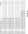
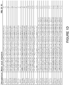
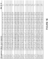
- the sequences of individual ABD binding motifs are presented in Figure 1 and as SEQ ID NO:1-52.
- the binding motif sequence is selected from any one of SEQ ID NO:1-52. More specifically, said sequence may be selected from any one of SEQ ID NO:1-7, such as from SEQ ID NO:1, SEQ ID NO:2, SEQ ID NO:3 and SEQ ID NO:4. In particular, said sequence may be SEQ ID NO:1.
- the BM as defined above "forms part of" a three-helix bundle protein domain. This is understood to mean that the sequence of the BM is "inserted” into or “grafted” onto the sequence of the original three-helix bundle domain, such that the BM replaces a similar structural motif in the original domain.
- the BM is thought to constitute two of the three helices of a three-helix bundle, and can therefore replace such a two-helix motif within any three-helix bundle.
- the replacement of two helices of the three-helix bundle domain by the two BM helices has to be performed so as not to affect the basic structure of the polypeptide.
- the overall folding of the C ⁇ backbone of the polypeptide according to this embodiment of the invention is substantially the same as that of the three-helix bundle protein domain of which it forms a part, e.g. having the same elements of secondary structure in the same order etc.
- a BM according to the invention "forms part" of a three-helix bundle domain if the polypeptide according to this embodiment of the invention has the same fold as the original domain, implying that the basic structural properties are shared, those properties e.g. resulting in similar CD spectra.
- the skilled person is aware of other parameters that are relevant.
- the ABD binding motif thus forms part of a three-helix bundle protein domain.
- the BM may essentially constitute two alpha helices with an interconnecting loop, within said three-helix bundle protein domain.
- said three-helix bundle protein domain is selected from domains of bacterial receptor proteins. Non-limiting examples of such domains are the five different three-helical domains of Protein A from Staphylococcus aureus, such as domain B, and derivatives thereof.
- the three-helical bundle protein domain is a variant of protein Z, which is derived from domain B of staphylococcal protein A.
- the ABD binding polypeptide for use in the disclosed separation method forms part of a three-helix bundle protein domain
- the ABD binding polypeptide may comprise the amino acid sequence: K-[ BM ]-DPSQS X a X b LLX c EAKKL NDX d Q; wherein
- X a in said sequence is A. In an alternative embodiment, X a in said sequence is S.
- X b in said sequence is N. In an alternative embodiment, X b in said sequence is E.
- X c in said sequence is A. In an alternative embodiment, X c in said sequence is S. In yet another alternative embodiment, X c in said sequence is C.
- X d in said sequence is A. In an alternative embodiment, X d in said sequence is S.
- said sequence in the definition of ABD binding polypeptides above is selected from SEQ ID NO:53-104, in particular from SEQ ID NO:53-59.
- said sequence is selected from SEQ ID NO:53, SEQ ID NO:54, SEQ ID NO:55 and SEQ ID NO:56.
- said sequence may be SEQ ID NO:53.
- an ABD binding polypeptide as defined above which comprises an amino acid sequence selected from:
- an ABD binding polypeptide as defined above which comprises an amino acid sequence selected from:
- an ABD binding polypeptide as defined above which comprises an amino acid sequence selected from:
- the ABD binding motif may form part of a polypeptide comprising an amino acid sequence selected from
- the ABD binding polypeptide for use in the disclosed separation method comprises the amino acid sequence:
- the amino acid sequence comprised in such a polypeptide may be selected from any one of SEQ ID NO:105-156.
- said sequence may be selected from any one of SEQ ID NO:105-111, such as selected from SEQ ID NO:105, SEQ ID NO:106, SEQ ID NO:107 and SEQ ID NO:108.
- said sequence is SEQ ID NO:105.
- the polypeptide for use in the disclosed separation method may be in monomeric or multimeric forms.
- the ABD binding polypeptide is present in multimeric form, comprising at least two ABD binding polypeptide monomer units, whose amino acid sequences may be the same or different.
- Multimeric forms of the polypeptide may be advantageous in that they may have enhanced binding properties.
- said ABD binding polypeptide monomer units are covalently coupled together.
- the ABD binding polypeptide monomer units are expressed as a fusion protein.
- polypeptides may be joined by covalent coupling using known organic chemistry methods, or expressed as one or more fusion polypeptides in a system for recombinant expression of polypeptides, or joined in any other fashion, either directly or via a linker, for example an amino acid linker.
- said ABD binding polypeptide is in dimeric form. Possible multimeric forms also include trimeric forms. Multimeric forms of the polypeptide may comprise a suitable number of polypeptide sequences as defined above.
- any ABD binding polypeptide disclosed herein may comprise further C-terminal and/or N-terminal amino acids.
- Such a polypeptide should be understood as a polypeptide having additional amino acid residues at the very first and/or the very last position in the polypeptide chain, i.e. at the N- and/or C-terminus.
- an ABD binding polypeptide may comprise any suitable number of additional amino acid residues, for example at least one additional amino acid residue.
- Each additional amino acid residue may individually or collectively be added in order to, for example, improve production, purification, stabilization in vivo or in vitro, coupling, or detection of the polypeptide.
- Such additional amino acid residues may comprise one or more amino acid residues added for the purpose of chemical coupling, which could for example allow coupling of the ABD binding polypeptide to a resin or matrix, for example a Sepharose-based resin or a SulfoLink coupling resin.
- a cysteine residue is added as an additional amino acid residue, or as one of a number of additional amino acid residues.
- the ABD binding polypeptide disclosed herein comprises a cysteine residue at the C-terminal end of the polypeptide, for example at the C-terminus.
- said cysteine is a part of a C-terminal additional tripeptide VDC.
- said cysteine is a lone C-terminal C.
- the ABD binding polypeptide comprises a dimer of two ABD binding domains as defined above, as well as a C-terminal cysteine residue.
- Such additional amino acid residues may also provide a "tag” for purification or detection of the polypeptide, such as a His 6 tag or a "myc” (c-myc) tag or a "FLAG” tag for interaction with antibodies specific to the tag or immobilized metal affinity chromatography (IMAC) in the case of the His 6 -tag.
- a tag for purification or detection of the polypeptide, such as a His 6 tag or a "myc” (c-myc) tag or a "FLAG” tag for interaction with antibodies specific to the tag or immobilized metal affinity chromatography (IMAC) in the case of the His 6 -tag.
- the further amino acids as discussed above may be coupled to the ABD binding polypeptide by means of chemical conjugation (using known organic chemistry methods) or by any other means, such as expression of the ABD binding polypeptide as a fusion protein.
- Biotinylation of target protein ABD001 with an N-terminal cysteine (C-ABD001; SEQ ID NO:158) and PEP07911 (SEQ ID NO:162) were biotinylated using EZ-Link Maleimide PEG 2 -Biotin (Pierce, cat. no. 21901).
- protein was dissolved in 50 mM sodium phosphate, 150 mM NaCl, 2 mM EDTA, pH 7.5.
- Dithiothreitol (DTT) was added to a final concentration of 20 mM and the samples were incubated for 1 h at 34 °C with end-over-end mixing.
- the buffer was exchanged to conjugation buffer (50 mM sodium phosphate, 150 mM NaCl, 1 mM EDTA, pH 7.0) using disposable PD-10 columns (GE Healthcare, cat. no. 17-0851-01).
- conjugation buffer 50 mM sodium phosphate, 150 mM NaCl, 1 mM EDTA, pH 7.0
- PD-10 columns GE Healthcare, cat. no. 17-0851-01
- a five times (5x) molar excess of EZ-Link Maleimide PEG 2 -Biotin was added to the protein samples and incubation proceeded for 2 h at room temperature (RT) with end-over-end mixing.
- PBS 10 mM phosphate, 137 mM NaCl, 2.68 mM KCI, pH 7.4
- PEP07986 (SEQ ID NO:161) was biotinylated according to the manufacturer's recommendations at RT for 30 min using No-Weigh EZ-Link Sulfo-NHS-LC-Biotin (Pierce, cat. no. 21327) at a 10x molar excess. Subsequent buffer exchange to PBS was performed using a dialysis cassette (Slide-a-lyzer 3.5 K, 3500 MWCO, Pierce, cat. no. 66333) according to the manufacturer's instructions.
- Phage display selection of ABD binding Z variants A library of random variants of protein Z displayed on bacteriophage, constructed in phagemid pAY02047 essentially as described in Grönwall et al (J Biotechnol, 128:162-183, 2007 ), was used to select ABD binding polypeptides.
- the library, Zlib004Naive.I utilizes the Taq DNA polymerase binding molecule Z03639 (described in Gunneriusson et al, Protein Eng 12:873-878, 1999 , where it was denoted Z TaqS1-1 ) as fusion partner.
- the library had an actual size of 1.4 x 10 10 variants.
- Phage stocks were prepared in a 20 I fermenter. Cells from a glycerol stock containing the phagemid library Zlib004Naive.I were inoculated in 20 I of TSB-YE (Tryptic Soy Broth-Yeast Extract; 30 g/l TSB, 5 g/l yeast extract) supplemented with 2 % glucose and 100 ⁇ g/ml ampicillin. The cultivations were grown at 37 °C in a fermenter (Belach Bioteknik, BR20). When the cells reached an optical density (OD) of 0.7-0.8, approximately 2.6 I of the cultivation was infected using a 10x molar excess of M13K07 helper phage (New England Biolabs, cat. no.
- OD optical density
- the cells were incubated for 30 min, whereupon the fermenter was filled up to 20 I with TSB-YE supplemented with 0.1 mM IPTG (isopropyl- ⁇ -D-1-thiogalactopyranoside, for induction of expression), 25 ⁇ g/ml kanamycin and 12.5 ⁇ g/ml carbenicillin, and cells were grown at 30 °C for 22 h.
- the cells in the cultivation were pelleted by centrifugation at 15900 g and the phage particles remaining in the medium were precipitated twice in PEG/NaCl (polyethylene glycol/sodium chloride), filtered and dissolved in PBS and glycerol as described in Grönwall et al, supra. Phage stocks were stored at -80 °C before use.
- Phage stock preparation, selection procedure and amplification of phage between selection cycles were performed essentially as described for selection against another target in WO2009/077175 .
- PBS supplemented with 0.1 % gelatine and 0.1 % Tween20 was used as selection buffer and the target-phage complexes were directly captured by Dynabeads® M-280 Streptavidin (Dynal, cat. no. 112.06). 1 mg beads per 0.25 ⁇ g ABD was used.
- E. coli strain XL1-Blue was used for phage amplification.
- Selection was performed in three cycles divided into three tracks: one track using PEP07911, one track using C-ABD001 and one track alternating between C-ABD001 and PEP07986.
- cycle 1 of the selections 100 nM PEP07911 or C-ABD001 was used in the different selection tracks, and two washes with PBST 0.1 % (PBS supplemented with 0.1 % Tween-20) were performed. An increased stringency, using a lowered target concentration and an increased number of washes, was applied in the subsequent two cycles.
- 50 nM followed by 25 nM PEP07911 or C-ABD001 were used in cycle 2 and 3, respectively.
- ELISA screening of Z variants To verify that the selected Z variant molecules could indeed interact with different variants of ABD, ELISA assays were performed.
- the Z variants were produced by inoculating single colonies from the selections into 1 ml TSB-YE medium supplemented with 100 ⁇ g/ml ampicillin and 0.1 mM IPTG in deep-well plates (Nunc, cat. no. 278752). The plates were incubated for 18-24 h at 37 °C. Cells were pelleted by centrifugation, re-suspended in 400 ⁇ l PBST 0.05 % and frozen at -80 °C to release the periplasmic fraction of the cells.
- Half-area 96-well ELISA plates (Costar, cat. no. 3690) were coated with 50 ⁇ l/well of coating buffer (50 mM sodium carbonate, pH 9.6) containing 4 ⁇ g/ml of an antibody specific for Z variants (Affibody, cat. no. 20.1000.01.0005) and incubated over-night at 4 °C.
- the antibody solution was poured off and the wells were blocked with 100 ⁇ l of PBSC (PBS supplemented with 0.5 % casein; Sigma, cat. no. C8654) for 2 h at RT.
- the blocking solution was discarded and 50 ⁇ l periplasmic solution was added to the wells and incubated for 1.5 h at RT under slow shaking.
- periplasmic fraction containing a Z variant binding to an irrelevant but specified target protein and expressed as above was assayed against 5 ⁇ g/ml of this target protein, biotinylated.
- negative control the same periplasmic preparation was assayed against PEP07986 or PEP07911. Sequencing was performed for the clones with positive absorbance values against PEP07986 or PEP07911.
- PCR fragments were amplified in two steps from single colonies using a standard PCR program and the primers AFFI-21 (5'-tgcttccggctcgtatgttgtgtg; SEQ ID NO:163) and AFFI-22 (5'-cggaaccagagccaccaccgg: SEQ ID NO:164). Sequencing of amplified fragments was performed using the biotinylated oligonucleotide AFFI-72 (5'-biotin-cggaaccagagccaccaccgg; SEQ ID NO:165) and a BigDye® Terminator v3.1 Cycle Sequencing Kit (Applied Biosystems, cat. no.
- sequencing reactions were purified by binding to magnetic streptavidin coated beads (Detach Streptavidin Beads, Nordiag, cat. no. 2012-01) using a Magnatrix 8000 (Magnetic Biosolution) and analyzed on ABI PRISM® 3130xI Genetic Analyzer (PE Applied Biosystems).
- Blocking ELISA A subset of clones from the initial ELISA screen were subjected to an ELISA blocking assay in order to elucidate if their target binding was affected by the presence of HSA. For the selected Z variants, the same periplasmic fractions as above were used.
- the ELISA blocking assay was run as the ELISA screening assay, with the following protocol modification introduced at the target step: HSA was mixed with the target protein before addition to the assay plate. 0.2 ⁇ g/ml of biotinylated PEP07911 was mixed with a 10x molar excess of HSA and then incubated for 15 min at RT to allow complex formation before addition to the plate.
- a periplasmic fraction containing a molecule binding an irrelevant target protein and expressed as above was assayed against 5 ⁇ g/ml biotinylated specific target protein.
- negative control the same periplasmic preparation was assayed against biotinylated PEP07911.
- PBSC was added instead of periplasmic preparation and biotinylated PEP07911 added as target. All controls were prepared as samples with and without addition of HSA in the same manner as above.
- Phage display selection of ABD binding Z variants Individual clones were obtained after three cycles of phage display selections against different variants of biotinylated ABD. The phage particle yield (phage particles out/phage particles in) increased for each cycle, indicating enrichment in target binding clones.
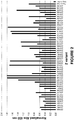
- ELISA screening of Z variants The clones obtained after three cycles of selection were produced in 96-well plates and screened for ABD binding activity in ELISA. The results of a selection of the positive clones (signals corresponding to at least 2x the negative control) assayed against PEP07911 are presented in Figure 2 .
- the control molecule specific for an irrelevant protein gave a positive signal for the specific protein, whereas no signal was obtained against PEP07986 or PEP07911.
- Blocking ELISA Clones positive for PEP07986 or PEP07911 were subjected to a blocking assay including HSA, to see if the Z variants had overlapping binding sites with the natural ligand HSA. For all tested clones, the binding signal to PEP07911 was completely extinguished by the presence of HSA, reaching the same level as the background ( Figure 3 ). The binding of the positive control was not affected by addition of an excess of HSA and the blank showed no background signal.
- Sequencing was performed for the clones with positive absorbance values against ABD in the ELISA screening. Each variant was given a unique identification number #####, and individual variants are referred to as Z#####.
- the amino acid sequences of the 58 amino acid residues long Z variants are listed in Figure 1 and in the sequence listing as SEQ ID NO:105-156.
- the deduced ABD binding motifs of these Z variants are denoted BM##### and listed in Figure 1 and in the sequence listing as SEQ ID NO:1-52.
- the amino acid sequences of the 49 amino acid residues long polypeptides predicted to constitute the complete three-helix bundle within each of these Z variants are denoted P##### and listed in Figure 1 and in the sequence listing as SEQ ID NO:53-104.
- Subcloning of Z variants The DNA of seven ABD binding Z variants, Z06608 (SEQ ID NO:107), Z06620 (SEQ ID NO:110), Z06638 (SEQ ID NO:106), Z06650 (SEQ ID NO:108), Z06677 (SEQ ID NO:105), Z06678 (SEQ ID NO:109) and Z06695 (SEQ ID NO:111), were amplified from the library vector pAY02047.
- a subcloning strategy for construction of dimeric Z variant molecules with N-terminal His 6 tag and C-terminal Cys was applied using standard molecular biology techniques and as described in detail in WO 2009/077175 for Z variants binding another target.
- the Z gene fragments were subcloned into the expression vector pAY01449 resulting in the encoded sequence MGSSHHHHHHLQ-[Z#####][Z#####]-VDC.
- E. coli BL21 (DE3) cells (Novagen) were transformed with plasmids containing the dimeric gene fragment of each respective Z variant and cultivated at 37 °C in 1 I of TSB + YE medium (Tryptic Soy Broth with Yeast Extract) supplemented with 50 ⁇ g/ml kanamycin.
- TSB + YE medium Tryptic Soy Broth with Yeast Extract
- 50 ⁇ g/ml kanamycin 50 ⁇ g/ml kanamycin.
- IPTG was added to induce protein expression at a final concentration of 0.17 mM and the cultivation was incubated at 37 °C for another 5 h. The cells were harvested by centrifugation.
- Contaminants were removed by washing with denaturing binding buffer, native binding buffer (20 mM sodium phosphate, 0.5 M NaCl, 20 mM imidazole, pH 7.4) and wash buffer (20 mM sodium phosphate, 0.5 M NaCl, 60 mM imidazole, pH 7.4) and the ABD binding Z variants were subsequently eluted with elution buffer (20 mM sodium phosphate, 0.5 M NaCl, 250 mM imidazole, pH 7.4). Each purified ABD binding Z variant was transferred to 10 mM NH 4 HCO 3 by size exclusion chromatography.
- Protein concentrations were determined by measuring the absorbance at 280 nm, using a NanoDrop® ND-1000 spectrophotometer, and using the extinction coefficient of the respective protein.
- the ABD binding Z variants were lyophilized and the purity of the final products was analyzed by SDS-PAGE stained with Coomassie Blue. The identity of each purified ABD binding Z variant was confirmed using HPLC-MS analysis.
- Example 2 the binding and elution characteristics of a set of ABD binding polypeptides, selected and produced as described in Example 1 and 2, respectively, were studied in a small-scale format using spin columns.
- Coupling of the ABD binding Z variants to resin 1 mg of each lyophilized ABD binding Z variant Z06608, Z06620, Z06638, Z06650, Z06677, Z06678 and Z06695, in the format His 6 -(Z#####) 2 -Cys produced as described in Example 2, was resuspended in a reducing buffer (50 mM Tris-HCl, 5 mM EDTA, 20 mM DTT, pH 8.5) and incubated at RT for 2 h.
- a reducing buffer 50 mM Tris-HCl, 5 mM EDTA, 20 mM DTT, pH 8.5
- Reduced protein solutions were transferred to a coupling buffer (50 mM Tris-HCl, 5 mM EDTA, pH 8.5) including 50 mM DTT, using size exclusion chromatography, and then mixed with portions of 0.2 ml SulfoLink Coupling Resin (Thermo Fisher Scientific, cat. no. 20401).
- This procedure enables site-directed reaction between the thiol groups of the C-terminal cysteine residues of the ABD binding Z variants and the iodoacetyl groups of the resin, forming a covalent thioether bond.
- the coupling reaction was carried out according to the instructions by the manufacturer.
- the degree of coupling defined as mg of coupled ABD binding Z variant per ml of resin, was determined as follows: The amount of coupled ABD binding Z variant was calculated by subtracting the amount of uncoupled material from the original amount of sample, based on concentrations obtained by spectrophotometric measurements of the absorbance at 280 nm. The amount of coupled ABD binding Z variant in mg was then divided with the volume of the SulfoLink Coupling Resin.
- Coupling efficiency of ABD binding polypeptides The degree of coupling of each ABD binding polypeptide to the SulfoLink Coupling Resin is shown in Table 3.
- Table 3 The degree of coupling of ABD binding Z variants to SulfoLink Resin ABD binding polypeptide Amount polypeptide coupled per ml resin (mg) Amount polypeptide coupled per ml resin ( ⁇ mol) His 6 -(Z06608) 2 -Cys 2.8 0.19 His 6 -(Z06620) 2 -Cys 2.7 0.19 His 6 -(Z06638) 2 -Cys 2.9 0.20 His 6 -(Z06650) 2 -Cys 3.4 0.23 His 6 -(Z06677) 2 -Cys 2.2 0.15 His 6 -(Z06678) 2 -Cys 2.1 0.14 His 6 -(Z06695) 2 -Cys 3.9 0.27
- the resins coupled with the ABD binding Z variants showed the most favorable elution profile with minor sample leakage during sample application and wash and with the majority of sample being released at elution with pH 2.5 buffer.
- the ABD binding Z variant-coupled resins appear to have a higher binding capacity as well as more favorable elution properties as judged both from the absorbance readings and the SDS-PAGE analysis.
- the difference in capacity cannot be due to differences in ligand density; in fact, the number of coupled HSA molecules on HSA Sepharose was approximately 1.5 - 2.2 times higher compared to the ABD binding Z variant-coupled resins.
- Circular dichroism (CD) analysis was carried out to determine the melting point (Tm) of His 6 -(Z06677) 2 -Cys.
- the purified sample was diluted to 0.5 mg/ml in PBS.
- a variable temperature measurement (VTM) was performed, in which the absorbance at 220 nm was monitored during heating of the sample from 20 °C to 90 °C, with a temperature slope of 5 °C/min. Tm was determined from the midpoint of the transition in the CD signal vs. temperature plot.
- the CD measurement was performed on a Jasco J-810 spectropolarimeter (Jasco Scandinavia AB) using a cell with an optical path length of 1 mm.
- EAH-Sepharose 4B activated EAH-Sepharose 4B (GE Healthcare, cat. no. 17-0569-01). Activation and immobilization was carried out according to the manufacturer's instructions, and the degree of coupling was determined as described in Example 3.
- Activated EAH Sepharose contains an iodoacetyl group that enables site-directed coupling via the C-terminal cysteine of the affinity ligand. 0.43 ml of the resin was packed in a Tricorn 5/50 column (GE Healthcare). As a reference, 0.47 ml HSA Sepharose (see Example 3) was packed in the same kind of column.
- the elution experiment was performed using an ⁇ KTAexplorer 10 chromatography system (GE Healthcare). The two columns were attached to the system and equilibrated with PBS. 2 ml of a sample containing 1 mg PEP08519 (comprising the albumin binding domain PP013 (SEQ ID NO:160) fused at its N-terminus to a Taq polymerase binding variant of protein Z) was loaded on each column followed by wash with PBS and 0.1 M sodium citrate, 0.9 % NaCl, pH 5.5.
- Bound protein was eluted by a linear pH gradient ranging from pH 5.5 to pH 2.3 (using 0.1 M sodium citrate, 0.9 % NaCl, pH 5.5, and 0.1 M sodium citrate, 0.9 % NaCl, pH 2.3) over 16 column volumes (CV).
- the pH was monitored by the built-in pH meter of the ⁇ KTAexplorer system.
- the experiment was performed using an ⁇ KTAexplorer 10 chromatography system. The two columns were attached to the system and equilibrated with PBS.
- a sample containing 0.25 mg/ml of PEP08519 in PBS was loaded on each column at a flow-rate of 0.2 ml/min (corresponding to a residence time of 2.4 min) until the absorbance at 280 nm of the solution emerging from the columns reached 10 % of the absorbance of the original sample (i.e. 10 % breakthrough).
- the dead volume i.e. tubing and column volume, measured by running a sample through a column comprising no affinity ligand
- Bound protein was eluted with 0.1 M sodium citrate, 0.9 % NaCl, pH 2.3. The columns were then re-equilibrated with PBS.
- the columns were subjected to alkali treatment by applying 5 CV of 0.2 M NaOH at a flow-rate of 1 ml/min, followed by an incubation period without flow.
- the total incubation time in alkali was 25 min.
- Neutralization of the columns was performed by washing with 5 CV of PBS at 1 ml/min, followed by a new capacity measurement as described above.
- Circular dichroism analysis The melting point of His 6 -(Z06677) 2 -Cys was determined to be 54 °C.
- the dynamic binding capacity was determined by measuring the amount of sample loaded until 10 % breakthrough.
- the dynamic binding capacity of the His 6 -(Z06677) 2 -Cys column was determined to be 3.0 mg PEP08519 per ml resin.
- the corresponding capacity for the HSA Sepharose column was determined to 0.5 mg per ml resin.
- the difference in capacity cannot be explained by the difference in ligand density, since the number of immobilized HSA molecules were estimated to be twice the number of His 6 -(Z06677) 2 -Cys molecules on their respective resins.
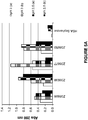
- the dynamic binding capacity was re-measured after each alkali incubation. The results are shown in Figure 7 .
- the His 6 -(Z06677) 2 -Cys column maintained its original capacity after all incubations. This indicates a high resistance to relatively harsh alkali conditions, which is an important feature for cleaning in place (CIP) procedures enabling repeated use of the column.
- CIP cleaning in place
- the HSA Sepharose column included as a reference, lost most of its capacity after 25 min in 0.2 M NaOH.
- the ABD binding Z variant His 6 -(Z06677) 2 -Cys was immobilized on four different types of resins, and the binding capacity and alkali resistance were assessed chromatographically.
- Coupling of His 6 -(Z06677) 2 -Cys to different resins 4 mg of the lyophilized His 6 -(Z06677) 2 -Cys (produced as described in Example 2) was dissolved in 1 ml 0.1 M NaHCO 3 , 0.5 M NaCl, pH 8.3. One half of the sample was immobilized on 0.67 ml NHS-activated Sepharose 4 Fast Flow (GE Healthcare, cat. no. 17-0906) and one half of the sample was immobilized on 0.67 ml CNBr-activated Sepharose 4 Fast Flow (GE Healthcare, cat. no. 17-0981). Both coupling reactions were carried out according to the instructions by the manufacturer.
- the fourth resin assessed in this experiment was EAH Sepharose immobilized with His 6 -(Z06677) 2 -Cys as described in Example 4.
- the two Z molecules within the dimer bind a sample molecule each to a higher extent compared to His 6 -(Z06677) 2 -Cys coupled to EAH Sepharose resin.
- the dynamic binding capacities of the two amine-coupled resins i.e. NHS-activated Sepharose and CNBr-activated Sepharose, were lower than for the two iodoacetyl-coupled resins. This indicates that the site-directed immobilization through a single cysteine, as used for coupling to the iodoacetyl resins, makes the binding sites of the ABD binding Z variant more accessible to the sample molecules in the mobile phase.
- the dynamic binding capacity of the NHS resin was less than half compared with the CNBr resin, despite the fact that the NHS resin holds a spacer arm that would theoretically improve the capacity, by making the affinity ligands more accessible.
- the dynamic binding capacities of the four different resins after alkali treatment with 0.5 M NaOH for 2 h are presented in Table 7.
- the dynamic binding capacities of the iodoacetyl-coupled resins were unaffected by the alkali incubation, while the capacities of the amine-coupled resins dropped by approximately 10 %.
- Table 7. Comparison of the dynamic binding capacities before and after alkali treatment.
- This Example describes a larger-scale production of the ABD binding Z variant Z006677 in a dimeric format and with an C-terminal Cys, i.e. (Z06677) 2 -Cys, and its subsequent immobilization on a SulfoLink Coupling Resin.
- the ABD binding Z variant Z06677 was subcloned as a dimer into an expression vector in which expression is regulated by a T7 promoter.
- the ABD binding polypeptide was expressed with the additional N-terminal amino acid sequence GSSLQ and the additional C-terminal amino acid sequence VDC.
- the expressed polypeptide has the sequence GSSLQ-[Z06677][Z06677]-VDC.
- E. coli BL21(DE3) cells Novagen
- OD 600 1, IPTG, at a final concentration of 0.17 mM, was added to induce protein expression and the cultivation was incubated at 37 °C for another 5 h.
- the cells were harvested by centrifugation.
- Acetonitrile (ACN) was added to the sample to a concentration of 2 % (v/v).
- the sample was loaded onto a FineLine 35 column (GE Healthcare) packed with 125 ml SOURCE 30 RPC (GE Healthcare) by use of an ⁇ KTAexplorer 100 chromatography system (GE Healthcare).
- the column was washed with 2 % ACN in Milli-Q water containing 0.1 % TFA (tri-fluoro acetic acid) followed by wash with an identical buffer, containing 4 % of ACN.
- Bound protein was eluted by increasing the ACN concentration from 4 % to 25 % ACN over 15 CV. Eluted fractions were collected and analyzed by SDS-PAGE and LC-MS.
- the purified dimeric ABD binding Z variant (Z06677) 2 -Cys was analyzed in a spectrophotometer by measuring the absorbance at 280 nm and by SDS-PAGE.
- Z06677) 2 -Cys lacks tryptophans, resulting in low absorbance readings at 280 nm.
- the concentration of the Z variant molecule was therefore estimated from the SDS-PAGE analysis, which included several different quantities of (Z06677) 2 -Cys.
- Visual inspection of the SDS-PAGE gel estimated the concentration of (Z06677) 2 -Cys to 3.4 mg/ml at a purity of approximately 95 %.
- the produced (Z06677) 2 -Cys-coupled resin is further referred to as "anti-ABD agarose", and in the following Examples it was used to demonstrate the wide applicability of the resin, in terms of binding to different albumin binding domain moieties in fusion with different proteins of varying size and functions. It is shown that successful purification is achieved regardless of where in the target protein the ABD moiety is situated. In other words, the ABD moiety may be placed at the N-terminus, at the C-terminus or even within a fusion protein such that it is flanked by other protein moieties on either side.
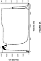
- PEP10986 (12.5 kDa), comprising an albumin binding domain (PP013, SEQ ID NO:160) fused at its N-terminus to a cytokine binding variant of protein Z, was purified using a column packed with anti-ABD agarose.
- Pelleted E. coli cells harboring soluble PEP10986 were suspended in TST-buffer (Tris, Saline, Tween: 25 mM Tris-HCl, 1 mM EDTA, 200 mM NaCl, 0.05 % Tween 20, pH 8.0) supplemented with 20 U/ml Benzonase®. Cells were disrupted by sonication and the cell debris removed by centrifugation. Chromatography was performed using an ⁇ KTAexplorer 100 system. The clarified sample was applied on an XK-50 column packed with 100 ml anti-ABD agarose (prepared as described in Example 6) and pre-equilibrated with TST.
- TST-buffer Tris, Saline, Tween: 25 mM Tris-HCl, 1 mM EDTA, 200 mM NaCl, 0.05 % Tween 20, pH 8.0
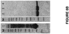
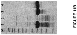
- the chromatogram from purification of PEP10986 on the anti-ABD agarose resin is presented in Figure 8A and the SDS-PAGE analysis of selected fractions is shown in Figure 8B .
- the eluted fractions E1, E2 and E3, corresponding to lane 2, 3 and 4 respectively in Figure 8B were pooled.
- the correct mass of PEP10986 was verified by HPLC-MS analysis and the purity was estimated to be higher than 95 % as judged from SDS-PAGE analysis. According to spectrophotometrical analysis, the pool contained 908 mg of purified PEP10986, corresponding to a yield of approximately 90 %.
- PEP03973 (12.4 kDa), comprising the wild-type albumin binding domain (ABD001, SEQ ID NO:157) fused at its N-terminus to a HER2 (Human Epidermal growth factor Receptor 2) binding variant of protein Z, was purified using a column packed with anti-ABD agarose.
- Pelleted E. coli cells harboring soluble PEP03973 were suspended in TST-buffer supplemented with 20 U/ml Benzonase®. Cells were disrupted by sonication and the cell debris removed by centrifugation. Chromatography was performed using an ⁇ KTAexplorer 100 system. The clarified sample was applied on an XK-50 column packed with 100 ml anti-ABD agarose (prepared as described in Example 6) and pre-equilibrated with TST. The column was washed with 5 CV of TST followed by 3 CV of 5 mM NH 4 Ac pH 5.5. Bound protein was eluted by applying 3 CV of 0.1 M HAc, pH 2.9. Flow-through fractions, wash fractions and eluted fractions were collected for further analyses by SDS-PAGE and HPLC-MS.
- the chromatogram from purification of PEP03973 on the anti-ABD agarose resin is presented in Figure 9A and the SDS-PAGE analysis of selected fractions is shown in Figure 9B .
- the eluted fractions E2, E3 and E4, corresponding to lane 5, 6 and 7 respectively in Figure 9B were pooled.
- the correct mass of PEP03973 was verified by HPLC-MS analysis and the purity was estimated to be higher than 95 % as judged from SDS-PAGE analysis. According to spectrophotometrical analysis, the pool contained 1036 mg of purified PEP03973, corresponding to a yield of approximately 97 %.
- PEP06548 (20.3 kDa), comprising an albumin binding domain (ABD035, SEQ ID NO:159, with the C-terminal extension VDC) fused at its N-terminus to two moieties (i.e. a dimer) of a variant of protein Z binding to TNF- ⁇ (tumor necrosis factor alpha), was purified using a column packed with anti-ABD agarose.
- PEP06548 also comprises an N-terminal hexahistidine tag and a C-terminal cysteine residue.
- Pelleted E. coli cells harboring soluble PEP06548 were suspended in TST-buffer supplemented with 27 U/ml Benzonase®. Cells were disrupted by sonication and the cell debris removed by centrifugation. The C-terminal cysteines of the PEP06548 sample molecules were reduced by addition of DTT to a final concentration of 20 mM, followed by incubation for 30 min at RT. Chromatography was performed using an ⁇ KTAexplorer 100 system. The clarified and reduced sample was applied on an XK-16 column packed with 9 ml anti-ABD agarose and pre-equilibrated with TST.
- This batch of anti-ABD agarose was prepared essentially as described in Example 6, except for a lower ligand density of approximately 2.3 mg (Z06677) 2 -Cys per ml of resin.
- the column was washed with 5.5 CV of TST followed by 3.3 CV of 5 mM NH 4 Ac pH 5.5. Bound protein was eluted by applying 2.9 CV of 0.1 M HAc, pH 2.9. Fractions were collected for further analyses by SDS-PAGE and HPLC-MS.
- PEP17081 (18.6 kDa), comprising an albumin binding domain (PEP07986, SEQ ID NO:161) fused to two moieties of a cytokine binding variant of protein Z in the format Z-PEP07986-Z, was purified using a column packed with anti-ABD agarose. Subsequent purification is also described.
- Pelleted E. coli cells harboring soluble PEP17081 were suspended in TST-buffer. Cells were disrupted by heat treatment in a water bath (83 °C, 10 min), followed by cooling on ice. Benzonase® was added to a final concentration of 15 U/ml to reduce viscosity caused by nucleic acids. Cell debris was removed by centrifugation and filtration (0.45 ⁇ m filter).
- a first purification step was performed by anti-ABD agarose affinity chromatography using an ⁇ KTApurifier 100 system.
- the clarified sample was applied on an XK-26 column packed with 25 ml anti-ABD agarose and pre-equilibrated with 2x TST (i.e. TST buffer with all components at double concentration).
- the column was washed with 5 CV of 2x TST followed by 8 CV of 5 mM NH 4 Ac, pH 5.5.
- Bound protein was eluted by applying 3 CV of 0.1 M HAc, pH 2.9. Fractions were collected for further analyses by SDS-PAGE and HPLC-MS.
- a second purification step was performed by RPC, using an ⁇ KTAexplorer 100 system.
- the protein eluted from the anti-ABD agarose column was supplemented with acetonitrile to a final concentration of 10 % and loaded on an HR-16 column (GE Healthcare) packed with 24 ml SOURCE 15 RPC (GE Healthcare) and pre-equilibrated with 10 % acetonitrile, 0.1 % trifluoroacetic acid (TFA) in Milli-Q water (Solvent A).
- the column was washed with 3.9 CV Solvent A.
- Bound material was eluted with a 15 CV linear gradient of increasing concentration of 80 % acetonitrile, 0.1 % TFA in Milli-Q water (Solvent B), ending at 50 % Solvent B. Fractions were collected for further analyses by SDS-PAGE and HPLC-MS.
- a third purification and buffer exchange step was performed by size exclusion chromatography (SEC) using an ⁇ KTAexplorer 100 system. Selected fractions eluted from the RPC column were pooled and buffer exchanged to 1x PBS on a XK-50 column packed with 500 ml Sephadex G-25 (GE Healthcare). Fractions were collected for further analyses by SDS-PAGE and HPLC-MS.
- SEC size exclusion chromatography
- Protein 1 (22.6 kDa; ⁇ l 10), comprising an albumin binding domain (ABD035, SEQ ID NO: 159) fused at its C-terminus to a protein hormone, was purified on an ⁇ KTAexplorer system using a column packed with anti-ABD agarose.
- Protein 1 was expressed in E.coli in the form of inclusion bodies. After solubilization and refolding of the inclusion bodies the material was run three times on size-exclusion chromatography (SEC) for buffer exchange and purification. 8 mg of the SEC purified Protein 1, in PBS diluted 1:10 in TST, was loaded at 0.5 ml/min on an XK 16/20 column (GE Healthcare) packed with 10-12 ml anti-ABD agarose and pre-equilibrated with TST. The column was washed with 4 CV of TST, followed by an additional 3 CV wash with 5 mM NH 4 Ac, pH 5.5, at a flow rate of 1 ml/min.
- SEC size-exclusion chromatography
- Bound protein was eluted with 3 CV of 0.1 M HAc, pH 2.9, and then neutralized to pH 7 with 1 M Tris-HCl, pH 9 . Flow-through fractions, wash fractions and eluted fractions were collected for further analysis by SDS-PAGE. 4-12 % Bis-Tris NuPAGE gels (Invitrogen) were run non-reduced and the protein stained with Simply Blue (Life Technologies). The column was re-equilibrated with TST after elution and cleaned in place (CIP) with 3 CV of 0.5 M NaOH followed by 3 CV of TST. Between purifications, the resin was stored in TST + 20 % ethanol.
- Protein 2 (9.5 kDa, pi 6.6), comprising an albumin binding domain (ABD035, SEQ ID NO: 159) fused at its N-terminus to a peptide hormone, was expressed in Pichia Pink and purified in a single step on an ⁇ KTAexplorer system using a column packed with anti-ABD agarose.
- Bound protein was eluted with 3 CV of 0.1 M HAc, pH 2.9 and then adjusted to pH 4 with 1 M Tris-HCl, pH 9.
- the column was re-equilibrated with TST after elution followed by CIP and storage in TST + 20 % ethanol. Flow-through fractions, wash fractions and eluted fractions were collected for further analysis by SDS-PAGE.
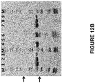
- the chromatogram from purification of Protein 2 on the anti-ABD agarose resin is presented in Figure 13A and the SDS-PAGE analysis of selected fractions is shown in Figure 13B .
- the elution peak from the anti-ABD agarose column contained 3-5 mg of Protein 2, corresponding to a yield of approximately 75 % (estimated from a Western blot).
- the correct mass of the eluted Protein 2 was verified by HPLC-MS analysis. The purity was estimated to be higher than 98 % as judged from SDS-PAGE analysis followed by Simply Blue staining. No contaminating media proteins could be detected in the elution fractions (lanes 7-13, Figure 13B ). It is recognized that Protein 2 runs smaller on SDS-PAGE gels as indicated by the molecular weight standards included on the gels.
Landscapes
- Chemical & Material Sciences (AREA)
- Organic Chemistry (AREA)
- Health & Medical Sciences (AREA)
- General Health & Medical Sciences (AREA)
- Biochemistry (AREA)
- Biophysics (AREA)
- Life Sciences & Earth Sciences (AREA)
- Genetics & Genomics (AREA)
- Medicinal Chemistry (AREA)
- Molecular Biology (AREA)
- Proteomics, Peptides & Aminoacids (AREA)
- Gastroenterology & Hepatology (AREA)
- Analytical Chemistry (AREA)
- Peptides Or Proteins (AREA)
Description
- This invention relates to a separation method employing polypeptides which bind to an albumin binding domain, ABD. The polypeptides disclosed herein have various industrial and pharmaceutical applications, for example use as an affinity ligand in separation technology or as a detection agent in molecular diagnostics.
- Serum albumin is the most abundant protein in mammalian sera (35-50 g/l, i.e. 0.53-0.75 mM, in humans) and several strategies to covalently couple a peptide or protein to carrier molecule that will allow in vivo association to serum albumin have been described e.g. in
WO91/01743 WO01/45746 WO91/01743 - Streptococcal protein G (SpG) is a bi-functional receptor present on the surface of certain strains of streptococci and is capable of binding to both IgG and serum albumin (Björck et al, Mol Immunol 24:1113, 1987). The structure is highly repetitive, with several structurally and functionally different domains (Guss et al, EMBO J 5:1567, 1986), more precisely three Ig-binding domains and three serum albumin binding domains (Olsson et al, Eur J Biochem 168:319, 1987). The structure of one of the three serum albumin binding domains in SpG has been determined, showing a three-helix bundle fold (Kraulis et al, FEBS Lett 378:190, 1996, Johansson et al, J. Biol. Chem. 277:8114-20, 2002). A 46 amino acid motif was defined as ABD (albumin binding domain) and has subsequently also been designated G148-GA3 (GA for protein G-related albumin binding and G148 from the strain of Streptococcus from which it is derived).
- Artificial variants of GA3-G148 having a very much improved affinity for human serum albumin were developed (Jonsson et al, Prot Eng Des Sel 21:515-27, 2008;
WO2009/016043 ), as well as engineered high affinity variants with reduced immune stimulatory properties (WO2012/004384 ). The latter were motivated by the fact that a few T- and B-cell epitopes were experimentally identified within GA3-G148 (Goetsch et al, Clin Diagn Lab Immunol 10:125-32, 2003), making this domain as such less suitable for use in pharmaceutical compositions for human administration. Throughout the present text, GA3-G148 as well as the various engineered derivatives thereof presented e.g. inWO2009/016043 andWO2012/004384 , are collectively referred to as "ABD". Thus, in the present disclosure, "ABD" denotes these classes of albumin binding polypeptides, rather than a specified polypeptide with a specific amino acid sequence. - With the increased interest in incorporating an albumin binding domain into therapeutic or diagnostic compositions follows a growing need for inexpensive and efficient purification strategies for isolation of molecules covalently linked to ABD, produced for instance by recombinant expression in prokaryotic or eukaryotic systems, or by direct chemical conjugation to ABD. A general strategy for purification of recombinantly expressed proteins would be to include a commonly used affinity tag such as a polyhistidine tag, a chitin binding protein (CBP), a maltose binding protein (MBP), a glutathione-S-transferase (GST)-tag or a FLAG-tag, and perform a classic affinity separation using commercial resins developed specifically for each tag. However, for certain applications, and in particular for molecules to be used as therapeutics, the end product must be homogeneous. Because the tag would need to be removed, e.g. by enzymatic or chemical cleavage, it is necessary to ensure complete cleavage to obtain a homogeneous product, or to suffer a loss of yield when removing incompletely cleaved product. Both of these issues serve to increase the production cost of the product. Therefore, a more motivated strategy would be to utilize the ABD moiety itself as a purification tag. One example of this would be to couple recombinant albumin to a solid support (see for example Jonsson et al, Prot Eng Des Sel 21:515-27, 2008; Andersen et al, J Biol Chem 286:5234-41, 2011). From a crude solute, compounds comprising an ABD tag are captured by albumin, non-specifically adsorbed contaminants are removed and ABD-tagged compounds are subsequently recovered by disrupting the specific but reversible interaction with albumin. However, albumin is a large natural carrier molecule with several interaction sites for different proteins, fatty acids, sterols, ions etc., and thus, the background binding of both specific and unspecific components may contaminate the recovered sample. Despite the fact that recombinant human albumin has been developed, it is still expensive to include as an affinity ligand in large-scale production of therapeutics, in particular because it is incompatible with standard procedures for cleaning in place, that would have to be applied for the repeated use of the albumin-coupled matrix. Furthermore, harsher elution conditions may be required to recover molecules containing ABD variants with an exceptionally high affinity for albumin. Such conditions may be unfavorable also for the ABD-tagged molecule.
- Protein A from Staphylococcus aureus has long been used as an affinity ligand in the industrial production of monoclonal antibodies and Fc-fusion proteins, due to the native affinity of Protein A for the Fc portion of IgG.
WO2010/056550 discloses the use of Protein A to purify single domain antibodies. Protein A in its entirety, as well as the individual Fc-binding domains thereof, have subsequently served as starting points for the rational design of engineered affinity ligands with improved properties. For an introduction, see the papers by Nord K and co-workers in Prot Eng (Nord et al, Prot Eng 8:601-608; 1995) and Nat Biotech (Nord et al, Nat Biotech 15:772-777; 1997). An evolved scaffold for such affinity ligands is disclosed inEP2077272 . - It is an object of the present disclosure to provide new methods for purification, separation and/or chromatography of protein molecules containing ABD, for example fusion proteins in which ABD is a fusion moiety. It is moreover an object of the disclosure to provide uses for ABD binding agents in biotechnology, for example in protein purification and separation applications.
- These objects, and other objects apparent to the skilled person from the present disclosure, may be achieved by one or more of the various inventive aspects disclosed below.
- Thus, in a first aspect, the disclosure provides a method of separation of at least one ABD-containing molecule present in a liquid from other constituents in the liquid, which method comprises a step of affinity separation, in which step is used, as affinity ligand, an ABD binding polypeptide comprising an ABD binding motif BM, which motif consists of the amino acid sequence:
EX2X3X4AX6X7EI X10X11LPNLTX17X18QX20 X21AFIX25KLX28D
wherein, independently from each other, - X2 is selected from F, I and L;
- X3 is selected from H, K, N, Q, R, S, T and V;
- X4 is selected from A, D, F, G, H, I, K, L, M, N, Q, R, S, T, V, W and Y;
- X6 is selected from F, I, L and Y;
- X7 is selected from A, H, I, K, L, N, Q, R, S, T and V;
- X10 is selected from G, H, K, N, Q, R and S;
- X11 is selected from A, D, F, G, I, K, L, N, Q, R, S, T, V and Y;
- X17 is selected from F, H, L, S and T;
- X18 is selected from D, E, H, I, K, L, M, N, Q, R, S, T and V;
- X20 is selected from H, K and R;
- X21 is selected from I, L and V;
- X25 is selected from F, I, L, V and Y;
- X28 is selected from D and E.
- As is will be realized by the person of skill in the art of affinity chromatography, the disclosed method of separation may be employed in the removal and/or purification of molecules comprising ABD, i.e. "ABD-containing molecules". Such molecules may in particular be fusion proteins, in which one fusion moiety comprises one or more ABD domains, and other fusion moieties have other functions, such as biologically active functions, for example therapeutic or diagnostic functions.
- The method comprises a step of affinity separation, in which step a polypeptide as defined above is used. More specific variants and embodiments of the ABD binding polypeptide are given below. Thus, the invention provides the use of the polypeptide as described herein in a method of affinity separation. Suitably, the method involves a separation device. The separation device may suitably comprise a solid support, for example selected from chromatographic media, membranes, cellulose, silica, agarose, polyacrylamide, magnetic beads, two-phase systems and other such materials commonly used in separation. In one embodiment, the polypeptide defined herein has been coupled to a solid support of a separation device. The thus obtained separation device, having a polypeptide as defined herein coupled thereto, is referred to throughout the description and claims as an "affinity matrix" or "resin", and a particular embodiment thereof is referred to in the examples as "anti-ABD agarose".
- For the purposes of purification of an ABD-containing molecule from a liquid, the liquid containing ABD-containing molecule to be purified is suitably applied to such an affinity matrix under conditions that are conducive to binding of ABD-containing molecule to the matrix. Thereafter, the affinity matrix is washed under conditions such that the binding of ABD-containing molecule to the matrix is maintained, but most, ideally all, other proteins and contaminants bound to the matrix are washed away. In an elution step, the matrix is treated such that ABD-containing molecule is released from the matrix in an ABD-containing molecule enriched fraction denoted "ABD-containing molecule fraction", which may be recovered.
- If, conversely, the purpose of the separation is the removal of ABD-containing molecule, essentially the same steps as above are suitably followed, with some exceptions. The liquid containing ABD-containing molecule to be removed is suitably applied to an affinity matrix under conditions that are conducive to binding of ABD-containing molecule to the matrix. Thereafter, the affinity matrix is washed under conditions such that the binding of ABD-containing molecule to the matrix is maintained, but most, ideally all, other proteins are recovered in the flow-through, thus obtaining a "depleted fraction" with a substantial reduction in ABD-containing molecule content, which is recovered. Thus, the constituents of the liquid that are not ABD-containing molecules and that were discarded in the purification method above, may instead be retained and used and/or processed further.
- As a further alternative of the inventive method, both the "depleted fraction" and the " ABD-containing molecule fraction" may be recovered from the same separation run. Then, once again, the liquid containing ABD-containing molecule is suitably applied to an affinity matrix under conditions that are conducive to binding of ABD-containing molecule to the matrix. Thereafter, the affinity matrix is washed, under conditions such that the binding of ABD-containing molecule to the matrix is maintained, but most, ideally all, other proteins are recovered in the flow-through. The thus obtained "depleted fraction" with a substantial reduction in ABD-containing molecule content is recovered. In an elution step, the matrix is treated such that ABD-containing molecule is released from the matrix in an ABD-containing molecule enriched fraction denoted " ABD-containing molecule fraction", which is recovered.
- In one embodiment, the disclosed separation method is carried out in a batch setup. In another embodiment, the method is carried out in a column setup. In still another embodiment, the method is carried out using expanded bed adsorption. It is also possible to carry out the disclosed method using hybrid set-ups, such as binding in batch mode followed by wash and elution in a column setup. The person of skill in affinity chromatography is aware of the available alternatives and may put them into practice without undue effort.
- The ABD binding polypeptide disclosed herein exhibits a set of characteristics, which, for example when coupled to a solid support, makes it suitable as an affinity ligand for separation of molecules comprising ABD. For example, in comparison with using human albumin as affinity ligand, the use of the disclosed ABD binding polypeptide in the disclosed separation method demonstrates the following advantages:
- Higher separation capacity per ml ligand-coupled matrix.
- Compatibility with repeated procedures for cleaning in place, for example using 0.5 M sodium hydroxide, which makes the ABD binding polypeptide useful in the large scale production of therapeutics.
- Enablement of milder elution conditions, for example elution at a higher pH, which makes possible the separation of a wider range of different molecules comprising ABD.
- In the context of the present disclosure, the terms "sample" and "liquid" may be used interchangeably. Neither term implies any limitations, for example with regard to the volume of liquid involved or other characteristics. The liquid may be of a small volume, such as an aliquot of a larger volume, e.g. for analytical purposes; or alternatively, the feed or liquid used in a large scale separation and/or purification process.
- The method disclosed herein may be useful at one or more of the different stages of purification. Thus, it may be used without limitation at either one or more of a first capture step, in any intermediate purification step or in a final polishing step.
- The skilled person will appreciate that successful purification of a target protein, using a method according to the present disclosure, employing a resin comprising an ABD binding polypeptide as described herein may be achieved regardless of where in the target protein the ABD moiety is situated. Hence, the ABD moiety may be placed at the N-terminus or at the C-terminus of the target protein. Alternatively, the target protein may be a fusion protein which comprises an ABD moiety flanked by other protein moieties on both sides.
- In the context of the present disclosure, "ABD" refers to three-helix albumin binding domains from streptococcal protein G and their derivatives. In particular, "ABD" may refer to GA3-G148 (Johansson et al, supra) or variants of GA3-G148 having improved affinity for albumin (
WO2009/016043 ), as well as high affinity variants with reduced immune stimulatory properties (WO2012/004384 ). In other words, in the present disclosure, "ABD" denotes the classes of albumin binding polypeptides described in the referenced works, rather than a specified polypeptide with a specific amino acid sequence. - The above definition of a class of sequence related, ABD binding polypeptides for use in the disclosed separation method is based on a statistical analysis of a number of random polypeptide variants of a parent scaffold, that were selected for their interaction with ABD in several different selection experiments. The identified ABD binding motif, or "BM", corresponds to the target binding region of the parent scaffold, which region constitutes two alpha helices within a three-helical bundle protein domain. In the parent scaffold, the varied amino acid residues of the two BM helices constitute a binding surface for interaction with the constant Fc part of antibodies. In the present invention, the random variation of binding surface residues and subsequent selection of variants have replaced the Fc interaction capacity with a capacity for interaction with ABD.
- The term "% identity", as used throughout, may be calculated as follows. The query sequence is aligned to the target sequence using the CLUSTAL W algorithm (Thompson et al, Nucleic Acids Research, 22: 4673-4680 (1994)). A comparison is made over the window corresponding to the shortest of the aligned sequences. The shortest of the aligned sequences may in some instances be the target sequence. In other instances, the query sequence may constitute the shortest of the aligned sequences. The amino acid residues at each position are compared, and the percentage of positions in the query sequence that have identical correspondences in the target sequence is reported as % identity.
- In one embodiment, X2 in said sequence is selected from F and L.
- In one embodiment, X2 in said sequence is F.
- In one embodiment, X2 in said sequence is L.
- In one embodiment, X3 in said sequence is selected from H, K, R, T and V.
- In one embodiment, X3 in said sequence is selected from H, K, R and V.
- In one embodiment, X3 in said sequence is selected from H, K and V.
- In one embodiment, X3 in said sequence is selected from K and R.
- In one embodiment, X3 in said sequence is K.
- In one embodiment, X3 in said sequence is R.
- In one embodiment, X3 in said sequence is V.
- In one embodiment, X4 in said sequence is selected from A, H, I, L, N, V and W.
- In one embodiment, X4 in said sequence is selected from H, L N, and V
- In one embodiment, X4 in said sequence is V.
- In one embodiment, X4 in said sequence is N.
- In one embodiment, X4 in said sequence is H.
- In one embodiment, X4 in said sequence is L.
- In one embodiment, X4 in said sequence is selected from A, L, N, V and W.
- In one embodiment, X4 in said sequence is selected from A, N, V and W.
- In one embodiment, X4 in said sequence is selected from A, N and W.
- In one embodiment, X6 in said sequence is selected from F and L.
- In one embodiment, X6 in said sequence is F.
- In one embodiment, X6 in said sequence is L.
- In one embodiment, X7 in said sequence is selected from K, L, N, Q, R and S.
- In one embodiment, X7 in said sequence is selected from K, N, Q, R and S.
- In one embodiment, X7 in said sequence is selected from K, Q, R and S.
- In one embodiment, X7 in said sequence is selected from K and R.
- In one embodiment, X7 in said sequence is K.
- In one embodiment, X7 in said sequence is R.
- In one embodiment, X10 in said sequence is selected from H, K, N, and R.
- In one embodiment, X10 in said sequence is selected from H, K and N.
- In one embodiment, X10 in said sequence is selected from K, N and R.
- In one embodiment, X10 in said sequence is selected from K and R.
- In one embodiment, X10 in said sequence is N.
- In one embodiment, X10 in said sequence is K.
- In one embodiment, X10 in said sequence is R.
- In one embodiment, X10 in said sequence is H.
- In one embodiment, X11 in said sequence is selected from A, F, L, R, T and Y.
- In one embodiment, X11 in said sequence is selected from A, F, T and Y.
- In one embodiment, X11 in said sequence is T.
- In one embodiment, X11 in said sequence is A.
- In one embodiment, X11 in said sequence is Y.
- In one embodiment, X11 in said sequence is F.
- In one embodiment, X11 in said sequence is selected from A, L, T and Y.
- In one embodiment, X11 in said sequence is selected from T and Y.
- In one embodiment, X17 in said sequence is selected from F, H and L.
- In one embodiment, X17 in said sequence is selected from F and H.
- In one embodiment, X17 in said sequence is selected from H and L.
- In one embodiment, X17 in said sequence is F.
- In one embodiment, X17 in said sequence is H.
- In one embodiment, X17 in said sequence is L.
- In one embodiment, X18 in said sequence is selected from D, H, I, K and Q.
- In one embodiment, X18 in said sequence is selected from D, H and Q.
- In one embodiment, X18 in said sequence is selected from H and Q.
- In one embodiment, X18 in said sequence is H.
- In one embodiment, X18 in said sequence is Q.
- In one embodiment, X18 in said sequence is D.
- In one embodiment, X20 in said sequence is selected from K and R.
- In one embodiment, X20 in said sequence is selected from H and R.
- In one embodiment, X20 in said sequence is R.
- In one embodiment, X20 in said sequence is K.
- In one embodiment, X21 in said sequence is selected from I and L.
- In one embodiment, X21 in said sequence is I.
- In one embodiment, X21 in said sequence is L.
- In one embodiment, X25 in said sequence is selected from I, L and V.
- In one embodiment, X25 in said sequence is selected from V and I.
- In one embodiment, X25 in said sequence is I.
- In one embodiment, X25 in said sequence is V.
- In one embodiment, X25 in said sequence is L.
- In one embodiment, X28 in said sequence is D.
- In one embodiment, said sequence is defined as follows: independently from each other,
X2 is selected from F and L;
X3 is selected from H, K, R, T and V;
X4 is selected from A, H, I, L, N, V and W;
X6 is selected from F and L;
X7 is selected from K, L, N, Q, R and S;
X10 is selected from H, K, N and R;
X11 is selected from A, F, L, R, T, and Y;
X17 is selected from F, H and L;
X18 is selected from D, H, I, K and Q;
X20 is selected from H and R;
X21 is selected from I and L;
X25 is selected from I, L and V; and
X28 is D. - In one embodiment, said sequence is defined as follows: independently from each other,
X2 is selected from F and L;
X3 is selected from H, K, T and V;
X4 is selected from H, L, N, and V;
X6 is selected from F and L;
X7 is selected from K, R, Q, and S;
X10 is selected from H, K, and N;
X11 is selected from A, F, T and Y;
X17 is selected from F, H and L;
X18 is selected from D, H and Q;
X20 is R;
X21 is selected from I and L;
X25 is selected from I, L and V; and
X28 is D. - In a more specific embodiment defining a sub-class of the ABD binding polypeptide, said sequence fulfills at least four of the six conditions I-VI:
- I. X2 is selected from F and L;
- II. X6 is selected from F and L;
- III. X20 is selected from H and R;
- IV. X21 is selected from I and L;
- V. X25 is selected from I and V; and
- VI. X28 is D.
- In some examples of an ABD binding polypeptide for use in the disclosed separation method, said sequence fulfils at least five of the six conditions I-VI. More specifically, said sequence may fulfill all of the six conditions I-VI.
- As described in detail in the experimental section to follow, the selection of ABD binding polypeptide variants has led to the identification of a number of individual ABD binding motif (BM) sequences. These sequences constitute individual embodiments of the binding motif sequence. The sequences of individual ABD binding motifs are presented in
Figure 1 and as SEQ ID NO:1-52. In some embodiments of this aspect, the binding motif sequence is selected from any one of SEQ ID NO:1-52. More specifically, said sequence may be selected from any one of SEQ ID NO:1-7, such as from SEQ ID NO:1, SEQ ID NO:2, SEQ ID NO:3 and SEQ ID NO:4. In particular, said sequence may be SEQ ID NO:1. - In some embodiments of the present disclosure, the BM as defined above "forms part of" a three-helix bundle protein domain. This is understood to mean that the sequence of the BM is "inserted" into or "grafted" onto the sequence of the original three-helix bundle domain, such that the BM replaces a similar structural motif in the original domain. For example, without wishing to be bound by theory, the BM is thought to constitute two of the three helices of a three-helix bundle, and can therefore replace such a two-helix motif within any three-helix bundle. As the skilled person will realize, the replacement of two helices of the three-helix bundle domain by the two BM helices has to be performed so as not to affect the basic structure of the polypeptide. That is, the overall folding of the Cα backbone of the polypeptide according to this embodiment of the invention is substantially the same as that of the three-helix bundle protein domain of which it forms a part, e.g. having the same elements of secondary structure in the same order etc. Thus, a BM according to the invention "forms part" of a three-helix bundle domain if the polypeptide according to this embodiment of the invention has the same fold as the original domain, implying that the basic structural properties are shared, those properties e.g. resulting in similar CD spectra. The skilled person is aware of other parameters that are relevant.
- In particular embodiments, the ABD binding motif (BM) thus forms part of a three-helix bundle protein domain. For example, the BM may essentially constitute two alpha helices with an interconnecting loop, within said three-helix bundle protein domain. In particular embodiments, said three-helix bundle protein domain is selected from domains of bacterial receptor proteins. Non-limiting examples of such domains are the five different three-helical domains of Protein A from Staphylococcus aureus, such as domain B, and derivatives thereof. In some embodiments, the three-helical bundle protein domain is a variant of protein Z, which is derived from domain B of staphylococcal protein A.
- In embodiments where the ABD binding polypeptide for use in the disclosed separation method forms part of a three-helix bundle protein domain, the ABD binding polypeptide may comprise the amino acid sequence:
K-[BM]-DPSQS XaXbLLXc EAKKL NDXdQ;
wherein - [BM] is an ABD binding motif as defined above;
- Xa is selected from A and S;
- Xb is selected from N and E;
- Xc is selected from A, S and C;
- Xd is selected from A and S.
- In one embodiment of the ABD binding polypeptide for use in the disclosed separation method, Xa in said sequence is A. In an alternative embodiment, Xa in said sequence is S.
- In one embodiment of the ABD binding polypeptide for use in the disclosed separation method, Xb in said sequence is N. In an alternative embodiment, Xb in said sequence is E.
- In one embodiment of the ABD binding polypeptide for use in the disclosed separation method, Xc in said sequence is A. In an alternative embodiment, Xc in said sequence is S. In yet another alternative embodiment, Xc in said sequence is C.
- In one embodiment of the ABD binding polypeptide for use in the disclosed separation method, Xd in said sequence is A. In an alternative embodiment, Xd in said sequence is S.
- In one embodiment of the ABD binding polypeptide for use in the disclosed separation method, in said sequence, Xa is A; Xb is N; Xc is A and Xd is A.
- In a further embodiment of the ABD binding polypeptide for use in the disclosed separation method, in said sequence, Xa is A; Xb is N; Xc is C and Xd is A.
- In a further embodiment of the ABD binding polypeptide for use in the disclosed separation method, in said sequence, Xa is S; Xb is E; Xc is S and Xd is S.
- In a further embodiment of the ABD binding polypeptide for use in the disclosed separation method, in said sequence, Xa is S; Xb is E; Xc is C and Xd is S.
- In a further embodiment of the ABD binding polypeptide for use in the disclosed separation method, in said sequence, Xa is S; Xb is E; Xc is A and Xd is A.
- In a further embodiment of the ABD binding polypeptide for use in the disclosed separation method, in said sequence, Xa is S; Xb is E; Xc is C and Xd is A.
- In yet a further embodiment, said sequence in the definition of ABD binding polypeptides above is selected from SEQ ID NO:53-104, in particular from SEQ ID NO:53-59. In a further embodiment, said sequence is selected from SEQ ID NO:53, SEQ ID NO:54, SEQ ID NO:55 and SEQ ID NO:56. In particular, said sequence may be SEQ ID NO:53.
- Also disclosed is the use in the disclosed separation method of an ABD binding polypeptide as defined above, which comprises an amino acid sequence selected from:
- v) YAK-[BM]-DPSQS SELLXc EAKKL NDSQA P;
wherein [BM] is an ABD binding motif as defined above and Xc is selected from S and C; and - vi) an amino acid sequence which has at least 83 % identity to a sequence defined by v).
- Also disclosed is the use in the disclosed separation method of an ABD binding polypeptide as defined above, which comprises an amino acid sequence selected from:
- vii) YAK-[BM]-DPSQS SELLXc EAKKL NDAQA P;
wherein [BM] is an ABD binding motif as defined above and Xc is selected from A and C; and - viii) an amino acid sequence which has at least 83 % identity to a sequence defined by vii).
- Also disclosed is the use in the disclosed separation method of an ABD binding polypeptide as defined above, which comprises an amino acid sequence selected from:
- ix) FNK-[BM]-DPSQS ANLLXc EAKKL NDAQA P;
wherein [BM] is an ABD binding motif as defined above and Xc is selected from A and C; and - x) an amino acid sequence which has at least 83 % identity to a sequence defined by ix).
- In some embodiments, the ABD binding motif may form part of a polypeptide comprising an amino acid sequence selected from
- ADNNFNK-[BM]-DPSQSANLLSEAKKLNESQAPK;
- ADNKFNK-[BM]-DPSQSANLLAEAKKLNDAQAPK;
- ADNKFNK-[BM]-DPSVSKEILAEAKKLNDAQAPK;
- ADAQQNNFNK-[BM]-DPSQSTNVLGEAKKLNESQAPK;
- AQHDE-[BM]-DPSQSANVLGEAQKLNDSQAPK;
- VDNKFNK-[BM]-DPSQSANLLAEAKKLNDAQAPK;
- AEAKYAK-[BM]-DPSESSELLSEAKKLNKSQAPK;
- VDAKYAK-[BM]-DPSQSSELLAEAKKLNDAQAPK;
- VDAKYAK-[BM]-DPSQSSELLAEAKKLNDSQAPK;
- AEAKYAK-[BM]-DPSQSSELLSEAKKLNDSQAPK;
- VDAKYAK-[BM]-DPSQSSELLSEAKKLNDSQAPK;
- VDAKYAK-[BM]-DPSQSSELLAEAKKLNKAQAPK; and
- AEAKYAK-[BM]-DPSQSSELLAEAKKLNKAQAPK.
- In one embodiment, the ABD binding polypeptide for use in the disclosed separation method comprises the amino acid sequence:
- VDAKYAK-[BM]- DPSQSSELLAEAKKLNDAQAPK;
- The amino acid sequence comprised in such a polypeptide may be selected from any one of SEQ ID NO:105-156. In particular, said sequence may be selected from any one of SEQ ID NO:105-111, such as selected from SEQ ID NO:105, SEQ ID NO:106, SEQ ID NO:107 and SEQ ID NO:108. In a specific embodiment of this polypeptide, said sequence is SEQ ID NO:105.
- The polypeptide for use in the disclosed separation method may be in monomeric or multimeric forms. In one embodiment, the ABD binding polypeptide is present in multimeric form, comprising at least two ABD binding polypeptide monomer units, whose amino acid sequences may be the same or different. Multimeric forms of the polypeptide may be advantageous in that they may have enhanced binding properties.
- In one embodiment, said ABD binding polypeptide monomer units are covalently coupled together. In a particular embodiment, the ABD binding polypeptide monomer units are expressed as a fusion protein.
- The polypeptides may be joined by covalent coupling using known organic chemistry methods, or expressed as one or more fusion polypeptides in a system for recombinant expression of polypeptides, or joined in any other fashion, either directly or via a linker, for example an amino acid linker.
- In one embodiment, said ABD binding polypeptide is in dimeric form. Possible multimeric forms also include trimeric forms. Multimeric forms of the polypeptide may comprise a suitable number of polypeptide sequences as defined above.
- The skilled person will understand that various modifications and/or additions can be made to an ABD binding polypeptide according to any aspect disclosed herein in order to tailor the polypeptide to a specific application without departing from the scope of the present disclosure. For example, any ABD binding polypeptide disclosed herein may comprise further C-terminal and/or N-terminal amino acids. Such a polypeptide should be understood as a polypeptide having additional amino acid residues at the very first and/or the very last position in the polypeptide chain, i.e. at the N- and/or C-terminus. Thus, an ABD binding polypeptide may comprise any suitable number of additional amino acid residues, for example at least one additional amino acid residue. Each additional amino acid residue may individually or collectively be added in order to, for example, improve production, purification, stabilization in vivo or in vitro, coupling, or detection of the polypeptide. Such additional amino acid residues may comprise one or more amino acid residues added for the purpose of chemical coupling, which could for example allow coupling of the ABD binding polypeptide to a resin or matrix, for example a Sepharose-based resin or a SulfoLink coupling resin. One example of this is the addition of a cysteine residue as an additional amino acid residue, or as one of a number of additional amino acid residues. In one embodiment, the ABD binding polypeptide disclosed herein comprises a cysteine residue at the C-terminal end of the polypeptide, for example at the C-terminus. In one embodiment, said cysteine is a part of a C-terminal additional tripeptide VDC. In one embodiment, said cysteine is a lone C-terminal C.
- In one particularly specific embodiment, the ABD binding polypeptide comprises a dimer of two ABD binding domains as defined above, as well as a C-terminal cysteine residue.
- Such additional amino acid residues may also provide a "tag" for purification or detection of the polypeptide, such as a His6 tag or a "myc" (c-myc) tag or a "FLAG" tag for interaction with antibodies specific to the tag or immobilized metal affinity chromatography (IMAC) in the case of the His6-tag.
- The further amino acids as discussed above may be coupled to the ABD binding polypeptide by means of chemical conjugation (using known organic chemistry methods) or by any other means, such as expression of the ABD binding polypeptide as a fusion protein.
- While the invention has been described with reference to various exemplary embodiments, it will be understood by those skilled in the art that various changes may be made and equivalents may be substituted for elements thereof without departing from the scope of the invention. In addition, many modifications may be made to adapt a particular situation or molecule to the teachings of the invention without departing from the essential scope thereof. Therefore, it is intended that the invention not be limited to any particular embodiment contemplated for carrying out this invention, but that the invention will include all embodiments falling within the scope of the appended claims.
-
-
Figure 1 is a listing of the amino acid sequences of examples of ABD binding motifs comprised in ABD binding polypeptides of the invention (SEQ ID NO:1-52), examples of 49-mer ABD binding polypeptides according to the invention (SEQ ID NO:53-104), examples of 58-mer ABD binding polypeptides according to the invention (SEQ ID NO:105-156) as well as the sequences of albumin binding domain variants (SEQ ID NO:157-162) used for selection and screening, or used in fusion proteins for illustration of the invention. -
Figure 2 shows the response in ELISA for a selection of Z variants assayed with 2 µg/ml of biotinylated PEP07911, as described in Example 1. The responses were normalized by dividing the absorbances with the signal from the positive control on the ELISA plate. -
Figure 3 shows the response for a selection of Z variants in a blocking ELISA assay, performed as described in Example 1. Periplasm preparations of Z variants were tested against 0.2 µg/ml PEP07911 with or without the addition of 10x excess HSA. Black bars correspond to samples without HSA and grey bars to samples with HSA added as blocking agent. -
Figure 4 shows the result from column study I described in Example 3. The bars in the diagram represent the relative amounts of the sample released from each of the ABD binding Z variant-coupled resins after the different wash and elution steps specified in Table 1. -
Figure 5 shows the result from column study II described in Example 3.Figure 5A represents the relative amounts of sample released from each of the ABD binding Z variant-coupled resins, or from an HSA-Sepharose resin included as a reference, after a first (a) or second (b) elution with 0.1 M sodium citrate, pH 3.0 or 0.5 M HAc, pH 2.5, respectively.Figure 5B shows the result of SDS-PAGE analysis of the corresponding fractions eluted from the Z variant-coupled resin inFigure 5A . "M" refers to Novex Sharp Pre-stained Protein standard (Invitrogen; Mw: 216, 160, 110, 80, 60, 50, 40, 30, 20, 15, 10, 3.5 kDa) and "Loaded sample" refers to the bacterial extract, containing PEP08515, initially loaded on each Z variant-coupled resin. 20 µl of the eluate from the Z variant-coupled resins, 5 µl of protein standard or bacterial extract were loaded in one lane each of the SDS-PAGE gel.Figure 5C shows the result of SDS-PAGE analysis of the corresponding fractions eluted from HSA Sepharose resin inFigure 5A . Lane 1: First elution with 0.1 M sodium citrate, pH 3.0 (pH 3 (a)). Lane 2: Second elution with 0.1 M sodium citrate, pH 3.0 (pH 3 (b)). Lane 3: First elution with 0.5 M HAc, pH 2.5 (pH 2.5 (a)). Lane 4: Second elution with 0.5 M HAc, pH 2.5 (pH 2.5 (b)). 20 µl of each eluate were loaded on the SDS-PAGE gel. "M" refers to Novex Sharp Pre-stained Protein standard (Invitrogen; Mw: 216, 160, 110, 80, 60, 50, 40, 30, 20, 15, 10, 3.5 kDa) of which 5 µl were loaded. -
Figure 6 shows a chromatogram from affinity purification, carried out as described in Example 4, of an ABD-fusion protein on an EAH-Sepharose resin coupled with His6-(Z06677)2-Cys (solid line). Purification of an identical sample on a Sepharose resin coupled with HSA (broken line), is included for reference. The sample injection point is indicated with an arrow. A and B indicate the flow-through fractions, and C and D the eluted fractions, from the His6-(Z06677)2-Cys-coupled resin and the HSA-coupled resin, respectively. Elution was performed with a linear pH gradient ranging from pH 5.5 to pH 2.3. The pH (dotted line) was monitored by the built-in pH meter of the HPLC system. -
Figure 7 shows the dynamic binding capacity of His6-(Z06677)2-Cys coupled to EAH-Sepharose resin (solid line), measured after repeated alkali incubations carried out as indicated and as further described in Example 4. The binding capacity of HSA coupled to Sepharose resin (broken line) is included for reference. -
Figure 8A shows a chromatogram from affinity purification of the ABD-fusion protein PEP10986 on anti-ABD agarose performed as described in Example 7. The sample injection point is indicated by an arrow. The absorbance signal at 280 nm is shown (solid line). FT and E refer to the flow-through fraction and eluted fraction, respectively. Elution was performed with 0.1 M HAc, pH 2.9 and the pH (dotted line) was monitored by the built-in pH meter of the HPLC system. -
Figure 8B shows the result of SDS-PAGE analysis of selected fractions from the purification described in Example 7. Lane 1: Clarified bacterial extract loaded on the column, Lane 2: Fraction E1, Lane 3: Fraction E2 and Lane 4: Fraction E3. "M" refers to Novex Sharp Pre-stained Protein standard (Invitrogen; Mw: 216, 160, 110, 80, 60, 50, 40, 30, 20, 15, 10, 3.5 kDa). 5 µl of each sample were loaded in respective lanes of the SDS-PAGE gel, except forlane 2 where 1.25 µl was loaded. -
Figure 9A shows a chromatogram from affinity purification of the ABD-fusion protein PEP03973 on anti-ABD agarose performed as described in Example 8. The sample injection point is indicated by an arrow. The absorbance signal at 280 nm is shown (solid line). FT and E refer to the flow-through fraction and eluted fraction, respectively. Elution was performed with 0.1 M HAc, pH 2.9 and the pH (dotted line) was monitored by the built-in pH meter of the HPLC system. -
Figure 9B shows the result of SDS-PAGE analysis of selected fractions from the purification described in Example 8. Lane 1: Clarified bacterial extract loaded on the column,Lane 2 and Lane 3: Flow-through, Lane 4: Fraction E1, Lane 5: Fraction E2, Lane 6: Fraction E3, Lane 7: Fraction E4, Lane 8: Fraction E5. "M" refers to Novex Sharp Pre-stained Protein standard (Invitrogen; Mw: 216, 160, 110, 80, 60, 50, 40, 30, 20, 15, 10, 3.5 kDa). 5 µl of each sample were loaded in respective lanes of the SDS-PAGE gel. -
Figure 10A shows a chromatogram from affinity purification of the ABD-fusion protein PEP06548 on anti-ABD agarose performed as described in Example 9. The sample injection point is indicated by an arrow. The absorbance signal at 280 nm is shown (solid line). FT and E refer to the flow-through fraction and eluted fraction, respectively. Elution was performed with 0.1 M HAc, pH 2.9 and the pH (dotted line) was monitored by the built-in pH meter of the HPLC system. -
Figure 10B shows the result of SDS-PAGE analysis of selected fractions from the purification described in Example 9. Lane 1: Clarified bacterial extract loaded on the column, Lane 2: Fraction E1. "M" refers to Novex Sharp Pre-stained Protein standard (Invitrogen; Mw: 216, 160, 110, 80, 60, 50, 40, 30, 20, 15, 10, 3.5 kDa). 5 µl were loaded in lane M and inlane 1 and 5.8 µl were loaded inlane 2 of the SDS-PAGE gel. -
Figure 11A shows a chromatogram of the purification of the ABD-fusion protein PEP17081 on anti-ABD agarose performed as described in Example 10. The absorbance signal at 280 nm (solid line) and the conductivity (dotted line) are shown. The sample injection point is indicated by an arrow. FT and E refer to the flow-through fraction and eluted fraction, respectively. -
Figure 11B shows the result of SDS-PAGE analysis of selected fractions from different stages of the purification described in Example 10. Lane M: 5 µl Novex Sharp Pre-stained Protein standard, Invitrogen (216, 160, 110, 80, 60, 50, 40, 30, 20, 15, 10, 3.5 kDa). Lane 1: 5 µl of the clarified, heat treated E. coli lysate that was loaded on the anti-ABD agarose column. Lane 2: 5 µl of the flow-through from the anti-ABD agarose column (FT inFigure 11A ). Lane 3: 3.6 µl of the eluted pool from the anti-ABD agarose column (E inFigure 11A ). Lane 4: 12 µl of PEP17081 further purified by RPC (reversed phase chromatography) and buffer exchanged. -
Figure 12A shows a chromatogram from affinity purification ofProtein 1, comprising ABD035 (SEQ ID NO:159) fused at its C-terminus to a protein hormone, on anti-ABD agarose performed as described in Example 11. The sample injection point is indicated by an arrow. The absorbance signal at 280 nm (solid line) and the conductivity (dotted line) are shown. FT, W and E refer to the flow-through fraction, wash fractions and eluted fraction, respectively. First wash step, W1, was performed with 4 CV of TST and second wash step, W2, with 3 CV of 5 mM NH4Ac, pH 5.5. -
Figure 12B shows the result of SDS-PAGE analysis of selected fractions from the purification described in Example 11. Lanes 1-3: SEC purified protein: 10 µg (1), 5 µg (2) and 1 µg (3). Lane 4: Protein sample loaded on the anti-ABD agarose column, i.e. SEC purified protein diluted 1:10 in TST. Lane 5: FT concentrated 20 times. Lane 6: W1 concentrated 60 times.Lanes -
Figure 13A shows a chromatogram from affinity purification ofProtein 2, comprising ABD035 (SEQ ID NO:159) fused at its N-terminus to a peptide hormone, on anti-ABD agarose performed as described in Example 12. The sample injection point is indicated by an arrow. The absorbance signal at 280 nm (solid line) and the conductivity (dotted line) are shown. FT, W and E refer to the flow-through fraction, wash fractions and eluted fraction, respectively. First wash step, W1, was performed with 8 CV of TST and second wash step, W2, with 3 CV of 5 mM NH4Ac, pH 5.5. -
Figure 13B shows the result of SDS-PAGE analysis of selected fractions from the purification described in Example 12. Lane 1: Protein sample loaded on the anti-ABD agarose column. Lane 2: FT. Lanes 4-6: Wash fractions. Lanes 7-13: Eluted protein fractions within the double peak marked infigure 13A . Fractions corresponding to the first peak within the double peak were loaded in lanes 7-10. Lanes marked with "M" were loaded with a protein molecular weight marker (Mw: 200, 116.3, 97.4, 66.3, 55.4, 36.5, 31, 21.5, 14.4, 6, 3.5, 2.5 kDa). - The following materials where used throughout this work except where otherwise noted:
- Escherichia coli strain XL1-Blue (Agilent Technologies, cat. no. 200268)
- Albumin binding domains ABD001 (SEQ ID NO:157), C-ABD001 (SEQ ID NO:158), ABD035 (SEQ ID NO:159), PP013 (SEQ ID NO:160), PEP07986 (SEQ ID NO:161) and PEP07911 (SEQ ID NO:162), produced essentially as described in
WO2009/016043 orWO2012/004384 . - Biotinylation of target protein: ABD001 with an N-terminal cysteine (C-ABD001; SEQ ID NO:158) and PEP07911 (SEQ ID NO:162) were biotinylated using EZ-Link Maleimide PEG2-Biotin (Pierce, cat. no. 21901). In brief, protein was dissolved in 50 mM sodium phosphate, 150 mM NaCl, 2 mM EDTA, pH 7.5. Dithiothreitol (DTT) was added to a final concentration of 20 mM and the samples were incubated for 1 h at 34 °C with end-over-end mixing. The buffer was exchanged to conjugation buffer (50 mM sodium phosphate, 150 mM NaCl, 1 mM EDTA, pH 7.0) using disposable PD-10 columns (GE Healthcare, cat. no. 17-0851-01). A five times (5x) molar excess of EZ-Link Maleimide PEG2-Biotin (dissolved in conjugation buffer) was added to the protein samples and incubation proceeded for 2 h at room temperature (RT) with end-over-end mixing. Subsequent buffer exchange to PBS (10 mM phosphate, 137 mM NaCl, 2.68 mM KCI, pH 7.4) was performed using PD-10 columns. PEP07986 (SEQ ID NO:161) was biotinylated according to the manufacturer's recommendations at RT for 30 min using No-Weigh EZ-Link Sulfo-NHS-LC-Biotin (Pierce, cat. no. 21327) at a 10x molar excess. Subsequent buffer exchange to PBS was performed using a dialysis cassette (Slide-a-lyzer 3.5 K, 3500 MWCO, Pierce, cat. no. 66333) according to the manufacturer's instructions.
- Phage display selection of ABD binding Z variants: A library of random variants of protein Z displayed on bacteriophage, constructed in phagemid pAY02047 essentially as described in Grönwall et al (J Biotechnol, 128:162-183, 2007), was used to select ABD binding polypeptides. The library, Zlib004Naive.I, utilizes the Taq DNA polymerase binding molecule Z03639 (described in Gunneriusson et al, Protein Eng 12:873-878, 1999, where it was denoted ZTaqS1-1 ) as fusion partner. The library had an actual size of 1.4 x 10¹⁰ variants.
- Phage stocks were prepared in a 20 I fermenter. Cells from a glycerol stock containing the phagemid library Zlib004Naive.I were inoculated in 20 I of TSB-YE (Tryptic Soy Broth-Yeast Extract; 30 g/l TSB, 5 g/l yeast extract) supplemented with 2 % glucose and 100 µg/ml ampicillin. The cultivations were grown at 37 °C in a fermenter (Belach Bioteknik, BR20). When the cells reached an optical density (OD) of 0.7-0.8, approximately 2.6 I of the cultivation was infected using a 10x molar excess of M13K07 helper phage (New England Biolabs, cat. no. N0315S). The cells were incubated for 30 min, whereupon the fermenter was filled up to 20 I with TSB-YE supplemented with 0.1 mM IPTG (isopropyl-β-D-1-thiogalactopyranoside, for induction of expression), 25 µg/ml kanamycin and 12.5 µg/ml carbenicillin, and cells were grown at 30 °C for 22 h. The cells in the cultivation were pelleted by centrifugation at 15900 g and the phage particles remaining in the medium were precipitated twice in PEG/NaCl (polyethylene glycol/sodium chloride), filtered and dissolved in PBS and glycerol as described in Grönwall et al, supra. Phage stocks were stored at -80 °C before use.
- Selections were performed in three cycles against the different variants of biotinylated ABD. Phage stock preparation, selection procedure and amplification of phage between selection cycles were performed essentially as described for selection against another target in
WO2009/077175 . PBS supplemented with 0.1 % gelatine and 0.1 % Tween20 was used as selection buffer and the target-phage complexes were directly captured by Dynabeads® M-280 Streptavidin (Dynal, cat. no. 112.06). 1 mg beads per 0.25 µg ABD was used. E. coli strain XL1-Blue was used for phage amplification. Selection was performed in three cycles divided into three tracks: one track using PEP07911, one track using C-ABD001 and one track alternating between C-ABD001 and PEP07986. Incycle 1 of the selections, 100 nM PEP07911 or C-ABD001 was used in the different selection tracks, and two washes with PBST 0.1 % (PBS supplemented with 0.1 % Tween-20) were performed. An increased stringency, using a lowered target concentration and an increased number of washes, was applied in the subsequent two cycles. For the selection tracks with only one target, 50 nM followed by 25 nM PEP07911 or C-ABD001, were used incycle cycle cycle 3. For all tracks, 4 and 8 washes were performed incycle - ELISA screening of Z variants: To verify that the selected Z variant molecules could indeed interact with different variants of ABD, ELISA assays were performed. The Z variants were produced by inoculating single colonies from the selections into 1 ml TSB-YE medium supplemented with 100 µg/ml ampicillin and 0.1 mM IPTG in deep-well plates (Nunc, cat. no. 278752). The plates were incubated for 18-24 h at 37 °C. Cells were pelleted by centrifugation, re-suspended in 400 µl PBST 0.05 % and frozen at -80 °C to release the periplasmic fraction of the cells. Frozen samples were subsequently thawed in a water bath and the freeze-thawing was repeated eight times. 400 µl PBST 0.05 % was added to the samples and cells were pelleted by centrifugation. The periplasmic supernatant contained the Z variants as fusions to the Taq DNA polymerase binding molecule Z03639, expressed as AQHDEALE-[Z#####]-VDYV-[Z03639]-YVPG. Z##### refers to individual 58 amino acid residue Z variants.
- Half-area 96-well ELISA plates (Costar, cat. no. 3690) were coated with 50 µl/well of coating buffer (50 mM sodium carbonate, pH 9.6) containing 4 µg/ml of an antibody specific for Z variants (Affibody, cat. no. 20.1000.01.0005) and incubated over-night at 4 °C. The antibody solution was poured off and the wells were blocked with 100 µl of PBSC (PBS supplemented with 0.5 % casein; Sigma, cat. no. C8654) for 2 h at RT. The blocking solution was discarded and 50 µl periplasmic solution was added to the wells and incubated for 1.5 h at RT under slow shaking. The supernatants were poured off and the wells were washed 4 times with PBST 0.05 %. Then, 50 µl of biotinylated PEP07986 or PEP07911, at a concentration of 2 µg/ml in PBSC, was added to each well. The plates were incubated for 1 h at RT followed by washes as described above. Streptavidin-HRP (Horseradish peroxidase; Thermo Scientific, cat. no. N100) diluted 1:30 000 in PBSC, was added to the wells and the plates were incubated for 45 min. After washing as described above, 50 µl ImmunoPure TMB substrate (Thermo Scientific, cat. no. 34021) was added to the wells and the plates were treated according to the manufacturer's recommendations. Absorbance of the wells was measured at 450 nm using a multi-well plate reader, Victor3 (Perkin Elmer).
- As positive control, a periplasmic fraction containing a Z variant binding to an irrelevant but specified target protein and expressed as above was assayed against 5 µg/ml of this target protein, biotinylated. As negative control, the same periplasmic preparation was assayed against PEP07986 or PEP07911. Sequencing was performed for the clones with positive absorbance values against PEP07986 or PEP07911.
- Sequencing: PCR fragments were amplified in two steps from single colonies using a standard PCR program and the primers AFFI-21 (5'-tgcttccggctcgtatgttgtgtg; SEQ ID NO:163) and AFFI-22 (5'-cggaaccagagccaccaccgg: SEQ ID NO:164). Sequencing of amplified fragments was performed using the biotinylated oligonucleotide AFFI-72 (5'-biotin-cggaaccagagccaccaccgg; SEQ ID NO:165) and a BigDye® Terminator v3.1 Cycle Sequencing Kit (Applied Biosystems, cat. no. 4336919), used in accordance with the manufacturer's protocol. The sequencing reactions were purified by binding to magnetic streptavidin coated beads (Detach Streptavidin Beads, Nordiag, cat. no. 2012-01) using a Magnatrix 8000 (Magnetic Biosolution) and analyzed on ABI PRISM® 3130xI Genetic Analyzer (PE Applied Biosystems).
- Blocking ELISA: A subset of clones from the initial ELISA screen were subjected to an ELISA blocking assay in order to elucidate if their target binding was affected by the presence of HSA. For the selected Z variants, the same periplasmic fractions as above were used. The ELISA blocking assay was run as the ELISA screening assay, with the following protocol modification introduced at the target step: HSA was mixed with the target protein before addition to the assay plate. 0.2 µg/ml of biotinylated PEP07911 was mixed with a 10x molar excess of HSA and then incubated for 15 min at RT to allow complex formation before addition to the plate. As positive control, a periplasmic fraction containing a molecule binding an irrelevant target protein and expressed as above was assayed against 5 µg/ml biotinylated specific target protein. As negative control, the same periplasmic preparation was assayed against biotinylated PEP07911. As blank, PBSC was added instead of periplasmic preparation and biotinylated PEP07911 added as target. All controls were prepared as samples with and without addition of HSA in the same manner as above.
- Phage display selection of ABD binding Z variants: Individual clones were obtained after three cycles of phage display selections against different variants of biotinylated ABD. The phage particle yield (phage particles out/phage particles in) increased for each cycle, indicating enrichment in target binding clones.
- ELISA screening of Z variants: The clones obtained after three cycles of selection were produced in 96-well plates and screened for ABD binding activity in ELISA. The results of a selection of the positive clones (signals corresponding to at least 2x the negative control) assayed against PEP07911 are presented in
Figure 2 . The control molecule specific for an irrelevant protein gave a positive signal for the specific protein, whereas no signal was obtained against PEP07986 or PEP07911. - Blocking ELISA: Clones positive for PEP07986 or PEP07911 were subjected to a blocking assay including HSA, to see if the Z variants had overlapping binding sites with the natural ligand HSA. For all tested clones, the binding signal to PEP07911 was completely extinguished by the presence of HSA, reaching the same level as the background (
Figure 3 ). The binding of the positive control was not affected by addition of an excess of HSA and the blank showed no background signal. - Sequencing: Sequencing was performed for the clones with positive absorbance values against ABD in the ELISA screening. Each variant was given a unique identification number #####, and individual variants are referred to as Z#####. The amino acid sequences of the 58 amino acid residues long Z variants are listed in
Figure 1 and in the sequence listing as SEQ ID NO:105-156. The deduced ABD binding motifs of these Z variants are denoted BM##### and listed inFigure 1 and in the sequence listing as SEQ ID NO:1-52. The amino acid sequences of the 49 amino acid residues long polypeptides predicted to constitute the complete three-helix bundle within each of these Z variants are denoted P##### and listed inFigure 1 and in the sequence listing as SEQ ID NO:53-104. - Subcloning of Z variants: The DNA of seven ABD binding Z variants, Z06608 (SEQ ID NO:107), Z06620 (SEQ ID NO:110), Z06638 (SEQ ID NO:106), Z06650 (SEQ ID NO:108), Z06677 (SEQ ID NO:105), Z06678 (SEQ ID NO:109) and Z06695 (SEQ ID NO:111), were amplified from the library vector pAY02047. A subcloning strategy for construction of dimeric Z variant molecules with N-terminal His6 tag and C-terminal Cys was applied using standard molecular biology techniques and as described in detail in WO 2009/077175 for Z variants binding another target. The Z gene fragments were subcloned into the expression vector pAY01449 resulting in the encoded sequence MGSSHHHHHHLQ-[Z#####][Z#####]-VDC.
- Cultivation and purification: E. coli BL21 (DE3) cells (Novagen) were transformed with plasmids containing the dimeric gene fragment of each respective Z variant and cultivated at 37 °C in 1 I of TSB + YE medium (Tryptic Soy Broth with Yeast Extract) supplemented with 50 µg/ml kanamycin. At OD600 = 1, IPTG was added to induce protein expression at a final concentration of 0.17 mM and the cultivation was incubated at 37 °C for another 5 h. The cells were harvested by centrifugation.
- 4.8 g of each cell pellet were re-suspended in 30 ml of denaturing binding buffer (20 mM sodium phosphate, 0.5 M NaCl, 20 mM imidazole, 8 M urea, pH 7.4) supplemented with 29 U/ml Benzonase® (Merck, cat. no. 1.01654.0001) and incubated with agitation at RT for 1 h to release the expressed protein. Cell debris was removed by centrifugation and each supernatant was applied on a 1 ml His GraviTrap IMAC column (GE Healthcare, cat. no. 11-0033-99). Contaminants were removed by washing with denaturing binding buffer, native binding buffer (20 mM sodium phosphate, 0.5 M NaCl, 20 mM imidazole, pH 7.4) and wash buffer (20 mM sodium phosphate, 0.5 M NaCl, 60 mM imidazole, pH 7.4) and the ABD binding Z variants were subsequently eluted with elution buffer (20 mM sodium phosphate, 0.5 M NaCl, 250 mM imidazole, pH 7.4). Each purified ABD binding Z variant was transferred to 10 mM NH4HCO3 by size exclusion chromatography. Protein concentrations were determined by measuring the absorbance at 280 nm, using a NanoDrop® ND-1000 spectrophotometer, and using the extinction coefficient of the respective protein. The ABD binding Z variants were lyophilized and the purity of the final products was analyzed by SDS-PAGE stained with Coomassie Blue. The identity of each purified ABD binding Z variant was confirmed using HPLC-MS analysis.
- Cultivation and purification: The seven ABD binding Z variants Z06608 (SEQ ID NO:107), Z06620 (SEQ ID NO:110), Z06638 (SEQ ID NO:106), Z06650 (SEQ ID NO:108), Z06677 (SEQ ID NO:105), Z06678 (SEQ ID NO:109) and Z06695 (SEQ ID NO:111), constructed as dimers and with an N-terminal His6 tag and a C-terminal Cys, expressed well in E. coli. The amount of IMAC-purified protein from 4.8 g bacterial pellets, determined spectrophotometrically by measuring the absorbance at 280 nm, ranged from 3 mg to 19 mg for the different ABD binding Z variants.
- SDS-PAGE analysis of each final protein preparation showed that these predominantly contained respective ABD binding Z variant. The correct molecular weight of each ABD binding Z variant was confirmed by HPLC-MS.
- In this Example, the binding and elution characteristics of a set of ABD binding polypeptides, selected and produced as described in Example 1 and 2, respectively, were studied in a small-scale format using spin columns.
- Coupling of the ABD binding Z variants to resin: 1 mg of each lyophilized ABD binding Z variant Z06608, Z06620, Z06638, Z06650, Z06677, Z06678 and Z06695, in the format His6-(Z#####)2-Cys produced as described in Example 2, was resuspended in a reducing buffer (50 mM Tris-HCl, 5 mM EDTA, 20 mM DTT, pH 8.5) and incubated at RT for 2 h. Reduced protein solutions were transferred to a coupling buffer (50 mM Tris-HCl, 5 mM EDTA, pH 8.5) including 50 mM DTT, using size exclusion chromatography, and then mixed with portions of 0.2 ml SulfoLink Coupling Resin (Thermo Fisher Scientific, cat. no. 20401). This procedure enables site-directed reaction between the thiol groups of the C-terminal cysteine residues of the ABD binding Z variants and the iodoacetyl groups of the resin, forming a covalent thioether bond. The coupling reaction was carried out according to the instructions by the manufacturer. The degree of coupling, defined as mg of coupled ABD binding Z variant per ml of resin, was determined as follows: The amount of coupled ABD binding Z variant was calculated by subtracting the amount of uncoupled material from the original amount of sample, based on concentrations obtained by spectrophotometric measurements of the absorbance at 280 nm. The amount of coupled ABD binding Z variant in mg was then divided with the volume of the SulfoLink Coupling Resin.
- Column study I: Assessment of ABD binding Z variant-coupled resins using a pure sample: A first column study was performed to test binding, wash and elution properties of each of the seven ABD binding Z variants coupled to resin and by using a previously purified sample of the ABD fusion protein PEP08517. PEP08517 comprises the albumin binding domain PP013 (SEQ ID NO:160) fused at its N-terminus to a cytokine binding variant of protein Z. 0.1 ml portions of the resins were transferred to test tubes and 0.1 ml of a pure sample of PEP08517 at 2 mg/ml in coupling buffer was added. The tubes were incubated on a rotating wheel for 1 h at RT. Each sample-resin mix was transferred to an empty spin column. Unbound protein was collected by centrifugation (flow-through, FT). 0.1 ml of different solutions, specified in Table 1, were thereafter added to wash and subsequently elute bound PEP08517 molecules. Each wash and elution fraction was collected by centrifugation.
- Collected fractions were analyzed in a spectrophotometer by measuring the absorbance at 280 nm.
Table 1: Specification of the sample and solutions in column study I Step Sample / Solution Abbreviation 1 Pure sample of PEP08517 in 50 mM Tris-HCl, 5 mM EDTA, pH 8.5 PEP08517 2 Unbound PEP08517 FT 3 2.68 mM KCl, 1.47 mM KH2PO4,137 mM NaCl, 8.1 mM Na2HPO4, pH 7.4 PBS 4 5 mM Tris-HCl, pH 8Tris 5 0.5 M HAc / NaAc, pH 4pH 4 (a) 6 0.5 M HAc / NaAc, pH 4pH 4 (b) 7 0.5 M HAc, pH 2.5 pH 2.5 (a) 8 0.5 M HAc, pH 2.5 pH 2.5 (b) - Column study II: Assessment of ABD binding Z variant-coupled resins using a bacterial extract: A second column study was performed to further test binding, wash and elution properties of four ABD binding Z variant-coupled resins from column study I. The ligands of these resins were His6-(Z06608)2-Cys, His6-(Z06638)2-Cys, His6-(Z06677)2-Cys and His6-(Z06650)2-Cys. An additional column packed with HSA Sepharose (human serum albumin coupled, according to the instructions by the manufacturer, via free amines to CNBr-activated
Sepharose 4 Fast Flow, GE Healthcare, cat. no. 17-0981) was included as a reference. A clarified bacterial extract, prepared from an E. coli pellet by sonication and centrifugation, was used as sample in this experiment. 0.17 ml of the bacterial extract, containing approximately 0.5 mg of PEP08515 (comprising the albumin binding domain PP013 (SEQ ID NO:160) fused at its N-terminus to a PDGFR-β (Platelet Derived Growth Factor Receptor beta) binding variant of protein Z) was transferred to spin columns packed with each respective ABD binding Z variant-coupled resin or HSA Sepharose. Flow-through was collected by centrifugation and reapplied twice to the columns. Different solutions, specified in Table 2, were added to remove bacterial host proteins and to subsequently elute bound PEP08515 molecules. Wash and elution fractions were collected by centrifugation and analyzed in a spectrophotometer by measuring the absorbance at 280 nm, and by SDS-PAGE.Table 2: Specification of the sample and solutions in column study II Step Sample / Solution Abbreviation Volume (ml) 1 Sample: bacterial extract containing PEP08515 PEP08515 0.17 2 Flow-through I collected after sample application FT I 0.17 3 Flow-through II collected after re-application of flow-through I FT II 0.17 4 2.68 mM KCl, 1.47 mM KH2PO4, 137 mM NaCl, 8.1 mM Na2HPO4, pH 7.4 PBS 4 x 0.2 5 5 mM NH4Ac, pH 5.5 NH4Ac 2 x 0.2 6 0.1 M sodium citrate, pH 3.0 pH 3.0 (a) 0.1 7 0.1 M sodium citrate, pH 3.0 pH 3.0 (b) 0.1 8 0.5 M HAc, pH 2.5 pH 2.5 (a) 0.1 9 0.5 M HAc, pH 2.5 pH 2.5 (b) 0.1 - Coupling efficiency of ABD binding polypeptides: The degree of coupling of each ABD binding polypeptide to the SulfoLink Coupling Resin is shown in Table 3.
Table 3: The degree of coupling of ABD binding Z variants to SulfoLink Resin ABD binding polypeptide Amount polypeptide coupled per ml resin (mg) Amount polypeptide coupled per ml resin (µmol) His6-(Z06608)2-Cys 2.8 0.19 His6-(Z06620)2-Cys 2.7 0.19 His6-(Z06638)2-Cys 2.9 0.20 His6-(Z06650)2-Cys 3.4 0.23 His6-(Z06677)2-Cys 2.2 0.15 His6-(Z06678)2-Cys 2.1 0.14 His6-(Z06695)2-Cys 3.9 0.27 - Column study I: Assessment of ABD binding Z variant-coupled resins using a pure sample: The amount of the sample PEP08517 released from each of the seven ABD binding Z variant-coupled resins after the different wash and elution steps specified in Table 1, and as determined by measuring the absorbance at 280 nm, is shown in
Figure 4 . - Under the conditions studied, the resins coupled with the ABD binding Z variants (His6-(Z06608)2-Cys, His6-(Z06638)2-Cys, His6-(Z06650)2-Cys) and His6-(Z06677)2-Cys, respectively, showed the most favorable elution profile with minor sample leakage during sample application and wash and with the majority of sample being released at elution with pH 2.5 buffer.
- None of the ABD binding Z variant-coupled resins eluted much of the sample at
pH 4. Interestingly, some resins, for example His6-(Z06608)2-Cys, eluted most of the sample at the second addition of pH 2.5 buffer ("pH 2.5 (b)"), while for example His6-(Z06677)2-Cys eluted the majority of the sample at the first pH 2.5 buffer addition ("pH 2.5 (a)"), indicating that a higher elution pH could be used with the affinity ligand His6-(Z06677)2-Cys as compared with the other candidates. - Column study II: Assessment of ABD binding Z variant-coupled resins using a bacterial extract: The four best performing ABD binding Z variant-coupled resins from column study I, including the affinity ligands His6-(Z06608)2-Cys, His6-(Z06638)2-Cys, His6-(Z06650)2-Cys and His6-(Z06677)2-Cys, were selected for further binding, wash and elution studies in which a bacterial extract containing PEP08515 was used as a sample. The amount of PEP08515 released from each of the four ABD binding Z variant-coupled resins, or from an HSA-Sepharose resin included as a reference, after the different elution steps specified in Table 2, and as determined by measuring the absorbance at 280 nm, is shown in
Figure 5A . - The SDS-PAGE analysis of the same eluted fractions is shown in
Figure 5B . The results from the two analyses agreed well, i.e. for a certain affinity ligand, an eluted fraction with a high absorbance reading also gave a thick band on the SDS-PAGE gel, and vice versa for fractions with a lower absorbance reading. As in column study I, the resin coupled with His6-(Z06677)2-Cys released a majority of its bound protein at the higher pH range of elution ("pH 3 (a)" and "pH 3 (b)"). - The purity of all eluted samples was generally very high (exceeding 95 %, as estimated from the SDS-PAGE analysis), but slight differences could be observed for the different ABD binding Z variants.
- In comparison to the HSA Sepharose resin (SDS-PAGE analysis shown in
Figure 5C ), the ABD binding Z variant-coupled resins appear to have a higher binding capacity as well as more favorable elution properties as judged both from the absorbance readings and the SDS-PAGE analysis. The difference in capacity cannot be due to differences in ligand density; in fact, the number of coupled HSA molecules on HSA Sepharose was approximately 1.5 - 2.2 times higher compared to the ABD binding Z variant-coupled resins. - Based on the results presented in Example 3, His6-(Z06677)2-Cys was selected for further characterization and studies on columns connected to a chromatography system.
- Circular dichroism (CD) analysis was carried out to determine the melting point (Tm) of His6-(Z06677)2-Cys. The purified sample was diluted to 0.5 mg/ml in PBS. A variable temperature measurement (VTM) was performed, in which the absorbance at 220 nm was monitored during heating of the sample from 20 °C to 90 °C, with a temperature slope of 5 °C/min. Tm was determined from the midpoint of the transition in the CD signal vs. temperature plot. The CD measurement was performed on a Jasco J-810 spectropolarimeter (Jasco Scandinavia AB) using a cell with an optical path length of 1 mm.
- Chromatography study of elution pH: His6-(Z06677)2-Cys was immobilized on activated EAH-Sepharose 4B (GE Healthcare, cat. no. 17-0569-01). Activation and immobilization was carried out according to the manufacturer's instructions, and the degree of coupling was determined as described in Example 3. Activated EAH Sepharose contains an iodoacetyl group that enables site-directed coupling via the C-terminal cysteine of the affinity ligand. 0.43 ml of the resin was packed in a
Tricorn 5/50 column (GE Healthcare). As a reference, 0.47 ml HSA Sepharose (see Example 3) was packed in the same kind of column. - The elution experiment was performed using an
ÄKTAexplorer 10 chromatography system (GE Healthcare). The two columns were attached to the system and equilibrated with PBS. 2 ml of a sample containing 1 mg PEP08519 (comprising the albumin binding domain PP013 (SEQ ID NO:160) fused at its N-terminus to a Taq polymerase binding variant of protein Z) was loaded on each column followed by wash with PBS and 0.1 M sodium citrate, 0.9 % NaCl, pH 5.5. Bound protein was eluted by a linear pH gradient ranging from pH 5.5 to pH 2.3 (using 0.1 M sodium citrate, 0.9 % NaCl, pH 5.5, and 0.1 M sodium citrate, 0.9 % NaCl, pH 2.3) over 16 column volumes (CV). The pH was monitored by the built-in pH meter of the ÄKTAexplorer system. - Chromatography study of the binding capacity and alkali resistance of an ABD binding polypeptide: In this experiment, the dynamic binding capacity and resistance to alkali was tested for the His6-(Z06677)2-Cys resin and an HSA Sepharose resin packed in the
Tricorn 5/50 columns used in the chromatography study described in the previous section. - The experiment was performed using an
ÄKTAexplorer 10 chromatography system. The two columns were attached to the system and equilibrated with PBS. For the capacity study, a sample containing 0.25 mg/ml of PEP08519 in PBS was loaded on each column at a flow-rate of 0.2 ml/min (corresponding to a residence time of 2.4 min) until the absorbance at 280 nm of the solution emerging from the columns reached 10 % of the absorbance of the original sample (i.e. 10 % breakthrough). The dead volume (i.e. tubing and column volume, measured by running a sample through a column comprising no affinity ligand) was subtracted from the sample volume. Bound protein was eluted with 0.1 M sodium citrate, 0.9 % NaCl, pH 2.3. The columns were then re-equilibrated with PBS. - After the capacity runs, the columns were subjected to alkali treatment by applying 5 CV of 0.2 M NaOH at a flow-rate of 1 ml/min, followed by an incubation period without flow. The total incubation time in alkali was 25 min. Neutralization of the columns was performed by washing with 5 CV of PBS at 1 ml/min, followed by a new capacity measurement as described above.
- Two more alkali treatments followed by capacity measurements were carried out as above, but with the NaOH concentration increased to 0.5 M and, in the last alkali treatment, the incubation time was extended to 2 h.
- Circular dichroism analysis: The melting point of His6-(Z06677)2-Cys was determined to be 54 °C.
- Chromatography study of elution pH of an ABD binding Z variant: The coupling degree of His6-(Z06677)2-Cys to the activated EAH Sepharose resin was determined to be 2.8 mg polypeptide per ml resin.
- As shown in
Figure 6 , the elution of the ABD-fusion molecule PEP08519 from the column containing the His6-(Z06677)2-Cys resin appeared earlier in the gradient (i.e. at a higher pH) than from the column with the HSA Sepharose resin, which also showed lower binding capacity with a larger fraction emerging in the flow through. - pH at peak maximum for the eluted peak from the His6-(Z06677)2-Cys column was 3.3, while the corresponding pH for the HSA Sepharose column was 2.6.
- Chromatography study of the binding capacity and alkali resistance of an ABD binding Z variant: The dynamic binding capacity was determined by measuring the amount of sample loaded until 10 % breakthrough. The dynamic binding capacity of the His6-(Z06677)2-Cys column was determined to be 3.0 mg PEP08519 per ml resin. The corresponding capacity for the HSA Sepharose column was determined to 0.5 mg per ml resin. As in Example 3, the difference in capacity cannot be explained by the difference in ligand density, since the number of immobilized HSA molecules were estimated to be twice the number of His6-(Z06677)2-Cys molecules on their respective resins.
- The dynamic binding capacity was re-measured after each alkali incubation. The results are shown in
Figure 7 . The His6-(Z06677)2-Cys column maintained its original capacity after all incubations. This indicates a high resistance to relatively harsh alkali conditions, which is an important feature for cleaning in place (CIP) procedures enabling repeated use of the column. In contrast, the HSA Sepharose column, included as a reference, lost most of its capacity after 25 min in 0.2 M NaOH. - In this example, the ABD binding Z variant His6-(Z06677)2-Cys was immobilized on four different types of resins, and the binding capacity and alkali resistance were assessed chromatographically.
- Coupling of His 6 -(Z06677) 2 -Cys to different resins: 4 mg of the lyophilized His6-(Z06677)2-Cys (produced as described in Example 2) was dissolved in 1 ml 0.1 M NaHCO3, 0.5 M NaCl, pH 8.3. One half of the sample was immobilized on 0.67 ml NHS-activated
Sepharose 4 Fast Flow (GE Healthcare, cat. no. 17-0906) and one half of the sample was immobilized on 0.67 ml CNBr-activatedSepharose 4 Fast Flow (GE Healthcare, cat. no. 17-0981). Both coupling reactions were carried out according to the instructions by the manufacturer. - 2 mg of lyophilized His6-(Z06677)2-Cys was dissolved in 0.5
ml 50 mM Tris-HCl, 5 mM EDTA, 20 mM DTT, pH 8.5 and incubated for 2 h. DTT was removed by size exclusion chromatography and the reduced polypeptide was mixed with 0.67 ml SulfoLink Coupling Resin. The coupling reaction was performed according to the instructions by the manufacturer. - The fourth resin assessed in this experiment was EAH Sepharose immobilized with His6-(Z06677)2-Cys as described in Example 4.
- The degree of coupling for each resin was determined as described in Example 3.
- Chromatography study of the binding capacity and alkali resistance of four ABD binding resins: The four His6-(Z06677)2-Cys-coupled resins were packed in
Tricorn 5/50 columns. The dynamic binding capacity was tested, after which the resins were subjected to 0.5 M NaOH for 2 h, followed by a second dynamic binding capacity test. The capacity tests and alkali incubations were carried out as described in Example 4. - Coupling of the ABD binding Z variant His 6 -(Z06677) 2 -Cys to different resins: The degree of immobilization of His6-(Z06677)2-Cys to each of the four different resins assessed in this experiment are shown in Table 5.
Table 5: The degree of coupling of His6-(Z06677)2-Cys to different resins Type of resin His6-(Z06677)2-Cys coupled per ml resin (mg) NHS-activated Sepharose 4 Fast Flow2.6 CNBr-activated Sepharose 4 Fast Flow2.6 SulfoLink Coupling Resin 1.9 Activated EAH Sepharose 2.8 - Chromatography study of the binding capacity and alkali resistance of four ABD binding resins: The dynamic binding capacity determined after coupling of His6-(Z06677)2-Cys to each of the four resins is presented in Table 6.
Table 6: The dynamic binding capacity of His6-(Z06677)2-Cys coupled to different resins Type of resin Dynamic binding capacity (mg sample/ml resin) (mol sample/mol ligand) NHS-activated Sepharose 4 Fast Flow0.9 0.4 CNBr-activated Sepharose 4 Fast Flow2.0 0.9 SulfoLink Coupling Resin 2.3 1.4 Activated EAH Sepharose 3.4 1.4 - The dynamic binding capacities of the two iodoacetyl-coupled resins, SulfoLink Coupling Resin and Activated EAH Sepharose, were determined to 2.3 and 3.4 mg sample per ml resin, respectively. However, when comparing the molar amount of sample that can bind per mole of affinity ligand, these resins show the same dynamic binding capacity (1.4 mol of sample / mol of His6-(Z06677)2-Cys ligand). In other words, for His6-(Z06677)2-Cys coupled to the SulfoLink resin, the two Z molecules within the dimer bind a sample molecule each to a higher extent compared to His6-(Z06677)2-Cys coupled to EAH Sepharose resin.
- The dynamic binding capacities of the two amine-coupled resins, i.e. NHS-activated Sepharose and CNBr-activated Sepharose, were lower than for the two iodoacetyl-coupled resins. This indicates that the site-directed immobilization through a single cysteine, as used for coupling to the iodoacetyl resins, makes the binding sites of the ABD binding Z variant more accessible to the sample molecules in the mobile phase.
- The dynamic binding capacity of the NHS resin was less than half compared with the CNBr resin, despite the fact that the NHS resin holds a spacer arm that would theoretically improve the capacity, by making the affinity ligands more accessible.
- The dynamic binding capacities of the four different resins after alkali treatment with 0.5 M NaOH for 2 h are presented in Table 7. The dynamic binding capacities of the iodoacetyl-coupled resins were unaffected by the alkali incubation, while the capacities of the amine-coupled resins dropped by approximately 10 %.
Table 7. Comparison of the dynamic binding capacities before and after alkali treatment. Type of resin Original dynamic binding capacity (mol sample / mol affinity ligand) Dynamic binding capacity after alkali incubation (mol sample / mol affinity ligand) NHS-activated Sepharose 4 Fast Flow0.41 0.38 CNBr-activated Sepharose 4 Fast Flow0.92 0.81 SulfoLink Coupling Resin 1.44 1.44 Activated EAH Sepharose 1.44 1.44 - This Example describes a larger-scale production of the ABD binding Z variant Z006677 in a dimeric format and with an C-terminal Cys, i.e. (Z06677)2-Cys, and its subsequent immobilization on a SulfoLink Coupling Resin.
- Cultivation and purification: The ABD binding Z variant Z06677 was subcloned as a dimer into an expression vector in which expression is regulated by a T7 promoter. The ABD binding polypeptide was expressed with the additional N-terminal amino acid sequence GSSLQ and the additional C-terminal amino acid sequence VDC. Thus, the expressed polypeptide has the sequence GSSLQ-[Z06677][Z06677]-VDC. E. coli BL21(DE3) cells (Novagen) were transformed with the plasmid and cultivated at 37 °C in 20 I of TSB + YE medium (Tryptic Soy Broth with Yeast Extract) supplemented with 50 µg/ml kanamycin. At OD600 = 1, IPTG, at a final concentration of 0.17 mM, was added to induce protein expression and the cultivation was incubated at 37 °C for another 5 h. The cells were harvested by centrifugation.
- As seen in Example 4 and Example 5, His6-(Z06677)2-Cys was proven to be resistant to alkali. This property was taken advantage of in the purification of (Z06677)2-Cys, as high concentrations of NaOH contribute to both cell disruption and purification, and a large part of E. coli host proteins denature at high pH, whereas (Z06677)2-Cys remains in solution.
- 165 g of cell pellet was re-suspended in 1.2 I of 0.5 M NaOH by use of Ultra-Turrax T-50 basic (IKA WERKE). The suspension was incubated for 1 h at RT under stirring and then frozen at -20 °C. The frozen sample was thawed and titrated to pH 8.2 by addition of 300 ml of 2 M HCl and Tris base to a final concentration of 20 mM. Cell debris, denatured protein and DNA were removed by centrifugation. To the clarified sample, 800 ml of [20 mM Tris-HCl, pH 8.0, 4.6 g DTT] and Benzonase® (Merck, cat. no. 1.01654.0001) to a final concentration of 10 U/ml were added and incubation proceeded for 1 h at RT. Thereafter, 300 g of ammonium sulphate was added to a concentration of 1 M, and the sample was incubated at RT under stirring for 40 min. Protein precipitated after the addition of ammonium sulphate was removed by centrifugation, followed by filtration through a Nalgene bottle top filter (0.45 µm).
- Acetonitrile (ACN) was added to the sample to a concentration of 2 % (v/v). The sample was loaded onto a
FineLine 35 column (GE Healthcare) packed with 125ml SOURCE 30 RPC (GE Healthcare) by use of anÄKTAexplorer 100 chromatography system (GE Healthcare). The column was washed with 2 % ACN in Milli-Q water containing 0.1 % TFA (tri-fluoro acetic acid) followed by wash with an identical buffer, containing 4 % of ACN. Bound protein was eluted by increasing the ACN concentration from 4 % to 25 % ACN over 15 CV. Eluted fractions were collected and analyzed by SDS-PAGE and LC-MS. Relevant fractions were pooled and transferred to coupling buffer (50 mM Tris-HCl, 5 mM EDTA, pH 8.5) by size exclusion chromatography using an XK-50 column (GE Healthcare) packed with 500 ml Sephadex G-25 (GE Healthcare). The buffer exchanged sample was analyzed spectrophotometrically by measuring the absorbance at 280 nm, and by SDS-PAGE, loading several different quantities of (Z06677)2-Cys, and subsequent staining with Comassie Blue. - Coupling of the ABD binding polypeptide (Z06677) 2 -Cys to resin: The sample containing (Z06677)2-Cys at a concentration of 3.4 mg/ml in coupling buffer was reduced by addition of DTT to a concentration of 20 mM, followed by incubation under stirring for 1 h at RT. DTT was removed by size exclusion chromatography using an XK-50 column packed with 500 ml Sephadex G-25, pre-equilibrated with coupling buffer. The reduced (Z06677)2-Cys was mixed with SulfoLink Coupling Resin (Thermo Scientific, cat. no. 20404) at a ratio of 6 mg (Z06677)2-Cys per ml resin. The subsequent coupling reaction was carried out according to the instructions by the manufacturer, but the incubation of the protein-resin mixture was extended from 15 to 35 min. The coupling efficiency was estimated by SDS-PAGE analysis of the uncoupled protein.
- Cultivation and purification: The purified dimeric ABD binding Z variant (Z06677)2-Cys was analyzed in a spectrophotometer by measuring the absorbance at 280 nm and by SDS-PAGE. (Z06677)2-Cys lacks tryptophans, resulting in low absorbance readings at 280 nm. The concentration of the Z variant molecule was therefore estimated from the SDS-PAGE analysis, which included several different quantities of (Z06677)2-Cys. Visual inspection of the SDS-PAGE gel estimated the concentration of (Z06677)2-Cys to 3.4 mg/ml at a purity of approximately 95 %.
- Coupling of the ABD binding polypeptide (Z06677) 2 -Cys to resin: The ligand density was estimated to 5.8 mg (Z06677)2-Cys / ml SulfoLink Coupling Resin by visually inspecting an SDS-PAGE gel run on the uncoupled protein fraction of the coupling reaction.
- The produced (Z06677)2-Cys-coupled resin is further referred to as "anti-ABD agarose", and in the following Examples it was used to demonstrate the wide applicability of the resin, in terms of binding to different albumin binding domain moieties in fusion with different proteins of varying size and functions. It is shown that successful purification is achieved regardless of where in the target protein the ABD moiety is situated. In other words, the ABD moiety may be placed at the N-terminus, at the C-terminus or even within a fusion protein such that it is flanked by other protein moieties on either side.
- In this example, PEP10986 (12.5 kDa), comprising an albumin binding domain (PP013, SEQ ID NO:160) fused at its N-terminus to a cytokine binding variant of protein Z, was purified using a column packed with anti-ABD agarose.
- Pelleted E. coli cells harboring soluble PEP10986 were suspended in TST-buffer (Tris, Saline, Tween: 25 mM Tris-HCl, 1 mM EDTA, 200 mM NaCl, 0.05
% Tween 20, pH 8.0) supplemented with 20 U/ml Benzonase®. Cells were disrupted by sonication and the cell debris removed by centrifugation. Chromatography was performed using anÄKTAexplorer 100 system. The clarified sample was applied on an XK-50 column packed with 100 ml anti-ABD agarose (prepared as described in Example 6) and pre-equilibrated with TST. The column was washed with 8 CV of TST followed by 8 CV of 5 mM NH4Ac pH 5.5. Bound protein was eluted by applying 3 CV of 0.1 M HAc, pH 2.9. Flow-through fractions, wash fractions and eluted fractions were collected for further analyses by SDS-PAGE and HPLC-MS. - To investigate the robustness and alkali stability of the anti-ABD agarose resin, several purifications, each followed by a CIP (Cleaning In Place) cycle of 2-3 column volumes of 0.5 M NaOH, were subsequently performed using the same column as above.
- The chromatogram from purification of PEP10986 on the anti-ABD agarose resin is presented in
Figure 8A and the SDS-PAGE analysis of selected fractions is shown inFigure 8B . The eluted fractions E1, E2 and E3, corresponding tolane Figure 8B , were pooled. The correct mass of PEP10986 was verified by HPLC-MS analysis and the purity was estimated to be higher than 95 % as judged from SDS-PAGE analysis. According to spectrophotometrical analysis, the pool contained 908 mg of purified PEP10986, corresponding to a yield of approximately 90 %. - Robustness and alkali stability of the anti-ABD agarose resin was confirmed by additional purifications of both PEP10986 and other ABD fusion molecules, each followed by a CIP (Cleaning In Place) cycle of 2-3 column volumes of 0.5 M NaOH, using the same column as in this Example. After 23 CIP cycles, no significant decrease in capacity had yet been observed.
- In this example, PEP03973 (12.4 kDa), comprising the wild-type albumin binding domain (ABD001, SEQ ID NO:157) fused at its N-terminus to a HER2 (Human Epidermal growth factor Receptor 2) binding variant of protein Z, was purified using a column packed with anti-ABD agarose.
- Pelleted E. coli cells harboring soluble PEP03973 were suspended in TST-buffer supplemented with 20 U/ml Benzonase®. Cells were disrupted by sonication and the cell debris removed by centrifugation. Chromatography was performed using an
ÄKTAexplorer 100 system. The clarified sample was applied on an XK-50 column packed with 100 ml anti-ABD agarose (prepared as described in Example 6) and pre-equilibrated with TST. The column was washed with 5 CV of TST followed by 3 CV of 5 mM NH4Ac pH 5.5. Bound protein was eluted by applying 3 CV of 0.1 M HAc, pH 2.9. Flow-through fractions, wash fractions and eluted fractions were collected for further analyses by SDS-PAGE and HPLC-MS. - The chromatogram from purification of PEP03973 on the anti-ABD agarose resin is presented in
Figure 9A and the SDS-PAGE analysis of selected fractions is shown inFigure 9B . The eluted fractions E2, E3 and E4, corresponding tolane Figure 9B , were pooled. The correct mass of PEP03973 was verified by HPLC-MS analysis and the purity was estimated to be higher than 95 % as judged from SDS-PAGE analysis. According to spectrophotometrical analysis, the pool contained 1036 mg of purified PEP03973, corresponding to a yield of approximately 97 %. - In this example, PEP06548 (20.3 kDa), comprising an albumin binding domain (ABD035, SEQ ID NO:159, with the C-terminal extension VDC) fused at its N-terminus to two moieties (i.e. a dimer) of a variant of protein Z binding to TNF-α (tumor necrosis factor alpha), was purified using a column packed with anti-ABD agarose. PEP06548 also comprises an N-terminal hexahistidine tag and a C-terminal cysteine residue.
- Pelleted E. coli cells harboring soluble PEP06548 were suspended in TST-buffer supplemented with 27 U/ml Benzonase®. Cells were disrupted by sonication and the cell debris removed by centrifugation. The C-terminal cysteines of the PEP06548 sample molecules were reduced by addition of DTT to a final concentration of 20 mM, followed by incubation for 30 min at RT. Chromatography was performed using an
ÄKTAexplorer 100 system. The clarified and reduced sample was applied on an XK-16 column packed with 9 ml anti-ABD agarose and pre-equilibrated with TST. This batch of anti-ABD agarose was prepared essentially as described in Example 6, except for a lower ligand density of approximately 2.3 mg (Z06677)2-Cys per ml of resin. The column was washed with 5.5 CV of TST followed by 3.3 CV of 5 mM NH4Ac pH 5.5. Bound protein was eluted by applying 2.9 CV of 0.1 M HAc, pH 2.9. Fractions were collected for further analyses by SDS-PAGE and HPLC-MS. - The chromatogram from purification of PEP06548 on the anti-ABD agarose resin is presented in
Figure 10A and the SDS-PAGE analysis of the eluted fraction E1 is shown inFigure 10B . The purity was estimated to be higher than 95 % as judged from the SDS-PAGE analysis. Bands at around 40 kDa were most probably derived from dimers formed by reaction between the C-terminal cysteines of the sample molecules. The correct masses of PEP06548, as well as a minor portion of the dimeric variant, were also identified in HPLC-MS analysis. - According to absorbance measurements, 3.7 mg purified PEP06548 per ml of resin was obtained, or approximately one PEP06548 molecule per (Z06677)2-Cys ligand.
- In this example, PEP17081 (18.6 kDa), comprising an albumin binding domain (PEP07986, SEQ ID NO:161) fused to two moieties of a cytokine binding variant of protein Z in the format Z-PEP07986-Z, was purified using a column packed with anti-ABD agarose. Subsequent purification is also described.
- Pelleted E. coli cells harboring soluble PEP17081 were suspended in TST-buffer. Cells were disrupted by heat treatment in a water bath (83 °C, 10 min), followed by cooling on ice. Benzonase® was added to a final concentration of 15 U/ml to reduce viscosity caused by nucleic acids. Cell debris was removed by centrifugation and filtration (0.45 µm filter).
- A first purification step was performed by anti-ABD agarose affinity chromatography using an
ÄKTApurifier 100 system. The clarified sample was applied on an XK-26 column packed with 25 ml anti-ABD agarose and pre-equilibrated with 2x TST (i.e. TST buffer with all components at double concentration). The column was washed with 5 CV of 2x TST followed by 8 CV of 5 mM NH4Ac, pH 5.5. Bound protein was eluted by applying 3 CV of 0.1 M HAc, pH 2.9. Fractions were collected for further analyses by SDS-PAGE and HPLC-MS. - A second purification step was performed by RPC, using an
ÄKTAexplorer 100 system. The protein eluted from the anti-ABD agarose column was supplemented with acetonitrile to a final concentration of 10 % and loaded on an HR-16 column (GE Healthcare) packed with 24ml SOURCE 15 RPC (GE Healthcare) and pre-equilibrated with 10 % acetonitrile, 0.1 % trifluoroacetic acid (TFA) in Milli-Q water (Solvent A). The column was washed with 3.9 CV Solvent A. Bound material was eluted with a 15 CV linear gradient of increasing concentration of 80 % acetonitrile, 0.1 % TFA in Milli-Q water (Solvent B), ending at 50 % Solvent B. Fractions were collected for further analyses by SDS-PAGE and HPLC-MS. - A third purification and buffer exchange step was performed by size exclusion chromatography (SEC) using an
ÄKTAexplorer 100 system. Selected fractions eluted from the RPC column were pooled and buffer exchanged to 1x PBS on a XK-50 column packed with 500 ml Sephadex G-25 (GE Healthcare). Fractions were collected for further analyses by SDS-PAGE and HPLC-MS. - The chromatogram from purification of PEP17081 on the anti-ABD agarose resin is presented in
Figure 11A , and the SDS-PAGE analysis is shown inFigure 11B . - Heat treatment of the E. coli cells served both as a cell disruption step and a purification step. PEP17081 is resistant to heat while many other E. coli proteins are not and consequently precipitated upon heating. As a result, the material loaded on the anti-ABD agarose column was relatively pure (
lane 1,Figure 11B ). - The vast majority of PEP17081 bound to the anti-ABD agarose resin, which is demonstrated by the apparent absence of PEP17081 in the flow through sample (
lane 2,Figure 11B ). According to spectrophotometric analysis, the eluted pool contained 330 mg PEP17081, corresponding to a binding capacity of 13 mg PEP17081 per ml resin and a yield exceeding 95 %. The purity of eluted PEP17081 was estimated to be higher than 95 % as judged from the SDS-PAGE analysis (lane 3,Figure 11B ). A further increase in purity was obtained after RPC and buffer exchange by SEC (lane 4,Figure 11B ). The correct mass of PEP17081 was verified by HPLC-MS analysis. - In this example, Protein 1 (22.6 kDa; µl 10), comprising an albumin binding domain (ABD035, SEQ ID NO: 159) fused at its C-terminus to a protein hormone, was purified on an ÄKTAexplorer system using a column packed with anti-ABD agarose.
-
Protein 1 was expressed in E.coli in the form of inclusion bodies. After solubilization and refolding of the inclusion bodies the material was run three times on size-exclusion chromatography (SEC) for buffer exchange and purification. 8 mg of the SEC purifiedProtein 1, in PBS diluted 1:10 in TST, was loaded at 0.5 ml/min on anXK 16/20 column (GE Healthcare) packed with 10-12 ml anti-ABD agarose and pre-equilibrated with TST. The column was washed with 4 CV of TST, followed by an additional 3 CV wash with 5 mM NH4Ac, pH 5.5, at a flow rate of 1 ml/min. Bound protein was eluted with 3 CV of 0.1 M HAc, pH 2.9, and then neutralized topH 7 with 1 M Tris-HCl,pH 9 . Flow-through fractions, wash fractions and eluted fractions were collected for further analysis by SDS-PAGE. 4-12 % Bis-Tris NuPAGE gels (Invitrogen) were run non-reduced and the protein stained with Simply Blue (Life Technologies). The column was re-equilibrated with TST after elution and cleaned in place (CIP) with 3 CV of 0.5 M NaOH followed by 3 CV of TST. Between purifications, the resin was stored in TST + 20 % ethanol. - The chromatogram from purification of
Protein 1 on the anti-ABD agarose resin is presented inFigure 12A and the SDS-PAGE analysis of selected fractions is shown inFigure 12B . The purity of the SEC purifiedProtein 1 loaded on the anti-ABD agarose column was high. However, weak bands of contaminating protein were visible on the SDS-PAGE gel, see arrows inFigure 12B . These contaminants were effectively eliminated by purification on the anti-ABD agarose column. A sample from the wash step concentrated 60 times was analyzed on SDS-PAGE and revealed contaminants (as well as leakage ofProtein 1;lane 6,Figure 12B ). No contaminants were seen in the eluted protein sample (lanes 7-9,Figure 12B ). The purity of the eluted sample was estimated to be higher than 98 % as judged from SDS-PAGE analysis. The correct mass of the elutedProtein 1 was verified by HPLC-MS analysis. - Of the initial 8 mg of SEC purified
Protein 1, 5.4 mg was collected in the eluted fractions from the anti-ABD agarose column, resulting in a yield of approximately 68 %. Although no protein was detected in the flow through sample loaded in lane 5 (Figure 12B ), there was some leakage ofProtein 1 upon washing with TST as well as removal of dimers and contaminating proteins. - In this example, Protein 2 (9.5 kDa, pi 6.6), comprising an albumin binding domain (ABD035, SEQ ID NO: 159) fused at its N-terminus to a peptide hormone, was expressed in Pichia Pink and purified in a single step on an ÄKTAexplorer system using a column packed with anti-ABD agarose.
- 100 ml of Pichia Pink supernatant containing secreted
Protein 2 was lyophilized and resuspended in 2 ml DMSO (dimethyl sulfoxide) and subsequently diluted with 200 ml of TST. The sample was loaded at 1 ml/min on anXK 16/20 column packed with 10-12 ml anti-ABD agarose and pre-equilibrated with TST. The column was washed with 8 CV of TST, followed by an additional 3 CV wash with 5 mM NH4Ac, pH 5.5. The usual wash of 4 CV TST was increased due to the high density of contaminating proteins from the media. Bound protein was eluted with 3 CV of 0.1 M HAc, pH 2.9 and then adjusted topH 4 with 1 M Tris-HCl,pH 9. The column was re-equilibrated with TST after elution followed by CIP and storage in TST + 20 % ethanol. Flow-through fractions, wash fractions and eluted fractions were collected for further analysis by SDS-PAGE. - The chromatogram from purification of
Protein 2 on the anti-ABD agarose resin is presented inFigure 13A and the SDS-PAGE analysis of selected fractions is shown inFigure 13B . The elution peak from the anti-ABD agarose column contained 3-5 mg ofProtein 2, corresponding to a yield of approximately 75 % (estimated from a Western blot). The correct mass of the elutedProtein 2 was verified by HPLC-MS analysis. The purity was estimated to be higher than 98 % as judged from SDS-PAGE analysis followed by Simply Blue staining. No contaminating media proteins could be detected in the elution fractions (lanes 7-13,Figure 13B ). It is recognized thatProtein 2 runs smaller on SDS-PAGE gels as indicated by the molecular weight standards included on the gels. -
- <110> AFFIBODY AB
- <120> NEW METHOD
- <130> 21066537
- <150>
EP12189948.8
<151> 2012-10-25 - <150>
US 61/718,246
<151> 2012-10-25 - <160> 165
- <170> PatentIn version 3.5
- <210> 1
<211> 29
<212> PRT
<213> Artificial sequence - <220>
<223> Engineered ABD binding polypeptide - <400> 1

- <210> 2
<211> 29
<212> PRT
<213> Artificial sequence - <220>
<223> Engineered ABD binding polypeptide - <400> 2

- <210> 3
<211> 29
<212> PRT
<213> Artificial sequence - <220>
<223> Engineered ABD binding polypeptide - <400> 3

- <210> 4
<211> 29
<212> PRT
<213> Artificial sequence - <220>
<223> Engineered ABD binding polypeptide - <400> 4

- <210> 5
<211> 29
<212> PRT
<213> Artificial sequence - <220>
<223> Engineered ABD binding polypeptide - <400> 5

- <210> 6
<211> 29
<212> PRT
<213> Artificial sequence - <220>
<223> Engineered ABD binding polypeptide - <400> 6

- <210> 7
<211> 29
<212> PRT
<213> Artificial sequence - <220>
<223> Engineered ABD binding polypeptide - <400> 7

- <210> 8
<211> 29
<212> PRT
<213> Artificial sequence - <220>
<223> Engineered ABD binding polypeptide - <400> 8

- <210> 9
<211> 29
<212> PRT
<213> Artificial sequence - <220>
<223> Engineered ABD binding polypeptide - <400> 9

- <210> 10
<211> 29
<212> PRT
<213> Artificial sequence - <220>
<223> Engineered ABD binding polypeptide - <400> 10

- <210> 11
<211> 29
<212> PRT
<213> Artificial sequence - <220>
<223> Engineered ABD binding polypeptide - <400> 11

- <210> 12
<211> 29
<212> PRT
<213> Artificial sequence - <220>
<223> Engineered ABD binding polypeptide - <400> 12

- <210> 13
<211> 29
<212> PRT
<213> Artificial sequence - <220>
<223> Engineered ABD binding polypeptide - <400> 13

- <210> 14
<211> 29
<212> PRT
<213> Artificial sequence - <220>
<223> Engineered ABD binding polypeptide - <400> 14

- <210> 15
<211> 29
<212> PRT
<213> Artificial sequence - <220>
<223> Engineered ABD binding polypeptide - <400> 15

- <210> 16
<211> 29
<212> PRT
<213> Artificial sequence - <220>
<223> Engineered ABD binding polypeptide - <400> 16

- <210> 17
<211> 29
<212> PRT
<213> Artificial sequence - <220>
<223> Engineered ABD binding polypeptide - <400> 17

- <210> 18
<211> 29
<212> PRT
<213> Artificial sequence - <220>
<223> Engineered ABD binding polypeptide - <400> 18

- <210> 19
<211> 29
<212> PRT
<213> Artificial sequence - <220>
<223> Engineered ABD binding polypeptide - <400> 19

- <210> 20
<211> 29
<212> PRT
<213> Artificial sequence - <220>
<223> Engineered ABD binding polypeptide - <400> 20

- <210> 21
<211> 29
<212> PRT
<213> Artificial sequence - <220>
<223> Engineered ABD binding polypeptide - <400> 21

- <210> 22
<211> 29
<212> PRT
<213> Artificial sequence - <220>
<223> Engineered ABD binding polypeptide - <400> 22

- <210> 23
<211> 29
<212> PRT
<213> Artificial sequence - <220>
<223> Engineered ABD binding polypeptide - <400> 23

- <210> 24
<211> 29
<212> PRT
<213> Artificial sequence - <220>
<223> Engineered ABD binding polypeptide - <400> 24

- <210> 25
<211> 29
<212> PRT
<213> Artificial sequence - <220>
<223> Engineered ABD binding polypeptide - <400> 25

- <210> 26
<211> 29
<212> PRT
<213> Artificial sequence - <220>
<223> Engineered ABD binding polypeptide - <400> 26

- <210> 27
<211> 29
<212> PRT
<213> Artificial sequence - <220>
<223> Engineered ABD binding polypeptide - <400> 27

- <210> 28
<211> 29
<212> PRT
<213> Artificial sequence - <220>
<223> Engineered ABD binding polypeptide - <400> 28

- <210> 29
<211> 29
<212> PRT
<213> Artificial sequence - <220>
<223> Engineered ABD binding polypeptide - <400> 29

- <210> 30
<211> 29
<212> PRT
<213> Artificial sequence - <220>
<223> Engineered ABD binding polypeptide - <400> 30

- <210> 31
<211> 29
<212> PRT
<213> Artificial sequence - <220>
<223> Engineered ABD binding polypeptide - <400> 31

- <210> 32
<211> 29
<212> PRT
<213> Artificial sequence - <220>
<223> Engineered ABD binding polypeptide - <400> 32

- <210> 33
<211> 29
<212> PRT
<213> Artificial sequence - <220>
<223> Engineered ABD binding polypeptide - <400> 33

- <210> 34
<211> 29
<212> PRT
<213> Artificial sequence - <220>
<223> Engineered ABD binding polypeptide - <400> 34

- <210> 35
<211> 29
<212> PRT
<213> Artificial sequence - <220>
<223> Engineered ABD binding polypeptide - <400> 35

- <210> 36
<211> 29
<212> PRT
<213> Artificial sequence - <220>
<223> Engineered ABD binding polypeptide - <400> 36

- <210> 37
<211> 29
<212> PRT
<213> Artificial sequence - <220>
<223> Engineered ABD binding polypeptide - <400> 37

- <210> 38
<211> 29
<212> PRT
<213> Artificial sequence - <220>
<223> Engineered ABD binding polypeptide - <400> 38

- <210> 39
<211> 29
<212> PRT
<213> Artificial sequence - <220>
<223> Engineered ABD binding polypeptide - <400> 39

- <210> 40
<211> 29
<212> PRT
<213> Artificial sequence - <220>
<223> Engineered ABD binding polypeptide - <400> 40

- <210> 41
<211> 29
<212> PRT
<213> Artificial sequence - <220>
<223> Engineered ABD binding polypeptide - <400> 41

- <210> 42
<211> 29
<212> PRT
<213> Artificial sequence - <220>
<223> Engineered ABD binding polypeptide - <400> 42

- <210> 43
<211> 29
<212> PRT
<213> Artificial sequence - <220>
<223> Engineered ABD binding polypeptide - <400> 43

- <210> 44
<211> 29
<212> PRT
<213> Artificial sequence - <220>
<223> Engineered ABD binding polypeptide - <400> 44

- <210> 45
<211> 29
<212> PRT
<213> Artificial sequence - <220>
<223> Engineered ABD binding polypeptide - <400> 45

- <210> 46
<211> 29
<212> PRT
<213> Artificial sequence - <220>
<223> Engineered ABD binding polypeptide - <400> 46

- <210> 47
<211> 29
<212> PRT
<213> Artificial sequence - <220>
<223> Engineered ABD binding polypeptide - <400> 47

- <210> 48
<211> 29
<212> PRT
<213> Artificial sequence - <220>
<223> Engineered ABD binding polypeptide - <400> 48

- <210> 49
<211> 29
<212> PRT
<213> Artificial sequence - <220>
<223> Engineered ABD binding polypeptide - <400> 49

- <210> 50
<211> 29
<212> PRT
<213> Artificial sequence - <220>
<223> Engineered ABD binding polypeptide - <400> 50

- <210> 51
<211> 29
<212> PRT
<213> Artificial sequence - <220>
<223> Engineered ABD binding polypeptide - <400> 51

- <210> 52
<211> 29
<212> PRT
<213> Artificial sequence - <220>
<223> Engineered ABD binding polypeptide - <400> 52

- <210> 53
<211> 49
<212> PRT
<213> Artificial sequence - <220>
<223> Engineered ABD binding polypeptide - <400> 53

- <210> 54
<211> 49
<212> PRT
<213> Artificial sequence - <220>
<223> Engineered ABD binding polypeptide - <400> 54

- <210> 55
<211> 49
<212> PRT
<213> Artificial sequence - <220>
<223> Engineered ABD binding polypeptide - <400> 55

- <210> 56
<211> 49
<212> PRT
<213> Artificial sequence - <220>
<223> Engineered ABD binding polypeptide - <400> 56

- <210> 57
<211> 49
<212> PRT
<213> Artificial sequence - <220>
<223> Engineered ABD binding polypeptide - <400> 57


- <210> 58
<211> 49
<212> PRT
<213> Artificial sequence - <220>
<223> Engineered ABD binding polypeptide - <400> 58

- <210> 59
<211> 49
<212> PRT
<213> Artificial sequence - <220>
<223> Engineered ABD binding polypeptide - <400> 59

- <210> 60
<211> 49
<212> PRT
<213> Artificial sequence - <220>
<223> Engineered ABD binding polypeptide - <400> 60

- <210> 61
<211> 49
<212> PRT
<213> Artificial sequence - <220>
<223> Engineered ABD binding polypeptide - <400> 61

- <210> 62
<211> 49
<212> PRT
<213> Artificial sequence - <220>
<223> Engineered ABD binding polypeptide - <400> 62

- <210> 63
<211> 49
<212> PRT
<213> Artificial sequence - <220>
<223> Engineered ABD binding polypeptide - <400> 63

- <210> 64
<211> 49
<212> PRT
<213> Artificial sequence - <220>
<223> Engineered ABD binding polypeptide - <400> 64


- <210> 65
<211> 49
<212> PRT
<213> Artificial sequence - <220>
<223> Engineered ABD binding polypeptide - <400> 65

- <210> 66
<211> 49
<212> PRT
<213> Artificial sequence - <220>
<223> Engineered ABD binding polypeptide - <400> 66

- <210> 67
<211> 49
<212> PRT
<213> Artificial sequence - <220>
<223> Engineered ABD binding polypeptide - <400> 67
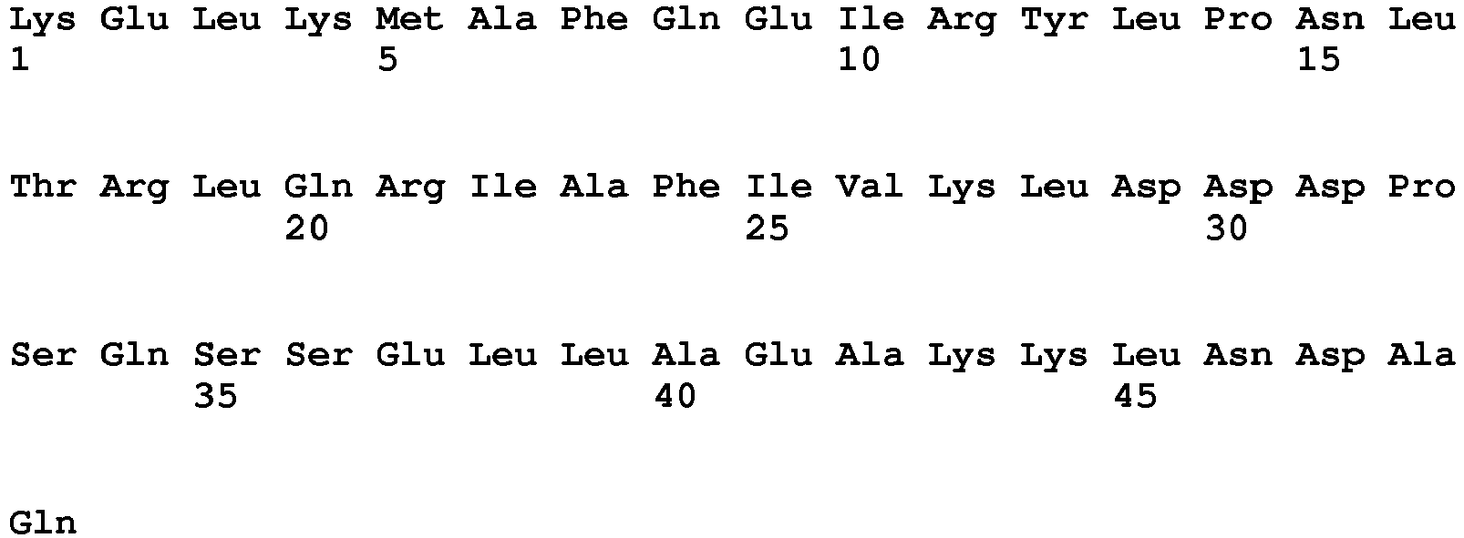
- <210> 68
<211> 49
<212> PRT
<213> Artificial sequence - <220>
<223> Engineered ABD binding polypeptide - <400> 68

- <210> 69
<211> 49
<212> PRT
<213> Artificial sequence - <220>
<223> Engineered ABD binding polypeptide - <400> 69


- <210> 70
<211> 49
<212> PRT
<213> Artificial sequence - <220>
<223> Engineered ABD binding polypeptide - <400> 70

- <210> 71
<211> 49
<212> PRT
<213> Artificial sequence - <220>
<223> Engineered ABD binding polypeptide - <400> 71

- <210> 72
<211> 49
<212> PRT
<213> Artificial sequence - <220>
<223> Engineered ABD binding polypeptide - <400> 72

- <210> 73
<211> 49
<212> PRT
<213> Artificial sequence - <220>
<223> Engineered ABD binding polypeptide - <400> 73

- <210> 74
<211> 49
<212> PRT
<213> Artificial sequence - <220>
<223> Engineered ABD binding polypeptide - <400> 74


- <210> 75
<211> 49
<212> PRT
<213> Artificial sequence - <220>
<223> Engineered ABD binding polypeptide - <400> 75

- <210> 76
<211> 49
<212> PRT
<213> Artificial sequence - <220>
<223> Engineered ABD binding polypeptide - <400> 76

- <210> 77
<211> 49
<212> PRT
<213> Artificial sequence - <220>
<223> Engineered ABD binding polypeptide - <400> 77

- <210> 78
<211> 49
<212> PRT
<213> Artificial sequence - <220>
<223> Engineered ABD binding polypeptide - <400> 78

- <210> 79
<211> 49
<212> PRT
<213> Artificial sequence - <220>
<223> Engineered ABD binding polypeptide - <400> 79

- <210> 80
<211> 49
<212> PRT
<213> Artificial sequence - <220>
<223> Engineered ABD binding polypeptide - <400> 80

- <210> 81
<211> 49
<212> PRT
<213> Artificial sequence - <220>
<223> Engineered ABD binding polypeptide - <400> 81


- <210> 82
<211> 49
<212> PRT
<213> Artificial sequence - <220>
<223> Engineered ABD binding polypeptide - <400> 82

- <210> 83
<211> 49
<212> PRT
<213> Artificial sequence - <220>
<223> Engineered ABD binding polypeptide - <400> 83

- <210> 84
<211> 49
<212> PRT
<213> Artificial sequence - <220>
<223> Engineered ABD binding polypeptide - <400> 84

- <210> 85
<211> 49
<212> PRT
<213> Artificial sequence - <220>
<223> Engineered ABD binding polypeptide - <400> 85

- <210> 86
<211> 49
<212> PRT
<213> Artificial sequence - <220>
<223> Engineered ABD binding polypeptide - <400> 86


- <210> 87
<211> 49
<212> PRT
<213> Artificial sequence - <220>
<223> Engineered ABD binding polypeptide - <400> 87

- <210> 88
<211> 49
<212> PRT
<213> Artificial sequence - <220>
<223> Engineered ABD binding polypeptide - <400> 88

- <210> 89
<211> 49
<212> PRT
<213> Artificial sequence - <220>
<223> Engineered ABD binding polypeptide - <400> 89

- <210> 90
<211> 49
<212> PRT
<213> Artificial sequence - <220>
<223> Engineered ABD binding polypeptide - <400> 90

- <210> 91
<211> 49
<212> PRT
<213> Artificial sequence - <220>
<223> Engineered ABD binding polypeptide - <400> 91


- <210> 92
<211> 49
<212> PRT
<213> Artificial sequence - <220>
<223> Engineered ABD binding polypeptide - <400> 92

- <210> 93
<211> 49
<212> PRT
<213> Artificial sequence - <220>
<223> Engineered ABD binding polypeptide - <400> 93


- <210> 94
<211> 49
<212> PRT
<213> Artificial sequence - <220>
<223> Engineered ABD binding polypeptide - <400> 94

- <210> 95
<211> 49
<212> PRT
<213> Artificial sequence - <220>
<223> Engineered ABD binding polypeptide - <400> 95

- <210> 96
<211> 49
<212> PRT
<213> Artificial sequence - <220>
<223> Engineered ABD binding polypeptide - <400> 96

- <210> 97
<211> 49
<212> PRT
<213> Artificial sequence - <220>
<223> Engineered ABD binding polypeptide - <400> 97

- <210> 98
<211> 49
<212> PRT
<213> Artificial sequence - <220>
<223> Engineered ABD binding polypeptide - <400> 98


- <210> 99
<211> 49
<212> PRT
<213> Artificial sequence - <220>
<223> Engineered ABD binding polypeptide - <400> 99
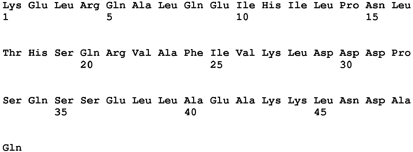
- <210> 100
<211> 49
<212> PRT
<213> Artificial sequence - <220>
<223> Engineered ABD binding polypeptide - <400> 100

- <210> 101
<211> 49
<212> PRT
<213> Artificial sequence - <220>
<223> Engineered ABD binding polypeptide - <400> 101

- <210> 102
<211> 49
<212> PRT
<213> Artificial sequence - <220>
<223> Engineered ABD binding polypeptide - <400> 102

- <210> 103
<211> 49
<212> PRT
<213> Artificial sequence - <220>
<223> Engineered ABD binding polypeptide - <400> 103


- <210> 104
<211> 49
<212> PRT
<213> Artificial sequence - <220>
<223> Engineered ABD binding polypeptide - <400> 104

- <210> 105
<211> 58
<212> PRT
<213> Artificial sequence - <220>
<223> Engineered ABD binding polypeptide - <400> 105

- <210> 106
<211> 58
<212> PRT
<213> Artificial sequence - <220>
<223> Engineered ABD binding polypeptide - <400> 106

- <210> 107
<211> 58
<212> PRT
<213> Artificial sequence - <220>
<223> Engineered ABD binding polypeptide - <400> 107

- <210> 108
<211> 58
<212> PRT
<213> Artificial sequence - <220>
<223> Engineered ABD binding polypeptide - <400> 108

- <210> 109
<211> 58
<212> PRT
<213> Artificial sequence - <220>
<223> Engineered ABD binding polypeptide - <400> 109

- <210> 110
<211> 58
<212> PRT
<213> Artificial sequence - <220>
<223> Engineered ABD binding polypeptide - <400> 110
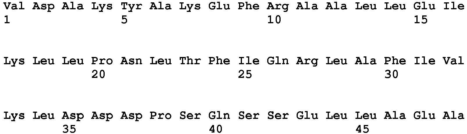

- <210> 111
<211> 58
<212> PRT
<213> Artificial sequence - <220>
<223> Engineered ABD binding polypeptide - <400> 111

- <210> 112
<211> 58
<212> PRT
<213> Artificial sequence - <220>
<223> Engineered ABD binding polypeptide - <400> 112

- <210> 113
<211> 58
<212> PRT
<213> Artificial sequence - <220>
<223> Engineered ABD binding polypeptide - <400> 113

- <210> 114
<211> 58
<212> PRT
<213> Artificial sequence - <220>
<223> Engineered ABD binding polypeptide - <400> 114

- <210> 115
<211> 58
<212> PRT
<213> Artificial sequence - <220>
<223> Engineered ABD binding polypeptide - <400> 115


- <210> 116
<211> 58
<212> PRT
<213> Artificial sequence - <220>
<223> Engineered ABD binding polypeptide - <400> 116

- <210> 117
<211> 58
<212> PRT
<213> Artificial sequence - <220>
<223> Engineered ABD binding polypeptide - <400> 117

- <210> 118
<211> 58
<212> PRT
<213> Artificial sequence - <220>
<223> Engineered ABD binding polypeptide - <400> 118

- <210> 119
<211> 58
<212> PRT
<213> Artificial sequence - <220>
<223> Engineered ABD binding polypeptide - <400> 119

- <210> 120
<211> 58
<212> PRT
<213> Artificial sequence - <220>
<223> Engineered ABD binding polypeptide - <400> 120


- <210> 121
<211> 58
<212> PRT
<213> Artificial sequence - <220>
<223> Engineered ABD binding polypeptide - <400> 121

- <210> 122
<211> 58
<212> PRT
<213> Artificial sequence - <220>
<223> Engineered ABD binding polypeptide - <400> 122

- <210> 123
<211> 58
<212> PRT
<213> Artificial sequence - <220>
<223> Engineered ABD binding polypeptide - <400> 123

- <210> 124
<211> 58
<212> PRT
<213> Artificial sequence - <220>
<223> Engineered ABD binding polypeptide - <400> 124

- <210> 125
<211> 58
<212> PRT
<213> Artificial sequence - <220>
<223> Engineered ABD binding polypeptide - <400> 125

- <210> 126
<211> 58
<212> PRT
<213> Artificial sequence - <220>
<223> Engineered ABD binding polypeptide - <400> 126

- <210> 127
<211> 58
<212> PRT
<213> Artificial sequence - <220>
<223> Engineered ABD binding polypeptide - <400> 127


- <210> 128
<211> 58
<212> PRT
<213> Artificial sequence - <220>
<223> Engineered ABD binding polypeptide - <400> 128

- <210> 129
<211> 58
<212> PRT
<213> Artificial sequence - <220>
<223> Engineered ABD binding polypeptide - <400> 129

- <210> 130
<211> 58
<212> PRT
<213> Artificial sequence - <220>
<223> Engineered ABD binding polypeptide - <400> 130

- <210> 131
<211> 58
<212> PRT
<213> Artificial sequence - <220>
<223> Engineered ABD binding polypeptide - <400> 131

- <210> 132
<211> 58
<212> PRT
<213> Artificial sequence - <220>
<223> Engineered ABD binding polypeptide - <400> 132


- <210> 133
<211> 58
<212> PRT
<213> Artificial sequence - <220>
<223> Engineered ABD binding polypeptide - <400> 133

- <210> 134
<211> 58
<212> PRT
<213> Artificial sequence - <220>
<223> Engineered ABD binding polypeptide - <400> 134

- <210> 135
<211> 58
<212> PRT
<213> Artificial sequence - <220>
<223> Engineered ABD binding polypeptide - <400> 135

- <210> 136
<211> 58
<212> PRT
<213> Artificial sequence - <220>
<223> Engineered ABD binding polypeptide - <400> 136
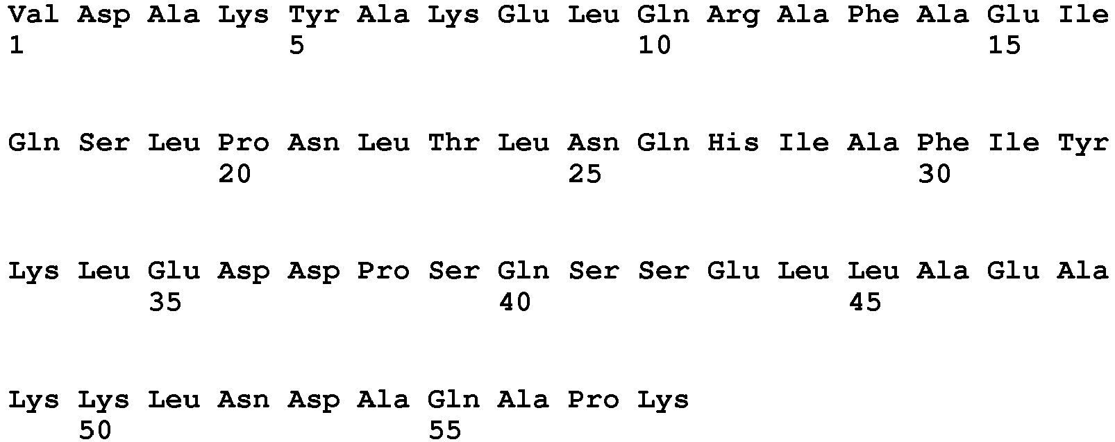
- <210> 137
<211> 58
<212> PRT
<213> Artificial sequence - <220>
<223> Engineered ABD binding polypeptide - <400> 137


- <210> 138
<211> 58
<212> PRT
<213> Artificial sequence - <220>
<223> Engineered ABD binding polypeptide - <400> 138

- <210> 139
<211> 58
<212> PRT
<213> Artificial sequence - <220>
<223> Engineered ABD binding polypeptide - <400> 139


- <210> 140
<211> 58
<212> PRT
<213> Artificial sequence - <220>
<223> Engineered ABD binding polypeptide - <400> 140

- <210> 141
<211> 58
<212> PRT
<213> Artificial sequence - <220>
<223> Engineered ABD binding polypeptide - <400> 141

- <210> 142
<211> 58
<212> PRT
<213> Artificial sequence - <220>
<223> Engineered ABD binding polypeptide - <400> 142

- <210> 143
<211> 58
<212> PRT
<213> Artificial sequence - <220>
<223> Engineered ABD binding polypeptide - <400> 143

- <210> 144
<211> 58
<212> PRT
<213> Artificial sequence - <220>
<223> Engineered ABD binding polypeptide - <400> 144


- <210> 145
<211> 58
<212> PRT
<213> Artificial sequence - <220>
<223> Engineered ABD binding polypeptide - <400> 145

- <210> 146
<211> 58
<212> PRT
<213> Artificial sequence - <220>
<223> Engineered ABD binding polypeptide - <400> 146
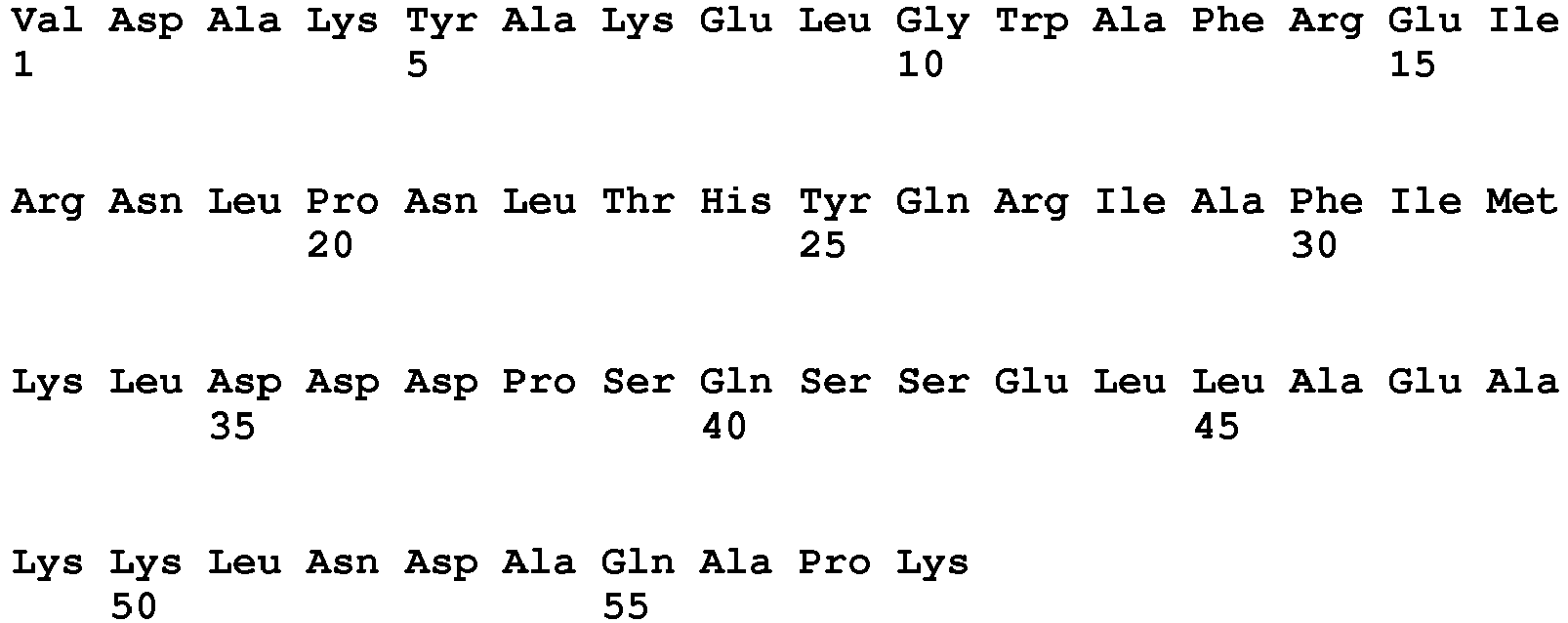
- <210> 147
<211> 58
<212> PRT
<213> Artificial sequence - <220>
<223> Engineered ABD binding polypeptide - <400> 147

- <210> 148
<211> 58
<212> PRT
<213> Artificial sequence - <220>
<223> Engineered ABD binding polypeptide - <400> 148

- <210> 149
<211> 58
<212> PRT
<213> Artificial sequence - <220>
<223> Engineered ABD binding polypeptide - <400> 149


- <210> 150
<211> 58
<212> PRT
<213> Artificial sequence - <220>
<223> Engineered ABD binding polypeptide - <400> 150

- <210> 151
<211> 58
<212> PRT
<213> Artificial sequence - <220>
<223> Engineered ABD binding polypeptide - <400> 151

- <210> 152
<211> 58
<212> PRT
<213> Artificial sequence - <220>
<223> Engineered ABD binding polypeptide - <400> 152

- <210> 153
<211> 58
<212> PRT
<213> Artificial sequence - <220>
<223> Engineered ABD binding polypeptide - <400> 153

- <210> 154
<211> 58
<212> PRT
<213> Artificial sequence - <220>
<223> Engineered ABD binding polypeptide - <400> 154


- <210> 155
<211> 58
<212> PRT
<213> Artificial sequence - <220>
<223> Engineered ABD binding polypeptide - <400> 155

- <210> 156
<211> 58
<212> PRT
<213> Artificial sequence - <220>
<223> Engineered ABD binding polypeptide - <400> 156


- <210> 157
<211> 46
<212> PRT
<213> Streptococcus sp. G148 - <400> 157

- <210> 158
<211> 47
<212> PRT
<213> Artificial sequence - <220>
<223> Engineered albumin binding domain - <400> 158

- <210> 159
<211> 46
<212> PRT
<213> Artificial sequence - <220>
<223> Engineered albumin binding domain - <400> 159


- <210> 160
<211> 46
<212> PRT
<213> Artificial sequence - <220>
<223> Engineered albumin binding domain - <400> 160

- <210> 161
<211> 48
<212> PRT
<213> Artificial sequence - <220>
<223> Engineered albumin binding domain - <400> 161

- <210> 162
<211> 47
<212> PRT
<213> Artificial sequence - <220>
<223> Engineered albumin binding domain - <400> 162


- <210> 163
<211> 24
<212> DNA
<213> Artificial sequence - <220>
<223> Synthetic primer - <400> 163
tgcttccggc tcgtatgttg tgtg 24 - <210> 164
<211> 21
<212> DNA
<213> Artificial sequence - <220>
<223> Synthetic primer - <400> 164
cggaaccaga gccaccaccgg 21 - <210> 165
<211> 21
<212> DNA
<213> Artificial sequence - <220>
<223> Synthetic primer - <400> 165
cggaaccaga gccaccaccgg 21
Claims (14)
- Method of separation of at least one ABD-containing molecule present in a liquid from other constituents in the liquid, comprising a step of affinity separation, in which step is used, as affinity ligand, an ABD binding polypeptide comprising an ABD binding motif BM, which motif consists of the amino acid sequence:
EX2X3X4AX6X7EIX1OX11LPNLTX17X18QX20X21AFiX25KLX28D
wherein, independently from each other,X2 is selected from F, I and L;X3 is selected from H, K, N, Q, R, S, T and V;X4 is selected from A, D, F, G, H, I, K, L, M, N, Q, R, S, T, V, W and Y;X6 is selected from F, I, L and Y;X7 is selected from A, H, I, K, L, N, Q, R, S, T and V;X10 is selected from G, H, K, N, Q, R and S;X11 is selected from A, D, F, G, I, K, L, N, Q, R, S, T, V and Y;X17 is selected from F, H, L, S and T;X18 is selected from D, E, H, I, K, L, M, N, Q, R, S, T and V;X20 is selected from H, K and R;X21 is selected from I, L and V;X25 is selected from F, I, L, V and Y;X28 is selected from D and E. - Method according to claim 1, comprising the steps:a) applying the liquid to an affinity matrix comprising said ABD binding polypeptide under conditions that are conducive to binding of an ABD-containing molecule to the affinity matrix;b) washing the affinity matrix for removal of substances not bound thereto; andc) eluting any bound ABD-containing molecule from the affinity matrix, thus obtaining an ABD-containing molecule fraction with an enriched content of ABD-containing molecule; andd) recovering said ABD-containing molecule fraction.
- Method according to claim 1, comprising the steps:a) applying the liquid to an affinity matrix comprising said ABD binding polypeptide under conditions that are conducive to binding of an ABD-containing molecule to the affinity matrix;b) washing the affinity matrix for recovery of substances not bound thereto, thus obtaining a depleted fraction with a substantially reduced ABD-containing molecule content; andc) recovering said depleted fraction.
- Method according to claim 1, comprising the steps:a) applying the liquid to an affinity matrix comprising said ABD binding polypeptide under conditions that are conducive to binding of an ABD-containing molecule to the affinity matrix;b) washing the affinity matrix for recovery of substances not bound thereto, thus obtaining a depleted fraction with a substantially reduced ABD-containing molecule content; andc) eluting the bound ABD-containing molecule from the affinity matrix, thus obtaining an ABD-containing molecule fraction with an ABD-containing molecule content; andd) recovering said ABD-containing molecule fraction and said depleted fraction.
- Method according to any preceding claim, wherein said amino acid sequence fulfills at least four of the six conditions I-VI:I. X2 is F or L;II. X6 is F or L;III. X20 is H or R;IV. X21 is I or L;V. X25 is I or V; andVI. X28 is D.
- Method according to any preceding claim, wherein said amino acid sequence is selected from SEQ ID NO:1-52.
- Method according to claim 6, wherein said amino acid sequence is selected from SEQ ID NO:1-7.
- Method according to any preceding claim, wherein said ABD binding motif forms part of a three-helix bundle protein domain selected from domains of protein A from Staphylococcus aureus or derivatives thereof.
- Method according to any preceding claim, in which said ABD binding polypeptide comprises the amino acid sequence:
K-[BM]-DPSQS XaXbLLXc EAKKL NDXdQ;
wherein[BM] is an ABD binding motif as defined in any one of claims 1 and 5-7;Xa is selected from A and S;Xb is selected from N and E;Xc is selected from A, S and C;Xd is selected from A and S. - Method according to claim 9, wherein said amino acid sequence is selected from any one of SEQ ID NO:53-104.
- Method according to claim 10, wherein said amino acid sequence is selected from any one of SEQ ID NO:53-59.
- Method according to any preceding claim, which comprises the amino acid sequence:
VDAKYAK-[BM]- DPSQSSELLAEAKKLNDAQAPK;
wherein [BM] is an ABD binding motif as defined in any one of claims 1 and 5-7. - Method according to claim 12, in which said amino acid sequence is selected from SEQ ID NO:105-156.
- Method according to claim 13, in which said amino acid sequence is selected from SEQ ID NO 105-111.
Priority Applications (2)
| Application Number | Priority Date | Filing Date | Title |
|---|---|---|---|
| PL13783334T PL2912051T3 (en) | 2012-10-25 | 2013-10-25 | Method for the separation of proteins containing an albumin-binding domain |
| EP13783334.9A EP2912051B1 (en) | 2012-10-25 | 2013-10-25 | Method for the separation of proteins containing an albumin-binding domain |
Applications Claiming Priority (4)
| Application Number | Priority Date | Filing Date | Title |
|---|---|---|---|
| US201261718246P | 2012-10-25 | 2012-10-25 | |
| EP12189948 | 2012-10-25 | ||
| PCT/EP2013/072365 WO2014064238A1 (en) | 2012-10-25 | 2013-10-25 | Method for the separation of proteins containing an albumin-binding |
| EP13783334.9A EP2912051B1 (en) | 2012-10-25 | 2013-10-25 | Method for the separation of proteins containing an albumin-binding domain |
Publications (2)
| Publication Number | Publication Date |
|---|---|
| EP2912051A1 EP2912051A1 (en) | 2015-09-02 |
| EP2912051B1 true EP2912051B1 (en) | 2018-04-18 |
Family
ID=47088703
Family Applications (1)
| Application Number | Title | Priority Date | Filing Date |
|---|---|---|---|
| EP13783334.9A Active EP2912051B1 (en) | 2012-10-25 | 2013-10-25 | Method for the separation of proteins containing an albumin-binding domain |
Country Status (7)
| Country | Link |
|---|---|
| US (1) | US9745340B2 (en) |
| EP (1) | EP2912051B1 (en) |
| JP (1) | JP6396303B2 (en) |
| DK (1) | DK2912051T3 (en) |
| ES (1) | ES2674983T3 (en) |
| PL (1) | PL2912051T3 (en) |
| WO (1) | WO2014064238A1 (en) |
Families Citing this family (3)
| Publication number | Priority date | Publication date | Assignee | Title |
|---|---|---|---|---|
| AU2013336646B2 (en) | 2012-10-25 | 2016-07-14 | Affibody Ab | ABD binding polypeptide |
| CA3048273C (en) * | 2017-01-04 | 2024-10-01 | Nanjing GenScript Biotech Co., Ltd. | High-loading and alkali-resistant protein a magnetic bead and method of use thereof |
| CN119735705B (en) * | 2024-12-31 | 2025-11-18 | 江南大学 | A fusion protein and its application in the preparation of smegglutinin precursor peptide |
Family Cites Families (10)
| Publication number | Priority date | Publication date | Assignee | Title |
|---|---|---|---|---|
| SE509359C2 (en) | 1989-08-01 | 1999-01-18 | Cemu Bioteknik Ab | Use of stabilized protein or peptide conjugates for the preparation of a drug |
| US7608681B2 (en) | 1999-12-24 | 2009-10-27 | Genentech, Inc. | Methods and compositions for prolonging elimination half-times of bioactive compounds |
| US8937153B2 (en) * | 2007-07-31 | 2015-01-20 | Affibody Ab | Compositions, methods and uses |
| US8426557B2 (en) | 2007-08-03 | 2013-04-23 | Affibody Ab | IGF-1R binding polypeptides and their use |
| EP2077272A1 (en) * | 2007-12-21 | 2009-07-08 | Affibody AB | Polypeptide libraries with a predetermined scaffold |
| ATE527353T1 (en) * | 2007-12-19 | 2011-10-15 | Affibody Ab | PDGF-BINDING POLYPEPTIDE FROM PROTEIN A |
| CA2739352C (en) * | 2008-10-29 | 2021-07-13 | Wyeth Llc | Methods for purification of single domain antigen binding molecules |
| EP2496598B1 (en) | 2009-11-04 | 2017-08-02 | Affibody AB | Her3 binding polypeptides |
| NZ604805A (en) * | 2010-07-09 | 2014-09-26 | Affibody Ab | Polypeptides |
| RS58503B1 (en) | 2012-02-20 | 2019-04-30 | Swedish Orphan Biovitrum Ab Publ | Polypeptides binding to human complement c5 |
-
2013
- 2013-10-25 EP EP13783334.9A patent/EP2912051B1/en active Active
- 2013-10-25 JP JP2015538459A patent/JP6396303B2/en active Active
- 2013-10-25 US US14/436,150 patent/US9745340B2/en active Active
- 2013-10-25 ES ES13783334.9T patent/ES2674983T3/en active Active
- 2013-10-25 WO PCT/EP2013/072365 patent/WO2014064238A1/en not_active Ceased
- 2013-10-25 DK DK13783334.9T patent/DK2912051T3/en active
- 2013-10-25 PL PL13783334T patent/PL2912051T3/en unknown
Non-Patent Citations (1)
| Title |
|---|
| None * |
Also Published As
| Publication number | Publication date |
|---|---|
| PL2912051T3 (en) | 2018-08-31 |
| US20150274771A1 (en) | 2015-10-01 |
| JP6396303B2 (en) | 2018-09-26 |
| US9745340B2 (en) | 2017-08-29 |
| JP2015533377A (en) | 2015-11-24 |
| ES2674983T3 (en) | 2018-07-05 |
| DK2912051T3 (en) | 2018-06-14 |
| EP2912051A1 (en) | 2015-09-02 |
| WO2014064238A1 (en) | 2014-05-01 |
Similar Documents
| Publication | Publication Date | Title |
|---|---|---|
| EP2912054B1 (en) | Albumin binding polypeptide | |
| US20250051400A1 (en) | Alkaline stable fc-binding proteins for affinity chromatography | |
| EP2231860B1 (en) | Polypeptide derived from protein a and able to bind pdgf | |
| JP5677943B2 (en) | Polypeptide | |
| EP4491268A2 (en) | Fc binding proteins with cysteine in the c-terminal helical region | |
| JP7689746B2 (en) | Binding proteins for the enzyme acid alpha-glucosidase (GAA) and uses thereof | |
| US9745340B2 (en) | Method for the separation of proteins containing an albumin-binding | |
| JP2025539595A (en) | New ligands for affinity chromatography | |
| EP4107178B1 (en) | Affinity ligand for the soluble part of fibroblast growth factor receptor 3 (sfgfr3) | |
| HK1210793B (en) | Albumin binding polypeptide | |
| WO2005000883A1 (en) | Polypeptides having binding affinity for insulin | |
| EP4463465B1 (en) | High affinity purification of ferritin | |
| WO2021176075A1 (en) | Novel immunoglobulin binding polypeptides |
Legal Events
| Date | Code | Title | Description |
|---|---|---|---|
| PUAI | Public reference made under article 153(3) epc to a published international application that has entered the european phase |
Free format text: ORIGINAL CODE: 0009012 |
|
| 17P | Request for examination filed |
Effective date: 20150520 |
|
| AK | Designated contracting states |
Kind code of ref document: A1 Designated state(s): AL AT BE BG CH CY CZ DE DK EE ES FI FR GB GR HR HU IE IS IT LI LT LU LV MC MK MT NL NO PL PT RO RS SE SI SK SM TR |
|
| AX | Request for extension of the european patent |
Extension state: BA ME |
|
| DAX | Request for extension of the european patent (deleted) | ||
| 17Q | First examination report despatched |
Effective date: 20160422 |
|
| STAA | Information on the status of an ep patent application or granted ep patent |
Free format text: STATUS: EXAMINATION IS IN PROGRESS |
|
| GRAP | Despatch of communication of intention to grant a patent |
Free format text: ORIGINAL CODE: EPIDOSNIGR1 |
|
| STAA | Information on the status of an ep patent application or granted ep patent |
Free format text: STATUS: GRANT OF PATENT IS INTENDED |
|
| INTG | Intention to grant announced |
Effective date: 20171114 |
|
| GRAS | Grant fee paid |
Free format text: ORIGINAL CODE: EPIDOSNIGR3 |
|
| GRAA | (expected) grant |
Free format text: ORIGINAL CODE: 0009210 |
|
| STAA | Information on the status of an ep patent application or granted ep patent |
Free format text: STATUS: THE PATENT HAS BEEN GRANTED |
|
| RAP1 | Party data changed (applicant data changed or rights of an application transferred) |
Owner name: AFFIBODY AB |
|
| AK | Designated contracting states |
Kind code of ref document: B1 Designated state(s): AL AT BE BG CH CY CZ DE DK EE ES FI FR GB GR HR HU IE IS IT LI LT LU LV MC MK MT NL NO PL PT RO RS SE SI SK SM TR |
|
| REG | Reference to a national code |
Ref country code: GB Ref legal event code: FG4D |
|
| REG | Reference to a national code |
Ref country code: CH Ref legal event code: EP |
|
| REG | Reference to a national code |
Ref country code: AT Ref legal event code: REF Ref document number: 990347 Country of ref document: AT Kind code of ref document: T Effective date: 20180515 |
|
| REG | Reference to a national code |
Ref country code: IE Ref legal event code: FG4D |
|
| REG | Reference to a national code |
Ref country code: DE Ref legal event code: R096 Ref document number: 602013036142 Country of ref document: DE |
|
| REG | Reference to a national code |
Ref country code: CH Ref legal event code: NV Representative=s name: OFFICE ERNEST T. FREYLINGER S.A., CH |
|
| REG | Reference to a national code |
Ref country code: DK Ref legal event code: T3 Effective date: 20180606 |
|
| REG | Reference to a national code |
Ref country code: SE Ref legal event code: TRGR |
|
| REG | Reference to a national code |
Ref country code: ES Ref legal event code: FG2A Ref document number: 2674983 Country of ref document: ES Kind code of ref document: T3 Effective date: 20180705 |
|
| REG | Reference to a national code |
Ref country code: LT Ref legal event code: MG4D |
|
| REG | Reference to a national code |
Ref country code: FR Ref legal event code: PLFP Year of fee payment: 6 |
|
| PG25 | Lapsed in a contracting state [announced via postgrant information from national office to epo] |
Ref country code: NO Free format text: LAPSE BECAUSE OF FAILURE TO SUBMIT A TRANSLATION OF THE DESCRIPTION OR TO PAY THE FEE WITHIN THE PRESCRIBED TIME-LIMIT Effective date: 20180718 Ref country code: FI Free format text: LAPSE BECAUSE OF FAILURE TO SUBMIT A TRANSLATION OF THE DESCRIPTION OR TO PAY THE FEE WITHIN THE PRESCRIBED TIME-LIMIT Effective date: 20180418 Ref country code: AL Free format text: LAPSE BECAUSE OF FAILURE TO SUBMIT A TRANSLATION OF THE DESCRIPTION OR TO PAY THE FEE WITHIN THE PRESCRIBED TIME-LIMIT Effective date: 20180418 Ref country code: BG Free format text: LAPSE BECAUSE OF FAILURE TO SUBMIT A TRANSLATION OF THE DESCRIPTION OR TO PAY THE FEE WITHIN THE PRESCRIBED TIME-LIMIT Effective date: 20180718 Ref country code: LT Free format text: LAPSE BECAUSE OF FAILURE TO SUBMIT A TRANSLATION OF THE DESCRIPTION OR TO PAY THE FEE WITHIN THE PRESCRIBED TIME-LIMIT Effective date: 20180418 |
|
| PG25 | Lapsed in a contracting state [announced via postgrant information from national office to epo] |
Ref country code: RS Free format text: LAPSE BECAUSE OF FAILURE TO SUBMIT A TRANSLATION OF THE DESCRIPTION OR TO PAY THE FEE WITHIN THE PRESCRIBED TIME-LIMIT Effective date: 20180418 Ref country code: LV Free format text: LAPSE BECAUSE OF FAILURE TO SUBMIT A TRANSLATION OF THE DESCRIPTION OR TO PAY THE FEE WITHIN THE PRESCRIBED TIME-LIMIT Effective date: 20180418 Ref country code: HR Free format text: LAPSE BECAUSE OF FAILURE TO SUBMIT A TRANSLATION OF THE DESCRIPTION OR TO PAY THE FEE WITHIN THE PRESCRIBED TIME-LIMIT Effective date: 20180418 Ref country code: GR Free format text: LAPSE BECAUSE OF FAILURE TO SUBMIT A TRANSLATION OF THE DESCRIPTION OR TO PAY THE FEE WITHIN THE PRESCRIBED TIME-LIMIT Effective date: 20180719 |
|
| REG | Reference to a national code |
Ref country code: AT Ref legal event code: MK05 Ref document number: 990347 Country of ref document: AT Kind code of ref document: T Effective date: 20180418 |
|
| PG25 | Lapsed in a contracting state [announced via postgrant information from national office to epo] |
Ref country code: PT Free format text: LAPSE BECAUSE OF FAILURE TO SUBMIT A TRANSLATION OF THE DESCRIPTION OR TO PAY THE FEE WITHIN THE PRESCRIBED TIME-LIMIT Effective date: 20180820 |
|
| REG | Reference to a national code |
Ref country code: DE Ref legal event code: R097 Ref document number: 602013036142 Country of ref document: DE |
|
| PG25 | Lapsed in a contracting state [announced via postgrant information from national office to epo] |
Ref country code: AT Free format text: LAPSE BECAUSE OF FAILURE TO SUBMIT A TRANSLATION OF THE DESCRIPTION OR TO PAY THE FEE WITHIN THE PRESCRIBED TIME-LIMIT Effective date: 20180418 Ref country code: CZ Free format text: LAPSE BECAUSE OF FAILURE TO SUBMIT A TRANSLATION OF THE DESCRIPTION OR TO PAY THE FEE WITHIN THE PRESCRIBED TIME-LIMIT Effective date: 20180418 Ref country code: RO Free format text: LAPSE BECAUSE OF FAILURE TO SUBMIT A TRANSLATION OF THE DESCRIPTION OR TO PAY THE FEE WITHIN THE PRESCRIBED TIME-LIMIT Effective date: 20180418 Ref country code: EE Free format text: LAPSE BECAUSE OF FAILURE TO SUBMIT A TRANSLATION OF THE DESCRIPTION OR TO PAY THE FEE WITHIN THE PRESCRIBED TIME-LIMIT Effective date: 20180418 Ref country code: SK Free format text: LAPSE BECAUSE OF FAILURE TO SUBMIT A TRANSLATION OF THE DESCRIPTION OR TO PAY THE FEE WITHIN THE PRESCRIBED TIME-LIMIT Effective date: 20180418 |
|
| PLBE | No opposition filed within time limit |
Free format text: ORIGINAL CODE: 0009261 |
|
| STAA | Information on the status of an ep patent application or granted ep patent |
Free format text: STATUS: NO OPPOSITION FILED WITHIN TIME LIMIT |
|
| PG25 | Lapsed in a contracting state [announced via postgrant information from national office to epo] |
Ref country code: SM Free format text: LAPSE BECAUSE OF FAILURE TO SUBMIT A TRANSLATION OF THE DESCRIPTION OR TO PAY THE FEE WITHIN THE PRESCRIBED TIME-LIMIT Effective date: 20180418 |
|
| 26N | No opposition filed |
Effective date: 20190121 |
|
| PG25 | Lapsed in a contracting state [announced via postgrant information from national office to epo] |
Ref country code: SI Free format text: LAPSE BECAUSE OF FAILURE TO SUBMIT A TRANSLATION OF THE DESCRIPTION OR TO PAY THE FEE WITHIN THE PRESCRIBED TIME-LIMIT Effective date: 20180418 |
|
| PG25 | Lapsed in a contracting state [announced via postgrant information from national office to epo] |
Ref country code: MT Free format text: LAPSE BECAUSE OF NON-PAYMENT OF DUE FEES Effective date: 20181025 |
|
| PG25 | Lapsed in a contracting state [announced via postgrant information from national office to epo] |
Ref country code: TR Free format text: LAPSE BECAUSE OF FAILURE TO SUBMIT A TRANSLATION OF THE DESCRIPTION OR TO PAY THE FEE WITHIN THE PRESCRIBED TIME-LIMIT Effective date: 20180418 |
|
| PG25 | Lapsed in a contracting state [announced via postgrant information from national office to epo] |
Ref country code: CY Free format text: LAPSE BECAUSE OF FAILURE TO SUBMIT A TRANSLATION OF THE DESCRIPTION OR TO PAY THE FEE WITHIN THE PRESCRIBED TIME-LIMIT Effective date: 20180418 Ref country code: MK Free format text: LAPSE BECAUSE OF NON-PAYMENT OF DUE FEES Effective date: 20180418 Ref country code: HU Free format text: LAPSE BECAUSE OF FAILURE TO SUBMIT A TRANSLATION OF THE DESCRIPTION OR TO PAY THE FEE WITHIN THE PRESCRIBED TIME-LIMIT; INVALID AB INITIO Effective date: 20131025 |
|
| PG25 | Lapsed in a contracting state [announced via postgrant information from national office to epo] |
Ref country code: IS Free format text: LAPSE BECAUSE OF FAILURE TO SUBMIT A TRANSLATION OF THE DESCRIPTION OR TO PAY THE FEE WITHIN THE PRESCRIBED TIME-LIMIT Effective date: 20180818 |
|
| P01 | Opt-out of the competence of the unified patent court (upc) registered |
Effective date: 20230517 |
|
| PGFP | Annual fee paid to national office [announced via postgrant information from national office to epo] |
Ref country code: DK Payment date: 20250917 Year of fee payment: 13 |
|
| PGFP | Annual fee paid to national office [announced via postgrant information from national office to epo] |
Ref country code: MC Payment date: 20250915 Year of fee payment: 13 |
|
| PGFP | Annual fee paid to national office [announced via postgrant information from national office to epo] |
Ref country code: LU Payment date: 20250916 Year of fee payment: 13 Ref country code: NL Payment date: 20250924 Year of fee payment: 13 Ref country code: IT Payment date: 20250919 Year of fee payment: 13 Ref country code: PL Payment date: 20250916 Year of fee payment: 13 |
|
| PGFP | Annual fee paid to national office [announced via postgrant information from national office to epo] |
Ref country code: GB Payment date: 20250918 Year of fee payment: 13 Ref country code: BE Payment date: 20250924 Year of fee payment: 13 |
|
| PGFP | Annual fee paid to national office [announced via postgrant information from national office to epo] |
Ref country code: FR Payment date: 20250915 Year of fee payment: 13 |
|
| PGFP | Annual fee paid to national office [announced via postgrant information from national office to epo] |
Ref country code: SE Payment date: 20250916 Year of fee payment: 13 |
|
| PGFP | Annual fee paid to national office [announced via postgrant information from national office to epo] |
Ref country code: IE Payment date: 20250912 Year of fee payment: 13 |
|
| REG | Reference to a national code |
Ref country code: CH Ref legal event code: U11 Free format text: ST27 STATUS EVENT CODE: U-0-0-U10-U11 (AS PROVIDED BY THE NATIONAL OFFICE) Effective date: 20251101 |
|
| PGFP | Annual fee paid to national office [announced via postgrant information from national office to epo] |
Ref country code: DE Payment date: 20250917 Year of fee payment: 13 |
|
| PGFP | Annual fee paid to national office [announced via postgrant information from national office to epo] |
Ref country code: CH Payment date: 20251101 Year of fee payment: 13 |
|
| PGFP | Annual fee paid to national office [announced via postgrant information from national office to epo] |
Ref country code: ES Payment date: 20251119 Year of fee payment: 13 |