EP2899275A1 - Method for obtaining information about endometrial cancer, and marker and kit for obtaining information about endometrial cancer - Google Patents
Method for obtaining information about endometrial cancer, and marker and kit for obtaining information about endometrial cancer Download PDFInfo
- Publication number
- EP2899275A1 EP2899275A1 EP13839246.9A EP13839246A EP2899275A1 EP 2899275 A1 EP2899275 A1 EP 2899275A1 EP 13839246 A EP13839246 A EP 13839246A EP 2899275 A1 EP2899275 A1 EP 2899275A1
- Authority
- EP
- European Patent Office
- Prior art keywords
- methylation
- cancer
- uterine body
- dna
- body cancer
- Prior art date
- Legal status (The legal status is an assumption and is not a legal conclusion. Google has not performed a legal analysis and makes no representation as to the accuracy of the status listed.)
- Granted
Links
Images






Classifications
-
- C—CHEMISTRY; METALLURGY
- C12—BIOCHEMISTRY; BEER; SPIRITS; WINE; VINEGAR; MICROBIOLOGY; ENZYMOLOGY; MUTATION OR GENETIC ENGINEERING
- C12Q—MEASURING OR TESTING PROCESSES INVOLVING ENZYMES, NUCLEIC ACIDS OR MICROORGANISMS; COMPOSITIONS OR TEST PAPERS THEREFOR; PROCESSES OF PREPARING SUCH COMPOSITIONS; CONDITION-RESPONSIVE CONTROL IN MICROBIOLOGICAL OR ENZYMOLOGICAL PROCESSES
- C12Q1/00—Measuring or testing processes involving enzymes, nucleic acids or microorganisms; Compositions therefor; Processes of preparing such compositions
- C12Q1/68—Measuring or testing processes involving enzymes, nucleic acids or microorganisms; Compositions therefor; Processes of preparing such compositions involving nucleic acids
- C12Q1/6876—Nucleic acid products used in the analysis of nucleic acids, e.g. primers or probes
- C12Q1/6883—Nucleic acid products used in the analysis of nucleic acids, e.g. primers or probes for diseases caused by alterations of genetic material
- C12Q1/6886—Nucleic acid products used in the analysis of nucleic acids, e.g. primers or probes for diseases caused by alterations of genetic material for cancer
-
- G—PHYSICS
- G01—MEASURING; TESTING
- G01N—INVESTIGATING OR ANALYSING MATERIALS BY DETERMINING THEIR CHEMICAL OR PHYSICAL PROPERTIES
- G01N33/00—Investigating or analysing materials by specific methods not covered by groups G01N1/00 - G01N31/00
- G01N33/48—Biological material, e.g. blood, urine; Haemocytometers
- G01N33/50—Chemical analysis of biological material, e.g. blood, urine; Testing involving biospecific ligand binding methods; Immunological testing
- G01N33/53—Immunoassay; Biospecific binding assay; Materials therefor
- G01N33/575—Immunoassay; Biospecific binding assay; Materials therefor for cancer
- G01N33/5755—Immunoassay; Biospecific binding assay; Materials therefor for cancer of the uterine cervix, uterine corpus or endometrium
-
- C—CHEMISTRY; METALLURGY
- C12—BIOCHEMISTRY; BEER; SPIRITS; WINE; VINEGAR; MICROBIOLOGY; ENZYMOLOGY; MUTATION OR GENETIC ENGINEERING
- C12Q—MEASURING OR TESTING PROCESSES INVOLVING ENZYMES, NUCLEIC ACIDS OR MICROORGANISMS; COMPOSITIONS OR TEST PAPERS THEREFOR; PROCESSES OF PREPARING SUCH COMPOSITIONS; CONDITION-RESPONSIVE CONTROL IN MICROBIOLOGICAL OR ENZYMOLOGICAL PROCESSES
- C12Q2600/00—Oligonucleotides characterized by their use
- C12Q2600/154—Methylation markers
Definitions
- the present invention relates to a method for obtaining information on uterine body cancer in a subject.
- the present invention also relates to a marker and a kit used in the method.
- Uterine body cancer is cancer of the corpus of the uterus, develops in the endometrium and thus is also referred to as endometrial cancer.
- Uterine body cancer is generally examined by pathological examination (cytological examination and biopsy) in which endometrial cells and tissues are collected and observed by microscopy.
- pathological examination requires insertion of a special instrument for cell or tissue collection into the corpus of the uterus, which may be accompanied by pain and bleeding and thus put a great burden on subjects.
- Examination for uterine body cancer includes measurement of tumor markers in blood.
- the tumor markers used include CA125, CA19-9 and the like.
- the examination of the tumor markers has insufficient cancer detection sensitivity.
- the examination has also been utilized for examinations of types of cancer different from uterine body cancer, and thus has insufficient sensitivity and specificity as the specific screening examination for uterine body cancer.
- the methods include, for example, ones based on information on methylation of DNAs.
- the methods use markers which are CpG sites (5'-(CG)-3') in base sequences of certain genes. Based on the analysis results of the methylation status of the markers, information such as presence or absence of a cancer cell is obtained, which information may be used as an index for diagnosis of cancer.
- genes indicative of abnormal methylation in uterine body cancer have been reported as described above, these genes are methylated in some extent in a type of cancer different from uterine body cancer.
- information deduced from the analysis result may include not only information on uterine body cancer but also information on other types of cancer. In this case, even when an analysis result provides information indicating that a sample contains a cancer cell for example, it is difficult to determine whether the cancer cell is derived from uterine body cancer or from other types of cancer.
- an object of the present invention is to provide a method for obtaining information on uterine body cancer by identifying such a novel marker and analyzing methylation status of the marker.
- Another object of the present invention is to provide a kit suitably used for the method.
- the inventors of the present invention have identified novel markers which are genetic regions specifically methylated in DNAs obtained from cancerous tissues of uterine body cancer.
- the inventors of the present invention have found that cancer cells derived from uterine body cancer can be clearly discriminated from other cells (cells of normal tissues, cells of non-cancerous tissues and cancer cells derived from a type of cancer other than uterine body cancer) based on the result obtained by analyzing methylation status of the markers, thereby completing the present invention.
- the present invention provides a method for obtaining information on uterine body cancer comprising the steps of:
- the present invention also provides a marker for obtaining information on uterine body cancer by methylation analysis, which is at least one CpG site selected from CpG sites located in promoter regions of F13A1 and CHCHD5 genes.
- the present invention also provides a kit for obtaining information on uterine body cancer, comprising a primer set for analysis of methylation status of at least one CpG site selected from CpG sites located in promoter regions of F13A1 and CHCHD5 genes.
- the present invention can provide a novel marker which allows provision of information on uterine body cancer.
- the present invention can also provide a method and a kit for obtaining information on uterine body cancer in a subject by analyzing methylation status of the marker.
- a DNA sample is first prepared from a biological sample collected from a subject.
- the biological sample is not particularly limited as far as it is a biological sample containing DNA of a subject and is preferably a sample containing a genomic DNA such as a clinical specimen.
- the clinical specimen may include, for example, body fluid, urine, tissues obtained by operations or biopsies and the like.
- the body fluid may include blood, serum, plasma, lymph fluid, ascetic fluid, bone marrow aspirate, nipple discharge and the like.
- the biological sample may also be a culture medium obtained by culturing cells or tissues collected from a subject.
- the DNA sample can be prepared by extracting DNA from the biological sample.
- the method for extracting DNA from biological samples is well known in the art. Extraction of DNA can be carried out, for example, by mixing the biological sample with a treatment solution containing a surfactant for solubilization of cells or tissues (e.g. sodium cholate, sodium dodecyl sulfate etc.), and subjecting the resulting mixture to physical procedure (stirring, homogenization, ultrasonication etc.) to release DNA contained in the biological sample into the mixture. In this case, it is preferable to centrifuge the mixture to precipitate cell debris and use the supernatant containing the released DNA to the next step of analyzing. The obtained supernatant may be purified according to well-known methods. DNA can also be extracted and purified from a biological sample by using commercially available kits.
- the preparation step preferably further comprises the step of fragmenting the extracted DNA.
- fragmenting DNA to have appropriate length, methylated DNA immunoprecipitation (MeDIP) and non-methylated cytosine conversion as described hereinbelow can be effectively carried out.
- MeDIP methylated DNA immunoprecipitation
- Fragmentation of DNA may be carried out by ultrasonication, alkaline treatment, restriction enzyme treatment and the like.
- a sodium hydroxide solution may be added to a DNA solution to the final concentration of 0.1N to 1.0N and the mixture may be incubated at 10°C to 40°C for 5 to 15 minutes to fragment the DNA.
- the restriction enzyme treatment is used for DNA fragmentation, the restriction enzyme may appropriately be selected based on the base sequence of DNA, which may be Mse I or Bam HI, for example.
- methylation status of a CpG site in a promoter region of at least one gene selected from F13A1 and CHCHD5 in DNA obtained from the preparation step is analyzed.
- CpG site means a site of a sequence in which cytosine (C) and guanine (G) are adjacent in this order from 5' to 3'.
- the letter “p” in “CpG” represents a phosphodiester bond between cytosine and guanine.
- to "analyze methylation status” means to analyze presence or absence of methylation of a CpG site located in a promoter region of at least one gene selected from F13A1 and CHCHD5 or analyze methylation frequency in the promoter region.
- the base sequences per se of the promoter regions of F13A1 and CHCHD5 genes are well known in the art.
- the base sequences can be obtained from well-known databases such as the one provided by the National Center for Biotechnology Information (NCBI) (http://www.ncbi.nlm.nih.gov/).
- NCBI National Center for Biotechnology Information
- the ID numbers of the above genes are shown in Table 1.
- the base sequences of the promoter regions of the genes are represented by SEQ ID NOs: 1 and 2, respectively.
- Table 1 Gene symbol Unigene ID Entrez Gene ID Transcript ID SEQ ID NO: F13A1 Hs. 335513 2162 NM_000129.3 1 CHCHD5 Hs. 375707 84269 NM_032309.2 2
- the analyzing step may be the step of analyzing presence or absence of methylation of at least one CpG site among CpG sites located in a promoter region of at least one gene selected from F13A1 and CHCHD5.
- Presence or absence of methylation means whether or not cytosine in a CpG site located in the promoter region is methylated.
- one CpG site may be analyzed; however, more than one CpG site is preferably analyzed for presence or absence of methylation. More than one CpG site may be selected from a promoter region of one gene or from each of promoter regions of more than one gene.
- the analyzing step may be the step of analyzing methylation frequency in a promoter region of at least one gene selected from F13A1 and CHCHD5.
- methylation frequency means the ratio of the number of methylated CpG site(s) relative to the number of CpG site(s) located in the promoter region.
- the target for analysis may be the whole promoter region or a part thereof including at least one CpG site.
- the target for analysis may contain only one CpG site; however it is preferable that the target for analysis contains more than one CpG site.
- the target for analysis may be selected from any one promoter region among the above genes or from promoter regions of more than one gene.
- the methylation frequency may be "methylation score" obtained by analyzing DNA for methylation status of CpG sites with mass spectrometry such as MassARRAY ® as described hereinbelow.
- MassARRAY ® allows calculation of methylation score based on the ratio between the area of a peak derived from a methylated DNA fragment and the area of a peak derived from a non-methylated DNA fragment obtained after measurement of the DNA fragments.
- the target for analysis may be any CpG site(s) (or certain region(s) including the CpG site(s)) in the promoter regions of F13A1 and CHCHD5 genes without particular limitation and may be appropriately selected by a person skilled in the art.
- the positions and numbers of CpG sites located in the promoter regions of the genes are already known.
- the target CpG site or region may be selected by routine experiments according to the well known analysis methods described hereinbelow.
- the analysis method preferably comprises differentiating methylated DNA from non-methylated DNA, amplifying DNA and detecting methylated DNA and/or non-methylated DNA.
- the step of differentiating methylated DNA from non-methylated DNA may include the step of carrying out methylation sensitive restriction enzyme treatment, the MeDIP method, non-methylated cytosine converting treatment and the like.
- the step of amplifying DNA may include the step of carrying out PCR, quantitative PCR, IVT ( in vitro transcription) amplification, SPIATM amplification and the like methods.
- the step of detecting methylated DNA and/or non-methylated DNA may include the step of carrying out electrophoresis, sequence analysis, microarray analysis, mass spectrometry, Southern hybridization and the like.
- the MeDIP method is a method in which methylated DNA in a biological sample is concentrated by immunoprecipitation using an anti-methylated cytosine antibody or an anti-methylated cytidine antibody, or an antibody which specifically recognizes a methylated DNA-binding protein.
- the analyzing step may be the step of concentrating methylated DNA in DNA obtained in the extracting step by the MeDIP method and analyzing methylation status of the concentrated methylated DNA.
- the methylated DNA concentrated by the MeDIP method may be amplified by e.g. IVT amplification and methylation status of the obtained amplified product may be analyzed by using a microarray. These analysis procedures are referred to as the MeDIP on chip method.
- the non-methylated cytosine converting treatment is the one in which DNA extracted from a biological sample is subjected to reaction with a non-methylated cytosine conversion agent so as to convert non-methylated cytosine(s) in the DNA to a different base (uracil, thymine, adenine or guanine).
- the non-methylated cytosine conversion agent is an agent which can react with DNA and convert a non-methylated cytosine in the DNA to a different base (uracil, thymine, adenine or guanine).
- the non-methylated cytosine conversion agent suitably used may be, for example, bisulfite such as sodium, potassium, calcium or magnesium bisulfite.
- non-methylated cytosine(s) in DNA is converted to uracil due to deamination reaction, while a methylated cytosine does not undergo such a base conversion.
- the difference in methylation status of a CpG site in DNA is converted to the difference in base sequence (C and U) by the non-methylated cytosine converting treatment using bisulfite.
- the non-methylated cytosine converting treatment using bisulfite is referred to as bisulfite treatment.
- the amount (concentration) of bisulfite added is not specifically limited so long as it can sufficiently convert non-methylated cytosine(s) in DNA, and corresponds to, for example, 1M or higher, preferably 1M to 15M, more preferably 3M to 10M as the final concentration in a solution containing DNA.
- the incubation conditions (temperature and time) after addition of bisulfite may be appropriately selected according to the amount added of bisulfite, and for example, when bisulfite is added at a final concentration of 6M, the incubation is carried out at 50°C to 80°C for 10 minutes to 90 minutes.
- Methylation status of CpG sites in DNA can be analyzed by analyzing the sequence of DNA after bisulfite treatment and detecting the difference in base sequence from the original sequence. This process is referred to as bisulfite sequencing method.
- Methylation status of CpG sites can be alternatively analyzed by mass spectrometry. Specifically, a DNA after bisulfite treatment as a template is amplified by PCR using a primer set specific for a base sequence which is a target for analysis, and the obtained PCR product is subjected to IVT amplification to convert methylated cytosine and uracil respectively to guanine (G) and adenine (A).
- the obtained IVT amplification product is digested with RNase A and the difference in mass (16 Da) due to difference between G and A in the obtained digested fragments is detected on a MALDI-TOF (matrix assisted laser desorption/ionization time-of-flight) mass spectrometer to analyze methylation status of DNA.
- MALDI-TOF matrix assisted laser desorption/ionization time-of-flight
- the cleavage site in IVT products by RNase A is between an arbitrary base and the adjacent uracil (U) or thymine (T).
- the base sequence and mass of the IVT product cleaved by RNase A may be predicted based on the base sequence of the template DNA.
- the peaks obtained in MassARRAY ® can be identified as to the portions of the base sequences in the template DNA from which the peaks are originated. For example, when one CpG site is methylated in a DNA fragment, a peak obtained in MassARRAY ® shifts to the side with an increased mass for 16 Da.
- the DNA fragment having two methylated CpG sites shows a shift of 32 Da and the DNA fragment having three methylated CpG sites shows a shift of 48 Da.
- the methylation score is theoretically 1 for a DNA fragment in which all CpG site(s) is methylated and 0 for a DNA fragment without any methylated CpG site.
- Methylation status of CpG sites can be analyzed by a methylation specific PCR (MSP) method.
- MSP method is a method in which methylation status of CpG sites (presence or absence of methylation) is analyzed by amplifying DNA after bisulfite treatment by PCR using a primer set described hereinafter and determining presence or absence of a PCR product.
- the MSP method utilizes a primer set which can amplify a base sequence having a CpG site to be analyzed that is methylated (i.e. cytosine is not converted to uracil) but cannot amplify a base sequence having a CpG site that is not methylated (i.e. cytosine is converted to uracil).
- a primer set which can amplify a base sequence having a CpG site to be analyzed that is methylated (i.e. cytosine is not converted to uracil) but cannot amplify a base sequence having a CpG site that is not methylated (i.e. cytosine is converted to uracil).
- presence of a PCR product indicates methylation of the CpG site analyzed.
- the MSP method may also be carried out by using a primer set which cannot amplify a base sequence having cytosine in a CpG site to be analyzed that is not converted to uracil but can amplify a base sequence having cytosine in a CpG site that is converted to uracil. In this case, absence of a PCR product indicates methylation of the CpG site analyzed.
- Each primer in the primer set used for the MSP method may be appropriately designed by a person skilled in the art based on the base sequence including a CpG site to be analyzed, and it is preferably designed so as to contain cytosine of the CpG site to be analyzed at the 3' end or in the vicinity thereof of the primer.
- Methylation status of CpG sites may alternatively be analyzed with a microarray.
- the microarray for analysis may be prepared by immobilizing a nucleic probe complementary to the base sequence of a promoter region of F13A1 or CHCHD5 gene on a substrate.
- the microarray can be prepared according to well-known methods in the art.
- DNA extracted from a biological sample is preferably labeled with a labeling substance well-known in the art.
- the present method preferably further comprises the step of labeling the extracted DNA.
- the labeling step is advantageously carried out after the DNA amplifying step because all DNA in the biological sample may be labeled.
- the labeling substance may include fluorescence substances, haptens such as biotin, radioactive substances and the like.
- the fluorescence substances may include Cy3, Cy5, FITC, Alexa FluorTM and the like. Labeling of DNA facilitates measurement of a signal from a probe on the microarray.
- a method for labeling DNA with the labeling substance is well-known in the art.
- the above signal may be any suitable signal depending on the type of microarrays.
- the signal may be an electric signal generated when a DNA fragment hybridizes to a probe on the microarray, or a fluorescence or luminescence signal generated from a labeling substance when DNA to be analyzed is labeled as described above.
- Detection of a signal can be carried out by using a scanner comprised in a conventional microarray analyzer.
- the scanner may be, for example, GeneChip ® Scanner3000 7G (Affymetrix), Illumina ® BeadArray Reader (Illumina) and the like.
- information on uterine body cancer in the subject is obtained based on the analysis result obtained in the analyzing step.
- the information on uterine body cancer is not particularly limited as far as it may be an index on diagnosis of uterine body cancer and is preferably information indicative of occurrence or status of uterine body cancer or both thereof in a subject.
- the information may include, for example, presence or absence of a cancer cell derived from uterine body cancer in a biological sample collected from a subject, a possibility of occurrence of uterine body cancer in a subject, or a risk for future occurrence of uterine body cancer in a subject.
- the information on uterine body cancer in a subject who has already been affected by uterine body cancer may include prognosis of the subject, a degree of progression (stage) and the like.
- the information on uterine body cancer obtained based on the analysis result obtained in the analyzing step does not substantially include information on a type of cancer different from uterine body cancer because the promoter regions of the above genes which are used as markers in the present method are specifically methylated in cancer cells derived from uterine body cancer.
- the analyzing step which provides the analysis result indicating presence of a methylated CpG site may provide information indicating occurrence of uterine body cancer or indicating that the status of uterine body cancer is poor (or aggravated).
- the above information can be obtained when the methylation frequency obtained in the analyzing step is at or higher than a certain threshold.
- the information obtained may be indicative of presence of a cancer cell derived from uterine body cancer in a biological sample.
- the information obtained may alternatively indicate that a subject has a high risk for being affected by uterine body cancer or that a subject has already been affected by uterine body cancer.
- information obtained may indicate that prognosis of the subject is poor (or aggravated) or that the cancer is in a progressed stage.
- the result from the analyzing step shows absence of methylated CpG site
- information suggesting no occurrence of uterine body cancer or information indicating that uterine body cancer is in a preferable status can be obtained.
- the above information can be obtained when the methylation frequency obtained in the analyzing step is lower than a certain threshold. More specifically, the information obtained may be indicative of absence of a cancer cell derived from uterine body cancer in a biological sample.
- the information obtained may alternatively indicate that a subject has a low risk for being affected by uterine body cancer or that a subject has not been affected by uterine body cancer. For a subject who has already been affected by uterine body cancer, information obtained may be indicative of a preferable prognosis of the subject or indicate that the cancer is in a relatively early stage.
- the threshold is not particularly limited and may be empirically established based on accumulated data on various biological samples.
- the threshold may alternatively be established as follows. First, methylation frequency is analyzed for DNA extracted respectively from a biological sample which is confirmed to be devoid of cancer cells derived from uterine body cancer (tissues or cells of normal uterine body) and a biological sample containing a cancer cell derived from uterine body cancer. Next, based on the obtained analysis results, a threshold is established within a range that is higher than the methylation frequency of the biological sample devoid of cancer cells and lower than that of the biological sample containing the cancer cell. Preferably, the threshold is established as a value which can highly accurately differentiate between the biological sample devoid of cancer cells and the biological sample containing the cancer cell.
- the scope of the present invention also encompasses a marker for obtaining information on uterine body cancer by methylation analysis (also merely referred to as "marker").
- the marker of the present invention is at least one CpG site selected from CpG sites located in promoter regions of F13A1 and CHCHD5 genes.
- methylation status of the marker in a DNA sample prepared from a biological sample collected from a subject may be analyzed and information on uterine body cancer in the subject can be obtained based on the analysis result.
- the analysis of methylation status and obtainment of information on uterine body cancer are the same as those previously described herein.
- kits for obtaining information on uterine body cancer also merely referred to as "kit”
- the kit of the present invention comprises a primer set for analysis of methylation status of at least one CpG site selected from CpG sites located in promoter regions of F13A1 and CHCHD5 genes.
- the primer set in the kit may be a primer set for analysis of methylation status of CpG sites according to mass spectrometry such as MassARRAY ® or an analysis method involving PCR amplification such as the MSP method, the bisulfite sequencing method, among which the primer set for mass spectrometry such as MassARRAY ® or for the MSP is preferred.
- the base sequences of the primers in the primer set may be appropriately selected by a person skilled in the art based on the base sequences in the promoter regions.
- primer set examples include a primer set of primers having base sequences SEQ ID NOs: 3 and 4; a primer set of primers having base sequences SEQ ID NOs: 5 and 6; a primer set of primers having base sequences SEQ ID NOs: 9 and 10; and a primer set of primers having base sequences SEQ ID NOs: 11 and 12.
- the present invention also encompasses a system suitable for provision of information on uterine body cancer in a subject.
- the system may be as follows.
- a system suitable for provision of information on uterine body cancer in a subject comprising a computer containing a processor and a memory controlled by the processor, wherein the memory comprises a computer program for enabling the computer to carry out the steps of:
- the present invention also encompasses a computer program product for enabling a computer to carry out provision of information on uterine body cancer in a subject.
- the computer program product may be as follows.
- a computer program product for enabling a computer to carry out provision of information on uterine body cancer in a subject comprising a computer readable medium, wherein the medium comprises a computer program for enabling the computer to carry out the steps of:
- Fig. 7 is a schematic view of an example of a determination device for providing information on uterine body cancer in a subject.
- the determination device 1 shown in Fig. 7 comprises a measurement device 2 and a computer system 3 connected to the measurement device 2.
- the measurement device 2 is a MALDI-TOF mass spectrometer.
- the measurement device 2 obtains mass spectrometric information such as the time of flight or the mass-to-charge ratio (m/z ratio) of a substance to be analyzed.
- the measurement device 2 onto which a measurement sample prepared from the DNA sample derived from a subject is mounted obtains mass spectrometric information of a nucleic acid in the measurement sample and sends the mass spectrometric information to the computer system 3.
- the measurement device 2 may be, when methylation status is analyzed by the MSP method, a gel imaging device such as a fluorescence image scanner.
- a gel imaging device such as a fluorescence image scanner.
- the measurement device 2 onto which a gel obtained by electrophoresis of a reaction solution after nucleic acid amplification by the MSP method is mounted detects amplification products.
- the measurement device 2 then obtains the band intensity data of the amplification products and sends the obtained data to the computer system 3.
- the computer system 3 comprises a computer main body 3a, an input device 3b and a display 3c which displays specimen information, determination results and the like.
- the computer system 3 receives the mass spectrometric information from the measurement device 2.
- the processor in the computer system 3 executes, based on the mass spectrometric information, a program for providing information on uterine body cancer in a subject.
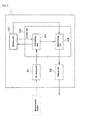
- Fig. 8 is a block diagram showing the functionality configuration of the determination device of Fig. 7 .
- the computer system 3 comprises a receiving unit 301, a memory unit 302, a calculating unit 303, a determining unit 304 and an output unit 305.
- the receiving unit 301 is communicably connected to the measurement device 2 though a network.
- the calculating unit 303 and the determining unit 304 are included in a control unit 306.
- the receiving unit 301 obtains information from the measurement device 2.
- the memory unit 302 stores a threshold necessary for determination and a formula for calculating the methylation score.
- the calculating unit 303 calculates the methylation score from information obtained at the receiving unit 301 according to the formula stored in the memory unit 302.
- the determining unit 304 determines whether or not the methylation score calculated at the calculating unit 303 is lower than the threshold stored at the memory unit 302.
- the output unit 305 outputs the determination result from the determining unit 304 as information on uterine body cancer in the subject (e.g., presence or absence of a cancer cell derived from uterine body cancer in the biological sample collected from the subject).
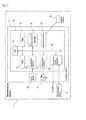
- Fig. 9 is a block diagram showing the hardware configuration of the determination device in Fig. 7 .
- the computer main body 3a comprises a CPU (Central Processing Unit) 30, ROM (Read Only Memory) 121, ROM 32, a hard disk 33, an input/output interface 34, a read-out device 35, a communication interface 36 and an image output interface 37.
- the CPU 30, ROM 31, RAM (Random Access Memory) 32, the hard disk 33, the input/output interface 34, the read-out device 35, the communication interface 36 and the image output interface 37 are connected via a bus 38 so as to be able to communicate with each other.
- a bus 38 so as to be able to communicate with each other.
- the CPU 30 can execute a computer program stored in ROM 31 and a computer program loaded with ROM 32.
- the functional blocks described above may be executed.
- the computer system serves as a terminal that is a determination device for providing information on uterine body cancer in a subject.
- ROM 31 is made up with mask ROM, PROM, EPROM, EEPROM or the like. ROM 31 stores the computer program executed by the CPU 30 and data used for the execution.
- ROM 32 is made up with SRAM, DRAM or the like. ROM 32 is used for readout of the computer programs stored in ROM 31 and the hard disk 33. ROM 32 is also utilized as a work area when the CPU 30 executes these computer programs.
- An operating system to be executed by the CPU 30, computer programs such as application programs (the computer program for providing information on uterine body cancer in a subject) and data for executing the computer programs are installed on the hard disk 33.
- the read-out device 35 is made up with a flexible disk drive, a CD-ROM drive, a DVD-ROM drive or the like and can read out the computer program or data stored on a portable memory medium 40.
- the input/output interface 34 is made up with a serial interface such as USB, IEEE1394 and RS-232C, a parallel interface such as SCSI, IDE and IEEE1284 and an analog interface formed by a D/A converter, an A/D converter or the like.
- the input/output interface 34 is connected to the input device 3b such as a keyboard and a mouse. A user can input data into the computer main body 3a by means of the input device 3b.
- the communication interface 36 is, for example, an Ethernet ® interface.
- the computer system 3 can send printing data to a printer via the communication interface 36.
- the image output interface 37 is connected to the display 3c made up with a LCD, a CRT and the like. Accordingly the display 3c can output an image signal according to image data provided by the CPU 30.
- the display 3c displays an image (on a screen) according to the input image signal.
- Fig. 10 is a flow chart for providing information on uterine body cancer using the determination device of Fig. 7 .
- An example is herein illustrated in which mass spectrometric information of a nucleic acid in a measurement sample prepared from a DNA sample derived from a subject is used for calculation of a peak area from which a methylation score is calculated and the methylation score is determined as to whether or not it is lower than a threshold.
- the present invention is not limited only to this embodiment.
- the receiving unit 301 in the determination device 1 receives from the measurement device 2 mass spectrometric information.
- the calculating unit 303 calculates from the mass spectrometric information received at the receiving unit 301 a peak area which is sent to the memory unit 302.
- the calculating unit 303 calculates the methylation score based on the peak area stored in the memory unit 302 according to the formula stored in the memory unit 302.
- the determining unit 304 determines whether or not the methylation score calculated at the calculating unit 303 is lower than the threshold stored in the memory unit 302.
- the determining unit 304 in the step S1-5 sends a determination result indicating that the biological sample collected from the subject does not contain a cancer cell derived from uterine body cancer to the output unit 305.
- the determining unit 304 sends a determination result indicating that the biological sample collected from the subject contains a cancer cell derived from uterine body cancer to the output unit 305.
- the output unit 305 outputs the determination result as information on uterine body cancer in the subject, so that the display 3c displays the result and/or the printer prints out the result. Accordingly, the determination device can provide, to a physician or the like, information assisting the physician or the like to judge whether or not the subject has uterine body cancer.
- Reference Example 1 Identification of novel markers utilizing methylation data of cancerous tissues from uterine body cancer and normal uterine tissue
- the Infinium HumanMethylation27 BeadChip includes probes for methylated and non-methylated CpG sites for each of 27,578 CpG sites on the human genome.
- the methylation positive rate of a gene can be calculated by "(number of methylation positive specimens among cancerous tissue specimens/total number of cancerous tissue specimens) x 100".
- the gene which showed a statistically significant difference between cancerous tissue specimens and the normal uterine tissue specimen was identified as a marker which is methylated in cancerous tissues.
- promoter regions of F13A1 and CHCHD5 genes were identified as markers which are highly methylated in cancerous tissues of uterine body cancer (see Figs. 1A and 1B ). These markers may also be referred to as the present markers hereinbelow.
- Reference Example 2 Comparison of methylation data between cancer/tumor tissue specimens derived from several types of cancer/tumor, non-cancerous tissue specimens and normal tissue specimens
- the methylation data for cancerous tissue specimens derived from gastric cancer, cancerous tissue specimens derived from hepatocellular carcinoma, non-cancerous tissue specimens derived from hepatocellular carcinoma, normal tissue specimens derived from normal liver and 12 types of normal tissue specimens were those measured by Infinium Methylation Assay using the Infinium HumanMethylation27 BeadChip (Illumina) in the same manner as described in Reference Example 1.
- the methylation data for other specimens were the methylation data of the Infinium HumanMethylation27 BeadChip (Illumina) available from TCGA (The Cancer Genome Atlas: http://tcga-data.nci.nih.gov/tcga/tcgaHome2.jsp) or, for the specimens of lung cancer and prostate cancer and 93 specimens of peripheral blood from healthy subjects, published in the following references.
- the methylation data in this context are the methylation rate (mCpG) of CpG sites in F13A1 and CHCHD5 obtained as described in section (2) in Reference Example 1.
- methylation positive specimen a specimen having a methylation rate at or higher than the threshold was designated as "methylation positive specimen”. Based on the number of methylation positive specimens, methylation positive rate (%) for the present markers in various tissues was calculated according to the above formula. The results are shown in Figs. 2A and 2B .
- “Normal tissue” represents, among the tissues indicated in Table 4, the normal tissues excluding 93 specimens of peripheral blood from healthy subjects (Salhia B et al.) and “Normal blood” represents the 93 specimens of peripheral blood from healthy subjects (Salhia B et al.).
- Figs. 2A and 2B show that both of the present markers are not highly methylated in other types of cancer or human normal tissues or human normal blood (peripheral blood from healthy subjects, Salhia B et al.) and thus are specifically and highly methylated in uterine body cancer.
- Comparative Example 1 Comparison of methylation positive rate between cancer/tumor tissue specimens derived from several types of cancer/tumor, non-cancerous tissue specimens and normal tissue specimens
- the methylation positive rate was calculated for each of MLH 1 and CDKN2A genes (hereinafter referred to as "known markers") which have already been known to be methylated in cancer cells derived from uterine body cancer in the similar manner as Reference Example 2 in the respective tissues. The results are shown in Figs. 3A and 3B .
- MLH1 and CDKN2A showed high positive rates in uterine body cancer and were also methylated in other types of cancer and other non-cancerous tissues. Therefore, these genes have low specificity towards uterine body cancer.
- the known markers have high sensitivity while having low specificity as markers of uterine body cancer, thereby posing issues in terms of performance as diagnostic markers of uterine body cancer. Namely it was shown that a search for markers is required to be carried out by taking the specificity in other types of cancer and other non-cancerous tissues into account.
- Example 1 Comparison of methylation data (MassARRAY) between normal uterus and uterine body cancer
- the biological samples used were normal uterine tissues (4 specimens) and cancerous tissues of uterine body cancer (4 specimens).
- Genomic DNA was extracted from the above tissues with the QIAamp DNA Mini Kit (QIAGEN).
- the genomic DNA contained in the obtained DNA solution was fragmented with Bioruptor (COSMO BIO).
- the control genomic DNA used was genomic DNA of human peripheral blood lymphocytes.
- the genomic DNA from human peripheral blood lymphocytes was amplified with the GenomiPhi v2DNA amplification kit (GE Healthcare Life Sciences).
- the obtained amplification product consisted of non-methylated DNA.
- the amplification product was fragmented with Bioruptor (COSMO BIO) to obtain a solution of non-methylated DNA fragments (0% methylated DNA).
- the respective DNA fragments (500 ng) obtained as above were subjected to bisulfite treatment with the EZ DNA Methylation Kit (Zymo Research) and the treated genomic DNA was eluted in sterilized distilled water (80 ⁇ l).
- Methylated cytosine and uracil in the DNA fragments after bisulfite treatment were converted respectively to guanine and adenine by PCR and IVT amplification.
- primer sets used for PCR can amplify both methylated DNA and non-methylated DNA without bias.
- sequences of the primer sets for the present markers are shown in Table 5.
- the base sequences (sequences of negative strands) which can be analyzed with the primer sets shown in Table 5 in the promoter regions in F13A1 and CHCHD5 genes are shown in SEQ ID NOs: 13 and 14, respectively.
- tag sequence and T7 promoter sequence were respectively added to the 5'-terminals of the forward primers and reverse primers in the above primer sets for IVT reaction.
- PCR reaction solution was prepared by mixing the following reagents: 10 x Hot Star buffer (QIAGEN) 0.5 ⁇ L 25 mM dNTP mix 0.04 ⁇ L Hot Star Taq (5 U/ ⁇ L) (QIAGEN) 0.04 ⁇ L Primer mix 2.0 ⁇ L DNA solution 1.0 ⁇ L Water 1.42 ⁇ L Total 5 ⁇ L
- the obtained mixture was incubated at 37°C for 3 hours to effect IVT reaction and uracil or thymine specific cleavage.
- the obtained reaction product was purified with Clean Resin (SEQUENOM) to obtain a sample for mass spectrometry.
- Samples derived from genomic DNA extracted from the above clinical specimens were designated as measurement samples and samples derived from control genomic DNA were designated as control samples, both of which were used for mass spectrometry described hereinbelow.
- the measurement samples obtained as above were subjected to mass spectrometry to obtain peaks of DNA fragments contained in the measurement samples.
- the obtained peaks were then allocated to the portions of the base sequences of F13A1 1 and CHCHD5.
- the methylation score was calculated on the basis of the ratio between the area of the peaks of the fragments containing a methylated CpG site and the area of the peaks of the fragments without the methylated CpG site, both fragments having the same base sequence.
- the obtained results are shown in Fig. 4A and 4B .
- Figs. 4A and 4B it was found that F13A1 and CHCHD5 showed low methylation scores in normal uterine tissues and significantly high methylation scores in uterine body cancer. It was therefore demonstrated that the methylation score of the present markers correlates with uterine body cancer, similar to the results of the Infinium method in Reference Example 1.
- Fig. 4 it was found that analysis of methylation frequency of the present markers allows establishment of the threshold that can distinguish specimens of uterine body cancer and specimens of normal uterine tissues.
- Example 2 Comparison of methylation data (MSP) between normal uterine tissue and uterine body cancer
- the biological samples used were normal uterine tissue (1 specimen) and cancerous tissues of uterine body cancer (4 specimens).
- Genomic DNA was extracted from the above tissues with the QIAamp DNA Mini Kit (QIAGEN). The genomic DNA contained in the obtained DNA solution was fragmented with Bioruptor (COSMO BIO).
- the control genomic DNA used was genomic DNA of human peripheral blood lymphocytes.
- the genomic DNA from human peripheral blood lymphocytes was amplified with the GenomiPhi v2DNA amplification kit (GE Healthcare Life Sciences).
- the obtained amplification product consisted of non-methylated DNA.
- the amplification product was fragmented with Bioruptor (COSMO BIO) to obtain a solution of non-methylated DNA fragments (0% methylated DNA).
- a portion of the solution of the non-methylated DNA fragments was subjected to reaction with SssI methylase (New England Biolab) to methylate all cytosines in CG sequences and obtain a solution of methylated DNA fragments (100% methylated DNA).
- the respective DNA fragments (500 ng) obtained as above were subjected to bisulfite treatment with the EZ DNA Methylation Kit (Zymo Research) and the treated genomic DNA was eluted in sterilized distilled water (80 ⁇ l).
- MSP was carried out on the measurement samples and control samples (DNA after bisulfite treatment) obtained in the above section (2).
- the composition of PCR reagents, primer sets and reaction conditions for PCR in MSP are shown below.
- DDW sterilized water
- 16.8 ⁇ L 10 x PCR buffer with MgCl 2 (Roche) 2.5 ⁇ L 2 mM dNTP mix
- 2.5 ⁇ L 10 ⁇ M sense primer 1.0 ⁇ L 10 ⁇ M antisense primer
- Faststart Taq polymerase (Roche) 0.2 ⁇ L Measurement sample or control sample 1.0 ⁇ L Total 25 ⁇ L
- the primer sets used for MSP are shown in Table 6. These primer sets allow generation of amplification products when DNA in the amplified regions is methylated. A primer set for accuracy control was also used which allows judgment on whether or not the bisulfite treatment had been appropriately carried out (see Table 7).
- the base sequences (sequences of negative strands) which can be analyzed with the primer sets shown in Table 6 in the promoter regions in F13A1 and CHCHD5 genes are shown in SEQ ID NOs: 17 and 18, respectively.
- Table 6 Gene symbol Primer Base sequence of primer SEQ ID NO: Size of PCR product (bp) Annealing temp.
- amplification product from MSP was verified by 2% agarose gel electrophoresis. The results are shown in Fig. 5 .
- "0" and "100” under “control” represent the 0% methylation control sample and the 100% methylation control sample, respectively.
- methylation analysis was carried out by MSP for CpG sites in the promoter region of F13A1 using peripheral blood as a biological sample.
- the biological samples used were peripheral blood collected from patients with uterine body cancer (5 specimens).
- the control samples used were peripheral blood from healthy subjects (3 specimens).
- Plasma was obtained from each of the above peripheral blood (2 ml) according to the standard method. Genomic DNA was extracted from the resulting plasma with the QIAamp Circulating Nucleic Acid Kit (QIAGEN).
- the respective DNA fragments (500 ng) obtained as above were subjected to bisulfite treatment with the EZ DNA Methylation Kit (Zymo Research) and the treated genomic DNA was eluted in sterilized distilled water (80 ⁇ l).
- X represents annealing temperature which is 54°C for F13A1 and 60°C for the primer set for accuracy control.
- amplification product from MSP was verified by 2% agarose gel electrophoresis.
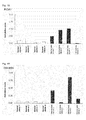
- the band intensities of amplification products were represented in graphs (see Figs. 6A and 6B ).
- the bars in Fig. 6A indicated under peripheral blood of healthy subjects correspond to the background, indicating absence of detection of any band.
- Fig. 6A it is found that PCR for F13A1 allowed detection of no band in peripheral blood from healthy subjects and detection of bands in 3 samples among 5 samples of peripheral blood from uterine body cancer patients.
- Fig. 6B shows that bands were detected in all samples by PCR with the primer set for accuracy control. This indicates that the bisulfite treatment of the samples had been appropriately carried out.
- the present marker tends to be highly methylated in uterine body cancer patients and methylation thereof is not detected in healthy subjects even when the biological sample used is peripheral blood. Therefore it is suggested that the present marker is highly specific for uterine body cancer and is useful for determining presence or absence of cancer cells derived from uterine body cancer in blood samples.
Landscapes
- Health & Medical Sciences (AREA)
- Life Sciences & Earth Sciences (AREA)
- Chemical & Material Sciences (AREA)
- Immunology (AREA)
- Engineering & Computer Science (AREA)
- Organic Chemistry (AREA)
- Proteomics, Peptides & Aminoacids (AREA)
- Pathology (AREA)
- Analytical Chemistry (AREA)
- Molecular Biology (AREA)
- Physics & Mathematics (AREA)
- Wood Science & Technology (AREA)
- Zoology (AREA)
- Urology & Nephrology (AREA)
- Microbiology (AREA)
- Hematology (AREA)
- Biochemistry (AREA)
- General Health & Medical Sciences (AREA)
- Biomedical Technology (AREA)
- Biotechnology (AREA)
- Genetics & Genomics (AREA)
- Oncology (AREA)
- Hospice & Palliative Care (AREA)
- Cell Biology (AREA)
- Biophysics (AREA)
- General Physics & Mathematics (AREA)
- Medicinal Chemistry (AREA)
- Food Science & Technology (AREA)
- Bioinformatics & Cheminformatics (AREA)
- General Engineering & Computer Science (AREA)
- Measuring Or Testing Involving Enzymes Or Micro-Organisms (AREA)
Abstract
Description
- The present invention relates to a method for obtaining information on uterine body cancer in a subject. The present invention also relates to a marker and a kit used in the method.
- The uterus contains the cervix corresponding to the entrance and the corpus that is located behind the cervix. Uterine body cancer is cancer of the corpus of the uterus, develops in the endometrium and thus is also referred to as endometrial cancer. Uterine body cancer is generally examined by pathological examination (cytological examination and biopsy) in which endometrial cells and tissues are collected and observed by microscopy. However, the pathological examination requires insertion of a special instrument for cell or tissue collection into the corpus of the uterus, which may be accompanied by pain and bleeding and thus put a great burden on subjects.
- Examination for uterine body cancer includes measurement of tumor markers in blood. The tumor markers used include CA125, CA19-9 and the like. However, the examination of the tumor markers has insufficient cancer detection sensitivity. The examination has also been utilized for examinations of types of cancer different from uterine body cancer, and thus has insufficient sensitivity and specificity as the specific screening examination for uterine body cancer.
- Meanwhile, new diagnosis methods for cancer have been recently studied which are based on genetic information. The methods include, for example, ones based on information on methylation of DNAs. The methods use markers which are CpG sites (5'-(CG)-3') in base sequences of certain genes. Based on the analysis results of the methylation status of the markers, information such as presence or absence of a cancer cell is obtained, which information may be used as an index for diagnosis of cancer.
- Methods for determining cancer utilizing methylation analyses of DNA have been studied and developed for uterine body cancer. For example, the publication by Esteller M. et al discloses that MLH1 gene is not methylated in normal endometrial tissue while it is highly methylated in tissues of endometrial cancer (see Non-Patent Literature 1). The publication by Furlan D. et al. discloses that three genes, MLH1, CDKN2A and MGMT are highly methylated in tumor tissues of the endometrium and ovary (see Non-Patent Literature 2).
-
- Non-Patent Literature 1: Esteller M. et al., Am. J. Pathol. vol. 155, p. 1767-1772 (1996)
- Non-Patent Literature 2: Furlan D. et al., Clin. Cancer Res. vol. 12, p. 3329-3336 (2006)
- Although the genes indicative of abnormal methylation in uterine body cancer have been reported as described above, these genes are methylated in some extent in a type of cancer different from uterine body cancer. When methylation analysis is carried out with a marker that is methylated in several types of cancer including uterine body cancer, information deduced from the analysis result may include not only information on uterine body cancer but also information on other types of cancer. In this case, even when an analysis result provides information indicating that a sample contains a cancer cell for example, it is difficult to determine whether the cancer cell is derived from uterine body cancer or from other types of cancer.
- As such upon obtaining information on uterine body cancer by methylation analysis of a marker, the specificity of the marker to uterine body cancer is very important. Thus there is a need for a novel marker specific to uterine body cancer, which allows specific detection of a cancer cell derived from uterine body cancer.
- Accordingly an object of the present invention is to provide a method for obtaining information on uterine body cancer by identifying such a novel marker and analyzing methylation status of the marker. Another object of the present invention is to provide a kit suitably used for the method.
- The inventors of the present invention have identified novel markers which are genetic regions specifically methylated in DNAs obtained from cancerous tissues of uterine body cancer. The inventors of the present invention have found that cancer cells derived from uterine body cancer can be clearly discriminated from other cells (cells of normal tissues, cells of non-cancerous tissues and cancer cells derived from a type of cancer other than uterine body cancer) based on the result obtained by analyzing methylation status of the markers, thereby completing the present invention.
- Thus the present invention provides a method for obtaining information on uterine body cancer comprising the steps of:
- preparing a DNA sample from a biological sample collected from a subject;
- analyzing methylation status of a CpG site in a promoter region of at least one gene selected from F13A1 (Coagulation factor XIII, A1 polypeptide) and CHCHD5 (Coiled-coil-helix-coiled-coil-helix domain containing 5) in the DNA sample obtained from the preparation step; and
- obtaining information on uterine body cancer in the subject based on an analysis result obtained from the analyzing step.
- The present invention also provides a marker for obtaining information on uterine body cancer by methylation analysis, which is at least one CpG site selected from CpG sites located in promoter regions of F13A1 and CHCHD5 genes.
- The present invention also provides a kit for obtaining information on uterine body cancer, comprising a primer set for analysis of methylation status of at least one CpG site selected from CpG sites located in promoter regions of F13A1 and CHCHD5 genes.
- The present invention can provide a novel marker which allows provision of information on uterine body cancer. The present invention can also provide a method and a kit for obtaining information on uterine body cancer in a subject by analyzing methylation status of the marker.
-
-
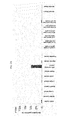
Fig. 1A is a graph showing the methylation positive rate of the promoter region of F13A1 gene calculated from methylation data of cancerous tissues of uterine body cancer and normal uterine tissue; -
Fig. 1B is a graph showing the methylation positive rate of the promoter region of CHCHD5 gene calculated from methylation data of cancerous tissues of uterine body cancer and normal uterine tissue; -
Fig. 2A is a graph showing the methylation positive rate of the promoter region of F13A1 gene calculated from methylation data of various clinical specimens; -
Fig. 2B is a graph showing the methylation positive rate of the promoter region of CHCHD5 gene calculated from methylation data of various clinical specimens; -
Fig. 3A is a graph showing the methylation positive rate of the promoter region of a knownmarker gene MLH 1 calculated from methylation data of various clinical specimens; -
Fig. 3B is a graph showing the methylation positive rate of the promoter region of a known marker gene CDKN2A calculated from methylation data of various clinical specimens; -
Fig. 4A is a graph showing the methylation score of the promoter region of F13A1 gene in DNAs extracted from normal uterine tissues and cancerous tissues of uterine body cancer; -
Fig. 4B is a graph showing the methylation score of the promoter region of CHCHD5 gene in DNAs extracted from normal uterine tissues and cancerous tissues of uterine body cancer; -
Fig. 5 is an image showing the results of amplification by methylation specific PCR (MSP) of DNAs extracted from normal uterine tissue and cancerous tissues of uterine body cancer using the respective primer sets for F13A1 and CHCHD5; -
Fig. 6A is a bar graph showing the band intensity of amplification products obtained by MSP of DNA extracted from peripheral blood of uterine body cancer patients and healthy subjects with a primer set for F13A1 (SEQ ID NOs: 9 and 10); -
Fig. 6B is a bar graph showing the band intensity of amplification products obtained by MSP of DNA extracted from peripheral blood of uterine body cancer patients and healthy subjects with a primer set for accuracy control (SEQ ID NOs: 15 and 16); -
Fig. 7 is a schematic view of an example of a determination device for providing information on uterine body cancer in a subject; -
Fig. 8 is a block diagram showing the functionality configuration of the determination device ofFig. 7 ; -
Fig. 9 is a block diagram showing the hardware configuration of the determination device shown inFig. 7 ; and -
Fig. 10 is a flow chart of determination for providing information on uterine body cancer in a subject using the determination device inFig. 7 . - In the method for obtaining information on uterine body cancer of the present invention (hereinafter also merely referred to as "method"), a DNA sample is first prepared from a biological sample collected from a subject.
- In the present embodiments, the biological sample is not particularly limited as far as it is a biological sample containing DNA of a subject and is preferably a sample containing a genomic DNA such as a clinical specimen. The clinical specimen may include, for example, body fluid, urine, tissues obtained by operations or biopsies and the like. The body fluid may include blood, serum, plasma, lymph fluid, ascetic fluid, bone marrow aspirate, nipple discharge and the like. The biological sample may also be a culture medium obtained by culturing cells or tissues collected from a subject.
- The DNA sample can be prepared by extracting DNA from the biological sample. The method for extracting DNA from biological samples is well known in the art. Extraction of DNA can be carried out, for example, by mixing the biological sample with a treatment solution containing a surfactant for solubilization of cells or tissues (e.g. sodium cholate, sodium dodecyl sulfate etc.), and subjecting the resulting mixture to physical procedure (stirring, homogenization, ultrasonication etc.) to release DNA contained in the biological sample into the mixture. In this case, it is preferable to centrifuge the mixture to precipitate cell debris and use the supernatant containing the released DNA to the next step of analyzing. The obtained supernatant may be purified according to well-known methods. DNA can also be extracted and purified from a biological sample by using commercially available kits.
- The preparation step preferably further comprises the step of fragmenting the extracted DNA. By fragmenting DNA to have appropriate length, methylated DNA immunoprecipitation (MeDIP) and non-methylated cytosine conversion as described hereinbelow can be effectively carried out.
- Fragmentation of DNA may be carried out by ultrasonication, alkaline treatment, restriction enzyme treatment and the like. When DNA is fragmented by alkaline treatment, a sodium hydroxide solution may be added to a DNA solution to the final concentration of 0.1N to 1.0N and the mixture may be incubated at 10°C to 40°C for 5 to 15 minutes to fragment the DNA. When the restriction enzyme treatment is used for DNA fragmentation, the restriction enzyme may appropriately be selected based on the base sequence of DNA, which may be MseI or BamHI, for example.
- According to the present method, methylation status of a CpG site in a promoter region of at least one gene selected from F13A1 and CHCHD5 in DNA obtained from the preparation step is analyzed.
- As used herein, the term "CpG site" means a site of a sequence in which cytosine (C) and guanine (G) are adjacent in this order from 5' to 3'. The letter "p" in "CpG" represents a phosphodiester bond between cytosine and guanine.
- As used herein, to "analyze methylation status" means to analyze presence or absence of methylation of a CpG site located in a promoter region of at least one gene selected from F13A1 and CHCHD5 or analyze methylation frequency in the promoter region.
- The base sequences per se of the promoter regions of F13A1 and CHCHD5 genes are well known in the art. The base sequences can be obtained from well-known databases such as the one provided by the National Center for Biotechnology Information (NCBI) (http://www.ncbi.nlm.nih.gov/). The ID numbers of the above genes are shown in Table 1. The base sequences of the promoter regions of the genes (sequences of negative strands) are represented by SEQ ID NOs: 1 and 2, respectively.
Table 1 Gene symbol Unigene ID Entrez Gene ID Transcript ID SEQ ID NO: F13A1 Hs. 335513 2162 NM_000129.3 1 CHCHD5 Hs. 375707 84269 NM_032309.2 2 - In the embodiments of the present invention, the analyzing step may be the step of analyzing presence or absence of methylation of at least one CpG site among CpG sites located in a promoter region of at least one gene selected from F13A1 and CHCHD5. The term "presence or absence of methylation" means whether or not cytosine in a CpG site located in the promoter region is methylated. In the embodiment, one CpG site may be analyzed; however, more than one CpG site is preferably analyzed for presence or absence of methylation. More than one CpG site may be selected from a promoter region of one gene or from each of promoter regions of more than one gene.
- In another embodiment of the present invention, the analyzing step may be the step of analyzing methylation frequency in a promoter region of at least one gene selected from F13A1 and CHCHD5. The term "methylation frequency" means the ratio of the number of methylated CpG site(s) relative to the number of CpG site(s) located in the promoter region. In the embodiment, the target for analysis may be the whole promoter region or a part thereof including at least one CpG site. The target for analysis may contain only one CpG site; however it is preferable that the target for analysis contains more than one CpG site. The target for analysis may be selected from any one promoter region among the above genes or from promoter regions of more than one gene.
- The positions and numbers of CpG sites located in the promoter regions of F13A1 and CHCHD5 genes are already known. Thus the number of methylated CpG site(s) itself in the promoter regions can also be used as the methylation frequency.
- The methylation frequency may be "methylation score" obtained by analyzing DNA for methylation status of CpG sites with mass spectrometry such as MassARRAY® as described hereinbelow. MassARRAY® allows calculation of methylation score based on the ratio between the area of a peak derived from a methylated DNA fragment and the area of a peak derived from a non-methylated DNA fragment obtained after measurement of the DNA fragments.
- In the embodiments of the present invention, the target for analysis may be any CpG site(s) (or certain region(s) including the CpG site(s)) in the promoter regions of F13A1 and CHCHD5 genes without particular limitation and may be appropriately selected by a person skilled in the art. The positions and numbers of CpG sites located in the promoter regions of the genes are already known. Thus the target CpG site or region may be selected by routine experiments according to the well known analysis methods described hereinbelow.
- Various methods are well-known in the art as to the method for analyzing methylation status. According to the present method, it is not specifically limited as to which analysis method is used; however, the analysis method preferably comprises differentiating methylated DNA from non-methylated DNA, amplifying DNA and detecting methylated DNA and/or non-methylated DNA.
- The step of differentiating methylated DNA from non-methylated DNA may include the step of carrying out methylation sensitive restriction enzyme treatment, the MeDIP method, non-methylated cytosine converting treatment and the like.
- The step of amplifying DNA may include the step of carrying out PCR, quantitative PCR, IVT (in vitro transcription) amplification, SPIA™ amplification and the like methods.
- The step of detecting methylated DNA and/or non-methylated DNA may include the step of carrying out electrophoresis, sequence analysis, microarray analysis, mass spectrometry, Southern hybridization and the like.
- The MeDIP method is a method in which methylated DNA in a biological sample is concentrated by immunoprecipitation using an anti-methylated cytosine antibody or an anti-methylated cytidine antibody, or an antibody which specifically recognizes a methylated DNA-binding protein. In embodiments of the present invention, the analyzing step may be the step of concentrating methylated DNA in DNA obtained in the extracting step by the MeDIP method and analyzing methylation status of the concentrated methylated DNA. The methylated DNA concentrated by the MeDIP method may be amplified by e.g. IVT amplification and methylation status of the obtained amplified product may be analyzed by using a microarray. These analysis procedures are referred to as the MeDIP on chip method.
- The non-methylated cytosine converting treatment is the one in which DNA extracted from a biological sample is subjected to reaction with a non-methylated cytosine conversion agent so as to convert non-methylated cytosine(s) in the DNA to a different base (uracil, thymine, adenine or guanine). In this context, the non-methylated cytosine conversion agent is an agent which can react with DNA and convert a non-methylated cytosine in the DNA to a different base (uracil, thymine, adenine or guanine). The non-methylated cytosine conversion agent suitably used may be, for example, bisulfite such as sodium, potassium, calcium or magnesium bisulfite.
- In the treatment using bisulfite, non-methylated cytosine(s) in DNA is converted to uracil due to deamination reaction, while a methylated cytosine does not undergo such a base conversion.
- Thus, the difference in methylation status of a CpG site in DNA is converted to the difference in base sequence (C and U) by the non-methylated cytosine converting treatment using bisulfite. The non-methylated cytosine converting treatment using bisulfite is referred to as bisulfite treatment.
- When the bisulfite treatment is carried out, the amount (concentration) of bisulfite added is not specifically limited so long as it can sufficiently convert non-methylated cytosine(s) in DNA, and corresponds to, for example, 1M or higher, preferably 1M to 15M, more preferably 3M to 10M as the final concentration in a solution containing DNA. The incubation conditions (temperature and time) after addition of bisulfite may be appropriately selected according to the amount added of bisulfite, and for example, when bisulfite is added at a final concentration of 6M, the incubation is carried out at 50°C to 80°C for 10 minutes to 90 minutes.
- Methylation status of CpG sites in DNA can be analyzed by analyzing the sequence of DNA after bisulfite treatment and detecting the difference in base sequence from the original sequence. This process is referred to as bisulfite sequencing method.
- Methylation status of CpG sites can be alternatively analyzed by mass spectrometry. Specifically, a DNA after bisulfite treatment as a template is amplified by PCR using a primer set specific for a base sequence which is a target for analysis, and the obtained PCR product is subjected to IVT amplification to convert methylated cytosine and uracil respectively to guanine (G) and adenine (A). The obtained IVT amplification product is digested with RNase A and the difference in mass (16 Da) due to difference between G and A in the obtained digested fragments is detected on a MALDI-TOF (matrix assisted laser desorption/ionization time-of-flight) mass spectrometer to analyze methylation status of DNA. This method is referred to as MassARRAY® analysis.
- It is known that the cleavage site in IVT products by RNase A is between an arbitrary base and the adjacent uracil (U) or thymine (T). Thus the base sequence and mass of the IVT product cleaved by RNase A may be predicted based on the base sequence of the template DNA. Accordingly the peaks obtained in MassARRAY® can be identified as to the portions of the base sequences in the template DNA from which the peaks are originated. For example, when one CpG site is methylated in a DNA fragment, a peak obtained in MassARRAY® shifts to the side with an increased mass for 16 Da. When a DNA fragment containing more than one CpG site is analyzed, for example, the DNA fragment having two methylated CpG sites shows a shift of 32 Da and the DNA fragment having three methylated CpG sites shows a shift of 48 Da.
- In mass spectrometry such as MassARRAY®, the methylation score of the analyzed DNA fragment can be calculated. For example when a DNA fragment having a certain sequence results in the ratio between the area of the peaks of non-methylated DNA fragments and the area of the peaks of methylated DNA fragments obtained in a resulting chart from the analysis of 1:3, the methylation score of the DNA fragment is 0.75 (= 3/(1+3)). The methylation score is theoretically 1 for a DNA fragment in which all CpG site(s) is methylated and 0 for a DNA fragment without any methylated CpG site.
- Methylation status of CpG sites can be analyzed by a methylation specific PCR (MSP) method. The MSP method is a method in which methylation status of CpG sites (presence or absence of methylation) is analyzed by amplifying DNA after bisulfite treatment by PCR using a primer set described hereinafter and determining presence or absence of a PCR product.
- The MSP method utilizes a primer set which can amplify a base sequence having a CpG site to be analyzed that is methylated (i.e. cytosine is not converted to uracil) but cannot amplify a base sequence having a CpG site that is not methylated (i.e. cytosine is converted to uracil). According to the MSP method using such a primer set, presence of a PCR product indicates methylation of the CpG site analyzed.
- The MSP method may also be carried out by using a primer set which cannot amplify a base sequence having cytosine in a CpG site to be analyzed that is not converted to uracil but can amplify a base sequence having cytosine in a CpG site that is converted to uracil. In this case, absence of a PCR product indicates methylation of the CpG site analyzed.
- Each primer in the primer set used for the MSP method may be appropriately designed by a person skilled in the art based on the base sequence including a CpG site to be analyzed, and it is preferably designed so as to contain cytosine of the CpG site to be analyzed at the 3' end or in the vicinity thereof of the primer.
- Methylation status of CpG sites may alternatively be analyzed with a microarray. In this case, the microarray for analysis may be prepared by immobilizing a nucleic probe complementary to the base sequence of a promoter region of F13A1 or CHCHD5 gene on a substrate. The microarray can be prepared according to well-known methods in the art.
- In the analysis using a microarray, DNA extracted from a biological sample is preferably labeled with a labeling substance well-known in the art. Thus, the present method preferably further comprises the step of labeling the extracted DNA. The labeling step is advantageously carried out after the DNA amplifying step because all DNA in the biological sample may be labeled. The labeling substance may include fluorescence substances, haptens such as biotin, radioactive substances and the like. The fluorescence substances may include Cy3, Cy5, FITC, Alexa Fluor™ and the like. Labeling of DNA facilitates measurement of a signal from a probe on the microarray. A method for labeling DNA with the labeling substance is well-known in the art.
- The above signal may be any suitable signal depending on the type of microarrays. For example, the signal may be an electric signal generated when a DNA fragment hybridizes to a probe on the microarray, or a fluorescence or luminescence signal generated from a labeling substance when DNA to be analyzed is labeled as described above. Detection of a signal can be carried out by using a scanner comprised in a conventional microarray analyzer. The scanner may be, for example, GeneChip® Scanner3000 7G (Affymetrix), Illumina® BeadArray Reader (Illumina) and the like.
- In the present method, information on uterine body cancer in the subject is obtained based on the analysis result obtained in the analyzing step. The information on uterine body cancer is not particularly limited as far as it may be an index on diagnosis of uterine body cancer and is preferably information indicative of occurrence or status of uterine body cancer or both thereof in a subject. The information may include, for example, presence or absence of a cancer cell derived from uterine body cancer in a biological sample collected from a subject, a possibility of occurrence of uterine body cancer in a subject, or a risk for future occurrence of uterine body cancer in a subject. The information on uterine body cancer in a subject who has already been affected by uterine body cancer may include prognosis of the subject, a degree of progression (stage) and the like. The information on uterine body cancer obtained based on the analysis result obtained in the analyzing step does not substantially include information on a type of cancer different from uterine body cancer because the promoter regions of the above genes which are used as markers in the present method are specifically methylated in cancer cells derived from uterine body cancer.
- In the embodiments of the present invention, the analyzing step which provides the analysis result indicating presence of a methylated CpG site may provide information indicating occurrence of uterine body cancer or indicating that the status of uterine body cancer is poor (or aggravated).
- In another embodiment of the present invention, the above information can be obtained when the methylation frequency obtained in the analyzing step is at or higher than a certain threshold.
- More specifically, the information obtained may be indicative of presence of a cancer cell derived from uterine body cancer in a biological sample. The information obtained may alternatively indicate that a subject has a high risk for being affected by uterine body cancer or that a subject has already been affected by uterine body cancer. For a subject who has already been affected by uterine body cancer, information obtained may indicate that prognosis of the subject is poor (or aggravated) or that the cancer is in a progressed stage.
- On the contrary, when the result from the analyzing step shows absence of methylated CpG site, information suggesting no occurrence of uterine body cancer or information indicating that uterine body cancer is in a preferable status can be obtained. Alternatively the above information can be obtained when the methylation frequency obtained in the analyzing step is lower than a certain threshold. More specifically, the information obtained may be indicative of absence of a cancer cell derived from uterine body cancer in a biological sample. The information obtained may alternatively indicate that a subject has a low risk for being affected by uterine body cancer or that a subject has not been affected by uterine body cancer. For a subject who has already been affected by uterine body cancer, information obtained may be indicative of a preferable prognosis of the subject or indicate that the cancer is in a relatively early stage.
- The threshold is not particularly limited and may be empirically established based on accumulated data on various biological samples. The threshold may alternatively be established as follows. First, methylation frequency is analyzed for DNA extracted respectively from a biological sample which is confirmed to be devoid of cancer cells derived from uterine body cancer (tissues or cells of normal uterine body) and a biological sample containing a cancer cell derived from uterine body cancer. Next, based on the obtained analysis results, a threshold is established within a range that is higher than the methylation frequency of the biological sample devoid of cancer cells and lower than that of the biological sample containing the cancer cell. Preferably, the threshold is established as a value which can highly accurately differentiate between the biological sample devoid of cancer cells and the biological sample containing the cancer cell.
- The scope of the present invention also encompasses a marker for obtaining information on uterine body cancer by methylation analysis (also merely referred to as "marker"). The marker of the present invention is at least one CpG site selected from CpG sites located in promoter regions of F13A1 and CHCHD5 genes.
- In the embodiments of the present invention, methylation status of the marker in a DNA sample prepared from a biological sample collected from a subject may be analyzed and information on uterine body cancer in the subject can be obtained based on the analysis result. The analysis of methylation status and obtainment of information on uterine body cancer are the same as those previously described herein.
- The scope of the present invention encompasses a kit for obtaining information on uterine body cancer (also merely referred to as "kit"). The kit of the present invention comprises a primer set for analysis of methylation status of at least one CpG site selected from CpG sites located in promoter regions of F13A1 and CHCHD5 genes.
- In the embodiments of the present invention, the primer set in the kit may be a primer set for analysis of methylation status of CpG sites according to mass spectrometry such as MassARRAY® or an analysis method involving PCR amplification such as the MSP method, the bisulfite sequencing method, among which the primer set for mass spectrometry such as MassARRAY® or for the MSP is preferred. The base sequences of the primers in the primer set may be appropriately selected by a person skilled in the art based on the base sequences in the promoter regions. Examples of the primer set include a primer set of primers having base sequences SEQ ID NOs: 3 and 4; a primer set of primers having base sequences SEQ ID NOs: 5 and 6; a primer set of primers having base sequences SEQ ID NOs: 9 and 10; and a primer set of primers having base sequences SEQ ID NOs: 11 and 12.
- The present invention also encompasses a system suitable for provision of information on uterine body cancer in a subject. For example, the system may be as follows.
- A system suitable for provision of information on uterine body cancer in a subject comprising a computer containing a processor and a memory controlled by the processor, wherein the memory comprises a computer program for enabling the computer to carry out the steps of:
- obtaining an analysis result on methylation status of a CpG site located in a promoter region of at least one gene selected from F13A1 and CHCHD5 in a DNA sample derived from the subject; and
- providing information on uterine body cancer in the subject based on the resulting analysis result.
- The present invention also encompasses a computer program product for enabling a computer to carry out provision of information on uterine body cancer in a subject. For example, the computer program product may be as follows.
- A computer program product for enabling a computer to carry out provision of information on uterine body cancer in a subject comprising a computer readable medium, wherein the medium comprises a computer program for enabling the computer to carry out the steps of:
- obtaining an analysis result on methylation status of a CpG site located in a promoter region of at least one gene selected from F13A1 and CHCHD5 in a DNA sample derived from the subject; and
- providing information on uterine body cancer in the subject based on the resulting analysis result.
- An embodiment of a suitable device for carrying out the present method is illustrated hereinafter by referring to figures. However, the present invention is not limited only to this embodiment.
Fig. 7 is a schematic view of an example of a determination device for providing information on uterine body cancer in a subject. Thedetermination device 1 shown inFig. 7 comprises ameasurement device 2 and acomputer system 3 connected to themeasurement device 2. - In the present embodiment, the
measurement device 2 is a MALDI-TOF mass spectrometer. Themeasurement device 2 obtains mass spectrometric information such as the time of flight or the mass-to-charge ratio (m/z ratio) of a substance to be analyzed. Themeasurement device 2 onto which a measurement sample prepared from the DNA sample derived from a subject is mounted obtains mass spectrometric information of a nucleic acid in the measurement sample and sends the mass spectrometric information to thecomputer system 3. - The
measurement device 2 may be, when methylation status is analyzed by the MSP method, a gel imaging device such as a fluorescence image scanner. In this case, themeasurement device 2 onto which a gel obtained by electrophoresis of a reaction solution after nucleic acid amplification by the MSP method is mounted detects amplification products. Themeasurement device 2 then obtains the band intensity data of the amplification products and sends the obtained data to thecomputer system 3. - The
computer system 3 comprises a computermain body 3a, aninput device 3b and adisplay 3c which displays specimen information, determination results and the like. Thecomputer system 3 receives the mass spectrometric information from themeasurement device 2. The processor in thecomputer system 3 executes, based on the mass spectrometric information, a program for providing information on uterine body cancer in a subject. -
Fig. 8 is a block diagram showing the functionality configuration of the determination device ofFig. 7 . As shown inFig. 8 , thecomputer system 3 comprises a receivingunit 301, amemory unit 302, a calculatingunit 303, a determiningunit 304 and anoutput unit 305. The receivingunit 301 is communicably connected to themeasurement device 2 though a network. The calculatingunit 303 and the determiningunit 304 are included in acontrol unit 306. - The receiving
unit 301 obtains information from themeasurement device 2. Thememory unit 302 stores a threshold necessary for determination and a formula for calculating the methylation score. The calculatingunit 303 calculates the methylation score from information obtained at the receivingunit 301 according to the formula stored in thememory unit 302. The determiningunit 304 determines whether or not the methylation score calculated at the calculatingunit 303 is lower than the threshold stored at thememory unit 302. Theoutput unit 305 outputs the determination result from the determiningunit 304 as information on uterine body cancer in the subject (e.g., presence or absence of a cancer cell derived from uterine body cancer in the biological sample collected from the subject). -
Fig. 9 is a block diagram showing the hardware configuration of the determination device inFig. 7 . As shown inFig. 9 , the computermain body 3a comprises a CPU (Central Processing Unit) 30, ROM (Read Only Memory) 121,ROM 32, ahard disk 33, an input/output interface 34, a read-outdevice 35, acommunication interface 36 and animage output interface 37. TheCPU 30,ROM 31, RAM (Random Access Memory) 32, thehard disk 33, the input/output interface 34, the read-outdevice 35, thecommunication interface 36 and theimage output interface 37 are connected via abus 38 so as to be able to communicate with each other. - The
CPU 30 can execute a computer program stored inROM 31 and a computer program loaded withROM 32. When theCPU 30 executes the application program, the functional blocks described above may be executed. Accordingly the computer system serves as a terminal that is a determination device for providing information on uterine body cancer in a subject. -
ROM 31 is made up with mask ROM, PROM, EPROM, EEPROM or the like.ROM 31 stores the computer program executed by theCPU 30 and data used for the execution. -
ROM 32 is made up with SRAM, DRAM or the like.ROM 32 is used for readout of the computer programs stored inROM 31 and thehard disk 33.ROM 32 is also utilized as a work area when theCPU 30 executes these computer programs. - An operating system to be executed by the
CPU 30, computer programs such as application programs (the computer program for providing information on uterine body cancer in a subject) and data for executing the computer programs are installed on thehard disk 33. - The read-out
device 35 is made up with a flexible disk drive, a CD-ROM drive, a DVD-ROM drive or the like and can read out the computer program or data stored on aportable memory medium 40. - The input/
output interface 34 is made up with a serial interface such as USB, IEEE1394 and RS-232C, a parallel interface such as SCSI, IDE and IEEE1284 and an analog interface formed by a D/A converter, an A/D converter or the like. The input/output interface 34 is connected to theinput device 3b such as a keyboard and a mouse. A user can input data into the computermain body 3a by means of theinput device 3b. - The
communication interface 36 is, for example, an Ethernet® interface. Thecomputer system 3 can send printing data to a printer via thecommunication interface 36. - The
image output interface 37 is connected to thedisplay 3c made up with a LCD, a CRT and the like. Accordingly thedisplay 3c can output an image signal according to image data provided by theCPU 30. Thedisplay 3c displays an image (on a screen) according to the input image signal. - The process operations carried out by the
determination device 1 for providing information on uterine body cancer in a subject are illustrated hereinafter.Fig. 10 is a flow chart for providing information on uterine body cancer using the determination device ofFig. 7 . An example is herein illustrated in which mass spectrometric information of a nucleic acid in a measurement sample prepared from a DNA sample derived from a subject is used for calculation of a peak area from which a methylation score is calculated and the methylation score is determined as to whether or not it is lower than a threshold. However, the present invention is not limited only to this embodiment. - In the step S1-1, the receiving
unit 301 in thedetermination device 1 receives from themeasurement device 2 mass spectrometric information. In the next step S1-2, the calculatingunit 303 calculates from the mass spectrometric information received at the receiving unit 301 a peak area which is sent to thememory unit 302. In the step S1-3, the calculatingunit 303 calculates the methylation score based on the peak area stored in thememory unit 302 according to the formula stored in thememory unit 302. - In the step S1-4, the determining
unit 304 determines whether or not the methylation score calculated at the calculatingunit 303 is lower than the threshold stored in thememory unit 302. When the methylation score is lower than the threshold, the determiningunit 304 in the step S1-5 sends a determination result indicating that the biological sample collected from the subject does not contain a cancer cell derived from uterine body cancer to theoutput unit 305. When the methylation score is not lower than the threshold (i.e., the methylation score is at or higher than the threshold), the determiningunit 304 sends a determination result indicating that the biological sample collected from the subject contains a cancer cell derived from uterine body cancer to theoutput unit 305. - In the step S1-7, the
output unit 305 outputs the determination result as information on uterine body cancer in the subject, so that thedisplay 3c displays the result and/or the printer prints out the result. Accordingly, the determination device can provide, to a physician or the like, information assisting the physician or the like to judge whether or not the subject has uterine body cancer. - The present invention is specifically described hereinafter by way of Examples which do not limit the present invention.
- Reference Example 1: Identification of novel markers utilizing methylation data of cancerous tissues from uterine body cancer and normal uterine tissue
- In the present Example, methylation data on the Infinium HumanMethylation27 BeadChip (Illumina) for cancerous tissues of uterine body cancer (60 specimens) and normal uterine tissue (1 specimen) were collected which are published in TCGA (The Cancer Genome Atlas : http://tcga-data.nci.nih.gov/tcga/tcgaHome2.jsp).
- The Infinium HumanMethylation27 BeadChip includes probes for methylated and non-methylated CpG sites for each of 27,578 CpG sites on the human genome. In the present Reference Example, the signal intensity (signal M) from probes for methylated CpG sites and the signal intensity (signal U) from probes for non-methylated CpG sites were detected on BeadArray Reader and the methylation rate (mCpG) of CpG sites in the respective genes was calculated according to the following calculation formula:

- The threshold was set as "0.4" for the methylation rate (mCpG) of genes, and a specimen having a methylation rate at or higher than the threshold was designated as "methylation positive specimen". Based on the number of methylation positive specimens, methylation positive rate (%) for the respective genes in various tissues was calculated according to the following formula:

- For example, for cancerous tissue specimens, the methylation positive rate of a gene can be calculated by "(number of methylation positive specimens among cancerous tissue specimens/total number of cancerous tissue specimens) x 100". The gene which showed a statistically significant difference between cancerous tissue specimens and the normal uterine tissue specimen was identified as a marker which is methylated in cancerous tissues.
- As a result of data mining using the Infinium HumanMethylation27 BeadChip (Illumina), promoter regions of F13A1 and CHCHD5 genes were identified as markers which are highly methylated in cancerous tissues of uterine body cancer (see
Figs. 1A and 1B ). These markers may also be referred to as the present markers hereinbelow. - Reference Example 2: Comparison of methylation data between cancer/tumor tissue specimens derived from several types of cancer/tumor, non-cancerous tissue specimens and normal tissue specimens
- In the present Reference Example, in addition to the cancerous tissues of uterine body cancer (60 specimens) and normal uterine tissue (1 specimen) described in Reference Example 1, methylation data of 9 types of cancer/tumor tissue specimens, 6 types of non-cancerous tissue specimens and 13 types of normal tissue specimens were compared. The number of specimens for the respective tissues is shown in the following tables.
Table 2 Cancer/tumor tissue specimens Tissue No. of specimens Brain tumor (Brain) 283 Colon cancer (Colon) 236 Breast cancer (Breast) 186 Lung cancer (Lung) 59 Gastric cancer (Gastric) 82 Hepatocellular carcinoma (Liver) 100 Renal cell carcinoma (Kidney) 219 Ovarian cancer (Ovary) 519 Prostate cancer (Prostate) 93 Table 3 Non-cancerous tissue Tissue No. of specimens Colonic mucosa (Colon) 16 Lung 59 Cirrhosis tissue (Liver) 39 Kidney 199 Ovary 16 Prostate 87 Table 4 Normal tissue Tissue No. of specimens Normal brain (Brain) 2 Normal oral mucosa (Oral) 2 Normal lung (Lung) 2 Normal colonic mucosa (Colon) 2 Normal liver (Liver) 2 Peripheral blood from healthy subject (Blood) 2 Normal skeletal muscle (Skeletal) 2 Normal testis (Testis) 1 Normal gastric mucosa (Stomach) 2 Normal pancreas (Pancreas) 1 Normal spleen (Spleen) 1 Normal kidney (Kidney) 2 Peripheral blood from healthy subject (Blood) * (Blood, Sahia B, et al.) 93 - Among the above specimens, the methylation data for cancerous tissue specimens derived from gastric cancer, cancerous tissue specimens derived from hepatocellular carcinoma, non-cancerous tissue specimens derived from hepatocellular carcinoma, normal tissue specimens derived from normal liver and 12 types of normal tissue specimens were those measured by Infinium Methylation Assay using the Infinium HumanMethylation27 BeadChip (Illumina) in the same manner as described in Reference Example 1. The methylation data for other specimens were the methylation data of the Infinium HumanMethylation27 BeadChip (Illumina) available from TCGA (The Cancer Genome Atlas: http://tcga-data.nci.nih.gov/tcga/tcgaHome2.jsp) or, for the specimens of lung cancer and prostate cancer and 93 specimens of peripheral blood from healthy subjects, published in the following references. The methylation data in this context are the methylation rate (mCpG) of CpG sites in F13A1 and CHCHD5 obtained as described in section (2) in Reference Example 1.
- Lung cancer: Selamat SA et al., Genome-scale analysis of DNA methylation in lung adenocarcinoma and integration with mRNA expression. Genome Res. Jul; 22 (7): 1197-211 (2012).
- Prostate cancer: Kobayashi Y et al., DNA methylation profiling reveals novel biomarkers and important roles for DNA methyltransferases in prostate cancer. Genome Res. Jul 21 (7): 1017-27 (2011).
- Data of 93 specimens of peripheral blood from healthy subjects: Salhia B et al., DNA methylation analysis determines the high frequency of genic hypomethylation and low frequency of hypermethylation events in plasma cell tumors. Cancer Res. ).
- The threshold was set as "0.4" for the methylation rate (mCpG) of genes, and a specimen having a methylation rate at or higher than the threshold was designated as "methylation positive specimen". Based on the number of methylation positive specimens, methylation positive rate (%) for the present markers in various tissues was calculated according to the above formula. The results are shown in
Figs. 2A and2B . InFig. 2 , "Normal tissue" represents, among the tissues indicated in Table 4, the normal tissues excluding 93 specimens of peripheral blood from healthy subjects (Salhia B et al.) and "Normal blood" represents the 93 specimens of peripheral blood from healthy subjects (Salhia B et al.). -
Figs. 2A and2B show that both of the present markers are not highly methylated in other types of cancer or human normal tissues or human normal blood (peripheral blood from healthy subjects, Salhia B et al.) and thus are specifically and highly methylated in uterine body cancer. - Comparative Example 1: Comparison of methylation positive rate between cancer/tumor tissue specimens derived from several types of cancer/tumor, non-cancerous tissue specimens and normal tissue specimens
- The methylation positive rate was calculated for each of
MLH 1 and CDKN2A genes (hereinafter referred to as "known markers") which have already been known to be methylated in cancer cells derived from uterine body cancer in the similar manner as Reference Example 2 in the respective tissues. The results are shown inFigs. 3A and3B . - According to
Figs. 3A and3B , MLH1 and CDKN2A showed high positive rates in uterine body cancer and were also methylated in other types of cancer and other non-cancerous tissues. Therefore, these genes have low specificity towards uterine body cancer. Thus, the known markers have high sensitivity while having low specificity as markers of uterine body cancer, thereby posing issues in terms of performance as diagnostic markers of uterine body cancer. Namely it was shown that a search for markers is required to be carried out by taking the specificity in other types of cancer and other non-cancerous tissues into account. - Example 1: Comparison of methylation data (MassARRAY) between normal uterus and uterine body cancer
- In the present Example, the biological samples used were normal uterine tissues (4 specimens) and cancerous tissues of uterine body cancer (4 specimens).
- Genomic DNA was extracted from the above tissues with the QIAamp DNA Mini Kit (QIAGEN). The genomic DNA contained in the obtained DNA solution was fragmented with Bioruptor (COSMO BIO). In order to generate a calibration curve for mass spectrometry, the control genomic DNA used was genomic DNA of human peripheral blood lymphocytes. The genomic DNA from human peripheral blood lymphocytes was amplified with the GenomiPhi v2DNA amplification kit (GE Healthcare Life Sciences). The obtained amplification product consisted of non-methylated DNA. The amplification product was fragmented with Bioruptor (COSMO BIO) to obtain a solution of non-methylated DNA fragments (0% methylated DNA). A portion of the solution of the non-methylated DNA fragments was subjected to reaction with SssI methylase (New England Biolab) to methylate all cytosines in CG sequences and obtain a solution of methylated DNA fragments (100% methylated DNA). By mixing the 0% methylated DNA solution and the 100% methylated DNA solution at certain proportions, solutions of 25%, 50% and 75% methylated DNA were obtained.
- The respective DNA fragments (500 ng) obtained as above were subjected to bisulfite treatment with the EZ DNA Methylation Kit (Zymo Research) and the treated genomic DNA was eluted in sterilized distilled water (80 µl).
- Methylated cytosine and uracil in the DNA fragments after bisulfite treatment were converted respectively to guanine and adenine by PCR and IVT amplification.
- It is verified by MassARRAY® analysis using control samples as described hereinbelow that the primer sets used for PCR can amplify both methylated DNA and non-methylated DNA without bias. The sequences of the primer sets for the present markers are shown in Table 5. The base sequences (sequences of negative strands) which can be analyzed with the primer sets shown in Table 5 in the promoter regions in F13A1 and CHCHD5 genes are shown in SEQ ID NOs: 13 and 14, respectively.
Table 5 Marker Base sequence of primer SEQ ID NO: F13A1 Forward : AGGAGAAAATTTTGTGGGATA 3 Reverse : CAAAATCRATAACTTACCTACAAAC 4 CHCHD5 Forward : TAGATTATTGGGTAAAAGAA 5 Reverse : AAAACCAAACTAAACTAAAC 6 - The following tag sequence and T7 promoter sequence were respectively added to the 5'-terminals of the forward primers and reverse primers in the above primer sets for IVT reaction.
- Tag sequence: AGGAAGAGAG (SEQ ID NO: 7)
- T7 promoter sequence: CAGTAATACGACTCACTATAGGGAGAAGGCT (SEQ ID NO: 8)
- PCR reaction solution was prepared by mixing the following reagents:
10 x Hot Star buffer (QIAGEN) 0.5 µL 25 mM dNTP mix 0.04 µL Hot Star Taq (5 U/µL) (QIAGEN) 0.04 µL Primer mix 2.0 µL DNA solution 1.0 µL Water 1.42 µL Total 5 µL - PCR reaction was carried out on the reaction solution under the following conditions:
- 1 cycle of 94°C for 15 minutes;
- 45 cycles of 94°C for 20 seconds, 52°C for 30 seconds and 72°C for 1 minute; and 72°C for 3 minutes.
- The obtained PCR products were dephosphorylated with SAP (Shrimp Alkaline Phosphatase) in the MassCLEAVE™ Reagent kit (SEQUENOM). The following reaction solution prepared with the kit was then added.
5 x T7 R&DNA polymerase buffer 0.89 µL T Cleavage mix 0.24 µL 100 mM DTT 0.22 µL T7 R&DNA polymerase 0.44 µL RNase A 0.06 µL RNase-free water 3.15 µL Total 5 µL - The obtained mixture was incubated at 37°C for 3 hours to effect IVT reaction and uracil or thymine specific cleavage. The obtained reaction product was purified with Clean Resin (SEQUENOM) to obtain a sample for mass spectrometry. Samples derived from genomic DNA extracted from the above clinical specimens were designated as measurement samples and samples derived from control genomic DNA were designated as control samples, both of which were used for mass spectrometry described hereinbelow.
- (3) Analysis of methylation status by mass spectrometry using MassARRAY®
- (i) Preparation of calibration curve
- Two mass spectrometric analyses were carried out independently on the control samples obtained as above. On the basis of the obtained analysis results calibration curves were prepared for the respective primer sets and correlation coefficients were calculated. Accordingly it was verified that the primer sets used could amplify both methylated DNA and non-methylated DNA without bias.
- The measurement samples obtained as above were subjected to mass spectrometry to obtain peaks of DNA fragments contained in the measurement samples. The obtained peaks were then allocated to the portions of the base sequences of
F13A1 1 and CHCHD5. The methylation score was calculated on the basis of the ratio between the area of the peaks of the fragments containing a methylated CpG site and the area of the peaks of the fragments without the methylated CpG site, both fragments having the same base sequence. The obtained results are shown inFig. 4A and 4B . - According to
Figs. 4A and 4B , it was found that F13A1 and CHCHD5 showed low methylation scores in normal uterine tissues and significantly high methylation scores in uterine body cancer. It was therefore demonstrated that the methylation score of the present markers correlates with uterine body cancer, similar to the results of the Infinium method in Reference Example 1. In addition, according toFig. 4 , it was found that analysis of methylation frequency of the present markers allows establishment of the threshold that can distinguish specimens of uterine body cancer and specimens of normal uterine tissues. - Example 2: Comparison of methylation data (MSP) between normal uterine tissue and uterine body cancer
- In the present Example, the biological samples used were normal uterine tissue (1 specimen) and cancerous tissues of uterine body cancer (4 specimens).
- Genomic DNA was extracted from the above tissues with the QIAamp DNA Mini Kit (QIAGEN). The genomic DNA contained in the obtained DNA solution was fragmented with Bioruptor (COSMO BIO).
- The control genomic DNA used was genomic DNA of human peripheral blood lymphocytes. The genomic DNA from human peripheral blood lymphocytes was amplified with the GenomiPhi v2DNA amplification kit (GE Healthcare Life Sciences). The obtained amplification product consisted of non-methylated DNA. The amplification product was fragmented with Bioruptor (COSMO BIO) to obtain a solution of non-methylated DNA fragments (0% methylated DNA). A portion of the solution of the non-methylated DNA fragments was subjected to reaction with SssI methylase (New England Biolab) to methylate all cytosines in CG sequences and obtain a solution of methylated DNA fragments (100% methylated DNA).
- The respective DNA fragments (500 ng) obtained as above were subjected to bisulfite treatment with the EZ DNA Methylation Kit (Zymo Research) and the treated genomic DNA was eluted in sterilized distilled water (80 µl).
- MSP was carried out on the measurement samples and control samples (DNA after bisulfite treatment) obtained in the above section (2). The composition of PCR reagents, primer sets and reaction conditions for PCR in MSP are shown below.
DDW (sterilized water) 16.8 µL 10 x PCR buffer with MgCl2 (Roche) 2.5 µL 2 mM dNTP mix 2.5 µL 10 µM sense primer 1.0 µL 10 µM antisense primer 1.0 µL Faststart Taq polymerase (Roche) 0.2 µL Measurement sample or control sample 1.0 µL Total 25 µL - The primer sets used for MSP are shown in Table 6. These primer sets allow generation of amplification products when DNA in the amplified regions is methylated. A primer set for accuracy control was also used which allows judgment on whether or not the bisulfite treatment had been appropriately carried out (see Table 7). The base sequences (sequences of negative strands) which can be analyzed with the primer sets shown in Table 6 in the promoter regions in F13A1 and CHCHD5 genes are shown in SEQ ID NOs: 17 and 18, respectively.
Table 6 Gene symbol Primer Base sequence of primer SEQ ID NO: Size of PCR product (bp) Annealing temp. (°C) Cycles F13A1 F13A1_MF TAATTGTTTTGTTTTTTTCGAGTTC 9 109 54 36 F13A1_MR AATCGATAACTTACCTACAAACGCT 10 CHCHD5 CHCHD5_MF ATTTATATTTCGAGATTTTTGTCG 11 100 56 36 CHCHD5_MR AACCAAACTAAACTAAACGCCG 12 Table 7 Base sequence of primer SEQ ID NO: Size of PCR product (bp) Annealing temp. (°C) Cycles Forward GGGATATTAAGTGGAGTTATTTTGGTTTTAGTT 15 129 60 40 Reverse CCCTCCCAACATCCTTCCTAA 16 - 95°C for 6 minutes;
Y cycles of 95°C for 30 seconds, X°C for 30 seconds and 72°C for 30 seconds; 72°C for 7 minutes; and
keep at 16°C. - In the above reaction conditions, "X" and "Y" respectively represent the annealing temperature and the number of cycles as indicated in Tables 6 and 7.
- The amplification product from MSP was verified by 2% agarose gel electrophoresis. The results are shown in
Fig. 5 . In this figure, "0" and "100" under "control" represent the 0% methylation control sample and the 100% methylation control sample, respectively. - In PCR using the primer set for accuracy control, bands were detected in all lanes as shown in
Fig. 5 . This shows that bisulfite treatment of the samples was appropriately carried out. In PCR using the present markers, bands derived from methylated CpGs were not detected for normal uterine tissue. In contrast, PCR of uterine body cancer tissues resulted in detection of bands in 3 samples among 4 samples for both markers of F13A1 and CHCHD5. Accordingly it is indicated that in methylation analysis of the present markers by MSP method, methylation of the present markers and uterine body cancer are correlated, similar to the result of the Infinium method from Reference Example 1. Namely it is suggested that F13A1 and CHCHD5 are markers with high specificity such that they tend to be highly methylated in uterine body cancer but methylation thereof is not detected in other normal tissues. - In the present Example, methylation analysis was carried out by MSP for CpG sites in the promoter region of F13A1 using peripheral blood as a biological sample.
- In the present Example, the biological samples used were peripheral blood collected from patients with uterine body cancer (5 specimens). The control samples used were peripheral blood from healthy subjects (3 specimens).
- Plasma was obtained from each of the above peripheral blood (2 ml) according to the standard method. Genomic DNA was extracted from the resulting plasma with the QIAamp Circulating Nucleic Acid Kit (QIAGEN).
- The respective DNA fragments (500 ng) obtained as above were subjected to bisulfite treatment with the EZ DNA Methylation Kit (Zymo Research) and the treated genomic DNA was eluted in sterilized distilled water (80 µl).
- Qualitative MSP was carried out with the measurement samples obtained in the above section (2). The composition of PCR reagents and conditions for PCR are shown below. The base sequences of the primer sets used in the present Example (F13A1 and accuracy control) are shown in Tables 6 and 7.
-
DW (sterilized water) 10.88 µL 10 x PCR buffer with MgCl2 (Roche) 1.5 µL 10 mM dNTP mix 0.3 µL 10 µM sense primer 0.6 µL 10 µM antisense primer 0.6 µL Faststart Taq polymerase (Roche) 0.12 µL Measurement sample 1.0 µL Total 15 µL - 95°C for 6 minutes;
50 cycles of 95°C for 30 seconds, X°C for 30 seconds and 72°C for 30 seconds;
72°C for 7 minutes; and
keep at 16°C. - In the above reaction conditions, "X" represents annealing temperature which is 54°C for F13A1 and 60°C for the primer set for accuracy control.
- The amplification product from MSP was verified by 2% agarose gel electrophoresis. The band intensities of amplification products were represented in graphs (see
Figs. 6A and6B ). The bars inFig. 6A indicated under peripheral blood of healthy subjects correspond to the background, indicating absence of detection of any band. According toFig. 6A , it is found that PCR for F13A1 allowed detection of no band in peripheral blood from healthy subjects and detection of bands in 3 samples among 5 samples of peripheral blood from uterine body cancer patients.Fig. 6B shows that bands were detected in all samples by PCR with the primer set for accuracy control. This indicates that the bisulfite treatment of the samples had been appropriately carried out. Accordingly it is found that the present marker tends to be highly methylated in uterine body cancer patients and methylation thereof is not detected in healthy subjects even when the biological sample used is peripheral blood. Therefore it is suggested that the present marker is highly specific for uterine body cancer and is useful for determining presence or absence of cancer cells derived from uterine body cancer in blood samples. -
- 1
- Determination device
- 2
- Measurement device
- 3
- Computer system
- 3a
- Computer main body
- 3b
- Input device
- 3c
- Display


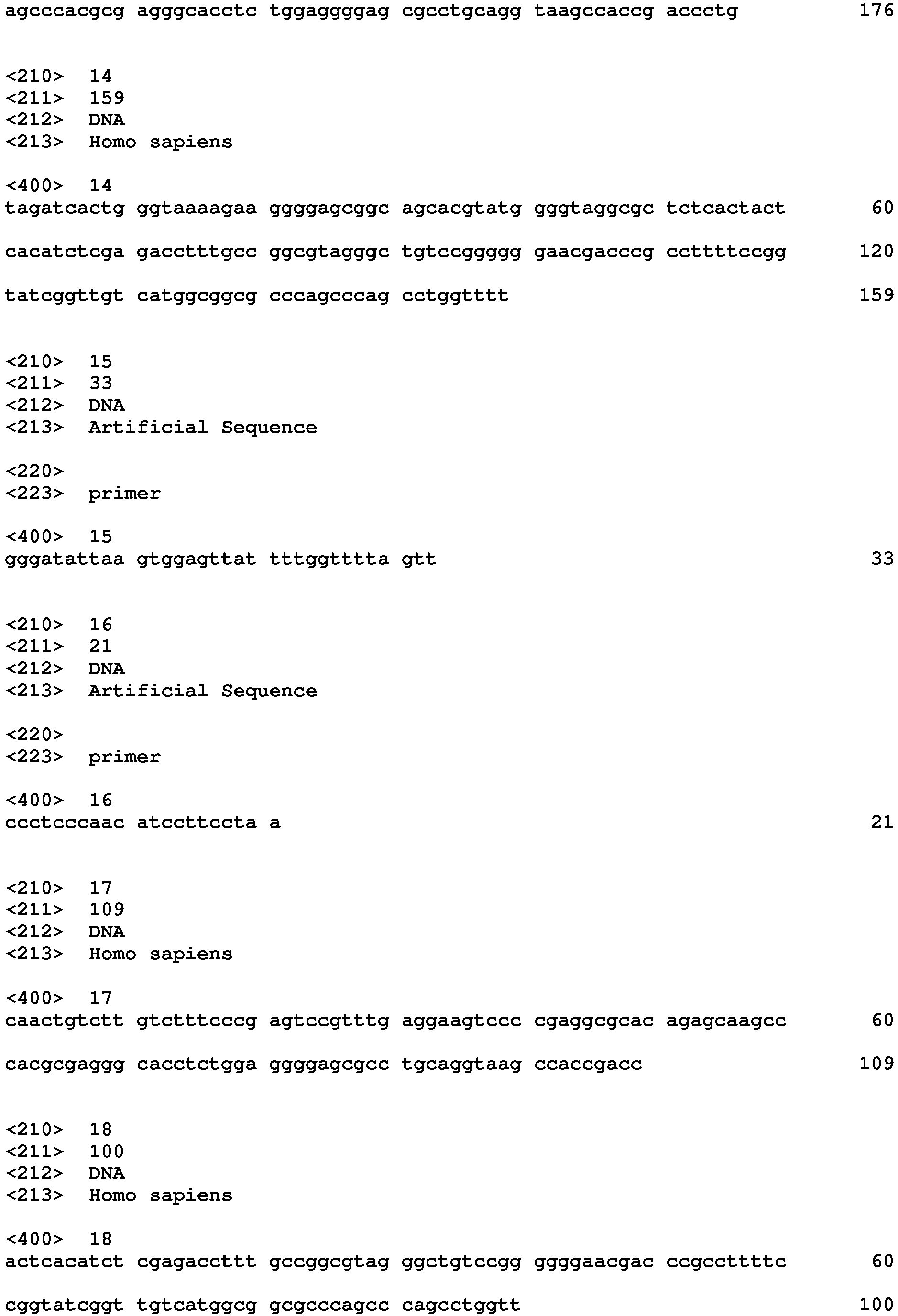
Claims (9)
- A method for obtaining information on uterine body cancer comprising the steps of:preparing a DNA sample from a biological sample collected from a subject;analyzing methylation status of a CpG site in a promoter region of at least one gene selected from F13A1 and CHCHD5 in the DNA sample obtained from the preparation step; andobtaining information on uterine body cancer in the subject based on an analysis result obtained from the analyzing step.
- The method according to claim 1, wherein the analyzing step is the step of analyzing presence or absence of methylation of at least one CpG site.
- The method according to claim 2, wherein
the information on uterine body cancer in the subject is presence or absence of a cancer cell derived from uterine body cancer in the biological sample collected from the subject, and
the step of obtaining information is the step of obtaining information indicating that the biological sample contains a cancer cell derived from uterine body cancer when the analysis result shows that there is a methylated CpG site. - The method according to claim 1, wherein the analyzing step is the step of analyzing methylation frequency.
- The method according to claim 4, wherein
the information on uterine body cancer in the subject is presence or absence of a cancer cell derived from uterine body cancer in the biological sample collected from the subject, and
the step of obtaining information is the step of obtaining information indicating that the biological sample contains a cancer cell derived from uterine body cancer when the methylation frequency obtained from the analyzing step is higher than a predetermined threshold. - A marker for obtaining information on uterine body cancer by methylation analysis, which is at least one CpG site selected from CpG sites located in promoter regions of F13A1 1 and CHCHD5 genes.
- A kit for obtaining information on uterine body cancer, comprising a primer set for analysis of methylation of at least one CpG site selected from CpG sites located in promoter regions of F13A1 and CHCHD5 genes.
- The kit according to claim 7, wherein the primer set is a primer set for analyzing methylation status of the CpG site by at least one method selected from mass spectrometry and methylation specific PCR method.
- The kit according to claim 8, wherein the primer set is at least one selected from:a primer set of primers respectively having base sequences SEQ ID NOs: 3 and 4;a primer set of primers respectively having base sequences SEQ ID NOs: 5 and 6;a primer set of primers respectively having base sequences SEQ ID NOs: 9 and 10; anda primer set of primers respectively having base sequences SEQ ID NOs: 11 and 12.
Applications Claiming Priority (2)
| Application Number | Priority Date | Filing Date | Title |
|---|---|---|---|
| JP2012205769 | 2012-09-19 | ||
| PCT/JP2013/075337 WO2014046200A1 (en) | 2012-09-19 | 2013-09-19 | Method for obtaining information about endometrial cancer, and marker and kit for obtaining information about endometrial cancer |
Publications (3)
| Publication Number | Publication Date |
|---|---|
| EP2899275A1 true EP2899275A1 (en) | 2015-07-29 |
| EP2899275A4 EP2899275A4 (en) | 2016-06-01 |
| EP2899275B1 EP2899275B1 (en) | 2018-06-27 |
Family
ID=50341504
Family Applications (1)
| Application Number | Title | Priority Date | Filing Date |
|---|---|---|---|
| EP13839246.9A Active EP2899275B1 (en) | 2012-09-19 | 2013-09-19 | Method for obtaining information about endometrial cancer, and marker and kit for obtaining information about endometrial cancer |
Country Status (3)
| Country | Link |
|---|---|
| EP (1) | EP2899275B1 (en) |
| JP (1) | JP6269494B2 (en) |
| WO (1) | WO2014046200A1 (en) |
Cited By (9)
| Publication number | Priority date | Publication date | Assignee | Title |
|---|---|---|---|---|
| CN113557308A (en) * | 2019-01-24 | 2021-10-26 | 梅约医学教育与研究基金会 | Detection of endometrial cancer |
| WO2022002423A1 (en) * | 2020-06-30 | 2022-01-06 | Universal Diagnostics, S.L. | Detection of colorectal neoplasms |
| WO2022012293A1 (en) * | 2020-07-11 | 2022-01-20 | 成都益安博生物技术有限公司 | Peripheral blood tcr marker for endometrial cancer, test kit using same, and application thereof |
| US11396679B2 (en) | 2019-05-31 | 2022-07-26 | Universal Diagnostics, S.L. | Detection of colorectal cancer |
| US11530453B2 (en) | 2020-06-30 | 2022-12-20 | Universal Diagnostics, S.L. | Systems and methods for detection of multiple cancer types |
| US11898199B2 (en) | 2019-11-11 | 2024-02-13 | Universal Diagnostics, S.A. | Detection of colorectal cancer and/or advanced adenomas |
| US12442043B2 (en) | 2019-10-31 | 2025-10-14 | Mayo Foundation For Medical Education And Research | Detecting ovarian cancer |
| US12467096B2 (en) | 2020-05-15 | 2025-11-11 | Universal Diagnostics, S.A. | Methods and systems for identifying methylation biomarkers |
| US12606872B2 (en) | 2022-11-16 | 2026-04-21 | Universal Diagnostics, S.A. | Methods for stratification and early detection of advanced adenoma and/or colorectal cancer using DNA methylation markers |
Families Citing this family (6)
| Publication number | Priority date | Publication date | Assignee | Title |
|---|---|---|---|---|
| ES2859645T3 (en) | 2013-03-14 | 2021-10-04 | Mayo Found Medical Education & Res | Neoplasia detection |
| JP6418594B2 (en) * | 2014-07-10 | 2018-11-07 | シスメックス株式会社 | Method for obtaining information on endometrial cancer, and marker and kit for obtaining information on endometrial cancer |
| RU2592242C2 (en) * | 2014-07-29 | 2016-07-20 | Наталья Владимировна Спиридинова | Diagnostic technique for determining probability of local inflammation development in myomatous nodule |
| WO2016160454A1 (en) | 2015-03-27 | 2016-10-06 | Exact Sciences Corporation | Detecting esophageal disorders |
| JP2016192906A (en) * | 2015-03-31 | 2016-11-17 | シスメックス株式会社 | Method for acquiring information regarding breast cancer, and marker for acquiring information regarding breast cancer and kit for detecting breast cancer |
| KR20250172724A (en) | 2015-08-31 | 2025-12-09 | 메이오 파운데이션 포 메디칼 에쥬케이션 앤드 리써치 | Detecting gastric neoplasm |
Family Cites Families (3)
| Publication number | Priority date | Publication date | Assignee | Title |
|---|---|---|---|---|
| WO2004108896A2 (en) * | 2003-06-03 | 2004-12-16 | The Board Of Trustees Of The University Of Arkansas | Gene expression profiling of uterine serous papillary carcinomas and ovarian serous papillary tumors |
| WO2005049861A2 (en) * | 2003-11-10 | 2005-06-02 | Epigenomics Ag | Method for the analysis of gynaecological cell proliferative disorders |
| JP2011160711A (en) * | 2010-02-09 | 2011-08-25 | Keio Gijuku | Method for using frequency of methylation of specific gene as biomarker for gynecologic cancer |
-
2013
- 2013-09-19 JP JP2014536914A patent/JP6269494B2/en active Active
- 2013-09-19 WO PCT/JP2013/075337 patent/WO2014046200A1/en not_active Ceased
- 2013-09-19 EP EP13839246.9A patent/EP2899275B1/en active Active
Non-Patent Citations (4)
| Title |
|---|
| D. FURLAN: "The High Frequency of De novo Promoter Methylation in Synchronous Primary Endometrial and Ovarian Carcinomas", CLINICAL CANCER RESEARCH, vol. 12, no. 11, 1 June 2006 (2006-06-01), US, pages 3329 - 3336, XP055267301, ISSN: 1078-0432, DOI: 10.1158/1078-0432.CCR-05-2679 * |
| DIANA L. KOLBE ET AL: "Differential Analysis of Ovarian and Endometrial Cancers Identifies a Methylator Phenotype", PLOS ONE, vol. 7, no. 3, 5 March 2012 (2012-03-05), pages e32941, XP055267312, DOI: 10.1371/journal.pone.0032941 * |
| None * |
| See also references of WO2014046200A1 * |
Cited By (11)
| Publication number | Priority date | Publication date | Assignee | Title |
|---|---|---|---|---|
| CN113557308A (en) * | 2019-01-24 | 2021-10-26 | 梅约医学教育与研究基金会 | Detection of endometrial cancer |
| EP3914733A4 (en) * | 2019-01-24 | 2023-05-17 | Mayo Foundation for Medical Education and Research | ENDOMETRIAL CANCER DETECTION |
| US12584177B2 (en) | 2019-01-24 | 2026-03-24 | Mayo Foundation For Medical Education And Research | Detecting endometrial cancer |
| US11396679B2 (en) | 2019-05-31 | 2022-07-26 | Universal Diagnostics, S.L. | Detection of colorectal cancer |
| US12442043B2 (en) | 2019-10-31 | 2025-10-14 | Mayo Foundation For Medical Education And Research | Detecting ovarian cancer |
| US11898199B2 (en) | 2019-11-11 | 2024-02-13 | Universal Diagnostics, S.A. | Detection of colorectal cancer and/or advanced adenomas |
| US12467096B2 (en) | 2020-05-15 | 2025-11-11 | Universal Diagnostics, S.A. | Methods and systems for identifying methylation biomarkers |
| WO2022002423A1 (en) * | 2020-06-30 | 2022-01-06 | Universal Diagnostics, S.L. | Detection of colorectal neoplasms |
| US11530453B2 (en) | 2020-06-30 | 2022-12-20 | Universal Diagnostics, S.L. | Systems and methods for detection of multiple cancer types |
| WO2022012293A1 (en) * | 2020-07-11 | 2022-01-20 | 成都益安博生物技术有限公司 | Peripheral blood tcr marker for endometrial cancer, test kit using same, and application thereof |
| US12606872B2 (en) | 2022-11-16 | 2026-04-21 | Universal Diagnostics, S.A. | Methods for stratification and early detection of advanced adenoma and/or colorectal cancer using DNA methylation markers |
Also Published As
| Publication number | Publication date |
|---|---|
| EP2899275B1 (en) | 2018-06-27 |
| JPWO2014046200A1 (en) | 2016-08-18 |
| EP2899275A4 (en) | 2016-06-01 |
| WO2014046200A1 (en) | 2014-03-27 |
| JP6269494B2 (en) | 2018-01-31 |
Similar Documents
| Publication | Publication Date | Title |
|---|---|---|
| EP2899275B1 (en) | Method for obtaining information about endometrial cancer, and marker and kit for obtaining information about endometrial cancer | |
| EP2816121B1 (en) | Method for obtaining information on hepatocellular carcinoma | |
| EP2977467B1 (en) | Method, use of marker, and determination device for obtaining information on plural types of cancers | |
| EP2899274A1 (en) | Method for obtaining information about hepatocellular carcinoma, and marker and kit for obtaining information about hepatocellular carcinoma | |
| EP2808397B1 (en) | Method for obtaining information on colon cancer and marker and kit for obtaining information on colon cancer | |
| EP2966183B1 (en) | Method for obtaining information on lung cancer, and use of marker and determination device for obtaining information on lung cancer | |
| EP2899272B1 (en) | Method and use for obtaining information about colorectal cancer | |
| EP2821487B1 (en) | Method for determining presence or absence of cancer cell derived from hepatocellular carcinoma, and determination marker and kit | |
| JP6269493B2 (en) | Method for acquiring information on brain tumor, and marker and kit for acquiring information on brain tumor | |
| EP2966182B1 (en) | Method for obtaining information on uterine body cancer and marker and determination device for obtaining information on uterine body cancer | |
| EP3075852B1 (en) | Method for acquiring information on gynecologic cancer and gynecologic cancer detection kit | |
| US20220333204A1 (en) | Tumor marker stamp-ep9 based on methylation modification and application thereof | |
| EP3075851A1 (en) | Method for acquiring information on gastric cancer and kit for detection of gastric cancer | |
| JP6636105B2 (en) | Method for obtaining information on colorectal cancer, and marker and kit for obtaining information on colorectal cancer | |
| EP3075853B1 (en) | Method of obtaining information on breast cancer and kit for detecting breast cancer | |
| JP6551656B2 (en) | Method for obtaining information on ovarian cancer, and marker for obtaining information on ovarian cancer and kit for detecting ovarian cancer |
Legal Events
| Date | Code | Title | Description |
|---|---|---|---|
| PUAI | Public reference made under article 153(3) epc to a published international application that has entered the european phase |
Free format text: ORIGINAL CODE: 0009012 |
|
| 17P | Request for examination filed |
Effective date: 20150409 |
|
| AK | Designated contracting states |
Kind code of ref document: A1 Designated state(s): AL AT BE BG CH CY CZ DE DK EE ES FI FR GB GR HR HU IE IS IT LI LT LU LV MC MK MT NL NO PL PT RO RS SE SI SK SM TR |
|
| AX | Request for extension of the european patent |
Extension state: BA ME |
|
| RIN1 | Information on inventor provided before grant (corrected) |
Inventor name: SAKAI, AYAKO Inventor name: ABURATANI, HIROYUKI Inventor name: KAJITA, MASAHIRO Inventor name: NAGAE, GENTA |
|
| DAX | Request for extension of the european patent (deleted) | ||
| RA4 | Supplementary search report drawn up and despatched (corrected) |
Effective date: 20160502 |
|
| RIC1 | Information provided on ipc code assigned before grant |
Ipc: C12Q 1/68 20060101ALI20160425BHEP Ipc: C12N 15/09 20060101AFI20160425BHEP Ipc: C12Q 1/04 20060101ALI20160425BHEP |
|
| 17Q | First examination report despatched |
Effective date: 20170404 |
|
| GRAP | Despatch of communication of intention to grant a patent |
Free format text: ORIGINAL CODE: EPIDOSNIGR1 |
|
| INTG | Intention to grant announced |
Effective date: 20180126 |
|
| GRAS | Grant fee paid |
Free format text: ORIGINAL CODE: EPIDOSNIGR3 |
|
| GRAA | (expected) grant |
Free format text: ORIGINAL CODE: 0009210 |
|
| AK | Designated contracting states |
Kind code of ref document: B1 Designated state(s): AL AT BE BG CH CY CZ DE DK EE ES FI FR GB GR HR HU IE IS IT LI LT LU LV MC MK MT NL NO PL PT RO RS SE SI SK SM TR |
|
| REG | Reference to a national code |
Ref country code: GB Ref legal event code: FG4D |
|
| REG | Reference to a national code |
Ref country code: AT Ref legal event code: REF Ref document number: 1012377 Country of ref document: AT Kind code of ref document: T Effective date: 20180715 |
|
| REG | Reference to a national code |
Ref country code: IE Ref legal event code: FG4D |
|
| REG | Reference to a national code |
Ref country code: DE Ref legal event code: R096 Ref document number: 602013039521 Country of ref document: DE |
|
| PG25 | Lapsed in a contracting state [announced via postgrant information from national office to epo] |
Ref country code: LT Free format text: LAPSE BECAUSE OF FAILURE TO SUBMIT A TRANSLATION OF THE DESCRIPTION OR TO PAY THE FEE WITHIN THE PRESCRIBED TIME-LIMIT Effective date: 20180627 Ref country code: NO Free format text: LAPSE BECAUSE OF FAILURE TO SUBMIT A TRANSLATION OF THE DESCRIPTION OR TO PAY THE FEE WITHIN THE PRESCRIBED TIME-LIMIT Effective date: 20180927 Ref country code: BG Free format text: LAPSE BECAUSE OF FAILURE TO SUBMIT A TRANSLATION OF THE DESCRIPTION OR TO PAY THE FEE WITHIN THE PRESCRIBED TIME-LIMIT Effective date: 20180927 Ref country code: SE Free format text: LAPSE BECAUSE OF FAILURE TO SUBMIT A TRANSLATION OF THE DESCRIPTION OR TO PAY THE FEE WITHIN THE PRESCRIBED TIME-LIMIT Effective date: 20180627 Ref country code: FI Free format text: LAPSE BECAUSE OF FAILURE TO SUBMIT A TRANSLATION OF THE DESCRIPTION OR TO PAY THE FEE WITHIN THE PRESCRIBED TIME-LIMIT Effective date: 20180627 |
|
| REG | Reference to a national code |
Ref country code: NL Ref legal event code: MP Effective date: 20180627 |
|
| REG | Reference to a national code |
Ref country code: LT Ref legal event code: MG4D |
|
| PG25 | Lapsed in a contracting state [announced via postgrant information from national office to epo] |
Ref country code: HR Free format text: LAPSE BECAUSE OF FAILURE TO SUBMIT A TRANSLATION OF THE DESCRIPTION OR TO PAY THE FEE WITHIN THE PRESCRIBED TIME-LIMIT Effective date: 20180627 Ref country code: GR Free format text: LAPSE BECAUSE OF FAILURE TO SUBMIT A TRANSLATION OF THE DESCRIPTION OR TO PAY THE FEE WITHIN THE PRESCRIBED TIME-LIMIT Effective date: 20180928 Ref country code: RS Free format text: LAPSE BECAUSE OF FAILURE TO SUBMIT A TRANSLATION OF THE DESCRIPTION OR TO PAY THE FEE WITHIN THE PRESCRIBED TIME-LIMIT Effective date: 20180627 Ref country code: LV Free format text: LAPSE BECAUSE OF FAILURE TO SUBMIT A TRANSLATION OF THE DESCRIPTION OR TO PAY THE FEE WITHIN THE PRESCRIBED TIME-LIMIT Effective date: 20180627 |
|
| REG | Reference to a national code |
Ref country code: AT Ref legal event code: MK05 Ref document number: 1012377 Country of ref document: AT Kind code of ref document: T Effective date: 20180627 |
|
| PG25 | Lapsed in a contracting state [announced via postgrant information from national office to epo] |
Ref country code: NL Free format text: LAPSE BECAUSE OF FAILURE TO SUBMIT A TRANSLATION OF THE DESCRIPTION OR TO PAY THE FEE WITHIN THE PRESCRIBED TIME-LIMIT Effective date: 20180627 |
|
| PG25 | Lapsed in a contracting state [announced via postgrant information from national office to epo] |
Ref country code: RO Free format text: LAPSE BECAUSE OF FAILURE TO SUBMIT A TRANSLATION OF THE DESCRIPTION OR TO PAY THE FEE WITHIN THE PRESCRIBED TIME-LIMIT Effective date: 20180627 Ref country code: CZ Free format text: LAPSE BECAUSE OF FAILURE TO SUBMIT A TRANSLATION OF THE DESCRIPTION OR TO PAY THE FEE WITHIN THE PRESCRIBED TIME-LIMIT Effective date: 20180627 Ref country code: PL Free format text: LAPSE BECAUSE OF FAILURE TO SUBMIT A TRANSLATION OF THE DESCRIPTION OR TO PAY THE FEE WITHIN THE PRESCRIBED TIME-LIMIT Effective date: 20180627 Ref country code: SK Free format text: LAPSE BECAUSE OF FAILURE TO SUBMIT A TRANSLATION OF THE DESCRIPTION OR TO PAY THE FEE WITHIN THE PRESCRIBED TIME-LIMIT Effective date: 20180627 Ref country code: AT Free format text: LAPSE BECAUSE OF FAILURE TO SUBMIT A TRANSLATION OF THE DESCRIPTION OR TO PAY THE FEE WITHIN THE PRESCRIBED TIME-LIMIT Effective date: 20180627 Ref country code: IS Free format text: LAPSE BECAUSE OF FAILURE TO SUBMIT A TRANSLATION OF THE DESCRIPTION OR TO PAY THE FEE WITHIN THE PRESCRIBED TIME-LIMIT Effective date: 20181027 Ref country code: EE Free format text: LAPSE BECAUSE OF FAILURE TO SUBMIT A TRANSLATION OF THE DESCRIPTION OR TO PAY THE FEE WITHIN THE PRESCRIBED TIME-LIMIT Effective date: 20180627 |
|
| PG25 | Lapsed in a contracting state [announced via postgrant information from national office to epo] |
Ref country code: ES Free format text: LAPSE BECAUSE OF FAILURE TO SUBMIT A TRANSLATION OF THE DESCRIPTION OR TO PAY THE FEE WITHIN THE PRESCRIBED TIME-LIMIT Effective date: 20180627 Ref country code: SM Free format text: LAPSE BECAUSE OF FAILURE TO SUBMIT A TRANSLATION OF THE DESCRIPTION OR TO PAY THE FEE WITHIN THE PRESCRIBED TIME-LIMIT Effective date: 20180627 Ref country code: IT Free format text: LAPSE BECAUSE OF FAILURE TO SUBMIT A TRANSLATION OF THE DESCRIPTION OR TO PAY THE FEE WITHIN THE PRESCRIBED TIME-LIMIT Effective date: 20180627 |
|
| REG | Reference to a national code |
Ref country code: DE Ref legal event code: R097 Ref document number: 602013039521 Country of ref document: DE |
|
| PG25 | Lapsed in a contracting state [announced via postgrant information from national office to epo] |
Ref country code: MC Free format text: LAPSE BECAUSE OF FAILURE TO SUBMIT A TRANSLATION OF THE DESCRIPTION OR TO PAY THE FEE WITHIN THE PRESCRIBED TIME-LIMIT Effective date: 20180627 |
|
| REG | Reference to a national code |
Ref country code: CH Ref legal event code: PL |
|
| PLBE | No opposition filed within time limit |
Free format text: ORIGINAL CODE: 0009261 |
|
| STAA | Information on the status of an ep patent application or granted ep patent |
Free format text: STATUS: NO OPPOSITION FILED WITHIN TIME LIMIT |
|
| GBPC | Gb: european patent ceased through non-payment of renewal fee |
Effective date: 20180927 |
|
| PG25 | Lapsed in a contracting state [announced via postgrant information from national office to epo] |
Ref country code: DK Free format text: LAPSE BECAUSE OF FAILURE TO SUBMIT A TRANSLATION OF THE DESCRIPTION OR TO PAY THE FEE WITHIN THE PRESCRIBED TIME-LIMIT Effective date: 20180627 |
|
| 26N | No opposition filed |
Effective date: 20190328 |
|
| REG | Reference to a national code |
Ref country code: BE Ref legal event code: MM Effective date: 20180930 |
|
| REG | Reference to a national code |
Ref country code: IE Ref legal event code: MM4A |
|
| PG25 | Lapsed in a contracting state [announced via postgrant information from national office to epo] |
Ref country code: LU Free format text: LAPSE BECAUSE OF NON-PAYMENT OF DUE FEES Effective date: 20180919 |
|
| PG25 | Lapsed in a contracting state [announced via postgrant information from national office to epo] |
Ref country code: IE Free format text: LAPSE BECAUSE OF NON-PAYMENT OF DUE FEES Effective date: 20180919 |
|
| PG25 | Lapsed in a contracting state [announced via postgrant information from national office to epo] |
Ref country code: CH Free format text: LAPSE BECAUSE OF NON-PAYMENT OF DUE FEES Effective date: 20180930 Ref country code: SI Free format text: LAPSE BECAUSE OF FAILURE TO SUBMIT A TRANSLATION OF THE DESCRIPTION OR TO PAY THE FEE WITHIN THE PRESCRIBED TIME-LIMIT Effective date: 20180627 Ref country code: LI Free format text: LAPSE BECAUSE OF NON-PAYMENT OF DUE FEES Effective date: 20180930 Ref country code: FR Free format text: LAPSE BECAUSE OF NON-PAYMENT OF DUE FEES Effective date: 20180930 Ref country code: BE Free format text: LAPSE BECAUSE OF NON-PAYMENT OF DUE FEES Effective date: 20180930 |
|
| PG25 | Lapsed in a contracting state [announced via postgrant information from national office to epo] |
Ref country code: GB Free format text: LAPSE BECAUSE OF NON-PAYMENT OF DUE FEES Effective date: 20180927 |
|
| PG25 | Lapsed in a contracting state [announced via postgrant information from national office to epo] |
Ref country code: AL Free format text: LAPSE BECAUSE OF FAILURE TO SUBMIT A TRANSLATION OF THE DESCRIPTION OR TO PAY THE FEE WITHIN THE PRESCRIBED TIME-LIMIT Effective date: 20180627 |
|
| PG25 | Lapsed in a contracting state [announced via postgrant information from national office to epo] |
Ref country code: MT Free format text: LAPSE BECAUSE OF NON-PAYMENT OF DUE FEES Effective date: 20180919 |
|
| PG25 | Lapsed in a contracting state [announced via postgrant information from national office to epo] |
Ref country code: TR Free format text: LAPSE BECAUSE OF FAILURE TO SUBMIT A TRANSLATION OF THE DESCRIPTION OR TO PAY THE FEE WITHIN THE PRESCRIBED TIME-LIMIT Effective date: 20180627 |
|
| PG25 | Lapsed in a contracting state [announced via postgrant information from national office to epo] |
Ref country code: PT Free format text: LAPSE BECAUSE OF FAILURE TO SUBMIT A TRANSLATION OF THE DESCRIPTION OR TO PAY THE FEE WITHIN THE PRESCRIBED TIME-LIMIT Effective date: 20180627 |
|
| PG25 | Lapsed in a contracting state [announced via postgrant information from national office to epo] |
Ref country code: MK Free format text: LAPSE BECAUSE OF NON-PAYMENT OF DUE FEES Effective date: 20180627 Ref country code: HU Free format text: LAPSE BECAUSE OF FAILURE TO SUBMIT A TRANSLATION OF THE DESCRIPTION OR TO PAY THE FEE WITHIN THE PRESCRIBED TIME-LIMIT; INVALID AB INITIO Effective date: 20130919 Ref country code: CY Free format text: LAPSE BECAUSE OF FAILURE TO SUBMIT A TRANSLATION OF THE DESCRIPTION OR TO PAY THE FEE WITHIN THE PRESCRIBED TIME-LIMIT Effective date: 20180627 |
|
| PGFP | Annual fee paid to national office [announced via postgrant information from national office to epo] |
Ref country code: DE Payment date: 20240912 Year of fee payment: 12 |