EP2898091B1 - Methods for predicting multiple myeloma treatment response - Google Patents
Methods for predicting multiple myeloma treatment response Download PDFInfo
- Publication number
- EP2898091B1 EP2898091B1 EP13766076.7A EP13766076A EP2898091B1 EP 2898091 B1 EP2898091 B1 EP 2898091B1 EP 13766076 A EP13766076 A EP 13766076A EP 2898091 B1 EP2898091 B1 EP 2898091B1
- Authority
- EP
- European Patent Office
- Prior art keywords
- score
- patients
- yes
- dms
- patient
- Prior art date
- Legal status (The legal status is an assumption and is not a legal conclusion. Google has not performed a legal analysis and makes no representation as to the accuracy of the status listed.)
- Active
Links
Images




Classifications
-
- C—CHEMISTRY; METALLURGY
- C12—BIOCHEMISTRY; BEER; SPIRITS; WINE; VINEGAR; MICROBIOLOGY; ENZYMOLOGY; MUTATION OR GENETIC ENGINEERING
- C12Q—MEASURING OR TESTING PROCESSES INVOLVING ENZYMES, NUCLEIC ACIDS OR MICROORGANISMS; COMPOSITIONS OR TEST PAPERS THEREFOR; PROCESSES OF PREPARING SUCH COMPOSITIONS; CONDITION-RESPONSIVE CONTROL IN MICROBIOLOGICAL OR ENZYMOLOGICAL PROCESSES
- C12Q1/00—Measuring or testing processes involving enzymes, nucleic acids or microorganisms; Compositions therefor; Processes of preparing such compositions
- C12Q1/68—Measuring or testing processes involving enzymes, nucleic acids or microorganisms; Compositions therefor; Processes of preparing such compositions involving nucleic acids
- C12Q1/6876—Nucleic acid products used in the analysis of nucleic acids, e.g. primers or probes
- C12Q1/6883—Nucleic acid products used in the analysis of nucleic acids, e.g. primers or probes for diseases caused by alterations of genetic material
- C12Q1/6886—Nucleic acid products used in the analysis of nucleic acids, e.g. primers or probes for diseases caused by alterations of genetic material for cancer
-
- A—HUMAN NECESSITIES
- A61—MEDICAL OR VETERINARY SCIENCE; HYGIENE
- A61K—PREPARATIONS FOR MEDICAL, DENTAL OR TOILETRY PURPOSES
- A61K31/00—Medicinal preparations containing organic active ingredients
- A61K31/70—Carbohydrates; Sugars; Derivatives thereof
- A61K31/7042—Compounds having saccharide radicals and heterocyclic rings
- A61K31/7052—Compounds having saccharide radicals and heterocyclic rings having nitrogen as a ring hetero atom, e.g. nucleosides, nucleotides
- A61K31/706—Compounds having saccharide radicals and heterocyclic rings having nitrogen as a ring hetero atom, e.g. nucleosides, nucleotides containing six-membered rings with nitrogen as a ring hetero atom
- A61K31/7064—Compounds having saccharide radicals and heterocyclic rings having nitrogen as a ring hetero atom, e.g. nucleosides, nucleotides containing six-membered rings with nitrogen as a ring hetero atom containing condensed or non-condensed pyrimidines
- A61K31/7068—Compounds having saccharide radicals and heterocyclic rings having nitrogen as a ring hetero atom, e.g. nucleosides, nucleotides containing six-membered rings with nitrogen as a ring hetero atom containing condensed or non-condensed pyrimidines having oxo groups directly attached to the pyrimidine ring, e.g. cytidine, cytidylic acid
-
- G—PHYSICS
- G16—INFORMATION AND COMMUNICATION TECHNOLOGY [ICT] SPECIALLY ADAPTED FOR SPECIFIC APPLICATION FIELDS
- G16B—BIOINFORMATICS, i.e. INFORMATION AND COMMUNICATION TECHNOLOGY [ICT] SPECIALLY ADAPTED FOR GENETIC OR PROTEIN-RELATED DATA PROCESSING IN COMPUTATIONAL MOLECULAR BIOLOGY
- G16B25/00—ICT specially adapted for hybridisation; ICT specially adapted for gene or protein expression
- G16B25/10—Gene or protein expression profiling; Expression-ratio estimation or normalisation
-
- G—PHYSICS
- G16—INFORMATION AND COMMUNICATION TECHNOLOGY [ICT] SPECIALLY ADAPTED FOR SPECIFIC APPLICATION FIELDS
- G16H—HEALTHCARE INFORMATICS, i.e. INFORMATION AND COMMUNICATION TECHNOLOGY [ICT] SPECIALLY ADAPTED FOR THE HANDLING OR PROCESSING OF MEDICAL OR HEALTHCARE DATA
- G16H10/00—ICT specially adapted for the handling or processing of patient-related medical or healthcare data
- G16H10/40—ICT specially adapted for the handling or processing of patient-related medical or healthcare data for data related to laboratory analysis, e.g. patient specimen analysis
-
- G—PHYSICS
- G16—INFORMATION AND COMMUNICATION TECHNOLOGY [ICT] SPECIALLY ADAPTED FOR SPECIFIC APPLICATION FIELDS
- G16H—HEALTHCARE INFORMATICS, i.e. INFORMATION AND COMMUNICATION TECHNOLOGY [ICT] SPECIALLY ADAPTED FOR THE HANDLING OR PROCESSING OF MEDICAL OR HEALTHCARE DATA
- G16H20/00—ICT specially adapted for therapies or health-improving plans, e.g. for handling prescriptions, for steering therapy or for monitoring patient compliance
- G16H20/40—ICT specially adapted for therapies or health-improving plans, e.g. for handling prescriptions, for steering therapy or for monitoring patient compliance relating to mechanical, radiation or invasive therapies, e.g. surgery, laser therapy, dialysis or acupuncture
-
- C—CHEMISTRY; METALLURGY
- C12—BIOCHEMISTRY; BEER; SPIRITS; WINE; VINEGAR; MICROBIOLOGY; ENZYMOLOGY; MUTATION OR GENETIC ENGINEERING
- C12Q—MEASURING OR TESTING PROCESSES INVOLVING ENZYMES, NUCLEIC ACIDS OR MICROORGANISMS; COMPOSITIONS OR TEST PAPERS THEREFOR; PROCESSES OF PREPARING SUCH COMPOSITIONS; CONDITION-RESPONSIVE CONTROL IN MICROBIOLOGICAL OR ENZYMOLOGICAL PROCESSES
- C12Q2600/00—Oligonucleotides characterized by their use
- C12Q2600/106—Pharmacogenomics, i.e. genetic variability in individual responses to drugs and drug metabolism
-
- C—CHEMISTRY; METALLURGY
- C12—BIOCHEMISTRY; BEER; SPIRITS; WINE; VINEGAR; MICROBIOLOGY; ENZYMOLOGY; MUTATION OR GENETIC ENGINEERING
- C12Q—MEASURING OR TESTING PROCESSES INVOLVING ENZYMES, NUCLEIC ACIDS OR MICROORGANISMS; COMPOSITIONS OR TEST PAPERS THEREFOR; PROCESSES OF PREPARING SUCH COMPOSITIONS; CONDITION-RESPONSIVE CONTROL IN MICROBIOLOGICAL OR ENZYMOLOGICAL PROCESSES
- C12Q2600/00—Oligonucleotides characterized by their use
- C12Q2600/158—Expression markers
-
- Y—GENERAL TAGGING OF NEW TECHNOLOGICAL DEVELOPMENTS; GENERAL TAGGING OF CROSS-SECTIONAL TECHNOLOGIES SPANNING OVER SEVERAL SECTIONS OF THE IPC; TECHNICAL SUBJECTS COVERED BY FORMER USPC CROSS-REFERENCE ART COLLECTIONS [XRACs] AND DIGESTS
- Y02—TECHNOLOGIES OR APPLICATIONS FOR MITIGATION OR ADAPTATION AGAINST CLIMATE CHANGE
- Y02A—TECHNOLOGIES FOR ADAPTATION TO CLIMATE CHANGE
- Y02A90/00—Technologies having an indirect contribution to adaptation to climate change
- Y02A90/10—Information and communication technologies [ICT] supporting adaptation to climate change, e.g. for weather forecasting or climate simulation
Definitions
- the present invention relates to methods for predicting multiple myeloma treatment response.
- Malignant transformation requires oncogenic activation and inactivation of tumor suppressor genes, which help cancer cells overriding the normal mechanisms controlling cellular survival and proliferation (1,2). These molecular events are caused by genetic alterations (translocations, amplification, mutations) and also by epigenetic modifications (3). Epigenetic modifications include methylation of DNA cytosine residues and histone modifications and have been shown to be critical in the initiation and progression of cancers (4). DNA methyltransferase inhibitors or HDAC inhibitors are now being used in the treatment of several hematologic malignancies including MM (5-8).
- MMCs Multiple Myeloma Cells
- MM arises through the accumulation of multiple genetic changes that include an aberrant or overexpression of a D-type cyclin gene, cyclin D1 (CCND1) in the case of t(11;14) translocation or gain in 11q13, cyclin D3 (CCND3) in the case of the rare t(6;14) translocation, or cyclin D2 (CCND2) on the background of a translocation involving c-maf (t(14;16)) or MMSET/ FGFR3 (t(4;14)) (9,10).
- a D-type cyclin gene cyclin D1 (CCND1) in the case of t(11;14) translocation or gain in 11q13
- cyclin D3 CCND3
- cyclin D2 cyclin D2
- HMCLs human myeloma cell lines
- SPARC secreted protein acidic and rich in cysteine
- Bcl2/adenovirus E1B 19kDa interacting protein 3 (B NIP3 ) promoters was associated with poor overall survival of MM patients (19).
- SOCS3 methylation was found to be associated with extramedullary manifestations, plasma cell leukemia and significant shortened survival in MM patients (20).
- DNMT inhibitors can be sub-divided into nucleoside analogue and non-nucleoside analogue families.
- 5-Azacytidine(azacytidine)/5-Aza-2'-deoxycytidine (decitabine) are both nucleoside analogues with Food and Drug Administration approval for use in myelodysplastic syndrome. Clinical trials in myeloma combining these demethylating agents with chemotherapy or other agents are underway (8,72).
- MMCs Multiple Myeloma Cells
- the present invention relates to a method of testing whether a patient suffering of multiple myeloma will respond or not to a DNA methyltransferase inhibitor (DNMTi).
- DNMTi DNA methyltransferase inhibitor
- the multiple myeloma treatment response was investigated by the inventors using DNA methyltransferase inhibitors (DNMTi), human multiple myeloma cell lines and primary myeloma cells.
- DNMTi DNA methyltransferase inhibitors
- the inventors analyzed gene expression profiles of 5 MM cell lines treated with decitabine using microarrays.
- the inventors identified 127 genes deregulated by decitabine. 47 out of 127 decitabine deregulated genes have prognostic value in inventor's cohort of 206 newly-diagnosed MM patients.
- the inventors summarized the prognostic information in a DNA methylation score (DM Score or DMS) built using the probe set signal value weighted by the beta coefficient of prognostic genes.
- the DM Score is predictive for DNMT inhibitor sensitivity of HMCLs and primary myeloma cells in vitro.
- the DM Score allows identification of myeloma patients that could benefit DNMT inhibitor treatment.
- HMCLs with a high DM Score exhibited significant 3-fold higher 5-Azacytidine sensitivity than HMCLs with a low DM Score and these results were validated in vitro with primary myeloma cells of patients.
- a patient denotes a mammal.
- a patient refers to any patient (preferably human) afflicted with multiple myeloma.
- multiple myeloma refers to multiple myeloma such as revised in the World Health Organisation Classification C90.
- DNA methyltransferase inhibitors or “DNMTi” has its general meaning in the art and refers to a multiple myeloma treatment.
- DNA methyltransferase inhibitors or “DNMTi” refers to DNA methyltransferase inhibitor that can be sub-divided into nucleoside analogue (5-Azacytidine (azacytidine), 5-Aza-2'-deoxycytidine (decitabine, 5-Aza-CdR), zebularine, 5-Fluoro-2'-deoxycytidine (5-F-CdR), 5 ,6-Dihydro-5-azacytidine (DHAC)) and non-nucleoside analogue families (Hydralazine, Procainamide, Procaine, EGCG ((-)-epigallocatechin-3-gallate), Psammaplin A, MG98, RG108) (8).
- nucleoside analogue 5-Azacy
- biological sample refers to multiple myeloma cells, bone marrow or medullary cell.
- the present invention relates to a method of testing whether a patient suffering of multiple myeloma will respond or not to a DNA methyltransferase inhibitor (DNMTi) comprising:
- the levels of at least 25 genes from Table A are determined wherein said genes are:
- the level of 25, 26, 27, 28, 29, 30, 31, 32, 33, 34, 35, 36, 37, 38, 39, 40, 41, 42, 43, 44, 45, 46, or 47 genes from Table A are determined wherein every combinations of genes comprises a minimal set of 25 genes consisting of:
- the level of the 47 genes of Table A are determined.
- the expression level as determined is a relative expression level. More preferably, the determination comprises contacting the biological sample with selective reagents such as probes, primers or ligands, and thereby detecting the presence, or measuring the amount, of polypeptide or nucleic acids of interest originally in the biological sample. Contacting may be performed in any suitable device, such as a plate, microtiter dish, test tube, well, glass, column, and so forth. In specific aspects, the contacting is performed on a substrate coated with the reagent, such as a nucleic acid array or a specific ligand array.
- the substrate may be a solid or semi-solid substrate such as any suitable support comprising glass, plastic, nylon, paper, metal, polymers and the like.
- the substrate may be of various forms and sizes, such as a slide, a membrane, a bead, a column, a gel, etc.
- the contacting may be made under any condition suitable for a detectable complex, such as a nucleic acid hybrid or an antibody-antigen complex, to be formed between the reagent and the nucleic acids or polypeptides of the biological sample.
- the expression level may be determined by determining the quantity of mRNA.
- nucleic acid contained in the biological sample is first extracted according to standard methods, for example using lytic enzymes or chemical solutions or extracted by nucleic-acid-binding resins following the manufacturer's instructions.
- the extracted mRNA is then detected by hybridization (e. g., Northern blot analysis) and/or amplification (e.g., RT-PCR).
- hybridization e. g., Northern blot analysis
- amplification e.g., RT-PCR
- RT-PCR e.g., RT-PCR
- quantitative or semi-quantitative RT-PCR is preferred. Real-time quantitative or semi-quantitative RT-PCR is particularly advantageous.
- LCR ligase chain reaction
- TMA transcription-mediated amplification
- SDA strand displacement amplification
- NASBA nucleic acid sequence based amplification
- Nucleic acids having at least 10 nucleotides and exhibiting sequence complementarity or homology to the mRNA of interest herein find utility as hybridization probes or amplification primers. It is understood that such nucleic acids need not be identical, but are typically at least about 80% identical to the homologous region of comparable size, more preferably 85% identical and even more preferably 90-95% identical. In certain aspects, it will be advantageous to use nucleic acids in combination with appropriate means, such as a detectable label, for detecting hybridization. A wide variety of appropriate indicators are known in the art including, fluorescent, radioactive, enzymatic or other ligands (e. g. avidin/biotin) .
- Probes typically comprise single-stranded nucleic acids of between 10 to 1000 nucleotides in length, for instance of between 10 and 800, more preferably of between 15 and 700, typically of between 20 and 500.
- Primers typically are shorter single-stranded nucleic acids, of between 10 to 25 nucleotides in length, designed to perfectly or almost perfectly match a nucleic acid of interest, to be amplified.
- the probes and primers are "specific" to the nucleic acids they hybridize to, i.e. they preferably hybridize under high stringency hybridization conditions (corresponding to the highest melting temperature Tm, e.g., 50 % formamide, 5x or 6x SCC.
- SCC is a 0.15 M NaCl, 0.015 M Na-citrate).
- the nucleic acid primers or probes used in the above amplification and detection method may be assembled as a kit.
- a kit includes consensus primers and molecular probes.
- a preferred kit also includes the components necessary to determine if amplification has occurred.
- the kit may also include, for example, PCR buffers and enzymes; positive control sequences, reaction control primers; and instructions for amplifying and detecting the specific sequences.
- the methods of the invention comprise the steps of providing total RNAs extracted from a biological samples and subjecting the RNAs to amplification and hybridization to specific probes, more particularly by means of a quantitative or semi-quantitative RT-PCR.
- the expression level is determined by DNA chip analysis.
- DNA chip or nucleic acid microarray consists of different nucleic acid probes that are chemically attached to a substrate, which can be a microchip, a glass slide or a microsphere-sized bead.
- a microchip may be constituted of polymers, plastics, resins, polysaccharides, silica or silica-based materials, carbon, metals, inorganic glasses, or nitrocellulose.
- Probes comprise nucleic acids such as cDNAs or oligonucleotides that may be about 10 to about 60 base pairs.
- a biological sample from a test patient optionally first subjected to a reverse transcription, is labelled and contacted with the microarray in hybridization conditions, leading to the formation of complexes between target nucleic acids that are complementary to probe sequences attached to the microarray surface.
- the labelled hybridized complexes are then detected and can be quantified or semi-quantified. Labelling may be achieved by various methods, e.g. by using radioactive or fluorescent labelling.
- Many variants of the microarray hybridization technology are available to the man skilled in the art (see e.g. the review by Hoheisel, Nature Reviews, Genetics, 2006, 7:200-210 )
- the invention further provides a DNA chip comprising a solid support which carries nucleic acids that are specific to the genes listed in table A.
- Predetermined reference values ELRi or DMS R used for comparison may consist of "cut-off" values.
- each reference (“cut-off') value ELRi for each gene may be determined by carrying out a method comprising the steps of:
- the expression level of a gene Gi has been assessed for 100 samples of 100 patients.
- the 100 samples are ranked according to the expression level of gene Gi.
- Sample 1 has the highest expression level and sample 100 has the lowest expression level.
- a first grouping provides two subsets: on one side sample Nr 1 and on the other side the 99 other samples.
- the next grouping provides on one side samples 1 and 2 and on the other side the 98 remaining samples etc., until the last grouping: on one side samples 1 to 99 and on the other side sample Nr 100.
- Kaplan Meier curves are prepared for each of the 99 groups of two subsets. Also for each of the 99 groups, the p value between both subsets was calculated.
- the reference value ELRi is then selected such as the discrimination based on the criterion of the minimum p value is the strongest.
- the expression level corresponding to the boundary between both subsets for which the p value is minimum is considered as the reference value. It should be noted that according to the experiments made by the inventors, the reference value ELRi is not necessarily the median value of expression levels.
- the reference value DMS R is the median value of DMS.
- the reference value ELRi for the genes are described in table A (right column).
- the reference value DMS R is -19.7 for determining whether a patient suffering of multiple myeloma will respond to a DNMTi or -15.3 for predicting the survival time of patient suffering of multiple myeloma.
- the regression ⁇ coefficient reference values may be easily determined by the skilled man in the art for each gene using a Cox model.
- the Cox model is based on a modeling approach to the analysis of survival data. The purpose of the model is to simultaneously explore the effects of several variables on survival.
- the Cox model is a well-recognised statistical technique for analysing survival data. When it is used to analyse the survival of patients in a clinical trial, the model allows us to isolate the effects of treatment from the effects of other variables.
- the logrank test cannot be used to explore (and adjust for) the effects of several variables, such as age and disease duration, known to affect survival. Adjustment for variables that are known to affect survival may improve the precision with which we can estimate the treatment effect.
- the regression method introduced by Cox is used to investigate several variables at a time.
- the procedure models or regresses the survival times (or more specifically, the so-called hazard function) on the explanatory variables.
- the hazard function is the probability that an individual will experience an event (for example, death) within a small time interval, given that the individual has survived up to the beginning of the interval. It can therefore be interpreted as the risk of dying at time t .
- the quantity h0 ( t ) is the baseline or underlying hazard function and corresponds to the probability of dying (or reaching an event) when all the explanatory variables are zero.
- the regression coefficient ⁇ gives the proportional change that can be expected in the hazard, related to changes in the explanatory variables.
- the coefficient ⁇ is estimated by a statistical method called maximum likelihood.
- HR Hazard Ratio
- exp( ⁇ ) the ratio of the hazard rates corresponding to the conditions described by two sets of explanatory variables. For example, in a drug study, the treated population may die at twice the rate per unit time as the control population. The hazard ratio would be 2, indicating higher hazard of death from the treatment.
- the regression ⁇ coefficient reference values are described in table A.
- kits for performing the methods as above described comprising means for measuring the expression level of the genes listed in Table A.
- the kit may include a primer, a set of primers, a probe, a set of probes as above described.
- the probe or set of probes are labelled as above described.
- the kit may also contain other suitably packaged reagents and materials needed for the particular detection protocol, including solid-phase matrices, if applicable, and standards.
- the score may be generated by a computer program.
- the method of the invention allows to define a subgroup of patients who will be responsive ("responder") or not ("non responder”) to the treatment with a DNA methyltransferase inhibitor.
- treating means reversing, alleviating, inhibiting the progress of, or preventing the disorder or condition to which such term applies, or one or more symptoms of such disorder or condition.
- the method comprises the following steps
- a further object of the invention relates to a DNA methyltransferase inhibitor for use in the treatment of multiple myeloma in a patient in need thereof, wherein the patient was being classified as responder by the method as above described.
- FIGURES are a diagrammatic representation of FIGURES.
- EXAMPLE 1 DEVELOPMENT OF GENE EXPRESSION BASED SCORE TO PREDICT SENSITIVITY OF MULTIPLE MYELOMA CELLS TO DNA METHYLATION INHIBITORS
- HMCLs Human Myeloma Cell Lines
- XG-1, XG-2, XG-3, XG-4, XG-5, XG-6, XG-7, XG-10, XG-11, XG-12, XG-13, XG-14, XG-16, XG-19, XG-20 and XG-21 human myeloma cell lines were obtained as previously described (22-26).
- JJN3 was kindly provided by Dr Van Riet (Bruxelles, Belgium), JIM3 by Dr MacLennan (Birmingham, UK) and MM1S by Dr S. Rosen (Chicago, USA).
- AMO-1, LP1, L363, U266, OPM2, and SKMM2 were from DSMZ (Germany) and RPMI8226 from ATTC (USA). All HMCLs derived in inventor's laboratory were cultured in the presence of recombinant IL-6. HMCLs microarray data have been deposited in the ArrayExpress public database under accession numbers E-TABM-937 and E-TABM-1088.
- MMCs were purified from 206 patients with newly-diagnosed MM after written informed consent was given at the University hospitals of Heidelberg (Germany) or adjoin (France) and agreement of the Center for Biological Resources of adjoin University Hospital (N°DC-2008-417). The study was approved by the ethics boards of Heidelberg and opponent Universities. These 206 patients were treated with high dose Melphalan (HDM) and autologous stem cell transplantation (ASCT) (27) and were termed in the following Heidelberg-Montpellier (HM) series (Supplementary Table S1). The CEL files and MAS5 files have been deposited in the ArrayExpress public database (E-MTAB-372).
- HDM high dose Melphalan
- ASCT autologous stem cell transplantation
- HM Heidelberg-Montpellier
- iFISH fluorescence in situ hybridization
- t(4;14) translocation was evaluated using MMSET spike expression (30) and del17p13 surrogated by TP53 probe set signal (31). After Ficoll-density gradient centrifugation, plasma cells were purified using anti-CD138 MACS microbeads (Miltenyi Biotech, Bergisch Gladbach, Germany).
- the human MM cell lines XG-5, XG-6, XG-7, XG-20 and LP1 were grown in RPMI 1640 supplemented with 10% fetal bovine serum and 2 ng/ml recombinant IL-6.
- Cells (2 x 10 5 /mL) were treated either with 0.5 ⁇ mol/L 5-Aza-2'-deoxycytidine (decitabine) (Sigma, St Louis, MO) for 7 days. Control cells were cultured in the same conditions without adding drug.
- HMCLs were cultured for 4 days in 96-well flat-bottom microtiter plates in RPMI 1640 medium, 10% FCS, 2 ng/ml IL-6 culture medium (control), with graded decitabine concentrations.
- Cell growth was evaluated by quantifying intracellular ATP amount with a Cell Titer Glo Luminescent Assay (Promega, Madison, WI) with a Centro LB 960 luminometer (Berthold Technologies, Bad Wildbad, Germany).
- Mononuclear cells from tumor samples of 12 patients with MM were cultured for 4 days at 2 x 10 5 cells/ml in RPMI 1640 medium, 10% FCS, 2 ng/ml IL-6, with or without graded concentrations of decitabine.
- viability and cell counts were assayed and MMCs were stained with an anti-CD 138-PE mAb (Immunotech, Marseille, France) as previously described (32).
- HMCLs Five HMCLs were treated with 0.5 ⁇ M of decitabine for 7 days, a concentration which did not affect myeloma cell viability (Supplementary Table S2) (19).
- SAM supervised paired analysis, the expression of 48 genes was found to be significantly upregulated and that of 79 genes downregulated by decitabine treatment of 5 HMCLs (FDR ⁇ 5%; Supplementary Table S3 and S4).
- Decitabine-regulated genes are significantly enriched in genes related to "Cancer" and "Cell death" pathways (FDR ⁇ 5%; Ingenuity pathway analysis.
- DM Score DNA methylation score
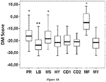
- DM Score is significantly higher in the proliferation, t(4;14) and MAF subgroups ( P ⁇ .001) associated with a poor prognosis (39) and significantly lower in the low bone disease subgroup ( P ⁇ .001) (39) ( Figure 1B ).
- DM Score, UAMS-HRS, IFM-score and GPI had prognostic value as well as t(4;14), del17p, ⁇ 2m, albumin and ISS using the HM patient cohort (Supplementary Table S6).
- DM Score tested with ⁇ 2m remained significant.
- DM Score, ⁇ 2m and t(4;14) kept prognostic value.
- the DM Score is also prognostic in an independent cohort of 345 patients from UAMS treated with TT2 therapy (UAMS-TT2 cohort). For each patient of UAMS-TT2 cohort, DM Score was computed using parameters defined with HM patients' cohort.
- DM Score is predictive for sensitivity of human myeloma cell lines or patients' primary MMCs to Decitabine in vitro.
- DM Score could predict for the sensitivity of 10 HMCLs to DNMT inhibitor.
- 10 HMCLs with the highest or lowest DM Score were selected to assay decitabine sensitivity.
- All HMCLs with the lowest DM Score and poorly sensitive to decitabine presented no ras mutations whereas 4 out of 5 HMCLs with the highest DM Score and higher decitabine sensitivity have ras mutations (Table 1).
- DM Score gene expression-based DNA methylation score
- RECQ1 ATP-dependent DNA helicase Q1
- KIF21B kinesin family member 21B
- RECQ helicase are a ubiquitous family of DNA unwinding enzymes involved in the maintenance of chromosome stability (40-42). Mutations in the genes of RECQ family members are linked with genetic disorders associated with genomic instability, cancer predisposition and features of premature ageing.
- RECQ proteins Consistent with their ability to unwind DNA, several functions have been attributed to RECQ proteins, including roles in stabilization and repair of damaged DNA replication forks, telomere maintenance, homologous recombination, and DNA damage checkpoint signaling (40-42). Recent reports supported a cancer specific role for RECQ1 (43-45). RECQ1 silencing in cancer cells resulted in mitotic catastrophe and administration of siRNA targeting RECQ1 prevented tumor growth in murine models (43-45).
- RECQ1 is highly expressed in various types of solid tumors including colon carcinoma, thyroid cancer, lung cancer and brain glioblastoma tissues (46)
- RNAi glioblastoma cell lines
- KIF21B is a kinesin family member. Kinesins are a conserved class of microtubule-dependent molecular motor proteins that have adenosine triphosphatase activity and motion characteristics (47).
- Kinesins support several cellular functions, such as mitosis, meiosis and the transport of macromolecules.
- mitosis of eukaryotic cells kinesins participate in spindle formation, chromosome congression and alignment, and cytokinesis (48).
- Abnormal expression and function of kinesins are involved in the development or progression of several kinds of human cancers (49,50).
- KIF21B maps to chromosome 1q arm (1q32.1) whose amplification characterizes a significant fraction of high-risk MM tumors (51).
- KIF21B gene was found in a critical neighbor-gene model associated with a poor prognosis across independent data sets of respectively, 559, 247 and 264 myeloma patients (52).
- decitabine treatment could synergize with DNA-damaging agents, in MM, targeting genes involved in DNA-repair and maintenance of chromosome stability.
- Table 1 Characteristics of HMCLs 5-aza sensitive and HMCLs 5-aza-resistant HMCL Name IL-6 dependence 1 Origin 2 Disease 3 Patient sample 4 Gender Isotype t(14q32 or 22q11) Target genes Ras TP53 CD45 HMCL classification 5-aza Resistant HMCLs XG6 ++ MN MM PB F Gl t(16;22) c-Maf wt wt + CTA/MF XG20 ++ MN PCL PB M l t(4;14) MMSET wt abn - MS XG13 ++ MN PCL PB M Gl t(14;16) c-Maf wt abn + MF SKMM2 - CO PCL PB M Gk t(11;14) CCND1 wt abn - CD-1 LP1 - CO MM PB F Gl t(4;14) MMSET / FGFR3 wt abn
- Table 2 Mononuclear cells from tumor samples of 12 patients with MM were cultured for 4 days in the presence of IL-6 (2 ng/mL) with or without increased doses of decitabine. At day 4 of culture, the cell count and viability were determined and the percentage of CD138 + viable plasma cells was determined by flow cytometry. Myeloma cell number/culture well Patient no.
- HMCLs Five HMCLs were cultured with or without 0.5 ⁇ M decitabine for 7 days and gene expression was profiled with Affymetrix U133 plus 2.0. Genes significantly differentially expressed between control and decitabine treated cells were identified using SAM supervised paired analysis with a 5% false discovery rate. When a gene was interrogated by several probe sets, we used the probe set yielding to a maximum variance across control and decitabine treated cells.
- Probeset Gene Ratio Banding Affymetrix description Intercellular communication and membrane proteins 209122_at ADFP 3.70 9p22.1 adipose differentiation-related protein 211990_at HLA-DPA1 1.70 6p21.3 major histocompatibility complex; class II; DP alpha 1 205718_at ITGB7 2.11 12q13.13 integrin; beta 7 1569003_at TMEM49 2.33 17q23.2 transmembrane protein 49 205483_s_at G1P2 2.77 1p36.33 interferon; alpha-inducible protein (clone IFI-15K) 200696_s_at GSN 3.97 9q33 gelsolin (amyloidosis; Finnish type) Signal transduction 203964_at NMI 1.86 2p24.3-q21.3 N-myc (and STAT) interactor 205552_s_at OAS1 1.65 12q24.1 2prime;5prime-oligoadenylate synthetase 1; 40
- HMCLs Five HMCLs were cultured with or without 0.5 ⁇ M decitabine for 7 days and gene expression was profiled with Affymetrix U133 plus 2.0. Genes significantly differentially expressed between control and decitabine treated cells were identified using SAM supervised paired analysis with a 5% false discovery rate. When a gene was interrogated by several probe sets, we used the probe set yielding to a maximum variance across control and decitabine treated cells.
- Probeset Gene Ratio Banding Affymetrix description Intercellular communication and membrane proteins 202304_at FNDC3A 0.40 13q14.2 fibronectin type III domain containing 3A 209541_at IGF1 0.43 12q22-q23 insulin-like growth factor 1 (somatomedin C) 221658_s_at IL21R 0.50 16p11 inter leukin 21 receptor 210587_at INHBE 0.55 12q13.3 inhibin; beta E 219702_at PLAC1 0.42 Xq26 placenta-specific 1 209606_at PSCDBP 0.48 2q11.2 pleckstrin homology; Sec7 and coiled-coil domains; binding protein 202375_at SEC24D 0.31 4q26 SEC24 related gene family; member D (S.
- RNA molecules 202061_s_at SEL1L 0.56 14q24.3-q31 sel-1 suppressor of lin-12-like (C. elegans) 217790_s_at SSR3 0.40 3q25.31 signal sequence receptor; gamma (translocon-associated protein gamma) 202558_s_at STCH 0.47 21q11.1
- the prognostic factors were tested as single variable or multi variables using Cox-model. P-values and the hazard ratios (HR) are shown.
- NS Not significant at a 5% threshold
- GPI gene expression based proliferation index
- ISS International Staging System
- HRS high-risk score
- IFM Intergroupe Francophone du Myélome
- NA Not available.
- PAM Prediction Analysis of Microarray
- Table B Probesets Name 202061_s_at SEL1L 202304_at FNDC3A 202558_s_at STCH 203397_s_at GALNT3 204079_at TPST2 205483_s_at G1P2 205552_s_at OAS1 205718_at ITGB7 208725_at EIF2S2 209083_at CORO1A 209118_s_at TUBA3 209122_at ADFP 209417_s_at IFI35 211990_at HLA-DPA1 214152_at CCPG1 214179_s_at NFE2L1 219118_at FKBP11 221658_s_at IL21R 222690_s_at TMEM39A 223129_x_at MYLIP 224451_x_at ARHGAP9 224763_at RPL37 227755_at --- 228831_s_at GNG7 232488_at MGC15875
- EXAMPLE 3 DNA methylation score is predictive of myeloma cell sensitivity to 5-azacitidine.
- Epigenetics is characterized by a wide range of changes that are reversible and orchestrate gene expression. Recent studies of the epigenome have shown that epigenetic modifications play a role in cancer physiopathology including hematologic malignancies (Issa 2007, Issa, et al 2004, Oki, et al 2008, Smith, et al 2009). In MM, DNA hypomethylation was reported as the predominant early change during myelomagenesis that is gradually transformed to DNA hypermethylation in relapsed cases and during the progression of the disease (Heuck, et al 2013, Walker, et al 2010).
- DM Score DNA methylation score
- HMCLs Human Myeloma Cell Lines
- HMCLs were cultured with graded 5-azacitidine (Sigma, St Louis, MO) concentrations. HMCLs cell growth was quantified with a Cell Titer Glo Luminescent Assay (Promega, Madison, WI) and half inhibitory concentration (IC50) was determined using GraphPad Prism (http://www.graphpad.com/scientific-software/prism/).
- GSEA Gene set enrichment analysis
- GSEA Gene set enrichment analysis
- DM Score The efficacy of DM Score to predict MMCs sensitivity to 5-azacitidine was investigated on 12 HMCLs with high or low DM Score.
- DM score could also predict for the ability of 5-azacytidine to kill patients' primary MMCs cultured together with their BM environment.
- the DM Score which was built using 47 genes whose expression is deregulated by decitabine in HMCLs and which have prognostic value for patients overall survival, can predict for the sensitivity of MM cell lines and primary MMCs to the two-clinical grade inhibitors of DNMT. These 47 genes include 22 genes associated with a bad prognosis and 25 with good one.
- GSEA analysis MMCs of patients with a high DM Score show a significant enrichment in genes associated with proliferation (gene sets: REACTOME CELL CYCLE, CELL CYCLE G2 M and PLASMA CELLS VS PLASMABLAST DN, P ⁇ 0.001, and supplementary Tables S7, S8 and S9).
- MMCs of patients with a low DM Score show a significant enrichment in genes coding for solute carrier group of membrane transport proteins (gene sets: REACTOME TRANSPORT OF INORGANIC CATIONS ANIONS AND AMINO ACIDS OLIGOPEPTIDES, P ⁇ 0.001, and supplementary Tables S10).
- the DM Score allows identification of MM patients who could benefit from treatment with two clinical grade DNMT inhibitors and the development of personalized treatment.
- TABLE S7 Genes set enrichment analysis revealed a significant overrepresentation of the REACTOME CELL CYCLEset in high DM Score patients compared to low DM Score patients ( P ⁇ .001).
Landscapes
- Health & Medical Sciences (AREA)
- Life Sciences & Earth Sciences (AREA)
- Chemical & Material Sciences (AREA)
- General Health & Medical Sciences (AREA)
- Engineering & Computer Science (AREA)
- Genetics & Genomics (AREA)
- Molecular Biology (AREA)
- Physics & Mathematics (AREA)
- Public Health (AREA)
- Medical Informatics (AREA)
- Epidemiology (AREA)
- Proteomics, Peptides & Aminoacids (AREA)
- Organic Chemistry (AREA)
- Biotechnology (AREA)
- Bioinformatics & Cheminformatics (AREA)
- Biophysics (AREA)
- Zoology (AREA)
- Analytical Chemistry (AREA)
- Immunology (AREA)
- Wood Science & Technology (AREA)
- Pathology (AREA)
- Pharmacology & Pharmacy (AREA)
- Veterinary Medicine (AREA)
- Animal Behavior & Ethology (AREA)
- Medicinal Chemistry (AREA)
- Bioinformatics & Computational Biology (AREA)
- Evolutionary Biology (AREA)
- Spectroscopy & Molecular Physics (AREA)
- Theoretical Computer Science (AREA)
- Primary Health Care (AREA)
- Microbiology (AREA)
- Biochemistry (AREA)
- Oncology (AREA)
- General Engineering & Computer Science (AREA)
- Hospice & Palliative Care (AREA)
- Urology & Nephrology (AREA)
- Surgery (AREA)
- Nuclear Medicine, Radiotherapy & Molecular Imaging (AREA)
- Measuring Or Testing Involving Enzymes Or Micro-Organisms (AREA)
Description
- The present invention relates to methods for predicting multiple myeloma treatment response.
- Malignant transformation requires oncogenic activation and inactivation of tumor suppressor genes, which help cancer cells overriding the normal mechanisms controlling cellular survival and proliferation (1,2). These molecular events are caused by genetic alterations (translocations, amplification, mutations) and also by epigenetic modifications (3). Epigenetic modifications include methylation of DNA cytosine residues and histone modifications and have been shown to be critical in the initiation and progression of cancers (4). DNA methyltransferase inhibitors or HDAC inhibitors are now being used in the treatment of several hematologic malignancies including MM (5-8).
- Multiple myeloma (MM) is a plasma cell neoplasm characterized by the accumulation of malignant plasma cells, termed Multiple Myeloma Cells (MMCs) within the bone marrow (BM). Despite the recent introduction of therapies such as Lenalidomide and Bortezomib, MM remains an almost incurable disease. MM arises through the accumulation of multiple genetic changes that include an aberrant or overexpression of a D-type cyclin gene, cyclin D1 (CCND1) in the case of t(11;14) translocation or gain in 11q13, cyclin D3 (CCND3) in the case of the rare t(6;14) translocation, or cyclin D2 (CCND2) on the background of a translocation involving c-maf (t(14;16)) or MMSET/FGFR3 (t(4;14)) (9,10).
- Recent studies have shown that epigenetic changes such as DNA methylation play a role by silencing various cancer-related genes in MM. Most of these studies have been performed on limited number of genes using methylation specific PCR (11-18). Among the genes identified with promoter hypermethylation in MM, cyclin-dependent kinase inhibitor 2A (CDKN2A) and transforming growth factor beta receptor 2 (TGFBR2) have been shown to be associated with a poor prognosis in MM patients with discrepant results for CDKN2A (12). Heller at al. have identified several epigenetic inactivated cancer related genes in 3 human myeloma cell lines (HMCLs) and validated the relevance of 10 of these genes in 6 additional HMCLs, premalignant PCs from 24 MGUS patients and MMCs from 111 patients with MM (19). Methylation of secreted protein acidic and rich in cysteine (SPARC) and Bcl2/adenovirus E1B 19kDa interacting protein 3 (BNIP3) promoters was associated with poor overall survival of MM patients (19). SOCS3 methylation was found to be associated with extramedullary manifestations, plasma cell leukemia and significant shortened survival in MM patients (20). More recently, Morgan et al have shown that the transition of normal plasma cell (PC) and MGUS stage to MM stage is associated with DNA hypomethylation, but the transition of intramedullary MM stage to plasma cell leukemia or HMCL stage is associated with DNA hypermethylation (21). They described 2 specific subgroups of hyperdiploid MM on the basis of their methylation profile, which had a significantly different overall survival (21).
- DNMT inhibitors can be sub-divided into nucleoside analogue and non-nucleoside analogue families. 5-Azacytidine(azacytidine)/5-Aza-2'-deoxycytidine (decitabine) are both nucleoside analogues with Food and Drug Administration approval for use in myelodysplastic syndrome. Clinical trials in myeloma combining these demethylating agents with chemotherapy or other agents are underway (8,72).
- An important objective for optimizing these clinical trials will be the identification of biomarkers predictive for sensitivity of MMCs to DNMTi. Expression marker signatures to classify and characterize Multiple Myeloma have already been described (22,71). In the present invention, the inventors used gene expression profiling of Multiple Myeloma Cells (MMCs) to build a novel "DNA methylation gene expression score" that makes it possible identification of patients whose MMCs will be targeted by DNMT inhibition.
- The present invention relates to a method of testing whether a patient suffering of multiple myeloma will respond or not to a DNA methyltransferase inhibitor (DNMTi).
- The multiple myeloma treatment response was investigated by the inventors using DNA methyltransferase inhibitors (DNMTi), human multiple myeloma cell lines and primary myeloma cells. The inventors analyzed gene expression profiles of 5 MM cell lines treated with decitabine using microarrays. The inventors identified 127 genes deregulated by decitabine. 47 out of 127 decitabine deregulated genes have prognostic value in inventor's cohort of 206 newly-diagnosed MM patients. The inventors summarized the prognostic information in a DNA methylation score (DM Score or DMS) built using the probe set signal value weighted by the beta coefficient of prognostic genes. The DM Score is predictive for DNMT inhibitor sensitivity of HMCLs and primary myeloma cells in vitro. The DM Score allows identification of myeloma patients that could benefit DNMT inhibitor treatment.
- The inventors also demonstrated that the DM Score is also predictive of myeloma cells sensitivity for another DNMTi, 5-Azacytidine. HMCLs with a high DM Score exhibited significant 3-fold higher 5-Azacytidine sensitivity than HMCLs with a low DM Score and these results were validated in vitro with primary myeloma cells of patients.
- The term "patient" denotes a mammal. In a preferred embodiment of the invention, a patient refers to any patient (preferably human) afflicted with multiple myeloma. The term "multiple myeloma" refers to multiple myeloma such as revised in the World Health Organisation Classification C90.
- The term "DNA methyltransferase inhibitors" or "DNMTi" has its general meaning in the art and refers to a multiple myeloma treatment. The term "DNA methyltransferase inhibitors" or "DNMTi" refers to DNA methyltransferase inhibitor that can be sub-divided into nucleoside analogue (5-Azacytidine (azacytidine), 5-Aza-2'-deoxycytidine (decitabine, 5-Aza-CdR), zebularine, 5-Fluoro-2'-deoxycytidine (5-F-CdR), 5 ,6-Dihydro-5-azacytidine (DHAC)) and non-nucleoside analogue families (Hydralazine, Procainamide, Procaine, EGCG ((-)-epigallocatechin-3-gallate), Psammaplin A, MG98, RG108) (8).
- The term "biological sample" refers to multiple myeloma cells, bone marrow or medullary cell.
- All the genes pertaining to the invention are known per se, and are listed in the below Table A.
Table A: Set of predictive genes. Gene Gene Symbol Gene name Gene ID β coefficient Reference level (ELRi) G1 COP1 Caspase-1 dominant-negative inhibitor pseudo-ICE 1552701_a_at 0,669962539 25.72815534 G2 DNAJB9 DnaJ (Hsp40) homolog; subfamily B; member 9 1554462_a_at -0,807267505 87.37864078 G3 INSIG1 insulin induced gene 1 201625_s_at 0,918185816 10.19417476 G4 SEL1L sel-1 suppressor of lin-12-like (C. elegans) 202061_s_at -0,683515307 86.40776699 G5 FNDC3A fibronectin type III domain containing 3A 202304_at -0,821182841 76.21359223 G6 IFI27 interferon, alpha-inducible protein 27 202411_at 0,87241554 60.19417476 G7 STCH stress 70 protein chaperone; microsome-associated; 60kDa 202558_s_at -0,924845543 27.66990291 G8 GALNT3 UDP-N-acetyl-alpha-D-galactosamine:polypeptide N-acetylgalactosaminyltransf erase 3 (GalNAc-T3) 203397_s_at 0,838104028 30.58252427 G9 TPST2 tyrosylprotein sulfotransferase 2 204079_at 0,698525183 14.5631068 G10 KIF21B kinesin family member 21B 204411_at 1,057242019 26.21359223 G11 G1P2 Interferon; alpha-inducible protein (clone IFI-15K) 205483_s_at 0,704117799 40.77669903 G12 OAS1 2',5'-oligoadenylate synthetase 1 205552_s_at 0,918506668 12.13592233 G13 ITGB7 integrin, beta 7205718_at 1,393658338 11.16504854 G14 SP110 SP110 nuclear body protein 208012_x_at 0,796781189 13.10679612 G15 EIF2S2 eukaryotic translation initiation factor 2,subunit 2 beta208725_at -0,608613084 75.24271845 G16 CORO1A coronin, actin binding protein, 1A 209083_at 1,116949242 50.48543689 G17 TUBA3 tubulin, alpha 1a 209118_s_at 0,794556227 15.04854369 G18 ADFP Adipose differentiation-related protein 209122_at -0,742527707 73.30097087 G19 IFI35 interferon-induced protein 35 209417_s_at 0,608446212 29.12621359 G20 STAT1 signal transducer and activator of transcription 1 209969_s_at 0,865200608 20.87378641 G21 HIST1H2B G histone cluster 1, H2bc 210387_at -0,713207906 78.15533981 G22 HLA-DPA1 major histocompatibility complex, class II, DP alpha 1 211990_at -0,708484927 79.12621359 G23 RECQL RecQ protein-like (DNA helicase Q1-like) 213878_at 0,79702138 19.90291262 G24 CCPG1 cell cycle progression 1 214152_at -0,686867998 48.05825243 G25 NFE2L1 nuclear factor (erythroid-derived 2)-like 1 214179_s_at 0,694584139 12.13592233 G26 PARP12 poly (ADP-ribose) polymerase family, member 12 218543_s_at 1,257650329 10.19417476 G27 FKBP 11 FK506 binding protein 11; 19 kDa 219118_at -0,88381802 76.69902913 G28 EAF2 ELL associated factor 2219551_at -0,762323817 83.98058252 G29 C6orf48 chromosome 6 open reading frame 48 220755_s_at -0,832289767 23.78640777 G30 IL21R interleukin 21 receptor 221658_s_at 0,612993436 24.75728155 G31 PGM3 Phosphoglucomutase 3 221788_at -1,227167702 87.86407767 G32 EIF2C2 Eukaryotic translation initiation factor 2C; 2 222294_s_at -0,657954783 69.90291262 G33 TMEM39A transmembrane protein 39A 222690_s_at 0,787711031 64.5631068 G34 SLAMF7 SLAM family member 7222838_at -0,601257629 64.0776699 G35 MYLIP myosin regulatory light chain interacting protein 223129_x_at 0,687458269 22.33009709 G36 PPAPDC1B phosphatidic acid phosphatase type 2 domain containing 1B223569_at -0,860377506 83.49514563 G37 ARHGAP9 Rho GTPase activating protein 9 /// Rho GTPase activating protein 9 224451_x_at -0,731399299 83.98058252 G38 RPL37 ribosomal protein L37 224763_at -1,135732424 88.34951456 G39 GSK3B Glycogen synthase kinase 3 beta 226183_at -0,772104529 83.98058252 G40 ELL2 elongation factor; RNA polymerase II; 2 226982_at -0,687762574 80.09708738 G41 EST, GenBank AA042983 Expression sequence tag, GenBank AA042983 227755_at -0,79716878 68.93203883 G42 SAMD9 sterile alpha motif domain containing 9 228531_at 0,813378182 46.60194175 G43 GNG7 guanine nucleotide binding protein (G protein); gamma 7228831_s_at -0,641328827 80.09708738 G44 SEC63 SEC63 Homolog (S. cerevisiae) 229969_at -1,216057852 85.9223301 G45 EST, GenBank AI702465 Expression sequence tag, GenBank AI702465 230570_at -0,659929394 82.52427184 G46 MGC15875 hypothetical protein MGC15875 232488_at -1,036621429 80.09708738 G47 SFN stratifin 33322_i_at 0,721955343 12.62135922 - The present invention relates to a method of testing whether a patient suffering of multiple myeloma will respond or not to a DNA methyltransferase inhibitor (DNMTi) comprising:
- i) determining the expression level (ELi) of several genes G1-Gn selected from table A in a biological sample obtained from said patient
- ii) comparing the expression level (ELi) determined at step i) with a predetermined reference level (ELRi)
- iii) calculating the DMS score trough the following formula

- iv) comparing the score DMS determined at step iii) with a predetermined reference value DMSR
- v) and concluding that the patient will respond to the DNMTi when the DMS score is higher than the predetermined reference value DMSR or concluding that the patient will not respond to the DNMTi when the DMS score is lower than the predetermined reference value DMSR
- In some aspects, the levels of at least 25 genes from Table A are determined wherein said genes are:
- 202061_s_at SEL1L
- 202304_at FNDC3A
- 202558_s_at STCH
- 203397_s_at GALNT3
- 204079_at TPST2
- 205483_s_at G1P2
- 205552_s_at OAS1
- 205718_at ITGB7
- 208725_at EIF2S2
- 209083_at CORO1A
- 209118_s_at TUBA3
- 209122_at ADFP
- 209417_s_at IFI35
- 211990_at HLA-DPA1
- 214152_at CCPG1
- 214179_s_at NFE2L1
- 219118_at FKBP11
- 221658_s_at IL21R
- 222690_s_at TMEM39A
- 223129_x_at MYLIP
- 224451_x_at ARHGAP9
- 224763_at RPL37
- 227755_at EST, GenBank AA042983
- 228831_s_at GNG7
- 232488_at MGC15875
- In some aspects, the level of 25, 26, 27, 28, 29, 30, 31, 32, 33, 34, 35, 36, 37, 38, 39, 40, 41, 42, 43, 44, 45, 46, or 47 genes from Table A are determined wherein every combinations of genes comprises a minimal set of 25 genes consisting of:
- 202061_s_at SEL1L
- 202304_at FNDC3A
- 202558_s_at STCH
- 203397_s_at GALNT3
- 204079_at TPST2
- 205483_s_at G1P2
- 205552_s_at OAS1
- 205718_at ITGB7
- 208725_at EIF2S2
- 209083_at CORO1A
- 209118_s_at TUBA3
- 209122_at ADFP
- 209417_s_at IFI35
- 211990_at HLA-DPA1
- 214152_at CCPG1
- 214179_s_at NFE2L1
- 219118_at FKBP11
- 221658_s_at IL21R
- 222690_s_at TMEM39A
- 223129_x _at MYLIP
- 224451_x_at ARHGAP9
- 224763_at RPL37
- 227755_at EST, GenBank AA042983
- 228831_s_at GNG7
- 232488_at MGC15875
- In some embodiments, the level of the 47 genes of Table A are determined.
- Determination of the expression level of the genes can be performed by a variety of techniques. Generally, the expression level as determined is a relative expression level. More preferably, the determination comprises contacting the biological sample with selective reagents such as probes, primers or ligands, and thereby detecting the presence, or measuring the amount, of polypeptide or nucleic acids of interest originally in the biological sample. Contacting may be performed in any suitable device, such as a plate, microtiter dish, test tube, well, glass, column, and so forth. In specific aspects, the contacting is performed on a substrate coated with the reagent, such as a nucleic acid array or a specific ligand array. The substrate may be a solid or semi-solid substrate such as any suitable support comprising glass, plastic, nylon, paper, metal, polymers and the like. The substrate may be of various forms and sizes, such as a slide, a membrane, a bead, a column, a gel, etc. The contacting may be made under any condition suitable for a detectable complex, such as a nucleic acid hybrid or an antibody-antigen complex, to be formed between the reagent and the nucleic acids or polypeptides of the biological sample.
- In a preferred aspect, the expression level may be determined by determining the quantity of mRNA.
- Methods for determining the quantity of mRNA are well known in the art. For example the nucleic acid contained in the biological sample is first extracted according to standard methods, for example using lytic enzymes or chemical solutions or extracted by nucleic-acid-binding resins following the manufacturer's instructions. The extracted mRNA is then detected by hybridization (e. g., Northern blot analysis) and/or amplification (e.g., RT-PCR). Preferably quantitative or semi-quantitative RT-PCR is preferred. Real-time quantitative or semi-quantitative RT-PCR is particularly advantageous.
- Other methods of amplification include ligase chain reaction (LCR), transcription-mediated amplification (TMA), strand displacement amplification (SDA) and nucleic acid sequence based amplification (NASBA).
- Nucleic acids having at least 10 nucleotides and exhibiting sequence complementarity or homology to the mRNA of interest herein find utility as hybridization probes or amplification primers. It is understood that such nucleic acids need not be identical, but are typically at least about 80% identical to the homologous region of comparable size, more preferably 85% identical and even more preferably 90-95% identical. In certain aspects, it will be advantageous to use nucleic acids in combination with appropriate means, such as a detectable label, for detecting hybridization. A wide variety of appropriate indicators are known in the art including, fluorescent, radioactive, enzymatic or other ligands (e. g. avidin/biotin) .
- Probes typically comprise single-stranded nucleic acids of between 10 to 1000 nucleotides in length, for instance of between 10 and 800, more preferably of between 15 and 700, typically of between 20 and 500. Primers typically are shorter single-stranded nucleic acids, of between 10 to 25 nucleotides in length, designed to perfectly or almost perfectly match a nucleic acid of interest, to be amplified. The probes and primers are "specific" to the nucleic acids they hybridize to, i.e. they preferably hybridize under high stringency hybridization conditions (corresponding to the highest melting temperature Tm, e.g., 50 % formamide, 5x or 6x SCC. SCC is a 0.15 M NaCl, 0.015 M Na-citrate).
- The nucleic acid primers or probes used in the above amplification and detection method may be assembled as a kit. Such a kit includes consensus primers and molecular probes. A preferred kit also includes the components necessary to determine if amplification has occurred. The kit may also include, for example, PCR buffers and enzymes; positive control sequences, reaction control primers; and instructions for amplifying and detecting the specific sequences.
- In a particular aspect, the methods of the invention comprise the steps of providing total RNAs extracted from a biological samples and subjecting the RNAs to amplification and hybridization to specific probes, more particularly by means of a quantitative or semi-quantitative RT-PCR.
- In another preferred aspect, the expression level is determined by DNA chip analysis. Such DNA chip or nucleic acid microarray consists of different nucleic acid probes that are chemically attached to a substrate, which can be a microchip, a glass slide or a microsphere-sized bead. A microchip may be constituted of polymers, plastics, resins, polysaccharides, silica or silica-based materials, carbon, metals, inorganic glasses, or nitrocellulose. Probes comprise nucleic acids such as cDNAs or oligonucleotides that may be about 10 to about 60 base pairs. To determine the expression level, a biological sample from a test patient, optionally first subjected to a reverse transcription, is labelled and contacted with the microarray in hybridization conditions, leading to the formation of complexes between target nucleic acids that are complementary to probe sequences attached to the microarray surface. The labelled hybridized complexes are then detected and can be quantified or semi-quantified. Labelling may be achieved by various methods, e.g. by using radioactive or fluorescent labelling. Many variants of the microarray hybridization technology are available to the man skilled in the art (see e.g. the review by Hoheisel, Nature Reviews, Genetics, 2006, 7:200-210)
- In this context, the invention further provides a DNA chip comprising a solid support which carries nucleic acids that are specific to the genes listed in table A.
- Predetermined reference values ELRi or DMSR used for comparison may consist of "cut-off" values.
- For example; each reference ("cut-off') value ELRi for each gene may be determined by carrying out a method comprising the steps of:
- a) providing a collection of samples from patients suffering of multiple myeloma;
- b) determining the expression level of the relevant gene for each sample contained in the collection provided at step a);
- c) ranking the samples according to said expression level
- d) classifying said samples in pairs of subsets of increasing, respectively decreasing, number of members ranked according to their expression level,
- e) providing, for each sample provided at step a), information relating to the actual clinical outcome for the corresponding cancer patient (i.e. the duration of the disease-free survival (DFS) or the overall survival (OS) or both);
- f) for each pair of subsets of tumour tissue samples, obtaining a Kaplan Meier percentage of survival curve;
- g) for each pair of subsets of tumour tissue samples calculating the statistical significance (p value) between both subsets
- h) selecting as reference value ELR for the expression level, the value of expression level for which the p value is the smallest.
- For example the expression level of a gene Gi has been assessed for 100 samples of 100 patients. The 100 samples are ranked according to the expression level of gene Gi. Sample 1 has the highest expression level and
sample 100 has the lowest expression level. A first grouping provides two subsets: on one side sample Nr 1 and on the other side the 99 other samples. The next grouping provides on oneside samples 1 and 2 and on the other side the 98 remaining samples etc., until the last grouping: on one side samples 1 to 99 and on the otherside sample Nr 100. According to the information relating to the actual clinical outcome for the corresponding cancer patient, Kaplan Meier curves are prepared for each of the 99 groups of two subsets. Also for each of the 99 groups, the p value between both subsets was calculated. The reference value ELRi is then selected such as the discrimination based on the criterion of the minimum p value is the strongest. In other terms, the expression level corresponding to the boundary between both subsets for which the p value is minimum is considered as the reference value. It should be noted that according to the experiments made by the inventors, the reference value ELRi is not necessarily the median value of expression levels. - The man skilled in the art also understands that the same technique of assessment of the DMSR could be used for obtaining the reference value and thereafter for assessment of the response to DNMTi. However in one embodiment, the reference value DMSR is the median value of DMS.
- In one aspect, the reference value ELRi for the genes are described in table A (right column).
- In one aspect, the reference value DMSR is -19.7 for determining whether a patient suffering of multiple myeloma will respond to a DNMTi or -15.3 for predicting the survival time of patient suffering of multiple myeloma.
- The regression β coefficient reference values may be easily determined by the skilled man in the art for each gene using a Cox model. The Cox model is based on a modeling approach to the analysis of survival data. The purpose of the model is to simultaneously explore the effects of several variables on survival. The Cox model is a well-recognised statistical technique for analysing survival data. When it is used to analyse the survival of patients in a clinical trial, the model allows us to isolate the effects of treatment from the effects of other variables. The logrank test cannot be used to explore (and adjust for) the effects of several variables, such as age and disease duration, known to affect survival. Adjustment for variables that are known to affect survival may improve the precision with which we can estimate the treatment effect. The regression method introduced by Cox is used to investigate several variables at a time. It is also known as proportional hazards regression analysis. Briefly, the procedure models or regresses the survival times (or more specifically, the so-called hazard function) on the explanatory variables. The hazard function is the probability that an individual will experience an event (for example, death) within a small time interval, given that the individual has survived up to the beginning of the interval. It can therefore be interpreted as the risk of dying at time t. The quantity h0 (t) is the baseline or underlying hazard function and corresponds to the probability of dying (or reaching an event) when all the explanatory variables are zero. The baseline hazard function is analogous to the intercept in ordinary regression (since exp0= 1). The regression coefficient β gives the proportional change that can be expected in the hazard, related to changes in the explanatory variables. The coefficient β is estimated by a statistical method called maximum likelihood. In survival analysis, the hazard ratio (HR) (Hazard Ratio = exp(β)) is the ratio of the hazard rates corresponding to the conditions described by two sets of explanatory variables. For example, in a drug study, the treated population may die at twice the rate per unit time as the control population. The hazard ratio would be 2, indicating higher hazard of death from the treatment.
- In one aspect, the regression β coefficient reference values are described in table A.
- Also disclosed is a kit for performing the methods as above described, wherein said kit comprises means for measuring the expression level of the genes listed in Table A. Typically the kit may include a primer, a set of primers, a probe, a set of probes as above described. In a particular embodiment, the probe or set of probes are labelled as above described. The kit may also contain other suitably packaged reagents and materials needed for the particular detection protocol, including solid-phase matrices, if applicable, and standards.
- In a particular aspect, the score may be generated by a computer program.
- The method of the invention allows to define a subgroup of patients who will be responsive ("responder") or not ("non responder") to the treatment with a DNA methyltransferase inhibitor.
- Also disclosed is a method for the treatment of multiple myeloma in a patient in need thereof.
- In the context of the disclosure, the term "treating" or "treatment", as used herein, means reversing, alleviating, inhibiting the progress of, or preventing the disorder or condition to which such term applies, or one or more symptoms of such disorder or condition.
- In a particular aspect, the method comprises the following steps
- a) testing whether the patient will respond or not to a DNA methyltransferase inhibitor (DNMTi) by performing the method according to the invention
- b) administering the DNA methyltransferase inhibitor, if said patient has as score higher than the reference value DMSR (i.e. the patient will respond to the DNA methyltransferase inhibitor).
- A further object of the invention relates to a DNA methyltransferase inhibitor for use in the treatment of multiple myeloma in a patient in need thereof, wherein the patient was being classified as responder by the method as above described.
- The invention will be further illustrated by the following figures and examples. However, these examples and figures should not be interpreted in any way as limiting the scope of the present invention.
-
-
Figure 1 : DNA methylation Score in normal and malignant plasma cells
(A) DNA methylation Score in normal bone marrow plasma cells (N = 7), in premalignant plasma cells of patients with monoclonal gammopathy of undetermined significance (MGUS, N = 5), in multiple myeloma cells of patients with intramedullary MM (N = 206) and in human myeloma cell lines (N = 40). ** Indicate that the score value is significantly different with a P value < .01. (B) DNA methylation Score in the 8 groups of the molecular classification of multiple myeloma. The DM Score was investigated in the 8 groups of the molecular classification of multiple myeloma in UAMS-TT2 cohort of patients. PR: proliferation, LB: low bone disease, MS: MMSET, HY: hyperdiploid, CD1: Cyclin D1, CD2: Cyclin D2, MF: MAF, MY: myeloid. * Indicate that the score value is significantly higher in the group compared to all the patients of the cohort (P < .05). ** Indicate that the score value is significantly lower in the group compared to all the patients of the cohort (P < .05). -
Figure 2 : Prognostic value of DM Score in multiple myeloma.
Patients of HM cohort were ranked according to increased DM Score and a maximum difference in OS was obtained with DM Score = -15.3 splitting patients in a high risk (34.5%) and low risk (65.5%) groups. The prognostic value of DM Score was tested on an independent cohort of 345 patients from UAMS treated with TT2 therapy (UAMS- TT2 cohort). The parameters to compute the DM Score of patients of UAMS-TT2 cohort and the DM Score cut-off delineating the 2 prognostic groups were those defined with HM cohort. -
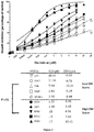
Figure 3 : DM Score predicts for sensitivity of human myeloma cell lines to decitabine.
(A) HMCLs with high DM Score (N = 5) exhibit significant higher DNMTi sensitivity compared to HMCLs with low DM Score (N = 5). HMCLs were cultured for 4 days in 96-well flat-bottom microtiter plates in RPMI 1640 medium, 10% FCS, 2 ng/ml IL-6 culture medium (control), with graded decitabine concentrations. Data are mean values plus or minus standard deviation (SD) of 5 experiments determined on sextuplet culture wells. -
Figure 4 : DM Score predicts for decitabine sensitivity of primary myeloma cells of patients.
Mononuclear cells from tumor samples of 12 patients with MM were cultured for 4 days in the presence of IL-6 (2 ng/ml) with or without graded decitabine concentrations. Atday 4 of culture, the cell count and the viability were determined and the percentage of CD138+ viable plasma cells was determined by flow cytometry. Black color represents patients with high DM Score (N = 6) and white represents patients with low DM Score values (N = 6). -
Figure 5 : DM Score predicts for sensitivity of human myeloma cell lines to 5-azacitidine.
HMCLs with a high DM Score (n = 6) exhibit significant higher 5-azacitidine sensitivity compared to HMCLs with a low DM Score (n = 6). HMCLs were cultured for 4 days in 96-well flat-bottom microtiter plates in RPMI 1640 medium, 10% FCS, 2 ng/ml IL-6 culture medium (control), and graded 5-azacitidine concentrations. Data are mean values plus or minus standard deviation (SD) of 5 experiments determined on sextuplet culture wells. -
Figure 6 : DM Score predicts for sensitivity of primary myeloma cells of patients to 5-azacitidine.
Mononuclear cells from tumor samples of 14 patients with MM were cultured for 4 days in the presence of IL-6 (2 ng/ml) with or without graded 5-azacitidine concentrations. Atday 4 of culture, the count of viable CD138+ MMCs was determined using flow cytometry. The black columns represent the mean ± SD of primary myeloma cell counts (expressed as the percentage of the count without adding 5-azcytidine) of the 7 patients with a low DM Score and the white columns that of the 7 patients with a high DM Score. - XG-1, XG-2, XG-3, XG-4, XG-5, XG-6, XG-7, XG-10, XG-11, XG-12, XG-13, XG-14, XG-16, XG-19, XG-20 and XG-21 human myeloma cell lines were obtained as previously described (22-26). JJN3 was kindly provided by Dr Van Riet (Bruxelles, Belgium), JIM3 by Dr MacLennan (Birmingham, UK) and MM1S by Dr S. Rosen (Chicago, USA). AMO-1, LP1, L363, U266, OPM2, and SKMM2 were from DSMZ (Germany) and RPMI8226 from ATTC (USA). All HMCLs derived in inventor's laboratory were cultured in the presence of recombinant IL-6. HMCLs microarray data have been deposited in the ArrayExpress public database under accession numbers E-TABM-937 and E-TABM-1088.
- MMCs were purified from 206 patients with newly-diagnosed MM after written informed consent was given at the University hospitals of Heidelberg (Germany) or Montpellier (France) and agreement of the Center for Biological Resources of Montpellier University Hospital (N°DC-2008-417). The study was approved by the ethics boards of Heidelberg and Montpellier Universities. These 206 patients were treated with high dose Melphalan (HDM) and autologous stem cell transplantation (ASCT) (27) and were termed in the following Heidelberg-Montpellier (HM) series (Supplementary Table S1). The CEL files and MAS5 files have been deposited in the ArrayExpress public database (E-MTAB-372). The structural chromosomal aberrations including t(4;14)(p16.3;q32.3) and t(11;14)(q13;q32.3), as well as numerical aberrations including 17p13 and 1q21 gain, were assayed by fluorescence in situ hybridization (iFISH) (28). The inventors also used Affymetrix data of a cohort of 345 purified MMC from previously untreated patients from the University of Arkansas for Medical Sciences (UAMS, Little Rock, AR). The patients were treated with
total therapy 2 including HDM and ASCT (29) and termed in the following UAMS-TT2 series. These data are publicly available via the online Gene Expression Omnibus (Gene Expression Profile of Multiple Myeloma, accession number GSE2658. http://www.ncbi.nlm.nih.gov/geo/). As iFISH data were not available for UAMS-TT2 patients, t(4;14) translocation was evaluated using MMSET spike expression (30) and del17p13 surrogated by TP53 probe set signal (31). After Ficoll-density gradient centrifugation, plasma cells were purified using anti-CD138 MACS microbeads (Miltenyi Biotech, Bergisch Gladbach, Germany). - The human MM cell lines XG-5, XG-6, XG-7, XG-20 and LP1 were grown in RPMI 1640 supplemented with 10% fetal bovine serum and 2 ng/ml recombinant IL-6. Cells (2 x 10⁵/mL) were treated either with 0.5 µmol/L 5-Aza-2'-deoxycytidine (decitabine) (Sigma, St Louis, MO) for 7 days. Control cells were cultured in the same conditions without adding drug.
- HMCLs were cultured for 4 days in 96-well flat-bottom microtiter plates in RPMI 1640 medium, 10% FCS, 2 ng/ml IL-6 culture medium (control), with graded decitabine concentrations. Cell growth was evaluated by quantifying intracellular ATP amount with a Cell Titer Glo Luminescent Assay (Promega, Madison, WI) with a Centro LB 960 luminometer (Berthold Technologies, Bad Wildbad, Germany).
- Mononuclear cells from tumor samples of 12 patients with MM (agreement of the Center for Biological Resources of Montpellier University Hospital (N°DC-2008-417)) were cultured for 4 days at 2 x 10⁵ cells/ml in RPMI 1640 medium, 10% FCS, 2 ng/ml IL-6, with or without graded concentrations of decitabine. In each culture group, viability and cell counts were assayed and MMCs were stained with an anti-CD 138-PE mAb (Immunotech, Marseille, France) as previously described (32).
- RNA was extracted using the RNeasy Kit (Qiagen, Hilden, Germany) as previously described (33,34). Biotinylated cRNA was amplified with a double in vitro transcription and hybridized to the human U133 2.0 plus GeneChips, according to the manufacturer's instructions (Affymetrix, Santa Clara, CA). Fluorescence intensities were quantified and analyzed using the GECOS software (Affymetrix).
- Gene expression data were normalized with the MAS5 algorithm and analyzed with inventor's bioinformatics platforms - RAGE (http://rage.montp.inserm.fr/) (35) and Amazonia (http://amazonia.montp.inserm.fr/) (36) - or SAM (Significance Analysis of Microarrays) software (37). The statistical significance of differences in overall survival between groups of patients was calculated by the log-rank test. Multivariate analysis was performed using the Cox proportional hazards model. Survival curves were plotted using the Kaplan-Meier method. All these analyses have been done with R.2.10.1 (http://www.r-project.org/) and bioconductor version 2.5. Gene annotation and networks were generated through the use of Ingenuity Pathways Analysis (Ingenuity® Systems, Redwood City, CA) (38).
- Five HMCLs were treated with 0.5 µM of decitabine for 7 days, a concentration which did not affect myeloma cell viability (Supplementary Table S2) (19). Using SAM supervised paired analysis, the expression of 48 genes was found to be significantly upregulated and that of 79 genes downregulated by decitabine treatment of 5 HMCLs (FDR < 5%; Supplementary Table S3 and S4). Decitabine-regulated genes are significantly enriched in genes related to "Cancer" and "Cell death" pathways (FDR < 5%; Ingenuity pathway analysis. Investigating the expression of these 127 decitabine-regulated genes in primary MMCs of a cohort of 206 newly-diagnosed patients (HM cohort), 22 genes had bad prognostic value and 25 a good one after Benjamini-Hochberg multiple testing correction (Supplementary Table S5). The prognostic information of decitabine regulated genes was gathered within an DNA methylation score (DM Score), which was the sum of the beta coefficients of the Cox model weighted by ± 1 according to the patient MMC signal above or below the probe set maxstat value as previously described (38). The value of DM Score in normal, premalignant or malignant plasma cells is displayed in
Figure 1A . There is no significant difference of DM Score between cells from MGUS patients and normal BMPCs. MMCs of patients had a significantly higher DM Score than normal BMPCs or plasma cells from MGUS-patients (P < .01), and HMCLs the highest score (P < .001) (Figure 1A ). Investigating the DM Score in the 8 groups of the molecular classification of multiple myeloma (39), DM Score is significantly higher in the proliferation, t(4;14) and MAF subgroups (P < .001) associated with a poor prognosis (39) and significantly lower in the low bone disease subgroup (P < .001) (39) (Figure 1B ). - DM Score had prognostic value when used as a continuous variable (P ≤ 10-4), or by splitting patients into two groups using Maxstat R function (38). A maximum difference in overall survival (OS) was obtained with DM Score = -15.3 splitting patients in a high-risk group of 34.5% patients (DM Score > - 15.3) with a 42.1 months median OS and a low risk group of 65.5% patients (DM Score ≤ -15.3) with not reached median survival (
Figure 2 ). - Using univariate Cox analysis, DM Score, UAMS-HRS, IFM-score and GPI had prognostic value as well as t(4;14), del17p, β2m, albumin and ISS using the HM patient cohort (Supplementary Table S6). When compared two by two, DM Score tested with β2m remained significant. When these parameters were tested together, DM Score, β2m and t(4;14) kept prognostic value. The DM Score is also prognostic in an independent cohort of 345 patients from UAMS treated with TT2 therapy (UAMS-TT2 cohort). For each patient of UAMS-TT2 cohort, DM Score was computed using parameters defined with HM patients' cohort. The median OS of patients within high score group (DM Score > - 15.3) was 53.7 months and not reached for patients with low DM Score (P = .0001) (
Figure 2 ). Using Cox univariate analysis, UAMS-HRS, IFM and GPI scores as well as t(4;14) and del17p had prognostic value. Comparing these prognostic factors two by two, DM Score remained significant compared to GPI, t(4;14), and del17p in the UAMS-TT2 cohort (Supplementary Table S5). When these parameters were tested together, UAMS-HRS, t(4;14) and del17p kept prognostic value in UAMS-TT2 cohort. - The inventors sought to determine whether DM Score could predict for the sensitivity of 10 HMCLs to DNMT inhibitor. Starting from a large cohort of 40 HMCLs (22), the 10 HMCLs with the highest or lowest DM Score were selected to assay decitabine sensitivity. The 5 HMCLs with the highest DM Score exhibited a significant 11-fold higher decitabine sensitivity (median IC50 = 0.68 µM; range: 0.15 to 2.22 µM) than the 5 HMCLs with low DM Score (P = .01; median IC50 = 7.94 µM; range: 2.92 to 60.81µM) (
Figure 3 ). All HMCLs with the lowest DM Score and poorly sensitive to decitabine presented no ras mutations whereas 4 out of 5 HMCLs with the highest DM Score and higher decitabine sensitivity have ras mutations (Table 1). - Primary MMCs were cultured with their BM environment and recombinant IL-6 and graded concentrations of decitabine for 4 days. Primary MMCs of patients with a DM Score above median value (-19.7,
Figure 1 ) exhibited significant (P < .01) 2.2-fold higher decitabine sensitivity than MMCs with DM Score below median (Figure 4 and Table 2). The characteristics of patients with MM included in this study are described in Table 3. - In this study, the inventors have identified a gene expression-based DNA methylation score (DM Score) which is predictive for patients' survival and for the in vitro sensitivity of human myeloma cell lines or patients' primary myeloma cells to a DNMT inhibitor, decitabine. Clinical trials in multiple myeloma combining these demethylating agents with chemotherapy or other agents are underway (8). The current identification of DM Score should be very useful to investigate whether the higher response to DNMTi is found in patients with highest DM Score and to speed up the investigation of the clinical efficacy of the novel agents.
- Besides the potential utility of the current DM Score in selecting patients who could benefit from DNMTi therapies, the current study highlights pathways which could be involved in the emergence of multiple myeloma cells. Among the genes downregulated by decitabine treatment and associated with a poor prognosis, the inventors identified RECQ1 (ATP-dependent DNA helicase Q1) and KIF21B (kinesin family member 21B). RECQ helicase are a ubiquitous family of DNA unwinding enzymes involved in the maintenance of chromosome stability (40-42). Mutations in the genes of RECQ family members are linked with genetic disorders associated with genomic instability, cancer predisposition and features of premature ageing. Consistent with their ability to unwind DNA, several functions have been attributed to RECQ proteins, including roles in stabilization and repair of damaged DNA replication forks, telomere maintenance, homologous recombination, and DNA damage checkpoint signaling (40-42). Recent reports supported a cancer specific role for RECQ1 (43-45). RECQ1 silencing in cancer cells resulted in mitotic catastrophe and administration of siRNA targeting RECQ1 prevented tumor growth in murine models (43-45). More recently, it was demonstrated that RECQ1 is highly expressed in various types of solid tumors including colon carcinoma, thyroid cancer, lung cancer and brain glioblastoma tissues (46) In glioblastoma cell lines, depletion of RECQ1 by RNAi results in a significant reduction of cellular proliferation, perturbation of S-phase progression, spontaneous γ-H2AX foci formation and hypersensitivity to hydroxyurea and temozolomide treatments (46). KIF21B is a kinesin family member. Kinesins are a conserved class of microtubule-dependent molecular motor proteins that have adenosine triphosphatase activity and motion characteristics (47). Kinesins support several cellular functions, such as mitosis, meiosis and the transport of macromolecules. In mitosis of eukaryotic cells, kinesins participate in spindle formation, chromosome congression and alignment, and cytokinesis (48). Abnormal expression and function of kinesins are involved in the development or progression of several kinds of human cancers (49,50). Interestingly, KIF21B maps to chromosome 1q arm (1q32.1) whose amplification characterizes a significant fraction of high-risk MM tumors (51). More recently, KIF21B gene was found in a critical neighbor-gene model associated with a poor prognosis across independent data sets of respectively, 559, 247 and 264 myeloma patients (52). These data suggest that decitabine treatment could synergize with DNA-damaging agents, in MM, targeting genes involved in DNA-repair and maintenance of chromosome stability. These results demonstrated that the study of MMCs response to epigenetic-targeted treatments will extend our knowledge of MM development and progression and will lead to potential therapeutic advances. Epigenetic therapies could be combined with conventional therapies to develop personalized treatments in MM, render resistant tumors responsive to treatment.
Table 1: Characteristics of HMCLs5-aza sensitive and HMCLs5-aza-resistant HMCL Name IL-6 dependence1 Origin2 Disease3 Patient sample4 Gender Isotype t(14q32 or 22q11) Target genes Ras TP53 CD45 HMCL classification 5-aza Resistant HMCLs XG6 ++ MN MM PB F Gl t(16;22) c-Maf wt wt + CTA/MF XG20 ++ MN PCL PB M l t(4;14) MMSET wt abn - MS XG13 ++ MN PCL PB M Gl t(14;16) c-Maf wt abn + MF SKMM2 - CO PCL PB M Gk t(11;14) CCND1 wt abn - CD-1 LP1 - CO MM PB F Gl t(4;14) MMSET/FGFR3 wt abn - MS 5-aza Sensitive HMCLs XG12 ++ MN PCL PB F l t(14;16) c-Maf mut wt + CTA/MF XG16 ++ MN PCL PB M k none none mut abn + CTA/FRZB XG19 ++ MN PCL PB F Al t(14;16) c-Maf wt wt + CTA/MF JJN3 - CO MM PE F Ak t(14;16) c-Maf mut abn +/- MF RPMI8226 - CO MM PB M Gl t(14;16) c-Maf mut abn - MF 1++ if growth is strictly dependent on adding exogenous IL-6, + if dependent on adding exogenous IL-6 - if not; 2Origin of the HMCL, MN Montpellier or Nantes, CO collected; 3Disease at diagnosis: MM multiple myeloma, PCL plasma celle leukemia, PCT plasmacytoma; 4Origin of the sample: AF ascitic fluid, BM bone marrow, PE pleural effusion, PB peripheral blood. Table 2: Mononuclear cells from tumor samples of 12 patients with MM were cultured for 4 days in the presence of IL-6 (2 ng/mL) with or without increased doses of decitabine. At day 4 of culture, the cell count and viability were determined and the percentage of CD138+ viable plasma cells was determined by flow cytometry.Myeloma cell number/culture well Patient no. Control 0.125µM decitabine 0.5µM decitabine 2µM decitabine 8µM decitabine Patients with high DM Score 1 72168 43320 36738 20592 5800 2 78182 49096 31086 22880 11600 3 60140 40432 28260 18304 10150 4 108252 63536 56520 29744 18850 5 117273 69312 42390 32032 17400 6 96224 60648 36738 20592 13050 Mean 88707 54391 38622 24024 12808 Patients with low DM Score 1 24880 26100 27195 21028 18265 2 57133 51984 45216 32032 21750 3 132308 77976 53694 22880 14500 4 53664 54336 51300 51230 53486 5 1068 1066 870 1044 1039 6 23716 51024 54726 55818 47440 Mean 48795 43748 38834 30672 26080 Table 3: Characteristics of patients with a DM Score above (N = 6) and under (N = 6) the median value. Age Sex Monoclonal protein Durie and Salmon stage ISS stage Serum β2-microglobulin Multiple myeloma molecular classification Patients with high DM Score Patient 1 59 F IgA Lambda IIIA III 6,7 MF Patient 2 48 M Lambda NA NA NA CD1 Patient 3 63 M BJ Lambda NA NA NA PR Patient 4 69 M IgG Lambda IIIA III 10.4 PR Patient 5 70 F IgA Lambda IIIA II 5 CD2 Patient 6 63 F Asecret III III 13.5 CD1 Patients with low DM Score Patient 1 72 M IgG Lambda IIIA III 8.6 PR Patient 2 54 M IgA Lambda IIIA I 2.3 CD2 Patient 3 62 M IgA Kappa IIA I 2.6 HY Patient 4 55 M BJ Kappa IIIB III 10 HY Patient 5 69 F IgG Kappa IIIA I 2.8 CD2 Patient 6 61 M BJ Kappa IIIA II 4.6 HY Supplementary Table S1. Clinical patient data for age, serum-β2-microglobulin, and plasma cell infiltration in the Heidelberg/Montpellier-group (HM) and the Arkansas cohort. Median value and range are given. NA, not available. ISS, International Staging System. Characteristic HM cohort (n=206) Arkansas cohort (n=345) Age 58,5 [27 - 73] 57 [25 - 77] Monoclonal protein IgG 120 193 IgA 46 93 Bence Jones 35 47 Asecretory 4 6 IgD 1 3 NA 0 3 Myeloma in Durie and Salmon stage I 22 NA II 31 NA III 153 NA Myeloma in ISS stage I 97 189 II 73 86 III 33 70 NA 3 0 Serum-β2-microglobulin 2.99 [1.3 - 53.6] 2.9 [1.0 - 38.7] Plasma cells in bone marrow 42 [1 - 100] 42 [4 - 98] Supplementary Table S2: Cell viability of HMCLs treated with either 0.5 µM decitabine for 7 days. Date are the mean percentages ± SD of viable cells evaluated by trypan blue exclusion (3 experiments). Cell viability (%) Day 0Day 3 Day 7HMCLS Control 5-aza Control 5-aza XG-5 70±2 70±1 65±5 83±5 69±6 XG-6 90±2 90±2 90±1 93±5 90±1 XG-7 100±0 90±2 90±2 93±5 85±5 XG-20 100±0 91±3 91±3 95±5 83±5 LP1 100±0 91±2 91±2 95±5 86±4 Supplementary Table S3: Genes overexpressed in decitabine treated HMCLs. Five HMCLs were cultured with or without 0.5 µM decitabine for 7 days and gene expression was profiled with Affymetrix U133 plus 2.0. Genes significantly differentially expressed between control and decitabine treated cells were identified using SAM supervised paired analysis with a 5% false discovery rate. When a gene was interrogated by several probe sets, we used the probe set yielding to a maximum variance across control and decitabine treated cells. Probeset Gene Ratio Banding Affymetrix description Intercellular communication and membrane proteins 209122_at ADFP 3.70 9p22.1 adipose differentiation-related protein 211990_at HLA-DPA1 1.70 6p21.3 major histocompatibility complex; class II; DP alpha 1 205718_at ITGB7 2.11 12q13.13 integrin; beta 7 1569003_at TMEM49 2.33 17q23.2 transmembrane protein 49 205483_s_at G1P2 2.77 1p36.33 interferon; alpha-inducible protein (clone IFI-15K) 200696_s_at GSN 3.97 9q33 gelsolin (amyloidosis; Finnish type) Signal transduction 203964_at NMI 1.86 2p24.3-q21.3 N-myc (and STAT) interactor 205552_s_at OAS1 1.65 12q24.1 2prime;5prime-oligoadenylate synthetase 1; 40/46kDa 209969_s_at STAT1 2.55 2q32.2 signal transducer and activator of transcription 1; 91kDa 202693_s_at STK17A 2.52 7p12-p14 serine/threonine kinase 17a (apoptosis-inducing) Cytoskeleton 223129_x_at MYLIP 2.00 6p23-p22.3 myosin regulatory light chain interacting protein 209083_at CORO1A 2.38 16p11.2 coronin; actin binding protein; 1A 216323_x_at H2-ALPHA 2.56 2q21.1 alpha-tubulin isotype H2-alpha 210527_x_at TUBA2 2.50 13q11 tubulin; alpha 2 209118_s_at TUBA3 2.23 12q12-12q14.3 tubulin; alpha 3 204141_at TUBB2 5.07 6p25 tubulin; beta 2 Cancer testis antigens 235700_at CT45-2 30.39 Xq26.3 cancer/testis antigen CT45-2 210437_at MAGEA 9 1.68 Xq28 melanoma antigen family A; 9 207847_s_at MUC1 2.92 1q21 mucin 1; transmembrane Protein binding 202411_at IFI27 2.84 14q32 interferon; alpha-inducible protein 27 224917_at MIRN21 1.99 --- microRNA 21 202814_s_at HEXIM1 2.18 17q21.31 hexamethylene bis-acetamide inducible 1 211071_s_at MLLT 11 2.53 1q21 myeloid/lymphoid or mixed-lineage leukemia (trithorax homolog; Drosophila); translocated to 11 33322_ i_at SFN 4.34 1p36.11 stratifin Metabolism 207761_s_at DKFZP5 86A0522 2.97 12q13.12 DKFZP586A0522 protein / METTL7A methyltransferase like 7A 201468_s_at NQO1 2.15 16q22.1 NAD(P)H dehydrogenase; quinone 1 218543_s_at PARP12 1.92 7q34 poly (ADP-ribose) polymerase family; member 12 214183_s_at TKTL1 85.24 Xq28 transketolase-like 1 204079_at TPST2 2.20 22q12.1 tyrosylprotein sulfotransferase 2 201243_s_at ATP1B1 3.43 1q24 ATPase; Na+/K+ transporting; beta 1 polypeptide 210580_x_at SULT1A 3 2.52 16p11.2 sulfotransferase family; cytosolic; 1A; phenol-preferring; member 3 Nuclear proteins and transcription factors 238825_at ACRC 25.99 Xq13.1 acidic repeat containing 31845_at ELF4 1.75 Xq26 E74-like factor 4 (ets domain transcription factor) 208012_x_at SP110 2.11 2q37.1 SP 110 nuclear body protein 210387_at HIST1H2 BG 2.00 6p21.3 histone 1; H2bg 201565_s_at ID2 2.76 2p25 inhibitor of DNA binding 2; dominant negative helix-loop-helix protein 223484_at NMES1 23.96 15q21.1 normal mucosa of esophagus specific 1 Apoptosis 1552701_a_a t COP1 2.34 --- caspase-1 dominant-negative inhibitor pseudo-ICE 209417_s_at IFI35 1.93 17q21 interferon-induced protein 35 202086_at MX1 1.73 21q22.3 myxovirus (influenza virus) resistance 1; interferon-inducible protein p78 (mouse) 219099_at C12orf5 2.16 12p13.3 chromosome 12 open reading frame 5 201631_s_at IER3 3.97 6p21.3 immediate early response 3 Others 227609_at EPSTI1 1.99 13q13.3 epithelial stromal interaction 1 (breast) 217755 at HN1 2.17 17q25.1 hematological and neurological expressed 1 215343_at KIAA1509 2.21 14q32.12 KIAA1509 228531_at SAMD9 2.06 7q21.2 sterile alpha motif domain containing 9 230000_at C17orf27 2.41 17q25.3 chromosome 17 open reading frame 27 235964_x_at C20orf1 18 2.12 20 Chromosome 20 open reading frame 118 Supplementary Table S4: Genes underexpressed in decitabine treated HMCLs. Five HMCLs were cultured with or without 0.5 µM decitabine for 7 days and gene expression was profiled with Affymetrix U133 plus 2.0. Genes significantly differentially expressed between control and decitabine treated cells were identified using SAM supervised paired analysis with a 5% false discovery rate. When a gene was interrogated by several probe sets, we used the probe set yielding to a maximum variance across control and decitabine treated cells. Probeset Gene Ratio Banding Affymetrix description Intercellular communication and membrane proteins 202304_at FNDC3A 0.40 13q14.2 fibronectin type III domain containing 3A 209541_at IGF1 0.43 12q22-q23 insulin-like growth factor 1 (somatomedin C) 221658_s_at IL21R 0.50 16p11 inter leukin 21 receptor 210587_at INHBE 0.55 12q13.3 inhibin; beta E 219702_at PLAC1 0.42 Xq26 placenta-specific 1 209606_at PSCDBP 0.48 2q11.2 pleckstrin homology; Sec7 and coiled-coil domains; binding protein 202375_at SEC24D 0.31 4q26 SEC24 related gene family; member D (S. cerevisiae) 222838_at SLAMF7 0.51 1q23.1-q24.1 SLAM family member 7 222690_s_at TMEM39A 0.46 3q13.33 transmembrane protein 39A Signal transduction 224451_ x_ at ARHGAP9 0.38 12q14 Rho GTPase activating protein 9 /// Rho GTPase activating protein 9 212954_at DYRK4 0.42 12p13.32 dual-specificity tyrosine-(Y)-phosphorylation regulated kinase 4 228831_s_at GNG7 0.33 19p13.3 guanine nucleotide binding protein (G protein); gamma 7 209314_s_at HBS1L 0.53 6q23-q24 HBS1-like (S. cerevisiae) 219188_s_at LRP16 0.36 11q11 LRP16 protein 229549_at OPN1SW 0.40 7q31.3-q32 Opsin 1 (cone pigments); short-wave-sensitive (color blindness; tritan) Cytoskeleton 213500_at COPB2 0.58 3q23 Coatomer protein complex; subunit beta 2 (beta prime) 204411_at KIF21B 0.53 1pter-q31.3 kinesin family member 21B 226438_at SNTB1 0.36 8q23-q24 Syntrophin; beta 1 (dystrophin-associated protein A1; 59kDa; basic component 1) 226181_at TUBE1 0.38 6q21 tubulin; epsilon 1 Cell cycle 214152_at CCPG1 0.44 15q21.1 cell cycle progression 1 223569_at PPAPDC1B 0.42 8p12 phosphatidic acid phosphatase type 2 domain containing 1B 223195_s_at SESN2 0.47 1p35.3 sestrin 2 Metabolism 231202_at ALDH1L2 0.45 12q23.3 aldehyde dehydrogenase 1 family; member L2 219572_at CADPS2 0.37 --- Ca2+-dependent activator protein for secretion 2 212816_s_at CBS 0.41 21q22.3 cystathionine-beta-synthase 218923_at CTBS 0.42 1p22 chitobiase; di-N-acetyl- 201791_s_at DHCR7 0.45 1 1q13.2-q13.5 7-dehydrocholesterol reductase 204646_at DPYD 0.48 1p22 dihydropyrimidine dehydrogenase 203397_s_at GALNT3 0.65 2q24-q31 UDP-N-acetyl-alpha-D-galactosamine:polypeptide N-acetylgalactosaminyltransferase 3 (GalNAc-T3) 203157_s_at GLS 0.42 2q32-q34 glutaminase 226183_at GSK3B 0.49 3q13.3 Glycogen synthase kinase 3 beta 1555037_a_at IDH1 0.53 2q33.3 isocitrate dehydrogenase 1 (NADP+); soluble 201625_s_at INSIG1 0.34 7q36 insulin induced gene 1 221760_at MAN1A1 0.43 6q22 Mannosidase; alpha; class 1A; member 1 222805_at MANEA 0.51 6q16.1 mannosidase; endo-alpha 232488_at MGC15875 0.55 5q35.3 hypothetical protein MGC15875 225520_at MTHFD1L 0.41 6q25.1 methylenetetrahydrofolate dehydrogenase (NADP+ dependent) 1-like 202847_at PCK2 0.47 14q11.2 phosphoenolpyruvate carboxykinase 2 (mitochondrial) 221788_at PGM3 0.49 6q14.1-q15 Phosphoglucomutase 3 201397_at PHGDH 0.36 1p12 phosphoglycerate dehydrogenase 205194_at PSPH 0.38 7p15.2-p15.1 phosphoserine phosphatase 200831_s_at SCD 0.39 10q23-q24 stearoyl-CoA desaturase (delta-9-desaturase) 209610_s_at SLC1A4 0.54 2p15-p13 solute carrier family 1 (glutamate/neutral amino acid transporter); member 4 208916_at SLC1A5 0.40 19q13.3 solute carrier family 1 (neutral amino acid transporter); member 5 209921_at SLC7A11 0.41 4q28-q32 solute carrier family 7; (cationic amino acid transporter; y+ system) member 11 Protein binding 215930_s_at CTAGE5 0.30 14q13.3 CTAGE family; member 5 1554462_a_at DNAJB9 0.42 7q31|14q24 .2-q24.3 DnaJ (Hsp40) homolog; subfamily B; member 9 222294_s_at EIF2C2 0.52 8q24 Eukaryotic translation initiation factor 2C; 2 235745_at ERN1 0.35 17q24.2 endoplasmic reticulum to nucleus signalling 1 219118_at FKBP11 0.38 12q13.12 FK506 binding protein 11; 19 kDa 218361_at GOLPH3L 0.45 1q21.2 golgi phosphoprotein 3-like 224763_at RPL37 0.35 5p13 ribosomal protein L37 201915_at SEC63 0.51 6q21 SEC63-like (S. cerevisiae) 202061_s_at SEL1L 0.56 14q24.3-q31 sel-1 suppressor of lin-12-like (C. elegans) 217790_s_at SSR3 0.40 3q25.31 signal sequence receptor; gamma (translocon-associated protein gamma) 202558_s_at STCH 0.47 21q11.1|21 q11 stress 70 protein chaperone; microsome-associated; 60kDa 222116_s_at TBC1D16 0.70 17q25.3 TBC1 domain family; member 16 218145_at TRIB3 0.41 20p13-p12.2 tribbles homolog 3 (Drosophila) Nuclear proteins and transcription factors 227558_at CBX4 0.49 17q25.3 chromobox homolog 4 (Pc class homolog; Drosophila) 219551_at EAF2 0.46 3q13.33 ELL associated factor 2 226982_at ELL2 0.45 5q15 elongation factor; RNA polymerase II; 2 202146_at IFRD1 0.51 7q22-q31 interferon-related developmental regulator 1 214179_s_at NFE2L1 0.59 17q21.3 nuclear factor (erythroid-derived 2)-like 1 213878_at RECQL 0.44 12p12 RecQ protein-like (DNA helicase Q1-like) 208763_s_at TSC22D3 0.53 Xq22.3 TSC22 domain family; member 3 225382_at ZNF275 0.35 Xq28 zinc finger protein 275 227132_at ZNF706 0.49 8q22.3 zinc finger protein 706 Others 227755_at --- 0.53 --- CDNA FLJ42435 fis; clone BLADE2006849 208725_at EIF2S2 0.46 --- eukaryotic translation initiation factor 2, subunit 2 beta, Full-length cDNA clone CS0DD001YD20 of Neuroblastoma Cot 50-normalized of Homo sapiens (human) 244623_at --- 0.39 --- Transcribed locus 226719_at --- 0.53 --- CDNA FLJ34899 fis; clone NT2NE2018594 230570_at --- 0.37 --- Transcribed locus 229969_at --- 0.41 --- Transcribed locus; moderately similar to XP_508230.1 PREDICTED: zinc finger protein 195 [Pan troglodytes] 223136_at AIG1 0.41 6q24.2 androgen-induced 1 222545_s_at C10orf57 0.56 10q22.3 chromosome 10 open reading frame 57 220755_s_at C6orf48 0.49 6p21.3 chromosome 6 open reading frame 48 219802_at FLJ22028 0.44 12p12.1 hypothetical protein FLJ22028 212633_at KIAA0776 0.49 6q16.1 KIAA0776 229090_at LOC220930 0.45 10p11.23 hypothetical protein LOC220930 Supplementary Table S5: Prognostic value of decitabine deregulated genes in primary MMC of newly-diagnosed patients Probeset NAME Ajusted P value Hazard ratio Bad prognostic genes 209417_s_at IFI35 .03 1.84 221658_s_at IL21R .04 1.84 1552701_a_at COP1 .02 1.95 223129 x at MYLIP .01 1.99 214179_s_at NFE2L1 .04 2.00 204079_at TPST2 .03 2.01 205483_s_at G1P2 .01 2.02 33322_i_at SFN .03 2.05 222690_s_at TMEM39A .02 2.20 209118_s_at TUBA3 .01 2.21 208012_ x_at SP110 .01 2.22 213878_at RECQL .01 2.22 228531_at SAMD9 .005 2.25 203397_s_at GALNT3 .004 2.31 209969_s_at STAT1 .003 2.37 202411_at IFI27 .006 2.39 201625_s_at INSIG1 .02 2.50 205552_s_at OAS1 .01 2.50 204411_at KIF21B .0005 2.88 209083_at CORO1A .00008 3.05 218543_s_at PARP12 .00005 3.52 205718_at ITGB7 .00001 4.03 Good prognostic genes 221788_at PGM3 .0003 .29 229969_at SEC63 .0003 .29 224763_at RPL37 .0006 .32 232488_at MGC15875 .0007 .35 202558_s_at STCH .04 .39 219118_at FKBP 11 .03 .41 223569_at PPAPDC1B .01 .42 220755_s_at C6orf48 .04 .43 202304_at FNDC3A .009 .44 1554462_a_at DNAJB9 .02 .45 227755_at EST, GenBank AA042983 .01 .45 226183_at GSK3B .02 .46 209122_at ADFP .01 .47 219551_at EAF2 .03 .47 224451_x_at ARHGAP9 .03 .48 210387_at HIST1H2BG .01 .49 211990_at HLA-DPA1 .01 .49 202061_s_at SEL1L .04 .50 214152_at CCPG1 .02 .50 226982_at ELL2 .03 .50 222294_s_at EIF2C2 .02 .52 228831_s_at GNG7 .04 .52 230570_at EST, GenBank AI702465 .04 .52 208725_at EIF2S2 .04 .54 222838_at SLAMF7 .04 .55 Supplementary Table S6: Cox univariate and multivariate analysis of OS in HM and TT2 patients' cohorts. HM Cohort TT2 Cohort OAS OAS Pronostic variable Proportional hazard ratio P-value Proportional hazard ratio P-value Univariate COX analysis - Overall survival DM Score 7.12 <.0001 2.06 <.0001 β2m 1.1 <.0001 NA NA ISS 1.73 .001 NA NA HRS 2.37 .01 4.67 <.0001 IFM score 3.09 .0001 1.78 .004 t(4 ;14) 2.14 .001 2.21 .001 del17p 3.44 .02 2.46 <.0001 GPI 2.21 .0001 1.75 <.0001 HM Cohort TT2 Cohort OAS OAS Pronostic variable Proportional hazard ratio P-value Proportional hazard ratio P-value Multivariate COX analysis - Overall survival DM Score 5.91 <.0001 NA NA ISS 1.40 NS NA NA DM Score 6.31 <.0001 NA NA β2m 1.1 .017 NA NA DM Score 6.68 <.0001 1.46 NS HRS 1.47 NS 4.02 <.0001 DM Score 6.80 <.0001 1.81 .006 IFM score 1.33 NS 1.37 NS DM Score 6.28 <.0001 1.94 .001 t(4 ;14) 1,88 NS 2.01 .003 DM Score 6.34 <.0001 2.00 <.0001 del17p 1.60 NS 2.32 .001 DM Score 5.97 <.0001 2.00 <.0001 GPI 1.47 NS 1.68 <.0001 Multivariate COX analysis - Overall survival DM Score 7.84 .005 1.40 NS β2m 1.1 NS NA NA ISS 1.37 NS NA NA HRS 1.28 NS 3.66 <.0001 IFM score .62 NS .34 NS t(4 ;14) 2.58 .02 2.36 <.0001 del17p 1.72 NS 2.21 .002 GPI 1.61 NS 1.11 NS - The prognostic factors were tested as single variable or multi variables using Cox-model. P-values and the hazard ratios (HR) are shown. NS, Not significant at a 5% threshold; GPI, gene expression based proliferation index; ISS, International Staging System; HRS, high-risk score; IFM, Intergroupe Francophone du Myélome; NA, Not available.
- In order to identify the minimal number of genes among the 47 genes used to calculate the DM score, the inventors used PAM (Prediction Analysis of Microarray) statistical technique used for class prediction from gene expression data using nearest shrunken centroids. 25 genes were identified and are depicted in Table B.
Table B: Probesets Name 202061_s_at SEL1L 202304_at FNDC3A 202558_s_at STCH 203397_s_at GALNT3 204079_at TPST2 205483_s_at G1P2 205552_s_at OAS1 205718_at ITGB7 208725_at EIF2S2 209083_at CORO1A 209118_s_at TUBA3 209122_at ADFP 209417_s_at IFI35 211990_at HLA-DPA1 214152_at CCPG1 214179_s_at NFE2L1 219118_at FKBP11 221658_s_at IL21R 222690_s_at TMEM39A 223129_x_at MYLIP 224451_x_at ARHGAP9 224763_at RPL37 227755_at --- 228831_s_at GNG7 232488_at MGC15875 - Epigenetics is characterized by a wide range of changes that are reversible and orchestrate gene expression. Recent studies of the epigenome have shown that epigenetic modifications play a role in cancer physiopathology including hematologic malignancies (Issa 2007, Issa, et al 2004, Oki, et al 2008, Smith, et al 2009). In MM, DNA hypomethylation was reported as the predominant early change during myelomagenesis that is gradually transformed to DNA hypermethylation in relapsed cases and during the progression of the disease (Heuck, et al 2013, Walker, et al 2010). Hypermethylation of GPX3, RBP1, SPARC and TGFBI genes was demonstrated to be associated with significantly shorter overall survival, independent of age, ISS score and adverse cytogenetics (Kaiser, et al 2013). DNA methylation is regulated by DNA methyltransferases (DNMT) (Hollenbach, et al 2010). Decitabine (5-aza-2'-deoxycytidine) or 5-azacytidine are both FDA (U.S. Food and Drug Administration)-approved DNMT inhibitors for the treatment of myelodysplastic syndrome (MDS) (Rodriguez-Paredes and Esteller 2011). 5-azacytidine is a ribonucleoside and Decitabine is a deoxyribonucleoside. Decitabine is incorporated only in DNA whereas 5-azacytidine incorporates both in DNA and RNA including rRNAs, tRNAs, mRNAs and miRNAs (Hollenbach, et al 2010). Once incorporated into DNA, 5-azacytidine and Decitabine will lead to DNMTs depletion, DNA hypomethylation and DNA damage induction (Flotho, et al 2009, Ghoshal, et al 2005, Hollenbach, et al 2010, Kiziltepe, et al 2007, Palii, et al 2008, Stresemann, et al 2006). Via incorporation into newly synthetized RNA, 5-azacytidine will alterate the processing of RNAs inhibiting protein synthesis (Hollenbach, et al 2010). The inventors have recently reported the building of a DNA methylation score (DM Score) predicting for the efficacy of decitabine to kill MM cells (MMCs). Given that 5-azacitidine can incorporate in both DNA and RNA and block DNA methylation and RNA traduction, The inventors have currently investigated whether DM Score could also predict for MMCs sensitivity to 5-azacitidine.
- Human myeloma cell lines XG-1, XG-2, XG-5, XG-6, XG-12, XG-13, XG-16, XG-19, XG-20, RPMI8226, LP1 and SKMM2 (HMCLs, N=12) were obtained as previously described (Gu, et al 2000, Moreaux, et al 2011, Rebouissou, et al 1998, Tarte, et al 1999, Zhang, et al 1994) or purchased from DSMZ and American Type Culture Collection. These HMCLs were extensively phenotypically and molecularly characterized as described (Moreaux, et al 2011). Microarray data are deposited in the ArrayExpress public database (accession numbers E-TABM-937 and E-TABM-1088). Bone marrow of patients presenting with previously untreated multiple myeloma (n=14) at the university hospital of Montpellier were obtained after patients' written informed consent in accordance with the Declaration of Helsinki and agreement of the Montpellier University Hospital Center for Biological Resources. MMCs were purified as previously published (Mahtouk, et al 2004) and whole genome gene expression profiling assayed with Affymetrix U133 2.0 plus microarrays (Affymetrix, Santa Clara, CA, USA) (De Vos, et al 2002). Gene expression data were analyzed using the inventor's bioinformatics platforms (http://rage.montp.inserm.fr/ and http://amazonia.montp.inserm.fr/) (Reme, et al 2008, Tanguy Le Carrour 2010) and computations performed using R 2.15.1 (http://www.r-project.org/) and bioconductor 2.0 (http://www.bioconductor.org).
- HMCLs were cultured with graded 5-azacitidine (Sigma, St Louis, MO) concentrations. HMCLs cell growth was quantified with a Cell Titer Glo Luminescent Assay (Promega, Madison, WI) and half inhibitory concentration (IC50) was determined using GraphPad Prism (http://www.graphpad.com/scientific-software/prism/).
- Primary myeloma cells of 14 patients were cultured with or without graded concentrations of 5-azacitidine and MMC cytotoxicity evaluated using anti-CD138-PE mAb (Immunotech, Marseille, France) as described (Jourdan, et al 1998; Mahtouk, et al 2004; Moreaux, et al 2012).
- The inventors compared the gene expression levels from high DM Score versus low DM Score patients and picked up the genes which had significant different expression for Gene set enrichment analysis (GSEA). Gene set enrichment analysis was carried out by computing overlaps with canonical pathways and gene ontology gene sets obtained from the Broad Institute.
- The efficacy of DM Score to predict MMCs sensitivity to 5-azacitidine was investigated on 12 HMCLs with high or low DM Score. The 6 HMCLs with high DM Score exhibited a 3 fold higher 5-azacitidine sensitivity (P = 0.01; median IC50 = 2.43 µM; range: 1.12 to 8.25 µM) compared to the 6 ones with a low DM Score (median IC50 = 7.45 µM; range: 5.73 to 27.9) (
Figure 5 ). DM score could also predict for the ability of 5-azacytidine to kill patients' primary MMCs cultured together with their BM environment. The MMCs of patients with a high DM Score (N = 7) exhibited a significant 1.6 fold higher 5-azacytidine sensitivity compared to low DM Score patients (N = 7) (P < .05,Figure 6 ). - Thus, the DM Score, which was built using 47 genes whose expression is deregulated by decitabine in HMCLs and which have prognostic value for patients overall survival, can predict for the sensitivity of MM cell lines and primary MMCs to the two-clinical grade inhibitors of DNMT. These 47 genes include 22 genes associated with a bad prognosis and 25 with good one. Using GSEA analysis, MMCs of patients with a high DM Score show a significant enrichment in genes associated with proliferation (gene sets: REACTOME CELL CYCLE, CELL CYCLE G2 M and PLASMA CELLS VS PLASMABLAST DN, P<0.001, and supplementary Tables S7, S8 and S9). On the other hand, MMCs of patients with a low DM Score show a significant enrichment in genes coding for solute carrier group of membrane transport proteins (gene sets: REACTOME TRANSPORT OF INORGANIC CATIONS ANIONS AND AMINO ACIDS OLIGOPEPTIDES, P<0.001, and supplementary Tables S10).
- Thus, the higher sensitivity of MMCs of patients with a high DM Score to DNMT inhibitors could be explained by the fact that these inhibitors are mainly active in cell cycling cells since incorporation into DNA is restricted to the S-phase (Hollenbach, et al 2010), and also by a reduced drug export in MMCs of these patients, resulting in higher intracellular drug accumulation.
- Clinical trials evaluate the safety of DNMTi as monotherapy or in combination with lenalidomide or dexamethasone in MM (Maes, et al 2013; Toor, et al 2012) and investigate the link between HM Score and response of patients to DNMTi could be promising.
- In conclusion, the DM Score allows identification of MM patients who could benefit from treatment with two clinical grade DNMT inhibitors and the development of personalized treatment.
TABLE S7: Genes set enrichment analysis revealed a significant overrepresentation of the REACTOME CELL CYCLEset in high DM Score patients compared to low DM Score patients (P<.001). PROBE GENE SYMBOL GENE_TITLE RANK METRIC SCORE RUNNING Enrichment Score CORE ENRIC HMENT MYBL2 MYBL2 v-myb myeloblastosis viral oncogene homolog (avian)-like 2 -0.167122021317482 -0.6420408 Yes CDKN2C CDKN2C cyclin-dependent kinase inhibitor 2C (p18, inhibits CDK4) -0.16787339746952057 -0.626822 Yes CDK6 CDK6 cyclin-dependent kinase 6 -0.19776728749275208 -0.6317126 Yes LMNA LMNA lamin A/C -0.20474471151828766 -0.61800563 Yes CDKN2B CDKN2B cyclin-dependent kinase inhibitor 2B (p15, inhibits CDK4) -0.22542211413383484 -0.6069075 Yes TYMS TYMS thymidylate synthetase -0.22692811489105225 -0.5853221 Yes MCM10 MCM10 MCM10 minichromosome maintenance deficient 10 (S. cerevisiae) -0.23174770176410675 -0.56442255 Yes BUB1B BUB1B BUB1 budding uninhibited by benzimidazoles 1 homolog beta (yeast) -0.2420835942029953 -0.54413825 Yes KIF23 KIF23 kinesin family member 23 -0.25110968947410583 -0.52252656 Yes CHEK1 CHEK1 CHK1 checkpoint homolog (S. pombe) -0.2538430988788605 -0.49904326 Yes HIST1H4 H HIST1H4H histone cluster 1, H4h -0.27717286348342896 -0.47906303 Yes MCM2 MCM2 MCM2 minichromosome maintenance deficient 2, mitotin (S. cerevisiae) -0.29232123494148254 -0.45348957 Yes GINS1 GINS1 GINS complex subunit 1 (Psf1 homolog) -0.29381972551345825 -0.42524388 Yes KIF20A KIF20A kinesin family member 20A -0.30122432112693787 -0.3985847 Yes MCM4 MCM4 MCM4 minichromosome maintenance deficient 4 (S. cerevisiae) -0.3015007972717285 -0.36983046 Yes OIP5 OIP5 Opa interacting protein 5 -0.30586564540863037 -0.3406566 Yes CCND2 CCND2 cyclin D2 -0.31117963790893555 -0.31189123 Yes RRM2 RRM2 ribonucleotide reductase M2 polypeptide -0.3157253563404083 -0.28176954 Yes BIRC5 BIRC5 baculoviral IAP repeat-containing 5 (survivin) -0.3474469482898712 -0.2515862 Yes AURKA AURKA aurora kinase A -0.35163724422454834 -0.21778236 Yes CENPA CENPA centromere protein A -0.35916951298713684 -0.1837141 Yes CCNB2 CCNB2 cyclin B2 -0.36041226983070374 -0.1490667 Yes HIST1H4 HIST1H4C histone cluster 1, H4c -0.37045058608055115 -0.11368413 Yes CCNB1 CCNB1 cyclin B 1 -0.38353657722473145 -0.077273406 Yes CDCA8 CDCA8 cell division cycle associated 8 -0.39406412839889526 -0.039620794 Yes NEK2 NEK2 NIMA (never in mitosis gene a)-related kinase 2 -0.4169292151927948 2.2989404E-4 Yes Supplementary Table S8: Genes set enrichment analysis revealed a significant overrepresentation of the CELL CYCLE G2/M set in high DM Score patients compared to low DM Score patients (P<.001). PROBE GENE SYMBOL GENE_TITLE RANK METRIC SCORE RUNNING Enrichment Score CORE ENRICH MENT CENPE CENPE centromere protein E, 312kDa -0.15664561092853546 -0.6155994 Yes BMP2 BMP2 bone morphogenetic protein 2 -0.16032224893569946 -0.6001657 Yes TACC3 TACC3 transforming, acidic coiled-coil containing protein 3 -0.18774476647377014 -0.6039366 Yes PRR5 PRR5 proline rich 5 (renal) -0.18954475224018097 -0.582728 Yes CENPF CENPF centromere protein F, 350/400ka (mitosin) -0.19975358247756958 -0.5653478 Yes LMNA LMNA lamin A/C -0.20474471151828766 -0.5455384 Yes TPX2 TPX2 TPX2, microtubule-associated, homolog (Xenopus laevis) -0.22465506196022034 -0.52863073 Yes SHCBP1 SHCBP1 SHC SH2-domain binding protein 1 -0.23987287282943726 -0.5050944 Yes KIF14 KIF14 kinesin family member 14 -0.2565326690673828 -0.48026097 Yes DEPDC1 B DEPDC1B DEP domain containing 1B -0.2718331515789032 -0.4513114 Yes HMMR HMMR hyaluronan-mediated motility receptor (RHAMM) -0.2835068106651306 -0.42005372 Yes DEPDC1 DEPDC1 DEP domain containing 1 -0.28364425897598267 -0.3862573 Yes ANLN ANLN anillin, actin binding protein -0.286072313785553 -0.35217157 Yes HMGB3 HMGB3 high-mobility group box 3 -0.28622567653656006 -0.31806755 Yes MCM4 MCM4 MCM4 minichromosome maintenance deficient 4 (S. cerevisiae) -0.3015007972717285 -0.286271 Yes FOXM1 FOXM1 forkhead box M1 -0.3144078850746155 -0.25064352 Yes CEP55 CEP55 centrosomal protein 55kDa -0.3161003887653351 -0.21320921 Yes BIRC5 BIRC5 baculoviral IAP repeat-containing 5 (survivin) -0.3474469482898712 -0.17479162 Yes AURKA AURKA aurora kinase A -0.35163724422454834 -0.13289377 Yes CENPA CENPA centromere protein A -0.35916951298713684 -0.09055706 Yes CCNB2 CCNB2 cyclin B2 -0.36041226983070374 -0.047613665 Yes NEK2 NEK2 NIMA (never in mitosis gene a)-related kinase 2 -0.4169292151927948 2.2932523E-4 Yes Supplementary Table S9: Genes set enrichment analysis revealed a significant overrepresentation of the PLASMA CELLS VS PLASMABLAST set in high DM Score patients compared to low DM Score patients (P<.001). PROBE GENE SYMBOL GENE_TITLE RANK METRIC SCORE RUNNING Enrichment Score CORE ENRICH MENT CLEC2B CLEC2B C-type lectin domain family 2, member B -0.1298338621854782 -0.6410949 Yes PLK4 PLK4 polo-like kinase 4 (Drosophila) -0.13095927238464355 -0.62984425 Yes RNASE6 RNASE6 ribonuclease, RNase A family, k6 -0.13925980031490326 -0.62673587 Yes IFI30 IFI30 interferon, gamma-inducible protein 30 -0.1412345916032791 -0.61493564 Yes GLIPR1 GLIPR1 GLI pathogenesis-related 1 (glioma) -0.141678586602211 -0.6014841 Yes CENPE CENPE centromere protein E, 312kDa -0.15664561092853546 -0.60218084 Yes CD52 CD52 CD52 molecule -0.15694235265254974 -0.58677125 Yes ITGB1 ITGB1 integrin, beta 1 (fibronectin receptor, beta polypeptide, antigen CD29 includes MDF2, MSK12) -0.1667313575744629 -0.5811952 Yes PAPOL A PAPOLA poly(A) polymerase alpha -0.19660720229148865 -0.5871552 Yes GALNT 3 GALNT3 UDP-N-acetyl-alpha-D-galactosamine:polypeptide N-acetylgalactosaminyltransferase 3 (GalNAc-T3) -0.19789130985736847 -0.56841403 Yes CD58 CD58 CD58 molecule -0.21272079646587372 -0.5571741 Yes GLDC GLDC glycine dehydrogenase (decarboxylating) -0.22228969633579254 -0.5404011 Yes TYMS TYMS thymidylate synthetase -0.22692811489105225 -0.5197276 Yes GZMB GZMB granzyme B (granzyme 2, cytotoxic T-lymphocyte-associated serine esterase 1) -0.2282218039035797 -0.49754903 Yes ITGB7 ITGB7 integrin, beta 7 -0.24833402037620544 -0.47867823 Yes KIF23 KIF23 kinesin family member 23 -0.25110968947410583 -0.45517108 Yes KIF14 KIF14 kinesin family member 14 -0.2565326690673828 -0.43227983 Yes PFKP PFKP phosphofructokinase, platelet -0.2628379166126251 -0.40693212 Yes HMMR HMMR hyaluronan-mediated motility receptor (RHAMM) -0.2835068106651306 -0.38460782 Yes GINS1 GINS1 GINS complex subunit 1 (Psfl homolog) -0.29381972551345825 -0.35759616 Yes SELL SELL selectin L (lymphocyte adhesion molecule 1) -0.2999555468559265 -0.32998204 Yes TOP2A TOP2A topoisomerase (DNA) II alpha 170kDa -0.30873504281044006 -0.3015059 Yes CDKN3 CDKN3 cyclin-dependent kinase inhibitor 3 (CDK2-associated dual specificity phosphatase) -0.30915674567222595 -0.27138063 Yes CCND2 CCND2 cyclin D2 -0.31117963790893555 -0.24082708 Yes FOXM1 FOXM1 forkhead box M1 -0.3144078850746155 -0.20995656 Yes RRM2 RRM2 ribonucleotide reductase M2 polypeptide -0.3157253563404083 -0.17895667 Yes CASP6 CASP6 caspase 6, apoptosis-related cysteine peptidase -0.34289273619651794 -0.14781573 Yes CENPA CENPA centromere protein A -0.35916951298713684 -0.11392827 Yes S100A10 S100A10 S100 calcium binding protein A10 -0.37857523560523987 -0.07767609 5 Yes CCNB1 CCNB1 cyclin B1 -0.38353657722473145 -0.040018078 Yes NEK2 NEK2 NIMA (never in mitosis gene a)-related kinase 2 -0.4169292151927948 2.296113E-4 Yes Supplementary Table S10: Genes set enrichment analysis revealed a significant overrepresentation of the REACTOME TRANSPORT OF INORGANIC CATIONS ANIONS AND AMINO ACID OLIGOPEPTIDES set in low DM Score patients compared to high DM Score patients (P<.001). PROBE GENE SYMBOL GENE_TITLE RANK METRIC SCORE RUNNING Enrichment Score CORE ENRICH MENT SLC24A3 SLC24A3 solute carrier family 24 (sodium/potassium/calcium exchanger), member 3 0.18617968261241913 0.03475809 Yes SLC17A8 SLC17A8 solute carrier family 17 (sodium-dependent inorganic phosphate cotransporter), member 8 0.17720697820186615 0.09601015 Yes SLC7A8 SLC7A8 solute carrier family 7 (cationic amino acid transporter, y+ system), member 8 0.17013469338417053 0.15928961 Yes SLC1A1 SLC1A1 solute carrier family 1 (neuronal/epithelial high affinity glutamate transporter, system Xag), member 1 0.15042848885059357 0.19263873 Yes SLC4A1 SLC4A1 solute carrier family 4, anion exchanger, member 1 (erythrocyte membrane protein band 3, Diego blood group) 0.134476438164711 0.21448465 Yes SLC12A5 SLC12A5 solute carrier family 12, (potassium-chloride transporter) member 5 0.13315615057945251 0.26718774 Yes SLC9A3 SLC9A3 solute carrier family 9 (sodium/hydrogen exchanger), member 3 0.1316232681274414 0.3169685 Yes SLC15A2 SLC15A2 solute carrier family 15 (H+/peptide transporter), member 2 0.11438794434070587 0.3207876 Yes SLC4A4 SLC4A4 solute carrier family 4, sodium bicarbonate cotransporter, member 4 0.10706888139247894 0.34565148 Yes SLC12A1 SLC12A1 solute carrier family 12 (sodium/potassium/chloride transporters), member 1 0.106844961643219 0.3904528 Yes SLC24A5 SLC24A5 solute carrier family 24, member 5 0.09523425251245499 0.39576042 Yes SLC8A1 SLC8A1 solute carrier family 8 (sodium/calcium exchanger), member 1 0.09496445208787918 0.43509954 Yes SLC1A3 SLC1A3 solute carrier family 1 (glial high affinity glutamate transporter), member 3 0.09355431795120239 0.47225094 Yes SLC8A3 SLC8A3 solute carrier family 8 (sodium-calcium exchanger), member 3 0.08295272290706635 0.47784585 Yes - Throughout this application, various references describe the state of the art to which this invention pertains.
- 1. Hahn WC, Weinberg RA. Rules for making human tumor cells. N Engl J Med. 2002;347(20):1593-1603.
- 2. Vogelstein B, Kinzler KW. Cancer genes and the pathways they control. Nat Med. 2004;10(8):789-799.
- 3. Baylin SB. DNA methylation and gene silencing in cancer. Nat Clin Pract Oncol. 2005;2 Suppl 1:S4-11.
- 4. Kondo Y. Epigenetic cross-talk between DNA methylation and histone modifications in human cancers. Yonsei Med J. 2009;50(4):455-463.
- 5. Issa JP. DNA methylation as a therapeutic target in cancer. Clin Cancer Res. 2007;13(6):1634-1637.
- 6. Issa JP, Garcia-Manero G, Giles FJ, et al. Phase 1 study of low-dose prolonged exposure schedules of the hypomethylating agent 5-aza-2'-deoxycytidine (decitabine) in hematopoietic malignancies. Blood. 2004;103(5):1635-1640.
- 7. Oki Y, Jelinek J, Shen L, Kantarjian HM, Issa JP. Induction of hypomethylation and molecular response after decitabine therapy in patients with chronic myelomonocytic leukemia. Blood. 2008;111(4):2382-2384.
- 8. Smith EM, Boyd K, Davies FE. The potential role of epigenetic therapy in multiple myeloma. Br J Haematol. 2009.
- 9. Bergsagel PL, Kuehl WM. Molecular pathogenesis and a consequent classification of multiple myeloma. J Clin Oncol. 2005;23(26):6333-6338.
- 10. Hideshima T, Bergsagel PL, Kuehl WM, Anderson KC. Advances in biology of multiple myeloma: clinical applications. Blood. 2004;104(3):607-618.
- 11. Chen G, Wang Y, Huang H, et al. Combination of DNA methylation inhibitor 5-azacytidine and arsenic trioxide has synergistic activity in myeloma. Eur J Haematol. 2009;82(3):176-183.
- 12. de Carvalho F, Colleoni GW, Almeida MS, Carvalho AL, Vettore AL. TGFbetaR2 aberrant methylation is a potential prognostic marker and therapeutic target in multiple myeloma. Int J Cancer. 2009;125(8):1985-1991.
- 13. Galm O, Yoshikawa H, Esteller M, Osieka R, Herman JG. SOCS-1, a negative regulator of cytokine signaling, is frequently silenced by methylation in multiple myeloma. Blood. 2003;101(7):2784-2788.
- 14. Hatzimichael E, Dranitsaris G, Dasoula A, et al. Von Hippel-Lindau methylation status in patients with multiple myeloma: a potential predictive factor for the development of bone disease. Clin Lymphoma Myeloma. 2009;9(3):239-242.
- 15. Hodge DR, Peng B, Cherry JC, et al. Interleukin 6 supports the maintenance of p53 tumor suppressor gene promoter methylation. Cancer Res. 2005;65(11):4673-4682.
- 16. Ng MH, Chung YF, Lo KW, Wickham NW, Lee JC, Huang DP. Frequent hypermethylation of p16 and p15 genes in multiple myeloma. Blood. 1997;89(7):2500-2506.
- 17. Seidl S, Ackermann J, Kaufmann H, et al. DNA-methylation analysis identifies the E-cadherin gene as a potential marker of disease progression in patients with monoclonal gammopathies. Cancer. 2004;100(12):2598-2606.
- 18. Tshuikina M, Jernberg-Wiklund H, Nilsson K, Oberg F. Epigenetic silencing of the interferon regulatory factor ICSBP/IRF8 in human multiple myeloma. Exp Hematol. 2008;36(12):1673-1681.
- 19. Heller G, Schmidt WM, Ziegler B, et al. Genome-wide transcriptional response to 5-aza-2'-deoxycytidine and trichostatin a in multiple myeloma cells. Cancer Res. 2008;68(1):44-54.
- 20. Wilop S, van Gemmeren TB, Lentjes MH, et al. Methylation-associated dysregulation of the suppressor of cytokine signaling-3 gene in multiple myeloma. Epigenetics. 2011;6(8):1047-1052.
- 21. Walker BA, Wardell CP, Chiecchio L, et al. Aberrant global methylation patterns affect the molecular pathogenesis and prognosis of multiple myeloma. Blood. 2010.
- 22. Moreaux J, Klein B, Bataille R, et al. A high-risk signature for patients with multiple myeloma established from the molecular classification of human myeloma cell lines. Haematologica. 2011;96(4):574-582.
- 23. Zhang XG, Gaillard JP, Robillard N, et al. Reproducible obtaining of human myeloma cell lines as a model for tumor stem cell study in human multiple myeloma. Blood. 1994;83(12):3654-3663.
- 24. Rebouissou C, Wijdenes J, Autissier P, et al. A gp130 interleukin-6 transducer-dependent SCID model of human multiple myeloma. Blood. 1998;91(12):4727-4737.
- 25. Tarte K, Zhang XG, Legouffe E, et al. Induced expression of B7-1 on myeloma cells following retroviral gene transfer results in tumor-specific recognition by cytotoxic T cells. J Immunol. 1999;163(1):514-524.
- 26. Gu ZJ, Vos JD, Rebouissou C, et al. Agonist anti-gp130 transducer monoclonal antibodies are human myeloma cell survival and growth factors. Leukemia. 2000;14(1):188-197.
- 27. Goldschmidt H, Sonneveld P, Cremer FW, et al. Joint HOVON-50/GMMG-HD3 randomized trial on the effect of thalidomide as part of a high-dose therapy regimen and as maintenance treatment for newly diagnosed myeloma patients. Ann Hematol. 2003;82(10):654-659.
- 28. Cremer FW, Bila J, Buck I, et al. Delineation of distinct subgroups of multiple myeloma and a model for clonal evolution based on interphase cytogenetics. Genes Chromosomes Cancer. 2005;44(2):194-203.
- 29. Barlogie B, Tricot G, Rasmussen E, et al. .
- 30. Sprynski AC, Hose D, Caillot L, et al. The role of IGF-1 as a major growth factor for myeloma cell lines and the prognostic relevance of the expression of its receptor. Blood. 2009;113(19):4614-4626.
- 31. Xiong W, Wu X, Starnes S, et al. An analysis of the clinical and biologic significance of TP53 loss and the identification of potential novel transcriptional targets of TP53 in multiple myeloma. Blood. 2008;112(10):4235-4246.
- 32. Moreaux J, Legouffe E, Jourdan E, et al. BAFF and APRIL protect myeloma cells from apoptosis induced by interleukin 6 deprivation and dexamethasone. Blood. 2004;103(8):3148-3157.
- 33. Hose D, Reme T, Meissner T, et al. Inhibition of aurora kinases for tailored risk-adapted treatment of multiple myeloma. Blood. 2009;113(18):4331-4340.
- 34. Moreaux J, Cremer FW, Reme T, et al. The level of TACI gene expression in myeloma cells is associated with a signature of microenvironment dependence versus a plasmablastic signature. Blood. 2005;106(3):1021-1030.
- 35. Reme T, Hose D, De Vos J, et al. A new method for class prediction based on signed-rank algorithms applied to Affymetrix microarray experiments. BMC Bioinformatics. 2008;9:16.
- 36. Tanguy Le Carrour SA, Sylvie Tondeur, Ludovic Lhermitte, Ned Lamb, Thierry Reme, Veronique Pantesco, Samir Hamamah, Bernard Klein, John De Vos. Amazonia!: An Online Resource to Google and Visualize Public Human whole Genome Expression Data. The Open Bioinformatics Journal. 2010;4:5-10.
- 37. Cui X, Churchill GA. Statistical tests for differential expression in cDNA microarray experiments. Genome Biol. 2003;4(4):210.
- 38. Kassambara A, Hose D, Moreaux J, et al. Genes with a spike expression are clustered in chromosome (sub)bands and spike (sub)bands have a powerful prognostic value in patients with multiple myeloma. Haematologica. 2011.
- 39. Zhan F, Huang Y, Colla S, et al. The molecular classification of multiple myeloma. Blood. 2006;108(6):2020-2028.
- 40. Chu WK, Hickson ID. RecQ helicases: multifunctional genome caretakers. Nat Rev Cancer. 2009;9(9):644-654.
- 41. Harrigan JA, Bohr VA. Human diseases deficient in RecQ helicases. Biochimie. 2003;85(11):1185-1193.
- 42. Hickson ID. RecQ helicases: caretakers of the genome. Nat Rev Cancer. 2003;3(3):169-178.
- 43. Arai A, Chano T, Futami K, et al. RECQL1 and WRN proteins are potential therapeutic targets in head and neck squamous cell carcinoma. Cancer Res. 2011;71(13):4598-4607.
- 44. Futami K, Kumagai E, Makino H, et al. Induction of mitotic cell death in cancer cells by small interference RNA suppressing the expression of RecQL1 helicase. Cancer Sci. 2008;99(1):71-80.
- 45. Futami K, Kumagai E, Makino H, et al. Anticancer activity of RecQL1 helicase siRNA in mouse xenograft models. Cancer Sci. 2008;99(6):1227-1236.
- 46. Mendoza-Maldonado R, Faoro V, Bajpai S, et al. The human RECQ1 helicase is highly expressed in glioblastoma and plays an important role in tumor cell proliferation. Mol Cancer. 2011;10:83.
- 47. Miki H, Okada Y, Hirokawa N. Analysis of the kinesin superfamily: insights into structure and function. Trends Cell Biol. 2005;15(9):467-476.
- 48. Hirokawa N. Kinesin and dynein superfamily proteins and the mechanism of organelle transport. Science. 1998;279(5350):519-526.
- 49. Yu Y, Feng YM. The role of kinesin family proteins in tumorigenesis and progression: potential biomarkers and molecular targets for cancer therapy. Cancer. 2010;116(22):5150-5160.
- 50. Zhu C, Zhao J, Bibikova M, et al. Functional analysis of human microtubule-based motor proteins, the kinesins and dyneins, in mitosis/cytokinesis using RNA interference. Mol Biol Cell. 2005;16(7):3187-3199.
- 51. Avet-Loiseau H, Li C, Magrangeas F, et al. Prognostic significance of copy-number alterations in multiple myeloma. J Clin Oncol. 2009;27(27):4585-4590.
- 52. Agnelli L, Forcato M, Ferrari F, et al. The reconstruction of transcriptional networks reveals critical genes with implications for clinical outcome of multiple myeloma. Clin Cancer Res. 2011;17(23):7402-7412.
- 53. De Vos, J., Thykjaer, T., Tarte, K., Ensslen, M., Raynaud, P., Requirand, G., Pellet, F., Pantesco, V., Reme, T., Jourdan, M., Rossi, J.F., Orntoft, T. & Klein, B. (2002) Comparison of gene expression profiling between malignant and normal plasma cells with oligonucleotide arrays. Oncogene, 21, 6848-6857.
- 54. Flotho, C., Claus, R., Batz, C., Schneider, M., Sandrock, I., Ihde, S., Plass, C., Niemeyer, C.M. & Lubbert, M. (2009) The DNA methyltransferase inhibitors azacitidine, decitabine and zebularine exert differential effects on cancer gene expression in acute myeloid leukemia cells. Leukemia, 23, 1019-1028.
- 55. Ghoshal, K., Datta, J., Majumder, S., Bai, S., Kutay, H., Motiwala, T. & Jacob, S.T. (2005) 5-Aza-deoxycytidine induces selective degradation of DNA methyltransferase 1 by a proteasomal pathway that requires the KEN box, bromo-adjacent homology domain, and nuclear localization signal. Mol Cell Biol, 25, 4727-4741.
- 56. Heuck, C.J., Mehta, J., Bhagat, T., Gundabolu, K., Yu, Y., Khan, S., Chrysofakis, G., Schinke, C., Tariman, J., Vickrey, E., Pulliam, N., Nischal, S., Zhou, L., Bhattacharyya, S., Meagher, R., Hu, C., Maqbool, S., Suzuki, M., Parekh, S., Reu, F., Steidl, U., Greally, J., Verma, A. & Singhal, S.B. (2013) Myeloma is characterized by stage-specific alterations in DNA methylation that occur early during myelomagenesis. J Immunol, 190, 2966-2975.
- 57. Hollenbach, P.W., Nguyen, A.N., Brady, H., Williams, M., Ning, Y., Richard, N., Krushel, L., Aukerman, S.L., Heise, C. & MacBeth, K.J. (2010) A comparison of azacitidine and decitabine activities in acute myeloid leukemia cell lines. PLoS One, 5, e9001.
- 58. Jourdan, M., Ferlin, M., Legouffe, E., Horvathova, M., Liautard, J., Rossi, J.F., Wijdenes, J., Brochier, J. & Klein, B. (1998) The myeloma cell antigen syndecan-1 is lost by apoptotic myeloma cells. Br J Haematol, 100, 637-646.
- 59. Kaiser, M.F., Johnson, D.C., Wu, P., Walker, B.A., Brioli, A., Mirabella, F., Wardell, C.P., Melchor, L., Davies, F.E. & Morgan, G.J. (2013) Global methylation analysis identifies prognostically important epigenetically inactivated tumour suppressor genes in multiple myeloma. Blood .
- 60. Kiziltepe, T., Hideshima, T., Catley, L., Raje, N., Yasui, H., Shiraishi, N., Okawa, Y., Ikeda, H., Vallet, S., Pozzi, S., Ishitsuka, K., Ocio, E.M., Chauhan, D. & Anderson, K.C. (2007) 5-Azacytidine, a DNA methyltransferase inhibitor, induces ATR-mediated DNA double-strand break responses, apoptosis, and synergistic cytotoxicity with doxorubicin and bortezomib against multiple myeloma cells. Mol Cancer Ther, 6, 1718-1727.
- 61. Maes, K., Menu, E., Van Valckenborgh, E., Van Riet, I., Vanderkerken, K. & De Bruyne, E. (2013) Epigenetic Modulating Agents as a New Therapeutic Approach in Multiple Myeloma. Cancers, 5, 430-461.
- 62. Mahtouk, K., Jourdan, M., De Vos, J., Hertogh, C., Fiol, G., Jourdan, E., Rossi, J.F. & Klein, B. (2004) An inhibitor of the EGF receptor family blocks myeloma cell growth factor activity of HB-EGF and potentiates dexamethasone or anti-IL-6 antibody-induced apoptosis. Blood, 103, 1829-1837.
- 63. Moreaux, J., Reme, T., Leonard, W., Veyrune, J.L., Requirand, G., Goldschmidt, H., Hose, D. & Klein, B. (2012) Development of gene expression-based score to predict sensitivity of multiple myeloma cells to DNA methylation inhibitors. Mol Cancer Ther, 11, 2685-2692.
- 64. Palii, S.S., Van Emburgh, B.O., Sankpal, U.T., Brown, K.D. & Robertson, K.D. (2008) DNA methylation inhibitor 5-Aza-2'-deoxycytidine induces reversible genome-wide DNA damage that is distinctly influenced by DNA methyltransferases 1 and 3B. Mol Cell Biol, 28, 752-771.
- 65. Rodriguez-Paredes, M. & Esteller, M. (2011) Cancer epigenetics reaches mainstream oncology. Nat Med, 17, 330-339.
- 66. Stresemann, C., Brueckner, B., Musch, T., Stopper, H. & Lyko, F. (2006) Functional diversity of DNA methyltransferase inhibitors in human cancer cell lines. Cancer Res, 66, 2794-2800.
- 67. Tanguy Le Carrour, S.A., Sylvie Tondeur, Ludovic Lhermitte, Ned Lamb, Thierry Reme, Veronique Pantesco, Samir Hamamah, Bernard Klein, John De Vos (2010) Amazonia!: An Online Resource to Google and Visualize Public Human whole Genome Expression Data. The Open Bioinformatics Journal, 4, 5-10.
- 68. Toor, A.A., Payne, K.K., Chung, H.M., Sabo, R.T., Hazlett, A.F., Kmieciak, M., Sanford, K., Williams, D.C., Clark, W.B., Roberts, C.H., McCarty, J.M. & Manjili, M.H. (2012) Epigenetic induction of adaptive immune response in multiple myeloma: sequential azacitidine and lenalidomide generate cancer testis antigen-specific cellular immunity. Br J Haematol, 158, 700-711.
- 69. Walker, B.A., Wardell, C.P., Chiecchio, L., Smith, E.M., Boyd, K.D., Neri, A., Davies, F.E., Ross, F.M. & Morgan, G.J. (2010) Aberrant global methylation patterns affect the molecular pathogenesis and prognosis of multiple myeloma. Blood .
- 70. Zhang, X.G., Gaillard, J.P., Robillard, N., Lu, Z.Y., Gu, Z.J., Jourdan, M., Boiron, J.M., Bataille, R. & Klein, B. (1994) Reproducible obtaining of human myeloma cell lines as a model for tumor stem cell study in human multiple myeloma. Blood, 83, 3654-3663.
- 71. Vanderkerken, K. et al. (2011) Epigenetic changes of myeloma cells within the bone marrow microenvironment. Haematologica, Abstract Book 13th International Myeloma Workshop, Paris, France, May 3-6, 2011,vol. 96, no. supplement 11 May 2011.
- 72. Smith, E.M. et al. (2010) The potential role of epigenetic therapy in multiple myeloma. Br J Haematol, vol. 148, no. 5, 1 March 2010 pages 702-713.
Claims (10)
- A method of testing whether a patient suffering of multiple myeloma will respond or not to a DNA methyltransferase inhibitor (DNMTi) comprising:i. determining the expression level (ELi) of the 47 genes G1-G47 from table A in a biological sample obtained from said patientii. comparing the expression level (ELi) determined at step i) with a predetermined reference level (ELRi)iii. calculating the DMS score trough the following formula
 iv. comparing the score DMS determined at step iii) with a predetermined reference value DMSRv. and concluding that the patient will respond to the DNMTi when the DMS score is higher than the predetermined reference value DMSR or concluding that the patient will not respond to the DNMTi when the DMS score is lower than the predetermined reference value DMSR.
- The method according to claim 1, wherein the said DNA methyltransferase inhibitor is a nucleoside analogue.
- The method according to claim 2, wherein the nucleoside analogue is selected in the group consisting of 5-Azacytidine (azacytidine), 5-Aza-2'-deoxycytidine (decitabine, 5-Aza-CdR), zebularine, 5-Fluoro-2'-deoxycytidine (5-F-CdR), 5,6-Dihydro-5-azacytidine (DHAC).
- The method according to claim 2, wherein the said DNA methyltransferase inhibitor is a non-nucleoside analogue.
- The method according to claim 4, wherein the non-nucleoside analogue is selected in the group consisting of Hydralazine, Procainamide, Procaine, EGCG ((-)-epigallocatechin-3-gallate), Psammaplin A, MG98, RG108.
- A DNA methyltransferase inhibitor for its use in the treatment of multiple myeloma in a patient in need thereof, wherein the patient was tested with the method according to claim 1 wherein the predetermined reference value DMSR is the median value of DMS, and wherein the said patient is classified as responder by the method according to claim 1.
- The DNA methyltransferase inhibitor for its use according to claim 6, wherein the said DNA methyltransferase inhibitor is a nucleoside analogue.
- The DNA methyltransferase inhibitor for its use according to claim 7, wherein the nucleoside analogues are selected in the group consisting of 5-Azacytidine (azacytidine), 5-Aza-2'-deoxycytidine (decitabine, 5-Aza-CdR), zebularine, 5-Fluoro-2'-deoxycytidine (5-F-CdR), 5,6-Dihydro-5-azacytidine (DHAC).
- The DNA methyltransferase inhibitor for its use according to claim 6, wherein the said DNA methyltransferase inhibitor is a non-nucleoside analogue.
- The DNA methyltransferase inhibitor for its use according to claim 9, wherein the non-nucleoside analogues are selected in the group consisting of Hydralazine, Procainamide, Procaine, EGCG ((-)-epigallocatechin-3-gallate), Psammaplin A, MG98, RG108.
Priority Applications (1)
| Application Number | Priority Date | Filing Date | Title |
|---|---|---|---|
| EP13766076.7A EP2898091B1 (en) | 2012-09-21 | 2013-09-23 | Methods for predicting multiple myeloma treatment response |
Applications Claiming Priority (3)
| Application Number | Priority Date | Filing Date | Title |
|---|---|---|---|
| EP12306141 | 2012-09-21 | ||
| PCT/EP2013/069736 WO2014044848A1 (en) | 2012-09-21 | 2013-09-23 | Methods for predicting multiple myeloma treatment response |
| EP13766076.7A EP2898091B1 (en) | 2012-09-21 | 2013-09-23 | Methods for predicting multiple myeloma treatment response |
Publications (2)
| Publication Number | Publication Date |
|---|---|
| EP2898091A1 EP2898091A1 (en) | 2015-07-29 |
| EP2898091B1 true EP2898091B1 (en) | 2019-10-02 |
Family
ID=47088770
Family Applications (1)
| Application Number | Title | Priority Date | Filing Date |
|---|---|---|---|
| EP13766076.7A Active EP2898091B1 (en) | 2012-09-21 | 2013-09-23 | Methods for predicting multiple myeloma treatment response |
Country Status (3)
| Country | Link |
|---|---|
| US (1) | US20150218654A1 (en) |
| EP (1) | EP2898091B1 (en) |
| WO (1) | WO2014044848A1 (en) |
Families Citing this family (4)
| Publication number | Priority date | Publication date | Assignee | Title |
|---|---|---|---|---|
| US10940160B2 (en) | 2014-12-29 | 2021-03-09 | The Regents Of The University Of Michigan | Biomarkers for predicting responsiveness to decitabine therapy |
| EP4136262B1 (en) * | 2020-04-15 | 2026-02-18 | Centre national de la recherche scientifique | Prognosis method of acute myeloid leukaemia |
| CN112382341B (en) * | 2020-11-18 | 2023-05-26 | 河南科技大学第一附属医院 | Method for identifying biomarkers related to prognosis of esophageal squamous carcinoma |
| CN117305458B (en) * | 2023-10-20 | 2024-06-21 | 四川省医学科学院·四川省人民医院 | Use of TKTmRNA detection reagent in preparation of multiple myeloma screening kit and kit |
-
2013
- 2013-09-23 US US14/428,689 patent/US20150218654A1/en not_active Abandoned
- 2013-09-23 WO PCT/EP2013/069736 patent/WO2014044848A1/en not_active Ceased
- 2013-09-23 EP EP13766076.7A patent/EP2898091B1/en active Active
Non-Patent Citations (1)
| Title |
|---|
| None * |
Also Published As
| Publication number | Publication date |
|---|---|
| EP2898091A1 (en) | 2015-07-29 |
| WO2014044848A1 (en) | 2014-03-27 |
| US20150218654A1 (en) | 2015-08-06 |
Similar Documents
| Publication | Publication Date | Title |
|---|---|---|
| EP3325653B1 (en) | Gene signature for immune therapies in cancer | |
| AU2006321602B2 (en) | Effects of inhibitors of FGFR3 on gene transcription | |
| US20140364439A1 (en) | Markers associated with chronic lymphocytic leukemia prognosis and progression | |
| AU2015230677A1 (en) | Determining cancer agressiveness, prognosis and responsiveness to treatment | |
| EP2880180B1 (en) | Methods and kits for screening patients with a cancer | |
| WO2016046640A2 (en) | Methods for predicting drug responsiveness | |
| US20160244834A1 (en) | Sepsis biomarkers and uses thereof | |
| Papaioannou et al. | Prognostic and biologic significance of long non-coding RNA profiling in younger adults with cytogenetically normal acute myeloid leukemia | |
| WO2007142936A2 (en) | Prediction of lung cancer tumor recurrence | |
| Mittal et al. | Molecular basis of aggressive disease in chronic lymphocytic leukemia patients with 11q deletion and trisomy 12 chromosomal abnormalities | |
| US20150218654A1 (en) | Methods for predicting multiple myeloma treatment response | |
| EP2904108B1 (en) | Methods for predicting multiple myeloma treatment response | |
| Wang et al. | Associations between SOX2 and miR-200b expression with the clinicopathological characteristics and prognosis of patients with glioma | |
| EP3119907B1 (en) | Methods for predicting response to hdaci/dnmti combination in multiple myeloma | |
| Weidner et al. | DNA methylation in PRDM8 is indicative for dyskeratosis congenita | |
| US20140155287A1 (en) | Methods and compositions for assessment of pulmonary function and disorders | |
| RS55074B1 (en) | Lapatinib for treating cancer | |
| WO2011138429A1 (en) | Risk prognosis method for chronic lymphocytic leukemia | |
| EP3050975A1 (en) | Method for predicting the response to chemotherapy treatment in patients suffering from colorectal cancer | |
| Song et al. | Concentrative nucleoside transporter 3 as a prognostic indicator for favorable outcome of t (8; 21)-positive acute myeloid leukemia patients after cytarabine-based chemotherapy | |
| CN106434893A (en) | Long non-coding RNA (lnc RNA) model capable of predicting recurrence of gastric cancer and response to platinum drugs, and construction method of lnc RNA model | |
| Pereira‐Martins et al. | PS990 INTEGRATED ANALYSIS OF NTAL RELATED GENES IMPROVES OUTCOMES PREDICTION OF EUROPEAN LEUKEMIANET 2008 RISK STRATIFICATION IN ACUTE MYELOID LEUKEMIA | |
| Boelens | Molecular genetic studies in epithelial cells of lung cancer and COPD patients |
Legal Events
| Date | Code | Title | Description |
|---|---|---|---|
| PUAI | Public reference made under article 153(3) epc to a published international application that has entered the european phase |
Free format text: ORIGINAL CODE: 0009012 |
|
| 17P | Request for examination filed |
Effective date: 20150331 |
|
| AK | Designated contracting states |
Kind code of ref document: A1 Designated state(s): AL AT BE BG CH CY CZ DE DK EE ES FI FR GB GR HR HU IE IS IT LI LT LU LV MC MK MT NL NO PL PT RO RS SE SI SK SM TR |
|
| AX | Request for extension of the european patent |
Extension state: BA ME |
|
| DAX | Request for extension of the european patent (deleted) | ||
| 17Q | First examination report despatched |
Effective date: 20160623 |
|
| STAA | Information on the status of an ep patent application or granted ep patent |
Free format text: STATUS: EXAMINATION IS IN PROGRESS |
|
| REG | Reference to a national code |
Ref country code: DE Ref legal event code: R079 Ref document number: 602013061243 Country of ref document: DE Free format text: PREVIOUS MAIN CLASS: C12Q0001680000 Ipc: C12Q0001688600 |
|
| RIC1 | Information provided on ipc code assigned before grant |
Ipc: A61K 31/7068 20060101ALI20190404BHEP Ipc: C12Q 1/6886 20180101AFI20190404BHEP |
|
| GRAP | Despatch of communication of intention to grant a patent |
Free format text: ORIGINAL CODE: EPIDOSNIGR1 |
|
| STAA | Information on the status of an ep patent application or granted ep patent |
Free format text: STATUS: GRANT OF PATENT IS INTENDED |
|
| INTG | Intention to grant announced |
Effective date: 20190522 |
|
| GRAS | Grant fee paid |
Free format text: ORIGINAL CODE: EPIDOSNIGR3 |
|
| GRAA | (expected) grant |
Free format text: ORIGINAL CODE: 0009210 |
|
| STAA | Information on the status of an ep patent application or granted ep patent |
Free format text: STATUS: THE PATENT HAS BEEN GRANTED |
|
| AK | Designated contracting states |
Kind code of ref document: B1 Designated state(s): AL AT BE BG CH CY CZ DE DK EE ES FI FR GB GR HR HU IE IS IT LI LT LU LV MC MK MT NL NO PL PT RO RS SE SI SK SM TR |
|
| REG | Reference to a national code |
Ref country code: GB Ref legal event code: FG4D |
|
| REG | Reference to a national code |
Ref country code: CH Ref legal event code: EP Ref country code: AT Ref legal event code: REF Ref document number: 1186221 Country of ref document: AT Kind code of ref document: T Effective date: 20191015 |
|
| REG | Reference to a national code |
Ref country code: IE Ref legal event code: FG4D |
|
| REG | Reference to a national code |
Ref country code: DE Ref legal event code: R096 Ref document number: 602013061243 Country of ref document: DE |
|
| REG | Reference to a national code |
Ref country code: NL Ref legal event code: MP Effective date: 20191002 |
|
| REG | Reference to a national code |
Ref country code: LT Ref legal event code: MG4D |
|
| REG | Reference to a national code |
Ref country code: AT Ref legal event code: MK05 Ref document number: 1186221 Country of ref document: AT Kind code of ref document: T Effective date: 20191002 |
|
| PG25 | Lapsed in a contracting state [announced via postgrant information from national office to epo] |
Ref country code: PL Free format text: LAPSE BECAUSE OF FAILURE TO SUBMIT A TRANSLATION OF THE DESCRIPTION OR TO PAY THE FEE WITHIN THE PRESCRIBED TIME-LIMIT Effective date: 20191002 Ref country code: LT Free format text: LAPSE BECAUSE OF FAILURE TO SUBMIT A TRANSLATION OF THE DESCRIPTION OR TO PAY THE FEE WITHIN THE PRESCRIBED TIME-LIMIT Effective date: 20191002 Ref country code: NO Free format text: LAPSE BECAUSE OF FAILURE TO SUBMIT A TRANSLATION OF THE DESCRIPTION OR TO PAY THE FEE WITHIN THE PRESCRIBED TIME-LIMIT Effective date: 20200102 Ref country code: AT Free format text: LAPSE BECAUSE OF FAILURE TO SUBMIT A TRANSLATION OF THE DESCRIPTION OR TO PAY THE FEE WITHIN THE PRESCRIBED TIME-LIMIT Effective date: 20191002 Ref country code: GR Free format text: LAPSE BECAUSE OF FAILURE TO SUBMIT A TRANSLATION OF THE DESCRIPTION OR TO PAY THE FEE WITHIN THE PRESCRIBED TIME-LIMIT Effective date: 20200103 Ref country code: ES Free format text: LAPSE BECAUSE OF FAILURE TO SUBMIT A TRANSLATION OF THE DESCRIPTION OR TO PAY THE FEE WITHIN THE PRESCRIBED TIME-LIMIT Effective date: 20191002 Ref country code: NL Free format text: LAPSE BECAUSE OF FAILURE TO SUBMIT A TRANSLATION OF THE DESCRIPTION OR TO PAY THE FEE WITHIN THE PRESCRIBED TIME-LIMIT Effective date: 20191002 Ref country code: PT Free format text: LAPSE BECAUSE OF FAILURE TO SUBMIT A TRANSLATION OF THE DESCRIPTION OR TO PAY THE FEE WITHIN THE PRESCRIBED TIME-LIMIT Effective date: 20200203 Ref country code: BG Free format text: LAPSE BECAUSE OF FAILURE TO SUBMIT A TRANSLATION OF THE DESCRIPTION OR TO PAY THE FEE WITHIN THE PRESCRIBED TIME-LIMIT Effective date: 20200102 Ref country code: FI Free format text: LAPSE BECAUSE OF FAILURE TO SUBMIT A TRANSLATION OF THE DESCRIPTION OR TO PAY THE FEE WITHIN THE PRESCRIBED TIME-LIMIT Effective date: 20191002 Ref country code: SE Free format text: LAPSE BECAUSE OF FAILURE TO SUBMIT A TRANSLATION OF THE DESCRIPTION OR TO PAY THE FEE WITHIN THE PRESCRIBED TIME-LIMIT Effective date: 20191002 Ref country code: LV Free format text: LAPSE BECAUSE OF FAILURE TO SUBMIT A TRANSLATION OF THE DESCRIPTION OR TO PAY THE FEE WITHIN THE PRESCRIBED TIME-LIMIT Effective date: 20191002 |
|
| PG25 | Lapsed in a contracting state [announced via postgrant information from national office to epo] |
Ref country code: IS Free format text: LAPSE BECAUSE OF FAILURE TO SUBMIT A TRANSLATION OF THE DESCRIPTION OR TO PAY THE FEE WITHIN THE PRESCRIBED TIME-LIMIT Effective date: 20200224 Ref country code: HR Free format text: LAPSE BECAUSE OF FAILURE TO SUBMIT A TRANSLATION OF THE DESCRIPTION OR TO PAY THE FEE WITHIN THE PRESCRIBED TIME-LIMIT Effective date: 20191002 Ref country code: RS Free format text: LAPSE BECAUSE OF FAILURE TO SUBMIT A TRANSLATION OF THE DESCRIPTION OR TO PAY THE FEE WITHIN THE PRESCRIBED TIME-LIMIT Effective date: 20191002 Ref country code: CZ Free format text: LAPSE BECAUSE OF FAILURE TO SUBMIT A TRANSLATION OF THE DESCRIPTION OR TO PAY THE FEE WITHIN THE PRESCRIBED TIME-LIMIT Effective date: 20191002 |
|
| PG25 | Lapsed in a contracting state [announced via postgrant information from national office to epo] |
Ref country code: AL Free format text: LAPSE BECAUSE OF FAILURE TO SUBMIT A TRANSLATION OF THE DESCRIPTION OR TO PAY THE FEE WITHIN THE PRESCRIBED TIME-LIMIT Effective date: 20191002 |
|
| REG | Reference to a national code |
Ref country code: DE Ref legal event code: R097 Ref document number: 602013061243 Country of ref document: DE |
|
| PG2D | Information on lapse in contracting state deleted |
Ref country code: IS |
|
| PG25 | Lapsed in a contracting state [announced via postgrant information from national office to epo] |
Ref country code: RO Free format text: LAPSE BECAUSE OF FAILURE TO SUBMIT A TRANSLATION OF THE DESCRIPTION OR TO PAY THE FEE WITHIN THE PRESCRIBED TIME-LIMIT Effective date: 20191002 Ref country code: DK Free format text: LAPSE BECAUSE OF FAILURE TO SUBMIT A TRANSLATION OF THE DESCRIPTION OR TO PAY THE FEE WITHIN THE PRESCRIBED TIME-LIMIT Effective date: 20191002 Ref country code: EE Free format text: LAPSE BECAUSE OF FAILURE TO SUBMIT A TRANSLATION OF THE DESCRIPTION OR TO PAY THE FEE WITHIN THE PRESCRIBED TIME-LIMIT Effective date: 20191002 Ref country code: IS Free format text: LAPSE BECAUSE OF FAILURE TO SUBMIT A TRANSLATION OF THE DESCRIPTION OR TO PAY THE FEE WITHIN THE PRESCRIBED TIME-LIMIT Effective date: 20200202 |
|
| PLBE | No opposition filed within time limit |
Free format text: ORIGINAL CODE: 0009261 |
|
| STAA | Information on the status of an ep patent application or granted ep patent |
Free format text: STATUS: NO OPPOSITION FILED WITHIN TIME LIMIT |
|
| PG25 | Lapsed in a contracting state [announced via postgrant information from national office to epo] |
Ref country code: SK Free format text: LAPSE BECAUSE OF FAILURE TO SUBMIT A TRANSLATION OF THE DESCRIPTION OR TO PAY THE FEE WITHIN THE PRESCRIBED TIME-LIMIT Effective date: 20191002 Ref country code: IT Free format text: LAPSE BECAUSE OF FAILURE TO SUBMIT A TRANSLATION OF THE DESCRIPTION OR TO PAY THE FEE WITHIN THE PRESCRIBED TIME-LIMIT Effective date: 20191002 Ref country code: SM Free format text: LAPSE BECAUSE OF FAILURE TO SUBMIT A TRANSLATION OF THE DESCRIPTION OR TO PAY THE FEE WITHIN THE PRESCRIBED TIME-LIMIT Effective date: 20191002 |
|
| 26N | No opposition filed |
Effective date: 20200703 |
|
| PG25 | Lapsed in a contracting state [announced via postgrant information from national office to epo] |
Ref country code: SI Free format text: LAPSE BECAUSE OF FAILURE TO SUBMIT A TRANSLATION OF THE DESCRIPTION OR TO PAY THE FEE WITHIN THE PRESCRIBED TIME-LIMIT Effective date: 20191002 |
|
| REG | Reference to a national code |
Ref country code: DE Ref legal event code: R119 Ref document number: 602013061243 Country of ref document: DE |
|
| PG25 | Lapsed in a contracting state [announced via postgrant information from national office to epo] |
Ref country code: MC Free format text: LAPSE BECAUSE OF FAILURE TO SUBMIT A TRANSLATION OF THE DESCRIPTION OR TO PAY THE FEE WITHIN THE PRESCRIBED TIME-LIMIT Effective date: 20191002 |
|
| REG | Reference to a national code |
Ref country code: CH Ref legal event code: PL |
|
| GBPC | Gb: european patent ceased through non-payment of renewal fee |
Effective date: 20200923 |
|
| REG | Reference to a national code |
Ref country code: BE Ref legal event code: MM Effective date: 20200930 |
|
| PG25 | Lapsed in a contracting state [announced via postgrant information from national office to epo] |
Ref country code: LU Free format text: LAPSE BECAUSE OF NON-PAYMENT OF DUE FEES Effective date: 20200923 |
|
| PG25 | Lapsed in a contracting state [announced via postgrant information from national office to epo] |
Ref country code: FR Free format text: LAPSE BECAUSE OF NON-PAYMENT OF DUE FEES Effective date: 20200930 Ref country code: DE Free format text: LAPSE BECAUSE OF NON-PAYMENT OF DUE FEES Effective date: 20210401 |
|
| PG25 | Lapsed in a contracting state [announced via postgrant information from national office to epo] |
Ref country code: CH Free format text: LAPSE BECAUSE OF NON-PAYMENT OF DUE FEES Effective date: 20200930 Ref country code: BE Free format text: LAPSE BECAUSE OF NON-PAYMENT OF DUE FEES Effective date: 20200930 Ref country code: GB Free format text: LAPSE BECAUSE OF NON-PAYMENT OF DUE FEES Effective date: 20200923 Ref country code: IE Free format text: LAPSE BECAUSE OF NON-PAYMENT OF DUE FEES Effective date: 20200923 Ref country code: LI Free format text: LAPSE BECAUSE OF NON-PAYMENT OF DUE FEES Effective date: 20200930 |
|
| PG25 | Lapsed in a contracting state [announced via postgrant information from national office to epo] |
Ref country code: TR Free format text: LAPSE BECAUSE OF FAILURE TO SUBMIT A TRANSLATION OF THE DESCRIPTION OR TO PAY THE FEE WITHIN THE PRESCRIBED TIME-LIMIT Effective date: 20191002 Ref country code: MT Free format text: LAPSE BECAUSE OF FAILURE TO SUBMIT A TRANSLATION OF THE DESCRIPTION OR TO PAY THE FEE WITHIN THE PRESCRIBED TIME-LIMIT Effective date: 20191002 Ref country code: CY Free format text: LAPSE BECAUSE OF FAILURE TO SUBMIT A TRANSLATION OF THE DESCRIPTION OR TO PAY THE FEE WITHIN THE PRESCRIBED TIME-LIMIT Effective date: 20191002 |
|
| PG25 | Lapsed in a contracting state [announced via postgrant information from national office to epo] |
Ref country code: MK Free format text: LAPSE BECAUSE OF FAILURE TO SUBMIT A TRANSLATION OF THE DESCRIPTION OR TO PAY THE FEE WITHIN THE PRESCRIBED TIME-LIMIT Effective date: 20191002 |