EP2890295B1 - Method and device for ordering a custom orthopedic device - Google Patents
Method and device for ordering a custom orthopedic device Download PDFInfo
- Publication number
- EP2890295B1 EP2890295B1 EP13762930.9A EP13762930A EP2890295B1 EP 2890295 B1 EP2890295 B1 EP 2890295B1 EP 13762930 A EP13762930 A EP 13762930A EP 2890295 B1 EP2890295 B1 EP 2890295B1
- Authority
- EP
- European Patent Office
- Prior art keywords
- image
- limb
- orientation
- joint
- guideline
- Prior art date
- Legal status (The legal status is an assumption and is not a legal conclusion. Google has not performed a legal analysis and makes no representation as to the accuracy of the status listed.)
- Active
Links
- 238000000034 method Methods 0.000 title claims description 60
- 230000000399 orthopedic effect Effects 0.000 title claims description 47
- 210000003414 extremity Anatomy 0.000 claims description 137
- 238000005259 measurement Methods 0.000 claims description 27
- 210000003127 knee Anatomy 0.000 claims description 24
- 238000004891 communication Methods 0.000 claims description 4
- 230000000007 visual effect Effects 0.000 claims description 4
- 238000004590 computer program Methods 0.000 claims description 3
- 210000000629 knee joint Anatomy 0.000 claims description 2
- 238000012552 review Methods 0.000 description 15
- 210000002414 leg Anatomy 0.000 description 7
- 210000004872 soft tissue Anatomy 0.000 description 7
- 210000003484 anatomy Anatomy 0.000 description 4
- 230000006835 compression Effects 0.000 description 2
- 238000007906 compression Methods 0.000 description 2
- 238000011960 computer-aided design Methods 0.000 description 2
- 238000013461 design Methods 0.000 description 2
- 238000009432 framing Methods 0.000 description 2
- 210000004417 patella Anatomy 0.000 description 2
- 238000003491 array Methods 0.000 description 1
- 230000005540 biological transmission Effects 0.000 description 1
- 239000003086 colorant Substances 0.000 description 1
- 230000008878 coupling Effects 0.000 description 1
- 238000010168 coupling process Methods 0.000 description 1
- 238000005859 coupling reaction Methods 0.000 description 1
- 230000001419 dependent effect Effects 0.000 description 1
- 238000003745 diagnosis Methods 0.000 description 1
- 238000009826 distribution Methods 0.000 description 1
- 238000005516 engineering process Methods 0.000 description 1
- 230000006870 function Effects 0.000 description 1
- 230000001788 irregular Effects 0.000 description 1
- 238000004519 manufacturing process Methods 0.000 description 1
- 239000003550 marker Substances 0.000 description 1
- 230000003287 optical effect Effects 0.000 description 1
- 238000003825 pressing Methods 0.000 description 1
- 238000011084 recovery Methods 0.000 description 1
- 238000001356 surgical procedure Methods 0.000 description 1
- 208000024891 symptom Diseases 0.000 description 1
- 210000002303 tibia Anatomy 0.000 description 1
Images


Classifications
-
- A—HUMAN NECESSITIES
- A61—MEDICAL OR VETERINARY SCIENCE; HYGIENE
- A61F—FILTERS IMPLANTABLE INTO BLOOD VESSELS; PROSTHESES; DEVICES PROVIDING PATENCY TO, OR PREVENTING COLLAPSING OF, TUBULAR STRUCTURES OF THE BODY, e.g. STENTS; ORTHOPAEDIC, NURSING OR CONTRACEPTIVE DEVICES; FOMENTATION; TREATMENT OR PROTECTION OF EYES OR EARS; BANDAGES, DRESSINGS OR ABSORBENT PADS; FIRST-AID KITS
- A61F5/00—Orthopaedic methods or devices for non-surgical treatment of bones or joints; Nursing devices; Anti-rape devices
- A61F5/01—Orthopaedic devices, e.g. splints, casts or braces
- A61F5/0102—Orthopaedic devices, e.g. splints, casts or braces specially adapted for correcting deformities of the limbs or for supporting them; Ortheses, e.g. with articulations
-
- A—HUMAN NECESSITIES
- A61—MEDICAL OR VETERINARY SCIENCE; HYGIENE
- A61B—DIAGNOSIS; SURGERY; IDENTIFICATION
- A61B5/00—Measuring for diagnostic purposes; Identification of persons
- A61B5/103—Detecting, measuring or recording devices for testing the shape, pattern, colour, size or movement of the body or parts thereof, for diagnostic purposes
- A61B5/107—Measuring physical dimensions, e.g. size of the entire body or parts thereof
- A61B5/1072—Measuring physical dimensions, e.g. size of the entire body or parts thereof measuring distances on the body, e.g. measuring length, height or thickness
-
- A—HUMAN NECESSITIES
- A61—MEDICAL OR VETERINARY SCIENCE; HYGIENE
- A61B—DIAGNOSIS; SURGERY; IDENTIFICATION
- A61B5/00—Measuring for diagnostic purposes; Identification of persons
- A61B5/103—Detecting, measuring or recording devices for testing the shape, pattern, colour, size or movement of the body or parts thereof, for diagnostic purposes
- A61B5/107—Measuring physical dimensions, e.g. size of the entire body or parts thereof
- A61B5/1079—Measuring physical dimensions, e.g. size of the entire body or parts thereof using optical or photographic means
-
- G—PHYSICS
- G16—INFORMATION AND COMMUNICATION TECHNOLOGY [ICT] SPECIALLY ADAPTED FOR SPECIFIC APPLICATION FIELDS
- G16H—HEALTHCARE INFORMATICS, i.e. INFORMATION AND COMMUNICATION TECHNOLOGY [ICT] SPECIALLY ADAPTED FOR THE HANDLING OR PROCESSING OF MEDICAL OR HEALTHCARE DATA
- G16H10/00—ICT specially adapted for the handling or processing of patient-related medical or healthcare data
- G16H10/20—ICT specially adapted for the handling or processing of patient-related medical or healthcare data for electronic clinical trials or questionnaires
-
- G—PHYSICS
- G16—INFORMATION AND COMMUNICATION TECHNOLOGY [ICT] SPECIALLY ADAPTED FOR SPECIFIC APPLICATION FIELDS
- G16H—HEALTHCARE INFORMATICS, i.e. INFORMATION AND COMMUNICATION TECHNOLOGY [ICT] SPECIALLY ADAPTED FOR THE HANDLING OR PROCESSING OF MEDICAL OR HEALTHCARE DATA
- G16H10/00—ICT specially adapted for the handling or processing of patient-related medical or healthcare data
- G16H10/60—ICT specially adapted for the handling or processing of patient-related medical or healthcare data for patient-specific data, e.g. for electronic patient records
- G16H10/65—ICT specially adapted for the handling or processing of patient-related medical or healthcare data for patient-specific data, e.g. for electronic patient records stored on portable record carriers, e.g. on smartcards, RFID tags or CD
-
- G—PHYSICS
- G16—INFORMATION AND COMMUNICATION TECHNOLOGY [ICT] SPECIALLY ADAPTED FOR SPECIFIC APPLICATION FIELDS
- G16H—HEALTHCARE INFORMATICS, i.e. INFORMATION AND COMMUNICATION TECHNOLOGY [ICT] SPECIALLY ADAPTED FOR THE HANDLING OR PROCESSING OF MEDICAL OR HEALTHCARE DATA
- G16H30/00—ICT specially adapted for the handling or processing of medical images
- G16H30/20—ICT specially adapted for the handling or processing of medical images for handling medical images, e.g. DICOM, HL7 or PACS
-
- G—PHYSICS
- G16—INFORMATION AND COMMUNICATION TECHNOLOGY [ICT] SPECIALLY ADAPTED FOR SPECIFIC APPLICATION FIELDS
- G16H—HEALTHCARE INFORMATICS, i.e. INFORMATION AND COMMUNICATION TECHNOLOGY [ICT] SPECIALLY ADAPTED FOR THE HANDLING OR PROCESSING OF MEDICAL OR HEALTHCARE DATA
- G16H30/00—ICT specially adapted for the handling or processing of medical images
- G16H30/40—ICT specially adapted for the handling or processing of medical images for processing medical images, e.g. editing
-
- G—PHYSICS
- G16—INFORMATION AND COMMUNICATION TECHNOLOGY [ICT] SPECIALLY ADAPTED FOR SPECIFIC APPLICATION FIELDS
- G16H—HEALTHCARE INFORMATICS, i.e. INFORMATION AND COMMUNICATION TECHNOLOGY [ICT] SPECIALLY ADAPTED FOR THE HANDLING OR PROCESSING OF MEDICAL OR HEALTHCARE DATA
- G16H40/00—ICT specially adapted for the management or administration of healthcare resources or facilities; ICT specially adapted for the management or operation of medical equipment or devices
- G16H40/40—ICT specially adapted for the management or administration of healthcare resources or facilities; ICT specially adapted for the management or operation of medical equipment or devices for the management of medical equipment or devices, e.g. scheduling maintenance or upgrades
-
- G—PHYSICS
- G16—INFORMATION AND COMMUNICATION TECHNOLOGY [ICT] SPECIALLY ADAPTED FOR SPECIFIC APPLICATION FIELDS
- G16H—HEALTHCARE INFORMATICS, i.e. INFORMATION AND COMMUNICATION TECHNOLOGY [ICT] SPECIALLY ADAPTED FOR THE HANDLING OR PROCESSING OF MEDICAL OR HEALTHCARE DATA
- G16H50/00—ICT specially adapted for medical diagnosis, medical simulation or medical data mining; ICT specially adapted for detecting, monitoring or modelling epidemics or pandemics
- G16H50/50—ICT specially adapted for medical diagnosis, medical simulation or medical data mining; ICT specially adapted for detecting, monitoring or modelling epidemics or pandemics for simulation or modelling of medical disorders
-
- G—PHYSICS
- G16—INFORMATION AND COMMUNICATION TECHNOLOGY [ICT] SPECIALLY ADAPTED FOR SPECIFIC APPLICATION FIELDS
- G16H—HEALTHCARE INFORMATICS, i.e. INFORMATION AND COMMUNICATION TECHNOLOGY [ICT] SPECIALLY ADAPTED FOR THE HANDLING OR PROCESSING OF MEDICAL OR HEALTHCARE DATA
- G16H70/00—ICT specially adapted for the handling or processing of medical references
- G16H70/20—ICT specially adapted for the handling or processing of medical references relating to practices or guidelines
-
- A—HUMAN NECESSITIES
- A61—MEDICAL OR VETERINARY SCIENCE; HYGIENE
- A61F—FILTERS IMPLANTABLE INTO BLOOD VESSELS; PROSTHESES; DEVICES PROVIDING PATENCY TO, OR PREVENTING COLLAPSING OF, TUBULAR STRUCTURES OF THE BODY, e.g. STENTS; ORTHOPAEDIC, NURSING OR CONTRACEPTIVE DEVICES; FOMENTATION; TREATMENT OR PROTECTION OF EYES OR EARS; BANDAGES, DRESSINGS OR ABSORBENT PADS; FIRST-AID KITS
- A61F2/00—Filters implantable into blood vessels; Prostheses, i.e. artificial substitutes or replacements for parts of the body; Appliances for connecting them with the body; Devices providing patency to, or preventing collapsing of, tubular structures of the body, e.g. stents
- A61F2/50—Prostheses not implantable in the body
- A61F2/5044—Designing or manufacturing processes
- A61F2/5046—Designing or manufacturing processes for designing or making customized prostheses, e.g. using templates, finite-element analysis or CAD-CAM techniques
- A61F2002/505—Designing or manufacturing processes for designing or making customized prostheses, e.g. using templates, finite-element analysis or CAD-CAM techniques using CAD-CAM techniques or NC-techniques
-
- G—PHYSICS
- G06—COMPUTING; CALCULATING OR COUNTING
- G06F—ELECTRIC DIGITAL DATA PROCESSING
- G06F30/00—Computer-aided design [CAD]
Definitions
- the present disclosure relates to a method reducing image and limb misalignment in capturing an image of the limb for ordering a custom orthopedic device for a joint of the limb and a device for ordering a custom orthopedic device and a computer program product in the form of an ordering application software for execution of the method on a portable device.
- a clinician may provide a patient with a custom fitted orthopedic device adapted to the specific anatomical dimensions of the individual patient.
- a common orthopedic device for customization is a knee brace.
- a patient will typically obtain a customized brace through a clinician having the expertise to assure that the orthopedic device fits the patient properly.
- a clinician can prepare the brace himself, or order a custom orthopedic device remotely through the mail or by submitting an order over mail, phone, fax or the Internet.
- the clinician typically provides the manufacturer or seller ("provider") with an image of a portion of the limb including the joint and measurements of the limb around the joint.
- the custom orthopedic brace is produced based on the submitted image of the limb and measurements.
- the provider may require the image of the limb be captured at a certain orientation, angle, height, and distance relative to the limb to ensure that the captured image accurately portrays the dimensions and proportions of the limb.
- Appropriate tags, reference indicia and reference markings of anatomy are often placed on the limb to identify the patient, the limb, and any other necessary information if the photo is misplaced from an order form.
- the photographer Using a conventional camera, the photographer must estimate or otherwise measure the specified distance between the camera and the limb, the specified portion of the limb to capture in the image, and the appropriate orientation of the camera relative to the limb. Since the conventional camera does not provide feedback about the angle or orientation at which the camera is held, it is difficult for the photographer to determine whether the image of the limb being captured meets the requirements of the manufacturer or seller without additional aids.
- the patient may need to use different devices to complete the entire ordering process. If the image of the limb is captured with a conventional digital camera, the image must then be transferred to a computer before the order and image can be uploaded over the Internet to the server of the manufacturer or seller.
- the features of the disclosure provide a solution to the need to reduce image and limb misalignment and improve the ease of capturing an image of the limb and of ordering a custom orthopedic device without multiple forms.
- a caliper must be properly applied against the object to take the desired measurement, and when measuring the thickness of between two sides of an object, a caliper must be held at right angles to the piece.
- calipers are used for measuring a knee and a leg for a knee brace.
- an exemplary method for measuring a knee includes using a marker to apply landmarks on the patient's leg such as at the medial joint space, and along the tibial crest at the tibial tuberosity and a distance below which is depicted in Figs. 7A and 7B.
- a conventional caliper 102 comes into use in the method by taking the M-L (medial-lateral) measurement 109 at the joint space against the patient's condyles.
- the caliper 102 includes jaws 104, 106 snugly adjusted against the patient's skin to obtain the measurement and are left in place as the measurement is recorded along the scale 108.
- the conventional caliper may obtain the M-L measurement and other devices have jaws that slide relative to one another and are indexed to a scale located along a shaft.
- the clinician must adjust one of the jaws relative to the other jaw, manually move the jaw to press against the patient's soft tissue, and manually removes the jaws from the soft tissue upon completion of the measurement.
- the force against the soft tissue may exceed comfort levels or may be inconsistently applied from patient to patient.
- the conventional caliper 102 may have narrow jaws 104, 106 which may shift over the soft tissue due to their narrow width and may cause discomfort because of sharp tips of the jaws.
- the jaws are typically elongated and straight, and provide little or any space between areas of the soft tissue within the jaws for adjustment by a clinician.
- a patient's condyles may have an irregular shape and the narrow jaws of fail to provide an accurate measurement of M/L due to condyle irregularity.
- conventional calipers may use contoured "cup-shaped" contact point which extend over the condyles but limit the clinician for placement over the condyles.
- US 2010/268138 A1 discloses a custom device such as braces and casts used to protect a portion of a body during recovery.
- a method for fabricating the custom device includes: marking a body with reference points and/or other indicators; obtaining multiple images of the body from multiple angles; using the images to determine the contours of the body; and locating and using the other markings to design the custom device.
- US 2011/166435 A1 discloses a surgical orientation system used to assist a surgeon to orient a prosthetic component relative to a patient's anatomy during surgery.
- the system includes: an implement for releasable attachment of a prosthetic component; an electronic orientation monitor attached to the implement; and a brace.
- the brace is releasably attachable to the patient so as to define a reference point relative to the patient's anatomy.
- the reference point is external of the patient and includes at least one surface defining a reference plane that is used to orient the monitor into a reference orientation to calibrate the monitor.
- the surgeon manipulates the implement so that the prosthetic component is in the desired position relative to the patient.
- the monitor provides an indication to the surgeon when a subsequent orientation of the monitor has a predefined relationship relative to the reference orientation. Upon receiving the indication, the surgeon inserts the prosthetic component into the patient.
- US 2009/088674 A1 discloses a method and system for designing a patient-specific orthopedic surgical instrument including coupling a knee sleeve to a leg of the patient, the knee sleeve including sensors configured to generate sensor data indicate of the position of the respective sensor.
- the design of the patient-specific orthopedic surgical instrument includes determining angulation data indicative of the angulation of the knee based on the sensor data; and generating medical image(s) of the knee; wherein the patient-specific orthopedic surgical instrument is designed based on the angulation data and the medical image(s).
- the first and second distances are the same and referenced from a knee axis line.
- the distances above and below the joint may be aligned with the depth of field guideline in the viewfinder image before capturing the image.
- the method may also include calibrating the image sensor of the portable device.
- the method may include executing an ordering application, determining whether the ordering application has been previously executed.
- the image sensor may be calibrated upon the determination that the ordering application has not been previously executed.
- the image of the limb enables capture upon the determination that the ordering application has been previously executed.
- the method may include reviewing the captured image of the limb and selecting a custom orthopedic device configuration.
- the step of reviewing the captured image of the limb includes viewing the captured image with an overlaid depth of field guideline to confirm the captured portion of the limb satisfies the overlaid depth of field guideline.
- the method may also include entering basic patient information into the portable device including measurements of the limb at various locations on the limb.
- the captured image may be overlaid with the basic patient information.
- the overlaid captured image may be stored in the portable device.
- the method can involve configuring the custom orthopedic device, reviewing the order, and storing the order in a memory of the portable device. At least one previous order may be stored in the memory of the portable device.
- the order may be transmitted as an e-mail containing the patient information and the saved, captured image of the limb.
- the method may include a login page requiring clinician and patient input.
- the user Upon entry of the information on the login page, the user is directed to an order configuration home screen or page. From the home screen, the user may select many pages for making the customized order. The user may first select the image capture and input measurements, followed by entering patient information, orthopedic device (brace) configuration, and any other order information.
- the user is not limited to a sequence of page use, other than upon entry of all data fields and appropriate image capture, the order is sent to the provider.
- the processor can be further configured to calibrate the image sensor by setting the image sensor to a first resolution and a first zoom level. At least one image of anatomical landmarks or markings on the limb is captured. A three-dimensional model of the limb is generated from the markings along with circumferential measurements.
- the embodiments of the method 2 and device 4 disclosed enable the user to easily capture accurate images of the limb and complete the ordering process on a single device.
- the method 2 is implemented in an application executed on a portable device.
- the application guides the user through the ordering and image capturing process.
- Such a device and process reduces misalignment issues while integrating picture capturing and ordering into a single device.
- the device used to capture the image and order the custom orthopedic brace may be any device having a display and an image sensor such as a mobile phone (iPhone ® , Android ® phone, Blackberry ® , Windows ® phone, etc.), a tablet (iPad ® , Android ® tablet, Windows ® tablet etc.), a personal digital assistant (PDA), a computer, or any other portable device.
- a mobile phone iPhone ® , Android ® phone, Blackberry ® , Windows ® phone, etc.
- a tablet iPad ® , Android ® tablet, Windows ® tablet etc.
- PDA personal digital assistant
- the preferred device 4 has an image sensor, a display, a processor, a gyroscope and/or accelerometer, memory, and a communication interface to allow communication of the order directly from the device over a network to a server of the manufacturer or seller.
- Fig. 1 is an overview of an embodiment of an embodiment of the method 2 for ordering a custom orthopedic device.
- the application is launched, and the application displays a login screen.
- the user logs into an account or creates a new account using a keypad or similar on the device.
- the login page may include an option for an embedded tutorial including a visual step-by-step discussion stored on the device or available to the device through streaming.
- the tutorial may provide guidance on patient positioning, making anatomical landmarks on the limb, and taking circumferential measurements.
- the application determines whether the current session is the first use of the device 4 to order the custom orthopedic device. In one embodiment, if the current session is the first use, the application calibrates a camera of the device at 300. During calibration 300, the application automatically sets the camera to specific settings such as a specific resolution, zoom, and color setting.
- the calibration 300 of the camera may be omitted.
- the preferred camera settings are in text within the application, and the calibration is manually performed by the user by adjusting the settings of the camera.
- the preferred camera settings may be displayed during the image capturing process and during a review of the captured image.
- the preferred camera settings can also be displayed and included in a "help" section of the application.
- the application enables capture of an image using guidelines and a specific alignment or orientation at step 400.
- the captured image is reviewed by the user to determine whether the captured image meets the specified alignment.
- patient information is entered, and a brace configuration is selected at 700.
- the user is prompted to review the order at 800 before the order is saved on the device and transmitted over a network to a server of the custom orthopedic device manufacturer or seller at 900.
- the capturing of the image of the limb at step 400 is described in more detail regarding Figs. 2-4 .
- the application ensures the device 4 is properly aligned with the limb.
- the joint is preferably centered in the image, and the image is captured from a point at the same height as the joint with the plane of the image being parallel to the limb or a vertical axis while the device 4 is in a portrait or landscape orientation.
- a longitudinal axis 6 of the device would be parallel to the horizontal axis when in a landscape orientation or to the vertical axis when in a portrait orientation.
- the application uses the gyroscope and/or accelerometer of the device 4 to determine whether the orientation of the device 4 is within a certain degree range, for example, whether the longitudinal axis 6 of the device 4 is within five degrees of the horizontal axis or within five degrees of the vertical axis.
- the application provides a visual indication 8 on a display 18 of the device 4 whether the device 4 is properly oriented and allows the user to adjust the device 4 until the orientation requirements are met.
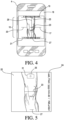
- Fig. 3 shows the device in a portrait orientation 10 followed by a landscape orientation 12 and an unacceptable tilted orientation 14.
- the display will indicate by highlighting the landscape or portrait orientation in Fig. 3 . If the device is not within the certain degree range of the vertical axis or the device is otherwise rotated about the vertical or horizontal axes, the cross-out symbol will flash to indicate to the user that adjustment of the device is tilted and adjustment is required.
- the display shows the viewfinder image and enables capture of an image.
- the display 18 of the device 4 becomes the viewfinder for the camera and a depth of field guideline 16 is overlaid on the viewfinder image.
- the depth of field guideline 16 provides the user with assistance in centering the joint in the photo and capturing the appropriate portion of the joint.
- the limb Before capturing the image of the limb, the limb may be measured and marked at multiple points to indicate specific distances above and below the joint, to aid in alignment with the depth of field guideline, and to provide reference points for circumference measurements of the limb.
- the markings may be at specific points or at regular intervals along and around the limb.
- Fig. 4 shows an example of the depth of field guideline 16 on the display 18 of the device 4 to assist the user in capturing the appropriate portion of the limb.
- the portions 25, 27 of the leg about 15 cm above and below a knee axis 23 should be captured in the image for a custom knee brace.
- a hash mark 17 is provided at the center of the viewfinder image 20 to assist the user in aligning the joint with the center of the image.
- An upper guideline 19 is at the top of the viewfinder image 20 indicating that the distance of about 15 cm above the joint is aligned with the upper guideline 19.
- a lower guideline 21 indicates the distance of about 15 cm below the joint is aligned with the lower guideline 21 in the image.
- the guideline 16, inclusive of mark and guidelines 17, 19, 21 include a vertical line 29 on the center of the image to align with the center of the limb. While the depth of field guideline 16 for the knee defines a portion of the knee about 15 cm above and below the knee, the depth of field guideline 16 defines a portion of the limb any distance above and below a certain point.
- markings are first placed on the limb indicating specific locations on the limb.
- An anatomical landmark or marking is placed at the point on the limb about 15 cm below the joint and about 15 cm above the joint which corresponds to the depth of field guidelines. Therefore, the photographer need only align the markings with the upper and lower guidelines and align the center hash mark over the center of the joint.
- the method requires a delay before the image is taken to assure stable and clear focus.
- the method Upon alignment with the field guideline, the method requires a steady position before taking the image.
- a signal may be released, such as a green dot, to prompt the user to capture the image by pressing a button on the device.
- the clinician first identifies the medial joint space and marks it appropriately. The clinician then measures approximately 2 cm above the medial joint space and draws a line across the knee, from medial to lateral sides, to define the knee axis line. The clinician then identifies and marks the lateral joint space. The clinician then may measure and mark points both 7.5 cm and 15 cm above and below the knee axis line. The clinician marks the tibial peak below the 15 cm mark and the tibial peak above the 7.5 cm mark, and connects the two with a line.
- the application may provide on-screen guides as to the current orientation of the device relative to the preferred orientation regarding each of the axes next to or over the viewfinder image.
- the application assists and guides the user in capturing the optimal image of the limb for a custom orthopedic device.
- the orientation of the device 4 Through immediate on-screen guidance as to the orientation of the device 4, the user can easily and quickly adjust the angle, orientation, and alignment of the device to obtain a well aligned and consistent photograph of the limb. The possibility of misalignment between the image capturing device and the limb is therefore greatly reduced.
- the clinician takes both an anterior view of the limb with the markings, and a lateral view of the limb.
- the depth of field guideline may vary according to the orientation of the limb, which is selected on the device by the clinician.
- the method may include image capture from any number sides of a patient's limb, including anterior, posterior, lateral and medial views and angles.
- the user is prompted to review the captured image.
- the captured image with the overlaid guidelines 16 is shown, and the user determines whether the correct portion of the limb is captured within the image and whether the limb is centered within the image. If the limb and the device 4 were not aligned when the image was captured, a new image may be taken. If a new image is captured, the application returns to step 400 to guide the user through the correct orientation and framing of the limb in the image.
- the user is prompted to enter measurements of the limb.
- the measurements of the limb may include the medial-lateral (M-L) width measurement of the limb and the circumference of the limb at various points above and below the joint.
- a measuring device such as a caliper or measuring tape may obtain the measurements of the limb.
- the captured image 22 is stored with an identification label 24.
- the identification label 24 may be text overlaid on the captured image 22.
- Fig. 5 is an example of a saved image overlaid with an identification label 24.
- the information label 24 may include data such as the patient's name, date of the photograph, and the M-L measurement along one side of the image.
- the image with overlaid information is stored in the memory of the device.
- the image may be password protected or encrypted in the memory to protect the patient's privacy.
- the system may include a tilt indicator 31 arranged to allow the clinician to correctly orient the camera position.
- the tilt indicator 31 relies on a determination by the device to measure whether the angle or orientation of the device is proper to assure a successful captured image.
- the tilt indicator will display a red color when the device is not in the proper orientation to capture the image, and a green color when the device is in a proper orientation.
- patient information is entered to fill out the order form.
- the patient information may include the name of the user, the prescriber of the orthopedic device, and the diagnosis or symptoms of the user with identification of the problem joint.
- the application can create a partial order on the device in encrypted XML format including the captured photo and patient information.
- Other security measures may protect the patient's data under HIPAA regulations.
- the user or patient selects the appropriate orthopedic device configuration such as the orthopedic device model and color.
- the application guides the user through the different orthopedic devices and provides the user with options based on the selected orthopedic device.
- the partial order is saved.
- the user can enter a menu listing all orders saved on the device or continue to a review of the order.
- the list also indicates the status of the order such as whether the order has been transmitted to the maker or seller. If the user selects a link for an order not yet transmitted, the user is prompted to review the order.
- the order is displayed for review.
- the saved image and the collected information are displayed.
- the user can edit portions of the order with the changes made during the review being saved with the order.
- the user can also enter the clinician information and payment and shipping information. The user then may save the order or transmitting the order.
- the order is saved and/or transmitted to the server of the custom orthopedic device provider.
- the application sends the order in an e-mail and automatically populates the fields of the e-mail based on the data in the saved order.
- the patient information, brace information, and clinician or user information are inserted into the body of the e-mail while the saved image associated with the order is automatically attached to or inserted into the e-mail.
- the device can send the order directly to a server of the seller through a network.
- the application and/or the server may display or send a notification to the user to confirm the order.
- the embodiments described relate to a method and device for ordering a custom orthopedic device which can be accomplished with a single image
- the method and device are used with or within a custom orthopedic device production method and system which produces the custom orthopedic device based on a three-dimensional model of the limb generated from a plurality of captured images.
- markers or reference points are placed on the limb and are subsequently captured in the image.
- the markers or reference points assist in determining the dimensions of the limb from the image by providing information related to the surface of the limb.
- a sock or sleeve having markings may be worn on the limb or the limb may be marked at particular intervals. Markings can be contrasting colored markings in many shapes such as a circular shape, a rectangular shape, a triangular shape, or any combination and are preferably the same size.
- the distribution and density of the markings over the surface of the sock, sleeve, or limb varies depending on the type of limb and the desired three-dimensional modeling resolution.
- markings or a higher density of markings in a certain area produces a more accurate three-dimensional model of the limb since more reference points would be provided in the captured image.
- the markings may be concentrated in areas where there are more variations in the continuity of the limb surface such as around the joint area. Additional references may be added to the limb or the sock. In ordering a custom knee brace, additional markings or references are added to indicate the center of the knee, the angle of the tibia, and the locations of the condyles.
- Fig. 6 shows an image capturing process 402 and a review stage 502 in an embodiment of the method for ordering a custom orthopedic device where a three-dimensional image of the limb is captured.
- This embodiment follows the same steps as the embodiment in Fig. 1 and differs in the specific steps taken to capture the image at 400 and in the image displayed during the review stage 500 of the embodiment of Fig. 1 .
- the ordering process continues with the remaining general ordering steps 600, 700, 800, and 900 as described with respect to Fig. 1 .
- the user is prompted to select whether to use a three-dimensional model. If the user does not select the three-dimensional model, the method continues as described regarding Figs. 1-5 . If the user uses the three-dimensional option, the image capturing process 402 as shown in Fig. 6 and review process 502 are used.
- markings or reference points are added to the limb in the manner described.
- the photographer captures at least two images of the limb at various angles or views.
- at least four individual images are captured of the limb with each image capturing a different angle or view of the limb such that the entire circumference of the appropriate portion of the limb is captured within the plurality of images.
- the application may instruct the user to capture a certain number of images of the limb from different angles, orientations, heights, or different portions of the limb.
- the application may use continuous image capturing where the application automatically captures images at different intervals such that the photographer need only move the device and indicate when images of all views or sides of the limb have been captured.
- the application can guide the photographer in capturing the appropriate angles or views of the limb with the appropriate alignment of the limb in the image using accelerometer and/or gyroscope data from the device using the guidelines described. Depending on the depth of field of the images, the application can determine the appropriate number of images, angles, or views needed to generate an accurate three-dimensional model of the limb.
- the application analyzes and processes the plurality of captured images to stitch the images together and form a continuous view of the limb.
- the application can perform the stitching of the images together automatically or with user assistance.
- the stitching of the images can also be performed while the images are being captured or with the plurality of individual separately captured images.
- the application generates a three-dimensional model of the limb using the markings on the sock, sleeve, or limb.
- the model may be a computer-aided design (CAD) point cloud surface where the markings shown in the captured images translate into points in the point could surface.
- the application preferably generates a 360° view of the limb.
- the application prompts the user to review the generated three-dimensional model to ensure that the three-dimensional model accurately depicts the surface shape of the corresponding portion of the limb at 502.
- the user can rotate and zoom into the model to view all sides of and different levels of detail of the limb.
- the user is given the option of approving the generated model or re-capturing the limb to generate a more accurate model.
- the order can include the plurality of images and/or the three-dimensional model of the limb.
- modules are defined here as an isolatable element that performs a defined function and has a defined interface to other elements.
- the modules described in this disclosure may be implemented in hardware, a combination of hardware and software, firmware, or a combination, all of which are behaviorally equivalent.
- Modules may be implemented using computer hardware in combination with software routine(s) written in a computer language (such as C, C++, Fortran, Java, Basic, Matlab or the like). It may be possible to implement modules using physical hardware that incorporates discrete or programmable analog and/or digital hardware. Examples of programmable hardware include: computers, microcontrollers, microprocessors, application-specific integrated circuits (ASICs); field programmable gate arrays (FPGAs); and complex programmable logic devices (CPLDs). Computers, microcontrollers and microprocessors are programmed using languages such as assembly, C, C++ or the like. Finally, the above mentioned technologies may be used in combination to achieve the result of a functional module.
- a computer language such as C, C++, Fortran, Java, Basic, Matlab or the like.
- the application may be software embodied on a computer readable medium which when executed by a processor of a computer performs a sequence of steps.
- a computer readable medium may be a floppy disk, a flexible disk, a hard disk, magnetic tape, or any other magnetic medium, a CD-ROM, any other optical medium, a RAM, a ROM, or any other medium from which a computer can read.
- Various forms of computer readable media may carry one or more sequences of one or more instructions to a processor for execution.
- the software may be transmitted over a wired or wireless network to the device.
Description
- The present disclosure relates to a method reducing image and limb misalignment in capturing an image of the limb for ordering a custom orthopedic device for a joint of the limb and a device for ordering a custom orthopedic device and a computer program product in the form of an ordering application software for execution of the method on a portable device.
- To provide customized support for a j oint, a clinician may provide a patient with a custom fitted orthopedic device adapted to the specific anatomical dimensions of the individual patient. A common orthopedic device for customization is a knee brace. A patient will typically obtain a customized brace through a clinician having the expertise to assure that the orthopedic device fits the patient properly.
- A clinician can prepare the brace himself, or order a custom orthopedic device remotely through the mail or by submitting an order over mail, phone, fax or the Internet. During the ordering process, the clinician typically provides the manufacturer or seller ("provider") with an image of a portion of the limb including the joint and measurements of the limb around the joint. The custom orthopedic brace is produced based on the submitted image of the limb and measurements. The provider may require the image of the limb be captured at a certain orientation, angle, height, and distance relative to the limb to ensure that the captured image accurately portrays the dimensions and proportions of the limb. Appropriate tags, reference indicia and reference markings of anatomy are often placed on the limb to identify the patient, the limb, and any other necessary information if the photo is misplaced from an order form.
- It is undesirable for the picture to be taken when the camera is at an angle relative to the limb (angle normal to the line of progression of the limb) or when the limb is not aligned with the center of the image since such an image would inaccurately portray the dimensions and proportions of the limb. Producing a custom orthopedic brace based on such a misaligned image results in a poorly fitting brace. Likewise, poor resolution or lack of indicia applied on the limb may impede the producer in fully understanding the contours of the patient's limb.
- Using a conventional camera, the photographer must estimate or otherwise measure the specified distance between the camera and the limb, the specified portion of the limb to capture in the image, and the appropriate orientation of the camera relative to the limb. Since the conventional camera does not provide feedback about the angle or orientation at which the camera is held, it is difficult for the photographer to determine whether the image of the limb being captured meets the requirements of the manufacturer or seller without additional aids.
- The patient may need to use different devices to complete the entire ordering process. If the image of the limb is captured with a conventional digital camera, the image must then be transferred to a computer before the order and image can be uploaded over the Internet to the server of the manufacturer or seller.
- While providing a photo is useful in understanding the patient's anatomy, dimensional measurements are likewise required. Various forms are required for completion by the clinician to determine measurement data and patient personal information. Other forms require the clinician to indicate brace models, features, accessories, colors, etc. From the requirement for forms and a photo, the ordering process both complicated and risks a mismatch of documents for the order.
- The features of the disclosure provide a solution to the need to reduce image and limb misalignment and improve the ease of capturing an image of the limb and of ordering a custom orthopedic device without multiple forms.
- Various types of calipers are known for measuring different objects. A caliper must be properly applied against the object to take the desired measurement, and when measuring the thickness of between two sides of an object, a caliper must be held at right angles to the piece.
- In the field of orthopedics, calipers are used for measuring a knee and a leg for a knee brace. As illustrated in Fig. 7, an exemplary method for measuring a knee includes using a marker to apply landmarks on the patient's leg such as at the medial joint space, and along the tibial crest at the tibial tuberosity and a distance below which is depicted in Figs. 7A and 7B. Referring to Fig. 7C, a conventional caliper 102 comes into use in the method by taking the M-L (medial-lateral) measurement 109 at the joint space against the patient's condyles. The caliper 102 includes jaws 104, 106 snugly adjusted against the patient's skin to obtain the measurement and are left in place as the measurement is recorded along the scale 108.
- In using the mid patella as a reference point, as depicted in Fig. 7D, above and below circumference measurements 105, 107 are taken at specified distances, and a caliper may also obtain M-L measurements at these locations as well. Next, with the patient standing in a weight bearing position facing a camera, the height of the camera is adjusted at a mid-patella level, and the camera is positioned so it is approximately a specified distance away from the patient. The image captures the leg a specified distance above and below mid patella and is taken of the anterior side of the knee, and an additional image is taken of the lateral side of the knee, as shown in Figs. 7E and 7F.
- The conventional caliper may obtain the M-L measurement and other devices have jaws that slide relative to one another and are indexed to a scale located along a shaft. The clinician must adjust one of the jaws relative to the other jaw, manually move the jaw to press against the patient's soft tissue, and manually removes the jaws from the soft tissue upon completion of the measurement. The force against the soft tissue may exceed comfort levels or may be inconsistently applied from patient to patient.
- The conventional caliper 102 may have narrow jaws 104, 106 which may shift over the soft tissue due to their narrow width and may cause discomfort because of sharp tips of the jaws. The jaws are typically elongated and straight, and provide little or any space between areas of the soft tissue within the jaws for adjustment by a clinician. A patient's condyles may have an irregular shape and the narrow jaws of fail to provide an accurate measurement of M/L due to condyle irregularity. In variations, conventional calipers may use contoured "cup-shaped" contact point which extend over the condyles but limit the clinician for placement over the condyles.
- Due to a variety of leg shapes, sizes and soft tissue amount, conventional calipers provide inconsistent compression over patients' soft tissue due in part to human error by clinician. The inconsistent compression results in improper measurements for the eventual knee brace and may lead to inferior customization of the knee brace.
-
US 2010/268138 A1 discloses a custom device such as braces and casts used to protect a portion of a body during recovery. A method for fabricating the custom device includes: marking a body with reference points and/or other indicators; obtaining multiple images of the body from multiple angles; using the images to determine the contours of the body; and locating and using the other markings to design the custom device. -
US 2011/166435 A1 discloses a surgical orientation system used to assist a surgeon to orient a prosthetic component relative to a patient's anatomy during surgery. The system includes: an implement for releasable attachment of a prosthetic component; an electronic orientation monitor attached to the implement; and a brace. The brace is releasably attachable to the patient so as to define a reference point relative to the patient's anatomy. The reference point is external of the patient and includes at least one surface defining a reference plane that is used to orient the monitor into a reference orientation to calibrate the monitor. The surgeon then manipulates the implement so that the prosthetic component is in the desired position relative to the patient. The monitor provides an indication to the surgeon when a subsequent orientation of the monitor has a predefined relationship relative to the reference orientation. Upon receiving the indication, the surgeon inserts the prosthetic component into the patient. -
US 2009/088674 A1 discloses a method and system for designing a patient-specific orthopedic surgical instrument including coupling a knee sleeve to a leg of the patient, the knee sleeve including sensors configured to generate sensor data indicate of the position of the respective sensor. The design of the patient-specific orthopedic surgical instrument includes determining angulation data indicative of the angulation of the knee based on the sensor data; and generating medical image(s) of the knee; wherein the patient-specific orthopedic surgical instrument is designed based on the angulation data and the medical image(s). - It is an object of the present invention to provide a method reducing image and limb misalignment in capturing an image of the limb for ordering a custom orthopedic device for a joint of the limb and device for ordering a custom orthopedic device and computer program product in the form of an ordering application software for execution of the method on a portable device, by which image and limb misalignment in capturing an image of the limb, such as a leg, for ordering a custom orthopedic device for a joint, such as a knee joint, can be reduced to improve the customization of the custom orthopedic device.
- The object is achieved by the features of
independent claim 1 and 12, respectively. Further embodiments are defined in the respective dependent claims. - According to a variation, the first and second distances are the same and referenced from a knee axis line. The distances above and below the joint may be aligned with the depth of field guideline in the viewfinder image before capturing the image.
- The method may also include calibrating the image sensor of the portable device.
- The method may include executing an ordering application, determining whether the ordering application has been previously executed. The image sensor may be calibrated upon the determination that the ordering application has not been previously executed. The image of the limb enables capture upon the determination that the ordering application has been previously executed.
- The method may include reviewing the captured image of the limb and selecting a custom orthopedic device configuration. The step of reviewing the captured image of the limb includes viewing the captured image with an overlaid depth of field guideline to confirm the captured portion of the limb satisfies the overlaid depth of field guideline. The method may also include entering basic patient information into the portable device including measurements of the limb at various locations on the limb. The captured image may be overlaid with the basic patient information. The overlaid captured image may be stored in the portable device.
- The method can involve configuring the custom orthopedic device, reviewing the order, and storing the order in a memory of the portable device. At least one previous order may be stored in the memory of the portable device. The order may be transmitted as an e-mail containing the patient information and the saved, captured image of the limb.
- The method may include a login page requiring clinician and patient input. Upon entry of the information on the login page, the user is directed to an order configuration home screen or page. From the home screen, the user may select many pages for making the customized order. The user may first select the image capture and input measurements, followed by entering patient information, orthopedic device (brace) configuration, and any other order information. The user is not limited to a sequence of page use, other than upon entry of all data fields and appropriate image capture, the order is sent to the provider.
- The processor can be further configured to calibrate the image sensor by setting the image sensor to a first resolution and a first zoom level. At least one image of anatomical landmarks or markings on the limb is captured. A three-dimensional model of the limb is generated from the markings along with circumferential measurements.
- The device and method for ordering a custom orthopedic device is described with reference to the accompanying drawings which show preferred embodiments according to the device described herein.
-
Fig. 1 is an overview of the steps in an embodiment of the custom orthopedic device ordering method. -
Fig. 2 shows a portrait orientation of a device for ordering the custom orthopedic device with respect to three axes. -
Fig. 3 is an example of the indication provided to the user of the preferred orientation of the device. -
Fig. 4 shows the device having a viewfinder image overlaid with a depth of field guideline. -
Fig. 5 is an example of the captured image stored with an identification label. -
Fig. 6 illustrates is a flowchart of an embodiment of the custom orthopedic device ordering method using a three-dimensional model of the limb. - A better understanding of different embodiments of the disclosure may be had from the following description read with the accompanying drawings in which like reference characters refer to like elements.
- The embodiments of the method 2 and
device 4 disclosed enable the user to easily capture accurate images of the limb and complete the ordering process on a single device. In an embodiment, the method 2 is implemented in an application executed on a portable device. The application guides the user through the ordering and image capturing process. Such a device and process reduces misalignment issues while integrating picture capturing and ordering into a single device. - The device used to capture the image and order the custom orthopedic brace may be any device having a display and an image sensor such as a mobile phone (iPhone®, Android® phone, Blackberry®, Windows® phone, etc.), a tablet (iPad®, Android® tablet, Windows® tablet etc.), a personal digital assistant (PDA), a computer, or any other portable device.
- The
preferred device 4 has an image sensor, a display, a processor, a gyroscope and/or accelerometer, memory, and a communication interface to allow communication of the order directly from the device over a network to a server of the manufacturer or seller. -
Fig. 1 is an overview of an embodiment of an embodiment of the method 2 for ordering a custom orthopedic device. After the user selects the ordering application, the application is launched, and the application displays a login screen. Atstep 100, the user logs into an account or creates a new account using a keypad or similar on the device. The login page may include an option for an embedded tutorial including a visual step-by-step discussion stored on the device or available to the device through streaming. The tutorial may provide guidance on patient positioning, making anatomical landmarks on the limb, and taking circumferential measurements. - At
step 200, the application determines whether the current session is the first use of thedevice 4 to order the custom orthopedic device. In one embodiment, if the current session is the first use, the application calibrates a camera of the device at 300. Duringcalibration 300, the application automatically sets the camera to specific settings such as a specific resolution, zoom, and color setting. - Alternatively, the
calibration 300 of the camera may be omitted. In this embodiment, the preferred camera settings are in text within the application, and the calibration is manually performed by the user by adjusting the settings of the camera. The preferred camera settings may be displayed during the image capturing process and during a review of the captured image. The preferred camera settings can also be displayed and included in a "help" section of the application. - If the current session is not the first use or calibration of the camera is unnecessary, the application enables capture of an image using guidelines and a specific alignment or orientation at step 400. In
step 500, the captured image is reviewed by the user to determine whether the captured image meets the specified alignment. Atstep 600, patient information is entered, and a brace configuration is selected at 700. The user is prompted to review the order at 800 before the order is saved on the device and transmitted over a network to a server of the custom orthopedic device manufacturer or seller at 900. - The capturing of the image of the limb at step 400 is described in more detail regarding
Figs. 2-4 . Before the application enables capturing of an image, the application ensures thedevice 4 is properly aligned with the limb. To produce a properly aligned image of the limb, the joint is preferably centered in the image, and the image is captured from a point at the same height as the joint with the plane of the image being parallel to the limb or a vertical axis while thedevice 4 is in a portrait or landscape orientation. Where the horizontal axis is parallel to the x-axis, the y-axis represents depth, and the vertical axis is parallel to the z-axis, alongitudinal axis 6 of the device would be parallel to the horizontal axis when in a landscape orientation or to the vertical axis when in a portrait orientation. - In determining whether the
device 4 is in an acceptable portrait or landscape orientation, the application uses the gyroscope and/or accelerometer of thedevice 4 to determine whether the orientation of thedevice 4 is within a certain degree range, for example, whether thelongitudinal axis 6 of thedevice 4 is within five degrees of the horizontal axis or within five degrees of the vertical axis. - The application provides a
visual indication 8 on adisplay 18 of thedevice 4 whether thedevice 4 is properly oriented and allows the user to adjust thedevice 4 until the orientation requirements are met. -
Fig. 3 shows the device in aportrait orientation 10 followed by alandscape orientation 12 and an unacceptable tiltedorientation 14. When the device is within the certain degrees range of the landscape or portrait orientation, the display will indicate by highlighting the landscape or portrait orientation inFig. 3 . If the device is not within the certain degree range of the vertical axis or the device is otherwise rotated about the vertical or horizontal axes, the cross-out symbol will flash to indicate to the user that adjustment of the device is tilted and adjustment is required. - Once the orientation requirements are met, the display shows the viewfinder image and enables capture of an image. The
display 18 of thedevice 4 becomes the viewfinder for the camera and a depth offield guideline 16 is overlaid on the viewfinder image. The depth offield guideline 16 provides the user with assistance in centering the joint in the photo and capturing the appropriate portion of the joint. Before capturing the image of the limb, the limb may be measured and marked at multiple points to indicate specific distances above and below the joint, to aid in alignment with the depth of field guideline, and to provide reference points for circumference measurements of the limb. The markings may be at specific points or at regular intervals along and around the limb. -
Fig. 4 shows an example of the depth offield guideline 16 on thedisplay 18 of thedevice 4 to assist the user in capturing the appropriate portion of the limb. In the example, theportions knee axis 23 should be captured in the image for a custom knee brace. Ahash mark 17 is provided at the center of theviewfinder image 20 to assist the user in aligning the joint with the center of the image. Anupper guideline 19 is at the top of theviewfinder image 20 indicating that the distance of about 15 cm above the joint is aligned with theupper guideline 19. Similarly, alower guideline 21 indicates the distance of about 15 cm below the joint is aligned with thelower guideline 21 in the image. Theguideline 16, inclusive of mark andguidelines field guideline 16 for the knee defines a portion of the knee about 15 cm above and below the knee, the depth offield guideline 16 defines a portion of the limb any distance above and below a certain point. - To aid the user in correctly framing the limb in the picture, markings are first placed on the limb indicating specific locations on the limb. An anatomical landmark or marking is placed at the point on the limb about 15 cm below the joint and about 15 cm above the joint which corresponds to the depth of field guidelines. Therefore, the photographer need only align the markings with the upper and lower guidelines and align the center hash mark over the center of the joint.
- The method requires a delay before the image is taken to assure stable and clear focus. Upon alignment with the field guideline, the method requires a steady position before taking the image. A signal may be released, such as a green dot, to prompt the user to capture the image by pressing a button on the device.
- In a variation, the clinician first identifies the medial joint space and marks it appropriately. The clinician then measures approximately 2 cm above the medial joint space and draws a line across the knee, from medial to lateral sides, to define the knee axis line. The clinician then identifies and marks the lateral joint space. The clinician then may measure and mark points both 7.5 cm and 15 cm above and below the knee axis line. The clinician marks the tibial peak below the 15 cm mark and the tibial peak above the 7.5 cm mark, and connects the two with a line.
- The application may provide on-screen guides as to the current orientation of the device relative to the preferred orientation regarding each of the axes next to or over the viewfinder image.
- In this manner, the application assists and guides the user in capturing the optimal image of the limb for a custom orthopedic device. Through immediate on-screen guidance as to the orientation of the
device 4, the user can easily and quickly adjust the angle, orientation, and alignment of the device to obtain a well aligned and consistent photograph of the limb. The possibility of misalignment between the image capturing device and the limb is therefore greatly reduced. - According to a variation, the clinician takes both an anterior view of the limb with the markings, and a lateral view of the limb. In both instances, the depth of field guideline may vary according to the orientation of the limb, which is selected on the device by the clinician. The method may include image capture from any number sides of a patient's limb, including anterior, posterior, lateral and medial views and angles.
- Once an image is captured, the user is prompted to review the captured image. During the review of the captured photograph at
step 500, the captured image with the overlaidguidelines 16 is shown, and the user determines whether the correct portion of the limb is captured within the image and whether the limb is centered within the image. If the limb and thedevice 4 were not aligned when the image was captured, a new image may be taken. If a new image is captured, the application returns to step 400 to guide the user through the correct orientation and framing of the limb in the image. Once the photograph is confirmed, the user is prompted to enter measurements of the limb. The measurements of the limb may include the medial-lateral (M-L) width measurement of the limb and the circumference of the limb at various points above and below the joint. A measuring device such as a caliper or measuring tape may obtain the measurements of the limb. - To associate the captured image 22 with the patient, the captured image 22 is stored with an
identification label 24. Theidentification label 24 may be text overlaid on the captured image 22.Fig. 5 is an example of a saved image overlaid with anidentification label 24. Theinformation label 24 may include data such as the patient's name, date of the photograph, and the M-L measurement along one side of the image. The image with overlaid information is stored in the memory of the device. The image may be password protected or encrypted in the memory to protect the patient's privacy. - As shown in
Fig. 5 , the system may include atilt indicator 31 arranged to allow the clinician to correctly orient the camera position. Thetilt indicator 31 relies on a determination by the device to measure whether the angle or orientation of the device is proper to assure a successful captured image. In a preferred embodiment, the tilt indicator will display a red color when the device is not in the proper orientation to capture the image, and a green color when the device is in a proper orientation. - At 600, additional patient information is entered to fill out the order form. The patient information may include the name of the user, the prescriber of the orthopedic device, and the diagnosis or symptoms of the user with identification of the problem joint. After the patient information is entered, the application can create a partial order on the device in encrypted XML format including the captured photo and patient information. Other security measures may protect the patient's data under HIPAA regulations.
- At
step 700, the user or patient selects the appropriate orthopedic device configuration such as the orthopedic device model and color. The application guides the user through the different orthopedic devices and provides the user with options based on the selected orthopedic device. After selection and configuration of the orthopedic device, the partial order is saved. The user can enter a menu listing all orders saved on the device or continue to a review of the order. The list also indicates the status of the order such as whether the order has been transmitted to the maker or seller. If the user selects a link for an order not yet transmitted, the user is prompted to review the order. - At
step 800, the order is displayed for review. The saved image and the collected information are displayed. The user can edit portions of the order with the changes made during the review being saved with the order. The user can also enter the clinician information and payment and shipping information. The user then may save the order or transmitting the order. - At
step 900, the order is saved and/or transmitted to the server of the custom orthopedic device provider. The application sends the order in an e-mail and automatically populates the fields of the e-mail based on the data in the saved order. The patient information, brace information, and clinician or user information are inserted into the body of the e-mail while the saved image associated with the order is automatically attached to or inserted into the e-mail. When the user elects to send the e-mail containing the order information, the device can send the order directly to a server of the seller through a network. The application and/or the server may display or send a notification to the user to confirm the order. - While the embodiments described relate to a method and device for ordering a custom orthopedic device which can be accomplished with a single image, in another embodiment the method and device are used with or within a custom orthopedic device production method and system which produces the custom orthopedic device based on a three-dimensional model of the limb generated from a plurality of captured images.
- To generate a three-dimensional model, markers or reference points are placed on the limb and are subsequently captured in the image. The markers or reference points assist in determining the dimensions of the limb from the image by providing information related to the surface of the limb. To place the markers or reference points on the limb, a sock or sleeve having markings may be worn on the limb or the limb may be marked at particular intervals. Markings can be contrasting colored markings in many shapes such as a circular shape, a rectangular shape, a triangular shape, or any combination and are preferably the same size. The distribution and density of the markings over the surface of the sock, sleeve, or limb varies depending on the type of limb and the desired three-dimensional modeling resolution.
- Providing more markings or a higher density of markings in a certain area produces a more accurate three-dimensional model of the limb since more reference points would be provided in the captured image. The markings may be concentrated in areas where there are more variations in the continuity of the limb surface such as around the joint area. Additional references may be added to the limb or the sock. In ordering a custom knee brace, additional markings or references are added to indicate the center of the knee, the angle of the tibia, and the locations of the condyles.
-
Fig. 6 shows animage capturing process 402 and areview stage 502 in an embodiment of the method for ordering a custom orthopedic device where a three-dimensional image of the limb is captured. This embodiment follows the same steps as the embodiment inFig. 1 and differs in the specific steps taken to capture the image at 400 and in the image displayed during thereview stage 500 of the embodiment ofFig. 1 . After the three-dimensional model specific steps of 402 and 502, the ordering process continues with the remaining general ordering steps 600, 700, 800, and 900 as described with respect toFig. 1 . - Once the ordering application is executed and before the image capturing step 400, the user is prompted to select whether to use a three-dimensional model. If the user does not select the three-dimensional model, the method continues as described regarding
Figs. 1-5 . If the user uses the three-dimensional option, theimage capturing process 402 as shown inFig. 6 andreview process 502 are used. - Before beginning the
image capturing process 402, markings or reference points are added to the limb in the manner described. In theimage capturing process 402 starting withstep 404, the photographer captures at least two images of the limb at various angles or views. Preferably, at least four individual images are captured of the limb with each image capturing a different angle or view of the limb such that the entire circumference of the appropriate portion of the limb is captured within the plurality of images. The application may instruct the user to capture a certain number of images of the limb from different angles, orientations, heights, or different portions of the limb. - Alternatively, the application may use continuous image capturing where the application automatically captures images at different intervals such that the photographer need only move the device and indicate when images of all views or sides of the limb have been captured.
- During the
image capturing process 402, the application can guide the photographer in capturing the appropriate angles or views of the limb with the appropriate alignment of the limb in the image using accelerometer and/or gyroscope data from the device using the guidelines described. Depending on the depth of field of the images, the application can determine the appropriate number of images, angles, or views needed to generate an accurate three-dimensional model of the limb. - At
step 406, the application analyzes and processes the plurality of captured images to stitch the images together and form a continuous view of the limb. The application can perform the stitching of the images together automatically or with user assistance. The stitching of the images can also be performed while the images are being captured or with the plurality of individual separately captured images. - At 408, from the stitched image, the application generates a three-dimensional model of the limb using the markings on the sock, sleeve, or limb. The model may be a computer-aided design (CAD) point cloud surface where the markings shown in the captured images translate into points in the point could surface. The application preferably generates a 360° view of the limb.
- Once the
image capturing process 402 is completed, the application prompts the user to review the generated three-dimensional model to ensure that the three-dimensional model accurately depicts the surface shape of the corresponding portion of the limb at 502. During review of the three-dimensional model, the user can rotate and zoom into the model to view all sides of and different levels of detail of the limb. The user is given the option of approving the generated model or re-capturing the limb to generate a more accurate model. - If the user performs the
image capturing process 402 again, the current generated model may be saved for comparison with later models during the review stage. If the user approves of the generated model, the application continues with the general ordering process stages 600-900 as described regardingFig. 1 . For the order review at 800 and transmission and saving of theorder 900, the order can include the plurality of images and/or the three-dimensional model of the limb. - Many of the elements described in the disclosed embodiments may be implemented as modules. A module is defined here as an isolatable element that performs a defined function and has a defined interface to other elements. The modules described in this disclosure may be implemented in hardware, a combination of hardware and software, firmware, or a combination, all of which are behaviorally equivalent.
- Modules may be implemented using computer hardware in combination with software routine(s) written in a computer language (such as C, C++, Fortran, Java, Basic, Matlab or the like). It may be possible to implement modules using physical hardware that incorporates discrete or programmable analog and/or digital hardware. Examples of programmable hardware include: computers, microcontrollers, microprocessors, application-specific integrated circuits (ASICs); field programmable gate arrays (FPGAs); and complex programmable logic devices (CPLDs). Computers, microcontrollers and microprocessors are programmed using languages such as assembly, C, C++ or the like. Finally, the above mentioned technologies may be used in combination to achieve the result of a functional module.
- The application may be software embodied on a computer readable medium which when executed by a processor of a computer performs a sequence of steps. A computer readable medium may be a floppy disk, a flexible disk, a hard disk, magnetic tape, or any other magnetic medium, a CD-ROM, any other optical medium, a RAM, a ROM, or any other medium from which a computer can read. Various forms of computer readable media may carry one or more sequences of one or more instructions to a processor for execution. The software may be transmitted over a wired or wireless network to the device.
- A preferred order for the steps in the method of ordering the custom orthopedic device has been described. It is noted that the order of the steps in the method may be rearranged.
Claims (13)
- A method (2) reducing image and limb misalignment in capturing an image of the limb for ordering a custom orthopedic device for a joint of the limb, comprising:aligning a viewfinder image (20) displayed on a display (18) and generated by an image sensor of a portable device (4) with at least one predetermined portion of the limb including the joint;capturing and storing an image of the portion of the limb using the image sensor of the portable device (4) based on at least one limb alignment guideline (16, 19, 21, 29);associating the captured image with measurements of the limb (23, 25, 27), and patient information entered into the portable device (4); andtransmitting an order containing the captured image, the measurements of the limb (23, 25, 27), and the patient information from the portable device (4) to a server of the custom orthopedic device provider; wherein,the at least one limb alignment guideline (16, 19, 21, 29) is a depth of field guideline (16) overlaid on the viewfinder image (20), the depth of field guideline (16) defining an upper guideline (19) for a first predetermined distance above the joint, a lower guideline (21) for a second predetermined distance below the joint, a hash mark (17) at a center of the viewfinder image (20) for a joint axis (23), and a vertical line (29) arranged on the center of the viewfinder image (20) for a center of the limb;before capturing of the image is enabled, it is determined that orientation of the device (4) requirements are met to ensure that the device (4) is properly aligned with the limb for producing a properly aligned image of the limb, in which the j oint is centered in the image and the image is captured from a point at the same height as the joint with the plane of the image being parallel to the limb or the vertical axis while the device (4) is in a portrait or landscape orientation, wherein the horizontal axis defines an x-axis, a y-axis represents depth, and the vertical axis defines a z-axis, and wherein the longitudinal axis (6) of the device (4) is parallel to the horizontal axis when in landscape orientation (12) or to the vertical axis when in portrait orientation (10);in determining whether the device (4) is in an acceptable portrait orientation (10) or landscape orientation (12):- a gyroscope and/or an accelerometer of the device (4) is used to determine whether the orientation of the device (4) is within a certain degree range of the horizontal axis or of the vertical axis;- a visual indication (8) is provided on the display (18) of the device (4) indicating whether the device (4) is properly oriented thereby allowing adjusting of the device (4) until the orientation requirements are met, wherein, when the device (4) is within the certain degree range of the landscape orientation (12) or the portrait orientation (10), the orientation is indicated on the display (18) otherwise it is indicated that the adjustment of the device (4) is tilted and adjustment is required; and- once the orientation requirements are met, the viewfinder image (20) is shown on the display (18) and capturing of the image is enabled.
- The method according to claim 1, wherein the first and second predetermined distances are the same and referenced from the joint axis (23).
- The method according to claim 1, further comprising the step of:
aligning the first and second predetermined distances above and below the joint axis (23) with the depth of field guideline (16) shown in the viewfinder image (20) before capturing the image. - The method according to claim 1, wherein once the orientation of the portable device (4) relative to the limb satisfies the at least one limb alignment guideline (16, 19, 21, 29), the portable device (4) enables image capture.
- The method according to any one of the preceding claims, further comprising the step of calibrating the image sensor of the portable device (4).
- The method according to any one of the preceding claims, further comprising the steps of:executing an ordering application;determining whether the ordering application has been previously executed;upon the determination that the ordering application has not been previously executed, calibrating the image sensor of the portable device (4); andupon the determination that the ordering application has been previously executed, enabling capture of the image of the limb.
- The method according to any one of the preceding claims, wherein at least two images are captured at two different angles or sides of the limb.
- The method according to claim 7, wherein the step of reviewing the captured image of the limb, comprises the steps of:viewing the captured image with an overlaid depth of field guideline (16) to confirm the captured portion of the limb satisfies the overlaid depth of field guideline (16);entering basic patient information into the portable device (4), wherein the basic patient information includes measurements of the limb at various locations on the limb;overlaying the captured image with the basic patient information; andstoring the overlaid captured image in the portable device (4).
- The method according to any one of the preceding claims, further comprising the steps of:configuring the custom orthopedic device;reviewing the order; andstoring the order in a memory of the portable device (4),wherein preferably at least one previous order is stored in the memory of the portable device (4).
- The method according to any one of the preceding claims, wherein the order is transmitted as an e-mail containing the patient information and the saved, captured image of the limb.
- The method according to any one of the preceding claims, wherein the joint is a knee joint and the orthopedic device is a knee brace.
- A device (4) for ordering a custom orthopedic device, comprising: an image sensor, the image sensor configured to capture an image; a display (18); a communication interface; a processor; and a memory;wherein the processor is configured to enable capturing an image of a portion of a limb including a joint using the image sensor based on at least one limb alignment guideline (16, 19, 21, 29) in a viewfinder image (20) displayed on the display (18), if the image of the limb satisfies the at least one limb alignment guideline;wherein the communication interface is configured to transmit an order containing the captured image and patient information from the apparatus over a network to a provider; whereinthe device (4) further comprises a gyroscope and/or accelerometer configured to provide orientation data to the processor; andthe at least one limb alignment guideline (16, 19, 21, 29) is a depth of field guideline (16) overlaid on the viewfinder image (20), the depth of field guideline (16) defining an upper guideline (19) for a first predetermined distance above the joint, a lower guideline (21)_for a second predetermined distance below the joint, a hash mark (17) at a center of the viewfinder image (20) for a joint axis (23), and a vertical line (29) arranged on the center of the viewfinder image (20) for a center of the limb; andthe processor is further configuredbefore enabling the capturing of the image of the portion of the limb, to assist an user of the device (4) in aligning the device (4) with the limb by- determining that orientation of the device (4) requirements are met to ensure that the device (4) is properly aligned with the limb for producing a properly aligned image of the limb, in which the joint is centered in the image and the image is captured from a point at the same height as the joint with the plane of the image being parallel to the limb or the vertical axis while the device (4) is in a portrait or landscape orientation, wherein the horizontal axis defines an x-axis, a y-axis represents depth, and the vertical axis defines a z-axis, and wherein the longitudinal axis (6) of the device (4) is parallel to the horizontal axis when in landscape orientation (12) or to the vertical axis when in portrait orientation (10);wherein in determining whether the device (4) is in an acceptable portrait orientation (10) or landscape orientation (12):- to use the orientation data provided by the gyroscope and/or an accelerometer to determine whether the orientation of the device (4) is within a certain degree range of the horizontal axis or of the vertical axis;- to provide a visual indication (8) on the display (18) of the device (4) indicating whether the device (4) is properly oriented thereby allowing adjusting of the device (4) until the orientation requirements are met, wherein, when the device (4) is within the certain degree range of the landscape orientation (12) or the portrait orientation (10), to indicate the orientation on the display (18) otherwise to indicate that the adjustment of the device (4) is tilted and adjustment is required; and- once the orientation requirements are met, to show the viewfinder image (20) on the display (18) and to enable the capturing of the image.
- Computer program product in the form of an ordering application software comprising instructions to cause the device according to claim 12 to execute the steps of the method of any of claims 1 to 11.
Applications Claiming Priority (3)
| Application Number | Priority Date | Filing Date | Title |
|---|---|---|---|
| US201261694314P | 2012-08-29 | 2012-08-29 | |
| US201261694474P | 2012-08-29 | 2012-08-29 | |
| PCT/US2013/056896 WO2014036029A2 (en) | 2012-08-29 | 2013-08-27 | Method and device for ordering a custom orthopedic device |
Publications (3)
| Publication Number | Publication Date |
|---|---|
| EP2890295A2 EP2890295A2 (en) | 2015-07-08 |
| EP2890295A4 EP2890295A4 (en) | 2016-06-22 |
| EP2890295B1 true EP2890295B1 (en) | 2023-09-20 |
Family
ID=49182493
Family Applications (1)
| Application Number | Title | Priority Date | Filing Date |
|---|---|---|---|
| EP13762930.9A Active EP2890295B1 (en) | 2012-08-29 | 2013-08-27 | Method and device for ordering a custom orthopedic device |
Country Status (2)
| Country | Link |
|---|---|
| EP (1) | EP2890295B1 (en) |
| WO (1) | WO2014036029A2 (en) |
Families Citing this family (4)
| Publication number | Priority date | Publication date | Assignee | Title |
|---|---|---|---|---|
| US10842653B2 (en) | 2007-09-19 | 2020-11-24 | Ability Dynamics, Llc | Vacuum system for a prosthetic foot |
| SE538141C2 (en) * | 2014-08-15 | 2016-03-15 | Babota Balance Body Tape Ab | A method and a system for producing a tape for correction ofa motion pattern |
| WO2018005730A1 (en) * | 2016-06-29 | 2018-01-04 | Vision Quest Industries Incorporated Dba Vq Orthocare | Measurement and ordering system for orthotic devices |
| LU100021B1 (en) | 2017-01-13 | 2018-07-30 | Adapttech Ltd | Socket fitting system |
Family Cites Families (3)
| Publication number | Priority date | Publication date | Assignee | Title |
|---|---|---|---|---|
| US8357111B2 (en) * | 2007-09-30 | 2013-01-22 | Depuy Products, Inc. | Method and system for designing patient-specific orthopaedic surgical instruments |
| DK2361045T3 (en) * | 2008-09-17 | 2021-05-25 | Gyder Surgical Pty Ltd | SURGICAL ORIENTATION SYSTEM |
| US8613716B2 (en) * | 2008-11-09 | 2013-12-24 | 3D Systems, Inc. | Custom braces, casts and devices having limited flexibility and methods for designing and fabricating |
-
2013
- 2013-08-27 EP EP13762930.9A patent/EP2890295B1/en active Active
- 2013-08-27 WO PCT/US2013/056896 patent/WO2014036029A2/en active Application Filing
Also Published As
| Publication number | Publication date |
|---|---|
| EP2890295A4 (en) | 2016-06-22 |
| EP2890295A2 (en) | 2015-07-08 |
| WO2014036029A3 (en) | 2014-06-19 |
| WO2014036029A2 (en) | 2014-03-06 |
Similar Documents
| Publication | Publication Date | Title |
|---|---|---|
| US11241319B2 (en) | Method and device for ordering a custom orthopedic device | |
| US11928625B2 (en) | System and method for performing a computer assisted orthopaedic surgical procedure | |
| US11918292B2 (en) | Orthopedic fixation control and manipulation | |
| US11737828B2 (en) | System and method for medical device placement | |
| US20070078678A1 (en) | System and method for performing a computer assisted orthopaedic surgical procedure | |
| US20140221819A1 (en) | Apparatus, system and method for surgical navigation | |
| EP2890295B1 (en) | Method and device for ordering a custom orthopedic device | |
| US11324610B2 (en) | Measurement and ordering system for orthotic devices | |
| EP3668426A1 (en) | System and method using augmented reality with shape alignment for medical device placement in bone | |
| KR101086865B1 (en) | Standard guide stent and an implant placement system using this and its control method | |
| US20220201199A1 (en) | Orientation calibration system for image capture | |
| JP3809339B2 (en) | Method and system for creating patient mask based on medical three-dimensional image data and medical three-dimensional image data processing program |
Legal Events
| Date | Code | Title | Description |
|---|---|---|---|
| PUAI | Public reference made under article 153(3) epc to a published international application that has entered the european phase |
Free format text: ORIGINAL CODE: 0009012 |
|
| 17P | Request for examination filed |
Effective date: 20150326 |
|
| AK | Designated contracting states |
Kind code of ref document: A2 Designated state(s): AL AT BE BG CH CY CZ DE DK EE ES FI FR GB GR HR HU IE IS IT LI LT LU LV MC MK MT NL NO PL PT RO RS SE SI SK SM TR |
|
| AX | Request for extension of the european patent |
Extension state: BA ME |
|
| RIN1 | Information on inventor provided before grant (corrected) |
Inventor name: TAYLOR, JASON, ROBERT Inventor name: BROOKOVER, DEREK |
|
| DAX | Request for extension of the european patent (deleted) | ||
| A4 | Supplementary search report drawn up and despatched |
Effective date: 20160524 |
|
| RIC1 | Information provided on ipc code assigned before grant |
Ipc: G06F 19/00 20110101ALI20160518BHEP Ipc: A61B 5/103 20060101ALI20160518BHEP Ipc: A61F 2/50 20060101ALI20160518BHEP Ipc: A61F 5/00 20060101ALI20160518BHEP Ipc: A61B 5/107 20060101AFI20160518BHEP |
|
| STAA | Information on the status of an ep patent application or granted ep patent |
Free format text: STATUS: EXAMINATION IS IN PROGRESS |
|
| 17Q | First examination report despatched |
Effective date: 20181205 |
|
| STAA | Information on the status of an ep patent application or granted ep patent |
Free format text: STATUS: EXAMINATION IS IN PROGRESS |
|
| RIC1 | Information provided on ipc code assigned before grant |
Ipc: G06T 1/00 20060101ALI20230222BHEP Ipc: A61F 2/00 20060101ALI20230222BHEP Ipc: A61F 5/00 20060101ALI20230222BHEP Ipc: A61B 5/103 20060101ALI20230222BHEP Ipc: A61B 5/107 20060101AFI20230222BHEP |
|
| GRAP | Despatch of communication of intention to grant a patent |
Free format text: ORIGINAL CODE: EPIDOSNIGR1 |
|
| STAA | Information on the status of an ep patent application or granted ep patent |
Free format text: STATUS: GRANT OF PATENT IS INTENDED |
|
| INTG | Intention to grant announced |
Effective date: 20230411 |
|
| GRAS | Grant fee paid |
Free format text: ORIGINAL CODE: EPIDOSNIGR3 |
|
| GRAA | (expected) grant |
Free format text: ORIGINAL CODE: 0009210 |
|
| STAA | Information on the status of an ep patent application or granted ep patent |
Free format text: STATUS: THE PATENT HAS BEEN GRANTED |
|
| AK | Designated contracting states |
Kind code of ref document: B1 Designated state(s): AL AT BE BG CH CY CZ DE DK EE ES FI FR GB GR HR HU IE IS IT LI LT LU LV MC MK MT NL NO PL PT RO RS SE SI SK SM TR |
|
| REG | Reference to a national code |
Ref country code: GB Ref legal event code: FG4D |
|
| P01 | Opt-out of the competence of the unified patent court (upc) registered |
Effective date: 20230823 |
|
| REG | Reference to a national code |
Ref country code: CH Ref legal event code: EP |
|
| REG | Reference to a national code |
Ref country code: DE Ref legal event code: R096 Ref document number: 602013084673 Country of ref document: DE |
|
| REG | Reference to a national code |
Ref country code: IE Ref legal event code: FG4D |
|
| REG | Reference to a national code |
Ref country code: LT Ref legal event code: MG9D |
|
| PG25 | Lapsed in a contracting state [announced via postgrant information from national office to epo] |
Ref country code: GR Free format text: LAPSE BECAUSE OF FAILURE TO SUBMIT A TRANSLATION OF THE DESCRIPTION OR TO PAY THE FEE WITHIN THE PRESCRIBED TIME-LIMIT Effective date: 20231221 |
|
| REG | Reference to a national code |
Ref country code: NL Ref legal event code: MP Effective date: 20230920 |
|
| PG25 | Lapsed in a contracting state [announced via postgrant information from national office to epo] |
Ref country code: SE Free format text: LAPSE BECAUSE OF FAILURE TO SUBMIT A TRANSLATION OF THE DESCRIPTION OR TO PAY THE FEE WITHIN THE PRESCRIBED TIME-LIMIT Effective date: 20230920 Ref country code: RS Free format text: LAPSE BECAUSE OF FAILURE TO SUBMIT A TRANSLATION OF THE DESCRIPTION OR TO PAY THE FEE WITHIN THE PRESCRIBED TIME-LIMIT Effective date: 20230920 Ref country code: NO Free format text: LAPSE BECAUSE OF FAILURE TO SUBMIT A TRANSLATION OF THE DESCRIPTION OR TO PAY THE FEE WITHIN THE PRESCRIBED TIME-LIMIT Effective date: 20231220 Ref country code: LV Free format text: LAPSE BECAUSE OF FAILURE TO SUBMIT A TRANSLATION OF THE DESCRIPTION OR TO PAY THE FEE WITHIN THE PRESCRIBED TIME-LIMIT Effective date: 20230920 Ref country code: LT Free format text: LAPSE BECAUSE OF FAILURE TO SUBMIT A TRANSLATION OF THE DESCRIPTION OR TO PAY THE FEE WITHIN THE PRESCRIBED TIME-LIMIT Effective date: 20230920 Ref country code: HR Free format text: LAPSE BECAUSE OF FAILURE TO SUBMIT A TRANSLATION OF THE DESCRIPTION OR TO PAY THE FEE WITHIN THE PRESCRIBED TIME-LIMIT Effective date: 20230920 Ref country code: GR Free format text: LAPSE BECAUSE OF FAILURE TO SUBMIT A TRANSLATION OF THE DESCRIPTION OR TO PAY THE FEE WITHIN THE PRESCRIBED TIME-LIMIT Effective date: 20231221 Ref country code: FI Free format text: LAPSE BECAUSE OF FAILURE TO SUBMIT A TRANSLATION OF THE DESCRIPTION OR TO PAY THE FEE WITHIN THE PRESCRIBED TIME-LIMIT Effective date: 20230920 |
|
| REG | Reference to a national code |
Ref country code: AT Ref legal event code: MK05 Ref document number: 1612763 Country of ref document: AT Kind code of ref document: T Effective date: 20230920 |
|
| PG25 | Lapsed in a contracting state [announced via postgrant information from national office to epo] |
Ref country code: NL Free format text: LAPSE BECAUSE OF FAILURE TO SUBMIT A TRANSLATION OF THE DESCRIPTION OR TO PAY THE FEE WITHIN THE PRESCRIBED TIME-LIMIT Effective date: 20230920 |
|
| PG25 | Lapsed in a contracting state [announced via postgrant information from national office to epo] |
Ref country code: IS Free format text: LAPSE BECAUSE OF FAILURE TO SUBMIT A TRANSLATION OF THE DESCRIPTION OR TO PAY THE FEE WITHIN THE PRESCRIBED TIME-LIMIT Effective date: 20240120 |