EP2889845A2 - Datenverarbeitungsverfahren eines Volumenmodells, entsprechendes Computerprogramm und entsprechendes Verarbeitungssystem - Google Patents
Datenverarbeitungsverfahren eines Volumenmodells, entsprechendes Computerprogramm und entsprechendes Verarbeitungssystem Download PDFInfo
- Publication number
- EP2889845A2 EP2889845A2 EP14199487.1A EP14199487A EP2889845A2 EP 2889845 A2 EP2889845 A2 EP 2889845A2 EP 14199487 A EP14199487 A EP 14199487A EP 2889845 A2 EP2889845 A2 EP 2889845A2
- Authority
- EP
- European Patent Office
- Prior art keywords
- point
- model
- mri
- irm
- repository
- Prior art date
- Legal status (The legal status is an assumption and is not a legal conclusion. Google has not performed a legal analysis and makes no representation as to the accuracy of the status listed.)
- Withdrawn
Links
Images

Classifications
-
- G—PHYSICS
- G06—COMPUTING OR CALCULATING; COUNTING
- G06T—IMAGE DATA PROCESSING OR GENERATION, IN GENERAL
- G06T15/00—3D [Three Dimensional] image rendering
- G06T15/08—Volume rendering
-
- G—PHYSICS
- G06—COMPUTING OR CALCULATING; COUNTING
- G06T—IMAGE DATA PROCESSING OR GENERATION, IN GENERAL
- G06T19/00—Manipulating 3D models or images for computer graphics
- G06T19/20—Editing of 3D images, e.g. changing shapes or colours, aligning objects or positioning parts
-
- G—PHYSICS
- G06—COMPUTING OR CALCULATING; COUNTING
- G06T—IMAGE DATA PROCESSING OR GENERATION, IN GENERAL
- G06T5/00—Image enhancement or restoration
- G06T5/50—Image enhancement or restoration using two or more images, e.g. averaging or subtraction
-
- G—PHYSICS
- G06—COMPUTING OR CALCULATING; COUNTING
- G06T—IMAGE DATA PROCESSING OR GENERATION, IN GENERAL
- G06T2210/00—Indexing scheme for image generation or computer graphics
- G06T2210/41—Medical
-
- G—PHYSICS
- G06—COMPUTING OR CALCULATING; COUNTING
- G06T—IMAGE DATA PROCESSING OR GENERATION, IN GENERAL
- G06T2219/00—Indexing scheme for manipulating 3D models or images for computer graphics
- G06T2219/20—Indexing scheme for editing of 3D models
- G06T2219/2021—Shape modification
Definitions
- the present invention relates to a method for treating a volume model, in particular an anatomical model, with at least one object intended to be added to or subtracted from said model.
- the volume model comprises a set of points arranged according to a spatial mesh in a first reference frame, each of said points being assigned an intensity value.
- the object has a plurality of points positioned in a second repository separate from the first repository.
- the method comprises calculating, for each point of the object, an image point in the first repository using a transfer function of the second repository to the first repository.
- the invention also relates to a computer program product comprising software instructions which, when implemented by a computer, implement such a method of processing.
- the invention also relates to an electronic system for processing the volume model with at least one object intended to be added to or subtracted from said model.
- the invention particularly relates to the field of medical imaging, such as medical imaging in section for a surgical purpose.
- the volume model is an anatomical model conforming to the DICOM ( Digital Imaging Communication Object Model ) format, and the first repository is then in conformity with the DICOM format.
- the object to be added or subtracted from the anatomical model is initially recorded in a format distinct from the DICOM format, and the object is an implant intended to be inserted into the anatomical model. Alternatively, the object is a part of the anatomical model to be removed.
- the first model may include functional data, e.g., magnetoencephalographic functional data, also referred to as MEG functional data.
- the first repository is then modified to conform to the Talairach repository.
- the volume model is an anatomical model, such as a model reconstructed from images obtained by magnetic resonance imaging (MRI) and formed of a plurality of parallel planes, two adjacent parallel planes being spaced apart from one another. given distance, corresponding to a sampling interval, in a direction perpendicular to the parallel planes.
- Each plane of the anatomical model corresponds to a sectional image.
- the volume model is defined in a first repository, the set of points forming this model being positioned in this first repository.
- a transfer function is applied to each of the points forming the object in order to effect the repository change of the second repository. to the first repository.
- the rendering of the volume model is not optimal following the addition or subtraction of the object, the added or subtracted object is not always visible.
- the object of the invention is therefore to propose a method and an electronic system for processing a volume model with at least one object intended to be added or subtracted making it possible to obtain a better rendering of the model following the addition or the subtraction of the object, in particular to more precisely visualize the added or subtracted object when the volume model is an imaging model intended to be displayed on a screen.
- the subject of the invention is a method for treating a volume model, particularly an anatomical model, with at least one object intended to be added to or subtracted from said model, the volume model comprising a set of points arranged according to a spatial mesh.
- a volume model particularly an anatomical model
- the volume model comprising a set of points arranged according to a spatial mesh.
- each of said points being assigned an intensity value
- the object comprising a plurality of points positioned in a second repository separate from the first repository
- the method comprising calculating, for each point of the object, an image point in the first repository using a transfer function of the second repository to the first repository; wherein the method further comprises modifying the intensity of points of the volume model by applying a correction function associated with each image point, the value of the correction function at a point of the volume model depending on the position of said point with respect to said image point at which said correction function is associated.
- a function of correcting the gray level (or intensity) of all or some of the points constituting the volume model is applied. More precisely, the points of the volume model, and in particular the points situated in the vicinity of an image point, undergo a correction of their intensity, the applied correction being dependent on their position vis-à-vis said image point. In other words, at each image point of the object, a correction function is established, the latter modulating the intensity of the points of the volume model, and in particular those placed in the vicinity of the image point.
- a volumic model is a set of points arranged according to a spatial mesh, the space between two adjacent points of the mesh, said mesh centers, being indeterminate.
- the volumic model is an anatomical model, and in particular a model obtained by juxtaposing parallel planes, the latter being obtained by a medical imaging modality, in particular MRI or X-ray tomography.
- the mesh of the model depends on the spatial mesh of each plane, as well as the distance between two adjacent planes.
- the anatomical model is made in a repository known as the DICOM repository.
- the invention also relates to a computer program product comprising software instructions which, when implemented by a computer, implement a processing method as defined above.
- the subject of the invention is also an electronic system for processing a volumic model, particularly an anatomical model, with at least one object intended to be added to or subtracted from said model, the volume model comprising a set of points arranged according to a spatial mesh in a first repository, each of said points being assigned an intensity value, the object comprising a plurality of points positioned in a second repository separate from the first repository, the system comprising means for calculating, for each point of the object, an image point in the first repository using a transfer function of the second repository to the first repository; wherein the system further comprises means for modifying the intensity of points of the volume model by applying a correction function associated with each image point, the value of the correction function at a point of the volumetric model depending on the position of said point with respect to said image point to which the correction function is associated.
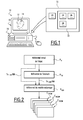
- an electronic system 10 for processing a volume model 12 with at least one object 14 to be added or subtracted from said model comprises an information processing unit 16 and a display screen 18 for displaying images, such as an image of the volume model 12, an initial image of the object 14 to be added or subtracted.
- the volume model 12 comprises a set of points P belonging to a plurality of parallel planes and positioned in a first reference frame R MRI , visible on the figure 2 .
- the volume model 12 has an amplitude, such as an intensity expressed in gray levels, at each of said points P.
- the volume model 12 is, for example, an anatomical model conforming to the DICOM format, and the first repository is then in accordance with the DICOM format.
- the invention relates to a three-dimensional model comprising a plurality of points distributed according to a spatial grid.
- Each point is assigned an intensity value, such as the gray level when the model is represented in black and white.
- the intensity is, for example, the intensity of the point in one or more wavelengths when the model is not monochrome.
- the object 14, noted S n comprises a plurality of points P n, i positioned in a second repository R n distinct from the first reference frame IRM .
- the object S n is constituted by a set of points P *.
- the method according to the invention makes it possible to add an object in the volume model, in which case the object is an auxiliary object, or to remove an object from the model.
- the object When the object is not initially included in the model, it is, for example, saved in a format separate from the DICOM format.
- the object is a device, called an implant, intended to be implanted in the body of a patient, for example a prosthesis, an electronic device, one or more electrodes, or screw-type surgical equipment. pins ....
- the processing system 10 is adapted to go directly from the second repository R n to the first reference R IRM volumic model.
- the information processing unit 16 visible on the figure 1 , comprises a data processor 20 and a memory 22 associated with the processor 20.
- the memory 22 is capable of storing software 24 for calculating, for each point P n, i of the object 14, an image point T n-> MRI (P n, i ) in the first frame of reference R MRI. using a transfer function T n-> IRM of the second repository R n to the first repository R IRM .
- the memory 22 is able to store a software 26 for modifying the amplitude of points of the volume model 12 via a correction function F n, i , associated with each image point T n-> IRM (P n , i ).
- This correction function F n, i is applied to each point of the model, or to points of the model situated in the vicinity of an image point T n-> IRM (P n, i ).
- the neighborhood of an image point designates the points of the adjacent volume model of said image point, or the points of the volume model closest to the image point.
- the value of the correction function F n, i depends on the position of each point of the model 12 with respect to the image point T n-> MRI (P n, i ) to which said function is associated.
- the calculation means 24 and the amplitude modification means 26 are made in the form of programmable logic components or in the form of dedicated integrated circuits.
- the correction function F n, i is adapted to modify the amplitude of certain points P of the volume model 12, in particular those situated in the vicinity of the image point with which the function is associated.
- the correction function F n, i is preferably adapted to modify the amplitude of each of the points P of the volume model 12. It makes it possible to modify the intensity of the points of the volume model 12 around each image point, that is to say in the vicinity of each image point.
- the three-dimensional volume model takes into account functional information in addition to the anatomical structure.
- the anatomical model describes a cortical surface
- it integrates a functional map of cortical activation.
- a reference change is made, so that the reference of the volume model corresponds to the so-called referential Talairach R Tal , well known in neurosurgery.
- the advantage of this variant is to be able to position the object in the model not only according to structural anatomical data, but also according to functional data. This variant is particularly suitable for positioning measurement electrodes or cortical stimulation.
- the transfer function T n-> MRI implemented by the calculation software 24 is known per se, the passage of the second reference frame R n to the first reference frame R IRM being for example carried out using a matrix of change of repository.
- the correction function F n, i associated with an image point of the volume model, may comprise a correction module M n, i depending on a Euclidean distance DIST (P, T n-> IRM (P n, i )) between said image point T n-> MRI (P n, i ) and the point P of the volume model, this Euclidean distance being measured in the first reference frame R MRI .
- the correction function F n, i is preferably equal to the corrective module M n, i multiplied by a weighting factor z n , the weighting factor z n depending on the object 14.
- the value of the weighting factor z n depends, for example, on the material of the added auxiliary object.
- the weighting factor z n takes a value even higher than the material is dense, such as a value substantially equal to 400 for a bone, and 1000 for metal. The adjustment of the value of this coefficient makes it possible to obtain an intensity level coherent with the intensity of the different materials represented in the volume model.
- the corrective module M n, i is a function of the Euclidean distance DIST (P, T n-> IRM (P n, i )), and this function is continuous and symmetrical around the image point, that is to say for values around the zero distance, its values being furthermore between 0 and 1.
- the value of the corrective module M n, i is for example equal to 1 for a zero value of the Euclidean distance DIST (P, T n-> IRM ( P n, i )) between the image point T n-> MRI (P n, i ) and the point P of the volume model 12, and the value of the corrective module M n, i decreases to 0 when the absolute value of said distance Euclidean DIST (P, T n-> MRI (P n, i )) increases.
- the correction function decreases around the image point.
- the value of the function, at a point of the model is closer to 0 than the distance between this point and the image point is high.
- the value of the corrective module M n, i is for example equal to 0 for a zero value of said Euclidean distance DIST (P, T n-> IRM (P n, i )), and the value of the corrective module M n, i increases to 1 when the absolute value of said Euclidean distance DIST (P, T n -> MRI (P n, i )) increases.
- the correction function is increasing around the image point.
- the value of the function, at a point of the model is closer to 0 than the distance between this point and the image point is small.
- the corrective module M n, i also depends on a spreading factor ⁇ n , the spreading factor ⁇ n being multiplied with the Euclidean distance DIST (P, T n-> MRI ( P n, i )).
- the spreading factor ⁇ n is a parameter for modifying the focusing of the function around the image point.
- the value of the spreading factor ⁇ n is preferably between 1 and 4.
- the spreading factor ⁇ n is, for example, equal to 1 to represent an electrode, and equal to 4 to represent a millimeter registration axis, or a piercing area.
- the correction function F n, i then preferably satisfies the following equation, according to equations (2) and (3) above:
- the correction function F n, i is preferably applied to each point P of the volume model 12 and for each index point i of each object 14 of index n.
- each object 14 to be added or subtracted from the volume model 12 is determined from object placement data.
- the object 14 is determined using data relating to the implant, when the object 14 is an implant to be added in an anatomical model, and with the aid of functional data.
- an operation to be performed on the volume model 12 consists of ablating the material of the volume model, then the object 14 is an object to be subtracted, and the shape of the object corresponding to the shape of the ablation of material.
- step 120 in order to calculate, for each point P n, i of the object 14, the image point T n-> IRM (P n, i ) in the first frame of reference R MRI at 1 help of the transfer function T n-> MRI .
- the volume model 12 and the image points T n-> MRI (P n, i ) associated with the object 14 are then all positioned in the first reference frame R IRM .
- the amplitude of certain points P of the volume model 12, preferably of all the points P of the volume model 12, is then, according to the invention, modified in the following step 130 via the correction function F n, i , the latter being especially applied in the vicinity of each image point.
- the correction function F n, i applied to the volume model 12 is, by way of example, that defined by equation (4).
- v int P min v origin P
- ⁇ P v fusion P max v int P , ⁇ P
- Equation (7) first allows to subtract the objects 14 intended to be subtracted to calculate an intermediate value v int , then equation (8) allows to add the objects 14 to be added to calculate the modified value amplitude v fusion .
- the data corresponding to the volume model 12 modified using the amplitude correction function is complementary, during the step 140, transmitted to another device, such as a surgical assistance robot, in order to to be exploited.
- Said transmitted data are, for example, sectional views in DICOM format, denoted C DICOM , as shown in FIG. figure 2 .
- the exploitation of the data corresponding to the modified volume model 12 consists of displaying the modified volume model on the display screen 18.
- the treatment system 10 and the treatment method according to the invention then make it possible to obtain a better rendering of the volume model 12 after adding or subtracting the object or objects 14.
- the visibility of each object 14 integrated in the volume model 12 is improved.
- volume model 12 is intended to be displayed on the display screen 18 or to be operated by another device, such as a surgical station.
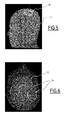
- the treatment system 10 and the treatment method according to the invention make it possible, for example, to better position the sensor or sensors of a direct neural control system, such as illustrated by way of example on Figures 4 to 6 .
- the objects 14 are each in the form of a cylinder, and correspond to the holes that will have to be made in the skull of the patient for the implantation of two implants each comprising a matrix of cortical electrodes, the locations of the two implants, and that matrices of electrodes being clearly visible on the figure 6 .
- the positioning of the electrode matrices was determined using an anatomical model integrating cortical functional mapping data. To do this, the repository of the volume model has previously been converted into the Talairach repository, as previously described.
- the object 14 is represented with a gray level close to the white color, which corresponds to an object to be subtracted, the objects 14 being in this example holes.
- the treatment system 10 and the treatment method according to the invention make it possible to obtain a more precise rendering of the model 12 following the addition or the subtraction of each object 14, in particular to more precisely visualize each object 14 when the volume model 12 is an imaging model intended to be displayed on the screen 18.
- the three-dimensional volumic model takes into account functional information, also called functional data, in addition to the anatomical structure.
Landscapes
- Engineering & Computer Science (AREA)
- Physics & Mathematics (AREA)
- General Physics & Mathematics (AREA)
- Theoretical Computer Science (AREA)
- Computer Graphics (AREA)
- Architecture (AREA)
- Computer Hardware Design (AREA)
- General Engineering & Computer Science (AREA)
- Software Systems (AREA)
- Magnetic Resonance Imaging Apparatus (AREA)
- Image Processing (AREA)
- Apparatus For Radiation Diagnosis (AREA)
Applications Claiming Priority (1)
| Application Number | Priority Date | Filing Date | Title |
|---|---|---|---|
| FR1363569A FR3015744B1 (fr) | 2013-12-24 | 2013-12-24 | Procede de traitement d'un modele volumique, produit programme d'ordinateur et systeme de traitement associes |
Publications (2)
| Publication Number | Publication Date |
|---|---|
| EP2889845A2 true EP2889845A2 (de) | 2015-07-01 |
| EP2889845A3 EP2889845A3 (de) | 2015-09-09 |
Family
ID=50639667
Family Applications (1)
| Application Number | Title | Priority Date | Filing Date |
|---|---|---|---|
| EP14199487.1A Withdrawn EP2889845A3 (de) | 2013-12-24 | 2014-12-19 | Datenverarbeitungsverfahren eines Volumenmodells, entsprechendes Computerprogramm und entsprechendes Verarbeitungssystem |
Country Status (3)
| Country | Link |
|---|---|
| US (1) | US9582925B2 (de) |
| EP (1) | EP2889845A3 (de) |
| FR (1) | FR3015744B1 (de) |
Families Citing this family (9)
| Publication number | Priority date | Publication date | Assignee | Title |
|---|---|---|---|---|
| WO2015142445A1 (en) * | 2014-03-21 | 2015-09-24 | St. Jude Medical, Cardiology Division, Inc. | Methods and systems for generating a multi-dimensional surface model of a geometric structure |
| WO2019060298A1 (en) | 2017-09-19 | 2019-03-28 | Neuroenhancement Lab, LLC | METHOD AND APPARATUS FOR NEURO-ACTIVATION |
| US11717686B2 (en) | 2017-12-04 | 2023-08-08 | Neuroenhancement Lab, LLC | Method and apparatus for neuroenhancement to facilitate learning and performance |
| US11478603B2 (en) | 2017-12-31 | 2022-10-25 | Neuroenhancement Lab, LLC | Method and apparatus for neuroenhancement to enhance emotional response |
| US12280219B2 (en) | 2017-12-31 | 2025-04-22 | NeuroLight, Inc. | Method and apparatus for neuroenhancement to enhance emotional response |
| US11364361B2 (en) | 2018-04-20 | 2022-06-21 | Neuroenhancement Lab, LLC | System and method for inducing sleep by transplanting mental states |
| US11452839B2 (en) | 2018-09-14 | 2022-09-27 | Neuroenhancement Lab, LLC | System and method of improving sleep |
| US10746667B2 (en) * | 2018-11-27 | 2020-08-18 | General Electric Company | Fluorescent penetrant inspection system and method |
| US11786694B2 (en) | 2019-05-24 | 2023-10-17 | NeuroLight, Inc. | Device, method, and app for facilitating sleep |
Citations (2)
| Publication number | Priority date | Publication date | Assignee | Title |
|---|---|---|---|---|
| US20080188741A1 (en) * | 2007-02-05 | 2008-08-07 | General Electric Company | Brain image alignment method and system |
| US20120008845A1 (en) * | 2008-03-10 | 2012-01-12 | University Of Rochester | Method and apparatus for 3d metal and high-density artifact correction for cone-beam and fan-beam ct imaging |
Family Cites Families (2)
| Publication number | Priority date | Publication date | Assignee | Title |
|---|---|---|---|---|
| US7505809B2 (en) * | 2003-01-13 | 2009-03-17 | Mediguide Ltd. | Method and system for registering a first image with a second image relative to the body of a patient |
| US8666478B2 (en) | 2009-10-08 | 2014-03-04 | The Medical College Of Wisconsin, Inc. | Method for determining locations of implanted electrodes with medical images |
-
2013
- 2013-12-24 FR FR1363569A patent/FR3015744B1/fr not_active Expired - Fee Related
-
2014
- 2014-12-19 EP EP14199487.1A patent/EP2889845A3/de not_active Withdrawn
- 2014-12-23 US US14/580,773 patent/US9582925B2/en not_active Expired - Fee Related
Patent Citations (2)
| Publication number | Priority date | Publication date | Assignee | Title |
|---|---|---|---|---|
| US20080188741A1 (en) * | 2007-02-05 | 2008-08-07 | General Electric Company | Brain image alignment method and system |
| US20120008845A1 (en) * | 2008-03-10 | 2012-01-12 | University Of Rochester | Method and apparatus for 3d metal and high-density artifact correction for cone-beam and fan-beam ct imaging |
Also Published As
| Publication number | Publication date |
|---|---|
| FR3015744A1 (fr) | 2015-06-26 |
| US9582925B2 (en) | 2017-02-28 |
| EP2889845A3 (de) | 2015-09-09 |
| US20150178978A1 (en) | 2015-06-25 |
| FR3015744B1 (fr) | 2017-05-05 |
Similar Documents
| Publication | Publication Date | Title |
|---|---|---|
| EP2889845A2 (de) | Datenverarbeitungsverfahren eines Volumenmodells, entsprechendes Computerprogramm und entsprechendes Verarbeitungssystem | |
| EP3785234B1 (de) | Automatisierte korrektur von metallbehafteten voxeldarstellungen von röntgendaten mit hilfe von tiefenlerntechniken | |
| CN110473243B (zh) | 基于深度轮廓感知的牙齿分割方法、装置及计算机设备 | |
| CA3095408C (en) | Systems and methods for automated detection and segmentation of vertebral centrum(s) in 3d images | |
| EP2435985B1 (de) | Verfahren zur quantifizierung von pathologien, welche veränderungen im volumen von körpern, insbesondere von tumoren einschliessen | |
| EP3965684A1 (de) | Verfahren zur planung von gewebeablation basierend auf tiefenlernen | |
| EP2988224A1 (de) | Lokalisierungsverfahren einer mit einer aufgabe verbundenen gehirnaktivität | |
| WO2014117108A1 (en) | Segmentation and identification of closed-contour features in images using graph theory and quasi-polar transform | |
| CN111192208B (zh) | 一种基于边窗滤波器的牙齿cr图像增强方法及装置 | |
| Dogra et al. | Bone vessel image fusion via generalized reisz wavelet transform using averaging fusion rule | |
| JP2021529622A (ja) | 網膜の光干渉断層撮影画像のセグメンテーションの方法及びコンピュータプログラム | |
| US11419727B2 (en) | Semi-automated imaging reconstruction for orbital fracture repair | |
| Nizamani et al. | Feature-enhanced fusion of U-NET-based improved brain tumor images segmentation | |
| Jeevakala et al. | A novel segmentation of cochlear nerve using region growing algorithm | |
| Bala et al. | Hybrid technique for fundus image enhancement using modified morphological filter and denoising net | |
| Mirajkar et al. | Acute ischemic stroke detection using wavelet based fusion of CT and MRI images | |
| FR3054692A1 (fr) | Procede de conception et de fabrication d'un appareillage personnalise dont la forme est adaptee a la morphologie d'un utilisateur | |
| EP3509483B1 (de) | System und verfahren zur rekonstruktion eines physiologischen signals einer dynamischen arterien-/gewebe-/venensystems eines organs in einem oberflächenareal | |
| Tarabichi et al. | Three-dimensional surface reconstruction of the human cochlear nucleus: implications for auditory brain stem implant design | |
| CN110648329B (zh) | 一种目标图像提取方法、系统及终端设备 | |
| CN113269772A (zh) | 一种图像分割方法和装置 | |
| CN112884699A (zh) | 用于分割图像数据的方法和图像处理设备和计算机程序产品 | |
| EP3652702A1 (de) | Verfahren und vorrichtung zur segmentierung von bildern durch automatische ausbreitung in eine (nil+1)-e dimension einer in dimension n eingeleiteten bildsegmentierung | |
| CN120163876A (zh) | 一种基于图像技术的麻醉穿刺辅助定位方法及系统 | |
| Vinodhini et al. | Swarm Based Enhancement Optimization Method for Image Enhancement for Diabetic Retinopathy Detection |
Legal Events
| Date | Code | Title | Description |
|---|---|---|---|
| PUAI | Public reference made under article 153(3) epc to a published international application that has entered the european phase |
Free format text: ORIGINAL CODE: 0009012 |
|
| 17P | Request for examination filed |
Effective date: 20141219 |
|
| AK | Designated contracting states |
Kind code of ref document: A2 Designated state(s): AL AT BE BG CH CY CZ DE DK EE ES FI FR GB GR HR HU IE IS IT LI LT LU LV MC MK MT NL NO PL PT RO RS SE SI SK SM TR |
|
| AX | Request for extension of the european patent |
Extension state: BA ME |
|
| PUAL | Search report despatched |
Free format text: ORIGINAL CODE: 0009013 |
|
| AK | Designated contracting states |
Kind code of ref document: A3 Designated state(s): AL AT BE BG CH CY CZ DE DK EE ES FI FR GB GR HR HU IE IS IT LI LT LU LV MC MK MT NL NO PL PT RO RS SE SI SK SM TR |
|
| AX | Request for extension of the european patent |
Extension state: BA ME |
|
| RIC1 | Information provided on ipc code assigned before grant |
Ipc: G06T 19/20 20110101AFI20150803BHEP Ipc: G06T 15/08 20110101ALN20150803BHEP Ipc: G06T 5/50 20060101ALI20150803BHEP |
|
| RIN1 | Information on inventor provided before grant (corrected) |
Inventor name: LABYT, ETIENNE Inventor name: DURAND, PIERRE |
|
| R17P | Request for examination filed (corrected) |
Effective date: 20160307 |
|
| RBV | Designated contracting states (corrected) |
Designated state(s): AL AT BE BG CH CY CZ DE DK EE ES FI FR GB GR HR HU IE IS IT LI LT LU LV MC MK MT NL NO PL PT RO RS SE SI SK SM TR |
|
| 17Q | First examination report despatched |
Effective date: 20171013 |
|
| GRAP | Despatch of communication of intention to grant a patent |
Free format text: ORIGINAL CODE: EPIDOSNIGR1 |
|
| RIC1 | Information provided on ipc code assigned before grant |
Ipc: G06T 19/20 20110101AFI20180628BHEP Ipc: G06T 15/08 20110101ALN20180628BHEP Ipc: G06T 5/50 20060101ALI20180628BHEP |
|
| INTG | Intention to grant announced |
Effective date: 20180718 |
|
| RIN1 | Information on inventor provided before grant (corrected) |
Inventor name: LABYT, ETIENNE Inventor name: DURAND, PIERRE |
|
| STAA | Information on the status of an ep patent application or granted ep patent |
Free format text: STATUS: THE APPLICATION IS DEEMED TO BE WITHDRAWN |
|
| 18D | Application deemed to be withdrawn |
Effective date: 20181129 |