EP2889050B1 - Method for sized-based cell separation using spinning membrane filtration - Google Patents
Method for sized-based cell separation using spinning membrane filtration Download PDFInfo
- Publication number
- EP2889050B1 EP2889050B1 EP14190423.5A EP14190423A EP2889050B1 EP 2889050 B1 EP2889050 B1 EP 2889050B1 EP 14190423 A EP14190423 A EP 14190423A EP 2889050 B1 EP2889050 B1 EP 2889050B1
- Authority
- EP
- European Patent Office
- Prior art keywords
- housing
- rotational speed
- cell type
- suspension
- spinning membrane
- Prior art date
- Legal status (The legal status is an assumption and is not a legal conclusion. Google has not performed a legal analysis and makes no representation as to the accuracy of the status listed.)
- Active
Links
- 238000009987 spinning Methods 0.000 title claims description 40
- 238000000034 method Methods 0.000 title claims description 28
- 238000000926 separation method Methods 0.000 title claims description 11
- 238000005374 membrane filtration Methods 0.000 title description 2
- 239000012528 membrane Substances 0.000 claims description 84
- 210000004027 cell Anatomy 0.000 claims description 34
- 210000003743 erythrocyte Anatomy 0.000 claims description 34
- 210000000265 leukocyte Anatomy 0.000 claims description 31
- 230000001413 cellular effect Effects 0.000 claims description 26
- 239000000725 suspension Substances 0.000 claims description 25
- 239000011148 porous material Substances 0.000 claims description 16
- 210000001772 blood platelet Anatomy 0.000 claims description 9
- 239000000463 material Substances 0.000 claims description 5
- 238000005204 segregation Methods 0.000 claims description 4
- 230000001225 therapeutic effect Effects 0.000 claims 1
- 239000000306 component Substances 0.000 description 17
- 210000004369 blood Anatomy 0.000 description 9
- 239000008280 blood Substances 0.000 description 9
- 210000002381 plasma Anatomy 0.000 description 9
- 239000012503 blood component Substances 0.000 description 6
- 230000014759 maintenance of location Effects 0.000 description 6
- 239000000047 product Substances 0.000 description 6
- 230000000717 retained effect Effects 0.000 description 6
- 230000001419 dependent effect Effects 0.000 description 3
- 238000002616 plasmapheresis Methods 0.000 description 3
- 239000012465 retentate Substances 0.000 description 3
- FAPWRFPIFSIZLT-UHFFFAOYSA-M Sodium chloride Chemical compound [Na+].[Cl-] FAPWRFPIFSIZLT-UHFFFAOYSA-M 0.000 description 2
- 230000017531 blood circulation Effects 0.000 description 2
- 239000006285 cell suspension Substances 0.000 description 2
- 238000009792 diffusion process Methods 0.000 description 2
- 238000007865 diluting Methods 0.000 description 2
- ZZUFCTLCJUWOSV-UHFFFAOYSA-N furosemide Chemical compound C1=C(Cl)C(S(=O)(=O)N)=CC(C(O)=O)=C1NCC1=CC=CO1 ZZUFCTLCJUWOSV-UHFFFAOYSA-N 0.000 description 2
- 238000002955 isolation Methods 0.000 description 2
- 230000000670 limiting effect Effects 0.000 description 2
- 210000005087 mononuclear cell Anatomy 0.000 description 2
- 239000002245 particle Substances 0.000 description 2
- 239000011780 sodium chloride Substances 0.000 description 2
- 206010018910 Haemolysis Diseases 0.000 description 1
- 239000000654 additive Substances 0.000 description 1
- 230000009286 beneficial effect Effects 0.000 description 1
- 239000000356 contaminant Substances 0.000 description 1
- 230000003247 decreasing effect Effects 0.000 description 1
- 238000000151 deposition Methods 0.000 description 1
- 238000010586 diagram Methods 0.000 description 1
- 230000004069 differentiation Effects 0.000 description 1
- 239000000706 filtrate Substances 0.000 description 1
- 238000001914 filtration Methods 0.000 description 1
- 239000012530 fluid Substances 0.000 description 1
- 238000005194 fractionation Methods 0.000 description 1
- 230000008588 hemolysis Effects 0.000 description 1
- 239000000696 magnetic material Substances 0.000 description 1
- 239000000203 mixture Substances 0.000 description 1
- 230000036961 partial effect Effects 0.000 description 1
- 230000002829 reductive effect Effects 0.000 description 1
- 239000006228 supernatant Substances 0.000 description 1
- 238000011144 upstream manufacturing Methods 0.000 description 1
- 238000005406 washing Methods 0.000 description 1
Images
Classifications
-
- A—HUMAN NECESSITIES
- A61—MEDICAL OR VETERINARY SCIENCE; HYGIENE
- A61M—DEVICES FOR INTRODUCING MEDIA INTO, OR ONTO, THE BODY; DEVICES FOR TRANSDUCING BODY MEDIA OR FOR TAKING MEDIA FROM THE BODY; DEVICES FOR PRODUCING OR ENDING SLEEP OR STUPOR
- A61M1/00—Suction or pumping devices for medical purposes; Devices for carrying-off, for treatment of, or for carrying-over, body-liquids; Drainage systems
- A61M1/34—Filtering material out of the blood by passing it through a membrane, i.e. hemofiltration or diafiltration
- A61M1/3496—Plasmapheresis; Leucopheresis; Lymphopheresis
-
- A—HUMAN NECESSITIES
- A61—MEDICAL OR VETERINARY SCIENCE; HYGIENE
- A61M—DEVICES FOR INTRODUCING MEDIA INTO, OR ONTO, THE BODY; DEVICES FOR TRANSDUCING BODY MEDIA OR FOR TAKING MEDIA FROM THE BODY; DEVICES FOR PRODUCING OR ENDING SLEEP OR STUPOR
- A61M1/00—Suction or pumping devices for medical purposes; Devices for carrying-off, for treatment of, or for carrying-over, body-liquids; Drainage systems
- A61M1/14—Dialysis systems; Artificial kidneys; Blood oxygenators ; Reciprocating systems for treatment of body fluids, e.g. single needle systems for hemofiltration or pheresis
- A61M1/16—Dialysis systems; Artificial kidneys; Blood oxygenators ; Reciprocating systems for treatment of body fluids, e.g. single needle systems for hemofiltration or pheresis with membranes
- A61M1/26—Dialysis systems; Artificial kidneys; Blood oxygenators ; Reciprocating systems for treatment of body fluids, e.g. single needle systems for hemofiltration or pheresis with membranes and internal elements which are moving
- A61M1/262—Dialysis systems; Artificial kidneys; Blood oxygenators ; Reciprocating systems for treatment of body fluids, e.g. single needle systems for hemofiltration or pheresis with membranes and internal elements which are moving rotating
- A61M1/265—Dialysis systems; Artificial kidneys; Blood oxygenators ; Reciprocating systems for treatment of body fluids, e.g. single needle systems for hemofiltration or pheresis with membranes and internal elements which are moving rotating inducing Taylor vortices
-
- A—HUMAN NECESSITIES
- A61—MEDICAL OR VETERINARY SCIENCE; HYGIENE
- A61M—DEVICES FOR INTRODUCING MEDIA INTO, OR ONTO, THE BODY; DEVICES FOR TRANSDUCING BODY MEDIA OR FOR TAKING MEDIA FROM THE BODY; DEVICES FOR PRODUCING OR ENDING SLEEP OR STUPOR
- A61M1/00—Suction or pumping devices for medical purposes; Devices for carrying-off, for treatment of, or for carrying-over, body-liquids; Drainage systems
- A61M1/36—Other treatment of blood in a by-pass of the natural circulatory system, e.g. temperature adaptation, irradiation ; Extra-corporeal blood circuits
- A61M1/3621—Extra-corporeal blood circuits
- A61M1/3627—Degassing devices; Buffer reservoirs; Drip chambers; Blood filters
- A61M1/3633—Blood component filters, e.g. leukocyte filters
-
- B—PERFORMING OPERATIONS; TRANSPORTING
- B01—PHYSICAL OR CHEMICAL PROCESSES OR APPARATUS IN GENERAL
- B01D—SEPARATION
- B01D63/00—Apparatus in general for separation processes using semi-permeable membranes
- B01D63/16—Rotary, reciprocated or vibrated modules
-
- A—HUMAN NECESSITIES
- A61—MEDICAL OR VETERINARY SCIENCE; HYGIENE
- A61M—DEVICES FOR INTRODUCING MEDIA INTO, OR ONTO, THE BODY; DEVICES FOR TRANSDUCING BODY MEDIA OR FOR TAKING MEDIA FROM THE BODY; DEVICES FOR PRODUCING OR ENDING SLEEP OR STUPOR
- A61M2205/00—General characteristics of the apparatus
- A61M2205/33—Controlling, regulating or measuring
- A61M2205/3365—Rotational speed
-
- B—PERFORMING OPERATIONS; TRANSPORTING
- B01—PHYSICAL OR CHEMICAL PROCESSES OR APPARATUS IN GENERAL
- B01D—SEPARATION
- B01D2315/00—Details relating to the membrane module operation
- B01D2315/02—Rotation or turning
Landscapes
- Health & Medical Sciences (AREA)
- Heart & Thoracic Surgery (AREA)
- Vascular Medicine (AREA)
- Public Health (AREA)
- Veterinary Medicine (AREA)
- Engineering & Computer Science (AREA)
- Anesthesiology (AREA)
- Biomedical Technology (AREA)
- Hematology (AREA)
- Life Sciences & Earth Sciences (AREA)
- Animal Behavior & Ethology (AREA)
- General Health & Medical Sciences (AREA)
- Urology & Nephrology (AREA)
- Emergency Medicine (AREA)
- Chemical & Material Sciences (AREA)
- Chemical Kinetics & Catalysis (AREA)
- Cardiology (AREA)
- Separation Using Semi-Permeable Membranes (AREA)
- External Artificial Organs (AREA)
- Measuring Or Testing Involving Enzymes Or Micro-Organisms (AREA)
- Micro-Organisms Or Cultivation Processes Thereof (AREA)
- Combined Means For Separation Of Solids (AREA)
- Separation Of Solids By Using Liquids Or Pneumatic Power (AREA)
- Filtration Of Liquid (AREA)
- Apparatus Associated With Microorganisms And Enzymes (AREA)
Description
- The present disclosure relates to a method for separating the cellular components of whole blood using a spinning membrane separator and, more particularly to a method of separating a selected cellular component that is not primarily dependent upon the nominal pore size of the membrane.
- Spinning porous membrane separators have been used to separate plasma from cellular components of whole blood. A well-known plasmapheresis device is the Plasmacell-C separator sold by Fenwal, Inc. of Lake Zurich, Illinois. A detailed description of a spinning membrane separator may be found in
U.S. Patent No. 5,194,145 to Schoendorfer . This patent describes a membrane-covered spinner having an interior collection system disposed within a stationary shell. Blood is fed into an annular space or gap between the spinner and the shell. The blood moves along the longitudinal axis of the shell toward an exit region, with plasma passing through the membrane and out of the shell into a collection bag. The remaining blood components move to the exit region between the spinner and the shell and then are typically returned to the donor. - Spinning membrane separators have been found to provide excellent filtration rates, due primarily to the unique flow patterns ("Taylor vortices") induced in the gap between the spinning membrane and the shell. The Taylor vortices help to sweep the surface of the membrane to inhibit the cellular components, primarily red blood cells, from depositing on and fouling or clogging the membrane. Spinning membranes can also be used for separating particles of a photographic suspension with particle size between 0.3 and 1.15 µm (
US 5,401,422 A ). - In membrane filtration, the identity of the filtrate is dependent primarily on size differentiation between the nominal pore size of the membrane and the cellular components of the retentate. For performing plasmapheresis by means of a spinning membrane, the nominal pore size is typically on the order of 0.65 µm, which allows plasma to pass through the membrane while retaining the bulk of the cellular blood components, namely white blood cells ("WBCs"), red blood cells ("RBCs") and platelets ("PLTs"). This retentate remains in the gap between the spinning membrane and the housing, and then exits the spinner housing. Thus, separation of WBCs, RBCs and PLTs from each other would require passing the retentate again through a separation device in which the membrane has a different nominal pore size, e.g., 4.0-5.0 µm, which would permit RBCs to pass through, but retain WBCs.
- By way of the present disclosure, methods are provided for separating the various blood components using a spinning membrane separator in which the type of cell that is separated is not solely dependent upon the nominal pore size of the membrane. Consequently, different resultant cell products may be obtained using a single spinning membrane separator.
- The present subject matter has a number of aspects which may be used in various combinations, and a disclosure of one or more specific embodiments is for the purpose of disclosure and description, and not limitation. This summary highlights only a few of the aspects of this subject matter, and additional aspects are disclosed in the drawings and the more detailed description that follows.
- A method is provided for separating a suspension of cellular material comprising at least two differently-sized cell types selected from red blood cells, white blood cells and platelets using a spinning membrane separator. The spinning membrane separator comprises a generally cylindrical housing having an interior wall, with an interior member is mounted therein that has an external surface. The interior wall of the housing and/or the external surface of the interior member includes a porous membrane that is spaced apart from the facing wall of the housing or surface of the interior member so as to define an annular gap therebetween. The housing and interior member are relatively rotatable, so that relative rotation of the housing and interior member creates a shear field in the gap having a force gradient, with higher forces adjacent the interior wall of the housing and the external surface of the interior member.
- In accordance with one aspect, the method comprises selecting the cell type to be separated by passing through the membrane; determining a concentration of the selected cell type in the suspension; selecting an inlet flow rate for the suspension; selecting a rotational speed for the spinning membrane separator related to one or more of the concentration and relative size of the selected cell type in the suspension; rotating the spinning membrane separator at the selected rotational speed so that the selected cell type tends to migrate to regions of the shear field adjacent the porous membrane; and flowing the suspension through the spinning membrane separator.
- In a further aspect, the method comprises selecting the rotational speed so that a higher concentration and/or smaller relative size for the selected cell type results in selection of a higher/faster rotational speed, and a lower concentration and/or larger relative size results in selection of a lower/slower rotational speed.
- In another aspect, the method comprises diluting the suspension prior to separation to enhance the susceptibility of the cellular material to segregation by cell type within shear fields formed within the spinning membrane separator.
- In further aspect, the method further comprises the spinning membrane separator having a default rotational speed, and adjusting the rotational speed from the default speed to the selected speed.
- In a particular application, the suspension comprises red blood cells, white blood cells, platelets and plasma, the spinning membrane separator has a nominal pore size of 4.0-5.0 µm, the selected cell type is red blood cells, and the default rotational speed is 3000 rpm.
- These and other features of the present subject matter are described in the following detailed description and shown in the attached figures, of which:
-
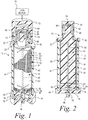
Fig. 1 is a perspective view of a spinning membrane separator, in partial cross section and with portions removed to show detail. -
Fig. 2 is a longitudinal cross sectional view of the spinning membrane separator ofFig. 1 . -
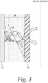
Fig. 3 is a schematic diagram illustrating the shear field in the gap and the relative diffusion of cellular components in the shear field based on size. -
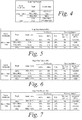
Fig. 4 is a table identifying five different possible debulking protocols with respect to three different source types (dRBC, dBC, and MNC) that may be performed in accordance with the methods of the present application. -
Figs. 5, 6 and 7 are tables outlining protocols for specific targeted cellular components for dRBC, dBC and MNC products in accordance with the methods of the present application. - A more detailed description of the methods in accordance with the present disclosure is set forth below. It should be understood that the description below of specific devices and methods is intended to be exemplary, and not exhaustive of all possible variations or applications. Thus, the scope of the disclosure is not intended to be limiting, and should be understood to encompass variations or embodiments that would occur to persons of ordinary skill.
- Turning to
Figs. 1 and 2 , a spinning membrane blood separation or fractionation system, generally designated 10, is shown. Such asystem 10 has primarily been used to extract plasma from whole blood obtained from an individual human donor. However, as described in more detail below, the device may also be used for isolating the cellular blood components. For ease of understanding, only the separation device and the associated drive unit are shown, although it should be understood that such a separator forms part of a disposable system including collection bags, bags of additives such as saline or ACD, return bags, tubing, etc., and that there are also associated control and instrumentation systems for operation of the device. - The
system 10 includes a generallycylindrical housing 12, mounted concentrically about a longitudinal vertical central axis. Aninternal member 14 is mounted concentric with the central axis. The housing and internal member is relatively rotatable. In the preferred embodiment, as illustrated, the housing is stationary and the internal member is a rotating spinner that is rotatable concentrically within thecylindrical housing 12. The boundaries of the blood flow path are generally defined by thegap 16 between the interior surface of thehousing 12 and the exterior surface of therotary spinner 14. The spacing between the housing and the spinner is sometimes referred to as the shear gap. A typical shear gap may be approximately 0.025 -0.050 inches (0.067-0.127 cm) and may be of a uniform dimension along the axis, for example, where the axis of the spinner and housing are coincident. The shear gap may also vary circumferentially for example, where the axis of the housing and spinner are offset. - The shear gap also may vary along the axial direction, for example preferably an increasing gap width in the direction of flow to limit hemolysis. Such a gap width may range from about 0.025 to about 0.075 inches (0.06 - 0.19 cm). For example the axes of the housing and rotor could be coincident and the diameter of the rotor decrease in the axial direction (direction of flow) while the diameter of inner surface of the housing remains constant or the diameter of the housing increases while the rotor diameter remains constant, or both surfaces vary in diameter. For example the gap width may be about 0.035 inches (0.088 cm) at the upstream or inlet end of the gap and about 0.059 inches (0.15 cm) at the downstream end or terminus of the gap. The gap width could be varied by varying the outer diameter of the rotor and/or the inner diameter of the facing housing surface. The gap width could change linearly or stepwise or in some other manner as may be desired. In any event, the width dimension of the gap is preferably selected so that at the desired relative rotational speed or speeds, Taylor-Couette flow, such as Taylor vortices, are created in the gap.
- Whole blood is fed from an inlet conduit 20 through an
inlet orifice 22, which directs the blood into the blood flow entrance region in a path tangential to the circumference about the upper end of thespinner 14. At the bottom end of thecylindrical housing 12, the housing inner wall includes an exit orifice 34. - The
cylindrical housing 12 is completed by anupper end cap 40 having anend boss 42, the walls of which are nonmagnetic, and abottom end housing 44 terminating in aplasma outlet orifice 46 concentric with the central axis. - The
spinner 14 is rotatably mounted between theupper end cap 40 and thebottom end housing 44. Thespinner 14 comprises a shaped central mandrel orrotor 50, the outer surface of which is shaped to define a series of spaced-apart circumferential grooves orribs 52 separated byannular lands 54. The surface channels defined by thecircumferential grooves 52 are interconnected bylongitudinal grooves 56. At each end of themandrel 50, thesegrooves 56 are in communication with a central orifice ormanifold 58. - In the illustrated embodiment, the surface of the
rotary spinner 14 is at least partially, and is preferably substantially or entirely, covered by a cylindricalporous membrane 62. If thesystem 10 is being used for plasmapheresis, themembrane 62 typically has a nominal pore size of 0.65 microns, but, as described below, other pore sizes may be used if isolation and separation of the cellular blood components is desired. Membranes useful in the methods described herein may be fibrous mesh membranes, cast membranes, track etched membranes or other types of membranes that will be known to those of skill in the art. - The rotary spinner is mounted in the upper end cap to rotate about a
pin 64, which is press fit into theend cap 40 on one side and seated within acylindrical bearing surface 65 in anend cylinder 66 forming part of therotary spinner 14. The internal spinner or outer housing may be rotated by any suitable rotary drive device or system. As illustrated, theend cylinder 66 is partially encompassed by aring 68 of magnetic material utilized in indirect driving of thespinner 14. Adrive motor 70 exterior to thehousing 12 is coupled to turn an annularmagnetic drive member 72 that includes at least a pair of interiorpermanent magnets 74. As theannular drive member 72 is rotated, magnetic attraction between thering 68 interior to thehousing 12 and themagnets 74 exterior to the housing locks thespinner 14 to the exterior drive, causing thespinner 14 to rotate. - At the lower end of the
rotary spinner 14, thecentral outlet orifice 58 communicates with acentral bore 76 in an end bearing 78 that is concentric with the central axis. An end bearing seat is defined by aninternal shoulder 80 that forms a lower edge of acentral opening 82. Thecentral opening 82 communicates with theplasma outlet orifice 46. If the inner facing surface of the housing is covered entirely or partially by a membrane, a fluid collection or manifold may be provided beneath the membrane to collect plasma and direct it through a housing outlet (not shown). - In accordance with the present disclosure, a separation method utilizing a spinning membrane separator is provided in which the nominal pore size of the membrane is selected and the gradient of the shear forces in the gap between the membrane and the housing is manipulated to cause diffusion of the cellular components to be retained in the gap away from the membrane to inhibit transport of that cellular component through the membrane and to enhance passage of the selected cellular component through the membrane. More specifically, the shear gradient is controlled by varying the rotational speed of the membrane relative to the housing by selecting a rotational speed related to one or more of the concentration and relative size of the cell type in the suspension to be passed through the membrane, such that a higher concentration and/or smaller relative size results in selection of a higher/faster rotational speed, and a lower concentration and/or larger relative size results in selection of a lower/slower rotational speed.
- As illustrated in
Fig. 3 , when themembrane 100 is rotated relative to thehousing 102, ashear gradient 104 forms across thegap 106 such that the highest shear forces (represented by the longest field lines) are encountered adjacent the surfaces of the membrane and housing, with the shear force theoretically decreasing to zero at the center or midway point of the gap. The principal cellular components of whole blood are, from largest to smallest in size, white blood cells, red blood cells, and platelets. Mature normal RBCs, which have no nucleus and are typically discoid in shape, have a diameter of about 7 µm and a thickness of about 2 µm. Although not perfectly spherical, WBCs typically have an outer diameter of a minimum of about 4.5 µm to about 20 µm, with a nucleus of typically 3.8 to 4 µm or greater. Platelets are discoid in shape and typically 2-3 µm in diameter. When a suspension of cellular blood components is introduced into the gap, the cells will diffuse spatially to minimize their presence in the shear field. Specifically, WBCs, being the largest, will be subjected to the largest shear forces and will be driven to the center of the gap. RBCs, being the next largest, will be driven to the center of the gap to the extent that this space is not already occupied by WBCs. PLTS will have the least force driving them toward the center, and this force is likely to be offset by the larger cells already occupying this space. - The shape and size of the pores in the membrane, as well as the spacing between the pores, can be selected depending on the identity of the cellular components to be passed through, and the relative deformability of the different cell types may be taken into account. For example, it is known that normal RBCs are relatively more readily deformable than WBCs, and deform faster and under less force than WBCs. Thus, if a nominal pore size of 4.0 µm-5.0 µm is selected for the membrane, WBCs will be retained and RBCs and PLTs can pass through the membrane. Accordingly, by appropriate selection of the rotational speed of the membrane the bulk of the PLTS and/or the bulk of the RBCs can each be separated from the bulk of the WBCs. For example a suspension of WBCs, RBCs and PLTS could be introduced into the gap of the spinning membrane separator, and the rotational speed selected so that the bulk of the WBCs and RBCs are maintained in the center of the gap to first separate the PLTS (with minimal RBCs) by passing through the membrane. Then the rotational speed adjusted (reduced) to cause the RBCs to migrate toward the membrane to separate the bulk of the RBCs from the WBCs by passing the RBCs through the membrane, while the WBCs are maintained in the gap. Depending on the relative concentrations of the various cellular components in the suspension, it may be necessary to dilute the suspension (with, e.g., saline) to lower the concentrations and thus enhance the susceptibility of the cellular components to segregation by cell type when subjected to the shear field. Thus, a single spinning membrane separator, appropriately controlled, could be used to obtain concentrations of each of WBCs, RBCs and PLTs.
- The method may also be advantageously used for various "single pass" separation protocols, including debulking or WBC removal (where WBCs are retained, and RBCs and PLTs are passed through the membrane), platelet removal (where WBCs and RBCs are retained, and PLTs passed through the membrane), WBC removal (where WBCs are retained and RBCs and PLTs passed through the membrane), washing operations (where a source supernatant is replaced with a new medium while retaining all of the cells within the source product), and isolation of a single cell-type product from a WBC/RBC cell suspension or from a RBC/PLT cell suspension.
- Specifically, the method according to the present disclosure controls three variables to selectively separate selected cellular components using a spinning membrane separator, namely inlet flow rate, the source product composition, and the membrane spin speed. Testing was performed to establish various single pass protocols. Five different debulking protocols, as set forth in Table 4, were established with respect to three different source types: diluted RBCs (dRBC), diluted buffy coat (dBC), and mononuclear cells (MNCs). It should be appreciated that the five identified protocols are only a subset of all the plausible protocols, and are presented herein for purposes of illustration, and not limitation.
- Protocols were established using a spinning membrane separator as described above having a PCTE membrane with a nominal pore size of 4.0 µm. In establishing the flow rates and spin speeds for the protocols, minimum retention levels for the various cellular components are set. It has been determined that flow rates become a factor in WBC retention at speeds lower than 2000 rpm, with WBC retention being highest at flow rates of 50 mL/min and lowest at 150 ml/min. Flow rates become a factor in RBC retention at speeds lower than 2500 rpm, where slower flow rates lead to higher RBC retention (similar to WBCs). Platelets behave differently than WBCs and RBCs, in that flow rate and spin speed do not appear to affect PLT retention, which is approximately 20%.
Figs 5, 6 and7 outline protocols for specific targeted cellular components for dRBC, dBC and MNC products, specifying the target cells and contaminants, the percentage of the source retained by cell type, the flow rate and spin speed. - Thus, this can be seen from the above description, the present disclosure has several different aspects which are not limited to the specific methods and apparatus shown in the attached drawings or described above. Variations of these concepts may be embodied in other steps for carrying out the methods and apparatus without departing from the scope of the disclosure.
- Aspects of the present subject matter described above may be beneficial alone or in combination with one or more other aspects. Without limiting the foregoing description, in accordance with one aspect of the subject matter herein, there is provided In accordance with one aspect, the method comprises selecting the cell type to be separated by passing through the membrane; determining a concentration of the selected cell type in the suspension; selecting an inlet flow rate for the suspension; selecting a rotational speed for the spinning membrane separator related to one or more of the concentration and relative size of the selected cell type in the suspension; rotating the spinning membrane separator at the selected rotational speed so that the selected cell type tends to migrate to regions of the shear field adjacent the porous membrane; and flowing the suspension through the spinning membrane separator.
- In a further aspect, the method comprises selecting the rotational speed so that a higher concentration and/or smaller relative size for the selected cell type results in selection of a higher/faster rotational speed, and a lower concentration and/or larger relative size results in selection of a lower/slower rotational speed.
- In another aspect, the method comprises diluting the suspension prior to separation to enhance the susceptibility of the cellular material to segregation by cell type within shear fields formed within the spinning membrane separator.
- In further aspect, the method further comprises the spinning membrane separator having a default rotational speed, and adjusting the rotational speed from the default speed to the selected speed.
- In a particular application, the suspension comprises red blood cells, white blood cells, platelets and plasma, the spinning membrane separator has a nominal pore size of 4.0-5.0 µm, the selected cell type is red blood cells, and the default rotational speed is 3000 rpm.
Claims (6)
- A non-therapeutic and non-surgical method for separating a suspension of cellular material comprising at least two differently-sized cell types selected from red blood cells, white blood cells and platelets using a spinning membrane separator (10), the steps comprising:a) selecting the cell type to be separated by passing through the membrane (62);b) determining a concentration of each different cell type in the suspension;c) selecting an inlet flow rate for the suspension;d) selecting a rotational speed for the spinning membrane separator (10) related to the concentration and size of the selected cell type in the suspension relative to each non-selected cell type;e) rotating the spinning membrane separator (10) at the selected rotational speed; andf) flowing the suspension through the spinning membrane separator (10).
- The method according to claim 1 wherein the rotational speed of the spinner (14) is selected such that a higher relative concentration and/or smaller relative size for the selected cell type results in selection of a higher/faster rotational speed and a lower relative concentration and/or larger relative size for the selected cell type results in selection of a lower/slower rotational speed.
- The method according to one of claims 1 or 2 wherein the suspension is diluted prior to separation to enhance the susceptibility of the cellular material to segregation by cell type within shear fields formed within the spinning membrane separator (10).
- The method according to one of claims 1 to 3 further comprising the spinning membrane separator (10) having a pre-determined nominal pore size and a default rotational speed, and adjusting the rotational speed from the default speed to the selected speed.
- The method according to claim 4 wherein the selected cell type is red blood cells, the nominal pore size is 4µm and the default rotational speed is 3000 rpm.
- The method according to one of claims 1 to 5, the spinning membrane separator (10) comprising a generally cylindrical housing (12) having an interior wall; an interior member mounted interior of the housing (12) and having an external surface; the interior wall of the housing (12) and/or the external surface of the interior member (14) including a porous membrane (62) spaced apart from the facing wall of the housing (12) or surface of the interior member (14) so as to define an annular gap (16) therebetween; the housing (12) and interior member (14) being relatively rotatable; wherein relative rotation of the housing (12) and interior member (14) creates a shear field in the gap having a force gradient with higher forces adjacent the interior wall of the housing (12) and the external surface of the interior member (14).
Applications Claiming Priority (1)
| Application Number | Priority Date | Filing Date | Title |
|---|---|---|---|
| US14/140,978 US9713669B2 (en) | 2013-12-26 | 2013-12-26 | Method for sized-based cell separation using spinning membrane filtration |
Publications (2)
| Publication Number | Publication Date |
|---|---|
| EP2889050A1 EP2889050A1 (en) | 2015-07-01 |
| EP2889050B1 true EP2889050B1 (en) | 2019-08-21 |
Family
ID=51790616
Family Applications (1)
| Application Number | Title | Priority Date | Filing Date |
|---|---|---|---|
| EP14190423.5A Active EP2889050B1 (en) | 2013-12-26 | 2014-10-27 | Method for sized-based cell separation using spinning membrane filtration |
Country Status (6)
| Country | Link |
|---|---|
| US (1) | US9713669B2 (en) |
| EP (1) | EP2889050B1 (en) |
| JP (1) | JP6453646B2 (en) |
| KR (1) | KR102330185B1 (en) |
| AU (1) | AU2014277687B2 (en) |
| NZ (1) | NZ700425A (en) |
Families Citing this family (3)
| Publication number | Priority date | Publication date | Assignee | Title |
|---|---|---|---|---|
| US10688233B2 (en) * | 2014-12-19 | 2020-06-23 | Fenwal, Inc. | Method and system for the automated processing of biological fluid during low level alert condition |
| JP7243835B2 (en) * | 2020-03-11 | 2023-03-22 | 株式会社レゾナック | CELL COLLECTION DEVICE, CELL COLLECTION METHOD, CELL SEPARATION SYSTEM, AND CELL SEPARATION METHOD |
| JP2022037418A (en) * | 2020-08-25 | 2022-03-09 | ソニーグループ株式会社 | Sample preparation apparatus and sample preparation system |
Family Cites Families (16)
| Publication number | Priority date | Publication date | Assignee | Title |
|---|---|---|---|---|
| DE3279765D1 (en) | 1981-07-22 | 1989-07-20 | Du Pont | Plasmapheresis by reciprocatory pulsatile filtration |
| US5034135A (en) | 1982-12-13 | 1991-07-23 | William F. McLaughlin | Blood fractionation system and method |
| US4911847A (en) | 1983-12-20 | 1990-03-27 | Membrex, Inc. | Process for controlling the permeate composition in a rotary filtration device |
| WO1985004112A1 (en) | 1984-03-21 | 1985-09-26 | Mclaughlin, William, Francis | Method and apparatus for filtration |
| DE3631804A1 (en) | 1986-09-18 | 1988-03-24 | Altenburger Electronic Gmbh | Process and apparatus for producing microfilters, and a microfilter produced accordingly |
| GB8628723D0 (en) | 1986-12-02 | 1987-01-07 | Bellhouse Brian John | Particle separation |
| JPS6475015A (en) | 1987-09-18 | 1989-03-20 | Terumo Corp | Filter for separating leukocytes |
| DE3742770A1 (en) | 1987-12-17 | 1989-06-29 | Akzo Gmbh | MICRO / ULTRAFILTRATION MEMBRANES WITH DEFINED PORO SIZE BY IRRADIATION WITH PULSE LASERS AND METHOD FOR THE PRODUCTION THEREOF |
| US4994188A (en) * | 1988-02-05 | 1991-02-19 | Baxter International Inc. | Adaptive filtrate flow control system using controlled reduction in filter efficiency |
| FR2668076B1 (en) | 1990-10-18 | 1993-04-30 | Kodak Pathe | METHOD FOR SEPARATING SOLID CONSTITUENTS FROM A SUSPENSION AND DEVICE FOR CARRYING OUT SAID METHOD. |
| NL9200902A (en) | 1992-05-21 | 1993-12-16 | Cornelis Johannes Maria Van Ri | CERAMIC MICROFILTRATION MEMBRANE AND METHOD FOR MANUFACTURING SUCH MEMBRANE. |
| JP3311091B2 (en) | 1993-06-27 | 2002-08-05 | テルモ株式会社 | Filter for separating white blood cells and filter for separating white blood cells and platelets |
| NL9401260A (en) | 1993-11-12 | 1995-06-01 | Cornelis Johannes Maria Van Ri | Membrane for microfiltration, ultrafiltration, gas separation and catalysis, method for manufacturing such a membrane, mold for manufacturing such a membrane, as well as various separation systems comprising such a membrane. |
| US5807406A (en) | 1994-10-07 | 1998-09-15 | Baxter International Inc. | Porous microfabricated polymer membrane structures |
| TW391881B (en) | 1996-09-25 | 2000-06-01 | Baxter Int | Method and apparatus for filtering suspensions of medical and biological fluids or the like |
| JP2000237633A (en) * | 1999-02-19 | 2000-09-05 | Matsushita Electric Ind Co Ltd | Clarifying device using centrifugal filter, and contamination-detecting sensor using the filter |
-
2013
- 2013-12-26 US US14/140,978 patent/US9713669B2/en active Active
-
2014
- 2014-09-26 NZ NZ700425A patent/NZ700425A/en unknown
- 2014-10-27 EP EP14190423.5A patent/EP2889050B1/en active Active
- 2014-11-28 KR KR1020140168193A patent/KR102330185B1/en active IP Right Grant
- 2014-12-16 AU AU2014277687A patent/AU2014277687B2/en active Active
- 2014-12-26 JP JP2014265423A patent/JP6453646B2/en active Active
Non-Patent Citations (1)
| Title |
|---|
| None * |
Also Published As
| Publication number | Publication date |
|---|---|
| US9713669B2 (en) | 2017-07-25 |
| AU2014277687B2 (en) | 2018-10-04 |
| AU2014277687A1 (en) | 2015-07-16 |
| JP2015164414A (en) | 2015-09-17 |
| US20150182682A1 (en) | 2015-07-02 |
| EP2889050A1 (en) | 2015-07-01 |
| KR102330185B1 (en) | 2021-11-24 |
| JP6453646B2 (en) | 2019-01-16 |
| NZ700425A (en) | 2016-07-29 |
| KR20150076076A (en) | 2015-07-06 |
Similar Documents
| Publication | Publication Date | Title |
|---|---|---|
| CA2237019C (en) | System for filtering medical and biological fluids | |
| US4808307A (en) | Couette membrane filtration apparatus for separating suspended components in a fluid medium using high shear | |
| US4755300A (en) | Couette membrane filtration apparatus for separating suspended components in a fluid medium using high shear | |
| CA1261765A (en) | Method and apparatus for separation of matter from suspension | |
| Beaudoin et al. | Plasma filtration in Couette flow membrane devices | |
| EP2683471B1 (en) | Membrane separation devices, systems and methods employing same | |
| EP2889050B1 (en) | Method for sized-based cell separation using spinning membrane filtration | |
| JPH0536105B2 (en) | ||
| US9033858B2 (en) | Method and apparatus for concentrating platelets from platelet-rich plasma | |
| US10729829B2 (en) | Systems and methods for platelet concentration with a spinning membrane separator | |
| EP3187248B1 (en) | Membrane separation device having improved filtration velocity | |
| EP3137127B1 (en) | Method for controlling fouling and complement protein activation during spinning membrane filtration of plasma from whole blood | |
| EP0310205A2 (en) | Filtering a liquid suspension | |
| EP3243561B1 (en) | Method for controlling fouling during a spinning membrane filtration procedure | |
| JP6621482B2 (en) | System and method for leukocyte reduction of fluid containing red blood cells and concentrated red blood cells | |
| EP4008374A1 (en) | Priming of spinning membrane separators | |
| WO2022225800A1 (en) | Blood filtration device | |
| Chen et al. | Platelet concentrates preparation using a rotating membrane with Taylor vortices and axial flow |
Legal Events
| Date | Code | Title | Description |
|---|---|---|---|
| PUAI | Public reference made under article 153(3) epc to a published international application that has entered the european phase |
Free format text: ORIGINAL CODE: 0009012 |
|
| 17P | Request for examination filed |
Effective date: 20141027 |
|
| AK | Designated contracting states |
Kind code of ref document: A1 Designated state(s): AL AT BE BG CH CY CZ DE DK EE ES FI FR GB GR HR HU IE IS IT LI LT LU LV MC MK MT NL NO PL PT RO RS SE SI SK SM TR |
|
| AX | Request for extension of the european patent |
Extension state: BA ME |
|
| R17P | Request for examination filed (corrected) |
Effective date: 20160104 |
|
| RBV | Designated contracting states (corrected) |
Designated state(s): AL AT BE BG CH CY CZ DE DK EE ES FI FR GB GR HR HU IE IS IT LI LT LU LV MC MK MT NL NO PL PT RO RS SE SI SK SM TR |
|
| STAA | Information on the status of an ep patent application or granted ep patent |
Free format text: STATUS: EXAMINATION IS IN PROGRESS |
|
| 17Q | First examination report despatched |
Effective date: 20180314 |
|
| REG | Reference to a national code |
Ref country code: DE Ref legal event code: R079 Ref document number: 602014052069 Country of ref document: DE Free format text: PREVIOUS MAIN CLASS: A61M0001340000 Ipc: A61M0001260000 |
|
| GRAP | Despatch of communication of intention to grant a patent |
Free format text: ORIGINAL CODE: EPIDOSNIGR1 |
|
| STAA | Information on the status of an ep patent application or granted ep patent |
Free format text: STATUS: GRANT OF PATENT IS INTENDED |
|
| RIC1 | Information provided on ipc code assigned before grant |
Ipc: A61M 1/26 20060101AFI20190225BHEP Ipc: B01D 63/16 20060101ALI20190225BHEP Ipc: A61M 1/34 20060101ALI20190225BHEP Ipc: A61M 1/36 20060101ALI20190225BHEP |
|
| INTG | Intention to grant announced |
Effective date: 20190402 |
|
| GRAS | Grant fee paid |
Free format text: ORIGINAL CODE: EPIDOSNIGR3 |
|
| GRAA | (expected) grant |
Free format text: ORIGINAL CODE: 0009210 |
|
| STAA | Information on the status of an ep patent application or granted ep patent |
Free format text: STATUS: THE PATENT HAS BEEN GRANTED |
|
| AK | Designated contracting states |
Kind code of ref document: B1 Designated state(s): AL AT BE BG CH CY CZ DE DK EE ES FI FR GB GR HR HU IE IS IT LI LT LU LV MC MK MT NL NO PL PT RO RS SE SI SK SM TR |
|
| REG | Reference to a national code |
Ref country code: GB Ref legal event code: FG4D |
|
| REG | Reference to a national code |
Ref country code: CH Ref legal event code: EP |
|
| REG | Reference to a national code |
Ref country code: DE Ref legal event code: R096 Ref document number: 602014052069 Country of ref document: DE |
|
| REG | Reference to a national code |
Ref country code: AT Ref legal event code: REF Ref document number: 1168983 Country of ref document: AT Kind code of ref document: T Effective date: 20190915 |
|
| REG | Reference to a national code |
Ref country code: IE Ref legal event code: FG4D |
|
| REG | Reference to a national code |
Ref country code: LT Ref legal event code: MG4D |
|
| REG | Reference to a national code |
Ref country code: NL Ref legal event code: MP Effective date: 20190821 |
|
| PG25 | Lapsed in a contracting state [announced via postgrant information from national office to epo] |
Ref country code: BG Free format text: LAPSE BECAUSE OF FAILURE TO SUBMIT A TRANSLATION OF THE DESCRIPTION OR TO PAY THE FEE WITHIN THE PRESCRIBED TIME-LIMIT Effective date: 20191121 Ref country code: SE Free format text: LAPSE BECAUSE OF FAILURE TO SUBMIT A TRANSLATION OF THE DESCRIPTION OR TO PAY THE FEE WITHIN THE PRESCRIBED TIME-LIMIT Effective date: 20190821 Ref country code: NO Free format text: LAPSE BECAUSE OF FAILURE TO SUBMIT A TRANSLATION OF THE DESCRIPTION OR TO PAY THE FEE WITHIN THE PRESCRIBED TIME-LIMIT Effective date: 20191121 Ref country code: NL Free format text: LAPSE BECAUSE OF FAILURE TO SUBMIT A TRANSLATION OF THE DESCRIPTION OR TO PAY THE FEE WITHIN THE PRESCRIBED TIME-LIMIT Effective date: 20190821 Ref country code: FI Free format text: LAPSE BECAUSE OF FAILURE TO SUBMIT A TRANSLATION OF THE DESCRIPTION OR TO PAY THE FEE WITHIN THE PRESCRIBED TIME-LIMIT Effective date: 20190821 Ref country code: LT Free format text: LAPSE BECAUSE OF FAILURE TO SUBMIT A TRANSLATION OF THE DESCRIPTION OR TO PAY THE FEE WITHIN THE PRESCRIBED TIME-LIMIT Effective date: 20190821 Ref country code: PT Free format text: LAPSE BECAUSE OF FAILURE TO SUBMIT A TRANSLATION OF THE DESCRIPTION OR TO PAY THE FEE WITHIN THE PRESCRIBED TIME-LIMIT Effective date: 20191223 Ref country code: HR Free format text: LAPSE BECAUSE OF FAILURE TO SUBMIT A TRANSLATION OF THE DESCRIPTION OR TO PAY THE FEE WITHIN THE PRESCRIBED TIME-LIMIT Effective date: 20190821 |
|
| PG25 | Lapsed in a contracting state [announced via postgrant information from national office to epo] |
Ref country code: LV Free format text: LAPSE BECAUSE OF FAILURE TO SUBMIT A TRANSLATION OF THE DESCRIPTION OR TO PAY THE FEE WITHIN THE PRESCRIBED TIME-LIMIT Effective date: 20190821 Ref country code: GR Free format text: LAPSE BECAUSE OF FAILURE TO SUBMIT A TRANSLATION OF THE DESCRIPTION OR TO PAY THE FEE WITHIN THE PRESCRIBED TIME-LIMIT Effective date: 20191122 Ref country code: IS Free format text: LAPSE BECAUSE OF FAILURE TO SUBMIT A TRANSLATION OF THE DESCRIPTION OR TO PAY THE FEE WITHIN THE PRESCRIBED TIME-LIMIT Effective date: 20191221 Ref country code: AL Free format text: LAPSE BECAUSE OF FAILURE TO SUBMIT A TRANSLATION OF THE DESCRIPTION OR TO PAY THE FEE WITHIN THE PRESCRIBED TIME-LIMIT Effective date: 20190821 Ref country code: RS Free format text: LAPSE BECAUSE OF FAILURE TO SUBMIT A TRANSLATION OF THE DESCRIPTION OR TO PAY THE FEE WITHIN THE PRESCRIBED TIME-LIMIT Effective date: 20190821 Ref country code: ES Free format text: LAPSE BECAUSE OF FAILURE TO SUBMIT A TRANSLATION OF THE DESCRIPTION OR TO PAY THE FEE WITHIN THE PRESCRIBED TIME-LIMIT Effective date: 20190821 |
|
| REG | Reference to a national code |
Ref country code: AT Ref legal event code: MK05 Ref document number: 1168983 Country of ref document: AT Kind code of ref document: T Effective date: 20190821 |
|
| PG25 | Lapsed in a contracting state [announced via postgrant information from national office to epo] |
Ref country code: TR Free format text: LAPSE BECAUSE OF FAILURE TO SUBMIT A TRANSLATION OF THE DESCRIPTION OR TO PAY THE FEE WITHIN THE PRESCRIBED TIME-LIMIT Effective date: 20190821 |
|
| PG25 | Lapsed in a contracting state [announced via postgrant information from national office to epo] |
Ref country code: DK Free format text: LAPSE BECAUSE OF FAILURE TO SUBMIT A TRANSLATION OF THE DESCRIPTION OR TO PAY THE FEE WITHIN THE PRESCRIBED TIME-LIMIT Effective date: 20190821 Ref country code: EE Free format text: LAPSE BECAUSE OF FAILURE TO SUBMIT A TRANSLATION OF THE DESCRIPTION OR TO PAY THE FEE WITHIN THE PRESCRIBED TIME-LIMIT Effective date: 20190821 Ref country code: IT Free format text: LAPSE BECAUSE OF FAILURE TO SUBMIT A TRANSLATION OF THE DESCRIPTION OR TO PAY THE FEE WITHIN THE PRESCRIBED TIME-LIMIT Effective date: 20190821 Ref country code: PL Free format text: LAPSE BECAUSE OF FAILURE TO SUBMIT A TRANSLATION OF THE DESCRIPTION OR TO PAY THE FEE WITHIN THE PRESCRIBED TIME-LIMIT Effective date: 20190821 Ref country code: AT Free format text: LAPSE BECAUSE OF FAILURE TO SUBMIT A TRANSLATION OF THE DESCRIPTION OR TO PAY THE FEE WITHIN THE PRESCRIBED TIME-LIMIT Effective date: 20190821 Ref country code: RO Free format text: LAPSE BECAUSE OF FAILURE TO SUBMIT A TRANSLATION OF THE DESCRIPTION OR TO PAY THE FEE WITHIN THE PRESCRIBED TIME-LIMIT Effective date: 20190821 |
|
| PG25 | Lapsed in a contracting state [announced via postgrant information from national office to epo] |
Ref country code: MC Free format text: LAPSE BECAUSE OF FAILURE TO SUBMIT A TRANSLATION OF THE DESCRIPTION OR TO PAY THE FEE WITHIN THE PRESCRIBED TIME-LIMIT Effective date: 20190821 Ref country code: IS Free format text: LAPSE BECAUSE OF FAILURE TO SUBMIT A TRANSLATION OF THE DESCRIPTION OR TO PAY THE FEE WITHIN THE PRESCRIBED TIME-LIMIT Effective date: 20200224 Ref country code: CZ Free format text: LAPSE BECAUSE OF FAILURE TO SUBMIT A TRANSLATION OF THE DESCRIPTION OR TO PAY THE FEE WITHIN THE PRESCRIBED TIME-LIMIT Effective date: 20190821 Ref country code: SK Free format text: LAPSE BECAUSE OF FAILURE TO SUBMIT A TRANSLATION OF THE DESCRIPTION OR TO PAY THE FEE WITHIN THE PRESCRIBED TIME-LIMIT Effective date: 20190821 Ref country code: SM Free format text: LAPSE BECAUSE OF FAILURE TO SUBMIT A TRANSLATION OF THE DESCRIPTION OR TO PAY THE FEE WITHIN THE PRESCRIBED TIME-LIMIT Effective date: 20190821 |
|
| REG | Reference to a national code |
Ref country code: CH Ref legal event code: PL |
|
| REG | Reference to a national code |
Ref country code: DE Ref legal event code: R097 Ref document number: 602014052069 Country of ref document: DE |
|
| PLBE | No opposition filed within time limit |
Free format text: ORIGINAL CODE: 0009261 |
|
| STAA | Information on the status of an ep patent application or granted ep patent |
Free format text: STATUS: NO OPPOSITION FILED WITHIN TIME LIMIT |
|
| PG2D | Information on lapse in contracting state deleted |
Ref country code: IS |
|
| PG25 | Lapsed in a contracting state [announced via postgrant information from national office to epo] |
Ref country code: CH Free format text: LAPSE BECAUSE OF NON-PAYMENT OF DUE FEES Effective date: 20191031 Ref country code: LU Free format text: LAPSE BECAUSE OF NON-PAYMENT OF DUE FEES Effective date: 20191027 Ref country code: LI Free format text: LAPSE BECAUSE OF NON-PAYMENT OF DUE FEES Effective date: 20191031 |
|
| 26N | No opposition filed |
Effective date: 20200603 |
|
| PG25 | Lapsed in a contracting state [announced via postgrant information from national office to epo] |
Ref country code: SI Free format text: LAPSE BECAUSE OF FAILURE TO SUBMIT A TRANSLATION OF THE DESCRIPTION OR TO PAY THE FEE WITHIN THE PRESCRIBED TIME-LIMIT Effective date: 20190821 |
|
| PG25 | Lapsed in a contracting state [announced via postgrant information from national office to epo] |
Ref country code: IE Free format text: LAPSE BECAUSE OF NON-PAYMENT OF DUE FEES Effective date: 20191027 |
|
| PG25 | Lapsed in a contracting state [announced via postgrant information from national office to epo] |
Ref country code: CY Free format text: LAPSE BECAUSE OF FAILURE TO SUBMIT A TRANSLATION OF THE DESCRIPTION OR TO PAY THE FEE WITHIN THE PRESCRIBED TIME-LIMIT Effective date: 20190821 |
|
| PG25 | Lapsed in a contracting state [announced via postgrant information from national office to epo] |
Ref country code: HU Free format text: LAPSE BECAUSE OF FAILURE TO SUBMIT A TRANSLATION OF THE DESCRIPTION OR TO PAY THE FEE WITHIN THE PRESCRIBED TIME-LIMIT; INVALID AB INITIO Effective date: 20141027 Ref country code: MT Free format text: LAPSE BECAUSE OF FAILURE TO SUBMIT A TRANSLATION OF THE DESCRIPTION OR TO PAY THE FEE WITHIN THE PRESCRIBED TIME-LIMIT Effective date: 20190821 |
|
| PG25 | Lapsed in a contracting state [announced via postgrant information from national office to epo] |
Ref country code: MK Free format text: LAPSE BECAUSE OF FAILURE TO SUBMIT A TRANSLATION OF THE DESCRIPTION OR TO PAY THE FEE WITHIN THE PRESCRIBED TIME-LIMIT Effective date: 20190821 |
|
| P01 | Opt-out of the competence of the unified patent court (upc) registered |
Effective date: 20230515 |
|
| PGFP | Annual fee paid to national office [announced via postgrant information from national office to epo] |
Ref country code: GB Payment date: 20231027 Year of fee payment: 10 |
|
| PGFP | Annual fee paid to national office [announced via postgrant information from national office to epo] |
Ref country code: FR Payment date: 20231025 Year of fee payment: 10 Ref country code: DE Payment date: 20231027 Year of fee payment: 10 |
|
| PGFP | Annual fee paid to national office [announced via postgrant information from national office to epo] |
Ref country code: BE Payment date: 20231027 Year of fee payment: 10 |