EP2859124B1 - Methode de quantification des differentes formes virales de l'adn du vih - Google Patents
Methode de quantification des differentes formes virales de l'adn du vih Download PDFInfo
- Publication number
- EP2859124B1 EP2859124B1 EP13744827.0A EP13744827A EP2859124B1 EP 2859124 B1 EP2859124 B1 EP 2859124B1 EP 13744827 A EP13744827 A EP 13744827A EP 2859124 B1 EP2859124 B1 EP 2859124B1
- Authority
- EP
- European Patent Office
- Prior art keywords
- dna
- hiv
- ltr
- linear
- pcr
- Prior art date
- Legal status (The legal status is an assumption and is not a legal conclusion. Google has not performed a legal analysis and makes no representation as to the accuracy of the status listed.)
- Active
Links
Images



Classifications
-
- C—CHEMISTRY; METALLURGY
- C12—BIOCHEMISTRY; BEER; SPIRITS; WINE; VINEGAR; MICROBIOLOGY; ENZYMOLOGY; MUTATION OR GENETIC ENGINEERING
- C12Q—MEASURING OR TESTING PROCESSES INVOLVING ENZYMES, NUCLEIC ACIDS OR MICROORGANISMS; COMPOSITIONS OR TEST PAPERS THEREFOR; PROCESSES OF PREPARING SUCH COMPOSITIONS; CONDITION-RESPONSIVE CONTROL IN MICROBIOLOGICAL OR ENZYMOLOGICAL PROCESSES
- C12Q1/00—Measuring or testing processes involving enzymes, nucleic acids or microorganisms; Compositions therefor; Processes of preparing such compositions
- C12Q1/70—Measuring or testing processes involving enzymes, nucleic acids or microorganisms; Compositions therefor; Processes of preparing such compositions involving virus or bacteriophage
- C12Q1/701—Specific hybridization probes
- C12Q1/702—Specific hybridization probes for retroviruses
- C12Q1/703—Viruses associated with AIDS
-
- C—CHEMISTRY; METALLURGY
- C12—BIOCHEMISTRY; BEER; SPIRITS; WINE; VINEGAR; MICROBIOLOGY; ENZYMOLOGY; MUTATION OR GENETIC ENGINEERING
- C12Q—MEASURING OR TESTING PROCESSES INVOLVING ENZYMES, NUCLEIC ACIDS OR MICROORGANISMS; COMPOSITIONS OR TEST PAPERS THEREFOR; PROCESSES OF PREPARING SUCH COMPOSITIONS; CONDITION-RESPONSIVE CONTROL IN MICROBIOLOGICAL OR ENZYMOLOGICAL PROCESSES
- C12Q1/00—Measuring or testing processes involving enzymes, nucleic acids or microorganisms; Compositions therefor; Processes of preparing such compositions
- C12Q1/68—Measuring or testing processes involving enzymes, nucleic acids or microorganisms; Compositions therefor; Processes of preparing such compositions involving nucleic acids
- C12Q1/6844—Nucleic acid amplification reactions
- C12Q1/6853—Nucleic acid amplification reactions using modified primers or templates
- C12Q1/6855—Ligating adaptors
-
- C—CHEMISTRY; METALLURGY
- C12—BIOCHEMISTRY; BEER; SPIRITS; WINE; VINEGAR; MICROBIOLOGY; ENZYMOLOGY; MUTATION OR GENETIC ENGINEERING
- C12Q—MEASURING OR TESTING PROCESSES INVOLVING ENZYMES, NUCLEIC ACIDS OR MICROORGANISMS; COMPOSITIONS OR TEST PAPERS THEREFOR; PROCESSES OF PREPARING SUCH COMPOSITIONS; CONDITION-RESPONSIVE CONTROL IN MICROBIOLOGICAL OR ENZYMOLOGICAL PROCESSES
- C12Q2525/00—Reactions involving modified oligonucleotides, nucleic acids, or nucleotides
- C12Q2525/10—Modifications characterised by
- C12Q2525/155—Modifications characterised by incorporating/generating a new priming site
-
- C—CHEMISTRY; METALLURGY
- C12—BIOCHEMISTRY; BEER; SPIRITS; WINE; VINEGAR; MICROBIOLOGY; ENZYMOLOGY; MUTATION OR GENETIC ENGINEERING
- C12Q—MEASURING OR TESTING PROCESSES INVOLVING ENZYMES, NUCLEIC ACIDS OR MICROORGANISMS; COMPOSITIONS OR TEST PAPERS THEREFOR; PROCESSES OF PREPARING SUCH COMPOSITIONS; CONDITION-RESPONSIVE CONTROL IN MICROBIOLOGICAL OR ENZYMOLOGICAL PROCESSES
- C12Q2545/00—Reactions characterised by their quantitative nature
- C12Q2545/10—Reactions characterised by their quantitative nature the purpose being quantitative analysis
- C12Q2545/114—Reactions characterised by their quantitative nature the purpose being quantitative analysis involving a quantitation step
-
- C—CHEMISTRY; METALLURGY
- C12—BIOCHEMISTRY; BEER; SPIRITS; WINE; VINEGAR; MICROBIOLOGY; ENZYMOLOGY; MUTATION OR GENETIC ENGINEERING
- C12Q—MEASURING OR TESTING PROCESSES INVOLVING ENZYMES, NUCLEIC ACIDS OR MICROORGANISMS; COMPOSITIONS OR TEST PAPERS THEREFOR; PROCESSES OF PREPARING SUCH COMPOSITIONS; CONDITION-RESPONSIVE CONTROL IN MICROBIOLOGICAL OR ENZYMOLOGICAL PROCESSES
- C12Q2600/00—Oligonucleotides characterized by their use
- C12Q2600/136—Screening for pharmacological compounds
-
- C—CHEMISTRY; METALLURGY
- C12—BIOCHEMISTRY; BEER; SPIRITS; WINE; VINEGAR; MICROBIOLOGY; ENZYMOLOGY; MUTATION OR GENETIC ENGINEERING
- C12Q—MEASURING OR TESTING PROCESSES INVOLVING ENZYMES, NUCLEIC ACIDS OR MICROORGANISMS; COMPOSITIONS OR TEST PAPERS THEREFOR; PROCESSES OF PREPARING SUCH COMPOSITIONS; CONDITION-RESPONSIVE CONTROL IN MICROBIOLOGICAL OR ENZYMOLOGICAL PROCESSES
- C12Q2600/00—Oligonucleotides characterized by their use
- C12Q2600/158—Expression markers
Definitions
- the present invention relates to a method for quantifying the different forms that can adopt the viral DNA of the human immunodeficiency virus (HIV), including linear forms, matured or not, and the circular form 1-LTR.
- HAV human immunodeficiency virus
- the human immunodeficiency virus is part of the Retrovirus family.
- the viral RNA is converted into a double-stranded linear viral DNA molecule.
- the linear DNA resulting from the reverse transcription then undergoes a ripening step at its ends LTR ( Long Terminal Repeat ).
- This step also called 3'-processing consists of an endonucleolytic attack which removes two bases, at each 3 'end of the virus ( Delelis et al. (2008) Retrovirology 5: 114 .).
- the processing step is essential to make the DNA ends compatible with the integration reaction.
- This step of integration of the DNA viral genome is described in the literature as an unavoidable step of the replication cycle ( Brown et al. (1990) Curr. Top. Microbiol.
- integrase the integration of the virus, provided by the viral enzyme called integrase, ensures the maintenance of the virus information within the cell but also the production of new infectious viral particles and thus the spread of the virus.
- viral DNA can also be found in circular forms (1 or 2-LTR) in the cell but that these viral forms are considered as having no major function in the replication cycle and viral progression.
- the present invention results from the development by the inventors of a method, based on the Linker-mediated PCR (LM- PCR ), for the quantification of linear forms and the 1-LTR circular form of the DNA genome of the HIV-1.
- LM- PCR Linker-mediated PCR
- the genome of HIV-1 can be RNA, as in the virion. It can also be DNA, in integrated form, in the genome of the host, or in non-integrated form. In the latter case, it can then be in linear form or in circular form.
- the circular shapes are called 1-LTR or 2-LTR, depending on whether they have a single LTR (Long Terminal Repeat) or two.
- the linear forms can in turn be matured or not matured at the 3 'end.
- PCR Polymerase Chain Reaction, chain polymerization reaction
- a quantitative PCR is a chain polymerization reaction for determining the amount of nucleic acid to be amplified initially present in the PCR sample.
- Many types of quantitative PCR are known to those skilled in the art.
- a “quantification” and a “quantity” according to the invention are absolute, and not relative, that is to say that they give access to a number of molecules, to a number of moles, or a mass, and the corresponding concentrations or percentages.
- the double-stranded oligonucleotide may also be called a linker oligonucleotide or " linker ", it is formed by the hybridization association of two oligonucleotides of which at least part of their sequences are complementary.
- the ligation reaction is well known to those skilled in the art, it consists in creating a covalent bond between the 3 'end of a nucleic acid and the 5' end of another nucleic acid. It is generally carried out using a ligase.
- the linker defined above allows a ligation with the 3 'linear matured form and the 3' non-matured linear form of the HIV-1 DNA genome and comprises a sticky end of two 5 'sequence nucleotides. -GT-3 ', and the total amount of linear forms of the HIV-1 genome in the sample is deduced from the quantitative PCR.
- the binding oligonucleotide is then formed of the association of a sequence 5'-GCGGTGACCCGGGAGATCTGAATTC-3 '(SEQ ID NO: 5), or a sequence having at least 90% identity with SEQ ID NO: 5, and a 5'-GTGAATTCAGATC-3 sequence (SEQ ID NO: 6) ), or a sequence having at least 90% identity with SEQ ID NO: 6.
- a double-stranded nucleic acid is said to have a sticky tip when at least one of the ends of the nucleic acid has a protruding strand that can not be matched to the other strand.
- a double-stranded nucleic acid is said to present a free end when at least one of the ends of the nucleic acid the two strands are completely matched.
- a sequence having at least 90% identity with SEQ ID NO: X according to the invention preferably has at least 95% identity with SEQ ID NO: X.
- This sequence differs in particular from SEQ ID NO: X by insertion, deletion or substitution of at least one nucleotide.
- the percentage identity between two sequences is defined as the number of positions for which the bases are identical when the sequences are optimally aligned, divided by the total number of bases of the larger of the two. sequences. Two sequences are said to be optimally aligned when the percentage of identity is maximum.
- sequence having at least 90% identity with SEQ ID NO: X retains the function, in particular of primer or probe, of SEQ ID NO: X, that is to say that it allows amplification or detection of the nucleic acid to be amplified.
- the binding oligonucleotide defined above allows a ligation specifically with the linear non-matured 3 'form of the linear form of the HIV-1 genome and comprises a blunt end, and quantitative PCR is deduced. the amount of the non-matured linear form of the HIV-1 genome in the sample. More preferably, the binding oligonucleotide is then formed by the combination of a 5'-GCGGTGACCCGGGAGATCTGAATTC-3 'sequence (SEQ ID NO: 5), or a sequence having at least 90% identity with SEQ ID NO: 5, and a 5'-GAATTCAGATC-3 'sequence (SEQ ID NO: 7), or a sequence having at least 90% identity with SEQ ID NO: 7.
- the amount of the linear mature form of the HIV-1 genome in the sample is determined by subtracting (i) the amount of the non-matured linear form of the HIV-1 genome in the sample determined according to the invention to (ii) the quantity total of the linear forms of the genome of HIV-1 in the sample determined according to the invention.
- the quantitative PCR according to the invention is a real-time PCR, more preferably carried out with probes of the hybridization probe type.
- Real-time PCR is well known to those skilled in the art. Briefly, real-time PCR combines the amplification of a nucleic acid and the fluorescence detection of the amplified nucleic acid.
- a conventional PCR is generally carried out in the presence of probes, in particular hybridization probes, that is to say probes which hybridize to the amplified nucleic acid and which emit a fluorescent signal in response to this hybridization.
- the fluorescence emission is measured according to PCR cycles.
- the PCR cycle for which the emitted florescence signal is measured above a threshold level, generally above the basal level or the fluorescence background level, is called the threshold cycle (Ct).
- Ct is proportional to the decimal logarithm of the amount of nucleic acid to be amplified initially present in the PCR reaction (see for example “ Real-time PCR “in Advanced Methods S., Dorak T. M ed, Taylor and Francis, Oxford, 2006 ).
- the quantitative PCR according to the invention is carried out in two steps: a first amplification step without quantification of the amplified nucleic acid and a second amplification step with quantification of the amplified nucleic acid.
- the first step of the quantitative PCR according to the invention is carried out with a primer comprising a 5'-GCGGTGACCCGGGAGATCTGAATTC-3 'sequence (SEQ ID NO: 5), such as a 5'-GCGCGCGGCGGTGACCCGGGAGATCTGAATTC-3 sequence. (SEQ ID NO: 25), or a sequence having at least 90% identity with SEQ ID NO: 5 or 25, and with a primer comprising a sequence 5'-CTCGCCTCTTGCCGTGCGCG-3 '(SEQ ID NO: 9) , or a sequence having at least 90% identity with SEQ ID NO: 9.
- the second step of the quantitative PCR according to the invention is carried out with a primer comprising a sequence 5'-GCGGTGACCCGGGAGATCTGAATTC-3 '(SEQ ID NO: 5), or a sequence having at least 90% identity.
- SEQ ID NO: 5 a primer comprising a sequence 5'-GAGTCCTGCGTCGAGAGATC-3 ' (SEQ ID NO: 26), or a sequence having at least 90% identity with SEQ ID NO: 26, and a hybridization probe comprising a 5'-CCCTCAGACCCTTTTAGTCAGTGTGGAA-3 'sequence (SEQ ID NO: 27), or a sequence having at least 90% identity with SEQ ID NO: 27, and / or a hybridization probe comprising a 5'-TCTCTAGCAGTGGCGCCCGAACAG-3 'sequence (SEQ ID NO: 28), or a probe exhibiting at least 90% identity with SEQ ID NO: 28.
- the first step of the quantitative PCR comprises from 9 to 30 cycles, more preferably 10 to 14 cycles, and still more preferably about 12 cycles.
- the step of deducing the amount of the 3 'linear matured form and / or the 3' linear non-matured linear form of the HIV-1 DNA genome in the sample may in particular be implemented by reference to a curve calibration.
- This calibration curve can in particular be obtained by performing the first two steps of the quantification method according to the invention from known quantities of double-stranded DNA molecules mimicking the 3 'linear matured form and / or the non-matured linear form. 3 'of the HIV-1 DNA genome.
- These known amounts of DNA molecules mimicking the 3 'linear matured form and / or the 3' non-matured linear form of the HIV-1 DNA genome are preferably obtained by digesting known amounts of a plasmid at the same time.
- double-stranded DNA molecules having at least one free end for molecules mimicking the non-matured linear form, or at least one sticky end of 2 nucleotides, for the molecules mimicking the shape linear non-matured.
- double-stranded DNA molecules mimicking the 3 'matured linear form and / or the 3' non-matured linear form of the HIV-1 DNA genome have substantially the same size and / or substantially the same size. sequence than the latter. However, variations in size or limited sequences are acceptable.
- the 2-nucleotide sticky end of the double-stranded DNA molecule mimicking the mature linear form of the HIV-1 DNA genome may have a different sequence than the linear mature form; in this case, one skilled in the art will understand that the sticky end of the binding oligonucleotide adapted for ligation with this double-stranded DNA and used in the preparation of the calibration curve should be modified accordingly, so that it is complementary to that of double-stranded DNA.
- the invention also relates to a method for quantifying the 1-LTR circular form of the HIV-1 DNA genome in a sample, in which a quantitative PCR according to the invention using two primers respectively comprising a sequence 5'-GCGCTTCAGCAAGCCGAGTCCT-3 '(SEQ ID NO: 1), or a sequence having at least 90% identity with SEQ ID NO: 1 , and a 5'-GTCACACCTCAGGTACCTTTAAGACCAATGAC-3 'sequence (SEQ ID NO: 2), or a sequence having at least 90% identity with SEQ ID NO: 2.
- the quantitative PCR is conducted with a hybridization probe comprising the sequence 5'-CACAACAGACGGGCACACACTACTTGA-3 '(SEQ ID NO: 3), or a sequence having at least 90% identity with SEQ ID NO: 3 and or with a hybridization probe comprising the sequence 5'-CACTCAAGGCAAGCTTTATTGAGGC-3 '(SEQ ID NO: 4), or a sequence having at least 90% identity with SEQ ID NO: 4.
- the quantitative PCR implemented in the method for quantifying the circular 1-LTR form of the HIV-1 DNA genome according to the invention requires the definition of a precise duration of the elongation step of the PCR.
- the elongation step i.e. the step conducted at a temperature promoting the activity of the thermostable DNA polymerase, has a duration of 20 to 30 seconds, more preferably 23 to 30 seconds. at 27 seconds, and even more preferably about 25 seconds.
- the invention also relates to a method for screening compounds capable of modifying the amount of a non-integrated form of the HIV-1 genome, comprising the use of a method for quantifying linear forms or the circular form at 1 -LTR as defined above.
- the compounds are capable of inhibiting the maturation of the linear form of the HIV-1 DNA genome and a quantization method of the linear forms as defined is used. above.
- the lymphoid cell line CEM (NIH, catalog number 117) was cultured in RPMI1640 medium (Invitrogen, Cat # 61870-044) supplemented with 10% fetal calf serum (FCS) (PAA, Lot # A10109-113).
- FCS fetal calf serum
- 293T and HeLa cells were cultured in DMEM (Invitrogen, Cat # 31966-047) ( Dulbecco's modified Eagle medium ) supplemented with 10% of FCS.
- HIV-1 stocks were prepared by transfection of 293T cells with the different molecular clones derived from pNL4-3 ( Gelderblom et al. (2008) Retrovirology 9; 5: 60 ).
- ⁇ env NLENG1-ES-IRES WT and NLENG1-ES-IRES D116N viruses respectively encode the wild type and the catalytically inactive D116N mutant of the integrase.
- the IRES element is necessary for the expression of GFP.
- ⁇ env virus pseudotyping was performed by co-transfection of 293T cells with a VSV-G plasmid (Addgene, plasmid 8454) using the calcium phosphate method. The viral supernatants were filtered through a 0.45 ⁇ m pore size filter (Sartorius, reference 16537K) and then frozen at -80 ° C.
- HIV-1 culture supernatants The p24 gag antigen content of HIV-1 culture supernatants was determined by ELISA (Perkin-Elmer Life Sciences, Paris, France Ref .: NEK050B001KT). 40ng of antigen p24 gag for 10 6 cells, which corresponds to a multiplicity of infection (me) of 0.1, were used for the infection. When necessary, the cells were treated in the presence of the integrase inhibitor raltegravir (RAL) at 500 nM. 2 to 5 million cells were collected at different times after infection. The cells were then washed in PBS, then centrifuged, and the dry cell pellets were frozen at -80 ° C until use.
- RAL integrase inhibitor
- the total cellular DNA of cells infected with the CEM cell line was purified using the QIAamp DNA Blood kit mini kit (Qiagen, reference 51104) according to the manufacturer's instructions.
- the extracted DNA was incubated with 5U Dpn I (NEB, reference R0176S) in a buffer comprising 20mM Tris pH 7.9 for 4 hours at 37 ° C.
- Plasmids Four plasmids were constructed: p1-LTR, p2-LTR, pLIN-HIV-ScaI and pLIN-HIV-NdeI.
- pLIN-HIV-ScaI was constructed as follows: PCR amplification using 25tSca-HIV oligonucleotide (5'-GCGGTGACCCGGGAGATCTGAATTCAGT ACTGGAAGGGCTAATTTGGTCCC -3 ', SEQ ID NO: 8) and oligonucleotide MS1 (5' -CTCGCCTCTTGCCGTGCGCG-3 ', SEQ ID NO: 9) using plasmid pNL4.3 as template. The amplification product was then purified and introduced into the pGEMT-easy vector (Proméga, reference A1360). The underlined nucleotides represent the viral DNA sequence of LTR5 '(U3 region).
- a Sca I site (5'-AGTACT-3 ', SEQ ID NO: 10) exists at position 315 in the plasmid pNL4.3.
- This Sca I site has been modified to a site that does not allow cleavage with the Sca I restriction enzyme (5'AGTACT-3 'mutated to 5'-AGAACT-3', SEQ ID NO: 11) by a method of Site-specific site-specific mutagenesis ( QuikChange Lightning Kits, Agilent) using oligonucleotides of 5'-CCCGAGAGCTGCATCCGGAGAACTACAAAGACTGCTGACATCG-3 'sequences (SEQ ID NO: 12) and 5'-CGATGTCAGCAGTCTTTGTAGTTCTCCGGATGCAGCTCTCGGG-3', SEQ ID NO: 13).
- pLIN-HIV-NdeI was constructed by site-specific mutagenesis ( QuikChange Lightning Kits , Agilent) of plasmid pLIN-HIV-ScaI, mutating Sca I site (5'-AGTACT-3 ', SEQ ID NO: 10) at an Nde I site (5'-CATATG-3 ', SEQ ID NO: 14) at the end of the viral DNA using the oligonucleotides of sequences 5'-CCGGGAGATCTGAATTCAGTCATATGGAAGGGCTAATTTGGTCC-3' (SEQ ID NO: 15 ) and 5'-GGACCAAATTAGCCCTTCCATATGACTGAATTCAGATCTCCCGG-3 '(SEQ ID NO: 16).
- Nde I and Aat II Digestion with Nde I and Aat II (whose digestion site is present only in the pGEMT-easy vector) leads to a fragment mimicking the mature 3 'end of the viral DNA. After purification, this fragment is used as standard for PCR to quantify the 3 'mature DNA. It is important to note that the double digestion Nde I and Aat II makes it possible to obtain a fragment mimicking the mature DNA of HIV-1 (cohesive end of 2 base pairs 5'-TA-3 '). However, the nature of these bases differs with those found at the end of the mature genome (5'-AC-3 '). However, the nature of these bases does not interfere with the method of quantification of the mature viral DNA.
- the 1LTR LA1 (5'-GCGCTTCAGCAAGCCGAGTCCT-3 ', SEQ ID NO: 1) and 1LTR LA16 (5'-GTCACACCTCAGGTACCTTTAAGACCAATGAC-3', SEQ ID NO: 2) primers were used to PCR amplify the env-LTR-1 region. gag contained in circular molecules at 1-LTR from extracts of cells infected with HIV-1. Plasmid p1-LTR was then obtained by cloning the amplification product into the pGEMT-easy vector.
- Plasmid p2-LTR was constructed as follows: ScaI-LTR3 'primers (5'-CGGGAGATCTGAATTCAGTACTGCTAGAGATTTTCCACACTGACTAAAAG-3', SEQ ID NO: 17) and 1LTR LA16 (5'-GTCACACCTCAGGTACCTTTAAGACCAATGAC-3 ', SEQ ID NO: 2) were used to amplify the env -LTR3 'region and the product amplification was inserted into the pGEMT-easy vector to give the intermediate plasmid pSca-LTR3 '.
- the Sca I site (5'-AGTACT-3 ', SEQ ID NO: 10), present in the LTR3' at position 10715 of the viral genome, was mutated to 5'-AGCACT-3 '(SEQ ID NO: 18) by a site-specific site-specific mutagenesis method ( QuikChange Lightning Kits, Agilent) using oligonucleotides of 5'-AGCTGCATCCGGAGCACTTCAAGAACTGCT-3 'sequences (SEQ ID NO: 19) and 5'-AGCAGTTCTTGAAGTACTCCGGATGCAGCT-3' (SEQ ID NO: 20).
- Plasmid pSca-LTR3 'and plasmid pLIN-HIV-ScaI were digested with Sca I enzyme. The resulting fragments, 2520pb and 1920pb, respectively, were purified and ligated to obtain plasmid p2-LTR. Finally, the Sca I site (5'-AGTACT-3 ', SEQ ID NO: 10) present in the vector pGEMT-easy (at position 1890 of the vector) was mutated to a 5'-AATACT-3' site (SEQ ID No.
- Quantification of 3 'unaged and matured linear forms of HIV-1 DNA by LM-PCR Quantification of 3 'unaged and matured linear forms of HIV-1 DNA by LM-PCR .
- the strategy used to quantify the linear viral DNA is based on the ligation of a binding oligonucleotide (or "linker") at the termini of the viral ends followed by two PCRs according to the protocol of Pierson et al. ( Pierson et al. (2002) Journal of Virology 17: 8518-8531 ). Major improvements have been made to implement a quantitative PCR approach.
- linker 11b The linker consisting of two oligonucleotides 25t and 11b (25t: 5'-GCGGTGACC CGGGAGATCTGAATTC-3 ', SEQ ID NO: 5, 11b: 5'-GAATTCAGATC-3', SEQ ID NO: 7) (linker 11b) and the linker consisting of oligonucleotides 25t and 11GT (25t: 5'-GCGGTGACCCGGGAGATCTGAATTC-3 ', SEQ ID NO: 5, 11GT: 5'-GTGAATTCAGATC-3', SEQ ID NO: 6) (11GTb linker), were respectively used for quantification of non-matured linear DNA of HIV-1 and linear mature HIV-1 DNA.
- the efficiency of the ligation-amplification procedure was determined by adding the binding oligonucleotide (linker 11b) to serial dilutions of the pLIN digest fragment.
- -HIV-ScaI obtained by digestion by the enzymes Sca I and Aat II. Serial dilutions of this fragment constitute the calibration range of the immature DNA.
- the efficiency of the ligation-amplification procedure was determined by adding the binding oligonucleotide (linker 11TAb) consisting of oligonucleotides 25t and 11TA (11TA: 5'-TAGAATTCAGATC- 3 ', SEQ ID NO: 24) at serial dilutions of fragments obtained by digestion of pLIN-HIV-NdeI with Nde I and Aat II. Serial dilutions of this fragment constitute the calibration range of the mature DNA.
- linker 11TAb consisting of oligonucleotides 25t and 11TA (11TA: 5'-TAGAATTCAGATC- 3 ', SEQ ID NO: 24)
- the different calibration curves were performed in the presence of uninfected cell DNA in order to verify that the efficiency of the ligation / amplification was not influenced by the uptake of DNA binding oligonucleotide from uninfected cells. .
- Binding oligonucleotides were combined by heating each complementary strand (at the final concentration of 3 ⁇ M) at 95 ° C for 5 min followed by slow cooling to room temperature. The binding oligonucleotides were added at the final concentration of 30nM to an amount of 200ng to 2 ⁇ g of infected cell DNA in a final volume of 20 ⁇ l in the presence of the ligase of the Quick ligation kit 0.8 ⁇ l, or 300 units of enzymes. (NEB, reference M2200S) for 2 hours at room temperature in a final volume of 20 ⁇ L. The ligated DNA products were then purified with the PCR switch charge purification kit (Life Technology, CS12000) according to the manufacturer's instructions and then eluted in 20 ⁇ l and subjected to a real-time PCR.
- the PCR switch charge purification kit Life Technology, CS12000
- Quantifications were carried out by real-time PCR on a Light Cycler device (Roche Diagnostics, Meylan, France). Quantifications, relative to the range used, were calculated using the second derivative method provided by the Light Cycler Quantification Software, version 3.5 (Roche Diagnostics).
- the conditions of the first round of PCR were as follows: a denaturation step of 8 min at 95 ° C. and then 12 amplification cycles (95 ° C. for 10 seconds, 60 ° C. for 10 seconds, and 72 ° C. for 31 seconds) .
- the second round of nested PCR was carried out on 1/100 of the product of the first round of PCR in a mixture comprising Buffer 1 x LightCycler FastStart DNA Master Hybprobes , 4 mM MgCl 2 , 300 nM primers 25t (5 -GCGGTGACCCGGGAGATCTGAATTC-3 ', SEQ ID NO: 5) and MS2 (5'-GAGTCCTGCGTCGAGAGATC-3', SEQ ID NO: 26) and 200nM MHFL * hybridization probes (5'-CCCTCAGACCCTTTTAGTCAGTGTGGAA a -3 ', SEQ ID NO: 27) and MHLC * (5'-TCTCTAGCAGTGGCGCCCGAACAG b -3 ', SEQ ID NO: 28) (a: Fluorescein at 3'; b: LCred640 at 5 'and phosphorylated at 3').
- the number of copies of the linear DNA matured and non-matured was determined by reference to calibration curves prepared by amplifying quantities ranging from 10 than 10 5 copies of the digested fragments corresponding.
- Quantification of 1-LTR circles The quantifications were performed by real-time PCR on a Light Cycler device (Roche Diagnostics, Meylan, France) using the second derivative method provided by the Light Cycler Quantification Software, version 3.5 (Roche Diagnostics) .
- the number of DNA copies of the 1-LTR circles was determined by quantifying the viral molecules using primers that hybridize to the gag and env genes , as well as two hybridization probes.
- the reaction mixtures contain 1 x LightCycle FastStart DNA master HybProbes buffer (Roche Diagnostics reference 12239272001), 4mM MgCl 2 , 300nM primers 1LTR LA1 (5'-GCGCTTCAGCAAGCCGAGTCCT-3 ', SEQ ID NO: 1) and 1LTR LA16 (5' -GTCACACCTCAGGTACCTTTAAGACCAATGAC-3 ', SEQ ID NO: 2) and 200nM LTR FL * hybridization probes (5'-CACAACAGACGGGCACACACTACTTGA a -3', SEQ ID NO: 3) and LTR LC * (5'-CACTCAAGGCAAGCTTTATTGAGGC b -3 SEQ ID NO: 4) ( a Fluorescein at the 3 'end; b LC red 640 at the 5' end and phosphorylated at the 3 'end) in a final volume of 20 ⁇ l.
- the conditions of the PCR cycles for quantitation of 1-LTR circles are as follows: denaturation step for 8 minutes followed by 40 cycles (95 ° C for 10s, 60 ° C for 10s, and 72 ° C for 25s).
- the number of copies of 1-LTR circles has been determined by reference to a calibration curve prepared by amplifying amounts ranging from 100 to 10 5 copies of p1-LTR.
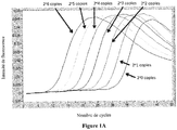
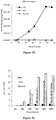
- pLIN-HIV-ScaI The efficiency and sensitivity of quantitative PCR with primers 25t and MS2 are shown for pLIN-HIV-ScaI ( Figure 1A ).
- the parameters used for pLIN-HIV-NdeI were identical to those determined for pLIN-HIV-ScaI indicating that the difference of the two plasmids concerning the end of the viral DNA (a Sca I site for pLIN-HIV-ScaI and a site Nde I for pLIN-HIV-NdeI) does not influence quantitative PCR as expected.
- DNA mimicking the non-matured end was obtained by Sca I / Aat II digestion followed by purification.
- DNA mimicking the mature 3 'end was obtained by digestion with Nde I / Aat II and purification. After purification, these fragments were quantified by quantitative PCR using primers and probes for quantification of total viral DNA (see Table 1 ).
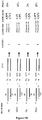
- the LM-PCR with the 11GTb linker leads to the detection of 3 'matured linear viral DNA and not matured with an efficiency of 90% and a sensitivity of 10 copies per 200,000 cells in both cases (as indicated in the material and methods part the effectiveness was in fact determined using the linker 11TAb whose sticky end of 2 nucleotides is complementary to that generated by the cutoff Nde I but for the sake of simplification it is the linker 11GTb which is represented in the figure).
- LM-PCR with the linker oligonucleotide (linker 11b) is only capable of detecting and quantifying undigested viral DNA.
- the amount of 3 'matured DNA is deduced by subtracting the amount obtained with the binding oligonucleotide (11GTb linker) from that obtained with the binding oligonucleotide (linker 11b).
- For each quantitation non-matured linear viral DNA and 3 'mature linear DNA, several dilutions of the corresponding fragments were made in uninfected cell DNA (200 ng / ⁇ l).
- a binding oligonucleotide (11GTb linker for non-matured linear DNA and 11b linker for mature linear DNA) was ligated with each dilution. end of viral DNA (not matured or matured 3 '). As a result, the quantification obtained after PCR takes into account the ligation efficiency. After ligation, the ligated DNA was purified using magnetic beads according to the manufacturer's instructions (Invitrogen) to prevent inhibition of PCR due to mixing of the ligation reaction. The ligated DNA (1/10 of the ligation reaction) was then subjected to real-time PCR amplification with primers 32t and MS1 (see Table 1 ).
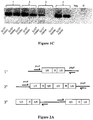
- the number of cycles has been determined empirically. In order to remain in the exponential phase, only 12 cycles of PCR were performed. Unexpectedly, decreasing the number of cycles (for example 8 cycles) during the first PCR results in weak amplification of the samples resulting in erroneous quantification. Likewise, increasing the number of cycles does not improve quantification. Moreover, after 30 cycles, the curves of the plasmid range used for the quantification (dilution of 10 to 10) are almost superimposed; therefore, the quantification of the sample is impossible. Thus, 12 cycles are sufficient to allow quantification of all dilutions of ligated DNA at the end of the second PCR. The amplified products (1/100) were then subjected to the second PCR. The efficiency of each quantification (immature and matured in 3 ') is described in Figure 1B and 1 C .
- the end of the Nde I digest fragment is similar but not identical to that found at the 3 'end in the infected cells (5'-ATTG and not 5).
- -ACTG in infected cells
- the binding oligonucleotide is able to detect the undigested viral end.
- the reason may be that the sticky end of the binding oligonucleotide (5'-GT) is not involved in the ligation with the phosphate on the 5 'side of the genome.
- this binding oligonucleotide 11GTb linker
- both the immature end and the 3 'mature end can be efficiently detected.
- the inventors have shown that asymmetric binding oligonucleotides can be used for efficient quantification of the ends of the viral DNA.
- the 25t / 11GT and 25t / 11b pairs will be used to quantify both undried and matured 3 'ends during the natural course of infection.
- Quantitative PCR using 1LTR LA1 and 1LTR LA16 primers and with p1-LTR as template leads to sensitive detection ( Figures 2C and 2D Table 2 ) and effective (200 copies / 10 6 cells) of 1-LTR DNA.
- p2-LTR subjected to the same protocol, leads to a weak amplification signal ( Figures 2C and 2D Table 2 ).
- p2-LTR was digested with Sca I to mimic the linear viral DNA found in the infected cells. The inventors have precisely quantified the purified DNA according to the protocol adapted to the total viral DNA.
- the inventors quantified the different forms of viral DNA during infection of cells with HIV-1 viruses derived from pNL4-3: ⁇ env NLENG1-ES-IRES virus type wild (wt) and NLENG1-ES-IRES D116N ( Gelderblom et al. (2008) Retrovirology 9; 5: 60 ).
- VSV-G envelope pseudotyped virus 30ng p24 antigen per million cells.
- 3 conditions of infection are achieved: infection with a virus whose integrase is catalytically active (wild type , WT) in the absence or in the presence of 500nM of raltegravir (RAL), an inhibitor of the integration reaction ( Steigbigel et al. (2008) New England Journal of Medicine 359.4: 339-354 ).
- the last condition is infection with a virus whose integrase is catalytically inactive (D116N) ( Shin et al. (1994), Journal of Virology 68,3: 1633-1642 ).
- the cells are washed in phosphate buffer (PBS), contacted with trypsin for 1 minute at 37 ° C.
- PBS phosphate buffer
- the cells are then washed once in culture medium and twice in a large volume of PBS.
- the cells are then cultured in RPMI 1640 medium containing 10% fetal calf serum.
- a sample of 3 million cells is constituted.
- the cell pellet is subjected to digestion with 1,500 U of DNaseI (Invitrogen, reference AM2238) in a buffer containing 20 mM Tris-HCl pH 8.3, 50 mM KCl, 2 mM MgCl 2 for 10 minutes at room temperature.
- the cell pellet is then washed in PBS and the cells are centrifuged and then put at -80 ° C before use.
- the DNA is then extracted using the QIAamp DNA blood mini kit (Qiagen, reference 51104).
- Total viral DNA copy number is determined using primers ( Butler et al. (2001) Nature Medicine 7: 631-634 ) hybridizing in the U5 region of the LTR (primer MH531: 5'-TGTGTGCCCGTCTGTTGTGT-3 ', SEQ ID NO: 29) and in the gag gene (MH532 primer: GAGTCCTGCGTCGAGAGAGC-3', SEQ ID NO: 30) as well as MH FL * hybridization probes (5'-CCCTCAGACCCTTTTAGTCAGTGTGGAAa-3 ', SEQ ID NO: 31) and MH LC * (5'-TCTCTAGCAGTGGCGCCCGAACAGb, SEQ ID NO: 32) (a: Fluorescein at the 3'end; b: LC red 640 at the 5 'end and phosphorylation at the 3' end) ( Table 1 ).
- the 2-LTR circles are amplified by the primers flanking the LTR-LTR junction (HIVF and HIVR1 primers) ( Brussel & Sonigo (2003) Journal of Virology 77: 10119-10124 ).
- the integrated viral DNA is quantified by Alu-PCR ( Brussel & Sonigo (2003) Journal of Virology 77: 10119-10124 ).
- U5-gag sequences, 2-LTR junctions as well as specific amplification of the integrated DNA are amplified in duplicate from 1/50 of the cellular DNA extraction. The number of copies is normalized per ⁇ g of DNA by measuring the amount of ⁇ -globin gene in 1/50 of the DNA extract ( Table 1 ).
- Raltegravir an integrase inhibitor
- integrase inhibitor an inhibitor of the integration reaction and not of the maturation of the extremities in the context of an infection ( Nguyen et al. (2011) Ann. NY Acad Sci. 1222: 83-9 ).
- the effectiveness of this drug is proven in the 3D Figure (Inhibition of integrated viral DNA detection, similar to inhibition of integration by catalytic site mutant (D116N)).
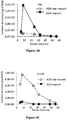
- the amount of mature viral DNA with or without raltegravir is similar ( Figure 4A-4B ).
- the mature viral DNA is almost undetectable in the case of an infection by the mutant of the catalytic site which has been shown to be completely devoid of activity, thus including for the catalysis of the maturation stage ( Figure 4C ).
- Linear viral DNA is the least stable viral form among the different viral genomes.
- Styrylquinolines are, in vitro, inhibitors of integrase binding on viral DNA ( Deprez et al. (2004) Mol. Pharmacol. 65: 85-98 ) and thus represent a different class from that of raltegravir since the SQs act as competitive inhibitors of the maturation stage. Although effective during a viral infection, the step targeted by these inhibitors could never be clearly identified by lack of tools allowing, ex vivo , to characterize the stage of viral DNA processing ( Bonnieri et al. (2004) Journal of Virology 78: 5728-36 ). The method according to the invention has thus made it possible to demonstrate that the styrylquinolines, whose molecule FZ41 is the prototype, well inhibits the reaction of maturation of the viral DNA.
- CEM cells have been infected with the virus having a catalytically active "wild-type" integrase.
- the cells were treated or not, at the time of infection, with 20 ⁇ M of FZ41.
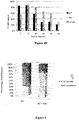
- 20 hours post-infection mature linear viral DNA and immature viral DNA are quantified and reported Figure 5 .
- the results show an inhibition of the viral DNA processing by the compound FZ41.
- the percentage of mature viral DNA is 75% in the absence of SQ (and consequently 25% of immature DNA) against 28% of mature DNA in the presence of FZ41. Inhibition of the processing of viral DNA is therefore important in the presence of SQ.
- the method according to the invention reliable, sensitive, inexpensive, is well suited to the screening of integrase inhibiting compounds to test their activity on the processing of linear DNA, the sole substrate of the integration reaction.
- this method can be broken down into a clinical test for a predictive test of patient escape, the viral load being associated with the amount of viral DNA in the infected cells.
- the results of the inventors confirm the strong inhibition (factor 40) of the formation of 2-LTR circles for both wild-type and D116N viruses in Nalm-114 cells compared to Nalm-6 cells.
- the amounts of 1-LTR circles are similar for cell lines infected with wild type or D116N viruses.
- ligase-4 is not involved in the formation of 1-LTR circles.
- the inventors noticed that the amount of 1-LTR circles was essentially unchanged regardless of the accumulation level of the 2-LTR circles, which confirms that using the quantitative approach of the invention, an exact quantification of circles at 1-LTR in the cellular context and possible unbiased due to the presence of 2-LTR circles.
- RAL remained essentially ineffective up to concentrations of 5 ⁇ M where 80% of the maturation activity was still visible.
- the maturation activity was greatly affected at 2.5 or 5 ⁇ M DTG and EVG (maturation efficiency at about 40% and less than 25% at 2.5 ⁇ M and 5 ⁇ M respectively).
- IC 50 values for the maturation reaction are respectively 2 ⁇ M, 5 ⁇ M and 10 ⁇ M for DTG, EVG and RAL.
- the processing efficiency was measured during the infection of primary CD4 + T lymphocytes using a previously described wild type ⁇ env virus (+/- RAL, EVG or DTG at 5 ⁇ M). It is observed that the efficiency of the ripening reaction is high (75%) but slightly slowed down in the primary cells (the maximum of maturation is reached 24 hours post-infection) and influenced by EVG and DTG (at 5 ⁇ M) but not by RAL. In addition, the accumulation kinetics of 1-LTR and 2-LTR circles are similar in primary cells as compared to cell line cells.
- 1-LTR circles require homologous recombination between 2 LTR of linear DNA in the nucleus while for others it involves a reverse transcription step in the cytoplasmic compartment.
- the peptides NLS-IN-Pen and SV40-NLS-Pen which have been described as inhibiting the nuclear import of the pre-integration complex via the inhibition of the interaction between integrase and importin ⁇ ( Levin et al. (2009) Retrovirology 6: 112 ), were used to determine whether the 1-LTR circles are formed only during the reverse transcription step and does not require translocation of the preintegration complex.
- HeLa cells, treated with one of the two peptides, were infected with a pNL4.3 virus in the presence of RAL. Without peptide, 2-LTR circles accumulate to 19.3% of total viral DNA at 48h post-infection.
- the inhibition of nuclear import of the pre-integration complex could also be carried out more specifically by mutations in the FLAP and / or CTS regions of the virus.
- HeLa cells were infected with a defective mutant, both in the CTS and PPT regions.
- the amounts of 1-LTR and 2-LTR circles were normalized to the amounts determined under wild-type conditions at 24 h post-infection.
- the inventors have found that the abrogation of the FLAP structure partially, but not completely, inhibits the nuclear import of the pre-integration complex, the inventors have observed a substantial decrease in the amount of 2-LTR circles (approximately 2 times). In this context, the inventors have demonstrated a concomitant decrease in the amount of circles at 1-LTR but to a lesser degree (1.4 times).
- 1-LTR circles could be formed in the cytoplasm during the reverse transcription phase but most of the circles at 1-LTR (90%) would be formed by homologous recombination, in the nucleus, after translocation of the pre-integration complex.
Landscapes
- Chemical & Material Sciences (AREA)
- Health & Medical Sciences (AREA)
- Life Sciences & Earth Sciences (AREA)
- Organic Chemistry (AREA)
- Zoology (AREA)
- Wood Science & Technology (AREA)
- Proteomics, Peptides & Aminoacids (AREA)
- Engineering & Computer Science (AREA)
- Virology (AREA)
- Immunology (AREA)
- Molecular Biology (AREA)
- Bioinformatics & Cheminformatics (AREA)
- Biotechnology (AREA)
- Biophysics (AREA)
- Analytical Chemistry (AREA)
- Physics & Mathematics (AREA)
- Biochemistry (AREA)
- Microbiology (AREA)
- General Engineering & Computer Science (AREA)
- General Health & Medical Sciences (AREA)
- Genetics & Genomics (AREA)
- AIDS & HIV (AREA)
- Chemical Kinetics & Catalysis (AREA)
- Measuring Or Testing Involving Enzymes Or Micro-Organisms (AREA)
Description
- La présente invention concerne une méthode de quantification des différentes formes que peut adopter l'ADN viral du virus de l'immunodéficience humaine (VIH), notamment les formes linéaires, maturées ou non, et la forme circulaire 1-LTR.
- Le virus de l'immunodéficience humaine (VIH) fait partie de la famille des Retrovirus. Lors de l'infection, l'ARN viral est converti en une molécule d'ADN viral linéaire double brin. L'ADN linéaire issu de la transcription inverse subit alors une étape de maturation à ses extrémités LTR (Long Terminal Repeat). Cette étape également dite de 3'-processing consiste en une attaque endonucléolytique qui supprime deux bases, à chaque extrémité 3' du virus (Delelis et al. (2008) Retrovirology 5:114.). L'étape de maturation est indispensable pour rendre compatible les extrémités d'ADN avec la réaction d'intégration. Cette étape d'intégration du génome viral ADN est décrite dans la littérature comme étant une étape incontournable du cycle de réplication (Brown et al. (1990) Curr. Top. Microbiol. Immunol 157:19-48). En effet, l'intégration du virus, assurée par l'enzyme virale nommée intégrase, assure le maintien de l'information du virus au sein de la cellule mais aussi la production de nouvelles particules virales infectieuses et ainsi la propagation du virus. Il convient de noter que l'ADN viral peut aussi se retrouver sous formes circulaires (à 1 ou 2-LTR) dans la cellule mais que ces formes virales sont considérées comme n'ayant aucune fonction majeure dans le cycle de réplication et la progression virale (Sloan & Wainberg (2011) Retrovirology 8:52 et Wu (2008) Retrovirology 5:61). Elles sont qualifiées de molécules cul-de-sac ("dead-end").
- Par conséquent, la quantification de l'ADN viral linéaire, seule forme virale compétente pour l'intégration du virus, s'avère un enjeu de premier plan. Dans ce cadre, plusieurs techniques basées sur la PCR (Polymerase Chain Reaction) existent pour quantifier les différentes formes d'ADN virales : forme intégrée, ADN viral circulaire à 2-LTR et ADN viral "total" (Graf et al. (2011) Plos Pathog. 7:e1001300). L'ADN viral total représente l'ensemble de tous les ADN viraux présents dans la cellule. Des méthodes de PCR ont été développées pour détecter l'ADN viral linéaire mais ne permettent pas de le quantifier (Pierson et al. (2002) Journal of Virology 17:8518-8531 ; Mohammed et al. (2011) Journal of Virology 85:4654-66). D'autre part, des méthodes de PCR ont été développées pour quantifier la forme circulaire 2-LTR de l'ADN viral (Butler et al. Nature Medicine, 7(5): 631-634, 2001; et
WO 2010/088491 ). Par ailleurs, la technique du Southern blot permet de visualiser l'ensemble des formes d'ADN virales : circulaires ou linéaires (Zennou et al. (2000) Cell 101:173-85), mais cette technique n'est pas quantitative et est longue, peu sensible et nécessite du matériel radioactif. - Ainsi, à l'heure actuelle, aucune technique fiable et reproductible n'est disponible pour quantifier l'ADN viral linéaire et encore moins de quantifier l'ADN viral linéaire immature de l'ADN viral mature (c'est à dire ayant subi la réaction de 3'-processing). Il faut préciser que la méthode par calcul (quantité d'ADN viral linéaire déduite par soustraction de la quantité d'ADN viral « total » de l'ADN viral 2-LTR, intégré et 1-LTR) n'est pas possible puisqu'une forme très représentée (les cercles à 1-LTR) n'est pas quantifiable à ce jour. De plus, ces techniques nécessitent de grande quantité de matériel viral.
- La présente invention découle de la mise au point, par les inventeurs, d'un procédé, basé sur la LM-PCR (Linker-mediated PCR), de quantification des formes linéaires et de la forme circulaire à 1-LTR du génome ADN du VIH-1.
- La LM-PCR, bien connue de l'homme du métier, est notamment décrite par Mueller & Wold (1989) Science 246:780-786 et Pfeifer et al. (1989) Science 246:810-813.
- Ainsi, la présente invention concerne un procédé de quantification des formes linéaires du génome ADN du virus de l'immunodéficience humaine 1 (VIH-1) dans un échantillon, dans lequel :
- (i) on quantifie les formes linéaires totales du génome du VIH-1, et
- (ii) on quantifie la forme linéaire non maturée du génome ADN du VIH-1
- Le génome du VIH-1 peut être ARN, comme dans le virion. Il peut également être ADN, sous forme intégrée, dans le génome de l'hôte, ou sous forme non intégrée. Dans ce dernier cas, il peut alors se présenter sous forme linéaire ou sous forme circulaire. Les formes circulaires sont dénommées 1-LTR ou 2-LTR, selon qu'elles présentent un seul LTR (Long Terminal Repeat) ou deux. Les formes linéaires peuvent quant à elles être maturées ou non maturée à l'extrémité 3'.
- La PCR (Polymerase Chain Reaction, réaction de polymérisation en chaine) est bien connue de l'homme du métier. Comme on l'entend ici, une PCR quantitative représente une réaction de polymérisation en chaine permettant de déterminer la quantité d'acide nucléique à amplifier initialement présent dans l'échantillon ayant subi la PCR. De nombreux types de PCR quantitative sont connus de l'homme du métier.
- Comme cela apparaitra clairement à l'homme du métier, une « quantification » et une « quantité » selon l'invention sont absolues, et non relatives, c'est-à-dire qu'elles donnent accès à un nombre de molécules, à un nombre de moles, ou à une masse, ainsi qu'aux concentrations ou pourcentages correspondants.
- Comme on l'entend ici, l'oligonucléotide double brin peut également être nommé oligonucléotide de liaison ou « linker », il est formé de l'association par hybridation de deux oligonucléotide dont au moins une partie de leurs séquences sont complémentaires.
- La réaction de ligation est bien connue de l'homme du métier, elle consiste à créer une liaison covalente entre l'extrémité 3' d'un acide nucléique et l'extrémité 5' d'un autre acide nucléique. Elle est généralement mise en oeuvre à l'aide d'une ligase.
- Selon l'invention, le linker défini ci-dessus permet une ligation avec la forme linéaire maturée en 3' et la forme linéaire non maturée en 3' du génome ADN du VIH-1 et comprend un bout collant de deux nucléotides de séquence 5'-GT-3', et on déduit de la PCR quantitative la quantité totale des formes linéaires du génome du VIH-1 dans l'échantillon. Plus préférablement, l'oligonucléotide de liaison est alors formé de l'association d'une séquence 5'-GCGGTGACCCGGGAGATCTGAATTC-3' (SEQ ID NO : 5), ou une séquence présentant au moins 90% d'identité avec SEQ ID NO : 5, et d'une séquence 5'-GTGAATTCAGATC-3 (SEQ ID NO : 6), ou une séquence ayant au moins 90% d'identité avec SEQ ID NO : 6.
- Comme on l'entend ici, un acide nucléique double brin est dit présenter un bout collant lorsque au moins l'une des extrémités de l'acide nucléique possède un brin protubérant qui ne peut pas s'apparier pas à l'autre brin. A contrario, un acide nucléique double brin est dit présenter un bout franc lorsqu'à au moins l'une des extrémités de l'acide nucléique les deux brins sont complètement appariés.
- Une séquence présentant au moins 90% d'identité avec SEQ ID NO: X selon l'invention, présente de préférence au moins 95% d'identité avec SEQ ID NO : X. Cette séquence diffère notamment de SEQ ID NO: X par l'insertion, la suppression ou la substitution d'au moins un nucléotide. Comme on l'entend ici, le pourcentage d'identité entre deux séquences est défini comme le nombre de positions pour lesquelles les bases sont identiques lorsque les séquences sont alignées de manière optimale, divisé par le nombre total de bases de la plus grande des deux séquences. Deux séquences sont dites alignées de manière optimale lorsque le pourcentage d'identité est maximal. Par ailleurs, comme cela apparaitra de manière claire à l'homme du métier, il peut être nécessaire de faire appel à des ajouts de positions lacunaires (des « gaps ») de manière à obtenir un alignement optimal entre les deux séquences. En outre, la séquence présentant au moins 90% d'identité avec SEQ ID NO : X selon l'invention conserve la fonction, notamment d'amorce ou de sonde, de SEQ ID NO : X, c'est-à-dire qu'elle permet une amplification ou une détection de l'acide nucléique à amplifier.
- Selon l'invention, l'oligonucléotide de liaison défini ci-dessus permet une ligation spécifiquement avec la forme linéaire non maturée en 3' de la forme linéaire du génome du VIH-1 et comprend un bout franc, et on déduit de la PCR quantitative la quantité de la forme linéaire non maturée du génome du VIH-1 dans l'échantillon. Plus préférablement, l'oligonucléotide de liaison est alors formé de l'association d'une séquence 5'-GCGGTGACCCGGGAGATCTGAATTC-3' (SEQ ID NO : 5), ou une séquence ayant au moins 90% d'identité avec SEQ ID NO : 5, et d'une séquence 5'-GAATTCAGATC-3' (SEQ ID NO : 7), ou une séquence ayant au moins 90% d'identité avec SEQ ID NO : 7.
- Selon l'invention, la quantité de la forme linéaire maturée du génome du VIH-1 dans l'échantillon est déterminée par soustraction de (i) la quantité de la forme linéaire non maturée du génome du VIH-1 dans l'échantillon déterminée selon l'invention à (ii) la quantité totale des formes linéaires du génome du VIH-1 dans l'échantillon déterminée selon l'invention.
- De préférence également, la PCR quantitative selon l'invention est une PCR en temps réel, plus préférablement conduite avec des sondes du type sonde d'hybridation.
- La PCR en temps réel est bien connue de l'homme du métier. Brièvement, la PCR en temps réel associe l'amplification d'un acide nucléique et la détection par fluorescence de l'acide nucléique amplifié. On effectue généralement une PCR classique en présence de sondes, notamment de sondes d'hybridation, c'est-à-dire des sondes qui s'hybrident à l'acide nucléique amplifié et qui émettent un signal fluorescent en réponse à cette hybridation. L'émission de fluorescence est mesurée en fonction des cycles de PCR. Le cycle de PCR pour lequel le signal de florescence émis est mesuré au dessus d'un niveau seuil, généralement au dessus du niveau basal ou du niveau de fond de fluorescence, est nommé le cycle seuil (Ct). Il a été montré que Ct est proportionnel au logarithme décimal de la quantité d'acide nucléique à amplifier initialement présent dans la réaction de PCR (voir par exemple "Real-time PCR" in Advanced Methods S., Dorak M T. ed, Taylor and Francis, Oxford, 2006).
- Parmi les nombreuses sondes d'hybridation selon l'invention connue de l'homme du métier applicables selon l'invention, il est notamment possible de citer les sondes du type Molecular Beacon qui sont par exemple décrite dans la demande internationale
WO 98/10096 - De manière préférée, la PCR quantitative selon l'invention est réalisée en deux étapes : une première étape d'amplification sans quantification de l'acide nucléique amplifié et une deuxième étape d'amplification avec quantification de l'acide nucléique amplifié.
- De manière davantage préférée, la première étape de la PCR quantitative selon l'invention est réalisée avec une amorce comprenant une séquence 5'-GCGGTGACCCGGGAGATCTGAATTC-3' (SEQ ID NO: 5), telle qu'une séquence 5'-GCGCGCGGCGGTGACCCGGGAGATCTGAATTC-3' (SEQ ID NO: 25), ou une séquence présentant au moins 90% d'identité avec SEQ ID NO : 5 ou 25, et avec une amorce comprenant une séquence 5'-CTCGCCTCTTGCCGTGCGCG-3' (SEQ ID NO : 9), ou une séquence présentant au moins 90% d'identité avec SEQ ID NO : 9.
- De manière davantage préférée également la deuxième étape de la PCR quantitative selon l'invention est réalisée avec une amorce comprenant une séquence 5'-GCGGTGACCCGGGAGATCTGAATTC-3' (SEQ ID NO : 5), ou une séquence présentant au moins 90% d'identité avec SEQ ID NO : 5, et une amorce comprenant une séquence 5'-GAGTCCTGCGTCGAGAGATC-3' (SEQ ID NO: 26), ou une séquence présentant au moins 90% d'identité avec SEQ ID NO : 26, et une sonde d'hybridation comprenant une séquence 5'-CCCTCAGACCCTTTTAGTCAGTGTGGAA-3' (SEQ ID NO: 27), ou une séquence présentant au moins 90% d'identité avec SEQ ID NO: 27, et/ou une sonde d'hybridation comprenant une séquence 5'-TCTCTAGCAGTGGCGCCCGAACAG-3' (SEQ ID NO : 28), ou une sonde présentant au moins 90% d'identité avec SEQ ID NO : 28.
- Par ailleurs, dans le procédé défini ci-dessus, on préfère que la première étape de la PCR quantitative comprenne de 9 à 30 cycles, plus préférablement 10 à 14 cycles, et encore plus préférablement environ 12 cycles.
- L'étape de déduction de la quantité de la forme linéaire maturée en 3' et/ou de la forme linéaire non maturée en 3' du génome ADN du VIH-1 dans l'échantillon peut notamment être mise en oeuvre par référence à une courbe de calibration. Cette courbe de calibration peut notamment être obtenue en réalisant les deux premières étapes du procédé de quantification selon l'invention à partir de quantités connues de molécules d'ADN double brin mimant la forme linéaire maturée en 3' et/ou la forme linéaire non maturée en 3' du génome ADN du VIH-1. Ces quantités connues de molécules d'ADN mimant la forme linéaire maturée en 3' et/ou la forme linéaire non maturée en 3' du génome ADN du VIH-1 sont de préférence obtenues par digestion de quantités connues d'un plasmide à l'aide d'enzymes de restriction permettant notamment de libérer des molécules d'ADN double brin ayant au moins un bout franc, pour les molécules mimant la forme linéaire non maturée, ou au moins un bout collant de 2 nucléotides, pour les molécules mimant la forme linéaire non maturée. Par ailleurs, on préfère que les molécules d'ADN double brin mimant la forme linéaire maturée en 3' et/ou la forme linéaire non maturée en 3' du génome ADN du VIH-1 aient sensiblement la même taille et/ou sensiblement la même séquence que ces dernières. Toutefois, des variations de tailles ou de séquences limitées sont acceptables. En particulier, le bout collant de 2 nucléotides de la molécule d'ADN double brin mimant la forme linéaire maturée du génome ADN du VIH-1 peut avoir une séquence différente de celui de la forme linéaire maturée ; dans ce cas, l'homme du métier comprendra qu'il convient de modifier en conséquence le bout collant de l'oligonucléotide de liaison adapté pour une ligation avec cet ADN double brin et utilisé dans la préparation de la courbe de calibration, pour qu'il soit complémentaire de celui de l'ADN double brin.
- Par ailleurs, l'invention concerne également un procédé de quantification de la forme circulaire à 1-LTR du génome ADN du VIH-1 dans un échantillon, dans lequel on effectue une PCR quantitative selon l'invention à l'aide de deux amorces comprenant respectivement une séquence 5'-GCGCTTCAGCAAGCCGAGTCCT-3' (SEQ ID NO : 1), ou une séquence présentant au moins 90% d'identité avec SEQ ID NO: 1, et une séquence 5'-GTCACACCTCAGGTACCTTTAAGACCAATGAC-3' (SEQ ID NO : 2), ou une séquence présentant au moins 90% d'identité avec SEQ ID NO : 2.
- De préférence, la PCR quantitative est conduite avec une sonde d'hybridation comprenant la séquence 5'-CACAACAGACGGGCACACACTACTTGA-3' (SEQ ID NO : 3), ou une séquence présentant au moins 90% d'identité avec SEQ ID NO : 3 et/ou avec une sonde d'hybridation comprenant la séquence 5'-CACTCAAGGCAAGCTTTATTGAGGC-3' (SEQ ID NO : 4), ou une séquence présentant au moins 90% d'identité avec SEQ ID NO : 4.
- De préférence également, la PCR quantitative mise en oeuvre dans le procédé de quantification de la forme circulaire à 1-LTR du génome ADN du VIH-1 selon l'invention requiert la définition d'une durée précise de l'étape d'élongation de la PCR. Ainsi, on préfère que l'étape d'élongation, c'est-à-dire l'étape conduite à une température favorisant l'activité de l'ADN polymérase thermostable, ait une durée de 20 à 30 secondes, plus préférablement de 23 à 27 secondes, et encore plus préférablement d'environ 25 secondes.
- L'invention concerne également un procédé de criblage de composés susceptibles de modifier la quantité d'une forme non intégrée du génome du VIH-1, comprenant la mise en oeuvre d'un procédé de quantification des formes linéaires ou de la forme circulaire à 1-LTR tel que défini ci-dessus. Dans un mode de réalisation préféré du procédé de criblage selon l'invention, les composés sont susceptibles d'inhiber la maturation de la forme linéaire du génome ADN du VIH-1 et on met en oeuvre un procédé de quantification des formes linéaires telles que définies ci-dessus.
- L'invention sera davantage explicitée à l'aide des Exemples et des Figures qui suivent.
-
-
Figures 1A ,1B et1C -
Figure 1A : PCR sur dilutions du plasmide pLIN-HIV-ScaI. Des dilutions successives de plasmide pLIN-HIV-ScaI ont été effectuées (de 2e6 copies à 2e0 copies). L'efficacité calculée de la PCR à l'aide de la méthode de la dérivée seconde fournie par le logiciel de quantification du Light Cycler, version 3.5 (Roche Diagnostics) est de 90%. L'intensité de Fluorescence (axe des ordonnées) est suivie en fonction du nombre de cycles (axe des abscisses). -
Figure 1B : Résultats des différentes PCR utilisant les linkers et les différents substrats étudiés. -
Figure 1C : Photographie d'un gel d'électrophorèse dans lequel les produits d'amplification ont été mis à migrer.
-
-
Figures 2A ,2B ,2C et2D -
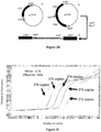
Figure 2A : Schéma de l'amplification utilisant les amorces pour la quantification des cercles 1-LTR sur les substrats 1-LTR (1), 2-LTR (2) et sur l'ADN viral linéaire (3). -
Figure 2B : Plasmides utilisés : Plasmide 1-LTR (1), 2-LTR (2) et ADN mimant l'ADN viral linéaire après digestion du plasmide 2-LTR par l'enzyme ScaI (3). -
Figure 2C : Efficacité de la PCR quantitative 1-LTR. Des dilutions successives du plasmide 1-LTR (2e6 copies à 2e2 copies) ont été effectuées. L'intensité de Fluorescence (axe des ordonnées) est suivie en fonction du nombre de cycles (axe des abscisses). -
Figure 2D : Photographie d'un gel d'électrophorèse dans lequel les produits d'amplification obtenus à partir du plasmide 1-LTR (1), du plasmide 2-LTR (2) et de l'ADN mimant l'ADN viral linéaire après digestion du plasmide 2-LTR par l'enzyme ScaI (3) ont été mis à migrer.
-
-
Figures 3A ,3B, 3C ,3D, 3E et3F
Cinétiques des différentes formes d'ADN viral après infection de cellules MT4 par le virus WT (en présence ou non de RAL) ou par un virus D116N. La condition WT sans Raltégravir est symbolisée par des carrés noirs. La condition WT avec Raltégravir est symbolisée par des diamants noirs. La condition D116N est symbolisée par des ronds blancs. Le nombre de copies d'ADN viral, normalisé par la quantité d'ADN cellulaire total (axe des ordonnées), est donné en fonction du temps en heures (axe des abscisses).-
Figure 3A : cinétique de l'ADN viral total. -
Figure 3B : cinétique des cercles 2-LTR. -
Figure 3C : cinétique des cercles 1-LTR. -
Figure 3D : cinétique de l'ADN viral intégré. -
Figure 3E : pourcentage des cercles 2-LTR (quantité de cercles 2-LTR sur l'ADN viral total, axe des ordonnées) en fonction du temps (axe des abscisses, en heures) pour la condition WT sans Raltégravir (WT, barres noires), la condition WT avec Raltégravir (RAL, barres grises) et la condition D116N (barre blanches). -
Figure 3F : ainsi que le pourcentage des cercles 1-LTR (quantité de cercles 1-LTR sur l'ADN viral total axe des ordonnées) en fonction du temps (axe des abscisses, en heures) pour la condition WT sans Raltégravir (WT, barres noires), la condition WT avec Raltégravir (RAL, barres grises) et la condition D116N (barre blanches).
-
-
Figures 4A ,4B ,4C et4D -
Figures 4A ,4B et 4C : Cinétique de l'ADN viral linéaire maturé et non maturé après infection de cellules CEM par le virus WT (en présence ou non de Raltégravir, respectivementFigure 4B etFigure 4A ) ou par le virus D116N (Figure 4C ). La quantité d'ADN non maturé est symbolisée par un rond blanc. La quantité d'ADN maturé est symbolisée par un carré noir. Le nombre de copies d'ADN viral, normalisé par la quantité d'ADN cellulaire total (axe des ordonnées), est donné en fonction du temps en heures (axe des abscisses). -
Figure 4D : Le pourcentage d'ADN maturé (quantité d'ADN maturé sur la quantité d'ADN linéaire total) est représenté pour les trois conditions : WT (noir), WT en présence de Raltégravir (gris) et D116N (en blanc).
-
-
Figure 5
Les cellules CEM ont été infectées par le virus pNL4.3 en absence (WT) ou en présence de 20µM de FZ41 (WT+FZ41). l'ADN viral maturé et non maturé est quantifié 24 heures après infection. La proportion de chaque espèce d'ADN viral linéaire maturé (gris) et non maturé (blanc) est représentée. - Cellules et virus. La lignée cellulaire lymphoïde CEM (NIH, référence catalogue 117) a été cultivée en milieu RPMI1640 (Invitrogen, Cat#61870-044) supplémenté par 10% de sérum de veau foetal (SVF) (PAA, Lot#A10109-113). Les cellules 293T et HeLa ont été cultivées en DMEM (Invitrogen, Cat#31966-047) (Dulbecco's modified Eagle medium) supplémenté par 10% de SVF. Les stocks de VIH-1 ont été préparés par transfection de cellules 293T avec les différents clones moléculaires dérivés de pNL4-3 (Gelderblom et al. (2008) Retrovirology 9;5:60). Les virus Δenv NLENG1-ES-IRES WT et NLENG1-ES-IRES D116N (Gelderblom et al. (2008) Retrovirology 9;5:60) codent respectivement le type sauvage et le mutant D116N catalytiquement inactif de l'intégrase. L'élément IRES est nécessaire pour l'expression de la GFP. Le pseudotypage des virus Δenv a été effectué par co-transfection de cellules 293T avec un plasmide VSV-G (Addgene, plasmide 8454) à l'aide de la méthode au phosphate de calcium. Les surnageants viraux ont été filtrés à travers un filtre à diamètre de pores de 0,45 µm (Sartorius, référence 16537K) puis congelés à - 80°C.
- Infection virale. Le contenu en antigène p24gag du VIH-1 des surnageants de culture a été déterminé par ELISA (Perkin-Elmer Life Sciences, Paris, France référence : NEK050B001KT). 40ng d'antigène p24gag pour 106 cellules, ce qui correspond à une multiplicité d'infection (m.o.i.) de 0.1, ont été utilisés pour l'infection. Lorsque nécessaire, les cellules ont été traitées en présence de l'inhibiteur d'intégrase raltégravir (RAL) à 500nM. 2 à 5 millions de cellules ont été collectées à différents temps après infection. Les cellules ont ensuite été lavées en PBS, puis centrifugées, et les culots cellulaires secs ont été congelés à -80°C jusqu'à leur utilisation. L'ADN cellulaire total des cellules infectées de la lignée cellulaire CEM a été purifié à l'aide du kit QIAamp DNA Blood mini kit (Qiagen, référence 51104) selon les instructions du fabriquant. Afin de dégrader les plasmides de transfection résiduels, l'ADN extrait a été incubé avec 5U de DpnI (NEB, référence R0176S) dans un tampon comprenant 20mM Tris pH 7.9 pendant 4 heures à 37°C.
- Plasmides. Quatre plasmides ont été construits : p1-LTR, p2-LTR, pLIN-HIV-ScaI et pLIN-HIV-NdeI.
- pLIN-HIV-ScaI a été construit comme suit : amplification par PCR à l'aide de l'oligonucléotide 25tSca-HIV (5'-GCGGTGACCCGGGAGATCTGAATTCAGTACTGGAAGGGCTAATTTGGTCCC-3', SEQ ID NO : 8) et de l'oligonucléotide MS1 (5'-CTCGCCTCTTGCCGTGCGCG-3', SEQ ID NO : 9) en utilisant le plasmide pNL4.3 comme matrice. Le produit d'amplification a alors été purifié et introduit dans le vecteur pGEMT-easy (Proméga, référence A1360). Les nucléotides soulignés représentent la séquence d'ADN virale du LTR5' (région U3). Il est à préciser qu'un site ScaI (5'-AGTACT-3', SEQ ID NO : 10) existe en position 315 dans le plasmide pNL4.3. Ce site ScaI a été modifié en un site ne permettant pas la coupure par l'enzyme de restriction ScaI (5'AGTACT-3' muté en 5'-AGAACT-3', SEQ ID NO : 11) par un procédé de mutagénèse dirigée site-spécifique (QuikChange Lightning Kits, Agilent) à l'aide des oligonucléotides de séquences 5'-CCCGAGAGCTGCATCCGGAGAACTACAAAGACTGCTGACATCG-3' (SEQ ID NO: 12) et 5'-CGATGTCAGCAGTCTTTGTAGTTCTCCGGATGCAGCTCTCGGG-3', SEQ ID NO : 13). La digestion par ScaI et AatII conduit à un fragment mimant l'extrémité non-maturée de l'ADN viral. Après purification, ce fragment est utilisé pour mettre en oeuvre une réaction de ligation avec les oligonucléotides de liaison double brin (ou « linker ») 25t/11b ou 25t/11GT.
- pLIN-HIV-NdeI a été construit par mutagénèse dirigée site-spécifique (QuikChange Lightning Kits, Agilent) du plasmide pLIN-HIV-ScaI, en mutant le site ScaI (5'-AGTACT-3', SEQ ID NO : 10) en un site NdeI (5'-CATATG-3', SEQ ID NO : 14) à l'extrémité de l'ADN viral à l'aide des oligonucléotides de séquences 5'-CCGGGAGATCTGAATTCAGTCATATGGAAGGGCTAATTTGGTCC-3' (SEQ ID NO : 15) et 5'-GGACCAAATTAGCCCTTCCATATGACTGAATTCAGATCTCCCGG-3' (SEQ ID NO : 16). La digestion par NdeI et AatII (dont le site de digestion n'est présent que dans le vecteur pGEMT-easy) conduit à un fragment mimant l'extrémité 3' maturée de l'ADN viral. Après purification, ce fragment est utilisé comme standard de la PCR visant à quantifier l'ADN maturé en 3'. Il est important de noter que la double digestion NdeI et AatII permet d'obtenir un fragment mimant l'ADN maturé du VIH-1 (extrémité cohésive de 2 paires de bases 5'-TA-3'). Cependant, la nature de ces bases diffère avec celles trouvées à l'extrémité du génome maturé (5'-AC-3'). Toutefois, la nature de ces bases n'interfère en rien avec la méthode de quantification de l'ADN viral maturé.
- Les amorces 1LTR LA1 (5'-GCGCTTCAGCAAGCCGAGTCCT-3', SEQ ID NO : 1) et 1LTR LA16 (5'-GTCACACCTCAGGTACCTTTAAGACCAATGAC-3', SEQ ID NO : 2) ont été utilisées pour amplifier par PCR la région env-LTR-gag contenue dans les molécules circulaires à 1-LTR à partir d'extraits de cellules infectées par le VIH-1. Le plasmide p1-LTR a alors été obtenu en clonant le produit d'amplification dans le vecteur pGEMT-easy.
- Le plasmide p2-LTR a été construit comme suit : les amorces ScaI-LTR3' (5'-CGGGAGATCTGAATTCAGTACTGCTAGAGATTTTCCACACTGACTAAAAG-3', SEQ ID NO: 17) et 1LTR LA16 (5'-GTCACACCTCAGGTACCTTTAAGACCAATGAC-3', SEQ ID NO: 2) ont été utilisées pour amplifier la région env-LTR3' et le produit d'amplification a été inséré dans le vecteur pGEMT-easy pour donner le plasmide intermédiaire pSca-LTR3'. Le site ScaI (5'-AGTACT-3', SEQ ID NO : 10), présent dans le LTR3' en position 10715 du génome viral a été muté en 5'-AGCACT-3' (SEQ ID NO : 18) par un procédé de mutagénèse dirigée site-spécifique (QuikChange Lightning Kits, Agilent) à l'aide des oligonucléotides de séquences 5'-AGCTGCATCCGGAGCACTTCAAGAACTGCT-3' (SEQ ID NO: 19) et 5'-AGCAGTTCTTGAAGTACTCCGGATGCAGCT-3' (SEQ ID NO : 20). Le plasmide pSca-LTR3' et le plasmide pLIN-HIV-ScaI ont été digérés par l'enzyme ScaI. Les fragments obtenus, respectivement de taille 2520pb et 1920pb ont été purifiés et ligaturés pour obtenir le plasmide p2-LTR. Enfin le site ScaI (5'-AGTACT-3', SEQ ID NO : 10) présent dans le vecteur pGEMT-easy (en position 1890 du vecteur) a été muté en un site 5'-AATACT-3' (SEQ ID NO: 21) à l'aide des oligonucléotides 5'-GTGACTGGTGAATACTCAACCAAGTCATTCTGAG-3' (SEQ ID NO: 22) et 5'-CTCAGAATGACTTGGTTGAGTATTCACCAGTCAC-3' (SEQ ID NO: 23), afin de permettre la linéarisation du plasmide p2-LTR par digestion de celui-ci par l'enzyme ScaI.
- Quantification des formes linéaires non maturées et maturées en 3' de l'ADN du VIH-1 par LM-PCR. La stratégie utilisée pour quantifier l'ADN viral linéaire est basée sur la ligation d'un oligonucléotide de liaison (ou « linker ») aux terminaisons des extrémités virales suivie de deux PCR selon le protocole de Pierson et al. (Pierson et al. (2002) Journal of Virology 17:8518-8531). Des améliorations majeures ont été apportées afin de mettre en oeuvre une approche de type PCR quantitative.
- Le linker constitué des deux oligonucléotides 25t et 11b (25t: 5'-GCGGTGACC CGGGAGATCTGAATTC-3', SEQ ID NO : 5, 11b: 5'-GAATTCAGATC-3', SEQ ID NO : 7) (linker 11b) et le linker constitué des oligonucléotides 25t et 11GT (25t: 5'-GCGGTGACCCGGGAGATCTGAATTC-3', SEQ ID NO: 5, 11GT: 5'-GTGAATTCAGATC-3', SEQ ID NO: 6) (linker 11GTb), ont été respectivement utilisés pour la quantification de l'ADN linéaire non maturé du VIH-1 et de l'ADN linéaire maturé du VIH-1.
- Pour la quantification de l'ADN viral linéaire non mature, l'efficacité de la procédure de ligation-amplification a été déterminée par l'ajout de l'oligonucléotide de liaison (linker 11b) à des dilutions en série du fragment de digestion de pLIN-HIV-ScaI obtenu par digestion par les enzymes ScaI et AatII. Les dilutions en série de ce fragment constituent la gamme de calibration de l'ADN non maturé.
- Pour la quantification de l'ADN viral linéaire maturé, l'efficacité de la procédure de ligation-amplification a été déterminée par ajout de l'oligonucléotide de liaison (linker 11TAb) constitué des oligonucléotides 25t et 11TA (11TA: 5'-TAGAATTCAGATC-3', SEQ ID NO : 24) à des dilutions en série de fragments obtenus par digestion de pLIN-HIV-NdeI par NdeI et AatII. Les dilutions en série de ce fragment constituent la gamme de calibration de l'ADN maturé.
- Il est à noter que lors de l'infection de cellules par le virus VIH-1, l'extrémité de l'ADN viral linéaire maturé diffère de l'extrémité de la gamme de calibration, c'est pour cela que la quantification de l'ADN viral maturé au sein des cellules infectées est effectuée à l'aide du linker 11GTb au lieu du linker 11TAb. Cette différence n'influe en rien sur la quantification de l'ADN viral maturé. La quantification de l'ADN viral linéaire maturé est donc obtenue grâce par comparaison à la gamme de calibration.
- Les différentes courbes de calibration ont été réalisées en présence d'ADN de cellules non infectées afin de vérifier que l'efficacité de la ligation/amplification n'était pas influencée par la captation d'oligonucléotide de liaison par l'ADN des cellules non infectées.
- Les oligonucléotides de liaison ont été associés en chauffant chaque brin complémentaire (à la concentration finale de 3µM) à 95°C pendant 5 min puis par un refroidissement lent jusqu'à atteindre la température ambiante. Les oligonucléotides de liaison ont été ajoutés à la concentration finale de 30nM à une quantité de 200ng à 2µg d'ADN de cellules infectées dans un volume final de 20µl en présence de la ligase du Quick ligation kit 0.8µl, soit 300 unités d'enzymes (NEB, référence M2200S) pendant 2 heures à température ambiante dans un volume final de 20µL. Les produits ADN ligaturés ont ensuite été purifiés avec le kit PCR switch charge purification (Life Technology, CS12000) selon les instructions du fabriquant puis élués dans 20µL et soumis à une PCR en temps réel.
- Les quantifications ont été mises en oeuvre par PCR en temps réel sur un appareil Light Cycler (Roche Diagnostics, Meylan, France). Les quantifications, par rapport à la gamme utilisée, ont été calculées à l'aide de la méthode de la dérivée seconde fournie par le logiciel de quantification du Light Cycler, version 3.5 (Roche Diagnostics).
- Dans un premier tour de PCR, 1/10 de l'ADN purifié précédemment a été amplifié dans un mélange réactionnel de 20µL comprenant du tampon 1 x LightCycler FastStart DNA master Hybprobes (Roche, référence 12239272001), 4mM MgCl2, 300nM d'amorces 32t 5'-GCGCGCGGCGGTGACCCGGGAGATCTGAATTC-3' (SEQ ID NO: 25) et MS1 5'-CTCGCCTCTTGCCGTGCGCG-3' (SEQ ID NO : 9).
- Afin de rester dans la phase exponentielle, seulement 12 cycles ont été effectués. Ainsi, les conditions du premier tour de PCR étaient les suivantes : une étape de dénaturation de 8 min à 95°C puis 12 cycles d'amplification (95°C pendant 10s, 60°C pendant 10s, et 72°C pendant 31s). Le deuxième tour de PCR emboitée (« nested PCR ») a été réalisé sur 1/100 du produit du premier tour de PCR dans un mélange comprenant du tampon 1 x LightCycler FastStart DNA master Hybprobes, 4mM MgCl2, 300nM des amorces 25t (5'-GCGGTGACCCGGGAGATCTGAATTC-3', SEQ ID NO: 5) et MS2 (5'-GAGTCCTGCGTCGAGAGATC-3', SEQ ID NO : 26) et 200nM des sondes d'hybridation MHFL* (5'-CCCTCAGACCCTTTTAGTCAGTGTGGAAa-3', SEQ ID NO : 27) et MHLC* (5'-TCTCTAGCAGTGGCGCCCGAACAGb-3', SEQ ID NO : 28) (a : Fluorescéine en 3' ; b : LCred640 en 5' et phosphorylé en 3').
- Le nombre de copies de l'ADN linéaire maturé et non maturé a été déterminé par référence aux courbes de calibration préparées en amplifiant des quantités allant de 10 à 105 copies de fragments digérés correspondant.
- Quantification des cercles 1-LTR. Les quantifications ont été réalisées par PCR en temps réel sur un appareil Light Cycler (Roche Diagnostics, Meylan, France) à l'aide de la méthode de la dérivée seconde fournie par le logiciel de quantification du Light Cycler, version 3.5 (Roche Diagnostics). Le nombre de copies d'ADN des cercles 1-LTR a été déterminé en quantifiant les molécules virales à l'aide d'amorces qui s'hybrident aux gènes gag et env, ainsi que deux sondes d'hybridation. Les mélanges réactionnels contiennent du tampon 1 x LightCycler FastStart DNA master HybProbes (Roche Diagnostics référence 12239272001), 4mM MgCl2, 300nM des amorces 1LTR LA1 (5'-GCGCTTCAGCAAGCCGAGTCCT-3', SEQ ID NO : 1) et 1LTR LA16 (5'-GTCACACCTCAGGTACCTTTAAGACCAATGAC-3', SEQ ID NO : 2) et 200nM des sondes d'hybridation LTR FL* (5'-CACAACAGACGGGCACACACTACTTGAa-3', SEQ ID NO: 3) et LTR LC*(5'-CACTCAAGGCAAGCTTTATTGAGGCb-3', SEQ ID NO : 4) (a Fluorescéine à l'extrémité 3'; b LC red 640 à l'extrémité 5' et phosphorylé à l'extrémité 3') dans un volume final de 20µl. Les conditions des cycles de PCR pour la quantification des cercles 1-LTR sont les suivantes : étape de dénaturation pendant 8 minutes suivie de 40 cycles (95°C pendant 10s, 60°C pendant 10s, et 72°C pendant 25s). Le nombre de copies des cercles 1-LTR a été déterminé par référence à une courbe de calibration préparée en amplifiant des quantités allant de 100 à 105 copies de p1-LTR.
- La quantification de l'ADN viral linéaire a été réalisée en suivant une approche par PCR mettant en oeuvre une ligation (ligation-mediated PCR, LM-PCR) apportant des améliorations majeures à celle de Pierson et al. (Pierson et al. (2002) Journal of Virology 17 : 8518-8531).
- L'efficacité et la sensibilité de la PCR quantitative avec les amorces 25t et MS2 sont représentées pour pLIN-HIV-ScaI (
Figure 1A ). Les paramètres utilisés pour pLIN-HIV-NdeI étaient identiques à ceux déterminés pour pLIN-HIV-ScaI indiquant que la différence des deux plasmides concernant l'extrémité de l'ADN viral (un site ScaI pour pLIN-HIV-ScaI et un site NdeI pour pLIN-HIV-NdeI) n'influence pas la PCR quantitative, comme attendu. - L'ADN mimant l'extrémité non maturé a été obtenu par digestion par ScaI/AatII puis purification. L'ADN mimant l'extrémité 3' maturée a été obtenue par digestion par NdeI/AatII puis purification. Après purification, ces fragments ont été quantifiés par PCR quantitative en utilisant les amorces et les sondes pour la quantification de l'ADN viral total (voir le Tableau 1 ). Comme décrit dans la
Figure 1B , la LM-PCR avec le linker 11GTb conduit à la détection de l'ADN viral linéaire maturé en 3' et non maturé avec une efficacité de 90% et une sensibilité de 10 copies pour 200 000 cellules dans les deux cas (comme indiqué dans la partie matériels et méthodes l'efficacité a en fait été déterminée à l'aide du linker 11TAb dont le bout collant de 2 nucléotides est complémentaires de celui généré par la coupure NdeI mais par souci de simplification c'est le linker 11GTb qui est représenté dans la figure). - La LM-PCR avec l'oligonucléotide de liaison (linker 11b) est seulement capable de détecter et de quantifier l'ADN viral non maturé. En conséquence, la quantité d'ADN maturé en 3' est déduite en soustrayant la quantité obtenue avec l'oligonucléotide de liaison (linker 11GTb) de celle obtenue avec l'oligonucléotide de liaison (linker 11b). Pour chaque quantification (ADN viral linéaire non maturé et ADN linéaire maturé en 3'), plusieurs dilutions des fragments correspondant ont été réalisées dans de l'ADN de cellules non infectées (200ng/µl). Un oligonucléotide de liaison (linker 11GTb pour l'ADN linéaire non maturé et linker 11b pour l'ADN linéaire maturé) a été ligaturé avec chaque dilution d'extrémité d'ADN viral (non maturée ou maturée en 3'). En conséquence, la quantification obtenue après PCR prend en compte l'efficacité de ligation. Après ligation, l'ADN ligaturé a été purifié à l'aide de billes magnétiques selon les instructions du fabricant (Invitrogen) afin d'empêcher l'inhibition de la PCR du fait du mélange de la réaction de ligation. L'ADN ligaturé (1/10 de la réaction de ligation) à alors été soumis à une amplification par PCR en temps réel avec les amorces 32t et MS1 (voir le Tableau 1 ).
- Le nombre de cycle a été déterminé de manière empirique. Afin de rester dans la phase exponentielle, seulement 12 cycles de PCR ont été réalisés. De manière inattendue, diminuer le nombre de cycle (par exemple 8 cycles) lors de la première PCR résulte en une amplification faible des échantillons aboutissant à une quantification erronée. De même, augmenter le nombre de cycle n'améliore pas la quantification. Par ailleurs, après 30 cycles, les courbes de la gamme de plasmide utilisées pour la quantification (dilution de 10 en 10) sont quasi superposées; par conséquent, la quantification de l'échantillon est impossible. Ainsi, 12 cycles sont suffisants pour permettre une quantification de toutes les dilutions d'ADN ligaturé à l'issu de la deuxième PCR. Les produits amplifiés (1/100) ont ensuite été soumis à la deuxième PCR. L'efficacité de chaque quantification (non mature et maturée en 3') est décrite dans les
Figure 1B et1C . - Comme indiqué plus haut, il est important de noter que l'extrémité du fragment issu de la digestion par NdeI est similaire mais non identique à celui trouvé à l'extrémité 3' dans les cellules infectées (5'-ATTG et non pas 5'-ACTG dans les cellules infectées), ce qui rend nécessaire l'utilisation de l'oligonucléotide de liaison 11TAb pour ces expériences à la place de l'oligonucléotide de liaison 11GTb. Comme décrit par Pierson et al. (Pierson et al. (2002) Journal of Virology 17 : 8518-8531), l'oligonucléotide de liaison (linker 11GTb) est capable de détecter l'extrémité virale non maturée. La raison peut être due au fait que le bout collant de l'oligonucléotide de liaison (5'-GT) n'est pas impliqué dans la ligation avec le phosphate du côté 5' du génome. Toutefois, en utilisant cet oligonucléotide de liaison (linker 11GTb), tant l'extrémité non mature que l'extrémité maturée en 3' peuvent être efficacement détectées. Globalement, les inventeurs ont montré que des oligonucléotides de liaison assymétriques peuvent être utilisés pour une quantification efficace des extrémités de l'ADN viral.
- Par conséquent, les couples 25t/11GT et 25t/11b seront utilisés pour quantifier à la fois les extrémités non maturées et maturées en 3' pendant le cours naturel de l'infection.
- Plusieurs méthodes basées sur la PCR ont été proposées afin de quantifier les cercles 1-LTR (Bukrinsky et al. (1992) Proc Natl Acad Sci USA 89, 14:6580-4). Ces PCR sont basées sur l'hybridation d'amorces dans les gènes env et gag (
Figure 2A 1 ). Toutefois, il a été rapporté que les méthodes basées sur la PCR ne permettaient pas une quantification correcte des cercles 1-LTR (Yoder & Fishel (2006) Journal of Virological Methods 138,1-2 :201-6). En effet, comme cela est montré dans laFigure 2A 2 , les amorces utilisées pour les cercles 1-LTR pourraient conduire à une amplification de la région LTR-LTR présente dans les cercles 2-LTR. - De plus, comme décrit par Yoder et al., ces amorces pourraient conduire à une amplification linéaire du LTR 5' et du LTR 3' de l'ADN viral linéaire (
Figure 2A 3 ). L'ADN simple brin linéaire produit pourrait s'hybrider aux séquences LTR homologue ce qui conduirait à une amplification exponentielle à partir de l'ADN viral et pourrait en conséquence aboutir à des biais dans la quantification des cercles 1-LTR. - L'amplification avec les amorces 1LTR LA1 (5'-GCGCTTCAGCAAGCCGAGTCCT-3', SEQ ID NO: 1) et 1LTR LA16 (5'-GTCACACCTCAGGTACCTTTAAGACCAATGAC-3', SEQID NO : 2) a été réalisée avec de l'ADN de cellules de la lignée cellulaire CEM infectées et le produit d'amplification a été cloné dans le vecteur pGEMT-easy pour donner p1-LTR utilisé comme calibrateur de la PCR en temps réel (
Figure 2B ). Dans ce cadre, il a été déterminé que la durée optimale de l'étape d'élongation de la PCR était de 25 secondes. p2-LTR qui comprend les LTR dans leur intégralité entourant les régions gag et env est utilisé en tant que témoins (Figure 2B ). Une PCR quantitative à l'aide des amorces 1LTR LA1 et 1LTR LA16 et avec p1-LTR comme matrice conduit à une détection sensible (Figures 2C et2D , Tableau 2 ) et efficace (200 copies/106 cellules) de l'ADN 1-LTR. p2-LTR, soumis au même protocole, conduit à un faible signal d'amplification (Figures 2C et2D , Tableau 2 ). De plus, p2-LTR a été digéré par ScaI pour mimer l'ADN viral linéaire trouvé dans les cellules infectées. Les inventeurs ont précisément quantifié l'ADN purifié selon le protocole adapté à l'ADN viral total. Des amplifications à partir de plusieurs dilutions de cet ADN dans des cellules non infectées conduit à des signaux d'amplification qui sont négligeables en comparaison avec la quantité initiale d'ADN linéaire (Figures 2C et2D , Tableau 2 ), ce qui confirme l'amplification à partir de l'ADN viral linéaire comme montré par Yoder et al. (Yoder & Fishel (2006) Journal of Virological Methods 138,1-2 :201-6). L'amplification non spécifique à partir d'ADN linéaire viral a été estimée à moins de 1% par rapport à la quantité initiale. Globalement, l'amplification de p1-LTR avec les amorces et le protocole décrits ici est représentative de la quantification des cercles 1-LTR.Tableau 2 : amplifications obtenues à partir du plasmide 1-LTR, du plasmide 2-LTR et de l'ADN mimant l'ADN viral linéaire après digestion du plasmide 2-LTR par l'enzyme ScaI Quantité initiale Quantité calculée Pourcentage d'amplification p1-LTR 2.105 1,925.105 96,2 2.104 2,109.104 100 2.103 2,002.103 100 p2-LTR 2.105 1,937.103 0,7 2.104 2,242.102 0,8 2.103 5,311.101 1,9 p2-LTR linéarisé 2.105 6,733.101 0,03 2.104 9,786 0,05 2.103 ND ND - Afin de valider la méthode décrite dans l' Exemple 1, les inventeurs ont quantifié les différentes formes d'ADN viral lors d'une infection de cellules par des virus VIH-1 dérivés de pNL4-3: virus Δenv NLENG1-ES-IRES type sauvage (wt) et NLENG1-ES-IRES D116N (Gelderblom et al. (2008) Retrovirology 9;5:60).
- 20 millions de cellules CEM sont infectées par un virus pseudotypé par l'enveloppe VSV-G (30ng d'antigène p24 par million de cellule). 3 conditions d'infection sont réalisées : infection par un virus dont l'intégrase est catalytiquement active (wild type, WT) en absence ou en présence de 500nM de Raltégravir (RAL), un inhibiteur de la réaction d'intégration (Steigbigel et al. (2008) New England Journal of Medicine 359,4 :339-354). La dernière condition est l'infection par un virus dont l'intégrase est catalytiquement inactive (D116N) (Shin et al. (1994), Journal of Virology 68,3 :1633-1642).
- Les résultats attendus sont:
- "WT": "Wild-Type": présence d'ADN maturé due à l'activité de l'intégrase ainsi que la détection d'ADN viral intégré.
- "D116N": Intégrase catalytiquement inactive: absence d'ADN maturé et d'ADN viral intégré due à l'absence d'activité de l'intégrase.
- "RAL": infection par un virus ayant une intégrase "WT" en présence de Raltégravir: présence d'ADN viral maturé car le Raltégravir n'est pas un inhibiteur de la réaction de maturation (3'-processing). En revanche, on s'attend à une absence d'ADN viral intégré car le Raltégravir est un inhibiteur de la réaction d'intégration.
- 1 heure après infection, les cellules sont lavées dans un tampon phosphate (PBS), mises en contact avec la trypsine pendant 1 minute à 37°C. Les cellules sont ensuite lavées une fois dans du milieu de culture et deux fois dans un grand volume de PBS. Les cellules sont ensuite cultivées dans du milieu RPMI 1640 contenant 10% de sérum de veau foetal. A chaque temps post-infection, un échantillon de 3 millions de cellules est constitué. Pour éliminer l'ADN utilisé pour la préparation des virus, le culot cellulaire est soumis à la digestion par 1,500U de DNaseI (Invitrogen, référence AM2238) dans un tampon contenant 20mM Tris-HCl pH 8,3, 50mM KCl, 2mM MgCl2 pendant 10 minutes à température ambiante. Le culot cellulaire est ensuite lavé dans du PBS et les cellules sont centrifugées puis mises à -80°C avant utilisation. L'ADN est ensuite extrait grâce au kit QIAamp DNA blood mini kit (Qiagen, référence 51104).
- Le nombre de copies d'ADN viral total est déterminé en utilisant des amorces (Butler et al. (2001) Nature Medicine 7:631-634) s'hybridant dans la région U5 du LTR (amorce MH531 : 5'-TGTGTGCCCGTCTGTTGTGT-3', SEQ ID NO: 29) et dans le gène gag (amorce MH532: GAGTCCTGCGTCGAGAGAGC-3', SEQ ID NO: 30) ainsi que les sondes d'hybridation MH FL* (5'-CCCTCAGACCCTTTTAGTCAGTGTGGAAa-3', SEQ ID NO : 31) et MH LC* (5'-TCTCTAGCAGTGGCGCCCGAACAGb, SEQ ID NO : 32) (a : Fluorescéine à l'extrémité 3'; b : LC red 640 à l'extrémité 5' et phosphorylation à l'extrémité 3') ( Tableau 1 ).
- Les cercles à 2-LTR sont amplifiés par les amorces encadrant la jonction LTR-LTR (amorces HIVF et HIVR1) (Brussel & Sonigo (2003) Journal of Virology 77:10119-10124). L'ADN viral intégré est quantifié par Alu-PCR (Brussel & Sonigo (2003) Journal of Virology 77:10119-10124). Les séquences U5-gag, les jonctions 2-LTR ainsi que l'amplification spécifique de l'ADN intégré sont amplifiés en double à partir d'1/50 de l'extraction d'ADN cellulaire. Le nombre de copies est normalisé par µg d'ADN en mesurant la quantité de gène β-globine dans 1/50 de l'extrait d'ADN ( Tableau 1 ).
- Les résultats sont présentés dans les
Figure 3A-3F . Comme le montrent ces figures, la synthèse de l'ADN viral total, témoin de la transcription inverse, est maximale 10 heures post-infection. La formation des cercles à 2-LTR est maximale 36 heures post-infection. L'intégration du virus est complète 48 heures post-infection. Ces cinétiques sont conformes à celles décrites dans le cas d'une infection par le VIH-1 (Brussel & Sonigo (2003) Journal of Virology 77:10119-10124). La quantité d'ADN viral linéaire (qui comprend les deux sous-populations : maturé et non maturé) est atteinte 8 heures post-infection. Elle est la même pour les 3 conditions, i.e. "WT", "RAL" et "D116N" ce qui est normal car ni la mutation D116N de l'intégrase, ni le Raltégravir, n'influence le taux de conversion d'ARN en ADN linéaire par la transcriptase inverse. L'ADN est ensuite dégradé témoignant de sa dégradation par la voie du protéasome (Butler et al. (2002) Journal of Virology 76:3739-47). - La discrimination entre l'ADN viral maturé et non maturé a été ensuite validée suivant le protocole présenté dans l' Exemple 1. Les résultats sont présentés dans les
Figures 4A-4D . - Le Raltégravir, inhibiteur d'intégrase, est un inhibiteur de la réaction d'intégration et non de la maturation des extrémités dans le contexte d'une infection (Nguyen et al. (2011) Ann. N. Y. Acad Sci. 1222:83-9). L'efficacité de cette drogue est prouvée dans la
Figure 3D (inhibition de la détection d'ADN viral intégré, similaire à l'inhibition de l'intégration par un mutant du site catalytique (D116N)). La quantité d'ADN viral maturé avec ou sans Raltégravir est similaire (Figure 4A-4B ). En revanche, l'ADN viral maturé est quasi indétectable dans le cas d'une infection par le mutant du site catalytique qui a été montré comme étant totalement dépourvue d'activité y compris donc pour la catalyse de l'étape de maturation (Figure 4C ). De plus, le pourcentage d'ADN viral linéaire maturé décroit en fonction du temps post-infection par rapport à l'ADN non maturé (Figure 4D ). Ainsi, cette expérience met en évidence une moins grande stabilité de l'ADN viral maturé par rapport à l'ADN viral linéaire immature. L'ADN viral linéaire étant la forme virale la moins stable parmi les différents génomes viraux. - Les Styrylquinolines (SQ) sont, in vitro, des inhibiteurs de fixation de l'intégrase sur l'ADN viral (Deprez et al. (2004) Mol. Pharmacol. 65:85-98) et représentent donc une classe différente de celle du Raltégravir puisque les SQ agissent comme des inhibiteurs compétitifs de l'étape de maturation. Bien qu'efficace lors d'une infection virale, l'étape ciblée par ces inhibiteurs n'a jamais pu être clairement identifiée par manque d'outil permettant, ex vivo, de caractériser l'étape de maturation de l'ADN viral (Bonnenfant et al. (2004) Journal of Virology 78:5728-36). La méthode selon l'invention a donc permis de démontrer que les styrylquinolines, dont la molécule FZ41 est le prototype, inhibe bien la réaction de maturation de l'ADN viral.

- En effet, des cellules CEM ont été infectées par le virus possédant une intégrase catalytiquement active "wild-type". Les cellules ont été traitées ou non, au moment de l'infection, par 20µM de FZ41. 20 heures post-infection l'ADN viral linéaire mature et l'ADN viral immature sont quantifiés et reportés
Figure 5 . Les résultats montrent une inhibition de la maturation de l'ADN viral par le composé FZ41. En effet, le pourcentage d'ADN viral maturé est de 75% en absence de SQ (et par conséquent 25% d'ADN non maturé) contre 28% d'ADN maturé en présence de FZ41. L'inhibition de la maturation de l'ADN viral est donc importante en présence de SQ. - La méthode selon l'invention, fiable, sensible, peu couteuse, est donc bien adaptée au criblage de composés inhibant l'intégrase en vue de tester leur activité sur la maturation de l'ADN linéaire, seul substrat de la réaction d'intégration.
- De plus, cette méthode peut être déclinée sur un test clinique en vue d'un test prédictif d'échappement des patients, la charge virale étant associée à la quantité d'ADN viral dans les cellules infectées.
- Afin d'évaluer l'influence du contenu en cercles à 2-LTR sur la quantification des cercles à 1-LTR selon l'invention, des cellules Nalm-6 (ligase-4+) et Nalm-114 (ligase-4-) ont été infectées par des VIH-1 Δenv, soit de type sauvage soit de type D116N (intégrase catalytiquement inactive). Il a précédemment été montré que par marquage de Southern que la ligase-4 est uniquement impliquée dans la formation des cercles à 2-LTR (pas dans la formation de ceux à 1-LTR) et que la présence de la mutation D116N conduit à une forte accumulation de cercles à 2-LTR dans un contexte ligase-4+ (et dans une moindre mesure à une légère accumulation de cercles à 1-LTR) du fait d'un défaut d'intégration. Les résultats des inventeurs confirment la forte inhibition (facteur 40) de la formation des cercles à 2-LTR à la fois pour les virus de type sauvage et D116N en cellules Nalm-114 par rapport aux cellules Nalm-6. Les quantités de cercles à 1-LTR sont semblables pour les lignées cellulaires infectées par les virus de type sauvage ou de type D116N. Ces résultats confirment que la ligase-4 n'est pas impliquée dans la formation des cercles à 1-LTR. De façon importante, les inventeurs ont remarqué que la quantité de cercles à 1-LTR était essentiellement inchangée quelle que soit le niveau d'accumulation des cercles à 2-LTR, ce qui confirme qu'à l'aide de l'approche quantitative de l'invention, une quantification exacte des cercles à 1-LTR dans le contexte cellulaire et possible sans biais lié à la présence de cercles à 2-LTR.
- Deux autres inhibiteurs d'intégrase (INSTI), l'élvitégravir (EVG) et le dolutégravir (DTG), ont été étudié pour leur capacité à inhiber la réaction de maturation (3'-processing) et ont été systématiquement comparés au raltégravir (RAL). Aux plus faibles concentrations de RAL, DTG et EVG inhibant complètement l'intégration virale (500 nM) (les trois inhibiteurs présentent des IC50 similaires en ce qui concerne l'inhibition de l'intégration virale, 2 nM pour DTG et EVG, 8 nM pour RAL), l'étape de maturation n'a pas été affectée quelle que soit l'inhibiteur utilisé. De manière intéressante, l'augmentation de la concentration en inhibiteur a eu des effets différents sur cette réaction de maturation selon le composé. Ainsi, RAL est resté essentiellement inefficace jusqu'à des concentrations de 5 µM où 80% de l'activité de maturation était encore visible. A contrario, l'activité de maturation était grandement affectée à 2,5 ou 5 µM de DTG et d'EVG (efficacité de maturation à environ 40% et inférieure à 25% à 2,5 µM et 5 µM respectivement).
- De fait, les études in vitro à l'aide d'intégrase recombinante montrent que les trois inhibiteurs n'inhibent pas la réaction de maturation au même niveau. Les IC50 relative à la réaction de maturation sont respectivement de 2 µM, 5 µM et 10 µM pour DTG, EVG et RAL.
- Les effets différents de chacun des inhibiteurs sur la réaction de maturation ont été confirmés à l'aide d'une sonde radiomarquée dérivé de la PCR. 500 nM de chaque inhibiteur ont conduits à des quantités similaires d'ADN maturé, alors qu'à 5 µM d'inhibiteur, DTG et EVG inhibent significativement la maturation, au contraire de RAL.
- De manière intéressante, varier les concentrations de DTG et d'EVG, de 500 nM à 5 µM, augmente de manière continue l'inhibition de la maturation mais ne conduit pas à une augmentation de l'accumulation de cercles à 2-LTR ou à 1-LTR. En effet, à cette gamme de concentrations où DTG et EVG inhibent complètement l'intégration, les cercles à 2 LTR représentent quasiment 40% des formes virales à 48 h post-infection. Ces données démontrent que l'accumulation de cercles à 2-LTR est principalement reliée à l'inhibition de l'intégration et non à l'efficacité de la réaction de maturation.
- L'efficacité de maturation a été mesurée pendant l'infection de lymphocytes T CD4+ primaires à l'aide d'un virus Δenv de type sauvage précédemment décrit (+/-RAL, EVG ou DTG à 5 µM). On observe que l'efficacité de la réaction de maturation est élevée (75%) mais légèrement ralentie dans les cellules primaires (le maximum de maturation est atteint 24 h post-infection) et influencée par EVG et DTG (à 5 µM) mais pas par RAL. De plus les cinétiques d'accumulation des cercles à 1-LTR et à 2-LTR sont similaires dans les cellules primaires par rapport aux cellules de lignée cellulaire.
- A l'heure actuelle, l'origine de la formation de cercles à 1-LTR reste peu claire. Ainsi, pour certains auteurs, la formation des cercles à 1-LTR requiert une recombinaison homologue entre 2 LTR d'ADN linéaire dans le noyau alors que pour d'autres elle implique une étape de transcription inverse dans le compartiment cytoplasmique.
- Un fractionnement cellulaire a été effectué 24 h post-infection sur des cellules MT4 infectées avec un virus Δenv porteur de la mutation D116N décrit précédemment, c'est-à-dire lorsque l'import nucléaire est accompli, comme en témoigne la quantité maximale de cercles à 2-LTR qui sont formés exclusivement dans le noyau. En effet, les résultats des inventeurs confirment que plus de 99,5% des cercles à 2-LTR sont retrouvés dans le noyau. En comparaison, la quantité de cercles à 1-LTR était beaucoup plus importante dans la fraction cytoplasmique. En effet, dans des conditions ou l'import nucléaire est maximal, les cercles à 1-LTR formés dans le cytoplasme représentent 10% de la quantité totale de cercle à 1-LTR. Ceci est cohérent avec la cinétique de formation des cercles à 1-LTR où les cercles à 1-LTR représentent 10% de l'ADN viral dès 5 h et 8 h post-infection et compatible avec l'hypothèse selon laquelle au moins une partie des cercles à 1-LTR pourraient être formés pendant l'étape de transcription inverse.
- Les peptides NLS-IN-Pen et SV40-NLS-Pen, qui ont été décrit comme inhibant l'import nucléaire du complexe de pré-intégration via l'inhibition de l'interaction entre l'intégrase et l'importine α (Levin et al. (2009) Retrovirology 6:112), ont été utilisés pour déterminer si les cercles à 1-LTR sont formés uniquement lors de l'étape de transcription inverse et ne nécessite pas la translocation du complexe de pré-intégration. Des cellules HeLa, traitées par l'un des deux peptides, ont été infectées avec un virus pNL4.3 en présence de RAL. Sans peptide, les cercles à 2-LTR s'accumulent jusqu'à représenter 19,3% de l'ADN viral total à 48h post-infection. Le traitement par les peptides conduit à une inhibition de l'accumulation de cercles à 2-LTR (5,22% et 2,24 pour NLS-IN-Pen et SV40-NLS-Pen respectivement) soulignant l'inhibition de l'import nucléaire (3,7 et 8,6 fois pour NLS-IN-Pen et SV40-NLS-Pen respectivement). De manière intéressante, dans ces conditions, les inventeurs observent une diminution de la formation des cercles à 1-LTR mais moins marquée que pour l'inhibition relative aux cercles à 2-LTR (1,8 et 2,5 fois pour NLS-IN-Pen et SV40-NLS-Pen respectivement).
- L'inhibition de l'import nucléaire du complexe de pré-intégration a pu également être effectuée plus spécifiquement par des mutations dans les régions FLAP et/ou CTS du virus. Ainsi, des cellules HeLa ont été infectées par un mutant défectif, à la fois dans les régions CTS et PPT. Les quantités de cercles à 1-LTR et à 2-LTR ont été normalisées par rapport aux quantités déterminées en conditions de type sauvage à 24 h post-infection. Bien que les inventeurs ont trouvé que l'abrogation de la structure FLAP inhibe partiellement, et non complètement, l'import nucléaire du complexe de pré-intégration, les inventeurs ont observé une diminution substantielle de la quantité de cercles à 2-LTR (environ 2 fois). Dans ce contexte, les inventeurs ont mis en évidence une diminution concomitante de la quantité de cercles à 1-LTR mais à un degré moindre (1,4 fois). Prises ensemble, ces données suggèrent clairement que les deux mécanismes de formation des cercles à 1-LTR ne sont pas mutuellement exclusifs : des cercles à 1-LTR pourraient être formés dans le cytoplasme lors de la phase de transcription inverse mais la majeure partie des cercles à 1-LTR (90%) serait formée par recombinaison homologue, dans le noyau, après la translocation du complexe de pré-intégration.
Tableau 1 Nom de la sonde ou de l'amorce Séquence SEQ ID NO : Cible Dénaturation Cycles PCR MH 531 5'-TGTGTGCCCGTCTGTTGTGT 29 ADN total du VIH-1 95°C, 8min 95°C 10s, 60°C 10s, 72°C 6s, 50 cycles MH 532 5'-GAGTCCTGCGTCGAGAGAGC 30 MH FL* 5'-CCCTCAGACCCTTTTAGTCAGTGTGGAAa 31 MH LC* 5'-TCTCTAGCAGTGGCGCCCGAACAGb 32 HIV F 5'-GTGCCCGTCTGTTGTGTGACT 33 Cercles 2-LTR 95°C, 8min 95°C 10s, 66°C 10s, 72°C 10s, 15 cycles puis 55 cycles avec une baisse de la température d'hybridation de 0.5°C/cycle jusqu'à 59°C HIV R1 5'-ACTGGTACTAGCTTGTAGCACCATCCA 34 HIV FL* 5'-CCACACACAAGGCTACTTCCCTGAa 35 HIV LC* 5'-TGGCAGAACTACACACCAGGGCb 36 L-M667 5'-ATGCCACGTAAGCGAAACTCTGGCTAACTAGGGAACCCACTG 37 ADN intégré du VIH-1 95°C, 8min 95°C 10s, 60°C 10s, 72°C 170s, 12 cycles Alu1 5'-TCCCAGCTACTGGGGAGGCTGAGG 38 (premier tour de PCR) Alu2 5'-GCCTCCCAAAGTGCTGGCATTACAG 39 Lambda 5'-TATGCCACGTAAGCGAAACT 40 ADN intégré du VIH-1 95°C, 8min 95°C 10s, 60°C 10s, 72°C 9s, 50 cycles AA55M 5'-GCTAGAGATTTTCCACACTGACTAA 41 (deuxième tour de PCR) LTR FL* 5'-CACAACAGACGGGCACACACTACTFGAa 3 LTR LC* 5'-CACTCAAGGCAAGCTTTATTGAGGCb 4 32t 5'-GCGCGCGGCGGTGACCCGGGAGATCTGAATTC 25 ADN linéaire du VIH-1 95°C, 8min 95°C 10s, 60°C 10s, 72°C 31s, 12 cycles MS1 5'-CTCGCCTCTTGCCGTGCGCG 9 (premier tour de PCR) 25t 5'-GCGGTGACCCGGGAGATCTGAATTC 5 ADN linéaire du VIH-1 95°C, 8min 95°C 10s, 60°C 10s, 72°C 26s, 50 cycles MS2 5'-GAGTCCTGCGTCGAGAGATC 26 (2ème tour de PCR) MH FL* 5'-CCCTCAGACCCTTTTAGTCAGTGTGGAAa 27 MH LC* 5'-TCTCTAGCAGTGGCGCCCGAACAGb 28 1LTR LA1 5'-GCGCTTCAGCAAGCCGAGTCCT 1 Cercles 1-LTR 95°C, 8min 95°C 10s, 60°C 10s, 72°C 25s, for 50 cycles 1LTR LA16 5'-GTCACACCTCAGGTACCTTTAAGACCAATGAC 2 LTR FL* 5'-CACAACAGACGGGCACACACTACTTGAa 3 LTR LC* 5'-CACTCAAGGCAAGCTTTATTGAGGCb 4 a Modifié par de la fluorescéine en 3'.
b Modifié par le fluorophore LC red 640 en 5' phosphorylé en 3'.
Les amorces et les sondes ont été obtenues de TIB MOLBIOL (Berlin, Allemagne).
* sonde -
- <110> Centre National de la Recherche Scientifique
Ecole Normale Supérieure de Cachan
Université Paris Diderot - Paris 7 - <120> METHODE DE QUANTIFICATION DES DIFFERENTES FORMES VIRALES DE L'ADN DU VIH
- <130> F644-343
- <160> 41
- <170> PatentIn version 3.5
- <210> 1
<211> 22
<212> ADN
<213> Séquence artificielle - <220>
<223> Amorce PCR - <400> 1
gcgcttcagc aagccgagtc ct 22 - <210> 2
<211> 32
<212> ADN
<213> Séquence artificielle - <220>
<223> Amorce PCR - <400> 2
gtcacacctc aggtaccttt aagaccaatg ac 32 - <210> 3
<211> 27
<212> ADN
<213> Séquence artificielle - <220>
<223> Sonde d'hybridation - <400> 3
cacaacagac gggcacacac tacttga 27 - <210> 4
<211> 25
<212> ADN
<213> Séquence artificielle - <220>
<223> Sonde d'hybridation - <400> 4
cactcaaggc aagctttatt gaggc 25 - <210> 5
<211> 25
<212> ADN
<213> Séquence artificielle - <220>
<223> Oligonucléotide de liaison - <400> 5
gcggtgaccc gggagatctg aattc 25 - <210> 6
<211> 13
<212> ADN
<213> Séquence artificielle - <220>
<223> Oligonucléotide de liaison - <400> 6
gtgaattcag atc 13 - <210> 7
<211> 11
<212> ADN
<213> Séquence artificielle - <220>
<223> Oligonucléotide de liaison - <400> 7
gaattcagat c 11 - <210> 8
<211> 51
<212> ADN
<213> Séquence artificielle - <220>
<223> Amorce PCR - <400> 8
gcggtgaccc gggagatctg aattcagtac tggaagggct aatttggtcc c 51 - <210> 9
<211> 20
<212> ADN
<213> Séquence artificielle - <220>
<223> Amorce PCR - <400> 9
ctcgcctctt gccgtgcgcg 20 - <210> 10
<211> 6
<212> ADN
<213> Séquence artificielle - <220>
<223> Site de restriction - <400> 10
agtact 6 - <210> 11
<211> 6
<212> ADN
<213> Séquence artificielle - <220>
<223> Site de restriction muté - <400> 11
agaact 6 - <210> 12
<211> 43
<212> ADN
<213> Séquence artificielle - <220>
<223> Amorce PCR - <400> 12
cccgagagct gcatccggag aactacaaag actgctgaca tcg 43 - <210> 13
<211> 43
<212> ADN
<213> Séquence artificielle - <220>
<223> 14 - <400> 13
cgatgtcagc agtctttgta gttctccgga tgcagctctc ggg 43 - <210> 14
<211> 6
<212> ADN
<213> Séquence artificielle - <220>
<223> Site de restriction - <400> 14
catatg 6 - <210> 15
<211> 44
<212> ADN
<213> Séquence artificielle - <220>
<223> Amorce PCR - <400> 15
ccgggagatc tgaattcagt catatggaag ggctaatttg gtcc 44 - <210> 16
<211> 44
<212> ADN
<213> Séquence artificielle - <220>
<223> Amorce PCR - <400> 16
ggaccaaatt agcccttcca tatgactgaa ttcagatctc ccgg 44 - <210> 17
<211> 50
<212> ADN
<213> Séquence artificielle - <220>
<223> Amorce PCR - <400> 17
cgggagatct gaattcagta ctgctagaga ttttccacac tgactaaaag 50 - <210> 18
<211> 6
<212> ADN
<213> Séquence artificielle - <220>
<223> Site de restriction muté - <400> 18
agcact 6 - <210> 19
<211> 30
<212> ADN
<213> Séquence artificielle - <220>
<223> Amorce PCR - <400> 19
agctgcatcc ggagcacttc aagaactgct 30 - <210> 20
<211> 30
<212> ADN
<213> Séquence artificielle - <220>
<223> Amorce PCR - <400> 20
agcagttctt gaagtactcc ggatgcagct 30 - <210> 21
<211> 6
<212> ADN
<213> Séquence artificielle - <220>
<223> Site de restriction muté - <400> 21
aatact 6 - <210> 22
<211> 34
<212> ADN
<213> Séquence artificielle - <220>
<223> Amorce PCR - <400> 22
gtgactggtg aatactcaac caagtcattc tgag 34 - <210> 23
<211> 34
<212> ADN
<213> Séquence artificielle - <220>
<223> Amorce PCR - <400> 23
ctcagaatga cttggttgag tattcaccag tcac 34 - <210> 24
<211> 13
<212> ADN
<213> Séquence artificielle - <220>
<223> Oligonucléotide de liaison - <400> 24
tagaattcag atc 13 - <210> 25
<211> 32
<212> ADN
<213> Séquence artificielle - <220>
<223> Amorce PCR - <400> 25
gcgcgcggcg gtgacccggg agatctgaat tc 32 - <210> 26
<211> 20
<212> ADN
<213> Séquence artificielle - <220>
<223> 27 - <400> 26
gagtcctgcg tcgagagatc 20 - <210> 27
<211> 28
<212> ADN
<213> Séquence artificielle - <220>
<223> Sonde d'hybridation - <400> 27
ccctcagacc cttttagtca gtgtggaa 28 - <210> 28
<211> 24
<212> ADN
<213> Séquence artificielle - <220>
<223> Sonde d'hybridation - <400> 28
tctctagcag tggcgcccga acag 24 - <210> 29
<211> 20
<212> ADN
<213> Séquence artificielle - <220>
<223> Amorce PCR - <400> 29
tgtgtgcccg tctgttgtgt 20 - <210> 30
<211> 20
<212> ADN
<213> Séquence artificielle - <220>
<223> Amorce PCR - <400> 30
gagtcctgcg tcgagagagc 20 - <210> 31
<211> 28
<212> ADN
<213> Séquence artificielle - <220>
<223> Sonde d'hybridation - <400> 31
ccctcagacc cttttagtca gtgtggaa 28 - <210> 32
<211> 24
<212> ADN
<213> Séquence artificielle - <220>
<223> Sonde d'hybridation - <400> 32
tctctagcag tggcgcccga acag 24 - <210> 33
<211> 21
<212> ADN
<213> Séquence artificielle - <220>
<223> Amorce PCR - <400> 33
gtgcccgtct gttgtgtgac t 21 - <210> 34
<211> 27
<212> ADN
<213> Séquence artificielle - <220>
<223> Amorce PCR - <400> 34
actggtacta gcttgtagca ccatcca 27 - <210> 35
<211> 24
<212> ADN
<213> Séquence artificielle - <220>
<223> Sonde d'hybridation - <400> 35
ccacacacaa ggctacttcc ctga 24 - <210> 36
<211> 22
<212> ADN
<213> Séquence artificielle - <220>
<223> Sonde d'hybridation - <400> 36
tggcagaact acacaccagg gc 22 - <210> 37
<211> 42
<212> ADN
<213> Séquence artificielle - <220>
<223> Amorce PCR - <400> 37
atgccacgta agcgaaactc tggctaacta gggaacccac tg 42 - <210> 38
<211> 24
<212> ADN
<213> Séquence artificielle - <220>
<223> Amorce PCR - <400> 38
tcccagctac tggggaggct gagg 24 - <210> 39
<211> 25
<212> ADN
<213> Séquence artificielle - <220>
<223> Amorce PCR - <400> 39
gcctcccaaa gtgctgggat tacag 25 - <210> 40
<211> 20
<212> ADN
<213> Séquence artificielle - <220>
<223> Amorce PCR - <400> 40
tatgccacgt aagcgaaact 20 - <210> 41
<211> 25
<212> ADN
<213> Séquence artificielle - <220>
<223> Amorce PCR - <400> 41
gctagagatt ttccacactg actaa 25
dans lequel la quantification de l'étape (i) est réalisée par PCR quantitative à partir du produit de ligation obtenu en effectuant une réaction de ligation avec un oligonucléotide double brin qui permet une ligation avec la forme linéaire maturée en 3' et la forme linéaire non maturée en 3' du génome ADN du VIH-1 et comprend un bout collant de deux nucléotides de séquence 5'-GT-3' ; et
dans lequel l'étape (ii) est réalisée par PCR quantitative à partir du produit de ligation obtenu en effectuant une réaction de ligation avec un oligonucléotide double brin qui permet une ligation spécifiquement avec la forme linéaire non maturée en 3' du génome ADN du VIH-1 et comprend un bout franc
Claims (8)
- Procédé de quantification de la forme linéaire maturée en 3' du génome ADN du virus de l'immunodéficience humaine 1 (VIH-1) dans un échantillon, dans lequel :(i) on quantifie les formes linéaires totales du génome du VIH-1, où la quantification de l'étape (i) est réalisée par PCR quantitative à partir du produit de ligation obtenu en effectuant une réaction de ligation avec un oligonucléotide double brin qui permet une ligation avec la forme linéaire maturée en 3' et la forme linéaire non maturée en 3' du génome ADN du VIH-1 et comprend un bout collant de deux nucléotides de séquence 5'-GT-3' et(ii) on quantifie la forme linéaire non maturée du génome ADN du VIH-1 dans lequel on déduit la quantité de la forme linéaire maturée en 3' en soustrayant la quantité de la forme linéaire non maturée du génome ADN du VIH-1 quantifiée à l'étape (ii) de la quantité totale des formes linéaires du génome du VIH-1 quantifiée à l'étape (i) où l'étape (ii) est réalisée par PCR quantitative à partir du produit de ligation obtenu en effectuant une réaction de ligation avec un oligonucléotide double brin qui permet une ligation spécifiquement avec la forme linéaire non maturée en 3' du génome ADN du VIH-1 et comprend un bout franc.
- Procédé selon la revendication 1, dans lequel la PCR quantitative est réalisée en deux étapes : une première étape d'amplification sans quantification de l'acide nucléique amplifié et une deuxième étape d'amplification avec quantification de l'acide nucléique amplifié.
- Procédé selon la revendication 2, dans lequel la première étape de la PCR quantitative comprend de 9 à 30 cycles.
- Procédé selon la revendication 2 ou 3, dans lequel la première étape de la PCR quantitative comprend de 10 à 14 cycles.
- Procédé selon l'une des revendications 1 à 4, dans lequel la PCR quantitative est une PCR en temps réel.
- Procédé selon la revendication 5, dans lequel la PCR en temps réel est conduite avec des sondes d'hybridation.
- Procédé de criblage de composés susceptibles de modifier la quantité d'une forme non intégrée du génome du VIH-1, comprenant la mise en oeuvre d'un procédé tel que défini dans l'une des revendications 1 à 6.
- Procédé de criblage de composés selon la revendication 7, dans lequel les composés sont susceptibles d'inhiber la maturation de la forme linéaire du génome ADN du VIH-1 et on met en oeuvre un procédé tel que défini dans l'une des revendications 1 à 6.
Applications Claiming Priority (2)
| Application Number | Priority Date | Filing Date | Title |
|---|---|---|---|
| FR1255489A FR2991692A1 (fr) | 2012-06-12 | 2012-06-12 | Methode de quantification des differentes formes virales de l'adn du vih |
| PCT/IB2013/054809 WO2013186718A1 (fr) | 2012-06-12 | 2013-06-12 | Methode de quantification des differentes formes virales de l'adn du vih |
Publications (2)
| Publication Number | Publication Date |
|---|---|
| EP2859124A1 EP2859124A1 (fr) | 2015-04-15 |
| EP2859124B1 true EP2859124B1 (fr) | 2018-04-25 |
Family
ID=48914382
Family Applications (1)
| Application Number | Title | Priority Date | Filing Date |
|---|---|---|---|
| EP13744827.0A Active EP2859124B1 (fr) | 2012-06-12 | 2013-06-12 | Methode de quantification des differentes formes virales de l'adn du vih |
Country Status (7)
| Country | Link |
|---|---|
| US (1) | US20150232954A1 (fr) |
| EP (1) | EP2859124B1 (fr) |
| JP (1) | JP6368302B2 (fr) |
| CA (1) | CA2876152C (fr) |
| ES (1) | ES2683381T3 (fr) |
| FR (1) | FR2991692A1 (fr) |
| WO (1) | WO2013186718A1 (fr) |
Family Cites Families (3)
| Publication number | Priority date | Publication date | Assignee | Title |
|---|---|---|---|---|
| AU2735797A (en) | 1996-04-12 | 1998-03-26 | Public Health Research Institute Of The City Of New York, Inc., The | Detection probes, kits and assays |
| AUPP561098A0 (en) * | 1998-09-02 | 1998-09-24 | Medvet Science Pty. Ltd. | Measurement of persistent-latent HIV viral load |
| US8748088B2 (en) * | 2009-01-30 | 2014-06-10 | University Of Massachusetts | Methods of monitoring treatment effectiveness in HIV-infected patients receiveing intensified HAART regimens by measuring episomal 2-LTR circles |
-
2012
- 2012-06-12 FR FR1255489A patent/FR2991692A1/fr not_active Withdrawn
-
2013
- 2013-06-12 JP JP2015516727A patent/JP6368302B2/ja active Active
- 2013-06-12 CA CA2876152A patent/CA2876152C/fr active Active
- 2013-06-12 EP EP13744827.0A patent/EP2859124B1/fr active Active
- 2013-06-12 US US14/406,767 patent/US20150232954A1/en not_active Abandoned
- 2013-06-12 WO PCT/IB2013/054809 patent/WO2013186718A1/fr not_active Ceased
- 2013-06-12 ES ES13744827.0T patent/ES2683381T3/es active Active
Non-Patent Citations (1)
| Title |
|---|
| None * |
Also Published As
| Publication number | Publication date |
|---|---|
| EP2859124A1 (fr) | 2015-04-15 |
| FR2991692A1 (fr) | 2013-12-13 |
| ES2683381T3 (es) | 2018-09-26 |
| CA2876152C (fr) | 2020-06-02 |
| CA2876152A1 (fr) | 2013-12-19 |
| JP6368302B2 (ja) | 2018-08-01 |
| US20150232954A1 (en) | 2015-08-20 |
| JP2015519074A (ja) | 2015-07-09 |
| WO2013186718A1 (fr) | 2013-12-19 |
Similar Documents
| Publication | Publication Date | Title |
|---|---|---|
| EP3455239B1 (fr) | Particule pour l'encapsidation d'un système d'ingénierie du génome | |
| Mayer et al. | Human endogenous retroviruses in the primate lineage and their influence on host genomes | |
| Terry et al. | Expression of HERV-K108 envelope interferes with HIV-1 production | |
| EP0245136B1 (fr) | Vecteur viral, codant pour une glycoprotéine du virus responsable du SIDA, vaccin et anticorps | |
| KR20180091099A (ko) | 단백질 제조를 위한 개선된 진핵 세포 및 이들을 제조하는 방법 | |
| EP0273782A1 (fr) | Retrovirus recombinant défectif | |
| Jennings et al. | The HIV-1 capsid serves as a nanoscale reaction vessel for reverse transcription | |
| WO2002038792A2 (fr) | Nouvelle methode d'analyse des caracteristiques phenotypiques des virus de l'immunodeficience humaine (vih) | |
| US20250230511A1 (en) | Methods and reagents for amplifying viral vector nucleic acid products | |
| Afonso et al. | Absence of accessory genes in a divergent simian T-lymphotropic virus type 1 isolated from a bonnet macaque (Macaca radiata) | |
| CA2876152C (fr) | Methode de quantification des differentes formes virales de l'adn du vih | |
| EP2473629B1 (fr) | Identification du tropisme cellulaire de virus | |
| EP0796338B1 (fr) | Lignees cellulaires d'encapsidation pour la transcomplementation de vecteurs retroviraux defectifs | |
| Jiang et al. | Construction and characterization of HIV type 1 CRF07_BC infectious molecular clone from men who have sex with men | |
| Berry et al. | Mitochondrial DNA and retroviral RNA analyses of archival oral polio vaccine (OPV CHAT) materials: evidence of macaque nuclear sequences confirms substrate identity | |
| FR2816635A1 (fr) | Nouvelle methode d'analyse des caracteristiques phenotypiques des virus de l'immunodeficience humaine (vih) | |
| Hernández | Role of viral proteins and nucleic acids interactions in the selective packaging of the foamy virus RNA genome | |
| Aguilar Hernández | Role of viral proteins and nucleic acids interactions in the selective packaging of the foamy virus RNA genome | |
| WO2020120778A1 (fr) | Méthode d'identification de cellules infectees par le vih | |
| FR2829506A1 (fr) | Nouvelle methode d'analyse des caracteristiques phenotypiques des virus de l'immunodeficience humaine(hiv) | |
| Sánchez-Jiménez et al. | Evidence of ongoing replication in a human immunodeficiency virus type 1 persistently infected cell line. | |
| WO2006087465A2 (fr) | Methode de preparation de fragments synthetiques d'acides nucleiques bicatenaires | |
| FR2829503A1 (fr) | Nouvelee methode d'analyse des caracteristiques phenotypiques des virus de l'immunodeficience humaine(vih) | |
| FR2829502A1 (fr) | Nouvelle methode d'analyse des caracteristiques phenotypiques des virus de l'immunodeficience humaine(vih) | |
| FR2816634A1 (fr) | Nouvelle methode d'analyse des caracteristiques phenotypiques des virus de l'immunodeficience humaine (vih) |
Legal Events
| Date | Code | Title | Description |
|---|---|---|---|
| PUAI | Public reference made under article 153(3) epc to a published international application that has entered the european phase |
Free format text: ORIGINAL CODE: 0009012 |
|
| 17P | Request for examination filed |
Effective date: 20141231 |
|
| AK | Designated contracting states |
Kind code of ref document: A1 Designated state(s): AL AT BE BG CH CY CZ DE DK EE ES FI FR GB GR HR HU IE IS IT LI LT LU LV MC MK MT NL NO PL PT RO RS SE SI SK SM TR |
|
| AX | Request for extension of the european patent |
Extension state: BA ME |
|
| DAX | Request for extension of the european patent (deleted) | ||
| 17Q | First examination report despatched |
Effective date: 20160406 |
|
| GRAP | Despatch of communication of intention to grant a patent |
Free format text: ORIGINAL CODE: EPIDOSNIGR1 |
|
| INTG | Intention to grant announced |
Effective date: 20171123 |
|
| GRAA | (expected) grant |
Free format text: ORIGINAL CODE: 0009210 |
|
| GRAS | Grant fee paid |
Free format text: ORIGINAL CODE: EPIDOSNIGR3 |
|
| AK | Designated contracting states |
Kind code of ref document: B1 Designated state(s): AL AT BE BG CH CY CZ DE DK EE ES FI FR GB GR HR HU IE IS IT LI LT LU LV MC MK MT NL NO PL PT RO RS SE SI SK SM TR |
|
| REG | Reference to a national code |
Ref country code: GB Ref legal event code: FG4D Free format text: NOT ENGLISH |
|
| REG | Reference to a national code |
Ref country code: CH Ref legal event code: EP |
|
| REG | Reference to a national code |
Ref country code: AT Ref legal event code: REF Ref document number: 992987 Country of ref document: AT Kind code of ref document: T Effective date: 20180515 |
|
| REG | Reference to a national code |
Ref country code: IE Ref legal event code: FG4D Free format text: LANGUAGE OF EP DOCUMENT: FRENCH |
|
| REG | Reference to a national code |
Ref country code: DE Ref legal event code: R096 Ref document number: 602013036489 Country of ref document: DE |
|
| REG | Reference to a national code |
Ref country code: CH Ref legal event code: NV Representative=s name: MICHELI AND CIE SA, CH Ref country code: FR Ref legal event code: PLFP Year of fee payment: 6 |
|
| REG | Reference to a national code |
Ref country code: NL Ref legal event code: MP Effective date: 20180425 |
|
| REG | Reference to a national code |
Ref country code: LT Ref legal event code: MG4D |
|
| REG | Reference to a national code |
Ref country code: ES Ref legal event code: FG2A Ref document number: 2683381 Country of ref document: ES Kind code of ref document: T3 Effective date: 20180926 |
|
| PG25 | Lapsed in a contracting state [announced via postgrant information from national office to epo] |
Ref country code: NL Free format text: LAPSE BECAUSE OF FAILURE TO SUBMIT A TRANSLATION OF THE DESCRIPTION OR TO PAY THE FEE WITHIN THE PRESCRIBED TIME-LIMIT Effective date: 20180425 |
|
| PG25 | Lapsed in a contracting state [announced via postgrant information from national office to epo] |
Ref country code: BG Free format text: LAPSE BECAUSE OF FAILURE TO SUBMIT A TRANSLATION OF THE DESCRIPTION OR TO PAY THE FEE WITHIN THE PRESCRIBED TIME-LIMIT Effective date: 20180725 Ref country code: FI Free format text: LAPSE BECAUSE OF FAILURE TO SUBMIT A TRANSLATION OF THE DESCRIPTION OR TO PAY THE FEE WITHIN THE PRESCRIBED TIME-LIMIT Effective date: 20180425 Ref country code: PL Free format text: LAPSE BECAUSE OF FAILURE TO SUBMIT A TRANSLATION OF THE DESCRIPTION OR TO PAY THE FEE WITHIN THE PRESCRIBED TIME-LIMIT Effective date: 20180425 Ref country code: SE Free format text: LAPSE BECAUSE OF FAILURE TO SUBMIT A TRANSLATION OF THE DESCRIPTION OR TO PAY THE FEE WITHIN THE PRESCRIBED TIME-LIMIT Effective date: 20180425 Ref country code: NO Free format text: LAPSE BECAUSE OF FAILURE TO SUBMIT A TRANSLATION OF THE DESCRIPTION OR TO PAY THE FEE WITHIN THE PRESCRIBED TIME-LIMIT Effective date: 20180725 Ref country code: LT Free format text: LAPSE BECAUSE OF FAILURE TO SUBMIT A TRANSLATION OF THE DESCRIPTION OR TO PAY THE FEE WITHIN THE PRESCRIBED TIME-LIMIT Effective date: 20180425 |
|
| PG25 | Lapsed in a contracting state [announced via postgrant information from national office to epo] |
Ref country code: LV Free format text: LAPSE BECAUSE OF FAILURE TO SUBMIT A TRANSLATION OF THE DESCRIPTION OR TO PAY THE FEE WITHIN THE PRESCRIBED TIME-LIMIT Effective date: 20180425 Ref country code: HR Free format text: LAPSE BECAUSE OF FAILURE TO SUBMIT A TRANSLATION OF THE DESCRIPTION OR TO PAY THE FEE WITHIN THE PRESCRIBED TIME-LIMIT Effective date: 20180425 Ref country code: RS Free format text: LAPSE BECAUSE OF FAILURE TO SUBMIT A TRANSLATION OF THE DESCRIPTION OR TO PAY THE FEE WITHIN THE PRESCRIBED TIME-LIMIT Effective date: 20180425 Ref country code: GR Free format text: LAPSE BECAUSE OF FAILURE TO SUBMIT A TRANSLATION OF THE DESCRIPTION OR TO PAY THE FEE WITHIN THE PRESCRIBED TIME-LIMIT Effective date: 20180726 |
|
| REG | Reference to a national code |
Ref country code: AT Ref legal event code: MK05 Ref document number: 992987 Country of ref document: AT Kind code of ref document: T Effective date: 20180425 |
|
| PG25 | Lapsed in a contracting state [announced via postgrant information from national office to epo] |
Ref country code: PT Free format text: LAPSE BECAUSE OF FAILURE TO SUBMIT A TRANSLATION OF THE DESCRIPTION OR TO PAY THE FEE WITHIN THE PRESCRIBED TIME-LIMIT Effective date: 20180827 |
|
| REG | Reference to a national code |
Ref country code: DE Ref legal event code: R097 Ref document number: 602013036489 Country of ref document: DE |
|
| PG25 | Lapsed in a contracting state [announced via postgrant information from national office to epo] |
Ref country code: EE Free format text: LAPSE BECAUSE OF FAILURE TO SUBMIT A TRANSLATION OF THE DESCRIPTION OR TO PAY THE FEE WITHIN THE PRESCRIBED TIME-LIMIT Effective date: 20180425 Ref country code: DK Free format text: LAPSE BECAUSE OF FAILURE TO SUBMIT A TRANSLATION OF THE DESCRIPTION OR TO PAY THE FEE WITHIN THE PRESCRIBED TIME-LIMIT Effective date: 20180425 Ref country code: AT Free format text: LAPSE BECAUSE OF FAILURE TO SUBMIT A TRANSLATION OF THE DESCRIPTION OR TO PAY THE FEE WITHIN THE PRESCRIBED TIME-LIMIT Effective date: 20180425 Ref country code: CZ Free format text: LAPSE BECAUSE OF FAILURE TO SUBMIT A TRANSLATION OF THE DESCRIPTION OR TO PAY THE FEE WITHIN THE PRESCRIBED TIME-LIMIT Effective date: 20180425 Ref country code: RO Free format text: LAPSE BECAUSE OF FAILURE TO SUBMIT A TRANSLATION OF THE DESCRIPTION OR TO PAY THE FEE WITHIN THE PRESCRIBED TIME-LIMIT Effective date: 20180425 Ref country code: SK Free format text: LAPSE BECAUSE OF FAILURE TO SUBMIT A TRANSLATION OF THE DESCRIPTION OR TO PAY THE FEE WITHIN THE PRESCRIBED TIME-LIMIT Effective date: 20180425 |
|
| PG25 | Lapsed in a contracting state [announced via postgrant information from national office to epo] |
Ref country code: SM Free format text: LAPSE BECAUSE OF FAILURE TO SUBMIT A TRANSLATION OF THE DESCRIPTION OR TO PAY THE FEE WITHIN THE PRESCRIBED TIME-LIMIT Effective date: 20180425 |
|
| PLBE | No opposition filed within time limit |
Free format text: ORIGINAL CODE: 0009261 |
|
| STAA | Information on the status of an ep patent application or granted ep patent |
Free format text: STATUS: NO OPPOSITION FILED WITHIN TIME LIMIT |
|
| REG | Reference to a national code |
Ref country code: BE Ref legal event code: MM Effective date: 20180630 |
|
| REG | Reference to a national code |
Ref country code: IE Ref legal event code: MM4A |
|
| PG25 | Lapsed in a contracting state [announced via postgrant information from national office to epo] |
Ref country code: LU Free format text: LAPSE BECAUSE OF NON-PAYMENT OF DUE FEES Effective date: 20180612 Ref country code: MC Free format text: LAPSE BECAUSE OF FAILURE TO SUBMIT A TRANSLATION OF THE DESCRIPTION OR TO PAY THE FEE WITHIN THE PRESCRIBED TIME-LIMIT Effective date: 20180425 |
|
| 26N | No opposition filed |
Effective date: 20190128 |
|
| PG25 | Lapsed in a contracting state [announced via postgrant information from national office to epo] |
Ref country code: IE Free format text: LAPSE BECAUSE OF NON-PAYMENT OF DUE FEES Effective date: 20180612 |
|
| PG25 | Lapsed in a contracting state [announced via postgrant information from national office to epo] |
Ref country code: SI Free format text: LAPSE BECAUSE OF FAILURE TO SUBMIT A TRANSLATION OF THE DESCRIPTION OR TO PAY THE FEE WITHIN THE PRESCRIBED TIME-LIMIT Effective date: 20180425 Ref country code: BE Free format text: LAPSE BECAUSE OF NON-PAYMENT OF DUE FEES Effective date: 20180630 |
|
| PG25 | Lapsed in a contracting state [announced via postgrant information from national office to epo] |
Ref country code: AL Free format text: LAPSE BECAUSE OF FAILURE TO SUBMIT A TRANSLATION OF THE DESCRIPTION OR TO PAY THE FEE WITHIN THE PRESCRIBED TIME-LIMIT Effective date: 20180425 |
|
| PG25 | Lapsed in a contracting state [announced via postgrant information from national office to epo] |
Ref country code: MT Free format text: LAPSE BECAUSE OF FAILURE TO SUBMIT A TRANSLATION OF THE DESCRIPTION OR TO PAY THE FEE WITHIN THE PRESCRIBED TIME-LIMIT Effective date: 20180425 |
|
| PG25 | Lapsed in a contracting state [announced via postgrant information from national office to epo] |
Ref country code: TR Free format text: LAPSE BECAUSE OF FAILURE TO SUBMIT A TRANSLATION OF THE DESCRIPTION OR TO PAY THE FEE WITHIN THE PRESCRIBED TIME-LIMIT Effective date: 20180425 |
|
| PG25 | Lapsed in a contracting state [announced via postgrant information from national office to epo] |
Ref country code: CY Free format text: LAPSE BECAUSE OF FAILURE TO SUBMIT A TRANSLATION OF THE DESCRIPTION OR TO PAY THE FEE WITHIN THE PRESCRIBED TIME-LIMIT Effective date: 20180425 Ref country code: HU Free format text: LAPSE BECAUSE OF FAILURE TO SUBMIT A TRANSLATION OF THE DESCRIPTION OR TO PAY THE FEE WITHIN THE PRESCRIBED TIME-LIMIT; INVALID AB INITIO Effective date: 20130612 Ref country code: MK Free format text: LAPSE BECAUSE OF NON-PAYMENT OF DUE FEES Effective date: 20180425 |
|
| PG25 | Lapsed in a contracting state [announced via postgrant information from national office to epo] |
Ref country code: IS Free format text: LAPSE BECAUSE OF FAILURE TO SUBMIT A TRANSLATION OF THE DESCRIPTION OR TO PAY THE FEE WITHIN THE PRESCRIBED TIME-LIMIT Effective date: 20180825 |
|
| REG | Reference to a national code |
Ref country code: GB Ref legal event code: 732E Free format text: REGISTERED BETWEEN 20220707 AND 20220713 |
|
| REG | Reference to a national code |
Ref country code: DE Ref legal event code: R081 Ref document number: 602013036489 Country of ref document: DE Owner name: UNIVERSITE PARIS CITE, FR Free format text: FORMER OWNERS: CENTRE NATIONAL DE LA RECHERCHE SCIENTIFIQUE (C.N.R.S.), PARIS, FR; ECOLE NORMALE SUPERIEURE DE CACHAN, CACHAN, FR; UNIVERSITE PARIS DIDEROT - PARIS 7, PARIS, FR Ref country code: DE Ref legal event code: R081 Ref document number: 602013036489 Country of ref document: DE Owner name: ECOLE NORMALE SUPERIEURE DE CACHAN, FR Free format text: FORMER OWNERS: CENTRE NATIONAL DE LA RECHERCHE SCIENTIFIQUE (C.N.R.S.), PARIS, FR; ECOLE NORMALE SUPERIEURE DE CACHAN, CACHAN, FR; UNIVERSITE PARIS DIDEROT - PARIS 7, PARIS, FR Ref country code: DE Ref legal event code: R081 Ref document number: 602013036489 Country of ref document: DE Owner name: CENTRE NATIONAL DE LA RECHERCHE SCIENTIFIQUE (, FR Free format text: FORMER OWNERS: CENTRE NATIONAL DE LA RECHERCHE SCIENTIFIQUE (C.N.R.S.), PARIS, FR; ECOLE NORMALE SUPERIEURE DE CACHAN, CACHAN, FR; UNIVERSITE PARIS DIDEROT - PARIS 7, PARIS, FR |
|
| REG | Reference to a national code |
Ref country code: ES Ref legal event code: PC2A Owner name: UNIVERSITE DE PARIS Effective date: 20221216 |
|
| REG | Reference to a national code |
Ref country code: ES Ref legal event code: PC2A Owner name: UNIVERSITE PARIS CITE Effective date: 20230131 |
|
| PGFP | Annual fee paid to national office [announced via postgrant information from national office to epo] |
Ref country code: GB Payment date: 20250626 Year of fee payment: 13 |
|
| PGFP | Annual fee paid to national office [announced via postgrant information from national office to epo] |
Ref country code: FR Payment date: 20250626 Year of fee payment: 13 |
|
| PGFP | Annual fee paid to national office [announced via postgrant information from national office to epo] |
Ref country code: ES Payment date: 20250828 Year of fee payment: 13 |
|
| PGFP | Annual fee paid to national office [announced via postgrant information from national office to epo] |
Ref country code: DE Payment date: 20250725 Year of fee payment: 13 |
|
| PGFP | Annual fee paid to national office [announced via postgrant information from national office to epo] |
Ref country code: IT Payment date: 20250626 Year of fee payment: 13 |
|
| PGFP | Annual fee paid to national office [announced via postgrant information from national office to epo] |
Ref country code: CH Payment date: 20250711 Year of fee payment: 13 |