EP2844733B1 - In vitro model for pathological or physiologic conditions - Google Patents
In vitro model for pathological or physiologic conditions Download PDFInfo
- Publication number
- EP2844733B1 EP2844733B1 EP13778454.2A EP13778454A EP2844733B1 EP 2844733 B1 EP2844733 B1 EP 2844733B1 EP 13778454 A EP13778454 A EP 13778454A EP 2844733 B1 EP2844733 B1 EP 2844733B1
- Authority
- EP
- European Patent Office
- Prior art keywords
- hepatocytes
- cells
- cell culture
- porous membrane
- shear force
- Prior art date
- Legal status (The legal status is an assumption and is not a legal conclusion. Google has not performed a legal analysis and makes no representation as to the accuracy of the status listed.)
- Active
Links
- 230000001575 pathological effect Effects 0.000 title claims description 180
- 238000010874 in vitro model Methods 0.000 title description 15
- 210000003494 hepatocyte Anatomy 0.000 claims description 344
- 210000004027 cell Anatomy 0.000 claims description 229
- 239000001963 growth medium Substances 0.000 claims description 211
- 230000000004 hemodynamic effect Effects 0.000 claims description 187
- 239000012528 membrane Substances 0.000 claims description 172
- 238000000034 method Methods 0.000 claims description 161
- 239000008103 glucose Substances 0.000 claims description 154
- WQZGKKKJIJFFOK-GASJEMHNSA-N Glucose Natural products OC[C@H]1OC(O)[C@H](O)[C@@H](O)[C@@H]1O WQZGKKKJIJFFOK-GASJEMHNSA-N 0.000 claims description 153
- NOESYZHRGYRDHS-UHFFFAOYSA-N insulin Chemical compound N1C(=O)C(NC(=O)C(CCC(N)=O)NC(=O)C(CCC(O)=O)NC(=O)C(C(C)C)NC(=O)C(NC(=O)CN)C(C)CC)CSSCC(C(NC(CO)C(=O)NC(CC(C)C)C(=O)NC(CC=2C=CC(O)=CC=2)C(=O)NC(CCC(N)=O)C(=O)NC(CC(C)C)C(=O)NC(CCC(O)=O)C(=O)NC(CC(N)=O)C(=O)NC(CC=2C=CC(O)=CC=2)C(=O)NC(CSSCC(NC(=O)C(C(C)C)NC(=O)C(CC(C)C)NC(=O)C(CC=2C=CC(O)=CC=2)NC(=O)C(CC(C)C)NC(=O)C(C)NC(=O)C(CCC(O)=O)NC(=O)C(C(C)C)NC(=O)C(CC(C)C)NC(=O)C(CC=2NC=NC=2)NC(=O)C(CO)NC(=O)CNC2=O)C(=O)NCC(=O)NC(CCC(O)=O)C(=O)NC(CCCNC(N)=N)C(=O)NCC(=O)NC(CC=3C=CC=CC=3)C(=O)NC(CC=3C=CC=CC=3)C(=O)NC(CC=3C=CC(O)=CC=3)C(=O)NC(C(C)O)C(=O)N3C(CCC3)C(=O)NC(CCCCN)C(=O)NC(C)C(O)=O)C(=O)NC(CC(N)=O)C(O)=O)=O)NC(=O)C(C(C)CC)NC(=O)C(CO)NC(=O)C(C(C)O)NC(=O)C1CSSCC2NC(=O)C(CC(C)C)NC(=O)C(NC(=O)C(CCC(N)=O)NC(=O)C(CC(N)=O)NC(=O)C(NC(=O)C(N)CC=1C=CC=CC=1)C(C)C)CC1=CN=CN1 NOESYZHRGYRDHS-UHFFFAOYSA-N 0.000 claims description 152
- 238000004113 cell culture Methods 0.000 claims description 150
- 239000003814 drug Substances 0.000 claims description 141
- 229940079593 drug Drugs 0.000 claims description 141
- 238000001727 in vivo Methods 0.000 claims description 113
- 150000001875 compounds Chemical class 0.000 claims description 97
- 230000000694 effects Effects 0.000 claims description 96
- 238000000338 in vitro Methods 0.000 claims description 90
- 102000004877 Insulin Human genes 0.000 claims description 76
- 108090001061 Insulin Proteins 0.000 claims description 76
- 229940125396 insulin Drugs 0.000 claims description 76
- 210000004185 liver Anatomy 0.000 claims description 57
- 238000007747 plating Methods 0.000 claims description 48
- 102000010834 Extracellular Matrix Proteins Human genes 0.000 claims description 43
- 108010037362 Extracellular Matrix Proteins Proteins 0.000 claims description 43
- 210000002744 extracellular matrix Anatomy 0.000 claims description 43
- 208000010706 fatty liver disease Diseases 0.000 claims description 41
- 239000006143 cell culture medium Substances 0.000 claims description 36
- 239000003550 marker Substances 0.000 claims description 33
- 230000004962 physiological condition Effects 0.000 claims description 28
- 230000008859 change Effects 0.000 claims description 27
- 239000000758 substrate Substances 0.000 claims description 27
- 238000010998 test method Methods 0.000 claims description 18
- 238000000151 deposition Methods 0.000 claims description 16
- 201000010099 disease Diseases 0.000 claims description 14
- 208000037265 diseases, disorders, signs and symptoms Diseases 0.000 claims description 14
- 102000051325 Glucagon Human genes 0.000 claims description 13
- 108060003199 Glucagon Proteins 0.000 claims description 13
- MASNOZXLGMXCHN-ZLPAWPGGSA-N glucagon Chemical compound C([C@@H](C(=O)N[C@H](C(=O)N[C@@H](CCC(N)=O)C(=O)N[C@@H](CC=1C2=CC=CC=C2NC=1)C(=O)N[C@@H](CC(C)C)C(=O)N[C@@H](CCSC)C(=O)N[C@@H](CC(N)=O)C(=O)N[C@@H]([C@@H](C)O)C(O)=O)C(C)C)NC(=O)[C@H](CC(O)=O)NC(=O)[C@H](CCC(N)=O)NC(=O)[C@H](C)NC(=O)[C@H](CCCNC(N)=N)NC(=O)[C@H](CCCNC(N)=N)NC(=O)[C@H](CO)NC(=O)[C@H](CC(O)=O)NC(=O)[C@H](CC(C)C)NC(=O)[C@H](CC=1C=CC(O)=CC=1)NC(=O)[C@H](CCCCN)NC(=O)[C@H](CO)NC(=O)[C@H](CC=1C=CC(O)=CC=1)NC(=O)[C@H](CC(O)=O)NC(=O)[C@H](CO)NC(=O)[C@@H](NC(=O)[C@H](CC=1C=CC=CC=1)NC(=O)[C@@H](NC(=O)CNC(=O)[C@H](CCC(N)=O)NC(=O)[C@H](CO)NC(=O)[C@@H](N)CC=1NC=NC=1)[C@@H](C)O)[C@@H](C)O)C1=CC=CC=C1 MASNOZXLGMXCHN-ZLPAWPGGSA-N 0.000 claims description 13
- 229960004666 glucagon Drugs 0.000 claims description 13
- 210000002966 serum Anatomy 0.000 claims description 10
- 230000004043 responsiveness Effects 0.000 claims description 8
- 239000012530 fluid Substances 0.000 claims description 5
- 238000004891 communication Methods 0.000 claims description 4
- 230000010399 physical interaction Effects 0.000 claims description 3
- 230000001225 therapeutic effect Effects 0.000 claims description 2
- 210000004379 membrane Anatomy 0.000 description 157
- 108060008682 Tumor Necrosis Factor Proteins 0.000 description 147
- 102000000852 Tumor Necrosis Factor-alpha Human genes 0.000 description 147
- 210000002889 endothelial cell Anatomy 0.000 description 147
- 108010071584 oxidized low density lipoprotein Proteins 0.000 description 133
- 108090000623 proteins and genes Proteins 0.000 description 104
- 230000003068 static effect Effects 0.000 description 103
- 210000000329 smooth muscle myocyte Anatomy 0.000 description 73
- 230000014509 gene expression Effects 0.000 description 62
- 230000035882 stress Effects 0.000 description 58
- 230000002792 vascular Effects 0.000 description 52
- 201000001320 Atherosclerosis Diseases 0.000 description 41
- 108010010234 HDL Lipoproteins Proteins 0.000 description 40
- 102000015779 HDL Lipoproteins Human genes 0.000 description 40
- CUKWUWBLQQDQAC-VEQWQPCFSA-N (3s)-3-amino-4-[[(2s)-1-[[(2s)-1-[[(2s)-1-[[(2s,3s)-1-[[(2s)-1-[(2s)-2-[[(1s)-1-carboxyethyl]carbamoyl]pyrrolidin-1-yl]-3-(1h-imidazol-5-yl)-1-oxopropan-2-yl]amino]-3-methyl-1-oxopentan-2-yl]amino]-3-(4-hydroxyphenyl)-1-oxopropan-2-yl]amino]-3-methyl-1-ox Chemical compound C([C@@H](C(=O)N[C@@H]([C@@H](C)CC)C(=O)N[C@@H](CC=1NC=NC=1)C(=O)N1[C@@H](CCC1)C(=O)N[C@@H](C)C(O)=O)NC(=O)[C@@H](NC(=O)[C@H](CCCN=C(N)N)NC(=O)[C@@H](N)CC(O)=O)C(C)C)C1=CC=C(O)C=C1 CUKWUWBLQQDQAC-VEQWQPCFSA-N 0.000 description 39
- 102400000345 Angiotensin-2 Human genes 0.000 description 39
- 101800000733 Angiotensin-2 Proteins 0.000 description 39
- 206010020772 Hypertension Diseases 0.000 description 39
- 229950006323 angiotensin ii Drugs 0.000 description 39
- 206010012601 diabetes mellitus Diseases 0.000 description 39
- 102000004169 proteins and genes Human genes 0.000 description 39
- 230000004044 response Effects 0.000 description 39
- PQSUYGKTWSAVDQ-ZVIOFETBSA-N Aldosterone Chemical compound C([C@@]1([C@@H](C(=O)CO)CC[C@H]1[C@@H]1CC2)C=O)[C@H](O)[C@@H]1[C@]1(C)C2=CC(=O)CC1 PQSUYGKTWSAVDQ-ZVIOFETBSA-N 0.000 description 37
- PQSUYGKTWSAVDQ-UHFFFAOYSA-N Aldosterone Natural products C1CC2C3CCC(C(=O)CO)C3(C=O)CC(O)C2C2(C)C1=CC(=O)CC2 PQSUYGKTWSAVDQ-UHFFFAOYSA-N 0.000 description 37
- 229960002478 aldosterone Drugs 0.000 description 37
- WQZGKKKJIJFFOK-VFUOTHLCSA-N beta-D-glucose Chemical compound OC[C@H]1O[C@@H](O)[C@H](O)[C@@H](O)[C@@H]1O WQZGKKKJIJFFOK-VFUOTHLCSA-N 0.000 description 36
- 208000007536 Thrombosis Diseases 0.000 description 32
- 238000003556 assay Methods 0.000 description 30
- 230000000778 atheroprotective effect Effects 0.000 description 29
- 230000003511 endothelial effect Effects 0.000 description 29
- 241000700159 Rattus Species 0.000 description 28
- IAZDPXIOMUYVGZ-UHFFFAOYSA-N Dimethylsulphoxide Chemical compound CS(C)=O IAZDPXIOMUYVGZ-UHFFFAOYSA-N 0.000 description 26
- 210000001367 artery Anatomy 0.000 description 25
- 241001465754 Metazoa Species 0.000 description 24
- 230000002503 metabolic effect Effects 0.000 description 24
- 210000003462 vein Anatomy 0.000 description 24
- 108010062497 VLDL Lipoproteins Proteins 0.000 description 23
- LOKCTEFSRHRXRJ-UHFFFAOYSA-I dipotassium trisodium dihydrogen phosphate hydrogen phosphate dichloride Chemical compound P(=O)(O)(O)[O-].[K+].P(=O)(O)([O-])[O-].[Na+].[Na+].[Cl-].[K+].[Cl-].[Na+] LOKCTEFSRHRXRJ-UHFFFAOYSA-I 0.000 description 23
- 239000002953 phosphate buffered saline Substances 0.000 description 23
- PEDCQBHIVMGVHV-UHFFFAOYSA-N Glycerine Chemical compound OCC(O)CO PEDCQBHIVMGVHV-UHFFFAOYSA-N 0.000 description 21
- UFTFJSFQGQCHQW-UHFFFAOYSA-N triformin Chemical compound O=COCC(OC=O)COC=O UFTFJSFQGQCHQW-UHFFFAOYSA-N 0.000 description 21
- 108010007622 LDL Lipoproteins Proteins 0.000 description 19
- 102000007330 LDL Lipoproteins Human genes 0.000 description 19
- 102000004887 Transforming Growth Factor beta Human genes 0.000 description 19
- 108090001012 Transforming Growth Factor beta Proteins 0.000 description 19
- 238000002474 experimental method Methods 0.000 description 19
- 230000002757 inflammatory effect Effects 0.000 description 19
- 230000011664 signaling Effects 0.000 description 19
- 231100000419 toxicity Toxicity 0.000 description 18
- 230000001988 toxicity Effects 0.000 description 18
- 208000019553 vascular disease Diseases 0.000 description 18
- 102000016942 Elastin Human genes 0.000 description 16
- 108010014258 Elastin Proteins 0.000 description 16
- ZPEIMTDSQAKGNT-UHFFFAOYSA-N chlorpromazine Chemical compound C1=C(Cl)C=C2N(CCCN(C)C)C3=CC=CC=C3SC2=C1 ZPEIMTDSQAKGNT-UHFFFAOYSA-N 0.000 description 16
- 229960001076 chlorpromazine Drugs 0.000 description 16
- 229920002549 elastin Polymers 0.000 description 16
- 229950003499 fibrin Drugs 0.000 description 16
- 210000002460 smooth muscle Anatomy 0.000 description 16
- 108010004103 Chylomicrons Proteins 0.000 description 15
- 108010035532 Collagen Proteins 0.000 description 15
- 102000008186 Collagen Human genes 0.000 description 15
- 102000004127 Cytokines Human genes 0.000 description 15
- 108090000695 Cytokines Proteins 0.000 description 15
- 108010073385 Fibrin Proteins 0.000 description 15
- 102000009123 Fibrin Human genes 0.000 description 15
- BWGVNKXGVNDBDI-UHFFFAOYSA-N Fibrin monomer Chemical compound CNC(=O)CNC(=O)CN BWGVNKXGVNDBDI-UHFFFAOYSA-N 0.000 description 15
- 229920001436 collagen Polymers 0.000 description 15
- -1 factors Substances 0.000 description 15
- XSQUKJJJFZCRTK-UHFFFAOYSA-N Urea Chemical compound NC(N)=O XSQUKJJJFZCRTK-UHFFFAOYSA-N 0.000 description 14
- 208000007474 aortic aneurysm Diseases 0.000 description 14
- 230000001965 increasing effect Effects 0.000 description 14
- 230000028327 secretion Effects 0.000 description 14
- 206010003658 Atrial Fibrillation Diseases 0.000 description 13
- 108010024212 E-Selectin Proteins 0.000 description 13
- 102100023471 E-selectin Human genes 0.000 description 13
- 206010061218 Inflammation Diseases 0.000 description 13
- 102000004895 Lipoproteins Human genes 0.000 description 13
- 108090001030 Lipoproteins Proteins 0.000 description 13
- 102000016387 Pancreatic elastase Human genes 0.000 description 13
- 108010067372 Pancreatic elastase Proteins 0.000 description 13
- 230000017531 blood circulation Effects 0.000 description 13
- 239000007857 degradation product Substances 0.000 description 13
- 230000008021 deposition Effects 0.000 description 13
- 230000004054 inflammatory process Effects 0.000 description 13
- ZRKFYGHZFMAOKI-QMGMOQQFSA-N tgfbeta Chemical compound C([C@H](NC(=O)[C@H](C(C)C)NC(=O)CNC(=O)[C@H](CCC(O)=O)NC(=O)[C@H](CCCNC(N)=N)NC(=O)[C@H](CC(N)=O)NC(=O)[C@H](CC(C)C)NC(=O)[C@H]([C@@H](C)O)NC(=O)[C@H](CCC(O)=O)NC(=O)[C@H]([C@@H](C)O)NC(=O)[C@H](CC(C)C)NC(=O)CNC(=O)[C@H](C)NC(=O)[C@H](CO)NC(=O)[C@H](CCC(N)=O)NC(=O)[C@@H](NC(=O)[C@H](C)NC(=O)[C@H](C)NC(=O)[C@@H](NC(=O)[C@H](CC(C)C)NC(=O)[C@@H](N)CCSC)C(C)C)[C@@H](C)CC)C(=O)N[C@@H]([C@@H](C)O)C(=O)N[C@@H](C(C)C)C(=O)N[C@@H](CC=1C=CC=CC=1)C(=O)N[C@@H](C)C(=O)N1[C@@H](CCC1)C(=O)N[C@@H]([C@@H](C)O)C(=O)N[C@@H](CC(N)=O)C(=O)N[C@@H](CCC(O)=O)C(=O)N[C@@H](C)C(=O)N[C@@H](CC=1C=CC=CC=1)C(=O)N[C@@H](CCCNC(N)=N)C(=O)N[C@@H](C)C(=O)N[C@@H](CC(C)C)C(=O)N1[C@@H](CCC1)C(=O)N1[C@@H](CCC1)C(=O)N[C@@H](CCCNC(N)=N)C(=O)N[C@@H](CCC(O)=O)C(=O)N[C@@H](CCCNC(N)=N)C(=O)N[C@@H](CO)C(=O)N[C@@H](CCCNC(N)=N)C(=O)N[C@@H](CC(C)C)C(=O)N[C@@H](CC(C)C)C(O)=O)C1=CC=C(O)C=C1 ZRKFYGHZFMAOKI-QMGMOQQFSA-N 0.000 description 13
- 150000003626 triacylglycerols Chemical class 0.000 description 13
- 239000003981 vehicle Substances 0.000 description 13
- 108091032973 (ribonucleotides)n+m Proteins 0.000 description 12
- 229930003316 Vitamin D Natural products 0.000 description 12
- QYSXJUFSXHHAJI-XFEUOLMDSA-N Vitamin D3 Natural products C1(/[C@@H]2CC[C@@H]([C@]2(CCC1)C)[C@H](C)CCCC(C)C)=C/C=C1\C[C@@H](O)CCC1=C QYSXJUFSXHHAJI-XFEUOLMDSA-N 0.000 description 12
- 210000004369 blood Anatomy 0.000 description 12
- 239000008280 blood Substances 0.000 description 12
- 230000001413 cellular effect Effects 0.000 description 12
- 238000009343 monoculture Methods 0.000 description 12
- HYAFETHFCAUJAY-UHFFFAOYSA-N pioglitazone Chemical compound N1=CC(CC)=CC=C1CCOC(C=C1)=CC=C1CC1C(=O)NC(=O)S1 HYAFETHFCAUJAY-UHFFFAOYSA-N 0.000 description 12
- 210000000130 stem cell Anatomy 0.000 description 12
- 235000019166 vitamin D Nutrition 0.000 description 12
- 239000011710 vitamin D Substances 0.000 description 12
- 150000003710 vitamin D derivatives Chemical class 0.000 description 12
- 229940046008 vitamin d Drugs 0.000 description 12
- 102000002004 Cytochrome P-450 Enzyme System Human genes 0.000 description 11
- 108010015742 Cytochrome P-450 Enzyme System Proteins 0.000 description 11
- 102100028452 Nitric oxide synthase, endothelial Human genes 0.000 description 11
- 208000002223 abdominal aortic aneurysm Diseases 0.000 description 11
- 239000011575 calcium Substances 0.000 description 11
- 208000019425 cirrhosis of liver Diseases 0.000 description 11
- 239000000512 collagen gel Substances 0.000 description 11
- 229960003957 dexamethasone Drugs 0.000 description 11
- UREBDLICKHMUKA-CXSFZGCWSA-N dexamethasone Chemical compound C1CC2=CC(=O)C=C[C@]2(C)[C@]2(F)[C@@H]1[C@@H]1C[C@@H](C)[C@@](C(=O)CO)(O)[C@@]1(C)C[C@@H]2O UREBDLICKHMUKA-CXSFZGCWSA-N 0.000 description 11
- 230000006870 function Effects 0.000 description 11
- 208000006575 hypertriglyceridemia Diseases 0.000 description 11
- 239000002773 nucleotide Substances 0.000 description 11
- 125000003729 nucleotide group Chemical group 0.000 description 11
- 230000002829 reductive effect Effects 0.000 description 11
- 230000001105 regulatory effect Effects 0.000 description 11
- 102000011690 Adiponectin Human genes 0.000 description 10
- 108010076365 Adiponectin Proteins 0.000 description 10
- OYPRJOBELJOOCE-UHFFFAOYSA-N Calcium Chemical compound [Ca] OYPRJOBELJOOCE-UHFFFAOYSA-N 0.000 description 10
- 208000024172 Cardiovascular disease Diseases 0.000 description 10
- JVTAAEKCZFNVCJ-UHFFFAOYSA-M Lactate Chemical compound CC(O)C([O-])=O JVTAAEKCZFNVCJ-UHFFFAOYSA-M 0.000 description 10
- 102000016267 Leptin Human genes 0.000 description 10
- 108010092277 Leptin Proteins 0.000 description 10
- LCTONWCANYUPML-UHFFFAOYSA-M Pyruvate Chemical compound CC(=O)C([O-])=O LCTONWCANYUPML-UHFFFAOYSA-M 0.000 description 10
- 210000002565 arteriole Anatomy 0.000 description 10
- 229910052791 calcium Inorganic materials 0.000 description 10
- 230000002440 hepatic effect Effects 0.000 description 10
- 229910052816 inorganic phosphate Inorganic materials 0.000 description 10
- NRYBAZVQPHGZNS-ZSOCWYAHSA-N leptin Chemical compound O=C([C@H](CO)NC(=O)[C@H](CC(C)C)NC(=O)[C@H](CC(O)=O)NC(=O)[C@H](CC(C)C)NC(=O)[C@H](CCC(N)=O)NC(=O)[C@H](CC=1C2=CC=CC=C2NC=1)NC(=O)[C@H](CC(C)C)NC(=O)[C@@H](NC(=O)[C@H](CC(O)=O)NC(=O)[C@H](CCC(N)=O)NC(=O)[C@H](CC(C)C)NC(=O)[C@H](CO)NC(=O)CNC(=O)[C@H](CCC(N)=O)NC(=O)[C@@H](N)CC(C)C)CCSC)N1CCC[C@H]1C(=O)NCC(=O)N[C@@H](CS)C(O)=O NRYBAZVQPHGZNS-ZSOCWYAHSA-N 0.000 description 10
- 229940039781 leptin Drugs 0.000 description 10
- 230000003278 mimic effect Effects 0.000 description 10
- 230000032258 transport Effects 0.000 description 10
- 101000979342 Homo sapiens Nuclear factor NF-kappa-B p105 subunit Proteins 0.000 description 9
- 101710090055 Nitric oxide synthase, endothelial Proteins 0.000 description 9
- 230000035602 clotting Effects 0.000 description 9
- 230000001631 hypertensive effect Effects 0.000 description 9
- 238000003757 reverse transcription PCR Methods 0.000 description 9
- 238000010186 staining Methods 0.000 description 9
- 241000282465 Canis Species 0.000 description 8
- 208000030453 Drug-Related Side Effects and Adverse reaction Diseases 0.000 description 8
- 206010019708 Hepatic steatosis Diseases 0.000 description 8
- 102100023050 Nuclear factor NF-kappa-B p105 subunit Human genes 0.000 description 8
- 206010070863 Toxicity to various agents Diseases 0.000 description 8
- 230000004913 activation Effects 0.000 description 8
- 239000003795 chemical substances by application Substances 0.000 description 8
- 238000003501 co-culture Methods 0.000 description 8
- 239000002934 diuretic Substances 0.000 description 8
- 230000001882 diuretic effect Effects 0.000 description 8
- 210000003315 endocardial cell Anatomy 0.000 description 8
- 210000001105 femoral artery Anatomy 0.000 description 8
- 230000001939 inductive effect Effects 0.000 description 8
- 150000002632 lipids Chemical class 0.000 description 8
- 230000036470 plasma concentration Effects 0.000 description 8
- 230000000770 proinflammatory effect Effects 0.000 description 8
- 231100000240 steatosis hepatitis Toxicity 0.000 description 8
- 238000012360 testing method Methods 0.000 description 8
- 229960001641 troglitazone Drugs 0.000 description 8
- GXPHKUHSUJUWKP-UHFFFAOYSA-N troglitazone Chemical compound C1CC=2C(C)=C(O)C(C)=C(C)C=2OC1(C)COC(C=C1)=CC=C1CC1SC(=O)NC1=O GXPHKUHSUJUWKP-UHFFFAOYSA-N 0.000 description 8
- GXPHKUHSUJUWKP-NTKDMRAZSA-N troglitazone Natural products C([C@@]1(OC=2C(C)=C(C(=C(C)C=2CC1)O)C)C)OC(C=C1)=CC=C1C[C@H]1SC(=O)NC1=O GXPHKUHSUJUWKP-NTKDMRAZSA-N 0.000 description 8
- 108010088751 Albumins Proteins 0.000 description 7
- 102000009027 Albumins Human genes 0.000 description 7
- 102000004328 Cytochrome P-450 CYP3A Human genes 0.000 description 7
- 108010081668 Cytochrome P-450 CYP3A Proteins 0.000 description 7
- 102000004190 Enzymes Human genes 0.000 description 7
- 108090000790 Enzymes Proteins 0.000 description 7
- 208000004930 Fatty Liver Diseases 0.000 description 7
- 108010000499 Thromboplastin Proteins 0.000 description 7
- 102000002262 Thromboplastin Human genes 0.000 description 7
- 206010047249 Venous thrombosis Diseases 0.000 description 7
- 238000004458 analytical method Methods 0.000 description 7
- 239000004202 carbamide Substances 0.000 description 7
- 230000000875 corresponding effect Effects 0.000 description 7
- 229940088598 enzyme Drugs 0.000 description 7
- 229940012952 fibrinogen Drugs 0.000 description 7
- 239000000499 gel Substances 0.000 description 7
- 238000003500 gene array Methods 0.000 description 7
- 239000000411 inducer Substances 0.000 description 7
- 238000012423 maintenance Methods 0.000 description 7
- 230000014759 maintenance of location Effects 0.000 description 7
- 231100000331 toxic Toxicity 0.000 description 7
- 230000002588 toxic effect Effects 0.000 description 7
- MZOFCQQQCNRIBI-VMXHOPILSA-N (3s)-4-[[(2s)-1-[[(2s)-1-[[(1s)-1-carboxy-2-hydroxyethyl]amino]-4-methyl-1-oxopentan-2-yl]amino]-5-(diaminomethylideneamino)-1-oxopentan-2-yl]amino]-3-[[2-[[(2s)-2,6-diaminohexanoyl]amino]acetyl]amino]-4-oxobutanoic acid Chemical compound OC[C@@H](C(O)=O)NC(=O)[C@H](CC(C)C)NC(=O)[C@H](CCCN=C(N)N)NC(=O)[C@H](CC(O)=O)NC(=O)CNC(=O)[C@@H](N)CCCCN MZOFCQQQCNRIBI-VMXHOPILSA-N 0.000 description 6
- 208000037260 Atherosclerotic Plaque Diseases 0.000 description 6
- JZUFKLXOESDKRF-UHFFFAOYSA-N Chlorothiazide Chemical compound C1=C(Cl)C(S(=O)(=O)N)=CC2=C1NCNS2(=O)=O JZUFKLXOESDKRF-UHFFFAOYSA-N 0.000 description 6
- 208000007342 Diabetic Nephropathies Diseases 0.000 description 6
- 208000032131 Diabetic Neuropathies Diseases 0.000 description 6
- 206010012689 Diabetic retinopathy Diseases 0.000 description 6
- 108010049003 Fibrinogen Proteins 0.000 description 6
- 102000008946 Fibrinogen Human genes 0.000 description 6
- 206010016654 Fibrosis Diseases 0.000 description 6
- 101001139146 Homo sapiens Krueppel-like factor 2 Proteins 0.000 description 6
- 201000008450 Intracranial aneurysm Diseases 0.000 description 6
- 102100020675 Krueppel-like factor 2 Human genes 0.000 description 6
- PPQNQXQZIWHJRB-UHFFFAOYSA-N Methylcholanthrene Chemical compound C1=CC=C2C3=CC4=CC=C(C)C(CC5)=C4C5=C3C=CC2=C1 PPQNQXQZIWHJRB-UHFFFAOYSA-N 0.000 description 6
- 229930040373 Paraformaldehyde Natural products 0.000 description 6
- 239000003146 anticoagulant agent Substances 0.000 description 6
- 229940125708 antidiabetic agent Drugs 0.000 description 6
- 239000003472 antidiabetic agent Substances 0.000 description 6
- 239000003524 antilipemic agent Substances 0.000 description 6
- 238000012258 culturing Methods 0.000 description 6
- 230000003247 decreasing effect Effects 0.000 description 6
- 208000033679 diabetic kidney disease Diseases 0.000 description 6
- 238000010790 dilution Methods 0.000 description 6
- 239000012895 dilution Substances 0.000 description 6
- 239000012091 fetal bovine serum Substances 0.000 description 6
- 229960002003 hydrochlorothiazide Drugs 0.000 description 6
- 238000011534 incubation Methods 0.000 description 6
- 229910052751 metal Inorganic materials 0.000 description 6
- 239000002184 metal Substances 0.000 description 6
- 239000002679 microRNA Substances 0.000 description 6
- 238000001000 micrograph Methods 0.000 description 6
- 229920002866 paraformaldehyde Polymers 0.000 description 6
- 230000037361 pathway Effects 0.000 description 6
- DDBREPKUVSBGFI-UHFFFAOYSA-N phenobarbital Chemical compound C=1C=CC=CC=1C1(CC)C(=O)NC(=O)NC1=O DDBREPKUVSBGFI-UHFFFAOYSA-N 0.000 description 6
- 229960002695 phenobarbital Drugs 0.000 description 6
- 229960005095 pioglitazone Drugs 0.000 description 6
- 230000003389 potentiating effect Effects 0.000 description 6
- 208000037803 restenosis Diseases 0.000 description 6
- 229960001225 rifampicin Drugs 0.000 description 6
- JQXXHWHPUNPDRT-WLSIYKJHSA-N rifampicin Chemical compound O([C@](C1=O)(C)O/C=C/[C@@H]([C@H]([C@@H](OC(C)=O)[C@H](C)[C@H](O)[C@H](C)[C@@H](O)[C@@H](C)\C=C\C=C(C)/C(=O)NC=2C(O)=C3C([O-])=C4C)C)OC)C4=C1C3=C(O)C=2\C=N\N1CC[NH+](C)CC1 JQXXHWHPUNPDRT-WLSIYKJHSA-N 0.000 description 6
- 210000001519 tissue Anatomy 0.000 description 6
- 230000003827 upregulation Effects 0.000 description 6
- 102000000905 Cadherin Human genes 0.000 description 5
- 108050007957 Cadherin Proteins 0.000 description 5
- 229940127291 Calcium channel antagonist Drugs 0.000 description 5
- 108010000543 Cytochrome P-450 CYP2C9 Proteins 0.000 description 5
- 102100029358 Cytochrome P450 2C9 Human genes 0.000 description 5
- KCXVZYZYPLLWCC-UHFFFAOYSA-N EDTA Chemical compound OC(=O)CN(CC(O)=O)CCN(CC(O)=O)CC(O)=O KCXVZYZYPLLWCC-UHFFFAOYSA-N 0.000 description 5
- 102100036442 Glutathione reductase, mitochondrial Human genes 0.000 description 5
- 108010009384 L-Iditol 2-Dehydrogenase Proteins 0.000 description 5
- 208000008589 Obesity Diseases 0.000 description 5
- 102100026974 Sorbitol dehydrogenase Human genes 0.000 description 5
- 238000000692 Student's t-test Methods 0.000 description 5
- 108010000134 Vascular Cell Adhesion Molecule-1 Proteins 0.000 description 5
- 108010059382 Zea mays trypsin inhibitor Proteins 0.000 description 5
- 239000000480 calcium channel blocker Substances 0.000 description 5
- 239000003153 chemical reaction reagent Substances 0.000 description 5
- 230000007882 cirrhosis Effects 0.000 description 5
- 230000001419 dependent effect Effects 0.000 description 5
- 238000005516 engineering process Methods 0.000 description 5
- 239000003102 growth factor Substances 0.000 description 5
- 210000003958 hematopoietic stem cell Anatomy 0.000 description 5
- 210000004024 hepatic stellate cell Anatomy 0.000 description 5
- 239000005556 hormone Substances 0.000 description 5
- 229940088597 hormone Drugs 0.000 description 5
- 229940106780 human fibrinogen Drugs 0.000 description 5
- 230000006698 induction Effects 0.000 description 5
- 229940043355 kinase inhibitor Drugs 0.000 description 5
- 210000001865 kupffer cell Anatomy 0.000 description 5
- 230000004807 localization Effects 0.000 description 5
- 239000002207 metabolite Substances 0.000 description 5
- 201000006417 multiple sclerosis Diseases 0.000 description 5
- 208000010125 myocardial infarction Diseases 0.000 description 5
- 208000008338 non-alcoholic fatty liver disease Diseases 0.000 description 5
- 235000020824 obesity Nutrition 0.000 description 5
- 210000000056 organ Anatomy 0.000 description 5
- 230000036542 oxidative stress Effects 0.000 description 5
- 210000003752 saphenous vein Anatomy 0.000 description 5
- 239000000243 solution Substances 0.000 description 5
- 239000006228 supernatant Substances 0.000 description 5
- 238000012353 t test Methods 0.000 description 5
- 238000002560 therapeutic procedure Methods 0.000 description 5
- 238000004627 transmission electron microscopy Methods 0.000 description 5
- XLYOFNOQVPJJNP-UHFFFAOYSA-N water Substances O XLYOFNOQVPJJNP-UHFFFAOYSA-N 0.000 description 5
- VOXZDWNPVJITMN-ZBRFXRBCSA-N 17β-estradiol Chemical compound OC1=CC=C2[C@H]3CC[C@](C)([C@H](CC4)O)[C@@H]4[C@@H]3CCC2=C1 VOXZDWNPVJITMN-ZBRFXRBCSA-N 0.000 description 4
- 102000007469 Actins Human genes 0.000 description 4
- 108010085238 Actins Proteins 0.000 description 4
- 206010003178 Arterial thrombosis Diseases 0.000 description 4
- 208000004434 Calcinosis Diseases 0.000 description 4
- 241000282472 Canis lupus familiaris Species 0.000 description 4
- 108010004942 Chylomicron Remnants Proteins 0.000 description 4
- 102100037373 DNA-(apurinic or apyrimidinic site) endonuclease Human genes 0.000 description 4
- 241000282412 Homo Species 0.000 description 4
- 101000806846 Homo sapiens DNA-(apurinic or apyrimidinic site) endonuclease Proteins 0.000 description 4
- 108091058560 IL8 Proteins 0.000 description 4
- 102000004890 Interleukin-8 Human genes 0.000 description 4
- 108090001007 Interleukin-8 Proteins 0.000 description 4
- MWUXSHHQAYIFBG-UHFFFAOYSA-N Nitric oxide Chemical compound O=[N] MWUXSHHQAYIFBG-UHFFFAOYSA-N 0.000 description 4
- RJKFOVLPORLFTN-LEKSSAKUSA-N Progesterone Chemical compound C1CC2=CC(=O)CC[C@]2(C)[C@@H]2[C@@H]1[C@@H]1CC[C@H](C(=O)C)[C@@]1(C)CC2 RJKFOVLPORLFTN-LEKSSAKUSA-N 0.000 description 4
- 102000001639 Thioredoxin Reductase 1 Human genes 0.000 description 4
- 108010093836 Thioredoxin Reductase 1 Proteins 0.000 description 4
- 102100029152 UDP-glucuronosyltransferase 1A1 Human genes 0.000 description 4
- RJURFGZVJUQBHK-UHFFFAOYSA-N actinomycin D Natural products CC1OC(=O)C(C(C)C)N(C)C(=O)CN(C)C(=O)C2CCCN2C(=O)C(C(C)C)NC(=O)C1NC(=O)C1=C(N)C(=O)C(C)=C2OC(C(C)=CC=C3C(=O)NC4C(=O)NC(C(N5CCCC5C(=O)N(C)CC(=O)N(C)C(C(C)C)C(=O)OC4C)=O)C(C)C)=C3N=C21 RJURFGZVJUQBHK-UHFFFAOYSA-N 0.000 description 4
- 239000002260 anti-inflammatory agent Substances 0.000 description 4
- 229940121363 anti-inflammatory agent Drugs 0.000 description 4
- 229940030600 antihypertensive agent Drugs 0.000 description 4
- 239000002220 antihypertensive agent Substances 0.000 description 4
- 229940127218 antiplatelet drug Drugs 0.000 description 4
- 230000003115 biocidal effect Effects 0.000 description 4
- 229960000074 biopharmaceutical Drugs 0.000 description 4
- 210000004204 blood vessel Anatomy 0.000 description 4
- 239000000872 buffer Substances 0.000 description 4
- 230000002308 calcification Effects 0.000 description 4
- 230000004087 circulation Effects 0.000 description 4
- 239000002299 complementary DNA Substances 0.000 description 4
- 238000004624 confocal microscopy Methods 0.000 description 4
- 239000003433 contraceptive agent Substances 0.000 description 4
- 230000002254 contraceptive effect Effects 0.000 description 4
- 238000001914 filtration Methods 0.000 description 4
- 238000002073 fluorescence micrograph Methods 0.000 description 4
- 210000002216 heart Anatomy 0.000 description 4
- 210000000265 leukocyte Anatomy 0.000 description 4
- 208000019423 liver disease Diseases 0.000 description 4
- 210000002540 macrophage Anatomy 0.000 description 4
- 239000002609 medium Substances 0.000 description 4
- 229960003105 metformin Drugs 0.000 description 4
- XZWYZXLIPXDOLR-UHFFFAOYSA-N metformin Chemical compound CN(C)C(=N)NC(N)=N XZWYZXLIPXDOLR-UHFFFAOYSA-N 0.000 description 4
- 108091070501 miRNA Proteins 0.000 description 4
- 239000000203 mixture Substances 0.000 description 4
- 210000001616 monocyte Anatomy 0.000 description 4
- 210000000440 neutrophil Anatomy 0.000 description 4
- 230000010412 perfusion Effects 0.000 description 4
- 239000004417 polycarbonate Substances 0.000 description 4
- 229920000515 polycarbonate Polymers 0.000 description 4
- AQHHHDLHHXJYJD-UHFFFAOYSA-N propranolol Chemical compound C1=CC=C2C(OCC(O)CNC(C)C)=CC=CC2=C1 AQHHHDLHHXJYJD-UHFFFAOYSA-N 0.000 description 4
- 210000001147 pulmonary artery Anatomy 0.000 description 4
- 108091006082 receptor inhibitors Proteins 0.000 description 4
- 230000002441 reversible effect Effects 0.000 description 4
- 102000040811 transporter activity Human genes 0.000 description 4
- 108091092194 transporter activity Proteins 0.000 description 4
- 208000001072 type 2 diabetes mellitus Diseases 0.000 description 4
- 210000005166 vasculature Anatomy 0.000 description 4
- 210000000264 venule Anatomy 0.000 description 4
- 230000035899 viability Effects 0.000 description 4
- KIUKXJAPPMFGSW-DNGZLQJQSA-N (2S,3S,4S,5R,6R)-6-[(2S,3R,4R,5S,6R)-3-Acetamido-2-[(2S,3S,4R,5R,6R)-6-[(2R,3R,4R,5S,6R)-3-acetamido-2,5-dihydroxy-6-(hydroxymethyl)oxan-4-yl]oxy-2-carboxy-4,5-dihydroxyoxan-3-yl]oxy-5-hydroxy-6-(hydroxymethyl)oxan-4-yl]oxy-3,4,5-trihydroxyoxane-2-carboxylic acid Chemical compound CC(=O)N[C@H]1[C@H](O)O[C@H](CO)[C@@H](O)[C@@H]1O[C@H]1[C@H](O)[C@@H](O)[C@H](O[C@H]2[C@@H]([C@@H](O[C@H]3[C@@H]([C@@H](O)[C@H](O)[C@H](O3)C(O)=O)O)[C@H](O)[C@@H](CO)O2)NC(C)=O)[C@@H](C(O)=O)O1 KIUKXJAPPMFGSW-DNGZLQJQSA-N 0.000 description 3
- ZOBPZXTWZATXDG-UHFFFAOYSA-N 1,3-thiazolidine-2,4-dione Chemical compound O=C1CSC(=O)N1 ZOBPZXTWZATXDG-UHFFFAOYSA-N 0.000 description 3
- SQDAZGGFXASXDW-UHFFFAOYSA-N 5-bromo-2-(trifluoromethoxy)pyridine Chemical compound FC(F)(F)OC1=CC=C(Br)C=N1 SQDAZGGFXASXDW-UHFFFAOYSA-N 0.000 description 3
- 102100028162 ATP-binding cassette sub-family C member 3 Human genes 0.000 description 3
- 102000015427 Angiotensins Human genes 0.000 description 3
- 108010064733 Angiotensins Proteins 0.000 description 3
- 208000035143 Bacterial infection Diseases 0.000 description 3
- 108091003079 Bovine Serum Albumin Proteins 0.000 description 3
- 239000002083 C09CA01 - Losartan Substances 0.000 description 3
- 241000283707 Capra Species 0.000 description 3
- 108010078791 Carrier Proteins Proteins 0.000 description 3
- 229920001287 Chondroitin sulfate Polymers 0.000 description 3
- 208000006545 Chronic Obstructive Pulmonary Disease Diseases 0.000 description 3
- 206010053567 Coagulopathies Diseases 0.000 description 3
- 102000018832 Cytochromes Human genes 0.000 description 3
- 108010052832 Cytochromes Proteins 0.000 description 3
- WQZGKKKJIJFFOK-QTVWNMPRSA-N D-mannopyranose Chemical compound OC[C@H]1OC(O)[C@@H](O)[C@@H](O)[C@@H]1O WQZGKKKJIJFFOK-QTVWNMPRSA-N 0.000 description 3
- 108010092160 Dactinomycin Proteins 0.000 description 3
- JVHXJTBJCFBINQ-ADAARDCZSA-N Dapagliflozin Chemical compound C1=CC(OCC)=CC=C1CC1=CC([C@H]2[C@@H]([C@@H](O)[C@H](O)[C@@H](CO)O2)O)=CC=C1Cl JVHXJTBJCFBINQ-ADAARDCZSA-N 0.000 description 3
- 206010051055 Deep vein thrombosis Diseases 0.000 description 3
- 229940124213 Dipeptidyl peptidase 4 (DPP IV) inhibitor Drugs 0.000 description 3
- 239000006144 Dulbecco’s modified Eagle's medium Substances 0.000 description 3
- 102100021242 Dymeclin Human genes 0.000 description 3
- LFQSCWFLJHTTHZ-UHFFFAOYSA-N Ethanol Chemical compound CCO LFQSCWFLJHTTHZ-UHFFFAOYSA-N 0.000 description 3
- 108010067306 Fibronectins Proteins 0.000 description 3
- 102000016359 Fibronectins Human genes 0.000 description 3
- 229930182566 Gentamicin Natural products 0.000 description 3
- CEAZRRDELHUEMR-URQXQFDESA-N Gentamicin Chemical compound O1[C@H](C(C)NC)CC[C@@H](N)[C@H]1O[C@H]1[C@H](O)[C@@H](O[C@@H]2[C@@H]([C@@H](NC)[C@@](C)(O)CO2)O)[C@H](N)C[C@@H]1N CEAZRRDELHUEMR-URQXQFDESA-N 0.000 description 3
- 206010019280 Heart failures Diseases 0.000 description 3
- 229920002971 Heparan sulfate Polymers 0.000 description 3
- 208000005176 Hepatitis C Diseases 0.000 description 3
- 101000986633 Homo sapiens ATP-binding cassette sub-family C member 3 Proteins 0.000 description 3
- 101000986629 Homo sapiens ATP-binding cassette sub-family C member 4 Proteins 0.000 description 3
- 101000817629 Homo sapiens Dymeclin Proteins 0.000 description 3
- 208000031226 Hyperlipidaemia Diseases 0.000 description 3
- 206010021024 Hypolipidaemia Diseases 0.000 description 3
- 229920000288 Keratan sulfate Polymers 0.000 description 3
- 239000005089 Luciferase Substances 0.000 description 3
- 208000001826 Marfan syndrome Diseases 0.000 description 3
- 108091007780 MiR-122 Proteins 0.000 description 3
- NWIBSHFKIJFRCO-WUDYKRTCSA-N Mytomycin Chemical compound C1N2C(C(C(C)=C(N)C3=O)=O)=C3[C@@H](COC(N)=O)[C@@]2(OC)[C@@H]2[C@H]1N2 NWIBSHFKIJFRCO-WUDYKRTCSA-N 0.000 description 3
- 101001122350 Neurospora crassa (strain ATCC 24698 / 74-OR23-1A / CBS 708.71 / DSM 1257 / FGSC 987) Dihydrolipoyllysine-residue acetyltransferase component of pyruvate dehydrogenase complex, mitochondrial Proteins 0.000 description 3
- 241000283973 Oryctolagus cuniculus Species 0.000 description 3
- RYMZZMVNJRMUDD-UHFFFAOYSA-N SJ000286063 Natural products C12C(OC(=O)C(C)(C)CC)CC(C)C=C2C=CC(C)C1CCC1CC(O)CC(=O)O1 RYMZZMVNJRMUDD-UHFFFAOYSA-N 0.000 description 3
- HEMHJVSKTPXQMS-UHFFFAOYSA-M Sodium hydroxide Chemical compound [OH-].[Na+] HEMHJVSKTPXQMS-UHFFFAOYSA-M 0.000 description 3
- 208000006011 Stroke Diseases 0.000 description 3
- 229940123464 Thiazolidinedione Drugs 0.000 description 3
- 201000008982 Thoracic Aortic Aneurysm Diseases 0.000 description 3
- 108090000190 Thrombin Proteins 0.000 description 3
- 102100026966 Thrombomodulin Human genes 0.000 description 3
- 108010079274 Thrombomodulin Proteins 0.000 description 3
- 208000005475 Vascular calcification Diseases 0.000 description 3
- 208000036142 Viral infection Diseases 0.000 description 3
- 108010031318 Vitronectin Proteins 0.000 description 3
- 102100035140 Vitronectin Human genes 0.000 description 3
- 230000003187 abdominal effect Effects 0.000 description 3
- 230000035508 accumulation Effects 0.000 description 3
- 238000009825 accumulation Methods 0.000 description 3
- 230000003213 activating effect Effects 0.000 description 3
- 229960000528 amlodipine Drugs 0.000 description 3
- HTIQEAQVCYTUBX-UHFFFAOYSA-N amlodipine Chemical compound CCOC(=O)C1=C(COCCN)NC(C)=C(C(=O)OC)C1C1=CC=CC=C1Cl HTIQEAQVCYTUBX-UHFFFAOYSA-N 0.000 description 3
- 230000000202 analgesic effect Effects 0.000 description 3
- 210000004102 animal cell Anatomy 0.000 description 3
- 230000001539 anorectic effect Effects 0.000 description 3
- 230000001773 anti-convulsant effect Effects 0.000 description 3
- 230000001430 anti-depressive effect Effects 0.000 description 3
- 230000000561 anti-psychotic effect Effects 0.000 description 3
- 229940127219 anticoagulant drug Drugs 0.000 description 3
- 239000001961 anticonvulsive agent Substances 0.000 description 3
- 239000000935 antidepressant agent Substances 0.000 description 3
- 229960003965 antiepileptics Drugs 0.000 description 3
- 229940034982 antineoplastic agent Drugs 0.000 description 3
- 239000002246 antineoplastic agent Substances 0.000 description 3
- 229960004676 antithrombotic agent Drugs 0.000 description 3
- 208000022362 bacterial infectious disease Diseases 0.000 description 3
- 230000015572 biosynthetic process Effects 0.000 description 3
- 230000002092 calcimimetic effect Effects 0.000 description 3
- 239000002738 chelating agent Substances 0.000 description 3
- 229940059329 chondroitin sulfate Drugs 0.000 description 3
- 108010031405 cytochrome P-450 CYP3A12 (canine) Proteins 0.000 description 3
- 229960003834 dapagliflozin Drugs 0.000 description 3
- 206010061428 decreased appetite Diseases 0.000 description 3
- 238000011161 development Methods 0.000 description 3
- 230000018109 developmental process Effects 0.000 description 3
- 235000014113 dietary fatty acids Nutrition 0.000 description 3
- 239000003603 dipeptidyl peptidase IV inhibitor Substances 0.000 description 3
- 231100000673 dose–response relationship Toxicity 0.000 description 3
- 230000002526 effect on cardiovascular system Effects 0.000 description 3
- 230000003628 erosive effect Effects 0.000 description 3
- 208000003457 familial thoracic 1 aortic aneurysm Diseases 0.000 description 3
- 229930195729 fatty acid Natural products 0.000 description 3
- 239000000194 fatty acid Substances 0.000 description 3
- 150000004665 fatty acids Chemical class 0.000 description 3
- 239000003527 fibrinolytic agent Substances 0.000 description 3
- 229960001031 glucose Drugs 0.000 description 3
- 208000002672 hepatitis B Diseases 0.000 description 3
- 238000002657 hormone replacement therapy Methods 0.000 description 3
- 210000005260 human cell Anatomy 0.000 description 3
- 229920002674 hyaluronan Polymers 0.000 description 3
- 229960003160 hyaluronic acid Drugs 0.000 description 3
- 208000029498 hypoalphalipoproteinemia Diseases 0.000 description 3
- 210000003090 iliac artery Anatomy 0.000 description 3
- 210000003111 iliac vein Anatomy 0.000 description 3
- 238000012744 immunostaining Methods 0.000 description 3
- 229960003444 immunosuppressant agent Drugs 0.000 description 3
- 230000001861 immunosuppressant effect Effects 0.000 description 3
- 239000003018 immunosuppressive agent Substances 0.000 description 3
- 230000028709 inflammatory response Effects 0.000 description 3
- 239000003112 inhibitor Substances 0.000 description 3
- KXCLCNHUUKTANI-RBIYJLQWSA-N keratan Chemical compound CC(=O)N[C@@H]1[C@@H](O)C[C@@H](COS(O)(=O)=O)O[C@H]1O[C@@H]1[C@@H](O)[C@H](O[C@@H]2[C@H](O[C@@H](O[C@H]3[C@H]([C@@H](COS(O)(=O)=O)O[C@@H](O)[C@@H]3O)O)[C@H](NC(C)=O)[C@H]2O)COS(O)(=O)=O)O[C@H](COS(O)(=O)=O)[C@@H]1O KXCLCNHUUKTANI-RBIYJLQWSA-N 0.000 description 3
- 208000017169 kidney disease Diseases 0.000 description 3
- 210000005248 left atrial appendage Anatomy 0.000 description 3
- 230000006372 lipid accumulation Effects 0.000 description 3
- 230000004132 lipogenesis Effects 0.000 description 3
- 229960004773 losartan Drugs 0.000 description 3
- KJJZZJSZUJXYEA-UHFFFAOYSA-N losartan Chemical compound CCCCC1=NC(Cl)=C(CO)N1CC1=CC=C(C=2C(=CC=CC=2)C=2[N]N=NN=2)C=C1 KJJZZJSZUJXYEA-UHFFFAOYSA-N 0.000 description 3
- 239000000463 material Substances 0.000 description 3
- 239000011859 microparticle Substances 0.000 description 3
- 238000000386 microscopy Methods 0.000 description 3
- 230000005012 migration Effects 0.000 description 3
- 238000013508 migration Methods 0.000 description 3
- BQJCRHHNABKAKU-KBQPJGBKSA-N morphine Chemical compound O([C@H]1[C@H](C=C[C@H]23)O)C4=C5[C@@]12CCN(C)[C@@H]3CC5=CC=C4O BQJCRHHNABKAKU-KBQPJGBKSA-N 0.000 description 3
- 238000010899 nucleation Methods 0.000 description 3
- 235000015097 nutrients Nutrition 0.000 description 3
- 230000003647 oxidation Effects 0.000 description 3
- 238000007254 oxidation reaction Methods 0.000 description 3
- 208000030613 peripheral artery disease Diseases 0.000 description 3
- 230000002093 peripheral effect Effects 0.000 description 3
- 210000001778 pluripotent stem cell Anatomy 0.000 description 3
- 239000011148 porous material Substances 0.000 description 3
- 230000037452 priming Effects 0.000 description 3
- 239000003909 protein kinase inhibitor Substances 0.000 description 3
- 208000014731 pulmonary artery disease Diseases 0.000 description 3
- 208000005069 pulmonary fibrosis Diseases 0.000 description 3
- 208000002815 pulmonary hypertension Diseases 0.000 description 3
- 238000007634 remodeling Methods 0.000 description 3
- 210000002254 renal artery Anatomy 0.000 description 3
- 238000011160 research Methods 0.000 description 3
- 239000003590 rho kinase inhibitor Substances 0.000 description 3
- 230000033764 rhythmic process Effects 0.000 description 3
- 229960002855 simvastatin Drugs 0.000 description 3
- RYMZZMVNJRMUDD-HGQWONQESA-N simvastatin Chemical compound C([C@H]1[C@@H](C)C=CC2=C[C@H](C)C[C@@H]([C@H]12)OC(=O)C(C)(C)CC)C[C@@H]1C[C@@H](O)CC(=O)O1 RYMZZMVNJRMUDD-HGQWONQESA-N 0.000 description 3
- 229960004034 sitagliptin Drugs 0.000 description 3
- MFFMDFFZMYYVKS-SECBINFHSA-N sitagliptin Chemical compound C([C@H](CC(=O)N1CC=2N(C(=NN=2)C(F)(F)F)CC1)N)C1=CC(F)=C(F)C=C1F MFFMDFFZMYYVKS-SECBINFHSA-N 0.000 description 3
- 230000008719 thickening Effects 0.000 description 3
- 229960004072 thrombin Drugs 0.000 description 3
- 229960000103 thrombolytic agent Drugs 0.000 description 3
- 230000008791 toxic response Effects 0.000 description 3
- 229960005356 urokinase Drugs 0.000 description 3
- 210000001631 vena cava inferior Anatomy 0.000 description 3
- 230000009385 viral infection Effects 0.000 description 3
- WWYNJERNGUHSAO-XUDSTZEESA-N (+)-Norgestrel Chemical compound O=C1CC[C@@H]2[C@H]3CC[C@](CC)([C@](CC4)(O)C#C)[C@@H]4[C@@H]3CCC2=C1 WWYNJERNGUHSAO-XUDSTZEESA-N 0.000 description 2
- PKYCWFICOKSIHZ-UHFFFAOYSA-N 1-(3,7-dihydroxyphenoxazin-10-yl)ethanone Chemical compound OC1=CC=C2N(C(=O)C)C3=CC=C(O)C=C3OC2=C1 PKYCWFICOKSIHZ-UHFFFAOYSA-N 0.000 description 2
- BFPYWIDHMRZLRN-UHFFFAOYSA-N 17alpha-ethynyl estradiol Natural products OC1=CC=C2C3CCC(C)(C(CC4)(O)C#C)C4C3CCC2=C1 BFPYWIDHMRZLRN-UHFFFAOYSA-N 0.000 description 2
- JKMHFZQWWAIEOD-UHFFFAOYSA-N 2-[4-(2-hydroxyethyl)piperazin-1-yl]ethanesulfonic acid Chemical compound OCC[NH+]1CCN(CCS([O-])(=O)=O)CC1 JKMHFZQWWAIEOD-UHFFFAOYSA-N 0.000 description 2
- BGWLYQZDNFIFRX-UHFFFAOYSA-N 5-[3-[2-[3-(3,8-diamino-6-phenylphenanthridin-5-ium-5-yl)propylamino]ethylamino]propyl]-6-phenylphenanthridin-5-ium-3,8-diamine;dichloride Chemical compound [Cl-].[Cl-].C=1C(N)=CC=C(C2=CC=C(N)C=C2[N+]=2CCCNCCNCCC[N+]=3C4=CC(N)=CC=C4C4=CC=C(N)C=C4C=3C=3C=CC=CC=3)C=1C=2C1=CC=CC=C1 BGWLYQZDNFIFRX-UHFFFAOYSA-N 0.000 description 2
- 239000005541 ACE inhibitor Substances 0.000 description 2
- RZVAJINKPMORJF-UHFFFAOYSA-N Acetaminophen Chemical compound CC(=O)NC1=CC=C(O)C=C1 RZVAJINKPMORJF-UHFFFAOYSA-N 0.000 description 2
- 101710190443 Acetyl-CoA carboxylase 1 Proteins 0.000 description 2
- HJCMDXDYPOUFDY-WHFBIAKZSA-N Ala-Gln Chemical compound C[C@H](N)C(=O)N[C@H](C(O)=O)CCC(N)=O HJCMDXDYPOUFDY-WHFBIAKZSA-N 0.000 description 2
- 101710129690 Angiotensin-converting enzyme inhibitor Proteins 0.000 description 2
- 206010003226 Arteriovenous fistula Diseases 0.000 description 2
- IJGRMHOSHXDMSA-UHFFFAOYSA-N Atomic nitrogen Chemical compound N#N IJGRMHOSHXDMSA-UHFFFAOYSA-N 0.000 description 2
- 206010003694 Atrophy Diseases 0.000 description 2
- 210000002237 B-cell of pancreatic islet Anatomy 0.000 description 2
- 239000005552 B01AC04 - Clopidogrel Substances 0.000 description 2
- 102100021334 Bcl-2-related protein A1 Human genes 0.000 description 2
- 108010006654 Bleomycin Proteins 0.000 description 2
- 101710086378 Bradykinin-potentiating and C-type natriuretic peptides Proteins 0.000 description 2
- 102100037403 Carbohydrate-responsive element-binding protein Human genes 0.000 description 2
- 101710101477 Carbohydrate-responsive element-binding protein Proteins 0.000 description 2
- 108010012236 Chemokines Proteins 0.000 description 2
- 102000019034 Chemokines Human genes 0.000 description 2
- 102100029297 Cholinephosphotransferase 1 Human genes 0.000 description 2
- 208000016216 Choristoma Diseases 0.000 description 2
- 102000029816 Collagenase Human genes 0.000 description 2
- 229940122204 Cyclooxygenase inhibitor Drugs 0.000 description 2
- 108010001237 Cytochrome P-450 CYP2D6 Proteins 0.000 description 2
- 102100036194 Cytochrome P450 2A6 Human genes 0.000 description 2
- 102100021704 Cytochrome P450 2D6 Human genes 0.000 description 2
- 108010064439 Cytochrome P450 Family 1 Proteins 0.000 description 2
- 102000015211 Cytochrome P450 Family 1 Human genes 0.000 description 2
- 206010013554 Diverticulum Diseases 0.000 description 2
- AOJJSUZBOXZQNB-TZSSRYMLSA-N Doxorubicin Chemical compound O([C@H]1C[C@@](O)(CC=2C(O)=C3C(=O)C=4C=CC=C(C=4C(=O)C3=C(O)C=21)OC)C(=O)CO)[C@H]1C[C@H](N)[C@H](O)[C@H](C)O1 AOJJSUZBOXZQNB-TZSSRYMLSA-N 0.000 description 2
- BFPYWIDHMRZLRN-SLHNCBLASA-N Ethinyl estradiol Chemical compound OC1=CC=C2[C@H]3CC[C@](C)([C@](CC4)(O)C#C)[C@@H]4[C@@H]3CCC2=C1 BFPYWIDHMRZLRN-SLHNCBLASA-N 0.000 description 2
- 102000018233 Fibroblast Growth Factor Human genes 0.000 description 2
- 108050007372 Fibroblast Growth Factor Proteins 0.000 description 2
- VZCYOOQTPOCHFL-OWOJBTEDSA-N Fumaric acid Chemical compound OC(=O)\C=C\C(O)=O VZCYOOQTPOCHFL-OWOJBTEDSA-N 0.000 description 2
- 229940122904 Glucagon receptor antagonist Drugs 0.000 description 2
- 102100037473 Glutathione S-transferase A1 Human genes 0.000 description 2
- 101710091951 Glycerol-3-phosphate acyltransferase Proteins 0.000 description 2
- 239000007995 HEPES buffer Substances 0.000 description 2
- 229940121710 HMGCoA reductase inhibitor Drugs 0.000 description 2
- 101150068639 Hnf4a gene Proteins 0.000 description 2
- 101000859570 Homo sapiens Carnitine O-palmitoyltransferase 1, liver isoform Proteins 0.000 description 2
- 101000909313 Homo sapiens Carnitine O-palmitoyltransferase 2, mitochondrial Proteins 0.000 description 2
- 101000989606 Homo sapiens Cholinephosphotransferase 1 Proteins 0.000 description 2
- 101000875170 Homo sapiens Cytochrome P450 2A6 Proteins 0.000 description 2
- 101001026125 Homo sapiens Glutathione S-transferase A1 Proteins 0.000 description 2
- 101001128156 Homo sapiens Nanos homolog 3 Proteins 0.000 description 2
- 101001124309 Homo sapiens Nitric oxide synthase, endothelial Proteins 0.000 description 2
- 101100041816 Homo sapiens SCD gene Proteins 0.000 description 2
- 108010001336 Horseradish Peroxidase Proteins 0.000 description 2
- 208000035150 Hypercholesterolemia Diseases 0.000 description 2
- 206010020880 Hypertrophy Diseases 0.000 description 2
- 206010022489 Insulin Resistance Diseases 0.000 description 2
- 102100025092 Insulin receptor substrate 2 Human genes 0.000 description 2
- 101710201820 Insulin receptor substrate 2 Proteins 0.000 description 2
- ZDXPYRJPNDTMRX-VKHMYHEASA-N L-glutamine Chemical compound OC(=O)[C@@H](N)CCC(N)=O ZDXPYRJPNDTMRX-VKHMYHEASA-N 0.000 description 2
- 229930182816 L-glutamine Natural products 0.000 description 2
- 108010013563 Lipoprotein Lipase Proteins 0.000 description 2
- 102100022119 Lipoprotein lipase Human genes 0.000 description 2
- 108700011259 MicroRNAs Proteins 0.000 description 2
- PVNIIMVLHYAWGP-UHFFFAOYSA-N Niacin Chemical compound OC(=O)C1=CC=CN=C1 PVNIIMVLHYAWGP-UHFFFAOYSA-N 0.000 description 2
- 229930012538 Paclitaxel Natural products 0.000 description 2
- 208000031481 Pathologic Constriction Diseases 0.000 description 2
- KPKZJLCSROULON-QKGLWVMZSA-N Phalloidin Chemical compound N1C(=O)[C@@H]([C@@H](O)C)NC(=O)[C@H](C)NC(=O)[C@H](C[C@@](C)(O)CO)NC(=O)[C@H](C2)NC(=O)[C@H](C)NC(=O)[C@@H]3C[C@H](O)CN3C(=O)[C@@H]1CSC1=C2C2=CC=CC=C2N1 KPKZJLCSROULON-QKGLWVMZSA-N 0.000 description 2
- 108010038512 Platelet-Derived Growth Factor Proteins 0.000 description 2
- 102000010780 Platelet-Derived Growth Factor Human genes 0.000 description 2
- 208000037062 Polyps Diseases 0.000 description 2
- 208000012287 Prolapse Diseases 0.000 description 2
- 102100038955 Proprotein convertase subtilisin/kexin type 9 Human genes 0.000 description 2
- 101710180553 Proprotein convertase subtilisin/kexin type 9 Proteins 0.000 description 2
- 101100114680 Rattus norvegicus Cyp3a2 gene Proteins 0.000 description 2
- 206010038926 Retinopathy hypertensive Diseases 0.000 description 2
- YASAKCUCGLMORW-UHFFFAOYSA-N Rosiglitazone Chemical compound C=1C=CC=NC=1N(C)CCOC(C=C1)=CC=C1CC1SC(=O)NC1=O YASAKCUCGLMORW-UHFFFAOYSA-N 0.000 description 2
- 101150097713 SCD1 gene Proteins 0.000 description 2
- 102100028897 Stearoyl-CoA desaturase Human genes 0.000 description 2
- 102000009822 Sterol Regulatory Element Binding Proteins Human genes 0.000 description 2
- 108010020396 Sterol Regulatory Element Binding Proteins Proteins 0.000 description 2
- 108010023197 Streptokinase Proteins 0.000 description 2
- MUMGGOZAMZWBJJ-DYKIIFRCSA-N Testostosterone Chemical compound O=C1CC[C@]2(C)[C@H]3CC[C@](C)([C@H](CC4)O)[C@@H]4[C@@H]3CCC2=C1 MUMGGOZAMZWBJJ-DYKIIFRCSA-N 0.000 description 2
- 108090000373 Tissue Plasminogen Activator Proteins 0.000 description 2
- 102000003978 Tissue Plasminogen Activator Human genes 0.000 description 2
- 102100030951 Tissue factor pathway inhibitor Human genes 0.000 description 2
- 239000007983 Tris buffer Substances 0.000 description 2
- 101710131583 Trypsin-10 Proteins 0.000 description 2
- 101710148271 UDP-glucose:glycoprotein glucosyltransferase 1 Proteins 0.000 description 2
- 101710205316 UDP-glucuronosyltransferase 1A1 Proteins 0.000 description 2
- 108090000435 Urokinase-type plasminogen activator Proteins 0.000 description 2
- 102000003990 Urokinase-type plasminogen activator Human genes 0.000 description 2
- 108010073929 Vascular Endothelial Growth Factor A Proteins 0.000 description 2
- 102000005789 Vascular Endothelial Growth Factors Human genes 0.000 description 2
- 108010019530 Vascular Endothelial Growth Factors Proteins 0.000 description 2
- 230000002159 abnormal effect Effects 0.000 description 2
- 239000002253 acid Substances 0.000 description 2
- RJURFGZVJUQBHK-IIXSONLDSA-N actinomycin D Chemical compound C[C@H]1OC(=O)[C@H](C(C)C)N(C)C(=O)CN(C)C(=O)[C@@H]2CCCN2C(=O)[C@@H](C(C)C)NC(=O)[C@H]1NC(=O)C1=C(N)C(=O)C(C)=C2OC(C(C)=CC=C3C(=O)N[C@@H]4C(=O)N[C@@H](C(N5CCC[C@H]5C(=O)N(C)CC(=O)N(C)[C@@H](C(C)C)C(=O)O[C@@H]4C)=O)C(C)C)=C3N=C21 RJURFGZVJUQBHK-IIXSONLDSA-N 0.000 description 2
- 239000000674 adrenergic antagonist Substances 0.000 description 2
- JAZBEHYOTPTENJ-JLNKQSITSA-N all-cis-5,8,11,14,17-icosapentaenoic acid Chemical compound CC\C=C/C\C=C/C\C=C/C\C=C/C\C=C/CCCC(O)=O JAZBEHYOTPTENJ-JLNKQSITSA-N 0.000 description 2
- 210000003484 anatomy Anatomy 0.000 description 2
- 229940044094 angiotensin-converting-enzyme inhibitor Drugs 0.000 description 2
- 230000003110 anti-inflammatory effect Effects 0.000 description 2
- 210000002376 aorta thoracic Anatomy 0.000 description 2
- 230000003143 atherosclerotic effect Effects 0.000 description 2
- 230000037444 atrophy Effects 0.000 description 2
- 229960002170 azathioprine Drugs 0.000 description 2
- LMEKQMALGUDUQG-UHFFFAOYSA-N azathioprine Chemical compound CN1C=NC([N+]([O-])=O)=C1SC1=NC=NC2=C1NC=N2 LMEKQMALGUDUQG-UHFFFAOYSA-N 0.000 description 2
- 102000015736 beta 2-Microglobulin Human genes 0.000 description 2
- 108010081355 beta 2-Microglobulin Proteins 0.000 description 2
- 102000012740 beta Adrenergic Receptors Human genes 0.000 description 2
- 108010079452 beta Adrenergic Receptors Proteins 0.000 description 2
- 210000000941 bile Anatomy 0.000 description 2
- 210000000741 bile canaliculi Anatomy 0.000 description 2
- 230000033228 biological regulation Effects 0.000 description 2
- 229960001561 bleomycin Drugs 0.000 description 2
- OYVAGSVQBOHSSS-UAPAGMARSA-O bleomycin A2 Chemical compound N([C@H](C(=O)N[C@H](C)[C@@H](O)[C@H](C)C(=O)N[C@@H]([C@H](O)C)C(=O)NCCC=1SC=C(N=1)C=1SC=C(N=1)C(=O)NCCC[S+](C)C)[C@@H](O[C@H]1[C@H]([C@@H](O)[C@H](O)[C@H](CO)O1)O[C@@H]1[C@H]([C@@H](OC(N)=O)[C@H](O)[C@@H](CO)O1)O)C=1N=CNC=1)C(=O)C1=NC([C@H](CC(N)=O)NC[C@H](N)C(N)=O)=NC(N)=C1C OYVAGSVQBOHSSS-UAPAGMARSA-O 0.000 description 2
- 230000000903 blocking effect Effects 0.000 description 2
- 238000010805 cDNA synthesis kit Methods 0.000 description 2
- 229960000830 captopril Drugs 0.000 description 2
- FAKRSMQSSFJEIM-RQJHMYQMSA-N captopril Chemical compound SC[C@@H](C)C(=O)N1CCC[C@H]1C(O)=O FAKRSMQSSFJEIM-RQJHMYQMSA-N 0.000 description 2
- 150000001720 carbohydrates Chemical class 0.000 description 2
- 230000000747 cardiac effect Effects 0.000 description 2
- 210000004413 cardiac myocyte Anatomy 0.000 description 2
- 230000008568 cell cell communication Effects 0.000 description 2
- 238000012054 celltiter-glo Methods 0.000 description 2
- 230000001587 cholestatic effect Effects 0.000 description 2
- HVYWMOMLDIMFJA-DPAQBDIFSA-N cholesterol Chemical compound C1C=C2C[C@@H](O)CC[C@]2(C)[C@@H]2[C@@H]1[C@@H]1CC[C@H]([C@H](C)CCCC(C)C)[C@@]1(C)CC2 HVYWMOMLDIMFJA-DPAQBDIFSA-N 0.000 description 2
- GKTWGGQPFAXNFI-HNNXBMFYSA-N clopidogrel Chemical compound C1([C@H](N2CC=3C=CSC=3CC2)C(=O)OC)=CC=CC=C1Cl GKTWGGQPFAXNFI-HNNXBMFYSA-N 0.000 description 2
- 229960003009 clopidogrel Drugs 0.000 description 2
- 230000015271 coagulation Effects 0.000 description 2
- 238000005345 coagulation Methods 0.000 description 2
- 229960002424 collagenase Drugs 0.000 description 2
- 238000010226 confocal imaging Methods 0.000 description 2
- 230000002596 correlated effect Effects 0.000 description 2
- 239000006059 cover glass Substances 0.000 description 2
- 239000012228 culture supernatant Substances 0.000 description 2
- 230000001086 cytosolic effect Effects 0.000 description 2
- 229960000640 dactinomycin Drugs 0.000 description 2
- 230000007423 decrease Effects 0.000 description 2
- 238000013461 design Methods 0.000 description 2
- 235000005911 diet Nutrition 0.000 description 2
- 230000037213 diet Effects 0.000 description 2
- 230000004069 differentiation Effects 0.000 description 2
- METQSPRSQINEEU-UHFFFAOYSA-N dihydrospirorenone Natural products CC12CCC(C3(CCC(=O)C=C3C3CC33)C)C3C1C1CC1C21CCC(=O)O1 METQSPRSQINEEU-UHFFFAOYSA-N 0.000 description 2
- 230000010339 dilation Effects 0.000 description 2
- 239000012153 distilled water Substances 0.000 description 2
- 229960004845 drospirenone Drugs 0.000 description 2
- METQSPRSQINEEU-HXCATZOESA-N drospirenone Chemical compound C([C@]12[C@H]3C[C@H]3[C@H]3[C@H]4[C@@H]([C@]5(CCC(=O)C=C5[C@@H]5C[C@@H]54)C)CC[C@@]31C)CC(=O)O2 METQSPRSQINEEU-HXCATZOESA-N 0.000 description 2
- 235000020673 eicosapentaenoic acid Nutrition 0.000 description 2
- 229960005135 eicosapentaenoic acid Drugs 0.000 description 2
- JAZBEHYOTPTENJ-UHFFFAOYSA-N eicosapentaenoic acid Natural products CCC=CCC=CCC=CCC=CCC=CCCCC(O)=O JAZBEHYOTPTENJ-UHFFFAOYSA-N 0.000 description 2
- 210000003038 endothelium Anatomy 0.000 description 2
- 210000002815 epigastric artery Anatomy 0.000 description 2
- 229960005309 estradiol Drugs 0.000 description 2
- 229960002568 ethinylestradiol Drugs 0.000 description 2
- 238000010195 expression analysis Methods 0.000 description 2
- 210000003191 femoral vein Anatomy 0.000 description 2
- 229940126864 fibroblast growth factor Drugs 0.000 description 2
- 238000009472 formulation Methods 0.000 description 2
- 239000011521 glass Substances 0.000 description 2
- 229960004346 glimepiride Drugs 0.000 description 2
- WIGIZIANZCJQQY-RUCARUNLSA-N glimepiride Chemical compound O=C1C(CC)=C(C)CN1C(=O)NCCC1=CC=C(S(=O)(=O)NC(=O)N[C@@H]2CC[C@@H](C)CC2)C=C1 WIGIZIANZCJQQY-RUCARUNLSA-N 0.000 description 2
- 230000004110 gluconeogenesis Effects 0.000 description 2
- 210000002767 hepatic artery Anatomy 0.000 description 2
- 235000009200 high fat diet Nutrition 0.000 description 2
- JYGXADMDTFJGBT-VWUMJDOOSA-N hydrocortisone Chemical compound O=C1CC[C@]2(C)[C@H]3[C@@H](O)C[C@](C)([C@@](CC4)(O)C(=O)CO)[C@@H]4[C@@H]3CCC2=C1 JYGXADMDTFJGBT-VWUMJDOOSA-N 0.000 description 2
- 201000009939 hypertensive encephalopathy Diseases 0.000 description 2
- 201000001948 hypertensive retinopathy Diseases 0.000 description 2
- 238000003384 imaging method Methods 0.000 description 2
- 210000002865 immune cell Anatomy 0.000 description 2
- 230000002401 inhibitory effect Effects 0.000 description 2
- 230000003601 intercostal effect Effects 0.000 description 2
- 210000004731 jugular vein Anatomy 0.000 description 2
- 229950008292 laropiprant Drugs 0.000 description 2
- NXFFJDQHYLNEJK-CYBMUJFWSA-N laropiprant Chemical compound C=1([C@@H](CC(O)=O)CCC=1C=1C=C(F)C=C(C2=1)S(=O)(=O)C)N2CC1=CC=C(Cl)C=C1 NXFFJDQHYLNEJK-CYBMUJFWSA-N 0.000 description 2
- 230000003902 lesion Effects 0.000 description 2
- 229960004400 levonorgestrel Drugs 0.000 description 2
- 108010013555 lipoprotein-associated coagulation inhibitor Proteins 0.000 description 2
- 235000004213 low-fat Nutrition 0.000 description 2
- 210000005244 lower chamber Anatomy 0.000 description 2
- 210000001349 mammary artery Anatomy 0.000 description 2
- 238000004519 manufacturing process Methods 0.000 description 2
- 238000005259 measurement Methods 0.000 description 2
- GLVAUDGFNGKCSF-UHFFFAOYSA-N mercaptopurine Chemical compound S=C1NC=NC2=C1NC=N2 GLVAUDGFNGKCSF-UHFFFAOYSA-N 0.000 description 2
- 229960001428 mercaptopurine Drugs 0.000 description 2
- 230000004060 metabolic process Effects 0.000 description 2
- 229960004857 mitomycin Drugs 0.000 description 2
- 230000004048 modification Effects 0.000 description 2
- 238000012986 modification Methods 0.000 description 2
- 230000004660 morphological change Effects 0.000 description 2
- IPCSVZSSVZVIGE-UHFFFAOYSA-N n-hexadecanoic acid Natural products CCCCCCCCCCCCCCCC(O)=O IPCSVZSSVZVIGE-UHFFFAOYSA-N 0.000 description 2
- 210000002569 neuron Anatomy 0.000 description 2
- 229960003512 nicotinic acid Drugs 0.000 description 2
- 235000001968 nicotinic acid Nutrition 0.000 description 2
- 239000011664 nicotinic acid Substances 0.000 description 2
- 239000000041 non-steroidal anti-inflammatory agent Substances 0.000 description 2
- 229940021182 non-steroidal anti-inflammatory drug Drugs 0.000 description 2
- 238000010606 normalization Methods 0.000 description 2
- 230000001590 oxidative effect Effects 0.000 description 2
- 229960001592 paclitaxel Drugs 0.000 description 2
- 210000004738 parenchymal cell Anatomy 0.000 description 2
- 239000002245 particle Substances 0.000 description 2
- 230000007170 pathology Effects 0.000 description 2
- 239000000137 peptide hydrolase inhibitor Substances 0.000 description 2
- 230000035699 permeability Effects 0.000 description 2
- DHHVAGZRUROJKS-UHFFFAOYSA-N phentermine Chemical compound CC(C)(N)CC1=CC=CC=C1 DHHVAGZRUROJKS-UHFFFAOYSA-N 0.000 description 2
- YBYRMVIVWMBXKQ-UHFFFAOYSA-N phenylmethanesulfonyl fluoride Chemical compound FS(=O)(=O)CC1=CC=CC=C1 YBYRMVIVWMBXKQ-UHFFFAOYSA-N 0.000 description 2
- 230000026731 phosphorylation Effects 0.000 description 2
- 238000006366 phosphorylation reaction Methods 0.000 description 2
- 239000003757 phosphotransferase inhibitor Substances 0.000 description 2
- 210000000557 podocyte Anatomy 0.000 description 2
- 239000001267 polyvinylpyrrolidone Substances 0.000 description 2
- 229920000036 polyvinylpyrrolidone Polymers 0.000 description 2
- 235000013855 polyvinylpyrrolidone Nutrition 0.000 description 2
- 210000003240 portal vein Anatomy 0.000 description 2
- 230000008569 process Effects 0.000 description 2
- 229960003387 progesterone Drugs 0.000 description 2
- 239000000186 progesterone Substances 0.000 description 2
- 229960003712 propranolol Drugs 0.000 description 2
- 239000002599 prostaglandin synthase inhibitor Substances 0.000 description 2
- 210000002321 radial artery Anatomy 0.000 description 2
- ZAHRKKWIAAJSAO-UHFFFAOYSA-N rapamycin Natural products COCC(O)C(=C/C(C)C(=O)CC(OC(=O)C1CCCCN1C(=O)C(=O)C2(O)OC(CC(OC)C(=CC=CC=CC(C)CC(C)C(=O)C)C)CCC2C)C(C)CC3CCC(O)C(C3)OC)C ZAHRKKWIAAJSAO-UHFFFAOYSA-N 0.000 description 2
- 230000036454 renin-angiotensin system Effects 0.000 description 2
- 229960002930 sirolimus Drugs 0.000 description 2
- QFJCIRLUMZQUOT-HPLJOQBZSA-N sirolimus Chemical compound C1C[C@@H](O)[C@H](OC)C[C@@H]1C[C@@H](C)[C@H]1OC(=O)[C@@H]2CCCCN2C(=O)C(=O)[C@](O)(O2)[C@H](C)CC[C@H]2C[C@H](OC)/C(C)=C/C=C/C=C/[C@@H](C)C[C@@H](C)C(=O)[C@H](OC)[C@H](O)/C(C)=C/[C@@H](C)C(=O)C1 QFJCIRLUMZQUOT-HPLJOQBZSA-N 0.000 description 2
- 210000002363 skeletal muscle cell Anatomy 0.000 description 2
- DUYSYHSSBDVJSM-KRWOKUGFSA-N sphingosine 1-phosphate Chemical compound CCCCCCCCCCCCC\C=C\[C@@H](O)[C@@H](N)COP(O)(O)=O DUYSYHSSBDVJSM-KRWOKUGFSA-N 0.000 description 2
- 230000007863 steatosis Effects 0.000 description 2
- 229960005202 streptokinase Drugs 0.000 description 2
- 229960005322 streptomycin Drugs 0.000 description 2
- 239000000126 substance Substances 0.000 description 2
- RCINICONZNJXQF-MZXODVADSA-N taxol Chemical compound O([C@@H]1[C@@]2(C[C@@H](C(C)=C(C2(C)C)[C@H](C([C@]2(C)[C@@H](O)C[C@H]3OC[C@]3([C@H]21)OC(C)=O)=O)OC(=O)C)OC(=O)[C@H](O)[C@@H](NC(=O)C=1C=CC=CC=1)C=1C=CC=CC=1)O)C(=O)C1=CC=CC=C1 RCINICONZNJXQF-MZXODVADSA-N 0.000 description 2
- RMMXLENWKUUMAY-UHFFFAOYSA-N telmisartan Chemical compound CCCC1=NC2=C(C)C=C(C=3N(C4=CC=CC=C4N=3)C)C=C2N1CC(C=C1)=CC=C1C1=CC=CC=C1C(O)=O RMMXLENWKUUMAY-UHFFFAOYSA-N 0.000 description 2
- LENZDBCJOHFCAS-UHFFFAOYSA-N tris Chemical compound OCC(N)(CO)CO LENZDBCJOHFCAS-UHFFFAOYSA-N 0.000 description 2
- 241000701161 unidentified adenovirus Species 0.000 description 2
- 210000005167 vascular cell Anatomy 0.000 description 2
- 238000005406 washing Methods 0.000 description 2
- 238000001262 western blot Methods 0.000 description 2
- 239000002676 xenobiotic agent Substances 0.000 description 2
- AHOUBRCZNHFOSL-YOEHRIQHSA-N (+)-Casbol Chemical compound C1=CC(F)=CC=C1[C@H]1[C@H](COC=2C=C3OCOC3=CC=2)CNCC1 AHOUBRCZNHFOSL-YOEHRIQHSA-N 0.000 description 1
- DBGIVFWFUFKIQN-VIFPVBQESA-N (+)-Fenfluramine Chemical compound CCN[C@@H](C)CC1=CC=CC(C(F)(F)F)=C1 DBGIVFWFUFKIQN-VIFPVBQESA-N 0.000 description 1
- DBGIVFWFUFKIQN-UHFFFAOYSA-N (+-)-Fenfluramine Chemical compound CCNC(C)CC1=CC=CC(C(F)(F)F)=C1 DBGIVFWFUFKIQN-UHFFFAOYSA-N 0.000 description 1
- HMJIYCCIJYRONP-UHFFFAOYSA-N (+-)-Isradipine Chemical compound COC(=O)C1=C(C)NC(C)=C(C(=O)OC(C)C)C1C1=CC=CC2=NON=C12 HMJIYCCIJYRONP-UHFFFAOYSA-N 0.000 description 1
- CEMAWMOMDPGJMB-UHFFFAOYSA-N (+-)-Oxprenolol Chemical compound CC(C)NCC(O)COC1=CC=CC=C1OCC=C CEMAWMOMDPGJMB-UHFFFAOYSA-N 0.000 description 1
- OZFAFGSSMRRTDW-UHFFFAOYSA-N (2,4-dichlorophenyl) benzenesulfonate Chemical compound ClC1=CC(Cl)=CC=C1OS(=O)(=O)C1=CC=CC=C1 OZFAFGSSMRRTDW-UHFFFAOYSA-N 0.000 description 1
- DVSZKTAMJJTWFG-SKCDLICFSA-N (2e,4e,6e,8e,10e,12e)-docosa-2,4,6,8,10,12-hexaenoic acid Chemical compound CCCCCCCCC\C=C\C=C\C=C\C=C\C=C\C=C\C(O)=O DVSZKTAMJJTWFG-SKCDLICFSA-N 0.000 description 1
- XEANIURBPHCHMG-SWLSCSKDSA-N (2r)-2-(3-chloro-4-methylsulfonylphenyl)-3-[(1r)-3-oxocyclopentyl]-n-pyrazin-2-ylpropanamide Chemical compound C1=C(Cl)C(S(=O)(=O)C)=CC=C1[C@H](C(=O)NC=1N=CC=NC=1)C[C@@H]1CC(=O)CC1 XEANIURBPHCHMG-SWLSCSKDSA-N 0.000 description 1
- LJRDOKAZOAKLDU-UDXJMMFXSA-N (2s,3s,4r,5r,6r)-5-amino-2-(aminomethyl)-6-[(2r,3s,4r,5s)-5-[(1r,2r,3s,5r,6s)-3,5-diamino-2-[(2s,3r,4r,5s,6r)-3-amino-4,5-dihydroxy-6-(hydroxymethyl)oxan-2-yl]oxy-6-hydroxycyclohexyl]oxy-4-hydroxy-2-(hydroxymethyl)oxolan-3-yl]oxyoxane-3,4-diol;sulfuric ac Chemical compound OS(O)(=O)=O.N[C@@H]1[C@@H](O)[C@H](O)[C@H](CN)O[C@@H]1O[C@H]1[C@@H](O)[C@H](O[C@H]2[C@@H]([C@@H](N)C[C@@H](N)[C@@H]2O)O[C@@H]2[C@@H]([C@@H](O)[C@H](O)[C@@H](CO)O2)N)O[C@@H]1CO LJRDOKAZOAKLDU-UDXJMMFXSA-N 0.000 description 1
- FJLGEFLZQAZZCD-MCBHFWOFSA-N (3R,5S)-fluvastatin Chemical compound C12=CC=CC=C2N(C(C)C)C(\C=C\[C@@H](O)C[C@@H](O)CC(O)=O)=C1C1=CC=C(F)C=C1 FJLGEFLZQAZZCD-MCBHFWOFSA-N 0.000 description 1
- WRIDQFICGBMAFQ-UHFFFAOYSA-N (E)-8-Octadecenoic acid Natural products CCCCCCCCCC=CCCCCCCC(O)=O WRIDQFICGBMAFQ-UHFFFAOYSA-N 0.000 description 1
- METKIMKYRPQLGS-GFCCVEGCSA-N (R)-atenolol Chemical compound CC(C)NC[C@@H](O)COC1=CC=C(CC(N)=O)C=C1 METKIMKYRPQLGS-GFCCVEGCSA-N 0.000 description 1
- WSEQXVZVJXJVFP-HXUWFJFHSA-N (R)-citalopram Chemical compound C1([C@@]2(C3=CC=C(C=C3CO2)C#N)CCCN(C)C)=CC=C(F)C=C1 WSEQXVZVJXJVFP-HXUWFJFHSA-N 0.000 description 1
- RTHCYVBBDHJXIQ-MRXNPFEDSA-N (R)-fluoxetine Chemical compound O([C@H](CCNC)C=1C=CC=CC=1)C1=CC=C(C(F)(F)F)C=C1 RTHCYVBBDHJXIQ-MRXNPFEDSA-N 0.000 description 1
- VSWBSWWIRNCQIJ-GJZGRUSLSA-N (R,R)-asenapine Chemical compound O1C2=CC=CC=C2[C@@H]2CN(C)C[C@H]2C2=CC(Cl)=CC=C21 VSWBSWWIRNCQIJ-GJZGRUSLSA-N 0.000 description 1
- GMHKMTDVRCWUDX-LBPRGKRZSA-N (S)-Mephenytoin Chemical compound C=1C=CC=CC=1[C@]1(CC)NC(=O)N(C)C1=O GMHKMTDVRCWUDX-LBPRGKRZSA-N 0.000 description 1
- PVHUJELLJLJGLN-INIZCTEOSA-N (S)-nitrendipine Chemical compound CCOC(=O)C1=C(C)NC(C)=C(C(=O)OC)[C@@H]1C1=CC=CC([N+]([O-])=O)=C1 PVHUJELLJLJGLN-INIZCTEOSA-N 0.000 description 1
- TWBNMYSKRDRHAT-RCWTXCDDSA-N (S)-timolol hemihydrate Chemical compound O.CC(C)(C)NC[C@H](O)COC1=NSN=C1N1CCOCC1.CC(C)(C)NC[C@H](O)COC1=NSN=C1N1CCOCC1 TWBNMYSKRDRHAT-RCWTXCDDSA-N 0.000 description 1
- KOHIRBRYDXPAMZ-YHBROIRLSA-N (S,R,R,R)-nebivolol Chemical compound C1CC2=CC(F)=CC=C2O[C@H]1[C@H](O)CNC[C@@H](O)[C@H]1OC2=CC=C(F)C=C2CC1 KOHIRBRYDXPAMZ-YHBROIRLSA-N 0.000 description 1
- BNSUXGZDOJLQLQ-UHFFFAOYSA-N 1-(2-cyclopentylphenoxy)-3-(propan-2-ylamino)propan-2-ol Chemical compound CC(C)NCC(O)COC1=CC=CC=C1C1CCCC1 BNSUXGZDOJLQLQ-UHFFFAOYSA-N 0.000 description 1
- BOVGTQGAOIONJV-BETUJISGSA-N 1-[(3ar,6as)-3,3a,4,5,6,6a-hexahydro-1h-cyclopenta[c]pyrrol-2-yl]-3-(4-methylphenyl)sulfonylurea Chemical compound C1=CC(C)=CC=C1S(=O)(=O)NC(=O)NN1C[C@H]2CCC[C@H]2C1 BOVGTQGAOIONJV-BETUJISGSA-N 0.000 description 1
- LLJFMFZYVVLQKT-UHFFFAOYSA-N 1-cyclohexyl-3-[4-[2-(7-methoxy-4,4-dimethyl-1,3-dioxo-2-isoquinolinyl)ethyl]phenyl]sulfonylurea Chemical compound C=1C(OC)=CC=C(C(C2=O)(C)C)C=1C(=O)N2CCC(C=C1)=CC=C1S(=O)(=O)NC(=O)NC1CCCCC1 LLJFMFZYVVLQKT-UHFFFAOYSA-N 0.000 description 1
- IIZPXYDJLKNOIY-JXPKJXOSSA-N 1-palmitoyl-2-arachidonoyl-sn-glycero-3-phosphocholine Chemical compound CCCCCCCCCCCCCCCC(=O)OC[C@H](COP([O-])(=O)OCC[N+](C)(C)C)OC(=O)CCC\C=C/C\C=C/C\C=C/C\C=C/CCCCC IIZPXYDJLKNOIY-JXPKJXOSSA-N 0.000 description 1
- SGTNSNPWRIOYBX-UHFFFAOYSA-N 2-(3,4-dimethoxyphenyl)-5-{[2-(3,4-dimethoxyphenyl)ethyl](methyl)amino}-2-(propan-2-yl)pentanenitrile Chemical compound C1=C(OC)C(OC)=CC=C1CCN(C)CCCC(C#N)(C(C)C)C1=CC=C(OC)C(OC)=C1 SGTNSNPWRIOYBX-UHFFFAOYSA-N 0.000 description 1
- 239000003315 2-(4-chlorophenoxy)-2-methylpropanoic acid Substances 0.000 description 1
- FFRVQTGCNAGNJO-UHFFFAOYSA-N 2-(4-fluorophenyl)-2-pyrrolidin-1-ylethanamine Chemical compound C=1C=C(F)C=CC=1C(CN)N1CCCC1 FFRVQTGCNAGNJO-UHFFFAOYSA-N 0.000 description 1
- KJBONRGCLLBWCJ-UHFFFAOYSA-N 2-(tert-butylamino)-1-(7-ethyl-1-benzofuran-2-yl)ethanol;hydron;chloride Chemical compound Cl.CCC1=CC=CC2=C1OC(C(O)CNC(C)(C)C)=C2 KJBONRGCLLBWCJ-UHFFFAOYSA-N 0.000 description 1
- FBMYKMYQHCBIGU-UHFFFAOYSA-N 2-[2-hydroxy-3-[[1-(1h-indol-3-yl)-2-methylpropan-2-yl]amino]propoxy]benzonitrile Chemical compound C=1NC2=CC=CC=C2C=1CC(C)(C)NCC(O)COC1=CC=CC=C1C#N FBMYKMYQHCBIGU-UHFFFAOYSA-N 0.000 description 1
- NSVFSAJIGAJDMR-UHFFFAOYSA-N 2-[benzyl(phenyl)amino]ethyl 5-(5,5-dimethyl-2-oxido-1,3,2-dioxaphosphinan-2-yl)-2,6-dimethyl-4-(3-nitrophenyl)-1,4-dihydropyridine-3-carboxylate Chemical compound CC=1NC(C)=C(C(=O)OCCN(CC=2C=CC=CC=2)C=2C=CC=CC=2)C(C=2C=C(C=CC=2)[N+]([O-])=O)C=1P1(=O)OCC(C)(C)CO1 NSVFSAJIGAJDMR-UHFFFAOYSA-N 0.000 description 1
- CPJAOFOWDGRJQD-NJVNFBHUSA-N 2-aminoacetic acid;(2s)-2-amino-3-phenylpropanoic acid;(2s)-2,5-diamino-5-oxopentanoic acid Chemical compound NCC(O)=O.OC(=O)[C@@H](N)CCC(N)=O.OC(=O)[C@@H](N)CC1=CC=CC=C1 CPJAOFOWDGRJQD-NJVNFBHUSA-N 0.000 description 1
- SGUAFYQXFOLMHL-UHFFFAOYSA-N 2-hydroxy-5-{1-hydroxy-2-[(4-phenylbutan-2-yl)amino]ethyl}benzamide Chemical compound C=1C=C(O)C(C(N)=O)=CC=1C(O)CNC(C)CCC1=CC=CC=C1 SGUAFYQXFOLMHL-UHFFFAOYSA-N 0.000 description 1
- YZQLWPMZQVHJED-UHFFFAOYSA-N 2-methylpropanethioic acid S-[2-[[[1-(2-ethylbutyl)cyclohexyl]-oxomethyl]amino]phenyl] ester Chemical compound C=1C=CC=C(SC(=O)C(C)C)C=1NC(=O)C1(CC(CC)CC)CCCCC1 YZQLWPMZQVHJED-UHFFFAOYSA-N 0.000 description 1
- LQJBNNIYVWPHFW-UHFFFAOYSA-N 20:1omega9c fatty acid Natural products CCCCCCCCCCC=CCCCCCCCC(O)=O LQJBNNIYVWPHFW-UHFFFAOYSA-N 0.000 description 1
- NDMPLJNOPCLANR-UHFFFAOYSA-N 3,4-dihydroxy-15-(4-hydroxy-18-methoxycarbonyl-5,18-seco-ibogamin-18-yl)-16-methoxy-1-methyl-6,7-didehydro-aspidospermidine-3-carboxylic acid methyl ester Natural products C1C(CC)(O)CC(CC2(C(=O)OC)C=3C(=CC4=C(C56C(C(C(O)C7(CC)C=CCN(C67)CC5)(O)C(=O)OC)N4C)C=3)OC)CN1CCC1=C2NC2=CC=CC=C12 NDMPLJNOPCLANR-UHFFFAOYSA-N 0.000 description 1
- AZKSAVLVSZKNRD-UHFFFAOYSA-M 3-(4,5-dimethylthiazol-2-yl)-2,5-diphenyltetrazolium bromide Chemical compound [Br-].S1C(C)=C(C)N=C1[N+]1=NC(C=2C=CC=CC=2)=NN1C1=CC=CC=C1 AZKSAVLVSZKNRD-UHFFFAOYSA-M 0.000 description 1
- DGENZVKCTGIDRZ-UHFFFAOYSA-N 3-[4-[(3-phenoxyphenyl)methylamino]phenyl]propanoic acid Chemical compound C1=CC(CCC(=O)O)=CC=C1NCC1=CC=CC(OC=2C=CC=CC=2)=C1 DGENZVKCTGIDRZ-UHFFFAOYSA-N 0.000 description 1
- UIAGMCDKSXEBJQ-IBGZPJMESA-N 3-o-(2-methoxyethyl) 5-o-propan-2-yl (4s)-2,6-dimethyl-4-(3-nitrophenyl)-1,4-dihydropyridine-3,5-dicarboxylate Chemical compound COCCOC(=O)C1=C(C)NC(C)=C(C(=O)OC(C)C)[C@H]1C1=CC=CC([N+]([O-])=O)=C1 UIAGMCDKSXEBJQ-IBGZPJMESA-N 0.000 description 1
- XTFPDGZNWTZCMF-DHZHZOJOSA-N 3-o-methyl 5-o-[(e)-3-phenylprop-2-enyl] 2,6-dimethyl-4-(3-nitrophenyl)-1,4-dihydropyridine-3,5-dicarboxylate Chemical compound COC(=O)C1=C(C)NC(C)=C(C(=O)OC\C=C\C=2C=CC=CC=2)C1C1=CC=CC([N+]([O-])=O)=C1 XTFPDGZNWTZCMF-DHZHZOJOSA-N 0.000 description 1
- AOJJSUZBOXZQNB-VTZDEGQISA-N 4'-epidoxorubicin Chemical compound O([C@H]1C[C@@](O)(CC=2C(O)=C3C(=O)C=4C=CC=C(C=4C(=O)C3=C(O)C=21)OC)C(=O)CO)[C@H]1C[C@H](N)[C@@H](O)[C@H](C)O1 AOJJSUZBOXZQNB-VTZDEGQISA-N 0.000 description 1
- SWLAMJPTOQZTAE-UHFFFAOYSA-N 4-[2-[(5-chloro-2-methoxybenzoyl)amino]ethyl]benzoic acid Chemical compound COC1=CC=C(Cl)C=C1C(=O)NCCC1=CC=C(C(O)=O)C=C1 SWLAMJPTOQZTAE-UHFFFAOYSA-N 0.000 description 1
- IKBZAUYPBWFMDI-UHFFFAOYSA-N 5-bromo-4-methoxy-7-methyl-2,3-dihydro-1h-indene Chemical compound C1=C(Br)C(OC)=C2CCCC2=C1C IKBZAUYPBWFMDI-UHFFFAOYSA-N 0.000 description 1
- RZTAMFZIAATZDJ-HNNXBMFYSA-N 5-o-ethyl 3-o-methyl (4s)-4-(2,3-dichlorophenyl)-2,6-dimethyl-1,4-dihydropyridine-3,5-dicarboxylate Chemical compound CCOC(=O)C1=C(C)NC(C)=C(C(=O)OC)[C@@H]1C1=CC=CC(Cl)=C1Cl RZTAMFZIAATZDJ-HNNXBMFYSA-N 0.000 description 1
- BSYNRYMUTXBXSQ-FOQJRBATSA-N 59096-14-9 Chemical compound CC(=O)OC1=CC=CC=C1[14C](O)=O BSYNRYMUTXBXSQ-FOQJRBATSA-N 0.000 description 1
- GZJLLYHBALOKEX-UHFFFAOYSA-N 6-Ketone, O18-Me-Ussuriedine Natural products CC=CCC=CCC=CCC=CCC=CCC=CCCCC(O)=O GZJLLYHBALOKEX-UHFFFAOYSA-N 0.000 description 1
- STQGQHZAVUOBTE-UHFFFAOYSA-N 7-Cyan-hept-2t-en-4,6-diinsaeure Natural products C1=2C(O)=C3C(=O)C=4C(OC)=CC=CC=4C(=O)C3=C(O)C=2CC(O)(C(C)=O)CC1OC1CC(N)C(O)C(C)O1 STQGQHZAVUOBTE-UHFFFAOYSA-N 0.000 description 1
- CRCWUBLTFGOMDD-UHFFFAOYSA-N 7-ethoxyresorufin Chemical compound C1=CC(=O)C=C2OC3=CC(OCC)=CC=C3N=C21 CRCWUBLTFGOMDD-UHFFFAOYSA-N 0.000 description 1
- QSBYPNXLFMSGKH-UHFFFAOYSA-N 9-Heptadecensaeure Natural products CCCCCCCC=CCCCCCCCC(O)=O QSBYPNXLFMSGKH-UHFFFAOYSA-N 0.000 description 1
- 102100028163 ATP-binding cassette sub-family C member 4 Human genes 0.000 description 1
- 239000012103 Alexa Fluor 488 Substances 0.000 description 1
- 102000008873 Angiotensin II receptor Human genes 0.000 description 1
- 108050000824 Angiotensin II receptor Proteins 0.000 description 1
- 108010058207 Anistreplase Proteins 0.000 description 1
- 241000272814 Anser sp. Species 0.000 description 1
- 102000018746 Apelin Human genes 0.000 description 1
- 108010052412 Apelin Proteins 0.000 description 1
- QNZCBYKSOIHPEH-UHFFFAOYSA-N Apixaban Chemical compound C1=CC(OC)=CC=C1N1C(C(=O)N(CC2)C=3C=CC(=CC=3)N3C(CCCC3)=O)=C2C(C(N)=O)=N1 QNZCBYKSOIHPEH-UHFFFAOYSA-N 0.000 description 1
- NCUCGYYHUFIYNU-UHFFFAOYSA-N Aranidipine Chemical compound COC(=O)C1=C(C)NC(C)=C(C(=O)OCC(C)=O)C1C1=CC=CC=C1[N+]([O-])=O NCUCGYYHUFIYNU-UHFFFAOYSA-N 0.000 description 1
- CEUORZQYGODEFX-UHFFFAOYSA-N Aripirazole Chemical compound ClC1=CC=CC(N2CCN(CCCCOC=3C=C4NC(=O)CCC4=CC=3)CC2)=C1Cl CEUORZQYGODEFX-UHFFFAOYSA-N 0.000 description 1
- XUKUURHRXDUEBC-KAYWLYCHSA-N Atorvastatin Chemical compound C=1C=CC=CC=1C1=C(C=2C=CC(F)=CC=2)N(CC[C@@H](O)C[C@@H](O)CC(O)=O)C(C(C)C)=C1C(=O)NC1=CC=CC=C1 XUKUURHRXDUEBC-KAYWLYCHSA-N 0.000 description 1
- XUKUURHRXDUEBC-UHFFFAOYSA-N Atorvastatin Natural products C=1C=CC=CC=1C1=C(C=2C=CC(F)=CC=2)N(CCC(O)CC(O)CC(O)=O)C(C(C)C)=C1C(=O)NC1=CC=CC=C1 XUKUURHRXDUEBC-UHFFFAOYSA-N 0.000 description 1
- ZKFQEACEUNWPMT-UHFFFAOYSA-N Azelnidipine Chemical compound CC(C)OC(=O)C1=C(C)NC(N)=C(C(=O)OC2CN(C2)C(C=2C=CC=CC=2)C=2C=CC=CC=2)C1C1=CC=CC([N+]([O-])=O)=C1 ZKFQEACEUNWPMT-UHFFFAOYSA-N 0.000 description 1
- 239000005485 Azilsartan Substances 0.000 description 1
- 239000005528 B01AC05 - Ticlopidine Substances 0.000 description 1
- MLDQJTXFUGDVEO-UHFFFAOYSA-N BAY-43-9006 Chemical compound C1=NC(C(=O)NC)=CC(OC=2C=CC(NC(=O)NC=3C=C(C(Cl)=CC=3)C(F)(F)F)=CC=2)=C1 MLDQJTXFUGDVEO-UHFFFAOYSA-N 0.000 description 1
- XPCFTKFZXHTYIP-PMACEKPBSA-N Benazepril Chemical compound C([C@@H](C(=O)OCC)N[C@@H]1C(N(CC(O)=O)C2=CC=CC=C2CC1)=O)CC1=CC=CC=C1 XPCFTKFZXHTYIP-PMACEKPBSA-N 0.000 description 1
- 102100027314 Beta-2-microglobulin Human genes 0.000 description 1
- 229940123208 Biguanide Drugs 0.000 description 1
- XNCOSPRUTUOJCJ-UHFFFAOYSA-N Biguanide Chemical compound NC(N)=NC(N)=N XNCOSPRUTUOJCJ-UHFFFAOYSA-N 0.000 description 1
- 102000015081 Blood Coagulation Factors Human genes 0.000 description 1
- 108010039209 Blood Coagulation Factors Proteins 0.000 description 1
- 102100021943 C-C motif chemokine 2 Human genes 0.000 description 1
- 101710155857 C-C motif chemokine 2 Proteins 0.000 description 1
- 239000004072 C09CA03 - Valsartan Substances 0.000 description 1
- 239000002947 C09CA04 - Irbesartan Substances 0.000 description 1
- 239000005537 C09CA07 - Telmisartan Substances 0.000 description 1
- 201000005488 Capillary Leak Syndrome Diseases 0.000 description 1
- 206010007558 Cardiac failure chronic Diseases 0.000 description 1
- RKWGIWYCVPQPMF-UHFFFAOYSA-N Chloropropamide Chemical compound CCCNC(=O)NS(=O)(=O)C1=CC=C(Cl)C=C1 RKWGIWYCVPQPMF-UHFFFAOYSA-N 0.000 description 1
- 206010008635 Cholestasis Diseases 0.000 description 1
- KJEBULYHNRNJTE-DHZHZOJOSA-N Cinalong Chemical compound COCCOC(=O)C1=C(C)NC(C)=C(C(=O)OC\C=C\C=2C=CC=CC=2)C1C1=CC=CC([N+]([O-])=O)=C1 KJEBULYHNRNJTE-DHZHZOJOSA-N 0.000 description 1
- KPSRODZRAIWAKH-JTQLQIEISA-N Ciprofibrate Natural products C1=CC(OC(C)(C)C(O)=O)=CC=C1[C@H]1C(Cl)(Cl)C1 KPSRODZRAIWAKH-JTQLQIEISA-N 0.000 description 1
- 108060005980 Collagenase Proteins 0.000 description 1
- 208000001819 Crigler-Najjar Syndrome Diseases 0.000 description 1
- CMSMOCZEIVJLDB-UHFFFAOYSA-N Cyclophosphamide Chemical compound ClCCN(CCCl)P1(=O)NCCCO1 CMSMOCZEIVJLDB-UHFFFAOYSA-N 0.000 description 1
- PMATZTZNYRCHOR-CGLBZJNRSA-N Cyclosporin A Chemical compound CC[C@@H]1NC(=O)[C@H]([C@H](O)[C@H](C)C\C=C\C)N(C)C(=O)[C@H](C(C)C)N(C)C(=O)[C@H](CC(C)C)N(C)C(=O)[C@H](CC(C)C)N(C)C(=O)[C@@H](C)NC(=O)[C@H](C)NC(=O)[C@H](CC(C)C)N(C)C(=O)[C@H](C(C)C)NC(=O)[C@H](CC(C)C)N(C)C(=O)CN(C)C1=O PMATZTZNYRCHOR-CGLBZJNRSA-N 0.000 description 1
- 108010036949 Cyclosporine Proteins 0.000 description 1
- 108010020076 Cytochrome P-450 CYP2B1 Proteins 0.000 description 1
- 108010064440 Cytochrome P450 Family 2 Proteins 0.000 description 1
- 102000015214 Cytochrome P450 Family 2 Human genes 0.000 description 1
- HCYAFALTSJYZDH-UHFFFAOYSA-N Desimpramine Chemical compound C1CC2=CC=CC=C2N(CCCNC)C2=CC=CC=C21 HCYAFALTSJYZDH-UHFFFAOYSA-N 0.000 description 1
- 101001071611 Dictyostelium discoideum Glutathione reductase Proteins 0.000 description 1
- 229940123900 Direct thrombin inhibitor Drugs 0.000 description 1
- 239000012591 Dulbecco’s Phosphate Buffered Saline Substances 0.000 description 1
- 102400000242 Dynorphin A(1-17) Human genes 0.000 description 1
- 108010065372 Dynorphins Proteins 0.000 description 1
- 238000002965 ELISA Methods 0.000 description 1
- HGVDHZBSSITLCT-JLJPHGGASA-N Edoxaban Chemical compound N([C@H]1CC[C@@H](C[C@H]1NC(=O)C=1SC=2CN(C)CCC=2N=1)C(=O)N(C)C)C(=O)C(=O)NC1=CC=C(Cl)C=N1 HGVDHZBSSITLCT-JLJPHGGASA-N 0.000 description 1
- 241000196324 Embryophyta Species 0.000 description 1
- 108010061435 Enalapril Proteins 0.000 description 1
- HTIJFSOGRVMCQR-UHFFFAOYSA-N Epirubicin Natural products COc1cccc2C(=O)c3c(O)c4CC(O)(CC(OC5CC(N)C(=O)C(C)O5)c4c(O)c3C(=O)c12)C(=O)CO HTIJFSOGRVMCQR-UHFFFAOYSA-N 0.000 description 1
- 108010008165 Etanercept Proteins 0.000 description 1
- HKVAMNSJSFKALM-GKUWKFKPSA-N Everolimus Chemical compound C1C[C@@H](OCCO)[C@H](OC)C[C@@H]1C[C@@H](C)[C@H]1OC(=O)[C@@H]2CCCCN2C(=O)C(=O)[C@](O)(O2)[C@H](C)CC[C@H]2C[C@H](OC)/C(C)=C/C=C/C=C/[C@@H](C)C[C@@H](C)C(=O)[C@H](OC)[C@H](O)/C(C)=C/[C@@H](C)C(=O)C1 HKVAMNSJSFKALM-GKUWKFKPSA-N 0.000 description 1
- 108010011459 Exenatide Proteins 0.000 description 1
- HTQBXNHDCUEHJF-XWLPCZSASA-N Exenatide Chemical compound C([C@@H](C(=O)N[C@@H]([C@@H](C)CC)C(=O)N[C@@H](CCC(O)=O)C(=O)N[C@@H](CC=1C2=CC=CC=C2NC=1)C(=O)N[C@@H](CC(C)C)C(=O)N[C@@H](CCCCN)C(=O)N[C@@H](CC(N)=O)C(=O)NCC(=O)NCC(=O)N1[C@@H](CCC1)C(=O)N[C@@H](CO)C(=O)N[C@@H](CO)C(=O)NCC(=O)N[C@@H](C)C(=O)N1[C@@H](CCC1)C(=O)N1[C@@H](CCC1)C(=O)N1[C@@H](CCC1)C(=O)N[C@@H](CO)C(N)=O)NC(=O)[C@H](CC(C)C)NC(=O)[C@H](CCCNC(N)=N)NC(=O)[C@@H](NC(=O)[C@H](C)NC(=O)[C@H](CCC(O)=O)NC(=O)[C@H](CCC(O)=O)NC(=O)[C@H](CCC(O)=O)NC(=O)[C@H](CCSC)NC(=O)[C@H](CCC(N)=O)NC(=O)[C@H](CCCCN)NC(=O)[C@H](CO)NC(=O)[C@H](CC(C)C)NC(=O)[C@H](CC(O)=O)NC(=O)[C@H](CO)NC(=O)[C@@H](NC(=O)[C@H](CC=1C=CC=CC=1)NC(=O)[C@@H](NC(=O)CNC(=O)[C@H](CCC(O)=O)NC(=O)CNC(=O)[C@@H](N)CC=1NC=NC=1)[C@@H](C)O)[C@@H](C)O)C(C)C)C1=CC=CC=C1 HTQBXNHDCUEHJF-XWLPCZSASA-N 0.000 description 1
- 208000024720 Fabry Disease Diseases 0.000 description 1
- 229940123583 Factor Xa inhibitor Drugs 0.000 description 1
- BZCALJIHZVNMGJ-HSZRJFAPSA-N Fasiglifam Chemical compound CC1=CC(OCCCS(C)(=O)=O)=CC(C)=C1C1=CC=CC(COC=2C=C3OC[C@@H](CC(O)=O)C3=CC=2)=C1 BZCALJIHZVNMGJ-HSZRJFAPSA-N 0.000 description 1
- 241000282326 Felis catus Species 0.000 description 1
- 229940125827 GPR40 agonist Drugs 0.000 description 1
- HEMJJKBWTPKOJG-UHFFFAOYSA-N Gemfibrozil Chemical compound CC1=CC=C(C)C(OCCCC(C)(C)C(O)=O)=C1 HEMJJKBWTPKOJG-UHFFFAOYSA-N 0.000 description 1
- 208000019534 Gleich syndrome Diseases 0.000 description 1
- 108010063919 Glucagon Receptors Proteins 0.000 description 1
- 102100040890 Glucagon receptor Human genes 0.000 description 1
- FAEKWTJYAYMJKF-QHCPKHFHSA-N GlucoNorm Chemical compound C1=C(C(O)=O)C(OCC)=CC(CC(=O)N[C@@H](CC(C)C)C=2C(=CC=CC=2)N2CCCCC2)=C1 FAEKWTJYAYMJKF-QHCPKHFHSA-N 0.000 description 1
- 239000004366 Glucose oxidase Substances 0.000 description 1
- 108010015776 Glucose oxidase Proteins 0.000 description 1
- 108010063907 Glutathione Reductase Proteins 0.000 description 1
- 208000032003 Glycogen storage disease due to glucose-6-phosphatase deficiency Diseases 0.000 description 1
- 206010018464 Glycogen storage disease type I Diseases 0.000 description 1
- HTTJABKRGRZYRN-UHFFFAOYSA-N Heparin Chemical compound OC1C(NC(=O)C)C(O)OC(COS(O)(=O)=O)C1OC1C(OS(O)(=O)=O)C(O)C(OC2C(C(OS(O)(=O)=O)C(OC3C(C(O)C(O)C(O3)C(O)=O)OS(O)(=O)=O)C(CO)O2)NS(O)(=O)=O)C(C(O)=O)O1 HTTJABKRGRZYRN-UHFFFAOYSA-N 0.000 description 1
- 208000002972 Hepatolenticular Degeneration Diseases 0.000 description 1
- 108010007267 Hirudins Proteins 0.000 description 1
- 102000007625 Hirudins Human genes 0.000 description 1
- 101001071608 Homo sapiens Glutathione reductase, mitochondrial Proteins 0.000 description 1
- 208000025500 Hutchinson-Gilford progeria syndrome Diseases 0.000 description 1
- 206010020571 Hyperaldosteronism Diseases 0.000 description 1
- 206010060378 Hyperinsulinaemia Diseases 0.000 description 1
- 208000000563 Hyperlipoproteinemia Type II Diseases 0.000 description 1
- HEFNNWSXXWATRW-UHFFFAOYSA-N Ibuprofen Chemical compound CC(C)CC1=CC=C(C(C)C(O)=O)C=C1 HEFNNWSXXWATRW-UHFFFAOYSA-N 0.000 description 1
- XDXDZDZNSLXDNA-TZNDIEGXSA-N Idarubicin Chemical compound C1[C@H](N)[C@H](O)[C@H](C)O[C@H]1O[C@@H]1C2=C(O)C(C(=O)C3=CC=CC=C3C3=O)=C3C(O)=C2C[C@@](O)(C(C)=O)C1 XDXDZDZNSLXDNA-TZNDIEGXSA-N 0.000 description 1
- XDXDZDZNSLXDNA-UHFFFAOYSA-N Idarubicin Natural products C1C(N)C(O)C(C)OC1OC1C2=C(O)C(C(=O)C3=CC=CC=C3C3=O)=C3C(O)=C2CC(O)(C(C)=O)C1 XDXDZDZNSLXDNA-UHFFFAOYSA-N 0.000 description 1
- 108060003951 Immunoglobulin Proteins 0.000 description 1
- 108010021625 Immunoglobulin Fragments Proteins 0.000 description 1
- 102000008394 Immunoglobulin Fragments Human genes 0.000 description 1
- 238000012404 In vitro experiment Methods 0.000 description 1
- 229940123993 Incretin mimetic Drugs 0.000 description 1
- 108010050904 Interferons Proteins 0.000 description 1
- 102000014150 Interferons Human genes 0.000 description 1
- 102000004889 Interleukin-6 Human genes 0.000 description 1
- 108090001005 Interleukin-6 Proteins 0.000 description 1
- 206010060711 Intravascular papillary endothelial hyperplasia Diseases 0.000 description 1
- PIWKPBJCKXDKJR-UHFFFAOYSA-N Isoflurane Chemical compound FC(F)OC(Cl)C(F)(F)F PIWKPBJCKXDKJR-UHFFFAOYSA-N 0.000 description 1
- FBOZXECLQNJBKD-ZDUSSCGKSA-N L-methotrexate Chemical compound C=1N=C2N=C(N)N=C(N)C2=NC=1CN(C)C1=CC=C(C(=O)N[C@@H](CCC(O)=O)C(O)=O)C=C1 FBOZXECLQNJBKD-ZDUSSCGKSA-N 0.000 description 1
- OUYCCCASQSFEME-QMMMGPOBSA-N L-tyrosine Chemical compound OC(=O)[C@@H](N)CC1=CC=C(O)C=C1 OUYCCCASQSFEME-QMMMGPOBSA-N 0.000 description 1
- 239000005517 L01XE01 - Imatinib Substances 0.000 description 1
- 239000005511 L01XE05 - Sorafenib Substances 0.000 description 1
- 239000002136 L01XE07 - Lapatinib Substances 0.000 description 1
- 239000002144 L01XE18 - Ruxolitinib Substances 0.000 description 1
- LTXREWYXXSTFRX-QGZVFWFLSA-N Linagliptin Chemical compound N=1C=2N(C)C(=O)N(CC=3N=C4C=CC=CC4=C(C)N=3)C(=O)C=2N(CC#CC)C=1N1CCC[C@@H](N)C1 LTXREWYXXSTFRX-QGZVFWFLSA-N 0.000 description 1
- OYHQOLUKZRVURQ-HZJYTTRNSA-N Linoleic acid Chemical compound CCCCC\C=C/C\C=C/CCCCCCCC(O)=O OYHQOLUKZRVURQ-HZJYTTRNSA-N 0.000 description 1
- 108010019598 Liraglutide Proteins 0.000 description 1
- YSDQQAXHVYUZIW-QCIJIYAXSA-N Liraglutide Chemical compound C([C@@H](C(=O)N[C@@H](CC(C)C)C(=O)N[C@@H](CCC(O)=O)C(=O)NCC(=O)N[C@@H](CCC(N)=O)C(=O)N[C@@H](C)C(=O)N[C@@H](C)C(=O)N[C@@H](CCCCNC(=O)CC[C@H](NC(=O)CCCCCCCCCCCCCCC)C(O)=O)C(=O)N[C@@H](CCC(O)=O)C(=O)N[C@@H](CC=1C=CC=CC=1)C(=O)N[C@@H]([C@@H](C)CC)C(=O)N[C@@H](C)C(=O)N[C@@H](CC=1C2=CC=CC=C2NC=1)C(=O)N[C@@H](CC(C)C)C(=O)N[C@@H](C(C)C)C(=O)N[C@@H](CCCNC(N)=N)C(=O)NCC(=O)N[C@@H](CCCNC(N)=N)C(=O)NCC(O)=O)NC(=O)[C@H](CO)NC(=O)[C@H](CO)NC(=O)[C@@H](NC(=O)[C@H](CC(O)=O)NC(=O)[C@H](CO)NC(=O)[C@@H](NC(=O)[C@H](CC=1C=CC=CC=1)NC(=O)[C@@H](NC(=O)CNC(=O)[C@H](CCC(O)=O)NC(=O)[C@H](C)NC(=O)[C@@H](N)CC=1NC=NC=1)[C@@H](C)O)[C@@H](C)O)C(C)C)C1=CC=C(O)C=C1 YSDQQAXHVYUZIW-QCIJIYAXSA-N 0.000 description 1
- 108010007859 Lisinopril Proteins 0.000 description 1
- 102100024640 Low-density lipoprotein receptor Human genes 0.000 description 1
- 108060001084 Luciferase Proteins 0.000 description 1
- 231100000002 MTT assay Toxicity 0.000 description 1
- 238000000134 MTT assay Methods 0.000 description 1
- 208000001145 Metabolic Syndrome Diseases 0.000 description 1
- 229930192392 Mitomycin Natural products 0.000 description 1
- PCZOHLXUXFIOCF-UHFFFAOYSA-N Monacolin X Natural products C12C(OC(=O)C(C)CC)CC(C)C=C2C=CC(C)C1CCC1CC(O)CC(=O)O1 PCZOHLXUXFIOCF-UHFFFAOYSA-N 0.000 description 1
- GNJCUHZOSOYIEC-GAROZEBRSA-N Morphine-6-glucuronide Chemical compound O([C@H]1C=C[C@H]2[C@H]3CC=4C5=C(C(=CC=4)O)O[C@@H]1[C@]52CCN3C)[C@@H]1O[C@H](C(O)=O)[C@@H](O)[C@H](O)[C@H]1O GNJCUHZOSOYIEC-GAROZEBRSA-N 0.000 description 1
- 241000699666 Mus <mouse, genus> Species 0.000 description 1
- ZDZOTLJHXYCWBA-VCVYQWHSSA-N N-debenzoyl-N-(tert-butoxycarbonyl)-10-deacetyltaxol Chemical compound O([C@H]1[C@H]2[C@@](C([C@H](O)C3=C(C)[C@@H](OC(=O)[C@H](O)[C@@H](NC(=O)OC(C)(C)C)C=4C=CC=CC=4)C[C@]1(O)C3(C)C)=O)(C)[C@@H](O)C[C@H]1OC[C@]12OC(=O)C)C(=O)C1=CC=CC=C1 ZDZOTLJHXYCWBA-VCVYQWHSSA-N 0.000 description 1
- 229910002651 NO3 Inorganic materials 0.000 description 1
- CMWTZPSULFXXJA-UHFFFAOYSA-N Naproxen Natural products C1=C(C(C)C(O)=O)C=CC2=CC(OC)=CC=C21 CMWTZPSULFXXJA-UHFFFAOYSA-N 0.000 description 1
- ZBBHBTPTTSWHBA-UHFFFAOYSA-N Nicardipine Chemical compound COC(=O)C1=C(C)NC(C)=C(C(=O)OCCN(C)CC=2C=CC=CC=2)C1C1=CC=CC([N+]([O-])=O)=C1 ZBBHBTPTTSWHBA-UHFFFAOYSA-N 0.000 description 1
- FAIIFDPAEUKBEP-UHFFFAOYSA-N Nilvadipine Chemical compound COC(=O)C1=C(C#N)NC(C)=C(C(=O)OC(C)C)C1C1=CC=CC([N+]([O-])=O)=C1 FAIIFDPAEUKBEP-UHFFFAOYSA-N 0.000 description 1
- NHNBFGGVMKEFGY-UHFFFAOYSA-N Nitrate Chemical compound [O-][N+]([O-])=O NHNBFGGVMKEFGY-UHFFFAOYSA-N 0.000 description 1
- IOVCWXUNBOPUCH-UHFFFAOYSA-M Nitrite anion Chemical compound [O-]N=O IOVCWXUNBOPUCH-UHFFFAOYSA-M 0.000 description 1
- ZQPPMHVWECSIRJ-UHFFFAOYSA-N Oleic acid Natural products CCCCCCCCC=CCCCCCCCC(O)=O ZQPPMHVWECSIRJ-UHFFFAOYSA-N 0.000 description 1
- 239000005480 Olmesartan Substances 0.000 description 1
- 108010093625 Opioid Peptides Proteins 0.000 description 1
- 102000001490 Opioid Peptides Human genes 0.000 description 1
- 238000009004 PCR Kit Methods 0.000 description 1
- 239000012807 PCR reagent Substances 0.000 description 1
- 108010016731 PPAR gamma Proteins 0.000 description 1
- 239000002033 PVDF binder Substances 0.000 description 1
- 235000021314 Palmitic acid Nutrition 0.000 description 1
- 102000018886 Pancreatic Polypeptide Human genes 0.000 description 1
- 108700020479 Pancreatic hormone Proteins 0.000 description 1
- AHOUBRCZNHFOSL-UHFFFAOYSA-N Paroxetine hydrochloride Natural products C1=CC(F)=CC=C1C1C(COC=2C=C3OCOC3=CC=2)CNCC1 AHOUBRCZNHFOSL-UHFFFAOYSA-N 0.000 description 1
- 102100038825 Peroxisome proliferator-activated receptor gamma Human genes 0.000 description 1
- 108010009711 Phalloidine Proteins 0.000 description 1
- 229940099471 Phosphodiesterase inhibitor Drugs 0.000 description 1
- TUZYXOIXSAXUGO-UHFFFAOYSA-N Pravastatin Natural products C1=CC(C)C(CCC(O)CC(O)CC(O)=O)C2C(OC(=O)C(C)CC)CC(O)C=C21 TUZYXOIXSAXUGO-UHFFFAOYSA-N 0.000 description 1
- 241000288906 Primates Species 0.000 description 1
- 229940122913 Prostaglandin D2 receptor antagonist Drugs 0.000 description 1
- 229940124158 Protease/peptidase inhibitor Drugs 0.000 description 1
- 238000012193 PureLink RNA Mini Kit Methods 0.000 description 1
- 239000012083 RIPA buffer Substances 0.000 description 1
- 239000013614 RNA sample Substances 0.000 description 1
- 101100114679 Rattus norvegicus Cyp3a1 gene Proteins 0.000 description 1
- GSINGUMRKGRYJP-VZWAGXQNSA-N Remogliflozin Chemical compound C1=CC(OC(C)C)=CC=C1CC1=C(C)N(C(C)C)N=C1O[C@H]1[C@H](O)[C@@H](O)[C@H](O)[C@@H](CO)O1 GSINGUMRKGRYJP-VZWAGXQNSA-N 0.000 description 1
- AJLFOPYRIVGYMJ-UHFFFAOYSA-N SJ000287055 Natural products C12C(OC(=O)C(C)CC)CCC=C2C=CC(C)C1CCC1CC(O)CC(=O)O1 AJLFOPYRIVGYMJ-UHFFFAOYSA-N 0.000 description 1
- 229940124639 Selective inhibitor Drugs 0.000 description 1
- MTCFGRXMJLQNBG-UHFFFAOYSA-N Serine Natural products OCC(N)C(O)=O MTCFGRXMJLQNBG-UHFFFAOYSA-N 0.000 description 1
- WBNUCLPUOSXSNJ-ZDUSSCGKSA-N Sibrafiban Chemical compound C1CC(OCC(=O)OCC)CCN1C(=O)[C@H](C)NC(=O)C1=CC=C(C(=N)NO)C=C1 WBNUCLPUOSXSNJ-ZDUSSCGKSA-N 0.000 description 1
- VYPSYNLAJGMNEJ-UHFFFAOYSA-N Silicium dioxide Chemical compound O=[Si]=O VYPSYNLAJGMNEJ-UHFFFAOYSA-N 0.000 description 1
- 229940123518 Sodium/glucose cotransporter 2 inhibitor Drugs 0.000 description 1
- 101710145796 Staphylokinase Proteins 0.000 description 1
- 229940100389 Sulfonylurea Drugs 0.000 description 1
- 208000002286 Susac Syndrome Diseases 0.000 description 1
- 208000031932 Systemic capillary leak syndrome Diseases 0.000 description 1
- 210000001744 T-lymphocyte Anatomy 0.000 description 1
- QJJXYPPXXYFBGM-LFZNUXCKSA-N Tacrolimus Chemical compound C1C[C@@H](O)[C@H](OC)C[C@@H]1\C=C(/C)[C@@H]1[C@H](C)[C@@H](O)CC(=O)[C@H](CC=C)/C=C(C)/C[C@H](C)C[C@H](OC)[C@H]([C@H](C[C@H]2C)OC)O[C@@]2(O)C(=O)C(=O)N2CCCC[C@H]2C(=O)O1 QJJXYPPXXYFBGM-LFZNUXCKSA-N 0.000 description 1
- 229940123237 Taxane Drugs 0.000 description 1
- AYFVYJQAPQTCCC-UHFFFAOYSA-N Threonine Natural products CC(O)C(N)C(O)=O AYFVYJQAPQTCCC-UHFFFAOYSA-N 0.000 description 1
- 239000004473 Threonine Substances 0.000 description 1
- 239000004012 Tofacitinib Substances 0.000 description 1
- KJADKKWYZYXHBB-XBWDGYHZSA-N Topiramic acid Chemical compound C1O[C@@]2(COS(N)(=O)=O)OC(C)(C)O[C@H]2[C@@H]2OC(C)(C)O[C@@H]21 KJADKKWYZYXHBB-XBWDGYHZSA-N 0.000 description 1
- VXFJYXUZANRPDJ-WTNASJBWSA-N Trandopril Chemical compound C([C@@H](C(=O)OCC)N[C@@H](C)C(=O)N1[C@@H](C[C@H]2CCCC[C@@H]21)C(O)=O)CC1=CC=CC=C1 VXFJYXUZANRPDJ-WTNASJBWSA-N 0.000 description 1
- GLNADSQYFUSGOU-GPTZEZBUSA-J Trypan blue Chemical compound [Na+].[Na+].[Na+].[Na+].C1=C(S([O-])(=O)=O)C=C2C=C(S([O-])(=O)=O)C(/N=N/C3=CC=C(C=C3C)C=3C=C(C(=CC=3)\N=N\C=3C(=CC4=CC(=CC(N)=C4C=3O)S([O-])(=O)=O)S([O-])(=O)=O)C)=C(O)C2=C1N GLNADSQYFUSGOU-GPTZEZBUSA-J 0.000 description 1
- 102100040247 Tumor necrosis factor Human genes 0.000 description 1
- 206010054094 Tumour necrosis Diseases 0.000 description 1
- 206010045261 Type IIa hyperlipidaemia Diseases 0.000 description 1
- 102100033782 UDP-galactose translocator Human genes 0.000 description 1
- 238000008083 Urea Assay Methods 0.000 description 1
- 102100023543 Vascular cell adhesion protein 1 Human genes 0.000 description 1
- JXLYSJRDGCGARV-WWYNWVTFSA-N Vinblastine Natural products O=C(O[C@H]1[C@](O)(C(=O)OC)[C@@H]2N(C)c3c(cc(c(OC)c3)[C@]3(C(=O)OC)c4[nH]c5c(c4CCN4C[C@](O)(CC)C[C@H](C3)C4)cccc5)[C@@]32[C@H]2[C@@]1(CC)C=CCN2CC3)C JXLYSJRDGCGARV-WWYNWVTFSA-N 0.000 description 1
- 229940122803 Vinca alkaloid Drugs 0.000 description 1
- 206010049644 Williams syndrome Diseases 0.000 description 1
- 201000001305 Williams-Beuren syndrome Diseases 0.000 description 1
- 208000018839 Wilson disease Diseases 0.000 description 1
- PNAMDJVUJCJOIX-IUNFJCKHSA-N [(1s,3r,7s,8s,8ar)-8-[2-[(2r,4r)-4-hydroxy-6-oxooxan-2-yl]ethyl]-3,7-dimethyl-1,2,3,7,8,8a-hexahydronaphthalen-1-yl] 2,2-dimethylbutanoate;(3r,4s)-1-(4-fluorophenyl)-3-[(3s)-3-(4-fluorophenyl)-3-hydroxypropyl]-4-(4-hydroxyphenyl)azetidin-2-one Chemical compound C([C@H]1[C@@H](C)C=CC2=C[C@H](C)C[C@@H]([C@H]12)OC(=O)C(C)(C)CC)C[C@@H]1C[C@@H](O)CC(=O)O1.N1([C@@H]([C@H](C1=O)CC[C@H](O)C=1C=CC(F)=CC=1)C=1C=CC(O)=CC=1)C1=CC=C(F)C=C1 PNAMDJVUJCJOIX-IUNFJCKHSA-N 0.000 description 1
- 229960000446 abciximab Drugs 0.000 description 1
- 210000000683 abdominal cavity Anatomy 0.000 description 1
- 201000000690 abdominal obesity-metabolic syndrome Diseases 0.000 description 1
- 238000002835 absorbance Methods 0.000 description 1
- 230000001133 acceleration Effects 0.000 description 1
- 229960002122 acebutolol Drugs 0.000 description 1
- GOEMGAFJFRBGGG-UHFFFAOYSA-N acebutolol Chemical compound CCCC(=O)NC1=CC=C(OCC(O)CNC(C)C)C(C(C)=O)=C1 GOEMGAFJFRBGGG-UHFFFAOYSA-N 0.000 description 1
- 229960001466 acetohexamide Drugs 0.000 description 1
- VGZSUPCWNCWDAN-UHFFFAOYSA-N acetohexamide Chemical compound C1=CC(C(=O)C)=CC=C1S(=O)(=O)NC(=O)NC1CCCCC1 VGZSUPCWNCWDAN-UHFFFAOYSA-N 0.000 description 1
- 229930183665 actinomycin Natural products 0.000 description 1
- 230000007059 acute toxicity Effects 0.000 description 1
- 231100000403 acute toxicity Toxicity 0.000 description 1
- 231100000369 acute toxicity data Toxicity 0.000 description 1
- 229960002964 adalimumab Drugs 0.000 description 1
- 229940125669 adenosine diphosphate receptor inhibitor Drugs 0.000 description 1
- 210000004504 adult stem cell Anatomy 0.000 description 1
- 230000032683 aging Effects 0.000 description 1
- 229930013930 alkaloid Natural products 0.000 description 1
- 150000003797 alkaloid derivatives Chemical class 0.000 description 1
- 229940100198 alkylating agent Drugs 0.000 description 1
- 239000002168 alkylating agent Substances 0.000 description 1
- 229960001667 alogliptin Drugs 0.000 description 1
- ZSBOMTDTBDDKMP-OAHLLOKOSA-N alogliptin Chemical compound C=1C=CC=C(C#N)C=1CN1C(=O)N(C)C(=O)C=C1N1CCC[C@@H](N)C1 ZSBOMTDTBDDKMP-OAHLLOKOSA-N 0.000 description 1
- 208000006682 alpha 1-Antitrypsin Deficiency Diseases 0.000 description 1
- 229960002213 alprenolol Drugs 0.000 description 1
- PAZJSJFMUHDSTF-UHFFFAOYSA-N alprenolol Chemical compound CC(C)NCC(O)COC1=CC=CC=C1CC=C PAZJSJFMUHDSTF-UHFFFAOYSA-N 0.000 description 1
- 229960003318 alteplase Drugs 0.000 description 1
- XSDQTOBWRPYKKA-UHFFFAOYSA-N amiloride Chemical compound NC(=N)NC(=O)C1=NC(Cl)=C(N)N=C1N XSDQTOBWRPYKKA-UHFFFAOYSA-N 0.000 description 1
- 229960002576 amiloride Drugs 0.000 description 1
- 229960000836 amitriptyline Drugs 0.000 description 1
- KRMDCWKBEZIMAB-UHFFFAOYSA-N amitriptyline Chemical compound C1CC2=CC=CC=C2C(=CCCN(C)C)C2=CC=CC=C21 KRMDCWKBEZIMAB-UHFFFAOYSA-N 0.000 description 1
- 229960001220 amsacrine Drugs 0.000 description 1
- XCPGHVQEEXUHNC-UHFFFAOYSA-N amsacrine Chemical compound COC1=CC(NS(C)(=O)=O)=CC=C1NC1=C(C=CC=C2)C2=NC2=CC=CC=C12 XCPGHVQEEXUHNC-UHFFFAOYSA-N 0.000 description 1
- MZZLGJHLQGUVPN-HAWMADMCSA-N anacetrapib Chemical compound COC1=CC(F)=C(C(C)C)C=C1C1=CC=C(C(F)(F)F)C=C1CN1C(=O)O[C@H](C=2C=C(C=C(C=2)C(F)(F)F)C(F)(F)F)[C@@H]1C MZZLGJHLQGUVPN-HAWMADMCSA-N 0.000 description 1
- 229950000285 anacetrapib Drugs 0.000 description 1
- 239000002333 angiotensin II receptor antagonist Substances 0.000 description 1
- 229940126317 angiotensin II receptor antagonist Drugs 0.000 description 1
- 238000010171 animal model Methods 0.000 description 1
- 239000005557 antagonist Substances 0.000 description 1
- 210000002551 anterior cerebral artery Anatomy 0.000 description 1
- 239000003242 anti bacterial agent Substances 0.000 description 1
- 230000000340 anti-metabolite Effects 0.000 description 1
- 239000003529 anticholesteremic agent Substances 0.000 description 1
- 229940127226 anticholesterol agent Drugs 0.000 description 1
- 239000002256 antimetabolite Substances 0.000 description 1
- 229940100197 antimetabolite Drugs 0.000 description 1
- 239000003698 antivitamin K Substances 0.000 description 1
- 210000000709 aorta Anatomy 0.000 description 1
- BWVPHIKGXQBZPV-QKFDDRBGSA-N apelin Chemical compound NCC(=O)N[C@@H](CO)C(=O)N[C@@H](CC(C)C)C(=O)N[C@@H](CCSC)C(=O)N1CCC[C@H]1C(=O)N[C@@H](CC(C)C)C(=O)N1[C@H](C(=O)N[C@@H](CC(O)=O)C(=O)NCC(=O)N[C@@H](CC(N)=O)C(=O)NCC(=O)N[C@@H](CC(C)C)C(=O)N[C@@H](CCC(O)=O)C(=O)N[C@@H](CC(O)=O)C(=O)NCC(=O)N[C@@H](CC(N)=O)C(=O)N[C@@H](C(C)C)C(=O)N[C@@H](CCCNC(N)=N)C(=O)N[C@@H](CC=2NC=NC=2)C(=O)N[C@@H](CC(C)C)C(=O)N[C@@H](C(C)C)C(=O)N[C@@H](CCC(N)=O)C(=O)N2[C@@H](CCC2)C(=O)N[C@@H](CCCNC(N)=N)C(=O)NCC(=O)N[C@@H](CO)C(=O)N[C@@H](CCCNC(N)=N)C(=O)N[C@@H](CC(N)=O)C(=O)NCC(=O)N2[C@@H](CCC2)C(=O)NCC(=O)N2[C@@H](CCC2)C(=O)N[C@@H](CC=2C3=CC=CC=C3NC=2)C(=O)N[C@@H](CCC(N)=O)C(=O)NCC(=O)NCC(=O)N[C@@H](CCCNC(N)=N)C(=O)N[C@@H](CCCNC(N)=N)C(=O)N[C@@H](CCCCN)C(=O)N[C@@H](CC=2C=CC=CC=2)C(=O)N[C@@H](CCCNC(N)=N)C(=O)N[C@@H](CCCNC(N)=N)C(=O)N[C@@H](CCC(N)=O)C(=O)N[C@@H](CCCNC(N)=N)C(=O)N2[C@@H](CCC2)C(=O)N[C@@H](CCCNC(N)=N)C(=O)N[C@@H](CC(C)C)C(=O)N[C@@H](CO)C(=O)N[C@@H](CC=2NC=NC=2)C(=O)N[C@@H](CCCCN)C(=O)NCC(=O)N2[C@@H](CCC2)C(=O)N[C@@H](CCSC)C(=O)N2[C@@H](CCC2)C(=O)N[C@@H](CC=2C=CC=CC=2)C(O)=O)CCC1 BWVPHIKGXQBZPV-QKFDDRBGSA-N 0.000 description 1
- 229960003886 apixaban Drugs 0.000 description 1
- 230000001640 apoptogenic effect Effects 0.000 description 1
- 239000002830 appetite depressant Substances 0.000 description 1
- 229950007556 aranidipine Drugs 0.000 description 1
- 229960003856 argatroban Drugs 0.000 description 1
- KXNPVXPOPUZYGB-XYVMCAHJSA-N argatroban Chemical compound OC(=O)[C@H]1C[C@H](C)CCN1C(=O)[C@H](CCCN=C(N)N)NS(=O)(=O)C1=CC=CC2=C1NC[C@H](C)C2 KXNPVXPOPUZYGB-XYVMCAHJSA-N 0.000 description 1
- 229960004372 aripiprazole Drugs 0.000 description 1
- 238000003491 array Methods 0.000 description 1
- 230000008321 arterial blood flow Effects 0.000 description 1
- 229960005245 asenapine Drugs 0.000 description 1
- 238000003149 assay kit Methods 0.000 description 1
- 210000001130 astrocyte Anatomy 0.000 description 1
- 229960002274 atenolol Drugs 0.000 description 1
- 230000036523 atherogenesis Effects 0.000 description 1
- QVGXLLKOCUKJST-UHFFFAOYSA-N atomic oxygen Chemical compound [O] QVGXLLKOCUKJST-UHFFFAOYSA-N 0.000 description 1
- 229960005370 atorvastatin Drugs 0.000 description 1
- 210000004191 axillary artery Anatomy 0.000 description 1
- 210000002048 axillary vein Anatomy 0.000 description 1
- 229960003005 axitinib Drugs 0.000 description 1
- RITAVMQDGBJQJZ-FMIVXFBMSA-N axitinib Chemical compound CNC(=O)C1=CC=CC=C1SC1=CC=C(C(\C=C\C=2N=CC=CC=2)=NN2)C2=C1 RITAVMQDGBJQJZ-FMIVXFBMSA-N 0.000 description 1
- 229950004646 azelnidipine Drugs 0.000 description 1
- KGSXMPPBFPAXLY-UHFFFAOYSA-N azilsartan Chemical compound CCOC1=NC2=CC=CC(C(O)=O)=C2N1CC(C=C1)=CC=C1C1=CC=CC=C1C1=NOC(=O)N1 KGSXMPPBFPAXLY-UHFFFAOYSA-N 0.000 description 1
- 229960002731 azilsartan Drugs 0.000 description 1
- 210000003719 b-lymphocyte Anatomy 0.000 description 1
- 229950000971 baricitinib Drugs 0.000 description 1
- XUZMWHLSFXCVMG-UHFFFAOYSA-N baricitinib Chemical compound C1N(S(=O)(=O)CC)CC1(CC#N)N1N=CC(C=2C=3C=CNC=3N=CN=2)=C1 XUZMWHLSFXCVMG-UHFFFAOYSA-N 0.000 description 1
- 229960002992 barnidipine Drugs 0.000 description 1
- VXMOONUMYLCFJD-DHLKQENFSA-N barnidipine Chemical compound C1([C@@H]2C(=C(C)NC(C)=C2C(=O)OC)C(=O)O[C@@H]2CN(CC=3C=CC=CC=3)CC2)=CC=CC([N+]([O-])=O)=C1 VXMOONUMYLCFJD-DHLKQENFSA-N 0.000 description 1
- 210000001841 basilar artery Anatomy 0.000 description 1
- 210000000227 basophil cell of anterior lobe of hypophysis Anatomy 0.000 description 1
- XFILPEOLDIKJHX-QYZOEREBSA-N batimastat Chemical compound C([C@@H](C(=O)NC)NC(=O)[C@H](CC(C)C)[C@H](CSC=1SC=CC=1)C(=O)NO)C1=CC=CC=C1 XFILPEOLDIKJHX-QYZOEREBSA-N 0.000 description 1
- 229950001858 batimastat Drugs 0.000 description 1
- 229960004530 benazepril Drugs 0.000 description 1
- 230000008901 benefit Effects 0.000 description 1
- 229960004916 benidipine Drugs 0.000 description 1
- QZVNQOLPLYWLHQ-ZEQKJWHPSA-N benidipine Chemical compound C1([C@H]2C(=C(C)NC(C)=C2C(=O)OC)C(=O)O[C@H]2CN(CC=3C=CC=CC=3)CCC2)=CC=CC([N+]([O-])=O)=C1 QZVNQOLPLYWLHQ-ZEQKJWHPSA-N 0.000 description 1
- 229960002537 betamethasone Drugs 0.000 description 1
- UREBDLICKHMUKA-DVTGEIKXSA-N betamethasone Chemical compound C1CC2=CC(=O)C=C[C@]2(C)[C@]2(F)[C@@H]1[C@@H]1C[C@H](C)[C@@](C(=O)CO)(O)[C@@]1(C)C[C@@H]2O UREBDLICKHMUKA-DVTGEIKXSA-N 0.000 description 1
- 229960004324 betaxolol Drugs 0.000 description 1
- NWIUTZDMDHAVTP-UHFFFAOYSA-N betaxolol Chemical compound C1=CC(OCC(O)CNC(C)C)=CC=C1CCOCC1CC1 NWIUTZDMDHAVTP-UHFFFAOYSA-N 0.000 description 1
- 229950011103 betrixaban Drugs 0.000 description 1
- XHOLNRLADUSQLD-UHFFFAOYSA-N betrixaban Chemical compound C=1C=C(Cl)C=NC=1NC(=O)C1=CC(OC)=CC=C1NC(=O)C1=CC=C(C(=N)N(C)C)C=C1 XHOLNRLADUSQLD-UHFFFAOYSA-N 0.000 description 1
- 229960000397 bevacizumab Drugs 0.000 description 1
- 229960000516 bezafibrate Drugs 0.000 description 1
- IIBYAHWJQTYFKB-UHFFFAOYSA-N bezafibrate Chemical compound C1=CC(OC(C)(C)C(O)=O)=CC=C1CCNC(=O)C1=CC=C(Cl)C=C1 IIBYAHWJQTYFKB-UHFFFAOYSA-N 0.000 description 1
- 239000000090 biomarker Substances 0.000 description 1
- 229960002781 bisoprolol Drugs 0.000 description 1
- VHYCDWMUTMEGQY-UHFFFAOYSA-N bisoprolol Chemical compound CC(C)NCC(O)COC1=CC=C(COCCOC(C)C)C=C1 VHYCDWMUTMEGQY-UHFFFAOYSA-N 0.000 description 1
- 108010055460 bivalirudin Proteins 0.000 description 1
- 229960001500 bivalirudin Drugs 0.000 description 1
- OIRCOABEOLEUMC-GEJPAHFPSA-N bivalirudin Chemical compound C([C@@H](C(=O)N[C@@H](CCC(O)=O)C(=O)N[C@@H](CCC(O)=O)C(=O)N[C@@H]([C@@H](C)CC)C(=O)N1[C@@H](CCC1)C(=O)N[C@@H](CCC(O)=O)C(=O)N[C@@H](CCC(O)=O)C(=O)N[C@@H](CC=1C=CC(O)=CC=1)C(=O)N[C@@H](CC(C)C)C(O)=O)NC(=O)[C@H](CC(O)=O)NC(=O)CNC(=O)[C@H](CC(N)=O)NC(=O)CNC(=O)CNC(=O)CNC(=O)CNC(=O)[C@H]1N(CCC1)C(=O)[C@H](CCCNC(N)=N)NC(=O)[C@H]1N(CCC1)C(=O)[C@H](N)CC=1C=CC=CC=1)C1=CC=CC=C1 OIRCOABEOLEUMC-GEJPAHFPSA-N 0.000 description 1
- 230000008499 blood brain barrier function Effects 0.000 description 1
- 239000003114 blood coagulation factor Substances 0.000 description 1
- 210000001218 blood-brain barrier Anatomy 0.000 description 1
- 239000001045 blue dye Substances 0.000 description 1
- 210000001185 bone marrow Anatomy 0.000 description 1
- 210000002302 brachial artery Anatomy 0.000 description 1
- 210000004556 brain Anatomy 0.000 description 1
- 229950005341 bucindolol Drugs 0.000 description 1
- 229960004367 bupropion hydrochloride Drugs 0.000 description 1
- BQRGNLJZBFXNCZ-UHFFFAOYSA-N calcein am Chemical compound O1C(=O)C2=CC=CC=C2C21C1=CC(CN(CC(=O)OCOC(C)=O)CC(=O)OCOC(C)=O)=C(OC(C)=O)C=C1OC1=C2C=C(CN(CC(=O)OCOC(C)=O)CC(=O)OCOC(=O)C)C(OC(C)=O)=C1 BQRGNLJZBFXNCZ-UHFFFAOYSA-N 0.000 description 1
- 229960001713 canagliflozin Drugs 0.000 description 1
- VHOFTEAWFCUTOS-TUGBYPPCSA-N canagliflozin hydrate Chemical compound O.CC1=CC=C([C@H]2[C@@H]([C@@H](O)[C@H](O)[C@@H](CO)O2)O)C=C1CC(S1)=CC=C1C1=CC=C(F)C=C1.CC1=CC=C([C@H]2[C@@H]([C@@H](O)[C@H](O)[C@@H](CO)O2)O)C=C1CC(S1)=CC=C1C1=CC=C(F)C=C1 VHOFTEAWFCUTOS-TUGBYPPCSA-N 0.000 description 1
- 239000002775 capsule Substances 0.000 description 1
- 229960004562 carboplatin Drugs 0.000 description 1
- 190000008236 carboplatin Chemical compound 0.000 description 1
- KPWSJANDNDDRMB-QAQDUYKDSA-N cariprazine Chemical compound C1C[C@@H](NC(=O)N(C)C)CC[C@@H]1CCN1CCN(C=2C(=C(Cl)C=CC=2)Cl)CC1 KPWSJANDNDDRMB-QAQDUYKDSA-N 0.000 description 1
- 229960005123 cariprazine Drugs 0.000 description 1
- 210000001715 carotid artery Anatomy 0.000 description 1
- 210000001168 carotid artery common Anatomy 0.000 description 1
- 210000000269 carotid artery external Anatomy 0.000 description 1
- 210000004004 carotid artery internal Anatomy 0.000 description 1
- 210000001326 carotid sinus Anatomy 0.000 description 1
- 229960001222 carteolol Drugs 0.000 description 1
- LWAFSWPYPHEXKX-UHFFFAOYSA-N carteolol Chemical compound N1C(=O)CCC2=C1C=CC=C2OCC(O)CNC(C)(C)C LWAFSWPYPHEXKX-UHFFFAOYSA-N 0.000 description 1
- 229960004195 carvedilol Drugs 0.000 description 1
- NPAKNKYSJIDKMW-UHFFFAOYSA-N carvedilol Chemical compound COC1=CC=CC=C1OCCNCC(O)COC1=CC=CC2=NC3=CC=C[CH]C3=C12 NPAKNKYSJIDKMW-UHFFFAOYSA-N 0.000 description 1
- 230000015556 catabolic process Effects 0.000 description 1
- 229960000590 celecoxib Drugs 0.000 description 1
- RZEKVGVHFLEQIL-UHFFFAOYSA-N celecoxib Chemical compound C1=CC(C)=CC=C1C1=CC(C(F)(F)F)=NN1C1=CC=C(S(N)(=O)=O)C=C1 RZEKVGVHFLEQIL-UHFFFAOYSA-N 0.000 description 1
- 210000002434 celiac artery Anatomy 0.000 description 1
- 239000013592 cell lysate Substances 0.000 description 1
- 230000036978 cell physiology Effects 0.000 description 1
- 238000003570 cell viability assay Methods 0.000 description 1
- 230000005754 cellular signaling Effects 0.000 description 1
- 238000005119 centrifugation Methods 0.000 description 1
- 210000001627 cerebral artery Anatomy 0.000 description 1
- 229960005110 cerivastatin Drugs 0.000 description 1
- SEERZIQQUAZTOL-ANMDKAQQSA-N cerivastatin Chemical compound COCC1=C(C(C)C)N=C(C(C)C)C(\C=C\[C@@H](O)C[C@@H](O)CC(O)=O)=C1C1=CC=C(F)C=C1 SEERZIQQUAZTOL-ANMDKAQQSA-N 0.000 description 1
- 229960005395 cetuximab Drugs 0.000 description 1
- 238000006243 chemical reaction Methods 0.000 description 1
- 239000007795 chemical reaction product Substances 0.000 description 1
- 229960001761 chlorpropamide Drugs 0.000 description 1
- 230000007870 cholestasis Effects 0.000 description 1
- 231100000359 cholestasis Toxicity 0.000 description 1
- 235000012000 cholesterol Nutrition 0.000 description 1
- 230000001906 cholesterol absorption Effects 0.000 description 1
- 239000003354 cholesterol ester transfer protein inhibitor Substances 0.000 description 1
- 229940125881 cholesteryl ester transfer protein inhibitor Drugs 0.000 description 1
- 210000001612 chondrocyte Anatomy 0.000 description 1
- 229960001265 ciclosporin Drugs 0.000 description 1
- 229960003020 cilnidipine Drugs 0.000 description 1
- VDHAWDNDOKGFTD-MRXNPFEDSA-N cinacalcet Chemical compound N([C@H](C)C=1C2=CC=CC=C2C=CC=1)CCCC1=CC=CC(C(F)(F)F)=C1 VDHAWDNDOKGFTD-MRXNPFEDSA-N 0.000 description 1
- 229960003315 cinacalcet Drugs 0.000 description 1
- 229960002174 ciprofibrate Drugs 0.000 description 1
- KPSRODZRAIWAKH-UHFFFAOYSA-N ciprofibrate Chemical compound C1=CC(OC(C)(C)C(O)=O)=CC=C1C1C(Cl)(Cl)C1 KPSRODZRAIWAKH-UHFFFAOYSA-N 0.000 description 1
- DQLATGHUWYMOKM-UHFFFAOYSA-L cisplatin Chemical compound N[Pt](N)(Cl)Cl DQLATGHUWYMOKM-UHFFFAOYSA-L 0.000 description 1
- 229960004316 cisplatin Drugs 0.000 description 1
- 229960001653 citalopram Drugs 0.000 description 1
- 229960003597 clevidipine Drugs 0.000 description 1
- KPBZROQVTHLCDU-GOSISDBHSA-N clevidipine Chemical compound CCCC(=O)OCOC(=O)C1=C(C)NC(C)=C(C(=O)OC)[C@H]1C1=CC=CC(Cl)=C1Cl KPBZROQVTHLCDU-GOSISDBHSA-N 0.000 description 1
- 229960001214 clofibrate Drugs 0.000 description 1
- KNHUKKLJHYUCFP-UHFFFAOYSA-N clofibrate Chemical compound CCOC(=O)C(C)(C)OC1=CC=C(Cl)C=C1 KNHUKKLJHYUCFP-UHFFFAOYSA-N 0.000 description 1
- 229950008441 clofibric acid Drugs 0.000 description 1
- TXCGAZHTZHNUAI-UHFFFAOYSA-N clofibric acid Chemical compound OC(=O)C(C)(C)OC1=CC=C(Cl)C=C1 TXCGAZHTZHNUAI-UHFFFAOYSA-N 0.000 description 1
- 239000011248 coating agent Substances 0.000 description 1
- 238000000576 coating method Methods 0.000 description 1
- 239000008119 colloidal silica Substances 0.000 description 1
- 238000007398 colorimetric assay Methods 0.000 description 1
- 230000001447 compensatory effect Effects 0.000 description 1
- 229940035811 conjugated estrogen Drugs 0.000 description 1
- 239000002872 contrast media Substances 0.000 description 1
- ARUVKPQLZAKDPS-UHFFFAOYSA-L copper(II) sulfate Chemical compound [Cu+2].[O-][S+2]([O-])([O-])[O-] ARUVKPQLZAKDPS-UHFFFAOYSA-L 0.000 description 1
- 229910000366 copper(II) sulfate Inorganic materials 0.000 description 1
- 210000004351 coronary vessel Anatomy 0.000 description 1
- 210000004748 cultured cell Anatomy 0.000 description 1
- 229960004397 cyclophosphamide Drugs 0.000 description 1
- 210000004292 cytoskeleton Anatomy 0.000 description 1
- 231100000433 cytotoxic Toxicity 0.000 description 1
- 230000001472 cytotoxic effect Effects 0.000 description 1
- 231100000135 cytotoxicity Toxicity 0.000 description 1
- 230000003013 cytotoxicity Effects 0.000 description 1
- 229960003850 dabigatran Drugs 0.000 description 1
- YBSJFWOBGCMAKL-UHFFFAOYSA-N dabigatran Chemical compound N=1C2=CC(C(=O)N(CCC(O)=O)C=3N=CC=CC=3)=CC=C2N(C)C=1CNC1=CC=C(C(N)=N)C=C1 YBSJFWOBGCMAKL-UHFFFAOYSA-N 0.000 description 1
- 229950004181 dalcetrapib Drugs 0.000 description 1
- STQGQHZAVUOBTE-VGBVRHCVSA-N daunorubicin Chemical compound O([C@H]1C[C@@](O)(CC=2C(O)=C3C(=O)C=4C=CC=C(C=4C(=O)C3=C(O)C=21)OC)C(C)=O)[C@H]1C[C@H](N)[C@H](O)[C@H](C)O1 STQGQHZAVUOBTE-VGBVRHCVSA-N 0.000 description 1
- 229960000975 daunorubicin Drugs 0.000 description 1
- 230000032459 dedifferentiation Effects 0.000 description 1
- 229960001425 deferoxamine mesylate Drugs 0.000 description 1
- CYQFCXCEBYINGO-IAGOWNOFSA-N delta1-THC Chemical compound C1=C(C)CC[C@H]2C(C)(C)OC3=CC(CCCCC)=CC(O)=C3[C@@H]21 CYQFCXCEBYINGO-IAGOWNOFSA-N 0.000 description 1
- 210000004443 dendritic cell Anatomy 0.000 description 1
- CFCUWKMKBJTWLW-UHFFFAOYSA-N deoliosyl-3C-alpha-L-digitoxosyl-MTM Natural products CC=1C(O)=C2C(O)=C3C(=O)C(OC4OC(C)C(O)C(OC5OC(C)C(O)C(OC6OC(C)C(O)C(C)(O)C6)C5)C4)C(C(OC)C(=O)C(O)C(C)O)CC3=CC2=CC=1OC(OC(C)C1O)CC1OC1CC(O)C(O)C(C)O1 CFCUWKMKBJTWLW-UHFFFAOYSA-N 0.000 description 1
- IDDIJAWJANBQLJ-UHFFFAOYSA-N desferrioxamine B mesylate Chemical compound [H+].CS([O-])(=O)=O.CC(=O)N(O)CCCCCNC(=O)CCC(=O)N(O)CCCCCNC(=O)CCC(=O)N(O)CCCCCN IDDIJAWJANBQLJ-UHFFFAOYSA-N 0.000 description 1
- 229960003914 desipramine Drugs 0.000 description 1
- 108010073652 desirudin Proteins 0.000 description 1
- 229960000296 desirudin Drugs 0.000 description 1
- XYWBJDRHGNULKG-OUMQNGNKSA-N desirudin Chemical compound C([C@@H](C(=O)N[C@@H](CCC(O)=O)C(=O)N[C@@H](CCC(O)=O)C(=O)N[C@@H]([C@@H](C)CC)C(=O)N1[C@@H](CCC1)C(=O)N[C@@H](CCC(O)=O)C(=O)N[C@@H](CCC(O)=O)C(=O)N[C@@H](CC=1C=CC(O)=CC=1)C(=O)N[C@@H](CC(C)C)C(=O)N[C@@H](CCC(N)=O)C(O)=O)NC(=O)[C@H](CC(O)=O)NC(=O)CNC(=O)[C@H](CC(O)=O)NC(=O)[C@H](CC(N)=O)NC(=O)[C@H](CC=1NC=NC=1)NC(=O)[C@H](CO)NC(=O)[C@H](CCC(N)=O)NC(=O)[C@H]1N(CCC1)C(=O)[C@H](CCCCN)NC(=O)[C@H]1N(CCC1)C(=O)[C@@H](NC(=O)CNC(=O)[C@H](CCC(O)=O)NC(=O)CNC(=O)[C@@H](NC(=O)[C@@H](NC(=O)[C@H]1NC(=O)[C@H](CCC(N)=O)NC(=O)[C@H](CC(N)=O)NC(=O)[C@H](CCCCN)NC(=O)[C@H](CCC(O)=O)NC(=O)CNC(=O)[C@H](CC(O)=O)NC(=O)[C@H](CO)NC(=O)CNC(=O)[C@H](CC(C)C)NC(=O)[C@H]([C@@H](C)CC)NC(=O)[C@@H]2CSSC[C@@H](C(=O)N[C@@H](CCC(O)=O)C(=O)NCC(=O)N[C@@H](CO)C(=O)N[C@@H](CC(N)=O)C(=O)N[C@H](C(=O)N[C@H](C(NCC(=O)N[C@@H](CCC(N)=O)C(=O)NCC(=O)N[C@@H](CC(N)=O)C(=O)N[C@@H](CCCCN)C(=O)N2)=O)CSSC1)C(C)C)NC(=O)[C@H](CC(C)C)NC(=O)[C@H]1NC(=O)[C@H](CC(C)C)NC(=O)[C@H](CC(N)=O)NC(=O)[C@H](CCC(N)=O)NC(=O)CNC(=O)[C@H](CO)NC(=O)[C@H](CCC(O)=O)NC(=O)[C@H]([C@@H](C)O)NC(=O)[C@@H](NC(=O)[C@H](CC(O)=O)NC(=O)[C@@H](NC(=O)[C@H](CC=2C=CC(O)=CC=2)NC(=O)[C@@H](NC(=O)[C@@H](N)C(C)C)C(C)C)[C@@H](C)O)CSSC1)C(C)C)[C@@H](C)O)[C@@H](C)O)C1=CC=CC=C1 XYWBJDRHGNULKG-OUMQNGNKSA-N 0.000 description 1
- 238000001514 detection method Methods 0.000 description 1
- 229960004597 dexfenfluramine Drugs 0.000 description 1
- XLMALTXPSGQGBX-GCJKJVERSA-N dextropropoxyphene Chemical compound C([C@](OC(=O)CC)([C@H](C)CN(C)C)C=1C=CC=CC=1)C1=CC=CC=C1 XLMALTXPSGQGBX-GCJKJVERSA-N 0.000 description 1
- 229960004193 dextropropoxyphene Drugs 0.000 description 1
- 229960001259 diclofenac Drugs 0.000 description 1
- DCOPUUMXTXDBNB-UHFFFAOYSA-N diclofenac Chemical compound OC(=O)CC1=CC=CC=C1NC1=C(Cl)C=CC=C1Cl DCOPUUMXTXDBNB-UHFFFAOYSA-N 0.000 description 1
- 229960001193 diclofenac sodium Drugs 0.000 description 1
- HSUGRBWQSSZJOP-RTWAWAEBSA-N diltiazem Chemical compound C1=CC(OC)=CC=C1[C@H]1[C@@H](OC(C)=O)C(=O)N(CCN(C)C)C2=CC=CC=C2S1 HSUGRBWQSSZJOP-RTWAWAEBSA-N 0.000 description 1
- 229960004166 diltiazem Drugs 0.000 description 1
- LDCRTTXIJACKKU-ONEGZZNKSA-N dimethyl fumarate Chemical compound COC(=O)\C=C\C(=O)OC LDCRTTXIJACKKU-ONEGZZNKSA-N 0.000 description 1
- 229960004419 dimethyl fumarate Drugs 0.000 description 1
- 229960002768 dipyridamole Drugs 0.000 description 1
- IZEKFCXSFNUWAM-UHFFFAOYSA-N dipyridamole Chemical compound C=12N=C(N(CCO)CCO)N=C(N3CCCCC3)C2=NC(N(CCO)CCO)=NC=1N1CCCCC1 IZEKFCXSFNUWAM-UHFFFAOYSA-N 0.000 description 1
- 229940042399 direct acting antivirals protease inhibitors Drugs 0.000 description 1
- 238000009826 distribution Methods 0.000 description 1
- 239000003534 dna topoisomerase inhibitor Substances 0.000 description 1
- 229960003668 docetaxel Drugs 0.000 description 1
- 235000020669 docosahexaenoic acid Nutrition 0.000 description 1
- KAUVQQXNCKESLC-UHFFFAOYSA-N docosahexaenoic acid (DHA) Natural products COC(=O)C(C)NOCC1=CC=CC=C1 KAUVQQXNCKESLC-UHFFFAOYSA-N 0.000 description 1
- 231100000294 dose-dependent toxicity Toxicity 0.000 description 1
- 229960004679 doxorubicin Drugs 0.000 description 1
- 229940007690 drospirenone and ethinylestradiol Drugs 0.000 description 1
- 238000012377 drug delivery Methods 0.000 description 1
- 239000000975 dye Substances 0.000 description 1
- JMNJYGMAUMANNW-FIXZTSJVSA-N dynorphin a Chemical compound C([C@@H](C(=O)N[C@@H](CC(C)C)C(=O)N[C@@H](CCCNC(N)=N)C(=O)N[C@@H](CCCNC(N)=N)C(=O)N[C@@H]([C@@H](C)CC)C(=O)N[C@@H](CCCNC(N)=N)C(=O)N1[C@@H](CCC1)C(=O)N[C@@H](CCCCN)C(=O)N[C@@H](CC(C)C)C(=O)N[C@@H](CCCCN)C(=O)N[C@@H](CC=1C2=CC=CC=C2NC=1)C(=O)N[C@@H](CC(O)=O)C(=O)N[C@@H](CC(N)=O)C(=O)N[C@@H](CCC(N)=O)C(O)=O)NC(=O)CNC(=O)CNC(=O)[C@@H](N)CC=1C=CC(O)=CC=1)C1=CC=CC=C1 JMNJYGMAUMANNW-FIXZTSJVSA-N 0.000 description 1
- 229960000622 edoxaban Drugs 0.000 description 1
- 229950003102 efonidipine Drugs 0.000 description 1
- 238000010828 elution Methods 0.000 description 1
- 210000001671 embryonic stem cell Anatomy 0.000 description 1
- 229960003345 empagliflozin Drugs 0.000 description 1
- OBWASQILIWPZMG-QZMOQZSNSA-N empagliflozin Chemical compound O[C@@H]1[C@@H](O)[C@H](O)[C@@H](CO)O[C@H]1C1=CC=C(Cl)C(CC=2C=CC(O[C@@H]3COCC3)=CC=2)=C1 OBWASQILIWPZMG-QZMOQZSNSA-N 0.000 description 1
- 229960000873 enalapril Drugs 0.000 description 1
- GBXSMTUPTTWBMN-XIRDDKMYSA-N enalapril Chemical compound C([C@@H](C(=O)OCC)N[C@@H](C)C(=O)N1[C@@H](CCC1)C(O)=O)CC1=CC=CC=C1 GBXSMTUPTTWBMN-XIRDDKMYSA-N 0.000 description 1
- 210000003890 endocrine cell Anatomy 0.000 description 1
- 210000002472 endoplasmic reticulum Anatomy 0.000 description 1
- 230000005183 environmental health Effects 0.000 description 1
- 230000001973 epigenetic effect Effects 0.000 description 1
- YJGVMLPVUAXIQN-UHFFFAOYSA-N epipodophyllotoxin Natural products COC1=C(OC)C(OC)=CC(C2C3=CC=4OCOC=4C=C3C(O)C3C2C(OC3)=O)=C1 YJGVMLPVUAXIQN-UHFFFAOYSA-N 0.000 description 1
- 229960001904 epirubicin Drugs 0.000 description 1
- 208000033068 episodic angioedema with eosinophilia Diseases 0.000 description 1
- QTTMOCOWZLSYSV-QWAPEVOJSA-M equilin sodium sulfate Chemical compound [Na+].[O-]S(=O)(=O)OC1=CC=C2[C@H]3CC[C@](C)(C(CC4)=O)[C@@H]4C3=CCC2=C1 QTTMOCOWZLSYSV-QWAPEVOJSA-M 0.000 description 1
- 229960004341 escitalopram Drugs 0.000 description 1
- WSEQXVZVJXJVFP-FQEVSTJZSA-N escitalopram Chemical compound C1([C@]2(C3=CC=C(C=C3CO2)C#N)CCCN(C)C)=CC=C(F)C=C1 WSEQXVZVJXJVFP-FQEVSTJZSA-N 0.000 description 1
- 229960000403 etanercept Drugs 0.000 description 1
- ZHCINJQZDFCSEL-CYBMUJFWSA-N ethyl (3s)-3-[[4-(4-carbamimidoylanilino)-4-oxobutanoyl]amino]pent-4-ynoate Chemical compound CCOC(=O)C[C@@H](C#C)NC(=O)CCC(=O)NC1=CC=C(C(N)=N)C=C1 ZHCINJQZDFCSEL-CYBMUJFWSA-N 0.000 description 1
- VJDOPFARMOLELX-ZDUSSCGKSA-N ethyl 3-[[(3s)-1-(4-carbamimidoylphenyl)-2-oxopyrrolidin-3-yl]carbamoylamino]propanoate Chemical compound O=C1[C@@H](NC(=O)NCCC(=O)OCC)CCN1C1=CC=C(C(N)=N)C=C1 VJDOPFARMOLELX-ZDUSSCGKSA-N 0.000 description 1
- VJJPUSNTGOMMGY-MRVIYFEKSA-N etoposide Chemical compound COC1=C(O)C(OC)=CC([C@@H]2C3=CC=4OCOC=4C=C3[C@@H](O[C@H]3[C@@H]([C@@H](O)[C@@H]4O[C@H](C)OC[C@H]4O3)O)[C@@H]3[C@@H]2C(OC3)=O)=C1 VJJPUSNTGOMMGY-MRVIYFEKSA-N 0.000 description 1
- 229960005420 etoposide Drugs 0.000 description 1
- IHIUGIVXARLYHP-YBXDKENTSA-N evacetrapib Chemical compound C1([C@@H](N(CC=2C=C(C=C(C=2)C(F)(F)F)C(F)(F)F)C2=NN(C)N=N2)CCC2)=CC(C)=CC(C)=C1N2C[C@H]1CC[C@H](C(O)=O)CC1 IHIUGIVXARLYHP-YBXDKENTSA-N 0.000 description 1
- 229950000005 evacetrapib Drugs 0.000 description 1
- 229960005167 everolimus Drugs 0.000 description 1
- 229960002027 evolocumab Drugs 0.000 description 1
- 230000007717 exclusion Effects 0.000 description 1
- 229960001519 exenatide Drugs 0.000 description 1
- 238000013401 experimental design Methods 0.000 description 1
- 238000000605 extraction Methods 0.000 description 1
- OLNTVTPDXPETLC-XPWALMASSA-N ezetimibe Chemical compound N1([C@@H]([C@H](C1=O)CC[C@H](O)C=1C=CC(F)=CC=1)C=1C=CC(O)=CC=1)C1=CC=C(F)C=C1 OLNTVTPDXPETLC-XPWALMASSA-N 0.000 description 1
- 201000001386 familial hypercholesterolemia Diseases 0.000 description 1
- 235000013861 fat-free Nutrition 0.000 description 1
- 235000019197 fats Nutrition 0.000 description 1
- 230000004129 fatty acid metabolism Effects 0.000 description 1
- 229960003580 felodipine Drugs 0.000 description 1
- 229960001582 fenfluramine Drugs 0.000 description 1
- 229960002297 fenofibrate Drugs 0.000 description 1
- YMTINGFKWWXKFG-UHFFFAOYSA-N fenofibrate Chemical compound C1=CC(OC(C)(C)C(=O)OC(C)C)=CC=C1C(=O)C1=CC=C(Cl)C=C1 YMTINGFKWWXKFG-UHFFFAOYSA-N 0.000 description 1
- 229960000701 fenofibric acid Drugs 0.000 description 1
- MQOBSOSZFYZQOK-UHFFFAOYSA-N fenofibric acid Chemical compound C1=CC(OC(C)(C)C(O)=O)=CC=C1C(=O)C1=CC=C(Cl)C=C1 MQOBSOSZFYZQOK-UHFFFAOYSA-N 0.000 description 1
- PJMPHNIQZUBGLI-UHFFFAOYSA-N fentanyl Chemical compound C=1C=CC=CC=1N(C(=O)CC)C(CC1)CCN1CCC1=CC=CC=C1 PJMPHNIQZUBGLI-UHFFFAOYSA-N 0.000 description 1
- 229960002428 fentanyl Drugs 0.000 description 1
- 229940125753 fibrate Drugs 0.000 description 1
- 210000002950 fibroblast Anatomy 0.000 description 1
- 230000004761 fibrosis Effects 0.000 description 1
- 229960000556 fingolimod Drugs 0.000 description 1
- KKGQTZUTZRNORY-UHFFFAOYSA-N fingolimod Chemical compound CCCCCCCCC1=CC=C(CCC(N)(CO)CO)C=C1 KKGQTZUTZRNORY-UHFFFAOYSA-N 0.000 description 1
- 239000007850 fluorescent dye Substances 0.000 description 1
- 229960002464 fluoxetine Drugs 0.000 description 1
- 229960003765 fluvastatin Drugs 0.000 description 1
- 229960001318 fondaparinux Drugs 0.000 description 1
- KANJSNBRCNMZMV-ABRZTLGGSA-N fondaparinux Chemical compound O[C@@H]1[C@@H](NS(O)(=O)=O)[C@@H](OC)O[C@H](COS(O)(=O)=O)[C@H]1O[C@H]1[C@H](OS(O)(=O)=O)[C@@H](O)[C@H](O[C@@H]2[C@@H]([C@@H](OS(O)(=O)=O)[C@H](O[C@H]3[C@@H]([C@@H](O)[C@H](O[C@@H]4[C@@H]([C@@H](O)[C@H](O)[C@@H](COS(O)(=O)=O)O4)NS(O)(=O)=O)[C@H](O3)C(O)=O)O)[C@@H](COS(O)(=O)=O)O2)NS(O)(=O)=O)[C@H](C(O)=O)O1 KANJSNBRCNMZMV-ABRZTLGGSA-N 0.000 description 1
- 235000021588 free fatty acids Nutrition 0.000 description 1
- 239000001530 fumaric acid Substances 0.000 description 1
- NKHAVTQWNUWKEO-UHFFFAOYSA-N fumaric acid monomethyl ester Natural products COC(=O)C=CC(O)=O NKHAVTQWNUWKEO-UHFFFAOYSA-N 0.000 description 1
- 230000002496 gastric effect Effects 0.000 description 1
- 210000001035 gastrointestinal tract Anatomy 0.000 description 1
- 229960003627 gemfibrozil Drugs 0.000 description 1
- 229960004580 glibenclamide Drugs 0.000 description 1
- 229960000346 gliclazide Drugs 0.000 description 1
- 229960003468 gliquidone Drugs 0.000 description 1
- 230000001434 glomerular Effects 0.000 description 1
- 239000003862 glucocorticoid Substances 0.000 description 1
- 229940124828 glucokinase activator Drugs 0.000 description 1
- 230000014101 glucose homeostasis Effects 0.000 description 1
- 229940116332 glucose oxidase Drugs 0.000 description 1
- 235000019420 glucose oxidase Nutrition 0.000 description 1
- ZNNLBTZKUZBEKO-UHFFFAOYSA-N glyburide Chemical compound COC1=CC=C(Cl)C=C1C(=O)NCCC1=CC=C(S(=O)(=O)NC(=O)NC2CCCCC2)C=C1 ZNNLBTZKUZBEKO-UHFFFAOYSA-N 0.000 description 1
- 150000004676 glycans Chemical class 0.000 description 1
- 201000004541 glycogen storage disease I Diseases 0.000 description 1
- 229940125672 glycoprotein IIb/IIIa inhibitor Drugs 0.000 description 1
- LNEPOXFFQSENCJ-UHFFFAOYSA-N haloperidol Chemical compound C1CC(O)(C=2C=CC(Cl)=CC=2)CCN1CCCC(=O)C1=CC=C(F)C=C1 LNEPOXFFQSENCJ-UHFFFAOYSA-N 0.000 description 1
- 210000002064 heart cell Anatomy 0.000 description 1
- 229920000669 heparin Polymers 0.000 description 1
- 210000002989 hepatic vein Anatomy 0.000 description 1
- 208000018645 hepatic veno-occlusive disease Diseases 0.000 description 1
- 238000004128 high performance liquid chromatography Methods 0.000 description 1
- 229940006607 hirudin Drugs 0.000 description 1
- WQPDUTSPKFMPDP-OUMQNGNKSA-N hirudin Chemical compound C([C@@H](C(=O)N[C@@H](CCC(O)=O)C(=O)N[C@@H](CCC(O)=O)C(=O)N[C@@H]([C@@H](C)CC)C(=O)N1[C@@H](CCC1)C(=O)N[C@@H](CCC(O)=O)C(=O)N[C@@H](CCC(O)=O)C(=O)N[C@@H](CC=1C=CC(OS(O)(=O)=O)=CC=1)C(=O)N[C@@H](CC(C)C)C(=O)N[C@@H](CCC(N)=O)C(O)=O)NC(=O)[C@H](CC(O)=O)NC(=O)CNC(=O)[C@H](CC(O)=O)NC(=O)[C@H](CC(N)=O)NC(=O)[C@H](CC=1NC=NC=1)NC(=O)[C@H](CO)NC(=O)[C@H](CCC(N)=O)NC(=O)[C@H]1N(CCC1)C(=O)[C@H](CCCCN)NC(=O)[C@H]1N(CCC1)C(=O)[C@@H](NC(=O)CNC(=O)[C@H](CCC(O)=O)NC(=O)CNC(=O)[C@@H](NC(=O)[C@@H](NC(=O)[C@H]1NC(=O)[C@H](CCC(N)=O)NC(=O)[C@H](CC(N)=O)NC(=O)[C@H](CCCCN)NC(=O)[C@H](CCC(O)=O)NC(=O)CNC(=O)[C@H](CC(O)=O)NC(=O)[C@H](CO)NC(=O)CNC(=O)[C@H](CC(C)C)NC(=O)[C@H]([C@@H](C)CC)NC(=O)[C@@H]2CSSC[C@@H](C(=O)N[C@@H](CCC(O)=O)C(=O)NCC(=O)N[C@@H](CO)C(=O)N[C@@H](CC(N)=O)C(=O)N[C@H](C(=O)N[C@H](C(NCC(=O)N[C@@H](CCC(N)=O)C(=O)NCC(=O)N[C@@H](CC(N)=O)C(=O)N[C@@H](CCCCN)C(=O)N2)=O)CSSC1)C(C)C)NC(=O)[C@H](CC(C)C)NC(=O)[C@H]1NC(=O)[C@H](CC(C)C)NC(=O)[C@H](CC(N)=O)NC(=O)[C@H](CCC(N)=O)NC(=O)CNC(=O)[C@H](CO)NC(=O)[C@H](CCC(O)=O)NC(=O)[C@H]([C@@H](C)O)NC(=O)[C@@H](NC(=O)[C@H](CC(O)=O)NC(=O)[C@@H](NC(=O)[C@H](CC=2C=CC(O)=CC=2)NC(=O)[C@@H](NC(=O)[C@@H](N)C(C)C)C(C)C)[C@@H](C)O)CSSC1)C(C)C)[C@@H](C)O)[C@@H](C)O)C1=CC=CC=C1 WQPDUTSPKFMPDP-OUMQNGNKSA-N 0.000 description 1
- 229960000890 hydrocortisone Drugs 0.000 description 1
- 230000007062 hydrolysis Effects 0.000 description 1
- 238000006460 hydrolysis reaction Methods 0.000 description 1
- 230000003345 hyperglycaemic effect Effects 0.000 description 1
- 201000001421 hyperglycemia Diseases 0.000 description 1
- 230000003451 hyperinsulinaemic effect Effects 0.000 description 1
- 201000008980 hyperinsulinism Diseases 0.000 description 1
- 229960001680 ibuprofen Drugs 0.000 description 1
- 229960000908 idarubicin Drugs 0.000 description 1
- 229960001101 ifosfamide Drugs 0.000 description 1
- HOMGKSMUEGBAAB-UHFFFAOYSA-N ifosfamide Chemical compound ClCCNP1(=O)OCCCN1CCCl HOMGKSMUEGBAAB-UHFFFAOYSA-N 0.000 description 1
- 229960002411 imatinib Drugs 0.000 description 1
- KTUFNOKKBVMGRW-UHFFFAOYSA-N imatinib Chemical compound C1CN(C)CCN1CC1=CC=C(C(=O)NC=2C=C(NC=3N=C(C=CN=3)C=3C=NC=CC=3)C(C)=CC=2)C=C1 KTUFNOKKBVMGRW-UHFFFAOYSA-N 0.000 description 1
- 229960001195 imidapril Drugs 0.000 description 1
- KLZWOWYOHUKJIG-BPUTZDHNSA-N imidapril Chemical compound C([C@@H](C(=O)OCC)N[C@@H](C)C(=O)N1C(N(C)C[C@H]1C(O)=O)=O)CC1=CC=CC=C1 KLZWOWYOHUKJIG-BPUTZDHNSA-N 0.000 description 1
- BCGWQEUPMDMJNV-UHFFFAOYSA-N imipramine Chemical compound C1CC2=CC=CC=C2N(CCCN(C)C)C2=CC=CC=C21 BCGWQEUPMDMJNV-UHFFFAOYSA-N 0.000 description 1
- 229960004801 imipramine Drugs 0.000 description 1
- 102000018358 immunoglobulin Human genes 0.000 description 1
- 239000002955 immunomodulating agent Substances 0.000 description 1
- 229940121354 immunomodulator Drugs 0.000 description 1
- 230000002584 immunomodulator Effects 0.000 description 1
- 230000001771 impaired effect Effects 0.000 description 1
- 238000012606 in vitro cell culture Methods 0.000 description 1
- 230000004968 inflammatory condition Effects 0.000 description 1
- 208000027866 inflammatory disease Diseases 0.000 description 1
- 229960000598 infliximab Drugs 0.000 description 1
- 230000005764 inhibitory process Effects 0.000 description 1
- 230000006362 insulin response pathway Effects 0.000 description 1
- 210000004692 intercellular junction Anatomy 0.000 description 1
- 229940079322 interferon Drugs 0.000 description 1
- 230000003715 interstitial flow Effects 0.000 description 1
- 230000003834 intracellular effect Effects 0.000 description 1
- 229950000991 ipragliflozin Drugs 0.000 description 1
- AHFWIQIYAXSLBA-RQXATKFSSA-N ipragliflozin Chemical compound O[C@@H]1[C@@H](O)[C@H](O)[C@@H](CO)O[C@H]1C1=CC=C(F)C(CC=2SC3=CC=CC=C3C=2)=C1 AHFWIQIYAXSLBA-RQXATKFSSA-N 0.000 description 1
- 229960002198 irbesartan Drugs 0.000 description 1
- YCPOHTHPUREGFM-UHFFFAOYSA-N irbesartan Chemical compound O=C1N(CC=2C=CC(=CC=2)C=2C(=CC=CC=2)C=2[N]N=NN=2)C(CCCC)=NC21CCCC2 YCPOHTHPUREGFM-UHFFFAOYSA-N 0.000 description 1
- 229960004768 irinotecan Drugs 0.000 description 1
- UWKQSNNFCGGAFS-XIFFEERXSA-N irinotecan Chemical compound C1=C2C(CC)=C3CN(C(C4=C([C@@](C(=O)OC4)(O)CC)C=4)=O)C=4C3=NC2=CC=C1OC(=O)N(CC1)CCC1N1CCCCC1 UWKQSNNFCGGAFS-XIFFEERXSA-N 0.000 description 1
- 229960002725 isoflurane Drugs 0.000 description 1
- 238000002955 isolation Methods 0.000 description 1
- QXJSBBXBKPUZAA-UHFFFAOYSA-N isooleic acid Natural products CCCCCCCC=CCCCCCCCCC(O)=O QXJSBBXBKPUZAA-UHFFFAOYSA-N 0.000 description 1
- 229960004427 isradipine Drugs 0.000 description 1
- DKYWVDODHFEZIM-UHFFFAOYSA-N ketoprofen Chemical compound OC(=O)C(C)C1=CC=CC(C(=O)C=2C=CC=CC=2)=C1 DKYWVDODHFEZIM-UHFFFAOYSA-N 0.000 description 1
- 229960000991 ketoprofen Drugs 0.000 description 1
- 210000003734 kidney Anatomy 0.000 description 1
- 229960001632 labetalol Drugs 0.000 description 1
- 229960004340 lacidipine Drugs 0.000 description 1
- GKQPCPXONLDCMU-CCEZHUSRSA-N lacidipine Chemical compound CCOC(=O)C1=C(C)NC(C)=C(C(=O)OCC)C1C1=CC=CC=C1\C=C\C(=O)OC(C)(C)C GKQPCPXONLDCMU-CCEZHUSRSA-N 0.000 description 1
- 229960004891 lapatinib Drugs 0.000 description 1
- BCFGMOOMADDAQU-UHFFFAOYSA-N lapatinib Chemical compound O1C(CNCCS(=O)(=O)C)=CC=C1C1=CC=C(N=CN=C2NC=3C=C(Cl)C(OCC=4C=C(F)C=CC=4)=CC=3)C2=C1 BCFGMOOMADDAQU-UHFFFAOYSA-N 0.000 description 1
- 229960004577 laquinimod Drugs 0.000 description 1
- GKWPCEFFIHSJOE-UHFFFAOYSA-N laquinimod Chemical compound OC=1C2=C(Cl)C=CC=C2N(C)C(=O)C=1C(=O)N(CC)C1=CC=CC=C1 GKWPCEFFIHSJOE-UHFFFAOYSA-N 0.000 description 1
- 210000002429 large intestine Anatomy 0.000 description 1
- 210000005246 left atrium Anatomy 0.000 description 1
- 210000005240 left ventricle Anatomy 0.000 description 1
- 229960004408 lepirudin Drugs 0.000 description 1
- OTQCKZUSUGYWBD-BRHMIFOHSA-N lepirudin Chemical compound C([C@@H](C(=O)N[C@H](C(=O)N[C@@H](CC(O)=O)C(=O)N[C@@H](CS)C(=O)N[C@H](C(=O)N[C@@H](CCC(O)=O)C(=O)N[C@@H](CO)C(=O)NCC(=O)N[C@@H](CCC(N)=O)C(=O)N[C@@H](CC(N)=O)C(=O)N[C@@H](CC(C)C)C(=O)N[C@@H](CS)C(=O)N[C@@H](CC(C)C)C(=O)N[C@@H](CS)C(=O)N[C@@H](CCC(O)=O)C(=O)NCC(=O)N[C@@H](CO)C(=O)N[C@@H](CC(N)=O)C(=O)N[C@H](C(=O)N[C@@H](CS)C(=O)NCC(=O)N[C@@H](CCC(N)=O)C(=O)NCC(=O)N[C@@H](CC(N)=O)C(=O)N[C@@H](CCCCN)C(=O)N[C@@H](CS)C(=O)N[C@@H]([C@@H](C)CC)C(=O)N[C@@H](CC(C)C)C(=O)NCC(=O)N[C@@H](CO)C(=O)N[C@@H](CC(O)=O)C(=O)NCC(=O)N[C@@H](CCC(O)=O)C(=O)N[C@@H](CCCCN)C(=O)N[C@@H](CC(N)=O)C(=O)N[C@@H](CCC(N)=O)C(=O)N[C@@H](CS)C(=O)N[C@@H](C(C)C)C(=O)N[C@@H]([C@@H](C)O)C(=O)NCC(=O)N[C@@H](CCC(O)=O)C(=O)NCC(=O)N[C@@H]([C@@H](C)O)C(=O)N1[C@@H](CCC1)C(=O)N[C@@H](CCCCN)C(=O)N1[C@@H](CCC1)C(=O)N[C@@H](CCC(N)=O)C(=O)N[C@@H](CO)C(=O)N[C@@H](CC=1NC=NC=1)C(=O)N[C@@H](CC(N)=O)C(=O)N[C@@H](CC(O)=O)C(=O)NCC(=O)N[C@@H](CC(O)=O)C(=O)N[C@@H](CC=1C=CC=CC=1)C(=O)N[C@@H](CCC(O)=O)C(=O)N[C@@H](CCC(O)=O)C(=O)N[C@@H]([C@@H](C)CC)C(=O)N1[C@@H](CCC1)C(=O)N[C@@H](CCC(O)=O)C(=O)N[C@@H](CCC(O)=O)C(=O)N[C@@H](CC=1C=CC(O)=CC=1)C(=O)N[C@@H](CC(C)C)C(=O)N[C@@H](CCC(N)=O)C(O)=O)C(C)C)[C@@H](C)O)[C@@H](C)O)NC(=O)[C@@H](NC(=O)[C@@H](N)CC(C)C)[C@@H](C)O)C1=CC=C(O)C=C1 OTQCKZUSUGYWBD-BRHMIFOHSA-N 0.000 description 1
- 229960004294 lercanidipine Drugs 0.000 description 1
- ZDXUKAKRHYTAKV-UHFFFAOYSA-N lercanidipine Chemical compound COC(=O)C1=C(C)NC(C)=C(C(=O)OC(C)(C)CN(C)CCC(C=2C=CC=CC=2)C=2C=CC=CC=2)C1C1=CC=CC([N+]([O-])=O)=C1 ZDXUKAKRHYTAKV-UHFFFAOYSA-N 0.000 description 1
- 229960002397 linagliptin Drugs 0.000 description 1
- 235000020778 linoleic acid Nutrition 0.000 description 1
- OYHQOLUKZRVURQ-IXWMQOLASA-N linoleic acid Natural products CCCCC\C=C/C\C=C\CCCCCCCC(O)=O OYHQOLUKZRVURQ-IXWMQOLASA-N 0.000 description 1
- 230000004130 lipolysis Effects 0.000 description 1
- 229960002701 liraglutide Drugs 0.000 description 1
- 229960002394 lisinopril Drugs 0.000 description 1
- RLAWWYSOJDYHDC-BZSNNMDCSA-N lisinopril Chemical compound C([C@H](N[C@@H](CCCCN)C(=O)N1[C@@H](CCC1)C(O)=O)C(O)=O)CC1=CC=CC=C1 RLAWWYSOJDYHDC-BZSNNMDCSA-N 0.000 description 1
- 102000004311 liver X receptors Human genes 0.000 description 1
- 108090000865 liver X receptors Proteins 0.000 description 1
- 210000005229 liver cell Anatomy 0.000 description 1
- 230000005976 liver dysfunction Effects 0.000 description 1
- XTTZERNUQAFMOF-QMMMGPOBSA-N lorcaserin Chemical compound C[C@H]1CNCCC2=CC=C(Cl)C=C12 XTTZERNUQAFMOF-QMMMGPOBSA-N 0.000 description 1
- 229960005060 lorcaserin Drugs 0.000 description 1
- 229960004844 lovastatin Drugs 0.000 description 1
- PCZOHLXUXFIOCF-BXMDZJJMSA-N lovastatin Chemical compound C([C@H]1[C@@H](C)C=CC2=C[C@H](C)C[C@@H]([C@H]12)OC(=O)[C@@H](C)CC)C[C@@H]1C[C@@H](O)CC(=O)O1 PCZOHLXUXFIOCF-BXMDZJJMSA-N 0.000 description 1
- QLJODMDSTUBWDW-UHFFFAOYSA-N lovastatin hydroxy acid Natural products C1=CC(C)C(CCC(O)CC(O)CC(O)=O)C2C(OC(=O)C(C)CC)CC(C)C=C21 QLJODMDSTUBWDW-UHFFFAOYSA-N 0.000 description 1
- 238000003670 luciferase enzyme activity assay Methods 0.000 description 1
- 210000004072 lung Anatomy 0.000 description 1
- 229960001432 lurasidone Drugs 0.000 description 1
- PQXKDMSYBGKCJA-CVTJIBDQSA-N lurasidone Chemical compound C1=CC=C2C(N3CCN(CC3)C[C@@H]3CCCC[C@H]3CN3C(=O)[C@@H]4[C@H]5CC[C@H](C5)[C@@H]4C3=O)=NSC2=C1 PQXKDMSYBGKCJA-CVTJIBDQSA-N 0.000 description 1
- 239000006166 lysate Substances 0.000 description 1
- 238000002595 magnetic resonance imaging Methods 0.000 description 1
- 229960003963 manidipine Drugs 0.000 description 1
- ANEBWFXPVPTEET-UHFFFAOYSA-N manidipine Chemical compound COC(=O)C1=C(C)NC(C)=C(C(=O)OCCN2CCN(CC2)C(C=2C=CC=CC=2)C=2C=CC=CC=2)C1C1=CC=CC([N+]([O-])=O)=C1 ANEBWFXPVPTEET-UHFFFAOYSA-N 0.000 description 1
- 239000011159 matrix material Substances 0.000 description 1
- 229960004961 mechlorethamine Drugs 0.000 description 1
- HAWPXGHAZFHHAD-UHFFFAOYSA-N mechlorethamine Chemical compound ClCCN(C)CCCl HAWPXGHAZFHHAD-UHFFFAOYSA-N 0.000 description 1
- 229950004994 meglitinide Drugs 0.000 description 1
- DKWNMCUOEDMMIN-PKOBYXMFSA-N melagatran Chemical compound C1=CC(C(=N)N)=CC=C1CNC(=O)[C@H]1N(C(=O)[C@H](NCC(O)=O)C2CCCCC2)CC1 DKWNMCUOEDMMIN-PKOBYXMFSA-N 0.000 description 1
- 229960002137 melagatran Drugs 0.000 description 1
- 210000003584 mesangial cell Anatomy 0.000 description 1
- 210000004249 mesenteric artery inferior Anatomy 0.000 description 1
- 210000001363 mesenteric artery superior Anatomy 0.000 description 1
- 230000007102 metabolic function Effects 0.000 description 1
- 210000001872 metatarsal bone Anatomy 0.000 description 1
- 229960000485 methotrexate Drugs 0.000 description 1
- IRAXRQFCCSHQDX-WBVHZDCISA-N methyl (2s)-2-(butoxycarbonylamino)-3-[[2-[(5r)-3-(4-carbamimidoylphenyl)-4,5-dihydro-1,2-oxazol-5-yl]acetyl]amino]propanoate Chemical compound O1[C@@H](CC(=O)NC[C@H](NC(=O)OCCCC)C(=O)OC)CC(C=2C=CC(=CC=2)C(N)=N)=N1 IRAXRQFCCSHQDX-WBVHZDCISA-N 0.000 description 1
- VKQFCGNPDRICFG-UHFFFAOYSA-N methyl 2-methylpropyl 2,6-dimethyl-4-(2-nitrophenyl)-1,4-dihydropyridine-3,5-dicarboxylate Chemical compound COC(=O)C1=C(C)NC(C)=C(C(=O)OCC(C)C)C1C1=CC=CC=C1[N+]([O-])=O VKQFCGNPDRICFG-UHFFFAOYSA-N 0.000 description 1
- 150000004702 methyl esters Chemical class 0.000 description 1
- 229960002237 metoprolol Drugs 0.000 description 1
- IUBSYMUCCVWXPE-UHFFFAOYSA-N metoprolol Chemical compound COCCC1=CC=C(OCC(O)CNC(C)C)C=C1 IUBSYMUCCVWXPE-UHFFFAOYSA-N 0.000 description 1
- 229950009116 mevastatin Drugs 0.000 description 1
- AJLFOPYRIVGYMJ-INTXDZFKSA-N mevastatin Chemical compound C([C@H]1[C@@H](C)C=CC2=CCC[C@@H]([C@H]12)OC(=O)[C@@H](C)CC)C[C@@H]1C[C@@H](O)CC(=O)O1 AJLFOPYRIVGYMJ-INTXDZFKSA-N 0.000 description 1
- BOZILQFLQYBIIY-UHFFFAOYSA-N mevastatin hydroxy acid Natural products C1=CC(C)C(CCC(O)CC(O)CC(O)=O)C2C(OC(=O)C(C)CC)CCC=C21 BOZILQFLQYBIIY-UHFFFAOYSA-N 0.000 description 1
- 239000011325 microbead Substances 0.000 description 1
- 239000004005 microsphere Substances 0.000 description 1
- 229960003793 midazolam Drugs 0.000 description 1
- DDLIGBOFAVUZHB-UHFFFAOYSA-N midazolam Chemical compound C12=CC(Cl)=CC=C2N2C(C)=NC=C2CN=C1C1=CC=CC=C1F DDLIGBOFAVUZHB-UHFFFAOYSA-N 0.000 description 1
- 210000003657 middle cerebral artery Anatomy 0.000 description 1
- 235000013336 milk Nutrition 0.000 description 1
- 239000008267 milk Substances 0.000 description 1
- 210000004080 milk Anatomy 0.000 description 1
- CFCUWKMKBJTWLW-BKHRDMLASA-N mithramycin Chemical compound O([C@@H]1C[C@@H](O[C@H](C)[C@H]1O)OC=1C=C2C=C3C[C@H]([C@@H](C(=O)C3=C(O)C2=C(O)C=1C)O[C@@H]1O[C@H](C)[C@@H](O)[C@H](O[C@@H]2O[C@H](C)[C@H](O)[C@H](O[C@@H]3O[C@H](C)[C@@H](O)[C@@](C)(O)C3)C2)C1)[C@H](OC)C(=O)[C@@H](O)[C@@H](C)O)[C@H]1C[C@@H](O)[C@H](O)[C@@H](C)O1 CFCUWKMKBJTWLW-BKHRDMLASA-N 0.000 description 1
- 210000003470 mitochondria Anatomy 0.000 description 1
- 230000004769 mitochondrial stress Effects 0.000 description 1
- 238000002156 mixing Methods 0.000 description 1
- NKHAVTQWNUWKEO-NSCUHMNNSA-N monomethyl fumarate Chemical compound COC(=O)\C=C\C(O)=O NKHAVTQWNUWKEO-NSCUHMNNSA-N 0.000 description 1
- 229940005650 monomethyl fumarate Drugs 0.000 description 1
- 108010075698 monteplase Proteins 0.000 description 1
- 229950005805 monteplase Drugs 0.000 description 1
- 229960005181 morphine Drugs 0.000 description 1
- 238000007491 morphometric analysis Methods 0.000 description 1
- 210000000663 muscle cell Anatomy 0.000 description 1
- 210000000107 myocyte Anatomy 0.000 description 1
- VMGAPWLDMVPYIA-HIDZBRGKSA-N n'-amino-n-iminomethanimidamide Chemical compound N\N=C\N=N VMGAPWLDMVPYIA-HIDZBRGKSA-N 0.000 description 1
- WQEPLUUGTLDZJY-UHFFFAOYSA-N n-Pentadecanoic acid Natural products CCCCCCCCCCCCCCC(O)=O WQEPLUUGTLDZJY-UHFFFAOYSA-N 0.000 description 1
- 229960004255 nadolol Drugs 0.000 description 1
- VWPOSFSPZNDTMJ-UCWKZMIHSA-N nadolol Chemical compound C1[C@@H](O)[C@@H](O)CC2=C1C=CC=C2OCC(O)CNC(C)(C)C VWPOSFSPZNDTMJ-UCWKZMIHSA-N 0.000 description 1
- 239000002105 nanoparticle Substances 0.000 description 1
- 229960002009 naproxen Drugs 0.000 description 1
- CMWTZPSULFXXJA-VIFPVBQESA-N naproxen Chemical compound C1=C([C@H](C)C(O)=O)C=CC2=CC(OC)=CC=C21 CMWTZPSULFXXJA-VIFPVBQESA-N 0.000 description 1
- 239000004084 narcotic analgesic agent Substances 0.000 description 1
- 229960000619 nebivolol Drugs 0.000 description 1
- 230000001338 necrotic effect Effects 0.000 description 1
- 210000004498 neuroglial cell Anatomy 0.000 description 1
- 229960001783 nicardipine Drugs 0.000 description 1
- 229960001597 nifedipine Drugs 0.000 description 1
- HYIMSNHJOBLJNT-UHFFFAOYSA-N nifedipine Chemical compound COC(=O)C1=C(C)NC(C)=C(C(=O)OC)C1C1=CC=CC=C1[N+]([O-])=O HYIMSNHJOBLJNT-UHFFFAOYSA-N 0.000 description 1
- 229960005366 nilvadipine Drugs 0.000 description 1
- 229960000715 nimodipine Drugs 0.000 description 1
- 229960000227 nisoldipine Drugs 0.000 description 1
- 229960005425 nitrendipine Drugs 0.000 description 1
- 229910052757 nitrogen Inorganic materials 0.000 description 1
- MVPQUSQUURLQKF-MCPDASDXSA-E nonasodium;(2s,3s,4s,5r,6r)-6-[(2r,3r,4s,5r,6r)-6-[(2r,3s,4s,5r,6r)-2-carboxylato-4,5-dimethoxy-6-[(2r,3r,4s,5r,6s)-6-methoxy-4,5-disulfonatooxy-2-(sulfonatooxymethyl)oxan-3-yl]oxyoxan-3-yl]oxy-4,5-disulfonatooxy-2-(sulfonatooxymethyl)oxan-3-yl]oxy-4,5-di Chemical compound [Na+].[Na+].[Na+].[Na+].[Na+].[Na+].[Na+].[Na+].[Na+].[O-]S(=O)(=O)O[C@@H]1[C@@H](OS([O-])(=O)=O)[C@@H](OC)O[C@H](COS([O-])(=O)=O)[C@H]1O[C@H]1[C@H](OC)[C@@H](OC)[C@H](O[C@@H]2[C@@H]([C@@H](OS([O-])(=O)=O)[C@H](O[C@H]3[C@@H]([C@@H](OC)[C@H](O[C@@H]4[C@@H]([C@@H](OC)[C@H](OC)[C@@H](COS([O-])(=O)=O)O4)OC)[C@H](O3)C([O-])=O)OC)[C@@H](COS([O-])(=O)=O)O2)OS([O-])(=O)=O)[C@H](C([O-])=O)O1 MVPQUSQUURLQKF-MCPDASDXSA-E 0.000 description 1
- 239000002736 nonionic surfactant Substances 0.000 description 1
- 102000039446 nucleic acids Human genes 0.000 description 1
- 108020004707 nucleic acids Proteins 0.000 description 1
- 150000007523 nucleic acids Chemical class 0.000 description 1
- 229960002378 oftasceine Drugs 0.000 description 1
- 229960005017 olanzapine Drugs 0.000 description 1
- KVWDHTXUZHCGIO-UHFFFAOYSA-N olanzapine Chemical compound C1CN(C)CCN1C1=NC2=CC=CC=C2NC2=C1C=C(C)S2 KVWDHTXUZHCGIO-UHFFFAOYSA-N 0.000 description 1
- 150000002888 oleic acid derivatives Chemical class 0.000 description 1
- VTRAEEWXHOVJFV-UHFFFAOYSA-N olmesartan Chemical compound CCCC1=NC(C(C)(C)O)=C(C(O)=O)N1CC1=CC=C(C=2C(=CC=CC=2)C=2NN=NN=2)C=C1 VTRAEEWXHOVJFV-UHFFFAOYSA-N 0.000 description 1
- 229960005117 olmesartan Drugs 0.000 description 1
- 235000020660 omega-3 fatty acid Nutrition 0.000 description 1
- 229940012843 omega-3 fatty acid Drugs 0.000 description 1
- 239000003399 opiate peptide Substances 0.000 description 1
- 238000005457 optimization Methods 0.000 description 1
- 229950002383 orbofiban Drugs 0.000 description 1
- 210000000963 osteoblast Anatomy 0.000 description 1
- 229950009478 otamixaban Drugs 0.000 description 1
- PFGVNLZDWRZPJW-OPAMFIHVSA-N otamixaban Chemical compound C([C@@H](C(=O)OC)[C@@H](C)NC(=O)C=1C=CC(=CC=1)C=1C=C[N+]([O-])=CC=1)C1=CC=CC(C(N)=N)=C1 PFGVNLZDWRZPJW-OPAMFIHVSA-N 0.000 description 1
- 229960001756 oxaliplatin Drugs 0.000 description 1
- DWAFYCQODLXJNR-BNTLRKBRSA-L oxaliplatin Chemical compound O1C(=O)C(=O)O[Pt]11N[C@@H]2CCCC[C@H]2N1 DWAFYCQODLXJNR-BNTLRKBRSA-L 0.000 description 1
- 229960004570 oxprenolol Drugs 0.000 description 1
- 229910052760 oxygen Inorganic materials 0.000 description 1
- 239000001301 oxygen Substances 0.000 description 1
- 108010085108 pamiteplase Proteins 0.000 description 1
- 229950003603 pamiteplase Drugs 0.000 description 1
- 210000000496 pancreas Anatomy 0.000 description 1
- 210000002571 pancreatic alpha cell Anatomy 0.000 description 1
- 229960005489 paracetamol Drugs 0.000 description 1
- 229960002296 paroxetine Drugs 0.000 description 1
- 230000008506 pathogenesis Effects 0.000 description 1
- 229960001639 penicillamine Drugs 0.000 description 1
- 210000003668 pericyte Anatomy 0.000 description 1
- 230000002572 peristaltic effect Effects 0.000 description 1
- 229960003562 phentermine Drugs 0.000 description 1
- 239000002571 phosphodiesterase inhibitor Substances 0.000 description 1
- 230000035479 physiological effects, processes and functions Effects 0.000 description 1
- 230000006461 physiological response Effects 0.000 description 1
- 229960002508 pindolol Drugs 0.000 description 1
- PHUTUTUABXHXLW-UHFFFAOYSA-N pindolol Chemical compound CC(C)NCC(O)COC1=CC=CC2=NC=C[C]12 PHUTUTUABXHXLW-UHFFFAOYSA-N 0.000 description 1
- 229950010267 piragliatin Drugs 0.000 description 1
- 229960002797 pitavastatin Drugs 0.000 description 1
- VGYFMXBACGZSIL-MCBHFWOFSA-N pitavastatin Chemical compound OC(=O)C[C@H](O)C[C@H](O)\C=C\C1=C(C2CC2)N=C2C=CC=CC2=C1C1=CC=C(F)C=C1 VGYFMXBACGZSIL-MCBHFWOFSA-N 0.000 description 1
- 229960003171 plicamycin Drugs 0.000 description 1
- 229960001237 podophyllotoxin Drugs 0.000 description 1
- YJGVMLPVUAXIQN-XVVDYKMHSA-N podophyllotoxin Chemical compound COC1=C(OC)C(OC)=CC([C@@H]2C3=CC=4OCOC=4C=C3[C@H](O)[C@@H]3[C@@H]2C(OC3)=O)=C1 YJGVMLPVUAXIQN-XVVDYKMHSA-N 0.000 description 1
- YVCVYCSAAZQOJI-UHFFFAOYSA-N podophyllotoxin Natural products COC1=C(O)C(OC)=CC(C2C3=CC=4OCOC=4C=C3C(O)C3C2C(OC3)=O)=C1 YVCVYCSAAZQOJI-UHFFFAOYSA-N 0.000 description 1
- 229920002401 polyacrylamide Polymers 0.000 description 1
- 102000054765 polymorphisms of proteins Human genes 0.000 description 1
- 229920001282 polysaccharide Polymers 0.000 description 1
- 239000005017 polysaccharide Substances 0.000 description 1
- 229920002981 polyvinylidene fluoride Polymers 0.000 description 1
- 210000003137 popliteal artery Anatomy 0.000 description 1
- 210000003513 popliteal vein Anatomy 0.000 description 1
- 210000003388 posterior cerebral artery Anatomy 0.000 description 1
- 229950004891 pranidipine Drugs 0.000 description 1
- 229960002965 pravastatin Drugs 0.000 description 1
- TUZYXOIXSAXUGO-PZAWKZKUSA-N pravastatin Chemical compound C1=C[C@H](C)[C@H](CC[C@@H](O)C[C@@H](O)CC(O)=O)[C@H]2[C@@H](OC(=O)[C@@H](C)CC)C[C@H](O)C=C21 TUZYXOIXSAXUGO-PZAWKZKUSA-N 0.000 description 1
- 229960005205 prednisolone Drugs 0.000 description 1
- OIGNJSKKLXVSLS-VWUMJDOOSA-N prednisolone Chemical compound O=C1C=C[C@]2(C)[C@H]3[C@@H](O)C[C@](C)([C@@](CC4)(O)C(=O)CO)[C@@H]4[C@@H]3CCC2=C1 OIGNJSKKLXVSLS-VWUMJDOOSA-N 0.000 description 1
- 229960004618 prednisone Drugs 0.000 description 1
- XOFYZVNMUHMLCC-ZPOLXVRWSA-N prednisone Chemical compound O=C1C=C[C@]2(C)[C@H]3C(=O)C[C@](C)([C@@](CC4)(O)C(=O)CO)[C@@H]4[C@@H]3CCC2=C1 XOFYZVNMUHMLCC-ZPOLXVRWSA-N 0.000 description 1
- 201000009395 primary hyperaldosteronism Diseases 0.000 description 1
- 229940002612 prodrug Drugs 0.000 description 1
- 239000000651 prodrug Substances 0.000 description 1
- 230000000750 progressive effect Effects 0.000 description 1
- 230000008741 proinflammatory signaling process Effects 0.000 description 1
- 230000002062 proliferating effect Effects 0.000 description 1
- 150000003180 prostaglandins Chemical class 0.000 description 1
- 230000001681 protective effect Effects 0.000 description 1
- 210000003492 pulmonary vein Anatomy 0.000 description 1
- 238000000746 purification Methods 0.000 description 1
- 229960004431 quetiapine Drugs 0.000 description 1
- URKOMYMAXPYINW-UHFFFAOYSA-N quetiapine Chemical compound C1CN(CCOCCO)CCN1C1=NC2=CC=CC=C2SC2=CC=CC=C12 URKOMYMAXPYINW-UHFFFAOYSA-N 0.000 description 1
- 229960001455 quinapril Drugs 0.000 description 1
- JSDRRTOADPPCHY-HSQYWUDLSA-N quinapril Chemical compound C([C@@H](C(=O)OCC)N[C@@H](C)C(=O)N1[C@@H](CC2=CC=CC=C2C1)C(O)=O)CC1=CC=CC=C1 JSDRRTOADPPCHY-HSQYWUDLSA-N 0.000 description 1
- 229960003401 ramipril Drugs 0.000 description 1
- HDACQVRGBOVJII-JBDAPHQKSA-N ramipril Chemical compound C([C@@H](C(=O)OCC)N[C@@H](C)C(=O)N1[C@@H](C[C@@H]2CCC[C@@H]21)C(O)=O)CC1=CC=CC=C1 HDACQVRGBOVJII-JBDAPHQKSA-N 0.000 description 1
- 238000003753 real-time PCR Methods 0.000 description 1
- 229940044601 receptor agonist Drugs 0.000 description 1
- 239000000018 receptor agonist Substances 0.000 description 1
- 229960000160 recombinant therapeutic protein Drugs 0.000 description 1
- 230000000306 recurrent effect Effects 0.000 description 1
- 229940126844 remogliflozin Drugs 0.000 description 1
- 210000002796 renal vein Anatomy 0.000 description 1
- BOLDJAUMGUJJKM-LSDHHAIUSA-N renifolin D Natural products CC(=C)[C@@H]1Cc2c(O)c(O)ccc2[C@H]1CC(=O)c3ccc(O)cc3O BOLDJAUMGUJJKM-LSDHHAIUSA-N 0.000 description 1
- 229960002354 repaglinide Drugs 0.000 description 1
- 230000001850 reproductive effect Effects 0.000 description 1
- 230000000717 retained effect Effects 0.000 description 1
- 108010051412 reteplase Proteins 0.000 description 1
- 229960002917 reteplase Drugs 0.000 description 1
- 210000005245 right atrium Anatomy 0.000 description 1
- 210000005241 right ventricle Anatomy 0.000 description 1
- 229960001534 risperidone Drugs 0.000 description 1
- RAPZEAPATHNIPO-UHFFFAOYSA-N risperidone Chemical compound FC1=CC=C2C(C3CCN(CC3)CCC=3C(=O)N4CCCCC4=NC=3C)=NOC2=C1 RAPZEAPATHNIPO-UHFFFAOYSA-N 0.000 description 1
- 229960001148 rivaroxaban Drugs 0.000 description 1
- KGFYHTZWPPHNLQ-AWEZNQCLSA-N rivaroxaban Chemical compound S1C(Cl)=CC=C1C(=O)NC[C@@H]1OC(=O)N(C=2C=CC(=CC=2)N2C(COCC2)=O)C1 KGFYHTZWPPHNLQ-AWEZNQCLSA-N 0.000 description 1
- 229960000371 rofecoxib Drugs 0.000 description 1
- RZJQGNCSTQAWON-UHFFFAOYSA-N rofecoxib Chemical compound C1=CC(S(=O)(=O)C)=CC=C1C1=C(C=2C=CC=CC=2)C(=O)OC1 RZJQGNCSTQAWON-UHFFFAOYSA-N 0.000 description 1
- 229960004586 rosiglitazone Drugs 0.000 description 1
- 229960000672 rosuvastatin Drugs 0.000 description 1
- BPRHUIZQVSMCRT-VEUZHWNKSA-N rosuvastatin Chemical compound CC(C)C1=NC(N(C)S(C)(=O)=O)=NC(C=2C=CC(F)=CC=2)=C1\C=C\[C@@H](O)C[C@@H](O)CC(O)=O BPRHUIZQVSMCRT-VEUZHWNKSA-N 0.000 description 1
- 210000003935 rough endoplasmic reticulum Anatomy 0.000 description 1
- 229950002267 roxifiban Drugs 0.000 description 1
- 229960000215 ruxolitinib Drugs 0.000 description 1
- HFNKQEVNSGCOJV-OAHLLOKOSA-N ruxolitinib Chemical compound C1([C@@H](CC#N)N2N=CC(=C2)C=2C=3C=CNC=3N=CN=2)CCCC1 HFNKQEVNSGCOJV-OAHLLOKOSA-N 0.000 description 1
- YGSDEFSMJLZEOE-UHFFFAOYSA-M salicylate Chemical compound OC1=CC=CC=C1C([O-])=O YGSDEFSMJLZEOE-UHFFFAOYSA-M 0.000 description 1
- 229960001860 salicylate Drugs 0.000 description 1
- 229960004937 saxagliptin Drugs 0.000 description 1
- 108010033693 saxagliptin Proteins 0.000 description 1
- QGJUIPDUBHWZPV-SGTAVMJGSA-N saxagliptin Chemical compound C1C(C2)CC(C3)CC2(O)CC13[C@H](N)C(=O)N1[C@H](C#N)C[C@@H]2C[C@@H]21 QGJUIPDUBHWZPV-SGTAVMJGSA-N 0.000 description 1
- 238000007790 scraping Methods 0.000 description 1
- 229940126842 sergliflozin Drugs 0.000 description 1
- HFLCZNNDZKKXCS-OUUBHVDSSA-N sergliflozin Chemical compound C1=CC(OC)=CC=C1CC1=CC=CC=C1O[C@H]1[C@H](O)[C@@H](O)[C@H](O)[C@@H](CO)O1 HFLCZNNDZKKXCS-OUUBHVDSSA-N 0.000 description 1
- 229950005747 sibrafiban Drugs 0.000 description 1
- UNAANXDKBXWMLN-UHFFFAOYSA-N sibutramine Chemical compound C=1C=C(Cl)C=CC=1C1(C(N(C)C)CC(C)C)CCC1 UNAANXDKBXWMLN-UHFFFAOYSA-N 0.000 description 1
- 229960004425 sibutramine Drugs 0.000 description 1
- 208000007056 sickle cell anemia Diseases 0.000 description 1
- 210000002027 skeletal muscle Anatomy 0.000 description 1
- 210000004927 skin cell Anatomy 0.000 description 1
- 210000000813 small intestine Anatomy 0.000 description 1
- 239000000779 smoke Substances 0.000 description 1
- 230000000391 smoking effect Effects 0.000 description 1
- 210000000783 smooth endoplasmic reticulum Anatomy 0.000 description 1
- 238000002415 sodium dodecyl sulfate polyacrylamide gel electrophoresis Methods 0.000 description 1
- JGMJQSFLQWGYMQ-UHFFFAOYSA-M sodium;2,6-dichloro-n-phenylaniline;acetate Chemical compound [Na+].CC([O-])=O.ClC1=CC=CC(Cl)=C1NC1=CC=CC=C1 JGMJQSFLQWGYMQ-UHFFFAOYSA-M 0.000 description 1
- 210000002325 somatostatin-secreting cell Anatomy 0.000 description 1
- 229960003787 sorafenib Drugs 0.000 description 1
- 229960002370 sotalol Drugs 0.000 description 1
- ZBMZVLHSJCTVON-UHFFFAOYSA-N sotalol Chemical compound CC(C)NCC(O)C1=CC=C(NS(C)(=O)=O)C=C1 ZBMZVLHSJCTVON-UHFFFAOYSA-N 0.000 description 1
- 241000894007 species Species 0.000 description 1
- 229960002256 spironolactone Drugs 0.000 description 1
- LXMSZDCAJNLERA-ZHYRCANASA-N spironolactone Chemical compound C([C@@H]1[C@]2(C)CC[C@@H]3[C@@]4(C)CCC(=O)C=C4C[C@H]([C@@H]13)SC(=O)C)C[C@@]21CCC(=O)O1 LXMSZDCAJNLERA-ZHYRCANASA-N 0.000 description 1
- 210000000952 spleen Anatomy 0.000 description 1
- 210000004989 spleen cell Anatomy 0.000 description 1
- 210000002563 splenic artery Anatomy 0.000 description 1
- 229910001220 stainless steel Inorganic materials 0.000 description 1
- 239000010935 stainless steel Substances 0.000 description 1
- 108010075636 steroid 16-beta-hydroxylase Proteins 0.000 description 1
- 150000003431 steroids Chemical class 0.000 description 1
- 230000004936 stimulating effect Effects 0.000 description 1
- 230000000638 stimulation Effects 0.000 description 1
- 210000003270 subclavian artery Anatomy 0.000 description 1
- 210000001321 subclavian vein Anatomy 0.000 description 1
- ACTRVOBWPAIOHC-XIXRPRMCSA-N succimer Chemical compound OC(=O)[C@@H](S)[C@@H](S)C(O)=O ACTRVOBWPAIOHC-XIXRPRMCSA-N 0.000 description 1
- YROXIXLRRCOBKF-UHFFFAOYSA-N sulfonylurea Chemical class OC(=N)N=S(=O)=O YROXIXLRRCOBKF-UHFFFAOYSA-N 0.000 description 1
- 238000001356 surgical procedure Methods 0.000 description 1
- 230000004083 survival effect Effects 0.000 description 1
- 239000000725 suspension Substances 0.000 description 1
- 230000002195 synergetic effect Effects 0.000 description 1
- 238000003786 synthesis reaction Methods 0.000 description 1
- 230000009885 systemic effect Effects 0.000 description 1
- 229960001967 tacrolimus Drugs 0.000 description 1
- QJJXYPPXXYFBGM-SHYZHZOCSA-N tacrolimus Natural products CO[C@H]1C[C@H](CC[C@@H]1O)C=C(C)[C@H]2OC(=O)[C@H]3CCCCN3C(=O)C(=O)[C@@]4(O)O[C@@H]([C@H](C[C@H]4C)OC)[C@@H](C[C@H](C)CC(=C[C@@H](CC=C)C(=O)C[C@H](O)[C@H]2C)C)OC QJJXYPPXXYFBGM-SHYZHZOCSA-N 0.000 description 1
- 229950007151 taspoglutide Drugs 0.000 description 1
- 108010048573 taspoglutide Proteins 0.000 description 1
- WRGVLTAWMNZWGT-VQSPYGJZSA-N taspoglutide Chemical compound C([C@@H](C(=O)N[C@@H]([C@@H](C)CC)C(=O)N[C@@H](C)C(=O)N[C@@H](CC=1C2=CC=CC=C2NC=1)C(=O)N[C@@H](CC(C)C)C(=O)N[C@@H](C(C)C)C(=O)N[C@@H](CCCCN)C(=O)NC(C)(C)C(=O)N[C@@H](CCCNC(N)=N)C(N)=O)NC(=O)[C@H](CCC(O)=O)NC(=O)[C@H](CCCCN)NC(=O)[C@H](C)NC(=O)[C@H](C)NC(=O)[C@H](CCC(N)=O)NC(=O)CNC(=O)[C@H](CCC(O)=O)NC(=O)[C@H](CC(C)C)NC(=O)[C@H](CC=1C=CC(O)=CC=1)NC(=O)[C@H](CO)NC(=O)[C@H](CO)NC(=O)[C@@H](NC(=O)[C@H](CC(O)=O)NC(=O)[C@H](CO)NC(=O)[C@@H](NC(=O)[C@H](CC=1C=CC=CC=1)NC(=O)[C@@H](NC(=O)CNC(=O)[C@H](CCC(O)=O)NC(=O)C(C)(C)NC(=O)[C@@H](N)CC=1NC=NC=1)[C@@H](C)O)[C@@H](C)O)C(C)C)C1=CC=CC=C1 WRGVLTAWMNZWGT-VQSPYGJZSA-N 0.000 description 1
- DKPFODGZWDEEBT-QFIAKTPHSA-N taxane Chemical class C([C@]1(C)CCC[C@@H](C)[C@H]1C1)C[C@H]2[C@H](C)CC[C@@H]1C2(C)C DKPFODGZWDEEBT-QFIAKTPHSA-N 0.000 description 1
- 229960005187 telmisartan Drugs 0.000 description 1
- NRUKOCRGYNPUPR-QBPJDGROSA-N teniposide Chemical compound COC1=C(O)C(OC)=CC([C@@H]2C3=CC=4OCOC=4C=C3[C@@H](O[C@H]3[C@@H]([C@@H](O)[C@@H]4O[C@@H](OC[C@H]4O3)C=3SC=CC=3)O)[C@@H]3[C@@H]2C(OC3)=O)=C1 NRUKOCRGYNPUPR-QBPJDGROSA-N 0.000 description 1
- 229960001278 teniposide Drugs 0.000 description 1
- 229960000331 teriflunomide Drugs 0.000 description 1
- UTNUDOFZCWSZMS-YFHOEESVSA-N teriflunomide Chemical compound C\C(O)=C(/C#N)C(=O)NC1=CC=C(C(F)(F)F)C=C1 UTNUDOFZCWSZMS-YFHOEESVSA-N 0.000 description 1
- 229960003604 testosterone Drugs 0.000 description 1
- 229940126585 therapeutic drug Drugs 0.000 description 1
- 210000001042 thoracic artery Anatomy 0.000 description 1
- 210000000115 thoracic cavity Anatomy 0.000 description 1
- 239000003868 thrombin inhibitor Substances 0.000 description 1
- RZWIIPASKMUIAC-VQTJNVASSA-N thromboxane Chemical compound CCCCCCCC[C@H]1OCCC[C@@H]1CCCCCCC RZWIIPASKMUIAC-VQTJNVASSA-N 0.000 description 1
- 210000001685 thyroid gland Anatomy 0.000 description 1
- 210000002465 tibial artery Anatomy 0.000 description 1
- PHWBOXQYWZNQIN-UHFFFAOYSA-N ticlopidine Chemical compound ClC1=CC=CC=C1CN1CC(C=CS2)=C2CC1 PHWBOXQYWZNQIN-UHFFFAOYSA-N 0.000 description 1
- 229960005001 ticlopidine Drugs 0.000 description 1
- 210000001578 tight junction Anatomy 0.000 description 1
- 229960004605 timolol Drugs 0.000 description 1
- 229960003425 tirofiban Drugs 0.000 description 1
- COKMIXFXJJXBQG-NRFANRHFSA-N tirofiban Chemical compound C1=CC(C[C@H](NS(=O)(=O)CCCC)C(O)=O)=CC=C1OCCCCC1CCNCC1 COKMIXFXJJXBQG-NRFANRHFSA-N 0.000 description 1
- 230000008467 tissue growth Effects 0.000 description 1
- 229960000187 tissue plasminogen activator Drugs 0.000 description 1
- 229960001350 tofacitinib Drugs 0.000 description 1
- UJLAWZDWDVHWOW-YPMHNXCESA-N tofacitinib Chemical compound C[C@@H]1CCN(C(=O)CC#N)C[C@@H]1N(C)C1=NC=NC2=C1C=CN2 UJLAWZDWDVHWOW-YPMHNXCESA-N 0.000 description 1
- 229960002277 tolazamide Drugs 0.000 description 1
- OUDSBRTVNLOZBN-UHFFFAOYSA-N tolazamide Chemical compound C1=CC(C)=CC=C1S(=O)(=O)NC(=O)NN1CCCCCC1 OUDSBRTVNLOZBN-UHFFFAOYSA-N 0.000 description 1
- 229960004394 topiramate Drugs 0.000 description 1
- 238000012876 topography Methods 0.000 description 1
- 229940044693 topoisomerase inhibitor Drugs 0.000 description 1
- 229960000303 topotecan Drugs 0.000 description 1
- UCFGDBYHRUNTLO-QHCPKHFHSA-N topotecan Chemical compound C1=C(O)C(CN(C)C)=C2C=C(CN3C4=CC5=C(C3=O)COC(=O)[C@]5(O)CC)C4=NC2=C1 UCFGDBYHRUNTLO-QHCPKHFHSA-N 0.000 description 1
- CMSGWTNRGKRWGS-NQIIRXRSSA-N torcetrapib Chemical compound COC(=O)N([C@H]1C[C@@H](CC)N(C2=CC=C(C=C21)C(F)(F)F)C(=O)OCC)CC1=CC(C(F)(F)F)=CC(C(F)(F)F)=C1 CMSGWTNRGKRWGS-NQIIRXRSSA-N 0.000 description 1
- 229950004514 torcetrapib Drugs 0.000 description 1
- 231100000820 toxicity test Toxicity 0.000 description 1
- 231100000027 toxicology Toxicity 0.000 description 1
- 229960002051 trandolapril Drugs 0.000 description 1
- VZCYOOQTPOCHFL-UHFFFAOYSA-N trans-butenedioic acid Natural products OC(=O)C=CC(O)=O VZCYOOQTPOCHFL-UHFFFAOYSA-N 0.000 description 1
- 238000013518 transcription Methods 0.000 description 1
- 230000035897 transcription Effects 0.000 description 1
- 238000012546 transfer Methods 0.000 description 1
- GPRLSGONYQIRFK-MNYXATJNSA-N triton Chemical compound [3H+] GPRLSGONYQIRFK-MNYXATJNSA-N 0.000 description 1
- OUYCCCASQSFEME-UHFFFAOYSA-N tyrosine Natural products OC(=O)C(N)CC1=CC=C(O)C=C1 OUYCCCASQSFEME-UHFFFAOYSA-N 0.000 description 1
- 201000011296 tyrosinemia Diseases 0.000 description 1
- 210000002559 ulnar artery Anatomy 0.000 description 1
- 238000002604 ultrasonography Methods 0.000 description 1
- 210000003954 umbilical cord Anatomy 0.000 description 1
- 229950007775 umirolimus Drugs 0.000 description 1
- YYSFXUWWPNHNAZ-PKJQJFMNSA-N umirolimus Chemical compound C1[C@@H](OC)[C@H](OCCOCC)CC[C@H]1C[C@@H](C)[C@H]1OC(=O)[C@@H]2CCCCN2C(=O)C(=O)[C@](O)(O2)[C@H](C)CC[C@H]2C[C@H](OC)/C(C)=C/C=C/C=C/[C@@H](C)C[C@@H](C)C(=O)[C@H](OC)[C@H](O)/C(C)=C/[C@@H](C)C(=O)C1 YYSFXUWWPNHNAZ-PKJQJFMNSA-N 0.000 description 1
- VBEQCZHXXJYVRD-GACYYNSASA-N uroanthelone Chemical compound C([C@@H](C(=O)N[C@H](C(=O)N[C@@H](CS)C(=O)N[C@@H](CC(N)=O)C(=O)N[C@@H](CS)C(=O)N[C@H](C(=O)N[C@@H]([C@@H](C)CC)C(=O)NCC(=O)N[C@@H](CC=1C=CC(O)=CC=1)C(=O)N[C@@H](CO)C(=O)NCC(=O)N[C@@H](CC(O)=O)C(=O)N[C@@H](CCCNC(N)=N)C(=O)N[C@@H](CS)C(=O)N[C@@H](CCC(N)=O)C(=O)N[C@@H]([C@@H](C)O)C(=O)N[C@@H](CCCNC(N)=N)C(=O)N[C@@H](CC(O)=O)C(=O)N[C@@H](CC(C)C)C(=O)N[C@@H](CCCNC(N)=N)C(=O)N[C@@H](CC=1C2=CC=CC=C2NC=1)C(=O)N[C@@H](CC=1C2=CC=CC=C2NC=1)C(=O)N[C@@H](CCC(O)=O)C(=O)N[C@@H](CC(C)C)C(=O)N[C@@H](CCCNC(N)=N)C(O)=O)C(C)C)[C@@H](C)O)NC(=O)[C@H](CO)NC(=O)[C@H](CC(O)=O)NC(=O)[C@H](CC(C)C)NC(=O)[C@H](CO)NC(=O)[C@H](CCC(O)=O)NC(=O)[C@@H](NC(=O)[C@H](CC=1NC=NC=1)NC(=O)[C@H](CCSC)NC(=O)[C@H](CS)NC(=O)[C@@H](NC(=O)CNC(=O)CNC(=O)[C@H](CC(N)=O)NC(=O)[C@H](CC(C)C)NC(=O)[C@H](CS)NC(=O)[C@H](CC=1C=CC(O)=CC=1)NC(=O)CNC(=O)[C@H](CC(O)=O)NC(=O)[C@H](CC=1C=CC(O)=CC=1)NC(=O)[C@H](CO)NC(=O)[C@H](CO)NC(=O)[C@H]1N(CCC1)C(=O)[C@H](CS)NC(=O)CNC(=O)[C@H]1N(CCC1)C(=O)[C@H](CC=1C=CC(O)=CC=1)NC(=O)[C@H](CO)NC(=O)[C@@H](N)CC(N)=O)C(C)C)[C@@H](C)CC)C1=CC=C(O)C=C1 VBEQCZHXXJYVRD-GACYYNSASA-N 0.000 description 1
- 229960002004 valdecoxib Drugs 0.000 description 1
- LNPDTQAFDNKSHK-UHFFFAOYSA-N valdecoxib Chemical compound CC=1ON=C(C=2C=CC=CC=2)C=1C1=CC=C(S(N)(=O)=O)C=C1 LNPDTQAFDNKSHK-UHFFFAOYSA-N 0.000 description 1
- 229960000653 valrubicin Drugs 0.000 description 1
- ZOCKGBMQLCSHFP-KQRAQHLDSA-N valrubicin Chemical compound O([C@H]1C[C@](CC2=C(O)C=3C(=O)C4=CC=CC(OC)=C4C(=O)C=3C(O)=C21)(O)C(=O)COC(=O)CCCC)[C@H]1C[C@H](NC(=O)C(F)(F)F)[C@H](O)[C@H](C)O1 ZOCKGBMQLCSHFP-KQRAQHLDSA-N 0.000 description 1
- 229960004699 valsartan Drugs 0.000 description 1
- SJSNUMAYCRRIOM-QFIPXVFZSA-N valsartan Chemical compound C1=CC(CN(C(=O)CCCC)[C@@H](C(C)C)C(O)=O)=CC=C1C1=CC=CC=C1C1=NN=N[N]1 SJSNUMAYCRRIOM-QFIPXVFZSA-N 0.000 description 1
- 210000003556 vascular endothelial cell Anatomy 0.000 description 1
- 230000003639 vasoconstrictive effect Effects 0.000 description 1
- 229940124549 vasodilator Drugs 0.000 description 1
- 239000003071 vasodilator agent Substances 0.000 description 1
- 210000002620 vena cava superior Anatomy 0.000 description 1
- 208000003663 ventricular fibrillation Diseases 0.000 description 1
- 229960001722 verapamil Drugs 0.000 description 1
- 210000002385 vertebral artery Anatomy 0.000 description 1
- 229960001254 vildagliptin Drugs 0.000 description 1
- SYOKIDBDQMKNDQ-XWTIBIIYSA-N vildagliptin Chemical compound C1C(O)(C2)CC(C3)CC1CC32NCC(=O)N1CCC[C@H]1C#N SYOKIDBDQMKNDQ-XWTIBIIYSA-N 0.000 description 1
- 229960003048 vinblastine Drugs 0.000 description 1
- JXLYSJRDGCGARV-XQKSVPLYSA-N vincaleukoblastine Chemical compound C([C@@H](C[C@]1(C(=O)OC)C=2C(=CC3=C([C@]45[C@H]([C@@]([C@H](OC(C)=O)[C@]6(CC)C=CCN([C@H]56)CC4)(O)C(=O)OC)N3C)C=2)OC)C[C@@](C2)(O)CC)N2CCC2=C1NC1=CC=CC=C21 JXLYSJRDGCGARV-XQKSVPLYSA-N 0.000 description 1
- OGWKCGZFUXNPDA-XQKSVPLYSA-N vincristine Chemical compound C([N@]1C[C@@H](C[C@]2(C(=O)OC)C=3C(=CC4=C([C@]56[C@H]([C@@]([C@H](OC(C)=O)[C@]7(CC)C=CCN([C@H]67)CC5)(O)C(=O)OC)N4C=O)C=3)OC)C[C@@](C1)(O)CC)CC1=C2NC2=CC=CC=C12 OGWKCGZFUXNPDA-XQKSVPLYSA-N 0.000 description 1
- 229960004528 vincristine Drugs 0.000 description 1
- OGWKCGZFUXNPDA-UHFFFAOYSA-N vincristine Natural products C1C(CC)(O)CC(CC2(C(=O)OC)C=3C(=CC4=C(C56C(C(C(OC(C)=O)C7(CC)C=CCN(C67)CC5)(O)C(=O)OC)N4C=O)C=3)OC)CN1CCC1=C2NC2=CC=CC=C12 OGWKCGZFUXNPDA-UHFFFAOYSA-N 0.000 description 1
- HHJUWIANJFBDHT-KOTLKJBCSA-N vindesine Chemical compound C([N@]1C[C@@H](C[C@]2(C(=O)OC)C=3C(=CC4=C([C@]56[C@H]([C@@]([C@H](O)[C@]7(CC)C=CCN([C@H]67)CC5)(O)C(N)=O)N4C)C=3)OC)C[C@@](C1)(O)CC)CC1=C2NC2=CC=CC=C12 HHJUWIANJFBDHT-KOTLKJBCSA-N 0.000 description 1
- 229960004355 vindesine Drugs 0.000 description 1
- 229940019333 vitamin k antagonists Drugs 0.000 description 1
- 229960005080 warfarin Drugs 0.000 description 1
- PJVWKTKQMONHTI-UHFFFAOYSA-N warfarin Chemical compound OC=1C2=CC=CC=C2OC(=O)C=1C(CC(=O)C)C1=CC=CC=C1 PJVWKTKQMONHTI-UHFFFAOYSA-N 0.000 description 1
- 229950004893 xemilofiban Drugs 0.000 description 1
- ZXIBCJHYVWYIKI-PZJWPPBQSA-N ximelagatran Chemical compound C1([C@@H](NCC(=O)OCC)C(=O)N2[C@@H](CC2)C(=O)NCC=2C=CC(=CC=2)C(\N)=N\O)CCCCC1 ZXIBCJHYVWYIKI-PZJWPPBQSA-N 0.000 description 1
- 229960001522 ximelagatran Drugs 0.000 description 1
- 229960002769 zofenopril Drugs 0.000 description 1
- IAIDUHCBNLFXEF-MNEFBYGVSA-N zofenopril Chemical compound C([C@@H](C)C(=O)N1[C@@H](C[C@@H](C1)SC=1C=CC=CC=1)C(O)=O)SC(=O)C1=CC=CC=C1 IAIDUHCBNLFXEF-MNEFBYGVSA-N 0.000 description 1
- 229950009819 zotarolimus Drugs 0.000 description 1
- CGTADGCBEXYWNE-JUKNQOCSSA-N zotarolimus Chemical compound N1([C@H]2CC[C@@H](C[C@@H](C)[C@H]3OC(=O)[C@@H]4CCCCN4C(=O)C(=O)[C@@]4(O)[C@H](C)CC[C@H](O4)C[C@@H](/C(C)=C/C=C/C=C/[C@@H](C)C[C@@H](C)C(=O)[C@H](OC)[C@H](O)/C(C)=C/[C@@H](C)C(=O)C3)OC)C[C@H]2OC)C=NN=N1 CGTADGCBEXYWNE-JUKNQOCSSA-N 0.000 description 1
Images


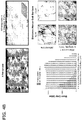


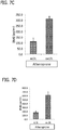


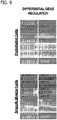


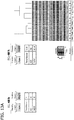


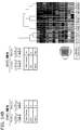


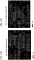


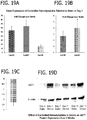


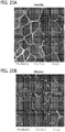
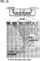
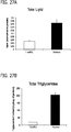

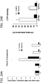


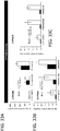

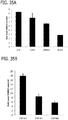

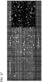
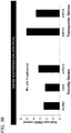
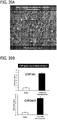

Classifications
-
- G—PHYSICS
- G01—MEASURING; TESTING
- G01N—INVESTIGATING OR ANALYSING MATERIALS BY DETERMINING THEIR CHEMICAL OR PHYSICAL PROPERTIES
- G01N33/00—Investigating or analysing materials by specific methods not covered by groups G01N1/00 - G01N31/00
- G01N33/48—Biological material, e.g. blood, urine; Haemocytometers
- G01N33/50—Chemical analysis of biological material, e.g. blood, urine; Testing involving biospecific ligand binding methods; Immunological testing
- G01N33/5005—Chemical analysis of biological material, e.g. blood, urine; Testing involving biospecific ligand binding methods; Immunological testing involving human or animal cells
- G01N33/5008—Chemical analysis of biological material, e.g. blood, urine; Testing involving biospecific ligand binding methods; Immunological testing involving human or animal cells for testing or evaluating the effect of chemical or biological compounds, e.g. drugs, cosmetics
- G01N33/502—Chemical analysis of biological material, e.g. blood, urine; Testing involving biospecific ligand binding methods; Immunological testing involving human or animal cells for testing or evaluating the effect of chemical or biological compounds, e.g. drugs, cosmetics for testing non-proliferative effects
- G01N33/5023—Chemical analysis of biological material, e.g. blood, urine; Testing involving biospecific ligand binding methods; Immunological testing involving human or animal cells for testing or evaluating the effect of chemical or biological compounds, e.g. drugs, cosmetics for testing non-proliferative effects on expression patterns
-
- G—PHYSICS
- G01—MEASURING; TESTING
- G01N—INVESTIGATING OR ANALYSING MATERIALS BY DETERMINING THEIR CHEMICAL OR PHYSICAL PROPERTIES
- G01N33/00—Investigating or analysing materials by specific methods not covered by groups G01N1/00 - G01N31/00
- G01N33/48—Biological material, e.g. blood, urine; Haemocytometers
- G01N33/50—Chemical analysis of biological material, e.g. blood, urine; Testing involving biospecific ligand binding methods; Immunological testing
- G01N33/5005—Chemical analysis of biological material, e.g. blood, urine; Testing involving biospecific ligand binding methods; Immunological testing involving human or animal cells
- G01N33/5008—Chemical analysis of biological material, e.g. blood, urine; Testing involving biospecific ligand binding methods; Immunological testing involving human or animal cells for testing or evaluating the effect of chemical or biological compounds, e.g. drugs, cosmetics
-
- G—PHYSICS
- G01—MEASURING; TESTING
- G01N—INVESTIGATING OR ANALYSING MATERIALS BY DETERMINING THEIR CHEMICAL OR PHYSICAL PROPERTIES
- G01N33/00—Investigating or analysing materials by specific methods not covered by groups G01N1/00 - G01N31/00
- G01N33/48—Biological material, e.g. blood, urine; Haemocytometers
- G01N33/50—Chemical analysis of biological material, e.g. blood, urine; Testing involving biospecific ligand binding methods; Immunological testing
- G01N33/5005—Chemical analysis of biological material, e.g. blood, urine; Testing involving biospecific ligand binding methods; Immunological testing involving human or animal cells
- G01N33/5008—Chemical analysis of biological material, e.g. blood, urine; Testing involving biospecific ligand binding methods; Immunological testing involving human or animal cells for testing or evaluating the effect of chemical or biological compounds, e.g. drugs, cosmetics
- G01N33/5044—Chemical analysis of biological material, e.g. blood, urine; Testing involving biospecific ligand binding methods; Immunological testing involving human or animal cells for testing or evaluating the effect of chemical or biological compounds, e.g. drugs, cosmetics involving specific cell types
- G01N33/5067—Liver cells
-
- G—PHYSICS
- G01—MEASURING; TESTING
- G01N—INVESTIGATING OR ANALYSING MATERIALS BY DETERMINING THEIR CHEMICAL OR PHYSICAL PROPERTIES
- G01N33/00—Investigating or analysing materials by specific methods not covered by groups G01N1/00 - G01N31/00
- G01N33/48—Biological material, e.g. blood, urine; Haemocytometers
- G01N33/50—Chemical analysis of biological material, e.g. blood, urine; Testing involving biospecific ligand binding methods; Immunological testing
- G01N33/5005—Chemical analysis of biological material, e.g. blood, urine; Testing involving biospecific ligand binding methods; Immunological testing involving human or animal cells
- G01N33/5091—Chemical analysis of biological material, e.g. blood, urine; Testing involving biospecific ligand binding methods; Immunological testing involving human or animal cells for testing the pathological state of an organism
-
- C—CHEMISTRY; METALLURGY
- C12—BIOCHEMISTRY; BEER; SPIRITS; WINE; VINEGAR; MICROBIOLOGY; ENZYMOLOGY; MUTATION OR GENETIC ENGINEERING
- C12M—APPARATUS FOR ENZYMOLOGY OR MICROBIOLOGY; APPARATUS FOR CULTURING MICROORGANISMS FOR PRODUCING BIOMASS, FOR GROWING CELLS OR FOR OBTAINING FERMENTATION OR METABOLIC PRODUCTS, i.e. BIOREACTORS OR FERMENTERS
- C12M35/00—Means for application of stress for stimulating the growth of microorganisms or the generation of fermentation or metabolic products; Means for electroporation or cell fusion
- C12M35/04—Mechanical means, e.g. sonic waves, stretching forces, pressure or shear stimuli
-
- G—PHYSICS
- G01—MEASURING; TESTING
- G01N—INVESTIGATING OR ANALYSING MATERIALS BY DETERMINING THEIR CHEMICAL OR PHYSICAL PROPERTIES
- G01N2500/00—Screening for compounds of potential therapeutic value
- G01N2500/10—Screening for compounds of potential therapeutic value involving cells
-
- G—PHYSICS
- G01—MEASURING; TESTING
- G01N—INVESTIGATING OR ANALYSING MATERIALS BY DETERMINING THEIR CHEMICAL OR PHYSICAL PROPERTIES
- G01N2800/00—Detection or diagnosis of diseases
- G01N2800/70—Mechanisms involved in disease identification
- G01N2800/7004—Stress
Definitions
- the present invention generally relates to in vitro methods for mimicking in vivo pathological or physiologic conditions.
- the present invention also relates to methods for testing drugs or compounds in such systems.
- US 2009/053752 relates to an in vitro model used to applied hemodynamic (i.e., blood flow) patterns modeled after the human circulation to human/animal cells in culture.
- US 2010/304355 relates to devices, in vitro cell cultures, systems, and methods for microscale cell culture analogous (CCA) device.
- CCA microscale cell culture analogous
- the conventional systems often do not exhibit responses to drugs or compounds at concentrations that induce the response in vivo, instead requiring much higher concentrations of the drug or compound to induce the same response.
- the invention is as defined by the accompanying claims.
- One aspect of the present invention is directed to a method of mimicking a pathological condition in vitro, as defined in the claims.
- the method comprises adding a culture media to a cell culture container, adding at least one factor wherein at least one factor comprises glucose to the culture media, plating hepatocytes on at least one surface within the cell culture container, and indirectly applying a shear force upon the hepatocytes at a rate of 0.1 dynes/cm 2 to 3.0 dynes/cm 2 .
- the shear force results from flow of the culture media induced by a flow device.
- the flow mimics flow to which the hepatocytes are exposed in vivo in the pathological condition.
- the concentration of the factor in the culture media can be within the in vivo concentration range of the factor observed in the pathological condition, with the glucose being present in the culture media at a concentration of 10 mM to 25 mM.
- Another aspect of the present invention is an in vitro method of testing a drug or a compound for an effect on a pathological condition, as defined in the claims.
- the method comprises mimicking the pathological condition, adding a drug or a compound to the culture media, and applying the shear force upon the hepatocytes exposed to the drug or the compound.
- a change in the hepatocytes, in the presence of the drug or the compound, indicates that the drug or the compound has an effect on the pathological condition.
- the pathological condition can be mimicked by the in vitro method of mimicking a pathological condition as described above.
- the present invention also provides an in vitro method of testing a drug or compound for an effect, as defined in the claims.
- the method comprises adding a culture media to a cell culture container, plating hepatocytes on at least one surface within the cell culture container, adding a drug or a compound to the culture media, and applying a shear force upon the hepatocytes exposed to the drug or the compound.
- the concentration of the drug or the compound in the culture media is within the concentration range of the drug or the compound that achieves the effect in vivo.
- the shear force results from flow of the culture media induced by a flow device.
- the flow mimics flow to which the hepatocytes are exposed in vivo.
- a change in the hepatocytes, in the presence of the drug or the compound indicates that the drug or the compound has the effect.
- the effect can be, for example, an effect on a physiologic condition or an effect on a pathological condition.
- Another aspect of the present invention is a method of mimicking a physiologic condition in vitro, as defined in the claims.
- the method comprises adding a culture media to a cell culture container, adding at least one factor wherein at least one factor comprises glucose to the culture media, plating hepatocytes on at least one surface within the cell culture container, and indirectly applying a shear force upon the hepatocytes at a rate of 0.1 dynes/cm 2 to 3.0 dynes/cm 2 .
- the shear force results from flow of the culture media induced by a flow device.
- the flow mimics flow to which the hepatocytes are exposed in vivo in the physiologic condition.
- the concentration of the factor in the culture media can be within the in vivo concentration range of the factor observed in the physiologic condition, with the glucose being present in the culture media at a concentration of 5 mM to 10 mM.
- the present invention is also directed to an in vitro method of testing a drug or a compound for an effect on a physiologic condition, as defined in the claims.
- the method comprises mimicking the physiologic condition, adding a drug or a compound to the culture media, and applying the shear force upon the hepatocytes exposed to the drug or the compound.
- a change in the hepatocytes, in the presence of the drug or the compound, indicates that the drug or the compound has an effect on the physiologic condition.
- the physiologic condition can be mimicked by the in vitro method of mimicking a physiologic condition as described above.
- Another aspect of the invention is a method of mimicking a pathological or physiologic condition of the liver in vitro, as defined in the claims.
- the method comprises adding a culture media to a cell culture container, adding at least one factor to the culture media wherein at least one factor comprises glucose, plating at least one hepatic cell type on at least one surface within the cell culture container, and indirectly applying a shear force upon the at least one plated hepatic cell type at a rate of 0.1 dynes/cm 2 to 3.0 dynes/cm 2 .
- the shear force results from flow of the culture media induced by a flow device.
- the flow mimics flow to which the at least one hepatic cell type is exposed in vivo in the pathological or physiologic condition.
- the concentration of the factor in the culture media for mimicking the pathological condition can be within the in vivo concentration range of the factor observed in the pathological condition, with the glucose being present in the culture media at a concentration of 10 mM to 25 mM.
- the concentration of the factor in the culture media can be capable of maintaining the mimicked pathological condition in vitro for a period of time under the shear force, the same concentration of factor being incapable of maintaining the mimicked pathological condition in vitro for the period of time in the absence of the shear force.
- the concentration of the factor in the culture media for mimicking the physiologic condition can be within the in vivo concentration range of the factor observed in the physiologic condition, with the glucose being present in the culture media at a concentration of 5 mM to 10 mM.
- the concentration of the factor in the culture media can be capable of maintaining the mimicked physiologic condition in vitro for a period of time under the shear force, the same concentration of factor being incapable of maintaining the mimicked physiologic condition in vitro for the period of time in the absence of the shear force.
- the present invention also provides an in vitro method of testing a drug or a compound for an effect on a pathological or physiological condition, as defined in the claims.
- the method comprises mimicking the pathological or physiological condition, adding a drug or a compound to the culture media, and applying the shear force upon at least one plated hepatic cell type exposed to the drug or the compound.
- a change in the at least one plated hepatic cell type, in the presence of the drug or the compound indicates that the drug or the compound has an effect on the pathological or physiological condition.
- the pathological condition can be mimicked by the in vitro method of mimicking a pathological or physiological condition as described in the immediately preceding paragraph.
- Another aspect of the invention is directed to a method of mimicking a pathological or physiologic condition of the liver in vitro, as defined in the claims.
- the method comprises adding a culture media to a cell culture container, depositing at least one extracellular matrix component on a surface within the cell culture container, plating hepatocytes on the at least one extracellular matrix component, and indirectly applying a shear force upon the at least one extracellular matrix component and the hepatocytes.
- the shear force results from flow of the culture media induced by a flow device.
- the flow mimics flow to which the hepatocytes are exposed in vivo in the pathological or physiologic condition.
- the invention also provides another method of mimicking a pathological or physiologic condition of the liver in vitro, as defined in the claims.
- the method comprises adding a culture media to a cell culture container and plating hepatocytes on a first surface of a porous membrane.
- the porous membrane is suspended in the cell culture container such that the first surface is proximal and in spaced relation to a bottom surface of the container, thereby defining within the container a lower volume comprising the hepatocytes and an upper volume comprising a second surface of the porous membrane.
- a shear force is applied upon the second surface of the porous membrane in the upper volume of the container, the shear force resulting from flow of the culture media induced by a flow device.
- the flow mimics flow to which the hepatocytes are exposed in vivo in the pathological or physiologic condition.
- the flow device comprises a body adapted for being positioned in the culture media in the upper volume of the container and a motor adapted to rotate the body.
- the present invention provides in vitro methods for mimicking an in vivo pathological or physiologic condition, as defined in the claims.
- the methods of the invention apply shear forces to cultured cells and replicate an in vivo pathological or physiological condition using in vivo pathological or physiologic concentrations of various factors.
- an in vitro liver model has been discovered in which hepatocytes can be maintained at in vivo physiologic concentrations of insulin and glucose that are significantly decreased as compared to the concentrations used in the standard static model. It has further been discovered that when higher concentrations of insulin and glucose are used in such a model, the hepatocytes exhibit numerous hallmarks of fatty liver disease.
- the present invention is also directed to a method of mimicking a pathological condition in vitro, as defined in the claims.
- the method comprises adding a culture media to a cell culture container, adding at least one factor to the culture media wherein at least one factor comprises glucose, hepatocytes on at least one surface within the cell culture container, and indirectly applying a shear force upon the hepatocytes cell type at a rate of 0.1 dynes/cm 2 to 3.0 dynes/cm 2 .
- the shear force results from flow of the culture media induced by a flow device.
- the flow mimics flow to which the at least one cell type is exposed in vivo in the pathological condition.
- the concentration of the factor in the culture media can be within the in vivo concentration range of the factor observed in the pathological condition, with the glucose being present in the culture media at a concentration of 10 mM to 25 mM.
- a change in a level of a marker of the pathological condition can be compared between the method of the invention and the same method in the absence of application of the shear force.
- the level of the marker in the hepatocytes or in the culture media upon application of the shear force is compared to the level of the marker in the hepatocytes or in the culture media in the absence of application of the shear force.
- a marker is known to be associated with a pathological condition and its concentration is known to increase in the serum when the condition is present in vivo
- an increase in the level of the marker in the culture media of the method of the invention with application of the shear force as compared to the level of the marker in the culture media in the absence of application of the shear force confirms that the in vivo pathological condition is mimicked by the in vitro method of the invention.
- Pathological conditions effects on the pathological conditions, physiologic conditions, flow devices, hemodynamic patterns, cell types, and cell culture media including factors added to the cell culture media for use in the methods of the invention are described in detail below, following the description of the various methods of the invention.
- the present invention is also directed to an in vitro method of testing a drug or a compound for an effect on a pathological condition, as defined in the claims.
- the method comprises mimicking the pathological condition, adding a drug or a compound to the culture media, and applying the shear force upon the hepatocytes exposed to the drug or the compound.
- a change in the hepatocytes, in the presence of the drug or the compound, indicates that the drug or the compound has an effect on the pathological condition.
- the pathological condition can be mimicked by the in vitro method of mimicking a pathological condition as described above.
- the pathological condition of the in vitro method of testing a drug or compound can also be mimicked by plating primary cells or immortalized cells from a subject or subjects having the pathological condition, and culturing the cells in cell culture media.
- the present invention is also directed to a method of mimicking a physiologic condition in vitro, as defined in the claims.
- the method comprises adding a culture media to a cell culture container, adding at least one factor to the culture media wherein at least one factor comprises glucose, plating hepatocytes on at least one surface within the cell culture container, and indirectly applying a shear force upon the hepatocyte cell type at a rate of 0.1 dynes/cm 2 to 3.0 dynes/cm 2 .
- the shear force results from flow of the culture media induced by a flow device.
- the flow mimics flow to which the hepatocytes are exposed in vivo in the physiologic condition.
- the concentration of the factor in the culture media can be within the in vivo concentration range of the factor observed in the physiologic condition, with the glucose being present in the culture media at a concentration of 5 mM to 10 mM.
- a change in a level of a marker of the physiologic condition can be compared between the method of the invention and the same method in the absence of application of the shear force.
- the level of the marker in the hepatocytes or in the culture media upon application of the shear force is compared to the level of the marker in the hepatocytes or in the culture media in the absence of application of the shear force.
- a marker is known to be associated with a physiologic condition and its concentration is known to increase in the serum when the condition is present in vivo
- an increase in the level of the marker in the culture media of the method of the invention with application of the shear force as compared to the level of the marker in the culture media in the absence of application of the shear force confirms that the in vivo physiologic condition is mimicked by the in vitro method of the invention.
- the present invention is also directed to an in vitro method of testing a drug or a compound for an effect on a physiologic condition, as defined in the claims.
- the method comprises mimicking the physiologic condition, adding a drug or a compound to the culture media, and applying the shear force upon the hepatocytes exposed to the drug or the compound.
- a change in the hepatocytes, in the presence of the drug or the compound, indicates that the drug or the compound has an effect on the physiologic condition.
- the physiologic condition can be mimicked by the in vitro method of mimicking a physiologic condition as described above.
- the physiologic condition of this in vitro method of testing a drug or compound can also be mimicked by plating primary cells or immortalized cells, and culturing the cells in cell culture media.
- the primary or immortalized cells are described in detail below.
- the present invention also relates to an in vitro method of testing a drug or a compound for an effect, as defined in the claims.
- the method comprises adding a culture media to a cell culture container, plating hepatocytes on at least one surface within the cell culture container, adding a drug or a compound to the culture media, and applying a shear force upon the hepatocytes exposed to the drug or the compound.
- the concentration of the drug or the compound in the culture media is within the concentration range of the drug or the compound that achieves the effect in vivo.
- the shear force results from flow of the culture media induced by a flow device.
- the flow mimics flow to which the hepatocytes is exposed in vivo.
- a change in the hepatocytes, in the presence of the drug or the compound, indicates that the drug or the compound has the effect.
- the effect can be an effect on a pathological condition.
- the effect can be an effect on a physiologic condition. Further effects are described in detail below.
- the method can further comprise analyzing the cell culture media for cytokine secretion, chemokine secretion, humoral factor secretion, microparticle secretion, growth factor secretion, shedding of a protein from the cellular surface, a metabolite of a compound, an immune cell, nitric oxide secretion, a vasodilator protein, a vasoconstrictive protein, miRNA, a secreted protein, or a secreted biological substance.
- the cell culture media can be analyzed for nitric oxide secretion by measuring nitrate or nitrite concentration.
- the protein can comprise a vascular cell adhesion molecule (VCAM), E-selectin, or an intracellular adhesion molecule (ICAM).
- VCAM vascular cell adhesion molecule
- E-selectin E-selectin
- ICM intracellular adhesion molecule
- the method can further comprise the step of culturing the cell type or cell types.
- the method can further comprise the step of comparing the hepatocytes after applying the shear force for a period of time wherein the media includes the drug or the compound to the hepatocytes after applying the shear force for the period of time wherein the media does not include the drug or the compound, to determine the effect of the drug or compound on the hepatocytes.
- the factors comprise insulin and glucose
- hepatocytes are plated on the surface within the cell culture container, and the shear force is applied indirectly to the plated hepatocytes.
- the hepatocytes can be plated on a first surface of a porous membrane.
- the porous membrane is then suspended in the cell culture container such that the first surface is proximal and in spaced relation to a bottom surface of the cell culture container, thereby defining within the cell culture container a lower volume and an upper volume.
- the lower volume comprises the hepatocytes and the upper volume comprises a second surface of the porous membrane.
- the shear force is applied to the second surface of the porous membrane in the upper volume of the container.
- a porous membrane suspended in the cell culture container is preferred in plating the cells.
- shear force is applied to plated cells or to the surface of the porous membrane (e.g., when the shear is applied on a surface of the membrane absent plated cells)
- the shear force can enable the cell culture media to perfuse from the upper volume to the lower volume.
- Such perfusion favorably impacts transport of factors from the upper volume to the lower volume, or vice versa.
- the invention is also directed to a method of mimicking a pathological or physiologic condition of the liver in vitro.
- the method comprises adding a culture media to a cell culture container, adding at least one factor to the culture media wherein at least one factor comprises glucose, plating at least one hepatic cell type on at least one surface within the cell culture container, and indirectly applying a shear force upon the at least one plated hepatic cell type at a rate of 0.1 dynes/cm 2 to 3.0 dynes/cm 2 .
- the shear force results from flow of the culture media induced by a flow device.
- the flow mimics flow to which the at least one hepatic cell type is exposed in vivo in the pathological or physiologic condition.
- the concentration of the factor in the culture media for mimicking the pathological condition can be within the in vivo concentration range of the factor observed in the pathological condition, wherein at least one factor comprises glucose with the glucose being present in the culture media at a concentration of 10 mM to 25 mM.
- the concentration of the factor in the culture media for mimicking the pathological condition can be capable of maintaining the mimicked pathological condition in vitro for a period of time under the shear force, the same concentration of factor being incapable of maintaining the mimicked pathological condition in vitro for the period of time in the absence of the shear force.
- the concentration of the factor in the culture media for mimicking the physiologic condition can be within the in vivo concentration range of the factor observed in the physiologic condition, wherein at least one factor comprises glucose with the glucose being present in the culture media at a concentration of 5 mM to 10 mM.
- the concentration of the factor in the culture media for mimicking the physiologic condition can be capable of maintaining the mimicked physiologic condition in vitro for a period of time under the shear force, the same concentration of factor being incapable of maintaining the mimicked physiologic condition in vitro for the period of time in the absence of the shear force.
- a change in a level of a marker of the pathological or physiologic condition in the at least one plated hepatic cell type or in the culture media upon application of the shear force, as compared to the level of the marker in the at least one plated hepatic cell type or in the culture media in the absence of application of the shear force confirms mimicking of the pathological or physiologic condition.
- the at least one plated hepatic cell type can comprise hepatocytes, and responsiveness to glucagon, insulin, or a glucose substrate in the hepatocytes confirms mimicking of the physiologic condition.
- the glucose substrate can be, for example, glycerol, lactate, pyruvate, or combinations thereof (e.g., a combination of lactate and pyruvate).
- the present invention is also directed to an in vitro method of testing a drug or a compound for an effect on a pathological or physiological condition.
- the method comprises mimicking the pathological or physiological condition, adding a drug or a compound to the culture media, and applying the shear force upon at least one plated hepatic cell type exposed to the drug or the compound.
- a change in the at least one plated hepatic cell type, in the presence of the drug or the compound, indicates that the drug or the compound has an effect on the pathological or physiological condition.
- the pathological condition can be mimicked by the in vitro method of mimicking a pathological or physiological condition as described directly above.
- the pathological or physiological condition of the in vitro method of testing a drug or compound can also be mimicked by plating primary cells or immortalized cells from a subject or subjects having the pathological condition, and culturing the cells in cell culture media.
- the invention is also directed to a method of mimicking a pathological or physiologic condition of the liver in vitro.
- the method comprises adding a culture media to a cell culture container, depositing at least one extracellular matrix component on a surface within the cell culture container, plating hepatocytes on the at least one extracellular matrix component, and indirectly applying a shear force upon the at least one extracellular matrix component and the hepatocytes.
- the shear force results from flow of the culture media induced by a flow device.
- the flow mimics flow to which the hepatocytes are exposed in vivo in the pathological or physiologic condition.
- At least one extracellular matrix component can be plated on a first surface of the porous membrane and the hepatic cells can subsequently be plated on the at least one extracellular matrix component.
- nonparenchymal hepatic cells e.g., sinusoidal endothelial cells
- the shear stress applied to the nonparenchymal hepatic cells can be plated on the second surface of the porous membrane, and the shear stress applied to the nonparenchymal hepatic cells.
- the at least one extracellular matrix component can be deposited on a first surface of a porous membrane.
- the hepatic cell type e.g., hepatocytes
- the porous membrane is suspended in the cell culture container such that the first surface is proximal and in spaced relation to a bottom surface of the cell culture container, thereby defining within the cell culture container a lower volume and an upper volume.
- the lower volume comprises at least one extracellular matrix component and the hepatic cell type (e.g., hepatocytes), and the upper volume comprises a second surface of the porous membrane.
- nonparenchymal hepatic cells e.g., sinusoidal endothelial cells
- the shear stress applied to the nonparenchymal hepatic cells can be applied to the nonparenchymal hepatic cells.
- the invention also provides another method of mimicking a pathological or physiologic condition of the liver in vitro.
- the method comprises adding a culture media to a cell culture container, and plating hepatocytes on a first surface of a porous membrane.
- the porous membrane is suspended in the cell culture container such that the first surface is proximal and in spaced relation to a bottom surface of the container, thereby defining within the container a lower volume comprising the hepatocytes and an upper volume comprising a second surface of the porous membrane.
- a shear force is applied upon the second surface of the porous membrane in the upper volume of the container, the shear force resulting from flow of the culture media induced by a flow device.
- the flow mimics flow to which the hepatocytes are exposed in vivo in the pathological or physiologic condition.
- the flow device comprises a body adapted for being positioned in the culture media in the upper volume of the container and a motor adapted to rotate the body.
- the body has a conical surface. It is also preferred that the flow device is adapted for positioning the conical surface of the body in the container and in contact with the cell culture media.
- This method can further comprise plating nonparenchymal hepatic cells on the second surface of the porous membrane, wherein the shear stress is applied to the nonparenchymal hepatic cells.
- the nonparenchymal hepatic cells can comprise sinusoidal endothelial cells, hepatic stellate cells, Kupffer cells, or combinations thereof.
- a change in a level of a marker of the pathological or physiologic condition can be compared in the inventive method to the same method in the absence of application of the shear force.
- a change in the level of the marker in any of the hepatic cells or in the culture media upon application of the shear force as compared to the level of the marker in the hepatic cells or in the culture media in the absence of application of the shear force confirms mimicking of the pathological or physiologic condition.
- a change in the level of the marker in the hepatocytes or nonparenchymal hepatic cells or in the culture media upon application of the shear force as compared to the level of the marker in the hepatocytes or nonparenchymal hepatic cells or in the culture media in the absence of application of the shear force confirms mimicking of the pathological or physiologic condition.
- the at least one plated hepatic cell type comprises hepatocytes
- responsiveness to glucagon, insulin, or a glucose substrate in the hepatocytes confirms mimicking of the physiologic condition.
- the glucose substrate can be, for example, glycerol, lactate, pyruvate, or combinations thereof (e.g., a combination of lactate and pyruvate).
- Pathological conditions include advanced inflammation, atherosclerosis, diabetic nephropathy, diabetic neuropathy, diabetic retinopathy, hypertension, hypertensive encephalopathy, hypertensive retinopathy, fatty liver disease, hypertension, heart failure, stroke, Marfan syndrome, carotid intima-medial thickening, atrial fibrillation, kidney disease, pulmonary fibrosis, chronic obstructive pulmonary disease, hyperlipidemia, hypercholesterolemia, diabetes, atherosclerotic plaque rupture, atherosclerotic plaque erosion, thoracic aortic aneurysm, cerebral aneurysm, abdominal aortic aneurysm, cerebral aneurysm, pulmonary artery disease, pulmonary hypertension, peripheral artery disease, arterial thrombosis, venous thrombosis (e.g., deep vein thrombosis), vascular restenosis, vascular calcification, myocardial infarction, obesity, hypertriglyceridemia,
- the pathological condition can be anatomical conditions, such as atrophy, calculi, choristoma, pathologic constriction, pathologic dilation, diverticulum, hypertrophy, polyps, prolapse, rupture, an arteriovenous fistula, or an appendage (e.g., left atrial appendage).
- anatomical conditions such as atrophy, calculi, choristoma, pathologic constriction, pathologic dilation, diverticulum, hypertrophy, polyps, prolapse, rupture, an arteriovenous fistula, or an appendage (e.g., left atrial appendage).
- endothelial cells, smooth muscle cells, or endocardial cells can be plated on the surface within the cell culture container, and the shear force applied upon the plated endothelial cells, smooth muscle cells, or endocardial cells.
- the factor can be oxidized low-density lipoprotein (oxLDL), tumor necrosis factor-a (TNF ⁇ ), glucose, tissue growth factor- ⁇ (TGF- ⁇ ), an elastin degradation product, elastase, vitamin D, an inorganic phosphate, leptin, adiponectin, apelin, aldosterone, angiotensin II, a triglyceride, high-density lipoprotein (HDL), oxidized high-density lipoprotein (oxHDL), a triglyceride-rich lipoprotein, low-density lipoprotein (LDL), insulin, a fatty acid, or a combination thereof.
- oxLDL oxidized low-density lipoprotein
- TGF- ⁇ tumor necrosis factor-a
- TGF- ⁇ tissue growth factor- ⁇
- elastin degradation product elastase
- vitamin D an inorganic phosphate
- leptin lept
- the triglyceride-rich lipoprotein can be very low-density lipoprotein (vLDL), a vLDL remnant, a chylomicron, or a chylomicron remnant.
- vLDL very low-density lipoprotein
- endocardial cells can be plated on a first surface of a porous membrane.
- the porous membrane is suspended in the cell culture container such that the first surface is proximal and in spaced relation to a bottom surface of the cell culture container, thereby defining within the cell culture container a lower volume comprising the endocardial cells and an upper volume comprising a second surface of the porous membrane.
- the shear force is applied to the second surface of the porous membrane in the upper volume.
- endothelial cells can be plated on the second surface of the porous membrane, and the shear force applied upon the plated endothelial cells.
- the endocardial cells can be smooth muscle cells.
- the cell types can be endothelial cells, smooth muscle cells, endocardial cells, or a combination thereof, such as the cell types being endothelial; smooth muscle; endothelial and smooth muscle; endocardial; or endocardial and endothelial.
- the plated cell types can be from a normal subject, a subject having diabetes, a hypertensive subject, an aged subject, or an animal genetically modified to model diabetes, hypertension, or aging.
- the flow or hemodynamic pattern can be derived from a cardiac sinus or from an atrial fibrillation rhythm.
- the factor can be oxLDL, TNF ⁇ , aldosterone, angiotensin II, or a combination thereof.
- the factor(s) can be oxLDL; TNF ⁇ ; oxLDL and TNF ⁇ ; aldosterone; angiotensin II; aldosterone and angiotensin II; oxLDL, TNF ⁇ , and angiotensin II; oxLDL, TNF ⁇ , and aldosterone; or oxLDL, TNF ⁇ , aldosterone, and angiotensin II.
- smooth muscle cells can be plated on a first surface of the porous membrane.
- the porous membrane is suspended in the cell culture container such that the first surface is proximal and in spaced relation to a bottom surface of the cell culture container, thereby defining within the cell culture container a lower volume comprising the smooth muscle cells and an upper volume comprising a second surface of the porous membrane.
- the shear force is applied to the second surface of the porous membrane in the upper volume.
- endothelial cells can be plated on the second surface of the porous membrane.
- endothelial cells can be plated on a second surface of a porous membrane.
- the porous membrane is suspended in the cell culture container such that a first surface of the porous membrane is proximal and in spaced relation to a bottom surface of the cell culture container, thereby defining within the cell culture container a lower volume comprising the first surface of the porous membrane and an upper volume comprising the endothelial cells.
- the shear force is applied to the endothelial cells in the upper volume.
- smooth muscle cells can be plated on the first surface of the porous membrane.
- the cell types can be endothelial cells, smooth muscle cells, or a combination thereof such as the cell types being endothelial; smooth muscle; or endothelial and smooth muscle.
- the plated cell types can be from a normal subject, a subject having diabetes, a hypertensive subject, or an animal genetically modified to model diabetes or hypertension.
- the flow or hemodynamic pattern can be atheroprone, atheroprotective (i.e., also described herein as "healthy state"), derived from a femoral artery, or derived from an arteriole.
- the factor can be LDL, oxLDL, TNF ⁇ , HDL, a triglyceride-rich lipoprotein, or a combination thereof.
- the factor(s) can be LDL; LDL and oxLDL; oxLDL; HDL; HDL and oxLDL; TNF ⁇ ; TNF ⁇ and oxLDL; TNF ⁇ , oxLDL, and HDL; or TNF ⁇ , oxLDL, and a triglyceride-rich lipoprotein.
- the cell types can be endothelial cells, smooth muscle cells, or a combination thereof such as the cell types being endothelial; smooth muscle; or endothelial and smooth muscle.
- the plated cell types can be from a normal subject, a subject having diabetes, a hypertensive subject, or an animal genetically modified to model diabetes or hypertension.
- the flow or hemodynamic pattern can be atheroprone, atheroprotective, derived from a femoral artery, or derived from an arteriole.
- the factor can be a triglyceride-rich lipoprotein.
- the cell types can be endothelial cells, smooth muscle cells, or a combination thereof such as cell types being endothelial; smooth muscle; or endothelial and smooth muscle.
- the plated cell types can be from a normal subject, a subject having diabetes, a hypertensive subject, a smoker, a subject having abdominal aortic aneurysm, or an animal genetically modified to model diabetes or hypertension or modified to model abdominal aortic aneurysm.
- the flow or hemodynamic pattern can be derived from an abdominal artery or derived from an intra-abdominal aortic aneurysm rhythm.
- the factor can be oxLDL, TNF ⁇ , glucose, an elastin degradation product, elastase, angiotensin II, aldosterone, insulin, TGF- ⁇ , or a combination thereof.
- the factor(s) can be oxLDL; TNF ⁇ ; glucose; an elastin degradation product; elastase; angiotensin II; aldosterone; insulin; TGF- ⁇ ; oxLDL and TNF ⁇ ; oxLDL and glucose; oxLDL and an elastin degradation product; oxLDL and elastase; oxLDL and angiotensin II; oxLDL and aldosterone; oxLDL and insulin; oxLDL and TGF- ⁇ ; TNF ⁇ and glucose; TNF ⁇ and an elastin degradation product; TNF ⁇ and elastase; TNF ⁇ and angiotensin II; TNF ⁇ and aldosterone; TNF ⁇ and insulin; TNF ⁇ and TGF- ⁇ ; glucose and an elastin degradation product; glucose and elastase; glucose and angiotensin II; glucose and aldosterone; glucose and insulin; glucose and TGF- ⁇
- smoke extract can be added to the culture media.
- the cell types can be endothelial cells, smooth muscle cells, or a combination thereof such as the cell types being endothelial; smooth muscle; or endothelial and smooth muscle.
- the vascular pathological condition is a diabetic vascular condition, such as diabetic nephropathy, diabetic neuropathy, or diabetic retinopathy
- the plated cell types can be from a normal subject, a subject having diabetes, or an animal genetically modified to model diabetes.
- the flow or hemodynamic pattern can be atheroprone, atheroprotective, derived from a femoral artery, or derived from an arteriole.
- the factor can be oxLDL, TNF ⁇ , glucose, HDL, oxHDL, a triglyceride-rich lipoprotein, insulin, or a combination thereof.
- the factor(s) can be glucose; glucose and insulin; glucose, oxLDL, and TNF ⁇ ; glucose, insulin, oxLDL, and TNF ⁇ ; glucose, oxLDL, TNF ⁇ , and HDL; glucose, oxLDL, TNF ⁇ , and oxHDL; glucose, oxLDL, TNF ⁇ , HDL, and oxHDL; glucose, insulin, oxLDL, TNF ⁇ , and HDL; glucose, insulin, oxLDL, TNF ⁇ , and oxHDL; glucose, insulin, oxLDL, TNF ⁇ , HDL, and oxHDL; glucose, oxLDL, TNF ⁇ , and a triglyceride-rich lipoprotein; or glucose, insulin, oxLDL, TNF ⁇ , and a triglyceride-rich lipoprotein.
- the cell types can be endothelial cells, smooth muscle cells, or a combination thereof such as the cell types being endothelial; smooth muscle; or endothelial and smooth muscle.
- the plated cell types can be from a normal subject, a subject having diabetes, a hypertensive subject, or an animal genetically modified to model diabetes or hypertension.
- the flow or hemodynamic pattern can be atheroprone, atheroprotective, or derived from a femoral artery, a pulmonary artery, or an arteriole.
- the factor can be oxLDL, TNF ⁇ , angiotensin II, aldosterone, or a combination thereof.
- the factor(s) can be angiotensin II; aldosterone; angiotensin II and aldosterone; or angiotensin II, aldosterone, oxLDL and TNF ⁇ .
- the cell types can be endothelial cells, smooth muscle cells, or a combination thereof such as the cell types being endothelial; smooth muscle; or endothelial and smooth muscle.
- the plated cell types can be from a normal subject, a subject having diabetes, a hypertensive subject, or an animal genetically modified to model diabetes or hypertension.
- the flow or hemodynamic pattern can be atheroprone, atheroprotective, or derived from a femoral artery, a pulmonary artery, or an arteriole.
- the factor can be oxLDL, TNF ⁇ , vitamin D, an inorganic phosphate, leptin, adiponectin, or a combination thereof.
- the factor(s) can be oxLDL; TNF ⁇ ; vitamin D; an inorganic phosphate; leptin; adiponectin; oxLDL and TNF ⁇ ; oxLDL and vitamin D; oxLDL and an inorganic phosphate; oxLDL and leptin; oxLDL and adiponectin; TNF ⁇ and vitamin D; TNF ⁇ and an inorganic phosphate; TNF ⁇ and leptin; TNF ⁇ and adiponectin; vitamin D and an inorganic phosphate; vitamin D and leptin; vitamin D and adiponectin; an inorganic phosphate and leptin; an inorganic phosphate and adiponectin; lep
- the cell types can be endothelial cells, smooth muscle cells, or a combination thereof such as the cell types being endothelial; smooth muscle; or endothelial and smooth muscle.
- the plated cell types can be from a normal subject, a subject having diabetes, a hypertensive subject, or an animal genetically modified to model diabetes or hypertension.
- the flow or hemodynamic pattern can be atheroprone, atheroprotective, or derived from a femoral artery, a pulmonary artery, or an arteriole.
- the factor can be TNF ⁇ , oxLDL, glucose, or a combination thereof.
- the factor(s) can be TNF ⁇ ; oxLDL; glucose; or oxLDL and glucose.
- the cell types can be hepatocytes, nonparenchymal hepatic cells, or combinations thereof.
- the nonparenchymal hepatic cells can include sinusoidal endothelial cells, hepatic stellate cells, Kupffer cells, or combinations thereof.
- the flow or hemodynamic pattern can be from a normal subject, a subject having fatty liver disease, or an animal genetically modified to model fatty liver disease.
- hepatocytes can be plated on a first surface of the porous membrane.
- the porous membrane is suspended in the cell culture container such that the first surface is proximal and in spaced relation to a bottom surface of the cell culture container, thereby defining within the cell culture container a lower volume comprising the hepatocytes and an upper volume comprising a second surface of the porous membrane.
- the shear force is applied to the second surface of the porous membrane in the upper volume.
- nonparenchymal hepatic cells can be plated on the second surface of the porous membrane, and the shear force is applied to the nonparenchymal hepatic cells in the upper volume.
- an extracellular matrix component can be deposited on the first surface of the porous membrane, and subsequently hepatocytes can be plated on the extracellular matrix component.
- nonparenchymal hepatic cells can be plated on a second surface of a porous membrane.
- the porous membrane is suspended in the cell culture container such that a first surface of the porous membrane is proximal and in spaced relation to a bottom surface of the cell culture container, thereby defining within the cell culture container a lower volume comprising the first surface of the porous membrane and an upper volume comprising the nonparenchymal hepatic cells.
- the shear force is applied to the nonparenchymal hepatic cells in the upper volume.
- an extracellular matrix component can be deposited on the first surface of the porous membrane, and subsequently hepatocytes can be plated on the extracellular matrix component.
- the factor can comprise insulin, glucose, or a combination thereof.
- the factor(s) can comprise insulin; glucose; or insulin and glucose.
- the cell type can be pancreatic ⁇ -cells, pancreatic ⁇ -cells, or a combination thereof; and the factor can be insulin, glucose, or insulin and glucose.
- physiologic conditions that can be mimicked in the methods of the invention include the physiologic conditions corresponding to any pathological condition of interest, such pathological conditions being described herein.
- a physiologic condition corresponding to fatty liver disease can be a healthy liver state
- a physiologic condition corresponding to atherosclerosis can be an atheroprotective state.
- the shear force can be applied using any suitable flow device which is capable of inducing flow of the culture media, wherein the flow mimics flow to which the cell type or cell types being cultured are exposed in vivo in the pathological or physiological condition.
- the flow device can be a cone-and-plate device or a parallel plate flow device.
- the flow device can be a cone-and-plate device substantially as described in U.S. Patent No. 7,811,782 and in Hastings, et al., Atherosclerosis-prone hemodynamics differentially regulates endothelial and smooth muscle cell phenotypes and promotes pro-inflammatory priming, AMERICAN J. PHYSIOLOGY & CELL PHYSIOLOGY 293:1824-33 (2007 ).
- An example of such a device is depicted in Figure 15B .
- the device 200 comprises an electronic controller for receiving a set of electronic instructions, a motor 220 operated by the electronic controller, and a shear force applicator operatively connected to the motor for being driven by the motor.
- the shear force applicator can comprise a cone 230 which is attached to the motor, and the cone can be directly driven by the motor.
- the motor causes the cone to rotate in either direction (clockwise or counterclockwise).
- the cone-and-plate device accommodates a cell culture container, for example a Petri dish (e.g., a 75-mm diameter Petri dish).
- the cone is adapted to fit inside the cell culture container.
- the cone has a diameter of about 71.4 mm.
- the cone generally has a shallow cone angle. For example, the angle between the surface of the cone and the surface within the Petri dish is approximately 1°.
- the cone of the device When the cone of the device is submerged in culture media in the Petri dish and rotated by the motor, the cone exerts a rotational force upon the culture media, and this in turn applies shear force to cells plated within the cell culture container or to a surface of a porous membrane suspended in the cell culture container.
- the cone-and-plate device can also include a base for securely holding the cell culture container.
- the device can also include clips that mount on the Petri dish and secure inflow and outflow tubing which is used to perfuse the upper and lower volumes, as described further below.
- the flow can be derived from a previously measured hemodynamic pattern, and can be modeled into a set of electronic instructions.
- the shear force is based on the set of electronic instructions.
- the flow device comprises an electronic controller for receiving the set of electronic instructions.
- the motor is operated by the electronic controller.
- a shear force applicator operatively connected to the motor is driven by the motor.
- the shear force applicator comprises a cone attached to the motor.
- the flow device is used in conjunction with a cell culture container.
- the cell culture container can include inlets and outlets for the flow of cell culture media, factors, drugs, compounds and other components into and out of the cell culture container.
- the inlets and outlets for the flow device can be secured to the cell culture container by a clip.
- Figure 23 depicts such a clip.
- Each clip is made up of three parts: the main body 1 and two pieces of thin metal tubing 2 and 3 as shown in FIG. 23 .
- the clip can be secured to the side of a cell culture dish from the outside by a screw 4.
- two clips can be attached and tightened to the side of the dish from the outside by a screw 4, as shown in FIGs. 24A and B ).
- the main body 1 is made of treated stainless steel metal and angles around the edge of the dish for attachment and access purposes. Two pieces of thin metal tubing (2 and 3) per clip are bent to provide access to the dish for supplying and drawing off media efficiently, without obstructing the cone rotation.
- a set screw 5 on either side of the main body 1 secures the metal tubing 2, 3 to the main body and holds the metal tubing in place such that it extends to the correct depth within the culture media. Flexible tubing then slides over the metal tubing, which is used to draw media (e.g., from the source bottle to the dish via mechanical peristaltic pump in the device of the examples).
- Figures 24A and 24B show the clips positioned in a cell culture container.
- a porous membrane suspended is suspended in the cell culture container, with endothelial cells only ( Fig. 24B ) or endothelial cells and smooth muscle cells ( Fig. 24A ) plated on surfaces of the porous membrane.
- the hemodynamic pattern can be derived from a subject or subjects having the pathological condition or a disease-promoting condition.
- the disease-promoting condition can comprise atrophy, calculi, choristoma, pathologic constriction, pathologic dilation, diverticulum, hypertrophy, polyps, prolapse, rupture, an arteriovenous fistula, or an appendage (e.g., a left atrial appendage).
- the hemodynamic pattern can be derived from at least a portion of an artery, an arteriole, a vein, a venule, or an organ.
- the artery or arteriole can comprise a carotid artery, thoracic artery, abdominal artery, pulmonary artery, femoral artery, renal efferent artery, renal afferent artery, coronary artery, brachial artery, internal mammary artery, cerebral artery, aorta, pre-capillary arteriole, hepatic artery, anterior cerebral artery, middle cerebral artery, posterior cerebral artery, basilar artery, external carotid artery, internal carotid artery, vertebral artery, subclavian artery, aortic arch, axillary artery, internal thoracic artery, branchial artery, deep branchial artery, radial recurrent artery, superior epigastric artery, descending aorta, inferior epigastric artery, interosseous artery, radial artery, uln
- the vein or venule can comprise a post-capillary venule, saphenous vein, hepatic portal vein, superior vena cava, inferior vena cava, coronary vein, Thesbian vein, superficial vein, perforator vein, systemic vein, pulmonary vein, jugular vein, sigmoid sinus, external jugular vein, internal jugular vein, inferior thyroid vein, subclavian vein, internal thoracic vein, axillary vein, cephalic vein, branchial vein, intercostal vein, basilic vein, median cubital vein, thoracoepigastric vein, ulnar vein, median antebranchial vein, inferior epigastric vein, deep palmar arch, superficial palmar arch, palmar digital vein, cardiac vein, inferior vena cava, hepatic vein, renal vein, abdominal vena cava, testicularis vein, common iliac vein,
- the organ can comprise a liver, a kidney, a lung, a brain, a pancreas, a spleen, a large intestine, a small intestine, a heart, a skeletal muscle, an eye, a tongue, a reproductive organ, or an umbilical cord.
- the hemodynamic pattern can be derived from analysis of ultrasound data.
- the hemodynamic pattern can be derived from analysis of magnetic resonance imaging (MRI) data.
- MRI magnetic resonance imaging
- the flow or the hemodynamic pattern can be time-variant.
- the flow or the hemodynamic pattern can be derived from a chamber of the heart, a left atrial appendage during sinus rhythm, an atrial fibrillation, or a ventricular fibrillation.
- the chamber of the heart can comprise a left atrium, a right atrium, a left ventricle or a right ventricle.
- the flow or the hemodynamic pattern can result from a physical change resulting from a pathological condition.
- the flow or hemodynamic pattern can be derived from a subject wherein blood flow or a hemodynamic pattern has been altered as a direct or indirect effect of administration of a drug to a subject as compared to the flow or the hemodynamic pattern for the subject absent administration of the drug.
- the flow or the hemodynamic pattern can be derived from an animal, such as a genetically modified animal or a human.
- the pattern is derived from a human.
- Cell types for use in methods of the invention include primary cells and immortalized cells.
- the primary cells or immortalized cells can comprise cells isolated from at least one subject having the pathological or physiologic condition, cells isolated from at least one subject having a risk factor for the pathological condition, cells isolated from at least one subject with a single nucleotide polymorphism linked to a pathological condition, cells isolated from at least one subject with an identified genotype linked to drug toxicity, or cells isolated from at least one subject with a single nucleotide polymorphism linked to drug toxicity.
- the primary cells or the immortalized cells used in in vitro methods of the invention involving a physiologic condition comprise cells isolated from at least one subject having the physiologic condition, cells isolated from at least one subject having a risk factor for a pathological condition, cells isolated from at least one subject with a single nucleotide polymorphism linked to a pathological condition, cells isolated from at least one subject with an identified genotype linked to drug toxicity, or cells isolated from at least one subject with a single nucleotide polymorphism linked to drug toxicity.
- the primary cells or immortalized cells used in in vitro methods of the invention involving a pathological condition can comprise cells isolated from at least one subject having the pathological condition, cells isolated from at least one subject having a risk factor for the pathological condition, cells isolated from at least one subject with a single nucleotide polymorphism linked to the pathological condition, cells isolated from at least one subject with an identified genotype linked to drug toxicity, or cells isolated from at least one subject with a single nucleotide polymorphism linked to drug toxicity.
- the primary cells or immortalized cells used in in vitro methods of the invention involving a pathological condition can comprise cells isolated from at least one subject not having the pathological condition, cells isolated from at least one subject not having a risk factor for the pathological condition, cells isolated from at least one subject without a single nucleotide polymorphism linked to the pathological condition, cells isolated from at least one subject without an identified genotype linked to drug toxicity, or cells isolated from at least one subject without a single nucleotide polymorphism linked to drug toxicity.
- the primary cells or immortalized cells used in in vitro methods of the invention involving a pathological condition can comprise cells isolated from at least one subject having a different pathological condition, cells isolated from at least one subject having a risk factor for a different pathological condition, or cells isolated from at least one subject with a single nucleotide polymorphism linked to a different pathological condition.
- the risk factor can include, but is not limited to, smoking, age, gender, race, epigenetic imprinting, an identified genotype linked to the pathological condition, an identified single nucleotide polymorphism linked to the pathological condition, diabetes, hypertension, atherosclerosis, atherosclerotic plaque rupture, atherosclerotic plaque erosion, thoracic aortic aneurysm, cerebral aneurysm, abdominal aortic aneurysm, cerebral aneurysm, heart failure, stroke, Marfan syndrome, carotid intima-medial thickening, atrial fibrillation, kidney disease, pulmonary fibrosis, chronic obstructive pulmonary disease, pulmonary artery disease, pulmonary hypertension, hyperlipidemia, familial hypercholesterolemia, peripheral artery disease, arterial thrombosis, venous thrombosis (e.g., deep vein thrombosis), vascular restenos
- the primary cells can include a cell lineage derived from stem cells (e.g., adult stem cells, embryonic stem cells, inducible pluripotent stem cells, or bone marrow-derived stem cells) or stem-like cells.
- stem cells e.g., adult stem cells, embryonic stem cells, inducible pluripotent stem cells, or bone marrow-derived stem cells
- the cell lineage derived from stem cells or stem-like cells can comprise endothelial cells, smooth muscle cells, cardiac myocytes, hepatocytes, neuronal cells, endocrine cells, pancreatic ⁇ -cells, pancreactic ⁇ -cells, or skeletal muscle cells.
- the primary cells can comprise inducible pluripotent stem cell (iPSC)-derived cells from a subject having a pathological condition.
- iPSC-derived cells from a subject having a pathological condition can comprise iPSC-derived hepatocytes from a subject having familial hpercholesterolemia, glycogen storage disease type I, Wilson's disease, A1 anti-trypsin deficiency, Crigler-Najjar syndrome, progressive familial hereditary cholestasis, or hereditary tyrosinemia Type 1.
- the iPSC-derived cells from a subject having a pathological condition can comprise iPSC-derived vascular cells (e.g., iPSC-derived smooth muscle cells, iPSC-derived endothelial cells, or iPSC-derived endocardial cells) from a subject having Hutchinson-Gilford progeria, Williams-Beuren syndrome, Fabry's disease, Susac's syndrome, systemic capillary leak syndrome, Gleich syndrome, intravascular papillary endothelial hyperplasia, sickle cell disease, or hepatic veno-occlusive disease.
- iPSC-derived vascular cells e.g., iPSC-derived smooth muscle cells, iPSC-derived endothelial cells, or iPSC-derived endocardial cells
- Hutchinson-Gilford progeria Williams-Beuren syndrome, Fabry's disease, Susac's syndrome, systemic capillary leak syndrome, Gleich syndrome, intra
- Cell types for use in methods of the invention include renal cells, cells of the airways, blood-brain barrier cells, vascular cells, hepatic cells, pancreatic cells, cardiac cells, muscle cells, spleen cells, gastrointestinal tract cells, skin cells, liver cells, immune cells, or hematopoietic cells.
- Specific cell types for use in the methods include astrocytes, endothelial cells, glomerular fenestrated endothelial cells, renal epithelial podocytes, alpha cells, ⁇ -cells, delta cells, pancreatic polypeptide (PP) cells, epsilon cells, glial cells, hepatocytes, neurons, nonparenchymal hepatic cells, podocytes, smooth muscle cells, mesangial cells, pericytes, cardiac muscle cells, skeletal muscle cells, leukocytes, monocytes, myocytes, macrophages, neutrophils, dendritic cells, T-cells, B-cells, endothelial progenitor cells, stem cells, circulating stem cells, and circulating hematopoietic cells.
- PP pancreatic polypeptide
- the nonparenchymal hepatic cells include hepatic stellate cells, sinusoidal endothelial cells, and Kupffer cells.
- the specific cell types can include endothelial cells, smooth muscle cells, hepatocytes, sinusoidal endothelial cells, or a combination thereof.
- the cell types for use in the methods of the invention can be animal cell types, such as cells from a genetically modified animal.
- the animal cell types are preferably human cell types.
- the human cell types can be selected on the basis of age, gender, race, epigenetics, disease, nationality, the presence or absence of one or more single nucleotide polymorphisms, a risk factor as described herein, or some other characteristic that is relevant to the pathological or physiologic condition.
- the shear force applied in the methods of the invention can be applied indirectly to the hepatocytes.
- Shear force can be applied directly to a plated cell type.
- the cell types, additional components such as extracellular matrix component, and the porous membrane are within the culture media (i.e., covered with culture media) in the methods of the invention.
- the methods of the invention can further comprise analyzing at least one of the cell types for toxicity, inflammation, permeability, compatibility, cellular adhesion, cellular remodeling, cellular migration, or phenotypic modulation resulting from the drug or the compound.
- Standard cell culture media can be used in the methods of the invention.
- the physiologic in vivo concentrations of the factors are well known in the art, as are the methods of determining these in vivo concentrations.
- the respective in vivo concentrations of HDL in a healthy human and in a human having atherosclerosis are greater than 30 mg/dl to 200 mg/dl, and less than 30 mg/dl, as determined from whole blood.
- Methods for determining in vivo concentrations of factors are available in the United States Pharmacopeia and in other literature.
- a reported in vivo concentration range for a factor can vary depending upon the method used for determining the range, the source from which the factor is obtained (e.g., whole blood or serum), the medical condition of the patient (i.e., whether the patient has a pathological condition or physiologic condition), and time of day relative to normal sleep and eating schedule.
- the source from which the factor is obtained e.g., whole blood or serum
- the medical condition of the patient i.e., whether the patient has a pathological condition or physiologic condition
- time of day relative to normal sleep and eating schedule e.g., time of day relative to normal sleep and eating schedule.
- a concentration outside an in vivo physiological concentration range reported in the literature would be an in vivo pathological concentration using the method reported for determining the concentration.
- a concentration below the lower endpoint or above the upper endpoint of an in vivo pathological concentration range reported in the literature would be an in vivo physiologic concentration using the method reported for determining the concentration; whether the in vivo physiologic concentration is below the lower endpoint or above the upper endpoint will depend upon the factor.
- the in vivo physiologic concentration of the factor HDL would be above the upper endpoint of the range, but the in vivo physiologic concentration of the factor oxLDL would be below the lower endpoint of the range, as would be recognized by one of ordinary skill in the art.
- Extracellular matrix components for use in the methods of the invention can comprise heparan sulfate, chondroitin sulfate, keratan sulfate, hyaluronic acid, a collagen, an elastin, a fibronectin, a laminin, a vitronectin, or combinations thereof.
- Collagen is a preferred extracellular matrix component, and is preferably the type of collagen that is present in the in vivo environment of the cell type or cell type(s) that are plated for a particular pathological or physiologic condition.
- the extracellular matrix component can be secreted by fibroblasts, chondrocytes, or osteoblasts plated on the surface within the cell culture container.
- the extracellular matrix component is especially suitable for use in the methods of the invention involving the liver.
- the drug or compound can be an anti-inflammatory agent, an anti-neoplastic agent, an anti-diabetic agent, a protein kinase inhibitor, an anti-thrombotic agent, a thrombolytic agent, an anti-platelet agent, an anti-coagulant, a calcium channel blocker, a chelating agent, a rho kinase inhibitor, an anti-hyperlipidemic agent, an agent that raises HDL, an anti-restenosis agent, an antibiotic, an immunosuppressant, an anti-hypertensive agent, a diuretic, an anorectic, an appetite suppressant, an anti-depressant, an anti-psychotic, a contraceptive, a calcimimetic, a biologic medical product, a multiple sclerosis therapy, an analgesic, a hormone replacement therapy, an anticonvulsant, or a combination thereof.
- the anti-inflammatory agent can comprise a steroid (e.g., prednisone, hydrocortisone, prednisolone, betamethasone, or dexamethasone), a non-steroidal anti-inflammatory drug (NSAID) (e.g., a salicylate such as acetylsalicylic acid, ibuprofen, acetaminophen, naproxen, ketoprofen, or diclofenac), a selective cyclooxygenase inhibitor (e.g., celecoxib, rofecoxib, or valdecoxib), a non-selective cyclooxygenase inhibitor, an immune selective anti-inflammatory agent (e.g., phenylalanine-glutamine-glycine tripeptide), or a combination thereof.
- NSAID non-steroidal anti-inflammatory drug
- a selective cyclooxygenase inhibitor e.g., celecoxib, rof
- the anti-neoplastic agent can comprise an alkylating agent (e.g., cisplatin, carboplatin, oxaliplatin, mechlorethamine, cyclophosphamide, chlorambucide, or ifosfamide), an anti-metabolite (e.g., azathioprine or mercaptopurine), a plant alkaloid (e.g., a taxane such as paclitaxel or docetaxel, a vinca alkaloid such as vincristine, vinblastine, or vindesine, or a podophyllotoxin such as etoposide or teniposide), a topoisomerase inhibitor (e.g., irinotecan, topotecan, or amsacrine), a cytotoxic antibiotic (e.g., actinomycin, bleomycin, plicamysin, mitomycin, doxor
- an anti-metabolite e.g
- the anti-diabetic agent can comprise a biguanide (e.g., metformin), a thiazolidinedione (e.g., rosiglitazone, troglitazone, or pioglitazone), a sulfonylurea (e.g., tolbutamine, acetohexamide, tolazamide, chlorpropamide, glipazide, glyburide, glimepiride, gliclazide, glycopyramide, or gliquidone), an incretin mimetic (e.g., exenatide, liraglutide, or taspoglutide), a dipeptidyl peptidase IV inhibitor (e.g., vildagliptin, sitagliptin, saxaglitpin, linagliptin, alogliptin, or septagliptin), a sodium-gluco
- a biguanide e.g.
- the anti-diabetic agent can also comprise a combination of two or more drugs.
- the anti-diabetic agent can comprise a combination of a thiazolidinedione (e.g., pioglitazone) and metformin; a combination of a thiazolidinedione (e.g., pioglitazone) and glimepiride; a combination of a dipeptidyl peptidase IV inhibitor (e.g., sitagliptin) and a statin (e.g., simvastatin); or a combination of a dipeptidyl peptidase IV inhibitor (e.g., sitagliptin) and metformin.
- the anti-diabetic agent can comprise a combination of dapagliflozin and metformin, or a combination of dapagliflozin and saxagliptin.
- the protein kinase inhibitor can comprise a serine/threonine-specific kinase inhibitor, a tyrosine-specific kinase inhibitor (e.g., imatinib, bevacizumab, cetuximab, axitinib, lapatinib, ruxolitinib, sorafenib, fostimatinib, baricitinib, or tofacitinib), an epidermal growth factor (EGF) receptor inhibitor, a fibroblast growth factor (FGF) receptor inhibitor, a platelet-derived growth factor (PDGF) receptor inhibitor, or a vascular endothelial growth factor (VEGF) receptor inhibitor.
- a serine/threonine-specific kinase inhibitor e.g., imatinib, bevacizumab, cetuximab, axitinib, lapatinib, ruxolitinib, sorafeni
- the anti-thrombotic agent can comprise dipyridamole, urokinase, r-urokinase, r-prourokinase, reteplase,reteplase, streptokinase, rt-PA, TNK-rt-PA, monteplase, staphylokinase, pamiteplase, unfractionated heparin, or APSAC.
- the thrombolytic agent can comprise a streptokinase, a urokinase, or a tissue plasminogen activator.
- the anti-platelet agent can comprise a glycoprotein IIb/IIIa inhibitor, a thromboxane inhibitor, an adenosine diphosphate receptor inhibitor, a prostaglandin analogue, or a phosphodiesterase inhibitor.
- the anti-platelet agent can comprise clopidogrel, abciximab, tirofiban, orbofiban, xemilofiban, sibrafiban, roxifiban, or ticlopinin.
- the anti-coagulant can comprise a vitamin K antagonist (e.g., warfarin), a factor Xa inhibitor (e.g., apixaban, betrixaban, edoxaban, otamixaban, rivaroxaban, fondaparinux, or idraparinux), or a direct thrombin inhibitor (e.g., hirudin, bivalirudin, lepirudin, desirudin, dabigatran, ximelagatran, melagatran, or argatroban).
- a vitamin K antagonist e.g., warfarin
- a factor Xa inhibitor e.g., apixaban, betrixaban, edoxaban, otamixaban, rivaroxaban, fondaparinux, or idraparinux
- a direct thrombin inhibitor e.g., hirudin, bivalirudin, lepirudin, desirudin
- the calcium channel blocker can comprise verapamil, diltiazem, amlodipine, aranidipine, azelnidipine, barnidipine, benidipine, cilnidipine, clevidipine, isradipine, efonidipine, felodipine, lacidipine, lercanidipine, manidipine, nicardipine, nifedipine, nilvadipine, nimodipine, nisoldipine, nitrendipine, or pranidipine.
- the chelating agent can comprise penicillamine, triethylene tetramine dihydrochloride, EDTA, DMSA, deferoxamine mesylate, or batimastat.
- the rho kinase inhibitor can comprise Y27632.
- the anti-hyperlipidemic agent can comprise a statin (e.g., atorvastatin, cerivastatin, fluvastatin, lovastatin, mevastatin, pitavastatin, pravastatin, rosuvastatin, or simvastatin), a fibrate (e.g., bezafibrate, benzafibric acid, ciprofibrate, ciprofibric acid, clofibrate, clofibric acid, gemfibrozil, fenofibrate, or fenofibric acid), a selective inhibitor of dietary cholesterol absorption (e.g., ezetimibe), a cholesterylester transfer protein inhibitor (e.g., anacetrapib, dalcetrapib, torcetrapib, or evacetrapib), a prostaglandin D2 receptor antagonist (e.g., larop
- the anti-hyperlipidemic agent can also comprise a combination of two or more drugs.
- the anti-hyperlipidemic agent can comprise a combination of niacin and laropiprant or a combination of ezetimibe and simvastatin.
- the agent that raises HDL can comprise an inhibitor of proprotein convertase subtilisin/kexin type 9 (PCSK9), such as AMG145.
- PCSK9 proprotein convertase subtilisin/kexin type 9
- the anti-restenosis agent can comprise dexamethasone ticlopidine, clopidogrel, sirolimus, paclitaxel, zotarolimus, everolimus, or umirolimus.
- the antibiotic can comprise actinomycin-D.
- the immunosuppressant can comprise a glucocorticoid, methotrexate, azathioprine, mercaptopurine, dactinomycin, mitomycin C, bleomycin, mithramycin, ciclosporin, tacrolimus, sirolimus, an interferon, infliximab, etanercept, or adalimumab.
- the anti-hypertensive agent can comprise a beta adrenergic receptor antagonist (e.g., alprenolol, bucindolol, carteolol, carvedilol, labetalol, nadolol, oxprenolol, penbutalol, pindolol, propranolol, sotalol, timolol, acebutolol, atenolol, betaxolol, bisoprolol, metoprolol, or nebivolol), an angiotensin II receptor antagonist (e.g., losartan, olmesartan, valsartan, telmisartan, irbesartan, or azilsartan), or an angiotensin converting enzyme inhibitor (e.g., captopril, enalapril, lis
- an angiotensin converting enzyme inhibitor
- the diuretic can comprise furoseamide, amiloride, spironolactone, or hydrochlorothiazide.
- the anorectic can comprise phentermine, fenfluramine, dexfenfluramine, sibutramine, lorcaserin, topiramate, or a combination thereof.
- the anti-depressant can comprise imipramine, desipramine, amitryptiline, paroxetine, citalopram, fluoxetine, or escitalopram.
- the anti-psychotic can comprise aripiprazole, risperidone, olanzapine, quetiapine, cariprazine, lurasidone, or asenapine.
- the contraceptive can comprise ⁇ -estradiol, ethinyl estradiol, progesterone, levonorgestrel, or drospirenone.
- the contraceptive can comprise a combination of drospirenone and ethinyl estradiol.
- the calcimimetic can comprise cinacalcet.
- the biologic medical product can comprise a synthetic polysaccharide, a synthetic, partially synthetic or humanized immunoglobulin, or a recombinant therapeutic protein.
- the multiple sclerosis therapy can comprise an oral therapy for multiple sclerosis.
- the multiple sclerosis therapy can comprise a methyl ester of fumaric acid (e.g., monomethyl fumarate or dimethyl fumarate), a sphingosine-1-phosphate (SIP) receptor agonist (e.g., fingolimod), or an immunomodulator (e.g., teriflunomide or laquinimod).
- SIP sphingosine-1-phosphate
- an immunomodulator e.g., teriflunomide or laquinimod.
- the analgesic can comprise a narcotic analgesic (e.g., propoxyphene, fentanyl, morphine, or a morphine metabolite such as 3-glucuronide or morphine 6-glucuronide) or an opioid peptide (e.g., dynorphin A).
- a narcotic analgesic e.g., propoxyphene, fentanyl, morphine, or a morphine metabolite such as 3-glucuronide or morphine 6-glucuronide
- an opioid peptide e.g., dynorphin A
- the hormone replacement therapy can comprise a conjugated estrogen, ⁇ -estradiol, ethinyl estradiol, progesterone, levonorgestrel, drospirenone, or testosterone.
- the anticonvulsant can comprise phenobarbital.
- the drug can also comprise a combination of two or more drugs.
- the drug can comprise a combination of a diuretic and a calcium channel blocker (e.g., a combination of hydrochlorothiazide and amlodipine); a combination of a diuretic and an angiotensin receptor II antagonist (e.g., a combination of hydrochlorothiazide and losartan); a combination of a diuretic and a beta-adrenergic receptor antagonist (e.g., a combination of hydrochlorothiazide and propranolol); or a combination of a diuretic and an angiotensin converting enzyme inhibitor (e.g., a combination of hydrochlorothiazide and captopril).
- a diuretic and a calcium channel blocker e.g., a combination of hydrochlorothiazide and amlodipine
- an angiotensin receptor II antagonist e.g., a combination of
- the drug can comprise a combination of an anti-hyperlipidemic agent, an anti-hypertensive agent, a diuretic, and a calcium channel blocker (e.g., a combination of simvastatin, losartan, hydrochlorothiazide, and amlodipine).
- a calcium channel blocker e.g., a combination of simvastatin, losartan, hydrochlorothiazide, and amlodipine.
- the drug or the compound can comprise a radiocontrast agent, a radioisotope, a prodrug, an antibody fragment, an antibody, a live cell, a therapeutic drug delivery microsphere, microbead, nanoparticle, gel or cell-impregnated gel, or a combination thereof.
- the compound can be capable of inhibiting, activating, or altering the function of proteins or genes in the at least one cell type.
- the method can further comprise testing at least one of the cell types for compatibility with, cellular adhesion to, or phenotypic modulation by the vascular stent material.
- the vascular stent material can be adjacent to the endothelial cells, the smooth muscle cells, or the endocardial cells.
- the concentration of the drug or the compound in the culture media is suitably within the concentration range of the drug or the compound that achieves the effect in vivo.
- the concentration of the drug or the compound in the culture media is suitably within the concentration range of the in vivo therapeutic C max for the drug or the compound.
- the step of adding the factor to the culture media or the step of adding the drug or a compound to the culture media can comprise adding sera from a subject to the culture media, wherein the sera comprises the factor, the drug, or the compound.
- the subject can be an animal, e.g., as a genetically modified animal or a human.
- the sera is derived from a human subject.
- the sera can be from a subject having a physiologic condition or a subject having a pathological condition.
- the pathological condition can comprise advanced inflammation, atherosclerosis, diabetic nephropathy, diabetic neuropathy, diabetic retinopathy, hypertension, hypertensive encephalopathy, hypertensive retinopathy, fatty liver disease, hypertension, heart failure, stroke, Marfan syndrome, carotid intima-medial thickening, atrial fibrillation, kidney disease, pulmonary fibrosis, chronic obstructive pulmonary disease, hyperlipidemia, hypercholesterolemia, diabetes, atherosclerotic plaque rupture, atherosclerotic plaque erosion, thoracic aortic aneurysm, cerebral aneurysm, abdominal aortic aneurysm, cerebral aneurysm, pulmonary artery disease, pulmonary hypertension, peripheral artery disease, arterial thrombosis, venous thrombo
- the effect can comprise an effect on a physiologic condition or an effect on a pathological condition.
- the effect on the physiologic condition or the pathological condition can be a toxic effect, a protective effect, a pathologic effect, a disease-promoting effect, an inflammatory effect, an oxidative effect, an endoplasmic reticulum stress effect, a mitochondrial stress effect, an apoptotic effect, a necrotic effect, a remodeling effect, a proliferative effect, an effect on the activity of a protein, such as inhibition of a protein or activation of a protein, or an effect on the expression of a gene, such as an increase in the expression of the gene or a decrease in the expression of the gene.
- the methods of the invention can further comprise perfusing culture media, factors, drugs or compounds into and out of the cell container.
- the method can further include the step of plating at least one cell type on a surface within the cell culture container comprising plating a first cell type on a first surface of a porous membrane, and optionally plating a second cell type on a second surface of the porous membrane, wherein the porous membrane is suspended in the cell culture container such that the first surface is proximal and in spaced relation to a bottom surface of the cell culture container, thereby defining within the cell culture container a lower volume comprising the first cell type and an upper volume comprising the optional second cell type.
- the porous membrane can be adapted to permit fluid communication of the cell culture media and physical interaction and communication between cells of the first cell type and cells of the optional second cell type.
- the shear force is applied to the second cell type or the second surface of the porous membrane in the upper volume.
- the method can further comprise perfusing culture media into and out of the upper volume and perfusing culture media into and out of the lower volume.
- the method can further comprise perfusing a drug or the compound into at least one of the upper volume and the lower volume.
- the method can further include the step of plating at least one cell type on a surface within the cell culture container comprising optionally plating a first cell type on a first surface of a porous membrane, and plating a second cell type on a second surface of the porous membrane, wherein the porous membrane is suspended in the cell culture container such that the first surface is proximal and in spaced relation to a bottom surface of the cell culture container, thereby defining within the cell culture container a lower volume comprising the optional first cell type and an upper volume comprising the second cell type.
- the porous membrane can be adapted to permit fluid communication of the cell culture media and physical interaction and communication between cells of the optional first cell type and cells of the second cell type.
- the shear force is applied to the second cell type in the upper volume.
- the method can further comprise perfusing culture media into and out of the upper volume and perfusing culture media into and out of the lower volume.
- the method can further comprise perfusing a drug or the compound into at least one of the upper volume and the lower volume.
- the inlets and outlets in the cell culture container can be within the portions of the cell culture container defining the upper and lower volumes.
- the methods described in this section can further comprise analyzing at least one of the first cell type or the second cell type for toxicity, inflammation, permeability, compatibility, cellular adhesion, cellular remodeling, cellular migration, or phenotypic modulation resulting from the drug or the compound.
- These methods can further comprise plating a third cell type on a surface of the container or the first surface or second surface of the porous membrane, suspending a third cell type in the culture media within the upper volume, or suspending a third cell type in the culture media within the lower volume.
- These methods can further comprise plating a fourth cell type on a surface of the container or the first or second surface of the porous membrane, suspending a fourth cell type in the culture media within the upper volume, or suspending a fourth cell type in the culture media within the lower volume.
- These methods can further comprise plating a fifth cell type on a surface of the container or the first or second surface of the porous membrane, suspending a fifth cell type in the culture media within the upper volume, or suspending a fifth cell type in the culture media within the lower volume.
- the first, second, third, fourth and fifth cell types can be various primary or immortalized cell types as described in the section above regarding cell types.
- the cells of the third cell type, the cells of the fourth cell type or the cells of the fifth cell type can be adhered to the bottom surface of the container.
- disease-promoting condition means an abnormal anatomical condition (i.e., the anatomy of the vasculature that deviates significantly from a medically accepted normal anatomy) that can contribute to a disease state.
- factor means a biological substance that contributes to the production of a pathological or physiologic condition.
- the factor provides a change in a level of a marker of the pathological or physiologic condition in the at least one plated cell type or in the culture media upon application of the shear force, as compared to the level of the marker in the at least one plated cell type or in the culture media in the absence of application of the shear force.
- hemodynamic means blood flow that mimics the blood flow in vivo in a tissue of interest.
- the acceleration/deceleration rates, flow reversal, forward basal flow, etc. are some parameters characterizing arterial hemodynamic flow.
- a constant blood flow may be used to characterize in vivo hemodynamics.
- pathological condition means an abnormal anatomical or physiological condition, which includes the objective or subjective manifestation of a disease.
- physiologic condition means a normal medical state that is not pathologic, and can be a medical state characteristic of or conforming to the normal functioning or state of the body or a tissue or organ.
- subject means an animal (e.g., a genetically modified animal or a human).
- the animal can include a mouse, rat, rabbit, cat, dog, or primate, or any animal typically used in medical research.
- TNF ⁇ is a potent inflammatory cytokine.
- TNF ⁇ and other cytokines have been shown to be potent mediators of endothelial and smooth muscle cell-derived tissue factor in vitro, which mediates fibrin deposition in the vascular wall.
- Circulating levels of TNF ⁇ detected in humans with cardiovascular disease are about 0.01 ng/ml to about 0.1 ng/ml. In healthy individuals, circulating levels of TNF ⁇ are much lower or undetectable, for example about 0 ng/ml to about 0.001 ng/ml.
- endothelial cells are plated on a surface within a cell culture container.
- the surface within the cell culture container can be the surface of a porous membrane, and the porous membrane can be suspended in the cell culture container such that a first surface of the porous membrane is proximal and in spaced relation to a bottom surface of the cell culture container, thereby defining within the cell culture media a lower volume comprising the first surface of the porous membrane and an upper volume comprising the second surface of the porous membrane and the endothelial cells.
- the surface upon which the endothelial cells are plated is the bottom of the cell culture container.
- One or more additional cell types can be plated on a surface within the cell culture container or suspended in the media in the cell culture container.
- smooth muscle cells can be plated on a first surface of a porous membrane within the cell culture container and endothelial cells can be plated on a second surface of the porous membrane.
- the porous membrane is suspended in the cell culture container such that the first surface of the porous membrane is proximal and in spaced relation to a bottom surface of the cell culture container, thereby defining within the cell culture media a lower volume comprising the first surface of the porous membrane and the smooth muscle cells and an upper volume comprising the second surface of the porous membrane and the endothelial cells.
- Monocytes, macrophages, neutrophils, endothelial progenitor cells, circulating stem cells, circulating hematopoietic cells, or leukocytes can optionally be suspended in the cell culture media within the upper or lower volume.
- a shear force is applied upon the plated endothelial cells, the shear force resulting from the flow of the culture media induced by a hemodynamic flow device.
- the flow mimics the flow to which endothelial cells are exposed in vivo at regions of the vasculature where thrombosis is likely to occur. For example, the flow is atheroprone hemodynamic flow.
- the shear force can be applied upon the plated endothelial cells for a period of time prior to the addition of one or more factors to the culture media.
- shear force may be applied to the endothelial cells for a period of about 12 hours to about 48 hours, about 12 hours to about 36 hours, about 16 hours to about 32 hours, or about 18 hours to about 28 hours prior to the addition of one or more factors to the culture media.
- the shear force can be applied to the plated endothelial cells for about 24 hours prior to the addition of one or more factors.
- the shear force can be applied upon the plated endothelial cells concurrently with the addition of the one or more factors to the culture media.
- One or more factors can be added to the culture media.
- the one or more factors added to the culture media can be factors which are involved in the development or progression of thrombosis.
- the factor or factors are added to the media in a concentration that is within an in vivo concentration range of the factor observed in subjects with vascular disease.
- TNF ⁇ can be added to the culture media in a concentration that is within the in vivo concentration range for TNF ⁇ which is observed in individuals with vascular disease.
- TNF ⁇ can be added to the culture media in a concentration of about 0.005 ng/ml to about 0.2 ng/ml, about 0.01 ng/ml to about 0.1 ng/ml, about 0.03 ng/ml to about 0.07 ng/ml, or about 0.04 ng/ml to about 0.06 ng/ml.
- TNF ⁇ can be added to the culture media at a concentration of about 0.05 ng/ml or about 0.1 ng/ml.
- oxidized LDL oxLDL
- glucose or both oxLDL and glucose can be added the culture media in combination with TNF ⁇ .
- Such factors are added to the culture media in concentrations which are within the in vivo concentration ranges of the factors observed in subjects with vascular disease. In healthy individuals, plasma concentrations of oxLDL are generally less than about 25 ⁇ g/ml, while in patients with vascular disease, the plasma concentration of oxLDL is greater than about 25 ⁇ g to about 100 ⁇ g/ml.
- oxLDL can be added to the culture media in a concentration of about 25 ⁇ g/ml to about 120 ⁇ g/ml, about 30 ⁇ g/ml to about 100 ⁇ g/ml, about 40 ⁇ g/ml to about 80 ⁇ g/ml, or about 25 ⁇ g/ml to about 50 ⁇ g/ml.
- oxLDL can be added to the culture media in a concentration of about 25 ⁇ g/ml or about 50 ⁇ g/ml.
- Glucose can also be added to the culture media. Diabetes and the associated elevated glucose levels are risk factors for thrombosis. In healthy individuals, blood glucose concentrations are about 5 mM to about 10 mM, while in diabetic individuals, blood glucose concentrations range from greater than about 10 mM to about 20 mM. Thus, for example, glucose can be added to the culture media in a concentration of about 10 mM to about 25 mM, about 12 mM to about 20 mM, or about 14 mM to about 18 mM. For instance, glucose can be added to the culture media in an amount of about 15 mM or about 17.5 mM.
- shear stress to the plated endothelial cells is suitably continued for a period of time following the addition of the one or more factors to the cell culture media.
- shear stress can be continued, for example, for a period of about 12 hours to about 48 hours, about 18 hours to about 36 hours, or about 20 to about 30 hours, about 18 hours to about 72 hours, or about 24 hours to about 72 hours.
- the shear stress can be continued for about 24 hours following the addition of the one or more factors to the cell culture media.
- Clot formation can then induced by incubating the endothelial cells with platelet-free plasma (PLP), calcium, and fibrinogen.
- PLP platelet-free plasma
- This incubation can be performed under static conditions.
- the shear force application to the endothelial cells can be continued during this incubation.
- the cell culture media can be removed from the upper volume and the endothelial cells can subsequently be incubated with the PLP, calcium, and fibrinogen, with or without continued application of shear to the endothelial cells.
- the PLP, calcium, and fibrinogen can be added to the cell culture media in the upper volume, with or without the continued application of shear forces.
- Mimicking of thrombosis can be assessed by any of a number of methods.
- a change in a level of a marker of thrombosis in the endothelial cells or smooth muscle cells or in the culture media upon application of the shear force confirms mimicking of thrombosis.
- mimicking of thrombosis can be assessed by examining fibrin deposition, by examining the expression of genes or proteins and/or secreted microparticles or proteins relevant to thrombosis, or by examining the activity of proteins relevant to thrombosis.
- Atherosclerosis is a focal inflammatory disease marked by inflammatory signaling within regions of the vasculature where low and oscillating shear stresses (atheroprone shear stresses) "prime" the endothelium for an inflammatory response.
- An important mediator of the inflammatory response is the transcription factor NF ⁇ B, which is activated by atheroprone shear stresses in vivo and in vitro.
- Oxidized low-density lipoprotein (oxLDL) is a hallmark of advanced atherosclerosis, is found in atherosclerotic lesions, and is elevated in the circulation of patients with cardiovascular complications.
- Oxidized 1-palmitoyl-2-arachidonoyl-sn-glycero-3-phosphocholine (oxPAPC), a major component of oxLDL, has not been shown to act through the canonical NF ⁇ B pathway, and although oxLDL is capable of increasing the expression of NF ⁇ B-dependent genes in vitro in static monocultures, this often requires higher concentrations of oxLDL (>100 ⁇ g/ml) than those observed in vivo in individuals with cardiovascular disease.
- TNF ⁇ is also secreted in advanced atherosclerotic lesions and is elevated in the circulation of patients who have experienced myocardial infarction. TNF ⁇ is a potent, pro-inflammatory cytokine capable of activating NF ⁇ B signaling at high concentrations (>1ng/ml).
- endothelial cells are plated on a surface within a cell culture container.
- the surface within the cell culture container can be the surface of a porous membrane, and the porous membrane can be suspended in the cell culture container such that a first surface of the porous membrane is proximal and in spaced relation to a bottom surface of the cell culture container, thereby defining within the cell culture media a lower volume comprising the first surface of the porous membrane and an upper volume comprising the second surface of the porous membrane and the endothelial cells.
- the surface upon which the endothelial cells are plated is the bottom of the cell culture container.
- One or more additional cell types can be plated on a surface within the cell culture container or suspended in the media in the cell culture container.
- smooth muscle cells can be plated on a first surface of a porous membrane within the cell culture container and endothelial cells can be plated on a second surface of the porous membrane.
- the porous membrane is suspended in the cell culture container such that the first surface of the porous membrane is proximal and in spaced relation to a bottom surface of the cell culture container, thereby defining within the cell culture media a lower volume comprising the first surface of the porous membrane and the smooth muscle cells and an upper volume comprising the second surface of the porous membrane and the endothelial cells.
- Monocytes, macrophages, neutrophils, endothelial progenitor cells, circulating stem cells, circulating hematopoietic cells, or leukocytes can optionally be suspended in the cell culture media within the upper or lower volume.
- a shear force is applied upon the plated endothelial cells, the shear force resulting from the flow of the culture media induced by a hemodynamic flow device.
- the flow mimics the flow to which endothelial cells are exposed in vivo at regions of the vasculature where atherosclerosis is likely to occur. For example, the flow is atheroprone hemodynamic flow.
- the shear force can be applied upon the plated endothelial cells for a period of time prior to the addition of one or more factors to the culture media.
- shear force can be applied to the endothelial cells for a period of about 12 hours to about 48 hours, about 12 hours to about 36 hours, about 16 hours to about 32 hours, or about 18 hours to about 28 hours prior to the addition of one or more factors to the culture media.
- the shear force can be applied to the plated endothelial cells for about 24 hours prior to the addition of one or more factors.
- the shear force can be applied upon the plated endothelial cells concurrently with the addition of the one or more factors to the culture media.
- One or more factors can be added to the culture media.
- the one or more factors added to the culture media can be factors which are involved in the development or progression of atherosclerosis.
- Such factor or factors are added to the media in a concentration which is within the in vivo concentration range of the factor observed in the individuals with vascular disease.
- oxLDL can be added to the culture media in a concentration that is within the in vivo concentration range of oxLDL observed in the individuals with vascular disease.
- oxLDL can be added to the culture media in a concentration of about 25 ⁇ g/ml to about 120 ⁇ g/ml, about 30 ⁇ g/ml to about 100 ⁇ g/ml, about 40 ⁇ g/ml to about 80 ⁇ g/ml, or about 25 ⁇ g/ml to about 50 ⁇ g/ml.
- oxLDL can be added to the culture media in a concentration of about 25 ⁇ g/ml or about 50 ⁇ g/ml.
- TNF ⁇ high-density lipoprotein
- HDL high-density lipoprotein
- triglycerides triglyceride-rich lipoproteins including very low-density lipoprotein (vLDL), vLDL remnants, chylomicrons, and/or chylomicron remnants
- vLDL very low-density lipoprotein
- LDL low-density lipoprotein
- glucose insulin
- a fatty acid TGF ⁇ or combinations thereof.
- TNF ⁇ can be added to the media instead of oxLDL.
- both oxLDL and TNF ⁇ can be added to the media.
- HDL can also optionally be added to the media.
- HDL can be added to the media alone, or in combination with other factors such as TNF ⁇ and oxLDL.
- Triglycerides or triglyceride rich lipoproteins including vLDL, vLDL remnants, chylomicrons, and/or chylomicron remnants can also optionally added to the media, either alone, or in combination with one or more other factors.
- Glucose can also optionally be added to the media.
- glucose may be added to the media alone, or in combination with other factors such as TNF ⁇ .
- LDL or TGF ⁇ can also be added to the media, either alone or in combination with other factors.
- TNF ⁇ can be added to the culture media in a concentration of about 0.005 ng/ml to about 0.2 ng/ml, about 0.01 ng/ml to about 0.1 ng/ml, about 0.03 ng/ml to about 0.07 ng/ml, or about 0.04 ng/ml to about 0.06 ng/ml.
- TNF ⁇ can be added to the culture media in a concentration of about 0.05 ng/ml or about 0.1 ng/ml.
- oxLDL can be added to the media in a concentration of about 50 ⁇ g/ml.
- TNF ⁇ can be added to the culture media at a concentration of about 0.05 ng/ml.
- HDL can be added to the culture media in a concentration that is within an in vivo concentration range of HDL observed in individuals with vascular disease or in individuals at risk for vascular disease.
- HDL concentrations in individuals at risk for vascular disease are generally less than about 300 ⁇ g/ml, while HDL concentrations in healthy individuals range from greater than about 300 ⁇ g/ml up to about 2,000 ⁇ g/ml in healthy exercising patients.
- HDL can be added to the culture media in a concentration of about 1 ⁇ g/ml to about 300 ⁇ g/ml, about 10 ⁇ g/ml to about 250 ⁇ g/ml, about 45 ⁇ g/ml to about 200 ⁇ g/ml, or about 90 ⁇ g/ml to about 150 ⁇ g/ml.
- HDL can be added to the culture media at a concentration of about 45 ⁇ g/ml or about 90 ⁇ g/ml.
- HDL may suitably be added to the culture media in combination with TNF ⁇ and oxLDL.
- the HDL, TNF ⁇ , and oxLDL are suitably each present at a concentration that is within the in vivo concentration ranges for these factors which are observed in individuals with vascular disease, for example, the concentration ranges listed above for each of these components.
- HDL is added to the culture media in a concentration of about 45 ⁇ g/ml or about 90 ⁇ g/ml
- TNF ⁇ is added to the culture media in a concentration of about 0.05 ng/ml
- oxLDL is added to the culture media in a concentration of about 50 ⁇ g/ml.
- Triglycerides or triglyceride-rich lipoproteins including very low-density lipoprotein (vLDL), vLDL remnants, chylomicrons, and/or chylomicron remnants can be added to the culture media in concentrations that are within the in vivo concentration ranges for these factors which are observed in individuals with vascular disease.
- Triglyceride levels in healthy patients range from about 40 mg/dL to about 150 mg/dL. In patients with hypertriglyceridemia, triglyceride levels range from greater than about 200 mg/dL to about 1500 mg/dL.
- triglycerides are suitably added to the culture media in a concentration of about 175 mg/dL to about 1600 mg/dL, about 200 mg/dL to about 1500 mg/dL, about 400 mg/dL to about 1200 mg/dL, or about 600 mg/dL to about 1000 mg/dL.
- glucose may also suitably be added to the media in the present methods for modeling atherosclerosis in vitro.
- the glucose is added to the culture media at a concentration that is within the in vivo concentration range for glucose as observed in individuals with diabetes. In healthy individuals, blood glucose concentrations are about 5 to about 10 mM, while in diabetic individuals, blood glucose concentrations range from greater than about 10 mM to about 20 mM.
- glucose is suitably added to the culture media in a concentration of about 10 mM to about 25 mM, about 12 mM to about 20 mM, or about 14 mM to about 18 mM.
- glucose can be added to the culture media in an amount of about 15 mM or about 17.5 mM.
- Glucose can be added to the culture media together with TNF ⁇ .
- the glucose and TNF ⁇ are added to the culture media in concentrations that are within the in vivo concentration ranges for glucose and TNF ⁇ which are observed in individuals with diabetes or vascular disease for example, the concentration ranges listed above for each of these components.
- glucose is suitably added to the media at a concentration of about 15 mM and TNF ⁇ is suitably added to the culture media at a concentration of 0.05 ng/ml.
- the glucose can be added to the culture media and the cells cultured in the presence of the glucose for a period of time prior to the application of the shear stress.
- the cells are suitably cultured in the presence of the glucose for about 1 to about 7 days, for example about 3 to about 5 days, or about 4 days prior to the application of shear stress.
- Shear stress can then applied to the upon the plated endothelial cells for a period of time prior to the addition of the TNF ⁇ to the culture media.
- shear stress can be applied to the endothelial cells for a period of about 12 hours to about 48 hours, about 12 hours to about 36 hours, about 16 hours to about 32 hours, about 18 hours to about 28 hours, or about 24 hours prior to the addition of the TNF ⁇ to the culture media.
- LDL and/or TGF ⁇ can added to the media at concentrations that are within an in vivo concentration range of LDL or TGF ⁇ which is observed in individuals with vascular disease.
- LDL levels generally range from about 50 mg/dL to about 100 mg/dL, while in individuals with atherosclerosis, LDL levels are generally above about 100 mg/dL.
- LDL is suitably added to the culture media at a concentration of about 100 mg/dL to about 500 mg/dL, about 100 mg/dL to about 300 mg/dL, or about 100 mg/dL.
- TGF ⁇ levels in healthy individuals are generally less than about 30 ng/ml, while levels in individuals with vascular disease are about 30 ng/ml to about 100 ng/ml.
- TGF ⁇ is suitably added to the culture media in a concentration of about 30 ng/ml to about 150 ng/ml, about 30 ng/ml to about 100 ng/ml, about 50 to about 100 ng/ml, or about 60 to about 90 ng/ml.
- Application of the shear stress to the plated endothelial cells is suitably continued for a period of time following the addition of the one or more factors to the cell culture media.
- Application of the shear stress can be continued, for example, for a period of about 12 hours to about 48 hours, about 18 hours to about 36 hours, or about 20 to about 30 hours, about 18 hours to about 72 hours, or about 24 hours to about 72 hours.
- the shear stress can be continued for about 24 hours following the addition of the one or more factors to the cell culture media.
- Mimicking of atherosclerosis can be assessed by a of a number of methods.
- a change in a level of a marker of atherosclerosis in the endothelial cells or smooth muscle cells or in the culture media upon application of the shear force, as compared to the level of the marker in the endothelial cells or smooth muscle cells or in the culture media in the absence of application of the shear force confirms mimicking of atherosclerosis.
- mimicking of atherosclerosis can be assessed by examining the expression of genes or proteins relevant to atherosclerosis, by examining the activity of proteins relevant to atherosclerosis, or by examining levels of secreted cytokines.
- Angiotensin II (ANG2) levels are increased in patients with cardiovascular complications, such as atherosclerosis, diabetes or hypertension.
- Typical concentrations of ANG2 range from about 1 nM to about 5nM in healthy patients, and from greater than about 6 nM to about 20nM in hypertensive patients.
- aldosterone is an important signaling hormone downstream of ANG2 in the renin-angiotensin system. Its levels can vary under a number of pathologies, including atherosclerosis, diabetes, and hypertension. Concentrations of aldosterone in healthy individuals are about 0.3 mM. Concentrations of aldosterone in individuals with hyperaldosteronism range from about 0.8 mM to about 1 mM.
- endothelial cells are plated on a surface within a cell culture container.
- the surface within the cell culture container can be the surface of a porous membrane, and the porous membrane can be suspended in the cell culture container such that a first surface of the porous membrane is proximal and in spaced relation to a bottom surface of the cell culture container, thereby defining within the cell culture media a lower volume comprising the first surface of the porous membrane and an upper volume comprising the second surface of the porous membrane and the endothelial cells.
- the surface upon which the endothelial cells are plated can be the bottom of the cell culture container.
- One or more additional cell types can be plated on a surface within the cell culture container or suspended in the media in the cell culture container.
- smooth muscle cells can be plated on a first surface of a porous membrane within the cell culture container and endothelial cells can be plated on a second surface of the porous membrane.
- the porous membrane is suspended in the cell culture container such that the first surface of the porous membrane is proximal and in spaced relation to a bottom surface of the cell culture container, thereby defining within the cell culture media a lower volume comprising the first surface of the porous membrane and the smooth muscle cells and an upper volume comprising the second surface of the porous membrane and the endothelial cells.
- Monocytes, macrophages, neutrophils, endothelial progenitor cells, circulating stem cells, circulating hematopoietic cells, or leukocytes can optionally be suspended in the cell culture media in the upper or lower volume.
- a shear force is applied upon the plated endothelial cells, the shear force resulting from the flow of the culture media induced by a hemodynamic flow device.
- the flow mimics the flow to which endothelial cells are exposed in vivo in hypertension.
- the shear force can be applied upon the plated endothelial cells for a period of time prior to the addition of one or more factors to the culture media.
- shear force can be applied to the endothelial cells for a period of about 12 hours to about 48 hours, about 12 hours to about 36 hours, about 16 hours to about 32 hours, or about 18 hours to about 28 hours prior to the addition of one or more factors to the culture media.
- the shear force is applied to the plated endothelial cells for about 24 hours prior to the addition of one or more factors.
- the shear force is applied upon the plated endothelial cells concurrently with the addition of the one or more factors to the culture media.
- One or more factors can be added to the culture media.
- the one or more factors added to the culture media can be factors which are involved in the development or progression of hypertension.
- the factor or factors are added to the media in a concentration that is within an in vivo concentration range of the factor observed in subjects with vascular disease.
- angiotensin is suitably added to the culture media at a concentration of about 5.5 nM to about 25 nM, about 6 nM to about 20 nM, about 8 nM to about 15 nM, or about 9 nM to about 12 nM, e.g., a concentration of about 10 nM.
- the angiotensin may be added to the culture media either alone or in combination with another factor such as aldosterone.
- aldosterone can be added to the culture media by itself or in combination with factors other than angiotensin.
- aldosterone is added to culture media, it is suitably present at a concentration of about 0.5 mM to about 1.5 mM, or about 0.8 mM to about 1 mM, e.g., at a concentration of about 1 mM.
- Application of the shear stress to the plated endothelial cells is suitably continued for a period of time following the addition of the one or more factors to the cell culture media.
- Application of the shear stress can be continued, for example, for a period of about 12 hours to about 48 hours, about 18 hours to about 36 hours, or about 20 to about 30 hours, about 18 hours to about 72 hours, or about 24 hours to about 72 hours.
- the shear stress can be continued for about 24 hours following the addition of the one or more factors to the cell culture media.
- Mimicking of atherosclerosis can be assessed by a number of methods.
- a change in a level of a marker of atherosclerosis in the endothelial cells or smooth muscle cells or in the culture media upon application of the shear force, as compared to the level of the marker in the endothelial cells or smooth muscle cells or in the culture media in the absence of application of the shear force confirms mimicking of atherosclerosis.
- mimicking of atherosclerosis can be assessed by examining the expression of genes or proteins and/or secreted microparticles or proteins relevant to atherosclerosis, by examining the activity of proteins relevant to atherosclerosis, or by examining levels of secreted cytokines, chemokines, or growth factors.
- hepatocytes are plated on a surface within a cell culture container, and shear forces are applied indirectly to the plated hepatocytes.
- the hepatocytes are suitably plated on a first surface of a porous membrane, where the porous membrane is suspended in a cell culture container such that the first surface is proximal and in spaced relation to a bottom surface of the cell culture container, thereby defining within the cell culture container a lower volume comprising the hepatocytes and an upper volume comprising a second surface of the porous membrane.
- the shear force is applied to the second surface of the porous membrane in the upper volume of the container.
- cords of hepatocytes 100 are separated from sinusoidal blood flow 150 by a filtering layer of sinusoidal endothelial cells 110 and a layer of extracellular matrix 140.
- the layer of extracellular matrix 140 provides for anchorage of the hepatocytes, is involved in signaling, and provides a reservoir of cytokines and growth factors.
- the hepatocytes 110 have a polarized morphology and biliary canaliculi 120 are present in the hepatocyte layer.
- Sinusoidal blood flow 150 and interstitial blood flow 130 provide for oxygen and nutrient transport.
- FIGS 15B and 15C depict an exemplary configuration used in the present in vitro liver model.
- hepatocytes 260 are plated on a porous membrane 250 suspended in a cell culture container 240, and a shear force applicator (shown as a cone 230 in Figures 15B and 15C ) is used to apply a shear force upon the opposing side of the porous membrane.
- the shear force results from the flow of culture media in the cell culture container.
- the porous membrane acts analogously to the filtering layer of sinusoidal endothelial cells which is present in the liver.
- the hepatocytes are shielded from direct effects of flow, as they would be in vivo.
- Inlets and outlets 270 in the upper and lower volumes within the cell culture container allow for the continuous perfusion of culture media and for perfusion of drugs or compounds into and out of the cell culture media.
- Application of the shear force creates controlled hemodynamics that regulate interstitial flow and solute transfer through the porous membrane.
- the hepatocytes maintain their polarized morphology and bile canaliculi.
- At least one layer of one or more extracellular matrix components 280 can suitably be deposited on a first surface of the porous membrane.
- the hepatocytes 260 are then plated on the extracellular matrix component(s).
- One or more additional layers of the extracellular matrix component(s) can then be deposited on top of the hepatocytes, such that the hepatocytes are substantially surrounded by the extracellular matrix component(s).
- the extracellular matrix component suitably comprises heparan sulfate, chondroitin sulfate, keratan sulfate, hyaluronic acid, a collagen, an elastin, a fibronectin, a laminin, a vitronectin, or combinations thereof.
- the extracellular matrix component can comprise collagen.
- One or more additional cell types can be plated on a surface within the cell culture container or suspended in the culture media.
- nonparenchymal hepatic cells are suitably plated on the second surface of the porous membrane, and the shear force is applied to the plated non-parenchymal cells.
- the nonparenchymal cells may include hepatic stellate cells, sinusoidal endothelial cells, Kupffer cells, or combinations thereof.
- the hepatocytes and nonparenchymal hepatic cells are suitably primary cells isolated from the liver of an animal, for example from the liver of a human.
- the hepatocytes and/or the nonparenchymal hepatic cells are immortalized cells.
- Media is suitably continuously perfused on both sides of the porous membrane, while shear forces, derived from a range of physiological blood flow values, are continuously applied to the second surface of the porous membrane or to the plated nonparenchymal hepatic cells.
- the shear forces applied to the second surface of the porous membrane mimic the flow through hepatic sinusoids which occurs in vivo.
- the shear rate is about 0.1 dynes/cm 2 to about 3.0 dynes/cm 2 , such as about 0.2 dynes/cm 2 to about 2.5 dynes/cm 2 , about 0.3 dynes/cm 2 to about 1.0 dynes/cm 2 or about 0.4 dynes/cm 2 to about 0.8 dynes/cm 2 .
- the shear rate can be about 0.6 dynes/cm 2 .
- the shear rate can be about 2.0 dynes/cm 2 .
- one or more factors are present in the culture media wherein at least one factor comprises glucose.
- these one or more factors can be added to the media at concentrations which are capable of maintaining the mimicking of the physiologic liver condition in vitro for a period of time under the shear force, where the same concentrations of these factors are incapable of maintaining the mimicking of the physiologic liver condition in vitro for the period of time in the absence of the shear force.
- the factors may also comprise insulin so a combination of insulin and glucose.
- the glucose and insulin are suitably present in reduced concentrations as compared to the concentrations which are typically used in static cultures (about 17.5 mM glucose and about 2 ⁇ M insulin).
- the glucose is present in the culture media at a concentration of about 5 mM to about 10 mM, such as at a concentration of about 5.5 to about 7 mM, e.g., at a concentration of about 5.5 mM.
- the insulin may be present in the culture media at a concentration of about 0.05 nM to about 5 nM, for example about 0.1 nM to about 3 nM, or about 0.5 to about 2.5 nM, e.g., at a concentration of about 2 nM.
- the one or more factors are suitably added to the culture media before or concurrently with application of the shear force.
- concentrations of the one or more factors are suitably capable of maintaining the mimicking of the physiologic liver condition in vitro for at least about 7 days, at least about 14, days, at least about 21 days, at least about 30 days, or longer.
- Mimicking of the physiologic liver condition can be assessed by a number of methods.
- a change in a level of a marker of the physiologic liver condition in the hepatocytes or nonparenchymal hepatic cells or in the culture media upon application of the shear force as compared to the level of the marker in the hepatocytes or nonparenchymal hepatic cells or in the culture media in the absence of application of the shear force confirms mimicking of the physiologic liver condition.
- mimicking of the physiologic liver condition can be assessed by examining the hepatocytes or nonparenchymal hepatic cells for the expression of genes or proteins involved in maintaining the liver in a physiologic state (e.g., in hepatocytes, metabolic and insulin/glucose/lipid pathway genes); examining the hepatocytes for lipid accumulation; examining the hepatocytes or nonparenchymal hepatic cells for changes in differentiated function (e.g., in hepatocytes, measuring urea and albumin secretion); examining the hepatocytes or nonparenchymal hepatic cells for changes in metabolic activity (e.g., in hepatocytes, using cytochrome p450 assays) or transporter activity; or by examining the hepatocytes or nonparenchymal hepatic cells for morphological changes.
- genes or proteins involved in maintaining the liver in a physiologic state e.g., in hepatocytes, metabolic and insulin/
- the physiologic condition of the liver can also be assessed by comparing the response of the hepatocytes or nonparenchymal hepatic cells to xenobiotics, nutrients, growth factors or cytokines to the in vivo liver response to the same xenobiotics, nutrients, growth factors or cytokines.
- hepatocytes cultured in the physiologic in vitro liver model of the present invention maintain their responsiveness to glucagon, insulin, and glucose substrates.
- responsiveness to glucagon, insulin, or one or more glucose substrates can also be used to assess mimicking of the physiologic liver condition.
- Suitable glucose substrates include glycerol, lactate, pyruvate, or combinations thereof (e.g., a combination of lactate and pyruvate).
- the physiologic in vitro liver model of the present invention can be used for in vitro testing of drugs that interact with the glucagon receptor (e.g., glucagon receptor antagonists).
- drugs that interact with the glucagon receptor e.g., glucagon receptor antagonists.
- hepatocytes cultured in the physiologic in vitro liver model of the present invention display induction and toxicity responses to drugs at concentrations much closer to in vivo and clinical C max levels than static culture systems.
- this model can be used for in vitro testing of drugs and compounds at concentrations within the concentration range of the drug or compound that achieves an effect in vivo.
- the methods described herein can also be used to create an in vitro model of fatty liver disease.
- Lipid regulation within hepatocytes is a complex and dynamic process. Triglyceride buildup can occur as a consequence of increased fatty acid uptake from a high fat diet, increased peripheral lipolysis, or from increased de novo lipogenesis. Insulin and glucose are key regulators of de novo lipogenesis and contribute to increased triglyceride content within hepatocytes by stimulating triglyceride synthesis as well as inhibiting fatty acid metabolism by beta oxidation.
- Non-alcoholic fatty liver disease is correlated with obesity, type II diabetes, and metabolic syndrome in the presence of insulin resistance.
- NAFLD is characterized by hepatic steatosis (excessive lipid accumulation in the liver) that if left untreated progresses to inflammatory changes (steatohepatitis) and cirrhosis.
- Many animal models induce steatosis through a hyperglycemic-hyperinsulinemic environment (e.g., through use of a low fat/high carbohydrate diet to stimulate lipogenesis).
- hepatocytes cultured in the presence of controlled liver-derived hemodynamics and transport retain differentiated function, morphology, and response at physiological glucose and insulin levels.
- introducing high concentrations of insulin and glucose induces fatty changes in the hepatocytes.
- controlled hemodynamics and transport produces a more physiological response to insulin and glucose in the hepatocytes, thereby inducing the fatty changes associated with steatosis in a hyperinsulemic, hyperglycemic environment as is typically seen initially under insulin resistant conditions of diabetes.
- hepatocytes cultured in the presence of controlled hemodynamics and transport display induction and toxicity responses to drugs at concentrations much closer to in vivo and clinical C max levels than static culture systems.
- the present system therefore provides an in vitro model of fatty liver disease.
- the hepatocytes are generally plated in the same manner as described above for the physiological liver model.
- Hepatocytes are plated on a surface within a cell culture container, and shear forces are applied indirectly to the plated hepatocytes.
- the hepatocytes are suitably plated on a first surface of a porous membrane, where the porous membrane is suspended in a cell culture container such that the first surface is proximal and in spaced relation to a bottom surface of the cell culture container, thereby defining within the cell culture container a lower volume comprising the hepatocytes and an upper volume comprising a second surface of the porous membrane.
- the shear force is applied to the second surface of the porous membrane in the upper volume of the container.
- At least one layer of one or more extracellular matrix components can suitably be deposited on the first surface of the porous membrane.
- the hepatocytes are then plated on the extracellular matrix component(s).
- One or more additional layers of the extracellular matrix component(s) can then be deposited on top of the hepatocytes, such that the hepatocytes are substantially surrounded by the extracellular matrix component(s).
- the extracellular matrix component suitably comprises heparan sulfate, chondroitin sulfate, keratan sulfate, hyaluronic acid, a collagen, an elastin, a fibronectin, a laminin, a vitronectin, or combinations thereof.
- the extracellular matrix component can comprise collagen.
- One or more additional cell types can be plated on a surface within the cell culture container or suspended in the culture media.
- nonparenchymal hepatic cells are suitably plated on the second surface of the porous membrane, and the shear force is applied to the plated non-parenchymal cells.
- the nonparenchymal cells may include hepatic stellate cells, sinusoidal endothelial cells, Kupffer cells, or combinations thereof.
- the hepatocytes and nonparenchymal hepatic cells are suitably primary cells isolated from the liver of an animal, for example from the liver of a human.
- the hepatocytes and/or the nonparenchymal hepatic cells are immortalized cells.
- Media is suitably continuously perfused on both sides of the porous membrane, while shear forces, derived from a range of physiological blood flow values, are continuously applied to the second surface of the porous membrane or to the plated nonparenchymal hepatic cells.
- the shear forces applied to the second surface of the porous membrane mimic the flow through hepatic sinusoids which occurs in vivo.
- the shear rate is about 0.1 dynes/cm 2 to about 3.0 dynes/cm 2 , such as about 0.2 dynes/cm 2 to about 2.5 dynes/cm 2 , about 0.3 dynes/cm 2 to about 1.0 dynes/cm 2 or about 0.4 dynes/cm 2 to about 0.8 dynes/cm 2 .
- the shear rate can be about 0.6 dynes/cm 2 .
- the shear rate can be about 2.0 dynes/cm 2 .
- one or more factors are present in the culture media wherein at least one factor comprises glucose. These one or more factors are added to the media at concentrations which are capable of maintaining the mimicking of fatty liver disease in vitro for a period of time under the shear force, the same concentration of factor being incapable of maintaining the mimicking of fatty liver disease for the period of time in the absence of the shear force.
- the factors may also comprise insulin, so a combination of insulin and glucose.
- the glucose is present in the culture media at a concentration of about 10 mM to about 25 mM, such as about 12 mM to about 20 mM, or about 14 mM to about 18 mM, e.g., about 17.5 mM.
- the insulin is suitably present in the culture medium at a concentration of about 1 ⁇ M to about 3 ⁇ M, about 1.5 ⁇ M to about 2.5 ⁇ M, or about 1.8 ⁇ M to about 2.2 ⁇ M, e.g., about 2 ⁇ M.
- the one or more factors are suitably added to the culture media before or concurrently with application of the shear force.
- concentrations of the one or more factors are suitably capable of maintaining the mimicking of fatty liver disease condition in vitro for at least about 7 days, at least about 14, days, at least about 21 days, at least about 30 days, or longer.
- Mimicking of fatty liver disease can be assessed by a number of methods.
- a change in a level of a marker of fatty liver disease in the hepatocytes or nonparenchymal hepatic cells or in the culture media upon application of the shear force as compared to the level of the marker in the hepatocytes or nonparenchymal hepatic cells or in the culture media in the absence of application of the shear force confirms mimicking of fatty liver disease.
- mimicking of fatty liver disease can be assessed by examining the hepatocytes or nonparenchymal hepatic cells for the expression of genes or proteins involved in the fatty liver disease state (e.g., in hepatocytes, metabolic and insulin/glucose/lipid pathway genes); examining the hepatocytes for lipid accumulation (e.g., in hepatocytes, measuring triglyceride levels or visualizing lipid droplets); examining the hepatocytes or nonparenchymal hepatic cells for changes in differentiated function (e.g., in hepatocytes, measuring urea and albumin secretion); examining the hepatocytes or nonparenchymal hepatic cells for changes in metabolic activity (e.g., in hepatocytes, using cytochrome p450 assays) or transporter activity; or by examining the hepatocytes or nonparenchymal hepatic cells for morphological changes. Sequelae to fatty liver changes can
- Example 1 An In Vitro Model for Arterial and Venous Thrombosis
- TNF ⁇ is a potent inflammatory cytokine.
- TNF ⁇ and other cytokines have been shown to be potent mediators of endothelial and smooth muscle cell-derived tissue factor in vitro, which mediates fibrin deposition in the vascular wall.
- Circulating levels of TNF ⁇ detected in humans with cardiovascular disease are about 0.01 ng/ml to about 0.1 ng/ml. In healthy individuals, circulating levels of TNF ⁇ are much lower or undetectable, for example about 0 ng/ml to about 0.001 ng/ml.
- Human endothelial cells were co-cultured with or without smooth muscle cells in the presence or absence of human-derived, region-specific hemodynamics. Endothelial cells were exposed to TNF ⁇ at various concentrations and incubated in human, platelet-free plasma supplemented by ALEXA FLUOR 488 (A488, a fluorescent dye)-labeled fibrinogen. Conversion of A488-fibrinogen to A488-fibrin and deposition on the endothelium was quantified by confocal microscopy.
- endothelial cells were plated at 100,000 cells/cm 2 on coverslips and allowed to adhere for 24 hours. After 24 hours, media was exchanged with media containing 0 ng/ml, 1 ng/ml, 10 ng/ml, or 20 ng/ml TNF ⁇ . Cells were incubated for 4 hours at 37°C. Following incubation, media was removed and cells were washed twice with PBS. Cells were then incubated an additional 15 minutes at 37°C with Human Platelet Free Plasma (PFP) supplemented with 37.5 ⁇ g/mL ALEXA-488 human fibrinogen, 20 ⁇ g/mL corn trypsin inhibitor, and 10 mM calcium.
- PFP Human Platelet Free Plasma
- Endothelial cells were plated at a plating density of 100,000 cells/cm 2 on the porous membrane of a TRANSWELL (polycarbonate, 10 ⁇ m thickness and 0.4 ⁇ m pore diameter, no. 3419, Corning), and subjected to atheroprone or atheroprotective hemodynamic patterns using a cone-and-plate device.
- TRANSWELL polycarbonate, 10 ⁇ m thickness and 0.4 ⁇ m pore diameter, no. 3419, Corning
- the media was supplemented with TNF ⁇ at a concentration of 0.05 ng/mL or 0.10 ng/mL and the shear stress was continued for an additional 24 hours, as shown in Figure 3A .
- Media was then removed from both the upper and lower chambers, and endothelial cells were washed twice with PBS. Endothelial cells were then incubated for 15 minutes at 37°C with PFP supplemented with 37.5 ⁇ g/mL ALEXA-488 human fibrinogen, 20 ⁇ g/mL corn trypsin inhibitor, and 10 mM calcium.
- the cells were fixed with 4% PFA and stained with 0.5 nM SYTO83 in 10 mM Tris, 1 mM EDTA buffer for 45 minutes. Small portions of the porous membrane were mounted onto coverglass using FLUOROMOUNT-G and imaged.
- Smooth muscle cells were plated on a first surface of the porous membrane of a TRANSWELL at a plating density of 20,000 cells/cm 2 and allowed to adhere to the membrane for two hours.
- the TRANSWELL was then inverted and the cells were incubated in reduced serum growth media (M199 supplemented with 2% FBS, 2 mM L-glutamine, and 100 U/ml penicillin-streptomycin) for forty-eight hours.
- Endothelial cells were then plated on a second surface of the TRANSWELL porous membrane at a density of 100,000 cells/cm 2 , under the same media conditions and incubated for an additional twenty-four hours prior to the application of shear stress.
- the media was supplemented with TNF ⁇ at a concentration of 0.05ng/mL or 0.10ng/mL and shear stress was continued for an additional 24 hours, as shown in Figure 3A .
- Media was then removed from both the upper and lower chambers, and both the endothelial cells and smooth muscle cells are washed twice with PBS. Endothelial cells were then incubated for 15 minutes at 37°C with PFP supplemented with 37.5 ⁇ g/mL ALEXA-488 human fibrinogen, 20 ⁇ g/mL corn trypsin inhibitor, and 10mM calcium, and fixed and imaged as described above for monocultures.
- Smooth muscle cells and endothelial cells were plated, subjected to atheroprone or atheroprotective shear stress, and treated with TNF ⁇ as described above for Protocol A.
- PFP supplemented with 37.5 ⁇ g/mL ALEXA-488 human fibrinogen and 20 ⁇ g/mL corn trypsin inhibitor was added to the upper volume to create a final concentration of PFP of approximately 27%.
- the PFP was further supplemented with calcium for a final concentration of 10 mM calcium in the PFP/media combination. Endothelial cells were then incubated for an additional 15 minutes at 37°C and fixed and imaged as described above.
- the co-culture was then incubated for an additional 30 minutes with application of atheroprone or atheroprotective shear stress, as shown in Figure 4A . After 30 minutes, the cell culture dish was removed from the device and media was removed from the lower volume. The endothelial cells were then fixed and imaged as described above.
- Fibrin deposition was tissue factor-dependent and blocked by an anti-CD 142 antibody.
- the upper panels of Figure 1C show fibrin deposition in the presence of an antibody to tissue factor (anti-CD 142, left) or a control antibody (IgG1K, right).
- the lower panels show staining of nuclei in the same fields, demonstrating the presence of cells.
- endothelial cells require activation by TNF ⁇ to initiate the clotting cascade. This activation is dependent on tissue factor activity and TNF ⁇ concentrations that are approximately 200-fold higher than physiological levels.
- FIG. 2B depicts a heat map of relative gene expression in endothelial and smooth muscle cells (grown under atheroprone or atheroprotective conditions; see Figure 2A ) of several genes relevant to thrombosis.
- Tissue Factor (F3) and Thrombomodulin (THBD) are presented below the heat map.
- Atheroprone hemodynamics up-regulate Tissue Factor (F3) compared to atheroprotective shear stress.
- thrombomodulin which binds thrombin and inhibits the clotting cascade, is downregulated.
- TNF ⁇ stimulation reduces Tissue Factor Pathway Inhibitor (TFPI) in both endothelial and smooth muscle cells.
- TFPI Tissue Factor Pathway Inhibitor
- Endothelial/smooth muscle cell co-cultures primed with inflammatory-prone hemodynamics derived from the internal carotid sinus and treated with 0.05 ng/ml TNF ⁇ deposited a dense fibrin network (1.9e7 mean fluorescence intensity) ( Figure 3B ; data generated using Protocol A as described above).
- atheroprone hemodynamics prime the endothelial layer to be more responsive to cytokine activation, allowing for 100- to 200-fold lower levels of TNF ⁇ to induce fibrin deposition as compared to static cultures.
- Figure 4B shows results from experiments performed according to Protocol C as described above, where shear stress was maintained during clot formation, more closely mimicking physiological conditions.
- the two upper panels in Figure 4B show stacked images for each condition and are slightly angled to show the topography of the clot.
- the bar graph in Figure 4B shows the fluorescence intensity of each of these images at the indicated distance above the cell surface. Representative images at 6 ⁇ m and 21 ⁇ m above the cell surface are shown in the lower right-hand panel of Figure 4B .
- Static monoculture of endothelial cells requires significantly elevated levels of TNF ⁇ that are not relevant to human circulating blood concentrations in order to induce fibrin deposition.
- Atheroprone hemodynamics up-regulate clotting factors and down-regulate clotting inhibitors compared to atheroprotective hemodynamics.
- Hemodynamic priming of endothelial cells in vitro shifts the dose-dependent fibrin deposition response to TNF ⁇ into a concentration range similar to the circulating levels of TNF ⁇ observed in humans with cardiovascular disease, two orders of magnitude below that required to induce fibrin deposition in static endothelial cell cultures.
- nLDL native LDL
- PBS native LDL
- PBS PBS containing 13.8 ⁇ M CuSO 4
- PBS 50 ⁇ M EDTA
- a relative electrophoretic migration number was used to confirm the oxidation level for each batch.
- the oxLDL was stored under nitrogen at 4°C until use.
- Smooth muscle cells were plated on a first surface of the porous membrane of a TRANSWELL (polycarbonate, 10 ⁇ m thickness and 0.4 ⁇ m pore diameter, no. 3419, Corning) at a plating density of 20,000 cells/cm 2 and allowed to adhere to the membrane for two hours.
- the TRANSWELL was then inverted and the cells were incubated in reduced serum growth media (M199 supplemented with 2% FBS, 2 mM L-glutamine, and 100 U/ml penicillin-streptomycin) for forty-eight hours.
- Endothelial cells were then plated on a second surface of the TRANSWELL porous membrane at a density of 100,000 cells/cm 2 , under the same media conditions and incubated for an additional twenty-four hours prior to the application of shear stress.
- oxLDL was added to upper volume (containing the endothelial cells) at a concentration of 10-50 ⁇ g/ml ( Figure 5 ).
- nLDL was added to the upper volume at the same concentration and used as a vehicle control. This concentration of oxLDL is similar to plasma concentrations of oxLDL observed in patients with cardiovascular disease. Shear stress application was continued for an additional 24 hours in the presence of oxLDL.
- Ad-NF ⁇ B-luc adenovirus NP ⁇ B-luciferase
- HO-1 and ATF3 are "classic" oxLDL-sensitive genes.
- oxLDL activates these genes at much higher levels under the atheroprone conditions compared to traditional static conditions.
- oxLDL was also found to activate inflammatory genes such as IL8 and E-Selectin (SELE), which were not regulated in traditional static cultures ( Figures 6C and 6D ). Interestingly, oxLDL reduced atheroprotective signaling (eNOS and KLF2) ( Figures 6E and 6F ).
- Figure 7 illustrates changes in protein expression in response to oxLDL treatment within the atheroprone hemodynamic environment.
- oxLDL treatment resulted in elevated VCAM-1 protein expression (pro-inflammatory; Figure 7A ) and reduced phosphorylation of atheroprotective eNOS signaling ( Figure 7B ).
- oxLDL treatment resulted in increased levels of secreted cytokines, such as IL6, IL8, and MCP-1 ( Figures 7C-7E ).
- mean ⁇ SE, n 4, *p ⁇ 0.05, t-test.
- NP ⁇ B activity was assessed using a luciferase reporter in endothelial cells and smooth muscle cells.
- Figure 8 shows that in endothelial cells ( Figure 8A ), atheroprone hemodynamics "prime” the cells for elevated NP ⁇ B activity compared to the healthy condition. This response is further heightened with treatment of oxLDL.
- smooth muscle cells Figure 8B ) showed elevated NP ⁇ B signaling with treatment of oxLDL (even though the oxLDL was added to the upper volume, which contained only endothelial cells).
- mean ⁇ SE, n 4, *p ⁇ 0.05, t-test.
- Full genome arrays were used to interrogate gene expression differences in ECs and SMCs within the atheroprone condition between 50 ⁇ g/ml nLDL and 50 ⁇ g/ml oxLDL.
- Figure 9 shows heatmaps for gene expression across the two different conditions for 5 donors (endothelial cells) or 4 donors (smooth muscle cells).
- SAM and wLPE methods 688 genes were regulated in endothelial cells and 304 in smooth muscle cells as compared to cells treated with nLDL.
- These gene panels were enriched with shear stress-regulated, pro- (VCAM, E-Selectin) and anti-(KLF2, eNOS) inflammatory genes.
- oxLDL is known to activate some pathways (HO-1, ATF3), but not others (E-Selectin, IL6) in traditional static cultures.
- E-Selectin, IL6 E-Selectin, IL6
- KLF2 endothelial "atheroprotective" signaling
- eNOS endothelial "atheroprotective" signaling
- phosphorylation endothelial "atheroprotective" signaling
- pro-inflammatory signaling including adhesion molecule expression and cytokine secretion.
- TNF- ⁇ is a potent inflammatory cytokine.
- concentration of TNF- ⁇ is modulated by severity of patients with chronic heart failure to levels of ⁇ 0.02 ng/ml, while in healthy individuals TNF- ⁇ is typically much lower or undetectable.
- circulating levels of TNF ⁇ detected in humans with cardiovascular disease are about 0.01 ng/ml to about 0.1 ng/ml.
- circulating levels of TNF ⁇ are much lower or undetectable, for example about 0 ng/ml to about 0.001 ng/ml.
- static in vitro experiments typically use TNF ⁇ concentrations of 1-10ng/ml.
- TNF ⁇ was added to upper volume (containing the endothelial cells) at a concentration of 0.05-1 ng/ml (see Figure 5 ). This concentration of TNF ⁇ is similar to plasma concentrations of TNF ⁇ observed in patients with cardiovascular disease. Shear stress application was continued for an additional 24 hours in the presence of TNF ⁇ .
- FIG. 11A shows elevated gene expression of E-Selectin (SELE), a pro-inflammatory adhesion molecule compared to oxLDL alone.
- SELE E-Selectin
- oxLDL causes higher levels of E-Selectin gene expression than controls treated with nLDL.
- TNF ⁇ caused even greater levels of inflammatory gene expression.
- the atheroprotective signaling through eNOS transcription is strongly reduced in inflammatory conditions.
- This response was amplified with the combination of atheroprone shear stress+oxLDL+ TNF ⁇ ( Figure 11B ).
- eNOS gene expression was reduced by oxLDL, but with the combination of TNF- ⁇ and oxLDL, the atheroprotective signaling is even further repressed.
- the end product is a system with a higher basal level of inflammatory signaling compared to the atheroprone alone.
- HDL plasma cholesterol distinct from nLDL or oxLDL
- endothelial cells and smooth muscle cells were plated as described above and subjected to 24 hours of shear stress preconditioning with atheroprone hemodynamic forces.
- HDL was then added to the upper volume at a concentration of 45-1,000 ⁇ g/ml.
- This broad range reflects the broad range of HDL concentrations that can exist within human patients.
- HDL concentrations in individuals at risk for vascular disease are generally less than about 300 ⁇ g/ml, while HDL concentrations in healthy individuals range from greater than about 300 ⁇ g/ml up to about 2,000 ⁇ g/ml in healthy exercising patients.
- endothelial cell/smooth muscle cell co-cultures plated as described above were preconditioned for 16 hours with hemodynamic shear stress. From hours 16-24 the cells were additionally primed with 0.05 ng/ml TNF ⁇ and 50 ⁇ g/ml of oxLDL (compared to a vehicle control containing 50 ⁇ g/ml nLDL) in the upper volume. At 24 hours, 45 ⁇ g/ml or 90 ⁇ g/ml HDL was added to the media in the upper volume and hemodynamic shear stress was continued for the next 24 hours (hours 24-48).
- Triglycerides are an important biomarker of cardiovascular disease.
- TRLs triglyceride-rich lipoproteins
- vLDL very low-density lipoprotein
- CM chylomicron remnants
- TG levels in healthy patients range from about 40 to about 150 mg/dL.
- TG levels in patients with hypertriglyceridemia range from greater than about 200 mg/dL to about 1500 mg/dL.
- Endothelial cell/smooth muscle cell co-cultures can be plated as described above and preconditioned for 24 hours with atheroprone shear stress.
- TG-containing lipoproteins containing very low density lipoprotein (vLDL), chylomicrons (CM), and remnant particles for vLDL and CM can be added to the system at 500 mg/dL, a concentration representative of levels seen in patients with hypertriglyceridemia.
- vLDL very low density lipoprotein
- CM chylomicrons
- remnant particles for vLDL and CM can be added to the system at 500 mg/dL, a concentration representative of levels seen in patients with hypertriglyceridemia.
- Diabetes is a disease characterized altered insulin and glucose homeostasis.
- blood glucose concentrations are about 5 to about 10 mM, while in diabetic individuals, blood glucose concentrations range from greater than about 10 mM to about 20 mM. Diabetes and associated elevated glucose levels are risk factors for atherosclerosis.
- Endothelial cells and smooth muscle cells were plated as described above. For a period of four days prior to the application of shear stress, the endothelial cells and smooth muscle cells were cultured in the presence of elevated glucose (15 mM) or basal glucose (5 mM) conditions found in most media formulations, supplemented with mannose as a vehicle control for glucose to account of potential changes in osmolarity. Cultures were then preconditioned for 24 hours under atheroprone hemodynamics, followed by exposure to 0.05 ng/ml TNF ⁇ (a concentration similar to circulating levels observed in patients with cardiovascular disease) for an additional 24 hours. Upon completion of the experiment, RNA was collected for gene expression analysis via RT-PCR.
- endothelial cells were infected with adenovirus with NF ⁇ B-luciferase construct measuring NP ⁇ B activity via luciferase assay.
- NP ⁇ B activity was assessed as well as pro-inflammatory genes (E-selectin and ICAM) and anti-inflammatory genes (KLF2 and eNOS).
- FIGS 12A and 12B show that elevated glucose increased activation of inflammatory signaling, including NP ⁇ B ( Fig. 12A ) activity and downstream gene activation of adhesion molecules E-Selectin and ICAM ( Fig. 12B ). Elevated glucose also caused larger decreases in atheroprotective signaling (eNOS, KLF2) compared to mannose treated controls ( Figure 12C ).
- Angiotensin II (ANG2) levels are increased in patients with cardiovascular complications, such as atherosclerosis, diabetes or hypertension.
- Typical concentrations of ANG2 range from about 1 nM to about 5nM in healthy patients, and from greater than about 6 nM to about 20nM in hypertensive patients.
- ANG2 vascular endothelial cells and smooth muscle cells were plated as described above and subjected to 24 hours of shear stress preconditioning with healthy and atheroprone hemodynamic forces.
- Figure 13 shows gene expression heat maps for both endothelial cells ( Figure 13A ) and smooth muscle cells( Figure 13B ) treated with ANG2 under both healthy and atheroprone (disease) conditions. As seen in Figure 13 , numerous genes were significantly regulated by ANG2 and the ANG2 conditions sorted together in an unbiased way.
- Aldosterone is an important signaling hormone downstream of ANG2 in the renin-angiotensin system. Its levels can vary under a number of pathologies, including atherosclerosis, diabetes, and hypertension. Concentrations of aldosterone in healthy individuals are about 0.3 mM. Concentrations of aldosterone in individuals with hperaldosteronism range from about 0.8 mM to about 1 mM.
- Figure 14 shows the gene expression heat maps for endothelial cells and smooth muscle cells treated with aldosterone (Aldo) in both healthy and atheroprone (disease) conditions. Many significant genes were regulated by these conditions, with the majority of regulated genes found within the atheroprone hemodynamic environment.
- Static hepatocyte cell culturing methods are associated with poor in vitro to in vivo correlations, due in part to the absence of physiological parameters which maintain metabolic phenotype over time in vivo.
- the inventors have now discovered that restoring physiological hemodynamics and transport retains hepatocyte phenotype and function in vitro compared to the standard static hepatocyte collagen gel configuration.
- the configuration of cells in the device is based on in vivo microarchitecture of hepatic lobules (see Figure 15A ) where cords of hepatocytes are separated from sinusoidal blood flow by a filtering layer of endothelial cells.
- This design uses a porous polycarbonate membrane suspended in a cell culture container, with primary rat hepatocytes sandwiched in a collagen gel on one side of the porous membrane.
- the porous membrane acts analogously to the filtering layer of sinusoidal endothelial cells which is present in the liver.
- Media is continuously perfused on both sides of the porous membrane, while hemodynamic forces, derived from a range of physiological blood flow values, are continuously applied to the non-cellular side of the porous membrane.
- the entire set up is housed in a controlled environment with 5% CO 2 and at 37°C.
- a flow-based culture system was effectively created whereby hepatocytes are shielded from direct effects of flow, as they would be in vivo.
- Recapitulating the hemodynamics and in a system designed to be analogous to the microstructure of the hepatic sinusoid results in stable retention of a differentiated hepatic and metabolic phenotype similar to that of in vivo liver.
- Hepatocytes were isolated from male Fischer rats (250g-350g) by a modification of Seglen's two-step collagenase perfusion procedure using a 20 mL/min flow rate ( Seglen, Hepatocyte Suspensions and Cultures as Tools in Experimental Carcinogegnesis, J. Toxicology & Environmental Health, 5(2-3): 551-560 (1979 )). Briefly, the rats were anaesthetized with isoflurane, following which the abdominal cavity was incised and the inferior vena cava was canulated while making an excision was made in the portal vein for outflow.
- the liver was perfused in two steps, first with a Ca ++ -free buffer to flush out blood and break up intercellular junctions, followed by collagenase in a Ca ++ -containing buffer to digest the extracellular collagen matrix. After the liver was suitably perfused it was excised and freed of the capsule in a Petri dish under a sterile hood.
- An enriched hepatocyte population ( ⁇ 95% purity) was obtained by two sequential 65g centrifugation and washing cycles of 10 minutes each followed by a 10 minute spin with 90% PERCOLL (colloidal silica particles of 15-30 nm diameter (23% w/w in water) coated with polyvinylpyrrolidone (PVP); used to establish density gradients that can be used to isolate cells).
- the viability of hepatocytes was determined by trypan blue exclusion test and cells with a viability over 85% are used.
- the rat hepatocyte culture media contained base media of DMEM/F12 containing high glucose (17.5 mM), supplemented by fetal bovine serum (10% at the time of plating and reduced to 2% for maintenance after 24 hours).
- the media also contained gentamycin (50 ⁇ g/ml), ITS (insulin concentration 2 ⁇ Mol), 1% NEAA, 1% GLUTAMAX, and dexamethasone (1 ⁇ M at plating and 250 nM for maintenance after 24 hours).
- the rat hepatocyte culture media contained base media of DMEM/F12 containing low glucose (5.5 mM), supplemented by HEPES (3% vol/vol) and fetal bovine serum (10% vol/vol at the time of plating and reduced to 2% for maintenance after 24 hours).
- the media also contained gentamycin (50 ⁇ g/ml), ITS (insulin concentration 2 nMol), 1% NEAA, 1% GLUTAMAX, and dexamethasone (1 ⁇ M at plating and 100 nM for maintenance after 24 hours).
- the culture media contained base media of DMEM/F12 containing low glucose (5.5 mM), supplemented by HEPES (3% vol/vol) and fetal bovine serum (10% vol/vol at the time of plating and reduced to 2% for maintenance after 24 hours).
- the media also contained gentamycin (50 ⁇ g/ml), ITS (insulin concentration 2 nMol), and dexamethasone (1 ⁇ M at plating and 100 nM for maintenance after 24 hours).
- Collagen coating and plating Collagen solution was made by mixing Type I Rat Tail Collagen in sterile distilled water, 10X phosphate buffered saline (PBS) and 0.2N sodium hydroxide in a predefined ratio (To make up 1 ml, the components were 440 ⁇ l, 375 ⁇ l, 100 ⁇ l and 85 ⁇ l respectively).
- tissue culture-treated sterile cell culture dishes were coated with 7 ⁇ l/cm 2 of collagen solution.
- the lower surface of the porous membrane of 75 mm TRANSWELLS (polycarbonate, 10 ⁇ m thickness and 0.4 ⁇ m pore diameter, no. 3419, Corning) were coated with 7 ⁇ l/cm 2 of collagen solution. After allowing an hour for the solution to gel, the surfaces were washed with DPBS, hepatocytes were plated at a seeding density of 125,000 viable cells/cm 2 , and a second layer of collagen gel added after 4 hours.
- the TRANSWELLS were inverted and placed into cell culture dishes, and media was added (9 ml in the lower volume and 6 ml in the upper volume). 7 ml of media was added to the tissue culture dishes to be used for static cultures. After 24 hours, the media was switched to maintenance media (containing 2% FBS), and the cell culture dishes containing TRANSWELLS were placed into the cone-and-plate device. Controlled hemodynamics were applied to the surface of the porous membrane of the TRANSWELL in the upper volume.
- Cryopreserved human hepatocytes were procured from commercial vendors (Kaly-Cell, France) and thawed as per the vendor's prescribed protocols.
- For plating human hepatocytes we followed a similar procedure to that described above for rat hepatocytes, but used a limited cell-seeding area.
- the second layer of collagen was applied as described above.
- Freshly isolated canine hepatocytes from beagle dogs were procured from commercial vendors (Triangle Research Laboratories, Research Triangle Park, North Carolina) and processed as per the vendor's prescribed protocols.
- For plating canine hepatocytes we followed a similar procedure to that described above for rat hepatocytes, but used a limited cell-seeding area.
- the second layer of collagen was applied as described above.
- ⁇ P pressure gradient across the sinusoid
- r radius of sinusoids
- l length of the sinusoids
- RT-PCR Changes in metabolic, toxic, and insulin/glucose/lipid pathway genes were assessed by extracting RNA from hepatocytes from devices run under healthy and steatotic conditions at the end of the culture period (7 or 14 days) and performing RT-PCR on this RNA.
- the TRANSWELLS were are removed from the devices and washed with PBS prior to scraping the cells off the porous membrane.
- Total RNA was isolated using a PURELINK RNA Mini Kit (a kit for purification of total RNA from cells) and reverse transcribed to cDNA using the ISCRIPT cDNA Synthesis Kit (a cDNA synthesis kit).
- Primers were designed for the metabolic genes CYP1A1, CYP1A2, CYP3A2, MDR, and GST as well as the insulin/glucose/lipid pathway genes GPAT, ACC1, IRS-2, PPAR- ⁇ , SREBP, ChREBP, LXR, SCD1, CPT1.
- RNA expression was analyzed by real-time RT-PCR using IQ SYBR Green Supermix (a PCR reagent mixture for RT-PCR) and a CFX96 Real-Time System with C1000 Thermal Cycler (an RT-PCR detection system and thermal cycler). RNA data were normalized to endogenous expression of ⁇ 2-microglobulin and reported as a relative quantity compared to healthy cultures.
- CYP1A1 Human genes assessed for metabolism and toxicity experiments included CYP1A1. CYP2A6, CYP2B6, CYP2C9, CYP2D6, CYP3A4, CYP3A5, GSTA1, UGT1A1, GSR, SORD, TXNRD1, and APEX1. The primer sequences for these are shown in Table 2. Canine genes assessed for metabolism included CYP1A1 and CYP3A12 (primer sequences shown in Table 3).
- Urea and Albumin Assays Media collected from static cultures and devices at various time points was assayed for albumin using a rat-specific ELISA based kit (Bethyl Laboratories) as per the manufacturer's protocols. Urea was estimated from the media samples using a standard colorimetric assay (QUANTICHROM Urea Assay Kit, DIUR-500, Gentaur). All measurements between the systems were normalized to a per million cells/day rate for comparison based on the volume of media perfused and the number of initially plated cells.
- Immunostaining and Biliary Activity Stain Antibodies used: Hnf4a (Santa Cruz sc-8987), E-cadherin (Santa Cruz sc-71009), and anti-MRP2 (Abcam ab3373).
- Hnf4a Santa Cruz sc-8987
- E-cadherin Santa Cruz sc-71009
- anti-MRP2 Abcam ab3373
- the incubation with primary antibodies was at a dilution of 1:100 for 1 hour. After 3 washes with PBS with 1% BSA, the secondary antibody was added at a dilution of 1:500 for another hour. The samples were then washed with PBS plus 1% BSA and then mounted for confocal imaging.
- Transmission Electron Microscopy Transmission electron microscopy was performed as described below in Example 5.
- Cytochrome Activity Assays Hepatocytes were cultured in the cone-and-plate devices under static or controlled hemodynamic conditions for five days, and then treated with 0.1% dimethyl sulfoxide (DMSO) or known inducers of cytocrhome enzymes (3-methylcholanthrene and dexamethasone) for 48 hours. Porous membrane segments roughly 2cm 2 in area were excised and transferred to standard 24-well plates alongside corresponding static cultures. The cells were incubated with 500 ⁇ l of hepatocyte media containing substrates from commercially available P450-GLO kits (kits for luminescent cytochrome p450 assays) at the manufacturer-recommended concentrations.
- P450-GLO kits kits for luminescent cytochrome p450 assays
- the media was transferred to 96-well plates and assayed for luminescent metabolites to reflect cytochrome p450 activity as per the manufacturer protocol.
- the ATP content of the cells in the same porous membrane segments or static wells was then estimated by the CELLTITER-GLO assay (a kit for a luminescent cell viability assay) using the manufacturer's protocol, and the cytochrome values were normalized to ATP content.
- the cells were plated and cultured in the cone-and-plate devices and subjected to controlled hemodynamics under the operating conditions described above or were cultured under static conditions (controls) for 7 days before being exposed to either 0.1% DMSO or known CYP inducer drugs phenobarbital (500 ⁇ M for static and 50 ⁇ M for devices) or rifampicin (25 ⁇ M for static and 2.5 ⁇ M for devices) for 72 hours.
- phenobarbital 500 ⁇ M for static and 50 ⁇ M for devices
- rifampicin 25 ⁇ M for static and 2.5 ⁇ M for devices
- the hepatocytes were then incubated with medium containing a cocktail of CYP substrates [(ethoxy resorufin (10 ⁇ M), midazolam (3 ⁇ M), bufuralol hydrochloride (10 ⁇ M), (S)-mephenytoin (50 ⁇ M), bupropion hydrochloride (100 ⁇ M), and diclofenac sodium (10 ⁇ M)] for 4 hours.
- the culture supernatants were then collected and analyzed by HPLC for formation of metabolites to assess specific activity of specific CYP enzymes. All values were normalized to protein content of the cells.
- Gluconeogenesis Assays Primary rat hepatocytes isolated and plated as described above were cultured in the cone-and-plate devices under controlled hemodynamics for 7 days. Hepatocytes were washed with PBS and incubated in glucose free media, with addition of substrates glycerol (2 mM) or lactate (20 mM) and pyruvate (2 mM) in the presence or absence of the regulatory hormones insulin (2 nM) or glucagon (100nM). After 4 hours, the supernatants were collected and assayed for glucose content using the colorimetric AMPLEX RED kit (a glucose/glucose oxidase assay kit, Life Technologies) as per manufacturer's instructions. The glucose values were normalized to the protein content of the cellular lysates.
- MTT Assay To assess toxicity responses of human hepatocytes, the cells were plated and cultured in the cone-and-plate devices under hemodynamic conditions using the operating conditions described above or were cultured under static conditions (controls) for 7 days before being exposed to either 0.1% DMSO or known toxic drug chlorpromazine (0.1 ⁇ M, 1 ⁇ M and 10 ⁇ M) for 72 hours. Hepatocytes were then incubated with medium containing 1mg/ml of MTT reagent (thiazolyl blue tetrazolium bromide) for 1 hour, following which the cells were lysed in DMSO to release the formazan blue dye formed. The solution was transferred to a 96 well plate and the absorbance was read at 595 nm.
- MTT reagent thiazolyl blue tetrazolium bromide
- Live-Dead Staining To assess toxicity responses of human hepatocytes, the cells were plated and cultured in the cone-and-plate devices under hemodynamic conditions under the operating conditions described above for 7 days or were cultured under static conditions (controls) before being exposed to either 0.1% DMSO or known toxic drug chlorpromazine (0.1 ⁇ M, 1 ⁇ M and 10 ⁇ M) for 72 hours. At the end of the treatment period, the hepatocytes were washed with PBS and then incubated in LIVE/DEAD viability/cytotoxicity reagent (Invitrogen) at a concentration of 2 ⁇ M calcein AM and 4 ⁇ M ethidium homodimer-1 (EthD-1) for 30 minutes. Cells were then mounted between glass coverslips and imaged using a confocal microscope.
- LIVE/DEAD viability/cytotoxicity reagent Invitrogen
- miRNA122 assay Rat hepatocytes were plated and cultured in the cone-and-plate devices under controlled hemodynamic or were cultured under static conditions (controls) using the operating conditions described above for 7 days. The hepatocytes were then washed with PBS and incubated with serum free hepatocyte medium with or without known toxic drug chlorpromazine (CPZ) at two different concentrations (1 ⁇ M and 10 ⁇ M) for 4 hours. Supernatants from the cells were collected and microRNA extraction was performed using the MIRNEASY serum/plasma kit (a kit for extracting microRNA, Qiagen).
- CPZ toxic drug chlorpromazine
- the cDNA was prepared by using the MISCRIPTII RT kit (a kit for preparing cDNA, Qiagen) and samples quantified using the MISCRIPT SYBR GREEN PCR kit (a kit for quantifying cDNA, Qiagen), following the manufacturer's instructions.
- Freshly isolated rat primary hepatocytes were obtained and plated in collagen gel sandwiches on porous membranes. After 1 day, cultures were either continued under standard static conditions in a CO 2 incubator at 37°C or introduced into the hemodynamic flow technology and maintained under controlled hemodynamics at pre-determined indirect shear rates of 0.6 dynes/cm 2 . Media was changed every 48 hours in static cultures and the devices were continuously perfused. After 7 days, the cultures were removed and fixed with 4% paraformaldehyde before immunostaining with antibodies for the hepatocyte differentiation markers E-cadherin and HNF-4 ⁇ , and visualized by confocal microscopy.
- E-cadherin staining patterns in static collagen gel sandwich cultures displayed higher levels of cytoplasmic E-cadherin confirmed and quantified by morphometric analysis (adjacent graphs) and disrupted peripheral membrane distribution.
- Figure 16B Under controlled hemodynamics ( Figure 16B ), hepatocytes exhibited a more differentiated morphology characterized by distinct peripheral membrane localization and lower cytoplasmic levels of E-cadherin.
- the staining pattern of the HNF4 ⁇ showed a distinct difference in localization patterns with the cells in static cultures having a more diffuse staining pattern by 7 days ( Figure 16C ) while the cells under controlled hemodynamics retained staining confined to the nucleus ( Figure 16D ), similar to what is seen in vivo.
- Hepatocytes were cultured under static or controlled hemodynamics (0.6 dynes/cm 2 ) for 2 weeks and media sampled at 4, 7, 11, and 14 days. Assays for urea and albumin were performed on the media and the values were normalized to production rates over 24 hours per million cells based on the initial number of plated cells. Hepatocyte function reflected by secreted albumin estimated from media samples at various time points over 14 days and expressed as ⁇ g/10 6 plated hepatocytes/day ( Figure 18A ), showed significantly higher levels (3-4 fold) under controlled hemodynamics (solid line) as compared to static cultures (dashed line) (Day 7: 97.96 ⁇ 11.34 vs.
- Hepatocytes were cultured under static or controlled hemodynamics (0.6 dynes/cm 2 ) for 7 days.
- QRT-PCR was performed for select metabolic genes (Table 1) on RNA samples at day 7 from these conditions. All values were normalized to day 7 static cultures.
- Hepatocytes were cultured under static or controlled hemodynamics (0.6 dynes/cm 2 ). Cell cultures were taken down at 4,7, 11 and 14 days and cell lysates were obtained as described in the methods section, normalized to total protein, and equivalent samples were loaded and run on SDS page gels before probing with antibodies for the phase II enzyme UGT1 A1 and ⁇ -actin (for normalization).
- Western blots ( Figure 19D ) demonstrate that UGT1 A1 is upregulated under controlled hemodynamics as compared to static conditions at all the time points over 2 weeks in culture.
- the cells were imaged by confocal microscopy over a time window of 20 minutes to observe the breakdown of the substrate into carboxy-2,7-dichlorofluorescein (CDF) and its active secretion into the bile canalicular structures (seen in Figure 17C ).
- CDF carboxy-2,7-dichlorofluorescein
- the pattern was very similar to that of sectioned samples of in vivo liver immunostained with antibodies to MRP-2 and HNF-4a ( Figure 17B ).
- Rat hepatocytes cultured under controlled hemodynamics display a higher level of basal and inducible cytochrome p450 activity than static cultures at more in vivo-like concentrations.
- primary rat hepatocytes were cultured as described earlier in the cone-and-plate devices under controlled hemodynamics (0.6 dynes/cm 2 ) and in static collagen gel cultures. After 5 days, they were either left untreated or treated with 0.1% DMSO, 1A/1B inducer 3-Methyl Cholanthrene (3-MC, 1 ⁇ M in static and 0.1 ⁇ M under controlled hemodynamics) or 3A inducer dexamethasone (50 ⁇ M in static and 02.5 ⁇ M under controlled hemodynamics).
- 3-Methyl Cholanthrene 3-MC, 1 ⁇ M in static and 0.1 ⁇ M under controlled hemodynamics
- 3A inducer dexamethasone 50 ⁇ M in static and 02.5 ⁇ M under controlled hemodynamics.
- Basal activity level of the cytochrome p450 enzymes ( Figure 20A ) in untreated cultures was upregulated by controlled hemodynamics compared to static (1A ⁇ 15 fold, 1B ⁇ 9 fold and 3A ⁇ 5 fold). In spite of higher levels of basal activity, under controlled hemodynamics the response to classical inducers ( Figure 20B ) was well maintained (1A/1B response to DMSO vs. 3-MC - 4.87 vs. 133.06; 3A response to DMSO vs. Dexamethasone - 11.64 vs. 57.53).
- TRANSWELL filter segments from the devices were incubated with the substrate carboxy-2,7-dichlorofluorescein diacetate (CDFDA).
- CDFDA carboxy-2,7-dichlorofluorescein diacetate
- the compound was broken down to the fluorescent form CDF Carboxy-2,7-Dichlorofluorescein which was actively secreted out into the canalicular spaces demonstrating active canalicular transport ( Figure 20C ).
- hepatocytes cultured with the concentrations of glucose (17.5 mM) and insulin (2 ⁇ Mol) which are used routinely for assays in static systems. It has now been discovered that when hepatocytes are cultured under controlled hemodynamic conditions as described herein, much lower concentrations of glucose and insulin, similar to the concentrations observed in healthy individuals in vivo, can be used. The data indicate that these lower concentrations of glucose (5.5 mM) and insulin (2 nM) further enhance hepatocyte function and metabolic activity. Moreover, hepatocytes can be cultured under controlled hemodynamics in media containing the higher concentrations of glucose and insulin in order to create a model of fatty liver disease, as explained further in the following Example.
- rat hepatocytes isolated and plated as described above were cultured in the cone-and-plate devices under controlled hemodynamics for 7 days prior to washing with PBS and incubation with the substrates glycerol (2 mM) or lactate (20 mM) and pyruvate (2 mM) either in the presence or absence of the regulatory hormones insulin (2 nM) or glucagon (100 nM).
- Glucose levels measured in the supernatant after 4 hours by the AMPLEX RED assay showed that in the absence of a substrate, insulin decreased glucose levels by 27% while glucagon increased it by 51%.
- glucose produced by the hepatocytes increased by 67%.
- Human hepatocytes were cultured in the cone-and-plate devices under controlled hemodynamics under the operating conditions described above or were cultured under static conditions (controls) for 7 days before being exposed to the known CYP inducer drugs phenobarbital (500 ⁇ M in for static conditions and 50 ⁇ M for controlled hemodynamic conditions) or rifampicin (25 ⁇ M in for static conditions and 2.5 ⁇ M for controlled hemodynamic conditions) for 72 hours.
- the hepatocytes were then washed with PBS and incubated with medium containing a cocktail of CYP substrates as described above for 4 hours.
- the culture supernatants were then collected and analyzed for formation of metabolites to assess specific activity of specific CYP enzymes.
- Results were normalized to protein content of the cells and expressed as pmol/min/mg of protein.
- Vehicle treated controls with DMSO 0.1% exhibited higher levels of CYP2B6, CYP2C9 and CYP3A4 in under controlled hemodynamic conditions as compared to static conditions (7.7 vs. 4.6, 4.6 vs. 0.5 and 7.6 vs. 0.7 pmol/min/mg of protein, respectively).
- Treatment with phenobarbital at the lower concentration (50 uM) under controlled hemodynamic conditions compared to higher concentration under static conditions (500 ⁇ M) also resulted in comparable or higher levels of enzyme activities of CYP2B6, CYP2C9 and CYP3A4 (45.9 vs.
- Cryopreserved primary human hepatocytes thawed and plated as described above were cultured in the cone-and-plate devices under controlled hemodynamics or were cultured under static conditions (controls) for 7 days before being exposed to different concentrations of chlorpromazine (0.1 ⁇ M, 1 ⁇ M, and 10 ⁇ M) or vehicle control for 72 hours. Live-dead staining was performed on the hepatocytes with ethidium-calcein stain. Hepatocytes were also incubated with MTT reagent for 1 hour to assess viability. RNA was extracted from additional segments and RT-PCR was performed to assess selected toxicity and metabolic genes. Hepatocytes cultured under static conditions did not exhibit any toxicity at all the concentrations tested.
- RT-PCR demonstrated upregulation of various oxidative stress related toxicity genes at 1 ⁇ M chlorpromazine under controlled hemodynamic conditions relative to static controls (8.3-fold for glutathione reductase (GSR), 5.5-fold for thioredoxin reductase 1 (TXNRD1), 6.9-fold for sorbitol dehydrogenase (SORD), and 2.8-fold for APEX nuclease (multifunctional DNA repair enzyme)).
- GSR glutathione reductase
- TXNRD1 5.5-fold for thioredoxin reductase 1
- SORD sorbitol dehydrogenase
- APEX nuclease multifunctional DNA repair enzyme
- rat hepatocytes isolated and plated as described above were cultured in the cone-and-plate devices under controlled hyemodynamic conditions for 5 days before being exposed to 4 ⁇ M or 40 uM troglitazone for 48 hours.
- the hepatocytes were washed with PBS and immediately incubated with the substrate 10 uM carboxy-2,7-dichlorofluorescein diacetate (CDFDA).
- CDFDA carboxy-2,7-dichlorofluorescein diacetate
- the cells were imaged by confocal microscopy during a 20-min exposure to the nonfluorescent substrate CDFDA to allow for the hydrolysis of the substrate to the highly fluorescent Mrp-2 substrate carboxy-2,7-dichlorofluorescein (CDF) and its active secretion into the bile canalicular structures.
- troglitazone was found to cause changes in the canalicular pattern with visibly dilated canalicular structures. These changes were much more prominent and extensive at 40 uM troglitazone ( Figure 37 ).
- the toxic response of rat hepatocytes to troglitazone at in vivo /clinical plasma C max concentrations when cultured under controlled hemodynamic conditions was associated with upregulation of oxidative stress-related genes and compensatory upregulation of MRP3 and MRP4 genes ( Figure 38 ).
- Freshly isolated canine hepatocytes were cultured in the cone-and-plate devices under controlled hemodynamic conditions under operating conditions similar to those described above for human hepatocytes or were cultured under static conditions (controls). After 7 days, cultures were fixed and stained with phalloidin and Draq5 for actin cytoskeleton and nucleus, respectively. RNA was collected from cells and RT-PCR was performed for specific metabolic genes. Canine hepatocytes were seen to retain polarized morphology with polygonal shape at 7 days and to express CYP1A1 and CYP3A12 at significantly higher levels than static controls (6.7- and 7.4-fold respectively). These results are depicted in Figure 39 .
- Nonalcoholic fatty liver disease is the most common cause of liver dysfunction and is associated with obesity, insulin resistance, and type 2 diabetes.
- the changes in the fatty liver progress from early accumulation of fat vesicles within hepatocytes (hepatic steatosis) to subsequent loss of liver metabolic function and inflammatory changes, ultimately leading to fibrosis and cirrhosis.
- Animal in vivo models of fatty liver disease have successfully used either high fat diets or low fat, high carbohydrate diets that induce the hyperglycemia and hyperinsulinemia reflective of the diabetic milieu to induce triglyceride buildup.
- the model described herein by contrast, preserves a more physiological hepatocyte response to drugs and hormones and allows us to maintain basic liver function at closer to in vivo concentration levels of glucose and insulin (as described above in Example 4), and furthermore allows us to elicit the pathologic response seen in fatty liver by creating a diabetic-like milieu characterized by high glucose and insulin levels.
- Healthy hepatocyte culture media contained base media of DMEM/F12 containing low glucose (5.5 mM), supplemented by fetal bovine serum (10% at the time of plating and reduced to 2% for maintenance after 24 hours).
- the media contained gentamycin (50 ⁇ g/ml), ITS (insulin, transferrin, and selenium; insulin concentration of 2 nM), 1% non-essential amino acids (NEAA), 1% GLUTAMAX (a media supplement containing L-alanyl-L-glutamine), and dexamethasone (1 ⁇ M at plating and 250nM for maintenance after 24 hours for the data shown in Figures 21 and 22 ; 100 nM throughout the experiment for the data shown in Figures 25-32 ).
- gentamycin 50 ⁇ g/ml
- ITS insulin, transferrin, and selenium
- insulin concentration of 2 nM insulin concentration of 2 nM
- NEAA non-essential amino acids
- GLUTAMAX a media supplement containing L-alanyl-L-glutamine
- dexamethasone 1 ⁇ M at plating and 250nM for maintenance after 24 hours for the data shown in Figures 21 and 22 ; 100 nM throughout the experiment for the data shown in
- fatty liver media Media to induce fatty liver changes
- the culture media used to induce fatty liver changes contained base media of DMEM/F12 containing high glucose (17.5 mM), supplemented by fetal bovine serum (10% at the time of plating and reduced to 2% for maintenance after 24 hours).
- the media also contained gentamycin (50 ⁇ g/ml), ITS (insulin concentration 2 ⁇ Mol), 1% NEAA, 1% GLUTAMAX, and dexamethasone (1 ⁇ M at plating and 250nM for maintenance after 24 hours for the data shown in Figures 21 and 22 ; 100 nM throughout the experiment for the data shown in Figures 25-32 ).
- Collagen coating and plating Collagen solution was made as described above in Example 4.
- the lower surfaces of the porous membranes of 75 mm TRANSWELLS (polycarbonate, 10 ⁇ m thickness and 0.4 ⁇ m pore diameter, no. 3419, Corning) were coated with 300 ⁇ l of the collagen solution. After allowing an hour for the solution to gel, the surfaces were washed with DPBS, hepatocytes were plated at a seeding density of 125,000 viable cells/cm 2 , and a second layer of collagen gel added after 4 hours. After 1 hour, the TRANSWELLS were inverted and placed into cell culture dishes, and media was added (9 ml in the lower volume and 6 ml in the upper volume).
- the media was changed to maintenance media (the healthy or fatty liver media described above) and the Petri dishes were placed in the cone-and-plate hemodynamic flow device, and controlled hemodynamics were applied to the surface of the porous membrane of the TRANSWELL in the upper volume.
- the maintenance media contained 1.5 ⁇ M pioglitazone in 0.1% DMSO vehicle or the 0.1% DMSO vehicle alone. The cells were cultured under controlled hemodynamics until day 7, when hepatocytes were examined using the assays described below.
- the shear stress was calculated as described above in Example 4. A range of applied shear stress conditions, generated by altering media viscosity and cone speed, and resulting in rates within an order of magnitude of the value predicted from literature (0.1 to 6 dynes/cm 2 ) were used. These were correlated with different transport profiles of reference dye horse radish peroxidase dye across the membrane. Cultures were run for 7 days and assessed for fatty liver changes.
- RT-PCR and urea and albumin assays were performed as described above in Example 4.
- Hepatocyte TRANSWELL membrane sections were permeabilized in 0.1% Triton-X diluted in PBS for 20 minutes and washed thrice in PBS for five minutes each. Samples were then blocked in 5% goat serum, 0.2% blotting grade non-fat dry milk blocker, and 1% BSA in PBS for 45 minutes. The samples were then washed thrice in 0.1% BSA in PBS and incubated with 1:5000 dilution of Nile red (ImM stock), 1:1000 DRAQ5 (a fluorescent DNA dye; Cell Signalling), 1:500 ALEXA FLUOR 488 conjugated phalloidin (Life Technologies), and 1% BSA in PBS for thirty minutes and protected from light.
- Nile red ImM stock
- 1:1000 DRAQ5 a fluorescent DNA dye; Cell Signalling
- ALEXA FLUOR 488 conjugated phalloidin Life Technologies
- the samples were washed in 0.1% BSA in PBS thrice for five minutes each and mounted on glass cover slips using PROLONG GOLD antifade mounting media (an antifade reagent; Invitrogen).
- PROLONG GOLD antifade mounting media an antifade reagent; Invitrogen.
- the samples were imaged on a Nikon C1+ Confocal System microscope.
- TEM Transmission Imaging Microscopy
- Oil Red O Assay Accumulation of intracellular lipids within hepatocytes was assessed by adapting and modifying a commercially available Steatosis Colorimetric Assay Kit (Cayman Chemical). At the end of the culture period, 2 cm 2 sized porous membrane segments containing the hepatocytes from devices under healthy and steatotic conditions were washed with PBS and fixed in 4% paraformaldehyde for 30 minutes. These porous membrane segments were then washed with PBS, dried completely and incubated with 300 ⁇ l of Oil Red O working solution for 20 minutes in 24 well plates.
- porous membrane segments were then washed repeatedly with distilled water 7-8 times followed by two five minute washes with the wash solution provided in the Steatosis Colorometric Assay Kit.
- Dye extraction solution 300 ⁇ l was added to each well and the plates were incubated on an orbital shaker for 15-30 minutes under constant agitation. The solution was then transferred to clear 96-well plates and absorbance was read at 490-520 nm in a spectrophotometer.
- Triglyceride content was assessed using a commercially available colorimetric assay kit (Cayman Triglyceride Colorimetric Assay Kit, Cat # 10010303).
- a commercially available colorimetric assay kit (Cayman Triglyceride Colorimetric Assay Kit, Cat # 10010303).
- cells were collected from the porous membranes by scraping with a rubber policeman and PBS, after which they were centrifuged (2,000 x g for 10 minutes at 4°C).
- the cell pellets were resuspended in 100 ⁇ l of cold diluted Standard Diluent from the triglyceride assay kit and sonicated 20 times at one second bursts.
- the cell suspension was then centrifuged at 10,000 x g for 10 minutes at 4°C.
- the supernatant was removed and used for the assay as per the manufacturer's protocol and normalized to protein content from the same samples.
- Cytochrome Activity Assays Hepatocytes were cultured in the cone-and-plate devices under healthy and steatotic conditions for 7 days. Porous membrane segments roughly 2cm 2 in area were excised and transferred to standard 24-well plates alongside corresponding static cultures. The cells were incubated with 500 ⁇ l of healthy hepatocyte media containing substrates from commercially available P450-GLO kits at the manufacturer-recommended concentrations. After 4 hours, the media was transferred to 96-well plates and assayed for luminescent metabolites to reflect cytochrome p450 activity as per the manufacturer protocol. The ATP content of the cells in the same porous membrane segments or static wells was then estimated by the CELLTITER-GLO assay using the manufacturer's protocol, and the cytochrome values were normalized to ATP content.
- Figures 25A and B show staining of hepatocytes cultured in the healthy ( Fig. 25A ) or fatty liver ( Fig. 25B ) media with Nile red, phalloidn, and DRAQ5.
- the hepatocytes cultured in the fatty liver media (containing high concentrations of glucose and insulin) accumulate a large number of lipid droplets.
- Transmission electron microscopy Hepatocytes cultured in the fatty liver media were also examined by transmission electron microscopy. As shown in Figure 26 , hepatocytes cultured under these conditions accumulate lipid. A large lipid droplet is indicated in the hepatocyte on the left side of the image. Gap junctions between two hepatocytes are also shown, demonstrating the polarized morphology.
- Total lipid and total triglycerides As shown in Figure 27 , total lipid ( Fig. 27A ) and total triglycerides ( Fig. 27B ) were both significantly increased in hepatocytes cultured under the high glucose/high insulin fatty liver conditions in the presence of liver-derived hemodynamics. Oil red O quantification indicated that the total lipid was raised in the disease cultures by about 3-fold as compared to the healthy cultures.
- Glycerol 3-phosphate acyltransferase is a key enzyme involved in triglyceride synthesis and known to upregulated and contribute to steatosis and fatty liver.
- GPAT Glycerol 3-phosphate acyltransferase
- FIG. 28B Similar results are shown in Figure 28B for hepatocytes cultured under controlled hemodynamics in healthy or fatty liver media containing a lower concentration of dexamethasone.
- the hepatocytes cultured in the high insulin/high glucose (fatty liver) media exhibited significantly higher levels of GPAT expression as compared to hepatocytes cultured in the healthy media containing lower levels of insulin and glucose.
- hepatocytes cultured under controlled hemodynamics in the high insulin/high glucose media also exhibited significantly higher levels of expression of sterol regulatory element-binding protein (SREBP), another key gene responsible for lipogenisis, as compared to hepatocytes cultured in the healthy media.
- SREBP sterol regulatory element-binding protein
- cytochrome p450 3A family is responsible for the metabolism of a majority of drugs.
- Both the healthy and pathological fatty liver levels under controlled flow are many fold higher than static cultures in collagen gel sandwiches (2 ⁇ M insulin and 17.5 mMol glucose).
- Cyp Activity As shown in Figure 29B , the activities of CYP3A2 and CYP1A1 were also reduced 3-6-fold under the high insulin/glucose fatty liver conditions compared to healthy, as measured by the p45glo assay.
- Pioglitazone treatment Pioglitazone, a drug used to treat steatosis, was tested in the fatty liver model to determine if it could reverse the lipid accumulation and metabolic changes induced by the high insulin/glucose fatty liver media.
- the pioglitazone was added to the media at a concentration of 1.5 ⁇ M, a concentration selected based on the therapeutic C max observed for pioglitazone in vivo.
- Pioglitazone was effective in reducing the lipid buildup and triglyceride content while restoring metabolic gene expression under the disease conditions.
- Nile red staining indicates that treatment with pioglitazone at in vivo therapeutic concentrations decreases lipid droplet formation under steatotic conditions.
- Pioglitazone also reduced total triglyceride content of hepatocytes cultured in the high insulin/glucose media to levels similar to those seem in the hepatocytes cultured under healthy conditions ( Figure 31 ). Moreover, as shown in Figure 32 , pioglitazone restored the expression of metabolic genes such as Cyp3A2 which are depressed by the high insulin/glucose disease conditions.
- a system was developed that preserves in vivo -like hepatocyte phenotype and response, to create a model of hepatic steatosis by inducing pathological steatotic changes in the presence of a high glucose/insulin milieu.
- Rat hepatocytes under controlled hemodynamics retain their response to insulin and glucose, and hepatocytes cultured under hemodynamic flow develop steatotic changes when cultured in high glucose and insulin ('disease') conditions.
- the steatosis is mediated via de novo lipogenesis with upregulation of two key genes (SREBP and GPAT), and the increase in lipid accumulation and triglyceride content is accompanied by a concomitant decrease in metabolic gene expression and activity.
- Example 6 An inducible pluripotent stem cell (iPSC)-derived human hepatocyte system
- Hepatocytes derived from inducible pluripotent stem cells offer a potential solution for eliminating variability and studying genotypic variation in drug response but have not found widespread acceptance on account of the fetal phenotype and inadequate metabolic profile they exhibit in standard, static culture systems.
- the data described above in Example 4 demonstrate that primary rat and human hepatocytes, which are known to rapidly dedifferentiate under static culture conditions, stably retain a mature differentiated phenotype when cultured under controlled hemodynamic conditions, resulting in a more physiologic drug and hormone response.
- the inventors have discovered that iPSCs respond similarly when physiological properties such as flow, hemodynamics and transport are maintained and exhibit the differentiated liver phenotype and response to drugs that they exhibit in vivo.
- iPSC-derived Hepatocytes were purchased from Cellular Dynamics International.
- the iPSC-derived hepatocyte culture media for static cultures was as per the vendors recommendations.
- a base media of Williams E medium supplemented by fetal bovine serum (10%) and dexamethasone (1 ⁇ M) at the time of plating was used.
- Maintenance media was used after 24 hours that did not contain FBS but was supplemented with bovine serum albumin (0.125%).
- the media also contained gentamycin (25 ⁇ g/ml), ITS (insulin concentration 2 nMol), 1% NEAA, 1% GLUTAMAX, HEPES (30mM) and dexamethasone (100nM).
- the collagen coating and plating conditions were identical to those described above in Example 4 for primary human hepatocytes.
- the iPSC-derived hepatocytes were dissociated and plated as per the vendor's protocols using the recommended media.
- iPSC-derived hepatocytes were cultured under static conditions or were transferred into the cone-and-plate devices after 24 hours for further culture under controlled hemodynamic conditions.
- Hepatocytes derived from inducible pluripotent stem cells (iPSCs) cultured under controlled hemodynamic conditions retain polarized morphology and exhibit higher expression of key metabolic genes relative to static cultures.
- iPSC-derived hepatocytes cultured in the cone-and-plate devices under controlled hemodynamics for 10 days retain polarized morphology ( Figure 40 ) and exhibit higher expression of key metabolic genes relative to static cultures (104-fold for CYP1A1, 91-fold for CYP1A2, 8.8-fold for CYP3A4, 8.2-fold for CYP2B6, 2.3-fold for CYP2C9 and 2.3-fold for CYP2D6).
- Expression of the constitutive androstane receptor CAR was 6.0-fold higher than cells cultured under static conditions and the liver-specific protein albumin was at 2.2-fold higher levels than in cells cultured under static conditions.
Landscapes
- Health & Medical Sciences (AREA)
- Life Sciences & Earth Sciences (AREA)
- Engineering & Computer Science (AREA)
- Immunology (AREA)
- Biomedical Technology (AREA)
- Chemical & Material Sciences (AREA)
- Hematology (AREA)
- Urology & Nephrology (AREA)
- Molecular Biology (AREA)
- Cell Biology (AREA)
- Analytical Chemistry (AREA)
- Biochemistry (AREA)
- Biotechnology (AREA)
- Pathology (AREA)
- General Physics & Mathematics (AREA)
- Food Science & Technology (AREA)
- Medicinal Chemistry (AREA)
- Physics & Mathematics (AREA)
- Tropical Medicine & Parasitology (AREA)
- Microbiology (AREA)
- General Health & Medical Sciences (AREA)
- Bioinformatics & Cheminformatics (AREA)
- Toxicology (AREA)
- Gastroenterology & Hepatology (AREA)
- Physiology (AREA)
- Micro-Organisms Or Cultivation Processes Thereof (AREA)
- Apparatus Associated With Microorganisms And Enzymes (AREA)
- Investigating Or Analysing Biological Materials (AREA)
- Measuring Or Testing Involving Enzymes Or Micro-Organisms (AREA)
- Medicines Containing Material From Animals Or Micro-Organisms (AREA)
Description
- The present invention generally relates to in vitro methods for mimicking in vivo pathological or physiologic conditions. The present invention also relates to methods for testing drugs or compounds in such systems.
- Conventional in vitro models of pathological or physiological conditions generally involve culturing one or more cell types under static conditions. However, such models typically require the addition of one or more factors in concentrations much higher than those observed in vivo in the pathological or physiological condition. For example, in order to maintain hepatocytes in static tissue culture, insulin and glucose must be added to the culture media in concentrations significantly higher than the concentrations observed in vivo in healthy individuals (by approximately 2 to 4-fold for glucose, and about 10,000-fold to 40,000-fold for insulin). Similarly, in conventional static monocultures of endothelial cells used to model thrombosis, significantly elevated levels of TNFα as compared to those observed in human circulating blood are required to induce fibrin deposition.
US 2009/053752 relates to an in vitro model used to applied hemodynamic (i.e., blood flow) patterns modeled after the human circulation to human/animal cells in culture.US 2010/304355 relates to devices, in vitro cell cultures, systems, and methods for microscale cell culture analogous (CCA) device. Han et al. 2009, Journal of Experimental Biology, 212(10): 1553-1558 relates to the role of insulin and glucose in goose primary hepatocyte triglyceride accumulation. - Furthermore, the conventional systems often do not exhibit responses to drugs or compounds at concentrations that induce the response in vivo, instead requiring much higher concentrations of the drug or compound to induce the same response.
- The invention is as defined by the accompanying claims. One aspect of the present invention is directed to a method of mimicking a pathological condition in vitro, as defined in the claims. The method comprises adding a culture media to a cell culture container, adding at least one factor wherein at least one factor comprises glucose to the culture media, plating hepatocytes on at least one surface within the cell culture container, and indirectly applying a shear force upon the hepatocytes at a rate of 0.1 dynes/cm2 to 3.0 dynes/cm2. The shear force results from flow of the culture media induced by a flow device. The flow mimics flow to which the hepatocytes are exposed in vivo in the pathological condition. The concentration of the factor in the culture media can be within the in vivo concentration range of the factor observed in the pathological condition, with the glucose being present in the culture media at a concentration of 10 mM to 25 mM.
- Another aspect of the present invention is an in vitro method of testing a drug or a compound for an effect on a pathological condition, as defined in the claims. The method comprises mimicking the pathological condition, adding a drug or a compound to the culture media, and applying the shear force upon the hepatocytes exposed to the drug or the compound. A change in the hepatocytes, in the presence of the drug or the compound, indicates that the drug or the compound has an effect on the pathological condition. In this in vitro method of testing a drug or compound, the pathological condition can be mimicked by the in vitro method of mimicking a pathological condition as described above.
- The present invention also provides an in vitro method of testing a drug or compound for an effect, as defined in the claims. The method comprises adding a culture media to a cell culture container, plating hepatocytes on at least one surface within the cell culture container, adding a drug or a compound to the culture media, and applying a shear force upon the hepatocytes exposed to the drug or the compound. The concentration of the drug or the compound in the culture media is within the concentration range of the drug or the compound that achieves the effect in vivo. The shear force results from flow of the culture media induced by a flow device. The flow mimics flow to which the hepatocytes are exposed in vivo. A change in the hepatocytes, in the presence of the drug or the compound, indicates that the drug or the compound has the effect. The effect can be, for example, an effect on a physiologic condition or an effect on a pathological condition.
- Another aspect of the present invention is a method of mimicking a physiologic condition in vitro, as defined in the claims. The method comprises adding a culture media to a cell culture container, adding at least one factor wherein at least one factor comprises glucose to the culture media, plating hepatocytes on at least one surface within the cell culture container, and indirectly applying a shear force upon the hepatocytes at a rate of 0.1 dynes/cm2 to 3.0 dynes/cm2. The shear force results from flow of the culture media induced by a flow device. The flow mimics flow to which the hepatocytes are exposed in vivo in the physiologic condition. The concentration of the factor in the culture media can be within the in vivo concentration range of the factor observed in the physiologic condition, with the glucose being present in the culture media at a concentration of 5 mM to 10 mM.
- The present invention is also directed to an in vitro method of testing a drug or a compound for an effect on a physiologic condition, as defined in the claims. The method comprises mimicking the physiologic condition, adding a drug or a compound to the culture media, and applying the shear force upon the hepatocytes exposed to the drug or the compound. A change in the hepatocytes, in the presence of the drug or the compound, indicates that the drug or the compound has an effect on the physiologic condition. In this in vitro method of testing a drug or compound, the physiologic condition can be mimicked by the in vitro method of mimicking a physiologic condition as described above.
- Another aspect of the invention is a method of mimicking a pathological or physiologic condition of the liver in vitro, as defined in the claims. The method comprises adding a culture media to a cell culture container, adding at least one factor to the culture media wherein at least one factor comprises glucose, plating at least one hepatic cell type on at least one surface within the cell culture container, and indirectly applying a shear force upon the at least one plated hepatic cell type at a rate of 0.1 dynes/cm2 to 3.0 dynes/cm2. The shear force results from flow of the culture media induced by a flow device. The flow mimics flow to which the at least one hepatic cell type is exposed in vivo in the pathological or physiologic condition. The concentration of the factor in the culture media for mimicking the pathological condition can be within the in vivo concentration range of the factor observed in the pathological condition, with the glucose being present in the culture media at a concentration of 10 mM to 25 mM. As a further alternative, the concentration of the factor in the culture media can be capable of maintaining the mimicked pathological condition in vitro for a period of time under the shear force, the same concentration of factor being incapable of maintaining the mimicked pathological condition in vitro for the period of time in the absence of the shear force. The concentration of the factor in the culture media for mimicking the physiologic condition can be within the in vivo concentration range of the factor observed in the physiologic condition, with the glucose being present in the culture media at a concentration of 5 mM to 10 mM. As a further alternative, the concentration of the factor in the culture media can be capable of maintaining the mimicked physiologic condition in vitro for a period of time under the shear force, the same concentration of factor being incapable of maintaining the mimicked physiologic condition in vitro for the period of time in the absence of the shear force.
- The present invention also provides an in vitro method of testing a drug or a compound for an effect on a pathological or physiological condition, as defined in the claims. The method comprises mimicking the pathological or physiological condition, adding a drug or a compound to the culture media, and applying the shear force upon at least one plated hepatic cell type exposed to the drug or the compound. A change in the at least one plated hepatic cell type, in the presence of the drug or the compound, indicates that the drug or the compound has an effect on the pathological or physiological condition. In this in vitro method of testing a drug or compound, the pathological condition can be mimicked by the in vitro method of mimicking a pathological or physiological condition as described in the immediately preceding paragraph.
- Another aspect of the invention is directed to a method of mimicking a pathological or physiologic condition of the liver in vitro, as defined in the claims. The method comprises adding a culture media to a cell culture container, depositing at least one extracellular matrix component on a surface within the cell culture container, plating hepatocytes on the at least one extracellular matrix component, and indirectly applying a shear force upon the at least one extracellular matrix component and the hepatocytes. The shear force results from flow of the culture media induced by a flow device. The flow mimics flow to which the hepatocytes are exposed in vivo in the pathological or physiologic condition.
- The invention also provides another method of mimicking a pathological or physiologic condition of the liver in vitro, as defined in the claims. The method comprises adding a culture media to a cell culture container and plating hepatocytes on a first surface of a porous membrane. The porous membrane is suspended in the cell culture container such that the first surface is proximal and in spaced relation to a bottom surface of the container, thereby defining within the container a lower volume comprising the hepatocytes and an upper volume comprising a second surface of the porous membrane. A shear force is applied upon the second surface of the porous membrane in the upper volume of the container, the shear force resulting from flow of the culture media induced by a flow device. The flow mimics flow to which the hepatocytes are exposed in vivo in the pathological or physiologic condition. The flow device comprises a body adapted for being positioned in the culture media in the upper volume of the container and a motor adapted to rotate the body.
-
-
FIG. 1A depicts an exemplary protocol for a thrombosis assay performed under static culture conditions. -
FIGs. 1B and1C show exemplary fluorescent microscopy results from a thrombosis assay performed under static culture conditions. -
FIG. 2A illustrates an exemplary protocol for application of atheroprone or atheroprotective hemodynamic flow to co-cultures of endothelial cells and smooth muscle cells. -
FIG. 2B shows exemplary heat maps of gene expression of genes relevant to thrombosis in endothelial cells and smooth muscle cells grown under atheroprone or atheroprotective hemodynamic flow conditions. -
FIG. 3A shows an exemplary protocol for a thrombosis assay performed under hemodynamic culture conditions. -
FIG. 3B shows exemplary fluorescent microscopy results from a thrombosis assay performed under hemodynamic culture conditions. -
FIG. 4A depicts an exemplary protocol for a thrombosis assay performed under hemodynamic culture conditions, with continued application of shear stress during clot formation. -
FIG. 4B depicts exemplary fluorescent microscopy results from a thrombosis assay performed under hemodynamic culture conditions, with continued application of shear stress during clot formation. -
FIG. 5 shows a protocol for assays wherein co-cultures of endothelial cells and smooth muscle cells are subjected to hemodynamic preconditioning, followed by treatment with one or more factors. -
FIGs. 6A-F depict exemplary gene expression data for assays using oxLDL. -
FIGs. 7A-E illustrate changes in gene expression in response to oxLDL. -
FIG. 8 depicts exemplary data showing NFκB activity in response to oxLDL. -
FIG. 9 shows exemplary differential gene regulation data for cells treated with oxLDL. -
FIG. 10 shows exemplary data showing gene expression in response to TNFα. -
FIGs. 11A-B show exemplary gene expression data in response to treatment with oxLDL and TNFα. -
FIGs. 12A-C depict exemplary data showing NFκB activity and changes expression of genes involved in inflammatory signaling in response to treatment with glucose and TNFα. -
FIGs. 13A-B show exemplary gene array data for endothelial cells and smooth muscle cells treated with angiotensin II under hemodynamic conditions. -
FIGs. 14A-B show exemplary gene array data for endothelial cells and smooth muscle cells treated with aldosterone under hemodynamic conditions. -
FIG. 15A is a schematic drawing of a liver sinusoid. -
FIG. 15B depicts the cone-and-plate device and the application of indirect shear forces to hepatocytes. -
FIG. 15C depicts the plating configuration of hepatocytes in the in vitro liver model. -
FIGs. 16A-F are exemplary fluorescent microscopy images of hepatocytes cultured under static conditions or in the presence of controlled hemodynamics. -
FIG. 17A is an exemplary fluorescent microscopy image of hepatocytes cultured under controlled hemodynamics. -
FIG. 17B is an exemplary fluorescent microscopy image of in vivo liver. -
FIG. 17C shows exemplary transmission electron microscopy images of hepatocytes cultured under controlled hemodynamics. -
FIGs. 18A-B show exemplary data for urea and albumin secretion in hepatocytes cultured under static conditions or controlled hemodynamics. -
FIGs. 19A-D show exemplary metabolic gene expression data for hepatocytes cultured under static conditions or controlled hemodynamics. -
FIGs. 20A-B show exemplary cytochrome p450 activity data for hepatocytes cultured under static conditions or controlled hemodynamics. -
FIG. 20C is an exemplary fluorescent microscopy image from an assay for transporter activity in hepatocytes cultured under controlled hemodynamics. -
FIG. 21 shows exemplary gene expression data for the in vitro fatty liver model. -
FIG. 22 shows exemplary gene expression data for the in vitro fatty liver model. -
FIG. 23 is a perspective of the clip that mounts on the cell culture dish and secures inflow and outflow tubing to perfuse the upper and lower volumes. -
FIG. 24 shows exemplary plating configurations of endothelial cells and smooth muscle cells within the cell culture container. -
FIGs. 25A-B show exemplary fluorescent microscopy images of hepatocytes cultured under healthy conditions or conditions that mimic fatty liver disease. -
FIG. 26 shows a transmission electron microscopy image of rat hepatocytes cultured under high glucose/high insulin conditions. - IGs. 27A-B show exemplary results from assays measuring total lipids and total triglycerides in hepatocytes cultured under healthy conditions or conditions that mimic fatty liver disease.
-
FIGs. 28A-B show exemplary gene expression data for hepatocytes cultured under healthy conditions or conditions that mimic fatty liver disease. -
FIGs. 29A-B provide exemplary metabolic gene expression data and cytochrome p450 activity data for hepatocytes cultured under healthy conditions or conditions that mimic fatty liver disease. -
FIGs. 30A-3C show exemplary fluorescent microscopy images from hepatocytes cultured under healthy conditions or under conditions that mimic fatty liver disease, in the presence or absence of pioglitazone. -
FIG. 31 provides exemplary results from an assay measuring total triglycerides in hepatocytes cultured under healthy conditions or under conditions that mimic fatty liver disease, in the presence or absence of pioglitazone. -
FIG. 32 provides exemplary metabolic gene expression data for hepatocytes cultured under healthy conditions or under conditions that mimic fatty liver disease, in the presence or absence of pioglitazone. -
FIG. 33 shows exemplary cytochrome activity data for hepatocytes cultured under controlled hemodynamic conditions or static conditions in the presence of phenobarbital or rifampicin. -
FIG. 34A provides exemplary fluorescence microscopy images showing the toxicity response of hepatocytes cultured under controlled hemodynamic conditions to chlorpromazine at an in vivo plasma Cmax concentration. -
FIG. 34B provides exemplary data showing a toxicity dose-response for hepatocytes cultured under controlled hemodynamics or static conditions and exposed to varying concentrations of chlorpromazine. -
FIG. 35 provides exemplary data showing upregulation of oxidative stress-related toxicity genes (FIG. 35A ) and metabolic genes (FIG. 35B ) in response to chlorpromazine in hepatocytes cultured under controlled hemodynamic conditions. -
FIG. 36 shows exemplary acute toxicity data, measured by release of miRNA122, in hepatocytes cultured under controlled hemodynamic or static conditions in response to chlorpromazine. -
FIG. 37 provides exemplary fluorescence microscopy images showing sublethal toxicity and cholestatic changes in hepatocytes cultured under controlled hemodynamic conditions in response to treatment with troglitazone. -
FIG. 38 shows exemplary data showing the upregulation of oxidative stress-related genes and MRP3 and MRP4 genes in hepatocytes cultured under controlled hemodynamic conditions in response to treatment with troglitazone. -
FIG. 39A provides an exemplary fluorescence microscopy image showing retention of polarized morphology in canine hepatocytes cultured under controlled hemodynamic conditions. -
FIG. 39B shows exemplary gene expression data showing expression of CYP1A1 and CYP3A1 in canine hepatocytes cultured under controlled hemodynamic conditions or static conditions. -
FIG. 40 provides an exemplary fluorescence microscopy image showing retention of polarized morphology in hepatocytes derived from inducible pluripotent stem cells (iPSCs) cultured under controlled hemodynamic conditions. -
FIG. 41 shows exemplary gene expression data showing the expression of metabolic genes and differentiation genes in iPSC-derived hepatocytes cultured under controlled hemodynamic conditions. - Corresponding reference characters indicate corresponding parts throughout the drawings.
- The present invention provides in vitro methods for mimicking an in vivo pathological or physiologic condition, as defined in the claims. Unlike static models currently used as the standard in vitro models by the pharmaceutical and biopharmaceutical industries, the methods of the invention apply shear forces to cultured cells and replicate an in vivo pathological or physiological condition using in vivo pathological or physiologic concentrations of various factors. For example, an in vitro liver model has been discovered in which hepatocytes can be maintained at in vivo physiologic concentrations of insulin and glucose that are significantly decreased as compared to the concentrations used in the standard static model. It has further been discovered that when higher concentrations of insulin and glucose are used in such a model, the hepatocytes exhibit numerous hallmarks of fatty liver disease.
- The present invention is also directed to a method of mimicking a pathological condition in vitro, as defined in the claims. The method comprises adding a culture media to a cell culture container, adding at least one factor to the culture media wherein at least one factor comprises glucose, hepatocytes on at least one surface within the cell culture container, and indirectly applying a shear force upon the hepatocytes cell type at a rate of 0.1 dynes/cm2 to 3.0 dynes/cm2. The shear force results from flow of the culture media induced by a flow device. The flow mimics flow to which the at least one cell type is exposed in vivo in the pathological condition.
- The concentration of the factor in the culture media can be within the in vivo concentration range of the factor observed in the pathological condition, with the glucose being present in the culture media at a concentration of 10 mM to 25 mM.
- To confirm that the in vivo pathological condition is mimicked, a change in a level of a marker of the pathological condition can be compared between the method of the invention and the same method in the absence of application of the shear force. The level of the marker in the hepatocytes or in the culture media upon application of the shear force is compared to the level of the marker in the hepatocytes or in the culture media in the absence of application of the shear force. For example, if a marker is known to be associated with a pathological condition and its concentration is known to increase in the serum when the condition is present in vivo, an increase in the level of the marker in the culture media of the method of the invention with application of the shear force as compared to the level of the marker in the culture media in the absence of application of the shear force confirms that the in vivo pathological condition is mimicked by the in vitro method of the invention.
- Pathological conditions, effects on the pathological conditions, physiologic conditions, flow devices, hemodynamic patterns, cell types, and cell culture media including factors added to the cell culture media for use in the methods of the invention are described in detail below, following the description of the various methods of the invention.
- The present invention is also directed to an in vitro method of testing a drug or a compound for an effect on a pathological condition, as defined in the claims. The method comprises mimicking the pathological condition, adding a drug or a compound to the culture media, and applying the shear force upon the hepatocytes exposed to the drug or the compound. A change in the hepatocytes, in the presence of the drug or the compound, indicates that the drug or the compound has an effect on the pathological condition.
- In this in vitro method of testing a drug or compound, the pathological condition can be mimicked by the in vitro method of mimicking a pathological condition as described above.
- The pathological condition of the in vitro method of testing a drug or compound can also be mimicked by plating primary cells or immortalized cells from a subject or subjects having the pathological condition, and culturing the cells in cell culture media.
- The present invention is also directed to a method of mimicking a physiologic condition in vitro, as defined in the claims. The method comprises adding a culture media to a cell culture container, adding at least one factor to the culture media wherein at least one factor comprises glucose, plating hepatocytes on at least one surface within the cell culture container, and indirectly applying a shear force upon the hepatocyte cell type at a rate of 0.1 dynes/cm2 to 3.0 dynes/cm2. The shear force results from flow of the culture media induced by a flow device. The flow mimics flow to which the hepatocytes are exposed in vivo in the physiologic condition.
- The concentration of the factor in the culture media can be within the in vivo concentration range of the factor observed in the physiologic condition, with the glucose being present in the culture media at a concentration of 5 mM to 10 mM.
- To confirm that the in vivo physiologic condition is mimicked, a change in a level of a marker of the physiologic condition can be compared between the method of the invention and the same method in the absence of application of the shear force. The level of the marker in the hepatocytes or in the culture media upon application of the shear force is compared to the level of the marker in the hepatocytes or in the culture media in the absence of application of the shear force. For example, if a marker is known to be associated with a physiologic condition and its concentration is known to increase in the serum when the condition is present in vivo, an increase in the level of the marker in the culture media of the method of the invention with application of the shear force as compared to the level of the marker in the culture media in the absence of application of the shear force confirms that the in vivo physiologic condition is mimicked by the in vitro method of the invention.
- The present invention is also directed to an in vitro method of testing a drug or a compound for an effect on a physiologic condition, as defined in the claims. The method comprises mimicking the physiologic condition, adding a drug or a compound to the culture media, and applying the shear force upon the hepatocytes exposed to the drug or the compound. A change in the hepatocytes, in the presence of the drug or the compound, indicates that the drug or the compound has an effect on the physiologic condition.
- In this in vitro method of testing a drug or compound, the physiologic condition can be mimicked by the in vitro method of mimicking a physiologic condition as described above.
- The physiologic condition of this in vitro method of testing a drug or compound can also be mimicked by plating primary cells or immortalized cells, and culturing the cells in cell culture media. The primary or immortalized cells are described in detail below.
- The present invention also relates to an in vitro method of testing a drug or a compound for an effect, as defined in the claims. The method comprises adding a culture media to a cell culture container, plating hepatocytes on at least one surface within the cell culture container, adding a drug or a compound to the culture media, and applying a shear force upon the hepatocytes exposed to the drug or the compound. The concentration of the drug or the compound in the culture media is within the concentration range of the drug or the compound that achieves the effect in vivo. The shear force results from flow of the culture media induced by a flow device. The flow mimics flow to which the hepatocytes is exposed in vivo. A change in the hepatocytes, in the presence of the drug or the compound, indicates that the drug or the compound has the effect.
- The effect can be an effect on a pathological condition. Alternatively, the effect can be an effect on a physiologic condition. Further effects are described in detail below.
- In any of the methods of the invention, the method can further comprise analyzing the cell culture media for cytokine secretion, chemokine secretion, humoral factor secretion, microparticle secretion, growth factor secretion, shedding of a protein from the cellular surface, a metabolite of a compound, an immune cell, nitric oxide secretion, a vasodilator protein, a vasoconstrictive protein, miRNA, a secreted protein, or a secreted biological substance. The cell culture media can be analyzed for nitric oxide secretion by measuring nitrate or nitrite concentration.
- When the cell culture media is analyzed for shedding of a protein from the cellular surface, the protein can comprise a vascular cell adhesion molecule (VCAM), E-selectin, or an intracellular adhesion molecule (ICAM).
- In any of the methods of the invention, the method can further comprise the step of culturing the cell type or cell types.
- In any of the methods of the invention wherein a drug or compound has been added to the culture media, the method can further comprise the step of comparing the hepatocytes after applying the shear force for a period of time wherein the media includes the drug or the compound to the hepatocytes after applying the shear force for the period of time wherein the media does not include the drug or the compound, to determine the effect of the drug or compound on the hepatocytes.
- When a drug or a compound is tested for an effect on a healthy liver, the factors comprise insulin and glucose, hepatocytes are plated on the surface within the cell culture container, and the shear force is applied indirectly to the plated hepatocytes.
- For example, the hepatocytes can be plated on a first surface of a porous membrane. The porous membrane is then suspended in the cell culture container such that the first surface is proximal and in spaced relation to a bottom surface of the cell culture container, thereby defining within the cell culture container a lower volume and an upper volume. The lower volume comprises the hepatocytes and the upper volume comprises a second surface of the porous membrane. The shear force is applied to the second surface of the porous membrane in the upper volume of the container.
- In any of the methods of the invention, use of a porous membrane suspended in the cell culture container is preferred in plating the cells. When shear force is applied to plated cells or to the surface of the porous membrane (e.g., when the shear is applied on a surface of the membrane absent plated cells), the shear force can enable the cell culture media to perfuse from the upper volume to the lower volume. Such perfusion favorably impacts transport of factors from the upper volume to the lower volume, or vice versa.
- The invention is also directed to a method of mimicking a pathological or physiologic condition of the liver in vitro. The method comprises adding a culture media to a cell culture container, adding at least one factor to the culture media wherein at least one factor comprises glucose, plating at least one hepatic cell type on at least one surface within the cell culture container, and indirectly applying a shear force upon the at least one plated hepatic cell type at a rate of 0.1 dynes/cm2 to 3.0 dynes/cm2. The shear force results from flow of the culture media induced by a flow device. The flow mimics flow to which the at least one hepatic cell type is exposed in vivo in the pathological or physiologic condition.
- In this method, the concentration of the factor in the culture media for mimicking the pathological condition can be within the in vivo concentration range of the factor observed in the pathological condition, wherein at least one factor comprises glucose with the glucose being present in the culture media at a concentration of 10 mM to 25 mM. As a further alternative, in this method, the concentration of the factor in the culture media for mimicking the pathological condition can be capable of maintaining the mimicked pathological condition in vitro for a period of time under the shear force, the same concentration of factor being incapable of maintaining the mimicked pathological condition in vitro for the period of time in the absence of the shear force.
- In this method, the concentration of the factor in the culture media for mimicking the physiologic condition can be within the in vivo concentration range of the factor observed in the physiologic condition, wherein at least one factor comprises glucose with the glucose being present in the culture media at a concentration of 5 mM to 10 mM. As a further alternative, in this method, the concentration of the factor in the culture media for mimicking the physiologic condition can be capable of maintaining the mimicked physiologic condition in vitro for a period of time under the shear force, the same concentration of factor being incapable of maintaining the mimicked physiologic condition in vitro for the period of time in the absence of the shear force.
- In this method, a change in a level of a marker of the pathological or physiologic condition in the at least one plated hepatic cell type or in the culture media upon application of the shear force, as compared to the level of the marker in the at least one plated hepatic cell type or in the culture media in the absence of application of the shear force confirms mimicking of the pathological or physiologic condition.
- Alternatively, in this method, the at least one plated hepatic cell type can comprise hepatocytes, and responsiveness to glucagon, insulin, or a glucose substrate in the hepatocytes confirms mimicking of the physiologic condition. The glucose substrate can be, for example, glycerol, lactate, pyruvate, or combinations thereof (e.g., a combination of lactate and pyruvate).
- The present invention is also directed to an in vitro method of testing a drug or a compound for an effect on a pathological or physiological condition. The method comprises mimicking the pathological or physiological condition, adding a drug or a compound to the culture media, and applying the shear force upon at least one plated hepatic cell type exposed to the drug or the compound. A change in the at least one plated hepatic cell type, in the presence of the drug or the compound, indicates that the drug or the compound has an effect on the pathological or physiological condition.
- In this in vitro method of testing a drug or compound, the pathological condition can be mimicked by the in vitro method of mimicking a pathological or physiological condition as described directly above.
- The pathological or physiological condition of the in vitro method of testing a drug or compound can also be mimicked by plating primary cells or immortalized cells from a subject or subjects having the pathological condition, and culturing the cells in cell culture media.
- The invention is also directed to a method of mimicking a pathological or physiologic condition of the liver in vitro. The method comprises adding a culture media to a cell culture container, depositing at least one extracellular matrix component on a surface within the cell culture container, plating hepatocytes on the at least one extracellular matrix component, and indirectly applying a shear force upon the at least one extracellular matrix component and the hepatocytes. The shear force results from flow of the culture media induced by a flow device. The flow mimics flow to which the hepatocytes are exposed in vivo in the pathological or physiologic condition.
- In methods of the invention in which hepatic cells are plated on a porous membrane, at least one extracellular matrix component can be plated on a first surface of the porous membrane and the hepatic cells can subsequently be plated on the at least one extracellular matrix component. Optionally, nonparenchymal hepatic cells (e.g., sinusoidal endothelial cells) can be plated on the second surface of the porous membrane, and the shear stress applied to the nonparenchymal hepatic cells.
- In the methods of the invention involving the deposition of an extracellular matrix component, for example, the at least one extracellular matrix component can be deposited on a first surface of a porous membrane. The hepatic cell type (e.g., hepatocytes) is subsequently plated on the at least one extracellular matrix component. The porous membrane is suspended in the cell culture container such that the first surface is proximal and in spaced relation to a bottom surface of the cell culture container, thereby defining within the cell culture container a lower volume and an upper volume. The lower volume comprises at least one extracellular matrix component and the hepatic cell type (e.g., hepatocytes), and the upper volume comprises a second surface of the porous membrane. The shear force is applied to the second surface of the porous membrane in the upper volume of the container. Optionally, nonparenchymal hepatic cells (e.g., sinusoidal endothelial cells) can be plated on the second surface of the porous membrane, and the shear stress applied to the nonparenchymal hepatic cells.
- The invention also provides another method of mimicking a pathological or physiologic condition of the liver in vitro. The method comprises adding a culture media to a cell culture container, and plating hepatocytes on a first surface of a porous membrane. The porous membrane is suspended in the cell culture container such that the first surface is proximal and in spaced relation to a bottom surface of the container, thereby defining within the container a lower volume comprising the hepatocytes and an upper volume comprising a second surface of the porous membrane. A shear force is applied upon the second surface of the porous membrane in the upper volume of the container, the shear force resulting from flow of the culture media induced by a flow device. The flow mimics flow to which the hepatocytes are exposed in vivo in the pathological or physiologic condition. The flow device comprises a body adapted for being positioned in the culture media in the upper volume of the container and a motor adapted to rotate the body. Preferably, the body has a conical surface. It is also preferred that the flow device is adapted for positioning the conical surface of the body in the container and in contact with the cell culture media.
- This method can further comprise plating nonparenchymal hepatic cells on the second surface of the porous membrane, wherein the shear stress is applied to the nonparenchymal hepatic cells. The nonparenchymal hepatic cells can comprise sinusoidal endothelial cells, hepatic stellate cells, Kupffer cells, or combinations thereof.
- In the in vitro methods for mimicking a pathological or physiologic condition of the liver, a change in a level of a marker of the pathological or physiologic condition can be compared in the inventive method to the same method in the absence of application of the shear force. A change in the level of the marker in any of the hepatic cells or in the culture media upon application of the shear force as compared to the level of the marker in the hepatic cells or in the culture media in the absence of application of the shear force confirms mimicking of the pathological or physiologic condition. For example, a change in the level of the marker in the hepatocytes or nonparenchymal hepatic cells or in the culture media upon application of the shear force as compared to the level of the marker in the hepatocytes or nonparenchymal hepatic cells or in the culture media in the absence of application of the shear force confirms mimicking of the pathological or physiologic condition.
- Alternatively, when the at least one plated hepatic cell type comprises hepatocytes, responsiveness to glucagon, insulin, or a glucose substrate in the hepatocytes confirms mimicking of the physiologic condition. The glucose substrate can be, for example, glycerol, lactate, pyruvate, or combinations thereof (e.g., a combination of lactate and pyruvate).
- Pathological conditions include advanced inflammation, atherosclerosis, diabetic nephropathy, diabetic neuropathy, diabetic retinopathy, hypertension, hypertensive encephalopathy, hypertensive retinopathy, fatty liver disease, hypertension, heart failure, stroke, Marfan syndrome, carotid intima-medial thickening, atrial fibrillation, kidney disease, pulmonary fibrosis, chronic obstructive pulmonary disease, hyperlipidemia, hypercholesterolemia, diabetes, atherosclerotic plaque rupture, atherosclerotic plaque erosion, thoracic aortic aneurysm, cerebral aneurysm, abdominal aortic aneurysm, cerebral aneurysm, pulmonary artery disease, pulmonary hypertension, peripheral artery disease, arterial thrombosis, venous thrombosis (e.g., deep vein thrombosis), vascular restenosis, vascular calcification, myocardial infarction, obesity, hypertriglyceridemia, hypoalphalipoproteinemia, fatty liver disease, hepatitis C, hepatitis B, liver fibrosis, bacterial infection, viral infection, cirrhosis, liver fibrosis, and alcohol-induced liver disease.
- The pathological condition can be anatomical conditions, such as atrophy, calculi, choristoma, pathologic constriction, pathologic dilation, diverticulum, hypertrophy, polyps, prolapse, rupture, an arteriovenous fistula, or an appendage (e.g., left atrial appendage).
- For a vascular pathological condition, endothelial cells, smooth muscle cells, or endocardial cells can be plated on the surface within the cell culture container, and the shear force applied upon the plated endothelial cells, smooth muscle cells, or endocardial cells.
- For a vascular pathological condition, the factor can be oxidized low-density lipoprotein (oxLDL), tumor necrosis factor-a (TNFα), glucose, tissue growth factor-β (TGF-β), an elastin degradation product, elastase, vitamin D, an inorganic phosphate, leptin, adiponectin, apelin, aldosterone, angiotensin II, a triglyceride, high-density lipoprotein (HDL), oxidized high-density lipoprotein (oxHDL), a triglyceride-rich lipoprotein, low-density lipoprotein (LDL), insulin, a fatty acid, or a combination thereof.
- The triglyceride-rich lipoprotein can be very low-density lipoprotein (vLDL), a vLDL remnant, a chylomicron, or a chylomicron remnant.
- For a vascular pathological condition where a porous membrane is used, endocardial cells can be plated on a first surface of a porous membrane. The porous membrane is suspended in the cell culture container such that the first surface is proximal and in spaced relation to a bottom surface of the cell culture container, thereby defining within the cell culture container a lower volume comprising the endocardial cells and an upper volume comprising a second surface of the porous membrane. The shear force is applied to the second surface of the porous membrane in the upper volume. Optionally, endothelial cells can be plated on the second surface of the porous membrane, and the shear force applied upon the plated endothelial cells.
- The endocardial cells can be smooth muscle cells.
- When the vascular pathological condition is atrial fibrillation, or atrial fibrillation and associated hypertension, the cell types can be endothelial cells, smooth muscle cells, endocardial cells, or a combination thereof, such as the cell types being endothelial; smooth muscle; endothelial and smooth muscle; endocardial; or endocardial and endothelial.
- For a vascular pathological condition such as atrial fibrillation, or atrial fibrillation and associated hypertension, the plated cell types can be from a normal subject, a subject having diabetes, a hypertensive subject, an aged subject, or an animal genetically modified to model diabetes, hypertension, or aging.
- When the vascular pathological condition is atrial fibrillation, or atrial fibrillation and associated hypertension, the flow or hemodynamic pattern can be derived from a cardiac sinus or from an atrial fibrillation rhythm.
- When the vascular pathological condition is atrial fibrillation, or atrial fibrillation and associated hypertension, the factor can be oxLDL, TNFα, aldosterone, angiotensin II, or a combination thereof. For example, the factor(s) can be oxLDL; TNFα; oxLDL and TNFα; aldosterone; angiotensin II; aldosterone and angiotensin II; oxLDL, TNFα, and angiotensin II; oxLDL, TNFα, and aldosterone; or oxLDL, TNFα, aldosterone, and angiotensin II.
- For a vascular pathological condition where a porous membrane is used, smooth muscle cells can be plated on a first surface of the porous membrane. The porous membrane is suspended in the cell culture container such that the first surface is proximal and in spaced relation to a bottom surface of the cell culture container, thereby defining within the cell culture container a lower volume comprising the smooth muscle cells and an upper volume comprising a second surface of the porous membrane. The shear force is applied to the second surface of the porous membrane in the upper volume. Optionally, endothelial cells can be plated on the second surface of the porous membrane.
- For a vascular pathological condition where a porous membrane is used, endothelial cells can be plated on a second surface of a porous membrane. The porous membrane is suspended in the cell culture container such that a first surface of the porous membrane is proximal and in spaced relation to a bottom surface of the cell culture container, thereby defining within the cell culture container a lower volume comprising the first surface of the porous membrane and an upper volume comprising the endothelial cells. The shear force is applied to the endothelial cells in the upper volume. Optionally, smooth muscle cells can be plated on the first surface of the porous membrane.
- When the vascular pathological condition is an advanced inflammation, such as atherosclerosis, the cell types can be endothelial cells, smooth muscle cells, or a combination thereof such as the cell types being endothelial; smooth muscle; or endothelial and smooth muscle.
- When the vascular pathological condition is an advanced inflammation, such as atherosclerosis, the plated cell types can be from a normal subject, a subject having diabetes, a hypertensive subject, or an animal genetically modified to model diabetes or hypertension.
- When the vascular pathological condition is advanced inflammation, such as atherosclerosis, the flow or hemodynamic pattern can be atheroprone, atheroprotective (i.e., also described herein as "healthy state"), derived from a femoral artery, or derived from an arteriole.
- When the vascular pathological condition is advanced inflammation such as atherosclerosis, the factor can be LDL, oxLDL, TNFα, HDL, a triglyceride-rich lipoprotein, or a combination thereof. For example, the factor(s) can be LDL; LDL and oxLDL; oxLDL; HDL; HDL and oxLDL; TNFα; TNFα and oxLDL; TNFα, oxLDL, and HDL; or TNFα, oxLDL, and a triglyceride-rich lipoprotein.
- When the vascular pathological condition is an advanced inflammation, such as hypertriglyceridemia, the cell types can be endothelial cells, smooth muscle cells, or a combination thereof such as the cell types being endothelial; smooth muscle; or endothelial and smooth muscle.
- When the vascular pathological condition is an advanced inflammation, such as hypertriglyceridemia, the plated cell types can be from a normal subject, a subject having diabetes, a hypertensive subject, or an animal genetically modified to model diabetes or hypertension.
- When the vascular pathological condition is advanced inflammation, such as hypertriglyceridemia, the flow or hemodynamic pattern can be atheroprone, atheroprotective, derived from a femoral artery, or derived from an arteriole.
- When the vascular pathological condition is advanced inflammation such as hypertriglyceridemia, the factor can be a triglyceride-rich lipoprotein.
- When the vascular pathological condition is abdominal aortic aneurysm, the cell types can be endothelial cells, smooth muscle cells, or a combination thereof such as cell types being endothelial; smooth muscle; or endothelial and smooth muscle.
- When the vascular pathological condition is abdominal aortic aneurysm, the plated cell types can be from a normal subject, a subject having diabetes, a hypertensive subject, a smoker, a subject having abdominal aortic aneurysm, or an animal genetically modified to model diabetes or hypertension or modified to model abdominal aortic aneurysm.
- When the vascular pathological condition is abdominal aortic aneurysm, the flow or hemodynamic pattern can be derived from an abdominal artery or derived from an intra-abdominal aortic aneurysm rhythm.
- When the vascular pathological condition is abdominal aortic aneurysm, the factor can be oxLDL, TNFα, glucose, an elastin degradation product, elastase, angiotensin II, aldosterone, insulin, TGF-β, or a combination thereof. For example, the factor(s) can be oxLDL; TNFα; glucose; an elastin degradation product; elastase; angiotensin II; aldosterone; insulin; TGF-β; oxLDL and TNFα; oxLDL and glucose; oxLDL and an elastin degradation product; oxLDL and elastase; oxLDL and angiotensin II; oxLDL and aldosterone; oxLDL and insulin; oxLDL and TGF-β; TNFα and glucose; TNFα and an elastin degradation product; TNFα and elastase; TNFα and angiotensin II; TNFα and aldosterone; TNFα and insulin; TNFα and TGF-β; glucose and an elastin degradation product; glucose and elastase; glucose and angiotensin II; glucose and aldosterone; glucose and insulin; glucose and TGF-β; an elastin degradation product and elastase; an elastin degradation product and angiotensin II; an elastin degradation product and aldosterone; an elastin degradation product and insulin; an elastin degradation product and TGF-β; elastase and angiotensin II; elastase and aldosterone; elastase and insulin; elastase and TGF-β; angiotensin II and aldosterone; angiotensin II and insulin; angiotensin II and TGF-β; aldosterone and insulin; aldosterone and TGF-β; insulin and TGF-β; oxLDL, TNFα, and glucose; oxLDL, TNFα, and an elastin degradation product; oxLDL, TNFα, and elastase; oxLDL, TNFα, and angiotensin II; oxLDL, TNFα, and aldosterone; oxLDL, TNFα, and insulin; oxLDL, TNFα, and TGF-β; TNFα, glucose, and an elastin degradation product; TNFα, glucose, and elastase; TNFα, glucose, and angiotensin II; TNFα, glucose, and aldosterone; TNFα, glucose, and insulin; TNFα, glucose, and TGF-β; and the like.
- When the vascular pathological condition is abdominal aortic aneurysm, smoke extract can be added to the culture media.
- When the vascular pathological condition is a diabetic vascular condition, such as diabetic nephropathy, diabetic neuropathy, or diabetic retinopathy, the cell types can be endothelial cells, smooth muscle cells, or a combination thereof such as the cell types being endothelial; smooth muscle; or endothelial and smooth muscle.
- When the vascular pathological condition is a diabetic vascular condition, such as diabetic nephropathy, diabetic neuropathy, or diabetic retinopathy, the plated cell types can be from a normal subject, a subject having diabetes, or an animal genetically modified to model diabetes.
- When the vascular pathological condition is a diabetic vascular condition, such as diabetic nephropathy, diabetic neuropathy, or diabetic retinopathy, the flow or hemodynamic pattern can be atheroprone, atheroprotective, derived from a femoral artery, or derived from an arteriole.
- When the vascular pathological condition is a diabetic vascular condition, such as diabetic nephropathy, diabetic neuropathy, or diabetic retinopathy, the factor can be oxLDL, TNFα, glucose, HDL, oxHDL, a triglyceride-rich lipoprotein, insulin, or a combination thereof. For example, the factor(s) can be glucose; glucose and insulin; glucose, oxLDL, and TNFα; glucose, insulin, oxLDL, and TNFα; glucose, oxLDL, TNFα, and HDL; glucose, oxLDL, TNFα, and oxHDL; glucose, oxLDL, TNFα, HDL, and oxHDL; glucose, insulin, oxLDL, TNFα, and HDL; glucose, insulin, oxLDL, TNFα, and oxHDL; glucose, insulin, oxLDL, TNFα, HDL, and oxHDL; glucose, oxLDL, TNFα, and a triglyceride-rich lipoprotein; or glucose, insulin, oxLDL, TNFα, and a triglyceride-rich lipoprotein.
- When the vascular pathological condition is hypertension, the cell types can be endothelial cells, smooth muscle cells, or a combination thereof such as the cell types being endothelial; smooth muscle; or endothelial and smooth muscle.
- When the vascular pathological condition is hypertension, the plated cell types can be from a normal subject, a subject having diabetes, a hypertensive subject, or an animal genetically modified to model diabetes or hypertension.
- When the vascular pathological condition is hypertension, the flow or hemodynamic pattern can be atheroprone, atheroprotective, or derived from a femoral artery, a pulmonary artery, or an arteriole.
- When the vascular pathological condition is hypertension, the factor can be oxLDL, TNFα, angiotensin II, aldosterone, or a combination thereof. For example, the factor(s) can be angiotensin II; aldosterone; angiotensin II and aldosterone; or angiotensin II, aldosterone, oxLDL and TNFα.
- When the vascular pathological condition is artery calcification, the cell types can be endothelial cells, smooth muscle cells, or a combination thereof such as the cell types being endothelial; smooth muscle; or endothelial and smooth muscle.
- When the vascular pathological condition is artery calcification, the plated cell types can be from a normal subject, a subject having diabetes, a hypertensive subject, or an animal genetically modified to model diabetes or hypertension.
- When the vascular pathological condition is artery calcification, the flow or hemodynamic pattern can be atheroprone, atheroprotective, or derived from a femoral artery, a pulmonary artery, or an arteriole.
- When the vascular pathological condition is artery calcification, the factor can be oxLDL, TNFα, vitamin D, an inorganic phosphate, leptin, adiponectin, or a combination thereof. For example, the factor(s) can be oxLDL; TNFα; vitamin D; an inorganic phosphate; leptin; adiponectin; oxLDL and TNFα; oxLDL and vitamin D; oxLDL and an inorganic phosphate; oxLDL and leptin; oxLDL and adiponectin; TNFα and vitamin D; TNFα and an inorganic phosphate; TNFα and leptin; TNFα and adiponectin; vitamin D and an inorganic phosphate; vitamin D and leptin; vitamin D and adiponectin; an inorganic phosphate and leptin; an inorganic phosphate and adiponectin; leptin and adiponectin; oxLDL, TNFα, and vitamin D; oxLDL, TNFα, and an inorganic phosphate; oxLDL, TNFα, and leptin; oxLDL, TNFα, and adiponectin; TNFα, vitamin D, and an inorganic phosphate; TNFα, vitamin D, and leptin; TNFα, vitamin D, and adiponectin; and the like.
- When the vascular pathological condition is thrombosis, the cell types can be endothelial cells, smooth muscle cells, or a combination thereof such as the cell types being endothelial; smooth muscle; or endothelial and smooth muscle.
- When the vascular pathological condition is thrombosis, the plated cell types can be from a normal subject, a subject having diabetes, a hypertensive subject, or an animal genetically modified to model diabetes or hypertension.
- When the vascular pathological condition is thrombosis, the flow or hemodynamic pattern can be atheroprone, atheroprotective, or derived from a femoral artery, a pulmonary artery, or an arteriole.
- When the vascular pathological condition is thrombosis, the factor can be TNFα, oxLDL, glucose, or a combination thereof. For example, the factor(s) can be TNFα; oxLDL; glucose; or oxLDL and glucose.
- When the pathological condition is fatty liver disease, the cell types can be hepatocytes, nonparenchymal hepatic cells, or combinations thereof. The nonparenchymal hepatic cells can include sinusoidal endothelial cells, hepatic stellate cells, Kupffer cells, or combinations thereof.
- When the vascular pathological condition is fatty liver disease, the flow or hemodynamic pattern can be from a normal subject, a subject having fatty liver disease, or an animal genetically modified to model fatty liver disease.
- Where the pathological condition is fatty liver disease and a porous membrane is used, hepatocytes can be plated on a first surface of the porous membrane. The porous membrane is suspended in the cell culture container such that the first surface is proximal and in spaced relation to a bottom surface of the cell culture container, thereby defining within the cell culture container a lower volume comprising the hepatocytes and an upper volume comprising a second surface of the porous membrane. The shear force is applied to the second surface of the porous membrane in the upper volume. Optionally, nonparenchymal hepatic cells can be plated on the second surface of the porous membrane, and the shear force is applied to the nonparenchymal hepatic cells in the upper volume. Optionally, an extracellular matrix component can be deposited on the first surface of the porous membrane, and subsequently hepatocytes can be plated on the extracellular matrix component.
- Where the pathological condition is fatty liver disease and a porous membrane is used, nonparenchymal hepatic cells can be plated on a second surface of a porous membrane. The porous membrane is suspended in the cell culture container such that a first surface of the porous membrane is proximal and in spaced relation to a bottom surface of the cell culture container, thereby defining within the cell culture container a lower volume comprising the first surface of the porous membrane and an upper volume comprising the nonparenchymal hepatic cells. The shear force is applied to the nonparenchymal hepatic cells in the upper volume. Optionally, an extracellular matrix component can be deposited on the first surface of the porous membrane, and subsequently hepatocytes can be plated on the extracellular matrix component.
- When the vascular pathological condition is fatty liver disease, the factor can comprise insulin, glucose, or a combination thereof. For example, the factor(s) can comprise insulin; glucose; or insulin and glucose.
- When the pathological condition is diabetes, the cell type can be pancreatic β-cells, pancreatic α-cells, or a combination thereof; and the factor can be insulin, glucose, or insulin and glucose.
- The physiologic conditions that can be mimicked in the methods of the invention include the physiologic conditions corresponding to any pathological condition of interest, such pathological conditions being described herein. For example, a physiologic condition corresponding to fatty liver disease can be a healthy liver state, and a physiologic condition corresponding to atherosclerosis can be an atheroprotective state.
- The shear force can be applied using any suitable flow device which is capable of inducing flow of the culture media, wherein the flow mimics flow to which the cell type or cell types being cultured are exposed in vivo in the pathological or physiological condition. For example, the flow device can be a cone-and-plate device or a parallel plate flow device.
- The flow device can be a cone-and-plate device substantially as described in
U.S. Patent No. 7,811,782 and in Hastings, et al., Atherosclerosis-prone hemodynamics differentially regulates endothelial and smooth muscle cell phenotypes and promotes pro-inflammatory priming, AMERICAN J. PHYSIOLOGY & CELL PHYSIOLOGY 293:1824-33 (2007). An example of such a device is depicted inFigure 15B . Thedevice 200 comprises an electronic controller for receiving a set of electronic instructions, amotor 220 operated by the electronic controller, and a shear force applicator operatively connected to the motor for being driven by the motor. The shear force applicator can comprise acone 230 which is attached to the motor, and the cone can be directly driven by the motor. The motor causes the cone to rotate in either direction (clockwise or counterclockwise). - The cone-and-plate device accommodates a cell culture container, for example a Petri dish (e.g., a 75-mm diameter Petri dish). The cone is adapted to fit inside the cell culture container. Thus, for example, in a device adapted for use with 75-mm diameter Petri dishes, the cone has a diameter of about 71.4 mm. The cone generally has a shallow cone angle. For example, the angle between the surface of the cone and the surface within the Petri dish is approximately 1°.
- When the cone of the device is submerged in culture media in the Petri dish and rotated by the motor, the cone exerts a rotational force upon the culture media, and this in turn applies shear force to cells plated within the cell culture container or to a surface of a porous membrane suspended in the cell culture container.
- The cone-and-plate device can also include a base for securely holding the cell culture container. The device can also include clips that mount on the Petri dish and secure inflow and outflow tubing which is used to perfuse the upper and lower volumes, as described further below.
- The flow can be derived from a previously measured hemodynamic pattern, and can be modeled into a set of electronic instructions. The shear force is based on the set of electronic instructions. The flow device comprises an electronic controller for receiving the set of electronic instructions. The motor is operated by the electronic controller. A shear force applicator operatively connected to the motor is driven by the motor. Preferably, the shear force applicator comprises a cone attached to the motor.
- The flow device is used in conjunction with a cell culture container. The cell culture container can include inlets and outlets for the flow of cell culture media, factors, drugs, compounds and other components into and out of the cell culture container.
- The inlets and outlets for the flow device can be secured to the cell culture container by a clip.
Figure 23 depicts such a clip. Each clip is made up of three parts: themain body 1 and two pieces ofthin metal tubing FIG. 23 . The clip can be secured to the side of a cell culture dish from the outside by ascrew 4. For example, two clips can be attached and tightened to the side of the dish from the outside by ascrew 4, as shown inFIGs. 24A and B ). Themain body 1 is made of treated stainless steel metal and angles around the edge of the dish for attachment and access purposes. Two pieces of thin metal tubing (2 and 3) per clip are bent to provide access to the dish for supplying and drawing off media efficiently, without obstructing the cone rotation. Aset screw 5 on either side of themain body 1 secures themetal tubing -
Figures 24A and 24B show the clips positioned in a cell culture container. In the configurations shown inFigure 24 , a porous membrane suspended is suspended in the cell culture container, with endothelial cells only (Fig. 24B ) or endothelial cells and smooth muscle cells (Fig. 24A ) plated on surfaces of the porous membrane. - The hemodynamic pattern can be derived from a subject or subjects having the pathological condition or a disease-promoting condition. The disease-promoting condition can comprise atrophy, calculi, choristoma, pathologic constriction, pathologic dilation, diverticulum, hypertrophy, polyps, prolapse, rupture, an arteriovenous fistula, or an appendage (e.g., a left atrial appendage).
- The hemodynamic pattern can be derived from at least a portion of an artery, an arteriole, a vein, a venule, or an organ.
- When a hemodynamic pattern is derived from at least a portion of an artery or an arteriole, the artery or arteriole can comprise a carotid artery, thoracic artery, abdominal artery, pulmonary artery, femoral artery, renal efferent artery, renal afferent artery, coronary artery, brachial artery, internal mammary artery, cerebral artery, aorta, pre-capillary arteriole, hepatic artery, anterior cerebral artery, middle cerebral artery, posterior cerebral artery, basilar artery, external carotid artery, internal carotid artery, vertebral artery, subclavian artery, aortic arch, axillary artery, internal thoracic artery, branchial artery, deep branchial artery, radial recurrent artery, superior epigastric artery, descending aorta, inferior epigastric artery, interosseous artery, radial artery, ulnar artery, palmar carpal arch, dorsal carpal arch, superficial or deep palmar arch, digital artery, descending branch of the femoral circumflex artery, descending genicular artery, superior genicular artery, inferior genicular artery, anterior tibial artery, posterior tibial artery, peroneal artery, deep plantar arch, arcuate artery, common carotid artery, intercostal arteries, left or right gastric artery, celiac trunk, splenic artery, common hepatic artery, superior mesenteric artery, renal artery, inferior mesenteric artery, testicularis artery, common iliac artery, internal iliac artery, external iliac artery, femoral circumflex artery, perforating branch, deep femoral artery, popliteal artery, dorsal metatarsal artery, or dorsal digital artery.
- When a hemodynamic pattern is derived from at least a portion of an vein or venule, the vein or venule can comprise a post-capillary venule, saphenous vein, hepatic portal vein, superior vena cava, inferior vena cava, coronary vein, Thesbian vein, superficial vein, perforator vein, systemic vein, pulmonary vein, jugular vein, sigmoid sinus, external jugular vein, internal jugular vein, inferior thyroid vein, subclavian vein, internal thoracic vein, axillary vein, cephalic vein, branchial vein, intercostal vein, basilic vein, median cubital vein, thoracoepigastric vein, ulnar vein, median antebranchial vein, inferior epigastric vein, deep palmar arch, superficial palmar arch, palmar digital vein, cardiac vein, inferior vena cava, hepatic vein, renal vein, abdominal vena cava, testicularis vein, common iliac vein, perforating branch, external iliac vein, internal iliac vein, external pudendal vein, deep femoral vein, great saphenous vein, femoral vein, accessory saphenous vein, superior genicular vein, popliteal vein, inferior genicular vein, great saphenous vein, small saphenous vein, anterior or posterior tibial vein, deep plantar vein, dorsal venous arch, or dorsal digital vein.
- When a hemodynamic pattern is derived from at least a portion of an organ, the organ can comprise a liver, a kidney, a lung, a brain, a pancreas, a spleen, a large intestine, a small intestine, a heart, a skeletal muscle, an eye, a tongue, a reproductive organ, or an umbilical cord.
- The hemodynamic pattern can be derived from analysis of ultrasound data.
- The hemodynamic pattern can be derived from analysis of magnetic resonance imaging (MRI) data.
- The flow or the hemodynamic pattern can be time-variant.
- The flow or the hemodynamic pattern can be derived from a chamber of the heart, a left atrial appendage during sinus rhythm, an atrial fibrillation, or a ventricular fibrillation.
- When the flow or the hemodynamic pattern is derived from a chamber of the heart, the chamber of the heart can comprise a left atrium, a right atrium, a left ventricle or a right ventricle.
- The flow or the hemodynamic pattern can result from a physical change resulting from a pathological condition.
- The flow or hemodynamic pattern can be derived from a subject wherein blood flow or a hemodynamic pattern has been altered as a direct or indirect effect of administration of a drug to a subject as compared to the flow or the hemodynamic pattern for the subject absent administration of the drug.
- The flow or the hemodynamic pattern can be derived from an animal, such as a genetically modified animal or a human. Preferably, the pattern is derived from a human.
- Cell types for use in methods of the invention include primary cells and immortalized cells. The primary cells or immortalized cells can comprise cells isolated from at least one subject having the pathological or physiologic condition, cells isolated from at least one subject having a risk factor for the pathological condition, cells isolated from at least one subject with a single nucleotide polymorphism linked to a pathological condition, cells isolated from at least one subject with an identified genotype linked to drug toxicity, or cells isolated from at least one subject with a single nucleotide polymorphism linked to drug toxicity.
- The primary cells or the immortalized cells used in in vitro methods of the invention involving a physiologic condition comprise cells isolated from at least one subject having the physiologic condition, cells isolated from at least one subject having a risk factor for a pathological condition, cells isolated from at least one subject with a single nucleotide polymorphism linked to a pathological condition, cells isolated from at least one subject with an identified genotype linked to drug toxicity, or cells isolated from at least one subject with a single nucleotide polymorphism linked to drug toxicity.
- The primary cells or immortalized cells used in in vitro methods of the invention involving a pathological condition can comprise cells isolated from at least one subject having the pathological condition, cells isolated from at least one subject having a risk factor for the pathological condition, cells isolated from at least one subject with a single nucleotide polymorphism linked to the pathological condition, cells isolated from at least one subject with an identified genotype linked to drug toxicity, or cells isolated from at least one subject with a single nucleotide polymorphism linked to drug toxicity.
- The primary cells or immortalized cells used in in vitro methods of the invention involving a pathological condition can comprise cells isolated from at least one subject not having the pathological condition, cells isolated from at least one subject not having a risk factor for the pathological condition, cells isolated from at least one subject without a single nucleotide polymorphism linked to the pathological condition, cells isolated from at least one subject without an identified genotype linked to drug toxicity, or cells isolated from at least one subject without a single nucleotide polymorphism linked to drug toxicity.
- The primary cells or immortalized cells used in in vitro methods of the invention involving a pathological condition can comprise cells isolated from at least one subject having a different pathological condition, cells isolated from at least one subject having a risk factor for a different pathological condition, or cells isolated from at least one subject with a single nucleotide polymorphism linked to a different pathological condition.
- When the cells are isolated from at least one subject having a risk factor for the pathological condition, the risk factor can include, but is not limited to, smoking, age, gender, race, epigenetic imprinting, an identified genotype linked to the pathological condition, an identified single nucleotide polymorphism linked to the pathological condition, diabetes, hypertension, atherosclerosis, atherosclerotic plaque rupture, atherosclerotic plaque erosion, thoracic aortic aneurysm, cerebral aneurysm, abdominal aortic aneurysm, cerebral aneurysm, heart failure, stroke, Marfan syndrome, carotid intima-medial thickening, atrial fibrillation, kidney disease, pulmonary fibrosis, chronic obstructive pulmonary disease, pulmonary artery disease, pulmonary hypertension, hyperlipidemia, familial hypercholesterolemia, peripheral artery disease, arterial thrombosis, venous thrombosis (e.g., deep vein thrombosis), vascular restenosis, vascular calcification, myocardial infarction, obesity, hypertriglyceridemia, hypoalphalipoproteinemia, fatty liver disease, hepatitis C, hepatitis B, liver fibrosis, bacterial infection, viral infection, cirrhosis, liver fibrosis, or alcohol-induced liver disease.
- The primary cells can include a cell lineage derived from stem cells (e.g., adult stem cells, embryonic stem cells, inducible pluripotent stem cells, or bone marrow-derived stem cells) or stem-like cells. The cell lineage derived from stem cells or stem-like cells can comprise endothelial cells, smooth muscle cells, cardiac myocytes, hepatocytes, neuronal cells, endocrine cells, pancreatic β-cells, pancreactic α-cells, or skeletal muscle cells.
- The primary cells can comprise inducible pluripotent stem cell (iPSC)-derived cells from a subject having a pathological condition. For example, the iPSC-derived cells from a subject having a pathological condition can comprise iPSC-derived hepatocytes from a subject having familial hpercholesterolemia, glycogen storage disease type I, Wilson's disease, A1 anti-trypsin deficiency, Crigler-Najjar syndrome, progressive familial hereditary cholestasis, or
hereditary tyrosinemia Type 1. Alternatively, the iPSC-derived cells from a subject having a pathological condition can comprise iPSC-derived vascular cells (e.g., iPSC-derived smooth muscle cells, iPSC-derived endothelial cells, or iPSC-derived endocardial cells) from a subject having Hutchinson-Gilford progeria, Williams-Beuren syndrome, Fabry's disease, Susac's syndrome, systemic capillary leak syndrome, Gleich syndrome, intravascular papillary endothelial hyperplasia, sickle cell disease, or hepatic veno-occlusive disease. - Cell types for use in methods of the invention include renal cells, cells of the airways, blood-brain barrier cells, vascular cells, hepatic cells, pancreatic cells, cardiac cells, muscle cells, spleen cells, gastrointestinal tract cells, skin cells, liver cells, immune cells, or hematopoietic cells.
- Specific cell types for use in the methods include astrocytes, endothelial cells, glomerular fenestrated endothelial cells, renal epithelial podocytes, alpha cells, β-cells, delta cells, pancreatic polypeptide (PP) cells, epsilon cells, glial cells, hepatocytes, neurons, nonparenchymal hepatic cells, podocytes, smooth muscle cells, mesangial cells, pericytes, cardiac muscle cells, skeletal muscle cells, leukocytes, monocytes, myocytes, macrophages, neutrophils, dendritic cells, T-cells, B-cells, endothelial progenitor cells, stem cells, circulating stem cells, and circulating hematopoietic cells. The nonparenchymal hepatic cells include hepatic stellate cells, sinusoidal endothelial cells, and Kupffer cells. Preferably, the specific cell types can include endothelial cells, smooth muscle cells, hepatocytes, sinusoidal endothelial cells, or a combination thereof.
- The cell types for use in the methods of the invention can be animal cell types, such as cells from a genetically modified animal. The animal cell types are preferably human cell types. The human cell types can be selected on the basis of age, gender, race, epigenetics, disease, nationality, the presence or absence of one or more single nucleotide polymorphisms, a risk factor as described herein, or some other characteristic that is relevant to the pathological or physiologic condition.
- The shear force applied in the methods of the invention can be applied indirectly to the hepatocytes.
- Shear force can be applied directly to a plated cell type.
- The cell types, additional components such as extracellular matrix component, and the porous membrane are within the culture media (i.e., covered with culture media) in the methods of the invention.
- The methods of the invention can further comprise analyzing at least one of the cell types for toxicity, inflammation, permeability, compatibility, cellular adhesion, cellular remodeling, cellular migration, or phenotypic modulation resulting from the drug or the compound.
- Standard cell culture media can be used in the methods of the invention.
- The factors that can be added to the cell culture media are described throughout the specification in conjunction with an associated pathological or physiologic condition.
- The physiologic in vivo concentrations of the factors are well known in the art, as are the methods of determining these in vivo concentrations. For example, the respective in vivo concentrations of HDL in a healthy human and in a human having atherosclerosis are greater than 30 mg/dl to 200 mg/dl, and less than 30 mg/dl, as determined from whole blood. Methods for determining in vivo concentrations of factors are available in the United States Pharmacopeia and in other literature.
- A reported in vivo concentration range for a factor can vary depending upon the method used for determining the range, the source from which the factor is obtained (e.g., whole blood or serum), the medical condition of the patient (i.e., whether the patient has a pathological condition or physiologic condition), and time of day relative to normal sleep and eating schedule. However, it would be known to one of ordinary skill in the art that a concentration outside an in vivo physiological concentration range reported in the literature would be an in vivo pathological concentration using the method reported for determining the concentration. Likewise, a concentration below the lower endpoint or above the upper endpoint of an in vivo pathological concentration range reported in the literature would be an in vivo physiologic concentration using the method reported for determining the concentration; whether the in vivo physiologic concentration is below the lower endpoint or above the upper endpoint will depend upon the factor. For example, the in vivo physiologic concentration of the factor HDL would be above the upper endpoint of the range, but the in vivo physiologic concentration of the factor oxLDL would be below the lower endpoint of the range, as would be recognized by one of ordinary skill in the art.
- Extracellular matrix components for use in the methods of the invention can comprise heparan sulfate, chondroitin sulfate, keratan sulfate, hyaluronic acid, a collagen, an elastin, a fibronectin, a laminin, a vitronectin, or combinations thereof. Collagen is a preferred extracellular matrix component, and is preferably the type of collagen that is present in the in vivo environment of the cell type or cell type(s) that are plated for a particular pathological or physiologic condition.
- The extracellular matrix component can be secreted by fibroblasts, chondrocytes, or osteoblasts plated on the surface within the cell culture container.
- The extracellular matrix component is especially suitable for use in the methods of the invention involving the liver.
- The drug or compound can be an anti-inflammatory agent, an anti-neoplastic agent, an anti-diabetic agent, a protein kinase inhibitor, an anti-thrombotic agent, a thrombolytic agent, an anti-platelet agent, an anti-coagulant, a calcium channel blocker, a chelating agent, a rho kinase inhibitor, an anti-hyperlipidemic agent, an agent that raises HDL, an anti-restenosis agent, an antibiotic, an immunosuppressant, an anti-hypertensive agent, a diuretic, an anorectic, an appetite suppressant, an anti-depressant, an anti-psychotic, a contraceptive, a calcimimetic, a biologic medical product, a multiple sclerosis therapy, an analgesic, a hormone replacement therapy, an anticonvulsant, or a combination thereof.
- When the drug is an anti-inflammatory agent, the anti-inflammatory agent can comprise a steroid (e.g., prednisone, hydrocortisone, prednisolone, betamethasone, or dexamethasone), a non-steroidal anti-inflammatory drug (NSAID) (e.g., a salicylate such as acetylsalicylic acid, ibuprofen, acetaminophen, naproxen, ketoprofen, or diclofenac), a selective cyclooxygenase inhibitor (e.g., celecoxib, rofecoxib, or valdecoxib), a non-selective cyclooxygenase inhibitor, an immune selective anti-inflammatory agent (e.g., phenylalanine-glutamine-glycine tripeptide), or a combination thereof.
- When the drug comprises an anti-neoplastic agent, the anti-neoplastic agent can comprise an alkylating agent (e.g., cisplatin, carboplatin, oxaliplatin, mechlorethamine, cyclophosphamide, chlorambucide, or ifosfamide), an anti-metabolite (e.g., azathioprine or mercaptopurine), a plant alkaloid (e.g., a taxane such as paclitaxel or docetaxel, a vinca alkaloid such as vincristine, vinblastine, or vindesine, or a podophyllotoxin such as etoposide or teniposide), a topoisomerase inhibitor (e.g., irinotecan, topotecan, or amsacrine), a cytotoxic antibiotic (e.g., actinomycin, bleomycin, plicamysin, mitomycin, doxorubicin, daunorubicin, valrubicin, idarubicin, epirubicin, or rifampicin), or a combination thereof.
- When the drug is an anti-diabetic agent, the anti-diabetic agent can comprise a biguanide (e.g., metformin), a thiazolidinedione (e.g., rosiglitazone, troglitazone, or pioglitazone), a sulfonylurea (e.g., tolbutamine, acetohexamide, tolazamide, chlorpropamide, glipazide, glyburide, glimepiride, gliclazide, glycopyramide, or gliquidone), an incretin mimetic (e.g., exenatide, liraglutide, or taspoglutide), a dipeptidyl peptidase IV inhibitor (e.g., vildagliptin, sitagliptin, saxaglitpin, linagliptin, alogliptin, or septagliptin), a sodium-
glucose co-transporter 2 inhibitor (e.g., dapagliflozin, canagliflozin, empagliflozin, ipragliflozin, remogliflozin, or sergliflozin), a glucokinase activator (e.g., piragliatin), a meglitinide (e.g., repaglinide), a GPR40 agonist (e.g., TAK-875), or a glucagon receptor antagonist. - The anti-diabetic agent can also comprise a combination of two or more drugs. For example, the anti-diabetic agent can comprise a combination of a thiazolidinedione (e.g., pioglitazone) and metformin; a combination of a thiazolidinedione (e.g., pioglitazone) and glimepiride; a combination of a dipeptidyl peptidase IV inhibitor (e.g., sitagliptin) and a statin (e.g., simvastatin); or a combination of a dipeptidyl peptidase IV inhibitor (e.g., sitagliptin) and metformin. As further examples, the anti-diabetic agent can comprise a combination of dapagliflozin and metformin, or a combination of dapagliflozin and saxagliptin.
- When the drug comprises a protein kinase inhibitor, the protein kinase inhibitor can comprise a serine/threonine-specific kinase inhibitor, a tyrosine-specific kinase inhibitor (e.g., imatinib, bevacizumab, cetuximab, axitinib, lapatinib, ruxolitinib, sorafenib, fostimatinib, baricitinib, or tofacitinib), an epidermal growth factor (EGF) receptor inhibitor, a fibroblast growth factor (FGF) receptor inhibitor, a platelet-derived growth factor (PDGF) receptor inhibitor, or a vascular endothelial growth factor (VEGF) receptor inhibitor.
- When the drug comprises the anti-thrombotic agent, the anti-thrombotic agent can comprise dipyridamole, urokinase, r-urokinase, r-prourokinase, reteplase, alteplase, streptokinase, rt-PA, TNK-rt-PA, monteplase, staphylokinase, pamiteplase, unfractionated heparin, or APSAC.
- When the drug comprises the thrombolytic agent, the thrombolytic agent can comprise a streptokinase, a urokinase, or a tissue plasminogen activator.
- When the drug comprises the anti-platelet agent, the anti-platelet agent can comprise a glycoprotein IIb/IIIa inhibitor, a thromboxane inhibitor, an adenosine diphosphate receptor inhibitor, a prostaglandin analogue, or a phosphodiesterase inhibitor. For example, the anti-platelet agent can comprise clopidogrel, abciximab, tirofiban, orbofiban, xemilofiban, sibrafiban, roxifiban, or ticlopinin.
- When the drug comprises the anti-coagulant, the anti-coagulant can comprise a vitamin K antagonist (e.g., warfarin), a factor Xa inhibitor (e.g., apixaban, betrixaban, edoxaban, otamixaban, rivaroxaban, fondaparinux, or idraparinux), or a direct thrombin inhibitor (e.g., hirudin, bivalirudin, lepirudin, desirudin, dabigatran, ximelagatran, melagatran, or argatroban).
- When the drug comprises the calcium channel blocker, the calcium channel blocker can comprise verapamil, diltiazem, amlodipine, aranidipine, azelnidipine, barnidipine, benidipine, cilnidipine, clevidipine, isradipine, efonidipine, felodipine, lacidipine, lercanidipine, manidipine, nicardipine, nifedipine, nilvadipine, nimodipine, nisoldipine, nitrendipine, or pranidipine.
- When the drug comprises the chelating agent, the chelating agent can comprise penicillamine, triethylene tetramine dihydrochloride, EDTA, DMSA, deferoxamine mesylate, or batimastat.
- When the drug comprises the rho kinase inhibitor, the rho kinase inhibitor can comprise Y27632.
- When the drug comprises the anti-hyperlipidemic agent, the anti-hyperlipidemic agent can comprise a statin (e.g., atorvastatin, cerivastatin, fluvastatin, lovastatin, mevastatin, pitavastatin, pravastatin, rosuvastatin, or simvastatin), a fibrate (e.g., bezafibrate, benzafibric acid, ciprofibrate, ciprofibric acid, clofibrate, clofibric acid, gemfibrozil, fenofibrate, or fenofibric acid), a selective inhibitor of dietary cholesterol absorption (e.g., ezetimibe), a cholesterylester transfer protein inhibitor (e.g., anacetrapib, dalcetrapib, torcetrapib, or evacetrapib), a prostaglandin D2 receptor antagonist (e.g., laropiprant), an omega-3-fatty acid (e.g., eicosapentaenoic acid (EPA) or docosahexaenoic acid (DHA)), or a cholesterol lowering agent (e.g., niacin).
- The anti-hyperlipidemic agent can also comprise a combination of two or more drugs. For example, the anti-hyperlipidemic agent can comprise a combination of niacin and laropiprant or a combination of ezetimibe and simvastatin.
- When the drug comprises the agent that raises HDL, the agent that raises HDL can comprise an inhibitor of proprotein convertase subtilisin/kexin type 9 (PCSK9), such as AMG145.
- When the drug comprises the anti-restenosis agent, the anti-restenosis agent can comprise dexamethasone ticlopidine, clopidogrel, sirolimus, paclitaxel, zotarolimus, everolimus, or umirolimus.
- When the drug comprises the antibiotic, the antibiotic can comprise actinomycin-D.
- When the drug comprises the immunosuppressant, the immunosuppressant can comprise a glucocorticoid, methotrexate, azathioprine, mercaptopurine, dactinomycin, mitomycin C, bleomycin, mithramycin, ciclosporin, tacrolimus, sirolimus, an interferon, infliximab, etanercept, or adalimumab.
- When the drug comprises the anti-hypertensive agent, the anti-hypertensive agent can comprise a beta adrenergic receptor antagonist (e.g., alprenolol, bucindolol, carteolol, carvedilol, labetalol, nadolol, oxprenolol, penbutalol, pindolol, propranolol, sotalol, timolol, acebutolol, atenolol, betaxolol, bisoprolol, metoprolol, or nebivolol), an angiotensin II receptor antagonist (e.g., losartan, olmesartan, valsartan, telmisartan, irbesartan, or azilsartan), or an angiotensin converting enzyme inhibitor (e.g., captopril, enalapril, lisinopril, quinapril, zofenopril, imidapril, benazepril, trandolapril, or ramipril).
- When the drug comprises the diuretic, the diuretic can comprise furoseamide, amiloride, spironolactone, or hydrochlorothiazide.
- When the drug comprises the anorectic, the anorectic can comprise phentermine, fenfluramine, dexfenfluramine, sibutramine, lorcaserin, topiramate, or a combination thereof.
- When the drug comprises the anti-depressant, the anti-depressant can comprise imipramine, desipramine, amitryptiline, paroxetine, citalopram, fluoxetine, or escitalopram.
- When the drug comprises the anti-psychotic, the anti-psychotic can comprise aripiprazole, risperidone, olanzapine, quetiapine, cariprazine, lurasidone, or asenapine.
- When the drug comprises the contraceptive, the contraceptive can comprise β-estradiol, ethinyl estradiol, progesterone, levonorgestrel, or drospirenone. For example, the contraceptive can comprise a combination of drospirenone and ethinyl estradiol.
- When the drug comprises the calcimimetic, the calcimimetic can comprise cinacalcet.
- When the drug comprises the biologic medical product, the biologic medical product can comprise a synthetic polysaccharide, a synthetic, partially synthetic or humanized immunoglobulin, or a recombinant therapeutic protein.
- When the drug comprises the multiple sclerosis therapy, the multiple sclerosis therapy can comprise an oral therapy for multiple sclerosis. For example, the multiple sclerosis therapy can comprise a methyl ester of fumaric acid (e.g., monomethyl fumarate or dimethyl fumarate), a sphingosine-1-phosphate (SIP) receptor agonist (e.g., fingolimod), or an immunomodulator (e.g., teriflunomide or laquinimod).
- When the drug comprises the analgesic, the analgesic can comprise a narcotic analgesic (e.g., propoxyphene, fentanyl, morphine, or a morphine metabolite such as 3-glucuronide or morphine 6-glucuronide) or an opioid peptide (e.g., dynorphin A).
- When the drug comprises the hormone replacement therapy, the hormone replacement therapy can comprise a conjugated estrogen, β-estradiol, ethinyl estradiol, progesterone, levonorgestrel, drospirenone, or testosterone.
- When the drug comprises the anticonvulsant, the anticonvulsant can comprise phenobarbital.
- The drug can also comprise a combination of two or more drugs. For example, the drug can comprise a combination of a diuretic and a calcium channel blocker (e.g., a combination of hydrochlorothiazide and amlodipine); a combination of a diuretic and an angiotensin receptor II antagonist (e.g., a combination of hydrochlorothiazide and losartan); a combination of a diuretic and a beta-adrenergic receptor antagonist (e.g., a combination of hydrochlorothiazide and propranolol); or a combination of a diuretic and an angiotensin converting enzyme inhibitor (e.g., a combination of hydrochlorothiazide and captopril). As a further example, the drug can comprise a combination of an anti-hyperlipidemic agent, an anti-hypertensive agent, a diuretic, and a calcium channel blocker (e.g., a combination of simvastatin, losartan, hydrochlorothiazide, and amlodipine).
- The drug or the compound can comprise a radiocontrast agent, a radioisotope, a prodrug, an antibody fragment, an antibody, a live cell, a therapeutic drug delivery microsphere, microbead, nanoparticle, gel or cell-impregnated gel, or a combination thereof.
- The compound can be capable of inhibiting, activating, or altering the function of proteins or genes in the at least one cell type.
- When the drug or the compound is to be evaluated for elution from a vascular stent material, the method can further comprise testing at least one of the cell types for compatibility with, cellular adhesion to, or phenotypic modulation by the vascular stent material. The vascular stent material can be adjacent to the endothelial cells, the smooth muscle cells, or the endocardial cells.
- In any of the methods of involving testing of a drug or compound, the concentration of the drug or the compound in the culture media is suitably within the concentration range of the drug or the compound that achieves the effect in vivo. For example, the concentration of the drug or the compound in the culture media is suitably within the concentration range of the in vivo therapeutic Cmax for the drug or the compound.
- In any of the methods described herein that involve adding a factor to the culture media or adding a drug or compound to the culture media, the step of adding the factor to the culture media or the step of adding the drug or a compound to the culture media can comprise adding sera from a subject to the culture media, wherein the sera comprises the factor, the drug, or the compound.
- The subject can be an animal, e.g., as a genetically modified animal or a human. Preferably, the sera is derived from a human subject.
- The sera can be from a subject having a physiologic condition or a subject having a pathological condition. For example, where the sera is from a subject that has a pathological condition, the pathological condition can comprise advanced inflammation, atherosclerosis, diabetic nephropathy, diabetic neuropathy, diabetic retinopathy, hypertension, hypertensive encephalopathy, hypertensive retinopathy, fatty liver disease, hypertension, heart failure, stroke, Marfan syndrome, carotid intima-medial thickening, atrial fibrillation, kidney disease, pulmonary fibrosis, chronic obstructive pulmonary disease, hyperlipidemia, hypercholesterolemia, diabetes, atherosclerotic plaque rupture, atherosclerotic plaque erosion, thoracic aortic aneurysm, cerebral aneurysm, abdominal aortic aneurysm, cerebral aneurysm, pulmonary artery disease, pulmonary hypertension, peripheral artery disease, arterial thrombosis, venous thrombosis (e.g., deep vein thrombosis), vascular restenosis, vascular calcification, myocardial infarction, obesity, hypertriglyceridemia, hypoalphalipoproteinemia, hepatitis C, hepatitis B, liver fibrosis, bacterial infection, viral infection, cirrhosis, liver fibrosis, or alcohol-induced liver disease.
- In methods of testing a drug or a compound for an effect, the effect can comprise an effect on a physiologic condition or an effect on a pathological condition. For example, the effect on the physiologic condition or the pathological condition can be a toxic effect, a protective effect, a pathologic effect, a disease-promoting effect, an inflammatory effect, an oxidative effect, an endoplasmic reticulum stress effect, a mitochondrial stress effect, an apoptotic effect, a necrotic effect, a remodeling effect, a proliferative effect, an effect on the activity of a protein, such as inhibition of a protein or activation of a protein, or an effect on the expression of a gene, such as an increase in the expression of the gene or a decrease in the expression of the gene.
- The methods of the invention can further comprise perfusing culture media, factors, drugs or compounds into and out of the cell container.
- When the surface within the cell culture container comprises a porous membrane suspended in the cell culture container, the method can further include the step of plating at least one cell type on a surface within the cell culture container comprising plating a first cell type on a first surface of a porous membrane, and optionally plating a second cell type on a second surface of the porous membrane, wherein the porous membrane is suspended in the cell culture container such that the first surface is proximal and in spaced relation to a bottom surface of the cell culture container, thereby defining within the cell culture container a lower volume comprising the first cell type and an upper volume comprising the optional second cell type. The porous membrane can be adapted to permit fluid communication of the cell culture media and physical interaction and communication between cells of the first cell type and cells of the optional second cell type. The shear force is applied to the second cell type or the second surface of the porous membrane in the upper volume. The method can further comprise perfusing culture media into and out of the upper volume and perfusing culture media into and out of the lower volume. The method can further comprise perfusing a drug or the compound into at least one of the upper volume and the lower volume.
- When the surface within the cell culture container comprises a porous membrane suspended in the cell culture container, the method can further include the step of plating at least one cell type on a surface within the cell culture container comprising optionally plating a first cell type on a first surface of a porous membrane, and plating a second cell type on a second surface of the porous membrane, wherein the porous membrane is suspended in the cell culture container such that the first surface is proximal and in spaced relation to a bottom surface of the cell culture container, thereby defining within the cell culture container a lower volume comprising the optional first cell type and an upper volume comprising the second cell type. The porous membrane can be adapted to permit fluid communication of the cell culture media and physical interaction and communication between cells of the optional first cell type and cells of the second cell type. The shear force is applied to the second cell type in the upper volume. The method can further comprise perfusing culture media into and out of the upper volume and perfusing culture media into and out of the lower volume. The method can further comprise perfusing a drug or the compound into at least one of the upper volume and the lower volume.
- The inlets and outlets in the cell culture container can be within the portions of the cell culture container defining the upper and lower volumes.
- The methods described in this section can further comprise analyzing at least one of the first cell type or the second cell type for toxicity, inflammation, permeability, compatibility, cellular adhesion, cellular remodeling, cellular migration, or phenotypic modulation resulting from the drug or the compound.
- These methods can further comprise plating a third cell type on a surface of the container or the first surface or second surface of the porous membrane, suspending a third cell type in the culture media within the upper volume, or suspending a third cell type in the culture media within the lower volume.
- These methods can further comprise plating a fourth cell type on a surface of the container or the first or second surface of the porous membrane, suspending a fourth cell type in the culture media within the upper volume, or suspending a fourth cell type in the culture media within the lower volume.
- These methods can further comprise plating a fifth cell type on a surface of the container or the first or second surface of the porous membrane, suspending a fifth cell type in the culture media within the upper volume, or suspending a fifth cell type in the culture media within the lower volume.
- The first, second, third, fourth and fifth cell types can be various primary or immortalized cell types as described in the section above regarding cell types.
- In each of these combinations, the cells of the third cell type, the cells of the fourth cell type or the cells of the fifth cell type can be adhered to the bottom surface of the container.
- For purposes of the inventions described herein, the term "disease-promoting condition" means an abnormal anatomical condition (i.e., the anatomy of the vasculature that deviates significantly from a medically accepted normal anatomy) that can contribute to a disease state.
- The term "factor" means a biological substance that contributes to the production of a pathological or physiologic condition. Preferably, the factor provides a change in a level of a marker of the pathological or physiologic condition in the at least one plated cell type or in the culture media upon application of the shear force, as compared to the level of the marker in the at least one plated cell type or in the culture media in the absence of application of the shear force.
- The term "hemodynamic" means blood flow that mimics the blood flow in vivo in a tissue of interest. For example, when arterial blood flow is of interest, the acceleration/deceleration rates, flow reversal, forward basal flow, etc. are some parameters characterizing arterial hemodynamic flow. In other tissues, such as the liver, a constant blood flow may be used to characterize in vivo hemodynamics.
- The term "pathological condition" means an abnormal anatomical or physiological condition, which includes the objective or subjective manifestation of a disease.
- The term "physiologic condition" means a normal medical state that is not pathologic, and can be a medical state characteristic of or conforming to the normal functioning or state of the body or a tissue or organ.
- The term "subject" means an animal (e.g., a genetically modified animal or a human). The animal can include a mouse, rat, rabbit, cat, dog, or primate, or any animal typically used in medical research.
- The use of the methods of the invention for particular in vitro models is described below.
- Methods disclosed herein can be used to model thrombosis in vitro. In the coagulation cascade, thrombin converts fibrinogen to fibrin, which is deposited on the surface of a blood vessel to begin blood clot formation (thrombosis). TNFα is a potent inflammatory cytokine. TNFα and other cytokines have been shown to be potent mediators of endothelial and smooth muscle cell-derived tissue factor in vitro, which mediates fibrin deposition in the vascular wall. Circulating levels of TNFα detected in humans with cardiovascular disease are about 0.01 ng/ml to about 0.1 ng/ml. In healthy individuals, circulating levels of TNFα are much lower or undetectable, for example about 0 ng/ml to about 0.001 ng/ml.
- In the methods which model thrombosis in vitro, endothelial cells are plated on a surface within a cell culture container. The surface within the cell culture container can be the surface of a porous membrane, and the porous membrane can be suspended in the cell culture container such that a first surface of the porous membrane is proximal and in spaced relation to a bottom surface of the cell culture container, thereby defining within the cell culture media a lower volume comprising the first surface of the porous membrane and an upper volume comprising the second surface of the porous membrane and the endothelial cells. Alternatively, the surface upon which the endothelial cells are plated is the bottom of the cell culture container.
- One or more additional cell types can be plated on a surface within the cell culture container or suspended in the media in the cell culture container. For example, smooth muscle cells can be plated on a first surface of a porous membrane within the cell culture container and endothelial cells can be plated on a second surface of the porous membrane. The porous membrane is suspended in the cell culture container such that the first surface of the porous membrane is proximal and in spaced relation to a bottom surface of the cell culture container, thereby defining within the cell culture media a lower volume comprising the first surface of the porous membrane and the smooth muscle cells and an upper volume comprising the second surface of the porous membrane and the endothelial cells.
- Monocytes, macrophages, neutrophils, endothelial progenitor cells, circulating stem cells, circulating hematopoietic cells, or leukocytes can optionally be suspended in the cell culture media within the upper or lower volume.
- A shear force is applied upon the plated endothelial cells, the shear force resulting from the flow of the culture media induced by a hemodynamic flow device. The flow mimics the flow to which endothelial cells are exposed in vivo at regions of the vasculature where thrombosis is likely to occur. For example, the flow is atheroprone hemodynamic flow.
- The shear force can be applied upon the plated endothelial cells for a period of time prior to the addition of one or more factors to the culture media. For example, shear force may be applied to the endothelial cells for a period of about 12 hours to about 48 hours, about 12 hours to about 36 hours, about 16 hours to about 32 hours, or about 18 hours to about 28 hours prior to the addition of one or more factors to the culture media. For instance, the shear force can be applied to the plated endothelial cells for about 24 hours prior to the addition of one or more factors. Alternatively, the shear force can be applied upon the plated endothelial cells concurrently with the addition of the one or more factors to the culture media.
- One or more factors can be added to the culture media. For example, the one or more factors added to the culture media can be factors which are involved in the development or progression of thrombosis. The factor or factors are added to the media in a concentration that is within an in vivo concentration range of the factor observed in subjects with vascular disease. For example, TNFα can be added to the culture media in a concentration that is within the in vivo concentration range for TNFα which is observed in individuals with vascular disease. For example, TNFα can be added to the culture media in a concentration of about 0.005 ng/ml to about 0.2 ng/ml, about 0.01 ng/ml to about 0.1 ng/ml, about 0.03 ng/ml to about 0.07 ng/ml, or about 0.04 ng/ml to about 0.06 ng/ml. TNFα can be added to the culture media at a concentration of about 0.05 ng/ml or about 0.1 ng/ml.
- Other factors can also be added to the culture media in addition to the TNFα. For example, oxidized LDL (oxLDL), glucose, or both oxLDL and glucose can be added the culture media in combination with TNFα. Such factors are added to the culture media in concentrations which are within the in vivo concentration ranges of the factors observed in subjects with vascular disease. In healthy individuals, plasma concentrations of oxLDL are generally less than about 25 µg/ml, while in patients with vascular disease, the plasma concentration of oxLDL is greater than about 25 µg to about 100 µg/ml. Thus, for example, oxLDL can be added to the culture media in a concentration of about 25 µg/ml to about 120 µg/ml, about 30 µg/ml to about 100 µg/ml, about 40 µg/ml to about 80 µg/ml, or about 25 µg/ml to about 50 µg/ml. For instance, oxLDL can be added to the culture media in a concentration of about 25 µg/ml or about 50 µg/ml.
- Glucose can also be added to the culture media. Diabetes and the associated elevated glucose levels are risk factors for thrombosis. In healthy individuals, blood glucose concentrations are about 5 mM to about 10 mM, while in diabetic individuals, blood glucose concentrations range from greater than about 10 mM to about 20 mM. Thus, for example, glucose can be added to the culture media in a concentration of about 10 mM to about 25 mM, about 12 mM to about 20 mM, or about 14 mM to about 18 mM. For instance, glucose can be added to the culture media in an amount of about 15 mM or about 17.5 mM.
- Application of the shear stress to the plated endothelial cells is suitably continued for a period of time following the addition of the one or more factors to the cell culture media.
- Application of the shear stress can be continued, for example, for a period of about 12 hours to about 48 hours, about 18 hours to about 36 hours, or about 20 to about 30 hours, about 18 hours to about 72 hours, or about 24 hours to about 72 hours. For instance, the shear stress can be continued for about 24 hours following the addition of the one or more factors to the cell culture media.
- Clot formation can then induced by incubating the endothelial cells with platelet-free plasma (PLP), calcium, and fibrinogen. This incubation can be performed under static conditions. Alternatively, the shear force application to the endothelial cells can be continued during this incubation. The cell culture media can be removed from the upper volume and the endothelial cells can subsequently be incubated with the PLP, calcium, and fibrinogen, with or without continued application of shear to the endothelial cells. Alternatively, the PLP, calcium, and fibrinogen can be added to the cell culture media in the upper volume, with or without the continued application of shear forces.
- Mimicking of thrombosis can be assessed by any of a number of methods. In general, a change in a level of a marker of thrombosis in the endothelial cells or smooth muscle cells or in the culture media upon application of the shear force, as compared to the level of the marker in the endothelial cells or smooth muscle cells or in the culture media in the absence of application of the shear force, confirms mimicking of thrombosis. For example, mimicking of thrombosis can be assessed by examining fibrin deposition, by examining the expression of genes or proteins and/or secreted microparticles or proteins relevant to thrombosis, or by examining the activity of proteins relevant to thrombosis.
- Methods disclosed herein can also be used to model atherosclerosis in vitro. Atherosclerosis is a focal inflammatory disease marked by inflammatory signaling within regions of the vasculature where low and oscillating shear stresses (atheroprone shear stresses) "prime" the endothelium for an inflammatory response. An important mediator of the inflammatory response is the transcription factor NFκB, which is activated by atheroprone shear stresses in vivo and in vitro. Oxidized low-density lipoprotein (oxLDL) is a hallmark of advanced atherosclerosis, is found in atherosclerotic lesions, and is elevated in the circulation of patients with cardiovascular complications. Oxidized 1-palmitoyl-2-arachidonoyl-sn-glycero-3-phosphocholine (oxPAPC), a major component of oxLDL, has not been shown to act through the canonical NFκB pathway, and although oxLDL is capable of increasing the expression of NFκB-dependent genes in vitro in static monocultures, this often requires higher concentrations of oxLDL (>100 µg/ml) than those observed in vivo in individuals with cardiovascular disease. In healthy patients plasma concentrations of oxLDL are on average 7 µg/ml, while in patients with myocardial infarction average plasma concentrations are about 28 to about 34 µg/ml, with some patients having levels of about 60 µg/ml. TNFα is also secreted in advanced atherosclerotic lesions and is elevated in the circulation of patients who have experienced myocardial infarction. TNFα is a potent, pro-inflammatory cytokine capable of activating NFκB signaling at high concentrations (>1ng/ml).
- The previous in vitro studies were all performed within static monocultures of endothelial cells. In the present methods, by contrast, atheroprone hemodynamic shear forces "prime" monocultures of endothelial cells or co-cultures of endothelial cells and smooth muscle cells by activating NFκB signaling, and the addition of oxLDL and/or TNFα, and optionally certain other factors at concentrations which are within the in vivo concentration range of the factor which is observed in patients with vascular disease further enhances NFκB activity and downstream inflammatory signaling.
- In the methods which model atherosclerosis in vitro, endothelial cells are plated on a surface within a cell culture container. The surface within the cell culture container can be the surface of a porous membrane, and the porous membrane can be suspended in the cell culture container such that a first surface of the porous membrane is proximal and in spaced relation to a bottom surface of the cell culture container, thereby defining within the cell culture media a lower volume comprising the first surface of the porous membrane and an upper volume comprising the second surface of the porous membrane and the endothelial cells. Alternatively, the surface upon which the endothelial cells are plated is the bottom of the cell culture container.
- One or more additional cell types can be plated on a surface within the cell culture container or suspended in the media in the cell culture container. For example, smooth muscle cells can be plated on a first surface of a porous membrane within the cell culture container and endothelial cells can be plated on a second surface of the porous membrane. The porous membrane is suspended in the cell culture container such that the first surface of the porous membrane is proximal and in spaced relation to a bottom surface of the cell culture container, thereby defining within the cell culture media a lower volume comprising the first surface of the porous membrane and the smooth muscle cells and an upper volume comprising the second surface of the porous membrane and the endothelial cells.
- Monocytes, macrophages, neutrophils, endothelial progenitor cells, circulating stem cells, circulating hematopoietic cells, or leukocytes can optionally be suspended in the cell culture media within the upper or lower volume.
- A shear force is applied upon the plated endothelial cells, the shear force resulting from the flow of the culture media induced by a hemodynamic flow device. The flow mimics the flow to which endothelial cells are exposed in vivo at regions of the vasculature where atherosclerosis is likely to occur. For example, the flow is atheroprone hemodynamic flow.
- The shear force can be applied upon the plated endothelial cells for a period of time prior to the addition of one or more factors to the culture media. For example, shear force can be applied to the endothelial cells for a period of about 12 hours to about 48 hours, about 12 hours to about 36 hours, about 16 hours to about 32 hours, or about 18 hours to about 28 hours prior to the addition of one or more factors to the culture media. For instance, the shear force can be applied to the plated endothelial cells for about 24 hours prior to the addition of one or more factors. Alternatively, the shear force can be applied upon the plated endothelial cells concurrently with the addition of the one or more factors to the culture media.
- One or more factors can be added to the culture media. For example, the one or more factors added to the culture media can be factors which are involved in the development or progression of atherosclerosis. Such factor or factors are added to the media in a concentration which is within the in vivo concentration range of the factor observed in the individuals with vascular disease.
- oxLDL can be added to the culture media in a concentration that is within the in vivo concentration range of oxLDL observed in the individuals with vascular disease. Thus, for example, oxLDL can be added to the culture media in a concentration of about 25 µg/ml to about 120 µg/ml, about 30 µg/ml to about 100 µg/ml, about 40 µg/ml to about 80 µg/ml, or about 25 µg/ml to about 50 µg/ml. For instance, oxLDL can be added to the culture media in a concentration of about 25 µg/ml or about 50 µg/ml.
- Other factors can also be added to the culture media, either instead of or in combination with oxLDL. These factors include, but are not limited to: TNFα, high-density lipoprotein (HDL); triglycerides; triglyceride-rich lipoproteins including very low-density lipoprotein (vLDL), vLDL remnants, chylomicrons, and/or chylomicron remnants; low-density lipoprotein (LDL); glucose; insulin; a fatty acid; TGFβ or combinations thereof. For example, TNFα can be added to the media instead of oxLDL. Alternatively, both oxLDL and TNFα can be added to the media. HDL can also optionally be added to the media. For example, HDL can be added to the media alone, or in combination with other factors such as TNFα and oxLDL. Triglycerides or triglyceride rich lipoproteins including vLDL, vLDL remnants, chylomicrons, and/or chylomicron remnants can also optionally added to the media, either alone, or in combination with one or more other factors. Glucose can also optionally be added to the media. For example, glucose may be added to the media alone, or in combination with other factors such as TNFα. LDL or TGFβ can also be added to the media, either alone or in combination with other factors.
- The factors are added to the media in concentrations which are within the in vivo concentration range of the factor observed in the individuals with vascular disease. Thus, for example, TNFα can be added to the culture media in a concentration of about 0.005 ng/ml to about 0.2 ng/ml, about 0.01 ng/ml to about 0.1 ng/ml, about 0.03 ng/ml to about 0.07 ng/ml, or about 0.04 ng/ml to about 0.06 ng/ml. For example, TNF α can be added to the culture media in a concentration of about 0.05 ng/ml or about 0.1 ng/ml.
- oxLDL can be added to the media in a concentration of about 50 µg/ml.
- TNFα can be added to the culture media at a concentration of about 0.05 ng/ml.
- HDL can be added to the culture media in a concentration that is within an in vivo concentration range of HDL observed in individuals with vascular disease or in individuals at risk for vascular disease. HDL concentrations in individuals at risk for vascular disease are generally less than about 300 µg/ml, while HDL concentrations in healthy individuals range from greater than about 300 µg/ml up to about 2,000 µg/ml in healthy exercising patients. Thus, for example, HDL can be added to the culture media in a concentration of about 1 µg/ml to about 300 µg/ml, about 10 µg/ml to about 250 µg/ml, about 45 µg/ml to about 200 µg/ml, or about 90 µg/ml to about 150 µg/ml. For example, HDL can be added to the culture media at a concentration of about 45 µg/ml or about 90 µg/ml.
- HDL may suitably be added to the culture media in combination with TNFα and oxLDL. The HDL, TNFα, and oxLDL are suitably each present at a concentration that is within the in vivo concentration ranges for these factors which are observed in individuals with vascular disease, for example, the concentration ranges listed above for each of these components. For example, HDL is added to the culture media in a concentration of about 45 µg/ml or about 90 µg/ml, TNFα is added to the culture media in a concentration of about 0.05 ng/ml, and oxLDL is added to the culture media in a concentration of about 50 µg/ml.
- Triglycerides or triglyceride-rich lipoproteins including very low-density lipoprotein (vLDL), vLDL remnants, chylomicrons, and/or chylomicron remnants can be added to the culture media in concentrations that are within the in vivo concentration ranges for these factors which are observed in individuals with vascular disease. Triglyceride levels in healthy patients range from about 40 mg/dL to about 150 mg/dL. In patients with hypertriglyceridemia, triglyceride levels range from greater than about 200 mg/dL to about 1500 mg/dL. Thus, for example triglycerides are suitably added to the culture media in a concentration of about 175 mg/dL to about 1600 mg/dL, about 200 mg/dL to about 1500 mg/dL, about 400 mg/dL to about 1200 mg/dL, or about 600 mg/dL to about 1000 mg/dL.
- Diabetes and the associated elevated levels of blood glucose are risk factors for atherosclerosis. Therefore, glucose may also suitably be added to the media in the present methods for modeling atherosclerosis in vitro. The glucose is added to the culture media at a concentration that is within the in vivo concentration range for glucose as observed in individuals with diabetes. In healthy individuals, blood glucose concentrations are about 5 to about 10 mM, while in diabetic individuals, blood glucose concentrations range from greater than about 10 mM to about 20 mM. Thus, for example, glucose is suitably added to the culture media in a concentration of about 10 mM to about 25 mM, about 12 mM to about 20 mM, or about 14 mM to about 18 mM. For instance, glucose can be added to the culture media in an amount of about 15 mM or about 17.5 mM.
- Glucose can be added to the culture media together with TNFα. The glucose and TNFα are added to the culture media in concentrations that are within the in vivo concentration ranges for glucose and TNFα which are observed in individuals with diabetes or vascular disease for example, the concentration ranges listed above for each of these components. For example, glucose is suitably added to the media at a concentration of about 15 mM and TNFα is suitably added to the culture media at a concentration of 0.05 ng/ml.
- When both glucose and TNFα are added to the culture media, the glucose can be added to the culture media and the cells cultured in the presence of the glucose for a period of time prior to the application of the shear stress. For example, the cells are suitably cultured in the presence of the glucose for about 1 to about 7 days, for example about 3 to about 5 days, or about 4 days prior to the application of shear stress. Shear stress can then applied to the upon the plated endothelial cells for a period of time prior to the addition of the TNFα to the culture media. For example, shear stress can be applied to the endothelial cells for a period of about 12 hours to about 48 hours, about 12 hours to about 36 hours, about 16 hours to about 32 hours, about 18 hours to about 28 hours, or about 24 hours prior to the addition of the TNFα to the culture media.
- LDL and/or TGFβ can added to the media at concentrations that are within an in vivo concentration range of LDL or TGFβ which is observed in individuals with vascular disease. In healthy individuals, LDL levels generally range from about 50 mg/dL to about 100 mg/dL, while in individuals with atherosclerosis, LDL levels are generally above about 100 mg/dL. Thus, for example, LDL is suitably added to the culture media at a concentration of about 100 mg/dL to about 500 mg/dL, about 100 mg/dL to about 300 mg/dL, or about 100 mg/dL.
- TGFβ levels in healthy individuals are generally less than about 30 ng/ml, while levels in individuals with vascular disease are about 30 ng/ml to about 100 ng/ml. Thus, TGFβ is suitably added to the culture media in a concentration of about 30 ng/ml to about 150 ng/ml, about 30 ng/ml to about 100 ng/ml, about 50 to about 100 ng/ml, or about 60 to about 90 ng/ml.
- Application of the shear stress to the plated endothelial cells is suitably continued for a period of time following the addition of the one or more factors to the cell culture media. Application of the shear stress can be continued, for example, for a period of about 12 hours to about 48 hours, about 18 hours to about 36 hours, or about 20 to about 30 hours, about 18 hours to about 72 hours, or about 24 hours to about 72 hours. For instance, the shear stress can be continued for about 24 hours following the addition of the one or more factors to the cell culture media.
- Mimicking of atherosclerosis can be assessed by a of a number of methods. In general, a change in a level of a marker of atherosclerosis in the endothelial cells or smooth muscle cells or in the culture media upon application of the shear force, as compared to the level of the marker in the endothelial cells or smooth muscle cells or in the culture media in the absence of application of the shear force confirms mimicking of atherosclerosis. For example, mimicking of atherosclerosis can be assessed by examining the expression of genes or proteins relevant to atherosclerosis, by examining the activity of proteins relevant to atherosclerosis, or by examining levels of secreted cytokines.
- Methods disclosed herein can also be used to model hypertension in vitro. Angiotensin II (ANG2) levels are increased in patients with cardiovascular complications, such as atherosclerosis, diabetes or hypertension. Typical concentrations of ANG2 range from about 1 nM to about 5nM in healthy patients, and from greater than about 6 nM to about 20nM in hypertensive patients. In addition, aldosterone is an important signaling hormone downstream of ANG2 in the renin-angiotensin system. Its levels can vary under a number of pathologies, including atherosclerosis, diabetes, and hypertension. Concentrations of aldosterone in healthy individuals are about 0.3 mM. Concentrations of aldosterone in individuals with hyperaldosteronism range from about 0.8 mM to about 1 mM.
- In the methods which model hypertension in vitro, endothelial cells are plated on a surface within a cell culture container. The surface within the cell culture container can be the surface of a porous membrane, and the porous membrane can be suspended in the cell culture container such that a first surface of the porous membrane is proximal and in spaced relation to a bottom surface of the cell culture container, thereby defining within the cell culture media a lower volume comprising the first surface of the porous membrane and an upper volume comprising the second surface of the porous membrane and the endothelial cells. Alternatively, the surface upon which the endothelial cells are plated can be the bottom of the cell culture container.
- One or more additional cell types can be plated on a surface within the cell culture container or suspended in the media in the cell culture container. For example, smooth muscle cells can be plated on a first surface of a porous membrane within the cell culture container and endothelial cells can be plated on a second surface of the porous membrane. The porous membrane is suspended in the cell culture container such that the first surface of the porous membrane is proximal and in spaced relation to a bottom surface of the cell culture container, thereby defining within the cell culture media a lower volume comprising the first surface of the porous membrane and the smooth muscle cells and an upper volume comprising the second surface of the porous membrane and the endothelial cells.
- Monocytes, macrophages, neutrophils, endothelial progenitor cells, circulating stem cells, circulating hematopoietic cells, or leukocytes can optionally be suspended in the cell culture media in the upper or lower volume.
- A shear force is applied upon the plated endothelial cells, the shear force resulting from the flow of the culture media induced by a hemodynamic flow device. The flow mimics the flow to which endothelial cells are exposed in vivo in hypertension.
- The shear force can be applied upon the plated endothelial cells for a period of time prior to the addition of one or more factors to the culture media. For example, shear force can be applied to the endothelial cells for a period of about 12 hours to about 48 hours, about 12 hours to about 36 hours, about 16 hours to about 32 hours, or about 18 hours to about 28 hours prior to the addition of one or more factors to the culture media. For instance, the shear force is applied to the plated endothelial cells for about 24 hours prior to the addition of one or more factors. Alternatively, the shear force is applied upon the plated endothelial cells concurrently with the addition of the one or more factors to the culture media.
- One or more factors can be added to the culture media. For example, the one or more factors added to the culture media can be factors which are involved in the development or progression of hypertension. The factor or factors are added to the media in a concentration that is within an in vivo concentration range of the factor observed in subjects with vascular disease. For example, angiotensin is suitably added to the culture media at a concentration of about 5.5 nM to about 25 nM, about 6 nM to about 20 nM, about 8 nM to about 15 nM, or about 9 nM to about 12 nM, e.g., a concentration of about 10 nM.
- The angiotensin may be added to the culture media either alone or in combination with another factor such as aldosterone. Alternatively, aldosterone can be added to the culture media by itself or in combination with factors other than angiotensin. When aldosterone is added to culture media, it is suitably present at a concentration of about 0.5 mM to about 1.5 mM, or about 0.8 mM to about 1 mM, e.g., at a concentration of about 1 mM.
- Application of the shear stress to the plated endothelial cells is suitably continued for a period of time following the addition of the one or more factors to the cell culture media. Application of the shear stress can be continued, for example, for a period of about 12 hours to about 48 hours, about 18 hours to about 36 hours, or about 20 to about 30 hours, about 18 hours to about 72 hours, or about 24 hours to about 72 hours. For example, the shear stress can be continued for about 24 hours following the addition of the one or more factors to the cell culture media.
- Mimicking of atherosclerosis can be assessed by a number of methods. In general, a change in a level of a marker of atherosclerosis in the endothelial cells or smooth muscle cells or in the culture media upon application of the shear force, as compared to the level of the marker in the endothelial cells or smooth muscle cells or in the culture media in the absence of application of the shear force confirms mimicking of atherosclerosis. For example, mimicking of atherosclerosis can be assessed by examining the expression of genes or proteins and/or secreted microparticles or proteins relevant to atherosclerosis, by examining the activity of proteins relevant to atherosclerosis, or by examining levels of secreted cytokines, chemokines, or growth factors.
- The present methods can also be used to create a physiologic in vitro model of the liver. In such methods, hepatocytes are plated on a surface within a cell culture container, and shear forces are applied indirectly to the plated hepatocytes. For example, the hepatocytes are suitably plated on a first surface of a porous membrane, where the porous membrane is suspended in a cell culture container such that the first surface is proximal and in spaced relation to a bottom surface of the cell culture container, thereby defining within the cell culture container a lower volume comprising the hepatocytes and an upper volume comprising a second surface of the porous membrane. The shear force is applied to the second surface of the porous membrane in the upper volume of the container. Thus, the configuration of cells in the device (
Figure 15C ) is based on in vivo microarchitecture of hepatic lobules (Figure 15A ). - As shown in
Figure 15A , in hepatic lobules in vivo, cords ofhepatocytes 100 are separated fromsinusoidal blood flow 150 by a filtering layer of sinusoidalendothelial cells 110 and a layer ofextracellular matrix 140. The layer ofextracellular matrix 140 provides for anchorage of the hepatocytes, is involved in signaling, and provides a reservoir of cytokines and growth factors. Thehepatocytes 110 have a polarized morphology andbiliary canaliculi 120 are present in the hepatocyte layer.Sinusoidal blood flow 150 andinterstitial blood flow 130 provide for oxygen and nutrient transport. -
Figures 15B and15C depict an exemplary configuration used in the present in vitro liver model. As shown in the inset inFigure 15B and inFigure 15C ,hepatocytes 260 are plated on aporous membrane 250 suspended in acell culture container 240, and a shear force applicator (shown as acone 230 inFigures 15B and15C ) is used to apply a shear force upon the opposing side of the porous membrane. The shear force results from the flow of culture media in the cell culture container. The porous membrane acts analogously to the filtering layer of sinusoidal endothelial cells which is present in the liver. The hepatocytes are shielded from direct effects of flow, as they would be in vivo. Inlets andoutlets 270 in the upper and lower volumes within the cell culture container allow for the continuous perfusion of culture media and for perfusion of drugs or compounds into and out of the cell culture media. Application of the shear force creates controlled hemodynamics that regulate interstitial flow and solute transfer through the porous membrane. In the in vitro models of the present invention, the hepatocytes maintain their polarized morphology and bile canaliculi. - As illustrated in
Figure 15C , at least one layer of one or more extracellular matrix components 280 (e.g., a collagen gel) can suitably be deposited on a first surface of the porous membrane. Thehepatocytes 260 are then plated on the extracellular matrix component(s). One or more additional layers of the extracellular matrix component(s) can then be deposited on top of the hepatocytes, such that the hepatocytes are substantially surrounded by the extracellular matrix component(s). The extracellular matrix component suitably comprises heparan sulfate, chondroitin sulfate, keratan sulfate, hyaluronic acid, a collagen, an elastin, a fibronectin, a laminin, a vitronectin, or combinations thereof. For example, the extracellular matrix component can comprise collagen. - One or more additional cell types can be plated on a surface within the cell culture container or suspended in the culture media. For example, nonparenchymal hepatic cells are suitably plated on the second surface of the porous membrane, and the shear force is applied to the plated non-parenchymal cells. The nonparenchymal cells may include hepatic stellate cells, sinusoidal endothelial cells, Kupffer cells, or combinations thereof. The hepatocytes and nonparenchymal hepatic cells are suitably primary cells isolated from the liver of an animal, for example from the liver of a human. Alternatively, the hepatocytes and/or the nonparenchymal hepatic cells are immortalized cells.
- Media is suitably continuously perfused on both sides of the porous membrane, while shear forces, derived from a range of physiological blood flow values, are continuously applied to the second surface of the porous membrane or to the plated nonparenchymal hepatic cells. The shear forces applied to the second surface of the porous membrane mimic the flow through hepatic sinusoids which occurs in vivo. The shear rate is about 0.1 dynes/cm2 to about 3.0 dynes/cm2, such as about 0.2 dynes/cm2 to about 2.5 dynes/cm2, about 0.3 dynes/cm2 to about 1.0 dynes/cm2 or about 0.4 dynes/cm2 to about 0.8 dynes/cm2. For example, the shear rate can be about 0.6 dynes/cm2. Alternatively, the shear rate can be about 2.0 dynes/cm2.
- In the physiologic in vitro liver model, one or more factors are present in the culture media wherein at least one factor comprises glucose. These one or more factors can be added to the media at concentrations which are capable of maintaining the mimicking of the physiologic liver condition in vitro for a period of time under the shear force, where the same concentrations of these factors are incapable of maintaining the mimicking of the physiologic liver condition in vitro for the period of time in the absence of the shear force. For example, the factors may also comprise insulin so a combination of insulin and glucose. The glucose and insulin are suitably present in reduced concentrations as compared to the concentrations which are typically used in static cultures (about 17.5 mM glucose and about 2 µM insulin). The glucose is present in the culture media at a concentration of about 5 mM to about 10 mM, such as at a concentration of about 5.5 to about 7 mM, e.g., at a concentration of about 5.5 mM. The insulin may be present in the culture media at a concentration of about 0.05 nM to about 5 nM, for example about 0.1 nM to about 3 nM, or about 0.5 to about 2.5 nM, e.g., at a concentration of about 2 nM. The one or more factors are suitably added to the culture media before or concurrently with application of the shear force.
- The concentrations of the one or more factors are suitably capable of maintaining the mimicking of the physiologic liver condition in vitro for at least about 7 days, at least about 14, days, at least about 21 days, at least about 30 days, or longer.
- Mimicking of the physiologic liver condition can be assessed by a number of methods. In general, a change in a level of a marker of the physiologic liver condition in the hepatocytes or nonparenchymal hepatic cells or in the culture media upon application of the shear force, as compared to the level of the marker in the hepatocytes or nonparenchymal hepatic cells or in the culture media in the absence of application of the shear force confirms mimicking of the physiologic liver condition. For example, mimicking of the physiologic liver condition can be assessed by examining the hepatocytes or nonparenchymal hepatic cells for the expression of genes or proteins involved in maintaining the liver in a physiologic state (e.g., in hepatocytes, metabolic and insulin/glucose/lipid pathway genes); examining the hepatocytes for lipid accumulation; examining the hepatocytes or nonparenchymal hepatic cells for changes in differentiated function (e.g., in hepatocytes, measuring urea and albumin secretion); examining the hepatocytes or nonparenchymal hepatic cells for changes in metabolic activity (e.g., in hepatocytes, using cytochrome p450 assays) or transporter activity; or by examining the hepatocytes or nonparenchymal hepatic cells for morphological changes. The physiologic condition of the liver can also be assessed by comparing the response of the hepatocytes or nonparenchymal hepatic cells to xenobiotics, nutrients, growth factors or cytokines to the in vivo liver response to the same xenobiotics, nutrients, growth factors or cytokines.
- As described further in Example 4 below, unlike hepatocytes cultured under static conditions, hepatocytes cultured in the physiologic in vitro liver model of the present invention maintain their responsiveness to glucagon, insulin, and glucose substrates. Thus, responsiveness to glucagon, insulin, or one or more glucose substrates (e.g., using a gluconeogenesis assay) can also be used to assess mimicking of the physiologic liver condition. Suitable glucose substrates include glycerol, lactate, pyruvate, or combinations thereof (e.g., a combination of lactate and pyruvate). Moreover, because the hepatocytes maintain responsiveness to glucagon, the physiologic in vitro liver model of the present invention can be used for in vitro testing of drugs that interact with the glucagon receptor (e.g., glucagon receptor antagonists).
- In addition, hepatocytes cultured in the physiologic in vitro liver model of the present invention display induction and toxicity responses to drugs at concentrations much closer to in vivo and clinical Cmax levels than static culture systems. Thus, this model can be used for in vitro testing of drugs and compounds at concentrations within the concentration range of the drug or compound that achieves an effect in vivo.
- The methods described herein can also be used to create an in vitro model of fatty liver disease. Lipid regulation within hepatocytes is a complex and dynamic process. Triglyceride buildup can occur as a consequence of increased fatty acid uptake from a high fat diet, increased peripheral lipolysis, or from increased de novo lipogenesis. Insulin and glucose are key regulators of de novo lipogenesis and contribute to increased triglyceride content within hepatocytes by stimulating triglyceride synthesis as well as inhibiting fatty acid metabolism by beta oxidation.
- Non-alcoholic fatty liver disease (NAFLD) is correlated with obesity, type II diabetes, and metabolic syndrome in the presence of insulin resistance. NAFLD is characterized by hepatic steatosis (excessive lipid accumulation in the liver) that if left untreated progresses to inflammatory changes (steatohepatitis) and cirrhosis. Many animal models induce steatosis through a hyperglycemic-hyperinsulinemic environment (e.g., through use of a low fat/high carbohydrate diet to stimulate lipogenesis). However, current in vitro hepatocyte models lack an adequate insulin-glucose response to induce the same, probably on account of the superphysiological levels of insulin/glucose required to maintain hepatocytes in culture under static conditions. Such in vitro models fail to induce fatty changes in hepatocytes through insulin and glucose, perhaps due to impaired insulin responsiveness of hepatocytes under static culture conditions and rapid dedifferentiation of the hepatocytes in vitro.
- By contrast, as described above with respect to the physiological liver model, hepatocytes cultured in the presence of controlled liver-derived hemodynamics and transport retain differentiated function, morphology, and response at physiological glucose and insulin levels. In this system, introducing high concentrations of insulin and glucose (a "disease milieu") induces fatty changes in the hepatocytes. Thus, controlled hemodynamics and transport produces a more physiological response to insulin and glucose in the hepatocytes, thereby inducing the fatty changes associated with steatosis in a hyperinsulemic, hyperglycemic environment as is typically seen initially under insulin resistant conditions of diabetes. In addition, hepatocytes cultured in the presence of controlled hemodynamics and transport display induction and toxicity responses to drugs at concentrations much closer to in vivo and clinical Cmax levels than static culture systems. The present system therefore provides an in vitro model of fatty liver disease.
- In this model, the hepatocytes are generally plated in the same manner as described above for the physiological liver model. Hepatocytes are plated on a surface within a cell culture container, and shear forces are applied indirectly to the plated hepatocytes. For example, the hepatocytes are suitably plated on a first surface of a porous membrane, where the porous membrane is suspended in a cell culture container such that the first surface is proximal and in spaced relation to a bottom surface of the cell culture container, thereby defining within the cell culture container a lower volume comprising the hepatocytes and an upper volume comprising a second surface of the porous membrane. The shear force is applied to the second surface of the porous membrane in the upper volume of the container.
- At least one layer of one or more extracellular matrix components can suitably be deposited on the first surface of the porous membrane. The hepatocytes are then plated on the extracellular matrix component(s). One or more additional layers of the extracellular matrix component(s) can then be deposited on top of the hepatocytes, such that the hepatocytes are substantially surrounded by the extracellular matrix component(s). The extracellular matrix component suitably comprises heparan sulfate, chondroitin sulfate, keratan sulfate, hyaluronic acid, a collagen, an elastin, a fibronectin, a laminin, a vitronectin, or combinations thereof. For example, the extracellular matrix component can comprise collagen.
- One or more additional cell types can be plated on a surface within the cell culture container or suspended in the culture media. For example, nonparenchymal hepatic cells are suitably plated on the second surface of the porous membrane, and the shear force is applied to the plated non-parenchymal cells. The nonparenchymal cells may include hepatic stellate cells, sinusoidal endothelial cells, Kupffer cells, or combinations thereof. The hepatocytes and nonparenchymal hepatic cells are suitably primary cells isolated from the liver of an animal, for example from the liver of a human. Alternatively, the hepatocytes and/or the nonparenchymal hepatic cells are immortalized cells.
- Media is suitably continuously perfused on both sides of the porous membrane, while shear forces, derived from a range of physiological blood flow values, are continuously applied to the second surface of the porous membrane or to the plated nonparenchymal hepatic cells. The shear forces applied to the second surface of the porous membrane mimic the flow through hepatic sinusoids which occurs in vivo. The shear rate is about 0.1 dynes/cm2 to about 3.0 dynes/cm2, such as about 0.2 dynes/cm2 to about 2.5 dynes/cm2, about 0.3 dynes/cm2 to about 1.0 dynes/cm2 or about 0.4 dynes/cm2 to about 0.8 dynes/cm2. For example, the shear rate can be about 0.6 dynes/cm2. Alternatively, the shear rate can be about 2.0 dynes/cm2.
- In the in vitro fatty liver model, one or more factors are present in the culture media wherein at least one factor comprises glucose. These one or more factors are added to the media at concentrations which are capable of maintaining the mimicking of fatty liver disease in vitro for a period of time under the shear force, the same concentration of factor being incapable of maintaining the mimicking of fatty liver disease for the period of time in the absence of the shear force. The factors may also comprise insulin, so a combination of insulin and glucose. The glucose is present in the culture media at a concentration of about 10 mM to about 25 mM, such as about 12 mM to about 20 mM, or about 14 mM to about 18 mM, e.g., about 17.5 mM. The insulin is suitably present in the culture medium at a concentration of about 1 µM to about 3 µM, about 1.5 µM to about 2.5 µM, or about 1.8 µM to about 2.2 µM, e.g., about 2 µM. The one or more factors are suitably added to the culture media before or concurrently with application of the shear force.
- The concentrations of the one or more factors are suitably capable of maintaining the mimicking of fatty liver disease condition in vitro for at least about 7 days, at least about 14, days, at least about 21 days, at least about 30 days, or longer.
- Mimicking of fatty liver disease can be assessed by a number of methods. In general, a change in a level of a marker of fatty liver disease in the hepatocytes or nonparenchymal hepatic cells or in the culture media upon application of the shear force, as compared to the level of the marker in the hepatocytes or nonparenchymal hepatic cells or in the culture media in the absence of application of the shear force confirms mimicking of fatty liver disease. For example, mimicking of fatty liver disease can be assessed by examining the hepatocytes or nonparenchymal hepatic cells for the expression of genes or proteins involved in the fatty liver disease state (e.g., in hepatocytes, metabolic and insulin/glucose/lipid pathway genes); examining the hepatocytes for lipid accumulation (e.g., in hepatocytes, measuring triglyceride levels or visualizing lipid droplets); examining the hepatocytes or nonparenchymal hepatic cells for changes in differentiated function (e.g., in hepatocytes, measuring urea and albumin secretion); examining the hepatocytes or nonparenchymal hepatic cells for changes in metabolic activity (e.g., in hepatocytes, using cytochrome p450 assays) or transporter activity; or by examining the hepatocytes or nonparenchymal hepatic cells for morphological changes. Sequelae to fatty liver changes can also be assessed by measuring the changes in oxidative state of the hepatocytes and the changes in surrounding extracellular matrix composition and amount.
- Having described the disclosure in detail, it will be apparent that modifications and variations are possible.
- In the coagulation cascade, thrombin converts fibrinogen to fibrin, which is deposited on the surface of a blood vessel to begin blood clot formation (thrombosis). TNFα is a potent inflammatory cytokine. TNFα and other cytokines have been shown to be potent mediators of endothelial and smooth muscle cell-derived tissue factor in vitro, which mediates fibrin deposition in the vascular wall. Circulating levels of TNFα detected in humans with cardiovascular disease are about 0.01 ng/ml to about 0.1 ng/ml. In healthy individuals, circulating levels of TNFα are much lower or undetectable, for example about 0 ng/ml to about 0.001 ng/ml.
- Human endothelial cells were co-cultured with or without smooth muscle cells in the presence or absence of human-derived, region-specific hemodynamics. Endothelial cells were exposed to TNFα at various concentrations and incubated in human, platelet-free plasma supplemented by ALEXA FLUOR 488 (A488, a fluorescent dye)-labeled fibrinogen. Conversion of A488-fibrinogen to A488-fibrin and deposition on the endothelium was quantified by confocal microscopy.
- For static monocultures of endothelial cells, endothelial cells were plated at 100,000 cells/cm2 on coverslips and allowed to adhere for 24 hours. After 24 hours, media was exchanged with media containing 0 ng/ml, 1 ng/ml, 10 ng/ml, or 20 ng/ml TNFα. Cells were incubated for 4 hours at 37°C. Following incubation, media was removed and cells were washed twice with PBS. Cells were then incubated an additional 15 minutes at 37°C with Human Platelet Free Plasma (PFP) supplemented with 37.5 µg/mL ALEXA-488 human fibrinogen, 20 µg/mL corn trypsin inhibitor, and 10 mM calcium. This protocol is depicted in
Figure 1A . After 15 minutes, the cells were fixed with 4% paraformaldehyde (PFA) and stained with 0.5nM SYTO83 (a fluorescent nucleic acid stain) in 10mM Tris, 1mM EDTA buffer for 45 minutes. Coverslips were mounted onto coverglass using FLUOROMOUNT-G (a mounting agent) and imaged. - Endothelial cells were plated at a plating density of 100,000 cells/cm2 on the porous membrane of a TRANSWELL (polycarbonate, 10 µm thickness and 0.4 µm pore diameter, no. 3419, Corning), and subjected to atheroprone or atheroprotective hemodynamic patterns using a cone-and-plate device.
- Following 24 hours of shear stress application (atheroprone or atheroprotective), the media was supplemented with TNFα at a concentration of 0.05 ng/mL or 0.10 ng/mL and the shear stress was continued for an additional 24 hours, as shown in
Figure 3A . Media was then removed from both the upper and lower chambers, and endothelial cells were washed twice with PBS. Endothelial cells were then incubated for 15 minutes at 37°C with PFP supplemented with 37.5 µg/mL ALEXA-488 human fibrinogen, 20 µg/mL corn trypsin inhibitor, and 10 mM calcium. After 15 minutes, the cells were fixed with 4% PFA and stained with 0.5 nM SYTO83 in 10 mM Tris, 1 mM EDTA buffer for 45 minutes. Small portions of the porous membrane were mounted onto coverglass using FLUOROMOUNT-G and imaged. - Smooth muscle cells were plated on a first surface of the porous membrane of a TRANSWELL at a plating density of 20,000 cells/cm2 and allowed to adhere to the membrane for two hours. The TRANSWELL was then inverted and the cells were incubated in reduced serum growth media (M199 supplemented with 2% FBS, 2 mM L-glutamine, and 100 U/ml penicillin-streptomycin) for forty-eight hours. Endothelial cells were then plated on a second surface of the TRANSWELL porous membrane at a density of 100,000 cells/cm2, under the same media conditions and incubated for an additional twenty-four hours prior to the application of shear stress.
- Following 24 hours of shear stress application (atheroprone or atheroprotective), the media was supplemented with TNFα at a concentration of 0.05ng/mL or 0.10ng/mL and shear stress was continued for an additional 24 hours, as shown in
Figure 3A . Media was then removed from both the upper and lower chambers, and both the endothelial cells and smooth muscle cells are washed twice with PBS. Endothelial cells were then incubated for 15 minutes at 37°C with PFP supplemented with 37.5 µg/mL ALEXA-488 human fibrinogen, 20 µg/mL corn trypsin inhibitor, and 10mM calcium, and fixed and imaged as described above for monocultures. - Smooth muscle cells and endothelial cells were plated, subjected to atheroprone or atheroprotective shear stress, and treated with TNFα as described above for Protocol A. PFP supplemented with 37.5 µg/mL ALEXA-488 human fibrinogen and 20 µg/mL corn trypsin inhibitor was added to the upper volume to create a final concentration of PFP of approximately 27%. The PFP was further supplemented with calcium for a final concentration of 10 mM calcium in the PFP/media combination. Endothelial cells were then incubated for an additional 15 minutes at 37°C and fixed and imaged as described above.
- Smooth muscle cells and endothelial cells were plated, subjected to atheroprone or atheroprotective shear stress, and treated with TNFα as described above for Protocol A. The cone of the cone-and-plate device was then raised by about 2 mm and the inflow/out-flow clips were removed. The cone was then lowered back to the operating height. PFP supplemented with 37.5 µg/mL ALEXA-488 human fibrinogen, 20 µg/mL corn trypsin inhibitor, was added to the upper volume to create a final concentration of PFP of approximately 27%. The PFP was further supplemented with calcium for a final concentration of 10 mM calcium in the PFP/media combination. The co-culture was then incubated for an additional 30 minutes with application of atheroprone or atheroprotective shear stress, as shown in
Figure 4A . After 30 minutes, the cell culture dish was removed from the device and media was removed from the lower volume. The endothelial cells were then fixed and imaged as described above. - In cultures of endothelial cells cultured for 24 hours under static conditions and treated with TNFα for 4 hours, samples treated with 1 ng/ml TNFα demonstrated minimal fibrin deposition, whereas samples treated with 10 ng/ml or 20 ng/ml exhibited dense fibrin networks (1.17e5 vs. 2.69e7 vs. 3.61e7 mean fluorescence intensity, respectively) (
Figure 1B ; insets are color images). A cross-section of the clot with the "x-axis" going from left to right and the "z-axis" going up and down was imaged in a stack as shown in the lower middle panel ofFigure 1B , to measure the height of the clot above the surface (z-axis). The mean grey value is the fluorescence intensity of each image of the stack at the designated height above the cell surface (lower far right panel ofFigure 1B ). - Fibrin deposition was tissue factor-dependent and blocked by an anti-CD 142 antibody. The upper panels of
Figure 1C show fibrin deposition in the presence of an antibody to tissue factor (anti-CD 142, left) or a control antibody (IgG1K, right). The lower panels show staining of nuclei in the same fields, demonstrating the presence of cells. - Thus, under static conditions, endothelial cells require activation by TNFα to initiate the clotting cascade. This activation is dependent on tissue factor activity and TNFα concentrations that are approximately 200-fold higher than physiological levels.
-
Figure 2B depicts a heat map of relative gene expression in endothelial and smooth muscle cells (grown under atheroprone or atheroprotective conditions; seeFigure 2A ) of several genes relevant to thrombosis. The relative changes in gene expression of Tissue Factor (F3) and Thrombomodulin (THBD) are presented below the heat map. Atheroprone hemodynamics up-regulate Tissue Factor (F3) compared to atheroprotective shear stress. Further, thrombomodulin, which binds thrombin and inhibits the clotting cascade, is downregulated. In addition, TNFα stimulation reduces Tissue Factor Pathway Inhibitor (TFPI) in both endothelial and smooth muscle cells. - Endothelial/smooth muscle cell co-cultures primed with inflammatory-prone hemodynamics derived from the internal carotid sinus and treated with 0.05 ng/ml TNFα deposited a dense fibrin network (1.9e7 mean fluorescence intensity) (
Figure 3B ; data generated using Protocol A as described above). Thus, atheroprone hemodynamics prime the endothelial layer to be more responsive to cytokine activation, allowing for 100- to 200-fold lower levels of TNFα to induce fibrin deposition as compared to static cultures. - Identical experiments in which only endothelial cells were cultured yielded similar results (data not shown), demonstrating that the result is hemodynamic-specific and not a consequence of the presence of smooth muscle cells.
-
Figure 4B shows results from experiments performed according to Protocol C as described above, where shear stress was maintained during clot formation, more closely mimicking physiological conditions. The two upper panels inFigure 4B show stacked images for each condition and are slightly angled to show the topography of the clot. The bar graph inFigure 4B shows the fluorescence intensity of each of these images at the indicated distance above the cell surface. Representative images at 6 µm and 21 µm above the cell surface are shown in the lower right-hand panel ofFigure 4B . - Static monoculture of endothelial cells requires significantly elevated levels of TNFα that are not relevant to human circulating blood concentrations in order to induce fibrin deposition. Atheroprone hemodynamics up-regulate clotting factors and down-regulate clotting inhibitors compared to atheroprotective hemodynamics. Hemodynamic priming of endothelial cells in vitro shifts the dose-dependent fibrin deposition response to TNFα into a concentration range similar to the circulating levels of TNFα observed in humans with cardiovascular disease, two orders of magnitude below that required to induce fibrin deposition in static endothelial cell cultures.
- To oxidize LDL, native LDL (nLDL) was dialyzed with PBS for 24 hours to remove EDTA. The LDL was then dialyzed for 3 days in PBS containing 13.8 µM CuSO4. The LDL was then dialyzed with PBS containing 50 µM EDTA for an additional 24 hours. A relative electrophoretic migration number was used to confirm the oxidation level for each batch. Upon completion of oxidation, the oxLDL was stored under nitrogen at 4°C until use.
- Smooth muscle cells were plated on a first surface of the porous membrane of a TRANSWELL (polycarbonate, 10 µm thickness and 0.4 µm pore diameter, no. 3419, Corning) at a plating density of 20,000 cells/cm2 and allowed to adhere to the membrane for two hours. The TRANSWELL was then inverted and the cells were incubated in reduced serum growth media (M199 supplemented with 2% FBS, 2 mM L-glutamine, and 100 U/ml penicillin-streptomycin) for forty-eight hours. Endothelial cells were then plated on a second surface of the TRANSWELL porous membrane at a density of 100,000 cells/cm2, under the same media conditions and incubated for an additional twenty-four hours prior to the application of shear stress.
- Following 16-24 hours of shear stress preconditioning with atheroprotective or atheroprone hemodynamic forces, oxLDL was added to upper volume (containing the endothelial cells) at a concentration of 10-50 µg/ml (
Figure 5 ). In devices not receiving oxLDL, nLDL was added to the upper volume at the same concentration and used as a vehicle control. This concentration of oxLDL is similar to plasma concentrations of oxLDL observed in patients with cardiovascular disease. Shear stress application was continued for an additional 24 hours in the presence of oxLDL. - Five different donor pairs were used to analyze the oxLDL (50 µg/ml) response compared to nLDL (50 µg/ml) within the atheroprone hemodynamic environment in endothelial cells and smooth muscle cells. Upon completion of the experiment, RNA was collected for gene array analysis. Significant genes were considered using an FDR of 0.01. Gene expression results are reported as the relative expression of the gene as compared to the expression of β-2 microglobulin (B2M). Protein expression results are reported as the relative expression of the protein as compared to the expression of actin. Activity of NFκB was assessed using an adenovirus NPκB-luciferase (Ad-NFκB-luc) reporter infected in ECs and SMCs.
- As shown in
Figure 6 , the effect of different concentrations of oxLDL on gene expression was compared between the atheroprone hemodynamic environment and traditional static cultures. These data were further compared to "Healthy" hemodynamic conditions without oxLDL. (InFigure 6 , "Healthy" indicates the application of atheroprotective hemodynamics, "Atheroprone" indicates the application of atheroprone hemodynamics; and "Traditional" indicates the application of static culture conditions. mean±SE, n=4, *p<0.05, t-test.) The hemodynamic environment significantly regulated many pro- and anti-inflammatory genes (IL8, E-selectin (SELE), KLF2, eNOS). The response to the addition of oxLDL compared to nLDL created dose-dependent changes in gene expression that was dependent on the hemodynamic environment. - In particular, previous published studies in traditional static cultures have shown that HO-1 and ATF3 are "classic" oxLDL-sensitive genes. As shown in
Figures 6A and 6B oxLDL activates these genes at much higher levels under the atheroprone conditions compared to traditional static conditions. - Unique to atheroprone conditions, oxLDL was also found to activate inflammatory genes such as IL8 and E-Selectin (SELE), which were not regulated in traditional static cultures (
Figures 6C and 6D ). Interestingly, oxLDL reduced atheroprotective signaling (eNOS and KLF2) (Figures 6E and 6F ). -
Figure 7 illustrates changes in protein expression in response to oxLDL treatment within the atheroprone hemodynamic environment. In agreement with gene expression, oxLDL treatment resulted in elevated VCAM-1 protein expression (pro-inflammatory;Figure 7A ) and reduced phosphorylation of atheroprotective eNOS signaling (Figure 7B ). Further, oxLDL treatment resulted in increased levels of secreted cytokines, such as IL6, IL8, and MCP-1 (Figures 7C-7E ). InFigure 7 , mean±SE, n=4, *p<0.05, t-test. - Because many of the affected genes and proteins by oxLDL are NPκB-dependent, NPκB activity was assessed using a luciferase reporter in endothelial cells and smooth muscle cells.
Figure 8 shows that in endothelial cells (Figure 8A ), atheroprone hemodynamics "prime" the cells for elevated NPκB activity compared to the healthy condition. This response is further heightened with treatment of oxLDL. Likewise, smooth muscle cells (Figure 8B ) showed elevated NPκB signaling with treatment of oxLDL (even though the oxLDL was added to the upper volume, which contained only endothelial cells). InFigure 8 , mean±SE, n=4, *p<0.05, t-test. - Full genome arrays were used to interrogate gene expression differences in ECs and SMCs within the atheroprone condition between 50 µg/ml nLDL and 50 µg/ml oxLDL.
-
Figure 9 shows heatmaps for gene expression across the two different conditions for 5 donors (endothelial cells) or 4 donors (smooth muscle cells). Using stringent statistical cut-off criteria (SAM and wLPE methods), 688 genes were regulated in endothelial cells and 304 in smooth muscle cells as compared to cells treated with nLDL. These gene panels were enriched with shear stress-regulated, pro- (VCAM, E-Selectin) and anti-(KLF2, eNOS) inflammatory genes. - In sum, oxLDL is known to activate some pathways (HO-1, ATF3), but not others (E-Selectin, IL6) in traditional static cultures. Here, it was found that the addition of oxLDL within the atheroprone hemodynamic environment preferentially reduced endothelial "atheroprotective" signaling (KLF2, eNOS expression and phosphorylation), and further activated pro-inflammatory signaling, including adhesion molecule expression and cytokine secretion. Though not directly exposed to either atheroprone shear stress or LDL, many genes were regulated by these conditions in the underlying SMCs in this coculture system, including inflammatory genes. Investigating the role of oxLDL within the context of physiologic shear stress was found to enhance the atheroprone-regulated gene profile towards a more pro-inflammatory phenotype. These conditions mimic vessel wall inflammation found in human arteries and provide an ideal environment for testing drugs intended for treating advanced atherosclerosis.
- TNF-α is a potent inflammatory cytokine. The concentration of TNF-α is modulated by severity of patients with chronic heart failure to levels of ∼0.02 ng/ml, while in healthy individuals TNF-α is typically much lower or undetectable. As noted above, circulating levels of TNFα detected in humans with cardiovascular disease are about 0.01 ng/ml to about 0.1 ng/ml. In healthy individuals, circulating levels of TNFα are much lower or undetectable, for example about 0 ng/ml to about 0.001 ng/ml. By comparison, static in vitro experiments typically use TNFα concentrations of 1-10ng/ml.
- Smooth muscle cells and endothelial cells were plated on first and second surfaces of a porous membrane of a TRANSWELL in the same manner as described above for the oxLDL experiments.
- Following 24 hours of shear stress preconditioning with atheroprotective or atheroprone hemodynamic forces, TNFα was added to upper volume (containing the endothelial cells) at a concentration of 0.05-1 ng/ml (see
Figure 5 ). This concentration of TNFα is similar to plasma concentrations of TNFα observed in patients with cardiovascular disease. Shear stress application was continued for an additional 24 hours in the presence of TNFα. - As shown in
Figure 10 , it was found that hemodynamic priming for 18-24 hours sensitized endothelial cells and smooth muscle cells to lower levels of TNFα compared to traditional static cultures. Treating endothelial cells with 0.1-1 ng/ml TNFα induced an inflammatory response that was not seen at the same levels in static cultures, as illustrated by increased expression of E-Selectin, ICAM, VCAM, and IL8 in the cultures subjected to atheroprone hemodynamic forces. InFigure 10 , "traditional" indicates that the cells were cultured under static conditions, and "disease" indicates that the cells were subjected to atheroprone hemodynamic forces. The data inFigure 10 are shown as mean±SE, n=4, *p<0.05, t-test. - To emulate more inflammatory stages of atherosclerosis, it is desirable to combine multiple circulating factors to better emulate the complexity found within human blood vessels. For these experiments, endothelial cells and smooth muscle cells were plated in the same manner as described above and subjected to atheroprone shear stress preconditioning for 18-24 hours. The media in the upper volume was then exchanged for media containing 0.05 ng/ml TNFα and 50 µg/ml of oxLDL. Media containing only 50 µg/ml of nLDL was used as a control.
- Following 24 hours of shear stress preconditioning with atheroprotective or atheroprone hemodynamic forces, 50 µg/ml of oxLDL and 0.05 ng/ml of TNFα were added to upper volume (containing the endothelial cells). This concentration of TNFα is similar to plasma concentrations of TNFα observed in patients with cardiovascular disease. In devices not receiving oxLDL, nLDL was added to the upper volume at the same concentration and used as a vehicle control. Shear stress application was continued for an additional 24 hours in the presence of TNF-α and oxLDL (see
Figure 5 ). Gene expression analysis was performed using RT-PCR. - The combination of these factors (oxLDL and TNFα) have shown both synergistic increases in gene and protein expression, while also providing broader signaling activation than by either oxLDL or TNFα alone.
Figure 11A shows elevated gene expression of E-Selectin (SELE), a pro-inflammatory adhesion molecule compared to oxLDL alone. Thus, similar to the results shown inFigure 6 , oxLDL causes higher levels of E-Selectin gene expression than controls treated with nLDL. The addition of TNFα with oxLDL caused even greater levels of inflammatory gene expression. - In addition, the atheroprotective signaling through eNOS transcription (NOS3) is strongly reduced in inflammatory conditions. This response was amplified with the combination of atheroprone shear stress+oxLDL+ TNFα (
Figure 11B ). Thus, eNOS gene expression (NOS3) was reduced by oxLDL, but with the combination of TNF-α and oxLDL, the atheroprotective signaling is even further repressed. The end product is a system with a higher basal level of inflammatory signaling compared to the atheroprone alone. - An additional component of plasma cholesterol distinct from nLDL or oxLDL is HDL. To assess the effects of HDL in the hemodynamic environment, endothelial cells and smooth muscle cells were plated as described above and subjected to 24 hours of shear stress preconditioning with atheroprone hemodynamic forces. HDL was then added to the upper volume at a concentration of 45-1,000 µg/ml. This broad range reflects the broad range of HDL concentrations that can exist within human patients. HDL concentrations in individuals at risk for vascular disease are generally less than about 300 µg/ml, while HDL concentrations in healthy individuals range from greater than about 300 µg/ml up to about 2,000 µg/ml in healthy exercising patients.
- In additional experiments, endothelial cell/smooth muscle cell co-cultures plated as described above were preconditioned for 16 hours with hemodynamic shear stress. From hours 16-24 the cells were additionally primed with 0.05 ng/ml TNFα and 50 µg/ml of oxLDL (compared to a vehicle control containing 50 µg/ml nLDL) in the upper volume. At 24 hours, 45 µg/ml or 90 µg/ml HDL was added to the media in the upper volume and hemodynamic shear stress was continued for the next 24 hours (hours 24-48).
- Addition of HDL at 45µg/ml or 90 µg/ml activates many atheroprotective genes while blocking activation of pro-inflammatory genes and proteins.
- Triglycerides (TG) are an important biomarker of cardiovascular disease. Several species of triglyceride-rich lipoproteins (TRLs) including very low-density lipoprotein (vLDL) and vLDL remnants, as well as chylomicron (CM) remnants appear to promote atherogenesis independently of LDL. TG levels in healthy patients range from about 40 to about 150 mg/dL. In patients with hypertriglyceridemia, TG levels range from greater than about 200 mg/dL to about 1500 mg/dL.
- Endothelial cell/smooth muscle cell co-cultures can be plated as described above and preconditioned for 24 hours with atheroprone shear stress. TG-containing lipoproteins, containing very low density lipoprotein (vLDL), chylomicrons (CM), and remnant particles for vLDL and CM can be added to the system at 500 mg/dL, a concentration representative of levels seen in patients with hypertriglyceridemia. Treatment concentrations of each component are based on the fraction of TGs each of these components represent: vLDL makes up about 53% of TGs, thus 0.53 x 500 mg/dL = 265 mg/dL; CM makes up about 38% of TGs, thus 0.38 x 500 mg/dL=190mg/dL for hypertriglyceridemia conditions. This can be compared to control conditions based on circulating levels of triglycerides of 150 mg/dL (representative of levels seen in healthy patients) of 80 mg/dL for vLDL and 57 mg/dL of CM. After 24 hours of hemodynamics preconditioning, vLDL, CM, or vLDL+CM can be added for the remaining 24 hours of the experiment. vLDL and CM remnant-like proteins (RLPs) can be generated by treating the same concentrations listed above with Lipoprotein Lipase (LPL). RLPs can be added individually or in combination with vLDL and/or CM.
- Diabetes is a disease characterized altered insulin and glucose homeostasis. In healthy individuals, blood glucose concentrations are about 5 to about 10 mM, while in diabetic individuals, blood glucose concentrations range from greater than about 10 mM to about 20 mM. Diabetes and associated elevated glucose levels are risk factors for atherosclerosis.
- Endothelial cells and smooth muscle cells were plated as described above. For a period of four days prior to the application of shear stress, the endothelial cells and smooth muscle cells were cultured in the presence of elevated glucose (15 mM) or basal glucose (5 mM) conditions found in most media formulations, supplemented with mannose as a vehicle control for glucose to account of potential changes in osmolarity. Cultures were then preconditioned for 24 hours under atheroprone hemodynamics, followed by exposure to 0.05 ng/ml TNFα (a concentration similar to circulating levels observed in patients with cardiovascular disease) for an additional 24 hours. Upon completion of the experiment, RNA was collected for gene expression analysis via RT-PCR. In some experiments, endothelial cells were infected with adenovirus with NFκB-luciferase construct measuring NPκB activity via luciferase assay. NPκB activity was assessed as well as pro-inflammatory genes (E-selectin and ICAM) and anti-inflammatory genes (KLF2 and eNOS).
- Cells were chronically exposed to elevated levels of glucose prior to plating for hemodynamic experiments. For the experiment shown in
Figure 12 , endothelial cells were preconditioned using atheroprone hemodynamic forces in the presence of elevated glucose for the remainder of the experiment. At the conclusion of the experiment gene expression and NPκB activity were assessed as a function of the glucose treatment in combination with TNFα. - While elevated glucose had no effect on basal levels of genes (untreated), samples treated with atheroprone shear stress and TNF-α had higher levels of inflammatory signaling when pretreated with elevated glucose, compared to mannose controls.
Figures 12A and12B show that elevated glucose increased activation of inflammatory signaling, including NPκB (Fig. 12A ) activity and downstream gene activation of adhesion molecules E-Selectin and ICAM (Fig. 12B ). Elevated glucose also caused larger decreases in atheroprotective signaling (eNOS, KLF2) compared to mannose treated controls (Figure 12C ). The results inFigure 12 are presented as mean±SE, n=4, *p<0.05, t-test. - Angiotensin II (ANG2) levels are increased in patients with cardiovascular complications, such as atherosclerosis, diabetes or hypertension. Typical concentrations of ANG2 range from about 1 nM to about 5nM in healthy patients, and from greater than about 6 nM to about 20nM in hypertensive patients.
- To assess the effects of ANG2 in the hemodynamic environment, endothelial cells and smooth muscle cells were plated as described above and subjected to 24 hours of shear stress preconditioning with healthy and atheroprone hemodynamic forces. ANG2 at a concentration of 10 nM (10.46 ng/ml) or a DMSO vehicle control (VEH) was added to the upper volume and RNA was collected for gene array analysis.
- Gene array analysis of this condition compared to DMSO vehicle controls revealed many significant inflammatory genes that are upregulated by ANG2.
Figure 13 shows gene expression heat maps for both endothelial cells (Figure 13A ) and smooth muscle cells(Figure 13B ) treated with ANG2 under both healthy and atheroprone (disease) conditions. As seen inFigure 13 , numerous genes were significantly regulated by ANG2 and the ANG2 conditions sorted together in an unbiased way. - Aldosterone is an important signaling hormone downstream of ANG2 in the renin-angiotensin system. Its levels can vary under a number of pathologies, including atherosclerosis, diabetes, and hypertension. Concentrations of aldosterone in healthy individuals are about 0.3 mM. Concentrations of aldosterone in individuals with hperaldosteronism range from about 0.8 mM to about 1 mM.
- To assess the effects of aldosterone in the hemodynamic environment, endothelial cells and smooth muscle cells were plated as described above and subjected to 24 hours of shear stress preconditioning with healthy and atheroprone hemodynamic forces. Aldosterone at a concentration of 1mM or a vehicle control (VEH) was added to the upper volume and RNA was collected for gene array analysis
- Gene array analyses of this condition compared to DMSO vehicle controls reveal many significant inflammatory genes that are upregulated by aldosterone.
Figure 14 shows the gene expression heat maps for endothelial cells and smooth muscle cells treated with aldosterone (Aldo) in both healthy and atheroprone (disease) conditions. Many significant genes were regulated by these conditions, with the majority of regulated genes found within the atheroprone hemodynamic environment. - Static hepatocyte cell culturing methods are associated with poor in vitro to in vivo correlations, due in part to the absence of physiological parameters which maintain metabolic phenotype over time in vivo. The inventors have now discovered that restoring physiological hemodynamics and transport retains hepatocyte phenotype and function in vitro compared to the standard static hepatocyte collagen gel configuration.
- To recreate a cellular hepatocyte system with fluid dynamics and transport analogous to in vivo liver circulation, a cone-and-plate device-based technology was employed that has been extensively used to re-establish in vivo blood vessel cell phenotypes by recreating the exposure of vascular endothelial cells to human-derived hemodynamic blood flow forces in vitro. This technology is described in
U.S. Patent No. 7,811,782 . The technology (Figure 15B ) was adapted and modified to design a rat liver monoculture system which applies hemodynamic flow and transport conditions reflective of in vivo hepatic circulatory values. The configuration of cells in the device (Figure 15C ) is based on in vivo microarchitecture of hepatic lobules (seeFigure 15A ) where cords of hepatocytes are separated from sinusoidal blood flow by a filtering layer of endothelial cells. This design uses a porous polycarbonate membrane suspended in a cell culture container, with primary rat hepatocytes sandwiched in a collagen gel on one side of the porous membrane. The porous membrane acts analogously to the filtering layer of sinusoidal endothelial cells which is present in the liver. Media is continuously perfused on both sides of the porous membrane, while hemodynamic forces, derived from a range of physiological blood flow values, are continuously applied to the non-cellular side of the porous membrane. The entire set up is housed in a controlled environment with 5% CO2 and at 37°C. A flow-based culture system was effectively created whereby hepatocytes are shielded from direct effects of flow, as they would be in vivo. Recapitulating the hemodynamics and in a system designed to be analogous to the microstructure of the hepatic sinusoid results in stable retention of a differentiated hepatic and metabolic phenotype similar to that of in vivo liver. - All animals used for the experiments were treated according to protocols approved by HemoShear's Animal Care & Use Committee. Hepatocytes were isolated from male Fischer rats (250g-350g) by a modification of Seglen's two-step collagenase perfusion procedure using a 20 mL/min flow rate (Seglen, Hepatocyte Suspensions and Cultures as Tools in Experimental Carcinogegnesis, J. Toxicology & Environmental Health, 5(2-3): 551-560 (1979)). Briefly, the rats were anaesthetized with isoflurane, following which the abdominal cavity was incised and the inferior vena cava was canulated while making an excision was made in the portal vein for outflow. The liver was perfused in two steps, first with a Ca++-free buffer to flush out blood and break up intercellular junctions, followed by collagenase in a Ca++-containing buffer to digest the extracellular collagen matrix. After the liver was suitably perfused it was excised and freed of the capsule in a Petri dish under a sterile hood. An enriched hepatocyte population (∼95% purity) was obtained by two sequential 65g centrifugation and washing cycles of 10 minutes each followed by a 10 minute spin with 90% PERCOLL (colloidal silica particles of 15-30 nm diameter (23% w/w in water) coated with polyvinylpyrrolidone (PVP); used to establish density gradients that can be used to isolate cells). The viability of hepatocytes was determined by trypan blue exclusion test and cells with a viability over 85% are used.
- Hepatocyte Culture Media: For the data shown in
Figures 16-20 , the rat hepatocyte culture media contained base media of DMEM/F12 containing high glucose (17.5 mM), supplemented by fetal bovine serum (10% at the time of plating and reduced to 2% for maintenance after 24 hours). The media also contained gentamycin (50 µg/ml), ITS (insulin concentration 2 µMol), 1% NEAA, 1% GLUTAMAX, and dexamethasone (1 µM at plating and 250 nM for maintenance after 24 hours). - For the data shown in Table 4 and
Figures 36 and37 , the rat hepatocyte culture media contained base media of DMEM/F12 containing low glucose (5.5 mM), supplemented by HEPES (3% vol/vol) and fetal bovine serum (10% vol/vol at the time of plating and reduced to 2% for maintenance after 24 hours). The media also contained gentamycin (50 µg/ml), ITS (insulin concentration 2 nMol), 1% NEAA, 1% GLUTAMAX, and dexamethasone (1 µM at plating and 100 nM for maintenance after 24 hours). - To culture human or dog hepatocytes, the culture media contained base media of DMEM/F12 containing low glucose (5.5 mM), supplemented by HEPES (3% vol/vol) and fetal bovine serum (10% vol/vol at the time of plating and reduced to 2% for maintenance after 24 hours). The media also contained gentamycin (50 µg/ml), ITS (
insulin concentration 2 nMol), and dexamethasone (1 µM at plating and 100 nM for maintenance after 24 hours). - Collagen coating and plating: Collagen solution was made by mixing Type I Rat Tail Collagen in sterile distilled water, 10X phosphate buffered saline (PBS) and 0.2N sodium hydroxide in a predefined ratio (To make up 1 ml, the components were 440 µl, 375µl, 100µl and 85µl respectively).
- For cultures to be subjected to static conditions, 100 mm tissue culture-treated sterile cell culture dishes were coated with 7 µl/cm2 of collagen solution. For cultures to be subjected to controlled hemodynamics, the lower surface of the porous membrane of 75 mm TRANSWELLS (polycarbonate, 10 µm thickness and 0.4 µm pore diameter, no. 3419, Corning) were coated with 7 µl/cm2 of collagen solution. After allowing an hour for the solution to gel, the surfaces were washed with DPBS, hepatocytes were plated at a seeding density of 125,000 viable cells/cm2, and a second layer of collagen gel added after 4 hours. After 1 hour, the TRANSWELLS were inverted and placed into cell culture dishes, and media was added (9 ml in the lower volume and 6 ml in the upper volume). 7 ml of media was added to the tissue culture dishes to be used for static cultures. After 24 hours, the media was switched to maintenance media (containing 2% FBS), and the cell culture dishes containing TRANSWELLS were placed into the cone-and-plate device. Controlled hemodynamics were applied to the surface of the porous membrane of the TRANSWELL in the upper volume.
- Cryopreserved human hepatocytes were procured from commercial vendors (Kaly-Cell, France) and thawed as per the vendor's prescribed protocols. For plating human hepatocytes, we followed a similar procedure to that described above for rat hepatocytes, but used a limited cell-seeding area. The second layer of collagen was applied as described above.
- Freshly isolated canine hepatocytes from beagle dogs were procured from commercial vendors (Triangle Research Laboratories, Research Triangle Park, North Carolina) and processed as per the vendor's prescribed protocols. For plating canine hepatocytes, we followed a similar procedure to that described above for rat hepatocytes, but used a limited cell-seeding area. The second layer of collagen was applied as described above.
- Operating conditions: The shear stress in dynes/cm2 (τ) was calculated for a typical hepatic sinusoid based on the formula for pressure driven flow of a Newtonian fluid through a cylinder,

using reference values for pressure gradient across the sinusoid (ΔP), radius of sinusoids (r) and length of the sinusoids (l) from the literature. As part of an initial optimization process, a range of applied shear stress conditions obtained by altering media viscosity and cone speed that resulted in rates within an order of magnitude of the value predicted from literature were seen to be correlated with different transport profiles of horse radish peroxidase dye across the membrane. These were tested for gene expression profiles of thehepatocytes 7 days into culture (data not shown). No differences were observed between static cultures and those that were simply perfused without any applied shear and based on the gene expression profiles, an operational shear rate of 0.6 dynes/cm2 was selected for all the experiments described in this Example. - RT-PCR: Changes in metabolic, toxic, and insulin/glucose/lipid pathway genes were assessed by extracting RNA from hepatocytes from devices run under healthy and steatotic conditions at the end of the culture period (7 or 14 days) and performing RT-PCR on this RNA. The TRANSWELLS were are removed from the devices and washed with PBS prior to scraping the cells off the porous membrane. Total RNA was isolated using a PURELINK RNA Mini Kit (a kit for purification of total RNA from cells) and reverse transcribed to cDNA using the ISCRIPT cDNA Synthesis Kit (a cDNA synthesis kit). Primers were designed for the metabolic genes CYP1A1, CYP1A2, CYP3A2, MDR, and GST as well as the insulin/glucose/lipid pathway genes GPAT, ACC1, IRS-2, PPAR-γ, SREBP, ChREBP, LXR, SCD1, CPT1. Primer sequences are shown below in Table 1:
Table 1: Rat Primer Sequences Gene Forward (SEQ ID NO.) Reverse (SEQ ID NO.) CYP1A1 GCTGCTCTTGGCCGTCACCA (1) TGAAGGGCAAGCCCCAGGGT (2) CYP1A2 CCTGCGCTACCTGCCCAACC (3) GGGCGCCTGTGATGTCCTGG (4) CYP3A2 CGGCGGGATTTTGGCCCAGT (5) CAGGCTTGCCTGTCTCCGCC (6) MDR GCTGCTGGGAACTCTGGCGG (7) CCGGCACCAATGCCCGTGTA (8) GST (Pi subunit) CGCAGCAGCTATGCCACCGT (9) CTTCCAGCTCTGGCCCTGGTC (10) GPAT AGCGTTGCTCCATGGGCATATAGT (11) TGTCAGGGATGGTGTTGGATGACA (12) ACC1 TGTCATGGTTACACCCGAAGACCT (13) TTGTTGTTGTTTGCTCCTCCAGGC (14) IRS-2 GCGAGCTCTATGGGTATATG (15) AGTCCTCTTCCTCAGTCCTC (16) PPAR-g ATATCTCCCTTTTTGTGGCTGCTA (17) TCCGACTCCGTCTTCTYGATGA (18) SREBP GGAGCCATGGATTGCACATT (19) AGGCCAGGGAAGTCACTGTCT (20) ChREBP CTATGTCCGGACCCGCACGC (21) CTATGTCCGGACCCGCACGC (22) LXR ACTCTGCAACGGAGTTGTGGAAGA (23) TCGGATGACTCCAACCCTATCCTT (24) SCD1 TGTGGAGCCACAGGACTTACAA (25) AGCCAACCCACGTGAGAGAAGAAA (26) CPT1 ATGTGGACCTGCATTCCTTCCCAT (27) TTGCCCATGTCCTTGTAATGTGCG (28) CYP2B1 GAGGAGTGTGGAAGAACGGATTC (29) AGGAACTGGCGGTCTGTGTAG (30) CYP2B2 TCATCGACACTTACCTTCTGC (31) AGTGTATGGCATTTTGGTACGA (32) SORD TCTGTGGCTCGGATGTTCACTACT (33) CGGCCGATCTTGCAGAATTCATCT (34) GSR GGACTATGACAACATCCCTACC (35) CCAACCACCTTCTCCTCTTT (36) APEX1 GCCTAAGGGCTTTCGTTACA (37) ATCCACATTCCAGGAGCATATC (38) MRP3 AGGCCAGCAGGGAGTTCT (39) AGCTCGGCTCCAAGTTCTG (40) MRP4 CAACTCCTCTCCAAGGTGCT (41) ATCTGCTCACGCGTGTTCTT (42) - RNA expression was analyzed by real-time RT-PCR using IQ SYBR Green Supermix (a PCR reagent mixture for RT-PCR) and a CFX96 Real-Time System with C1000 Thermal Cycler (an RT-PCR detection system and thermal cycler). RNA data were normalized to endogenous expression of β2-microglobulin and reported as a relative quantity compared to healthy cultures.
- Human genes assessed for metabolism and toxicity experiments included CYP1A1. CYP2A6, CYP2B6, CYP2C9, CYP2D6, CYP3A4, CYP3A5, GSTA1, UGT1A1, GSR, SORD, TXNRD1, and APEX1. The primer sequences for these are shown in Table 2. Canine genes assessed for metabolism included CYP1A1 and CYP3A12 (primer sequences shown in Table 3).
Table 2: Human Primer Sequences Gene Forward (SEQ ID NO.) Reverse (SEQ ID NO.) CYP1A1 GGACCTGAATGAGAAGTTCTACAGC (43) AGCTCCAAAGAGGTCCAAGACGAT (44) CYP2A6 TCATAGCCAAGAAGGTGGAGCACA (45) CCCAATGAAGAGGTTCAACGTGGT (46) CYP2B6 GGGCACACAGGCAAGTTTACAA (47) AGAGCGTGTTGAGGTTGAGGTTCT (48) CYP2C9 TGACTTGTTTGGAGCTGGGACAGA (49) ACAGCATCTGTGTAGGGCATGT (50) CYP2D6 ACGACACTCATCACCAACCTGTCA (51) AGGTGAAGAAGAGGAAGAGCTCCA (52) CYP3A4 CTGCATTGGCATGAGGTTTGCTCT (53) AAATTCAGGCTCCACTTACGGTGC (54) CYP3A5 CTGCATTGGCATGAGGTTTGCTCT (55) AGGGTTCCATCTCTTGAATCCACC (56) GSTA1 GA TGCCAAGCTTGCCTTGA T (57) AGGGAAGCTGGAGATAAAGACTGGA (58) UGT1A1 GGCCCATCATGCCCAATATGGTTT (59) GCATCAGCAATTGCCATAGCTTTC (60) SORD TAGCGCCACCAGAAGCGACCAAA (61) TCATTTGGGCCTGGTTCAGGGATA (62) APEX1 CCAGCCCTGTATGAGGACC (63) GGAGCTGACCAGTATTGATGAGA (64) GSR CACTTGCGTGAATGTTGGATG (65) TGGGATCACTCGTGAAGGCT (66) TXNRD1 ATATGGCAAGAAGGTGATGGTCC (67) GGGCTTGTCCTAACAAAGCTG (68) Table 3: Canine Primer Sequences Gene Forward (SEQ ID NO.) Reverse (SEQ ID NO.) CYP1A1 CACCATCCCCCACAGCACAACAAA (69) GCTCTGGCCGGAATGCAAATGGAT (70) CYP3A12 GAGAGAATGAAGGAAAGTCGCC (71) GCCACCAGCTCCAAATCAGA (72) B2MG TCCTCATCCTCCTCGCT (73) TTCTCTGCTGGGTGTCG (74) - Urea and Albumin Assays: Media collected from static cultures and devices at various time points was assayed for albumin using a rat-specific ELISA based kit (Bethyl Laboratories) as per the manufacturer's protocols. Urea was estimated from the media samples using a standard colorimetric assay (QUANTICHROM Urea Assay Kit, DIUR-500, Gentaur). All measurements between the systems were normalized to a per million cells/day rate for comparison based on the volume of media perfused and the number of initially plated cells.
- Western Blots: Following application of controlled hemodynamics, 1/3 of the plated surface of the porous membrane of the TRANSWELL (∼1.8 million cells) was harvested for protein in 150 µl IX RIPA buffer containing fresh 150 mM DTT and protease inhibitors (HALT Protease Inhibitor Cocktail (Pierce) + ImM PMSF + 200 mM DTT). Samples were sonicated on ice with 5 × 1 second pulses, allowed to sit on ice for 30 minutes and centrifuged at 17,000×g for 10 minutes in a chilled microcentrifuge. Protein determination was done using A660 nm Protein Reagent (Pierce). Samples were boiled 70°C for 10 minutes and then run on a 7.5% TGX gel (a pre-cast polyacrylamide gel, BioRad) before wet-transferring to 0.2 µm PVDF membrane and blocking in 5% non-fat milk at room temperature for 10 minutes. Membranes were incubated overnight at 4°C in rabbit anti UGT antibody (Cell Signaling, 1:500 dilution). Secondary antibody (Santa Cruz, Goat anti Rabbit HRP, 1:5000 dilution) incubation was at room temperature for one hour. Chemiluminescent signal was developed using SUPERSIGNAL WEST PICO (a chemiluminescent substrate for horseradish peroxidase, Pierce) reagent and captured using an Innotech ALPHAEASE imaging system. For normalization, gels were probed for mouse anti β-Actin (Sigma A1978, 1:2000 dilution) followed by secondary goat anti mouse HRP (Santa Cruz sc-2005, 1:10,000 dilution).
- Immunostaining and Biliary Activity Stain: Antibodies used: Hnf4a (Santa Cruz sc-8987), E-cadherin (Santa Cruz sc-71009), and anti-MRP2 (Abcam ab3373). At the chosen time points in the experimental design, the static cultures and cultures subjected to controlled hemodynamics were washed gently with 1x PBS, following which they were fixed with 4% paraformaldehyde for 30 minutes. The samples were stored in PBS at 4°C until they were to be immunostained. For immunostaining, the samples were first permeabilized with 0.1% TRITON X (a nonionic surfactant) for 20 minutes and then washed with PBS and blocked with 5% goat serum. The incubation with primary antibodies was at a dilution of 1:100 for 1 hour. After 3 washes with PBS with 1% BSA, the secondary antibody was added at a dilution of 1:500 for another hour. The samples were then washed with PBS plus 1% BSA and then mounted for confocal imaging.
- For imaging of the biliary activity at canalicular junctions, sections of the porous membrane of the TRANSWELL were washed with PBS and incubated with media containing 10 µM carboxy-2,7-dichlorofluorescein diacetate (CDFDA) for 10 minutes. Samples were then washed with PBS and placed on glass slide for confocal imaging.
- Transmission Electron Microscopy: Transmission electron microscopy was performed as described below in Example 5.
- Cytochrome Activity Assays: Hepatocytes were cultured in the cone-and-plate devices under static or controlled hemodynamic conditions for five days, and then treated with 0.1% dimethyl sulfoxide (DMSO) or known inducers of cytocrhome enzymes (3-methylcholanthrene and dexamethasone) for 48 hours. Porous membrane segments roughly 2cm2 in area were excised and transferred to standard 24-well plates alongside corresponding static cultures. The cells were incubated with 500 µl of hepatocyte media containing substrates from commercially available P450-GLO kits (kits for luminescent cytochrome p450 assays) at the manufacturer-recommended concentrations. After 4 hours, the media was transferred to 96-well plates and assayed for luminescent metabolites to reflect cytochrome p450 activity as per the manufacturer protocol. The ATP content of the cells in the same porous membrane segments or static wells was then estimated by the CELLTITER-GLO assay (a kit for a luminescent cell viability assay) using the manufacturer's protocol, and the cytochrome values were normalized to ATP content.
- To assess CYP activity and induction responses of human hepatocytes, the cells were plated and cultured in the cone-and-plate devices and subjected to controlled hemodynamics under the operating conditions described above or were cultured under static conditions (controls) for 7 days before being exposed to either 0.1% DMSO or known CYP inducer drugs phenobarbital (500 µM for static and 50 µM for devices) or rifampicin (25 µM for static and 2.5 µM for devices) for 72 hours. The hepatocytes were then incubated with medium containing a cocktail of CYP substrates [(ethoxy resorufin (10 µM), midazolam (3 µM), bufuralol hydrochloride (10 µM), (S)-mephenytoin (50 µM), bupropion hydrochloride (100 µM), and diclofenac sodium (10 µM)] for 4 hours. The culture supernatants were then collected and analyzed by HPLC for formation of metabolites to assess specific activity of specific CYP enzymes. All values were normalized to protein content of the cells.
- Gluconeogenesis Assays: Primary rat hepatocytes isolated and plated as described above were cultured in the cone-and-plate devices under controlled hemodynamics for 7 days. Hepatocytes were washed with PBS and incubated in glucose free media, with addition of substrates glycerol (2 mM) or lactate (20 mM) and pyruvate (2 mM) in the presence or absence of the regulatory hormones insulin (2 nM) or glucagon (100nM). After 4 hours, the supernatants were collected and assayed for glucose content using the colorimetric AMPLEX RED kit (a glucose/glucose oxidase assay kit, Life Technologies) as per manufacturer's instructions. The glucose values were normalized to the protein content of the cellular lysates.
- MTT Assay: To assess toxicity responses of human hepatocytes, the cells were plated and cultured in the cone-and-plate devices under hemodynamic conditions using the operating conditions described above or were cultured under static conditions (controls) for 7 days before being exposed to either 0.1% DMSO or known toxic drug chlorpromazine (0.1 µM, 1 µM and 10 µM) for 72 hours. Hepatocytes were then incubated with medium containing 1mg/ml of MTT reagent (thiazolyl blue tetrazolium bromide) for 1 hour, following which the cells were lysed in DMSO to release the formazan blue dye formed. The solution was transferred to a 96 well plate and the absorbance was read at 595 nm.
- Live-Dead Staining: To assess toxicity responses of human hepatocytes, the cells were plated and cultured in the cone-and-plate devices under hemodynamic conditions under the operating conditions described above for 7 days or were cultured under static conditions (controls) before being exposed to either 0.1% DMSO or known toxic drug chlorpromazine (0.1 µM, 1 µM and 10 µM) for 72 hours. At the end of the treatment period, the hepatocytes were washed with PBS and then incubated in LIVE/DEAD viability/cytotoxicity reagent (Invitrogen) at a concentration of 2 µM calcein AM and 4 µM ethidium homodimer-1 (EthD-1) for 30 minutes. Cells were then mounted between glass coverslips and imaged using a confocal microscope.
- miRNA122 assay: Rat hepatocytes were plated and cultured in the cone-and-plate devices under controlled hemodynamic or were cultured under static conditions (controls) using the operating conditions described above for 7 days. The hepatocytes were then washed with PBS and incubated with serum free hepatocyte medium with or without known toxic drug chlorpromazine (CPZ) at two different concentrations (1 µM and 10 µM) for 4 hours. Supernatants from the cells were collected and microRNA extraction was performed using the MIRNEASY serum/plasma kit (a kit for extracting microRNA, Qiagen). The cDNA was prepared by using the MISCRIPTII RT kit (a kit for preparing cDNA, Qiagen) and samples quantified using the MISCRIPT SYBR GREEN PCR kit (a kit for quantifying cDNA, Qiagen), following the manufacturer's instructions.
- Freshly isolated rat primary hepatocytes were obtained and plated in collagen gel sandwiches on porous membranes. After 1 day, cultures were either continued under standard static conditions in a CO2 incubator at 37°C or introduced into the hemodynamic flow technology and maintained under controlled hemodynamics at pre-determined indirect shear rates of 0.6 dynes/cm2. Media was changed every 48 hours in static cultures and the devices were continuously perfused. After 7 days, the cultures were removed and fixed with 4% paraformaldehyde before immunostaining with antibodies for the hepatocyte differentiation markers E-cadherin and HNF-4α, and visualized by confocal microscopy. E-cadherin staining patterns in static collagen gel sandwich cultures (
Figure 16A ) displayed higher levels of cytoplasmic E-cadherin confirmed and quantified by morphometric analysis (adjacent graphs) and disrupted peripheral membrane distribution. Under controlled hemodynamics (Figure 16B ), hepatocytes exhibited a more differentiated morphology characterized by distinct peripheral membrane localization and lower cytoplasmic levels of E-cadherin. The staining pattern of the HNF4α showed a distinct difference in localization patterns with the cells in static cultures having a more diffuse staining pattern by 7 days (Figure 16C ) while the cells under controlled hemodynamics retained staining confined to the nucleus (Figure 16D ), similar to what is seen in vivo. Polarized morphology and canalicular localization of the transporter multi drug resistant protein -2 (MRP-2) that appears after 5-7 days of culture in collagen gel sandwiches is lost in static cultures by day 14 (Figure 16E ) but the canalicular network patterns are stable and extensive under controlled hemodynamics (Figure 16F ).Day 14 cultures maintained under controlled hemodynamics co-stained for MRP-2 and HNF-4α (Figure 17A ) alongside sections from rat in vivo liver (Figure 17B ) show very similar staining patterns. Transmission electron microscopy images ofday 7 cultures under controlled hemodynamics (Figure 17C ) demonstrate the retention of subcellular components such as rough and smooth endoplasmic reticulum and mitochondria in addition to confirming the presence of bile canaliculi and tight junctions. - Hepatocytes were cultured under static or controlled hemodynamics (0.6 dynes/cm2) for 2 weeks and media sampled at 4, 7, 11, and 14 days. Assays for urea and albumin were performed on the media and the values were normalized to production rates over 24 hours per million cells based on the initial number of plated cells. Hepatocyte function reflected by secreted albumin estimated from media samples at various time points over 14 days and expressed as µg/106 plated hepatocytes/day (
Figure 18A ), showed significantly higher levels (3-4 fold) under controlled hemodynamics (solid line) as compared to static cultures (dashed line) (Day 7: 97.96±11.34 vs. 25.84±8.22, p = 0.00001; Day 14: 87.80±8.62 vs. 33.93±4.39, p= 0.0001). Urea secretion (Figure 18B ) by hepatocytes expressed as µg/106 plated hepatocytes/day under controlled hemodynamics (solid line) was also found to be at 4-5 fold higher levels than static cultures (dashed line) consistently over two weeks in culture (Day 7: 622.78±33.96 vs. 139.76±13.37, p = 2.7 x 10⁻⁹; Day 14: 667.71±84.37 vs. 178.68±6.13, p= 1 x 10⁻⁶). - Hepatocytes were cultured under static or controlled hemodynamics (0.6 dynes/cm2) for 7 days. QRT-PCR was performed for select metabolic genes (Table 1) on RNA samples at
day 7 from these conditions. All values were normalized today 7 static cultures. Hepatocytes cultured under controlled hemodynamics resulted in gene expression levels that were consistently higher than in static cultures (n=11, Fold changes relative to static cultures: Cyp1A1 ∼ 54, p = 0.0003; Cyp1A2 ∼ 64, p = 0.005, Cyp2B1 ∼15, p = 0.001:Figure 19A , Cyp2B2 ∼ 2.7, p=0.09 and Cyp3A2 ∼ 4, p=0.075:Figure 19B ) and closer to in vivo levels. Interestingly, the expression levels of the gene for the Pi subunit of phase II enzyme GST, known to increase in static cultures over time, was lower in both in vivo liver (-4.9 fold, p = 0.152) and hepatocytes cultured under controlled hemodynamics (-2.3 fold, p = 0.025) compared to static cultures (Figure 19C ). - Hepatocytes were cultured under static or controlled hemodynamics (0.6 dynes/cm2). Cell cultures were taken down at 4,7, 11 and 14 days and cell lysates were obtained as described in the methods section, normalized to total protein, and equivalent samples were loaded and run on SDS page gels before probing with antibodies for the phase II enzyme UGT1 A1 and β-actin (for normalization). Western blots (
Figure 19D ) demonstrate that UGT1 A1 is upregulated under controlled hemodynamics as compared to static conditions at all the time points over 2 weeks in culture. In the same experiment, part of the porous membrane of the TRANSWELL from 14 day cultures under controlled hemodynamics was fixed with 4% paraformaldehyde and stained for HNF-4a and the canalicular transporter protein MRP-2, demonstrating retention and localization of MRP-2 along the canalicular junctions between the hepatocytes (Figure 17A ). The remainder of the membrane was excised after removal from the device and immediately incubated with the substrate carboxy-2,7-dichlorofluorescein diacetate (CDFDA). The cells were imaged by confocal microscopy over a time window of 20 minutes to observe the breakdown of the substrate into carboxy-2,7-dichlorofluorescein (CDF) and its active secretion into the bile canalicular structures (seen inFigure 17C ). The pattern was very similar to that of sectioned samples of in vivo liver immunostained with antibodies to MRP-2 and HNF-4a (Figure 17B ). - To validate that the increase in metabolic genes and proteins translated to changes in metabolic activity, primary rat hepatocytes were cultured as described earlier in the cone-and-plate devices under controlled hemodynamics (0.6 dynes/cm2) and in static collagen gel cultures. After 5 days, they were either left untreated or treated with 0.1% DMSO, 1A/1B inducer 3-Methyl Cholanthrene (3-MC, 1µM in static and 0.1µM under controlled hemodynamics) or 3A inducer dexamethasone (50µM in static and 02.5µM under controlled hemodynamics). After 48 hours, on
day 7, segments of the porous membrane from the devices containing hepatocytes cultured under controlled hemodynamics that were roughly 2.0 cm2 in area were excised and transferred to standard 24-well plates and treated with substrates for the Cyp p450 enzymes in parallel to corresponding static cultures treated with the different agents. Cytochrome p450 assays were done onday 7 using commercially available P450-GLO kits. After 4 hours the media was transferred to 96-well plates and assayed for luminescent metabolites to reflect cytochrome p450 activity. Values were normalized to the ATP content of the cells assessed by CELLTITER-GLO assay in order to get an accurate representation of live cells and avoid any confounding effects of the collagen gels on total protein measurements. - Basal activity level of the cytochrome p450 enzymes (
Figure 20A ) in untreated cultures was upregulated by controlled hemodynamics compared to static (1A ∼ 15 fold, 1B ∼ 9 fold and 3A ∼ 5 fold). In spite of higher levels of basal activity, under controlled hemodynamics the response to classical inducers (Figure 20B ) was well maintained (1A/1B response to DMSO vs. 3-MC - 4.87 vs. 133.06; 3A response to DMSO vs. Dexamethasone - 11.64 vs. 57.53). - While initially measuring the Cyp activity to confirm the enhanced gene expression that was noted under controlled flow, 50 µM dexamethasone, the concentration recommended for inducing static cultures, was toxic in this system. As a result the concentration of the dexamethasone was decreased to 1 µg/ml in order to get an inductive response, a level that correlates well with plasma concentrations seen in vivo in rats. Similarly, induction responses for 3-MC were also seen at 10-fold lower levels under controlled hemodynamics.
- To confirm the presence of transporter activity under controlled hemodynamics, TRANSWELL filter segments from the devices were incubated with the substrate carboxy-2,7-dichlorofluorescein diacetate (CDFDA). The compound was broken down to the fluorescent form CDF Carboxy-2,7-Dichlorofluorescein which was actively secreted out into the canalicular spaces demonstrating active canalicular transport (
Figure 20C ). - The data described above are the result of experiments carried out to evaluate the effect of exposing hepatocytes to controlled hemodynamics in order to restore their phenotype more similar to that observed in vivo. These experiments used standard media formulations routinely used in static culture in order to allow for side by side comparison with the static collagen gel cultures and identify the selective benefits of controlled hemodynamics. In the course of these experiments, hepatocytes cultured under these controlled hemodynamic conditions demonstrated enhanced in vivo-like phenotype and function and were more responsive to inducers such as dexamethasone and 3-MC. However, some accumulation of lipids was also observed in hepatocytes cultured with the concentrations of glucose (17.5 mM) and insulin (2 µMol) which are used routinely for assays in static systems. It has now been discovered that when hepatocytes are cultured under controlled hemodynamic conditions as described herein, much lower concentrations of glucose and insulin, similar to the concentrations observed in healthy individuals in vivo, can be used. The data indicate that these lower concentrations of glucose (5.5 mM) and insulin (2 nM) further enhance hepatocyte function and metabolic activity. Moreover, hepatocytes can be cultured under controlled hemodynamics in media containing the higher concentrations of glucose and insulin in order to create a model of fatty liver disease, as explained further in the following Example.
- Primary rat hepatocytes isolated and plated as described above were cultured in the cone-and-plate devices under controlled hemodynamics for 7 days prior to washing with PBS and incubation with the substrates glycerol (2 mM) or lactate (20 mM) and pyruvate (2 mM) either in the presence or absence of the regulatory hormones insulin (2 nM) or glucagon (100 nM). Glucose levels measured in the supernatant after 4 hours by the AMPLEX RED assay showed that in the absence of a substrate, insulin decreased glucose levels by 27% while glucagon increased it by 51%. In the presence of the substrate glycerol, glucose produced by the hepatocytes increased by 67%. Addition of glucagon increased glucose levels by further 15% while insulin decreased glucose levels by 38%. When lactate and pyruvate were used as substrates, glucose produced by the hepatocytes increased in the presence of glucagon by 80% while insulin decreased glucose levels by 25%. These data are summarized in Table 4.
Table 4. Substrate Effect of Insulin (% Change) Effect of Glucagon (% Change) No substrate - 27% + 51% Glycerol (+ 67%) - 38% + 15% Lactate/Pyruvate - 25% + 80% - Human hepatocytes were cultured in the cone-and-plate devices under controlled hemodynamics under the operating conditions described above or were cultured under static conditions (controls) for 7 days before being exposed to the known CYP inducer drugs phenobarbital (500 µM in for static conditions and 50 µM for controlled hemodynamic conditions) or rifampicin (25 µM in for static conditions and 2.5 µM for controlled hemodynamic conditions) for 72 hours. The hepatocytes were then washed with PBS and incubated with medium containing a cocktail of CYP substrates as described above for 4 hours. The culture supernatants were then collected and analyzed for formation of metabolites to assess specific activity of specific CYP enzymes. Results were normalized to protein content of the cells and expressed as pmol/min/mg of protein. Vehicle treated controls with DMSO 0.1% exhibited higher levels of CYP2B6, CYP2C9 and CYP3A4 in under controlled hemodynamic conditions as compared to static conditions (7.7 vs. 4.6, 4.6 vs. 0.5 and 7.6 vs. 0.7 pmol/min/mg of protein, respectively). Treatment with phenobarbital at the lower concentration (50 uM) under controlled hemodynamic conditions compared to higher concentration under static conditions (500 µM) also resulted in comparable or higher levels of enzyme activities of CYP2B6, CYP2C9 and CYP3A4 (45.9 vs. 34.3, 16.3 vs. 0.9 and 16.3 vs. 3.8 pmol/min/mg of protein, respectively). Similarly, treatment with rifampicin at the lower concentration (2.5 µM) under controlled hemodynamic conditions compared to the higher concentration in static conditions (25 µM) also resulted in comparable or higher levels of enzyme activities of CYP2B6, CYP2C9 and CYP3A4 (87.3 vs. 131.1, 1.4 vs. 16.0 and 11.5 vs. 23.1 pmol/min/mg of protein, respectively). These results are depicted in
Figure 33 . - Cryopreserved primary human hepatocytes thawed and plated as described above were cultured in the cone-and-plate devices under controlled hemodynamics or were cultured under static conditions (controls) for 7 days before being exposed to different concentrations of chlorpromazine (0.1 µM, 1 µM, and 10 µM) or vehicle control for 72 hours. Live-dead staining was performed on the hepatocytes with ethidium-calcein stain. Hepatocytes were also incubated with MTT reagent for 1 hour to assess viability. RNA was extracted from additional segments and RT-PCR was performed to assess selected toxicity and metabolic genes. Hepatocytes cultured under static conditions did not exhibit any toxicity at all the concentrations tested. However hepatocytes cultured under controlled hemodynamics demonstrated dose-dependent toxicity with 30.3% toxicity at 1 µM and 46.4% toxicity at 10 µuM (
Figure 34 , right panel). At 1 µM, the toxicity to the hepatocytes cultured under controlled hemodynamics devices was also detected by live-dead staining (Figure 34 , left panel). - RT-PCR demonstrated upregulation of various oxidative stress related toxicity genes at 1 µM chlorpromazine under controlled hemodynamic conditions relative to static controls (8.3-fold for glutathione reductase (GSR), 5.5-fold for thioredoxin reductase 1 (TXNRD1), 6.9-fold for sorbitol dehydrogenase (SORD), and 2.8-fold for APEX nuclease (multifunctional DNA repair enzyme)). Concomitantly, certain metabolic genes were also upregulated under controlled hemodynamic conditions relative to static controls (17.8-fold for
cytochrome p450 family 1 member A2 (CYP1A2), 8.4-fold forcytochrome p450 family 1 member A1 (CYP1A1), and 5.6-fold forCytochrome p450 family 2 member B6 (CYP2B6). These results are depicted inFigure 35 . The results shown inFigure 35 used primary human hepatocytes from KalyCell Donor #B0403VT. - These data show that primary human hepatocytes display toxic responses to chlorpromazine at clinical plasma Cmax concentrations under controlled hemodynamic conditions. These toxic responses are associated with the upregulation of oxidative stress-related genes and certain metabolic genes.
- Primary rat hepatocytes isolated and plated as described above were cultured in the cone-and-plate devices under controlled hemodynamic conditions or were cultured under static conditions (controls) for 7 days. The hepatocytes were washed with PBS and immediately incubated with either vehicle (distilled water) or chlorpromazine (1 µM) for 4 hours. The supernatant was collected and miRNA 122 levels were measured as described above. It was seen that under static conditions, chlorpromazine at 1 µM did not cause any change in miRNA 122 levels in the supernatants compared to vehicle controls. By contrast, hepatocytes cultured under controlled hemodynamic conditions and incubated with chlorpromazine (1 µM) for 4 hours released miRNA at significantly higher levels (6-fold over vehicle controls). These results are depicted in
Figure 36 . - Primary rat hepatocytes isolated and plated as described above were cultured in the cone-and-plate devices under controlled hyemodynamic conditions for 5 days before being exposed to 4 µM or 40 uM troglitazone for 48 hours. The hepatocytes were washed with PBS and immediately incubated with the
substrate 10 uM carboxy-2,7-dichlorofluorescein diacetate (CDFDA). The cells were imaged by confocal microscopy during a 20-min exposure to the nonfluorescent substrate CDFDA to allow for the hydrolysis of the substrate to the highly fluorescent Mrp-2 substrate carboxy-2,7-dichlorofluorescein (CDF) and its active secretion into the bile canalicular structures. At 4 uM, troglitazone was found to cause changes in the canalicular pattern with visibly dilated canalicular structures. These changes were much more prominent and extensive at 40 uM troglitazone (Figure 37 ). The toxic response of rat hepatocytes to troglitazone at in vivo/clinical plasma Cmax concentrations when cultured under controlled hemodynamic conditions was associated with upregulation of oxidative stress-related genes and compensatory upregulation of MRP3 and MRP4 genes (Figure 38 ). - Freshly isolated canine hepatocytes were cultured in the cone-and-plate devices under controlled hemodynamic conditions under operating conditions similar to those described above for human hepatocytes or were cultured under static conditions (controls). After 7 days, cultures were fixed and stained with phalloidin and Draq5 for actin cytoskeleton and nucleus, respectively. RNA was collected from cells and RT-PCR was performed for specific metabolic genes. Canine hepatocytes were seen to retain polarized morphology with polygonal shape at 7 days and to express CYP1A1 and CYP3A12 at significantly higher levels than static controls (6.7- and 7.4-fold respectively). These results are depicted in
Figure 39 . - Nonalcoholic fatty liver disease (NAFLD) is the most common cause of liver dysfunction and is associated with obesity, insulin resistance, and
type 2 diabetes. The changes in the fatty liver progress from early accumulation of fat vesicles within hepatocytes (hepatic steatosis) to subsequent loss of liver metabolic function and inflammatory changes, ultimately leading to fibrosis and cirrhosis. Animal in vivo models of fatty liver disease have successfully used either high fat diets or low fat, high carbohydrate diets that induce the hyperglycemia and hyperinsulinemia reflective of the diabetic milieu to induce triglyceride buildup. However in vitro models typically use only overloading with free fatty acids (oleic, palmitic or linoleic acid) to induce fatty changes and may not capture the de novo hepatocyte response to the high levels of glucose and insulin that may play a critical role in the pathogenesis of the disease. Static hepatocyte cultures are also known to have a markedly decreased insulin response and standard culture medias typically require high non-physiological levels of the hormone for basic hepatocyte survival and function. The model described herein, by contrast, preserves a more physiological hepatocyte response to drugs and hormones and allows us to maintain basic liver function at closer to in vivo concentration levels of glucose and insulin (as described above in Example 4), and furthermore allows us to elicit the pathologic response seen in fatty liver by creating a diabetic-like milieu characterized by high glucose and insulin levels. - Animal surgery and hepatocyte isolation were performed as described above in Example 4.
- Healthy hepatocyte culture media: The healthy hepatocyte culture media contained base media of DMEM/F12 containing low glucose (5.5 mM), supplemented by fetal bovine serum (10% at the time of plating and reduced to 2% for maintenance after 24 hours). Additionally, the media contained gentamycin (50 µg/ml), ITS (insulin, transferrin, and selenium; insulin concentration of 2 nM), 1% non-essential amino acids (NEAA), 1% GLUTAMAX (a media supplement containing L-alanyl-L-glutamine), and dexamethasone (1µM at plating and 250nM for maintenance after 24 hours for the data shown in
Figures 21 and 22 ; 100 nM throughout the experiment for the data shown inFigures 25-32 ). - Media to induce fatty liver changes ("fatty liver media "): The culture media used to induce fatty liver changes contained base media of DMEM/F12 containing high glucose (17.5 mM), supplemented by fetal bovine serum (10% at the time of plating and reduced to 2% for maintenance after 24 hours). The media also contained gentamycin (50 µg/ml), ITS (
insulin concentration 2 µMol), 1% NEAA, 1% GLUTAMAX, and dexamethasone (1µM at plating and 250nM for maintenance after 24 hours for the data shown inFigures 21 and 22 ; 100 nM throughout the experiment for the data shown inFigures 25-32 ). - Collagen coating and plating: Collagen solution was made as described above in Example 4. The lower surfaces of the porous membranes of 75 mm TRANSWELLS (polycarbonate, 10 µm thickness and 0.4 µm pore diameter, no. 3419, Corning) were coated with 300 µl of the collagen solution. After allowing an hour for the solution to gel, the surfaces were washed with DPBS, hepatocytes were plated at a seeding density of 125,000 viable cells/cm2, and a second layer of collagen gel added after 4 hours. After 1 hour, the TRANSWELLS were inverted and placed into cell culture dishes, and media was added (9 ml in the lower volume and 6 ml in the upper volume). After 24 hours (i.e., on
day 2 of the experiments), the media was changed to maintenance media (the healthy or fatty liver media described above) and the Petri dishes were placed in the cone-and-plate hemodynamic flow device, and controlled hemodynamics were applied to the surface of the porous membrane of the TRANSWELL in the upper volume. In some experiments, the maintenance media contained 1.5 µM pioglitazone in 0.1% DMSO vehicle or the 0.1% DMSO vehicle alone. The cells were cultured under controlled hemodynamics untilday 7, when hepatocytes were examined using the assays described below. - Operating conditions: The shear stress was calculated as described above in Example 4. A range of applied shear stress conditions, generated by altering media viscosity and cone speed, and resulting in rates within an order of magnitude of the value predicted from literature (0.1 to 6 dynes/cm2) were used. These were correlated with different transport profiles of reference dye horse radish peroxidase dye across the membrane. Cultures were run for 7 days and assessed for fatty liver changes.
- To examine changes occurring in the fatty liver model against healthy controls the following were evaluated:
- (a) Changes in metabolic and insulin/glucose/lipid pathway genes (RT-PCR);
- (b) Accumulation of intracellular lipids within hepatocytes by Oil Red O assay, Nile red staining, and measurement of total triglycerides;
- (c) Changes in differentiated function of hepatocytes (urea and albumin secretion);
- (d) Changes in metabolic activity (Cytochrome p450 assays); and
- (e) Morphological changes within hepatocytes by transmission electron microscopy (TEM).
- RT-PCR and urea and albumin assays were performed as described above in Example 4.
- Staining Methods: Hepatocyte TRANSWELL membrane sections were permeabilized in 0.1% Triton-X diluted in PBS for 20 minutes and washed thrice in PBS for five minutes each. Samples were then blocked in 5% goat serum, 0.2% blotting grade non-fat dry milk blocker, and 1% BSA in PBS for 45 minutes. The samples were then washed thrice in 0.1% BSA in PBS and incubated with 1:5000 dilution of Nile red (ImM stock), 1:1000 DRAQ5 (a fluorescent DNA dye; Cell Signalling), 1:500 ALEXA FLUOR 488 conjugated phalloidin (Life Technologies), and 1% BSA in PBS for thirty minutes and protected from light. The samples were washed in 0.1% BSA in PBS thrice for five minutes each and mounted on glass cover slips using PROLONG GOLD antifade mounting media (an antifade reagent; Invitrogen). The samples were imaged on a Nikon C1+ Confocal System microscope.
- Transmission Imaging Microscopy (TEM): Segments of the porous membranes from TRANSWELLS containing hepatocytes cultured under healthy or steatotic conditions for 7 days were washed with PBS before fixing in a solution containing 4% paraformaldehyde and 2% glutaraldehyde for 1 hour. The samples were then sent to be processed for TEM at the University of Virginia imaging center. TEM images were evaluated for accumulation of lipid within the hepatocytes, the appearance of subcellular organelles such as mitochondria and smooth and rough endoplasmic reticulum, retention of polarized morphology, and bile canaliculi.
- Oil Red O Assay: Accumulation of intracellular lipids within hepatocytes was assessed by adapting and modifying a commercially available Steatosis Colorimetric Assay Kit (Cayman Chemical). At the end of the culture period, 2 cm2 sized porous membrane segments containing the hepatocytes from devices under healthy and steatotic conditions were washed with PBS and fixed in 4% paraformaldehyde for 30 minutes. These porous membrane segments were then washed with PBS, dried completely and incubated with 300 µl of Oil Red O working solution for 20 minutes in 24 well plates. The porous membrane segments were then washed repeatedly with distilled water 7-8 times followed by two five minute washes with the wash solution provided in the Steatosis Colorometric Assay Kit. Dye extraction solution (300 µl) was added to each well and the plates were incubated on an orbital shaker for 15-30 minutes under constant agitation. The solution was then transferred to clear 96-well plates and absorbance was read at 490-520 nm in a spectrophotometer.
- Measurement of Total Triglycerides: Triglyceride content was assessed using a commercially available colorimetric assay kit (Cayman Triglyceride Colorimetric Assay Kit, Cat # 10010303). At the end of the treatment period, cells were collected from the porous membranes by scraping with a rubber policeman and PBS, after which they were centrifuged (2,000 x g for 10 minutes at 4°C). The cell pellets were resuspended in 100 µl of cold diluted Standard Diluent from the triglyceride assay kit and sonicated 20 times at one second bursts. The cell suspension was then centrifuged at 10,000 x g for 10 minutes at 4°C. The supernatant was removed and used for the assay as per the manufacturer's protocol and normalized to protein content from the same samples.
- Cytochrome Activity Assays: Hepatocytes were cultured in the cone-and-plate devices under healthy and steatotic conditions for 7 days. Porous membrane segments roughly 2cm2 in area were excised and transferred to standard 24-well plates alongside corresponding static cultures. The cells were incubated with 500 µl of healthy hepatocyte media containing substrates from commercially available P450-GLO kits at the manufacturer-recommended concentrations. After 4 hours, the media was transferred to 96-well plates and assayed for luminescent metabolites to reflect cytochrome p450 activity as per the manufacturer protocol. The ATP content of the cells in the same porous membrane segments or static wells was then estimated by the CELLTITER-GLO assay using the manufacturer's protocol, and the cytochrome values were normalized to ATP content.
- Nile red staining:
Figures 25A and B show staining of hepatocytes cultured in the healthy (Fig. 25A ) or fatty liver (Fig. 25B ) media with Nile red, phalloidn, and DRAQ5. As can be seen inFigure 25B , the hepatocytes cultured in the fatty liver media (containing high concentrations of glucose and insulin) accumulate a large number of lipid droplets. - Transmission electron microscopy: Hepatocytes cultured in the fatty liver media were also examined by transmission electron microscopy. As shown in
Figure 26 , hepatocytes cultured under these conditions accumulate lipid. A large lipid droplet is indicated in the hepatocyte on the left side of the image. Gap junctions between two hepatocytes are also shown, demonstrating the polarized morphology. - Total lipid and total triglycerides: As shown in
Figure 27 , total lipid (Fig. 27A ) and total triglycerides (Fig. 27B ) were both significantly increased in hepatocytes cultured under the high glucose/high insulin fatty liver conditions in the presence of liver-derived hemodynamics. Oil red O quantification indicated that the total lipid was raised in the disease cultures by about 3-fold as compared to the healthy cultures. - Gene expression: Glycerol 3-phosphate acyltransferase (GPAT) is a key enzyme involved in triglyceride synthesis and known to upregulated and contribute to steatosis and fatty liver. As shown in
Figure 21 , primary rat hepatocytes cultured under controlled hemodynamics in the devices when exposed to pathological conditions (n=9) of high insulin (2 µMol) and high glucose (17.5 mMol) exhibit a significantly higher expression the GPAT gene (p=0.04) compared to those cultured under healthy physiological levels (n=6) of insulin (2 nMol) and glucose (5.5 mMol) in the media. The results are expressed as fold increase over standard static cultures in collagen gel sandwiches (2 µMol insulin and 17.5 mMol glucose). - Similar results are shown in
Figure 28B for hepatocytes cultured under controlled hemodynamics in healthy or fatty liver media containing a lower concentration of dexamethasone. The hepatocytes cultured in the high insulin/high glucose (fatty liver) media exhibited significantly higher levels of GPAT expression as compared to hepatocytes cultured in the healthy media containing lower levels of insulin and glucose. As shown inFigure 28A , hepatocytes cultured under controlled hemodynamics in the high insulin/high glucose media also exhibited significantly higher levels of expression of sterol regulatory element-binding protein (SREBP), another key gene responsible for lipogenisis, as compared to hepatocytes cultured in the healthy media. - These steatotic changes were accompanied by concomitant metabolic changes. Of all the key metabolic enzymes, the cytochrome p450 3A family is responsible for the metabolism of a majority of drugs. As shown in
Figure 22 , primary rat hepatocytes cultured under controlled hemodynamics in the devices with healthy physiological levels (n=6) of insulin (2 nMol) and glucose (5.5 mMol) in the media, exhibit a significantly higher expression level of the key metabolic enzyme cytochrome p450 3a2 (Cyp3A2; p=0.03), compared to those cultured under pathological conditions (n=9) with high insulin (2µMol) and high glucose (17.5 mMol) levels. Both the healthy and pathological fatty liver levels under controlled flow are many fold higher than static cultures in collagen gel sandwiches (2µM insulin and 17.5 mMol glucose). - Similarly, as shown in
Figure 29A , expression of a number of phase I enzymes involved in drug metabolism are differentially regulated under low and high glucose/insulin conditions. Under hemodynamic flow, hepatocytes under healthy media conditions maintained high levels of mRNA expression of Cyp1a1, Cyp 2b1, 2b2, Cyp3a2,and (20, 90, 30 and 40-fold higher than traditional static cultures respectively), whereas Cyp 2b2 and Cyp 3a2 levels in hepatocytes cultured in the fatty liver media were decreased by 9 and 12 fold compared to healthy. - Cyp Activity: As shown in
Figure 29B , the activities of CYP3A2 and CYP1A1 were also reduced 3-6-fold under the high insulin/glucose fatty liver conditions compared to healthy, as measured by the p45glo assay. - Pioglitazone treatment: Pioglitazone, a drug used to treat steatosis, was tested in the fatty liver model to determine if it could reverse the lipid accumulation and metabolic changes induced by the high insulin/glucose fatty liver media. The pioglitazone was added to the media at a concentration of 1.5 µM, a concentration selected based on the therapeutic Cmax observed for pioglitazone in vivo. Pioglitazone was effective in reducing the lipid buildup and triglyceride content while restoring metabolic gene expression under the disease conditions. As shown in
Figure 30 , Nile red staining indicates that treatment with pioglitazone at in vivo therapeutic concentrations decreases lipid droplet formation under steatotic conditions. Pioglitazone also reduced total triglyceride content of hepatocytes cultured in the high insulin/glucose media to levels similar to those seem in the hepatocytes cultured under healthy conditions (Figure 31 ). Moreover, as shown inFigure 32 , pioglitazone restored the expression of metabolic genes such as Cyp3A2 which are depressed by the high insulin/glucose disease conditions. - In summary, a system was developed that preserves in vivo-like hepatocyte phenotype and response, to create a model of hepatic steatosis by inducing pathological steatotic changes in the presence of a high glucose/insulin milieu. Rat hepatocytes under controlled hemodynamics retain their response to insulin and glucose, and hepatocytes cultured under hemodynamic flow develop steatotic changes when cultured in high glucose and insulin ('disease') conditions. The steatosis is mediated via de novo lipogenesis with upregulation of two key genes (SREBP and GPAT), and the increase in lipid accumulation and triglyceride content is accompanied by a concomitant decrease in metabolic gene expression and activity. Treatment with the PPAR-γ agonist pioglitazone helps prevent the buildup of lipid and loss of metabolic activity under the high glucose and insulin conditions. These data demonstrate a novel and important new in vitro model of diet induced non-alcoholic fatty liver disease (NAFLD) for which none currently exist.
- Hepatocytes derived from inducible pluripotent stem cells (iPSCs) offer a potential solution for eliminating variability and studying genotypic variation in drug response but have not found widespread acceptance on account of the fetal phenotype and inadequate metabolic profile they exhibit in standard, static culture systems. The data described above in Example 4 demonstrate that primary rat and human hepatocytes, which are known to rapidly dedifferentiate under static culture conditions, stably retain a mature differentiated phenotype when cultured under controlled hemodynamic conditions, resulting in a more physiologic drug and hormone response. The inventors have discovered that iPSCs respond similarly when physiological properties such as flow, hemodynamics and transport are maintained and exhibit the differentiated liver phenotype and response to drugs that they exhibit in vivo.
- iPSC-derived Hepatocytes were purchased from Cellular Dynamics International.
- The iPSC-derived hepatocyte culture media for static cultures was as per the vendors recommendations. For cells cultured under controlled hemodynamic conditions in the cone-and-plate devices, a base media of Williams E medium supplemented by fetal bovine serum (10%) and dexamethasone (1 µM) at the time of plating was used. Maintenance media was used after 24 hours that did not contain FBS but was supplemented with bovine serum albumin (0.125%). The media also contained gentamycin (25 µg/ml), ITS (
insulin concentration 2 nMol), 1% NEAA, 1% GLUTAMAX, HEPES (30mM) and dexamethasone (100nM). - The collagen coating and plating conditions were identical to those described above in Example 4 for primary human hepatocytes. The iPSC-derived hepatocytes were dissociated and plated as per the vendor's protocols using the recommended media. iPSC-derived hepatocytes were cultured under static conditions or were transferred into the cone-and-plate devices after 24 hours for further culture under controlled hemodynamic conditions.
- iPSC-derived hepatocytes cultured in the cone-and-plate devices under controlled hemodynamics for 10 days retain polarized morphology (
Figure 40 ) and exhibit higher expression of key metabolic genes relative to static cultures (104-fold for CYP1A1, 91-fold for CYP1A2, 8.8-fold for CYP3A4, 8.2-fold for CYP2B6, 2.3-fold for CYP2C9 and 2.3-fold for CYP2D6). Expression of the constitutive androstane receptor CAR was 6.0-fold higher than cells cultured under static conditions and the liver-specific protein albumin was at 2.2-fold higher levels than in cells cultured under static conditions. These results are depicted inFigure 41 . - When introducing elements of the present invention or the preferred embodiments(s) thereof, the articles "a", "an", "the" and "said" are intended to mean that there are one or more of the elements. The terms "comprising", "including" and "having" are intended to be inclusive and mean that there may be additional elements other than the listed elements.
Claims (19)
- A method of mimicking a physiological condition in vitro, the method comprising:adding a culture medium and at least one factor to a cell culture container;plating hepatocytes on at least one surface within the cell culture container; andindirectly applying a shear force upon the plated hepatocytes, the shear force resulting from flow of the culture medium induced by a flow device, the flow mimicking flow to which the hepatocytes are exposed in vivo in the physiological condition; wherein the concentration of the factor in the culture media for mimicking the physiological condition is within the in vivo concentration range of the factor observed in the physiological condition, wherein:
the physiological condition comprises a physiological condition of the liver, the at least one factor comprises glucose, and the glucose is present in the culture medium at a concentration of 5 mM to 10mM and wherein the shear force is applied at a rate of 0.1 dynes/cm2 to 3.0 dynes/cm2. - A method of mimicking a pathological condition in vitro, the method comprising:adding a culture medium and at least one factor to a cell culture container;plating hepatocytes on at least one surface within the cell culture container; andindirectly applying a shear force upon the plated hepatocytes, the shear force resulting from flow of the culture medium induced by a flow device, the flow mimicking flow to which the hepatocytes are exposed in vivo in the pathological condition; wherein the concentration of the factor in the culture media for mimicking the pathological condition is within the in vivo concentration range of the factor observed in the pathological condition, wherein:the pathological condition comprises fatty liver disease, the at least one factor comprises glucose, and the glucose is present in the culture medium at a concentration of 10 mM to 25 mM;and wherein the shear force is applied at a rate of 0.1 dynes/cm2 to 3.0 dynes/cm2.
- An in vitro method of testing a drug or a compound for an effect on the pathological or physiological condition, the method comprising:mimicking the pathological or physiological condition according to the method of claim 1 or 2;adding a drug or a compound to the culture medium; andapplying the shear force upon the hepatocytes exposed to the drug or the compound;wherein a change in the hepatocytes, in the presence of the drug or the compound, indicates that the drug or the compound has an effect on the pathological or physiological condition.
- The method of claim 3, wherein the concentration of the drug or the compound in the culture medium is within the concentration range of the drug or the compound that achieves the effect in vivo.
- The method of any one of claims 2-4, wherein a change in a level of a marker of the pathological condition in the hepatocytes or in the culture medium upon application of the shear force, as compared to the level of the marker in the hepatocytes or in the culture medium in the absence of application of the shear force confirms mimicking of the pathological condition.
- The method of any one of claims 1 and 3-4, wherein responsiveness to glucagon, insulin, or a glucose substrate in the hepatocytes confirms mimicking of the physiological condition.
- The method of any one of claims 1 3, 4, and 6, wherein the physiological condition comprises a physiological condition of the liver and the at least one factor further comprises insulin, the insulin being present in the culture medium at a concentration of 0.05 nM to 5 nM.
- The method of any one of claims 1-7, wherein the method further comprises plating the hepatocytes on a first surface of a porous membrane, the porous membrane being suspended in the cell culture container such that the first surface is proximal and in spaced relation to a bottom surface of the cell culture container, thereby defining within the cell culture container a lower volume comprising the hepatocytes and an upper volume comprising a second surface of the porous membrane, and applying the shear force to the second surface of the porous membrane in the upper volume of the container.
- The method of any one of claims 1-8, wherein at least one extracellular matrix component is deposited on a first surface of a porous membrane, the hepatocytes are plated on the at least one extracellular matrix component, the porous membrane is suspended in the cell culture container such that the first surface is proximal and in spaced relation to a bottom surface of the cell culture container, thereby defining within the cell culture container a lower volume comprising the at least one extracellular matrix component and the hepatocytes and an upper volume comprising a second surface of the porous membrane, and the shear force is applied to the second surface of the porous membrane in the upper volume of the container.
- The method of claim 3 or 4, wherein the concentration of the drug or the compound in the culture medium is within the concentration range of the in vivo therapeutic Cmax for the drug or the compound.
- The method of any one of claims 1-10, wherein the step of adding at least one factor to the cell culture container or the step of adding a drug or a compound to the culture medium comprises adding serum from a subject to the culture medium, wherein the serum comprises the factor, the drug, or the compound, and wherein the subject is a subject having the physiological condition or a subject having the pathological condition.
- The method of any one of claims 1-11 wherein the hepatocytes comprise primary cells.
- The method of any one of claims 1-12, wherein:the method further comprises perfusing culture medium into and out of the cell culture container;the surface within the cell culture container comprises a porous membrane suspended in the cell culture container; and/orwherein the step of plating the hepatocytes on a surface within the cell culture container comprises plating the hepatocytes on a first surface of a porous membrane, and plating a second cell type on a second surface of the porous membrane, wherein the porous membrane is suspended in the cell culture container such that the first surface is proximal and in spaced relation to a bottom surface of the cell culture container, thereby defining within the cell culture container a lower volume comprising the hepatocytes and an upper volume comprising the second cell type, the porous membrane being adapted to permit fluid communication of the cell culture medium and physical interaction and communication between the hepatocytes and cells of the second cell type; and the shear force is applied to the second cell type in the upper volume.
- The method of any one of claims 1-13, wherein the cell culture container further comprises inlets and outlets within the cell culture container.
- The method of any one of claims 1-14, wherein the flow is derived from a previously measured hemodynamic pattern and is modeled into a set of electronic instructions, and the shear force is based on the set of electronic instructions; and wherein the hemodynamic pattern is derived from a subject or subjects having the pathological condition or a disease-promoting condition.
- The method of any one of claims 1-15, wherein the flow or the hemodynamic pattern results from a physical change resulting from a pathological condition.
- The method of claim 8 or 9, wherein the method further comprises plating nonparenchymal hepatic cells on the second surface of the porous membrane, and the shear force is applied to the nonparenchymal hepatic cells.
- The method of any one of claims 2-5, and 8-17, wherein the pathological condition comprises fatty liver disease.
- The method of any one of claims 1-19, wherein the method further comprises depositing at least one extracellular matrix component on the surface within the cell culture container and plating the hepatocytes on the at least one extracellular matrix component; and wherein the indirect shear force is applied upon the at least one extracellular matrix component and the hepatocytes.
Priority Applications (1)
| Application Number | Priority Date | Filing Date | Title |
|---|---|---|---|
| EP20177693.7A EP3757205A1 (en) | 2012-04-18 | 2013-04-18 | In vitro model for pathological or physiologic conditions |
Applications Claiming Priority (3)
| Application Number | Priority Date | Filing Date | Title |
|---|---|---|---|
| US201261635118P | 2012-04-18 | 2012-04-18 | |
| US201261724864P | 2012-11-09 | 2012-11-09 | |
| PCT/US2013/037263 WO2013158939A1 (en) | 2012-04-18 | 2013-04-18 | In vitro model for pathological or physiologic conditions |
Related Child Applications (2)
| Application Number | Title | Priority Date | Filing Date |
|---|---|---|---|
| EP20177693.7A Division EP3757205A1 (en) | 2012-04-18 | 2013-04-18 | In vitro model for pathological or physiologic conditions |
| EP20177693.7A Division-Into EP3757205A1 (en) | 2012-04-18 | 2013-04-18 | In vitro model for pathological or physiologic conditions |
Publications (3)
| Publication Number | Publication Date |
|---|---|
| EP2844733A1 EP2844733A1 (en) | 2015-03-11 |
| EP2844733A4 EP2844733A4 (en) | 2016-01-06 |
| EP2844733B1 true EP2844733B1 (en) | 2020-07-15 |
Family
ID=49384086
Family Applications (2)
| Application Number | Title | Priority Date | Filing Date |
|---|---|---|---|
| EP13778454.2A Active EP2844733B1 (en) | 2012-04-18 | 2013-04-18 | In vitro model for pathological or physiologic conditions |
| EP20177693.7A Withdrawn EP3757205A1 (en) | 2012-04-18 | 2013-04-18 | In vitro model for pathological or physiologic conditions |
Family Applications After (1)
| Application Number | Title | Priority Date | Filing Date |
|---|---|---|---|
| EP20177693.7A Withdrawn EP3757205A1 (en) | 2012-04-18 | 2013-04-18 | In vitro model for pathological or physiologic conditions |
Country Status (6)
| Country | Link |
|---|---|
| US (6) | US9500642B2 (en) |
| EP (2) | EP2844733B1 (en) |
| JP (4) | JP6385339B2 (en) |
| CN (1) | CN104350142A (en) |
| CA (1) | CA2908622A1 (en) |
| WO (1) | WO2013158939A1 (en) |
Cited By (2)
| Publication number | Priority date | Publication date | Assignee | Title |
|---|---|---|---|---|
| WO2025022162A1 (en) | 2023-07-26 | 2025-01-30 | Vilnius University | Ex vivo cellular model system comprising heterogenous cells from human lung tumor for the evaluation of drug cytotoxicity in vitro and method thereof |
| WO2025230463A1 (en) * | 2024-05-03 | 2025-11-06 | Agency For Science, Technology And Research | In vitro hepatic glucose production assay |
Families Citing this family (47)
| Publication number | Priority date | Publication date | Assignee | Title |
|---|---|---|---|---|
| SG188369A1 (en) | 2010-09-01 | 2013-04-30 | Univ Minnesota | Methods of recellularizing a tissue or organ for improved transplantability |
| EP2681306B1 (en) | 2011-02-28 | 2019-01-09 | President and Fellows of Harvard College | Cell culture system |
| WO2013086502A1 (en) | 2011-12-09 | 2013-06-13 | President And Fellows Of Harvard College | Organ chips and uses thereof |
| EP2844733B1 (en) | 2012-04-18 | 2020-07-15 | Hemoshear, LLC | In vitro model for pathological or physiologic conditions |
| JP6511606B2 (en) * | 2013-03-01 | 2019-05-15 | 株式会社生命科学インスティテュート | Pharmaceutical composition comprising a chemotactic factor for inducing pluripotent stem cells to a site of injury |
| CA2965114A1 (en) | 2013-10-21 | 2015-04-30 | Hemoshear, Llc | In vitro model for a tumor microenvironment |
| EP2878339A1 (en) * | 2013-12-02 | 2015-06-03 | Siena Biotech S.p.A. | SIP3 antagonists |
| KR101519452B1 (en) * | 2013-12-23 | 2015-05-12 | 한국과학기술원 | Fabry disease induced pluripotent stem cell model and use thereof |
| ES2900843T3 (en) * | 2014-05-16 | 2022-03-18 | Astrazeneca Ab | Method for suppressing glucagon secretion of an sglt2 inhibitor |
| EP3183573B1 (en) | 2014-08-22 | 2021-04-14 | Biopredic International | Mechanical alterations of bile canaliculi activity and rho- kinase - myosin ii pathway and junctional permeability modulation, methods of detection and using derivatives thereof |
| WO2016049363A1 (en) | 2014-09-24 | 2016-03-31 | Los Alamos National Security, Llc | Bio-assessment device and method of making the device |
| US10612042B2 (en) * | 2014-10-24 | 2020-04-07 | Avectas Limited | Delivery across cell plasma membranes |
| CN105062879B (en) * | 2015-07-26 | 2017-06-27 | 江苏大学附属医院 | Device for multi-directional cell migration experiment |
| EP3331540B1 (en) | 2015-08-05 | 2021-09-29 | Colorado State University Research Foundation | Engineered platforms to stabilize both hepatocytes and endothelial cellsin vitro |
| CA3013309A1 (en) * | 2015-12-04 | 2017-06-08 | President And Fellows Of Harvard College | Devices for simulating a function of a liver tissue and methods of use and manufacturing thereof |
| AU2016380836A1 (en) | 2015-12-30 | 2018-07-12 | Avectas Limited | Vector-free delivery of gene editing proteins and compositions to cells and tissues |
| AU2017217688B2 (en) * | 2016-02-10 | 2023-01-19 | Wake Forest University Health Sciences | Model system of liver fibrosis and method of making and using the same |
| CN105911096B (en) * | 2016-03-29 | 2018-07-10 | 南京艾尔普再生医学科技有限公司 | A kind of artificial heart system that can carry out drug pharmacological toxicology screening in vitro |
| CN105837595A (en) * | 2016-04-23 | 2016-08-10 | 徐月苗 | Medicinal composition of atenolol and application of medicinal composition in biological medicine |
| WO2017202855A1 (en) * | 2016-05-26 | 2017-11-30 | F. Hoffmann-La Roche Ag | Assay for evaluating anti-hbv effect |
| US12104174B2 (en) | 2016-09-13 | 2024-10-01 | President And Fellows Of Harvard College | Methods relating to intestinal organ-on-a-chip |
| WO2018089499A1 (en) * | 2016-11-08 | 2018-05-17 | Navitor Pharmaceuticals, Inc. | PHENYL AMINO PIPERIDINE mTORC INHIBITORS AND USES THEREOF |
| ES3001152T3 (en) | 2016-12-20 | 2025-03-04 | Lts Lohmann Therapie Systeme Ag | Transdermal therapeutic system containing asenapine and polysiloxane or polyisobutylene |
| PL3338768T3 (en) | 2016-12-20 | 2020-05-18 | Lts Lohmann Therapie-Systeme Ag | Transdermal therapeutic system containing asenapine |
| EP3636746B1 (en) * | 2017-06-09 | 2023-08-16 | FUJIFILM Corporation | Method for producing cell laminate |
| WO2019002204A1 (en) | 2017-06-26 | 2019-01-03 | Lts Lohmann Therapie-Systeme Ag | Transdermal therapeutic system containing asenapine and silicone acrylic hybrid polymer |
| US11407979B2 (en) | 2017-09-01 | 2022-08-09 | Colorado State University Research Foundation | Intermittent starvation techniques to prolong functional lifetime of liver cells in culture |
| CN107811609B (en) * | 2017-09-22 | 2020-06-09 | 中国医学科学院北京协和医院 | A brain aging assessment system |
| CN108195783A (en) * | 2018-01-30 | 2018-06-22 | 迈克生物股份有限公司 | Heparin Activity Assay Kit |
| US12263275B2 (en) | 2018-06-13 | 2025-04-01 | Miromatrix Medical Inc. | Fistula filler and deployment system |
| EP3810099A1 (en) | 2018-06-20 | 2021-04-28 | LTS Lohmann Therapie-Systeme AG | Transdermal therapeutic system containing asenapine |
| AU2019291060B2 (en) | 2018-06-20 | 2024-09-05 | Lts Lohmann Therapie-Systeme Ag | Transdermal therapeutic system containing asenapine |
| CN108753686B (en) * | 2018-06-22 | 2021-06-22 | 中国人民解放军军事科学院军事医学研究院 | Tissue engineering liver model, construction method and application thereof |
| US20190388548A1 (en) * | 2018-06-26 | 2019-12-26 | Tzu Chi University | Method for providing ocular neuroprotection or for preventing, treating or alleviating the effects of, an ocular disease associated with retinal ganglion cell death |
| CN111269829A (en) * | 2018-12-05 | 2020-06-12 | 中国科学院大连化学物理研究所 | Method for evaluating protective effect of metformin on diabetic nephropathy based on organ chip |
| WO2020116254A1 (en) * | 2018-12-06 | 2020-06-11 | 富士フイルム株式会社 | Cell culture device |
| WO2020122556A1 (en) * | 2018-12-10 | 2020-06-18 | 서울대학교산학협력단 | Bioenvironmental simulation device and method |
| KR102371708B1 (en) | 2018-12-10 | 2022-03-07 | 서울대학교산학협력단 | Apparatus and method for REPLICATING BIOLOGICAL CONDITION AND DISEASE |
| US11248001B2 (en) | 2019-01-18 | 2022-02-15 | Astrazeneca Ab | PCSK9 inhibitors and methods of use thereof |
| EA202191890A1 (en) | 2019-01-18 | 2022-02-03 | Астразенека Аб | PCSK9 INHIBITORS AND METHODS FOR THEIR USE |
| CN109929794A (en) * | 2019-03-21 | 2019-06-25 | 青岛市市立医院 | A method for establishing a non-alcoholic fatty liver cell model |
| KR102167443B1 (en) * | 2019-03-25 | 2020-10-19 | 주식회사 카티프라임 | In vitro osteoarthritis condition model and drug screening method for osteoarthritis using the same |
| CN110973060B (en) * | 2019-12-06 | 2021-10-22 | 武汉大学 | An animal model of fetal-derived adult cholestatic liver injury, its specific biomarkers and its application |
| KR102419296B1 (en) * | 2020-11-26 | 2022-07-11 | 퓨쳐메디신 주식회사 | In vitro liver disease model using triple co-culture and preparation method thereof |
| CN114073702B (en) * | 2021-03-05 | 2023-12-12 | 中以海德人工智能药物研发股份有限公司 | Use of quinolones for treating or preventing hepatitis B |
| CN113667630A (en) * | 2021-08-24 | 2021-11-19 | 广东海洋大学 | Evaluation of in vitro models and methods for screening intestinal anti-inflammatory and antioxidant Chinese herbal extracts |
| CN115281184B (en) * | 2022-10-08 | 2022-12-16 | 四川大学华西医院 | Mesenchymal stem cell composite cryopreservation liquid and preparation method and application thereof |
Family Cites Families (17)
| Publication number | Priority date | Publication date | Assignee | Title |
|---|---|---|---|---|
| US5728534A (en) | 1996-07-19 | 1998-03-17 | New England Medical Center Hospitals, Inc. | Methods for identifying cardiovascular therapeutic agents |
| JP3553858B2 (en) | 1999-08-25 | 2004-08-11 | 東洋紡績株式会社 | Cell culture module having a vascular network-like structure |
| US6586259B1 (en) | 1999-11-15 | 2003-07-01 | Pharmanetics Incorporated | Platelet/leukocyte interaction assay and reagent therefor |
| US6599694B2 (en) | 2000-12-18 | 2003-07-29 | Cytokinetics, Inc. | Method of characterizing potential therapeutics by determining cell-cell interactions |
| ATE364685T1 (en) * | 2001-04-25 | 2007-07-15 | Cornell Res Foundation Inc | INSTALLATIONS AND METHODS FOR PHARMACOCINETIC-BASED CELL CULTURES |
| WO2003060061A1 (en) * | 2001-12-21 | 2003-07-24 | Organogenesis Inc. | Chamber with adjustable volume for cell culture and organ assist |
| EP1558076A4 (en) * | 2002-10-22 | 2007-03-21 | Surface Logix Inc | DEVICE AND METHOD FOR MONITORING THE MIGRATION OF LEUKOCYTES |
| US20050130254A1 (en) * | 2003-12-16 | 2005-06-16 | Park Sung-Soo | Drug testing system with bio-artificial liver |
| US8916184B2 (en) | 2003-11-07 | 2014-12-23 | University Of Connecticut | Artificial tissue systems and uses thereof |
| US7927867B2 (en) * | 2005-03-31 | 2011-04-19 | Cascade Life Sciences, Inc. | Device for evaluating in vitro cell migration under flow conditions, and methods of use thereof |
| CA2679597A1 (en) | 2006-11-30 | 2008-06-05 | Michael B. Dancu | System and method for producing dynamic conditions including in vivo hemodynamic conditions in a three dimensional tubular structure |
| JP5666134B2 (en) * | 2007-01-10 | 2015-02-12 | ヘモシアー・リミテッド・ライアビリティ・カンパニーHemoShear, LLC | Use of an in vitro hemodynamic endothelial / smooth muscle cell co-culture model to identify new therapeutic targets for vascular disease |
| DK3404093T3 (en) * | 2008-07-16 | 2020-03-16 | Childrens Medical Center | DEVICE AND METHOD OF CELL CELL MONITORING |
| US8828711B2 (en) | 2010-05-28 | 2014-09-09 | Drexel University | Flow chamber and analyte detection method |
| WO2013012498A1 (en) | 2011-06-14 | 2013-01-24 | Hemoshear, Llc | Electric cell- substrate impedance sensing (ecis) of biological samples in shear stress flow |
| EP2844733B1 (en) | 2012-04-18 | 2020-07-15 | Hemoshear, LLC | In vitro model for pathological or physiologic conditions |
| CA2965114A1 (en) | 2013-10-21 | 2015-04-30 | Hemoshear, Llc | In vitro model for a tumor microenvironment |
-
2013
- 2013-04-18 EP EP13778454.2A patent/EP2844733B1/en active Active
- 2013-04-18 US US14/395,119 patent/US9500642B2/en not_active Expired - Fee Related
- 2013-04-18 WO PCT/US2013/037263 patent/WO2013158939A1/en not_active Ceased
- 2013-04-18 CN CN201380031039.3A patent/CN104350142A/en active Pending
- 2013-04-18 CA CA2908622A patent/CA2908622A1/en active Pending
- 2013-04-18 US US13/866,017 patent/US9658211B2/en not_active Expired - Fee Related
- 2013-04-18 JP JP2015507198A patent/JP6385339B2/en not_active Expired - Fee Related
- 2013-04-18 EP EP20177693.7A patent/EP3757205A1/en not_active Withdrawn
-
2016
- 2016-11-18 US US15/355,227 patent/US10514374B2/en active Active
-
2017
- 2017-05-19 US US15/599,821 patent/US10837957B2/en not_active Expired - Fee Related
-
2018
- 2018-08-07 JP JP2018148672A patent/JP6698126B2/en not_active Expired - Fee Related
-
2019
- 2019-12-18 US US16/718,425 patent/US11061016B2/en not_active Expired - Fee Related
-
2020
- 2020-04-27 JP JP2020078194A patent/JP6971353B2/en not_active Expired - Fee Related
- 2020-10-01 US US17/060,933 patent/US20210172935A1/en not_active Abandoned
-
2021
- 2021-11-01 JP JP2021178458A patent/JP2022017438A/en active Pending
Non-Patent Citations (2)
| Title |
|---|
| C. HAN ET AL: "The role of insulin and glucose in goose primary hepatocyte triglyceride accumulation", JOURNAL OF EXPERIMENTAL BIOLOGY., vol. 212, no. 10, 1 May 2009 (2009-05-01), GB, pages 1553 - 1558, XP055554397, ISSN: 0022-0949, DOI: 10.1242/jeb.022210 * |
| MALEK A M ET AL: "A CONE-PLATE APPARATUS FOR THE IN VITRO BIOCHEMICAL AND MOLECULAR ANALYSIS OF THE EFFECT OF SHEAR STRESS ON ADHERENT CELLS", METHODS IN CELL SCIENCE, KLUWER ACADEMIC PUBLISHERS, AMSTERDAM, NL, vol. 17, no. 3, 1 January 1995 (1995-01-01), pages 165 - 176, XP001037841, DOI: 10.1007/BF00996123 * |
Cited By (2)
| Publication number | Priority date | Publication date | Assignee | Title |
|---|---|---|---|---|
| WO2025022162A1 (en) | 2023-07-26 | 2025-01-30 | Vilnius University | Ex vivo cellular model system comprising heterogenous cells from human lung tumor for the evaluation of drug cytotoxicity in vitro and method thereof |
| WO2025230463A1 (en) * | 2024-05-03 | 2025-11-06 | Agency For Science, Technology And Research | In vitro hepatic glucose production assay |
Also Published As
| Publication number | Publication date |
|---|---|
| JP2018183185A (en) | 2018-11-22 |
| US20210172935A1 (en) | 2021-06-10 |
| US10837957B2 (en) | 2020-11-17 |
| JP2015516154A (en) | 2015-06-11 |
| JP6971353B2 (en) | 2021-11-24 |
| US20150362483A1 (en) | 2015-12-17 |
| JP2022017438A (en) | 2022-01-25 |
| JP2020127412A (en) | 2020-08-27 |
| US20130309677A1 (en) | 2013-11-21 |
| EP2844733A1 (en) | 2015-03-11 |
| EP3757205A1 (en) | 2020-12-30 |
| US20170227527A1 (en) | 2017-08-10 |
| EP2844733A4 (en) | 2016-01-06 |
| CN104350142A (en) | 2015-02-11 |
| JP6385339B2 (en) | 2018-09-05 |
| US9658211B2 (en) | 2017-05-23 |
| CA2908622A1 (en) | 2013-10-24 |
| US9500642B2 (en) | 2016-11-22 |
| US10514374B2 (en) | 2019-12-24 |
| WO2013158939A1 (en) | 2013-10-24 |
| JP6698126B2 (en) | 2020-05-27 |
| US11061016B2 (en) | 2021-07-13 |
| US20200326329A1 (en) | 2020-10-15 |
| US20180045714A1 (en) | 2018-02-15 |
Similar Documents
| Publication | Publication Date | Title |
|---|---|---|
| US11061016B2 (en) | In vitro model for pathological or physiologic conditions | |
| US11008549B2 (en) | In vitro model for a tumor microenvironment | |
| EP3350313B1 (en) | Derivation of liver organoids from human pluripotent stem cells | |
| Sohara et al. | Reversal of activation of human myofibroblast-like cells by culture on a basement membrane-like substrate | |
| JP2019514345A (en) | Improved differentiation method | |
| JP7592251B2 (en) | Cardiomyocytes and compositions and methods for producing same | |
| Knight et al. | Inhibition of adult liver progenitor (oval) cell growth and viability by an agonist of the peroxisome proliferator activated receptor (PPAR) family member γ, but not α or δ | |
| Class et al. | Patent application title: IN VITRO MODEL FOR A TUMOR MICROENVIRONMENT Inventors: Brian R. Wamhoff (Charlottesville, VA, US) Brian R. Wamhoff (Charlottesville, VA, US) Brett R. Blackman (Charlottesville, VA, US) Robert A. Figler (Earlysville, VA, US) Daniel G. Gioeli (Charlottesville, VA, US) Michael B. Simmers (Charlottesville, VA, US) | |
| Salmon | A Tissue-Engineered Blood Vessel Model for Vascular Aging | |
| Kim | The Role of PPARα in Liver Progenitor Cell-Mediated Liver Regeneration | |
| WO2025083647A1 (en) | Hepatic stellate cells and methods of making and using such cells | |
| JP2004517880A (en) | P27 suppresses cell migration | |
| Brito et al. | Mast cells can revert dexamethasone-mediated down-regulation of stem cell factor | |
| Akingbasote | The Potential Role of Liver Sinusoidal Endothelial Cells in Drug-Induced Liver Injury | |
| Umieszek et al. | Research Article Effect of Metformin on Viability, Morphology, and Ultrastructure of Mouse Bone Marrow-Derived Multipotent Mesenchymal Stromal Cells and Balb/3T3 Embryonic Fibroblast Cell Line | |
| Bavaria et al. | Antioxidant Enzymes Reduce DNA Damage and Early Activation of Valvular Interstitial |
Legal Events
| Date | Code | Title | Description |
|---|---|---|---|
| PUAI | Public reference made under article 153(3) epc to a published international application that has entered the european phase |
Free format text: ORIGINAL CODE: 0009012 |
|
| 17P | Request for examination filed |
Effective date: 20141117 |
|
| AK | Designated contracting states |
Kind code of ref document: A1 Designated state(s): AL AT BE BG CH CY CZ DE DK EE ES FI FR GB GR HR HU IE IS IT LI LT LU LV MC MK MT NL NO PL PT RO RS SE SI SK SM TR |
|
| AX | Request for extension of the european patent |
Extension state: BA ME |
|
| DAX | Request for extension of the european patent (deleted) | ||
| RA4 | Supplementary search report drawn up and despatched (corrected) |
Effective date: 20151207 |
|
| RIC1 | Information provided on ipc code assigned before grant |
Ipc: C12Q 1/00 20060101ALI20151201BHEP Ipc: C12M 3/00 20060101ALI20151201BHEP Ipc: C12M 1/34 20060101AFI20151201BHEP Ipc: G01N 33/15 20060101ALI20151201BHEP Ipc: G01N 33/50 20060101ALI20151201BHEP |
|
| STAA | Information on the status of an ep patent application or granted ep patent |
Free format text: STATUS: EXAMINATION IS IN PROGRESS |
|
| 17Q | First examination report despatched |
Effective date: 20171018 |
|
| GRAP | Despatch of communication of intention to grant a patent |
Free format text: ORIGINAL CODE: EPIDOSNIGR1 |
|
| STAA | Information on the status of an ep patent application or granted ep patent |
Free format text: STATUS: GRANT OF PATENT IS INTENDED |
|
| INTG | Intention to grant announced |
Effective date: 20200109 |
|
| RIN1 | Information on inventor provided before grant (corrected) |
Inventor name: WAMHOFF, BRIAN, R. Inventor name: SIMMERS, MICHAEL, B. Inventor name: BLACKMAN, BRETT, R. Inventor name: FEAVER, RYAN, E. Inventor name: DASH, AJIT |
|
| GRAS | Grant fee paid |
Free format text: ORIGINAL CODE: EPIDOSNIGR3 |
|
| GRAA | (expected) grant |
Free format text: ORIGINAL CODE: 0009210 |
|
| STAA | Information on the status of an ep patent application or granted ep patent |
Free format text: STATUS: THE PATENT HAS BEEN GRANTED |
|
| AK | Designated contracting states |
Kind code of ref document: B1 Designated state(s): AL AT BE BG CH CY CZ DE DK EE ES FI FR GB GR HR HU IE IS IT LI LT LU LV MC MK MT NL NO PL PT RO RS SE SI SK SM TR |
|
| REG | Reference to a national code |
Ref country code: CH Ref legal event code: EP Ref country code: GB Ref legal event code: FG4D |
|
| REG | Reference to a national code |
Ref country code: IE Ref legal event code: FG4D |
|
| REG | Reference to a national code |
Ref country code: DE Ref legal event code: R096 Ref document number: 602013070734 Country of ref document: DE |
|
| REG | Reference to a national code |
Ref country code: AT Ref legal event code: REF Ref document number: 1291066 Country of ref document: AT Kind code of ref document: T Effective date: 20200815 |
|
| REG | Reference to a national code |
Ref country code: NL Ref legal event code: FP |
|
| REG | Reference to a national code |
Ref country code: CH Ref legal event code: NV Representative=s name: NOVAGRAAF INTERNATIONAL SA, CH |
|
| REG | Reference to a national code |
Ref country code: LT Ref legal event code: MG4D |
|
| REG | Reference to a national code |
Ref country code: AT Ref legal event code: MK05 Ref document number: 1291066 Country of ref document: AT Kind code of ref document: T Effective date: 20200715 |
|
| PG25 | Lapsed in a contracting state [announced via postgrant information from national office to epo] |
Ref country code: FI Free format text: LAPSE BECAUSE OF FAILURE TO SUBMIT A TRANSLATION OF THE DESCRIPTION OR TO PAY THE FEE WITHIN THE PRESCRIBED TIME-LIMIT Effective date: 20200715 Ref country code: NO Free format text: LAPSE BECAUSE OF FAILURE TO SUBMIT A TRANSLATION OF THE DESCRIPTION OR TO PAY THE FEE WITHIN THE PRESCRIBED TIME-LIMIT Effective date: 20201015 Ref country code: SE Free format text: LAPSE BECAUSE OF FAILURE TO SUBMIT A TRANSLATION OF THE DESCRIPTION OR TO PAY THE FEE WITHIN THE PRESCRIBED TIME-LIMIT Effective date: 20200715 Ref country code: ES Free format text: LAPSE BECAUSE OF FAILURE TO SUBMIT A TRANSLATION OF THE DESCRIPTION OR TO PAY THE FEE WITHIN THE PRESCRIBED TIME-LIMIT Effective date: 20200715 Ref country code: GR Free format text: LAPSE BECAUSE OF FAILURE TO SUBMIT A TRANSLATION OF THE DESCRIPTION OR TO PAY THE FEE WITHIN THE PRESCRIBED TIME-LIMIT Effective date: 20201016 Ref country code: AT Free format text: LAPSE BECAUSE OF FAILURE TO SUBMIT A TRANSLATION OF THE DESCRIPTION OR TO PAY THE FEE WITHIN THE PRESCRIBED TIME-LIMIT Effective date: 20200715 Ref country code: HR Free format text: LAPSE BECAUSE OF FAILURE TO SUBMIT A TRANSLATION OF THE DESCRIPTION OR TO PAY THE FEE WITHIN THE PRESCRIBED TIME-LIMIT Effective date: 20200715 Ref country code: LT Free format text: LAPSE BECAUSE OF FAILURE TO SUBMIT A TRANSLATION OF THE DESCRIPTION OR TO PAY THE FEE WITHIN THE PRESCRIBED TIME-LIMIT Effective date: 20200715 Ref country code: BG Free format text: LAPSE BECAUSE OF FAILURE TO SUBMIT A TRANSLATION OF THE DESCRIPTION OR TO PAY THE FEE WITHIN THE PRESCRIBED TIME-LIMIT Effective date: 20201015 Ref country code: PT Free format text: LAPSE BECAUSE OF FAILURE TO SUBMIT A TRANSLATION OF THE DESCRIPTION OR TO PAY THE FEE WITHIN THE PRESCRIBED TIME-LIMIT Effective date: 20201116 |
|
| PG25 | Lapsed in a contracting state [announced via postgrant information from national office to epo] |
Ref country code: PL Free format text: LAPSE BECAUSE OF FAILURE TO SUBMIT A TRANSLATION OF THE DESCRIPTION OR TO PAY THE FEE WITHIN THE PRESCRIBED TIME-LIMIT Effective date: 20200715 Ref country code: LV Free format text: LAPSE BECAUSE OF FAILURE TO SUBMIT A TRANSLATION OF THE DESCRIPTION OR TO PAY THE FEE WITHIN THE PRESCRIBED TIME-LIMIT Effective date: 20200715 Ref country code: RS Free format text: LAPSE BECAUSE OF FAILURE TO SUBMIT A TRANSLATION OF THE DESCRIPTION OR TO PAY THE FEE WITHIN THE PRESCRIBED TIME-LIMIT Effective date: 20200715 Ref country code: IS Free format text: LAPSE BECAUSE OF FAILURE TO SUBMIT A TRANSLATION OF THE DESCRIPTION OR TO PAY THE FEE WITHIN THE PRESCRIBED TIME-LIMIT Effective date: 20201115 |
|
| REG | Reference to a national code |
Ref country code: DE Ref legal event code: R097 Ref document number: 602013070734 Country of ref document: DE |
|
| PG25 | Lapsed in a contracting state [announced via postgrant information from national office to epo] |
Ref country code: EE Free format text: LAPSE BECAUSE OF FAILURE TO SUBMIT A TRANSLATION OF THE DESCRIPTION OR TO PAY THE FEE WITHIN THE PRESCRIBED TIME-LIMIT Effective date: 20200715 Ref country code: DK Free format text: LAPSE BECAUSE OF FAILURE TO SUBMIT A TRANSLATION OF THE DESCRIPTION OR TO PAY THE FEE WITHIN THE PRESCRIBED TIME-LIMIT Effective date: 20200715 Ref country code: CZ Free format text: LAPSE BECAUSE OF FAILURE TO SUBMIT A TRANSLATION OF THE DESCRIPTION OR TO PAY THE FEE WITHIN THE PRESCRIBED TIME-LIMIT Effective date: 20200715 Ref country code: IT Free format text: LAPSE BECAUSE OF FAILURE TO SUBMIT A TRANSLATION OF THE DESCRIPTION OR TO PAY THE FEE WITHIN THE PRESCRIBED TIME-LIMIT Effective date: 20200715 Ref country code: RO Free format text: LAPSE BECAUSE OF FAILURE TO SUBMIT A TRANSLATION OF THE DESCRIPTION OR TO PAY THE FEE WITHIN THE PRESCRIBED TIME-LIMIT Effective date: 20200715 Ref country code: SM Free format text: LAPSE BECAUSE OF FAILURE TO SUBMIT A TRANSLATION OF THE DESCRIPTION OR TO PAY THE FEE WITHIN THE PRESCRIBED TIME-LIMIT Effective date: 20200715 |
|
| PLBE | No opposition filed within time limit |
Free format text: ORIGINAL CODE: 0009261 |
|
| STAA | Information on the status of an ep patent application or granted ep patent |
Free format text: STATUS: NO OPPOSITION FILED WITHIN TIME LIMIT |
|
| PG25 | Lapsed in a contracting state [announced via postgrant information from national office to epo] |
Ref country code: AL Free format text: LAPSE BECAUSE OF FAILURE TO SUBMIT A TRANSLATION OF THE DESCRIPTION OR TO PAY THE FEE WITHIN THE PRESCRIBED TIME-LIMIT Effective date: 20200715 |
|
| 26N | No opposition filed |
Effective date: 20210416 |
|
| PG25 | Lapsed in a contracting state [announced via postgrant information from national office to epo] |
Ref country code: SK Free format text: LAPSE BECAUSE OF FAILURE TO SUBMIT A TRANSLATION OF THE DESCRIPTION OR TO PAY THE FEE WITHIN THE PRESCRIBED TIME-LIMIT Effective date: 20200715 |
|
| PG25 | Lapsed in a contracting state [announced via postgrant information from national office to epo] |
Ref country code: SI Free format text: LAPSE BECAUSE OF FAILURE TO SUBMIT A TRANSLATION OF THE DESCRIPTION OR TO PAY THE FEE WITHIN THE PRESCRIBED TIME-LIMIT Effective date: 20200715 |
|
| PG25 | Lapsed in a contracting state [announced via postgrant information from national office to epo] |
Ref country code: MC Free format text: LAPSE BECAUSE OF FAILURE TO SUBMIT A TRANSLATION OF THE DESCRIPTION OR TO PAY THE FEE WITHIN THE PRESCRIBED TIME-LIMIT Effective date: 20200715 |
|
| PG25 | Lapsed in a contracting state [announced via postgrant information from national office to epo] |
Ref country code: LU Free format text: LAPSE BECAUSE OF NON-PAYMENT OF DUE FEES Effective date: 20210418 |
|
| REG | Reference to a national code |
Ref country code: BE Ref legal event code: MM Effective date: 20210430 |
|
| PG25 | Lapsed in a contracting state [announced via postgrant information from national office to epo] |
Ref country code: IE Free format text: LAPSE BECAUSE OF NON-PAYMENT OF DUE FEES Effective date: 20210418 |
|
| PG25 | Lapsed in a contracting state [announced via postgrant information from national office to epo] |
Ref country code: IS Free format text: LAPSE BECAUSE OF FAILURE TO SUBMIT A TRANSLATION OF THE DESCRIPTION OR TO PAY THE FEE WITHIN THE PRESCRIBED TIME-LIMIT Effective date: 20201115 |
|
| PG25 | Lapsed in a contracting state [announced via postgrant information from national office to epo] |
Ref country code: BE Free format text: LAPSE BECAUSE OF NON-PAYMENT OF DUE FEES Effective date: 20210430 |
|
| PG25 | Lapsed in a contracting state [announced via postgrant information from national office to epo] |
Ref country code: HU Free format text: LAPSE BECAUSE OF FAILURE TO SUBMIT A TRANSLATION OF THE DESCRIPTION OR TO PAY THE FEE WITHIN THE PRESCRIBED TIME-LIMIT; INVALID AB INITIO Effective date: 20130418 |
|
| PG25 | Lapsed in a contracting state [announced via postgrant information from national office to epo] |
Ref country code: CY Free format text: LAPSE BECAUSE OF FAILURE TO SUBMIT A TRANSLATION OF THE DESCRIPTION OR TO PAY THE FEE WITHIN THE PRESCRIBED TIME-LIMIT Effective date: 20200715 |
|
| PGFP | Annual fee paid to national office [announced via postgrant information from national office to epo] |
Ref country code: NL Payment date: 20230426 Year of fee payment: 11 |
|
| PGFP | Annual fee paid to national office [announced via postgrant information from national office to epo] |
Ref country code: FR Payment date: 20230425 Year of fee payment: 11 Ref country code: DE Payment date: 20230427 Year of fee payment: 11 Ref country code: CH Payment date: 20230502 Year of fee payment: 11 |
|
| PGFP | Annual fee paid to national office [announced via postgrant information from national office to epo] |
Ref country code: GB Payment date: 20230427 Year of fee payment: 11 |
|
| PG25 | Lapsed in a contracting state [announced via postgrant information from national office to epo] |
Ref country code: MK Free format text: LAPSE BECAUSE OF FAILURE TO SUBMIT A TRANSLATION OF THE DESCRIPTION OR TO PAY THE FEE WITHIN THE PRESCRIBED TIME-LIMIT Effective date: 20200715 |
|
| PG25 | Lapsed in a contracting state [announced via postgrant information from national office to epo] |
Ref country code: TR Free format text: LAPSE BECAUSE OF FAILURE TO SUBMIT A TRANSLATION OF THE DESCRIPTION OR TO PAY THE FEE WITHIN THE PRESCRIBED TIME-LIMIT Effective date: 20200715 |
|
| PG25 | Lapsed in a contracting state [announced via postgrant information from national office to epo] |
Ref country code: MT Free format text: LAPSE BECAUSE OF FAILURE TO SUBMIT A TRANSLATION OF THE DESCRIPTION OR TO PAY THE FEE WITHIN THE PRESCRIBED TIME-LIMIT Effective date: 20200715 |
|
| REG | Reference to a national code |
Ref country code: DE Ref legal event code: R119 Ref document number: 602013070734 Country of ref document: DE |
|
| REG | Reference to a national code |
Ref country code: CH Ref legal event code: PL |
|
| REG | Reference to a national code |
Ref country code: NL Ref legal event code: MM Effective date: 20240501 |
|
| GBPC | Gb: european patent ceased through non-payment of renewal fee |
Effective date: 20240418 |
|
| PG25 | Lapsed in a contracting state [announced via postgrant information from national office to epo] |
Ref country code: DE Free format text: LAPSE BECAUSE OF NON-PAYMENT OF DUE FEES Effective date: 20241105 |
|
| PG25 | Lapsed in a contracting state [announced via postgrant information from national office to epo] |
Ref country code: NL Free format text: LAPSE BECAUSE OF NON-PAYMENT OF DUE FEES Effective date: 20240501 |
|
| PG25 | Lapsed in a contracting state [announced via postgrant information from national office to epo] |
Ref country code: GB Free format text: LAPSE BECAUSE OF NON-PAYMENT OF DUE FEES Effective date: 20240418 |
|
| PG25 | Lapsed in a contracting state [announced via postgrant information from national office to epo] |
Ref country code: FR Free format text: LAPSE BECAUSE OF NON-PAYMENT OF DUE FEES Effective date: 20240430 |
|
| PG25 | Lapsed in a contracting state [announced via postgrant information from national office to epo] |
Ref country code: NL Free format text: LAPSE BECAUSE OF NON-PAYMENT OF DUE FEES Effective date: 20240501 Ref country code: GB Free format text: LAPSE BECAUSE OF NON-PAYMENT OF DUE FEES Effective date: 20240418 Ref country code: FR Free format text: LAPSE BECAUSE OF NON-PAYMENT OF DUE FEES Effective date: 20240430 Ref country code: DE Free format text: LAPSE BECAUSE OF NON-PAYMENT OF DUE FEES Effective date: 20241105 Ref country code: CH Free format text: LAPSE BECAUSE OF NON-PAYMENT OF DUE FEES Effective date: 20240430 |