EP2840132A1 - Method for producing Engineered Heart Muscle (EHM) - Google Patents
Method for producing Engineered Heart Muscle (EHM) Download PDFInfo
- Publication number
- EP2840132A1 EP2840132A1 EP20130181352 EP13181352A EP2840132A1 EP 2840132 A1 EP2840132 A1 EP 2840132A1 EP 20130181352 EP20130181352 EP 20130181352 EP 13181352 A EP13181352 A EP 13181352A EP 2840132 A1 EP2840132 A1 EP 2840132A1
- Authority
- EP
- European Patent Office
- Prior art keywords
- serum
- free
- ehm
- supplement
- medium
- Prior art date
- Legal status (The legal status is an assumption and is not a legal conclusion. Google has not performed a legal analysis and makes no representation as to the accuracy of the status listed.)
- Granted
Links
Images








Classifications
-
- C—CHEMISTRY; METALLURGY
- C12—BIOCHEMISTRY; BEER; SPIRITS; WINE; VINEGAR; MICROBIOLOGY; ENZYMOLOGY; MUTATION OR GENETIC ENGINEERING
- C12N—MICROORGANISMS OR ENZYMES; COMPOSITIONS THEREOF; PROPAGATING, PRESERVING, OR MAINTAINING MICROORGANISMS; MUTATION OR GENETIC ENGINEERING; CULTURE MEDIA
- C12N5/00—Undifferentiated human, animal or plant cells, e.g. cell lines; Tissues; Cultivation or maintenance thereof; Culture media therefor
- C12N5/06—Animal cells or tissues; Human cells or tissues
- C12N5/0602—Vertebrate cells
- C12N5/0652—Cells of skeletal and connective tissues; Mesenchyme
- C12N5/0657—Cardiomyocytes; Heart cells
-
- A—HUMAN NECESSITIES
- A61—MEDICAL OR VETERINARY SCIENCE; HYGIENE
- A61P—SPECIFIC THERAPEUTIC ACTIVITY OF CHEMICAL COMPOUNDS OR MEDICINAL PREPARATIONS
- A61P9/00—Drugs for disorders of the cardiovascular system
-
- C—CHEMISTRY; METALLURGY
- C12—BIOCHEMISTRY; BEER; SPIRITS; WINE; VINEGAR; MICROBIOLOGY; ENZYMOLOGY; MUTATION OR GENETIC ENGINEERING
- C12N—MICROORGANISMS OR ENZYMES; COMPOSITIONS THEREOF; PROPAGATING, PRESERVING, OR MAINTAINING MICROORGANISMS; MUTATION OR GENETIC ENGINEERING; CULTURE MEDIA
- C12N5/00—Undifferentiated human, animal or plant cells, e.g. cell lines; Tissues; Cultivation or maintenance thereof; Culture media therefor
- C12N5/0018—Culture media for cell or tissue culture
- C12N5/0037—Serum-free medium, which may still contain naturally-sourced components
-
- C—CHEMISTRY; METALLURGY
- C12—BIOCHEMISTRY; BEER; SPIRITS; WINE; VINEGAR; MICROBIOLOGY; ENZYMOLOGY; MUTATION OR GENETIC ENGINEERING
- C12N—MICROORGANISMS OR ENZYMES; COMPOSITIONS THEREOF; PROPAGATING, PRESERVING, OR MAINTAINING MICROORGANISMS; MUTATION OR GENETIC ENGINEERING; CULTURE MEDIA
- C12N2500/00—Specific components of cell culture medium
- C12N2500/05—Inorganic components
- C12N2500/10—Metals; Metal chelators
- C12N2500/20—Transition metals
- C12N2500/24—Iron; Fe chelators; Transferrin
-
- C—CHEMISTRY; METALLURGY
- C12—BIOCHEMISTRY; BEER; SPIRITS; WINE; VINEGAR; MICROBIOLOGY; ENZYMOLOGY; MUTATION OR GENETIC ENGINEERING
- C12N—MICROORGANISMS OR ENZYMES; COMPOSITIONS THEREOF; PROPAGATING, PRESERVING, OR MAINTAINING MICROORGANISMS; MUTATION OR GENETIC ENGINEERING; CULTURE MEDIA
- C12N2500/00—Specific components of cell culture medium
- C12N2500/30—Organic components
-
- C—CHEMISTRY; METALLURGY
- C12—BIOCHEMISTRY; BEER; SPIRITS; WINE; VINEGAR; MICROBIOLOGY; ENZYMOLOGY; MUTATION OR GENETIC ENGINEERING
- C12N—MICROORGANISMS OR ENZYMES; COMPOSITIONS THEREOF; PROPAGATING, PRESERVING, OR MAINTAINING MICROORGANISMS; MUTATION OR GENETIC ENGINEERING; CULTURE MEDIA
- C12N2500/00—Specific components of cell culture medium
- C12N2500/30—Organic components
- C12N2500/32—Amino acids
-
- C—CHEMISTRY; METALLURGY
- C12—BIOCHEMISTRY; BEER; SPIRITS; WINE; VINEGAR; MICROBIOLOGY; ENZYMOLOGY; MUTATION OR GENETIC ENGINEERING
- C12N—MICROORGANISMS OR ENZYMES; COMPOSITIONS THEREOF; PROPAGATING, PRESERVING, OR MAINTAINING MICROORGANISMS; MUTATION OR GENETIC ENGINEERING; CULTURE MEDIA
- C12N2500/00—Specific components of cell culture medium
- C12N2500/30—Organic components
- C12N2500/34—Sugars
-
- C—CHEMISTRY; METALLURGY
- C12—BIOCHEMISTRY; BEER; SPIRITS; WINE; VINEGAR; MICROBIOLOGY; ENZYMOLOGY; MUTATION OR GENETIC ENGINEERING
- C12N—MICROORGANISMS OR ENZYMES; COMPOSITIONS THEREOF; PROPAGATING, PRESERVING, OR MAINTAINING MICROORGANISMS; MUTATION OR GENETIC ENGINEERING; CULTURE MEDIA
- C12N2500/00—Specific components of cell culture medium
- C12N2500/30—Organic components
- C12N2500/36—Lipids
-
- C—CHEMISTRY; METALLURGY
- C12—BIOCHEMISTRY; BEER; SPIRITS; WINE; VINEGAR; MICROBIOLOGY; ENZYMOLOGY; MUTATION OR GENETIC ENGINEERING
- C12N—MICROORGANISMS OR ENZYMES; COMPOSITIONS THEREOF; PROPAGATING, PRESERVING, OR MAINTAINING MICROORGANISMS; MUTATION OR GENETIC ENGINEERING; CULTURE MEDIA
- C12N2500/00—Specific components of cell culture medium
- C12N2500/30—Organic components
- C12N2500/38—Vitamins
-
- C—CHEMISTRY; METALLURGY
- C12—BIOCHEMISTRY; BEER; SPIRITS; WINE; VINEGAR; MICROBIOLOGY; ENZYMOLOGY; MUTATION OR GENETIC ENGINEERING
- C12N—MICROORGANISMS OR ENZYMES; COMPOSITIONS THEREOF; PROPAGATING, PRESERVING, OR MAINTAINING MICROORGANISMS; MUTATION OR GENETIC ENGINEERING; CULTURE MEDIA
- C12N2500/00—Specific components of cell culture medium
- C12N2500/30—Organic components
- C12N2500/46—Amines, e.g. putrescine
-
- C—CHEMISTRY; METALLURGY
- C12—BIOCHEMISTRY; BEER; SPIRITS; WINE; VINEGAR; MICROBIOLOGY; ENZYMOLOGY; MUTATION OR GENETIC ENGINEERING
- C12N—MICROORGANISMS OR ENZYMES; COMPOSITIONS THEREOF; PROPAGATING, PRESERVING, OR MAINTAINING MICROORGANISMS; MUTATION OR GENETIC ENGINEERING; CULTURE MEDIA
- C12N2500/00—Specific components of cell culture medium
- C12N2500/90—Serum-free medium, which may still contain naturally-sourced components
-
- C—CHEMISTRY; METALLURGY
- C12—BIOCHEMISTRY; BEER; SPIRITS; WINE; VINEGAR; MICROBIOLOGY; ENZYMOLOGY; MUTATION OR GENETIC ENGINEERING
- C12N—MICROORGANISMS OR ENZYMES; COMPOSITIONS THEREOF; PROPAGATING, PRESERVING, OR MAINTAINING MICROORGANISMS; MUTATION OR GENETIC ENGINEERING; CULTURE MEDIA
- C12N2501/00—Active agents used in cell culture processes, e.g. differentation
- C12N2501/10—Growth factors
- C12N2501/105—Insulin-like growth factors [IGF]
-
- C—CHEMISTRY; METALLURGY
- C12—BIOCHEMISTRY; BEER; SPIRITS; WINE; VINEGAR; MICROBIOLOGY; ENZYMOLOGY; MUTATION OR GENETIC ENGINEERING
- C12N—MICROORGANISMS OR ENZYMES; COMPOSITIONS THEREOF; PROPAGATING, PRESERVING, OR MAINTAINING MICROORGANISMS; MUTATION OR GENETIC ENGINEERING; CULTURE MEDIA
- C12N2501/00—Active agents used in cell culture processes, e.g. differentation
- C12N2501/10—Growth factors
- C12N2501/15—Transforming growth factor beta (TGF-β)
-
- C—CHEMISTRY; METALLURGY
- C12—BIOCHEMISTRY; BEER; SPIRITS; WINE; VINEGAR; MICROBIOLOGY; ENZYMOLOGY; MUTATION OR GENETIC ENGINEERING
- C12N—MICROORGANISMS OR ENZYMES; COMPOSITIONS THEREOF; PROPAGATING, PRESERVING, OR MAINTAINING MICROORGANISMS; MUTATION OR GENETIC ENGINEERING; CULTURE MEDIA
- C12N2533/00—Supports or coatings for cell culture, characterised by material
- C12N2533/50—Proteins
- C12N2533/54—Collagen; Gelatin
-
- C—CHEMISTRY; METALLURGY
- C12—BIOCHEMISTRY; BEER; SPIRITS; WINE; VINEGAR; MICROBIOLOGY; ENZYMOLOGY; MUTATION OR GENETIC ENGINEERING
- C12N—MICROORGANISMS OR ENZYMES; COMPOSITIONS THEREOF; PROPAGATING, PRESERVING, OR MAINTAINING MICROORGANISMS; MUTATION OR GENETIC ENGINEERING; CULTURE MEDIA
- C12N5/00—Undifferentiated human, animal or plant cells, e.g. cell lines; Tissues; Cultivation or maintenance thereof; Culture media therefor
- C12N5/0062—General methods for three-dimensional culture
Definitions
- the present invention provides a new method for producing Engineered Heart Muscle (EHM) under chemically fully defined conditions all compatible with GMP regulations.
- EHM Engineered Heart Muscle
- the resulting human myocardium generates force and shows typical heart muscle properties.
- Heart disease is the number one cause of death in industrialized countries and its prevalence is expected to rise despite refined pharmacological and interventional treatment. Consequently, novel pharmacological and non-pharmacological treatment modalities are inevitably called for.
- Tissue engineered myocardium can on the one hand be used to identify new drugs or drug targets for the treatment of heart disease (substance screening/target validation) and may on the other hand be directly applied in cardiac repair (regenerative/reparative medicine) ( Eschenhagen & Zimmermann Circ Res 97: 1220-1231 (2005 ); Zimmermann et al. Cardiovasc Res 71: 419-429 (2006 )).
- a main prerequisite of functional engineered myocardium, as of native heart muscle, is the ability to generate force.
- EHM engineered heart muscle
- the inventors have performed first studies to replace animal serum with a serum-free medium.
- the inventors report a protocol to engineer human myocardium (Human Engineered Heart Muscle: hEHM) using all defined components. These components include a hydrogel matrix, human cells and serum-free culture medium conditions all compatible with GMP regulations. The resulting human myocardium generates force and shows typical heart muscle properties.
- hEHM Human Engineered Heart Muscle
- EHM engineered heart muscle
- the invention provides a method for producing engineered heart muscle (EHM), the method comprising the steps of:
- the minimum essential medium in step (i) may be selected from Iscove's medium, ⁇ MEM, DMEM, and RPMI.
- the basal medium is Iscove's medium or ⁇ MEM.
- the basal medium is Iscove's medium.
- any suitable minimal medium may be used in the method. Recipes of suitable minimum essential mediums are provided herein or are publicly available, e.g. from catalogues of the ATCC.
- the serum-free supplement of step (i) further comprises one or more components selected from the group consisting of vitamin A, D-galactose, linoleic acid, linolenic acid, progesterone, and putrescine.
- these components are conducive for the viability of the cells. Suitable concentrations of the respective components are known to the skilled person or can be easily determined using routine measures.
- B27 ® supplement or B27 ® supplement minus insulin can be used for the serum-free supplement referred to in component (b) of step (i).
- the custom made supplement as shown in Table 2 below can be used.
- the B27 ® supplement or B27 ® supplement minus insulin used as component (b) of step (i) of the above method is applied in an amount of 2-6% (v/v). More preferably, the B27 ® supplement or B27 ® supplement minus insulin used as component (b) of step (i) of the above method is applied in an amount of 4% (v/v).
- said reconstitution mixture of step (i) preferably comprises 0.3-0.5 mg collagen per 1.5 x 10 6 cardiac myocyte and non-myocyte cell mixtures. More preferably, said reconstitution mixture of step (i) comprises about 0.4 mg collagen per 1.5 x 10 6 cardiac myocyte and non-myocyte cell mixtures.
- the collagen in component (c) of the reconstitution mixture of step (i) is preferably of medical grade and selected from the group consisting of collagen type I, collagen type III, collagen type V, and a mixture thereof.
- component (c) of the reconstitution mixture of step (i) comprises at least 90% of said collagen is collagen type I.
- said collagen may also further comprises one or more extracellular matrix components selected from the group consisting of elastin, laminin, entactin, nidogen, proteoglycan, and fibronectin.
- the exact composition of the collagen will depend on the origin, from where it is derived from.
- the collagen is preferably of human origin, but bovine origin, or marine origin, such as from algae or fish origin, is also contemplated.
- pluripotent stem cells have emerged as the major source of human heart cell. Pluripotent stem cells are able to differentiate into every cell type of the body. As such, human pluripotent stem cells offer the unique opportunity to obtain bona fide human heart cells.
- pluripotent stem cells are embryonic stem cells (ESC) or induced pluripotent stem cells (iPSC). Human ESC-lines were first established by Thomson and coworkers ( Thomson et al..
- ESC research recently enabled the development of a new technology to reprogram cells of the body into a ES-like cell. This technology was pioneered by Yamanaka and coworkers in 2006 ( Takahashi & Yamanaka Cell 126: 663-676 (2006 ); incorporated herein in its entirety by reference).
- Resulting induced pluripotent cells show a very similar behavior as ESC and, importantly, are also able to differentiate into every cell of the body. Cardiac differentiation of ESCs and iPSCs occurs in embryoid body ( Schroeder et al.
- the cardiac myocytes are human cardiac myocytes.
- said cardiac myocytes are derived from embryonic stem cells, wherein the cell is not produced using a process which involves modifying the germ line genetic identity of human beings or which involves use of a human embryo for industrial or commercial purposes.
- the cardiac myocytes are derived from induced pluripotent cells, parthogenetic stem cells, or adult stem cells, as described above.
- the cardiac myocytes can be obtained by serum-free differentiation.
- the cardiac myocytes may also be derived from non-human primate stem cells, fetal or neonatal cardiac myocytes.
- the cardiac myocytes admixture contains 20-80% cardiac myocytes, more preferably 30-70% cardiac myocytes, even more preferably 40-60% cardiac myocytes, and most preferably about 50% cardiac myocytes, wherein the non-myocytes are fibroblasts or endothelial cells.
- the non-myocytes are fibroblasts.
- Suitable non-myocytes may be identified by expression of e.g. the CD90 surface marker. Suitable cells can be identified, for example, using techniques such as immune staining or fluorescence activated cell sorting (FACS). EHMs resulting from such an admixture usually generate a higher force.
- the cardiac myocytes are provided in step (i) in a cell concentration of at least 2.7-20 x 10 6 per ml.
- the cardiac myocytes are provided in step (i) in a cell concentration of at least 2.9-10 x 10 6 per ml, even more preferably in a cell concentration of at least 3.1-5 x 10 6 per ml, and in a most preferred embodiment in a cell concentration of at least 3.3-3.4 x 10 6 per ml.
- the mould referred to in the method may have any suitable form allowing incorporation of the EHM in a host in need thereof.
- the mould is ring-, multiangular-, disc- or pouch-shaped.
- culturing is carried out using common procedures and equipment generally known in the art. Usually, culturing conditions comprise a temperature in the range of 30-40 °C, preferably 36-38 °C, and most preferably at about 37 °C, using a humidified cell culture incubator in the presence of 5-10% CO 2 .
- B27 ® supplement or B27 ® supplement minus insulin can be used for the serum-free supplement referred to in component (b) of step (iii).
- the custom made supplement as shown in Table 2 below can be used.
- the B27 ® supplement or B27 ® supplement minus insulin used as component (b) of step (iii) of the above method is applied in an amount of 2-6% (v/v). More preferably, the B27 ® supplement or B27 ® supplement minus insulin used as component (b) of step (i) of the above method is applied in an amount of 4% (v/v).
- the serum-free supplement of step (iii) may further comprise one or more components selected from the group consisting of vitamin A, D-galactose, L-carnitine, linoleic acid, linolenic acid, progesterone, and putrescine.
- these components are conducive for the viability of the cells. Suitable concentrations of the respective components are known to the skilled person or can be easily determined using routine measures.
- the basal medium comprised in said EHM culture medium in step (iii) may be selected from Iscove's medium, ⁇ MEM, DMEM, and RPMI. Since RPMI usually has a lower concentration of calcium, it may be necessary to supplement the RPMI basal medium accordingly. If deemed appropriate, the basal medium may be supplemented with non-essential amino acids. If ⁇ MEM is used as the basal medium, the EHM culture medium need not be supplemented additionally with non-essential amino acids. The non-essential amino acids are commercially available as a combined supplement.
- Such a supplement for example comprises 750 mg/L glycine, 890 mg/L L-alanine, 1320 mg/L L-asparagine, 1330 mg/L L-aspartic acid, 1470 mg/L L-glutamic acid, 1150 mg/L L-proline, and 1050 mg/L L-serine.
- the basal medium comprised in said EHM culture medium in step (iii) is Iscove's medium or ⁇ MEM. In a more preferred embodiment, the basal medium comprised in said EHM culture medium in step (iii) is Iscove's medium.
- any basal medium may be used in the method. Recipes of suitable minimum essential mediums are provided herein or are publicly available, e.g. from catalogues of the ATCC.
- the serum-free EHM culture medium advantageously further comprises VEGF, FGF, or both VEGF and FGF. Addition of VEGF and/or FGF has been shown to result in EHM exhibiting a higher force.
- VEGF is added in a concentration of about 5-20 ng/ml VEGF, preferably 6-18 ng/ml, more preferably 7-16 ng/ml, even more preferably 8-14 ng/ml, most preferably 9-12 ng/ml, and even most preferably in a concentration of about 10 ng/ml.
- FGF is added in a concentration of about 5-20 ng/ml FGF, preferably 6-18 ng/ml, more preferably 7-16 ng/ml, even more preferably 8-14 ng/ml, most preferably 9-12 ng/ml, and even most preferably in a concentration of about 10 ng/ml.
- VEGF vascular endothelial growth factor
- FGF FGF
- IGF1 IGF1
- TGF ⁇ 1 TGF ⁇ 1.
- all of VEGF, FGF, IGF1, and TGF ⁇ 1 are human.
- step (iii) is carried out for at least 3 days, preferably for about 3 to about 7 days.
- the further culturing in step (iv) may be carried out for any suitable period of time.
- the further culturing in step (iv) is carried out for a period of at least 3-60 days, preferably for 4-30 days, more preferably for 5-20 days.
- the further culturing is carried out for 7 days, since this time period represents an optimal balance of a preferably short culturing time and a time period which is sufficient to result in force-generating EHMs.
- step (iv) of the above method is carried out on a stretch device, as generally known in the art.
- the stretch device applies a static, phasic or dynamic stretch to the EHM.
- mechanical stretching can be (i) static, (ii) dynamic, or (iii) flexible against a resilient load.
- the EHM produced by the above method generates more than 0.01 mN force upon induction with 3 mM calcium as determined using the method described in Zimmermann et al. Circ. Res. 90, 223-230 (2002 ), preferably more than 0.1 mN force, more preferably more than 0.2 mN force, and most preferably more than 0.3 mN force upon induction with 3 mM calcium as determined using the method described in Zimmermann et al. Circ. Res. 90, 223-230 (2002 ).
- Substance Final concentration 25x Supplier Albumin 5 mg/ml 125 mg/ml Sigma, A9511 Transferrin 10 ⁇ g/ml 250 ⁇ g/ml Sigma, T0665 EthanolamineHCl 2 ⁇ g/ml 50 ⁇ g/ml Sigma, E6133 Sodium selenite 0.032 ⁇ g/ml 0.8 ⁇ g/ml Sigma, S5361 L-CarnitineHCl 4 ⁇ g/ml 100 ⁇ g/ml Sigma, C0283 Hydrocortisone 1 ⁇ g/ml 25 ⁇ g/ml Sigma, H2270 Fatty acid supplement 0.5 ⁇ l/ml 12.5 ⁇ l/ml Sigma, F7050 Triiodo-L-thyronine 0.004 ⁇ g/ml 0.1 ⁇ g/ml Sigma, T 6397 Prepare 25x in cell culture-qualified water Table 3: Iscove's basal medium formulation without glutamine (Biochrom).
- Substance Concentration (mg/l) Substance Concentration (mg/l) NaCI 4505 L-serine 42 KCI 330 L-valine 94 NaH2PO4 125 L-cystine 70 MgSO4 ⁇ 7H2O 200 L-asparagine 25 CaCI2 ⁇ 2H2O 218,6 L-aspartic acid 30 KNO3 0.076 L-alanine 25 Na2SeO3 0.0173 L-glutamic acid 75 D-glucose 4500 L-proline 40 HEPES 5958 Biotin 0.013 NaHCO3 3024 Vitamin B12 0.013 Phenol red 15 Nicotin acid amide 4 Na-pyruvate 110 Cholin chloride 4 L-arginine ⁇ HCI 84 D-Ca-pantothenate 4 L-histidine ⁇ HCI ⁇ H2O 42 Pyridoxal ⁇ HCI 4 Glycine 30 Thiamine ⁇ HCI 4 L-isoleucine 105 Riboflavin 0.4 L-lysine
- Substance Concentration Supplier Iscove's Biochrom, F0465 B27 supplement or B27 supplement minus insulin or custom-made supplement 4% Invitrogen, 17504044 or 0050129SA
- Non-essential amino acids 1% Invitrogen 11140035 L-Glutamine 2 mmol/L Invitrogen 25030-081 Penicillin/Streptomycin 100 U/ml / 100 mg/ml Invitrogen, 15070-063 Ascorbic acid 0.3 mmol/L Sigma, A8960 hFGF 10 ng/ml Peprotech, AF-100-18B hIGF 20 ng/ml Peprotech, AF-100-11 hVEGF 10 ng/ml Peprotech, AF-100-20 hTGF- ⁇ 1, day 0-3 5 ng/ml Peprotech, 100-21 Bovine collagen, acid soluble DM6 0.8 mg/ml Devros Medical Table 7: Alternative serum-free, defined medium for EHM generation.
- Substance Concentration Supplier ⁇ MEM Invitrogen, 32561-029 B27 supplement or B27 supplement minus insulin or custom-made supplement 4% Invitrogen, 17504044 or 0050129A Penicillin/Streptomycin 100 U/ml / 100 mg/ml Invitrogen, 15070-063 hFGF 10 ng/ml Peprotech, AF-100-18B hIGF 20 ng/ml Peprotech, AF-100-11 hVEGF 10 ng/ml Peprotech, AF-100-20 hTGF- ⁇ 1, day 0-3 5 ng/ml Peprotech, 100-21 Bovine collagen, acid soluble DM6 0.8 mg/ml Devros Medical
- DMEM, RPMI, ⁇ MEM (cat. No. 32561-029), streptomycin, penicillin, and B27 are obtainable from Invitrogen; bovine collagen of medical grade is available from Devros Medical; fatty acid supplement can be ordered from Sigma (cat. No. F7050); and the various growth factors are available from Peprotech (FGF2, AF-1 ⁇ -18B; IGF-1, AF-100-11; TGF ⁇ 1, 100-21).
- H9.2 (Technion, Haifa, Israel), hES3 (Embryonic Stem Cell International, Singapore) and transgenic hES3-ENVY ( Costa, M., et al. Nat Methods 2: 259-260 (2005 )) as well as hES2 line (McEwen Centre for Regenrative Medicine, Toronto, Canada; Yang et al. Nature 453: 524-528 (2008 )) in the present study (approval by the Robert-Koch-Institute to W.-H.Z.: permit #12; reference number: 1710-79-1-4-16).
- Differentiated EBs were shipped to Hamburg/Goettingen at room temperature and arrived within 72-96 hrs.
- iPS lines were from Toronto (iPS BJ) and Goettingen (iPS 12, Streckfuss-Bomeke et al, Eur Heart J (2012 ) dol: 10.1093/eurheartj/ehs203, and iPS Sendai).
- EBs were digested with collagenase B (1 mg/ml; H9.2), collagenase I (2 mg/ml) and/or trypsin/EDTA (0.25%/1 mmol/l; hES3, hES3-ENVY, hES2, iPS NJ, iPS 12) as described elsewhere ( Kehat et al. J Clin Invest 108: 407-414 (2001 ); Mummery et al. Circulation 107: 2733-2740 (2003 ); Xu et al. Circ Res 91: 501-508 (2002 ); Yang et al. Nature 453: 524-528 (2008 ); Passier et al.
- Cardiomyocytes were counted in representative aliquots of enzymatically dispersed cells after staining of tropomyosin or sarcomeric actinin.
- EHMs substitution volume: 450 ⁇ l
- ESC-derivatives 1x10 4 - 15x10 6 cells in Iscove-Medium with 20% fetal calf serum, 1% non-essential amino acids, 2 mmol/l glutamine, 100 ⁇ mol/l ⁇ -mercaptoethanol, 100 U/ml penicillin, and 100 mg/ml streptomycin), pH-neutralized collagen type I from rat tails (0.4 mg/EHM), Matrigel TM (10% v/v; Becton Dickenson or tebu), and concentrated serum-containing culture medium (2x DMEM, 20% horse serum, 4% chick embryo extract, 200 U/ml penicillin, and 200 mg/ml streptomycin) in circular
- hEHM condensed quickly within the casting molds and were transferred onto static stretch devices (110% of slack length) ( Zimmermann et al. Nat Med 12: 452-458 (2006 ), incorporated herein by reference) on culture day 3. Medium was changed every other day. hEHM culture under stretch was performed for 7 days.
- a protocol with reduced xenogenic components was established to enable pre-GMP hEHM.
- Cells were reconstituted in a mixture of pH-neutralized bovine collagen (Devros Medical, 0.4 mg/EHM), concentrated serum-containing culture medium (2x DMEM, 40% fetal calf serum, 200 U/ml penicillin, and 200 mg/ml streptomycin) and cultured in Iscove-Medium with 20% fetal calf serum, 1% non-essential amino acids, 2 mmol/I glutamine, 0.3 mmol/l ascorbic acid, 100 ⁇ mol/l ⁇ -mercaptoethanol, 100 U/ml penicillin, and 100 ⁇ g/ml streptomycin).
- serum-free EHM cells were reconstituted in a mixture of pH-neutralized bovine collagen (Devros Medical, 0.4 mg/EHM), concentrated serum-free medium medium (2x DMEM, 8% B27, 200 U/ml penicillin, and 200 mg/ml streptomycin) and cultured in Iscove-Medium with full 4% B27, 1% non-essential amino acids, 2 mmol/l glutamine, 0.3 mmol/l ascorbic acid, 20 ng/ml IGF-1, 10 ng/ml FGF2, 10 ng/ml VEGF, 5 ng/ml TGFb1 (culture day 0-3 only), and 100 U/ml penicillin, and 100 ⁇ g/ml streptomycin (Serum-free protocol, Table 1 ).
- B27 supplement contains vitamins (Biotin, DL Alpha Tocopherol, Acetate DL Alpha-Tocopherol, Vitamin A), proteins and enzymes (BSA, fatty acid free Fraction V, Catalase, Human Recombinant Insulin, Human Transferrin, Superoxide Dismutase), and other cell-supporting components (Corticosterone, D-Galactose, Ethanolamine, Glutathione (reduced), L-Carnitine, Linoleic Acid, Linolenic Acid, Progesterone, Putrescine, Sodium Selenite, and T3 (triodo-I-thyronine).
- B27 supplement was replaced by a custom-made supplement consisting of Albumin, Transferrin, Ethanolamine, Sodium selenite, L-Carnitine HCl, Hydrocortisone, Fatty acid supplement, and Triiodo-L-thyronine ( Table 2 ).
- EBs cultured in different medium conditions were made into a single cell suspension as described above.
- Cells were fixed in 70% ice cold ethanol under constant mixing.
- the cells were stained for sarcomeric actinin (Sigma) to label cardiomyocytes and DAPI to analyze nuclear DNA content and to exclude cell doublets.
- Cells were run on a LSRII Cytometer (BD). At least 10,000 live cells were analyzed.
- cell death percentage of cells in the sub-G1 fraction
- cardiomyocyte and non-myocyte percentage Actinin-positive and negative cells, respectively
- Cardiomyocte maturation mean actinin fluorescence
- cardiomyocyte and (5) non-myocyte size (based on sideward scatter area, SSC-A)
- hEHMs were fixed in neutral buffered 4% formaldehyde/1% methanol, pH 7.4 for confocal laser scanning microscopy (CLSM; Zeiss 510 Meta LSM system or Zeiss 710 LSM) respectively as described earlier ( Zimmermann et al. Circ Res 90: 223-230 (2002 ), incorporated herein by reference).
- CLSM confocal laser scanning microscopy
- the inventors prepared vibratome sections (100 ⁇ m; Leica VT1000 S) and subjected them to immune fluorescent labeling with antibodies directed against ⁇ -sarcomeric actinin (Sigma clone EA-53, 1:800; with appropriate secondary antibodies). Nuclei were stained with DAPI (4',6-diamidino-2-phenylindole; 1 ⁇ g/ml).
- hEHM human engineered heart muscle
- EBs - prepared in Haifa (H9.2; ), Singapore (hES3 and hES3-ENVY; Costa et al. Nat Methods 2: 259-260 (2005 )), and Toronto (hHES2; iPS) - were sent to Hamburg/Göttingen by express mail at room temperature in an air-tight container filled with culture medium. Delivery was ensured within 72-96 h. After arrival, EBs were transferred into fresh culture medium and allowed to recover for 24-48 h. Within that time EBs regained spontaneous contractile activity. EBs were enzymatically dispersed EBs and the resulting single cell suspensions allocated to hEHM generation or cytohistology.
- IBMX 3-isobutyl-1-methylxanthine
- the inventors initially constructed hEHMs based on a protocol that the inventors had developed in a neonatal rat heart cell model ( Zimmermann et al. Biotechnol Bioeng 68: 106-114 (2000 )).
- This protocol includes several non-human components (including rat collagen, Matrigel, horse serum, fetal calf serum, and chick embryo extract) that are incompatible with a "therapeutic application" in vivo.
- a series of experiments directly testing whether non-human components of the hEHM-matrix could be reduced were first conducted. Rat collagen was replaced with medical grade (GMP) bovine collagen without loss of performance ( Fig. 4A ).
- Matrigel ® horse serum and chick embryo extract could be left out without negative impact on hEHM formation and function ("Matrix protocol", Fig. 4B , Table 1 ). Complementing bovine collagen with other extracellular matrix proteins such as fibronectin and laminin, one of the major components of Matrigel ® , did not yield additional benefit ( Fig. 4C ).
- the inventors sought to replace all undefined serum component with chemically defined supplements.
- the inventors introduced a simplified screening algorithm based on 3D-human embryoid body (EB) cultures. ESCs for this screen were cultured under serum-free conditions ( Yang et al. Nature 453: 524-528 (2008 ); Kattman et al. Cell Stem Cell 8: 228-240 (2011 )).
- the reference for the screens was our serum-containing EHM medium ( Table 1 ): (1) Iscove's, (2) 2 mmol/L L-glutamine, (3) 20% FBS, (4) 1% non-essential amino acids, (5) 0.3 mmol/L ascorbic acid, (6) 100 ⁇ mol/l ⁇ -mercaptoethanol, (7) 100 U/ml Penicillin/100 mg/ml Streptomycin.
- serum-containing EHM medium Table 1
- Iscove's Iscove's
- 2 mmol/L L-glutamine 20% FBS
- 4% non-essential amino acids e.g., 0.3 mmol/L ascorbic acid
- (6) 100 ⁇ mol/l ⁇ -mercaptoethanol
- (7) 100 U/ml Penicillin/100 mg/ml Streptomycin.
- As read outs for the beneficial or detrimental role of basal culture medium and supplements a flow cytometry-based protocol Tiburcy et al.
- the inventors first screened three basal media formulations (Iscove's, RPMI, ⁇ MEM: Table 3-5 ) with and without B27 supplementation.
- B27 has been used by several groups for differentiation of human ESCs and iPSCs ( Burridge et al. Cell Stem Cell 10: 16-28 (2012 )).
- the screen demonstrated that B27 was essential for EB formation irrespective of the tested basal medium.
- Iscove's and RPMI showed comparable results while aMEM appeared to cause slightly higher cell death.
- ⁇ MEM was superior for cardiomyocyte actinin expression ( Fig. 5A ).
- TGF ⁇ 1, VEGF and fatty acid supplements enhanced individual cardiomyocyte actinin expression.
- FGF-2 supported non-myocyte growth and increased cardiomyocyte cell size ( Fig. 6A ).
- TGF ⁇ 2 had no beneficial effect ( Fig. 6B ). All of the tested compounds or combinations thereof were implicated in cardiac development and EHM generation ( Naito et al. Circulation 114: 172-78 (2006 ); Shimojo et al. Am J Physiol Heart Circ Physiol 293: H474-481 (2007 ); Vantleret al. J Mol Cell Cardiol 48: 1316-1323 (2010 ); Odiete et al.
- EHM development is characterized by two stages. Initially there is a “condensation phase” where the isolated cells are “settling in” the matrix, reorganizing themselves and the matrix which may also be accompanied by substantial cell death. This stage is greatly influenced by the non-myocytes. The second stage is the maturation of the tissue under mechanical load. This phase is characterized by hypertrophic growth and maturation of cardiomyocytes, alignment, increasing force development, and matrix stabilization ( Tiburcy et al. Circ Res 109: 1105-1114 (2011 )).
- the inventors found a similar maximal force development and calcium sensitivity compared to the serum containing control ( Fig. 7A ).
- Both, Iscove's and ⁇ MEM basal medium supported EHM formation with comparable results in contractile force.
- RPMI did not support force-generating tissue ( Fig. 7A , Table 5, 6 ).
- the serum-free EHM responded to adrenergic and muscarinic stimulation as expected for heart muscle.
- the ⁇ MEM EHM responded significantly better than serum-containing controls. ( Fig. 7 C, D ).
- TGF ⁇ 1 addition from day 0 to 3 was essential, but prolonged TGF ⁇ 1 treatment did not yield an additional benefit ( Fig. 9A , B ). Both FGF and VEGF were able to enhance force-generation confirming an important contribution to tissue formation ( Fig. 10 ).
- B27 supplement concentration was superior to 2% B27 ( Fig. 11 ).
- B27 minus antioxidants performed comparable to full B27 suggesting that antioxidants are not required.
- B27 minus insulin performed better than full B27 implying that insulin supplementation is also not required ( Fig. 12 ).
Landscapes
- Health & Medical Sciences (AREA)
- Engineering & Computer Science (AREA)
- Life Sciences & Earth Sciences (AREA)
- Biomedical Technology (AREA)
- Biotechnology (AREA)
- Organic Chemistry (AREA)
- Bioinformatics & Cheminformatics (AREA)
- Chemical & Material Sciences (AREA)
- Zoology (AREA)
- Genetics & Genomics (AREA)
- Wood Science & Technology (AREA)
- General Health & Medical Sciences (AREA)
- Biochemistry (AREA)
- Microbiology (AREA)
- Cell Biology (AREA)
- General Engineering & Computer Science (AREA)
- Cardiology (AREA)
- Rheumatology (AREA)
- Medicinal Chemistry (AREA)
- Chemical Kinetics & Catalysis (AREA)
- General Chemical & Material Sciences (AREA)
- Heart & Thoracic Surgery (AREA)
- Nuclear Medicine, Radiotherapy & Molecular Imaging (AREA)
- Pharmacology & Pharmacy (AREA)
- Animal Behavior & Ethology (AREA)
- Public Health (AREA)
- Veterinary Medicine (AREA)
- Micro-Organisms Or Cultivation Processes Thereof (AREA)
- Medicines Containing Material From Animals Or Micro-Organisms (AREA)
Abstract
Description
- The present invention provides a new method for producing Engineered Heart Muscle (EHM) under chemically fully defined conditions all compatible with GMP regulations. The resulting human myocardium generates force and shows typical heart muscle properties.
- Heart disease is the number one cause of death in industrialized countries and its prevalence is expected to rise despite refined pharmacological and interventional treatment. Consequently, novel pharmacological and non-pharmacological treatment modalities are inevitably called for. Tissue engineered myocardium can on the one hand be used to identify new drugs or drug targets for the treatment of heart disease (substance screening/target validation) and may on the other hand be directly applied in cardiac repair (regenerative/reparative medicine) (Eschenhagen & Zimmermann Circ Res 97: 1220-1231 (2005); Zimmermann et al. Cardiovasc Res 71: 419-429 (2006)). A main prerequisite of functional engineered myocardium, as of native heart muscle, is the ability to generate force.
- Several myocardial tissue engineering modalities have been established throughout the past decade. However, reliable force-generation has only been demonstrated using hydrogel-cell-entrapment (Eschenhagen et al. Faseb J 11: 683-694 (1997); Kofidis et al. J Thorac Cardiovasc Surg 124: 63-69 (2002); Morritt et al. Circulation 115: 353-360 (2007); Zimmermann et al. Biotechnol Bioeng 68: 106-114 (2000); Tulloch et al. Circ Res 109: 47-59 (2011)) or cell-sheet technologies (Shimizu et al. Circ Res 90: e40 (2002)). The inventors and others have provided evidence that myocyte entrapment in collagen-hydrogels offer a three-dimensional growth milieu that can on the one hand facilitate assembly of multicellular, anisotropic cardiac muscle and on the other hand support advanced maturation of immature cardiomyocytes (Tiburcy et al. Circ Res 109: 1105-1114 (2011)). Resulting engineered heart muscle (EHM) preparations (formerly described as engineered heart tissue: EHT) ultimately facilitate the formation of contractile myocardial constructs with properties of postnatal heart muscle (Radisic et al. Proc Natl Acad Sci U SA 101: 18129-18134 (2004); Tiburcy et al. Circ Res 109: 1105-1114 (2011); Zimmermann et al. Circ Res 90: 223-230 (2002)). Proof-of principle animal studies have shown that after implantation onto diseased hearts EHMs not only electrically integrate but also improve heart function (Zimmermann et al. Nat Med 12: 452-458 (2006)).
In principle, the inventors have shown that tissue engineered myocardium may be a novel treatment modality for diseased heart. However, all published cardiac tissue engineering approaches so far rely on the use of undefined animal components, mostly animal matrix (e.g. rat collagen, bovine fibrin, mouse tumor-derived extracellular matrix [Matrigel®]), and animal serum (Tulloch et al. Circ Res 109: 47-59 (2011), Zimmermann et al. Circ Res 90: 223-230 (2002), Zimmermann et al. Nat Med 12: 452-458 (2006), Schaaf et al. PLoS One 6: e26397 (2011); Soong et al. Curr Prot Cell Biol 23.8.1-23.8.21 (2012);WO 01/55297 WO 2007/054286 , andWO 2008/058917 ). In the rat EHM model the inventors have performed first studies to replace animal serum with a serum-free medium. While the inventors were able to achieve a comparable force production in resulting tissues, the inventors could not take out animal components during the initial phase of tissue formation in the first seven days (Naito et al. Circulation 114: 172-78 (2006); Zimmermann, Universitätsklinikum Hamburg Eppendorf, Habilitation (2006); Schneiderbanger, Universität Hamburg, Dissertation (2006)). - Recently, several serum-free, cytokine-directed protocols for more efficient cardiac differentiations have been described (Burridge et al. Cell Stem Cell 10: 16-28 (2012)) yielding cultures containing up to 98% cardiomyocytes (Lian et al. Proc Natl Acad Sci U S A (2012)). Importantly, these serum-free differentiation protocols offer potential clinical applicability as defined substances without animal products are utilized. Whilst scaling of human heart cells under GMP conditions appears to be a resolvable caveat, the generation of human force-generating myocardium still remains a challenge. It remains a pivotal issue to support organotypic organization and advanced maturation of ESC-derived myocytes under defined, serum-free culture conditions.
- Here, the inventors report a protocol to engineer human myocardium (Human Engineered Heart Muscle: hEHM) using all defined components. These components include a hydrogel matrix, human cells and serum-free culture medium conditions all compatible with GMP regulations. The resulting human myocardium generates force and shows typical heart muscle properties.
- More specifically, a method for producing engineered heart muscle (EHM) is provided, the method comprising the steps of:
- i) providing a serum-free reconstitution mixture in one or more moulds, said reconstitution mixture comprising (a) a serum-free minimum essential medium; (b) a serum-free supplement resulting in a final concentration of 0.5-50 mg/ml albumin, 1-100 µg/ml transferrin, 0.1-10 µg/ml ethanol amine, 0.003-0.3 µg/ml sodium selenite, 0.4-40 µg/ml L-Carnitine HCl, 0.1-10 µg/ml Hydrocortisone, 0.05-5 µl/ml Fatty acid supplement, 0.0001-0.1 µg/ml triodo-L-thyronine (T3) and 0.2-2 mg/ml collagen; and (c) a mixture of human cardiac myocytes and human non-myocytes, wherein 20 to 80% of the total cell mixture are cardiac myocytes; wherein the reconstitution mixture has a pH of 7.2 to 7.6;
- ii) culturing the serum-free reconstitution mixture in said one or more moulds, whereby the serum-free reconstitution mixture is allowed to condense for at least 15 min;
- iii) culturing the mixture obtained in step (ii) in said one or more moulds in a serum-free EHM culture medium until the mixture condenses to at least 50% of its original thickness, wherein said EHM culture medium comprises (a) a basal medium comprising 0.5-3 mmol/L Ca2+; (b) a serum-free supplement as defined in (i)(b); (c) 0.5-10 mmol/L L-glutamine; (d) 0.01-1.0 mmol/L ascorbic acid; (e) 1-100 ng/ml IGF-1; and (f) 1-10 ng/ml TGFβ1;
- iv) culturing the mixture obtained in step (iii) under mechanical stretching in a serum-free EHM culture medium as defined in step (iii) (a)-(f), whereby force-generating EHM is formed.
- More specifically, the invention provides a method for producing engineered heart muscle (EHM), the method comprising the steps of:
- (i) providing a serum-free reconstitution mixture in one or more moulds, said reconstitution mixture comprising (a) a serum-free minimum essential medium; (b) a serum-free supplement resulting in a final concentration of 0.5-50 mg/ml albumin (preferably 1-40 mg/ml, more preferably 2-30 mg/ml, still more preferably 3-20 mg/ml, most preferably 4-10 mg/ml, and even most preferably 4.5-7.5 mg/ml such as about 5 mg/ml),
1-100 µg/ml transferrin (preferably 2-90 µg/ml, more preferably 3-80 µg/ml, even more preferably 4-70 µg/ml, still more preferably 5-60 µg/ml, more preferably 6-50 µg/ml, more preferably 7-40 µg/ml, more preferably 8-30 µg/ml, more preferably 9-20 µg/ml, such as about 10 µg/ml),
0.1-10 µg/ml ethanol amine (preferably 0.2-9 µg/ml, more preferably 0.3-8 µg/ml, even more preferably 0.4-7 µg/ml, still more preferably 0.5-6 µg/ml, more preferably 0.6-5 µg/ml, more preferably 0.7-4 µg/ml, more preferably 0.8-3 µg/ml, more preferably 1-2.5 µg/ml, such as about 2 µg/ml),
0.003-0.3 µg/ml sodium selenite (preferably 0.005-0.2 µg/ml, more preferably 0.01-0.1 µg/ml, even more preferably 0.02-0.05 µg/ml, and most preferably about 0.03 µg/ml, such as about 0.032 µg/ml),
0.4-40 µg/ml L-Carnitine HCl (preferably 0.5-30 µg/ml, more preferably 1-20 µg/ml, even more preferably 2-10 µg/ml, most preferably 3-5 µg/ml, and even most preferably about 4 µg/ml),
0.1-10 µg/ml Hydrocortisone (preferably 0.2-9 µg/ml, more preferably 0.3-8 µg/ml, even more preferably 0.4-7 µg/ml, still more preferably 0.5-6 µg/ml, more preferably 0.6-5 µ/ml, more preferably 0.7-4 µg/ml, more preferably 0.8-3 µg/ml, more preferably 0.9-2 µg/ml, such as about 1 µg/ml),
0.05-5 µl/ml Fatty acid supplement (preferably 0.1-4 µl/ml, more preferably 0.2-3 µl/ml, even more preferably 0.3-3 µl/ml, most preferably 0.4-2 µl/ml, and even most preferably 0.45-1 µl/ml, such as about 0.5 µl/ml),
0.0001-0.1 µg/ml triodo-L-thyronine (T3) (preferably 0.001-0.01 µg/ml, more preferably 0.002-0.0075 µg/ml, even more preferably 0.003-0.005 µg/ml, and most preferably about 0.004 µg/ml), and
0.2-2 mg/ml collagen (preferably 0.3-1.9 mg/ml, more preferably 0.4-1.8 mg/ml, even more preferably 0.4-1.7 mg/ml, still more preferably 0.5-1.6 mg/ml, more preferably 0.6-1.5 mg/ml, more preferably 0.7-1.4 mg/ml, more preferably 0.8-1.3 mg/ml, more preferably 0.9-1.2 mg/ml, such as about 1 mg/ml);
and (c) a mixture of human cardiac myocytes and human non-myocytes, wherein 20 to 80% of the total cell mixture are cardiac myocytes;
wherein the reconstitution mixture has a pH of 7.0 to 7.8, preferably 7.1 to 7.7, more preferably 7.2 to 7.6, even more preferably 7.3 to 7.5, and most preferably about 7.4; - (ii) culturing the serum-free reconstitution mixture in said one or more moulds, whereby the serum-free reconstitution mixture is allowed to condense for at least 15 min, preferably 0.25-3h, and more preferably for 0.5-1.5h,
- (iii) culturing the mixture obtained in step (ii) in said one or more moulds in a serum-free EHM culture medium until the mixture condenses to at least 50% (preferably at least 55%, more preferably at least 60%, even more preferably at least 70%, and most preferably at least 75%) of its original thickness,
wherein said EHM culture medium comprises- (a) a basal medium comprising 0.5-3 mmol/L Ca2+ (preferably 1-2.5 mmol/L, more preferably about 2 mmol/L);
- (b) a serum-free supplement as defined in (i)(b);
- (c) 0.5-10 mmol/L L-glutamine (preferably 1-7 mmol/L, more preferably 2-6 mmol/L, even more preferably 3-5 mmmol/L, still more preferably about 2 mmol/L);
- (d) 0.01-1.0 mmol/L ascorbic acid (preferably 0.05-1.0 mmol/L, more preverably 0.1-0.5 mmol/L, even more preferably 0.2-0.4 mmol/L, still more preferably about 0.3 mmol/L);
- (e) 1-100 ng/ml IGF-1 (preferably 2-90 ng/ml, more preferably 3-80 ng/ml, even more preferably 4-70 ng/ml, still more preferably 5-60 ng/ml, more preferably 6-50 ng/ml, more preferably 7-40 ng/ml, more preferably 8-30 ng/ml, more preferably 9-20 ng/ml, such as about 10 ng/ml); and
- (f) 1-10 ng/ml TGFβ1 (preferably 1-9 ng/ml, more preferably 2-8 ng/ml, even more preferably 3-7 ng/ml, most preferably 4-6 ng/ml, and even most preferably about 5 ng/ml);
- (iv) culturing the mixture obtained in step (iii) under mechanical stretching in a serum-free EHM culture medium as defined in step (iii) (a)-(f), whereby force-generating EHM is formed.
- The minimum essential medium in step (i) may be selected from Iscove's medium, αMEM, DMEM, and RPMI. In a preferred embodiment, the basal medium is Iscove's medium or αMEM. In a more preferred embodiment, the basal medium is Iscove's medium. However any suitable minimal medium may be used in the method. Recipes of suitable minimum essential mediums are provided herein or are publicly available, e.g. from catalogues of the ATCC.
- Preferably, the serum-free supplement of step (i) further comprises one or more components selected from the group consisting of vitamin A, D-galactose, linoleic acid, linolenic acid, progesterone, and putrescine. These components are conducive for the viability of the cells. Suitable concentrations of the respective components are known to the skilled person or can be easily determined using routine measures.
- For the serum-free supplement referred to in component (b) of step (i), commercially available B27® supplement or B27® supplement minus insulin can be used. Alternatively, the custom made supplement as shown in Table 2 below can be used. In a preferred embodiment, the B27® supplement or B27® supplement minus insulin used as component (b) of step (i) of the above method is applied in an amount of 2-6% (v/v). More preferably, the B27® supplement or B27® supplement minus insulin used as component (b) of step (i) of the above method is applied in an amount of 4% (v/v).
- Further, said reconstitution mixture of step (i) preferably comprises 0.3-0.5 mg collagen per 1.5 x 10⁶ cardiac myocyte and non-myocyte cell mixtures. More preferably, said reconstitution mixture of step (i) comprises about 0.4 mg collagen per 1.5 x 10⁶ cardiac myocyte and non-myocyte cell mixtures.
- The collagen in component (c) of the reconstitution mixture of step (i) is preferably of medical grade and selected from the group consisting of collagen type I, collagen type III, collagen type V, and a mixture thereof. In a more preferred embodiment, component (c) of the reconstitution mixture of step (i) comprises at least 90% of said collagen is collagen type I. However, said collagen may also further comprises one or more extracellular matrix components selected from the group consisting of elastin, laminin, entactin, nidogen, proteoglycan, and fibronectin. Usually, the exact composition of the collagen will depend on the origin, from where it is derived from. The collagen is preferably of human origin, but bovine origin, or marine origin, such as from algae or fish origin, is also contemplated.
- The development of functional (i.e force-generating), defined, serum-free human tissue engineered myocardium for potential regenerative heart therapy requires a number of hurdles to be overcome. Of paramount importance is a reliable source of human heart cells. To date, human pluripotent stem cells have emerged as the major source of human heart cell. Pluripotent stem cells are able to differentiate into every cell type of the body. As such, human pluripotent stem cells offer the unique opportunity to obtain bona fide human heart cells. Currently, the most utilized pluripotent cells are embryonic stem cells (ESC) or induced pluripotent stem cells (iPSC). Human ESC-lines were first established by Thomson and coworkers (Thomson et al.. Science 282: 1145-1147 (1998); incorporated herein in its entirety by reference). Human ESC research recently enabled the development of a new technology to reprogram cells of the body into a ES-like cell. This technology was pioneered by Yamanaka and coworkers in 2006 (Takahashi & Yamanaka Cell 126: 663-676 (2006); incorporated herein in its entirety by reference). Resulting induced pluripotent cells (iPSC) show a very similar behavior as ESC and, importantly, are also able to differentiate into every cell of the body. Cardiac differentiation of ESCs and iPSCs occurs in embryoid body (Schroeder et al. Biotechnol Bioeng 92: 920-933 (2005); incorporated herein in its entirety by reference) cultures as a more or less stochastic event yielding cell populations containing 5-20% bona fide cardiomyocytes (Kehat et al. J Clin Invest 108: 407-414 (2001); Mummery et al. Circulation 107: 2733-2740 (2003); Xu et al. Circ Res 91: 501-508 (2002); all incorporated herein by reference). Moreover, it was reported that also parthenogenetic stem cells are likely to be suitable for EHM-production (Didié et al. J Clin Invest. doi:10.1172/JCI66584; incorporated herein in its entirety by reference). Accordingly, in a preferred embodiment, the cardiac myocytes are human cardiac myocytes. Preferably, said cardiac myocytes are derived from embryonic stem cells, wherein the cell is not produced using a process which involves modifying the germ line genetic identity of human beings or which involves use of a human embryo for industrial or commercial purposes. In an alternative embodiment, the cardiac myocytes are derived from induced pluripotent cells, parthogenetic stem cells, or adult stem cells, as described above.
- Recently, several serum-free, cytokine-directed protocols for more efficient cardiac differentiations have been described (Burridge et al. Cell Stem Cell 10: 16-28 (2012); incorporated herein in its entirety by reference) yielding cultures containing up to 98% cardiomyocytes (Lian et al. Proc Natl Acad Sci USA (2012); incorporated herein in its entirety by reference). Accordingly, in another preferred embodiment, the cardiac myocytes can be obtained by serum-free differentiation. On the other hand, the cardiac myocytes may also be derived from non-human primate stem cells, fetal or neonatal cardiac myocytes.
- It has been demonstrated that it is advantageous to provide the cardiac myocytes in admixture with cells of one or more class of cells selected from the group of non-myocytes such as fibroblasts, endothelial cells, smooth muscle cells, and mesenchymal stem cells. Hence, preferably the cardiac myocytes admixture contains 20-80% cardiac myocytes, more preferably 30-70% cardiac myocytes, even more preferably 40-60% cardiac myocytes, and most preferably about 50% cardiac myocytes, wherein the non-myocytes are fibroblasts or endothelial cells. Indeed, it is a particularly preferred embodiment that the non-myocytes are fibroblasts. Suitable non-myocytes may be identified by expression of e.g. the CD90 surface marker. Suitable cells can be identified, for example, using techniques such as immune staining or fluorescence activated cell sorting (FACS). EHMs resulting from such an admixture usually generate a higher force.
- Preferably, the cardiac myocytes are provided in step (i) in a cell concentration of at least 2.7-20 x 106 per ml. However, in a more preferred embodiment, the cardiac myocytes are provided in step (i) in a cell concentration of at least 2.9-10 x 106 per ml, even more preferably in a cell concentration of at least 3.1-5 x 10⁶ per ml, and in a most preferred embodiment in a cell concentration of at least 3.3-3.4 x 10⁶ per ml.
- The mould referred to in the method may have any suitable form allowing incorporation of the EHM in a host in need thereof. However, in a preferred embodiment, the mould is ring-, multiangular-, disc- or pouch-shaped.
- Cell culturing is carried out using common procedures and equipment generally known in the art. Usually, culturing conditions comprise a temperature in the range of 30-40 °C, preferably 36-38 °C, and most preferably at about 37 °C, using a humidified cell culture incubator in the presence of 5-10% CO2.
- For the serum-free supplement referred to in component (b) of step (iii), commercially available B27® supplement or B27® supplement minus insulin can be used. Alternatively, the custom made supplement as shown in Table 2 below can be used. In a preferred embodiment, the B27® supplement or B27® supplement minus insulin used as component (b) of step (iii) of the above method is applied in an amount of 2-6% (v/v). More preferably, the B27® supplement or B27® supplement minus insulin used as component (b) of step (i) of the above method is applied in an amount of 4% (v/v).
- In addition, the serum-free supplement of step (iii) may further comprise one or more components selected from the group consisting of vitamin A, D-galactose, L-carnitine, linoleic acid, linolenic acid, progesterone, and putrescine. As noted above, these components are conducive for the viability of the cells. Suitable concentrations of the respective components are known to the skilled person or can be easily determined using routine measures.
- The basal medium comprised in said EHM culture medium in step (iii) may be selected from Iscove's medium, αMEM, DMEM, and RPMI. Since RPMI usually has a lower concentration of calcium, it may be necessary to supplement the RPMI basal medium accordingly. If deemed appropriate, the basal medium may be supplemented with non-essential amino acids. If αMEM is used as the basal medium, the EHM culture medium need not be supplemented additionally with non-essential amino acids. The non-essential amino acids are commercially available as a combined supplement. Such a supplement for example comprises 750 mg/L glycine, 890 mg/L L-alanine, 1320 mg/L L-asparagine, 1330 mg/L L-aspartic acid, 1470 mg/L L-glutamic acid, 1150 mg/L L-proline, and 1050 mg/L L-serine.
- In a preferred embodiment, the basal medium comprised in said EHM culture medium in step (iii) is Iscove's medium or αMEM. In a more preferred embodiment, the basal medium comprised in said EHM culture medium in step (iii) is Iscove's medium. However any basal medium may be used in the method. Recipes of suitable minimum essential mediums are provided herein or are publicly available, e.g. from catalogues of the ATCC.
- As demonstrated in the Examples below, the serum-free EHM culture medium advantageously further comprises VEGF, FGF, or both VEGF and FGF. Addition of VEGF and/or FGF has been shown to result in EHM exhibiting a higher force.
- Typically, VEGF is added in a concentration of about 5-20 ng/ml VEGF, preferably 6-18 ng/ml, more preferably 7-16 ng/ml, even more preferably 8-14 ng/ml, most preferably 9-12 ng/ml, and even most preferably in a concentration of about 10 ng/ml.
- FGF is added in a concentration of about 5-20 ng/ml FGF, preferably 6-18 ng/ml, more preferably 7-16 ng/ml, even more preferably 8-14 ng/ml, most preferably 9-12 ng/ml, and even most preferably in a concentration of about 10 ng/ml.
- In principle, any type of VEGF, FGF, IGF1 and TGFβ1 can be used, as long as these growth factors are capable of signalling via their corresponding receptors on the cell surface of the cardiac myocytes of the EHM. However, in a preferred embodiment the VEGF is human VEGF. In another preferred embodiment, the FGF is human FGF. In still another preferred embodiment IGF1 is human IGF1. In still another embodiment, the TGFβ1 is human TGFβ1. In a most preferred embodiment, all of VEGF, FGF, IGF1, and TGFβ1 are human.
- Usually, culturing in step (iii) is carried out for at least 3 days, preferably for about 3 to about 7 days.
- In principal, the further culturing in step (iv) may be carried out for any suitable period of time. However, usually, the further culturing in step (iv) is carried out for a period of at least 3-60 days, preferably for 4-30 days, more preferably for 5-20 days. In a most preferred embodiment, the further culturing is carried out for 7 days, since this time period represents an optimal balance of a preferably short culturing time and a time period which is sufficient to result in force-generating EHMs.
- Usually, step (iv) of the above method is carried out on a stretch device, as generally known in the art. Preferably, the stretch device applies a static, phasic or dynamic stretch to the EHM. More specifically, mechanical stretching can be (i) static, (ii) dynamic, or (iii) flexible against a resilient load.
- As will be further demonstrate below, the EHM produced by the above method generates more than 0.01 mN force upon induction with 3 mM calcium as determined using the method described in Zimmermann et al. Circ. Res. 90, 223-230 (2002), preferably more than 0.1 mN force, more preferably more than 0.2 mN force, and most preferably more than 0.3 mN force upon induction with 3 mM calcium as determined using the method described in Zimmermann et al. Circ. Res. 90, 223-230 (2002).
- Another detailed prior art protocol which is suitable to serve as a basis for the improved method disclosed herein is described by Soong et al. Curr Prot Cell Biol. 55: 23.8.1-23.8.21 (2012), which is incorporated herewith in its entirety, and in particular reference is made to the "
Basic Protocol 2", and the "Support Protocol 2".Table 1: Comparison of protocols for human EHM generation. Component Basic protocols Matrix protocol Serum-free protocol Matrix Rat tail collagen I
(0.4 mg/EHM)Medical grade bovine collagen
(0.4 mg/EHM)Medical grade bovine collagen
(0.4 mg/EHM)Matrigel (10% v/v) Concentrated medium (2x) 2x DMEM 2x DMEM 2x DMEM 20% horse erum 40% FBS 8% B27 or 8% B27 minus insulin or custom made serum supplement (2x) 4% chick embryo extract 200 U/ml Penicillin 200 U/ml Penicillin 200 U/ml Penicillin 200 mg/ml Streptomycin 200 mg/ml Streptomycin 200 mg/ml Streptomycin Culture medium Iscove's Iscove's Basal medium (Iscove's, αMEM, RPMI) comprising 1-2 mmol/L Ca2+ 20% FBS 20% FBS 4% B27 or 4% B27 minus insulin or custom made serum supplement 1% non-essential amino acids 1% non-essential amino acids 1% non-essential amino acids, omit if αMEM 2 mmol/L 2 mmol/L 2 mmol/L L-glutamine L-glutamine L-glutamine, omit if αMEM or if RPMI 0.3 mmol/L ascorbic acid 0.3 mmol/L ascorbic acid 0.3 mmol/L ascorbic acid, omit if αMEM 100 µmol/l β-mercaptoethanol 100 µmol/l β-mercaptoethanol 0.05% Fatty acids supplement 20 ng/ml IGF 10 ng/ml VEGF 10 ng/ml FGF 5 ng/ml TGFb1 day 0-3 100 U/ml Penicillin 100 U/ml Penicillin 100 U/ml Penicillin 100 mg/ml Streptomycin 100 mg/ml Streptomycin 100 mg/ml Streptomycin Table 2: Custom-made supplement to replace B27. Substance Final concentration 25x Supplier Albumin 5 mg/ml 125 mg/ml Sigma, A9511 Transferrin 10 µg/ml 250 µg/ml Sigma, T0665 EthanolamineHCl 2 µg/ ml 50 µg/ml Sigma, E6133 Sodium selenite 0.032 µg/ml 0.8 µg/ml Sigma, S5361 L- CarnitineHCl 4 µg/ ml 100 µg/ml Sigma, C0283 Hydrocortisone 1 µg/ml 25 µg/ml Sigma, H2270 Fatty acid supplement 0.5 µl/ml 12.5 µl/ml Sigma, F7050 Triiodo-L-thyronine 0.004 µg/ml 0.1 µg/ml Sigma, T 6397 Prepare 25x in cell culture-qualified water Table 3: Iscove's basal medium formulation without glutamine (Biochrom). Substance Concentration (mg/l) Substance Concentration (mg/l) NaCI 4505 L-serine 42 KCI 330 L-valine 94 NaH2PO4 125 L-cystine 70 MgSO4·7H2O 200 L-asparagine 25 CaCI2·2H2O 218,6 L-aspartic acid 30 KNO3 0.076 L-alanine 25 Na2SeO3 0.0173 L-glutamic acid 75 D-glucose 4500 L- proline 40 HEPES 5958 Biotin 0.013 NaHCO3 3024 Vitamin B12 0.013 Phenol red 15 Nicotin acid amide 4 Na-pyruvate 110 Cholin chloride 4 L-arginine·HCI 84 D-Ca-pantothenate 4 L-histidine·HCI·H2O 42 Pyridoxal· HCI 4 Glycine 30 Thiamine·HCI 4 L-isoleucine 105 Riboflavin 0.4 L-lysine·HCI 146 Folic acid 4 L-methionine 30 Myo-inositol 7.2 L-phenylalanine 66 L-leucine 105 L-threonine 95 L-tryptophane 16 L-tyrosine·2Na 104.2 Table 4: RPMI basal medium (Invitrogen,) Components Molecular Weight Concentration (mg/L) AminoAcids Glycine 75 10 L-Alanyl-Glutamine 217 446 L-Arginine 174 200 L-Asparagine 132 50 L-Aspartic acid 133 20 L-Cystine 240 50 L-Glutamic Acid 147 20 L-Histidine 155 15 L-Hydroxyproline 131 20 L-Isoleucine 131 50 L-Leucine 131 50 L-Lysine hydrochloride 183 40 L-Methionine 149 15 L-Phenylalanine 165 15 L-Proline 115 20 L-Serine 105 30 L-Threonine 119 20 L-Tryptophan 204 5 L-Tyrosine 181 20 L-Valine 117 20 Vitamins Biotin 244 0.2 Choline chloride 140 3 D-Calcium pantothenate 477 0.25 Folic Acid 441 1 Niacinamide 122 1 Para-Aminobenzoic Acid 137 1 Pyridoxine hydrochloride 206 1 Riboflavin 376 0.2 Thiamine hydrochloride 337 1 Vitamin B12 1355 0.005 i-Inositol 180 35 InorganicSalts Calcium nitrate (Ca(NO3)2 4H2O) 236 100 Magnesium Sulfate (MgSO4-7H2O) 246 100 Potassium Chloride (KCl) 75 400 Sodium Bicarbonate (NaHCO3) 84 2000 Sodium Chloride (NaCl) 58 6000 Sodium Phosphate dibasic (Na2HPO4) anhydrous 142 800 Other Components D-Glucose (Dextrose) 180 2000 Glutathione (reduced) 307 1 Phenol Red 376.4 5 Table 5: αMEM basal medium formulation (Invitrogen). Components Molecular Weight Concentration (mg/L) AminoAcids Glycine 75 50 L-Alanine 89 25 L-Alanyl-L-Glutamine 203 406 L-Arginine 211 105 L-Asparagine-H2O 132 50 L-Aspartic acid 133 30 L-Cysteine hydrochloride 158 100 L-Cystine 240 24 L-Glutamic Acid 147 75 L-Histidine 155 31 L-Isoleucine 131 52.4 L-Leucine 131 52.4 L-Lysine 146 58 L-Methionine 149 15 L-Phenylalanine 165 32 L-Proline 115 40 L-Serine 105 25 L-Threonine 119 4 L-Tryptophan 204 10 L-Tyrosine 181 36 L-Valine 117 46 Vitamins Ascorbic Acid 176 50 Biotin 244 0.1 Choline chloride 140 1 D-Calcium pantothenate 477 1 Folic Acid 441 1 Niacinamide 122 1 Pyridoxal hydrochloride 204 1 Riboflavin 376 0.1 Thiamine hydrochloride 337 1 Vitamin B12 1355 1.36 i-Inositol 180 2 InorganicSalts Calcium chloride (CaCl2-2H2O) 147 264 Magnesium Sulfate (MgSO4-7H2O) 246 200 Potassium Chloride (KCl) 75 400 Sodium Bicarbonate (NaHCO3) 84 2200 Sodium Chloride (NaCl) 58 6800 Sodium Phosphate monobasic (Na2HPO4-2H2O) 156 158 Other Components D-Glucose (Dextrose) 180 1000 Lipoic Acid 206 0.2 Phenol Red 376.4 10 Sodium Pyruvate 110 110 Table 6: Serum-free, defined medium for EHM generation. Substance Concentration Supplier Iscove's Biochrom, F0465 B27 supplement or B27 supplement minus insulin or custom-made supplement 4% Invitrogen, 17504044 or 0050129SA Non-essential amino acids 1% Invitrogen, 11140035 L- Glutamine 2 mmol/L Invitrogen 25030-081 Penicillin/Streptomycin 100 U/ml / 100 mg/ml Invitrogen, 15070-063 Ascorbic acid 0.3 mmol/L Sigma, A8960 hFGF 10 ng/ml Peprotech, AF-100- 18B hIGF 20 ng/ml Peprotech, AF-100-11 hVEGF 10 ng/ml Peprotech, AF-100-20 hTGF-β1, day 0-3 5 ng/ml Peprotech, 100-21 Bovine collagen, acid soluble DM6 0.8 mg/ml Devros Medical Table 7: Alternative serum-free, defined medium for EHM generation. Substance Concentration Supplier αMEM Invitrogen, 32561-029 B27 supplement or B27 supplement minus insulin or custom-made supplement 4% Invitrogen, 17504044 or 0050129A Penicillin/Streptomycin 100 U/ml / 100 mg/ml Invitrogen, 15070-063 hFGF 10 ng/ml Peprotech, AF-100- 18B hIGF 20 ng/ml Peprotech, AF-100-11 hVEGF 10 ng/ml Peprotech, AF-100-20 hTGF-β1, day 0-3 5 ng/ml Peprotech, 100-21 Bovine collagen, acid soluble DM6 0.8 mg/ml Devros Medical - The invention is further described by the following embodiments:
- 1 A method for producing engineered heart muscle (EHM), the method comprising the steps of:
- (i) providing a serum-free reconstitution mixture in one or more moulds, said reconstitution mixture comprising (a) a serum-free minimum essential medium; (b) a serum-free supplement resulting in a final concentration of 0.5-50 mg/ml albumin, 1-100 µg/ml transferrin, 0.1-10 µg/ml ethanol amine, 0.003-0.3 µg/ml sodium selenite, 0.4-40 µg/ml L-Carnitine HCl, 0.1-10 µg/ml Hydrocortisone, 0.05-5 µl/ml Fatty acid supplement, 0.0001-0.1 µg/ml triodo-L-thyronine (T3) and 0.2-2 mg/ml collagen; and (c) a mixture of human cardiac myocytes and human non-myocytes, wherein 20 to 80% of the total cell mixture are cardiac myocytes; wherein the reconstitution mixture has a pH of 7.2 to 7.6;
- (ii) culturing the serum-free reconstitution mixture in said one or more moulds, whereby the serum-free reconstitution mixture is allowed to condense for at least 15 min;
- (iii) culturing the mixture obtained in step (ii) in said one or more moulds in a serum-free EHM culture medium until the mixture condenses to at least 50% of its original thickness, wherein said EHM culture medium comprises (a) a basal medium comprising 0.5-3 mmol/L Ca2+; (b) a serum-free supplement as defined in (i)(b); (c) 0.5-10 mmol/L L-glutamine; (d) 0.01-1.0 mmol/L ascorbic acid; (e) 1-100 ng/ml IGF-1; and (f) 1-10 ng/ml TGFβ1;
- (iv) culturing the mixture obtained in step (iii) under mechanical stretching in a serum-free EHM culture medium as defined in step (iii) (a)-(f), whereby force-generating EHM is formed.
- 2 The method of
embodiment 1, wherein the minimum essential medium in step (i) is selected from Iscove's medium, αMEM, DMEM, and RPMI. - 3 The method of
embodiment 2, wherein the basal medium is Iscove's medium or αMEM. - 4 The method of
embodiment 2, wherein the basal medium is Iscove's medium. - 5 The method of any one of embodiment 1-4, wherein the serum-free supplement of step (i) further comprises one or more components selected from the group consisting of vitamin A, D-galactose, linoleic acid, linolenic acid, progesterone, and putrescine.
- 6 The method of any one of embodiment 1-5, wherein the serum-free supplement in component (b) of step (i) is B27® supplement or B27® supplement minus insulin.
- 7 The method of
embodiment 6, wherein the serum-free supplement in component (b) of step (i) is 2-6% (v/v) B27® supplement or B27® supplement minus insulin. - 8 The method of
embodiment 6, wherein the serum-free supplement in component (b) of step (i) is 4% (v/v) B27® supplement or B27® supplement minus insulin. - 9 The method of any one of embodiment 1-8, wherein said reconstitution mixture of step (i) comprises 0.3-0.5 mg collagen per 1.5 x 10⁶ cardiac myocyte and non-myocyte cell mixtures.
- 10 The method of embodiment 9, wherein said reconstitution mixture of step (i) comprises about 0.4 mg collagen per 1.5 x 10⁶ cardiac myocyte and non-myocyte cell mixtures.
- 11 The method of any one of embodiment 1-10, wherein in component (c) of the reconstitution mixture of step (i) said collagen is selected from the group consisting of collagen type I, collagen type III, collagen type V, and a mixture thereof.
- 12 The method of any one of embodiment 1-11, wherein in component (c) of the reconstitution mixture of step (i) at least 90% of said collagen is collagen type I.
- 13 The method of any one of embodiment 1-12, wherein in component (c) of the reconstitution mixture of step (i) said collagen is of medical grade.
- 14 The method of any one of embodiment 1-13, wherein in component (c) of the reconstitution mixture of step (i) said collagen is of human origin, bovine origin, or marine origin.
- 15 The method of any one of embodiment 1-14, wherein in component (c) of the reconstitution mixture of step (i) said collagen further comprises one or more extracellular matrix components selected from the group consisting of elastin, laminin, entactin, nidogen, proteoglycan, and fibronectin.
- 16 The method of any one of embodiment 1-15, wherein the reconstitution mixture of step (i) has a pH of 7 to 7.8.
- 17 The method of embodiment 13, wherein the reconstitution mixture of step (i) has a pH of about 7.4.
- 18 The method of any one of embodiment 1-17, wherein the cardiac myocytes are human cardiac myocytes.
- 19 The method of any one of embodiment 1-18, wherein the cardiac myocytes are derived from embryonic stem cells, wherein the cell is not produced using a process which involves modifying the germ line genetic identity of human beings or which involves use of a human embryo for industrial or commercial purposes.
- 20 The method of any one of embodiment 1-19, wherein the cardiac myocytes are derived from induced pluripotent cells, parthogenetic stem cells, or adult stem cells.
- 21 The method of any one of embodiment 1-20, wherein the cardiac myocytes are obtained by serum-free differentiation.
- 22 The method of any one of embodiment 1-21, wherein the cardiac myocytes are non-human primate stem cell-derived, fetal or neonatal cardiac myocytes.
- 23 The method of any one of embodiment 1-22, wherein the cardiac myocytes are provided in admixture with cells of one or more class of cells selected from the group of non-myocytes such as fibroblasts, endothelial cells, smooth muscle cells, and mesenchymal stem cells.
- 24 The method of embodiment 23, wherein the cardiac myocytes admixture contains 20-80% cardiac myocytes.
- 25 The method of embodiment 23, wherein the cardiac myocytes admixture contains 30-70% cardiac myocytes.
- 26 The method of embodiment 23, wherein the cardiac myocytes admixture contains 40-60% cardiac myocytes.
- 27 The method of embodiment 23, wherein the cardiac myocytes admixture contains 50% cardiac myocytes.
- 28 The method of any one of embodiment 1-27, wherein the non-myocytes are fibroblasts or endothelial cells.
- 29 The method of any one of embodiment 1-28, wherein the non-myocytes are fibroblasts.
- 30 The method of any one of embodiment 1-29, wherein the non-myocytes express CD90.
- 31 The method of any one of embodiment 1-30, wherein the cardiac myocytes are provided in step (i) in a cell concentration of at least 2.7-20 x 106 per ml.
- 32 The method of embodiment 31, wherein the cardiac myocytes are provided in step (i) in a cell concentration of at least 2.9-10 x 106 per ml.
- 33 The method of embodiment 31, wherein the cardiac myocytes are provided in step (i) in a cell concentration of at least 3.1-5 x 10⁶ per ml.
- 34 The method of embodiment 31, wherein the cardiac myocytes are provided in step (i) in a cell concentration of at least 3.3-3.4 x 10⁶ per ml.
- 35 The method of any one of embodiment 1-34, wherein in step (ii) the mould is ring-, multiangular-, disc- or pouch-shaped.
- 36 The method of any one of embodiment 1-35, wherein culturing in step (ii) is carried out for 0.25-3h.
- 37 The method of embodiment 36, wherein culturing in step (ii) is carried out for 0.5-1.5h.
- 38 The method of any one of embodiment 1-37, wherein culturing is carried out at a temperature range of 30-40 °C.
- 39 The method of embodiment 38, wherein culturing is carried out at a temperature range of 36-38 °C.
- 40 The method of embodiment 39, wherein culturing is carried out at about 37°C.
- 41 The method of any one of embodiment 1-40, wherein culturing is carried out in a humidified cell culture incubator in the presence of 5-10% CO2.
- 42 The method of any one of embodiment 1-41, wherein the serum-free supplement in component (b) of step (iii) is B27® supplement or B27® supplement minus insulin.
- 43 The method of embodiment 42, wherein the serum-free supplement in component (b) of step (iii) is 2-6% (v/v) B27® supplement or B27® supplement minus insulin.
- 44 The method of embodiment 43, wherein the serum-free supplement in component (b) of step (iii) is 4% (v/v) B27® supplement or B27® supplement minus insulin.
- 45 The method of any one of embodiment 1-44, wherein said serum-free supplement of step (iii) further comprises one or more components selected from the group consisting of vitamin A, D-galactose, L-carnitine, linoleic acid, linolenic acid, progesterone, and putrescine.
- 46 The method of any one of embodiment 1-45, wherein the basal medium comprised in said EHM culture medium in step (iii) is selected from Iscove's medium, αMEM, DMEM, and RPMI.
- 47 The method of embodiment 46, wherein the basal medium is Iscove's medium or αMEM.
- 48 The method of embodiment 47, wherein the basal medium is Iscove's medium.
- 49 The method of any one of embodiment 1-48, wherein the serum-free EHM culture medium comprises about 20 ng/ml IGF1.
- 50 The method of any one of embodiment 1-49, wherein the IGF1 is human IGF1.
- 51 The method of any one of embodiment 1-50, wherein the serum-free EHM culture medium comprises about 5 ng/ml TGFβ1.
- 52 The method of any one of embodiment 1-51, wherein the TGFβ1 is human TGFβ1.
- 53 The method of any one of embodiment 1-52, wherein the serum-free EHM culture medium further comprises VEGF, FGF, or both VEGF and FGF.
- 54 The method of any one of embodiment 1-53, wherein the VEGF is human VEGF.
- 55 The method of any one of embodiment 1-54, wherein the FGF is human FGF.
- 56 The method of embodiment 53, wherein the serum-free EHM culture medium comprises about 5-20 ng/ml VEGF.
- 57 The method of embodiment 56, wherein the serum-free EHM culture medium comprises about 10 ng/ml VEGF.
- 58 The method of embodiment 53, wherein the serum-free EHM culture medium comprises about 5-20 ng/ml FGF.
- 59 The method of embodiment 58, wherein the serum-free EHM culture medium comprises about 10 ng/ml FGF.
- 60 The method of any one of embodiment 1-59, wherein the serum-free EHM culture medium in step (iii) additionally comprises 750 mg/L glycine, 890 mg/L L-alanine, 1320 mg/L L-asparagine, 1330 mg/L L-aspartic acid, 1470 mg/L L-glutamic acid, 1150 mg/L L-proline, and 1050 mg/L L-serine.
- 61 The method of any one of embodiment 1-60, wherein culturing in step (iii) is carried out for at least 3 days.
- 62 The method of embodiment 61, wherein culturing in step (iii) is carried out for about 3 to about 7 days.
- 63 The method of any one of embodiment 1-62, wherein the culturing in step (iv) is carried out for a period of at least 3-60 days.
- 64 The method of embodiment 63, wherein the further culturing is carried out for 4-30 days.
- 65 The method of embodiment 63, wherein the further culturing is carried out for 5-20 days.
- 66 The method of embodiment 63, wherein the further culturing is carried out for 6-10 days.
- 67 The method of embodiment 63, wherein the further culturing is carried out for 7 days.
- 68 The method of any one of embodiment 1-67, wherein step (iv) is carried out on a stretch device.
- 69 The method of embodiment 68, wherein the stretch device applies a static, phasic or dynamic stretch.
- 70 The method of any one of embodiment 1-69, wherein said EHM generates more than 0.01 mN force upon induction with 3 mM calcium as determined using the method described in Zimmermann et al. Circ. Res. 90, 223-230 (2002).
- 71 The method of embodiment 70, wherein said EHM generates more than 0.1 mN force upon induction with 3 mM calcium as determined using the method described in Zimmermann et al. Circ. Res. 90, 223-230 (2002).
- 72 The method of embodiment 70, wherein said EHM generates more than 0.2 mN force upon induction with 3 mM calcium as determined using the method described in Zimmermann et al. Circ. Res. 90, 223-230 (2002).
- 73 The method of embodiment 70, wherein said EHM generates more than 0.3 mN force upon induction with 3 mM calcium as determined using the method described in Zimmermann et al. Circ. Res. 90, 223-230 (2002).
-
-
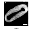
Figure 1 . hEHM formation. hEHM after 10 days in culture (3 days in the casting mold followed by 7 days on a custom made spacer;culture day 3 + 7). Bar: 1 mm (. -
Figure 2 . Characterization of hEHM contractile function. (A) Chronotropic response after stimulation with cAMP-elevating compounds (Iso: beta-adrenergic stimulation; IBMX: phosphodiesterase inhibition, n=6/group). (B) Inotropic response of hEHMs to increasing concentrations of extracellular calcium, comparison of hES2 (n=12), hES3 (n=12) and iPS 12 (n=3) (C) Inotropic response of hEHMs (hES2) to isoprenaline (triangles) and carbachol (squares) stimulation. Inset: normalized twitches at low calcium (lowest graph), stimulation with isoprenaline (highest graph) and carbachol (middle graph). (D) Relaxation of hEHMs with isoprenaline (triangles) and carbachol (squares) stimulation. Inset: normalized twitches *p < 0.05 vs. baseline values (A: paired two-tailed Student's t-test; C, D: ANOVA followed by Dunnet's post hoc test). -
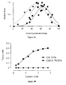
Figure 3 . Importance of non-myocytes for EHM formation. (A) Normalized force compared to cardiomyocyte percentage (actinin+ cells) of EHM generated from hES2 line ( circles: monolayer cardiac differentiation protocol (Hudson, et al. Stem Cells Dev 21: 1513-1523 (2012)), -squares: EB cardiac differentiation protocol (Yang et al. Nature 453: 524-528 (2008))) and iPS BJ line (triangles). (B) Contractile force of hEHM generated with 100% cardiomyocytes (CM 100%) and a mixture of 70% cardiomyocytes and 30% cardiac fibroblasts (CM/CF 70/30%), n=1. -
Figure 4 . GMP-compatible hydrogel matrix. (A) Contractile force of hEHMs made with rat collagen (0.4 mg/EHM, n=12) or bovine collagen (0.4 mg/EHM, n=14) at an extracellular calcium concentration of 2 mmol/L. (B) Contractile force at 2 mmol/L extracellular calcium of hEHMs made with the "Original protocol" (rat collagen, Matrigel®, n=12) and the "Matrix protocol" (bovine collagen, no Matrigel®, n=9). Please refer also to Table 1. (C) Contractile force at 2 mmol/L extracellular calcium of hEHMs made with bovine (bov.) collagen only, bovine collagen plus Matrigel (10% v/v), bovine collagen plus fibronectin (5 µg/EHM), and bovine collagen plus fibronectin (5 µg/EHM) plus laminin (5 µg/EHM), n=4/group. -
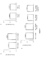
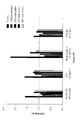
Figure 5 . Screening of basal media and serum-free growth supplement vs. Iscove with 20% serum. (A) F.l.t.r.: Change in cell death, cardiomyocyte percentage (CM percentage), cardiomyocyte mean actinin fluorescence (CM maturation), cardiomyocyte size based on sideward scatter area (CM size) and non-myocyte size based on sideward scatter area (NM size) of serum free media based on Iscove's, RPMI and aMEM basal media (see also Table 2-4) compared to serum containing EHM medium (see also Table 1).
(B) F.l.t.r.: Change in cell death, cardiomyocyte percentage (CM percentage), cardiomyocyte mean actinin fluorescence (CM maturation), cardiomyocyte size based on sideward scatter
area (CM size) and non-myocyte size based on sideward scatter area (NM size) of serum free media with 2% and 4% B27 plus insulin (B27+) and minus insulin (B27-) compared to serum containing EHM medium (see also Table 1). -
Figure 6 . Screening of peptide growth factors and fatty acid supplement. (A) Factors considered for serum-free, defined EHM medium. F.l.t.r.: Change in cell death , cardiomyocyte percentage (CM percentage), cardiomyocyte mean actinin fluorescence (CM maturation), cardiomyocyte size based on sideward scatter area (CM size) and non-myocyte size based on sideward scatter area (NM size) by indicated growth factors and fatty acid supplement compared to serum free medium without the factor. (B) Factors not considered for serum-free, defined EHM medium. Fl.t.r.: Change in cell death, cardiomyocyte percentage (CM percentage), cardiomyocyte mean actinin fluorescence (CM maturation), cardiomyocyte size based on sideward scatter area (CM size) and non-myocyte size based on sideward scatter area (NM size) by indicated growth factors and fatty acid supplement compared to serum free medium without the factor. -
Figure 7 . Serum-free, defined EHM generation from hESC cells. (A) Contractile force of hEHMs (hES2) with increasing concentrations of extracellular calcium, comparison of serum-containing medium (Serum), and serum-free Iscove's-based medium (SF-IMDM, see also Table 5), serum-free aMEM-based medium (SF-aMEM, see also Table 6), and serum-free RPMI-based medium (SF-RPMI). Respective EC50 for calcium, n=15/5/6/7 for Serum/SF IMDM, SF-aMEM, SF-RPMI, n.d. not determined. (B) Immunostaining of serum-free hEHM, showing typical heart muscle properties (muscle bundles). Staining was performed with an antibody against α-sarcomeric actinin and with DAPI for nuclear staining. (C) Percent change in force after adrenergic stimulation with 1 µM isoprenaline of serum-containing EHM (Serum), and serum-free Iscove's-based EHM (SF-IMDM), serum-free aMEM-based EHM (SF-aMEM), n=15/5/6, *p<0.05 (D) Percent change in force after muscarinic stimulation with 10 µM carbachol of serum-containing EHM (Serum), and serum-free Iscove's-based EHM (SF-IMDM), serum-free aMEM-based EHM (SF-aMEM), n=15/5/6. Bar: 20 µm -
Figure 8 . Effect of calcium on serum-free hEHM force. A) Contractile force of hEHMs (hES2) with increasing concentrations of extracellular calcium, comparison of RPMI medium (~0.4 mM calcium, n=2), and RPMI medium with addition of 0.8 mM CaCl (final free calcium concentration ~1.2 mM calcium, n=2), B) Response to 1 µM isoprenaline of serum-free hEHM cultured in RPMI or RPMI+calcium medium. -
Figure 9 . Effect of TGFb1 on hEHM force. (A) Contractile force of hEHMs (iPS BJ) with increasing concentrations of extracellular calcium, comparison of serum-free medium (SF), and serum-free medium with 5 ng/ml TGFb1 from culture day 0-3 (n=4/group, *p<0.05). (B) Comparison of contractile force of human EHM (hES2) in serum-free medium with TGFb1 treatment from culture 0-3 (SF+TGFb1 day 0-3) and culture day 0-10 (SF+TGFb1 day 0-10, n=5/group). -
Figure 10 . Effect of FGF and VEGF on hEHM force. Contractile force of hEHMs (hES2) with increasing concentrations of extracellular calcium, comparison of serum-free medium (SF, n=3), serum-free medium with 10 ng/ml FGF-2 (SF+FGF, n=3), and serum-free medium with 10 ng/ml VEGF (SF+VEGF, n=3), *p<0.05 SF+FGF vs SF. -
Figure 11 . Effect of B27 supplement concentration on hEHM force. Contractile force of hEHMs (hES2) with increasing concentrations of extracellular calcium, comparison of serum-containing medium (Serum), and serum-free medium with 2% B27 (SF-2% B27) and serum-free medium with 4% B27 (SF-4% B27), n=17/9/11, *p<0.05vs 2% B27. -
Figure 12 . Effect of B27 composition on hEHM force. Contractile force of hEHMs (hES2) with increasing concentrations of extracellular calcium, comparison of serum-containing medium (n=9), serum-free medium with full B27 (n=5), serum-free medium with B27 minus antioxidants (n=5), and serum-free medium with B27 minus insulin (n=5). -
Figure 13 . Replacement of B27 by custom-made supplement in HELM from hES and iPS cells. (A) Contractile force of hEHMs (hES2) with increasing concentrations of extracellular calcium, comparison of serum-containing medium (n=3), serum-free medium with B27 minus insulin (B27-insulin, n=5), and serum-free medium with custom-made supplement (CMS; n=3). (B) Contractile force of hEHMs from iPS cells (BJ) with increasing concentrations of extracellular calcium, comparison of serum-containing medium (n=8), serum-free medium with B27-insulin (n=8), and serum-free medium with custom-made supplement (CMS; n=4). - The following examples are meant to further illustrate, but not limit the invention. The examples comprise various technical features, and it will be appreciated that the invention also relates to combinations of the technical features presented in this exemplifying section.
- The materials used herein are commercially available. For example, DMEM, RPMI, αMEM (cat. No. 32561-029), streptomycin, penicillin, and B27 are obtainable from Invitrogen; bovine collagen of medical grade is available from Devros Medical; fatty acid supplement can be ordered from Sigma (cat. No. F7050); and the various growth factors are available from Peprotech (FGF2, AF-1ββ-18B; IGF-1, AF-100-11; TGFβ1, 100-21).
- The inventors utilized H9.2 (Technion, Haifa, Israel), hES3 (Embryonic Stem Cell International, Singapore) and transgenic hES3-ENVY (Costa, M., et al. Nat Methods 2: 259-260 (2005)) as well as hES2 line (McEwen Centre for Regenrative Medicine, Toronto, Canada; Yang et al. Nature 453: 524-528 (2008)) in the present study (approval by the Robert-Koch-Institute to W.-H.Z.:
permit # 12; reference number: 1710-79-1-4-16). Differentiated EBs were shipped to Hamburg/Goettingen at room temperature and arrived within 72-96 hrs. iPS lines were from Toronto (iPS BJ) and Goettingen (iPS 12, Streckfuss-Bomeke et al, Eur Heart J (2012) dol: 10.1093/eurheartj/ehs203, and iPS Sendai). - EBs were digested with collagenase B (1 mg/ml; H9.2), collagenase I (2 mg/ml) and/or trypsin/EDTA (0.25%/1 mmol/l; hES3, hES3-ENVY, hES2, iPS NJ, iPS 12) as described elsewhere (Kehat et al. J Clin Invest 108: 407-414 (2001); Mummery et al. Circulation 107: 2733-2740 (2003); Xu et al. Circ Res 91: 501-508 (2002); Yang et al. Nature 453: 524-528 (2008); Passier et al. Stem Cells 23: 772-780 (2005); each incorporated herein by reference). Cardiomyocytes were counted in representative aliquots of enzymatically dispersed cells after staining of tropomyosin or sarcomeric actinin.
- The inventors constructed hEHMs using a modified EHM-engineering protocol (Zimmermann et al. Circ Res 90: 223-230 (2002), incorporated herein by reference). Briefly, EHMs (reconstitution volume: 450 µl) were prepared by pipetting a mixture containing freshly dispersed ESC-derivatives 1x10⁴ - 15x106 cells in Iscove-Medium with 20% fetal calf serum, 1% non-essential amino acids, 2 mmol/l glutamine, 100 µmol/l β-mercaptoethanol, 100 U/ml penicillin, and 100 mg/ml streptomycin), pH-neutralized collagen type I from rat tails (0.4 mg/EHM), Matrigel™ (10% v/v; Becton Dickenson or tebu), and concentrated serum-containing culture medium (2x DMEM, 20% horse serum, 4% chick embryo extract, 200 U/ml penicillin, and 200 mg/ml streptomycin) in circular molds (inner/outer diameter: 2/4 mm; height: 5 mm) (Table 1). hEHM condensed quickly within the casting molds and were transferred onto static stretch devices (110% of slack length) (Zimmermann et al. Nat Med 12: 452-458 (2006), incorporated herein by reference) on
culture day 3. Medium was changed every other day. hEHM culture under stretch was performed for 7 days. - Another detailed prior art protocol which is suitable to serve as a basis for the improved method disclosed herein is described by Soong et al. Curr Prot Cell Biol. 23.8.1-23.8.21 (2012), which is incorporated herewith in its entirety, and in particular reference is made to the "
Basic Protocol 2", and the "Support Protocol 2". - A protocol with reduced xenogenic components (Matrix protocol, Table 1) was established to enable pre-GMP hEHM. Cells were reconstituted in a mixture of pH-neutralized bovine collagen (Devros Medical, 0.4 mg/EHM), concentrated serum-containing culture medium (2x DMEM, 40% fetal calf serum, 200 U/ml penicillin, and 200 mg/ml streptomycin) and cultured in Iscove-Medium with 20% fetal calf serum, 1% non-essential amino acids, 2 mmol/I glutamine, 0.3 mmol/l ascorbic acid, 100 µmol/l β-mercaptoethanol, 100 U/ml penicillin, and 100 µg/ml streptomycin).
- To generate fully defined, serum-free EHM cells were reconstituted in a mixture of pH-neutralized bovine collagen (Devros Medical, 0.4 mg/EHM), concentrated serum-free medium medium (2x DMEM, 8% B27, 200 U/ml penicillin, and 200 mg/ml streptomycin) and cultured in Iscove-Medium with full 4% B27, 1% non-essential amino acids, 2 mmol/l glutamine, 0.3 mmol/l ascorbic acid, 20 ng/ml IGF-1, 10 ng/ml FGF2, 10 ng/ml VEGF, 5 ng/ml TGFb1 (culture day 0-3 only), and 100 U/ml penicillin, and 100 µg/ml streptomycin (Serum-free protocol, Table 1). B27 supplement contains vitamins (Biotin, DL Alpha Tocopherol, Acetate DL Alpha-Tocopherol, Vitamin A), proteins and enzymes (BSA, fatty acid free Fraction V, Catalase, Human Recombinant Insulin, Human Transferrin, Superoxide Dismutase), and other cell-supporting components (Corticosterone, D-Galactose, Ethanolamine, Glutathione (reduced), L-Carnitine, Linoleic Acid, Linolenic Acid, Progesterone, Putrescine, Sodium Selenite, and T3 (triodo-I-thyronine). Where indicated full B27 (Invitrogen, A1486701) was compared to B27 without antioxidants (Invitrogen, #10889038) and B27 without insulin (Invitrogen, #0050129SA). B27 supplement was replaced by a custom-made supplement consisting of Albumin, Transferrin, Ethanolamine, Sodium selenite, L-Carnitine HCl, Hydrocortisone, Fatty acid supplement, and Triiodo-L-thyronine (Table 2).
- The inventors analyzed force of contraction and twitch kinetics (contraction time: time from 50% to maximal contraction; relaxation time: time from maximal contraction to 50% relaxation) under isometric conditions as described before (Zimmermann et al. Circ Res 90: 223-230 (2002), incorporated herein by reference). Contraction frequency was assessed by light microcopy (unstimulated spontaneous contractions) immediately after removing EHMs from the incubator.
- EBs cultured in different medium conditions were made into a single cell suspension as described above. Cells were fixed in 70% ice cold ethanol under constant mixing. The cells were stained for sarcomeric actinin (Sigma) to label cardiomyocytes and DAPI to analyze nuclear DNA content and to exclude cell doublets. Cells were run on a LSRII Cytometer (BD). At least 10,000 live cells were analyzed. The following parameters were then analyzed, (1) cell death (percentage of cells in the sub-G1 fraction), (2) cardiomyocyte and non-myocyte percentage (Actinin-positive and negative cells, respectively) (3) Cardiomyocte maturation (mean actinin fluorescence), (4) cardiomyocyte and (5) non-myocyte size (based on sideward scatter area, SSC-A),
- hEHMs were fixed in neutral buffered 4% formaldehyde/1% methanol, pH 7.4 for confocal laser scanning microscopy (CLSM; Zeiss 510 Meta LSM system or Zeiss 710 LSM) respectively as described earlier (Zimmermann et al. Circ Res 90: 223-230 (2002), incorporated herein by reference). For CLSM, the inventors prepared vibratome sections (100 µm; Leica VT1000 S) and subjected them to immune fluorescent labeling with antibodies directed against α-sarcomeric actinin (Sigma clone EA-53, 1:800; with appropriate secondary antibodies). Nuclei were stained with DAPI (4',6-diamidino-2-phenylindole; 1 µg/ml).
- Data are presented as mean ± standard error of the mean. Statistical differences were determined using paired and unpaired two-tailed Student's t-tests or ANOVA followed by Dunnett's post hoc test as indicated. A P value < 0.05 was considered statistically significant.
- EBs - prepared in Haifa (H9.2; ), Singapore (hES3 and hES3-ENVY; Costa et al. Nat Methods 2: 259-260 (2005)), and Toronto (hHES2; iPS) - were sent to Hamburg/Göttingen by express mail at room temperature in an air-tight container filled with culture medium. Delivery was ensured within 72-96 h. After arrival, EBs were transferred into fresh culture medium and allowed to recover for 24-48 h. Within that time EBs regained spontaneous contractile activity. EBs were enzymatically dispersed EBs and the resulting single cell suspensions allocated to hEHM generation or cytohistology. An initial series of experiments explored the number of necessary cell quantity per hEHM (1x10⁴ - 15x106 cells) and utility of different ESC-lines (H9.2, hES2, hES3, hES3-ENVY) and iPS lines (I2, BJ, Sendai) for hEHM construction (n = 67). Spontaneous beating of variably sized areas could be observed in all cultures within 48 h of hEHM casting. However, force-generating hEHMs formed only if 1.25 - 15x106 cells/EHM were utilized (
Fig. 1 ). Depending on the size of the EHM, the cell density can easily be adapted. - hEHMs contracted stably and rhythmically (0.8 ± 0.05 Hz at 37°C; n = 14) for at least 3 weeks in culture. The inventors performed a detailed functional characterization at 10 days. Incubation with isoprenaline increased spontaneous beating frequency to 1.2 ± 0.1 Hz (n= 6; p < 0.01
Fig. 2A ). Additional inhibition of phosphodiesterase with 3-isobutyl-1-methylxanthine (IBMX; 100 µmol/l) caused a further increase in spontaneous beating rate to 1.6 ± 0.1 Hz (n= 6; p < 0.001Fig. 2A ). hEHM developed maximal twitch mean forces of 130 ± 13 µN at 2.4 mmol/I calcium (EC50: 0.8 ± 0.04 mmol/l, n= 17;Fig. 2B ). β-Adrenergic stimulation with 1 µmol/l isoprenaline at sub-EC50 extracellular calcium (0.4 mmol/l) increased force of contraction by 47 ± 12% (n = 12;Fig. 2C ) and shortened relaxation to 113 ± 2.3 ms (n= 12;Fig. 2D ). - All ESC-lines and iPS lines utilized here appeared to be suited for hEHM generation. To test which cardiomyocyte content is optimal for a force generating tissue, the inventors plotted developed force against the cardiomyocyte percentage. Interestingly, a bell-shaped distribution with highest forces developed at a cardiomyocyte percentage of 40-80% was found (
Fig. 3A ). - This observationsuggests a critical role of non-myocytes for proper tissue formation. To investigate this the inventors performed experiment with human cardiomyocytes that were purified by the surface marker CD172a (SIRPα) (Dubois et al. Nat Biotechnol 29: 1011-1018 (2011)). EHMs generated from purified cardiomyocytes did not form force-generating tissue (
Fig. 3B ). Supplementation with 30% human cardiac fibroblasts, however, yielded a stiffer tissue with good force production. These results emphasize that non-myocytes are vital to cardiac tissue formation and that these cells also need to be supported under serum-free conditions. - The inventors initially constructed hEHMs based on a protocol that the inventors had developed in a neonatal rat heart cell model (Zimmermann et al. Biotechnol Bioeng 68: 106-114 (2000)). This protocol includes several non-human components (including rat collagen, Matrigel, horse serum, fetal calf serum, and chick embryo extract) that are incompatible with a "therapeutic application" in vivo. To address this caveat, a series of experiments directly testing whether non-human components of the hEHM-matrix could be reduced were first conducted. Rat collagen was replaced with medical grade (GMP) bovine collagen without loss of performance (
Fig. 4A ). Also, Matrigel®, horse serum and chick embryo extract could be left out without negative impact on hEHM formation and function ("Matrix protocol",Fig. 4B , Table 1). Complementing bovine collagen with other extracellular matrix proteins such as fibronectin and laminin, one of the major components of Matrigel®, did not yield additional benefit (Fig. 4C ). - To further define the human EHM culture and make it GMP compatible, the inventors sought to replace all undefined serum component with chemically defined supplements. To screen for these supplements the inventors introduced a simplified screening algorithm based on 3D-human embryoid body (EB) cultures. ESCs for this screen were cultured under serum-free conditions (Yang et al. Nature 453: 524-528 (2008); Kattman et al. Cell Stem Cell 8: 228-240 (2011)). The reference for the screens was our serum-containing EHM medium (Table 1): (1) Iscove's, (2) 2 mmol/L L-glutamine, (3) 20% FBS, (4) 1% non-essential amino acids, (5) 0.3 mmol/L ascorbic acid, (6) 100 µmol/l β-mercaptoethanol, (7) 100 U/ml Penicillin/100 mg/ml Streptomycin. As read outs for the beneficial or detrimental role of basal culture medium and supplements a flow cytometry-based protocol (Tiburcy et al. Circ Res 109: 1105-1114 (2011); incorporated herein by reference) was established to determine (1) cell death (based on sub-G1 DNA content), (2) cardiomyocyte content (based on actinin expression), (3) cardiomyocyte maturation (based on actinin mean fluorescence per cardiomyocyte), (4) cardiomyocyte size, and (5) non-myocyte size (based on sideward scatter area).
- The inventors first screened three basal media formulations (Iscove's, RPMI, αMEM: Table 3-5) with and without B27 supplementation. B27 has been used by several groups for differentiation of human ESCs and iPSCs (Burridge et al. Cell Stem Cell 10: 16-28 (2012)). The screen demonstrated that B27 was essential for EB formation irrespective of the tested basal medium. Iscove's and RPMI showed comparable results while aMEM appeared to cause slightly higher cell death. On the other hand, αMEM was superior for cardiomyocyte actinin expression (
Fig. 5A ). - Given own preliminary findings showing suboptimal performance of EHMs cultured in the presence of basal RPMI (
Fig. 7 ), the inventors continued the screen with Iscove's or aMEM as basal medium. The inventors next scrutinized the utility of B27 in its standard formulation with and without insulin. 4% B27 supplementation with insulin (10 µg/ml final concentration) showed lowest cell death and possibly greater non-myocyte support (lower cardiomyocytes percentage suggest a shift to more non-myocytes) as compared to B27 without insulin and lower B27 (2%) supplement concentration (Fig. 5B ). The inventors next screened the utility of 6 peptide growth factors at above EC50 concentrations and fatty acid supplements. PDGF-BB, CT-1 and Neuregulin-1 caused substantial cell death whereas IGF-1 appeared to protect from cell death (Fig. 6A -
,B). TGFβ1, VEGF and fatty acid supplements enhanced individual cardiomyocyte actinin expression. FGF-2 supported non-myocyte growth and increased cardiomyocyte cell size (Fig. 6A ). TGFβ2 had no beneficial effect (Fig. 6B ). All of the tested compounds or combinations thereof were implicated in cardiac development and EHM generation (Naito et al. Circulation 114: 172-78 (2006); Shimojo et al. Am J Physiol Heart Circ Physiol 293: H474-481 (2007); Vantleret al. J Mol Cell Cardiol 48: 1316-1323 (2010); Odiete et al. Circ Res 111: 1376-1385 (2012); Wollert & Chien J Mol Med (Berl) 75: 492-501 (1997); Price et al. Anat Rec A Discov Mol Cell Evol Biol 272: 424-433 (2003); Molin et al. Dev Dyn 227: 431-444 (2003); Corda et al. Circ Res 81: 679-687 (1997); Lopaschuk & Jaswal J Cardiovasc Pharmacol 56: 130-140; all incorporated herein by reference). - EHM development is characterized by two stages. Initially there is a "condensation phase" where the isolated cells are "settling in" the matrix, reorganizing themselves and the matrix which may also be accompanied by substantial cell death. This stage is greatly influenced by the non-myocytes. The second stage is the maturation of the tissue under mechanical load. This phase is characterized by hypertrophic growth and maturation of cardiomyocytes, alignment, increasing force development, and matrix stabilization (Tiburcy et al. Circ Res 109: 1105-1114 (2011)).
- The inventors reasoned that according to the stage different medium conditions may be required. To prevent cell death during the condensation phase the inventors chose a combination of medium components that were neutral or even reducing cell death in the initial screen. This is Iscove's basal medium, 4% B27, and IGF-1. Also, the matrix reorganisation and condensation through non-myocytes was supported by factors that increase number and/or size of non-myocytes (IGF-1, TGF-beta1, FGF-2, in the first stage). VEGF is added for support of cardiomyocyte maturation (Table 5). This medium was then tested for its ability to support the formation of force-generating EHM. Serum-free EHM were beating coherently at a spontaneous beating frequency of 113±12 bpm, n=7. Serum containing EHM were beating significantly faster (199±8 bpm, n=8). The inventors found a similar maximal force development and calcium sensitivity compared to the serum containing control (
Fig. 7A ). Both, Iscove's and αMEM basal medium supported EHM formation with comparable results in contractile force. RPMI did not support force-generating tissue (Fig. 7A , Table 5, 6). Morphologically, serum-free EHM contained well developed muscle bundles with anisotropically aligned cardiomyocytes (Fig. 7B ). Importantly, the serum-free EHM responded to adrenergic and muscarinic stimulation as expected for heart muscle. In fact the αMEM EHM responded significantly better than serum-containing controls. (Fig. 7 C, D). - The inventors hypothesized that the insufficient performance of RPMI medium was due to the lower than physiological free calcium concentration of RPMI medium (0.424 mmol/L, Table 4). To test if this was true the inventors performed an additional experiment to compare RPMI medium with RPMI medium with 0.8 mM CaCl added (final free calcium concentration 1.242 mmol/L.) While EHM with RPMI hardly contracted supplementation of calcium lead to measurable maximal force and better responsiveness to isoprenalin (
Fig. 8 ). In fact maximal force production with supplemented RPMI was comparable to IMDM or αMEM medium (Fig. 7A ) suggesting that a range of ~1 - 2 mmol/L calcium is required for proper tissue function. - To verify the results from the initial screen, the inventors additionally tested the influence of critical factors on the formation of functional EHM. TGFβ1 addition from
day 0 to 3 was essential, but prolonged TGFβ1 treatment did not yield an additional benefit (Fig. 9A , B). Both FGF and VEGF were able to enhance force-generation confirming an important contribution to tissue formation (Fig. 10 ). - Increasing the B27 supplement concentration to 4% was superior to 2% B27 (
Fig. 11 ). To test which components of B27 are essential for EHM function the inventors first performed an experiment with different commercially available B27 formulations, the inventors compared full B27 with B27 minus insulin, and B27 minus antioxidants. B27 minus antioxidants performed comparable to full B27 suggesting that antioxidants are not required. Surprisingly, B27 minus insulin performed better than full B27 implying that insulin supplementation is also not required (Fig. 12 ). - Based on these results the inventors then developed a custom-made serum supplement to replace B27 (Table 7). When the inventors tested this custom-made serum supplement against serum-containing medium and serum-free medium with B27 minus insulin the inventors found comparable maximal force development suggesting that B27 can be omitted from the serum-free EHM culture and replaced by custom-made serum supplement (
Fig. 13A ). These findings were confirmed with hEHM from iPSC demonstrating utility for generation of defined engineered heart muscle from different pluripotent stem cell sources. (Fig. 13B ). - This study demonstrates for the first time that differentiated, force-generating human heart muscle can be generated in vitro under fully defined, serum-free conditions. The protocol works for embryonic (ESC) and induced pluripotent (iPS) stem cells-derived heart muscle.
- This is a major breakthrough which enables future in vitro studies to investigate e.g. maturation and hypertrophy without confounding serum factors but also potential in vivo applications and therapeutic approaches under GMP regulations.
-
- Zimmermann, Kardiale Regeneration mit konstlichem Herzgewebe. Universitatsklinikum Hamburg Eppendorf, Habilitation (2006)
- Schneiderbanger, Zur Bedeutung von Tranforming Growth Factor-β1 und Interleukin-1β für die Morphologie, die Genexpression und die kontraktile Funktion von rekonstituiertem dreidimensionalen konstlichen Herzmuskelgewebe. Universität Hamburg, Dissertation (2006)
- Eschenhagen, T. & Zimmermann, W.H. Engineering myocardial tissue. Circ Res 97, 1220-1231 (2005).
- Zimmermann, W.H., et al. Heart muscle engineering: an update on cardiac muscle replacement therapy. Cardiovasc Res 71, 419-429 (2006).
- Eschenhagen, T., et al. Three-dimensional reconstitution of embryonic cardiomyocytes in a collagen matrix: a new heart muscle model system. Faseb J 11, 683-694 (1997).
- Kofidis, T., et al. In vitro engineering of heart muscle: artificial myocardial tissue. J Thorac Cardiovasc Surg 124, 63-69 (2002).
- Morritt, A.N., et al. Cardiac tissue engineering in an in vivo vascularized chamber. Circulation 115, 353-360 (2007).
- Radisic, M., et al. Functional assembly of engineered myocardium by electrical stimulation of cardiac myocytes cultured on scaffolds. Proc Natl Acad Sci U S A 101, 18129-18134 (2004).
- Shimizu, T., et al. Fabrication of pulsatile cardiac tissue grafts using a novel 3-dimensional cell sheet manipulation technique and temperature-responsive cell culture surfaces. Circ Res 90, e40 (2002).
- Zimmermann, W.H., et al. Three-dimensional engineered heart tissue from neonatal rat cardiac myocytes. Biotechnol Bioeng 68, 106-114 (2000).
- Tulloch, N.L., et al. Growth of engineered human myocardium with mechanical loading and vascular coculture. Circ Res 109, 47-59 (2011).
- Tiburcy, M., et al. Terminal differentiation, advanced organotypic maturation, and modeling of hypertrophic growth in engineered heart tissue. Circ Res 109, 1105-1114 (2011).
- Zimmermann, W.H., et al. Tissue engineering of a differentiated cardiac muscle construct. Circ Res 90, 223-230 (2002).
- Zimmermann, W.H., et al. Engineered heart tissue grafts improve systolic and diastolic function in infarcted rat hearts. ).
- Schaaf, S., et al. Human engineered heart tissue as a versatile tool in basic research and preclinical toxicology. PLoS One 6, e26397 (2011).
- Naito, H., et al. Optimizing engineered heart tissue for therapeutic applications as surrogate heart muscle. Circulation 114, I72-78 (2006).
- Thomson, J.A., et al. Embryonic stem cell lines derived from human blastocysts. Science 282, 1145-1147 (1998).
- Takahashi, K. & Yamanaka, S. Induction of pluripotent stem cells from mouse embryonic and adult fibroblast cultures by defined factors. Cell 126, 663-676 (2006).
- Schroeder, M., et al. Differentiation and lineage selection of mouse embryonic stem cells in a stirred bench scale bioreactor with automated process control. Biotechnol Bioeng 92, 920-933 (2005).
- Kehat, I., et al. Human embryonic stem cells can differentiate into myocytes with structural and functional properties of cardiomyocytes. J Clin Invest 108, 407-414 (2001).
- Mummery, C., et al. Differentiation of human embryonic stem cells to cardiomyocytes: role of coculture with visceral endoderm-like cells. Circulation 107, 2733-2740 (2003).
- Xu, C., Police, S., Rao, N. & Carpenter, M.K. Characterization and enrichment of cardiomyocytes derived from human embryonic stem cells. Circ Res 91, 501-508 (2002).
- Burridge, P.W., Keller, G., Gold, J.D. & Wu, J.C. Production of de novo cardiomyocytes: human pluripotent stem cell differentiation and direct reprogramming. ).
- Lian, X., et al. Robust cardiomyocyte differentiation from human pluripotent stem cells via temporal modulation of canonical Wnt signaling. Proc Natl Acad Sci U S A (2012).
- Costa, M., et al. The hESC line Envy expresses high levels of GFP in all differentiated progeny. ).
- Yang, L., et al. Human cardiovascular progenitor cells develop from a KDR+ embryonic-stem-cell-derived population. Nature 453, 524-528 (2008).
- Streckfuss-Bomeke, K., et al. Comparative study of human-induced pluripotent stem cells derived from bone marrow cells, hair keratinocytes, and skin fibroblasts. Eur Heart J, doi: 10.1093/eurheartj/ehs203 (2012).
- Passier, R., et al. Increased cardiomyocyte differentiation from human embryonic stem cells in serum-free cultures. Stem Cells 23, 772-780 (2005).
- Dubois, N.C., et al. SIRPA is a specific cell-surface marker for isolating cardiomyocytes derived from human pluripotent stem cells. Nat Biotechnol 29, 1011-1018 (2011).
- Kattman, S.J., et al. Stage-specific optimization of activin/nodal and BMP signaling promotes cardiac differentiation of mouse and human pluripotent stem cell lines. Cell Stem Cell 8, 228-240.
- Shimojo, N., et al. Contributory role of VEGF overexpression in endothelin-1-induced cardiomyocyte hypertrophy. Am J Physiol Heart Circ Physiol 293, H474-481 (2007).
- Vantler, M., et al. PDGF-BB protects cardiomyocytes from apoptosis and improves contractile function of engineered heart tissue. J Mol Cell Cardiol 48, 1316-1323 (2010).
- Odiete, O., Hill, M.F. & Sawyer, D.B. Neuregulin in cardiovascular development and disease. Circ Res 111, 1376-1385 (2012).
- Wollert, K.C. & Chien, K.R. Cardiotrophin-1 and the role of gp130-dependent signaling pathways in cardiac growth and development. J Mol Med (Berl) 75, 492-501 (1997).
- Price, R.L., et al. Effects of platelet-derived growth factor-AA and -BB on embryonic cardiac development. Anat Rec A Discov Mol Cell Evol Biol 272, 424-433 (2003).
- Molin, D.G., et al. Expression patterns of Tgfbeta1-3 associate with myocardialisation of the outflow tract and the development of the epicardium and the fibrous heart skeleton. Dev Dyn 227, 431-444 (2003).
- Corda, S., et al. Trophic effect of human pericardial fluid on adult cardiac myocytes. Differential role of fibroblast growth factor-2 and factors related to ventricular hypertrophy. Circ Res 81, 679-687 (1997).
- Lopaschuk, G.D. & Jaswal, J.S. Energy metabolic phenotype of the cardiomyocyte during development, differentiation, and postnatal maturation. J Cardiovasc Pharmacol 56, 130-140.
- Didié et al. Parthenogenetic stem cells for tissue-engineered heart repair. J Clin Invest. doi :10.1172/JCI66854.
- Soong et al. Cardiac Differentiation of Human Enbryonic Stem Cells and their Assembly into Engineered Heart Muscle. Curr Prot Cell Biol. 55: 23.8.1-23.8.21, (2012).
- Hudson, J., Titmarsh, D., Hidalgo, A., Wolvetang, E. & Cooper-White, J. Primitive cardiac cells from human embryonic stem cells. ).
Claims (15)
- A method for producing engineered heart muscle (EHM), the method comprising the steps of:(i) providing a serum-free reconstitution mixture in one or more moulds, said reconstitution mixture comprising (a) a serum-free minimum essential medium; (b) a serum-free supplement resulting in a final concentration of 0.5-50 mg/ml albumin, 1-100 µg/ml transferrin, 0.1-10 µg/ml ethanol amine, 0.003-0.3 µg/ml sodium selenite, 0.4-40 µg/ml L-Carnitine HCl, 0.1-10 µg/ml Hydrocortisone, 0.05-5 µl/ml Fatty acid supplement, 0.0001-0.1 µg/ml triodo-L-thyronine (T3) and 0.2-2 mg/ml collagen; and (c) a mixture of human cardiac myocytes and human non-myocytes, wherein 20 to 80% of the total cell mixture are cardiac myocytes;
wherein the cardiac myocytes are not produced using a process which involves modifying the germ line genetic identity of human beings or which involves use of a human embryo for industrial or commercial purposes;
wherein the reconstitution mixture has a pH of 7.2 to 7.6;(ii) culturing the serum-free reconstitution mixture in said one or more moulds, whereby the serum-free reconstitution mixture is allowed to condense for at least 15 min;(iii) culturing the mixture obtained in step (ii) in said one or more moulds in a serum-free EHM culture medium until the mixture condenses to at least 50% of its original thickness, wherein said EHM culture medium comprises (a) a basal medium comprising 0.5-3 mmol/L Ca2+; (b) a serum-free supplement as defined in (i)(b); (c) 0.5-10 mmol/L L-glutamine; (d) 0.01-1.0 mmol/L ascorbic acid; (e) 1-100 ng/ml IGF-1; and (f) 1-10 ng/ml TGFβ1;(iv) culturing the mixture obtained in step (iii) under mechanical stretching in a serum-free EHM culture medium as defined in step (iii) (a)-(f), whereby force-generating EHM is formed. - The method of claim 1, wherein the minimum essential medium in step (i) and/or in step (iii) is selected from Iscove's medium, αMEM, DMEM, and RPMI, preferably wherein the basal medium is Iscove's medium or αMEM, more preferably wherein the basal medium is Iscove's medium.
- The method of claim 1 or 2, wherein the serum-free supplement of step (i) and/or step (iii) further comprises one or more components selected from the group consisting of vitamin A, D-galactose, linoleic acid, linolenic acid, progesterone, and putrescine.
- The method of any one of claims 1-3, wherein the serum-free supplement in component (b) of step (i) and/or in component (b) of step (iii) is B27® supplement or B27® supplement minus insulin, preferably wherein the serum-free supplement in component (b) is 2-6% (v/v) B27® supplement or B27® supplement minus insulin, more preferably wherein the serum-free supplement in component (b) is 4% (v/v) B27® supplement or B27® supplement minus insulin.
- The method of any one of claims 1-4, wherein said reconstitution mixture of step (i) comprises 0.3-0.5 mg collagen per 1.5 x 10⁶ cardiac myocyte and non-myocyte cell mixtures, preferably wherein said reconstitution mixture of step (i) comprises about 0.4 mg collagen per 1.5 x 10⁶ cardiac myocyte and non-myocyte cell mixtures.
- The method of any one of claims 1-5, wherein in component (c) of the reconstitution mixture of step (i) said collagen is of medical grade and selected from the group consisting of collagen type I, collagen type III, collagen type V, and a mixture thereof, preferably wherein in component (c) of the reconstitution mixture of step (i) at least 90% of said collagen is collagen type I, and preferably wherein in component (c) of the reconstitution mixture of step (i) said collagen further comprises one or more extracellular matrix components selected from the group consisting of elastin, laminin, entactin, nidogen, proteoglycan, and fibronectin.
- The method of any one of claims 1-6, wherein the reconstitution mixture of step (i) has a pH of 7 to 7.8, preferably wherein the reconstitution mixture of step (i) has a pH of about 7.4.
- The method of any one of claims 1-7, wherein the cardiac myocytes are human cardiac myocytes.
- The method of any one of claims 1-8, wherein the cardiac myocytes are provided in admixture with cells of one or more class of cells selected from the group of non-myocytes such as fibroblasts, endothelial cells, smooth muscle cells, and mesenchymal stem cells, wherein the cardiac myocytes admixture contains 20-80% cardiac myocytes, preferably wherein the cardiac myocytes are provided in step (i) in a cell concentration of at least 2.7-20 x 106 per ml.
- The method of any one of claims 1-9, wherein culturing in step (ii) is carried out for 0.25-3h, preferably for 0.5h - 1.5h.
- The method of any one of claims 1-10, wherein the serum-free EHM culture medium comprises (i) about 20 ng/ml human IGF1; (ii) about 5 ng/ml human TGFβ1; and/or (iii) about 5-20 ng/ml human VEGF and/or about 5-20 ng/ml human FGF.
- The method of any one of claims 1-11, wherein the serum-free EHM culture medium in step (iii) additionally comprises 750 mg/L glycine, 890 mg/L L-alanine, 1320 mg/L L-asparagine, 1330 mg/L L-aspartic acid, 1470 mg/L L-glutamic acid, 1150 mg/L L-proline, and 1050 mg/L L-serine.
- The method of any one of claims 1-12, wherein culturing in step (iii) is carried out for at least 3 days, preferably for about 3 to about 7 days.
- The method of any one of claims 1-13, wherein the culturing in step (iv) is carried out for a period of at least 3-60 days, preferably for 4-30 days, more preferably for 5-20 days, even more preferably 6-10 days, and most preferably for 7 days,
wherein step (iv) is carried out on a stretch device, preferably wherein the stretch device applies a static, phasic or dynamic stretch. - The method of any one of claims 1-14, wherein said EHM generates more than 0.01 mN force upon induction with 3 mM calcium as determined using the method described in Zimmermann et al. Circ. Res. 90, 223-230 (2002), preferably wherein said EHM generates more than 0.1 mN force, more preferably whereis said EHM generates more than 0.2 mN force, and most preferably wherein said EHM generates more than 0.3 mN force upon induction with 3 mM calcium.
Priority Applications (11)
| Application Number | Priority Date | Filing Date | Title |
|---|---|---|---|
| DK13181352.9T DK2840132T3 (en) | 2013-08-22 | 2013-08-22 | Method of Manufacturing Constructed Heart Muscle (EHM) |
| ES13181352.9T ES2615396T3 (en) | 2013-08-22 | 2013-08-22 | Method to produce cardiac muscle by engineering (EHM) |
| EP13181352.9A EP2840132B1 (en) | 2013-08-22 | 2013-08-22 | Method for producing Engineered Heart Muscle (EHM) |
| US14/913,263 US10626374B2 (en) | 2013-08-22 | 2014-08-22 | Method for producing engineered heart muscle (EHM) |
| CA2921882A CA2921882C (en) | 2013-08-22 | 2014-08-22 | Method for producing engineered heart muscle (ehm) |
| PCT/EP2014/067886 WO2015025030A1 (en) | 2013-08-22 | 2014-08-22 | Method for producing engineered heart muscle (ehm) |
| AU2014310526A AU2014310526B2 (en) | 2013-08-22 | 2014-08-22 | Method for producing engineered heart muscle (EHM) |
| CN201480057991.5A CN105745321B (en) | 2013-08-22 | 2014-08-22 | Method for generating engineered myocardium (EHM) |
| SG11201601269RA SG11201601269RA (en) | 2013-08-22 | 2014-08-22 | Method for producing engineered heart muscle (ehm) |
| JP2016535491A JP6662777B2 (en) | 2013-08-22 | 2014-08-22 | Method for producing artificial heart muscle (EHM) |
| US16/814,186 US11492594B2 (en) | 2013-08-22 | 2020-03-10 | Method for producing engineered heart muscle (EHM) |
Applications Claiming Priority (1)
| Application Number | Priority Date | Filing Date | Title |
|---|---|---|---|
| EP13181352.9A EP2840132B1 (en) | 2013-08-22 | 2013-08-22 | Method for producing Engineered Heart Muscle (EHM) |
Publications (2)
| Publication Number | Publication Date |
|---|---|
| EP2840132A1 true EP2840132A1 (en) | 2015-02-25 |
| EP2840132B1 EP2840132B1 (en) | 2016-11-30 |
Family
ID=49035371
Family Applications (1)
| Application Number | Title | Priority Date | Filing Date |
|---|---|---|---|
| EP13181352.9A Active EP2840132B1 (en) | 2013-08-22 | 2013-08-22 | Method for producing Engineered Heart Muscle (EHM) |
Country Status (10)
| Country | Link |
|---|---|
| US (2) | US10626374B2 (en) |
| EP (1) | EP2840132B1 (en) |
| JP (1) | JP6662777B2 (en) |
| CN (1) | CN105745321B (en) |
| AU (1) | AU2014310526B2 (en) |
| CA (1) | CA2921882C (en) |
| DK (1) | DK2840132T3 (en) |
| ES (1) | ES2615396T3 (en) |
| SG (1) | SG11201601269RA (en) |
| WO (1) | WO2015025030A1 (en) |
Cited By (4)
| Publication number | Priority date | Publication date | Assignee | Title |
|---|---|---|---|---|
| DE102018202268A1 (en) * | 2018-02-14 | 2019-08-14 | Georg-August-Universität Göttingen Stiftung Öffentlichen Rechts, Universitätsmedizin | Method for increasing the safety of tissue engineered products for clinical use |
| EP3945133A1 (en) * | 2020-07-29 | 2022-02-02 | Georg-August-Universität Göttingen | Mass production of human pluripotent stem cell derived cardiac stromal cell |
| US11492594B2 (en) | 2013-08-22 | 2022-11-08 | Georg-August-Universität Göttingen Stiftung Öffentlichen Rechts, Universitätsmedizin | Method for producing engineered heart muscle (EHM) |
| LU507930B1 (en) * | 2024-08-05 | 2026-02-05 | Luxembourg Institute Of Science And Tech | Serum-free culture medium for the culture of human monocytic cells and method using the same |
Families Citing this family (16)
| Publication number | Priority date | Publication date | Assignee | Title |
|---|---|---|---|---|
| JP6681825B2 (en) * | 2013-09-20 | 2020-04-15 | レパイロン・ゲーエムベーハーRepairon Gmbh | Direct differentiation of pluripotent stem cells into functional myocardium |
| EP3259347A1 (en) * | 2015-02-20 | 2017-12-27 | Pluriomics B.v. | Assays for measuring cardiotoxicity |
| JP7262385B2 (en) * | 2016-08-26 | 2023-04-21 | ザ・カウンシル・オブ・ザ・クィーンズランド・インスティテュート・オブ・メディカル・リサーチ | cardiomyocyte maturation |
| JP7710205B2 (en) * | 2017-09-04 | 2025-07-18 | Toppanホールディングス株式会社 | Abnormal beating myocardial model and its manufacturing method, agent for forming the abnormal beating myocardial model, and method for evaluating efficacy of therapeutic drug for heart disease |
| CN108251360A (en) * | 2017-12-25 | 2018-07-06 | 西安组织工程与再生医学研究所 | A kind of human mesenchymal stem cell serum free medium and its preparation method |
| US12331318B2 (en) | 2018-07-13 | 2025-06-17 | Lothar Germeroth | Non-immunogenic engineered tissue and methods of producing and using the same |
| US11763503B2 (en) * | 2019-02-25 | 2023-09-19 | Life Impact Solutions | Media alteration based on variable geolocation metadata |
| CN110643512B (en) * | 2019-11-03 | 2023-12-29 | 南京周子未来食品科技有限公司 | Porous reticular cultured meat production mould, porous reticular muscle tissue production method based on mould and application of porous reticular cultured meat production mould |
| EP3835403A1 (en) | 2019-12-11 | 2021-06-16 | Sartorius Stedim Biotech GmbH | Automated medium exchange strategy for suspension cells |
| ES2972851T3 (en) * | 2020-10-22 | 2024-06-17 | Georg August Univ Goettingen Stiftung Oeffentlichen Rechts Univsmedizin | Genome-engineered multilayer heart muscle |
| CN112592890A (en) * | 2020-12-17 | 2021-04-02 | 江南大学 | Method for promoting proliferation of muscle stem cells |
| CA3200169A1 (en) | 2021-01-21 | 2022-07-28 | Luis Haupt | Method of changing culture medium of a culture using spinfilters |
| CA3200171A1 (en) | 2021-01-21 | 2022-07-28 | Luis Haupt | Application of permittivity measurement probes in an suspension culture aggregate comprising cell aggregates |
| WO2023087109A1 (en) * | 2021-11-17 | 2023-05-25 | The Governing Council Of The University Of Toronto | Maturation medium for pluripotent stem cell-derived cardiomyocytes |
| WO2024016058A1 (en) * | 2022-07-21 | 2024-01-25 | Murdoch Children's Research Institute | Engineered human cardiac tissue |
| EP4386384A1 (en) | 2022-12-15 | 2024-06-19 | Georg-August-Universität Göttingen Stiftung Öffentlichen Rechts, Universitätsmedizin | Potency assay for pharmaceutical products |
Citations (3)
| Publication number | Priority date | Publication date | Assignee | Title |
|---|---|---|---|---|
| WO2001055297A2 (en) | 2000-01-27 | 2001-08-02 | Medigene Ag | Three-dimensional matrix body, and device and method for measuring contractions of a cell tissue |
| WO2007054286A1 (en) | 2005-11-08 | 2007-05-18 | Universitätsklinikum Hamburg-Eppendorf | Multiloop engineered heart muscle tissue |
| WO2008058917A1 (en) | 2006-11-11 | 2008-05-22 | Universitätsklinikum Hamburg-Eppendorf | Pouch-like construct for preventing heart distension |
Family Cites Families (20)
| Publication number | Priority date | Publication date | Assignee | Title |
|---|---|---|---|---|
| US20040014209A1 (en) | 2002-01-23 | 2004-01-22 | Lassar Andrew B. | Compositions and methods for modulating cell differentiation |
| JP2006506047A (en) | 2002-05-28 | 2006-02-23 | ノボセル インコーポレイテッド | Methods, components and growth and differentiation factors for insulin producing cells |
| JPWO2008068917A1 (en) | 2006-12-01 | 2010-03-18 | カンケンテクノ株式会社 | Semiconductor manufacturing exhaust gas abatement system |
| US8153427B2 (en) | 2007-03-15 | 2012-04-10 | California Stem Cell, Inc. | Cardiomyocytes and methods of producing and purifying cardiomyocytes |
| JP2008307007A (en) | 2007-06-15 | 2008-12-25 | Bayer Schering Pharma Ag | Human pluripotent stem cell induced from human tissue-originated undifferentiated stem cell after birth |
| US20100278787A1 (en) | 2007-07-18 | 2010-11-04 | Cellartis Ab | Cardiomyocyte-like cell clusters derived from hbs cells |
| WO2009017460A1 (en) * | 2007-07-31 | 2009-02-05 | Es Cell International Pte Ltd | Method for identifying and selecting cardiomyocytes |
| EP2100954A1 (en) | 2008-03-10 | 2009-09-16 | Assistance Publique - Hopitaux de Paris | Method for generating primate cardiac progenitor cells for clinical use from primate embryonic stem cells, and their applications |
| AU2009274517B2 (en) | 2008-07-25 | 2015-03-26 | The University Of Georgia Research Foundation, Inc. | Compositions for Mesoderm derived ISL1+ Multipotent cells (IMPs), epicardial progenitor cells (EPCs) and multipotent CXCR4+CD56+ cells (C56Cs) and methods of use |
| WO2010144678A2 (en) | 2009-06-10 | 2010-12-16 | The General Hospital Corporation | Generation of vascularized human heart tissue and uses thereof |
| EP2511366A4 (en) * | 2009-12-09 | 2014-04-30 | Univ Kyoto | AGENT FOR STIMULATING THE DIFFERENTIATION OF PLURIPOTENT STEM CELLS IN CARDIAC MUSCLE CELLS THAT INCLUDES NITROVIN |
| US9023340B2 (en) | 2011-07-02 | 2015-05-05 | Chemregen, Inc. | Wnt inhibitors for human stem cell differentiation |
| CN111500665A (en) | 2011-07-21 | 2020-08-07 | 小利兰·斯坦福大学托管委员会 | Cardiomyocytes derived from patient induced pluripotent stem cells and methods of using the same |
| US8951798B2 (en) | 2011-10-13 | 2015-02-10 | Wisconsin Alumni Research Foundation | Generation of cardiomyocytes from human pluripotent stem cells |
| WO2013063305A2 (en) | 2011-10-25 | 2013-05-02 | Mount Sinai School Of Medicine | Directed cardiomyocyte differentiation of stem cells |
| SI2785359T1 (en) | 2011-11-30 | 2018-11-30 | Astellas Institute For Regenerative Medicine | Mesenchymal stromal cells and uses related thereto |
| KR101996343B1 (en) | 2012-01-27 | 2019-07-05 | 고쿠리츠 다이가쿠 호진 교토 다이가쿠 | Method For Inducing Differentiation of Pluripotent Stem Cell Into Cardiac Muscle |
| DK2840132T3 (en) * | 2013-08-22 | 2017-02-20 | Georg-August-Universität Göttingen Stiftung Öffenlichen Rechts Universitätsmedizin | Method of Manufacturing Constructed Heart Muscle (EHM) |
| ES2706401T3 (en) | 2013-08-30 | 2019-03-28 | Georg August Univ Goettingen Stiftung Oeffentlichen Rechts Univsmedizin | Saquiform structure with paracrine activity and methods for its preparation |
| US11578310B2 (en) | 2016-06-23 | 2023-02-14 | Kyoto University | Method for producing CD4/CD8 double-positive T cells |
-
2013
- 2013-08-22 DK DK13181352.9T patent/DK2840132T3/en active
- 2013-08-22 EP EP13181352.9A patent/EP2840132B1/en active Active
- 2013-08-22 ES ES13181352.9T patent/ES2615396T3/en active Active
-
2014
- 2014-08-22 US US14/913,263 patent/US10626374B2/en active Active
- 2014-08-22 SG SG11201601269RA patent/SG11201601269RA/en unknown
- 2014-08-22 CA CA2921882A patent/CA2921882C/en active Active
- 2014-08-22 JP JP2016535491A patent/JP6662777B2/en active Active
- 2014-08-22 CN CN201480057991.5A patent/CN105745321B/en active Active
- 2014-08-22 WO PCT/EP2014/067886 patent/WO2015025030A1/en not_active Ceased
- 2014-08-22 AU AU2014310526A patent/AU2014310526B2/en active Active
-
2020
- 2020-03-10 US US16/814,186 patent/US11492594B2/en active Active
Patent Citations (3)
| Publication number | Priority date | Publication date | Assignee | Title |
|---|---|---|---|---|
| WO2001055297A2 (en) | 2000-01-27 | 2001-08-02 | Medigene Ag | Three-dimensional matrix body, and device and method for measuring contractions of a cell tissue |
| WO2007054286A1 (en) | 2005-11-08 | 2007-05-18 | Universitätsklinikum Hamburg-Eppendorf | Multiloop engineered heart muscle tissue |
| WO2008058917A1 (en) | 2006-11-11 | 2008-05-22 | Universitätsklinikum Hamburg-Eppendorf | Pouch-like construct for preventing heart distension |
Non-Patent Citations (101)
| Title |
|---|
| "Current Protocols in Cell Biology", 1 June 2012, JOHN WILEY & SONS, INC., Hoboken, NJ, USA, ISBN: 978-0-47-114303-1, article POH LOONG SOONG ET AL: "Cardiac Differentiation of Human Embryonic Stem Cells and their Assembly into Engineered Heart Muscle", XP055083981, DOI: 10.1002/0471143030.cb2308s55 * |
| BOMEKE ET AL., EUR HEART J, 2012 |
| BURRIDGE ET AL., CELL STEM CELL, vol. 10, 2012, pages 16 - 28 |
| BURRIDGE, P.W.; KELLER, G.; GOLD, J.D.; WU, J.C.: "Production of de novo cardiomyocytes: human pluripotent stem cell differentiation and direct reprogramming", CELL STEM CELL, vol. 10, 2012, pages 16 - 28 |
| CORDA ET AL., CIRC RES, vol. 81, 1997, pages 679 - 687 |
| CORDA, S. ET AL.: "Trophic effect of human pericardial fluid on adult cardiac myocytes. Differential role of fibroblast growth factor-2 and factors related to ventricular hypertrophy", CIRC RES, vol. 81, 1997, pages 679 - 687 |
| COSTA ET AL., NAT METHODS, vol. 2, 2005, pages 259 - 260 |
| COSTA, M. ET AL., NAT METHODS, vol. 2, 2005, pages 259 - 260 |
| COSTA, M. ET AL.: "The hESC line Envy expresses high levels of GFP in all differentiated progeny", NAT METHODS, vol. 2, 2005, pages 259 - 260 |
| DIDIE ET AL., J CLIN INVEST. |
| DIDIE ET AL.: "Parthenogenetic stem cells for tissue-engineered heart repair", J CLIN INVEST |
| DUBOIS ET AL., NAT BIOTECHNOL, vol. 29, 2011, pages 1011 - 1018 |
| DUBOIS, N.C. ET AL.: "SIRPA is a specific cell-surface marker for isolating cardiomyocytes derived from human pluripotent stem cells", NAT BIOTECHNOL, vol. 29, 2011, pages 1011 - 1018 |
| ESCHENHAGEN ET AL., FASEB J, vol. 11, 1997, pages 683 - 694 |
| ESCHENHAGEN T ET AL: "THREE-DIMENSIONAL RECONSTITUTION OF EMBRYONIC CARDIOMYOCYTES IN A COLLAGEN MATRIX: A NEW HEART MUSCLE MODEL SYSTEM", FASEB JOURNAL, FED. OF AMERICAN SOC. FOR EXPERIMENTAL BIOLOGY, US, vol. 11, no. 8, 1 January 1997 (1997-01-01), pages 683 - 694, XP001109636, ISSN: 0892-6638 * |
| ESCHENHAGEN, T ET AL.: "Three-dimensional reconstitution of embryonic cardiomyocytes in a collagen matrix: a new heart muscle model system", FASEB J, vol. 11, 1997, pages 683 - 694 |
| ESCHENHAGEN, T; ZIMMERMANN, W.H: "Engineering myocardial tissue", CIRC RES, vol. 97, 2005, pages 1220 - 1231 |
| ESCHENHAGEN; ZIMMERMANN, CIRC RES, vol. 97, 2005, pages 1220 - 1231 |
| HUDSON ET AL., STEM CELLS DEV, vol. 21, 2012, pages 1513 - 1523 |
| HUDSON, J.; TITMARSH, D.; HIDALGO, A.; WOLVETANG, E.; COOPER-WHITE, J.: "Primitive cardiac cells from human embryonic stem cells", STEM CELLS DEV, vol. 21, 2012, pages 1513 - 1523 |
| KATTMAN ET AL., CELL STEM CELL, vol. 8, 2011, pages 228 - 240 |
| KATTMAN, S.J. ET AL.: "Stage-specific optimization of activin/nodal and BMP signaling promotes cardiac differentiation of mouse and human pluripotent stem cell lines", CELL STEM CELL, vol. 8, pages 228 - 240 |
| KEHAT ET AL., J CLIN INVEST, vol. 108, 2001, pages 407 - 414 |
| KEHAT, I. ET AL.: "Human embryonic stem cells can differentiate into myocytes with structural and functional properties of cardiomyocytes", J CLIN INVEST, vol. 108, 2001, pages 407 - 414 |
| KEHAT, J CLIN INVEST, vol. 108, 2001, pages 407 - 414 |
| KOFIDIS ET AL., J THORAC CARDIOVASC SURG, vol. 124, 2002, pages 63 - 69 |
| KOFIDIS, T ET AL.: "In vitro engineering of heart muscle: artificial myocardial tissue", J THORAC CARDIOVASC SURG, vol. 124, 2002, pages 63 - 69 |
| LIAN ET AL., PROC NATL ACAD SCI U S A, 2012 |
| LIAN ET AL., PROC NATL ACAD SCÍ, 2012 |
| LIAN, X. ET AL.: "Robust cardiomyocyte differentiation from human pluripotent stem cells via temporal modulation of canonical Wnt signaling", PROC NATL ACAD SCI U S A, 2012 |
| LOPASCHUK, G.D.; JASWAL, J.S.: "Energy metabolic phenotype of the cardiomyocyte during development, differentiation, and postnatal maturation", J CARDIOVASC PHARMACOL, vol. 56, pages 130 - 140 |
| LOPASCHUK; JASWAL, J CARDIOVASC PHARMACOL, vol. 56, pages 130 - 140 |
| MALTE TIBURCY ET AL: "Modeling myocardial growth and hypertrophy in engineered heart muscle", TRENDS IN CARDIOVASCULAR MEDICINE, 1 August 2013 (2013-08-01), XP055083983, ISSN: 1050-1738, DOI: 10.1016/j.tcm.2013.05.003 * |
| MOLIN ET AL., DEV DYN, vol. 227, 2003, pages 431 - 444 |
| MOLIN, D.G. ET AL.: "Expression patterns of Tgfbetal-3 associate with myocardialisation of the outflow tract and the development of the epicardium and the fibrous heart skeleton", DEV DYN, vol. 227, 2003, pages 431 - 444 |
| MORRITT ET AL., CIRCULATION, vol. 115, 2007, pages 353 - 360 |
| MORRITT, A.N. ET AL.: "Cardiac tissue engineering in an in vivo vascularized chamber", CIRCULATION, vol. 115, 2007, pages 353 - 360 |
| MUMMERY ET AL., CIRCULATION, vol. 107, 2003, pages 2733 - 2740 |
| MUMMERY, C. ET AL.: "Differentiation of human embryonic stem cells to cardiomyocytes: role of coculture with visceral endoderm-like cells", CIRCULATION, vol. 107, 2003, pages 2733 - 2740 |
| MUMMERY, CIRCULATION, vol. 107, 2003, pages 2733 - 2740 |
| NAITO ET AL., CIRCULATION, vol. 114, 2006, pages 172 - 78 |
| NAITO ET AL., CIRCULATION, vol. 114, 2006, pages I72 - 78 |
| NAITO, H. ET AL.: "Optimizing engineered heart tissue for therapeutic applications as surrogate heart muscle", CIRCULATION, vol. 114, 2006, pages 172 - 78 |
| ODIETE ET AL., CIRC RES, vol. 111, 2012, pages 1376 - 1385 |
| ODIETE, O.; HILL, M.F.; SAWYER, D.B.: "Neuregulin in cardiovascular development and disease", CIRC RES, vol. 111, 2012, pages 1376 - 1385 |
| PASSIER ET AL., STEM CELLS, vol. 23, 2005, pages 772 - 780 |
| PASSIER, R. ET AL.: "Increased cardiomyocyte differentiation from human embryonic stem cells in serum-free cultures", STEM CELLS, vol. 23, 2005, pages 772 - 780 |
| PRICE ET AL., ANAT REC A DISCOV MOL CELL EVOL BIOI, vol. 272, 2003, pages 424 - 433 |
| PRICE, R.L. ET AL.: "Effects of platelet-derived growth factor-AA and -BB on embryonic cardiac development", ANAT REC A DISCOV MOL CELL EVOL BIOL, vol. 272, 2003, pages 424 - 433 |
| RADISIC ET AL., PROC NATL ACAD SC, vol. 101, 2004, pages 18129 - 18134 |
| RADISIC, M. ET AL.: "Functional assembly of engineered myocardium by electrical stimulation of cardiac myocytes cultured on scaffolds", PROC NATL ACAD SCI U S A, vol. 101, 2004, pages 18129 - 18134 |
| SCHAAF ET AL., PLOS ONE, vol. 6, 2011, pages E26397 |
| SCHAAF, S. ET AL.: "Human engineered heart tissue as a versatile tool in basic research and preclinical toxicology", PLOS ONE, vol. 6, 2011, pages E26397 |
| SCHNEIDERBANGER, DISSERTATION |
| SCHNEIDERBANGER: "Dissertation", 2006, UNIVERSITÄT HAMBURG, article "Zur Bedeutung von Tranforming Growth Factor-?1 und Interleukin-1? fur die Morphologie, die Genexpression und die kontraktile Funktion von rekonstituiertem dreidimensionalen konstlichen Herzmuskelgew" |
| SCHROEDER ET AL., BIOTECHNOL BIOENG, vol. 92, 2005, pages 920 - 933 |
| SCHROEDER, M. ET AL.: "Differentiation and lineage selection of mouse embryonic stem cells in a stirred bench scale bioreactor with automated process control", BIOTECHNOL BIOENG, vol. 92, 2005, pages 920 - 933 |
| SEBASTIAN SCHAAF ET AL: "Human Engineered Heart Tissue as a Versatile Tool in Basic Research and Preclinical Toxicology", PLOS ONE, vol. 6, no. 10, 20 October 2011 (2011-10-20), pages e26397, XP055083859, DOI: 10.1371/journal.pone.0026397 * |
| SHIMIZU ET AL., CIRC RES, vol. 90, 2002, pages E40 |
| SHIMIZU, T ET AL.: "Fabrication of pulsatile cardiac tissue grafts using a novel 3-dimensional cell sheet manipulation technique and temperature-responsive cell culture surfaces", CIRC RES, vol. 90, 2002, pages E40 |
| SHIMOJO ET AL., AM J PHYSIOL HEART CIRC PHYSIOL, vol. 293, 2007, pages H474 - 481 |
| SHIMOJO, N. ET AL.: "Contributory role of VEGF overexpression in endothelin-l-induced cardiomyocyte hypertrophy", AM J PHYSIOL HEART CIRC PHYSIOL, vol. 293, 2007, pages H474 - 481 |
| SOONG ET AL., CURR PROT CELL BIOL, 2012, pages 23.8.1 - 23.8.21 |
| SOONG ET AL., CURR PROT CELL BIOL., 2012, pages 23.8.1 - 23.8.21 |
| SOONG ET AL., CURR PROT CELL BIOL., vol. 55, 2012, pages 23.8.1 - 23.8.21 |
| SOONG ET AL.: "Cardiac Differentiation of Human Enbryonic Stem Cells and their Assembly into Engineered Heart Muscle", CURR PROT CELL BIOL., vol. 55, 2012, pages 23.8.1 - 23.8.21 |
| STRECKFUSS-BOMEKE, K. ET AL.: "Comparative study of human-induced pluripotent stem cells derived from bone marrow cells, hair keratinocytes, and skin fibroblasts", EUR HEART J, 2012 |
| T. ESCHENHAGEN: "Engineering Myocardial Tissue", CIRCULATION RESEARCH, vol. 97, no. 12, 9 December 2005 (2005-12-09), pages 1220 - 1231, XP055083995, ISSN: 0009-7330, DOI: 10.1161/01.RES.0000196562.73231.7d * |
| T. KOFIDIS ET AL: "In vitro engineering of heart muscle: Artificial myocardial tissue", THE JOURNAL OF THORACIC AND CARDIOVASCULAR SURGERY, vol. 124, no. 1, 1 July 2002 (2002-07-01), pages 63 - 69, XP055083844, ISSN: 0022-5223, DOI: 10.1067/mtc.2002.121971 * |
| TAKAHASHI, K.; YAMANAKA, S: "Induction of pluripotent stem cells from mouse embryonic and adult fibroblast cultures by defined factors", CELL, vol. 126, 2006, pages 663 - 676 |
| TAKAHASHI; YAMANAKA, CELL, vol. 126, 2006, pages 663 - 676 |
| THOMSON ET AL., SCIENCE, vol. 282, 1998, pages 1145 - 1147 |
| THOMSON, J.A. ET AL.: "Embryonic stem cell lines derived from human blastocysts", SCIENCE, vol. 282, 1998, pages 1145 - 1147 |
| TIBURCY ET AL., CIRC RES, vol. 109, 2011, pages 1105 - 1114 |
| TIBURCY, CIRC RES, vol. 109, 2011, pages 1105 - 1114 |
| TIBURCY, M. ET AL.: "Terminal differentiation, advanced organotypic maturation, and modeling of hypertrophic growth in engineered heart tissue", CIRC RES, vol. 109, 2011, pages 1105 - 1114 |
| TULLOCH ET AL., CIRC RES, vol. 109, 2011, pages 47 - 59 |
| TULLOCH, N.L. ET AL.: "Growth of engineered human myocardium with mechanical loading and vascular coculture", CIRC RES, vol. 109, 2011, pages 47 - 59 |
| VANTLER, M. ET AL.: "PDGF-BB protects cardiomyocytes from apoptosis and improves contractile function of engineered heart tissue", J MOL CELL CARDIOL, vol. 48, 2010, pages 1316 - 1323 |
| VANTLERET, J MOL CELL CARDIOL, vol. 48, 2010, pages 1316 - 1323 |
| WOLFRAM-HUBERTUS ZIMMERMANN ET AL: "Engineered heart tissue grafts improve systolic and diastolic function in infarcted rat hearts", NATURE MEDICINE, vol. 12, no. 4, 1 April 2006 (2006-04-01), pages 452 - 458, XP055083857, ISSN: 1078-8956, DOI: 10.1038/nm1394 * |
| WOLLERT, K.C.; CHIEN, K.R.: "Cardiotrophin-1 and the role of gp130-dependent signaling pathways in cardiac growth and development", J MOL MED, vol. 75, 1997, pages 492 - 501 |
| WOLLERT; CHIEN, J MOL MED (BERL, vol. 75, 1997, pages 492 - 501 |
| XU ET AL., CIRC RES, vol. 91, 2002, pages 501 - 508 |
| XU, C.; POLICE, S.; RAO, N.; CARPENTER, M.K: "Characterization and enrichment of cardiomyocytes derived from human embryonic stem cells", CIRC RES, vol. 91, 2002, pages 501 - 508 |
| YANG ET AL., NATURE, vol. 453, 2008, pages 524 - 528 |
| YANG, L. ET AL.: "Human cardiovascular progenitor cells develop from a KDR+ embryonic-stem- cell-derived population", NATURE, vol. 453, 2008, pages 524 - 528 |
| ZIMMERMANN ET AL., BIOTECHNOL BIOENG, vol. 68, 2000, pages 106 - 114 |
| ZIMMERMANN ET AL., CARDIOVASC RES, vol. 71, 2006, pages 419 - 429 |
| ZIMMERMANN ET AL., CIRC RES, vol. 90, 2002, pages 223 - 230 |
| ZIMMERMANN ET AL., CIRC. RES., vol. 90, 2002, pages 223 - 230 |
| ZIMMERMANN ET AL., NAT MED, vol. 12, 2006, pages 452 - 458 |
| ZIMMERMANN W H ET AL: "Heart muscle engineering: An update on cardiac muscle replacement therapy", CARDIOVASCULAR RESEARCH, OXFORD UNIVERSITY PRESS, GB, vol. 71, no. 3, 1 August 2006 (2006-08-01), pages 419 - 429, XP025011330, ISSN: 0008-6363, [retrieved on 20060801], DOI: 10.1016/J.CARDIORES.2006.03.023 * |
| ZIMMERMANN W H ET AL: "Three-Dimensional Engineered Heart Tissue from Neonatal Rat Cardiac Myocytes", BIOTECHNOLOGY AND BIOENGINEERING, WILEY & SONS, HOBOKEN, NJ, US, vol. 68, no. 1, 5 April 2000 (2000-04-05), pages 106 - 114, XP002433696, ISSN: 0006-3592, DOI: 10.1002/(SICI)1097-0290(20000405)68:1<106::AID-BIT13>3.0.CO;2-3 * |
| ZIMMERMANN, CIRC RES, vol. 90, 2002, pages 223 - 230 |
| ZIMMERMANN, HABILITATION, 2006 |
| ZIMMERMANN, W.H. ET AL.: "Engineered heart tissue grafts improve systolic and diastolic function in infarcted rat hearts", NAT MED, vol. 12, 2006, pages 452 - 458 |
| ZIMMERMANN, W.H. ET AL.: "Heart muscle engineering: an update on cardiac muscle replacement therapy", CARDIOVASC RES, vol. 71, 2006, pages 419 - 429 |
| ZIMMERMANN, W.H. ET AL.: "Three-dimensional engineered heart tissue from neonatal rat cardiac myocytes", BIOTECHNOL BIOENG, vol. 68, 2000, pages 106 - 114 |
| ZIMMERMANN, W.H. ET AL.: "Tissue engineering of a differentiated cardiac muscle construct", CIRC RES, vol. 90, 2002, pages 223 - 230 |
| ZIMMERMANN: "Habilitation", 2006, UNIVERSITÄTSKLINIKUM HAMBURG EPPENDORF, article "Kardiale Regeneration mit konstlichem Herzgewebe" |
Cited By (6)
| Publication number | Priority date | Publication date | Assignee | Title |
|---|---|---|---|---|
| US11492594B2 (en) | 2013-08-22 | 2022-11-08 | Georg-August-Universität Göttingen Stiftung Öffentlichen Rechts, Universitätsmedizin | Method for producing engineered heart muscle (EHM) |
| DE102018202268A1 (en) * | 2018-02-14 | 2019-08-14 | Georg-August-Universität Göttingen Stiftung Öffentlichen Rechts, Universitätsmedizin | Method for increasing the safety of tissue engineered products for clinical use |
| EP3945133A1 (en) * | 2020-07-29 | 2022-02-02 | Georg-August-Universität Göttingen | Mass production of human pluripotent stem cell derived cardiac stromal cell |
| WO2022023451A1 (en) * | 2020-07-29 | 2022-02-03 | Georg-August-Universität Göttingen Stiftung Öffentlichen Rechts, Universitätsmedizin | Mass production of human pluripotent stem cell derived cardiac stromal cell |
| LU507930B1 (en) * | 2024-08-05 | 2026-02-05 | Luxembourg Institute Of Science And Tech | Serum-free culture medium for the culture of human monocytic cells and method using the same |
| WO2026032904A1 (en) * | 2024-08-05 | 2026-02-12 | Luxembourg Institute Of Science And Technology | Serum-free culture medium for the culture of human monocytic cells and method using the same |
Also Published As
| Publication number | Publication date |
|---|---|
| SG11201601269RA (en) | 2016-03-30 |
| EP2840132B1 (en) | 2016-11-30 |
| CA2921882A1 (en) | 2015-02-26 |
| CN105745321B (en) | 2019-12-13 |
| CA2921882C (en) | 2021-09-28 |
| US10626374B2 (en) | 2020-04-21 |
| WO2015025030A1 (en) | 2015-02-26 |
| CN105745321A (en) | 2016-07-06 |
| DK2840132T3 (en) | 2017-02-20 |
| US20200231934A1 (en) | 2020-07-23 |
| JP6662777B2 (en) | 2020-03-11 |
| US20160201034A1 (en) | 2016-07-14 |
| ES2615396T3 (en) | 2017-06-07 |
| JP2016528898A (en) | 2016-09-23 |
| AU2014310526A1 (en) | 2016-03-10 |
| AU2014310526B2 (en) | 2020-07-02 |
| US11492594B2 (en) | 2022-11-08 |
Similar Documents
| Publication | Publication Date | Title |
|---|---|---|
| US11492594B2 (en) | Method for producing engineered heart muscle (EHM) | |
| US10047341B2 (en) | Generation of keratinocytes from pluripotent stem cells and maintenance of keratinocyte cultures | |
| EP2172542B1 (en) | Method for constructing mass of myocardial cells and use of the myocardial cell mass | |
| US20050233446A1 (en) | Defined media for stem cell culture | |
| WO2008007082A2 (en) | Cell growth medium | |
| EP3260536B1 (en) | Novel method for inducing chondrocyte | |
| KR20160068982A (en) | Novel chondrocyte induction method | |
| JP6744084B2 (en) | Stem cell-derived medium for differentiated cells, method for producing differentiated cells from stem cells, and method for producing cell pharmaceutical composition containing the differentiated cells | |
| AU2004320333B2 (en) | Improved cardiomyocyte differentiation | |
| WO2023037544A1 (en) | Method for producing pluripotent stem cells | |
| US20230081733A1 (en) | Methods for preparing keratinocytes | |
| JP2019010001A (en) | Medium for culture of stem cell, proliferation promoter and culturing method, and cell composition including stem cell and method for producing the same | |
| HK1226443B (en) | Method for producing engineered heart muscle (ehm) | |
| WO2026014382A1 (en) | Artificial bone-cartilage complex and method for producing same | |
| HK1226443A1 (en) | Method for producing engineered heart muscle (ehm) |
Legal Events
| Date | Code | Title | Description |
|---|---|---|---|
| PUAI | Public reference made under article 153(3) epc to a published international application that has entered the european phase |
Free format text: ORIGINAL CODE: 0009012 |
|
| 17P | Request for examination filed |
Effective date: 20130822 |
|
| AK | Designated contracting states |
Kind code of ref document: A1 Designated state(s): AL AT BE BG CH CY CZ DE DK EE ES FI FR GB GR HR HU IE IS IT LI LT LU LV MC MK MT NL NO PL PT RO RS SE SI SK SM TR |
|
| AX | Request for extension of the european patent |
Extension state: BA ME |
|
| R17P | Request for examination filed (corrected) |
Effective date: 20150720 |
|
| RBV | Designated contracting states (corrected) |
Designated state(s): AL AT BE BG CH CY CZ DE DK EE ES FI FR GB GR HR HU IE IS IT LI LT LU LV MC MK MT NL NO PL PT RO RS SE SI SK SM TR |
|
| 17Q | First examination report despatched |
Effective date: 20150827 |
|
| GRAP | Despatch of communication of intention to grant a patent |
Free format text: ORIGINAL CODE: EPIDOSNIGR1 |
|
| INTG | Intention to grant announced |
Effective date: 20160608 |
|
| GRAS | Grant fee paid |
Free format text: ORIGINAL CODE: EPIDOSNIGR3 |
|
| GRAA | (expected) grant |
Free format text: ORIGINAL CODE: 0009210 |
|
| AK | Designated contracting states |
Kind code of ref document: B1 Designated state(s): AL AT BE BG CH CY CZ DE DK EE ES FI FR GB GR HR HU IE IS IT LI LT LU LV MC MK MT NL NO PL PT RO RS SE SI SK SM TR |
|
| REG | Reference to a national code |
Ref country code: GB Ref legal event code: FG4D Ref country code: CH Ref legal event code: EP |
|
| REG | Reference to a national code |
Ref country code: AT Ref legal event code: REF Ref document number: 849817 Country of ref document: AT Kind code of ref document: T Effective date: 20161215 |
|
| REG | Reference to a national code |
Ref country code: IE Ref legal event code: FG4D |
|
| REG | Reference to a national code |
Ref country code: DE Ref legal event code: R096 Ref document number: 602013014637 Country of ref document: DE |
|
| REG | Reference to a national code |
Ref country code: DK Ref legal event code: T3 Effective date: 20170215 |
|
| REG | Reference to a national code |
Ref country code: NL Ref legal event code: FP |
|
| PG25 | Lapsed in a contracting state [announced via postgrant information from national office to epo] |
Ref country code: LV Free format text: LAPSE BECAUSE OF FAILURE TO SUBMIT A TRANSLATION OF THE DESCRIPTION OR TO PAY THE FEE WITHIN THE PRESCRIBED TIME-LIMIT Effective date: 20161130 |
|
| REG | Reference to a national code |
Ref country code: SE Ref legal event code: TRGR |
|
| REG | Reference to a national code |
Ref country code: LT Ref legal event code: MG4D |
|
| PG25 | Lapsed in a contracting state [announced via postgrant information from national office to epo] |
Ref country code: NO Free format text: LAPSE BECAUSE OF FAILURE TO SUBMIT A TRANSLATION OF THE DESCRIPTION OR TO PAY THE FEE WITHIN THE PRESCRIBED TIME-LIMIT Effective date: 20170228 Ref country code: GR Free format text: LAPSE BECAUSE OF FAILURE TO SUBMIT A TRANSLATION OF THE DESCRIPTION OR TO PAY THE FEE WITHIN THE PRESCRIBED TIME-LIMIT Effective date: 20170301 Ref country code: LT Free format text: LAPSE BECAUSE OF FAILURE TO SUBMIT A TRANSLATION OF THE DESCRIPTION OR TO PAY THE FEE WITHIN THE PRESCRIBED TIME-LIMIT Effective date: 20161130 |
|
| PG25 | Lapsed in a contracting state [announced via postgrant information from national office to epo] |
Ref country code: FI Free format text: LAPSE BECAUSE OF FAILURE TO SUBMIT A TRANSLATION OF THE DESCRIPTION OR TO PAY THE FEE WITHIN THE PRESCRIBED TIME-LIMIT Effective date: 20161130 Ref country code: HR Free format text: LAPSE BECAUSE OF FAILURE TO SUBMIT A TRANSLATION OF THE DESCRIPTION OR TO PAY THE FEE WITHIN THE PRESCRIBED TIME-LIMIT Effective date: 20161130 Ref country code: RS Free format text: LAPSE BECAUSE OF FAILURE TO SUBMIT A TRANSLATION OF THE DESCRIPTION OR TO PAY THE FEE WITHIN THE PRESCRIBED TIME-LIMIT Effective date: 20161130 Ref country code: PT Free format text: LAPSE BECAUSE OF FAILURE TO SUBMIT A TRANSLATION OF THE DESCRIPTION OR TO PAY THE FEE WITHIN THE PRESCRIBED TIME-LIMIT Effective date: 20170330 Ref country code: PL Free format text: LAPSE BECAUSE OF FAILURE TO SUBMIT A TRANSLATION OF THE DESCRIPTION OR TO PAY THE FEE WITHIN THE PRESCRIBED TIME-LIMIT Effective date: 20161130 |
|
| REG | Reference to a national code |
Ref country code: ES Ref legal event code: FG2A Ref document number: 2615396 Country of ref document: ES Kind code of ref document: T3 Effective date: 20170607 |
|
| PG25 | Lapsed in a contracting state [announced via postgrant information from national office to epo] |
Ref country code: CZ Free format text: LAPSE BECAUSE OF FAILURE TO SUBMIT A TRANSLATION OF THE DESCRIPTION OR TO PAY THE FEE WITHIN THE PRESCRIBED TIME-LIMIT Effective date: 20161130 Ref country code: EE Free format text: LAPSE BECAUSE OF FAILURE TO SUBMIT A TRANSLATION OF THE DESCRIPTION OR TO PAY THE FEE WITHIN THE PRESCRIBED TIME-LIMIT Effective date: 20161130 Ref country code: RO Free format text: LAPSE BECAUSE OF FAILURE TO SUBMIT A TRANSLATION OF THE DESCRIPTION OR TO PAY THE FEE WITHIN THE PRESCRIBED TIME-LIMIT Effective date: 20161130 Ref country code: SK Free format text: LAPSE BECAUSE OF FAILURE TO SUBMIT A TRANSLATION OF THE DESCRIPTION OR TO PAY THE FEE WITHIN THE PRESCRIBED TIME-LIMIT Effective date: 20161130 |
|
| REG | Reference to a national code |
Ref country code: FR Ref legal event code: PLFP Year of fee payment: 5 |
|
| PG25 | Lapsed in a contracting state [announced via postgrant information from national office to epo] |
Ref country code: SM Free format text: LAPSE BECAUSE OF FAILURE TO SUBMIT A TRANSLATION OF THE DESCRIPTION OR TO PAY THE FEE WITHIN THE PRESCRIBED TIME-LIMIT Effective date: 20161130 Ref country code: BG Free format text: LAPSE BECAUSE OF FAILURE TO SUBMIT A TRANSLATION OF THE DESCRIPTION OR TO PAY THE FEE WITHIN THE PRESCRIBED TIME-LIMIT Effective date: 20170228 |
|
| REG | Reference to a national code |
Ref country code: DE Ref legal event code: R097 Ref document number: 602013014637 Country of ref document: DE |
|
| PLBE | No opposition filed within time limit |
Free format text: ORIGINAL CODE: 0009261 |
|
| STAA | Information on the status of an ep patent application or granted ep patent |
Free format text: STATUS: NO OPPOSITION FILED WITHIN TIME LIMIT |
|
| 26N | No opposition filed |
Effective date: 20170831 |
|
| PG25 | Lapsed in a contracting state [announced via postgrant information from national office to epo] |
Ref country code: SI Free format text: LAPSE BECAUSE OF FAILURE TO SUBMIT A TRANSLATION OF THE DESCRIPTION OR TO PAY THE FEE WITHIN THE PRESCRIBED TIME-LIMIT Effective date: 20161130 |
|
| PG25 | Lapsed in a contracting state [announced via postgrant information from national office to epo] |
Ref country code: MC Free format text: LAPSE BECAUSE OF FAILURE TO SUBMIT A TRANSLATION OF THE DESCRIPTION OR TO PAY THE FEE WITHIN THE PRESCRIBED TIME-LIMIT Effective date: 20161130 |
|
| REG | Reference to a national code |
Ref country code: FR Ref legal event code: PLFP Year of fee payment: 6 |
|
| PG25 | Lapsed in a contracting state [announced via postgrant information from national office to epo] |
Ref country code: MT Free format text: LAPSE BECAUSE OF NON-PAYMENT OF DUE FEES Effective date: 20170822 |
|
| PG25 | Lapsed in a contracting state [announced via postgrant information from national office to epo] |
Ref country code: HU Free format text: LAPSE BECAUSE OF FAILURE TO SUBMIT A TRANSLATION OF THE DESCRIPTION OR TO PAY THE FEE WITHIN THE PRESCRIBED TIME-LIMIT; INVALID AB INITIO Effective date: 20130822 |
|
| REG | Reference to a national code |
Ref country code: AT Ref legal event code: UEP Ref document number: 849817 Country of ref document: AT Kind code of ref document: T Effective date: 20161130 |
|
| PG25 | Lapsed in a contracting state [announced via postgrant information from national office to epo] |
Ref country code: CY Free format text: LAPSE BECAUSE OF FAILURE TO SUBMIT A TRANSLATION OF THE DESCRIPTION OR TO PAY THE FEE WITHIN THE PRESCRIBED TIME-LIMIT Effective date: 20161130 |
|
| PG25 | Lapsed in a contracting state [announced via postgrant information from national office to epo] |
Ref country code: MK Free format text: LAPSE BECAUSE OF FAILURE TO SUBMIT A TRANSLATION OF THE DESCRIPTION OR TO PAY THE FEE WITHIN THE PRESCRIBED TIME-LIMIT Effective date: 20161130 |
|
| PG25 | Lapsed in a contracting state [announced via postgrant information from national office to epo] |
Ref country code: TR Free format text: LAPSE BECAUSE OF FAILURE TO SUBMIT A TRANSLATION OF THE DESCRIPTION OR TO PAY THE FEE WITHIN THE PRESCRIBED TIME-LIMIT Effective date: 20161130 |
|
| PG25 | Lapsed in a contracting state [announced via postgrant information from national office to epo] |
Ref country code: IS Free format text: LAPSE BECAUSE OF FAILURE TO SUBMIT A TRANSLATION OF THE DESCRIPTION OR TO PAY THE FEE WITHIN THE PRESCRIBED TIME-LIMIT Effective date: 20170330 Ref country code: AL Free format text: LAPSE BECAUSE OF FAILURE TO SUBMIT A TRANSLATION OF THE DESCRIPTION OR TO PAY THE FEE WITHIN THE PRESCRIBED TIME-LIMIT Effective date: 20161130 |
|
| P01 | Opt-out of the competence of the unified patent court (upc) registered |
Effective date: 20230530 |
|
| PGFP | Annual fee paid to national office [announced via postgrant information from national office to epo] |
Ref country code: LU Payment date: 20250820 Year of fee payment: 13 Ref country code: NL Payment date: 20250821 Year of fee payment: 13 |
|
| PGFP | Annual fee paid to national office [announced via postgrant information from national office to epo] |
Ref country code: ES Payment date: 20250917 Year of fee payment: 13 |
|
| PGFP | Annual fee paid to national office [announced via postgrant information from national office to epo] |
Ref country code: DK Payment date: 20250821 Year of fee payment: 13 Ref country code: DE Payment date: 20250819 Year of fee payment: 13 |
|
| PGFP | Annual fee paid to national office [announced via postgrant information from national office to epo] |
Ref country code: IT Payment date: 20250829 Year of fee payment: 13 |
|
| PGFP | Annual fee paid to national office [announced via postgrant information from national office to epo] |
Ref country code: BE Payment date: 20250820 Year of fee payment: 13 Ref country code: GB Payment date: 20250822 Year of fee payment: 13 |
|
| PGFP | Annual fee paid to national office [announced via postgrant information from national office to epo] |
Ref country code: AT Payment date: 20250819 Year of fee payment: 13 Ref country code: FR Payment date: 20250821 Year of fee payment: 13 |
|
| PGFP | Annual fee paid to national office [announced via postgrant information from national office to epo] |
Ref country code: CH Payment date: 20250901 Year of fee payment: 13 Ref country code: SE Payment date: 20250821 Year of fee payment: 13 |
|
| PGFP | Annual fee paid to national office [announced via postgrant information from national office to epo] |
Ref country code: IE Payment date: 20250821 Year of fee payment: 13 |