EP2835111A1 - Hysteropexy mesh apparatuses - Google Patents
Hysteropexy mesh apparatuses Download PDFInfo
- Publication number
- EP2835111A1 EP2835111A1 EP14179669.8A EP14179669A EP2835111A1 EP 2835111 A1 EP2835111 A1 EP 2835111A1 EP 14179669 A EP14179669 A EP 14179669A EP 2835111 A1 EP2835111 A1 EP 2835111A1
- Authority
- EP
- European Patent Office
- Prior art keywords
- mesh
- anterior
- posterior
- broad ligament
- ligament
- Prior art date
- Legal status (The legal status is an assumption and is not a legal conclusion. Google has not performed a legal analysis and makes no representation as to the accuracy of the status listed.)
- Granted
Links
Images



Classifications
-
- A—HUMAN NECESSITIES
- A61—MEDICAL OR VETERINARY SCIENCE; HYGIENE
- A61F—FILTERS IMPLANTABLE INTO BLOOD VESSELS; PROSTHESES; DEVICES PROVIDING PATENCY TO, OR PREVENTING COLLAPSING OF, TUBULAR STRUCTURES OF THE BODY, e.g. STENTS; ORTHOPAEDIC, NURSING OR CONTRACEPTIVE DEVICES; FOMENTATION; TREATMENT OR PROTECTION OF EYES OR EARS; BANDAGES, DRESSINGS OR ABSORBENT PADS; FIRST-AID KITS
- A61F2/00—Filters implantable into blood vessels; Prostheses, i.e. artificial substitutes or replacements for parts of the body; Appliances for connecting them with the body; Devices providing patency to, or preventing collapsing of, tubular structures of the body, e.g. stents
- A61F2/0004—Closure means for urethra or rectum, i.e. anti-incontinence devices or support slings against pelvic prolapse
- A61F2/0031—Closure means for urethra or rectum, i.e. anti-incontinence devices or support slings against pelvic prolapse for constricting the lumen; Support slings for the urethra
- A61F2/0036—Closure means for urethra or rectum, i.e. anti-incontinence devices or support slings against pelvic prolapse for constricting the lumen; Support slings for the urethra implantable
- A61F2/0045—Support slings
-
- A—HUMAN NECESSITIES
- A61—MEDICAL OR VETERINARY SCIENCE; HYGIENE
- A61B—DIAGNOSIS; SURGERY; IDENTIFICATION
- A61B17/00—Surgical instruments, devices or methods
- A61B17/42—Gynaecological or obstetrical instruments or methods
- A61B17/4241—Instruments for manoeuvring or retracting the uterus, e.g. during laparoscopic surgery
-
- A—HUMAN NECESSITIES
- A61—MEDICAL OR VETERINARY SCIENCE; HYGIENE
- A61F—FILTERS IMPLANTABLE INTO BLOOD VESSELS; PROSTHESES; DEVICES PROVIDING PATENCY TO, OR PREVENTING COLLAPSING OF, TUBULAR STRUCTURES OF THE BODY, e.g. STENTS; ORTHOPAEDIC, NURSING OR CONTRACEPTIVE DEVICES; FOMENTATION; TREATMENT OR PROTECTION OF EYES OR EARS; BANDAGES, DRESSINGS OR ABSORBENT PADS; FIRST-AID KITS
- A61F2/00—Filters implantable into blood vessels; Prostheses, i.e. artificial substitutes or replacements for parts of the body; Appliances for connecting them with the body; Devices providing patency to, or preventing collapsing of, tubular structures of the body, e.g. stents
- A61F2/0063—Implantable repair or support meshes, e.g. hernia meshes
-
- A—HUMAN NECESSITIES
- A61—MEDICAL OR VETERINARY SCIENCE; HYGIENE
- A61F—FILTERS IMPLANTABLE INTO BLOOD VESSELS; PROSTHESES; DEVICES PROVIDING PATENCY TO, OR PREVENTING COLLAPSING OF, TUBULAR STRUCTURES OF THE BODY, e.g. STENTS; ORTHOPAEDIC, NURSING OR CONTRACEPTIVE DEVICES; FOMENTATION; TREATMENT OR PROTECTION OF EYES OR EARS; BANDAGES, DRESSINGS OR ABSORBENT PADS; FIRST-AID KITS
- A61F2/00—Filters implantable into blood vessels; Prostheses, i.e. artificial substitutes or replacements for parts of the body; Appliances for connecting them with the body; Devices providing patency to, or preventing collapsing of, tubular structures of the body, e.g. stents
- A61F2/0095—Packages or dispensers for prostheses or other implants
-
- A—HUMAN NECESSITIES
- A61—MEDICAL OR VETERINARY SCIENCE; HYGIENE
- A61F—FILTERS IMPLANTABLE INTO BLOOD VESSELS; PROSTHESES; DEVICES PROVIDING PATENCY TO, OR PREVENTING COLLAPSING OF, TUBULAR STRUCTURES OF THE BODY, e.g. STENTS; ORTHOPAEDIC, NURSING OR CONTRACEPTIVE DEVICES; FOMENTATION; TREATMENT OR PROTECTION OF EYES OR EARS; BANDAGES, DRESSINGS OR ABSORBENT PADS; FIRST-AID KITS
- A61F2/00—Filters implantable into blood vessels; Prostheses, i.e. artificial substitutes or replacements for parts of the body; Appliances for connecting them with the body; Devices providing patency to, or preventing collapsing of, tubular structures of the body, e.g. stents
- A61F2/0063—Implantable repair or support meshes, e.g. hernia meshes
- A61F2002/0072—Delivery tools therefor
-
- A—HUMAN NECESSITIES
- A61—MEDICAL OR VETERINARY SCIENCE; HYGIENE
- A61F—FILTERS IMPLANTABLE INTO BLOOD VESSELS; PROSTHESES; DEVICES PROVIDING PATENCY TO, OR PREVENTING COLLAPSING OF, TUBULAR STRUCTURES OF THE BODY, e.g. STENTS; ORTHOPAEDIC, NURSING OR CONTRACEPTIVE DEVICES; FOMENTATION; TREATMENT OR PROTECTION OF EYES OR EARS; BANDAGES, DRESSINGS OR ABSORBENT PADS; FIRST-AID KITS
- A61F2230/00—Geometry of prostheses classified in groups A61F2/00 - A61F2/26 or A61F2/82 or A61F9/00 or A61F11/00 or subgroups thereof
- A61F2230/0002—Two-dimensional shapes, e.g. cross-sections
- A61F2230/0017—Angular shapes
- A61F2230/0019—Angular shapes rectangular
-
- A—HUMAN NECESSITIES
- A61—MEDICAL OR VETERINARY SCIENCE; HYGIENE
- A61F—FILTERS IMPLANTABLE INTO BLOOD VESSELS; PROSTHESES; DEVICES PROVIDING PATENCY TO, OR PREVENTING COLLAPSING OF, TUBULAR STRUCTURES OF THE BODY, e.g. STENTS; ORTHOPAEDIC, NURSING OR CONTRACEPTIVE DEVICES; FOMENTATION; TREATMENT OR PROTECTION OF EYES OR EARS; BANDAGES, DRESSINGS OR ABSORBENT PADS; FIRST-AID KITS
- A61F2230/00—Geometry of prostheses classified in groups A61F2/00 - A61F2/26 or A61F2/82 or A61F9/00 or A61F11/00 or subgroups thereof
- A61F2230/0002—Two-dimensional shapes, e.g. cross-sections
- A61F2230/0028—Shapes in the form of latin or greek characters
- A61F2230/0043—L-shaped
Definitions
- the invention relates generally to apparatuses and methods for performing hysteropexy.
- Uterine prolapse occurs when pelvic floor muscles and ligaments stretch and weaken, providing inadequate support for the uterus. The uterus then descends into or protrudes from the vagina. Uterine prolapse can happen to women of any age, but it often affects postmenopausal women who've had one or more vaginal deliveries. Damage to supportive tissues during pregnancy and childbirth, effects of gravity, loss of estrogen, and repeated straining over the years all can weaken the pelvic floor and lead to uterine prolapse.
- sacral colpopexy is the gold standard procedure for post-hysterectomy patients who have vaginal vault prolapse. Patients who still have their uterus and are undergoing sacral colpopexy are having a total hysterectomy or a supracervical hysterectomy at the same time. This can lead to mesh erosion. The only reason that most hysterectomies are performed is because the appropriate mesh that can provide anterior, posterior, and apical uterine support is not available.
- Hysteropexy is the surgical fixation of a displaced uterus. The hysteropexies that are performed at the present time are either supporting the uterus by placing the mesh anteriorly or posteriorly.
- the present invention provides embodiments of apparatuses and methods configured for use in hysteropexy and methods of using the same.
- a hysteropexy comprising a hysteropexy mesh comprising: an anterior vaginal portion, substantially rectangular in shape, having a major dimension of about 10 cm and a minor dimension of about 4.0 cm; a right broad ligament portion, substantially L-shaped, extending from one long edge of the anterior vaginal portion and having a thickness dimension about 1.2 cm and a major dimension of about 3.5 cm; a left broad ligament portion, substantially L-shaped, extending from the other long edge of the anterior vaginal portion and having a thickness dimension about 1.2 cm and a major dimension of about 3.5 cm; a sacral portion, substantially rectangular in shape, having a major dimension of about 8.0 cm and a minor dimension of about 4.0 cm; and a posterior vaginal portion, substantially rectangular in shape, having a major dimension of about 15 cm and a minor dimension of about 4.0 cm; where the left broad ligament portion extends between and connects the anterior vaginal portion and the posterior vaginal portion.
- the hysteropexy mesh comprises one of polypropylene, polyester, polyethylene, silicone, a urethane, a polyurethane, copolymers, or block copolymers thereof.
- a method of treating hysteropexy comprising: create a peritoneal incision just above the rectum; separate rectum from the posterior aspect of the vagina by dissection until the perineal body is reached and the levator muscles are exposed; penetrate peritoneum overlying the sacral promontory and inferiorly dissected to meet posterior dissection from cul de sac; identify and clear off anterior longitudinal ligament and presacral vessels on the sacral promontory; manipulate uterus to create vesicouterine fascia incision and bladder flap; dissect vesicovaginal space to separate the bladder from the anterior aspect of the vagina until the level of the trigone is reached; create broad ligament incisions on each side near the cervix; insert hysteropexy mesh and attach by suture the posterior vaginal portion of the mesh to the posterior aspect of the vagina; pass folded anterior vaginal portion and right and left broad ligament arms through incision in the broad ligament on the left; unfold anterior vaginal portion and attach by su
- the hysteropexy procedure is performed robotically, laparoscopically, or in open surgery through a laparotomy incision.
- a medical device kit comprising the hysteropexy mesh of the current disclosure and instructions for implantation.
- Embodiments and methods for using a hysteropexy mesh configured to be implanted in the pelvic region of a patient are discussed below.
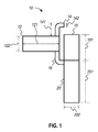
- FIG. 1 A plan view of an embodiment of a hysteropexy mesh 10 is shown in FIG. 1 .
- Mesh 10 comprises an anterior vaginal portion 12, a right broad ligament portion 14, a left broad ligament portion 16, a sacral portion 18, and a posterior vaginal portion 20.
- mesh 10 is an integral mesh, and each of the above-identified portions 12-20 are regions of the whole.
- mesh 10 may be cut, stamped, or otherwise machined from a single sheet of mesh.
- the portions may be separate or separable.
- anterior vaginal portion 12, right broad ligament portion 14, and left broad ligament portion 16 may comprise one integral mesh
- sacral portion 18 may comprise a second integral mesh
- posterior vaginal portion 20 may comprise a third integral mesh, and these may be coupled to each other, such as a with a suture or an ultrasonic weld.
- anterior vaginal portion 12 is substantially rectangular in shape with a major dimension 121 of about 10 cm and a minor dimension 122 of about 4.0 cm.
- Right broad ligament portion 14 extends from the long edge of anterior vaginal portion 12, such that an edge of right broad ligament portion 14 is in common (i.e., substantially aligned) with a short edge of anterior vaginal portion 12.
- Left broad ligament portion 16 extends from the other long edge of anterior vaginal portion 12, such that an edge of left broad ligament portion 16 is in common (i.e., substantially aligned) with the same short edge of anterior vaginal portion 12 and the edge of right broad ligament portion 14.
- Right and left broad ligament portions 14 and 16 are substantially L-shaped, and may be rounded as shown in FIG. 1 .
- Each of the broad ligament portions has a thickness dimension 142 of about 1.2 cm and a major dimension 141 of about 3.5 cm.
- Left broad ligament portion 16 is between anterior vaginal portion 12 and posterior vaginal portion 20.
- Posterior vaginal portion 20 is substantially rectangular in shape and has a major dimension 201 of about 15 cm and a minor dimension 202 of about 4.0 cm.
- the edge of left broad ligament portion 16 that is in common with the edge of anterior vaginal portion 12 and the edge of right broad ligament portion 14 is also in common with a short edge of posterior vaginal portion 20.
- Sacral portion 18 is substantially rectangular in shape and has a major dimension 181 of about 8 cm and a minor dimension 202 of about 4.0 cm.
- mesh 10 is configured to be folded substantially along the longitudinal midline of anterior vaginal portion 12 and is further configured to be folded between sacral portion 18 and posterior vaginal portion 20.
- mesh 10 is packaged as depicted in FIG. 2 , such that mesh 10 is folded substantially along the longitudinal midline of anterior vaginal portion 12 and between sacral portion 18 and posterior vaginal portion 20.
- mesh 10 may be removed from the packaging, passed through the left side broad ligament incision, then unfolded to be attached to the anterior vaginal wall.
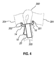
- FIGS. 3-5 are anterior, posterior, and lateral illustrations, respectively, of mesh 10 implanted into the pelvic region of a patient.
- FIG. 3 is an anterior view of mesh 10 implanted in the pelvic region showing the uterus 300, the pubic bone 303, and the anterior vagina 307.
- FIG. 4 is a posterior view of mesh 10 implanted in the pelvic region showing uterus 300, the broad ligament 304, and a portion of the sacrum 305.
- FIG. 5 is a lateral view of mesh 10 implanted in the pelvic region showing pubic bone 303, bladder 306, uterus 300, rectum 308, and sacrum 305.
- the mesh 10 is a knitted monofilament polypropylene mesh having a mass per area between approximately 15-35 g/m 2 with a pore size between approximately 500-1500 ⁇ m and a thickness of approximately 260 ⁇ m.
- This mesh is thin and light weight (i.e., the basis weight is less than approximately 35 g/m 2 ) to provide a thin and comfortable mesh that is less likely to erode tissue that contacts the mesh and less likely to be sensed through the tissue layers by the patient.
- Other suitable materials for the support include fabrics formed from polyester, polyethylene, silicone, urethanes, polyurethanes, copolymers, or block copolymers of these or suitably similar polymeric materials.
- Suitable such knitted monofilament polypropylene mesh is available from Coloplast Corp., Minneapolis, MN.
- Other suitable woven polypropylene mesh material is available from, for example, HemiaMesh, Chivasso, Italy.
- Embodiments of the disclosed method may be performed robotically, laparoscopically, or in open surgery through a laparotomy incision.
- a uterine manipulator may be placed vaginally to allow for manipulation of uterus 300 for the anterior and posterior dissection of the vaginal walls and attachment of the mesh. Then a robotic tenaculum may be used to grab the fundus of the uterus to manipulate the uterus for the anterior and posterior dissection of the vaginal walls and attachment of the mesh. A tenaculum can then be used through an accessory port to be placed on the fundus of the uterus to manipulate the uterus for the anterior and posterior dissection of the vaginal walls and attachment of the mesh.
- the tenaculum may be used to be placed on the fundus of uterus 300 to manipulate uterus 300 for the anterior and posterior dissection of the vaginal walls and attachment of the mesh or a uterine manipulator may be placed vaginally to allow for manipulation of uterus 300 for the anterior and posterior dissection of the vaginal walls and attachment of the mesh.
- a uterine manipulator may be placed vaginally to allow for manipulation of uterus 300 for the anterior and posterior dissection of the vaginal walls and attachment of the mesh.
- Uterus 300 is elevated so the posterior vagina is visualized.
- An incision is made in the peritoneum just above the rectum 308.
- Dissection is then performed bluntly and sharply in the rectovaginal space to separate rectum 308 from the posterior aspect of the vagina until the perineal body is reached and the levator muscles are exposed.
- the peritoneum overlying the sacral promontory is tented up and entered sharply with scissors.
- the peritoneal dissection is then carried inferiorly to meet the dissection posteriorly in the cul de sac below.
- the anterior longitudinal ligament as well as the presacral vessels on the sacral promontory are identified and cleared off.
- Uterus 300 is then pulled dorsally and apically so that the anterior aspect of uterus 300 and the vagina are visualized.
- An incision is made in the vesicouterine fascia and the bladder flap is created sharply.
- the dissection is performed bluntly and sharply in the vesicovaginal space to separate the bladder from the anterior aspect of the vagina. This is done until the level of the trigone is reached.
- Two one-centimeter incisions 301, 302 are made with the use of scissors and/or cautery in the broad ligament on each side near the cervix.
- posterior vaginal portion 20 of mesh 10 is attached to the posterior aspect of the vagina, such as with interrupted sutures.
- the folded anterior vaginal portion 12 and the right and left broad ligament arms 14, 16 are then passed through incision 301 in the broad ligament on the left.
- Anterior vaginal portion 12 is then unfolded and attached to the anterior aspect of the vagina using interrupted sutures.
- Right broad ligament portion 14 is then passed through incision 302 in the broad ligament on the right.
- Uterus 300 is then lifted again, such as with a tenaculum or a manipulator and right broad ligament portion 14 of mesh 10 is attached to posterior vaginal portion 20 of mesh 10 and the posterior aspect of the cervix.
- Sacral portion 18 of mesh 10 is than attached to the anterior longitudinal ligament at the level of the S1 vertebrae, such as with two interrupted sutures in tension free fashion.
- posterior vaginal portion 20 of mesh 10 and the peritoneal incision made on the sacrum are than closed using absorbable suture.
- Anterior vaginal portion 12 of mesh 10 is then covered with the bladder peritoneum using another absorbable suture. Any remaining incisions are closed and the surgery is complete.
- the present invention provides a hysteropexy mesh comprising: an anterior vaginal portion, substantially rectangular in shape, having a major dimension of about 10 cm and a minor dimension of about 4.0 cm; a right broad ligament portion, substantially L-shaped, extending from one long edge of the anterior vaginal portion and having a thickness dimension about 1.2 cm and a major dimension of about 3.5 cm; a left broad ligament portion, substantially L-shaped, extending from the other long edge of the anterior vaginal portion and having a thickness dimension about 1.2 cm and a major dimension of about 3.5 cm; a sacral portion, substantially rectangular in shape, having a major dimension of about 8.0 cm and a minor dimension of about 4.0 cm; and a posterior vaginal portion, substantially rectangular in shape, having a major dimension of about 15 cm and a minor dimension of about 4.0 cm; where the left broad ligament portion extends between and connects the anterior vaginal portion and the posterior vaginal portion.
Landscapes
- Health & Medical Sciences (AREA)
- Life Sciences & Earth Sciences (AREA)
- Animal Behavior & Ethology (AREA)
- Veterinary Medicine (AREA)
- Engineering & Computer Science (AREA)
- Biomedical Technology (AREA)
- Heart & Thoracic Surgery (AREA)
- Public Health (AREA)
- General Health & Medical Sciences (AREA)
- Urology & Nephrology (AREA)
- Surgery (AREA)
- Cardiology (AREA)
- Vascular Medicine (AREA)
- Transplantation (AREA)
- Oral & Maxillofacial Surgery (AREA)
- Gynecology & Obstetrics (AREA)
- Pregnancy & Childbirth (AREA)
- Reproductive Health (AREA)
- Nuclear Medicine, Radiotherapy & Molecular Imaging (AREA)
- Medical Informatics (AREA)
- Molecular Biology (AREA)
- Surgical Instruments (AREA)
- Prostheses (AREA)
Abstract
Right broad ligament portion 14 extends from the long edge of anterior vaginal portion 12, left broad ligament portion 16 extends from the other long edge of anterior vaginal portion 12.
Right and left broad ligament portions 14 and 16 are substantially L-shaped. Each of the broad ligament portions has a thickness dimension 142 of about 1.2 cm and a major dimension 141 of about 3.5 cm.
Left broad ligament portion 16 is between anterior vaginal portion 12 and posterior vaginal portion 20. Posterior vaginal portion 20 is substantially rectangular in shape and has a major dimension 201 of about 15 cm and a minor dimension 202 of about 4.0 cm.
Sacral portion 18 is substantially rectangular in shape and has a major dimension 181 of about 8 cm and a minor dimension 202 of about 4.0 cm.
Description
- This application claims the benefit of Provisional Application Serial No.
61/862,386, filed August 5, 2013 - The invention relates generally to apparatuses and methods for performing hysteropexy.
- Uterine prolapse occurs when pelvic floor muscles and ligaments stretch and weaken, providing inadequate support for the uterus. The uterus then descends into or protrudes from the vagina. Uterine prolapse can happen to women of any age, but it often affects postmenopausal women who've had one or more vaginal deliveries. Damage to supportive tissues during pregnancy and childbirth, effects of gravity, loss of estrogen, and repeated straining over the years all can weaken the pelvic floor and lead to uterine prolapse.
- At the present time, sacral colpopexy is the gold standard procedure for post-hysterectomy patients who have vaginal vault prolapse. Patients who still have their uterus and are undergoing sacral colpopexy are having a total hysterectomy or a supracervical hysterectomy at the same time. This can lead to mesh erosion. The only reason that most hysterectomies are performed is because the appropriate mesh that can provide anterior, posterior, and apical uterine support is not available. Hysteropexy is the surgical fixation of a displaced uterus. The hysteropexies that are performed at the present time are either supporting the uterus by placing the mesh anteriorly or posteriorly.
- The present invention provides embodiments of apparatuses and methods configured for use in hysteropexy and methods of using the same.
- In one embodiment, a hysteropexy is presented comprising a hysteropexy mesh comprising: an anterior vaginal portion, substantially rectangular in shape, having a major dimension of about 10 cm and a minor dimension of about 4.0 cm; a right broad ligament portion, substantially L-shaped, extending from one long edge of the anterior vaginal portion and having a thickness dimension about 1.2 cm and a major dimension of about 3.5 cm; a left broad ligament portion, substantially L-shaped, extending from the other long edge of the anterior vaginal portion and having a thickness dimension about 1.2 cm and a major dimension of about 3.5 cm; a sacral portion, substantially rectangular in shape, having a major dimension of about 8.0 cm and a minor dimension of about 4.0 cm; and a posterior vaginal portion, substantially rectangular in shape, having a major dimension of about 15 cm and a minor dimension of about 4.0 cm; where the left broad ligament portion extends between and connects the anterior vaginal portion and the posterior vaginal portion.
- In another embodiment the hysteropexy mesh comprises one of polypropylene, polyester, polyethylene, silicone, a urethane, a polyurethane, copolymers, or block copolymers thereof.
- In another embodiment a method of treating hysteropexy is provided comprising: create a peritoneal incision just above the rectum; separate rectum from the posterior aspect of the vagina by dissection until the perineal body is reached and the levator muscles are exposed; penetrate peritoneum overlying the sacral promontory and inferiorly dissected to meet posterior dissection from cul de sac; identify and clear off anterior longitudinal ligament and presacral vessels on the sacral promontory; manipulate uterus to create vesicouterine fascia incision and bladder flap; dissect vesicovaginal space to separate the bladder from the anterior aspect of the vagina until the level of the trigone is reached; create broad ligament incisions on each side near the cervix; insert hysteropexy mesh and attach by suture the posterior vaginal portion of the mesh to the posterior aspect of the vagina; pass folded anterior vaginal portion and right and left broad ligament arms through incision in the broad ligament on the left; unfold anterior vaginal portion and attach by suture to the anterior aspect of the vagina; pass right broad ligament portion through incision in the broad ligament on the right; manipulator uterus and attach right broad ligament portion of mesh to posterior vaginal portion of mesh and the posterior aspect of the cervix; attach sacral portion of mesh to the anterior longitudinal ligament at the level of the S1 vertebrae by suture in a tension free fashion; close peritoneal incision made on the sacrum by suture and cover with the bladder peritoneum using another suture; and close any remaining incisions to complete procedure.
- In another embodiment the hysteropexy procedure is performed robotically, laparoscopically, or in open surgery through a laparotomy incision.
- In yet another embodiment a medical device kit is provided comprising the hysteropexy mesh of the current disclosure and instructions for implantation.
- Although the present invention has been described with reference to preferred embodiments, persons skilled in the art will recognize that changes may be made in form and detail without departing from the spirit and scope of the invention. All references cited throughout the specification, including those in the background, are incorporated herein in their entirety. Those skilled in the art will recognize, or be able to ascertain, using no more than routine experimentation, many equivalents to specific embodiments of the invention described specifically herein. Such equivalents are intended to be encompassed in the scope of the following claims.
- While multiple embodiments are disclosed, still other embodiments of the present invention will become apparent to those skilled in the art from the following detailed description. As will be apparent, the invention is capable of modifications in various obvious aspects, all without departing from the spirit and scope of the present invention. Accordingly, the detailed descriptions are to be regarded as illustrative in nature and not restrictive.
-
-
FIG. 1 is a plan view of an embodiment of a hysteropexy mesh comprising an anterior vaginal portion, a right broad ligament portion, a left broad ligament portion, a sacral portion, and a posterior vaginal portion. -
FIG. 2 is a plan view of an embodiment of a hysteropexy mesh with anterior vaginal portion folded along its longitudinal midline. -
FIG. 3 is an anterior view of an embodiment of a hysteropexy mesh implanted in the pelvic region showing the uterus, the pubic bone, and the anterior vagina. -
FIG. 4 is a posterior view of an embodiment of a hysteropexy mesh implanted in the pelvic region showing the uterus, the broad ligament, and a portion of the sacrum. -
FIG. 5 is a lateral view of an embodiment of a hysteropexy mesh implanted in the pelvic region showing the pubic bone, the bladder, the uterus, the rectum, and the sacrum. - Embodiments and methods for using a hysteropexy mesh configured to be implanted in the pelvic region of a patient are discussed below.
- A plan view of an embodiment of a
hysteropexy mesh 10 is shown inFIG. 1 .Mesh 10 comprises an anteriorvaginal portion 12, a rightbroad ligament portion 14, a leftbroad ligament portion 16, asacral portion 18, and a posteriorvaginal portion 20. - In the illustrated embodiment,
mesh 10 is an integral mesh, and each of the above-identified portions 12-20 are regions of the whole. In such embodiments,mesh 10 may be cut, stamped, or otherwise machined from a single sheet of mesh. - In other embodiments, the portions may be separate or separable. For example, in certain embodiments, anterior
vaginal portion 12, rightbroad ligament portion 14, and leftbroad ligament portion 16 may comprise one integral mesh,sacral portion 18 may comprise a second integral mesh, and posteriorvaginal portion 20 may comprise a third integral mesh, and these may be coupled to each other, such as a with a suture or an ultrasonic weld. - In the illustrated embodiment, anterior
vaginal portion 12 is substantially rectangular in shape with amajor dimension 121 of about 10 cm and aminor dimension 122 of about 4.0 cm. - Right
broad ligament portion 14 extends from the long edge of anteriorvaginal portion 12, such that an edge of rightbroad ligament portion 14 is in common (i.e., substantially aligned) with a short edge of anteriorvaginal portion 12. - Left
broad ligament portion 16 extends from the other long edge of anteriorvaginal portion 12, such that an edge of leftbroad ligament portion 16 is in common (i.e., substantially aligned) with the same short edge of anteriorvaginal portion 12 and the edge of rightbroad ligament portion 14. - Right and left
broad ligament portions FIG. 1 . Each of the broad ligament portions has athickness dimension 142 of about 1.2 cm and amajor dimension 141 of about 3.5 cm. - Left
broad ligament portion 16 is between anteriorvaginal portion 12 and posteriorvaginal portion 20. Posteriorvaginal portion 20 is substantially rectangular in shape and has amajor dimension 201 of about 15 cm and aminor dimension 202 of about 4.0 cm. The edge of leftbroad ligament portion 16 that is in common with the edge of anteriorvaginal portion 12 and the edge of rightbroad ligament portion 14 is also in common with a short edge of posteriorvaginal portion 20. -
Sacral portion 18 is substantially rectangular in shape and has amajor dimension 181 of about 8 cm and aminor dimension 202 of about 4.0 cm. - As shown in
FIG. 2 ,mesh 10 is configured to be folded substantially along the longitudinal midline of anteriorvaginal portion 12 and is further configured to be folded betweensacral portion 18 and posteriorvaginal portion 20. In certain embodiments,mesh 10 is packaged as depicted inFIG. 2 , such thatmesh 10 is folded substantially along the longitudinal midline of anteriorvaginal portion 12 and betweensacral portion 18 and posteriorvaginal portion 20. In such embodiments,mesh 10 may be removed from the packaging, passed through the left side broad ligament incision, then unfolded to be attached to the anterior vaginal wall. -
FIGS. 3-5 are anterior, posterior, and lateral illustrations, respectively, ofmesh 10 implanted into the pelvic region of a patient.FIG. 3 is an anterior view ofmesh 10 implanted in the pelvic region showing theuterus 300, thepubic bone 303, and theanterior vagina 307.FIG. 4 is a posterior view ofmesh 10 implanted in the pelvicregion showing uterus 300, thebroad ligament 304, and a portion of thesacrum 305.FIG. 5 is a lateral view ofmesh 10 implanted in the pelvic region showingpubic bone 303,bladder 306,uterus 300,rectum 308, andsacrum 305. These figures may be reference to better understand the steps of embodiments of the method discussed below. - In one embodiment, the
mesh 10 is a knitted monofilament polypropylene mesh having a mass per area between approximately 15-35 g/m2 with a pore size between approximately 500-1500 µm and a thickness of approximately 260 µm. This mesh is thin and light weight (i.e., the basis weight is less than approximately 35 g/m2) to provide a thin and comfortable mesh that is less likely to erode tissue that contacts the mesh and less likely to be sensed through the tissue layers by the patient. Other suitable materials for the support include fabrics formed from polyester, polyethylene, silicone, urethanes, polyurethanes, copolymers, or block copolymers of these or suitably similar polymeric materials. Suitable such knitted monofilament polypropylene mesh is available from Coloplast Corp., Minneapolis, MN. Other suitable woven polypropylene mesh material is available from, for example, HemiaMesh, Chivasso, Italy. - Embodiments of the disclosed method may be performed robotically, laparoscopically, or in open surgery through a laparotomy incision.
- In embodiments where the method is performed robotically, a uterine manipulator may be placed vaginally to allow for manipulation of
uterus 300 for the anterior and posterior dissection of the vaginal walls and attachment of the mesh. Then a robotic tenaculum may be used to grab the fundus of the uterus to manipulate the uterus for the anterior and posterior dissection of the vaginal walls and attachment of the mesh. A tenaculum can then be used through an accessory port to be placed on the fundus of the uterus to manipulate the uterus for the anterior and posterior dissection of the vaginal walls and attachment of the mesh. - In embodiments where the procedure is being performed laparoscopically, the tenaculum may be used to be placed on the fundus of
uterus 300 to manipulateuterus 300 for the anterior and posterior dissection of the vaginal walls and attachment of the mesh or a uterine manipulator may be placed vaginally to allow for manipulation ofuterus 300 for the anterior and posterior dissection of the vaginal walls and attachment of the mesh. - In embodiments where the procedure is being performed open through a laparotomy incision a uterine manipulator may be placed vaginally to allow for manipulation of
uterus 300 for the anterior and posterior dissection of the vaginal walls and attachment of the mesh. - In the disclosed embodiments, the other steps of the method may be performed as follows.
-
Uterus 300 is elevated so the posterior vagina is visualized. An incision is made in the peritoneum just above therectum 308. Dissection is then performed bluntly and sharply in the rectovaginal space to separaterectum 308 from the posterior aspect of the vagina until the perineal body is reached and the levator muscles are exposed. - After that, the peritoneum overlying the sacral promontory is tented up and entered sharply with scissors. The peritoneal dissection is then carried inferiorly to meet the dissection posteriorly in the cul de sac below. The anterior longitudinal ligament as well as the presacral vessels on the sacral promontory are identified and cleared off.
-
Uterus 300 is then pulled dorsally and apically so that the anterior aspect ofuterus 300 and the vagina are visualized. An incision is made in the vesicouterine fascia and the bladder flap is created sharply. The dissection is performed bluntly and sharply in the vesicovaginal space to separate the bladder from the anterior aspect of the vagina. This is done until the level of the trigone is reached. - Two one-
centimeter incisions - Then, posterior
vaginal portion 20 ofmesh 10 is attached to the posterior aspect of the vagina, such as with interrupted sutures. - The folded anterior
vaginal portion 12 and the right and leftbroad ligament arms incision 301 in the broad ligament on the left. - Anterior
vaginal portion 12 is then unfolded and attached to the anterior aspect of the vagina using interrupted sutures. - Right
broad ligament portion 14 is then passed throughincision 302 in the broad ligament on the right.Uterus 300 is then lifted again, such as with a tenaculum or a manipulator and rightbroad ligament portion 14 ofmesh 10 is attached to posteriorvaginal portion 20 ofmesh 10 and the posterior aspect of the cervix.Sacral portion 18 ofmesh 10 is than attached to the anterior longitudinal ligament at the level of the S1 vertebrae, such as with two interrupted sutures in tension free fashion. - To minimize exposure of the mesh to intraperitoneal organs, posterior
vaginal portion 20 ofmesh 10 and the peritoneal incision made on the sacrum are than closed using absorbable suture. Anteriorvaginal portion 12 ofmesh 10 is then covered with the bladder peritoneum using another absorbable suture. Any remaining incisions are closed and the surgery is complete. - In the specification and in the claims, the terms "including" and "comprising" are open-ended terms and should be interpreted to mean "including, but not limited to.... " These terms encompass the more restrictive terms "consisting essentially of" and "consisting of."
- It must be noted that as used herein and in the appended claims, the singular forms "a", "an", and "the" include plural reference unless the context clearly dictates otherwise. As well, the terms "a" (or "an"), "one or more" and "at least one" can be used interchangeably herein. It is also to be noted that the terms "comprising", "including", "characterized by" and "having" can be used interchangeably.
- Unless defined otherwise, all technical and scientific terms used herein have the same meanings as commonly understood by one of ordinary skill in the art to which this invention belongs. All publications and patents specifically mentioned herein are incorporated by reference in their entirety for all purposes including describing and disclosing the chemicals, instruments, statistical analyses and methodologies which are reported in the publications which might be used in connection with the invention. All references cited in this specification are to be taken as indicative of the level of skill in the art. Nothing herein is to be construed as an admission that the invention is not entitled to antedate such disclosure by virtue of prior invention.
- The following paragraphs enumerated consecutively from 1 to 6 provide for various aspects of the present invention.
- In one embodiment, in a first paragraph (1), the present invention provides a hysteropexy mesh comprising: an anterior vaginal portion, substantially rectangular in shape, having a major dimension of about 10 cm and a minor dimension of about 4.0 cm; a right broad ligament portion, substantially L-shaped, extending from one long edge of the anterior vaginal portion and having a thickness dimension about 1.2 cm and a major dimension of about 3.5 cm; a left broad ligament portion, substantially L-shaped, extending from the other long edge of the anterior vaginal portion and having a thickness dimension about 1.2 cm and a major dimension of about 3.5 cm; a sacral portion, substantially rectangular in shape, having a major dimension of about 8.0 cm and a minor dimension of about 4.0 cm; and a posterior vaginal portion, substantially rectangular in shape, having a major dimension of about 15 cm and a minor dimension of about 4.0 cm; where the left broad ligament portion extends between and connects the anterior vaginal portion and the posterior vaginal portion.
- (2) The hysteropexy mesh of claim 1, wherein the mesh comprises one of polypropylene, polyester, polyethylene, silicone, a urethane, a polyurethane, copolymers, or block copolymers thereof.
- (3) A method of treating hysteropexy comprising: create a peritoneal incision just above the rectum; separate rectum from the posterior aspect of the vagina by dissection until the perineal body is reached and the levator muscles are exposed; penetrate peritoneum overlying the sacral promontory and inferiorly dissected to meet posterior dissection from cul de sac; identify and clear off anterior longitudinal ligament and presacral vessels on the sacral promontory; manipulate uterus to create vesicouterine fascia incision and bladder flap; dissect vesicovaginal space to separate the bladder from the anterior aspect of the vagina until the level of the trigone is reached; create broad ligament incisions on each side near the cervix; insert hysteropexy mesh and attach by suture the posterior vaginal portion of the mesh to the posterior aspect of the vagina; pass folded anterior vaginal portion and right and left broad ligament arms through incision in the broad ligament on the left; unfold anterior vaginal portion and attach by suture to the anterior aspect of the vagina; pass right broad ligament portion through incision in the broad ligament on the right; manipulator uterus and attach right broad ligament portion of mesh to posterior vaginal portion of mesh and the posterior aspect of the cervix;
attach sacral portion of mesh to the anterior longitudinal ligament at the level of the S1 vertebrae by suture in a tension free fashion; close peritoneal incision made on the sacrum by suture and cover with the bladder peritoneum using another suture; and close any remaining incisions to complete procedure. - (4) The method of paragraph (3), wherein the procedure is performed robotically, laparoscopically, or in open surgery through a laparotomy incision.
- (5) A medical device kit comprising: the hysteropexy mesh of paragraph (1); and instructions for the implantation of the medical device.
- (6) The medical device kit of parapraph (5), wherein the instructions comprise: create a peritoneal incision just above the rectum; separate rectum from the posterior aspect of the vagina by dissection until the perineal body is reached and the levator muscles are exposed; penetrate peritoneum overlying the sacral promontory and inferiorly dissected to meet posterior dissection from cul de sac; identify and clear off anterior longitudinal ligament and presacral vessels on the sacral promontory; manipulate uterus to create vesicouterine fascia incision and bladder flap; dissect vesicovaginal space to separate the bladder from the anterior aspect of the vagina until the level of the trigone is reached; create broad ligament incisions on each side near the cervix; insert hysteropexy mesh and attach by suture the posterior vaginal portion of the mesh to the posterior aspect of the vagina; pass folded anterior vaginal portion and right and left broad ligament arms through incision in the broad ligament on the left; unfold anterior vaginal portion and attach by suture to the anterior aspect of the vagina; pass right broad ligament portion through incision in the broad ligament on the right; manipulator uterus and attach right broad ligament portion of mesh to posterior vaginal portion of mesh and the posterior aspect of the cervix; attach sacral portion of mesh to the anterior longitudinal ligament at the level of the S1 vertebrae by suture in a tension free fashion; close peritoneal incision made on the sacrum by suture and cover with the bladder peritoneum using another suture; and close any remaining incisions to complete procedure.
- Although the present invention has been described with reference to preferred embodiments, persons skilled in the art will recognize that changes may be made in form and detail without departing from the spirit and scope of the invention. All references cited throughout the specification, including those in the background, are incorporated herein in their entirety. Those skilled in the art will recognize, or be able to ascertain, using no more than routine experimentation, many equivalents to specific embodiments of the invention described specifically herein. Such equivalents are intended to be encompassed in the scope of the following claims.
Claims (6)
- A hysteropexy mesh comprising:an anterior vaginal portion, substantially rectangular in shape, having major dimension of about 10 cm and a minor dimension of about 4.0 cm;a right broad ligament portion, substantially L-shaped, extending from one long edge of the anterior vaginal portion and having a thickness dimension about 1.2 cm and a major dimension of about 3.5 cm;a left broad ligament portion, substantially L-shaped, extending from the other long edge of the anterior vaginal portion and having a thickness dimension about 1.2 cm and a major dimension of about 3.5 cm;a sacral portion, substantially rectangular in shape, having a major dimension of about 8.0 cm and a minor dimension of about 4.0 cm; anda posterior vaginal portion, substantially rectangular in shape, having a major dimension of about 15 cm and a minor dimension of about 4.0 cm;where the left broad ligament portion extends between and connects the anterior vaginal portion and the posterior vaginal portion.
- The hysteropexy mesh of claim 1, wherein the mesh comprises one of polypropylene, polyester, polyethylene, silicone, a urethane, a polyurethane, copolymers, or block copolymers thereof.
- A method of treating hysteropexy comprising:creating a peritoneal incision just above the rectum;separating the rectum from the posterior aspect of the vagina by dissection until perineal body is reached and levator muscles are exposed;penetrating peritoneum overlying sacral promontory and inferiorly dissect to meet posterior dissection from cul de sac;identifying and clear anterior longitudinal ligament and presacral vessels on sacral promontory;manipulating uterus to create vesicouterine fascia incision and bladder flap;dissecting vesicovaginal space to separate the bladder from anterior aspect of the vagina until the level of the trigone is reached;creating broad ligament incisions on each side near the cervix;inserting hysteropexy mesh of claim 1 and attach by suture the posterior vaginal portion of the mesh to the posterior aspect of the vagina;passing folded anterior vaginal portion and right and left broad ligament arms through incision in the broad ligament on the left;unfolding anterior vaginal portion and attach by suture to the anterior aspect of the vagina;passing right broad ligament portion through incision in the broad ligament on the right;manipulating uterus and attach right broad ligament portion of mesh to posterior vaginal portion of mesh and the posterior aspect of the cervix;attaching sacral portion of mesh to the anterior longitudinal ligament at the level of the S1 vertebrae by suture in a tension free fashion;closing peritoneal incision made on the sacrum by suture and cover with the bladder peritoneum using another suture; andclosing any remaining incisions to complete procedure.
- The method of claim 3, wherein the procedure is performed robotically, laparoscopically, or in open surgery through a laparotomy incision.
- A medical device kit comprising:the hysteropexy mesh of claim 1; andinstructions for the implantation of the medical device.
- The medical device kit of claim 5, wherein the instructions comprise:create a peritoneal incision just above the rectum;separate rectum from the posterior aspect of the vagina by dissection until the perineal body is reached and the levator muscles are exposed;penetrate peritoneum overlying the sacral promontory and inferiorly dissected to meet posterior dissection from cul de sac;identify and clear off anterior longitudinal ligament and presacral vessels on the sacral promontory;manipulate uterus to create vesicouterine fascia incision and bladder flap;dissect vesicovaginal space to separate the bladder from the anterior aspect of the vagina until the level of the trigone is reached;create broad ligament incisions on each side near the cervix;insert hysteropexy mesh and attach by suture the posterior vaginal portion of the mesh to the posterior aspect of the vagina;pass folded anterior vaginal portion and right and left broad ligament arms through incision in the broad ligament on the left;unfold anterior vaginal portion and attach by suture to the anterior aspect of the vagina;pass right broad ligament portion through incision in the broad ligament on the right;manipulator uterus and attach right broad ligament portion of mesh to posterior vaginal portion of mesh and the posterior aspect of the cervix;attach sacral portion of mesh to the anterior longitudinal ligament at the level of the S1 vertebrae by suture in a tension free fashion;close peritoneal incision made on the sacrum by suture and cover with the bladder peritoneum using another suture; andclose any remaining incisions to complete procedure.
Priority Applications (1)
| Application Number | Priority Date | Filing Date | Title |
|---|---|---|---|
| EP16178607.4A EP3115020B1 (en) | 2013-08-05 | 2014-08-04 | Hysteropexy mesh apparatuses |
Applications Claiming Priority (2)
| Application Number | Priority Date | Filing Date | Title |
|---|---|---|---|
| US201361862386P | 2013-08-05 | 2013-08-05 | |
| US14/449,459 US9480546B2 (en) | 2013-08-05 | 2014-08-01 | Hysteropexy mesh apparatuses and methods |
Related Child Applications (1)
| Application Number | Title | Priority Date | Filing Date |
|---|---|---|---|
| EP16178607.4A Division EP3115020B1 (en) | 2013-08-05 | 2014-08-04 | Hysteropexy mesh apparatuses |
Publications (2)
| Publication Number | Publication Date |
|---|---|
| EP2835111A1 true EP2835111A1 (en) | 2015-02-11 |
| EP2835111B1 EP2835111B1 (en) | 2016-07-13 |
Family
ID=51260756
Family Applications (2)
| Application Number | Title | Priority Date | Filing Date |
|---|---|---|---|
| EP16178607.4A Active EP3115020B1 (en) | 2013-08-05 | 2014-08-04 | Hysteropexy mesh apparatuses |
| EP14179669.8A Active EP2835111B1 (en) | 2013-08-05 | 2014-08-04 | Hysteropexy mesh apparatuses |
Family Applications Before (1)
| Application Number | Title | Priority Date | Filing Date |
|---|---|---|---|
| EP16178607.4A Active EP3115020B1 (en) | 2013-08-05 | 2014-08-04 | Hysteropexy mesh apparatuses |
Country Status (3)
| Country | Link |
|---|---|
| US (2) | US9480546B2 (en) |
| EP (2) | EP3115020B1 (en) |
| ES (2) | ES2684990T3 (en) |
Cited By (1)
| Publication number | Priority date | Publication date | Assignee | Title |
|---|---|---|---|---|
| CN110180069A (en) * | 2019-05-29 | 2019-08-30 | 王森 | Intelligence leads happy method, system and medium |
Families Citing this family (2)
| Publication number | Priority date | Publication date | Assignee | Title |
|---|---|---|---|---|
| USD1098434S1 (en) | 2025-04-29 | 2025-10-14 | Allosource | Implant for pelvic organ prolapse |
| USD1122447S1 (en) | 2025-04-29 | 2026-04-14 | Allosource | Implant for pelvic organ prolapse |
Citations (6)
| Publication number | Priority date | Publication date | Assignee | Title |
|---|---|---|---|---|
| WO2010087923A1 (en) * | 2009-02-02 | 2010-08-05 | C.R. Bard, Inc. | Implant for pelvic organ prolapse repair |
| US20100305394A1 (en) * | 2009-05-28 | 2010-12-02 | Rosenblatt Peter L | Tubular mesh for sacrocolpopexy and related procedures |
| WO2011037837A2 (en) * | 2009-09-23 | 2011-03-31 | The General Hospital Corporation | Vaginal vault suspension device and method |
| FR2976788A1 (en) * | 2011-06-22 | 2012-12-28 | Ct Hospitalier Universitaire Nimes | Prosthesis for suspension of e.g. rectum of pelvis for treatment of cystocele prolapse, has strip provided with sections, where one of sections is fixed to organ, and other section is fixed at anchor point and shifted from former section |
| US20130066145A1 (en) * | 2011-09-09 | 2013-03-14 | Boston Scientific Scimed, Inc. | Medical device and methods of delivering the medical device |
| EP2754412A1 (en) * | 2013-01-10 | 2014-07-16 | B. Braun Surgical, S.A. | A surgical implant for the treatment of pelvic organ prolapse by sacrocolpopexy and/or sacrohysteropexy |
Family Cites Families (134)
| Publication number | Priority date | Publication date | Assignee | Title |
|---|---|---|---|---|
| US2738790A (en) | 1954-08-12 | 1956-03-20 | George P Pilling & Son Company | Suturing instrument |
| US4009711A (en) | 1976-03-17 | 1977-03-01 | Uson Aurelio C | Penile prosthesis for the management of erectile impotence |
| US5409453A (en) | 1992-08-12 | 1995-04-25 | Vidamed, Inc. | Steerable medical probe with stylets |
| US5230694A (en) | 1992-01-09 | 1993-07-27 | Rosenblum Jeffrey L | Urological prosthesis |
| US5284141A (en) | 1992-07-31 | 1994-02-08 | Eibling David L | Electrode emplacement apparatus for amniotomy and fetal monitoring and method of use |
| US5356432B1 (en) | 1993-02-05 | 1997-02-04 | Bard Inc C R | Implantable mesh prosthesis and method for repairing muscle or tissue wall defects |
| US5899909A (en) | 1994-08-30 | 1999-05-04 | Medscand Medical Ab | Surgical instrument for treating female urinary incontinence |
| SE506164C2 (en) | 1995-10-09 | 1997-11-17 | Medscand Medical Ab | Instruments for the treatment of urinary incontinence in women |
| US6117144A (en) | 1995-08-24 | 2000-09-12 | Sutura, Inc. | Suturing device and method for sealing an opening in a blood vessel or other biological structure |
| US6406420B1 (en) | 1997-01-02 | 2002-06-18 | Myocor, Inc. | Methods and devices for improving cardiac function in hearts |
| DE69808614T2 (en) | 1997-02-13 | 2003-06-18 | Boston Scientific Ltd., St. Michael | STABILIZATION SLING FOR USE IN MINIMALLY INVASIVE PELVIC SURGERY |
| US6039686A (en) | 1997-03-18 | 2000-03-21 | Kovac; S. Robert | System and a method for the long term cure of recurrent urinary female incontinence |
| US5934283A (en) | 1997-04-15 | 1999-08-10 | Uroplasty, Inc. | Pubovaginal sling device |
| CA2304296C (en) | 1997-10-01 | 2005-02-15 | Boston Scientific Limited | Pelvic floor reconstruction |
| US6382214B1 (en) | 1998-04-24 | 2002-05-07 | American Medical Systems, Inc. | Methods and apparatus for correction of urinary and gynecological pathologies including treatment of male incontinence and female cystocele |
| US6302840B1 (en) | 1998-09-21 | 2001-10-16 | Theodore V. Benderev | Surgical monitor |
| US6050937A (en) | 1998-09-21 | 2000-04-18 | Benderev; Theodore V. | Surgical tension/pressure monitor |
| US6200330B1 (en) | 1998-11-23 | 2001-03-13 | Theodore V. Benderev | Systems for securing sutures, grafts and soft tissue to bone and periosteum |
| US20050004576A1 (en) | 1998-11-23 | 2005-01-06 | Benderev Theodore V. | System for securing sutures, grafts and soft tissue to bone and periosteum |
| FR2792824B1 (en) | 1999-04-27 | 2001-06-22 | Sofradim Production | DEVICE FOR TREATING PROLAPSUS BY VAGINAL SUSPENSION |
| US7121997B2 (en) | 1999-06-09 | 2006-10-17 | Ethicon, Inc. | Surgical instrument and method for treating female urinary incontinence |
| US7226407B2 (en) | 1999-06-09 | 2007-06-05 | Ethicon, Inc. | Surgical instrument and method for treating female urinary incontinence |
| US6273852B1 (en) | 1999-06-09 | 2001-08-14 | Ethicon, Inc. | Surgical instrument and method for treating female urinary incontinence |
| US7083637B1 (en) | 1999-06-09 | 2006-08-01 | Tannhauser Robert J | Method and apparatus for adjusting flexible areal polymer implants |
| US6355065B1 (en) | 1999-09-01 | 2002-03-12 | Shlomo Gabbay | Implantable support apparatus and method of using same |
| US6599318B1 (en) | 1999-11-30 | 2003-07-29 | Shlomo Gabbay | Implantable support apparatus and method of using same |
| US7131943B2 (en) | 2000-03-09 | 2006-11-07 | Ethicon, Inc. | Surgical instrument and method for treating organ prolapse conditions |
| RU2185779C2 (en) | 2000-05-19 | 2002-07-27 | Московский областной научно-исследовательский клинический институт | Surgical method for treating the cases of enuresis in male patients |
| US6951534B2 (en) | 2000-06-13 | 2005-10-04 | Acorn Cardiovascular, Inc. | Cardiac support device |
| US6638211B2 (en) | 2000-07-05 | 2003-10-28 | Mentor Corporation | Method for treating urinary incontinence in women and implantable device intended to correct urinary incontinence |
| US7025063B2 (en) | 2000-09-07 | 2006-04-11 | Ams Research Corporation | Coated sling material |
| US6592515B2 (en) | 2000-09-07 | 2003-07-15 | Ams Research Corporation | Implantable article and method |
| US20060205995A1 (en) | 2000-10-12 | 2006-09-14 | Gyne Ideas Limited | Apparatus and method for treating female urinary incontinence |
| US20020128670A1 (en) | 2000-11-22 | 2002-09-12 | Ulf Ulmsten | Surgical instrument and method for treating female urinary incontinence |
| US20110168192A9 (en) | 2000-12-20 | 2011-07-14 | Eduardo Fierro | Adjustable autofixing sling for treatment of urinary incontinence |
| US20020161382A1 (en) | 2001-03-29 | 2002-10-31 | Neisz Johann J. | Implant inserted without bone anchors |
| US6802807B2 (en) | 2001-01-23 | 2004-10-12 | American Medical Systems, Inc. | Surgical instrument and method |
| US6612977B2 (en) | 2001-01-23 | 2003-09-02 | American Medical Systems Inc. | Sling delivery system and method of use |
| US7229453B2 (en) | 2001-01-23 | 2007-06-12 | Ams Research Corporation | Pelvic floor implant system and method of assembly |
| US7070556B2 (en) | 2002-03-07 | 2006-07-04 | Ams Research Corporation | Transobturator surgical articles and methods |
| US6685626B2 (en) | 2001-02-02 | 2004-02-03 | Regeneration Technologies, Inc. | Compositions, devices, methods, and kits for induction of adhesions |
| US7364541B2 (en) | 2001-03-09 | 2008-04-29 | Boston Scientific Scimed, Inc. | Systems, methods and devices relating to delivery of medical implants |
| US6936052B2 (en) | 2001-03-09 | 2005-08-30 | Boston Scientific Scimed, Inc. | System for implanting an implant and method thereof |
| AUPR406501A0 (en) | 2001-03-28 | 2001-04-26 | Kaladelfos, George | Treatment of vault prolapse |
| EP1399082B1 (en) | 2001-06-27 | 2008-10-01 | Promedon Do Brasil Produtos Medico-Hospitalares Ltda | Adjustable autofixing sling for treatment of urinary incontinence |
| US7407480B2 (en) | 2001-07-27 | 2008-08-05 | Ams Research Corporation | Method and apparatus for correction of urinary and gynecological pathologies, including treatment of incontinence cystocele |
| US6755781B2 (en) | 2001-07-27 | 2004-06-29 | Scimed Life Systems, Inc. | Medical slings |
| ATE332674T1 (en) | 2001-08-03 | 2006-08-15 | Aesculap Ag & Co Kg | INCONTINENCE BAND FOR THE TREATMENT OF URINARY INCONTINENCE |
| US6666817B2 (en) | 2001-10-05 | 2003-12-23 | Scimed Life Systems, Inc. | Expandable surgical implants and methods of using them |
| US20030078471A1 (en) | 2001-10-18 | 2003-04-24 | Foley Frederick J. | Manipulation of an organ |
| US6790213B2 (en) | 2002-01-07 | 2004-09-14 | C.R. Bard, Inc. | Implantable prosthesis |
| PT1487377E (en) | 2002-03-01 | 2012-11-23 | Ethicon Inc | Apparatus for treating pelvic organ prolapses in female patients |
| US6911003B2 (en) | 2002-03-07 | 2005-06-28 | Ams Research Corporation | Transobturator surgical articles and methods |
| JP4476630B2 (en) | 2002-03-07 | 2010-06-09 | エーエムエス・リサーチ・コーポレーション | Surgical instruments and methods via the closure membrane |
| CA2483124A1 (en) | 2002-05-07 | 2003-11-20 | Ams Research Corporation | Urethral prosthesis with tensioning member |
| US6972027B2 (en) | 2002-06-26 | 2005-12-06 | Stryker Endoscopy | Soft tissue repair system |
| ITFI20020145A1 (en) | 2002-08-01 | 2004-02-02 | Giulio Nicita | DEVICE FOR THE SURGICAL TREATMENT OF FEMALE PROLAXIS. |
| AU2003269934A1 (en) | 2002-08-02 | 2004-02-23 | C.R. Bard, Inc. | Self anchoring sling and introducer system |
| AU2003250591B2 (en) | 2002-08-23 | 2007-06-21 | Pamarope Pty Limited | Anchoring device and its implementation |
| PT1542596E (en) | 2002-08-29 | 2010-12-07 | Univ Liege | Devices for surgical treatment of female urinary incontinence |
| EP1990023B1 (en) | 2002-11-15 | 2012-09-26 | Ethicon, Inc. | Devices suitable for use in surgical repair of vagina damaged by pelvic organ prolapse |
| US6808486B1 (en) | 2002-12-03 | 2004-10-26 | Pat O'Donnell | Surgical instrument for treating female urinary stress incontinence |
| US7517357B2 (en) | 2003-01-09 | 2009-04-14 | Linvatec Biomaterials | Knotless suture anchor |
| US7204801B2 (en) | 2003-01-17 | 2007-04-17 | Massachusetts General Hospital | Pubovaginal support for treating female urinary incontinence |
| BRPI0408903A (en) | 2003-03-28 | 2006-03-28 | Analytic Biosurgical Solutions | implant for treatment of the rectocele and / or vaginal dome prolapse, device for inserting an implant and process for treating the rectocele in a woman |
| RU2243729C1 (en) | 2003-05-05 | 2005-01-10 | Диагностический центр "Здоровье" | Method for treating the cases of male enuresis |
| US7320701B2 (en) | 2003-06-02 | 2008-01-22 | Linvatec Corporation | Push-in suture anchor, insertion tool, and method for inserting a push-in suture anchor |
| FR2859624B1 (en) | 2003-09-16 | 2005-12-02 | Sofradim Production | PROTHETIC KNIT WITH VARIABLE PROPERTIES |
| US7393319B2 (en) | 2003-10-14 | 2008-07-01 | Caldera Medical, Inc. | Implantable sling having bladder support |
| US20050101834A1 (en) | 2003-11-10 | 2005-05-12 | Merade Bryon L. | Readily implantable sling |
| RU2275883C2 (en) | 2004-04-13 | 2006-05-10 | Вадим Александрович Зотов | Artificial urethral sphincter and method for surgically treating enuresis in male cases |
| EP1744679A2 (en) | 2004-05-03 | 2007-01-24 | AMS Research Corporation | Surgical implants and related methods |
| GB0411360D0 (en) | 2004-05-21 | 2004-06-23 | Mpathy Medical Devices Ltd | Implant |
| EP2543341B1 (en) | 2004-06-14 | 2016-07-20 | Boston Scientific Limited | A soft tissue anchor |
| US20050283040A1 (en) | 2004-06-16 | 2005-12-22 | Secant Medical, Llc | Incontinence sling |
| US7727140B2 (en) | 2004-06-21 | 2010-06-01 | Department Of Veterans Affairs | Surgically implantable perineal urinary incontinence device |
| AU2005274013A1 (en) | 2004-08-09 | 2006-02-23 | Innovative Spinal Technologies | System and method for dynamic skeletal stabilization |
| US7527588B2 (en) | 2004-09-15 | 2009-05-05 | Ethicon, Inc. | System and method for surgical implant placement |
| WO2006034719A1 (en) | 2004-09-28 | 2006-04-06 | Negm Sherif Mohammed Maher Ibr | Vaginal stent for prevention and treatment of vaginal vault prolapse |
| US7901346B2 (en) | 2004-10-05 | 2011-03-08 | Ams Research Corporation | Method for supporting vaginal cuff |
| WO2006045042A1 (en) | 2004-10-20 | 2006-04-27 | University Of Florida Research Foundation, Inc. | Devices and methods for vaginal reconstruction |
| US7914437B2 (en) | 2005-02-04 | 2011-03-29 | Ams Research Corporation | Transobturator methods for installing sling to treat incontinence, and related devices |
| AU2006226961A1 (en) | 2005-03-22 | 2006-09-28 | Tyco Healthcare Group, Lp | Mesh implant |
| US7556598B2 (en) | 2005-04-04 | 2009-07-07 | Boston Scientific Scimed, Inc. | Dissolvable protective treatment for an implantable supportive sling |
| WO2006108145A1 (en) | 2005-04-06 | 2006-10-12 | Boston Scientific Scimed, Inc. | Systems, devices, and methods for treating pelvic floor disorders |
| US7431690B2 (en) | 2005-04-30 | 2008-10-07 | Coloplast A/S | Implantable sling for the treatment of male incontinence and method of using the same |
| WO2007002260A2 (en) | 2005-06-21 | 2007-01-04 | Cook Incorporated | Implantable graft to close a fistula |
| US7981023B2 (en) | 2005-07-25 | 2011-07-19 | Boston Scientific Scimed, Inc. | Elastic sling system and related methods |
| WO2007014241A1 (en) | 2005-07-25 | 2007-02-01 | Boston Scientific Scimed, Inc. | Pelvic floor repair system |
| WO2007018532A1 (en) | 2005-08-03 | 2007-02-15 | Boston Scientific Scimed, Inc. | Systems, devices and methods relating to a shape resilient sling-like support for treating urinary incontinence |
| US7878970B2 (en) * | 2005-09-28 | 2011-02-01 | Boston Scientific Scimed, Inc. | Apparatus and method for suspending a uterus |
| US7673631B2 (en) | 2005-10-25 | 2010-03-09 | Ethicon, Inc. | Adjustable vaginal splint for pelvic floor support |
| EP1948073B1 (en) | 2005-11-14 | 2014-03-19 | C.R.Bard, Inc. | Sling anchor system |
| BRPI0520800B1 (en) | 2005-12-09 | 2016-06-28 | Promedon Do Brasil Produtos Médico Hospitalares Ltda | implant for the treatment of stress urinary incontinence and prolapse of the anterior vaginal wall |
| JP2009528851A (en) | 2005-12-28 | 2009-08-13 | シー・アール・バード・インコーポレイテツド | Apparatus and method for inserting an implant |
| US7559885B2 (en) | 2006-02-14 | 2009-07-14 | Coloplast A/S | Implantable sling for the treatment of incontinence and method of using the same |
| EP1998711B1 (en) | 2006-03-16 | 2013-12-11 | Boston Scientific Limited | Apparatus for treatment of pelvic conditions |
| EP2007310B1 (en) | 2006-04-14 | 2015-08-12 | A.M.I. Agency for Medical Innovations GmbH | Implantable mesh for surgical reconstruction in the area of the pelvic floor |
| US20070293717A1 (en) | 2006-05-12 | 2007-12-20 | Ams Research Corporation | Tube mesh for abdominal sacral colpopexy |
| WO2007137226A2 (en) | 2006-05-19 | 2007-11-29 | Ams Research Corporation | Method and articles for treatment of stress urinary incontinence |
| US20080021265A1 (en) * | 2006-05-31 | 2008-01-24 | Olivier Garbin | Prosthesis to be implanted in a human body for repairing or treating pelvic organ prolapses in a female patient and surgical method using such a prosthesis |
| US8628463B2 (en) | 2006-06-22 | 2014-01-14 | Ams Research Corporation | Adjustable tension incontinence sling assemblies |
| JP2009545352A (en) | 2006-08-02 | 2009-12-24 | コンパニ ドゥ ルシェルシュ オン コンポザン,アンプラン エ マテリエル プール ラプリカシオン クリニーク | Hammock under the urethra |
| FR2906131B1 (en) | 2006-09-21 | 2009-04-24 | Cl Medical Sarl | SURGICAL TREATMENT FOR TREATMENT OF URINARY INCONTINENCE IN MAN |
| RU2483697C2 (en) | 2006-10-18 | 2013-06-10 | Колопласт А/С | Implanted devices for treatment of incontinence and methods of their application |
| CA2829730C (en) | 2006-11-06 | 2016-07-12 | Caldera Medical, Inc. | Implants and procedures for treatment of pelvic floor disorders |
| KR101328421B1 (en) | 2007-03-02 | 2013-11-14 | ?뜨르 오스삐탈리에 유니버시떼이르 드 리에즈 | Surgical techniques and tools for use in the treatment of male incontinence |
| US8758359B2 (en) | 2007-05-14 | 2014-06-24 | Ethicon, Inc. | Instruments for implanting implantable prostheses |
| US7645227B2 (en) | 2007-05-14 | 2010-01-12 | Ethicon, Inc. | Implants and methods for pelvic floor repair |
| DE602007002621D1 (en) | 2007-06-11 | 2009-11-12 | Novus Scient Pte Ltd | Mesh implant with interlocking mesh structure |
| WO2008152435A1 (en) | 2007-06-12 | 2008-12-18 | PROMEDON DO BRASIL PRODUTOS MéDICO HOSPITALARES LTDA. | Micro sling and implantation tool for urinary incontinence and faecal incontinence treatment |
| US8933290B2 (en) | 2007-06-26 | 2015-01-13 | Sofradim Production Sas | Mesh implant |
| US8007429B2 (en) | 2007-07-05 | 2011-08-30 | Gt Urological, Llc | Vessel occlusive device and method of occluding a vessel |
| FR2919996B1 (en) | 2007-08-17 | 2010-09-10 | Cl Medical | IMPLANT FOR SUPPORTING THE URETRE OF A MAN AND SURGICAL ASSEMBLY FOR TREATMENT OF URINARY INCONTINENCE IN A MAN COMPRISING SUCH AN IMPLANT |
| US8206280B2 (en) | 2007-11-13 | 2012-06-26 | C. R. Bard, Inc. | Adjustable tissue support member |
| US8430807B2 (en) | 2007-12-28 | 2013-04-30 | Boston Scientific Scimed, Inc. | Devices and methods for treating pelvic floor dysfunctions |
| WO2009086446A1 (en) | 2007-12-28 | 2009-07-09 | Boston Scientific Scimed, Inc. | Meshes of variable construction |
| US20090216072A1 (en) | 2008-02-27 | 2009-08-27 | Ralph Zipper | Urinary incontinence sling apparatus and method |
| US20100191038A1 (en) | 2009-01-27 | 2010-07-29 | Coloplast A/S | Devices and tools for treatment of urinary incontinence |
| US8585579B2 (en) | 2009-02-05 | 2013-11-19 | Coloplast A/S | Implantable anatomical support |
| US8696544B2 (en) | 2009-02-05 | 2014-04-15 | Coloplast A/S | Minimally invasive adjustable support |
| CA2748894C (en) | 2009-02-10 | 2018-11-13 | Ams Research Corporation | Surgical articles and methods for treating urinary incontinence |
| US20100261956A1 (en) | 2009-04-09 | 2010-10-14 | Minnesota Medical Development, Inc. | Apparatus and Method for Pelvic Floor Repair in the Human Female |
| US20100261954A1 (en) * | 2009-04-09 | 2010-10-14 | Minnesota Medical Development, Inc. | Apparatus and Method for Pelvic Floor Repair in the Human Female |
| US8098079B2 (en) * | 2009-04-17 | 2012-01-17 | Oracle America, Inc. | Receive circuit for connectors with variable complex impedance |
| US8790369B2 (en) | 2009-07-24 | 2014-07-29 | Depuy Mitek, Llc | Apparatus and method for repairing tissue |
| AU2010339436B2 (en) | 2009-12-30 | 2015-01-15 | Boston Scientific Scimed, Inc. | Systems, implants, tools, and methods for treatments of pelvic conditions |
| AU2011220816B2 (en) | 2010-02-23 | 2017-03-16 | Ams Research Corporation | Surgical articles and methods |
| US8876693B2 (en) | 2010-04-21 | 2014-11-04 | David Elliot Rapp | Device and method for vaginal sacrocolpopexy |
| WO2012054985A1 (en) | 2010-10-29 | 2012-05-03 | Carey Tasca Pty Ltd | Method and devices for repair of vaginal wall or uterus |
| FR2973217B1 (en) | 2011-03-28 | 2014-09-05 | Ct Hospitalier Universitaire Nimes | DEVICE FOR PREDICTING THE EFFECTIVENESS OF SURGICAL TREATMENT OF URINARY INCONTINENCE OF EFFORT |
| US8864647B2 (en) | 2011-08-19 | 2014-10-21 | Coloplast A/S | Incontinence treatment device with pubic arm attachment mechanism |
| US9320586B2 (en) * | 2012-07-02 | 2016-04-26 | Ethicon, Inc. | Surgical implant for treating pelvic organ prolapse conditions |
| US8834348B2 (en) | 2013-02-14 | 2014-09-16 | Coloplast A/S | System for treating pelvic organ prolapse including a shell and an anchor |
-
2014
- 2014-08-01 US US14/449,459 patent/US9480546B2/en active Active
- 2014-08-04 ES ES16178607.4T patent/ES2684990T3/en active Active
- 2014-08-04 EP EP16178607.4A patent/EP3115020B1/en active Active
- 2014-08-04 ES ES14179669.8T patent/ES2593504T3/en active Active
- 2014-08-04 EP EP14179669.8A patent/EP2835111B1/en active Active
-
2016
- 2016-07-25 US US15/218,084 patent/US10034734B2/en active Active
Patent Citations (6)
| Publication number | Priority date | Publication date | Assignee | Title |
|---|---|---|---|---|
| WO2010087923A1 (en) * | 2009-02-02 | 2010-08-05 | C.R. Bard, Inc. | Implant for pelvic organ prolapse repair |
| US20100305394A1 (en) * | 2009-05-28 | 2010-12-02 | Rosenblatt Peter L | Tubular mesh for sacrocolpopexy and related procedures |
| WO2011037837A2 (en) * | 2009-09-23 | 2011-03-31 | The General Hospital Corporation | Vaginal vault suspension device and method |
| FR2976788A1 (en) * | 2011-06-22 | 2012-12-28 | Ct Hospitalier Universitaire Nimes | Prosthesis for suspension of e.g. rectum of pelvis for treatment of cystocele prolapse, has strip provided with sections, where one of sections is fixed to organ, and other section is fixed at anchor point and shifted from former section |
| US20130066145A1 (en) * | 2011-09-09 | 2013-03-14 | Boston Scientific Scimed, Inc. | Medical device and methods of delivering the medical device |
| EP2754412A1 (en) * | 2013-01-10 | 2014-07-16 | B. Braun Surgical, S.A. | A surgical implant for the treatment of pelvic organ prolapse by sacrocolpopexy and/or sacrohysteropexy |
Cited By (1)
| Publication number | Priority date | Publication date | Assignee | Title |
|---|---|---|---|---|
| CN110180069A (en) * | 2019-05-29 | 2019-08-30 | 王森 | Intelligence leads happy method, system and medium |
Also Published As
| Publication number | Publication date |
|---|---|
| ES2684990T3 (en) | 2018-10-05 |
| ES2593504T3 (en) | 2016-12-09 |
| US20160331501A1 (en) | 2016-11-17 |
| US10034734B2 (en) | 2018-07-31 |
| US20150038780A1 (en) | 2015-02-05 |
| EP3115020A1 (en) | 2017-01-11 |
| EP2835111B1 (en) | 2016-07-13 |
| EP3115020B1 (en) | 2018-05-23 |
| US9480546B2 (en) | 2016-11-01 |
Similar Documents
| Publication | Publication Date | Title |
|---|---|---|
| KR100895598B1 (en) | Method and apparatus for treating pelvic organ prolapses in female patients | |
| EP2026702B1 (en) | Apparatus for levator distension repair | |
| US8109867B2 (en) | Tubular mesh for sacrocolpopexy and related procedures | |
| AU2005244179B2 (en) | Apparatus for cystocele repair | |
| CN101677849B (en) | Implants for Pelvic Floor Repair | |
| US10034734B2 (en) | Hysteropexy mesh apparatuses and methods | |
| LEE | Vaginal hysterectomy with repair of enterocele, cystocele, and rectocele | |
| US8986190B2 (en) | Implant for treating a genital prolapse, and associated treatment method | |
| US9579180B2 (en) | Method of treating pelvic prolapse using a sacrocolpopexy/sacrocervicopexy vaginal positioning and mesh retention system | |
| Finamore | 15.3 Laparoscopic vaginal vault suspension | |
| HK1081095B (en) | Method and apparatus for treating pelvic organ prolapses in female patients | |
| HK1129057B (en) | Apparatus for levator distension repair |
Legal Events
| Date | Code | Title | Description |
|---|---|---|---|
| PUAI | Public reference made under article 153(3) epc to a published international application that has entered the european phase |
Free format text: ORIGINAL CODE: 0009012 |
|
| 17P | Request for examination filed |
Effective date: 20140804 |
|
| AK | Designated contracting states |
Kind code of ref document: A1 Designated state(s): AL AT BE BG CH CY CZ DE DK EE ES FI FR GB GR HR HU IE IS IT LI LT LU LV MC MK MT NL NO PL PT RO RS SE SI SK SM TR |
|
| AX | Request for extension of the european patent |
Extension state: BA ME |
|
| R17P | Request for examination filed (corrected) |
Effective date: 20150105 |
|
| RBV | Designated contracting states (corrected) |
Designated state(s): AL AT BE BG CH CY CZ DE DK EE ES FI FR GB GR HR HU IE IS IT LI LT LU LV MC MK MT NL NO PL PT RO RS SE SI SK SM TR |
|
| RIC1 | Information provided on ipc code assigned before grant |
Ipc: A61F 2/00 20060101AFI20151201BHEP Ipc: A61B 17/42 20060101ALN20151201BHEP |
|
| GRAP | Despatch of communication of intention to grant a patent |
Free format text: ORIGINAL CODE: EPIDOSNIGR1 |
|
| INTG | Intention to grant announced |
Effective date: 20160120 |
|
| GRAS | Grant fee paid |
Free format text: ORIGINAL CODE: EPIDOSNIGR3 |
|
| GRAA | (expected) grant |
Free format text: ORIGINAL CODE: 0009210 |
|
| AK | Designated contracting states |
Kind code of ref document: B1 Designated state(s): AL AT BE BG CH CY CZ DE DK EE ES FI FR GB GR HR HU IE IS IT LI LT LU LV MC MK MT NL NO PL PT RO RS SE SI SK SM TR |
|
| REG | Reference to a national code |
Ref country code: GB Ref legal event code: FG4D |
|
| REG | Reference to a national code |
Ref country code: AT Ref legal event code: REF Ref document number: 811661 Country of ref document: AT Kind code of ref document: T Effective date: 20160715 Ref country code: CH Ref legal event code: EP |
|
| REG | Reference to a national code |
Ref country code: IE Ref legal event code: FG4D Ref country code: FR Ref legal event code: PLFP Year of fee payment: 3 |
|
| REG | Reference to a national code |
Ref country code: DE Ref legal event code: R096 Ref document number: 602014002636 Country of ref document: DE |
|
| REG | Reference to a national code |
Ref country code: LT Ref legal event code: MG4D |
|
| REG | Reference to a national code |
Ref country code: NL Ref legal event code: MP Effective date: 20160713 |
|
| REG | Reference to a national code |
Ref country code: ES Ref legal event code: FG2A Ref document number: 2593504 Country of ref document: ES Kind code of ref document: T3 Effective date: 20161209 |
|
| REG | Reference to a national code |
Ref country code: AT Ref legal event code: MK05 Ref document number: 811661 Country of ref document: AT Kind code of ref document: T Effective date: 20160713 |
|
| PG25 | Lapsed in a contracting state [announced via postgrant information from national office to epo] |
Ref country code: BE Free format text: LAPSE BECAUSE OF NON-PAYMENT OF DUE FEES Effective date: 20160831 |
|
| PG25 | Lapsed in a contracting state [announced via postgrant information from national office to epo] |
Ref country code: NO Free format text: LAPSE BECAUSE OF FAILURE TO SUBMIT A TRANSLATION OF THE DESCRIPTION OR TO PAY THE FEE WITHIN THE PRESCRIBED TIME-LIMIT Effective date: 20161013 Ref country code: RS Free format text: LAPSE BECAUSE OF FAILURE TO SUBMIT A TRANSLATION OF THE DESCRIPTION OR TO PAY THE FEE WITHIN THE PRESCRIBED TIME-LIMIT Effective date: 20160713 Ref country code: NL Free format text: LAPSE BECAUSE OF FAILURE TO SUBMIT A TRANSLATION OF THE DESCRIPTION OR TO PAY THE FEE WITHIN THE PRESCRIBED TIME-LIMIT Effective date: 20160713 Ref country code: FI Free format text: LAPSE BECAUSE OF FAILURE TO SUBMIT A TRANSLATION OF THE DESCRIPTION OR TO PAY THE FEE WITHIN THE PRESCRIBED TIME-LIMIT Effective date: 20160713 Ref country code: HR Free format text: LAPSE BECAUSE OF FAILURE TO SUBMIT A TRANSLATION OF THE DESCRIPTION OR TO PAY THE FEE WITHIN THE PRESCRIBED TIME-LIMIT Effective date: 20160713 Ref country code: LT Free format text: LAPSE BECAUSE OF FAILURE TO SUBMIT A TRANSLATION OF THE DESCRIPTION OR TO PAY THE FEE WITHIN THE PRESCRIBED TIME-LIMIT Effective date: 20160713 Ref country code: IS Free format text: LAPSE BECAUSE OF FAILURE TO SUBMIT A TRANSLATION OF THE DESCRIPTION OR TO PAY THE FEE WITHIN THE PRESCRIBED TIME-LIMIT Effective date: 20161113 |
|
| PG25 | Lapsed in a contracting state [announced via postgrant information from national office to epo] |
Ref country code: SE Free format text: LAPSE BECAUSE OF FAILURE TO SUBMIT A TRANSLATION OF THE DESCRIPTION OR TO PAY THE FEE WITHIN THE PRESCRIBED TIME-LIMIT Effective date: 20160713 Ref country code: PL Free format text: LAPSE BECAUSE OF FAILURE TO SUBMIT A TRANSLATION OF THE DESCRIPTION OR TO PAY THE FEE WITHIN THE PRESCRIBED TIME-LIMIT Effective date: 20160713 Ref country code: AT Free format text: LAPSE BECAUSE OF FAILURE TO SUBMIT A TRANSLATION OF THE DESCRIPTION OR TO PAY THE FEE WITHIN THE PRESCRIBED TIME-LIMIT Effective date: 20160713 Ref country code: BE Free format text: LAPSE BECAUSE OF FAILURE TO SUBMIT A TRANSLATION OF THE DESCRIPTION OR TO PAY THE FEE WITHIN THE PRESCRIBED TIME-LIMIT Effective date: 20160713 Ref country code: GR Free format text: LAPSE BECAUSE OF FAILURE TO SUBMIT A TRANSLATION OF THE DESCRIPTION OR TO PAY THE FEE WITHIN THE PRESCRIBED TIME-LIMIT Effective date: 20161014 Ref country code: PT Free format text: LAPSE BECAUSE OF FAILURE TO SUBMIT A TRANSLATION OF THE DESCRIPTION OR TO PAY THE FEE WITHIN THE PRESCRIBED TIME-LIMIT Effective date: 20161114 Ref country code: LV Free format text: LAPSE BECAUSE OF FAILURE TO SUBMIT A TRANSLATION OF THE DESCRIPTION OR TO PAY THE FEE WITHIN THE PRESCRIBED TIME-LIMIT Effective date: 20160713 |
|
| REG | Reference to a national code |
Ref country code: DE Ref legal event code: R097 Ref document number: 602014002636 Country of ref document: DE |
|
| PG25 | Lapsed in a contracting state [announced via postgrant information from national office to epo] |
Ref country code: EE Free format text: LAPSE BECAUSE OF FAILURE TO SUBMIT A TRANSLATION OF THE DESCRIPTION OR TO PAY THE FEE WITHIN THE PRESCRIBED TIME-LIMIT Effective date: 20160713 Ref country code: MC Free format text: LAPSE BECAUSE OF FAILURE TO SUBMIT A TRANSLATION OF THE DESCRIPTION OR TO PAY THE FEE WITHIN THE PRESCRIBED TIME-LIMIT Effective date: 20160713 Ref country code: RO Free format text: LAPSE BECAUSE OF FAILURE TO SUBMIT A TRANSLATION OF THE DESCRIPTION OR TO PAY THE FEE WITHIN THE PRESCRIBED TIME-LIMIT Effective date: 20160713 |
|
| PLBE | No opposition filed within time limit |
Free format text: ORIGINAL CODE: 0009261 |
|
| STAA | Information on the status of an ep patent application or granted ep patent |
Free format text: STATUS: NO OPPOSITION FILED WITHIN TIME LIMIT |
|
| PG25 | Lapsed in a contracting state [announced via postgrant information from national office to epo] |
Ref country code: CZ Free format text: LAPSE BECAUSE OF FAILURE TO SUBMIT A TRANSLATION OF THE DESCRIPTION OR TO PAY THE FEE WITHIN THE PRESCRIBED TIME-LIMIT Effective date: 20160713 Ref country code: DK Free format text: LAPSE BECAUSE OF FAILURE TO SUBMIT A TRANSLATION OF THE DESCRIPTION OR TO PAY THE FEE WITHIN THE PRESCRIBED TIME-LIMIT Effective date: 20160713 Ref country code: SK Free format text: LAPSE BECAUSE OF FAILURE TO SUBMIT A TRANSLATION OF THE DESCRIPTION OR TO PAY THE FEE WITHIN THE PRESCRIBED TIME-LIMIT Effective date: 20160713 Ref country code: BG Free format text: LAPSE BECAUSE OF FAILURE TO SUBMIT A TRANSLATION OF THE DESCRIPTION OR TO PAY THE FEE WITHIN THE PRESCRIBED TIME-LIMIT Effective date: 20161013 Ref country code: SM Free format text: LAPSE BECAUSE OF FAILURE TO SUBMIT A TRANSLATION OF THE DESCRIPTION OR TO PAY THE FEE WITHIN THE PRESCRIBED TIME-LIMIT Effective date: 20160713 |
|
| REG | Reference to a national code |
Ref country code: IE Ref legal event code: MM4A |
|
| REG | Reference to a national code |
Ref country code: FR Ref legal event code: PLFP Year of fee payment: 4 |
|
| 26N | No opposition filed |
Effective date: 20170418 |
|
| PG25 | Lapsed in a contracting state [announced via postgrant information from national office to epo] |
Ref country code: IE Free format text: LAPSE BECAUSE OF NON-PAYMENT OF DUE FEES Effective date: 20160804 |
|
| PG25 | Lapsed in a contracting state [announced via postgrant information from national office to epo] |
Ref country code: LU Free format text: LAPSE BECAUSE OF NON-PAYMENT OF DUE FEES Effective date: 20160804 Ref country code: SI Free format text: LAPSE BECAUSE OF FAILURE TO SUBMIT A TRANSLATION OF THE DESCRIPTION OR TO PAY THE FEE WITHIN THE PRESCRIBED TIME-LIMIT Effective date: 20160713 |
|
| REG | Reference to a national code |
Ref country code: CH Ref legal event code: PL |
|
| PG25 | Lapsed in a contracting state [announced via postgrant information from national office to epo] |
Ref country code: LI Free format text: LAPSE BECAUSE OF NON-PAYMENT OF DUE FEES Effective date: 20170831 Ref country code: CH Free format text: LAPSE BECAUSE OF NON-PAYMENT OF DUE FEES Effective date: 20170831 |
|
| PG25 | Lapsed in a contracting state [announced via postgrant information from national office to epo] |
Ref country code: HU Free format text: LAPSE BECAUSE OF FAILURE TO SUBMIT A TRANSLATION OF THE DESCRIPTION OR TO PAY THE FEE WITHIN THE PRESCRIBED TIME-LIMIT; INVALID AB INITIO Effective date: 20140804 |
|
| REG | Reference to a national code |
Ref country code: FR Ref legal event code: PLFP Year of fee payment: 5 |
|
| PG25 | Lapsed in a contracting state [announced via postgrant information from national office to epo] |
Ref country code: MT Free format text: LAPSE BECAUSE OF NON-PAYMENT OF DUE FEES Effective date: 20160831 Ref country code: MK Free format text: LAPSE BECAUSE OF FAILURE TO SUBMIT A TRANSLATION OF THE DESCRIPTION OR TO PAY THE FEE WITHIN THE PRESCRIBED TIME-LIMIT Effective date: 20160713 Ref country code: CY Free format text: LAPSE BECAUSE OF FAILURE TO SUBMIT A TRANSLATION OF THE DESCRIPTION OR TO PAY THE FEE WITHIN THE PRESCRIBED TIME-LIMIT Effective date: 20160713 |
|
| PG25 | Lapsed in a contracting state [announced via postgrant information from national office to epo] |
Ref country code: AL Free format text: LAPSE BECAUSE OF FAILURE TO SUBMIT A TRANSLATION OF THE DESCRIPTION OR TO PAY THE FEE WITHIN THE PRESCRIBED TIME-LIMIT Effective date: 20160713 Ref country code: TR Free format text: LAPSE BECAUSE OF FAILURE TO SUBMIT A TRANSLATION OF THE DESCRIPTION OR TO PAY THE FEE WITHIN THE PRESCRIBED TIME-LIMIT Effective date: 20160713 |
|
| PGFP | Annual fee paid to national office [announced via postgrant information from national office to epo] |
Ref country code: ES Payment date: 20250901 Year of fee payment: 12 |
|
| PGFP | Annual fee paid to national office [announced via postgrant information from national office to epo] |
Ref country code: DE Payment date: 20250827 Year of fee payment: 12 |
|
| PGFP | Annual fee paid to national office [announced via postgrant information from national office to epo] |
Ref country code: IT Payment date: 20250820 Year of fee payment: 12 |
|
| PGFP | Annual fee paid to national office [announced via postgrant information from national office to epo] |
Ref country code: GB Payment date: 20250827 Year of fee payment: 12 |
|
| PGFP | Annual fee paid to national office [announced via postgrant information from national office to epo] |
Ref country code: FR Payment date: 20250825 Year of fee payment: 12 |