EP2831279B1 - Rapid aneuploidy detection - Google Patents
Rapid aneuploidy detection Download PDFInfo
- Publication number
- EP2831279B1 EP2831279B1 EP13768366.0A EP13768366A EP2831279B1 EP 2831279 B1 EP2831279 B1 EP 2831279B1 EP 13768366 A EP13768366 A EP 13768366A EP 2831279 B1 EP2831279 B1 EP 2831279B1
- Authority
- EP
- European Patent Office
- Prior art keywords
- amplicons
- sequences
- chromosome
- query
- dna sample
- Prior art date
- Legal status (The legal status is an assumption and is not a legal conclusion. Google has not performed a legal analysis and makes no representation as to the accuracy of the status listed.)
- Active
Links
- 208000036878 aneuploidy Diseases 0.000 title claims description 19
- 231100001075 aneuploidy Toxicity 0.000 title claims description 19
- 238000001514 detection method Methods 0.000 title description 3
- 108020004414 DNA Proteins 0.000 claims description 63
- 210000000349 chromosome Anatomy 0.000 claims description 55
- 238000000034 method Methods 0.000 claims description 34
- 108091093088 Amplicon Proteins 0.000 claims description 29
- 201000010374 Down Syndrome Diseases 0.000 claims description 21
- 206010044688 Trisomy 21 Diseases 0.000 claims description 21
- 230000002759 chromosomal effect Effects 0.000 claims description 16
- 230000001605 fetal effect Effects 0.000 claims description 14
- 230000008774 maternal effect Effects 0.000 claims description 10
- 238000000126 in silico method Methods 0.000 claims description 9
- FWMNVWWHGCHHJJ-SKKKGAJSSA-N 4-amino-1-[(2r)-6-amino-2-[[(2r)-2-[[(2r)-2-[[(2r)-2-amino-3-phenylpropanoyl]amino]-3-phenylpropanoyl]amino]-4-methylpentanoyl]amino]hexanoyl]piperidine-4-carboxylic acid Chemical compound C([C@H](C(=O)N[C@H](CC(C)C)C(=O)N[C@H](CCCCN)C(=O)N1CCC(N)(CC1)C(O)=O)NC(=O)[C@H](N)CC=1C=CC=CC=1)C1=CC=CC=C1 FWMNVWWHGCHHJJ-SKKKGAJSSA-N 0.000 claims description 6
- 230000000295 complement effect Effects 0.000 claims description 6
- 239000002773 nucleotide Substances 0.000 claims description 4
- 125000003729 nucleotide group Chemical group 0.000 claims description 4
- 210000002966 serum Anatomy 0.000 claims description 4
- 108091028043 Nucleic acid sequence Proteins 0.000 claims description 3
- 238000006243 chemical reaction Methods 0.000 claims 2
- 238000003752 polymerase chain reaction Methods 0.000 claims 2
- 238000010998 test method Methods 0.000 claims 2
- 238000002474 experimental method Methods 0.000 description 23
- 239000000523 sample Substances 0.000 description 23
- 238000012163 sequencing technique Methods 0.000 description 23
- 238000009826 distribution Methods 0.000 description 22
- 230000003321 amplification Effects 0.000 description 17
- 238000003199 nucleic acid amplification method Methods 0.000 description 17
- 238000012360 testing method Methods 0.000 description 15
- 210000000265 leukocyte Anatomy 0.000 description 14
- 239000012634 fragment Substances 0.000 description 13
- 238000004458 analytical method Methods 0.000 description 9
- 238000010606 normalization Methods 0.000 description 9
- 238000002360 preparation method Methods 0.000 description 7
- 230000002902 bimodal effect Effects 0.000 description 6
- 230000008569 process Effects 0.000 description 6
- 201000006360 Edwards syndrome Diseases 0.000 description 5
- 201000009928 Patau syndrome Diseases 0.000 description 5
- 206010044686 Trisomy 13 Diseases 0.000 description 5
- 208000006284 Trisomy 13 Syndrome Diseases 0.000 description 5
- 208000007159 Trisomy 18 Syndrome Diseases 0.000 description 5
- 206010053884 trisomy 18 Diseases 0.000 description 5
- 230000005856 abnormality Effects 0.000 description 4
- 238000002493 microarray Methods 0.000 description 4
- 210000005259 peripheral blood Anatomy 0.000 description 4
- 239000011886 peripheral blood Substances 0.000 description 4
- 238000000692 Student's t-test Methods 0.000 description 3
- 208000037280 Trisomy Diseases 0.000 description 3
- 239000011324 bead Substances 0.000 description 3
- 210000003754 fetus Anatomy 0.000 description 3
- 238000002156 mixing Methods 0.000 description 3
- 238000012353 t test Methods 0.000 description 3
- 208000031404 Chromosome Aberrations Diseases 0.000 description 2
- 208000035752 Live birth Diseases 0.000 description 2
- 108091034117 Oligonucleotide Proteins 0.000 description 2
- 108091081062 Repeated sequence (DNA) Proteins 0.000 description 2
- JLCPHMBAVCMARE-UHFFFAOYSA-N [3-[[3-[[3-[[3-[[3-[[3-[[3-[[3-[[3-[[3-[[3-[[5-(2-amino-6-oxo-1H-purin-9-yl)-3-[[3-[[3-[[3-[[3-[[3-[[5-(2-amino-6-oxo-1H-purin-9-yl)-3-[[5-(2-amino-6-oxo-1H-purin-9-yl)-3-hydroxyoxolan-2-yl]methoxy-hydroxyphosphoryl]oxyoxolan-2-yl]methoxy-hydroxyphosphoryl]oxy-5-(5-methyl-2,4-dioxopyrimidin-1-yl)oxolan-2-yl]methoxy-hydroxyphosphoryl]oxy-5-(6-aminopurin-9-yl)oxolan-2-yl]methoxy-hydroxyphosphoryl]oxy-5-(6-aminopurin-9-yl)oxolan-2-yl]methoxy-hydroxyphosphoryl]oxy-5-(6-aminopurin-9-yl)oxolan-2-yl]methoxy-hydroxyphosphoryl]oxy-5-(6-aminopurin-9-yl)oxolan-2-yl]methoxy-hydroxyphosphoryl]oxyoxolan-2-yl]methoxy-hydroxyphosphoryl]oxy-5-(5-methyl-2,4-dioxopyrimidin-1-yl)oxolan-2-yl]methoxy-hydroxyphosphoryl]oxy-5-(4-amino-2-oxopyrimidin-1-yl)oxolan-2-yl]methoxy-hydroxyphosphoryl]oxy-5-(5-methyl-2,4-dioxopyrimidin-1-yl)oxolan-2-yl]methoxy-hydroxyphosphoryl]oxy-5-(5-methyl-2,4-dioxopyrimidin-1-yl)oxolan-2-yl]methoxy-hydroxyphosphoryl]oxy-5-(6-aminopurin-9-yl)oxolan-2-yl]methoxy-hydroxyphosphoryl]oxy-5-(6-aminopurin-9-yl)oxolan-2-yl]methoxy-hydroxyphosphoryl]oxy-5-(4-amino-2-oxopyrimidin-1-yl)oxolan-2-yl]methoxy-hydroxyphosphoryl]oxy-5-(4-amino-2-oxopyrimidin-1-yl)oxolan-2-yl]methoxy-hydroxyphosphoryl]oxy-5-(4-amino-2-oxopyrimidin-1-yl)oxolan-2-yl]methoxy-hydroxyphosphoryl]oxy-5-(6-aminopurin-9-yl)oxolan-2-yl]methoxy-hydroxyphosphoryl]oxy-5-(4-amino-2-oxopyrimidin-1-yl)oxolan-2-yl]methyl [5-(6-aminopurin-9-yl)-2-(hydroxymethyl)oxolan-3-yl] hydrogen phosphate Polymers Cc1cn(C2CC(OP(O)(=O)OCC3OC(CC3OP(O)(=O)OCC3OC(CC3O)n3cnc4c3nc(N)[nH]c4=O)n3cnc4c3nc(N)[nH]c4=O)C(COP(O)(=O)OC3CC(OC3COP(O)(=O)OC3CC(OC3COP(O)(=O)OC3CC(OC3COP(O)(=O)OC3CC(OC3COP(O)(=O)OC3CC(OC3COP(O)(=O)OC3CC(OC3COP(O)(=O)OC3CC(OC3COP(O)(=O)OC3CC(OC3COP(O)(=O)OC3CC(OC3COP(O)(=O)OC3CC(OC3COP(O)(=O)OC3CC(OC3COP(O)(=O)OC3CC(OC3COP(O)(=O)OC3CC(OC3COP(O)(=O)OC3CC(OC3COP(O)(=O)OC3CC(OC3COP(O)(=O)OC3CC(OC3COP(O)(=O)OC3CC(OC3CO)n3cnc4c(N)ncnc34)n3ccc(N)nc3=O)n3cnc4c(N)ncnc34)n3ccc(N)nc3=O)n3ccc(N)nc3=O)n3ccc(N)nc3=O)n3cnc4c(N)ncnc34)n3cnc4c(N)ncnc34)n3cc(C)c(=O)[nH]c3=O)n3cc(C)c(=O)[nH]c3=O)n3ccc(N)nc3=O)n3cc(C)c(=O)[nH]c3=O)n3cnc4c3nc(N)[nH]c4=O)n3cnc4c(N)ncnc34)n3cnc4c(N)ncnc34)n3cnc4c(N)ncnc34)n3cnc4c(N)ncnc34)O2)c(=O)[nH]c1=O JLCPHMBAVCMARE-UHFFFAOYSA-N 0.000 description 2
- 238000013459 approach Methods 0.000 description 2
- 230000008901 benefit Effects 0.000 description 2
- 238000004422 calculation algorithm Methods 0.000 description 2
- 210000004027 cell Anatomy 0.000 description 2
- 230000001351 cycling effect Effects 0.000 description 2
- 238000010828 elution Methods 0.000 description 2
- 230000002068 genetic effect Effects 0.000 description 2
- 238000013412 genome amplification Methods 0.000 description 2
- 238000013507 mapping Methods 0.000 description 2
- 230000000873 masking effect Effects 0.000 description 2
- 239000000203 mixture Substances 0.000 description 2
- 229920002401 polyacrylamide Polymers 0.000 description 2
- 238000009598 prenatal testing Methods 0.000 description 2
- 230000002441 reversible effect Effects 0.000 description 2
- 210000003765 sex chromosome Anatomy 0.000 description 2
- 230000003393 splenic effect Effects 0.000 description 2
- 230000009466 transformation Effects 0.000 description 2
- 208000032170 Congenital Abnormalities Diseases 0.000 description 1
- 208000026350 Inborn Genetic disease Diseases 0.000 description 1
- 201000006347 Intellectual Disability Diseases 0.000 description 1
- 108020004446 Long Interspersed Nucleotide Elements Proteins 0.000 description 1
- 238000012408 PCR amplification Methods 0.000 description 1
- 206010035148 Plague Diseases 0.000 description 1
- 241000607479 Yersinia pestis Species 0.000 description 1
- 231100000176 abortion Toxicity 0.000 description 1
- 206010000210 abortion Diseases 0.000 description 1
- 238000002669 amniocentesis Methods 0.000 description 1
- 238000003491 array Methods 0.000 description 1
- 238000003556 assay Methods 0.000 description 1
- 210000001124 body fluid Anatomy 0.000 description 1
- 239000010839 body fluid Substances 0.000 description 1
- 210000004252 chorionic villi Anatomy 0.000 description 1
- 238000007405 data analysis Methods 0.000 description 1
- 230000007423 decrease Effects 0.000 description 1
- 230000007812 deficiency Effects 0.000 description 1
- 238000003745 diagnosis Methods 0.000 description 1
- 238000002405 diagnostic procedure Methods 0.000 description 1
- 238000007847 digital PCR Methods 0.000 description 1
- 238000006073 displacement reaction Methods 0.000 description 1
- 238000013401 experimental design Methods 0.000 description 1
- 210000004700 fetal blood Anatomy 0.000 description 1
- 231100000562 fetal loss Toxicity 0.000 description 1
- 210000002950 fibroblast Anatomy 0.000 description 1
- 238000001914 filtration Methods 0.000 description 1
- 230000006870 function Effects 0.000 description 1
- 238000001502 gel electrophoresis Methods 0.000 description 1
- 238000012252 genetic analysis Methods 0.000 description 1
- 208000016361 genetic disease Diseases 0.000 description 1
- PCHJSUWPFVWCPO-UHFFFAOYSA-N gold Chemical compound [Au] PCHJSUWPFVWCPO-UHFFFAOYSA-N 0.000 description 1
- 239000010931 gold Substances 0.000 description 1
- 229910052737 gold Inorganic materials 0.000 description 1
- 230000036541 health Effects 0.000 description 1
- 210000003917 human chromosome Anatomy 0.000 description 1
- 238000009396 hybridization Methods 0.000 description 1
- 238000013383 initial experiment Methods 0.000 description 1
- 238000013101 initial test Methods 0.000 description 1
- 238000011068 loading method Methods 0.000 description 1
- 230000004807 localization Effects 0.000 description 1
- 210000002751 lymph Anatomy 0.000 description 1
- 239000011159 matrix material Substances 0.000 description 1
- 238000007481 next generation sequencing Methods 0.000 description 1
- 201000003738 orofaciodigital syndrome VIII Diseases 0.000 description 1
- 210000004180 plasmocyte Anatomy 0.000 description 1
- 238000000746 purification Methods 0.000 description 1
- 238000011002 quantification Methods 0.000 description 1
- 238000003753 real-time PCR Methods 0.000 description 1
- 238000011160 research Methods 0.000 description 1
- 230000000717 retained effect Effects 0.000 description 1
- 210000003296 saliva Anatomy 0.000 description 1
- 238000005070 sampling Methods 0.000 description 1
- 238000012216 screening Methods 0.000 description 1
- 238000000926 separation method Methods 0.000 description 1
- 239000007790 solid phase Substances 0.000 description 1
- 238000002798 spectrophotometry method Methods 0.000 description 1
- 210000000952 spleen Anatomy 0.000 description 1
- 210000004989 spleen cell Anatomy 0.000 description 1
- 238000007619 statistical method Methods 0.000 description 1
- 210000004243 sweat Anatomy 0.000 description 1
- 230000009885 systemic effect Effects 0.000 description 1
- 210000001138 tear Anatomy 0.000 description 1
- 230000001225 therapeutic effect Effects 0.000 description 1
- 210000001519 tissue Anatomy 0.000 description 1
- 230000018412 transposition, RNA-mediated Effects 0.000 description 1
- 210000002700 urine Anatomy 0.000 description 1
- 238000012070 whole genome sequencing analysis Methods 0.000 description 1
Images


Classifications
-
- C—CHEMISTRY; METALLURGY
- C12—BIOCHEMISTRY; BEER; SPIRITS; WINE; VINEGAR; MICROBIOLOGY; ENZYMOLOGY; MUTATION OR GENETIC ENGINEERING
- C12Q—MEASURING OR TESTING PROCESSES INVOLVING ENZYMES, NUCLEIC ACIDS OR MICROORGANISMS; COMPOSITIONS OR TEST PAPERS THEREFOR; PROCESSES OF PREPARING SUCH COMPOSITIONS; CONDITION-RESPONSIVE CONTROL IN MICROBIOLOGICAL OR ENZYMOLOGICAL PROCESSES
- C12Q1/00—Measuring or testing processes involving enzymes, nucleic acids or microorganisms; Compositions therefor; Processes of preparing such compositions
- C12Q1/68—Measuring or testing processes involving enzymes, nucleic acids or microorganisms; Compositions therefor; Processes of preparing such compositions involving nucleic acids
- C12Q1/6869—Methods for sequencing
-
- C—CHEMISTRY; METALLURGY
- C12—BIOCHEMISTRY; BEER; SPIRITS; WINE; VINEGAR; MICROBIOLOGY; ENZYMOLOGY; MUTATION OR GENETIC ENGINEERING
- C12Q—MEASURING OR TESTING PROCESSES INVOLVING ENZYMES, NUCLEIC ACIDS OR MICROORGANISMS; COMPOSITIONS OR TEST PAPERS THEREFOR; PROCESSES OF PREPARING SUCH COMPOSITIONS; CONDITION-RESPONSIVE CONTROL IN MICROBIOLOGICAL OR ENZYMOLOGICAL PROCESSES
- C12Q1/00—Measuring or testing processes involving enzymes, nucleic acids or microorganisms; Compositions therefor; Processes of preparing such compositions
- C12Q1/68—Measuring or testing processes involving enzymes, nucleic acids or microorganisms; Compositions therefor; Processes of preparing such compositions involving nucleic acids
- C12Q1/6811—Selection methods for production or design of target specific oligonucleotides or binding molecules
-
- C—CHEMISTRY; METALLURGY
- C12—BIOCHEMISTRY; BEER; SPIRITS; WINE; VINEGAR; MICROBIOLOGY; ENZYMOLOGY; MUTATION OR GENETIC ENGINEERING
- C12Q—MEASURING OR TESTING PROCESSES INVOLVING ENZYMES, NUCLEIC ACIDS OR MICROORGANISMS; COMPOSITIONS OR TEST PAPERS THEREFOR; PROCESSES OF PREPARING SUCH COMPOSITIONS; CONDITION-RESPONSIVE CONTROL IN MICROBIOLOGICAL OR ENZYMOLOGICAL PROCESSES
- C12Q1/00—Measuring or testing processes involving enzymes, nucleic acids or microorganisms; Compositions therefor; Processes of preparing such compositions
- C12Q1/68—Measuring or testing processes involving enzymes, nucleic acids or microorganisms; Compositions therefor; Processes of preparing such compositions involving nucleic acids
- C12Q1/6813—Hybridisation assays
- C12Q1/6827—Hybridisation assays for detection of mutation or polymorphism
-
- C—CHEMISTRY; METALLURGY
- C12—BIOCHEMISTRY; BEER; SPIRITS; WINE; VINEGAR; MICROBIOLOGY; ENZYMOLOGY; MUTATION OR GENETIC ENGINEERING
- C12Q—MEASURING OR TESTING PROCESSES INVOLVING ENZYMES, NUCLEIC ACIDS OR MICROORGANISMS; COMPOSITIONS OR TEST PAPERS THEREFOR; PROCESSES OF PREPARING SUCH COMPOSITIONS; CONDITION-RESPONSIVE CONTROL IN MICROBIOLOGICAL OR ENZYMOLOGICAL PROCESSES
- C12Q1/00—Measuring or testing processes involving enzymes, nucleic acids or microorganisms; Compositions therefor; Processes of preparing such compositions
- C12Q1/68—Measuring or testing processes involving enzymes, nucleic acids or microorganisms; Compositions therefor; Processes of preparing such compositions involving nucleic acids
- C12Q1/6876—Nucleic acid products used in the analysis of nucleic acids, e.g. primers or probes
- C12Q1/6883—Nucleic acid products used in the analysis of nucleic acids, e.g. primers or probes for diseases caused by alterations of genetic material
-
- C—CHEMISTRY; METALLURGY
- C12—BIOCHEMISTRY; BEER; SPIRITS; WINE; VINEGAR; MICROBIOLOGY; ENZYMOLOGY; MUTATION OR GENETIC ENGINEERING
- C12Q—MEASURING OR TESTING PROCESSES INVOLVING ENZYMES, NUCLEIC ACIDS OR MICROORGANISMS; COMPOSITIONS OR TEST PAPERS THEREFOR; PROCESSES OF PREPARING SUCH COMPOSITIONS; CONDITION-RESPONSIVE CONTROL IN MICROBIOLOGICAL OR ENZYMOLOGICAL PROCESSES
- C12Q2600/00—Oligonucleotides characterized by their use
- C12Q2600/112—Disease subtyping, staging or classification
-
- C—CHEMISTRY; METALLURGY
- C12—BIOCHEMISTRY; BEER; SPIRITS; WINE; VINEGAR; MICROBIOLOGY; ENZYMOLOGY; MUTATION OR GENETIC ENGINEERING
- C12Q—MEASURING OR TESTING PROCESSES INVOLVING ENZYMES, NUCLEIC ACIDS OR MICROORGANISMS; COMPOSITIONS OR TEST PAPERS THEREFOR; PROCESSES OF PREPARING SUCH COMPOSITIONS; CONDITION-RESPONSIVE CONTROL IN MICROBIOLOGICAL OR ENZYMOLOGICAL PROCESSES
- C12Q2600/00—Oligonucleotides characterized by their use
- C12Q2600/156—Polymorphic or mutational markers
-
- C—CHEMISTRY; METALLURGY
- C12—BIOCHEMISTRY; BEER; SPIRITS; WINE; VINEGAR; MICROBIOLOGY; ENZYMOLOGY; MUTATION OR GENETIC ENGINEERING
- C12Q—MEASURING OR TESTING PROCESSES INVOLVING ENZYMES, NUCLEIC ACIDS OR MICROORGANISMS; COMPOSITIONS OR TEST PAPERS THEREFOR; PROCESSES OF PREPARING SUCH COMPOSITIONS; CONDITION-RESPONSIVE CONTROL IN MICROBIOLOGICAL OR ENZYMOLOGICAL PROCESSES
- C12Q2600/00—Oligonucleotides characterized by their use
- C12Q2600/16—Primer sets for multiplex assays
Definitions
- This invention is related to the area of genetic analysis. In particular, it relates to assessment of relative copy number of chromosomal sequences.
- a major chromosomal abnormality is detected in approximately 1 of 140 live births 1 and in a much higher fraction of fetuses that do not reach term or are still-born 2 .
- the most common aneuploidy is trisomy 21, which currently occurs in 1 of 730 births 1 .
- trisomy 18 Edwards Syndrome
- trisomy 13 Patau syndrome
- a large variety of congenital defects, growth deficiencies, and intellectual disabilities are found in children with chromosomal aneuploidies, and these present life-long challenges to families and societies 3 . For these reasons, much effort has been devoted to detecting chromosome abnormalities during early fetal life, at a time when therapeutic abortions can be offered as an option to prospective parents.
- the only commercially available test for circulating fetal DNA aneuploidy involves the preparation of whole genome libraries and the analysis of a sufficient number of sequences on the relevant chromosomes to reliably detect small differences in copy number.
- the preparation of whole genome libraries involves several sequential steps, including end-repair, 5'-phosphorlyation, addition of a terminal dA nucleotide to the 3' ends of the fragments, ligation of the fragments to adapters, and PCR amplification of the ligated products, many of which require intervening purifications.
- the PCR products are then quantified and loaded on the sequencing instrument.
- tags are aligned to the human genome and assessed with Digital Karyotyping 11 , i.e., the number of tags per genomic locus is used to construct a virtual karyotype.
- Digital Karyotyping 11 i.e., the number of tags per genomic locus is used to construct a virtual karyotype.
- Another recently described test involves fewer, but still a large number of, steps to prepare libraries for sequencing 12 .
- European patent application EP 2261371 A2 discloses a method of amplifying genomes with a single primer by strand displacement amplification.
- WO 2011/087760 A2 discloses processes for identifying aneuploidy but does not disclose detecting aneuploidy of chromosomes 21, 13 and 18 using a single primer pair.
- a method for testing a human for aneuploidy as defined in appended claim 1.
- a pair of primers is provided which is useful for analyzing human aneuploidy.
- a first primer comprises SEQ ID NO: 1 and a second primer comprising SEQ ID NO: 2.
- the inventors have developed a method that rapidly and non-invasively detects genetic abnormalities, in particular copy number abnormalities.
- the process in the commercially available test could be simplified if a defined number of fragments from throughout the genome could be amplified using a single primer pair, obviating the need for end-repair, terminal 3'-dA addition, or ligation to adapters.
- the smaller number of fragments to be assessed would streamline the genome matching and analysis processes.
- the method developed was capable of detecting trisomies in a reproducible fashion in pilot experiments. It has advantages over unbiased whole genome sequencing in ease of implementation, cost, analysis time, and throughput.
- BLAT BLAST-Like Alignment Tool 13
- primer pairs i.e., primer pairs that would amplify many distinct fragments of DNA from throughout the genome as well as throughout the Down's Syndrome critical region.
- Three such primer pairs were identified, and after testing these primers in silico (using In Silico PCR 14 ) as well as in pilot sequencing experiments, we found one of the three primer pairs (henceforth FAST-1) to be optimal (Online Methods).
- the FAST-1 primer pair was predicted to amplify subfamilies of long interspersed nucleotide elements (L1 retrotransposons) which, like other human repeats, have spread throughout the genome via retrotransposition, particularly in AT-rich regions 15 . As it is generally more difficult to uniformly amplify and sequence regions that vary widely in their GC content 8,16 , we expected that this differential localization would work in our favor.
- L1 retrotransposons long interspersed nucleotide elements
- any chromosome or chromosome region can be queried.
- the query chromosomes and regions are chromosome 21, chromosome 13, and chromosome 18.
- the query and reference chromosomes may be nuclear. In some embodiments at least two, three, four, or five amplicons tested match a genomic sequence within a query chromosome or query region.
- the amplicons may be relatively small so that the degraded nature of some samples is not an impediment to detection.
- The are 180 bp or less, and may be less than 150 bp, less than 120 bp, less than 100 bp, or less than 50 bp.
- the pair of primers comprises a first and a second primer in which the first primer comprises SEQ ID NO: 1 and the second primer comprising SEQ ID NO: 2.
- the DNA sample is from a human.
- the sample is from a gravid female and the DNA sample comprises maternal and fetal DNA.
- the DNA sample may be from plasma or from serum, for example. Other body fluids such as saliva, tears, sweat, lymph, urine, may be used. Tissue samples may be used as a source of DNA. Cord blood may be used as a source of DNA. Because fetal DNA is only a small fraction of maternal plasma DNA, maternal DNA could mask a fetal abnormality. Therefore a sufficient number of amplicons must be counted to detect fetal aneuploidy if present.
- the amplicons may be larger, the entire amplicon need not be sequenced to identify an amplicon.
- at least 30 nt of nucleotide sequence is determined of the plurality of amplicons. More may be determined if desired.
- Useful primer pairs may be complementary to sequences that are distributed on at least 4, 5, 6, 7, 8, 9, 10, 11, 12, 13, 14, 15, 16, 17, 18, 19, 20, 21, 22, or 23 human chromosomes.
- the complementary sequences may be separated by at least 3, 4, 5, 6, 7, 8, 9, 10, 11, 12, 13, 14, 15, 16, 17, 18 or more nt.
- Control DNA was obtained from normal spleen, peripheral blood white blood cells (WBCs), or plasma (Supplementary Table 1). Fibroblast DNA from five individuals with trisomy 21 (NA02767, NA04616, NG05120, NG05397, and NG07438), two with trisomy 18 (NA03623 and NG12614), and one with trisomy 13 (NA03330) were purchased from the Coriell Institute for Medical Research (Camden, New Jersey). A total of 33 ng of DNA was used for each experiment. All templates were quantified by OD, except for the mixing experiments in which the templates were quantified by Digital PCR 20 to achieve a more accurate estimate of concentration.
- the FAST-1 amplification primers each contained a different 5' universal primer sequence (UPS) followed by the sequences allowing amplification of the repeated elements described above (Forward: ACACAGGGAGGGGAACAT; SEQ ID NO: 1; Reverse: TGCCATGGTGGTTTGCT; SEQ ID NO: 2) (Supplementary Table 3). Additionally, the forward primer contained a stretch of 16-20 degenerate bases immediately 3' to its UPS (Supplementary Table 3). PCR was performed using Phusion Hot Start II Polymerase (Thermo Scientific, cat. no.
- the elution was used directly for a second round of amplification using primers that annealed to the UPS site introduced by the first round primers and that additionally contained the 5' grafting sequences necessary for hybridization to the Illumina flow cell (Supplementary Table 3). Further, we introduced one of five indexes ("barcodes”) (Supplementary Table 3) to each sample in the reverse primer to later allow multiplexed sequencing.
- the second round of PCR was performed using Phusion Hot Start II Polymerase in 1X Phusion HF buffer, 0.5 ⁇ M each primer, and two units of polymerase in a total of 50 ⁇ L under the following cycling conditions: 98 0 C for 120s, followed by 13 cycles of 98 0 C for 10s, 65 0 C for 15s, and 72 0 C for 15s.
- Amplification products were again purified with AMPure XP beads and were quantified by spectrophotometry, real time PCR or on an Agilent 2100 Bioanalyzer; all methods of quantification yielded similar results.
- Oligonucleotides were purchased from IDT (Coralville, Iowa).
- FAST-SeqS yields sequences that aligned to an average of only 21,676 positions; Supplementary Table 2).
- the number of positions to which the sequences aligned varied little compared to the range of sequence data obtained across all experiments.
- the number of uniquely aligned tags per experiment spanned a 12-fold range (1,343,382 to 16,015,347) the number of positions varied only by 0.25-fold (range: 18,484 to 24,562 positions; Supplementary Table 2).
- Raw reads from all experiments can be downloaded from the domainsammlunget.org, subdomain fast, document fast.htm.
- Massively parallel sequencing will generate a different number of sequence tags from each sample, as well as from different sequencing runs of the same sample, due to stochastic and experimental variations. Thus, it is essential to normalize the data to make meaningful comparisons of the type used here. Although it would be most straightforward to simply express tag counts as a fraction of the total number of tags sequenced in an experiment, this normalization is too simplistic and is highly susceptible to systemic biases that frequently plague next generation sequencing of both DNA and RNA templates, and these are routinely used in digital karyotyping analyses such as that used for the diagnosis of trisomy 21 8,16 .
- sex chromosomes could be useful for other applications, such as detecting aneuploidies involving chromosome X or determining the gender of a sample (i.e., by the presence or absence of sequences aligning to chromosome Y).
- a common method of determining the aneuploidy status of a particular sample in Digital Karyotyping-based 11 assays is by comparison of z-scores 6,8,26 .
- Fetal DNA accounts for a geometric mean of 13.4% of maternal DNA, depending largely on maternal weight rather than gestational age 8 .
- FAST-SeqS could distinguish samples that contained mixtures of disomic and trisomic DNA.
Description
-
- This invention is related to the area of genetic analysis. In particular, it relates to assessment of relative copy number of chromosomal sequences.
- A major chromosomal abnormality is detected in approximately 1 of 140 live births1 and in a much higher fraction of fetuses that do not reach term or are still-born2. The most common aneuploidy is
trisomy 21, which currently occurs in 1 of 730 births1. Though less common thantrisomy 21, trisomy 18 (Edwards Syndrome) and trisomy 13 (Patau syndrome) occur in 1 in 5,500 and 1 in 17,200 live births, respectively1. A large variety of congenital defects, growth deficiencies, and intellectual disabilities are found in children with chromosomal aneuploidies, and these present life-long challenges to families and societies3. For these reasons, much effort has been devoted to detecting chromosome abnormalities during early fetal life, at a time when therapeutic abortions can be offered as an option to prospective parents. - There are a variety of prenatal diagnostic tests that can indicate increased risk for fetal aneuploidy, although invasive tests such as amniocentesis or chorionic villus sampling are the current gold standard4 and are associated with a non-negligible risk of fetal loss. More reliable, non-invasive tests for fetal aneuploidy have therefore long been sought. The most promising of these are based on the detection of fetal DNA in maternal plasma, as pioneered by Lo's group5. It has been demonstrated that massively parallel sequencing of libraries generated from maternal plasma can reliably detect
chromosome 21 abnormalities6,7. In the most comprehensive study to date8, 98.6% of fetuses withtrisomy 21 were detected in maternal plasma, with a false positive rate of 0.2 percent. In an additional 0.8 percent of samples, the test failed to give a result. These exciting studies promise a new era of non-invasive prenatal testing. - Currently, almost half of trisomy 21 babies are born to mothers less than 35 years of age, as more than 80% of pregnant women are under 359,10. Though the risk of invasive procedures is thought to outweigh the benefit of invasive testing for eligible young mothers, it is clear that the vast majority of births associated with chromosomal aneuploidies could be safely prevented with reliable non-invasive tests that could be safely administered to all pregnant women. Prenatal testing is an extraordinarily stressful exercise for pregnant mothers and their families, and the more rapid the process, the better.
- To achieve this goal with circulating fetal DNA testing, decreases in cost and increases in throughput will be necessary. There are three major components of plasma DNA testing: preparation of DNA libraries for loading on the sequencing instrument, the sequencing of these libraries, and their analysis. The second component is being addressed by instrument manufacturers, who have made remarkable progress over the last few years. Potential improvements in the first and third components are the subject of the current study.
- The only commercially available test for circulating fetal DNA aneuploidy involves the preparation of whole genome libraries and the analysis of a sufficient number of sequences on the relevant chromosomes to reliably detect small differences in copy number. The preparation of whole genome libraries involves several sequential steps, including end-repair, 5'-phosphorlyation, addition of a terminal dA nucleotide to the 3' ends of the fragments, ligation of the fragments to adapters, and PCR amplification of the ligated products, many of which require intervening purifications. The PCR products are then quantified and loaded on the sequencing instrument. Following the sequencing run, the tags are aligned to the human genome and assessed with Digital Karyotyping11, i.e., the number of tags per genomic locus is used to construct a virtual karyotype. Another recently described test involves fewer, but still a large number of, steps to prepare libraries for sequencing12.
- European patent application
EP 2261371 A2 discloses a method of amplifying genomes with a single primer by strand displacement amplification. -
WO 2011/087760 A2 discloses processes for identifying aneuploidy but does not disclose detecting aneuploidy ofchromosomes - There is a continuing need in the art to rapidly and non-invasively detect genetic abnormalities.
- According to one embodiment of the invention a method is provided for testing a human for aneuploidy as defined in appended
claim 1. - According to another embodiment a method is provided for testing a human for DNA copy number changes as defined in appended
claim 9. - A pair of primers is provided which is useful for analyzing human aneuploidy. A first primer comprises SEQ ID NO: 1 and a second primer comprising SEQ ID NO: 2.
- These and other embodiments which will be apparent to those of skill in the art upon reading the specification provide the art with new tools and methods for assessing genomic copy number sensitively and rapidly.
-
-
Figs. 1a-1d : Comparison of observed and predicted distributions of FAST-SeqS amplification products. (Fig. 1a ) A density plot of the expected distribution of fragment lengths, with peaks at 124 and 142 bp. (Fig. 1b ) A density plot of the actual tag counts obtained in eight normal plasma DNAs. The 124 bp fragments are preferentially amplified compared to the 142 bp fragments. Inset: polyacrylamide gel of a representative FAST-SeqS sequencing library. Note that the amplification products contain an additional 121 bp of flanking sequence to facilitate sequencing (Supplementary Table 3). (Fig. 1c ) The average representation of L1 retrotransposons within repetitive DNA in eight normal plasma samples. Roughly 97% of tags align to positions representing only seven L1 retrotransposons. (Fig. 1d ) A detailed examination of the observed distribution from eight normal plasma DNAs compared with the distribution of each of the seven L1 retrotransposons predicted by RepeatMasker. Error bars in each panel depict the range. -
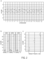
Figs. 2a-2c : Demonstration of FAST-SeqS reproducibility. (Fig. 2a ) Calculated z-scores for each autosome from eight normal plasma DNA samples. No chromosome had a z-score > 3.0 (range: -2.1 to 1.9). (Fig. 2b ) Comparison of z-scores from patients with trisomy 21 (n=4), trisomy 18 (n=2), and trisomy 13 (n=1) with eight normal spleen or WBC DNAs. The z-scores displayed represent the relevant
chromosome for the comparison. The maximum z-score observed for any of the compared normal chromosomes was 1.9 (chr13). (Fig. 2c ) Control WBC DNA was analyzed alone (z-score range: -0.8 to 1.3) or when mixed with DNA from a patient withtrisomy 21 at 4% (z-score range: 4.5 to 7.2) or 8% (z-score range: 8.9 to 10.) levels. Each experiment in (Fig. 2c ) was performed in quadruplicate. -
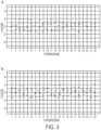
Fig. 3 (Supp.Fig. 1 ) Further demonstration of FAST-SeqS reproducibility using different instruments, samples, and sequencing depth. (Fig. 3a ) Matched peripheral blood white blood cell (WBC) DNA from eight samples whose plasma DNA was sequenced inFigure 2a was also sequenced on one-quarter of anIllumina HiSeq 2000 lane. Plotted are the calculated z-scores for each autosome. No chromosome had a z-score > 3.0 (range: -2.2 to 1.9). (Fig. 3b ) Eight samples of either splenic or WBC DNA were sequenced on one-half of an Illumina GA IIx lane, designed to yield less tags than the aforementioned plasma and WBC samples (Fig. 2a and (a)). Displayed are the z-scores calculated for each autosome. Despite three-fold less sequencing, no chromosome had a z-score > 3.0 (range: -2.2 to 2.1). -
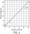
Fig. 4 (Supp.Fig. 2 ) Pilot mixing experiment oftrisomy 21 and euploid DNA. Control peripheral blood white blood cell (WBC) DNA was analyzed alone (n=2) or when mixed with DNA from a patient withtrisomy 21 at 5% (n=2), 10% (n=1), or 25% (n=1) levels. A tight correlation exists between the expected and observed fractions of extra chromosome 21 (r = 0.997 by Pearson correlation test, n=6). -
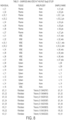
Fig. 5 . (Supplementary Table 1.) Samples analyzed in this FAST-SeqS study -
Fig. 6A-6B . (Supplementary Table 2) Sequencing characteristics of FAST-SeqS -
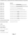
Fig. 7 . (Supplementary Table 3.) Oligonucleotides used to prepare and sequence FAST-SeqS samples (SEQ ID NO: 4-14 consecutively) -
Fig. 8 . (Table 1) Samples analyzed in this FAST-SeqS study - The inventors have developed a method that rapidly and non-invasively detects genetic abnormalities, in particular copy number abnormalities. We reasoned that the process in the commercially available test could be simplified if a defined number of fragments from throughout the genome could be amplified using a single primer pair, obviating the need for end-repair, terminal 3'-dA addition, or ligation to adapters. Furthermore, the smaller number of fragments to be assessed (compared to the whole genome) would streamline the genome matching and analysis processes. The method developed was capable of detecting trisomies in a reproducible fashion in pilot experiments. It has advantages over unbiased whole genome sequencing in ease of implementation, cost, analysis time, and throughput.
- Our approach to achieving the goals was based on the use of specific primers that anneal to a subset of human sequences dispersed throughout the genome. We named this approach "Fast Aneuploidy Screening Test-Sequencing System (henceforth FAST-SeqS). For maximum utility, we sought to identify regions with enough similarity so that they could be amplified with a single pair of primers, but sufficiently unique to allow most of the amplified loci to be distinguished. To be compatible with the degraded DNA found in plasma8, we further required that the amplified sequences be ≤ 150 bp. Using the BLAST-Like Alignment Tool (BLAT) algorithm13, we iteratively searched fragments of a small portion of chromosome 21 (~6.8 Mb) containing the Down's Syndrome critical region to identify suitable primer pairs, i.e., primer pairs that would amplify many distinct fragments of DNA from throughout the genome as well as throughout the Down's Syndrome critical region. Three such primer pairs were identified, and after testing these primers in silico (using In Silico PCR14) as well as in pilot sequencing experiments, we found one of the three primer pairs (henceforth FAST-1) to be optimal (Online Methods). The FAST-1 primer pair was predicted to amplify subfamilies of long interspersed nucleotide elements (L1 retrotransposons) which, like other human repeats, have spread throughout the genome via retrotransposition, particularly in AT-rich regions15. As it is generally more difficult to uniformly amplify and sequence regions that vary widely in their GC content8,16, we expected that this differential localization would work in our favor.
- Any chromosome or chromosome region can be queried. In accordance with the claimed invention, the query chromosomes and regions are
chromosome 21,chromosome 13, andchromosome 18. The query and reference chromosomes may be nuclear. In some embodiments at least two, three, four, or five amplicons tested match a genomic sequence within a query chromosome or query region. - The amplicons may be relatively small so that the degraded nature of some samples is not an impediment to detection. The are 180 bp or less, and may be less than 150 bp, less than 120 bp, less than 100 bp, or less than 50 bp. In one embodiment the pair of primers comprises a first and a second primer in which the first primer comprises SEQ ID NO: 1 and the second primer comprising SEQ ID NO: 2.
- The DNA sample is from a human. In certain embodiments the sample is from a gravid female and the DNA sample comprises maternal and fetal DNA. The DNA sample may be from plasma or from serum, for example. Other body fluids such as saliva, tears, sweat, lymph, urine, may be used. Tissue samples may be used as a source of DNA. Cord blood may be used as a source of DNA. Because fetal DNA is only a small fraction of maternal plasma DNA, maternal DNA could mask a fetal abnormality. Therefore a sufficient number of amplicons must be counted to detect fetal aneuploidy if present.
- Although the amplicons may be larger, the entire amplicon need not be sequenced to identify an amplicon. In accordance with the claimed invention at least 30 nt of nucleotide sequence is determined of the plurality of amplicons. More may be determined if desired.
- Useful primer pairs may be complementary to sequences that are distributed on at least 4, 5, 6, 7, 8, 9, 10, 11, 12, 13, 14, 15, 16, 17, 18, 19, 20, 21, 22, or 23 human chromosomes. The complementary sequences may be separated by at least 3, 4, 5, 6, 7, 8, 9, 10, 11, 12, 13, 14, 15, 16, 17, 18 or more nt.
- The above disclosure generally describes the present invention. A more complete understanding can be obtained by reference to the following specific examples which are provided herein for purposes of illustration only, and are not intended to limit the scope of the invention.
- We began by searching a ~6.8 Mb region of
chromosome 21 containing the Down's Syndrome critical region (hg19 17 coordinates 35,888,786 to 42,648,523) for sequence blocks of ~150 bp that were similar but not identical to those present on all chromosomes. Using 150 bp sliding windows incremented by 50 bp (135,186 sequences of 150 bp in length), we queried sequences with the BLAST-Like Alignment Tool (BLAT) algorithm13 to identify such blocks. We also required that at least three of the blocks were present onchromosome 21 in addition to the one within the ~6.8 Mb region described above. - Out of the 135,186 queried blocks, we found only 56 that met the following criteria:
- contained at least 11 variant bases from the query sequence, to aid in distinguishing amplified loci;
- contained no more than 30 variant bases from the query sequence, to increase the chance of uniform amplification; and
- spanned no more than a total of 180 bases, to be compatible with the analysis of degraded DNA.
- We then manually scanned the BLAT alignments of these 56 blocks to search for those that had highly similar 5' and 3' ends. At least three of the 56 sequences met our criteria and we designed primers for them. In Silico PCR14 verified that each theoretical primer pair would amplify many distinct sequences from every nuclear chromosome.
- Sequences that were too similar could pose a problem during alignment because of the inevitable errors introduced during library preparation or sequencing. We therefore wrote a custom script to assess how many distinct sequences would remain after allowing one, two, or three errors in each ~150 bp sequence. The theoretical amplification products of one primer pair (FAST-1) greatly outperformed the other two, and the superiority of FAST-1 was confirmed in pilot sequencing experiments. In Silico PCR14 predicted a bimodal distribution of PCR fragments, and this was confirmed by size separation of the amplified PCR products on a polyacrylamide gel and through the observed distribution of counts per position (
Fig. 1b ). - Control DNA was obtained from normal spleen, peripheral blood white blood cells (WBCs), or plasma (Supplementary Table 1). Fibroblast DNA from five individuals with trisomy 21 (NA02767, NA04616, NG05120, NG05397, and NG07438), two with trisomy 18 (NA03623 and NG12614), and one with trisomy 13 (NA03330) were purchased from the Coriell Institute for Medical Research (Camden, New Jersey). A total of 33 ng of DNA was used for each experiment. All templates were quantified by OD, except for the mixing experiments in which the templates were quantified by Digital PCR20 to achieve a more accurate estimate of concentration.
- The significant savings in time in FAST-SeqS is due to the replacement of a traditional whole genome amplification library preparation with an amplification using a single primer pair. Templates were amplified with FAST-1 primers as described by Kinde et al 21 in which individual template molecules are tagged with a unique identifier DNA sequence. Though the unique identifier sequences turned out to be not necessary for FAST-SeqS (see Precise template counting section below), we included them in the initial experimental design and continued to use them once they were observed to provide robust PCR products in our initial experiments. Briefly, the FAST-1 amplification primers each contained a different 5' universal primer sequence (UPS) followed by the sequences allowing amplification of the repeated elements described above (Forward: ACACAGGGAGGGGAACAT; SEQ ID NO: 1; Reverse: TGCCATGGTGGTTTGCT; SEQ ID NO: 2) (Supplementary Table 3). Additionally, the forward primer contained a stretch of 16-20 degenerate bases immediately 3' to its UPS (Supplementary Table 3). PCR was performed using Phusion Hot Start II Polymerase (Thermo Scientific, cat. no. F-549L) in 1X Phusion HF buffer, 0.5 µM each primer, and two units of polymerase in a total of 50 µL under the following cycling conditions: 980C for 120s, followed by two cycles of 980C for 10s, 570C for 120s, and 720C for 120s. The initial amplification primers were removed with AMPure XP beads (Beckman Coulter Genomics, cat. no. A63880) according to the manufacturer with the exception that the beads were added at only 1.4X the PCR volume and the elution volume was reduced to 10 uL of TE. The elution was used directly for a second round of amplification using primers that annealed to the UPS site introduced by the first round primers and that additionally contained the 5' grafting sequences necessary for hybridization to the Illumina flow cell (Supplementary Table 3). Further, we introduced one of five indexes ("barcodes") (Supplementary Table 3) to each sample in the reverse primer to later allow multiplexed sequencing. The second round of PCR was performed using Phusion Hot Start II Polymerase in 1X Phusion HF buffer, 0.5 µM each primer, and two units of polymerase in a total of 50 µL under the following cycling conditions: 980C for 120s, followed by 13 cycles of 980C for 10s, 650C for 15s, and 720C for 15s. Amplification products were again purified with AMPure XP beads and were
quantified by spectrophotometry, real time PCR or on an Agilent 2100 Bioanalyzer; all methods of quantification yielded similar results. Oligonucleotides were purchased from IDT (Coralville, Iowa). - As opposed to traditional whole genome amplification libraries, where the vast majority of tags align to the genome in unique positions and thus each tag needs an independent alignment, FAST-SeqS yields sequences that aligned to an average of only 21,676 positions; Supplementary Table 2). The number of positions to which the sequences aligned varied little compared to the range of sequence data obtained across all experiments. Though the number of uniquely aligned tags per experiment spanned a 12-fold range (1,343,382 to 16,015,347) the number of positions varied only by 0.25-fold (range: 18,484 to 24,562 positions; Supplementary Table 2). Raw reads from all experiments (Supplementary Table 2) can be downloaded from the domain sagenet.org, subdomain fast, document fast.htm.
- Thirty-seven base sequence tags passing the Illumina chastity filter and containing at least three correct terminal bases of the amplification primer were filtered for quality by masking any base with a quality score <20 with an N using a custom script. Thus, tags with low quality bases were given the opportunity to align by considering only their most reliable bases. After quality masking, only the distinct sequences were aligned to the human genome (hg19 17) using Bowtie 0.12.718. When building the reference index for Bowtie, we included unresolved or unplaced contigs22 to ensure the most accurate alignments. Sequences that aligned uniquely with up to one mismatch (using the flags -
m 1 and -v 1, respectively) were retained and their alignments were matched back to the original data. An average of 38% of tags across all samples could be uniquely assigned to a genomic position (range: 31% to 45%; Supplementary Table 2). - After confirming the in silico prediction of a primarily bimodal distribution of FAST-SeqS amplification products by gel electrophoresis, we investigated whether the counts of sequenced fragments that aligned to unique positions were similarly distributed. Though we only sequenced 37 bases, we could estimate the relative size of each tag from In Silico PCR14 and its unique position in the genome. This exercise could provide additional evidence that the actual amplification products matched those that were predicted and could alert us to any amplification bias (see Normalization section below).
- First, we transformed the tag counts per uniquely aligned position to a log scale, a transformation frequently performed to this class of data to induce symmetry23. We performed this transformation for each group of experiments (e.g., from eight normal plasma samples analyzed in the same instrument run; Supplementary Table 2). Next, we used a nonparametric method to estimate a smoothened distribution (a kernel density estimator, implemented in R24 using the density function), which made it straightforward to visualize the modality of our data. After plotting the distribution using ggplot2 25 (an R24 package), we observed that each group of experiments showed a similar clustering of tag counts per position, consistent with a primarily bimodal distribution with a negative skew. A representative plot is displayed in
Figure 1b . - Massively parallel sequencing will generate a different number of sequence tags from each sample, as well as from different sequencing runs of the same sample, due to stochastic and experimental variations. Thus, it is essential to normalize the data to make meaningful comparisons of the type used here. Although it would be most straightforward to simply express tag counts as a fraction of the total number of tags sequenced in an experiment, this normalization is too simplistic and is highly susceptible to systemic biases that frequently plague next generation sequencing of both DNA and RNA templates, and these are routinely used in digital karyotyping analyses such as that used for the diagnosis of
trisomy 218,16. - Because of the bimodal size distribution of the amplicons obtained with the FAST-1 primer pair, we predicted that the majority of bias in FAST-1 amplifications would be due to the potential over-representation of the smaller-sized fragments. This bias could either occur during library preparation or during solid-phase bridge PCR on the Illumina flow cell. We found that an appropriate normalization for this distribution could be obtained using the quantile method19, used extensively within the microarray community. By organizing our data into a list of positions (equivalent to probes in microarray data), each associated with a tag count (equivalent to intensities in microarray data), we were able to apply standard quantile normalization to FAST-SeqS data. To best approximate the microarray data format, we chose to only analyze positions that were shared within each experimental group (e.g., the data from eight normal plasma samples). As the FAST-1 primers amplified a highly reproducible set of positions, this generally only eliminated <1% of the data. To maximize reproducibility, we excluded positions aligning to unresolved or unplaced contigs and those aligning to sex chromosomes, although inclusion of these chromosomes only marginally increased variability between experiments (e.g., in eight normal plasma samples, the maximum z-score from any chromosome rose from 1.9 to 2.3). The inclusion of sex chromosomes could be useful for other applications, such as detecting aneuploidies involving chromosome X or determining the gender of a sample (i.e., by the presence or absence of sequences aligning to chromosome Y).
- We implemented the quantile normalization19 for each experimental group (each of which contained multiple samples; Supplementary Table 2) by performing the following steps:
- generating a sorted array of tag counts representing each position for every sample (all of equal length as only the shared positions in each experiment were analyzed);
- combining these sorted arrays into a 2×2 matrix, where each experiment is represented in its own column and the shared positions constitute the rows;
- replacing an individual sample's count with the mean count for all samples at that particular row; and
- re-sorting the counts back to their original order.
- The distribution of our data was always negatively skewed (see
Fig. 1b for a representative example). We excluded the positions falling within the left tail of each experiment's distribution (the positions containing the smallest number of tags) from our analysis by: - estimating the distribution of normalized values as described above;
- determining the inflection point between the two peaks of the bimodal distribution; and
- considering the positions that had a relative density below the inflection point as the left tail.
- Once the left tail was determined and positions within it discarded, the quantile normalization was repeated. Through this process, each sample within an experimental group had the same sum total of tags and an identical distribution of counts, so direct comparisons could be made. We automated the quantile normalization in R24. The entire normalization procedure routinely took less than a few minutes to complete.
- A common method of determining the aneuploidy status of a particular sample in Digital Karyotyping-based11 assays is by comparison of z-scores6,8,26. Through this method, one determines the mean and standard deviation of tag counts lying within a chromosome of interest in a group of reference samples (e.g., samples with known euploid content), and then creates a standardized score (i.e., z-score) for a chromosome of interest for each sample as follows: z-scorei,chrN = (chrNi - µchrN) / sdchrN, where i represents the sample to be standardized, chrN represents the normalized tag count of the sample's chromosome, and µchrN and sdchrN represent the mean and standard deviation of the normalized tag counts, respectively, of chrN in the reference group. When all samples are standardized in this way, outliers are easily detected because they have a z-score > 3.0. This indicates that the normalized tag count of the outlier exceeds the mean of the reference group by at least three standard deviations.
- Finally, we evaluated whether precisely counting template molecules could further increase reproducibility. By incorporating 16-20 degenerate bases at the 5' end of one of the two FAST-1 primers (Supplementary Table 3), it is possible to uniquely identify each template molecule giving rise to a PCR product21. This could potentially increase accuracy by minimizing the possibility that the same template molecule was counted more than once in the final tally for each chromosome. We found that this enhancement did not significantly alter the consistency of normalized counts per chromosome among the eight normal plasma samples: the maximum z-score for any chromosome was slightly increased from 1.9 to 2.0. By performing a two-tailed t-test on the absolute values of the z-scores for all autosomes comparing analysis methods, we found no statistically significant difference between the two methods (p=0.759, n=22×8 for each group).
- As an initial test of the performance of FAST-SeqS, we examined the representation of each autosome in the plasma DNA of seven normal females, including one biologic replicate (Supplementary Table 1). Using only 37 cycles of sequencing in one-quarter of a lane on an
Illumina HiSeq 2000 instrument, we recovered an average of 31,547,988 high quality tags per individual (range: 27,179,424 to 36,0418,017 tags; Supplementary Table 2). An average of 35% of these tags (range: 31 to 37%) could be uniquely mapped to one of an average of 23,681 unique chromosomal positions (range: 22,589 to 24,562 positions) when allowing up to one mismatch during alignment to hg1917 using Bowtie18. The theoretical in silico (Fig. 1a ) and observed distribution of tag counts (Fig. 1b ) both showed a bimodal distribution of sizes. Of the uniquely aligned tags, 99.1% aligned to positions predicted to be repetitive DNA by RepeatMasker (http://www.repeatmasker.org), 97.5% of which fell into just seven L1 retrotransposon subfamilies (Fig. 1c ). Additionally, the distribution of each subfamily agreed with that predicted by RepeatMasker (Fig. 1d ). Because tag alignment to a discrete set of chromosomal positions is simpler than alignment to the entire genome, the post-sequencing analysis process was very rapid. In fact, this mapping plus subsequent statistical analysis could be completed in less than 30 min per sample with a single computer housing two six-core CPUs (Intel Xeon X5680). - Most importantly, the relative fraction of tags mapping to each chromosome was remarkably similar among the individual samples after normalizing19 to compare chromosome tag counts among different samples (Online Methods). In particular, the fraction of tags that matched to any of the autosomes in any of the eight samples studied never deviated from the average by a z-score > 3.0 (
Fig. 2a ). Of particular note, the maximum z-scores observed among the eight samples forchromosomes - We next studied the reproducibility of chromosome representation in two additional experiments employing different types of samples, different instruments, and different depths of sequencing. In the first experiment, we analyzed DNA from peripheral blood white blood cells (WBCs) from the same seven individuals who contributed plasma. Four samples were sequenced on a single lane of an
Illumina HiSeq 2000, yielding a mean of 10,835,559 uniquely aligned tags per sample (range: 4,905,067 to 16,015,347 tags). The maximum z-scores for any of the samples were 1.0, 1.2, and 1.6 forchromosomes Fig. 1a ). - In the next experiment, we analyzed splenic or WBC DNA from an additional eight individuals using one-half of a lane of an Illumina GA IIx instrument per sample (Supplementary Table 2). We obtained a mean of 4,013,951 uniquely aligned tags per sample (range: 2,847,661 to 4,645,608 tags). Despite almost 3-fold less sequencing, the maximum z-scores among the samples were still only 1.3, 1.5, and 1.9 for
chromosomes Fig. 1b ). - Given the tight distributions of tags evident in
Fig. 2a , we expected it would be straightforward to distinguish the DNA of patients with trisomies from those of normal individuals with euploid chromosome constitutions. The data depicted inFigure 2b demonstrate that this expectation was realized in each of four patients withtrisomy 21. The z-scores among thesetrisomy 21 patients ranged from 32 to 36, while the maximum z-score among eight normal individuals was 1.3. Similarly, the z-scores of DNA from two patients withtrisomy 18 and one fromtrisomy 13 were 51, 56, and 36, respectively, far exceeding the maximum z-scores for these chromosomes in normal individuals (Fig. 2b ). - Fetal DNA accounts for a geometric mean of 13.4% of maternal DNA, depending largely on maternal weight rather than gestational age8. To investigate whether FAST-SeqS could distinguish samples that contained mixtures of disomic and trisomic DNA, we performed mixing experiments using DNA from patients with
trisomy 21 and normal individuals. In a first experiment of this type, we mixed 5% (n=2), 10% (n=1), and 25% (n=1)trisomy 21 DNA into normal WBC DNA alongside two controls (SupplementaryFig. 2 ), and found a tight correlation between the expected and observed fractions of extra chromosome 21 (r=0.997 by Pearson correlation test, n=6). In a second experiment, we evaluated mixtures that contained 4% or 8% trisomy 21 DNA. As shown inFigure 2c , there was a clear distinction between the samples containing 4% trisomy 21 DNA vs. those from normal individuals (p= 2×10⁻⁴ as determined by two-tailed t-test, n=4 in each group). The samples containing 8% trisomy 21 DNA were of course even more easily distinguishable (p=4×10⁻⁶ when compared to the euploid group and p=1×10⁻³ when compared to the 4% trisomy 21 samples, both by two-tailed t-test with n=4 for each group). - The disclosure of each reference cited is expressly incorporated herein.
- 1. LYF Hsu, in Genetic Disorders and the Fetus, edited by A Milunsky (The Johns Hopkins University Press, Baltimore, 1998), pp. 179.
- 2. M. Staebler, C. Donner, N. Van Regemorter et al., Prenat Diagn 25 (7), 567 (2005).
- 3. K.L. Jones, Smith's recognizable patterns of human malformation, 6 ed. (Elsevier Saunders, Philadelphia, 2006).
- 4. American College of Obstetricians and Gynecologists, Obstet Gynecol 110 (6), 1459 (2007).
- 5. Y. M. Lo, M. S. Tein, T. K. Lau et al., Am J Hum Genet 62 (4), 768 (1998).
- 6. R. W. Chiu, K. C. Chan, Y. Gao et al., Proc Natl Acad Sci U S A 105 (51), 20458 (2008).
- 7. H. C. Fan, Y. J. Blumenfeld, U. Chitkara et al., Proc Natl Acad Sci U S A 105 (42), 16266 (2008).
- 8. G. E. Palomaki, E. M. Kloza, G. M. Lambert-Messerlian etal., Genet Med 13 (11), 913 (2011).
- 9. J. Cleary-Goldman, F. D. Malone, J. Vidaver et al., Obstet Gynecol 105 (5 Pt 1), 983 (2005).
- 10. R. G. Resta, Am J Med GenetA 133A (1), 31 (2005).
- 11. T. L. Wang, C. Maierhofer, M. R. Speicher et al., Proc Natl Acad Sci U S A 99 (25), 16156 (2002).
- 12. A. B. Sparks, E. T. Wang, C. A. Struble et al., Prenat Diagn (2012).
- 13. W. J. Kent, Genome Res 12 (4), 656 (2002).
- 14. R. M. Kuhn, D. Karolchik, A. S. Zweig et al., Nucleic Acids Res 35 (Database issue), D668 (2007).
- 15. A. F. Smit, Curr Opin Genet Dev 9 (6), 657 (1999).
- 16. H. C. Fan and S. R. Quake, PLoS One 5 (5), e10439 (2010).
- 17. P. A. Fujita, B. Rhead, A. S. Zweig et al., Nucleic Acids Res 39 (Database issue), D876 (2011).
- 18. B. Langmead, C. Trapnell, M. Pop et al., Genome Biol 10 (3), R25 (2009).
- 19. B. M. Bolstad, R. A. Irizarry, M. Astrand et al., Bioinformatics 19 (2), 185 (2003).
- 20. B. Vogelstein and K. W. Kinzler, Proc Natl Acad Sci U S A 96 (16), 9236 (1999).
- 21. I. Kinde, J. Wu, N. Papadopoulos et al., Proc Natl Acad Sci U S A 108 (23), 9530 (2011).
- 22. E. S. Lander, L. M. Linton, B. Birren et al., Nature 409 (6822), 860 (2001).
- 23. J. Tukey, Exploratory Data Analysis. (Addison-Wesley, Reading, Massachusetts, 1977).
- 24. R Development Core Team, R: A language and environment for statistical computing (R Foundation for Statistical Computing, 2011).
- 25. H. Wickham, ggplot2: elegant graphics for data analysis. (Springer, New York, 2009).
- 26. M. Ehrich, C. Deciu, T. Zwiefelhofer et al., Am J Obstet Gynecol 204 (3), 205 e1 (2011).
Claims (14)
- A method of testing a human for aneuploidy comprising:amplifying using polymerase chain reaction a plurality of chromosomal sequences in a DNA sample from a human with a single pair of primers complementary to said plurality of chromosomal sequences to form a plurality of amplicons which are not identical, wherein(i) each amplicon is 180bp or less;(ii) the plurality of amplicons include sequences on query chromosomes 21, 13 and 18; and(iii) the amplicons comprise said chromosomal sequences complementary to the single pair of primers and the sequences that intervene between the pair of primers;performing reactions to determine the nucleotide sequence of at least 30 nt of the plurality of amplicons;matching amplicon nucleotide sequences in silico to human genomic sequences at a discrete set of genomic loci;counting the number of matching amplicons at individual genomic loci from the query chromosomes;comparing the number of amplicons matched to genomic loci from at least one of the query chromosomes to the number of amplicons matched to the same genomic loci on one or more reference chromosomes having known euploid content.
- The method of claim 1 wherein the at least one query chromosome is chromosome 21.
- The method of claim 1 wherein the at least one query chromosome is chromosome 13.
- The method of claim 1 wherein the pair of primers comprises a first and a second primer in which the first primer comprises SEQ ID NO: 1 and the second primer comprises SEQ ID NO: 2.
- The method of claim 2 wherein:(i) at least one of the amplicons matches a genomic sequence within Down's Syndrome critical region on chromosome 21; or(ii) at least three of the amplicons match a genomic sequence on chromosome 21.
- The method of claim 1 wherein the DNA sample is from a gravid female and the DNA sample comprises maternal and fetal DNA.
- The method of claim 6 wherein:(i) the DNA sample is from plasma;(ii) the DNA sample is from serum; or(iii) sufficient amplicons are counted to detect fetal aneuploidy if present.
- The method of claim 1 wherein:(i) the DNA sample is from plasma; or(ii) the DNA sample is from serum.
- A method of testing a human for DNA copy number changes, comprising:amplifying using polymerase chain reaction a plurality of chromosomal sequences in a DNA sample from a human with a single pair of primers complementary to said plurality of chromosomal sequences to form a plurality of amplicons which are not identical, wherein(i) each amplicon is 180bp or less;(ii) the plurality of amplicons include sequences in query chromosomal regions, wherein the query chromosomal regions are chromosomes 21, 13 and 18; and(iii) the amplicons comprise chromosomal sequences complementary to the single pair of primers and the sequences that intervene between the pair of primers;performing reactions to determine the nucleotide sequence of at least 30 nt of the plurality of amplicons;matching amplicon nucleotide sequences in silico to human genomic sequences at a discrete set of genomic loci;counting the number of matching amplicons at individual genomic loci from the query chromosomes;comparing the number of amplicons matched to genomic loci from at least one of the query chromosomal regions to the number of amplicons matched to the same genomic loci on one or more reference chromosomal regions of one or more chromosomes having known euploid content.
- The method of claim 9 wherein the at least one query chromosomal region is chromosome 21.
- The method of claim 9, wherein the pair of primers comprises a first and a second primer in which the first primer comprises SEQ ID NO: 1 and the second primer comprises SEQ ID NO: 2.
- The method of claim 9 wherein the DNA sample is from a gravid female and the DNA sample comprises maternal and fetal DNA.
- The method of claim 12 wherein:(i) the DNA sample is from plasma; or(ii) the DNA sample is from serum.
- A pair of primers useful for analyzing human aneuploidy, wherein a first primer comprises SEQ ID NO: 1 and a second primer comprises SEQ ID NO: 2.
Priority Applications (1)
| Application Number | Priority Date | Filing Date | Title |
|---|---|---|---|
| EP23170939.5A EP4239081A3 (en) | 2012-03-26 | 2013-03-22 | Rapid aneuploidy detection |
Applications Claiming Priority (3)
| Application Number | Priority Date | Filing Date | Title |
|---|---|---|---|
| US201261615535P | 2012-03-26 | 2012-03-26 | |
| US201261659695P | 2012-06-14 | 2012-06-14 | |
| PCT/US2013/033451 WO2013148496A1 (en) | 2012-03-26 | 2013-03-22 | Rapid aneuploidy detection |
Related Child Applications (1)
| Application Number | Title | Priority Date | Filing Date |
|---|---|---|---|
| EP23170939.5A Division EP4239081A3 (en) | 2012-03-26 | 2013-03-22 | Rapid aneuploidy detection |
Publications (3)
| Publication Number | Publication Date |
|---|---|
| EP2831279A1 EP2831279A1 (en) | 2015-02-04 |
| EP2831279A4 EP2831279A4 (en) | 2015-12-30 |
| EP2831279B1 true EP2831279B1 (en) | 2023-05-03 |
Family
ID=49261127
Family Applications (2)
| Application Number | Title | Priority Date | Filing Date |
|---|---|---|---|
| EP23170939.5A Pending EP4239081A3 (en) | 2012-03-26 | 2013-03-22 | Rapid aneuploidy detection |
| EP13768366.0A Active EP2831279B1 (en) | 2012-03-26 | 2013-03-22 | Rapid aneuploidy detection |
Family Applications Before (1)
| Application Number | Title | Priority Date | Filing Date |
|---|---|---|---|
| EP23170939.5A Pending EP4239081A3 (en) | 2012-03-26 | 2013-03-22 | Rapid aneuploidy detection |
Country Status (11)
| Country | Link |
|---|---|
| US (3) | US10053729B2 (en) |
| EP (2) | EP4239081A3 (en) |
| CN (2) | CN104350158A (en) |
| AU (1) | AU2013240088B2 (en) |
| CA (1) | CA2868836C (en) |
| ES (1) | ES2945311T3 (en) |
| FI (1) | FI2831279T3 (en) |
| HK (2) | HK1205204A1 (en) |
| IL (1) | IL234850B (en) |
| PL (1) | PL2831279T3 (en) |
| WO (1) | WO2013148496A1 (en) |
Families Citing this family (40)
| Publication number | Priority date | Publication date | Assignee | Title |
|---|---|---|---|---|
| JP2012525147A (en) | 2009-04-30 | 2012-10-22 | グッド スタート ジェネティクス, インコーポレイテッド | Methods and compositions for assessing genetic markers |
| WO2010127186A1 (en) | 2009-04-30 | 2010-11-04 | Prognosys Biosciences, Inc. | Nucleic acid constructs and methods of use |
| US20190300945A1 (en) | 2010-04-05 | 2019-10-03 | Prognosys Biosciences, Inc. | Spatially Encoded Biological Assays |
| DK2556171T3 (en) | 2010-04-05 | 2015-12-14 | Prognosys Biosciences Inc | Spatially CODED BIOLOGICAL ASSAYS |
| US10787701B2 (en) | 2010-04-05 | 2020-09-29 | Prognosys Biosciences, Inc. | Spatially encoded biological assays |
| US9163281B2 (en) | 2010-12-23 | 2015-10-20 | Good Start Genetics, Inc. | Methods for maintaining the integrity and identification of a nucleic acid template in a multiplex sequencing reaction |
| GB201106254D0 (en) | 2011-04-13 | 2011-05-25 | Frisen Jonas | Method and product |
| EP3246416A1 (en) | 2011-04-15 | 2017-11-22 | The Johns Hopkins University | Safe sequencing system |
| US9228233B2 (en) | 2011-10-17 | 2016-01-05 | Good Start Genetics, Inc. | Analysis methods |
| US8209130B1 (en) | 2012-04-04 | 2012-06-26 | Good Start Genetics, Inc. | Sequence assembly |
| US10227635B2 (en) | 2012-04-16 | 2019-03-12 | Molecular Loop Biosolutions, Llc | Capture reactions |
| US11913065B2 (en) | 2012-09-04 | 2024-02-27 | Guardent Health, Inc. | Systems and methods to detect rare mutations and copy number variation |
| US10876152B2 (en) | 2012-09-04 | 2020-12-29 | Guardant Health, Inc. | Systems and methods to detect rare mutations and copy number variation |
| US20160040229A1 (en) | 2013-08-16 | 2016-02-11 | Guardant Health, Inc. | Systems and methods to detect rare mutations and copy number variation |
| KR102028375B1 (en) | 2012-09-04 | 2019-10-04 | 가던트 헬쓰, 인크. | Systems and methods to detect rare mutations and copy number variation |
| EP2912468B1 (en) | 2012-10-29 | 2018-09-12 | The Johns Hopkins University | Papanicolaou test for ovarian and endometrial cancers |
| WO2014152421A1 (en) | 2013-03-14 | 2014-09-25 | Good Start Genetics, Inc. | Methods for analyzing nucleic acids |
| CN111662960B (en) | 2013-06-25 | 2024-04-12 | 普罗格诺西斯生物科学公司 | Spatially encoded bioanalytical analysis using microfluidic devices |
| US10851414B2 (en) | 2013-10-18 | 2020-12-01 | Good Start Genetics, Inc. | Methods for determining carrier status |
| AU2014369841B2 (en) | 2013-12-28 | 2019-01-24 | Guardant Health, Inc. | Methods and systems for detecting genetic variants |
| WO2015175530A1 (en) | 2014-05-12 | 2015-11-19 | Gore Athurva | Methods for detecting aneuploidy |
| WO2016040446A1 (en) | 2014-09-10 | 2016-03-17 | Good Start Genetics, Inc. | Methods for selectively suppressing non-target sequences |
| JP2017536087A (en) | 2014-09-24 | 2017-12-07 | グッド スタート ジェネティクス, インコーポレイテッド | Process control to increase the robustness of genetic assays |
| JP2017530720A (en) * | 2014-10-17 | 2017-10-19 | グッド スタート ジェネティクス, インコーポレイテッド | Preimplantation genetic screening and aneuploidy detection |
| CA3010579A1 (en) | 2015-01-06 | 2016-07-14 | Good Start Genetics, Inc. | Screening for structural variants |
| EP3271481B1 (en) | 2015-01-15 | 2020-04-08 | Good Start Genetics, Inc. | Methods of quality control using single-nucleotide polymorphisms in pre-implantation genetic screening |
| DK3901281T3 (en) | 2015-04-10 | 2023-01-23 | Spatial Transcriptomics Ab | SPATIALLY SEPARATE, MULTIPLEX NUCLEIC ACID ANALYSIS OF BIOLOGICAL SAMPLES |
| WO2017027653A1 (en) | 2015-08-11 | 2017-02-16 | The Johns Hopkins University | Assaying ovarian cyst fluid |
| SG11201805119QA (en) | 2015-12-17 | 2018-07-30 | Guardant Health Inc | Methods to determine tumor gene copy number by analysis of cell-free dna |
| CN107699607A (en) * | 2016-08-31 | 2018-02-16 | 刘红彦 | A kind of kit, method and purposes for building fetus dissociative DNA sequencing library |
| AU2018342007A1 (en) | 2017-08-07 | 2020-02-27 | Board Of Regents, The University Of Texas Systems | Methods and materials for assessing and treating cancer |
| CN112041460A (en) * | 2018-02-28 | 2020-12-04 | 克罗玛科德公司 | Molecular targets for fetal nucleic acid analysis |
| CN108841931B (en) * | 2018-07-05 | 2021-06-11 | 广州市达瑞生物技术股份有限公司 | Primer group and detection kit for detecting STR locus of human chromosome 4 and application of primer group and detection kit |
| CN108913757B (en) * | 2018-07-05 | 2021-06-11 | 广州市达瑞生物技术股份有限公司 | Primer group and detection kit for chromosome aneuploid number abnormality and application thereof |
| US11100087B2 (en) * | 2019-04-26 | 2021-08-24 | Microsoft Technology Licensing, Llc | Data tokenization system maintaining data integrity |
| US20220259668A1 (en) | 2019-05-17 | 2022-08-18 | The Johns Hopkins University | Rapid aneuploidy detection |
| WO2020243579A1 (en) | 2019-05-30 | 2020-12-03 | 10X Genomics, Inc. | Methods of detecting spatial heterogeneity of a biological sample |
| EP4025692A2 (en) | 2020-06-02 | 2022-07-13 | 10X Genomics, Inc. | Nucleic acid library methods |
| EP4158054A1 (en) | 2020-06-02 | 2023-04-05 | 10X Genomics, Inc. | Spatial transcriptomics for antigen-receptors |
| WO2021252499A1 (en) | 2020-06-08 | 2021-12-16 | 10X Genomics, Inc. | Methods of determining a surgical margin and methods of use thereof |
Citations (1)
| Publication number | Priority date | Publication date | Assignee | Title |
|---|---|---|---|---|
| WO2011087760A2 (en) * | 2009-12-22 | 2011-07-21 | Sequenom, Inc. | Processes and kits for identifying aneuploidy |
Family Cites Families (25)
| Publication number | Priority date | Publication date | Assignee | Title |
|---|---|---|---|---|
| CA43460A (en) | 1893-07-03 | Robert Wellington Bigger | Furnace | |
| CA57345A (en) | 1897-07-30 | 1897-09-04 | Francis Louis Becker | Upright piano-forte action |
| CA62924A (en) | 1898-12-27 | 1899-03-25 | Azarie Mireault | Medicinal compound |
| GB9704444D0 (en) | 1997-03-04 | 1997-04-23 | Isis Innovation | Non-invasive prenatal diagnosis |
| US6703228B1 (en) * | 1998-09-25 | 2004-03-09 | Massachusetts Institute Of Technology | Methods and products related to genotyping and DNA analysis |
| US6977162B2 (en) | 2002-03-01 | 2005-12-20 | Ravgen, Inc. | Rapid analysis of variations in a genome |
| US20060172294A1 (en) * | 2002-06-06 | 2006-08-03 | Arturas Petronis | Detection of epigenetic abnormalities and diagnostic method based thereon |
| US7704687B2 (en) * | 2002-11-15 | 2010-04-27 | The Johns Hopkins University | Digital karyotyping |
| CA2510587A1 (en) * | 2002-12-20 | 2004-07-15 | Qiagen Gmbh | Nucleic acid amplification |
| EP1664077B1 (en) | 2003-09-05 | 2016-04-13 | Trustees of Boston University | Method for non-invasive prenatal diagnosis |
| US7537889B2 (en) | 2003-09-30 | 2009-05-26 | Life Genetics Lab, Llc. | Assay for quantitation of human DNA using Alu elements |
| CN101985619B (en) | 2003-10-08 | 2014-08-20 | 波士顿大学信托人 | Methods for prenatal diagnosis of chromosomal abnormalities |
| US20060019270A1 (en) * | 2004-04-01 | 2006-01-26 | Board Of Regents The University Of Texas System | Global DNA methylation assessment using bisulfite PCR |
| US7709194B2 (en) | 2004-06-04 | 2010-05-04 | The Chinese University Of Hong Kong | Marker for prenatal diagnosis and monitoring |
| JP5219516B2 (en) | 2005-03-18 | 2013-06-26 | ザ チャイニーズ ユニバーシティー オブ ホンコン | Chromosome aneuploidy detection method |
| US7888017B2 (en) | 2006-02-02 | 2011-02-15 | The Board Of Trustees Of The Leland Stanford Junior University | Non-invasive fetal genetic screening by digital analysis |
| KR20080107464A (en) | 2006-03-06 | 2008-12-10 | 더 트러스티이스 오브 콜롬비아 유니버시티 인 더 시티 오브 뉴욕 | Specific amplification of fetal dna sequences from a mixed, fetal-maternal source |
| US8137912B2 (en) | 2006-06-14 | 2012-03-20 | The General Hospital Corporation | Methods for the diagnosis of fetal abnormalities |
| PT2557517T (en) * | 2007-07-23 | 2023-01-04 | Univ Hong Kong Chinese | Determining a nucleic acid sequence imbalance |
| US20100112590A1 (en) | 2007-07-23 | 2010-05-06 | The Chinese University Of Hong Kong | Diagnosing Fetal Chromosomal Aneuploidy Using Genomic Sequencing With Enrichment |
| SI2334812T1 (en) * | 2008-09-20 | 2017-05-31 | The Board of Trustees of the Leland Stanford Junior University Office of the General Counsel Building 170 | Noninvasive diagnosis of fetal aneuploidy by sequencing |
| US8563242B2 (en) | 2009-08-11 | 2013-10-22 | The Chinese University Of Hong Kong | Method for detecting chromosomal aneuploidy |
| WO2011066476A1 (en) * | 2009-11-25 | 2011-06-03 | Quantalife, Inc. | Methods and compositions for detecting genetic material |
| EP2366031B1 (en) * | 2010-01-19 | 2015-01-21 | Verinata Health, Inc | Sequencing methods in prenatal diagnoses |
| EP2569447A4 (en) * | 2010-05-14 | 2013-11-27 | Fluidigm Corp | Assays for the detection of genotype, mutations, and/or aneuploidy |
-
2013
- 2013-03-22 EP EP23170939.5A patent/EP4239081A3/en active Pending
- 2013-03-22 ES ES13768366T patent/ES2945311T3/en active Active
- 2013-03-22 AU AU2013240088A patent/AU2013240088B2/en active Active
- 2013-03-22 CA CA2868836A patent/CA2868836C/en active Active
- 2013-03-22 CN CN201380026625.9A patent/CN104350158A/en active Pending
- 2013-03-22 EP EP13768366.0A patent/EP2831279B1/en active Active
- 2013-03-22 US US14/388,314 patent/US10053729B2/en active Active
- 2013-03-22 CN CN202010003298.7A patent/CN111073962A/en active Pending
- 2013-03-22 PL PL13768366.0T patent/PL2831279T3/en unknown
- 2013-03-22 WO PCT/US2013/033451 patent/WO2013148496A1/en active Application Filing
- 2013-03-22 FI FIEP13768366.0T patent/FI2831279T3/en active
-
2014
- 2014-09-28 IL IL234850A patent/IL234850B/en active IP Right Grant
-
2015
- 2015-06-17 HK HK15105795.9A patent/HK1205204A1/en unknown
- 2015-07-07 US US14/792,716 patent/US20150376691A1/en not_active Abandoned
- 2015-07-30 HK HK15107297.8A patent/HK1206795A1/en unknown
-
2021
- 2021-09-23 US US17/483,537 patent/US20220010371A1/en active Pending
Patent Citations (1)
| Publication number | Priority date | Publication date | Assignee | Title |
|---|---|---|---|---|
| WO2011087760A2 (en) * | 2009-12-22 | 2011-07-21 | Sequenom, Inc. | Processes and kits for identifying aneuploidy |
Also Published As
| Publication number | Publication date |
|---|---|
| CA2868836A1 (en) | 2013-10-03 |
| US20150051085A1 (en) | 2015-02-19 |
| EP4239081A3 (en) | 2023-11-08 |
| WO2013148496A1 (en) | 2013-10-03 |
| EP2831279A4 (en) | 2015-12-30 |
| PL2831279T3 (en) | 2023-12-04 |
| US20220010371A1 (en) | 2022-01-13 |
| US20150376691A1 (en) | 2015-12-31 |
| AU2013240088B2 (en) | 2017-02-02 |
| EP2831279A1 (en) | 2015-02-04 |
| EP4239081A2 (en) | 2023-09-06 |
| HK1205204A1 (en) | 2015-12-11 |
| CA2868836C (en) | 2019-08-06 |
| IL234850A0 (en) | 2014-12-31 |
| CN104350158A (en) | 2015-02-11 |
| ES2945311T3 (en) | 2023-06-30 |
| AU2013240088A1 (en) | 2014-10-16 |
| CN111073962A (en) | 2020-04-28 |
| FI2831279T3 (en) | 2023-05-23 |
| US10053729B2 (en) | 2018-08-21 |
| HK1206795A1 (en) | 2016-01-15 |
| IL234850B (en) | 2019-08-29 |
Similar Documents
| Publication | Publication Date | Title |
|---|---|---|
| US20220010371A1 (en) | Rapid aneuploidy detection | |
| JP2022173465A (en) | Diagnosing fetal chromosomal aneuploidy using genomic sequencing | |
| US11142799B2 (en) | Detecting chromosomal aberrations associated with cancer using genomic sequencing | |
| Kinde et al. | FAST-SeqS: a simple and efficient method for the detection of aneuploidy by massively parallel sequencing | |
| AU2013203077B2 (en) | Diagnosing fetal chromosomal aneuploidy using genomic sequencing | |
| AU2008278843B2 (en) | Diagnosing fetal chromosomal aneuploidy using genomic sequencing | |
| AU2013200581A1 (en) | Diagnosing cancer using genomic sequencing |
Legal Events
| Date | Code | Title | Description |
|---|---|---|---|
| PUAI | Public reference made under article 153(3) epc to a published international application that has entered the european phase |
Free format text: ORIGINAL CODE: 0009012 |
|
| 17P | Request for examination filed |
Effective date: 20140930 |
|
| AK | Designated contracting states |
Kind code of ref document: A1 Designated state(s): AL AT BE BG CH CY CZ DE DK EE ES FI FR GB GR HR HU IE IS IT LI LT LU LV MC MK MT NL NO PL PT RO RS SE SI SK SM TR |
|
| AX | Request for extension of the european patent |
Extension state: BA ME |
|
| DAX | Request for extension of the european patent (deleted) | ||
| RA4 | Supplementary search report drawn up and despatched (corrected) |
Effective date: 20151130 |
|
| RIC1 | Information provided on ipc code assigned before grant |
Ipc: C12N 15/11 20060101ALI20151124BHEP Ipc: C12Q 1/68 20060101AFI20151124BHEP Ipc: G01N 33/53 20060101ALI20151124BHEP Ipc: G01N 33/48 20060101ALI20151124BHEP |
|
| REG | Reference to a national code |
Ref country code: HK Ref legal event code: DE Ref document number: 1206795 Country of ref document: HK |
|
| RIN1 | Information on inventor provided before grant (corrected) |
Inventor name: VOGELSTEIN, BERT Inventor name: KINDE, ISAAC Inventor name: KINZLER, KENNETH W. Inventor name: PAPADOPOULOS, NICKOLAS |
|
| 17Q | First examination report despatched |
Effective date: 20161019 |
|
| STAA | Information on the status of an ep patent application or granted ep patent |
Free format text: STATUS: EXAMINATION IS IN PROGRESS |
|
| STAA | Information on the status of an ep patent application or granted ep patent |
Free format text: STATUS: EXAMINATION IS IN PROGRESS |
|
| STAA | Information on the status of an ep patent application or granted ep patent |
Free format text: STATUS: EXAMINATION IS IN PROGRESS |
|
| REG | Reference to a national code |
Ref country code: DE Ref legal event code: R079 Ref document number: 602013083724 Country of ref document: DE Free format text: PREVIOUS MAIN CLASS: C12Q0001680000 Ipc: C12Q0001682700 |
|
| GRAJ | Information related to disapproval of communication of intention to grant by the applicant or resumption of examination proceedings by the epo deleted |
Free format text: ORIGINAL CODE: EPIDOSDIGR1 |
|
| GRAP | Despatch of communication of intention to grant a patent |
Free format text: ORIGINAL CODE: EPIDOSNIGR1 |
|
| RIC1 | Information provided on ipc code assigned before grant |
Ipc: C12Q 1/6883 20180101ALI20221018BHEP Ipc: C12Q 1/6827 20180101AFI20221018BHEP |
|
| INTG | Intention to grant announced |
Effective date: 20221114 |
|
| GRAS | Grant fee paid |
Free format text: ORIGINAL CODE: EPIDOSNIGR3 |
|
| STAA | Information on the status of an ep patent application or granted ep patent |
Free format text: STATUS: GRANT OF PATENT IS INTENDED |
|
| GRAA | (expected) grant |
Free format text: ORIGINAL CODE: 0009210 |
|
| STAA | Information on the status of an ep patent application or granted ep patent |
Free format text: STATUS: THE PATENT HAS BEEN GRANTED |
|
| AK | Designated contracting states |
Kind code of ref document: B1 Designated state(s): AL AT BE BG CH CY CZ DE DK EE ES FI FR GB GR HR HU IE IS IT LI LT LU LV MC MK MT NL NO PL PT RO RS SE SI SK SM TR |
|
| REG | Reference to a national code |
Ref country code: GB Ref legal event code: FG4D |
|
| REG | Reference to a national code |
Ref country code: AT Ref legal event code: REF Ref document number: 1564633 Country of ref document: AT Kind code of ref document: T Effective date: 20230515 Ref country code: CH Ref legal event code: EP |
|
| REG | Reference to a national code |
Ref country code: DE Ref legal event code: R096 Ref document number: 602013083724 Country of ref document: DE |
|
| REG | Reference to a national code |
Ref country code: IE Ref legal event code: FG4D |
|
| REG | Reference to a national code |
Ref country code: SE Ref legal event code: TRGR |
|
| P01 | Opt-out of the competence of the unified patent court (upc) registered |
Effective date: 20230328 |
|
| REG | Reference to a national code |
Ref country code: NL Ref legal event code: FP |
|
| REG | Reference to a national code |
Ref country code: NO Ref legal event code: T2 Effective date: 20230503 |
|
| REG | Reference to a national code |
Ref country code: ES Ref legal event code: FG2A Ref document number: 2945311 Country of ref document: ES Kind code of ref document: T3 Effective date: 20230630 |
|
| REG | Reference to a national code |
Ref country code: LT Ref legal event code: MG9D |
|
| REG | Reference to a national code |
Ref country code: AT Ref legal event code: MK05 Ref document number: 1564633 Country of ref document: AT Kind code of ref document: T Effective date: 20230503 |
|
| PG25 | Lapsed in a contracting state [announced via postgrant information from national office to epo] |
Ref country code: PT Free format text: LAPSE BECAUSE OF FAILURE TO SUBMIT A TRANSLATION OF THE DESCRIPTION OR TO PAY THE FEE WITHIN THE PRESCRIBED TIME-LIMIT Effective date: 20230904 Ref country code: AT Free format text: LAPSE BECAUSE OF FAILURE TO SUBMIT A TRANSLATION OF THE DESCRIPTION OR TO PAY THE FEE WITHIN THE PRESCRIBED TIME-LIMIT Effective date: 20230503 |
|
| PG25 | Lapsed in a contracting state [announced via postgrant information from national office to epo] |
Ref country code: RS Free format text: LAPSE BECAUSE OF FAILURE TO SUBMIT A TRANSLATION OF THE DESCRIPTION OR TO PAY THE FEE WITHIN THE PRESCRIBED TIME-LIMIT Effective date: 20230503 Ref country code: LV Free format text: LAPSE BECAUSE OF FAILURE TO SUBMIT A TRANSLATION OF THE DESCRIPTION OR TO PAY THE FEE WITHIN THE PRESCRIBED TIME-LIMIT Effective date: 20230503 Ref country code: LT Free format text: LAPSE BECAUSE OF FAILURE TO SUBMIT A TRANSLATION OF THE DESCRIPTION OR TO PAY THE FEE WITHIN THE PRESCRIBED TIME-LIMIT Effective date: 20230503 Ref country code: IS Free format text: LAPSE BECAUSE OF FAILURE TO SUBMIT A TRANSLATION OF THE DESCRIPTION OR TO PAY THE FEE WITHIN THE PRESCRIBED TIME-LIMIT Effective date: 20230903 Ref country code: HR Free format text: LAPSE BECAUSE OF FAILURE TO SUBMIT A TRANSLATION OF THE DESCRIPTION OR TO PAY THE FEE WITHIN THE PRESCRIBED TIME-LIMIT Effective date: 20230503 Ref country code: GR Free format text: LAPSE BECAUSE OF FAILURE TO SUBMIT A TRANSLATION OF THE DESCRIPTION OR TO PAY THE FEE WITHIN THE PRESCRIBED TIME-LIMIT Effective date: 20230804 |
|
| PG25 | Lapsed in a contracting state [announced via postgrant information from national office to epo] |
Ref country code: SK Free format text: LAPSE BECAUSE OF FAILURE TO SUBMIT A TRANSLATION OF THE DESCRIPTION OR TO PAY THE FEE WITHIN THE PRESCRIBED TIME-LIMIT Effective date: 20230503 |
|
| PG25 | Lapsed in a contracting state [announced via postgrant information from national office to epo] |
Ref country code: SM Free format text: LAPSE BECAUSE OF FAILURE TO SUBMIT A TRANSLATION OF THE DESCRIPTION OR TO PAY THE FEE WITHIN THE PRESCRIBED TIME-LIMIT Effective date: 20230503 Ref country code: SK Free format text: LAPSE BECAUSE OF FAILURE TO SUBMIT A TRANSLATION OF THE DESCRIPTION OR TO PAY THE FEE WITHIN THE PRESCRIBED TIME-LIMIT Effective date: 20230503 Ref country code: RO Free format text: LAPSE BECAUSE OF FAILURE TO SUBMIT A TRANSLATION OF THE DESCRIPTION OR TO PAY THE FEE WITHIN THE PRESCRIBED TIME-LIMIT Effective date: 20230503 Ref country code: EE Free format text: LAPSE BECAUSE OF FAILURE TO SUBMIT A TRANSLATION OF THE DESCRIPTION OR TO PAY THE FEE WITHIN THE PRESCRIBED TIME-LIMIT Effective date: 20230503 Ref country code: DK Free format text: LAPSE BECAUSE OF FAILURE TO SUBMIT A TRANSLATION OF THE DESCRIPTION OR TO PAY THE FEE WITHIN THE PRESCRIBED TIME-LIMIT Effective date: 20230503 Ref country code: CZ Free format text: LAPSE BECAUSE OF FAILURE TO SUBMIT A TRANSLATION OF THE DESCRIPTION OR TO PAY THE FEE WITHIN THE PRESCRIBED TIME-LIMIT Effective date: 20230503 |
|
| REG | Reference to a national code |
Ref country code: DE Ref legal event code: R097 Ref document number: 602013083724 Country of ref document: DE |
|
| PLBE | No opposition filed within time limit |
Free format text: ORIGINAL CODE: 0009261 |
|
| STAA | Information on the status of an ep patent application or granted ep patent |
Free format text: STATUS: NO OPPOSITION FILED WITHIN TIME LIMIT |
|
| 26N | No opposition filed |
Effective date: 20240206 |
|
| PGFP | Annual fee paid to national office [announced via postgrant information from national office to epo] |
Ref country code: IE Payment date: 20240327 Year of fee payment: 12 Ref country code: LU Payment date: 20240327 Year of fee payment: 12 Ref country code: NL Payment date: 20240326 Year of fee payment: 12 |