EP2820125B1 - Expansion of alloantigen-reactive regulatory t cells - Google Patents
Expansion of alloantigen-reactive regulatory t cells Download PDFInfo
- Publication number
- EP2820125B1 EP2820125B1 EP13754740.2A EP13754740A EP2820125B1 EP 2820125 B1 EP2820125 B1 EP 2820125B1 EP 13754740 A EP13754740 A EP 13754740A EP 2820125 B1 EP2820125 B1 EP 2820125B1
- Authority
- EP
- European Patent Office
- Prior art keywords
- cells
- tregs
- donor
- reactive
- treg
- Prior art date
- Legal status (The legal status is an assumption and is not a legal conclusion. Google has not performed a legal analysis and makes no representation as to the accuracy of the status listed.)
- Active
Links
- 210000003289 regulatory T cell Anatomy 0.000 title claims description 227
- 210000004027 cell Anatomy 0.000 claims description 87
- 210000003819 peripheral blood mononuclear cell Anatomy 0.000 claims description 61
- 210000003719 b-lymphocyte Anatomy 0.000 claims description 40
- 210000001744 T-lymphocyte Anatomy 0.000 claims description 39
- 238000000034 method Methods 0.000 claims description 39
- 206010062016 Immunosuppression Diseases 0.000 claims description 34
- 230000001506 immunosuppresive effect Effects 0.000 claims description 31
- ZAHRKKWIAAJSAO-UHFFFAOYSA-N rapamycin Natural products COCC(O)C(=C/C(C)C(=O)CC(OC(=O)C1CCCCN1C(=O)C(=O)C2(O)OC(CC(OC)C(=CC=CC=CC(C)CC(C)C(=O)C)C)CCC2C)C(C)CC3CCC(O)C(C3)OC)C ZAHRKKWIAAJSAO-UHFFFAOYSA-N 0.000 claims description 24
- 229960002930 sirolimus Drugs 0.000 claims description 24
- QFJCIRLUMZQUOT-HPLJOQBZSA-N sirolimus Chemical compound C1C[C@@H](O)[C@H](OC)C[C@@H]1C[C@@H](C)[C@H]1OC(=O)[C@@H]2CCCCN2C(=O)C(=O)[C@](O)(O2)[C@H](C)CC[C@H]2C[C@H](OC)/C(C)=C/C=C/C=C/[C@@H](C)C[C@@H](C)C(=O)[C@H](OC)[C@H](O)/C(C)=C/[C@@H](C)C(=O)C1 QFJCIRLUMZQUOT-HPLJOQBZSA-N 0.000 claims description 24
- 230000014509 gene expression Effects 0.000 claims description 21
- 210000000056 organ Anatomy 0.000 claims description 20
- 239000003814 drug Substances 0.000 claims description 18
- 238000004519 manufacturing process Methods 0.000 claims description 17
- 101001057504 Homo sapiens Interferon-stimulated gene 20 kDa protein Proteins 0.000 claims description 16
- 101001055144 Homo sapiens Interleukin-2 receptor subunit alpha Proteins 0.000 claims description 16
- 102100026878 Interleukin-2 receptor subunit alpha Human genes 0.000 claims description 16
- 210000004185 liver Anatomy 0.000 claims description 16
- 210000003491 skin Anatomy 0.000 claims description 16
- 108010029697 CD40 Ligand Proteins 0.000 claims description 15
- 102100032937 CD40 ligand Human genes 0.000 claims description 15
- 102100024222 B-lymphocyte antigen CD19 Human genes 0.000 claims description 13
- 101000980825 Homo sapiens B-lymphocyte antigen CD19 Proteins 0.000 claims description 13
- 230000000694 effects Effects 0.000 claims description 11
- 238000012258 culturing Methods 0.000 claims description 10
- 239000007787 solid Substances 0.000 claims description 10
- 230000001965 increasing effect Effects 0.000 claims description 9
- 102100027207 CD27 antigen Human genes 0.000 claims description 8
- 101000914511 Homo sapiens CD27 antigen Proteins 0.000 claims description 8
- 230000006698 induction Effects 0.000 claims description 8
- 108010002350 Interleukin-2 Proteins 0.000 claims description 6
- 102000000588 Interleukin-2 Human genes 0.000 claims description 6
- NOESYZHRGYRDHS-UHFFFAOYSA-N insulin Chemical compound N1C(=O)C(NC(=O)C(CCC(N)=O)NC(=O)C(CCC(O)=O)NC(=O)C(C(C)C)NC(=O)C(NC(=O)CN)C(C)CC)CSSCC(C(NC(CO)C(=O)NC(CC(C)C)C(=O)NC(CC=2C=CC(O)=CC=2)C(=O)NC(CCC(N)=O)C(=O)NC(CC(C)C)C(=O)NC(CCC(O)=O)C(=O)NC(CC(N)=O)C(=O)NC(CC=2C=CC(O)=CC=2)C(=O)NC(CSSCC(NC(=O)C(C(C)C)NC(=O)C(CC(C)C)NC(=O)C(CC=2C=CC(O)=CC=2)NC(=O)C(CC(C)C)NC(=O)C(C)NC(=O)C(CCC(O)=O)NC(=O)C(C(C)C)NC(=O)C(CC(C)C)NC(=O)C(CC=2NC=NC=2)NC(=O)C(CO)NC(=O)CNC2=O)C(=O)NCC(=O)NC(CCC(O)=O)C(=O)NC(CCCNC(N)=N)C(=O)NCC(=O)NC(CC=3C=CC=CC=3)C(=O)NC(CC=3C=CC=CC=3)C(=O)NC(CC=3C=CC(O)=CC=3)C(=O)NC(C(C)O)C(=O)N3C(CCC3)C(=O)NC(CCCCN)C(=O)NC(C)C(O)=O)C(=O)NC(CC(N)=O)C(O)=O)=O)NC(=O)C(C(C)CC)NC(=O)C(CO)NC(=O)C(C(C)O)NC(=O)C1CSSCC2NC(=O)C(CC(C)C)NC(=O)C(NC(=O)C(CCC(N)=O)NC(=O)C(CC(N)=O)NC(=O)C(NC(=O)C(N)CC=1C=CC=CC=1)C(C)C)CC1=CN=CN1 NOESYZHRGYRDHS-UHFFFAOYSA-N 0.000 claims description 6
- 210000003734 kidney Anatomy 0.000 claims description 6
- 210000004072 lung Anatomy 0.000 claims description 6
- 239000000203 mixture Substances 0.000 claims description 6
- 210000000496 pancreas Anatomy 0.000 claims description 6
- 102000006395 Globulins Human genes 0.000 claims description 5
- 108010044091 Globulins Proteins 0.000 claims description 5
- 101001018097 Homo sapiens L-selectin Proteins 0.000 claims description 5
- 101000914514 Homo sapiens T-cell-specific surface glycoprotein CD28 Proteins 0.000 claims description 5
- 102000004388 Interleukin-4 Human genes 0.000 claims description 5
- 108090000978 Interleukin-4 Proteins 0.000 claims description 5
- 102100033467 L-selectin Human genes 0.000 claims description 5
- 241000283973 Oryctolagus cuniculus Species 0.000 claims description 5
- 102100027213 T-cell-specific surface glycoprotein CD28 Human genes 0.000 claims description 5
- QJJXYPPXXYFBGM-LFZNUXCKSA-N Tacrolimus Chemical compound C1C[C@@H](O)[C@H](OC)C[C@@H]1\C=C(/C)[C@@H]1[C@H](C)[C@@H](O)CC(=O)[C@H](CC=C)/C=C(C)/C[C@H](C)C[C@H](OC)[C@H]([C@H](C[C@H]2C)OC)O[C@@]2(O)C(=O)C(=O)N2CCCC[C@H]2C(=O)O1 QJJXYPPXXYFBGM-LFZNUXCKSA-N 0.000 claims description 5
- 230000001154 acute effect Effects 0.000 claims description 5
- 230000001494 anti-thymocyte effect Effects 0.000 claims description 5
- 230000000747 cardiac effect Effects 0.000 claims description 5
- 210000004698 lymphocyte Anatomy 0.000 claims description 5
- 229960001967 tacrolimus Drugs 0.000 claims description 5
- QJJXYPPXXYFBGM-SHYZHZOCSA-N tacrolimus Natural products CO[C@H]1C[C@H](CC[C@@H]1O)C=C(C)[C@H]2OC(=O)[C@H]3CCCCN3C(=O)C(=O)[C@@]4(O)O[C@@H]([C@H](C[C@H]4C)OC)[C@@H](C[C@H](C)CC(=C[C@@H](CC=C)C(=O)C[C@H](O)[C@H]2C)C)OC QJJXYPPXXYFBGM-SHYZHZOCSA-N 0.000 claims description 5
- 229930105110 Cyclosporin A Natural products 0.000 claims description 4
- PMATZTZNYRCHOR-CGLBZJNRSA-N Cyclosporin A Chemical compound CC[C@@H]1NC(=O)[C@H]([C@H](O)[C@H](C)C\C=C\C)N(C)C(=O)[C@H](C(C)C)N(C)C(=O)[C@H](CC(C)C)N(C)C(=O)[C@H](CC(C)C)N(C)C(=O)[C@@H](C)NC(=O)[C@H](C)NC(=O)[C@H](CC(C)C)N(C)C(=O)[C@H](C(C)C)NC(=O)[C@H](CC(C)C)N(C)C(=O)CN(C)C1=O PMATZTZNYRCHOR-CGLBZJNRSA-N 0.000 claims description 4
- 108010036949 Cyclosporine Proteins 0.000 claims description 4
- 102000004338 Transferrin Human genes 0.000 claims description 4
- 108090000901 Transferrin Proteins 0.000 claims description 4
- 208000032839 leukemia Diseases 0.000 claims description 4
- 239000012581 transferrin Substances 0.000 claims description 4
- 102000004877 Insulin Human genes 0.000 claims description 3
- 108090001061 Insulin Proteins 0.000 claims description 3
- 230000006052 T cell proliferation Effects 0.000 claims description 3
- 230000001684 chronic effect Effects 0.000 claims description 3
- 238000004132 cross linking Methods 0.000 claims description 3
- 229940125396 insulin Drugs 0.000 claims description 3
- 210000000936 intestine Anatomy 0.000 claims description 3
- HPNSFSBZBAHARI-UHFFFAOYSA-N micophenolic acid Natural products OC1=C(CC=C(C)CCC(O)=O)C(OC)=C(C)C2=C1C(=O)OC2 HPNSFSBZBAHARI-UHFFFAOYSA-N 0.000 claims description 3
- 229940014456 mycophenolate Drugs 0.000 claims description 3
- HPNSFSBZBAHARI-RUDMXATFSA-N mycophenolic acid Chemical compound OC1=C(C\C=C(/C)CCC(O)=O)C(OC)=C(C)C2=C1C(=O)OC2 HPNSFSBZBAHARI-RUDMXATFSA-N 0.000 claims description 3
- 229940028885 interleukin-4 Drugs 0.000 claims description 2
- XOFYZVNMUHMLCC-ZPOLXVRWSA-N prednisone Chemical compound O=C1C=C[C@]2(C)[C@H]3C(=O)C[C@](C)([C@@](CC4)(O)C(=O)CO)[C@@H]4[C@@H]3CCC2=C1 XOFYZVNMUHMLCC-ZPOLXVRWSA-N 0.000 claims description 2
- 229960004618 prednisone Drugs 0.000 claims description 2
- 238000003556 assay Methods 0.000 description 32
- 239000011324 bead Substances 0.000 description 24
- 230000000735 allogeneic effect Effects 0.000 description 22
- 238000001802 infusion Methods 0.000 description 19
- 230000000638 stimulation Effects 0.000 description 18
- 230000001629 suppression Effects 0.000 description 16
- 238000002054 transplantation Methods 0.000 description 14
- 238000004458 analytical method Methods 0.000 description 13
- 239000012636 effector Substances 0.000 description 13
- 238000000684 flow cytometry Methods 0.000 description 13
- 238000007799 mixed lymphocyte reaction assay Methods 0.000 description 13
- 102100037796 Zinc finger protein Helios Human genes 0.000 description 12
- 230000000961 alloantigen Effects 0.000 description 12
- 241000699670 Mus sp. Species 0.000 description 11
- 102000004127 Cytokines Human genes 0.000 description 10
- 108090000695 Cytokines Proteins 0.000 description 10
- 102100027581 Forkhead box protein P3 Human genes 0.000 description 10
- 101000861452 Homo sapiens Forkhead box protein P3 Proteins 0.000 description 10
- 238000002474 experimental method Methods 0.000 description 10
- 230000035755 proliferation Effects 0.000 description 10
- 108020004414 DNA Proteins 0.000 description 9
- 102000053602 DNA Human genes 0.000 description 9
- 102000006354 HLA-DR Antigens Human genes 0.000 description 9
- 108010058597 HLA-DR Antigens Proteins 0.000 description 9
- 210000004369 blood Anatomy 0.000 description 9
- 239000008280 blood Substances 0.000 description 9
- 210000005259 peripheral blood Anatomy 0.000 description 9
- 239000011886 peripheral blood Substances 0.000 description 9
- 238000012360 testing method Methods 0.000 description 9
- 238000002560 therapeutic procedure Methods 0.000 description 9
- 238000006243 chemical reaction Methods 0.000 description 8
- 238000011161 development Methods 0.000 description 8
- 230000018109 developmental process Effects 0.000 description 8
- 239000002609 medium Substances 0.000 description 8
- 101000599037 Homo sapiens Zinc finger protein Helios Proteins 0.000 description 7
- 102220595529 Mitochondrial ubiquitin ligase activator of NFKB 1_K40L_mutation Human genes 0.000 description 7
- 230000006870 function Effects 0.000 description 7
- 238000000338 in vitro Methods 0.000 description 7
- 230000003389 potentiating effect Effects 0.000 description 7
- 102100024616 Platelet endothelial cell adhesion molecule Human genes 0.000 description 6
- 210000000612 antigen-presenting cell Anatomy 0.000 description 6
- 238000001727 in vivo Methods 0.000 description 6
- RTGDFNSFWBGLEC-SYZQJQIISA-N mycophenolate mofetil Chemical compound COC1=C(C)C=2COC(=O)C=2C(O)=C1C\C=C(/C)CCC(=O)OCCN1CCOCC1 RTGDFNSFWBGLEC-SYZQJQIISA-N 0.000 description 6
- 229960004866 mycophenolate mofetil Drugs 0.000 description 6
- 210000002966 serum Anatomy 0.000 description 6
- 210000000952 spleen Anatomy 0.000 description 6
- NHBKXEKEPDILRR-UHFFFAOYSA-N 2,3-bis(butanoylsulfanyl)propyl butanoate Chemical compound CCCC(=O)OCC(SC(=O)CCC)CSC(=O)CCC NHBKXEKEPDILRR-UHFFFAOYSA-N 0.000 description 5
- 208000009329 Graft vs Host Disease Diseases 0.000 description 5
- 101100059511 Homo sapiens CD40LG gene Proteins 0.000 description 5
- 238000013459 approach Methods 0.000 description 5
- 208000024908 graft versus host disease Diseases 0.000 description 5
- 210000000265 leukocyte Anatomy 0.000 description 5
- 239000012071 phase Substances 0.000 description 5
- 230000009467 reduction Effects 0.000 description 5
- 230000002829 reductive effect Effects 0.000 description 5
- 230000004044 response Effects 0.000 description 5
- 210000001266 CD8-positive T-lymphocyte Anatomy 0.000 description 4
- 102100026122 High affinity immunoglobulin gamma Fc receptor I Human genes 0.000 description 4
- 101000913074 Homo sapiens High affinity immunoglobulin gamma Fc receptor I Proteins 0.000 description 4
- 101000914484 Homo sapiens T-lymphocyte activation antigen CD80 Proteins 0.000 description 4
- 238000011529 RT qPCR Methods 0.000 description 4
- 102100027222 T-lymphocyte activation antigen CD80 Human genes 0.000 description 4
- IQFYYKKMVGJFEH-XLPZGREQSA-N Thymidine Chemical compound O=C1NC(=O)C(C)=CN1[C@@H]1O[C@H](CO)[C@@H](O)C1 IQFYYKKMVGJFEH-XLPZGREQSA-N 0.000 description 4
- 239000000427 antigen Substances 0.000 description 4
- 238000001574 biopsy Methods 0.000 description 4
- 239000003153 chemical reaction reagent Substances 0.000 description 4
- 230000006378 damage Effects 0.000 description 4
- 230000001900 immune effect Effects 0.000 description 4
- 230000036039 immunity Effects 0.000 description 4
- 238000003364 immunohistochemistry Methods 0.000 description 4
- 230000001939 inductive effect Effects 0.000 description 4
- 238000012317 liver biopsy Methods 0.000 description 4
- 230000007774 longterm Effects 0.000 description 4
- 238000012423 maintenance Methods 0.000 description 4
- 230000001717 pathogenic effect Effects 0.000 description 4
- 239000000047 product Substances 0.000 description 4
- 238000000746 purification Methods 0.000 description 4
- 239000000243 solution Substances 0.000 description 4
- 230000004936 stimulating effect Effects 0.000 description 4
- 229920001917 Ficoll Polymers 0.000 description 3
- 101000738771 Homo sapiens Receptor-type tyrosine-protein phosphatase C Proteins 0.000 description 3
- 102100037850 Interferon gamma Human genes 0.000 description 3
- 108010074328 Interferon-gamma Proteins 0.000 description 3
- 102000013691 Interleukin-17 Human genes 0.000 description 3
- 108050003558 Interleukin-17 Proteins 0.000 description 3
- 241001465754 Metazoa Species 0.000 description 3
- 241000699666 Mus <mouse, genus> Species 0.000 description 3
- 102100037422 Receptor-type tyrosine-protein phosphatase C Human genes 0.000 description 3
- 208000027418 Wounds and injury Diseases 0.000 description 3
- 230000010261 cell growth Effects 0.000 description 3
- 230000002596 correlated effect Effects 0.000 description 3
- 239000003246 corticosteroid Substances 0.000 description 3
- 229960001334 corticosteroids Drugs 0.000 description 3
- 238000005138 cryopreservation Methods 0.000 description 3
- 230000017858 demethylation Effects 0.000 description 3
- 238000010520 demethylation reaction Methods 0.000 description 3
- 210000004443 dendritic cell Anatomy 0.000 description 3
- 238000011194 good manufacturing practice Methods 0.000 description 3
- 238000009169 immunotherapy Methods 0.000 description 3
- 238000002347 injection Methods 0.000 description 3
- 239000007924 injection Substances 0.000 description 3
- 208000014674 injury Diseases 0.000 description 3
- 238000007427 paired t-test Methods 0.000 description 3
- 230000036515 potency Effects 0.000 description 3
- 238000011160 research Methods 0.000 description 3
- 238000010186 staining Methods 0.000 description 3
- 230000001225 therapeutic effect Effects 0.000 description 3
- 210000001519 tissue Anatomy 0.000 description 3
- 230000000007 visual effect Effects 0.000 description 3
- FWBHETKCLVMNFS-UHFFFAOYSA-N 4',6-Diamino-2-phenylindol Chemical compound C1=CC(C(=N)N)=CC=C1C1=CC2=CC=C(C(N)=N)C=C2N1 FWBHETKCLVMNFS-UHFFFAOYSA-N 0.000 description 2
- 102100031585 ADP-ribosyl cyclase/cyclic ADP-ribose hydrolase 1 Human genes 0.000 description 2
- 239000012103 Alexa Fluor 488 Substances 0.000 description 2
- IJGRMHOSHXDMSA-UHFFFAOYSA-N Atomic nitrogen Chemical compound N#N IJGRMHOSHXDMSA-UHFFFAOYSA-N 0.000 description 2
- DWRXFEITVBNRMK-UHFFFAOYSA-N Beta-D-1-Arabinofuranosylthymine Natural products O=C1NC(=O)C(C)=CN1C1C(O)C(O)C(CO)O1 DWRXFEITVBNRMK-UHFFFAOYSA-N 0.000 description 2
- 206010057573 Chronic hepatic failure Diseases 0.000 description 2
- 208000010334 End Stage Liver Disease Diseases 0.000 description 2
- 101000777636 Homo sapiens ADP-ribosyl cyclase/cyclic ADP-ribose hydrolase 1 Proteins 0.000 description 2
- 101000998011 Homo sapiens Keratin, type I cytoskeletal 19 Proteins 0.000 description 2
- 101000917858 Homo sapiens Low affinity immunoglobulin gamma Fc region receptor III-A Proteins 0.000 description 2
- 101000917839 Homo sapiens Low affinity immunoglobulin gamma Fc region receptor III-B Proteins 0.000 description 2
- 101000581981 Homo sapiens Neural cell adhesion molecule 1 Proteins 0.000 description 2
- 102100033420 Keratin, type I cytoskeletal 19 Human genes 0.000 description 2
- 102100029185 Low affinity immunoglobulin gamma Fc region receptor III-B Human genes 0.000 description 2
- 102100027347 Neural cell adhesion molecule 1 Human genes 0.000 description 2
- 108020004682 Single-Stranded DNA Proteins 0.000 description 2
- 206010052779 Transplant rejections Diseases 0.000 description 2
- 102000036639 antigens Human genes 0.000 description 2
- 108091007433 antigens Proteins 0.000 description 2
- IQFYYKKMVGJFEH-UHFFFAOYSA-N beta-L-thymidine Natural products O=C1NC(=O)C(C)=CN1C1OC(CO)C(O)C1 IQFYYKKMVGJFEH-UHFFFAOYSA-N 0.000 description 2
- 230000037396 body weight Effects 0.000 description 2
- 208000011444 chronic liver failure Diseases 0.000 description 2
- 239000002299 complementary DNA Substances 0.000 description 2
- 230000001276 controlling effect Effects 0.000 description 2
- 230000003111 delayed effect Effects 0.000 description 2
- 238000010586 diagram Methods 0.000 description 2
- 238000010790 dilution Methods 0.000 description 2
- 239000012895 dilution Substances 0.000 description 2
- 239000000975 dye Substances 0.000 description 2
- 230000007717 exclusion Effects 0.000 description 2
- MHMNJMPURVTYEJ-UHFFFAOYSA-N fluorescein-5-isothiocyanate Chemical compound O1C(=O)C2=CC(N=C=S)=CC=C2C21C1=CC=C(O)C=C1OC1=CC(O)=CC=C21 MHMNJMPURVTYEJ-UHFFFAOYSA-N 0.000 description 2
- 239000012595 freezing medium Substances 0.000 description 2
- 238000011577 humanized mouse model Methods 0.000 description 2
- 230000028993 immune response Effects 0.000 description 2
- 230000006058 immune tolerance Effects 0.000 description 2
- 230000001976 improved effect Effects 0.000 description 2
- 238000010348 incorporation Methods 0.000 description 2
- 102000007236 involucrin Human genes 0.000 description 2
- 108010033564 involucrin Proteins 0.000 description 2
- PGHMRUGBZOYCAA-UHFFFAOYSA-N ionomycin Natural products O1C(CC(O)C(C)C(O)C(C)C=CCC(C)CC(C)C(O)=CC(=O)C(C)CC(C)CC(CCC(O)=O)C)CCC1(C)C1OC(C)(C(C)O)CC1 PGHMRUGBZOYCAA-UHFFFAOYSA-N 0.000 description 2
- PGHMRUGBZOYCAA-ADZNBVRBSA-N ionomycin Chemical compound O1[C@H](C[C@H](O)[C@H](C)[C@H](O)[C@H](C)/C=C/C[C@@H](C)C[C@@H](C)C(/O)=C/C(=O)[C@@H](C)C[C@@H](C)C[C@@H](CCC(O)=O)C)CC[C@@]1(C)[C@@H]1O[C@](C)([C@@H](C)O)CC1 PGHMRUGBZOYCAA-ADZNBVRBSA-N 0.000 description 2
- 210000002510 keratinocyte Anatomy 0.000 description 2
- 210000005228 liver tissue Anatomy 0.000 description 2
- 210000001165 lymph node Anatomy 0.000 description 2
- 239000003550 marker Substances 0.000 description 2
- 239000000463 material Substances 0.000 description 2
- 230000001404 mediated effect Effects 0.000 description 2
- 230000011987 methylation Effects 0.000 description 2
- 238000007069 methylation reaction Methods 0.000 description 2
- 230000000813 microbial effect Effects 0.000 description 2
- 238000003752 polymerase chain reaction Methods 0.000 description 2
- 238000002360 preparation method Methods 0.000 description 2
- 230000008569 process Effects 0.000 description 2
- 230000001737 promoting effect Effects 0.000 description 2
- 108090000623 proteins and genes Proteins 0.000 description 2
- 238000000275 quality assurance Methods 0.000 description 2
- 238000004445 quantitative analysis Methods 0.000 description 2
- 230000009257 reactivity Effects 0.000 description 2
- 238000003757 reverse transcription PCR Methods 0.000 description 2
- 238000012552 review Methods 0.000 description 2
- 229920002477 rna polymer Polymers 0.000 description 2
- UCSJYZPVAKXKNQ-HZYVHMACSA-N streptomycin Chemical compound CN[C@H]1[C@H](O)[C@@H](O)[C@H](CO)O[C@H]1O[C@@H]1[C@](C=O)(O)[C@H](C)O[C@H]1O[C@@H]1[C@@H](NC(N)=N)[C@H](O)[C@@H](NC(N)=N)[C@H](O)[C@H]1O UCSJYZPVAKXKNQ-HZYVHMACSA-N 0.000 description 2
- -1 succinimidyl ester Chemical class 0.000 description 2
- 239000006228 supernatant Substances 0.000 description 2
- 230000004083 survival effect Effects 0.000 description 2
- 229940104230 thymidine Drugs 0.000 description 2
- 238000012546 transfer Methods 0.000 description 2
- 239000013598 vector Substances 0.000 description 2
- 230000035899 viability Effects 0.000 description 2
- 230000003442 weekly effect Effects 0.000 description 2
- IQFYYKKMVGJFEH-OFKYTIFKSA-N 1-[(2r,4s,5r)-4-hydroxy-5-(tritiooxymethyl)oxolan-2-yl]-5-methylpyrimidine-2,4-dione Chemical compound C1[C@H](O)[C@@H](CO[3H])O[C@H]1N1C(=O)NC(=O)C(C)=C1 IQFYYKKMVGJFEH-OFKYTIFKSA-N 0.000 description 1
- ZOOGRGPOEVQQDX-UUOKFMHZSA-N 3',5'-cyclic GMP Chemical compound C([C@H]1O2)OP(O)(=O)O[C@H]1[C@@H](O)[C@@H]2N1C(N=C(NC2=O)N)=C2N=C1 ZOOGRGPOEVQQDX-UUOKFMHZSA-N 0.000 description 1
- BZTDTCNHAFUJOG-UHFFFAOYSA-N 6-carboxyfluorescein Chemical compound C12=CC=C(O)C=C2OC2=CC(O)=CC=C2C11OC(=O)C2=CC=C(C(=O)O)C=C21 BZTDTCNHAFUJOG-UHFFFAOYSA-N 0.000 description 1
- HJCMDXDYPOUFDY-WHFBIAKZSA-N Ala-Gln Chemical compound C[C@H](N)C(=O)N[C@H](C(O)=O)CCC(N)=O HJCMDXDYPOUFDY-WHFBIAKZSA-N 0.000 description 1
- 239000012114 Alexa Fluor 647 Substances 0.000 description 1
- 230000003844 B-cell-activation Effects 0.000 description 1
- 102100022005 B-lymphocyte antigen CD20 Human genes 0.000 description 1
- 241000894006 Bacteria Species 0.000 description 1
- 108020004635 Complementary DNA Proteins 0.000 description 1
- 206010011968 Decreased immune responsiveness Diseases 0.000 description 1
- 201000004624 Dermatitis Diseases 0.000 description 1
- 241000233866 Fungi Species 0.000 description 1
- 101001066288 Gallus gallus GATA-binding factor 3 Proteins 0.000 description 1
- 102100028972 HLA class I histocompatibility antigen, A alpha chain Human genes 0.000 description 1
- 102100028976 HLA class I histocompatibility antigen, B alpha chain Human genes 0.000 description 1
- 102100028971 HLA class I histocompatibility antigen, C alpha chain Human genes 0.000 description 1
- 108010075704 HLA-A Antigens Proteins 0.000 description 1
- 108010058607 HLA-B Antigens Proteins 0.000 description 1
- 108010052199 HLA-C Antigens Proteins 0.000 description 1
- 102000018713 Histocompatibility Antigens Class II Human genes 0.000 description 1
- 241000282412 Homo Species 0.000 description 1
- 101000897405 Homo sapiens B-lymphocyte antigen CD20 Proteins 0.000 description 1
- 101001002657 Homo sapiens Interleukin-2 Proteins 0.000 description 1
- 101000946889 Homo sapiens Monocyte differentiation antigen CD14 Proteins 0.000 description 1
- 101000797623 Homo sapiens Protein AMBP Proteins 0.000 description 1
- 206010061218 Inflammation Diseases 0.000 description 1
- 102000003814 Interleukin-10 Human genes 0.000 description 1
- 108090000174 Interleukin-10 Proteins 0.000 description 1
- 241000713666 Lentivirus Species 0.000 description 1
- 108091054438 MHC class II family Proteins 0.000 description 1
- 102100035877 Monocyte differentiation antigen CD14 Human genes 0.000 description 1
- 241000204031 Mycoplasma Species 0.000 description 1
- 206010028980 Neoplasm Diseases 0.000 description 1
- 229930040373 Paraformaldehyde Natural products 0.000 description 1
- 229930182555 Penicillin Natural products 0.000 description 1
- JGSARLDLIJGVTE-MBNYWOFBSA-N Penicillin G Chemical compound N([C@H]1[C@H]2SC([C@@H](N2C1=O)C(O)=O)(C)C)C(=O)CC1=CC=CC=C1 JGSARLDLIJGVTE-MBNYWOFBSA-N 0.000 description 1
- KHGNFPUMBJSZSM-UHFFFAOYSA-N Perforine Natural products COC1=C2CCC(O)C(CCC(C)(C)O)(OC)C2=NC2=C1C=CO2 KHGNFPUMBJSZSM-UHFFFAOYSA-N 0.000 description 1
- 102100032859 Protein AMBP Human genes 0.000 description 1
- 206010040844 Skin exfoliation Diseases 0.000 description 1
- FAPWRFPIFSIZLT-UHFFFAOYSA-M Sodium chloride Chemical compound [Na+].[Cl-] FAPWRFPIFSIZLT-UHFFFAOYSA-M 0.000 description 1
- 230000006044 T cell activation Effects 0.000 description 1
- 230000005867 T cell response Effects 0.000 description 1
- 102000004887 Transforming Growth Factor beta Human genes 0.000 description 1
- 108090001012 Transforming Growth Factor beta Proteins 0.000 description 1
- 206010053692 Wound complication Diseases 0.000 description 1
- 230000004913 activation Effects 0.000 description 1
- 230000006786 activation induced cell death Effects 0.000 description 1
- 150000001413 amino acids Chemical class 0.000 description 1
- 208000007502 anemia Diseases 0.000 description 1
- 230000003042 antagnostic effect Effects 0.000 description 1
- 238000002617 apheresis Methods 0.000 description 1
- 238000003491 array Methods 0.000 description 1
- 230000001363 autoimmune Effects 0.000 description 1
- 230000008901 benefit Effects 0.000 description 1
- 230000033228 biological regulation Effects 0.000 description 1
- 239000000090 biomarker Substances 0.000 description 1
- 238000010804 cDNA synthesis Methods 0.000 description 1
- 238000004364 calculation method Methods 0.000 description 1
- 201000011510 cancer Diseases 0.000 description 1
- 229940022399 cancer vaccine Drugs 0.000 description 1
- 238000009566 cancer vaccine Methods 0.000 description 1
- 210000002318 cardia Anatomy 0.000 description 1
- 230000024245 cell differentiation Effects 0.000 description 1
- 239000006285 cell suspension Substances 0.000 description 1
- 238000005119 centrifugation Methods 0.000 description 1
- 238000012512 characterization method Methods 0.000 description 1
- 239000003795 chemical substances by application Substances 0.000 description 1
- 230000004186 co-expression Effects 0.000 description 1
- 238000011109 contamination Methods 0.000 description 1
- 230000016396 cytokine production Effects 0.000 description 1
- 206010052015 cytokine release syndrome Diseases 0.000 description 1
- 230000003013 cytotoxicity Effects 0.000 description 1
- 231100000135 cytotoxicity Toxicity 0.000 description 1
- 230000003247 decreasing effect Effects 0.000 description 1
- 238000012217 deletion Methods 0.000 description 1
- 230000037430 deletion Effects 0.000 description 1
- 238000000432 density-gradient centrifugation Methods 0.000 description 1
- 239000005547 deoxyribonucleotide Substances 0.000 description 1
- 125000002637 deoxyribonucleotide group Chemical group 0.000 description 1
- 230000000779 depleting effect Effects 0.000 description 1
- 210000004207 dermis Anatomy 0.000 description 1
- 238000001514 detection method Methods 0.000 description 1
- 206010012601 diabetes mellitus Diseases 0.000 description 1
- 201000010099 disease Diseases 0.000 description 1
- 208000037265 diseases, disorders, signs and symptoms Diseases 0.000 description 1
- BFMYDTVEBKDAKJ-UHFFFAOYSA-L disodium;(2',7'-dibromo-3',6'-dioxido-3-oxospiro[2-benzofuran-1,9'-xanthene]-4'-yl)mercury;hydrate Chemical compound O.[Na+].[Na+].O1C(=O)C2=CC=CC=C2C21C1=CC(Br)=C([O-])C([Hg])=C1OC1=C2C=C(Br)C([O-])=C1 BFMYDTVEBKDAKJ-UHFFFAOYSA-L 0.000 description 1
- 230000019975 dosage compensation by inactivation of X chromosome Effects 0.000 description 1
- 231100000371 dose-limiting toxicity Toxicity 0.000 description 1
- 229940079593 drug Drugs 0.000 description 1
- 230000004064 dysfunction Effects 0.000 description 1
- 210000003162 effector t lymphocyte Anatomy 0.000 description 1
- 210000003989 endothelium vascular Anatomy 0.000 description 1
- 239000002158 endotoxin Substances 0.000 description 1
- 210000000981 epithelium Anatomy 0.000 description 1
- 238000010195 expression analysis Methods 0.000 description 1
- 230000002349 favourable effect Effects 0.000 description 1
- 239000012997 ficoll-paque Substances 0.000 description 1
- 238000000799 fluorescence microscopy Methods 0.000 description 1
- 238000001943 fluorescence-activated cell sorting Methods 0.000 description 1
- 239000012737 fresh medium Substances 0.000 description 1
- 238000003500 gene array Methods 0.000 description 1
- PCHJSUWPFVWCPO-UHFFFAOYSA-N gold Chemical compound [Au] PCHJSUWPFVWCPO-UHFFFAOYSA-N 0.000 description 1
- 239000010931 gold Substances 0.000 description 1
- 229910052737 gold Inorganic materials 0.000 description 1
- 210000003714 granulocyte Anatomy 0.000 description 1
- 239000001963 growth medium Substances 0.000 description 1
- 231100000226 haematotoxicity Toxicity 0.000 description 1
- 230000003862 health status Effects 0.000 description 1
- 230000002489 hematologic effect Effects 0.000 description 1
- 210000002767 hepatic artery Anatomy 0.000 description 1
- 230000002440 hepatic effect Effects 0.000 description 1
- 201000004108 hypersplenism Diseases 0.000 description 1
- 230000002519 immonomodulatory effect Effects 0.000 description 1
- 238000011502 immune monitoring Methods 0.000 description 1
- 210000000987 immune system Anatomy 0.000 description 1
- 238000010166 immunofluorescence Methods 0.000 description 1
- 238000002991 immunohistochemical analysis Methods 0.000 description 1
- 238000011534 incubation Methods 0.000 description 1
- 208000015181 infectious disease Diseases 0.000 description 1
- 230000008595 infiltration Effects 0.000 description 1
- 238000001764 infiltration Methods 0.000 description 1
- 230000002757 inflammatory effect Effects 0.000 description 1
- 230000004054 inflammatory process Effects 0.000 description 1
- 230000000977 initiatory effect Effects 0.000 description 1
- 238000007689 inspection Methods 0.000 description 1
- 230000003907 kidney function Effects 0.000 description 1
- 238000002372 labelling Methods 0.000 description 1
- 230000000670 limiting effect Effects 0.000 description 1
- 239000007788 liquid Substances 0.000 description 1
- 208000019423 liver disease Diseases 0.000 description 1
- 230000007246 mechanism Effects 0.000 description 1
- 210000003071 memory t lymphocyte Anatomy 0.000 description 1
- 238000002493 microarray Methods 0.000 description 1
- 239000011325 microbead Substances 0.000 description 1
- 238000001000 micrograph Methods 0.000 description 1
- 210000005087 mononuclear cell Anatomy 0.000 description 1
- 230000007935 neutral effect Effects 0.000 description 1
- 208000004235 neutropenia Diseases 0.000 description 1
- 229910052757 nitrogen Inorganic materials 0.000 description 1
- 238000001543 one-way ANOVA Methods 0.000 description 1
- 229920002866 paraformaldehyde Polymers 0.000 description 1
- 230000005298 paramagnetic effect Effects 0.000 description 1
- 231100000915 pathological change Toxicity 0.000 description 1
- 230000036285 pathological change Effects 0.000 description 1
- 229940049954 penicillin Drugs 0.000 description 1
- 229930192851 perforin Natural products 0.000 description 1
- 230000008823 permeabilization Effects 0.000 description 1
- 230000002688 persistence Effects 0.000 description 1
- 230000010287 polarization Effects 0.000 description 1
- 208000007232 portal hypertension Diseases 0.000 description 1
- 238000012545 processing Methods 0.000 description 1
- 230000009696 proliferative response Effects 0.000 description 1
- 230000002035 prolonged effect Effects 0.000 description 1
- XJMOSONTPMZWPB-UHFFFAOYSA-M propidium iodide Chemical compound [I-].[I-].C12=CC(N)=CC=C2C2=CC=C(N)C=C2[N+](CCC[N+](C)(CC)CC)=C1C1=CC=CC=C1 XJMOSONTPMZWPB-UHFFFAOYSA-M 0.000 description 1
- 230000009993 protective function Effects 0.000 description 1
- 230000007420 reactivation Effects 0.000 description 1
- 238000003753 real-time PCR Methods 0.000 description 1
- 238000011084 recovery Methods 0.000 description 1
- 230000000306 recurrent effect Effects 0.000 description 1
- 230000001105 regulatory effect Effects 0.000 description 1
- 230000004043 responsiveness Effects 0.000 description 1
- 230000028327 secretion Effects 0.000 description 1
- 230000035945 sensitivity Effects 0.000 description 1
- 239000011780 sodium chloride Substances 0.000 description 1
- 210000004988 splenocyte Anatomy 0.000 description 1
- 230000010473 stable expression Effects 0.000 description 1
- 238000007619 statistical method Methods 0.000 description 1
- 210000000498 stratum granulosum Anatomy 0.000 description 1
- 210000000437 stratum spinosum Anatomy 0.000 description 1
- 229960005322 streptomycin Drugs 0.000 description 1
- 239000013589 supplement Substances 0.000 description 1
- 208000024891 symptom Diseases 0.000 description 1
- 230000008685 targeting Effects 0.000 description 1
- 238000011285 therapeutic regimen Methods 0.000 description 1
- 206010043554 thrombocytopenia Diseases 0.000 description 1
- 230000003614 tolerogenic effect Effects 0.000 description 1
- 230000026683 transduction Effects 0.000 description 1
- 238000010361 transduction Methods 0.000 description 1
- 238000011282 treatment Methods 0.000 description 1
- 235000011178 triphosphate Nutrition 0.000 description 1
- 239000001226 triphosphate Substances 0.000 description 1
- UNXRWKVEANCORM-UHFFFAOYSA-N triphosphoric acid Chemical compound OP(O)(=O)OP(O)(=O)OP(O)(O)=O UNXRWKVEANCORM-UHFFFAOYSA-N 0.000 description 1
- 231100000588 tumorigenic Toxicity 0.000 description 1
- 230000000381 tumorigenic effect Effects 0.000 description 1
- 238000007492 two-way ANOVA Methods 0.000 description 1
- 230000003827 upregulation Effects 0.000 description 1
- 239000012808 vapor phase Substances 0.000 description 1
- 210000003556 vascular endothelial cell Anatomy 0.000 description 1
- 238000012795 verification Methods 0.000 description 1
- XLYOFNOQVPJJNP-UHFFFAOYSA-N water Substances O XLYOFNOQVPJJNP-UHFFFAOYSA-N 0.000 description 1
- 239000002023 wood Substances 0.000 description 1
- 230000029663 wound healing Effects 0.000 description 1
Images


Classifications
-
- A—HUMAN NECESSITIES
- A61—MEDICAL OR VETERINARY SCIENCE; HYGIENE
- A61K—PREPARATIONS FOR MEDICAL, DENTAL OR TOILETRY PURPOSES
- A61K35/00—Medicinal preparations containing materials or reaction products thereof with undetermined constitution
- A61K35/12—Materials from mammals; Compositions comprising non-specified tissues or cells; Compositions comprising non-embryonic stem cells; Genetically modified cells
- A61K35/14—Blood; Artificial blood
- A61K35/17—Lymphocytes; B-cells; T-cells; Natural killer cells; Interferon-activated or cytokine-activated lymphocytes
-
- A—HUMAN NECESSITIES
- A61—MEDICAL OR VETERINARY SCIENCE; HYGIENE
- A61K—PREPARATIONS FOR MEDICAL, DENTAL OR TOILETRY PURPOSES
- A61K31/00—Medicinal preparations containing organic active ingredients
- A61K31/33—Heterocyclic compounds
- A61K31/395—Heterocyclic compounds having nitrogen as a ring hetero atom, e.g. guanethidine or rifamycins
- A61K31/435—Heterocyclic compounds having nitrogen as a ring hetero atom, e.g. guanethidine or rifamycins having six-membered rings with one nitrogen as the only ring hetero atom
- A61K31/4353—Heterocyclic compounds having nitrogen as a ring hetero atom, e.g. guanethidine or rifamycins having six-membered rings with one nitrogen as the only ring hetero atom ortho- or peri-condensed with heterocyclic ring systems
- A61K31/436—Heterocyclic compounds having nitrogen as a ring hetero atom, e.g. guanethidine or rifamycins having six-membered rings with one nitrogen as the only ring hetero atom ortho- or peri-condensed with heterocyclic ring systems the heterocyclic ring system containing a six-membered ring having oxygen as a ring hetero atom, e.g. rapamycin
-
- A—HUMAN NECESSITIES
- A61—MEDICAL OR VETERINARY SCIENCE; HYGIENE
- A61K—PREPARATIONS FOR MEDICAL, DENTAL OR TOILETRY PURPOSES
- A61K31/00—Medicinal preparations containing organic active ingredients
- A61K31/33—Heterocyclic compounds
- A61K31/395—Heterocyclic compounds having nitrogen as a ring hetero atom, e.g. guanethidine or rifamycins
- A61K31/535—Heterocyclic compounds having nitrogen as a ring hetero atom, e.g. guanethidine or rifamycins having six-membered rings with at least one nitrogen and one oxygen as the ring hetero atoms, e.g. 1,2-oxazines
- A61K31/5375—1,4-Oxazines, e.g. morpholine
- A61K31/5377—1,4-Oxazines, e.g. morpholine not condensed and containing further heterocyclic rings, e.g. timolol
-
- A—HUMAN NECESSITIES
- A61—MEDICAL OR VETERINARY SCIENCE; HYGIENE
- A61K—PREPARATIONS FOR MEDICAL, DENTAL OR TOILETRY PURPOSES
- A61K31/00—Medicinal preparations containing organic active ingredients
- A61K31/56—Compounds containing cyclopenta[a]hydrophenanthrene ring systems; Derivatives thereof, e.g. steroids
- A61K31/57—Compounds containing cyclopenta[a]hydrophenanthrene ring systems; Derivatives thereof, e.g. steroids substituted in position 17 beta by a chain of two carbon atoms, e.g. pregnane or progesterone
- A61K31/573—Compounds containing cyclopenta[a]hydrophenanthrene ring systems; Derivatives thereof, e.g. steroids substituted in position 17 beta by a chain of two carbon atoms, e.g. pregnane or progesterone substituted in position 21, e.g. cortisone, dexamethasone, prednisone or aldosterone
-
- A—HUMAN NECESSITIES
- A61—MEDICAL OR VETERINARY SCIENCE; HYGIENE
- A61K—PREPARATIONS FOR MEDICAL, DENTAL OR TOILETRY PURPOSES
- A61K39/00—Medicinal preparations containing antigens or antibodies
- A61K39/0005—Vertebrate antigens
- A61K39/001—Preparations to induce tolerance to non-self, e.g. prior to transplantation
-
- A—HUMAN NECESSITIES
- A61—MEDICAL OR VETERINARY SCIENCE; HYGIENE
- A61K—PREPARATIONS FOR MEDICAL, DENTAL OR TOILETRY PURPOSES
- A61K39/00—Medicinal preparations containing antigens or antibodies
- A61K39/46—Cellular immunotherapy
- A61K39/461—Cellular immunotherapy characterised by the cell type used
- A61K39/4611—T-cells, e.g. tumor infiltrating lymphocytes [TIL], lymphokine-activated killer cells [LAK] or regulatory T cells [Treg]
-
- A—HUMAN NECESSITIES
- A61—MEDICAL OR VETERINARY SCIENCE; HYGIENE
- A61K—PREPARATIONS FOR MEDICAL, DENTAL OR TOILETRY PURPOSES
- A61K39/00—Medicinal preparations containing antigens or antibodies
- A61K39/46—Cellular immunotherapy
- A61K39/462—Cellular immunotherapy characterized by the effect or the function of the cells
- A61K39/4621—Cellular immunotherapy characterized by the effect or the function of the cells immunosuppressive or immunotolerising
-
- A—HUMAN NECESSITIES
- A61—MEDICAL OR VETERINARY SCIENCE; HYGIENE
- A61K—PREPARATIONS FOR MEDICAL, DENTAL OR TOILETRY PURPOSES
- A61K39/00—Medicinal preparations containing antigens or antibodies
- A61K39/46—Cellular immunotherapy
- A61K39/464—Cellular immunotherapy characterised by the antigen targeted or presented
- A61K39/4643—Vertebrate antigens
- A61K39/46434—Antigens related to induction of tolerance to non-self
-
- A—HUMAN NECESSITIES
- A61—MEDICAL OR VETERINARY SCIENCE; HYGIENE
- A61P—SPECIFIC THERAPEUTIC ACTIVITY OF CHEMICAL COMPOUNDS OR MEDICINAL PREPARATIONS
- A61P37/00—Drugs for immunological or allergic disorders
-
- A—HUMAN NECESSITIES
- A61—MEDICAL OR VETERINARY SCIENCE; HYGIENE
- A61P—SPECIFIC THERAPEUTIC ACTIVITY OF CHEMICAL COMPOUNDS OR MEDICINAL PREPARATIONS
- A61P37/00—Drugs for immunological or allergic disorders
- A61P37/02—Immunomodulators
- A61P37/06—Immunosuppressants, e.g. drugs for graft rejection
-
- C—CHEMISTRY; METALLURGY
- C12—BIOCHEMISTRY; BEER; SPIRITS; WINE; VINEGAR; MICROBIOLOGY; ENZYMOLOGY; MUTATION OR GENETIC ENGINEERING
- C12N—MICROORGANISMS OR ENZYMES; COMPOSITIONS THEREOF; PROPAGATING, PRESERVING, OR MAINTAINING MICROORGANISMS; MUTATION OR GENETIC ENGINEERING; CULTURE MEDIA
- C12N5/00—Undifferentiated human, animal or plant cells, e.g. cell lines; Tissues; Cultivation or maintenance thereof; Culture media therefor
- C12N5/06—Animal cells or tissues; Human cells or tissues
- C12N5/0602—Vertebrate cells
- C12N5/0634—Cells from the blood or the immune system
- C12N5/0635—B lymphocytes
-
- C—CHEMISTRY; METALLURGY
- C12—BIOCHEMISTRY; BEER; SPIRITS; WINE; VINEGAR; MICROBIOLOGY; ENZYMOLOGY; MUTATION OR GENETIC ENGINEERING
- C12N—MICROORGANISMS OR ENZYMES; COMPOSITIONS THEREOF; PROPAGATING, PRESERVING, OR MAINTAINING MICROORGANISMS; MUTATION OR GENETIC ENGINEERING; CULTURE MEDIA
- C12N5/00—Undifferentiated human, animal or plant cells, e.g. cell lines; Tissues; Cultivation or maintenance thereof; Culture media therefor
- C12N5/06—Animal cells or tissues; Human cells or tissues
- C12N5/0602—Vertebrate cells
- C12N5/0634—Cells from the blood or the immune system
- C12N5/0636—T lymphocytes
- C12N5/0637—Immunosuppressive T lymphocytes, e.g. regulatory T cells or Treg
-
- A—HUMAN NECESSITIES
- A61—MEDICAL OR VETERINARY SCIENCE; HYGIENE
- A61K—PREPARATIONS FOR MEDICAL, DENTAL OR TOILETRY PURPOSES
- A61K2239/00—Indexing codes associated with cellular immunotherapy of group A61K39/46
- A61K2239/26—Universal/off- the- shelf cellular immunotherapy; Allogenic cells or means to avoid rejection
-
- A—HUMAN NECESSITIES
- A61—MEDICAL OR VETERINARY SCIENCE; HYGIENE
- A61K—PREPARATIONS FOR MEDICAL, DENTAL OR TOILETRY PURPOSES
- A61K2239/00—Indexing codes associated with cellular immunotherapy of group A61K39/46
- A61K2239/38—Indexing codes associated with cellular immunotherapy of group A61K39/46 characterised by the dose, timing or administration schedule
-
- A—HUMAN NECESSITIES
- A61—MEDICAL OR VETERINARY SCIENCE; HYGIENE
- A61K—PREPARATIONS FOR MEDICAL, DENTAL OR TOILETRY PURPOSES
- A61K2300/00—Mixtures or combinations of active ingredients, wherein at least one active ingredient is fully defined in groups A61K31/00 - A61K41/00
-
- C—CHEMISTRY; METALLURGY
- C12—BIOCHEMISTRY; BEER; SPIRITS; WINE; VINEGAR; MICROBIOLOGY; ENZYMOLOGY; MUTATION OR GENETIC ENGINEERING
- C12N—MICROORGANISMS OR ENZYMES; COMPOSITIONS THEREOF; PROPAGATING, PRESERVING, OR MAINTAINING MICROORGANISMS; MUTATION OR GENETIC ENGINEERING; CULTURE MEDIA
- C12N2501/00—Active agents used in cell culture processes, e.g. differentation
- C12N2501/04—Immunosuppressors, e.g. cyclosporin, tacrolimus
-
- C—CHEMISTRY; METALLURGY
- C12—BIOCHEMISTRY; BEER; SPIRITS; WINE; VINEGAR; MICROBIOLOGY; ENZYMOLOGY; MUTATION OR GENETIC ENGINEERING
- C12N—MICROORGANISMS OR ENZYMES; COMPOSITIONS THEREOF; PROPAGATING, PRESERVING, OR MAINTAINING MICROORGANISMS; MUTATION OR GENETIC ENGINEERING; CULTURE MEDIA
- C12N2501/00—Active agents used in cell culture processes, e.g. differentation
- C12N2501/20—Cytokines; Chemokines
- C12N2501/23—Interleukins [IL]
- C12N2501/2304—Interleukin-4 (IL-4)
-
- C—CHEMISTRY; METALLURGY
- C12—BIOCHEMISTRY; BEER; SPIRITS; WINE; VINEGAR; MICROBIOLOGY; ENZYMOLOGY; MUTATION OR GENETIC ENGINEERING
- C12N—MICROORGANISMS OR ENZYMES; COMPOSITIONS THEREOF; PROPAGATING, PRESERVING, OR MAINTAINING MICROORGANISMS; MUTATION OR GENETIC ENGINEERING; CULTURE MEDIA
- C12N2501/00—Active agents used in cell culture processes, e.g. differentation
- C12N2501/50—Cell markers; Cell surface determinants
- C12N2501/515—CD3, T-cell receptor complex
-
- C—CHEMISTRY; METALLURGY
- C12—BIOCHEMISTRY; BEER; SPIRITS; WINE; VINEGAR; MICROBIOLOGY; ENZYMOLOGY; MUTATION OR GENETIC ENGINEERING
- C12N—MICROORGANISMS OR ENZYMES; COMPOSITIONS THEREOF; PROPAGATING, PRESERVING, OR MAINTAINING MICROORGANISMS; MUTATION OR GENETIC ENGINEERING; CULTURE MEDIA
- C12N2502/00—Coculture with; Conditioned medium produced by
- C12N2502/11—Coculture with; Conditioned medium produced by blood or immune system cells
- C12N2502/1107—B cells
-
- C—CHEMISTRY; METALLURGY
- C12—BIOCHEMISTRY; BEER; SPIRITS; WINE; VINEGAR; MICROBIOLOGY; ENZYMOLOGY; MUTATION OR GENETIC ENGINEERING
- C12N—MICROORGANISMS OR ENZYMES; COMPOSITIONS THEREOF; PROPAGATING, PRESERVING, OR MAINTAINING MICROORGANISMS; MUTATION OR GENETIC ENGINEERING; CULTURE MEDIA
- C12N2502/00—Coculture with; Conditioned medium produced by
- C12N2502/30—Coculture with; Conditioned medium produced by tumour cells
Definitions
- the present disclosure relates generally to the manufacture of regulatory T cells (Tregs) for use in immunotherapy.
- the present disclosure relates to robust approaches for the expansion of alloantigen-reactive Tregs ex vivo. Alloantigen-reactive Tregs produced in this way are suitable for the induction and/or maintenance of immunologic tolerance in recipients of allogeneic transplants.
- Tregs T regulatory cells
- their importance in regulating immune responses has encouraged the reconfiguration of immunosuppression regimens to favor Treg development and function with the ultimate goal of inducing graft tolerance ( Waldmann et al., J.
- Treg-supportive immunsuppression regimens have included the initial de-bulking of donor-reactive T cells.
- rATG Rabbit anti-thymocyte globulin
- rATG Rabbit anti-thymocyte globulin
- polyTregs polyclonal Tregs
- alloantigen-specific Tregs are more effective and safer than non-specific Tregs in transplant settings because they provide specific rather than generic immunosuppression ( Golshayan et al., Blood, 109:827-835, 2007 ; and Raimondi et al., J Immunol, 184:624-636, 2010 ).
- donor-reactive Tregs have the potential to induce tolerance to the transplanted organ without impeding conventional immune responses.
- the present disclosure relates generally to the manufacture of regulatory T cells (Tregs) for use in immunotherapy.
- the present disclosure relates to robust approaches for the expansion of alloantigen-reactive Tregs (alloTregs) ex vivo. AlloTregs produced in this way are suitable for the induction and/or maintenance of immunologic tolerance in recipients of allogeneic transplants.
- the present invention provides a method for the production of human, donor-reactive regulatory T cells (Tregs), comprising:
- the present disclosure provides methods for the production of human, donor-reactive regulatory T cells (Tregs), comprising: a) co-culturing CD19+ B cells of a human donor (first human subject) with irradiated CD40L+ human leukemia feeder cells under conditions effective in producing stimulated B cells (sBc); and b) co-culturing CD4+, CD25+, CD127-/lo T cells of a human recipient (second human subject) with the sBc under conditions effective in selectively expanding human donor-reactive regulatory T cells (Tregs).
- the human donor is unrelated to the human recipient.
- the human donor is HLA-mismatched in relation to the human recipient (e.g., donor is allogeneic to the recipient or said another way the transplant is a heterologous organ transplant).
- the HLA-mismatch comprises a mismatch at one, two, three or four of HLA-A, HLA-B, HLA-C and HLA-DR.
- the methods further comprise step c) re-stimulating the donor-reactive Tregs by cross-linking CD3 and CD28 of the donor-reactive Tregs under conditions effective in producing re-stimulated donor-reactive Tregs.
- the donor-reactive Tregs are CD4+, Helios+ and Foxp3+.
- the donor-reactive Tregs are CD27+ and CD62L+.
- the methods further comprise a step before a) of isolating CD4+, CD25+, CD127-/lo T cells from cryopreserved peripheral blood mononuclear cells (PBMC) obtained from the human recipient.
- step a) comprises co-culturing the B cells and the feeder cells in medium comprising insulin, transferrin, interleukin-4 and cyclosporine A.
- the feeder cells are KCD40L cells.
- step b) comprises co-culturing the sBc and the CD4+, CD25+, CD127-/lo T cells in medium comprising interleukin-2, after the sBc have been irradiated.
- step c) commences 9-12 days after step b) commences.
- the re-stimulated alloTregs comprise at least 200, 250, 300, 350, 400, 450, 500, 600, 700, 800, 900, 1000, 1100, 1200, 1400 or 1600 fold more cells than the CD4+, CD25+, CD127-/lo T cells at the onset of step b).
- compositions comprising a physiologically acceptable buffer (e.g., saline, PBS, etc.) and the restimulated donor-reactive Tregs produced using the methods described above.
- a physiologically acceptable buffer e.g., saline, PBS, etc.
- methods for treating an organ transplant recipient comprising: administering from 10 7 to 10 11 of the restimulated donor-reactive Tregs produced using the methods described above to a human recipient of a heterologous organ transplant.
- medicaments for use in treating or preventing rejection of a solid organ allograft by the human recipient the medicament comprising: from 10 7 to 10 11 of the restimulated donor-reactive Tregs produced using the methods described above.
- the organ transplant is a solid organ allograft selected from the group consisting of cardiac, lung, cardiac/lung, kidney, pancreas, kidney/pancreas, intestine and liver allografts.
- the solid organ allograft is a skin allograft.
- the restimulated donor-reactive Tregs are administered on more than one occasion (repeatedly administered).
- the restimulated donor-reactive Tregs are first administered after the recipient has received the heterologous organ transplant.
- the restimulated donor-reactive Tregs are administered before and after the recipient has received the heterologous organ transplant.
- the Treg-supportive immunosuppression regimen may comprise: administering rabbit anti-thymocyte globulin to the human recipient at an amount effective to achieve lymphocyte depletion.
- methods further comprising administering sirolimus to the human subject are effective in reducing the likelihood of acute and/or chronic transplant rejection.
- the administration of the restimulated donor-reactive Tregs is effective in prolonging survival of the solid organ allograft. In some preferred embodiments, the administration of the restimulated donor-reactive Tregs is effective in achieving one or more of the following: increasing Treg percentages over baseline, increasing donor-reactive Treg frequency, increasing donor-reactive Treg activity, and induction of tolerance gene expression profiles in PBMC and/or transplant tissue.
- the present disclosure relates generally to the manufacture of regulatory T cells (Tregs) for use in immunotherapy.
- the present disclosure relates to robust approaches for the expansion of alloantigen-specific Tregs ex vivo. Alloantigen-specific Tregs produced in this way are suitable for the induction and/or maintenance of immunologic tolerance in recipients of allogeneic transplants.
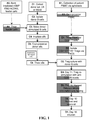
- FIG. 1 shows the workflow of donor-reactive Treg manufacturing. Briefly, the process begins with stimulating purified donor B cells with lethally irradiated good manufacturing process (GMP)-certified K562-hCD40L transfectants. The stimulated donor B cells are irradiated and used to selectively expand donor-reactive Tregs from CD4+CD25+CD127lo Tregs isolated from recipients' peripheral blood by fluorescent activated cell sorting ( FIG. 2A ).
- GMP lethally irradiated good manufacturing process
- the Tregs that remained in the culture are virtually all donor-reactive.
- the Tregs are restimulated with anti-CD3 and anti-CD28-conjugated beads to further expand the cells for additional 5 days.
- This protocol induces robust proliferation of Tregs ( FIG. 2B ) and can produce over a billion donor-reactive Tregs from one unit of blood.
- the expanded Tregs are >95% CD4+, >60% Foxp3+, >90% with demethylated Foxp3 promotor, >90% donor-reactive, and suppress donor-stimulated T cell proliferation when present at a 1:125 Treg:responder PBMC ratio.
- the donor-reactive Tregs also referred to herein as alloantigen-specific Tregs or alloTregs
- An exemplary embodiment involves the use of donor-reactive Tregs in the context of a Treg-supportive immunosuppression regimen as an approach to inducing tolerance of a liver transplant (Ltx).
- Treg therapy is useful for increasing the likelihood of and/or accelerating the development of tolerance.
- Treg administration in organ transplant settings is done in combination with administration of Treg-supportive immunosuppression regimens.
- M molar
- mM millimolar
- ⁇ M micromolar
- nM nanomolar
- mol mole
- mmol millimoles
- ⁇ mol micromol
- nmol nanomoles
- gm grams
- mg milligrams
- ⁇ g micrograms
- pg picograms
- L liters
- ml and mL milliliters
- ⁇ l and ⁇ L microliters
- Additional abbreviations include: Ab (antibody); allo (allogenic); CFSE (carboxyfluorescein diacetate, succinimidyl ester); FACS (fluorescent activated cell sorting); GMP (good manufacturing practice); IHC (immunohistochemistry); Ltx (liver transplant); MELD (model for end-stage liver disease); MLR (mixed lymphocyte reaction); PBMC (peripheral blood mononuclear cells); poly (polyclonal); rATG (rabbit anti-thymocyte globulin); sBcs (stimulated B cells); SOC (standard of care); SRL (sirolimus/rapamycin): tac (tacrolimus); Tconv (conventional T cells); Tregs (regulatory T cells); TSDR (Treg-specific demethylation region); Tx (transplant/transplantation); and UCSF (University of California San Francisco).
- This example provides an exemplary GMP-compliant method to selectively expand ex vivo up to billions (10 9 ) of alloantigen-specific Tregs from human peripheral blood monocular cells (PBMC) in about 2 weeks (see FIG. 1 ).
- PBMC peripheral blood monocular cells
- PBMC peripheral blood mononuclear cells
- PBMC peripheral blood mononuclear cells
- the cells were washed twice and resuspended in ice cold CS10 cryopreservation solution (BioLife Solutions) at 100-200 million cells/ml/cryogenic vial. The cells were frozen in a controlled rate freezer and stored in vapor phase of liquid nitrogen until further use.
- Donor B cell purification and banking Donor spleen or lymph nodes from cadaveric donors or PBMC from living donors were collected and transported to the GMP facility for processing into a single cell suspension. B cells were purified using CD19 positive selection on a CliniMACS instrument. Purified CD19 + B cells were banked by cryopreservation until needed for Treg expansion.
- K562 Human erythromyeloblastoid leukemia cells, K562 (ATCC No. CCL-243), were transfected with a lentivirus to express human CD40L, CD64 and HLA-DR0401 (K562-hCD40L or K40L). These cells are not tumorigenic in immunodeficient mice.
- the K40L feeder cells were ⁇ -irradiated at 10,000 rads, and banked until further use.
- the mixed culture was restimulated with K40L feeder cells at a 1-10:1 ratio (B:K40L) for 3 days.
- the average expansion was 10 to 20 fold.
- the sBc were passed over ficoll to remove dead cells including the dead K40L cells.
- a set of quality assurance assays were performed on the sBc, which included a qPCR-based EBV reactivation test (Viracor) and flow cytometry to determine purity as well as expression of HLA-DR, CD80, and CD86.
- the sBc were ⁇ - irradiated (1000 rads) and banked until further use.
- Treg expansion Recipient PBMC were thawed, counted and stained with clinical-grade, fluorescently-conjugated antibodies (CD4-PerCP Ab, CD25-APC Ab, and CD127-PE Ab). CD4 + CD127 lo/- CD25 + cells were purified from the stained PBMC by FACS ( FIG. 2A ). FACS-purified Tregs were mixed with banked irradiated sBc at a 4:1 ratio of sBc:Treg in growth medium comprising GMP-grade Optimizer Medium (Invitrogen) containing supplement, GlutaMAX-1 CTS and 2% human AB serum.
- GMP-grade Optimizer Medium Invitrogen
- human recombinant IL-2 was added to the culture at a total concentration of 300 IU/ml when media volume was doubled.
- the cultures were fed with fresh medium containing IL-2 on days 5, 7 and 9 to maintain cell concentration at 2-3x10 5 cells/ml.
- On day 11 of the culture cells were restimulated with beads conjugated with anti-CD3 and anti-CD28 monoclonal antibodies at a 1:1 ratio for the remainder of the culture period.
- the cultures were fed on day 1 and harvested on day 16.
- the Tregs expand 200 to 1600 fold in the 16-day culture period.
- Tregs are resuspended in HypoThermosal solution and kept at 4°C while awaiting the results of release assays, quality assurance review and approval. Upon product release, the Tregs are transported to the clinic for infusion. Greater than 5x10 6 Tregs are purified from one unit of recipient whole blood. With a conservative estimate of 200-fold expansion, at least 1x10 9 donor-reactive Tregs are expected to be harvested at the end of the expansion period.
- Treg release assays and release criteria The following assays and criteria are used before Treg release: viability >99%, flow cytometry for CD4 >90%, CD8 ⁇ 5%, CD19 ⁇ 5%, Foxp3 >60%, and TSDR >80%. Negative microbial tests for bacteria, fungus, mycoplasma, and endotoxin on culture day 12.
- the TSDR assay employed is currently the most accurate and reliable test for the purity and stability of Tregs.
- the methylation assay confirms the percentage of Foxp3+ cells determined by flow cytometry. Additionally, there is strong evidence that Foxp3 can be expressed in activated Tconv cells. However, the Foxp3 TSDR locus is methylated in activated Tconv cells while it is demethylated in bona fide Tregs.
- Post-release assays The following assays are performed on each product to fully document the phenotype and functionality of the cells: 1) expanded flow cytometric analysis using two panels consisting of CD4/Foxp3/CD27/CD62L and CD4/Foxp3/CD25/Helios; 2) donor specific suppression assay; 3) donor specificity assay; 4) long-term 14-day microbial test; and 5) cytokines (IL-2, IFN-gamma and IL-17) induced by donor sBc and PMA and ionomycin.
- cytokines IL-2, IFN-gamma and IL-17
- Foxp3+ Tregs are "plastic” and can acquire expression of effector cytokines such as IFN-gamma and IL-17 ( Zhou et al., Curr Opin Immunol, 21:281-285, 2009 ; Zhou et al., Immunity, 30:646-655, 2009 ; and Hori et al., Curr Opin Immunol, 22:575-582, 2010 ).
- exTregs While exTregs have low or no suppressive activity and can be pathogenic in experimental autoimmune settings, it is important to note that emergence of exTreg in lympho-replete hosts primarily occurs in extreme experimental conditions ( Rubtsov et al., Science, 329:1667-1671, 2010 ). Moreover, in all conditions, the majority of exTregs do not express effector cytokines even after supraphysiologic in vitro stimulation with PMA and ionomycin.
- the donor-reactive Tregs produced using the exemplary protocol we have high levels of Foxp3, TSDR, and Helios expression.
- Treg-supportive immunosuppression these cells are infused into patients under Treg-supportive immunosuppression, therefore the chance of the infused donor-reactive Tregs turning into full-fledged pathogenic effectors in vivo is low.
- effector Tregs have been shown to be suppressive in many experimental conditions.
- IFN-gamma production by Tregs has been shown to be essential to their suppressive function and protection against allograft rejection ( Sawitzki et al., J Exp Med, 201:1925-1935, 2005 ).
- effector cytokine production by Foxp3+ Tregs is expected to be tolerogenic rather than pathogenic.
- Tregs were purified from PBMCs using FACS based on CD4 + CD127 lo/- CD25 + cell surface phenotype as previously described ( Putman et al., Diabetes, 58:652-662, 2009 ).
- Donor B cells were purified using anti-CD19 CliniMACS beads (Miltenyi) and stimulated with irradiated GMP-compliant K562 cells expressing human CD40L.
- the dead K540L cells were removed from the sBc by ficoll density gradient centrifugation and the purified sBc were irradiated before adding to purified Tregs.
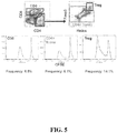
- up to ⁇ 1600-fold expansion of Tregs was achieved. Given that ⁇ 10% of Tregs are reactive to a fully HLA-mistmatched donor, 1600-fold overall expansion translates into ⁇ 16,000-fold increase in donor-reactive Tregs in the 16-day culture period.
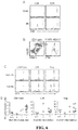
- the expanded Treg cultures were CD3+CD4+CD8-CD19-, Foxp3+, Helios+, CD27+ and CD62Lhi when compared to similarly expanded Tconv cells ( FIG. 2B ).
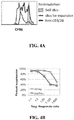
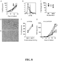
- Almost all donor sBc-expanded Tregs responded to restimulation with the donor sBc, but not to syngeneic sBc, indicating that they are stimulator-reactive ( FIG. 4A ).
- the donor-sBc-expanded Tregs exhibited enhanced donor-specific suppressive activity when compared with polyclonally expanded Tregs ( FIG. 4B ).
- This example describes a dose escalation clinical trial to assess safety of autologous, donor-reactive Treg therapy in liver transplant (Ltx) recipients.
- the methods and compositions of the present disclosure are not limited to this context. In fact, the methods and compositions of the present disclosure are expected to find use in the context of other solid organ allografts, as well as in treating or preventing graft versus host disease.
- Donor-reactive Tregs and Treg-supportive immunosuppression are expected to be suitable for inducing or maintaining tolerance of allografts selected from but not limited to cardiac, lung, cardia/lung, kidney, pancreas, kidney/pancreas, intestine and liver allografts.
- Escalating doses of Tregs expanded ex vivo using activated donor B cells are administered to Ltx recipients in conjunction with a modified immunosuppression regimen designed to favor Treg development, persistence, and function.
- This regimen is comprised of rabbit anti-thymocyte globulin (rATG) induction, reduced dosing of corticosteroids (Pred), mycophenolate mofetil (MMF), and tacrolimus (tac), followed by the delayed introduction of sirolimus (SRL).
- rATG rabbit anti-thymocyte globulin
- Pred corticosteroids
- MMF mycophenolate mofetil
- tac tacrolimus
- Subjects are followed for one year after transplantation, during which clinical data along with peripheral blood (PBMC and serum) and liver biopsy samples are collected and analyzed.
- PBMC and serum peripheral blood
- liver biopsy samples are collected and analyzed.
- the clinical trial encompasses three phases with specific inclusion/exclusion criteria at each phase to maximize participant safety.
- Pre and Ltx phase Patients are selected from the Ltx waiting list who have end-stage liver disease, between the ages of 20-70 years, and have a calculated MELD score of no greater than 25 ( Kamath et al., Hepatology, 33:464-470, 2001 ).
- the trial specifically excludes Ltx recipients at increased risk of acute rejection and recurrent disease and limits the severity of liver disease and portal hypertension and/or hypersplenism.
- only patients with Tregs present in PBMC at greater than 10/ ⁇ l are selected.
- PBMC peripheral blood mononuclear cells
- Treg purification and expansion Eligible patients undergo leukopheresis to isolate PBMC, which are cryopreserved for subsequent Treg purification and expansion.
- donor spleen and or lymph nodes along with liver biopsy tissue are collected and banked.
- Ltx recipients must be out of the ICU and initiate rATG induction no later than post-tx day 3. They receive a total dose of 3-4.5 mg/kg rATG to achieve lymphocyte depletion, defined as CD3 count ⁇ 50/mm3. This dose range was chosen to achieve adequate debulking ( Wong et al., Transpl Int, 19:629-635, 2006 ) while minimizing immunosuppression.
- the timing and setting of rATG administration was chosen to avoid the potential for over-immunosuppression and/or cytokine release syndrome/hematologic toxicities in medically unstable recipients.
- SRL sirolimus
- the immunosuppression regimen for study subjects was specifically designed to foster Treg development while optimizing participant safety.
- Study participants start on standard of care (SOC) immunosuppression with half-dose corticosteroids and half-dose mycophenolate mofetile (MMF).
- MMF mycophenolate mofetile
- Tacrolimus (Tac) is initiated, targeting reduced levels of 6-8 ⁇ g/L compared to SOC (10-15 ⁇ /L).
- No later than post-tx day 3 patients receive a course or rATG (3.0-4.5 mg/kg total dose) to deplete lymphocytes (CD3 count ⁇ 50/mm 3 or when the maximal dose has been given).
- Treg infusion phase Approximately 10-12 weeks after Ltx, participants are assessed for suitability to receive donor-reactive Tregs. Data regarding the kinetics of T cell recovery after rATG show stable T cell numbers between 4-12 weeks after tx. Therefore, the Treg infusion at 10-11 weeks after tx is in the setting of a debulked immune system. Participants must have normal allograft function in the context of stable SRL-based immunosuppression.
- donor B cells are expanded for 10 days and then used to expand Tregs over an additional 16 days (Example 1). Expanded donor-reactive Tregs passing all release criteria are available for infusion between 10-11 weeks after tx.
- Dose escalation plan Eligible patients receive either no Treg infusion or a single infusion of donor-reactive Tregs at 3 dose levels: 50, 200, and 800 million. Progression from one group to the next is based on the occurrence of dose-limiting toxicity. Table 2-2.
- This example describes analyses that are done on peripheral blood and liver tissues to assess the effects of Treg-supportive immunosupression and Treg therapy on alloimmune responses.
- donor-reactive Treg therapy along with Treg-supportive immunosuppression is expected to have a measurable impact on the frequency of donor-reactive Treg and on anti-donor T cell responsiveness.
- the exemplary therapeutic regimen described in Example 2 is expected to lead to an earlier development of an immune tolerance signature than occurs with conventional (SOC) immunosuppression regimens.
- Analyses include one or more of the following: 1) T cell functional and phenotypic analyses; 2) tolerance gene expression signature in PBMCs and protocol biopsy samples; and 3) histological analyses of for-cause as well as protocol biopsy samples.
- Multiparameter flow cytometry is used to profile leukocyte subpopulations, determine frequencies of donor-reactive T cells, assess donor-specific suppression by Tregs, and profile donor-antigen induced gene and cytokine expression. Together, these assays permit the assessment of the contribution of four known mechanisms of immune tolerance - deletion, deviation, anergy/exhaustion, and regulation.
- This assay is used to determine the frequency of donor-reactive CD4+ Tconv cells, CD8+ T cells, and Tregs.
- Banked PBMC samples are compared from pre-transplant/transplant, pre-Treg/post SRL conversion, on days 1, 3, 7, and 28 after Treg infusion, and at one year post transplant. An increase in donor-reactive Treg shortly after infusion is expected, especially in the cohorts receiving 200-800x10 6 dsRegs.
- In vitro suppression assay This assay is used to evaluate suppression by Tregs isolated from pre-transplant, pre-Treg infusion/post SRL conversion, at days 1 and 28 after Treg infusion, and 1 yr after liver transplant time points.
- Pre-transplant leukophoresed PBMC are used as responders mixed with Tregs isolated from various time points.
- the cultures are stimulated with irradiated donor PMBC to assess donor-specific suppression or with anti-CD3 and anti-CD28 to assess non-specific suppression.
- Multiparameter flow cytometry MFC is used to determine the percentage of leukocyte subsets in peripheral blood using panels of antibodies developed in our lab. Samples collected from panels and markers used are summarized in Table 3-1. Table 3-1. Multiparameter Flow Cytometry Panels and Marks.
- T cell activation / differentiation assay CD4+ Tconv cells and CD8+ T cells from pre-transplant, pre-Treg/post SRL conversion, on days 1, 3, 7, and 28 after Treg infusion and 1yr post transplant are stimulated using donor sBc for 3.5 days.
- the sample collected at pre-transplant, pre-Treg/post SRL conversion, on days 1, 3, 7, and 28 after Treg infusion time points is analyzed for cytokine gene expression using qPCR arrays and cytokine secretion into the supernatant using a 42-plex Luminex assay.
- the samples collected at pre-transplant and 1 yr after transplant are used to analyze gene expression profiles using gene array and the cytokine in the supernatant is analyzed using a 42-plex Luminex assay.
- Luminex assay changes in donor-sBc stimulated gene expression are expected to be observed in liver transplant patients. This assay permits alternations in donor-antigen stimulated gene expression profiles to be determined.
- CD3/CD45RO /CD45RA Monitor the relative ratio of naive to memory T cells; test whether Treg therapy leads to a reduction in portal-based CD3+/CD45RO+ (memory) T cells
- CD4/Tbet/GATA-3/IL-17/FoxP3 Monitor the polarization of CD4+ lymphocytes within the allograft to determine whether an increase of putative regulatory T cells contributes to tolerance.
- CK19/CD31 /HLADR Up-regulation of HLA-DR on biliary epithelium (CK19+) and vascular endothelium (CD31+) makes these cells targets of immune rejection. Determine if Treg therapy prevents DR induction.
- This example demonstrates a manufacturing process that can generate billions of human alloantigen-reactive regulatory T cells (Tregs) in short-term cultures using GMP-compliant reagents.
- the process uses CD40L-activated allogeneic B cells to selectively expand alloantigen-reactive Tregs followed by polyclonal restimulation to increase yield.
- the alloantigen-expanded Tregs were 5 to 25 times more potent than polyclonally expanded Tregs in vitro and were more effective at controlling allograft injuries in vivo in a humanized mouse model of skin transplantation.
- PBMC peripheral blood mononuclear cells
- CryoStor CS10 freezing medium BioLife Solutions, Bothell, WA
- CoolCellTM® devices BioCision, Mill Valley, CA
- CD40L expressing feeder cells Lentiviral vectors encoding human CD40L (NM_000074), CD64 (BC032634), DRA (BC071659) and DRB 0401 33 were produced as previously described 34 . These vectors were used to transduce K562 cells to generate a KT64-CD40L.HLADR0401 cell line and FACS was used to generate single cell clones as previously described 35 . Stable expression of expanded clones was verified by flow cytometry using antibodies to CD40L, HLA-DR, and CD64 from BD Biosciences, San Jose, CA.
- B cells were enriched from PBMC or spleen using the untouched human B cells enrichment kit (Invitrogen, Carlsbad, CA). Enriched B cells were cultured with irradiated (40Gy) 3T3 or K562 cells expressing human CD40L as described before 36 . For some experiments, dissociated splenocytes was cultured with CD40L-expressing cells without prior enrichment of B cells.
- the CD40L-sBc were irradiated (30Gy) and used to stimulate Tregs or cryopreserved in CryoStor CS10 freezing medium until use.
- peripheral blood B cells were purified using CD19 positive selection on a CliniMACS (Miltenyi Biotech, Germany), stimulated with irradiated (100Gy) K-CD40L cells in transferrin-containing X-VIV015 medium (Lonza, Walkersville, MD) supplemented with 10% human AB serum (Valley Biomedical, Winchester, PA), GMP grade IL-4 (Miltenyi), and Cyclosporine A (Teva Pharmaceuticals, North Wales, PA).
- Responder PBMC labeled with 1.25 ⁇ M CFSE were stimulated with irradiated allogeneic CD40L-sBc (two sBcs per PBMC) or with irradiated allogeneic PBMCs (5 stimulators per responder).
- the cultures were harvested after 84 to 96 hrs, stained with anti-CD3 PerCP (BD), anti-CD4 PE-Cy7 (BD), anti-CD8 APC-Cy7 (BioLegend, San Diego, CA), efluor 506 fixable viability dye (eBioscience, San Diego, CA).
- the cells were then fixed and permeabilized using a FOXP3 Fixation/Permeabilization buffer set (eBioscience) before staining with anti-FOXP3-Alexa Fluor 647 (eBioscience) and anti-HELIOS PE (BioLegend).
- Flow cytometry was performed on Fortessa (BD), and analysis was done using FACSdiva (BD) or FlowJo software (Treestar, Ashland, OR).
- Treg expansion was isolated using a BD FACSAria II (BD) based on the cell surface phenotype of CD4 + CD127 lo/- CD25 + and polyclonal expansions of Tregs were performed as previously described 28 .
- the clinically compliant sorting utilized cGMP mAbs generated and kindly provided by Noel Warner (BD).
- BD Noel Warner
- the cultures were maintained in OpTmizer T Cell Expansion Medium (Invitrogen) supplemented with 1% GlutaMAX (Invitrogen), Penicillin/ Streptomycin, and 2% human AB serum or X-VIVO15 medium supplemented with 10% human AB serum.
- Tregs were mixed with CD40L-sBc at a 4:1 sBc to Treg ratio.
- the cultures were maintained with medium containing 300 IU/ml human IL-2 until day 9 or 11, when the cells were restimulated with new irradiated sBc at 4 sBc per Treg ratio or with anti-CD3/anti-CD28-coated beads at a 1:1 bead to cell ratio. Cultures were fed 3 days later and harvested on day 5 after restimulation.
- Phenotype of expanded Tregs was assessed using three flow cytometric panels.
- the first panel consisted of anti-CD8 FITC, anti-CD4 PerCP, anti-CD3 PE, and anti-CD19APC.
- the second panel consisted of anti-CD4 PerCP, anti-CD62L PE, anti-CD27 APC, and anti-FOXP3 Alexa Fluor 488 (BioLegend, Clone 206D).
- the third panel consisted of anti-CD4 PerCP, anti-CD25 APC, anti-HELIOS PE (BioLegend), and anti-FOXP3 Alexa Fluor 488.
- Mouse IgG1 Alex Fluor 488 and mouse IgG1 PE were used to control for FOXP3 and HELIOS staining, respectively.
- the stained cells were analyzed on a FACSCalibur and the data was analyzed using FlowJo.
- the CD40L-sBc were analyzed on an AccuriC6 (BD) flow cytometer after staining with anti-HLA-DR PE, anti-CD80 FITC, anti-CD86 PerCP-Cy5.5, and anti-CD19 APC.
- the data were analyzed using Cflow PLUS software (BD). All antibodies were from BD Biosciences unless otherwise noted.
- Treg specificity assay Expanded Tregs were labeled with 1.25 ⁇ M CFSE and stimulated with allogeneic or autologous CD40L-sBc, anti-CD3 and anti-CD28-coated beads, or left unstimulated in media containing 30IU/ml IL-2. After 72 hours, the cells were collected and stained with anti-CD4 APC (BD) and propidium iodide and analyzed on an AccuriC6 flow cytometer.
- BD anti-CD4 APC
- TSDR methylation assay Genomic DNA isolated from 0.5x10 6 expanded Tregs using licensed reagents from Epiontis GmbH (Berlin, Germany) according to protocol established by Epiontis GmbH 37 . The assay was performed in triplicated and the percentages of methylated TSDR were calculated as: [mean copy numbers of unmethylated DNA/(mean copy numbers of unmethylated + mean copy numbers of methylated DNA)] x 100. For cultures expanded using female donors, the percentages from the above calculation were multiplied by 2 to correct for X chromosome inactivation.
- mice were co-injected with 2 x 10 6 ex vivo expanded polyclonal or alloantigen-reactive Tregs. Visual and tactile inspections of the grafts were performed two times weekly. Histological analysis of the grafts was performed 6 weeks after PBMC injections. For the total duration of these experiments, 100 ⁇ g purified anti-mouse Gr1 mAB (Bio X Cell, West Riverside, NH) was injected intraperitoneally every 4-5 days to deplete mouse granulocytes. All procedures were conducted in accordance with institutional guidelines and the Home Office Animals Scientific Procedures Act (1986). Frozen sections
- CD40L-stimulated B cells are potent stimulators of alloantigen-reactive Tregs. Allogeneic PBMC, dendritic cells (DC), fresh B cells, and CD40L-stimulated B cells (referred as CD40L-sBc) have been used previously to selectively stimulate the expansion of human alloantigen-reactive cells 13-16 . However, less is known about the relative ability of these cell subsets in stimulating Tregs. A comparison of the relative potencies of irradiated PBMC, freshly isolated B cells, and CD40L-sBc in a one-way mixed lymphocyte reaction (MLR) demonstrated that CD40L-sBc were the most potent stimulators.
- MLR mixed lymphocyte reaction
- GMP good manufacturing practice
- CD64 and HLADR0401 expression does not interfere with CD40L activity while allowing for the cell line to be used for other applications including antigen-specific and polyclonal T cell expansions.
- the CD40L-sBc expressed significantly higher amounts of HLADR, CD80, and CD86 ( FIG. 7B and 7C ), consistent with their enhanced potency in stimulating allogeneic T cells.
- CD40L-sBc Although there was consistent increase of HLA-DR, CD80, and CD86 expression on CD40L-sBc, the levels varied from donor to donor. However, CD40L-sBc generated from multiple donors were able to induce MLR and Treg expansion, suggesting that the potency of the CD40L-sBC was not strictly correlated with the absolute levels of the co-stimulatory and MHC class II molecules as long as a threshold was met.
- CD40L-sBc robustly induce expansion of alloantigen-reactive Tregs.
- the conditions for optimal stimulation of alloantigen-reactive Tregs using CD40L-sBc were tested.
- a protocol for polyclonal expansion of Tregs using two round stimulations (days 0 and 9) of fluorescence-activated cell sorting (FACS) purified CD4 + CD127 lo/- CD25 + Tregs with anti-CD3 and anti-CD28-coated beads is known 28 .
- FACS fluorescence-activated cell sorting
- the expanded Tregs were highly responsive to the same CD40L-sBc used to stimulate Treg expansion, but failed to respond to self CD40L-sBc ( FIG. 8B ).
- This result demonstrated a marked enrichment of Tregs reactive to the alloantigens expressed by the CD40L-sBc used to stimulate Treg expansion.
- the Tregs were already highly reactive to the CD40L-sBc, similar to that observed on day 14, suggesting that there might not be a need to further enrich for alloreactivity during restimulation.
- Tregs One unit of blood yields an average of 5 million Tregs after FACS purification. Therefore, using the protocol from FIG. 8 , between 250 million to 1.5 billion alloantigen-reactive Tregs may be produced based on a 50- to 300-fold expansion. It was estimated that the numbers of Tregs needed for efficacy in transplantation in humans are in the range of 300 million to several billion cells 20 . To ensure consistent production of more than 300 million alloantigen-reactive Tregs, modified conditions to improve Treg expansion were explored. It was observed that, unlike the PolyTregs activated on day 0 with beads, the CD40L-sBc-stimulated Tregs continued to cluster and blast on day 9 after the initial stimulation ( FIG. 8D ).
- the alloantigen-reactive Tregs routinely expanded 200 to 1600 fold, reliably producing more than 1 x 10 9 alloantigen-reactive Tregs in a 16-day period.
- Tregs expanded with the CD40L-sBc protocol were found to be CD3 + CD4 + with minimal contamination with CD8 + T cell and CD19+ B cells ( FIG. 9A ).
- the majority of the CD4 + T cells were FOXP3 + HELIOS + and co-expressed CD27 and CD62L ( FIG. 9B ), distinct from the pattern expressed on similarly expanded Tconv cells ( FIG. 9B ).
- the expanded Tregs had > 80% demethylated Treg-specific demethylated region.
- Tregs harvested on day 16 were restimulated with the same CD40L-sBc.
- 87.5% (range 72.5 to 95.2%) of the alloantigen expanded Tregs proliferated in response to restimulation by the same sBc similar to the proliferation induced using anti-CD3 and anti-CD28 beads (average 88.8%, range 73.6 to 96%), suggesting that the vast majority of the Tregs were reactive to the alloantigens expressed by the CD40L-sBc ( FIG. 9C and 9D ).
- the expanded Tregs were highly suppressive when activated in vitro by PBMCs from the same donor as the CD40L-sBc ( FIG. 9E ).
- Side-by-side comparison of alloantigen-expanded Tregs and polyclonally expanded Tregs showed that the donor alloantigen-reactive Tregs were 5 to 25 fold more potent at suppressing MLR than PolyTregs ( FIG. 9E ).
- Treg expanded by restimulation with CD40L-sBc or anti-CD3 and anti-CD28 beads have identical activity in suppressing MLR ( FIG.
- Alloantigen-reactive Tregs are superior at protecting skin allografts in vivo.
- Using a model of alloimmune mediated injury of human skin allografts ( FIG. 11A ) 13 the protective function of alloantigen-reactive Tregs and PolyTregs was compared.
- BALB/c.Rag2 -/- ⁇ c -/- mice were transplanted with human skin from a HLA-DR0401 + donor and the grafts were allowed to heal for 6 weeks before adoptive transfer of allogeneic PBMC depleted of CD25 + cells alone or in combination with different preparations of syngeneic Tregs at a ratio 5:1 effector cells:Treg cells.
- PBMC donors were HLA-DR0401 - and alloantigen-reactive Tregs from these donors were expanded using HLA-DR0401 + CD40L-sBc. Grafts were monitored until rejection or until up to a maximum of 6 weeks after PBMC reconstitution when the grafts were collected for histological analysis. Levels of human leukocyte engraftment in spleens were similar in the three groups of mice that received human PBMC alone or in combination with Tregs ( FIG. 11B ). No animal developed xenogeneic graft-versus-host disease symptoms confirmed by the maintenance of stable body weight ( FIG. 11C ).
Description
- The present disclosure relates generally to the manufacture of regulatory T cells (Tregs) for use in immunotherapy. In particular, the present disclosure relates to robust approaches for the expansion of alloantigen-reactive Tregs ex vivo. Alloantigen-reactive Tregs produced in this way are suitable for the induction and/or maintenance of immunologic tolerance in recipients of allogeneic transplants.
- Ongoing refinement of immunosuppression regimens has substantially reduced the incidence of acute rejection after solid organ transplantation. However, long-term outcomes have stagnated partly due to morbidity and mortality associated with immunosuppression. The traditional approach to immunosuppression has emphasized non-specific suppression of T cell responses.
- The more recent elucidation of T regulatory cells (Tregs) and their importance in regulating immune responses has encouraged the reconfiguration of immunosuppression regimens to favor Treg development and function with the ultimate goal of inducing graft tolerance (Waldmann et al., J. Clin Immunol, 28:716-725, 2008; Kang et al., Am J Transplant, 7:1457-1463, 2007; Walsh et al., J Clin Invest, 114:1398-1403, 2004; Yeung et al., Transplant Proc, 41:S21-26, 2009; Sanchez-Fueyo et al., J Immunol, 176:329-334, 2006; Sagoo et al., Curr Opin Organ Transplant, 13:645-653, 2008; and Long et al., Transplantation, 88:1050-1056, 2009). Multiple preclinical models have shown that adoptive transfer of Tregs can mitigate graft rejection and, in combination with "Treg-supportive" immunsuppression regimens, can induce long-term tolerance (Kang et al., Am J Transplant, 7:1457-1463, 2007; Riley et al., Immunity, 30:656-665, 2009; Issa et al., Expert Rev Clin Immunol, 6:155-169, 2010; and Nadig et al., Nat Med, 16:809-813, 2010). Treg-supportive" immunsuppression regimens have included the initial de-bulking of donor-reactive T cells. Rabbit anti-thymocyte globulin (rATG), a commonly used T-cell depleting agent in transplantation, appears to spare Tregs (Sewgobind et al., Nephrol Dial Transplant, 24:1635- 1644, 2009), thereby increasing Treg:T conventional cell (Tconv) ratio. Additionally, sirolimus (SRL) suppresses effector T cells while fostering Treg development (Demirkiran et al., Transplantation, 85:783-789, 2008; and Demirkiran et al., Transplantation, 87:1062-1068, 2009).
- Most protocols typically expand all Tregs nondiscriminately to produce cells referred to as polyclonal Tregs (polyTregs). However, alloantigen-specific Tregs (alloTregs) are more effective and safer than non-specific Tregs in transplant settings because they provide specific rather than generic immunosuppression (Golshayan et al., Blood, 109:827-835, 2007; and Raimondi et al., J Immunol, 184:624-636, 2010). In particular, donor-reactive Tregs have the potential to induce tolerance to the transplanted organ without impeding conventional immune responses. Thus what is needed in the art are robust methods for expansion of alloTregs for use in promoting transplant tolerance and for treating graft versus host disease.
- Also cited are:
- Tang et al., J Mol Cell Biol, 4:11-21, 2011. This review focuses on developing therapy for transplantation using CD4+Foxp3+ Tregs.
-
WO 2010/129770 . This disclosure describes methods for expanding regulatory T cells. - The present disclosure relates generally to the manufacture of regulatory T cells (Tregs) for use in immunotherapy. In particular, the present disclosure relates to robust approaches for the expansion of alloantigen-reactive Tregs (alloTregs) ex vivo. AlloTregs produced in this way are suitable for the induction and/or maintenance of immunologic tolerance in recipients of allogeneic transplants.
- The present invention provides a method for the production of human, donor-reactive regulatory T cells (Tregs), comprising:
- a) co-culturing CD19+ B cells of a human donor with irradiated CD40L+ human leukemia feeder cells under conditions effective in producing stimulated B cells (sBc);
- b) co-culturing CD4+, CD25+, CD127-/lo T cells of a human recipient with said sBc under conditions effective in selectively expanding human donor-reactive regulatory T cells (Tregs); and
- c) re-stimulating said donor-reactive Tregs by cross-linking CD3 and CD28 of said donor-reactive Tregs under conditions effective in producing donor-reactive Tregs that are CD4+, Helios+ and Foxp3+;
- The present disclosure provides methods for the production of human, donor-reactive regulatory T cells (Tregs), comprising: a) co-culturing CD19+ B cells of a human donor (first human subject) with irradiated CD40L+ human leukemia feeder cells under conditions effective in producing stimulated B cells (sBc); and b) co-culturing CD4+, CD25+, CD127-/lo T cells of a human recipient (second human subject) with the sBc under conditions effective in selectively expanding human donor-reactive regulatory T cells (Tregs). In some embodiments, the human donor is unrelated to the human recipient. In some embodiments, the human donor is HLA-mismatched in relation to the human recipient
(e.g., donor is allogeneic to the recipient or said another way the transplant is a heterologous organ transplant). In some embodiments, the HLA-mismatch comprises a mismatch at one, two, three or four of HLA-A, HLA-B, HLA-C and HLA-DR. The methods further comprise step c) re-stimulating the donor-reactive Tregs by cross-linking CD3 and CD28 of the donor-reactive Tregs under conditions effective in producing re-stimulated donor-reactive Tregs. The donor-reactive Tregs are CD4+, Helios+ and Foxp3+. In some embodiments, the donor-reactive Tregs are CD27+ and CD62L+. In some embodiments, the methods further comprise a step before a) of isolating CD4+, CD25+, CD127-/lo T cells from cryopreserved peripheral blood mononuclear cells (PBMC) obtained from the human recipient. In some embodiments, step a) comprises co-culturing the B cells and the feeder cells in medium comprising insulin, transferrin, interleukin-4 and cyclosporine A. In some embodiments, the feeder cells are KCD40L cells. In some embodiments, step b) comprises co-culturing the sBc and the CD4+, CD25+, CD127-/lo T cells in medium comprising interleukin-2, after the sBc have been irradiated. In some embodiments, step c) commences 9-12 days after step b) commences. In some preferred embodiments, the re-stimulated alloTregs comprise at least 200, 250, 300, 350, 400, 450, 500, 600, 700, 800, 900, 1000, 1100, 1200, 1400 or 1600 fold more cells than the CD4+, CD25+, CD127-/lo T cells at the onset of step b). Also provided by the present disclosure are compositions comprising a physiologically acceptable buffer (e.g., saline, PBS, etc.) and the restimulated donor-reactive Tregs produced using the methods described above. Also described are methods for treating an organ transplant recipient comprising: administering from 107 to 1011 of the restimulated donor-reactive Tregs produced using the methods described above to a human recipient of a heterologous organ transplant. Also provided are medicaments for use in treating or preventing rejection of a solid organ allograft by the human recipient, the medicament comprising: from 107 to 1011 of the restimulated donor-reactive Tregs produced using the methods described above. In some embodiments, the organ transplant is a solid organ allograft selected from the group consisting of cardiac, lung, cardiac/lung, kidney, pancreas, kidney/pancreas, intestine and liver allografts. In some embodiments the solid organ allograft is a skin allograft. In some embodiments, the restimulated donor-reactive Tregs are administered on more than one occasion (repeatedly administered). In some embodiments, the restimulated donor-reactive Tregs are first administered after the recipient has received the heterologous organ transplant. In some embodiments, the restimulated donor-reactive Tregs are administered before and after the recipient has received the heterologous organ transplant.
We also describe methods further comprising subjecting the human recipient to a Treg-supportive immunosuppression regimen before administration of the restimulated donor-reactive Tregs. The Treg-supportive immunosuppression regimen may comprise: administering rabbit anti-thymocyte globulin to the human recipient at an amount effective to achieve lymphocyte depletion. We also describe methods further comprising administering prednisone, mycophenolate mofetile and tacrolimus to the human subject at doses below standard of care. We also describe methods further comprising administering sirolimus to the human subject. In some preferred embodiments, the administration of the restimulated donor-reactive Tregs is effective in reducing the likelihood of acute and/or chronic transplant rejection. In some preferred embodiments, the administration of the restimulated donor-reactive Tregs is effective in prolonging survival of the solid organ allograft. In some preferred embodiments, the administration of the restimulated donor-reactive Tregs is effective in achieving one or more of the following: increasing Treg percentages over baseline, increasing donor-reactive Treg frequency, increasing donor-reactive Treg activity, and induction of tolerance gene expression profiles in PBMC and/or transplant tissue. - As used herein, the singular form "a," "an" and "the" includes plural references unless indicated otherwise.
-
-
FIG. 1 provides a flow chart of an exemplary donor-reactive regulatory T cell (Treg) manufacturing process. -
FIG. 2A shows the CD4+CD25+CD127-/lo population of cells purifed from recipient PBMC by FACS.FIG. 2B shows the magnitude of expansion of donor-reactive Tregs achievable with the methods of the present disclosure. The arrow indicates when the Tregs were exposed to polyclonal stimulus (e.g., anti-CD3/CD28 conjugated beads). -
FIG 3A is a flow cytometric analysis of the expanded donor-reactive Tregs and control donor-reactive T conventional cells (Tconv).FIG. 3B shows Treg-specific demethylated region (TSDR) analysis of expanded donor-reactive Tregs and Tconv and polyclonal Tregs (polyTregs) expanded using a polyclonal stimulus (anti-CD3/anti-CD28 coated beads). -
FIG. 4A provides results of a donor specificity assay. Donor-reactive Tregs were labeled with CFSE and restimulated as indicated.FIG. 4B provides results of a mixed lymphocyte reaction (MLR) suppression assay. Titrated number of donor-reactive Treg and polyclonally expanded Tregs were mixed with 2.5x10⁴ autologous PBMC and 1.25x10⁵ irradiated donor PBMC and incubated for 6 days. Tritiated-thymidine was added during the last 16 hours. Suppression of thymidine incorporation was calculated by comparing counts per minute (CPM) in wells without Tregs. -
FIG. 5 shows results of a donor-reactive T cell frequency assay. Recipient PBMC were labeled with CFSE and stimulated with donor sBc for 3.5 days. The culture was harvested, stained for CD3, CD4, CD8, Foxp3 and Helios, and analyzed on a flow cytometer. The CFSE probiles of CD8, CD4+ Tconv, and Tregs were used to calculate the frequencies of donor-reactive T cells in each subset. -
FIG. 6A and FIG. 6B shows PBMC and CD40L-sBc from the same donor compared for their ability to stimulate proliferation of alloreactive T cells in a one-way MLR. The responder PBMC were labeled with CFSE before MLR and the cultures were harvested on day 4 for flow cytometric analysis. Representative CFSE dilution profiles of CD4+ and CD8+ T cells (FIG. 6A ) and CD4+FOXP3+HELIOS+ Tregs (FIG. 6B ) are shown. The data is a representative of at least 10 independent experiments.FIG. 6C and FIG. 6D show autologous CD40L-sBc and allogeneic CD40L-sBc with different degree of HLA mismatches with responder cells compared in their ability to stimulation proliferation of CD4+ Tconv, CD8+ T cells, and Treg cells. Each symbol represents the same responder. Results are a summary of 15 different stimulator and responder combinations. -
FIG. 7A shows the expansion of purified B cells in a 10-day culture. The arrow indicates the time of restimulation.FIG. 7B and FIG. 7C show expression of HLA-DR, CD80, and CD86 in freshly isolated B cells andday 10 CD40L-sBc compared using flow cytometry. Sample overlay histograms are shown inFIG. 7B and charts summarizing results from independent experiments are shown inFIG. 7C . The data is summary of 6 independent experiments. -
FIG. 8A shows allogeneic sBc used to stimulate FACS purified Tregs onday 0 and day 9. Fold expansion of Treg in the 14-day culture in 6 independent experiments is shown. The arrow indicates the time of restimulation.FIG. 8B shows alloreactivity of expanded Tregs determined by labeling the expanded Tregs with CFSE before restimulation with the same CD40L-sBc used for expansion (thick line), anti-CD3 and anti-CD28-coated beads (thin line), or syngeneic CD40L-sBc (shaded histogram).FIG. 8C shows Tregs stimulated with CD40L-sBc for 9 days and then split with half restimulated with CD40L-sBc from the same donor and the other half with anti-CD3 and anti-CD28-coated beads. Fold expansion on day 14 of three independent paired cultures is shown (p=0.52, two-tailed paired t test).FIG. 8D and FIG. 8E show appearances of Treg cultures on days 9 (FIG. 8D ) and11 (FIG. 8E ) after primary stimulation. Data represents results from at least 10 independent cultures.FIG. 8F shows Tregs stimulated with CD40L-sBc for 9 or 11 days before restimulation with anti-CD3 and anti-CD28-coated beads. The cultures were harvested 5 days after restimulation and total fold expansion in 3 paired cultures are compared (p=0.0026, two-tailed paired t test).FIG. 8G shows Tregs stimulated with CD40L-sBc for 11 days before restimulation with anti-CD3 and anti-CD28-coated beads from Invitrogen (open symbols) or Miltenyi Biotec (closed symbols). Cell expansions over time in 3 paired cultures are shown. Two-tailed paired t test was used to compare the difference in total fold expansion on day 16 (p=0.0258). -
FIG. 9A and FIG. 9B show flow cytometric profiles of ungated (a) and CD4 gated (b) Treg cultures. Data are representative of at least 14 independent experiments.FIG. 9C shows alloreactivity of Tregs expanded with primary allogeneic sBc stimulation and polyclonal restimulation onday 11 determined as described inFIG. 8B . An example of overlay histogram is shown.FIG. 9D shows a summary of 7 independent cultures analyzed as described inFIG. 9C . Each symbol represents one independent Treg culture.FIG. 9E shows a summary of in vitro suppression by Tregs expanded with two rounds of stimulation with allogeneic CD40L-sBc (closed circles, Allo-a, n=3), allogeneic sBc primary stimulation followed by polyclonal restimulation (open circles, Allo-p, n=8), or two rounds of polyclonal stimulations (open squares, Poly, n=5). Responders are PBMC from the Treg donor and stimulators are PBMC from the sBc donor. Data shown is mean +/- SEM suppression observed in 3 to 8 independent experiments. Two-way ANOVA with Bonferroni multiple comparison test was used to determine the statistical significance of the differences. Suppression at 1:5 ratio by different groups of Tregs are not significantly different. Suppression by PolyTregs is significantly lowered when compared to Allo-a Tregs (p<0.001 at 1:25 ratio and p<0.01 at 1:125 ratio), or when compared to Allo-p Tregs (p<0.0001 at 1:25 ratio and p<0.001 at 1:125 ratio). Allo-a and Allo-p Tregs are not significantly different from each other at all ratios).FIG. 9F shows suppression by CD40L-sBc expanded Tregs stimulated by PBMC from the sBc donor (closed circles) or a third party donor (open triangles). Result shown is representative of two independent experiments. -
FIG. 10A, FIG. 10B and FIG. 10C show data from BALB/c. Rag2-/-γc-/- mice transplanted with human skin and reconstituted with PBMC allogeneic to the skin donor. Immunofluorescence micrograph images were analyzed by counting 4 to 6 high-powered visual fields per stain for each graft. Quantitative results from four experimental groups were then compared. One-way ANOVA with Bonferroni multiple comparison test was used to determine the statistical significance of the differences. -
FIG. 11A is a schematic diagram of the experimental model and procedure is shown.FIG. 11B shows PBMC reconstitution determined at the end of the experiment, demonstrating that co-infusion of Tregs did not significantly alter the extent of PBMC reconstitution.FIG. 11C shows body weight of the BALB/c.Rag2-/-γc-/- mice in four experimental groups was assessed to determine general health status, demonstrating that PBMC infusion did not induce graft-versus-host disease. -
FIG. 12 is a schematic diagram of an exemplary alloantigen-reactive Treg manufacturing process. - The present disclosure relates generally to the manufacture of regulatory T cells (Tregs) for use in immunotherapy. In particular, the present disclosure relates to robust approaches for the expansion of alloantigen-specific Tregs ex vivo. Alloantigen-specific Tregs produced in this way are suitable for the induction and/or maintenance of immunologic tolerance in recipients of allogeneic transplants.
- The present disclosure provides methods to selectively expand donor-reactive Tregs 200 to 1,000 fold in less than 20 days. Contrary to the dogma that dendritic cells are most efficient at expanding T cells, CD40 ligand-stimulated human B cells were found to be extremely potent in inducing proliferation of Tregs.
FIG. 1 shows the workflow of donor-reactive Treg manufacturing. Briefly, the process begins with stimulating purified donor B cells with lethally irradiated good manufacturing process (GMP)-certified K562-hCD40L transfectants. The stimulated donor B cells are irradiated and used to selectively expand donor-reactive Tregs from CD4+CD25+CD127lo Tregs isolated from recipients' peripheral blood by fluorescent activated cell sorting (FIG. 2A ). By day 9 to 12, the Tregs that remained in the culture are virtually all donor-reactive. The Tregs are restimulated with anti-CD3 and anti-CD28-conjugated beads to further expand the cells for additional 5 days. This protocol induces robust proliferation of Tregs (FIG. 2B ) and can produce over a billion donor-reactive Tregs from one unit of blood. The expanded Tregs are >95% CD4+, >60% Foxp3+, >90% with demethylated Foxp3 promotor, >90% donor-reactive, and suppress donor-stimulated T cell proliferation when present at a 1:125 Treg:responder PBMC ratio. The donor-reactive Tregs (also referred to herein as alloantigen-specific Tregs or alloTregs) find use in methods for promoting transplant tolerance and for treating graft versus host disease. - An exemplary embodiment involves the use of donor-reactive Tregs in the context of a Treg-supportive immunosuppression regimen as an approach to inducing tolerance of a liver transplant (Ltx). Treg therapy is useful for increasing the likelihood of and/or accelerating the development of tolerance. Because of the exceptionally high frequency of donor-reactive T cells, "debulking" of the host alloreactive repertoire and adjunct immunosuppression are needed to create a more favorable setting for Tregs to control alloimmunity and to ensure long-term graft tolerance (Wells et al., Nat Med, 5:1303-1307, 1999; Li et al., Curr Opin Immunol, 12:522-527, 2000; and Wells et al., Philos Trans R Soc Lond B Biol Sci, 356:617-623, 2001). Importantly, some immunosuppression drugs favor Treg development and/or survival while others are neutral or antagonistic. Thus in some embodiments, Treg administration in organ transplant settings is done in combination with administration of Treg-supportive immunosuppression regimens.
- Findings in Treg research in the past 15 years provide a compelling rationale for therapeutic use of donor-reactive Tregs in transplantation. The present disclosure provides the first clinical trial involving the administration of donor-reactive Tregs to solid organ transplant recipients. Development of a good manufacturing practice (GMP)-compliant protocol to reliably expand human donor-reactive Tregs (Example 1) has made this effort possible. Additionally, a set of immune monitoring assays has been developed to dissect alloimmune responses in transplant patients, which have significantly improved sensitivity and reproducibility as compared to previously described assays.
- The present disclosure is described in further detail in the following examples which are not in any way intended to limit the scope of the disclosure as claimed. The attached figures are meant to be considered as integral parts of the specification and description of the disclosure. The following examples are offered to illustrate, but not to limit the claimed disclosure.
- In the experimental disclosure which follows, the following abbreviations apply: M (molar); mM (millimolar); µM (micromolar); nM (nanomolar); mol (moles); mmol (millimoles); µmol (micromoles); nmol (nanomoles); gm (grams); mg (milligrams); µg (micrograms); pg (picograms); L (liters); ml and mL (milliliters); µl and µL (microliters); cm (centimeters); mm (millimeters); µm (micrometers); nm (nanometers); U (units); V (volts); MW (molecular weight); sec (seconds); min(s) (minute/minutes); h(s) and hr(s) (hour/hours); °C (degrees Centigrade); ND (not done); NA (not applicable); rpm (revolutions per minute); H2O (water); aa (amino acid); bp (base pair); kb (kilobase pair); kD (kilodaltons); cDNA (copy or complementary DNA); DNA (deoxyribonucleic acid); ssDNA (single stranded DNA); dsDNA (double stranded DNA); dNTP (deoxyribonucleotide triphosphate); PCR (polymerase chain reaction); qPCR (quantitative PCR); RNA (ribonucleic acid); and RT-PCR (reverse transcription PCR). Additional abbreviations include: Ab (antibody); allo (allogenic); CFSE (carboxyfluorescein diacetate, succinimidyl ester); FACS (fluorescent activated cell sorting); GMP (good manufacturing practice); IHC (immunohistochemistry); Ltx (liver transplant); MELD (model for end-stage liver disease); MLR (mixed lymphocyte reaction); PBMC (peripheral blood mononuclear cells); poly (polyclonal); rATG (rabbit anti-thymocyte globulin); sBcs (stimulated B cells); SOC (standard of care); SRL (sirolimus/rapamycin): tac (tacrolimus); Tconv (conventional T cells); Tregs (regulatory T cells); TSDR (Treg-specific demethylation region); Tx (transplant/transplantation); and UCSF (University of California San Francisco).
- This example provides an exemplary GMP-compliant method to selectively expand ex vivo up to billions (109) of alloantigen-specific Tregs from human peripheral blood monocular cells (PBMC) in about 2 weeks (see
FIG. 1 ). - Recipient T cell purification and banking. PBMC were purified from whole blood or leukopheresis products from participants using ficoll density centrifugation. The cells were washed twice and resuspended in ice cold CS10 cryopreservation solution (BioLife Solutions) at 100-200 million cells/ml/cryogenic vial. The cells were frozen in a controlled rate freezer and stored in vapor phase of liquid nitrogen until further use.
- Donor B cell purification and banking. Donor spleen or lymph nodes from cadaveric donors or PBMC from living donors were collected and transported to the GMP facility for processing into a single cell suspension. B cells were purified using CD19 positive selection on a CliniMACS instrument. Purified CD19+ B cells were banked by cryopreservation until needed for Treg expansion.
- Feeder cell preparation. Human erythromyeloblastoid leukemia cells, K562 (ATCC No. CCL-243), were transfected with a lentivirus to express human CD40L, CD64 and HLA-DR0401 (K562-hCD40L or K40L). These cells are not tumorigenic in immunodeficient mice. The K40L feeder cells were γ -irradiated at 10,000 rads, and banked until further use.
- Banked donor B cell activation. A modified, GMP-compliant protocol (Zand et al., Am J Transplant, 5:76-86, 2005) was used to generate stimulated B cells (sBc). Specifically, ∼1-100x106 donor B cells purified with paramagnetic anti-CD19 microbeads on a CliniMACS (Miltenyi) were stimulated with banked, GMP-compliant, γ-irradiated K40L cells at a 1-2:1 ratio (B:K40L) for 7 days in a medium containing 10% human AB serum, insulin, transferrin, human recombinant IL-4, and cyclosporine A. On day 7, the mixed culture was restimulated with K40L feeder cells at a 1-10:1 ratio (B:K40L) for 3 days. The average expansion was 10 to 20 fold. The sBc were passed over ficoll to remove dead cells including the dead K40L cells. A set of quality assurance assays were performed on the sBc, which included a qPCR-based EBV reactivation test (Viracor) and flow cytometry to determine purity as well as expression of HLA-DR, CD80, and CD86. The sBc were γ - irradiated (1000 rads) and banked until further use.
- Treg expansion. Recipient PBMC were thawed, counted and stained with clinical-grade, fluorescently-conjugated antibodies (CD4-PerCP Ab, CD25-APC Ab, and CD127-PE Ab). CD4+CD127lo/-CD25+ cells were purified from the stained PBMC by FACS (
FIG. 2A ). FACS-purified Tregs were mixed with banked irradiated sBc at a 4:1 ratio of sBc:Treg in growth medium comprising GMP-grade Optimizer Medium (Invitrogen) containing supplement, GlutaMAX-1 CTS and 2% human AB serum. On day 2, human recombinant IL-2 was added to the culture at a total concentration of 300 IU/ml when media volume was doubled. The cultures were fed with fresh medium containing IL-2 ondays 5, 7 and 9 to maintain cell concentration at 2-3x10⁵ cells/ml. Onday 11 of the culture, cells were restimulated with beads conjugated with anti-CD3 and anti-CD28 monoclonal antibodies at a 1:1 ratio for the remainder of the culture period. The cultures were fed onday 1 and harvested onday 16. The Tregs expand 200 to 1600 fold in the 16-day culture period. - Tregs are resuspended in HypoThermosal solution and kept at 4°C while awaiting the results of release assays, quality assurance review and approval. Upon product release, the Tregs are transported to the clinic for infusion. Greater than 5x10⁶ Tregs are purified from one unit of recipient whole blood. With a conservative estimate of 200-fold expansion, at least 1x10⁹ donor-reactive Tregs are expected to be harvested at the end of the expansion period.
- Release assays and release criteria. The following assays and criteria are used before Treg release: viability >99%, flow cytometry for CD4 >90%, CD8 <5%, CD19 <5%, Foxp3 >60%, and TSDR >80%. Negative microbial tests for bacteria, fungus, mycoplasma, and endotoxin on culture day 12. The TSDR assay employed is currently the most accurate and reliable test for the purity and stability of Tregs. The methylation assay confirms the percentage of Foxp3+ cells determined by flow cytometry. Additionally, there is strong evidence that Foxp3 can be expressed in activated Tconv cells. However, the Foxp3 TSDR locus is methylated in activated Tconv cells while it is demethylated in bona fide Tregs.
- Post-release assays. The following assays are performed on each product to fully document the phenotype and functionality of the cells: 1) expanded flow cytometric analysis using two panels consisting of CD4/Foxp3/CD27/CD62L and CD4/Foxp3/CD25/Helios; 2) donor specific suppression assay; 3) donor specificity assay; 4) long-term 14-day microbial test; and 5) cytokines (IL-2, IFN-gamma and IL-17) induced by donor sBc and PMA and ionomycin.
- Recent experimental evidence suggests that Foxp3+ Tregs are "plastic" and can acquire expression of effector cytokines such as IFN-gamma and IL-17 (Zhou et al., Curr Opin Immunol, 21:281-285, 2009; Zhou et al., Immunity, 30:646-655, 2009; and Hori et al., Curr Opin Immunol, 22:575-582, 2010). It is helpful to distinguish two types of plastic Treg fates, one that results in loss of Foxp3 expression and concomitant effector cytokine expression (exTregs) (Komatus et al., Proc Natl Acad Sci USA 106:1903-1908, 2009; Xu et al., J Immunol, 178:6725-6729, 2007; Osorio et al., Eur J Immunol, 38:3274-3281, 2008; Yang et al., Immunity, 29:44-56, 2008; and Zhou et al., Nat Immunol, 10:1000-1007, 2009) and the other one that leads to co-expression of Foxp3 and effector cytokines (effector Tregs) (Tartar et al., J Immunol, 184:3377-3385, 2010; Beriou et al., Blood, 113:4240-4249, 2009; Radhakrishnan et al., J Immunol, 181:3137-3147, 2008; Oldenhove et al., Immunity, 31:772-786, 2009; Stroopinsky et al., Eur J Immunol, 39:2703-2715, 2009; Koch et al., Nat Immunol, 10:595-602, 2009; and Hvhannisyan et al., Gastroenterology, 140:957-965, 2011). While exTregs have low or no suppressive activity and can be pathogenic in experimental autoimmune settings, it is important to note that emergence of exTreg in lympho-replete hosts primarily occurs in extreme experimental conditions (Rubtsov et al., Science, 329:1667-1671, 2010). Moreover, in all conditions, the majority of exTregs do not express effector cytokines even after supraphysiologic in vitro stimulation with PMA and ionomycin. The donor-reactive Tregs produced using the exemplary protocol we have high levels of Foxp3, TSDR, and Helios expression. These cells are infused into patients under Treg-supportive immunosuppression, therefore the chance of the infused donor-reactive Tregs turning into full-fledged pathogenic effectors in vivo is low. In contrast to exTregs, effector Tregs have been shown to be suppressive in many experimental conditions. In particular, IFN-gamma production by Tregs has been shown to be essential to their suppressive function and protection against allograft rejection (Sawitzki et al., J Exp Med, 201:1925-1935, 2005). Thus, effector cytokine production by Foxp3+ Tregs is expected to be tolerogenic rather than pathogenic. The infusion of donor-reactive Tregs that have high, stable Foxp3 expression based on TSDR assay into patients undergoing Treg-supportive immunosuppression is expected to prevent the potential conversion of the donor-reactive Tregs into pathogenic exTregs.
- Expansion of donor-reactive Tregs. The methods described above using CD40L-stimulated donor B cells (sBc) as antigen presenting cells (APC) are suitable for the selective expansion of donor-reactive regulatory T cells starting with FACS purified CD4+CD127lo/- CD25+Tregs from recipient PBMC. Extensive testing showed that virtually all the live cells that remain in the culture 8 to 10 days after stimulation are donor reactive. The cells were then further expanded by polyclonal restimulation using anti-CD3 and anti-CD28 conjugated beads. Tregs were purified from PBMCs using FACS based on CD4+CD127lo/-CD25+ cell surface phenotype as previously described (Putman et al., Diabetes, 58:652-662, 2009). Donor B cells were purified using anti-CD19 CliniMACS beads (Miltenyi) and stimulated with irradiated GMP-compliant K562 cells expressing human CD40L. The dead K540L cells were removed from the sBc by ficoll density gradient centrifugation and the purified sBc were irradiated before adding to purified Tregs. Using this protocol, up to ∼1600-fold expansion of Tregs was achieved. Given that ≤10% of Tregs are reactive to a fully HLA-mistmatched donor, 1600-fold overall expansion translates into ≥ 16,000-fold increase in donor-reactive Tregs in the 16-day culture period.
- A series of protocols were established to assess the phenotype and functional capacities of the expanded donor-reactive Tregs. The expanded Treg cultures were CD3+CD4+CD8-CD19-, Foxp3+, Helios+, CD27+ and CD62Lhi when compared to similarly expanded Tconv cells (
FIG. 2B ). Almost all donor sBc-expanded Tregs responded to restimulation with the donor sBc, but not to syngeneic sBc, indicating that they are stimulator-reactive (FIG. 4A ). The donor-sBc-expanded Tregs exhibited enhanced donor-specific suppressive activity when compared with polyclonally expanded Tregs (FIG. 4B ). An important issue is whether the donor-reactive Treg were stable Tregs or T effector cells that may have transiently upregulated Foxp3. Demethylation of the Foxp3 promoter has been shown to be a robust marker for stable Foxp3 expressing Treg (Wang et al., Eur J Immunol, 37:129-138, 2007; and McClymont et al., J Immunol, 186:3918-3926, 2010). Greater than 94% of the donor-reactive Tregs, and less than 1% of dsTconv, have demethylated Foxp3 promoter (FIG. 3B ) as determined by a quantitative Treg-Specific Demethylation Region (TSDR) assay (Wieczorek et al., Cancer Res, 69:599-608, 2009). Together, these results demonstrate that the exemplary protocol reliably expands GMP-grade donor-reactive Tregs. Release assay results from a typical expansion are shown inFIG. 5 . - This example describes a dose escalation clinical trial to assess safety of autologous, donor-reactive Treg therapy in liver transplant (Ltx) recipients. However, the methods and compositions of the present disclosure are not limited to this context. In fact, the methods and compositions of the present disclosure are expected to find use in the context of other solid organ allografts, as well as in treating or preventing graft versus host disease. Donor-reactive Tregs and Treg-supportive immunosuppression are expected to be suitable for inducing or maintaining tolerance of allografts selected from but not limited to cardiac, lung, cardia/lung, kidney, pancreas, kidney/pancreas, intestine and liver allografts.
- Escalating doses of Tregs expanded ex vivo using activated donor B cells are administered to Ltx recipients in conjunction with a modified immunosuppression regimen designed to favor Treg development, persistence, and function. This regimen is comprised of rabbit anti-thymocyte globulin (rATG) induction, reduced dosing of corticosteroids (Pred), mycophenolate mofetil (MMF), and tacrolimus (tac), followed by the delayed introduction of sirolimus (SRL). Subjects are followed for one year after transplantation, during which clinical data along with peripheral blood (PBMC and serum) and liver biopsy samples are collected and analyzed.
- Primary Objectives. The following outcomes are assessed for adult, de novo Ltx recipients: one year acute rejection rate ("Banff schema for grading liver allograft rejection: an international consensus document," Hepatology, 25:658-663, 1997); one year chronic rejection rate ("Liver biopsy interpretation for causes of late liver allograft dysfunction,", Hepatology, 44:489-501, 2006); rate of ≥ grade 3 infection three months after Treg infusion; rate of ≥ grade 3 wound complications; rate of ≥ grade 3 anemia, neutropenia, and/or thrombocytopenia.
- Secondary Objectives. The following outcomes are also assessed: increase of Treg percentages over baseline; increase of donor-reactive Treg frequency; increase of donor-reactive Treg activity; and detection of tolerance gene expression profiles in PBMC and/or liver tissue.
- Patient population and inclusion / exclusion criteria. The clinical trial encompasses three phases with specific inclusion/exclusion criteria at each phase to maximize participant safety.
- Pre and Ltx phase. Patients are selected from the Ltx waiting list who have end-stage liver disease, between the ages of 20-70 years, and have a calculated MELD score of no greater than 25 (Kamath et al., Hepatology, 33:464-470, 2001). The trial specifically excludes Ltx recipients at increased risk of acute rejection and recurrent disease and limits the severity of liver disease and portal hypertension and/or hypersplenism. In some embodiment, only patients with Tregs present in PBMC at greater than 10/µl are selected.
- Eligible patients undergo leukopheresis to isolate PBMC, which are cryopreserved for subsequent Treg purification and expansion. At the time of tx and after verification of the participant's ongoing eligibility, donor spleen and or lymph nodes along with liver biopsy tissue are collected and banked.
- Treg-supportive immunosuppression phase. Ltx recipients must be out of the ICU and initiate rATG induction no later than post-tx day 3. They receive a total dose of 3-4.5 mg/kg rATG to achieve lymphocyte depletion, defined as CD3 count <50/mm3. This dose range was chosen to achieve adequate debulking (Wong et al., Transpl Int, 19:629-635, 2006) while minimizing immunosuppression. The timing and setting of rATG administration was chosen to avoid the potential for over-immunosuppression and/or cytokine release syndrome/hematologic toxicities in medically unstable recipients. Patients are assessed for eligibility to convert to sirolimus (SRL)-based immunosuppression and must have normal allograft function, as well as adequate renal function, hematologic parameters, wound healing, and hepatic artery patency between 4-6 weeks after Ltx.
- The immunosuppression regimen for study subjects was specifically designed to foster Treg development while optimizing participant safety. Study participants start on standard of care (SOC) immunosuppression with half-dose corticosteroids and half-dose mycophenolate mofetile (MMF). Tacrolimus (Tac) is initiated, targeting reduced levels of 6-8 µg/L compared to SOC (10-15 µ/L). No later than post-tx day 3, patients receive a course or rATG (3.0-4.5 mg/kg total dose) to deplete lymphocytes (CD3 count <50/mm3 or when the maximal dose has been given). Participants who are off corticosteroids convert to SRL-based immunosuppression between 4-6 weeks after tx with SRL initiation to target levels of 6-8 µg/L, and reduction of tag to trough levels of 3-5 µg/L. MMF is discontinued. Four weeks after conversion to SRL-based IS (8-10 weeks after tx), participants undergo final assessment, including allograft biopsy to ensure eligibility to receive Treg infusion. Six months after tx, SRL is further reduced to target levels of 4-6 µg/L.
Table 2-1. Immunosuppression (IS) Plan For Liver Transplant Patients Transplant Hospitalization LTx/Treg-supportive IS Out-patient Follow-up Sirolimus conversion Day or Week D0 D3 D5 D/C Wk5-10 Wkll-12 Pred (mg/d) 500 → → 20 0 0 MMF (mg/d) 1000 → → 1000 → → → 0 0 Tac (µg/L) 0 6-8 6-8 6-8 → 3 - 5 3 - 5 rATG (mg/kg) 0 3.0 - 4.5 3.0 - 4.5 3.0 - 4.5 0 0 SRL (µg/L) 0 0 0 0 → 6 - 8 6 - 8 dsTregs 0 0 0 0 0 Infusion - Treg infusion phase: Approximately 10-12 weeks after Ltx, participants are assessed for suitability to receive donor-reactive Tregs. Data regarding the kinetics of T cell recovery after rATG show stable T cell numbers between 4-12 weeks after tx. Therefore, the Treg infusion at 10-11 weeks after tx is in the setting of a debulked immune system. Participants must have normal allograft function in the context of stable SRL-based immunosuppression.
- In parallel with the immunosuppression conversion, donor B cells are expanded for 10 days and then used to expand Tregs over an additional 16 days (Example 1). Expanded donor-reactive Tregs passing all release criteria are available for infusion between 10-11 weeks after tx.
- After Treg infusion, blood is collected on
days - Dose escalation plan: Eligible patients receive either no Treg infusion or a single infusion of donor-reactive Tregs at 3 dose levels: 50, 200, and 800 million. Progression from one group to the next is based on the occurrence of dose-limiting toxicity.
Table 2-2. Comparison To Current Standard Of Care rATG + SRL Standard of Care Week Wk1 Wk5 Wk13 Wk24 Wk1 Wk5 Wk13 Wk24 Pred (mg/d) 500 → 20 0 0 0 1000 → 20 7.5 5.0 5.0 MMF (mg/d) 1000 1000 → 0 0 0 2000-3000 2000 1500 1000 Tac (µg/L) 6-8 6 - 8 → 3 - 5 3 - 5 3 - 5 10 - 12 10 - 12 8 - 10 6 - 8 rATG (mg/kg) 3.0 - 4.5 0 0 0 0 0 0 0 SRL (µg/L) 0 0 → 6 - 8 6 - 8 4 - 6 0 0 0 0 - This example describes analyses that are done on peripheral blood and liver tissues to assess the effects of Treg-supportive immunosupression and Treg therapy on alloimmune responses. donor-reactive Treg therapy along with Treg-supportive immunosuppression is expected to have a measurable impact on the frequency of donor-reactive Treg and on anti-donor T cell responsiveness. Additionally, the exemplary therapeutic regimen described in Example 2 is expected to lead to an earlier development of an immune tolerance signature than occurs with conventional (SOC) immunosuppression regimens. Analyses include one or more of the following: 1) T cell functional and phenotypic analyses; 2) tolerance gene expression signature in PBMCs and protocol biopsy samples; and 3) histological analyses of for-cause as well as protocol biopsy samples.
- T cell phenotype and function analyses. Multiparameter flow cytometry (MFC) is used to profile leukocyte subpopulations, determine frequencies of donor-reactive T cells, assess donor-specific suppression by Tregs, and profile donor-antigen induced gene and cytokine expression. Together, these assays permit the assessment of the contribution of four known mechanisms of immune tolerance - deletion, deviation, anergy/exhaustion, and regulation.
- Frequency of donor-reactive T cells. This assay is used to determine the frequency of donor-reactive CD4+ Tconv cells, CD8+ T cells, and Tregs. Banked PBMC samples are compared from pre-transplant/transplant, pre-Treg/post SRL conversion, on
days - In vitro suppression assay. This assay is used to evaluate suppression by Tregs isolated from pre-transplant, pre-Treg infusion/post SRL conversion, at
days - Multiparameter flow cytometry (MFC). MFC is used to determine the percentage of leukocyte subsets in peripheral blood using panels of antibodies developed in our lab. Samples collected from panels and markers used are summarized in Table 3-1.
Table 3-1. Multiparameter Flow Cytometry Panels and Marks. Panel Cell # Markers Leukocyte Subsets 0.25m CD45 CD14 CD3 CD19 CD56 CD16 CD4 CD8 Effector/Memory/ Naive T cells 0.5m CD3 CD4 CD8 CD45RA CD27 CD28 CD38 HLA-DR Tregs 1m CD3 CD4 CD25 CD127 Foxp3 Helios Cytotoxicity 1m CD3 CD19 CD4 CD8 CD56 CD16 Perforin GzB TCR 1m CD3 TCRab CD19 Vd1 Vd2 B cells 0.5m CD3 CD19 CD20 CD27 CD38 - T cell activation/differentiation assay. CD4+ Tconv cells and CD8+ T cells from pre-transplant, pre-Treg/post SRL conversion, on
days days - Gene expression analyses. Peripheral blood samples are analyzed using microarrays with a previously identified narrow subset of genes representing the most promising biomarkers currently available to detect operational tolerance after liver transplant (Martinez-Llordella et al., J Clin Invest, 118:2845-2867, 2008).
- Histological analyses and multiplex immunohistochemistry (mIHC). Extensive histology and mIHC analysis of protocol biopsy samples obtained pre-transplant and at 1 year after liver transplant is performed. Histological analyses evaluate 40 histopathological features to determine tissue integrity and degree of inflammation as shown in Table 3-2.
Table 3-2. Multiplex Immunohistochemistry Markers mIHC panel Rationale C4d/CD31 Decrease in C4d deposits on the hepatic microvasculature is associated with Ltx tolerance. Determine if Treg therapy leads to decrease in C4d deposits 003/γδ-1/γδ-2 Portal tract ratio of γδ-1/γδ-2 > 1.0 is associated with operational tolerance. Determine if Treg therapy promote this signature CD3/CD45RO /CD45RA Monitor the relative ratio of naive to memory T cells; test whether Treg therapy leads to a reduction in portal-based CD3+/CD45RO+ (memory) T cells CD4/Tbet/GATA-3/IL-17/FoxP3 Monitor the polarization of CD4+ lymphocytes within the allograft to determine whether an increase of putative regulatory T cells contributes to tolerance. IL10/TGFβ /HLADR Monitor expression of immunomodulatory cytokines by HLA-DR expressing cells in the liver such as Kupffer's cells and B cells. CK19/CD31 /HLADR Up-regulation of HLA-DR on biliary epithelium (CK19+) and vascular endothelium (CD31+) makes these cells targets of immune rejection. Determine if Treg therapy prevents DR induction. - This example demonstrates a manufacturing process that can generate billions of human alloantigen-reactive regulatory T cells (Tregs) in short-term cultures using GMP-compliant reagents. The process uses CD40L-activated allogeneic B cells to selectively expand alloantigen-reactive Tregs followed by polyclonal restimulation to increase yield. Tregs expanded 200 to 1600 fold, were highly alloantigen reactive, and expressed the phenotype of stable Tregs. The alloantigen-expanded Tregs were 5 to 25 times more potent than polyclonally expanded Tregs in vitro and were more effective at controlling allograft injuries in vivo in a humanized mouse model of skin transplantation.
- Cell sources. Normal donors were recruited and consented for whole blood donation. When large numbers of cells were required, de-identified apheresis products from normal donors were obtained from the UCSF Blood Center. PBMC were isolated using a Ficoll-Paque PLUS density gradient (GE Healthcare Bio-Sciences AB, Pittsburgh, PA) and used fresh or after cryopreservation in CryoStor CS10 freezing medium (BioLife Solutions, Bothell, WA) using CoolCell™® devices (BioCision, Mill Valley, CA). Spleens were obtained from cadaveric organ donors with research consent. All procedures were approved by the Committee on Human Research at University of California San Francisco and Guy's hospital at King's College London.
- Generation of CD40L expressing feeder cells. Lentiviral vectors encoding human CD40L (NM_000074), CD64 (BC032634), DRA (BC071659) and DRB 040133 were produced as previously described34. These vectors were used to transduce K562 cells to generate a KT64-CD40L.HLADR0401 cell line and FACS was used to generate single cell clones as previously described35. Stable expression of expanded clones was verified by flow cytometry using antibodies to CD40L, HLA-DR, and CD64 from BD Biosciences, San Jose, CA.
- Generation of CD40L-sBc. B cells were enriched from PBMC or spleen using the untouched human B cells enrichment kit (Invitrogen, Carlsbad, CA). Enriched B cells were cultured with irradiated (40Gy) 3T3 or K562 cells expressing human CD40L as described before36. For some experiments, dissociated splenocytes was cultured with CD40L-expressing cells without prior enrichment of B cells. The CD40L-sBc were irradiated (30Gy) and used to stimulate Tregs or cryopreserved in CryoStor CS10 freezing medium until use. For GMP-compliant expansions, peripheral blood B cells were purified using CD19 positive selection on a CliniMACS (Miltenyi Biotech, Germany), stimulated with irradiated (100Gy) K-CD40L cells in transferrin-containing X-VIV015 medium (Lonza, Walkersville, MD) supplemented with 10% human AB serum (Valley Biomedical, Winchester, PA), GMP grade IL-4 (Miltenyi), and Cyclosporine A (Teva Pharmaceuticals, North Wales, PA).
- MLR. Responder PBMC labeled with 1.25µM CFSE (Invitrogen) were stimulated with irradiated allogeneic CD40L-sBc (two sBcs per PBMC) or with irradiated allogeneic PBMCs (5 stimulators per responder). The cultures were harvested after 84 to 96 hrs, stained with anti-CD3 PerCP (BD), anti-CD4 PE-Cy7 (BD), anti-CD8 APC-Cy7 (BioLegend, San Diego, CA), efluor 506 fixable viability dye (eBioscience, San Diego, CA). The cells were then fixed and permeabilized using a FOXP3 Fixation/Permeabilization buffer set (eBioscience) before staining with anti-FOXP3-Alexa Fluor 647 (eBioscience) and anti-HELIOS PE (BioLegend). Flow cytometry was performed on Fortessa (BD), and analysis was done using FACSdiva (BD) or FlowJo software (Treestar, Ashland, OR).
- Treg expansion. Tregs were isolated using a BD FACSAria II (BD) based on the cell surface phenotype of CD4+CD127lo/-CD25+ and polyclonal expansions of Tregs were performed as previously described28. The clinically compliant sorting utilized cGMP mAbs generated and kindly provided by Noel Warner (BD). For alloantigen-reactive Treg expansions, the cultures were maintained in OpTmizer T Cell Expansion Medium (Invitrogen) supplemented with 1% GlutaMAX (Invitrogen), Penicillin/ Streptomycin, and 2% human AB serum or X-VIVO15 medium supplemented with 10% human AB serum. FACS purified Tregs were mixed with CD40L-sBc at a 4:1 sBc to Treg ratio. The cultures were maintained with medium containing 300 IU/ml human IL-2 until
day 9 or 11, when the cells were restimulated with new irradiated sBc at 4 sBc per Treg ratio or with anti-CD3/anti-CD28-coated beads at a 1:1 bead to cell ratio. Cultures were fed 3 days later and harvested onday 5 after restimulation. - Flow cytometry. Phenotype of expanded Tregs was assessed using three flow cytometric panels. The first panel consisted of anti-CD8 FITC, anti-CD4 PerCP, anti-CD3 PE, and anti-CD19APC. The second panel consisted of anti-CD4 PerCP, anti-CD62L PE, anti-CD27 APC, and anti-FOXP3 Alexa Fluor 488 (BioLegend, Clone 206D). The third panel consisted of anti-CD4 PerCP, anti-CD25 APC, anti-HELIOS PE (BioLegend), and anti-FOXP3 Alexa Fluor 488. Mouse IgG1 Alex Fluor 488 and mouse IgG1 PE (BioLegend) were used to control for FOXP3 and HELIOS staining, respectively. The stained cells were analyzed on a FACSCalibur and the data was analyzed using FlowJo. The CD40L-sBc were analyzed on an AccuriC6 (BD) flow cytometer after staining with anti-HLA-DR PE, anti-CD80 FITC, anti-CD86 PerCP-Cy5.5, and anti-CD19 APC. The data were analyzed using Cflow PLUS software (BD). All antibodies were from BD Biosciences unless otherwise noted.
- Treg specificity assay. Expanded Tregs were labeled with 1.25µM CFSE and stimulated with allogeneic or autologous CD40L-sBc, anti-CD3 and anti-CD28-coated beads, or left unstimulated in media containing 30IU/ml IL-2. After 72 hours, the cells were collected and stained with anti-CD4 APC (BD) and propidium iodide and analyzed on an AccuriC6 flow cytometer.
- In vitro suppression assays. Titrated numbers of expanded Tregs were mixed with 3 x 10⁴ PBMCs from the Treg donor in V-bottom 96 well plates in triplicates. The cells were stimulated with irradiated PBMCs from the sBc donor for 7 days and incorporation of 3[H] thymidine during the final 16-20 hours of culture was used to measure proliferation. Cultures containing no expanded Tregs were used as controls. Percent suppression was calculated as: [1 - (mean cpm PBMC with Tregs/mean cpm PBMC without Tregs)] × 100.
- TSDR methylation assay. Genomic DNA isolated from 0.5x10⁶ expanded Tregs using licensed reagents from Epiontis GmbH (Berlin, Germany) according to protocol established by Epiontis GmbH37. The assay was performed in triplicated and the percentages of methylated TSDR were calculated as: [mean copy numbers of unmethylated DNA/(mean copy numbers of unmethylated + mean copy numbers of methylated DNA)] x 100. For cultures expanded using female donors, the percentages from the above calculation were multiplied by 2 to correct for X chromosome inactivation.
- In vivo assessment of Treg function in humanized mouse model of skin transplant. BALB/c.Rag2-/-γc-/- mice (Charles River) were bred and maintained in the Biological Services Unit of King's College London under specific-pathogen-free conditions. De-identified human skin was obtained from patients who had undergone routine abdominoplasty and reduction mammaplasty with informed consent and ethical approval. The skin was transplanted onto 8-12 week old BALB/c.Rag2-/-γc-/- mice and allowed to engraft for 6 weeks before injection of 10 x 106 HLA mismatched CD25-depleted human PBMC. Some mice were co-injected with 2 x 10⁶ ex vivo expanded polyclonal or alloantigen-reactive Tregs. Visual and tactile inspections of the grafts were performed two times weekly. Histological analysis of the grafts was performed 6 weeks after PBMC injections. For the total duration of these experiments, 100 µg purified anti-mouse Gr1 mAB (Bio X Cell, West Lebanon, NH) was injected intraperitoneally every 4-5 days to deplete mouse granulocytes. All procedures were conducted in accordance with institutional guidelines and the Home Office Animals Scientific Procedures Act (1986). Frozen sections
- (6 to 8µm) of human skin grafts were fixed with 5% paraformaldehyde and stained with antibodies against human antigens ki67 (clone 4A1, Abcam, Cambridge, MA), CD45 (clone HI30, eBioscience), CD3 (A0452, Dako, Denmark), FOXP3 (clone 259D/C7, eBioscience), involucrin (clone SY5, Sigma) and CD31 (ab28364, Abcam), followed by incubation with appropriate fluorochrome-conjugated secondary antibodies and mounted with Prolong Gold Anti-fade Reagent with 4-6-diamidino-2-phenylindole (DAPI) (Invitrogen). Samples were subjected to quantitative analysis using fluorescence microscopy by counting four to six nonoverlapping visual fields. The individual reading the slides was blinded to the treatment conditions.
- Statistics. Statistical analyses were performed with the aid of the Prism GraphPad software.
- CD40L-stimulated B cells are potent stimulators of alloantigen-reactive Tregs. Allogeneic PBMC, dendritic cells (DC), fresh B cells, and CD40L-stimulated B cells (referred as CD40L-sBc) have been used previously to selectively stimulate the expansion of human alloantigen-reactive cells13-16. However, less is known about the relative ability of these cell subsets in stimulating Tregs. A comparison of the relative potencies of irradiated PBMC, freshly isolated B cells, and CD40L-sBc in a one-way mixed lymphocyte reaction (MLR) demonstrated that CD40L-sBc were the most potent stimulators. Using a CFSE dye dilution assay to monitor CD4+ and CD8+ T cell proliferation, it was found that robust proliferative responses can be detected after 3.5 days of stimulation with CD40L-sBc and only a weak response was observed after stimulation using irradiated PBMCs (
FIG. 6A ). By further gating on CD4+FOXP3+HELIOS+ Tregs, it was found that CD40L-sBc stimulated vigorous proliferation of Tregs in these MLR cultures (FIG. 6B ). Freshly isolated peripheral blood B cells did not stimulate proliferation of T cells consistent with previous reports23. To determine if the proliferation was in response to alloantigens expressed on CD40L-sBc, the stimulatory capacity of autologous CD40L-sBc and allogeneic CD40L-sBc with varying degrees of HLA-mismatches to the responder T cells was compared. It was found that, for the same responding PBMC, the frequencies of responding CD4+ conventional T cells (Tconv) and Tregs positively correlated to the numbers of HLA-DR mismatches and frequencies of responding CD8+ T cells positively correlated with the numbers of HLA-AB mismatches (FIG. 6C and 6D ). Strikingly, frequencies of responding Tregs were consistently higher than those for CD4+ Tconv and CD8+ T cells. These results demonstrated that CD40L-sBc were potent stimulators of alloantigen-reactive Tregs and prompted the exploration of the utility of CD40L-sBc in selective expansion of alloantigen-reactive Tregs for clinical use. - Generation of good manufacturing practice (GMP)-compliant CD40L-expressing feeder cells. A GMP-compatible human CD40L-expressing cell line, KT64-CD40L.HLADR0401 (abbreviated as K-CD40L) was generated to enable manufacture of Treg for clinical use. Lentiviral transduction was used to express CD40L in the myeloleukemia cell line K562, which has been used as vehicle for cancer vaccines and as artificial antigen presenting cells in manufacturing therapeutic T cells for clinical applications 24-27. The expression of CD40L is essential to the generation of CD40L-sBc. CD64 and HLADR0401 expression does not interfere with CD40L activity while allowing for the cell line to be used for other applications including antigen-specific and polyclonal T cell expansions. Two rounds of stimulation with the K-CD40L cells on
days 0 and 7 along with a constant supply of IL-4 led to 10 to 50 fold expansion of B cells purified from peripheral blood or spleens (FIG. 7A ). When compared with freshly isolated B cells, the CD40L-sBc expressed significantly higher amounts of HLADR, CD80, and CD86 (FIG. 7B and 7C ), consistent with their enhanced potency in stimulating allogeneic T cells. Although there was consistent increase of HLA-DR, CD80, and CD86 expression on CD40L-sBc, the levels varied from donor to donor. However, CD40L-sBc generated from multiple donors were able to induce MLR and Treg expansion, suggesting that the potency of the CD40L-sBC was not strictly correlated with the absolute levels of the co-stimulatory and MHC class II molecules as long as a threshold was met. - CD40L-sBc robustly induce expansion of alloantigen-reactive Tregs. The conditions for optimal stimulation of alloantigen-reactive Tregs using CD40L-sBc were tested. A protocol for polyclonal expansion of Tregs using two round stimulations (
days 0 and 9) of fluorescence-activated cell sorting (FACS) purified CD4+CD127lo/-CD25+ Tregs with anti-CD3 and anti-CD28-coated beads is known28. For expanding alloantigen-reactive Tregs, a similar protocol was followed, but the beads were replaced with irradiated CD40L-sBc ondays 0 and 9. A 50 to 300-fold expansion was achieved by day 14 using this protocol (FIG. 8A ). At the end of the culture (day 14), the expanded Tregs were highly responsive to the same CD40L-sBc used to stimulate Treg expansion, but failed to respond to self CD40L-sBc (FIG. 8B ). This result demonstrated a marked enrichment of Tregs reactive to the alloantigens expressed by the CD40L-sBc used to stimulate Treg expansion. In fact, by day 9 after the primary stimulation, the Tregs were already highly reactive to the CD40L-sBc, similar to that observed on day 14, suggesting that there might not be a need to further enrich for alloreactivity during restimulation. Given the robust expansion of PolyTreg using anti-CD3 and anti-CD28 stimulation28 and the ease of standardization and implementation with bead-based protocols, replacing CD40L-sBc with anti-CD3 and anti-CD28-coated beads during restimulation may lead to comparable expansions. However, the results showed no significant differences in overall Treg expansions between sBc and bead restimulations (FIG. 8C ). Therefore, the protocol of primary sBc stimulation followed by polyclonal restimulation with anti-CD3 and anti-CD28-coated beads was adopted. - One unit of blood yields an average of 5 million Tregs after FACS purification. Therefore, using the protocol from
FIG. 8 , between 250 million to 1.5 billion alloantigen-reactive Tregs may be produced based on a 50- to 300-fold expansion. It was estimated that the numbers of Tregs needed for efficacy in transplantation in humans are in the range of 300 million to several billion cells20. To ensure consistent production of more than 300 million alloantigen-reactive Tregs, modified conditions to improve Treg expansion were explored. It was observed that, unlike the PolyTregs activated onday 0 with beads, the CD40L-sBc-stimulated Tregs continued to cluster and blast on day 9 after the initial stimulation (FIG. 8D ). This observation suggested that the CD40L-sBc were more potent than mAb-coated beads leading to prolonged activation of the Tregs. Restimulation of activated T cells could lead to activation-induced cell death thus limiting optimal expansion. Therefore, restimulation was delayed untilday 11 when the cells dissociated from the clusters and became smaller (FIG. 8E ). Delay restimulation significantly improved overall expansion (FIG. 8F ). In addition to the timing of restimulation, it was found that the source of the beads used for restimulation greatly affected the rate of Treg expansion (FIG. 8G ). Overall, by optimizing restimulation timing and restimulation reagents, the alloantigen-reactive Tregs routinely expanded 200 to 1600 fold, reliably producing more than 1 x 10⁹ alloantigen-reactive Tregs in a 16-day period. - In vitro characterization of CD40L-sBc-expanded Tregs. Tregs expanded with the CD40L-sBc protocol were found to be CD3+CD4+ with minimal contamination with CD8+ T cell and CD19+ B cells (
FIG. 9A ). The majority of the CD4+ T cells were FOXP3+HELIOS+ and co-expressed CD27 and CD62L (FIG. 9B ), distinct from the pattern expressed on similarly expanded Tconv cells (FIG. 9B ). Lastly, the expanded Tregs had > 80% demethylated Treg-specific demethylated region. Collectively, the phenotype of Tregs expanded using allogeneic CD40L-sBc suggested that they were stable committed Tregs. - To determine the reactivity of the expanded Tregs toward the allogeneic CD40L-sBc used for primary stimulation, Tregs harvested on
day 16 were restimulated with the same CD40L-sBc. On average 87.5% (range 72.5 to 95.2%) of the alloantigen expanded Tregs proliferated in response to restimulation by the same sBc, similar to the proliferation induced using anti-CD3 and anti-CD28 beads (average 88.8%, range 73.6 to 96%), suggesting that the vast majority of the Tregs were reactive to the alloantigens expressed by the CD40L-sBc (FIG. 9C and 9D ). - Consistent with these phenotypic data and the enhanced alloantigen recognition, the expanded Tregs were highly suppressive when activated in vitro by PBMCs from the same donor as the CD40L-sBc (
FIG. 9E ). Side-by-side comparison of alloantigen-expanded Tregs and polyclonally expanded Tregs showed that the donor alloantigen-reactive Tregs were 5 to 25 fold more potent at suppressing MLR than PolyTregs (FIG. 9E ). Treg expanded by restimulation with CD40L-sBc or anti-CD3 and anti-CD28 beads have identical activity in suppressing MLR (FIG. 9E ), demonstrating that polyclonal restimulation did not alter their alloreactivity or suppressive activity in vivo. The suppressive activity stimulated by PBMC from the same donor of the CD40L-sBc or a third party donor was also compared. Tregs expanded with allogeneic sBc were 9 to 27 times more suppressive when stimulated by the relevant PBMC than when stimulated by third-party cells (FIG. 9F ). Together, the results show that CD40L-sBc expanded Tregs had highly enriched reactivity and suppressive activity toward the alloantigens expressed by the B cells used for their expansion. - Alloantigen-reactive Tregs are superior at protecting skin allografts in vivo. Using a model of alloimmune mediated injury of human skin allografts (
FIG. 11A )13, the protective function of alloantigen-reactive Tregs and PolyTregs was compared. BALB/c.Rag2-/-γc-/- mice were transplanted with human skin from a HLA-DR0401+ donor and the grafts were allowed to heal for 6 weeks before adoptive transfer of allogeneic PBMC depleted of CD25+ cells alone or in combination with different preparations of syngeneic Tregs at a ratio 5:1 effector cells:Treg cells. PBMC donors were HLA-DR0401- and alloantigen-reactive Tregs from these donors were expanded using HLA-DR0401+ CD40L-sBc. Grafts were monitored until rejection or until up to a maximum of 6 weeks after PBMC reconstitution when the grafts were collected for histological analysis. Levels of human leukocyte engraftment in spleens were similar in the three groups of mice that received human PBMC alone or in combination with Tregs (FIG. 11B ). No animal developed xenogeneic graft-versus-host disease symptoms confirmed by the maintenance of stable body weight (FIG. 11C ). - Compared to the skin grafts in control animals that did not receive PBMC (Table 4-1), skin grafts in the PBMC alone group showed intense human CD45+ mononuclear cell infiltrates at the dermo-epidermal junctions with concomitant increase in keratinocyte proliferation, loss of involucrin in the upper stratum spinosum and granulosum, and decreased vascularization as indicated by the reduction in clustered CD31+ cells in the dermis (Table 4-1). These changes revealed active skin inflammation and loss of dermo-epidermal integrity mediated by the allogeneic human leukocytes. As reported in a previous study13, all these inflammatory parameters in the grafts were reduced by co-injection of PolyTregs, correlating with an increase in FOXP3+ cells (Table 4-1). Strikingly, skin grafts in mice that received alloantigen-reactive Tregs were nearly completely protected from histological features of graft injuries and were indistinguishable from those in control grafts except the infiltration of FOXP3+ cells at the dermo-epidermal junctions (Table 4-1). Quantitative analysis of these histological findings demonstrated significant reduction in Ki67+ keratinocytes, increase in CD31+ vascular endothelial cells, correlating with significantly higher FOXP3+ to CD3+ cell ratios in grafts of mice injected with alloantigen-reactive Tregs when compared to those in mice treated with PolyTregs (Table 4-1). These results demonstrated that alloantigen-reactive Tregs were more effective at controlling allograft damage in vivo than the equivalent number of PolyTregs. At a ratio of 5:1 effector:Tregs, alloantigen-reactive Tregs completely protected the skin grafts from pathological changes induced by the effectors cells.
Table 4-1. Phenotype of Expanded Alloantigen-Reactive Tregs Marker CD3+ CD4+ FOXP3+ TSDR HELIOS+ CD62L+ CD27+ CD8+ CD19+ Mean 97.1 97.1 83.0 94.0 88.2 85.4 0.5 0.2 SD 2.6 1.9 10.8 15.5 6.6 6.4 0.2 0.2 N 14 14 14 10 14 10 14 14 -
- 1. Wood, K.J. & Sakaguchi, S. Regulatory T cells in transplantation tolerance. Nat Rev Immunol 3, 199-210 (2003).
- 2. Walsh, P.T., Taylor, D.K. & Turka, L.A. Tregs and transplantation tolerance. J Clin Invest 114, 1398-1403 (2004).
- 3. Waldmann, H., Adams, E. & Cobbold, S. Reprogramming the immune system: co-receptor blockade as a paradigm for harnessing tolerance mechanisms. Immunol Rev 223, 361-370 (2008).
- 4. Tang, Q., Bluestone, J.A. & Kang, S.M. CD4(+)Foxp3(+) regulatory T cell therapy in transplantation. J Mol Cell Biol 4, 11-21 (2011).
- 5. Sakaguchi, S. Naturally arising Foxp3-expressing CD25+CD4+ regulatory T cells in immunological tolerance to self and non-self. Nat Immunol 6, 345-352 (2005).
- 6. Sagoo, P., Lombardi, G. & Lechler, R.I. Regulatory T cells as therapeutic cells. Curr ).
- 7. Trzonkowski, P., et al. First-in-man clinical results of the treatment of patients with graft versus host disease with human ex vivo expanded CD4+CD25+CD127- T regulatory cells. Clin Immunol 133, 22-26 (2009).
- 8. Brunstein, C.G., et al. Infusion of ex vivo expanded T regulatory cells in adults transplanted with umbilical cord blood: safety profile and detection kinetics. Blood 117, 1061-1070 (2010).
- 9. Di Ianni, M., et al. Tregs prevent GVHD and promote immune reconstitution in HLA-haploidentical transplantation. Blood 117, 3921-3928 (2011).
- 10. Marek-Trzonkowska, N., et al. Administration of CD4+CD25highCD127- Regulatory T Cells Preserves beta-Cell Function in ).
- 11. Sanchez-Fueyo, A., et al. Specificity of CD4+CD25+ regulatory T cell function in alloimmunity. J Immunol 176, 329-334 (2006).
- 12. Tsang, J.Y., et al. Conferring indirect allospecificity on CD4+CD25+ Tregs by TCR gene transfer favors transplantation tolerance in mice. J Clin Invest 118, 3619-3628 (2008).
- 13. Sagoo, P., et al. Human regulatory T cells with alloantigen specificity are more potent inhibitors of alloimmune skin graft damage than polyclonal regulatory T cells. Sci Transl Med 3, 83ra42 (2011).
- 14. Peters, J.H., Hilbrands, L.B., Koenen, H.J. & Joosten, I. Ex vivo generation of human alloantigen-specific regulatory T cells from CD4(pos)CD25(high) T cells for immunotherapy. PLoS One 3, e2233 (2008).
- 15. Koenen, H.J., Fasse, E. & Joosten, I. CD27/CFSE-based ex vivo selection of highly suppressive alloantigen-specific human regulatory T cells. J Immunol 174, 7573-7583 (2005).
- 16. Banerjee, D.K., Dhodapkar, M.V., Matayeva, E., Steinman, R.M. & Dhodapkar, K.M. Expansion of FOXP3high regulatory T cells by human dendritic cells (DCs) in vitro and after injection of cytokine-matured DCs in myeloma patients. Blood 108, 2655-2661 (2006).
- 17. Tu, W., et al. Efficient generation of human alloantigen-specific CD4+ regulatory T cells from naive precursors by CD40-activated B cells. Blood 112, 2554-2562 (2008).
- 18. Koenen, H.J., et al. Human CD25highFoxp3pos regulatory T cells differentiate into IL-17-producing cells. Blood 112, 2340-2352 (2008).
- 19. Baron, U., et al. DNA demethylation in the human FOXP3 locus discriminates regulatory T cells from activated FOXP3(+) conventional T cells. Eur J Immunol 37, 2378-2389 (2007).
- 20. Tang, Q. & Lee, K. Regulatory T-cell therapy for transplantation: how many cells do we need? Curr Opin Organ Transplant 17, 349-354 (2012).
- 21. Hoffmann, P., et al. Loss of FOXP3 expression in natural human CD4+CD25+ regulatory T cells upon repetitive in vitro stimulation. Eur J Immunol 39, 1088-1097 (2009).
- 22. Hippen, K.L., et al. Massive ex vivo expansion of human natural regulatory T cells (T(regs)) with minimal loss of in vivo functional activity. Sci Transl Med 3, 83ra41 (2011).
- 23. Chen, L.C., Delgado, J.C., Jensen, P.E. & Chen, X. Direct expansion of human allospecific FoxP3+CD4+ regulatory T cells with allogeneic B cells for therapeutic application. J Immunol 183, 4094-4102 (2009).
- 24. Ye, Q., et al. Engineered artificial antigen presenting cells facilitate direct and efficient expansion of tumor infiltrating lymphocytes. Journal of translational medicine 9, 131 (2011).
- 25. Butler, M.O., et al. Establishment of antitumor memory in humans using in vitro-educated CD8+ T cells. Sci Transl Med 3, 80ra34 (2011).
- 26. Smith, B.D., et al. K562/GM-CSF immunotherapy reduces tumor burden in chronic myeloid leukemia patients with residual disease on imatinib mesylate. ).
- 27. Borrello, I.M., et al. Granulocyte-macrophage colony-stimulating factor (GM-CSF)-secreting cellular immunotherapy in combination with autologous stem cell transplantation (ASCT) as postremission therapy for acute myeloid leukemia (AML). Blood 114, 1736-1745 (2009).
- 28. Putnam, A.L., et al. Expansion of human regulatory T-cells from patients with ).
- 29. Golovina, T.N., et al. CD28 costimulation is essential for human T regulatory expansion and function. J Immunol 181, 2855-2868 (2008).
- 30. Cobbold, S. & Waldmann, H. Infectious tolerance. ).
- 31. Kendal, A.R., et al. Sustained suppression by Foxp3+ regulatory T cells is vital for infectious transplantation tolerance. J Exp Med 208, 2043-2053 (2011).
- 32. Sagoo, P., Lombardi, G. & Lechler, R.I. Relevance of regulatory T cell promotion of donor-specific tolerance in solid organ transplantation. Frontiers in immunology 3, 184 (2012).
- 33. Novak, E.J., Liu, A.W., Nepom, G.T. & Kwok, W.W. MHC class II tetramers identify peptide-specific human CD4(+) T cells proliferating in response to influenza A antigen. J Clin Invest 104, R63-67 (1999).
- 34. Parry, R.V., Rumbley, C.A., Vandenberghe, L.H., June, C.H. & Riley, J.L. CD28 and inducible costimulatory protein Src homology 2 binding domains show distinct regulation of phosphatidylinositol 3-kinase, Bcl-xL, and IL-2 expression in primary human CD4 T lymphocytes. J Immunol 171, 166-174 (2003).
- 35. Suhoski, M.M., et al. Engineering artificial antigen-presenting cells to express a diverse array of co-stimulatory molecules. Molecular therapy : the journal of the American Society of ).
- 36. Zand, M.S., et al. A renewable source of donor cells for repetitive monitoring of T- and B-cell alloreactivity. ).
- 37. Wieczorek, G., et al. Quantitative DNA methylation analysis of FOXP3 as a new method for counting regulatory T cells in peripheral blood and solid tissue. Cancer Res 69, 599-608 (2009).
Claims (16)
- A method for the production of human, donor-reactive regulatory T cells (Tregs), comprising:a) co-culturing CD19+ B cells of a human donor with irradiated CD40L+ human leukemia feeder cells under conditions effective in producing stimulated B cells (sBc);b) co-culturing CD4+, CD25+, CD127-/lo T cells of a human recipient with said sBc under conditions effective in selectively expanding human donor-reactive regulatory T cells (Tregs); andc) re-stimulating said donor-reactive Tregs by cross-linking CD3 and CD28 of said donor-reactive Tregs under conditions effective in producing donor-reactive Tregs that are CD4+, Helios+ and Foxp3+;wherein the donor is HLA-mismatched in relation to the human recipient.
- The method of claim 1, wherein step c) commences 9-12 days after step b) commences.
- The method of claim 1 or 2, where said donor-reactive Tregs are CD27+, CD62L+.
- The method of any one of claims 1-3, further comprising a step before a) of isolating CD4+, CD25+, CD127-/lo T cells by fluorescent activated cell sorting (FACS) from cryopreserved peripheral blood mononuclear cells (PBMC) obtained from said human recipient.
- The method of any one of claims 1-4, wherein step a) comprises co-culturing said B cells and said feeder cells in medium comprising insulin, transferrin, interleukin-4 and cyclosporine A.
- The method of any one of claims 1-5, wherein step b) comprises co-culturing said sBc and said CD4+, CD25+, CD127-/lo T cells in medium comprising interleukin-2 after said sBc have been irradiated.
- The method of any one of claims 1-6, wherein said re-stimulated donor-reactive Tregs comprise 200 fold to 2000 fold more cells than said CD4+, CD25+, CD 127-/lo T cells at the onset of step b).
- A composition comprising the restimulated donor-reactive Tregs produced using the method of any one of claims 1-7, and a physiologically acceptable buffer.
- The composition according to claim 8 wherein the expanded Tregs are >95% CD4+, >60% Foxp3+, >90% with demethylated Foxp3 promoter, >90% donor-reactive, and suppress donor-stimulated T cell proliferation when present at a 1:125 Treg:responder PBMC ratio.
- A medicament for use in treating or preventing rejection of a solid organ allograft by the human recipient, said medicament comprising: from 107 to 1011 of the restimulated donor-reactive Tregs produced using the method of any one of claims 1-7.
- The medicament for use according to claim 10, wherein said solid organ allograft is selected from the group consisting of cardiac, lung, cardiac/lung, kidney, pancreas, kidney/pancreas, liver, intestine, and skin allografts.
- The medicament for use according to claim 10 or claim 11, wherein said medicament is effective in reducing the likelihood of acute and/or chronic rejection.
- The medicament for use according to any one of claims 10-12, wherein said medicament is effective in achieving one or more of the group consisting of increasing Treg percentages over baseline, increasing donor-reactive Treg frequency, increasing donor-reactive Treg activity, and induction of tolerance gene expression profiles in PBMC and/or transplant tissue.
- The medicament for use according to any one of claims 10-13, wherein said medicament is administered to the human recipient after the human recipient has undergone a Treg-supportive immunosuppression regimen comprising rabbit anti-thymocyte globulin in an amount effective to achieve lymphocyte depletion.
- The medicament for use according to any one of claims 10-14, wherein the medicament is administered to the human recipient concurrently with prednisone, mycophenolate mofetile and tacrolimus at doses below standard of care.
- The medicament for use according to any one of claims 10-12, wherein the medicament is administered to the human recipient concurrently with sirolimus.
Priority Applications (1)
| Application Number | Priority Date | Filing Date | Title |
|---|---|---|---|
| EP18156148.1A EP3366768B1 (en) | 2012-03-02 | 2013-03-01 | Expansion of alloantigen-reactive regulatory t cells |
Applications Claiming Priority (2)
| Application Number | Priority Date | Filing Date | Title |
|---|---|---|---|
| US201261606329P | 2012-03-02 | 2012-03-02 | |
| PCT/US2013/028734 WO2013131045A1 (en) | 2012-03-02 | 2013-03-01 | Expansion of alloantigen-reactive regulatory t cells |
Related Child Applications (2)
| Application Number | Title | Priority Date | Filing Date |
|---|---|---|---|
| EP18156148.1A Division EP3366768B1 (en) | 2012-03-02 | 2013-03-01 | Expansion of alloantigen-reactive regulatory t cells |
| EP18156148.1A Division-Into EP3366768B1 (en) | 2012-03-02 | 2013-03-01 | Expansion of alloantigen-reactive regulatory t cells |
Publications (3)
| Publication Number | Publication Date |
|---|---|
| EP2820125A1 EP2820125A1 (en) | 2015-01-07 |
| EP2820125A4 EP2820125A4 (en) | 2015-07-29 |
| EP2820125B1 true EP2820125B1 (en) | 2018-05-09 |
Family
ID=49083363
Family Applications (2)
| Application Number | Title | Priority Date | Filing Date |
|---|---|---|---|
| EP18156148.1A Active EP3366768B1 (en) | 2012-03-02 | 2013-03-01 | Expansion of alloantigen-reactive regulatory t cells |
| EP13754740.2A Active EP2820125B1 (en) | 2012-03-02 | 2013-03-01 | Expansion of alloantigen-reactive regulatory t cells |
Family Applications Before (1)
| Application Number | Title | Priority Date | Filing Date |
|---|---|---|---|
| EP18156148.1A Active EP3366768B1 (en) | 2012-03-02 | 2013-03-01 | Expansion of alloantigen-reactive regulatory t cells |
Country Status (8)
| Country | Link |
|---|---|
| US (2) | US9801911B2 (en) |
| EP (2) | EP3366768B1 (en) |
| JP (1) | JP6422344B2 (en) |
| CN (1) | CN104245923B (en) |
| AU (2) | AU2013225721B2 (en) |
| CA (1) | CA2866116C (en) |
| ES (1) | ES2675317T3 (en) |
| WO (1) | WO2013131045A1 (en) |
Families Citing this family (14)
| Publication number | Priority date | Publication date | Assignee | Title |
|---|---|---|---|---|
| KR101697473B1 (en) | 2014-11-26 | 2017-01-18 | 주식회사 녹십자랩셀 | Method for Preparation of Natural Killer Cells Using T Cells |
| CN108463547A (en) * | 2015-10-28 | 2018-08-28 | 生命技术股份公司 | The T cell subgroup more different than selective amplification with signal by changing cell-surface signal |
| CN105420190B (en) * | 2016-01-08 | 2018-07-31 | 无锡药明生基医药科技有限公司 | Application of the B27 additives and the like in using serum free medium culture lymphocyte |
| EP3216861A1 (en) * | 2016-03-11 | 2017-09-13 | Fropharm GmbH | Immunoregulatory cells and methods for their production |
| US20200179449A1 (en) * | 2017-06-12 | 2020-06-11 | The Trustees Of Columbia University In The City Of New York | Methods for generating polyclonal regulatory t cells |
| TWI793151B (en) | 2017-08-23 | 2023-02-21 | 瑞士商諾華公司 | 3-(1-oxoisoindolin-2-yl)piperidine-2,6-dione derivatives and uses thereof |
| KR102232321B1 (en) * | 2018-03-23 | 2021-03-26 | 주식회사 녹십자랩셀 | Method for Preparing NK Cell |
| RS64759B1 (en) | 2018-07-10 | 2023-11-30 | Novartis Ag | 3-(5-hydroxy-1-oxoisoindolin-2-yl)piperidine-2,6-dione derivatives and their use in the treatment of ikaros family zinc finger 2 (ikzf2)-dependent diseases |
| AR116109A1 (en) | 2018-07-10 | 2021-03-31 | Novartis Ag | DERIVATIVES OF 3- (5-AMINO-1-OXOISOINDOLIN-2-IL) PIPERIDINE-2,6-DIONA AND USES OF THE SAME |
| MX2021013227A (en) * | 2019-04-30 | 2022-01-06 | Univ California | Bead-free ex-vivo expansion of human regulatory t cells. |
| CN110195041B (en) * | 2019-06-20 | 2023-04-25 | 威海正生生物科技有限公司 | Method for obtaining high-activity Tregs cells by using ASC three-dimensional culture system |
| US11160832B2 (en) | 2019-07-09 | 2021-11-02 | The Children's Mercy Hospital | Engineered regulatory T cells |
| PL238336B1 (en) * | 2019-08-22 | 2021-08-09 | Gdanski Univ Medyczny | Method of obtaining in vitro antigen-specific regulatory T lymphocytes |
| EP4297757A1 (en) * | 2021-02-23 | 2024-01-03 | KSQ Therapeutics, Inc. | Methods for expanding regulatory t cells |
Family Cites Families (15)
| Publication number | Priority date | Publication date | Assignee | Title |
|---|---|---|---|---|
| US5540926A (en) | 1992-09-04 | 1996-07-30 | Bristol-Myers Squibb Company | Soluble and its use in B cell stimulation |
| US7541184B2 (en) | 2000-02-24 | 2009-06-02 | Invitrogen Corporation | Activation and expansion of cells |
| US7572631B2 (en) | 2000-02-24 | 2009-08-11 | Invitrogen Corporation | Activation and expansion of T cells |
| US6759035B2 (en) | 2000-04-11 | 2004-07-06 | University Of Southen California | Method to prevent graft rejection using TGF-beta to induce T suppressor cells |
| US6746670B2 (en) | 2000-08-15 | 2004-06-08 | Schering Corporation | Regulatory T cells; methods |
| US6670146B2 (en) | 2000-10-04 | 2003-12-30 | Schering Corporation | Regulatory T cells; methods |
| US7745140B2 (en) | 2002-01-03 | 2010-06-29 | The Trustees Of The University Of Pennsylvania | Activation and expansion of T-cells using an engineered multivalent signaling platform as a research tool |
| US7494812B2 (en) | 2003-04-01 | 2009-02-24 | University Of Southern California | Generation of human regulatory T cells by bacterial toxins for the treatment of inflammatory disorders |
| US7651855B2 (en) | 2003-04-17 | 2010-01-26 | The Trustees Of The University Of Pennsylvania | Regulatory T cells and their use in immunotherapy and suppression of autoimmune responses |
| FR2856700B1 (en) | 2003-06-24 | 2007-06-08 | Txcell | METHOD FOR IDENTIFYING TR1 REGULATORY LYMPHOCYTES BY THE PRESENCE AND OVEREXPRESSION OF SPECIFIC MOLECULES AND APPLICATIONS THEREOF |
| CA2552891A1 (en) | 2004-01-08 | 2005-08-04 | Regents Of The University Of California | Regulatory t cells suppress autoimmunity |
| EP3431585A1 (en) | 2006-05-31 | 2019-01-23 | The Regents of The University of California | Cd127 expression inversely correlates with foxp3 and suppressive function of cd4+ tregs |
| BRPI0819975A2 (en) | 2007-11-30 | 2015-06-16 | Therakos Inc | Process for reducing the effects of graft versus host disease using ex vivo expanded cd4 + cd25 + regulatory t cells |
| US8658159B2 (en) | 2008-06-30 | 2014-02-25 | Versitech Limited | Method to induce and expand therapeutic alloantigen-specific human regulatory T cells in large-scale |
| WO2010129770A1 (en) | 2009-05-07 | 2010-11-11 | The University Of Utah Research Foundation | Methods for expanding human t regulatory cells and uses of same |
-
2013
- 2013-03-01 US US14/382,537 patent/US9801911B2/en active Active
- 2013-03-01 EP EP18156148.1A patent/EP3366768B1/en active Active
- 2013-03-01 AU AU2013225721A patent/AU2013225721B2/en active Active
- 2013-03-01 CA CA2866116A patent/CA2866116C/en active Active
- 2013-03-01 CN CN201380021317.7A patent/CN104245923B/en active Active
- 2013-03-01 JP JP2014560106A patent/JP6422344B2/en active Active
- 2013-03-01 ES ES13754740.2T patent/ES2675317T3/en active Active
- 2013-03-01 WO PCT/US2013/028734 patent/WO2013131045A1/en active Application Filing
- 2013-03-01 EP EP13754740.2A patent/EP2820125B1/en active Active
-
2017
- 2017-09-29 US US15/721,439 patent/US20180036345A1/en not_active Abandoned
-
2018
- 2018-08-20 AU AU2018219968A patent/AU2018219968B2/en active Active
Non-Patent Citations (1)
| Title |
|---|
| None * |
Also Published As
| Publication number | Publication date |
|---|---|
| AU2018219968A1 (en) | 2018-09-06 |
| AU2013225721A1 (en) | 2014-10-09 |
| JP2015513403A (en) | 2015-05-14 |
| EP2820125A1 (en) | 2015-01-07 |
| US9801911B2 (en) | 2017-10-31 |
| EP3366768A1 (en) | 2018-08-29 |
| JP6422344B2 (en) | 2018-11-14 |
| CN104245923A (en) | 2014-12-24 |
| CA2866116A1 (en) | 2013-09-06 |
| AU2018219968B2 (en) | 2020-09-17 |
| CN104245923B (en) | 2018-04-20 |
| EP2820125A4 (en) | 2015-07-29 |
| EP3366768B1 (en) | 2020-07-08 |
| CA2866116C (en) | 2021-06-15 |
| WO2013131045A1 (en) | 2013-09-06 |
| AU2013225721B2 (en) | 2018-06-14 |
| ES2675317T3 (en) | 2018-07-10 |
| US20150110761A1 (en) | 2015-04-23 |
| US20180036345A1 (en) | 2018-02-08 |
Similar Documents
| Publication | Publication Date | Title |
|---|---|---|
| AU2018219968B2 (en) | Expansion of alloantigen-reactive regulatory t cells | |
| Blazar et al. | Immune regulatory cell infusion for graft-versus-host disease prevention and therapy | |
| Putnam et al. | Clinical grade manufacturing of human alloantigen-reactive regulatory T cells for use in transplantation | |
| JP2015513403A5 (en) | ||
| Jeffery et al. | Clinical potential of regulatory T cell therapy in liver diseases: an overview and current perspectives | |
| Hubert et al. | The cross-talk between dendritic and regulatory T cells: good or evil? | |
| Giganti et al. | Treg cell therapy: How cell heterogeneity can make the difference | |
| Lerret et al. | Donor-specific CD8+ Foxp3+ T cells protect skin allografts and facilitate induction of conventional CD4+ Foxp3+ regulatory T cells | |
| Cai et al. | iPSC-derived regulatory dendritic cells inhibit allograft rejection by generating alloantigen-specific regulatory T cells | |
| AU2016318762B2 (en) | A new subpopulation of CD8+CD45RClow Tregs and uses thereof | |
| Sanders et al. | Foxp3+ regulatory T cell therapy for tolerance in autoimmunity and solid organ transplantation | |
| WO2013173076A1 (en) | Methods and compositions for generating and using allogeneic suppressor cells | |
| Mathew et al. | Cellular and functional biomarkers of clinical transplant tolerance | |
| Prinz et al. | Therapeutic potential of induced and natural FoxP3+ regulatory T cells for the treatment of Graft-versus-host disease | |
| Nikoueinejad et al. | Regulatory T cells as a therapeutic tool to induce solid-organ transplant tolerance: current clinical experiences | |
| Shao et al. | Combination of monoclonal antibodies with DST inhibits accelerated rejection mediated by memory T cells to induce long-lived heart allograft acceptance in mice | |
| Wang et al. | Immunotherapeutic strategy based on anti-OX40L and low dose of IL-2 to prolong graft survival in sensitized mice by inducing the generation of CD4+ and CD8+ Tregs | |
| Rossetti et al. | Regulatory T-cell therapy in transplantation and severe autoimmunity | |
| WO2023011434A1 (en) | Modified immune cell and use thereof | |
| WO2016182944A1 (en) | Cell culture systems for producing il-33 induced t9 cells and methods of using the cells | |
| CA2997646C (en) | A new subpopulation of cd8+cd45rclow tregs and uses thereof | |
| Brown | Tools for engineering exhaustion-resistant chimeric antigen receptor regulatory T cells in solid organ transplantation | |
| Zanon | Stem cell-like properties of memory T cells in human immune reconstitution | |
| Romano | In vitro characterisation and expansion of human regulatory T cells for their in vivo application in the induction of tolerance in haematopoietic stem cell and solid organ transplantation | |
| Battaglia | Induction of tolerance via T regulatory cells. A clinical reality or just a nice experiment in mice? |
Legal Events
| Date | Code | Title | Description |
|---|---|---|---|
| PUAI | Public reference made under article 153(3) epc to a published international application that has entered the european phase |
Free format text: ORIGINAL CODE: 0009012 |
|
| 17P | Request for examination filed |
Effective date: 20140924 |
|
| AK | Designated contracting states |
Kind code of ref document: A1 Designated state(s): AL AT BE BG CH CY CZ DE DK EE ES FI FR GB GR HR HU IE IS IT LI LT LU LV MC MK MT NL NO PL PT RO RS SE SI SK SM TR |
|
| AX | Request for extension of the european patent |
Extension state: BA ME |
|
| DAX | Request for extension of the european patent (deleted) | ||
| RA4 | Supplementary search report drawn up and despatched (corrected) |
Effective date: 20150630 |
|
| RIC1 | Information provided on ipc code assigned before grant |
Ipc: C12N 5/0781 20100101ALI20150624BHEP Ipc: C12N 5/0783 20100101AFI20150624BHEP Ipc: A61P 37/00 20060101ALI20150624BHEP Ipc: A61K 35/14 20150101ALI20150624BHEP |
|
| 17Q | First examination report despatched |
Effective date: 20160715 |
|
| STAA | Information on the status of an ep patent application or granted ep patent |
Free format text: STATUS: EXAMINATION IS IN PROGRESS |
|
| GRAJ | Information related to disapproval of communication of intention to grant by the applicant or resumption of examination proceedings by the epo deleted |
Free format text: ORIGINAL CODE: EPIDOSDIGR1 |
|
| STAA | Information on the status of an ep patent application or granted ep patent |
Free format text: STATUS: GRANT OF PATENT IS INTENDED |
|
| GRAP | Despatch of communication of intention to grant a patent |
Free format text: ORIGINAL CODE: EPIDOSNIGR1 |
|
| INTG | Intention to grant announced |
Effective date: 20171011 |
|
| GRAS | Grant fee paid |
Free format text: ORIGINAL CODE: EPIDOSNIGR3 |
|
| GRAA | (expected) grant |
Free format text: ORIGINAL CODE: 0009210 |
|
| STAA | Information on the status of an ep patent application or granted ep patent |
Free format text: STATUS: THE PATENT HAS BEEN GRANTED |
|
| AK | Designated contracting states |
Kind code of ref document: B1 Designated state(s): AL AT BE BG CH CY CZ DE DK EE ES FI FR GB GR HR HU IE IS IT LI LT LU LV MC MK MT NL NO PL PT RO RS SE SI SK SM TR |
|
| REG | Reference to a national code |
Ref country code: GB Ref legal event code: FG4D |
|
| REG | Reference to a national code |
Ref country code: CH Ref legal event code: EP Ref country code: CH Ref legal event code: NV Representative=s name: VENI GMBH, CH Ref country code: AT Ref legal event code: REF Ref document number: 997587 Country of ref document: AT Kind code of ref document: T Effective date: 20180515 |
|
| REG | Reference to a national code |
Ref country code: IE Ref legal event code: FG4D |
|
| REG | Reference to a national code |
Ref country code: DE Ref legal event code: R096 Ref document number: 602013037248 Country of ref document: DE |
|
| REG | Reference to a national code |
Ref country code: ES Ref legal event code: FG2A Ref document number: 2675317 Country of ref document: ES Kind code of ref document: T3 Effective date: 20180710 |
|
| REG | Reference to a national code |
Ref country code: NL Ref legal event code: FP |
|
| REG | Reference to a national code |
Ref country code: LT Ref legal event code: MG4D |
|
| PG25 | Lapsed in a contracting state [announced via postgrant information from national office to epo] |
Ref country code: SE Free format text: LAPSE BECAUSE OF FAILURE TO SUBMIT A TRANSLATION OF THE DESCRIPTION OR TO PAY THE FEE WITHIN THE PRESCRIBED TIME-LIMIT Effective date: 20180509 Ref country code: LT Free format text: LAPSE BECAUSE OF FAILURE TO SUBMIT A TRANSLATION OF THE DESCRIPTION OR TO PAY THE FEE WITHIN THE PRESCRIBED TIME-LIMIT Effective date: 20180509 Ref country code: BG Free format text: LAPSE BECAUSE OF FAILURE TO SUBMIT A TRANSLATION OF THE DESCRIPTION OR TO PAY THE FEE WITHIN THE PRESCRIBED TIME-LIMIT Effective date: 20180809 Ref country code: NO Free format text: LAPSE BECAUSE OF FAILURE TO SUBMIT A TRANSLATION OF THE DESCRIPTION OR TO PAY THE FEE WITHIN THE PRESCRIBED TIME-LIMIT Effective date: 20180809 Ref country code: FI Free format text: LAPSE BECAUSE OF FAILURE TO SUBMIT A TRANSLATION OF THE DESCRIPTION OR TO PAY THE FEE WITHIN THE PRESCRIBED TIME-LIMIT Effective date: 20180509 |
|
| PG25 | Lapsed in a contracting state [announced via postgrant information from national office to epo] |
Ref country code: GR Free format text: LAPSE BECAUSE OF FAILURE TO SUBMIT A TRANSLATION OF THE DESCRIPTION OR TO PAY THE FEE WITHIN THE PRESCRIBED TIME-LIMIT Effective date: 20180810 Ref country code: LV Free format text: LAPSE BECAUSE OF FAILURE TO SUBMIT A TRANSLATION OF THE DESCRIPTION OR TO PAY THE FEE WITHIN THE PRESCRIBED TIME-LIMIT Effective date: 20180509 Ref country code: RS Free format text: LAPSE BECAUSE OF FAILURE TO SUBMIT A TRANSLATION OF THE DESCRIPTION OR TO PAY THE FEE WITHIN THE PRESCRIBED TIME-LIMIT Effective date: 20180509 Ref country code: HR Free format text: LAPSE BECAUSE OF FAILURE TO SUBMIT A TRANSLATION OF THE DESCRIPTION OR TO PAY THE FEE WITHIN THE PRESCRIBED TIME-LIMIT Effective date: 20180509 |
|
| REG | Reference to a national code |
Ref country code: AT Ref legal event code: MK05 Ref document number: 997587 Country of ref document: AT Kind code of ref document: T Effective date: 20180509 |
|
| PG25 | Lapsed in a contracting state [announced via postgrant information from national office to epo] |
Ref country code: PL Free format text: LAPSE BECAUSE OF FAILURE TO SUBMIT A TRANSLATION OF THE DESCRIPTION OR TO PAY THE FEE WITHIN THE PRESCRIBED TIME-LIMIT Effective date: 20180509 Ref country code: SK Free format text: LAPSE BECAUSE OF FAILURE TO SUBMIT A TRANSLATION OF THE DESCRIPTION OR TO PAY THE FEE WITHIN THE PRESCRIBED TIME-LIMIT Effective date: 20180509 Ref country code: AT Free format text: LAPSE BECAUSE OF FAILURE TO SUBMIT A TRANSLATION OF THE DESCRIPTION OR TO PAY THE FEE WITHIN THE PRESCRIBED TIME-LIMIT Effective date: 20180509 Ref country code: DK Free format text: LAPSE BECAUSE OF FAILURE TO SUBMIT A TRANSLATION OF THE DESCRIPTION OR TO PAY THE FEE WITHIN THE PRESCRIBED TIME-LIMIT Effective date: 20180509 Ref country code: CZ Free format text: LAPSE BECAUSE OF FAILURE TO SUBMIT A TRANSLATION OF THE DESCRIPTION OR TO PAY THE FEE WITHIN THE PRESCRIBED TIME-LIMIT Effective date: 20180509 Ref country code: RO Free format text: LAPSE BECAUSE OF FAILURE TO SUBMIT A TRANSLATION OF THE DESCRIPTION OR TO PAY THE FEE WITHIN THE PRESCRIBED TIME-LIMIT Effective date: 20180509 Ref country code: EE Free format text: LAPSE BECAUSE OF FAILURE TO SUBMIT A TRANSLATION OF THE DESCRIPTION OR TO PAY THE FEE WITHIN THE PRESCRIBED TIME-LIMIT Effective date: 20180509 |
|
| REG | Reference to a national code |
Ref country code: DE Ref legal event code: R097 Ref document number: 602013037248 Country of ref document: DE |
|
| PG25 | Lapsed in a contracting state [announced via postgrant information from national office to epo] |
Ref country code: SM Free format text: LAPSE BECAUSE OF FAILURE TO SUBMIT A TRANSLATION OF THE DESCRIPTION OR TO PAY THE FEE WITHIN THE PRESCRIBED TIME-LIMIT Effective date: 20180509 |
|
| PLBE | No opposition filed within time limit |
Free format text: ORIGINAL CODE: 0009261 |
|
| STAA | Information on the status of an ep patent application or granted ep patent |
Free format text: STATUS: NO OPPOSITION FILED WITHIN TIME LIMIT |
|
| 26N | No opposition filed |
Effective date: 20190212 |
|
| PG25 | Lapsed in a contracting state [announced via postgrant information from national office to epo] |
Ref country code: SI Free format text: LAPSE BECAUSE OF FAILURE TO SUBMIT A TRANSLATION OF THE DESCRIPTION OR TO PAY THE FEE WITHIN THE PRESCRIBED TIME-LIMIT Effective date: 20180509 |
|
| PG25 | Lapsed in a contracting state [announced via postgrant information from national office to epo] |
Ref country code: MC Free format text: LAPSE BECAUSE OF FAILURE TO SUBMIT A TRANSLATION OF THE DESCRIPTION OR TO PAY THE FEE WITHIN THE PRESCRIBED TIME-LIMIT Effective date: 20180509 |
|
| PG25 | Lapsed in a contracting state [announced via postgrant information from national office to epo] |
Ref country code: LU Free format text: LAPSE BECAUSE OF NON-PAYMENT OF DUE FEES Effective date: 20190301 Ref country code: AL Free format text: LAPSE BECAUSE OF FAILURE TO SUBMIT A TRANSLATION OF THE DESCRIPTION OR TO PAY THE FEE WITHIN THE PRESCRIBED TIME-LIMIT Effective date: 20180509 |
|
| REG | Reference to a national code |
Ref country code: BE Ref legal event code: MM Effective date: 20190331 |
|
| PG25 | Lapsed in a contracting state [announced via postgrant information from national office to epo] |
Ref country code: IE Free format text: LAPSE BECAUSE OF NON-PAYMENT OF DUE FEES Effective date: 20190301 |
|
| PG25 | Lapsed in a contracting state [announced via postgrant information from national office to epo] |
Ref country code: BE Free format text: LAPSE BECAUSE OF NON-PAYMENT OF DUE FEES Effective date: 20190331 |
|
| PG25 | Lapsed in a contracting state [announced via postgrant information from national office to epo] |
Ref country code: TR Free format text: LAPSE BECAUSE OF FAILURE TO SUBMIT A TRANSLATION OF THE DESCRIPTION OR TO PAY THE FEE WITHIN THE PRESCRIBED TIME-LIMIT Effective date: 20180509 |
|
| PG25 | Lapsed in a contracting state [announced via postgrant information from national office to epo] |
Ref country code: MT Free format text: LAPSE BECAUSE OF NON-PAYMENT OF DUE FEES Effective date: 20190301 Ref country code: PT Free format text: LAPSE BECAUSE OF FAILURE TO SUBMIT A TRANSLATION OF THE DESCRIPTION OR TO PAY THE FEE WITHIN THE PRESCRIBED TIME-LIMIT Effective date: 20180910 |
|
| PG25 | Lapsed in a contracting state [announced via postgrant information from national office to epo] |
Ref country code: CY Free format text: LAPSE BECAUSE OF FAILURE TO SUBMIT A TRANSLATION OF THE DESCRIPTION OR TO PAY THE FEE WITHIN THE PRESCRIBED TIME-LIMIT Effective date: 20180509 |
|
| PG25 | Lapsed in a contracting state [announced via postgrant information from national office to epo] |
Ref country code: IS Free format text: LAPSE BECAUSE OF FAILURE TO SUBMIT A TRANSLATION OF THE DESCRIPTION OR TO PAY THE FEE WITHIN THE PRESCRIBED TIME-LIMIT Effective date: 20180909 |
|
| PG25 | Lapsed in a contracting state [announced via postgrant information from national office to epo] |
Ref country code: HU Free format text: LAPSE BECAUSE OF FAILURE TO SUBMIT A TRANSLATION OF THE DESCRIPTION OR TO PAY THE FEE WITHIN THE PRESCRIBED TIME-LIMIT; INVALID AB INITIO Effective date: 20130301 |
|
| PG25 | Lapsed in a contracting state [announced via postgrant information from national office to epo] |
Ref country code: MK Free format text: LAPSE BECAUSE OF FAILURE TO SUBMIT A TRANSLATION OF THE DESCRIPTION OR TO PAY THE FEE WITHIN THE PRESCRIBED TIME-LIMIT Effective date: 20180509 |
|
| PGFP | Annual fee paid to national office [announced via postgrant information from national office to epo] |
Ref country code: NL Payment date: 20230726 Year of fee payment: 11 |
|
| PGFP | Annual fee paid to national office [announced via postgrant information from national office to epo] |
Ref country code: IT Payment date: 20230720 Year of fee payment: 11 Ref country code: GB Payment date: 20230727 Year of fee payment: 11 Ref country code: ES Payment date: 20230804 Year of fee payment: 11 Ref country code: CH Payment date: 20230731 Year of fee payment: 11 |
|
| PGFP | Annual fee paid to national office [announced via postgrant information from national office to epo] |
Ref country code: FR Payment date: 20230725 Year of fee payment: 11 Ref country code: DE Payment date: 20230727 Year of fee payment: 11 |