EP2812697B1 - Innate immune proteins as biomarkers for cns injury - Google Patents
Innate immune proteins as biomarkers for cns injury Download PDFInfo
- Publication number
- EP2812697B1 EP2812697B1 EP13746979.7A EP13746979A EP2812697B1 EP 2812697 B1 EP2812697 B1 EP 2812697B1 EP 13746979 A EP13746979 A EP 13746979A EP 2812697 B1 EP2812697 B1 EP 2812697B1
- Authority
- EP
- European Patent Office
- Prior art keywords
- patient
- injury
- protein
- inflammasome
- level
- Prior art date
- Legal status (The legal status is an assumption and is not a legal conclusion. Google has not performed a legal analysis and makes no representation as to the accuracy of the status listed.)
- Active
Links
- 208000014674 injury Diseases 0.000 title claims description 113
- 230000006378 damage Effects 0.000 title claims description 112
- 208000027418 Wounds and injury Diseases 0.000 title claims description 108
- 108090000623 proteins and genes Proteins 0.000 title claims description 43
- 102000004169 proteins and genes Human genes 0.000 title claims description 43
- 239000000090 biomarker Substances 0.000 title description 25
- 108010034143 Inflammasomes Proteins 0.000 claims description 144
- 210000003169 central nervous system Anatomy 0.000 claims description 84
- 108090000426 Caspase-1 Proteins 0.000 claims description 71
- 102100035904 Caspase-1 Human genes 0.000 claims description 70
- 238000000034 method Methods 0.000 claims description 64
- 210000001175 cerebrospinal fluid Anatomy 0.000 claims description 60
- 238000011282 treatment Methods 0.000 claims description 57
- 208000030886 Traumatic Brain injury Diseases 0.000 claims description 56
- 230000009529 traumatic brain injury Effects 0.000 claims description 53
- 208000020431 spinal cord injury Diseases 0.000 claims description 48
- 101001109463 Homo sapiens NACHT, LRR and PYD domains-containing protein 1 Proteins 0.000 claims description 46
- 102100022698 NACHT, LRR and PYD domains-containing protein 1 Human genes 0.000 claims description 39
- 239000012472 biological sample Substances 0.000 claims description 32
- 230000000324 neuroprotective effect Effects 0.000 claims description 29
- 238000004393 prognosis Methods 0.000 claims description 17
- 230000002631 hypothermal effect Effects 0.000 claims description 14
- 238000003119 immunoblot Methods 0.000 claims description 14
- 210000002966 serum Anatomy 0.000 claims description 12
- RJKFOVLPORLFTN-LEKSSAKUSA-N Progesterone Chemical compound C1CC2=CC(=O)CC[C@]2(C)[C@@H]2[C@@H]1[C@@H]1CC[C@H](C(=O)C)[C@@]1(C)CC2 RJKFOVLPORLFTN-LEKSSAKUSA-N 0.000 claims description 10
- 210000002381 plasma Anatomy 0.000 claims description 8
- 230000006735 deficit Effects 0.000 claims description 7
- DYKFCLLONBREIL-KVUCHLLUSA-N minocycline Chemical compound C([C@H]1C2)C3=C(N(C)C)C=CC(O)=C3C(=O)C1=C(O)[C@@]1(O)[C@@H]2[C@H](N(C)C)C(O)=C(C(N)=O)C1=O DYKFCLLONBREIL-KVUCHLLUSA-N 0.000 claims description 7
- 229960004023 minocycline Drugs 0.000 claims description 7
- 239000002773 nucleotide Substances 0.000 claims description 7
- 125000003729 nucleotide group Chemical group 0.000 claims description 7
- 102000021350 Caspase recruitment domains Human genes 0.000 claims description 6
- 108091011189 Caspase recruitment domains Proteins 0.000 claims description 6
- 206010010071 Coma Diseases 0.000 claims description 6
- 101000728679 Homo sapiens Apoptosis-associated speck-like protein containing a CARD Proteins 0.000 claims description 6
- 108010006444 Leucine-Rich Repeat Proteins Proteins 0.000 claims description 6
- 210000004901 leucine-rich repeat Anatomy 0.000 claims description 6
- 102100039892 Pyrin domain-containing protein 1 Human genes 0.000 claims description 5
- 101710115346 Pyrin domain-containing protein 1 Proteins 0.000 claims description 5
- 210000004369 blood Anatomy 0.000 claims description 5
- 239000008280 blood Substances 0.000 claims description 5
- 229960003387 progesterone Drugs 0.000 claims description 5
- 239000000186 progesterone Substances 0.000 claims description 5
- VOXZDWNPVJITMN-SFFUCWETSA-N 17α-estradiol Chemical compound OC1=CC=C2[C@H]3CC[C@](C)([C@@H](CC4)O)[C@@H]4[C@@H]3CCC2=C1 VOXZDWNPVJITMN-SFFUCWETSA-N 0.000 claims description 4
- VHRSUDSXCMQTMA-PJHHCJLFSA-N 6alpha-methylprednisolone Chemical compound C([C@@]12C)=CC(=O)C=C1[C@@H](C)C[C@@H]1[C@@H]2[C@@H](O)C[C@]2(C)[C@@](O)(C(=O)CO)CC[C@H]21 VHRSUDSXCMQTMA-PJHHCJLFSA-N 0.000 claims description 4
- 102100029647 Apoptosis-associated speck-like protein containing a CARD Human genes 0.000 claims description 4
- 238000002965 ELISA Methods 0.000 claims description 4
- QNVSXXGDAPORNA-UHFFFAOYSA-N Resveratrol Natural products OC1=CC=CC(C=CC=2C=C(O)C(O)=CC=2)=C1 QNVSXXGDAPORNA-UHFFFAOYSA-N 0.000 claims description 4
- RYMZZMVNJRMUDD-UHFFFAOYSA-N SJ000286063 Natural products C12C(OC(=O)C(C)(C)CC)CC(C)C=C2C=CC(C)C1CCC1CC(O)CC(=O)O1 RYMZZMVNJRMUDD-UHFFFAOYSA-N 0.000 claims description 4
- LUKBXSAWLPMMSZ-OWOJBTEDSA-N Trans-resveratrol Chemical compound C1=CC(O)=CC=C1\C=C\C1=CC(O)=CC(O)=C1 LUKBXSAWLPMMSZ-OWOJBTEDSA-N 0.000 claims description 4
- 239000013068 control sample Substances 0.000 claims description 4
- 230000001747 exhibiting effect Effects 0.000 claims description 4
- 229940089161 ginsenoside Drugs 0.000 claims description 4
- 229930182494 ginsenoside Natural products 0.000 claims description 4
- 229960004584 methylprednisolone Drugs 0.000 claims description 4
- 235000021283 resveratrol Nutrition 0.000 claims description 4
- 229940016667 resveratrol Drugs 0.000 claims description 4
- MEZLKOACVSPNER-GFCCVEGCSA-N selegiline Chemical compound C#CCN(C)[C@H](C)CC1=CC=CC=C1 MEZLKOACVSPNER-GFCCVEGCSA-N 0.000 claims description 4
- RYMZZMVNJRMUDD-HGQWONQESA-N simvastatin Chemical compound C([C@H]1[C@@H](C)C=CC2=C[C@H](C)C[C@@H]([C@H]12)OC(=O)C(C)(C)CC)C[C@@H]1C[C@@H](O)CC(=O)O1 RYMZZMVNJRMUDD-HGQWONQESA-N 0.000 claims description 4
- 229960002855 simvastatin Drugs 0.000 claims description 4
- 208000024891 symptom Diseases 0.000 claims description 2
- 239000000203 mixture Substances 0.000 claims 4
- VOXZDWNPVJITMN-UHFFFAOYSA-N 13-methyl-6,7,8,9,11,12,14,15,16,17-decahydrocyclopenta[a]phenanthrene-3,17-diol Chemical compound OC1=CC=C2C3CCC(C)(C(CC4)O)C4C3CCC2=C1 VOXZDWNPVJITMN-UHFFFAOYSA-N 0.000 claims 2
- 235000018102 proteins Nutrition 0.000 description 41
- 210000000278 spinal cord Anatomy 0.000 description 19
- 239000012634 fragment Substances 0.000 description 14
- 208000034656 Contusions Diseases 0.000 description 13
- 206010021113 Hypothermia Diseases 0.000 description 13
- 230000009519 contusion Effects 0.000 description 13
- 239000000523 sample Substances 0.000 description 11
- -1 ASC Proteins 0.000 description 10
- 208000001738 Nervous System Trauma Diseases 0.000 description 10
- 230000004913 activation Effects 0.000 description 10
- 208000028412 nervous system injury Diseases 0.000 description 10
- 230000004044 response Effects 0.000 description 10
- 230000002008 hemorrhagic effect Effects 0.000 description 9
- 238000011084 recovery Methods 0.000 description 9
- 102000053171 Glial Fibrillary Acidic Human genes 0.000 description 8
- 101710193519 Glial fibrillary acidic protein Proteins 0.000 description 8
- 210000005046 glial fibrillary acidic protein Anatomy 0.000 description 8
- 210000001519 tissue Anatomy 0.000 description 8
- 230000001154 acute effect Effects 0.000 description 7
- 238000004458 analytical method Methods 0.000 description 7
- 210000004556 brain Anatomy 0.000 description 7
- 208000029028 brain injury Diseases 0.000 description 7
- 230000034994 death Effects 0.000 description 7
- 231100000517 death Toxicity 0.000 description 7
- 230000007659 motor function Effects 0.000 description 7
- 210000002569 neuron Anatomy 0.000 description 7
- 210000004248 oligodendroglia Anatomy 0.000 description 7
- 230000001225 therapeutic effect Effects 0.000 description 7
- 206010048962 Brain oedema Diseases 0.000 description 6
- 241000282412 Homo Species 0.000 description 6
- 125000003275 alpha amino acid group Chemical group 0.000 description 6
- 208000006752 brain edema Diseases 0.000 description 6
- 239000003153 chemical reaction reagent Substances 0.000 description 6
- 230000004927 fusion Effects 0.000 description 6
- 230000007246 mechanism Effects 0.000 description 6
- 230000007170 pathology Effects 0.000 description 6
- 108091023037 Aptamer Proteins 0.000 description 5
- 102100023057 Neurofilament light polypeptide Human genes 0.000 description 5
- 238000010171 animal model Methods 0.000 description 5
- 210000004960 anterior grey column Anatomy 0.000 description 5
- 230000002349 favourable effect Effects 0.000 description 5
- 210000001652 frontal lobe Anatomy 0.000 description 5
- 239000000499 gel Substances 0.000 description 5
- 230000028709 inflammatory response Effects 0.000 description 5
- 230000008733 trauma Effects 0.000 description 5
- VOXZDWNPVJITMN-ZBRFXRBCSA-N 17β-estradiol Chemical compound OC1=CC=C2[C@H]3CC[C@](C)([C@H](CC4)O)[C@@H]4[C@@H]3CCC2=C1 VOXZDWNPVJITMN-ZBRFXRBCSA-N 0.000 description 4
- 101000979333 Homo sapiens Neurofilament light polypeptide Proteins 0.000 description 4
- 108010022181 Phosphopyruvate Hydratase Proteins 0.000 description 4
- 102000012288 Phosphopyruvate Hydratase Human genes 0.000 description 4
- SXEHKFHPFVVDIR-UHFFFAOYSA-N [4-(4-hydrazinylphenyl)phenyl]hydrazine Chemical compound C1=CC(NN)=CC=C1C1=CC=C(NN)C=C1 SXEHKFHPFVVDIR-UHFFFAOYSA-N 0.000 description 4
- 210000003050 axon Anatomy 0.000 description 4
- 210000004027 cell Anatomy 0.000 description 4
- 230000004054 inflammatory process Effects 0.000 description 4
- 238000012417 linear regression Methods 0.000 description 4
- 210000003205 muscle Anatomy 0.000 description 4
- 230000000926 neurological effect Effects 0.000 description 4
- 230000003472 neutralizing effect Effects 0.000 description 4
- 230000001936 parietal effect Effects 0.000 description 4
- 230000008506 pathogenesis Effects 0.000 description 4
- 210000003296 saliva Anatomy 0.000 description 4
- 210000002700 urine Anatomy 0.000 description 4
- 108010001336 Horseradish Peroxidase Proteins 0.000 description 3
- 208000034693 Laceration Diseases 0.000 description 3
- 102100040249 Leucine-rich repeat protein 1 Human genes 0.000 description 3
- 101710102855 Leucine-rich repeat protein 1 Proteins 0.000 description 3
- 208000036110 Neuroinflammatory disease Diseases 0.000 description 3
- 102000011425 S100 Calcium Binding Protein beta Subunit Human genes 0.000 description 3
- 108010023918 S100 Calcium Binding Protein beta Subunit Proteins 0.000 description 3
- 230000002146 bilateral effect Effects 0.000 description 3
- 230000006835 compression Effects 0.000 description 3
- 238000007906 compression Methods 0.000 description 3
- 230000007423 decrease Effects 0.000 description 3
- 238000011161 development Methods 0.000 description 3
- 238000002991 immunohistochemical analysis Methods 0.000 description 3
- 230000015788 innate immune response Effects 0.000 description 3
- 238000002684 laminectomy Methods 0.000 description 3
- 230000003902 lesion Effects 0.000 description 3
- 238000004811 liquid chromatography Methods 0.000 description 3
- 238000004949 mass spectrometry Methods 0.000 description 3
- 210000000274 microglia Anatomy 0.000 description 3
- 210000002161 motor neuron Anatomy 0.000 description 3
- 230000003959 neuroinflammation Effects 0.000 description 3
- 230000037000 normothermia Effects 0.000 description 3
- 108090000765 processed proteins & peptides Proteins 0.000 description 3
- 230000001953 sensory effect Effects 0.000 description 3
- 230000037152 sensory function Effects 0.000 description 3
- 230000009528 severe injury Effects 0.000 description 3
- 230000002123 temporal effect Effects 0.000 description 3
- 238000012360 testing method Methods 0.000 description 3
- 210000004885 white matter Anatomy 0.000 description 3
- 229930182834 17alpha-Estradiol Natural products 0.000 description 2
- 206010002941 Apallic syndrome Diseases 0.000 description 2
- 102000004657 Calcium-Calmodulin-Dependent Protein Kinase Type 2 Human genes 0.000 description 2
- 108010003721 Calcium-Calmodulin-Dependent Protein Kinase Type 2 Proteins 0.000 description 2
- 208000000202 Diffuse Axonal Injury Diseases 0.000 description 2
- 101710136259 E3 ubiquitin-protein ligase XIAP Proteins 0.000 description 2
- 102000004190 Enzymes Human genes 0.000 description 2
- 108090000790 Enzymes Proteins 0.000 description 2
- WSFSSNUMVMOOMR-UHFFFAOYSA-N Formaldehyde Chemical compound O=C WSFSSNUMVMOOMR-UHFFFAOYSA-N 0.000 description 2
- 208000007356 Fracture Dislocation Diseases 0.000 description 2
- 208000023329 Gun shot wound Diseases 0.000 description 2
- WZUVPPKBWHMQCE-UHFFFAOYSA-N Haematoxylin Chemical compound C12=CC(O)=C(O)C=C2CC2(O)C1C1=CC=C(O)C(O)=C1OC2 WZUVPPKBWHMQCE-UHFFFAOYSA-N 0.000 description 2
- 208000032843 Hemorrhage Diseases 0.000 description 2
- 101000661600 Homo sapiens Steryl-sulfatase Proteins 0.000 description 2
- 206010061218 Inflammation Diseases 0.000 description 2
- 208000032382 Ischaemic stroke Diseases 0.000 description 2
- 238000012313 Kruskal-Wallis test Methods 0.000 description 2
- 238000000585 Mann–Whitney U test Methods 0.000 description 2
- 241000699670 Mus sp. Species 0.000 description 2
- 102000006386 Myelin Proteins Human genes 0.000 description 2
- 108010083674 Myelin Proteins Proteins 0.000 description 2
- 102000002233 Myelin-Oligodendrocyte Glycoprotein Human genes 0.000 description 2
- 108010000123 Myelin-Oligodendrocyte Glycoprotein Proteins 0.000 description 2
- 102000012064 NLR Proteins Human genes 0.000 description 2
- 208000009213 Pneumocephalus Diseases 0.000 description 2
- 101100348685 Rattus norvegicus Nlrp1a gene Proteins 0.000 description 2
- 101000661598 Rattus norvegicus Steryl-sulfatase Proteins 0.000 description 2
- 206010039203 Road traffic accident Diseases 0.000 description 2
- 208000006011 Stroke Diseases 0.000 description 2
- 102000002689 Toll-like receptor Human genes 0.000 description 2
- 108020000411 Toll-like receptor Proteins 0.000 description 2
- 102000050257 X-Linked Inhibitor of Apoptosis Human genes 0.000 description 2
- 230000004721 adaptive immunity Effects 0.000 description 2
- 102000035181 adaptor proteins Human genes 0.000 description 2
- 238000013459 approach Methods 0.000 description 2
- 238000003556 assay Methods 0.000 description 2
- 210000000988 bone and bone Anatomy 0.000 description 2
- 210000005013 brain tissue Anatomy 0.000 description 2
- 230000002354 daily effect Effects 0.000 description 2
- 230000003247 decreasing effect Effects 0.000 description 2
- 238000010217 densitometric analysis Methods 0.000 description 2
- 238000002405 diagnostic procedure Methods 0.000 description 2
- 230000009521 diffuse axonal injury Effects 0.000 description 2
- 229940079593 drug Drugs 0.000 description 2
- 239000003814 drug Substances 0.000 description 2
- 230000000694 effects Effects 0.000 description 2
- 229940088598 enzyme Drugs 0.000 description 2
- 229960005309 estradiol Drugs 0.000 description 2
- 238000002474 experimental method Methods 0.000 description 2
- 230000006870 function Effects 0.000 description 2
- 238000004817 gas chromatography Methods 0.000 description 2
- 230000005484 gravity Effects 0.000 description 2
- 102000048187 human NLRP1 Human genes 0.000 description 2
- 102000050702 human PYCARD Human genes 0.000 description 2
- 230000002055 immunohistochemical effect Effects 0.000 description 2
- 230000006872 improvement Effects 0.000 description 2
- 238000007917 intracranial administration Methods 0.000 description 2
- 230000007774 longterm Effects 0.000 description 2
- 210000002540 macrophage Anatomy 0.000 description 2
- 230000001404 mediated effect Effects 0.000 description 2
- 239000012528 membrane Substances 0.000 description 2
- 239000002082 metal nanoparticle Substances 0.000 description 2
- 238000000386 microscopy Methods 0.000 description 2
- 238000012544 monitoring process Methods 0.000 description 2
- 210000005012 myelin Anatomy 0.000 description 2
- 239000013642 negative control Substances 0.000 description 2
- 210000000944 nerve tissue Anatomy 0.000 description 2
- 230000003961 neuronal insult Effects 0.000 description 2
- 238000012261 overproduction Methods 0.000 description 2
- 210000001152 parietal lobe Anatomy 0.000 description 2
- 239000013610 patient sample Substances 0.000 description 2
- 210000003819 peripheral blood mononuclear cell Anatomy 0.000 description 2
- 208000005026 persistent vegetative state Diseases 0.000 description 2
- 230000000144 pharmacologic effect Effects 0.000 description 2
- BASFCYQUMIYNBI-UHFFFAOYSA-N platinum Chemical compound [Pt] BASFCYQUMIYNBI-UHFFFAOYSA-N 0.000 description 2
- 229920002981 polyvinylidene fluoride Polymers 0.000 description 2
- 102000004196 processed proteins & peptides Human genes 0.000 description 2
- 102000005962 receptors Human genes 0.000 description 2
- 108020003175 receptors Proteins 0.000 description 2
- 208000037974 severe injury Diseases 0.000 description 2
- 230000011664 signaling Effects 0.000 description 2
- 238000002560 therapeutic procedure Methods 0.000 description 2
- 230000000451 tissue damage Effects 0.000 description 2
- 231100000827 tissue damage Toxicity 0.000 description 2
- 208000037816 tissue injury Diseases 0.000 description 2
- KISWVXRQTGLFGD-UHFFFAOYSA-N 2-[[2-[[6-amino-2-[[2-[[2-[[5-amino-2-[[2-[[1-[2-[[6-amino-2-[(2,5-diamino-5-oxopentanoyl)amino]hexanoyl]amino]-5-(diaminomethylideneamino)pentanoyl]pyrrolidine-2-carbonyl]amino]-3-hydroxypropanoyl]amino]-5-oxopentanoyl]amino]-5-(diaminomethylideneamino)p Chemical compound C1CCN(C(=O)C(CCCN=C(N)N)NC(=O)C(CCCCN)NC(=O)C(N)CCC(N)=O)C1C(=O)NC(CO)C(=O)NC(CCC(N)=O)C(=O)NC(CCCN=C(N)N)C(=O)NC(CO)C(=O)NC(CCCCN)C(=O)NC(C(=O)NC(CC(C)C)C(O)=O)CC1=CC=C(O)C=C1 KISWVXRQTGLFGD-UHFFFAOYSA-N 0.000 description 1
- FWMNVWWHGCHHJJ-SKKKGAJSSA-N 4-amino-1-[(2r)-6-amino-2-[[(2r)-2-[[(2r)-2-[[(2r)-2-amino-3-phenylpropanoyl]amino]-3-phenylpropanoyl]amino]-4-methylpentanoyl]amino]hexanoyl]piperidine-4-carboxylic acid Chemical compound C([C@H](C(=O)N[C@H](CC(C)C)C(=O)N[C@H](CCCCN)C(=O)N1CCC(N)(CC1)C(O)=O)NC(=O)[C@H](N)CC=1C=CC=CC=1)C1=CC=CC=C1 FWMNVWWHGCHHJJ-SKKKGAJSSA-N 0.000 description 1
- 102000002260 Alkaline Phosphatase Human genes 0.000 description 1
- 108020004774 Alkaline Phosphatase Proteins 0.000 description 1
- 208000024827 Alzheimer disease Diseases 0.000 description 1
- 239000004382 Amylase Substances 0.000 description 1
- 102000013142 Amylases Human genes 0.000 description 1
- 108010065511 Amylases Proteins 0.000 description 1
- 208000019901 Anxiety disease Diseases 0.000 description 1
- 208000025978 Athletic injury Diseases 0.000 description 1
- 201000006474 Brain Ischemia Diseases 0.000 description 1
- 206010052346 Brain contusion Diseases 0.000 description 1
- 102000003952 Caspase 3 Human genes 0.000 description 1
- 108090000397 Caspase 3 Proteins 0.000 description 1
- 208000014912 Central Nervous System Infections Diseases 0.000 description 1
- 206010008120 Cerebral ischaemia Diseases 0.000 description 1
- 102100028682 Claudin-11 Human genes 0.000 description 1
- 108050007280 Claudin-11 Proteins 0.000 description 1
- RYGMFSIKBFXOCR-UHFFFAOYSA-N Copper Chemical compound [Cu] RYGMFSIKBFXOCR-UHFFFAOYSA-N 0.000 description 1
- 208000020406 Creutzfeldt Jacob disease Diseases 0.000 description 1
- 208000003407 Creutzfeldt-Jakob Syndrome Diseases 0.000 description 1
- 208000010859 Creutzfeldt-Jakob disease Diseases 0.000 description 1
- 241000699800 Cricetinae Species 0.000 description 1
- 206010011732 Cyst Diseases 0.000 description 1
- 208000023929 Degloving injury Diseases 0.000 description 1
- 206010012374 Depressed mood Diseases 0.000 description 1
- 201000004624 Dermatitis Diseases 0.000 description 1
- 102100022188 Dihydropyrimidinase-related protein 1 Human genes 0.000 description 1
- 108090000204 Dipeptidase 1 Proteins 0.000 description 1
- 108700019745 Disks Large Homolog 4 Proteins 0.000 description 1
- 102000047174 Disks Large Homolog 4 Human genes 0.000 description 1
- 102100024117 Disks large homolog 2 Human genes 0.000 description 1
- 101710185758 Disks large homolog 2 Proteins 0.000 description 1
- 206010013911 Dysgeusia Diseases 0.000 description 1
- 101000685314 Escherichia coli (strain K12) L-serine dehydratase 2 Proteins 0.000 description 1
- 206010017076 Fracture Diseases 0.000 description 1
- 108010015133 Galactose oxidase Proteins 0.000 description 1
- 108010043121 Green Fluorescent Proteins Proteins 0.000 description 1
- 102000004144 Green Fluorescent Proteins Human genes 0.000 description 1
- 229940121710 HMGCoA reductase inhibitor Drugs 0.000 description 1
- 206010018852 Haematoma Diseases 0.000 description 1
- 206010019196 Head injury Diseases 0.000 description 1
- 206010019233 Headaches Diseases 0.000 description 1
- 101000900531 Homo sapiens Dihydropyrimidinase-related protein 1 Proteins 0.000 description 1
- 101000874221 Homo sapiens Serine dehydratase-like Proteins 0.000 description 1
- 101000759926 Homo sapiens Ubiquitin carboxyl-terminal hydrolase isozyme L1 Proteins 0.000 description 1
- 208000023105 Huntington disease Diseases 0.000 description 1
- 206010020751 Hypersensitivity Diseases 0.000 description 1
- 102000008394 Immunoglobulin Fragments Human genes 0.000 description 1
- 108010021625 Immunoglobulin Fragments Proteins 0.000 description 1
- 206010022840 Intraventricular haemorrhage Diseases 0.000 description 1
- 102100038609 Lactoperoxidase Human genes 0.000 description 1
- 108010023244 Lactoperoxidase Proteins 0.000 description 1
- 206010067125 Liver injury Diseases 0.000 description 1
- 108060001084 Luciferase Proteins 0.000 description 1
- 239000005089 Luciferase Substances 0.000 description 1
- 201000009906 Meningitis Diseases 0.000 description 1
- 108010020004 Microtubule-Associated Proteins Proteins 0.000 description 1
- 102000009664 Microtubule-Associated Proteins Human genes 0.000 description 1
- 206010027940 Mood altered Diseases 0.000 description 1
- 206010027951 Mood swings Diseases 0.000 description 1
- 101000933115 Mus musculus Caspase-4 Proteins 0.000 description 1
- 102000055324 Myelin Proteolipid Human genes 0.000 description 1
- 101710094913 Myelin proteolipid protein Proteins 0.000 description 1
- 102000003896 Myeloperoxidases Human genes 0.000 description 1
- 108090000235 Myeloperoxidases Proteins 0.000 description 1
- 229940127523 NMDA Receptor Antagonists Drugs 0.000 description 1
- 206010028813 Nausea Diseases 0.000 description 1
- 102100023233 Neurensin-1 Human genes 0.000 description 1
- 108050002720 Neurensin-1 Proteins 0.000 description 1
- 102000008763 Neurofilament Proteins Human genes 0.000 description 1
- 108010088373 Neurofilament Proteins Proteins 0.000 description 1
- 208000008457 Neurologic Manifestations Diseases 0.000 description 1
- 206010060860 Neurological symptom Diseases 0.000 description 1
- 206010030113 Oedema Diseases 0.000 description 1
- 102100032361 Pannexin-1 Human genes 0.000 description 1
- 101710165201 Pannexin-1 Proteins 0.000 description 1
- 206010033799 Paralysis Diseases 0.000 description 1
- 208000018737 Parkinson disease Diseases 0.000 description 1
- 229920001213 Polysorbate 20 Polymers 0.000 description 1
- 208000008348 Post-Concussion Syndrome Diseases 0.000 description 1
- 241000288906 Primates Species 0.000 description 1
- 108010029485 Protein Isoforms Proteins 0.000 description 1
- 102000001708 Protein Isoforms Human genes 0.000 description 1
- 241000700159 Rattus Species 0.000 description 1
- 241000283984 Rodentia Species 0.000 description 1
- 101000845104 Saccharomyces cerevisiae (strain ATCC 204508 / S288c) Sorbitol dehydrogenase 2 Proteins 0.000 description 1
- 206010040030 Sensory loss Diseases 0.000 description 1
- 102100035737 Serine dehydratase-like Human genes 0.000 description 1
- BQCADISMDOOEFD-UHFFFAOYSA-N Silver Chemical compound [Ag] BQCADISMDOOEFD-UHFFFAOYSA-N 0.000 description 1
- 208000028990 Skin injury Diseases 0.000 description 1
- 206010041349 Somnolence Diseases 0.000 description 1
- 206010041541 Spinal compression fracture Diseases 0.000 description 1
- 208000020339 Spinal injury Diseases 0.000 description 1
- 206010041738 Sports injury Diseases 0.000 description 1
- 208000032851 Subarachnoid Hemorrhage Diseases 0.000 description 1
- 208000002667 Subdural Hematoma Diseases 0.000 description 1
- 102100021994 Synapsin-2 Human genes 0.000 description 1
- 101710197509 Synapsin-2 Proteins 0.000 description 1
- 102000004874 Synaptophysin Human genes 0.000 description 1
- 108090001076 Synaptophysin Proteins 0.000 description 1
- 108050006783 Synuclein Proteins 0.000 description 1
- 102000019355 Synuclein Human genes 0.000 description 1
- 102100025038 Ubiquitin carboxyl-terminal hydrolase isozyme L1 Human genes 0.000 description 1
- 208000003443 Unconsciousness Diseases 0.000 description 1
- 102100035071 Vimentin Human genes 0.000 description 1
- 108010065472 Vimentin Proteins 0.000 description 1
- 206010047513 Vision blurred Diseases 0.000 description 1
- 206010047700 Vomiting Diseases 0.000 description 1
- 230000033289 adaptive immune response Effects 0.000 description 1
- 230000007815 allergy Effects 0.000 description 1
- 125000000539 amino acid group Chemical group 0.000 description 1
- 235000019418 amylase Nutrition 0.000 description 1
- 206010002026 amyotrophic lateral sclerosis Diseases 0.000 description 1
- 230000003110 anti-inflammatory effect Effects 0.000 description 1
- 239000003963 antioxidant agent Substances 0.000 description 1
- 235000006708 antioxidants Nutrition 0.000 description 1
- 230000036506 anxiety Effects 0.000 description 1
- 210000002565 arteriole Anatomy 0.000 description 1
- 208000006673 asthma Diseases 0.000 description 1
- 210000001130 astrocyte Anatomy 0.000 description 1
- 210000004227 basal ganglia Anatomy 0.000 description 1
- 108010005774 beta-Galactosidase Proteins 0.000 description 1
- 102000005936 beta-Galactosidase Human genes 0.000 description 1
- 102000006635 beta-lactamase Human genes 0.000 description 1
- 230000015572 biosynthetic process Effects 0.000 description 1
- 230000000903 blocking effect Effects 0.000 description 1
- 210000001124 body fluid Anatomy 0.000 description 1
- 230000006931 brain damage Effects 0.000 description 1
- 231100000874 brain damage Toxicity 0.000 description 1
- 229910052793 cadmium Inorganic materials 0.000 description 1
- BDOSMKKIYDKNTQ-UHFFFAOYSA-N cadmium atom Chemical compound [Cd] BDOSMKKIYDKNTQ-UHFFFAOYSA-N 0.000 description 1
- 210000000170 cell membrane Anatomy 0.000 description 1
- 230000001413 cellular effect Effects 0.000 description 1
- 208000010353 central nervous system vasculitis Diseases 0.000 description 1
- 230000002490 cerebral effect Effects 0.000 description 1
- 206010008118 cerebral infarction Diseases 0.000 description 1
- 230000001684 chronic effect Effects 0.000 description 1
- 230000006720 chronic neuroinflammation Effects 0.000 description 1
- 230000001149 cognitive effect Effects 0.000 description 1
- 239000002131 composite material Substances 0.000 description 1
- 150000001875 compounds Chemical class 0.000 description 1
- 238000002591 computed tomography Methods 0.000 description 1
- 230000009514 concussion Effects 0.000 description 1
- 229910052802 copper Inorganic materials 0.000 description 1
- 239000010949 copper Substances 0.000 description 1
- 210000000877 corpus callosum Anatomy 0.000 description 1
- 230000002596 correlated effect Effects 0.000 description 1
- 210000003618 cortical neuron Anatomy 0.000 description 1
- 108010082025 cyan fluorescent protein Proteins 0.000 description 1
- 208000031513 cyst Diseases 0.000 description 1
- 230000016396 cytokine production Effects 0.000 description 1
- 230000007547 defect Effects 0.000 description 1
- 230000002939 deleterious effect Effects 0.000 description 1
- 208000017004 dementia pugilistica Diseases 0.000 description 1
- 230000001419 dependent effect Effects 0.000 description 1
- 230000001066 destructive effect Effects 0.000 description 1
- 238000001514 detection method Methods 0.000 description 1
- 238000003745 diagnosis Methods 0.000 description 1
- 239000000104 diagnostic biomarker Substances 0.000 description 1
- 238000010790 dilution Methods 0.000 description 1
- 239000012895 dilution Substances 0.000 description 1
- 201000010099 disease Diseases 0.000 description 1
- 208000037265 diseases, disorders, signs and symptoms Diseases 0.000 description 1
- 208000002173 dizziness Diseases 0.000 description 1
- 101150069842 dlg4 gene Proteins 0.000 description 1
- 239000000975 dye Substances 0.000 description 1
- 210000005069 ears Anatomy 0.000 description 1
- 238000001378 electrochemiluminescence detection Methods 0.000 description 1
- 238000001962 electrophoresis Methods 0.000 description 1
- 239000006274 endogenous ligand Substances 0.000 description 1
- 210000003743 erythrocyte Anatomy 0.000 description 1
- 238000011156 evaluation Methods 0.000 description 1
- 230000003203 everyday effect Effects 0.000 description 1
- 230000003492 excitotoxic effect Effects 0.000 description 1
- 231100000063 excitotoxicity Toxicity 0.000 description 1
- 239000006277 exogenous ligand Substances 0.000 description 1
- 210000001808 exosome Anatomy 0.000 description 1
- 206010016256 fatigue Diseases 0.000 description 1
- GNBHRKFJIUUOQI-UHFFFAOYSA-N fluorescein Chemical compound O1C(=O)C2=CC=CC=C2C21C1=CC=C(O)C=C1OC1=CC(O)=CC=C21 GNBHRKFJIUUOQI-UHFFFAOYSA-N 0.000 description 1
- 238000002866 fluorescence resonance energy transfer Methods 0.000 description 1
- 239000007850 fluorescent dye Substances 0.000 description 1
- 238000001502 gel electrophoresis Methods 0.000 description 1
- 230000002068 genetic effect Effects 0.000 description 1
- 239000011521 glass Substances 0.000 description 1
- 239000003825 glutamate receptor antagonist Substances 0.000 description 1
- PCHJSUWPFVWCPO-UHFFFAOYSA-N gold Chemical compound [Au] PCHJSUWPFVWCPO-UHFFFAOYSA-N 0.000 description 1
- 229910052737 gold Inorganic materials 0.000 description 1
- 239000010931 gold Substances 0.000 description 1
- 239000005090 green fluorescent protein Substances 0.000 description 1
- 231100000869 headache Toxicity 0.000 description 1
- 231100000234 hepatic damage Toxicity 0.000 description 1
- 208000003906 hydrocephalus Diseases 0.000 description 1
- 239000002471 hydroxymethylglutaryl coenzyme A reductase inhibitor Substances 0.000 description 1
- 238000003018 immunoassay Methods 0.000 description 1
- 238000003364 immunohistochemistry Methods 0.000 description 1
- 230000002757 inflammatory effect Effects 0.000 description 1
- 230000002401 inhibitory effect Effects 0.000 description 1
- 230000000977 initiatory effect Effects 0.000 description 1
- 108091005434 innate immune receptors Proteins 0.000 description 1
- 210000005007 innate immune system Anatomy 0.000 description 1
- 230000031146 intracellular signal transduction Effects 0.000 description 1
- 238000007912 intraperitoneal administration Methods 0.000 description 1
- 238000011835 investigation Methods 0.000 description 1
- 229940057428 lactoperoxidase Drugs 0.000 description 1
- 239000007788 liquid Substances 0.000 description 1
- 230000008818 liver damage Effects 0.000 description 1
- 208000018883 loss of balance Diseases 0.000 description 1
- 239000003550 marker Substances 0.000 description 1
- 238000001906 matrix-assisted laser desorption--ionisation mass spectrometry Methods 0.000 description 1
- 230000003340 mental effect Effects 0.000 description 1
- 230000000813 microbial effect Effects 0.000 description 1
- 244000000010 microbial pathogen Species 0.000 description 1
- 230000007510 mood change Effects 0.000 description 1
- 201000006417 multiple sclerosis Diseases 0.000 description 1
- 201000006938 muscular dystrophy Diseases 0.000 description 1
- 239000002105 nanoparticle Substances 0.000 description 1
- 230000008693 nausea Effects 0.000 description 1
- 230000001537 neural effect Effects 0.000 description 1
- 230000004770 neurodegeneration Effects 0.000 description 1
- 208000015122 neurodegenerative disease Diseases 0.000 description 1
- 230000007171 neuropathology Effects 0.000 description 1
- 230000004112 neuroprotection Effects 0.000 description 1
- 239000004090 neuroprotective agent Substances 0.000 description 1
- 231100000189 neurotoxic Toxicity 0.000 description 1
- 230000002887 neurotoxic effect Effects 0.000 description 1
- 230000007935 neutral effect Effects 0.000 description 1
- 210000000869 occipital lobe Anatomy 0.000 description 1
- 238000006384 oligomerization reaction Methods 0.000 description 1
- 210000004789 organ system Anatomy 0.000 description 1
- 230000036542 oxidative stress Effects 0.000 description 1
- 239000012188 paraffin wax Substances 0.000 description 1
- 244000052769 pathogen Species 0.000 description 1
- 230000001717 pathogenic effect Effects 0.000 description 1
- 230000037361 pathway Effects 0.000 description 1
- 239000008188 pellet Substances 0.000 description 1
- 230000009518 penetrating injury Effects 0.000 description 1
- 230000008832 photodamage Effects 0.000 description 1
- 229910052697 platinum Inorganic materials 0.000 description 1
- 239000000256 polyoxyethylene sorbitan monolaurate Substances 0.000 description 1
- 235000010486 polyoxyethylene sorbitan monolaurate Nutrition 0.000 description 1
- 238000010837 poor prognosis Methods 0.000 description 1
- 239000013641 positive control Substances 0.000 description 1
- 238000004321 preservation Methods 0.000 description 1
- 230000007112 pro inflammatory response Effects 0.000 description 1
- 238000012545 processing Methods 0.000 description 1
- 239000000047 product Substances 0.000 description 1
- 239000000092 prognostic biomarker Substances 0.000 description 1
- 230000002035 prolonged effect Effects 0.000 description 1
- 235000004252 protein component Nutrition 0.000 description 1
- 230000001696 purinergic effect Effects 0.000 description 1
- 238000011002 quantification Methods 0.000 description 1
- 238000003127 radioimmunoassay Methods 0.000 description 1
- 230000009467 reduction Effects 0.000 description 1
- 238000005070 sampling Methods 0.000 description 1
- 238000013341 scale-up Methods 0.000 description 1
- 230000035807 sensation Effects 0.000 description 1
- 230000035945 sensitivity Effects 0.000 description 1
- 229910052709 silver Inorganic materials 0.000 description 1
- 239000004332 silver Substances 0.000 description 1
- 208000019116 sleep disease Diseases 0.000 description 1
- 208000022925 sleep disturbance Diseases 0.000 description 1
- 206010041569 spinal fracture Diseases 0.000 description 1
- 238000010186 staining Methods 0.000 description 1
- 210000001055 substantia gelatinosa Anatomy 0.000 description 1
- 239000006228 supernatant Substances 0.000 description 1
- 238000001356 surgical procedure Methods 0.000 description 1
- 102000000580 synaptojanin Human genes 0.000 description 1
- 108010016910 synaptojanin Proteins 0.000 description 1
- 102000003137 synaptotagmin Human genes 0.000 description 1
- 108060008004 synaptotagmin Proteins 0.000 description 1
- 230000009885 systemic effect Effects 0.000 description 1
- 238000002626 targeted therapy Methods 0.000 description 1
- 210000001138 tear Anatomy 0.000 description 1
- MPLHNVLQVRSVEE-UHFFFAOYSA-N texas red Chemical compound [O-]S(=O)(=O)C1=CC(S(Cl)(=O)=O)=CC=C1C(C1=CC=2CCCN3CCCC(C=23)=C1O1)=C2C1=C(CCC1)C3=[N+]1CCCC3=C2 MPLHNVLQVRSVEE-UHFFFAOYSA-N 0.000 description 1
- 238000011285 therapeutic regimen Methods 0.000 description 1
- 210000000115 thoracic cavity Anatomy 0.000 description 1
- 238000002627 tracheal intubation Methods 0.000 description 1
- 238000013518 transcription Methods 0.000 description 1
- 230000035897 transcription Effects 0.000 description 1
- 238000012546 transfer Methods 0.000 description 1
- 208000001072 type 2 diabetes mellitus Diseases 0.000 description 1
- 238000012795 verification Methods 0.000 description 1
- 210000005048 vimentin Anatomy 0.000 description 1
- 244000052613 viral pathogen Species 0.000 description 1
- 238000012800 visualization Methods 0.000 description 1
- 230000001755 vocal effect Effects 0.000 description 1
- 230000008673 vomiting Effects 0.000 description 1
- 108091005957 yellow fluorescent proteins Proteins 0.000 description 1
Images


Classifications
-
- G—PHYSICS
- G01—MEASURING; TESTING
- G01N—INVESTIGATING OR ANALYSING MATERIALS BY DETERMINING THEIR CHEMICAL OR PHYSICAL PROPERTIES
- G01N33/00—Investigating or analysing materials by specific methods not covered by groups G01N1/00 - G01N31/00
- G01N33/48—Biological material, e.g. blood, urine; Haemocytometers
- G01N33/50—Chemical analysis of biological material, e.g. blood, urine; Testing involving biospecific ligand binding methods; Immunological testing
- G01N33/68—Chemical analysis of biological material, e.g. blood, urine; Testing involving biospecific ligand binding methods; Immunological testing involving proteins, peptides or amino acids
- G01N33/6893—Chemical analysis of biological material, e.g. blood, urine; Testing involving biospecific ligand binding methods; Immunological testing involving proteins, peptides or amino acids related to diseases not provided for elsewhere
- G01N33/6896—Neurological disorders, e.g. Alzheimer's disease
-
- A—HUMAN NECESSITIES
- A61—MEDICAL OR VETERINARY SCIENCE; HYGIENE
- A61F—FILTERS IMPLANTABLE INTO BLOOD VESSELS; PROSTHESES; DEVICES PROVIDING PATENCY TO, OR PREVENTING COLLAPSING OF, TUBULAR STRUCTURES OF THE BODY, e.g. STENTS; ORTHOPAEDIC, NURSING OR CONTRACEPTIVE DEVICES; FOMENTATION; TREATMENT OR PROTECTION OF EYES OR EARS; BANDAGES, DRESSINGS OR ABSORBENT PADS; FIRST-AID KITS
- A61F7/00—Heating or cooling appliances for medical or therapeutic treatment of the human body
-
- G—PHYSICS
- G01—MEASURING; TESTING
- G01N—INVESTIGATING OR ANALYSING MATERIALS BY DETERMINING THEIR CHEMICAL OR PHYSICAL PROPERTIES
- G01N2800/00—Detection or diagnosis of diseases
- G01N2800/28—Neurological disorders
-
- G—PHYSICS
- G01—MEASURING; TESTING
- G01N—INVESTIGATING OR ANALYSING MATERIALS BY DETERMINING THEIR CHEMICAL OR PHYSICAL PROPERTIES
- G01N2800/00—Detection or diagnosis of diseases
- G01N2800/52—Predicting or monitoring the response to treatment, e.g. for selection of therapy based on assay results in personalised medicine; Prognosis
-
- G—PHYSICS
- G01—MEASURING; TESTING
- G01N—INVESTIGATING OR ANALYSING MATERIALS BY DETERMINING THEIR CHEMICAL OR PHYSICAL PROPERTIES
- G01N2800/00—Detection or diagnosis of diseases
- G01N2800/70—Mechanisms involved in disease identification
- G01N2800/7095—Inflammation
Landscapes
- Health & Medical Sciences (AREA)
- Life Sciences & Earth Sciences (AREA)
- Engineering & Computer Science (AREA)
- Biomedical Technology (AREA)
- Immunology (AREA)
- Chemical & Material Sciences (AREA)
- Urology & Nephrology (AREA)
- Molecular Biology (AREA)
- Hematology (AREA)
- General Health & Medical Sciences (AREA)
- Biotechnology (AREA)
- General Physics & Mathematics (AREA)
- Cell Biology (AREA)
- Proteomics, Peptides & Aminoacids (AREA)
- Neurosurgery (AREA)
- Food Science & Technology (AREA)
- Medicinal Chemistry (AREA)
- Physics & Mathematics (AREA)
- Analytical Chemistry (AREA)
- Biochemistry (AREA)
- Neurology (AREA)
- Microbiology (AREA)
- Pathology (AREA)
- Heart & Thoracic Surgery (AREA)
- Vascular Medicine (AREA)
- Animal Behavior & Ethology (AREA)
- Public Health (AREA)
- Veterinary Medicine (AREA)
- Investigating Or Analysing Biological Materials (AREA)
- Peptides Or Proteins (AREA)
- Measuring Or Testing Involving Enzymes Or Micro-Organisms (AREA)
Description
- The present invention relates generally to the fields of neurology, immunology, and diagnostics. In particular, the present invention relates to the identification of biomarkers in biological samples which can predict the severity of neuronal injury, such as spinal cord and traumatic brain injury, in patients. The identified biomarkers may also be used in determining prognosis, directing therapeutic and rehabilitation efforts, and monitoring response to treatment for patients with a central nervous system injury.
- Nucleotide-binding oligomerization domain (NOD)-containing protein-like receptors (NLRs) are a recently discovered class of innate immune receptors that play a crucial role in initiating inflammatory responses following tissue injury in the central nervous system (CNS) (Abulafia et al., 2009, Silverman et al., 2009). Previous work shows that NLRP1 (also known as NAcht leucine-rich-repeat protein 1 (NALP-1)) forms an inflammasome complex comprising NLRP1, the adaptor protein apoptosis-associated speck-like protein containing a caspase recruitment domain (ASC) and the caspase-1 enzyme that orchestrate the early inflammatory processes after spinal cord injury (SCI) and traumatic brain injury (TBI) via IL-1β activation (de Rivero Vaccari et al., 2008; 2009). The formation of inflammasomes is induced by physical damage to the plasma membrane, and by certain endogenous ligands referred to as danger associated molecular patterns (DAMPs) or exogenous ligands known as pathogen associated molecular patterns (PAMPs) (Bianchi, 2007, Wakefield et al., 2010). However, the full IL-1β response also depends on the activation of Toll-like receptors (TLRs) and/or purinergic ATP-gated receptors, which induce the transcription of pro- IL-1β.
- Hyperinflammatory responses associated with tissue damage can promote pathogenesis of SCI and TBI via overproduction of IL-1β and other potentially neurotoxic products. Inflammasome-mediated IL-1β overproduction is involved in the pathogenesis of
type 2 diabetes, liver damage and muscular dystrophy (Kufer and Sansonetti, 2011). Moreover, increasing genetic evidence suggests that inflammasome activation could also drive adaptive immunity in types of dermatitis, skin related allergies and asthma (Kufer and Sansonetti, 2011). In addition, inflammasome components may be secreted into the extracellular milieu via a mechanism involving the exosome pathway (Bianchi, 2007). The inflammasome therefore has a complex connection with the control of adaptive immune responses that has become the subject of intense investigation. Whether inflammasomes are associated with tissue destructive inflammatory processes after SCI and TBI in humans has not been investigated. - TBI affects an estimated 1.5 million people each year and causes one-third of injury-related deaths. Approximately 5.3 million Americans are living today with a permanent TBI-related disability. Predicting the severity and outcome of TBI and well as SCI is difficult, given the lack of objective, laboratory-based biomarkers. Currently, the Glasgow Coma Scale (GCS) score (Teasdale et al., 1974) is the best available clinical predictor of injury severity; however, its value is limited in patients undergoing pharmacological paralysis for intubation, as a motor score cannot be obtained (Brain Trauma Foundation, American Association of Neurological Surgeons, 2000). Predicting outcome is further complicated by the heterogeneity of pathology in patients with a similar GCS score. Therefore, the identification of diagnostic and prognostic biomarkers that directly reflect injury to CNS cells is imperative. Such biomarkers of TBI and SCI will enable clinicians to assess the degree of damage to the brain or spinal cord, relay prognostic information to the patient's family members, and target acute and chronic treatments to specific CNS damage mechanisms. Therefore, an early, accurate diagnostic test designed to target neuroprotective strategies would be a most desirable prognostic tool.
- Although, significant progress has been made regarding the verification and testing of various biomarkers after stroke and TBI, limited data are available regarding what biomarkers are appropriate for SCI. The biomarkers S-100β, neuron-specific enolase, neurofilament light chain and glial fibrillary acidic protein are significantly increased in cases of SCI in experimental animals studies (Skouen et al., 1999, Ma et al., 2001, Nagy et al., 2002, Cornefjord et al., 2004, Loy et al., 2005, Cao et al., 2008, Pouw et al., 2009). Although some biomarkers show promising results, these do not yet provide a sensitive prognostic tool. Quantitative standards for determining the extent of SCI and TBI during the acute phase must be developed and validated.
- A new approach for evaluating the primary cord and brain damage in the acute phase is the assessment of biomarkers in the cerebrospinal fluid (CSF). Since CSF surrounds the spinal cord and brain, damage to the cord or brain may lead to the release of proteins and molecules from central nervous system cells into the CSF that may serve as biomarkers for SCI and TBI in the CSF. Several studies have been conducted concerning S-100□, neuron-specific enolase, neurofilament light chain, and glial fibrillary acidic protein (GFAP) in CSF and serum of animal models of SCI (Pouw et al., 2009). However, only one study has investigated neurofilament protein and GFAP in CSF after SCI in humans (Guez et al., 2003). Thus, there is a need in the art to identify biomarkers of neuronal damage following central nervous system injury in humans that can be used to ascertain the severity of the injury and facilitate the selection of an appropriate therapeutic strategy to maximize recovery.
- Sanchez Mejia et al. conducted immunohistochemical analysis of brain tissue from mice subjected to traumatic brain injury and reported that activated caspase-1 and caspase-3 are present in neurons 24 hours after injury, localising specifically within neurons. Intraperitoneal administration of minocycline was found to reduce tissue injury and neurological defects resulting from experimental TBI, suggested to be through a caspase-1-dependent mechanism. (Sanchez Mejia et al., Neurosurgery 48:1393-14012001.)
- Loane and Faden reviewed experimental animal models and therapies in development for traumatic brain injury. They identified a number of effective neuroprotective agents including anti-inflammatory treatments such as minocycline. (Loane & Faden, Trends in Pharmacological Sciences 31(12):596-604 2010.)
- Vink and Nimmo also reviewed drugs for treating traumatic brain injury, including statins, progesterone and minocycline. (Vink & Nimmo, Neurotherapeutics 6:28-42 2009.)
- Onose et al. reviewed published reports on neuroprotection in spinal cord injury, describing the types of injury that may occur to the brain and spinal cord and attempted to categorise the variety of therapeutic approaches, including established treatments and new therapies in development. (Onose et al., Spinal Cord 47(10):716-726 2009.)
-
US2009/104200 (Keane et al. ) discussed the role of the inflammasome in the pathogenesis of TBI and SCI and described compounds, including neutralising antibodies, for binding and inhibiting components of the inflammasome to treat such injuries. -
US2011/0082203 (Wang & Hayes ) described methods of diagnosing and treating brain injury by assaying biological samples such as cerebrospinal fluid or serum to determine the presence of one or more biomarkers. - The present invention is based, in part, on the discovery that NLRP1 (NALP-1) inflammasome components are secreted into the cerebrospinal fluid (CSF) acutely after SCI and traumatic brain injury (TBI) in humans. Elevated inflammasome protein levels in the CSF following central nervous system (CNS) injury represent the degree of neuroinflammation in CNS tissue and reflect the extent of inflammatory-induced damage. The CSF levels of inflammasome protein following injury correlate with the degree of functional recovery in patients and thus, can be used as acute biomarkers to predict patient prognosis and direct therapeutic interventions. Accordingly, the present invention provides a method of assessing the severity of a CNS injury in a patient.
- In one embodiment, the invention provides a method of evaluating a patient suspected of having a CNS injury, as set out in appended
claim 1.
Described are diagnostic methods that further comprise administering a neuroprotective treatment to the patient based on the measured level of one or more inflammasome proteins, and following changes in the level of one or more inflammasome proteins as a mechanism to monitor response to treatment. - In some embodiments, the levels of one or more inflammasome proteins in the patient's sample can be used to prepare an inflammasome protein profile associated with CNS injury. The levels of inflammasome proteins in the profile may be determined relative to levels of the proteins in control samples or pre-determined reference values or ranges of reference values. The inflammasome protein profiles are, in some embodiments, indicative of the presence or severity of CNS injury in a patient. When such protein profiles are prepared from samples obtained from patients following administration of a neuroprotective treatment, the inflammasome protein profiles are indicative of therapeutic efficacy of the neuroprotective treatment in the patient.
- The present invention also provides a method of determining a prognosis for a patient with a central nervous system injury, as set out in appended claim 8. In particular embodiments, an elevated level of at least one inflammasome protein relative to a pre-determined reference value or range of reference values is indicative of a poorer prognosis or unfavorable patient outcome. For, example elevated inflammasome protein levels are predictive of the patient having a Glasgow Outcome Scale (GOS) score of 1 to 3 upon follow-up assessment. In other embodiments, a reduced level of at least one inflammasome protein relative to a pre-determined reference value or range of reference values is predictive of a favorable patient outcome (e.g. GOS score of 4 or 5 upon follow-up assessment). In certain embodiments, the method provides a prognosis for a patient with a spinal cord or traumatic brain injury.
- Also described are kits for preparing an inflammasome protein profile associated with CNS injury. A kit may comprise a labeled-binding partner, such as labeled-antibody or fragment thereof, that specifically binds to one or more inflammasome proteins, wherein said one or more inflammasome proteins are selected from the group consisting of NLRP1, ASC, caspase-1, and combinations thereof.
-
-
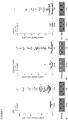
Figure 1 . NLRP1, ASC, and Caspase-1 are biomarkers that predict outcome after SCI. CSF samples were immunoblotted with antibodies against NLRP1, caspase-1 and ASC. CSF samples from uninjured patients were used as controls. Immunoblot analysis of 6 different cases of patients with SCI indicates that patients (2, 3 and 4) who express low levels of caspase-1 acutely after SCI have a better prognosis than subjects (1, 5 and 6) who have elevated levels of this protein in CSF. -
Figure 2 . NLRP1 inflammasome proteins are expressed in cells of the CNS. Spinal cord sections were obtained from decedents that had injury to the spinal cord. Immunohistochemical analysis combined with light microscopy indicates that NLRP1 is expressed in neurons of the ventral horn (black arrows) myelinated axons (black arrow heads) and oligodendrocytes (yellow arrows) (top panel). Caspase-1 is expressed in swollen axons (spheroids, blue arrows), motor neurons (black arrows) and in oligodendrocytes (yellow arrows) (central panel). ASC is present in neurons in the ventral horn (black arrows), white matter oligodendrocytes (yellow arrow) and macrophages/microglia (blue arrow heads) (bottom panel). -
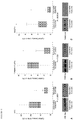
Figure 3 . Scatter plots of expression of inflammasome proteins in controls and patients with TBI. Samples were immunoblotted for ASC (A), caspase-1 (B), and NALP-1 (C). The p values in the upper left corner represent results of a Mann-Whitney U-test. Densitometric analysis revealed a significant increase in expression of ASC, caspase-1 (p20), and NALP-1 in the CSF of patients with TBI compared with nontrauma controls. Solid lines denote mean values for each group. Different shapes correspond to patient outcomes at 5 months postinjury. Representative immunoblots are shown. Samples were run on the same gel but were noncontiguous. N = the number of TBI samples analyzed; ★ =GOS Score 5; ■ =GOS Score 4;=
GOS Score 3; ∇ =GOS Score 1. -
Figure 4 . Box plots of expression of inflammasome proteins sorted by outcome category. The ends of the whiskers represent the lowest datum within 1.5 interquartile range of the lower quartile and the highest datum within 1.5 interquartile range of the upper quartile. The asterisks represent the outliers. Mann-Whitney U-tests indicate higher expression of ASC (A), caspase-1 (p20) (B), and NALP-1 (C) are significantly associated with anunfavorable outcome 5 months after injury (p < 0.0001). Representative immunoblots for each protein are shown. Samples were run on the same gel but were noncontiguous. -
Figure 5 . Scatter plots and estimated linear regression of ASC (A), caspase-1 (p20) (B), and NALP-1 (C) expression in the CSF with GOS score. Probability values of the linear regression are shown in the top left of each graph. Expression of each protein correlated significantly with GOS score at 5 months post-injury. The p values on the x axis represent post hoc comparisons of a Kruskal-Wallis test. Representative immunoblots are shown. Samples were run on the same gel but were noncontiguous. -
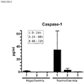
Figure 6 . Caspase-1 levels in CSF one, two, and three days following TBI in pediatric patients receiving hypothermia treatment or no treatment (normothermia). - The present invention is based, in part, on the discovery that NLRP1 inflammasomes play an important role in inflammatory responses after SCI and TBI in humans. In particular, the present inventors have surprisingly found that nucleotide-binding leucine-rich repeat pyrin domain containing protein 1 (NLRP1), the adaptor protein apoptosis-associated speck-like protein containing a caspase recruitment domain (ASC), and caspase-1 are secreted into the cerebrospinal fluid (CSF) of human patients following SCI and TBI. Thus, these inflammasome proteins represent sensitive biomarkers of the severity of central nervous system injury in human patients. Accordingly, the present invention provides a method of assessing the severity of a central nervous system injury in a patient by measuring the level of at least one inflammasome protein in a biological sample obtained from the patient, wherein the measured level of said at least one inflammasome protein is indicative of the severity of the central nervous system injury in the patient.
- As used herein, the term "inflammasome" refers to a multi-protein complex that activates caspase-1 activity, which in turn regulates IL-1β, IL-18 and IL-33 processing and activation. See Arend et al. 2008; Li et al. 2008; and Martinon et al. 2002. An "inflammasome protein" is a protein component of inflammasome complexes and can include, but is not limited to, an nucleotide binding domain, leucine-rich repeat containing (NLR) family member (e.g. NLRP1), ASC, caspase-1, caspase-11,
X-linked inhibitor of apoptosis protein (XIAP), and pannexin-1. NLRP1 is also known as NAcht leucine-rich-repeat protein 1 (NALP-1). Thus, the terms "NLRP1" and "NALP-1 are used interchangeably throughout the disclosure. In certain embodiments, the method comprises measuring an inflammasome protein selected from the group consisting of NLRP1 (NALP-1), ASC, caspase-1, or combinations thereof. In one embodiment, the p20 subunit of active caspase-1 is measured. - The terms "patient" or "subject" are used interchangeably herein, and is meant a mammalian subject to be treated, with human patients being preferred. In certain embodiments, the patient is a pediatric patient. Pediatric patients include newborns (birth to 1 month of age), infants (1 month to 2 years of age), children (2 to 12 years of age), and adolescents (12-21 years of age). In some cases, the methods of the invention find use in experimental animals, in veterinary application, and in the development of animal models for disease, including, but not limited to, rodents including mice, rats, and hamsters, and primates.
- In certain embodiments, the present invention provides a method of evaluating a patient suspected of having a central nervous system (CNS) injury, as set out in appended
claim 1. - A patient may be suspected of having a CNS injury on the basis of neurologic symptoms (motor, sensory, cognitive) and/or radiological evaluation (MRI, CT scan, X-ray) consistent with a CNS injury, e.g., after a physician's exam. In some embodiments, a patient suspected of having a CNS injury, particularly a spinal cord injury, may having a rating of A or B on the American Spinal Cord Injury Association (ASIA) Impairment Scale. The ASIA Impairment Scale is a standard diagnostic tool that assess a patient's motor and sensory function. The classification ratings and accompanying descriptions of the ASIA Impairment Scale are as follows:
Classification/Rating Description A Complete: no motor or sensory function is preserved below the level of injury, including the sacral segments S4-S5 B Incomplete: sensory, but not motor, function is preserved below the neurologic level and some sensation in the sacral segments S4-S5 C Incomplete: motor function is preserved below the neurologic level, however, more than half of key muscles below the neurologic level have a muscle grade less than 3 (i.e., not strong enough to move against gravity) D Incomplete: motor function is preserved below the neurologic level, and at least half of key muscles below the neurologic level have a muscle grade of 3 or more (i.e., joints can be moved against gravity) E Normal: motor and sensory functions are normal - In other embodiments, a patient suspected of having a CNS injury may have a score of ≤ 12 (e.g. 3 to 12) on the Glasgow Coma Scale (GCS). In still other embodiments, the patient may have a GCS score of ≤ 8 (e.g. 3 to 8). The GCS is a neurological scale commonly used to assess the level of consciousness of patients after injury or trauma. The scale is composed of three tests (eye, verbal and motor responses), each of which is assigned a value on a scale up to 6. The three values separately as well as their sum are considered. The lowest possible GCS score (the sum) is 3 (deep coma or death), while the highest is 15 (fully awake person). A GCS score < 9 is indicative of severe brain injury whereas a GCS score ≥ 13 is indicative of minor brain injury. A GCS score between 9-12 is generally indicative of a moderate brain injury.
- A patient suspected of having a CNS injury may have one or more signs and symptoms of CNS injury, such as temporary loss of consciousness, confusion, disorientation, memory or concentration problems, headache, dizziness, loss of balance, nausea or vomiting, sensory disruptions (e.g. blurred vision, ringing in the ears, bad taste in the mouth, loss of sensation in limbs), loss of motor function, sensitivity to light or sound, mood changes or mood swings, depression or anxiety, fatigue, drowsiness, and sleep disturbances.
- In some embodiments, the level, concentration, or abundance of one or more inflammasome proteins is measured in a biological sample obtained from a patient (e.g. a patient suspected of having or suffering from a CNS injury). In particular embodiments, the levels, concentrations, or abundance of one or more inflammasome proteins is indicative of the severity of CNS injury in the patient. A CNS injury includes, but is not limited to, a traumatic brain injury, a stroke- related injury, a cerebral aneurism-related injury, a spinal cord injury (e.g. contusions, compressions, lacerations), concussion-related injury (including post-concussion syndrome), cerebral ischemia, injury resulting from neurodegenerative diseases (including Parkinson's disease, Dementia Pugilistica, Huntington's disease, Alzheimer's disease, Creutzfeldt-Jakob disease), seizure-related injuries, multiple sclerosis, amyotrophic lateral sclerosis, and other CNS traumas. In certain embodiments, the levels, concentrations, or abundance of one or more inflammasome proteins is indicative of the severity of traumatic brain injury or spinal cord injury in the patient.
- As used herein, "biological sample" refers to any bodily fluid or tissue obtained from a patient or subject. A biological sample can include, but is not limited to, whole blood, red blood cells, plasma, serum, peripheral blood mononuclear cells (PBMCs), urine, saliva, tears, buccal swabs, CSF, CNS microdialysate, and nerve tissue. In one embodiment, the biological sample is CSF, serum or plasma. In certain embodiments, the biological sample is CSF.
- In some embodiments, the measured level, concentration, or abundance of one or more inflammasome proteins in the biological sample is used to prepare an inflammasome protein profile, wherein the profile is indicative of the severity of a CNS injury in the patient or the patient's prognosis or recovery potential from a CNS injury. The inflammasome protein profile may comprise the level, abundance, or concentration of one or more inflammasome proteins measured in the patient's sample optionally in relation to a pre-determined value or range of reference values as described herein. The inflammasome proteins in the profile include NLRP1 (NALP-1), ASC, and/or caspase-1 (e.g. p20 subunit of caspase-1). In one particular embodiment, the inflammasome protein profile comprises the level, abundance, or concentration of each of NLRP1 (NALP-1), ASC, and caspase-1 (e.g. p20 subunit of caspase-1).
- In one aspect of the invention, the method of evaluating a patient suspected of having a CNS injury comprises determining the presence or absence of a protein signature associated with a CNS injury or a more severe CNS injury based on the measured level, abundance, or concentration of one or more inflammasome proteins in the patient sample or on the inflammasome protein profile prepared from the patient's sample. In certain embodiments, the protein signature comprises an elevated level of at least one inflammasome protein. The level of said at least one inflammasome protein in the protein signature may be enhanced relative to the level of the protein in a control sample or relative to a pre-determined reference value or range of reference values as further described herein. The protein signature may, in certain embodiments, comprise an elevated level for each of caspase-1 (e.g. p20 subunit of caspase-1), NLRP1, and ASC. Patients who exhibit the protein signature may be selected or identified as having a CNS injury or a more severe CNS injury.
- The level or concentration of at least one inflammasome protein can be assessed at a single time point (e.g. after a potential CNS injury) and compared to a pre-determined reference value or range of reference values or can be assessed at multiple time points (e.g. two, three, four, five or more) after a potential CNS injury and compared to a pre-determined reference value or to previously assessed values. For instance, a biological sample for measuring levels or concentrations of inflammasome proteins can be obtained from a patient within one hour of a potential CNS injury to two weeks following a potential CNS injury. In some embodiments, the biological sample is obtained within one day, two days, three days, four days, five days, six days, seven days, ten days, or twelve days of a CNS injury or potential injury.
- As used herein, "pre-determined reference value" refers to a pre-determined value of the level or concentration of an inflammasome protein ascertained from a known sample. For instance, the pre-determined reference value can reflect the level or concentration of an inflammasome protein in a sample obtained from a control subject (i.e., an uninjured, healthy subject). The control subject may, in some embodiments, be age-matched to the patients being evaluated. Thus, in particular embodiments, the measured level or concentration of at least one inflammasome protein is compared or determined relative to the level or concentration of said at least one inflammasome protein in a control sample (i.e. obtained from an uninjured subject).
- In other embodiments, the pre-determined reference value or range of reference values can reflect the level or concentration of an inflammasome protein in a sample obtained from a patient with a known severity of CNS injury as assessed by clinical measures or post mortem analysis. A pre-determined reference value can also be a known amount or concentration of an inflammasome protein. Such a known amount or concentration of an inflammasome protein may correlate with an average level or concentration of the inflammasome protein from a population of control subjects or a population of patients with known levels of injury. In another embodiment, the pre-determined reference value can be a range of values, which, for instance, can represent a mean plus or minus a standard deviation or confidence interval. A range of reference values can also refer to individual reference values for a particular inflammasome protein across various levels of CNS injury severity. In certain embodiments, an increase in the level of one or more inflammasome proteins (e.g., NLRP1 (NALP-1), ASC, or caspase-1) relative to a pre-determined reference value or range of reference values is indicative of a more severe central nervous system injury.
- In some embodiments, the method of assessing the severity of a CNS injury further comprises measuring the level or concentration of one or more proteins described in
U.S. Patent Publication No. 2011/0177974 , in addition to measuring the level or concentration of one or more inflammasome proteins. For instance, in certain embodiments, the method further comprises measuring the level or concentration of one or more proteins selected from ubiquitin C-terminal hydrolase L1; vesicular membrane protein p-24; synuclein; microtubule-associated protein; synaptophysin; Vimentin; Synaptotagmin; Synaptojanin-2; Synapsin2; CRMP1, 2; Amphiphysin-1; PSD95; PSD-93; Calmodulin dependent protein kinase II (CAMPK)-alpha, beta, gamma; Myelin basic protein (MBP); Myelin proteolipid protein (PLP); Myelin Oligodendrocyte specific protein (MOSP); Myelin Oligodendrocyte glycoprotein (MOG); myelin associated protein (MAG); NF-H; NF-L; NF-M; BIII-tubulin-1 or combinations thereof in the biological sample obtained from the patient in addition to measuring the level or concentration of one or more inflammasome proteins. Thus, the protein signature may comprise an elevated level of one or more of these proteins in addition to the elevated level of one or more inflammasome proteins. In other embodiments, the method further comprises measuring the level or concentration of one or more proteins selected from S-100β, neuron-specific enolase, neurofilament light chain, glial fibrillary acidic protein (GFAP) or combinations thereof in the biological sample obtained from the patient in addition to measuring the level or concentration of one or more inflammasome proteins. In one embodiment, the protein signature associated with a CNS injury or a more severe CNS injury comprises an elevated level of one or more proteins selected from S-100β, neuron-specific enolase, neurofilament light chain, glial fibrillary acidic protein (GFAP) in addition to an elevated level of one or more inflammasome proteins (e.g. NLRP1 (NALP-1), ASC, or caspase-1). - Methods of assessing the severity of a CNS injury in a patient or evaluating a patient suspected of having a CNS injury may further comprise administering a neuroprotective treatment to the patient based on the measured level of said at least one inflammasome protein or when a protein signature associated with a CNS injury or a more severe CNS injury is identified. Such neuroprotective treatments include drugs that reduce excitotoxicity, oxidative stress, and inflammation. Thus, suitable neuroprotective treatments include, but are not limited to, methylprednisolone, 17α-estradiol, 17β-estradiol, ginsenoside, progesterone, simvastatin, deprenyl, minocycline, resveratrol, and other glutamate receptor antagonists (e.g. NMDA receptor antagonists) and antioxidants. In some embodiments, neuroprotective treatments are neutralizing antibodies against an inflammasome protein or binding fragments thereof, such as those described in
U.S. Patent Publication No. 2009/0104200 . For instance, in one embodiment, the neuroprotective treatment is an anti-ASC antibody or fragment thereof. Anti-ASC antibodies include antibodies that specifically bind to amino acid residues 178-193 of rat ASC (accession number BAC43754), e.g., amino acid sequence ALRQTQPYLVTDLEQS (SEQ ID NO:1), or antibodies that specifically bind to the amino acid sequence RESQSYLVEDLERS (SEQ ID NO:2) of human ASC. In another embodiment, the neuroprotective treatment is an anti-NLRP1 antibody or fragment thereof. Suitable neutralizing anti-NLRP1 antibodies or fragments thereof include antibodies that specifically bind to the amino acid sequence CEYYTEIREREREKSEKGR (SEQ ID NO:3) of human NLRP1 or the amino acid sequence MEESQSKEESNTEG (SEQ ID NO: 4) of rat NLRP1. The neutralizing antibodies or antibody fragments may be polyclonal antibodies, monoclonal antibodies, chimeric antibodies, humanized antibodies, single-chain variable fragments (scFvs) and the like. Aptamers that specifically bind to an inflammasome protein or epitope thereof (e.g., SEQ ID NOs: 1-4) may also be suitable neuroprotective treatments. Neuroprotective treatments also encompass therapeutic regimens or rehabilitative procedures, such as hypothermia treatment. - The success of, or response to, treatment can also be monitored by measuring the levels of at least one inflammasome protein. Accordingly, in some embodiments, the methods of evaluating a patient further comprise measuring the level of at least one inflammasome protein in a biological sample obtained from the patient following neuroprotective treatment, preparing a treatment protein signature associated with a positive response to the neuroprotective treatment, wherein the treatment protein signature comprises a reduced level of at least one inflammasome protein, and identifying patients exhibiting the presence of the treatment protein signature as responding positively to the neuroprotective treatment. A reduction in the level, abundance, or concentration of one or more inflammasome proteins (e.g. NLRP1, ASC, and caspase-1) is indicative of the efficacy of the neuroprotective treatment in the patient. The one or more inflammasome proteins measured in the sample obtained following treatment may be the same as or different than the inflammasome proteins measured in the sample obtained prior to treatment. The inflammasome protein levels may also be used to adjust dosage or frequency of a neuroprotective treatment.
- The present invention also provides a method of determining a prognosis for a patient with a central nervous system injury, as set out in appended claim 8. In certain preferred embodiments, the biological sample is obtained from the patient within five days, or within three days of injury. In some embodiments, an increase in the level of one or more inflammasome proteins (e.g., NLRP1, ASC, caspase-1, or combinations thereof) relative to a pre-determined reference value or range of reference values is indicative of a poorer prognosis. For instance, an increase of about 20% to about 300% in the level of one or more inflammasome proteins relative to a pre-determined reference value or range of reference values is indicative of a poorer prognosis. In one embodiment, increased levels of caspase-1, particularly the p20 subunit of active caspase-1, relative to a pre-determined reference value or range of reference values acutely after injury (i.e. within a week of injury) is indicative of a poorer prognosis.
- In particular embodiments, an elevated level of at least one inflammasome protein relative to a pre-determined reference value or range of reference values is predictive of the patient's recovery potential or long-term outcome as assessed by the Glasgow Outcome Scale (GOS). The GOS is a scale that allows for the objective assessment of a patient's recovery following brain injury. The scale is comprised of scores ranging from 1 to 5 with the following descriptions:
Score/Category Description 1-Death Severe injury or death without recovery of consciousness 2-Persistent Vegetative State Severe damage with prolonged state of unresponsiveness and a lack of higher mental functions 3-Severe Disability Severe injury with permanent need for help with daily living 4-Moderate Disability No need for assistance in everyday life, employment is possible but may require special equipment 5-Low Disability Light damage with minor neurological and psychological deficits. - The inflammasome proteins of the invention and other marker proteins can be measured in a biological sample by various methods known to those skilled in the art. For instance, proteins can be measured by methods including, but not limited to, liquid chromatography, gas chromatography, mass spectrometry, radioimmunoassays, immunofluorescent assays, FRET-based assays, immunoblot, ELISAs, or liquid chromatography followed by mass spectrometry (e.g., MALDI MS). One of skill in the art can ascertain other suitable methods for measuring and quantitating any particular biomarker protein of the invention.
- Also described are kits for preparing an inflammasome protein profile associated with CNS injury, such as spinal cord injury or traumatic brain injury. The kits may include a reagent for measuring at least one inflammasome protein and instructions for measuring said at least one inflammasome protein for assessing the severity of a central nervous system injury in a patient. As used herein, a "reagent" refers to the components necessary for detecting or quantitating one or more proteins by any one of the methods described herein. For instance, in some embodiments, kits for measuring one or more inflammasome proteins can include reagents for performing liquid or gas chromatography, mass spectrometry, immunoassays, immunoblots, or electrophoresis to detect one or more inflammasome proteins as described herein. In some embodiments, the kit includes reagents for measuring one or more inflammasome proteins selected from NLRP1, ASC, caspase-1, or combinations thereof.
- In one embodiment, the kit comprises a labeled-binding partner that specifically binds to one or more inflammasome proteins, wherein said one or more inflammasome proteins are selected from the group consisting of NLRP1, ASC, caspase-1, and combinations thereof. Suitable binding partners for specifically binding to inflammasome proteins include, but are not limited to, antibodies and fragments thereof, aptamers, peptides, and the like. In certain embodiments, the binding partners for detecting NLPR1 are antibodies or fragments thereof, aptamers, or peptides that specifically bind to the amino acid sequence of SEQ ID NO: 3 or SEQ ID NO: 4 of human NLRP1 and rat NLRP1, respectively. In other embodiments, the binding partners for detecting ASC are antibodies or fragments thereof, aptamers, or peptides that specifically bind to the amino acid sequence of SEQ ID NO: 1 or SEQ ID NO: 2 of rat ASC and human ASC, respectively. Labels that can be conjugated to the binding partner include metal nanoparticles (e.g., gold, silver, copper, platinum, cadmium, and composite nanoparticles), fluorescent labels (e.g., fluorescein, Texas-Red, green fluorescent protein, yellow fluorescent protein, cyan fluorescent protein, Alexa dye molecules, etc.), and enzyme labels (e.g., alkaline phosphatase, horseradish peroxidase, beta-galactosidase, beta-lactamase, galactose oxidase, lactoperoxidase, luciferase, myeloperoxidase, and amylase).
- In some embodiments, the kit can include reagents for measuring one or more inflammasome proteins in CSF samples. In other embodiments, the kits can include reagents for measuring one or more inflammasome proteins in other patient samples including nerve tissue, CNS microdialysate, blood, saliva, serum, plasma, or urine. In still other embodiments, the kits further comprise a set of reference values to which the measured level of one or more inflammasome proteins can be compared.
- To determine whether NLRP1 inflammasome proteins were present in cerebrospinal fluid (CSF) following spinal cord injury (SCI), CSF samples from seven patients with SCI or control patients were analyzed for levels of nucleotide-binding leucine-rich repeat pyrin domain containing protein 1 (NLRP1; also known as NAcht leucine-rich-repeat protein 1 (NALP-1)), apoptosis-associated speck-like protein containing a caspase recruitment domain (ASC), and caspase-1. The American Spinal Cord Injury Association (ASIA) scale of the SCI patients at admission to the emergency department ranged from AIS A to B. Information regarding the diagnosis, procedures and outcomes of the patients is shown in Table 1. None of the patients had any complications. CSF from uninjured individuals was obtained as a control from three males and two females ranging from 67 to 91 years old.
- For detection of inflammasome proteins, CSF samples were prepared with Laemali buffer. Immunoblot analysis was carried with the Criterion system (Bio-Rad) as described previously (de Rivero Vaccari et al., 2008) using antibodies (1:1000 dilution) to NLRP1 (Bethyl Laboratories), Caspase-1 (Imgenex) and ASC (Santa Cruz). Proteins were resolved in 14-20% TGX Criterion precasted gels (Bio-Rad), transferred to polyvinylidene difluoride (PVDF) transfer membranes (Tropifluor - Applied Biosystems) and placed in blocking buffer (PBS, 0.1% Tween-20, 0.4% I-Block (Applied Biosystems) and then incubated for one hour with primary antibodies. Membranes were then incubated for one hour with anti-mouse, anti-rat or anti-rabbit horseradish peroxidase (HRP)-linked antibodies. Signal visualization was performed by enhanced chemiluminescence.
- Immunoblot analysis of control CSF samples (n=5) revealed very low levels of NLRP1 inflammasome proteins (
Figure 1 ). In contrast, immunoblot analysis of samples from 6 different SCI patients showed an increase in the levels of NLRP1, caspase-1 and ASC in the CSF when compared to CSF from control subjects. It should be noted thatpatients Figure 1 ) did not show increased levels of caspase-1 acutely (day 0 through day 2) after SCI. Interestingly, these patients demonstrated stark motor improvement at 2 days after SCI.Patients - The results from these experiments show that protein levels of NLRP1, ASC, and caspase-1 in CSF are increased following injury to the central nervous system and suggest that levels of these inflammasome proteins can serve as biomarkers of the severity of neuronal damage following injury thereby directing treatment and rehabilitation efforts, monitoring response to treatment, and aiding in the determination of prognosis of recovery in injured patients.
- Spinal cord sections were obtained from nine decedents (8 males and 1 female with ages ranging from 20 to 77 years) who had injury to the spinal cord due to vertebral fractures. The spinal cord injury was assessed microscopically, using bright field optics, by examining one H&E or H&E/DAB-stained section from the lesion center of each case or from cervical, thoracic and lumbar sections from control cases. The spinal cord injuries were classified on the basis of their histological appearance as "contusion/cyst," massive compression, or laceration (Fleming et al., 2006). Contusional injuries were characterized by an intact pia and relative preservation of the anatomical relations of various elements of the spinal cord, and variable degrees of injury ranging from involvement of the entire cross-sectional area to large usually asymmetric areas of tissue damage. Massive compression injuries were characterized by disruption of the pia and severe distortion and disruption of spinal cord parenchyma. Laceration injuries, which by definition were perforating or penetrating injuries caused by weapons or projectiles, were associated with breaching of the pia and linear tearing of the cord tissue.
- All tissue samples had been removed within 24 h of death and fixed in neutral buffered formalin. Blocks from the spinal cords were dehydrated, embedded in paraffin wax, cut into 6 µm thick sections and placed on positively charged glass slides. One set of sections was stained with hematoxylin-eosin (H&E) and the remaining sets were used for immunohistochemistry. Paraffin-embedded sections were stained with anti-NLRP1 (Bethyl Laboratories as described in de Rivero Vaccari et al. 2008), anti-caspase-1 (Upstate), and anti-ASC (Chemicon) using diaminobenzidine (DAB) as the chromophore and hematoxylin. Negative controls included sections in which the primary antibody was omitted and sections incubated with isotype-matched antibodies (1:100-1:10,000 IgG). These positive and negative controls were processed with every batch of immunohistochemical slides.
- In all cases, tissue samples from the center of injury and at various distances above and below the injury were obtained. The data from tissue from the center of the lesion were used to compare the inflammatory responses between cases whereas those from the remote, uninjured segments of the spinal cord served as within-case controls. Between-case comparison of the remote samples was not possible because, for different cases, the distance of these samples from the lesion center was variable.
- Immunohistochemical analysis combined with light microscopy indicated that NLRP1 is expressed in neurons of the ventral horn (black arrows), myelinated axons (arrow heads) and oligodendrocytes (yellow arrows) in injured spinal cords (
Figure 2 ). Moreover, NLRP1 immunoreactivity in areas of the penumbra (C7) was higher than in areas distant to the epicenter (L2). - DAB immunoreactivity for caspase-1 was detected in swollen axons (spheroids, blue arrows) (
Figure 2 ), and arterioles (not shown). At areas of the penumbra, caspase-1 staining is present in motor neurons (black arrows) of the ventral horn, and in the white matter in oligodendrocytes (yellow arrows). Caspase-1 immunoreactivity in oligodendrocytes (yellow arrows) was the same at all levels of the spinal cords examined, regardless of proximity to the epicenter. At areas distant to the epicenter (T12), caspase-1 was also present in motor neurons (black arrows) but with decreased immunoreactivity than the penumbra (C7). This finding indicates that caspase-1 immunoreactivity in neurons decreases as the distance to the epicenter increases, similarly to NLRP1. - At areas of the penumbra (C7) and distant to the epicenter (L2), neurons in the ventral horn (black arrows) and white matter oligodendrocytes (yellow arrow) showed ASC immunoreactivity. In addition, ASC was also present in macrophages/microglia at the epicenter (blue arrow heads). Moreover, ASC immunoreactivity was also detected in the substantia gelatinosa (dorsal horn) at C7 and L2 (not shown).
- Neuroinflammation has been considered to play a critical role in the pathogenesis of SCI and TBI, but the role of the innate immune response has not been examined directly. The innate immune system senses microbial and viral pathogen-associated molecular patterns and danger signals released from damaged or stressed cells to trigger conserved intracellular signaling pathways that drive proinflammatory responses that are critical for productive innate and adaptive immunity. Excessive inflammatory responses become deleterious leading to tissue destruction. The results of this experiment provide evidence demonstrating that the NLRP1 inflammasome signaling system is activated in the innate immune response in damaged human spinal cord and brain tissue after trauma. These findings support the idea that activation of the NLRP1 inflammasome signaling system is an early event in spinal cord and brain pathology and that these proteins may serve as biomarkers for SCI and TBI in humans.
- To determine whether inflammasome proteins may serve as biomarkers for other types of central nervous system injury, a total of 45 CSF samples were collected from 23 traumatic brain injury (TBI) patients on the day of injury and up to three days after the injury and analyzed by immunoblot for levels of NALP-1 (also known as NLRP1), ASC, and caspase-1. Each of the patients presented with the following inclusion criteria: severe or moderate head trauma (Glasgow Coma Scale (GCS) score ≤ 12),
age 1 month to 65 years, and ventriculostomy. Twenty-two of the patients suffered severe brain trauma (GCS score ≤ 8) and 1 suffered moderate brain trauma (moderate TBI GCS score range 9-12). Nine patients (5 men and 4 women) with a mean age of 66.3 years (range 29-91 years) served as controls. Control patients required a ventriculostomy for nontraumatic pathology. Patients with acute meningitis, cerebral vasculitis, or other recent CNS infection were excluded. Information regarding patient demographics, intracranial pathology, GCS score at presentation, and Glasgow Outcome Scale (GOS) score at 5 months post-injury is shown in Table 2. - Cerebrospinal fluid samples were collected within 12 hours of injury and up to 72 hours after injury. Samples were centrifuged at 2000g for 10 minutes at 4°C to pellet cellular bodies and debris. Supernatants were resolved by gel electrophoresis and immunoblotted as previously described (de Rivero Vaccari et al., 2008). Quantification of band density was performed with UNSCAN-IT gel digitizing software (Silk Scientific). Due to the low volume of sample available, NALP-1 was analyzed in 6 of the 9 controls, caspase-1 was analyzed in 43 of the 45 TBI samples, and NALP-1 was analyzed in 42 of the 45 TBI samples. Immunoblot analysis shows that the inflammasome proteins ASC, caspase-1 (p20), and NALP-1 are present in the CSF of patients with TBI and nontrauma controls. Quantitative data from a densitometric analysis are shown in
Figure 3 . Expression of the 22-kD isoform of ASC (Fig. 3A ), the p20 subunit of cleaved caspase-1 (Fig. 3B ), and NALP-1 (Fig. 3C ) is significantly elevated in the CSF of TBI patients compared with nontrauma controls (p < 0.0001, p = 0.0029, and p = 0.0202, respectively). - To determine if the levels of inflammasome components correlate with outcome, we grouped study participants by outcome category (
GOS Scores GOS Scores Fig. 4A ), caspase-1 (p20) (Fig. 4B ), and NALP-1 (Fig. 4C ) in the CSF of TBI patients with unfavorable outcomes, including death and severe disability with complete dependence on others for activities of daily living (p < 0.0001). - To further understand the relationship between outcome and inflammasome proteins, we constructed modified scatter plots of expression levels of ASC, caspase-1 (p20), and NALP-1 and GOS (
Figure 5 ). A calculated linear regression line is shown for each plot. Linear regression analysis shows that expression of ASC (Fig. 5A ; p < 0.05), caspase-1 (p20) (Fig. 5B ; p < 0.01), and NALP-1 (Fig. 5C ; p < 0.05) correlate significantly with outcome at 5 months. Post hoc, the Dunn multiple comparison tests following a Kruskal-Wallis test showed that the levels of ASC are significantly higher in patients with severe disability (GOS Score 3) compared with patients with moderate disability (GOS Score 4) (p < 0.001) and patients with mild to no disability (GOS Score 5) (p < 0.05). Similarly, expression levels of caspase-1 (p20) and NALP-1 are significantly higher in patients with severe disability (GOS Score 3) than in those with moderate disability (GOS Score 4) (p < 0.001). - The results of this study show that inflammasome proteins (e.g. ASC, NALP-1, and caspase-1) are acutely elevated (e.g. within 72 hours) in the CSF of patients with TBI as compared with nontrauma controls. Elevation of these proteins likely reflects the extent of neuroinflammation, suggesting that inflammasome proteins can serve as acute biomarkers of CNS injury. These findings are clinically relevant, as CSF biomarkers are more specific indicators of neuropathology than serum biomarkers. Cerebrospinal fluid directly bathes the brain, closely reflecting the extracellular milieu and biochemical changes that are specific to the CNS. Sampling the CSF eliminates influences of multiorgan trauma or other systemic pathology represented in the serum, which is significant as patients with TBI often present with trauma to other organ systems.
- The results also demonstrate that levels of inflammasome proteins are significantly higher in the CSF of patients who have died and those with severe disability than in patients with moderate to no disability, suggesting that inflammasome activation produces chronic neuroinflammation, contributing to secondary injury and
poor outcome 5 months after TBI. The extent of acute elevation of these proteins can predict an unfavorable versus favorable outcome. Such markers could also direct treatment and rehabilitation efforts. The clinician would target therapies to patients identified as having a greater risk of inflammation-mediated secondary injury. - Response to treatment could be monitored by following the levels of ASC, active caspase-1, and NALP-1 in the CSF. One such treatment, therapeutic hypothermia, attenuates the endogenous inflammatory response of the CNS to TBI by decreasing cytokine production and reducing activation of astrocytes and microglia (Aibiki et al., 1999; Goss et al., 1995; Kumar et al., 1997; Truettner et al., 2005), and cortical neurons exposed to moderate hypothermia in culture show a decrease in activation of the inflammasome (Tomura et al., in press). Thus, ASC, active caspase-1, and NALP-1 can serve as objective, biochemical indicators of treatment efficacy for patients with CNS injury.
- To evaluate whether inflammasome proteins, such as caspase-1, can also be used to monitor treatment efficacy in TBI patients, CSF caspase-1 levels obtained from pediatric patients who received hypothermia treatment following TBI were compared to those obtained from pediatric patients who did not receive treatment following TBI. Cerebrospinal fluid of pediatric patients (ages 0.1 to 16 years) was obtained at different times after traumatic brain injury (
day Figure 6 , the data indicate that within 24 hours after injury the levels of caspase-1 were lower in the hypothermia group when compared to the normothermia group. Thus post-traumatic therapeutic hypothermia lowers the levels of caspase-1 activation after brain injury, consistent with findings that those patients that receive hypothermia treatment present better outcomes when compared to those patients who do not get the hypothermia treatment.Table 1. Spinal Cord Injury Subjects Patient Age Gender Race Mechanism of Injury Spinal Injury AIS Grade Level Surgery Hypothermia Other injuries Exam at Rehab D/ C 1 43 M Black Auto vs. Pedestrian C7/T1 bilateral jumped facets A C7 C4-T2 Laminectomy w/instrumented fusion Yes None C7 ASIA A 2 38 M Hispanic Fall from height T3 & T4 Burst fractures B T3 T1-6 Laminectomy w/instrumented fusion No None T3 ASIA D 3 21 M White Driving accident C5/6 Fracture dislocation A C5 C5/6 Anterior disectomy & fusion Yes None C5 ASIA C 4 19 M Black Motor vehicle accident C4/5 Bilateral jumped facets B C4 C4/5 Anterior disectomy & fusion No None C4 ASIA D 5 22 M White Rugby accident C5/6 Bilateral jumped facets A C5 C5/6 Anterior disectomy & fusion Yes None C6 ASIA A 6 48 M Hispanic Motorcycle vs. Pedestrian T1/2 Fracture dislocation A T3 C5-T2 Laminectomy w/instrumented fusion No Degloving injury and fractures of face T3 ASIA B Table 2. Summary of demographic data in patients with TBI* Case No. Age (yrs), Sex Race Mechanism of Injury GCS Score† GOS Score‡ Intracranial Pathology 1 26, M White MVA 3 4 bilat temporal cortical contusions, SAH, SDH 2 22, M Hispanic motorcycle accident 3 4 SDH, diffuse SAH 3 19, F Hispanic MVA 7 3 fronto- & temporoparietal & basal ganglia hemorrhagic contusions, frontoparietal SAH 4 30, F Hispanic MVA 8 4 scattered SAH, diffuse cerebral edema 5 21, M Hispanic motorcycle accident 5 3 frontal SDH & SAH 6 17, F Hispanic MVA 4 3 diffuse extraaxial & intraparenchymal hemorrhage 7 26, M Hispanic MVA 8 3 diffuse SAH, IVH, & parenchymal hemorrhagic contusions 8 36, M White ATV accident 3 5 SDH, SAH, & diffuse cerebral edema 9 16, M Hispanic MVA 7 5 frontotemporal SDH, frontal lobe contusion, cerebral edema 10 22, M Hispanic MVA 3 3 SDH 11 36, M Black Gunshot wound 3 1 SDH, bullet fragments in frontal lobe 12 54, M Hispanic Fall 3 3 frontal hemorrhagic contusions, parietooccipital hemorrhagic contusion, SDH, brain edema 13 62, M Black MVA 8 3 diffuse frontoparietal hemorrhage, frontal & parietal lobe hemorrhagic contusions 14 49, F White Assault 8 3 temporal & frontal lobe hemorrhagic contusions, frontotemporal SAH, frontal dural hematoma 15 20, M Hispanic Motorcycle accident 6 4 bone fragments in frontoparietal brain parenchyma, scattered frontoparietal SAH, hemorrhagic contusions 16 28, M Hispanic Motorcycle accident 4 3 SDH 17 21, M Black MVA 5 4 mild cerebral edema 18 45, M Hispanic Gunshot wound 8 3 bullet fragments in occipital lobe, occipital SDH, minimal parietal & occipital pneumocephalus 19 21, M White MVA 3 3 diffuse axonal injury, scattered SAH, parietal SDH, mild hydrocephalus 20 17, M Hispanic Sports injury 4 4 SDH, SAH, diffuse cerebral edema 21 19, F Hispanic MVA 11 5 bone fragments in frontal lobe parenchyma, frontal lobe contusion, edema, pneumocephalus 22 65, M White MVA 7 1 parietal SDH, SAH, parietal hemorrhagic contusion, uncal herniation 23 18, M White MVA 3 1 diffuse axonal injury, scattered hemorrhagic contusions in frontal, temporal, parietal lobes & corpus callosum, IVH * IVH = intraventricular hemorrhage; SAH = subarachnoid hemorrhage; SDH = subdural hematoma, MUA = motor vehicle accident; ATV = all terrain vehicle
† Obtained on admission.
‡ Assessed at 5 months postinjury. -
- Abulafia DP, de Rivero Vaccari JP, Lozano JD, Lotocki G, Keane RW, Dietrich WD. Inhibition of the inflammasome complex reduces the inflammatory response after thromboembolic stroke in mice. J Cereb Blood Flow Metab 29:534-544, 2009.
- Aibiki M, Maekawa S, Ogura S, Kinoshita Y, Kawai N, Yokono S: Effect of moderate hypothermia on systemic and internal jugular plasma IL-6 levels after traumatic brain injury in humans. J Neurotrauma 16:225-232, 1999.
- Arend WP, Palmer G, Gabay C. IL-1, IL-18, and IL-33 families of cytokines. Immunol Rev 223:20-38,2008.
- Bianchi ME. DAMPs, PAMPs and alarmins: all we need to know about danger. J Leukoc Biol 81:1-5,2007.
- Brain Trauma Foundation, American Association of Neurological Surgeons, Joint Section on Neurotrauma and Critical Care: Initial management. J Neurotrauma 17:463-469, 2000.
- Cao F, Yang XF, Liu WG, Hu WW, Li G, Zheng XJ, Shen F, Zhao XQ, Lv ST. Elevation of neuron-specific enolase and S-100beta protein level in experimental acute spinal cord injury. J Clin Neurosci 15:541-544, 2008.
- Cornefjord M, Nyberg F, Rosengren L, Brisby H. Cerebrospinal fluid biomarkers in experimental spinal nerve root injury. Spine (Phila Pa 1976) 29:1862-1868, 2004.
- de Rivero Vaccari JP, Lotocki G, Alonso OF, Bramlett HM, Dietrich WD, Keane RW. Therapeutic neutralization of the NLRP1 inflammasome reduces the innate immune response and improves histopathology after traumatic brain injury. J Cereb Blood Flow Metab 29:1251-1261,2009.
- de Rivero Vaccari JP, Lotocki G, Marcillo AE, Dietrich WD, Keane RW. A molecular platform in neurons regulates inflammation after spinal cord injury. J Neurosci 28:3404-3414, 2008.
- Goss JR, Styren SD, Miller PD, Kochanek PM, Palmer AM, Marion DW, et al: Hypothermia attenuates the normal increase in .
- Guez M, Hildingsson C, Rosengren L, Karlsson K, Toolanen G. Nervous tissue damage markers in cerebrospinal fluid after cervical spine injuries and whiplash trauma. J Neurotrauma 20:853-858,2003.
- Kufer TA, Sansonetti PJ. NLR functions beyond pathogen recognition. Nat Immunol 12:121-128,2011.
- Kumar K, Wu X, Evans AT: GFAP-immunoreactivity following hypothermic forebrain ischemia. Metab Brain Dis 12:21-27, 1997.
- Li H, Willingham SB, Ting JP, Re F. Cutting edge: inflammasome activation by alum and alum's adjuvant effect are mediated by NLRP3. J Immunol 181:17-21, 2008.
- Loy DN, Sroufe AE, Pelt JL, Burke DA, Cao QL, Talbott JF, Whittemore SR. Serum biomarkers for experimental acute spinal cord injury: rapid elevation of neuron-specific enolase and S-100beta. Neurosurgery 56:391-397; discussion 391-397, 2005.
- Ma J, Novikov LN, Karlsson K, Kellerth JO, Wiberg M. Plexus avulsion and spinal cord injury increase the serum concentration of S-100 protein: an experimental study in rats. Scand J Plast Reconstr Surg Hand Surg 35:355-359, 2001.
- Martinon F, Burns K, Tschopp J. The inflammasome: a molecular platform triggering activation of inflammatory caspases and processing of proIL-beta. Mol Cell 10:417-426, 2002.
- Nagy G, Dzsinich C, Selmeci L, Sepa G, Dzsinich M, Kekesi V, Juhasz-Nagy A. Biochemical alterations in cerebrospinal fluid during thoracoabdominal aortic cross-clamping in dogs. Ann Vasc Surg 16:436-441, 2002.
- Pouw MH, Hosman AJ, van Middendorp JJ, Verbeek MM, Vos PE, van de Meent H. Biomarkers in spinal cord injury. Spinal Cord 47:519-525, 2009.
- Qu Y, Franchi L, Nunez G, Dubyak GR. Nonclassical IL-1 beta secretion stimulated by P2X7 receptors is dependent on inflammasome activation and correlated with exosome release in murine macrophages. J Immunol 179:1913-1925, 2007.
- Silverman WR, de Rivero Vaccari JP, Locovei S, Qiu F, Carlsson SK, Scemes E, Keane RW, Dahl G. The .
- Skouen JS, Brisby H, Otani K, Olmarker K, Rosengren L, Rydevik B. Protein markers in cerebrospinal fluid in experimental nerve root injury. A study of slow-onset chronic compression effects or the biochemical effects of nucleus pulposus on sacral nerve roots. Spine (Phila Pa 1976) 24:2195-2200, 1999.
- Teasdale G, Jennett B. Assessment of coma and impaired consciousness. A practical scale. Lancet 2:81-84, 1974.
- Tomura S, de Rivero Vaccari JP, Keane RW, Bramlett HM, Dietrich WD: Effects of therapeutic hypothermia on inflammasome signaling after traumatic brain injury. J Cereb Blood Flow Metab [in press], 2012.
- Truettner JS, Suzuki T, Dietrich WD: The effect of therapeutic hypothermia on the expression of inflammatory response genes following moderate traumatic brain injury in the rat. Brain Res Mol Brain Res 138:124-134, 2005.
- Described are also:
- A. A method of evaluating a patient suspected of having a central nervous system (CNS) injury comprising: providing a biological sample from a patient presenting with clinical symptoms consistent with a CNS injury; measuring the level of at least one inflammasome protein in the biological sample; determining the presence or absence of a protein signature associated with a CNS injury or a more severe CNS injury, wherein the protein signature comprises an elevated level of said at least one inflammasome protein; and selecting patients exhibiting the presence of the protein signature as having a CNS injury or a more severe CNS injury.
- B. The method of statement A, wherein the level of said at least one inflammasome protein in the protein signature is enhanced relative to the level of said at least one inflammasome protein in a control sample.
- C. The method of statement A, wherein the level of said at least one inflammasome protein in the protein signature is enhanced relative to a pre-determined reference value or range of reference values.
- D. The method of statement A, wherein the CNS injury is a spinal cord injury or traumatic brain injury.
- E. The method of statement A, wherein said at least one inflammasome protein is nucleotide-binding leucine-rich repeat pyrin domain containing protein 1 (NLRP1), apoptosis-associated speck-like protein containing a caspase recruitment domain (ASC), caspase-1, or combinations thereof.
- F. The method of statement F, wherein the protein signature comprises an elevated level for each of caspase-1, NLRP1, and ASC.
- G. The method of statement F, wherein the protein signature comprises an elevated level for the p20 subunit of caspase-1.
- H. The method of statement A, wherein said biological sample is obtained within one week of the suspected injury.
- I. The method of statement A, wherein said biological sample is obtained within five days of the suspected injury.
- J. The method of statement A, wherein said biological sample is obtained within three days of the suspected injury.
- K. The method of statement A, wherein said biological sample is cerebrospinal fluid (CSF), CNS microdialysate, saliva, serum, plasma, or urine.
- L. The method of statement A, wherein the patient has an A or B rating on the American Spinal Cord Injury Association (ASIA) Impairment Scale.
- M. The method of statement A, wherein the patient has a Glasgow Coma Scale (GCS) score of 3 to 12.
- N. The method of statement M, wherein the patient has a GCS score of 3 to 8.
- O. The method of statement A, wherein the patient is a pediatric patient.
- P. The method of statement A, wherein the level of said at least one inflammasome protein is measured by immunoblot or ELISA.
- Q. The method of statement A further comprising administering a neuroprotective treatment to the patient when said protein signature is identified.
- R. The method of statement Q, where in the neuroprotective treatment is hypothermia, methylprednisolone, 17α-estradiol, 17β-estradiol, ginsenoside, progesterone, simvastatin, deprenyl, minocycline, and resveratrol.
- S. The method of statement Q, further comprising: measuring the level of said at least one inflammasome protein in a biological sample obtained from the patient following neuroprotective treatment; preparing a treatment protein signature associated with a positive response to the neuroprotective treatment, wherein the treatment protein signature comprises a reduced level of at least one inflammasome protein; and identifying patients exhibiting the presence of the treatment protein signature as responding positively to the neuroprotective treatment.
- T. A kit for preparing an inflammasome protein profile associated with CNS injury comprising a labeled-binding partner that specifically binds to one or more inflammasome proteins, wherein said one or more inflammasome proteins are selected from the group consisting of NLRP1, ASC, caspase-1, and combinations thereof.
- U. The kit of statement T, wherein the kit comprises labeled-binding partners that specifically binding to each of NLRP1, ASC, and caspase-1.
- V. The kit of statement T, wherein the labeled-binding partner is a labeled-antibody or fragment thereof, aptamer, or peptide.
- W. The kit of statement T, wherein the binding partner is labeled with metal nanoparticles, a fluorescent label, or an enzyme label.
- X. A method of determining a prognosis for a patient with a CNS injury comprising:
- providing a biological sample obtained from the patient within a week of injury; and
- measuring the level of at least one inflammasome protein in the biological sample to prepare an inflammasome protein profile, wherein the inflammasome protein profile is indicative of the prognosis of the patient.
- Y. The method of statement X, wherein an elevated level of at least one inflammasome protein relative to a pre-determined reference value or range of reference values is predictive of the patient having a Glasgow Outcome Scale (GOS) score of 1 to 3 upon follow-up assessment.
- Z. The method of statement X, wherein said at least one inflammasome protein is NLRP1, ASC, caspase-1, or combinations thereof.
- AA. The method of statement Z, wherein said at least one inflammasome protein is the p20 subunit of caspase-1.
- AB. The method of statement X, wherein the CNS injury is a spinal cord injury or traumatic brain injury.
- AC. The method of statement X, wherein a reduced level of at least one inflammasome protein relative to a pre-determined reference value or range of reference values is predictive of the patient having a GOS score of 4 or 5 upon follow-up assessment.
- AD. The method of statement X, wherein said biological sample is CSF, CNS microdialysate, saliva, serum, plasma, or urine.
- AE. The method of statement X, wherein the level of said at least one inflammasome protein is measured by immunoblot or ELISA.
- AF. The method of statement X, wherein the patient is a pediatric patient.
Claims (14)
- A method of evaluating a patient suspected of having a central nervous system (CNS) injury comprising:measuring the level of at least one inflammasome protein in a biological sample obtained from a patient presenting with clinical symptoms consistent with a CNS injury, wherein the biological sample is cerebrospinal fluid (CSF), blood, serum, or plasma, and wherein the at least one inflammasome protein is nucleotide-binding leucine-rich repeat pyrin domain containing protein 1 (NLRP1), apoptosis-associated speck- like protein containing a caspase recruitment domain (ASC), caspase-1, or the p20 subunit of caspase-1;determining the presence or absence of an elevated level of the at least one inflammasome protein relative to (i) the level of the at least one inflammasome protein in a control sample or (ii) a pre-determined reference value or range of reference values; andselecting the patient if the patient exhibits the presence of the elevated level of the at least one inflammasome protein as having the CNS injury or a more severe CNS injury,optionally wherein the biological sample is obtained within one week, five days or three days of the suspected injury.
- The method of claim 1, wherein the patient has an A or B rating on the American Spinal Cord Injury Association (ASIA) Impairment Scale or a Glasgow Coma Scale (GCS) score of 3 to 12.
- The method of claim 1, wherein the patient is selected for neuroprotective treatment when the protein signature is identified.
- The method of claim 3, wherein the neuroprotective treatment is hypothermia, methylprednisolone, 17a-estradiol, 17 -estradiol, ginsenoside, progesterone, simvastatin, deprenyl, minocycline, or resveratrol.
- A composition comprising a neuroprotective treatment for use in a method of treating the CNS injury or the more severe CNS injury in the patient exhibiting the presence of an elevated level of at least one inflammasome protein, wherein the method comprises selecting the patient according to the method of claim 1 and administering the composition comprising the neuroprotective treatment to the patient.
- A composition for use according to claim 5, where in the neuroprotective treatment is methylprednisolone, 17a-estradiol, 17 -estradiol, ginsenoside, progesterone, simvastatin, deprenyl, minocycline, or resveratrol.
- A composition for use according to claim 5, wherein the method of treating the CNS injury or the more severe CNS injury further comprises:measuring the level of the at least one inflammasome protein in a biological sample obtained from the patient following neuroprotective treatment, wherein the biological sample is cerebrospinal fluid (CSF), blood, serum, or plasma;andidentifying patients exhibiting the presence of a reduced level of the at least one inflammasome protein as responding positively to the neuroprotective treatment.
- A method of determining a prognosis for a patient with a CNS injury comprising:measuring the level of at least one inflammasome protein in a biological sample obtained from the patient within a week of the CNS injury, wherein the level of the at least one inflammasome protein is indicative of the prognosis of the patient, wherein the biological sample is cerebrospinal fluid (CSF), blood, serum, or plasma, and wherein the at least one inflammasome protein is nucleotide-binding leucine-rich repeat pyrin domain containing protein 1 (NLRP1), apoptosis-associated speck- like protein containing a caspase recruitment domain (ASC), caspase-1, or the p20 subunit of caspase-1.
- The method of claim 8, wherein an elevated level of at least one inflammasome protein relative to a pre-determined reference value or range of reference values is predictive of the patient having a Glasgow Outcome Scale (GOS) score of 1 to 3 upon follow-up assessment.
- The method of claim 1 or 8, wherein the CNS injury is a spinal cord injury or traumatic brain injury.
- The method of claim 8, wherein a reduced level of the at least one inflammasome protein relative to a pre-determined reference value or range of reference values is predictive of the patient having a GOS score of 4 or 5 upon follow-up assessment.
- The method of claim 1 or 8, wherein the level of the at least one inflammasome protein is measured by immunoblot or ELISA.
- The method of claim 1 or 8, wherein the patient is a pediatric patient.
- The method of any of claims 1 to 4 or 8 to 13, wherein the biological sample is CSF.
Applications Claiming Priority (2)
| Application Number | Priority Date | Filing Date | Title |
|---|---|---|---|
| US201261595254P | 2012-02-06 | 2012-02-06 | |
| PCT/US2013/024941 WO2013119673A1 (en) | 2012-02-06 | 2013-02-06 | Innate immune proteins as biomarkers for cns injury |
Publications (3)
| Publication Number | Publication Date |
|---|---|
| EP2812697A1 EP2812697A1 (en) | 2014-12-17 |
| EP2812697A4 EP2812697A4 (en) | 2015-08-26 |
| EP2812697B1 true EP2812697B1 (en) | 2017-03-22 |
Family
ID=48947960
Family Applications (1)
| Application Number | Title | Priority Date | Filing Date |
|---|---|---|---|
| EP13746979.7A Active EP2812697B1 (en) | 2012-02-06 | 2013-02-06 | Innate immune proteins as biomarkers for cns injury |
Country Status (6)
| Country | Link |
|---|---|
| US (4) | US20150185232A1 (en) |
| EP (1) | EP2812697B1 (en) |
| JP (1) | JP6170071B2 (en) |
| CA (1) | CA2863417C (en) |
| ES (1) | ES2630905T3 (en) |
| WO (1) | WO2013119673A1 (en) |
Families Citing this family (10)
| Publication number | Priority date | Publication date | Assignee | Title |
|---|---|---|---|---|
| WO2012142301A2 (en) | 2011-04-12 | 2012-10-18 | Quanterix Corporation | Methods of determining a treatment protocol for and/or a prognosis of a patients recovery from a brain injury |
| CN106605146B (en) * | 2014-04-08 | 2020-07-28 | 佛罗里达大学研究基金会有限公司 | Protein markers for acute, subacute and chronic traumatic injury of the central nervous system |
| US11840565B2 (en) | 2016-12-29 | 2023-12-12 | University Of Miami | Methods and compositions for treating virus-associated inflammation |
| CN110461356A (en) | 2016-12-29 | 2019-11-15 | 迈阿密大学 | Method for adjusting inflammatory body activity and inflammation in lung |
| JP2020535401A (en) * | 2017-09-20 | 2020-12-03 | ユニバーシティ オブ マイアミ | Methods for detecting inflammasome proteins as biomarkers of neuropathy |
| WO2019199871A1 (en) * | 2018-04-10 | 2019-10-17 | Quanterix Corporation | Quantification of neurofilament light chain in physiological samples |
| GB201815045D0 (en) | 2018-09-14 | 2018-10-31 | Univ Ulster | Bispecific antibody targeting IL-1R1 and NLPR3 |
| CN109406795A (en) * | 2018-11-20 | 2019-03-01 | 昆明医科大学 | A kind of serum molecules diagnostic marker of light-duty brain trauma and application |
| EP3918336A1 (en) * | 2019-01-31 | 2021-12-08 | University of Geneva | Prospective markers in traumatic brain injury (tbi) |
| US20210100909A1 (en) * | 2019-10-07 | 2021-04-08 | United States Government As Represented By The Department Of Veterans Affairs | Polymeric Methylprednisolone Conjugates and Uses Thereof |
Family Cites Families (5)
| Publication number | Priority date | Publication date | Assignee | Title |
|---|---|---|---|---|
| EP1545700A4 (en) * | 2002-09-04 | 2006-09-27 | Univ Washington | Methods for treating central nervous system damage |
| US8685400B2 (en) * | 2007-07-30 | 2014-04-01 | University Of Miami | Modulating inflammasome activity and inflammation in the central nervous system |
| EP3115785A3 (en) * | 2008-02-04 | 2017-02-22 | Banyan Biomarkers, Inc. | Process to diagnose or treat brain injury |
| US8404752B2 (en) * | 2008-07-18 | 2013-03-26 | Oxygen Biotherapeutics, Inc. | Method of treating traumatic brain injury |
| US20120177632A1 (en) * | 2011-01-10 | 2012-07-12 | Shinohara Mari L | Methods of optimizing disease treatment |
-
2013
- 2013-02-06 EP EP13746979.7A patent/EP2812697B1/en active Active
- 2013-02-06 WO PCT/US2013/024941 patent/WO2013119673A1/en active Application Filing
- 2013-02-06 US US14/376,383 patent/US20150185232A1/en not_active Abandoned
- 2013-02-06 CA CA2863417A patent/CA2863417C/en active Active
- 2013-02-06 JP JP2014555851A patent/JP6170071B2/en active Active
- 2013-02-06 ES ES13746979.7T patent/ES2630905T3/en active Active
-
2016
- 2016-07-20 US US15/214,868 patent/US20170003303A1/en not_active Abandoned
-
2020
- 2020-08-06 US US16/986,904 patent/US20200363432A1/en not_active Abandoned
-
2023
- 2023-07-05 US US18/347,108 patent/US20230349929A1/en active Pending
Non-Patent Citations (1)
| Title |
|---|
| None * |
Also Published As
| Publication number | Publication date |
|---|---|
| JP2015508887A (en) | 2015-03-23 |
| EP2812697A1 (en) | 2014-12-17 |
| WO2013119673A1 (en) | 2013-08-15 |
| CA2863417C (en) | 2020-07-28 |
| EP2812697A4 (en) | 2015-08-26 |
| ES2630905T3 (en) | 2017-08-25 |
| US20200363432A1 (en) | 2020-11-19 |
| US20150185232A1 (en) | 2015-07-02 |
| US20230349929A1 (en) | 2023-11-02 |
| US20170003303A1 (en) | 2017-01-05 |
| JP6170071B2 (en) | 2017-07-26 |
| CA2863417A1 (en) | 2013-08-15 |
Similar Documents
| Publication | Publication Date | Title |
|---|---|---|
| US20230349929A1 (en) | Innate immune proteins as biomarkers for cns injury | |
| Tamhane et al. | Review of biomarkers in ocular matrices: challenges and opportunities | |
| Hong et al. | DJ-1 and α-synuclein in human cerebrospinal fluid as biomarkers of Parkinson’s disease | |
| CA2877975C (en) | Specific salivary biomarkers for risk detection, early diagnosis, prognosis and monitoring of alzheimer's and parkinson's diseases | |
| WO2011160096A2 (en) | Glial fibrillary acidic protein, autoantigens and autoantibodies thereto as biomarkers of neural injury or neurological disorder or condition | |
| KR20200088299A (en) | Method of detecting inflammasome protein as biomarker of neurological disorder | |
| Cossu et al. | Potential of PINK1 and PARKIN proteins as biomarkers for active multiple sclerosis: a Japanese cohort study | |
| Ogwang et al. | Systemic and cerebrospinal fluid immune and complement activation in Ugandan children and adolescents with long‐standing nodding syndrome: A case‐control study | |
| EP2591367B1 (en) | Method for diagnosis | |
| Schindler et al. | Glial fibrillary acidic protein as a biomarker in neuromyelitis optica spectrum disorder: a current review | |
| Tovi et al. | Prestin autoantibodies screening in idiopathic sudden sensorineural hearing loss | |
| Li et al. | From biological aging to functional decline: A narrative review of insights into chronic inflammation and intrinsic capacity | |
| US20120295281A1 (en) | Specific salivary biomarkers for risk detection, early diagnosis, prognosis and monitoring of alzheimer's and parkinson's diseases | |
| Suzuki et al. | Ratio of alpha 2-macroglobulin levels in cerebrospinal fluid and serum: an expression of neuroinflammation in acute disseminated encephalomyelitis | |
| US20240060995A1 (en) | Innate immune proteins as biomarkers for traumatic brain injury in adult and pediatric patients | |
| US20240142471A1 (en) | Test for mild cognitive impairment | |
| WO2023212583A9 (en) | Innate immune proteins as biomarkers for traumatic brain injury in adult and pediatric patients | |
| US20220412994A1 (en) | Biomarker of drug-induced cellular toxicity and depression | |
| US11609236B2 (en) | Blood biomarkers for severe traumatic brain injuries | |
| Ji et al. | Baseline S100B protein as a potential predicator for postoperative cognitive dysfunction in elderly patients after hip joint replacement surgery | |
| 鈴木雄一 | Ratio of alpha2-macroglobulin levels in cerebrospinal fluid and serum: an expression of neuroinflammation in acute disseminated encephalomyelitis | |
| Zhang et al. | Serum pNF-H, a potential predictive biomarker for postoperative cognitive dysfunction in elderly subjects undergoing hip joint replacement | |
| Traboulsee et al. | Epidemiology & Registry |
Legal Events
| Date | Code | Title | Description |
|---|---|---|---|
| PUAI | Public reference made under article 153(3) epc to a published international application that has entered the european phase |
Free format text: ORIGINAL CODE: 0009012 |
|
| 17P | Request for examination filed |
Effective date: 20140908 |
|
| AK | Designated contracting states |
Kind code of ref document: A1 Designated state(s): AL AT BE BG CH CY CZ DE DK EE ES FI FR GB GR HR HU IE IS IT LI LT LU LV MC MK MT NL NO PL PT RO RS SE SI SK SM TR |
|
| AX | Request for extension of the european patent |
Extension state: BA ME |
|
| RIN1 | Information on inventor provided before grant (corrected) |
Inventor name: DIETRICH, W., DALTON Inventor name: ADAMCZAK, STEPHANIE Inventor name: LEVI, ALLAN Inventor name: VACCARI, JUAN, PABLO DE RIVERO Inventor name: WANG, MICHAEL, Y. Inventor name: BULLOCK, M., ROSS Inventor name: KEANE, ROBERT, W. |
|
| DAX | Request for extension of the european patent (deleted) | ||
| RA4 | Supplementary search report drawn up and despatched (corrected) |
Effective date: 20150728 |
|
| RIC1 | Information provided on ipc code assigned before grant |
Ipc: G01N 33/53 20060101AFI20150722BHEP |
|
| REG | Reference to a national code |
Ref country code: DE Ref legal event code: R079 Ref document number: 602013018909 Country of ref document: DE Free format text: PREVIOUS MAIN CLASS: G01N0033530000 Ipc: G01N0033680000 |
|
| GRAP | Despatch of communication of intention to grant a patent |
Free format text: ORIGINAL CODE: EPIDOSNIGR1 |
|
| RIC1 | Information provided on ipc code assigned before grant |
Ipc: G01N 33/68 20060101AFI20160905BHEP |
|
| INTG | Intention to grant announced |
Effective date: 20161006 |
|
| GRAS | Grant fee paid |
Free format text: ORIGINAL CODE: EPIDOSNIGR3 |
|
| GRAA | (expected) grant |
Free format text: ORIGINAL CODE: 0009210 |
|
| AK | Designated contracting states |
Kind code of ref document: B1 Designated state(s): AL AT BE BG CH CY CZ DE DK EE ES FI FR GB GR HR HU IE IS IT LI LT LU LV MC MK MT NL NO PL PT RO RS SE SI SK SM TR |
|
| REG | Reference to a national code |
Ref country code: GB Ref legal event code: FG4D |
|
| REG | Reference to a national code |
Ref country code: CH Ref legal event code: EP |
|
| REG | Reference to a national code |
Ref country code: AT Ref legal event code: REF Ref document number: 878303 Country of ref document: AT Kind code of ref document: T Effective date: 20170415 |
|
| REG | Reference to a national code |
Ref country code: IE Ref legal event code: FG4D |
|
| REG | Reference to a national code |
Ref country code: DE Ref legal event code: R096 Ref document number: 602013018909 Country of ref document: DE |
|
| REG | Reference to a national code |
Ref country code: SE Ref legal event code: TRGR |
|
| REG | Reference to a national code |
Ref country code: NL Ref legal event code: MP Effective date: 20170322 |
|
| PG25 | Lapsed in a contracting state [announced via postgrant information from national office to epo] |
Ref country code: LT Free format text: LAPSE BECAUSE OF FAILURE TO SUBMIT A TRANSLATION OF THE DESCRIPTION OR TO PAY THE FEE WITHIN THE PRESCRIBED TIME-LIMIT Effective date: 20170322 Ref country code: FI Free format text: LAPSE BECAUSE OF FAILURE TO SUBMIT A TRANSLATION OF THE DESCRIPTION OR TO PAY THE FEE WITHIN THE PRESCRIBED TIME-LIMIT Effective date: 20170322 Ref country code: GR Free format text: LAPSE BECAUSE OF FAILURE TO SUBMIT A TRANSLATION OF THE DESCRIPTION OR TO PAY THE FEE WITHIN THE PRESCRIBED TIME-LIMIT Effective date: 20170623 Ref country code: HR Free format text: LAPSE BECAUSE OF FAILURE TO SUBMIT A TRANSLATION OF THE DESCRIPTION OR TO PAY THE FEE WITHIN THE PRESCRIBED TIME-LIMIT Effective date: 20170322 Ref country code: NO Free format text: LAPSE BECAUSE OF FAILURE TO SUBMIT A TRANSLATION OF THE DESCRIPTION OR TO PAY THE FEE WITHIN THE PRESCRIBED TIME-LIMIT Effective date: 20170622 |
|
| REG | Reference to a national code |
Ref country code: LT Ref legal event code: MG4D |
|
| REG | Reference to a national code |
Ref country code: AT Ref legal event code: MK05 Ref document number: 878303 Country of ref document: AT Kind code of ref document: T Effective date: 20170322 |
|
| REG | Reference to a national code |
Ref country code: ES Ref legal event code: FG2A Ref document number: 2630905 Country of ref document: ES Kind code of ref document: T3 Effective date: 20170825 |
|
| PG25 | Lapsed in a contracting state [announced via postgrant information from national office to epo] |
Ref country code: RS Free format text: LAPSE BECAUSE OF FAILURE TO SUBMIT A TRANSLATION OF THE DESCRIPTION OR TO PAY THE FEE WITHIN THE PRESCRIBED TIME-LIMIT Effective date: 20170322 Ref country code: LV Free format text: LAPSE BECAUSE OF FAILURE TO SUBMIT A TRANSLATION OF THE DESCRIPTION OR TO PAY THE FEE WITHIN THE PRESCRIBED TIME-LIMIT Effective date: 20170322 Ref country code: BG Free format text: LAPSE BECAUSE OF FAILURE TO SUBMIT A TRANSLATION OF THE DESCRIPTION OR TO PAY THE FEE WITHIN THE PRESCRIBED TIME-LIMIT Effective date: 20170622 |
|
| PG25 | Lapsed in a contracting state [announced via postgrant information from national office to epo] |
Ref country code: NL Free format text: LAPSE BECAUSE OF FAILURE TO SUBMIT A TRANSLATION OF THE DESCRIPTION OR TO PAY THE FEE WITHIN THE PRESCRIBED TIME-LIMIT Effective date: 20170322 |
|
| PG25 | Lapsed in a contracting state [announced via postgrant information from national office to epo] |
Ref country code: CZ Free format text: LAPSE BECAUSE OF FAILURE TO SUBMIT A TRANSLATION OF THE DESCRIPTION OR TO PAY THE FEE WITHIN THE PRESCRIBED TIME-LIMIT Effective date: 20170322 Ref country code: AT Free format text: LAPSE BECAUSE OF FAILURE TO SUBMIT A TRANSLATION OF THE DESCRIPTION OR TO PAY THE FEE WITHIN THE PRESCRIBED TIME-LIMIT Effective date: 20170322 Ref country code: SK Free format text: LAPSE BECAUSE OF FAILURE TO SUBMIT A TRANSLATION OF THE DESCRIPTION OR TO PAY THE FEE WITHIN THE PRESCRIBED TIME-LIMIT Effective date: 20170322 Ref country code: EE Free format text: LAPSE BECAUSE OF FAILURE TO SUBMIT A TRANSLATION OF THE DESCRIPTION OR TO PAY THE FEE WITHIN THE PRESCRIBED TIME-LIMIT Effective date: 20170322 Ref country code: RO Free format text: LAPSE BECAUSE OF FAILURE TO SUBMIT A TRANSLATION OF THE DESCRIPTION OR TO PAY THE FEE WITHIN THE PRESCRIBED TIME-LIMIT Effective date: 20170322 |
|
| PG25 | Lapsed in a contracting state [announced via postgrant information from national office to epo] |
Ref country code: SM Free format text: LAPSE BECAUSE OF FAILURE TO SUBMIT A TRANSLATION OF THE DESCRIPTION OR TO PAY THE FEE WITHIN THE PRESCRIBED TIME-LIMIT Effective date: 20170322 Ref country code: PT Free format text: LAPSE BECAUSE OF FAILURE TO SUBMIT A TRANSLATION OF THE DESCRIPTION OR TO PAY THE FEE WITHIN THE PRESCRIBED TIME-LIMIT Effective date: 20170724 Ref country code: IS Free format text: LAPSE BECAUSE OF FAILURE TO SUBMIT A TRANSLATION OF THE DESCRIPTION OR TO PAY THE FEE WITHIN THE PRESCRIBED TIME-LIMIT Effective date: 20170722 Ref country code: PL Free format text: LAPSE BECAUSE OF FAILURE TO SUBMIT A TRANSLATION OF THE DESCRIPTION OR TO PAY THE FEE WITHIN THE PRESCRIBED TIME-LIMIT Effective date: 20170322 |
|
| REG | Reference to a national code |
Ref country code: DE Ref legal event code: R097 Ref document number: 602013018909 Country of ref document: DE |
|
| PLBE | No opposition filed within time limit |
Free format text: ORIGINAL CODE: 0009261 |
|
| STAA | Information on the status of an ep patent application or granted ep patent |
Free format text: STATUS: NO OPPOSITION FILED WITHIN TIME LIMIT |
|
| PG25 | Lapsed in a contracting state [announced via postgrant information from national office to epo] |
Ref country code: DK Free format text: LAPSE BECAUSE OF FAILURE TO SUBMIT A TRANSLATION OF THE DESCRIPTION OR TO PAY THE FEE WITHIN THE PRESCRIBED TIME-LIMIT Effective date: 20170322 |
|
| REG | Reference to a national code |
Ref country code: FR Ref legal event code: PLFP Year of fee payment: 6 |
|
| 26N | No opposition filed |
Effective date: 20180102 |
|
| PG25 | Lapsed in a contracting state [announced via postgrant information from national office to epo] |
Ref country code: SI Free format text: LAPSE BECAUSE OF FAILURE TO SUBMIT A TRANSLATION OF THE DESCRIPTION OR TO PAY THE FEE WITHIN THE PRESCRIBED TIME-LIMIT Effective date: 20170322 |
|
| PG25 | Lapsed in a contracting state [announced via postgrant information from national office to epo] |
Ref country code: MC Free format text: LAPSE BECAUSE OF FAILURE TO SUBMIT A TRANSLATION OF THE DESCRIPTION OR TO PAY THE FEE WITHIN THE PRESCRIBED TIME-LIMIT Effective date: 20170322 |
|
| REG | Reference to a national code |
Ref country code: IE Ref legal event code: MM4A |
|
| REG | Reference to a national code |
Ref country code: BE Ref legal event code: MM Effective date: 20180228 |
|
| PG25 | Lapsed in a contracting state [announced via postgrant information from national office to epo] |
Ref country code: LU Free format text: LAPSE BECAUSE OF NON-PAYMENT OF DUE FEES Effective date: 20180206 |
|
| PG25 | Lapsed in a contracting state [announced via postgrant information from national office to epo] |
Ref country code: IE Free format text: LAPSE BECAUSE OF NON-PAYMENT OF DUE FEES Effective date: 20180206 |
|
| PG25 | Lapsed in a contracting state [announced via postgrant information from national office to epo] |
Ref country code: BE Free format text: LAPSE BECAUSE OF NON-PAYMENT OF DUE FEES Effective date: 20180228 |
|
| REG | Reference to a national code |
Ref country code: DE Ref legal event code: R082 Ref document number: 602013018909 Country of ref document: DE |
|
| PG25 | Lapsed in a contracting state [announced via postgrant information from national office to epo] |
Ref country code: MT Free format text: LAPSE BECAUSE OF NON-PAYMENT OF DUE FEES Effective date: 20180206 |
|
| PG25 | Lapsed in a contracting state [announced via postgrant information from national office to epo] |
Ref country code: TR Free format text: LAPSE BECAUSE OF FAILURE TO SUBMIT A TRANSLATION OF THE DESCRIPTION OR TO PAY THE FEE WITHIN THE PRESCRIBED TIME-LIMIT Effective date: 20170322 |
|
| PG25 | Lapsed in a contracting state [announced via postgrant information from national office to epo] |
Ref country code: HU Free format text: LAPSE BECAUSE OF FAILURE TO SUBMIT A TRANSLATION OF THE DESCRIPTION OR TO PAY THE FEE WITHIN THE PRESCRIBED TIME-LIMIT; INVALID AB INITIO Effective date: 20130206 |
|
| PG25 | Lapsed in a contracting state [announced via postgrant information from national office to epo] |
Ref country code: CY Free format text: LAPSE BECAUSE OF FAILURE TO SUBMIT A TRANSLATION OF THE DESCRIPTION OR TO PAY THE FEE WITHIN THE PRESCRIBED TIME-LIMIT Effective date: 20170322 Ref country code: MK Free format text: LAPSE BECAUSE OF NON-PAYMENT OF DUE FEES Effective date: 20170322 |
|
| PG25 | Lapsed in a contracting state [announced via postgrant information from national office to epo] |
Ref country code: AL Free format text: LAPSE BECAUSE OF FAILURE TO SUBMIT A TRANSLATION OF THE DESCRIPTION OR TO PAY THE FEE WITHIN THE PRESCRIBED TIME-LIMIT Effective date: 20170322 |
|
| PGFP | Annual fee paid to national office [announced via postgrant information from national office to epo] |
Ref country code: FR Payment date: 20230223 Year of fee payment: 11 Ref country code: ES Payment date: 20230301 Year of fee payment: 11 Ref country code: CH Payment date: 20230307 Year of fee payment: 11 |
|
| PGFP | Annual fee paid to national office [announced via postgrant information from national office to epo] |
Ref country code: SE Payment date: 20230227 Year of fee payment: 11 Ref country code: IT Payment date: 20230221 Year of fee payment: 11 Ref country code: GB Payment date: 20230227 Year of fee payment: 11 Ref country code: DE Payment date: 20230223 Year of fee payment: 11 |
|
| P01 | Opt-out of the competence of the unified patent court (upc) registered |
Effective date: 20230516 |
|
| PGFP | Annual fee paid to national office [announced via postgrant information from national office to epo] |
Ref country code: ES Payment date: 20240301 Year of fee payment: 12 |
|
| PGFP | Annual fee paid to national office [announced via postgrant information from national office to epo] |
Ref country code: DE Payment date: 20240228 Year of fee payment: 12 Ref country code: GB Payment date: 20240227 Year of fee payment: 12 Ref country code: CH Payment date: 20240301 Year of fee payment: 12 |