EP2812449B1 - USE OF miR-199a-5p, TARGETS AND/OR INHIBITORS THEREOF FOR THE DIAGNOSIS, PROGNOSIS AND TREATMENT OF FIBROPROLIFERATIVE DISORDERS - Google Patents
USE OF miR-199a-5p, TARGETS AND/OR INHIBITORS THEREOF FOR THE DIAGNOSIS, PROGNOSIS AND TREATMENT OF FIBROPROLIFERATIVE DISORDERS Download PDFInfo
- Publication number
- EP2812449B1 EP2812449B1 EP13712915.1A EP13712915A EP2812449B1 EP 2812449 B1 EP2812449 B1 EP 2812449B1 EP 13712915 A EP13712915 A EP 13712915A EP 2812449 B1 EP2812449 B1 EP 2812449B1
- Authority
- EP
- European Patent Office
- Prior art keywords
- mir
- expression
- level
- mrna
- cav1
- Prior art date
- Legal status (The legal status is an assumption and is not a legal conclusion. Google has not performed a legal analysis and makes no representation as to the accuracy of the status listed.)
- Active
Links
Images











Classifications
-
- C—CHEMISTRY; METALLURGY
- C12—BIOCHEMISTRY; BEER; SPIRITS; WINE; VINEGAR; MICROBIOLOGY; ENZYMOLOGY; MUTATION OR GENETIC ENGINEERING
- C12N—MICROORGANISMS OR ENZYMES; COMPOSITIONS THEREOF; PROPAGATING, PRESERVING, OR MAINTAINING MICROORGANISMS; MUTATION OR GENETIC ENGINEERING; CULTURE MEDIA
- C12N15/00—Mutation or genetic engineering; DNA or RNA concerning genetic engineering, vectors, e.g. plasmids, or their isolation, preparation or purification; Use of hosts therefor
- C12N15/09—Recombinant DNA-technology
- C12N15/11—DNA or RNA fragments; Modified forms thereof; Non-coding nucleic acids having a biological activity
- C12N15/113—Non-coding nucleic acids modulating the expression of genes, e.g. antisense oligonucleotides; Antisense DNA or RNA; Triplex- forming oligonucleotides; Catalytic nucleic acids, e.g. ribozymes; Nucleic acids used in co-suppression or gene silencing
-
- A—HUMAN NECESSITIES
- A61—MEDICAL OR VETERINARY SCIENCE; HYGIENE
- A61K—PREPARATIONS FOR MEDICAL, DENTAL OR TOILETRY PURPOSES
- A61K9/00—Medicinal preparations characterised by special physical form
- A61K9/0012—Galenical forms characterised by the site of application
- A61K9/007—Pulmonary tract; Aromatherapy
-
- A—HUMAN NECESSITIES
- A61—MEDICAL OR VETERINARY SCIENCE; HYGIENE
- A61K—PREPARATIONS FOR MEDICAL, DENTAL OR TOILETRY PURPOSES
- A61K9/00—Medicinal preparations characterised by special physical form
- A61K9/10—Dispersions; Emulsions
- A61K9/12—Aerosols; Foams
-
- A—HUMAN NECESSITIES
- A61—MEDICAL OR VETERINARY SCIENCE; HYGIENE
- A61P—SPECIFIC THERAPEUTIC ACTIVITY OF CHEMICAL COMPOUNDS OR MEDICINAL PREPARATIONS
- A61P1/00—Drugs for disorders of the alimentary tract or the digestive system
- A61P1/16—Drugs for disorders of the alimentary tract or the digestive system for liver or gallbladder disorders, e.g. hepatoprotective agents, cholagogues, litholytics
-
- A—HUMAN NECESSITIES
- A61—MEDICAL OR VETERINARY SCIENCE; HYGIENE
- A61P—SPECIFIC THERAPEUTIC ACTIVITY OF CHEMICAL COMPOUNDS OR MEDICINAL PREPARATIONS
- A61P11/00—Drugs for disorders of the respiratory system
-
- A—HUMAN NECESSITIES
- A61—MEDICAL OR VETERINARY SCIENCE; HYGIENE
- A61P—SPECIFIC THERAPEUTIC ACTIVITY OF CHEMICAL COMPOUNDS OR MEDICINAL PREPARATIONS
- A61P13/00—Drugs for disorders of the urinary system
- A61P13/12—Drugs for disorders of the urinary system of the kidneys
-
- A—HUMAN NECESSITIES
- A61—MEDICAL OR VETERINARY SCIENCE; HYGIENE
- A61P—SPECIFIC THERAPEUTIC ACTIVITY OF CHEMICAL COMPOUNDS OR MEDICINAL PREPARATIONS
- A61P17/00—Drugs for dermatological disorders
- A61P17/02—Drugs for dermatological disorders for treating wounds, ulcers, burns, scars, keloids, or the like
-
- A—HUMAN NECESSITIES
- A61—MEDICAL OR VETERINARY SCIENCE; HYGIENE
- A61P—SPECIFIC THERAPEUTIC ACTIVITY OF CHEMICAL COMPOUNDS OR MEDICINAL PREPARATIONS
- A61P43/00—Drugs for specific purposes, not provided for in groups A61P1/00-A61P41/00
-
- C—CHEMISTRY; METALLURGY
- C12—BIOCHEMISTRY; BEER; SPIRITS; WINE; VINEGAR; MICROBIOLOGY; ENZYMOLOGY; MUTATION OR GENETIC ENGINEERING
- C12Q—MEASURING OR TESTING PROCESSES INVOLVING ENZYMES, NUCLEIC ACIDS OR MICROORGANISMS; COMPOSITIONS OR TEST PAPERS THEREFOR; PROCESSES OF PREPARING SUCH COMPOSITIONS; CONDITION-RESPONSIVE CONTROL IN MICROBIOLOGICAL OR ENZYMOLOGICAL PROCESSES
- C12Q1/00—Measuring or testing processes involving enzymes, nucleic acids or microorganisms; Compositions therefor; Processes of preparing such compositions
- C12Q1/68—Measuring or testing processes involving enzymes, nucleic acids or microorganisms; Compositions therefor; Processes of preparing such compositions involving nucleic acids
- C12Q1/6876—Nucleic acid products used in the analysis of nucleic acids, e.g. primers or probes
- C12Q1/6883—Nucleic acid products used in the analysis of nucleic acids, e.g. primers or probes for diseases caused by alterations of genetic material
-
- C—CHEMISTRY; METALLURGY
- C12—BIOCHEMISTRY; BEER; SPIRITS; WINE; VINEGAR; MICROBIOLOGY; ENZYMOLOGY; MUTATION OR GENETIC ENGINEERING
- C12N—MICROORGANISMS OR ENZYMES; COMPOSITIONS THEREOF; PROPAGATING, PRESERVING, OR MAINTAINING MICROORGANISMS; MUTATION OR GENETIC ENGINEERING; CULTURE MEDIA
- C12N2310/00—Structure or type of the nucleic acid
- C12N2310/10—Type of nucleic acid
- C12N2310/11—Antisense
-
- C—CHEMISTRY; METALLURGY
- C12—BIOCHEMISTRY; BEER; SPIRITS; WINE; VINEGAR; MICROBIOLOGY; ENZYMOLOGY; MUTATION OR GENETIC ENGINEERING
- C12N—MICROORGANISMS OR ENZYMES; COMPOSITIONS THEREOF; PROPAGATING, PRESERVING, OR MAINTAINING MICROORGANISMS; MUTATION OR GENETIC ENGINEERING; CULTURE MEDIA
- C12N2310/00—Structure or type of the nucleic acid
- C12N2310/10—Type of nucleic acid
- C12N2310/11—Antisense
- C12N2310/111—Antisense spanning the whole gene, or a large part of it
-
- C—CHEMISTRY; METALLURGY
- C12—BIOCHEMISTRY; BEER; SPIRITS; WINE; VINEGAR; MICROBIOLOGY; ENZYMOLOGY; MUTATION OR GENETIC ENGINEERING
- C12N—MICROORGANISMS OR ENZYMES; COMPOSITIONS THEREOF; PROPAGATING, PRESERVING, OR MAINTAINING MICROORGANISMS; MUTATION OR GENETIC ENGINEERING; CULTURE MEDIA
- C12N2310/00—Structure or type of the nucleic acid
- C12N2310/10—Type of nucleic acid
- C12N2310/14—Type of nucleic acid interfering nucleic acids [NA]
-
- C—CHEMISTRY; METALLURGY
- C12—BIOCHEMISTRY; BEER; SPIRITS; WINE; VINEGAR; MICROBIOLOGY; ENZYMOLOGY; MUTATION OR GENETIC ENGINEERING
- C12N—MICROORGANISMS OR ENZYMES; COMPOSITIONS THEREOF; PROPAGATING, PRESERVING, OR MAINTAINING MICROORGANISMS; MUTATION OR GENETIC ENGINEERING; CULTURE MEDIA
- C12N2320/00—Applications; Uses
- C12N2320/30—Special therapeutic applications
- C12N2320/32—Special delivery means, e.g. tissue-specific
-
- C—CHEMISTRY; METALLURGY
- C12—BIOCHEMISTRY; BEER; SPIRITS; WINE; VINEGAR; MICROBIOLOGY; ENZYMOLOGY; MUTATION OR GENETIC ENGINEERING
- C12Q—MEASURING OR TESTING PROCESSES INVOLVING ENZYMES, NUCLEIC ACIDS OR MICROORGANISMS; COMPOSITIONS OR TEST PAPERS THEREFOR; PROCESSES OF PREPARING SUCH COMPOSITIONS; CONDITION-RESPONSIVE CONTROL IN MICROBIOLOGICAL OR ENZYMOLOGICAL PROCESSES
- C12Q2600/00—Oligonucleotides characterized by their use
- C12Q2600/118—Prognosis of disease development
-
- C—CHEMISTRY; METALLURGY
- C12—BIOCHEMISTRY; BEER; SPIRITS; WINE; VINEGAR; MICROBIOLOGY; ENZYMOLOGY; MUTATION OR GENETIC ENGINEERING
- C12Q—MEASURING OR TESTING PROCESSES INVOLVING ENZYMES, NUCLEIC ACIDS OR MICROORGANISMS; COMPOSITIONS OR TEST PAPERS THEREFOR; PROCESSES OF PREPARING SUCH COMPOSITIONS; CONDITION-RESPONSIVE CONTROL IN MICROBIOLOGICAL OR ENZYMOLOGICAL PROCESSES
- C12Q2600/00—Oligonucleotides characterized by their use
- C12Q2600/158—Expression markers
-
- C—CHEMISTRY; METALLURGY
- C12—BIOCHEMISTRY; BEER; SPIRITS; WINE; VINEGAR; MICROBIOLOGY; ENZYMOLOGY; MUTATION OR GENETIC ENGINEERING
- C12Q—MEASURING OR TESTING PROCESSES INVOLVING ENZYMES, NUCLEIC ACIDS OR MICROORGANISMS; COMPOSITIONS OR TEST PAPERS THEREFOR; PROCESSES OF PREPARING SUCH COMPOSITIONS; CONDITION-RESPONSIVE CONTROL IN MICROBIOLOGICAL OR ENZYMOLOGICAL PROCESSES
- C12Q2600/00—Oligonucleotides characterized by their use
- C12Q2600/178—Oligonucleotides characterized by their use miRNA, siRNA or ncRNA
Definitions
- the present invention relates to the field of fibroproliferative disorders, particularly idiopathic pulmonary fibrosis (IPF), and the role of microRNA (miRNA) in the tissue fibrosis process.
- IPF idiopathic pulmonary fibrosis
- miRNA microRNA
- the present invention relates more specifically to the use of microRNA (miRNA), particularly miR-199a-5p, targets and/or inhibitors thereof for the diagnosis, the prognosis and treatment of fibroproliferative disorders, namely idiopathic pulmonary fibrosis.
- miRNA microRNA
- Tissue fibrosis defined as the excessive persistent formation of non-functional connective scar tissue in response to a chronic tissue lesion, is a major cause of morbidity and mortality associated with loss of function of the damaged organ in various disorders such as those affecting the pulmonary interstitium [ Wynn, J. Clin. Invest., 117: 524-529, 2007 ] [1].
- Fibroproliferative disorders are a major public health problem. Indeed, in the United States alone, 45% of deaths are attributed to fibroproliferative disorders and the prevalence thereof is constantly increasing [ Wynn, Nat. Rev. Immunol., 4: 583-594, 2004 ] [2].
- IPF idiopathic pulmonary fibrosis
- IPF is the most frequent and lethal form with an average survival rate of 3 to 5 years post-diagnosis [ Wilson and Wynn, Mucosal. Immunol., 2: 103-121, 2009 ] [3].
- IPF is a chronic and frequently fatal lung disease characterised by fibroblast proliferation and excessive extracellular matrix protein deposition. IPF is a rare disorder with a prevalence of 13 to 20 cases per 100,000 inhabitants and whose causes remain poorly understood, and for which no effective treatment is currently available.
- repeated alveolar epithelium damage is considered to be responsible for lesions on the pulmonary epithelium promoting alveolar plasma exudate formation and clotting process activation, secretion of growth factor such as TGF ⁇ by pneumocytes enabling pulmonary fibroblast recruitment, proliferation and activation and abnormal and excessive extracellular matrix deposition [Wilson and Wynn, 2009, cited above] [3].
- the fibroblasts have a myofibroblast phenotype and are organised into fibroblastic foci.
- MicroRNAs are a class of small non-coding RNA with approximately 22 bases and having a key role in a wide range of cell phenomena such as development, differentiation, survival, response to stress, apoptosis, proliferation, homeostasis or differentiation [ Ambros, Nature, 431: 350-355, 2004 ] [4]. Recent studies have identified specific miRNA expression profiles associated with the initiation and progression of various disorders including cancer and inflammatory and autoimmune disorders. Moreover, miRNA function gain and loss studies have revealed pathogenic miRNA functions accentuating the major role thereof in vivo.
- microRNA or "miRNA”, in the context of the present invention, means an RNA oligonucleotide consisting of between 18 to 25 nucleotides in length.
- miRNAs are typically regulatory endogenous RNA molecules.
- the mechanism of action thereof involves the formation of a complex between a plurality of miRNA bases and the 3'-non-coding part of the target mRNA [ Brennecke et al., PLoS. Biol., 3: e85, 2005 ] [5]. This interaction gives rise to destabilisation of the target mRNA and/or protein synthesis inhibition [Brennecke et al., 2005, cited above] [5]. Recognition between miRNA and the target thereof is essentially controlled by a sequence of approximately 7 bases, situated in the 5' part of the miRNA (recognition sequence or seed) [Brennecke et al., 2005, cited above] [5].
- target gene or “target mRNA” refer to regulatory mRNA targets of microRNAs, in which said "target gene” or “target mRNA” is regulated post-transcriptionally by the microRNA based on near-perfect or perfect complementarity between the miRNA and its target site resulting in target mRNA cleavage; or limited complementarity, often conferred to complementarity between the so-called seed sequence (nucleotides 2-7 of the miRNA) and the target site resulting in translational inhibition of the target mRNA.
- MiRNA regulation is thus considered as a major form of gene expression regulation, the impact of which has been largely underestimated until now [ Xie et al., nature, 434: 338-345, 2005 ; Berezikov et al., Cell, 120: 21-24, 2005 ] [6, 7].
- MiRNAs are transcribed in the nucleus in the form of long precursors (pri-miRNA) and undergo a first maturation step in the nucleus to produce pre-miRNA having a smaller hairpin structure.
- This miRNA precursor is thus exported from the nucleus to the cytoplasm where it undergoes a final maturation step with Dicer enzyme generating two single-stranded miRNAs (one 5p strand and one 3p strand): the "mature" strand is taken on by a multi-protein complex (RNA Induced Silencing Complex or RISC) interacting with the 3'-non-coding part of the target mRNA, whereas the "star” complementary strand undergoes degradation (annotation: miR-xx*).
- RISC RNA Induced Silencing Complex
- miRNAs display good stability in biological fluids such as serum [ Mitchell et al., Proc. Natl. Acad. Sci. U.S.A., 105: 10513-10518, 2008 ] [8] or urine [ Weber et al., Clin. Chem., 56: 1733-1741, 2010 ] [9]. For this reason, studying the expression thereof in these biological media offers new non-invasive prospects for the development of new diagnostic or prognostic biomarkers. Moreover, miRNA tissue expression profiles could also offer new prognostic or diagnostic tools as already demonstrated in cancer treatment [ Lu et al., Nature, 435: 834-838, 2005 ] [10].
- miR-199a-5p one of the two mature miRNA species derived from the miR-199a precursor, has been associated with malignancy not only in hepatocellular carcinoma [ Jiang et al., Clin. Cancer Res., 14(2): 419-427, 2008 ] [11] but also in bronchial tumours [ Mascaux et al., Eur. Respir. J., 33(2): 352-359 (Epub 2008), 2009 ;
- Méségur et al., Cell death Differ., 18(3): 465-478, 2011 [12, 13].
- miRNA expression modulation could also enable the development of new treatments [ Krutzfeldt et al., Nature, 438: 685-689, 2005 ] [14].
- miR-122 inhibitor in the development of new hepatitis C treatments has made it possible to obtain a significant reduction in the viral load of this virus [ Lanford et al., Science, 327: 198-201, 2010 ] [15].
- miRNAs for which the expression is associated with pulmonary fibrosis could offer particularly promising tools for the development of new diagnostic and prognostic markers of IPF and new therapeutic strategies for this disorder which continues to be incurable and have a poor prognosis [ Pandit et al., Transl. Res., 157: 191-199, 2011 ] [19].
- the inventors demonstrated, for the first time and completely unexpectedly, the role of miRNAs, particularly miR-199a-5p, and the targets thereof in pulmonary, hepatic and renal fibrosis.
- the inventors more specifically used an experimental pulmonary fibrosis model to identify, in the lung, i) miRNAs differentially expressed only in C57BL/6 mice sensitive to bleomycin-induced pulmonary fibrosis and ii) correlated miRNAs during the progression of the fibrotic process.
- the inventors used various experimental approaches combining miRNA biochips, in situ hybridisation, and quantitative RT-PCR.
- miR-199a-5p In the first time, they demonstrated, for the first time, the unique role of miR-199a-5p in the pulmonary fibrosis process. In this way, they identified significant miR-199a-5p up-regulation in the lungs of mice with bleomycin-induced fibrosis. Indeed, miR-199a-5p would appear to be the most discriminatory miRNA between sensitive C57BL/6 mouse strains and resistant Balb/c mouse strains in respect of bleomycin-induced pulmonary fibrosis, thus enabling a distinction between pathological and normal cases.
- miR-199a-5p up-regulation was also identified in the lungs of patients suffering from IPF. More specifically, the miR-199a-5p levels increased selectively in the myofibroblasts of damaged lungs.
- miR-199a-5p were studied further in pulmonary fibroblasts. In this way, they demonstrated pulmonary fibroblast activation to a profibrotic phenotype after miR-199a-5p overexpression. Finally, the inventors demonstrated that miR-199a-5p overexpression partially mimicked the transcriptional signature and cellular effects of TGF ⁇ , one of the main factors involved in fibrotic mechanisms, which is also capable of increasing miR-199a-5p expression.
- miR-199a-5p can be extrapolated to other fibrotic disorders in mammals since this miRNA is common to the various forms of pulmonary, hepatic and renal fibrosis; and could thus become a universal marker thereof. Indeed, they demonstrated abnormal miR-199a-5p expression in mouse renal and hepatic fibrosis models, demonstrating that miR-199a-5p deregulation represents a general mechanism contributing to the fibrosis process.
- the inventors combined in silico approaches (target prediction bioinformatics software) and experimental approaches (transcriptome chips, ectopic miRNA expression and reporter vectors containing the 3'-UTR part of a gene of interest fused with luciferase) in order to determine and characterise the target genes specifically regulated by miR-199a-5p.
- variation in expression of the gene coding for caveolin-1 (CAV1), a critical mediator in pulmonary fibrosis, according to the level of miR-199a-5p expression was observed. In this way, they identified CAV1 as a genuine target of miR-199a-5p.
- CAV1 caveolin-1
- the 22 miRNAs of the specific expression profile for the pulmonary response to bleomycin-induced pulmonary fibrosis are represented by the following accession numbers: miRNA name miRBase accession number mir-146b MIMAT0003475 mir-34a MIMAT0000542 mir-21 MIMAT0000530 mir-449a MIMAT0001542 mir-449b MIMAT0005447 mir-20a MIMAT0000529 mir-18a MIMAT0000528 mir-223 MIMAT0000665 mir-449c MIMAT0003460 mir-147b MIMAT0004857 mir-152 MIMAT0000154 mir-181ac MIMAT0000210 mir-451 MIMAT0001632 mir-351 MIMAT0000609 mir-133ac MIMAT0000145 mir-214 MIMAT0000661 mir-199a-5p MIMAT0000229 mir-132 MIMAT0000144 mir-222 MIMAT0000670 mir-342-3p MIMAT0000590 mir-345-5p MIMAT0000595 mir-221 MIMAT0000669
- fibroproliferative disorder refers to disorders characterised by a parenchymal organ lesion and fibrosis.
- it refers to pulmonary, hepatic and renal fibrosis, particularly idiopathic pulmonary fibrosis (IPF).
- IPF idiopathic pulmonary fibrosis
- subject refers to a vertebrate, particularly a mammal, more particularly a human.
- biological sample refers to a bronchial, liver or kidney biopsy.
- it refers to epithelial tissue, particularly from the respiratory, hepatic or renal epithelium.
- reference biological sample refers to a biological sample as defined above obtained from a healthy subject, i.e. not presenting fibrosis, or a subject in whom the miR-199a-5p expression is known and associated with a particular clinical stage.
- it refers to the level of miR-199a-5p expression obtained after analysing human biopsy samples (see table 1 hereinafter) or mouse lung samples (see table 2 hereinafter) with "miRNA” biochips (Agilent technology) in healthy subjects (control) or subjects suffering from idiopathic pulmonary fibrosis (IPF).
- the level of miRNA expression in a cell or tissue is determined by measuring the miRNAs present in said cell or said tissue.
- the level of miRNA expression may be measured using any technique known to those skilled in the art. For example, after RNA extraction, highspeed miRNA sequencing, NASBA (Nucleic Acid Strand Based Amplification) sequencing, primer extension sequencing, "miRNA” DNA chips, quantitative RT-PCR methods applied to miRNA or in situ hybridisation.
- the level of expression of a target gene in a cell or a tissue is determined by measuring the transcript expressed in said cell or said tissue.
- the level of target gene expression may be measured using any technique known to those skilled in the art. For example, it may be carried out after quantitative RT-PCR RNA extraction, or on tissue sections by means of immunocytochemistry or immunocytology.
- the present invention also relates to a miR-199a-5p inhibitor for use in the treatment and/or prevention of a fibroproliferative disorder, preferably idiopathic pulmonary fibrosis.
- the present invention also relates to a miR-199a-5p inhibitor for use in stimulating wound repair.
- RNA inhibitor refers to DNA analogues or "oligomer", consisting of a contiguous sequence of from 7 to at least 22 nucleotides in length.
- nucleotide refers to a glycoside comprising a sugar moiety, a base moiety and a covalently linked group (linkage group), such as a phosphate or phosphorothioate internucleotide linkage group. It covers both naturally occurring nucleotides and non-naturally occurring nucleotides comprising modified sugar and/or base moieties, which are also referred to as “nucleotide analogues" herein.
- Non-naturally occurring nucleotides include nucleotides which have sugar moieties, such as bicyclic nucleotides or 2' modified nucleotides or 2' modified nucleotides such as 2' substituted nucleotides.
- Nucleotide analogues are variants of natural oligonucleotides by virtue of modifications in the sugar and/or base moieties. Preferably, without being limited by this explanation, the analogues will have a functional effect on the way in which the oligomer works to bind to its target; for example by producing increased binding affinity to the target and/or increased resistance to nucleases and/or increased ease of transport into the cell.
- nucleoside analogues are described by Freier and Altman (Nucl. Acid Res., 25: 4429-4443, 1997 ) [35] and Uhlmann (Curr. Opinion in Drug Development, 3: 293-213, 2000 ) [36].
- Incorporation of affinity-enhancing analogues in the oligomer, including Locked Nucleic Acid (LNATM) can allow the size of the specifically binding oligomer to be reduced and may also reduce the upper limit to the size of the oligomer before non-specific or aberrant binding takes place.
- LNATM Locked Nucleic Acid
- LNATM refers to a bicyclic nucleoside analogue, known as “Locked Nucleic Acid” ( Rajwanshi et al., Angew Chem. Int. Ed. Engl., 39(9): 1656-1659, 2000 ) [37]. It may refer to an LNATM monomer, or, when used in the context of an "LNATM oligonucleotide” to an oligonucleotide containing one or more such bicylic analogues.
- the oligomer of the invention is capable of specifically inhibiting (silencing) the activity of miR-199a-5p ("anti-miR-199-5p") or preventing the binding of miR-199a-5p to specific miR-199a-5p binding sites in target RNAs ("miR-199a-5p Target Site Blocker”).
- a "anti-miR-199a-5p” refers to antisense oligonucleotides with sequence complementary to miR-199a-5p (e.g ., anti-miR-199a-5p LNATM miRNA inhibitor; Product No.: 426918-00, Product name: hsa-miR-199a-5p, Description: miRCURY LNATM microRNA Power Inhibitor, EXIQON).
- These oligomers may comprise or consist of a contiguous nucleotide sequence of a total of 7 to at least 22 contiguous nucleotides in length, up to 70% nucleotide analogues (LNATM).
- the shortest oligomer (7 nucleotides) will likely correspond to an antisense oligonucleotide with perfect sequence complementarity matching to the first 7 nucleotides located at the 5' end of mature miR-199a-5p, and comprising the 7 nucleotide sequence at position 2-8 from 5' end called the "seed" sequence (i.e., "ccagugu” for miR-199a-5p, see Seq ID N°13) involved in miRNA target specificity ( Lewis et al., Cell. 2005 Jan 14;120(1):15-20 ) [38].
- a "miR-199a-5p Target Site Blocker” refers to antisense oligonucleotides with sequence complementary to a miR-199a-5p binding site located on a specific mRNA. These oligomers may be designed according to the teaching of US 20090137504 . These oligomers may comprise or consist of a contiguous nucleotide sequence of a total of 8 to 23 contiguous nucleotides in length. These sequences may span from 20 nucleotides in the 5' or the 3' direction from the sequence corresponding to the reverse complement of miR-199a-5p "seed" sequence.
- CAV1 Target Site Blockers comprise or consist of a contiguous sequence of a total of 8 to 23 contiguous nucleotides in length which corresponds to the reverse complement of a specific nucleotide sequence present in the CAV1 mRNA (NM_001753.4, NM_001172895.1, NM_001172896.1, NM_001172897.1 or naturally occurring variants thereof and RNA nucleic acids derived therefrom, preferably mRNA) and defined as miR-199a-5p site ( Fig.5A : SEQ ID N°10).
- the nucleotide sequence of the oligomers of invention will bind with high affinity to the miR-199a-5p site on CAV1 mRNA, preventing the binding of miR-199a-5p on the 3'UTR of CAV1 mRNA) at this particular target site.
- the present invention also relates to a miR-199a-5p inhibitor for use as a medicinal product.
- a miR-199a-5p inhibitor for use as a medicinal product.
- it consists of the use of a miR-199a-5p inhibitor by the aerosol route for inhibiting fibrogenesis in the pathological respiratory epithelium in subjects suffering from pulmonary fibrosis and thus restoring the integrity of the pathological tissue so as to restore full functionality.
- said medicinal product is intended to prevent and/or treat a fibroproliferative disorder, preferably idiopathic pulmonary fibrosis.
- MRC-5 and hFL1 5CCL-153) normal human lung fibroblasts and the A549 human lung cancer cell line were acquired from the American Type Culture Collection (ATCC, Manassas, VA, USA) and cultured in DMEM medium containing 10% foetal calf serum, at 37°C with 5% CO 2 v/v.
- Recombinant TGF ⁇ was acquired from Sigma-Aldrich.
- the following monoclonal and polyclonal antibodies were used for the immunohistochemistry and Western blot analyses: rabbit anti-CAV1 polyclonal antibody (sc-894, Santa Cruz Biotechnology Inc.), rabbit anti- ⁇ -actin monoclonal antibody (13E5, cell signaling), mouse anti- ⁇ -SMA monoclonal antibody (1A4, Dako).
- mice Male C57BL/6 and BALB/c strain mice, aged from 9 to 12 weeks, were acquired from Charles River, France. To induce the fibrotic changes, the mice were instilled by the intratracheal route with bleomycin or PBS for the controls. For this purpose, the mice were anaesthetised by sevorane (Abbott) inhalation and placed in the supine position. Transtracheal insertion of a 24-G supply needle was used for instilling bleomycin (0.75 units/ml) or excipient (PBS), in an 80 ⁇ l volume. The mice were sacrificed 7 and 14 days after instillation and the lungs were removed for subsequent analysis.
- bleomycin sevorane
- PBS excipient
- mice Male C57BL/6 and BALB/c strain mice, aged from 9 to 12 weeks, were acquired from Charles River, France. The mice were anaesthetised by means of intraperitoneal injection of pentobarbital (50 mg/kg of body weight). After standard laparotomy, the left proximal ureter was exposed and ligated at two points with 4-0 silk. The "simulation or control" procedure consisted of a similar identification of the left ureter, but the ureter was not ligated.
- mice Male C57BL/6 and BALB/c strain mice, aged from 6 to 8 weeks, were acquired from Jackson Laboratory (Bar Harbor). To induce hepatic fibrosis, the mice received 0.6 ml/kg of body weight of CCl 4 (Merck) mixed with corn oil (Sigma life science) by the intraperitoneal route as described above [ Roderbrug et al., Hepatol., 53: 209-218, 2011 ] [24]. The bile duct was ligated by exposing the common hepatic duct and conducting a double-ligation thereof, and subsequently cutting between the ligations as described by Roderburg et al. [2011, cited above] [24]. To induce fibrosis regression, the mice were treated for 6 weeks with CCl 4 as described above and sacrificed 2 or 4 weeks, respectively, after the end of the treatment.
- CCl 4 Merck
- corn oil Sigma life science
- Frozen lung tissues from subjects with IPF and free from chronic lung disease were obtained from the "Lung Tissue Research Consortium (LTRC)". The diagnoses were based on ATS/ERS guidelines [ Demedts and Costabel, Eur. Respir. J., 19: 794-796, 2002 ; Steele et al., Am. J. Respir. Crit. Care Med., 172: 1146-1152, 2005 ] [32, 33] based on the clinical history, pathology and radiology. All the tests were approved by the local Institutional Review Board at the University of Pittsburgh. The clinical data were made fully available to the investigators for examination.
- Paraffin-treated lung sections from patients with IPF were obtained from Lille hospital. The tests were approved by the Lille hospital institutional research commission.
- the oligonucleotide sequences corresponding 2054 mature miRNAs found in the miRNA registry are available at http://www.microarray.fr:8080/merge/index (follow the link to "microARN”: platform references on the NCBI GEO database in GPL4718). Three biological reproductions were produced for each comparison.
- the experimental data and the biochip design were registered on the NCBI GEO database (http://www.ncbi.nlm.nih.gov/geo/) under series GSE34812.
- the experimental design used a "dye-swap" approach, such that each mouse probe, imprinted 8 times on the biochip was measured 16 times independently for each sample.
- MiRNA biochip analysis was conducted as previously described [Pandit et al., 2010, cited above] [18]. In brief, 100 ng of total RNA was labelled and hybridised on Agilent microRNA Microarray Release 16.0, 8x60K. After washing, the chips were scanned using an Agilent Microarray scanner. The scanned images were processed with Agilent's Feature Extraction software version 9.5.3. The miRNA biochip data were analysed using GeneSpring v11.5 and BRB-ArrayTools v.1 developed by Dr. Richard Simon and the BRB-ArrayTools development team. The data were standardised per quantile. The miRNA biochip data are available to the public via the Lung Genomics Research Consortium (LGRC) website (lung-genomics.org).
- LGRC Lung Genomics Research Consortium
- RNA samples were labelled with Cy3 stain using the low RNA input QuickAmp kit (Agilent) according to the manufacturer's recommendations. 825 ng of labelled cRNA probe was hybridised on mouse or human Agilent 8x60K high density SurePrint G3 gene expression biochips. Two ( in vitro human tests) or five ( in vivo- derived samples) biological reproductions were produced for each comparison.
- the experimental data were registered on the NCBI GEO database (http://www.ncbi.nlm.nih.gov/geo/) under SuperSeries GSE34818 (series GSE34812 and GSE34814 for the miRNA and mRNA responses in the mouse bleomycin model, respectively; series GSE34815 for the miRNA/siRNA transfection tests in hFL1 human fibroblasts).
- the data were converted into log2 and standardised using a cyclic Loess algorithm in the R programming environment as previously described [ Wu et al., BMC. Bioinformatics, 6: 309, 2005 ] [34].
- the human biochip data have been made available to the public on the LTRC (ltrcpublic.org) and LGRC websites as part of the LTRC protocol.
- the standardisation was carried out using the Limma program available from Bioconductor (http://www.bioconductor.org).
- the within-chip (two-colour dye-swap tests only) and between-chip standardisation was carried out using the "PrintTip Loess" method and the quantile method, respectively.
- the mean ratios of all the comparisons were calculated and a B test analysis was conducted. Differentially expressed genes were selected using the Benjamini-Hochberg p-value correction for multiple tests, based on a p-value less than 0.05.
- the expression biochip data were analysed for biological theme enrichment (canonical pathways and genetic ontology molecular function) and to construct biological networks using Ingenuity Pathway Analysis software (http://www.ingenuity.com/) and Mediante (http://www.microarray.fr:8080/merge/index) [ Le and Barbry, Bioinformatics, 23: 1304-1306, 2007 ] [27], an information system containing various information on probes and data sets.
- GSEA Gene Set Enrichment Analysis
- Hierarchical clusters were produced with MultiExperiment Viewer (MeV) version 4.3, using the Manhattan distance and the mean link.
- MiRonTop [ Le et al., Bioinformatics, 26: 3131-3132, 2010 ] [29] is an online Java network tool (available at http://www.microarray.fr:8080/miRonTop/index) which integrates DNA biochip data to identify the potential involvement of miRNAs in a specific biological system.
- MiRonTop classifies transcripts into two categories (“up-regulated” and “down-regulated”), based on expression level and differential expression thresholds.
- Pre-miR-199-5p, pre-miR-21 and control miRNA were acquired from Ambion.
- miR-199-5p knock-down tests anti-miR-199-5p LNA and the anti-miR-159s LNA negative control (miRCURY LNA knock-down probe) were ordered from Exiqon.
- SiRNA targeted against CAV1 and control siRNA were acquired from Applied Biosystems.
- MRC5/hFL1 cells were cultured in DMEM medium containing 10% FCS and transfected to 30-40% confluence in 6- or 12-well plates using Lipofectamine RNAi MAXTM (Invitrogen) with pre-miRNA, siRNA or LNA inhibitors at a final concentration of 10 nM unless indicated.
- Lipofectamine RNAi MAXTM Invitrogen
- pre-miRNA, siRNA or LNA inhibitors at a final concentration of 10 nM unless indicated.
- HEK293 cells were cultured in DMEM medium containing 10% FCS to confluence. The cells were then distributed into 96-well plates and cotransfected using lipofectamine 2000TM (Invitrogen) with 0.2 ⁇ g of psiCHECKTM-2 plasmid construction or pre-miR-199-5p or control miRNA at a final concentration of 10, 30 and 50 nM.
- renilla and firefly luciferase activities were evaluated with the Dual Glo Luciferase Assay System kit (Promega) and measured using a luminometer (Luminoskan Ascent, Thermolab system).
- MRC5 cells were seeded in 96 well plate and cotransfected 24h later at 4% confluency using RNAi MAX lipofectamine reagent with 100 ng SMAD reported vector (Cignal Smad Reporter, Qiagen) and 10 nM LNA-control, LNA-199a-5p or CAV1 protector. 24h after transfection, cells were serum starved 3h before adding 10 ng/ml TGF ⁇ . Cells were lyzed and Glo luciferase assay (Promega) was performed 24h following TGF ⁇ exposure.
- the miR-199a-5p expression was evaluated using the TaqMan MicroRNA Assay kit (Applied Biosystems) as specified in the protocol.
- Real-time PCR was conducted using the GeneAmp Fast PCR Master Mix product (Applied Biosystems) and the ABI 7900HT real-time PCR machine.
- the levels of mature miRNA expression were evaluated using the comparative CT method (2- dectaCT ).
- the pri-miR-199a-1 and pri-miR-199a-2 expressions were evaluated using the TaqMan pri-microRNA Assay system (Applied Biosystems) according to the manufacturer's recommendations.
- Real-time PCR was conducted using the TaqManTM Gene Expression Master Mix product (Applied Biosystems) and the ABI 7900HT real-time PCR machine.
- the levels of pri-miRNA expression were evaluated using the comparative CT method (2- deltaCT ).
- the levels of human and mouse CAV1 expression were analysed using the TaqMan MicroRNA Assay system (Applied Biosystems) according to the manufacturer's recommendations. Real-time PCR was conducted using the TaqManTM Gene Expression Master Mix product (Applied Biosystems) and the ABI 7900HT real-time PCR machine. The levels of CAV1 were evaluated using the comparative CT method (2- deltaCT ).
- the cells or tissues were lysed in a lysis buffer (M-PER protein extraction buffer for cells, T-PER protein extraction reagent for tissues) and a mixture of protease inhibitors (Pierce).
- the lysates were assayed for protein concentrations using the Bradford assay (Biorad).
- the proteins (10 ⁇ g per sample) underwent electrophoresis in SDS-polyacrylamide gel and transferred into liquid medium on a nitrocellulose membrane (HybondTM C Extra, Amersham Bioscience) for 1 hr 30 in a transfer buffer (50 mM Tris base, 40 mM Glycine, 1.3 mM SDS and 10% ethanol).
- the membrane was blocked in a 5% milk 0.1% TBS-Tween solution for 1 hour at ambient temperature under stirring. It was then incubated with anti-CAV1 primary antibodies (Santa Cruz) diluted to 1:400, anti-beta actin antibodies (Cell Signaling Technology) to 1:1000 overnight at 4°C on a rotating plate. After washing 3 times in 0.1% TBS-Tween for 30 minutes at ambient temperature, the membrane was incubated for 1 hour in the presence of mouse anti-IgG secondary antibody coupled with HRP (Horse Radish Peroxidase, Cell Signaling) diluted to 1:5000 5% milk TBS-0.1% Tween under stirring. After washing several times, in 0.1% TBS-Tween for 30 minutes, the proteins of interest were detected by chemoluminescence (ECL substrates, Amersham).
- HRP Hase Radish Peroxidase, Cell Signaling
- the sections were also blocked for avidin/biotin activity, blocked with serum-free blocking reagents, and incubated with anti-CAV1 or anti alpha-SMA primary antibody for 1 hour at ambient temperature and overnight at 4°C, respectively.
- the proteins of interest were then detected with diaminobenzidine (DAB, DAKO) after incubating the secondary antibody.
- DAB diaminobenzidine
- the tissue location of miR-199a-5p was detected using the double DIG-labeled LNA probes system (Exiqon, Woburn, MA).
- Mouse tissues included in paraffin were deparaffinised in xylene (2 x 5 minutes) and rehydrated by incubating 2 x 5 minutes in 100% ethanol, 2 x 5 minutes in 95% ethanol, and 2 x 5 minutes in 80% ethanol.
- the slides were then washed in PBS (pH 7.5) and permeabilised by incubating for 15 minutes at 37°C in proteinase K (Ambion).
- the slides were washed again in PBS, and pre-hybridised in a hybridisation buffer (50% formamide, 5x SSC, 0.1% Tween 20, 9.2 mM citric acid, 50 ⁇ g/ml heparin, and 500 ⁇ g/ml yeast RNA, pH 6) in a wet chamber.
- the double DIG-labelled LNA probes were then added at a concentration of 80 nM and incubated for 2 hours at 50°C in a wet chamber.
- the slides were rinsed in 5xSSC, 1x SSC and 0.2x SSC solutions at the same hybridisation temperature.
- MRC5 cells were grown on a Round Glass Coverslips ⁇ 16 mm (Thermo scientific) placed inside a 12 Multiwell Plate. Coverslips slides were washed in phosphate-buffered saline and fixed in 4% paraformaldehyde for 15 min, cells were then permeated using 0.1% Triton X-102 (Agilent Technologies) for 10 min and blocked with PBS solution containing BSA (3%) for 30 min. Incubation with primary antibodies was performed in a blocking solution BSA (1%) at 37°C for 1h at the following dilutions: ⁇ -SMA (1:1000), CAV1 (1:50).
- the human lung fibroblasts (MRC-5 or hFL-1) were distributed into 6-well plates (150,000 cells per well) in DMEM medium supplemented with 10% FCS. The following day, the culture medium was replaced with serum-free medium and the cells were transfected with pre-miR-199a-5p. The cell proliferation was then evaluated 48 hours after transfection by flow cytometry using the click-iTTM EdU Cell Proliferation Assay kit (Invitrogen) according to the manufacturer's recommendations.
- the human lung fibroblasts (MRC-5 or hFL-1) were placed in 12-well plates (500,000 cells per well). At confluence, the culture medium was replaced with serum-free medium and the cells were transfected with pre-miR-199a-5p or an anti-miR-199-5p inhibitor (LNA anti-miR-199-5p, Exiqon). 24 hours after transfection, the cells were scratched with a pipette tip and treated or untreated with TGF ⁇ (20 ng/ml). The " in vitro " healing mechanism was then filmed for 24 hours after scratching by video microscopy using an Axiovert 200 M inverted microscope (Carl Zeiss) equipped with a regulation insert at 5% CO 2 and 37°C (Pecon GmbH). Images with a light background were taken every 30 minutes through a factor 10 phase contrast lens with a CoolSNAPHQ CCD camera managed using Metamorph software (Roper Scientific). The cell motility was calculated by evaluating the repaired area percentage using ImageJ image analysis software.
- MRC5 fibroblast overexpressing mR-199a-5p was assessed using commercially available 24-well BioCoat Matrigel Invasion Chamber (BD Biosciences).
- pulmonary fibrobasts were transfected either with pre-miR-199a-5p or negative control as described above. 24h after transfection, cells were harvested with trypsin-EDTA, centrifuged, and resuspended in DMEM medium. Cell suspensions (1x10 5 cells/well) were added to the upper chamber. Bottom wells of the chamber were filled with DMEM medium containing 10% FBS as chemoattractant, whereas the upper chamber was filled with DMEM only.
- Pulmonary fibrosis-resistant and sensitive mice display a distinct miRNA expression profile in response to bleomycin
- Bleomycin is the experimental tool of choice for inducing pulmonary fibrosis on various animal models, including mice. In this way, C57BL/6 type mice are considered to be sensitive to bleomycin-induced pulmonary fibrosis whereas BALB/c type mice are resistant.
- the pulmonary miRNA expression profile in response to bleomycin in bleomycin-induced pulmonary fibrosis-sensitive and resistant mice was studied using a biochip-based platform (data set 1, accession number GSE34812) described elsewhere [25, 26, 13]. In particular, the expression profile study focused on the fibrotic phase of the fibrosis process, i.e.
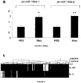
- the expression profile identified consists of 22 significantly differentially expressed miRNAs between the lungs of control and bleomycin-treated animals in at least one type, the majority being up-regulated in lungs instilled with bleomycin ( figure 1A ).
- miR-199a-5p displayed increased expression in response to bleomycin during the progression of pulmonary fibrosis only in C57BL/6 type mice ( figure 1B ).
- miR-199a-1 on chromosome 9
- miR-199a-2 on chromosome 1
- Target prediction bioinformatics software ectopic miRNA expression and reporter vectors containing the 3'-UTR part of a gene of interest fused with luciferase were combined, as previously described [25, 13].
- a number of target prediction algorithms have been proposed. They are generally based on i) the complementarity between miRNA and target mRNA 3'-UTR in the 5' region of miRNA (referred to as the "seed"), and ii) the phylogenetic conservation of this sequence in the target mRNA 3'-UTR.
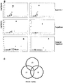
- Direct putative targets were then searched in the down-regulated transcript population using the MiRonTop tool. This indicated specific over-representation of predicted targets in the down-regulated transcript population after heterologous miR-199a-5p or miR-21 expression, using a number of prediction tools including an additional direct progeny search or TargetScan ( figure 3B ).
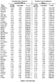
- EXTL3 Homo sapiens exostoses (multiple)-like 3 (EXTL3), mRNA 13.66 -1.20
- FAM188A Homo sapiens family with sequence similarity 188, member A (FAM188A), mRNA 11.55 -1.33 FBXO28 NM_015176 Homo sapiens F-box protein 28 (FBXO28), transcript variant 1, mRNA 10.39 -1.73 FZD6 NM_003506 Homo sapiens frizzled homolog 6 (Drosophila) (FZ
- pombe (VPS26A), transcript variant 1, mRNA 12.37 -1.60 ZNF512 NM_032434 Homo sapiens zinc finger protein 512 (ZNF512), mRNA 10.47 -1.16 ZNF512B NM_020713 Homo sapiens zinc finger protein 512B (ZNF512B), mRNA 12.08 -1.02 ZNF584 NM_173548 Homo sapiens zinc finger protein 584 (ZNF584), mRNA 9.46 -1.41 ZNF706 NM_0010425 10 Homo sapiens zinc finger protein 706 (ZNF706), transcript variant 1, mRNA 8.15 -1.10 ZNF776 NM_173632 Homo sapiens zinc finger protein 776 (ZNF776), mRNA 9.93 -1.22 1 logarithm (base 2) of the average intensity (AveExpr) 2 logarithm (base 2) of the ratio of miR-199a-5p / miR-Neg (logFC)
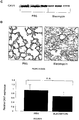
- caveolin-1 would appear to represent a key target of miR-199a-5p, based on previous studies demonstrating a significant link between CAV1 down-regulation in lung fibroblasts and adverse TGF ⁇ -mediated effects [ Wang et al., J. Exp. Med., 203: 2895-2906, 2006 ; Xia et al., Am. J. Pathol., 176: 2626-2637, 2010 ] [23, 31].
- Caveolin-1 is a 22 kDa membrane protein essential for the formation of small plasma membrane invaginations referred to as caveolae. Caveolae are found in most cell types, in varying quantities according to the tissue. They are particularly abundant in differentiated cells such as adipocytes, endothelial cells, type I pneumocytes, fibroblasts and smooth and striated muscle cells. Caveolae represent a subcategory of lipid rafts, morphologically identifiable due to the invaginated and circular shape thereof, and characterised by the presence of structuring proteins named caveolin-1, caveolin-2 and caveolin-3.
- Caveolin-1 and -2 have a relatively ubiquitous distribution in most differentiated cells with the exception of skeletal muscle fibres and cardiac myocytes [ Scherer et al., J. Cell Biol., 127: 1233-1243, 1994 ] [20].
- the expression of caveolin-3 is restricted to the skeletal muscles, diaphragm and heart [ Tang et al., J. Biol. Chem., 271; 2255-2261, 1996 ] [21].
- transgenic mice in which the caveolin-1 gene has been deleted present various anomalies, particularly in the lungs [ Park et al., Biochemistry, 42: 15124-15131, 2003 ] [22]. These mice in particular develop pulmonary fibrosis and endothelial cell proliferation.
- the 3'UTR of human CAV1 was cloned in psiCHEKTM-2 vector downstream from the sequence coding for luciferase, and cotransfected in HEK293 cells in the presence of miR-199a-5p or a negative control miRNA ( figure 5A ).
- a CAV1 3'UTR construct mutated on the predicted miR-199a-5p site was also used.
- Human pre-miR-199a-5p induced a significant decrease in the normalized luciferase activity relative to control in the presence of the wild type construction only, confirming that it represents a functional site.
- miR-199a-5p overexpression also gave rise to an increase in ACTA2 expression (marking myofibroblast differentiation) ( Figure 12E ), as well as to a significant potentialization of COL1A1 induction in response to TGF ⁇ ( Figure 12F ).
- the Ingenuity PathwaysTM canonical pathways of miR-199a-5p were analysed and compared to those of miR-21 and siCAV1 contexts. As shown in the decision tree in figure 14A , hierarchical clusters support the proximity between the pathways regulated by miR-199a-5p and siCAV1. Specific pathways for miR-199a-5p were also detected, particularly some associated with inflammation, such as "IL-1 signaling”, “Acute phase response signaling” and "P38 MAPK signaling", which are all typical of fibrotic processes.
- miR-199a-5p Of the genes specifically regulated by miR-199a-5p, a plurality of known profibrotic genes with distinct biological activities was detected, and for which abnormal expression was confirmed in vivo ( figures 10 and 15 ). This confirms that miR-199a-5p regulates multiple different signaling pathways involved in pulmonary fibrogenesis. In particular, compared to cells transfected with siCAV1, miR-199a-5p overexpression increased CCL2, TGFBRI and MMP3 expression significantly and decreased CAV1 and PLAU expression significantly ( figure 16 ). It should be noted that these two down-regulated genes are taken to be direct targets of miR-199a-5p based on the Pictar algorithm.
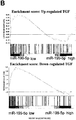
- TGF ⁇ signaling signature was defined experimentally in lung fibroblasts and compared to the signature of miR-199a-5p using the GSEA method. This analysis showed a significant overlap between these two signatures, evaluated with standardised enrichment scores greater than 1 (1.4 and 2.17 for up- and down-regulated genes, respectively, with a nominal p-value and an FDR q value ⁇ 0.05), demonstrating that miR-199a-5p is, in principle, a TGF ⁇ response mediator in lung fibroblasts ( figure 14B ).
- MiR-199a-5p is deregulated in mouse renal fibrosis and cirrhosis (hepatic fibrosis) models
- miRNA takes part in the fibrotic process in various organs such as the heart, kidneys, liver or lungs.
- previous studies have shown that miR-21 has an important role in pulmonary and cardiac fibrosis.
- miR-199a-5p is also deregulated in one forms of tissue fibrosis, i.e. renal and hepatic fibrosis using well-characterised experimental mouse models.
- the expression profiles of miRNAs obtained in these fibrosis models were compared using the same platform based on the miRNAs. 5 miRNAs routinely deregulated to a p-value ⁇ 0.01 were identified ( figure 17 ).
- miRNAs 3 were down-regulated (miR-193, miR-30b and miR-29c) and 2 were up-regulated (miR-199a-3p and miR-199a-5p) (see table 4 hereinafter).
Landscapes
- Health & Medical Sciences (AREA)
- Chemical & Material Sciences (AREA)
- Life Sciences & Earth Sciences (AREA)
- Engineering & Computer Science (AREA)
- Organic Chemistry (AREA)
- General Health & Medical Sciences (AREA)
- Bioinformatics & Cheminformatics (AREA)
- Genetics & Genomics (AREA)
- Veterinary Medicine (AREA)
- Pharmacology & Pharmacy (AREA)
- Medicinal Chemistry (AREA)
- Animal Behavior & Ethology (AREA)
- Public Health (AREA)
- Wood Science & Technology (AREA)
- Zoology (AREA)
- Proteomics, Peptides & Aminoacids (AREA)
- General Chemical & Material Sciences (AREA)
- Chemical Kinetics & Catalysis (AREA)
- Nuclear Medicine, Radiotherapy & Molecular Imaging (AREA)
- General Engineering & Computer Science (AREA)
- Biotechnology (AREA)
- Molecular Biology (AREA)
- Analytical Chemistry (AREA)
- Biomedical Technology (AREA)
- Biochemistry (AREA)
- Physics & Mathematics (AREA)
- Microbiology (AREA)
- Biophysics (AREA)
- Dispersion Chemistry (AREA)
- Epidemiology (AREA)
- Pathology (AREA)
- Pulmonology (AREA)
- Immunology (AREA)
- Plant Pathology (AREA)
- Dermatology (AREA)
- Urology & Nephrology (AREA)
- Gastroenterology & Hepatology (AREA)
- Pharmaceuticals Containing Other Organic And Inorganic Compounds (AREA)
- Measuring Or Testing Involving Enzymes Or Micro-Organisms (AREA)
- Medicines That Contain Protein Lipid Enzymes And Other Medicines (AREA)
Description
- The present invention relates to the field of fibroproliferative disorders, particularly idiopathic pulmonary fibrosis (IPF), and the role of microRNA (miRNA) in the tissue fibrosis process.
- The present invention relates more specifically to the use of microRNA (miRNA), particularly miR-199a-5p, targets and/or inhibitors thereof for the diagnosis, the prognosis and treatment of fibroproliferative disorders, namely idiopathic pulmonary fibrosis.
- In the description hereinafter, the references between square brackets ([ ]) refer to the list of references given at the end of the text.
- Tissue fibrosis, defined as the excessive persistent formation of non-functional connective scar tissue in response to a chronic tissue lesion, is a major cause of morbidity and mortality associated with loss of function of the damaged organ in various disorders such as those affecting the pulmonary interstitium [Wynn, J. Clin. Invest., 117: 524-529, 2007] [1]. Fibroproliferative disorders are a major public health problem. Indeed, in the United States alone, 45% of deaths are attributed to fibroproliferative disorders and the prevalence thereof is constantly increasing [Wynn, Nat. Rev. Immunol., 4: 583-594, 2004] [2].
- Of the pulmonary interstitial disorders of unknown origin, idiopathic pulmonary fibrosis (IPF) is the most frequent and lethal form with an average survival rate of 3 to 5 years post-diagnosis [Wilson and Wynn, Mucosal. Immunol., 2: 103-121, 2009] [3]. IPF is a chronic and frequently fatal lung disease characterised by fibroblast proliferation and excessive extracellular matrix protein deposition. IPF is a rare disorder with a prevalence of 13 to 20 cases per 100,000 inhabitants and whose causes remain poorly understood, and for which no effective treatment is currently available.
- Observations based on animal pulmonary fibrosis models and lung sections of patients with IPF suggest a dynamic biopathological process involving excessive scar tissue formation with chronic inflammation, epithelial and endothelial cell apoptosis, mesenchymatous cell proliferation and activation with fibroblast/myofibroblast site formation, and finally excessive extracellular matrix deposition resulting in the destruction of the lung structure and loss of lung function. In physiopathological terms, at the present time, repeated alveolar epithelium damage is considered to be responsible for lesions on the pulmonary epithelium promoting alveolar plasma exudate formation and clotting process activation, secretion of growth factor such as TGFβ by pneumocytes enabling pulmonary fibroblast recruitment, proliferation and activation and abnormal and excessive extracellular matrix deposition [Wilson and Wynn, 2009, cited above] [3]. During the fibrosis process, the fibroblasts have a myofibroblast phenotype and are organised into fibroblastic foci. Other mechanisms may also be involved in the fibrosis process such as mesenchymal epithelial transition of epithelial, endothelial or mesothelial cells and pulmonary medullary circulating fibrocyte recruitment [Wilson and Wynn, 2009, cited above] [3].
- MicroRNAs (miRNAs) are a class of small non-coding RNA with approximately 22 bases and having a key role in a wide range of cell phenomena such as development, differentiation, survival, response to stress, apoptosis, proliferation, homeostasis or differentiation [Ambros, Nature, 431: 350-355, 2004] [4]. Recent studies have identified specific miRNA expression profiles associated with the initiation and progression of various disorders including cancer and inflammatory and autoimmune disorders. Moreover, miRNA function gain and loss studies have revealed pathogenic miRNA functions accentuating the major role thereof in vivo. Preferably, the term "microRNA" or "miRNA", in the context of the present invention, means an RNA oligonucleotide consisting of between 18 to 25 nucleotides in length. In functional terms miRNAs are typically regulatory endogenous RNA molecules.
- The mechanism of action thereof involves the formation of a complex between a plurality of miRNA bases and the 3'-non-coding part of the target mRNA [Brennecke et al., PLoS. Biol., 3: e85, 2005] [5]. This interaction gives rise to destabilisation of the target mRNA and/or protein synthesis inhibition [Brennecke et al., 2005, cited above] [5]. Recognition between miRNA and the target thereof is essentially controlled by a sequence of approximately 7 bases, situated in the 5' part of the miRNA (recognition sequence or seed) [Brennecke et al., 2005, cited above] [5]. For this reason, each miRNA would appear to be capable of regulating the stability of a broad range of distinct mRNAs. The terms "target gene" or "target mRNA" refer to regulatory mRNA targets of microRNAs, in which said "target gene" or "target mRNA" is regulated post-transcriptionally by the microRNA based on near-perfect or perfect complementarity between the miRNA and its target site resulting in target mRNA cleavage; or limited complementarity, often conferred to complementarity between the so-called seed sequence (nucleotides 2-7 of the miRNA) and the target site resulting in translational inhibition of the target mRNA.
- About 2000 miRNAs have been identified in humans to date (miRBase release 19 http://www.mirbase.org) where they would appear to regulate more than 30% of transcripts. MiRNA regulation is thus considered as a major form of gene expression regulation, the impact of which has been largely underestimated until now [Xie et al., nature, 434: 338-345, 2005; Berezikov et al., Cell, 120: 21-24, 2005] [6, 7].
- MiRNAs are transcribed in the nucleus in the form of long precursors (pri-miRNA) and undergo a first maturation step in the nucleus to produce pre-miRNA having a smaller hairpin structure. This miRNA precursor is thus exported from the nucleus to the cytoplasm where it undergoes a final maturation step with Dicer enzyme generating two single-stranded miRNAs (one 5p strand and one 3p strand): the "mature" strand is taken on by a multi-protein complex (RNA Induced Silencing Complex or RISC) interacting with the 3'-non-coding part of the target mRNA, whereas the "star" complementary strand undergoes degradation (annotation: miR-xx*).
- In clinical terms, numerous studies suggest possible use of miRNAs as a diagnostic tool. Indeed, miRNAs display good stability in biological fluids such as serum [Mitchell et al., Proc. Natl. Acad. Sci. U.S.A., 105: 10513-10518, 2008] [8] or urine [Weber et al., Clin. Chem., 56: 1733-1741, 2010] [9]. For this reason, studying the expression thereof in these biological media offers new non-invasive prospects for the development of new diagnostic or prognostic biomarkers. Moreover, miRNA tissue expression profiles could also offer new prognostic or diagnostic tools as already demonstrated in cancer treatment [Lu et al., Nature, 435: 834-838, 2005] [10]. For example, miR-199a-5p, one of the two mature miRNA species derived from the miR-199a precursor, has been associated with malignancy not only in hepatocellular carcinoma [Jiang et al., Clin. Cancer Res., 14(2): 419-427, 2008] [11] but also in bronchial tumours [Mascaux et al., Eur. Respir. J., 33(2): 352-359 (Epub 2008), 2009; Puisségur et al., Cell death Differ., 18(3): 465-478, 2011] [12, 13].
- In therapeutic terms, miRNA expression modulation could also enable the development of new treatments [Krutzfeldt et al., Nature, 438: 685-689, 2005] [14]. For example, the use of miR-122 inhibitor in the development of new hepatitis C treatments has made it possible to obtain a significant reduction in the viral load of this virus [Lanford et al., Science, 327: 198-201, 2010] [15].
- Despite increasing evidence of the involvement of miRNAs in the tissue fibrosis process, the precise role thereof and mechanism(s) of action thereof have yet to be explored extensively [Jiang et al., FEBS J., 277: 2015-2021, 2010] [16]. Although the cause of IPF is unknown, a central role of miRNAs was recently suggested in the pathogenesis thereof. However, the role of miRNAs in fibrotic disorders, particularly in pulmonary fibrosis, is poorly documented and only a few miRNAs such as miR-21 or let7-d have been studied to date [Liu et al., J. Exp. Med., 20: 1589-1597, 2010; Pandit et al., Am. J. Respir. Crit. Care Med., 182: 220-229, 2010] [17, 18]. Therefore, miRNAs for which the expression is associated with pulmonary fibrosis could offer particularly promising tools for the development of new diagnostic and prognostic markers of IPF and new therapeutic strategies for this disorder which continues to be incurable and have a poor prognosis [Pandit et al., Transl. Res., 157: 191-199, 2011] [19].
- The inventors demonstrated, for the first time and completely unexpectedly, the role of miRNAs, particularly miR-199a-5p, and the targets thereof in pulmonary, hepatic and renal fibrosis.
- The inventors more specifically used an experimental pulmonary fibrosis model to identify, in the lung, i) miRNAs differentially expressed only in C57BL/6 mice sensitive to bleomycin-induced pulmonary fibrosis and ii) correlated miRNAs during the progression of the fibrotic process. For this purpose, the inventors used various experimental approaches combining miRNA biochips, in situ hybridisation, and quantitative RT-PCR.
- In this way, they identified an expression profile of 22 specific miRNAs with respect to the pulmonary response of C57BL/6 mice sensitive to bleomycin-induced pulmonary fibrosis.
- Among the 22 miRNAs of the identified expression profile, they demonstrated, for the first time, the unique role of miR-199a-5p in the pulmonary fibrosis process. In this way, they identified significant miR-199a-5p up-regulation in the lungs of mice with bleomycin-induced fibrosis. Indeed, miR-199a-5p would appear to be the most discriminatory miRNA between sensitive C57BL/6 mouse strains and resistant Balb/c mouse strains in respect of bleomycin-induced pulmonary fibrosis, thus enabling a distinction between pathological and normal cases.
- Significant miR-199a-5p up-regulation was also identified in the lungs of patients suffering from IPF. More specifically, the miR-199a-5p levels increased selectively in the myofibroblasts of damaged lungs.
- Consequently, the profibrotic effects of miR-199a-5p were studied further in pulmonary fibroblasts. In this way, they demonstrated pulmonary fibroblast activation to a profibrotic phenotype after miR-199a-5p overexpression. Finally, the inventors demonstrated that miR-199a-5p overexpression partially mimicked the transcriptional signature and cellular effects of TGFβ, one of the main factors involved in fibrotic mechanisms, which is also capable of increasing miR-199a-5p expression.
- They also demonstrated that the role of miR-199a-5p can be extrapolated to other fibrotic disorders in mammals since this miRNA is common to the various forms of pulmonary, hepatic and renal fibrosis; and could thus become a universal marker thereof. Indeed, they demonstrated abnormal miR-199a-5p expression in mouse renal and hepatic fibrosis models, demonstrating that miR-199a-5p deregulation represents a general mechanism contributing to the fibrosis process.
- Furthermore, the inventors combined in silico approaches (target prediction bioinformatics software) and experimental approaches (transcriptome chips, ectopic miRNA expression and reporter vectors containing the 3'-UTR part of a gene of interest fused with luciferase) in order to determine and characterise the target genes specifically regulated by miR-199a-5p. In particular, variation in expression of the gene coding for caveolin-1 (CAV1), a critical mediator in pulmonary fibrosis, according to the level of miR-199a-5p expression, was observed. In this way, they identified CAV1 as a genuine target of miR-199a-5p.
- The present invention thus relates to an in vitro diagnostic method for a fibroproliferative disorder in a subject characterised in that it comprises the following steps:
- (i) quantitative measurement of the level of miR-199a-5p expression in a biological sample from said subject;
- (ii) comparing the miR-199a-5p expression profile of the biological sample from said subject with the same miRNA expression profile of a reference biological sample;
- (iii) identifying and measuring in the biological sample from said subject the expression of a target gene for which the level of expression is regulated by the level of miR-199a-5p identified in step (ii), wherein said target gene codes for caveolin-1;
- (iv) comparing the caveolin-1 expression in the biological sample from said subject with the caveolin-1 expression of a reference biological sample; and
- (v) determining whether the levels of the miR-199a-5p expression and of the caveolin-1 expression are associated with a proliferative disorder.
- According to one particular embodiment, the 22 miRNAs of the specific expression profile for the pulmonary response to bleomycin-induced pulmonary fibrosis are represented by the following accession numbers:
miRNA name miRBase accession number mir-146b MIMAT0003475 mir-34a MIMAT0000542 mir-21 MIMAT0000530 mir-449a MIMAT0001542 mir-449b MIMAT0005447 mir-20a MIMAT0000529 mir-18a MIMAT0000528 mir-223 MIMAT0000665 mir-449c MIMAT0003460 mir-147b MIMAT0004857 mir-152 MIMAT0000154 mir-181ac MIMAT0000210 mir-451 MIMAT0001632 mir-351 MIMAT0000609 mir-133ac MIMAT0000145 mir-214 MIMAT0000661 mir-199a-5p MIMAT0000229 mir-132 MIMAT0000144 mir-222 MIMAT0000670 mir-342-3p MIMAT0000590 mir-345-5p MIMAT0000595 mir-221 MIMAT0000669 - The term "fibroproliferative disorder" according to the present invention refers to disorders characterised by a parenchymal organ lesion and fibrosis. For example, it refers to pulmonary, hepatic and renal fibrosis, particularly idiopathic pulmonary fibrosis (IPF).
- The term "subject" according to the present invention refers to a vertebrate, particularly a mammal, more particularly a human.
- The term "biological sample" according to the present invention refers to a bronchial, liver or kidney biopsy. For example, it refers to epithelial tissue, particularly from the respiratory, hepatic or renal epithelium.
- The term "reference biological sample" according to the present invention refers to a biological sample as defined above obtained from a healthy subject, i.e. not presenting fibrosis, or a subject in whom the miR-199a-5p expression is known and associated with a particular clinical stage. For example, it refers to the level of miR-199a-5p expression obtained after analysing human biopsy samples (see table 1 hereinafter) or mouse lung samples (see table 2 hereinafter) with "miRNA" biochips (Agilent technology) in healthy subjects (control) or subjects suffering from idiopathic pulmonary fibrosis (IPF).
Table 1 Probe (IDa) miRNA (name) Controlb IPFb Ratioc P-valued A_25_P00010700 Hsa-miR-199a-5p 8.38 8.69 1.24 0.006 A_25_P00010701 Hsa-miR-199a-5p 6.31 6.69 1.30 0.005 A_25_P00010069 Hsa-miR-199a-3p 8.97 9.47 1.41 0.015 A_25_P00010068 Hsa-miR-199a-3p 7.89 8.19 1.23 0.045 aID Agilent SurePrint G3 hsa chip probe blog2 median expression cIPF vs control dWilcoxon test Table 2 Experimental mouse model Probe (IDa) MiRNA (name) Controlb Fibrosisb Ratioc P-valued Pulmonary fibrosis MIMAT0000229 mmu-miR-199a-5p 3.23 4.61 2.89 4.10E-03 Renal fibrosis MIMAT0000229 mmu-miR-199a-5p 2.61 4.52 3.71 4.12E-07 Hepatic fibrosis MIMAT0000229 mmu-miR-199a-5p 2.60 4.10 2.82 4.00E-03 amiRBase ID (http://www.mirbase.org) blog2 median expression cbleomycin vs PBS control dWilcoxon test - The level of miRNA expression in a cell or tissue is determined by measuring the miRNAs present in said cell or said tissue. The level of miRNA expression may be measured using any technique known to those skilled in the art. For example, after RNA extraction, highspeed miRNA sequencing, NASBA (Nucleic Acid Strand Based Amplification) sequencing, primer extension sequencing, "miRNA" DNA chips, quantitative RT-PCR methods applied to miRNA or in situ hybridisation.
- The level of expression of a target gene in a cell or a tissue is determined by measuring the transcript expressed in said cell or said tissue. The level of target gene expression may be measured using any technique known to those skilled in the art. For example, it may be carried out after quantitative RT-PCR RNA extraction, or on tissue sections by means of immunocytochemistry or immunocytology.
- The present invention also relates to a method for identifying a subject liable to develop a fibroproliferative disorder characterised in that it comprises or consists of the following steps:
- (i) quantitative measurement of the level of miR-199a-5p expression in a biological sample from said subject;
- (ii) comparing the level of miR-199a-5p expression in said biological sample from said subject with the level of miR-199a-5p expression in a reference biological sample;
- (iii) identifying and measuring in the biological sample from said subject the expression level of a target gene for which the level of expression is regulated by the level of miR-199a-5p expression identified in step (ii); wherein said target gene codes for caveolin-1;
- (iv) comparing the level of caveolin-1 expression in said biological sample from said subject with the level of caveolin-1 expression in a reference biological sample; and,
- (v) detecting an increase in the level of miR-199a-5p expression and a decrease in the level of caveolin-1 expression.
- The present invention also relates to a miR-199a-5p inhibitor for use in the treatment and/or prevention of a fibroproliferative disorder, preferably idiopathic pulmonary fibrosis.
- The present invention also relates to a miR-199a-5p inhibitor for use in stimulating wound repair.
- The term "miRNA inhibitor" according to the present invention refers to DNA analogues or "oligomer", consisting of a contiguous sequence of from 7 to at least 22 nucleotides in length. The term "nucleotide" as used herein, refers to a glycoside comprising a sugar moiety, a base moiety and a covalently linked group (linkage group), such as a phosphate or phosphorothioate internucleotide linkage group. It covers both naturally occurring nucleotides and non-naturally occurring nucleotides comprising modified sugar and/or base moieties, which are also referred to as "nucleotide analogues" herein. Non-naturally occurring nucleotides include nucleotides which have sugar moieties, such as bicyclic nucleotides or 2' modified nucleotides or 2' modified nucleotides such as 2' substituted nucleotides. "Nucleotide analogues" are variants of natural oligonucleotides by virtue of modifications in the sugar and/or base moieties. Preferably, without being limited by this explanation, the analogues will have a functional effect on the way in which the oligomer works to bind to its target; for example by producing increased binding affinity to the target and/or increased resistance to nucleases and/or increased ease of transport into the cell. Specific examples of nucleoside analogues are described by Freier and Altman (Nucl. Acid Res., 25: 4429-4443, 1997) [35] and Uhlmann (Curr. Opinion in Drug Development, 3: 293-213, 2000) [36]. Incorporation of affinity-enhancing analogues in the oligomer, including Locked Nucleic Acid (LNA™), can allow the size of the specifically binding oligomer to be reduced and may also reduce the upper limit to the size of the oligomer before non-specific or aberrant binding takes place. The term "LNA™" refers to a bicyclic nucleoside analogue, known as "Locked Nucleic Acid" (Rajwanshi et al., Angew Chem. Int. Ed. Engl., 39(9): 1656-1659, 2000) [37]. It may refer to an LNA™ monomer, or, when used in the context of an "LNA™ oligonucleotide" to an oligonucleotide containing one or more such bicylic analogues.
- Suitably, the oligomer of the invention is capable of specifically inhibiting (silencing) the activity of miR-199a-5p ("anti-miR-199-5p") or preventing the binding of miR-199a-5p to specific miR-199a-5p binding sites in target RNAs ("miR-199a-5p Target Site Blocker"). Preferably, a "anti-miR-199a-5p" refers to antisense oligonucleotides with sequence complementary to miR-199a-5p (e.g., anti-miR-199a-5p LNA™ miRNA inhibitor; Product No.: 426918-00, Product name: hsa-miR-199a-5p, Description: miRCURY LNA™ microRNA Power Inhibitor, EXIQON). These oligomers may comprise or consist of a contiguous nucleotide sequence of a total of 7 to at least 22 contiguous nucleotides in length, up to 70% nucleotide analogues (LNA™). The shortest oligomer (7 nucleotides) will likely correspond to an antisense oligonucleotide with perfect sequence complementarity matching to the first 7 nucleotides located at the 5' end of mature miR-199a-5p, and comprising the 7 nucleotide sequence at position 2-8 from 5' end called the "seed" sequence (i.e., "ccagugu" for miR-199a-5p, see Seq ID N°13) involved in miRNA target specificity (Lewis et al., Cell. 2005 ) [38]. A "miR-199a-5p Target Site Blocker refers to antisense oligonucleotides with sequence complementary to a miR-199a-5p binding site located on a specific mRNA. These oligomers may be designed according to the teaching of
US 20090137504 . These oligomers may comprise or consist of a contiguous nucleotide sequence of a total of 8 to 23 contiguous nucleotides in length. These sequences may span from 20 nucleotides in the 5' or the 3' direction from the sequence corresponding to the reverse complement of miR-199a-5p "seed" sequence. For example, "CAV1 Target Site Blockers comprise or consist of a contiguous sequence of a total of 8 to 23 contiguous nucleotides in length which corresponds to the reverse complement of a specific nucleotide sequence present in the CAV1 mRNA (NM_001753.4, NM_001172895.1, NM_001172896.1, NM_001172897.1 or naturally occurring variants thereof and RNA nucleic acids derived therefrom, preferably mRNA) and defined as miR-199a-5p site (Fig.5A : SEQ ID N°10). The nucleotide sequence of the oligomers of invention will bind with high affinity to the miR-199a-5p site on CAV1 mRNA, preventing the binding of miR-199a-5p on the 3'UTR of CAV1 mRNA) at this particular target site. - The present invention also relates to a miR-199a-5p inhibitor for use as a medicinal product. For example, it consists of the use of a miR-199a-5p inhibitor by the aerosol route for inhibiting fibrogenesis in the pathological respiratory epithelium in subjects suffering from pulmonary fibrosis and thus restoring the integrity of the pathological tissue so as to restore full functionality.
- According to one particular embodiment of the invention, said medicinal product is intended to prevent and/or treat a fibroproliferative disorder, preferably idiopathic pulmonary fibrosis.
- The examples of embodiments of the present invention hereinafter, along with the appended figures, illustrate the invention and are given as non-limiting examples.
-
-
Figure 1 represents miR-199a-5p expression during bleomycin-induced pulmonary fibrosis (A) Hierarchical clustering (or tree) representing differential statistically significantly modulated miRNA expression (adjusted p-value <0.05) in the lungs of BALB/c and C57BL/6 mice in response to bleomycin at the indicated time points. Up-regulated miRNAs are shown in progressively brighter shades of red, and down-regulated miRNAs are shown in progressively brighter shades of green based on the log2 value thereof (Bleomycin / PBS). MiR-199a-5p is shown with an arrow. N=3 mice in each group. (B) MiR-199a-5p in the lungs of BALB/c and C57BL/6 mice in response to bleomycin at the specified times. n=3 mice in each group. The data are expressed as the mean ± standard error of the mean. **p<0.01. (C) Paraffin sections prepared from C57BL/6 mice collected 14 days after intratracheal bleomycin instillation. In situ hybridisation and immunohistochemistry tests were carried out to determine the colocation of miR-199a-5p and α-SMA. The results represent one out of three tests conducted independently. -
Figure 2 represents miR-199a-5p and pri-miR-199a expression in C57BL/6mice 14 days after exposure to bleomycin (A) Real-time PCR conducted to confirm the increase in mature miR-199a-5p expression in the lungs of C57BL/6 and BALB/c mice 14 days after administering bleomycin; n=5 mice in each group, the data are expressed as the mean ± standard error of the mean.*p<0.05 (B) Expression of pri-miR-199a-1 and pri-miR-199a-2 genes in the lungs of C57BL/6mice 14 days after bleomycin installation. N=5 mice in each group, the data are expressed as the mean ± standard error of the mean.*p<0.05 and **p<0.01. -
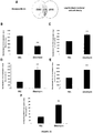
Figure 3 represents the identification of miR-199a-5p candidate targets using a transcriptomic approach. Normal hFL1 human lung fibroblasts were transfected with pre-miR-Neg, pre-miR-199-5p or pre-miR21 (n=2). RNA samples were collected 48 hours after transfection and the expression profiles were determined with genomic chips (A) Hierarchical tree comparing the standardised log2 of the ratios between the signal under the various conditions and the pre-miR-neg signal (B) Overexpression of specific predicted targets in the set of down-regulated transcripts following transfection with miR-199a-5p and miR-21. The representation of the predicted miRNA targets in the set of down-regulated genes was compared with the set of all the expressed genes using the miRonTop tool: http://www.microaarray.fr:8080/miRonTop/index. The graphs show the extent of the enrichment, represented in -log10 form (adjusted p-value), based on the enrichment level using three different prediction tools for all known miRNAs: miR-199a-5p and miR-21 are represented as ◊ and Δ, respectively. The threshold values used to define the set of up-regulated and down-regulated genes: AveExp=7.0; log FC=0,7; adjusted p-value=0.05. (C) Venn diagram comparing the number of miR-199a-5p targets among the set of significantly down-regulated genes following pre-miR-199a-5p transfection based on three separate prediction tools (targetScan, Pictar and miRanda). Selection thresholds are equal to 7.0 for log2 (signal), -1.5 for log2 (ratio), and 0.01 for the adjusted p-value. -
Figure 4 represents the list of themes corresponding to the "canonical pathway" annotations identified by Ingenuity Pathway Analysis software in response to miR-199a-5p and miR-21 overexpression in hFL1 human lung fibroblasts. hFL1 human lung fibroblasts were transfected with pre-miR-Neg, pre-miR-199a-5p or pre-miR-21 (n=2). RNA samples were collected 48 hours after transfection and the expression profiles were determined with whole genome biochips. The probability of obtaining the number of genes in a specific pathway in the list of differentially expressed genes between miR-199a-5p or miR-21 vs miR-Neg was compared with the same representation of the same pathway among all the genes in the biochip; -log10 of the Fisher exact probability is shown. While both miR-199a-5p and miR-21 signatures were associated with "TGFβ signaling", several signatures were specifically associated with miR-199a-5p, including "Integrin signaling" and "Caveolae-mediated endocytosis signaling". -
Figure 5 represents CAV1 as the direct target of miR-199a-5p. (A) Co-transfection of pre-miR-199a-5p or pre-miR-Neg and human CAV1 3'UTR-derived psiCHECK™-2 construct (wild type or mutated in the putative miR-199a-5p seed region) in HEK293 cells demonstrates a significant decrease in standardised luciferase activity 48 hours after transfection for the wild type construct only. *p<0.05. The position of the miR-199a-5p target site in CAV1 3'UTR and the sequence alignment of miR-199a-5p and CAV1 3'UTR using various species are shown. The representation is limited to the region around the complementary miR-199a-5p site. The miR-199a-5p binding site retained in CAV1 3'UTR is shown in bold. (B) Relative CAV1 expression determined by real-time PCR in hFL1 lung fibroblasts after transfection with pre-miR-199a-5p, pre-miR-21 or si-CAV1. The data represent three independent tests. The data are expressed as the mean ± standard error of the mean.*p<0.01 (C) Standardised fluorescence expression values of CAV1 in hFL1 lung fibroblasts after transfection with pre-miR-199a-5p, pre-miR-21 or siCAV1 based on biochip tests. The data represent two independent tests. The data are expressed as the mean ± standard error of the mean.*p<0.01 (D) Western blot analysis demonstrating the down-regulation of CAV1 protein expression after hFL1 lung fibroblast transfection with pre-miR-199a-5p, pre-miR-21 or siCAV1. One representative test out of two is shown. -
Figure 6 represents the decrease in CAV1 expression after MRC-5 lung fibroblast transfection with pre-miR-199a-5p. (A) MRC-5 lung fibroblasts transfected with 10 nM of pre-miR-199a-5p for 24 hours display a significant decrease in CAV1 expression as determined by means of real-time PCR. The data are expressed as the mean ± standard error of the mean.**p<0.01. (B) Western blot analysis demonstrating the decrease in CAV1 expression after MRC-5 lung fibroblast transfection with pre-miR-199a-5p. The data represent two independent tests. -
Figure 7 represents CAV1 and miR-199a-5p expression regulation with TGFβ. MRC-5 lung fibroblasts were treated with 10ng/ml of TGFβ for 24 hours and 48 hours. The levels of expression of miR-199a-5p (A) and CAV1 (B) were determined by Taqman PCR. The data are expressed as the mean ± standard error of the mean.*p<0.01. (C) The CAV1 protein levels were determined by immunoblot analysis. (D) MRC5 cells were transfected with LNA-miR-199a-5p, a CAV1 LNA-Target Site Blocker (CAV1 protector) or LNA-control then incubated with or without 10 ng/ml TGFβ for 24h and CAV1 protein levels were determined by immunoblot analysis. The data represent three independent tests. -
Figure 8 represents altered CAV1 expression in a mouse bleomycin-induced pulmonary fibrosis model. (A) Biochip analysis demonstrates a significant reduction in CAV1 expression in C57BL/6 mice treated with bleomycin for 14 days (n=5) compared to the control mice (n=5). The data are expressed in the form of normalized fluorescence values ± standard error of the mean.*p<0.01 (B) Real-time PCR conducted to confirm the decrease in CAV1 expression in the lungs of C57BL/6mice 14 days after administering bleomycin. N=5 mice in each group. The data are expressed as the mean ± standard error of the mean.*p<0.05 (C) CAV1 expression as detected by immunoblot analysis in lung tissue samples from C57BL/6 mice treated with bleomycin for 14 days (n=3) and control mice (n=3) (D) Immunohistochemical analysis of CAV1 expression in sections of lung tissue from C57BL/6mice 14 days after intratracheal bleomycin instillation. One representative section out of three is shown. -
Figure 9 represents pulmonary CAV1 expression in "lung fibrosis-resistant" BALB/c mice 14 days after injecting bleomycin. Real-time PCR was conducted to evaluate CAV1 expression in the lungs of BALB/c mice 14 days after exposure to bleomycin. n=5 mice in each group, the data are expressed as the mean ± standard error of the mean. n.s.= non-significant. -
Figure 10 represents the list of the 133 genes modulated by miR-199a-5p in lung fibroblasts which are also deregulated in the lungs of C57BL/6mice 14 days after bleomycin treatment [Average intensity: log2 average intensity; LogRatio: log2 (Bleo / PBS); PValue: adjusted p-value for a large number of events according to the Benjamini Hochberg corrected test]. Both CAV1 and CAV2 are significantly dow-regulated in the 2 models. -
Figure 11 represents the deregulation of miR-199a-5p and the CAV1 target thereof in IPF. (A and B) Box plots showing the normalized expression of log2-transformed CAV1 and miR-199a-5p in both IPF (n=94) and control (n=83) lungs. The box represents the 25-75% quartiles, the line in the box represents the median and whiskers represent the range. (**p<0.001) (C) Immunohistochemical analysis of CAV1 expression in sections of lung tissue from patients suffering from IPF. One representative section is shown (D) In situ hybridisation demonstrating miR-199a-5p expression (i) or a control probe (ii) in sections of lung tissue from patients suffering from IPF. -
Figure 12 represents the functional impact of miR-199a-5p on lung fibroblasts. The increase in miR-199a-5p expression in the lung fibroblasts results in an increase in the ability of the fibroblasts to migrate, proliferate, invade matrigel and differentiate into myofibroblasts. (A and B) In vitro healing test conducted to evaluate the migration rate of lung fibroblasts transfected with 10 nM of miR-199a-5p orcontrol 0, 6.5 and 9.5 hours after scratch injury. A significant increase (p<0.01) in the migration rate was observed in lung fibroblasts transfected with miR-199a-5p compared to the control. The data represent two independent tests. (C) Invasion assay on matrigel showing that overexpression of miR-199a-5p increases MRC5 lung fibroblast invasiveness. Data are representative of two independent experiments (D) Effects of miR-199a-5p on lung fibroblast proliferation determined by bivariate flow cytometry analysis EdU/DNA stained cells. The x-axis represents the linear intensity obtained using propidium iodide (total DNA content), and the y-axis represents the logarithmic intensity obtained using Alexa Fluor647. The cells were separated into G0/G1 phase and G2/M phase based on the DNA content thereof, and into labelled undivided and labelled divided subgroups based on the DNA content of the EdU-labelled cells. One representative test out of three is shown. (E) Confocal microscopy of MRC5 cells overexpressing miR-199a-5p revealed that increasing miR-199a-5p levels in lung fibroblasts promotes their differentiation into myofibroblasts. Cells were stained with an antibody against α-SMA (green) and phalloidin (red). Experiments were performed twice. (F) Relative expression of COL1A1 was assessed by Taqman PCR in MRC5 cells overexpressing miR-199a-5p and exposed or not to TGFβ. Data are expressed as mean ± SEM and derived from 2 independent experiments. *p<0.05. -
Figure 13 represents the comparison of transcriptome changes induced by miR-199a-5p and siRNA targeted against CAV1. Normal hFL1 human lung fibroblasts were transfected with pre-miR-Neg, pre-miR-199a-5p and with siCAV1 or control siRNA (n=2). RNA samples were collected 48 hours after transfection and the expression profiles were determined with a set of genomic biochips. (A) Decision tree comparing the standardised log2 of the ratios between the pre-miR-199a-5p signals vs pre-miR-Neg or the siCAV1 signals vs si-Neg (B) Venn diagram comparing the set of down-regulated transcripts followed induction with miR-199a-5p and siCAV1. The selection thresholds are equal to 7.0 for log2 (signal), 0.7 for log2 (ratio), and 0.05 for the adjusted p-value. -
Figure 14 represents miR-199a-5p as a TGFβ pathway effector. (A) Decision tree of the significant canonical pathways associated with miR-199a-5p, miR-21 or siCAV1 overexpression contexts identified using Ingenuity Pathway Analysis™ software. (B) GSEA graphs for an experimental TGFβ signature showing a significative enrichment for genes up-regulated and down-regulated by miR-199a-5p. The data were processed using GSEA software, with transcripts ordered based on the miR-199a-5p vs miR-Neg contexts measured using the standard deviation thereof. Up- and down-regulated genes were handled separately with respect to an experimental TGFβ signature obtained in the same cell models (hFL1 fibroblasts treated with 10 ng/ml of TGFβ1 for 48 hours): two sets of 134 and 113 genes corresponding to sets of genes up-regulated and down-regulated by TGFβ were selected as described in Example 1 (p<0.05). In each case, the graphs show enrichment, the peak score corresponding to the SE and the position of the set of TGFβ genes in the list of classified genes (each transcript is shown with a vertical line). (C) MRC5 cells transfected with LNA-miR-199a-5p, CAV1 Target Site Blocker (CAV1 protector) of LNA-control, then incubated with or without 10 ng/ml TGFβ for 24h, and cells stained with an antibody against α-SMA (green), phalloidin (red) and DAPI (blue). Experiments were performed twice. (D) Cells co-transfected with SMAD-luciferase reporter plasmid, luciferase activities analyzed 48h after transfection. All firefly luciferase activities were normalized with renilla luciferase activity. *p<0.05. Two independent experiments. (E) and (F) In vitro healing (or scratch) test conducted to evaluate the migration rate of TGFβ-stimulated hFL1 lung fibroblasts treated with 25 nM of LNA-anti-miR-199a-5p, LNA-control inhibitor and CAV1 protector. A significant decrease (p<0.01) in the migration rate was observed in lung fibroblasts transfected with LNA-anti-miR-199a-5p and CAV1 protector compared to the control. The data represent two independent tests. -
Figure 15 represents the comparison of the change of gene expression between genes regulated by miR-199a-5p in lung fibroblasts and lungs of C57BL/6mice 14 days after injecting bleomycin. (A) Venn diagram showing the correlations of the changes in gene expression between lung fibroblasts transfected with miR-199a-5p (two independent tests) and the lungs of C57BL/6mice 14 days after bleomycin treatment (n=5 mice). The numbers of genes for which expression was detected differentially under each condition at p<0.05 are shown. The biochip analysis demonstrates a significant reduction in the expression of CAV1 (B), TGFBRI (C), CCL2 (D), ACTA2 (E) and MMP3 (F) in C57BL/6 mice treated with bleomycin for 14 days (n=5) compared to control mice (n=5). The data are expressed as the mean ± standard error of the mean. **p<0.01. -
Figure 16 represents the profibrotic genes significantly modulated in lung fibroblasts by miR-199a-5p regardless of CAV1 regulation. Lung fibroblasts were transfected with miR-199a-5p, siCAV1 or negative controls. The biochip analysis shows the expression of known fibrotic genes: CAV1 (A), TGFBRI (B), MMP3 (C), PLAU (D) and CCL2 (E) 48 hours after transfection. The data are expressed as the mean ± standard error of the mean. -
Figure 17 represents miR-199a-5p deregulation in three experimental mouse hepatic, pulmonary and renal fibrosis models. Venn diagrams showing the correlations of changes of miRNA expression in lungs of C57BL/6mice 14 days after bleomycin treatment (n=4 mice), livers of BALB/c mice 6 weeks after administering CCl4 (n=5 mice) and kidneys of C57BL/6mice 28 days after unilateral ureteral obstruction (n=4 mice). The number of miRNAs for which expression was detected differentially in each mouse model at p<0.01 are shown. The hepatic fibrosis data are taken from [24]. -
Figure 18 represents enhanced expression of miR-199a-5p in clinical samples from patients with liver fibrosis. In situ hybridization assay was performed to determine the localization of miR-199a-5p in normal and fibrotic human livers. Results represent one out of three independent experiments. -
Figure 19 represents the alteration of miR-199a-5p expression in a mouse CCl4-induced hepatic fibrosis model. (A) MiR-199a-5p expression in the livers of BALB/c mice treated with oil or CCl4 for 6 weeks analysed by qPCR; n=5 per group. The data are expressed as the mean ± standard error of the mean. **p<0.01. (B) MiR-199a-5p expression in the livers of C57BL/6 mice treated with oil or CCl4 for 6 weeks analysed by qPCR; n=5 per group. The data are expressed as the mean ± standard error of the mean. *p<0.05. (C) MiR-199a-5p expression in the livers of C57BL/6mice 21 days after bile duct ligation or the control procedure analysed by qPCR; n=4 mice per group. The data are expressed as the mean ± standard error of the mean. **p<0.05. (D) MiR-199a-5p expression in the liver of C57BL/6 mice during fibrosis regression. Hepatic fibrosis was induced by injecting CCl4 and the miR-199a-5p expression was evaluated 6 weeks after the CCl4 treatment and 2 and 4 weeks after the final injection. The data are expressed as the mean ± standard error of the mean. **p<0.05. (E) MiR-199a-5p and CAV1 expression in primary stellate cells isolated from C57BL/6 mice and stimulated with 20 ng/ml of TGFβ for 48 hours. -
Figure 20 represents the alteration of miR-199a-5p and CAV1 expression in a mouse unilateral ureteral obstruction (UUO) renal fibrosis model. (A) MiR-199a-5p expression in the kidney of C57BL/6 mice after UUO at the indicated time points. n=5 to 7 mice in each group. The data are expressed as the mean ± standard error of the mean. **p<0.01. (B) Paraffin sections prepared from kidneys of C57BL/6 mice collected 28 days after UUO. The in situ hybridisation test was conducted to determine the location of miR-199a-5p in the kidney. The results represent one out of three independent tests. (C) Immunohistochemical analysis of CAV1 expression in kidney tissue sections from C57BL/6mice 28 days after UUO. One representative section out of two is shown. - MRC-5 and hFL1 5CCL-153) normal human lung fibroblasts and the A549 human lung cancer cell line were acquired from the American Type Culture Collection (ATCC, Manassas, VA, USA) and cultured in DMEM medium containing 10% foetal calf serum, at 37°C with 5% CO2 v/v.
- Recombinant TGFβ was acquired from Sigma-Aldrich.
- The following monoclonal and polyclonal antibodies were used for the immunohistochemistry and Western blot analyses: rabbit anti-CAV1 polyclonal antibody (sc-894, Santa Cruz Biotechnology Inc.), rabbit anti-β-actin monoclonal antibody (13E5, cell signaling), mouse anti-α-SMA monoclonal antibody (1A4, Dako).
- Male C57BL/6 and BALB/c strain mice, aged from 9 to 12 weeks, were acquired from Charles River, France. To induce the fibrotic changes, the mice were instilled by the intratracheal route with bleomycin or PBS for the controls. For this purpose, the mice were anaesthetised by sevorane (Abbott) inhalation and placed in the supine position. Transtracheal insertion of a 24-G supply needle was used for instilling bleomycin (0.75 units/ml) or excipient (PBS), in an 80 µl volume. The mice were sacrificed 7 and 14 days after instillation and the lungs were removed for subsequent analysis.
- Male C57BL/6 and BALB/c strain mice, aged from 9 to 12 weeks, were acquired from Charles River, France. The mice were anaesthetised by means of intraperitoneal injection of pentobarbital (50 mg/kg of body weight). After standard laparotomy, the left proximal ureter was exposed and ligated at two points with 4-0 silk. The "simulation or control" procedure consisted of a similar identification of the left ureter, but the ureter was not ligated.
- Male C57BL/6 and BALB/c strain mice, aged from 6 to 8 weeks, were acquired from Jackson Laboratory (Bar Harbor). To induce hepatic fibrosis, the mice received 0.6 ml/kg of body weight of CCl4 (Merck) mixed with corn oil (Sigma life science) by the intraperitoneal route as described above [Roderbrug et al., Hepatol., 53: 209-218, 2011] [24]. The bile duct was ligated by exposing the common hepatic duct and conducting a double-ligation thereof, and subsequently cutting between the ligations as described by Roderburg et al. [2011, cited above] [24]. To induce fibrosis regression, the mice were treated for 6 weeks with CCl4 as described above and sacrificed 2 or 4 weeks, respectively, after the end of the treatment.
- Primary stellate cells were isolated from 40-55 week old C57BL/6 mice and stimulated with 20 ng/ml of TGF-β1 (Sigma Aldrich) for 48 hours as previously described by Roderburg et al. [2011, cited above] [24].
- Frozen lung tissues from subjects with IPF and free from chronic lung disease were obtained from the "Lung Tissue Research Consortium (LTRC)". The diagnoses were based on ATS/ERS guidelines [Demedts and Costabel, Eur. Respir. J., 19: 794-796, 2002; Steele et al., Am. J. Respir. Crit. Care Med., 172: 1146-1152, 2005] [32, 33] based on the clinical history, pathology and radiology. All the tests were approved by the local Institutional Review Board at the University of Pittsburgh. The clinical data were made fully available to the investigators for examination.
- Paraffin-treated lung sections from patients with IPF were obtained from Lille hospital. The tests were approved by the Lille hospital institutional research commission.
- Mouse kidneys and lungs were fixed overnight with neutral buffered formalin and then included in paraffin. 5 µm thick tissue sections were mounted and stained with haematoxylin and eosin and with Masson's trichrome to evaluate the degree of fibrosis. The histological sections were examined by an experienced pathologist.
- The total RNA was extracted from lung cell and tissue samples using Trizol Reagent (Invitrogen) according to the manufacturer's recommendations. The purity and concentration of the total RNA samples were first evaluated using the Nanodrop spectrophotometer. The 260/280 and 260/230 ratios were checked and should have a value close to 2. The RNA was then loaded onto a chi nano chip and the quality thereof (RNA integrity and degradation level) was analysed using the Bioanalyzer System (Agilent Technologies, France).
- The oligonucleotide sequences corresponding 2054 mature miRNAs found in the miRNA registry are available at http://www.microarray.fr:8080/merge/index (follow the link to "microARN": platform references on the NCBI GEO database in GPL4718). Three biological reproductions were produced for each comparison. The experimental data and the biochip design were registered on the NCBI GEO database (http://www.ncbi.nlm.nih.gov/geo/) under series GSE34812. The experimental design used a "dye-swap" approach, such that each mouse probe, imprinted 8 times on the biochip was measured 16 times independently for each sample. The target preparation and biochip hybridisation were carried out as previously described [Pottier et al., PLoS. One, 4: e6718, 2009; Triboulet et al., Science, 315: 1579-1582, 2007; Puisségur et al., 2011, cited above] [25, 26, 13].
- MiRNA biochip analysis was conducted as previously described [Pandit et al., 2010, cited above] [18]. In brief, 100 ng of total RNA was labelled and hybridised on Agilent microRNA Microarray Release 16.0, 8x60K. After washing, the chips were scanned using an Agilent Microarray scanner. The scanned images were processed with Agilent's Feature Extraction software version 9.5.3. The miRNA biochip data were analysed using GeneSpring v11.5 and BRB-ArrayTools v.1 developed by Dr. Richard Simon and the BRB-ArrayTools development team. The data were standardised per quantile. The miRNA biochip data are available to the public via the Lung Genomics Research Consortium (LGRC) website (lung-genomics.org).
- The RNA samples were labelled with Cy3 stain using the low RNA input QuickAmp kit (Agilent) according to the manufacturer's recommendations. 825 ng of labelled cRNA probe was hybridised on mouse or human Agilent 8x60K high density SurePrint G3 gene expression biochips. Two (in vitro human tests) or five (in vivo-derived samples) biological reproductions were produced for each comparison. The experimental data were registered on the NCBI GEO database (http://www.ncbi.nlm.nih.gov/geo/) under SuperSeries GSE34818 (series GSE34812 and GSE34814 for the miRNA and mRNA responses in the mouse bleomycin model, respectively; series GSE34815 for the miRNA/siRNA transfection tests in hFL1 human fibroblasts). For the human gene expression chips, the data were converted into log2 and standardised using a cyclic Loess algorithm in the R programming environment as previously described [Wu et al., BMC. Bioinformatics, 6: 309, 2005] [34]. The human biochip data have been made available to the public on the LTRC (ltrcpublic.org) and LGRC websites as part of the LTRC protocol.
- The standardisation was carried out using the Limma program available from Bioconductor (http://www.bioconductor.org). The within-chip (two-colour dye-swap tests only) and between-chip standardisation was carried out using the "PrintTip Loess" method and the quantile method, respectively. The mean ratios of all the comparisons were calculated and a B test analysis was conducted. Differentially expressed genes were selected using the Benjamini-Hochberg p-value correction for multiple tests, based on a p-value less than 0.05. The expression biochip data were analysed for biological theme enrichment (canonical pathways and genetic ontology molecular function) and to construct biological networks using Ingenuity Pathway Analysis software (http://www.ingenuity.com/) and Mediante (http://www.microarray.fr:8080/merge/index) [Le and Barbry, Bioinformatics, 23: 1304-1306, 2007] [27], an information system containing various information on probes and data sets. Gene Set Enrichment Analysis (GSEA) software was used to determine whether a gene set defined in principle can characterise differences between two biological states [Subramanian et al., Proc. Natl. Acad. Sci. U.S.A., 102: 15545-15550, 2005] [28]. Hierarchical clusters were produced with MultiExperiment Viewer (MeV) version 4.3, using the Manhattan distance and the mean link.
- MiRonTop [Le et al., Bioinformatics, 26: 3131-3132, 2010] [29] is an online Java network tool (available at http://www.microarray.fr:8080/miRonTop/index) which integrates DNA biochip data to identify the potential involvement of miRNAs in a specific biological system. In brief, MiRonTop classifies transcripts into two categories ("up-regulated" and "down-regulated"), based on expression level and differential expression thresholds. It then calculates the number of predicted targets for each miRNA, based on the selected prediction software (Targetscan, MiRBase, PicTar, exact progeny search: 2-7 or 1-8 first nucleotides of miRNA, TarBase v1), in each gene set. MiRNA target enrichment in each category is then tested using the hypergeometric function. The absence of a non-siRNA target effect was verified in si-CAV1 transcriptome tests using the Sylamer tool [Van et al., Nat. Methods, 5: 1023-1025, 2008] [30].
- Pre-miR-199-5p, pre-miR-21 and control miRNA (miR-Neg#1) were acquired from Ambion. For the miR-199-5p knock-down tests, anti-miR-199-5p LNA and the anti-miR-159s LNA negative control (miRCURY LNA knock-down probe) were ordered from Exiqon. SiRNA targeted against CAV1 and control siRNA (Silencer Select validated siRNAs) were acquired from Applied Biosystems.
- MRC5/hFL1 cells were cultured in DMEM medium containing 10% FCS and transfected to 30-40% confluence in 6- or 12-well plates using Lipofectamine RNAi MAX™ (Invitrogen) with pre-miRNA, siRNA or LNA inhibitors at a final concentration of 10 nM unless indicated.
- Molecular constructions were produced in pSI-CHECK™-2 vector (Promega) by cloning behind Renilla luciferase in the XhoI and NotI restriction sites, CAV1 3'UTR derived hybrid oligonucleotides described hereinafter. HEK293 cells were cultured in DMEM medium containing 10% FCS to confluence. The cells were then distributed into 96-well plates and
cotransfected using lipofectamine 2000™ (Invitrogen) with 0.2 µg of psiCHECK™-2 plasmid construction or pre-miR-199-5p or control miRNA at a final concentration of 10, 30 and 50 nM. 48 hours after transfection, the renilla and firefly luciferase activities were evaluated with the Dual Glo Luciferase Assay System kit (Promega) and measured using a luminometer (Luminoskan Ascent, Thermolab system). - hsa-CAV1: WT (sense):
-
- hsa-CAV1: WT (anti-sense):
-
- hsa-CAV1: MUT (sense):
-
- hsa-CAV1: MUT (anti-sense):
-
- MRC5 cells were seeded in 96 well plate and
cotransfected 24h later at 4% confluency using RNAi MAX lipofectamine reagent with 100 ng SMAD reported vector (Cignal Smad Reporter, Qiagen) and 10 nM LNA-control, LNA-199a-5p or CAV1 protector. 24h after transfection, cells were serum starved 3h before adding 10 ng/ml TGFβ. Cells were lyzed and Glo luciferase assay (Promega) was performed 24h following TGFβ exposure. - The miR-199a-5p expression was evaluated using the TaqMan MicroRNA Assay kit (Applied Biosystems) as specified in the protocol. Real-time PCR was conducted using the GeneAmp Fast PCR Master Mix product (Applied Biosystems) and the ABI 7900HT real-time PCR machine. The levels of mature miRNA expression were evaluated using the comparative CT method (2-dectaCT).
- The pri-miR-199a-1 and pri-miR-199a-2 expressions were evaluated using the TaqMan pri-microRNA Assay system (Applied Biosystems) according to the manufacturer's recommendations. Real-time PCR was conducted using the TaqMan™ Gene Expression Master Mix product (Applied Biosystems) and the ABI 7900HT real-time PCR machine. The levels of pri-miRNA expression were evaluated using the comparative CT method (2-deltaCT).
- The levels of human and mouse CAV1 expression were analysed using the TaqMan MicroRNA Assay system (Applied Biosystems) according to the manufacturer's recommendations. Real-time PCR was conducted using the TaqMan™ Gene Expression Master Mix product (Applied Biosystems) and the ABI 7900HT real-time PCR machine. The levels of CAV1 were evaluated using the comparative CT method (2-deltaCT).
- The cells or tissues were lysed in a lysis buffer (M-PER protein extraction buffer for cells, T-PER protein extraction reagent for tissues) and a mixture of protease inhibitors (Pierce). The lysates were assayed for protein concentrations using the Bradford assay (Biorad). The proteins (10 µg per sample) underwent electrophoresis in SDS-polyacrylamide gel and transferred into liquid medium on a nitrocellulose membrane (Hybond™ C Extra, Amersham Bioscience) for 1
hr 30 in a transfer buffer (50 mM Tris base, 40 mM Glycine, 1.3 mM SDS and 10% ethanol). After transfer, the membrane was blocked in a 5% milk 0.1% TBS-Tween solution for 1 hour at ambient temperature under stirring. It was then incubated with anti-CAV1 primary antibodies (Santa Cruz) diluted to 1:400, anti-beta actin antibodies (Cell Signaling Technology) to 1:1000 overnight at 4°C on a rotating plate. After washing 3 times in 0.1% TBS-Tween for 30 minutes at ambient temperature, the membrane was incubated for 1 hour in the presence of mouse anti-IgG secondary antibody coupled with HRP (Horse Radish Peroxidase, Cell Signaling) diluted to 1:5000 5% milk TBS-0.1% Tween under stirring. After washing several times, in 0.1% TBS-Tween for 30 minutes, the proteins of interest were detected by chemoluminescence (ECL substrates, Amersham). - 5 µm thick mouse lung sections included in paraffin were deparaffinised in xylene (2 x 5 minutes) and rehydrated by incubating 2 x 5 minutes in 100% ethanol, 2 x 5 minutes in 95% ethanol, and 2 x 5 minutes in 80% ethanol. After washing with water, the antigen was unmasked by thermal denaturation using a citrate buffer (pH=6.0, DAKO) for 20 minutes. The sections were cooled to ambient temperature and then washed in 0.1% TBS-Tween, and the endogenous peroxidases were inactivated with 3% hydrogen peroxide in TBS for 10 minutes. The sections were also blocked for avidin/biotin activity, blocked with serum-free blocking reagents, and incubated with anti-CAV1 or anti alpha-SMA primary antibody for 1 hour at ambient temperature and overnight at 4°C, respectively. The proteins of interest were then detected with diaminobenzidine (DAB, DAKO) after incubating the secondary antibody.
- The tissue location of miR-199a-5p was detected using the double DIG-labeled LNA probes system (Exiqon, Woburn, MA). Mouse tissues included in paraffin were deparaffinised in xylene (2 x 5 minutes) and rehydrated by incubating 2 x 5 minutes in 100% ethanol, 2 x 5 minutes in 95% ethanol, and 2 x 5 minutes in 80% ethanol. The slides were then washed in PBS (pH 7.5) and permeabilised by incubating for 15 minutes at 37°C in proteinase K (Ambion). The slides were washed again in PBS, and pre-hybridised in a hybridisation buffer (50% formamide, 5x SSC, 0.1
% Tween 20, 9.2 mM citric acid, 50 µg/ml heparin, and 500 µg/ml yeast RNA, pH 6) in a wet chamber. The double DIG-labelled LNA probes were then added at a concentration of 80 nM and incubated for 2 hours at 50°C in a wet chamber. The slides were rinsed in 5xSSC, 1x SSC and 0.2x SSC solutions at the same hybridisation temperature. This was followed by blocking with 2% sheep serum, 2 mg/ml BSA in PBS + 0.1% Tween 20 (PBST) and incubation with anti-DIG-AP Fab fragments (1:800) (Roche Applied Sciences) for 2 hours at ambient temperature. After washing in PBST, the stained reaction was produced by incubating in a 5-bromo-4-chloro-3-indolyl phosphate (BCIP)/nitro blue tetrazolium (NBT) solution (Roche Applied Sciences) with 1 mM levamisole overnight at ambient temperature. The stained reaction was stopped after observing sufficient blue precipitate development by washing with PBST. The slides were then counterstained with Fastred, mounted and topped with a slide cover. - MRC5 cells were grown on a Round
Glass Coverslips Ø 16 mm (Thermo scientific) placed inside a 12 Multiwell Plate. Coverslips slides were washed in phosphate-buffered saline and fixed in 4% paraformaldehyde for 15 min, cells were then permeated using 0.1% Triton X-102 (Agilent Technologies) for 10 min and blocked with PBS solution containing BSA (3%) for 30 min. Incubation with primary antibodies was performed in a blocking solution BSA (1%) at 37°C for 1h at the following dilutions: α-SMA (1:1000), CAV1 (1:50). After three washes with PBS, cells were incubated with secondary Alexa Fluor 488 goat antimouse IgG (Invitrogen) (1:500), Alexa Fluor 647 goat anti-rabbit IgG (Invitrogen) (1:500) and Alexa Fluor® 647 phalloidin (A22287, Life Technologies) (1 unit/slide). 45 min later, Coverslips slides were fixed on microscope slides using ProLong® Gold Antifade Reagent with DAPI (Invitrogen). Fluorescence was viewed with an FV10i Olympus confocal scanning microscope. - The human lung fibroblasts (MRC-5 or hFL-1) were distributed into 6-well plates (150,000 cells per well) in DMEM medium supplemented with 10% FCS. The following day, the culture medium was replaced with serum-free medium and the cells were transfected with pre-miR-199a-5p. The cell proliferation was then evaluated 48 hours after transfection by flow cytometry using the click-iT™ EdU Cell Proliferation Assay kit (Invitrogen) according to the manufacturer's recommendations.
- The human lung fibroblasts (MRC-5 or hFL-1) were placed in 12-well plates (500,000 cells per well). At confluence, the culture medium was replaced with serum-free medium and the cells were transfected with pre-miR-199a-5p or an anti-miR-199-5p inhibitor (LNA anti-miR-199-5p, Exiqon). 24 hours after transfection, the cells were scratched with a pipette tip and treated or untreated with TGFβ (20 ng/ml). The "in vitro" healing mechanism was then filmed for 24 hours after scratching by video microscopy using an Axiovert 200 M inverted microscope (Carl Zeiss) equipped with a regulation insert at 5% CO2 and 37°C (Pecon GmbH). Images with a light background were taken every 30 minutes through a
factor 10 phase contrast lens with a CoolSNAPHQ CCD camera managed using Metamorph software (Roper Scientific). The cell motility was calculated by evaluating the repaired area percentage using ImageJ image analysis software. - Invasion of MRC5 fibroblast overexpressing mR-199a-5p was assessed using commercially available 24-well BioCoat Matrigel Invasion Chamber (BD Biosciences). In brief, pulmonary fibrobasts were transfected either with pre-miR-199a-5p or negative control as described above. 24h after transfection, cells were harvested with trypsin-EDTA, centrifuged, and resuspended in DMEM medium. Cell suspensions (1x10⁵ cells/well) were added to the upper chamber. Bottom wells of the chamber were filled with DMEM medium containing 10% FBS as chemoattractant, whereas the upper chamber was filled with DMEM only. After incubation for 48h at 37°C, the non-invading cells on the top of the membrane were removed with a cotton swab. Membrane containing invading-cells were fixed with methanol, washed three times with PBS and mounted with DAPI hard set (Vector Laboratories) onto glass slides for fluorescent microscopy.
- The results were given as the mean ± standard error of the mean. The statistical analyses were carried out using the Student test as provided by Microsoft Excel™.
- Bleomycin is the experimental tool of choice for inducing pulmonary fibrosis on various animal models, including mice. In this way, C57BL/6 type mice are considered to be sensitive to bleomycin-induced pulmonary fibrosis whereas BALB/c type mice are resistant. To identify the miRNAs potentially involved in the pulmonary fibrosis process, the pulmonary miRNA expression profile in response to bleomycin in bleomycin-induced pulmonary fibrosis-sensitive and resistant mice (n=3 mice per group) was studied using a biochip-based platform (
data set 1, accession number GSE34812) described elsewhere [25, 26, 13]. In particular, the expression profile study focused on the fibrotic phase of the fibrosis process, i.e. 7 days and 14 days after administering bleomycin. The expression profile identified consists of 22 significantly differentially expressed miRNAs between the lungs of control and bleomycin-treated animals in at least one type, the majority being up-regulated in lungs instilled with bleomycin (figure 1A ). Interestingly, miR-199a-5p displayed increased expression in response to bleomycin during the progression of pulmonary fibrosis only in C57BL/6 type mice (figure 1B ). The increase in the level of miR-199a-5p expression 14 days after administering bleomycin was confirmed independently by real-time PCR on a larger number of animals (n=5 mice per group) (figure 2A ). Consequently, these results demonstrated that, in principle, miR-199a-5p plays an important role during the pulmonary fibrosis process. - To study the regulation mechanisms underlying miR-199a-5p production, the expression status of two mouse genes was evaluated, miR-199a-1 (on chromosome 9) and miR-199a-2 (on chromosome 1), in response to bleomycin using the Taqman assay designed to discriminate between pri-miR-199a-1 and pri-miR-199a-2. The results demonstrated that, 14 days after instilling bleomycin, the two pri-miR-199a transcripts were up-regulated in the lungs of C57BL/6J mice (
figure 2B ) and thus contribute to miR-199a-5p production. - Moreover, in situ hybridisation tests conducted on histological lung sections obtained from C57BL/6 mice, 14 days after administering bleomycin (fibrotic lungs) or PBS (healthy lungs), revealed selective miR-199a-5p expression in myofibroblasts in the damaged areas of the diseased lungs corresponding to the fibroblastic sites (
figure 1C ). - It is important to note that, in line with previous studies, miR-21 up-regulation was detected in response to bleomycin (
figures 1A ). Nonetheless, miR-21 expression increased significantly in both types of mouse after administering bleomycin. Given that miR-21 was previously clearly associated with the development of fibrotic disorders including pulmonary fibrosis, these results suggest that BALB/c could have compensatory mechanisms which could counteract the adverse effects of miR-21. - One of the major stakes in miRNA biology is that of being able to identify and characterise regulated target mRNA experimentally. In this context, in silico approaches (target prediction bioinformatics software) and experimental approaches (transcriptome chips), ectopic miRNA expression and reporter vectors containing the 3'-UTR part of a gene of interest fused with luciferase were combined, as previously described [25, 13]. A number of target prediction algorithms have been proposed. They are generally based on i) the complementarity between miRNA and target mRNA 3'-UTR in the 5' region of miRNA (referred to as the "seed"), and ii) the phylogenetic conservation of this sequence in the target mRNA 3'-UTR.
- To determine the target genes regulated by miR-199a-5p, Agilent® expression chip (Agilent Technology) mRNA profiling was carried out after manipulating the miR-199a-5p expression level by transfection in lung fibroblasts. The influence of miR-199a-5p on the transcriptome of hFL1 human lung fibroblasts was compared to that of miR-21 (
data set 2, accession number GEO GSE34815). The ectopic expression of each miRNA induced major modifications in the transcriptome 48 hours after transfection with modulated genes 1261 and 753 in miR-199a-5p and miR-21 contexts, respectively. Whereas these two miRNAs induced very different modulation profiles (figure 3A ), a functional annotation of these two signature profiles with signature with Ingenuity Pathway™ software showed an overlap for the "canonical pathways" including "Cell cycle regulation" and "TGFβ signaling" (figure 4 ). In line with previous studies, very significant pathways associated with miR-21 were associated with "Cell cycle and cyclin control" and "Chromosomal cell cycle control", "Mismatch repair in eukaryotes" and "ATM signaling". Whereas the highest scoring pathway for miR-199a-5p consisted of the "Steroid biosynthesis" metabolic pathways, enrichment was also observed for the pathways associated with "Integrin signaling" and "Caveolin-mediated endocytosis signaling". - Direct putative targets were then searched in the down-regulated transcript population using the MiRonTop tool. This indicated specific over-representation of predicted targets in the down-regulated transcript population after heterologous miR-199a-5p or miR-21 expression, using a number of prediction tools including an additional direct progeny search or TargetScan (
figure 3B ). - The analysis then focused on a subset of transcripts containing complementary miR-199a-5p hexamers in the 3'UTR thereof displaying the most expression inhibition. Three different prediction tools, TargetScan, PicTar and miRanda, produced contrasting results, as shown in the Venn diagrams represented in
figure 3C . While a total of 92 transcripts were predicted by at least one of these algorithms, only 21 transcripts were predicted by the three different algorithms (TargetScan, Pictar, MiRanda) using the MiRonTop tool (http://www.microarray.fr:8080/miRonTop/index) (see table 3 hereinafter).Table 3 GeneName Accession Description AveExpr1 logFC 2 ACSL4 NM_004458 Homo sapiens acyl-CoA synthetase long-chain family member 4 (ACSL4), transcript variant 1, mRNA 10.56 -1.97 AKAP7 NM_016377 Homo sapiens A kinase (PRKA) anchor protein 7 (AKAP7), transcript variant gamma, mRNA 9.25 -1.51 AP1S2 NM_003916 Homo sapiens adaptor-related protein complex 1, sigma 2 subunit (AP1S2), mRNA 13.01 -1.37 ARCN1 NM_001655 Homo sapiens archain 1 (ARCN1), transcript variant 1, mRNA 11.99 -1.60 ARHGAP12 NM_018287 Homo sapiens Rho GTPase activating protein 12 (ARHGAP12), mRNA 11.12 -1.96 ATP6V1H NM_015941 Homo sapiens ATPase, H+ transporting, lysosomal 50/57kDa, V1 subunit H (ATP6V1H), transcript variant 1, mRNA 10.14 -1.86 BNC1 NM_001717 Homo sapiens basonuclin 1 (BNC1), mRNA 8.87 -1.08 BTRC NM_033637 Homo sapiens beta-transducin repeat containing (BTRC), transcript variant 1, mRNA 9.37 -1.33 C17orf63 NM_018182 Homo sapiens chromosome 17 open reading frame 63 (C17orf63), transcript variant 2, mRNA 11.90 -1.58 C18orf25 NM_145055 Homo sapiens chromosome 18 open reading frame 25 (C18orf25), transcript variant 1, mRNA 9.74 -1.56 C1GALT1 NM_020156 Homo sapiens core 1 synthase, glycoprotein-N-acetylgalactosamine 3-beta-galactosyltransferase, 1 (C1GALT1), mRNA 9.19 -1.68 C1orf213 NR_033690 Homo sapiens chromosome 1 open reading frame 213 (C1orf213), transcript variant 1, non-coding RNA 9.29 -1.69 C5orf13 NM_004772 Homo sapiens chromosome 5 open reading frame 13 (C5orf13), transcript variant 1, mRNA 15.85 -1.03 C5orf24 NM_0011355 86 Homo sapiens chromosome 5 open reading frame 24 (C5orf24), transcript variant 1, mRNA 11.26 -1.43 C9orf5 NM_032012 Homo sapiens chromosome 9 open reading frame 5 (C9orf5), mRNA 12.19 -1.27 CAV1 NM_001753 Homo sapiens caveolin 1, caveolae protein, 22kDa (CAV1), transcript variant 1, mRNA 15.28 -1.78 CDCA7L NM_018719 Homo sapiens cell division cycle associated 7-like (CDCA7L), transcript variant 1, mRNA 11.03 -1.55 CDH2 NM_001792 Homo sapiens cadherin 2, type 1, N-cadherin (neuronal) (CDH2), mRNA 13.27 -1.97 CHCHD4 NM_144636 Homo sapiens coiled-coil-helix-coiled-coil-helix domain containing 4 (CHCHD4), nuclear gene encoding mitochondrial protein, transcript variant 2, mRNA 11.56 -1.35 CNOT6 NM_015455 Homo sapiens CCR4-NOT transcription complex, subunit 6 (CNOT6), mRNA 9.25 -1.36 CNPY2 NM_014255 Homo sapiens canopy 2 homolog (zebrafish) (CNPY2), transcript variant 1, mRNA 15.41 -1.50 DCBLD2 NM_080927 Homo sapiens discoidin, CUB and LCCL domain containing 2 (DCBLD2), mRNA 13.37 -2.87 DDR1 NM_013993 Homo sapiens discoidin domain receptor tyrosine kinase 1 (DDR1), transcript variant 2, mRNA 9.57 -2.31 DISC1 NM_018662 Homo sapiens disrupted in schizophrenia 1 (DISC1), transcript variant L, mRNA 10.00 -1.07 ELP2 NM_018255 Homo sapiens elongation protein 2 homolog (S. cerevisiae) (ELP2), mRNA 14.00 -1.54 EPAS1 NM_001430 Homo sapiens endothelial PAS domain protein 1 (EPAS1), mRNA 14.79 -2.61 EPB41L1 NM_012156 Homo sapiens erythrocyte membrane protein band 4.1-like 1 (EPB41L1), transcript variant 1, mRNA 12.88 -1.94 EXTL3 NM_001440 Homo sapiens exostoses (multiple)-like 3 (EXTL3), mRNA 13.66 -1.20 FAM188A NM_024948 Homo sapiens family with sequence similarity 188, member A (FAM188A), mRNA 11.55 -1.33 FBXO28 NM_015176 Homo sapiens F-box protein 28 (FBXO28), transcript variant 1, mRNA 10.39 -1.73 FZD6 NM_003506 Homo sapiens frizzled homolog 6 (Drosophila) (FZD6), transcript variant 1, mRNA 11.62 -2.07 GPD2 NM_0010831 12 Homo sapiens glycerol-3-phosphate dehydrogenase 2 (mitochondrial) (GPD2), nuclear gene encoding mitochondrial protein, transcript variant 1, mRNA 9.81 -2.15 GPR56 NM_201525 Homo sapiens G protein-coupled receptor 56 (GPR56), transcript variant 3, mRNA 10.62 -1.14 GPRC5A NM_003979 Homo sapiens G protein-coupled receptor, family C, group 5, member A (GPRC5A), mRNA 8.12 -2.04 HOXB6 NM_018952 Homo sapiens homeobox B6 (HOXB6), mRNA 14.50 -1.10 HVCN1 NM_0010401 07 Homo sapiens hydrogen voltage-gated channel 1 (HVCN1), transcript variant 1, mRNA 7.86 -1.31 IKBKB NM_001556 Homo sapiens inhibitor of kappa polypeptide gene enhancer in B-cells, kinase beta (IKBKB), transcript variant 1, mRNA 10.18 -1.31 IPO8 NM_006390 Homo sapiens importin 8 (IPO8), transcript variant 1, mRNA 12.00 -2.09 ITFG3 NM_032039 Homo sapiens integrin alpha FG-GAP repeat containing 3 (ITFG3), mRNA 13.18 -1.22 ITGA3 NM_002204 Homo sapiens integrin, alpha 3 (antigen CD49C, alpha 3 subunit of VLA-3 receptor) (ITGA3), transcript variant a, mRNA 10.58 -2.66 KIF1B NM_183416 Homo sapiens kinesin family member 1B (KIF1B), transcript variant 2, mRNA 12.31 -2.26 KLHL3 NM_017415 Homo sapiens kelch-like 3 (Drosophila) (KLHL3), mRNA 8.44 -1.32 KPNA4 NM_002268 Homo sapiens karyopherin alpha (importin alpha 3) (KPNA4), mRNA 13.44 -1.11 LEPREL1 NM_018192 Homo sapiens leprecan-like 1 (LEPREL1), transcript variant 1, mRNA 13.48 -1.21 MAP3K11 NM_002419 Homo sapiens mitogen-activated protein kinase kinase kinase 11 (MAP3K11), mRNA 13.11 -2.77 MAPKAPK3 NM_004635 Homo sapiens mitogen-activated protein kinase-activated protein kinase 3 (MAPKAPK3), mRNA 11.13 -1.16 MAX NM_145113 Homo sapiens MYC associated factor X (MAX), transcript variant 3, mRNA 13.11 -1.21 MCFD2 NM_139279 Homo sapiens multiple coagulation factor deficiency 2 (MCFD2), transcript variant 1, mRNA 12.48 -1.21 MPP5 NM_022474 Homo sapiens membrane protein, palmitoylated 5 (MAGUK p55 subfamily member 5) (MPP5), mRNA 11.76 -1.99 NAB1 NM_005966 Homo sapiens NGFI-A binding protein 1 (EGR1 binding protein 1) (NAB1), mRNA 9.59 -1.23 NAV3 NM_014903 Homo sapiens neuron navigator 3 (NAV3), mRNA 10.22 -1.36 NCEH1 NM_020792 Homo sapiens neutral cholesterol ester hydrolase 1 (NCEH1), transcript variant 2, mRNA 11.44 -1.64 NCSTN NM_015331 Homo sapiens nicastrin (NCSTN), mRNA 13.89 -1.02 NFKB1 NM_003998 Homo sapiens nuclear factor of kappa light polypeptide gene enhancer in B-cells 1 (NFKB1), transcript variant 1, mRNA 11.43 -1.25 NLK NM_016231 Homo sapiens nemo-like kinase (NLK), mRNA 11.36 -1.63 NOTCH2NL NM_203458 Homo sapiens notch 2 N-terminal like (NOTCH2NL), mRNA 12.92 -1.74 PCYOX1 NM_016297 Homo sapiens prenylcysteine oxidase 1 (PCYOX1), mRNA 12.23 -1.84 PEX3 NM_003630 Homo sapiens peroxisomal biogenesis factor 3 (PEX3), mRNA 9.79 -1.43 PHF6 NM_032335 Homo sapiens PHD finger protein 6 (PHF6), transcript variant 3, mRNA 8.48 -1.60 PIK3CD NM_005026 Homo sapiens phosphoinositide-3-kinase, catalytic, delta polypeptide (PIK3CD), mRNA 9.18 -1.96 PLAU NM_002658 Homo sapiens plasminogen activator, urokinase (PLAU), transcript variant 1, mRNA 15.54 -2.48 PLXND1 NM_015103 Homo sapiens plexin D1 (PLXND1), mRNA 12.63 -1.40 PODXL NM_0010181 11 Homo sapiens podocalyxin-like (PODXL), transcript variant 1, mRNA 11.21 -2.59 PPP1R2 NM_006241 Homo sapiens protein phosphatase 1, regulatory (inhibitor) subunit 2 (PPP1 R2), mRNA 11.29 -2.50 PPP2R3A NM_002718 Homo sapiens protein phosphatase 2, regulatory subunit B", alpha (PPP2R3A), transcript variant 1, mRNA 11.82 -1.34 PRKAA1 NM_206907 Homo sapiens protein kinase, AMP-activated, alpha 1 catalytic subunit (PRKAA1), transcript variant 2, mRNA 9.36 -1.59 PTGS1 NM_000962 Homo sapiens prostaglandin-endoperoxide synthase 1 (prostaglandin G/H synthase and cyclooxygenase) (PTGS1), transcript variant 1, mRNA 10.61 -1.67 PXN NM_002859 Homo sapiens paxillin (PXN), transcript variant 2, mRNA 14.99 -1.41 R3HDM2 NM_014925 Homo sapiens R3H domain containing 2 (R3HDM2), mRNA 11.95 -1.32 RBM24 NM_153020 Homo sapiens RNA binding motif protein 24 (RBM24), transcript variant 2, mRNA 8.66 -2.44 RNF11 NM_014372 Homo sapiens ring finger protein 11 (RNF11), mRNA 14.30 -1.09 RNF141 NM_016422 Homo sapiens ring finger protein 141 (RNF141), mRNA 11.70 -1.52 RPS6KA3 NM_004586 Homo sapiens ribosomal protein S6 kinase, 90kDa, polypeptide 3 (RPS6KA3), mRNA 10.64 -1.59 RTN4 NM_020532 Homo sapiens reticulon 4 (RTN4), transcript variant 1, mRNA 17.62 -1.01 SCCPDH NM_016002 Homo sapiens saccharopine dehydrogenase (putative) (SCCPDH), mRNA 13.63 -1.10 SERPINE1 NM_000602 Homo sapiens serpin peptidase inhibitor, clade E (nexin, plasminogen activator inhibitor type 1), member 1 (SERPINE1), transcript variant 1, mRNA 10.72 -1.98 SLC14A1 NM_0011460 37 Homo sapiens solute carrier family 14 (urea transporter), member 1 (Kidd blood group) (SLC14A1), transcript variant 4, mRNA 11.83 -1.95 SLC35A2 NM_0010424 98 Homo sapiens solute carrier family 35 (UDP-galactose transporter), member A2 (SLC35A2), transcript variant 3, mRNA 11.12 -1.58 SRGN NM_002727 Homo sapiens serglycin (SRGN), transcript variant 1, mRNA 9.68 -1.40 SSH2 NM_033389 Homo sapiens slingshot homolog (Drosophila) (SSH2), mRNA 10.81 -1.28 ST6GAL1 NM_173216 Homo sapiens ST6 beta-galactosamide alpha-2,6-sialyltranferase 1 (ST6GAL1), transcript variant 1, mRNA 10.67 -1.70 TAF9B NM_015975 Homo sapiens TAF9B RNA polymerase II, TATA box binding protein (TBP)-associated factor, 31kDa (TAF9B), mRNA 9.77 -2.25 TSPAN6 NM_003270 Homo sapiens tetraspanin 6 (TSPAN6), mRNA 12.41 -1.65 TST NM_003312 Homo sapiens thiosulfate sulfurtransferase (rhodanese) (TST), nuclear gene encoding mitochondrial protein, mRNA 14.03 -2.02 TXNDC12 NM_015913 Homo sapiens thioredoxin domain containing 12 (endoplasmic reticulum) (TXNDC12), mRNA 11.33 -1.74 UHMK1 NM_175866 Homo sapiens U2AF homology motif (UHM) kinase 1 (UHMK1), transcript variant 1, mRNA 10.14 -2.57 VPS26A NM_004896 Homo sapiens vacuolar protein sorting 26 homolog A (S. pombe) (VPS26A), transcript variant 1, mRNA 12.37 -1.60 ZNF512 NM_032434 Homo sapiens zinc finger protein 512 (ZNF512), mRNA 10.47 -1.16 ZNF512B NM_020713 Homo sapiens zinc finger protein 512B (ZNF512B), mRNA 12.08 -1.02 ZNF584 NM_173548 Homo sapiens zinc finger protein 584 (ZNF584), mRNA 9.46 -1.41 ZNF706 NM_0010425 10 Homo sapiens zinc finger protein 706 (ZNF706), transcript variant 1, mRNA 8.15 -1.10 ZNF776 NM_173632 Homo sapiens zinc finger protein 776 (ZNF776), mRNA 9.93 -1.22 1 logarithm (base 2) of the average intensity (AveExpr)
2 logarithm (base 2) of the ratio of miR-199a-5p / miR-Neg (logFC) - A genetic network analysis was then focused on these 21 targets, and a more limited list of candidate genes associated with the most significant canonical pathways described above was identified. Finally, the levels of expression of these candidate genes were compared with the mouse orthologues thereof in a bleomycin model using whole genome biochips (C57BL/
6J mice 14 days after instilling bleomycin or PBS,data set 3, accession number GEO GSE34814). It was observed that 4 of 21 best putative targets of miR-199a-5p were also down-regulated in fibrotic lung tissue: ARHGAP12, CAV1, MAP3K11 and MPP5 (table 3). Of these, caveolin-1 (CAV1) would appear to represent a key target of miR-199a-5p, based on previous studies demonstrating a significant link between CAV1 down-regulation in lung fibroblasts and adverse TGFβ-mediated effects [Wang et al., J. Exp. Med., 203: 2895-2906, 2006; Xia et al., Am. J. Pathol., 176: 2626-2637, 2010] [23, 31]. - Caveolin-1 (CAV1) is a 22 kDa membrane protein essential for the formation of small plasma membrane invaginations referred to as caveolae. Caveolae are found in most cell types, in varying quantities according to the tissue. They are particularly abundant in differentiated cells such as adipocytes, endothelial cells, type I pneumocytes, fibroblasts and smooth and striated muscle cells. Caveolae represent a subcategory of lipid rafts, morphologically identifiable due to the invaginated and circular shape thereof, and characterised by the presence of structuring proteins named caveolin-1, caveolin-2 and caveolin-3. Caveolin-1 and -2 have a relatively ubiquitous distribution in most differentiated cells with the exception of skeletal muscle fibres and cardiac myocytes [Scherer et al., J. Cell Biol., 127: 1233-1243, 1994] [20]. The expression of caveolin-3 is restricted to the skeletal muscles, diaphragm and heart [Tang et al., J. Biol. Chem., 271; 2255-2261, 1996] [21]. Interestingly, studies recently demonstrated that transgenic mice in which the caveolin-1 gene has been deleted present various anomalies, particularly in the lungs [Park et al., Biochemistry, 42: 15124-15131, 2003] [22]. These mice in particular develop pulmonary fibrosis and endothelial cell proliferation. In vitro studies have also demonstrated that decreasing caveolin-1 expression in lung fibroblast cells promotes the pro-fibrotic effects of TGFβ on this cell type, particularly the activation, proliferation and differentiation of fibroblasts to myofibroblasts [Wang et al., 2006, cited above] [23].
Using the MicroCible algorithm, a potential binding site for miR-199a-5p was identified in the 3'UTR sequence of CAV1 (figure 5A ). To evaluate whether miR-199a-5p alters CAV1 expression, the 3'UTR of human CAV1 was cloned in psiCHEK™-2 vector downstream from the sequence coding for luciferase, and cotransfected in HEK293 cells in the presence of miR-199a-5p or a negative control miRNA (figure 5A ). As a control, a CAV1 3'UTR construct mutated on the predicted miR-199a-5p site was also used. Human pre-miR-199a-5p induced a significant decrease in the normalized luciferase activity relative to control in the presence of the wild type construction only, confirming that it represents a functional site. Moreover, the transfection of pre-miR-199a-5p in MRC5 and hFL1 lung fibroblasts gave rise to a significant and specific decrease in CAV1 in respect of mRNA and protein levels while miR-21 had no significant effect (figures 5B-5D and6 ). - It was studied whether the decrease in CAV1 expression following stimulation with TGFβ is associated with an increase in miR-199a-5p expression. In order to test this hypothesis, the MRC5 cell line was exposed to TGFβ, and the levels of CAV1 and miR-199a-5p expression were analysed. As detected by Taqman RT-PCR, treating human fibroblasts with TGFβ for 24 to 48 hours gave rise to a marked reduction in CAV1 mRNA, whereas miR-199a-5p expression was significantly up-regulated (
figures 7A and 7B ). Finally, a time-dependent decrease in CAV1 protein levels was also observed after TGFβ treatment (figure 7C ). All these results demonstrate that, in principle, miR-199a-5p may contribute to TGFβ-dependent down-regulation of CAV1 expression in human fibroblasts. - To further investigate whether miR-199-5p is involved in TGFβ-induced downregulation of CAV1, additional experiments using a LNA-based inhibitor of miR-199a-5p as well as a LNA-based Target Site Blocker preventing miR-199-5p binding on CAV1 3'UTR mRNA (CAV1 protector) were performed to specifically interfere with miR-199a-5p binding on CAV1 3'UTR. As depicted in
Figure 7D , both LNA-mediated silencing of miR-199a-5p and blocking miR-199a-5p binding on CAV1 3'UTR inhibit TGFβ-induced downregulation of CAV1. Altogether these results demonstrate that, in lung fibroblasts, induction of miR-199a-5p in response to TGFβ mediates CAV1 downregulation through binding on a unique site located in CAV1 3'UTR. - CAV1 expression was studied in fibrotic mouse lungs. In line with previous studies, the data show a significant decrease in the level of CAV1 protein and mRNA expression in C57BL/6J mice, 14 days after administering bleomycin (
figures 8A, 8B and8C ). Moreover, immunohistochemical staining of CAV1 on lung tissue sections obtained from C57BL/6J mice 14 days after bleomycin treatment, demonstrated a marked reduction in CAV1 in the fibrotic area of the lungs (figure 8D ). Collectively, these results demonstrated that, in lung fibroblasts, miR-199a-5p expression up-regulation in fibrotic mouse lungs correlates with CAV1 down-regulation. - It should be noted that the BALB/c mice, for which miR-199a-5p expression is not up-regulated in response to bleomycin, did not display a significant decrease in the level of
CAV1 mRNA expression 14 days after bleomycin treatment (figure 9 ). - It was studied whether miR-199a-5p expression is also deregulated in lungs of patients suffering from IPF, using a recently published data set (accession number GEO GSE13316) consisting of 10 IPF samples and 10 control samples. Interestingly, compared to the control, miR-199a-5p expression in the IPF samples increased very significantly (p=0.005, p=0.006, table 1).
- These results were then studied on a larger cohort of IPF patients (IPF n=94 and control n=83) where the inverse correlation between CAV1 and miR-199a-5p observed in mice was confirmed in humans (
figures 11A and 11B ). In this way, the lungs of patients with IPF displayed significantly increased miR-199a-5p expression along with significantly decreased CAV1 expression compared to healthy lungs. The linear ratio for CAV1 between IPF and control was 0.54 (FDR<0.05) and the linear ratio for miR-199a-5p for the same subjects was 1.35 (p<0.05). Finally, the examination of lung sections subject to IPF demonstrated the miR-199a-5p expression in lung fibroblasts along with reduced CAV1 expression (figures 11C and 11D ). - Given that the level of CAV1 expression is a critical factor involved in the fibrogenic activation of lung fibroblasts, it was studied whether miR-199a-5p overexpression in lung fibroblasts is sufficient to recapitulate the known profibrotic effects associated with a decrease in CAV1 expression (i.e. fibroblast proliferation, migration and differentiation to myofibroblasts). As represented in
figure 12 , increasing miR-199a-5p levels by transfecting miR-199a-5p precursors gave rise to a significant increase in lung fibroblast migration (figures 12A and 12B ), invasion(figure 12C ) and proliferation (figure 12D ). Moreover, miR-199a-5p overexpression also gave rise to an increase in ACTA2 expression (marking myofibroblast differentiation) (Figure 12E ), as well as to a significant potentialization of COL1A1 induction in response to TGFβ (Figure 12F ). - It was then studied whether miR-199a-5p has further profibrotic effects regardless of CAV1 regulation. For this purpose, the genetic expression profile obtained in lung fibroblasts transfected with miR-199a-5p precursors was compared with that obtained after transfection with a siRNA specifically targeted against CAV1. Interestingly, some overlap was detected between the two signatures, essentially among the down-regulated transcripts (
figure 13A , group 2), with 34% of transcripts down-regulated by miR-199a-5p also repressed by si-CAV1 (figure 13B ). - For the purposes of familiarisation with the pathways modulated by miR-199a-5p, the Ingenuity Pathways™ canonical pathways of miR-199a-5p were analysed and compared to those of miR-21 and siCAV1 contexts. As shown in the decision tree in
figure 14A , hierarchical clusters support the proximity between the pathways regulated by miR-199a-5p and siCAV1. Specific pathways for miR-199a-5p were also detected, particularly some associated with inflammation, such as "IL-1 signaling", "Acute phase response signaling" and "P38 MAPK signaling", which are all typical of fibrotic processes. Of the genes specifically regulated by miR-199a-5p, a plurality of known profibrotic genes with distinct biological activities was detected, and for which abnormal expression was confirmed in vivo (figures 10 and15 ). This confirms that miR-199a-5p regulates multiple different signaling pathways involved in pulmonary fibrogenesis. In particular, compared to cells transfected with siCAV1, miR-199a-5p overexpression increased CCL2, TGFBRI and MMP3 expression significantly and decreased CAV1 and PLAU expression significantly (figure 16 ). It should be noted that these two down-regulated genes are taken to be direct targets of miR-199a-5p based on the Pictar algorithm. - Finally, it was demonstrated that miR-199a-5p is involved in TGFβ signaling. For this, a TGFβ signaling signature was defined experimentally in lung fibroblasts and compared to the signature of miR-199a-5p using the GSEA method. This analysis showed a significant overlap between these two signatures, evaluated with standardised enrichment scores greater than 1 (1.4 and 2.17 for up- and down-regulated genes, respectively, with a nominal p-value and an FDR q value <0.05), demonstrating that miR-199a-5p is, in principle, a TGFβ response mediator in lung fibroblasts (
figure 14B ). - To further demonstrate the importance of miR-199a-5p in TGFβ response, silencing of miR-199a-5p was performed in lung fibroblasts using LNA-based inhibitors. In particular, it was shown that LNA-mediated silencing of miR-199a-5p strongly inhibited TGFβ-induced differentiation of lung fibroblasts into myofibroblasts (
Figures 14C ), SMAD signaling (Figure 14D ), and stimulation of wound repair by slowing down wound closure significantly (Figures 14E and 14F ). - Remarkably, similar results were obtained using a LNA-based Target Site Blocker (CAV1 protector) demonstrating therefore that miR-199a-5p is a key effector of TGFβ response through CAV1 regulation (
Figures 14C, 14D, 14E, 14F ). - Increasing evidence suggests that miRNA takes part in the fibrotic process in various organs such as the heart, kidneys, liver or lungs. For example, previous studies have shown that miR-21 has an important role in pulmonary and cardiac fibrosis. In this way, it was studied whether miR-199a-5p is also deregulated in one forms of tissue fibrosis, i.e. renal and hepatic fibrosis using well-characterised experimental mouse models. For this purpose, the expression profiles of miRNAs obtained in these fibrosis models were compared using the same platform based on the miRNAs. 5 miRNAs routinely deregulated to a p-value <0.01 were identified (
figure 17 ). Of these miRNAs, 3 were down-regulated (miR-193, miR-30b and miR-29c) and 2 were up-regulated (miR-199a-3p and miR-199a-5p) (see table 4 hereinafter).Table 4 miRNA miRNA sequence Change in expression mmu-miR-193 AACUGGCCUACAAAGUCCCAGU (SEQ ID NO: 5) Down mmu-miR-29c UAGCACCAUUUGAAAUCGGUUA (SEQ ID NO: 6) Down mmu-miR-30b UGUAAACAUCCUACACUCAGCU (SEQ ID NO: 7) Down mmu-miR-199a-3p ACAGUAGUCUGCACAUUGGUUA (SEQ ID NO: 8) Up mmu-miR-199a-5p CCCAGUGUUCAGACUACCUGUUC (SEQ ID NO: 9) Up - The increase in miR-199a-5p expression was confirmed in two independent experimental hepatic fibrosis models (
figures 19A, 19B and 19C ) and was correlated with the severity of the hepatic fibrosis, given that BALB/c mice have more pronounced hepatic fibrosis than C57BL/6 mice, after administering CCl4 (figures 19A and 19B ). Moreover, miR-199a-5p expression decreased significantly during the regression of the experimental hepatic fibrosis induced by CCl4 (figure 19D ). Furthermore, it was demonstrated that the exposure of stellate cells to TGFβ was associated with an increase in miR-199a-5p expression and a decrease in the level of CAV1 expression (figures 19E and 19F ). Interestingly, enhanced expression of miR-199a-5p was also observed in clinical samples from patients with liver fibrosis (Figure 18 ). - Similarly, the data obtained from the unilateral ureteral obstruction renal fibrosis model demonstrated an increase in miR-199a-5p in damaged kidneys compared to mice undergoing the control procedure (
figure 20A ). Interestingly, as in the case of pulmonary fibrosis, renal miR-199a-5p expression was correlated with a progression of the disorder (figure 20A ). As represented infigure 20B , in situ hybridisation conducted 28 days after the procedure (i.e. when the fibrosis is established) did not display any detectable signal for miR-199a-5p in normal kidneys, whereas the hybridisation signal increased significantly throughout the damaged kidneys in the area compatible with myofibroblasts. Furthermore, CAV1 immunochemistry conducted onfibrotic mouse kidneys 28 days after the procedure demonstrated a marked reduction in CAV1 expression in the fibrotic area of the kidneys (figure 20C ). -
- 1. Wynn, J. Clin. Invest., 117: 524-529, 2007
- 2. Wynn, Nat. Rev. Immunol., 4: 583-594, 2004
- 3. Wilson and Wynn, Mucosal. Immunol., 2: 103-121, 2009
- 4. Ambros, Nature, 431: 350-355, 2004
- 5. Brennecke et al., PLoS. Biol., 3: e85, 2005
- 6. Xie et al., Nature, 434: 338-345, 2005
- 7. Berezikov et al., Cell, 120: 21-24, 2005
- 8. Mitchell et al., Proc. Natl. Acad. Sci. U.S.A., 105: 10513-10518, 2008
- 9. Weber et al., Clin. Chem., 56: 1733-1741, 2010
- 10. Lu et al., Nature, 435: 834-838, 2005
- 11. Jiang et al., Clin. Cancer Res., 14(2): 419-427, 2008
- 12. Mascaux et al., Eur. Respir. J., 33(2): 352-359 (Epub 2008), 2009
- 13. Puisségur et al., Cell Death. Differ., 18(3): 465-478, 2011
- 14. Krutzfeldt et al., Nature, 438: 685-689, 2005
- 15. Lanford et al., Science, 327: 198-201, 2010
- 16. Jiang et al., FEBS J., 277: 2015-2021, 2010
- 17. Liu et al., J. Exp. Med., 20: 1589-1597, 2010
- 18. Pandit et al., Am. J. Respir. Crit. Care Med., 182: 220-229, 2010
- 19. Pandit et al., Transl. Res., 157: 191-199, 2011
- 20. Scherer et al., J. Cell Biol., 127: 1233-1243, 1994
- 21. Tang et al., J. Biol. Chem., 271; 2255-2261, 1996
- 22. Park et al., Biochemistry, 42: 15124-15131, 2003
- 23. Wang et al., J. Exp. Med., 203: 2895-2906, 2006
- 24. Roderbrug et al., Hepatol., 53: 209-218, 2011
- 25. Pottier et al., PLoS. One, 4: e6718, 2009
- 26. Triboulet et al., Science, 315: 1579-1582, 2007
- 27. Le and Barbry, Bioinformatics, 23: 1304-1306, 2007
- 28. Subramanian et al., Proc. Natl. Acad. Sci. U.S.A., 102: 15545-15550, 2005
- 29. Le et al., Bioinformatics, 26: 3131-3132, 2010
- 30. Van et al., Nat. Methods, 5: 1023-1025, 2008
- 31. Xia et al., Am. J. Pathol., 176: 2626-2637, 2010
- 32. Demedts and Costabel, Eur. Respir. J., 19: 794-796, 2002
- 33. Steele et al., Am. J. Respir. Crit. Care Med., 172: 1146-1152, 2005
- 34. WU et al., BMC. Bioinformatics, 6: 309, 2005
- 35. Freier and Altman, Nucl. Acid Res., 25: 4429-4443, 1997
- 36. Uhlmann, Curr. Opinion in Drug Development, 3: 293-213, 2000)
- 37. Rajwanshi et al., Angew Chem. Int. Ed. Engl., 39 (9) : 1656-1659, 2000
- 38. Lewis et al., Cell, 120(1): 15-20, 2005
-
- <110> CENTRE HOSPITALIER REGIONAL UNIVERSITAIRE DE LILLE CENTRE NATIONAL DE LA RECHERCHE SCIENTIFIQUE (CNRS)
MARI, Bernard
POTTIER, Nicolas
MARCET, Brice
BARBRY, Pascal - <120> USE OF miR-199a-5p, TARGETS AND/OR INHIBITORS THEREOF FOR THE DIAGNOSIS, PROGNOSIS AND TREATMENT OF FIBROPROLIFERATIVE DISORDERS
- <130> BIP212745PC00
- <150>
FR1251089 - <160> 13
- <170> PatentIn version 3.5
- <210> 1
<211> 99
<212> DNA
<213> Artificial Sequence - <220>
<223> Oligonucleotide hsa-CAVI WT (sense) - <400> 1

- <210> 2
<211> 99
<212> DNA
<213> Artificial Sequence - <220>
<223> Oligonucleotide hsa-CAVI WT (antisense) - <400> 2

- <210> 3
<211> 99
<212> DNA
<213> Artificial Sequence - <220>
<223> Oligonucleotide hsa-CAVI MUT (sense) - <400> 3

- <210> 4
<211> 99
<212> DNA
<213> Artificial Sequence - <220>
<223> Oligonucleotide hsa-CAVI MUT (antisense) - <400> 4

- <210> 5
<211> 22
<212> RNA
<213> Artificial Sequence - <220>
<223> mmu-miR-193 - <400> 5
aacuggccua caaaguccca gu 22 - <210> 6
<211> 22
<212> RNA
<213> Artificial Sequence - <220>
<223> mmu-miR-29c - <400> 6
uagcaccauu ugaaaucggu ua 22 - <210> 7
<211> 22
<212> RNA
<213> Artificial Sequence - <220>
<223> mmu-miR-30b - <400> 7
uguaaacauc cuacacucagcu 22 - <210> 8
<211> 22
<212> RNA
<213> Artificial Sequence - <220>
<223> mmu-miR-199a-3p - <400> 8
acaguagucu gcacauuggu ua 22 - <210> 9
<211> 23
<212> RNA
<213> Artificial Sequence - <220>
<223> mmu-miR-199a-5p - <400> 9
cccaguguuc agacuaccug uuc 23 - <210> 10
<211> 30
<212> RNA
<213> Artificial Sequence - <220>
<223> 3'UTR CAV1 human - <400> 10
acuuuuugca uuuaaaacag acacuggcau 30 - <210> 11
<211> 30
<212> RNA
<213> Artificial Sequence - <220>
<223> 3'UTR CAV1 mouse - <400> 11
auuguuugca uuuaaaacag acacuggcau 30 - <210> 12
<211> 30
<212> RNA
<213> Artificial Sequence - <220>
<223> 3'UTR CAV1 rat - <400> 12
auuguuccca uuugaaacag acacuggcau 30 - <210> 13
<211> 23
<212> RNA
<213> Artificial Sequence - <220>
<223> miR-199a-5p - <400> 13
cuuguccauc agacuuguga ccc 23


Claims (7)
- A miR-199a-5p inhibitor for use in the prevention and/or treatment of a fibroproliferative disorder.
- A miR-199a-5p inhibitor for use according to claim 1, wherein the fibroproliferative disorder is idiopathic pulmonary fibrosis.
- A miR-199a-5p inhibitor for use as a medicinal product.
- In vitro diagnostic method for a fibroproliferative disorder in a subject characterised in that it comprises the following steps:(i) quantitative measurement of the level of miR-199a-5p expression in a biological sample from said subject;(ii) comparing the miR-199a-5p expression of the biological sample from said subject with the same miR-199a-5p expression of a reference biological sample;(iii) identifying and measuring in the biological sample from said subject the expression of a target gene for which the level of expression is regulated by the level of miR-199a-5p identified in step (ii); wherein said target gene codes for caveolin-1;(iv) comparing the caveolin-1 expression in the biological sample from said subject with the caveolin-1 expression of a reference biological sample; and(v) determining whether the levels of the miR-199a-5p expression and of the caveolin-1 expression are associated with a fibroproliferative disorder.
- Method for identifying a subject liable to develop a fibroproliferative disorder characterised in that it comprises the following steps:(i) quantitative measurement of the level of miR-199a-5p expression in a biological sample from said subject;(ii) comparing the level of miR-199a-5p expression in said biological sample from said subject with the level of miR-199a-5p expression in a reference biological sample;(iii) identifying and measuring in the biological sample from said subject the expression level of a target gene for which the level of expression is regulated by the level of miR-199a-5p expression identified in step (ii); wherein said target gene codes for caveolin-1;(iv) comparing the level of caveolin-1 expression in said biological sample from said subject with the level of caveolin-1 expression in a reference biological sample; and(v) detecting an increase in the level of miR-199a-5p expression and a decrease in the level of caveolin-1 expression.
- A miR-199a-5p inhibitor for use in stimulating wound repair.
- A method for monitoring progression of a fibroproliferative disorder in a subject, characterized in that it comprises the following steps:(i) quantitative measurement of the level of miR-199a-5p expression and of the level of expression of a target gene for which said expression level is regulated by the level of miR-199a-5p expression and wherein said target gene codes for caveolin-1, in biological samples from said subject taken at different time intervals;(ii) comparing the level of miR-199a-5p expression and of the level of expression of said target gene in each of said samples from said subject, and determining whether there is progression of the fibroproliferative disorder or not.
Applications Claiming Priority (2)
| Application Number | Priority Date | Filing Date | Title |
|---|---|---|---|
| FR1251089A FR2986538B1 (en) | 2012-02-06 | 2012-02-06 | USE OF MIR-199A-5P OF ITS TARGETS AND / OR INHIBITORS FOR THE DIAGNOSIS, PROGNOSIS AND TREATMENT OF FIBROPROLIFERATIVE DISEASES |
| PCT/IB2013/050989 WO2013118066A1 (en) | 2012-02-06 | 2013-02-06 | USE OF miR-199a-5p, TARGETS AND/OR INHIBITORS THEREOF FOR THE DIAGNOSIS, PROGNOSIS AND TREATMENT OF FIBROPROLIFERATIVE DISORDERS |
Publications (2)
| Publication Number | Publication Date |
|---|---|
| EP2812449A1 EP2812449A1 (en) | 2014-12-17 |
| EP2812449B1 true EP2812449B1 (en) | 2017-07-12 |
Family
ID=48014123
Family Applications (1)
| Application Number | Title | Priority Date | Filing Date |
|---|---|---|---|
| EP13712915.1A Active EP2812449B1 (en) | 2012-02-06 | 2013-02-06 | USE OF miR-199a-5p, TARGETS AND/OR INHIBITORS THEREOF FOR THE DIAGNOSIS, PROGNOSIS AND TREATMENT OF FIBROPROLIFERATIVE DISORDERS |
Country Status (8)
| Country | Link |
|---|---|
| US (2) | US20150045247A1 (en) |
| EP (1) | EP2812449B1 (en) |
| JP (1) | JP6193269B2 (en) |
| CA (1) | CA2862275C (en) |
| DK (1) | DK2812449T3 (en) |
| ES (1) | ES2642188T3 (en) |
| FR (1) | FR2986538B1 (en) |
| WO (1) | WO2013118066A1 (en) |
Families Citing this family (7)
| Publication number | Priority date | Publication date | Assignee | Title |
|---|---|---|---|---|
| FR2986538B1 (en) | 2012-02-06 | 2016-03-11 | Centre Nat Rech Scient | USE OF MIR-199A-5P OF ITS TARGETS AND / OR INHIBITORS FOR THE DIAGNOSIS, PROGNOSIS AND TREATMENT OF FIBROPROLIFERATIVE DISEASES |
| EP3234603A4 (en) | 2015-01-30 | 2018-07-11 | The Government of the United States as represented By the Secretary of the Army, | Articles for diagnosis of liver fibrosis |
| WO2017082943A1 (en) * | 2016-01-29 | 2017-05-18 | The Government Of The United States, As Represented By The Secretary Of The Army | Articles for diagnosis of liver fibrosis |
| ES2734678T3 (en) * | 2015-12-22 | 2019-12-11 | Siemens Ag | Specific signatures in Alzheimer's disease by multicentric myarn profiles |
| CN106924757B (en) * | 2015-12-31 | 2020-07-24 | 中国科学院遗传与发育生物学研究所 | Application of miR-449c-5p and its analogues in the preparation of products for the treatment and prevention of heart valve disease |
| IT201900004191A1 (en) | 2019-03-22 | 2020-09-22 | Eni Spa | Method to inhibit the permeation of water in a well for the extraction of a hydrocarbon fluid from an underground reservoir. |
| US11097134B2 (en) * | 2019-05-16 | 2021-08-24 | Academia Sinica | Caveolin-1 antibody for use in treating brain inflammation and injury and improving functional recovery |
Family Cites Families (6)
| Publication number | Priority date | Publication date | Assignee | Title |
|---|---|---|---|---|
| US20090137504A1 (en) | 2006-12-21 | 2009-05-28 | Soren Morgenthaler Echwald | Microrna target site blocking oligos and uses thereof |
| US8258111B2 (en) * | 2008-05-08 | 2012-09-04 | The Johns Hopkins University | Compositions and methods related to miRNA modulation of neovascularization or angiogenesis |
| WO2010039502A2 (en) * | 2008-09-23 | 2010-04-08 | University Of Pittsburgh - Of The Commonwealth System Of Higher Education | Micrornas in idiopathic pulmonary fibrosis |
| US20110190372A1 (en) * | 2009-08-07 | 2011-08-04 | New York University | Compositions and methods for treating inflammatory disorders |
| BR112014012288A2 (en) * | 2011-11-22 | 2017-05-23 | Intermune Inc | methods for diagnosing and treating idiopathic pulmonary fibrosis |
| FR2986538B1 (en) | 2012-02-06 | 2016-03-11 | Centre Nat Rech Scient | USE OF MIR-199A-5P OF ITS TARGETS AND / OR INHIBITORS FOR THE DIAGNOSIS, PROGNOSIS AND TREATMENT OF FIBROPROLIFERATIVE DISEASES |
-
2012
- 2012-02-06 FR FR1251089A patent/FR2986538B1/en active Active
-
2013
- 2013-02-06 CA CA2862275A patent/CA2862275C/en active Active
- 2013-02-06 DK DK13712915.1T patent/DK2812449T3/en active
- 2013-02-06 EP EP13712915.1A patent/EP2812449B1/en active Active
- 2013-02-06 JP JP2014556174A patent/JP6193269B2/en active Active
- 2013-02-06 US US14/376,569 patent/US20150045247A1/en not_active Abandoned
- 2013-02-06 ES ES13712915.1T patent/ES2642188T3/en active Active
- 2013-02-06 WO PCT/IB2013/050989 patent/WO2013118066A1/en not_active Ceased
-
2017
- 2017-01-03 US US15/397,351 patent/US10647984B2/en active Active
Non-Patent Citations (1)
| Title |
|---|
| None * |
Also Published As
| Publication number | Publication date |
|---|---|
| JP6193269B2 (en) | 2017-09-06 |
| US10647984B2 (en) | 2020-05-12 |
| EP2812449A1 (en) | 2014-12-17 |
| ES2642188T3 (en) | 2017-11-15 |
| US20150045247A1 (en) | 2015-02-12 |
| US20170183658A1 (en) | 2017-06-29 |
| WO2013118066A1 (en) | 2013-08-15 |
| CA2862275C (en) | 2022-12-06 |
| FR2986538B1 (en) | 2016-03-11 |
| CA2862275A1 (en) | 2013-08-15 |
| JP2015513526A (en) | 2015-05-14 |
| DK2812449T3 (en) | 2017-10-09 |
| FR2986538A1 (en) | 2013-08-09 |
Similar Documents
| Publication | Publication Date | Title |
|---|---|---|
| US10647984B2 (en) | Use of miR-199a-5p, targets and/or inhibitors thereof for the diagnosis, prognosis and treatment of fibroproliferative disorders | |
| CN102892897B (en) | Compositions and methods for microRNA expression profiling of lung cancer | |
| US9074206B2 (en) | Compositions and methods for micro-RNA expression profiling of colorectal cancer | |
| EP3426781B1 (en) | Micrornas and methods of their use | |
| US20110160285A1 (en) | Identification of mirna profiles that are diagnostic of hypertrophic cardiomyopathy | |
| US9322016B2 (en) | MicroRNA-based methods and assays for osteosarcoma | |
| WO2011076143A1 (en) | Compositions and methods for microrna expression profiling of lung cancer | |
| WO2011076142A1 (en) | Compositions and methods for microrna expession profiling in plasma of colorectal cancer | |
| CN102933719B (en) | Compositions and methods for microrna expession profiling in plasma of colorectal cancer | |
| Bi et al. | RETRACTED ARTICLE: Long noncoding RNA LINC00657 enhances the malignancy of pancreatic ductal adenocarcinoma by acting as a competing endogenous RNA on microRNA-433 to increase PAK4 expression | |
| CN103343167B (en) | Screening and identifying method of SCA3/MJD molecular marker miRNAs capable of regulating and controlling ATXN3 gene expression | |
| KR20110015013A (en) | Methods of Evaluating Colorectal Cancer and Compositions for Use Here | |
| CN102770561A (en) | Tissue-based micro-RNA methods for diagnosis of different subtypes of lung cancer | |
| CN102770560A (en) | Plasma-based micro-RNA biomarkers and methods for early detection of colorectal cancer | |
| EP4431117A1 (en) | Composition for diagnosis, prevention, or treatment of vascular smooth muscle cell proliferative disease, using mirna inhibitor | |
| CN107227362B (en) | Gene related to liver cancer and application thereof | |
| US10294476B2 (en) | NEAT1 as a prognostic marker and therapeutic target for prostate cancer | |
| CN109371136B (en) | Lung adenocarcinoma-related lncRNA and application thereof | |
| Ma et al. | Serum miRNA expression and correlation with clinical characteristics in acute kidney injury | |
| Dhandapani et al. | MicroRNAs in childhood nephrotic syndrome | |
| US20220389420A1 (en) | Agents for the treatment of patients with nsclc and methods to predict response | |
| US11260072B2 (en) | MicroRNA-based methods and assays for osteocarcinoma | |
| US10905708B2 (en) | MicroRNA-based methods and assays for osteocarcinoma | |
| CN118127152A (en) | Compositions and methods for intestinal exo-miRNA expression profiling | |
| Elfar | Expression Levels of miR-590-3p in Hepatocellular Carcinoma and Osteosarcoma Cell Lines and its Downstream Target Genes |
Legal Events
| Date | Code | Title | Description |
|---|---|---|---|
| PUAI | Public reference made under article 153(3) epc to a published international application that has entered the european phase |
Free format text: ORIGINAL CODE: 0009012 |
|
| 17P | Request for examination filed |
Effective date: 20140725 |
|
| AK | Designated contracting states |
Kind code of ref document: A1 Designated state(s): AL AT BE BG CH CY CZ DE DK EE ES FI FR GB GR HR HU IE IS IT LI LT LU LV MC MK MT NL NO PL PT RO RS SE SI SK SM TR |
|
| AX | Request for extension of the european patent |
Extension state: BA ME |
|
| DAX | Request for extension of the european patent (deleted) | ||
| 17Q | First examination report despatched |
Effective date: 20160216 |
|
| GRAP | Despatch of communication of intention to grant a patent |
Free format text: ORIGINAL CODE: EPIDOSNIGR1 |
|
| STAA | Information on the status of an ep patent application or granted ep patent |
Free format text: STATUS: GRANT OF PATENT IS INTENDED |
|
| INTG | Intention to grant announced |
Effective date: 20170317 |
|
| GRAS | Grant fee paid |
Free format text: ORIGINAL CODE: EPIDOSNIGR3 |
|
| GRAA | (expected) grant |
Free format text: ORIGINAL CODE: 0009210 |
|
| STAA | Information on the status of an ep patent application or granted ep patent |
Free format text: STATUS: THE PATENT HAS BEEN GRANTED |
|
| AK | Designated contracting states |
Kind code of ref document: B1 Designated state(s): AL AT BE BG CH CY CZ DE DK EE ES FI FR GB GR HR HU IE IS IT LI LT LU LV MC MK MT NL NO PL PT RO RS SE SI SK SM TR |
|
| REG | Reference to a national code |
Ref country code: GB Ref legal event code: FG4D |
|
| REG | Reference to a national code |
Ref country code: CH Ref legal event code: EP |
|
| REG | Reference to a national code |
Ref country code: AT Ref legal event code: REF Ref document number: 908372 Country of ref document: AT Kind code of ref document: T Effective date: 20170715 |
|
| REG | Reference to a national code |
Ref country code: IE Ref legal event code: FG4D |
|
| REG | Reference to a national code |
Ref country code: DE Ref legal event code: R096 Ref document number: 602013023369 Country of ref document: DE |
|
| REG | Reference to a national code |
Ref country code: CH Ref legal event code: NV Representative=s name: NOVAGRAAF INTERNATIONAL SA, CH |
|
| REG | Reference to a national code |
Ref country code: DK Ref legal event code: T3 Effective date: 20171003 |
|
| REG | Reference to a national code |
Ref country code: ES Ref legal event code: FG2A Ref document number: 2642188 Country of ref document: ES Kind code of ref document: T3 Effective date: 20171115 Ref country code: NL Ref legal event code: MP Effective date: 20170712 |
|
| REG | Reference to a national code |
Ref country code: LT Ref legal event code: MG4D |
|
| REG | Reference to a national code |
Ref country code: AT Ref legal event code: MK05 Ref document number: 908372 Country of ref document: AT Kind code of ref document: T Effective date: 20170712 |
|
| PG25 | Lapsed in a contracting state [announced via postgrant information from national office to epo] |
Ref country code: LT Free format text: LAPSE BECAUSE OF FAILURE TO SUBMIT A TRANSLATION OF THE DESCRIPTION OR TO PAY THE FEE WITHIN THE PRESCRIBED TIME-LIMIT Effective date: 20170712 Ref country code: NO Free format text: LAPSE BECAUSE OF FAILURE TO SUBMIT A TRANSLATION OF THE DESCRIPTION OR TO PAY THE FEE WITHIN THE PRESCRIBED TIME-LIMIT Effective date: 20171012 Ref country code: NL Free format text: LAPSE BECAUSE OF FAILURE TO SUBMIT A TRANSLATION OF THE DESCRIPTION OR TO PAY THE FEE WITHIN THE PRESCRIBED TIME-LIMIT Effective date: 20170712 Ref country code: HR Free format text: LAPSE BECAUSE OF FAILURE TO SUBMIT A TRANSLATION OF THE DESCRIPTION OR TO PAY THE FEE WITHIN THE PRESCRIBED TIME-LIMIT Effective date: 20170712 Ref country code: FI Free format text: LAPSE BECAUSE OF FAILURE TO SUBMIT A TRANSLATION OF THE DESCRIPTION OR TO PAY THE FEE WITHIN THE PRESCRIBED TIME-LIMIT Effective date: 20170712 Ref country code: SE Free format text: LAPSE BECAUSE OF FAILURE TO SUBMIT A TRANSLATION OF THE DESCRIPTION OR TO PAY THE FEE WITHIN THE PRESCRIBED TIME-LIMIT Effective date: 20170712 Ref country code: AT Free format text: LAPSE BECAUSE OF FAILURE TO SUBMIT A TRANSLATION OF THE DESCRIPTION OR TO PAY THE FEE WITHIN THE PRESCRIBED TIME-LIMIT Effective date: 20170712 |
|
| REG | Reference to a national code |
Ref country code: FR Ref legal event code: PLFP Year of fee payment: 6 |
|
| PG25 | Lapsed in a contracting state [announced via postgrant information from national office to epo] |
Ref country code: GR Free format text: LAPSE BECAUSE OF FAILURE TO SUBMIT A TRANSLATION OF THE DESCRIPTION OR TO PAY THE FEE WITHIN THE PRESCRIBED TIME-LIMIT Effective date: 20171013 Ref country code: RS Free format text: LAPSE BECAUSE OF FAILURE TO SUBMIT A TRANSLATION OF THE DESCRIPTION OR TO PAY THE FEE WITHIN THE PRESCRIBED TIME-LIMIT Effective date: 20170712 Ref country code: LV Free format text: LAPSE BECAUSE OF FAILURE TO SUBMIT A TRANSLATION OF THE DESCRIPTION OR TO PAY THE FEE WITHIN THE PRESCRIBED TIME-LIMIT Effective date: 20170712 Ref country code: PL Free format text: LAPSE BECAUSE OF FAILURE TO SUBMIT A TRANSLATION OF THE DESCRIPTION OR TO PAY THE FEE WITHIN THE PRESCRIBED TIME-LIMIT Effective date: 20170712 Ref country code: IS Free format text: LAPSE BECAUSE OF FAILURE TO SUBMIT A TRANSLATION OF THE DESCRIPTION OR TO PAY THE FEE WITHIN THE PRESCRIBED TIME-LIMIT Effective date: 20171112 Ref country code: BG Free format text: LAPSE BECAUSE OF FAILURE TO SUBMIT A TRANSLATION OF THE DESCRIPTION OR TO PAY THE FEE WITHIN THE PRESCRIBED TIME-LIMIT Effective date: 20171012 |
|
| REG | Reference to a national code |
Ref country code: DE Ref legal event code: R097 Ref document number: 602013023369 Country of ref document: DE |
|
| PG25 | Lapsed in a contracting state [announced via postgrant information from national office to epo] |
Ref country code: RO Free format text: LAPSE BECAUSE OF FAILURE TO SUBMIT A TRANSLATION OF THE DESCRIPTION OR TO PAY THE FEE WITHIN THE PRESCRIBED TIME-LIMIT Effective date: 20170712 Ref country code: CZ Free format text: LAPSE BECAUSE OF FAILURE TO SUBMIT A TRANSLATION OF THE DESCRIPTION OR TO PAY THE FEE WITHIN THE PRESCRIBED TIME-LIMIT Effective date: 20170712 |
|
| PLBE | No opposition filed within time limit |
Free format text: ORIGINAL CODE: 0009261 |
|
| STAA | Information on the status of an ep patent application or granted ep patent |
Free format text: STATUS: NO OPPOSITION FILED WITHIN TIME LIMIT |
|
| PG25 | Lapsed in a contracting state [announced via postgrant information from national office to epo] |
Ref country code: SM Free format text: LAPSE BECAUSE OF FAILURE TO SUBMIT A TRANSLATION OF THE DESCRIPTION OR TO PAY THE FEE WITHIN THE PRESCRIBED TIME-LIMIT Effective date: 20170712 Ref country code: EE Free format text: LAPSE BECAUSE OF FAILURE TO SUBMIT A TRANSLATION OF THE DESCRIPTION OR TO PAY THE FEE WITHIN THE PRESCRIBED TIME-LIMIT Effective date: 20170712 Ref country code: SK Free format text: LAPSE BECAUSE OF FAILURE TO SUBMIT A TRANSLATION OF THE DESCRIPTION OR TO PAY THE FEE WITHIN THE PRESCRIBED TIME-LIMIT Effective date: 20170712 |
|
| REG | Reference to a national code |
Ref country code: CH Ref legal event code: NV Representative=s name: MICHELI AND CIE SA, CH |
|
| 26N | No opposition filed |
Effective date: 20180413 |
|
| PG25 | Lapsed in a contracting state [announced via postgrant information from national office to epo] |
Ref country code: SI Free format text: LAPSE BECAUSE OF FAILURE TO SUBMIT A TRANSLATION OF THE DESCRIPTION OR TO PAY THE FEE WITHIN THE PRESCRIBED TIME-LIMIT Effective date: 20170712 |
|
| PG25 | Lapsed in a contracting state [announced via postgrant information from national office to epo] |
Ref country code: MC Free format text: LAPSE BECAUSE OF FAILURE TO SUBMIT A TRANSLATION OF THE DESCRIPTION OR TO PAY THE FEE WITHIN THE PRESCRIBED TIME-LIMIT Effective date: 20170712 |
|
| REG | Reference to a national code |
Ref country code: DE Ref legal event code: R082 Ref document number: 602013023369 Country of ref document: DE Representative=s name: LAVOIX MUNICH, DE |
|
| REG | Reference to a national code |
Ref country code: IE Ref legal event code: MM4A |
|
| PG25 | Lapsed in a contracting state [announced via postgrant information from national office to epo] |
Ref country code: LU Free format text: LAPSE BECAUSE OF NON-PAYMENT OF DUE FEES Effective date: 20180206 |
|
| PG25 | Lapsed in a contracting state [announced via postgrant information from national office to epo] |
Ref country code: IE Free format text: LAPSE BECAUSE OF NON-PAYMENT OF DUE FEES Effective date: 20180206 |
|
| PG25 | Lapsed in a contracting state [announced via postgrant information from national office to epo] |
Ref country code: MT Free format text: LAPSE BECAUSE OF NON-PAYMENT OF DUE FEES Effective date: 20180206 |
|
| PG25 | Lapsed in a contracting state [announced via postgrant information from national office to epo] |
Ref country code: TR Free format text: LAPSE BECAUSE OF FAILURE TO SUBMIT A TRANSLATION OF THE DESCRIPTION OR TO PAY THE FEE WITHIN THE PRESCRIBED TIME-LIMIT Effective date: 20170712 |
|
| PG25 | Lapsed in a contracting state [announced via postgrant information from national office to epo] |
Ref country code: PT Free format text: LAPSE BECAUSE OF FAILURE TO SUBMIT A TRANSLATION OF THE DESCRIPTION OR TO PAY THE FEE WITHIN THE PRESCRIBED TIME-LIMIT Effective date: 20170712 Ref country code: HU Free format text: LAPSE BECAUSE OF FAILURE TO SUBMIT A TRANSLATION OF THE DESCRIPTION OR TO PAY THE FEE WITHIN THE PRESCRIBED TIME-LIMIT; INVALID AB INITIO Effective date: 20130206 |
|
| PG25 | Lapsed in a contracting state [announced via postgrant information from national office to epo] |
Ref country code: CY Free format text: LAPSE BECAUSE OF FAILURE TO SUBMIT A TRANSLATION OF THE DESCRIPTION OR TO PAY THE FEE WITHIN THE PRESCRIBED TIME-LIMIT Effective date: 20170712 Ref country code: MK Free format text: LAPSE BECAUSE OF NON-PAYMENT OF DUE FEES Effective date: 20170712 |
|
| PG25 | Lapsed in a contracting state [announced via postgrant information from national office to epo] |
Ref country code: AL Free format text: LAPSE BECAUSE OF FAILURE TO SUBMIT A TRANSLATION OF THE DESCRIPTION OR TO PAY THE FEE WITHIN THE PRESCRIBED TIME-LIMIT Effective date: 20170712 |
|
| REG | Reference to a national code |
Ref country code: DE Ref legal event code: R082 Ref document number: 602013023369 Country of ref document: DE |
|
| PGFP | Annual fee paid to national office [announced via postgrant information from national office to epo] |
Ref country code: ES Payment date: 20250424 Year of fee payment: 13 |
|
| REG | Reference to a national code |
Ref country code: CH Ref legal event code: U11 Free format text: ST27 STATUS EVENT CODE: U-0-0-U10-U11 (AS PROVIDED BY THE NATIONAL OFFICE) Effective date: 20260331 |
|
| PGFP | Annual fee paid to national office [announced via postgrant information from national office to epo] |
Ref country code: GB Payment date: 20260225 Year of fee payment: 14 |
|
| PGFP | Annual fee paid to national office [announced via postgrant information from national office to epo] |
Ref country code: DE Payment date: 20260324 Year of fee payment: 14 Ref country code: DK Payment date: 20260223 Year of fee payment: 14 |
|
| PGFP | Annual fee paid to national office [announced via postgrant information from national office to epo] |
Ref country code: BE Payment date: 20260226 Year of fee payment: 14 Ref country code: IT Payment date: 20260223 Year of fee payment: 14 |
|
| PGFP | Annual fee paid to national office [announced via postgrant information from national office to epo] |
Ref country code: FR Payment date: 20260227 Year of fee payment: 14 |
|
| PGFP | Annual fee paid to national office [announced via postgrant information from national office to epo] |
Ref country code: CH Payment date: 20260331 Year of fee payment: 14 |