EP2809775B1 - Pluripotent germ layer origin antigen presenting cancer vaccine - Google Patents
Pluripotent germ layer origin antigen presenting cancer vaccine Download PDFInfo
- Publication number
- EP2809775B1 EP2809775B1 EP13743673.9A EP13743673A EP2809775B1 EP 2809775 B1 EP2809775 B1 EP 2809775B1 EP 13743673 A EP13743673 A EP 13743673A EP 2809775 B1 EP2809775 B1 EP 2809775B1
- Authority
- EP
- European Patent Office
- Prior art keywords
- cells
- gamma
- cancer
- ifn
- dendritic cells
- Prior art date
- Legal status (The legal status is an assumption and is not a legal conclusion. Google has not performed a legal analysis and makes no representation as to the accuracy of the status listed.)
- Not-in-force
Links
- 239000000427 antigen Substances 0.000 title description 68
- 108091007433 antigens Proteins 0.000 title description 67
- 102000036639 antigens Human genes 0.000 title description 67
- 210000001654 germ layer Anatomy 0.000 title description 7
- 238000009566 cancer vaccine Methods 0.000 title description 2
- 229940022399 cancer vaccine Drugs 0.000 title description 2
- 210000004027 cell Anatomy 0.000 claims description 304
- 108010074328 Interferon-gamma Proteins 0.000 claims description 198
- 102000008070 Interferon-gamma Human genes 0.000 claims description 197
- 229960003130 interferon gamma Drugs 0.000 claims description 196
- 206010028980 Neoplasm Diseases 0.000 claims description 186
- 210000004443 dendritic cell Anatomy 0.000 claims description 172
- 201000011510 cancer Diseases 0.000 claims description 143
- 238000000034 method Methods 0.000 claims description 74
- 108091054438 MHC class II family Proteins 0.000 claims description 68
- 102000043131 MHC class II family Human genes 0.000 claims description 66
- 108090000765 processed proteins & peptides Proteins 0.000 claims description 61
- 230000002886 autophagic effect Effects 0.000 claims description 49
- 102000004196 processed proteins & peptides Human genes 0.000 claims description 47
- 239000000203 mixture Substances 0.000 claims description 43
- 230000004987 nonapoptotic effect Effects 0.000 claims description 38
- 238000000338 in vitro Methods 0.000 claims description 23
- 230000032823 cell division Effects 0.000 claims description 15
- 208000005017 glioblastoma Diseases 0.000 claims description 15
- 229940029030 dendritic cell vaccine Drugs 0.000 claims description 12
- 239000003795 chemical substances by application Substances 0.000 claims description 11
- 210000003716 mesoderm Anatomy 0.000 claims description 11
- 102000039446 nucleic acids Human genes 0.000 claims description 10
- 108020004707 nucleic acids Proteins 0.000 claims description 10
- 150000007523 nucleic acids Chemical class 0.000 claims description 10
- 206010009944 Colon cancer Diseases 0.000 claims description 9
- 210000003981 ectoderm Anatomy 0.000 claims description 8
- 208000029742 colonic neoplasm Diseases 0.000 claims description 7
- 210000001900 endoderm Anatomy 0.000 claims description 7
- 208000008839 Kidney Neoplasms Diseases 0.000 claims description 5
- 206010033128 Ovarian cancer Diseases 0.000 claims description 5
- 206010061535 Ovarian neoplasm Diseases 0.000 claims description 5
- 206010038389 Renal cancer Diseases 0.000 claims description 5
- 201000010982 kidney cancer Diseases 0.000 claims description 5
- 239000004971 Cross linker Substances 0.000 claims description 4
- 241000124008 Mammalia Species 0.000 claims description 4
- 210000004881 tumor cell Anatomy 0.000 description 103
- 201000001441 melanoma Diseases 0.000 description 88
- 230000014509 gene expression Effects 0.000 description 63
- 230000004900 autophagic degradation Effects 0.000 description 56
- 108090000623 proteins and genes Proteins 0.000 description 48
- 238000011282 treatment Methods 0.000 description 43
- 230000004083 survival effect Effects 0.000 description 40
- 230000006907 apoptotic process Effects 0.000 description 34
- 238000000684 flow cytometry Methods 0.000 description 33
- 230000001464 adherent effect Effects 0.000 description 32
- 230000001640 apoptogenic effect Effects 0.000 description 30
- 239000003153 chemical reaction reagent Substances 0.000 description 29
- 230000006698 induction Effects 0.000 description 29
- 108700012813 7-aminoactinomycin D Proteins 0.000 description 28
- 210000001744 T-lymphocyte Anatomy 0.000 description 28
- 230000028993 immune response Effects 0.000 description 28
- YXHLJMWYDTXDHS-IRFLANFNSA-N 7-aminoactinomycin D Chemical compound C[C@H]1OC(=O)[C@H](C(C)C)N(C)C(=O)CN(C)C(=O)[C@@H]2CCCN2C(=O)[C@@H](C(C)C)NC(=O)[C@H]1NC(=O)C1=C(N)C(=O)C(C)=C2OC(C(C)=C(N)C=C3C(=O)N[C@@H]4C(=O)N[C@@H](C(N5CCC[C@H]5C(=O)N(C)CC(=O)N(C)[C@@H](C(C)C)C(=O)O[C@@H]4C)=O)C(C)C)=C3N=C21 YXHLJMWYDTXDHS-IRFLANFNSA-N 0.000 description 24
- 230000004044 response Effects 0.000 description 24
- 102000004121 Annexin A5 Human genes 0.000 description 22
- 108090000672 Annexin A5 Proteins 0.000 description 22
- 229960005486 vaccine Drugs 0.000 description 22
- 238000003556 assay Methods 0.000 description 21
- 210000000612 antigen-presenting cell Anatomy 0.000 description 19
- 238000011068 loading method Methods 0.000 description 19
- 102000004169 proteins and genes Human genes 0.000 description 19
- 230000000694 effects Effects 0.000 description 16
- 102000005962 receptors Human genes 0.000 description 16
- 108020003175 receptors Proteins 0.000 description 16
- 108010017213 Granulocyte-Macrophage Colony-Stimulating Factor Proteins 0.000 description 15
- 102100039620 Granulocyte-macrophage colony-stimulating factor Human genes 0.000 description 15
- 229920001184 polypeptide Polymers 0.000 description 14
- 102000043129 MHC class I family Human genes 0.000 description 13
- 108091054437 MHC class I family Proteins 0.000 description 13
- 239000000556 agonist Substances 0.000 description 13
- 210000003719 b-lymphocyte Anatomy 0.000 description 13
- 210000003289 regulatory T cell Anatomy 0.000 description 13
- 230000020382 suppression by virus of host antigen processing and presentation of peptide antigen via MHC class I Effects 0.000 description 13
- 102100034922 T-cell surface glycoprotein CD8 alpha chain Human genes 0.000 description 12
- 102100035793 CD83 antigen Human genes 0.000 description 11
- 101000946856 Homo sapiens CD83 antigen Proteins 0.000 description 11
- 238000009169 immunotherapy Methods 0.000 description 11
- 230000035800 maturation Effects 0.000 description 11
- 239000002609 medium Substances 0.000 description 11
- 102000011727 Caspases Human genes 0.000 description 10
- 108010076667 Caspases Proteins 0.000 description 10
- 102000002689 Toll-like receptor Human genes 0.000 description 10
- 108020000411 Toll-like receptor Proteins 0.000 description 10
- 108700018351 Major Histocompatibility Complex Proteins 0.000 description 9
- 208000006265 Renal cell carcinoma Diseases 0.000 description 9
- 108010044012 STAT1 Transcription Factor Proteins 0.000 description 9
- 102100029904 Signal transducer and activator of transcription 1-alpha/beta Human genes 0.000 description 9
- 208000037265 diseases, disorders, signs and symptoms Diseases 0.000 description 9
- 230000004936 stimulating effect Effects 0.000 description 9
- TZCPCKNHXULUIY-RGULYWFUSA-N 1,2-distearoyl-sn-glycero-3-phosphoserine Chemical compound CCCCCCCCCCCCCCCCCC(=O)OC[C@H](COP(O)(=O)OC[C@H](N)C(O)=O)OC(=O)CCCCCCCCCCCCCCCCC TZCPCKNHXULUIY-RGULYWFUSA-N 0.000 description 8
- 102000004127 Cytokines Human genes 0.000 description 8
- 108090000695 Cytokines Proteins 0.000 description 8
- ZWZWYGMENQVNFU-UHFFFAOYSA-N Glycerophosphorylserin Natural products OC(=O)C(N)COP(O)(=O)OCC(O)CO ZWZWYGMENQVNFU-UHFFFAOYSA-N 0.000 description 8
- 230000005867 T cell response Effects 0.000 description 8
- 230000027455 binding Effects 0.000 description 8
- 210000001072 colon Anatomy 0.000 description 8
- 239000003550 marker Substances 0.000 description 8
- 210000000822 natural killer cell Anatomy 0.000 description 8
- 230000002611 ovarian Effects 0.000 description 8
- 230000008569 process Effects 0.000 description 8
- 239000000047 product Substances 0.000 description 8
- 230000004913 activation Effects 0.000 description 7
- 230000008859 change Effects 0.000 description 7
- 210000004698 lymphocyte Anatomy 0.000 description 7
- 210000002540 macrophage Anatomy 0.000 description 7
- 238000002360 preparation method Methods 0.000 description 7
- 239000000523 sample Substances 0.000 description 7
- FSASIHFSFGAIJM-UHFFFAOYSA-N 3-methyladenine Chemical compound CN1C=NC(N)=C2N=CN=C12 FSASIHFSFGAIJM-UHFFFAOYSA-N 0.000 description 6
- 210000004957 autophagosome Anatomy 0.000 description 6
- 230000015572 biosynthetic process Effects 0.000 description 6
- 239000013592 cell lysate Substances 0.000 description 6
- 230000001086 cytosolic effect Effects 0.000 description 6
- LOKCTEFSRHRXRJ-UHFFFAOYSA-I dipotassium trisodium dihydrogen phosphate hydrogen phosphate dichloride Chemical compound P(=O)(O)(O)[O-].[K+].P(=O)(O)([O-])[O-].[Na+].[Na+].[Cl-].[K+].[Cl-].[Na+] LOKCTEFSRHRXRJ-UHFFFAOYSA-I 0.000 description 6
- 239000012091 fetal bovine serum Substances 0.000 description 6
- 238000007667 floating Methods 0.000 description 6
- 239000003112 inhibitor Substances 0.000 description 6
- 239000002953 phosphate buffered saline Substances 0.000 description 6
- 230000001105 regulatory effect Effects 0.000 description 6
- 238000010186 staining Methods 0.000 description 6
- 210000001519 tissue Anatomy 0.000 description 6
- 102000000412 Annexin Human genes 0.000 description 5
- 108050008874 Annexin Proteins 0.000 description 5
- 101000914484 Homo sapiens T-lymphocyte activation antigen CD80 Proteins 0.000 description 5
- 108010038807 Oligopeptides Proteins 0.000 description 5
- 102000015636 Oligopeptides Human genes 0.000 description 5
- 108010081691 STAT2 Transcription Factor Proteins 0.000 description 5
- 102000004265 STAT2 Transcription Factor Human genes 0.000 description 5
- 102100027222 T-lymphocyte activation antigen CD80 Human genes 0.000 description 5
- -1 and NYESO-1 Proteins 0.000 description 5
- 230000000903 blocking effect Effects 0.000 description 5
- 210000004369 blood Anatomy 0.000 description 5
- 239000008280 blood Substances 0.000 description 5
- 230000036755 cellular response Effects 0.000 description 5
- 201000010099 disease Diseases 0.000 description 5
- 239000003814 drug Substances 0.000 description 5
- ZCCUUQDIBDJBTK-UHFFFAOYSA-N furocoumarin Natural products C1=C2OC(=O)C=CC2=CC2=C1OC=C2 ZCCUUQDIBDJBTK-UHFFFAOYSA-N 0.000 description 5
- 210000000987 immune system Anatomy 0.000 description 5
- 230000006882 induction of apoptosis Effects 0.000 description 5
- 238000002347 injection Methods 0.000 description 5
- 239000007924 injection Substances 0.000 description 5
- 239000003446 ligand Substances 0.000 description 5
- 230000002062 proliferating effect Effects 0.000 description 5
- 238000012216 screening Methods 0.000 description 5
- 210000002966 serum Anatomy 0.000 description 5
- 230000011664 signaling Effects 0.000 description 5
- 230000003442 weekly effect Effects 0.000 description 5
- 238000011510 Elispot assay Methods 0.000 description 4
- 101001137987 Homo sapiens Lymphocyte activation gene 3 protein Proteins 0.000 description 4
- 102100020862 Lymphocyte activation gene 3 protein Human genes 0.000 description 4
- 102000009664 Microtubule-Associated Proteins Human genes 0.000 description 4
- 108010020004 Microtubule-Associated Proteins Proteins 0.000 description 4
- 239000006146 Roswell Park Memorial Institute medium Substances 0.000 description 4
- 102000003425 Tyrosinase Human genes 0.000 description 4
- 108060008724 Tyrosinase Proteins 0.000 description 4
- 239000005557 antagonist Substances 0.000 description 4
- 230000010056 antibody-dependent cellular cytotoxicity Effects 0.000 description 4
- 238000001574 biopsy Methods 0.000 description 4
- 230000030833 cell death Effects 0.000 description 4
- 150000001875 compounds Chemical class 0.000 description 4
- 210000001151 cytotoxic T lymphocyte Anatomy 0.000 description 4
- 210000004544 dc2 Anatomy 0.000 description 4
- 208000035475 disorder Diseases 0.000 description 4
- 230000006870 function Effects 0.000 description 4
- 230000001939 inductive effect Effects 0.000 description 4
- 230000003834 intracellular effect Effects 0.000 description 4
- 230000002147 killing effect Effects 0.000 description 4
- 230000000670 limiting effect Effects 0.000 description 4
- 238000004519 manufacturing process Methods 0.000 description 4
- 239000000463 material Substances 0.000 description 4
- 239000008194 pharmaceutical composition Substances 0.000 description 4
- 238000000926 separation method Methods 0.000 description 4
- 210000000130 stem cell Anatomy 0.000 description 4
- 238000010254 subcutaneous injection Methods 0.000 description 4
- 238000012360 testing method Methods 0.000 description 4
- 230000035899 viability Effects 0.000 description 4
- VQFKFAKEUMHBLV-BYSUZVQFSA-N 1-O-(alpha-D-galactosyl)-N-hexacosanoylphytosphingosine Chemical compound CCCCCCCCCCCCCCCCCCCCCCCCCC(=O)N[C@H]([C@H](O)[C@H](O)CCCCCCCCCCCCCC)CO[C@H]1O[C@H](CO)[C@H](O)[C@H](O)[C@H]1O VQFKFAKEUMHBLV-BYSUZVQFSA-N 0.000 description 3
- ZPBYVFQJHWLTFB-UHFFFAOYSA-N 3-methyl-7H-purin-6-imine Chemical compound CN1C=NC(=N)C2=C1NC=N2 ZPBYVFQJHWLTFB-UHFFFAOYSA-N 0.000 description 3
- VXGRJERITKFWPL-UHFFFAOYSA-N 4',5'-Dihydropsoralen Natural products C1=C2OC(=O)C=CC2=CC2=C1OCC2 VXGRJERITKFWPL-UHFFFAOYSA-N 0.000 description 3
- CSCPPACGZOOCGX-UHFFFAOYSA-N Acetone Chemical compound CC(C)=O CSCPPACGZOOCGX-UHFFFAOYSA-N 0.000 description 3
- 102100029968 Calreticulin Human genes 0.000 description 3
- 108090000549 Calreticulin Proteins 0.000 description 3
- 102100025064 Cellular tumor antigen p53 Human genes 0.000 description 3
- 102100031573 Hematopoietic progenitor cell antigen CD34 Human genes 0.000 description 3
- 101000777663 Homo sapiens Hematopoietic progenitor cell antigen CD34 Proteins 0.000 description 3
- 101001001420 Homo sapiens Interferon gamma receptor 1 Proteins 0.000 description 3
- 101001125032 Homo sapiens Nucleotide-binding oligomerization domain-containing protein 1 Proteins 0.000 description 3
- 101000669402 Homo sapiens Toll-like receptor 7 Proteins 0.000 description 3
- 102100035678 Interferon gamma receptor 1 Human genes 0.000 description 3
- 102000004289 Interferon regulatory factor 1 Human genes 0.000 description 3
- 108090000890 Interferon regulatory factor 1 Proteins 0.000 description 3
- 102000006992 Interferon-alpha Human genes 0.000 description 3
- 108010047761 Interferon-alpha Proteins 0.000 description 3
- 108010065805 Interleukin-12 Proteins 0.000 description 3
- 102000013462 Interleukin-12 Human genes 0.000 description 3
- 108090000978 Interleukin-4 Proteins 0.000 description 3
- 102100026371 MHC class II transactivator Human genes 0.000 description 3
- 102100022430 Melanocyte protein PMEL Human genes 0.000 description 3
- 102100028389 Melanoma antigen recognized by T-cells 1 Human genes 0.000 description 3
- 206010027480 Metastatic malignant melanoma Diseases 0.000 description 3
- 102100029424 Nucleotide-binding oligomerization domain-containing protein 1 Human genes 0.000 description 3
- FAPWRFPIFSIZLT-UHFFFAOYSA-M Sodium chloride Chemical compound [Na+].[Cl-] FAPWRFPIFSIZLT-UHFFFAOYSA-M 0.000 description 3
- 102100039390 Toll-like receptor 7 Human genes 0.000 description 3
- 102100033728 Tumor necrosis factor receptor superfamily member 18 Human genes 0.000 description 3
- 230000009471 action Effects 0.000 description 3
- 239000002671 adjuvant Substances 0.000 description 3
- 238000002617 apheresis Methods 0.000 description 3
- 238000013459 approach Methods 0.000 description 3
- 239000000090 biomarker Substances 0.000 description 3
- 230000015556 catabolic process Effects 0.000 description 3
- 238000004113 cell culture Methods 0.000 description 3
- 230000007969 cellular immunity Effects 0.000 description 3
- 230000002596 correlated effect Effects 0.000 description 3
- 238000004163 cytometry Methods 0.000 description 3
- 238000006731 degradation reaction Methods 0.000 description 3
- 238000001514 detection method Methods 0.000 description 3
- 239000000975 dye Substances 0.000 description 3
- 238000011156 evaluation Methods 0.000 description 3
- 230000004727 humoral immunity Effects 0.000 description 3
- 230000036039 immunity Effects 0.000 description 3
- 238000003119 immunoblot Methods 0.000 description 3
- 238000003364 immunohistochemistry Methods 0.000 description 3
- 238000011534 incubation Methods 0.000 description 3
- 230000015788 innate immune response Effects 0.000 description 3
- 229940117681 interleukin-12 Drugs 0.000 description 3
- 210000000265 leukocyte Anatomy 0.000 description 3
- 210000003712 lysosome Anatomy 0.000 description 3
- 230000001868 lysosomic effect Effects 0.000 description 3
- 230000001404 mediated effect Effects 0.000 description 3
- 108020004999 messenger RNA Proteins 0.000 description 3
- 208000021039 metastatic melanoma Diseases 0.000 description 3
- OHDXDNUPVVYWOV-UHFFFAOYSA-N n-methyl-1-(2-naphthalen-1-ylsulfanylphenyl)methanamine Chemical compound CNCC1=CC=CC=C1SC1=CC=CC2=CC=CC=C12 OHDXDNUPVVYWOV-UHFFFAOYSA-N 0.000 description 3
- 230000001338 necrotic effect Effects 0.000 description 3
- 210000003463 organelle Anatomy 0.000 description 3
- 229940127255 pan-caspase inhibitor Drugs 0.000 description 3
- 239000000902 placebo Substances 0.000 description 3
- 229940068196 placebo Drugs 0.000 description 3
- 239000004033 plastic Substances 0.000 description 3
- 238000012545 processing Methods 0.000 description 3
- 230000002829 reductive effect Effects 0.000 description 3
- 230000019491 signal transduction Effects 0.000 description 3
- 239000011780 sodium chloride Substances 0.000 description 3
- 239000007787 solid Substances 0.000 description 3
- VIDRYROWYFWGSY-UHFFFAOYSA-N sotalol hydrochloride Chemical compound Cl.CC(C)NCC(O)C1=CC=C(NS(C)(=O)=O)C=C1 VIDRYROWYFWGSY-UHFFFAOYSA-N 0.000 description 3
- 230000000638 stimulation Effects 0.000 description 3
- 239000007929 subcutaneous injection Substances 0.000 description 3
- 238000002560 therapeutic procedure Methods 0.000 description 3
- 108700026220 vif Genes Proteins 0.000 description 3
- 238000005406 washing Methods 0.000 description 3
- 238000001262 western blot Methods 0.000 description 3
- 108091032973 (ribonucleotides)n+m Proteins 0.000 description 2
- 102000040650 (ribonucleotides)n+m Human genes 0.000 description 2
- 108010042708 Acetylmuramyl-Alanyl-Isoglutamine Proteins 0.000 description 2
- IJGRMHOSHXDMSA-UHFFFAOYSA-N Atomic nitrogen Chemical compound N#N IJGRMHOSHXDMSA-UHFFFAOYSA-N 0.000 description 2
- 108091003079 Bovine Serum Albumin Proteins 0.000 description 2
- 206010006187 Breast cancer Diseases 0.000 description 2
- 208000026310 Breast neoplasm Diseases 0.000 description 2
- 108700012439 CA9 Proteins 0.000 description 2
- 102100027207 CD27 antigen Human genes 0.000 description 2
- 108010029697 CD40 Ligand Proteins 0.000 description 2
- 102100032937 CD40 ligand Human genes 0.000 description 2
- 102100024423 Carbonic anhydrase 9 Human genes 0.000 description 2
- 108010012236 Chemokines Proteins 0.000 description 2
- 102000019034 Chemokines Human genes 0.000 description 2
- 208000001333 Colorectal Neoplasms Diseases 0.000 description 2
- 102100039498 Cytotoxic T-lymphocyte protein 4 Human genes 0.000 description 2
- 108020004414 DNA Proteins 0.000 description 2
- 102000004190 Enzymes Human genes 0.000 description 2
- 108090000790 Enzymes Proteins 0.000 description 2
- 101100129915 Escherichia coli (strain K12) melB gene Proteins 0.000 description 2
- 102000016355 Granulocyte-Macrophage Colony-Stimulating Factor Receptors Human genes 0.000 description 2
- 108010092372 Granulocyte-Macrophage Colony-Stimulating Factor Receptors Proteins 0.000 description 2
- 239000012981 Hank's balanced salt solution Substances 0.000 description 2
- 102000002812 Heat-Shock Proteins Human genes 0.000 description 2
- 108010004889 Heat-Shock Proteins Proteins 0.000 description 2
- 101000914511 Homo sapiens CD27 antigen Proteins 0.000 description 2
- 101100382122 Homo sapiens CIITA gene Proteins 0.000 description 2
- 101000721661 Homo sapiens Cellular tumor antigen p53 Proteins 0.000 description 2
- 101000889276 Homo sapiens Cytotoxic T-lymphocyte protein 4 Proteins 0.000 description 2
- 101000620359 Homo sapiens Melanocyte protein PMEL Proteins 0.000 description 2
- 101001012157 Homo sapiens Receptor tyrosine-protein kinase erbB-2 Proteins 0.000 description 2
- 101000669447 Homo sapiens Toll-like receptor 4 Proteins 0.000 description 2
- 101000801234 Homo sapiens Tumor necrosis factor receptor superfamily member 18 Proteins 0.000 description 2
- 108091054729 IRF family Proteins 0.000 description 2
- 108010064593 Intercellular Adhesion Molecule-1 Proteins 0.000 description 2
- 102100037877 Intercellular adhesion molecule 1 Human genes 0.000 description 2
- 108010002350 Interleukin-2 Proteins 0.000 description 2
- 102000000588 Interleukin-2 Human genes 0.000 description 2
- 108010010995 MART-1 Antigen Proteins 0.000 description 2
- 108700002010 MHC class II transactivator Proteins 0.000 description 2
- 108010071463 Melanoma-Specific Antigens Proteins 0.000 description 2
- 102000007557 Melanoma-Specific Antigens Human genes 0.000 description 2
- 206010027476 Metastases Diseases 0.000 description 2
- 102100034256 Mucin-1 Human genes 0.000 description 2
- 102000035195 Peptidases Human genes 0.000 description 2
- 108091005804 Peptidases Proteins 0.000 description 2
- 239000004365 Protease Substances 0.000 description 2
- 239000012980 RPMI-1640 medium Substances 0.000 description 2
- 101500027983 Rattus norvegicus Octadecaneuropeptide Proteins 0.000 description 2
- 102100030086 Receptor tyrosine-protein kinase erbB-2 Human genes 0.000 description 2
- 108700027336 Suppressor of Cytokine Signaling 1 Proteins 0.000 description 2
- 102100024779 Suppressor of cytokine signaling 1 Human genes 0.000 description 2
- 102100039360 Toll-like receptor 4 Human genes 0.000 description 2
- 102000040945 Transcription factor Human genes 0.000 description 2
- 108091023040 Transcription factor Proteins 0.000 description 2
- 150000001413 amino acids Chemical class 0.000 description 2
- 238000004458 analytical method Methods 0.000 description 2
- 238000011319 anticancer therapy Methods 0.000 description 2
- 230000030741 antigen processing and presentation Effects 0.000 description 2
- 230000002238 attenuated effect Effects 0.000 description 2
- 239000012822 autophagy inhibitor Substances 0.000 description 2
- UCMIRNVEIXFBKS-UHFFFAOYSA-N beta-alanine Chemical group NCCC(O)=O UCMIRNVEIXFBKS-UHFFFAOYSA-N 0.000 description 2
- 239000013060 biological fluid Substances 0.000 description 2
- 229910052792 caesium Inorganic materials 0.000 description 2
- TVFDJXOCXUVLDH-UHFFFAOYSA-N caesium atom Chemical compound [Cs] TVFDJXOCXUVLDH-UHFFFAOYSA-N 0.000 description 2
- 239000006285 cell suspension Substances 0.000 description 2
- 238000002659 cell therapy Methods 0.000 description 2
- 230000001413 cellular effect Effects 0.000 description 2
- 230000005754 cellular signaling Effects 0.000 description 2
- 238000012512 characterization method Methods 0.000 description 2
- 238000006243 chemical reaction Methods 0.000 description 2
- 230000004540 complement-dependent cytotoxicity Effects 0.000 description 2
- 230000037011 constitutive activity Effects 0.000 description 2
- 239000000287 crude extract Substances 0.000 description 2
- 238000005138 cryopreservation Methods 0.000 description 2
- 210000000448 cultured tumor cell Anatomy 0.000 description 2
- 231100000433 cytotoxic Toxicity 0.000 description 2
- 230000001472 cytotoxic effect Effects 0.000 description 2
- 230000002950 deficient Effects 0.000 description 2
- 238000003745 diagnosis Methods 0.000 description 2
- 230000004069 differentiation Effects 0.000 description 2
- BFMYDTVEBKDAKJ-UHFFFAOYSA-L disodium;(2',7'-dibromo-3',6'-dioxido-3-oxospiro[2-benzofuran-1,9'-xanthene]-4'-yl)mercury;hydrate Chemical compound O.[Na+].[Na+].O1C(=O)C2=CC=CC=C2C21C1=CC(Br)=C([O-])C([Hg])=C1OC1=C2C=C(Br)C([O-])=C1 BFMYDTVEBKDAKJ-UHFFFAOYSA-L 0.000 description 2
- 229940079593 drug Drugs 0.000 description 2
- 230000007717 exclusion Effects 0.000 description 2
- 238000002474 experimental method Methods 0.000 description 2
- 239000012530 fluid Substances 0.000 description 2
- 238000001943 fluorescence-activated cell sorting Methods 0.000 description 2
- 239000012634 fragment Substances 0.000 description 2
- 230000004927 fusion Effects 0.000 description 2
- QIFGMZZTJRULMA-XLPZGREQSA-N gamma-D-glutamyl-meso-diaminopimelic acid Chemical compound OC(=O)[C@@H](N)CCC[C@H](C(O)=O)NC(=O)CC[C@@H](N)C(O)=O QIFGMZZTJRULMA-XLPZGREQSA-N 0.000 description 2
- SDUQYLNIPVEERB-QPPQHZFASA-N gemcitabine Chemical compound O=C1N=C(N)C=CN1[C@H]1C(F)(F)[C@H](O)[C@@H](CO)O1 SDUQYLNIPVEERB-QPPQHZFASA-N 0.000 description 2
- 229960005277 gemcitabine Drugs 0.000 description 2
- 238000003306 harvesting Methods 0.000 description 2
- 210000002443 helper t lymphocyte Anatomy 0.000 description 2
- 239000000710 homodimer Substances 0.000 description 2
- 235000003642 hunger Nutrition 0.000 description 2
- 230000016784 immunoglobulin production Effects 0.000 description 2
- 230000001506 immunosuppresive effect Effects 0.000 description 2
- 230000000415 inactivating effect Effects 0.000 description 2
- 230000002779 inactivation Effects 0.000 description 2
- 230000002401 inhibitory effect Effects 0.000 description 2
- 230000005764 inhibitory process Effects 0.000 description 2
- 230000003993 interaction Effects 0.000 description 2
- 229960005386 ipilimumab Drugs 0.000 description 2
- 238000001325 log-rank test Methods 0.000 description 2
- 230000002132 lysosomal effect Effects 0.000 description 2
- 238000012423 maintenance Methods 0.000 description 2
- 239000012528 membrane Substances 0.000 description 2
- 230000009401 metastasis Effects 0.000 description 2
- 210000001616 monocyte Anatomy 0.000 description 2
- 229940035032 monophosphoryl lipid a Drugs 0.000 description 2
- BSOQXXWZTUDTEL-ZUYCGGNHSA-N muramyl dipeptide Chemical compound OC(=O)CC[C@H](C(N)=O)NC(=O)[C@H](C)NC(=O)[C@@H](C)O[C@H]1[C@H](O)[C@@H](CO)O[C@@H](O)[C@@H]1NC(C)=O BSOQXXWZTUDTEL-ZUYCGGNHSA-N 0.000 description 2
- 210000004985 myeloid-derived suppressor cell Anatomy 0.000 description 2
- 210000000933 neural crest Anatomy 0.000 description 2
- 229940046166 oligodeoxynucleotide Drugs 0.000 description 2
- 210000000056 organ Anatomy 0.000 description 2
- 239000008177 pharmaceutical agent Substances 0.000 description 2
- 230000026731 phosphorylation Effects 0.000 description 2
- 238000006366 phosphorylation reaction Methods 0.000 description 2
- 210000004180 plasmocyte Anatomy 0.000 description 2
- 238000011240 pooled analysis Methods 0.000 description 2
- 230000004481 post-translational protein modification Effects 0.000 description 2
- 230000003389 potentiating effect Effects 0.000 description 2
- 230000001855 preneoplastic effect Effects 0.000 description 2
- 230000000861 pro-apoptotic effect Effects 0.000 description 2
- 230000035755 proliferation Effects 0.000 description 2
- 239000012217 radiopharmaceutical Substances 0.000 description 2
- 229940121896 radiopharmaceutical Drugs 0.000 description 2
- 230000002799 radiopharmaceutical effect Effects 0.000 description 2
- 238000011160 research Methods 0.000 description 2
- 238000007790 scraping Methods 0.000 description 2
- 230000028327 secretion Effects 0.000 description 2
- 150000003384 small molecules Chemical class 0.000 description 2
- DAEPDZWVDSPTHF-UHFFFAOYSA-M sodium pyruvate Chemical compound [Na+].CC(=O)C([O-])=O DAEPDZWVDSPTHF-UHFFFAOYSA-M 0.000 description 2
- 230000037351 starvation Effects 0.000 description 2
- 238000007920 subcutaneous administration Methods 0.000 description 2
- 239000000126 substance Substances 0.000 description 2
- 239000000758 substrate Substances 0.000 description 2
- 239000006228 supernatant Substances 0.000 description 2
- 230000009885 systemic effect Effects 0.000 description 2
- 238000001890 transfection Methods 0.000 description 2
- 208000027930 type IV hypersensitivity disease Diseases 0.000 description 2
- 230000003827 upregulation Effects 0.000 description 2
- AOFUBOWZWQFQJU-SNOJBQEQSA-N (2r,3s,4s,5r)-2,5-bis(hydroxymethyl)oxolane-2,3,4-triol;(2s,3r,4s,5s,6r)-6-(hydroxymethyl)oxane-2,3,4,5-tetrol Chemical compound OC[C@H]1O[C@](O)(CO)[C@@H](O)[C@@H]1O.OC[C@H]1O[C@H](O)[C@H](O)[C@@H](O)[C@@H]1O AOFUBOWZWQFQJU-SNOJBQEQSA-N 0.000 description 1
- JKMHFZQWWAIEOD-UHFFFAOYSA-N 2-[4-(2-hydroxyethyl)piperazin-1-yl]ethanesulfonic acid Chemical compound OCC[NH+]1CCN(CCS([O-])(=O)=O)CC1 JKMHFZQWWAIEOD-UHFFFAOYSA-N 0.000 description 1
- AXAVXPMQTGXXJZ-UHFFFAOYSA-N 2-aminoacetic acid;2-amino-2-(hydroxymethyl)propane-1,3-diol Chemical compound NCC(O)=O.OCC(N)(CO)CO AXAVXPMQTGXXJZ-UHFFFAOYSA-N 0.000 description 1
- LKKMLIBUAXYLOY-UHFFFAOYSA-N 3-Amino-1-methyl-5H-pyrido[4,3-b]indole Chemical compound N1C2=CC=CC=C2C2=C1C=C(N)N=C2C LKKMLIBUAXYLOY-UHFFFAOYSA-N 0.000 description 1
- RHKWIGHJGOEUSM-UHFFFAOYSA-N 3h-imidazo[4,5-h]quinoline Chemical class C1=CN=C2C(N=CN3)=C3C=CC2=C1 RHKWIGHJGOEUSM-UHFFFAOYSA-N 0.000 description 1
- WOVKYSAHUYNSMH-RRKCRQDMSA-N 5-bromodeoxyuridine Chemical compound C1[C@H](O)[C@@H](CO)O[C@H]1N1C(=O)NC(=O)C(Br)=C1 WOVKYSAHUYNSMH-RRKCRQDMSA-N 0.000 description 1
- 102100021569 Apoptosis regulator Bcl-2 Human genes 0.000 description 1
- 101100243447 Arabidopsis thaliana PER53 gene Proteins 0.000 description 1
- 108090000448 Aryl Hydrocarbon Receptors Proteins 0.000 description 1
- 102100026792 Aryl hydrocarbon receptor Human genes 0.000 description 1
- 108010074708 B7-H1 Antigen Proteins 0.000 description 1
- 108091007065 BIRCs Proteins 0.000 description 1
- 241000894006 Bacteria Species 0.000 description 1
- 102100021663 Baculoviral IAP repeat-containing protein 5 Human genes 0.000 description 1
- 241000283690 Bos taurus Species 0.000 description 1
- 201000011057 Breast sarcoma Diseases 0.000 description 1
- 102100023698 C-C motif chemokine 17 Human genes 0.000 description 1
- 102100021943 C-C motif chemokine 2 Human genes 0.000 description 1
- 101710155857 C-C motif chemokine 2 Proteins 0.000 description 1
- 102100021984 C-C motif chemokine 4-like Human genes 0.000 description 1
- 102100036170 C-X-C motif chemokine 9 Human genes 0.000 description 1
- 101150013553 CD40 gene Proteins 0.000 description 1
- 108010021064 CTLA-4 Antigen Proteins 0.000 description 1
- 102000008203 CTLA-4 Antigen Human genes 0.000 description 1
- 229940045513 CTLA4 antagonist Drugs 0.000 description 1
- 101100327692 Caenorhabditis elegans hsp-60 gene Proteins 0.000 description 1
- 101100457021 Caenorhabditis elegans mag-1 gene Proteins 0.000 description 1
- VTYYLEPIZMXCLO-UHFFFAOYSA-L Calcium carbonate Chemical compound [Ca+2].[O-]C([O-])=O VTYYLEPIZMXCLO-UHFFFAOYSA-L 0.000 description 1
- 102100025570 Cancer/testis antigen 1 Human genes 0.000 description 1
- 102000014914 Carrier Proteins Human genes 0.000 description 1
- 108010078791 Carrier Proteins Proteins 0.000 description 1
- 102000003952 Caspase 3 Human genes 0.000 description 1
- 108090000397 Caspase 3 Proteins 0.000 description 1
- 102100031219 Centrosomal protein of 55 kDa Human genes 0.000 description 1
- 101710092479 Centrosomal protein of 55 kDa Proteins 0.000 description 1
- 108010055165 Chemokine CCL4 Proteins 0.000 description 1
- 102000006579 Chemokine CXCL10 Human genes 0.000 description 1
- 108010008978 Chemokine CXCL10 Proteins 0.000 description 1
- 108010077544 Chromatin Proteins 0.000 description 1
- CMSMOCZEIVJLDB-UHFFFAOYSA-N Cyclophosphamide Chemical compound ClCCN(CCCl)P1(=O)NCCCO1 CMSMOCZEIVJLDB-UHFFFAOYSA-N 0.000 description 1
- 102000053602 DNA Human genes 0.000 description 1
- 230000004544 DNA amplification Effects 0.000 description 1
- 230000005778 DNA damage Effects 0.000 description 1
- 231100000277 DNA damage Toxicity 0.000 description 1
- 102000052510 DNA-Binding Proteins Human genes 0.000 description 1
- 101710096438 DNA-binding protein Proteins 0.000 description 1
- 102000009058 Death Domain Receptors Human genes 0.000 description 1
- 108010049207 Death Domain Receptors Proteins 0.000 description 1
- 238000009007 Diagnostic Kit Methods 0.000 description 1
- 238000002965 ELISA Methods 0.000 description 1
- 102000018651 Epithelial Cell Adhesion Molecule Human genes 0.000 description 1
- 108010066687 Epithelial Cell Adhesion Molecule Proteins 0.000 description 1
- 108010087819 Fc receptors Proteins 0.000 description 1
- 102000009109 Fc receptors Human genes 0.000 description 1
- 102100037362 Fibronectin Human genes 0.000 description 1
- 108010067306 Fibronectins Proteins 0.000 description 1
- 102000010451 Folate receptor alpha Human genes 0.000 description 1
- 108050001931 Folate receptor alpha Proteins 0.000 description 1
- 102000013446 GTP Phosphohydrolases Human genes 0.000 description 1
- 102000030782 GTP binding Human genes 0.000 description 1
- 108091000058 GTP-Binding Proteins 0.000 description 1
- 108091006109 GTPases Proteins 0.000 description 1
- 239000007995 HEPES buffer Substances 0.000 description 1
- 102100029966 HLA class II histocompatibility antigen, DP alpha 1 chain Human genes 0.000 description 1
- 102100031618 HLA class II histocompatibility antigen, DP beta 1 chain Human genes 0.000 description 1
- 102100036242 HLA class II histocompatibility antigen, DQ alpha 2 chain Human genes 0.000 description 1
- 102100036241 HLA class II histocompatibility antigen, DQ beta 1 chain Human genes 0.000 description 1
- 102100040505 HLA class II histocompatibility antigen, DR alpha chain Human genes 0.000 description 1
- 102100040485 HLA class II histocompatibility antigen, DRB1 beta chain Human genes 0.000 description 1
- 108010093061 HLA-DPA1 antigen Proteins 0.000 description 1
- 108010045483 HLA-DPB1 antigen Proteins 0.000 description 1
- 108010086786 HLA-DQA1 antigen Proteins 0.000 description 1
- 108010065026 HLA-DQB1 antigen Proteins 0.000 description 1
- 108010058597 HLA-DR Antigens Proteins 0.000 description 1
- 102000006354 HLA-DR Antigens Human genes 0.000 description 1
- 108010067802 HLA-DR alpha-Chains Proteins 0.000 description 1
- 108010039343 HLA-DRB1 Chains Proteins 0.000 description 1
- 102000055207 HMGB1 Human genes 0.000 description 1
- 108700010013 HMGB1 Proteins 0.000 description 1
- 102100026122 High affinity immunoglobulin gamma Fc receptor I Human genes 0.000 description 1
- 102100037907 High mobility group protein B1 Human genes 0.000 description 1
- 108010027412 Histocompatibility Antigens Class II Proteins 0.000 description 1
- 102000018713 Histocompatibility Antigens Class II Human genes 0.000 description 1
- 101000971171 Homo sapiens Apoptosis regulator Bcl-2 Proteins 0.000 description 1
- 101000978362 Homo sapiens C-C motif chemokine 17 Proteins 0.000 description 1
- 101000947172 Homo sapiens C-X-C motif chemokine 9 Proteins 0.000 description 1
- 101000856237 Homo sapiens Cancer/testis antigen 1 Proteins 0.000 description 1
- 101000913074 Homo sapiens High affinity immunoglobulin gamma Fc receptor I Proteins 0.000 description 1
- 101001025337 Homo sapiens High mobility group protein B1 Proteins 0.000 description 1
- 101001032334 Homo sapiens Immunity-related GTPase family M protein Proteins 0.000 description 1
- 101001057504 Homo sapiens Interferon-stimulated gene 20 kDa protein Proteins 0.000 description 1
- 101001055144 Homo sapiens Interleukin-2 receptor subunit alpha Proteins 0.000 description 1
- 101000599048 Homo sapiens Interleukin-6 receptor subunit alpha Proteins 0.000 description 1
- 101000983747 Homo sapiens MHC class II transactivator Proteins 0.000 description 1
- 101000578784 Homo sapiens Melanoma antigen recognized by T-cells 1 Proteins 0.000 description 1
- 101001133056 Homo sapiens Mucin-1 Proteins 0.000 description 1
- 101000623901 Homo sapiens Mucin-16 Proteins 0.000 description 1
- 101000972286 Homo sapiens Mucin-4 Proteins 0.000 description 1
- 101001125026 Homo sapiens Nucleotide-binding oligomerization domain-containing protein 2 Proteins 0.000 description 1
- 101001093143 Homo sapiens Protein transport protein Sec61 subunit gamma Proteins 0.000 description 1
- 101000695844 Homo sapiens Receptor-type tyrosine-protein phosphatase zeta Proteins 0.000 description 1
- 101000831567 Homo sapiens Toll-like receptor 2 Proteins 0.000 description 1
- 101000831496 Homo sapiens Toll-like receptor 3 Proteins 0.000 description 1
- 101000851370 Homo sapiens Tumor necrosis factor receptor superfamily member 9 Proteins 0.000 description 1
- 108010001336 Horseradish Peroxidase Proteins 0.000 description 1
- 241000701806 Human papillomavirus Species 0.000 description 1
- 102100038249 Immunity-related GTPase family M protein Human genes 0.000 description 1
- 108060003951 Immunoglobulin Proteins 0.000 description 1
- 102000055031 Inhibitor of Apoptosis Proteins Human genes 0.000 description 1
- 102100022297 Integrin alpha-X Human genes 0.000 description 1
- 102100037850 Interferon gamma Human genes 0.000 description 1
- 102100036157 Interferon gamma receptor 2 Human genes 0.000 description 1
- 102100029838 Interferon regulatory factor 2 Human genes 0.000 description 1
- 108090000908 Interferon regulatory factor 2 Proteins 0.000 description 1
- 102100038251 Interferon regulatory factor 9 Human genes 0.000 description 1
- 101710157824 Interferon regulatory factor 9 Proteins 0.000 description 1
- 102000014150 Interferons Human genes 0.000 description 1
- 108010050904 Interferons Proteins 0.000 description 1
- 102000007482 Interleukin-13 Receptor alpha2 Subunit Human genes 0.000 description 1
- 108010085418 Interleukin-13 Receptor alpha2 Subunit Proteins 0.000 description 1
- 102100026878 Interleukin-2 receptor subunit alpha Human genes 0.000 description 1
- 102100037792 Interleukin-6 receptor subunit alpha Human genes 0.000 description 1
- 102100031413 L-dopachrome tautomerase Human genes 0.000 description 1
- 101710093778 L-dopachrome tautomerase Proteins 0.000 description 1
- ZDXPYRJPNDTMRX-VKHMYHEASA-N L-glutamine Chemical compound OC(=O)[C@@H](N)CCC(N)=O ZDXPYRJPNDTMRX-VKHMYHEASA-N 0.000 description 1
- 102000001109 Leukocyte L1 Antigen Complex Human genes 0.000 description 1
- 108010069316 Leukocyte L1 Antigen Complex Proteins 0.000 description 1
- 241000186779 Listeria monocytogenes Species 0.000 description 1
- 102000003735 Mesothelin Human genes 0.000 description 1
- 108090000015 Mesothelin Proteins 0.000 description 1
- 241001465754 Metazoa Species 0.000 description 1
- 108010008707 Mucin-1 Proteins 0.000 description 1
- 102100023123 Mucin-16 Human genes 0.000 description 1
- 102100022693 Mucin-4 Human genes 0.000 description 1
- MSFSPUZXLOGKHJ-UHFFFAOYSA-N Muraminsaeure Natural products OC(=O)C(C)OC1C(N)C(O)OC(CO)C1O MSFSPUZXLOGKHJ-UHFFFAOYSA-N 0.000 description 1
- 101100067996 Mus musculus Gbp1 gene Proteins 0.000 description 1
- 101001032335 Mus musculus Immunity-related GTPase family M protein 1 Proteins 0.000 description 1
- 241000186366 Mycobacterium bovis Species 0.000 description 1
- 201000003793 Myelodysplastic syndrome Diseases 0.000 description 1
- 239000000020 Nitrocellulose Substances 0.000 description 1
- 108020004711 Nucleic Acid Probes Proteins 0.000 description 1
- 102100029441 Nucleotide-binding oligomerization domain-containing protein 2 Human genes 0.000 description 1
- 108091034117 Oligonucleotide Proteins 0.000 description 1
- 241000283973 Oryctolagus cuniculus Species 0.000 description 1
- 239000002033 PVDF binder Substances 0.000 description 1
- 108010013639 Peptidoglycan Proteins 0.000 description 1
- 102000003992 Peroxidases Human genes 0.000 description 1
- 206010057249 Phagocytosis Diseases 0.000 description 1
- 108091036414 Polyinosinic:polycytidylic acid Proteins 0.000 description 1
- 102100024216 Programmed cell death 1 ligand 1 Human genes 0.000 description 1
- 101710089372 Programmed cell death protein 1 Proteins 0.000 description 1
- 102100040678 Programmed cell death protein 1 Human genes 0.000 description 1
- 229940124158 Protease/peptidase inhibitor Drugs 0.000 description 1
- 229940079156 Proteasome inhibitor Drugs 0.000 description 1
- 102100036306 Protein transport protein Sec61 subunit gamma Human genes 0.000 description 1
- 108700020978 Proto-Oncogene Proteins 0.000 description 1
- 102000052575 Proto-Oncogene Human genes 0.000 description 1
- 102000004433 Proto-Oncogene Proteins c-pim-1 Human genes 0.000 description 1
- 108010017121 Proto-Oncogene Proteins c-pim-1 Proteins 0.000 description 1
- 230000010799 Receptor Interactions Effects 0.000 description 1
- 102100028508 Receptor-type tyrosine-protein phosphatase zeta Human genes 0.000 description 1
- 101150094092 STAT1 gene Proteins 0.000 description 1
- 241001222774 Salmonella enterica subsp. enterica serovar Minnesota Species 0.000 description 1
- 101710173694 Short transient receptor potential channel 2 Proteins 0.000 description 1
- 101150045565 Socs1 gene Proteins 0.000 description 1
- 108010090804 Streptavidin Proteins 0.000 description 1
- 238000000692 Student's t-test Methods 0.000 description 1
- 108010002687 Survivin Proteins 0.000 description 1
- 230000024932 T cell mediated immunity Effects 0.000 description 1
- 108091008874 T cell receptors Proteins 0.000 description 1
- 102000016266 T-Cell Antigen Receptors Human genes 0.000 description 1
- 229940124614 TLR 8 agonist Drugs 0.000 description 1
- 101150080074 TP53 gene Proteins 0.000 description 1
- 102000006463 Talin Human genes 0.000 description 1
- 108010083809 Talin Proteins 0.000 description 1
- 108010000499 Thromboplastin Proteins 0.000 description 1
- 102100030859 Tissue factor Human genes 0.000 description 1
- 108010060818 Toll-Like Receptor 9 Proteins 0.000 description 1
- 102100024333 Toll-like receptor 2 Human genes 0.000 description 1
- 102100024324 Toll-like receptor 3 Human genes 0.000 description 1
- 102100033117 Toll-like receptor 9 Human genes 0.000 description 1
- GLNADSQYFUSGOU-GPTZEZBUSA-J Trypan blue Chemical compound [Na+].[Na+].[Na+].[Na+].C1=C(S([O-])(=O)=O)C=C2C=C(S([O-])(=O)=O)C(/N=N/C3=CC=C(C=C3C)C=3C=C(C(=CC=3)\N=N\C=3C(=CC4=CC(=CC(N)=C4C=3O)S([O-])(=O)=O)S([O-])(=O)=O)C)=C(O)C2=C1N GLNADSQYFUSGOU-GPTZEZBUSA-J 0.000 description 1
- 101710187882 Tumor necrosis factor receptor superfamily member 18 Proteins 0.000 description 1
- 102100022153 Tumor necrosis factor receptor superfamily member 4 Human genes 0.000 description 1
- 101710165473 Tumor necrosis factor receptor superfamily member 4 Proteins 0.000 description 1
- 102100036856 Tumor necrosis factor receptor superfamily member 9 Human genes 0.000 description 1
- 102000040856 WT1 Human genes 0.000 description 1
- 108700020467 WT1 Proteins 0.000 description 1
- 101150084041 WT1 gene Proteins 0.000 description 1
- MIFGOLAMNLSLGH-QOKNQOGYSA-N Z-Val-Ala-Asp(OMe)-CH2F Chemical compound COC(=O)C[C@@H](C(=O)CF)NC(=O)[C@H](C)NC(=O)[C@H](C(C)C)NC(=O)OCC1=CC=CC=C1 MIFGOLAMNLSLGH-QOKNQOGYSA-N 0.000 description 1
- 102100023895 Zyxin Human genes 0.000 description 1
- 108010023249 Zyxin Proteins 0.000 description 1
- JLCPHMBAVCMARE-UHFFFAOYSA-N [3-[[3-[[3-[[3-[[3-[[3-[[3-[[3-[[3-[[3-[[3-[[5-(2-amino-6-oxo-1H-purin-9-yl)-3-[[3-[[3-[[3-[[3-[[3-[[5-(2-amino-6-oxo-1H-purin-9-yl)-3-[[5-(2-amino-6-oxo-1H-purin-9-yl)-3-hydroxyoxolan-2-yl]methoxy-hydroxyphosphoryl]oxyoxolan-2-yl]methoxy-hydroxyphosphoryl]oxy-5-(5-methyl-2,4-dioxopyrimidin-1-yl)oxolan-2-yl]methoxy-hydroxyphosphoryl]oxy-5-(6-aminopurin-9-yl)oxolan-2-yl]methoxy-hydroxyphosphoryl]oxy-5-(6-aminopurin-9-yl)oxolan-2-yl]methoxy-hydroxyphosphoryl]oxy-5-(6-aminopurin-9-yl)oxolan-2-yl]methoxy-hydroxyphosphoryl]oxy-5-(6-aminopurin-9-yl)oxolan-2-yl]methoxy-hydroxyphosphoryl]oxyoxolan-2-yl]methoxy-hydroxyphosphoryl]oxy-5-(5-methyl-2,4-dioxopyrimidin-1-yl)oxolan-2-yl]methoxy-hydroxyphosphoryl]oxy-5-(4-amino-2-oxopyrimidin-1-yl)oxolan-2-yl]methoxy-hydroxyphosphoryl]oxy-5-(5-methyl-2,4-dioxopyrimidin-1-yl)oxolan-2-yl]methoxy-hydroxyphosphoryl]oxy-5-(5-methyl-2,4-dioxopyrimidin-1-yl)oxolan-2-yl]methoxy-hydroxyphosphoryl]oxy-5-(6-aminopurin-9-yl)oxolan-2-yl]methoxy-hydroxyphosphoryl]oxy-5-(6-aminopurin-9-yl)oxolan-2-yl]methoxy-hydroxyphosphoryl]oxy-5-(4-amino-2-oxopyrimidin-1-yl)oxolan-2-yl]methoxy-hydroxyphosphoryl]oxy-5-(4-amino-2-oxopyrimidin-1-yl)oxolan-2-yl]methoxy-hydroxyphosphoryl]oxy-5-(4-amino-2-oxopyrimidin-1-yl)oxolan-2-yl]methoxy-hydroxyphosphoryl]oxy-5-(6-aminopurin-9-yl)oxolan-2-yl]methoxy-hydroxyphosphoryl]oxy-5-(4-amino-2-oxopyrimidin-1-yl)oxolan-2-yl]methyl [5-(6-aminopurin-9-yl)-2-(hydroxymethyl)oxolan-3-yl] hydrogen phosphate Polymers Cc1cn(C2CC(OP(O)(=O)OCC3OC(CC3OP(O)(=O)OCC3OC(CC3O)n3cnc4c3nc(N)[nH]c4=O)n3cnc4c3nc(N)[nH]c4=O)C(COP(O)(=O)OC3CC(OC3COP(O)(=O)OC3CC(OC3COP(O)(=O)OC3CC(OC3COP(O)(=O)OC3CC(OC3COP(O)(=O)OC3CC(OC3COP(O)(=O)OC3CC(OC3COP(O)(=O)OC3CC(OC3COP(O)(=O)OC3CC(OC3COP(O)(=O)OC3CC(OC3COP(O)(=O)OC3CC(OC3COP(O)(=O)OC3CC(OC3COP(O)(=O)OC3CC(OC3COP(O)(=O)OC3CC(OC3COP(O)(=O)OC3CC(OC3COP(O)(=O)OC3CC(OC3COP(O)(=O)OC3CC(OC3COP(O)(=O)OC3CC(OC3CO)n3cnc4c(N)ncnc34)n3ccc(N)nc3=O)n3cnc4c(N)ncnc34)n3ccc(N)nc3=O)n3ccc(N)nc3=O)n3ccc(N)nc3=O)n3cnc4c(N)ncnc34)n3cnc4c(N)ncnc34)n3cc(C)c(=O)[nH]c3=O)n3cc(C)c(=O)[nH]c3=O)n3ccc(N)nc3=O)n3cc(C)c(=O)[nH]c3=O)n3cnc4c3nc(N)[nH]c4=O)n3cnc4c(N)ncnc34)n3cnc4c(N)ncnc34)n3cnc4c(N)ncnc34)n3cnc4c(N)ncnc34)O2)c(=O)[nH]c1=O JLCPHMBAVCMARE-UHFFFAOYSA-N 0.000 description 1
- 239000002253 acid Substances 0.000 description 1
- 230000033289 adaptive immune response Effects 0.000 description 1
- 210000004504 adult stem cell Anatomy 0.000 description 1
- 238000001042 affinity chromatography Methods 0.000 description 1
- 230000001093 anti-cancer Effects 0.000 description 1
- 230000005809 anti-tumor immunity Effects 0.000 description 1
- 230000005775 apoptotic pathway Effects 0.000 description 1
- 229940037642 autologous vaccine Drugs 0.000 description 1
- 210000004961 autolysosome Anatomy 0.000 description 1
- 239000011324 bead Substances 0.000 description 1
- 230000008901 benefit Effects 0.000 description 1
- 229940000635 beta-alanine Drugs 0.000 description 1
- 230000008827 biological function Effects 0.000 description 1
- 239000012620 biological material Substances 0.000 description 1
- 229960000074 biopharmaceutical Drugs 0.000 description 1
- 238000001815 biotherapy Methods 0.000 description 1
- 208000002352 blister Diseases 0.000 description 1
- 239000002981 blocking agent Substances 0.000 description 1
- 238000004820 blood count Methods 0.000 description 1
- 230000009172 bursting Effects 0.000 description 1
- 244000309466 calf Species 0.000 description 1
- 239000003560 cancer drug Substances 0.000 description 1
- 230000003197 catalytic effect Effects 0.000 description 1
- 230000021164 cell adhesion Effects 0.000 description 1
- 230000024245 cell differentiation Effects 0.000 description 1
- 230000010261 cell growth Effects 0.000 description 1
- 210000000170 cell membrane Anatomy 0.000 description 1
- 230000005859 cell recognition Effects 0.000 description 1
- 230000003833 cell viability Effects 0.000 description 1
- 210000001175 cerebrospinal fluid Anatomy 0.000 description 1
- 210000003483 chromatin Anatomy 0.000 description 1
- 230000010428 chromatin condensation Effects 0.000 description 1
- 239000003593 chromogenic compound Substances 0.000 description 1
- 238000003776 cleavage reaction Methods 0.000 description 1
- 230000024203 complement activation Effects 0.000 description 1
- 238000012790 confirmation Methods 0.000 description 1
- 230000021615 conjugation Effects 0.000 description 1
- 230000000875 corresponding effect Effects 0.000 description 1
- 108091008034 costimulatory receptors Proteins 0.000 description 1
- 239000003431 cross linking reagent Substances 0.000 description 1
- 238000012258 culturing Methods 0.000 description 1
- 229960004397 cyclophosphamide Drugs 0.000 description 1
- 210000000172 cytosol Anatomy 0.000 description 1
- 230000003013 cytotoxicity Effects 0.000 description 1
- 231100000135 cytotoxicity Toxicity 0.000 description 1
- 229960002806 daclizumab Drugs 0.000 description 1
- 230000034994 death Effects 0.000 description 1
- 231100000517 death Toxicity 0.000 description 1
- 230000007423 decrease Effects 0.000 description 1
- 230000018044 dehydration Effects 0.000 description 1
- 238000006297 dehydration reaction Methods 0.000 description 1
- 230000001419 dependent effect Effects 0.000 description 1
- 238000011161 development Methods 0.000 description 1
- 230000018109 developmental process Effects 0.000 description 1
- 239000000032 diagnostic agent Substances 0.000 description 1
- 229940039227 diagnostic agent Drugs 0.000 description 1
- 238000010790 dilution Methods 0.000 description 1
- 239000012895 dilution Substances 0.000 description 1
- 230000003828 downregulation Effects 0.000 description 1
- 230000012202 endocytosis Effects 0.000 description 1
- 210000002472 endoplasmic reticulum Anatomy 0.000 description 1
- 210000002889 endothelial cell Anatomy 0.000 description 1
- 239000002158 endotoxin Substances 0.000 description 1
- 238000005516 engineering process Methods 0.000 description 1
- 230000006862 enzymatic digestion Effects 0.000 description 1
- 238000003114 enzyme-linked immunosorbent spot assay Methods 0.000 description 1
- 102000052116 epidermal growth factor receptor activity proteins Human genes 0.000 description 1
- 108700015053 epidermal growth factor receptor activity proteins Proteins 0.000 description 1
- 230000028023 exocytosis Effects 0.000 description 1
- 239000006277 exogenous ligand Substances 0.000 description 1
- 230000001605 fetal effect Effects 0.000 description 1
- 210000002950 fibroblast Anatomy 0.000 description 1
- MHMNJMPURVTYEJ-UHFFFAOYSA-N fluorescein-5-isothiocyanate Chemical compound O1C(=O)C2=CC(N=C=S)=CC=C2C21C1=CC=C(O)C=C1OC1=CC(O)=CC=C21 MHMNJMPURVTYEJ-UHFFFAOYSA-N 0.000 description 1
- 239000007850 fluorescent dye Substances 0.000 description 1
- 238000013467 fragmentation Methods 0.000 description 1
- 238000006062 fragmentation reaction Methods 0.000 description 1
- 239000007789 gas Substances 0.000 description 1
- 239000000499 gel Substances 0.000 description 1
- 230000002068 genetic effect Effects 0.000 description 1
- 239000003862 glucocorticoid Substances 0.000 description 1
- 230000004153 glucose metabolism Effects 0.000 description 1
- ZDXPYRJPNDTMRX-UHFFFAOYSA-N glutamine Natural products OC(=O)C(N)CCC(N)=O ZDXPYRJPNDTMRX-UHFFFAOYSA-N 0.000 description 1
- 230000036541 health Effects 0.000 description 1
- 230000002489 hematologic effect Effects 0.000 description 1
- 230000013632 homeostatic process Effects 0.000 description 1
- 230000028996 humoral immune response Effects 0.000 description 1
- 229960002751 imiquimod Drugs 0.000 description 1
- DOUYETYNHWVLEO-UHFFFAOYSA-N imiquimod Chemical compound C1=CC=CC2=C3N(CC(C)C)C=NC3=C(N)N=C21 DOUYETYNHWVLEO-UHFFFAOYSA-N 0.000 description 1
- 230000006058 immune tolerance Effects 0.000 description 1
- 230000003053 immunization Effects 0.000 description 1
- 230000000984 immunochemical effect Effects 0.000 description 1
- 102000018358 immunoglobulin Human genes 0.000 description 1
- 230000017555 immunoglobulin mediated immune response Effects 0.000 description 1
- 229940072221 immunoglobulins Drugs 0.000 description 1
- 238000001727 in vivo Methods 0.000 description 1
- 208000015181 infectious disease Diseases 0.000 description 1
- 102000028416 insulin-like growth factor binding Human genes 0.000 description 1
- 108091022911 insulin-like growth factor binding Proteins 0.000 description 1
- 108010085650 interferon gamma receptor Proteins 0.000 description 1
- 229940047124 interferons Drugs 0.000 description 1
- 230000004068 intracellular signaling Effects 0.000 description 1
- 150000002500 ions Chemical class 0.000 description 1
- 230000001678 irradiating effect Effects 0.000 description 1
- 230000000155 isotopic effect Effects 0.000 description 1
- 210000003734 kidney Anatomy 0.000 description 1
- 210000001865 kupffer cell Anatomy 0.000 description 1
- 238000002372 labelling Methods 0.000 description 1
- 210000001821 langerhans cell Anatomy 0.000 description 1
- 230000002045 lasting effect Effects 0.000 description 1
- 208000032839 leukemia Diseases 0.000 description 1
- 230000037356 lipid metabolism Effects 0.000 description 1
- 150000002632 lipids Chemical class 0.000 description 1
- 229920006008 lipopolysaccharide Polymers 0.000 description 1
- 239000007788 liquid Substances 0.000 description 1
- 230000007774 longterm Effects 0.000 description 1
- 230000004777 loss-of-function mutation Effects 0.000 description 1
- 239000006166 lysate Substances 0.000 description 1
- 108010017286 macrophage inflammatory protein 1alpha receptor Proteins 0.000 description 1
- 230000014759 maintenance of location Effects 0.000 description 1
- 238000005259 measurement Methods 0.000 description 1
- 230000007246 mechanism Effects 0.000 description 1
- 239000002207 metabolite Substances 0.000 description 1
- 208000037819 metastatic cancer Diseases 0.000 description 1
- 208000011575 metastatic malignant neoplasm Diseases 0.000 description 1
- 210000000274 microglia Anatomy 0.000 description 1
- 238000000386 microscopy Methods 0.000 description 1
- 230000005012 migration Effects 0.000 description 1
- 238000013508 migration Methods 0.000 description 1
- 230000002438 mitochondrial effect Effects 0.000 description 1
- 230000011278 mitosis Effects 0.000 description 1
- 210000002864 mononuclear phagocyte Anatomy 0.000 description 1
- 210000002894 multi-fate stem cell Anatomy 0.000 description 1
- 230000035772 mutation Effects 0.000 description 1
- YOHYSYJDKVYCJI-UHFFFAOYSA-N n-[3-[[6-[3-(trifluoromethyl)anilino]pyrimidin-4-yl]amino]phenyl]cyclopropanecarboxamide Chemical compound FC(F)(F)C1=CC=CC(NC=2N=CN=C(NC=3C=C(NC(=O)C4CC4)C=CC=3)C=2)=C1 YOHYSYJDKVYCJI-UHFFFAOYSA-N 0.000 description 1
- 210000000581 natural killer T-cell Anatomy 0.000 description 1
- 210000005170 neoplastic cell Anatomy 0.000 description 1
- 229920001220 nitrocellulos Polymers 0.000 description 1
- 229910052757 nitrogen Inorganic materials 0.000 description 1
- 239000002853 nucleic acid probe Substances 0.000 description 1
- 239000002773 nucleotide Substances 0.000 description 1
- 125000003729 nucleotide group Chemical group 0.000 description 1
- 210000004940 nucleus Anatomy 0.000 description 1
- 238000006384 oligomerization reaction Methods 0.000 description 1
- 238000011017 operating method Methods 0.000 description 1
- 230000003204 osmotic effect Effects 0.000 description 1
- 244000052769 pathogen Species 0.000 description 1
- 239000008188 pellet Substances 0.000 description 1
- 239000000137 peptide hydrolase inhibitor Substances 0.000 description 1
- 210000003819 peripheral blood mononuclear cell Anatomy 0.000 description 1
- 108040007629 peroxidase activity proteins Proteins 0.000 description 1
- 230000008782 phagocytosis Effects 0.000 description 1
- 239000000825 pharmaceutical preparation Substances 0.000 description 1
- 210000005134 plasmacytoid dendritic cell Anatomy 0.000 description 1
- 229940115272 polyinosinic:polycytidylic acid Drugs 0.000 description 1
- 229920002981 polyvinylidene fluoride Polymers 0.000 description 1
- 239000013641 positive control Substances 0.000 description 1
- 230000036316 preload Effects 0.000 description 1
- XJMOSONTPMZWPB-UHFFFAOYSA-M propidium iodide Chemical compound [I-].[I-].C12=CC(N)=CC=C2C2=CC=C(N)C=C2[N+](CCC[N+](C)(CC)CC)=C1C1=CC=CC=C1 XJMOSONTPMZWPB-UHFFFAOYSA-M 0.000 description 1
- 239000003207 proteasome inhibitor Substances 0.000 description 1
- 238000000751 protein extraction Methods 0.000 description 1
- 102000028828 purine nucleotide binding proteins Human genes 0.000 description 1
- 108091009376 purine nucleotide binding proteins Proteins 0.000 description 1
- 230000005855 radiation Effects 0.000 description 1
- 238000011867 re-evaluation Methods 0.000 description 1
- 230000009257 reactivity Effects 0.000 description 1
- 230000007115 recruitment Effects 0.000 description 1
- 230000009467 reduction Effects 0.000 description 1
- 229950010550 resiquimod Drugs 0.000 description 1
- BXNMTOQRYBFHNZ-UHFFFAOYSA-N resiquimod Chemical compound C1=CC=CC2=C(N(C(COCC)=N3)CC(C)(C)O)C3=C(N)N=C21 BXNMTOQRYBFHNZ-UHFFFAOYSA-N 0.000 description 1
- 230000026206 response to starvation Effects 0.000 description 1
- 230000004043 responsiveness Effects 0.000 description 1
- 230000002441 reversible effect Effects 0.000 description 1
- 210000003705 ribosome Anatomy 0.000 description 1
- 150000003839 salts Chemical class 0.000 description 1
- 230000007017 scission Effects 0.000 description 1
- 230000003248 secreting effect Effects 0.000 description 1
- 230000035945 sensitivity Effects 0.000 description 1
- 230000007781 signaling event Effects 0.000 description 1
- 238000002415 sodium dodecyl sulfate polyacrylamide gel electrophoresis Methods 0.000 description 1
- 229940054269 sodium pyruvate Drugs 0.000 description 1
- 238000000527 sonication Methods 0.000 description 1
- 238000010561 standard procedure Methods 0.000 description 1
- 238000007619 statistical method Methods 0.000 description 1
- 230000001629 suppression Effects 0.000 description 1
- 208000024891 symptom Diseases 0.000 description 1
- 230000008685 targeting Effects 0.000 description 1
- 229940124597 therapeutic agent Drugs 0.000 description 1
- 230000001225 therapeutic effect Effects 0.000 description 1
- 210000001541 thymus gland Anatomy 0.000 description 1
- 239000003104 tissue culture media Substances 0.000 description 1
- 229940044616 toll-like receptor 7 agonist Drugs 0.000 description 1
- 229940044655 toll-like receptor 9 agonist Drugs 0.000 description 1
- 230000001988 toxicity Effects 0.000 description 1
- 231100000419 toxicity Toxicity 0.000 description 1
- 238000012546 transfer Methods 0.000 description 1
- 230000005945 translocation Effects 0.000 description 1
- 229960001814 trypan blue Drugs 0.000 description 1
- 238000003211 trypan blue cell staining Methods 0.000 description 1
- 229940030325 tumor cell vaccine Drugs 0.000 description 1
- 230000007306 turnover Effects 0.000 description 1
- 210000002700 urine Anatomy 0.000 description 1
- 238000002255 vaccination Methods 0.000 description 1
- 210000003934 vacuole Anatomy 0.000 description 1
- 230000007998 vessel formation Effects 0.000 description 1
- 230000003612 virological effect Effects 0.000 description 1
- 208000037911 visceral disease Diseases 0.000 description 1
Images


Classifications
-
- A—HUMAN NECESSITIES
- A61—MEDICAL OR VETERINARY SCIENCE; HYGIENE
- A61K—PREPARATIONS FOR MEDICAL, DENTAL OR TOILETRY PURPOSES
- A61K39/00—Medicinal preparations containing antigens or antibodies
- A61K39/0005—Vertebrate antigens
- A61K39/0011—Cancer antigens
-
- A—HUMAN NECESSITIES
- A61—MEDICAL OR VETERINARY SCIENCE; HYGIENE
- A61K—PREPARATIONS FOR MEDICAL, DENTAL OR TOILETRY PURPOSES
- A61K35/00—Medicinal preparations containing materials or reaction products thereof with undetermined constitution
- A61K35/12—Materials from mammals; Compositions comprising non-specified tissues or cells; Compositions comprising non-embryonic stem cells; Genetically modified cells
- A61K35/14—Blood; Artificial blood
- A61K35/15—Cells of the myeloid line, e.g. granulocytes, basophils, eosinophils, neutrophils, leucocytes, monocytes, macrophages or mast cells; Myeloid precursor cells; Antigen-presenting cells, e.g. dendritic cells
-
- A—HUMAN NECESSITIES
- A61—MEDICAL OR VETERINARY SCIENCE; HYGIENE
- A61K—PREPARATIONS FOR MEDICAL, DENTAL OR TOILETRY PURPOSES
- A61K39/00—Medicinal preparations containing antigens or antibodies
- A61K39/0005—Vertebrate antigens
- A61K39/0011—Cancer antigens
- A61K39/001154—Enzymes
- A61K39/001156—Tyrosinase and tyrosinase related proteinases [TRP-1 or TRP-2]
-
- A—HUMAN NECESSITIES
- A61—MEDICAL OR VETERINARY SCIENCE; HYGIENE
- A61K—PREPARATIONS FOR MEDICAL, DENTAL OR TOILETRY PURPOSES
- A61K39/00—Medicinal preparations containing antigens or antibodies
- A61K39/0005—Vertebrate antigens
- A61K39/0011—Cancer antigens
- A61K39/001184—Cancer testis antigens, e.g. SSX, BAGE, GAGE or SAGE
- A61K39/001186—MAGE
-
- A—HUMAN NECESSITIES
- A61—MEDICAL OR VETERINARY SCIENCE; HYGIENE
- A61K—PREPARATIONS FOR MEDICAL, DENTAL OR TOILETRY PURPOSES
- A61K39/00—Medicinal preparations containing antigens or antibodies
- A61K39/0005—Vertebrate antigens
- A61K39/0011—Cancer antigens
- A61K39/00119—Melanoma antigens
-
- A—HUMAN NECESSITIES
- A61—MEDICAL OR VETERINARY SCIENCE; HYGIENE
- A61K—PREPARATIONS FOR MEDICAL, DENTAL OR TOILETRY PURPOSES
- A61K39/00—Medicinal preparations containing antigens or antibodies
- A61K39/0005—Vertebrate antigens
- A61K39/0011—Cancer antigens
- A61K39/00119—Melanoma antigens
- A61K39/001191—Melan-A/MART
-
- A—HUMAN NECESSITIES
- A61—MEDICAL OR VETERINARY SCIENCE; HYGIENE
- A61K—PREPARATIONS FOR MEDICAL, DENTAL OR TOILETRY PURPOSES
- A61K39/00—Medicinal preparations containing antigens or antibodies
- A61K39/46—Cellular immunotherapy
- A61K39/461—Cellular immunotherapy characterised by the cell type used
- A61K39/4615—Dendritic cells
-
- A—HUMAN NECESSITIES
- A61—MEDICAL OR VETERINARY SCIENCE; HYGIENE
- A61K—PREPARATIONS FOR MEDICAL, DENTAL OR TOILETRY PURPOSES
- A61K39/00—Medicinal preparations containing antigens or antibodies
- A61K39/46—Cellular immunotherapy
- A61K39/462—Cellular immunotherapy characterized by the effect or the function of the cells
- A61K39/4622—Antigen presenting cells
-
- A—HUMAN NECESSITIES
- A61—MEDICAL OR VETERINARY SCIENCE; HYGIENE
- A61K—PREPARATIONS FOR MEDICAL, DENTAL OR TOILETRY PURPOSES
- A61K39/00—Medicinal preparations containing antigens or antibodies
- A61K39/46—Cellular immunotherapy
- A61K39/464—Cellular immunotherapy characterised by the antigen targeted or presented
- A61K39/4643—Vertebrate antigens
- A61K39/4644—Cancer antigens
- A61K39/464454—Enzymes
- A61K39/464456—Tyrosinase or tyrosinase related proteinases [TRP-1 or TRP-2]
-
- A—HUMAN NECESSITIES
- A61—MEDICAL OR VETERINARY SCIENCE; HYGIENE
- A61K—PREPARATIONS FOR MEDICAL, DENTAL OR TOILETRY PURPOSES
- A61K39/00—Medicinal preparations containing antigens or antibodies
- A61K39/46—Cellular immunotherapy
- A61K39/464—Cellular immunotherapy characterised by the antigen targeted or presented
- A61K39/4643—Vertebrate antigens
- A61K39/4644—Cancer antigens
- A61K39/464484—Cancer testis antigens, e.g. SSX, BAGE, GAGE or SAGE
- A61K39/464486—MAGE
-
- A—HUMAN NECESSITIES
- A61—MEDICAL OR VETERINARY SCIENCE; HYGIENE
- A61K—PREPARATIONS FOR MEDICAL, DENTAL OR TOILETRY PURPOSES
- A61K39/00—Medicinal preparations containing antigens or antibodies
- A61K39/46—Cellular immunotherapy
- A61K39/464—Cellular immunotherapy characterised by the antigen targeted or presented
- A61K39/4643—Vertebrate antigens
- A61K39/4644—Cancer antigens
- A61K39/46449—Melanoma antigens
- A61K39/464491—Melan-A/MART
-
- A—HUMAN NECESSITIES
- A61—MEDICAL OR VETERINARY SCIENCE; HYGIENE
- A61P—SPECIFIC THERAPEUTIC ACTIVITY OF CHEMICAL COMPOUNDS OR MEDICINAL PREPARATIONS
- A61P35/00—Antineoplastic agents
-
- C—CHEMISTRY; METALLURGY
- C12—BIOCHEMISTRY; BEER; SPIRITS; WINE; VINEGAR; MICROBIOLOGY; ENZYMOLOGY; MUTATION OR GENETIC ENGINEERING
- C12N—MICROORGANISMS OR ENZYMES; COMPOSITIONS THEREOF; PROPAGATING, PRESERVING, OR MAINTAINING MICROORGANISMS; MUTATION OR GENETIC ENGINEERING; CULTURE MEDIA
- C12N5/00—Undifferentiated human, animal or plant cells, e.g. cell lines; Tissues; Cultivation or maintenance thereof; Culture media therefor
- C12N5/06—Animal cells or tissues; Human cells or tissues
- C12N5/0602—Vertebrate cells
- C12N5/0634—Cells from the blood or the immune system
- C12N5/0639—Dendritic cells, e.g. Langherhans cells in the epidermis
-
- A—HUMAN NECESSITIES
- A61—MEDICAL OR VETERINARY SCIENCE; HYGIENE
- A61K—PREPARATIONS FOR MEDICAL, DENTAL OR TOILETRY PURPOSES
- A61K39/00—Medicinal preparations containing antigens or antibodies
- A61K2039/51—Medicinal preparations containing antigens or antibodies comprising whole cells, viruses or DNA/RNA
- A61K2039/515—Animal cells
- A61K2039/5152—Tumor cells
-
- A—HUMAN NECESSITIES
- A61—MEDICAL OR VETERINARY SCIENCE; HYGIENE
- A61K—PREPARATIONS FOR MEDICAL, DENTAL OR TOILETRY PURPOSES
- A61K39/00—Medicinal preparations containing antigens or antibodies
- A61K2039/51—Medicinal preparations containing antigens or antibodies comprising whole cells, viruses or DNA/RNA
- A61K2039/515—Animal cells
- A61K2039/5154—Antigen presenting cells [APCs], e.g. dendritic cells or macrophages
-
- A—HUMAN NECESSITIES
- A61—MEDICAL OR VETERINARY SCIENCE; HYGIENE
- A61K—PREPARATIONS FOR MEDICAL, DENTAL OR TOILETRY PURPOSES
- A61K39/00—Medicinal preparations containing antigens or antibodies
- A61K2039/80—Vaccine for a specifically defined cancer
- A61K2039/876—Skin, melanoma
Definitions
- the present disclosure relates to treating cancers derived from each of the embryonic germ layers, screening subjects suitable for treatment, compositions of matter, methods and kits.
- Cancer is distinguished by the lack of effective immune response against the cancer. Lack of immune response can result, for example, from the fact that many tumor antigens are "self-antigens," from lack of expression of MHC by the tumor cells and consequent lack of presentation of tumor antigens by the tumor cells, from the association of macrophages with tumors where the macrophages express cytokines that reduce immune response, and from the immunosuppressive activity of T regulatory cells (Tregs). Lack of immune response against tumors also results from the fact that tumor cells tend not to express molecules that stimulate innate immune response, that is, molecules that stimulate toll-like receptors (TLRs) or nucleotide-binding oligomerization domain (NOD)-like receptors). Cancer encompasses solid tumors as well as the hematological cancers, such as the leukemias and the myelodysplastic syndromes.
- TLRs toll-like receptors
- NOD nucleotide-binding oligomerization domain
- the embryonic germ layers are endoderm, mesoderm, ectoderm, and neural crest.
- Neural crest is derived from ectoderm, but is sometimes considered to be a fourth germ layer.
- Neoplastic cells can be identified as being derived from one of these germ layers.
- breast cancer, sarcoma, and colon cancer arise in tissues derived from embryonic ectoderm, mesoderm, and endoderm, respectively ( Singer et al (1997) J. Clin. Endocrinol. Metabol. 82:1917-1922 ).
- the present disclosure provides reagents that stimulate immune response against cancers derived from tissues arising from each of the embryonic germ layers, as well as against tumors of mixed embryological origin.
- the immune system encompasses cellular immunity, humoral immunity, and complement response.
- Cellular immunity includes a network of cells and events involving dendritic cells, CD8 + T cells (cytotoxic T cells; cytotoxic lymphocytes), and CD4 + T cells (helper T cells).
- Dendritic cells acquire polypeptide antigens, where these antigens can be acquired from outside of the DC, or biosynthesized inside of the DC by an infecting organism.
- the DC processes the polypeptide, resulting in peptides of about ten amino acids in length, transfers the peptides to either MHC class I or MHC class II to form a complex, and shuttles the complex to the surface of the DC.
- Effective immune response by the CD8 + T cell often requires prior stimulation of the DC by one or more of a number of interactions. These interactions include direct contact of a CD4 + T cell to the DC (by way of contact the CD4 + T cell's CD40 ligand to the DC's CD40 receptor), or direct contact of a TLR agonist to one of the dendritic cell's toll-like receptors (TLRs).
- interactions include direct contact of a CD4 + T cell to the DC (by way of contact the CD4 + T cell's CD40 ligand to the DC's CD40 receptor), or direct contact of a TLR agonist to one of the dendritic cell's toll-like receptors (TLRs).
- Humoral immunity refers to B cells and antibodies.
- B cells become transformed to plasma cells, and the plasma cells express and secrete antibodies.
- Naive B cells are distinguished in that they do not express the marker CD27, while antigen-specific B cells do express CD27 ( Perez-Andres, et al. (2010) Cytometry Part B 78B (Suppl. 1) S47-S60 ).
- the secreted antibodies can subsequently bind to tumor antigens residing on the surface of tumor cells. The result is that the infected cells or tumor cells become tagged with the antibody. With binding of the antibody to the infected cell or tumor cell, the bound antibody mediates killing of the infected cell or tumor cell, where killing is by NK cells.
- NK cells are not configured to recognize specific target antigens, in the way that T cells are configured to recognize target antigens, the ability of NK cells to bind to the constant region of antibodies, enables NK cells to specifically kill the cells that are tagged with antibodies.
- the NK cell's recognition of the antibodies is mediated by Fc receptor (of the NK cell) binding to the Fc portion of the antibody. This type of killing is called, antibody-dependent cell cytotoxicity (ADCC).
- ADCC antibody-dependent cell cytotoxicity
- NK cells can also kill cells independent of the mechanism of ADCC, where this killing requires expression of MHC class I to be lost or deficient in the target cell (see, e.g., Caligiuri (2008) Blood 112:461-469 ).
- delayed type hypersensitivity response can be used to distinguish between immune responses that mainly involve cellular immunity or mainly involve humoral immunity.
- a positive signal from the delayed type hypersensitivity response indicates a cellular response (see, e.g., Roychowdhury, et al. (2005) AAPS J. E834-E846 ).
- Autophagy is a homeostatic process by which cytosolic components and organelles are delivered to the lysosome for degradation. Autophagy is also associated with innate and adaptive immune responses to intracellular pathogens whereby cytosolic antigens are loaded onto major histocompatibility complex (MHC) class II molecules for CD4 + T-cell recognition.
- MHC major histocompatibility complex
- WO 2007/016340 discloses methods for producing defective ribosomal products (DRiPs) in blebs (DRibbles) by contacting cells with a proteasome inhibitor, and the use of such DRibbles for loading antigen presenting cells (APCs). Such DRibble-loaded APCs can be used to stimulate an immune response in a patient.
- DRiPs defective ribosomal products
- APCs antigen presenting cells
- the present disclosure provides a population of mammalian dendritic cells according to current claim 1.
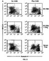
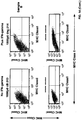
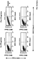
- the present disclosure provides a population of mammalian dendritic cells comprising: cancer-specific peptides from cancer cells taken from a subject that has a cancer; wherein the cancer-specific peptides are acquired in vitro by dendritic cells from said cancer cells; wherein the cancer cells are not treated in vitro with interferon-gamma (IFN-gamma) or with IFN-gamma mimetic; wherein greater than 60 percent (%), or greater than 70%, or greater than 80%, or greater than 90%, or greater than 95%, or greater than 98%, or greater than 99%, of said cancer cells that are not treated in vitro with IFN-gamma or IFN-gamma mimetic, are autophagic and non-apoptotic, and wherein the dendritic cells and cancer cells are from the same subject.
- IFN-gamma interferon-gamma
- IFN-gamma mimetic interferon-gamma mimetic
- the above population of dendritic cells wherein greater than 80% of said cancer cells that are not treated in vitro with IFN-gamma or IFN-gamma mimetic, are autophagic and non-apoptotic.
- a vaccine for the above subject comprising the above population of dendritic cells.
- the above population of dendritic cells wherein essentially all of the cancer cells that are not treated with IFN-gamma or IFN-gamma mimetic are irradiated and incapable of cell division.
- the above population of dendritic cells wherein at least 80% of the cancer cells that are not treated with IFN-gamma or IFN-gamma mimetic are irradiated and incapable of cell division. Also provided is the above population of dendritic cells of, wherein at least 80% of the cancer cells that are not treated with IFN-gamma or IFN-gamma mimetic are treated with a nucleic acid cross-linker and are incapable of cell division.
- a cancer vaccine comprising: at least one mature dendritic cell from a subject that has cancer; wherein the at least one mature dendritic cell had been contacted with at least one cancer tumor cell from the same subject, wherein the at least cancer tumor cell that is contacted with the at least one mature dendritic cell is non-dividing, autophagic, and non-apoptotic. Also provided is a composition for use in a method for stimulating immune response against a cancer-specific antigen, comprising administering an immune-stimulatory amount of the above population of dendritic cells to the subject.
- the immune response that is stimulated comprises one or more of CD4 + T cell response, CD8 + T cell response, and B cell response.
- the CD4 + T cell response, CD8 + T cell response, or B cell response can be measured by ELISPOT assays, by intracellular cytokine staining assays, by tetramer assays, or by detecting antigen-specific antibody production.
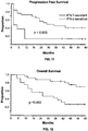
- the immune response comprises a survival time that comprises 2-year overall survival (OS), and where the 2-year overall survival is at least 60%.
- the administration comprises subcutaneous injections of the vaccine.
- the administration comprises injections of the vaccine given weekly for three months and then monthly for five months.
- a method for preparing a dendritic cell vaccine comprising: one or more cancer cells is treated with an agent that prevents cell division; the one or more cancer cells are not treated in vitro with interferon-gamma (IFN-gamma) or with an IFN-gamma mimetic; cancer cells that are autophagic and non-apoptotic are selected; cancer cells that are non-autophagic and apoptotic are rejected; and, wherein the cancer cells that are autophagic and non-apoptotic are provided to one or more autologous dendritic cells, or, wherein peptides derived from the cancer cells that are autophagic and non-apoptotic are provided to one or more autologous dendritic cells.
- IFN-gamma interferon-gamma
- IFN-gamma mimetic interferon-gamma mimetic
- compositions comprising: at least one cancer cell that is not treated with interferon-gamma (IFN-gamma) from a first subject, and at least one antigen presenting cell (APC) from the same first subject, wherein the cancer cell is: autophagic; and non-apoptotic. Also provided is the above composition, wherein the cancer cell is MHC class II-expressing. Also provided is the above composition, wherein the APC is a dendritic cell, a macrophage, or a B cell.
- IFN-gamma interferon-gamma
- APC antigen presenting cell
- the at least one cancer cell comprises cancer-specific peptides, and wherein the cancer specific-peptides are substantially not contained in said APCs and are substantially not processed by said APCs.
- the cancer cells comprise cancer-specific peptides, and wherein the cancer specific-peptides are substantially contained in said APCs and are partially or substantially processed in said APCs.
- compositions wherein the cancer cell is loaded into the APC. Also provided is the above composition, wherein the cancer cell is not loaded into the APC. Also provided is the above composition, wherein autophagy is demonstrated by a test that assays microtubule-associated protein light chain 3 (LC3). Also provided is the above composition, wherein the cells are demonstrated to be non-apoptotic using at least one of the reagent, 7-aminoactinomycin D (7-ADD), or the reagent, annexin.
- the following provides a method of manufacturing embodiment. What is provided is a method for manufacturing the above vaccine or the above composition, comprising contacting at least one cancer tumor cell to at least one antigen presenting cell (APC), wherein the at least one cancer tumor cell is from a first human subject, and wherein the at least one APC is from the same first human subject.
- APC antigen presenting cell
- a method for preparing a dendritic cell vaccine comprising: treating cancer cells acquired from a first subject with an agent that prevents cell division; wherein the cancer cells are not treated in vitro with IFN-gamma or an IFN-gamma mimetic; selecting cancer cells that are autophagic and non-apoptotic; and, contacting the selected cancer cells with autologous dendritic cells from the same first subject.
- a composition that comprises a dendritic cell vaccine as prepared by the above method.
- a composition for use in a method for stimulating immune response against a cancer-specific antigen comprising administering the above composition to a subject that has cancer.
- Also disclosed is a vaccine comprising the above population of the above mammalian dendritic cells.
- dendritic cells wherein the given subject is a human subject.
- the above dendritic cells wherein the given subject is a mammal that is not human.
- compositions for use in stimulating an immune response comprising one or more of CD4 + T cell response, CD8 + T cell response, and B cell response.
- compositions for use as defined wherein the CD4 + T cell response, CD8 + T cell response, or B cell response, can be measured by ELISPOT assays, by intracellular cytokine staining assays, by tetramer assays, or by detecting antigen-specific antibody production.
- composition for use as defined wherein the immune response comprises a survival time that comprises 2-year overall survival (OS), and where the 2-year overall survival is at least 60%.
- OS 2-year overall survival
- compositions for use as defined wherein the administration comprises subcutaneous injections of the vaccine.
- compositions for use as defined wherein the administration comprises injections of the vaccine given weekly for three months and then monthly for five months.
- composition wherein the APC is a dendritic cell, a macrophage, or a B cell.
- composition wherein autophagy is demonstrated by a test that assays microtubule-associated protein light chain 3 (LC3).
- LC3 microtubule-associated protein light chain 3
- composition wherein the cells are demonstrated to be non-apoptotic using at least one of the reagent, 7-aminoactinomycin D (7-ADD), or the reagent, annexin.
- composition that comprises a dendritic cell vaccine, as prepared by the above method.
- the pharmaceutical composition, reagent, and related methods, of the present disclosure uses a preparation of cancer cells that, is 7-AAD negative and annexin V negative.
- This population can be, e.g., about 99% 7-AAD negative and about 99% annexin V negative, or at least 95% 7-AAD negative and at least 95% annexin V negative, or at least 90% 7-AAD negative and at least 90% annexin V negative, to provide non-limiting examples.
- composition wherein autophagy is demonstrated by a test that assays microtubule-associated protein light chain 3 (LC3); and the above composition, wherein the cells are demonstrated to be non-apoptotic using at least one of the reagent, 7-aminoactinomycin D (7-AAD), or the reagent, annexin.
- LC3 microtubule-associated protein light chain 3
- the present disclosure provides a kit for testing immune response against a tumor antigen in a subject, wherein the subject is treated by one or more of the above methods, and wherein the kit comprises a reagent that detects humoral immune response, cellular immune response, or innate immune response.
- Immune-stimulatory amount can be an amount that increases ELISPOT assay results by a measurable amount, that increases ICS assay results by a measurable amount, that increases tetramer assay results by a measurable amount, that increases the blood population of antigen-specific CD4 + T cells by a measurable amount, that increases the blood population of antigen-specific CD8 + T cells by a measurable amount, or where the increase is by at least 10%, 20%, 30%, 40%, 50%, 60%, 70%, 80%, 90%, 100%, 1.5-fold, 2.0-fold, 3.0-fold, and the like, when compared to a suitable control.
- a suitable control can be a control vaccine, where dendritic cells are not loaded with melanoma cells, or are not loaded with peptide derived from melanoma cells.
- cancer-specific peptides from cancer cells taken from a subject that has a cancer it means the following. If the subject with the cancer has melanoma, the peptides are melanoma-specific peptides, and that the cancer cells are melanoma cells.
- melanoma-specific antigen encompasses antigens that are frequently associated with melanoma, and where the antigen is considered to be unique to melanoma, as opposed to being associated with other cancers, and in addition, the term “melanoma-specific antigens” encompasses antigens that are frequently associated with melanoma, and where the antigen is also associated with other types of cancer, such as breast cancer, colorectal cancer, and the like.
- Irradiated in the context of irradiating melanoma cells for the present disclosure, is preferably by gamma-irradiation, but also encompasses irradiation by x-rays, electrons, neutrons, protons, electromagnetic irradiation, visible light, ultraviolet light, and so on.
- the irradiation functions to prevent cell division of the melanoma cells.
- the irradiation prevents cell division, but also denatures cellular proteins.
- the present disclosure prevents cell division of melanoma cells by way of physical disruption, e.g., sonication, cavitation, dehydration, ion depletion, or by toxicity from exposure to one or more salts.
- percent refers to the number of peptide molecules, and not to the number of different antigenically distinct peptides.
- percent refers to the number of peptide molecules, and not to the number of different antigenically distinct peptides.
- percent refers to the number of peptide molecules, and not to the number of different antigenically distinct peptides.
- percent refers to the number of peptide molecules, and not to the number of different antigenically distinct peptides.
- percent as in “less than 40% of the melanoma-specific peptides” refers to the number of peptide molecules, and not to the number of antigenically distinct peptides.
- percent refers to the number of peptide molecules, and not to the number of antigenically distinct peptides, and the like.
- peptides as in “greater than 60% of the melanoma-specific peptides,” refers to the sum of the number of peptide molecules, oligopeptides molecules, and polypeptide molecules.
- peptides as in “greater than 80% of the melanoma-specific peptides,” refers to the sum of the number of peptide molecules, oligopeptides molecules, and polypeptide molecules.
- peptides as in “less than 40% of the melanoma-specific peptides,” refers to the sum of the number of peptide molecules, oligopeptides molecules, and polypeptide molecules.
- peptides as in “less than 20% of the melanoma-specific peptides,” refers to the sum of the number of peptide molecules, oligopeptides molecules, and polypeptide molecules, and the like.
- the cancer cell can be broken, for example, by a homogenizer or by osmotic bursting, resulting in a crude extract.
- Peptides, oligopeptides, and polypeptides of the crude extract can be exposed to dendritic cells, followed by processing of the peptides by the dendritic cells.
- Derived from also encompasses providing dendritic cells with intact cancer cells, where the cancer cells are living, or where the cancer cells have been treated with irradiation but are still metabolically active, or where the cancer cells have been treated with a nucleic acid cross-linking agent but are still metabolically active.
- "Derived from” includes mixtures of cancer cell debris, free cancer cell proteins, and irradiated cancer cells, that are taken up by dendritic cells, and therefore are derived from the cancer cells.
- administering refers without limitation to contact of an exogenous ligand, reagent, placebo, small molecule, pharmaceutical agent, therapeutic agent, diagnostic agent, or composition to the subject, cell, tissue, organ, or biological fluid, and the like.
- administering can refer, e.g., to therapeutic, pharmacokinetic, diagnostic, research, placebo, and experimental methods.
- Treatment of a cell encompasses contact of a reagent to the cell, as well as contact of a reagent to a fluid, where the fluid is in contact with the cell.
- administering also encompasses in vitro and ex vivo treatments, e.g., of a cell, by a reagent, diagnostic, binding composition, or by another cell.
- an "agonist,” as it relates to a ligand and receptor, comprises a molecule, combination of molecules, a complex, or a combination of reagents, that stimulates the receptor.
- an agonist of granulocyte-macrophage colony stimulating factor (GM-CSF) can encompass GM-CSF, a mutein or derivative of GM-CSF, a peptide mimetic of GM-CSF, a small molecule that mimics the biological function of GM-CSF, or an antibody that stimulates GM-CSF receptor.
- An antagonist as it relates to a ligand and receptor, comprises a molecule, combination of molecules, or a complex, that inhibits, counteracts, downregulates, and/or desensitizes the receptor.
- An antagonist encompasses any reagent that inhibits a constitutive activity of the receptor. A constitutive activity is one that is manifest in the absence of a ligand/receptor interaction.
- Antagonist also encompasses any reagent that inhibits or prevents a stimulated (or regulated) activity of a receptor.
- an antagonist of GM-CSF receptor includes, without implying any limitation, an antibody that binds to the ligand (GM-CSF) and prevents it from binding to the receptor, or an antibody that binds to the receptor and prevents the ligand from binding to the receptor, or where the antibody locks the receptor in an inactive conformation.
- expression encompasses the following. Expression encompasses the biosynthesis of mRNA, polypeptide biosynthesis, polypeptide activation, e.g., by post-translational modification, or an activation of expression by changing the subcellular location or by recruitment to chromatin. In other words, “increased expression” encompasses increased biosynthesis, or increased activity that is caused by phosphorylation, or an increased activity that is caused by migration from the cytosol to the nucleus.
- Antigen presenting cells are cells of the immune system used for presenting antigen to T cells.
- APCs include dendritic cells, monocytes, macrophages, marginal zone Kupffer cells, microglia, Langerhans cells, T cells, and B cells (see, e.g., Rodriguez-Pinto and Moreno (2005) Eur. J. Immunol. 35:1097-1105 ).
- Dendritic cells occur in at least two lineages. The first lineage encompasses pre-DC1, myeloid DC1, and mature DC1.
- the second lineage encompasses CD34 ++ CD45RA- early progenitor multipotent cells, CD34 ++ CD45RA + cells, CD34 ++ CD45RA ++ CD4 + IL-3Ralpha ++ pro-DC2 cells, CD4 + CD11c - plasmacytoid pre-DC2 cells, lymphoid human DC2 plasmacytoid-derived DC2s, and mature DC2s (see, e.g., Gilliet and Liu (2002) J. Exp. Med. 195:695-704 ; Bauer, et al. (2001) J. Immunol. 166:5000-5007 ; Arpinati, et al. (2000) Blood 95:2484-2490 ; Kadowaki, et al. (2001) J.
- Effective amount encompasses, without limitation, an amount that can ameliorate, reverse, mitigate, prevent, or diagnose a symptom or sign of a medical condition or disorder. Unless dictated otherwise, explicitly or by context, an "effective amount” is not limited to a minimal amount sufficient to ameliorate a condition.
- the severity of a disease or disorder, as well as the ability of a treatment to prevent, treat, or mitigate, the disease or disorder can be measured, without implying any limitation, by a biomarker or by a clinical parameter. Biomarkers include blood counts, metabolite levels in serum, urine, or cerebrospinal fluid, tumor cell counts, cancer stem cell counts, tumor levels. Tumor levels can be determined by the RECIST criteria ( Eisenhauer, et al. (2009) Eur.
- Expression markers encompass genetic expression of mRNA or gene amplification, expression of an antigen, and expression of a polypeptide.
- Clinical parameters include progression-free survival (PFS), 6-month PFS, disease-free survival (DFS), time to progression (TTP), time to distant metastasis (TDM), and overall survival, without implying any limitation.
- a composition that is "labeled” is detectable, either directly or indirectly, by spectroscopic, photochemical, biochemical, immunochemical, isotopic, or chemical methods.
- useful labels include 32 P, 33 P, 35 S, 14 C, 3 H, 125 I, stable isotopes, epitope tags fluorescent dyes, electron-dense reagents, substrates, or enzymes, e.g., as used in enzyme-linked immunoassays, or fluorettes (see, e.g., Rozinov and Nolan (1998) Chem. Biol. 5:713-728 ).
- TLR toll-like receptor
- Regs T regulatory cells
- CD8 + T cells or CD4 + T cells agents that can enhance activity of CD8 + T cells or CD4 + T cells
- present disclosure provides reagents, methods of administration, methods of diagnosis, and methods for stimulating immune response against tumor antigens, that use one or more, as well as combinations thereof, of the following reagents.
- TLR toll-like receptor
- Imidazoquinolines such as imiquimod and resiquimod
- TLR7 or TLR8 agonists Stimulation of TLR7 of plasmacytoid dendritic cells results in expression of IFN-alpha.
- Stimulation of TLR7 of myeloid dendritic cells results in expression of interleukin-12, and the consequent Th1-type immune response.
- CpG oligonucleotides are TLR9 agonists.
- CpG ODNs occur in three classes, CpG-A, CpG-B, and CpG-C.
- CpG-A stimulates NK cells, owing to its IFN-alpha producing effect on plasmacytoid DCs.
- CpG-B induces IFN-alpha, and upregulates co-stimulatory markers on pDCs and B cells.
- TLR3 agonists include polyriboinosinic-polyribocytidylic acid (poly I:C), which is an analogue of viral double stranded RNA (dsRNA).
- TLR4 agonists include monophosphoryl lipid A (MPL), which is a derivative of Salmonella minnesota lipopolysaccharide, and which is used as part of a vaccine against human papillomavirus. Attenuated bacteria are used in anti-cancer therapy. Mycobacterium bovis stimulates TLR2, and TLR4, and TLR9. Listeria monocytogenes stimulates various TLRs ( US 2007/0207171 of Dubensky et al ). See also, Galluzzi et al (2012) Oncolmmunology. 1:699-716 ; Adams (2009) Immunotherapy. 1:949-964 .
- MPL monophosphoryl lipid A
- Alpha-galactosylceramide (alpha-GalCer) is also provided ( Schwaab and Ernstoff (2011) Therapy. 4:369-377 ). Alpha-GalCer provokes the activation of NKT cells that express certain T cell receptors ( Lopez-Sagaseta et al (2012) PLoS Biol. 10:e1001412 (11 pages )). What is also provided are agonists of NOD receptors. NOD receptors include NOD1 and NOD2. NOD agonists include N-acetylmuramyl-L-alanyl-D-isoglutamine (muramyldipeptide (MDP)), which binds to NOD 2.
- MDP N-acetylmuramyl-L-alanyl-D-isoglutamine
- NOD agonists include gamma-D-glutamyl-meso-diaminopimelic acid (iE-DAP), which binds to NOD1.
- NOD agonists include desmuramylpeptides (DMP), which binds to NOD1. See, e.g., Uehara et al (2006) J. Immunol. 177:1796-1804 ).
- NOD agonists are derived from fragments of peptidoglycan.
- T regulatory cells include cyclophosphamide, gemcitabine, and antibodies that deplete Tregs (see, e.g., Le and Jaffee (2012) Cancer Res. 72:3439-3444 ; Klages et al (2010) Cancer Res. 70:7788-7799 ).
- Gemcitabine reduces the immunosuppressive activity of myeloid-derived suppressor cells (MDSCs) ( Le et al (2009) Int. Immunopharmacol. 9:900-909 ).
- Anti-CD25 antibodies can deplete Tregs, and have been used in the treatment of cancer ( Klages et al (2010) Cancer Res. 70:7788-7799 ).
- Daclizumab is an anti-CD25 antibody, which blocks binding of interleukin-2 (IL-2) to CD25, thereby blocking a signal needed for Treg maintenance.
- Some Tregs express high levels of CTLA4 (CD45RA + Tregs).
- Anti-CTLA4 antibodies such as ipilimumab, target CTLA4. Ipilimumab is used for treating melanoma ( Rech et al (2012) Immunotherapy. 4:1103-1105 ).
- Lymphocyte activation gene-3 (LAG-3) blocking agents such as anti-LAG-3 antibodies or soluble LAG-3 (e.g., LAG-3 Ig), can enhance immune response to cancers or infections.
- Anti-LAG-3 antibodies reduce the activity of Tregs (see, e.g., Huang, et al. (2004) Immunity 21:503-513 ; Triebel (2003) Trends Immunol. 24:619-622 ).
- Antibodies are available that directly target CD8 + T cells or CD4 + T cells. These antibodies include those that target costimulatory receptors (4-1BB, OX40 and GITR) or the blockade of co-inhibitory receptors (CTLA-4, PD-1 and PD-L1).
- GITR is glucocorticoid-induced TNFR-related protein. See, e.g., Schaer et al (2010) Curr. Opin. Investig. Drugs. 11:1378-1386 ).
- Anti-GITR can stop the suppressive function of Tregs, thus explaining anti-GITR's efficacy in anti-cancer therapy ( Coe et al (2010) Cancer Immunol. Immunother. 59:1367-1377 ).
- DTA-1 is an anti-GITR antibody.
- the present disclosure also provides ELISPOT assays, intracellular cytokine staining (ICS), and tetramer assays, for characterizing immune response (see, e.g., of US 2007/0190029 of Pardoll ; Chattopadhyay (2008) Cytometry A. 2008 73:1001-1009 ; Vollers (2008) Immunology. 123:305-313 ; Lalvani, et al. (1997) J. Exp. Med. 186:859-865 ; Waldrop (1997) J. Clin. Invest. 99:1739-1750 ; Hudgens (2004) J. Immunol. Methods 288:19-34 ; Goulder (2001) J. Virol. 75:1339-1347 ; Goulder (2000) J.
- Immune response in a patient can be assessed by endpoints that are used in oncology clinical trials, including objective response (RECIST criteria), overall survival, progression-free survival (PFS), disease-free survival, time to distant metastasis, 6-month PFS, 12-month PFS, and so on.
- RECIST criteria objective response
- PFS progression-free survival
- disease-free survival time to distant metastasis
- Glioblastoma-specific antigens include PTPRZ1; EGFR; SEC61G; TNC; HER2; TRP-2; gp100; MAGE-1; IL13Ralpha2; AIM-2 ( Phuphanich et al (2013) Cancer Immunol. Immunother. 62:125-135 ; Neidert et al (2012) J. Neurooncol. ).
- Colorectal cancer-specific antigens include SPARC, CEA, Cep55/c10orf3 ( Inoue et al (2010) Int. J. Cancer. 127:1393-1403 , Parkhurst et al (2011) Mol. Ther. 19:620-626 ; Inoda et al (2011) Exp. Mol. Pathol.
- Renal cancer-specific antigens include carbonic anhydrase IX (CA-IX), MUC-1, and NYESO-1, and 5T4 ( Tykodi et al (2012) J. Immunother. 35:523-533 ).
- Ovarian cancer-specific antigens include WT1, mesothelin, NY-ESO-1, p53, p53 carrying specific mutations, HER-2/neu, folate receptor-alpha, IGFBP; MUC1, MUC4, MUC16, EpCAM; CTA ( Dohi et al (2011) Anticancer Res. 31:2441-2445 ; Vermeij et al (2012) Curr. Pharm. Des.
- Specificity can arise from the amino acid sequence of the antigen, from the degree of expression of that antigen by the tumor cell, from post-translational modification of the antigen, and the like. Specificity to a certain type of cancer cell can also arise from a particular fingerprint of a plurality of tumor antigens. Specificity can also arise, from the fact that a particular antigen, while expressed by a wide variety of tumor cells, has particular use in immunotherapy against a smaller number of tumor types. Specificity can also arise from the fact that a particular collection of MHC class I presentable and MHC class II presentable epitopes exist, on a particular polypeptide or polypeptide fragment.
- specificity in an administered antigen can arise by omitting one or more peptides that can provoke immunotolerance.
- the skilled artisan can easily locate the relevant nucleic acid and polypeptide sequences, e.g., on the U.S. government's web site, at ncbi.nlm.nih.
- a vaccine of the present disclosure can be purely preventative, it can be used for treating an existing cancer or pre-neoplastic disorder, or it can be used for preventing recurrence of a previously treated cancer.
- Dendritic cell vaccine of the present disclosure can be administered by intradermal, intranodal, mucosal, or subcutaneous routes, or any combination of the above.
- Each dose can comprise about 10 x 10 3 dendritic cells, 20 x 10 3 cells, 50 x 10 3 cells, 100 x 10 3 cells, 200 x 10 3 cells, 500 x 10 3 cells, 1 x 10 6 cells, 2 x 10 6 cells, 20 x 10 6 cells, 50 x 10 6 cells, 100 x 10 6 cells, 200 x 10 6 , 500 x 10 6 , 1 x 10 9 cells, 2 x 10 9 cells, 5 x 10 9 cells, 10 x 10 9 cells, and the like.
- Administration frequency can be, e.g., once per week, twice per week, once every two weeks, once every three weeks, once every four weeks, once per month, once every two months, once every three months, once every four months, once every five months, once every six months, and so on.
- the total number of days where administration occurs can be one day, on 2 days, or on 3, 4, 5, 6, 7, 8, 9, 10, 11, 12, 13, 14, 15, 16, 17, 18, 19, or 20 days, and so on. It is understood that any given administration might involve two or more injections on the same day.
- the disclosure involves loading dendritic cells with whole tumor cells, where at least 10%, where at least 20%, at least 30%, at least 40%, at least 50%, at least 60%, at least 70%, at least 80%, at least 90%, at least 95%, or at least 99%, of the melanoma cell-derived protein that is loaded into the dendritic cells resides in whole tumor cells.
- dendritic cell vaccine is held in a flask, in a vial, in a bottle, in a syringe, in a catheter, in a cannula, and so on.
- dendritic cells For administration, at least 20%, at least 30%, at least 40%, at least 50%, at least 60%, at least 70%, at least 80%, at least 90%, at least 95%, at least 99%, of the dendritic cells that are administered are mature dendritic cells.
- the present disclosure encompasses mimetics, for example, interferon-gamma mimetics, such as mimetic peptide 95-132 ( Ahmed (2007) J. Immunol. 178:4576-4583 ; Fulcher (2008) FEBS Lett. 582:1569-1574 ).
- IFN-mimetic encompasses, e.g., an antibody that has the same agonist activity of interferon-gamma.
- compositions and methods where cancer cells are inactivated, for example, by radiation or by way of nucleic acid cross-linkers.
- exemplary cross-linkers have the ability to cross-link DNA but to leave proteins unmodified.
- a nucleic acid alkylator can be beta-alanine, N-(acridin-9-yl), 2-[bis(2-chloroethyl)amino]ethyl ester.
- the nucleic acid targeted compound is a psoralen compound activated by UVA irradiation.
- the nucleic acid targeting compound can be 4'-(4-amino-2-oxa) butyl-4,5',8-trimethylpsoralen (also referred to herein as "S-59").
- Cells can be inactivated with 150 micromolar of psoralen S-59 and 3 J/cm 2 UVA light (FX 1019 irradiation device, Baxter Fenwal, Round Lake, IL).
- the inactivation with S-59 is referred to as photochemical treatment and results in complete inactivation of the cells.
- Various concentrations of nucleic acid cross-linked agent can be tested for efficacy in inactivating cells, for example, for efficacy in preventing cell division.
- S-59 is distinguished by its ability to cross-link nucleic acids, but to leave proteins intact an unmodified.
- Cells can be suspended in 5 mL of saline containing 0, 1, 10, 100, and 1000 nM of psoralen S-59. Each sample can be irradiated as follows. The S-59 can be added at a concentration of 100 nM. Samples can be UVA irradiated at a dose of approximately 2 J/cm 2 (FX1019 irradiation device, Baxter Fenwal, Round Lake, III.).
- Each sample can then transferred to a 15 mL tube, centrifuged, and the supernatant removed, and then washed with 5 mL saline, centrifuged and the supernatant removed and the final pellet suspended in 0.5 mL of saline ( U.S. Pat. Nos. 7,833,775 of Dubensky and 7,691,393 of Dubensky ).
- a population of melanoma cells can be enriched in melanoma cells that are non-apoptotic, for example, by use of the technique that separates non-apoptotic and autophagic cells from cells that are non-autophagic and apoptotic, where separation is by the adhesion of the autophagic and non-apoptotic cells to a surface, where the other cells are floating.
- a population enriched in non-apoptotic melanoma cells can also be acquired by removing apoptotic cells by way of an antibody specific for phosphatidyl serine. Techniques for removing cells by way of immobilized antibodies are available ( Onodera (1998) Ther. Apher. 2:37-42 ).
- Antibodies specific for phosphatidylserine are available (e.g., EMD Millipore, Billerca, MA). Also, bulk population of melanoma cells can be labeled with fluorescent anti-phosphatidylserine antibodies, where the tagged apoptotic melanoma cells are removed by flow cytometry, affinity chromatography, immunomagnetic separation (see, e.g., Hoeppener (2012) Recent Results Cancer Res. 195:43-58 ; Dainiak (2007) Adv. Biochem. Eng. Biotechnol. 106:1-18 ). This technique is generally applicable to enriching in cancer cells that are melanoma cells, and also for cancer cells that are not melanoma cells.
- Z-VAD Z-VAD-fmk
- Z-VAD-fmk an inhibitor of apoptosis
- Z-VAD-fmk is a synthetic peptide, Z-Val-Ala-Asp(OMe)-CH 2 F.
- Caspases are cysteine-aspartic acid-specific members of the protease family.
- Caspases are activated by a death receptor ligation, e.g., TRAIL, FAS, by DNA damage, stress, serum starvation and in some cell types, interferons. Caspases play a critical role in the highly regulated process of apoptosis that includes nuclear fragmentation, chromatin condensation, and loss of cytoplasmic integrity.
- the pan-capase inhibitor, z-VAD-fmk (carbobenzoxy-valyl-alanyl-aspartyl-[O-methyl]- fluoromethylketone) irreversibly binds the catalytic site of caspase proteases and inhibits their function in inducing apoptosis.
- Inhibiting the ability of cells to undergo apoptosis in response to IFN-gamma can be a means by which cells that are non-apoptotic but autophagic and be generated without the steps of selection by the washing to remove floating apoptotic cells.
- the disclosure provides pharmaceuticals, reagents, kits including diagnostic kits, that wherein the pharmaceuticals, reagents, and kits, comprise dendritic cells, antibodies, or antigens.
- compositions that comprise at least one dendritic cell and at least one antigen, methods for stimulating antibody formation, methods for stimulating ADCC, methods for stimulating complement-dependent cytotoxicity, and methods and kits for determining patient suitability, for determining patient inclusion/exclusion criteria in the context of a clinical trial or ordinary medical treatment, and for predicting response to the pharmaceutical or reagent.
- Complement-dependent cytotoxicity is described (see, e.g., Goodman, et al. (1990) J. Clin. Oncol. 8:1083-1092 ; Cheson (2010) J. Clin.
- the pharmaceutical compositions, reagents, and related methods, of the disclosure encompass CD83 positive dendritic cells, where CD83 is induced by loading with IFN-gamma-treated cancer cells.
- the CD83 is induced by at least 2%, at least 3%, at least 4%, 6%, 7%, 8%, 9%, 10%, and the like.
- Figure 1 reveals a graphic of cultured tumor cells before treatment with IFN-gamma (left) and after treating with IFN-gamma for 72 hours (right).
- the cultured tumor cells are either floating, non-autophagic, and apoptotic, or adherent, autophagic, and non-apoptotic.
- the floating cells are shown expressing the apoptotic marker, phosphatidyl serine.
- the floating cells are shown with relatively few expressed MHC class II, while the adherent cells are shown with over-expressed MHC class II.
- Figures 2A-D show characterization of IFN-gamma treated autologous tumor cells used for loading dendritic cells.
- Autologous melanoma tumor cells were treated with or without 1000 IU/mL IFN-gamma for 72 hours in 15% FBS/ECS in RPMI, harvested and irradiated with 100Gy and cryopreserved. Cells were then thawed in AIMV and a sample taken for flow cytometry and for preparation of cell lysates for immunoblotting prior to antigen loading of DCs.
- An example of four separate autologous melanoma cell lines is shown ( Fig.2A , Fig.2B and Fig.2C ).
- the disclosure excludes non-autologous tumor cells for loading dendritic cells, and excludes methods of using non-autologous tumor cells for loading dendritic cells.
- Figures 3A and 3B describe phenotype of dendritic cells loaded with autologous melanoma cell lines treated with or without interferon-gamma.
- a set of four autologous melanoma cell lines were treated with or without 1000 IU/mL of IFN-gamma for 72 hours, irradiated and cryopreserved.
- the cells were then thawed in AIMV and combined with autologous dendritic cells for approximately 24 hours prior to harvest and assaying by flow cytometry for the expression of CD80, CD83, CD86 and MHC class II ( Figure 3A ).
- Figures 4A and 4B show phenotype of dendritic cells used for dose preparation.
- FACS Caliber® beads were used to set the initial flow cytometer instrument settings which were then held constant throughout the collection of data ( Figure 4A ).
- Values of percent expression and mean fluorescence intensity (MFI) ⁇ SD are compared in Figure 4B for Pre-ATC and Post-ATC loading.
- FIGS 5A to 5C show interferon-gamma treated melanoma cells undergo autophagy.
- a selection of commercially-available melanoma cell lines were incubated with 1000 IU/mL IFN-gamma for 72 hours in 5%FBS/RMPI.
- Phase-contrast photomicrographs of SK-5-Mel cell cultures were taken at the end of the incubation period ( Figure 5A ) showing enlarged cells with vacuoles reminiscent of autophagosomes.
- Confirmation of the formation of autophagosomes was demonstrated by transfection with GFP-LC3B constructs prior to treatment with IFN-gamma ( Figure 5B ).
- Autophagy induction after IFN-gamma treated was confirmed by western blotting using an antibody for LC3B ( Figure 5C ) which identifies a faster migrating form of LC3 that has been shown to be associated with autophagic vessel formation.
- Figures 6A and 6B reveal apoptosis and autophagy induced in response to interferon-gamma.
- SK-5-Mel cells were incubated with 1000 IU/mL of IFN-gamma for 72 hours after which non-adherent and adherent populations were collected and assayed for apoptosis and autophagy by flow cytometry using 7-AAD and Annexin-V ( Figure 6A ).
- Enzo Cyto-ID Autophagy Detection Dye was used to measure autophagy by flow cytometry by measuring the mean intensity peak shift of dye provided by the manufacturer ( Figure 6B ). Fold changes in the peak shift in comparison to 5% FBS/RPMI are shown in Figure 6C with serum-free as positive control for the induction of autophagy.
- Figure 7 discloses autophagy induction after blocking of caspase activity did not affect the induction of autophagy in response to IFN-gamma in melanoma cells.
- SK-5-Mel cells were treated with 1000 IU/mL of IFN-gamma in the presence of 20uM of the pan-caspase inhibitor z-VAD or its control compound, z-FA for 72 hours.
- the cells were harvested and assayed for autophagy by flow cytometry as in Figure 6C .
- Figure 8 shows SK-5-Mel cells which were incubated with 1000 IU/mL of IFN-gamma in the presence of 10 uM of the autophagy inhibitor 3-methyladenine (3-MA) for 72 hours. The cells were then harvested and assayed for apoptosis and MHC class II (HLA-DR) expression by flow cytometry.
- 3-MA autophagy inhibitor 3-methyladenine
- MFI mean fluorescence intensity
- Annexin-V positive percent apoptotic cells
- Figure 11 and Figure 12 show a correlation between induction of MHC class II and the absence of apoptosis (Interferon-gamma resistant) is associated with better progression-free survival ( Fig.11 ) and overall survival ( Fig.12 ) in patients received dendritic cells loaded with autophagic, non-apoptotic interferon-gamma treated tumor cells.
- Figure 13 shows survival curves from three trials.
- the plot (Kaplan-Meier plot) is a stepwise curve showing the percent of study subjects surviving during the course of clinical trials.
- the groups are designated DC-54 (solid circle); TC-74 (solid square); TC-24 (solid triangles); and DC-18 (line). Poorest survival occurred with TC-24. The next poorest survival was with TC-74.
- TC-24 refers to a vaccine of tumor cells in a study involving 24 subjects.
- Figure 14 shows survival curves from three trials.
- the trials are the same clinical trials as those disclosed in Figure 13 , but with additional data acquired from later time points.
- Dendritic cells were generated by plastic adherence method of ficoled apheresis products ( Choi, et al. (1998) Clin. Cancer Res. 4:2709-2716 ; ships, et al. (1998) Exp. Hematol. 26:489-500 ; Cornforth, et al. (2011) Cancer Immunol. Immunother. 60:123-131 ), in antibiotic-free AIM-V medium (Invitrogen, Grand Island, NY) supplemented with 1,000IU/mL each of IL-4 (CellGenix, Freisberg, Germany) and GM-CSF (Berlex, Seattle, WA) (DC medium). The flasks were then cultivated for 6 days prior to loading with IFN-gamma treated, irradiated autologous tumor cells.
- the IFN-gamma treated and irradiated tumor cells were recovered from cryopreservation, washed with phosphate buffered saline (PBS), and then added to the cultivated dendritic cells (DCs) and then incubated for about 24h.
- the antigen-loaded DCs were harvested by gentle scraping with a rubber policeman and cryopreserved. Aliquots of IFN-gamma treated or untreated tumor cells and loaded DCs were obtained for flow cytometry evaluation and trypan-blue exclusion assay.
- Apoptosis can be detected or measured with a number of reagents, e.g., fluorochrome-labeled annexin, by staining with dyes such as propidium iodide and 7-aminoactinomycin D (7-AAD), by determining loss of mitochondrial inner memebrane potential, by measuring activation or cleavage of caspases.
- fluorochrome-labeled annexin by staining with dyes such as propidium iodide and 7-aminoactinomycin D (7-AAD), by determining loss of mitochondrial inner memebrane potential, by measuring activation or cleavage of caspases.
- 7-AAD 7-aminoactinomycin D
- the available methods can distinguish between live cells, necrotic cells, early apoptotic cells, and late apoptotic cells.
- the disclosure uses melanoma cells that are not apoptotic by 7-ADD assay, not apoptotic by annexin V assay, not apoptotic by an assay for apoptosis after IFN-gamma treatment ( Dillman, et al. (2011) Cancer Biotherapy Radiopharmaceuticals 26:407-415 ), or not apoptotic by one or more of the biomarkers Bcl-2, caspase-3, P53, or survivin ( Karam, et al. (2007) Lancet Oncol. 8:128-136 ).
- compositions, reagents, and related methods, of the disclosure exclude IFN-gamma-treated melanoma cells that are apoptotic, where apoptosis is determined, e.g., according to U.S. Pat. No. 7,544,465 issued to Herlyn, et al ; U.S. Pat. No. 7,714,109 issued to Thorpe, et al.
- Autophagy is a naturally occurring process that is used for the degradation of many proteins and some organelles. Autophagy mediates protein and organelle turnover, starvation response, cell differentiation, cell death, and so on.
- Microtubule-associated protein light chain 3 (LC3) is to monitor autophagy.
- autophagy can be detected by measuring the conversion of LC3, which involves conversion of LC3-I to LC3-II. The amount of LC3-II is correlated with the number of autophagosomes.
- LC3 is cytosolic and soluble, while LC3-II is present on membranes.
- LC3-II has a greater molecular weight because it is conjugated with a lipid.
- LC3 processing can be measured, e.g., by western blots, while autophagy, autophagic vesicles, and autophagosomes, can be measured by microscopy.
- Autophagy can be quantitated, e.g., by detecting processed LC3-II, by the ratio between early to late autophagic compartments, or by autophagic volume. See, ( Mizushima and Yoshimori (2007) Autophagy 3:542-546:634-641 ; Tanida, et al. (2008) Methods Mol. Biol. 445:77-88 ; Eng, et al. (2010) Autophagy 6:634-641 ).
- the present disclosure uses autophagy as a screening tool, for selecting appropriate autophagic cancer cells, where the cells can be selected according to occurrence of autophagy in one or more particular stages.
- These autophagy stages include: (1) sequestering of cytosolic compartments by the autophagosome, (2) fusion of the autophagosome with the lysosome to form the autolysosome, and (3) degradation of the autophagosomal contents by proteases within the lysosome.
- the present disclosure includes mainly cells displaying the first stage, mainly the second stage, mainly the third stage, mainly the first and second stage, mainly the second and third stage, or mainly cells displaying all three stages.
- the disclosure comprises cells displaying the first stage, the second stage, the third stage, the first and second, the second and third stage, or cells displaying all three stages.
- Interferon-gamma IFN-gamma
- IFN-gamma type II interferon signaling depends on expression of a number of genes, e.g., IFN-gamma receptor, STAT1, STAT2, STAT1 homodimers, STAT1/STAT2 heterodimers, IRF-1, GAS, and IRF-E. Studies have shown that IFN-gamma signaling is dependent on IFN-gamma receptor (IFNGR1 chain; IFNGR2 chain). Low expression of IFNGR on the cell surface can block some aspects of IFN-gamma signaling ( Schroder, et al. (2004) J. Leukocyte boil. 75:163-189 ). In one aspect, the present disclosure excludes using cancer cells that show low surface expression of IFNGR.
- the present disclosure screens cancer cells for those that express the STAT1 homodimer, uses these cells, and substantially excludes cells that do not express STAT1 homodimer.
- the disclosure contemplates screening cells for those with STAT1 phosphorylation (serine-727). What is also contemplated, is excluding cancer cells from patients having loss of function mutations in the STAT1 gene (see, e.g., Dupuis, et al. (2001) Science 293:300-303 ; Schroder, et al. (2004) J. Leukoc. Biol. 75:163-189 ).
- the disclosure embraces using cancer cells that express one or more of these IRF gene family genes, or excluding cancer cells that do not express one or more of these genes.
- the present disclosure embraces biologic material, compositions, reagents, and methods that require using a melanoma cell, or pre-neoplastic melanoma cell, that responds to IFN-gamma.
- the melanoma cell can be identified, distinguished, and selected, by an assay for the expression of one or more IFN-gamma-responsive genes.
- IFN-gamma-responsive genes have been identified (see, e.g., Halonen, et al. (2006) J. Neuroimmunol. 175:19-30 ; MacMicking (2004) 11:601-609; Boehm, etal. (1997) 15:749-795).
- Said assay can involve removing one or more melanoma cells from the patient, culturing the cell in the presence and absence of added IFN-gamma, and determining responsiveness to IFN-gamma.
- IFN-gamma induced gene expression can be detected by assays sensitive to binding of a transcription factor to the promoter of an IFN-gamma induced gene, to expression of mRNA from an IFN-gamma induced gene, to expressed polypeptide, and the like.
- the IFN-gamma response gene can include, e.g., a gene used for immune response, encoding a transcription factor, a transport protein, an apoptosis gene, a gene used for cell growth or maintenance, a gene used for lipid metabolism, a gene that mediates endocytosis or exocytosis, an intracellular signaling gene, a glucose metabolism gene, a cell adhesion gene, as well as genes without an established function.
- the disclosure excludes melanoma cells that, with IFN-gamma treatment, show reduced expression of MHC class II, show no detectable change in expression of MHC class II, show an increase of MHC class II expression of 10% or less, show an increase in MHC class II expression of 15% or less, show an increase in MHC class II expression of 20% or less, 25% or less, 30% or less, 40% or less, 50% or less, and the like.
- the value for percentage refers to the average expression value for the population of melanoma cells, residing in a biopsy or part of a biopsy, from a given subject or patient.
- cytokine signalling-3 SOCS-3
- M21065 Interferon regulatory factor 1
- M63630 GTP binding protein (IRG-47)
- U19119 G-protein-like LRG-47
- L27990 Ro protein
- M31419 204 interferon-activatable protein
- af022371 Interferon-inducible protein 203
- U28404 MIP-1 alpha receptor
- U43085 Glucocorticoid-attenuated response 39
- x56123 Talin
- m31419, 204 interferon-activatable protein U53219 , GTPase IGTP
- 138444 T-cell specific protein
- M31418 202 interferon-activatable protein
- d38417 Arylhydrocarbon receptor
- m26071 Tissue factor (mtf)
- D13759 Cot proto-oncogene
- M18194 Fibronectin
- M13945 Aryl
- the disclosure encompasses use of the IFN-gamma induced gene, CIITA (see, e.g., Chan, et al. (2010) J. Leukocyte Biol. 88:303-311 ; Kwon, et al (2007) Mol. Immunol. 44:2841-2849 ).
- CIITA IFN-gamma induced gene
- the present disclosure embraces measuring expression of one or more of the following IFN-gamma inducible genes, as a screening procedure for qualifying or selecting patients for administering a pharmaceutical.
- the genes include, (gene 1) FCGR1A, (gene 2) IL6R, (gene 3) CXCL9, (gene 4) CLCSF14, (gene 5) UBD, (gene 6) C/EBPalpha, and (gene 7) MHC2TA (CIITA) (see, Waddell, et al. (2010) PLoS ONE 5:e9753 ).
- melanoma cells that is less than 90% are autophagic, less than 80% are autophagic, less than 70% are autophagic, less than 60% are autophagic, less than 50% are autophagic, less than 40% are autophagic, and the like.
- melanoma cells where, that is less than 90% are non-apoptotic, less than 80% are non-apoptotic, less than 70% are non-apoptotic, less than 60% are non-apoptotic, less than 50% are non-apoptotic, less than 40% are non-apoptotic, and the like.
- melanoma cells that is less than 90% are non-adherent, less than 80% are non-adherent, less than 70% are non-adherent, less than 60% are non-adherent, less than 50% are non-adherent, less than 40% are non-adherent, and the like.
- MHC class II can be measured, for example, using antibodies or nucleic acid probes that are specific for MHC class II gene products.
- MHC class II gene products include HLA-DPA1, HLA-DPB1, HLA-DQA1, HLA-DQB1, HLA-DRA, HLA-DRB1, as well as HLA-DM and HLA-DO (see, e.g., protestopoulos, et al. (2008) Human Vaccines 4:400-409 ).
- the present disclosure encompasses reagents, methods of treatment, and methods of diagnosis, that require the melanoma cells to express STAT1 and STAT2, to have an active STAT1-signaling pathway, to have an active STAT2-signaling pathway, or to have active STAT1 and STAT2-signaling pathways.
- the disclosure provides a pharmaceutical composition or pharmaceutical reagent, related methods of administration, and methods of treatment, that result in survival data with a hazard ratio (HR) of less than 1.0, HR less than 0.9, HR less than 0.8, HR less than 0.7, HR less than 0.6, HR less than 0.5, HR less than 0.4, HR less than 0.3, and the like.
- HR hazard ratio
- the disclosure results in overall survival data, progression-free survival data, time to progression data, and so on.
- 6-month PFS of at least 40%, at least 50%, at least 60%, at least 70%, at least 75%, at least 80%, at least 85%, at least 90%, at least 95%, and so on.
- 1-year (or 2-year) PFS of at least 40% at least 50%, at least 60%, at least 70%, at least 75%, at least 80%, at least 85%, at least 90%, at least 95%, and so on.
- 1-year (or 2-year) overall survival of at least 40%, at least 50%, at least 60%, at least 70%, at least 75%, at least 80%, at least 85%, at least 90%, at least 95%, and so on see, e.g., U.S. Dept. of Health and Human Services. Food and Drug Administration. Guidance for Industry. Clinical trial endpoints for the approval of cancer drugs and biologics (April 2005 )).
- Induction of autophagy after IFN-gamma treatment can be used to determine response to systemic IFN-gamma treatment. If biopsied melanoma tumor cells, upon exposure to IFN-gamma in culture, undergo autophagy but not apoptosis, this indicates that these patients will respond favorably to systemic IFN-gamma treatment. Additionally, if successful cell lines are established from the biopsies, that patient would also benefit from cell-therapy products prepared from IFN-gamma treated purified tumor cells lines that are from autophagic but non-apoptotic adherent populations.
- the disclosure embraces isolating and characterizing major histocompatiability complexes isolated from autophagic, non-apoptotic cells collected from tumor cell lines treated with interferon-gamma.
- Major histocompatibility complexes contain antigens specific for CD4 + T cells and have been associated with antibody mediated immune responses. The complexes would represent a large repertoire of antigens would not be present in non-autophagic cells due to the action of lysosomal mediated antigen processing induced in autophagic cells.
- Non-apoptotic, autophagic tumor cells generated from IFN-gamma treated cell lines can be fused with dendritic cells to enhance the antigen presentation due to the high levels of major histocompatability complexes on the surface of the autophagic tumor cells. This process would yield a novel cellular product generated from the fusion of the two cell types.
- the process of induction of autophagy in response to IFN-gamma may be induced in a manner that does not result in apoptosis.
- caspase inhibitors and interferon gamma By combining the treatment of tumor cells with caspase inhibitors and interferon gamma, the process of cell death (and ultimately the formation of tolergeneic apoptotic cells) can be blocked without inhibiting the induction of autophagy or the increase in major histocompatibility class II complexes.
- the methodology of the study was as follows. Autologous and model melanoma tumor cell lines were incubated with 1000 IU/mL of IFN-gamma for 72 hours prior to assaying for autophagy, apoptosis and MHC class II expression. Autophagy was detected by immunobloting with antibodies against LC3 II and by flow cytometry with Enzo's CytolD® Autophagy Detection Kit. Apoptosis and MHC class II induction were assayed by flow cytometry using 7-AAD and annexin-V staining and antibodies against MHC class II, respectively. 7-AAD intercalates in dsDNA. 7-AAD is excluded by viable cells, and thus it can be used to detect dead cells.
- IFN-gamma induces both autophagic and apoptotic cell populations in melanoma cell lines.
- the apoptotic population was predominantly found in the non-adherent population while the autophagic cells remained adherent to the flask.
- Blocking of autophagy with the inhibitor 3-methyladenine (3-MA) inhibits the induction of MHC class II positive cells in response to IFN-gamma (39.4% IFN-gamma vs. 10.0% IFN-gamma + 3-MA).
- pan caspase inhibitor Z-VAD Inhibition of caspase activity with the pan caspase inhibitor Z-VAD prevents apoptosis but does not perturb autophagy in IFN-gamma treated cells (2.75 ⁇ 0.15 IFN-gamma vs. 3.04 ⁇ 0.27 IFN-gamma + Z-VAD, fold change).
- induction of apoptosis is associated with reduced levels of autophagy and MHC class II induction.
- This disclosure provides method or procedure to eliminate apoptotic cells while retaining viable autophagic cells after IFN-gamma treatment can enhance the effectiveness of this type of cell-based immunotherapy.
- IFN-gamma has been associated with suppression of immune response against tumors (see, e.g., Hallermalm (2008) J. Immunol. 180:3766-3774 ; Romieu-Mourez (2010) Cancer Res. 70:7742-7747 ; Lee (2005) Clinical Cancer Res. 11:107-112 ).
- a tumor can be a heterogeneous population of more or less differentiated cells.
- IFN-gamma treatment of melanoma cells of a tumor can act on some of the more differentiated cells that are more susceptible to apoptosis. By eliminating these cells from the antigen source, the result can be loss of some effect on the tumor bulk following vaccination, translated by slow or no apparent regression of tumor size. Studies have shown that apoptotic cells do not activate dendritic cells ( Sauter (2000) J. Exp. Med. 191:423-434 ).
- IFN-gamma may act to skew monocyte differentiation from DCs to macrophages.
- the amount of IFN-gamma in the preparation may influence the incomplete differentiation of DCs by skewing the phenotype to the less specialized macrophages.
- IFN-gamma may be used to enhance the MHC Class II molecules, and have a direct presentation to the T cells.
- the co-induction of li protein (Calprotectin) with MHC Class II molecules prevents the presentation of endogenous tumor antigens from MHC Class II molecules.
- Dendritic cells were generated by plastic adherence method as previously described ( Choi (1998) Clin. Cancer Res. 4:2709-2716 ; Luft (1998) Exp. Hematol. 26:489-500 ). Briefly, autologous apheresis product was subjected to ficoll-hypaque (GE Healthcare, Buckinghamshire, United Kingdom) density gradient separation.
- peripheral blood mononuclear cells were placed in antibiotic-free AIM-V medium (Invitrogen, Grand Island, NY) supplemented with 1,000 IU/mL each of IL-4 (CellGenix, Freisberg, Germany) and GM-CSF (Berlex, Seattle, WA) (DC medium) at 15 x 10 6 cells/mL in cell cultivation flasks (Corning-Costar, Corning, NY). After one hour incubation, the non-adherent population was discarded and fresh DC medium was added to the flasks. The following morning, the non-adherent cells were discarded, the flasks were washed once with ambient temperature PBS, and fresh DC medium was added. The flasks were then cultivated for 6 days at which time flow cytometry evaluation is performed to determine the percentage and phenotype of DC generated by this approach (pre-load DC).
- pre-load DC pre-load DC
- the IFN-gamma treated and irradiated tumor cells were recovered from cryopreservation, washed 3x with PBS, and then added to the in vitro cultivated DC and incubated for -24 hours.
- the antigen loaded DC were harvested by gentle scraping with a rubber policeman and cryopreserved at equal amounts in 9-11 aliquots. An aliquot of cells was obtained for flow cytometry evaluation which represents the post-loaded DC cells.
- Phenotypic characterization of the dendritic cell populations were performed using monoclonal antibodies against surface markers obtained from BD Pharmingen San Diego, CA: anti-MHC class II conjugated to PerCp, anti CD11c conjugated to APC, anti-CD80, anti-CD83, anti-CD86 conjugated to PE. Isotype controls were used to determine percent positive cells.
- Flow cytometry of tumor cells was conducted using antibodies against MHC class I and II conjugated to FITC, annexin-V-PE and 7-amino-actinomycin D (7-AAD) from BD Pharmingen. CaliBRITE flow cytometry calibration (BD Pharmingen) was used prior to each run and the same instrument settings were used throughout the collection of flow cytometric data.
- Cytoplasmic cell lysates were prepared with Mammalian Protein Extraction Reagent (Thermo Scientific, Rockford, IL) plus protease inhibitor cocktail (Roche, Indianapolis, IN) at 10,000 cells/uL on ice. Approximately 25 uLs/lane of cell lysates were separated on 12.5% tris-glycine gels, transferred to PVDF membrane and probed with antibodies against the following: calreticulin (MBL, Woburn, MA), Hsp-60, Hsp-70, Hsp-90 (R&D Systems, Minneapolis, MN), HMBG-1 (Cell Signaling, Danvers, MA), ICAM-1 (Santa Cruz Biotech, Santa Cruz, CA), Mel-4, Mart-1 (Signet, Emeryville, CA), tyrosinase (Upstate, Lake Placid, NY) and GADPH (Calbiochem, Darmstadt, Germany).
- MBL calreticulin
- Hsp-60 Hsp-70
- Hsp-90 R&D Systems
- Immunohistochemistry was performed using biotinylated anitmouse or rabbit immunoglobulins, Super Sensitive enzyme-conjugated streptavidin labeling and horse radish peroxidase chromogen, and substrate kits (Biogenex, San Ramon, CA).
- the reactivity of the following anti-human polyclonal or monoclonal antibodies was investigated with isotype matched control antibody: S-100 and HMB-45 (Biogenex, San Ramon, CA), Mel-2, Mel-5, Mart-1 (Signet, Dedham, MA), Tyrosinase and Mage-1 (Thermo Scientific, Fremont, CA), Melan-A, HLA-class I and HLA-class II (Dako, Denmark).
- Lysates from cells that were incubated in the presence or absence of IFN-gamma were subjected to immunoblotting for a variety of molecules that may be important mediators of immunity ( Figure 1B ).
- heat shock proteins appear to be differentially regulated but remain largely present in the cell preparations, especially in the case of hsp-70.
- the endoplasmic reticulum protein, calreticulin, and high-mobility group box-1 protein (HMGB-1) appear to be up-regulated in some cases upon treatment with IFN-gamma ( Figure 1B ).
- mel-4, Mart-1 and tyrosinase generally appear to be down regulated by IFN-gamma while ICAM-1, a lymphocyte adhesion molecule associated with sensitivity to lymphocyte mediated cytotoxicity ( Hamai (2008) Cancer Res. 68:9854-9864 ), is significantly up-regulated ( Figure 1C ).
- IFN-gamma treated melanoma tumor cells were found to be more sensitive to cytotoxic T lymphocyte (CTL) activity.
- CTL cytotoxic T lymphocyte
- immunohistochemistry of a panel of melanoma associated antigens revealed that IFN-gamma results in the down regulation of antigen expression in many of the antigens examined (Table 1).
- IFN-gamma results in the up-regulation of the major histocompatibility complexes, class I and class II ( Bohn (1998) J. Immunol. 161:897-908 ).
- MFI mean fluorescence intensity
- the level of MHC class II molecules on the surface of the autologous melanoma cells was generally lower than that of the MHC class I molecules but in 70% of the cases the induction was greater than two fold in response to IFN-gamma treatment for the MHC class II molecules due to the low initial level of MHC class II expression.
- the presence of these molecules on the tumor cells during loading of antigens onto dendritic cells may provide an opportunity for "cross dressing" MHC complexes onto antigen presenting cells ( Dolan (2006) J. Immunol. 277:6018-6024 , Dolan (2006) J. Immunol. 176:1447-1455 ).
- a sample of pre-loaded DC showed that they expressed CD80 (39.0 ⁇ 16.2%), CD83 (7.1 ⁇ 6.9%), CD86 (73.6 ⁇ 19.5%) and were MHC class II positive with a viability of 96.2 ⁇ 5.0%.
- Protocols for antigen loading, maturation, and administration, in the context of anti-tumor immunity, and guidance on dendritic cell (DC)-based immune therapy are practiced by the skilled artisan.
- This type of therapy encompasses use of purified autologous tumor cells as the source of antigen, and contains a patient-specific repertoire of tumor-associated antigens ( Selvan (2010) Melanoma Res. 20:280-292 ; Dillman (2007) Cancer Biother. Radiopharm. 22:309-321 ).
- Some clinical trials are using unpurified autologous bulk tumors. This source of antigen may have contaminating fibroblasts and necrotic tissue ( O'Rourke (2007) Melanoma Res. 17:316-322 ).
- Tumor stem cell associated antigens may be present in the purified cell lines ( Dillman (2006) New Engl. J. Med. 355:1179-1181 ). IFN-gamma treatment increases expression of MHC class II molecules. MHC class II molecules are important for response to dendritic cell-based therapy. Molecules present in phagocytosed material, such as calreticulin, HMGB-1, and heat shock proteins, may contribute to a maturation signal, where this contribution may be in addition to contributions by cytokine cocktails. The present preparation of DCs shows a trend toward maturation, which can be associated with the phagocytosis of late stage apoptotic cells ( Ip (2004) J. Immunol. 173:189-196 ).
- apoptotic cells has been correlated with the generation of dendritic cells that were more effective at stimulating lymphocyte IFN-gamma secretion versus dendritic cells loaded with either tumor cell lysates or necrotic cells suggesting that dendritic cells loaded with apoptotic cells may be more potent in vivo. Resistance to the proapoptotic effects of IFN-gamma may be associated with a better clinical outcome ( Cornforth (2010) Cancer Immunol. Immunother. 60:123-131 ). Interleukin-12 (IL-12) secretion by mature DC can lead to robust cytotoxic lymphocyte (CTL; CD8 + T cells) activity. The issue of whether ex vivo maturation leads to lasting tumor immunity, has been addressed.
- CTL cytotoxic lymphocyte
- Serum chemokines like CCL17/TARC produced by dendritic cells in response to the adjuvant GM-CSF, have been associated with better progression-free survival rates.
- activation of lymphocytes by dendritic cells may require the expression of co-stimulatory molecules like CD80 and CD86.
- CD83 is expressed on mature dendritic cells and may correspond to dendritic cells that can induce a more potent immune response ( Prazma (2008) Immunol. Lett. 115:1-8 ). This represents a fraction of all the cells in the pharmaceutical preparation. The number of mature DCs alone, in any one pharmaceutical regiment, may or may not be correlated with a better patient response.
- Table 1 Change in the expression level of common tumor associated antigens in response to interferon-gamma in melanoma cell lines used patient-specific cell based dendritic cell therapy.
- Table 1 Antigens No basal expression Basal expression Change after IFN-gamma treatment None Down S-100 74.1% 25.9% 42.9% 57.1% HMB-45 18.5 81.5 54.5 45/5 Mel-2 3.7 96.3 46.2 53.8 Melan-A 11.1 88.9 29.2 70.8 Mel-5 18.5 81.5 72.7 27.3 MAGE-1 51.9 48.1 38.5 61.5 MART-1 11.1 88.9 14.8 85.2 Tyrosinase 25.9 74.1 40.0 60.0
- N 27 samples.
- the commercially available melanoma cell lines A375, SK-Mel-5 and SK-Mel-28 were purchased from American Type Culture Collection (Catalogue numbers: CRL-1619, HTB-70, and HTB-72).
- A375, SK-Mel-5, and SK-Mel-28 were maintained in 5% fetal bovine serum in RPMI-1640 (Invitrogen, catalogue number 11875-085).
- the pan-capase inhibitor, z-VAD-fmk and its control compound, z-FA-fmk, were purchased from BD Pharmingen (Catalogue numbers: 550377 and 550411).
- Transfections of GFP-LC3 were performed as per manufacturer instructions (InvivoGen, catalogue numbers psetz-gfplc3 and lyec-12) and photomicrograph were taken on an Olympus BX-51 microscope using a DP72 digital camera. Tumor cells lines were incubated with 1000 U/mL of IFN- ⁇ (InterMune, Cat #) for 72 hours prior to assaying. Patient-specific cell lines were generated as described ( Hamai (2008) Cancer Res. 68:9854-9864 ; Tyring (1984) J. Natl. Cancer Inst.
- Dendritic cells were generated by plastic adherence method of ficoled apheresis products ( Selvan (2007) Int. J. Cancer. 122:1374-1383 ; Cornforth (2010) Cancer Immunol. 60:123-131 ) in antibiotic-free AIM-V medium (Invitrogen, Cat#) supplemented with 1,000 IU/mL each of IL-4 (CellGenix, Cat#) and GM-CSF (Berlex, Seattle, WA) (DC medium). The flasks were then cultivated for 6 days prior to loading with IFN-gamma treated, irradiated autologous tumor cells.
- IFN-gamma induces both autophagic and apoptotic cell populations in melanoma cell lines.
- the apoptotic population is predominantly found in the non-adherent population while the autophagic cells remain adherent to the flask.
- Blocking of autophagy with the inhibitor 3-methyladenine (3-MA) inhibits the induction of MHC class II positive cells in response to IFN-gamma (39.4% IFN-gamma vs. 10.0% IFN-gamma + 3-MA).
- Inhibition of caspase activity with the pan caspase inhibitor Z-VAD prevents apoptosis but does not perturb autophagy in IFN-gamma treated cells (2.75 ⁇ 0.15 IFN-gamma vs. 3.04 ⁇ 0.27 IFN-gamma + Z-VAD, fold change). Induction of apoptosis is associated with reduced levels of autophagy and MHC class II expression. Patients receiving autologous tumor cell loaded dendritic cells that are non-apoptotic autophagic cells derived from interferon-gamma treated purified tumor cell lines have improved progression-free and overall survival (p 0.003 and p 0.002, respectively). A procedure to eliminate apoptotic cells while retaining viable autophagic cells after IFN-gamma treatment may enhance the effectiveness of this type of cell-based immunotherapy.
- OS overall survival
- Patient-specific DC vaccines primed with antigens from autologous proliferating, self-renewing tumor cells are associated with encouraging long-term survival rates, and are superior to patient-specific TC vaccines in populations of patients who have been diagnosed with metastatic melanoma.
- the survival curves from the three trials of patient specific vaccines are shown in Figure 13 .
- Consecutive phase I and II clinical trials were conducted using autologous tumor cells, in combination with autologous dendritic cells or without the dendritic cells, were conducted. Subcutaneous injections were given weekly for three (3) weeks, then monthly for five (5) months, 74 patients were injected with irradiated tumor cells without pretreatment with IFN-gamma (TC): 54 patients were injected with autologous dendritic cells (DC) that had been co-cultured with irradiated autologous tumor cells with pretreatment with IFN-gamma : in a randomized phase II trial, 24 patients were injected with TC without pretreatment without IFN-gamma, and 18 with DC plus TC without pretreatment with IFN-gamma .
- TC IFN-gamma
- DC autologous dendritic cells
- Figure 14 shows survival curves from three trials, where the trials are the same clinical trials as those disclosed in Figure 13 , but with additional data acquired from later time points, as is evident from comparing the step plots in the two figures.
- the melanoma cells in the clinical trials, TC-24 and TC-74 did not receive IFN-gamma.
- the melanoma cells in the clinical trial, DC-TC-18 did not receive IFN-gamma.
- the melanoma cells in the clinical trial, DC-TC-54 did get IFN-gamma.
- a non-limiting standard operating procedure for preparing dendritic cell vaccine includes the following (Table 3). Upon harvesting tumor cells after expansion, the following are to be made for each patient's tumor cell lot. What is needed is about 220 million cells to make the tumor cell vaccine lot. Any extra cells are to be cryopreserved as back up cells. Make stock cell suspension as 220 x 10 6 cells in 22 ml medium to distribute in the following manner (Table 3). Table 3.
- the following provides flow cytometry data from studies of colon cancer cells (endoderm), ovarian cancer cells (mixed mesoderm plus extra-embryonic), glioblastoma cells (ectoderm), and renal cancer cells (mesoderm).
- endoderm colon cancer cells
- ovarian cancer cells mixed mesoderm plus extra-embryonic
- glioblastoma cells ectoderm
- renal cancer cells mesoderm.
- the embryological points of origin of each of these types of cells is indicated in parenthesis.
- Melanoma cells are of neurocrest origin.
- Table 3 summarizes the tumor cell types, while Table 4 summarizes the results from experiments on these cells, where the cells were treated with interferon-gamma (IFN-gamma).
- IFN-gamma interferon-gamma
- Tumor cells have various similarities with adult stem cells, such as potential to self-propagate and differentiate in more mature type of cells.
- autophagia is phenomenon that can be induced to tumor cells derived from all embryonic layers.
- melanoma which originated from cells derived from neurocrest
- the present study involved tumors generated in tissue derived from endoderm (colon), mesoderm (kidney), ectoderm (glioblastoma) and mixed mesoderm plus extra-embryonic (ovarian).
- Table 3 Figure legend to tumor types Sample Tumor type A Colon B Ovarian C Glioblastoma D Renal cell carcinoma
- Table 4 discloses the following results. Autophagy was shown to be induced in the tumor types examined based on the induction of MHC class II complexes in the absence of apoptosis (phosphatidylserine exposure) in the adherent cell populations upon interferon-gamma treatment. Table 4 Response to interferon-gamma Sample Tumor type Induction of MHC class I Induction of MHC class II Induction of apoptosis Induction of autophagy A Colon Yes Yes No Yes B Ovarian Yes Yes No Yes C Glioblastoma No Yes Yes Yes D Renal Cell Carcinoma No Yes Yes Yes Yes
- tumor biopsies from surgical samples were enzymatically processed into single cell suspensions and purified cell lines generated by differential attachment and serum starvation.
- the cell lines generated were cryopreserved in liquid nitrogen, thawed and treated with or without interferon-gamma in 5% fetal bovine serum/RPMI media for 72 hours. After exposure to interferon-gamma, the non-adherent cells were analyzed separately from the adherent cells by flow-cytometry.
- the markers investigated were MHC class I, MHC class II, Annexin V (apoptosis), and 7-ADD (viability).
- Figure 14 discloses the following.
- the data are from bulk cells without any separation into adherent cells and non-adherent cells.
- Interferon-gamma treatment increases the expression of major histocompatibility complexes: Colon (A), Ovarian (B), Glioblastoma (C), and Renal Cell Carcinoma (D).
- Tumor cell lines were harvested after being treated with or without 1000 IU/mL IFN-gamma for 72 hours and then assayed for MHC class I and class II by flow-cytometry ( Figure 1A ).
- Figure 15 reveals the following. Control isotype antibodies were used to identify positive populations.
- the fold change in mean fluorescence intensity (MFI) for major histocompatibility complex class I and class II are summarized in Figure 15 .
- MFI mean fluorescence intensity
- Figure 16 discloses the following. Apoptosis is induced by IFN-gamma treatment of tumor cells from a variety of sources. Colon (A), Ovarian (B), Glioblastoma (C), and Renal Cell Carcinoma (D). Tumor cell lines were harvested after being treated with or without 1000 IU/mL IFN-gamma for 72 hours and then assayed for phosphatidylserine exposure by annexin-V and cell viability by 7-AAD.
- Figure 17 discloses the following.
- Non-apoptotic, autophagic, MHC class II expressing cells are induced by IFN-gamma treatment of tumor cells from a variety of sources in the adherent population of cells (but not in the non-adherent population).
- the non-adherent populations of tumor cells were removed by washing with HBSS and the adherent cells were harvested after being treated with or without 1000 IU/mL IFN-gamma for 72 hours and then assayed for phosphatidylserine exposure by annexin-V and major histocompatibility complex class II in the 7-AAD negative fractions ( Figure 17 ).
- Figure 18 discloses the following. Control isotype antibodies were used to identify positive populations.
- the fold change in mean fluorescence intensity (MFI) for major histocompatibility complex II is summarized in Figure 18 in the non-adherent cell population.
- MFI mean fluorescence intensity
- the Mean Fluorescence Intensity (MFI) data of this figure represents the fold-change, shown in the figures as changes that are increases in autophagy, for colon cancer cells (A), glioblastoma (C), and renal cell carcinoma cells (D).
- the cells used were from a non-apoptotic population.
- the changes in MFI were in response to IFN-gamma treatment.
- Figure 19 discloses the following.
- the figure discloses induction of autophagy in the adherent, non-apoptotic cells (Annexin-V) from the four tumor types examined. After treatment with or without 1000 IU/mL IFN-gamma for 72 hours, the non-adherent populations were removed by washing with HBSS and the adherent cells were harvested and assayed for phosphatidylserine exposure by annexin-V and major histocompatibility complex class II in the 7-AAD negative fractions.
- MHC class II molecules are normally expressed exclusively on specialized cells such as dendritic cells, mononuclear phagocytes, B lymphocytes, some endothelial cells and thymus. As shown in Table 4, the expression of MHC class II was found clearly expressed by all the investigated samples as the result of the described in-vitro manipulations. Some of the samples were more sensitive to IFN-gamma and resulted in induction of apoptosis which may be a feature associated with the expression of apoptotic pathway proteins such as caspases and bcl family proteins.
- MHC class II molecule expression on the tumor cells after the described in vitro manipulations is the result of lysosomal processing, thus the results indicate that induction of autophagy occurs in the adherent, non-apoptotic population in tumor cell lines derived from all embryonic germ layers (ectoderm, mesoderm and endoderm) exemplified here by colon, ovarian, glioblastoma and renal cell carcinoma.
- This response to the interferon-gamma is identical with the response seen with melanoma cells and suggests that this feature could be used as means to generate non-apoptotic autophagic cells populations that we show to be the key component in cell-based immunotherapy for cancer.
- the MHC class II presenting tumor cells as the result of autophagy can interact directly with the helper T lymphocytes (CD4 + T cells) and induce an immune response.
- the complex of dendritic cells and autophagic tumor cells induce a faster and more robust response in patients compared to other immune therapies that are based on dendritic cells alone, non-manipulated tumor cell population or cell lysate from tumors, as demonstrated by our clinical data.
- MHC class II expression as surrogate marker for autophagy in response to interferon-gamma treatment is well supported by the evidence presented here and in cited works that show that autophagy has been described to participate in the translocation of endogenous protein into the MHC class II loading compartment. This effect is revealed by the increase in the expression of MHC class II complexes on the surface of autophagic cells (see, e.g., Crotzer and Blum (2009) J. Immunol. 182:3335-3341 ).
- the present disclosure, or cited priority documents demonstrates that the use of the autophagy inhibitor 3-methyl adenine resulted in the loss of MHC class II expression despite treatment with interferon-gamma.
- measurement of the induction of MHC class II can be used as a surrogate marker for autophagy.
Description
- The present disclosure relates to treating cancers derived from each of the embryonic germ layers, screening subjects suitable for treatment, compositions of matter, methods and kits.
- Cancer is distinguished by the lack of effective immune response against the cancer. Lack of immune response can result, for example, from the fact that many tumor antigens are "self-antigens," from lack of expression of MHC by the tumor cells and consequent lack of presentation of tumor antigens by the tumor cells, from the association of macrophages with tumors where the macrophages express cytokines that reduce immune response, and from the immunosuppressive activity of T regulatory cells (Tregs). Lack of immune response against tumors also results from the fact that tumor cells tend not to express molecules that stimulate innate immune response, that is, molecules that stimulate toll-like receptors (TLRs) or nucleotide-binding oligomerization domain (NOD)-like receptors). Cancer encompasses solid tumors as well as the hematological cancers, such as the leukemias and the myelodysplastic syndromes.
- The embryonic germ layers are endoderm, mesoderm, ectoderm, and neural crest. Neural crest is derived from ectoderm, but is sometimes considered to be a fourth germ layer. Neoplastic cells can be identified as being derived from one of these germ layers. For example, breast cancer, sarcoma, and colon cancer, arise in tissues derived from embryonic ectoderm, mesoderm, and endoderm, respectively (Singer et al (1997) J. Clin. Endocrinol. Metabol. 82:1917-1922). The present disclosure provides reagents that stimulate immune response against cancers derived from tissues arising from each of the embryonic germ layers, as well as against tumors of mixed embryological origin.
- The immune system encompasses cellular immunity, humoral immunity, and complement response. Cellular immunity includes a network of cells and events involving dendritic cells, CD8+ T cells (cytotoxic T cells; cytotoxic lymphocytes), and CD4+ T cells (helper T cells). Dendritic cells (DCs) acquire polypeptide antigens, where these antigens can be acquired from outside of the DC, or biosynthesized inside of the DC by an infecting organism. The DC processes the polypeptide, resulting in peptides of about ten amino acids in length, transfers the peptides to either MHC class I or MHC class II to form a complex, and shuttles the complex to the surface of the DC. When a DC bearing a MHC class I/peptide complex contacts a CD8+ T cell, the result is activation and proliferation of the CD8+ T cell. Regarding the role of MHC class II, when a DC bearing a MHC class II/peptide complex contacts a CD4+ T cell, the outcome is activation and proliferation of the CD4+ T cell (Munz, et al. (2010) Curr. Opin. Immunol. 22:89-93; Monaco (1995) J. Leukocyte Biol. 57:543-547; Robinson, et al (2002) Immunology 105:252-262). Although dendritic cells presenting antigen to a T cell can "activate" that T cell, the activated T cell might not be capable of mounting an effective immune response. Effective immune response by the CD8+ T cell often requires prior stimulation of the DC by one or more of a number of interactions. These interactions include direct contact of a CD4+ T cell to the DC (by way of contact the CD4+ T cell's CD40 ligand to the DC's CD40 receptor), or direct contact of a TLR agonist to one of the dendritic cell's toll-like receptors (TLRs).
- Humoral immunity refers to B cells and antibodies. B cells become transformed to plasma cells, and the plasma cells express and secrete antibodies. Naive B cells are distinguished in that they do not express the marker CD27, while antigen-specific B cells do express CD27 (Perez-Andres, et al. (2010) Cytometry Part B 78B (Suppl. 1) S47-S60). The secreted antibodies can subsequently bind to tumor antigens residing on the surface of tumor cells. The result is that the infected cells or tumor cells become tagged with the antibody. With binding of the antibody to the infected cell or tumor cell, the bound antibody mediates killing of the infected cell or tumor cell, where killing is by NK cells. Although NK cells are not configured to recognize specific target antigens, in the way that T cells are configured to recognize target antigens, the ability of NK cells to bind to the constant region of antibodies, enables NK cells to specifically kill the cells that are tagged with antibodies. The NK cell's recognition of the antibodies is mediated by Fc receptor (of the NK cell) binding to the Fc portion of the antibody. This type of killing is called, antibody-dependent cell cytotoxicity (ADCC). NK cells can also kill cells independent of the mechanism of ADCC, where this killing requires expression of MHC class I to be lost or deficient in the target cell (see, e.g., Caligiuri (2008) Blood 112:461-469).
- The technique of "delayed type hypersensitivity response" can be used to distinguish between immune responses that mainly involve cellular immunity or mainly involve humoral immunity. A positive signal from the delayed type hypersensitivity response indicates a cellular response (see, e.g., Roychowdhury, et al. (2005) AAPS J. E834-E846).
- Autophagy is a homeostatic process by which cytosolic components and organelles are delivered to the lysosome for degradation. Autophagy is also associated with innate and adaptive immune responses to intracellular pathogens whereby cytosolic antigens are loaded onto major histocompatibility complex (MHC) class II molecules for CD4+ T-cell recognition.
-
WO 2007/016340 discloses methods for producing defective ribosomal products (DRiPs) in blebs (DRibbles) by contacting cells with a proteasome inhibitor, and the use of such DRibbles for loading antigen presenting cells (APCs). Such DRibble-loaded APCs can be used to stimulate an immune response in a patient. - As used herein, including the appended claims, the singular forms of words such as "a," "an," and "the" include their corresponding plural references unless the context clearly dictates otherwise.
- The present disclosure provides a population of mammalian dendritic cells according to
current claim 1. - The present disclosure provides a population of mammalian dendritic cells comprising: cancer-specific peptides from cancer cells taken from a subject that has a cancer; wherein the cancer-specific peptides are acquired in vitro by dendritic cells from said cancer cells; wherein the cancer cells are not treated in vitro with interferon-gamma (IFN-gamma) or with IFN-gamma mimetic; wherein greater than 60 percent (%), or greater than 70%, or greater than 80%, or greater than 90%, or greater than 95%, or greater than 98%, or greater than 99%, of said cancer cells that are not treated in vitro with IFN-gamma or IFN-gamma mimetic, are autophagic and non-apoptotic, and wherein the dendritic cells and cancer cells are from the same subject.
- Also provided is the above population of dendritic cells, wherein greater than 80% of said cancer cells that are not treated in vitro with IFN-gamma or IFN-gamma mimetic, are autophagic and non-apoptotic. Also provided is a vaccine for the above subject, comprising the above population of dendritic cells. Also provided is the above population of dendritic cells of, wherein essentially all of the cancer cells that are not treated with IFN-gamma or IFN-gamma mimetic are incapable of cell division. Also provided is the above population of dendritic cells, wherein essentially all of the cancer cells that are not treated with IFN-gamma or IFN-gamma mimetic are irradiated and incapable of cell division. Also provided is the above population of dendritic cells, wherein at least 80% of the cancer cells that are not treated with IFN-gamma or IFN-gamma mimetic are irradiated and incapable of cell division. Also provided is the above population of dendritic cells of, wherein at least 80% of the cancer cells that are not treated with IFN-gamma or IFN-gamma mimetic are treated with a nucleic acid cross-linker and are incapable of cell division.
- Also provided is the above population of dendritic cells, wherein the given subject is a human subject. Also provided is the above population of dendritic cells, wherein the given subject is a mammal that is not human. In a vaccine embodiment, what is provided is a cancer vaccine comprising: at least one mature dendritic cell from a subject that has cancer; wherein the at least one mature dendritic cell had been contacted with at least one cancer tumor cell from the same subject, wherein the at least cancer tumor cell that is contacted with the at least one mature dendritic cell is non-dividing, autophagic, and non-apoptotic. Also provided is a composition for use in a method for stimulating immune response against a cancer-specific antigen, comprising administering an immune-stimulatory amount of the above population of dendritic cells to the subject.
- Also provided is the above method, wherein the immune response that is stimulated comprises one or more of CD4+ T cell response, CD8+ T cell response, and B cell response. Also provided is the above method, wherein the CD4+ T cell response, CD8+ T cell response, or B cell response, can be measured by ELISPOT assays, by intracellular cytokine staining assays, by tetramer assays, or by detecting antigen-specific antibody production. Also provided is the above method, wherein the immune response comprises a survival time that comprises 2-year overall survival (OS), and where the 2-year overall survival is at least 60%. Also provided is the above method, wherein the administration comprises subcutaneous injections of the vaccine. Also provided is the above method, wherein the administration comprises injections of the vaccine given weekly for three months and then monthly for five months. Also provided is the a method for preparing a dendritic cell vaccine, involving cancer cells and dendritic cells from the same subject, the method comprising: one or more cancer cells is treated with an agent that prevents cell division; the one or more cancer cells are not treated in vitro with interferon-gamma (IFN-gamma) or with an IFN-gamma mimetic; cancer cells that are autophagic and non-apoptotic are selected; cancer cells that are non-autophagic and apoptotic are rejected; and, wherein the cancer cells that are autophagic and non-apoptotic are provided to one or more autologous dendritic cells, or, wherein peptides derived from the cancer cells that are autophagic and non-apoptotic are provided to one or more autologous dendritic cells.
- The following provides a composition embodiment. What is provided is a composition comprising: at least one cancer cell that is not treated with interferon-gamma (IFN-gamma) from a first subject, and at least one antigen presenting cell (APC) from the same first subject, wherein the cancer cell is: autophagic; and non-apoptotic. Also provided is the above composition, wherein the cancer cell is MHC class II-expressing. Also provided is the above composition, wherein the APC is a dendritic cell, a macrophage, or a B cell. Also provided is the above composition, wherein the at least one cancer cell comprises cancer-specific peptides, and wherein the cancer specific-peptides are substantially not contained in said APCs and are substantially not processed by said APCs. Also provided is the above composition, wherein the cancer cells comprise cancer-specific peptides, and wherein the cancer specific-peptides are substantially contained in said APCs and are partially or substantially processed in said APCs.
- Also provided is the above composition, wherein the cancer cell is loaded into the APC. Also provided is the above composition, wherein the cancer cell is not loaded into the APC. Also provided is the above composition, wherein autophagy is demonstrated by a test that assays microtubule-associated protein light chain 3 (LC3). Also provided is the above composition, wherein the cells are demonstrated to be non-apoptotic using at least one of the reagent, 7-aminoactinomycin D (7-ADD), or the reagent, annexin. There is provided a composition for use in a method of stimulating immune response in a subject having a cancer and comprising cancer cells, wherein the subject is the same subject as the first subject, comprising administering an immunology effective amount of the above composition. Also provided is the above composition, wherein at least 90% of the cancer cells are not treated in vitro with IFN-gamma, and less than 10% of the cancer cells are treated in vitro with IFN-gamma.
- The following provides a method of manufacturing embodiment. What is provided is a method for manufacturing the above vaccine or the above composition, comprising contacting at least one cancer tumor cell to at least one antigen presenting cell (APC), wherein the at least one cancer tumor cell is from a first human subject, and wherein the at least one APC is from the same first human subject. What is also provided, is a method for preparing a dendritic cell vaccine, comprising: treating cancer cells acquired from a first subject with an agent that prevents cell division; wherein the cancer cells are not treated in vitro with IFN-gamma or an IFN-gamma mimetic; selecting cancer cells that are autophagic and non-apoptotic; and, contacting the selected cancer cells with autologous dendritic cells from the same first subject. What is also provided, is a composition that comprises a dendritic cell vaccine, as prepared by the above method. Also, what is provided is a composition for use in a method for stimulating immune response against a cancer-specific antigen, comprising administering the above composition to a subject that has cancer.
- Also disclosed is the above population of mammalian dendritic cells, wherein: greater than 80% of said melanoma cells are autophagic and non-apoptotic.
- Also disclosed is a vaccine comprising the above population of the above mammalian dendritic cells.
- Also disclosed, are the above dendritic cells, wherein the given subject is a human subject.
- Disclosed are the above dendritic cells, wherein the given subject is a mammal that is not human.
- What is disclosed is a composition for use in stimulating an immune response, wherein the immune response that is stimulated comprises one or more of CD4+ T cell response, CD8+ T cell response, and B cell response.
- What is disclosed is a composition for use as defined, wherein the CD4+ T cell response, CD8+ T cell response, or B cell response, can be measured by ELISPOT assays, by intracellular cytokine staining assays, by tetramer assays, or by detecting antigen-specific antibody production.
- Also disclosed is a composition for use as defined, wherein the immune response comprises a survival time that comprises 2-year overall survival (OS), and where the 2-year overall survival is at least 60%.
- What is disclosed is a composition for use as defined, wherein the administration comprises subcutaneous injections of the vaccine.
- What is disclosed is a composition for use as defined, wherein the administration comprises injections of the vaccine given weekly for three months and then monthly for five months.
- Also disclosed is the above composition, wherein the APC is a dendritic cell, a macrophage, or a B cell.
- Disclosed is the above composition, wherein autophagy is demonstrated by a test that assays microtubule-associated protein light chain 3 (LC3).
- Disclosed is the above composition, wherein the cells are demonstrated to be non-apoptotic using at least one of the reagent, 7-aminoactinomycin D (7-ADD), or the reagent, annexin.
- What is disclosed is a composition that comprises a dendritic cell vaccine, as prepared by the above method.
- In one aspect, the pharmaceutical composition, reagent, and related methods, of the present disclosure uses a preparation of cancer cells that, is 7-AAD negative and annexin V negative. This population can be, e.g., about 99% 7-AAD negative and about 99% annexin V negative, or at least 95% 7-AAD negative and at least 95% annexin V negative, or at least 90% 7-AAD negative and at least 90% annexin V negative, to provide non-limiting examples.
- Furthermore, what is embraced is the above composition, wherein autophagy is demonstrated by a test that assays microtubule-associated protein light chain 3 (LC3); and the above composition, wherein the cells are demonstrated to be non-apoptotic using at least one of the reagent, 7-aminoactinomycin D (7-AAD), or the reagent, annexin.
- In a kit aspect, the present disclosure provides a kit for testing immune response against a tumor antigen in a subject, wherein the subject is treated by one or more of the above methods, and wherein the kit comprises a reagent that detects humoral immune response, cellular immune response, or innate immune response.
-
-
Figure 1 . Graphic showing method for separating floating cells from adherent cells. -
Figure 2 . Histograms and marker expression. -
Figure 3 . Flow cytometry showing MHC class II versus marker expression. -
Figure 4 . Flow cytometry and marker expression. -
Figure 5 . Phase-contrast photomicrographs of cells. -
Figure 6 . Flow cytometry. -
Figure 7 . Histograms. -
Figure 8 . Flow cytometry showing MHC class II versus annexin-V. -
Figure 9 . Histograms. -
Figure 10 . Plot of MHC class li expression versus percent annexin-V positive. -
Figure 11 . Survival plot. -
Figure 12 . Survival plot. -
Figure 13 . Survival plot. -
Figure 14 . Flow cytometry showing expression of MHC. -
Figure 15 . Expression of MHC class I and of MHC class II. -
Figure 16 . Flow cytometry showing 7-AAD versus annexin V. -
Figure 17 . Flow cytometry showing MHC expression versus annexin V. -
Figure 18 . Expression of MHC class II. -
Figure 19 . Flow cytometry showing MHC expression versus annexin V. - Immune-stimulatory amount, without limitation, can be an amount that increases ELISPOT assay results by a measurable amount, that increases ICS assay results by a measurable amount, that increases tetramer assay results by a measurable amount, that increases the blood population of antigen-specific CD4+ T cells by a measurable amount, that increases the blood population of antigen-specific CD8+ T cells by a measurable amount, or where the increase is by at least 10%, 20%, 30%, 40%, 50%, 60%, 70%, 80%, 90%, 100%, 1.5-fold, 2.0-fold, 3.0-fold, and the like, when compared to a suitable control. A suitable control can be a control vaccine, where dendritic cells are not loaded with melanoma cells, or are not loaded with peptide derived from melanoma cells.
- Where a particular sentence repeatedly refers to a "cancer," it will be assumed, unless specified otherwise, that the cancer is the same type of cancer. For example, where it is recited that, "cancer-specific peptides from cancer cells taken from a subject that has a cancer," it means the following. If the subject with the cancer has melanoma, the peptides are melanoma-specific peptides, and that the cancer cells are melanoma cells.
- The term "melanoma-specific antigen," to provide the non-limiting example of melanoma, encompasses antigens that are frequently associated with melanoma, and where the antigen is considered to be unique to melanoma, as opposed to being associated with other cancers, and in addition, the term "melanoma-specific antigens" encompasses antigens that are frequently associated with melanoma, and where the antigen is also associated with other types of cancer, such as breast cancer, colorectal cancer, and the like.
- "Irradiated," in the context of irradiating melanoma cells for the present disclosure, is preferably by gamma-irradiation, but also encompasses irradiation by x-rays, electrons, neutrons, protons, electromagnetic irradiation, visible light, ultraviolet light, and so on. In one aspect, the irradiation functions to prevent cell division of the melanoma cells. In another aspect, the irradiation prevents cell division, but also denatures cellular proteins. As an alternative to irradiation, the present disclosure prevents cell division of melanoma cells by way of physical disruption, e.g., sonication, cavitation, dehydration, ion depletion, or by toxicity from exposure to one or more salts.
- The term "percent," as in "greater than 60% of the melanoma-specific peptides," refers to the number of peptide molecules, and not to the number of different antigenically distinct peptides. The term "percent," as in "greater than 80% of the melanoma-specific peptides," refers to the number of peptide molecules, and not to the number of different antigenically distinct peptides. The term "percent," as in "less than 40% of the melanoma-specific peptides," refers to the number of peptide molecules, and not to the number of antigenically distinct peptides. The term "percent," as in "less than 20% of the melanoma-specific peptides," refers to the number of peptide molecules, and not to the number of antigenically distinct peptides, and the like.
- The term "peptides," as in "greater than 60% of the melanoma-specific peptides," refers to the sum of the number of peptide molecules, oligopeptides molecules, and polypeptide molecules. The term "peptides," as in "greater than 80% of the melanoma-specific peptides," refers to the sum of the number of peptide molecules, oligopeptides molecules, and polypeptide molecules. The term, "peptides," as in "less than 40% of the melanoma-specific peptides," refers to the sum of the number of peptide molecules, oligopeptides molecules, and polypeptide molecules. The term, "peptides," as in "less than 20% of the melanoma-specific peptides," refers to the sum of the number of peptide molecules, oligopeptides molecules, and polypeptide molecules, and the like.
- "Derived from," in the context of peptides derived from one or more cancer cells, encompasses the following. The cancer cell can be broken, for example, by a homogenizer or by osmotic bursting, resulting in a crude extract. Peptides, oligopeptides, and polypeptides of the crude extract can be exposed to dendritic cells, followed by processing of the peptides by the dendritic cells. Derived from also encompasses providing dendritic cells with intact cancer cells, where the cancer cells are living, or where the cancer cells have been treated with irradiation but are still metabolically active, or where the cancer cells have been treated with a nucleic acid cross-linking agent but are still metabolically active. "Derived from" includes mixtures of cancer cell debris, free cancer cell proteins, and irradiated cancer cells, that are taken up by dendritic cells, and therefore are derived from the cancer cells.
- "Administration" as it applies to a human, mammal, mammalian subject, animal, veterinary subject, placebo subject, research subject, experimental subject, cell, tissue, organ, or biological fluid, refers without limitation to contact of an exogenous ligand, reagent, placebo, small molecule, pharmaceutical agent, therapeutic agent, diagnostic agent, or composition to the subject, cell, tissue, organ, or biological fluid, and the like. "Administration" can refer, e.g., to therapeutic, pharmacokinetic, diagnostic, research, placebo, and experimental methods. Treatment of a cell encompasses contact of a reagent to the cell, as well as contact of a reagent to a fluid, where the fluid is in contact with the cell. "Administration" also encompasses in vitro and ex vivo treatments, e.g., of a cell, by a reagent, diagnostic, binding composition, or by another cell.
- An "agonist," as it relates to a ligand and receptor, comprises a molecule, combination of molecules, a complex, or a combination of reagents, that stimulates the receptor. For example, an agonist of granulocyte-macrophage colony stimulating factor (GM-CSF) can encompass GM-CSF, a mutein or derivative of GM-CSF, a peptide mimetic of GM-CSF, a small molecule that mimics the biological function of GM-CSF, or an antibody that stimulates GM-CSF receptor. An antagonist, as it relates to a ligand and receptor, comprises a molecule, combination of molecules, or a complex, that inhibits, counteracts, downregulates, and/or desensitizes the receptor. "Antagonist" encompasses any reagent that inhibits a constitutive activity of the receptor. A constitutive activity is one that is manifest in the absence of a ligand/receptor interaction. "Antagonist" also encompasses any reagent that inhibits or prevents a stimulated (or regulated) activity of a receptor. By way of example, an antagonist of GM-CSF receptor includes, without implying any limitation, an antibody that binds to the ligand (GM-CSF) and prevents it from binding to the receptor, or an antibody that binds to the receptor and prevents the ligand from binding to the receptor, or where the antibody locks the receptor in an inactive conformation.
- Unless expressly stated otherwise, or dictated otherwise by the context, the term "expression" encompasses the following. Expression encompasses the biosynthesis of mRNA, polypeptide biosynthesis, polypeptide activation, e.g., by post-translational modification, or an activation of expression by changing the subcellular location or by recruitment to chromatin. In other words, "increased expression" encompasses increased biosynthesis, or increased activity that is caused by phosphorylation, or an increased activity that is caused by migration from the cytosol to the nucleus.
- Antigen presenting cells (APCs) are cells of the immune system used for presenting antigen to T cells. APCs include dendritic cells, monocytes, macrophages, marginal zone Kupffer cells, microglia, Langerhans cells, T cells, and B cells (see, e.g., Rodriguez-Pinto and Moreno (2005) Eur. J. Immunol. 35:1097-1105). Dendritic cells occur in at least two lineages. The first lineage encompasses pre-DC1, myeloid DC1, and mature DC1. The second lineage encompasses CD34++CD45RA- early progenitor multipotent cells, CD34++CD45RA+ cells, CD34++CD45RA++ CD4+ IL-3Ralpha++ pro-DC2 cells, CD4+CD11c- plasmacytoid pre-DC2 cells, lymphoid human DC2 plasmacytoid-derived DC2s, and mature DC2s (see, e.g., Gilliet and Liu (2002) J. Exp. Med. 195:695-704; Bauer, et al. (2001) J. Immunol. 166:5000-5007; Arpinati, et al. (2000) Blood 95:2484-2490; Kadowaki, et al. (2001) J. Exp. Med. 194:863-869; Liu (2002) Human Immunology 63:1067-1071; McKenna, et al. (2005) J. Virol. 79:17-27; O'Neill, et al. (2004) Blood 104:2235-2246; Rossi and Young (2005) J. Immunol. 175:1373-1381; Banchereau and Palucka (2005) Nat. Rev. Immunol. 5:296-306).
- "Effective amount" encompasses, without limitation, an amount that can ameliorate, reverse, mitigate, prevent, or diagnose a symptom or sign of a medical condition or disorder. Unless dictated otherwise, explicitly or by context, an "effective amount" is not limited to a minimal amount sufficient to ameliorate a condition. The severity of a disease or disorder, as well as the ability of a treatment to prevent, treat, or mitigate, the disease or disorder can be measured, without implying any limitation, by a biomarker or by a clinical parameter. Biomarkers include blood counts, metabolite levels in serum, urine, or cerebrospinal fluid, tumor cell counts, cancer stem cell counts, tumor levels. Tumor levels can be determined by the RECIST criteria (Eisenhauer, et al. (2009) Eur. J. Cancer. 45:228-247). Expression markers encompass genetic expression of mRNA or gene amplification, expression of an antigen, and expression of a polypeptide. Clinical parameters include progression-free survival (PFS), 6-month PFS, disease-free survival (DFS), time to progression (TTP), time to distant metastasis (TDM), and overall survival, without implying any limitation.
- A composition that is "labeled" is detectable, either directly or indirectly, by spectroscopic, photochemical, biochemical, immunochemical, isotopic, or chemical methods. For example, useful labels include 32P, 33P, 35S, 14C, 3H, 125I, stable isotopes, epitope tags fluorescent dyes, electron-dense reagents, substrates, or enzymes, e.g., as used in enzyme-linked immunoassays, or fluorettes (see, e.g., Rozinov and Nolan (1998) Chem. Biol. 5:713-728).
- The following generally concerns toll-like receptor (TLR) agonists, agents that deplete T regulatory cells (Tregs), agents that can enhance activity of CD8+ T cells or CD4+ T cells, and other agents that modulate the immune system. The present disclosure provides reagents, methods of administration, methods of diagnosis, and methods for stimulating immune response against tumor antigens, that use one or more, as well as combinations thereof, of the following reagents.
- The present disclosure provides immune adjuvants and other immune system modulating agents for use with dendritic cell vaccine. What is provided are toll-like receptor (TLR) agonists, such as the following. Imidazoquinolines, such as imiquimod and resiquimod, are TLR7 or TLR8 agonists. Stimulation of TLR7 of plasmacytoid dendritic cells results in expression of IFN-alpha. Stimulation of TLR7 of myeloid dendritic cells results in expression of interleukin-12, and the consequent Th1-type immune response. CpG oligonucleotides (CpG ODNs) are TLR9 agonists. CpG ODNs occur in three classes, CpG-A, CpG-B, and CpG-C. CpG-A stimulates NK cells, owing to its IFN-alpha producing effect on plasmacytoid DCs. CpG-B induces IFN-alpha, and upregulates co-stimulatory markers on pDCs and B cells. TLR3 agonists include polyriboinosinic-polyribocytidylic acid (poly I:C), which is an analogue of viral double stranded RNA (dsRNA). TLR4 agonists include monophosphoryl lipid A (MPL), which is a derivative of Salmonella minnesota lipopolysaccharide, and which is used as part of a vaccine against human papillomavirus. Attenuated bacteria are used in anti-cancer therapy. Mycobacterium bovis stimulates TLR2, and TLR4, and TLR9. Listeria monocytogenes stimulates various TLRs (
US 2007/0207171 of Dubensky et al
). See also, Galluzzi et al (2012) Oncolmmunology. 1:699-716; Adams (2009) Immunotherapy. 1:949-964. - Alpha-galactosylceramide (alpha-GalCer) is also provided (Schwaab and Ernstoff (2011) Therapy. 4:369-377). Alpha-GalCer provokes the activation of NKT cells that express certain T cell receptors (Lopez-Sagaseta et al (2012) PLoS Biol. 10:e1001412 (11 pages)). What is also provided are agonists of NOD receptors. NOD receptors include NOD1 and NOD2. NOD agonists include N-acetylmuramyl-L-alanyl-D-isoglutamine (muramyldipeptide (MDP)), which binds to
NOD 2. NOD agonists include gamma-D-glutamyl-meso-diaminopimelic acid (iE-DAP), which binds to NOD1. NOD agonists include desmuramylpeptides (DMP), which binds to NOD1. See, e.g., Uehara et al (2006) J. Immunol. 177:1796-1804). NOD agonists are derived from fragments of peptidoglycan. - Also provided are agents that inhibit T regulatory cells (Tregs). These agents include cyclophosphamide, gemcitabine, and antibodies that deplete Tregs (see, e.g., Le and Jaffee (2012) Cancer Res. 72:3439-3444; Klages et al (2010) Cancer Res. 70:7788-7799). Gemcitabine reduces the immunosuppressive activity of myeloid-derived suppressor cells (MDSCs) (Le et al (2009) Int. Immunopharmacol. 9:900-909). Anti-CD25 antibodies can deplete Tregs, and have been used in the treatment of cancer (Klages et al (2010) Cancer Res. 70:7788-7799). Daclizumab is an anti-CD25 antibody, which blocks binding of interleukin-2 (IL-2) to CD25, thereby blocking a signal needed for Treg maintenance. Some Tregs express high levels of CTLA4 (CD45RA+ Tregs). Anti-CTLA4 antibodies, such as ipilimumab, target CTLA4. Ipilimumab is used for treating melanoma (Rech et al (2012) Immunotherapy. 4:1103-1105).
- Lymphocyte activation gene-3 (LAG-3) blocking agents, such as anti-LAG-3 antibodies or soluble LAG-3 (e.g., LAG-3 Ig), can enhance immune response to cancers or infections. Anti-LAG-3 antibodies reduce the activity of Tregs (see, e.g., Huang, et al. (2004) Immunity 21:503-513; Triebel (2003) Trends Immunol. 24:619-622).
- Antibodies are available that directly target CD8+ T cells or CD4+ T cells. These antibodies include those that target costimulatory receptors (4-1BB, OX40 and GITR) or the blockade of co-inhibitory receptors (CTLA-4, PD-1 and PD-L1). GITR is glucocorticoid-induced TNFR-related protein. See, e.g., Schaer et al (2010) Curr. Opin. Investig. Drugs. 11:1378-1386). Anti-GITR can stop the suppressive function of Tregs, thus explaining anti-GITR's efficacy in anti-cancer therapy (Coe et al (2010) Cancer Immunol. Immunother. 59:1367-1377). DTA-1 is an anti-GITR antibody.
- The present disclosure also provides ELISPOT assays, intracellular cytokine staining (ICS), and tetramer assays, for characterizing immune response (see, e.g., of
US 2007/0190029 of Pardoll ; Chattopadhyay (2008) Cytometry A. 2008 73:1001-1009; Vollers (2008) Immunology. 123:305-313; Lalvani, et al. (1997) J. Exp. Med. 186:859-865; Waldrop (1997) J. Clin. Invest. 99:1739-1750; Hudgens (2004) J. Immunol. Methods 288:19-34; Goulder (2001) J. Virol. 75:1339-1347; Goulder (2000) J. Exp. Med. 192:1819-1831; Anthony (2003) Methods 29:260-269; Badovinac and Harty (2000) J. Immunol. Methods 238:107-117). Immune response in a patient can be assessed by endpoints that are used in oncology clinical trials, including objective response (RECIST criteria), overall survival, progression-free survival (PFS), disease-free survival, time to distant metastasis, 6-month PFS, 12-month PFS, and so on. - Glioblastoma-specific antigens include PTPRZ1; EGFR; SEC61G; TNC; HER2; TRP-2; gp100; MAGE-1; IL13Ralpha2; AIM-2 (Phuphanich et al (2013) Cancer Immunol. Immunother. 62:125-135; Neidert et al (2012) J. Neurooncol.). Colorectal cancer-specific antigens include SPARC, CEA, Cep55/c10orf3 (Inoue et al (2010) Int. J. Cancer. 127:1393-1403, Parkhurst et al (2011) Mol. Ther. 19:620-626; Inoda et al (2011) Exp. Mol. Pathol. 90:55-60). Renal cancer-specific antigens include carbonic anhydrase IX (CA-IX), MUC-1, and NYESO-1, and 5T4 (Tykodi et al (2012) J. Immunother. 35:523-533). Ovarian cancer-specific antigens include WT1, mesothelin, NY-ESO-1, p53, p53 carrying specific mutations, HER-2/neu, folate receptor-alpha, IGFBP; MUC1, MUC4, MUC16, EpCAM; CTA (Dohi et al (2011) Anticancer Res. 31:2441-2445; Vermeij et al (2012) Curr. Pharm. Des. 18:3804-3811; Preston et al (2011) Immunother. 3:539-556). Specificity can arise from the amino acid sequence of the antigen, from the degree of expression of that antigen by the tumor cell, from post-translational modification of the antigen, and the like. Specificity to a certain type of cancer cell can also arise from a particular fingerprint of a plurality of tumor antigens. Specificity can also arise, from the fact that a particular antigen, while expressed by a wide variety of tumor cells, has particular use in immunotherapy against a smaller number of tumor types. Specificity can also arise from the fact that a particular collection of MHC class I presentable and MHC class II presentable epitopes exist, on a particular polypeptide or polypeptide fragment. Also, specificity in an administered antigen can arise by omitting one or more peptides that can provoke immunotolerance. The skilled artisan can easily locate the relevant nucleic acid and polypeptide sequences, e.g., on the U.S. government's web site, at ncbi.nlm.nih.
- A vaccine of the present disclosure can be purely preventative, it can be used for treating an existing cancer or pre-neoplastic disorder, or it can be used for preventing recurrence of a previously treated cancer. Dendritic cell vaccine of the present disclosure can be administered by intradermal, intranodal, mucosal, or subcutaneous routes, or any combination of the above. Each dose can comprise about 10 x 103 dendritic cells, 20 x 103 cells, 50 x 103 cells, 100 x 103cells, 200 x 103 cells, 500 x 103 cells, 1 x 10⁶ cells, 2 x 10⁶ cells, 20 x 106 cells, 50 x 106 cells, 100 x 106 cells, 200 x 106, 500 x 106, 1 x 10⁹ cells, 2 x 10⁹ cells, 5 x 10⁹ cells, 10 x 109 cells, and the like. Administration frequency can be, e.g., once per week, twice per week, once every two weeks, once every three weeks, once every four weeks, once per month, once every two months, once every three months, once every four months, once every five months, once every six months, and so on. The total number of days where administration occurs can be one day, on 2 days, or on 3, 4, 5, 6, 7, 8, 9, 10, 11, 12, 13, 14, 15, 16, 17, 18, 19, or 20 days, and so on. It is understood that any given administration might involve two or more injections on the same day. In one aspect, the disclosure involves loading dendritic cells with whole tumor cells, where at least 10%, where at least 20%, at least 30%, at least 40%, at least 50%, at least 60%, at least 70%, at least 80%, at least 90%, at least 95%, or at least 99%, of the melanoma cell-derived protein that is loaded into the dendritic cells resides in whole tumor cells. In non-limiting embodiments, dendritic cell vaccine is held in a flask, in a vial, in a bottle, in a syringe, in a catheter, in a cannula, and so on. For administration, at least 20%, at least 30%, at least 40%, at least 50%, at least 60%, at least 70%, at least 80%, at least 90%, at least 95%, at least 99%, of the dendritic cells that are administered are mature dendritic cells.
- The present disclosure encompasses mimetics, for example, interferon-gamma mimetics, such as mimetic peptide 95-132 (Ahmed (2007) J. Immunol. 178:4576-4583; Fulcher (2008) FEBS Lett. 582:1569-1574). IFN-mimetic encompasses, e.g., an antibody that has the same agonist activity of interferon-gamma.
- The present disclosure provides compositions and methods, where cancer cells are inactivated, for example, by radiation or by way of nucleic acid cross-linkers. Exemplary cross-linkers, have the ability to cross-link DNA but to leave proteins unmodified. A nucleic acid alkylator can be beta-alanine, N-(acridin-9-yl), 2-[bis(2-chloroethyl)amino]ethyl ester. In some embodiments, the nucleic acid targeted compound is a psoralen compound activated by UVA irradiation. For instance, the nucleic acid targeting compound can be 4'-(4-amino-2-oxa) butyl-4,5',8-trimethylpsoralen (also referred to herein as "S-59"). Cells can be inactivated with 150 micromolar of psoralen S-59 and 3 J/cm2 UVA light (FX 1019 irradiation device, Baxter Fenwal, Round Lake, IL). The inactivation with S-59 is referred to as photochemical treatment and results in complete inactivation of the cells. Various concentrations of nucleic acid cross-linked agent can be tested for efficacy in inactivating cells, for example, for efficacy in preventing cell division. S-59 is distinguished by its ability to cross-link nucleic acids, but to leave proteins intact an unmodified. Cells can be suspended in 5 mL of saline containing 0, 1, 10, 100, and 1000 nM of psoralen S-59. Each sample can be irradiated as follows. The S-59 can be added at a concentration of 100 nM. Samples can be UVA irradiated at a dose of approximately 2 J/cm2 (FX1019 irradiation device, Baxter Fenwal, Round Lake, III.). Each sample can then transferred to a 15 mL tube, centrifuged, and the supernatant removed, and then washed with 5 mL saline, centrifuged and the supernatant removed and the final pellet suspended in 0.5 mL of saline (
U.S. Pat. Nos. 7,833,775 of Dubensky and7,691,393 of Dubensky ). - A population of melanoma cells can be enriched in melanoma cells that are non-apoptotic, for example, by use of the technique that separates non-apoptotic and autophagic cells from cells that are non-autophagic and apoptotic, where separation is by the adhesion of the autophagic and non-apoptotic cells to a surface, where the other cells are floating. A population enriched in non-apoptotic melanoma cells can also be acquired by removing apoptotic cells by way of an antibody specific for phosphatidyl serine. Techniques for removing cells by way of immobilized antibodies are available (Onodera (1998) Ther. Apher. 2:37-42). Antibodies specific for phosphatidylserine are available (e.g., EMD Millipore, Billerca, MA). Also, bulk population of melanoma cells can be labeled with fluorescent anti-phosphatidylserine antibodies, where the tagged apoptotic melanoma cells are removed by flow cytometry, affinity chromatography, immunomagnetic separation (see, e.g., Hoeppener (2012) Recent Results Cancer Res. 195:43-58; Dainiak (2007) Adv. Biochem. Eng. Biotechnol. 106:1-18). This technique is generally applicable to enriching in cancer cells that are melanoma cells, and also for cancer cells that are not melanoma cells.
- Z-VAD (Z-VAD-fmk), an inhibitor of apoptosis, can be acquired from, e.g., Enzo Life Sciences (Exeter, UK), R & D Systems (Minneapolis, MN), Tocris Biosciences (Bristol, UK), BioMol (Plymouth Meeting, PA), and EMD Chemicals (Gibbstown, NJ). Z-VAD-fmk is a synthetic peptide, Z-Val-Ala-Asp(OMe)-CH2F. Caspases are cysteine-aspartic acid-specific members of the protease family. Caspases are activated by a death receptor ligation, e.g., TRAIL, FAS, by DNA damage, stress, serum starvation and in some cell types, interferons. Caspases play a critical role in the highly regulated process of apoptosis that includes nuclear fragmentation, chromatin condensation, and loss of cytoplasmic integrity. The pan-capase inhibitor, z-VAD-fmk (carbobenzoxy-valyl-alanyl-aspartyl-[O-methyl]- fluoromethylketone) irreversibly binds the catalytic site of caspase proteases and inhibits their function in inducing apoptosis. Inhibiting the ability of cells to undergo apoptosis in response to IFN-gamma can be a means by which cells that are non-apoptotic but autophagic and be generated without the steps of selection by the washing to remove floating apoptotic cells.
- The disclosure provides pharmaceuticals, reagents, kits including diagnostic kits, that wherein the pharmaceuticals, reagents, and kits, comprise dendritic cells, antibodies, or antigens. What is also provided are methods for administering compositions that comprise at least one dendritic cell and at least one antigen, methods for stimulating antibody formation, methods for stimulating ADCC, methods for stimulating complement-dependent cytotoxicity, and methods and kits for determining patient suitability, for determining patient inclusion/exclusion criteria in the context of a clinical trial or ordinary medical treatment, and for predicting response to the pharmaceutical or reagent. Complement-dependent cytotoxicity is described (see, e.g., Goodman, et al. (1990) J. Clin. Oncol. 8:1083-1092; Cheson (2010) J. Clin. Oncol. 28:3525-3530). The pharmaceutical compositions, reagents, and related methods, of the disclosure encompass CD83 positive dendritic cells, where CD83 is induced by loading with IFN-gamma-treated cancer cells. In a CD83 aspect of the disclosure, the CD83 is induced by at least 2%, at least 3%, at least 4%, 6%, 7%, 8%, 9%, 10%, and the like.
-
Figure 1 reveals a graphic of cultured tumor cells before treatment with IFN-gamma (left) and after treating with IFN-gamma for 72 hours (right). After treatment, the cultured tumor cells are either floating, non-autophagic, and apoptotic, or adherent, autophagic, and non-apoptotic. The floating cells are shown expressing the apoptotic marker, phosphatidyl serine. The floating cells are shown with relatively few expressed MHC class II, while the adherent cells are shown with over-expressed MHC class II. -
Figures 2A-D show characterization of IFN-gamma treated autologous tumor cells used for loading dendritic cells. Autologous melanoma tumor cells were treated with or without 1000 IU/mL IFN-gamma for 72 hours in 15% FBS/ECS in RPMI, harvested and irradiated with 100Gy and cryopreserved. Cells were then thawed in AIMV and a sample taken for flow cytometry and for preparation of cell lysates for immunoblotting prior to antigen loading of DCs. An example of four separate autologous melanoma cell lines is shown (Fig.2A ,Fig.2B andFig.2C ). Induction major histocompatibility complexes by IFN-gamma treatment of autologous tumor cells (Fig.2D ). Tumor cells were harvested after being treated with or without 1000 IU/mL IFN-gamma for 72 hours and then assayed for MHC class I and class II. Control isotype antibodies were used to identify positive populations. Dark data points indicate median mean fluorescent plus/minus 95% confidence interval. N = 65. After irradiation, melanoma cells are checked by assays to ensure that there is not any mitosis. - In one aspect, the disclosure excludes non-autologous tumor cells for loading dendritic cells, and excludes methods of using non-autologous tumor cells for loading dendritic cells.
-
Figures 3A and3B describe phenotype of dendritic cells loaded with autologous melanoma cell lines treated with or without interferon-gamma. A set of four autologous melanoma cell lines were treated with or without 1000 IU/mL of IFN-gamma for 72 hours, irradiated and cryopreserved. The cells were then thawed in AIMV and combined with autologous dendritic cells for approximately 24 hours prior to harvest and assaying by flow cytometry for the expression of CD80, CD83, CD86 and MHC class II (Figure 3A ). The data is summarized inFigure 3B . Averages ± SD are shown, n = 4. -
Figures 4A and4B show phenotype of dendritic cells used for dose preparation. Samples of DC prior to loading (Pre-ATC Load DC, N = 53) and after loading (Post-ATC Load DC, N = 65) with IFN-gamma treated, irradiated autologous tumor cells were accessed by flow cytometry for the expression of CD80, CD83, CD86 and MHC class II. FACS Caliber® beads were used to set the initial flow cytometer instrument settings which were then held constant throughout the collection of data (Figure 4A ). Values of percent expression and mean fluorescence intensity (MFI) ± SD are compared inFigure 4B for Pre-ATC and Post-ATC loading. * p = 0.019 and ** p = 0.0009. -
Figures 5A to 5C show interferon-gamma treated melanoma cells undergo autophagy. A selection of commercially-available melanoma cell lines were incubated with 1000 IU/mL IFN-gamma for 72 hours in 5%FBS/RMPI. Phase-contrast photomicrographs of SK-5-Mel cell cultures were taken at the end of the incubation period (Figure 5A ) showing enlarged cells with vacuoles reminiscent of autophagosomes. Confirmation of the formation of autophagosomes was demonstrated by transfection with GFP-LC3B constructs prior to treatment with IFN-gamma (Figure 5B ). Autophagy induction after IFN-gamma treated was confirmed by western blotting using an antibody for LC3B (Figure 5C ) which identifies a faster migrating form of LC3 that has been shown to be associated with autophagic vessel formation. -
Figures 6A and6B reveal apoptosis and autophagy induced in response to interferon-gamma. SK-5-Mel cells were incubated with 1000 IU/mL of IFN-gamma for 72 hours after which non-adherent and adherent populations were collected and assayed for apoptosis and autophagy by flow cytometry using 7-AAD and Annexin-V (Figure 6A ). Enzo Cyto-ID Autophagy Detection Dye was used to measure autophagy by flow cytometry by measuring the mean intensity peak shift of dye provided by the manufacturer (Figure 6B ). Fold changes in the peak shift in comparison to 5% FBS/RPMI are shown inFigure 6C with serum-free as positive control for the induction of autophagy. -
Figure 7 discloses autophagy induction after blocking of caspase activity did not affect the induction of autophagy in response to IFN-gamma in melanoma cells. SK-5-Mel cells were treated with 1000 IU/mL of IFN-gamma in the presence of 20uM of the pan-caspase inhibitor z-VAD or its control compound, z-FA for 72 hours. The cells were harvested and assayed for autophagy by flow cytometry as inFigure 6C . -
Figure 8 shows SK-5-Mel cells which were incubated with 1000 IU/mL of IFN-gamma in the presence of 10 uM of the autophagy inhibitor 3-methyladenine (3-MA) for 72 hours. The cells were then harvested and assayed for apoptosis and MHC class II (HLA-DR) expression by flow cytometry. -
Figure 9 shows IFN-gamma treated cells from tumor cell lines generated from patient tumor specimens (N = 36) were assayed for changes in MHC class II or apoptosis. The data shown are averages of mean fluorescent intensity (MFI) ± SE. -
Figure 10 shows IFN-gamma treated cells that were assayed for MHC class II or apoptosis by flow cytometry from samples used for loading dendritic cells for a patient-specific vaccine immunotherapy (N = 54). Fold changes in MHC class II mean fluorescence intensity (MFI) and percent apoptotic cells (Annexin-V positive) are shown. -
Figure 11 and Figure 12 show a correlation between induction of MHC class II and the absence of apoptosis (Interferon-gamma resistant) is associated with better progression-free survival (Fig.11 ) and overall survival (Fig.12 ) in patients received dendritic cells loaded with autophagic, non-apoptotic interferon-gamma treated tumor cells. -
Figure 13 shows survival curves from three trials. The plot (Kaplan-Meier plot) is a stepwise curve showing the percent of study subjects surviving during the course of clinical trials. The groups are designated DC-54 (solid circle); TC-74 (solid square); TC-24 (solid triangles); and DC-18 (line). Poorest survival occurred with TC-24. The next poorest survival was with TC-74. TC-24 refers to a vaccine of tumor cells in a study involving 24 subjects. -
Figure 14 shows survival curves from three trials. The trials are the same clinical trials as those disclosed inFigure 13 , but with additional data acquired from later time points. - Dendritic cells were generated by plastic adherence method of ficoled apheresis products (Choi, et al. (1998) Clin. Cancer Res. 4:2709-2716; Luft, et al. (1998) Exp. Hematol. 26:489-500;Cornforth, et al. (2011) Cancer Immunol. Immunother. 60:123-131), in antibiotic-free AIM-V medium (Invitrogen, Grand Island, NY) supplemented with 1,000IU/mL each of IL-4 (CellGenix, Freisberg, Germany) and GM-CSF (Berlex, Seattle, WA) (DC medium). The flasks were then cultivated for 6 days prior to loading with IFN-gamma treated, irradiated autologous tumor cells.
- Pure tumor cells were generated according to Cornforth, et al. (Cornforth, et al. (2011) Cancer Immunol. Immunother. 60:123-131; Dillman, et al. (1993) J. Immunother. Emphasis Tumor Immunol. 14:65-69; Dillman, et al. (2000) Cancer Biother. Radiopharm. 15:161-168). The tumor cells were then incubated with 1,000U/mL of interferon-gamma (InterMune, Brisbane, CA) for 72h, irradiated with 100Gy from a cesium source and cryopreserved (Selvan, et al.(2007) Int. J. Cancer 122:1374-1383; Selvan, et al. (2010) Melanoma Res. 20:280-292). The IFN-gamma treated and irradiated tumor cells were recovered from cryopreservation, washed with phosphate buffered saline (PBS), and then added to the cultivated dendritic cells (DCs) and then incubated for about 24h. The antigen-loaded DCs were harvested by gentle scraping with a rubber policeman and cryopreserved. Aliquots of IFN-gamma treated or untreated tumor cells and loaded DCs were obtained for flow cytometry evaluation and trypan-blue exclusion assay.
- Apoptosis can be detected or measured with a number of reagents, e.g., fluorochrome-labeled annexin, by staining with dyes such as propidium iodide and 7-aminoactinomycin D (7-AAD), by determining loss of mitochondrial inner memebrane potential, by measuring activation or cleavage of caspases. See, e.g., George, et al. (2004) Cytometry Part A. 59A:237-245. An early event in apoptosis is exposure of phosphatidyl serine on the outer surface of the plasma membrane, which can be detected by fluorochrome-labeled annexin. The available methods can distinguish between live cells, necrotic cells, early apoptotic cells, and late apoptotic cells. The disclosure uses melanoma cells that are not apoptotic by 7-ADD assay, not apoptotic by annexin V assay, not apoptotic by an assay for apoptosis after IFN-gamma treatment (Dillman, et al. (2011) Cancer Biotherapy Radiopharmaceuticals 26:407-415), or not apoptotic by one or more of the biomarkers Bcl-2, caspase-3, P53, or survivin (Karam, et al. (2007) Lancet Oncol. 8:128-136). The pharmaceutical compositions, reagents, and related methods, of the disclosure exclude IFN-gamma-treated melanoma cells that are apoptotic, where apoptosis is determined, e.g., according to
U.S. Pat. No. 7,544,465 issued to Herlyn, et al ;U.S. Pat. No. 7,714,109 issued to Thorpe, et al. - Autophagy is a naturally occurring process that is used for the degradation of many proteins and some organelles. Autophagy mediates protein and organelle turnover, starvation response, cell differentiation, cell death, and so on. Microtubule-associated protein light chain 3 (LC3) is to monitor autophagy. In one approach, autophagy can be detected by measuring the conversion of LC3, which involves conversion of LC3-I to LC3-II. The amount of LC3-II is correlated with the number of autophagosomes. In detail, LC3 is cytosolic and soluble, while LC3-II is present on membranes. LC3-II has a greater molecular weight because it is conjugated with a lipid. LC3 processing can be measured, e.g., by western blots, while autophagy, autophagic vesicles, and autophagosomes, can be measured by microscopy. Autophagy can be quantitated, e.g., by detecting processed LC3-II, by the ratio between early to late autophagic compartments, or by autophagic volume. See, (Mizushima and Yoshimori (2007) Autophagy 3:542-546:634-641; Tanida, et al. (2008) Methods Mol. Biol. 445:77-88; Eng, et al. (2010) Autophagy 6:634-641). In one aspect, the present disclosure uses autophagy as a screening tool, for selecting appropriate autophagic cancer cells, where the cells can be selected according to occurrence of autophagy in one or more particular stages. These autophagy stages include: (1) sequestering of cytosolic compartments by the autophagosome, (2) fusion of the autophagosome with the lysosome to form the autolysosome, and (3) degradation of the autophagosomal contents by proteases within the lysosome. In another aspect, the present disclosure includes mainly cells displaying the first stage, mainly the second stage, mainly the third stage, mainly the first and second stage, mainly the second and third stage, or mainly cells displaying all three stages. In yet another aspect, the disclosure comprises cells displaying the first stage, the second stage, the third stage, the first and second, the second and third stage, or cells displaying all three stages.
- IFN-gamma (type II interferon) signaling depends on expression of a number of genes, e.g., IFN-gamma receptor, STAT1, STAT2, STAT1 homodimers, STAT1/STAT2 heterodimers, IRF-1, GAS, and IRF-E. Studies have shown that IFN-gamma signaling is dependent on IFN-gamma receptor (IFNGR1 chain; IFNGR2 chain). Low expression of IFNGR on the cell surface can block some aspects of IFN-gamma signaling (Schroder, et al. (2004) J. Leukocyte boil. 75:163-189). In one aspect, the present disclosure excludes using cancer cells that show low surface expression of IFNGR. In another aspect, the present disclosure screens cancer cells for those that express the STAT1 homodimer, uses these cells, and substantially excludes cells that do not express STAT1 homodimer. In yet another aspect, the disclosure contemplates screening cells for those with STAT1 phosphorylation (serine-727). What is also contemplated, is excluding cancer cells from patients having loss of function mutations in the STAT1 gene (see, e.g., Dupuis, et al. (2001) Science 293:300-303; Schroder, et al. (2004) J. Leukoc. Biol. 75:163-189). The following concerns the IRF gene family. IRF-1, IRF-2, and IRF-9, all participate in IFN-gamma signaling. The disclosure embraces using cancer cells that express one or more of these IRF gene family genes, or excluding cancer cells that do not express one or more of these genes.
- The present disclosure embraces biologic material, compositions, reagents, and methods that require using a melanoma cell, or pre-neoplastic melanoma cell, that responds to IFN-gamma. The melanoma cell can be identified, distinguished, and selected, by an assay for the expression of one or more IFN-gamma-responsive genes. A number of IFN-gamma-responsive genes have been identified (see, e.g., Halonen, et al. (2006) J. Neuroimmunol. 175:19-30; MacMicking (2004) 11:601-609; Boehm, etal. (1997) 15:749-795). Said assay can involve removing one or more melanoma cells from the patient, culturing the cell in the presence and absence of added IFN-gamma, and determining responsiveness to IFN-gamma. In the assay, IFN-gamma induced gene expression can be detected by assays sensitive to binding of a transcription factor to the promoter of an IFN-gamma induced gene, to expression of mRNA from an IFN-gamma induced gene, to expressed polypeptide, and the like. The IFN-gamma response gene can include, e.g., a gene used for immune response, encoding a transcription factor, a transport protein, an apoptosis gene, a gene used for cell growth or maintenance, a gene used for lipid metabolism, a gene that mediates endocytosis or exocytosis, an intracellular signaling gene, a glucose metabolism gene, a cell adhesion gene, as well as genes without an established function.
- In one aspect, the disclosure excludes melanoma cells that, with IFN-gamma treatment, show reduced expression of MHC class II, show no detectable change in expression of MHC class II, show an increase of MHC class II expression of 10% or less, show an increase in MHC class II expression of 15% or less, show an increase in MHC class II expression of 20% or less, 25% or less, 30% or less, 40% or less, 50% or less, and the like. In one aspect, the value for percentage refers to the average expression value for the population of melanoma cells, residing in a biopsy or part of a biopsy, from a given subject or patient.
- ab000677, JAB/SOCS1; m63961, IFN-gamma inducible protein (mag-1) ; m35590, Macrophage inflammatory protein 1-β; m19681, MCP-1 (JE); y07711, zyxin; M34815, Monokine induced by IFN-gamma (MIG); m33266, Interferon inducible protein 10 (IP-10); U44731 , Purine nucleotide binding protein ; U88328, Sup. of cytokine signalling-3 (SOCS-3); M21065, Interferon regulatory factor 1; M63630, GTP binding protein (IRG-47) ; U19119, G-protein-like LRG-47; L27990, Ro protein ; M31419, 204 interferon-activatable protein ; af022371, Interferon-inducible protein 203; U28404, MIP-1 alpha receptor; U43085, Glucocorticoid-attenuated response 39; x56123, Talin; m31419, 204 interferon-activatable protein ; U53219 , GTPase IGTP; 138444, T-cell specific protein; M31418, 202 interferon-activatable protein; d38417, Arylhydrocarbon receptor; m26071, Tissue factor (mtf); D13759, Cot proto-oncogene; M18194, Fibronectin; u59463, ICH-3; M13945, pim-1 proto-oncogene; L20450 , DNA-binding protein (see, Gil, et al. (2001) Proc. Natl. Acad. Sci 98:6680-6685). The disclosure encompasses use of the IFN-gamma induced gene, CIITA (see, e.g., Chan, et al. (2010) J. Leukocyte Biol. 88:303-311; Kwon, et al (2007) Mol. Immunol. 44:2841-2849).
- The present disclosure embraces measuring expression of one or more of the following IFN-gamma inducible genes, as a screening procedure for qualifying or selecting patients for administering a pharmaceutical. The genes include, (gene 1) FCGR1A, (gene 2) IL6R, (gene 3) CXCL9, (gene 4) CLCSF14, (gene 5) UBD, (gene 6) C/EBPalpha, and (gene 7) MHC2TA (CIITA) (see, Waddell, et al. (2010) PLoS ONE 5:e9753). Also embraced are use of specific clusters of these genes, in the qualifying procedure, such as,
genes - What is excluded is a population of melanoma cells that is less than 90% are autophagic, less than 80% are autophagic, less than 70% are autophagic, less than 60% are autophagic, less than 50% are autophagic, less than 40% are autophagic, and the like.
- What is excluded is a population of melanoma cells where, that is less than 90% are non-apoptotic, less than 80% are non-apoptotic, less than 70% are non-apoptotic, less than 60% are non-apoptotic, less than 50% are non-apoptotic, less than 40% are non-apoptotic, and the like.
- What is excluded is a population of melanoma cells that is less than 90% are non-adherent, less than 80% are non-adherent, less than 70% are non-adherent, less than 60% are non-adherent, less than 50% are non-adherent, less than 40% are non-adherent, and the like.
- Expression of MHC class II can be measured, for example, using antibodies or nucleic acid probes that are specific for MHC class II gene products. These MHC class II gene products include HLA-DPA1, HLA-DPB1, HLA-DQA1, HLA-DQB1, HLA-DRA, HLA-DRB1, as well as HLA-DM and HLA-DO (see, e.g., Apostolopoulos, et al. (2008) Human Vaccines 4:400-409).
- For example, the present disclosure encompasses reagents, methods of treatment, and methods of diagnosis, that require the melanoma cells to express STAT1 and STAT2, to have an active STAT1-signaling pathway, to have an active STAT2-signaling pathway, or to have active STAT1 and STAT2-signaling pathways.
- The disclosure provides a pharmaceutical composition or pharmaceutical reagent, related methods of administration, and methods of treatment, that result in survival data with a hazard ratio (HR) of less than 1.0, HR less than 0.9, HR less than 0.8, HR less than 0.7, HR less than 0.6, HR less than 0.5, HR less than 0.4, HR less than 0.3, and the like. The disclosure results in overall survival data, progression-free survival data, time to progression data, and so on. What is also provided is 6-month PFS of at least 40%, at least 50%, at least 60%, at least 70%, at least 75%, at least 80%, at least 85%, at least 90%, at least 95%, and so on. Moreover, what is provided is 6-month overall survival of at least 40%, at least 50%, at least 60%, at least 70%, at least 75%, at least 80%, at least 85%, at least 90%, at least 95%, and so on. Additionally, what is provided is 1-year (or 2-year) PFS of at least 40%, at least 50%, at least 60%, at least 70%, at least 75%, at least 80%, at least 85%, at least 90%, at least 95%, and so on. Moreover, what is provided is 1-year (or 2-year) overall survival of at least 40%, at least 50%, at least 60%, at least 70%, at least 75%, at least 80%, at least 85%, at least 90%, at least 95%, and so on (see, e.g., U.S. Dept. of Health and Human Services. Food and Drug Administration. Guidance for Industry. Clinical trial endpoints for the approval of cancer drugs and biologics (April 2005)).
- Induction of autophagy after IFN-gamma treatment, as measured by increases in the expression of major histocompatibility class II complexes, can be used to determine response to systemic IFN-gamma treatment. If biopsied melanoma tumor cells, upon exposure to IFN-gamma in culture, undergo autophagy but not apoptosis, this indicates that these patients will respond favorably to systemic IFN-gamma treatment. Additionally, if successful cell lines are established from the biopsies, that patient would also benefit from cell-therapy products prepared from IFN-gamma treated purified tumor cells lines that are from autophagic but non-apoptotic adherent populations.
- The disclosure embraces isolating and characterizing major histocompatiability complexes isolated from autophagic, non-apoptotic cells collected from tumor cell lines treated with interferon-gamma. Major histocompatibility complexes contain antigens specific for CD4+ T cells and have been associated with antibody mediated immune responses. The complexes would represent a large repertoire of antigens would not be present in non-autophagic cells due to the action of lysosomal mediated antigen processing induced in autophagic cells.
- Non-apoptotic, autophagic tumor cells generated from IFN-gamma treated cell lines can be fused with dendritic cells to enhance the antigen presentation due to the high levels of major histocompatability complexes on the surface of the autophagic tumor cells. This process would yield a novel cellular product generated from the fusion of the two cell types.
- The process of induction of autophagy in response to IFN-gamma may be induced in a manner that does not result in apoptosis. By combining the treatment of tumor cells with caspase inhibitors and interferon gamma, the process of cell death (and ultimately the formation of tolergeneic apoptotic cells) can be blocked without inhibiting the induction of autophagy or the increase in major histocompatibility class II complexes.
- Studies of melanoma demonstrated a correlation between the presence of apoptotic cells and poor survival in a clinical trial (Cornforth, et al. (2011) Cancer Immunol. Immunother. 60:123-131; Dillman, et al. (2011) Cancer Biother. Radiopharmaceuticals 26:407-415). The following study investigated the induction of autophagy, apoptosis and MHC class II molecules after IFN-gamma treatment of melanoma tumor cells in vitro.
- The methodology of the study was as follows. Autologous and model melanoma tumor cell lines were incubated with 1000 IU/mL of IFN-gamma for 72 hours prior to assaying for autophagy, apoptosis and MHC class II expression. Autophagy was detected by immunobloting with antibodies against LC3 II and by flow cytometry with Enzo's CytolD® Autophagy Detection Kit. Apoptosis and MHC class II induction were assayed by flow cytometry using 7-AAD and annexin-V staining and antibodies against MHC class II, respectively. 7-AAD intercalates in dsDNA. 7-AAD is excluded by viable cells, and thus it can be used to detect dead cells.
- The results from the study demonstrated that IFN-gamma induces both autophagic and apoptotic cell populations in melanoma cell lines. The apoptotic population was predominantly found in the non-adherent population while the autophagic cells remained adherent to the flask. Blocking of autophagy with the inhibitor 3-methyladenine (3-MA) inhibits the induction of MHC class II positive cells in response to IFN-gamma (39.4% IFN-gamma vs. 10.0% IFN-gamma + 3-MA). Inhibition of caspase activity with the pan caspase inhibitor Z-VAD prevents apoptosis but does not perturb autophagy in IFN-gamma treated cells (2.75 ± 0.15 IFN-gamma vs. 3.04 ± 0.27 IFN-gamma + Z-VAD, fold change). To conclude, induction of apoptosis is associated with reduced levels of autophagy and MHC class II induction. This disclosure provides method or procedure to eliminate apoptotic cells while retaining viable autophagic cells after IFN-gamma treatment can enhance the effectiveness of this type of cell-based immunotherapy.
- IFN-gamma has been associated with suppression of immune response against tumors (see, e.g., Hallermalm (2008) J. Immunol. 180:3766-3774; Romieu-Mourez (2010) Cancer Res. 70:7742-7747; Lee (2005) Clinical Cancer Res. 11:107-112).
- A tumor can be a heterogeneous population of more or less differentiated cells. IFN-gamma treatment of melanoma cells of a tumor can act on some of the more differentiated cells that are more susceptible to apoptosis. By eliminating these cells from the antigen source, the result can be loss of some effect on the tumor bulk following vaccination, translated by slow or no apparent regression of tumor size. Studies have shown that apoptotic cells do not activate dendritic cells (Sauter (2000) J. Exp. Med. 191:423-434).
- IFN-gamma may act to skew monocyte differentiation from DCs to macrophages. The amount of IFN-gamma in the preparation may influence the incomplete differentiation of DCs by skewing the phenotype to the less specialized macrophages.
- IFN-gamma may be used to enhance the MHC Class II molecules, and have a direct presentation to the T cells. However, the co-induction of li protein (Calprotectin) with MHC Class II molecules prevents the presentation of endogenous tumor antigens from MHC Class II molecules.
- Dendritic cells were generated by plastic adherence method as previously described (Choi (1998) Clin. Cancer Res. 4:2709-2716; Luft (1998) Exp. Hematol. 26:489-500). Briefly, autologous apheresis product was subjected to ficoll-hypaque (GE Healthcare, Buckinghamshire, United Kingdom) density gradient separation. The resulting peripheral blood mononuclear cells were placed in antibiotic-free AIM-V medium (Invitrogen, Grand Island, NY) supplemented with 1,000 IU/mL each of IL-4 (CellGenix, Freisberg, Germany) and GM-CSF (Berlex, Seattle, WA) (DC medium) at 15 x 106 cells/mL in cell cultivation flasks (Corning-Costar, Corning, NY). After one hour incubation, the non-adherent population was discarded and fresh DC medium was added to the flasks. The following morning, the non-adherent cells were discarded, the flasks were washed once with ambient temperature PBS, and fresh DC medium was added. The flasks were then cultivated for 6 days at which time flow cytometry evaluation is performed to determine the percentage and phenotype of DC generated by this approach (pre-load DC).
- Pure tumor cells generated and characterized as previously reported were expanded to 200 million cells and then incubated with 1000 IU/mL of IFN-gamma (InterMune, Brisbane, CA) for 72 hours in 15%FBS/ECS in RPMI (complete medium), irradiated with 100 Gy from a cesium source and cryopreserved as previously described (Choi (1998) Clin. Cancer Res. 4:2709-2716; Luft (1998) Exp. Hematol. 26:489-500; Dillman (1993) J. Immunother. Emphasis Tumor Immunol. 14:65-69). The IFN-gamma treated and irradiated tumor cells were recovered from cryopreservation, washed 3x with PBS, and then added to the in vitro cultivated DC and incubated for -24 hours. The antigen loaded DC were harvested by gentle scraping with a rubber policeman and cryopreserved at equal amounts in 9-11 aliquots. An aliquot of cells was obtained for flow cytometry evaluation which represents the post-loaded DC cells.
- Phenotypic characterization of the dendritic cell populations were performed using monoclonal antibodies against surface markers obtained from BD Pharmingen San Diego, CA: anti-MHC class II conjugated to PerCp, anti CD11c conjugated to APC, anti-CD80, anti-CD83, anti-CD86 conjugated to PE. Isotype controls were used to determine percent positive cells. Flow cytometry of tumor cells was conducted using antibodies against MHC class I and II conjugated to FITC, annexin-V-PE and 7-amino-actinomycin D (7-AAD) from BD Pharmingen. CaliBRITE flow cytometry calibration (BD Pharmingen) was used prior to each run and the same instrument settings were used throughout the collection of flow cytometric data.
- Cytoplasmic cell lysates were prepared with Mammalian Protein Extraction Reagent (Thermo Scientific, Rockford, IL) plus protease inhibitor cocktail (Roche, Indianapolis, IN) at 10,000 cells/uL on ice. Approximately 25 uLs/lane of cell lysates were separated on 12.5% tris-glycine gels, transferred to PVDF membrane and probed with antibodies against the following: calreticulin (MBL, Woburn, MA), Hsp-60, Hsp-70, Hsp-90 (R&D Systems, Minneapolis, MN), HMBG-1 (Cell Signaling, Danvers, MA), ICAM-1 (Santa Cruz Biotech, Santa Cruz, CA), Mel-4, Mart-1 (Signet, Emeryville, CA), tyrosinase (Upstate, Lake Placid, NY) and GADPH (Calbiochem, Darmstadt, Germany).
- Expression of a panel antigens by melanoma lines were determined using immunocytochemical procedure. Cells were cultured in 8-chamber culture slides (Thermo Fisher, Rochester, NY) in the presence or absence of 1000 IU/mL IFN-gamma. After 72 hours, the cells were washed 3 times with 1X Phosphate Buffered Saline (PBS) and fixed in cold acetone. After blocking endogenous peroxidase, cells were incubated with appropriate primary antibodies against the antigens listed. Immunohistochemistry was performed using biotinylated anitmouse or rabbit immunoglobulins, Super Sensitive enzyme-conjugated streptavidin labeling and horse radish peroxidase chromogen, and substrate kits (Biogenex, San Ramon, CA). The reactivity of the following anti-human polyclonal or monoclonal antibodies was investigated with isotype matched control antibody: S-100 and HMB-45 (Biogenex, San Ramon, CA), Mel-2, Mel-5, Mart-1 (Signet, Dedham, MA), Tyrosinase and Mage-1 (Thermo Scientific, Fremont, CA), Melan-A, HLA-class I and HLA-class II (Dako, Denmark).
- Student t-test of two-tailed, two samples of equal variance. Significant differences were determined by p value ≤ 0.05.
- Cell death was differentially induced in the autologous melanoma tumor cells line in response to incubation with IFN-gamma for 72 hours in complete medium. Trypan-blue dye exclusion assay performed on cells either treated with IFN-gamma or not, revealed a significant trend toward lower viability in the IFN-gamma treated cells (89.1 ± 6.8% vs. 84.9 ± 9.3%, p = 0.014, N = 47). Analysis of a sample of four autologous melanoma cell lines by flow cytometry for apoptosis induction (
Figure 1A ) revealed that melanoma cells are differentially sensitive to the effects of IFN-gamma induced apoptosis with some cells displaying more late apoptosis or 'dead' populations (7-AAD+/Annexin-V+) while others displayed signs of early apoptosis or 'dying' populations (7-AAD-/Annexin-V+). The resulting presence of apoptotic cells after IFN-gamma treatment was associated with significant decreases in progression-free and overall survival (Cornforth (2010) Cancer Immunol. Immunother. Resistance to the proapoptotic effects of interferon-gamma on melanoma cells used in patient-specific dendritic cell immunotherapy is associated with improved overall survival). A log-rank test revealed a significant association with lower viability upon IFN-gamma treatment of melanoma tumor cells and overall survival in patients under study. - Lysates from cells that were incubated in the presence or absence of IFN-gamma were subjected to immunoblotting for a variety of molecules that may be important mediators of immunity (
Figure 1B ). In the setting of melanoma cells treated with IFN-gamma, heat shock proteins appear to be differentially regulated but remain largely present in the cell preparations, especially in the case of hsp-70. The endoplasmic reticulum protein, calreticulin, and high-mobility group box-1 protein (HMGB-1), appear to be up-regulated in some cases upon treatment with IFN-gamma (Figure 1B ). By contrast, common melanoma antigens (mel-4, Mart-1 and tyrosinase) generally appear to be down regulated by IFN-gamma while ICAM-1, a lymphocyte adhesion molecule associated with sensitivity to lymphocyte mediated cytotoxicity (Hamai (2008) Cancer Res. 68:9854-9864), is significantly up-regulated (Figure 1C ). Indeed, IFN-gamma treated melanoma tumor cells were found to be more sensitive to cytotoxic T lymphocyte (CTL) activity. Additionally, immunohistochemistry of a panel of melanoma associated antigens revealed that IFN-gamma results in the down regulation of antigen expression in many of the antigens examined (Table 1). - The use of IFN-gamma results in the up-regulation of the major histocompatibility complexes, class I and class II (Bohn (1998) J. Immunol. 161:897-908). As shown in
Figure 1D , the treatment of autologous melanoma cells with IFN-gamma resulted in the near universal and significant up-regulation of MHC class I (p = 2.8 x 10⁻⁸) with a median fold induction of 2.91 ± 1.13 (95% C.I.). Additionally, the mean fluorescence intensity (MFI) of MHC class II was also significantly higher but less so (p = 0.039) with a median induction of 4.23 ± 2.66 (95% C.I.). The level of MHC class II molecules on the surface of the autologous melanoma cells was generally lower than that of the MHC class I molecules but in 70% of the cases the induction was greater than two fold in response to IFN-gamma treatment for the MHC class II molecules due to the low initial level of MHC class II expression. The presence of these molecules on the tumor cells during loading of antigens onto dendritic cells may provide an opportunity for "cross dressing" MHC complexes onto antigen presenting cells (Dolan (2006) J. Immunol. 277:6018-6024, Dolan (2006) J. Immunol. 176:1447-1455). - A set of four representative autologous melanoma cell lines were incubated with IFN-gamma and loaded in equal amounts onto dendritic cells which were then assayed by flow cytometry for the expression of CD80, CD83, CD86 and MHC-class II. The results indicated that a small but appreciable increase in the percent positive population of dendritic cells expressing CD83 was seen upon the loading of the IFN-gamma treated melanoma cells (
Figure 2 ). Additionally, more unprocessed tumor cells are noted in the CD86 dot plot (upper left quadrant) which resulted in a discernible reduction in the percent CD86 positive population, indicating that IFN-gamma untreated tumor cells were still present. This effect is most likely due to the induction of apoptosis by IFN-gamma, as apoptotic cells are more likely to be phagocytosed by dendritic cells as previously reported. - As shown in
Figure 3 , a sample of pre-loaded DC showed that they expressed CD80 (39.0 ± 16.2%), CD83 (7.1 ± 6.9%), CD86 (73.6 ± 19.5%) and were MHC class II positive with a viability of 96.2 ± 5.0%. The loaded DC had a significantly higher percentage of CD83 (9.4 ± 7.1%, p = 0.019) with a significantly higher mean fluorescence intensity (MFI) (172.9 ± 79.0, p = 0.0009) indicating that loading the DC with irradiated, IFN-gamma treated tumor cells induces maturation in some dendritic cells (Figure 3B ). - Protocols for antigen loading, maturation, and administration, in the context of anti-tumor immunity, and guidance on dendritic cell (DC)-based immune therapy are practiced by the skilled artisan. This type of therapy encompasses use of purified autologous tumor cells as the source of antigen, and contains a patient-specific repertoire of tumor-associated antigens (Selvan (2010) Melanoma Res. 20:280-292; Dillman (2007) Cancer Biother. Radiopharm. 22:309-321). Some clinical trials are using unpurified autologous bulk tumors. This source of antigen may have contaminating fibroblasts and necrotic tissue (O'Rourke (2007) Melanoma Res. 17:316-322). Tumor stem cell associated antigens may be present in the purified cell lines (Dillman (2006) New Engl. J. Med. 355:1179-1181). IFN-gamma treatment increases expression of MHC class II molecules. MHC class II molecules are important for response to dendritic cell-based therapy. Molecules present in phagocytosed material, such as calreticulin, HMGB-1, and heat shock proteins, may contribute to a maturation signal, where this contribution may be in addition to contributions by cytokine cocktails. The present preparation of DCs shows a trend toward maturation, which can be associated with the phagocytosis of late stage apoptotic cells (Ip (2004) J. Immunol. 173:189-196). Use of apoptotic cells has been correlated with the generation of dendritic cells that were more effective at stimulating lymphocyte IFN-gamma secretion versus dendritic cells loaded with either tumor cell lysates or necrotic cells suggesting that dendritic cells loaded with apoptotic cells may be more potent in vivo. Resistance to the proapoptotic effects of IFN-gamma may be associated with a better clinical outcome (Cornforth (2010) Cancer Immunol. Immunother. 60:123-131). Interleukin-12 (IL-12) secretion by mature DC can lead to robust cytotoxic lymphocyte (CTL; CD8+ T cells) activity. The issue of whether ex vivo maturation leads to lasting tumor immunity, has been addressed. The risk of induction of regulatory T cells, which can suppress antigen specific CTLs, by immature DC has also been shown to occur with cytokine matured DC. A re-evaluation of the sequence of signaling events that leads to maturation is being investigated to improve DC maturation protocols. Thus, the use of irradiated whole tumor cells as the antigen source in this study, without the necessity of ex vivo cytokine maturation, may be a more preferable method of DC immunotherapy since the evidence presented here indicates that the DC have begun the process of maturation. Upon injection, these "maturing" DCs may complete the process of maturation by secreting chemokines which will attract licensing, antigen-specific CD40L expressing CD4+ T cells. Serum chemokines, like CCL17/TARC produced by dendritic cells in response to the adjuvant GM-CSF, have been associated with better progression-free survival rates. In some contexts, activation of lymphocytes by dendritic cells may require the expression of co-stimulatory molecules like CD80 and CD86. As a marker of maturation, CD83, is expressed on mature dendritic cells and may correspond to dendritic cells that can induce a more potent immune response (Prazma (2008) Immunol. Lett. 115:1-8). This represents a fraction of all the cells in the pharmaceutical preparation. The number of mature DCs alone, in any one pharmaceutical regiment, may or may not be correlated with a better patient response.
- Table 1: Change in the expression level of common tumor associated antigens in response to interferon-gamma in melanoma cell lines used patient-specific cell based dendritic cell therapy.
Table 1 Antigens No basal expression Basal expression Change after IFN-gamma treatment None Down S-100 74.1% 25.9% 42.9% 57.1% HMB-45 18.5 81.5 54.5 45/5 Mel-2 3.7 96.3 46.2 53.8 Melan-A 11.1 88.9 29.2 70.8 Mel-5 18.5 81.5 72.7 27.3 MAGE-1 51.9 48.1 38.5 61.5 MART-1 11.1 88.9 14.8 85.2 Tyrosinase 25.9 74.1 40.0 60.0 - N = 27 samples.
- The commercially available melanoma cell lines A375, SK-Mel-5 and SK-Mel-28 were purchased from American Type Culture Collection (Catalogue numbers: CRL-1619, HTB-70, and HTB-72). A375, SK-Mel-5, and SK-Mel-28 were maintained in 5% fetal bovine serum in RPMI-1640 (Invitrogen, catalogue number 11875-085). The pan-capase inhibitor, z-VAD-fmk and its control compound, z-FA-fmk, were purchased from BD Pharmingen (Catalogue numbers: 550377 and 550411). Transfections of GFP-LC3 were performed as per manufacturer instructions (InvivoGen, catalogue numbers psetz-gfplc3 and lyec-12) and photomicrograph were taken on an Olympus BX-51 microscope using a DP72 digital camera. Tumor cells lines were incubated with 1000 U/mL of IFN-γ (InterMune, Cat #) for 72 hours prior to assaying. Patient-specific cell lines were generated as described (Hamai (2008) Cancer Res. 68:9854-9864; Tyring (1984) J. Natl. Cancer Inst. 73:1067-1073) by enzymatic digestion of surgical tumor samples, cultivation in RPMI-1640 tissue culture media supplemented with fetal bovine and enriched calf serum (Omega Scientific, San Diego, CA) plus 1mM sodium pyruvate, 1 mM glutamine and HEPES buffer. Phase contrast photomicrographs were taken on a Olympus CK-2 microscope using a Nikon DS-L1 digital microscope camera.
- Dendritic cells were generated by plastic adherence method of ficoled apheresis products (Selvan (2007) Int. J. Cancer. 122:1374-1383; Cornforth (2010) Cancer Immunol. 60:123-131) in antibiotic-free AIM-V medium (Invitrogen, Cat#) supplemented with 1,000 IU/mL each of IL-4 (CellGenix, Cat#) and GM-CSF (Berlex, Seattle, WA) (DC medium). The flasks were then cultivated for 6 days prior to loading with IFN-gamma treated, irradiated autologous tumor cells.
- Analysis of tumor cell death and changes in major histocompatibility class II expression in response to IFN-gamma were conducted by use of antibodies directed against MHC class II, annexin-V and 7-amino-actinomycin D (7-AAD) and acquired on a Beckton-Dickenson FACS Calibur® flow cytometer.
- Melanoma tumor cell lysates were resolved on 10-12.5% SDS-PAGE, transferred to nitrocellulose and probed with primary antibodies overnight prior to secondary antibody conjugation and development by Novex AP Chromogenic substrate (Invitrogen, Carlsbad, CA) to develop bands. Antibodies against LC3-B antibodies (Cell Signaling Technologies, Boston, MA) and GADPH (EMD biosciences, Germany) were used at manufacturers recommended dilutions of 1:100 and 1:10,000, respectively.
- What was investigated was the induction of autophagy, apoptosis and MHC class II molecules after IFN-gamma treatment of melanoma tumor cells in vitro. Autologous and model melanoma tumor cell lines were incubated with 1000 IU/mL of IFN-gamma for 72 hours prior to assaying for autophagy, apoptosis and MHC class II expression. Autophagy was detected by immunobloting with antibodies against LC3 II and by flow cytometry with Enzo's CytolD Autophagy Detection Kit. Apoptosis and MHC class II induction were assayed by flow cytometry using 7-AAD and annexin-V staining and antibodies against MHC class II, respectively.
- The results demonstrated that IFN-gamma induces both autophagic and apoptotic cell populations in melanoma cell lines. The apoptotic population is predominantly found in the non-adherent population while the autophagic cells remain adherent to the flask. Blocking of autophagy with the inhibitor 3-methyladenine (3-MA) inhibits the induction of MHC class II positive cells in response to IFN-gamma (39.4% IFN-gamma vs. 10.0% IFN-gamma + 3-MA). Inhibition of caspase activity with the pan caspase inhibitor Z-VAD prevents apoptosis but does not perturb autophagy in IFN-gamma treated cells (2.75 ± 0.15 IFN-gamma vs. 3.04 ± 0.27 IFN-gamma + Z-VAD, fold change). Induction of apoptosis is associated with reduced levels of autophagy and MHC class II expression. Patients receiving autologous tumor cell loaded dendritic cells that are non-apoptotic autophagic cells derived from interferon-gamma treated purified tumor cell lines have improved progression-free and overall survival (p 0.003 and p 0.002, respectively). A procedure to eliminate apoptotic cells while retaining viable autophagic cells after IFN-gamma treatment may enhance the effectiveness of this type of cell-based immunotherapy.
- Autologous, proliferating, self-renewing tumor cells (putative tumor stem cells and/or early progenitor cells), are important to establishment of new depots of metastatic cancer, and may be excellent sources of antigen for vaccines. These studies addressed the impact on survival from immunizing with antigens from such cells.
- Data was pooled from three successive phase II trials, all of which included patients with documented metastatic melanoma, who were treated in protocols that utilized antigens from cell cultures of autologous tumor cells. S.C. injections were given weekly for 3 weeks, then monthly for 5 months: 74 patients were injected with irradiated tumor cells (TC): 54 patients were injected with autologous dendritic cells (DC) that had been co-cultured with irradiated autologous tumor cells (NCI-V01-1646): in a randomized phase II trial, 24 patients were injected with TC, and 18 with DC.
- Table 2 summarizes overall survival (OS) in each trial. In the pooled analysis there were 98 TC and 72 DC patients. Characteristics were similar in terms of age (51, 52), male gender (62%, 62%), no evidence of disease at the time of treatment (46%, 47%), and presence of M1c visceral disease at the time of treatment (13%, 14%). OS was longer in patients treated with DC (median 63.1 vs 20.2 months, 5-year OS 51% vs 26%, p=0.0002 Mantle-Cox log-rank test). The difference in OS in the randomized trial is also significant (p=0.007).
- Patient-specific DC vaccines primed with antigens from autologous proliferating, self-renewing tumor cells are associated with encouraging long-term survival rates, and are superior to patient-specific TC vaccines in populations of patients who have been diagnosed with metastatic melanoma.
Table 2. Vaccine # patients # deaths Median OS 2-yr OS 5- yr OS TC 74 60 20.3 mos 45% 28 % DC 54 31 58.4 mos 72% 50% (Use IFN-gamma treated melanoma cells) TC 24 16 15.9 mos 31% --- DC 18 5 Not Reached 72% --- (No IFN-gamma treatment of melanoma cells) - The survival curves from the three trials of patient specific vaccines are shown in
Figure 13 . Consecutive phase I and II clinical trials were conducted using autologous tumor cells, in combination with autologous dendritic cells or without the dendritic cells, were conducted. Subcutaneous injections were given weekly for three (3) weeks, then monthly for five (5) months, 74 patients were injected with irradiated tumor cells without pretreatment with IFN-gamma (TC): 54 patients were injected with autologous dendritic cells (DC) that had been co-cultured with irradiated autologous tumor cells with pretreatment with IFN-gamma : in a randomized phase II trial, 24 patients were injected with TC without pretreatment without IFN-gamma, and 18 with DC plus TC without pretreatment with IFN-gamma . -
Figure 14 shows survival curves from three trials, where the trials are the same clinical trials as those disclosed inFigure 13 , but with additional data acquired from later time points, as is evident from comparing the step plots in the two figures. The melanoma cells in the clinical trials, TC-24 and TC-74, did not receive IFN-gamma. The melanoma cells in the clinical trial, DC-TC-18, did not receive IFN-gamma. The melanoma cells in the clinical trial, DC-TC-54, did get IFN-gamma. - A non-limiting standard operating procedure for preparing dendritic cell vaccine includes the following (Table 3). Upon harvesting tumor cells after expansion, the following are to be made for each patient's tumor cell lot. What is needed is about 220 million cells to make the tumor cell vaccine lot. Any extra cells are to be cryopreserved as back up cells. Make stock cell suspension as 220 x 106 cells in 22 ml medium to distribute in the following manner (Table 3).
Table 3. Operating Procedure Use Total cell # needed First action Second action Final disposition TC Vaccine Doses or DC Loading Cells 150 million 15 ml from the stock to a 50 ml conical tube, add 25 ml AIM-V, and irradiate Cryopreserve cells after irradiation in 10 small cryovials. Store until needed for patient treatment. - Trial #2: DC 2000-2006 (NCI-V01-1646). Phase II Trial of Autologous Dendritic Cells Loaded with Antigens from Irradiated Autologous Tumor Cells as Patient Specific Vaccines (BB-IND 8554): Dendritic Cell (DC) Vaccine. In the production of the vaccine for this trial, autologous proliferating tumor cells were co-incubated with IFN-gamma, cryopreserved, and then subsequently co-incubated with autologous dendritic cells. Each aliquot of cells was suspended in 500 micrograms of GM-CSF for injection
- Trial #3: DC vs. TC 2007-2011 (NCT00436930): Randomized Phase II Trial Of Autologous Vaccines Consisting Of Adjuvant GM-CSF plus Proliferating Tumor Cells Versus GM-CSF Plus Dendritic Cells Loaded With Proliferating Tumor Cells In Patients With Metastatic Melanoma (BB-IND 8554 and BB-IND 5838): 'MAC VAC.' The third trial was a randomized trial to determine whether there was a difference in the two approaches noted above. IFN-gamma was not used in the production of the tumor cells. As in the DC trial above, all patients were randomized to receive either TC or DC injected s.c. with 500 micrograms of GM-CSF, weekly for 3 weeks and then monthly for five months. The projected 72% 2-year survival rate for patients in the DC arm is comparable to the 71% observed 2-year survival observed in the previous 54-patient trial of DC in which the median survival was five years.
- The following provides flow cytometry data from studies of colon cancer cells (endoderm), ovarian cancer cells (mixed mesoderm plus extra-embryonic), glioblastoma cells (ectoderm), and renal cancer cells (mesoderm). The embryological points of origin of each of these types of cells is indicated in parenthesis. Melanoma cells are of neurocrest origin. Table 3 summarizes the tumor cell types, while Table 4 summarizes the results from experiments on these cells, where the cells were treated with interferon-gamma (IFN-gamma).
- Tumor cells have various similarities with adult stem cells, such as potential to self-propagate and differentiate in more mature type of cells. The following data demonstrate that autophagia is phenomenon that can be induced to tumor cells derived from all embryonic layers. In addition to melanoma, which originated from cells derived from neurocrest, the present study involved tumors generated in tissue derived from endoderm (colon), mesoderm (kidney), ectoderm (glioblastoma) and mixed mesoderm plus extra-embryonic (ovarian).
Table 3. Figure legend to tumor types Sample Tumor type A Colon B Ovarian C Glioblastoma D Renal cell carcinoma - Table 4 discloses the following results. Autophagy was shown to be induced in the tumor types examined based on the induction of MHC class II complexes in the absence of apoptosis (phosphatidylserine exposure) in the adherent cell populations upon interferon-gamma treatment.
Table 4 Response to interferon-gamma Sample Tumor type Induction of MHC class I Induction of MHC class II Induction of apoptosis Induction of autophagy A Colon Yes Yes No Yes B Ovarian Yes Yes No Yes C Glioblastoma No Yes Yes Yes D Renal Cell Carcinoma No Yes Yes Yes - Pure tumor cells were generated from colon, ovarian, glioblastoma, and renal cell carcinoma according to Dillman and Cornforth, et al. (Cornforth, et al. (2011) Cancer Immunol. Immunother. 60:123-131; Dillman, et al. (1993) J. Immunother. Emphasis Tumor Immunol. 14:65-69; Dillman, et al. (2000) Cancer Biother. Radiopharm. 15:161-168; Dillman, et al. (1999) 14:443-449; Dillman et al. (2000) 15:161-168).
- Briefly, tumor biopsies from surgical samples were enzymatically processed into single cell suspensions and purified cell lines generated by differential attachment and serum starvation. The cell lines generated were cryopreserved in liquid nitrogen, thawed and treated with or without interferon-gamma in 5% fetal bovine serum/RPMI media for 72 hours. After exposure to interferon-gamma, the non-adherent cells were analyzed separately from the adherent cells by flow-cytometry. The markers investigated were MHC class I, MHC class II, Annexin V (apoptosis), and 7-ADD (viability).
-
Figure 14 discloses the following. The data are from bulk cells without any separation into adherent cells and non-adherent cells. Interferon-gamma treatment increases the expression of major histocompatibility complexes: Colon (A), Ovarian (B), Glioblastoma (C), and Renal Cell Carcinoma (D). Tumor cell lines were harvested after being treated with or without 1000 IU/mL IFN-gamma for 72 hours and then assayed for MHC class I and class II by flow-cytometry (Figure 1A ).Figure 15 reveals the following. Control isotype antibodies were used to identify positive populations. The fold change in mean fluorescence intensity (MFI) for major histocompatibility complex class I and class II are summarized inFigure 15 . -
Figure 16 discloses the following. Apoptosis is induced by IFN-gamma treatment of tumor cells from a variety of sources. Colon (A), Ovarian (B), Glioblastoma (C), and Renal Cell Carcinoma (D). Tumor cell lines were harvested after being treated with or without 1000 IU/mL IFN-gamma for 72 hours and then assayed for phosphatidylserine exposure by annexin-V and cell viability by 7-AAD. -
Figure 17 discloses the following. Non-apoptotic, autophagic, MHC class II expressing cells are induced by IFN-gamma treatment of tumor cells from a variety of sources in the adherent population of cells (but not in the non-adherent population). Colon cancer (A), Glioblastoma (C), and Renal Cell Carcinoma (D). The non-adherent populations of tumor cells were removed by washing with HBSS and the adherent cells were harvested after being treated with or without 1000 IU/mL IFN-gamma for 72 hours and then assayed for phosphatidylserine exposure by annexin-V and major histocompatibility complex class II in the 7-AAD negative fractions (Figure 17 ). -
Figure 18 discloses the following. Control isotype antibodies were used to identify positive populations. The fold change in mean fluorescence intensity (MFI) for major histocompatibility complex II is summarized inFigure 18 in the non-adherent cell population. Because MHC class II expression is a surrogate for autophagy, the Mean Fluorescence Intensity (MFI) data of this figure represents the fold-change, shown in the figures as changes that are increases in autophagy, for colon cancer cells (A), glioblastoma (C), and renal cell carcinoma cells (D). The cells used were from a non-apoptotic population. The changes in MFI were in response to IFN-gamma treatment. -
Figure 19 discloses the following. The figure discloses induction of autophagy in the adherent, non-apoptotic cells (Annexin-V) from the four tumor types examined. After treatment with or without 1000 IU/mL IFN-gamma for 72 hours, the non-adherent populations were removed by washing with HBSS and the adherent cells were harvested and assayed for phosphatidylserine exposure by annexin-V and major histocompatibility complex class II in the 7-AAD negative fractions. Colon (A), Ovarian (B), Glioblastoma (C), and Renal Cell Carcinoma (D). - MHC class II molecules are normally expressed exclusively on specialized cells such as dendritic cells, mononuclear phagocytes, B lymphocytes, some endothelial cells and thymus. As shown in Table 4, the expression of MHC class II was found clearly expressed by all the investigated samples as the result of the described in-vitro manipulations. Some of the samples were more sensitive to IFN-gamma and resulted in induction of apoptosis which may be a feature associated with the expression of apoptotic pathway proteins such as caspases and bcl family proteins.
- MHC class II molecule expression on the tumor cells after the described in vitro manipulations is the result of lysosomal processing, thus the results indicate that induction of autophagy occurs in the adherent, non-apoptotic population in tumor cell lines derived from all embryonic germ layers (ectoderm, mesoderm and endoderm) exemplified here by colon, ovarian, glioblastoma and renal cell carcinoma. This response to the interferon-gamma is identical with the response seen with melanoma cells and suggests that this feature could be used as means to generate non-apoptotic autophagic cells populations that we show to be the key component in cell-based immunotherapy for cancer. The MHC class II presenting tumor cells as the result of autophagy can interact directly with the helper T lymphocytes (CD4+ T cells) and induce an immune response. The complex of dendritic cells and autophagic tumor cells induce a faster and more robust response in patients compared to other immune therapies that are based on dendritic cells alone, non-manipulated tumor cell population or cell lysate from tumors, as demonstrated by our clinical data.
- The use of MHC class II expression as surrogate marker for autophagy in response to interferon-gamma treatment is well supported by the evidence presented here and in cited works that show that autophagy has been described to participate in the translocation of endogenous protein into the MHC class II loading compartment. This effect is revealed by the increase in the expression of MHC class II complexes on the surface of autophagic cells (see, e.g., Crotzer and Blum (2009) J. Immunol. 182:3335-3341). The present disclosure, or cited priority documents, demonstrates that the use of the autophagy inhibitor 3-methyl adenine resulted in the loss of MHC class II expression despite treatment with interferon-gamma. Thus, measurement of the induction of MHC class II can be used as a surrogate marker for autophagy.
Claims (6)
- A composition for use in a method for treating cancer comprising a population of mammalian dendritic cells comprising cancer-specific peptides from cancer cells taken from a subject that has a cancer, wherein(a) the cancer-specific peptides are acquired in vitro by dendritic cells from said cancer cells by contacting said cancer cells to said dendritic cells in vitro;(b) the cancer cells are not treated in vitro with interferon-gamma (IFN-gamma) or are not treated in vitro with an IFN-gamma mimetic;(c) the cancer cells are MHC class II-expressing;(d) greater than about 60 percent (%) of said cancer cells that are not treated in vitro with IFN-gamma or IFN-gamma mimetic, are autophagic, and non-apoptotic;(e) the dendritic cells and cancer cells are from the same subject; and(f) the cancer is colon cancer of endoderm origin, renal cancer of mesoderm origin, glioblastoma of ectoderm origin, or ovarian cancer of mixed mesoderm plus extra-embryonic origin.
- The composition for use according to claim 1, wherein(a) greater than 80% of said cancer cells that are not treated in vitro with IFN-gamma or an IFN-gamma mimetic are autophagic and non-apoptotic; or(b) all of the cancer cells that are not treated with IFN-gamma or IFN-gamma mimetic are incapable of cell division; or(c) all of the cancer cells that are not treated with IFN-gamma or IFN-gamma mimetic are irradiated and incapable of cell division; or(d) at least 80% of the cancer cells that are not treated with IFN-gamma or IFN-gamma mimetic are irradiated and incapable of cell division; or(e) at least 80% of the cancer cells that are not treated with IFN-gamma or IFN-gamma mimetic are treated with a nucleic acid cross-linker and are incapable of cell division.
- The composition for use according to claim 1, wherein the subject is a human subject; or a mammal that is not human.
- A method for preparing a dendritic cell vaccine, comprising:treating cancer cells acquired from a first subject with an agent that prevents cell division; wherein the cancer cells are not treated in vitro with IFN-gamma or not treated in vitro with an IFN-gamma mimetic;selecting cancer cells that are autophagic and non-apoptotic; andcontacting the selected cancer cells with autologous dendritic cells from the first subject to load, in vitro, the dendritic cells with cancer cell peptides derived from the selected cancer cells;wherein the cancer is colon cancer of endoderm origin, renal cancer of mesoderm origin, glioblastoma of ectoderm origin, or ovarian cancer of mixed mesoderm plus extra-embryonic origin.
- A composition that comprises a dendritic cell vaccine, as prepared by the method of claim 4.
- A composition for use according to claim 1, wherein each dose of the composition comprises about 10 x 103 dendritic cells, 20 x 103 dendritic cells, 50 x 103 dendritic cells, 100 x 103 dendritic cells, 200 x 103 dendritic cells, 500 x 103 dendritic cells, 1 x 10⁶ dendritic cells, 2 x 10⁶ dendritic cells, 20 x 106 dendritic cells, 50 x 106 dendritic cells, 100 x 106 dendritic cells, 200 x 106 dendritic cells, 500 x 106 dendritic cells, 500 x 106 dendritic cells, 1 x 10⁹ dendritic cells, 2 x 10⁹ dendritic cells, or 10 x 109 dendritic cells.
Applications Claiming Priority (2)
| Application Number | Priority Date | Filing Date | Title |
|---|---|---|---|
| US201261594304P | 2012-02-02 | 2012-02-02 | |
| PCT/US2013/024123 WO2013116505A1 (en) | 2012-02-02 | 2013-01-31 | Pluripotent germ layer origin antigen presenting cancer vaccine |
Publications (3)
| Publication Number | Publication Date |
|---|---|
| EP2809775A1 EP2809775A1 (en) | 2014-12-10 |
| EP2809775A4 EP2809775A4 (en) | 2015-07-01 |
| EP2809775B1 true EP2809775B1 (en) | 2018-06-13 |
Family
ID=48905824
Family Applications (1)
| Application Number | Title | Priority Date | Filing Date |
|---|---|---|---|
| EP13743673.9A Not-in-force EP2809775B1 (en) | 2012-02-02 | 2013-01-31 | Pluripotent germ layer origin antigen presenting cancer vaccine |
Country Status (13)
| Country | Link |
|---|---|
| US (1) | US20150064217A1 (en) |
| EP (1) | EP2809775B1 (en) |
| JP (1) | JP2015506698A (en) |
| KR (1) | KR20140143361A (en) |
| CN (1) | CN104540937A (en) |
| AU (1) | AU2013215116A1 (en) |
| CA (1) | CA2863653A1 (en) |
| GB (1) | GB2512558A (en) |
| HK (2) | HK1202432A1 (en) |
| IL (1) | IL233928A0 (en) |
| NZ (1) | NZ628292A (en) |
| SG (1) | SG11201404543UA (en) |
| WO (2) | WO2013116505A1 (en) |
Families Citing this family (4)
| Publication number | Priority date | Publication date | Assignee | Title |
|---|---|---|---|---|
| CN103599528B (en) * | 2013-12-04 | 2015-08-05 | 深圳市合一康生物科技股份有限公司 | The preparation method of human dendritic cell vaccine |
| US11291718B2 (en) * | 2016-10-11 | 2022-04-05 | Cytlimic Inc. | Method for treating cancer by administering a toll-like receptor agonist and LAG-3 IgG fusion protein |
| WO2018131659A1 (en) * | 2017-01-11 | 2018-07-19 | 国立研究開発法人国立がん研究センター | Immunotherapeutic agent |
| IL268744B1 (en) * | 2017-02-17 | 2024-04-01 | Aivita Biomedical Inc | Methods to enhance tumor immunogenicity and compositions for autologous cancer immunotherapeutic products using modified tumor cells and modified dendritic cells |
Family Cites Families (6)
| Publication number | Priority date | Publication date | Assignee | Title |
|---|---|---|---|---|
| IL140796A0 (en) * | 2001-01-08 | 2002-02-10 | Hadasit Med Res Service | An autologous anti-cancer vaccine |
| KR101803702B1 (en) * | 2001-09-06 | 2017-12-01 | 노쓰웨스트 바이오써라퓨틱스, 인크. | Compositions and methods for priming monocytic dendritic cells and T cells for Th-1 response |
| EP1814981A4 (en) * | 2004-10-25 | 2009-09-30 | Baylor Res Inst | Dendritic cells loaded with heat shocked melanoma cell bodies |
| CA2617009C (en) * | 2005-07-29 | 2017-02-28 | Providence Health System | Defective ribosomal products in blebs (dribbles) and methods of use to stimulate an immune response |
| CA2700436C (en) * | 2006-09-28 | 2017-04-18 | John S. Yu | Cancer vaccines and vaccination methods |
| US20090041792A1 (en) * | 2007-07-19 | 2009-02-12 | Istituto Superiore Di Sanita | Dendritic cells, uses therefor, and vaccines and methods comprising the same |
-
2013
- 2013-01-31 AU AU2013215116A patent/AU2013215116A1/en not_active Abandoned
- 2013-01-31 JP JP2014555705A patent/JP2015506698A/en active Pending
- 2013-01-31 GB GB1414403.4A patent/GB2512558A/en not_active Withdrawn
- 2013-01-31 WO PCT/US2013/024123 patent/WO2013116505A1/en active Application Filing
- 2013-01-31 CA CA2863653A patent/CA2863653A1/en not_active Abandoned
- 2013-01-31 CN CN201380017689.2A patent/CN104540937A/en active Pending
- 2013-01-31 EP EP13743673.9A patent/EP2809775B1/en not_active Not-in-force
- 2013-01-31 US US14/375,966 patent/US20150064217A1/en not_active Abandoned
- 2013-01-31 NZ NZ628292A patent/NZ628292A/en not_active IP Right Cessation
- 2013-01-31 WO PCT/US2013/024181 patent/WO2013116541A1/en active Application Filing
- 2013-01-31 SG SG11201404543UA patent/SG11201404543UA/en unknown
- 2013-01-31 KR KR1020147024678A patent/KR20140143361A/en not_active Application Discontinuation
-
2014
- 2014-08-03 IL IL233928A patent/IL233928A0/en unknown
-
2015
- 2015-03-24 HK HK15103009.6A patent/HK1202432A1/en unknown
- 2015-10-15 HK HK15110097.4A patent/HK1209455A1/en unknown
Non-Patent Citations (4)
| Title |
|---|
| ALBERTS ET AL.: "Molecular Biology of the cell", 2002, GARLAND SCIENCE, New, York, USA, ISBN: 0-8153-3218-1, article CHAPTER 24, pages: 1410 * |
| CHAWLA-SARKAR M ET AL: "Apoptosis and interferons: Role of interferon-stimulated genes as mediators of apoptosis.", APOPTOSIS, vol. 8, no. 3, June 2003 (2003-06-01), pages 237 - 249, ISSN: 1360-8185 * |
| CROTZER VICTORIA L ET AL: "Autophagy and its Role in MHC-Mediated Antigen Presentation", JOURNAL OF IMMUNOLOGY, vol. 182, no. 6, March 2009 (2009-03-01), pages 3335 - 3341, ISSN: 0022-1767 * |
| KHANNA RAJIV: "Tumour surveillance: Missing peptides and MHC molecules", IMMUNOLOGY AND CELL BIOLOGY, vol. 76, no. 1, February 1998 (1998-02-01), pages 20 - 26, ISSN: 0818-9641 * |
Also Published As
| Publication number | Publication date |
|---|---|
| KR20140143361A (en) | 2014-12-16 |
| JP2015506698A (en) | 2015-03-05 |
| SG11201404543UA (en) | 2014-08-28 |
| WO2013116505A1 (en) | 2013-08-08 |
| HK1209455A1 (en) | 2016-04-01 |
| HK1202432A1 (en) | 2015-10-02 |
| EP2809775A4 (en) | 2015-07-01 |
| WO2013116541A1 (en) | 2013-08-08 |
| NZ628292A (en) | 2016-09-30 |
| IL233928A0 (en) | 2014-09-30 |
| GB201414403D0 (en) | 2014-10-01 |
| AU2013215116A1 (en) | 2014-08-21 |
| US20150064217A1 (en) | 2015-03-05 |
| CA2863653A1 (en) | 2013-08-08 |
| EP2809775A1 (en) | 2014-12-10 |
| GB2512558A (en) | 2014-10-01 |
| CN104540937A (en) | 2015-04-22 |
Similar Documents
| Publication | Publication Date | Title |
|---|---|---|
| Carreno et al. | IL-12p70–producing patient DC vaccine elicits Tc1-polarized immunity | |
| Dudek et al. | Immature, semi-mature, and fully mature dendritic cells: toward a DC-cancer cells interface that augments anticancer immunity | |
| Palucka et al. | Recent developments in cancer vaccines | |
| Escobar et al. | Dendritic cell immunizations alone or combined with low doses of interleukin-2 induce specific immune responses in melanoma patients | |
| Goutagny et al. | Targeting pattern recognition receptors in cancer immunotherapy | |
| Scarlett et al. | In situ stimulation of CD40 and toll-like receptor 3 transforms ovarian cancer–infiltrating dendritic cells from immunosuppressive to immunostimulatory cells | |
| Kim et al. | Phase I/II study of immunotherapy using autologous tumor lysate-pulsed dendritic cells in patients with metastatic renal cell carcinoma | |
| Dannull et al. | Melanoma immunotherapy using mature DCs expressing the constitutive proteasome | |
| Hatfield et al. | Optimization of dendritic cell loading with tumor cell lysates for cancer immunotherapy | |
| Duewell et al. | Targeted activation of melanoma differentiation-associated protein 5 (MDA5) for immunotherapy of pancreatic carcinoma | |
| US9555058B2 (en) | Antigen presenting cancer vaccine | |
| EP2809775B1 (en) | Pluripotent germ layer origin antigen presenting cancer vaccine | |
| AU2023208190A1 (en) | Methods relating to activated dendritic cell compositions and immunotherapeutic treatments for subjects with advanced cancers | |
| Huck et al. | Activation and route of administration both determine the ability of bone marrow-derived dendritic cells to accumulate in secondary lymphoid organs and prime CD8+ T cells against tumors | |
| Soler et al. | Triiodothyronine-stimulated dendritic cell vaccination boosts antitumor immunity against murine colon cancer | |
| Liu et al. | Recombinant MUC1-MBP fusion protein combined with CpG2006 vaccine induces antigen-specific CTL responses through cDC1-mediated cross-priming mainly regulated by type I IFN signaling in mice | |
| NZ623917B2 (en) | Antigen presenting cancer vaccine | |
| Dahlfrancis | In vitro effects of the second-generation proteasome inhibitor carfilzomib on human monocyte-derived dendritic cells | |
| Teulings et al. | 7Vitiligo-like depigmentation in patients with |
Legal Events
| Date | Code | Title | Description |
|---|---|---|---|
| PUAI | Public reference made under article 153(3) epc to a published international application that has entered the european phase |
Free format text: ORIGINAL CODE: 0009012 |
|
| 17P | Request for examination filed |
Effective date: 20140814 |
|
| AK | Designated contracting states |
Kind code of ref document: A1 Designated state(s): AL AT BE BG CH CY CZ DE DK EE ES FI FR GB GR HR HU IE IS IT LI LT LU LV MC MK MT NL NO PL PT RO RS SE SI SK SM TR |
|
| AX | Request for extension of the european patent |
Extension state: BA ME |
|
| RIN1 | Information on inventor provided before grant (corrected) |
Inventor name: CORNFORTH, ANDREW Inventor name: DILLMAN, ROBERT |
|
| DAX | Request for extension of the european patent (deleted) | ||
| REG | Reference to a national code |
Ref country code: DE Ref legal event code: R079 Ref document number: 602013038895 Country of ref document: DE Free format text: PREVIOUS MAIN CLASS: C12N0005071000 Ipc: A61K0039395000 |
|
| RA4 | Supplementary search report drawn up and despatched (corrected) |
Effective date: 20150601 |
|
| RIC1 | Information provided on ipc code assigned before grant |
Ipc: A61K 39/395 20060101AFI20150526BHEP Ipc: A61K 38/17 20060101ALI20150526BHEP Ipc: A61P 35/00 20060101ALI20150526BHEP |
|
| 17Q | First examination report despatched |
Effective date: 20160718 |
|
| STAA | Information on the status of an ep patent application or granted ep patent |
Free format text: STATUS: EXAMINATION IS IN PROGRESS |
|
| GRAP | Despatch of communication of intention to grant a patent |
Free format text: ORIGINAL CODE: EPIDOSNIGR1 |
|
| STAA | Information on the status of an ep patent application or granted ep patent |
Free format text: STATUS: GRANT OF PATENT IS INTENDED |
|
| INTG | Intention to grant announced |
Effective date: 20180103 |
|
| RAP1 | Party data changed (applicant data changed or rights of an application transferred) |
Owner name: CALADRIUS BIOSCIENCES, INC. |
|
| GRAS | Grant fee paid |
Free format text: ORIGINAL CODE: EPIDOSNIGR3 |
|
| GRAA | (expected) grant |
Free format text: ORIGINAL CODE: 0009210 |
|
| STAA | Information on the status of an ep patent application or granted ep patent |
Free format text: STATUS: THE PATENT HAS BEEN GRANTED |
|
| AK | Designated contracting states |
Kind code of ref document: B1 Designated state(s): AL AT BE BG CH CY CZ DE DK EE ES FI FR GB GR HR HU IE IS IT LI LT LU LV MC MK MT NL NO PL PT RO RS SE SI SK SM TR |
|
| REG | Reference to a national code |
Ref country code: GB Ref legal event code: FG4D |
|
| REG | Reference to a national code |
Ref country code: CH Ref legal event code: EP Ref country code: AT Ref legal event code: REF Ref document number: 1007788 Country of ref document: AT Kind code of ref document: T Effective date: 20180615 |
|
| REG | Reference to a national code |
Ref country code: IE Ref legal event code: FG4D |
|
| REG | Reference to a national code |
Ref country code: DE Ref legal event code: R096 Ref document number: 602013038895 Country of ref document: DE |
|
| REG | Reference to a national code |
Ref country code: NL Ref legal event code: MP Effective date: 20180613 |
|
| REG | Reference to a national code |
Ref country code: LT Ref legal event code: MG4D |
|
| PG25 | Lapsed in a contracting state [announced via postgrant information from national office to epo] |
Ref country code: SE Free format text: LAPSE BECAUSE OF FAILURE TO SUBMIT A TRANSLATION OF THE DESCRIPTION OR TO PAY THE FEE WITHIN THE PRESCRIBED TIME-LIMIT Effective date: 20180613 Ref country code: ES Free format text: LAPSE BECAUSE OF FAILURE TO SUBMIT A TRANSLATION OF THE DESCRIPTION OR TO PAY THE FEE WITHIN THE PRESCRIBED TIME-LIMIT Effective date: 20180613 Ref country code: CY Free format text: LAPSE BECAUSE OF FAILURE TO SUBMIT A TRANSLATION OF THE DESCRIPTION OR TO PAY THE FEE WITHIN THE PRESCRIBED TIME-LIMIT Effective date: 20180613 Ref country code: BG Free format text: LAPSE BECAUSE OF FAILURE TO SUBMIT A TRANSLATION OF THE DESCRIPTION OR TO PAY THE FEE WITHIN THE PRESCRIBED TIME-LIMIT Effective date: 20180913 Ref country code: NO Free format text: LAPSE BECAUSE OF FAILURE TO SUBMIT A TRANSLATION OF THE DESCRIPTION OR TO PAY THE FEE WITHIN THE PRESCRIBED TIME-LIMIT Effective date: 20180913 Ref country code: FI Free format text: LAPSE BECAUSE OF FAILURE TO SUBMIT A TRANSLATION OF THE DESCRIPTION OR TO PAY THE FEE WITHIN THE PRESCRIBED TIME-LIMIT Effective date: 20180613 Ref country code: LT Free format text: LAPSE BECAUSE OF FAILURE TO SUBMIT A TRANSLATION OF THE DESCRIPTION OR TO PAY THE FEE WITHIN THE PRESCRIBED TIME-LIMIT Effective date: 20180613 |
|
| PG25 | Lapsed in a contracting state [announced via postgrant information from national office to epo] |
Ref country code: HR Free format text: LAPSE BECAUSE OF FAILURE TO SUBMIT A TRANSLATION OF THE DESCRIPTION OR TO PAY THE FEE WITHIN THE PRESCRIBED TIME-LIMIT Effective date: 20180613 Ref country code: GR Free format text: LAPSE BECAUSE OF FAILURE TO SUBMIT A TRANSLATION OF THE DESCRIPTION OR TO PAY THE FEE WITHIN THE PRESCRIBED TIME-LIMIT Effective date: 20180914 Ref country code: LV Free format text: LAPSE BECAUSE OF FAILURE TO SUBMIT A TRANSLATION OF THE DESCRIPTION OR TO PAY THE FEE WITHIN THE PRESCRIBED TIME-LIMIT Effective date: 20180613 Ref country code: RS Free format text: LAPSE BECAUSE OF FAILURE TO SUBMIT A TRANSLATION OF THE DESCRIPTION OR TO PAY THE FEE WITHIN THE PRESCRIBED TIME-LIMIT Effective date: 20180613 |
|
| REG | Reference to a national code |
Ref country code: AT Ref legal event code: MK05 Ref document number: 1007788 Country of ref document: AT Kind code of ref document: T Effective date: 20180613 |
|
| PG25 | Lapsed in a contracting state [announced via postgrant information from national office to epo] |
Ref country code: NL Free format text: LAPSE BECAUSE OF FAILURE TO SUBMIT A TRANSLATION OF THE DESCRIPTION OR TO PAY THE FEE WITHIN THE PRESCRIBED TIME-LIMIT Effective date: 20180613 |
|
| PG25 | Lapsed in a contracting state [announced via postgrant information from national office to epo] |
Ref country code: RO Free format text: LAPSE BECAUSE OF FAILURE TO SUBMIT A TRANSLATION OF THE DESCRIPTION OR TO PAY THE FEE WITHIN THE PRESCRIBED TIME-LIMIT Effective date: 20180613 Ref country code: CZ Free format text: LAPSE BECAUSE OF FAILURE TO SUBMIT A TRANSLATION OF THE DESCRIPTION OR TO PAY THE FEE WITHIN THE PRESCRIBED TIME-LIMIT Effective date: 20180613 Ref country code: EE Free format text: LAPSE BECAUSE OF FAILURE TO SUBMIT A TRANSLATION OF THE DESCRIPTION OR TO PAY THE FEE WITHIN THE PRESCRIBED TIME-LIMIT Effective date: 20180613 Ref country code: PL Free format text: LAPSE BECAUSE OF FAILURE TO SUBMIT A TRANSLATION OF THE DESCRIPTION OR TO PAY THE FEE WITHIN THE PRESCRIBED TIME-LIMIT Effective date: 20180613 Ref country code: IS Free format text: LAPSE BECAUSE OF FAILURE TO SUBMIT A TRANSLATION OF THE DESCRIPTION OR TO PAY THE FEE WITHIN THE PRESCRIBED TIME-LIMIT Effective date: 20181013 Ref country code: AT Free format text: LAPSE BECAUSE OF FAILURE TO SUBMIT A TRANSLATION OF THE DESCRIPTION OR TO PAY THE FEE WITHIN THE PRESCRIBED TIME-LIMIT Effective date: 20180613 Ref country code: SK Free format text: LAPSE BECAUSE OF FAILURE TO SUBMIT A TRANSLATION OF THE DESCRIPTION OR TO PAY THE FEE WITHIN THE PRESCRIBED TIME-LIMIT Effective date: 20180613 |
|
| PG25 | Lapsed in a contracting state [announced via postgrant information from national office to epo] |
Ref country code: IT Free format text: LAPSE BECAUSE OF FAILURE TO SUBMIT A TRANSLATION OF THE DESCRIPTION OR TO PAY THE FEE WITHIN THE PRESCRIBED TIME-LIMIT Effective date: 20180613 Ref country code: SM Free format text: LAPSE BECAUSE OF FAILURE TO SUBMIT A TRANSLATION OF THE DESCRIPTION OR TO PAY THE FEE WITHIN THE PRESCRIBED TIME-LIMIT Effective date: 20180613 |
|
| REG | Reference to a national code |
Ref country code: DE Ref legal event code: R097 Ref document number: 602013038895 Country of ref document: DE |
|
| PLBE | No opposition filed within time limit |
Free format text: ORIGINAL CODE: 0009261 |
|
| STAA | Information on the status of an ep patent application or granted ep patent |
Free format text: STATUS: NO OPPOSITION FILED WITHIN TIME LIMIT |
|
| 26N | No opposition filed |
Effective date: 20190314 |
|
| PG25 | Lapsed in a contracting state [announced via postgrant information from national office to epo] |
Ref country code: SI Free format text: LAPSE BECAUSE OF FAILURE TO SUBMIT A TRANSLATION OF THE DESCRIPTION OR TO PAY THE FEE WITHIN THE PRESCRIBED TIME-LIMIT Effective date: 20180613 Ref country code: DK Free format text: LAPSE BECAUSE OF FAILURE TO SUBMIT A TRANSLATION OF THE DESCRIPTION OR TO PAY THE FEE WITHIN THE PRESCRIBED TIME-LIMIT Effective date: 20180613 |
|
| REG | Reference to a national code |
Ref country code: DE Ref legal event code: R119 Ref document number: 602013038895 Country of ref document: DE |
|
| PG25 | Lapsed in a contracting state [announced via postgrant information from national office to epo] |
Ref country code: MC Free format text: LAPSE BECAUSE OF FAILURE TO SUBMIT A TRANSLATION OF THE DESCRIPTION OR TO PAY THE FEE WITHIN THE PRESCRIBED TIME-LIMIT Effective date: 20180613 |
|
| REG | Reference to a national code |
Ref country code: CH Ref legal event code: PL |
|
| GBPC | Gb: european patent ceased through non-payment of renewal fee |
Effective date: 20190131 |
|
| PG25 | Lapsed in a contracting state [announced via postgrant information from national office to epo] |
Ref country code: LU Free format text: LAPSE BECAUSE OF NON-PAYMENT OF DUE FEES Effective date: 20190131 |
|
| REG | Reference to a national code |
Ref country code: BE Ref legal event code: MM Effective date: 20190131 |
|
| REG | Reference to a national code |
Ref country code: IE Ref legal event code: MM4A |
|
| PG25 | Lapsed in a contracting state [announced via postgrant information from national office to epo] |
Ref country code: DE Free format text: LAPSE BECAUSE OF NON-PAYMENT OF DUE FEES Effective date: 20190801 Ref country code: FR Free format text: LAPSE BECAUSE OF NON-PAYMENT OF DUE FEES Effective date: 20190131 |
|
| PG25 | Lapsed in a contracting state [announced via postgrant information from national office to epo] |
Ref country code: BE Free format text: LAPSE BECAUSE OF NON-PAYMENT OF DUE FEES Effective date: 20190131 Ref country code: AL Free format text: LAPSE BECAUSE OF FAILURE TO SUBMIT A TRANSLATION OF THE DESCRIPTION OR TO PAY THE FEE WITHIN THE PRESCRIBED TIME-LIMIT Effective date: 20180613 |
|
| PG25 | Lapsed in a contracting state [announced via postgrant information from national office to epo] |
Ref country code: CH Free format text: LAPSE BECAUSE OF NON-PAYMENT OF DUE FEES Effective date: 20190131 Ref country code: GB Free format text: LAPSE BECAUSE OF NON-PAYMENT OF DUE FEES Effective date: 20190131 Ref country code: LI Free format text: LAPSE BECAUSE OF NON-PAYMENT OF DUE FEES Effective date: 20190131 |
|
| PG25 | Lapsed in a contracting state [announced via postgrant information from national office to epo] |
Ref country code: IE Free format text: LAPSE BECAUSE OF NON-PAYMENT OF DUE FEES Effective date: 20190131 |
|
| PG25 | Lapsed in a contracting state [announced via postgrant information from national office to epo] |
Ref country code: TR Free format text: LAPSE BECAUSE OF FAILURE TO SUBMIT A TRANSLATION OF THE DESCRIPTION OR TO PAY THE FEE WITHIN THE PRESCRIBED TIME-LIMIT Effective date: 20180613 |
|
| PG25 | Lapsed in a contracting state [announced via postgrant information from national office to epo] |
Ref country code: MT Free format text: LAPSE BECAUSE OF NON-PAYMENT OF DUE FEES Effective date: 20190131 Ref country code: PT Free format text: LAPSE BECAUSE OF FAILURE TO SUBMIT A TRANSLATION OF THE DESCRIPTION OR TO PAY THE FEE WITHIN THE PRESCRIBED TIME-LIMIT Effective date: 20181015 |
|
| PG25 | Lapsed in a contracting state [announced via postgrant information from national office to epo] |
Ref country code: HU Free format text: LAPSE BECAUSE OF FAILURE TO SUBMIT A TRANSLATION OF THE DESCRIPTION OR TO PAY THE FEE WITHIN THE PRESCRIBED TIME-LIMIT; INVALID AB INITIO Effective date: 20130131 |
|
| PG25 | Lapsed in a contracting state [announced via postgrant information from national office to epo] |
Ref country code: MK Free format text: LAPSE BECAUSE OF FAILURE TO SUBMIT A TRANSLATION OF THE DESCRIPTION OR TO PAY THE FEE WITHIN THE PRESCRIBED TIME-LIMIT Effective date: 20180613 |