EP2809683B1 - Anti-phospholipase d4 antibody - Google Patents
Anti-phospholipase d4 antibody Download PDFInfo
- Publication number
- EP2809683B1 EP2809683B1 EP13705843.4A EP13705843A EP2809683B1 EP 2809683 B1 EP2809683 B1 EP 2809683B1 EP 13705843 A EP13705843 A EP 13705843A EP 2809683 B1 EP2809683 B1 EP 2809683B1
- Authority
- EP
- European Patent Office
- Prior art keywords
- sequence
- antibody
- pld4
- variable region
- cell
- Prior art date
- Legal status (The legal status is an assumption and is not a legal conclusion. Google has not performed a legal analysis and makes no representation as to the accuracy of the status listed.)
- Active
Links
- 210000004027 cell Anatomy 0.000 claims description 325
- 102100036183 5'-3' exonuclease PLD4 Human genes 0.000 claims description 257
- 101710142129 5'-3' exonuclease PLD4 Proteins 0.000 claims description 256
- 210000005134 plasmacytoid dendritic cell Anatomy 0.000 claims description 201
- 241000282414 Homo sapiens Species 0.000 claims description 195
- 230000027455 binding Effects 0.000 claims description 103
- 238000000034 method Methods 0.000 claims description 102
- 238000009739 binding Methods 0.000 claims description 92
- 230000000694 effects Effects 0.000 claims description 74
- 239000000427 antigen Substances 0.000 claims description 70
- 108091007433 antigens Proteins 0.000 claims description 70
- 102000036639 antigens Human genes 0.000 claims description 70
- 108090000623 proteins and genes Proteins 0.000 claims description 65
- 210000004408 hybridoma Anatomy 0.000 claims description 63
- 108010021625 Immunoglobulin Fragments Proteins 0.000 claims description 49
- 102000008394 Immunoglobulin Fragments Human genes 0.000 claims description 49
- 101001074382 Homo sapiens 5'-3' exonuclease PLD4 Proteins 0.000 claims description 42
- 238000004519 manufacturing process Methods 0.000 claims description 39
- 210000000628 antibody-producing cell Anatomy 0.000 claims description 33
- 102000004169 proteins and genes Human genes 0.000 claims description 31
- 108060003951 Immunoglobulin Proteins 0.000 claims description 30
- 102000018358 immunoglobulin Human genes 0.000 claims description 30
- 238000010367 cloning Methods 0.000 claims description 24
- 230000008569 process Effects 0.000 claims description 23
- 108020001507 fusion proteins Proteins 0.000 claims description 22
- 102000037865 fusion proteins Human genes 0.000 claims description 22
- 108091033319 polynucleotide Proteins 0.000 claims description 16
- 102000040430 polynucleotide Human genes 0.000 claims description 16
- 239000002157 polynucleotide Substances 0.000 claims description 16
- 238000001514 detection method Methods 0.000 claims description 15
- 108010047041 Complementarity Determining Regions Proteins 0.000 claims description 9
- 239000003153 chemical reaction reagent Substances 0.000 claims description 9
- 238000012258 culturing Methods 0.000 claims description 7
- 108010050904 Interferons Proteins 0.000 claims description 5
- 102000014150 Interferons Human genes 0.000 claims description 5
- 229940079322 interferon Drugs 0.000 claims description 5
- 230000004083 survival effect Effects 0.000 claims description 5
- 238000012360 testing method Methods 0.000 claims description 4
- 125000003275 alpha amino acid group Chemical group 0.000 claims 3
- 210000004102 animal cell Anatomy 0.000 claims 1
- 241000699666 Mus <mouse, genus> Species 0.000 description 266
- 150000001413 amino acids Chemical group 0.000 description 159
- 230000014509 gene expression Effects 0.000 description 72
- 108020004414 DNA Proteins 0.000 description 69
- 108091028043 Nucleic acid sequence Proteins 0.000 description 63
- 150000007523 nucleic acids Chemical group 0.000 description 62
- 239000002299 complementary DNA Substances 0.000 description 60
- 241000699660 Mus musculus Species 0.000 description 42
- 239000013598 vector Substances 0.000 description 39
- 102000043866 human PLD4 Human genes 0.000 description 36
- 239000012228 culture supernatant Substances 0.000 description 33
- 241000282567 Macaca fascicularis Species 0.000 description 31
- 238000004549 pulsed laser deposition Methods 0.000 description 31
- 108090000553 Phospholipase D Proteins 0.000 description 30
- 210000003819 peripheral blood mononuclear cell Anatomy 0.000 description 25
- 108010076504 Protein Sorting Signals Proteins 0.000 description 24
- 239000013604 expression vector Substances 0.000 description 23
- 239000012634 fragment Substances 0.000 description 23
- 241000282560 Macaca mulatta Species 0.000 description 22
- 210000004443 dendritic cell Anatomy 0.000 description 21
- 230000002441 reversible effect Effects 0.000 description 21
- 230000002163 immunogen Effects 0.000 description 20
- 241001465754 Metazoa Species 0.000 description 19
- 239000000872 buffer Substances 0.000 description 19
- 238000002360 preparation method Methods 0.000 description 18
- 230000009471 action Effects 0.000 description 17
- 239000013612 plasmid Substances 0.000 description 17
- 230000001629 suppression Effects 0.000 description 17
- 238000012413 Fluorescence activated cell sorting analysis Methods 0.000 description 16
- 230000004540 complement-dependent cytotoxicity Effects 0.000 description 16
- 238000010586 diagram Methods 0.000 description 15
- 108020004999 messenger RNA Proteins 0.000 description 15
- 101001074389 Homo sapiens 5'-3' exonuclease PLD3 Proteins 0.000 description 13
- 101100029951 Homo sapiens PLD4 gene Proteins 0.000 description 13
- 210000004544 dc2 Anatomy 0.000 description 13
- 238000010186 staining Methods 0.000 description 13
- 238000006243 chemical reaction Methods 0.000 description 12
- 230000009257 reactivity Effects 0.000 description 12
- 108091032973 (ribonucleotides)n+m Proteins 0.000 description 11
- 239000000047 product Substances 0.000 description 11
- 230000000638 stimulation Effects 0.000 description 11
- 208000023275 Autoimmune disease Diseases 0.000 description 10
- 239000003795 chemical substances by application Substances 0.000 description 10
- 210000005259 peripheral blood Anatomy 0.000 description 10
- 239000011886 peripheral blood Substances 0.000 description 10
- 230000000284 resting effect Effects 0.000 description 10
- 238000012216 screening Methods 0.000 description 10
- 241000894007 species Species 0.000 description 10
- 230000010056 antibody-dependent cellular cytotoxicity Effects 0.000 description 9
- 210000004369 blood Anatomy 0.000 description 9
- 239000008280 blood Substances 0.000 description 9
- 238000010790 dilution Methods 0.000 description 9
- 239000012895 dilution Substances 0.000 description 9
- 230000000670 limiting effect Effects 0.000 description 9
- 239000002609 medium Substances 0.000 description 9
- 101100382321 Caenorhabditis elegans cal-1 gene Proteins 0.000 description 8
- 101001040875 Homo sapiens Glucosidase 2 subunit beta Proteins 0.000 description 8
- 101100029953 Homo sapiens PLD5 gene Proteins 0.000 description 8
- 101000730665 Homo sapiens Phospholipase D1 Proteins 0.000 description 8
- 101000730670 Homo sapiens Phospholipase D2 Proteins 0.000 description 8
- 101000761444 Loxosceles laeta Dermonecrotic toxin Proteins 0.000 description 8
- 101000964266 Loxosceles laeta Dermonecrotic toxin Proteins 0.000 description 8
- 102100032967 Phospholipase D1 Human genes 0.000 description 8
- 102100032983 Phospholipase D2 Human genes 0.000 description 8
- 238000004458 analytical method Methods 0.000 description 8
- 230000007910 cell fusion Effects 0.000 description 8
- 238000012790 confirmation Methods 0.000 description 8
- 230000006870 function Effects 0.000 description 8
- 230000004927 fusion Effects 0.000 description 8
- 210000005260 human cell Anatomy 0.000 description 8
- 230000011488 interferon-alpha production Effects 0.000 description 8
- 230000003393 splenic effect Effects 0.000 description 8
- 102100028668 C-type lectin domain family 4 member C Human genes 0.000 description 7
- 241000282693 Cercopithecidae Species 0.000 description 7
- 102100022887 GTP-binding nuclear protein Ran Human genes 0.000 description 7
- 101000774835 Heteractis crispa PI-stichotoxin-Hcr2o Proteins 0.000 description 7
- 101000766907 Homo sapiens C-type lectin domain family 4 member C Proteins 0.000 description 7
- 101000620756 Homo sapiens GTP-binding nuclear protein Ran Proteins 0.000 description 7
- 238000012300 Sequence Analysis Methods 0.000 description 7
- 210000001744 T-lymphocyte Anatomy 0.000 description 7
- 239000002671 adjuvant Substances 0.000 description 7
- 230000000692 anti-sense effect Effects 0.000 description 7
- 210000003719 b-lymphocyte Anatomy 0.000 description 7
- 230000003013 cytotoxicity Effects 0.000 description 7
- 231100000135 cytotoxicity Toxicity 0.000 description 7
- 238000011161 development Methods 0.000 description 7
- 230000018109 developmental process Effects 0.000 description 7
- 239000012636 effector Substances 0.000 description 7
- 102000023980 human phospholipase D3 Human genes 0.000 description 7
- 230000003834 intracellular effect Effects 0.000 description 7
- 239000000049 pigment Substances 0.000 description 7
- 239000006228 supernatant Substances 0.000 description 7
- 210000001519 tissue Anatomy 0.000 description 7
- 241001430294 unidentified retrovirus Species 0.000 description 7
- 210000004885 white matter Anatomy 0.000 description 7
- 102100036184 5'-3' exonuclease PLD3 Human genes 0.000 description 6
- 238000002965 ELISA Methods 0.000 description 6
- 101100029952 Mus musculus Pld4 gene Proteins 0.000 description 6
- 238000011529 RT qPCR Methods 0.000 description 6
- 101100393821 Saccharomyces cerevisiae (strain ATCC 204508 / S288c) GSP2 gene Proteins 0.000 description 6
- 230000003321 amplification Effects 0.000 description 6
- 238000004113 cell culture Methods 0.000 description 6
- 238000002955 isolation Methods 0.000 description 6
- 239000007788 liquid Substances 0.000 description 6
- 210000000274 microglia Anatomy 0.000 description 6
- 238000003199 nucleic acid amplification method Methods 0.000 description 6
- 230000036961 partial effect Effects 0.000 description 6
- RXWNCPJZOCPEPQ-NVWDDTSBSA-N puromycin Chemical compound C1=CC(OC)=CC=C1C[C@H](N)C(=O)N[C@H]1[C@@H](O)[C@H](N2C3=NC=NC(=C3N=C2)N(C)C)O[C@@H]1CO RXWNCPJZOCPEPQ-NVWDDTSBSA-N 0.000 description 6
- 101001074380 Homo sapiens Inactive phospholipase D5 Proteins 0.000 description 5
- 102000006386 Myelin Proteins Human genes 0.000 description 5
- 108010083674 Myelin Proteins Proteins 0.000 description 5
- 238000013459 approach Methods 0.000 description 5
- 230000000295 complement effect Effects 0.000 description 5
- 230000004069 differentiation Effects 0.000 description 5
- 239000002158 endotoxin Substances 0.000 description 5
- 238000000684 flow cytometry Methods 0.000 description 5
- 102000006602 glyceraldehyde-3-phosphate dehydrogenase Human genes 0.000 description 5
- 108020004445 glyceraldehyde-3-phosphate dehydrogenase Proteins 0.000 description 5
- 230000001900 immune effect Effects 0.000 description 5
- 210000000987 immune system Anatomy 0.000 description 5
- 238000005259 measurement Methods 0.000 description 5
- 210000001616 monocyte Anatomy 0.000 description 5
- 210000005012 myelin Anatomy 0.000 description 5
- 238000006386 neutralization reaction Methods 0.000 description 5
- 239000002243 precursor Substances 0.000 description 5
- 238000000746 purification Methods 0.000 description 5
- 210000002966 serum Anatomy 0.000 description 5
- 239000007790 solid phase Substances 0.000 description 5
- 210000000952 spleen Anatomy 0.000 description 5
- FWMNVWWHGCHHJJ-SKKKGAJSSA-N 4-amino-1-[(2r)-6-amino-2-[[(2r)-2-[[(2r)-2-[[(2r)-2-amino-3-phenylpropanoyl]amino]-3-phenylpropanoyl]amino]-4-methylpentanoyl]amino]hexanoyl]piperidine-4-carboxylic acid Chemical compound C([C@H](C(=O)N[C@H](CC(C)C)C(=O)N[C@H](CCCCN)C(=O)N1CCC(N)(CC1)C(O)=O)NC(=O)[C@H](N)CC=1C=CC=CC=1)C1=CC=CC=C1 FWMNVWWHGCHHJJ-SKKKGAJSSA-N 0.000 description 4
- 102100024222 B-lymphocyte antigen CD19 Human genes 0.000 description 4
- 241001416154 Chimaera sp. Species 0.000 description 4
- 102000004190 Enzymes Human genes 0.000 description 4
- 108090000790 Enzymes Proteins 0.000 description 4
- 241000588724 Escherichia coli Species 0.000 description 4
- 101000980825 Homo sapiens B-lymphocyte antigen CD19 Proteins 0.000 description 4
- 102100036182 Inactive phospholipase D5 Human genes 0.000 description 4
- 241000283973 Oryctolagus cuniculus Species 0.000 description 4
- 239000012980 RPMI-1640 medium Substances 0.000 description 4
- 241000700605 Viruses Species 0.000 description 4
- 238000003556 assay Methods 0.000 description 4
- 230000015572 biosynthetic process Effects 0.000 description 4
- 239000002458 cell surface marker Substances 0.000 description 4
- 230000009260 cross reactivity Effects 0.000 description 4
- 238000003113 dilution method Methods 0.000 description 4
- 208000037265 diseases, disorders, signs and symptoms Diseases 0.000 description 4
- 238000005516 engineering process Methods 0.000 description 4
- 229940088598 enzyme Drugs 0.000 description 4
- 238000010195 expression analysis Methods 0.000 description 4
- 230000003053 immunization Effects 0.000 description 4
- 238000002649 immunization Methods 0.000 description 4
- 210000004698 lymphocyte Anatomy 0.000 description 4
- 230000007246 mechanism Effects 0.000 description 4
- BASFCYQUMIYNBI-UHFFFAOYSA-N platinum Chemical compound [Pt] BASFCYQUMIYNBI-UHFFFAOYSA-N 0.000 description 4
- 230000006798 recombination Effects 0.000 description 4
- 238000005215 recombination Methods 0.000 description 4
- 230000001105 regulatory effect Effects 0.000 description 4
- 238000011160 research Methods 0.000 description 4
- 230000001225 therapeutic effect Effects 0.000 description 4
- 230000001052 transient effect Effects 0.000 description 4
- 229920000936 Agarose Polymers 0.000 description 3
- 102000004127 Cytokines Human genes 0.000 description 3
- 108090000695 Cytokines Proteins 0.000 description 3
- LYCAIKOWRPUZTN-UHFFFAOYSA-N Ethylene glycol Chemical compound OCCO LYCAIKOWRPUZTN-UHFFFAOYSA-N 0.000 description 3
- 101100370002 Mus musculus Tnfsf14 gene Proteins 0.000 description 3
- 206010057249 Phagocytosis Diseases 0.000 description 3
- 102000004861 Phosphoric Diester Hydrolases Human genes 0.000 description 3
- 108090001050 Phosphoric Diester Hydrolases Proteins 0.000 description 3
- 101000702488 Rattus norvegicus High affinity cationic amino acid transporter 1 Proteins 0.000 description 3
- 239000011543 agarose gel Substances 0.000 description 3
- 230000002155 anti-virotic effect Effects 0.000 description 3
- 238000011091 antibody purification Methods 0.000 description 3
- 210000000601 blood cell Anatomy 0.000 description 3
- 230000002490 cerebral effect Effects 0.000 description 3
- KRKNYBCHXYNGOX-UHFFFAOYSA-N citric acid Chemical compound OC(=O)CC(O)(C(O)=O)CC(O)=O KRKNYBCHXYNGOX-UHFFFAOYSA-N 0.000 description 3
- 238000010276 construction Methods 0.000 description 3
- 210000000877 corpus callosum Anatomy 0.000 description 3
- 201000010099 disease Diseases 0.000 description 3
- 238000002474 experimental method Methods 0.000 description 3
- 230000005484 gravity Effects 0.000 description 3
- 230000012010 growth Effects 0.000 description 3
- 238000000338 in vitro Methods 0.000 description 3
- 239000003550 marker Substances 0.000 description 3
- 230000008018 melting Effects 0.000 description 3
- 238000002844 melting Methods 0.000 description 3
- 244000005700 microbiome Species 0.000 description 3
- 210000004897 n-terminal region Anatomy 0.000 description 3
- 210000000944 nerve tissue Anatomy 0.000 description 3
- 239000002245 particle Substances 0.000 description 3
- 230000001575 pathological effect Effects 0.000 description 3
- 238000002823 phage display Methods 0.000 description 3
- 230000008782 phagocytosis Effects 0.000 description 3
- 108010031256 phosducin Proteins 0.000 description 3
- 102000005309 phosducin Human genes 0.000 description 3
- 230000026731 phosphorylation Effects 0.000 description 3
- 238000006366 phosphorylation reaction Methods 0.000 description 3
- 239000013641 positive control Substances 0.000 description 3
- 238000012545 processing Methods 0.000 description 3
- 229950010131 puromycin Drugs 0.000 description 3
- 230000002285 radioactive effect Effects 0.000 description 3
- 238000003753 real-time PCR Methods 0.000 description 3
- 108091008146 restriction endonucleases Proteins 0.000 description 3
- 238000012552 review Methods 0.000 description 3
- 235000002020 sage Nutrition 0.000 description 3
- 239000000243 solution Substances 0.000 description 3
- 239000000126 substance Substances 0.000 description 3
- 239000003053 toxin Substances 0.000 description 3
- 231100000765 toxin Toxicity 0.000 description 3
- 108700012359 toxins Proteins 0.000 description 3
- 241001529453 unidentified herpesvirus Species 0.000 description 3
- 230000009385 viral infection Effects 0.000 description 3
- PORPENFLTBBHSG-MGBGTMOVSA-N 1,2-dihexadecanoyl-sn-glycerol-3-phosphate Chemical compound CCCCCCCCCCCCCCCC(=O)OC[C@H](COP(O)(O)=O)OC(=O)CCCCCCCCCCCCCCC PORPENFLTBBHSG-MGBGTMOVSA-N 0.000 description 2
- GEYOCULIXLDCMW-UHFFFAOYSA-N 1,2-phenylenediamine Chemical compound NC1=CC=CC=C1N GEYOCULIXLDCMW-UHFFFAOYSA-N 0.000 description 2
- NFGXHKASABOEEW-UHFFFAOYSA-N 1-methylethyl 11-methoxy-3,7,11-trimethyl-2,4-dodecadienoate Chemical compound COC(C)(C)CCCC(C)CC=CC(C)=CC(=O)OC(C)C NFGXHKASABOEEW-UHFFFAOYSA-N 0.000 description 2
- JKMHFZQWWAIEOD-UHFFFAOYSA-N 2-[4-(2-hydroxyethyl)piperazin-1-yl]ethanesulfonic acid Chemical compound OCC[NH+]1CCN(CCS([O-])(=O)=O)CC1 JKMHFZQWWAIEOD-UHFFFAOYSA-N 0.000 description 2
- 206010003445 Ascites Diseases 0.000 description 2
- 238000011725 BALB/c mouse Methods 0.000 description 2
- 102100026189 Beta-galactosidase Human genes 0.000 description 2
- 102000017420 CD3 protein, epsilon/gamma/delta subunit Human genes 0.000 description 2
- 108050005493 CD3 protein, epsilon/gamma/delta subunit Proteins 0.000 description 2
- 101100180402 Caenorhabditis elegans jun-1 gene Proteins 0.000 description 2
- PHEDXBVPIONUQT-UHFFFAOYSA-N Cocarcinogen A1 Natural products CCCCCCCCCCCCCC(=O)OC1C(C)C2(O)C3C=C(C)C(=O)C3(O)CC(CO)=CC2C2C1(OC(C)=O)C2(C)C PHEDXBVPIONUQT-UHFFFAOYSA-N 0.000 description 2
- 108010008286 DNA nucleotidylexotransferase Proteins 0.000 description 2
- 102100033215 DNA nucleotidylexotransferase Human genes 0.000 description 2
- 101710088194 Dehydrogenase Proteins 0.000 description 2
- 239000007995 HEPES buffer Substances 0.000 description 2
- 102000006354 HLA-DR Antigens Human genes 0.000 description 2
- 108010058597 HLA-DR Antigens Proteins 0.000 description 2
- 102100031573 Hematopoietic progenitor cell antigen CD34 Human genes 0.000 description 2
- 101000777663 Homo sapiens Hematopoietic progenitor cell antigen CD34 Proteins 0.000 description 2
- 101000946889 Homo sapiens Monocyte differentiation antigen CD14 Proteins 0.000 description 2
- 241000701044 Human gammaherpesvirus 4 Species 0.000 description 2
- 108010067060 Immunoglobulin Variable Region Proteins 0.000 description 2
- 102000017727 Immunoglobulin Variable Region Human genes 0.000 description 2
- 108010047761 Interferon-alpha Proteins 0.000 description 2
- 102000006992 Interferon-alpha Human genes 0.000 description 2
- 108010002386 Interleukin-3 Proteins 0.000 description 2
- 108090000978 Interleukin-4 Proteins 0.000 description 2
- 108010063738 Interleukins Proteins 0.000 description 2
- 102000015696 Interleukins Human genes 0.000 description 2
- ZDXPYRJPNDTMRX-VKHMYHEASA-N L-glutamine Chemical compound OC(=O)[C@@H](N)CCC(N)=O ZDXPYRJPNDTMRX-VKHMYHEASA-N 0.000 description 2
- 229930182816 L-glutamine Natural products 0.000 description 2
- 108010059881 Lactase Proteins 0.000 description 2
- 102100035877 Monocyte differentiation antigen CD14 Human genes 0.000 description 2
- 108090000526 Papain Proteins 0.000 description 2
- 102000057297 Pepsin A Human genes 0.000 description 2
- 108090000284 Pepsin A Proteins 0.000 description 2
- 102000054291 Phox homology Human genes 0.000 description 2
- 108700035387 Phox homology Proteins 0.000 description 2
- 206010035226 Plasma cell myeloma Diseases 0.000 description 2
- 102000010995 Pleckstrin homology domains Human genes 0.000 description 2
- 108050001185 Pleckstrin homology domains Proteins 0.000 description 2
- 239000004365 Protease Substances 0.000 description 2
- FAPWRFPIFSIZLT-UHFFFAOYSA-M Sodium chloride Chemical compound [Na+].[Cl-] FAPWRFPIFSIZLT-UHFFFAOYSA-M 0.000 description 2
- 108091005956 Type II transmembrane proteins Proteins 0.000 description 2
- 210000000683 abdominal cavity Anatomy 0.000 description 2
- 239000000654 additive Substances 0.000 description 2
- 230000000259 anti-tumor effect Effects 0.000 description 2
- 239000002246 antineoplastic agent Substances 0.000 description 2
- 230000006907 apoptotic process Effects 0.000 description 2
- 108010005774 beta-Galactosidase Proteins 0.000 description 2
- 230000001588 bifunctional effect Effects 0.000 description 2
- 230000037396 body weight Effects 0.000 description 2
- 210000004899 c-terminal region Anatomy 0.000 description 2
- 238000003163 cell fusion method Methods 0.000 description 2
- 210000000170 cell membrane Anatomy 0.000 description 2
- 230000007541 cellular toxicity Effects 0.000 description 2
- 210000001638 cerebellum Anatomy 0.000 description 2
- 210000000805 cytoplasm Anatomy 0.000 description 2
- 239000002254 cytotoxic agent Substances 0.000 description 2
- 229940127089 cytotoxic agent Drugs 0.000 description 2
- 231100000599 cytotoxic agent Toxicity 0.000 description 2
- 238000002784 cytotoxicity assay Methods 0.000 description 2
- 231100000263 cytotoxicity test Toxicity 0.000 description 2
- 230000029087 digestion Effects 0.000 description 2
- 238000010494 dissociation reaction Methods 0.000 description 2
- 230000005593 dissociations Effects 0.000 description 2
- 239000012154 double-distilled water Substances 0.000 description 2
- 230000006862 enzymatic digestion Effects 0.000 description 2
- 230000002255 enzymatic effect Effects 0.000 description 2
- 238000011156 evaluation Methods 0.000 description 2
- 230000001747 exhibiting effect Effects 0.000 description 2
- 238000013467 fragmentation Methods 0.000 description 2
- 238000006062 fragmentation reaction Methods 0.000 description 2
- 230000036541 health Effects 0.000 description 2
- 230000028993 immune response Effects 0.000 description 2
- 230000036039 immunity Effects 0.000 description 2
- 230000016784 immunoglobulin production Effects 0.000 description 2
- 229940072221 immunoglobulins Drugs 0.000 description 2
- 238000001727 in vivo Methods 0.000 description 2
- 230000006698 induction Effects 0.000 description 2
- 230000005764 inhibitory process Effects 0.000 description 2
- 239000007924 injection Substances 0.000 description 2
- 238000002347 injection Methods 0.000 description 2
- 238000002372 labelling Methods 0.000 description 2
- 229940116108 lactase Drugs 0.000 description 2
- 210000000265 leukocyte Anatomy 0.000 description 2
- 239000008176 lyophilized powder Substances 0.000 description 2
- 210000002540 macrophage Anatomy 0.000 description 2
- HQKMJHAJHXVSDF-UHFFFAOYSA-L magnesium stearate Chemical compound [Mg+2].CCCCCCCCCCCCCCCCCC([O-])=O.CCCCCCCCCCCCCCCCCC([O-])=O HQKMJHAJHXVSDF-UHFFFAOYSA-L 0.000 description 2
- 210000004962 mammalian cell Anatomy 0.000 description 2
- 239000012528 membrane Substances 0.000 description 2
- 239000000203 mixture Substances 0.000 description 2
- 230000004048 modification Effects 0.000 description 2
- 238000012986 modification Methods 0.000 description 2
- 108700002791 mouse Cd244a Proteins 0.000 description 2
- 201000000050 myeloid neoplasm Diseases 0.000 description 2
- OHDXDNUPVVYWOV-UHFFFAOYSA-N n-methyl-1-(2-naphthalen-1-ylsulfanylphenyl)methanamine Chemical compound CNCC1=CC=CC=C1SC1=CC=CC2=CC=CC=C12 OHDXDNUPVVYWOV-UHFFFAOYSA-N 0.000 description 2
- 239000013642 negative control Substances 0.000 description 2
- 238000007857 nested PCR Methods 0.000 description 2
- 238000010899 nucleation Methods 0.000 description 2
- 239000002773 nucleotide Substances 0.000 description 2
- 125000003729 nucleotide group Chemical group 0.000 description 2
- 210000004248 oligodendroglia Anatomy 0.000 description 2
- 229940055729 papain Drugs 0.000 description 2
- 235000019834 papain Nutrition 0.000 description 2
- 229940111202 pepsin Drugs 0.000 description 2
- PHEDXBVPIONUQT-RGYGYFBISA-N phorbol 13-acetate 12-myristate Chemical compound C([C@]1(O)C(=O)C(C)=C[C@H]1[C@@]1(O)[C@H](C)[C@H]2OC(=O)CCCCCCCCCCCCC)C(CO)=C[C@H]1[C@H]1[C@]2(OC(C)=O)C1(C)C PHEDXBVPIONUQT-RGYGYFBISA-N 0.000 description 2
- 229910052697 platinum Inorganic materials 0.000 description 2
- 229920001223 polyethylene glycol Polymers 0.000 description 2
- 239000011148 porous material Substances 0.000 description 2
- 230000003389 potentiating effect Effects 0.000 description 2
- 108090000765 processed proteins & peptides Proteins 0.000 description 2
- 238000003196 serial analysis of gene expression Methods 0.000 description 2
- DAEPDZWVDSPTHF-UHFFFAOYSA-M sodium pyruvate Chemical compound [Na+].CC(=O)C([O-])=O DAEPDZWVDSPTHF-UHFFFAOYSA-M 0.000 description 2
- UCSJYZPVAKXKNQ-HZYVHMACSA-N streptomycin Chemical compound CN[C@H]1[C@H](O)[C@@H](O)[C@H](CO)O[C@H]1O[C@@H]1[C@](C=O)(O)[C@H](C)O[C@H]1O[C@@H]1[C@@H](NC(N)=N)[C@H](O)[C@@H](NC(N)=N)[C@H](O)[C@H]1O UCSJYZPVAKXKNQ-HZYVHMACSA-N 0.000 description 2
- 229960005322 streptomycin Drugs 0.000 description 2
- 208000024891 symptom Diseases 0.000 description 2
- 201000000596 systemic lupus erythematosus Diseases 0.000 description 2
- -1 that is to say Proteins 0.000 description 2
- 238000001890 transfection Methods 0.000 description 2
- 239000012096 transfection reagent Substances 0.000 description 2
- 230000010474 transient expression Effects 0.000 description 2
- 102000035160 transmembrane proteins Human genes 0.000 description 2
- 108091005703 transmembrane proteins Proteins 0.000 description 2
- 230000003612 virological effect Effects 0.000 description 2
- 101150028074 2 gene Proteins 0.000 description 1
- VGONTNSXDCQUGY-RRKCRQDMSA-N 2'-deoxyinosine Chemical compound C1[C@H](O)[C@@H](CO)O[C@H]1N1C(N=CNC2=O)=C2N=C1 VGONTNSXDCQUGY-RRKCRQDMSA-N 0.000 description 1
- 229920001817 Agar Polymers 0.000 description 1
- GUBGYTABKSRVRQ-XLOQQCSPSA-N Alpha-Lactose Chemical compound O[C@@H]1[C@@H](O)[C@@H](O)[C@@H](CO)O[C@H]1O[C@@H]1[C@@H](CO)O[C@H](O)[C@H](O)[C@H]1O GUBGYTABKSRVRQ-XLOQQCSPSA-N 0.000 description 1
- 102100022005 B-lymphocyte antigen CD20 Human genes 0.000 description 1
- 241000283690 Bos taurus Species 0.000 description 1
- 108700031361 Brachyury Proteins 0.000 description 1
- 101100098985 Caenorhabditis elegans cct-3 gene Proteins 0.000 description 1
- 108020004635 Complementary DNA Proteins 0.000 description 1
- 108010014303 DNA-directed DNA polymerase Proteins 0.000 description 1
- 102000016928 DNA-directed DNA polymerase Human genes 0.000 description 1
- 102000016607 Diphtheria Toxin Human genes 0.000 description 1
- 108010053187 Diphtheria Toxin Proteins 0.000 description 1
- 108010010803 Gelatin Proteins 0.000 description 1
- 101000897405 Homo sapiens B-lymphocyte antigen CD20 Proteins 0.000 description 1
- 101000959820 Homo sapiens Interferon alpha-1/13 Proteins 0.000 description 1
- 101000917858 Homo sapiens Low affinity immunoglobulin gamma Fc region receptor III-A Proteins 0.000 description 1
- 101000917839 Homo sapiens Low affinity immunoglobulin gamma Fc region receptor III-B Proteins 0.000 description 1
- 101000581981 Homo sapiens Neural cell adhesion molecule 1 Proteins 0.000 description 1
- 206010020751 Hypersensitivity Diseases 0.000 description 1
- 102000009786 Immunoglobulin Constant Regions Human genes 0.000 description 1
- 108010009817 Immunoglobulin Constant Regions Proteins 0.000 description 1
- 102000018071 Immunoglobulin Fc Fragments Human genes 0.000 description 1
- 108010091135 Immunoglobulin Fc Fragments Proteins 0.000 description 1
- 108700005091 Immunoglobulin Genes Proteins 0.000 description 1
- 102100022297 Integrin alpha-X Human genes 0.000 description 1
- 102100040019 Interferon alpha-1/13 Human genes 0.000 description 1
- 102100026720 Interferon beta Human genes 0.000 description 1
- 102100037850 Interferon gamma Human genes 0.000 description 1
- 108090000467 Interferon-beta Proteins 0.000 description 1
- 108010074328 Interferon-gamma Proteins 0.000 description 1
- 108010002352 Interleukin-1 Proteins 0.000 description 1
- 108090000174 Interleukin-10 Proteins 0.000 description 1
- 108010002616 Interleukin-5 Proteins 0.000 description 1
- GUBGYTABKSRVRQ-QKKXKWKRSA-N Lactose Natural products OC[C@H]1O[C@@H](O[C@H]2[C@H](O)[C@@H](O)C(O)O[C@@H]2CO)[C@H](O)[C@@H](O)[C@H]1O GUBGYTABKSRVRQ-QKKXKWKRSA-N 0.000 description 1
- 102100029185 Low affinity immunoglobulin gamma Fc region receptor III-B Human genes 0.000 description 1
- 239000012515 MabSelect SuRe Substances 0.000 description 1
- 241000124008 Mammalia Species 0.000 description 1
- 102000018697 Membrane Proteins Human genes 0.000 description 1
- 108010052285 Membrane Proteins Proteins 0.000 description 1
- 102100036314 Mitochondrial cardiolipin hydrolase Human genes 0.000 description 1
- 101710168999 Mitochondrial cardiolipin hydrolase Proteins 0.000 description 1
- 229930192392 Mitomycin Natural products 0.000 description 1
- 101001074387 Mus musculus 5'-3' exonuclease PLD3 Proteins 0.000 description 1
- 241000699670 Mus sp. Species 0.000 description 1
- NWIBSHFKIJFRCO-WUDYKRTCSA-N Mytomycin Chemical compound C1N2C(C(C(C)=C(N)C3=O)=O)=C3[C@@H](COC(N)=O)[C@@]2(OC)[C@@H]2[C@H]1N2 NWIBSHFKIJFRCO-WUDYKRTCSA-N 0.000 description 1
- 102100027347 Neural cell adhesion molecule 1 Human genes 0.000 description 1
- 108020004485 Nonsense Codon Proteins 0.000 description 1
- 101710160107 Outer membrane protein A Proteins 0.000 description 1
- 229930012538 Paclitaxel Natural products 0.000 description 1
- 229930182555 Penicillin Natural products 0.000 description 1
- JGSARLDLIJGVTE-MBNYWOFBSA-N Penicillin G Chemical compound N([C@H]1[C@H]2SC([C@@H](N2C1=O)C(O)=O)(C)C)C(=O)CC1=CC=CC=C1 JGSARLDLIJGVTE-MBNYWOFBSA-N 0.000 description 1
- 102000011420 Phospholipase D Human genes 0.000 description 1
- 239000002202 Polyethylene glycol Substances 0.000 description 1
- 239000004793 Polystyrene Substances 0.000 description 1
- 241000589516 Pseudomonas Species 0.000 description 1
- 201000004681 Psoriasis Diseases 0.000 description 1
- 238000011530 RNeasy Mini Kit Methods 0.000 description 1
- 108010039491 Ricin Proteins 0.000 description 1
- 229920002684 Sepharose Polymers 0.000 description 1
- 229920002472 Starch Polymers 0.000 description 1
- 235000021355 Stearic acid Nutrition 0.000 description 1
- CZMRCDWAGMRECN-UGDNZRGBSA-N Sucrose Chemical compound O[C@H]1[C@H](O)[C@@H](CO)O[C@@]1(CO)O[C@@H]1[C@H](O)[C@@H](O)[C@H](O)[C@@H](CO)O1 CZMRCDWAGMRECN-UGDNZRGBSA-N 0.000 description 1
- 229930006000 Sucrose Natural products 0.000 description 1
- AYFVYJQAPQTCCC-UHFFFAOYSA-N Threonine Natural products CC(O)C(N)C(O)=O AYFVYJQAPQTCCC-UHFFFAOYSA-N 0.000 description 1
- 239000004473 Threonine Substances 0.000 description 1
- 108010060818 Toll-Like Receptor 9 Proteins 0.000 description 1
- 102100033117 Toll-like receptor 9 Human genes 0.000 description 1
- 230000002159 abnormal effect Effects 0.000 description 1
- 238000002835 absorbance Methods 0.000 description 1
- 238000009825 accumulation Methods 0.000 description 1
- 230000004913 activation Effects 0.000 description 1
- 238000001261 affinity purification Methods 0.000 description 1
- 239000008272 agar Substances 0.000 description 1
- 235000010419 agar Nutrition 0.000 description 1
- 208000026935 allergic disease Diseases 0.000 description 1
- 230000007815 allergy Effects 0.000 description 1
- 229940125644 antibody drug Drugs 0.000 description 1
- 210000000612 antigen-presenting cell Anatomy 0.000 description 1
- 230000005975 antitumor immune response Effects 0.000 description 1
- 230000001363 autoimmune Effects 0.000 description 1
- 210000003050 axon Anatomy 0.000 description 1
- 239000011324 bead Substances 0.000 description 1
- 239000012148 binding buffer Substances 0.000 description 1
- 230000008827 biological function Effects 0.000 description 1
- 230000033228 biological regulation Effects 0.000 description 1
- 229960000074 biopharmaceutical Drugs 0.000 description 1
- 210000004556 brain Anatomy 0.000 description 1
- 230000004641 brain development Effects 0.000 description 1
- 239000007853 buffer solution Substances 0.000 description 1
- 238000010805 cDNA synthesis kit Methods 0.000 description 1
- 238000004364 calculation method Methods 0.000 description 1
- 229930195731 calicheamicin Natural products 0.000 description 1
- HXCHCVDVKSCDHU-LULTVBGHSA-N calicheamicin Chemical compound C1[C@H](OC)[C@@H](NCC)CO[C@H]1O[C@H]1[C@H](O[C@@H]2C\3=C(NC(=O)OC)C(=O)C[C@](C/3=C/CSSSC)(O)C#C\C=C/C#C2)O[C@H](C)[C@@H](NO[C@@H]2O[C@H](C)[C@@H](SC(=O)C=3C(=C(OC)C(O[C@H]4[C@@H]([C@H](OC)[C@@H](O)[C@H](C)O4)O)=C(I)C=3C)OC)[C@@H](O)C2)[C@@H]1O HXCHCVDVKSCDHU-LULTVBGHSA-N 0.000 description 1
- 230000021164 cell adhesion Effects 0.000 description 1
- 230000034196 cell chemotaxis Effects 0.000 description 1
- 230000001413 cellular effect Effects 0.000 description 1
- 210000003169 central nervous system Anatomy 0.000 description 1
- 230000008859 change Effects 0.000 description 1
- 238000012512 characterization method Methods 0.000 description 1
- 239000013522 chelant Substances 0.000 description 1
- 210000004978 chinese hamster ovary cell Anatomy 0.000 description 1
- 229960001231 choline Drugs 0.000 description 1
- OEYIOHPDSNJKLS-UHFFFAOYSA-N choline Chemical compound C[N+](C)(C)CCO OEYIOHPDSNJKLS-UHFFFAOYSA-N 0.000 description 1
- 230000001684 chronic effect Effects 0.000 description 1
- 235000015165 citric acid Nutrition 0.000 description 1
- 239000013599 cloning vector Substances 0.000 description 1
- 238000005520 cutting process Methods 0.000 description 1
- 238000007405 data analysis Methods 0.000 description 1
- 230000007423 decrease Effects 0.000 description 1
- 230000003247 decreasing effect Effects 0.000 description 1
- 230000001419 dependent effect Effects 0.000 description 1
- 230000000779 depleting effect Effects 0.000 description 1
- 230000008021 deposition Effects 0.000 description 1
- 238000000151 deposition Methods 0.000 description 1
- VGONTNSXDCQUGY-UHFFFAOYSA-N desoxyinosine Natural products C1C(O)C(CO)OC1N1C(NC=NC2=O)=C2N=C1 VGONTNSXDCQUGY-UHFFFAOYSA-N 0.000 description 1
- 238000003745 diagnosis Methods 0.000 description 1
- 238000000502 dialysis Methods 0.000 description 1
- 238000009826 distribution Methods 0.000 description 1
- 238000012137 double-staining Methods 0.000 description 1
- 239000003814 drug Substances 0.000 description 1
- 239000003937 drug carrier Substances 0.000 description 1
- 230000005014 ectopic expression Effects 0.000 description 1
- 238000001962 electrophoresis Methods 0.000 description 1
- 239000012149 elution buffer Substances 0.000 description 1
- 210000002472 endoplasmic reticulum Anatomy 0.000 description 1
- 238000011051 endospecy test Methods 0.000 description 1
- 239000003623 enhancer Substances 0.000 description 1
- 230000028023 exocytosis Effects 0.000 description 1
- 230000001605 fetal effect Effects 0.000 description 1
- 239000012997 ficoll-paque Substances 0.000 description 1
- MHMNJMPURVTYEJ-UHFFFAOYSA-N fluorescein-5-isothiocyanate Chemical compound O1C(=O)C2=CC(N=C=S)=CC=C2C21C1=CC=C(O)C=C1OC1=CC(O)=CC=C21 MHMNJMPURVTYEJ-UHFFFAOYSA-N 0.000 description 1
- 239000007850 fluorescent dye Substances 0.000 description 1
- 238000009472 formulation Methods 0.000 description 1
- 230000037433 frameshift Effects 0.000 description 1
- 230000008014 freezing Effects 0.000 description 1
- 238000007710 freezing Methods 0.000 description 1
- 239000008273 gelatin Substances 0.000 description 1
- 229920000159 gelatin Polymers 0.000 description 1
- 235000019322 gelatine Nutrition 0.000 description 1
- 235000011852 gelatine desserts Nutrition 0.000 description 1
- 238000012215 gene cloning Methods 0.000 description 1
- 238000010353 genetic engineering Methods 0.000 description 1
- 239000003102 growth factor Substances 0.000 description 1
- 235000019534 high fructose corn syrup Nutrition 0.000 description 1
- 229920001519 homopolymer Polymers 0.000 description 1
- 230000007062 hydrolysis Effects 0.000 description 1
- 238000006460 hydrolysis reaction Methods 0.000 description 1
- 230000003100 immobilizing effect Effects 0.000 description 1
- 238000003018 immunoassay Methods 0.000 description 1
- 230000001939 inductive effect Effects 0.000 description 1
- 208000015181 infectious disease Diseases 0.000 description 1
- 238000010255 intramuscular injection Methods 0.000 description 1
- 239000007927 intramuscular injection Substances 0.000 description 1
- 239000007928 intraperitoneal injection Substances 0.000 description 1
- 238000010253 intravenous injection Methods 0.000 description 1
- 210000003734 kidney Anatomy 0.000 description 1
- 239000008101 lactose Substances 0.000 description 1
- 239000003446 ligand Substances 0.000 description 1
- 150000002632 lipids Chemical group 0.000 description 1
- 210000001165 lymph node Anatomy 0.000 description 1
- 235000019359 magnesium stearate Nutrition 0.000 description 1
- 239000006249 magnetic particle Substances 0.000 description 1
- 238000002826 magnetic-activated cell sorting Methods 0.000 description 1
- 238000012423 maintenance Methods 0.000 description 1
- 210000001161 mammalian embryo Anatomy 0.000 description 1
- 229960004857 mitomycin Drugs 0.000 description 1
- 230000000877 morphologic effect Effects 0.000 description 1
- 230000035772 mutation Effects 0.000 description 1
- 210000005036 nerve Anatomy 0.000 description 1
- 210000004498 neuroglial cell Anatomy 0.000 description 1
- 230000037434 nonsense mutation Effects 0.000 description 1
- 238000011580 nude mouse model Methods 0.000 description 1
- QIQXTHQIDYTFRH-UHFFFAOYSA-N octadecanoic acid Chemical compound CCCCCCCCCCCCCCCCCC(O)=O QIQXTHQIDYTFRH-UHFFFAOYSA-N 0.000 description 1
- OQCDKBAXFALNLD-UHFFFAOYSA-N octadecanoic acid Natural products CCCCCCCC(C)CCCCCCCCC(O)=O OQCDKBAXFALNLD-UHFFFAOYSA-N 0.000 description 1
- 210000000056 organ Anatomy 0.000 description 1
- 238000007500 overflow downdraw method Methods 0.000 description 1
- 238000004806 packaging method and process Methods 0.000 description 1
- 229960001592 paclitaxel Drugs 0.000 description 1
- 230000000242 pagocytic effect Effects 0.000 description 1
- 229940049954 penicillin Drugs 0.000 description 1
- 210000005105 peripheral blood lymphocyte Anatomy 0.000 description 1
- 239000008194 pharmaceutical composition Substances 0.000 description 1
- 125000005539 phosphatidic acid group Chemical group 0.000 description 1
- WTJKGGKOPKCXLL-RRHRGVEJSA-N phosphatidylcholine Chemical compound CCCCCCCCCCCCCCCC(=O)OC[C@H](COP([O-])(=O)OCC[N+](C)(C)C)OC(=O)CCCCCCCC=CCCCCCCCC WTJKGGKOPKCXLL-RRHRGVEJSA-N 0.000 description 1
- 229930004090 phosphatidylinositide Natural products 0.000 description 1
- 239000002504 physiological saline solution Substances 0.000 description 1
- 239000013600 plasmid vector Substances 0.000 description 1
- 210000004180 plasmocyte Anatomy 0.000 description 1
- 239000004033 plastic Substances 0.000 description 1
- 229920003023 plastic Polymers 0.000 description 1
- 101150088709 plb3 gene Proteins 0.000 description 1
- 229920000642 polymer Polymers 0.000 description 1
- 229920002223 polystyrene Polymers 0.000 description 1
- 239000003755 preservative agent Substances 0.000 description 1
- 230000002335 preservative effect Effects 0.000 description 1
- 230000000717 retained effect Effects 0.000 description 1
- 230000001177 retroviral effect Effects 0.000 description 1
- 238000010839 reverse transcription Methods 0.000 description 1
- 206010039073 rheumatoid arthritis Diseases 0.000 description 1
- 238000010517 secondary reaction Methods 0.000 description 1
- 230000028327 secretion Effects 0.000 description 1
- 230000011664 signaling Effects 0.000 description 1
- 239000011780 sodium chloride Substances 0.000 description 1
- 239000001509 sodium citrate Substances 0.000 description 1
- NLJMYIDDQXHKNR-UHFFFAOYSA-K sodium citrate Chemical compound O.O.[Na+].[Na+].[Na+].[O-]C(=O)CC(O)(CC([O-])=O)C([O-])=O NLJMYIDDQXHKNR-UHFFFAOYSA-K 0.000 description 1
- 238000002415 sodium dodecyl sulfate polyacrylamide gel electrophoresis Methods 0.000 description 1
- 239000001488 sodium phosphate Substances 0.000 description 1
- 229910000162 sodium phosphate Inorganic materials 0.000 description 1
- 229940054269 sodium pyruvate Drugs 0.000 description 1
- 239000002904 solvent Substances 0.000 description 1
- 238000001179 sorption measurement Methods 0.000 description 1
- 230000009870 specific binding Effects 0.000 description 1
- 239000007921 spray Substances 0.000 description 1
- 239000003381 stabilizer Substances 0.000 description 1
- 239000008107 starch Substances 0.000 description 1
- 235000019698 starch Nutrition 0.000 description 1
- 239000008117 stearic acid Substances 0.000 description 1
- 238000010254 subcutaneous injection Methods 0.000 description 1
- 239000007929 subcutaneous injection Substances 0.000 description 1
- 239000005720 sucrose Substances 0.000 description 1
- 239000013589 supplement Substances 0.000 description 1
- 239000000829 suppository Substances 0.000 description 1
- 239000000454 talc Substances 0.000 description 1
- 229910052623 talc Inorganic materials 0.000 description 1
- 235000012222 talc Nutrition 0.000 description 1
- 230000008685 targeting Effects 0.000 description 1
- RCINICONZNJXQF-MZXODVADSA-N taxol Chemical compound O([C@@H]1[C@@]2(C[C@@H](C(C)=C(C2(C)C)[C@H](C([C@]2(C)[C@@H](O)C[C@H]3OC[C@]3([C@H]21)OC(C)=O)=O)OC(=O)C)OC(=O)[C@H](O)[C@@H](NC(=O)C=1C=CC=CC=1)C=1C=CC=CC=1)O)C(=O)C1=CC=CC=C1 RCINICONZNJXQF-MZXODVADSA-N 0.000 description 1
- 229940124597 therapeutic agent Drugs 0.000 description 1
- 239000002562 thickening agent Substances 0.000 description 1
- 238000013518 transcription Methods 0.000 description 1
- 230000035897 transcription Effects 0.000 description 1
- 230000009466 transformation Effects 0.000 description 1
- 238000013519 translation Methods 0.000 description 1
- 238000002054 transplantation Methods 0.000 description 1
- 230000032258 transport Effects 0.000 description 1
- RYFMWSXOAZQYPI-UHFFFAOYSA-K trisodium phosphate Chemical compound [Na+].[Na+].[Na+].[O-]P([O-])([O-])=O RYFMWSXOAZQYPI-UHFFFAOYSA-K 0.000 description 1
- 210000004881 tumor cell Anatomy 0.000 description 1
- 235000015112 vegetable and seed oil Nutrition 0.000 description 1
- 239000008158 vegetable oil Substances 0.000 description 1
- 238000005406 washing Methods 0.000 description 1
- XLYOFNOQVPJJNP-UHFFFAOYSA-N water Substances O XLYOFNOQVPJJNP-UHFFFAOYSA-N 0.000 description 1
Images


Classifications
-
- C—CHEMISTRY; METALLURGY
- C07—ORGANIC CHEMISTRY
- C07K—PEPTIDES
- C07K16/00—Immunoglobulins [IGs], e.g. monoclonal or polyclonal antibodies
- C07K16/40—Immunoglobulins [IGs], e.g. monoclonal or polyclonal antibodies against enzymes
-
- A—HUMAN NECESSITIES
- A61—MEDICAL OR VETERINARY SCIENCE; HYGIENE
- A61P—SPECIFIC THERAPEUTIC ACTIVITY OF CHEMICAL COMPOUNDS OR MEDICINAL PREPARATIONS
- A61P37/00—Drugs for immunological or allergic disorders
- A61P37/02—Immunomodulators
-
- A—HUMAN NECESSITIES
- A61—MEDICAL OR VETERINARY SCIENCE; HYGIENE
- A61P—SPECIFIC THERAPEUTIC ACTIVITY OF CHEMICAL COMPOUNDS OR MEDICINAL PREPARATIONS
- A61P37/00—Drugs for immunological or allergic disorders
- A61P37/08—Antiallergic agents
-
- A—HUMAN NECESSITIES
- A61—MEDICAL OR VETERINARY SCIENCE; HYGIENE
- A61P—SPECIFIC THERAPEUTIC ACTIVITY OF CHEMICAL COMPOUNDS OR MEDICINAL PREPARATIONS
- A61P43/00—Drugs for specific purposes, not provided for in groups A61P1/00-A61P41/00
-
- C—CHEMISTRY; METALLURGY
- C07—ORGANIC CHEMISTRY
- C07K—PEPTIDES
- C07K2317/00—Immunoglobulins specific features
- C07K2317/30—Immunoglobulins specific features characterized by aspects of specificity or valency
- C07K2317/33—Crossreactivity, e.g. for species or epitope, or lack of said crossreactivity
-
- C—CHEMISTRY; METALLURGY
- C07—ORGANIC CHEMISTRY
- C07K—PEPTIDES
- C07K2317/00—Immunoglobulins specific features
- C07K2317/50—Immunoglobulins specific features characterized by immunoglobulin fragments
- C07K2317/56—Immunoglobulins specific features characterized by immunoglobulin fragments variable (Fv) region, i.e. VH and/or VL
- C07K2317/565—Complementarity determining region [CDR]
-
- C—CHEMISTRY; METALLURGY
- C07—ORGANIC CHEMISTRY
- C07K—PEPTIDES
- C07K2317/00—Immunoglobulins specific features
- C07K2317/70—Immunoglobulins specific features characterized by effect upon binding to a cell or to an antigen
- C07K2317/73—Inducing cell death, e.g. apoptosis, necrosis or inhibition of cell proliferation
-
- C—CHEMISTRY; METALLURGY
- C07—ORGANIC CHEMISTRY
- C07K—PEPTIDES
- C07K2317/00—Immunoglobulins specific features
- C07K2317/70—Immunoglobulins specific features characterized by effect upon binding to a cell or to an antigen
- C07K2317/76—Antagonist effect on antigen, e.g. neutralization or inhibition of binding
-
- C—CHEMISTRY; METALLURGY
- C07—ORGANIC CHEMISTRY
- C07K—PEPTIDES
- C07K2317/00—Immunoglobulins specific features
- C07K2317/90—Immunoglobulins specific features characterized by (pharmaco)kinetic aspects or by stability of the immunoglobulin
- C07K2317/92—Affinity (KD), association rate (Ka), dissociation rate (Kd) or EC50 value
Definitions
- the present invention relates to an antibody that binds to phospholipase D4.
- phospholipase D may be abbreviated as PLD
- PLD4 phospholipase D4
- Interferon (hereinafter, the "interferon” may be abbreviated as IFN) is the most important cytokine in the anti-virus immune response.
- Interferon producing cell in human blood IPC: IPC is an undifferentiated lymphocyte-based dendritic cell positioned as a precursor cell of the dendritic cell (DC).
- the IPC may be also called plasmacytoid dendritic cell or plasmacytoid dendritic cell (pDC).
- pDC plasmacytoid dendritic cell
- IPC and pDC are synonymous, and uniformly referred to as a term of pDC in principle below.
- pDC is CD4 + CD11c - 2 type precursor cell of the dendritic cell, and found out that pDC produces IFN more by 200 to 1000 folds than other blood cells after stimulation by a microorganism. Accordingly, pDC is a conclusive immune system effector cell in an anti-virus/anti-tumor immune response.
- IFN ⁇ and IFN ⁇ are known as type I IFN having anti-virus activity or anti-tumor activity.
- IFN ⁇ is associated with autoimmune diseases.
- abnormal production of IFN ⁇ has been reported in patients of autoimmune diseases such as systemic lupus erythematosus and chronic rheumatoid arthritis.
- autoimmune diseases such as systemic lupus erythematosus and chronic rheumatoid arthritis.
- autoimmune disease symptoms are expressed or aggravated upon administration of a recombinant IFN ⁇ 2 or IFN. It has been also suggested that autoimmune symptoms are likely to be alleviated by neutralization of IFN ⁇ .
- IFN ⁇ induces differentiation of dendritic cell (DC). It has been contemplated that induction of differentiation of a dendritic cell constitutes an important mechanism in an autoimmune disease since a dendritic cell is an antigen presenting cell. In fact, it has been suggested that induction of differentiation of a dendritic cell of IFN ⁇ is deeply associated with development of systemic lupus erythematosus. As described above, close relationship of IFN ⁇ with autoimmune diseases as well as anti-tumor activity has been pointed out. In addition, IFN ⁇ is also deeply associated with development of psoriasis.
- DC dendritic cell
- pDC Only a few pDC exists in the blood. It is contemplated that the ratio of pDC occupying the peripheral blood lymphocyte is 1% or less. However, pDC has very high IFN-production ability. The IFN-production ability of pDC reaches, for example, 3000 pg/mL/10 4 cells. That is to say, it can be said that most of IFN ⁇ or IFN ⁇ in the blood produced at the time of virus infection is caused by pDC, although the number of the cells is small.
- pDC is differentiated into a dendritic cell by virus stimulation, and induces production of IFN- ⁇ or interleukin (IL)-10 by T cell.
- pDC is also differentiated into a dendritic cell by IL-3 stimulation.
- the dendritic cell differentiated upon IL-3 stimulation induces production of Th2 cytokine (IL-4, IL-5, IL-10) by T cell.
- IL-4, IL-5, IL-10) Th2 cytokine
- pDC has a property that it is differentiated into different dendritic cells depending on the difference of stimulations.
- pDC is a cell that has two sides, i.e., one side as an IFN producing cell, and the other side as a precursor cell of a dendritic cell. Either one of the cells plays an important role in the immune system. That is to say, pDC is one of the important cells that support the immune system in various aspects.
- a humoral factor such as IFN
- administration of an antibody that recognizes the factor is effective.
- an attempt to treat autoimmune diseases with an antibody against IL-1 or IL-4 was in practical use.
- neutralization antibody is regarded as a therapeutic agent for autoimmune diseases. It can be expected that similar approach is effective for IFN producing pDC. However, such approach is based on inhibition of the action of a humoral factor after being produced. If production of an intended humoral factor can be directly controlled, further essential therapeutic effects can be achieved.
- anti-BDCA-2 monoclonal antibody is a human pDC-specific monoclonal antibody ( Dzionek, A. et al. J. Immunol, 165: 6037-6046, 2000 ). It has been revealed that the anti-BDCA-2 monoclonal antibody has an action of suppressing IFN production of human pDC ( J. Exp. Med. 194: 1823-1834, 2001 ). Additionally, it has been also reported that a monoclonal antibody that recognizes mouse interferon-producing cell suppresses the production of interferon ( Blood 2004 Jun 1; 103/11: 4201-4206 . Epub 2003 Dec). It has been also reported that the number of dendritic cells decreases by a monoclonal antibody for mouse pDC ( J. Immunol. 2003, 171: 6466-6477 ).
- an antibody that can recognize human pDC and regulate the activity thereof is provided.
- the present inventors revealed already that an antibody recognizing Ly49Q specifically binds to mouse pDC.
- the antibody for Ly49Q did not interfere with the activity of mouse pDC ( Blood, 1 April 2005, Vol. 105, No.7, pp. 2787-2792 ).
- PLD is an enzyme that catalyzes a reaction of hydrolysis of phosphatidyl choline to produce phosphatidic acid and choline, and causes signaling in various cells. It is contemplated that the produced phosphatidic acid functions as a lipid signal molecule.
- PLD1 and PLD2 are conventionally known as two kinds of mammal PLDs, and contain Phox homology domain (PX domain), which is bondable to phosphatidyl inositide, and pleckstrin homology domain (PH domain) at the N terminal region thereof. Both of the domains are involved in PLD membrane targeting.
- PX domain Phox homology domain
- PH domain pleckstrin homology domain
- PLD1 and PLD2 further contain two His-x-Lys-x-x-x-x-Asp sequences (HKD motif). This HKD motif is an essential domain in PLD activity.
- phosphatidic acid produced by PLD1 and PLD2 is involved in re-constitution of cellular skeleton, exocytosis, phagocytosis, canceration, cell adhesion, chemotaxis, and the like, and acts centrally in the nerve system, the immune system, and the like.
- PLD3 human Hu-K4 and mouse SAM9 are officially named as PLD3 until now, they are lack of PX and PH domains, and exhibit no PLD activity though they have two HKD motifs. Furthermore, although there are three PLD family members, i.e., PLD4, PLD5, and PLD6, these nonclassical PLDs are scarcely known.
- the cerebellar development transcriptome database (CDT-DB) for gene expression pattern in development of mouse cerebellum was searched, and as a result thereof, PLD4, which was a transcription product that was controlled at the time of development, was identified (see Tao et al., Nat. Methods 2(8), 591-598(2005 )). Basic characteristics of PLD4 have not been reported. It is regarded that it should be determined from now whether PLD4 exhibits enzymatic activity or not, and whether a de-glycosylated form of PLD4 has PLD activity or not.
- PLD4 is a 506 amino acid sequence represented by SEQ ID NO: 1 ( Tao et al., Nat. Methods 2(8), 591-598(2005 ) and Clark et al., Genome Res. 13(10), 2265-2270(2003 )).
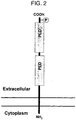
- the PLD4 protein has two tentative PDE regions (phosphodiesterase motif), which are constituted with two HKD motifs (amino acid sequence of His-x-Lys-x-x-x-x-Asp, wherein x is the other amino acids) conserved in the C terminal region, and a presumptive phosphorylation site (Thr 472).
- the structure of the PLD4 protein is predicted as a type II monotropic transmembrane protein.
- the N terminal region of the PLD4 protein does not have PX region and PH region, which are possessed by PLD1 and PLD2 that are classical PLD family (FIGFIGS. 1 and 2).
- PLD4 belongs to the PLD family from the fact that it has two HKD motifs, PLD4 is lack of PX domain and PH domain, but has a putative transmembrane domain instead.
- PLD4 mRNA expression of PLD4 which was characteristically at from a low level to a medium level, was found in a cell subpopulation that was preferentially localized at the corpus callosum and the periphery of the white matter region including cerebellar white matter of a mouse 1 week after birth. These cells expressing the PLD4 mRNA have been identified as Ibal positive microglia (see Tao et al., Nat. Methods 2(8), 591-598(2005 )).
- the period of 1 week after birth is a period when activation of myelin formation starts in the corpus callosum and the cerebellar white matter of a mouse.
- PLD4 is highly expressed at the amoeboid (activated state) microglia that exists in the white matter. From these facts, a possibility is contemplated that PLD4 expression cell in the white matter is involved in myelin formation in this period. Particularly, accumulation PLD4 in the phagocytic vesicle becomes evident, and a possibility is suggested that PLD4 expression cell is involved in phagocytosis.
- oligodendrocyte glia cell in the central nervous system, which forms myelin as rolled and attached to the axon
- extra oligodendrocyte causes apoptosis in the white matter of the brain at the development stage.
- the extra oligodendrocyte is degraded and removed from the amoeboid microglia to secret a signal molecule, whereby to arrange the environment for myelin formation in the white matter. It is suggested that PLD4 is involved in these processes including the myelin formation.
- PLD4 mRNA expression is universally seen also in non-nerve tissues, but is mainly distributed in the spleen. Strong PLD4 expression is detected at the periphery of the border zone of red pulp of the spleen, and splenic PLD4 protein collected from the membrane fraction in the cell is highly N-glycosylated.
- PLD4 was expressed in a heterogeneous cell system, they were localized in the endoplasmic reticulum and the Golgi body. Heterologously-expressed PLD4 showed no PLD enzymatic activity ( Plos ONE www.plosone.org, November 2010, Volume 5, Issue 11, e13932 ).
- PLD4 plays a role in common functions in the microglia or the cell in the spleen border region at the time of brain development at the initial stage after birth.
- PLD4 mRNA expression and PLD4 distribution in the nerve tissue and non-nerve tissue have been overviewed above.
- the present inventors found out that PLD4 mRNA is specifically highly expressed in a pDC cell at the resting stage (resting pDC) in the level of a cell species described below.
- Mouse anti-human PLD4 polyclonal antibody against total length human PLD4 protein is commercially available (PLD4 purified MaxPab mouse polyclonal antibody (B01P), catalog No. H00122618-B01P, manufactured by Abnova Corporation). However, a monoclonal antibody that binds only to a certain site of PLD4, or a monoclonal antibody that can specifically bind to PLD4, has not been obtained.
- a problem to be solved by the invention is to provide an antibody that binds to PLD4, and to detect, identify, or isolate pDC.
- a problem to be solved by the invention is to regulate activity of pDC.
- the present inventors confirmed through a research for PLD4 that expression of PLD4 specifically rises in pDC, particularly pDC at the resting stage in addition to pDC at the active stage. Consequently, the present inventors tried the preparation of PLD4 antibody and the elucidation of its action.
- a protein prepared by gene recombinant technology is generally used as an immunogen.
- the present inventors tried expression of PLD4 based on the information of the base sequence of PLD4 cDNA, and an amino acid sequence (GenBank Accession No. NM_138790.2) encoded by it, which has been already revealed ( Nat. Methods 2(8), 591-598 (2005 )).
- the present inventors revealed that use of a special immunogen allows obtaining an antibody that binds to pDC. Furthermore, the present inventors confirmed that thus-obtained antibody specifically recognizes human pDC, and further has an action of regulating its activity, and completed the invention. That is to say, the invention relates to an anti-PLD4 antibody, a preparation method thereof, and use thereof described below.
- the invention is as follows:
- the invention provides a monoclonal antibody that binds to human PLD4 on a plasmacytoid dendritic cell.
- PLD4 is a membrane protein that belongs to PLD family.
- the present inventors revealed that an antibody that specifically recognizes PLD4 can be easily obtained.
- the anti-PLD4 antibody that can be obtained by the invention is an antibody having high specificity, which distinguishes human pDC from a cell that expresses other PLD families.
- the anti-PLD4 antibody provided by the invention binds to human pDC.
- the antibody of the invention specifically recognizes human pDC. Accordingly, the antibody of the invention is useful for detection or isolation of pDC.
- pDC is a cell that produces most of type 1 IFN. Accordingly, such detection or isolation is important in diagnosis or research of diseases associated with pDC such as autoimmune diseases.
- the anti-PLD4 antibody provided by the invention has an action of regulating human pDC activity. Accordingly, the anti-PLD4 antibody of the invention can be used for suppressing pDC activity. Accordingly, if suppression of pDC activity is employed using the antibody of the invention, therapeutic effects can be expected for a patient of an autoimmune disease in which IFN ⁇ expression has risen.
- pDC produces a large amount of IFN in a small number of cells.
- an antibody is necessary, which depends on the number of IFN molecules.
- the activity of the production cell is directly suppressed.
- potent IFN suppression effect can be expected with less amount of the antibody in comparison with neutralization of an anti-IFN antibody.
- IFN in a case where IFN is produced continuously, it is expected that neutralization by IFN antibody will remain as transient suppression.
- pDC activity is suppressed, and from this, IFN-producing suppression effect can be expected over a long time.
- PLD4 is a molecule that is specifically expressed in the mRNA level and protein level in a plasmacytoid dendritic cell at the resting stage (resting pDC). A method of preparing an antibody that recognizes PLD4 is not established.
- mouse PLD4 is a molecule that is expressed in amoeboid (activated state) microglia at development stage in the cerebellum or the corpus callosum at the initial stage after birth.
- expression of human PLD4 is not known until now. Particularly, expression in the immune system, intracellular location, structure, function, and the like of human PLD4 have not been reported until now.
- human PLD4 which was contemplated until now to be expressed only in the cytoplasm, is a cell surface marker that is expressed in human plasmacytoid dendritic cell (pDC) as type II transmembrane protein. Accordingly, binding of PLD4 antibody to pDC becomes possible, and it has been proved that PLD4 antibody is useful as a molecular target of a therapeutic antibody intended to regulate functions of B cell and pDC cell.
- the present inventors confirmed by gene expression analysis that PLD4 is specifically expressed in human pDC. It was contemplated that if an antibody that can distinguish PLD4 immunologically from other molecules, is obtained, it would be useful for pDC research. However, there are many molecules in the PLD family including PLD4, which are very similar in the structure to each other. Molecules such as PLD1, PLD2, PLD3, and PLD5, including PLD that is PLD4, encompass an amino acid sequence having particularly high homology ( FIG. 4 ). Accordingly, it was contemplated that it is difficult to obtain an antibody that distinguish these molecules mutually using an immunogen of a peptide that employs an amino acid sequence (a partial sequence) that constitutes PLD4 (or extracellular domain). Consequently, the present inventors tried acquisition of an antibody against PLD4 using a recombinant PLD4-Ig fusion protein as an immunogen, which encodes an amino acid sequence encompassing PLD4 extracellular domain.
- the present inventors repeated researches in order to acquire an antibody that recognizes PLD4 and revealed that the intended antibody is obtained using a recombinant PLD4-Ig fusion protein as an immunogen, and completed the invention. That is to say, the invention relates to a monoclonal antibody that binds to a PLD4 extracellular domain, or a fragment containing an antigen-binding region thereof.
- PLD4 is a natural molecule that is expressed in human pDC, or an immunologically equivalent molecule to PLD4 that is expressed in human pDC. In the invention, binding of an antibody to PLD4 can be confirmed, for example, as described below.
- binding activity with at least partial subset of pDC is one of the important characteristics of the antibody that binds to PLD4 in the invention.
- the fact that some cell is pDC can be confirmed by a cell surface marker inherent in each cell family. For example, binding to an intended cell is confirmed by double staining of an antibody that binds to a cell surface marker, and an antibody to be confirmed for the binding activity. That is to say, pDC in the invention encompasses cells that express, for example, BDCA2.
- a transformed cell that expresses PLD4 is preferable as a cell for confirming binding property of an antibody to the extracellular domain of PLD4 in the invention.
- a non-transformed cell is desirably used as a control.
- the antibody that binds to PLD4 in the invention may be an antibody that shows cross property with a cell family that is known to express PLD family other than PLD4, or may be an antibody that does not show such cross property.
- the antibody showing no cross property is preferable as the antibody that binds to PLD4 in the invention.
- the antibody that binds to PLD4 in the invention is preferably an antibody that may not be confirmed for the binding with a cell family that is known to express PLD family other than PLD4, under the same conditions to the conditions where binding to pDC is confirmed.
- the monoclonal antibody that binds to a PLD4 extracellular domain in the invention preferably encompasses a monoclonal antibody that has the immunological characteristics described below.
- the monoclonal antibody of the invention is preferably an antibody that may not be confirmed for binding to monocyte, macrophage, B cell, CD34 positive cell, and dendritic cells derived from these cells, under conditions allowing binding to human pDC.
- the monoclonal antibody that binds to a PLD4 extracellular domain in the invention preferably encompasses a monoclonal antibody that has immunological characteristics described below.
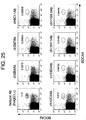
- the anti-PLD4 monoclonal antibody is not crossed with other molecules of PLD family in the invention, can be confirmed by employing a cell in which each PLD family was mandatorily expressed. That is to say, mandatory expression is performed by introducing cDNA that encodes the amino acid sequence of each PLD family into an appropriate host cell. The obtained transformed cell is brought into contact with anti-PLD4 monoclonal antibody to be confirmed for the cross property. If binding to a cell that expresses PLD family molecules other than PLD4 is not exhibited, it can be confirmed that the antibody can immunologically distinguish PLD4 from other PLD family molecules.
- the monoclonal antibody in the invention is preferably a monoclonal antibody that binds to PLD4, but may not be detected for binding to PLD3, PLD5, PLD1, or PLD2 under the same conditions. If an antibody that can immunologically distinguish these PLD family molecules from PLD4 is used, change of PLD4 expression can be specifically detected.
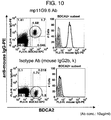
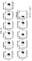
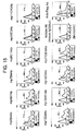
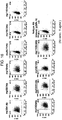
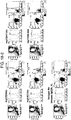
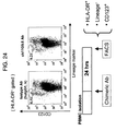
- Binding of a monoclonal antibody to be confirmed for binding activity, to each species of a cell can be confirmed, for example, in flow cytometry principle.
- a fluorescent label or a luminescent label is used.
- a fluorescence-activated cell sorter FACS
- an antibody A that is preliminarily known to be able to identify pDC, and an antibody B to be analyzed for binding characteristics with pDC are reacted at the same time with a cell family encompassing pDC.
- the antibody A and the antibody B are on label with a fluorescent signal that can distinguish them from each other. If both of the signals are detected from the same cell family, it can be confirmed that such antibodies bind to the same cell family. That is to say, it is found out that the antibody A and the antibody B have the same binding characteristics. If the antibody A and the antibody B bind to a different cell family, it is evident that the binding characteristics of them are different from each other.
- Examples of preferable monoclonal antibody in the invention include, for example, monoclonal antibodies produced by hybridomas mp5B7, mp7B4, mp13D4, and mp13H11.
- hybridomas mp5B7, mp7B4, mp13D4 and mp13H11 were deposited under Accession Numbers: NITE BP-1211, NITE BP-1212, NITE BP-1213, and NITE BP-1214 at National Institute of Technology and Evaluation (NITE) Patent Microorganisms Depositary on January 27, 2012. Contents to specify the deposition will be described below.
- the monoclonal antibody of the invention may be a fragment containing an antigen-binding region thereof.
- an antibody fragment encompassing an antigen binding site that is produced by IgG enzymatic digestion may be used as the antibody in the invention.
- an antibody fragment such as Fab or F(ab')2 may be obtained.
- an immunoglobulin fragment encompassing a variable region in which complementarily-determining region (CDR) of some monoclonal antibody is transplanted, is encompassed in the fragment containing antigen binding region. It is widely known that these antibody fragments can be used as an antibody molecule that has binding affinity to an antigen.
- an antibody constructed by gene recombination may be used as long as it maintains the activity necessary for antigen binding.
- the antibody constructed by gene recombination include, for example, a chimeric antibody, a CDR transplant antibody, a single chain Fv, a diabody (diabodies), a linear antibody, and a polyspecific antibody formed by antibody fragments.
- a method of obtaining such antibodies based on a monoclonal antibody, or an antibody-producing cell that produces it, is known.
- the monoclonal antibody of the invention can be obtained by using recombinant PLD4-Ig fusion protein, or a transformed cell that expresses human PLD4 as an immunogen. That is to say, the present invention relates to a method of preparing a cell that produces a monoclonal antibody that binds to a PLD4 extracellular domain, which contains the processes below.
- Thus-obtained antibody-producing cell, or immortalized cell of the antibody-producing cell may be cultured, and an intended monoclonal antibody may be collected from the culture.
- Various methods are known as a method for immortalizing the antibody-producing cell.
- the transformed cell used as an immunogen in the invention can be obtained, for example, by preparing a cell in which an extrinsic polynucleotide (a) that encodes the amino acid sequence containing the PLD4 extracellular domain described below is retained in an expressible way.
- the extrinsic polynucleotide in the present invention refers to a polynucleotide that is artificially introduced into a host cell.
- a human cell is used as the cell, a human gene is introduced into the human cell.
- artificially introduced polynucleotide is also referred to as the extrinsic polynucleotide. Accordingly, ectopic expression of PLD4 is encompassed in extrinsic polynucleotide expression.
- the PLD4 extracellular domain in the invention refers to an amino acid sequence of 54-506 positions that corresponds to the extracellular domain in the amino acid sequence described in SEQ ID NO 1.
- amino acid sequences containing each of the regions in the order below from the N terminal side are preferable as the amino acid sequence containing the PLD4 extracellular domain in the invention.
- amino acid sequence that is partially lacking the intracellular region as described below is also encompassed in the amino acid sequence containing the PLD4 extracellular domain in the present invention.
- regions than the extracellular domain in the above-mentioned structure may be a sequence selected from the amino acid sequence represented by SEQ ID NO 1, or may be a combination with another homologous amino acid sequence.
- an amino acid sequence that constitutes a signal sequence, a transmembrane domain, and an intracellular region can be used as an amino acid sequence of PLD family molecules other than PLD4.
- the amino acid sequence of PLD family of other species than human can be also combined.
- the amino acid sequence that constitutes other regions than the extracellular domain may contain mutation within a range where each of the functions of the regions can be maintained.
- other regions can be interposed between each of the regions.
- an epitope tag such as FLAG can be also inserted.
- the signal sequence is a region that is subjected to processing in the step of transport to the cell membrane surface after translation of the protein, and is removed.
- an arbitrary amino acid sequence that induces passage of the translated protein through the cell membrane can be used as a signal sequence.
- an amino acid sequence of PLD4 SEQ ID NO 1
- PLD4 amino acid sequence of PLD4
- the above-mentioned polynucleotide (a) in the invention may be an arbitrary base sequence that encodes the amino acid sequence that constitutes the above-mentioned structure [Intracellular region + transmembrane domain + extracellular domain].
- the amino acid sequence of SEQ ID NO 1 is encoded by the cDNA base sequence described in SEQ ID NO 44.
- the recombinant PLD4-Ig fusion protein as an immunogen in the invention may be obtained by introducing an expression vector which retains the aforementioned polynucleotide in an expressible way, into an appropriate host cell.
- the cDNA base sequence of the recombinant PLD4-Ig fusion protein is represented by SEQ ID NO 125, and the amino acid sequence is represented by SEQ ID NO 126.
- the host cell in the invention is preferably a mammalian cell. Specifically, a cell derived from a human, a monkey, a mouse, or a rat may be used as the host cell. Particularly, human-derived cell is preferable as the host cell.
- HEK-293T cell is a human embryo-derived renal cell strain that may be preferably used as the host cell in the invention.
- the HEK-293T cell is available as ATCC CRL-11268.
- a cell derived from an immunized animal may be used as the host cell. If a cell derived from an immunized animal is used as an immunogen, immune response against the host cell is small.
- an antibody against the PLD4 extracellular domain which is expressed extrinsically, can be obtained effectively. Accordingly, for example, when a mouse is used as the immunized animal, a cell derived from the mouse can be also used as the host cell.
- the above-mentioned polynucleotide can be loaded to a vector that can induce expression in a host cell to transform the cell.
- a commercially available vector that can induce expression in a mammalian cell may be used.
- an expression vector such as pCMV-Script(R) Vector, pSG5 Vector (manufactured by Stratagene), pcDNA3.1 (manufactured by Invitrogen), and pMXs-IP retroviral vector (manufactured by Cell BioLabs), can be used for the invention.
- Thus-obtained transformed cell is administered to an immunized animal, with an additional component such as an adjuvant if necessary.
- an adjuvant for example, Freund's complete adjuvant may be used.
- purified recombinant PLD4-Ig fusion protein is administered to BALB/c mouse.
- adjuvant Freund's Adjuvant, Complete and Incomplete (manufactured by SIGMA) were used, and administered in 200 ⁇ g/mouse at the first time, and 50 ⁇ g/mouse at the second time to fourth time.
- an immunogen is administered multiple times intervally until the antibody titer increases.
- a transformed cell is administered at an interval of 2 to 4 days, more specifically 3 days, and after 2 to 3 times of the administration, antibody-producing cells can be collected.
- the antibody-producing cells can be also collected after the administration 5 to 6 times at an interval of once or so a week.
- the collected antibody-producing cell is cloned.
- the antibody-producing cell is preferably immortalized.
- a cell fusion method represented by hybridoma method, or transformation by Epstein-Barr virus (EBV) may be used as a method for immortalizing the antibody-producing cell.
- An antibody-producing cell produces one kind of antibody per one cell. Accordingly, if a cell population derived from one cell can be established (that is to say, cloning), a monoclonal antibody can be obtained.
- the hybridoma method refers to a method in which an antibody-producing cell is fused with an appropriate cell strain, immortalized, and then cloned.
- the immortalized antibody-producing cell can be cloned by a method such as limiting dilution method.
- Many cell strains that are useful in the hybridoma method are known. These cell strains are excellent in immortalization efficiency of a lymphocyte-based cell, and have various gene markers that are necessary for selecting cells that have succeeded in the cell fusion. Furthermore, in a case where acquisition of the antibody-producing cell is intended, a cell strain that is lack of antibody production ability may be also used.
- mouse myeloma P3x63Ag8.653 (ATCC CRL-1580) or P3x63Ag8U.1 (ATCC CRL-1597) is universally used as a useful cell strain for a cell fusion method of a mouse or a rat.
- a hybridoma is prepared by fusion of homogeneous cells.
- a monoclonal antibody may be also acquired from a heterohybridoma between close heterogeneous species.
- a specific protocol of the cell fusion is known. That is to say, an antibody-producing cell of an immunized animal is mixed with an appropriate fusion partner, and subjected to cell fusion.
- an antibody-producing cell for example, a splenic cell, a lymphocyte cell collected from the lymph node, or peripheral blood B cell may be used.
- the fusion partner various cell strains described above may be used.
- a polyethylene glycol method or an electric fusion method is used for the cell fusion.
- cells that have succeeded in the cell fusion are selected based on a selection marker possessed by the fusion cell. For example, in a case where HAT sensitive cell strain is used in the cell fusion, cells that have succeeded in the cell fusion are selected by selecting cells that grow in the HAT medium. Furthermore, an antibody produced by the selected cells is confirmed whether it has intended reactivity.
- each of the hybridoma is screened based on the reactivity of the antibody. That is to say, a hybridoma that produces an antibody that binds to PLD4 is selected by the method as described above. Preferably, in a case where selected hybridoma is subcloned, and production of intended antibody is finally confirmed, the hybridoma is selected as a hybridoma that produces the monoclonal antibody of the invention.
- intended hybridoma can be selected based on reactivity with a human cell, or reactivity with a transformed cell that expresses PLD4 gene.
- An antibody that binds to a cell can be detected on the principle of an immunoassay.
- ELISA in which a cell is used as an antigen, can be used in detection of intended antibody.
- a culture supernatant of the hybridoma is brought into contact with a carrier that immobilizes human pDC, or a transformed cell used as an immunogen. In a case where the culture supernatant contains the intended antibody, the antibody is captured by the cell immobilized on the carrier.
- the solid phase is isolated from the culture supernatant, washed if necessary, and then the antibody captured on the solid phase can be detected.
- an antibody that recognizes the antibody may be used.
- an antibody of a mouse can be detected by anti-mouse immunoglobulin antibody. If the antibody that recognizes an antibody is on label, the detection thereof is ease.
- the label for example, an enzyme, a fluorescent pigment, or a luminescent pigment may be used.
- a particle, or the internal wall of a microtiter plate may be used as the carrier that immobilizes the cell.
- the surface of a particle or a container made of plastic can immobilize the cell by physical adsorption.
- beads or a reaction container made of polystyrene may be used as a carrier for immobilizing the cell.
- hybridoma there may be a case where predicted is not production of an antibody against PLD4, but production of an antibody against a host cell of a transformed cell used as an immunogen.
- a human cell is used as an immunogen and a mouse is used as an immunized animal as shown in Examples, the human cell is recognized as a foreign substance, and it is predicted that an antibody that binds to it is produced.
- acquisition of an antibody that recognizes PLD4 is intended. Accordingly, acquisition of an antibody that recognizes other human cell antigens than PLD4 is not necessary.
- an antibody not intended may be preliminarily absorbed before confirmation of the antibody reactivity.
- Antibodies that are not intended can be absorbed by an antigen that binds to an antibody predicted to exist. Specifically, for example, an antibody against other human cell antigens other than PLD4 can be absorbed by a cell for which expression of PLD4 may not be detected.
- the host cell used for the immunogen is preferable as an antigen for absorbing antibodies not intended.
- the influence on pDC can be confirmed, for example, by a method described below.
- the monoclonal antibody of the invention can be collected from a culture that is obtained by culturing a hybridoma that produces the monoclonal antibody.
- the hybridoma may be cultured in vitro or in vivo. In the culture in vitro, the hybridoma may be cultured using a known medium such as RPMI 1640. Immunoglobulins secreted by the hybridoma are accumulated in the culture supernatant. Accordingly, the monoclonal antibody of the invention may be obtained by collecting the culture supernatant, and purifying it if necessary. Purification of immunoglobulin is ease in a medium that is not added with the serum. However, for the purpose of rapid growth of the hybridoma and promotion of antibody production, 10% or so of bovine fetal serum may be also added to the medium.
- the hybridoma may be also cultured in vivo. Specifically, the hybridoma may be cultured in the abdominal cavity by inoculating the hybridoma into the abdominal cavity of a nude mouse.
- the monoclonal antibody is accumulated in the ascites. Accordingly, ascites is collected, and purified if necessary, to obtain necessary monoclonal antibody.
- the obtained monoclonal antibody may be suitably modified, or processed depending on the purpose.
- the monoclonal antibody of the invention can be expressed by acquiring cDNA that encodes the antigen binding region of the antibody from the hybridoma, and inserting it into an appropriate expression vector.
- a technology for acquiring cDNA that encodes a variable region of an antibody and express it in an appropriate host cell is known.
- an approach is known in which a variable region containing an antigen binding region is bound to a constant region to give a chimeric antibody.
- a gene of a variable region may give a chimeric antibody by binding to genes that encode a human IgG1 heavy chain constant region and human Ig kappa light chain constant region, respectively.
- antigen binding activity of a monoclonal antibody can be transplanted to another immunoglobulin by incorporating CDR that constitutes a variable region into a frame region of another immunoglobulin molecule.
- a method is established of transplanting antigen binding activity possessed by a heterogeneous immunoglobulin to a human immunoglobulin.
- a partial amino acid sequence of a frame region supporting CDR is transplanted to a human variable region from a variable region of a mouse antibody.
- these humanized, re-constituted variable regions of a human antibody may be linked to a constant region of a human antibody, to obtain a humanized antibody.
- Examples of preferable monoclonal antibody in the invention include, for example, monoclonal antibodies produced by hybridomas mp5B7, mp7B4, mp13D4, and mp13H11, which have been deposited under Accession Numbers: NITE BP-1211, NITE BP-1212, NITE BP-1213, and NITE BP-1214, respectively.
- an antibody having a constant region derived from IgG or IgM is contained preferably in the antibody of the invention.
- the antibody of the invention is more preferably an antibody that has a combination of:
- the present inventors have confirmed that the monoclonal antibody against PLD4 has CDC action for a PLD4 expression cell. Accordingly, the antibody that has IgG- or IgM-derived constant region, has an action of cytotoxicity for PLD4 expression cell by CDC action. Such antibody is useful for suppressing the cell number of a PLD4 expression cell such as pDC.
- a chimeric antibody, or a humanized antibody that recognizes PLD4 can be prepared by genetic engineering using a polynucleotide that encodes them.
- An antibody that can specifically recognize PLD4 was not obtained.
- An antibody that recognizes PLD4 has been provided for the first time by the immunogen of the disclosure. That is to say, the invention provides an antibody that recognizes PLD4, which can be obtained by the processes described below.
- PLD4 is specifically expressed in human pDC.
- the specific expression in human pDC was also confirmed by gene expression analysis with SAGE by the present inventors.
- PLD4 expression level was all analyzed based on mRNA in a report of the past.
- the PLD4 protein is known to be expressed only in the cytoplasm. It has been revealed by the invention that PLD4 is also expressed on the cell surface. Since an antibody that can detect PLD4 was not provided, analysis for the state of protein expression was not conventionally performed. The antibody that binds to a PLD4 extracellular domain provided by the invention has realized analysis of the PLD4 protein.
- the monoclonal antibody that binds to a PLD4 extracellular domain based on the invention has specifically detected human pDC. That is to say, the invention relates to an ex vivo method of detecting a human plasmacytoid dendritic cell, which contains steps of bringing into contact with a test cell the monoclonal antibody of the invention, or the antibody fragment containing an antigen-binding region thereof, and detecting the monoclonal antibody, or the antibody fragment containing an antigen-binding region thereof that binds to the cell.
- the invention provides a method of identifying pDC using PLD4 as an index.
- a cell for which PLD4 is detected may be isolated based on the invention, whereby to isolate human pDC. That is to say, the disclosure provides a method of isolating pDC using PLD4 as an index.
- a monoclonal antibody that binds to a PLD4 extracellular domain, or a fragment containing an antigen-binding region thereof may be on label.
- the antibody can be easily detected by labeling it with a luminescent pigment or fluorescent pigment. More specifically, fluorescent pigment-labeled antibody is brought into contact with a cell population that possibly contains pDC, and a cell that binds to the antibody of the invention can be detected using the fluorescent pigment as an index. Furthermore, if a cell detected with the fluorescent pigment is isolated, pDC can be isolated. A series of steps can be easily implemented in the principle of FACS.
- the antibody of the invention may be also as bound to a solid phase carrier such as a magnetic particle.
- a solid phase carrier such as a magnetic particle.
- the antibody that is bound to the solid phase carrier recognizes PLD4, and pDC is captured on the solid phase carrier. As a result thereof, pDC can be detected or isolated.
- the antibody that is necessary in the ex vivo pDC detection method based on the invention may be supplied as a reagent for detection of pDC. That is to say, the invention provides use of the monoclonal antibody of the invention or the antibody fragment containing an antigen-binding region thereof as a reagent for detection of pDC ex vivo.
- a positive control, or a negative control may be combined in addition to the antibody.
- a transformed cell that expresses PLD4 extracellular domain used as an immunogen, or pDC collected from a human may be used as a positive control. Usually, only little human pDC can be obtained from the peripheral blood. Accordingly, a transformed cell is particularly preferable as a positive control in the reagent.
- the negative control an arbitrary cell that does not express PLD4 may be used.
- the disclosure provides a kit for detection of human pDC containing a monoclonal antibody that binds to a PLD4 extracellular domain, or a fragment containing an antigen-binding region thereof.
- the inventors analyzed the influence of the antibody that binds to a PLD4 extracellular domain on pDC. As a result thereof, it was confirmed that the antibody that binds to a PLD4 extracellular domain suppresses pDC activity. That is to say, the invention relates to a method of suppressing an activity of a human plasmacytoid dendritic cell ex vivo, comprising bringing any one of the components below into contact with the cell:
- the disclosure relates to a method of suppressing pDC activity in a living organism, which contains a step of administering any one of the components below to a living organism:
- the pDC in the invention refers to a cell that has IFN production ability, and expresses PLD4 on the cell surface.
- pDC is not only a cell that is a precursor cell of a dendritic cell, but contains a cell that has IFN production ability, and expresses PLD4 on the cell surface.
- a method of identifying such pDC is known.
- pDC can be distinguished from other blood cells using several cell surface markers as an index. Specifically, the profile of a human pDC cell surface marker is as described below ( Shortman, K. and Liu, YJ. Nature Reviews 2: 151-161, 2002 ). In recent years, there is also a report which positioned BDCA-2 positive cell as pDC ( Dzione k, A. et al. J. Immunol. 165: 6037-6046, 2000 ).
- a cell that has the expression profile of these known markers, and has IFN production ability may be also referred to as pDC.
- a cell that belongs to a cell family having a different profile from the expression pattern of the expression profile of these markers, but having IFN production ability in a living organism is encompassed in pDC.
- examples of common characteristics exhibited by human pDC include the characteristics as described below.
- Suppression of the pDC activity in the invention refers to suppression of at least one of the functions of pDC.
- the pDC functions include IFN production and cell survival.
- the cell survival can be referred to as the cell number in other words. Accordingly, suppression of either one or both of these functions is referred to as the suppression of pDC activity.
- type I IFN produced by pDC is a cause for various diseases. Accordingly, suppression of the pDC cell number or IFN production is useful as a strategy for treating such diseases.
- IFN ⁇ is important.
- the invention relates to a suppressor for IFN production, which contains the antibody that binds to a PLD4 extracellular domain as an active component.
- the disclosure provides a method of suppressing IFN production, which contains a process of administering an antibody that binds to a PLD4 extracellular domain.
- the disclosure relates to use of an antibody that binds to a PLD4 extracellular domain in preparation of a pharmaceutical composition that suppresses IFN production.
- a cell that produces a large amount of IFN in a small number of the cells is encompassed in the pDC.
- a precursor cell of a dendritic cell that is received with stimulation by a virus or the like produces most of IFN produced by a living organism. Suppression of the cell number of pDC that produces a large amount of IFN leads to suppression of IFN production as results. Accordingly, suppression of the pDC cell number can also alleviate pathological conditions caused by IFN ⁇ .
- the anti-PLD4 monoclonal antibody binds to a PLD4 expression cell, and imparts cytotoxicity by Complement Dependent Cytotoxicity (CDC) action.
- the CDC action is one of the important action mechanisms of an antibody drug.
- the anti-PLD4 monoclonal antibody of the invention also has potent cytotoxicity action for a PLD4 expression cell such as pDC by the CDC action. That is to say, the anti-PLD4 monoclonal antibody in a preferred aspect, can be expected for effects of suppressing IFN production not only by a mechanism of suppressing IFN production, but also by cytotoxicity for pDC.
- the antibody that recognizes a PLD4 extracellular domain used in the invention can be obtained based on the method as described above.
- the antibody in the invention may be any class.
- a biological species from which the antibody is derived is not limited.
- a fragment containing a antigen binding region of the antibody can be used as the antibody.
- an antibody fragment containing an antigen binding site produced by enzymatic digestion of IgG may be used as the antibody in the invention.
- an antibody fragment such as Fab or F(ab')2 may be obtained by digestion by papain or pepsin. It is widely known that these antibody fragments can be used as an antibody molecule that has binding affinity to an antigen.
- an antibody constructed by gene recombination may be also used as long as it maintains necessary antigen binding activity.
- Examples of the antibody constructed by gene recombination include a chimeric antibody, a CDR transplantation antibody, a single chain Fv, a diabody (diabodies), a linear antibody, and a polyspecific antibody formed by an antibody fragment. A method of obtaining these antibodies based on a monoclonal antibody is known.
- the antibody in the invention may be modified, if necessary.
- the antibody that recognizes a PLD4 extracellular domain has an action of suppressing pDC activity. That is to say, a possibility has been contemplated that the antibody itself has cytotoxicity for pDC. A subclass of the antibody exhibiting strong effector action is known.
- effects of suppressing pDC activity can be further increased by modifying the antibody with a cytotoxic agent. Examples of the cytotoxic agent include the agents described below.
- Toxins containing a protein can bind to, for example, an antibody, or a fragment thereof by a bifunctional reagent.
- a gene that encodes the toxins may be also conjugated with a gene that encodes the antibody, to give a fusion protein of them.
- a method of binding a radioactive isotope to an antibody is also known.
- a method of labeling an antibody with a radioactive isotope using a chelate agent is known.
- an anti-cancer agent can bind to an antibody by using, for example, a sugar chain, or a bifunctional reagent.
- artificially structure-modified antibody may be also used as an active component.
- various modifications are known to improve cytotoxicity or stability of an antibody.
- an immunoglobulin is known, in which the sugar chain of the heavy chain is modified ( Shinkawa, T. et al. J. Biol. Chem. 278: 3466-3473. 2003 ). Modification of the sugar chain increased Antibody Dependent Cell-mediated Cytotoxicity (ADCC) activity of the immunoglobulin.
- ADCC Antibody Dependent Cell-mediated Cytotoxicity
- the antibody that binds to a PLD4 extracellular domain suppresses the activity of pDC when the antibody is brought into contact with the pDC. Accordingly, such antibody can be used as an agent for suppressing pDC activity, or in a method of suppressing pDC. That is to say, the disclosure provides an agent for suppressing pDC activity, which contains at least one kind of a component selected from a group consisting of (a) and (b) described below as an active component.
- the invention relates to any one of (a) and (b) described below for use in a method of suppressing human pDC activity in a living human, which contains a step of administering at least one kind of a component selected from a group consisting of (a) and (b) described below.
- the disclosure relates to use of at least one kind of a component selected from a group consisting of (a) and (b) described below in preparation of an agent for regulating pDC activity.
- a monoclonal antibody that recognizes a PLD4 extracellular domain may be used.
- One kind or multiple kinds of the monoclonal antibodies may be used in the invention.
- multiple species of the monoclonal antibodies that recognize a PLD4 extracellular domain may be blended, and used in the invention.
- pDC produces a large amount of IFN upon viral stimulation.
- the antibody is given to pDC before, after, or at the same time of viral stimulation, and IFN production ability is compared with a control in which the antibody is not given to pDC.
- the IFN production ability can be evaluated by measuring IFN- ⁇ or IFN- ⁇ contained in the supernatant of a pDC culture. As a result of the comparison, if the amount of IFN in the supernatant is significantly lowered by addition of the antibody, it can be confirmed that the tested antibody has an action of suppressing the IFN production ability.
- a method of measuring these IFNs is known.
- pDC is a cell that produces most of IFN in a living organism. Accordingly, suppression of IFN production ability of pDC can regulate IFN production state in a living organism.
- the activity of pDC in the invention encompasses maintenance of the pDC cell number. Accordingly, suppression of pDC activity in the invention encompasses suppression of the pDC cell number. If the pDC cell number is confirmed to be suppressed in the presence of the antibody, the antibody is found out to suppress the pDC activity. Similarly to the IFN production, an inactive immunoglobulin that is derived from the same animal species as the species of the antibody to be confirmed for the activity, may be used as a control for comparison. The pDC cell number can be compared quantitatively by cell counting. The cell number can be counted with FACS or a microscope.
- pDC is also assumed to be differentiated into a cell that induces Th2 called Dendritic Cell 2 (DC2) as a result of infection by, for example, virus. If pDC of IFN production upon virus stimulation is suppressed, there is a possibility that differentiation to Th2 can be also suppressed. Accordingly, the monoclonal antibody of the invention that suppresses IFN production can be also expected to have therapeutic effects for various allergy diseases.
- DC2 Dendritic Cell 2
- the antibody that recognizes a PLD4 extracellular domain is administered to a host that is different from the biological species from which the antibody is derived, it is desirable to process the antibody into a form that is hardly recognized as a foreign substance by the host.
- immunoglobulin may be rendered to be hardly recognized as a foreign substance by processing the molecule as described below. A method of processing an immunoglobulin molecule as described below is known.
- the gene of a human immunoglobulin variable region can be also acquired by phage display method ( McCafferty J et al., Nature 348:552-554, 1990 ; Kretzschmar T et. a1., Curr Opin Biotechnol. 2002 Dec; 13(6): 598-602 .).
- the gene that encodes the human immunoglobulin variable region is incorporated into the phage gene.
- a phage library can be also prepared.
- a phage expresses the variable region as a fusion protein of the protein that constitutes itself.
- the variable region expressed on the phage surface by the phage maintains binding activity to antigen.
- selection of a phage that binds to an antigen or, for example, a cell in which the antigen is expressed can screen a phage in which a variable region having intended binding activity is expressed from a phage library. Furthermore, a particle of thus-selected phage retains a gene that encodes the variable region having intended binding activity. That is to say, a gene that encodes a variable region having intended binding activity can be acquired using the binding activity of the variable region as an index in the phage display method.
- the antibody that recognizes a PLD4 extracellular domain, or an antibody fragment containing at least antigen binding region thereof in the agent for suppressing pDC activity, or the method of suppressing pDC activity can be administered as a protein, or a polynucleotide that encodes the protein.
- a vector in which a polynucleotide that encodes intended protein, is disposed under control of an appropriate promoter so as to express the intended protein.
- an enhancer or a terminator may be also disposed.
- a vector is known that retains genes of a heavy chain and a light chain that constitute an immunoglobulin, and can express the immunoglobulin molecule.
- the vector that can express an immunoglobulin can be administered by being introduced into a cell.
- a vector that can infect a cell by being administered into a living organism can be administered as it is.
- a vector may be also first introduced into a lymphocyte isolated from a living organism, and returned again into the living organism (ex vivo).
- the amount of the monoclonal antibody that is administered to a living organism in the agent for suppressing pDC activity, or the method of suppressing pDC activity is usually 0.5 mg to 100 mg, for example 1 mg to 50 mg, preferably 2 mg to 10 mg per 1 kg of the body weight as an immunoglobulin.
- the administration interval of the antibody to a living organism can be suitably regulated such that an effective concentration of the immunoglobulin in the organism can be maintained during the treatment period. Specifically, administration may be performed, for example, at an interval of 1 to 2 weeks.
- the administration route is arbitrary. One of ordinary skill in the art can suitably select an effective administration route in the treatment. Specifically, examples of the administration route include oral or non-oral administration.
- the antibody can be administered systemically or locally by intravenous injection, intramuscular injection, intraperitoneal injection, or subcutaneous injection.
- an appropriate formulation for non-oral administration in the invention include an injection, a suppository, and a spray.
- the immunoglobulin is administered to a culture liquid in usually 1 ⁇ g/mL, preferably 10 ⁇ g/mL or more, more preferably 50 ⁇ g/mL or more, and further preferably 0.5 mg/mL or more.
- the monoclonal antibody may be administered by any method to a living organism.
- the monoclonal antibody is blended with a pharmaceutically acceptable carrier.
- additives such as a thickening agent, a stabilizer, a preservative, and a solubilizing agent may be blended if necessary.
- carrier or additives include lactose, citric acid, stearic acid, magnesium stearate, sucrose, starch, talc, gelatin, agar, vegetable oil, and ethylene glycol.
- the terms called "pharmaceutically acceptable” refers to those approved by government supervisory of each country, or those listed in a pharmacopoeia of each country or a pharmacopoeia generally acknowledged for use in an animal, a mammalian animal, and particularly human.
- the agent for suppressing pDC activity may be also supplied in a form of a lyophilized powder or tablet in a single dose or multiple doses.
- the lyophilized powder or tablet may be further combined with sterilized water, physiological saline, or a buffer solution for injection for dissolving the composition to a desired concentration before administration.
- the agent for suppressing pDC activity is administered as an immunoglobulin expression vector
- the heavy chain and the light chain are co-transfected with a separate plasmid, and each of the plasmids can be administered in 0.1 to 10 mg, for example, 1 to 5 mg per 1 kg of the body weight.
- a vector of 1 to 5 ⁇ g/10 6 cells is used for introduction into a cell in vitro.
- the monocyte was isolated as CD14 positive cell and the human pDC cell was isolated as BDCA-4 positive cell with BDCA-4+ isolation kit (Miltenyi Biotec company) and MACS system (Miltenyi Biotec company). Furthermore, the human pDC cell was cultured for 12 hours in the presence of inactive HSV-1, whereby to prepare activated pDC (pDC+HSV).
- pDC+HSV activated pDC
- Total RNA was extracted from each of the cells, and the SAGE library was prepared using I-SAGETM kit (Invitrogen company). The obtained data of about 100,000 tagged base sequences were analyzed with SAGE2000 Analysis Software (Invitrogen company).
- PLD4 Phospholipase D family, member 4, C14orf175; GenBank Accession Number: NM_138790.2
- PLD4 is a 506 amino acid sequence (SEQ ID NO: 1) encoded by a base sequence represented by SEQ ID NO: 44.
- the PLD4 protein has two tentative PDE regions (Phosphodiesterase motif) constituted with two HKD motifs (His-x-Lys-x-x-x-x-Asp amino acid sequence, the x is the other amino acids) conserved in the C terminal region, and a tentative phosphorylation site (Thr 472).
- the structure of the PLD4 protein is predicted as type II monotropic transmembrane protein.
- the PLD4 protein does not have PX region (Phox homology domain) and PH region (Pleckstrin homology domain) in the N terminal region, which are possessed by PLD1 and PLD2 that are of classical PLD family (FIGFIGS. 1 and 2).
- PLD4 mRNA in a blood cell was specifically reviewed. From the human peripheral blood, each of the cells was isolated and taken by a cell sorter. From each of the cell family isolated and taken, RNA was extracted, and cDNA was synthesized. Using the obtained cDNA as a template, quantitative real time PCR was performed, and expression level of PLD4 mRNA was analyzed.
- BDTM MTC multiple tissue cDNA panel Human I; Cat. No. 636742, Human immune; Cat. No. 636748; all Takara Bio company
- the base sequences of the used primers are represented as follows.
- the quantitative PCR was performed with ABI PRISM 7000 using Platinum SYBR Green qPCR Super Mix-UDG kit (Invitrogen company). Sequence Detection System Software (Applied Biosystem company) was used in the analysis. The reaction conditions are as described below.
- PLD4 genes were compared between each of the tissues by standardization with gene expression level of GAPDH (glyceraldehyde-3-phosphate dehydrogenase), which is known to be expressed homeostatically.
- GAPDH glycosylcholine
- PLD4 mRNA exhibited relatively high expression in the spleen and the peripheral blood leucocyte.
- the expression level of the PLD4 mRNA was less than 100 folds of the expression level of the resting pDC cell ( FIG. 6 ).
- PLD4 gene expression vector was performed in order to express a human PLD4 protein.
- PLD4 cDNA clone incorporated into a pCR4-TOPO cloning vector (Open Biosystem, Cat, No. MHS4771-99610856)
- EcoRI enzyme a pcDNA3.1 expression vector
- human PLD4-pcDNA3.1 vector a pcDNA3.1 expression vector
- the PLD4 gene was amplified with a primer containing EcoRI, Not I, and Kozak sequences (GCC GCC ACC) (The information of the primers is as described below).
- PCR product was cloned at EcoRI and Not I sites with pMX-IP retrovirus vector (human PLD4-pMX-IP retrovirus vector).
- pMX-IP retrovirus vector human PLD4-pMX-IP retrovirus vector.
- 1 unit of KOD Plus DNA polymerase TOYOBO company was used, and the reaction conditions were 1 cycle at 94°C for 2 minutes, and then 25 cycle of [94°C for 15 seconds and 68°C for 1 minutes 30 seconds].
- HEK Human Embryonic Kidney-293T cell
- 293T cell Human Embryonic Kidney-293T cell
- Human PLD4 belongs to PLD family, and there are multiple molecules having high homology. Human PLD4 illustrates about 41% homology to human PLD3 and about 29.3% homology to human PLD5 ( FIG. 4 ).
- human PLD3 clone K.K.DNAFORM company, Cat. No 5189261
- human PLD5 clone K.K.DNAFORM company, Cat. No 5189261
- the underlined parts represent base sequences that encode the added FLAG tags, and the italic type represents Hind III cutting site or EcoRI site of the restriction enzymes.
- 293T cell was transiently transfected with human PLD3-pcDNA3.1 vector or human PLD5-pcDNA3.1 vector, and whether the human PLD4 was expressed on the surface of the 293T cell was confirmed with cell staining, and then FACS method.
- the prepared human PLD4-pMX-IP vector was cotransfected to 293T cell along with pCL-Eco vector (IMGENEX, Cat. No. 10045P), which is a retrovirus packaging vector, whereby to perform gene introduction.
- the gene introduction experiment used FuGENE (registered trademark) HD Transfection Reagent (Roche) as the transfection reagent. After 2 days, the cell culture supernatant, in which human PLD4 gene-containing retrovirus was secreted, was collected, and was infected to CT125 cell strain (2B4 mouse T cell lymphocyte tumor cell series).
- pMX-IP retrovirus vector contains puromycin-resistant gene
- culture of infected CT125 cell in the presence of puromycin allows survival of only cells that express human PLD4, leading to selection thereof.
- the selected human PLD4 expression CT125 cells (hereinafter, human PLD4-CT125) were further selected to only the CT125 cells allowing higher expression of human PLD4 by FACS sorting, and cultured.
- the CT125 cells were stained with commercially available mouse anti-human PLD4 polyclonal antibody (Abnova, Cat #:HOO122618-B01P) adjusted to 5 ⁇ g/mL, and FACS analysis was performed.
- human PLD4-CT125 cell stable strain was established, and used in FACS screening of the hybridoma.
- the cDNA sequence of PLD4 of rhesus monkey is reported at Genbank database (XM_002805227.1) and the like, but a partial, total length cDNA thereof is not reported. Furthermore, since the PLD4 gene sequence of cynomolgus monkey is not yet reported, from PBMC (10 mL respectively; SHIN NIPPON BIOMEDICAL LABORATORIES, LTD.) of cynomolgus monkey and rhesus monkey, gene cloning was performed.
- RNA was extracted from the peripheral blood of the monkey, and from 5 ⁇ g thereof, cDNA was synthesized using oligo-dT primer and SuperScript Choice System for cDNA Synthesis kit.
- the cynomolgus monkey PLD4 and the rhesus monkey PLD4 gene were amplified with PCR method using the primers of the following base sequences.
- the human PLD4 protein represents about 94.4% identity to the protein sequence with cynomolgus monkey PLD4 (SEQ ID NO: 129), and about 94% identity to the protein sequence with rhesus monkey PLD4 (SEQ ID NO: 130).
- FIG. 14 illustrates the homology of the protein sequences of human PLD4 to cynomolgus monkey PLD4, rhesus monkey PLD4, and mouse PLD4 (cDNA SEQ ID NO: 131, amino acid SEQ ID NO: 132).
- Cynomolgus monkey PLD4 expression cell stable strain was established by culturing CT125 cell infected with a retrovirus vector in the presence of puromycin, in the same method as the method of preparing human PLD4 expression cell stable strain using the prepared cynomolgus monkey PLD4-pMX-IP vector.
- the collected cell culture supernatant was purified with protein A affinity column of AKTA-FPLC system, and the protein of recombinant human PLD4-Ig fusion protein was purified ( FIG. 7 ).
- PLD4-Ig fusion protein As an immunogen, the aforementioned recombinant PLD4-Ig fusion protein was used. PLD4-Ig fusion protein was administered subcutaneously to the back part of 3 mice of BALB/c mouse. Freund's Adjuvant, Complete and Incomplete (SIGMA) were used as an adjuvant, and 200 ⁇ g/mouse at the first time and 50 ⁇ g/mouse at the second to fourth time, were administered.
- SIGMA Complete and Incomplete
- the blood was collected after the third immunization and the fourth immunization, and the anti-PLD4-Ig titer in the serum was evaluated with ELISA.
- PLD4-Ig fusion protein was solid-phased to a 96 well microtiter plate.
- the anti-serum was diluted stepwise from 1,000 folds by 3 folds, and dilution series were prepared to 729,000 folds.
- Each of the samples was added to the antigen solid-phased plate by 50 ⁇ L, and primary reaction was performed. After washing, secondary reaction was performed with HRP label anti-mouse IgG ( ⁇ , ⁇ ) antibody, and color-detected (490 nm) with OPD (ortho-phenylenediamine).
- Splenic cells were isolated from a mouse for which increase of the anti-serum titer was recognized.
- the isolated splenic cell and the mouse myeloma cell (P3U1) were fused with PEG method, and selection and culture of the fusion splenic cell was performed from HAT medium.
- Antibodies producing each clone of the fusion splenic cell obtained from HAT selection culture were evaluated with FACS.
- Human pDC-like cell strain CAL-1 cells in 2 ⁇ 10 5 were reacted with 50 ⁇ L culture supernatant of each of the hybridomas described below for 15 minutes at 4°C.
- the cells were washed with FACS buffer (1% FBS + PBS) twice, centrifuged, and the supernatant was removed. Reaction was performed for 20 minutes at 4°C using PE labeled anti-mouse IgG antibody (BD Bioscience: 550589) as a secondary antibody.
- the culture liquid after 10 days from the start of HAT selection culture was used as original fold for the culture supernatant of each clone.
- Antibodies producing each clone of the fusion splenic cell obtained from HAT selection culture were evaluated with FACS.
- Human PLD4-CT125 in 2 ⁇ 10 5 was reacted for 15 minutes at 4°C with 50 ⁇ L of culture supernatant of each of the hybridomas described below.
- the cells were washed with FACS buffer (1% FBS + PBS) twice, centrifuged, and the supernatant was removed. Reaction was performed for 20 minutes at 4°C using PE labeled anti-mouse IgG antibody (BD Bioscience: 550589) as a secondary antibody.
- peripheral blood mononuclear cell PBMC
- HISTOPAQUE-1077 HISTOPAQUE-1077
- 1 ⁇ 10 6 PBMCs were stained for every sample.
- the cells were washed with FACS buffer, and then added with 25 ⁇ L of 5 fold dilution of Fc block reagent (Militenyi company), and reacted at 4°C for 15 minutes.
- the cells were washed with FACS buffer, and then was added with 50 ⁇ L of the cell culture supernatant of each hybridoma and mouse IgG2b, ⁇ , and reacted at 4°C for 20 minutes.
- the cells were washed with FACS buffer, and then added with PE-labeled anti-mouse IgG antibody, and reacted at 4°C for 20 minutes.
- the cells were washed with FACS buffer, and then added with 50 ⁇ L of 10 fold dilution of APC-labeled anti-BDCA2 antibody, and reacted with at 4°C for 20 minutes.
- the cells were washed with FACS buffer, and then resuspended in 300 ⁇ L of FACS buffer, and analyzed with FACS Calibur (BD).
- the mp11G9.6 antibody exhibited the binding to pDC ( FIG. 10 ).
- Antibodies producing each clone of the fusion splenic cell obtained from HAT selection culture were evaluated with FACS.
- Human PLD4-CT125 in 2 ⁇ 10 5 was reacted with 50 ⁇ L culture supernatant of each of the hybridomas described below for 15 minutes at 4°C.
- the cells were washed with FACS buffer (1% FBS + PBS) twice, centrifuged, and the supernatant was removed. Reaction was performed for 20 minutes at 4°C using PE labeled anti-mouse IgG antibody (BD Bioscience: 550589) as a secondary antibody.
- PBMC of rhesus monkey from the peripheral blood (10 mL; SHIN NIPPON BIOMEDICAL LABORATORIES, LTD.), was centrifuged with specific gravity using 96% Ficoll-Paque (trademark) PLUS (GE Healthcare company, Cat No. 17-1440-02).
- FACS Fluorescence Activated Cell Sorting
- the cells were washed with FACS buffer, and then added with 100 ⁇ L of the cell culture supernatant of each hybridoma and 10 ⁇ g/mL of mouse IgG2a, ⁇ or mouse IgG1, ⁇ , and reacted at 4°C for 15 minutes.
- the cells were washed with FACS buffer, and then added with 1 ⁇ g/mL of APC-labeled anti-mouse IgG antibody, and reacted at 4°C for 20 minutes.
- the cells were washed with FACS buffer, and then reacted with 25 ⁇ L of 10 fold dilution of FITC-labeled anti-Lineage 1 antibody, PE-labeled anti-CD123 antibody, and PcrCP-Cy5.5-labeled anti-HLA-DR antibody at 4°C for 15 minutes.
- the cells were washed with FACS buffer, and then resuspended in 300 ⁇ L of FACS buffer, and analyzed with FACS calibur.
- the used hybridoma culture supernatant was specific to PLD4, and 10 kinds of 3B4, 5B7, 7B4, 8C11, 10C3, 11D10, 13D4, 13H11, 14C1, and 11G9.6, which bound well to CAL-1 cell or human pDC, were selected.
- 5 kinds of the hybridoma cell culture supernatants i.e., 5B7, 7B4, 13D4, 13H11, and 14C1 specifically bound to pDC cell population of cynomolgus monkey (Lineage-CD123+ HLA-DR+) ( FIG. 18 ).
- limiting dilution was performed. The limiting dilution was seeded on 2 pieces of 96 well plate. After 6 days from the seeding, the cells were observed under a microscope, and culture supernatants that were monoclone-derived and exhibited good growth in all wells were collected. FACS analysis was performed for the collected culture supernatant as a sample.
- the surface antigen of cell strain CAL-1 was stained using the culture supernatant of each clone followed by PE-labeled anti-mouse IgG antibody (BD Bioscience: 550589) as a secondary antibody.
- PE-labeled anti-mouse IgG antibody BD Bioscience: 550589
- the 11G9.6 hybridoma other than the aforementioned 9 kinds was collected, and suspended with a sorting buffer (1% FBS/PBS) to be 1 ⁇ 10 5 cells/mL.
- a sorting buffer 1% FBS/PBS
- Single cell sorting was performed using FACS Aria (BD).
- the data were incorporated, and the incorporated data was developed to two dimensional dot plot of X axis: FSC and Y axis: SSC.
- live cells were surrounded with a gate.
- the gate was draped to exclude doublet from the cells in the live cell gate, the cell population was isolated and taken into 96 well flat plate to be 1 cell/well.
- the cells after Single cell sorting were cultured in HAT medium (RPMI 1640+ 2 mM L-Glutamine, 10 Unit/mL Penicillin-Streptomycin, 10 mM HEPES, 1 mM Sodium Pyruvate, 50 ⁇ M 2-ME) + hybridoma growth supplement HFCS (Roche company). Then, CAL-1 cell, human PLD4-CT125 expression stable cell strain, human PBMC, and the monkey PLD4-CT125 expression stable cell strain were stained using the cell culture supernatant of the hybridoma, and 11G9.6 of single hybridoma was selected.
- HAT medium RPMI 1640+ 2 mM L-Glutamine, 10 Unit/mL Penicillin-Streptomycin, 10 mM HEPES, 1 mM Sodium Pyruvate, 50 ⁇ M 2-ME
- HFCS hybridoma growth supplement
- Endotoxin concentration was performed since if endotoxin was contained in the purified antibody, it could have influence on the results of a property determination test.
- the Kits used were Endospecy ES-50M set, Toxicolor DIA-MP set, and endotoxin standard CSE-L set (SEIKAGAKU BIOBUSINESS CORPORATION company). As a result thereof, endotoxin concentration of any purified antibody was 0.3 EU/mg or less Ab that was the reference value.
- Binding ability of the purified antibody was confirmed with CAL-1 cell that is human pDC-like cell strain. As a result, it could be confirmed that any antibody maintained the binding ability to human PLD4 on the cell surface ( FIG. 11 ). In addition, the antibody also specifically bound to pDC cell population (BDCA2+) of the human peripheral blood ( FIG. 13 ).
- human PLD4-CT125 expression stable cell strain was reacted to nearly 100% of the positive staining rate at low concentration to high concentration (0.001 ⁇ g/mL to 30 ⁇ g/mL) of purified PLD4 antibody concentration.
- the frequency of staining positive cell and the antibody were data-analyzed using Graph Pad Prism version 5 software, and the dissociation constant molar concentration (Kd value) was computed in nM unit. From the fact that Kd values (nM) of the anti-PLD4 antibodies were all 1 nM or less or 1 nM nearly except the 2 clones of 3B4 and 14C1, the anti-PLD4 antibodies bound very strongly to the human PLD4-CT125 cell ( FIG. 19 ).
- the complement dependent cytotoxicity activity (hereinafter, referred to as CDC activity) of the anti-PLD4 antibody was measured for CT125 cell stable strain that expresses human PLD4 (hereinafter, referred to as HuPLD4-CT-125) using immature rabbit serum as a complement source.
- the index of the activity was cell toxicity calculated from measured value of Lactase dehydrogenase (LDH) released from the cell.
- LDH Lactase dehydrogenase
- Each cell was dispensed to 96 well U bottom plate by 2 ⁇ 10 4 cells/50 ⁇ L/well.
- 1% Baby rabbit complement (CEDARLANE company) was prepared at CDC medium (RPMI 1640 + 0.1% BSA + 10 mM HEPES + 2 mM L-Glutamine + 1% Pen-Strep).
- mice IgG2b, ⁇ mouse isotype control antibody
- 10 kinds of the anti-PLD4 mouse purified antibodies (3B4, 5B7, 7B4, 8C11, 10C3, 11D10, 13D4, 13H11, 14C1, and 11G9.6)
- CytoTox 96 Non-Radioactive Cytotoxicity Assay (Promega company) Kit was used.
- Concentration-dependent, complement-dependent cytotoxicity activity mouse isotype control antibody (mouse IgG2b, ⁇ ) of anti-PLD4 antibody (11G9.6), anti-PLD4 mouse antibody (mp11G9.6 Ab), human isotype control antibody (human IgG1, ⁇ ), and anti-PLD4 chimeric antibody (ch11G9 Ab) were adjusted to total 6 points of the antibody concentration of 0.1 ⁇ g/mL to 30 ⁇ g/mL.
- CytoTox 96 Non-Radioactive Cytotoxicity Assay (Promega company) kit was used.
- mp11G9.6Ab exhibited about 70% CDC activity concentration-dependently at 3 ⁇ g/mL of the antibody concentration.
- ch11G9Ab which was a chimeric antibody, exhibited 10% or less of the CDC activity even at 30 ⁇ g/mL of high concentration ( FIG. 21 ).
- the isotype of the constant region of the mouse anti-PLD4 antibody produced from the hybridoma that produces the anti-PLD4 antibody was confirmed. From the culture supernatants of the hybridomas of 10 kinds (3B4, 5B7, 7B4, 8C11, 10C3, 11D10, 13D4, 13H11, 14C1, and 11G9.6), the isotype was confirmed using Pierce Rapid ELISA Mouse mAb Isotyping Kit (Thermo Fisher Scientific company). As a result thereof, 3B4 and 14C1 were mouse IgG1 and mouse Ig kappa, 10C3 was mouse IgG2a and mouse Ig kappa, and the others are mouse IgG2b and mouse Ig kappa.
- RNA was isolated using a commercially available kit, "RNeasy Mini Kit” (Qiagen company, catalog No.: 74106) according to the instruction attached to the kit. It was prepared from 5 ⁇ 10 6 cell number of the hybridoma cell strain, and about 79 ⁇ g of total RNA was obtained.
- RNA isolated in 2-1 5 ⁇ g of the total RNA isolated in 2-1 was used, and cDNA that encodes the mouse heavy chain variable region was amplified by 5' RACE PCR method.
- a commercially available kit "5' RACE System for Rapid Amplification of cDNA ENDs, Version 2.0 Kit” (Invitrogen company, catalog No.: 18374-058) was used. The details are as follows. First, single-stranded cDNA was synthesized by the reverse transcription enzyme from total RNA obtained in 2-1). At this time, the antisense primer (GSP1) used was as follows. The GSP1 primers used in amplification of cDNA were used differently depending on the isotype of each mouse heavy chain.
- the following antisense primers are used.
- dC which is a nucleotide homopolymer
- TdT terminal deoxynucleotidyl transferase
- cDNA was amplified by PCR method using an anchor primer having a nucleotide polymer complementary to dC (anchor sequence) (SEQ ID NO: 70) at the 3'-terminal, and an antisense primer (GSP2).
- GSP2 antisense primer
- cDNA was amplified by Nested PCR method using the obtained PCR product as a template and using AUAP primer (SEQ ID NO: 71) and the antisense primer shown in Table 1 (GSP2). Furthermore, this PCR product was purified by 1.5% low melting point agarose method.
- cDNA that encodes the mouse light chain variable region was amplified from the total RNA in isolated 2-1), similarly to 2-2). At this time, the GSP1 primers used in amplification of cDNA were used differently depending on the isotype of each mouse light chain.
- the following antisense primers are used for light chain cloning since the 10 kinds of the PLD4 antibodies have mouse Ig kappa light chain.
- the obtained PCR product was purified by 1.5% low melting point agarose method.
- the cDNA fragments of the heavy chain variable region obtained in 2-2), and the light chain variable region obtained in 2-3) were cloned, respectively with pCR4Blunt-TOPO vector using a commercially available kit "Zero Blunt TOPO PCR Cloning Kit” (Invitrogen company, catalog No.: 1325137), according to the instruction attached to the kit, and transformed to Escherichia coli competent cell to obtain an Escherichia coli transformant.
- a plasmid was obtained from this transformant, and the plasmid DNA sample was sent for sequence analysis to Operon Biotechnology Co. Ltd company (Tokyo), and the cDNA base sequence in the plasmid was confirmed.
- softwares of "Sequencher DNA sequence assembly and analysis software version 4.2.2 (Gene Codes Corporation)” and “GENETYX-MAC Version. 11.1.1” software (GENETYX CORPORATION)" were used.
- CDR region A transcript of a right sequence was extracted excluding a transcript of inactive RNA, as frame shift, nonsense mutation, and the like occur at the periphery of a complementarity-determining region (hereinafter referred to as "CDR region"). Furthermore, for the cDNA base sequence contained in the plasmid, the homology with Immunoglobulins database (IgBLAST, URL: www.ncbi.nlm.nih.gov/Igblast/) was confirmed, and the sequences of the CDR region in each variable region (CDRs; CDR1, CDR2, and CDR3), the FW region (Frame work regions), and the variable regions were determined according to the analysis method of Kabat numbering system ( Kabat et al, 1991, Sequences of Proteins of Immunological Interest, National Institutes of Health Publication No. 91-3242, 5th ed ., united States Department of Health and Human Services, Bethesda, MD).
- the nucleic acid sequence of the heavy chain variable region of the obtained mouse 11G9.6 antibody is SEQ ID NO: 74, and the amino acid sequence is SEQ ID NO: 75.
- the amino acid sequences of CDR1, CDR2, and CDR3 in the heavy chain variable region of the mouse 11G9.6 antibody are SEQ ID NO: 2, SEQ ID NO: 3, and SEQ ID NO: 4, respectively.
- the nucleic acid sequence of the heavy chain variable region of the obtained mouse 3B4 antibody is SEQ ID NO: 76, and the amino acid sequence is SEQ ID NO: 77.
- the amino acid sequences of CDR1, CDR2, and CDR3 in the heavy chain variable region of the mouse 3B4 antibody are SEQ ID NO: 8, SEQ ID NO: 9, and SEQ ID NO: 10, respectively.
- the nucleic acid sequence of the heavy chain variable region of the obtained mouse 5B7 antibody is SEQ ID NO: 78, and the amino acid sequence is SEQ ID NO: 79.
- the amino acid sequences of CDR1, CDR2, and CDR3 in the heavy chain variable region of the mouse 5B7 antibody are SEQ ID NO: 14, SEQ ID NO: 15, and SEQ ID NO: 16, respectively.
- the nucleic acid sequence of the heavy chain variable region of the obtained mouse 7B4 antibody is SEQ ID NO: 80, and the amino acid sequence is SEQ ID NO: 81.
- the amino acid sequences of CDR1, CDR2, and CDR3 in the heavy chain variable region of the mouse 7B4 antibody are SEQ ID NO: 14, SEQ ID NO: 15, and SEQ ID NO: 16, respectively.
- the 7B4 antibody is an antibody having the same heavy chain and the light chain variable region CDR sequences as those of the 5B7 antibody.
- the nucleic acid sequence of the heavy chain variable region of the obtained mouse 8C11 antibody is SEQ ID NO: 82, and the amino acid sequence is SEQ ID NO: 83.
- the amino acid sequences of CDR1, CDR2 and CDR3 in the heavy chain variable region of the mouse 8C11 antibody are SEQ ID NO: 20, SEQ ID NO: 21 and SEQ ID NO: 22, respectively.
- the nucleic acid sequence of the heavy chain variable region of the obtained mouse 10C3 antibody is SEQ ID NO: 84, and the amino acid sequence is SEQ ID NO: 85.
- the amino acid sequences of CDR1, CDR2 and CDR3 in the heavy chain variable region of the mouse 10C3 antibody are SEQ ID NO: 26, SEQ ID NO: 27, and SEQ ID NO: 28, respectively.
- the nucleic acid sequence of the heavy chain variable region of the obtained mouse 11D10 antibody is SEQ ID NO: 86, and the amino acid sequence is SEQ ID NO: 87.
- the amino acid sequences of CDR1, CDR2, and CDR3 in the heavy chain variable region of the mouse 11D10 antibody are SEQ ID NO: 26, SEQ ID NO: 27, and SEQ ID NO: 28, respectively.
- the 11D10 antibody is an antibody having the same light chain variable region CDR sequence to the heavy chain of the 10C3 antibody.
- the heavy chain isotype (10C3 is the constant region of the mouse IgG2a, and 11D10 is the constant region of the mouse IgG2b) is different.
- the nucleic acid sequence of the heavy chain variable region of the obtained mouse 13D4 antibody is SEQ ID NO: 88, and the amino acid sequence is SEQ ID NO: 89.
- the amino acid sequences of CDR1, CDR2, and CDR3 in the heavy chain variable region of the mouse 13D4 antibody are SEQ ID NO: 32, SEQ ID NO: 33 and SEQ ID NO: 34, respectively.
- the nucleic acid sequence of the heavy chain variable region of the obtained mouse 13H11 antibody is SEQ ID NO: 90, and the amino acid sequence is SEQ ID NO: 91.
- the amino acid sequences of CDR1, CDR2, and CDR3 in the heavy chain variable region of the mouse 13H11 antibody are SEQ ID NO: 38, SEQ ID NO: 39, and SEQ ID NO: 40, respectively.
- the nucleic acid sequence of the heavy chain variable region of the obtained mouse 14C1 antibody is SEQ ID NO: 92, and the amino acid sequence is SEQ ID NO: 93.
- the amino acid sequences of CDR1, CDR2, and CDR3 in the heavy chain variable region of the mouse 14C1 antibody are SEQ ID NO: 38, SEQ ID NO: 39, and SEQ ID NO: 40, respectively.
- the 14C1 antibody is an antibody having the same CDR sequences of the heavy chain and light chain variable regions as those of the 13H11 antibody.
- the heavy chain isotype 13H11 is the constant region of Mouse IgG2b, and 14C1 is the constant region of mouse IgG1 is different.
- the nucleic acid sequence of the light chain variable region of the mouse 11G9.6 antibody is SEQ ID NO: 94, and the amino acid sequence is SEQ ID NO: 95.
- the amino acid sequences of CDR1, CDR2, and CDR3 in the light chain variable region of the mouse 11G9.6 antibody are SEQ ID NO: 5, SEQ ID NO: 6, and SEQ ID NO: 7, respectively.
- the nucleic acid sequence of the light chain variable region of the mouse 3B4 antibody is SEQ ID NO: 96, and the amino acid sequence is SEQ ID NO: 97.
- the amino acid sequences of CDR1, CDR2, and CDR3 in the light chain variable region of the mouse 3B4 antibody are SEQ ID NO: 11, SEQ ID NO: 12, and SEQ ID NO: 13, respectively.
- the nucleic acid sequence of the light chain variable region of the mouse 5B7 antibody is SEQ ID NO: 98, and the amino acid sequence is SEQ ID NO: 99.
- the amino acid sequences of CDR1, CDR2, and CDR3 in the light chain variable region of the mouse 5B7 antibody are SEQ ID NO: 17, SEQ ID NO: 18, and SEQ ID NO: 19, respectively.
- the nucleic acid sequence of the light chain variable region of the mouse 7B4 antibody is SEQ ID NO: 100, and the amino acid sequence is SEQ ID NO: 101.
- the amino acid sequences of CDR1, CDR2, and CDR3 in the light chain variable region of the mouse 7B4 antibody are SEQ ID NO: 17, SEQ ID NO: 18, and SEQ ID NO: 19, respectively.
- the nucleic acid sequence of the light chain variable region of the mouse 8C11 antibody is SEQ ID NO: 102, and the amino acid sequence is SEQ ID NO: 103.
- the amino acid sequences of CDR1, CDR2, and CDR3 in the light chain variable region of the mouse 8C11 antibody are SEQ ID NO: 23, SEQ ID NO: 24, and SEQ ID NO: 25, respectively.
- the nucleic acid sequence of the light chain variable region of the mouse 10C3 antibody is SEQ ID NO: 104, and the amino acid sequence is SEQ ID NO: 105.
- the amino acid sequences of CDR1, CDR2, and CDR3 in the light chain variable region of the mouse 10C3 antibody are SEQ ID NO: 29, SEQ ID NO: 30, and SEQ ID NO: 31, respectively.
- the nucleic acid sequence of the light chain variable region of the mouse 11D10 antibody is SEQ ID NO: 106, and the amino acid sequence is SEQ ID NO: 107.
- the amino acid sequences of CDR1, CDR2, and CDR3 in the light chain variable region of the mouse 11D10 antibody are SEQ ID NO: 29, SEQ ID NO: 30, and SEQ ID NO: 31, respectively.
- the nucleic acid sequence of the light chain variable region of the mouse 13D4 antibody is SEQ ID NO: 108, and the amino acid sequence is SEQ ID NO: 109.
- the amino acid sequences of CDR1, CDR2, and CDR3 in the light chain variable region of the mouse 13D4 antibody are SEQ ID NO: 35, SEQ ID NO: 36, and SEQ ID NO: 37, respectively.
- the nucleic acid sequence of the light chain variable region of the mouse 13H11 antibody is SEQ ID NO: 100, and the amino acid sequence is SEQ ID NO: 111.
- the amino acid sequences of CDR1, CDR2, and CDR3 in the light chain variable region of the mouse 13H11 antibody are SEQ ID NO: 41, SEQ ID NO: 42, and SEQ ID NO: 43, respectively.
- the nucleic acid sequence of the light chain variable region of the mouse 14C1 antibody is SEQ ID NO: 112, and the amino acid sequence is SEQ ID NO: 113.
- the amino acid sequences of CDR1, CDR2, and CDR3 in the light chain variable region of the mouse 14C1 antibody are SEQ ID NO: 41, SEQ ID NO: 42, and SEQ ID NO: 43, respectively.
- RNA of human PBMC From total RNA of human PBMC, cDNAs of the human IgG1 heavy chain constant region and the human Ig kappa light chain constant region were cloned, and cloned with pCR4Blunt-TOPO vector, respectively using a commercially available kit "Zero Blunt TOPO PCR Cloning Kit” (manufactured by Invitrogen company, catalog No.: 1325137) according to the instruction attached to the kit, and transformed to Escherichia coli competent cell to obtain an Escherichia coli transformant. A plasmid was obtained from this transformant, and the plasmid DNA sample was sent for sequence analysis to Operon Biotechnology Co. Ltd (Tokyo), and the cDNA base sequence in the plasmid was confirmed.
- cDNA that encodes the heavy chain of a chimeric PLD4 antibody was prepared by ligation with pEE6.4 vector expression vector that has the heavy chain variable region of the mouse 11G9.6 antibody obtained in 2-2, and the heavy chain constant region of the human IgG1.
- the heavy chain variable region of the mouse 11G9.6 antibody was amplified with PCR method, and a PCR product of about 450 base lengths was obtained. At this time, the primer is as shown in Table 1.
- the obtained PCR product was purified by 1.5% low melting point agarose method.
- the heavy chain variable region of the mouse 11G9.6 antibody obtained in 2-2 was subjected to PCR method to obtain "PCR Product that encodes 11G9.6 heavy chain variable region".
- the PCR Product that encodes the heavy chain variable region of the 11 G9.6 was digested with Hind III and Apa I restriction enzymes, and purified with 1.5% agarose gel method. This was dissolved in ddH 2 O, which was taken as a solution of a cDNA fragment that encodes the heavy chain variable region.
- the obtained cDNA was amplified with PCR from pCR4Blunt-TOPO plasmid clone containing 11G9.6 V H region, using primers chill G9VH-IF (Hind III) and chi11G9VL-408R, in which the restriction sites (Hind III and Apa I) preferred for cloning of the V H code region of chimeric 11G9.6 to pEE6.4 vector (manufactured by Lonza Biologics, Slough, UK), and ideal Kozak sequence (GCCGCCACC) were introduced as the cloning sites of Hind III and Apa I.
- Chi11G9VH-pEE6.4 vector contains the heavy chain constant region of human IgG1.
- V H PCR fragment was inserted into pEE6.4 vector with an in-frame using Hind III and Apa I.
- the construct was investigated by cDNA base sequence analysis. For the sequence analysis, the plasmid DNA sample was sent to Operon Biotechnology Co. Ltd (Tokyo), and the cDNA base sequence in the plasmid was confirmed.
- cDNA that encodes the light chain of the chimeric PLD4 antibody In order to prepare cDNA that encodes the light chain of the chimeric PLD4 antibody, the light chain variable region of the mouse 11G9.6 antibody obtained in 2-3, and the light chain constant region of the human Ig kappa obtained in 3-2 were fused to give a PCR fragment, and the PCR fragment was amplified to a PCR product of about 730 base lengths by an approach based on an overlap extension PCR method.
- the PCR product that encodes the light chain variable region of the 11G9.6 was digested with Hind III and EcoR I restriction enzymes, and purified with 1.5% agarose gel method. This was dissolved in ddH 2 O, which was taken as a solution of a cDNA fragment that encodes the light chain variable region.
- the obtained cDNA that encodes V L of the chimeric 11G9 was amplified with PCR from pCR4Blunt-TOPO plasmid clone containing 11G9.6 V L region, using primers chi11G9VL-IF (Hind) and chi11G9VL-726R (R I), in which the restriction sites (Hind III and EcoR I) preferred for cloning of pEE14.4 vector (manufactured by Lonza Biologies), and ideal Kozak sequence were introduced.
- the Chi11G9VL-pEE14.4 vector contains the light chain constant region of kappa.
- the V L PCR fragment was inserted into pEE14.4 vector with an in-frame using Hind III and EcoR I. The construct was investigated by cDNA base sequence analysis.
- the chimeric PLD4 antibody (chi11G9DG vector) Lonza expression vector combined in one 2-gene vector was constructed from chimeric PLD4 antibody heavy chain expression vector (chi11G9VH-pEE6.4), and chimeric PLD4 antibody light chain vector (chi11G9VL-pEE14.4) by standard cloning technology.
- chi11G9DG Lonza vector DNA 80 ⁇ g of chi11G9DG Lonza vector DNA, a transient expression vector plasmid, was used.
- 293F cells were combined to 80 mL in 8 ⁇ 10 5 cells/mL in 250 mL Erlenmyer flask (catalog No. 431144; manufactured by CORNING) on the previous day of transfection, and cultured at the conditions of 37°C and 8% CO 2 concentration for 7 days with shaking.
- the culture liquid of the transfected 293F cell was collected to 50 mL tube, and centrifuge was performed at the conditions of 2,070 g at 4°C for 5 minutes.
- the supernatant was filtered with a syringe filter having 0.45 ⁇ m pore size (catalog No. 431220; manufactured by CORNING), and the culture supernatant was collected for antibody purification.
- antibody purification was performed using AKTA-FPLC (manufactured by GE Healthcare Japan) and software Unicorn 5.0.
- HiTrap MabSelect SuRe 1 mL (catalog No. 11-0034-93, Lot No. 10032458; manufactured by GE Healthcare Japan) was used.
- the column conditions are as follows. Affinity purification was performed using a binding buffer (20 mM Sodium phosphate, 0.15 M NaCl, pH 7.4) and an elution buffer (20 mM Sodium citrate, pH 3.4). In order to replace the buffer of the antibody after purification with PBS, buffer exchange was performed using Slide-A-Lyzer MINI Dialysis Unit 10kMWCO.
- the concentration of the purified antibody was calculated by measuring the absorbance at 280 nm, wherein 1 mg/mL was calculated as 1.38 OD.
- the protein quality was analyzed with Flow cytometry method and SDS-PAGE.
- Antibody-dependent cellular cytotoxicity (ADCC activity) of the prepared anti-human PLD4 chimeric antibody (ch11G9.6 Ab) was measured.
- cell toxicity calculated from measurement value of lactase dehydrogenase (LDH) released from the cell was used an index.
- LDH lactase dehydrogenase
- the human peripheral blood mononuclear cell that became an effector cell was purified with specific gravity centrifuge using HISTOPAQUE-1077.
- As the cell to be a target mandatory transformed cell of human PLD4 gene employing a CHO-K1 cell strain (Chinese hamster ovary cell strain) (Hereinafter, HuPLD4-CHO) was used (2 ⁇ 10 4 /well).
- the effector and the target cell were mixed in a ratio of 10:1, 20:1, 40:1, and 80:1, and added with 10 mg/mL of ch11G9Ab or isotype control antibody (human IgG1, ⁇ ), cultured at 37°C for 4 hours, and the effect of cytotoxicity activity of the antibody was evaluated.
- ch11G9Ab of the anti-hPLD4 chimeric antibody exhibited maximum about 50% or so of ADCC activity to the HuPLD4-CHO cells that was the target, dependently on the effector cell ( FIG. 22 ).
- Such results proved that the prepared anti-PLD4 chimeric antibody damaged the cells that expressed PLD4 selectively.
- PBMC from the human peripheral blood was purified, mixed with 10 ⁇ g/mL of anti-human PLD4 chimeric antibody and cultured for 24 hours. Then, the cells were stimulated for 24 hours with CpG2216, which was a ligand of Toll-like receptor 9 expressed in pDC, to induce IFN ⁇ production. After the CpG stimulation, the amount of produced IFN ⁇ was tested, and it was confirmed that IFN ⁇ production was completely inhibited by treatment of ch11G9Ab of the anti-PLD4 chimeric antibody ( FIG. 23 ).
- ch11G9.6Ab biological function of the chimeric anti-PLD4 Abs such as ch3B4Ab, ch5B7Ab, ch8C11Ab, ch10C3Ab, ch13D4Ab, ch13H11Ab were examined in human primary pDCs.
- whole human PBMCs were cultured with ch3B4Ab, ch5B7Ab, ch8C11Ab, ch10C3Ab, ch13D4Ab, ch13H11Ab, ch11G9.6Ab, or isotyope Ab for 14 h.
- the cells were harvested and stained BDCA2 and BDCA4 to identify pDCs by flow cytometry.
- IFN ⁇ production was also measured in the culture of PBMCs with the chimeric anti-PLD4 Abs.
- Whole human PBMCs were treated with the ch3B4Ab, ch5B7Ab, ch8C11Ab, ch10C3Ab, ch13D4Ab, ch13H11Ab, ch11G9.6Ab, or isotype Ab.
- IFN ⁇ inducible CpG2216 was added to the culture and the cells were further cultured for 24 h. The culture supernatants were harvested and measured IFN ⁇ production by ELISA.
Description
- The present invention relates to an antibody that binds to phospholipase D4. Hereinafter, "phospholipase D" may be abbreviated as PLD, and "phospholipase D4" may be abbreviated as PLD4.
- Interferon (hereinafter, the "interferon" may be abbreviated as IFN) is the most important cytokine in the anti-virus immune response. Interferon producing cell in human blood (IPC: IPC is an undifferentiated lymphocyte-based dendritic cell positioned as a precursor cell of the dendritic cell (DC). The IPC may be also called plasmacytoid dendritic cell or plasmacytoid dendritic cell (pDC). Hereinafter, in the present specification, IPC and pDC are synonymous, and uniformly referred to as a term of pDC in principle below.) expresses CD4 and major histocompatible complex class II protein. However, isolation or particular characterization of the cells has not been performed until now due to an insufficient number of such cells, rapid apoptosis, and further lack of lineage (system) marker. It has been revealed that pDC is CD4+CD11c-2 type precursor cell of the dendritic cell, and found out that pDC produces IFN more by 200 to 1000 folds than other blood cells after stimulation by a microorganism. Accordingly, pDC is a conclusive immune system effector cell in an anti-virus/anti-tumor immune response.
- IFNα and IFNβ are known as type I IFN having anti-virus activity or anti-tumor activity. On the other hand, it has been revealed that IFNα is associated with autoimmune diseases. For example, abnormal production of IFNα has been reported in patients of autoimmune diseases such as systemic lupus erythematosus and chronic rheumatoid arthritis. Furthermore, it has been reported a case where autoimmune disease symptoms are expressed or aggravated upon administration of a recombinant IFNα2 or IFN. It has been also suggested that autoimmune symptoms are likely to be alleviated by neutralization of IFNα.
- In addition, it has been also revealed that IFNα induces differentiation of dendritic cell (DC). It has been contemplated that induction of differentiation of a dendritic cell constitutes an important mechanism in an autoimmune disease since a dendritic cell is an antigen presenting cell. In fact, it has been suggested that induction of differentiation of a dendritic cell of IFNα is deeply associated with development of systemic lupus erythematosus. As described above, close relationship of IFNα with autoimmune diseases as well as anti-tumor activity has been pointed out. In addition, IFNα is also deeply associated with development of psoriasis.
- Only a few pDC exists in the blood. It is contemplated that the ratio of pDC occupying the peripheral blood lymphocyte is 1% or less. However, pDC has very high IFN-production ability. The IFN-production ability of pDC reaches, for example, 3000 pg/mL/104 cells. That is to say, it can be said that most of IFNα or IFNβ in the blood produced at the time of virus infection is caused by pDC, although the number of the cells is small.
- pDC is differentiated into a dendritic cell by virus stimulation, and induces production of IFN-γ or interleukin (IL)-10 by T cell. In addition, pDC is also differentiated into a dendritic cell by IL-3 stimulation. The dendritic cell differentiated upon IL-3 stimulation induces production of Th2 cytokine (IL-4, IL-5, IL-10) by T cell. As described above, pDC has a property that it is differentiated into different dendritic cells depending on the difference of stimulations.
- Accordingly, pDC is a cell that has two sides, i.e., one side as an IFN producing cell, and the other side as a precursor cell of a dendritic cell. Either one of the cells plays an important role in the immune system. That is to say, pDC is one of the important cells that support the immune system in various aspects.
- In regulation of the activity of a humoral factor such as IFN, administration of an antibody that recognizes the factor is effective. For example, an attempt to treat autoimmune diseases with an antibody against IL-1 or IL-4 was in practical use. In addition, also for IFN similarly, neutralization antibody is regarded as a therapeutic agent for autoimmune diseases. It can be expected that similar approach is effective for IFN producing pDC. However, such approach is based on inhibition of the action of a humoral factor after being produced. If production of an intended humoral factor can be directly controlled, further essential therapeutic effects can be achieved.
- Antibodies that recognize human pDC have been reported. For example, anti-BDCA-2 monoclonal antibody is a human pDC-specific monoclonal antibody (Dzionek, A. et al. J. Immunol, 165: 6037-6046, 2000). It has been revealed that the anti-BDCA-2 monoclonal antibody has an action of suppressing IFN production of human pDC (J. Exp. Med. 194: 1823-1834, 2001). Additionally, it has been also reported that a monoclonal antibody that recognizes mouse interferon-producing cell suppresses the production of interferon (Blood 2004 Jun 1; 103/11: 4201-4206. Epub 2003 Dec). It has been also reported that the number of dendritic cells decreases by a monoclonal antibody for mouse pDC (J. Immunol. 2003, 171: 6466-6477).
- Similarly, it would be useful if an antibody that can recognize human pDC and regulate the activity thereof is provided. For example, the present inventors revealed already that an antibody recognizing Ly49Q specifically binds to mouse pDC. However, the antibody for Ly49Q did not interfere with the activity of mouse pDC (Blood, 1 April 2005, Vol. 105, No.7, pp. 2787-2792).
- PLD is an enzyme that catalyzes a reaction of hydrolysis of phosphatidyl choline to produce phosphatidic acid and choline, and causes signaling in various cells. It is contemplated that the produced phosphatidic acid functions as a lipid signal molecule.
- PLD1 and PLD2 are conventionally known as two kinds of mammal PLDs, and contain Phox homology domain (PX domain), which is bondable to phosphatidyl inositide, and pleckstrin homology domain (PH domain) at the N terminal region thereof. Both of the domains are involved in PLD membrane targeting.
- PLD1 and PLD2 further contain two His-x-Lys-x-x-x-x-Asp sequences (HKD motif). This HKD motif is an essential domain in PLD activity.
- It has been contemplated that phosphatidic acid produced by PLD1 and PLD2 is involved in re-constitution of cellular skeleton, exocytosis, phagocytosis, canceration, cell adhesion, chemotaxis, and the like, and acts centrally in the nerve system, the immune system, and the like.
- Although human Hu-K4 and mouse SAM9 are officially named as PLD3 until now, they are lack of PX and PH domains, and exhibit no PLD activity though they have two HKD motifs. Furthermore, although there are three PLD family members, i.e., PLD4, PLD5, and PLD6, these nonclassical PLDs are scarcely known.
- The cerebellar development transcriptome database (CDT-DB) for gene expression pattern in development of mouse cerebellum was searched, and as a result thereof, PLD4, which was a transcription product that was controlled at the time of development, was identified (see Tao et al., Nat. Methods 2(8), 591-598(2005)). Basic characteristics of PLD4 have not been reported. It is regarded that it should be determined from now whether PLD4 exhibits enzymatic activity or not, and whether a de-glycosylated form of PLD4 has PLD activity or not.
- PLD4 is a 506 amino acid sequence represented by SEQ ID NO: 1 (Tao et al., Nat. Methods 2(8), 591-598(2005) and Clark et al., Genome Res. 13(10), 2265-2270(2003)). The PLD4 protein has two tentative PDE regions (phosphodiesterase motif), which are constituted with two HKD motifs (amino acid sequence of His-x-Lys-x-x-x-x-Asp, wherein x is the other amino acids) conserved in the C terminal region, and a presumptive phosphorylation site (Thr 472). The structure of the PLD4 protein is predicted as a type II monotropic transmembrane protein. In addition, the N terminal region of the PLD4 protein does not have PX region and PH region, which are possessed by PLD1 and PLD2 that are classical PLD family (FIGFIGS. 1 and 2).
- On the other hand, although PLD4 belongs to the PLD family from the fact that it has two HKD motifs, PLD4 is lack of PX domain and PH domain, but has a putative transmembrane domain instead.
- mRNA expression of PLD4, which was characteristically at from a low level to a medium level, was found in a cell subpopulation that was preferentially localized at the corpus callosum and the periphery of the white matter region including cerebellar white matter of a
mouse 1 week after birth. These cells expressing the PLD4 mRNA have been identified as Ibal positive microglia (see Tao et al., Nat. Methods 2(8), 591-598(2005)). - The period of 1 week after birth is a period when activation of myelin formation starts in the corpus callosum and the cerebellar white matter of a mouse. In this period, PLD4 is highly expressed at the amoeboid (activated state) microglia that exists in the white matter. From these facts, a possibility is contemplated that PLD4 expression cell in the white matter is involved in myelin formation in this period. Particularly, accumulation PLD4 in the phagocytic vesicle becomes evident, and a possibility is suggested that PLD4 expression cell is involved in phagocytosis. From the amoeboid microglia in the activated state, various cytokines or growth factors are secreted, and phagocytosis is activated as well. It is contemplated that extra oligodendrocyte (glia cell in the central nervous system, which forms myelin as rolled and attached to the axon) causes apoptosis in the white matter of the brain at the development stage. A possibility has been contemplated that the extra oligodendrocyte is degraded and removed from the amoeboid microglia to secret a signal molecule, whereby to arrange the environment for myelin formation in the white matter. It is suggested that PLD4 is involved in these processes including the myelin formation.
- PLD4 mRNA expression is universally seen also in non-nerve tissues, but is mainly distributed in the spleen. Strong PLD4 expression is detected at the periphery of the border zone of red pulp of the spleen, and splenic PLD4 protein collected from the membrane fraction in the cell is highly N-glycosylated. When PLD4 was expressed in a heterogeneous cell system, they were localized in the endoplasmic reticulum and the Golgi body. Heterologously-expressed PLD4 showed no PLD enzymatic activity (Plos ONE www.plosone.org, November 2010, ).
- From the pattern of the PLD4 expression limited in terms of time and location, it is suggested that PLD4 plays a role in common functions in the microglia or the cell in the spleen border region at the time of brain development at the initial stage after birth.
- The PLD4 mRNA expression and PLD4 distribution in the nerve tissue and non-nerve tissue have been overviewed above. However, the present inventors found out that PLD4 mRNA is specifically highly expressed in a pDC cell at the resting stage (resting pDC) in the level of a cell species described below.
- Mouse anti-human PLD4 polyclonal antibody against total length human PLD4 protein is commercially available (PLD4 purified MaxPab mouse polyclonal antibody (B01P), catalog No. H00122618-B01P, manufactured by Abnova Corporation). However, a monoclonal antibody that binds only to a certain site of PLD4, or a monoclonal antibody that can specifically bind to PLD4, has not been obtained.
-
- (1) Tao et al., Nat. Methods 2(8), 591-598 (2005)
- (2) Clark et al., Genome Res. 13(10), 2265-2270 (2003)
- (3) Plos ONE www.plosone.org, November 2010,
- (4) Catalog of mouse PLD4 polyclonal antibody against full length human PLD4 protein (Abnova, catalog No. H00122618-B01P)
- (5) Dzionek, A. et al. J.Immunol. 165: 6037-6046, 2000
- (6) J. Exp. Med.194:1823-1834, 2001
- (7) Blood 2004 . Epub 2003 Dec
- (8) J. Immunol. 2003, 171:6466-6477
- (9) Blood, 1 April 2005, Vol. 105, No. 7, pp. 2787-2792
- (10) Nat. Methods 2(8), 591-598(2005)
- A problem to be solved by the invention is to provide an antibody that binds to PLD4, and to detect, identify, or isolate pDC. In addition, a problem to be solved by the invention is to regulate activity of pDC.
- The present inventors confirmed through a research for PLD4 that expression of PLD4 specifically rises in pDC, particularly pDC at the resting stage in addition to pDC at the active stage. Consequently, the present inventors tried the preparation of PLD4 antibody and the elucidation of its action.
- In order to obtain an antibody that recognizes a slight amount of a protein derived from a living organism, a protein prepared by gene recombinant technology is generally used as an immunogen. The present inventors tried expression of PLD4 based on the information of the base sequence of PLD4 cDNA, and an amino acid sequence (GenBank Accession No. NM_138790.2) encoded by it, which has been already revealed (Nat. Methods 2(8), 591-598 (2005)).
- In order to obtain an antibody of a protein, use of a partial amino acid sequence of a natural protein as an immunogen is often tried. However, in order for an antibody to recognize a molecule on the cell surface, it is necessary to select a region that constitutes a part recognized by the antibody as an epitope on the cell surface. Accordingly, it has been contemplated that it is not realistic to use a fragment amino acid sequence as an immunogen to obtain an antibody specific to PLD4.
- Under such circumstances, the present inventors revealed that use of a special immunogen allows obtaining an antibody that binds to pDC. Furthermore, the present inventors confirmed that thus-obtained antibody specifically recognizes human pDC, and further has an action of regulating its activity, and completed the invention. That is to say, the invention relates to an anti-PLD4 antibody, a preparation method thereof, and use thereof described below.
- The invention is as follows:
- (1) A monoclonal antibody, or an antibody fragment containing an antigen-binding region thereof that binds to human phospholipase D4 (PLD4) protein on a plasmacytoid dendritic cell.
- (2) The monoclonal antibody, or the antibody fragment containing an antigen-binding region thereof as described in the above-mentioned (1),
- (a) which has the sequence SYWMH (SEQ ID NO: 2) as CDR1, the sequence DIYPGSDSTNYNEKFKS (SEQ ID NO: 3) as CDR2, and the sequence GGWLDAMDY (SEQ ID NO: 4) as CDR3 in the heavy chain variable region;
- (b) which has the sequence RASQDISNYLN (SEQ ID NO: 5) as CDR1, the sequence YTSRLHS (SEQ ID NO: 6) as CDR2, and sequence QQGNTLPW (SEQ ID NO: 7) as CDR3 in the light chain variable region;
- (c) which has the sequence SYWMH as CDR1, the sequence DIYPGSDSTNYNEKFKS as CDR2 and the sequence GGWLDAMDY as CDR3 in the heavy chain variable region, and has the sequence RASQDISNYLN as CDR1, the sequence YTSRLHS as CDR2, and the sequence QQGNTLPW as CDR3 in the light chain variable region;
- (d) which has the sequence TYWMH (SEQ ID NO: 8) as CDR1, the sequence AIYPGNSETSYNQKFKG (SEQ ID NO: 9) as CDR2, and the sequence GYSDFDY (SEQ ID NO: 10) as CDR3 in the heavy chain variable region (8C11 antibody);
- (e) which has the sequence HASQGIRSNIG (SEQ ID NO: 11) as CDR1, the sequence HGTNLED (SEQ ID NO: 12) as CDR2, and the sequence VQYVQFP (SEQ ID NO: 13) as CDR3 in the light chain variable region;
- (f) which has the sequence TYWMH as CDR1, the sequence AIYPGNSETSYNQKFKG as CDR2, and the sequence GYSDFDY as CDR3 in the heavy chain variable region, and has the sequence HASQGIRSNIG as CDR1, the sequence HGTNLED as CDR2, and the sequence VQYVQFP as CDR3 in the light chain variable region;
- (g) which has the sequence DYNLH (SEQ ID NO: 14) as CDR1, the sequence YIYPYNGNTGYNQKFKR (SEQ ID NO: 15) as CDR2, and the sequence GGIYDDYYDYAIDY (SEQ ID NO: 16) as CDR3 in the heavy chain variable region;
- (h) which has the sequence RASENIYSHIA (SEQ ID NO: 17) as CDR1, the sequence GATNLAH (SEQ ID NO: 18) as CDR2, and the sequence QHFWGTP (SEQ ID NO: 19) as CDR3 in the light chain variable region;
- (i) which has the sequence DYNLH as CDR1, the sequence YIYPYNGNTGYNQKFKR as CDR2, and the sequence GGIYDDYYDYAIDY as CDR3 in the heavy chain variable region, and has the sequence RASENIYSHIA as CDR1, the sequence GATNLAH as CDR2, and the sequence QHFWGTP as CDR3 in the light chain variable region;
- (j) which has the sequence SYYLY (SEQ ID NO: 20) as CDR1, the sequence LINPTNSDTIFNEKFKS (SEQ ID NO: 21) as CDR2, and the sequence EGGYGYGPFAY (SEQ ID NO: 22) as CDR3 in the heavy chain variable region;
- (k) which has the sequence TSSQTLVHSNGNTYLH (SEQ ID NO: 23) as CDR1, the sequence KVSNRFS (SEQ ID NO: 24) as CDR2, and the sequence HSTHVP (SEQ ID NO: 25) as CDR3 in the light chain variable region;
- (l) which has the sequence SYYLY as CDR1, the sequence LINPTNSDTIFNEKFKS as CDR2, and the sequence EGGYGYGPFAY as CDR3 in the heavy chain variable region, and has the sequence TSSQTLVHSNGNTYLH as CDR1, the sequence KVSNRFS as CDR2, and the sequence HSTHVP as CDR3 in the light chain variable region;
- (m) which has the sequence SYGMS (SEQ ID NO: 26) as CDR1, the sequence TISSGGSYIYYPESVKG (SEQ ID NO: 27) as CDR2, and the sequence LYGGRRGYGLDY (SEQ ID NO: 28) as CDR3 in the heavy chain variable region;
- (n) which has the sequence RSSKSLLHSDGITYLY (SEQ ID NO: 29) as CDR1, the sequence QMSNLAS (SEQ ID NO: 30) as CDR2, and the sequence AQNLEL (SEQ ID NO: 31) as CDR3 in the light chain variable region;
- (o) which has the sequence SYGMS as CDR1, the sequence TISSGGSYIYYPESVKG as CDR2, and the sequence LYGGRRGYGLDY as CDR3 in the heavy chain variable region, and has the sequence RSSKSLLHSDGITYLY as CDR1, the sequence QMSNLAS as CDR2, and the sequence AQNLEL as CDR3 in the light chain variable region;
- (p) which has the sequence SHYYWT (SEQ ID NO: 32) as CDR1, the sequence YISYDGSNNYNPSLKN (SEQ ID NO: 33) as CDR2, and the sequence EGPLYYGNPYWYFDV (SEQ ID NO: 34) as CDR3 in the heavy chain variable region;
- (q) which has the sequence RASQDIDNYLN (SEQ ID NO: 35) as CDR1, the sequence YTSRLHS (SEQ ID NO: 36) as CDR2, and the sequence QQFNTLP (SEQ ID NO: 37) as CDR3 in the light chain variable region;
- (r) which has the sequence SHYYWT as CDR1, the sequence YISYDGSNNYNPSLKN as CDR2, and the sequence EGPLYYGNPYWYFDV as CDR3 in the heavy chain variable region, and has the sequence RASQDIDNYLN as CDR1, the sequence YTSRLHS as CDR2, and the sequence QQFNTLP as CDR3 in the light chain variable region;
- (s) which has the sequence SHYYWS (SEQ ID NO: 38) as CDR1, the sequence YISYDGSNNYNPSLKN (SEQ ID NO: 39) as CDR2, and the sequence EGPLYYGNPYWYFDV (SEQ ID NO: 40) as CDR3 in the heavy chain variable region;
- (t) which has the sequence RASQDIDNYLN (SEQ ID NO: 41) as CDR1, the sequence YTSRLHS (SEQ ID NO: 42) as CDR2, and the sequence QQFNTLP (SEQ ID NO: 43) as CDR3 in the light chain variable region;
- (u) which has the sequence SHYYWS as CDR1, the sequence YISYDGSNNYNPSLKN as CDR2, and the sequence EGPLYYGNPYWYFDV as CDR3 in the heavy chain variable region, and has the sequence RASQDIDNYLN as CDR1, the sequence YTSRLHS as CDR2, and the sequence QQFNTLP as CDR3 in the light chain variable region.
- (3) The monoclonal antibody or the antibody fragment containing the antigen-binding region thereof as described in the above-mentioned (2), which has a heavy chain set forth in SEQ ID: 121 and a light chain set forth in SEQ ID: 123.
- (4) The monoclonal antibody or the antibody fragment containing an antigen-binding region thereof as described in any one of the above-mentioned (1) to (3), wherein the antibody or the antibody fragment suppresses an activity of the plasmacytoid dendritic cell.
- (5) The monoclonal antibody or the antibody fragment containing an antigen-binding region thereof as described in the above-mentioned (4), wherein the activity is the production of interfereon (IFN) and/or survival of the plasmacytoid dendritic cell.
- (6) The monoclonal antibody or the antibody fragment containing an antigen-binding region thereof as described in any one of the above-mentioned (1) to (5) wherein the antibody is a chimeric antibody or a humanzied antibody.
- (7) A monoclonal antibody, or an antibody fragment containing an antigen-binding region thereof, being produced by any one of hybridomas mp5B7, mp7B4, mp13D4, and mp13H11 that are deposited under Accession Numbers: NITE BP-1211, NITE BP-1212, NITE BP-1213, and NITE BP-1214.
- (8) A method of preparing a cell that produces a monoclonal antibody that binds to PLD4 as described in any one of the above-mentioned (1), (4) and (5), containing the following processes;
- 1) a process of administering recombinant PLD4-Ig fusion protein that encodes an amino acid sequence containing a PLD4 extracellular domain, to immunize an animal, and
- 2) a process of selecting an antibody-producing cell that produces an antibody that binds to PLD4 from antibody-producing cells of the immunized animal by binding said PLD4-binding antibody to a cell that expresses PLD4; and optionally
- 3) a process of cloning the obtained antibody-producing cells incrementally.
- (9) The method as described in the above-mentioned (8), wherein the cell that expresses PLD4 is a cell that retains an extrinsic polynucleotide that encodes an amino acid sequence containing the PLD4 extracellular domain in an expressible way.
- (10) A method of preparing a monoclonal antibody that binds to a PLD4 extracellular domain, comprising culturing antibody-producing cells obtained by the method as described in the above-mentioned (9), and collecting a monoclonal antibody from the culture.
- (11) The monoclonal antibody, or the antibody fragment containing an antigen-binding region thereof as described in any one of the above-mentioned (1), (4) and (5), which can be obtained by the following processes;
- 1) administering a recombinant PLD4-Ig fusion protein that encodes an amino acid sequence containing a PLD4 extracellular domain, to immunize an animal,
- 2) selecting an antibody-producing cell that produces an antibody that binds to PLD4 from antibody-producing cells of the immunized animal, and
- 3) culturing the antibody-producing cells selected in the process 2), and collecting an antibody that recognizes PLD4 from the culture.
- (12) An ex vivo method of detecting a plasmacytoid dendritic cell, comprising bringing into contact with a test cell the monoclonal antibody or the antibody fragment containing an antigen-binding region thereof as described in any one of the above-mentioned (1) to (7), and detecting the monoclonal antibody or the antibody fragment containing an antigen-binding region thereof that binds to the cell.
- (13) Use of the monoclonal antibody, or the antibody fragment containing an antigen-binding region thereof as described in any one of the above-mentioned (1) to (7) as a reagent for detection of a plasmacytoid dendritic cell ex vivo.
- (14) A method of suppressing an activity of a human plasmacytoid dendritic cell, ex vivo comprising bringing any one of the components described below into contact with the cell:
- (a) the monoclonal or the antibody fragment containing an antigen-binding region thereof as described in any one of the above-mentioned (1) to (7), and
- (b) an immunoglobulin or an antibody fragment containing an antigen-binding region thereof in which the complementarity-determining regions of the monoclonal antibody of any one of the above-mentioned (1) to (7) are transplanted.
- (15) Any one of:
- (a) the monoclonal antibody or the antibody fragment containing an antigen-binding region thereof as described in any one of the above-mentioned (1) to (7), and
- (b) an immunoglobulin or an antibody fragment containing an antigen-binding region thereof, in which the complementarity-determining regions of the monoclonal antibody of any one of the above-mentioned (1) to (7) are transplanted for use in a method of suppressing an activity of a human plasmacytoid dendritic cell in a living organism, comprising administering any one of (a) and (b) to the living human.
- The invention provides a monoclonal antibody that binds to human PLD4 on a plasmacytoid dendritic cell. PLD4 is a membrane protein that belongs to PLD family. The present inventors revealed that an antibody that specifically recognizes PLD4 can be easily obtained. The anti-PLD4 antibody that can be obtained by the invention is an antibody having high specificity, which distinguishes human pDC from a cell that expresses other PLD families.
- The anti-PLD4 antibody provided by the invention binds to human pDC. In addition, the antibody of the invention specifically recognizes human pDC. Accordingly, the antibody of the invention is useful for detection or isolation of pDC. pDC is a cell that produces most of
type 1 IFN. Accordingly, such detection or isolation is important in diagnosis or research of diseases associated with pDC such as autoimmune diseases. - Furthermore, in a preferred aspect, the anti-PLD4 antibody provided by the invention has an action of regulating human pDC activity. Accordingly, the anti-PLD4 antibody of the invention can be used for suppressing pDC activity. Accordingly, if suppression of pDC activity is employed using the antibody of the invention, therapeutic effects can be expected for a patient of an autoimmune disease in which IFNα expression has risen.
- pDC produces a large amount of IFN in a small number of cells. In neutralization of IFN, an antibody is necessary, which depends on the number of IFN molecules. However, in the invention, the activity of the production cell is directly suppressed. As a result thereof, potent IFN suppression effect can be expected with less amount of the antibody in comparison with neutralization of an anti-IFN antibody. Furthermore, in a case where IFN is produced continuously, it is expected that neutralization by IFN antibody will remain as transient suppression. However, in the invention, pDC activity is suppressed, and from this, IFN-producing suppression effect can be expected over a long time.
-
-
FIG. 1 is a diagram that illustrates the amino acid sequence of human PLD4 (C14orfl75) protein (506 residues). It is contemplated that the 31 to 53 residues from the N terminal are a transmembrane domain as analyzed using "SOSUI program (http://bp.nuap.nagoya-u.ac.jp/sosui/sosui_submit.html)", which is prediction system for a transmembrane region. The 54 to 506 residues contain two phosphodiesterase motifs, and this protein is predicted to be type II transmembrane protein; -
FIG. 2 is a schematic diagram that represents a predicted structure of the human PLD4 protein. The structure has two HKD (HxKxxxxD) motifs in theamino acid 506 residues, and the residue of threonine 472 is possibly a phosphorylation site; -
FIG. 3 is a graph that illustrates the homology of human PLD4 protein with homologous molecular species in a heterogeneous animal. The PLD4 protein has been conserved in the evolution process from a mouse to a human; -
FIG. 4 is a graph that illustrates the homology with human PLD4 protein family (paralogous); -
FIG. 5 is a graph that illustrates human pDC-specific expression of PLD4 gene in a cell responsible for human immunity. Expression of PLD4 gene is high in pDC at the resting stage, and the expression is low in CD19+ B cell; -
FIG. 6 is a graph that illustrates the tissue expression pattern of human PLD4 mRNA. The expression is high in the spleen and the peripheral blood leucocyte; -
FIG. 7 is a schematic diagram that illustrates the structure of recombinant human PLD4-Ig fusion protein. A cDNA fragment that corresponds to human PLD4 extracellular domain (56-506 amino acids) was amplified by PCR. This fragment was inserted into the BamHI-EcoRI cloning site of N-Flag pcDNA3.1 expression vector containing mouse IgK leader segment and mouse IgG2a heavy chain constant Fc region (hinge + CH2 + CH3) at the N terminal. 293F cell was transfected temporarily with a plasmid, and the culture supernatant was collected; -
FIG. 8 is a FACS analysis diagram that illustrates binding specificity to human PLD4. The mp11G9.6 and ch11G9.6 antibodies specifically recognized human PLD4, but did not recognize human PLD3-293T or PLD5-293T transfectants; -
FIG. 9 is a FACS analysis diagram that illustrates cross-reactivity with cynomolgus monkey PLD4. The mp11G9.6 and ch11G9.6 antibodies were capable of recognizing cynomolgus monkey PLD4 on the 293T transfectant; -
FIG. 10 is a FACS measurement diagram that illustrates staining in the mp11G9.6 antibody of human PBMC. The mp11G9.6 antibody strongly recognized BDCA2+ pDC in human PBMC; -
FIG. 11 is a FACS analysis diagram that illustrates staining in the purified anti-PLD4 antibody of CAL-1 cell; -
FIG. 12 is a FACS analysis diagram that illustrates staining in anti-PLD4 antibody of human PLD4-CT125 stable cell strain; -
FIG. 13 is a FACS analysis diagram that illustrates staining in anti-PLD4 antibody of human PBMC. All of the anti-PLD4 antibodies were capable of recognizing BDCA2+ pDC in human PBMC; -
FIG. 14 is a diagram that represents the multiple alignment, and the homology of the proteins of human PLD, cynomolgus monkey PLD4, rhesus monkey PLD4, and mouse PLD4; -
FIG. 15 is a FACS analysis diagram that illustrates staining in anti-PLD4 antibody with transient gene introduction of Flag tagged cynomolgus monkey PLD4 expression vector into human PLD4-293T cell. Cell surface expression of the cynomolgus monkey PLD4 protein was confirmed in the anti-Flag antibody; -
FIG. 16 is a FACS measurement diagram that illustrates staining in anti-PLD4 antibody of cynomolgus monkey PLD4-CT125 cell. Among the anti-PLD4 antibodies, seven (3B4, 5B7, 7B4, 13D4, 13H11, 14C1, and 11G9.6) antibodies can binds to cynomolgus monkey PLD4-CT125 stable transfectant; -
FIG. 17 is a FACS analysis diagram that illustrates staining in anti-PLD4 antibody, with transient gene introduction of Flag tagged rhesus monkey PLD4 expression vector into human PLD4-293T cell. Cell surface expression of the rhesus monkey PLD4 protein was confirmed in the anti-Flag antibody; -
FIG. 18-1 andFIG. 18-2 are FACS analysis diagrams that illustrate staining in anti-PLD4 antibody of rhesus monkey PBMC. Among the anti-PLD4 antibodies, five (5B7, 7B4, 13D4, 13H11, and 14C1) antibodies bound specifically to pDC cell population (Lineage-CD123 + HLA-DR+) of cynomolgus monkey in rhesus monkey PBMC; -
FIG. 19 is a graph that illustrates dissociation constant molar concentration (nM unit, Kd value) of anti-PLD4 antibody against human PLD4-CT125 stable cell strain; -
FIG. 20 is a graph that illustrates CDC activity of ten kinds of the anti-PLD4 antibodies. Target cell: human PLD4-CT125 (mouse 2B4 T cell lymphocyte) stable transfectant antibody concentration: 10 µg/mL effector: 1% immature rabbit complement; -
FIG. 21 is a graph that illustrates CDC activity of the anti-PLD4 antibodies (mp11G9.6 antibody and ch11G9.6 antibody). Target cell: human PLD4-CT125 (mouse 2B4 T cell lymphocyte) stable transfectant antibody concentration: 0.1 µg/mL to 30 µg/mL effector: 1% immature rabbit complement; -
FIG. 22 is a graph that illustrates ADCC activity of anti-PLD4 chimeric antibody. Target cell: human PLD4-CHO stable transfectant antibody concentration: 10 µg/mL; -
FIG. 23 is a graph that illustrates measurement with ELISA of IFN-α secretion inhibition by treatment with anti-PLD4 chimeric antibody (ch11G9.6) in human PBMC isolated from three healthy individuals; and -
FIG. 24 is a diagram that illustrates human pDC loss after treatment with anti-PLD4 chimeric antibody. -
FIG. 25 is a result of ADCC assay with chimeric anti-PLD4 Abs against human primary pDCs. -
FIG. 26 is IFNα production from PBMCs by CpG2216 in the presence of chimeric anti-PLD4 Abs. - The present inventors found that PLD4 is a molecule that is specifically expressed in the mRNA level and protein level in a plasmacytoid dendritic cell at the resting stage (resting pDC). A method of preparing an antibody that recognizes PLD4 is not established.
- There is a report that mouse PLD4 is a molecule that is expressed in amoeboid (activated state) microglia at development stage in the cerebellum or the corpus callosum at the initial stage after birth. However, expression of human PLD4 is not known until now. Particularly, expression in the immune system, intracellular location, structure, function, and the like of human PLD4 have not been reported until now. It was confirmed by the invention that human PLD4, which was contemplated until now to be expressed only in the cytoplasm, is a cell surface marker that is expressed in human plasmacytoid dendritic cell (pDC) as type II transmembrane protein. Accordingly, binding of PLD4 antibody to pDC becomes possible, and it has been proved that PLD4 antibody is useful as a molecular target of a therapeutic antibody intended to regulate functions of B cell and pDC cell.
- The present inventors confirmed by gene expression analysis that PLD4 is specifically expressed in human pDC. It was contemplated that if an antibody that can distinguish PLD4 immunologically from other molecules, is obtained, it would be useful for pDC research. However, there are many molecules in the PLD family including PLD4, which are very similar in the structure to each other. Molecules such as PLD1, PLD2, PLD3, and PLD5, including PLD that is PLD4, encompass an amino acid sequence having particularly high homology (
FIG. 4 ). Accordingly, it was contemplated that it is difficult to obtain an antibody that distinguish these molecules mutually using an immunogen of a peptide that employs an amino acid sequence (a partial sequence) that constitutes PLD4 (or extracellular domain). Consequently, the present inventors tried acquisition of an antibody against PLD4 using a recombinant PLD4-Ig fusion protein as an immunogen, which encodes an amino acid sequence encompassing PLD4 extracellular domain. - The present inventors repeated researches in order to acquire an antibody that recognizes PLD4 and revealed that the intended antibody is obtained using a recombinant PLD4-Ig fusion protein as an immunogen, and completed the invention. That is to say, the invention relates to a monoclonal antibody that binds to a PLD4 extracellular domain, or a fragment containing an antigen-binding region thereof.
- In the invention, PLD4 is a natural molecule that is expressed in human pDC, or an immunologically equivalent molecule to PLD4 that is expressed in human pDC. In the invention, binding of an antibody to PLD4 can be confirmed, for example, as described below.
- Confirmation based on reactivity with human cell:
According to the findings obtained by the present inventors, it is contemplated that PLD4 can be used as a pDC marker, from the fact that it exhibits specific expression in human pDC. - Based on such expression profile of PLD4, binding activity with at least partial subset of pDC is one of the important characteristics of the antibody that binds to PLD4 in the invention. The fact that some cell is pDC can be confirmed by a cell surface marker inherent in each cell family. For example, binding to an intended cell is confirmed by double staining of an antibody that binds to a cell surface marker, and an antibody to be confirmed for the binding activity. That is to say, pDC in the invention encompasses cells that express, for example, BDCA2.
-
- Confirmation based on reactivity with transformed cell that expresses PLD4 gene:
The present inventors confirmed that immunological characteristics of PLD4 that is expressed in human pDC is re-constituted when PLD4 gene is expressed under certain conditions. Accordingly, reactivity with PLD4 can be also confirmed based on the reactivity of an antibody for a cell into which a gene that encodes PLD4 is artificially introduced. That is to say, the invention relates to a monoclonal antibody that binds to a molecule that contains an amino acid sequence that constitutes PLD4 extracellular domain as an extracellular domain, or a fragment containing an antigen-binding region thereof. Meanwhile, the extracellular domain is constituted by an amino acid sequence that corresponds to 54 to 506 from the N terminal in SEQ ID NO 1 (FIG. 1 ) of the amino acid sequence represented bySEQ ID NO 1. - For example, in a cell that is transformed with an expression vector encompassing DNA that encodes PLD4, immunological characteristics of PLD4 that are expressed in human pDC are maintained. Accordingly, a transformed cell that expresses PLD4 is preferable as a cell for confirming binding property of an antibody to the extracellular domain of PLD4 in the invention. When the reactivity of an antibody by a transformed cell is confirmed in the invention, a non-transformed cell is desirably used as a control.
- Next, the antibody that binds to PLD4 in the invention may be an antibody that shows cross property with a cell family that is known to express PLD family other than PLD4, or may be an antibody that does not show such cross property. The antibody showing no cross property is preferable as the antibody that binds to PLD4 in the invention. Specifically, the antibody that binds to PLD4 in the invention is preferably an antibody that may not be confirmed for the binding with a cell family that is known to express PLD family other than PLD4, under the same conditions to the conditions where binding to pDC is confirmed.
- That is to say, the monoclonal antibody that binds to a PLD4 extracellular domain in the invention preferably encompasses a monoclonal antibody that has the immunological characteristics described below.
- a) Binding to human pDC,
- b) May not be confirmed for binding to one or multiple species of cells selected from a group consisting of monocyte, macrophage, CD34 positive cell, and dendritic cells derived from these cells, under conditions allowing binding to human pDC.
- Particularly, the monoclonal antibody of the invention is preferably an antibody that may not be confirmed for binding to monocyte, macrophage, B cell, CD34 positive cell, and dendritic cells derived from these cells, under conditions allowing binding to human pDC.
- Alternatively, the monoclonal antibody that binds to a PLD4 extracellular domain in the invention preferably encompasses a monoclonal antibody that has immunological characteristics described below.
- c) Binding to a transformed cell that is transformed with an expression vector that retains DNA encoding PLD4 in an expressible way,
- d) May not be confirmed for binding with the host cell of c) before being transformed, under conditions allowing binding to the transformed cell c).
- The fact that the anti-PLD4 monoclonal antibody is not crossed with other molecules of PLD family in the invention, can be confirmed by employing a cell in which each PLD family was mandatorily expressed. That is to say, mandatory expression is performed by introducing cDNA that encodes the amino acid sequence of each PLD family into an appropriate host cell. The obtained transformed cell is brought into contact with anti-PLD4 monoclonal antibody to be confirmed for the cross property. If binding to a cell that expresses PLD family molecules other than PLD4 is not exhibited, it can be confirmed that the antibody can immunologically distinguish PLD4 from other PLD family molecules. For example, it is confirmed in Examples described below that most of the anti-PLD4 monoclonal antibodies obtained by the invention does not cross with PLD3 and PLD5 having particularly high homology to PLD4, and further PLD1 and PLD2. Accordingly, the monoclonal antibody in the invention is preferably a monoclonal antibody that binds to PLD4, but may not be detected for binding to PLD3, PLD5, PLD1, or PLD2 under the same conditions. If an antibody that can immunologically distinguish these PLD family molecules from PLD4 is used, change of PLD4 expression can be specifically detected.
- Binding of a monoclonal antibody to be confirmed for binding activity, to each species of a cell, can be confirmed, for example, in flow cytometry principle. In order to confirm reactivity of an antibody by flow cytometry principle, it is advantageous to label the antibody with a molecule or atom group that produces a detectable signal. Generally, a fluorescent label or a luminescent label is used. In order to analyze binding of a fluorescent-labeled antibody to a cell by the principle of flow cytometry, a fluorescence-activated cell sorter (FACS) can be used. By using FACS, multiple bindings of an antibody to a cell can be confirmed effectively.
- Specifically, for example, an antibody A that is preliminarily known to be able to identify pDC, and an antibody B to be analyzed for binding characteristics with pDC, are reacted at the same time with a cell family encompassing pDC. The antibody A and the antibody B are on label with a fluorescent signal that can distinguish them from each other. If both of the signals are detected from the same cell family, it can be confirmed that such antibodies bind to the same cell family. That is to say, it is found out that the antibody A and the antibody B have the same binding characteristics. If the antibody A and the antibody B bind to a different cell family, it is evident that the binding characteristics of them are different from each other.
- Examples of preferable monoclonal antibody in the invention include, for example, monoclonal antibodies produced by hybridomas mp5B7, mp7B4, mp13D4, and mp13H11.
- The hybridomas mp5B7, mp7B4, mp13D4 and mp13H11 were deposited under Accession Numbers: NITE BP-1211, NITE BP-1212, NITE BP-1213, and NITE BP-1214 at National Institute of Technology and Evaluation (NITE) Patent Microorganisms Depositary on January 27, 2012. Contents to specify the deposition will be described below.
- (1) Name And Address of Deposit Authority
- Name: National Institute of Technology and Evaluation (NITE) Advanced Industrial Science and Technology Patent Microorganisms Depositary
- Address : 2-5-8 Kazusakamatari, Kisarazu-shi, Chiba-ken, 292-0818 JAPAN
- (2) Deposit date: January 27, 2012
- (3) Accession Numbers NITE BP-1211 (hybridoma mp5B7)
- NITE BP-1212 (hybridoma mp7B4)
- NITE BP-1213 (hybridoma mp13D4)
- NITE BP-1214 (hybridoma mp13H11)
- The monoclonal antibody of the invention may be a fragment containing an antigen-binding region thereof. For example, an antibody fragment encompassing an antigen binding site that is produced by IgG enzymatic digestion, may be used as the antibody in the invention. Specifically, by digestion with papain or pepsin, an antibody fragment such as Fab or F(ab')2 may be obtained. In addition, an immunoglobulin fragment encompassing a variable region in which complementarily-determining region (CDR) of some monoclonal antibody is transplanted, is encompassed in the fragment containing antigen binding region. It is widely known that these antibody fragments can be used as an antibody molecule that has binding affinity to an antigen. Alternatively, an antibody constructed by gene recombination may be used as long as it maintains the activity necessary for antigen binding. Examples of the antibody constructed by gene recombination include, for example, a chimeric antibody, a CDR transplant antibody, a single chain Fv, a diabody (diabodies), a linear antibody, and a polyspecific antibody formed by antibody fragments. A method of obtaining such antibodies based on a monoclonal antibody, or an antibody-producing cell that produces it, is known.
- The monoclonal antibody of the invention can be obtained by using recombinant PLD4-Ig fusion protein, or a transformed cell that expresses human PLD4 as an immunogen. That is to say, the present invention relates to a method of preparing a cell that produces a monoclonal antibody that binds to a PLD4 extracellular domain, which contains the processes below.
- (1) Process of administering recombinant PLD4-Ig fusion protein that encodes an amino acid sequence containing PLD4 extracellular domain to immunize an animal, and
- (2) process of selecting an antibody-producing cell that produces an antibody that binds to PLD4 from antibody-producing cells of the immunized animal by binding said PLD4-binding antibody to a cell that expresses PLD4.
- Thus-obtained antibody-producing cell, or immortalized cell of the antibody-producing cell may be cultured, and an intended monoclonal antibody may be collected from the culture. Various methods are known as a method for immortalizing the antibody-producing cell.
- The transformed cell used as an immunogen in the invention can be obtained, for example, by preparing a cell in which an extrinsic polynucleotide (a) that encodes the amino acid sequence containing the PLD4 extracellular domain described below is retained in an expressible way.
- The extrinsic polynucleotide in the present invention refers to a polynucleotide that is artificially introduced into a host cell. In a case where a human cell is used as the cell, a human gene is introduced into the human cell. In such combination, artificially introduced polynucleotide is also referred to as the extrinsic polynucleotide. Accordingly, ectopic expression of PLD4 is encompassed in extrinsic polynucleotide expression.
- The PLD4 extracellular domain in the invention refers to an amino acid sequence of 54-506 positions that corresponds to the extracellular domain in the amino acid sequence described in
SEQ ID NO 1. For example, amino acid sequences containing each of the regions in the order below from the N terminal side are preferable as the amino acid sequence containing the PLD4 extracellular domain in the invention. - Alternatively, an amino acid sequence that is partially lacking the intracellular region as described below is also encompassed in the amino acid sequence containing the PLD4 extracellular domain in the present invention.
- Furthermore, a structure that is lacking the intracellular region as described below is encompassed in the amino acid sequence containing the PLD4 extracellular domain in the present invention.
- Other regions than the extracellular domain in the above-mentioned structure may be a sequence selected from the amino acid sequence represented by
SEQ ID NO 1, or may be a combination with another homologous amino acid sequence. For example, an amino acid sequence that constitutes a signal sequence, a transmembrane domain, and an intracellular region can be used as an amino acid sequence of PLD family molecules other than PLD4. Alternatively, the amino acid sequence of PLD family of other species than human can be also combined. Furthermore, the amino acid sequence that constitutes other regions than the extracellular domain may contain mutation within a range where each of the functions of the regions can be maintained. In addition, other regions can be interposed between each of the regions. For example, between the signal sequence and the extracellular domain, an epitope tag such as FLAG can be also inserted. Particularly, the signal sequence is a region that is subjected to processing in the step of transport to the cell membrane surface after translation of the protein, and is removed. Accordingly, an arbitrary amino acid sequence that induces passage of the translated protein through the cell membrane can be used as a signal sequence. More specifically, an amino acid sequence of PLD4 (SEQ ID NO 1) is preferable as an amino acid sequence containing the PLD4 extracellular domain. - Accordingly, the above-mentioned polynucleotide (a) in the invention may be an arbitrary base sequence that encodes the amino acid sequence that constitutes the above-mentioned structure [Intracellular region + transmembrane domain + extracellular domain]. For example, the amino acid sequence of
SEQ ID NO 1 is encoded by the cDNA base sequence described in SEQ ID NO 44. - The recombinant PLD4-Ig fusion protein as an immunogen in the invention may be obtained by introducing an expression vector which retains the aforementioned polynucleotide in an expressible way, into an appropriate host cell. The cDNA base sequence of the recombinant PLD4-Ig fusion protein is represented by SEQ ID NO 125, and the amino acid sequence is represented by SEQ ID NO 126.
- The host cell in the invention is preferably a mammalian cell. Specifically, a cell derived from a human, a monkey, a mouse, or a rat may be used as the host cell. Particularly, human-derived cell is preferable as the host cell. For example, HEK-293T cell is a human embryo-derived renal cell strain that may be preferably used as the host cell in the invention. The HEK-293T cell is available as ATCC CRL-11268. In addition to that, a cell derived from an immunized animal may be used as the host cell. If a cell derived from an immunized animal is used as an immunogen, immune response against the host cell is small. Therefore, an antibody against the PLD4 extracellular domain, which is expressed extrinsically, can be obtained effectively. Accordingly, for example, when a mouse is used as the immunized animal, a cell derived from the mouse can be also used as the host cell.
- The above-mentioned polynucleotide can be loaded to a vector that can induce expression in a host cell to transform the cell. A commercially available vector that can induce expression in a mammalian cell may be used. For example, an expression vector such as pCMV-Script(R) Vector, pSG5 Vector (manufactured by Stratagene), pcDNA3.1 (manufactured by Invitrogen), and pMXs-IP retroviral vector (manufactured by Cell BioLabs), can be used for the invention.
- Thus-obtained transformed cell is administered to an immunized animal, with an additional component such as an adjuvant if necessary. As the adjuvant, for example, Freund's complete adjuvant may be used. In a case where a mouse is used as the immunized animal, purified recombinant PLD4-Ig fusion protein is administered to BALB/c mouse. As the adjuvant, Freund's Adjuvant, Complete and Incomplete (manufactured by SIGMA) were used, and administered in 200 µg/mouse at the first time, and 50 µg/mouse at the second time to fourth time. Generally, an immunogen is administered multiple times intervally until the antibody titer increases. For example, in a case of short time immunization method, a transformed cell is administered at an interval of 2 to 4 days, more specifically 3 days, and after 2 to 3 times of the administration, antibody-producing cells can be collected. In addition, the antibody-producing cells can be also collected after the
administration 5 to 6 times at an interval of once or so a week. - In order to obtain the monoclonal antibody in the invention, the collected antibody-producing cell is cloned. For the cloning, the antibody-producing cell is preferably immortalized. For example, a cell fusion method represented by hybridoma method, or transformation by Epstein-Barr virus (EBV) may be used as a method for immortalizing the antibody-producing cell.
- An antibody-producing cell produces one kind of antibody per one cell. Accordingly, if a cell population derived from one cell can be established (that is to say, cloning), a monoclonal antibody can be obtained. The hybridoma method refers to a method in which an antibody-producing cell is fused with an appropriate cell strain, immortalized, and then cloned. The immortalized antibody-producing cell can be cloned by a method such as limiting dilution method. Many cell strains that are useful in the hybridoma method are known. These cell strains are excellent in immortalization efficiency of a lymphocyte-based cell, and have various gene markers that are necessary for selecting cells that have succeeded in the cell fusion. Furthermore, in a case where acquisition of the antibody-producing cell is intended, a cell strain that is lack of antibody production ability may be also used.
- For example, mouse myeloma P3x63Ag8.653 (ATCC CRL-1580) or P3x63Ag8U.1 (ATCC CRL-1597) is universally used as a useful cell strain for a cell fusion method of a mouse or a rat. Generally, a hybridoma is prepared by fusion of homogeneous cells. However, a monoclonal antibody may be also acquired from a heterohybridoma between close heterogeneous species.
- A specific protocol of the cell fusion is known. That is to say, an antibody-producing cell of an immunized animal is mixed with an appropriate fusion partner, and subjected to cell fusion. As the antibody-producing cell, for example, a splenic cell, a lymphocyte cell collected from the lymph node, or peripheral blood B cell may be used. As the fusion partner, various cell strains described above may be used. For the cell fusion, a polyethylene glycol method or an electric fusion method is used.
- Next, cells that have succeeded in the cell fusion are selected based on a selection marker possessed by the fusion cell. For example, in a case where HAT sensitive cell strain is used in the cell fusion, cells that have succeeded in the cell fusion are selected by selecting cells that grow in the HAT medium. Furthermore, an antibody produced by the selected cells is confirmed whether it has intended reactivity.
- Each of the hybridoma is screened based on the reactivity of the antibody. That is to say, a hybridoma that produces an antibody that binds to PLD4 is selected by the method as described above. Preferably, in a case where selected hybridoma is subcloned, and production of intended antibody is finally confirmed, the hybridoma is selected as a hybridoma that produces the monoclonal antibody of the invention.
- Specifically, intended hybridoma can be selected based on reactivity with a human cell, or reactivity with a transformed cell that expresses PLD4 gene. An antibody that binds to a cell can be detected on the principle of an immunoassay. For example, ELISA, in which a cell is used as an antigen, can be used in detection of intended antibody. Specifically, a culture supernatant of the hybridoma is brought into contact with a carrier that immobilizes human pDC, or a transformed cell used as an immunogen. In a case where the culture supernatant contains the intended antibody, the antibody is captured by the cell immobilized on the carrier. Then, the solid phase is isolated from the culture supernatant, washed if necessary, and then the antibody captured on the solid phase can be detected. In detection of the antibody, an antibody that recognizes the antibody may be used. For example, an antibody of a mouse can be detected by anti-mouse immunoglobulin antibody. If the antibody that recognizes an antibody is on label, the detection thereof is ease. As the label, for example, an enzyme, a fluorescent pigment, or a luminescent pigment may be used.
- On the other hand, as the carrier that immobilizes the cell, a particle, or the internal wall of a microtiter plate may be used. The surface of a particle or a container made of plastic, can immobilize the cell by physical adsorption. For example, beads or a reaction container made of polystyrene may be used as a carrier for immobilizing the cell.
- In selecting the hybridoma, there may be a case where predicted is not production of an antibody against PLD4, but production of an antibody against a host cell of a transformed cell used as an immunogen. For example, when a human cell is used as an immunogen and a mouse is used as an immunized animal as shown in Examples, the human cell is recognized as a foreign substance, and it is predicted that an antibody that binds to it is produced. In the invention, acquisition of an antibody that recognizes PLD4 is intended. Accordingly, acquisition of an antibody that recognizes other human cell antigens than PLD4 is not necessary. In order to exclude a hybridoma that produces such antibody by screening, an antibody not intended may be preliminarily absorbed before confirmation of the antibody reactivity.
- Antibodies that are not intended can be absorbed by an antigen that binds to an antibody predicted to exist. Specifically, for example, an antibody against other human cell antigens other than PLD4 can be absorbed by a cell for which expression of PLD4 may not be detected. In the invention, the host cell used for the immunogen is preferable as an antigen for absorbing antibodies not intended.
- A monoclonal antibody confirmed to have binding activity to an antigen, is confirmed for practical influence on the activity of pDC, if necessary. The influence on pDC can be confirmed, for example, by a method described below.
- The monoclonal antibody of the invention can be collected from a culture that is obtained by culturing a hybridoma that produces the monoclonal antibody. The hybridoma may be cultured in vitro or in vivo. In the culture in vitro, the hybridoma may be cultured using a known medium such as RPMI 1640. Immunoglobulins secreted by the hybridoma are accumulated in the culture supernatant. Accordingly, the monoclonal antibody of the invention may be obtained by collecting the culture supernatant, and purifying it if necessary. Purification of immunoglobulin is ease in a medium that is not added with the serum. However, for the purpose of rapid growth of the hybridoma and promotion of antibody production, 10% or so of bovine fetal serum may be also added to the medium.
- The hybridoma may be also cultured in vivo. Specifically, the hybridoma may be cultured in the abdominal cavity by inoculating the hybridoma into the abdominal cavity of a nude mouse. The monoclonal antibody is accumulated in the ascites. Accordingly, ascites is collected, and purified if necessary, to obtain necessary monoclonal antibody. The obtained monoclonal antibody may be suitably modified, or processed depending on the purpose.
- The monoclonal antibody of the invention can be expressed by acquiring cDNA that encodes the antigen binding region of the antibody from the hybridoma, and inserting it into an appropriate expression vector. A technology for acquiring cDNA that encodes a variable region of an antibody and express it in an appropriate host cell is known. In addition, an approach is known in which a variable region containing an antigen binding region is bound to a constant region to give a chimeric antibody. For example, a gene of a variable region may give a chimeric antibody by binding to genes that encode a human IgG1 heavy chain constant region and human Ig kappa light chain constant region, respectively. Furthermore, it is known that antigen binding activity of a monoclonal antibody can be transplanted to another immunoglobulin by incorporating CDR that constitutes a variable region into a frame region of another immunoglobulin molecule. Using this, a method is established of transplanting antigen binding activity possessed by a heterogeneous immunoglobulin to a human immunoglobulin. For example, there may be a case where a partial amino acid sequence of a frame region supporting CDR is transplanted to a human variable region from a variable region of a mouse antibody. Next, these humanized, re-constituted variable regions of a human antibody may be linked to a constant region of a human antibody, to obtain a humanized antibody.
- Examples of preferable monoclonal antibody in the invention include, for example, monoclonal antibodies produced by hybridomas mp5B7, mp7B4, mp13D4, and mp13H11, which have been deposited under Accession Numbers: NITE BP-1211, NITE BP-1212, NITE BP-1213, and NITE BP-1214, respectively.
- As the chimeric antibody containing the variable region, or the humanized antibody in which CDR that constitutes the variable region is transplanted, an antibody having a constant region derived from IgG or IgM is contained preferably in the antibody of the invention. Particularly, the antibody of the invention is more preferably an antibody that has a combination of:
- heavy chain CDR1: DYNLH,
- CDR2: YIYPYNGNTGYNQKFKR,
- CDR3: GGIYDDYYDYAIDY and
- light chain CDR1: RASENIYSHIA,
- CDR2: GATNLAH,
- CDR3: QHFWGTP,
- heavy chain CDR1: SHYYWT,
- CDR2: YISYDGSNNYNPSLKN,
- CDR3: EGPLYYGNPYWYFDV and
- light chain CDR1: RASQDIDNYLN,
- CDR2: YTSRLHS,
- CDR3: QQFNTLP,
- heavy chain CDR1: SHYYWS,
- CDR2: YISYDGSNNYNPSLKN,
- CDR3: EGPLYYGNPYWYFDV and
- light chain CDR1: RASQDIDNYLN,
- CDR2: YTSRLHS,
- CDR3: QQFNTLP,
- The present inventors have confirmed that the monoclonal antibody against PLD4 has CDC action for a PLD4 expression cell. Accordingly, the antibody that has IgG- or IgM-derived constant region, has an action of cytotoxicity for PLD4 expression cell by CDC action. Such antibody is useful for suppressing the cell number of a PLD4 expression cell such as pDC.
- A chimeric antibody, or a humanized antibody that recognizes PLD4 can be prepared by genetic engineering using a polynucleotide that encodes them.
- An antibody that can specifically recognize PLD4 was not obtained. An antibody that recognizes PLD4 has been provided for the first time by the immunogen of the disclosure. That is to say, the invention provides an antibody that recognizes PLD4, which can be obtained by the processes described below.
- (1) Process of administering a recombinant PLD4-Ig fusion protein that encodes an amino acid sequence containing a PLD4 extracellular domain, to immunized animal,
- (2) process of selecting an antibody-producing cell that produces an antibody that binds to PLD4, from antibody-producing cells of the immunize an animal, and
- (3) process of culturing the antibody-producing cell selected in (2), and collecting an antibody that recognizes PLD4 from the culture.
- It has been revealed that PLD4 is specifically expressed in human pDC. The specific expression in human pDC was also confirmed by gene expression analysis with SAGE by the present inventors. However, PLD4 expression level was all analyzed based on mRNA in a report of the past. In addition, the PLD4 protein is known to be expressed only in the cytoplasm. It has been revealed by the invention that PLD4 is also expressed on the cell surface. Since an antibody that can detect PLD4 was not provided, analysis for the state of protein expression was not conventionally performed. The antibody that binds to a PLD4 extracellular domain provided by the invention has realized analysis of the PLD4 protein.
- As confirmed practically by the present inventors, the monoclonal antibody that binds to a PLD4 extracellular domain based on the invention has specifically detected human pDC. That is to say, the invention relates to an ex vivo method of detecting a human plasmacytoid dendritic cell, which contains steps of bringing into contact with a test cell the monoclonal antibody of the invention, or the antibody fragment containing an antigen-binding region thereof, and detecting the monoclonal antibody, or the antibody fragment containing an antigen-binding region thereof that binds to the cell.
- By detection of PLD4 based on the invention, it can be confirmed whether some cell is pDC or not. That is to say, the invention provides a method of identifying pDC using PLD4 as an index. Alternatively, a cell for which PLD4 is detected may be isolated based on the invention, whereby to isolate human pDC. That is to say, the disclosure provides a method of isolating pDC using PLD4 as an index.
- In the invention, a monoclonal antibody that binds to a PLD4 extracellular domain, or a fragment containing an antigen-binding region thereof may be on label. For example, the antibody can be easily detected by labeling it with a luminescent pigment or fluorescent pigment. More specifically, fluorescent pigment-labeled antibody is brought into contact with a cell population that possibly contains pDC, and a cell that binds to the antibody of the invention can be detected using the fluorescent pigment as an index. Furthermore, if a cell detected with the fluorescent pigment is isolated, pDC can be isolated. A series of steps can be easily implemented in the principle of FACS.
- Alternatively, the antibody of the invention may be also as bound to a solid phase carrier such as a magnetic particle. The antibody that is bound to the solid phase carrier recognizes PLD4, and pDC is captured on the solid phase carrier. As a result thereof, pDC can be detected or isolated.
- The antibody that is necessary in the ex vivo pDC detection method based on the invention may be supplied as a reagent for detection of pDC. That is to say, the invention provides use of the monoclonal antibody of the invention or the antibody fragment containing an antigen-binding region thereof as a reagent for detection of pDC ex vivo. In the reagent for detection of pDC, a positive control, or a negative control may be combined in addition to the antibody. For example, a transformed cell that expresses PLD4 extracellular domain used as an immunogen, or pDC collected from a human, may be used as a positive control. Usually, only little human pDC can be obtained from the peripheral blood. Accordingly, a transformed cell is particularly preferable as a positive control in the reagent. On the other hand, as the negative control, an arbitrary cell that does not express PLD4 may be used.
- That is to say, the disclosure provides a kit for detection of human pDC containing a monoclonal antibody that binds to a PLD4 extracellular domain, or a fragment containing an antigen-binding region thereof.
- Furthermore, the inventors analyzed the influence of the antibody that binds to a PLD4 extracellular domain on pDC. As a result thereof, it was confirmed that the antibody that binds to a PLD4 extracellular domain suppresses pDC activity. That is to say, the invention relates to a method of suppressing an activity of a human plasmacytoid dendritic cell ex vivo, comprising bringing any one of the components below into contact with the cell:
- (a) the monoclonal antibody of the invention, or the antibody fragment containing an antigen-binding region thereof, that binds to PLD4 and suppresses pDC activity, and
- (b) an immunoglobulin or an antibody fragment containing an antigen-binding region thereof in which the complementarity-determining regions of the monoclonal antibody (a) are transplanted.
- Alternatively, the disclosure relates to a method of suppressing pDC activity in a living organism, which contains a step of administering any one of the components below to a living organism:
- (a) a monoclonal antibody that binds to PLD4 and suppresses pDC activity, or a fragment containing an antigen-binding region thereof,
- (b) an immunoglobulin in which a complementarity-determining region of the monoclonal antibody (a) is transplanted, or a fragment containing an antigen-binding region thereof, and
- (c) a polynucleotide that encodes the component described in (a) or (b).
- The pDC in the invention refers to a cell that has IFN production ability, and expresses PLD4 on the cell surface. Hereinafter, unless otherwise stated particularly, pDC is not only a cell that is a precursor cell of a dendritic cell, but contains a cell that has IFN production ability, and expresses PLD4 on the cell surface. A method of identifying such pDC is known. For example, pDC can be distinguished from other blood cells using several cell surface markers as an index. Specifically, the profile of a human pDC cell surface marker is as described below (Shortman, K. and Liu, YJ. Nature Reviews 2: 151-161, 2002). In recent years, there is also a report which positioned BDCA-2 positive cell as pDC (Dzione k, A. et al. J. Immunol. 165: 6037-6046, 2000).
-
- CD4 positive and CDI23 positive,
- Lineage (CD3, CD14, CD16, CD19, CD20, and CD56) negative, CD11c negative
- Accordingly, a cell that has the expression profile of these known markers, and has IFN production ability may be also referred to as pDC. Furthermore, a cell that belongs to a cell family having a different profile from the expression pattern of the expression profile of these markers, but having IFN production ability in a living organism is encompassed in pDC.
- Furthermore, examples of common characteristics exhibited by human pDC include the characteristics as described below.
-
- Similar to plasma cell
- Round cell having smooth cell surface
- Relatively large nucleus
-
- Production of a large amount of type I IFN in a short time at the time of virus infection
- Differentiation into dendritic cell after virus infection
- Suppression of the pDC activity in the invention refers to suppression of at least one of the functions of pDC. Examples of the pDC functions include IFN production and cell survival. The cell survival can be referred to as the cell number in other words. Accordingly, suppression of either one or both of these functions is referred to as the suppression of pDC activity. It has been revealed that type I IFN produced by pDC is a cause for various diseases. Accordingly, suppression of the pDC cell number or IFN production is useful as a strategy for treating such diseases.
- For example, the relation of pathological conditions of an autoimmune disease with IFNα has been pointed out. Most of IFNα is produced by pDC. Accordingly, if the production thereof is suppressed, pathological conditions caused by IFNα can be alleviated. Meanwhile, in the invention, suppression of IFN production by pDC refers to suppression of production of at least one kind of IFN produced by pDC. The IFN in the invention is preferably type I IFN. Among them, IFNα is important.
- That is to say, the invention relates to a suppressor for IFN production, which contains the antibody that binds to a PLD4 extracellular domain as an active component. Alternatively, the disclosure provides a method of suppressing IFN production, which contains a process of administering an antibody that binds to a PLD4 extracellular domain. Furthermore, the disclosure relates to use of an antibody that binds to a PLD4 extracellular domain in preparation of a pharmaceutical composition that suppresses IFN production.
- A cell that produces a large amount of IFN in a small number of the cells is encompassed in the pDC. For example, a precursor cell of a dendritic cell that is received with stimulation by a virus or the like produces most of IFN produced by a living organism. Suppression of the cell number of pDC that produces a large amount of IFN leads to suppression of IFN production as results. Accordingly, suppression of the pDC cell number can also alleviate pathological conditions caused by IFNα.
- In a preferred aspect of the invention, it was confirmed that the anti-PLD4 monoclonal antibody binds to a PLD4 expression cell, and imparts cytotoxicity by Complement Dependent Cytotoxicity (CDC) action. The CDC action is one of the important action mechanisms of an antibody drug. The anti-PLD4 monoclonal antibody of the invention also has potent cytotoxicity action for a PLD4 expression cell such as pDC by the CDC action. That is to say, the anti-PLD4 monoclonal antibody in a preferred aspect, can be expected for effects of suppressing IFN production not only by a mechanism of suppressing IFN production, but also by cytotoxicity for pDC.
- The antibody that recognizes a PLD4 extracellular domain used in the invention can be obtained based on the method as described above. The antibody in the invention may be any class. In addition, a biological species from which the antibody is derived is not limited. Furthermore, a fragment containing a antigen binding region of the antibody can be used as the antibody. For example, an antibody fragment containing an antigen binding site produced by enzymatic digestion of IgG may be used as the antibody in the invention. Specifically, an antibody fragment such as Fab or F(ab')2 may be obtained by digestion by papain or pepsin. It is widely known that these antibody fragments can be used as an antibody molecule that has binding affinity to an antigen. Alternatively, an antibody constructed by gene recombination may be also used as long as it maintains necessary antigen binding activity. Examples of the antibody constructed by gene recombination include a chimeric antibody, a CDR transplantation antibody, a single chain Fv, a diabody (diabodies), a linear antibody, and a polyspecific antibody formed by an antibody fragment. A method of obtaining these antibodies based on a monoclonal antibody is known.
- The antibody in the invention may be modified, if necessary. According to the invention, the antibody that recognizes a PLD4 extracellular domain has an action of suppressing pDC activity. That is to say, a possibility has been contemplated that the antibody itself has cytotoxicity for pDC. A subclass of the antibody exhibiting strong effector action is known. Alternatively, effects of suppressing pDC activity can be further increased by modifying the antibody with a cytotoxic agent. Examples of the cytotoxic agent include the agents described below.
- Toxins: Pseudomonas Endotoxin (PE), diphtheria toxin, and ricin
- Radioactive isotope: Tc99m, Sr89, 1131, and Y90
- Anti-cancer agent: calicheamicin, mitomycin, and paclitaxel
- Toxins containing a protein can bind to, for example, an antibody, or a fragment thereof by a bifunctional reagent. Alternatively, a gene that encodes the toxins may be also conjugated with a gene that encodes the antibody, to give a fusion protein of them. A method of binding a radioactive isotope to an antibody is also known. For example, a method of labeling an antibody with a radioactive isotope using a chelate agent is known. Furthermore, an anti-cancer agent can bind to an antibody by using, for example, a sugar chain, or a bifunctional reagent.
- In the invention, artificially structure-modified antibody may be also used as an active component. For example, various modifications are known to improve cytotoxicity or stability of an antibody. Specifically, an immunoglobulin is known, in which the sugar chain of the heavy chain is modified (Shinkawa, T. et al. J. Biol. Chem. 278: 3466-3473. 2003). Modification of the sugar chain increased Antibody Dependent Cell-mediated Cytotoxicity (ADCC) activity of the immunoglobulin.
- The antibody that binds to a PLD4 extracellular domain suppresses the activity of pDC when the antibody is brought into contact with the pDC. Accordingly, such antibody can be used as an agent for suppressing pDC activity, or in a method of suppressing pDC. That is to say, the disclosure provides an agent for suppressing pDC activity, which contains at least one kind of a component selected from a group consisting of (a) and (b) described below as an active component. Alternatively, the invention relates to any one of (a) and (b) described below for use in a method of suppressing human pDC activity in a living human, which contains a step of administering at least one kind of a component selected from a group consisting of (a) and (b) described below. Furthermore, the disclosure relates to use of at least one kind of a component selected from a group consisting of (a) and (b) described below in preparation of an agent for regulating pDC activity.
- (a) The monoclonal antibody of the invention, or the antibody fragment containing an antigen-binding region thereof, that binds to a PLD4 extracellular domain.
- (b) An immunoglobulin or an antibody fragment containing an antigen-binding region thereof in which the complementarity-determining regions of the monoclonal antibody (a) are transplanted.
- As the monoclonal antibody that suppresses pDC activity in the invention, a monoclonal antibody that recognizes a PLD4 extracellular domain may be used. One kind or multiple kinds of the monoclonal antibodies may be used in the invention. For example, multiple species of the monoclonal antibodies that recognize a PLD4 extracellular domain may be blended, and used in the invention.
- The fact that the antibody has an action of suppressing IFN production activity of pDC, can be confirmed in the manner as described below. pDC produces a large amount of IFN upon viral stimulation. The antibody is given to pDC before, after, or at the same time of viral stimulation, and IFN production ability is compared with a control in which the antibody is not given to pDC. The IFN production ability can be evaluated by measuring IFN-α or IFN-β contained in the supernatant of a pDC culture. As a result of the comparison, if the amount of IFN in the supernatant is significantly lowered by addition of the antibody, it can be confirmed that the tested antibody has an action of suppressing the IFN production ability. A method of measuring these IFNs is known. pDC is a cell that produces most of IFN in a living organism. Accordingly, suppression of IFN production ability of pDC can regulate IFN production state in a living organism.
- The activity of pDC in the invention encompasses maintenance of the pDC cell number. Accordingly, suppression of pDC activity in the invention encompasses suppression of the pDC cell number. If the pDC cell number is confirmed to be suppressed in the presence of the antibody, the antibody is found out to suppress the pDC activity. Similarly to the IFN production, an inactive immunoglobulin that is derived from the same animal species as the species of the antibody to be confirmed for the activity, may be used as a control for comparison. The pDC cell number can be compared quantitatively by cell counting. The cell number can be counted with FACS or a microscope.
- Furthermore, pDC is also assumed to be differentiated into a cell that induces Th2 called Dendritic Cell 2 (DC2) as a result of infection by, for example, virus. If pDC of IFN production upon virus stimulation is suppressed, there is a possibility that differentiation to Th2 can be also suppressed. Accordingly, the monoclonal antibody of the invention that suppresses IFN production can be also expected to have therapeutic effects for various allergy diseases.
- In a case where the antibody that recognizes a PLD4 extracellular domain is administered to a host that is different from the biological species from which the antibody is derived, it is desirable to process the antibody into a form that is hardly recognized as a foreign substance by the host. For example, immunoglobulin may be rendered to be hardly recognized as a foreign substance by processing the molecule as described below. A method of processing an immunoglobulin molecule as described below is known.
- Fragment containing antigen binding region, from which the constant region is deleted (Monoclonal Antibodies: Principles and Practice, third edition, Academic Press Limited. 1995; Antibody Engineering, A Practical Approach, IRL PRESS, 1996)
- Chimeric antibody constituted with an antigen binding region of a monoclonal antibody and a constant region of host immunoglobulin (Gene Expression Experiment Manual, Kodansha Ltd. 1994 (edited by Isao ISHIDA and Tamie ANDO))
- CDR-substituted antibody in which the complementarity-determining region (CDR) in a host immunoglobulin is substituted with CDR of a monoclonal antibody (Gene Expression Experiment Manual, Kodansha Ltd. 1994 (edited by Isao ISHIDA and Tamie ANDO))
- Alternatively, the gene of a human immunoglobulin variable region can be also acquired by phage display method (McCafferty J et al., Nature 348:552-554, 1990; Kretzschmar T et. a1., Curr Opin Biotechnol. 2002 Dec; 13(6): 598-602.). In the phage display method, the gene that encodes the human immunoglobulin variable region is incorporated into the phage gene. Using various immunoglobulin genes as a source, a phage library can be also prepared. A phage expresses the variable region as a fusion protein of the protein that constitutes itself. The variable region expressed on the phage surface by the phage maintains binding activity to antigen. Accordingly, selection of a phage that binds to an antigen or, for example, a cell in which the antigen is expressed, can screen a phage in which a variable region having intended binding activity is expressed from a phage library. Furthermore, a particle of thus-selected phage retains a gene that encodes the variable region having intended binding activity. That is to say, a gene that encodes a variable region having intended binding activity can be acquired using the binding activity of the variable region as an index in the phage display method.
- The antibody that recognizes a PLD4 extracellular domain, or an antibody fragment containing at least antigen binding region thereof in the agent for suppressing pDC activity, or the method of suppressing pDC activity, can be administered as a protein, or a polynucleotide that encodes the protein. In administering the polynucleotide, it is desirable to use a vector in which a polynucleotide that encodes intended protein, is disposed under control of an appropriate promoter so as to express the intended protein. In the vector, an enhancer or a terminator may be also disposed. A vector is known that retains genes of a heavy chain and a light chain that constitute an immunoglobulin, and can express the immunoglobulin molecule. The vector that can express an immunoglobulin can be administered by being introduced into a cell. In administration to a living organism, a vector that can infect a cell by being administered into a living organism, can be administered as it is. Alternatively, a vector may be also first introduced into a lymphocyte isolated from a living organism, and returned again into the living organism (ex vivo).
- The amount of the monoclonal antibody that is administered to a living organism in the agent for suppressing pDC activity, or the method of suppressing pDC activity, is usually 0.5 mg to 100 mg, for example 1 mg to 50 mg, preferably 2 mg to 10 mg per 1 kg of the body weight as an immunoglobulin. The administration interval of the antibody to a living organism can be suitably regulated such that an effective concentration of the immunoglobulin in the organism can be maintained during the treatment period. Specifically, administration may be performed, for example, at an interval of 1 to 2 weeks. The administration route is arbitrary. One of ordinary skill in the art can suitably select an effective administration route in the treatment. Specifically, examples of the administration route include oral or non-oral administration. For example, the antibody can be administered systemically or locally by intravenous injection, intramuscular injection, intraperitoneal injection, or subcutaneous injection. Examples of an appropriate formulation for non-oral administration in the invention include an injection, a suppository, and a spray. In addition, in a case where an immunoglobulin is administered to a cell, the immunoglobulin is administered to a culture liquid in usually 1 µg/mL, preferably 10 µg/mL or more, more preferably 50 µg/mL or more, and further preferably 0.5 mg/mL or more.
- In the agent for suppressing pDC activity or the method of suppressing pDC activity, the monoclonal antibody may be administered by any method to a living organism. Usually, the monoclonal antibody is blended with a pharmaceutically acceptable carrier. Along with the monoclonal antibody, additives such as a thickening agent, a stabilizer, a preservative, and a solubilizing agent may be blended if necessary. Examples of such carrier or additives include lactose, citric acid, stearic acid, magnesium stearate, sucrose, starch, talc, gelatin, agar, vegetable oil, and ethylene glycol. The terms called "pharmaceutically acceptable" refers to those approved by government supervisory of each country, or those listed in a pharmacopoeia of each country or a pharmacopoeia generally acknowledged for use in an animal, a mammalian animal, and particularly human. The agent for suppressing pDC activity may be also supplied in a form of a lyophilized powder or tablet in a single dose or multiple doses. The lyophilized powder or tablet may be further combined with sterilized water, physiological saline, or a buffer solution for injection for dissolving the composition to a desired concentration before administration.
- Furthermore, in a case where the agent for suppressing pDC activity is administered as an immunoglobulin expression vector, the heavy chain and the light chain are co-transfected with a separate plasmid, and each of the plasmids can be administered in 0.1 to 10 mg, for example, 1 to 5 mg per 1 kg of the body weight. In addition, a vector of 1 to 5 µg/106 cells is used for introduction into a cell in vitro. Hereinafter, the invention will be further specifically explained based on Examples.
- Although the invention will be further specifically described with Examples below, the invention is not limited to such Examples at all.
- Gene expressions in resting pDC, human monocyte, and activated pDCs treated with inactive herpes virus (HSV-1), were compared by SAGE™ (Serial Analysis of Gene Expression) method. The analysis method is as described below.
- From the human peripheral blood, the monocyte was isolated as CD14 positive cell and the human pDC cell was isolated as BDCA-4 positive cell with BDCA-4+ isolation kit (Miltenyi Biotec company) and MACS system (Miltenyi Biotec company). Furthermore, the human pDC cell was cultured for 12 hours in the presence of inactive HSV-1, whereby to prepare activated pDC (pDC+HSV). Total RNA was extracted from each of the cells, and the SAGE library was prepared using I-SAGE™ kit (Invitrogen company). The obtained data of about 100,000 tagged base sequences were analyzed with SAGE2000 Analysis Software (Invitrogen company). As a result thereof, the score value of monocyte/pDC/pDC+HSV was 0/9/0 gene, that is to say, PLD4 (Phospholipase D family,
member 4, C14orf175; GenBank Accession Number: NM_138790.2) was found, which is a known gene as a gene exhibiting resting pDC cell specific expression (FIG. 1 , Tao et al., Nat. Methods 2(8), 591-598(2005); Clark et al., Genome Res. 13(10), 2265-2270(2003)). PLD4 is a 506 amino acid sequence (SEQ ID NO: 1) encoded by a base sequence represented by SEQ ID NO: 44. The PLD4 protein has two tentative PDE regions (Phosphodiesterase motif) constituted with two HKD motifs (His-x-Lys-x-x-x-x-Asp amino acid sequence, the x is the other amino acids) conserved in the C terminal region, and a tentative phosphorylation site (Thr 472). The structure of the PLD4 protein is predicted as type II monotropic transmembrane protein. In addition, the PLD4 protein does not have PX region (Phox homology domain) and PH region (Pleckstrin homology domain) in the N terminal region, which are possessed by PLD1 and PLD2 that are of classical PLD family (FIGFIGS. 1 and 2). - Expression of PLD4 mRNA in a blood cell was specifically reviewed. From the human peripheral blood, each of the cells was isolated and taken by a cell sorter. From each of the cell family isolated and taken, RNA was extracted, and cDNA was synthesized. Using the obtained cDNA as a template, quantitative real time PCR was performed, and expression level of PLD4 mRNA was analyzed.
- For the quantitative real time PCR reaction, quantitative PCR was performed with
ABI PRISM 7000 using Platinum SYBR Green qPCR Super Mix-UDG kit (Invitrogen company). Sequence Detection System Software (Applied Biosystem company) was used in the data analysis. The PCR reaction conditions and the base sequence of the used primers are as follows. - Forward primer for PLD4: 5' ATG GAC TGG CGG TCT CTG 3' (SEQ ID NO: 45)
- Reverse primer for PLD4: 5' TGG AAG GTC TTC TCC AGG TC 3' (SEQ ID NO: 46)
- Forward primer for GAPDH: 5' AGC CAC ATC GCT CAG ACA C 3' (SEQ ID NO: 47)
- Reverse primer for GAPBH: 5' GCC CAA TAC GAC CAA ATC C 3' (SEQ ID NO: 48)
- 1 cycle at 58°C for 2 minutes,
- 1 cycle at 95°C for 10 minutes,
- 50 cycles of [95°C for 15 second, and 60°C for 60 seconds],
- pDC, pDC (pDC+HSV) stimulated with HSV, B cell (CD19+ cell), activated B cell (CD19+ cell), T cell (CD3+ cell), and activated T cell stimulated with Inomycin and PMA (Phorbol 12-myristate 13-acetate) were reviewed. As a result, it was illustrated that PLD4 expression was specifically high in the pDC at the resting stage, and low in the CDI9+ B cell. The other human blood fraction cDNA used BD™ MTC Multiple Tissue cDNA Panels (Cat. No. 636750, Takara Bio company) (
FIG. 5 ). - Furthermore, expressions in other organs or tissues were reviewed by quantitative PCR using ABI PRISM 7000 (Applied Biosystem company). As the cDNA panel, BD™ MTC multiple tissue cDNA panel (Human I; Cat. No. 636742, Human immune; Cat. No. 636748; all Takara Bio company) was used. The base sequences of the used primers are represented as follows.
- Forward primer for PLD4: 5' ATG GAC TGG CGG TCT CTG 3' (SEQ ID NO: 49)
- Reverse primer for PLD4: 5' TGG AAG GTC TTC TCC AGG TC 3' (SEQ ID NO: 50)
- Forward primer for GAPDH: 5' AGC CAC ATC GCT CAG ACA C 3' (SEQ ID NO: 51)
- Reverse primer for GAPDH: 5' GCC CAA TAC GAC CAA ATC C 3' (SEQ ID NO: 52)
- The quantitative PCR was performed with
ABI PRISM 7000 using Platinum SYBR Green qPCR Super Mix-UDG kit (Invitrogen company). Sequence Detection System Software (Applied Biosystem company) was used in the analysis. The reaction conditions are as described below. - Step 1: 1 cycle at 50°C for 2 minutes
- Step 2: 1 cycle at 95°C for 10 minutes
- Step 3: 50 cycles at 95°C for 15 seconds and 60°C for 1 minute
- Expressions of PLD4 genes were compared between each of the tissues by standardization with gene expression level of GAPDH (glyceraldehyde-3-phosphate dehydrogenase), which is known to be expressed homeostatically. As a result thereof, PLD4 mRNA exhibited relatively high expression in the spleen and the peripheral blood leucocyte. In addition, it was revealed that PLD4 mRNA was expressed widely in other tissues. However, the expression level of the PLD4 mRNA was less than 100 folds of the expression level of the resting pDC cell (
FIG. 6 ). - Preparation of PLD4 gene expression vector was performed in order to express a human PLD4 protein. From PLD4 cDNA clone incorporated into a pCR4-TOPO cloning vector (Open Biosystem, Cat, No. MHS4771-99610856), only PLD4 gene was taken out with EcoRI enzyme, and incorporated into a pcDNA3.1 expression vector (human PLD4-pcDNA3.1 vector). Using the obtained human PLD4-pcDNA3.1 plasmid as a template, the PLD4 gene was amplified with a primer containing EcoRI, Not I, and Kozak sequences (GCC GCC ACC) (The information of the primers is as described below). The PCR product was cloned at EcoRI and Not I sites with pMX-IP retrovirus vector (human PLD4-pMX-IP retrovirus vector). In the PCR reaction, 1 unit of KOD Plus DNA polymerase (TOYOBO company) was used, and the reaction conditions were 1 cycle at 94°C for 2 minutes, and then 25 cycle of [94°C for 15 seconds and 68°C for 1
minutes 30 seconds]. - Forward primer (SEQ ID NO: 53): 5' ttt GAA TTC gcc gcc acc ATG CTG AAG CCT 3' (30-mer)
- Reverse primer (SEQ ID NO: 54): 5' aaa gcg gcc gcT CAG CCC TGC CAA ACG CAG TCC T 3' (34-mer)
- Along with the sequence analysis, HEK (Human Embryonic Kidney)-293T cell (Hereinafter, 293T cell) was transiently transfected with human PLD4-pMX-IP retrovirus vector, and whether the human PLD4 was expressed on the surface of the 293T cell was confirmed with cell staining, and then FACS method.
- The fact that the anti-PLD4 monoclonal antibody does not cross with other molecules of PLD family, can be confirmed employing a cell in which each of the PLD family is mandatorily expressed. Human PLD4 belongs to PLD family, and there are multiple molecules having high homology. Human PLD4 illustrates about 41% homology to human PLD3 and about 29.3% homology to human PLD5 (
FIG. 4 ). - For human PLD3 (cDNA SEQ ID NO: 55, amino acid SEQ ID NO: 127) and human PLD5 (cDNA SEQ ID NO: 56, amino acid SEQ ID NO: 123), from human PLD3 clone (K.K.DNAFORM company, Cat. No 5189261) incorporated into pCMV-SPORT6 vector, and human PLD5 clone (K.K.DNAFORM company, Cat. No 40025860) incorporated into pCR-Blunt II-TOPO vector, only PLD3 and PLD5 genes were amplified with PCR, and each of the genes was cloned at Hind III and EcoRI sites of Flag-tagged pcDNA3.1 (Invitrogen company), whereby to prepare an expression vector (The information of the primers is as described below).
-
- Forward primer: huPLD3-IF (Hind III)
- Sequence: 5' ttt AAG CTT gcc gcc acc ATG AAG CCT AAA CTG ATG TAC 3' (39-mer) SEQ ID NO: 57)
- Reverse primer: huPLD4-1518R (EcoRI)
- Sequence: 5' ttt gaa ttc TCA ctt atc gtc gtc atc ctt gta atc GAG CAG GCG GCA GGC GTT GCC 3' (57-mer) (SEQ ID NO: 58)
-
- Forward primer: huPLD5-IF (Hind III).
- Sequence: 5' ttt AAG CTT gcc gcc acc ATG GGA GAG GAT GAG GAT GGA 3' (39-mer) (SEQ ID NO: 59)
- Reverse primer: huPLD5-1383R (EcoRI)
- Sequence: 5' ttt gaa ttc TCA ctt atc gtc gtc atc ctt gta atc TAC GTT CCG GGG ATC CTT TCC 3' (57-mer) (SEQ ID NO: 60)
- Among the aforementioned primer sequences, the underlined parts represent base sequences that encode the added FLAG tags, and the italic type represents Hind III cutting site or EcoRI site of the restriction enzymes.
- Along with the sequence analysis, 293T cell was transiently transfected with human PLD3-pcDNA3.1 vector or human PLD5-pcDNA3.1 vector, and whether the human PLD4 was expressed on the surface of the 293T cell was confirmed with cell staining, and then FACS method.
- As a result thereof, the antibodies did not react with a cell in which human PLB3 or human PLD5 appeared to be expressed, which suggested that these anti-PLD4 antibodies recognized human PLD4 molecules specifically (
FIG. 8 ). - The prepared human PLD4-pMX-IP vector was cotransfected to 293T cell along with pCL-Eco vector (IMGENEX, Cat. No. 10045P), which is a retrovirus packaging vector, whereby to perform gene introduction. The gene introduction experiment used FuGENE (registered trademark) HD Transfection Reagent (Roche) as the transfection reagent. After 2 days, the cell culture supernatant, in which human PLD4 gene-containing retrovirus was secreted, was collected, and was infected to CT125 cell strain (2B4 mouse T cell lymphocyte tumor cell series). From the fact that pMX-IP retrovirus vector contains puromycin-resistant gene, culture of infected CT125 cell in the presence of puromycin allows survival of only cells that express human PLD4, leading to selection thereof. The selected human PLD4 expression CT125 cells (hereinafter, human PLD4-CT125) were further selected to only the CT125 cells allowing higher expression of human PLD4 by FACS sorting, and cultured. For confirmation of the human PLD4 expression, the CT125 cells were stained with commercially available mouse anti-human PLD4 polyclonal antibody (Abnova, Cat #:HOO122618-B01P) adjusted to 5 µg/mL, and FACS analysis was performed. As a result thereof, human PLD4-CT125 cell stable strain was established, and used in FACS screening of the hybridoma.
- Construction of PLD4 expression vectors of cynomolgus monkey and rhesus monkey, and preparation of human PLD4 expression cell stable strain
- For expressions of the PLD4 proteins of cynomolgus monkey and rhesus monkey, cloning of monkey PLD4 gene and construction of expression vectors were performed. 1) Cloning of cynomolgus monkey PLD4 and rhesus monkey PLD4 gene
- The cDNA sequence of PLD4 of rhesus monkey is reported at Genbank database (XM_002805227.1) and the like, but a partial,
total length cDNA thereof is not reported. Furthermore, since the PLD4 gene sequence of cynomolgus monkey is not yet reported, from PBMC (10 mL respectively; SHIN NIPPON BIOMEDICAL LABORATORIES, LTD.) of cynomolgus monkey and rhesus monkey, gene cloning was performed. - Total RNA was extracted from the peripheral blood of the monkey, and from 5 µg thereof, cDNA was synthesized using oligo-dT primer and SuperScript Choice System for cDNA Synthesis kit.
- Using the prepared cDNA as a template, the cynomolgus monkey PLD4 and the rhesus monkey PLD4 gene were amplified with PCR method using the primers of the following base sequences.
- Forward primer (cynoPLD4-32F): 5' AGA TGC TGA AGC CTC TTC GGA GAG Cg 3' (SEQ ID NO: 61)
- Reverse primer (cynoPLD4-1554R): 5' TCA GCC CTG CCA AAC GCA GTC CTG G3' (SEQ ID NO: 62)
- Amplified about 1521 base pairs of cynomolgus monkey PLD4 and 1521 base pairs of rhesus monkey PLD4 cDNA fragment were isolated by electrophoresis using 1% agarose gel, and collected, and cloned to pCR4Blunt-TOPO plasmid vector (Invitrogen company) using Zero Blunt TOPO PCR Cloning kit (Invitrogen company). The base sequences of the obtained gene were analyzed, which are represented by SEQ ID NO: 63 and SEQ ID NO: 124. It was confirmed that intended cynomolgus monkey PLD4 and rhesus monkey PLD4 gene were able to be cloned.
- The human PLD4 protein represents about 94.4% identity to the protein sequence with cynomolgus monkey PLD4 (SEQ ID NO: 129), and about 94% identity to the protein sequence with rhesus monkey PLD4 (SEQ ID NO: 130).
FIG. 14 illustrates the homology of the protein sequences of human PLD4 to cynomolgus monkey PLD4, rhesus monkey PLD4, and mouse PLD4 (cDNA SEQ ID NO: 131, amino acid SEQ ID NO: 132). - Cynomolgus monkey PLD4 expression cell stable strain was established by culturing CT125 cell infected with a retrovirus vector in the presence of puromycin, in the same method as the method of preparing human PLD4 expression cell stable strain using the prepared cynomolgus monkey PLD4-pMX-IP vector.
- For confirmation of PLD4 expression of cynomolgus monkey, the cells were stained with commercially available mouse anti-human PLD4 polyclonal antibody (Abnova, Cat #: H00122618-B01P) that represent cross-reactivity also with monkey PLD4, and FACS analysis was performed. As a result thereof, cynomolgus monkey PLD4-CT125 cell stable strain was established, and used in FACS screening of the hybridoma (
FIG. 16 ). - For use as an immunogen in preparation of anti-human PLD4 monoclonal antibody, 2142 bps of a DNA fragment, in which the extracellular region of human PLD4 protein (56-506 a.a) and mouse IgG2a Fc fragment (234 a.a containing a portion of the heavy chain hinge, CH2, and CH3) were fused, were amplified with 2 step PCR method, and expression vector plasmids of human PLD4-Ig pcDNA3.1 and human PLD4-Ig pEE14.4 were constructed (The sequences of the cDNA and the protein are specified in the sequence list).
- In order to obtain a protein from the culture supernatant, Maxi-prep DNA was transiently transfected to Freestyle 293F cell (hereinafter, 293F cell, catalog No. R790-07; Invitrogen). At day 7 after the transfection, the culture liquid of the cotransfected 293F cell was collected in 50 mL tube, and centrifuge was performed under conditions of 2,070 g at 4°C for 5 minutes. The supernatant was filtered with a syringe filter having 0.45 µm pore size (catalog No. 431220; CORNING), and the culture supernatants were collected together.
- The collected cell culture supernatant was purified with protein A affinity column of AKTA-FPLC system, and the protein of recombinant human PLD4-Ig fusion protein was purified (
FIG. 7 ). - As an immunogen, the aforementioned recombinant PLD4-Ig fusion protein was used. PLD4-Ig fusion protein was administered subcutaneously to the back part of 3 mice of BALB/c mouse. Freund's Adjuvant, Complete and Incomplete (SIGMA) were used as an adjuvant, and 200 µg/mouse at the first time and 50 µg/mouse at the second to fourth time, were administered.
- The blood was collected after the third immunization and the fourth immunization, and the anti-PLD4-Ig titer in the serum was evaluated with ELISA.
- PLD4-Ig fusion protein was solid-phased to a 96 well microtiter plate. The anti-serum was diluted stepwise from 1,000 folds by 3 folds, and dilution series were prepared to 729,000 folds. Each of the samples was added to the antigen solid-phased plate by 50 µL, and primary reaction was performed. After washing, secondary reaction was performed with HRP label anti-mouse IgG (κ, λ) antibody, and color-detected (490 nm) with OPD (ortho-phenylenediamine).
- Splenic cells were isolated from a mouse for which increase of the anti-serum titer was recognized. The isolated splenic cell and the mouse myeloma cell (P3U1) were fused with PEG method, and selection and culture of the fusion splenic cell was performed from HAT medium.
- Antibodies producing each clone of the fusion splenic cell obtained from HAT selection culture were evaluated with FACS. Human pDC-like cell strain CAL-1 cells in 2×10⁵ were reacted with 50 µL culture supernatant of each of the hybridomas described below for 15 minutes at 4°C. The cells were washed with FACS buffer (1% FBS + PBS) twice, centrifuged, and the supernatant was removed. Reaction was performed for 20 minutes at 4°C using PE labeled anti-mouse IgG antibody (BD Bioscience: 550589) as a secondary antibody. The culture liquid after 10 days from the start of HAT selection culture was used as original fold for the culture supernatant of each clone. As a result thereof, 3B4, 5B7, 7B4, 8C11, 10C3, 11D10, 13D4, 13H11, 14C1, and 11G9.6 of the hybridoma culture supernatants were well reacted to CAL-1 cell (
FIG. 11 ). - Antibodies producing each clone of the fusion splenic cell obtained from HAT selection culture were evaluated with FACS. Human PLD4-CT125 in 2×10⁵ was reacted for 15 minutes at 4°C with 50 µL of culture supernatant of each of the hybridomas described below. The cells were washed with FACS buffer (1% FBS + PBS) twice, centrifuged, and the supernatant was removed. Reaction was performed for 20 minutes at 4°C using PE labeled anti-mouse IgG antibody (BD Bioscience: 550589) as a secondary antibody. As a result thereof, 3B4, 5B7, 7B4, 8C11, 10C3, 11D10, 13D4, 13H11, 14C1, and 11G9.6 of the hybridoma culture supernatants were well reacted to human PLD4-CT125 cell (
FIG. 12 ). - 20 mL of peripheral blood of a healthy individual was collected, and the peripheral blood mononuclear cell (PBMC) was isolated with specific gravity centrifuge using HISTOPAQUE-1077 (SIGMA company). 1×10⁶ PBMCs were stained for every sample. The cells were washed with FACS buffer, and then added with 25 µL of 5 fold dilution of Fc block reagent (Militenyi company), and reacted at 4°C for 15 minutes. The cells were washed with FACS buffer, and then was added with 50 µL of the cell culture supernatant of each hybridoma and mouse IgG2b, κ, and reacted at 4°C for 20 minutes. The cells were washed with FACS buffer, and then added with PE-labeled anti-mouse IgG antibody, and reacted at 4°C for 20 minutes. The cells were washed with FACS buffer, and then added with 50 µL of 10 fold dilution of APC-labeled anti-BDCA2 antibody, and reacted with at 4°C for 20 minutes. The cells were washed with FACS buffer, and then resuspended in 300 µL of FACS buffer, and analyzed with FACS Calibur (BD). The mp11G9.6 antibody exhibited the binding to pDC (
FIG. 10 ). - Furthermore, 9 kinds of PLD4 antibodies of 3B4, 5B7, 7B4, 8C11, 10C3, 11D10, 13D4, 13H11, and 14C1 exhibited specific binding reaction for pDC cell population, which was a BDCA2 positive cell (
FIG. 13 ). - Antibodies producing each clone of the fusion splenic cell obtained from HAT selection culture were evaluated with FACS. Human PLD4-CT125 in 2×10⁵ was reacted with 50 µL culture supernatant of each of the hybridomas described below for 15 minutes at 4°C. The cells were washed with FACS buffer (1% FBS + PBS) twice, centrifuged, and the supernatant was removed. Reaction was performed for 20 minutes at 4°C using PE labeled anti-mouse IgG antibody (BD Bioscience: 550589) as a secondary antibody. As a result thereof, 7 kinds antibodies of 3B4, 5B7, 7B4, 13D4, 13H11, 14C1, and 11G9.6 antibodies among 10 kinds of the PLD4 antibodies were well reacted to cynomolgus monkey PLD4-CT125 cell and rhesus monkey PLD4-293T cell (
FIG. 15 ,FIG. 16 , andFIG. 17 ). - PBMC of rhesus monkey from the peripheral blood (10 mL; SHIN NIPPON BIOMEDICAL LABORATORIES, LTD.), was centrifuged with specific gravity using 96% Ficoll-Paque (trademark) PLUS (GE Healthcare company, Cat No. 17-1440-02). For FACS, 5×10⁵ of cells were used per sample. The cells were washed with FACS buffer cell, and then added with 10 µL of 10% cynomolgus monkey serum diluted with FACS buffer, and reacted at 4°C for 20 minutes. The cells were washed with FACS buffer, and then added with 100 µL of the cell culture supernatant of each hybridoma and 10 µg/mL of mouse IgG2a, κ or mouse IgG1, κ, and reacted at 4°C for 15 minutes. The cells were washed with FACS buffer, and then added with 1 µg/mL of APC-labeled anti-mouse IgG antibody, and reacted at 4°C for 20 minutes. The cells were washed with FACS buffer, and then reacted with 25 µL of 10 fold dilution of FITC-labeled anti-Lineage 1 antibody, PE-labeled anti-CD123 antibody, and PcrCP-Cy5.5-labeled anti-HLA-DR antibody at 4°C for 15 minutes. The cells were washed with FACS buffer, and then resuspended in 300 µL of FACS buffer, and analyzed with FACS calibur. The used hybridoma culture supernatant was specific to PLD4, and 10 kinds of 3B4, 5B7, 7B4, 8C11, 10C3, 11D10, 13D4, 13H11, 14C1, and 11G9.6, which bound well to CAL-1 cell or human pDC, were selected. As a result thereof, 5 kinds of the hybridoma cell culture supernatants, i.e., 5B7, 7B4, 13D4, 13H11, and 14C1 specifically bound to pDC cell population of cynomolgus monkey (Lineage-CD123+ HLA-DR+) (
FIG. 18 ). - For cloning of selected 9 kinds (3B4, 5B7, 7B4, 8C11, 10C3, 11D10, 13D4, 13H11, and 14C1) of hybridomas except 11G9.6 hybridoma, limiting dilution was performed. The limiting dilution was seeded on 2 pieces of 96 well plate. After 6 days from the seeding, the cells were observed under a microscope, and culture supernatants that were monoclone-derived and exhibited good growth in all wells were collected. FACS analysis was performed for the collected culture supernatant as a sample.
- In FACS analysis, the surface antigen of cell strain CAL-1 was stained using the culture supernatant of each clone followed by PE-labeled anti-mouse IgG antibody (BD Bioscience: 550589) as a secondary antibody. As the culture supernatant of each clone, the culture liquid after 7 days from the seeding of the limiting dilution was used as original fold.
- Based on FACS analysis results of the 2nd screening and the cell state in each well, 1 well was selected from each clone, and limiting dilution was performed again. The limiting dilution was similarly performed as in 2), and FACS analysis (3rd) was performed for the collected culture supernatant as a sample. Based on the FACS analysis data and the like, following 9 kinds (3B4, 5B7, 7B4, 8C11, 10C3, 11D10, 13D4, 13H11, and 14C1) of the clones were cloned by limiting dilution method, and anti-human PLD4 antibody-producing hybridoma was established as a stable cell strain.
- The 11G9.6 hybridoma other than the aforementioned 9 kinds was collected, and suspended with a sorting buffer (1% FBS/PBS) to be 1×10⁵ cells/mL. Single cell sorting was performed using FACS Aria (BD). The data were incorporated, and the incorporated data was developed to two dimensional dot plot of X axis: FSC and Y axis: SSC. On the dot plot, live cells were surrounded with a gate. The gate was draped to exclude doublet from the cells in the live cell gate, the cell population was isolated and taken into 96 well flat plate to be 1 cell/well. The cells after Single cell sorting were cultured in HAT medium (RPMI 1640+ 2 mM L-Glutamine, 10 Unit/mL Penicillin-Streptomycin, 10 mM HEPES, 1 mM Sodium Pyruvate, 50 µM 2-ME) + hybridoma growth supplement HFCS (Roche company). Then, CAL-1 cell, human PLD4-CT125 expression stable cell strain, human PBMC, and the monkey PLD4-CT125 expression stable cell strain were stained using the cell culture supernatant of the hybridoma, and 11G9.6 of single hybridoma was selected.
- Based on the FACS analysis results described above and the cell state of each well, 1 well was selected from each clone. For the selected well, expansion culture was performed at 50 mL scale. The medium was RPMI 1640 containing 10% FCS and penicillin streptomycin. The cells were cultured to subconfluent, and freezed and conserved in
cell number 1×10⁶ cells/tube. As the liquid for freezing and conserving, BAMBANKER (NIPPON Genetics, Co. Ltd.) was used. In addition, the culture supernatant at this time was collected and conserved. - 10 Kinds (3B4, 5B7, 7B4, 8C11, 10C3, 11D10, 13D4, 13H11, 14C1, and 11G9.6) of purified antibodies were obtained from the culture supernatants of the hybridomas by purification using protein A affinity column (rProtein A Sepharose Fast Flow (catalog No. 17-1279-01, GE Healthcare company). Isotypes were confirmed using Pierce Rapid ELISA Mouse mAb Isotyping Kit (Thermo Fisher Scientific company). As a result thereof, 3B4 and 14C1 were mouse IgG1, κ, 10C3 was mouse IgG2a, κ, and the others were mouse IgG2b, κ. Measurement of endotoxin concentration was performed since if endotoxin was contained in the purified antibody, it could have influence on the results of a property determination test. The Kits used were Endospecy ES-50M set, Toxicolor DIA-MP set, and endotoxin standard CSE-L set (SEIKAGAKU BIOBUSINESS CORPORATION company). As a result thereof, endotoxin concentration of any purified antibody was 0.3 EU/mg or less Ab that was the reference value.
- Binding ability of the purified antibody was confirmed with CAL-1 cell that is human pDC-like cell strain. As a result, it could be confirmed that any antibody maintained the binding ability to human PLD4 on the cell surface (
FIG. 11 ). In addition, the antibody also specifically bound to pDC cell population (BDCA2+) of the human peripheral blood (FIG. 13 ). - For binding ability of the purified antibody, human PLD4-CT125 expression stable cell strain was reacted to nearly 100% of the positive staining rate at low concentration to high concentration (0.001 µg/mL to 30 µg/mL) of purified PLD4 antibody concentration. The frequency of staining positive cell and the antibody were data-analyzed using Graph
Pad Prism version 5 software, and the dissociation constant molar concentration (Kd value) was computed in nM unit. From the fact that Kd values (nM) of the anti-PLD4 antibodies were all 1 nM or less or 1 nM nearly except the 2 clones of 3B4 and 14C1, the anti-PLD4 antibodies bound very strongly to the human PLD4-CT125 cell (FIG. 19 ). - The complement dependent cytotoxicity activity (hereinafter, referred to as CDC activity) of the anti-PLD4 antibody was measured for CT125 cell stable strain that expresses human PLD4 (hereinafter, referred to as HuPLD4-CT-125) using immature rabbit serum as a complement source. The index of the activity was cell toxicity calculated from measured value of Lactase dehydrogenase (LDH) released from the cell. Each cell was dispensed to 96 well U bottom plate by 2×10⁴ cells/50 µL/well. 1% Baby rabbit complement (CEDARLANE company) was prepared at CDC medium (RPMI 1640 + 0.1% BSA + 10 mM HEPES + 2 mM L-Glutamine + 1% Pen-Strep). The cells were added with 10 µg/mL of mouse isotype control antibody (mouse IgG2b, κ) and 10 kinds of the anti-PLD4 mouse purified antibodies (3B4, 5B7, 7B4, 8C11, 10C3, 11D10, 13D4, 13H11, 14C1, and 11G9.6), and reacted for 1 hour. For the assay system, CytoTox 96 Non-Radioactive Cytotoxicity Assay (Promega company) Kit was used. As a result thereof, for the target cell of HuPLD4-CT-125, 8 kinds of the PLD4 antibodies (5B7, 7B4, 8C11, 10C3, 11D10, 13D4, 13H11, and 11G9.6) exhibited CDC activity of from about 33.5% to 71.1% at 10 µg/mL of antibody concentration except 2 kinds of the PLD4 antibodies, 3B4 and 14C of which the heavy chain isotype was mouse IgG1 (
FIG. 20 ). - Concentration-dependent, complement-dependent cytotoxicity activity mouse isotype control antibody (mouse IgG2b, κ) of anti-PLD4 antibody (11G9.6), anti-PLD4 mouse antibody (mp11G9.6 Ab), human isotype control antibody (human IgG1, κ), and anti-PLD4 chimeric antibody (ch11G9 Ab) were adjusted to total 6 points of the antibody concentration of 0.1 µg/mL to 30 µg/mL. As the assay system, CytoTox 96 Non-Radioactive Cytotoxicity Assay (Promega company) kit was used. As a result thereof, for the target cell of HuPLD4-CT-125, mp11G9.6Ab exhibited about 70% CDC activity concentration-dependently at 3 µg/mL of the antibody concentration. On the other hand, ch11G9Ab, which was a chimeric antibody, exhibited 10% or less of the CDC activity even at 30 µg/mL of high concentration (
FIG. 21 ). - 10 Kinds were prepared as a hybridoma that produces mouse anti-PLD4 antibody, and used.
- The isotype of the constant region of the mouse anti-PLD4 antibody produced from the hybridoma that produces the anti-PLD4 antibody was confirmed. From the culture supernatants of the hybridomas of 10 kinds (3B4, 5B7, 7B4, 8C11, 10C3, 11D10, 13D4, 13H11, 14C1, and 11G9.6), the isotype was confirmed using Pierce Rapid ELISA Mouse mAb Isotyping Kit (Thermo Fisher Scientific company). As a result thereof, 3B4 and 14C1 were mouse IgG1 and mouse Ig kappa, 10C3 was mouse IgG2a and mouse Ig kappa, and the others are mouse IgG2b and mouse Ig kappa.
- From the 11G9.6 hybridoma, total RNA was isolated using a commercially available kit, "RNeasy Mini Kit" (Qiagen company, catalog No.: 74106) according to the instruction attached to the kit. It was prepared from 5×10⁶ cell number of the hybridoma cell strain, and about 79 µg of total RNA was obtained.
- 5 µg of the total RNA isolated in 2-1) was used, and cDNA that encodes the mouse heavy chain variable region was amplified by 5' RACE PCR method. In the amplification, a commercially available kit, "5' RACE System for Rapid Amplification of cDNA ENDs, Version 2.0 Kit" (Invitrogen company, catalog No.: 18374-058) was used. The details are as follows. First, single-stranded cDNA was synthesized by the reverse transcription enzyme from total RNA obtained in 2-1). At this time, the antisense primer (GSP1) used was as follows. The GSP1 primers used in amplification of cDNA were used differently depending on the isotype of each mouse heavy chain.
- For example, in cloning of the heavy chain variable regions of 3B4 and 14C1 hybridomas having mouse IgG1 heavy chains, the following antisense primers are used.
- GSP1 primer: mu IgG1VH-GSP1
- Sequence: 5'-CCA GGA GAG TGG GAG AGG CTC TTC TCA GTA TGG TGG-3' (36-mer) (SEQ ID NO: 64)
- GSP2 primer: mu IgG1VH-GSP2
- Sequence: 5'-GGC TCA GGG AAA TAG CCC TTG ACC AGG CAT CC-3' (32-mer) (SEQ ID NO: 65)
- In cloning of the heavy chain variable regions of 10C3 hybridoma having mouse IgG2a heavy chain, the following antisense primers are used.
- GSP1 primer: mu IgGHγ1-GSP1
- Sequence: 5' TCC AGA GTT CCA GGT CAC TGT CAC 3' (24-mer) (SEQ ID NO: 66)
- GSP2 primer: mu IgGHγ1-GSP2
- Sequence: 5' AGG GGC CAG TGG ATA GAC AGA TGG 3' (24-mer) (SEQ ID NO: 67)
- In cloning of the heavy chain variable regions of 5B7, 7B4, 8C11, 11D10, 13D4, 13H11, and 11G9.6 hybridomas having mouse IgG2b heavy chain, the following antisense primers are used.
- GSP1 primer: mu IgGHγ2B-GSP1
- Sequence: 5' TCC AGA GTT CCA AGT CAC AGT CAC 3' (24-mer) (SEQ ID NO: 68)
- GSP2 primer: mu IgGHy2B-GSP2
- Sequence: 5' AGG GGC CAG TGG ATA GAC TGA TGG 3' (24-mer) (SEQ ID NO: 69)
- Furthermore, to the 3'-terminal of the single-stranded cDNA, dC, which is a nucleotide homopolymer, was added using terminal deoxynucleotidyl transferase (TdT). Then, cDNA was amplified by PCR method using an anchor primer having a nucleotide polymer complementary to dC (anchor sequence) (SEQ ID NO: 70) at the 3'-terminal, and an antisense primer (GSP2). Furthermore, cDNA was amplified by Nested PCR method using the obtained PCR product as a template and using AUAP primer (SEQ ID NO: 71) and the antisense primer shown in Table 1 (GSP2). Furthermore, this PCR product was purified by 1.5% low melting point agarose method.
-
- Anchor primer for 5' RACE (SEQ ID NO: 70)
- 5'-GGC CAC GCG TCG ACT AGT ACG GGI IGG GII GGG IIG-3' 36-mer) AUAP primer for 5' RACE (SEQ ID NO: 71)
- 5'-GGC CAC GCG TCG ACT AGT AC-3' 20-mer)
- cDNA that encodes the mouse light chain variable region was amplified from the total RNA in isolated 2-1), similarly to 2-2). At this time, the GSP1 primers used in amplification of cDNA were used differently depending on the isotype of each mouse light chain.
- The following antisense primers are used for light chain cloning since the 10 kinds of the PLD4 antibodies have mouse Ig kappa light chain.
- GSP1 primer: mu IgG VL kappa-GSPl
- Sequence: 5'-CAC TAC TTC CTG TTG AAG CTC TTG ACG ATG G-3' (31-mer) (SEQ ID NO: 72)
- GSP2 primer: mu IgG VL kappa-GSP2
- Sequence: 5'-GTG AGT GGC CTC ACA GGT ATA GC-3' (23-mer) (SEQ ID NO: 73)
- The obtained PCR product was purified by 1.5% low melting point agarose method.
- The cDNA fragments of the heavy chain variable region obtained in 2-2), and the light chain variable region obtained in 2-3) were cloned, respectively with pCR4Blunt-TOPO vector using a commercially available kit "Zero Blunt TOPO PCR Cloning Kit" (Invitrogen company, catalog No.: 1325137), according to the instruction attached to the kit, and transformed to Escherichia coli competent cell to obtain an Escherichia coli transformant. A plasmid was obtained from this transformant, and the plasmid DNA sample was sent for sequence analysis to Operon Biotechnology Co. Ltd company (Tokyo), and the cDNA base sequence in the plasmid was confirmed. In analysis of the sequence, softwares of "Sequencher DNA sequence assembly and analysis software version 4.2.2 (Gene Codes Corporation)" and "GENETYX-MAC Version. 11.1.1" software (GENETYX CORPORATION)" were used.
- A transcript of a right sequence was extracted excluding a transcript of inactive RNA, as frame shift, nonsense mutation, and the like occur at the periphery of a complementarity-determining region (hereinafter referred to as "CDR region"). Furthermore, for the cDNA base sequence contained in the plasmid, the homology with Immunoglobulins database (IgBLAST, URL: www.ncbi.nlm.nih.gov/Igblast/) was confirmed, and the sequences of the CDR region in each variable region (CDRs; CDR1, CDR2, and CDR3), the FW region (Frame work regions), and the variable regions were determined according to the analysis method of Kabat numbering system (Kabat et al, 1991, Sequences of Proteins of Immunological Interest, National Institutes of Health Publication No. 91-3242, 5th ed., united States Department of Health and Human Services, Bethesda, MD).
- The nucleic acid sequence of the heavy chain variable region of the obtained mouse 11G9.6 antibody is SEQ ID NO: 74, and the amino acid sequence is SEQ ID NO: 75. The amino acid sequences of CDR1, CDR2, and CDR3 in the heavy chain variable region of the mouse 11G9.6 antibody are SEQ ID NO: 2, SEQ ID NO: 3, and SEQ ID NO: 4, respectively.
- The nucleic acid sequence of the heavy chain variable region of the obtained mouse 3B4 antibody is SEQ ID NO: 76, and the amino acid sequence is SEQ ID NO: 77. The amino acid sequences of CDR1, CDR2, and CDR3 in the heavy chain variable region of the mouse 3B4 antibody are SEQ ID NO: 8, SEQ ID NO: 9, and SEQ ID NO: 10, respectively.
- The nucleic acid sequence of the heavy chain variable region of the obtained mouse 5B7 antibody is SEQ ID NO: 78, and the amino acid sequence is SEQ ID NO: 79. The amino acid sequences of CDR1, CDR2, and CDR3 in the heavy chain variable region of the mouse 5B7 antibody are SEQ ID NO: 14, SEQ ID NO: 15, and SEQ ID NO: 16, respectively.
- The nucleic acid sequence of the heavy chain variable region of the obtained mouse 7B4 antibody is SEQ ID NO: 80, and the amino acid sequence is SEQ ID NO: 81. The amino acid sequences of CDR1, CDR2, and CDR3 in the heavy chain variable region of the mouse 7B4 antibody are SEQ ID NO: 14, SEQ ID NO: 15, and SEQ ID NO: 16, respectively. The 7B4 antibody is an antibody having the same heavy chain and the light chain variable region CDR sequences as those of the 5B7 antibody.
- The nucleic acid sequence of the heavy chain variable region of the obtained mouse 8C11 antibody is SEQ ID NO: 82, and the amino acid sequence is SEQ ID NO: 83. The amino acid sequences of CDR1, CDR2 and CDR3 in the heavy chain variable region of the mouse 8C11 antibody are SEQ ID NO: 20, SEQ ID NO: 21 and SEQ ID NO: 22, respectively.
- The nucleic acid sequence of the heavy chain variable region of the obtained mouse 10C3 antibody is SEQ ID NO: 84, and the amino acid sequence is SEQ ID NO: 85. The amino acid sequences of CDR1, CDR2 and CDR3 in the heavy chain variable region of the mouse 10C3 antibody are SEQ ID NO: 26, SEQ ID NO: 27, and SEQ ID NO: 28, respectively.
- The nucleic acid sequence of the heavy chain variable region of the obtained mouse 11D10 antibody is SEQ ID NO: 86, and the amino acid sequence is SEQ ID NO: 87. The amino acid sequences of CDR1, CDR2, and CDR3 in the heavy chain variable region of the mouse 11D10 antibody are SEQ ID NO: 26, SEQ ID NO: 27, and SEQ ID NO: 28, respectively. The 11D10 antibody is an antibody having the same light chain variable region CDR sequence to the heavy chain of the 10C3 antibody. However, the heavy chain isotype (10C3 is the constant region of the mouse IgG2a, and 11D10 is the constant region of the mouse IgG2b) is different.
- The nucleic acid sequence of the heavy chain variable region of the obtained mouse 13D4 antibody is SEQ ID NO: 88, and the amino acid sequence is SEQ ID NO: 89. The amino acid sequences of CDR1, CDR2, and CDR3 in the heavy chain variable region of the mouse 13D4 antibody are SEQ ID NO: 32, SEQ ID NO: 33 and SEQ ID NO: 34, respectively.
- The nucleic acid sequence of the heavy chain variable region of the obtained mouse 13H11 antibody is SEQ ID NO: 90, and the amino acid sequence is SEQ ID NO: 91. The amino acid sequences of CDR1, CDR2, and CDR3 in the heavy chain variable region of the mouse 13H11 antibody are SEQ ID NO: 38, SEQ ID NO: 39, and SEQ ID NO: 40, respectively.
- The nucleic acid sequence of the heavy chain variable region of the obtained mouse 14C1 antibody is SEQ ID NO: 92, and the amino acid sequence is SEQ ID NO: 93. The amino acid sequences of CDR1, CDR2, and CDR3 in the heavy chain variable region of the mouse 14C1 antibody are SEQ ID NO: 38, SEQ ID NO: 39, and SEQ ID NO: 40, respectively. The 14C1 antibody is an antibody having the same CDR sequences of the heavy chain and light chain variable regions as those of the 13H11 antibody. However, the heavy chain isotype (13H11 is the constant region of Mouse IgG2b, and 14C1 is the constant region of mouse IgG1) is different.
- The nucleic acid sequence of the light chain variable region of the mouse 11G9.6 antibody is SEQ ID NO: 94, and the amino acid sequence is SEQ ID NO: 95. The amino acid sequences of CDR1, CDR2, and CDR3 in the light chain variable region of the mouse 11G9.6 antibody are SEQ ID NO: 5, SEQ ID NO: 6, and SEQ ID NO: 7, respectively.
- The nucleic acid sequence of the light chain variable region of the mouse 3B4 antibody is SEQ ID NO: 96, and the amino acid sequence is SEQ ID NO: 97. The amino acid sequences of CDR1, CDR2, and CDR3 in the light chain variable region of the mouse 3B4 antibody are SEQ ID NO: 11, SEQ ID NO: 12, and SEQ ID NO: 13, respectively.
- The nucleic acid sequence of the light chain variable region of the mouse 5B7 antibody is SEQ ID NO: 98, and the amino acid sequence is SEQ ID NO: 99. The amino acid sequences of CDR1, CDR2, and CDR3 in the light chain variable region of the mouse 5B7 antibody are SEQ ID NO: 17, SEQ ID NO: 18, and SEQ ID NO: 19, respectively.
- The nucleic acid sequence of the light chain variable region of the mouse 7B4 antibody is SEQ ID NO: 100, and the amino acid sequence is SEQ ID NO: 101. The amino acid sequences of CDR1, CDR2, and CDR3 in the light chain variable region of the mouse 7B4 antibody are SEQ ID NO: 17, SEQ ID NO: 18, and SEQ ID NO: 19, respectively.
- The nucleic acid sequence of the light chain variable region of the mouse 8C11 antibody is SEQ ID NO: 102, and the amino acid sequence is SEQ ID NO: 103. The amino acid sequences of CDR1, CDR2, and CDR3 in the light chain variable region of the mouse 8C11 antibody are SEQ ID NO: 23, SEQ ID NO: 24, and SEQ ID NO: 25, respectively.
- The nucleic acid sequence of the light chain variable region of the mouse 10C3 antibody is SEQ ID NO: 104, and the amino acid sequence is SEQ ID NO: 105. The amino acid sequences of CDR1, CDR2, and CDR3 in the light chain variable region of the mouse 10C3 antibody are SEQ ID NO: 29, SEQ ID NO: 30, and SEQ ID NO: 31, respectively.
- The nucleic acid sequence of the light chain variable region of the mouse 11D10 antibody is SEQ ID NO: 106, and the amino acid sequence is SEQ ID NO: 107. The amino acid sequences of CDR1, CDR2, and CDR3 in the light chain variable region of the mouse 11D10 antibody are SEQ ID NO: 29, SEQ ID NO: 30, and SEQ ID NO: 31, respectively.
- The nucleic acid sequence of the light chain variable region of the mouse 13D4 antibody is SEQ ID NO: 108, and the amino acid sequence is SEQ ID NO: 109. The amino acid sequences of CDR1, CDR2, and CDR3 in the light chain variable region of the mouse 13D4 antibody are SEQ ID NO: 35, SEQ ID NO: 36, and SEQ ID NO: 37, respectively.
- The nucleic acid sequence of the light chain variable region of the mouse 13H11 antibody is SEQ ID NO: 100, and the amino acid sequence is SEQ ID NO: 111. The amino acid sequences of CDR1, CDR2, and CDR3 in the light chain variable region of the mouse 13H11 antibody are SEQ ID NO: 41, SEQ ID NO: 42, and SEQ ID NO: 43, respectively.
- The nucleic acid sequence of the light chain variable region of the mouse 14C1 antibody is SEQ ID NO: 112, and the amino acid sequence is SEQ ID NO: 113. The amino acid sequences of CDR1, CDR2, and CDR3 in the light chain variable region of the mouse 14C1 antibody are SEQ ID NO: 41, SEQ ID NO: 42, and SEQ ID NO: 43, respectively.
- From total RNA of human PBMC, cDNAs of the human IgG1 heavy chain constant region and the human Ig kappa light chain constant region were cloned, and cloned with pCR4Blunt-TOPO vector, respectively using a commercially available kit "Zero Blunt TOPO PCR Cloning Kit" (manufactured by Invitrogen company, catalog No.: 1325137) according to the instruction attached to the kit, and transformed to Escherichia coli competent cell to obtain an Escherichia coli transformant. A plasmid was obtained from this transformant, and the plasmid DNA sample was sent for sequence analysis to Operon Biotechnology Co. Ltd (Tokyo), and the cDNA base sequence in the plasmid was confirmed.
- cDNA that encodes the heavy chain of a chimeric PLD4 antibody was prepared by ligation with pEE6.4 vector expression vector that has the heavy chain variable region of the mouse 11G9.6 antibody obtained in 2-2, and the heavy chain constant region of the human IgG1. The heavy chain variable region of the mouse 11G9.6 antibody was amplified with PCR method, and a PCR product of about 450 base lengths was obtained. At this time, the primer is as shown in Table 1. The obtained PCR product was purified by 1.5% low melting point agarose method.
-
Table 1 Primer name Sequence Chimera 11G9.6 antibody heavy chain expression primer 1) chi11G9VH-IF(Hind3) 
2) chi11G9VH-444R(ApaI) 
Chimera 11G9.6 antibody light chain expression primer 5) chi11G9VL-IF(Hind) 
6) chi11G9VL-408R 
7) chi11G9VL-385F 
8) chi11G9VL-726R(RI) 
- The heavy chain variable region of the mouse 11G9.6 antibody obtained in 2-2 was subjected to PCR method to obtain "PCR Product that encodes 11G9.6 heavy chain variable region". The PCR Product that encodes the heavy chain variable region of the 11 G9.6 was digested with Hind III and Apa I restriction enzymes, and purified with 1.5% agarose gel method. This was dissolved in ddH2O, which was taken as a solution of a cDNA fragment that encodes the heavy chain variable region.
- The obtained cDNA was amplified with PCR from pCR4Blunt-TOPO plasmid clone containing 11G9.6 VH region, using primers chill G9VH-IF (Hind III) and chi11G9VL-408R, in which the restriction sites (Hind III and Apa I) preferred for cloning of the VH code region of chimeric 11G9.6 to pEE6.4 vector (manufactured by Lonza Biologics, Slough, UK), and ideal Kozak sequence (GCCGCCACC) were introduced as the cloning sites of Hind III and Apa I. Chi11G9VH-pEE6.4 vector contains the heavy chain constant region of human IgG1. The VH PCR fragment was inserted into pEE6.4 vector with an in-frame using Hind III and Apa I. The construct was investigated by cDNA base sequence analysis. For the sequence analysis, the plasmid DNA sample was sent to Operon Biotechnology Co. Ltd (Tokyo), and the cDNA base sequence in the plasmid was confirmed.
- In order to prepare cDNA that encodes the light chain of the chimeric PLD4 antibody, the light chain variable region of the mouse 11G9.6 antibody obtained in 2-3, and the light chain constant region of the human Ig kappa obtained in 3-2 were fused to give a PCR fragment, and the PCR fragment was amplified to a PCR product of about 730 base lengths by an approach based on an overlap extension PCR method.
- The PCR product that encodes the light chain variable region of the 11G9.6 was digested with Hind III and EcoR I restriction enzymes, and purified with 1.5% agarose gel method. This was dissolved in ddH2O, which was taken as a solution of a cDNA fragment that encodes the light chain variable region.
- The obtained cDNA that encodes VL of the chimeric 11G9 was amplified with PCR from pCR4Blunt-TOPO plasmid clone containing 11G9.6 VL region, using primers chi11G9VL-IF (Hind) and chi11G9VL-726R (R I), in which the restriction sites (Hind III and EcoR I) preferred for cloning of pEE14.4 vector (manufactured by Lonza Biologies), and ideal Kozak sequence were introduced. The Chi11G9VL-pEE14.4 vector contains the light chain constant region of kappa. The VLPCR fragment was inserted into pEE14.4 vector with an in-frame using Hind III and EcoR I. The construct was investigated by cDNA base sequence analysis.
- The chimeric PLD4 antibody (chi11G9DG vector) Lonza expression vector combined in one 2-gene vector was constructed from chimeric PLD4 antibody heavy chain expression vector (chi11G9VH-pEE6.4), and chimeric PLD4 antibody light chain vector (chi11G9VL-pEE14.4) by standard cloning technology.
- 80 µg of chi11G9DG Lonza vector DNA, a transient expression vector plasmid, was used.
- 293F cells were combined to 80 mL in 8×10⁵ cells/mL in 250 mL Erlenmyer flask (catalog No. 431144; manufactured by CORNING) on the previous day of transfection, and cultured at the conditions of 37°C and 8% CO2 concentration for 7 days with shaking.
- After the culture for 7 days, the culture liquid of the transfected 293F cell was collected to 50 mL tube, and centrifuge was performed at the conditions of 2,070 g at 4°C for 5 minutes. The supernatant was filtered with a syringe filter having 0.45 µm pore size (catalog No. 431220; manufactured by CORNING), and the culture supernatant was collected for antibody purification.
- Using the collected culture supernatant, antibody purification was performed using AKTA-FPLC (manufactured by GE Healthcare Japan) and software Unicorn 5.0.
- As the column for chimeric 11G9.6 antibody purification,
HiTrap MabSelect SuRe 1 mL (catalog No. 11-0034-93, Lot No. 10032458; manufactured by GE Healthcare Japan) was used. The column conditions are as follows. Affinity purification was performed using a binding buffer (20 mM Sodium phosphate, 0.15 M NaCl, pH 7.4) and an elution buffer (20 mM Sodium citrate, pH 3.4). In order to replace the buffer of the antibody after purification with PBS, buffer exchange was performed using Slide-A-Lyzer MINI Dialysis Unit 10kMWCO. - The concentration of the purified antibody was calculated by measuring the absorbance at 280 nm, wherein 1 mg/mL was calculated as 1.38 OD.
- For the ch11G9.6Ab of the purified anti-PLD4 chimeric antibody, the protein quality was analyzed with Flow cytometry method and SDS-PAGE.
- Antibody-dependent cellular cytotoxicity (ADCC activity) of the prepared anti-human PLD4 chimeric antibody (ch11G9.6 Ab) was measured. For the activity, cell toxicity calculated from measurement value of lactase dehydrogenase (LDH) released from the cell was used an index. The human peripheral blood mononuclear cell that became an effector cell was purified with specific gravity centrifuge using HISTOPAQUE-1077. As the cell to be a target, mandatory transformed cell of human PLD4 gene employing a CHO-K1 cell strain (Chinese hamster ovary cell strain) (Hereinafter, HuPLD4-CHO) was used (2×10⁴/well). The effector and the target cell were mixed in a ratio of 10:1, 20:1, 40:1, and 80:1, and added with 10 mg/mL of ch11G9Ab or isotype control antibody (human IgG1, κ), cultured at 37°C for 4 hours, and the effect of cytotoxicity activity of the antibody was evaluated. As a result thereof, ch11G9Ab of the anti-hPLD4 chimeric antibody exhibited maximum about 50% or so of ADCC activity to the HuPLD4-CHO cells that was the target, dependently on the effector cell (
FIG. 22 ). Such results proved that the prepared anti-PLD4 chimeric antibody damaged the cells that expressed PLD4 selectively. - The effects of the anti-PLD4 antibody on pDC were reviewed. PBMC from the human peripheral blood was purified, mixed with 10 µg/mL of anti-human PLD4 chimeric antibody and cultured for 24 hours. Then, the cells were stimulated for 24 hours with CpG2216, which was a ligand of Toll-
like receptor 9 expressed in pDC, to induce IFNα production. After the CpG stimulation, the amount of produced IFNα was tested, and it was confirmed that IFNα production was completely inhibited by treatment of ch11G9Ab of the anti-PLD4 chimeric antibody (FIG. 23 ). As for this mechanism, it was found out that when the cells were collected 24 hours after the ch11G9Ab treatment, and pDC cells were confirmed with triple staining of the anti-CD 123 antibody, anti-HLA-DR antibody and anti-Lineage 1 antibody, pDC cell population decreased more than treatment of isotype control antibody (human IgG1, κ) (FIG. 24 ). These results indicated that the anti-PLD4 chimeric antibody damaged pDC that specifically expressed PLD4, and as a result thereof, IFNα production by CpG2216 stimulation was inhibited.
In addition to ch11G9.6Ab, biological function of the chimeric anti-PLD4 Abs such as ch3B4Ab, ch5B7Ab, ch8C11Ab, ch10C3Ab, ch13D4Ab, ch13H11Ab were examined in human primary pDCs. In order to examine ADCC assay for pDCs, whole human PBMCs were cultured with ch3B4Ab, ch5B7Ab, ch8C11Ab, ch10C3Ab, ch13D4Ab, ch13H11Ab, ch11G9.6Ab, or isotyope Ab for 14 h. The cells were harvested and stained BDCA2 and BDCA4 to identify pDCs by flow cytometry. The treatment with the chimeric PLD4 Abs completely depleted pDCs compared to isotype Ab-treated PBMCs (FIG. 25 ). IFNα production was also measured in the culture of PBMCs with the chimeric anti-PLD4 Abs. Whole human PBMCs were treated with the ch3B4Ab, ch5B7Ab, ch8C11Ab, ch10C3Ab, ch13D4Ab, ch13H11Ab, ch11G9.6Ab, or isotype Ab. 24 h later, IFNα inducible CpG2216 was added to the culture and the cells were further cultured for 24 h. The culture supernatants were harvested and measured IFNα production by ELISA. All of the chimeric PLD4 Ab-treated PBMCs completely abolished IFNα production compared to isotype Ab-treated PBMCs (FIG. 26 ). These results indicated that the chimeric anti-PLD4 Ab abolished pDC function such as a large amount of IFNα production by depleting pDCs via ADCC activity. - 1. The nucleic acid sequence of the heavy chain variable region of the obtained anti-PLD4 mouse 11G9.6 antibody is SEQ ID NO: 74, and the amino acid sequence is SEQ ID NO: 75. The amino acid sequences of CDR1, CDR2, and CDR3 in the heavy chain variable region of the mouse 11G9.6 antibody are SEQ ID NO: 2, SEQ ID NO: 3, and SEQ ID NO: 4, respectively.
The nucleic acid sequence of the heavy chain variable region of anti-PLD4 mouse 11G9.6 antibody (504 bp) [Upper case: mouse 11G9.6VH variable region, lower case: mouse IgG2b heavy chain constant region] (SEQ ID NO: 74)
The amino acid sequence of the heavy chain variable region of mouse 11G9.6 antibody (168 a.a.) [Upper case: mouse 11G9.6VH variable region, lower case: mouse IgG2b heavy chain constant region]. The underlined sequence represents a signal sequence, and the double underline represents CDR regions (CDR1, CDR2, and CDR3) (SEQ ID NO: 75).
- CDR1 of the heavy chain variable region of 11G9.6 antibody
SYWMH (SEQ ID NO: 2) - CDR2 of the heavy chain variable region of 11G9.6 antibody
DIYPGSDSTNYNEKFKS (SEQ ID NO: 3) - CDR3 of the heavy chain variable region of 11G9.6 antibody
GGWLDAMDY (SEQ ID NO: 4)
The nucleic acid sequence of the light chain variable region of the obtained anti-PLD4 mouse 11G9.6 antibody is SEQ ID NO: 38, and the amino acid sequence is SEQ ID NO: 39. The amino acid sequences of CDR1, CDR2, and CDR3 in the light chain variable region of the mouse 11G9.6 antibody are SEQ ID NO: 40, SEQ ID NO: 41, and SEQ ID NO: 42, respectively.
The nucleic acid sequence of the light chain variable region of anti-PLD4 mouse 11G9.6 antibody (421 bp) [Upper case: mouse 11G9.6VL variable region, lower case: mouse IgK light chain constant region] (SEQ ID NO: 94)
The amino acid sequence of the light chain variable region of the mouse 11G9.6 antibody (140 a.a.) [Upper case: mouse 11G9.6VL variable region, lower case: light chain constant region of mouse Igκ]. The underlined sequence represents a signal sequence, and the double underline represents CDR regions (CDR1, CDR2, and CDR3) (SEQ ID NO: 95).
- CDR1 of the light chain variable region of 11G9.6 antibody
RASQDISNYLN (SEQ ID NO: 5) - CDR2 of the light chain variable region of 11G9.6 antibody
YTSRLHS (SEQ ID NO: 6) - CDR3 of the light chain variable region of 11G9.6 antibody
QQGNTLPW (SEQ ID NO: 7)
- CDR1 of the heavy chain variable region of 11G9.6 antibody
- 2. The nucleic acid sequence of the heavy chain variable region of the obtained anti-PLD4 mouse 3B4 antibody is SEQ ID NO: 76, and the amino acid sequence is SEQ ID NO: 77. The amino acid sequences of CDR1, CDR2, and CDR3 in the heavy chain variable region of the mouse 3B4 antibody are SEQ ID NO: 8, SEQ ID NO: 9, and SEQ ID NO: 10, respectively.
The nucleic acid sequence of the heavy chain variable region of anti-PLD4 mouse 3B4 antibody (437 bp) [Upper case: mouse 3B4VH variable region, lower case: mouse IgG1 heavy chain constant region]
The amino acid sequence of the heavy chain variable region of the mouse 3B4 antibody (145 a.a.) [Upper case: mouse 3B4VH variable region, lower case: mouse IgG1 heavy chain constant region]. The underlined sequence represents a signal sequence, and the double underline represents CDR regions (CDR1, CDR2, and CDR3).
- CDR1 of the heavy chain variable region of 3B4 antibody
TYWMH - CDR2 of the heavy chain variable region of 3B4 antibody
AIYPGNSETSYNQKFKG - CDR3 of the heavy chain variable region of 3B4 antibody
GYSDFDY
The nucleic acid sequence of the light chain variable region of the obtained anti-PLD4 mouse 3B4 antibody is SEQ ID NO: 96, and the amino acid sequence is SEQ ID NO: 97. The amino acid sequences of CDR1, CDR2, and CDR3 in the light chain variable region of the mouse 3B4 antibody are SEQ ID NO: 11, SEQ ID NO: 12, and SEQ ID NO: 13, respectively.
The nucleic acid sequence of the light chain variable region of anti-PLD4 mouse 3B4 antibody (459 bp) [Upper case: mouse 3B4VL variable region, lower case: mouse IgK light chain constant region]
The amino acid sequence of the light chain variable region of the mouse 3B4 antibody (153 a.a.) [Upper case: mouse 3B4VL variable region, lower case: mouse IgK light chain constant region]. The underlined sequence represents a signal sequence, and the double underline represents CDR regions (CDR1, CDR2, and CDR3).
- CDR1 of the light chain variable region of 3B4 antibody
HASQGIRSNIG - CDR2 of the light chain variable region of 3B4 antibody
HGTNLED - CDR3 of the light chain variable region of 3B4 antibody
VQYVQFP
- CDR1 of the heavy chain variable region of 3B4 antibody
- 3. The nucleic acid sequence of the heavy chain variable region of the obtained anti-PLD4 mouse 5B7 antibody is SEQ ID NO: 78, and the amino acid sequence is SEQ ID NO: 79. The amino acid sequences of CDR1, CDR2, and CDR3 in the heavy chain variable region of the mouse 5B7 antibody are SEQ ID NO: 14, SEQ ID NO: 15, and SEQ ID NO: 16, respectively.
The nucleic acid sequence of the heavy chain variable region of anti-PLD4 mouse 5B7 antibody (475 bp) [Upper case: mouse 5B7VH variable region, lower case: mouse IgG2b heavy chain constant region]
The amino acid sequence of the heavy chain variable region of the mouse 5B7 antibody (158 a.a.) [Upper case: mouse 5B7VH variable region, lower case: mouse IgG2b heavy chain constant region]. The underlined sequence represents a signal sequence, and the double underline represents CDR regions (CDR1, CDR2, and CDR3).

The nucleic acid sequence of the light chain variable region of the obtained anti-PLD4 mouse 5B7 antibody is SEQ ID NO: 98, and the amino acid sequence is SEQ ID NO: 99. The amino acid sequences of CDR1, CDR2, and CDR3 in the light chain variable region of the mouse 5B7 antibody are SEQ ID NO: 17, SEQ ID NO: 18, and SEQ ID NO: 19, respectively.
The nucleic acid sequence of the light chain variable region of anti-PLD4 mouse 5B7 antibody (467 bp) [Upper case: mouse 5B7VL variable region, lower case: mouse Igκ light chain constant region]
The amino acid sequence of the light chain variable region of the mouse 5B7 antibody (155 a.a.) [Upper case: mouse 5B7VL variable region, lower case: mouse Igκ light chain constant region]. The underlined sequence represents a signal sequence, and the double underline represents CDR regions (CDR1, CDR2, and CDR3).
- CDR1 of the light chain variable region of 5B7 antibody
RASENIYSHIA - CDR2 of the light chain variable region of 5B7 antibody
GATNLAH - CDR3 of the light chain variable region of 5B7 antibody
QHFWGTP
- CDR1 of the light chain variable region of 5B7 antibody
- 4. The nucleic acid sequence of the heavy chain variable region of the obtained anti-PLD4 mouse 7B4 antibody is SEQ ID NO: 80, and the amino acid sequence is SEQ ID NO: 81. The amino acid sequences of CDR1, CDR2, and CDR3 in the heavy chain variable region of the mouse 7B4 antibody are SEQ ID NO: 14, SEQ ID NO: 15, and SEQ ID NO: 16, respectively.
The nucleic acid sequence of the heavy chain variable region of anti-PLD4 mouse 7B4 antibody (470 bp) [Upper case: mouse 7B4VH variable region, lower case: mouse IgG2b heavy chain constant region]
The amino acid sequence of the heavy chain variable region of the mouse 7B4 antibody (156 a.a.) [Upper case: mouse 7B4VH variable region, lower case: mouse IgG2b heavy chain constant region]. The underlined sequence represents a signal sequence, and the double underline represents CDR regions (CDR1, CDR2, and CDR3).
- CDR1 of the heavy chain variable region of 7B4 antibody
DYNLH - CDR2 of the heavy chain variable region of 7B4 antibody
YIYPYNGNTGYNQKFKR - CDR3 of the heavy chain variable region of 7B4 antibody
GGIYDDYYDYAIDY
The nucleic acid sequence of the light chain variable region of the obtained anti-PLD4 mouse 7B4 antibody is SEQ ID NO: 100, and the amino acid sequence is SEQ ID NO: 101. The amino acid sequences of CDR1, CDR2, and CDR3 in the light chain variable region of the mouse 7B4 antibody are SEQ ID NO: 17, SEQ ID NO: 18, and SEQ ID NO: 19, respectively.
The nucleic acid sequence of the light chain variable region of anti-PLD4 mouse 7B4 antibody (454 bp) [Upper case: mouse 7B4VL variable region, lower case: mouse Igκ light chain constant region]
The amino acid sequence of the light chain variable region of the mouse 7B4 antibody (151 a.a.) [Upper case: mouse 7B4VL variable region, lower case: mouse Igκ light chain constant region]. The underlined sequence represents a signal sequence, and the double underline represents CDR regions (CDR1, CDR2, and CDR3).
- CDR1 of the light chain variable region of 7B4 antibody
RASENIYSHIA - CDR2 of the light chain variable region of 7B4 antibody
GATNLAH - CDR3 of the light chain variable region of 7B4 antibody
QHFWGTP
- CDR1 of the heavy chain variable region of 7B4 antibody
- 5. The nucleic acid sequence of the heavy chain variable region of the obtained anti-PLD4 mouse 8C11 antibody is SEQ ID NO: 82, and the amino acid sequence is SEQ ID NO: 83. The amino acid sequences of CDR1, CDR2, and CDR3 in the heavy chain variable region of the mouse 8C11 antibody are SEQ ID NO: 20, SEQ ID NO: 21, and SEQ ID NO: 22, respectively.
The nucleic acid sequence of the heavy chain variable region of anti-PLD4 mouse 8C11 antibody (462 bp) [Upper case: mouse 8C11VH variable region, lower case: mouse IgG2b heavy chain constant region]
The amino acid sequence of the heavy chain variable region of the mouse 7B4 antibody (154 a.a.) [Upper case: mouse 8C11VH variable region, lower case: mouse IgG2b heavy chain constant region]. The underlined sequence represents a signal sequence, and the double underline represents CDR regions (CDR1, CDR2, and CDR3).
- CDR1 of the heavy chain variable region of 8C11 antibody
SYYLY - CDR2 of the heavy chain variable region of 8C11 antibody
LINPTNSDTIFNEKFKS - CDR3 of the heavy chain variable region of 8C11 antibody
EGGYGYGPFAY
The nucleic acid sequence of the light chain variable region of the obtained anti-PLD4 mouse 8C11 antibody is SEQ ID NO: 102, and the amino acid sequence is SEQ ID NO: 103. The amino acid sequences of CDR1, CDR2, and CDR3 in the light chain variable region of the mouse 8C11 antibody are SEQ ID NO: 23, SEQ ID NO: 24, SEQ ID NO: 25, respectively.
The nucleic acid sequence of the light chain variable region of anti-PLD4 mouse 8C11 antibody (457 bp) [Upper case: mouse 8C11VL variable region, lower case: mouse IgK light chain constant region]
The amino acid sequence of the light chain variable region of the mouse 8C11 antibody (152 a.a.) [Upper case: mouse 8C11VL variable region, lower case: mouse IgK light chain constant region]. The underlined sequence represents a signal sequence, and the double underline represents CDR regions (CDR1, CDR2, and CDR3).
- CDR1 of the light chain variable region of 8C11 antibody
TSSQTLVHSNGNTYLH - CDR2 of the light chain variable region of 8C11 antibody
KVSNRFS - CDR3 of the light chain variable region of 8C11 antibody
HSTHVP
- CDR1 of the heavy chain variable region of 8C11 antibody
- 6. The nucleic acid sequence of the heavy chain variable region of the obtained anti-PLD4 mouse 10C3 antibody is SEQ ID NO: 84, and the amino acid sequence is SEQ ID NO: 85. The amino acid sequences of CDR1, CDR2, and CDR3 in the heavy chain variable region of the mouse 10C3 antibody are SEQ ID NO: 26, SEQ ID NO: 27, and SEQ ID NO: 28, respectively.
The nucleic acid sequence of the heavy chain variable region of anti-PLD4 mouse 10C3 antibody (450 bp) [Upper case: mouse 10C3VH variable region, lower case: mouse IgG2a heavy chain constant region]
The amino acid sequence of the heavy chain variable region of the mouse 10C3 antibody (150 a.a.) [Upper case: mouse 10C3VH variable region, lower case: mouse IgG2a heavy chain constant region]. The underlined sequence represents a signal sequence, and the double underline represents CDR regions (CDR1, CDR2, and CDR3).
- CDR1 of the heavy chain variable region of 10C3 antibody
SYGMS - CDR2 of the heavy chain variable region of 10C3 antibody
TISSGGSYIYYPESVKG - CDR3 of the heavy chain variable region of 10C3 antibody
LYGGRRGYGLDY
The nucleic acid sequence of the light chain variable region of the obtained anti-PLD4 mouse 10C3 antibody is SEQ ID NO: 104, and the amino acid sequence is SEQ ID NO: 105. The amino acid sequences of CDR1, CDR2, and CDR3 in the light chain variable region of the mouse 10C3 antibody are SEQ ID NO: 29, SEQ ID NO: 30, and SEQ ID NO: 31, respectively.
The nucleic acid sequence of the light chain variable region of anti-PLD4 mouse 10C3 antibody (423 bp) [Upper case: mouse 10C3VL variable region, lower case: mouse Igκ light chain constant region]
The amino acid sequence of the light chain variable region of the mouse 10C3 antibody (141 a.a.) [Upper case: mouse 10C3VL variable region, lower case: mouse Igκ light chain constant region]. The underlined sequence represents a signal sequence, and the double underline represents CDR regions (CDR1, CDR2, and CDR3).
- CDR1 of the light chain variable region of 10C3 antibody
RSSKSLLHSDGITYLY - CDR2 of the light chain variable region of 10C3 antibody
QMSNLAS - CDR3 of the light chain variable region of 10C3 antibody
AQNLEL
- CDR1 of the heavy chain variable region of 10C3 antibody
- 7. The nucleic acid sequence of the heavy chain variable region of the obtained anti-PLD4 mouse 11D10 antibody is SEQ ID NO: 86, and the amino acid sequence is SEQ ID NO: 87. The amino acid sequences of CDR1, CDR2, and CDR3 in the heavy chain variable region of the mouse 11D10 antibody are SEQ ID NO: 26, SEQ ID NO: 27, and SEQ ID NO: 28, respectively.
The nucleic acid sequence of the heavy chain variable region of anti-PLD4 mouse 11D10 antibody (450 bp) [Upper case: mouse 11D10VH variable region, lower case: mouse IgG2b heavy chain constant region]
The amino acid sequence of the heavy chain variable region of the mouse 11D10 antibody (150 a.a.) [Upper case: mouse 11D10VH variable region, lower case: mouse IgG2b heavy chain constant region]. The underlined sequence represents a signal sequence, and the double underline represents CDR regions (CDR1, CDR2, and CDR3).
- CDR1 of the heavy chain variable region of 11D10 antibody
SYGMS - CDR2 of the heavy chain variable region of 11D10 antibody
TISSGGSYIYYPESVKG - CDR3 of the heavy chain variable region of 11D10 antibody
LYGGRRGYGLDY
The nucleic acid sequence of the light chain variable region of the obtained anti-PLD4 mouse 11D10 antibody is SEQ ID NO: 106, and the amino acid sequence is SEQ ID NO: 107. The amino acid sequences of CDR1, CDR2, and CDR3 in the light chain variable region of the mouse 11D10 antibody are SEQ ID NO: 29, SEQ ID NO: 30, and SEQ ID NO: 31, respectively.
The nucleic acid sequence of the light chain variable region of anti-PLD4 mouse 11D10 antibody (423 bp) [Upper case: mouse 11D10VL variable region, lower case: mouse Igκ light chain constant region]
The amino acid sequence of the light chain variable region (141 a.a.) of the mouse 11D10 antibody [Upper case: mouse 11D10VL variable region, lower case: mouse IgK light chain constant region]. The underlined sequence represents a signal sequence, and the double underline represents CDR regions (CDR1, CDR2, and CDR3).
- CDR1 of the light chain variable region of 11D10 antibody
RSSKSLLHSDGITYLY - CDR2 of the light chain variable region of 11D10 antibody
QMSNLAS - CDR3 of the light chain variable region of 11D10 antibody
AQNLEL
- CDR1 of the heavy chain variable region of 11D10 antibody
- 8. The nucleic acid sequence of the heavy chain variable region of the obtained anti-PLD4 mouse 13D4 antibody is SEQ ID NO: 88, and the amino acid sequence is SEQ ID NO: 89. The amino acid sequences of CDR1, CDR2, and CDR3 in the heavy chain variable region of the mouse 13D4 antibody are SEQ ID NO: 32, SEQ ID NO: 33, and SEQ ID NO: 34, respectively.
The nucleic acid sequence of the heavy chain variable region of anti-PLD4 mouse 13D4 antibody (472 bp) [Upper case: mouse 13D4VH variable region, lower case: mouse IgG2b heavy chain constant region]
The amino acid sequence of the heavy chain variable region of the mouse 13D4 antibody (157 a.a.) [Upper case: mouse 13D4VH variable region, lower case: mouse IgG2b heavy chain constant region]. The underlined sequence represents a signal sequence, and the double underline represents CDR regions (CDR1, CDR2, and CDR3).
- CDR1 of the heavy chain variable region of 13D4 antibody
SHYYWT - CDR2 of the heavy chain variable region of 13D4 antibody
YISYDGSNNYNPSLKN - CDR3 of the heavy chain variable region of 13D4 antibody
EGPLYYGNPYWYFDV
The nucleic acid sequence of the light chain variable region of the obtained anti-PLD4 mouse 13D4 antibody is SEQ ID NO: 108, and the amino acid sequence is SEQ ID NO: 109. The amino acid sequences of CDR1, CDR2, and CDR3 in the light chain variable region of the mouse 13D4 antibody are SEQ ID NO: 35, SEQ ID NO: 36, and SEQ ID NO: 37, respectively.
The nucleic acid sequence of the light chain variable region of anti-PLD4 mouse 13D4 antibody (404 bp) [Upper case: mouse 13D4VL variable region, lower case: mouse IgK light chain constant region]
The amino acid sequence of the light chain variable region of the mouse 13D4 antibody (134 a.a.) [Upper case: mouse 13D4VL variable region, lower case: mouse Igκ light chain constant region]. The underlined sequence represents a signal sequence, and the double underline represents CDR regions (CDR1, CDR2, and CDR3).
- CDR1 of the light chain variable region of 13D4 antibody
RASQDIDNYLN - CDR2 of the light chain variable region of 13D4 antibody
YTSRLHS - CDR3 of the light chain variable region of 13D4 antibody
QQFNTLP
- CDR1 of the heavy chain variable region of 13D4 antibody
- 9. The nucleic acid sequence of the heavy chain variable region of the obtained anti-PLD4 mouse 13H11 antibody is SEQ ID NO: 90, and the amino acid sequence is SEQ ID NO: 91. The amino acid sequences of CDR1, CDR2, and CDR3 in the heavy chain variable region of the mouse 13H11 antibody are SEQ ID NO: 38, SEQ ID NO: 39, and SEQ ID NO: 40, respectively.
The nucleic acid sequence of the heavy chain variable region of anti-PLD4 mouse 13H11 antibody (471 bp) [Upper case: mouse 13H11VH variable region, lower case: mouse IgG2b heavy chain constant region]
The amino acid sequence of the heavy chain variable region of the mouse 13H11 antibody (157 a.a.) [Upper case: mouse 13H11VH variable region, lower case: mouse IgG2b heavy chain constant region]. The underlined sequence represents a signal sequence, and the double underline represents CDR regions (CDR1, CDR2, and CDR3).
- CDR1 of the heavy chain variable region of the 13H11 antibody
SHYYWS - CDR2 of the heavy chain variable region of the 13H11 antibody
YISYDGSNNYNPSLKN - CDR3 of the heavy chain variable region of the 13H11 antibody
EGPLYYGNPYWYFDV
The nucleic acid sequence of the light chain variable region of the obtained anti-PLD4 mouse 13H11 antibody is SEQ ID NO: 110, and the amino acid sequence is SEQ ID NO: 111. The amino acid sequences of CDR1, CDR2, and CDR3 in the light chain variable region of the mouse 13H11 antibody are SEQ ID NO: 41, SEQ ID NO: 42, and SEQ ID NO: 43, respectively.
The nucleic acid sequence of the light chain variable region of anti-PLD4 mouse 13H11 antibody (414 bp) [Upper case: mouse 13H11VL variable region, lower case: mouse Igκ light chain constant region]
The amino acid sequence of the light chain variable region of the mouse 13H11 antibody (138 a.a.) [Upper case: mouse 13H11VL variable region, lower case: mouse IgK light chain constant region]. The underlined sequence represents a signal sequence, and the double underline represents CDR regions (CDR1, CDR2, and CDR3).
- CDR1 of the light chain variable region of the 13H11 antibody
RASQDIDNYLN - CDR2 of the light chain variable region of the 13H11 antibody
YTSRLHS - CDR3 of the light chain variable region of the 13H11 antibody
QQFNTLP
- CDR1 of the heavy chain variable region of the 13H11 antibody
- 10. The nucleic acid sequence of the heavy chain variable region of the obtained anti-PLD4 mouse 14C1 antibody is SEQ ID NO: 92, and the amino acid sequence is SEQ ID NO: 93. The amino acid sequences of CDR1, CDR2, and CDR3 in the heavy chain variable region of the mouse 14C1 antibody are SEQ ID NO: 38, SEQ ID NO: 39, and SEQ ID NO: 40, respectively.
The nucleic acid sequence of the heavy chain variable region of anti-PLD4 mouse 14C1 antibody (470 bp) [Upper case: mouse 14C1VH variable region, lower case: mouse IgG1 heavy chain constant region]
The amino acid sequence of the heavy chain variable region of the mouse 14C1 antibody (156 a.a.) [Upper case: mouse 14C1VH variable region, lower case: mouse IgG1 heavy chain constant region]. The underlined sequence represents a signal sequence, and the double underline represents CDR regions (CDR1, CDR2, and CDR3).
- CDR1 of the heavy chain variable region of 14C1 antibody
SHYYWS - CDR2 of the heavy chain variable region of 14C1 antibody
YISYDGSNNYNPSLKN - CDR3 of the heavy chain variable region of 14C1 antibody
EGPLYYGNPYWYFDV
The nucleic acid sequence of the light chain variable region of the obtained anti-PLD4 mouse 14C1 antibody is SEQ ID NO: 112, and the amino acid sequence is SEQ ID NO: 113. The amino acid sequences of CDR1, CDR2, and CDR3 in the light chain variable region of the mouse 14C1 antibody are SEQ ID NO: 41, SEQ ID NO: 42, and SEQ ID NO: 43, respectively.
The nucleic acid sequence of the light chain variable region of anti-PLD4 mouse 14C1 antibody (465 bp) [Upper case: mouse 14C1VL variable region, lower case: mouse IgK light chain constant region]
The amino acid sequence of the light chain variable region of the mouse 14C1 antibody (155 a.a.) [Upper case: mouse 14C1VL variable region, lower case: mouse Igκ light chain constant region]. The underlined sequence represents a signal sequence, and the double underline represents CDR regions (CDR1, CDR2, and CDR3).
- CDR1 of the light chain variable region of 14C1 antibody
RASQDIDNYLN - CDR2 of the light chain variable region of 14C1 antibody
YTSRLHS - CDR3 of the light chain variable region of 14C1 antibody
QQFNTLP
The base sequences and the amino acid sequences of the heavy chain and the light chain of the prepared chimeric 11G9 antibody are following Sequence Nos., respectively.
Heavy chain- SEQ ID NO: 120 (base sequence)
- SEQ ID NO: 121 (amino acid sequence)
Light chain- SEQ ID NO: 122 (base sequence)
- SEQ ID NO: 123 (amino acid sequence)
- CDR1 of the heavy chain variable region of 14C1 antibody
- 11. The nucleic acid sequence of the heavy chain of anti-PLD4 chimeric 11G9 antibody (1401 bp) [Upper case: chimeric 11G9VH variable region, lower case: human IgG1 heavy chain constant region] (SEQ ID NO: 120)

- 12. The amino acid sequence of the heavy chain of anti-PLD4 chimeric 11G9 antibody (466 a.a.) [Upper case: chimeric 11G9VH variable region, lower case: human IgG1 heavy chain constant region] (SEQ ID NO: 121)

- 13. The nucleic acid sequence of the light chain of anti-PLD4 chimeric 11G9 antibody (705 bp) [Upper case: chimeric 11G9VL variable region, lower case: human Igκ light chain constant region] (SEQ ID NO: 122)

- 14. The amino acid sequence of the light chain of anti-PLD4 chimeric 11G9 antibody (234 a.a.) [Upper case: chimeric 11G9VL variable region, lower case: human IgK light chain constant region] (SEQ ID NO: 123)

-

-

-

-

-

-

-

-

-

-

-

-

-

-

-
- NITE BP-1211
- NITE BP-1212
- NITE BP-1213
- NITE BP-1214
-
- SEQ ID NO 45: Forward primer
- SEQ ID NO 46: Reverse primer
- SEQ ID NO 47: Forward primer
- SEQ ID NO 48: Reverse primer
- SEQ ID NO 49: Forward primer
- SEQ ID NO 50: Reverse primer
- SEQ ID NO 51: Forward primer
- SEQ ID NO 52: Reverse primer
- SEQ ID NO 53: Forward primer
- SEQ ID NO 54: Reverse primer
- SEQ ID NO 70: Anchor primer
- SEQ ID NO 70: n is deoxyinosine
- SEQ ID NO 71: AUAP primer
- SEQ ID NO 72: Primer
- SEQ ID NO 73: Primer
- SEQ ID NO 114: Primer
- SEQ ID NO 115: Primer
- SEQ ID NO 116: Primer
- SEQ ID NO 117: Primer
- SEQ ID NO 118: Primer
- SEQ ID NO 119: Primer
-
- <110> SBI BIOTECH CO., LTD.
- <120> ANTI PHOSPHOLIPASE D4 ANTIBODY
- <130> W6651-000000
- <160> 136
- <170> Patent In version 3.4
- <210> 1
<211> 506
<212> PRT
<213> Homo sapiens - <220>
<221> PEPTIDE
<222> (1).. (506) - <400> 1


- <210> 2
<211> 5
<212> PRT
<213> Homo sapiens - <400> 2

- <210> 3
<211> 17
<212> PRT
<213> Homo sapiens - <400> 3

- <210> 4
<211> 9
<212> PRT
<213> Homo sapiens - <400> 4

- <210> 5
<211> 11
<212> PRT
<213> Homo sapiens - <400> 5

- <210> 6
<211> 7
<212> PRT
<213> Homo sapiens - <400> 6

- <210> 7
<211> 8
<212> PRT
<213> Homo sapiens - <400> 7

- <210> 8
<211> 5
<212> PRT
<213> Homo sapiens - <400> 8

- <210> 9
<211> 17
<212> PRT
<213> Homo sapiens - <400> 9

- <210> 10
<211> 7
<212> PRT
<213> Homo sapiens - <400> 10

- <210> 11
<211> 11
<212> PRT
<213> Homo sapiens - <400> 11

- <210> 12
<211> 7
<212> PRT
<213> Homo sapiens - <400> 12

- <210> 13
<211> 7
<212> PRT
<213> Homo sapiens - <400> 13

- <210> 14
<211> 5
<212> PRT
<213> Homo sapiens - <400> 14

- <210> 15
<211> 17
<212> PRT
<213> Homo sapiens - <400> 15

- <210> 16
<211> 14
<212> PRT
<213> Homo sapiens - <400> 16

- <210> 17.
<211> 11
<212> PRT
<213> Homo sapiens - <400> 17

- <210> 18
<211> 7
<212> PRT
<213> Homo sapiens - <400> 18

- <210> 19
<211> 7
<212> PRT
<213> Homo sapiens - <400> 19

- <210> 20
<211> 5
<212> PRT
<213> Homo sapiens - <400> 20

- <210> 21
<211> 17
<212> PRT
<213> Homo sapiens - <400> 21

- <210> 22
<211> 11
<212> PRT
<213> Homo sapiens - <400> 22

- <210> 23
<211> 16
<212> PRT
<213> Homo sapiens - <400> 23

- <210> 24
<211> 7
<212> PRT
<213> Homo sapiens - <400> 24

- <210> 25
<211> 6
<212> PRT
<213> Homo sapiens - <400> 25

- <210> 26
<211> 5
<212> PRT
<213> Homo sapiens - <400> 26

- <210> 27
<211> 17
<212> PRT
<213> Homo sapiens - <400> 27

- <210> 28
<211> 12
<212> PRT
<213> Homo sapiens - <400> 28

- <210> 29
<211> 16
<212> PRT
<213> Homo sapiens - <400> 29

- <210> 30
<211> 7
<212> PRT
<213> Homo sapiens - <400> 30

- <210> 31
<211> 6
<212> PRT
<213> Homo sapiens - <400> 31

- <210> 32
<211> 6
<212> PRT
<213> Homo sapiens - <400> 32

- <210> 33
<211> 16
<212> PRT
<213> Homo sapiens - <400> 33

- <210> 34
<211> 15
<212> PRT
<213> Homo sapiens - <400> 34

- <210> 35
<211> 11
<212> PRT
<213> Homo sapiens - <400> 35

- <210> 36
<211> 7
<212> PRT
<213> Homo sapiens - <400> 36

- <210> 37
<211> 7
<212> PRT
<213> Homo sapiens. - <400> 37

- <210> 38
<211> 6
<212> PRT
<213> Homo sapiens - <400> 38

- <210> 39
<211> 16
<212> PRT
<213> Homo sapiens - <400> 39

- <210> 40
<211> 15
<212> PRT
<213> Homo sapiens - <400> 40

- <210> 41
<211> 11
<212> PRT
<213> Homo sapiens - <400> 41

- <210> 42
<211> 7
<212> PRT
<213> Homo sapiens - <400> 42

- <210> 43
<211> 7
<212> PRT
<213> Homo sapiens - <400> 43

- <210> 44
<211> 1521
<212> DNA
<213> Homo sapiens - <400> 44

- <210> 45
<211> 18
<212> DNA
<213> Artificial - <220>
<223> forward primer - <400> 45
atggactggc ggtctctg 18 - <210> 46
<211> 20
<212> DNA
<213> Artificial - <220>
<223> reverse primer - <400> 46
tggaaggtct tctccaggtc 20 - <210> 47
<211> 19
<212> DNA
<213> Artificial - <220>
<223> forward primer - <400> 47
agccacatcg ctcagacac 19 - <210> 48
<211> 19
<212> DNA
<213> Artificial - <220>
<223> reverse primer - <400> 48
gcccaatacg accaaatcc 19 - <210> 49
<211> 18
<212> DNA
<213> Artificial - <220>
<223> forward primer - <400> 49
atggactggc ggtctctg 18 - <210> 50
<211> 20
<212> DNA
<213> Artificial - <220>
<223> reverse primer - <400> 50
tggaaggtct tctccaggtc 20 - <210> 51
<211> 19
<212> DNA
<213> Artificial - <220>
<223> forward primer - <400> 51
agccacatcg ctcagacac 19 - <210> 52
<211> 19
<212> DNA
<213> Artificial - <220>
<223> reverse primer - <400> 52
gcccaatacg accaaatcc 19 - <210> 53
<211> 30
<212> DNA
<213> Artificial - <220>
<223> forward primer - <400> 53
tttgaattcg ccgccaccat gctgaagcct 30 - <210> 54
<211> 34
<212> DNA
<213> Artificial - <220>
<223> reverse primer - <400> 54
aaagcggccg ctcagccctg ccaaacgcag tcct 34 - <210> 55
<211> 1473
<212> DNA
<213> Homo sapiens - <220>
<221> gene
<222> (1)..(1473) - <400> 55


- <210> 56
<211> 1338
<212> DNA
<213> Homo sapiens - <220>
<221> gene
<222> (1)..(1338) - <400> 56


- <210> 57
<211> 39
<212> DNA
<213> Artificial - <220>
<223> forward primer - <400> 57
tttaagcttg ccgccaccat gaagcctaaa ctgatgtac 39 - <210> 58
<211> 57
<212> DNA
<213> Artificial - <220>
<223> reverse primer - <400> 58
tttgaattct cacttatcgt cgtcatcctt gtaatcgagc aggcggcagg cgttgcc 57 - <210> 59
<211> 39
<212> DNA
<213> Artificial - <220>
<223> forward primer - <400> 59
tttaagcttg ccgccaccat gggagaggat gaggatgga 39 - <210> 60
<211> 57
<212> DNA
<213> Artificial - <220>
<223> reverse primer - <400> 60
tttgaattct cacttatcgt cgtcatcctt gtaatctacg ttccggggat cctttcc 57 - <210> 61
<211> 26
<212> DNA
<213> Artificial - <220>
<223> forward primer - <400> 61
agatgctgaa gcctcttcgg agagcg 26 - <210> 62
<211> 25
<212> DNA
<213> Artificial - <220>
<223> reverse primer - <400> 62
tcagccctgc caaacgcagt cctgg 25 - <210> 63
<211> 1521
<212> DNA
<213> Macaca fascicularis - <220>
<221> gene
<222> (1).. (1521) - <400> 63

- <210> 64
<211> 36
<212> DNA
<213> Artificial - <220>
<223> primer - <400> 64
ccaggagagt gggagaggct cttctcagta tggtgg 36 - <210> 65
<211> 32
<212> DNA
<213> Artificial - <220>
<223> primer - <400> 65
ggctcaggga aatagccctt gaccaggcat cc 32 - <210> 66
<211> 24
<212> DNA
<213> Artificial - <220>
<223> primer - <400> 66
tccagagttc caggtcactgtcac 24 - <210> 67
<211> 24
<212> DNA
<213> Artificial - <220>
<223> primer - <400> 67
aggggccagt ggatagacag atgg 24 - <210> 68
<211> 24
<212> DNA
<213> Artificial - <220>
<223> primer - <400> 68
tccagagttc caagtcacagtcac 24 - <210> 69
<211> 24
<212> DNA
<213> Artificial - <220>
<223> primer - <400> 69
aggggccagt ggatagactg atgg 24 - <210> 70
<211> 36
<212> DNA
<213> Artificial - <220>
<223> anchor primer - <220>
<221> misc_feature
<222> (24).. (25)
<223> n is deoxyisosine. - <220>
<221> misc_feature
<222> (29).. (30)
<223> n is deoxyisosine. - <220>
<221> misc_feature
<222> (34)..(35)
<223> n is deoxyisosine. - <400> 70
ggccacgcgt cgactagtac gggnngggnn gggnng 36 - <210> 71
<211> 20
<212> DNA
<213> Artificial - <220>
<223> AUAP primer - <400> 71
ggccacgcgt cgactagtac 20 - <210> 72
<211> 31
<212> DNA
<213> Artificial - <220>
<223> primer - <400> 72
cactacttcc tgttgaagct cttgacgatg g 31 - <210> 73
<211> 23
<212> DNA
<213> Artificial - <220>
<223> primer - <400> 73
gtgagtggcc tcacaggtat agc 23 - <210> 74
<211> 504
<212> DNA
<213> Mus musculus - <400> 74

- <210> 75
<211> 168
<212> PRT
<213> Mus musculus - <400> 75

- <210> 76
<211> 437
<212> DNA
<213> Mus musculus - <400> 76

- <210> 77
<211> 145
<212> PRT
<213> Mus musculus - <400> 77

- <210> 78
<211> 475
<212> DNA
<213> Mus musculus - <400> 78

- <210> 79
<211> 158
<212> PRT
<213> Mus musculus - <400> 79


- <210> 80
<211> 470
<212> DNA
<213> Mus musculus - <400> 80

- <210> 81
<211> 156
<212> PRT
<213> Mus musculus - <400> 81


- <210> 82
<211> 462
<212> DNA
<213> Mus musculus - <400> 82

- <210> 83
<211> 154
<212> PRT
<213> Mus musculus - <400> 83


- <210> 84
<211> 450
<212> DNA
<213> Mus musculus - <400> 84

- <210> 85
<211> 150
<212> PRT
<213> Mus musculus - <400> 85


- <210> 86
<211> 450
<212> DNA
<213> Mus musculus - <400> 86

- <210> 87
<211> 150
<212> PRT
<213> Mus musculus - <400> 87


- <210> 88
<211> 472
<212> DNA
<213> Mus musculus - <400> 88

- <210> 89
<211> 157
<212> PRT
<213> Mus musculus - <400> 89

- <210> 90
<211> 471
<212> DNA
<213> Mus musculus - <400> 90

- <210> 91
<211> 157
<212> PRT
<213> Mus musculus - <400> 91

- <210> 92
<211> 470
<212> DNA
<213> Mus musculus - <400> 92


- <210> 93
<211> 156
<212> PRT
<213> Mus musculus - <400> 93

- <210> 94
<211> 421
<212> DNA
<213> Mus musculus - <400> 94


- <210> 95
<211> 140
<212> PRT
<213> Mus musculus - <400> 95

- <210> 96
<211> 459
<212> DNA
<213> Mus musculus - <400> 96

- <210> 97
<211> 153
<212> PRT
<213> Mus musculus - <400> 97

- <210> 98
<211> 467
<212> DNA
<213> Mus musculus - <400> 98

- <210> 99
<211> 155
<212> PRT
<213> Mus musculus - <400> 99


- <210> 100
<211> 454
<212> DNA
<213> Mus musculus - <400> 100

- <210> 101
<211> 151
<212> PRT
<213> Mus musculus - <400> 101


- <210> 102
<211> 457
<212> DNA
<213> Mus musculus - <400> 102

- <210> 103
<211> 152
<212> PRT
<213> Mus musculus - <400> 103


- <210> 104
<211> 423
<212> DNA
<213> Mus musculus - <400> 104

- <210> 105
<211> 141
<212> PRT
<213> Mus musculus - <400> 105


- <210> 106
<211> 423
<212> DNA
<213> Mus musculus - <400> 106

- <210> 107
<211> 141
<212> PRT
<213> Mus musculus - <400> 107

- <210> 108
<211> 404
<212> DNA
<213> Mus musculus - <400> 108

- <210> 109
<211> 134
<212> PRT
<213> Mus musculus - <400> 109

- <210> 110
<211> 414
<212> DNA
<213> Mus musculus - <400> 110


- <210> 111
<211> 138
<212> PRT
<213> Mus musculus - <400> 111

- <210> 112
<211> 465
<212> DNA
<213> Mus musculus - <400> 112

- <210> 113
<211> 155
<212> PRT
<213> Mus musculus - <400> 113

- <210> 114
<211> 93
<212> DNA
<213> Artificial - <220>
<223> primer - <400> 114

- <210> 115
<211> 42
<212> DNA
<213> Artificial - <220>
<223> primer - <400> 115
cgatgggccc ttggtgctag ctgaggagac ggtgactgag gt 42 - <210> 116
<211> 39
<212> DNA
<213> Artificial - <220>
<223> primer - <400> 116
accaagcttg ccgccaccat gatgtcctct gctcagttc 39 - <210> 117
<211> 33
<212> DNA
<213> Artificial - <220>
<223> primer - <400> 117
agccacagtt cgtttgattt ccagcttggt gcc 33 - <210> 118
<211> 33
<212> DNA
<213> Artificial - <220>
<223> primer - <400> 118
ctggaaatca aacgaactgt ggctgcacca tct 33 - <210> 119
<211> 30
<212> DNA
<213> Artificial - <220>
<223> primer - <400> 119
aaagaattcc tagcactctc ccctgttgaa 30 - <210> 120
<211> 1401
<212> DNA
<213> Chimaera sp. - <400> 120


- <210> 121
<211> 466
<212> PRT
<213> Chimaera sp. - <400> 121


- <210> 122
<211> 705
<212> DNA
<213> Chimaera sp. - <400> 122

- <210> 123
<211> 234
<212> PRT
<213> Chimaera sp. - <400> 123


- <210> 124
<211> 1521
<212> DNA
<213> Macaca mulatta - <220>
<221> gene
<222> (1)..(1521) - <400> 124


- <210> 125
<211> 2142
<212> DNA
<213> Homo sapiens - <400> 125


- <210> 126
<211> 713
<212> PRT
<213> Homo sapiens - <400> 126


- <210> 127
<211> 490
<212> PRT
<213> Homo sapiens - <400> 127


- <210> 128
<211> 445
<212> PRT
<213> Homo sapiens - <400> 128


- <210> 129
<211> 506
<212> PRT
<213> Macaca fascicularis - <400> 129


- <210> 130
<211> 506
<212> PRT
<213> Macaca mulatta - <400> 130


- <210> 131
<211> 1512
<212> DNA
<213> Mus musculus - <400> 131


- <210> 132
<211> 503
<212> PRT
<213> Mus musculus - <400> 132


Claims (15)
- A monoclonal antibody or an antibody fragment containing an antigen-binding region thereof that binds to human phospholipase D4 (PLD4) protein on a plasmacytoid dendritic cell.
- The monoclonal antibody or the antibody fragment containing an antigen-binding region thereof according to claim 1,(a) which has the sequence SYWMH as CDR1, the sequence DIYPGSDSTNYNEKFKS as CDR2 and the sequence GGWLDAMDY as CDR3 in the heavy chain variable region;(b) which has the sequence RASQDISNYLN as CDR1, the sequence YTSRLHS as CDR2 and the sequence QQGNTLPW as CDR3 in the light chain variable region;(c) which has the sequence SYWMH as CDR1, the sequence DIYPGSDSTNYNEKFKS as CDR2 and the sequence GGWLDAMDY as CDR3 in the heavy chain variable region, and has the sequence RASQDISNYLN as CDR1, the sequence YTSRLHS as CDR2, and the sequence QQGNTLPW as CDR3 in the light chain variable region;(d) which has the sequence TYWMH as CDR1, the sequence AIYPGNSETSYNQKFKG as CDR2, and the sequence GYSDFDY as CDR3 in the heavy chain variable region;(e) which has the sequence HASQGIRSNIG as CDR1, the sequence HGTNLED as CDR2, and the sequence VQYVQFP as CDR3 in the light chain variable region;(f) which has the sequence TYWMH as CDR1, the sequence AIYPGNSETSYNQKFKG as CDR2, and the sequence GYSDFDY as CDR3 in the heavy chain variable region, and has the sequence HASQGIRSNIG as CDR1, the sequence HGTNLED as CDR2, and the sequence VQYVQFP as CDR3 in the light chain variable region;(g) which has the sequence DYNLH as CDR1, the sequence YIYPYNGNTGYNQKFKR as CDR2, and the sequence GGIYDDYYDYAIDY as CDR3 in the heavy chain variable region;(h) which has the sequence RASENIYSHIA as CDR1, the sequence GATNLAH as CDR2, and the sequence QHFWGTP as CDR3 in the light chain variable region;(i) which has the sequence DYNLH as CDR1, the sequence YIYPYNGNTGYNQKFKR as CDR2, and the sequence GGIYDDYYDYAIDY as CDR3 in the heavy chain variable region, and has the sequence RASENIYSHIA as CDR1, the sequence GATNLAH as CDR2, and the sequence QHFWGTP as CDR3 in the light chain variable region;(j) which has the sequence SYYLY as CDR1, the sequence LINPTNSDTIFNEKFKS as CDR2, and the sequence EGGYGYGPFAY as CDR3 in the heavy chain variable region;(k) which has the sequence TSSQTLVHSNGNTYLH as CDR1, the sequence KVSNRFS as CDR2, and the sequence HSTHVP as CDR3 in the light chain variable region;(l) which has the sequence SYYLY as CDR1, the sequence LINPTNSDTIFNEKFKS as CDR2, and the sequence EGGYGYGPFAY as CDR3 in the heavy chain variable region, and has the sequence TSSQTLVHSNGNTYLH as CDR1, the sequence KVSNRFS as CDR2, and the sequence HSTHVP as CDR3 in the light chain variable region;(m) which has the sequence SYGMS as CDR1, the sequence TISSGGSYIYYPESVKG as CDR2, and the sequence LYGGRRGYGLDY as CDR3 in the heavy chain variable region;(n) which has the sequence RSSKSLLHSDGITYLY as CDR1, the sequence QMSNLAS as CDR2, and the sequence AQNLEL as CDR3 in the light chain variable region;(o) which has the sequence SYGMS as CDR1, the sequence TISSGGSYIYYPESVKG as CDR2, and the sequence LYGGRRGYGLDY as CDR3 in the heavy chain variable region, and has the sequence RSSKSLLHSDGITYLY as CDR1, the sequence QMSNLAS as CDR2, and the sequence AQNLEL as CDR3 in the light chain variable region;(p) which has the sequence SHYYWT as CDR1, the sequence YISYDGSNNYNPSLKN as CDR2, and the sequence EGPLYYGNPYWYFDV as CDR3 in the heavy chain variable region;(q) which has the sequence RASQDIDNYLN as CDR1, the sequence YTSRLHS as CDR2, and the sequence QQFNTLP as CDR3 in the light chain variable region;(r) which has the sequence SHYYWT as CDR1, the sequence YISYDGSNNYNPSLKN as CDR2, and the sequence EGPLYYGNPYWYFDV as CDR3 in the heavy chain variable region, and has the sequence RASQDIDNYLN as CDR1, the sequence YTSRLHS as CDR2, and the sequence QQFNTLP as CDR3 in the light chain variable region;(s) which has the sequence SHYYWS as CDR1, the sequence YISYDGSNNYNPSLKN as CDR2, and the sequence EGPLYYGNPYWYFDV as CDR3 in the heavy chain variable region;(t) which has the sequence RASQDIDNYLN as CDR1, the sequence YTSRLHS as CDR2, and the sequence QQFNTLP as CDR3 in the light chain variable region; or(u) which has the sequence SHYYWS as CDR1, the sequence YISYDGSNNYNPSLKN as CDR2, and the sequence EGPLYYGNPYWYFDV as CDR3 in the heavy chain variable region, and has the sequence RASQDIDNYLN as CDR1, the sequence YTSRLHS as CDR2, and the sequence QQFNTLP as CDR3 in the light chain variable region.
- The monoclonal antibody or the antibody fragment containing the antigen-binding region thereof according to claim 2, which has a heavy chain set forth in SEQ ID: 121 and a light chain set forth in SEQ ID: 123.
- The monoclonal antibody or the antibody fragment containing an antigen-binding region thereof according to any one of claims 1 to 3, wherein the antibody or the antibody fragment suppresses an activity of the plasmacytoid dendritic cell.
- The monoclonal antibody or the antibody fragment containing an antigen-binding region thereof according to claim 4, wherein the activity is the production of interferon (IFN) and/or survival of the plasmacytoid dendritic cell.
- The monoclonal antibody or the antibody fragment containing an antigen-binding region thereof according to any one of claims 1 to 5, wherein the antibody is a chimeric antibody or a humanized antibody.
- A monoclonal antibody, or an antibody fragment containing an antigen-binding region thereof, being produced by any one of hybridomas mp5B7, mp7B4, mp13D4, and mpl3Hl 1 that are deposited under Accession Numbers: NITE BP-1211, NITE BP-1212, NITE BP-1213, and NITE BP-1214.
- A method of preparing a cell that produces a monoclonal antibody that binds to PLD4 according to any one of claims 1, 4 and 5, comprising the following processes:1) a process of administering recombinant PLD4-Ig fusion protein that encodes an amino acid sequence containing a PLD4 extracellular domain to immunize a non-human animal; and2) a process of selecting an antibody-producing cell that produces an antibody that binds to PLD4 from antibody-producing cells of the immunized non-human animal by binding said PLD4-binding antibody to a cell that expresses PLD4; and optionally3) a process of cloning the obtained antibody-producing cells incrementally.
- The method according to claim 8, wherein the cell that expresses PLD4 is a cell that retains an extrinsic polynucleotide that encodes an amino acid sequence containing the PLD4 extracellular domain in an expressible way.
- A method of preparing a monoclonal antibody that binds to a PLD4 extracellular domain, comprising culturing antibody-producing animal cells obtained by the method according to claim 9 and collecting a monoclonal antibody from the culture.
- The monoclonal antibody or the antibody fragment containing an antigen-binding region thereof according to any one of claims 1, 4 and 5, which can be obtained by the following processes;1) administering a recombinant PLD4-Ig fusion protein that encodes an amino acid sequence containing a PLD4 extracellular domain to immunize a non-human animal,2) selecting an antibody-producing cell that produces an antibody that binds to PLD4 from antibody-producing cells of the immunized non-human animal, and3) culturing the antibody-producing cells selected in the process 2), and collecting an antibody that recognizes PLD4 from the culture.
- An ex vivo method of detecting a human plasmacytoid dendritic cell, comprising bringing into contact with a test cell the monoclonal antibody or the antibody fragment containing an antigen-binding region thereof according to any one of claims 1 to 7, and detecting the monoclonal antibody or the antibody fragment containing an antigen-binding region thereof that binds to the cell.
- Use of the monoclonal antibody or the antibody fragment containing an antigen-binding region thereof according to any one of claims 1 to 7 as a reagent for detection of a plasmacytoid dendritic cell ex vivo.
- A method of suppressing an activity of a human plasmacytoid dendritic cell ex vivo, comprising bringing any one of the components described below into contact with the cell:(a) the monoclonal antibody or the antibody fragment containing an antigen-binding region thereof according to any one of claims 1 to 7, and(b) an immunoglobulin or an antibody fragment containing an antigen-binding region thereof in which the complementarity-determining regions of the monoclonal antibody of any one of claims 1 to 7 are transplanted.
- Any one of:(a) the monoclonal antibody or the antibody fragment containing an antigen-binding region thereof according to any one of claims 1 to 7, and(b) an immunoglobulin or an antibody fragment containing an antigen-binding region thereof in which the complementarity-determining regions of the monoclonal antibody of any one of claims 1 to 7 are transplanted,for use in a method of suppressing an activity of a human plasmacytoid dendritic cell in a living human, comprising administering any one of (a) and (b) to the living human.
Priority Applications (4)
| Application Number | Priority Date | Filing Date | Title |
|---|---|---|---|
| PL18188036T PL3431504T3 (en) | 2012-01-31 | 2013-01-31 | Anti-phospholipase d4 antibody |
| PL13705843T PL2809683T3 (en) | 2012-01-31 | 2013-01-31 | Anti-phospholipase d4 antibody |
| DK18188036.0T DK3431504T3 (en) | 2012-01-31 | 2013-01-31 | ANTIPHOSPHOLIPASE D4 ANTIBODY |
| EP18188036.0A EP3431504B1 (en) | 2012-01-31 | 2013-01-31 | Anti-phospholipase d4 antibody |
Applications Claiming Priority (2)
| Application Number | Priority Date | Filing Date | Title |
|---|---|---|---|
| JP2012018266 | 2012-01-31 | ||
| PCT/JP2013/052781 WO2013115410A2 (en) | 2012-01-31 | 2013-01-31 | Anti-phospholipase d4 antibody |
Related Child Applications (2)
| Application Number | Title | Priority Date | Filing Date |
|---|---|---|---|
| EP18188036.0A Division-Into EP3431504B1 (en) | 2012-01-31 | 2013-01-31 | Anti-phospholipase d4 antibody |
| EP18188036.0A Division EP3431504B1 (en) | 2012-01-31 | 2013-01-31 | Anti-phospholipase d4 antibody |
Publications (2)
| Publication Number | Publication Date |
|---|---|
| EP2809683A2 EP2809683A2 (en) | 2014-12-10 |
| EP2809683B1 true EP2809683B1 (en) | 2018-10-03 |
Family
ID=47748721
Family Applications (2)
| Application Number | Title | Priority Date | Filing Date |
|---|---|---|---|
| EP13705843.4A Active EP2809683B1 (en) | 2012-01-31 | 2013-01-31 | Anti-phospholipase d4 antibody |
| EP18188036.0A Active EP3431504B1 (en) | 2012-01-31 | 2013-01-31 | Anti-phospholipase d4 antibody |
Family Applications After (1)
| Application Number | Title | Priority Date | Filing Date |
|---|---|---|---|
| EP18188036.0A Active EP3431504B1 (en) | 2012-01-31 | 2013-01-31 | Anti-phospholipase d4 antibody |
Country Status (15)
| Country | Link |
|---|---|
| US (2) | US9944715B2 (en) |
| EP (2) | EP2809683B1 (en) |
| JP (4) | JP6118345B2 (en) |
| KR (1) | KR102098413B1 (en) |
| CN (1) | CN104284903B (en) |
| AU (2) | AU2013215886B2 (en) |
| CA (1) | CA2863009C (en) |
| DK (2) | DK3431504T3 (en) |
| ES (2) | ES2694165T3 (en) |
| HU (2) | HUE054362T2 (en) |
| IL (1) | IL233794B (en) |
| PL (2) | PL2809683T3 (en) |
| PT (1) | PT2809683T (en) |
| TR (1) | TR201819828T4 (en) |
| WO (1) | WO2013115410A2 (en) |
Cited By (1)
| Publication number | Priority date | Publication date | Assignee | Title |
|---|---|---|---|---|
| CN114292331A (en) * | 2013-12-24 | 2022-04-08 | 安斯泰来制药株式会社 | Anti-human BDCA-2 antibody, method for producing same, polynucleotide, expression vector, host cell and pharmaceutical composition |
Families Citing this family (7)
| Publication number | Priority date | Publication date | Assignee | Title |
|---|---|---|---|---|
| HUE054362T2 (en) * | 2012-01-31 | 2021-09-28 | Sbi Biotech Co Ltd | Anti-phospholipase d4 antibody |
| PL3027656T3 (en) * | 2013-07-30 | 2019-10-31 | Sbi Biotech Co Ltd | Medicament comprising anti-phospholipase d4 antibody |
| EP3402520A4 (en) * | 2016-01-14 | 2019-01-02 | BPS Bioscience, Inc. | Anti-pd-1 antibodies and uses thereof |
| RU2756109C2 (en) * | 2016-03-10 | 2021-09-28 | Виела Байо, Инк. | Ilt7 binding molecules and their application methods |
| CN105624325B (en) * | 2016-03-31 | 2019-06-11 | 北京泱深生物信息技术有限公司 | The diagnosis and treatment marker of adenocarcinoma of lung |
| RS61596B1 (en) * | 2017-10-14 | 2021-04-29 | Abbvie Inc | Anti-cd71 activatable antibody drug conjugates and methods of use thereof |
| CN110042123B (en) * | 2019-01-07 | 2023-01-17 | 西北农林科技大学 | Method for improving bovine somatic cell cloning efficiency through induction expression of zfp57 |
Family Cites Families (4)
| Publication number | Priority date | Publication date | Assignee | Title |
|---|---|---|---|---|
| US7098015B2 (en) * | 1999-06-11 | 2006-08-29 | Millennium Pharmaceuticals, Inc. | 27875, 22025, 27420, 17906, 16319, 55092 and 10218 molecules and uses therefor |
| CA2508214A1 (en) | 2002-12-02 | 2004-06-17 | Abgenix, Inc. | Antibodies directed to phospholipase a2 and uses thereof |
| HUE054362T2 (en) * | 2012-01-31 | 2021-09-28 | Sbi Biotech Co Ltd | Anti-phospholipase d4 antibody |
| PL3027656T3 (en) * | 2013-07-30 | 2019-10-31 | Sbi Biotech Co Ltd | Medicament comprising anti-phospholipase d4 antibody |
-
2013
- 2013-01-31 HU HUE18188036A patent/HUE054362T2/en unknown
- 2013-01-31 HU HUE13705843A patent/HUE041552T2/en unknown
- 2013-01-31 DK DK18188036.0T patent/DK3431504T3/en active
- 2013-01-31 WO PCT/JP2013/052781 patent/WO2013115410A2/en active Application Filing
- 2013-01-31 JP JP2014554166A patent/JP6118345B2/en active Active
- 2013-01-31 CN CN201380018285.5A patent/CN104284903B/en active Active
- 2013-01-31 EP EP13705843.4A patent/EP2809683B1/en active Active
- 2013-01-31 EP EP18188036.0A patent/EP3431504B1/en active Active
- 2013-01-31 PT PT13705843T patent/PT2809683T/en unknown
- 2013-01-31 AU AU2013215886A patent/AU2013215886B2/en active Active
- 2013-01-31 CA CA2863009A patent/CA2863009C/en active Active
- 2013-01-31 TR TR2018/19828T patent/TR201819828T4/en unknown
- 2013-01-31 PL PL13705843T patent/PL2809683T3/en unknown
- 2013-01-31 US US14/375,266 patent/US9944715B2/en active Active
- 2013-01-31 KR KR1020147023620A patent/KR102098413B1/en active IP Right Grant
- 2013-01-31 ES ES13705843.4T patent/ES2694165T3/en active Active
- 2013-01-31 DK DK13705843.4T patent/DK2809683T3/en active
- 2013-01-31 PL PL18188036T patent/PL3431504T3/en unknown
- 2013-01-31 ES ES18188036T patent/ES2873035T3/en active Active
-
2014
- 2014-07-24 IL IL233794A patent/IL233794B/en active IP Right Grant
-
2017
- 2017-03-24 JP JP2017058867A patent/JP2017155045A/en active Pending
-
2018
- 2018-02-08 AU AU2018200934A patent/AU2018200934B2/en active Active
- 2018-03-02 US US15/910,206 patent/US10336834B2/en active Active
- 2018-11-27 JP JP2018220873A patent/JP6830471B2/en active Active
-
2020
- 2020-10-28 JP JP2020180164A patent/JP7244102B2/en active Active
Non-Patent Citations (2)
| Title |
|---|
| ABNOVA: "PLD4 (Human) Recombinant Protein (P01)", Retrieved from the Internet <URL:www.abnova.com> [retrieved on 20160613] * |
| ABNOVA: "PLD4 purified MaxPab mouse polyclonal antibody (B01P)", Retrieved from the Internet <URL:www.abnova.com> [retrieved on 20160613] * |
Cited By (1)
| Publication number | Priority date | Publication date | Assignee | Title |
|---|---|---|---|---|
| CN114292331A (en) * | 2013-12-24 | 2022-04-08 | 安斯泰来制药株式会社 | Anti-human BDCA-2 antibody, method for producing same, polynucleotide, expression vector, host cell and pharmaceutical composition |
Also Published As
Similar Documents
| Publication | Publication Date | Title |
|---|---|---|
| US10336834B2 (en) | Methods of using anti-phospholipase D4 antibodies | |
| JP6412072B2 (en) | Anti-human receptor protein tyrosine phosphatase sigma antibody | |
| EP3441403A1 (en) | Anti-ilt7 antibody | |
| JP6843449B2 (en) | Drugs containing anti-phospholipase D4 antibody | |
| BR112014018539B1 (en) | METHOD OF PREPARING A HYBRIDOMA THAT PRODUCES A MONOCLONAL ANTIBODY, HYBRIDOMA, MONOCLONAL ANTIBODY OR A FRAGMENT CONTAINING AN ANTIGEN-BINDING REGION THEREOF, METHOD OF PREPARING A MONOCLONAL ANTIBODY, EX VIVO METHOD OF DETECTING A PLASMACYTOID DENDRITIC CELL, REAGENT FOR THE DETECTION OF A PLASMACYTOID DENDRITIC CELL, EX VIVO METHOD OF SUPPRESSING AN ACTIVITY OF A PLASMACYTOID DENDRITIC CELL AND AGENT FOR SUPPRESSING AN ACTIVITY OF A PLASMACYTOID DENDRITIC CELL |
Legal Events
| Date | Code | Title | Description |
|---|---|---|---|
| PUAI | Public reference made under article 153(3) epc to a published international application that has entered the european phase |
Free format text: ORIGINAL CODE: 0009012 |
|
| 17P | Request for examination filed |
Effective date: 20140807 |
|
| AK | Designated contracting states |
Kind code of ref document: A2 Designated state(s): AL AT BE BG CH CY CZ DE DK EE ES FI FR GB GR HR HU IE IS IT LI LT LU LV MC MK MT NL NO PL PT RO RS SE SI SK SM TR |
|
| AX | Request for extension of the european patent |
Extension state: BA ME |
|
| DAX | Request for extension of the european patent (deleted) | ||
| 17Q | First examination report despatched |
Effective date: 20160204 |
|
| STAA | Information on the status of an ep patent application or granted ep patent |
Free format text: STATUS: EXAMINATION IS IN PROGRESS |
|
| GRAP | Despatch of communication of intention to grant a patent |
Free format text: ORIGINAL CODE: EPIDOSNIGR1 |
|
| STAA | Information on the status of an ep patent application or granted ep patent |
Free format text: STATUS: GRANT OF PATENT IS INTENDED |
|
| INTG | Intention to grant announced |
Effective date: 20180412 |
|
| GRAS | Grant fee paid |
Free format text: ORIGINAL CODE: EPIDOSNIGR3 |
|
| GRAA | (expected) grant |
Free format text: ORIGINAL CODE: 0009210 |
|
| STAA | Information on the status of an ep patent application or granted ep patent |
Free format text: STATUS: THE PATENT HAS BEEN GRANTED |
|
| AK | Designated contracting states |
Kind code of ref document: B1 Designated state(s): AL AT BE BG CH CY CZ DE DK EE ES FI FR GB GR HR HU IE IS IT LI LT LU LV MC MK MT NL NO PL PT RO RS SE SI SK SM TR |
|
| REG | Reference to a national code |
Ref country code: GB Ref legal event code: FG4D |
|
| REG | Reference to a national code |
Ref country code: CH Ref legal event code: EP Ref country code: AT Ref legal event code: REF Ref document number: 1048489 Country of ref document: AT Kind code of ref document: T Effective date: 20181015 |
|
| REG | Reference to a national code |
Ref country code: DE Ref legal event code: R096 Ref document number: 602013044433 Country of ref document: DE |
|
| REG | Reference to a national code |
Ref country code: CH Ref legal event code: NV Representative=s name: BOHEST AG, CH Ref country code: IE Ref legal event code: FG4D |
|
| REG | Reference to a national code |
Ref country code: DK Ref legal event code: T3 Effective date: 20181203 |
|
| REG | Reference to a national code |
Ref country code: RO Ref legal event code: EPE Ref country code: ES Ref legal event code: FG2A Ref document number: 2694165 Country of ref document: ES Kind code of ref document: T3 Effective date: 20181218 |
|
| REG | Reference to a national code |
Ref country code: NL Ref legal event code: FP |
|
| REG | Reference to a national code |
Ref country code: PT Ref legal event code: SC4A Ref document number: 2809683 Country of ref document: PT Date of ref document: 20190117 Kind code of ref document: T Free format text: AVAILABILITY OF NATIONAL TRANSLATION Effective date: 20190103 |
|
| REG | Reference to a national code |
Ref country code: SE Ref legal event code: TRGR |
|
| REG | Reference to a national code |
Ref country code: LT Ref legal event code: MG4D |
|
| REG | Reference to a national code |
Ref country code: GR Ref legal event code: EP Ref document number: 20180403673 Country of ref document: GR Effective date: 20190404 |
|
| PG25 | Lapsed in a contracting state [announced via postgrant information from national office to epo] |
Ref country code: IS Free format text: LAPSE BECAUSE OF FAILURE TO SUBMIT A TRANSLATION OF THE DESCRIPTION OR TO PAY THE FEE WITHIN THE PRESCRIBED TIME-LIMIT Effective date: 20190203 Ref country code: NO Free format text: LAPSE BECAUSE OF FAILURE TO SUBMIT A TRANSLATION OF THE DESCRIPTION OR TO PAY THE FEE WITHIN THE PRESCRIBED TIME-LIMIT Effective date: 20190103 Ref country code: LT Free format text: LAPSE BECAUSE OF FAILURE TO SUBMIT A TRANSLATION OF THE DESCRIPTION OR TO PAY THE FEE WITHIN THE PRESCRIBED TIME-LIMIT Effective date: 20181003 Ref country code: HR Free format text: LAPSE BECAUSE OF FAILURE TO SUBMIT A TRANSLATION OF THE DESCRIPTION OR TO PAY THE FEE WITHIN THE PRESCRIBED TIME-LIMIT Effective date: 20181003 Ref country code: BG Free format text: LAPSE BECAUSE OF FAILURE TO SUBMIT A TRANSLATION OF THE DESCRIPTION OR TO PAY THE FEE WITHIN THE PRESCRIBED TIME-LIMIT Effective date: 20190103 Ref country code: LV Free format text: LAPSE BECAUSE OF FAILURE TO SUBMIT A TRANSLATION OF THE DESCRIPTION OR TO PAY THE FEE WITHIN THE PRESCRIBED TIME-LIMIT Effective date: 20181003 |
|
| REG | Reference to a national code |
Ref country code: HU Ref legal event code: AG4A Ref document number: E041552 Country of ref document: HU |
|
| PG25 | Lapsed in a contracting state [announced via postgrant information from national office to epo] |
Ref country code: RS Free format text: LAPSE BECAUSE OF FAILURE TO SUBMIT A TRANSLATION OF THE DESCRIPTION OR TO PAY THE FEE WITHIN THE PRESCRIBED TIME-LIMIT Effective date: 20181003 Ref country code: AL Free format text: LAPSE BECAUSE OF FAILURE TO SUBMIT A TRANSLATION OF THE DESCRIPTION OR TO PAY THE FEE WITHIN THE PRESCRIBED TIME-LIMIT Effective date: 20181003 |
|
| REG | Reference to a national code |
Ref country code: DE Ref legal event code: R097 Ref document number: 602013044433 Country of ref document: DE |
|
| PLBE | No opposition filed within time limit |
Free format text: ORIGINAL CODE: 0009261 |
|
| STAA | Information on the status of an ep patent application or granted ep patent |
Free format text: STATUS: NO OPPOSITION FILED WITHIN TIME LIMIT |
|
| PG25 | Lapsed in a contracting state [announced via postgrant information from national office to epo] |
Ref country code: SM Free format text: LAPSE BECAUSE OF FAILURE TO SUBMIT A TRANSLATION OF THE DESCRIPTION OR TO PAY THE FEE WITHIN THE PRESCRIBED TIME-LIMIT Effective date: 20181003 Ref country code: EE Free format text: LAPSE BECAUSE OF FAILURE TO SUBMIT A TRANSLATION OF THE DESCRIPTION OR TO PAY THE FEE WITHIN THE PRESCRIBED TIME-LIMIT Effective date: 20181003 Ref country code: SK Free format text: LAPSE BECAUSE OF FAILURE TO SUBMIT A TRANSLATION OF THE DESCRIPTION OR TO PAY THE FEE WITHIN THE PRESCRIBED TIME-LIMIT Effective date: 20181003 |
|
| 26N | No opposition filed |
Effective date: 20190704 |
|
| PG25 | Lapsed in a contracting state [announced via postgrant information from national office to epo] |
Ref country code: LU Free format text: LAPSE BECAUSE OF NON-PAYMENT OF DUE FEES Effective date: 20190131 |
|
| PG25 | Lapsed in a contracting state [announced via postgrant information from national office to epo] |
Ref country code: SI Free format text: LAPSE BECAUSE OF FAILURE TO SUBMIT A TRANSLATION OF THE DESCRIPTION OR TO PAY THE FEE WITHIN THE PRESCRIBED TIME-LIMIT Effective date: 20181003 |
|
| PG25 | Lapsed in a contracting state [announced via postgrant information from national office to epo] |
Ref country code: MT Free format text: LAPSE BECAUSE OF NON-PAYMENT OF DUE FEES Effective date: 20190131 |
|
| PG25 | Lapsed in a contracting state [announced via postgrant information from national office to epo] |
Ref country code: CY Free format text: LAPSE BECAUSE OF FAILURE TO SUBMIT A TRANSLATION OF THE DESCRIPTION OR TO PAY THE FEE WITHIN THE PRESCRIBED TIME-LIMIT Effective date: 20181003 |
|
| REG | Reference to a national code |
Ref country code: AT Ref legal event code: UEP Ref document number: 1048489 Country of ref document: AT Kind code of ref document: T Effective date: 20181003 |
|
| PG25 | Lapsed in a contracting state [announced via postgrant information from national office to epo] |
Ref country code: MK Free format text: LAPSE BECAUSE OF FAILURE TO SUBMIT A TRANSLATION OF THE DESCRIPTION OR TO PAY THE FEE WITHIN THE PRESCRIBED TIME-LIMIT Effective date: 20181003 |
|
| PGFP | Annual fee paid to national office [announced via postgrant information from national office to epo] |
Ref country code: RO Payment date: 20230125 Year of fee payment: 11 Ref country code: MC Payment date: 20230123 Year of fee payment: 11 Ref country code: IE Payment date: 20230119 Year of fee payment: 11 Ref country code: FR Payment date: 20230124 Year of fee payment: 11 Ref country code: FI Payment date: 20230120 Year of fee payment: 11 Ref country code: DK Payment date: 20230123 Year of fee payment: 11 Ref country code: CZ Payment date: 20230123 Year of fee payment: 11 Ref country code: CH Payment date: 20230124 Year of fee payment: 11 Ref country code: AT Payment date: 20230120 Year of fee payment: 11 |
|
| PGFP | Annual fee paid to national office [announced via postgrant information from national office to epo] |
Ref country code: TR Payment date: 20230131 Year of fee payment: 11 Ref country code: SE Payment date: 20230119 Year of fee payment: 11 Ref country code: PL Payment date: 20230120 Year of fee payment: 11 Ref country code: IT Payment date: 20230120 Year of fee payment: 11 Ref country code: BE Payment date: 20230119 Year of fee payment: 11 |
|
| P01 | Opt-out of the competence of the unified patent court (upc) registered |
Effective date: 20230517 |
|
| PGFP | Annual fee paid to national office [announced via postgrant information from national office to epo] |
Ref country code: ES Payment date: 20230405 Year of fee payment: 11 |
|
| PGFP | Annual fee paid to national office [announced via postgrant information from national office to epo] |
Ref country code: PT Payment date: 20231221 Year of fee payment: 12 |
|
| PGFP | Annual fee paid to national office [announced via postgrant information from national office to epo] |
Ref country code: NL Payment date: 20240119 Year of fee payment: 12 |
|
| PGFP | Annual fee paid to national office [announced via postgrant information from national office to epo] |
Ref country code: GR Payment date: 20240122 Year of fee payment: 12 |
|
| PGFP | Annual fee paid to national office [announced via postgrant information from national office to epo] |
Ref country code: IE Payment date: 20240119 Year of fee payment: 12 Ref country code: ES Payment date: 20240223 Year of fee payment: 12 |
|
| PGFP | Annual fee paid to national office [announced via postgrant information from national office to epo] |
Ref country code: AT Payment date: 20240122 Year of fee payment: 12 |
|
| PGFP | Annual fee paid to national office [announced via postgrant information from national office to epo] |
Ref country code: MC Payment date: 20240123 Year of fee payment: 12 |
|
| PGFP | Annual fee paid to national office [announced via postgrant information from national office to epo] |
Ref country code: RO Payment date: 20240123 Year of fee payment: 12 Ref country code: HU Payment date: 20240123 Year of fee payment: 12 Ref country code: FI Payment date: 20240119 Year of fee payment: 12 Ref country code: DE Payment date: 20240119 Year of fee payment: 12 Ref country code: CZ Payment date: 20240119 Year of fee payment: 12 Ref country code: CH Payment date: 20240202 Year of fee payment: 12 Ref country code: GB Payment date: 20240123 Year of fee payment: 12 |