EP2809260B1 - Medical device for the reconstruction of parastomal hernias and/or for the prevention of their development - Google Patents
Medical device for the reconstruction of parastomal hernias and/or for the prevention of their development Download PDFInfo
- Publication number
- EP2809260B1 EP2809260B1 EP12818556.8A EP12818556A EP2809260B1 EP 2809260 B1 EP2809260 B1 EP 2809260B1 EP 12818556 A EP12818556 A EP 12818556A EP 2809260 B1 EP2809260 B1 EP 2809260B1
- Authority
- EP
- European Patent Office
- Prior art keywords
- sheets
- tube member
- medical device
- abdominal wall
- intestine
- Prior art date
- Legal status (The legal status is an assumption and is not a legal conclusion. Google has not performed a legal analysis and makes no representation as to the accuracy of the status listed.)
- Not-in-force
Links
- 206010019909 Hernia Diseases 0.000 title claims description 47
- 238000011161 development Methods 0.000 title claims description 13
- 230000002265 prevention Effects 0.000 title claims description 5
- 210000003815 abdominal wall Anatomy 0.000 claims description 61
- 210000000936 intestine Anatomy 0.000 claims description 37
- 239000004033 plastic Substances 0.000 claims description 32
- 229920003023 plastic Polymers 0.000 claims description 32
- 210000003195 fascia Anatomy 0.000 claims description 27
- 210000003205 muscle Anatomy 0.000 claims description 19
- 210000000683 abdominal cavity Anatomy 0.000 claims description 16
- 210000002429 large intestine Anatomy 0.000 claims description 16
- 210000004303 peritoneum Anatomy 0.000 claims description 13
- 239000004743 Polypropylene Substances 0.000 claims description 11
- -1 polypropylene Polymers 0.000 claims description 11
- 229920001155 polypropylene Polymers 0.000 claims description 11
- 210000001519 tissue Anatomy 0.000 claims description 7
- 239000004753 textile Substances 0.000 claims description 4
- 239000000853 adhesive Substances 0.000 claims description 3
- 230000001070 adhesive effect Effects 0.000 claims description 3
- 239000002210 silicon-based material Substances 0.000 claims description 3
- 239000010410 layer Substances 0.000 description 31
- 239000000463 material Substances 0.000 description 24
- 230000002349 favourable effect Effects 0.000 description 9
- 230000036770 blood supply Effects 0.000 description 8
- 230000000694 effects Effects 0.000 description 8
- 230000033001 locomotion Effects 0.000 description 8
- 238000000034 method Methods 0.000 description 8
- 238000001356 surgical procedure Methods 0.000 description 8
- 210000001367 artery Anatomy 0.000 description 7
- 229920000544 Gore-Tex Polymers 0.000 description 6
- 210000002747 omentum Anatomy 0.000 description 4
- 230000003187 abdominal effect Effects 0.000 description 3
- 239000011248 coating agent Substances 0.000 description 3
- 238000000576 coating method Methods 0.000 description 3
- 230000000254 damaging effect Effects 0.000 description 3
- 201000010099 disease Diseases 0.000 description 3
- 208000037265 diseases, disorders, signs and symptoms Diseases 0.000 description 3
- 210000000056 organ Anatomy 0.000 description 3
- 239000002985 plastic film Substances 0.000 description 3
- 230000037390 scarring Effects 0.000 description 3
- 238000011477 surgical intervention Methods 0.000 description 3
- 208000031481 Pathologic Constriction Diseases 0.000 description 2
- 210000001015 abdomen Anatomy 0.000 description 2
- 210000001072 colon Anatomy 0.000 description 2
- 230000001419 dependent effect Effects 0.000 description 2
- 238000002513 implantation Methods 0.000 description 2
- 238000003780 insertion Methods 0.000 description 2
- 230000037431 insertion Effects 0.000 description 2
- 238000004519 manufacturing process Methods 0.000 description 2
- 210000004379 membrane Anatomy 0.000 description 2
- 239000012528 membrane Substances 0.000 description 2
- 210000000664 rectum Anatomy 0.000 description 2
- 229910052709 silver Inorganic materials 0.000 description 2
- 239000004332 silver Substances 0.000 description 2
- 206010000050 Abdominal adhesions Diseases 0.000 description 1
- 206010016717 Fistula Diseases 0.000 description 1
- 206010021620 Incisional hernias Diseases 0.000 description 1
- 206010061218 Inflammation Diseases 0.000 description 1
- 208000029836 Inguinal Hernia Diseases 0.000 description 1
- 206010022699 Intestinal stenosis Diseases 0.000 description 1
- 206010023804 Large intestine perforation Diseases 0.000 description 1
- 206010028980 Neoplasm Diseases 0.000 description 1
- 208000012287 Prolapse Diseases 0.000 description 1
- XUIMIQQOPSSXEZ-UHFFFAOYSA-N Silicon Chemical compound [Si] XUIMIQQOPSSXEZ-UHFFFAOYSA-N 0.000 description 1
- 208000027418 Wounds and injury Diseases 0.000 description 1
- 230000000844 anti-bacterial effect Effects 0.000 description 1
- 230000015572 biosynthetic process Effects 0.000 description 1
- 201000011510 cancer Diseases 0.000 description 1
- 230000000112 colonic effect Effects 0.000 description 1
- 230000008602 contraction Effects 0.000 description 1
- 238000012937 correction Methods 0.000 description 1
- 230000006378 damage Effects 0.000 description 1
- 230000034994 death Effects 0.000 description 1
- 230000007812 deficiency Effects 0.000 description 1
- 238000007599 discharging Methods 0.000 description 1
- 238000006073 displacement reaction Methods 0.000 description 1
- 238000002224 dissection Methods 0.000 description 1
- 230000003628 erosive effect Effects 0.000 description 1
- 230000003203 everyday effect Effects 0.000 description 1
- 238000000605 extraction Methods 0.000 description 1
- 210000003608 fece Anatomy 0.000 description 1
- 230000003890 fistula Effects 0.000 description 1
- 229920002457 flexible plastic Polymers 0.000 description 1
- 239000011888 foil Substances 0.000 description 1
- 208000015181 infectious disease Diseases 0.000 description 1
- 230000004054 inflammatory process Effects 0.000 description 1
- 208000014674 injury Diseases 0.000 description 1
- 230000000968 intestinal effect Effects 0.000 description 1
- 208000003243 intestinal obstruction Diseases 0.000 description 1
- 206010022694 intestinal perforation Diseases 0.000 description 1
- 238000002350 laparotomy Methods 0.000 description 1
- 210000000713 mesentery Anatomy 0.000 description 1
- 230000003387 muscular Effects 0.000 description 1
- 230000017074 necrotic cell death Effects 0.000 description 1
- 230000002980 postoperative effect Effects 0.000 description 1
- 230000001737 promoting effect Effects 0.000 description 1
- 210000001139 rectus abdominis Anatomy 0.000 description 1
- 230000000306 recurrent effect Effects 0.000 description 1
- 238000002271 resection Methods 0.000 description 1
- 238000000926 separation method Methods 0.000 description 1
- 229910052710 silicon Inorganic materials 0.000 description 1
- 239000010703 silicon Substances 0.000 description 1
- 239000002356 single layer Substances 0.000 description 1
- 239000007787 solid Substances 0.000 description 1
- 238000005728 strengthening Methods 0.000 description 1
- 210000004003 subcutaneous fat Anatomy 0.000 description 1
- 239000000126 substance Substances 0.000 description 1
- 238000012360 testing method Methods 0.000 description 1
- 230000008719 thickening Effects 0.000 description 1
- 230000005945 translocation Effects 0.000 description 1
Images


Classifications
-
- A—HUMAN NECESSITIES
- A61—MEDICAL OR VETERINARY SCIENCE; HYGIENE
- A61F—FILTERS IMPLANTABLE INTO BLOOD VESSELS; PROSTHESES; DEVICES PROVIDING PATENCY TO, OR PREVENTING COLLAPSING OF, TUBULAR STRUCTURES OF THE BODY, e.g. STENTS; ORTHOPAEDIC, NURSING OR CONTRACEPTIVE DEVICES; FOMENTATION; TREATMENT OR PROTECTION OF EYES OR EARS; BANDAGES, DRESSINGS OR ABSORBENT PADS; FIRST-AID KITS
- A61F2/00—Filters implantable into blood vessels; Prostheses, i.e. artificial substitutes or replacements for parts of the body; Appliances for connecting them with the body; Devices providing patency to, or preventing collapsing of, tubular structures of the body, e.g. stents
- A61F2/0063—Implantable repair or support meshes, e.g. hernia meshes
Definitions
- the subject of the invention relates to a medical device used for the reconstruction of parastomal hernias and or for the prevention of their development.
- a removable and replaceable ostomy device may be attached to the abdominal wall which seals off the intestinal stoma from the environment and which retains the faeces in a known way, the secure positioning and wearability of which has a decisive effect on the resocialisation of the patient.
- hernia - so called parastomal hernia develops around the stoma located on the abdominal wall, a proportion of which, approx. 20-50% are not only problematic due to a change in the shape of the body of the patient, but they may, on the one hand, cause an intestinal obstruction and, on the other hand, make it impossible to wear the aforementioned ostomy device or to secure it safely, which - as we have already mentioned - may have an exceedingly disadvantageous effect on the resocialisation of the patient.
- the significant advantage of the aforementioned "Goretex" material is that they can be placed in the abdominal cavity to be in direct contact with the peritoneal covering of the intra-abdominal organs without the risk of intra-abdominal adhesions in the vicinity.
- polypropylene - may be able to prevent a piece of the omentum or another small bowel loop from slipping in between the intestine and the mesh but it does not represent a perfect solution, and it does not provide space for a loop of artery to provide the blood supply for the stoma brought outside of the abdominal wall, what is more, due to scarring it may dangerously press it down, which may lead to the stoma dying or, in a milder case, to stricture.
- none of the procedures presented above have been used to strengthen the vicinity of the pulled-through stoma for the purpose of preventing hernia.
- auxiliary devices consisting of two-layered, tissue-friendly mesh sheets are used, in which the two mesh sheets are connected to each other via a distance-maintaining tube element one end of which is closed by the one sheet and the other end opens out to the surface of the other sheet.
- This auxiliary device is a product of the Johnson and Johnson Company (Somerville, USA).
- Hungarian patent application number P 01 02060 presents an auxiliary device that can be used for the reconstruction of parastomal hernias, which has plastic sheets compatible with living tissue placed in the abdominal wall. They run at a distance from one another and are connected to one another via a connecting-separating element.
- the connecting-separating element is formed by a tube member open at both ends which forms a channel, and which makes it possible to lead the large intestine from the abdominal cavity outside the abdominal wall.
- a longitudinal groove On the internal side of the channel there is a longitudinal groove that serves to accommodate the arterial loop that provides the blood supply to the bowel loop used for the stoma.
- At least the tube member, but favourably the sheets are from plastic mesh or from woven plastic textile.
- the task to be solved with the invention is to provide a device that promotes the prevention of parastomal hernias that overcomes the deficiencies of the known, similar solutions, i.e. of the device detailed above, and makes it possible to perform the perfect reconstruction of parastomal hernias and/or prevent their development, or at least reduce the risk of their development to the minimum.
- the invention is based on the following recognitions:
- the scarred adhesion occurring as detailed above of the tube member made of plastic mesh or woven plastic to the wall of the intestine can be overcome by coating the internal surface of the tube member coming into contact with the wall of the intestine with a coating that does not cause an inflammatory reaction with human tissue, i.e. with the wall of the intestine, which only adheres to the surface of the intestine so that the adhesion between the tube member and the intestine can be relatively easily separated if it becomes necessary.
- the so-called "serosal" surface of the intestine excretes a collagen-like substance that can be separated from the plastic easily without the elements of the intestine wall becoming injured.
- the coating forms a sufficiently strong bond between the two surfaces - the mesh coated with silicon and the external surface of the intestine wall - so that the internal pressure of the abdominal cavity is unable to pull them apart from each other.
- the two surfaces can be easily separated from each other.
- the set task according to the invention was solved with a device used for the reconstruction of parastomal hernias and/or for the prevention of their development, which has sheets made from plastic compatible with living tissue placed in the abdominal wall running at a distance from each other and connected with a linking-separating element, which linking-separating element is formed by a tube member open at both ends forming a channel to make it possible to pull out a loop of the intestine from the abdominal cavity through the abdominal wall, and at least the tube member is coated with an adhesive plastic, and characteristic of the device is that the fascias and the muscular layer of the abdominal wall in the vicinity of the tube member is fixed to the sheets via fixing elements - favourably passing through all of the fascias and muscle layers.
- the sheets of the device according to the invention may be made from a material identical to that of the tube member, but also from a different material, but also from a different pp material, even from solid plastic sheet (foil) as well; this latter solution may be favourable basically from the point of view of cost. It is also possible that the sheets are each made from a different material: for example, dangerfree contact of the device with the abdominal cavity organs is made possible if the internal sheet is made from the aforementioned "Goretex", or from material of this nature: in this case this embedding of the device is technically simpler, however, its price is higher.
- One component of the material of the tube member or of the sheets may be a material that is absorbed into the body as well.
- the fixing elements are formed by transfascial sutures passing through the sheets, as well as through the outer and inner fascia, furthermore through the abdominal wall muscle between them, favourably made from monophile polypropylene, non-absorbing thread.
- the application of the device includes that during the operation the fixing is solved with transfascial sutures passing through the sheets, the outer and inner fascias, as well as through the abdominal wall muscle between them, favourably from monophile polypropylene, non-absorbing thread.
- transfascial sutures are U-shaped through the internal sheet and also hold the peritonium, and passing through the external sheet they are knotted outside the outer fascia.
- sutures are located at symmetrically allocated positions as compared to the geometrical longitudinal axis of the device.
- the internal surface of the central linking tube member is a continuous smooth surface.
- the longitudinal groove used in its cylindrical walled connection member may cause postoperative bowel prolapse an slipping out of the mesentery.
- the continuous, smooth internal surface of the linking member i.e. not containing a protruding shape or depression, e.g. longitudinal groove - with the correct selection of the diameter of the linking tube member eliminates the risk of the artery or artery bundle providing the blood supply for the stoma possibly becoming blocked and of stoma necrosis.
- At least the tube member of the device is made from plastic mesh and/or woven plastic textile. It is practical to produce the device in various sizes so that it is not necessary to cut the mesh to size, because this may lead to the unravelling of the edges, which weakens the supporting ability of the mesh.
- the internal surface of the tube member linking the sheets is coated with a favourably silicon-based material that causes easily releasable adhesion with the surface of the intestine.
- the sheets are parallel to each other, and if the geometric longitudinal axis of the tube member passes through the geometric centre point of the sheets; and also if the sheets are circular and produced in various diameters; furthermore, if the device has a circular cylinder cross-section tube member, the diameter of which is selected considering the diameter of the bowel to be led through it.
- the diameter of the tube member should be selected so as to prevent the early falling forward, slipping out of the bowel after surgery, however, the fit between the bowel and the tube member should not be so tight so as to have a damaging effect on the blood supply to the bowel. It is practical to manufacture the length of the tube in various lengths due to the differences in the thickness of the abdominal wall of different individuals.
- the cross-sectional shape of the tube member is most favourably circular, but other shapes may also be chosen, e.g. elliptical.
- the hernia sac formed around the opening of the large intestine located in the abdominal wall is dissected; the envelope formed by the peritoneum is left alone from the edge of the stoma aperture; the contents of the hernia and the large intestines are separated and placed back into the abdominal cavity; the musculo-aponeurotic layer surrounded by fascia is tightened; the inner sheet of the medical device is located above the peritoneum, under the musculo-aponeurotic layer, then the large bowel placed back into the abdominal cavity and destined to be the orifice is pulled through the tube member of the medical device above the external surface of the skin, then fixed to the abdominal wall with sutures.
- a feature of the invention is that following the extraction of the large intestine stoma instrumented through the tube member, the medical device is fixed to the musculo-aponeurotic layer of the abdominal wall favourably with the help of sutures at four points symmetrically arranged as compared to the geometrical longitudinal axis of the device.
- the muscle layer is fixed to the device with transfascial sutures. It is favourable if the transfascial sutures are performed using monophile polypropylene, non-absorbing thread with U-shaped sutures passing through the internal sheet and also holding the peritoneum from the outside, and passing through the external sheet they are knotted outside the outer fascia.
- the two plastic mesh sheets are fixed to each other with sutures passing through every layer, from the external tendinous abdominal wall to the peritonium.
- the device may be impregnated with silver for the purpose of preventing infection or rejection, based on the known antibacterial effect of silver.
- the medical device according to the invention marked with reference number 11 on figure 1a has an internal sheet 1 an external sheet 2 running in parallel with it at a distance a , and a cylindrical tube member 3 linking these sheets to each other, which is open at both ends, i.e. it has an external opening 3a and an internal opening 3b.
- the sheets 1, 2 and the tube member 3 together form a relatively rigid unit.
- the tube member 3 is fixed to the sheets 1, 2 along its end edges, therefore, the openings 3a, 3b fall in the plane of the sheets 1, 2.
- the curved wall of the tube member 3 is continuous and smooth, it is unbroken by any groove or other depressions.
- the sheets 1, 2 are circular and identical in size, the geometric axis X passes through their centre point o , which is the longitudinal centre axis of the circular tube member 3.
- the diameter D of the sheets 1, 2 is favourably 10-12 cm, while the diameter d of the cylindrical tube member 3 may be between 2-3 cm.
- Both the sheets 1, 2 and the tube member 3 are formed from flexible plastic mesh made from living tissue-friendly plastic mesh, favourably from the aforementioned, commercially available "Goretex", which is known in itself, or from a polypropylene or polypropylene-based material, their thickness and rigidity is selected in accordance with the values usual in the given specialist field. It may be favourable if the sheets 1, 2 are of differing size, for example, the internal sheet has a larger diameter than the external sheet.
- FIGS 2 and 3 we have shown the medical device 11 in its entirety built into the abdominal wall 6 delimiting the abdominal cavity 9 from the outside, in the phase of an operation aimed at the reconstruction of a parastomal hernia when the stoma 7a of the large intestine 7 is led out in front of the abdominal wall 6 and its external end 7b is already fixed to the abdominal wall 6 with a suture.
- the abdominal wall 6 On the outside the abdominal wall 6 is delimited by the skin layer 5, under it is the fat layer 8 (subcutaneous fat), under which lies the musculo-aponeurotic abdominal wall layer 6b encompassed by the fascia 6a (membrane covering muscles), which is delimited from below by the peritoneum 10 (abdominal membrane), which separate the abdominal wall 6 from the abdominal cavity 9.
- the internal sheet 1 - viewed from the abdominal cavity 9 - is made from "Goretex" it may be positioned inside the peritoneum 10.
- the device 11 has four fixing elements indicated with reference number 4 on figures 2 and 3 , which link the device 11 and the musculo-aponeurotic abdominal wall layer 6b of the abdominal wall 6 together, and through this a connection is created between this abdominal wall layer and the device 11 that essentially prevents movement of the device and this abdominal wall layer as compared to each other, together, however, they can turn.
- the fixing elements 4 are so-called transfascial sutures, which are positioned between the edges of sheets 1 and 2 and the tube member 3, at the same lateral distances c from the centre points o of these sheets indicated on figures 1a and 1b , and at equal lateral distances e from each other measured on a curve, in other words symmetrically around the geometric axis x of the device 11, as we have shown on figure 1b , where the geometric positions of the fixing elements 4 are indicated by points 4'.
- the points 4' are also at an equal distance from the edge of the tube member 3.
- the fixing elements 4 are formed by transfascial sutures, which pass through the sheets 1 and 2, the musculo-aponeurotic abdominal wall layer 6b encompassed by the fascia 6a, and the peritoneum 10, under which they are pulled through sheet 2 in a "U" shape.
- the knots 4a of the fixing elements 4 are established above sheet 1, with the thread passing through it, i.e. they are located here.
- the supporting layer of the abdominal wall 6, in other words the musculo-aponeurotic layer 6b covered with fascia 6a gets between the two sheets 1, 2 surrounding the edge of the cylindrical tube member 3 at a width of about 2 cm. It is not necessary to take separate measures to fix the large intestine 7, more precisely the pulled through stoma 7a to the cylindrical tube member 3 because due to the very strong adhesive ability and scarring forming feature of the plastic mesh material - in a way known in itself - it reliably prevents omentum or other intestine sections slipping in between the wall of the tube member 3 and the pulled through stoma 7a, however, the problem detailed previously, scarred adhesion between the intestine wall and the mesh material of the tube member, may occur.
- the internal surface of the cylindrical tube member 3 connecting the sheets 1, 2, which comes into direct contact with the wall of the intestine should be favourably coated with a silicon-based material, which does not, or only loosely adheres to the surface of the stoma 7a, which creates easily releasable adhesion between the mesh and the intestine.
- a silicon-based material may be commercially acquired from, for example Bondex Kft. under the brand name of Medi-sil (EU 82/711/EEC and 93/8/EEC and USAFDA-CFR21, I77.2600 classification).
- the diameter of the stoma 7a of the large intestine 7 to be pulled through varies depending on the anatomical features of the individual, therefore it is favourable to produce the cylindrical tube member 3 with various diameters.
- the product manufactured with the distance a between the sheets 1, 2 at various or at least two different sizes due to the possible differences in thickness between individuals of the musculo-aponeurotic layer 6b of the abdominal wall 6 it is also favourable to have the product manufactured with the distance a between the sheets 1, 2 at various or at least two different sizes.
- hernia reconstruction operation If we are not performing a hernia reconstruction operation but an operation aimed at preventing the development of a parastomal hernia the hernia dissection is not performed and there is no hernial sac covered by the peritoneum 10; otherwise the other steps of the medical device 11 insertion operation - including the fixing of the device to the abdominal wall - are the same as those detailed above.
- this sheet 1 may be positioned within the peritoneum 10, it may be in direct contact with the organs within the abdominal cavity 9, and may be linked directly to them. Its fixing may take place with the above described transfascial sutures.
Description
- The subject of the invention relates to a medical device used for the reconstruction of parastomal hernias and or for the prevention of their development.
- In the case of certain illnesses, primarily cancer of the large intestine a loop of the large intestine is led out through the abdominal wall in the course of a surgical intervention and then the stoma located on the abdominal wall is fixed to the skin with stitches around it. A removable and replaceable ostomy device may be attached to the abdominal wall which seals off the intestinal stoma from the environment and which retains the faeces in a known way, the secure positioning and wearability of which has a decisive effect on the resocialisation of the patient.
- Partly for anatomical reasons and partly as a consequence of errors during the operation in some 15-30% of cases a hernia - so called parastomal hernia develops around the stoma located on the abdominal wall, a proportion of which, approx. 20-50% are not only problematic due to a change in the shape of the body of the patient, but they may, on the one hand, cause an intestinal obstruction and, on the other hand, make it impossible to wear the aforementioned ostomy device or to secure it safely, which - as we have already mentioned - may have an exceedingly disadvantageous effect on the resocialisation of the patient.
- Several types of surgical method are known of for the correction of parastomal hernias - e.g. local reconstruction of the hernia with resection of the hernial sac and reconstruction of the abdominal wall or annulorrhaphy, relocation of the stoma, insertion of abdominal wall replacement materials, etc. - however, none of these have proven suitable in themselves to be reassuring solutions to the problem. Hernia relapse is frequent, the proportion of septic and other complications following surgery is high (Londono-Schimmer EE, Leong Ap, Phillips RK, Life table analysis of stomal complications following colostomy, Diseases of the colon and rectum, 37; 916-920, 1994; Makela JT, Turku PH, Laitinen ST, Analysis of late stomal complications following ostomy surgery, Annales chirurgiae et gynaecologiae, 86; 305-310, 1997; Oriz H, Sara MJ, Armendariz P, de Miguel M, Marti J, Chocarro C., Does the frequency of paracolostomy hernias depend on the position of the colostomy in the abdominal wall? International journal of colorectal disease, 9; 65-67, 1994; Rubin MS, Schoetz DJ Jr, Matthews JB, Parastomal hernia. Is stoma relocation superior to fascial repair? Archives of Surgery, 129; 413-418, 1994). Kasparek R., Willis S., Klinge U. Schumpelik V. Update on incisional hernia Parastomal hernia Chirurg 73. 895-8 2002. A large percentage of the foreign materials used for implantation in the past were rejected, afterwards frequently leaving a larger hernia than what they were used to treat (Botet X, Boldo E, Llaurando JM, Colonic parastomal hernia repair by translocation without formal laparotomy, British Journal of Surgery 83; 981, 1996; Morris-Stiff G'Hughes LE, The continuing challenge of parastomal hernia: failure of a novel polypropylene mesh repair, Annals of the Royal College of Surgeins of England, 80;184-187,1998. Aldridge A J, Simson J N Erosion and perforation of colon by synthetic mesh in a recurrent paracolostomy hernia Hernia 5. 110-2. 2001.
- In the field of the development of living tissue-friendly plastic sheets and plates, especially plastic meshes and woven plastic textile plates and plastic meshes new result achieved recently have made it possible to attempt to use these in potentially septic environments as well. According to the experience gained in the recent past polypropylene-based and the "Goretex" brand-named abdominal wall replacement plates and meshes are fundamentally reliable from the point of view of compatibility with human tissue, their rejection from the body, as compared to the mesh materials used in the past, is now a rare occurrence. Several companies producing suture thread and other medical auxiliary materials distribute devices suitable for implantation, e.g. "Bard", "Braun Mellsungen", "Johnson and Johnson", etc. The significant advantage of the aforementioned "Goretex" material is that they can be placed in the abdominal cavity to be in direct contact with the peritoneal covering of the intra-abdominal organs without the risk of intra-abdominal adhesions in the vicinity.
- Several methods are known of to locate plastic meshes around parastomal hernias. The disadvantage of single-layer meshes placed on the fascia or on the internal surface above the peritoneum of the abdominal wall is that their edge comes into contact with the wall of the intestine that is led through them, and depending on the rigidity of the plastic material as a consequence of the motion of the abdominal wall, and the tensing of the muscles in various directions they can even cut into the wall of the intestine, and causing a faecal fistula they may destroy the otherwise mechanical support result. A mesh placed on the fascia is also questionable from the point of view of the safety of the closing of the hernia.
- It is known that in the case of operations aimed at the reconstruction of parastomal hernias two plastic meshes are positioned in two layers with a gap between one another (Byers JM., Steinberg JB., Postier RG, Repair of parastomal hernias using polypropylene mesh, Archives of Surgery, 127; 1246-1347, 1992), which, however, does not stop the risk of the sawing of the plastic meshes into the wall of the intestine, at the most it only moderates it as a result of the intestine being tensioned in the area between the two plastic meshes.
- According to another known solution (DeRuiter P., Bijnen AB, Successful local repair of paracolostomy hernia with a newly developed prosthetic device, International Journal of Colorectal Disease, 7; 132-140, 1992, Comment in: Int J Colorectal Dis: 8, 179, 1993) one layer of plastic mesh is used, but a small cylindrical tube member built into the mesh surrounds the stoma running through the layers of the abdominal wall. This tube member lying on the wall of the intestine at a given length - if this tube is made from a suitable material with a feature permitting it to be built into the abdominal wall, e.g. polypropylene - may be able to prevent a piece of the omentum or another small bowel loop from slipping in between the intestine and the mesh but it does not represent a perfect solution, and it does not provide space for a loop of artery to provide the blood supply for the stoma brought outside of the abdominal wall, what is more, due to scarring it may dangerously press it down, which may lead to the stoma dying or, in a milder case, to stricture. We note that to date none of the procedures presented above have been used to strengthen the vicinity of the pulled-through stoma for the purpose of preventing hernia.
- It is only in inguinal hernia operations that auxiliary devices consisting of two-layered, tissue-friendly mesh sheets are used, in which the two mesh sheets are connected to each other via a distance-maintaining tube element one end of which is closed by the one sheet and the other end opens out to the surface of the other sheet. This auxiliary device is a product of the Johnson and Johnson Company (Somerville, USA).
Hungarian patent application number P 01 02060 - In the case of the solution forming the subject of the Hungarian patent application detailed above, it is a problem that the longitudinal groove formed in the side wall of the aforementioned tube member - channel - accommodating the mesentary containing the arteries providing the blood supply to the stoma to be pulled through may cause the omentum or bowel loops to fall forwards or slip out early following the operation. It is also a problem that the internal surface of the tube member made from plastic mesh and binding the sheets, which comes into contact with the surface of the stoma, may cause scarred adhesion between the wall of the intestine and the material of the mesh, which adhesion my be so strong that on occasion it may even penetrate to the inner layers of the wall of the intestine. Over and above the fact that the serious scarring may restrict the wave-like motion of the intestine promoting the discharging of the content of the intestine, and that it may also involve the risk of the development of bowel perforation, in the case of reoperation it may have a disadvantageous effect on the separation of the tissues without causing them injury.
- Finally, in the case of this solution it is also a problem that the external and internal plastic mesh sheets running at a distance from one another occasionally twist the cylindrical tube member connecting them around its longitudinal axis, and the intestine follows this twisting movement, and if finally the device and the intestine become fixed - even if in a slightly twisted position - this may cause a smaller or larger degree of intestinal stenosis.
- The latter problem is partly overcome by the device proposed in
US 4,854,316 where the parallel sheets and the connecting tubular element there between in sutured to the neighbouring abdominal portion. However, displacement and torsion of the sheets with respect to each other cannot be fully inhibited in this way since working of the muscle layers of the abdominal wall results in multidirectional movements, thus the inner and outer sheets may move independently from each other. - The task to be solved with the invention is to provide a device that promotes the prevention of parastomal hernias that overcomes the deficiencies of the known, similar solutions, i.e. of the device detailed above, and makes it possible to perform the perfect reconstruction of parastomal hernias and/or prevent their development, or at least reduce the risk of their development to the minimum.
- The invention is based on the following recognitions:
- The twisting of the plastic mesh tube connecting the external and internal sheets of the device according to the known Hungarian patent specification discussed above along its longitudinal axis and with this the twisting of the intestine itself as well as its fixing in a twisted position is caused by the everyday, multidirectional movement of the muscle layers of the abdominal wall, which obviously widens the intestine opening. In connection with this we make reference to an examination performed on cadavers directly after death (Rath AM, Zhang J, Chevrel JP: The sheath of the rectus abdominis muscle: an anatomical and biomechanical study. Hernia 1997; 1 (3): 139-142), according to which the rectus muscle fascias were subject to biomechanical tests in various locations - this is the muscle through which the large intestine is led to the outer surface of the abdominal wall - as was the transverse tendinous thickening of the linea arcuata running in the lower 2/3 of the rectus muscle.
- According to their results resistance of the outer fascia in its part under the linea arcuata to a pulling force is greater than in its part above the linea arcuata, at the same time the other measured parameter, resistance to internal pressure, is precisely the other way round, it is greater above the linea arcuata than below it in the outer fascia and greater everywhere than in the inner fascia.
- These same indicators in the inner fascia are such that the pulling force is more or less the same in the whole length of the fascia, while resistance to internal pressure is the lowest precisely in the vicinity of the linea arcuata, in other words in the region where the stoma is usually fixed, in the left lower abdominal quadrant.
- This resistance to internal pressure is actually the pressure of the abdominal cavity, therefore all the conditions that cause an increase in this gradually endanger the inner fascia and the pulling force, which in life is the contractions of the rectus muscle, causes a greater effect in the outer fascia.
- From the above we recognised that when creating a stoma, which is performed using the device according to the invention, and which in practice demands the formation of an opening passing through the intact muscle held between the two sheets of the device, the fixing of these muscle layers separately to each other cannot be realised. As, however, the two plastic mesh sheets of the device encompass both the front and the inner fascia of the rectus muscle, the position of the latter, ensuring it is immobile can be solved with sutures passing through the muscle layers supporting the abdominal wall. With sutures at, at least, four symmetrical points we can ensure that the vicinity of the stoma and the mesh move together and overcome the risk or possibility of twisting in the opposite direction. As a result of the fixing the possibility of the pulling apart of the edges of the opening formed in the abdominal wall, i.e. of the opening becoming larger is also stopped in the space between the two parallel plastic mesh sheets.
- We also recognised that during the surgical interventions performed in the recent past it could be determined that there is actually no need for the V-shaped groove on the cylindrical tube member linking the two parallel sheets with an opening in the middle of the device according to the Hungarian patent application referred to above which is designed to accommodate the blood supply arteries for the stoma to be pulled through it in order to provide a suitable blood supply for the stoma. However, it is unfavourable from the point of view that it may promote early falling forward or slipping out following the intestine operation. By forming the cylindrical wall of the tube member channel without the continuous, longitudinal groove this problem may be overcome and if we select the diameter of the tube member in accordance with the diameter of the intestine to be led through it, the risk of the artery or artery bundle providing the blood supply for the extracted stoma possibly becoming blocked and of the stoma dying can be reassuringly eliminated.
- Furthermore, we also recognised that the scarred adhesion occurring as detailed above of the tube member made of plastic mesh or woven plastic to the wall of the intestine can be overcome by coating the internal surface of the tube member coming into contact with the wall of the intestine with a coating that does not cause an inflammatory reaction with human tissue, i.e. with the wall of the intestine, which only adheres to the surface of the intestine so that the adhesion between the tube member and the intestine can be relatively easily separated if it becomes necessary. As according to our observations when the two surfaces come into contact the so-called "serosal" surface of the intestine excretes a collagen-like substance that can be separated from the plastic easily without the elements of the intestine wall becoming injured. The coating forms a sufficiently strong bond between the two surfaces - the mesh coated with silicon and the external surface of the intestine wall - so that the internal pressure of the abdominal cavity is unable to pull them apart from each other. However, during a new surgical intervention that possibly becomes necessary the two surfaces can be easily separated from each other. With this solution we can completely eliminate the occurrence of scarred adhesion between the intestine wall and the material of the mesh that occasionally even penetrates to the internal layers of the intestine wall, and obviously the unfavourable consequences deriving from it and detailed above.
- The task to be solved with the invention is included in the features contained in
claim 1, and the practical embodiments are defined in the dependent claims. - So, on the basis of the above recognitions the set task according to the invention was solved with a device used for the reconstruction of parastomal hernias and/or for the prevention of their development, which has sheets made from plastic compatible with living tissue placed in the abdominal wall running at a distance from each other and connected with a linking-separating element, which linking-separating element is formed by a tube member open at both ends forming a channel to make it possible to pull out a loop of the intestine from the abdominal cavity through the abdominal wall, and at least the tube member is coated with an adhesive plastic, and characteristic of the device is that the fascias and the muscular layer of the abdominal wall in the vicinity of the tube member is fixed to the sheets via fixing elements - favourably passing through all of the fascias and muscle layers.
- The sheets of the device according to the invention may be made from a material identical to that of the tube member, but also from a different material, but also from a different pp material, even from solid plastic sheet (foil) as well; this latter solution may be favourable basically from the point of view of cost. It is also possible that the sheets are each made from a different material: for example, dangerfree contact of the device with the abdominal cavity organs is made possible if the internal sheet is made from the aforementioned "Goretex", or from material of this nature: in this case this embedding of the device is technically simpler, however, its price is higher. One component of the material of the tube member or of the sheets may be a material that is absorbed into the body as well.
- According to an advantageous embodiment the fixing elements are formed by transfascial sutures passing through the sheets, as well as through the outer and inner fascia, furthermore through the abdominal wall muscle between them, favourably made from monophile polypropylene, non-absorbing thread. With this solution we eliminate the problem detailed in the introduction that scarred bonds may occur between the intestine wall and the connection tube member made from plastic mesh, which may involve the damaging consequences detailed there. Such materials are distributed, for example, by Bondex Kft. under the brand name Medi-sil (EU 82/711/EEC and 93/8/EEC, and USAFD-CFR21,177.2600 classification).
- So the application of the device includes that during the operation the fixing is solved with transfascial sutures passing through the sheets, the outer and inner fascias, as well as through the abdominal wall muscle between them, favourably from monophile polypropylene, non-absorbing thread. It is favourable if the transfascial sutures are U-shaped through the internal sheet and also hold the peritonium, and passing through the external sheet they are knotted outside the outer fascia. It is also favourable if the sutures are located at symmetrically allocated positions as compared to the geometrical longitudinal axis of the device.
- According to an advantageous embodiment the internal surface of the central linking tube member is a continuous smooth surface. With this solution we eliminate the disadvantage of the previously presented solution that the longitudinal groove used in its cylindrical walled connection member may cause postoperative bowel prolapse an slipping out of the mesentery. However, the continuous, smooth internal surface of the linking member - i.e. not containing a protruding shape or depression, e.g. longitudinal groove - with the correct selection of the diameter of the linking tube member eliminates the risk of the artery or artery bundle providing the blood supply for the stoma possibly becoming blocked and of stoma necrosis.
- According to another embodiment at least the tube member of the device is made from plastic mesh and/or woven plastic textile. It is practical to produce the device in various sizes so that it is not necessary to cut the mesh to size, because this may lead to the unravelling of the edges, which weakens the supporting ability of the mesh.
- According to an especially favourable embodiment the internal surface of the tube member linking the sheets is coated with a favourably silicon-based material that causes easily releasable adhesion with the surface of the intestine.
- It is favourable if the sheets are parallel to each other, and if the geometric longitudinal axis of the tube member passes through the geometric centre point of the sheets; and also if the sheets are circular and produced in various diameters; furthermore, if the device has a circular cylinder cross-section tube member, the diameter of which is selected considering the diameter of the bowel to be led through it. In this respect we note that the diameter of the tube member should be selected so as to prevent the early falling forward, slipping out of the bowel after surgery, however, the fit between the bowel and the tube member should not be so tight so as to have a damaging effect on the blood supply to the bowel. It is practical to manufacture the length of the tube in various lengths due to the differences in the thickness of the abdominal wall of different individuals.
- The cross-sectional shape of the tube member is most favourably circular, but other shapes may also be chosen, e.g. elliptical.
- During the surgical procedure eliminating the hernia, the hernia sac formed around the opening of the large intestine located in the abdominal wall is dissected; the envelope formed by the peritoneum is left alone from the edge of the stoma aperture; the contents of the hernia and the large intestines are separated and placed back into the abdominal cavity; the musculo-aponeurotic layer surrounded by fascia is tightened; the inner sheet of the medical device is located above the peritoneum, under the musculo-aponeurotic layer, then the large bowel placed back into the abdominal cavity and destined to be the orifice is pulled through the tube member of the medical device above the external surface of the skin, then fixed to the abdominal wall with sutures. A feature of the invention is that following the extraction of the large intestine stoma instrumented through the tube member, the medical device is fixed to the musculo-aponeurotic layer of the abdominal wall favourably with the help of sutures at four points symmetrically arranged as compared to the geometrical longitudinal axis of the device. One of the most important methods of conception of the procedure is that the muscle layer is fixed to the device with transfascial sutures. It is favourable if the transfascial sutures are performed using monophile polypropylene, non-absorbing thread with U-shaped sutures passing through the internal sheet and also holding the peritoneum from the outside, and passing through the external sheet they are knotted outside the outer fascia. The two plastic mesh sheets are fixed to each other with sutures passing through every layer, from the external tendinous abdominal wall to the peritonium.
- Favourably the device may be impregnated with silver for the purpose of preventing infection or rejection, based on the known antibacterial effect of silver.
- In the following we present the invention in detail on the basis of the attached drawings, which contain an advantageous embodiment of the medical device, and which illustrate the method of its application. In the drawings
-
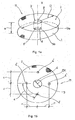
Figure 1a . shows an embodiment of the medical device according the invention in perspective view; -
Figure 1b . shows the device according tofigure 1a . in top view; -
Figure 2 shows a perspective view of the medical device according tofigure 1 with the large intestine pulled through the tube member and the abdominal wall; -
Figure 3 shows an axial cross-section of the device according tofigure 2 illustrating its position in the abdominal wall; -
Figure 4 shows the device according to the invention in top view also showing the large intestine pulled through its tube member. - The medical device according to the invention marked with
reference number 11 onfigure 1a . has aninternal sheet 1 anexternal sheet 2 running in parallel with it at a distance a, and acylindrical tube member 3 linking these sheets to each other, which is open at both ends, i.e. it has anexternal opening 3a and aninternal opening 3b. Thesheets tube member 3 together form a relatively rigid unit. Thetube member 3 is fixed to thesheets openings sheets figure 1 the curved wall of thetube member 3 is continuous and smooth, it is unbroken by any groove or other depressions.
In the case of this embodiment thesheets circular tube member 3. The diameter D of thesheets cylindrical tube member 3 may be between 2-3 cm. Both thesheets tube member 3 are formed from flexible plastic mesh made from living tissue-friendly plastic mesh, favourably from the aforementioned, commercially available "Goretex", which is known in itself, or from a polypropylene or polypropylene-based material, their thickness and rigidity is selected in accordance with the values usual in the given specialist field. It may be favourable if thesheets - In
figures 2 and3 we have shown themedical device 11 in its entirety built into the abdominal wall 6 delimiting theabdominal cavity 9 from the outside, in the phase of an operation aimed at the reconstruction of a parastomal hernia when the stoma 7a of thelarge intestine 7 is led out in front of the abdominal wall 6 and itsexternal end 7b is already fixed to the abdominal wall 6 with a suture. On the outside the abdominal wall 6 is delimited by theskin layer 5, under it is the fat layer 8 (subcutaneous fat), under which lies the musculo-aponeuroticabdominal wall layer 6b encompassed by thefascia 6a (membrane covering muscles), which is delimited from below by the peritoneum 10 (abdominal membrane), which separate the abdominal wall 6 from theabdominal cavity 9. We note that if the internal sheet 1 - viewed from the abdominal cavity 9 - is made from "Goretex" it may be positioned inside theperitoneum 10. - According this embodiment of the
device 11 according to the invention it has four fixing elements indicated with reference number 4 onfigures 2 and3 , which link thedevice 11 and the musculo-aponeuroticabdominal wall layer 6b of the abdominal wall 6 together, and through this a connection is created between this abdominal wall layer and thedevice 11 that essentially prevents movement of the device and this abdominal wall layer as compared to each other, together, however, they can turn. In the case of this embodiment the fixing elements 4 are so-called transfascial sutures, which are positioned between the edges ofsheets tube member 3, at the same lateral distances c from the centre points o of these sheets indicated onfigures 1a and 1b , and at equal lateral distances e from each other measured on a curve, in other words symmetrically around the geometric axis x of thedevice 11, as we have shown onfigure 1b , where the geometric positions of the fixing elements 4 are indicated by points 4'. Obviously the points 4' are also at an equal distance from the edge of thetube member 3. It can be seen well onfigures 2 and3 that the fixing elements 4 are formed by transfascial sutures, which pass through thesheets abdominal wall layer 6b encompassed by thefascia 6a, and theperitoneum 10, under which they are pulled throughsheet 2 in a "U" shape. Theknots 4a of the fixing elements 4 are established abovesheet 1, with the thread passing through it, i.e. they are located here. - As a result of the fixing described above the central tube member of the
device 11, through which we pull the large intestine during the course of the operation, fills the primary role of not being able to expand further as a consequence of the forces that are exerted on the edge of the hole passing through the abdominal wall. In practice parastomal hernias only develop as a consequence of these forces, as we have illustrated viafigure 4 . We note that by eliminating the expansion of the size of the hole we minimise the risk of the development of a hernia. - Illustrating the force effects according to
figure 4 - where we have shown themesh material sheets internal sheet 1 is elliptical and theexternal sheet 2 is circular - according to Laplace's law the forces exerted on thetube member 3, or on the edge of the hole passing through the abdominal wall are directly related to the radius R 1 of the hole. It has also been shown that the so-called tangential forces Ftang exerted on the edge of the hole, besides being dependent on the radius R 1 of thehole 5 or opening, depend on the abdominal cavity pressure P, as well as on the radius R 2 of the abdomen (not shown onfigure 4 ) according to the following formula: - Ftang= FradR1 (force exerted on the edge of the stoma opening)
- Frad=P×R2/2 (the force exerted on the abdominal wall from the inside)
- K1=2π.R2
- K2=2 π.R1
- K1= the circumference of the abdomen; and
- K2= the circumference of the stoma.
- So fixing the
device 11 and the abdominal wall 6 to each other according to the invention reassuringly eliminates the unfavourable effect of the tangential forces and the further expansion of the opening, and through this it minimises the risk of the development of a hernia. - The execution of the parastomal hernia operation with the help of the
medical device 11 according tofigures 1a-3 takes place in the following way: - The hernia (not illustrated) developed around the opening of the
large intestine 7 positioned on the abdominal wall 6 is dissected - in a way known in itself - from an incision made around the opening, the envelope formed by theperitoneum 10 is left from the edge of the hernial orifice. We resolve the adhesions of the content of the hernia, then separating the content of the hernia and the large intestines from each other we place them back into theabdominal cavity 9. We tighten the musculo-aponeurotic layer 6b (not visible in the drawing) surrounded by thefascia 6a with sutures leaving a hole of distance d. We dissect in theperitoneum 10 and thefascia 6a layer, then we insert themedical device 11 with itsupper sheet 2 clamped together with itslower sheet 1 with an appropriate clamping instrument above the peritoneum 10 but under the musculo-aponeurotic layer 6b. Theexternal sheet 2 is placed onto thefascia 6a, above the musculo-aponeurotic layer 6b. Thedevice 11 is fixed to the abdominal wall 6 with transfascial sutures described above, establishing by this the fixing elements 4 at 4 positions according tofigure 1b (figures 2 and3 ). As a result of this fixing operation - in other words with a "U" shaped lower suture with non-absorbing thread from monophile polypropylene usually used in surgical techniques and with the knotting of the thread we have fixed the vicinity of the stoma 7a, with this we have ensured that the environment of the stoma and the mesh move together both on the external and internal surface, and have eliminated the possibility of opposite direction twisting. Besides this with the immediate fixing the risk of the stretching out of the edges of the opening formed in the abdominal wall 6, i.e. the enlarging of the opening in the area between the two sheets is also eliminated. Following this the stoma 7a destined to be the orifice previously placed into theabdominal cavity 9 is pulled though thetube member 3 of themedical device 11, and is raised approx. 2 cm above the external surface of theskin layer 5 of the abdominal wall. - As a result of this operation the supporting layer of the abdominal wall 6, in other words the musculo-
aponeurotic layer 6b covered withfascia 6a gets between the twosheets cylindrical tube member 3 at a width of about 2 cm. It is not necessary to take separate measures to fix thelarge intestine 7, more precisely the pulled through stoma 7a to thecylindrical tube member 3 because due to the very strong adhesive ability and scarring forming feature of the plastic mesh material - in a way known in itself - it reliably prevents omentum or other intestine sections slipping in between the wall of thetube member 3 and the pulled through stoma 7a, however, the problem detailed previously, scarred adhesion between the intestine wall and the mesh material of the tube member, may occur. Due to this, during production the internal surface of thecylindrical tube member 3 connecting thesheets - We put a drain in the place of the eliminated hernia sac, then we suture the aforementioned circular skin incision together with the edge of the pulled through stoma 7a; this suturing is indicated with
reference number 7b. - The diameter of the stoma 7a of the
large intestine 7 to be pulled through varies depending on the anatomical features of the individual, therefore it is favourable to produce thecylindrical tube member 3 with various diameters. Similarly, due to the possible differences in thickness between individuals of the musculo-aponeurotic layer 6b of the abdominal wall 6 it is also favourable to have the product manufactured with the distance a between thesheets peritoneum 10; otherwise the other steps of themedical device 11 insertion operation - including the fixing of the device to the abdominal wall - are the same as those detailed above. - If the
internal sheet 1 of the device is made from "Goretex" or material of a similar nature, thissheet 1 may be positioned within theperitoneum 10, it may be in direct contact with the organs within theabdominal cavity 9, and may be linked directly to them. Its fixing may take place with the above described transfascial sutures. - The advantageous effects linked to the invention are summarised in the following:
- using a simple device, the invention makes possible the reconstruction of parastomal hernias so that their reoccurrence, hernia relapse and the occurrence of septic and other complications can be viewed as being reliably excluded, and the device when preparing the stoma, through the strengthening of the abdominal wall area in the vicinity of the pulled out intestine, provides the possibility for preventing the development of a hernia. An outstanding advantageous feature of the invention is that due to fixing the device and the abdominal wall to each other we have eliminated the risk that occurred in the case of an earlier such solution when due to the twisting movement of the torso the sheets may twist the cylindrical connection tube member, and as the intestine follows this twisting motion, damaging, smaller or larger strictures may occur in the latter. Furthermore, we have eliminated the possibility of the early falling forward or slipping out of the omentum, furthermore, the risk of the scarred adhesion of the material of the plastic mesh to the intestine wall.
- Naturally, the invention is not restricted to the embodiments of the medical device detailed above, or to the described method of conception of the procedure, it may be realised in several ways within the sphere of protection defined by the claims.
Claims (11)
- Medical device for the reconstruction of parastomal hernias and/or for the prevention of their development, having sheets (1, 2) which are made from plastic compatible with living tissue and which can be placed in the abdominal wall and which are connected via a linking-separating element and which are running separated at a distance (a) from each other, which linking-separating element is formed by a tube member (3) open at both ends forming a channel to make it possible to extract the large intestine out of the abdominal cavity through the abdominal wall and at least the tube member (3) is made from an adhesive plastic suitable for creating an organic connection with live tissue, characterised by that the two sheets (1, 2) are connected via fixing elements (4) outside of the tube member (3) for fixing the musculo-aponeurotic layer of the abdominal wall between the sheets (1, 2) in the vicinity of the tube member (3) to the sheets (1, 2) and the tube member (3) has a continuous and smooth wall unbroken by any groove or other depressions.
- The medical device according to claim 1, characterised by that the layer of the abdominal wall is fixed to the sheets (1, 2) via fixing elements (4) passing through all of the fascias and muscle layers.
- The medical device according to claim 1 or 2, characterised by that the fixing elements (4) are formed by transfascial sutures passing through the sheets (1, 2), as well as through the outer and inner fascia, furthermore through the abdominal wall muscle between them, made from monophile polypropylene, non-absorbing thread.
- The medical device according to claim 3, characterised by that the transfascial sutures are U-shaped through the internal sheet (1) and also hold the peritoneum, and passing through the external sheet (2) they are knotted (4a) outside the outer fascia.
- The medical device according any of claims 1-4, characterised by that it has four fixing elements (4) located at symmetrically allocated positions as compared to the geometrical longitudinal axis (x) of the device (11).
- The medical device according any of claims 1-5, characterised by that the internal cylindrical surface of the central linking tube member (3) is a continuous smooth surface.
- The medical device according any of claims 1-6, characterised by that at least the tube member (3) is made from plastic mesh and/or woven plastic textile.
- The medical device according any of claims 1-7, characterised by that the internal surface of the tube member (3) linking the sheets (1, 2) is coated with a silicon-based material that causes easily releasable adhesion with the surface of the intestine.
- The medical device according any of claims 1-8, characterised by that the sheets (1, 2) are parallel to each other, and the geometric longitudinal axis (x) of the tube member (3) passes through the geometric centre point (o) of the sheets (1, 2).
- The medical device according any of claims 1-9, characterised by that the sheets (1, 2) are circular and have the same diameter (D).
- The medical device according any of claims 1-9, characterised by that it has a circular cylinder cross-section tube member (3), the diameter (d) of which is selected in accordance with the diameter of the stoma to be led through it.
Applications Claiming Priority (2)
| Application Number | Priority Date | Filing Date | Title |
|---|---|---|---|
| HU1200068A HU230051B1 (en) | 2012-01-31 | 2012-01-31 | Device for prevention of a parastomal hernia and method for using thereof |
| PCT/HU2012/000120 WO2013114145A1 (en) | 2012-01-31 | 2012-11-08 | Medical device for the reconstruction of parastomal hernias and/or for the prevention of their development; as well as a procedure for the application of the device |
Publications (2)
| Publication Number | Publication Date |
|---|---|
| EP2809260A1 EP2809260A1 (en) | 2014-12-10 |
| EP2809260B1 true EP2809260B1 (en) | 2016-07-20 |
Family
ID=89990592
Family Applications (1)
| Application Number | Title | Priority Date | Filing Date |
|---|---|---|---|
| EP12818556.8A Not-in-force EP2809260B1 (en) | 2012-01-31 | 2012-11-08 | Medical device for the reconstruction of parastomal hernias and/or for the prevention of their development |
Country Status (6)
| Country | Link |
|---|---|
| US (1) | US20140350579A1 (en) |
| EP (1) | EP2809260B1 (en) |
| JP (1) | JP2015506251A (en) |
| CN (1) | CN104244864B (en) |
| HU (1) | HU230051B1 (en) |
| WO (1) | WO2013114145A1 (en) |
Families Citing this family (12)
| Publication number | Priority date | Publication date | Assignee | Title |
|---|---|---|---|---|
| US9114197B1 (en) | 2014-06-11 | 2015-08-25 | Silver Bullett Therapeutics, Inc. | Coatings for the controllable release of antimicrobial metal ions |
| US10265435B2 (en) | 2009-08-27 | 2019-04-23 | Silver Bullet Therapeutics, Inc. | Bone implant and systems and coatings for the controllable release of antimicrobial metal ions |
| US8927004B1 (en) | 2014-06-11 | 2015-01-06 | Silver Bullet Therapeutics, Inc. | Bioabsorbable substrates and systems that controllably release antimicrobial metal ions |
| US9821094B2 (en) | 2014-06-11 | 2017-11-21 | Silver Bullet Therapeutics, Inc. | Coatings for the controllable release of antimicrobial metal ions |
| WO2012064402A1 (en) | 2010-11-12 | 2012-05-18 | Silver Bullet Therapeutics, Inc. | Bone implant and systems that controllably releases silver |
| US9452242B2 (en) | 2014-06-11 | 2016-09-27 | Silver Bullet Therapeutics, Inc. | Enhancement of antimicrobial silver, silver coatings, or silver platings |
| US20190038452A1 (en) * | 2017-08-07 | 2019-02-07 | Covidien Lp | Stent and associated methodologies for creating a stoma |
| US20190159795A1 (en) * | 2017-11-28 | 2019-05-30 | Covidien Lp | Organ measuring tool assembly |
| CN108114363B (en) * | 2018-01-08 | 2023-06-06 | 上海长海医院 | Ureter skin ostomy support's device of putting into |
| CN109830161A (en) * | 2019-04-02 | 2019-05-31 | 华泰国际医院有限公司 | A kind of Emulational teaching model for urinary tract stoma postoperative care |
| WO2024006420A1 (en) * | 2022-06-30 | 2024-01-04 | Davol Inc. | Implantable prosthesis |
| WO2024043802A1 (en) * | 2022-08-25 | 2024-02-29 | Общество с ограниченной ответственностью "Айкон Лаб ГмбХ" | Endoprosthesis for the surgical treament of parastomal hernias using the ipom technique |
Family Cites Families (19)
| Publication number | Priority date | Publication date | Assignee | Title |
|---|---|---|---|---|
| US4854316A (en) * | 1986-10-03 | 1989-08-08 | Davis Emsley A | Apparatus and method for repairing and preventing para-stomal hernias |
| US4969902A (en) * | 1987-02-20 | 1990-11-13 | Biagio Ravo | Implantable device |
| US4781176A (en) * | 1987-02-20 | 1988-11-01 | Biagio Ravo | Implantable device |
| US5254133A (en) * | 1991-04-24 | 1993-10-19 | Seid Arnold S | Surgical implantation device and related method of use |
| US5743917A (en) * | 1993-01-13 | 1998-04-28 | Saxon; Allen | Prosthesis for the repair of soft tissue defects |
| JP3557368B2 (en) * | 1999-05-18 | 2004-08-25 | 尚義 菅 | Vertebral body / disc resection tool |
| US6632450B1 (en) * | 2000-05-16 | 2003-10-14 | Kenton Gregory | Adherable biomaterial patches and methods for producing and for using same |
| HUP0102060A2 (en) * | 2001-05-18 | 2004-04-28 | Attila Nagy | Therapeutical equipment for reconstruction or prevention of ventral hermina |
| US6966916B2 (en) * | 2002-09-26 | 2005-11-22 | Kumar Sarbjeet S | Device and method for surgical repair of abdominal wall hernias |
| US20050085924A1 (en) * | 2003-10-17 | 2005-04-21 | Darois Roger E. | Tissue infiltratable prosthetic device incorporating an antimicrobial substance |
| AU2005280005A1 (en) * | 2004-08-25 | 2006-03-09 | Pavad Medical, Inc. | Artificial sphincter |
| JP2006192007A (en) * | 2005-01-12 | 2006-07-27 | Akira Mitsuyoshi | Prosthesis for treating inguinal hernia |
| GB2430372B (en) * | 2005-09-19 | 2010-09-29 | Stephen George Edward Barker | Reinforcement device |
| US20070093859A1 (en) * | 2005-10-26 | 2007-04-26 | Phillips Edward H | Suture passer device for abdominal or thoracoscopic surgery |
| US20110184441A1 (en) * | 2007-01-10 | 2011-07-28 | Pascal St-Germain | Prosthetic repair patch with integrated sutures and method therefor |
| AU2009238585A1 (en) * | 2008-04-23 | 2009-10-29 | Mardil, Inc. | Implantable anti-adhesion three layer patch |
| CN201248792Y (en) * | 2008-06-11 | 2009-06-03 | 华明勋 | Bellyband for belly evacuation diversion fistulation |
| JP2012509153A (en) * | 2008-11-20 | 2012-04-19 | ライフセル コーポレーション | Methods for treatment and prevention of parastoma hernia |
| US8449512B2 (en) * | 2010-04-09 | 2013-05-28 | Davinci Biomedical Research Products Inc. | Stoma stabilitating device and method |
-
2012
- 2012-01-31 HU HU1200068A patent/HU230051B1/en not_active IP Right Cessation
- 2012-11-08 WO PCT/HU2012/000120 patent/WO2013114145A1/en active Application Filing
- 2012-11-08 JP JP2014555319A patent/JP2015506251A/en active Pending
- 2012-11-08 EP EP12818556.8A patent/EP2809260B1/en not_active Not-in-force
- 2012-11-08 CN CN201280071738.6A patent/CN104244864B/en not_active Expired - Fee Related
- 2012-11-08 US US14/375,972 patent/US20140350579A1/en not_active Abandoned
Also Published As
| Publication number | Publication date |
|---|---|
| JP2015506251A (en) | 2015-03-02 |
| CN104244864B (en) | 2016-08-24 |
| US20140350579A1 (en) | 2014-11-27 |
| HU230051B1 (en) | 2015-06-29 |
| HUP1200068A2 (en) | 2013-08-28 |
| WO2013114145A1 (en) | 2013-08-08 |
| CN104244864A (en) | 2014-12-24 |
| EP2809260A1 (en) | 2014-12-10 |
Similar Documents
| Publication | Publication Date | Title |
|---|---|---|
| EP2809260B1 (en) | Medical device for the reconstruction of parastomal hernias and/or for the prevention of their development | |
| JP3180827U (en) | Reinforcing device | |
| JP6092367B2 (en) | Puncture device | |
| AU2010245983B2 (en) | Surgical patch cover and method of use | |
| CN104661616B (en) | For disposing and the interim auxiliary member of fixing organization reparation implant | |
| WO2002064041A1 (en) | Gastric clamp for performing vertical band gastroplasty and a gastric bypass with lesser curvature | |
| JP2010521216A (en) | Soft tissue fixation device | |
| EP1592361A2 (en) | Implantable hernia repair system | |
| EP0553344A1 (en) | Surgical apparatus and method | |
| JP6980085B2 (en) | Implants and methods for the treatment of pelvic disorders | |
| CN104203123A (en) | Implant for hernia repair | |
| CN105228555B (en) | Implantable reinforcing prosthese for reinforcing biological wall | |
| CN109419577A (en) | Bracket and the correlation technique for being used to form stoma | |
| JP2012504471A (en) | Method and apparatus for tension-free inguinal hernia repair and physiological function reconstruction using inguinal hernia prosthesis with side-unenclosed cord positioning structure | |
| JP6697459B2 (en) | Muscle wall defect prosthesis and deployment system | |
| JP6812347B2 (en) | Implantable prosthesis for soft tissue repair | |
| US20140114266A1 (en) | Ostomy Implant System and Method | |
| WO2020193292A1 (en) | Implant for stoma | |
| CN111163709A (en) | Gastric liner funnel with anastomosis | |
| CA2852612C (en) | Implantable stoma ring | |
| RU2639854C2 (en) | Device for inguinal hernioplasty | |
| US20240138969A1 (en) | Umbilical hernia prosthesis | |
| Szczepkowski et al. | Patient reoperations for late complications of surgical treatment of parastomal hernias–own experience | |
| US20130030339A1 (en) | Gauze assembly for reinforcing tissue which extends around a surgically created stoma |
Legal Events
| Date | Code | Title | Description |
|---|---|---|---|
| PUAI | Public reference made under article 153(3) epc to a published international application that has entered the european phase |
Free format text: ORIGINAL CODE: 0009012 |
|
| 17P | Request for examination filed |
Effective date: 20140826 |
|
| AK | Designated contracting states |
Kind code of ref document: A1 Designated state(s): AL AT BE BG CH CY CZ DE DK EE ES FI FR GB GR HR HU IE IS IT LI LT LU LV MC MK MT NL NO PL PT RO RS SE SI SK SM TR |
|
| AX | Request for extension of the european patent |
Extension state: BA ME |
|
| DAX | Request for extension of the european patent (deleted) | ||
| GRAP | Despatch of communication of intention to grant a patent |
Free format text: ORIGINAL CODE: EPIDOSNIGR1 |
|
| INTG | Intention to grant announced |
Effective date: 20151027 |
|
| GRAS | Grant fee paid |
Free format text: ORIGINAL CODE: EPIDOSNIGR3 |
|
| GRAA | (expected) grant |
Free format text: ORIGINAL CODE: 0009210 |
|
| AK | Designated contracting states |
Kind code of ref document: B1 Designated state(s): AL AT BE BG CH CY CZ DE DK EE ES FI FR GB GR HR HU IE IS IT LI LT LU LV MC MK MT NL NO PL PT RO RS SE SI SK SM TR |
|
| REG | Reference to a national code |
Ref country code: GB Ref legal event code: FG4D |
|
| REG | Reference to a national code |
Ref country code: CH Ref legal event code: EP |
|
| REG | Reference to a national code |
Ref country code: IE Ref legal event code: FG4D |
|
| REG | Reference to a national code |
Ref country code: AT Ref legal event code: REF Ref document number: 813385 Country of ref document: AT Kind code of ref document: T Effective date: 20160815 |
|
| REG | Reference to a national code |
Ref country code: DE Ref legal event code: R096 Ref document number: 602012020834 Country of ref document: DE |
|
| REG | Reference to a national code |
Ref country code: LT Ref legal event code: MG4D |
|
| REG | Reference to a national code |
Ref country code: NL Ref legal event code: MP Effective date: 20160720 |
|
| REG | Reference to a national code |
Ref country code: AT Ref legal event code: MK05 Ref document number: 813385 Country of ref document: AT Kind code of ref document: T Effective date: 20160720 |
|
| PG25 | Lapsed in a contracting state [announced via postgrant information from national office to epo] |
Ref country code: IT Free format text: LAPSE BECAUSE OF FAILURE TO SUBMIT A TRANSLATION OF THE DESCRIPTION OR TO PAY THE FEE WITHIN THE PRESCRIBED TIME-LIMIT Effective date: 20160720 Ref country code: LT Free format text: LAPSE BECAUSE OF FAILURE TO SUBMIT A TRANSLATION OF THE DESCRIPTION OR TO PAY THE FEE WITHIN THE PRESCRIBED TIME-LIMIT Effective date: 20160720 Ref country code: FI Free format text: LAPSE BECAUSE OF FAILURE TO SUBMIT A TRANSLATION OF THE DESCRIPTION OR TO PAY THE FEE WITHIN THE PRESCRIBED TIME-LIMIT Effective date: 20160720 Ref country code: NO Free format text: LAPSE BECAUSE OF FAILURE TO SUBMIT A TRANSLATION OF THE DESCRIPTION OR TO PAY THE FEE WITHIN THE PRESCRIBED TIME-LIMIT Effective date: 20161020 Ref country code: HR Free format text: LAPSE BECAUSE OF FAILURE TO SUBMIT A TRANSLATION OF THE DESCRIPTION OR TO PAY THE FEE WITHIN THE PRESCRIBED TIME-LIMIT Effective date: 20160720 Ref country code: IS Free format text: LAPSE BECAUSE OF FAILURE TO SUBMIT A TRANSLATION OF THE DESCRIPTION OR TO PAY THE FEE WITHIN THE PRESCRIBED TIME-LIMIT Effective date: 20161120 Ref country code: NL Free format text: LAPSE BECAUSE OF FAILURE TO SUBMIT A TRANSLATION OF THE DESCRIPTION OR TO PAY THE FEE WITHIN THE PRESCRIBED TIME-LIMIT Effective date: 20160720 Ref country code: RS Free format text: LAPSE BECAUSE OF FAILURE TO SUBMIT A TRANSLATION OF THE DESCRIPTION OR TO PAY THE FEE WITHIN THE PRESCRIBED TIME-LIMIT Effective date: 20160720 |
|
| PG25 | Lapsed in a contracting state [announced via postgrant information from national office to epo] |
Ref country code: AT Free format text: LAPSE BECAUSE OF FAILURE TO SUBMIT A TRANSLATION OF THE DESCRIPTION OR TO PAY THE FEE WITHIN THE PRESCRIBED TIME-LIMIT Effective date: 20160720 Ref country code: SE Free format text: LAPSE BECAUSE OF FAILURE TO SUBMIT A TRANSLATION OF THE DESCRIPTION OR TO PAY THE FEE WITHIN THE PRESCRIBED TIME-LIMIT Effective date: 20160720 Ref country code: GR Free format text: LAPSE BECAUSE OF FAILURE TO SUBMIT A TRANSLATION OF THE DESCRIPTION OR TO PAY THE FEE WITHIN THE PRESCRIBED TIME-LIMIT Effective date: 20161021 Ref country code: LV Free format text: LAPSE BECAUSE OF FAILURE TO SUBMIT A TRANSLATION OF THE DESCRIPTION OR TO PAY THE FEE WITHIN THE PRESCRIBED TIME-LIMIT Effective date: 20160720 Ref country code: PL Free format text: LAPSE BECAUSE OF FAILURE TO SUBMIT A TRANSLATION OF THE DESCRIPTION OR TO PAY THE FEE WITHIN THE PRESCRIBED TIME-LIMIT Effective date: 20160720 Ref country code: PT Free format text: LAPSE BECAUSE OF FAILURE TO SUBMIT A TRANSLATION OF THE DESCRIPTION OR TO PAY THE FEE WITHIN THE PRESCRIBED TIME-LIMIT Effective date: 20161121 Ref country code: ES Free format text: LAPSE BECAUSE OF FAILURE TO SUBMIT A TRANSLATION OF THE DESCRIPTION OR TO PAY THE FEE WITHIN THE PRESCRIBED TIME-LIMIT Effective date: 20160720 Ref country code: BE Free format text: LAPSE BECAUSE OF NON-PAYMENT OF DUE FEES Effective date: 20160720 |
|
| REG | Reference to a national code |
Ref country code: DE Ref legal event code: R097 Ref document number: 602012020834 Country of ref document: DE |
|
| PG25 | Lapsed in a contracting state [announced via postgrant information from national office to epo] |
Ref country code: EE Free format text: LAPSE BECAUSE OF FAILURE TO SUBMIT A TRANSLATION OF THE DESCRIPTION OR TO PAY THE FEE WITHIN THE PRESCRIBED TIME-LIMIT Effective date: 20160720 Ref country code: RO Free format text: LAPSE BECAUSE OF FAILURE TO SUBMIT A TRANSLATION OF THE DESCRIPTION OR TO PAY THE FEE WITHIN THE PRESCRIBED TIME-LIMIT Effective date: 20160720 |
|
| PLBE | No opposition filed within time limit |
Free format text: ORIGINAL CODE: 0009261 |
|
| STAA | Information on the status of an ep patent application or granted ep patent |
Free format text: STATUS: NO OPPOSITION FILED WITHIN TIME LIMIT |
|
| PG25 | Lapsed in a contracting state [announced via postgrant information from national office to epo] |
Ref country code: SK Free format text: LAPSE BECAUSE OF FAILURE TO SUBMIT A TRANSLATION OF THE DESCRIPTION OR TO PAY THE FEE WITHIN THE PRESCRIBED TIME-LIMIT Effective date: 20160720 Ref country code: SM Free format text: LAPSE BECAUSE OF FAILURE TO SUBMIT A TRANSLATION OF THE DESCRIPTION OR TO PAY THE FEE WITHIN THE PRESCRIBED TIME-LIMIT Effective date: 20160720 Ref country code: BG Free format text: LAPSE BECAUSE OF FAILURE TO SUBMIT A TRANSLATION OF THE DESCRIPTION OR TO PAY THE FEE WITHIN THE PRESCRIBED TIME-LIMIT Effective date: 20161020 Ref country code: DK Free format text: LAPSE BECAUSE OF FAILURE TO SUBMIT A TRANSLATION OF THE DESCRIPTION OR TO PAY THE FEE WITHIN THE PRESCRIBED TIME-LIMIT Effective date: 20160720 Ref country code: CZ Free format text: LAPSE BECAUSE OF FAILURE TO SUBMIT A TRANSLATION OF THE DESCRIPTION OR TO PAY THE FEE WITHIN THE PRESCRIBED TIME-LIMIT Effective date: 20160720 |
|
| REG | Reference to a national code |
Ref country code: DE Ref legal event code: R119 Ref document number: 602012020834 Country of ref document: DE |
|
| 26N | No opposition filed |
Effective date: 20170421 |
|
| REG | Reference to a national code |
Ref country code: CH Ref legal event code: PL |
|
| GBPC | Gb: european patent ceased through non-payment of renewal fee |
Effective date: 20161108 |
|
| PG25 | Lapsed in a contracting state [announced via postgrant information from national office to epo] |
Ref country code: LI Free format text: LAPSE BECAUSE OF NON-PAYMENT OF DUE FEES Effective date: 20161130 Ref country code: CH Free format text: LAPSE BECAUSE OF NON-PAYMENT OF DUE FEES Effective date: 20161130 |
|
| REG | Reference to a national code |
Ref country code: IE Ref legal event code: MM4A |
|
| REG | Reference to a national code |
Ref country code: FR Ref legal event code: ST Effective date: 20170731 |
|
| PG25 | Lapsed in a contracting state [announced via postgrant information from national office to epo] |
Ref country code: SI Free format text: LAPSE BECAUSE OF FAILURE TO SUBMIT A TRANSLATION OF THE DESCRIPTION OR TO PAY THE FEE WITHIN THE PRESCRIBED TIME-LIMIT Effective date: 20160720 |
|
| PG25 | Lapsed in a contracting state [announced via postgrant information from national office to epo] |
Ref country code: LU Free format text: LAPSE BECAUSE OF NON-PAYMENT OF DUE FEES Effective date: 20161130 |
|
| PG25 | Lapsed in a contracting state [announced via postgrant information from national office to epo] |
Ref country code: FR Free format text: LAPSE BECAUSE OF NON-PAYMENT OF DUE FEES Effective date: 20161130 |
|
| PG25 | Lapsed in a contracting state [announced via postgrant information from national office to epo] |
Ref country code: IE Free format text: LAPSE BECAUSE OF NON-PAYMENT OF DUE FEES Effective date: 20161108 Ref country code: DE Free format text: LAPSE BECAUSE OF NON-PAYMENT OF DUE FEES Effective date: 20170601 Ref country code: GB Free format text: LAPSE BECAUSE OF NON-PAYMENT OF DUE FEES Effective date: 20161108 |
|
| PG25 | Lapsed in a contracting state [announced via postgrant information from national office to epo] |
Ref country code: HU Free format text: LAPSE BECAUSE OF FAILURE TO SUBMIT A TRANSLATION OF THE DESCRIPTION OR TO PAY THE FEE WITHIN THE PRESCRIBED TIME-LIMIT; INVALID AB INITIO Effective date: 20121108 |
|
| PG25 | Lapsed in a contracting state [announced via postgrant information from national office to epo] |
Ref country code: MC Free format text: LAPSE BECAUSE OF FAILURE TO SUBMIT A TRANSLATION OF THE DESCRIPTION OR TO PAY THE FEE WITHIN THE PRESCRIBED TIME-LIMIT Effective date: 20160720 Ref country code: CY Free format text: LAPSE BECAUSE OF FAILURE TO SUBMIT A TRANSLATION OF THE DESCRIPTION OR TO PAY THE FEE WITHIN THE PRESCRIBED TIME-LIMIT Effective date: 20160720 Ref country code: MK Free format text: LAPSE BECAUSE OF FAILURE TO SUBMIT A TRANSLATION OF THE DESCRIPTION OR TO PAY THE FEE WITHIN THE PRESCRIBED TIME-LIMIT Effective date: 20160720 |
|
| PG25 | Lapsed in a contracting state [announced via postgrant information from national office to epo] |
Ref country code: MT Free format text: LAPSE BECAUSE OF NON-PAYMENT OF DUE FEES Effective date: 20161108 |
|
| PG25 | Lapsed in a contracting state [announced via postgrant information from national office to epo] |
Ref country code: TR Free format text: LAPSE BECAUSE OF FAILURE TO SUBMIT A TRANSLATION OF THE DESCRIPTION OR TO PAY THE FEE WITHIN THE PRESCRIBED TIME-LIMIT Effective date: 20160720 Ref country code: AL Free format text: LAPSE BECAUSE OF FAILURE TO SUBMIT A TRANSLATION OF THE DESCRIPTION OR TO PAY THE FEE WITHIN THE PRESCRIBED TIME-LIMIT Effective date: 20160720 |