EP2805165B1 - Methods for predicting the survival time of a patient suffering from a solid cancer based on density of b cells - Google Patents
Methods for predicting the survival time of a patient suffering from a solid cancer based on density of b cells Download PDFInfo
- Publication number
- EP2805165B1 EP2805165B1 EP13701044.3A EP13701044A EP2805165B1 EP 2805165 B1 EP2805165 B1 EP 2805165B1 EP 13701044 A EP13701044 A EP 13701044A EP 2805165 B1 EP2805165 B1 EP 2805165B1
- Authority
- EP
- European Patent Office
- Prior art keywords
- density
- tumor
- cells
- value
- cut
- Prior art date
- Legal status (The legal status is an assumption and is not a legal conclusion. Google has not performed a legal analysis and makes no representation as to the accuracy of the status listed.)
- Active
Links
Images


Classifications
-
- G—PHYSICS
- G01—MEASURING; TESTING
- G01N—INVESTIGATING OR ANALYSING MATERIALS BY DETERMINING THEIR CHEMICAL OR PHYSICAL PROPERTIES
- G01N33/00—Investigating or analysing materials by specific methods not covered by groups G01N1/00 - G01N31/00
- G01N33/48—Biological material, e.g. blood, urine; Haemocytometers
- G01N33/50—Chemical analysis of biological material, e.g. blood, urine; Testing involving biospecific ligand binding methods; Immunological testing
- G01N33/53—Immunoassay; Biospecific binding assay; Materials therefor
- G01N33/575—Immunoassay; Biospecific binding assay; Materials therefor for cancer
-
- G—PHYSICS
- G01—MEASURING; TESTING
- G01N—INVESTIGATING OR ANALYSING MATERIALS BY DETERMINING THEIR CHEMICAL OR PHYSICAL PROPERTIES
- G01N2333/00—Assays involving biological materials from specific organisms or of a specific nature
- G01N2333/435—Assays involving biological materials from specific organisms or of a specific nature from animals; from humans
- G01N2333/705—Assays involving receptors, cell surface antigens or cell surface determinants
- G01N2333/70503—Immunoglobulin superfamily, e.g. VCAMs, PECAM, LFA-3
- G01N2333/7051—T-cell receptor (TcR)-CD3 complex
-
- G—PHYSICS
- G01—MEASURING; TESTING
- G01N—INVESTIGATING OR ANALYSING MATERIALS BY DETERMINING THEIR CHEMICAL OR PHYSICAL PROPERTIES
- G01N2333/00—Assays involving biological materials from specific organisms or of a specific nature
- G01N2333/435—Assays involving biological materials from specific organisms or of a specific nature from animals; from humans
- G01N2333/705—Assays involving receptors, cell surface antigens or cell surface determinants
- G01N2333/70503—Immunoglobulin superfamily, e.g. VCAMs, PECAM, LFA-3
- G01N2333/70517—CD8
-
- G—PHYSICS
- G01—MEASURING; TESTING
- G01N—INVESTIGATING OR ANALYSING MATERIALS BY DETERMINING THEIR CHEMICAL OR PHYSICAL PROPERTIES
- G01N2333/00—Assays involving biological materials from specific organisms or of a specific nature
- G01N2333/435—Assays involving biological materials from specific organisms or of a specific nature from animals; from humans
- G01N2333/705—Assays involving receptors, cell surface antigens or cell surface determinants
- G01N2333/70589—CD45
-
- G—PHYSICS
- G01—MEASURING; TESTING
- G01N—INVESTIGATING OR ANALYSING MATERIALS BY DETERMINING THEIR CHEMICAL OR PHYSICAL PROPERTIES
- G01N2333/00—Assays involving biological materials from specific organisms or of a specific nature
- G01N2333/435—Assays involving biological materials from specific organisms or of a specific nature from animals; from humans
- G01N2333/705—Assays involving receptors, cell surface antigens or cell surface determinants
- G01N2333/70596—Molecules with a "CD"-designation not provided for elsewhere in G01N2333/705
-
- G—PHYSICS
- G01—MEASURING; TESTING
- G01N—INVESTIGATING OR ANALYSING MATERIALS BY DETERMINING THEIR CHEMICAL OR PHYSICAL PROPERTIES
- G01N2800/00—Detection or diagnosis of diseases
- G01N2800/52—Predicting or monitoring the response to treatment, e.g. for selection of therapy based on assay results in personalised medicine; Prognosis
Definitions
- the present invention relates to methods and kits for the prognosis of survival time of a patient suffering from a solid cancer.
- Cancer is a complex disease involving interactions between the immune system and the tumour 1 .
- the correlation between a "high” intra- and peri-tumoural adaptive immune reaction in colorectal carcinoma and a good prognosis was previously reported.
- a "low” density of T cells was correlated with a poor prognosis 2-4 .
- the immune T cell infiltrate was shown to be the most important predictive criteria for survival 2,7-9 . This is also supported by mouse models of immunosurveillance and immunoediting 10-12 .
- Recent advances in cellular immunology and tumour biology are guiding new approaches to adoptive T-cell therapy 13 with promising results 14 .
- Mahmoud et al (Breast Cancer Research and Treatment, vol.132, no.2, 2001, pp. 545-553 ) evaluates the prognostic significance of B lymphocytes in invasive carcinoma of the breast.
- EP1777523 discloses methods for determining the prognosis of a patient suffering from cancer, said methods comprising quantifying, within the tumor, at least one biological marker which is indicative of the status of the adaptive immune response of said patient against cancer.
- Fridman et al discusses tumor-immune interactions in human cancers.
- the present invention relates to a method for predicting the survival time of a patient suffering from a solid cancer comprising the steps consisting of i) determining the density of B cells at the invasive margin of the tumor (im) in a tumor tissue sample obtained from said patient, ii) comparing said density with a predetermined reference value and iii) providing a good prognosis when the density of B cells at the invasive margin of the tumor is higher than the predetermined reference value and a poor prognosis when the density of B cells at the invasive margin of the tumor is lower than the predetermined reference value.
- the inventors investigated gene expression and cell densities from 28 different cell types comprising most innate and adaptive immune-cell subpopulations in tumours. They found clusters of immune-cells associated with a specific tumour stage. Among them, B and T lymphocytes are organized within a core network and are the most prominent immune cells correlating with tumour progression and a favourable prognosis. More particularly, the inventors demonstrated that patients with high density of B cells in the invasive margin of the tumor had a prolonged disease-free survival whereas patients with low density of B cells in the invasive margin of the tumor had a poor prognosis.
- the combination of B cell marker with at least one marker selected from the group consisting of CD3, CD8 and CDR45RO in at least one region of the tumor (im and/or ct) gave also a better discrimination of patients.
- the present invention relates to a method for predicting the survival time of a patient suffering from a solid cancer comprising the steps consisting of determining the density of CD20+ B cells at the invasive margin of the tumor (im) in a tumor tissue sample obtained from said patient; determining the density of one further cell type selected from CD8+ cytotoxic T cells or CD45RO+ memory T cells at the invasive margin of the tumor (im) and at the center of the tumor (ct) in a tumor tissue sample obtained from said patient; comparing each density with a cut-off value; and providing:
- the present invention further relates to a method for predicting the survival time of a patient suffering from colorectal cancer comprising the steps consisting of i) determining the density of B cells at the invasive margin of the tumor (im) in a tumor tissue sample obtained from said patient ii) comparing said density with a predetermined reference value and iii) providing a good prognosis when the density of B cells at the invasive margin of the tumor is higher than the predetermined reference value and a poor prognosis when the density of B cells at the invasive margin of the tumor is lower than the predetermined reference value.
- the patient suffers from a cancer selected from the group consisting of adrenal cortical cancer, anal cancer, bile duct cancer (e.g. periphilar cancer, distal bile duct cancer, intrahepatic bile duct cancer), bladder cancer, bone cancer (e.g. osteoblastoma, osteochrondroma, hemangioma, chondromyxoid fibroma, osteosarcoma, chondrosarcoma, fibrosarcoma, malignant fibrous histiocytoma, giant cell tumor of the bone, chordoma, lymphoma, multiple myeloma), brain and central nervous system cancer (e.g.
- a cancer selected from the group consisting of adrenal cortical cancer, anal cancer, bile duct cancer (e.g. periphilar cancer, distal bile duct cancer, intrahepatic bile duct cancer), bladder cancer, bone cancer (e.g. osteoblastoma, osteochrondroma,
- meningioma astocytoma, oligodendrogliomas, ependymoma, gliomas, medulloblastoma, ganglioglioma, Schwannoma, germinoma, craniopharyngioma), breast cancer (e.g. ductal carcinoma in situ, infiltrating ductal carcinoma, infiltrating lobular carcinoma, lobular carcinoma in situ, gynecomastia), Castleman disease (e.g. giant lymph node hyperplasia, angiofollicular lymph node hyperplasia), cervical cancer, colorectal cancer, endometrial cancer (e.g.
- adenocarcinoma endometrial adenocarcinoma, adenocanthoma, papillary serous adnocarcinoma, clear cell
- esophagus cancer gallbladder cancer (mucinous adenocarcinoma, small cell carcinoma), gastrointestinal carcinoid tumors (e.g. choriocarcinoma, chorioadenoma destruens), Hodgkin's disease, non-Hodgkin's lymphoma, Kaposi's sarcoma, kidney cancer (e.g. renal cell cancer), laryngeal and hypopharyngeal cancer, liver cancer (e.g.
- lung cancer e.g. small cell lung cancer, non-small cell lung cancer
- mesothelioma plasmacytoma, nasal cavity and paranasal sinus cancer (e.g. esthesioneuroblastoma, midline granuloma), nasopharyngeal cancer, neuroblastoma, oral cavity and oropharyngeal cancer, ovarian cancer, pancreatic cancer, penile cancer, pituitary cancer, prostate cancer, retinoblastoma, rhabdomyosarcoma (e.g.
- rhabdomyosarcoma embryonal rhabdomyosarcoma, alveolar rhabdomyosarcoma, pleomorphic rhabdomyosarcoma), salivary gland cancer, skin cancer (e.g. melanoma, nonmelanoma skin caner), stomach cancer, testicular cancer (e.g. seminoma, nonseminoma germ cell cancer), thymus cancer, thyroid cancer (e.g. follicular carcinoma, anaplastic carcinoma, poorly differentiated carcinoma, medullary thyroid carcinoma, thyroid lymphoma), vaginal cancer, vulvar cancer, and uterine cancer (e.g. uterine leiomyosarcoma).
- skin cancer e.g. melanoma, nonmelanoma skin caner
- testicular cancer e.g. seminoma, nonseminoma germ cell cancer
- thymus cancer testicular cancer (e.g. seminoma, nonsemin
- tissue sample has its general meaning in the art and encompasses pieces or slices of tissue that have been removed including following a surgical tumor resection or following the collection of a tissue sample for biopsy.
- the tissue tumor sample shall comprise the invasive margin surrounding the tumor, and may comprise or not the center of the tumor.
- invasive margin has its general meaning in the art and refers to the cellular environment surrounding the tumor.
- the tumor tissue sample can, of course, be subjected to a variety of well-known post-collection preparative and storage techniques (e.g., fixation, storage, freezing, etc.) prior to determining the density of B cells at the invasive margin of the tumor (im).
- the tissue tumor sample may be paraffin-embedded or frozen.
- the method of the invention is particularly suitable for the duration of the disease-free survival (DFS) or the overall survival (OS).
- DFS disease-free survival
- OS overall survival
- B cell has its general meaning in the art and refers to a cell produced in the bone marrow expressing membrane-bound antibody specific for an antigen. Following interaction with the antigen it differentiates into a plasma cell which secretes antibodies specific for the antigen or into a memory B cell. "B cell” and “B lymphocyte” is used interchangeably. Typically, B cells are characterized by expression of B cell markers at their cell surface. As used herein, the term “B cell marker” refers to surface molecules on the B cells which are specific for particular B cells.
- B cell markers suitable for use in the present invention include, but are not limited to surface IgG, kappa and lambda chains, Ig-alpha (CD79alpha), Ig-beta (CD79beta), CD19, B220 (CD45R), CD20, CD21, CD22, CD23, CD27, or any other CD antigen specific for B cells.
- a B cell is a CD20+ cell.
- Determining the density of B cells at the invasive margin of the tumor may be determined by any well known method in the art.
- such methods comprise contacting the tumor tissue sample with at least one selective binding agent capable of selectively interacting with B cells.
- the selective binding agent may be polyclonal antibody or monoclonal antibody, an antibody fragment, synthetic antibodies, or other protein-specific agents such as nucleic acid or peptide aptamers.
- the selective binding agent binds any of the B cell markers, such as an antibody specific for any of these molecules.
- Preferred B cell selective binding agents bind to CD19, CD20, CD21, CD22 or CD37. Particularly preferred B cell selective binding agents bind to CD20.
- the antibodies may be tagged directly with detectable labels such as enzymes, chromogens or fluorescent probes or indirectly detected with a secondary antibody conjugated with detectable labels.
- the preferred method according to the present invention is immunohistochemistry.
- the tissue tumor sample is firstly incubated with labeled antibodies directed against one B cell marker of interest (e.g. CD20). After washing, the labeled antibodies that are bound to said B cell marker of interest are revealed by the appropriate technique, depending of the kind of label is borne by the labeled antibody, e.g. radioactive, fluorescent or enzyme label. Multiple labelling can be performed simultaneously.
- the method of the present invention may use a secondary antibody coupled to an amplification system (to intensify staining signal) and enzymatic molecules.
- Such coupled secondary antibodies are commercially available, e.g. from Dako, EnVision system. Counterstaining may be used, e.g. H&E, DAPI, Hoechst.
- Other staining methods may be accomplished using any suitable method or system as would be apparent to one of skill in the art, including automated, semi-automated or manual systems.
- the density of B cells may be expressed as the number of these cells that are counted per one unit of surface area of tissue sample, e.g. as the number of B cells that are counted per cm 2 or mm 2 of surface area of tumor tissue sample. As used herein, the density of B cells may also be expressed as the number of B cells per one volume unit of sample, e.g. as the number of B cells per cm3 of tumor tissue sample. As used herein, the density of B cells may also consist of the percentage of B cells per total cells (set at 100%).
- Predetermined reference values used for comparison consist of "cut-of' values that may be determined as described hereunder.
- Each reference (“cut-off') value for each biological marker may be predetermined by carrying out a method comprising the steps of
- the predetermined reference value may consist of the B cell density value (e.g. density of CD20+ cells), per total cells (set at 100%), that correlates with a poor prognosis (e.g. a short disease-free survival time), or in contrast may consist of the B cell density value that correlates with good prognosis (e.g. a long disease-free survival time).
- the comparison steps may include a classification of the quantification values measured for the cell density in two groups, respectively: (i) a first group termed "Hi” when the quantification value for the cell density is higher than the predetermined corresponding reference value and (ii) a second group termed "Lo" when the quantification value for the cell density is lower than the predetermined corresponding reference value. It flows from the example that if the result of the comparison step consists of a "Hi” value, then a good prognosis is provided ( Figure 1 ). Conversely, if the result of the comparison step consists of a "Lo” value, then a poor prognosis is provided ( Figure 1 ). A score may be also determined according to table 1.
- the method of the invention may further comprise the steps consisting of i) determining the density of at least one further cell type at the invasive margin of the tumor (im) and/or at the center of the tumor (ct) in a tumor tissue sample obtained from said patient and ii) comparing said density with a predetermined reference value.
- the further cell type is selected from the group consisting of T cells, among a particular subset of T cells including cytotoxic T cells or memory T cells.
- T cell has its general meaning in the art and includes cells within the T cell lineage, including thymocytes, immature T cells, mature T cells and the like. Typically, T cells are characterized by expression of T cell markers at their cell surface. As used herein, the term “T cell marker” refers to surface molecules on the T cells which are specific for particular T cells. T cell markers suitable for use in the present invention include, but are not limited to surface CD3, CD4, CD8, CD45RO or any other CD antigen specific for T cells. Typically a T cell is a CD3+ cell.
- cytotoxic T cells has its general meaning in the art and refers to a T cell, once activated by a MHC-antigen complex, releases the protein perforin, which forms pores in the target cell's plasma membrane; this causes ions and water to flow into the target cell, making it expand and eventually lyse. Cytotoxic T cells also release granzyme, a serine protease that can enter target cells via the perforin-formed pore and induce apoptosis (cell death). Most cytotoxic T cells have present on the cell surface the protein CD8, which is attracted to portions of the Class I MHC molecule. Typically a cytotoxic T cell is a CD8+ cell.
- memory T cell has its general meaning in the art and to a subset of T cells that are specific to the antigen they first encountered and can be called upon during the secondary immune response. Memory T cells are characterized by the expression at their cell surface of CDR45RO. Typically a memory T cell is a CD45RO+ cell.
- the method of the invention may further comprise the steps consisting of i) determining the density of T cells in the center of the tumor (ct) in a tumor tissue sample obtained from said patient and ii) comparing said density with a predetermined reference value.
- the method of the invention may further comprise the steps consisting of i) determining the density of T cells at the invasive margin (im) of the tumor in a tumor tissue sample obtained from said patient and ii) comparing said density with a predetermined reference value.
- the invention may further comprise the steps consisting of i) determining the density of T cells in the center of the tumor (ct) in a tumor tissue sample obtained from said patient ii) determining the density of T cells at the invasive margin (im) of the tumor in a tumor tissue sample obtained from said patient and iii) comparing said densities with predetermined reference values.
- the method of the invention may further comprise the steps consisting of determining the density of cytotoxic T cells in the center of the tumor (ct) in a tumor tissue sample obtained from said patient and ii) comparing said density with a predetermined reference value.
- the method of the invention may further comprise the steps consisting of determining the density of cytotoxic T cells at the invasive margin (im) of the tumor in a tumor tissue sample obtained from said patient and ii) comparing said density with a predetermined reference value.
- the method of the invention may further comprise the steps consisting of i) determining the density of cytotoxic T cells in the center of the tumor (ct) in a tumor tissue sample obtained from said patient ii) determining the density of cytotoxic T cells at the invasive margin (im) of the tumor in a tumor tissue sample obtained from said patient and iii) comparing said densities with predetermined reference values.
- the method of the invention may further comprise the steps consisting of determining the density of cytotoxic T cells in the center of the tumor (ct) in a tumor tissue sample obtained from said patient and ii) comparing said density with a predetermined reference value.
- the method of the invention may further comprise the steps consisting of determining the density of memory T cells in the center of the tumor (ct) in a tumor tissue sample obtained from said patient and ii) comparing said density with a predetermined reference value.
- the method of the invention may further comprise the steps consisting of determining the density of memory T cells at the invasive margin (im) of the tumor in a tumor tissue sample obtained from said patient and ii) comparing said density with a predetermined reference value.
- the method of the invention may further comprise the steps consisting of i) determining the density of memory T cells in the center of the tumor (ct) in a tumor tissue sample obtained from said patient ii) determining the density of memory T cells at the invasive margin (im) of the tumor in a tumor tissue sample obtained from said patient and iii) comparing said densities with predetermined reference values.
- the comparison steps may also include a classification of the quantification values measured for each cell density in two groups, respectively: (i) a first group termed "Hi” when the quantification value for cell density is higher than the predetermined corresponding reference value and (ii) a second group termed "Lo" when the quantification value for the cell density is lower than the predetermined corresponding reference value.
- a score which is a composite of classification done for the B cell density and for the additional densities may also be calculated as depicted in tables 1-9 to make easier to understand the result of the comparison step.
- the methods of the invention are of higher accuracy than currently used staging methods (e.g. UICC-TNM). Accordingly, the methods of the invention can be applied for monitoring the effectiveness of anti-cancer treatments.
- the present invention provides a method for monitoring the effectiveness of treatment of a subject with an agent comprising the steps of (i) predicting the survival time of the patient before administering said agent by performing the method according to the invention, ii) predicting the survival time of the patient after administering said agent by performing the method according to the invention iii) comparing the survival time of step a) with the survival time of step b) and iv) and providing the conclusion that the agent is effective for the treatment of the cancer when the survival time of step b) is higher than the survival time of step a).
- the physician may adapt the treatment by prescribing different dosage or by prescribing another agent to administer.
- the methods of the invention may also particularly suitable for determining whether the patient will be considered as responder to the treatment (e.g. an immunotherapy agent).
- the patient may be eligible for the treatment.
- the methods of the invention may also particularly suitable for determining whether adjuvant therapy (e.g. chemotherapy) will be required or not.
- adjuvant therapy e.g. chemotherapy
- the subsequent anti-cancer treatment may not comprise any adjuvant chemotherapy.
- the patient my eligible for the adjuvant chemotherapy.
- kits for performing the method of the present invention comprising means for determining the cell density as above described.
- a kit according to the disclosure may comprise one or a combination or a set of antibodies, each kind of antibodies being directed specifically against one cell type. Suitable means include antibodies, antibody derivatives, antibody fragments, and the like.
- the kit of the disclosure may optionally comprise additional components useful for performing the methods of the invention.
- the kit may comprise fluids (e.g. buffers), one or more sample compartments, an instructional material which describes performance of the method of the invention, and the like.
- FIGURES are a diagrammatic representation of FIGURES.
- tissue microarray instrument Beecher Instruments, Alphelys, Plaisir, France
- the center of the tumor (ct) and the invasive margin (im) were punched (0.6 mm and 1 mm-diameter, respectively) from paraffin-embedded tissue-blocks.
- Tissue microarrays were constructed and cut into 5- ⁇ m sections for immunohistochemical staining.
- Kaplan-Meier curves were used to assess the influence of immune parameters on disease-free survival. The significance of these parameters was calculated with the log-rank test. We applied cutoffs based on the patients' disease-free survival using the median and "minimum P-value" approach to separate patients into a Hi and Lo group. For pairwise comparisons Wilcoxon rank-sum test was used. P ⁇ 0.05 was considered statistically significant. All analyzes were performed with the statistical software R and Statview.
- CD20 and CD3, CD8 and CDR45RO markers defined groups of patients with very different outcome. For example patients with CD20 Lo (im) and at least one "Lo" marker selected from the group consisting of CD3, CD8 and CDR45RO in at least one region of the tumors had a dramatic outcome. In contrast, patients with CD20 Hi (im) and at least one "Hi” marker selected from the group consisting of CD3, CD8 and CDR45RO in at least one region of the tumors had a good outcome (Table 11).
- Table 1 scores for CD20 (im) Name Combination Prognosis Score Hi Hi CD20 (im) Good 1 Lo Lo CD20 (im) Poor 0
- Table 2 scores for combinations of CD20 (im) and CD8 (ct): Name Combination Prognosis Score HiHi Hi CD20 (im) / Hi CD8 (ct) Good 2 Het [Hi CD20 (im) / Lo CD8 (ct)] or [Lo CD20 (im) / Hi CD8 (ct)] intermediate 1 LoLo Lo CD20 (im) / Lo CD8 (ct) Poor 0
- Table 3 scores for combinations of CD20 (im) and CD8 (im): Name Combination Prognosis Score HiHi Hi CD20 (im) / Hi CD8 (im) Good 2 Het [Hi CD20 (im) / Lo CD8 (im)] or [Lo CD20 (im) / Hi CD8 (im)] intermediate 1 LoLo Lo CD20 (im) / Lo CD8 (im) Poor 0
- Table 4 scores
Landscapes
- Health & Medical Sciences (AREA)
- Life Sciences & Earth Sciences (AREA)
- Immunology (AREA)
- Engineering & Computer Science (AREA)
- Molecular Biology (AREA)
- Biomedical Technology (AREA)
- Chemical & Material Sciences (AREA)
- Hematology (AREA)
- Urology & Nephrology (AREA)
- Food Science & Technology (AREA)
- Biochemistry (AREA)
- Cell Biology (AREA)
- Biotechnology (AREA)
- Medicinal Chemistry (AREA)
- Physics & Mathematics (AREA)
- Analytical Chemistry (AREA)
- Microbiology (AREA)
- General Health & Medical Sciences (AREA)
- General Physics & Mathematics (AREA)
- Pathology (AREA)
- Investigating Or Analysing Biological Materials (AREA)
- Measuring Or Testing Involving Enzymes Or Micro-Organisms (AREA)
- Hospice & Palliative Care (AREA)
- Oncology (AREA)
Description
- The present invention relates to methods and kits for the prognosis of survival time of a patient suffering from a solid cancer.
- Cancer is a complex disease involving interactions between the immune system and the tumour 1. The correlation between a "high" intra- and peri-tumoural adaptive immune reaction in colorectal carcinoma and a good prognosis was previously reported. In contrast, a "low" density of T cells was correlated with a poor prognosis 2-4. In fact, of all the various clinical and histopathologic criteria currently available 5,6, the immune T cell infiltrate was shown to be the most important predictive criteria for survival 2,7-9 . This is also supported by mouse models of immunosurveillance and immunoediting 10-12. Recent advances in cellular immunology and tumour biology are guiding new approaches to adoptive T-cell therapy 13 with promising results 14. However there is still a need for other reliable methods that will help physicians for predicting the outcome of a cancer in a patient.
- Ladanyi et al (Cancer Immunology, vol.60, no.12, 2011; pp. 1729-1738) evaluates the prognostic impact of B-cell density in cutaneous melanoma.
- Mahmoud et al (Breast Cancer Research and Treatment, vol.132, no.2, 2001, pp. 545-553) evaluates the prognostic significance of B lymphocytes in invasive carcinoma of the breast.
-
EP1777523 discloses methods for determining the prognosis of a patient suffering from cancer, said methods comprising quantifying, within the tumor, at least one biological marker which is indicative of the status of the adaptive immune response of said patient against cancer. - Nelson et al (Journal of Immunology, vol.185, no.9, 2010, pp. 4977-4982) is a review which compares B cell responses in cancer, autoimmunity and transplantation, with the goal of elucidating the mechanisms used by B cells to facilitate long-term T cell responses.
- Fridman et al (Curr. Top. Microbiol. Immunol., 344, 2011, pp. 1-24) discusses tumor-immune interactions in human cancers.
- The present invention relates to a method for predicting the survival time of a patient suffering from a solid cancer comprising the steps consisting of i) determining the density of B cells at the invasive margin of the tumor (im) in a tumor tissue sample obtained from said patient, ii) comparing said density with a predetermined reference value and iii) providing a good prognosis when the density of B cells at the invasive margin of the tumor is higher than the predetermined reference value and a poor prognosis when the density of B cells at the invasive margin of the tumor is lower than the predetermined reference value.
- The invention is defined by the claims.
- Applying integrative analyses, the inventors investigated gene expression and cell densities from 28 different cell types comprising most innate and adaptive immune-cell subpopulations in tumours. They found clusters of immune-cells associated with a specific tumour stage. Among them, B and T lymphocytes are organized within a core network and are the most prominent immune cells correlating with tumour progression and a favourable prognosis. More particularly, the inventors demonstrated that patients with high density of B cells in the invasive margin of the tumor had a prolonged disease-free survival whereas patients with low density of B cells in the invasive margin of the tumor had a poor prognosis. The combination of B cell marker with at least one marker selected from the group consisting of CD3, CD8 and CDR45RO in at least one region of the tumor (im and/or ct) gave also a better discrimination of patients.
- Accordingly, the present invention relates to a method for predicting the survival time of a patient suffering from a solid cancer comprising the steps consisting of determining the density of CD20+ B cells at the invasive margin of the tumor (im) in a tumor tissue sample obtained from said patient; determining the density of one further cell type selected from CD8+ cytotoxic T cells or CD45RO+ memory T cells at the invasive margin of the tumor (im) and at the center of the tumor (ct) in a tumor tissue sample obtained from said patient; comparing each density with a cut-off value; and providing:
- a) a good prognosis when the density of CD20+ B cells at the invasive margin of the tumor is higher than the cut-off value, the density of said further cell type at the invasive margin of the tumor (im) is higher than the cut-off value, and the density of said further cell type at the center of the tumor (ct) is higher than the cut-off value;
- b) an intermediate prognosis when:
- the density of CD20+ B cells at the invasive margin of the tumor is higher than the cut-off value, the density of said further cell type at the invasive margin of the tumor (im) is lower than the cut-off value, and the density of said further cell type at the center of the tumor (ct) is higher than the cut-off value; or
- the density of CD20+ B cells at the invasive margin of the tumor is higher than the cut-off value, the density of said further cell type at the invasive margin of the tumor (im) is higher than the cut-off value, and the density of said further cell type at the center of the tumor (ct) is lower than the cut-off value; or
- the density of CD20+ B cells at the invasive margin of the tumor is lower than the cut-off value, the density of said further cell type at the invasive margin of the tumor (im) is higher than the cut-off value, and the density of said further cell type at the center of the tumor (ct) is higher than the cut-off value; or
- the density of CD20+ B cells at the invasive margin of the tumor is higher than the cut-off value, the density of said further cell type at the invasive margin of the tumor (im) is lower than the cut-off value, and the density of said further cell type at the center of the tumor (ct) is lower than the cut-off value; or
- the density of CD20+ B cells at the invasive margin of the tumor is lower than the cut-off value, the density of said further cell type at the invasive margin of the tumor (im) is lower than the cut-off value, and the density of said further cell type at the center of the tumor (ct) is higher than the cut-off value; or
- the density of CD20+ B cells at the invasive margin of the tumor is lower than the cut-off value, the density of said further cell type at the invasive margin of the tumor (im) is higher than the cut-off value, and the density of said further cell type at the center of the tumor (ct) is lower than the cut-off value;
- c) a poor prognosis when the density of B cells at the invasive margin of the tumor is lower than the cut-off value, the density of said further cell type at the invasive margin of the tumor (im) is lower than the cut-off value, and the density of said further cell type at the center of the tumor (ct) is lower than the cut-off value.
- The present invention further relates to a method for predicting the survival time of a patient suffering from colorectal cancer comprising the steps consisting of i) determining the density of B cells at the invasive margin of the tumor (im) in a tumor tissue sample obtained from said patient ii) comparing said density with a predetermined reference value and iii) providing a good prognosis when the density of B cells at the invasive margin of the tumor is higher than the predetermined reference value and a poor prognosis when the density of B cells at the invasive margin of the tumor is lower than the predetermined reference value.
- In one embodiment the patient suffers from a cancer selected from the group consisting of adrenal cortical cancer, anal cancer, bile duct cancer (e.g. periphilar cancer, distal bile duct cancer, intrahepatic bile duct cancer), bladder cancer, bone cancer (e.g. osteoblastoma, osteochrondroma, hemangioma, chondromyxoid fibroma, osteosarcoma, chondrosarcoma, fibrosarcoma, malignant fibrous histiocytoma, giant cell tumor of the bone, chordoma, lymphoma, multiple myeloma), brain and central nervous system cancer (e.g. meningioma, astocytoma, oligodendrogliomas, ependymoma, gliomas, medulloblastoma, ganglioglioma, Schwannoma, germinoma, craniopharyngioma), breast cancer (e.g. ductal carcinoma in situ, infiltrating ductal carcinoma, infiltrating lobular carcinoma, lobular carcinoma in situ, gynecomastia), Castleman disease (e.g. giant lymph node hyperplasia, angiofollicular lymph node hyperplasia), cervical cancer, colorectal cancer, endometrial cancer (e.g. endometrial adenocarcinoma, adenocanthoma, papillary serous adnocarcinoma, clear cell), esophagus cancer, gallbladder cancer (mucinous adenocarcinoma, small cell carcinoma), gastrointestinal carcinoid tumors (e.g. choriocarcinoma, chorioadenoma destruens), Hodgkin's disease, non-Hodgkin's lymphoma, Kaposi's sarcoma, kidney cancer (e.g. renal cell cancer), laryngeal and hypopharyngeal cancer, liver cancer (e.g. hemangioma, hepatic adenoma, focal nodular hyperplasia, hepatocellular carcinoma), lung cancer (e.g. small cell lung cancer, non-small cell lung cancer), mesothelioma, plasmacytoma, nasal cavity and paranasal sinus cancer (e.g. esthesioneuroblastoma, midline granuloma), nasopharyngeal cancer, neuroblastoma, oral cavity and oropharyngeal cancer, ovarian cancer, pancreatic cancer, penile cancer, pituitary cancer, prostate cancer, retinoblastoma, rhabdomyosarcoma (e.g. embryonal rhabdomyosarcoma, alveolar rhabdomyosarcoma, pleomorphic rhabdomyosarcoma), salivary gland cancer, skin cancer (e.g. melanoma, nonmelanoma skin caner), stomach cancer, testicular cancer (e.g. seminoma, nonseminoma germ cell cancer), thymus cancer, thyroid cancer (e.g. follicular carcinoma, anaplastic carcinoma, poorly differentiated carcinoma, medullary thyroid carcinoma, thyroid lymphoma), vaginal cancer, vulvar cancer, and uterine cancer (e.g. uterine leiomyosarcoma).
- As used herein, the term "tumor tissue sample" has its general meaning in the art and encompasses pieces or slices of tissue that have been removed including following a surgical tumor resection or following the collection of a tissue sample for biopsy. The tissue tumor sample shall comprise the invasive margin surrounding the tumor, and may comprise or not the center of the tumor. As used herein the "invasive margin" has its general meaning in the art and refers to the cellular environment surrounding the tumor. The tumor tissue sample can, of course, be subjected to a variety of well-known post-collection preparative and storage techniques (e.g., fixation, storage, freezing, etc.) prior to determining the density of B cells at the invasive margin of the tumor (im). Typically the tissue tumor sample may be paraffin-embedded or frozen.
- The method of the invention is particularly suitable for the duration of the disease-free survival (DFS) or the overall survival (OS).
- As used herein, the term "B cell" has its general meaning in the art and refers to a cell produced in the bone marrow expressing membrane-bound antibody specific for an antigen. Following interaction with the antigen it differentiates into a plasma cell which secretes antibodies specific for the antigen or into a memory B cell. "B cell" and "B lymphocyte" is used interchangeably. Typically, B cells are characterized by expression of B cell markers at their cell surface. As used herein, the term "B cell marker" refers to surface molecules on the B cells which are specific for particular B cells. B cell markers suitable for use in the present invention include, but are not limited to surface IgG, kappa and lambda chains, Ig-alpha (CD79alpha), Ig-beta (CD79beta), CD19, B220 (CD45R), CD20, CD21, CD22, CD23, CD27, or any other CD antigen specific for B cells. Typically a B cell is a CD20+ cell.
- Determining the density of B cells at the invasive margin of the tumor may be determined by any well known method in the art. Typically, such methods comprise contacting the tumor tissue sample with at least one selective binding agent capable of selectively interacting with B cells. The selective binding agent may be polyclonal antibody or monoclonal antibody, an antibody fragment, synthetic antibodies, or other protein-specific agents such as nucleic acid or peptide aptamers. Typically, the selective binding agent binds any of the B cell markers, such as an antibody specific for any of these molecules. Preferred B cell selective binding agents bind to CD19, CD20, CD21, CD22 or CD37. Particularly preferred B cell selective binding agents bind to CD20. Several antibodies have been described in the prior art and many antibodies are also commercially available such as described in the EXAMPLE. For the detection of the antibody that makes the presence of the B cells detectable by microscopy or an automated analysis system, the antibodies may be tagged directly with detectable labels such as enzymes, chromogens or fluorescent probes or indirectly detected with a secondary antibody conjugated with detectable labels.
- The preferred method according to the present invention is immunohistochemistry. Typically, the tissue tumor sample is firstly incubated with labeled antibodies directed against one B cell marker of interest (e.g. CD20). After washing, the labeled antibodies that are bound to said B cell marker of interest are revealed by the appropriate technique, depending of the kind of label is borne by the labeled antibody, e.g. radioactive, fluorescent or enzyme label. Multiple labelling can be performed simultaneously. Alternatively, the method of the present invention may use a secondary antibody coupled to an amplification system (to intensify staining signal) and enzymatic molecules. Such coupled secondary antibodies are commercially available, e.g. from Dako, EnVision system. Counterstaining may be used, e.g. H&E, DAPI, Hoechst. Other staining methods may be accomplished using any suitable method or system as would be apparent to one of skill in the art, including automated, semi-automated or manual systems.
- As used herein, the density of B cells may be expressed as the number of these cells that are counted per one unit of surface area of tissue sample, e.g. as the number of B cells that are counted per cm2 or mm2 of surface area of tumor tissue sample. As used herein, the density of B cells may also be expressed as the number of B cells per one volume unit of sample, e.g. as the number of B cells per cm3 of tumor tissue sample. As used herein, the density of B cells may also consist of the percentage of B cells per total cells (set at 100%).
- Predetermined reference values used for comparison consist of "cut-of' values that may be determined as described hereunder. Each reference ("cut-off') value for each biological marker may be predetermined by carrying out a method comprising the steps of
- a) providing a collection of tumor tissue samples from cancer patients;
- b) providing, for each tumor tissue sample provided at step a), information relating to the actual clinical outcome for the corresponding cancer patient (i.e. the duration of the disease-free survival (DFS) or the overall survival (OS));
- c) providing a serial of arbitrary quantification values;
- d) determining the B cells density at the invasive margin of the tumor for each tumor tissue sample contained in the collection provided at step a);
- e) classifying said tumor tissue samples in two groups for one specific arbitrary quantification value provided at step c), respectively: (i) a first group comprising tissue tumor samples that exhibit a quantification value for said density that is lower than the said arbitrary quantification value contained in the said serial of quantification values; (ii) a second group comprising tumor tissue samples that exhibit a quantification value said density that is higher than the said arbitrary quantification value contained in the said serial of quantification values; whereby two groups of tumor tissue samples are obtained for the said specific quantification value, wherein the tumors tissue samples of each group are separately enumerated;
- f) calculating the statistical significance between (i) the quantification value obtained at step e) and (ii) the actual clinical outcome of the patients from which tumor tissue samples contained in the first and second groups defined at step f) derive;
- g) reiterating steps f) and g) until every arbitrary quantification value provided at step d) is tested;
- h) setting the said predetermined reference value ("cut-off' value) as consisting of the arbitrary quantification value for which the highest statistical significance (most significant) has been calculated at step g).
- Typically, the predetermined reference value may consist of the B cell density value (e.g. density of CD20+ cells), per total cells (set at 100%), that correlates with a poor prognosis (e.g. a short disease-free survival time), or in contrast may consist of the B cell density value that correlates with good prognosis (e.g. a long disease-free survival time).
- In a particular embodiment the comparison steps may include a classification of the quantification values measured for the cell density in two groups, respectively: (i) a first group termed "Hi" when the quantification value for the cell density is higher than the predetermined corresponding reference value and (ii) a second group termed "Lo" when the quantification value for the cell density is lower than the predetermined corresponding reference value. It flows from the example that if the result of the comparison step consists of a "Hi" value, then a good prognosis is provided (
Figure 1 ). Conversely, if the result of the comparison step consists of a "Lo" value, then a poor prognosis is provided (Figure 1 ). A score may be also determined according to table 1. - The method of the invention may further comprise the steps consisting of i) determining the density of at least one further cell type at the invasive margin of the tumor (im) and/or at the center of the tumor (ct) in a tumor tissue sample obtained from said patient and ii) comparing said density with a predetermined reference value.
- The further cell type is selected from the group consisting of T cells, among a particular subset of T cells including cytotoxic T cells or memory T cells.
- As used herein, the term "T cell" has its general meaning in the art and includes cells within the T cell lineage, including thymocytes, immature T cells, mature T cells and the like. Typically, T cells are characterized by expression of T cell markers at their cell surface. As used herein, the term "T cell marker" refers to surface molecules on the T cells which are specific for particular T cells. T cell markers suitable for use in the present invention include, but are not limited to surface CD3, CD4, CD8, CD45RO or any other CD antigen specific for T cells. Typically a T cell is a CD3+ cell.
- As used herein the term "cytotoxic T cells" has its general meaning in the art and refers to a T cell, once activated by a MHC-antigen complex, releases the protein perforin, which forms pores in the target cell's plasma membrane; this causes ions and water to flow into the target cell, making it expand and eventually lyse. Cytotoxic T cells also release granzyme, a serine protease that can enter target cells via the perforin-formed pore and induce apoptosis (cell death). Most cytotoxic T cells have present on the cell surface the protein CD8, which is attracted to portions of the Class I MHC molecule. Typically a cytotoxic T cell is a CD8+ cell.
- As used herein the term "memory T cell" has its general meaning in the art and to a subset of T cells that are specific to the antigen they first encountered and can be called upon during the secondary immune response. Memory T cells are characterized by the expression at their cell surface of CDR45RO. Typically a memory T cell is a CD45RO+ cell.
- The method of the invention may further comprise the steps consisting of i) determining the density of T cells in the center of the tumor (ct) in a tumor tissue sample obtained from said patient and ii) comparing said density with a predetermined reference value.
- The method of the invention may further comprise the steps consisting of i) determining the density of T cells at the invasive margin (im) of the tumor in a tumor tissue sample obtained from said patient and ii) comparing said density with a predetermined reference value.
- The invention may further comprise the steps consisting of i) determining the density of T cells in the center of the tumor (ct) in a tumor tissue sample obtained from said patient ii) determining the density of T cells at the invasive margin (im) of the tumor in a tumor tissue sample obtained from said patient and iii) comparing said densities with predetermined reference values.
- In a particular embodiment the method of the invention may further comprise the steps consisting of determining the density of cytotoxic T cells in the center of the tumor (ct) in a tumor tissue sample obtained from said patient and ii) comparing said density with a predetermined reference value.
- In a particular embodiment the method of the invention may further comprise the steps consisting of determining the density of cytotoxic T cells at the invasive margin (im) of the tumor in a tumor tissue sample obtained from said patient and ii) comparing said density with a predetermined reference value.
- In a particular embodiment the method of the invention may further comprise the steps consisting of i) determining the density of cytotoxic T cells in the center of the tumor (ct) in a tumor tissue sample obtained from said patient ii) determining the density of cytotoxic T cells at the invasive margin (im) of the tumor in a tumor tissue sample obtained from said patient and iii) comparing said densities with predetermined reference values.
- In a particular embodiment the method of the invention may further comprise the steps consisting of determining the density of cytotoxic T cells in the center of the tumor (ct) in a tumor tissue sample obtained from said patient and ii) comparing said density with a predetermined reference value.
- In a particular embodiment the method of the invention may further comprise the steps consisting of determining the density of memory T cells in the center of the tumor (ct) in a tumor tissue sample obtained from said patient and ii) comparing said density with a predetermined reference value.
- In a particular embodiment the method of the invention may further comprise the steps consisting of determining the density of memory T cells at the invasive margin (im) of the tumor in a tumor tissue sample obtained from said patient and ii) comparing said density with a predetermined reference value.
- In a particular embodiment the method of the invention may further comprise the steps consisting of i) determining the density of memory T cells in the center of the tumor (ct) in a tumor tissue sample obtained from said patient ii) determining the density of memory T cells at the invasive margin (im) of the tumor in a tumor tissue sample obtained from said patient and iii) comparing said densities with predetermined reference values.
- For said additional cell densities the comparison steps may also include a classification of the quantification values measured for each cell density in two groups, respectively: (i) a first group termed "Hi" when the quantification value for cell density is higher than the predetermined corresponding reference value and (ii) a second group termed "Lo" when the quantification value for the cell density is lower than the predetermined corresponding reference value.
- A score which is a composite of classification done for the B cell density and for the additional densities may also be calculated as depicted in tables 1-9 to make easier to understand the result of the comparison step.
- The methods of the invention are of higher accuracy than currently used staging methods (e.g. UICC-TNM). Accordingly, the methods of the invention can be applied for monitoring the effectiveness of anti-cancer treatments. For example, the present invention provides a method for monitoring the effectiveness of treatment of a subject with an agent comprising the steps of (i) predicting the survival time of the patient before administering said agent by performing the method according to the invention, ii) predicting the survival time of the patient after administering said agent by performing the method according to the invention iii) comparing the survival time of step a) with the survival time of step b) and iv) and providing the conclusion that the agent is effective for the treatment of the cancer when the survival time of step b) is higher than the survival time of step a). In case where the conclusion is negative then the physician may adapt the treatment by prescribing different dosage or by prescribing another agent to administer. The methods of the invention may also particularly suitable for determining whether the patient will be considered as responder to the treatment (e.g. an immunotherapy agent). Typically, when a good prognosis is provided by the methods of the invention the patient may be eligible for the treatment. The methods of the invention may also particularly suitable for determining whether adjuvant therapy (e.g. chemotherapy) will be required or not. For example, when a good prognosis is provided by the method of the invention, the subsequent anti-cancer treatment may not comprise any adjuvant chemotherapy. However when a poor prognosis is provided by the method of the invention, then the patient my eligible for the adjuvant chemotherapy.
- The present disclosure includes a kit for performing the method of the present invention comprising means for determining the cell density as above described. For example, a kit according to the disclosure may comprise one or a combination or a set of antibodies, each kind of antibodies being directed specifically against one cell type. Suitable means include antibodies, antibody derivatives, antibody fragments, and the like. The kit of the disclosure may optionally comprise additional components useful for performing the methods of the invention. By way of example, the kit may comprise fluids (e.g. buffers), one or more sample compartments, an instructional material which describes performance of the method of the invention, and the like.
- The invention will be further illustrated by the following figures and examples. However, these examples and figures should not be interpreted in any way as limiting the scope of the present invention.
-
-
Figure 1 : Kaplan Meier curves illustrate the duration of Disease free survival according to the CD20 cells density in the invasive margin (im) of the tumor (test cohort, n=107). The cutoff value for the density of cells was defined at the optimal p-value of the cohort. -
Figure 2 : Kaplan Meier curves illustrate the duration of Disease free survival according to the CD20 cells density in the invasive margin (im) of the tumor in combination with CD8 cells density in the center (ct) of the tumor (test cohort, n=107). The cutoff value for the density of CD20 and CD8 cells was defined at the optimal p-value of the cohort. -
Figure 3 : Kaplan Meier curves illustrate the duration of Disease free survival according to the CD20 cells density in the invasive margin (im) of the tumor in combination with CD8 cells density in invasive margin (im) of the tumor (test cohort, n=107). The cutoff value for the density of CD20 and CD8 cells was defined at the optimal p-value of the cohort. -
Figure 4 : Kaplan Meier curves illustrate the duration of Disease free survival according to the CD20 cells density in the invasive margin (im) of the tumor in combination with CD3 cells density in the center (ct) of the tumor (test cohort, n=107). The cutoff value for the density of CD20 and CD3 cells was defined at the optimal p-value of the cohort. -
Figure 5 : Kaplan Meier curves illustrate the duration of Disease free survival according to the CD20 cells density in the invasive margin (im) of the tumor in combination with CD3 cells density in invasive margin (im) of the tumor (test cohort, n=107). The cutoff value for the density of CD20 and CD3 cells was defined at the optimal p-value of the cohort. -
Figure 6 : Kaplan Meier curves illustrate the duration of Disease free survival according to the CD20 cells density in the invasive margin (im) of the tumor in combination with CD45RO cells density in the center (ct) of the tumor (test cohort, n=107). The cutoff value for the density of CD20 and CD45RO cells was defined at the optimal p-value of the cohort. -
Figure 7 : Kaplan Meier curves illustrate the duration of Disease free survival according to the CD20 cells density in the invasive margin (im) of the tumor in combination with CD45RO cells density in invasive margin (im) of the tumor (test cohort, n=107). The cutoff value for the density of CD20 and CD45RO cells was defined at the optimal p-value of the cohort. -
Figure 8 : Kaplan Meier curves illustrate the duration of Disease free survival according to the CD20 cells density in the invasive margin (im) of the tumor in combination with CD8 cells density in the center (ct) and in the invasive margin (im) of the tumor (test cohort, n=107). The cutoff value for the density of CD20 and CD8 cells was defined at the optimal p-value of the cohort. The group "het" illustrate the duration of Disease free survival of combinations of Hi or Lo CD20 cells density in the invasive margin (im) with Hi or Lo CD8 cells in the centre (ct) and in the invasive margin of the tumor (im). -
Figure 9 : Kaplan Meier curves illustrate the duration of Disease free survival according to the CD20 cells density in the invasive margin (im) of the tumor in combination with CD3 cells density in the center (ct) and in the invasive margin (im) of the tumor (test cohort, n=107). The cutoff value for the density of CD20 and CD3 cells was defined at the optimal p-value of the cohort. The group "het" illustrate the duration of Disease free survival of combinations of Hi or Lo CD20 cells density in the invasive margin (im) with Hi or Lo CD3 cells in the centre (ct) and in the invasive margin of the tumor (im). -
Figure 10 : Kaplan Meier curves illustrate the duration of Disease free survival according to the CD20 cells density in the invasive margin (im) of the tumor in combination with CD45RO cells density in the center (ct) and in the invasive margin (im) of the tumor (test cohort, n=107). The cutoff value for the density of CD20 and CD45RO cells was defined at the optimal p-value of the cohort. The group "het" illustrate the duration of Disease free survival of combinations of Hi or Lo CD20 cells density in the invasive margin (im) with Hi or Lo CD45RO cells in the centre (ct) and in the invasive margin of the tumor (im). -
Figure 11 : Kaplan Meier curves illustrate the duration of Disease free survival according to the CD20 cells density in the invasive margin (im) of the tumor (validation cohort, n=415). The cutoff value for the density of cells was defined at the optimal p-value of the cohort. - Patients with colorectal cancer who underwent a primary resection at the Laennec/HEGP (Hopital Europpéen George Pompidou) Hospital were randomly selected (n=107). The validation cohort (n=415) was previously described (Galon J, Costes A, Sanchez-Cabo F, et al. Type, density, and location of immune cells within human colorectal tumors predict clinical outcome. Science 2006;313:1960-4.). Time to recurrence or disease-free time was defined as the time period from the date of surgery to the confirmed tumor relapse date for relapsed patients and from the date of surgery to the date of last follow-up for disease-free patients. A secure Web-based database, TME.db, integrated the clinical data and the data from high-throughput technologies (36).
- Using a tissue microarray instrument (Beecher Instruments, Alphelys, Plaisir, France), we selected two different and representative areas of the tumor. The center of the tumor (ct) and the invasive margin (im) were punched (0.6 mm and 1 mm-diameter, respectively) from paraffin-embedded tissue-blocks. Tissue microarrays were constructed and cut into 5-µm sections for immunohistochemical staining.
- After antigen retrieval and quenching of endogenous peroxidase activity, sections were incubated for 60 min at room temperature with antibodies against CD3 (SP7), CD8 (4B11), CD45RO (OPD4) and CD20 (L26; DAKO, Carpinteria, CA).. Envision+ system (enzyme-conjugated polymer backbone coupled to secondary antibodies) and DAB-chromogen were applied (Dako, Copenhagen, Denmark). Tissue sections were counterstained with Harris's hematoxylin. Slides were analyzed using an image analysis workstation (SpotBrowser, Alphelys, Plaisir, France). The density was recorded as the number of positive cells per tissue surface area (mm2). For each duplicate, the mean density was used for further statistical analysis.
- Kaplan-Meier curves were used to assess the influence of immune parameters on disease-free survival. The significance of these parameters was calculated with the log-rank test. We applied cutoffs based on the patients' disease-free survival using the median and "minimum P-value" approach to separate patients into a Hi and Lo group. For pairwise comparisons Wilcoxon rank-sum test was used. P<0.05 was considered statistically significant. All analyzes were performed with the statistical software R and Statview.
- In situ studies using tissue microarray from the center and the invasive margin of the tumor were perfomed. Immunostaining for B cells (CD20), T cells (CD3), cytotoxic T cells (CD8) and memory T cells (CD45RO) were quantified with a dedicated image analysis workstation. A precise measurement of intratumoral immune cell density was performed by counting the immune cells and measuring the surface area of the tissue. We evaluated the disease-free survival according to the B cell density. Kaplan-Meier curves illustrated the pejorative effect of CD20 on patient's survival (
Figures 1-10 ) in combination or no with cells (CD3) and cytotoxic T cells (CD8) and memory T cells (CD45RO) densities.. Patients with high CD20 density in the invasive margin of the tumors had a better disease-free survival than patients with low CD20 density in the centre region (Figure 1 ). The combination of the CD20 and CD3, CD8 and CDR45RO markers defined groups of patients with very different outcome. For example patients with CD20 Lo (im) and at least one "Lo" marker selected from the group consisting of CD3, CD8 and CDR45RO in at least one region of the tumors had a dramatic outcome. In contrast, patients with CD20 Hi (im) and at least one "Hi" marker selected from the group consisting of CD3, CD8 and CDR45RO in at least one region of the tumors had a good outcome (Table 11). We validated the results by analyzing an independent cohort of 415 colorectal cancer patients by tissue microarrays. Similar results were found (Figure 11 ). Longer disease-free survival was observed among patients with tumor containing a high density of CD20+ cells in the invasive margin of the tumors (Hi CD20 (im)), and patients with at least one "Hi" marker selected from the group consisting of CD3, CD8 and CDR45RO in at least one region of the tumors (im and/or ct) had the best outcome. Similar hazard-ratios and P-values were found in both cohorts. - In conclusion patients with high density of B cells in the invasive margin of the tumor had a prolonged disease-free survival whereas patients with low density of B cells in the invasive margin of the tumor had a poor prognosis. The combination of this marker with at least one marker selected from the group consisting of CD3, CD8 and CDR45RO in at least one region of the tumor (im and/or ct) gave also a better discrimination of patients. Immunological scores may be then calculated according to tables 1-10.
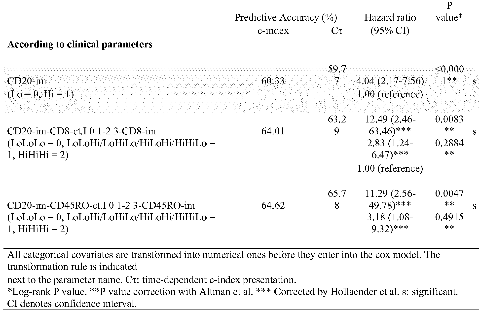
Table 1: scores for CD20 (im) Name Combination Prognosis Score Hi Hi CD20 (im) Good 1 Lo Lo CD20 (im) Poor 0 Table 2: scores for combinations of CD20 (im) and CD8 (ct): Name Combination Prognosis Score HiHi Hi CD20 (im) / Hi CD8 (ct) Good 2 Het [Hi CD20 (im) / Lo CD8 (ct)] or [Lo CD20 (im) / Hi CD8 (ct)] intermediate 1 LoLo Lo CD20 (im) / Lo CD8 (ct) Poor 0 Table 3: scores for combinations of CD20 (im) and CD8 (im): Name Combination Prognosis Score HiHi Hi CD20 (im) / Hi CD8 (im) Good 2 Het [Hi CD20 (im) / Lo CD8 (im)] or [Lo CD20 (im) / Hi CD8 (im)] intermediate 1 LoLo Lo CD20 (im) / Lo CD8 (im) Poor 0 Table 4: scores for combinations of CD20 (im) and CD3 (ct): Name Combination Prognosis Score HiHi Hi CD20 (im) / Hi CD3 (ct) Good 2 Het [Hi CD20 (im) / Lo CD3 (ct)] or [Lo CD20 (im) / Hi CD3 (ct)] intermediate 1 LoLo Lo CD20 (im) / Lo CD3 (ct) Poor 0 Table 5: scores for combinations of CD20 (im) and CD3 (im): Name Combination Prognosis Score HiHi Hi CD20 (im) / Hi CD3 (im) Good 2 Het [Hi CD20 (im) / Lo CD3 (im)] or [Lo CD20 (im) / Hi CD3 (im)] intermediate 1 LoLo Lo CD20 (im) / Lo CD3 (im) Poor 0 Table 6: scores for combinations of CD20 (im) and CD45RO (ct): Name Combination Prognosis Score HiHi Hi CD20 (im) / Hi CD45RO (ct) Good 2 Het [Hi CD20 (im) / Lo CD45RO (ct)] or [Lo CD20 (im) / Hi CD45RO (ct)] intermediate 1 LoLo Lo CD20 (im) / Lo CD45RO (ct) Poor 0 Table 7: scores for combinations of CD20 (im) and CD45RO (im): Name Combination Prognosis Score HiHi Hi CD20 (im) / Hi CD45RO (im) Good 2 Het [Hi CD20 (im) / Lo CD45RO (im)] or [Lo CD20 (im) / Hi CD45RO (im)] intermediate 1 LoLo Lo CD20 (im) / Lo CD45RO (im) Poor 0 Table 8: scores for combinations of CD20 (im), CD8 (ct) and CD8 (im): Name Combination Prognosis Score HiHiHi Hi CD20 (im) / Hi CD8 (ct) /Hi CD8 (im) Good 3 Het [Hi CD20 (im) / Hi CD8 (ct) /Lo CD8 (im)] or [Hi CD20 (im) / Lo CD8 (ct) /Hi CD8 (im)] or [Lo CD20 (im) / Hi CD8 (ct) / Hi CD8 (im)] intermediate 2 Het [Hi CD20 (im) / Lo CD8 (ct) / Lo CD8 (im)] or [Lo CD20 (im) / Hi CD8 (ct) / Lo CD8 (im)] or [Lo CD20 (im) / Lo CD8 (ct) / Hi CD8 (im)] intermediate 1 LoLoLo Lo CD20 (im) / Lo CD8 (ct) /Lo CD8 (im) Poor 0 Table 9: scores for combinations of CD20 (im), CD3 (ct) and CD3 (im): Name Combination Prognosis Score HiHiHi Hi CD20 (im) / Hi CD3 (ct) /Hi CD3 (im) Good 3 Het [Hi CD20 (im) / Hi CD3 (ct) /Lo CD3 (im)] or [Hi CD20 (im) / Lo CD3 (ct) /Hi CD3 (im)] or [Lo CD20 (im) / Hi CD3 (ct) / Hi CD3 (im)] intermediate 2 Het [Hi CD20 (im) / Lo CD3 (ct) / Lo CD3 (im)] or [Lo CD20 (im) / Hi CD3 (ct) / Lo CD3 (im)] or [Lo CD20 (im) / Lo CD3 (ct) / Hi CD3 (im)] intermediate 1 LoLoLo Lo CD20 (im) / Lo CD3 (ct) /Lo CD3 (im) Poor 0 Table 10: scores for combinations of CD20 (im), CD45RO (ct) and CD45RO (im): Name Combination Prognosis Score HiHiHi Hi CD20 (im) / Hi CD45RO (ct) /Hi CD45RO (im) Good 3 Het [Hi CD20 (im) / Hi CD45RO (ct) /Lo CD45RO (im)] or [Hi CD20 (im) / Lo CD45RO (ct) /Hi CD45RO (im)] or [Lo CD20 (im) / Hi CD45RO (ct) / Hi CD45RO (im)] intermediate 2 Het [Hi CD20 (im) / Lo CD45RO (ct) / Lo CD45RO (im)] or [Lo CD20 (im) / Hi CD45RO (ct) / Lo CD45RO (im)] or [Lo CD20 (im) / Lo CD45RO (ct) / Hi CD45RO (im)] intermediate 1 LoLoLo Lo CD20 (im) / Lo CD45RO (ct) /Lo CD45RO (im) Poor 0 
- Throughout this application, various references describe the state of the art to which this invention pertains.
- 1. Finn, O.J. Cancer immunology. N Engl J Med 358, 2704-2715 (2008).
- 2. Galon, J., et al. Type, density, and location of immune cells within human colorectal tumors predict clinical outcome. Science 313, 1960-1964 (2006).
- 3. Galon, J., Fridman, W.H. & Pages, F. The adaptive immunologic microenvironment in colorectal cancer: a novel perspective. Cancer Res 67, 1883-1886 (2007).
- 4. Pages, F., et al. Effector memory T cells, early metastasis, and survival in colorectal cancer. N Engl J Med 353, 2654-2666 (2005).
- 5. Jemal, A., et al. Cancer statistics, 2006. CA Cancer J Clin 56, 106-130 (2006).
- 6. Weitz, J., et al. Colorectal cancer. Lancet 365, 153-165 (2005).
- 7. Bindea, G., Mlecnik, B., Fridman, W.H., Pages, F. & Galon, J. Natural immunity to cancer in humans. Curr Opin Immunol 22, 215-222 (2010).
- 8. Pages, F., et al. In situ cytotoxic and memory T cells predict outcome in patients with early-stage colorectal cancer. J Clin Oncol 27, 5944-5951 (2009).
- 9. Atreya, I. & Neurath, M.F. Immune cells in colorectal cancer: prognostic relevance and therapeutic strategies. Expert ).
- 10. Koebel, C.M., et al. Adaptive immunity maintains occult cancer in an equilibrium state. Nature 450, 903-907 (2007).
- 11. Shankaran, V., et al. IFNgamma and lymphocytes prevent primary tumour development and shape tumour immunogenicity. Nature 410, 1107-1111 (2001).
- 12. Smyth, M.J., Dunn, G.P. & Schreiber, R.D. Cancer immunosurveillance and immunoediting: the roles of immunity in suppressing tumor development and shaping tumor immunogenicity. ).
- 13. June, C.H. Principles of adoptive T cell cancer therapy. J Clin Invest 117, 1204-1212 (2007).
- 14. June, C.H. Adoptive T cell therapy for cancer in the clinic. J Clin Invest 117, 1466-1476 (2007).
Claims (5)
- A method for predicting the survival time of a patient suffering from a solid cancer comprising the steps consisting of :- determining the density of CD20+ B cells at the invasive margin of the tumor (im) in a tumor tissue sample obtained from said patient;- determining the density of one further cell type selected from CD8+ cytotoxic T cells or CD45RO+ memory T cells at the invasive margin of the tumor (im) and at the center of the tumor (ct) in a tumor tissue sample obtained from said patient;- comparing each density with a cut-off value; and- providing:a) a good prognosis when:a1) the density of CD20+ B cells at the invasive margin of the tumor is higher than the cut-off value; anda2) the density of said further cell type at the invasive margin of the tumor (im) is higher than the cut-off value; anda3) the density of said further cell type at the center of the tumor (ct) is higher than the cut-off value;b) an intermediate prognosis when:b1) the density of CD20+ B cells at the invasive margin of the tumor is higher than the cut-off value; andb2) the density of said further cell type at the invasive margin of the tumor (im) is lower than the cut-off value; andb3) the density of said further cell type at the center of the tumor (ct) is higher than the cut-off value;orb'1) the density of CD20+ B cells at the invasive margin of the tumor is higher than the cut-off value; andb'2) the density of said further cell type at the invasive margin of the tumor (im) is higher than the cut-off value; andb'3) the density of said further cell type at the center of the tumor (ct) is lower than the cut-off value;orb" 1) the density of CD20+ B cells at the invasive margin of the tumor is lower than the cut-off value;b"2) the density of said further cell type at the invasive margin of the tumor (im) is higher than the cut-off value; andb"3) the density of said further cell type at the center of the tumor (ct) is higher than the cut-off value;orbi) the density of CD20+ B cells at the invasive margin of the tumor is higher than the cut-off value;bii) the density of said further cell type at the invasive margin of the tumor (im) is lower than the cut-off value; andbiii) the density of said further cell type at the center of the tumor (ct) is lower than the cut-off value;orb'i) the density of CD20+ B cells at the invasive margin of the tumor is lower than the cut-off value;b'ii) the density of said further cell type at the invasive margin of the tumor (im) is lower than the cut-off value; andb'iii) the density of said further cell type at the center of the tumor (ct) is higher than the cut-off value;orb"i) the density of CD20+ B cells at the invasive margin of the tumor is lower than the cut-off value;b"ii) the density of said further cell type at the invasive margin of the tumor (im) is higher than the cut-off value; andb"iii) the density of said further cell type at the center of the tumor (ct) is lower than the cut-off value;c) a poor prognosis when:c1) the density of CD20+ B cells at the invasive margin of the tumor is lower than the cut-off value; andc2) the density of said further cell type at the invasive margin of the tumor (im) is lower than the cut-off value; andc3) the density of said further cell type at the center of the tumor (ct) is lower than the cut-off value.
- The method according to claim 1, wherein said further cell type consists in CD8+ cytotoxic cells.
- The method according to claim 1, wherein said further cell type consists in CD45RO memory T cells.
- The method according to any one of claims 1 to 3, wherein said solid cancer is colorectal cancer.
- A method for assisting in predicting the survival time of a patient suffering from colorectal cancer comprising the steps consisting of i) determining the density of CD20+ B cells at the invasive margin of the tumor (im) in a tumor tissue sample obtained from said patient, ii) comparing said density with a cut-off value and iii) providing a good prognosis when the density of B cells at the invasive margin of the tumor is higher than the cut-off value and a poor prognosis when the density of B cells at the invasive margin of the tumor is lower than the cut-off value.
Priority Applications (1)
| Application Number | Priority Date | Filing Date | Title |
|---|---|---|---|
| EP13701044.3A EP2805165B1 (en) | 2012-01-20 | 2013-01-21 | Methods for predicting the survival time of a patient suffering from a solid cancer based on density of b cells |
Applications Claiming Priority (4)
| Application Number | Priority Date | Filing Date | Title |
|---|---|---|---|
| EP12305072 | 2012-01-20 | ||
| US201261600752P | 2012-02-20 | 2012-02-20 | |
| EP13701044.3A EP2805165B1 (en) | 2012-01-20 | 2013-01-21 | Methods for predicting the survival time of a patient suffering from a solid cancer based on density of b cells |
| PCT/EP2013/051008 WO2013107900A1 (en) | 2012-01-20 | 2013-01-21 | Methods for predicting the survival time of a patient suffering from a solid cancer based on density of b cells |
Publications (2)
| Publication Number | Publication Date |
|---|---|
| EP2805165A1 EP2805165A1 (en) | 2014-11-26 |
| EP2805165B1 true EP2805165B1 (en) | 2018-03-14 |
Family
ID=48798696
Family Applications (1)
| Application Number | Title | Priority Date | Filing Date |
|---|---|---|---|
| EP13701044.3A Active EP2805165B1 (en) | 2012-01-20 | 2013-01-21 | Methods for predicting the survival time of a patient suffering from a solid cancer based on density of b cells |
Country Status (5)
| Country | Link |
|---|---|
| US (1) | US9945861B2 (en) |
| EP (1) | EP2805165B1 (en) |
| JP (1) | JP5970560B2 (en) |
| ES (1) | ES2671423T3 (en) |
| WO (1) | WO2013107900A1 (en) |
Families Citing this family (10)
| Publication number | Priority date | Publication date | Assignee | Title |
|---|---|---|---|---|
| JP6605506B2 (en) | 2014-02-24 | 2019-11-13 | ヴェンタナ メディカル システムズ, インク. | Methods, kits, and systems for scoring immune responses against cancer by simultaneous detection of CD3, CD8, CD20 and FoxP3 |
| US11385231B2 (en) * | 2015-08-27 | 2022-07-12 | Inserm (Institut National De La Sante Et De La Recherche Scientifique) | Methods for predicting the survival time of patients suffering from a lung cancer |
| JP7281903B2 (en) * | 2016-05-09 | 2023-05-26 | アンセルム(アンスティチュート・ナシオナル・ドゥ・ラ・サンテ・エ・ドゥ・ラ・ルシェルシュ・メディカル) | Methods for Classifying Patients with Solid Tumors |
| CN114072676B (en) | 2019-06-03 | 2024-07-12 | 法国国家卫生及研究医学协会 | Methods for adjusting treatment regimen |
| WO2021092071A1 (en) | 2019-11-07 | 2021-05-14 | Oncxerna Therapeutics, Inc. | Classification of tumor microenvironments |
| JP7741831B2 (en) | 2020-06-30 | 2025-09-18 | アンスティチュ ナショナル ドゥ ラ サンテ エ ドゥ ラ ルシェルシュ メディカル | Method for predicting the risk of recurrence and/or death in patients with solid tumors after neoadjuvant therapy and curative surgery - Patent Application 20070122997 |
| EP4172621A1 (en) | 2020-06-30 | 2023-05-03 | INSERM (Institut National de la Santé et de la Recherche Médicale) | Methods for predicting the risk of recurrence and/or death of patients suffering from a solid cancer after preoperative adjuvant therapies |
| CN113834941B (en) * | 2021-11-02 | 2024-01-05 | 复旦大学附属中山医院 | Markers for prognostic diagnosis of colon cancer based on B cell expression and their uses |
| CN114019163B (en) * | 2021-11-02 | 2024-07-23 | 复旦大学附属中山医院 | Markers for prognosis diagnosis of colon cancer based on expression of activated B cells and their uses |
| US20250035635A1 (en) | 2022-03-17 | 2025-01-30 | Institut National De La Sante Et De La Recherche Medicale | Methods for predicting response to an immunotherapeutic treatment in a patient with a cancer |
Family Cites Families (1)
| Publication number | Priority date | Publication date | Assignee | Title |
|---|---|---|---|---|
| EP1777523A1 (en) * | 2005-10-19 | 2007-04-25 | INSERM (Institut National de la Santé et de la Recherche Médicale) | An in vitro method for the prognosis of progression of a cancer and of the outcome in a patient and means for performing said method |
-
2013
- 2013-01-21 JP JP2014552648A patent/JP5970560B2/en not_active Expired - Fee Related
- 2013-01-21 US US14/372,757 patent/US9945861B2/en not_active Expired - Fee Related
- 2013-01-21 WO PCT/EP2013/051008 patent/WO2013107900A1/en not_active Ceased
- 2013-01-21 EP EP13701044.3A patent/EP2805165B1/en active Active
- 2013-01-21 ES ES13701044.3T patent/ES2671423T3/en active Active
Also Published As
| Publication number | Publication date |
|---|---|
| EP2805165A1 (en) | 2014-11-26 |
| JP5970560B2 (en) | 2016-08-17 |
| JP2015505607A (en) | 2015-02-23 |
| WO2013107900A1 (en) | 2013-07-25 |
| US20140363472A1 (en) | 2014-12-11 |
| US9945861B2 (en) | 2018-04-17 |
| ES2671423T3 (en) | 2018-06-06 |
Similar Documents
| Publication | Publication Date | Title |
|---|---|---|
| US9945861B2 (en) | Methods for predicting the survival time of a patient suffering from a solid cancer based on density of B cells | |
| Goodman Jr et al. | Circulating tumor cells as a predictive biomarker in patients with hormone-sensitive prostate cancer | |
| Wulfing et al. | HER2-positive circulating tumor cells indicate poor clinical outcome in stage I to III breast cancer patients | |
| Munzone et al. | Prognostic value of circulating tumor cells according to immunohistochemically defined molecular subtypes in advanced breast cancer | |
| US20200041519A1 (en) | Method for the prognosis of survival time of a patient suffering from a solid cancer | |
| Zech et al. | Prognostic and diagnostic relevance of hnRNP A2/B1, hnRNP B1 and S100 A2 in non-small cell lung cancer | |
| KR102561377B1 (en) | Methods and related uses for identifying individuals to be treated with chemotherapy based on marker molecules | |
| US20110166030A1 (en) | Prediction of response to docetaxel therapy based on the presence of TMPRSSG2:ERG fusion in circulating tumor cells | |
| Kim et al. | Concordance of programmed death-ligand 1 expression between primary and metastatic non-small cell lung cancer by immunohistochemistry and RNA in situ hybridization | |
| EP2135084A1 (en) | Autoimmune regulation of prostate cancer by annexin a3 | |
| US20180055882A1 (en) | Functional immunophenotyping of leukocytes in human tissues | |
| Ma et al. | The prognostic value of adhesion molecule CD44v6 in women with primary breast carcinoma: a clinicopathologic study | |
| Wang et al. | A novel in vitro prognostic model of bladder cancer based on urine-derived living tumor cells | |
| KR102599695B1 (en) | Method for detection of lung adenocarcinoma recurrence based on marker human epididymal protein 4 (HE4) and associated uses | |
| Dawood et al. | Integrating circulating tumor cell assays into the management of breast cancer | |
| Ji et al. | Immunoproteomics reveal different characteristics for the prognostic markers of Intratumoral-Infiltrating CD3+ T lymphocytes and immunoscore in colorectal cancer | |
| CA3221148A1 (en) | Systems and methods for integrated analysis of blood and surgical drain fluid biomarkers | |
| US9645152B2 (en) | Methods for predicting the survival time of a patient suffering from a solid cancer | |
| US20080299590A1 (en) | SHC protein-related methods and compositions | |
| TWI924312B (en) | Method of isolating and analyzing circulating tumor cells | |
| Burgers et al. | Clinicopathological prognostic factors and scoring systems in malignant pleural mesothelioma | |
| Nariculam et al. | MCM-2 expression in clinically localised prostate cancer | |
| Boxler et al. | Prediction of outcome in prostate cancer patients after radical prostatectomy by matrixmetalloproteinase-9 (MMP-9) expression | |
| Jansen et al. | Prostate cancer-derived exosomes as novel biomarker treasure chests | |
| Muneer et al. | Analysis of minichromosome maintenance proteins 2 and 5 in urine sediment, for the detection of prostate cancer |
Legal Events
| Date | Code | Title | Description |
|---|---|---|---|
| PUAI | Public reference made under article 153(3) epc to a published international application that has entered the european phase |
Free format text: ORIGINAL CODE: 0009012 |
|
| 17P | Request for examination filed |
Effective date: 20140708 |
|
| AK | Designated contracting states |
Kind code of ref document: A1 Designated state(s): AL AT BE BG CH CY CZ DE DK EE ES FI FR GB GR HR HU IE IS IT LI LT LU LV MC MK MT NL NO PL PT RO RS SE SI SK SM TR |
|
| DAX | Request for extension of the european patent (deleted) | ||
| RAP1 | Party data changed (applicant data changed or rights of an application transferred) |
Owner name: INSERM - INSTITUT NATIONAL DE LA SANTE ET DE LA RE Owner name: UNIVERSITE PARIS DESCARTES Owner name: ASSISTANCE PUBLIQUE-HOPITAUX DE PARIS (APHP) |
|
| 17Q | First examination report despatched |
Effective date: 20160316 |
|
| STAA | Information on the status of an ep patent application or granted ep patent |
Free format text: STATUS: EXAMINATION IS IN PROGRESS |
|
| GRAP | Despatch of communication of intention to grant a patent |
Free format text: ORIGINAL CODE: EPIDOSNIGR1 |
|
| STAA | Information on the status of an ep patent application or granted ep patent |
Free format text: STATUS: GRANT OF PATENT IS INTENDED |
|
| INTG | Intention to grant announced |
Effective date: 20171004 |
|
| RAP1 | Party data changed (applicant data changed or rights of an application transferred) |
Owner name: UNIVERSITE PARIS DESCARTES Owner name: ASSISTANCE PUBLIQUE-HOPITAUX DE PARIS (APHP) Owner name: INSERM - INSTITUT NATIONAL DE LA SANTE ET DE LA RE |
|
| RIN1 | Information on inventor provided before grant (corrected) |
Inventor name: GALON, JEROME Inventor name: FRIDMAN, HERVE Inventor name: MLECNIK, BERNHARD Inventor name: PAGES, FRANCK Inventor name: BINDEA, GABRIELA |
|
| GRAS | Grant fee paid |
Free format text: ORIGINAL CODE: EPIDOSNIGR3 |
|
| GRAA | (expected) grant |
Free format text: ORIGINAL CODE: 0009210 |
|
| STAA | Information on the status of an ep patent application or granted ep patent |
Free format text: STATUS: THE PATENT HAS BEEN GRANTED |
|
| AK | Designated contracting states |
Kind code of ref document: B1 Designated state(s): AL AT BE BG CH CY CZ DE DK EE ES FI FR GB GR HR HU IE IS IT LI LT LU LV MC MK MT NL NO PL PT RO RS SE SI SK SM TR |
|
| REG | Reference to a national code |
Ref country code: GB Ref legal event code: FG4D |
|
| REG | Reference to a national code |
Ref country code: AT Ref legal event code: REF Ref document number: 979400 Country of ref document: AT Kind code of ref document: T Effective date: 20180315 Ref country code: CH Ref legal event code: EP |
|
| REG | Reference to a national code |
Ref country code: IE Ref legal event code: FG4D |
|
| REG | Reference to a national code |
Ref country code: DE Ref legal event code: R096 Ref document number: 602013034368 Country of ref document: DE |
|
| REG | Reference to a national code |
Ref country code: ES Ref legal event code: FG2A Ref document number: 2671423 Country of ref document: ES Kind code of ref document: T3 Effective date: 20180606 |
|
| REG | Reference to a national code |
Ref country code: NL Ref legal event code: FP |
|
| REG | Reference to a national code |
Ref country code: SE Ref legal event code: TRGR |
|
| REG | Reference to a national code |
Ref country code: LT Ref legal event code: MG4D |
|
| PG25 | Lapsed in a contracting state [announced via postgrant information from national office to epo] |
Ref country code: FI Free format text: LAPSE BECAUSE OF FAILURE TO SUBMIT A TRANSLATION OF THE DESCRIPTION OR TO PAY THE FEE WITHIN THE PRESCRIBED TIME-LIMIT Effective date: 20180314 Ref country code: LT Free format text: LAPSE BECAUSE OF FAILURE TO SUBMIT A TRANSLATION OF THE DESCRIPTION OR TO PAY THE FEE WITHIN THE PRESCRIBED TIME-LIMIT Effective date: 20180314 Ref country code: HR Free format text: LAPSE BECAUSE OF FAILURE TO SUBMIT A TRANSLATION OF THE DESCRIPTION OR TO PAY THE FEE WITHIN THE PRESCRIBED TIME-LIMIT Effective date: 20180314 Ref country code: CY Free format text: LAPSE BECAUSE OF FAILURE TO SUBMIT A TRANSLATION OF THE DESCRIPTION OR TO PAY THE FEE WITHIN THE PRESCRIBED TIME-LIMIT Effective date: 20180314 Ref country code: NO Free format text: LAPSE BECAUSE OF FAILURE TO SUBMIT A TRANSLATION OF THE DESCRIPTION OR TO PAY THE FEE WITHIN THE PRESCRIBED TIME-LIMIT Effective date: 20180614 |
|
| REG | Reference to a national code |
Ref country code: AT Ref legal event code: MK05 Ref document number: 979400 Country of ref document: AT Kind code of ref document: T Effective date: 20180314 |
|
| PG25 | Lapsed in a contracting state [announced via postgrant information from national office to epo] |
Ref country code: BG Free format text: LAPSE BECAUSE OF FAILURE TO SUBMIT A TRANSLATION OF THE DESCRIPTION OR TO PAY THE FEE WITHIN THE PRESCRIBED TIME-LIMIT Effective date: 20180614 Ref country code: RS Free format text: LAPSE BECAUSE OF FAILURE TO SUBMIT A TRANSLATION OF THE DESCRIPTION OR TO PAY THE FEE WITHIN THE PRESCRIBED TIME-LIMIT Effective date: 20180314 Ref country code: LV Free format text: LAPSE BECAUSE OF FAILURE TO SUBMIT A TRANSLATION OF THE DESCRIPTION OR TO PAY THE FEE WITHIN THE PRESCRIBED TIME-LIMIT Effective date: 20180314 Ref country code: GR Free format text: LAPSE BECAUSE OF FAILURE TO SUBMIT A TRANSLATION OF THE DESCRIPTION OR TO PAY THE FEE WITHIN THE PRESCRIBED TIME-LIMIT Effective date: 20180615 |
|
| PG25 | Lapsed in a contracting state [announced via postgrant information from national office to epo] |
Ref country code: EE Free format text: LAPSE BECAUSE OF FAILURE TO SUBMIT A TRANSLATION OF THE DESCRIPTION OR TO PAY THE FEE WITHIN THE PRESCRIBED TIME-LIMIT Effective date: 20180314 Ref country code: RO Free format text: LAPSE BECAUSE OF FAILURE TO SUBMIT A TRANSLATION OF THE DESCRIPTION OR TO PAY THE FEE WITHIN THE PRESCRIBED TIME-LIMIT Effective date: 20180314 Ref country code: PL Free format text: LAPSE BECAUSE OF FAILURE TO SUBMIT A TRANSLATION OF THE DESCRIPTION OR TO PAY THE FEE WITHIN THE PRESCRIBED TIME-LIMIT Effective date: 20180314 Ref country code: AL Free format text: LAPSE BECAUSE OF FAILURE TO SUBMIT A TRANSLATION OF THE DESCRIPTION OR TO PAY THE FEE WITHIN THE PRESCRIBED TIME-LIMIT Effective date: 20180314 |
|
| PG25 | Lapsed in a contracting state [announced via postgrant information from national office to epo] |
Ref country code: SK Free format text: LAPSE BECAUSE OF FAILURE TO SUBMIT A TRANSLATION OF THE DESCRIPTION OR TO PAY THE FEE WITHIN THE PRESCRIBED TIME-LIMIT Effective date: 20180314 Ref country code: AT Free format text: LAPSE BECAUSE OF FAILURE TO SUBMIT A TRANSLATION OF THE DESCRIPTION OR TO PAY THE FEE WITHIN THE PRESCRIBED TIME-LIMIT Effective date: 20180314 Ref country code: SM Free format text: LAPSE BECAUSE OF FAILURE TO SUBMIT A TRANSLATION OF THE DESCRIPTION OR TO PAY THE FEE WITHIN THE PRESCRIBED TIME-LIMIT Effective date: 20180314 Ref country code: CZ Free format text: LAPSE BECAUSE OF FAILURE TO SUBMIT A TRANSLATION OF THE DESCRIPTION OR TO PAY THE FEE WITHIN THE PRESCRIBED TIME-LIMIT Effective date: 20180314 |
|
| REG | Reference to a national code |
Ref country code: DE Ref legal event code: R097 Ref document number: 602013034368 Country of ref document: DE |
|
| PG25 | Lapsed in a contracting state [announced via postgrant information from national office to epo] |
Ref country code: PT Free format text: LAPSE BECAUSE OF FAILURE TO SUBMIT A TRANSLATION OF THE DESCRIPTION OR TO PAY THE FEE WITHIN THE PRESCRIBED TIME-LIMIT Effective date: 20180716 |
|
| PLBE | No opposition filed within time limit |
Free format text: ORIGINAL CODE: 0009261 |
|
| STAA | Information on the status of an ep patent application or granted ep patent |
Free format text: STATUS: NO OPPOSITION FILED WITHIN TIME LIMIT |
|
| PG25 | Lapsed in a contracting state [announced via postgrant information from national office to epo] |
Ref country code: DK Free format text: LAPSE BECAUSE OF FAILURE TO SUBMIT A TRANSLATION OF THE DESCRIPTION OR TO PAY THE FEE WITHIN THE PRESCRIBED TIME-LIMIT Effective date: 20180314 |
|
| 26N | No opposition filed |
Effective date: 20181217 |
|
| PG25 | Lapsed in a contracting state [announced via postgrant information from national office to epo] |
Ref country code: SI Free format text: LAPSE BECAUSE OF FAILURE TO SUBMIT A TRANSLATION OF THE DESCRIPTION OR TO PAY THE FEE WITHIN THE PRESCRIBED TIME-LIMIT Effective date: 20180314 |
|
| PG25 | Lapsed in a contracting state [announced via postgrant information from national office to epo] |
Ref country code: MC Free format text: LAPSE BECAUSE OF FAILURE TO SUBMIT A TRANSLATION OF THE DESCRIPTION OR TO PAY THE FEE WITHIN THE PRESCRIBED TIME-LIMIT Effective date: 20180314 |
|
| PG25 | Lapsed in a contracting state [announced via postgrant information from national office to epo] |
Ref country code: TR Free format text: LAPSE BECAUSE OF FAILURE TO SUBMIT A TRANSLATION OF THE DESCRIPTION OR TO PAY THE FEE WITHIN THE PRESCRIBED TIME-LIMIT Effective date: 20180314 |
|
| PG25 | Lapsed in a contracting state [announced via postgrant information from national office to epo] |
Ref country code: MT Free format text: LAPSE BECAUSE OF NON-PAYMENT OF DUE FEES Effective date: 20190121 |
|
| PG25 | Lapsed in a contracting state [announced via postgrant information from national office to epo] |
Ref country code: IS Free format text: LAPSE BECAUSE OF FAILURE TO SUBMIT A TRANSLATION OF THE DESCRIPTION OR TO PAY THE FEE WITHIN THE PRESCRIBED TIME-LIMIT Effective date: 20180714 |
|
| PG25 | Lapsed in a contracting state [announced via postgrant information from national office to epo] |
Ref country code: HU Free format text: LAPSE BECAUSE OF FAILURE TO SUBMIT A TRANSLATION OF THE DESCRIPTION OR TO PAY THE FEE WITHIN THE PRESCRIBED TIME-LIMIT; INVALID AB INITIO Effective date: 20130121 |
|
| REG | Reference to a national code |
Ref country code: LU Ref legal event code: PD Owner name: UNIVERSITE DE PARIS; FR Free format text: FORMER OWNER: UNIVERSITE PARIS DESCARTES Effective date: 20210813 |
|
| REG | Reference to a national code |
Ref country code: CH Ref legal event code: PK Free format text: BERICHTIGUNGEN |
|
| REG | Reference to a national code |
Ref country code: GB Ref legal event code: 732E Free format text: REGISTERED BETWEEN 20210930 AND 20211006 |
|
| REG | Reference to a national code |
Ref country code: BE Ref legal event code: PD Owner name: ASSISTANCE PUBLIQUE-HOPITAUX DE PARIS (APHP); FR Free format text: DETAILS ASSIGNMENT: CHANGE OF OWNER(S), MERGE; FORMER OWNER NAME: VAN REET JOSEPH Effective date: 20211013 |
|
| PGFP | Annual fee paid to national office [announced via postgrant information from national office to epo] |
Ref country code: IE Payment date: 20211217 Year of fee payment: 10 Ref country code: LU Payment date: 20211216 Year of fee payment: 10 |
|
| REG | Reference to a national code |
Ref country code: NL Ref legal event code: PD Owner name: UNIVERSITE DE PARIS; FR Free format text: DETAILS ASSIGNMENT: CHANGE OF OWNER(S), MERGE; FORMER OWNER NAME: UNIVERSITE PARIS DESCARTES Effective date: 20220110 |
|
| REG | Reference to a national code |
Ref country code: DE Ref legal event code: R081 Ref document number: 602013034368 Country of ref document: DE Owner name: UNIVERSITE PARIS CITE, FR Free format text: FORMER OWNERS: ASSISTANCE PUBLIQUE-HOPITAUX DE PARIS (APHP), PARIS, FR; INSERM - INSTITUT NATIONAL DE LA SANTE ET DE LA RECHERCHE MEDICALE, PARIS, FR; UNIVERSITE PARIS DESCARTES, PARIS, FR Ref country code: DE Ref legal event code: R081 Ref document number: 602013034368 Country of ref document: DE Owner name: UNIVERSITE DE PARIS, FR Free format text: FORMER OWNERS: ASSISTANCE PUBLIQUE-HOPITAUX DE PARIS (APHP), PARIS, FR; INSERM - INSTITUT NATIONAL DE LA SANTE ET DE LA RECHERCHE MEDICALE, PARIS, FR; UNIVERSITE PARIS DESCARTES, PARIS, FR Ref country code: DE Ref legal event code: R081 Ref document number: 602013034368 Country of ref document: DE Owner name: INSERM - INSTITUT NATIONAL DE LA SANTE ET DE L, FR Free format text: FORMER OWNERS: ASSISTANCE PUBLIQUE-HOPITAUX DE PARIS (APHP), PARIS, FR; INSERM - INSTITUT NATIONAL DE LA SANTE ET DE LA RECHERCHE MEDICALE, PARIS, FR; UNIVERSITE PARIS DESCARTES, PARIS, FR Ref country code: DE Ref legal event code: R081 Ref document number: 602013034368 Country of ref document: DE Owner name: ASSISTANCE PUBLIQUE-HOPITAUX DE PARIS (APHP), FR Free format text: FORMER OWNERS: ASSISTANCE PUBLIQUE-HOPITAUX DE PARIS (APHP), PARIS, FR; INSERM - INSTITUT NATIONAL DE LA SANTE ET DE LA RECHERCHE MEDICALE, PARIS, FR; UNIVERSITE PARIS DESCARTES, PARIS, FR |
|
| PG25 | Lapsed in a contracting state [announced via postgrant information from national office to epo] |
Ref country code: MK Free format text: LAPSE BECAUSE OF FAILURE TO SUBMIT A TRANSLATION OF THE DESCRIPTION OR TO PAY THE FEE WITHIN THE PRESCRIBED TIME-LIMIT Effective date: 20180314 |
|
| REG | Reference to a national code |
Ref country code: DE Ref legal event code: R081 Ref document number: 602013034368 Country of ref document: DE Owner name: UNIVERSITE PARIS CITE, FR Free format text: FORMER OWNERS: ASSISTANCE PUBLIQUE-HOPITAUX DE PARIS (APHP), PARIS, FR; INSERM - INSTITUT NATIONAL DE LA SANTE ET DE LA RECHERCHE MEDICALE, PARIS, FR; UNIVERSITE DE PARIS, PARIS, FR Ref country code: DE Ref legal event code: R081 Ref document number: 602013034368 Country of ref document: DE Owner name: INSERM - INSTITUT NATIONAL DE LA SANTE ET DE L, FR Free format text: FORMER OWNERS: ASSISTANCE PUBLIQUE-HOPITAUX DE PARIS (APHP), PARIS, FR; INSERM - INSTITUT NATIONAL DE LA SANTE ET DE LA RECHERCHE MEDICALE, PARIS, FR; UNIVERSITE DE PARIS, PARIS, FR Ref country code: DE Ref legal event code: R081 Ref document number: 602013034368 Country of ref document: DE Owner name: ASSISTANCE PUBLIQUE-HOPITAUX DE PARIS (APHP), FR Free format text: FORMER OWNERS: ASSISTANCE PUBLIQUE-HOPITAUX DE PARIS (APHP), PARIS, FR; INSERM - INSTITUT NATIONAL DE LA SANTE ET DE LA RECHERCHE MEDICALE, PARIS, FR; UNIVERSITE DE PARIS, PARIS, FR |
|
| REG | Reference to a national code |
Ref country code: LU Ref legal event code: HC Owner name: ASSISTANCE PUBLIQUE - HOPITAUX DE PARIS; FR Free format text: FORMER OWNER: UNIVERSITE DE PARIS Effective date: 20220720 |
|
| REG | Reference to a national code |
Ref country code: NL Ref legal event code: HC Owner name: UNIVERSITE PARIS CITE; FR Free format text: DETAILS ASSIGNMENT: CHANGE OF OWNER(S), CHANGE OF OWNER(S) NAME; FORMER OWNER NAME: UNIVERSITE DE PARIS Effective date: 20220818 |
|
| REG | Reference to a national code |
Ref country code: BE Ref legal event code: HC Owner name: ASSISTANCE PUBLIQUE-HOPITAUX DE PARIS (APHP); FR Free format text: DETAILS ASSIGNMENT: CHANGE OF OWNER(S), CHANGE OF OWNER(S) NAME; FORMER OWNER NAME: UNIVERSITE DE PARIS Effective date: 20220812 |
|
| REG | Reference to a national code |
Ref country code: ES Ref legal event code: PC2A Owner name: UNIVERSITE DE PARIS Effective date: 20221216 |
|
| REG | Reference to a national code |
Ref country code: ES Ref legal event code: PC2A Owner name: UNIVERSITE PARIS CITE Effective date: 20230201 |
|
| PG25 | Lapsed in a contracting state [announced via postgrant information from national office to epo] |
Ref country code: LU Free format text: LAPSE BECAUSE OF NON-PAYMENT OF DUE FEES Effective date: 20230121 |
|
| PG25 | Lapsed in a contracting state [announced via postgrant information from national office to epo] |
Ref country code: IE Free format text: LAPSE BECAUSE OF NON-PAYMENT OF DUE FEES Effective date: 20230121 |
|
| PGFP | Annual fee paid to national office [announced via postgrant information from national office to epo] |
Ref country code: BE Payment date: 20241218 Year of fee payment: 13 Ref country code: NL Payment date: 20241219 Year of fee payment: 13 |
|
| PGFP | Annual fee paid to national office [announced via postgrant information from national office to epo] |
Ref country code: GB Payment date: 20241219 Year of fee payment: 13 |
|
| PGFP | Annual fee paid to national office [announced via postgrant information from national office to epo] |
Ref country code: FR Payment date: 20241219 Year of fee payment: 13 |
|
| PGFP | Annual fee paid to national office [announced via postgrant information from national office to epo] |
Ref country code: SE Payment date: 20241219 Year of fee payment: 13 |
|
| PGFP | Annual fee paid to national office [announced via postgrant information from national office to epo] |
Ref country code: DE Payment date: 20241218 Year of fee payment: 13 |
|
| PGFP | Annual fee paid to national office [announced via postgrant information from national office to epo] |
Ref country code: ES Payment date: 20250203 Year of fee payment: 13 |
|
| PGFP | Annual fee paid to national office [announced via postgrant information from national office to epo] |
Ref country code: CH Payment date: 20250201 Year of fee payment: 13 |
|
| PGFP | Annual fee paid to national office [announced via postgrant information from national office to epo] |
Ref country code: IT Payment date: 20250107 Year of fee payment: 13 |
|
| REG | Reference to a national code |
Ref country code: DE Ref legal event code: R079 Ref document number: 602013034368 Country of ref document: DE Free format text: PREVIOUS MAIN CLASS: G01N0033574000 Ipc: G01N0033575000 |