FIELD OF THE DISCLOSURE
-
The technology provided herein generally relates to novel fusion proteins suitable as human and/or animal vaccines against parasites or pathogens of the phylum Apicomplexa. In particular, the present disclosure relates to novel fusion proteins as a basis for vaccines against Plasmodium parasites, including P. falciparum, P. vivax, P. malariae, P. ovale and P. knowlesi. Nucleic acid molecules encoding said recombinant fusion proteins, vectors, host cells containing the nucleic acids and methods for preparation and producing such fusion proteins; antibodies induced or generated by the use of said fusion proteins or said nucleic acid molecules encoding said fusion proteins and the use of such antibodies or recombinant derivatives for passive immunotherapy; methods for producing such fusion proteins; compositions and methods for using such fusion proteins for the prevention and treatment of malaria are also encompassed by the present disclosure.
BACKGROUND
-
The Apicomplexa are an eukaryotic protozoan phylum of around 5000 species including parasites which belong to the most successful and devastating pathogens today, infecting a wide range of animals from mollusks to mammals. Many species of Apicomplexa cause diseases of medical and veterinary importance and represent a significant economic burden and global healthcare challenge. Members of the phylum include:
- Plasmodium, the etiological agent of malaria, afflicting 10-40% of world population and accounting for one-in-five deaths among children under the age of five in Africa
- Toxoplasma gondii, the causative agent of toxoplasmosis. From one-third to half of the world's human population is estimated to carry a Toxoplasma infection. It is a major pathogen to humans with a weakened immune system, such as AIDS patients or pregnant women
- Cryptosporidium, a waterborne pathogen which typically does not cause serious illness in healthy people, but is a big health problem for immuno-compromised people, and
- the agricultural parasites Eimeria (infects poultry and causes annual losses in revenue totaling nearly a billion dollars), Neospora (an important pathogen in cattle and dogs), Babesia (thought to be the second most common blood parasites of mammals with a major health impact on domestic animals) and Theileria (causitve agent of theileriosis a disease of cattle, sheep and goats).
-
The apicomplexan life cycle is complex and can be divided into three broad stages wherein the first two serve for the asexual replication of the pathogen (more precise of the invasive stages of these protists called sporozoites and merozoites) and the third stage defines the sexual reproduction of the parasite. While the general life cycle is common to the Apicomplexa phylum, there are striking differences between species.
-
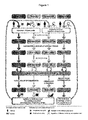
Figure 1 shows the apicomplexan life cycles. As mentioned above, the members of Apicomplexa share a generalized life cycle, even though each species has its own specializations. Plasmodium spp. and Theileria spp. are transmitted and undergo sexual recombination in an insect vector, the Anopheles mosquito and Rhipicephalus tick, respectively. Cryptosporidium is able to autoinfect its host; the oocyst can sporulate and excyst in the same host, maintaining the infection for months to years. The Coccidian parasites are represented in this figure by Toxoplasma, which is able to infect the majority of warm-blooded animals. The differentiation of Toxoplasma tachyzoites into gametocytes is triggered only when members of the cat family (Felidae) are infected (Wasmuth et al., 2009).
-
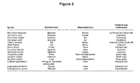
Some Apicomplexa require a single host (e.g. Cryptosporidium), whereas others are more complex, requiring sexual reproduction in the vector species for transmission (e.g. Theileria and Plasmodium; see Figure 2).
-
Although members of the Apicomplexa infect different host and cell types, they have a similar number of defining organelles involved in host cell attachment, invasion, and the establishment of an intracellular parasitophorous vacuole within the host cell. The arsenal of organelles varies between species, but typically includes rhoptries, micronemes, and dense granules. To develop novel antiparasitic compounds and increase the understanding of apicomplexan biology, several large-scale-sequencing projects were initiated and the availability of genomic data sets for 15 species opened the way for the identification of conserved protein families and their functions within the phylum and in the above mentioned processes. Domain analysis also identified both the taxonomic distribution of apicomplexan domains as well as domain architectures specific to the Apicomplexa.
-
Malaria is a disease caused by infection with parasites of the phylum Apicomplexa protozoan, namely parasites of the genus Plasmodium, globally causing more than 200 million new infections and 700 thousand deaths every year. Malaria is especially a serious problem in Africa, where one in every five (20%) childhood deaths is due to the effects of the disease. An African child has on average between 1.6 and 5.4 episodes of malaria fever each year.
-
Malarial diseases in humans are caused by five species of the Plasmodium parasite: P. falciparum, P. vivax, P. ovale, P. malariae and P. knowlesi, wherein the most prevalent being Plasmodium falciparum and Plasmodium vivax. Malaria caused by Plasmodium falciparum (also called malignantor malaria, falciparum malaria or malaria tropica) is the most dangerous form of malaria, with the highest rates of complications and mortality. Almost all malarial deaths are caused by P. falciparum.
-
Briefly, the plasmodial life cycle in man starts with the inoculation of a few sporozoites through the bite of an Anopheles mosquito. Within minutes, sporozoites invade the hepatocyte and start their development, multiplying by schizogony (liver stage or pre-erythrocytic stage). In the case of P. vivax and P. ovale, some sporozoites may differentiate into hypnozoites, responsible for late relapses of the infection. After a period of 5-14 days - depending on the plasmodial species - schizonts develop into thousands of merozoites that are freed into the bloodstream and invade the red blood cells (RBCs), initiating the asexual blood stage. In the RBC, each merozoite develops into a trophozoite that matures and divides, generating a schizont that, after fully matured, gives rise to up to 32 merozoites within 42-72 h, depending on the plasmodial species. The merozoites, released into the bloodstream, will invade other RBC, maintaining the cycle. Some merozoites, after invading a RBC, develop into sexual forms - the male or female gametocytes which also enter the bloodstream after maturation and erythrocyte rupture. When a female Anopheles mosquito takes its blood meal and ingests the gametocytes, it will become infected (sexual stage of the Plasmodium life cycle). In the mosquito gut, the male gametocyte fuses with the female gametocyte, forming the ookinete, which binds to and passes through the gut wall, remains attached to its external face and transforms into the oocyst. The oocyst will divide by sporogony, giving rise to thousands of sporozoites that are released in the body cavity of the mosquito and eventually migrate to its salivary gland, where they will maturate, becoming capable of starting a new infection in humans when the mosquito bites the host for a blood meal.
-
Resistance of Plasmodium falciparum to the existing anti-malarial drug chloroquine emerged in the sixties and has been spreading since then. In addition, the malaria parasite has developed resistance to most other anti-malarial drugs over the past decades. This poses a major threat to public health in tropical countries and to travellers. There is every reason to believe that the prevalence and degree of anti-malarial drug resistance will continue to increase. The growing number of insecticide resistant vectors and drug resistant parasites further increases the demand for an effective malaria vaccine. Malaria vaccines are not limited to a single mode of action and hold the potential to dramatically alleviate the burden of malaria.
-
Some of the difficulties to develop a malaria vaccine result from the multi-stage life cycle of the parasite and its host as mentioned above. Each stage of the parasite development is characterized by different sets of surface antigens, eliciting different types of immune responses. Despite the large variety of displayed surface antigens, the immune response against them is often ineffective. One of the reasons is the extensive sequence polymorphism of plasmodial antigens, which facilitates the immune evasion of the different isolates.
-
Some of the most prominent blood-stage vaccine candidates MSP1, MSP2, AMA1, and RESA have primarily been selected for clinical testing because of their ability to induce growth-inhibitory antibodies in pre-clinical animal models. However, despite these promising initial data, they have in general proved poorly immunogenic in human volunteers and the induced antibodies were predominantly unable to inhibit the in vitro growth of P. falciparum.
-
A pre-erythrocytic vaccine would protect against the infectious form (sporozoite) injected by a mosquito and/or inhibit parasite development in the liver. In a previously unexposed individual, if a few parasites were to escape the immune defences induced by a pre-erythrocytic vaccine, they would eventually enter the blood-stage, multiply within the erythrocytes and establish a full-blown disease.
-
An erythrocytic or asexual blood-stage vaccine would inhibit the invasion and multiplication of the parasite in the red blood cells, thus preventing (or diminishing) severe disease symptoms during the blood infection. However, it would not prevent the transmission of the parasite.
-
A sexual-stage vaccine would not protect the person being vaccinated, but instead interrupt the cycle of transmission by inhibiting the development of parasites once they are ingested by the mosquito along with antibodies produced in response to the vaccine. Transmission-blocking vaccines could be part of a multi-faceted strategy directed towards parasite elimination and reduction of overall resistance to anti pre-erythrocytic or erythrocytic treatment.
-
The before mentioned multi-stage life cycle of malaria parasites presents unique challenges for a synergistic vaccine approach. Immunity against malaria parasites is stage dependent and species dependent. Many malaria researchers and textbook descriptions believe and conclude that a single-antigen vaccine representing only one stage of the life cycle will not be sufficient and that a multi-antigen, multi-stage vaccine that targets different stages of parasite development is necessary to induce effective immunity (Mahajan, Berzofsky et al. 2010). The construction of a multi-antigen vaccine (with the aim of increasing the breadth of the vaccine-induced immune responses to try to circumvent potential P. falciparum escape mutants) can be achieved by either genetically linking (full-size) antigens together, by a mixture of recombinant proteins or by synthetic-peptide-based (15-25-mer), chemically synthesized vaccines containing several peptides derived from different parasite proteins and stages.
-
A poly-protein approach being comprised of several different antigens or several different alleles of a single antigen (to induce antibodies with synergistic activities against the parasite) is hindered by antigenic diversity and the capacity of P. falciparum for immune evasion (Richards,Beeson, 2009). A large number of antigens have been evaluated as potential vaccine candidates, but most clinical trials have not shown significant impact on preventing clinical malaria although some of them have shown to reduce parasite growth. The size of the resulting fusion protein/ vaccine candidate is another limiting factor allowing only the combination of a few selected antigens, not excluding that the chosen antigens are not targets of natural immunity and/or exhibit significant genetic polymorphism. Highly variable antigens with multiple alleles are obviously targets of the immune response under natural challenge, and vaccine studies of AMA1 and MSP2 suggest that allele-specific effects can be achieved (Schwartz, 2012). Practical considerations argue against multi-stage vaccines, particularly the associated increased manufacturing cost of a multicomponent vaccine including several antigens unless these can be encompassed by a single production step and single delivery technology (Hill, 2011). Currently only combination vaccines (being comprised of CSP und AMA1) are undergoing clinical trials that target the pre-erythrocytic and asexual blood stage of P. falciparum (Schwartz, 2012). A multi-antigen vaccine candidate targeting all three life cycle stages of Plasmodium (including the sexual stage in Anopheles mosquitos and thus blocking parasite transmission) is still not tested in clinical trials.
-
The so-called SPf66 vaccine was the pioneer multi-epitope, multi-stage peptide-based malaria vaccine. It was first formulated and tested in Colombia (Patarroyo, 1988) and later also manufactured in the USA. SPf66 consists of epitopes of merozoite surface protein 1 (MSP1) linked by a peptide derived from the NANP repeat sequence of the circumsporozoite protein (CSP) adjuvanted with alum, and more recently tested with QS-21 (Schwartz, 2012). Since then, a number of synthetic peptide vaccines have been produced for both murine (P. berghei and P. yoelii) and human (P. falciparum and P. vivax) malarias and tested for immunogenicity or immunogenicity and efficacy. However, in spite of the early momentum, several theoretical considerations and technological hurdles have slowed the progress of this vaccine development approach. A major disadvantage of the peptide-based vaccine approach lies in its limitation to short linear epitopes that lack the surrounding sequence context often required for three-dimensional protein structures (e.g. folded domains) and complex conformational epitopes.
-
Therefore the availability of novel and improved multi-stage vaccines against parasites of the phylum Apicomplexa would be highly advantageous.
SUMMARY OF THE DISCLOSURE
-
The present disclosure relates to novel fusion proteins, in particular recombinant fusion proteins suitable as human and/or animal vaccines against a parasite of the phylum Apicomplexa, in particular against Plasmodium falciparum comprising a plurality of isolated heat stable fragments derived from at least two different Apicomplexa surface proteins presented on the surface of the parasite in at least two different stages in the life cycle of the parasite, wherein each fragment contains at least one folded domain.
-
In one aspect, embodiments of the disclosure provide methods of producing and/or purifying recombinant fusion proteins according to the present disclosure comprising the steps of:
- a) providing a nucleic acid construct comprising a nucleic acid encoding a fusion protein according to the present disclosure,
- b) introducing the nucleic acid construct into a host cell, and
- c) maintaining the host cell under conditions permitting expression of the fusion protein,
- d) purifying the fusion protein from the host cell comprising a heat-treatment of the cell culture supernatant or extract, and
- e) optionally further processing of the fusion protein.
-
In a further aspect, embodiments of this disclosure relate to methods of preparing a biologically active, therapeutic agent substantially free of active virus, wherein a source for a given fusion protein and/or vaccine according to the present disclosure is subjected to a viral inactivation step under conditions sufficient to inactivate any virus present, in particular via a heat treatment and/or an acidic-treatment.
-
In another aspect, embodiments of this disclosure relate to methods for purifying a recombinant fusion protein according to the present disclosure, comprising
- a) suspending host cells expressing said fusion protein at a pH < 8 and incubating said suspension at a temperature of between 55-70. °C,
- b) separating, and
- c) collecting the soluble fraction of the suspension, containing the recombinant fusion protein, and
- d) purifying and optionally further processing said recombinant fusion protein.
-
In another aspect, embodiments of this disclosure relate to methods for purifying a recombinant fusion protein according to the present disclosure, comprising
- a) harvesting a cell culture of host cells expressing said fusion protein,
- b) resuspending said host cells at a pH < 8 and incubating said suspension at a temperature of between 55-70. °C
- c) separating, and
- d) collecting the soluble fraction of the suspension, containing the recombinant fusion protein, and
- e) purifying and optionally further processing said recombinant fusion protein.
-
In a further aspect, embodiments of this disclosure relate to antibody compositions comprising isolated antibodies or fragments thereof binding to one or more recombinant fusion protein(s) according to the present disclosure.
-
In another aspect, embodiments of this disclosure relate to compositions comprising a recombinant fusion protein according to the present disclosure and/or an amino acid sequence selected from the group consisting of SEQ ID NO.193 to SEQ ID NO.195, and SEQ ID NO.202 to SEQ ID NO.205, or homologous polypeptides thereof, wherein the composition is preferably a pharmaceutical and/or diagnostic composition.
-
A further aspect of the present disclosure pertains to pharmaceutical compositions comprising the recombinant fusion protein according to the present disclosure and/or an amino acid sequence selected from the group consisting of SEQ ID NO.193 to SEQ ID NO.195, and SEQ ID NO.202 to SEQ ID NO.205, or homologous polypeptides thereof, and a pharmaceutically acceptable carrier.
-
In another aspect, embodiments of this disclosure relate to pharmaceutical and/or diagnostic compositions comprising a recombinant fusion protein according to the present disclosure.
-
In a further aspect, embodiments of this disclosure relate to vaccine compositions for immunizing a susceptible mammal against malaria comprising as an active ingredient the recombinant fusion protein according to the present disclosure and a carrier in a physiologically acceptable medium.
-
In still another aspect, embodiments of this disclosure relate to vaccine compositions for immunizing a susceptible mammal against a parasite of the phylum Apicomplexa comprising as an active ingredient a recombinant fusion protein according to the present disclosure and/or an amino acid sequence selected from the group consisting of SEQ ID NO.193 to SEQ ID NO.195 and SEQ ID NO.202 to SEQ ID NO.205, or homologous polypeptides thereof, and a carrier in a physiologically acceptable medium.
-
In still another aspect, embodiments of this disclosure provide nucleic acids encoding said recombinant fusion protein, as well as vectors and host cells comprising such nucleic acids. In other aspects, this disclosure relates to use of recombinant fusion protein according to the present disclosure in the prevention of malaria tropica.
-
Furthermore, methods of immunizing humans against an Apicomplexa infection, in particular against Plasmodium falciparum, comprising administering an effective amount of a fusion protein of the present disclosure, a composition comprising the recombinant fusion protein the present disclosure or a vaccine composition according to the present disclosure are disclosed.
-
In another important aspect, the present disclosure relates to vaccine compositions suitable as human and/or animal vaccine against a parasite of the phylum Apicomplexa comprising a plurality, in particular at least four isolated heat stable fragments derived from at least two different Apicomplexa surface proteins presented on the surface of the parasite in at least two different stages in the life cycle of the parasite, wherein each fragment contains at least one folded domain.
-
In yet another aspect, embodiments of this disclosure relate to methods for purifying a recombinant fusion protein according to the present disclosure from a eukaryotic expression host by means of a heat-treatment of the cell culture supernatant or extract.
-
In a further aspect, embodiments of this disclosure relate to viral inactivation of the recombinant protein product during downstream processing by heat-treatment.
-
Before the disclosure is described in detail, it is to be understood that this disclosure is not limited to the particular component parts of the devices described or process steps of the methods described as such devices and methods may vary. It is also to be understood that the terminology used herein is for purposes of describing particular embodiments only, and is not intended to be limiting. It must be noted that, as used in the specification and the appended claims, the singular forms "a," "an" and "the" include singular and/or plural reference unless the context clearly dictates otherwise. It is moreover to be understood that, in case parameter ranges are given which are delimited by numeric values, the ranges are deemed to include these limitation values.
BRIEF DESCRIPTION OF THE DRAWINGS
-
- Figure 1 is a scheme of the general life cycle of different apicomplexan parasites (Wasmuth et al., 2009).
- Figure 2 is a summary of intermediate and definitive hosts for different apicomplexan parasites (Wasmuth et al., 2009).
- Figure 3 is a diagram showing a time-controlled heat-treatment of an advantageous embodiment of a thermostable fusion protein according to the present disclosure.
- Figure 4 shows an exemplary sequence alignment of P. falciparum EGF9_Ripr (SEQ ID NO.16) and its orthologs in different apicomplexan species.
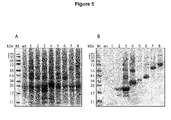
- Figure 5 is a Coomassie stained gel (A) and an Immunoblot analysis (B) showing the transient production of a multi-stage, multi-fragment malaria vaccine candidate (SEQ ID NO.198) and further fusion protein constructs according to the present disclosure based on heat stable fragments of different P. falciparum surface proteins in Nicotiana benthamiana.
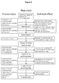
- Figure 6 shows a schematic flow chart of the protein purification processes according to the present disclosure.
- Figure 7 shows the results of immunofluorescence assays of different P. falciparum stages using purified polyclonal rabbit antibodies raised against a multi-stage, multi-fragment vaccine candidate (SEQ ID NO.197) according to the present disclosure.
- Figure 8 is a schematic representation of an advantageous embodiment of a thermostable multi-stage multi-fragment fusion protein according to the present disclosure.
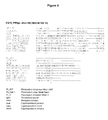
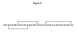
- Figure 9 shows the sequence of a typical EGF domain.
DETAILED DESCRIPTION OF THIS DISCLOSURE
-
The present disclosure pertains to therapeutically and diagnostic fusion proteins, compositions and antibodies suitable as human and/or animal vaccines against parasites of the phylum Apicomplexa, in particular against parasites of the genus Plasmodium like Plasmodium falciparum.
-
In advantageous embodiments, the recombinant fusion proteins and compositions according to the present disclosure combine heat stable fragments from different Apicomplexa surface proteins from different stages of the parasite development, wherein each heat stable fragment comprises at least one folded domain.
-
One advantage of using heat stable fragments with folded domains in place of full-length antigen proteins is to circumvent the limitation of the protein size, to allow better fusion protein thermo stability and to improve the protein expression capacity.
-
Surprisingly, the inventors found that using isolated heat stable fragments with folded domains as building blocks for the manufacture of vaccines has several advantages:
- For example, the thermo stability of the fusion proteins enables an efficient purification step by heating up the cell culture supernatant or cell extract. Many host cell proteins are denatured during that step and precipitate (see Fig. 3). They can thus be easily removed by centrifugation or filtration. Moreover, many host cell proteases are thermally inactivated, resulting in increased stability of the target recombinant fusion protein during downstream processing.
-
Further, thermo stability is moreover an extremely useful property for viral inactivation steps during downstream processing in vaccine manufacturing. Such steps are mandatory for ensuring product safety, but may not always be compatible with the activity of the target protein. In such cases expensive ultra/nano-filtration procedures have to be employed, including tedious and expensive process validation. By being able to employ simple heat treatment of the product, the overall process becomes cheaper and more efficient and results in a safer product. These properties are highly important for generating a vaccine that is particularly useful and applicable for developing countries.
-
Furthermore, thermo stability is an important feature during the formulation (e.g. lyophilisation) of the product and the storage. In particular, vaccines destined for developing countries in sub-Saharan Africa generally cannot rely on a cold chain, thus rendering many potentially promising vaccines completely useless.
-
The thermo stability of the fusion proteins according to this disclosure also results in longer shelf-life of the vaccine, thereby further reducing total costs for health systems.
-
Surprisingly, the inventors found that also the immune responses directed against such heat stable fragments with a folded domain are more robust, especially resulting in balanced immune responses against all represented components. Immuno-dominant and immuno-silent regions have not been found, even for candidates comprising a high number of different domains.
-
Furthermore, the inventors found that the selection of these heat stable fragments enables different (virtually all) combinations, i.e. the domains can generally be combined freely, resulting in recombinant fusion proteins comprising the same components but in a different order (e.g. A-B-C vs. B-C-A). Again, the induced immune responses are balanced, irrespective of the actual order of heat stable fragments. This possibility has tremendous advantages for (i) developing specific candidates with increased productivity and (ii) enables prime-boost regimens or (iii) combination vaccines where immune responses against the domain junctions are minimized or prevented.
-
The desire for a vaccine candidate composed of a single polypeptide is mainly driven by practical, technical and economical demands for reproducible, robust and cost-efficient production. However, to those skilled in the art, it is also clear, that there is a size limitation for recombinant expressed proteins. Although protein specific differences have to be taken into account as well, there is a strong decrease of expression levels and yields with increasing length of the polypeptide. Multiple challenges increase over-proportionally with size and the overall properties of large proteins are significantly less amenable to optimization than those of smaller proteins, domains or fragments. All these problems have so far been significant bottlenecks for the development of efficient vaccines against apicomplexan parasites and have resulted in an overwhelming number of sub-optimal vaccine candidates that comprise only multiple linear epitopes, one or two antigens from a one or two life cycle stages. As alternative, chemically or genetically attenuated or inactivated life-vaccines are proposed (e.g. irradiated sporozoites), but such approaches have to deal with batch-to-batch consistency, scaled-up production and most importantly product safety.
-
It is therefore an extremely important aspect of the present disclosure, that the heat stable fragments can be combined as building blocks e.g. in a recombinant fusion protein comprising several (in particular ≥ 4) heat stable fragments from different Apicomplexa surface proteins from the same but preferably from different life-cycle stages and can be efficiently produced. In an advantageous embodiment, the recombinant fusion proteins according to the present disclosure comprise at least four, in particular at least six heat stable fragments. It also has to be emphasized that the heat stable building blocks comprise folded protein domains that are fundamentally different from isolated small linear epitopes. Such linear epitopes are highly efficient for T-cell responses but are generally not suited as immunogens for inducing neutralizing antibodies. Quite contrary, apicomplexan parasites and in particular parasites of the genus Plasmodium have many proteins containing repetitive linear sequences, and their role is to divert the humoral immune response. It is therefore another particular aspect that the fusion proteins according to this disclosure do not comprise only these repetitive linear peptide epitopes.
-
Importantly, the fusion proteins according to the present disclosure (i) comprise domains derived from different Apicomplexa surface proteins and (ii) were designed using building blocks (domains) that have been experimentally identified and verified as heat stable (temperature tolerant).
-
In summary, the recombinant fusion proteins of the present disclosure can be well expressed in different expression systems, are thermo stable, have a high immunological relevance and have an improved immunogenicity. In advantageous embodiments, the recombinant fusion proteins of the present disclosure used as vaccines have the ability to elicit protective immunity that blocks infection as well as prevents pathology and interrupts transmission of parasites, and would most likely be a combination vaccine composed of subunits from different parasite stages.
-
As mentioned above, the isolated heat stable fragments have the advantage that they are small protein fragments with a conserved stable protein fold (like the Epidermal growth factor-like domain (EGF) and the Thrombospondin type 1 repeats (TSR) domain) in contrast e.g. to specifically selected Plasmodium linear T/B-cell epitopes used in the prior art. Using larger, folded protein domains instead of known linear B-, and T-cell epitope sequences enables the presentation of a larger number of conformational and overlapping B-cell epitopes as well as a larger number of T-cell epitopes thereby enhancing the chances to elicit a broader and more sustained immune response.
-
Parts/Fragments of an antigen that are recognized by the immune system, more specifically by antibodies, B cells, or T cells are defined as epitopes or antigenic determinants. Such protein structures that are composed of amino acids that have been brought together in three-dimensional structure (e.g. in a folded domain) are known as conformational epitopes. In contrast, a single peptide chain lacking secondary structure is termed a linear epitope. Most B cell epitopes are conformational even though some antibodies bind linear peptide fragments of antigens. T cell epitopes are linear peptides presented on the surface of an antigen-presenting cell, where they are bound to MHC molecules. T cell epitopes presented by MHC class I molecules are typically peptides between 8 and 11 amino acids in length, whereas MHC class II molecules are at least 13 amino acids long, but can be much longer. The clusters of conserved residues that bind the two ends of a peptide in MHC class I molecules are not found in MHC class II molecules, and the ends of the peptides are not bound. Instead, the peptide lies in an extended conformation along the MHC class II peptide-binding pocket. By comparing the sequences of known binding peptides it is usually possible to detect a pattern of permissive amino acids for each of the different alleles of MHC class II molecules, and to model how the amino acids of this peptide sequence motif will interact with the amino acids that make up the binding pocket.
-
Currently, a reasonably large database of unique B-cell and T-cell epitopes from Plasmodium proteins, including those from human P. falciparum and P. vivax malarias, has become available. By conducting a comprehensive meta-analysis of available data for Plasmodium immune epitopes, Vaughan et al. have identified more than 5,000 unique B-cell and T-cell epitopes for malaria parasites. Several of the P. falciparum and P. vivax epitopes were identified in extensive field studies conducted over the last 2 decades and by computer-based predictions of immune epitopes by analysis of genomic and proteomic databases; some of these predictions were validated in HLA-peptide binding studies (T cell epitopes) and in in vitro immunological studies (B cell epitopes).
-
Advantageous embodiments of the present disclosure pertains to recombinant fusion proteins suitable as human and/or animal vaccines against a parasite of the phylum Apicomplexa comprising a plurality of isolated heat stable fragments from at least two different Apicomplexa surface proteins, wherein each fragment contains at least one folded domain, wherein the isolated heat stable fragments are derived from Apicomplexa surface proteins presented on the surface of the parasite in at least two different stages in the life cycle of the parasite.
-
The terms "recombinant fusion protein" and "fusion protein" are used herein interchangeably to refer for example to a protein produced by recombinant technology which comprises segments i.e. amino acid sequences, from heterologous sources, such as different proteins or different organisms. The segments are joined either directly or indirectly to each other via peptide bonds. By indirect joining it is meant that an intervening amino acid sequence, such as a peptide linker is juxtaposed between segments forming the fusion protein. A recombinant fusion protein is encoded by a nucleotide sequence, which is obtained by genetically joining nucleotide sequences derived from different regions of one gene and/or by joining nucleotide sequences derived from two or more separate genes. These nucleotide sequences can be derived from a parasite of the phylum Apicomplexa and in particular derived from P. falciparum, but they may also be derived from other organisms, the plasmids used for the cloning procedures or from other nucleotide sequences.
-
Furthermore, the encoding nucleotide sequences may be synthesized in vitro without the need for initial template DNA samples e.g. by oligonucleotide synthesis from digital genetic sequences and subsequent annealing of the resultant fragments. Desired protein sequences can be "reverse translated" e.g. using appropriate software tools. Due to the degeneracy of the universal genetic code, synonymous codons within the open-reading frame (i.e. the recombinant protein coding region) can be exchanged in different ways, e.g. to remove cis-acting instability elements (e.g. AUUUA), to remove, introduce or modify the secondary and tertiary mRNA structures (e.g. pseudoknots, stem-loops,...), to avoid self-complementary regions that might trigger post-transcriptional gene silencing (PGTS), to change the overall AT:GC content, or to adjust the codon-usage to the expression host. Such changes can be designed manually or by using appropriate software tools or through a combination.
-
A recombinant fusion protein comprising a heat stable fragment from a Apicomplexa surface protein, in particular from a Plasmodium surface protein can be a recombinant product prepared using recombinant DNA methodology and expression in a suitable host cell, as is known in the art (see for example Sambrook et al., (2001) Molecular Cloning, Cold Spring Harbor Laboratory Press, Cold Spring Harbor, N. Y). Nucleotide sequences encoding specific isolated protein domain may be conveniently prepared, for example by polymerase chain reaction using appropriate oligonucleotide primers corresponding to the 5' and 3' regions of the domain required for isolation, and a full length coding of the isolated protein domain sequence as template. The source of the full length coding protein sequence may be for example, DNA extracted from parasite cells or a plasmid vector containing a cloned full-length gene. Alternatively, the protein coding sequence may partially or completely be synthesized in vitro or a combination of different approaches may be used. Non-limiting examples of properties of the fusion proteins according to the present disclosure are thermo stability and pH-stability. Especially the thermal performance of the fusion proteins in combination with the improved immunogenicity via using heat stable fragments comprising a folded domain is considered an important characteristic of the fusion proteins according to the present disclosure. The thermo stability for example may be determined as described in Example 4.
-
The Apicomplexa (also referred to as Apicomplexia) are a large group of protists, most of which possess a unique organelle called apicoplast and an apical complex structure involved in penetrating a host's cell. They are a diverse group including organisms such as coccidia, gregarines, piroplasms, haemogregarines, and plasmodia (Plasmodium falciparum, Plasmodium vivax, Plasmodium ovale, Plasmodium malariae, Plasmodium knowlesi). Diseases caused by apicomplexan organisms include, but are not limited to Babesiosis (Babesia), Malaria (Plasmodium), Coccidian diseases including Cryptosporidiosis (Cryptosporidium parvum), Cyclosporiasis (Cyclospora cayetanensis), Isosporiasis (Isospora belli) and Toxoplasmosis (Toxoplasma gondii).
-
In advantageous embodiments, the recombinant fusion proteins as well as the compositions according to the present disclosure are suitable as human and/or animal vaccines against a parasite of the genus Plasmodium including Plasmodium falciparum, Plasmodium vivax, Plasmodium malariae and/or Plasmodium ovale. In an advantageous embodiment, the parasite is Plasmodium falciparum.
-
Apicomplexa surface proteins are preferably membrane-bound or associated proteins or proteins known to be secreted. These proteins can e.g. be identified by analyzing the Genome or known genes for the presence of an N-terminal Signal peptid, the presence of a PEXEL motif, the presence of a GPI anchor motif, or the presence of one or more transmembrane domains using generally available software tools. These proteins and their homologues e.g. include but are not limited to:
- CelTOS (cell traversal protein for ookinetes and sporozoites), Antigen 2 (PfAg2, PvAg2, PoAg2, etc.)
- CSP (circumsporozoite protein)
- EBA175 (Erythrocyte binding antigen 175)
- EXP1 (Exported Protein 1); synonyms: CRA1 (Circumsporozoite-Related Antigen-1 /Cross-Reactive Antigen-1), AG 5.1 (Exported antigen 5.1), QF119
- MSP1 (Merozoite surface protein 1); synonyms: MSA1 (Merozoite surface antigen 1), PMMSA, p190, p195, gp190, gp195
- MSP3 (Merozoite surface protein 3); synonym: SPAM (secreted polymorphic antigen associated with the merozoite)
- MSP4 (Merozoite surface protein 4)
- MSP8 (Merozoite surface protein 8)
- MSP10 (Merozoite surface protein 10)
- MTRAP (merozoite TRAP homologue, merozoite TRAP homolog, merozoite TRAP-like protein)
- Pf38; synonym: 6-cysteine protein
- PfRh2a, Rh2a (Reticulocyte binding protein 2 homolog a, Reticulocyte binding protein 2 homologue a)
- PfRh2b, Rh2b (Reticulocyte binding protein 2 homolog b, Reticulocyte binding protein 2 homologue b)
- PfRh4, Rh4 (Reticulocyte binding protein homolog 4, Reticulocyte binding protein homologue 4)
- PfRh5, Rh5 (Reticulocyte binding protein homolog 5, Reticulocyte binding protein homologue 5)
- PfRipr, Ripr (Rh5 interacting protein)
- Pfs25 (25 kDa ookinete surface antigen, Sexual stage antigen pfs25 )
- Pfs230, S230 (Transmission-blocking target antigen Pfs230, Transmission-blocking target antigen S230)
- Pfs48/45 (45/48 kDa doublet proteins on Plasmodium gametes and gametocytes)
- Ron2 (rhoptry neck protein 2)
- TRAMP (thrombospondin-related apical membrane protein); synonym: PTRAMP
- TRAP (Thrombospondin-related anonymous protein); synonym: SSP2 (Sporozoite Surface Protein 2)
-
Heat stable fragments in the recombinant fusion protein or in the vaccine compositions according to the present disclosure may be from the same Apicomplexa surface protein, in particular from the same Plasmodium surface protein or preferably from different Apicomplexa surface proteins.
-
In an advantageous embodiment, the fusion proteins or the vaccine compositions according to the present disclosure comprise a plurality of isolated heat stable fragments from at least two different Apicomplexa surface proteins, wherein each fragment contains at least one folded domain.
-
In some advantageous embodiments, the fusion proteins or the compositions according to the present disclosure comprise more than one, in particular at least three, more particular at least four isolated heat stable fragments from different Apicomplexa surface proteins. In an advantageous embodiment, the recombinant fusion proteins comprise at least four different isolated heat stable fragments. Preferably, the Apicomplexa surface proteins are presented on the surface of the parasite in at least two different stages in the life cycle of the parasite.
-
In an advantageous embodiment, the fusion proteins or the vaccine compositions according to the present disclosure comprise at least four different isolated heat stable fragments from at least two different Apicomplexa surface proteins presented on the surface of the parasite in at least two different stages in the life cycle of the parasite. Since Apicomplexa parasites are able to use alternative antigens of a single life stage for their invasion process it is an advantage that a vaccine candidate covers at least two different antigen fragments per life stage. To further increase the vaccine efficacy more than one parasite life stage should be targeted. This would equal four antigen fragments and two life stages per minimal vaccine.
-
However, in an advantageous embodiment the number of covered antigens and life stages may be less for one vaccine if it is used as a composition with a complementary vaccine so that the sum of both vaccines equals at least four antigen fragments and two life stages.
-
Therefore, the present disclosure is also directed to vaccine compositions suitable as human and/or animal vaccine against a parasite of the phylum Apicomplexa comprising a plurality, in particular at least four isolated heat stable fragments derived from at least two different Apicomplexa surface proteins presented on the surface of the parasite in at least two different stages in the life cycle of the parasite, wherein each fragment contains at least one folded domain.
-
In some embodiments, the isolated heat stable fragments in the vaccine compositions according to the present disclosure are comprised in at least two different recombinant fusion proteins, wherein in an advantageous embodiment one recombinant fusion protein comprises two or more heat stable fragments derived from at least one Apicomplexa surface protein presented on the surface of the parasite in a single stage of the life cycle of the parasite and wherein the other recombinant fusion protein comprises two or more heat stable fragments derived from at least one Apicomplexa surface protein presented on the surface of the parasite in a different stage of the life cycle.
-
In other words, the vaccine compositions according to the present disclosure may comprise different fusion proteins having heat stable fragments derived from different Apicomplexa surface proteins for directing the parasite in more than one stages of the life cycle of the parasite.
-
In further embodiments of the present disclosure, one or more heat stable fragments derived from at least one Apicomplexa surface protein presented on the surface of the parasite in at least one stage of the life cycle of the parasite are repeated several times within the fusion proteins according to the present disclosure. The repeated fragments may be 100% identical, e.g. to increase the valence, or the repeated fragments may represent the same region of the full-length surface protein but actually comprise sequences derived from different strains, different species or different genera.
-
The term "fragment" as used herein refers to a continuous part of a natural full-length protein, with or without mutations, which is separate from and not in the context of a full length Apicomplexa surface protein. It may be a structural/topographical or functional subunit of a full length or complete protein. The term "fragments" expressly excludes polypeptides corresponding to full-length amino acid sequences of an Apicomplexa surface protein but also excludes short peptides from Apicomplexa surface proteins, not folding into domains. For example, in some embodiments of the present disclosure fragments having an amino acid sequence of less than 90 % of the parent full-length surface protein are used.
-
In an advantageous embodiment, the heat stable fragments are isolated heat stable fragments. The term "isolated" when used in relation to a nucleic acid or protein (e. g. an protein domain), refers to a nucleic acid sequence or protein that is identified and separated from at least one contaminant (nucleic acid or protein, respectively) with which it is ordinarily associated in its natural source. Isolated nucleic acid or protein is present in a form or setting that is different from that in which it is found in nature. In contrast, non-isolated nucleic acids or proteins are found in the state they exist in nature.
-
The term "heat stable" as used herein refers in particular to the ability of the fragments to withstand a temperature treatment of at least 50°C for 15 minutes, preferably of 60°C for 15 minutes, more preferably of 65°C for 15 minutes, most preferably of 70°C for 15 minutes without denaturation.
-
A typical "thermo stable protein" is in the present context a protein which is stable in environments with elevated temperatures. Such a protein has a wide range of applications, as it is more robust than other proteins, and it is also characterized by being active in extreme conditions. The underlying heat precipitation is described in point 4 of the Methods and Examples part in the present description. Further, Figure 3 illustrates a time-controlled heat-treatment according to the present disclosure.
-
The term "denaturation" as used herein refers to a process, which alters the folding structure of a protein or protein fragment due to exposure to certain chemical (acid, solvents, etc.) or physical factors (heat, etc.), causing the protein to become biologically inactive and/or to precipitate.
-
The thermo stable fusion proteins according to the present disclosure are able to withstand a temperature treatment of at least 50°C for 5 minutes, preferably of 60°C for 5 minutes, more preferably of 65°C for 5 minutes, most preferably of 70°C for 5 minutes without denaturation and a recovery rate of at least 80 %. In advantageous embodiments, the fusion proteins according to the present disclosure are able to withstand a temperature treatment of at least 80°C for 5 minutes without denaturation and having a recovery rate of at least 60 %. In another advantageous embodiments, the fusion proteins according to the present disclosure are able to withstand a temperature treatment of at least 90°C for 5 minutes without denaturation and having a recovery rate of at least 50 %.
-
According to the present disclosure, the different heat stable fragments are linked to each other. "Linked" refers to non-covalent or covalent bonding between two or more molecules. Linking may be direct or indirect. Two molecules are indirectly linked when the two molecules are linked via a connecting molecule (linker). Two molecules are directly linked when there is no intervening molecule linking them. As mentioned above, the isolated protein domains are linked either directly or indirectly to each other, preferably via peptide bonds or disulfide bonds. An example of indirect covalent linking is that an intervening amino acid sequence, such as a peptide linker is juxtaposed between segments forming the fusion protein.
-
In some embodiments, the heat stable fragments are directly linked to each other. In other embodiments, the heat stable fragments are indirectly linked to each other via a linker, wherein in some examples the linker is a polypeptide with a size of less or equal twenty amino acids, in particular 2 to 6 amino acids.
-
In some advantageous embodiments, each heat stable fragment contains at least one folded domain.
-
The term "folded domain" as used herein refers to a protein structure composed of amino acids that are arranged in a three dimensional formation. A folded domain is part of a structural/topographical or functional subunit of a full length or complete protein. It may be kept within the context of the full length or complete protein, or may be separated therefrom, as in an isolated domain. Domains corresponding to structural/topographical subunits include for example, a cytoplasmic domain, an extracellular domain or a transmembrane domain. Domains corresponding to functional subunits include for example, a receptor binding domain or in particular an antibody binding domain.
-
In some advantageous embodiments, the folded domain is an "EGF-like domain" which is an EGF-like motif that may be found in a variety of proteins, as well as EGF and Notch and Notch ligands, including those involved in the blood clotting cascade (Furie and Furie, 1988, Cell 53: 505-518). For example, this motif has been found in extracellular proteins such as the blood clotting factors IX and X (Rees et al., 1988, EMBO J. 7:2053-2061; Furie and Furie, 1988, Cell 53: 505-518), in other Drosophila genes (Knust et al., 1987 EMBO J. 761-766; Rothberg et al., 1988, Cell 55:1047-1059 ), and in some cell-surface receptor proteins, such as thrombomodulin (Suzuki et al., 1987, EMBO J. 6:1891-1897) and LDL receptor (Sudhof et al., 1985, Science 228:815-822 ). A protein binding site has been mapped to the EGF repeat domain in thrombomodulin and urokinase (Kurosawa et al., 1988, J. Biol. Chem 263:5993-5996; Appella et al., 1987, J. Biol. Chem. 262:4437-4440 ).
-
As reported by PROSITE a typical EGF domain may include six cysteine residues which have been shown (in EGF) to be involved in disulfide bonds. The main structure is proposed, but not necessarily required, to be a two-stranded beta-sheet followed by a loop to a C-terminal short two-stranded sheet. Subdomains between the conserved cysteines strongly vary in length as shown in Figure 9, wherein "C" represents conserved cysteine involved in a disulfide bond, "G" represents often-conserved glycine, "a" represents often conserved aromatic amino acid and "x" represents any residue.
-
The region between the 5th and 6th cysteine contains two conserved glycines of which at least one is normally present in most EGF-like domains and force the assembly of several well-defined discontinuous epitopes (Farley and Long, 1995, Exp. Parasitol. 80, 328-332; McBride and Heidrich, 1987, supra; Uthaipibull et al, 2001, J. Mol. Biol. 307,1381 1394). The EGF-like domain used in the recombinant fusion proteins or compositions according to the present disclosure may be derived from any suitable Apicomplexa surface protein, expressed in any life cycle stage, including for example the pre-erythrocytic stage, the blood stage and the sexual stage. The surface proteins may occur on the different forms of the apicomplexan parasite, in particular of Plasmodium falciparum.
-
Preferably the EGF-like domains are derived from Plasmodium vivax and/or Plasmodium falciparum. The term "EGF-like domain" as used herein includes sequence variants, fragments, derivatives and mimetics having activity corresponding to naturally occurring domains.
-
A "TSR domain" is a small about 60-residue domain found in extracellular proteins or in the extracellular part of transmembrane proteins that are involved in immunity, cell adhesion, cell-cell-interactions and neuronal development (Tucker, 2004). Structures of TSR domains from thrombospondin-1 (TSP-1; Tan et al. 2002) and F-spondin (PDB codes 1SZL and 1VEX) have been solved. These show that a TSR domain has an elongated structure consisting of an antiparallel three-stranded β-sheet. The domain core is formed by a stacked array of side chains of conserved tryptophans, arginines, and cysteines. TSRs of several proteins have been reported to mediate glycosaminoglycan (GAG) binding. For example, the plasmodium surface proteins plasmodium CS and TRAP both contain an adhesive thrombospondin type 1 domain, TSR.
-
In an advantageous embodiment, the recombinant fusion proteins and the vaccine compositions according to the present disclosure comprise at least two folded domains from at least two different Apicomplexa surface proteins, wherein the first folded protein domain is an isolated EGF-like domain and the second domain is an isolated EGF-like domain or an isolated TSR domain.
-
In another embodiment, the heat stable isolated fragments in the recombinant fusion proteins or vaccine compositions may comprise one or more further folded domains. In advantageous embodiments, the heat stable isolated fragments may comprise 2 to 12 different folded protein domains, in particular 4 to 10, in particular 6 to 8 different folded domains, wherein at least one folded domain is an EGF-like domain.
-
As mentioned above, EGF-like domains as well as TSR domains show a high cysteine content. In a further embodiment, the recombinant fusion protein according to the present disclosure has a cysteine content of at least 5%, in particular of at least 7.5%, more particular of at least 10%.
-
It is one further advantage of the present invention that also non-heat stable isolated fragments which are for example highly immunogenic can be embedded into a fusion protein having a plurality of isolated heat stable fragments, wherein the entire fusion protein is still thermo-stable. Therefore, in some embodiments, recombinant fusion proteins according to the present disclosure may comprise at least one non-heat stable isolated fragment derived from an Apicomplexa surface protein, wherein the entire fusion protein is thermo stable.
-
In some advantageous embodiments, the folded domains of the fragments in the fusion proteins and the vaccines according to the present disclosure comprise at least one conformational epitope.
-
The term "epitope" as used herein refers to a region of a protein molecule to which an antibody can bind. In particular, an "epitope" may be defined as an array of 3-20 amino acids aligned along the surface of a protein. In a linear epitope, the amino acids are joined sequentially and follow the primary structure of the protein. In a "conformational epitope" the amino acids are arranged in a specific three-dimensional structure in a context-dependent manner (e.g. a folded domain). With respect to conformational epitopes, the length of the epitope-defining sequence can be subject to wide variations. The portions of the primary structure of the antigen between the residues defining the epitope may not be critical to the structure of the conformational epitope. For example, deletion or substitution of these intervening sequences may not necessarily affect the conformational epitope provided sequences critical to epitope conformation are maintained (e.g. cysteines involved in disulfide bonding, glycosylation sites, etc.). A conformational epitope may also be formed by association of 2 or more subunits into homo-or hetero-oligomers. A conformational epitope may be a folded domain or an accessible part thereof that is recognized by the immune system and capable of eliciting an immune response.
-
As mentioned above, one of the advantages of the recombinant fusion proteins according to the present disclosure is the relatively small protein size in relation to the number of folded domains from different Apicomplexa surface proteins in contrast to vaccine constructs comprising full length surface proteins, in particular if the fusion protein is directed to surface proteins from different life cycle stages. Therefore, in some embodiments the fusion protein of the present disclosure has a molecular weight of less or equal 160 kDa if the fusion protein comprises heat stable fragments derived from a plurality of Apicomplexa surface proteins presented on the surface of the parasite in all stages of the plasmodium life cycle or if the fusion protein comprises heat stable fragments derived from Apicomplexa surface proteins from selected stages of less or equal 120kDa.
-
The recombinant fusion proteins and/or vaccine compositions suitable as human and/or animal vaccine against a parasite of the phylum Apicomplexa may combine heat stable fragments from different stages of the parasite development, inducing several mechanisms of protection. In one embodiment, the recombinant fusion proteins and/or vaccine compositions of the present disclosure comprises heat stable fragments for pre-erythrocytic and blood stage coverage and can be used for prophylactic and/or therapeutic vaccines. In another advantageous embodiment, the recombinant fusion proteins and/or vaccine compositions of the present disclosure comprises heat stable fragments for pre-erythrocytic and blood stage coverage as well as heat stable fragments for sexual stage coverage.
-
As mentioned above, in some embodiments the heat stable fragments are derived from different Apicomplexa surface proteins which are expressed in the same stage of the Apicomplexa life cycle, in particular in the blood stage. In an advantageous embodiment, the heat stable fragments are from different Apicomplexa surface proteins which are expressed in different stages of the Apicomplexa life cycle, in particular in the blood stage and in the pre-erythrocyte stage. In a further embodiment, the heat stable fragments are from different Apicomplexa surface protein antigens which are expressed in different stages of the Apicomplexa life cycle, in particular the blood stage, the sexual stage and the pre-erythrocyte stage.
-
In advantageous embodiments the heat stable fragments are derived from different Plasmodium falciparum surface proteins which are expressed in the same stage of the Plasmodium life cycle, in particular in the blood stage. In an advantageous embodiment, the heat stable fragments are from different Plasmodium falciparum surface proteins which are expressed in different stages of the Plasmodium falciparum life cycle, in particular in the blood stage and in the pre-erythrocyte stage. In a further embodiment, the heat stable fragments are from different Plasmodium falciparum surface protein antigens which are expressed in different stages of the Plasmodium falciparum life cycle, in particular the blood stage, the sexual stage and the pre-erythrocyte stage.
-
Furthermore, the isolated heat stable fragments may be from different Plasmodium surface proteins, which are expressed in the different Plasmodium life cycle stages:
The pre-erythrocytic stage:
a) Sporozoite
-
The sporozoite remains in the bloodstream for a very short period of time before invading a hepatocyte. Examples for Plasmodium protein antigens expressed in the sporozoite are the circumsporozoite protein (CSP), the major constituent of the outer membrane of the sporozoite (Nussenzweig et al., 1989). Induced antibodies may be able to block the binding and the entrance of the sporozoite into the hepatocyte.
b) Liver stage
-
During this stage, immunity is mostly mediated by cellular-dependent mechanisms involving CD8+ T cells, CD4+ T cells, natural killer (NK) cells and γδ T cells. CSP is expressed both in the sporozoite and during the liver stage. So, much of the research involving CSP has switched from the immunodominant repeats inducing humoral response to regions that are able to induce cytotoxic T-cell responses. Other identified liver-stage antigens include liver-stage antigen-1 (LSA-1), LSA-2, LSA-3, SALSA and STARP, among others (Hoffman et al., 1996;).
The asexual blood stage:
c) Merozoite
-
Besides the sporozoite, the merozoite is the only stage in the human host in which the malaria parasite is extracellular. In contrast to the sporozoite, several cycles of merozoite release will occur during a malaria infection, making them often available. A major ligand in P. falciparum is the erythrocyte-binding antigen-175 (EBA-175), located in the microneme (Chitnis et al., 1994). Several merozoite surface proteins (MSPs) have been identified, but for most of them their function still has to be further elucidated. In the case of the major MSP, named MSP-1, a role has been postulated in merozoite binding to the RBC and in the subsequent biochemical mechanisms involved in invasion. This protein is synthesized as a precursor of 185-210 kDa in the late schizont stage and is processed to generate several polypeptides of varied molecular weights. A 42 kDa polypeptide (MSP1-42) is kept attached to the merozoite membrane, and it is further processed to generate a 19 kDa polypeptide (MSP1-19), which goes into the host cell. Besides MSP-1, at least eight other MSPs have been described in P. falciparum: MSP-2, MSP-3, MSP-4, MSP-5, MSP-6, MSP-7, MSP-8 and MSP-10. Another merozoite surface-associated antigen is the acidic-basic repeat antigen (ABRA). Proteins located in merozoite apical organelles have also been identified (e.g. the rhoptry proteins apical membrane antigen-1 (AMA-1), rhoptry-associated protein-1 (RAP-1) and RAP-2).
d) Infected RBC
-
Once it has invaded the RBC, the parasite is supposed to have found a safer place to stay. One of the most studied molecules is the ring erythrocyte surface antigen (RESA). Further, the serine-rich protein (SERP or SERA) is a soluble protein expressed in the schizont stage and secreted in the parasitophorous vacuole. Other proteins that are located on the RBC membrane are the erythrocyte membrane protein-1 (EMP-1), EMP-2 and EMP-3. PfEMP-1, which binds to the receptors such as CD36 in the endothelium, is a family of proteins encoded by the so-called var genes.
The sexual stage:
e) Sporogonic cycle
-
Other Plasmodium protein antigens are expressed in sexually differentiated parasite stages such as Ps25, Ps28, Ps48/45 or Ps230. Antibodies against these sexual stage proteins may block the development of the parasite in mosquitoes.
-
In an advantageous embodiment, each of the heat stable fragments are from different Plasmodium surface proteins expressed in at least two different stages of the Plasmodium life cycle.
-
In advantageous embodiments, the heat stable fragments are selected from the group consisting of heat stable fragments comprising an EGF-like domain from MSP1, MSP4, MSP8, MSP10, PfRipr and Pfs25.
-
In further advantageous embodiments, the heat stable fragments are selected from the group consisting of heat stable fragments comprising a TSR domain is selected from CSP, mTRAP, TRAP and TRAMP.
-
In other advantageous embodiments, the heat stable fragments are selected from the group consisting of heat stable fragments from Pfs230, Pfs45/48, CelTos and Ron2, MSP3 and EXP1.
-
In advantageous embodiments, the heat stable fragments comprised in the recombinant fusion protein and/or vaccine compositions according to the present disclosure are selected from the group listed in Table 1.
Table 1: Examples of heat stable fragments. | Name | Amino acid | PlasmoDB | Strain | Reference |
| CelTos | F25-D182 | PFL0800c | 3D7 | (Berqmann-Leitner et al. 2010) |
| CSP1_TSR | P293-S365 | PFC0210c | 3D7 | (Plassmeyer et al. 2009) |
| EXP1 | E23-S79, N102-H162 | PF1121600 | 3D7 | (Simmons et al. 1987) |
| EGF1_MSP1-19 | I1589-V1629 | PFI1475w | 3D7 | (Blackman et al. 1991) |
| MSP3aGKO | K26-K157 | PF1035400 | 3D7 | |
| EGF_MSP4 | L201-L247 | PFB0310c | 3D7 | (Marshall et al. 1997) |
| EGF1_MSP8 | N464-D508 | PFE0120c | 3D7 | (Black et al. 2001) |
| EGF2_MSP8 | D509-S551 | PFE0120c | 3D7 | (Black et al. 2001) |
| EGF1_MSP10 | V388-P432 | PFF0995c | 3D7 | (Black et al. 2003) |
| EGF2_MSP10aglyc | K434-K475 | PFF0995c | 3D7 | (Black et al. 2003) |
| mTRAP_TSR | T3-E76 | PF10_0281 | 3D7 | (Uchime et al. 2012) |
| EGF1_PfRipr | R268-E302 | PFC1045c | 3D7 | (Chen et al. 2011) |
| EGF2_PfRipr | L306-Y343 | PFC1045c | 3D7 |
| EGF3_PfRipr | S617-E656 | PFC1045c | 3D7 |
| EGF4_PfRipr | D660-V696 | PFC1045c | 3D7 |
| EGF5_PfRipr | K700-I734 | PFC1045c | 3D7 |
| EGF6_PfRipr | K752-I795 | PFC1045c | 3D7 |
| EGF7_PfRipr | Y799-I835 | PFC1045c | 3D7 |
| EGF8_PfRipr | S839-V878 | PFC1045c | 3D7 |
| EGF9_PfRipr | K882-L919 | PFC1045c | 3D7 | |
| EGF10_PfRipr | P923-V960 | PFC1045c | 3D7 |
| Pfs230_C0 | E423-N566 | PFB0405w | 3D7 | (Tachibana et al. 2011) |
| Pfs25FKO | V2-T171 | PF10_0303 | 3D7 | (Kaslow et al. 1988) |
| Pfs45/48 | N28-S427 | PF1346700 | 3D7 | |
| Ron2L | M2020-K2067 | PF14_0495 | 3D7 | (Srinivasan et al. 2011) |
| TRAMP | F244-K309 | PF1218000 | 3D7 | |
| TRAP_TSR | E214-K264 | PF13_0201 | 3D7 | (Tossavainen et al. 2006) |
-
The abbreviations used in table 1 are as follows: Aglyc/GKO: aglycosylated/glycosylation site knock-out, FKO: full glycosylation site knock-out.
-
In further advantageous embodiments, the heat stable fragments comprised in the recombinant fusion protein and/or vaccine compositions according to the present disclosure are selected from the group listed in Table 2.
-
Table 2 shows sequences of Plasmodium falciparum antigens and their orthologs in other Apicomplexa like Toxoplasma, Theileria, Neospora, Babesia and Cryptosporidium. Exemplary use and combination of these antigen fragment sequences for the generation of various fusion proteins as a basis for Apicomplexa vaccine candidates are shown in Table 3. In an advantageous embodiment, the heat stable fragments of surface proteins from one member, preferably from one strain of an Apicomplexa are used in a fusion protein according to the present disclosure. However, in some embodiments heat stable fragments of surface proteins from different members, also from different strains are used in a fusion protein according to the present disclosure useful as general vaccines.
-
Several online-tools were used for sequence retrieval, such as http://plasmodb.org (Plasmodium falciparum/vivax/knowlesi (Pf 3D7/Pv Sal-1/Pk H)), http://www.ncbi.nlm.nih.gov and http://uniprot.org (Plasmodium malariae/ovale (Pm/ Pol/CDC)) as well as http://orthomcl.org (Toxoplasma gondii (Tgon), Theileria annulatalparva (Tann/Tpar), Neospora caninum (Ncan), Babesia bovis (Bbov) and Cryptosporidium hominis/ parvum/muris (Chom/Cpar/Cmur)).
-
The abbreviations used in table 2 are as follows:
- Pf 3D7
- Plasmodium falciparum Strain 3D7
- Pv Sal-1
- Plasmodium vivax Strain Sal-1
- Pk H
- Plasmodium knowlesi Strain H
- Pol/CDC
- Plasmodium ovale Strain I/CDC
- Pm
- Plasmodium malariae
- Tgon
- Toxoplasma gondii
- Ncan
- Neospara caninum
- Cpar
- Cryptosporidium parvum
- Cmur
- Cryptosporidium muris
- Chom
- Cryptosporidium hominis
- Bbov
- Babesia bovis
- Tann
- Theileria annulata
- Tpar
- Theileria parva
Aglyc/GKO: aglycosylated/glycosylation site knock-out
FKO: full glycosylation site knock-out
Table 2: Exemplary fragment sequences for the generation of novel Apicomplexa vaccine candidates. | SEQ ID | Name | Sequence | |
| 1. | Pf_3D7 EGF1_MSP119|P F3D7_0930300 EGF1 of MSP119: short 1_19 | |
| 2. | Pf_3D7 EGF1_MSP8|PF3 D7_0502400 EGF1 of MSP8: short 1_8 | |
| 3. | Pf_3D7 EGF2_MSP8|PF3 D7_0502400 EGF2 of MSP8: short 2_8 | |
| 4. | Pf_3D7 EGF_MSP4|PF3 D7_0207000 EGF of MSP4: short 4 | |
| 5. | Pf_3D7 EGF_MSP5|PF3 D7_0206900.1 EGF of MSP5: short 5 | |
| 6. | Pf_3D7 EGF1_MSP10|PF 3D7_0620400 EGF1 of MSP10: short 1_10 | |
| 7. | Pf_3D7 EGF2_MSP10agl yc|PF3D7_06204 00 EGF2 of MSP10aglyc (point mutation in bold): short 2_10a | |
| 8. | Pf_3D7 EGF1_PfRipr|PF 3D7_0323400 EGF1 of PfRipr: short R1 | |
| 9. | Pf_D7 EGF2_PfRipr|PF 3D7_0323400 EGF2 of PfRipr: short R2 | |
| 10. | Pf_3D7 EGF3_PfRipr|PF 3D7_0323400 EGF3 of PfRipr: short R3 | |
| 11. | Pf_3D7 EGF4_PfRipr|PF 3D7_0323400 EGF4 of PfRipr: short R4 | |
| 12. | Pf_3D7 EGF5 PfRipr|PF 3D7_0323400 EGF5 of PfRipr: short R5 | |
| 13. | Pf_3D7 EGF6_PfRipr|PF 3D7_0323400 EGF6 of PfRipr: short R6 | |
| 14. | Pf_3D7 EGF7_PfRipr|PF 3D7_0323400 EGF7 of PfRipr: short R7 | |
| 15. | Pf_3D7 EGF8_PfRipr|PF 3D7_0323400 EGF8 of PfRipr: short R8 | |
| 16. | Pf_3D7 EGF9_PfRipr|PF 3D7_0323400 EGF9 of PfRipr: short R9 | |
| 17. | Pf_3D7 EGF10_PfRipr|P F3D7_0323400 EGF10 of PfRipr: short R10 | |
| 18. | Pf_3D7 Pfs25FKO|PF3D7 1031000 Pfs25 (1x N missing, with knocked-out glycosites: 25FKO | |
| 19. | Pf_3D7 fullCSP_TSR|PF3 D7_0304600 TSR-domain of fullCSP_TSR: short fCSP_TSR | |
| 20. | Pf_3D7 shortCSP_TSR|P F3D7_0304600 Shortened TSR-domain of CSP (shortCSP_TSR): short CSP_TSR (only domain with an potential N-glycosylation site, marked in bold) | |
| 21. | Pf_3D7 mTRAP_TSR|PF 3D7_1028700 TSR-domain of mTRAP: short mTRAP_TSR | |
| 22. | Pf_3D7 TRAP_TSR|PF3 D7_1335900 TSR-domain of TRAP: short TRAP_TSR | |
| 23. | Pf_3D7 TRAMP_TSR|PF 3D7_1218000 TSR-domain of TRAMP: short TRAMP_TSR | |
| 24. | Pf_3D7 EBA175_F2 domain|PF3D7_0 731500 EBA175 F2 domain: short EBA175_F2 | |
| 25. | Pf_3D7_EBA140 EBS1|PF3D7_13 01600 EBA140 erythrocyte bindin sequence 1: short EBA140 EBA1 | |
| 26. | Pf_3D7_EBA140 EBS2|PF3D7_13 01600 EBA140 erythrocyte binding sequence 2: short EBA140 EBS2 | |
| 27. | Pf_3D7 EBL1 EBS|PF3D7_137 1600 EBL erythrocyte binding sequence: short EBL1 EBS | |
| 28. | Pf_3D7 GAMA EBS|PF3D7_082 8800 GAMA erythrocyte binding sequence: short GAMA EBS | |
| 29. | Pf_3D7 Pfs230_C0|PF3D 7_0209000 Pfs230 part C0: short Pfs230_C0 | |
| 30. | Pf_3D7 CelTos|PF3D7_1 216600 CelTos: short C | |
| 31. | Pf_3D7 Ron2L|PF3D7_14 52000 Ron2L | |
| 32. | Pf_3D7 EXP1|PF3D7_11 21600 EXP1 | |
| 33. | Pf_3D7 MSP3aGKO| PF3D7_1035400 MSP3aGKO | |
| 34. | Pf_3D7 Pfs4845| PF3D7_1346700 Pfs4845 | |
| | | |
| 35. | Pv_Sal-1 EGF1_MSP119|P VX_099980 Homolog of EGF1 of MSP1_19 | |
| 36. | Pv_Sal-1 EGF1_MSP8|PV X_097625 EGF1 of MSP8: short 1_8 | |
| 37. | Pv_Sal-1 EGF2_MSP8|PV X_097625 EGF2 of MSP8: short 2_8 | |
| 38. | Pv_Sal-1 EGF_MSP4|PVX 003775 EGF of MSP4: short 4 | |
| 39. | Pv_Sal-1 EGF_MSP5|PVX 003770 EGF of MSP5: short 5 | |
| 40. | Pv_Sal-1 EGF1_MSP10|P VX_114145 EGF1 of MSP10: short 1_10 | |
| 41. | Pv_Sal-1 EGF2_MSP10|P VX_114145 EGF2 of MSP10 | |
| 42. | Pv_Sal-1 EGF1_PfRiprlPV X_095055 EGF1 of PfRipr: short R1 | |
| 43. | Pv_Sal-1 EGF2_PfRiprlPV X_095055 EGF2 of PfRipr: short R2 | |
| 44. | Pv_Sal-1 EGF3_PfRiprlPV X_095055 EGF3 of PfRipr: short R3 | |
| 45. | Pv Sal-1 EGF4_PfRiprlPV X_095055 EGF4 of PfRipr: short R4 | |
| 46. | Pv_Sal-1 EGF5_PfRiprlPV X_095055 EGF5 of PfRipr: short R5 | |
| 47. | Pv_Sal-1 EGF6_PfRiprlPV X_095055 EGF6 of PfRipr: short R6 | |
| 48. | Pv_Sal-1 EGF7_PfRiprlPV X_095055 EGF7 of PfRipr: short R7 | |
| 49. | Pv_Sal-1 EGF8_PfRiprlPV X_095055 EGF8 of PfRipr: short R8 | |
| 50. | Pv_Sal-1 EGF9_PfRiprlPV X_095055 EGF9 of PfRipr: short R9 | |
| 51. | Pv_Sal-1 EGF10_PfRipr|P VX_095055 EGF10 of PfRipr: short R10 | |
| 52. | Pv_Sal-1 | |
| | Pvs25|PVX_1111 75 Pvs25 | |
| 53. | Pv_Sal-1 fullCSP_TSR|PV X_119355 TSR-domain of fullCSP_TSR: short fCSP_TSR | |
| 54. | Pv_Sal-1 shortCSP TSR Shortened TSR-domain of CSP (shortCSP_TSR): short CSP_TSR | |
| 55. | Pv_Sal-1 mTRAP_TSR|PV X_111290 TSR-domain of mTRAP: short mTRAP_TSR | |
| 56. | Pv_Sal-1 TRAP_TSR|PVX 082735 TSR-domain of TRAP: short TRAP_TSR | |
| 57. | Pv_Sal-1 TRAMP_TSR|PV X_123575 TSR-domain of TRAMP: short TRAMP_TSR | |
| 58. | Pv_Sal-1 EBA175 F2 domain|PVX_110 810 EBA175 F2 domain | |
| 59. | Pv_Sal-1 EBA140 EBS1|PVX_1108 10 EBA140 erythrocyte bindin sequence 1: short EBA140 EBS1 | |
| 60. | Pv_Sal-1 EBA140 EBS2|PVX_1108 10 | |
| | EBA140 erythrocyte binding sequence 2: short EBA140 EBS2 | |
| 61. | Pv_Sal-1 EBL1 EBS|PVX_11081 0 EBL erythrocyte binding sequence: short EBL1 EBS | |
| 62. | Pv_Sal-1 GAMA EBS|PVX_08891 0 Homolog of GAMA erythrocyte binding sequence: short GAMA EBS | |
| 63. | Pv_Sal-1 Pvs230_C0|PVX 003905 Pfs230 part C0: short Pfs230_C0 | |
| 64. | Pv_Sal-1 CelTos|PVX_123 510 CelTos: short C | |
| 65. | Pv_Sal-1 Ron2L|PVX_1178 80 Homolog of Ron2L | |
| 66. | Pv_Sal-1 Exp1|PVX_09170 0 EXP1 | |
| 67. | Pv_Sal-1 Pvs4845|PVX_08 3235 Pvs4845 | |
| | | |
| 68. | Pk_H EGF1_MSP19|P KH_072850 Homolog of EGF1 of MSP119: short 1_19 | |
| 69. | Pk_H EGF1_MSP8|PK H_103110 EGF1 of MSP8: short 1_8 | |
| 70. | Pk_H EGF2_MSP8|PK H_103110 EGF2 of MSP8: short 2_8 | |
| 71. | Pk_H EGF_MSP4|PKH_ 041300 EGF of MSP4: short 4 | |
| 72. | Pk_H EGF_MSP5|PKH_ 041310 EGF of MSP5: short 5 | |
| 73. | Pk_H EGF1_MSP10|P KH_112880 EGF1 of MSP10: short 1_10 | |
| 74. | Pk_H EGF2_MSP10|P KH_112880 EGF2 of MSP10 | |
| 75. | Pk_H EGF1_PfRipr|PK H_081690 EGF1 of PfRipr: short R1 | |
| 76. | Pk_H EGF2_PfRipr|PK H_081690 EGF2 of PfRipr: short R2 | |
| 77. | Pk_H EGF3_PfRipr|PK H_081690 EGF3 of PfRipr: short R3 | |
| 78. | Pk_H EGF4_PfRipr|PK H_081690 EGF4 of PfRipr: short R4 | |
| 79. | Pk_H EGF5_PfRipr|PK H_081690 EGF5 of PfRipr: short R5 | KCDLLCPSNKSCLIENGKKICKCINGLTLENGVCI | |
| 80. | Pk_H 6_PfRipr|PK H_081690 EGF6 of PfRipr: short R6 | |
| 81. | Pk_H EGF7_PfRipr|PK H_081690 EGF7 of PfRipr: short R7 | |
| 82. | Pk_H EGF8_PfRipr|PK H_081690 EGF8 of PfRipr: short R8 | |
| 83. | Pk_H EGF9_PfRipr|PK H_081690 EGF9 of PfRipr: short R9 | |
| 84. | Pk_H EGF10_PfRipr|P KH_081690 EGF10 of PfRipr: short R10 | |
| 85. | Pk_H Pks251PKH_0615 30 Pks25 | |
| 86. | Pk H fullCSP_TSR|PK H_083560 TSR-domain of fullCSP_TSR: short fCSP_TSR | |
| 87. | Pk H shortCSP_TSR|P KH_083560 Shortened TSR-domain of CSP (shortCSP_TSR): short CSP_TSR | |
| 88. | Pk H mTRAP_TSR|PK H_061300 TSR-domain of mTRAP: short mTRAP_TSR | |
| 89. | Pk_H TRAP_TSR|PKH 121770 TSR-domain of TRAP: short TRAP_TSR | |
| 90. | Pk_H TRAMP_TSR|PK H_143510 TSR-domain of TRAMP: short TRAMP_TSR | |
| 91. | Pk_H EBA175 F2 domain|PKH_000 490 EBA175 F2 domain | |
| 92. | Pk_H EBA175 F2 domain|PKH_062 300 EBA175 F2 domain | |
| 93. | Pk_H EBA175_F2 domain|PKH_134 580 EBA175 F2 domain | |
| 94. | Pk_H EBA140 EBS1|PKH_0004 90 EBA140 erythrocyte bindin sequence 1: short EBA140 EBS1 | |
| 95. | Pk_H EBA140 EBS1|PKH_0623 00 EBA140 erythrocyte bindin sequence 1: short EBA140 EBS1 | |
| 96. | Pk_H EBA140 EBS1|PKH_1345 80 EBA140 erythrocyte bindin sequence 1: short EBA140 EBS1 | |
| 97. | Pk_H EBA140 EBS2|PKH_0004 90 EBA140 erythrocyte binding sequence 2: short EBA140 EBS2 | |
| 98. | Pk_H EBA140 EBS2|PKH_0623 00 EBA140 erythrocyte binding sequence 2: short EBA140 EBS2 | |
| 99. | Pk_H EBA140 EBS2|PKH_1345 80 EBA140 erythrocyte binding sequence 2: short EBA140 EBS2 | |
| 100. | Pk_H EBL1 EBS1|PKH_0004 90 EBL erythrocyte binding sequence: short EBL1 EBS | |
| 101. | Pk_H EBL1 EBS1|PKH_0623 00 EBL erythrocyte binding sequence: short EBL1 EBS | |
| 102. | Pk_H EBL1 EBS1|PKH_1345 80 EBL erythrocyte binding sequence: short EBL1 EBS | |
| 103. | Pk_H GAMA EBS|PKH_05021 | |
| | 0 GAMA erythrocyte binding sequence: short GAMA EBS | |
| 104. | Pk_H Pks230_C0|PKH_ 041100 Pfs230 part C0: short Pfs230_C0 | |
| 105. | Pk_H CelTos|PKH_143 380 CelTos: short C | |
| 106. | Pk_H Ron2L|PKH_125 430 Ron2L | |
| 107. | Pk_H EXP1| PKH 091900 EXP1 | |
| 108. | Pk_H Pks4845|PKH_12 0750 Pks4845 | |
| | | |
| 109. | Pol/CDC EGF1_MSP19|FJ 824670.1 Homolog of EGF1 of MSP119: short | |
| 110. | Pol/CDC Pos25|Q969A0 Pos25 | |
| | | |
| 111. | Pm EGF1_MSP19|FJ 824669.1 Homolog of EGF1 of MSP119: short 1_19 | |
| 112. | Pm fullCSP_TSR|CA A04809.1 Homolog of TSR-domain of fullCSP_TSR: short fCSP_TSR | |
| 113. | Pm shortCSP_TSR|C AA04809.1 Homolog of shortened TSR-domain of CSP (shortCSP_TSR): short CSP_TSR | |
| | | |
| 114. | Tgon|TGME49_0 67680 Ortholog of EGF1_PfRipr short R1 | |
| 115. | Tgon|TGME49_0 02400 Ortholog of EGF1_PfRipr short R1 | |
| 116. | Tgon|TGME49_0 67680 Ortholog of EGF2_PfRipr short R2 | |
| 117. | Tgon|TGME49_0 02400 Ortholog of EGF2_PfRipr short R2 | |
| 118. | Tgon|TGME49_0 67680 Ortholog of EGF3_PfRipr short R3 | |
| 119. | Tgon|TGME49_0 02400 Ortholog of EGF3_PfRipr short R3 | |
| 120. | Tgon|TGME49_0 67680 Ortholog of EGF4_PfRipr short R4 | |
| 121. | Tgon|TGME49_0 02400 Ortholog of EGF4_PfRipr short R4 | |
| 122. | Tgon|TGME49_0 67680 Ortholog of EGF5_PfRipr short R5 | |
| 123. | Tgon|TGME49_0 02400 Ortholog of EGF5_PfRipr short R5 | |
| 124. | Tgon|TGME49_0 67680 Ortholog of EGF6_PfRipr short R6 | |
| 125. | Tgon|TGME49_0 02400 Ortholog of EGF6_PfRipr short R6 | |
| 126. | Tgon|TGME49_0 67680 Ortholog of EGF7_PfRipr short R7 | |
| 127. | Tgon|TGME49_0 02400 Ortholog of EGF7_PfRipr short R7 | |
| 128. | Tgon|TGME49_0 67680 Ortholog of EGF8_PfRipr short R8 | |
| 129. | Tgon|TGME49_0 02400 Ortholog of EGF8_PfRipr short R8 | |
| 130. | Tgon|TGME49_0 67680 Ortholog of EGF9_PfRipr short R9 | |
| 131. | Tgon|TGME49_0 02400 Ortholog of EGF9_PfRipr short R9 | |
| 132. | Tgon|TGME49_0 67680 Ortholog of EGF10_PfRipr short R10 | |
| 133. | Tgon|TGME49_0 02400 Ortholog of EGF10_PfRipr short R10 | |
| 134. | Tgon|TGME49_0 43930 Ortholog of GAMA erythrocyte binding sequence | |
| 135. | Tgon|TGME49_1 00100 Ortholog to Ron2L | |
| | | |
| 136. | Ncan|NCLIV_022 530 Ortholog of EGF1_PfRipr short R1 | |
| 137. | Ncan|NCLIV_069 310 Ortholog of EGF1_PfRipr short R1 | |
| 138. | Ncan|NCLIV_022 530 Ortholog of EGF2_PfRipr short R2 | |
| 139. | Ncan|NCLIV_069 310 Ortholog of EGF2_PfRipr short R2 | |
| 140. | Ncan|NCLIV_022 530 Ortholog of EGF3_PfRipr short R3 | |
| 141. | Ncan|NCLIV_069 310 Ortholog of EGF3_PfRipr short R3 | |
| | | |
| 142. | Ncan|NCLIV_022 530 Ortholog of EGF4_PfRipr short R4 | |
| 143. | Ncan|NCLIV_069 310 Ortholog of EGF4_PfRipr short R4 | |
| 144. | Ncan|NCLIV_022 530 Ortholog of EGF5_PfRipr short R5 | |
| 145. | Ncan|NCLIV_069 310 Ortholog of EGF5_PfRipr short R5 | |
| 146. | Ncan|NCLIV_022 530 Ortholog of EGF6_PfRipr short R6 | |
| 147. | Ncan|NCLIV_069 310 Ortholog of EGF6_PfRipr short R6 | |
| 148. | Ncan|NCLIV_022 530 Ortholog of EGF7_PfRipr short R7 | |
| 149. | Ncan|NCLIV_069 310 Ortholog of EGF7_PfRipr short R7 | |
| 150. | Ncan|NCLIV_022 530 Ortholog of EGF8_PfRipr short R8 | |
| 151. | Ncan|NCLIV_069 310 Ortholog of EGF8_PfRipr short R8 | |
| 152. | Ncan|NCLIV_022 | |
| | 530 Ortholog of EGF9_PfRipr short R9 | |
| 153. | Ncan|NCLIV_069 310 Ortholog of EGF9_PfRipr short R9 | |
| 154. | Ncan|NCLIV_022 530 Ortholog of EGF10_PfRipr short R10 | |
| 155. | Ncan|NCLIV_069 310 Ortholog of EGF10_PfRipr short R10 | |
| 156. | Ncan|NCLIV_018 530 Ortholog of GAMA erythrocyte binding sequence | |
| 157. | Ncan|NCLIV_064 620 Ortholog of Ron2L | |
| | | |
| 158. | Cpar|cgd3_1860 Ortholog of EGF1_PfRipr short R1 | |
| 159. | Cpar|cgd3_1860 Ortholog of EGF2_PfRipr short R2 | |
| 160. | Cpar|cgd3_1860 Ortholog of EGF3_PfRipr short R3 | |
| 161. | Cpar|cgd3_1860 Ortholog of EGF4_PfRipr short R4 | |
| 162. | Cpar|cgd3_1860 Ortholog of EGF5_PfRipr short R5 | |
| 163. | Cpar|cgd3_1860 Ortholog of | |
| | EGF6_PfRipr short R6 | |
| 164. | Cpar|cgd3_1860 Ortholog of EGF7_PfRipr short R7 | |
| 165. | Cpar|cgd3_1860 Ortholog of EGF8_PfRipr short R8 | |
| 166. | Cpar|cgd3_1860 Ortholog of EGF9_PfRipr short R9 | |
| 167. | Cpar|cgd3_1860 Ortholog of EGF10_PfRipr short R10 | |
| | | |
| 168. | Cmur|CMU_0017 10 Ortholog of EGF1_PfRipr short R1 | |
| 169. | Cmur|CMU_0017 10 Ortholog of EGF2_PfRipr short R2 | |
| 170. | Cmur|CMU_0017 10 Ortholog of EGF3_PfRipr short R3 | |
| 171. | Cmur|CMU_0017 10 Ortholog of EGF4_PfRipr short R4 | |
| 172. | Cmur|CMU_0017 10 Ortholog of EGF5_PfRipr short R5 | |
| 173. | Cmur|CMU_0017 10 Ortholog of EGF6_PfRipr short R6 | |
| 174. | Cmur|CMU_0017 10 Ortholog of EGF7_PfRipr short R7 | |
| 175. | Cmur|CMU_0017 10 Ortholog of EGF8_PfRipr short R8 | |
| 176. | Cmur|CMU_0017 10 Ortholog of EGF9_PfRipr short R9 | |
| 177. | Cmur|CMU_0017 10 Ortholog of EGF10_PfRipr short R10 | |
| | | |
| 178. | Chom|Chro.3022 0 Ortholog of EGF1_PfRipr short R1 | |
| 179. | Chom|Chro.3022 0 Ortholog of EGF2_PfRipr short R2 | |
| 180. | Chom|Chro.3022 0 Ortholog of EGF3_PfRipr short R3 | |
| 181. | Chom|Chro.3022 0 Ortholog of EGF4_PfRipr short R4 | |
| 182. | Chom|Chro.3022 0 Ortholog of EGF5_PfRipr short R5 | |
| 183. | Chom|Chro.3022 0 Ortholog of EGF6_PfRipr short R6 | |
| 184. | Chom|Chro.3022 0 Ortholog of EGF7_PfRipr short R7 | |
| 185. | Chom|Chro.3022 | |
| | 0 Ortholog of EGF8_PfRipr short R8 | |
| 186. | Chom|Chro.3022 0 Ortholog of EGF9_PfRipr short R9 | |
| 187. | Chom|Chro.3022 0 Ortholog of EGF10_PfRipr short R10 | |
| | | |
| 188. | Bbov|XP_001609 788.1 Ortholog of TRAP_TSR | |
| 189. | Bbov|XP_001608 815.1 Ortholog of Ron2L | |
| | | |
| 190. | Tann|TA07755 Ortholog of TRAP TSR | |
| 191. | Tann|TA19390 Ortholog of Ron2L | |
| | | |
| 192. | Tpar|XP_765541 Ortholog of Ron2L | |
-
Figure 4 shows an exemplary sequence alignment of P. falciparum EGF9_Ripr (SEQ ID NO.16) and its orthologs in different Apicomplexa species (P. vivax, P. knowlesi, Toxoplasma gondii, Neospora caninum, Cryptosporidium parvum, Cryptosporidium muris, Cryptosporidium hominis).
-
Based on Apicomplexa heat stable fragment sequences from Table 2 advantageous embodiments of recombinant fusion proteins and/or fragments comprised in the compositions suitable as human and/or animal vaccines against apicomplexan pathogens are listed in the following Table 3.
Table 3: Examples of produced heat stable fusion proteins according to the present disclosure as basis for single-component or multi-component vaccine compositions against Apicomplexa. | SEQ ID | Fragment Combination | Amino Acid Sequence |
| Single-stage, multi-fragment fusion proteins for P. falciparum vaccines |
| 193. | 25FKO-230_C0 | |
| 194. | CelTos-fCSP_TSR-TRAP_TSR | |
| 195. | 1_19-4-1_8-2_8-MSP3aGKO | |
| Multi-stage, multi-fragment fusion proteins for P. falciparum vaccines |
| 196. | 1_19-1_8-2_8-4-25FKO | |
| | | |
| 197. | 1_19-1_8-2_8-4-25FKO-CSP_TSR | |
| 198. | 1_19-1_8-2_8-4-1_10-2_10aglyc-25FKO-CSP_TSR | |
| 199. | 1_19-1_8-2_8-4-R6-CelTos | |
| | | |
| 200. | 1_19-1_8-2_8-4-R6-CelTos-25FKO-230_C0 | |
| 201. | 1_19-1_8-2_8-4-R6-CelTos-25FKO-230_C0-fCSP_TSR-mTRAP_TSR-TRAP_TSR | |
| | | |
| Single-stage, multi-strain, multi-fragment fusion proteins for P. falciparum vaccines |
| 202. | MSP119_3D7-MSP119_FUP-MSP119_Wellcome-MSP119_Type2 | |
| | | |
| Single-stage, multi-species, multi-fragment fusion proteins for apicomplexan vaccines |
| 203. | PfTRAP_TSR-PvTRAP_TSR-PkHTRAP_TSR | |
| 204. | PfTRAP_TSR-BbTRAP_TSR | |
| 205. | PfTRAP_TSR-BbTRAP_TSR-CparTRAP_TSR | |
-
Further, based on heat stable fragments of different apicomplexan surface proteins the transient production of a multi-stage, multi-fragment fusion protein (SEQ ID NO.198) and other different recombinant fusions proteins according to the present disclosure in Nicotiana benthamiana is shown in Figure 5.
-
In an advantageous embodiment, the recombinant fusion protein comprises an 25FKO isolated EGF-like domain derived from MSP4, an isolated TSR domain derived from CSP, an isolated EGF-like domain derived from Pfs25, an isolated EGF-like domain derived from MSP1-19, two isolated EGF-like domains derived from MSP8 and two isolated EGF-like domains derived from MSP10.
-
In a further advantageous embodiment, the recombinant fusion protein comprises heat stable fragments derived from MSP 1_19, MSP 1_8, MSP 2_8, MSP-4, Pfs25FKO and CSP_TSR (see Figure 8).
-
In further embodiments, the recombinant fusion proteins and/or vaccine compositions comprise a sequence selected from the group consisting of SEQ ID NO.193, SEQ ID NO.194, SEQ ID NO.195, SEQ ID NO.196, SEQ ID NO.197, SEQ ID NO.198, SEQ ID NO.199, SEQ ID NO.200, SEQ ID NO.201, SEQ ID NO.202, SEQ ID NO.203, SEQ ID NO.204 and SEQ ID NO. 205 including homologous polypeptides of these sequences, which are produced by recombinant or synthetic methods by substitution, insertion, addition or deletion of one or more amino acid residues.
-
In an advantageous embodiment, the isolated heat stable fragments comprised in the fusion proteins and/or vaccine compositions according to the present disclosure are selected from the group consisting of SEQ ID NO.1 to SEQ ID NO.192 including homologous polypeptides of these sequences, which are produced by recombinant or synthetic methods by substitution, insertion, addition or deletion of one or more amino acid residues.
-
In another advantageous embodiment, the isolated heat stable fragments comprised in the fusion proteins and/or vaccine compositions according to the present disclosure and suitable as human and/or animal vaccines against P. falciparum parasites are selected from the group consisting of SEQ ID NO.1 to SEQ ID NO.34 including homologous polypeptides of these sequences, which are produced by recombinant or synthetic methods by substitution, insertion, addition or deletion of one or more amino acid residues.
-
In another advantageous embodiment, the isolated heat stable fragments comprised in the fusion proteins and/or compositions according to the present disclosure and suitable as human and/or animal vaccines against P. vivax parasites are selected from the group consisting of SEQ ID NO.35 to SEQ ID NO.67 including homologous polypeptides of these sequences, which are produced by recombinant or synthetic methods by substitution, insertion, addition or deletion of one or more amino acid residues.
-
In another advantageous embodiment, the isolated heat stable fragments comprised in the fusion proteins and/or vaccine compositions according to the present disclosure and suitable as human and/or animal vaccines against P. knowlesi parasites are selected from the group consisting of SEQ ID NO.68 to SEQ ID NO.108 including homologous polypeptides of these sequences, which are produced by recombinant or synthetic methods by substitution, insertion, addition or deletion of one or more amino acid residues.
-
In another advantageous embodiment, the isolated heat stable fragments comprised in the fusion proteins and/or vaccine compositions according to the present disclosure and suitable as human and/or animal vaccines against P. ovale parasites are selected from the group consisting of SEQ ID NO.109 to SEQ ID NO.110 including homologous polypeptides of these sequences, which are produced by recombinant or synthetic methods by substitution, insertion, addition or deletion of one or more amino acid residues.
-
In another advantageous embodiment, the isolated heat stable fragments comprised in the fusion proteins and/or vaccine compositions according to the present disclosure and suitable as human and/or animal vaccines against P. malariae parasites are selected from the group consisting of SEQ ID NO.111 to SEQ ID NO.113 including homologous polypeptides of these sequences, which are produced by recombinant or synthetic methods by substitution, insertion, addition or deletion of one or more amino acid residues.
-
In another advantageous embodiment, the isolated heat stable fragments comprised in the fusion proteins and/or vaccine compositions according to the present disclosure and suitable as human and/or animal vaccines against Toxoplasma gondii parasites are selected from the group consisting of SEQ ID NO.114 to SEQ ID NO.135 including homologous polypeptides of these sequences, which are produced by recombinant or synthetic methods by substitution, insertion, addition or deletion of one or more amino acid residues.
-
In another advantageous embodiment, the isolated heat stable fragments comprised in the fusion proteins and/or vaccine compositions according to the present disclosure and suitable as human and/or animal vaccines against Neospara caninum parasites are selected from the group consisting of SEQ ID NO.136 to SEQ ID NO.157 including homologous polypeptides of these sequences, which are produced by recombinant or synthetic methods by substitution, insertion, addition or deletion of one or more amino acid residues.
-
In another advantageous embodiment, the isolated heat stable fragments comprised in the fusion proteins and/or vaccine compositions according to the present disclosure and suitable as human and/or animal vaccines against Cryptosporidium parvum parasites are selected from the group consisting of SEQ ID NO.158 to SEQ ID NO.167 including homologous polypeptides of these sequences, which are produced by recombinant or synthetic methods by substitution, insertion, addition or deletion of one or more amino acid residues.
-
In another advantageous embodiment, the isolated heat stable fragments comprised in the fusion proteins and/or vaccine compositions according to the present disclosure and suitable as human and/or animal vaccines against Cryptosporidium muris parasites are selected from the group consisting of SEQ ID NO.168 to SEQ ID NO.177 including homologous polypeptides of these sequences, which are produced by recombinant or synthetic methods by substitution, insertion, addition or deletion of one or more amino acid residues.
-
In another advantageous embodiment, the isolated heat stable fragments comprised in the fusion proteins and/or vaccine compositions according to the present disclosure and suitable as human and/or animal vaccines against Cryptosporidium hominis parasites are selected from the group consisting of SEQ ID NO.178 to SEQ ID NO.187 including homologous polypeptides of these sequences, which are produced by recombinant or synthetic methods by substitution, insertion, addition or deletion of one or more amino acid residues.
-
In another advantageous embodiment, the isolated heat stable fragments comprised in the fusion proteins and/or vaccine compositions according to the present disclosure and suitable as human and/or animal vaccines against Babesia bovis parasites are selected from the group consisting of SEQ ID NO.188 to SEQ ID NO.189 including homologous polypeptides of these sequences, which are produced by recombinant or synthetic methods by substitution, insertion, addition or deletion of one or more amino acid residues.
-
In another advantageous embodiment, the isolated heat stable fragments comprised in the fusion proteins and/or vaccine compositions according to the present disclosure and suitable as human and/or animal vaccines against Theileria annulata parasites are selected from the group consisting of SEQ ID NO.190 to SEQ ID NO.191 including homologous polypeptides of these sequences, which are produced by recombinant or synthetic methods by substitution, insertion, addition or deletion of one or more amino acid residues.
-
In another advantageous embodiment, the isolated heat stable fragment comprised in the fusion proteins or compositions according to the present disclosure and suitable as human and/or animal vaccines against Theileria parva parasites is the polypeptide with the amino acid sequence SEQ ID NO.192 including homologous polypeptides of this sequence, which are produced by recombinant or synthetic methods by substitution, insertion, addition or deletion of one or more amino acid residues.
-
In an advantageous embodiment, the recombinant fusion protein has the SEQ ID NO.197, or derivatives and variants thereof including homologous polypeptides of this sequence, which are produced by recombinant or synthetic methods by substitution, insertion, addition or deletion of one or more amino acid residues.
-
In an advantageous embodiment, the recombinant fusion protein has the SEQ ID NO.198, or derivatives and variants thereof including homologous polypeptides of this sequence, which are produced by recombinant or synthetic methods by substitution, insertion, addition or deletion of one or more amino acid residues.
-
The term "derivative" as used herein, refers to protein or polypeptide variants including fragments, chimeric proteins, fusion proteins, mutants, homologs, and peptidomimetics.
-
The term "modified form" or "variant" means that the fusion protein has been modified but retains the same functional characteristics, in particular thermo stability and immunogenicity.
-
The term "homologous polypeptide" or "a homolog" according to the present disclosure comprises an amino acid sequence with a sequence identity of at least 70% or preferably at least 80%, 85%, 90%, 95%, 97% or 99% to a parent amino acid sequence.
-
Homology is defined as an analogue or variant of the fusion protein of the present disclosure. The fusion protein is characterised by specific amino acids and is encoded by specific nucleic acid sequences. It will be understood that such sequences include analogues and variants produced by recombinant or synthetic methods wherein such polypeptide sequences have been modified by substitution, insertion, addition or deletion of one or more amino acid residues in the recombinant polypeptide and still be immunogenic in any of the biological assays described herein. Substitutions are preferably "conservative". Substitutions are preferably silent substitutions in the codon usage which will not lead to any change in the amino acid sequence, but may be introduced to enhance the expression of the protein. According to Table 4 amino acids in the same block of the second column and preferably in the same line of the fourth column may be substituted for each other. The amino acids in the second and fourth column are indicated in one-letter code.
-
In an advantageous embodiment, the treatment with fusion proteins and/or the compositions according to the present disclosure results in a cumulative inhibition in two apicomplexan life stages including the sexual stage of at least 70% (see table 6).
-
Further embodiments relates to methods for conjugating the recombinant fusion protein to itself or to other molecules, proteins or carriers, in particular by random ways or by using site-directed coupling methods. In particular, site directed coupling could be accommodated to N-glycosylation site specifically retained within or introduced into the recombinant fusion protein.
-
It is also understood that the present disclosure comprises all molecules that are derived from the polynucleotides of the disclosure and all variants thereof described in this application, by posttranslational processing compared to the genetically encoded amino acid sequence. These posttranslational modifications comprise, but are not limited to, proteolytic cleavage of N-terminal sequences such as leader and/or pro-sequences, proteolytic removal of C-terminal extensions, N- and/or O-glycosylation or de-glycosylation, lipidation, acylation, deamidation, pyroglutamate formation, phosphorylation and/or others, or any combination thereof, as they occur during production/expression by the native host or any suitable expression host. These post-translational modifications may or may not have an influence on the physical or enzymatic properties of the enzymes as explored herein.
-
The term "modification" as used herein, refers for example to substitutions, insertions or deletions of amino acid residues at specific positions in an amino acid sequence as well as the phosphorylation, acetylation like palmitoylation, methylation, sulphation, glycosylation, lipidation like isoprenylation, farnesylation, attachment of a fatty acid moiety, glypiation and/or ubiquitinylation of specific positions on the polypeptide, or combinations thereof.
-
The term "modifying", as used herein, includes changing one or more amino acids in the antibodies or antigen-binding portions thereof. These changes can be produced by adding, substituting or deleting an amino acid at one or more positions. The change can be produced using known techniques, such as PCR mutagenesis.
-
In another aspect, the present disclosure pertains to isolated nucleic acid molecules, selected from the group consisting of
- a) nucleic acid molecules encoding a fusion proteins according to the present disclosure;
- b) nucleic acid molecules encoding for a modified form of a fusion proteins according to the present disclosure, preferably in which one or more amino acid residues are conservatively substituted;
- c) nucleic acid molecules that are capable of hybridizing to any of the nucleic acid molecules of a) - b) under stringent conditions
- d) nucleic acid molecules that are capable of hybridizing to the complement of any of the nucleic acid molecules of a) - c) under stringent conditions
- e) a nucleic acid molecule having a sequence identity of at least 85 % with any of the nucleic acid molecules of a) - d) and encoding a thermo stable fusion protein suitable as a human and/or animal vaccine against one or more parasites of the phylum Apicomplexa,
- f) or complements of any of the nucleic acid molecules of a) - e).
-
The term "nucleic acid molecule" or "nucleic acid" is intended to indicate any single- or double stranded nucleic acid molecule of cDNA, genomic DNA, synthetic DNA or RNA, Peptide nucleic acid (PNA) or LNA origin.
-
The terms "conservative mutation", or "conservative substitution", respectively, refer to an amino acid mutation that a person skilled in the art would consider a conservative to a first mutation. "Conservative" in this context means a similar amino acid in terms of the amino acid characteristics. If, for example, a mutation leads at a specific position to a substitution of a non-aliphatic amino acid residue (e.g. Ser) with an aliphatic amino acid residue (e.g. Leu) then a substitution at the same position with a different aliphatic amino acid (e.g. IIe or Val) is referred to as a conservative mutation. Further amino acid characteristics include size of the residue, hydrophobicity, polarity, charge, pK-value, and other amino acid characteristics known in the art. Accordingly, a conservative mutation may include substitution such as basic for basic, acidic for acidic, polar for polar etc. The sets of amino acids thus derived are likely to be conserved for structural reasons. These sets can be described in the form of a Venn diagram. Conservative substitutions may be made, for example, according to Table 4 below which describes a generally accepted Venn diagram grouping of amino acids.
Table 4: Venn diagram grouping amino acids. | Set | Sub-set |
| Hydrophobic | FWYHKMILVAGC | Aromatic | F W Y H |
| Aliphatic | I L V |
| Polar | WYHKREDCSTNQ | Charged | H K R E D |
| Positively charged | H K R |
| | | Negatively charged | E D |
| Small | V C A G S P T N D | Tiny | A G S |
-
"Percent sequence identity", with respect to two amino acid or polynucleotide sequences, refers to the percentage of residues that are identical in the two sequences when the sequences are optimally aligned. Thus, 80% amino acid sequence identity means that 80% of the amino acids in two optimally aligned polypeptide sequences are identical. Percent identity can be determined, for example, by a direct comparison of the sequence information between two molecules by aligning the sequences, counting the exact number of matches between the two aligned sequences, dividing by the length of the shorter sequence, and multiplying the result by 100. Readily available computer programs can be used to aid in the analysis, such as ALIGN14, National Biomedical Research Foundation, Washington, DC, which adapts the local homology algorithm of Smith and Waterman for peptide analysis.15. Programs for determining nucleotide sequence identity are available in the Wisconsin Sequence Analysis Package, Version 8 (available from Genetics Computer Group, Madison, WI) for example, the BESTFIT, FASTA and GAP programs, which also rely on the Smith and Waterman algorithm. These programs are readily utilized with the default parameters 5 recommended by the manufacturer and described in the Wisconsin Sequence Analysis Package referred to above. An example of an algorithm that is suitable for determining sequence similarity is the BLAST algorithm, which was described before.16 Software for performing BLAST analyses is publicly available through the National Center for Biotechnology Information (http://www.ncbi.nlm.nih.gov/). Likewise, computer programs for determining percent homology are also readily available.
-
The term "mutation" refers to the substitution or replacement of single or multiple nucleotide triplets, insertions or deletions of one or more codons, homologous or heterologous recombination between different genes, fusion of additional coding sequences at either end of the encoding sequence, or insertion of additional encoding sequences or any combination of these methods, which result in a polynucleic acid sequence encoding the desired protein. Thus, the term "mutations" also refers to all of the changes in the polypeptide sequence encoded by the polynucleic acid sequence modified by one or more of the above described changes.
-
The present disclosure is also directed to vectors comprising a nucleotide molecule of the present disclosure. The term "vector" includes a nucleic acid molecule capable of transporting another nucleic acid to which it has been linked. One type of vector is a "plasmid", which refers to a circular double stranded DNA loop into which additional DNA segments may be ligated. Another type of vector is a viral vector, wherein additional DNA segments may be ligated into the viral genome. Certain vectors are capable of autonomous replication in a host cell into which they are introduced (e.g., bacterial vectors having a bacterial origin of replication and episomal mammalian vectors). Other vectors (e.g., non-episomal mammalian vectors) can be integrated into the genome of a host cell upon introduction into the host cell, and thereby are replicated along with the host genome. Moreover, certain vectors are capable of directing the expression of genes to which they are operatively linked. Such vectors are referred to herein as "recombinant expression vectors" (or simply, "expression vectors"). In general, expression vectors of utility in recombinant DNA techniques are often in the form of plasmids. In the present specification, "plasmid" and "vector" may be used interchangeably as the plasmid is the most commonly used form of vector. However, the disclosure is intended to include such other forms of expression vectors, such as viral vectors (e.g., replication defective retroviruses, adenoviruses and adeno-associated viruses), which serve equivalent functions.
-
In advantageous embodiments, the sequences of the isolated protein domains were inserted into the plant expression vector pTRAkc as NcoI and NotI fragments. pTRAkc is an example of a plant expression vector, which can be electroporated into agrobacteria and subsequently infiltrated into Nicotiana plants (Boes, A. et al. 2011). Other protein expression systems are also known in the art and are contemplated herein.
-
The present disclosure is also directed to a host cell with a vector comprising the recombinant fusion proteins according to the present disclosure. The phrase "recombinant host cell" (or simply "host cell") includes a cell into which a recombinant expression vector has been introduced. It should be understood that such terms are intended to refer not only to the particular subject cell but to the progeny of such a cell. Because certain modifications may occur in succeeding generations due to either mutation or environmental influences, such progeny may not, in fact, be identical to the parent cell, but are still included within the scope of the term "host cell" as used herein.
-
Host cells include progeny of a single host cell, and the progeny may not necessarily be completely identical (in morphology or in total DNA complement) to the original parent cell due to natural, accidental, or deliberate mutation and/or change. A host cell includes a cell transfected or infected in vivo or in vitro with a recombinant vector or a polynucleotide of the present disclosure. A host cell, which comprises a recombinant vector of the invention may also be referred to as a "recombinant host cell".
-
The term "host cell(s)" refers to cell(s), which may be used in a process for purifying a recombinant protein in accordance with the present disclosure. Such host cells carry the protein of interest (POI). A host cell may also be referred to as a protein-expressing cell. A host cell, according to the present invention, may be, but is not limited to, prokaryotic cells, eukaryotic cells, archeobacteria, bacterial cells, insect cells, yeast, mammal cells, and/or plant cells. Bacteria envisioned as host cells can be either gram-negative or gram-positive, e.g. Escherichia coli, Erwinia sp., Klebsellia sp., Lactobacillus sp. or Bacillus subtilis. Typical yeast host cells are selected from the group consisting of Saccharomyces cerevisiae, Hansenula polymorpha and Pichia pastoris.
-
In advantageous embodiments, the host cell is a Nicotiana benthamiana plant cell or a Nicotiana tabacum plant cell. If mammalian, it is preferably a CHO, COS, NSO or 293 cell, if yeast, it is preferably Pichia pastoris.
-
Plants for use in accordance with the present disclosure include Angiosperms, Bryophytes (e.g., Hepaticae, Musci, etc), Ptepdophytes (e.g., ferns, horsetails, lycopods), Gymnosperms (e.g., conifers, cycase, Ginko, Gnetales), and Algae (e.g., Chlorophyceae, Phaeophyceae, Rhodophyceae, Myxophyceae, Xanthophyceae, and Euglenophyceae). Exemplary plants are members of the family Leguminosae (Fabaceae, e.g., pea, alfalfa, soybean), Gramineae (Poaceae, e.g., corn, wheat, nee), Solanaceae, particularly of the genus Lycopersicon (e.g., tomato), Solarium (e.g., potato, eggplant), Capsium (e.g., pepper), or Nicotiana (e.g., tobacco), Umbelhferae, particularly of the genus Daucus (e.g., carrot), Apium (e.g., celery), or Rutaceae (e.g., oranges), Compositae, particularly of the genus Lactuca (e.g., lettuce), Brassicaceae (Cruciferae), particularly of the genus Brassica or Sinapis In certain aspects, plants in accordance with the invention maybe species of Brassica or Arabidopsis Some exemplary Brassicaceae family members include Brassica campestns, B cannata, B juncea, B napus, B nigra, B oleraceae, B tournifortu, Sinapis alba, and Raphanus sativus Some suitable plants that are amendable to transformation and are edible as sprouted seedlings include alfalfa, mung bean, radish, wheat, mustard, spinach, carrot, beet, onion, garlic, celery, rhubarb, a leafy plant such as cabbage or lettuce, watercress or cress, herbs such as parsley, mint, or clovers, cauliflower, broccoli, soybean, lentils, edible flowers such as sunflower etc
-
To express a fusion protein according to the present disclosure, a DNA encoding the fusion protein or parts thereof, may be inserted into an expression vector such that the gene is operably linked to transcriptional and translational control sequences. In this context, the term "operably linked" means that a protein gene is ligated into a vector such that transcriptional and translational control sequences within the vector serve their intended function of regulating the transcription and translation of the protein gene. The expression vector and expression control sequences are chosen to be compatible with the expression host cell used. The isolated protein domain sequences are typically inserted into the same expression vector. The protein genes are inserted into the expression vector by standard methods. Additionally, the recombinant expression vector can encode a signal peptide that facilitates co-translational translocation of the nascent polypeptide chain into the endoplasmic reticulum (ER). The folded polypeptide (recombinant fusion protein according to this disclosure) may be secreted from a host cell or may be retained within the host cell. Intracellular retention or targeting can be achieved by the use of an appropriate targeting peptide such as C-terminal KDEL-tag for ER retrieval.
-
In general, those skilled in the art are well able to construct vectors and design protocols for recombinant gene expression. For further details see, for example, Molecular Cloning : a Laboratory Manual : 2nd edition, Sambrook et al, 1989, Cold Spring Harbor Laboratory Press (or later editions of this work) and Current Protocols in Molecular Biology, Second Edition, Ausubel et al. eds. , John Wiley & Sons, 1992, which are incorporated herein by reference.
-
In an advantageous embodiment, the expression vectors may be delivered to plants according to known techniques. For example, vectors themselves may be directly applied to plants (e.g., via abrasive inoculations, mechanized spray inoculations, vacuum infiltration, particle bombardment, or electroporation). Alternatively or additionally, virons may be prepared (e.g., from already infected plants), and may be applied to other plants according to known techniques. A wide variety of viruses are known that infect various plant species, and can be employed for polynucleotide expression according to the present invention (see, for example, in
The Classification and Nomenclature of Viruses, "Sixth Report of the International Committee on Taxonomy of Viruses" (Ed Murphy et al), Springer Verlag New York, 1995,
Grierson et al, Plant Molecular Biology, Blackie, London, pp 126-146, 1984,
Gluzman er al, Communications in Molecular Biology Viral Vectors, Cold Spring Harbor Laboratory, Cold Spring Harbor, NY, pp 172-189, 1988, and Mathew, Plant Viruses Online, all of which are incorporated herein by reference) In certain embodiments, rather than delivering a single viral vector to a plant cell, multiple different vectors are delivered which, together, allow for replication (and, optionally cell-to-cell and/or long distance movement) of viral vector(s) Some or all of the proteins may be encoded by the genome of transgenic plants. In certain aspects, described in further detail herein, these systems include one or more viral vector components.
-
Further aspects of the disclosure relate to: a method of expressing in a host cell a recombinant fusion protein as described herein from a nucleic acid molecule described herein; a host cell capable of expressing a fusion protein as described herein in appropriate culture conditions for producing said fusion protein; a method of producing a fusion protein comprising culturing such a host cell under appropriate conditions, which method may further comprise isolating said fusion protein from the cell culture, and which method may further comprise admixing the isolated fusion protein with a suitable further component (which may, for example, be another protein or an excipient or carrier).
-
Therefore, some advantageous embodiments pertain to methods of producing recombinant fusion proteins according to the present disclosure; the methods comprise the steps of:
- a) providing a nucleic acid construct comprising a nucleic acid encoding the fusion protein,
- b) introducing the nucleic acid construct into a host cell, and
- c) maintaining the host cell under conditions permitting expression of the fusion protein,
- d) purifying the fusion protein from the host cell comprising a heat-treatment of the cell culture supernatant or extract, and
- e) optionally further processing of said fusion protein.
-
Further embodiments pertains to methods of preparing a biologically active, therapeutic agent which is substantially free of an active virus, wherein a source for a given fusion protein according to the present disclosure is subjected to a viral inactivation step under conditions sufficient to inactivate any virus present, in particular via a heat treatment and/or an acidic-treatment.
-
Further embodiments pertains to methods for purifying a recombinant fusion protein according to the present disclosure, comprising
- a) suspending host cells expressing said fusion protein at a pH < 8 and incubating said suspension at a temperature of between 55-70. °C,
- b) separating, and
- c) collecting the soluble fraction of the suspension, containing the recombinant fusion protein, and
- d) purifying and optionally further processing said recombinant fusion protein.
-
Another embodiments relates to methods for purifying a recombinant fusion protein according to the present disclosure, comprising
- a) harvesting a cell culture of host cells expressing said fusion protein,
- b) resuspending said host cells at a pH < 8 and incubating said suspension at a temperature of between 55-70. °C
- c) separating, and
- d) collecting the soluble fraction of the suspension, containing the recombinant protein,
- e) purifying and optionally further processing said recombinant fusion protein.
-
As discussed above, in accordance with the present disclosure, the recombinant fusion polypeptides may be produced in any desirable system. Vector constructs and expression systems are well known in the art and may be adapted to incorporate use of recombinant fusion polypeptides provided herein. For example, transgenic plant production is known and generation of constructs and plant production maybe adapted according to known techniques in the art. In some embodiments, transient expression systems in plants are desirable (see international patent application
WO10037063A2 ).
-
In general, standard methods known in the art may be used for culturing or growing plants, plant cells, and/or plant tissues in accordance with the invention (e.g. clonal plants, clonal plant cells, clonal roots, clonal root lines, sprouts, sprouted seedlings, plants, etc) for production of recombinant polypeptides. A wide variety of culture media and bioreactors have been employed to culture hairy root cells, root cell lines, and plant cells (see for example Rao et al, 2002, Biotechnol Adv, 20 101).
-
In a certain embodiments, recombinant fusion polypeptides in accordance with the present description may be produced by any known method. In some embodiments, a fusion protein is expressed in a plant or portion thereof. Proteins may be isolated and purified in accordance with conventional conditions and techniques known in the art. These include methods such as extraction, precipitation, chromatography, affinity chromatography, electrophoresis, and the like. The present invention involves purification and affordable scaling up of production of recombinant fusion polypeptide(s) using any of a variety of plant expression systems known in the art and provided herein.
-
In some embodiments of the present disclosure, it will be desirable to isolate recombinant polypeptide(s) for vaccine products. Where a protein in accordance with the disclosure is produced from plant tissue(s) or a portion thereof, e.g., roots, root cells, plants, plant cells, that express them, methods known in the art may be used for any of partial or complete isolation from plant material. Where it is desirable to isolate the expression product from some or all of plant cells or tissues that express it, any available purification techniques maybe employed. Those of ordinary skill in the art are familiar with a wide range of fractionation and separation procedures (see, for example, Scopes et al, Protein Purification Principles and Practice, 3 rd Ed, Janson et al, 1993, Protein Purification Principles High Resolution Methods, and Applications, Wiley- VCH, 1998, Springer-Verlag, NY, 1993, and Roe, Protein Purification Techniques, Oxford University Press, 2001, each of which is incorporated herein by reference). Those skilled in the art will appreciate that a method of obtaining desired recombinant fusion polypeptide(s) product(s) is by extraction. Plant material (e.g., roots, leaves, etc) may be extracted to remove desired products from residual biomass, thereby increasing the concentration and purity of product. Plants may be extracted in a buffered solution. For example, plant material may be transferred into an amount of ice-cold water at a ratio of one to one by weight that has been buffered with, e.g., phosphate buffer. Protease inhibitors can be added as required. The plant material can be disrupted by vigorous blending or grinding while suspended in buffer solution and extracted biomass removed by filtration or centrifugation. The product earned in solution can be further purified by additional steps or converted to a dry powder by freeze-drying or precipitation. Extraction can be earned out by pressing plants or roots in a press or by being crushed as they are passed through closely spaced rollers. Fluids derived from crushed plants or roots are collected and processed according to methods well known in the art. Extraction by pressing allows release of products in a more concentrated form. In some embodiments, polypeptides can be further purified by chromatographic methods including, but not limited to anion exchange chromatography (Q Column) or ultrafiltration. Polypeptides that contain His-tags can be purified using nickel-exchange chromatography according to standard methods. In some embodiments, produced proteins or polypeptides are not isolated from plant tissue but rather are provided in the context of live plants (e.g., sprouted seedlings). In some embodiments, where the plant is edible, plant tissue containing expressed protein or polypeptide is provided directly for consumption. Thus, the present disclosure provides edible young plant biomass (e.g. edible sprouted seedlings) containing expressed protein or polypeptide.
-
As mentioned above, the heat-stability of the fragments and therefore the thermo stability of the fusion proteins according to the present disclosure enable an efficient purification step by heating up the cell culture supernatant and/or cell extract. Many host cell proteins are denatured during that step and precipitate. They can thus be easily removed by centrifugation or filtration. Moreover, many host cell proteases are thermally inactivated, resulting in increased stability of the target recombinant fusion protein during downstream processing. The heat stability is moreover an extremely useful property for viral inactivation steps during downstream processing in vaccine manufacturing. Such steps are mandatory for ensuring product safety, but may not always be compatible with the activity of the target protein. In such cases expensive ultra/nano-filtration procedures have to be employed, including tedious and expensive process validation. By being able to employ simple heat treatment of the product, the overall process becomes cheaper and more efficient and results in a safer product. These properties are highly important for generating a vaccine that is particularly useful and applicable for developing countries.
-
According to one embodiment, the expressed product of interest may be obtained by disrupting the host cells. The fusion proteins are preferably expressed, e.g. secreted, into the culture medium and can be obtained therefrom. For this purpose, an appropriate leader peptide is provided in the polypeptide of interest. Leader sequences and expression cassette designs to achieve secretion are well known in the prior art. Also a combination of the respective methods is possible. Thereby, the fusion proteins can be produced and obtained/isolated efficiently with high yield.
-
The produced fusion proteins according to the present disclosure may be recovered, further purified, isolated, processed and/or modified by methods known in the art. For example, the polypeptide may be recovered from the nutrient medium by conventional procedures including, but not limited to, centrifugation, filtration, ultra-filtration, extraction or precipitation. Further processing steps such as purification steps may be performed by a variety of procedures known in the art including, but not limited to, chromatography (e.g. ion exchange, affinity, hydrophobic, chromatofocusing, and size exclusion), electrophoretic procedures (e.g., preparative isoelectric focusing), differential solubility (e.g. ammonium sulfate precipitation) or extraction.
-
Furthermore, the isolated and purified polypeptide of interest may be further processed, such as e.g. formulated into a composition, e.g. a pharmaceutical composition.
-
According to the present disclosure a schematic flow chart of the protein purification process is illustrated in Figure 6, wherein potential steps for a heat treatment and/or a high or low pH hold step are indicated on the right site. The technical effects as well as technical advantages are listed on the right site. Purification of the thermo stable and/or acidic resistant recombinant fusion proteins according to the present disclosure can thus be carried out by a simple method comprising relatively few procedural steps, wherein the protein expressing cells (i.e. the host cells) are subjected to a heat treatment and/or in a low pH solution. This combined process in itself generates proteins practically free from contaminating host cell proteins and in particular of virus particles and/or viral proteins. The simplicity, ease and speed of this method should make it an attractive alternative in production of recombinant fusion proteins as basis for vaccines. Furthermore, it is not necessary to remove cell debris before the primary purification, and the whole process may be carried out in one vessel. Consequently, as mentioned above one aspect of the present disclosure lies in providing a cost-effective and simple process for harvesting the recombinant fusion proteins according to the present disclosure of high purity, including releasing a heterologous protein from a host cell, separating host cell proteins from the heterologous protein and removing contaminants from the protein solution. Such a method is characterised by comprising a novel combined cell disruption and purification step for fusion protein vaccine candidates as described herein.
-
Accordingly, the embodiments of the present disclosure relate to a process for purifying a recombinant fusion protein according to the present disclosure, which process comprises suspending host cells expressing said fusion protein e.g. in acetic acid and incubating said suspension at a temperature of between 50-90° C, separating and collecting the soluble fraction of the suspension containing the recombinant fusion protein.
-
In the present context, the term "process" may be used interchangeably with the terms "method" or "procedure" and refers in particular to any assembly and/or order of procedural steps for purifying a recombinantly produced fusion protein according to the present disclosure.
-
The term "multi-step" process is in the present context employed to describe a process for purifying a protein and/or a peptide, which comprises a series of costly and/or laborious and/or time-consuming and/or technically complicated purification steps. As a general rule, a multi-step process, in contrast to the process described in the methods of the present disclosure with one or only a few number of steps, comprises at least two distinctly separated procedural steps for the initial disruption of the host cell and for the first crude separation of the heterologous protein from host cell proteins and contaminants. Typically, these steps are again followed by at least one purification step. Although both the multi-step process and the process related to in the present disclosure may further comprise several additional purification steps, if need for even higher purity of the protein of interest arises, this is not essential for the process described in the present disclosure. The process of the present disclosure, generating substantially pure recombinant fusion proteins that are practically free from contaminating host cell proteins, can preferably be performed in essentially a single procedural step.
-
A suspension in which said host cells are suspended and/or resuspended in, according to the disclosure, may display a pH which is below or equal to 5, such as below or equal to 1, 2, 3, 4 or 5. Alternatively, a suspension according to the invention may display a pH below or equal to 3, such as about 0.5; 1.5; 1.75; 2.5; 2.75 or 3. Typically, the range of pH employed in said suspension is from 0.5 to 5, such as from 1 to 4.5; 1.5 to 3.5; 3 to 5, or 1 to 3.
-
In an embodiment the preferred acid for the process for purifying a recombination protein according to the disclosure is acetic acid. This is not to be understood to be limiting the method of protein purification to the specific use of acetic acid per se. As should be obvious to the person skilled in the art, the acid preferred to achieve a specific pH of the solution in which said host cells are suspended and/or re-suspended in will differ for different experimental set-ups and should be construed to comprise any sufficient acid.
-
Furthermore, a suspension for use in a process for purifying a recombination protein according to the disclosure will be incubated employing conditions with elevated temperatures, to allow for the initial purification to occur. An incubation period suitable for a process according to the invention may be a period of about 5-40 minutes, such as about 5 to 10, 10 to 20, 10 to 30, 5 to 35 or about 30 to 40 minutes, such as about 5, 10, 15, 18, 20, 25, 30, 35 or 40 minutes. Such an incubation period may also be longer, such as about 30 to 60 minutes. It will be clear to the skilled artisan, that said incubation time is chosen for achieving optimal results, and therefore may vary due to the other conditions (such as temperature variations) used during the process of purification. In one preferred embodiment of the invention, said incubation is performed for at least 5-35 minutes. In another, equally preferred embodiment of the invention, the incubation is performed for at least 20 minutes.
-
A suspension comprising host cells according to the present disclosure, may be incubated as a heat treatment step at a temperature of about 50 to 90° C., such as about 50 to 60° C, 60 to 70° C., 65 to 75° C., 70 to 80° C., 75 to 85° C. or 80 to 90° C, such as selected from about 65, 70, 75, 77, 79, 80, 82, 84, 86, 88, and/or 90° C. In one preferred embodiment of the disclosure, said incubation is performed at 70 to 90° C. In another, equally preferred embodiment of the disclosure, the incubation is performed at approximately 70° C. As will be obvious to the skilled person, the temperature might vary slightly due to the experimental setup.
-
The term "cell disruption" is in the present context employed to describe the breaking apart of the cell wall and plasma membrane of the host cell to effect the release of intracellular products, thus allowing subsequent recovery. In general, the term is to be understood to include lysis, which is used in the field to describe the rupture of a cell by disrupting its plasma membrane, resulting in the loss of cell contents. The term can be used to relate to procedures resulting in either substantial and/or insubstantial amounts of cytoplasmic material being released. Occurrence of cell disruption can e.g. be shown by viable cell counts that measure bacterial action. With regards to cell disruption of gram-negative organism, this of course also includes the rupture of the outer membrane.
-
"Separation" of and/or "separating" the protein of interest from cell debris pertaining from the host cells, during a process of the present disclosure, may be performed by any suitable means, such as by centrifugation or filtration, and/or by using any other standard procedures such as, but not limited to, absorption of the protein of interest to immobilized immunoglobulin, as described by
Sjoquist, U.S. Pat. No. 3,850,798 (1974 ), ion exchange, affinity or gel chromatography, precipitation (e.g. with ammonium sulphate), dialysis, filtration and/or by a combination of these methods.
-
"Collecting" the soluble fraction during a process of the present disclosure, may be performed by any suitable means, such as by using a pipette of a suitable size, to obtain the supernatant comprising the protein of interest from the vessel used in the process, or simply by decanting.
-
A "soluble fraction" according to the disclosure, refers to a fraction comprising the protein of interest, which protein is made soluble by the specific conditions employed during the process, such as the temperature and the pH chosen, to allow for direct separation of the protein of interest from the cell debris originating from the host cells in the vessel.
-
In an advantageous embodiment, the disclosure relates to a process for purifying a recombinant fusion protein according to the present disclosure, comprising harvesting a cell culture of host cells expressing said protein, re-suspending said host cells in acetic acid and incubating said suspension at 50-90° C., separating, and collecting the soluble fraction of the suspension, containing the recombinant fusion protein.
-
The term "harvesting" or to "harvest" (host) cells of interest in the present context, refers to a procedure of obtaining cells from a cell culture, which cells previously has been allowed to grow in or on a media, which media may have been provided with nutrients and/or other components to facilitate proliferation of the cells. Cells from the cell culture may be harvested by removing them from the media by any means of separation, such as by centrifugation, or by shaking and/or scraping, or by using a specific column, preferably wash the cells, and alternatively resuspending the cells in another solution suitable for the next procedural step.
-
Furthermore, the present disclosure is also directed to methods for viral inactivation of the recombinant protein product during downstream processing by heat-treatment by using the above-described methods comprising a heat-treatment.
-
The disclosure pertains also to vaccine compositions comprising a fusion protein according to the present disclosure. In order to ensure optimum performance of such a vaccine composition it is preferred that it comprises an immunologically and pharmaceutically acceptable carrier, vehicle or adjuvant. The vaccine compositions and the carrier may be in a physiologically acceptable medium.
-
Furthermore, the disclosure pertains to vaccine composition suitable as human and/or animal vaccine against one or more parasites of the phylum Apicomplexa comprising at least four isolated heat stable fragments from different Apicomplexa surface proteins, wherein each fragment contains at least one folded domain, wherein the isolated heat stable fragments are derived from Apicomplexa surface proteins presented on the surface of the parasite in at least two different stages in the life cycle of the parasite.
-
An effective vaccine, wherein a fusion protein of the disclosure is recognized by the animal, will in an animal model be able to decrease parasite load in blood and target organs, prolong survival times and/or diminish weight loss after challenge with a malarial parasite, compared to non-vaccinated animals.
-
Furthermore, the fusion protein of the invention may be coupled to a carbohydrate or a lipid moiety, e.g. a carrier, or a modified in other ways, e.g. being acetylated.
-
Suitable carriers are selected from the group consisting of a polymer to which the polypeptide(s) is/are bound by hydrophobic non-covalent interaction, such as a plastic, e.g. polystyrene, or a polymer to which the polypeptide(s) is/are covalently bound, such as a polysaccharide, or a polypeptide, e.g. bovine serum albumin, ovalbumin or keyhole limpet haemocyanin. Suitable vehicles are selected from the group consisting of a diluent and a suspending agent. The adjuvant is preferably selected from the group consisting of dimethyldioctadecylammonium bromide (DDA), Quil A, poly I:C, aluminium hydroxide, Freund's incomplete adjuvant, IFN-gamma, IL-2, IL-12, monophosphoryl lipid A (MPL), Treholose Dimycolate (TDM), Trehalose Dibehenate and muramyl dipeptide (MDP).
-
Preparation of vaccines which contain peptide sequences as active ingredients is generally well understood in the art, as exemplified by
U.S. Patents 4,608,251 ;
4,601,903 ;
4,599,231 and
4,599,230 , all incorporated herein by reference.
-
Other methods of achieving adjuvant effect for the vaccine include use of agents such as aluminum hydroxide or phosphate (alum), synthetic polymers of sugars (Carbopol), aggregation of the protein in the vaccine by heat treatment, aggregation by reactivating with pepsin treated (Fab) antibodies to albumin, mixture with bacterial cells such as C. parvum or endotoxins or lipopolysaccharide components of gram-negative bacteria, emulsion in physiologically acceptable oil vehicles such as mannide mono-oleate (Aracel A) or emulsion with 20 % solution of a perfluorocarbon (Fluosol-DA) used as a block substitute may also be employed. Other possibilities involve the use of immune modulating substances such as cytokines or synthetic IFN-gamma inducers such as poly I:C in combination with the above-mentioned adjuvants.
-
Another possibility for achieving adjuvant effect is to employ the technique described in Gosselin et al, 1992. In brief, a relevant antigen such as an antigen of the present invention can be conjugated to an antibody (or antigen binding antibody fragment) against the Fc-receptors on monocytes/macrophages.
-
The vaccines are administered in a manner compatible with the dosage formulation, and in such amount as will be therapeutically effective and immunogenic. The quantity to be administered depends on the subject to be treated, including, e.g., the capacity of the individual's immune system to mount an immune response, and the degree of protection desired. Suitable dosage ranges are of the order of several hundred micrograms active ingredient per vaccination with a preferred range from about 0.1 micro g to 1000 micro g, such as in the range from about 1 micro g to 300 micro g, and especially in the range from about 10 micro g to 50 micro g. Suitable regimens for initial administration and booster shots are also variable but are typified by an initial administration followed by subsequent inoculations or other administrations. The manner of application may be varied widely. Any of the conventional methods for administration of a vaccine are applicable. These are believed to include oral application on a solid physiologically acceptable base or in a physiologically acceptable dispersion, parenterally, by injection or the like. The dosage of the vaccine will depend on the route of administration and will vary according to the age of the person to be vaccinated and, to a lesser degree, the size of the person to be vaccinated.
-
The vaccines are conventionally administered parenterally, by injection, for example, either subcutaneously or intramuscularly. Additional formulations which are suitable for other modes of administration include suppositories and, in some cases, oral formulations. For suppositories, traditional binders and carriers may include, for example, polyalkalene glycols or triglycerides; such suppositories may be formed from mixtures containing the active ingredient in the range of 0.5 percent to 10 percent, preferably 1-2 percent. Oral formulations include such normally employed excipients as, for example, pharmaceutical grades of mannitol, lactose, starch, magnesium stearate, sodium saccharine, cellulose, magnesium carbonate, and the like. These compositions take the form of solutions, suspensions, tablets, pills, capsules, sustained release formulations or powders and advantageously contain 10-95 percent of active ingredient, preferably 25-70 %.
-
In many instances, it will be necessary to have multiple administrations of the vaccine. Especially, vaccines can be administered to prevent an infection with malaria and/or to treat established malarial infection. When administered to prevent an infection, the vaccine is given prophylactically, before definitive clinical signs or symptoms of an infection are present.
-
Due to genetic variation, different individuals may react with immune responses of varying strength to the same protein. Therefore, the vaccine according to the disclosure may comprise several different fusion proteins according to the present disclosure in order to increase the immune response. The vaccine may comprise two or more fusion proteins or immunogenic portions, where all of the proteins are as defined above, or some but not all of the peptides may be derived from P. falciparum or other parasites from the genus Plasmodium, hi the latter example, the polypeptides not necessarily fulfilling the criteria set forth above for polypeptides may either act due to their own immunogenicity or merely act as adjuvants. The vaccine may comprise 1-20, such as 2-20 or even 3-20 different proteins or fusion proteins, such as 3-10 different proteins or fusion proteins.
-
In some embodiments, the fusion protein is adsorbed on or covalently bound to said carrier. In another embodiment, the carrier is a carrier protein.
-
The disclosure pertains also to antibody compositions comprising isolated antibodies or fragments thereof which bind to the recombinant fusion protein according to the present disclosure.
-
According to the present disclosure, the term "antibody" includes, but is not limited to recombinant antibodies, polyclonal antibodies, monoclonal antibodies, single chain antibodies, humanized antibodies, minibodies, diabodies, tribodies as well as antibody fragments, including antigen-binding portion of the antibodies according to the present disclosure, such as Fab', Fab, F(ab')2 and single domain antibodies as mentioned above.
-
The disclosure further pertains to compositions comprising a recombinant fusion protein as described herein, wherein the composition is preferably a pharmaceutical and/or diagnostic composition. In some embodiments, the pharmaceutical compositions comprise a recombinant fusion protein and a pharmaceutically acceptable carrier.
-
The term "pharmaceutically acceptable carrier" includes any and all solvents, dispersion media, coatings, surfactants, antioxidants, preservatives (e.g. antibacterial agents, antifungal agents), isotonic agents, absorption delaying agents, salts, preservatives, drugs, drug stabilizers, gels, binders, excipients, disintegration agents, lubricants, sweetening agents, flavoring agents, dyes, such like materials and combinations thereof, as would be known to one of ordinary skill in the art.
-
A pharmaceutically acceptable carrier is preferably formulated for administration to a human, although in certain embodiments it may be desirable to use a pharmaceutically acceptable carrier that is formulated for administration to a non-human animal, such as a canine, but which would not be acceptable (e.g., due to governmental regulations) for administration to a human. Except insofar as any conventional carrier is incompatible with the active ingredient, its use in the therapeutic or pharmaceutical compositions is contemplated.
-
The actual dosage amount of a composition of the present disclosure administered to a subject can be determined by physical and physiological factors such as body weight, severity of condition, the type of disease being treated, previous or concurrent therapeutic interventions, idiopathy of the patient and on the route of administration. The practitioner responsible for administration will, in any event, determine the concentration of active ingredient(s) in a composition and appropriate dose(s) for the individual subject.
-
In advantageous embodiments, the fusion proteins according to the present disclosure are used for preparing a medicament for preventing or treating malaria, in particular malaria tropica.
-
In some embodiments, the pharmaceutical compositions further comprise an additional agent in particular a therapeutic agent.
-
The following methods and examples are offered for illustrative purposes only, and are not intended to limit the scope of the present disclosure in any way.
Methods and Examples
-
In the following examples, materials and methods of the present disclosure are provided. It should be understood that these examples are for illustrative purpose only and are not to be construed as limiting this disclosure in any manner. All publications, patents, and patent applications cited herein are hereby incorporated by reference in their entirety for all purposes.
1. Cloning of expression constructs
-
The antigen fragment sequences listed in Table 1 were analyzed for potential N-glycosylation sites (NetNGlyc 1.0). Potential N-gycosylation sites were eliminated by point mutations and optimized for plant expression (GeneArt). The optimized sequences were inserted into the plant expression vector pTRAkc as NcoI and NotI fragments. For the generation of antigen fusion proteins or DsRed fusions (a red fluorescent protein) the plant expression vector containing either the antigen or DsRed was linearized by NotI, 5' phosphate groups were removed by calf intestinal alkaline phosphatase (CIP) and the antigen domains were inserted as EagI fragments. All constructs carried a C-terminal His6-tag for purification and a SEKDEL-tag for ER retrieval (Pelham,1990). The fusion protein without any tag (his6 and SEKDEL) was inserted as NcoI and XbaI fragment. A detailed describtion of the pTRAkc plasmid is reported in Boes et al (Boes et al. 2011). All recombinant genes were verified by sequencing and introduced into Agrobacterium tumefaciens strain GV3101 (pMP90RK) by electroporation. The recombinant Agrobacterium tumefaciens were cultivated as described previously (Sack et al. 2007; Vaquero et al. 1999). The optical density (OD) of the cultures was determined and expression strains were mixed with the agrobacterium strain carrying the silencing suppressor p19 (Plant Bioscience Limited, Norwich, England) at a 5:1 ratio to a final OD of 1.
-
Figure 8 shows a schematic representation of the multi-stage multi-domain fusion protein of SEQ ID NO: 197 comprising the following heat stable fragments:
- 1_19: EGF1 from the 19 kDa Fragment of MSP1 of P. falciparum; 1_8: EGF1 from MSP8 P. falciparum; 2_8: EGF2 from MSP8 of P. falciparum; 4: EGF from MSP4 of P. falciparum; pfs25FKO: Surface protein Pfs25. All potential N-glycan sites were eliminated by point mutations; CSP_TSR: TSR-domain from CSP of P. falciparum.
- (EGF: Epidermal Growth Factor (EGF) like domain; TSR: Thrombospondin-related (TSR) like domain)
2. Transient expression
-
The recombinant bacteria containing the expression cassettes were injected manually into 6-8 week old Nicotiana benthamiana plants grown in rockwool. Plant tissue samples were taken three-five days post infiltration (dpi) and the accumulation and integrity of target protein was determined. Large scale infiltration of proteins of interest was performed by vacuum infiltration. Infiltrated Nicotiana benthamiana plants were incubated for 3-5 days at 22°C with a 16-h photoperiod.
3. Protein extraction
-
Leaf disc samples were homogenized using an electropistill and soluble proteins were extracted with 2 ml PBS per gram of leaf material. Insoluble material was removed by centrifugation (16000 x g, 20 min, 4°C) and the clear supernatant was used directly for analysis. For purification, leaf material from fully infiltrated plants was homogenized in a blender with 3 ml PBS per gram of leaf material. After heat precipitation insoluble material was removed by a series of centrifugation and filtration steps. The cleared supernatant was used for the purification of the recombinant proteins.
4. Heat precipitation
-
To evaluate the thermal stability of the fusion proteins an aliquot (100µl-1ml) of the clear supernatant was incubated at 70°C for 5min in a Thermoblock at 500rpm. A detailed analysis of the fusion protein with the SEQ ID 197 was performed at different temperatures starting from 20°C (reference) up to 90°C in 10°C steps (Figure 3). After heat treatment samples were chilled on ice, insoluble material was removed by centrifugation (16000 x g, 5 min) and the clear supernatant was used directly for analysis. The TSP content in the supernatant after heat treatment was determined by Bradford assay and the presence and integrity of the fusion protein was verified by sandwich ELISA or surface plasmon resonance spectroscopy using conformational specific antibodies. In case of purification the heat precipitation was conducted in a water bath set to 70°C with the extract placed in a glass bottle inside. During heat treatment the glass bottle was shaken manually. The temperature was monitored and the heat precipitation was stopped after the temperature of the extract reached 65°C. Insoluble material was again removed by a series of centrifugation and filtration steps and used for further purification of the recombinant protein.
5. Determination of total soluble protein (TSP) by Bradford
-
The concentration of total soluble protein was measured by a spectroscopic Bradford assay. Samples were used undiluted or in a serial dilution with PBS. 10µl of sample was mixed with 200µl 1x Roti-Quant (Carl Roth, K015.1), incubated for 5min at room temperature and finally the OD at 595nm was measured. The assay was performed in a 96well format. The TSP was calculated using a standard curve generated by using BSA from New England Labs (NEB) as reference.
6. Murine antibody production for immunoaffinity chromatography
-
The hybridoma cells producing the antibody specific for EGF1 of MSP19 was ordered at the Malaria Research and Reference Reagent Resource Center MR4. The cells were adapted to a serum free and fully synthetic medium. For the antibody production the cells were cultivated in a Celline bio reactor (Sartorius) at 37°C and 5% CO2. In the celline bioreactor the cells were cultivated in a small cultivation compartment which is separated from the nutrient compartment by a dialysis membrane. The dialysis membrane allows the nutrients to get into the cultivation compartment but do not allow the antibody to escape from it. The hybridoma cells grow in serum free medium and were fed by the nutrition compartment filled with a complex medium. At the harvesting point the medium from the small cultivation compartment was taken, cells were removed by centrifugation and the supernatant was used for antibody purification by MEP HyperCel.
7. MEP HyperCel purification of murine antibodies
-
The pH of cell culture supernatant containing the antibody was adjusted to pH 7.5-8.0 and Na4EDTA was added to a final concentration of 10mM. The supernatant was centrifugated and 0.45µm filtered prior purification by MEP HyperCel (Pall). The cell culture supernatant was loaded onto MEP HyderCel and unbound impurities were washed away in a first wash step at pH 7 followed by a step at pH 5. The bound antibody was eluted at pH 4 and antibody containing elution fraction were dialysed against either PBS or coupling buffer. All steps were performed with 50mM citrate buffer at the described pH values.
8. Antibody coupling to NHS activated sepharose for immunoaffinity chromatography
-
NHS activated sepharose (GE healthcare, 17-0906-01) was washed with at least 10-15 matrix volumes (mv) of ice cold 1mM HCl. The matrix was settled by centrifugation, HCl was removed and the matrix was washed with coupling buffer. The coupling buffer was again removed by centrifugation and the matrix was mixed with the antibody previously dialysed against coupling buffer. The antibody was used at 4mg/ml at an antibody solution/matrix ratio of 2:1. The coupling step was performed for 45min at room temperature. After coupling the matrix was again settled by centrifugation and incubated over night in 3 mv blocking solution. Finally, the blocking solution was removed and the matrix was washed with 1
mv wash buffer 1 followed by 1
mv wash buffer 2. These washing steps were repeated 3 times. The final matrix was stored in PBS at 4°C.
| Coupling buffer, pH 8,3 | 0,2 M NaHCO3, 0,5 M NaCl |
| Block and wash buffer 1, pH 8,5 | 0,1 M Tris-HCl |
| Wash buffer |
| 2, pH 4,0 | 0.1 M Acetate, 0,5M NaCl |
9. Protein Purification
-
His-tagged recombinant proteins of interest were purified by immobilized metal ion chromatography (IMAC). Briefly, the pH of the extract was adjusted to pH 8.0 and NaCl was added to a final concentration of 500mM. The target protein was captured on Chelating sepharose charged with Nickel. After a washing step with PBS adjusted to pH 8.0 the target protein was eluted in a step gradient at 15mM, 50mM and 250mM imidazole dissolved in PBS at pH 8.0. The IMAC eluates were buffer exchanged by dialysis into a buffer suitable for further polishing the target protein by ion exchange chromatography (IEX) using MonoQ resin. The elution of the recombinant proteins was conducted with an increasing concentration of NaCl. Elution fractions containing the recombinant protein were dialysed against PBS.
-
Untagged recombinant proteins were purified by immunoaffinity chromatography (IAC). Briefly, the extract was adjusted to pH 7.5. The capture step is based on an immobilized antibody specific for the EGF1 of MSP19. After a washing step with PBS the target protein was eluted with 100mM glycine at pH 2.5. The elution fractions were directly neutralized with 1 M TRIS pH 8.8 and dialysed against PBS.
10. Immunization of rabbits
-
The purified protein was sent to Biogenes (Berlin, Germany) for immunization of rabbits according to the "complete and Easy offer" and its corresponding immunization protocol.
11. Protein A purification of antibodies from rabbit sera
-
After immunization the antibodies from the rabbit antisera were purified by protein A chromatography. Briefly, serum samples were diluted 1:5 with PBS and filtered through 0,45µm filter prior purification. The antibodies were bound onto Protein A resin and unbound impurities were removed by a washing step with PBS. The bound antibodies were eluted with 100mM glycine pH 3.0 and directly neutralized with 1M TRIS pH 8.8. A buffer exchange against RPMI1640+GlutaMax was performed using a HiPrep Desalting column and the antibodies were concentrated by centrifugal concentration devices to a concentration greater than 12mg/ml and sterile filtered. Aliquots of 300µl were stored at -20°C. For all subsequent functionality assays the antibodies were reconstituted with 300µl sterile H2O.
12. SDS-PAGE and immunoblot analysis
-
Proteins were separated on freshly-prepared 12% (w/v) polyacrylamide gels under reducing and non-reducing conditions or on commercial 4-12 % (w/v) gradient gels (Invitrogen) and stained with Coomassie R-250 following the Fairbanks protocol (Wong et al. 2000). Separated proteins were blotted onto a nitrocellulose membrane (Whatman, Dassel, Germany) and blocked with 5% (w/v) skimmed milk dissolved in PBS. Proteins were probed with the following primary antibodies at a dilution of 1:5000: Rabbit anti-HiS6-tag or mab 5.2 (monoclonal mouse antibody specific for EGF1 of MSP19). Secondary antibodies were Goat anti-Rabbit H+L alkaline phosphatase labeled or Goat anti-mouse IgG Fc alkaline phosphatase labeled. Bands were visualized with NBT/BCIP (1 mg·ml-1 in substrate buffer: 150 mM NaCl, 2 mM MgCl2, 50 mM Tris-HCl, pH 9.6). Between the incubation steps the membranes were washed three times with PBS supplemented with 0.05 % (v/v) Tween-20.
-
Figure 5 shows an accumulation analysis of different recombinant fusions proteins according to the present disclosure. Leaf disc samples were taken 5dpi and proteins were extracted and heat precipitated as described above. Proteins were separated under reducing conditions. Figure 5A shows a Coomassie stained gel; Figure 5B is an Immunoblot analysis. Recombinant proteins were detected with Rabbit anti-His antibodies followed by goat anti-Rabbit H+L alkaline phosphatase labeled antibodies. Molecular weight standard is indicated at the left site. All of the recombinant fusion proteins could be detected except construct number 1 due to its small size and the selected transfer conditions.
-
The abbreviations in Figure 5 are:
- wt:.extract of non-transformed wild-type Nicotiana benthamiana plant material
- 1-8: extract of transformed Nicotiana benthamiana plant material with malaria vaccine candidates of different complexity
- 1: 1_19
- 2: 1_19-1_8
- 3: 1_19-1_8-2_8
- 4: 1_19-1_8-2_8-4
- 5: 1_19-1_8-2_8-4-1_10
- 6: 1_19-1_8-2_8-4-1_10-2_10aglyc
- 7: 1_19-1_8-2_8-4-1_10-2_10aglyc-25FKO
- 8: 1_19-1_8-2_8-4-1_10-2_10aglyc-25FKO-CSP_TSR (SEQ ID NO.198)
Immunoblot detection:
-
rabbit anti-His antibodies followed by goat anti-rabbit H+L alkaline phosphatase labeled antibodies
13. ELISA
-
The thermal stabilities of the fusion proteins were analyzed by conventional sandwich ELISA. The rabbit serum specific for the fusion protein with the SEQ ID 198 was diluted 1:5000 dilution with PBS and coated for 1h at room temperature. The wells were blocked with 5% (w/v) skimmed milk in PBS and incubated again for 1 h at room temperature. A serial dilution of the heat treated samples was applied and incubated for 1h at room temperature followed by a conformational murine antibody specific for the fusion protein at a 1:5000 dilution. After incubation for 1h at room temperature a HRPO labeled Goat anti-mouse Fc antibody was added. The recombinant fusion protein was detected with ABTS substrate at 405nm. Between each step, the plates were washed three times with PBS supplemented with 0.05 % (v/v) Tween-20. The amount of fusion protein was expressed as the percentage of the amount in the reference sample (20°C) which was set to 100%.
-
The specific antibody (IgG) titer in the serum against the protein used for immunization as well as the reactivity against all subunits/domains was measured by ELISA using high-binding 96 well plates (Greiner bio-one, Frickenhausen, Germany) coated with the full-length protein as well as with single antigens as DsRed fusions at a concentration of 1µg/ml After 1h of incubation at room temperature. The wells were blocked with 5% (w/v) skimmed milk in PBS and incubated again for 1 h at room temperature. A serial dilution of the serum as well as the pre-immune serum was applied to the 96 well plate and incubated for 1h at room temperature. The antigen-bound antibodies were probed with HRPO-labeled Goat anti-Rabbit IgG Fc and detected with ABTS substrate at 405nm after 45min. Between each step, the plates were washed three times with PBS supplemented with 0.05 % (v/v) Tween-20. The specific IgG titer was defined as the dilution which results in an OD 405nm twice the value of the pre-immune serum. The rabbit antibody titers induced against a multi-stage, multi-fragment vaccine candidate (SEQ ID No.197) according to the present disclosure is given below in Table 5.
Table 5: Rabbit antibody titers raised against a multi-stage, multi-fragment vaccine candidate (SEQ ID No.197) according to the present disclosure. | Pathogen stages covered by recombinant fusion protein | Assay | Minimal balanced antibody titer against every antigen fragment included in vaccine candidate SEQ ID NO.197 |
| pre-erythrocytic stage asexual/blood stage sexual stage | ELISA | 1x10⁻⁶ |
14. Immunofluorescence-Assay (IFA)
-
To visualize different stages of the P. falciparum parasite indirect IFA was performed in the main as described previously (Pradel et al, 2004). Cultivation of asexual stages and gametocytes of P. falciparum strain NF54 were performed as described previously (Ifediba and Vanderberg, 1981). Parasite preparations were air dried on 8-well diagnostic slides (Thermo scientific) and fixed with -80°C methanol for 10 min. To block nonspecific binding and to permeabilize membranes, fixed cells were incubated in 0.5% BSA, 0.01% saponin in PBS for 30 min at RT and subsequently in 0.5% BSA, 0.01% saponin, 1% neutral goat serum in PBS for 30 min at RT. Samples were incubated with the purified antibodies directed against the corresponding multi-stage, multi-fragment vaccine candidate, diluted in blocking solution without goat serum at 37°C for 1h. Purified antibodies were used at a final concentration of 15µg/ml. For counterstaining of the different P. falciparum life cycle stages, mouse antisera directed against single P. falciparum antigen fragments from CSP (counterstaining of sporozoites), MSP1-19 (counterstaining of schizonts) or Pfs25 (counterstaining of macrogametes and zygotes) were generated by Fraunhofer IME and used in final concentrations of 1/200. Primary antibodies were visualized by incubation of cells with fluorescence-conjugated Alexa Fluor 488 goat-anti-mouse or Alexa Fluor 594 goat-anti-rabbit antibodies (Invitrogen) at a dilution of 1/1000 in blocking solution without goat serum. If no labeling of parasites with Alexa Fluor 594 coupled antibodies occurred, cells were counterstained with 0.05% Evans Blue in PBS. To highlight nuclei, samples were incubated with Hoechst in 0.01% saponin in PBS. Finally, cells were mounted with anti-fading solution AF2 (Citifluor Ltd.) and sealed with nail varnish. Examination of labeled cells and scanning of images was performed using a leica sp5 confocal microscope. Exemplary immunofluorescence assays of different Plasmodium falciparum stages with purified rabbit antibodies raised against a multi-stage, multi-fragment vaccine candidate (SEQ ID No.197) according to the present disclosure is illustrated in Figure 7. In each section of the Figure (Figure 7A, 7B and 7C) a Hoechst nuclear staining is shown on the left, a positive control staining in the middle (murine control pAb, detection with anti-mouse pAb labeled with Alexa488) and a staining with purified rabbit pAb raised against a fusion protein having SEQ ID NO.197 on the right (detection with anti-rabbit pAb labeled with Alexa594).
15. Inhibition of sporozoite binding / invasion (ISI)
-
To assess the ability of antisera directed against
P. falciparum antigens to block the attachment and invasion of
P. falciparum NF54 sporozoites to human liver cells, inhibition of sporozoite binding / invasion assays were performed following the protocols presented in Rathore et al. (2003) and McCormick et al. (2008). HepG2 cells were diluted in RPMI medium containing 10% FBS to a concentration of 60000/ml. 400 µl of this suspension were added to each well of E-C-L cell attachment matrix (Millipore) coated 8-well Lab-Tek permanox chamber slides (Thermo Scientific). Cells were incubated for 48 h at 37°C and 5% CO
2 to form a closed monolayer. On
day 2 after seeding of HepG2 cells,
Plasmodium falciparum NF54 sporozoites were isolated from
Anopheles stephensi mosquitoes 19-21 days after an artificial infectious blood meal and collected in 0.0001% FBS in PBS. Sporozoites where counted using a neubauer hemocytometer and 20000 sporozoites in 300 µl RPMI/ 10 % FBS where added to each well of HepG2 cells, washed 3 times with RPMI before. Purified polyclonal antibodies from rabbit antisera directed against
P. falciparum antigens dissolved in RPMI where used at concentrations of 600µg/ml and cells where subsequently incubated for 3 hours at 37°C and 5% CO
2. To distinguish between extracellular and intracellular sporozoites a double labeling was performed following the protocols described previously (Hügel et al. 1996, Pradel and Frevert 2001) with some modifications. To label extracellular sporozoites, HepG2 cells were washed thrice with RPMI medium. Incubation with rabbit-anti-CSP (MRA-24, ATCC) diluted 1/200 in RPMI for 1h at 37°C was further followed by three washing steps with RPMI and incubation with alexa 488 conjugated goat-anti-rabbit antibodies (Invitrogen) diluted 1/1000 in RPMI at 37°C for 1h. Cells were washed thrice with PBS, air dried and fixed with methanol for 10 min at -80°C. Blocking and permeabilization of cell membranes was performed over night at 4°C by incubation with 0.5% BSA, 0.01% saponin in PBS. To subsequently label all sporozoites, incubation with rabbit-anti-CSP (MRA-24, ATCC) diluted 1/200 in blocking solution for 1h at 37°C was followed by three washing steps with blocking solution and incubation with alexa 594 conjugated goat-anti-rabbit antibodies (Invitrogen) diluted 1/1000 in blocking solution at 37°C for 1h. To highlight nuclei, samples were incubated with Hoechst in PBS. Finally, cells were mounted with anti-fading solution AF2 (Citifluor Ltd.) and sealed with nail varnish. Counting of extracellular (red and green fluorescence) and intracellular (only red fluorescence) was performed using a Zeiss LSM510 confocal microscope. The ISI results of purified rabbit antibodies raised against a multi-stage, multi-fragment vaccine candidate (SEQ ID No.197) according to the present disclosure are listed below in Table 6.
Table 6: Exemplary inhibition results of purified rabbit antibodies raised against a multi-stage, multi-fragment vaccine candidate (SEQ ID No.197) according to the present disclosure. | Pathogen stage | Inhibition assay | Inhibition [%] |
| pre-erythrocytic stage | inhibition of sporozoite binding /invasion | 40-55 |
| asexual/blood stage | growth inhibition assay | 30-50 |
| sexual stage | transmission blocking assay | 85-100 |
16. Growth inhibition assay (GIA)
-
The growth inhibitory potential against Plasmodium parasites was performed using a standardized protocol. The P. falciparum parasite strain 3D7A (provided by MR4) was maintained in culture at parasitemias below 5 % at a haematocrit of 4% in RPMI medium supplemented with 10% Albumax II (Invitrogen), 25mM Hepes, 12 µg/ml gentamicin and 100µM hypoxanthine at 37°C and 5%CO2, 5% O2 and 90% N2. The cultures were maintained in a daily routine and parasitemia estimated by Giemsa staining. The erythrocyte used in the assay were mixed from 15 malaria-naïve blood donors and not older than 3 weeks. The erythrocytes were stored in SAG-Mannitol at 4°C. The parasites were synchronized by 10% Sorbitol treatment within a time window of 1-16 hours post invasion. For the assay, only highly synchronous cultures 36 to 40 hours post invasion were used.
-
Parasites and fresh RBCs and antibodies were mixed in a 96-well plate appropriately in order to have a final parasitemia of 0.1% and a final haematocrit of 2%. In case of having the purified rabbit antibodies dialysed against RPMI1640+GlutaMax HEPES was added to a final concentration of 25mM. For the background control, only RBC without parasites were kept in culture under the same conditions as the parasites. A growth control for the monitoring the parasite growth was performed by culturing the Plasmodium falciparum parasite without additions. All samples were measured in triplicates. As negative control, malaria-naïve rabbit and human plasma were derived purified antibodies were tested. For positive control of complete invasion inhibition, EDTA (4mM final concentration) and BG98 rabbit anti-AMA-1 polyclonal antibodies were used. The plates were incubated at 37°C, 95% humidity, 5% CO
2, 5% O
2, and 90% N
2 for 40 to 44 hours. At harvest, wells were washed once with cold PBS and frozen down. Parasite growth was estimated by a Malstat
™ assay
32. Absorbance was measured after 30 minutes at a wavelength of 655 nm using a spectrophotometer. Inhibitory capacity was estimated by the following formula:
-
As mentioned above, the growth inhibition assay is a standard in vitro assay to evaluate the inhibitory potential of antibodies. The assay simulates the asexual stage/blood stage. The GIA results of purified rabbit antibodies raised against a multi-stage, multi-fragment vaccine candidate (SEQ ID No.197) according to the present disclosure are listed in Table 6.
17. Transmission blocking assay (TBA)
-
To assess the ability of antisera directed against P. falciparum antigens to block the transmission of P. falciparum NF54 from the human to the mosquito, membrane feeding assays were performed (Bishop and Gilchrist, 1946). Briefly, mature stage V gametocytes were purified from cultures showing substantial exflagellation by percoll density gradient centrifugation (Kariuki et al, 1998) and mixed with an equal amount of fresh A+-erythrocytes. Cells were then mixed with an equal amount of active human A+-serum supplemented with the respective antiserum to test. Unpurified test sera where used up to a concentration of 1/10, purified test sera up to a concentration of 1 mg/ml. Samples were directly fed to 3-5 days old A. stephensi mosquitoes through a thin layer of parafilm stretched across the bottom of a glass feeder heated to 38°C. Mosquitoes used for infections were previously fed on a solution of 5% saccharose, 0.05% para-aminobenzoic acid, 40 µg/ml gentamicin soaked on cotton wool pads. Gentamicin was part of the diet to enhance overall infection rates (Beier MS et al, 1994). The mosquitoes were allowed to feed for 20 minutes on the blood meal and were afterwards kept in a secured insectary at 80% humidity and 26°C. On the following days, feeding was done using the above mentioned solution. To measure the infectivity of the different blood meals for each sample 20 midguts of blood fed mosquitoes were dissected 9-12 days after the infection and stained with 0.2% mercurochrome in PBS to facilitate counting of oocysts. Counting of oocysts was performed at a light microscope using a magnification of 100 fold. The TBA results of purified rabbit antibodies raised against a multi-stage, multi-fragment vaccine candidate (SEQ ID No.197) according to the present disclosure are listed in Table 6.
-
The results demonstrate the feasibility to produce a multi-stage, multi-fragment fusion protein according to the present disclosure based on heat stable fragments containing at least one folded domain from Plasmodium falciparum surface proteins of at least two different Plasmodium life cycle stages. The production was accomplished in different production systems. For the plant-based production more than 80% of the contaminating tobacco host cell proteins were eliminated without significant loss of the recombinant fusion protein by using a heat treatment step after extraction. The fusion protein retained its correct protein folding after heat treatment. The same is true after harsh acidic elution during immunoaffinity chromatography. The thermal stability as well as the pH stability is a favorable advantage of the present disclosure allowing for instance efficient removal of contaminating host cell proteins, inactivation of proteases, inactivation of pathogenic viruses and long-term storage. After purification the recombinant protein elicited a balanced antibody response in animals with a titer greater than 1x10⁻⁶. Immune fluorescence assays confirmed that the induced antibodies specifically bind to the native Plasmodium antigens. Further, functional assays demonstrated specific parasite inhibition in every corresponding Plasmodium life cycle stage in a range from 30-100%.
References
-
The contents of all cited references, including literature references, issued patents, and published patent applications, as cited throughout this application are hereby expressly incorporated by reference.
-
Altschul, S.F., Gish, W., Miller, W., Myers, E.W. & Lipman, D.J. Basic local alignment search tool. Journal of molecular biology 215, 403-10 (1990).
-
Ausubel, F.M. et al. Current protocols in molecular biology, edited by M. Ausubel, R. Brent, R.E. Kingston, D.D. Moore, J.G. Seidman, J.A. Smith, and K. Struhl. . Molecular Reproduction and ).
-
Beier MS, Pumpuni CB, Beier JC, Davis JR. 1994. Effects of para-aminobenzoic acid, insulin, and gentamicin on Plasmodium falciparum development in anopheline mosquitoes (Diptera: Culicidae). J. Med. Entomol. 31(4): 561-565
-
Bergmann-Leitner ES, Mease RM, De La Vega P, Savranskaya T, Polhemus M, Ockenhouse C, Angov E. 2010. Immunization with pre-erythrocytic antigen CelTOS from Plasmodium falciparum elicits cross-species protection against heterologous challenge with Plasmodium berghei. PLoS One 5(8):e12294.
-
Bishop A and Gilchrist BM. 1946. Experiments upon the feeding of Aedes aegypti through animal membranes with a view to applying the method to the chemotherapy of malaria. Parasitology. 37: 85-100
-
Black CG, Wang L, Wu T, Coppel RL. 2003. Apical location of a novel EGF-like domain-containing protein of Plasmodium falciparum. Mol Biochem Parasitol 127(1):59-68.
-
Black CG, Wu T, Wang L, Hibbs AR, Coppel RL. 2001. .
-
Black, C.G., Wang, L., Wu, T. & Coppel, R.L. Apical location of a novel EGF-like domain-containing protein of Plasmodium falciparum. Molecular and biochemical parasitology 127, 59-68 (2003).
-
Black CG, Wu T, Wang L, Hibbs AR, Coppel RL. .
-
Blackman MJ, Ling IT, Nicholls SC, Holder AA. 1991. Proteolytic processing of the Plasmodium falciparum merozoite surface protein-1 produces a membrane-bound fragment containing two epidermal growth factor-like domains. Mol Biochem Parasitol 49(1):29-33.
-
Boes A, Spiegel H, Delbruck H, Fischer R, Schillberg S, Sack M. 2011. Affinity purification of a .
-
Brochet, X., Lefranc, M.-P. & Giudicelli, V. IMGT/V-QUEST: the highly customized and integrated system for IG and TR standardized V-J and V-D-J sequence analysis. Nucleic acids research 36, W503-8 (2008).
-
Chen L, Lopaticki S, Riglar DT, Dekiwadia C, Uboldi AD, Tham WH, O'Neill MT, Richard D, Baum J, Ralph SA and others. 2011. An EGF-like protein forms a complex with PfRh5 and is required for invasion of human erythrocytes by Plasmodium falciparum. PLoS Pathog 7(9):e1002199.
-
Dayhoff, M.O. Atlas of Protein Sequence and Structure ().
-
Epping RJ, Goldstone SD, Ingram LT et al. An epitope recognized by inhibitory monoclonal antibodies that react with a 51 kilodalton merozoite surface antigen in Plasmodium falciparum. Exp Parasitol 1988;81:90-6.
-
García-Basteiro AL, Bassat Q and Alonso PL. 2012. Approaching the Target: the Path Towards an Effective Malaria Vaccine. Mediterr J Hematol Infect Dis. 4(1):e2012015
-
Geysen, H.M., Meloen, R.H. & Barteling, S.J. Use of peptide synthesis to probe viral antigens for epitopes to a resolution of a single amino acid. Proceedings of the National Academy of Sciences of the United States of America 81, 3998-4002 (1984).
-
Gosselin, E. J., K. Wardwell, D. R. Gosselin, N. Alter, J. L. Fisher, and P. M. Guyre. 1992. Enhanced antigen presentation using human Fc gamma receptor (monocyte/macrophage)-specific immunogens. J.Immunol. 149:3477-3481.
-
Hügel FU, Pradel G and Frevert U. 1996. Release of malaria circumsporozoite protein into the host cell cytoplasm and interaction with ribosomes. Mol Biochem Parasitol. 81(2): 151-170
-
Ifediba T, Vanderberg JP. 1981. Complete in vitro maturation of Plasmodium falciparum gametocytes. Nature. 294(5839): 364-366
-
Kariuki MM, Kiaira JK, Mulaa FK, Mwangi JK, Wasunna MK and Martin SK. 1998. Plasmodium falciparum: Purification of the various gametocyte developmental stages from in vitro-cultivated parasites. Am. J. Trop. Med. Hyg. 59(4): 505-508
-
Kaslow DC, Quakyi IA, Syin C, Raum MG, Keister DB, Coligan JE, McCutchan TF, Miller LH. 1988. A vaccine candidate from the sexual stage of human malaria that contains EGF-like domains. Nature 333(6168):74-6.
-
Kusi, K.A. et al. Immunization with different PfAMA1 alleles in sequence induces clonal imprint humoral responses that are similar to responses induced by the same alleles as a vaccine cocktail in rabbits. Malaria journal 10, 40 (2011).
-
Livingstone, C.D. & Barton, G.J. Protein sequence alignments: a strategy for the hierarchical analysis of residue conservation. Computer applications in the biosciences : CABIOS 9, 745-56 (1993).
-
-
Makler, M.T. et al. Parasite lactate dehydrogenase as an assay for Plasmodium falciparum drug sensitivity. The American journal of tropical medicine and hygiene 48, 739-41 (1993).
-
Marshall VM, Silva A, Foley M, Cranmer S, Wang L, McColl DJ, Kemp DJ, Coppel RL. 1997. A second merozoite surface protein (MSP-4) of Plasmodium falciparum that contains an epidermal growth factor-like domain. Infect Immun 65(11):4460-7.
-
Marshall VM, Tieqiao W, Coppel RL. 1998. Close linkage of three merozoite surface protein genes on . Pelham HR. 1990. The retention signal for soluble proteins of the endoplasmic reticulum. Trends Biochem Sci 15(12):483-6.
-
Marshall VM, Silva A, Foley M et al. A second merozoite surface protein (MSP-4) of Plasmodium falciparum that contains an epidermal growth factor-like domain. Infect Immun 1997;65:4460-7.
-
McCormick CJ, Hollingdale MR and Taylor R. 2008. Sporozoite invasion assay. In: Methods in Malaria Research 5th Edition. K. Moll, I. Ljungström, H. Perlmann, A. Scherf and M. Wahlgren (Eds.). MR4/ATCC Manassas, Virginia. BioMalPar Paris, France. pp 138-140
-
Pachebat JA, Ling IT, Grainger M et al. The 22 kDa component of the protein complex on the surface of Plasmodium falciparum merozoites is derived from a larger precursor, .
-
Patarroyo ME, Amador R, Clavijo P, Moreno A, Guzman F, Romero P, et al. A synthetic vaccine protects humans against challenge with asexual blood stages of Plasmodium falciparum malaria. Nature. 1988;332(6160):158-61
-
Pradel G, Hayton K, Aravind L, lyer LM, Abrahamsen MS, Bonawitz A, Mejia C, Templeton TJ. 2004. A multidomain adhesion protein family expressed in Plasmodium falciparum is essential for transmission to the mosquito. J. Exp. Med. 199(11): 1533-1544
-
Pradel G and Frevert U. 2001. Malaria sporozoites actively enter and pass through rat Kupffer cells prior to hepatocyte invasion. Hepatology. 33(5): 1154-11654. Chothia, C. et al. Conformations of immunoglobulin hypervariable regions. Nature 342, 877-83
-
Plassmeyer ML, Reiter K, Shimp RL, Jr., Kotova S, Smith PD, Hurt DE, House B, Zou X, Zhang Y, Hickman M and others. 2009. Structure of the Plasmodium falciparum circumsporozoite protein, a leading malaria vaccine candidate. J Biol Chem 284(39):26951-63.
-
Rathore D, Hrstka SC, Sacci JB Jr, De la Vega P, Linhardt RJ, Kumar S and McCutchan TF. 2003. Molecular mechanism of host specificity in Plasmodium falciparum infection: role of circumsporozoite protein. J Biol Chem. 278(42): 40905-40910
-
-
Roestenberg, M. et al. Safety and immunogenicity of a recombinant Plasmodium falciparum AMA1 malaria vaccine adjuvanted with Alhydrogel, Montanide ISA 720 or AS02. ).
-
Sack M, Paetz A, Kunert R, Bomble M, Hesse F, Stiegler G, Fischer R, Katinger H, Stoeger E, Rademacher T. 2007. Functional analysis of the broadly neutralizing human anti-HIV-1 antibody 2F5 produced in transgenic BY-2 suspension cultures. FASEB J 21 (8):1655-64.
-
Sambrook, J., Fritsch, E.F. & Maniatis, T. Molecular Cloning: A Laboratory Manual, on. Sambrook J E F Fritsch and T Maniatis Molecular Cloning A Laboratory Manual ).
-
-
Simmons, D., Woollett, G., Bergin-Cartwright, M., Kay, D., Scaife, J. 1987. A malaria protein exported into a new compartment within the host erythrocyte. EMBO J 6 (2): 485-91
-
Smith, T.F. & Waterman, M.S. Comparison of biosequences. Advances in ).
-
Srinivasan P, Beatty WL, Diouf A, Herrera R, Ambroggio X, Moch JK, Tyler JS, Narum DL, Pierce SK, Boothroyd JC and others. 2011. Binding of Plasmodium merozoite proteins RON2 and AMA1 triggers commitment to invasion. Proc Natl Acad Sci U S A 108(32):13275-80.
-
Tachibana M, Wu Y, Iriko H, Muratova O, MacDonald NJ, Sattabongkot J, Takeo S, Otsuki H, Torii M, Tsuboi T. 2011. N-terminal prodomain of Pfs230 synthesized using a cell-free system is sufficient to induce complement-dependent malaria transmission-blocking activity. Clin Vacc
-
Tan K., Duquette M., Liu J., Dong Y., Zhang R., Joachimiak A., Lawler J., Wang J. 2002. Crystal structure of the TSP-1 .
-
Taylor, W.R. The classification of amino acid conservation. Journal of theoretical biology 119, 205-18 (1986).
-
Tossavainen H, Pihlajamaa T, Huttunen TK, Raulo E, Rauvala H, Permi P, Kilpelainen I. 2006. The layered fold of the TSR domain of P. falciparum TRAP contains a heparin binding site. Protein Sci 15(7):1760-8.
-
Trucco C, Fernadez-Reyes D, Howell S et al. The .
-
Tucker R.P. 2004. The .
-
Uchime O, Herrera R, Reiter K, Kotova S, Shimp RL, Jr., Miura K, Jones D, Lebowitz J, Ambroggio X, Hurt DE and others. 2012. Analysis of the Conformation and Function of the Plasmodium falciparum Merozoite Proteins MTRAP and PTRAMP. Eukaryot Cell 11 (5):615-25.
-
Vaquero C, Sack M, Chandler J, Drossard J, Schuster F, Monecke M, Schillberg S, Fischer R. 1999. Transient expression of a tumor-specific single-chain fragment and a chimeric antibody in tobacco leaves. Proc Natl Acad Sci U S A 96(20):11128-33.
-
-
Wong C, Sridhara S, Bardwell JC, Jakob U. 2000. Heating greatly speeds Coomassie blue staining and destaining. Biotechniques 28(3):426-8, 430, 432.