EP2793935B1 - Use of c1-inhibitor for the treatment of secondary edema of the central nervous system - Google Patents
Use of c1-inhibitor for the treatment of secondary edema of the central nervous system Download PDFInfo
- Publication number
- EP2793935B1 EP2793935B1 EP12808401.9A EP12808401A EP2793935B1 EP 2793935 B1 EP2793935 B1 EP 2793935B1 EP 12808401 A EP12808401 A EP 12808401A EP 2793935 B1 EP2793935 B1 EP 2793935B1
- Authority
- EP
- European Patent Office
- Prior art keywords
- inhibitor
- edema
- brain
- use according
- inh
- Prior art date
- Legal status (The legal status is an assumption and is not a legal conclusion. Google has not performed a legal analysis and makes no representation as to the accuracy of the status listed.)
- Active
Links
- 206010030113 Oedema Diseases 0.000 title claims description 89
- 239000003112 inhibitor Substances 0.000 title claims description 30
- 210000003169 central nervous system Anatomy 0.000 title claims description 23
- 238000011282 treatment Methods 0.000 title description 37
- 229940009550 c1 esterase inhibitor Drugs 0.000 claims description 124
- 102000055157 Complement C1 Inhibitor Human genes 0.000 claims description 123
- 208000006752 brain edema Diseases 0.000 claims description 50
- 210000004556 brain Anatomy 0.000 claims description 48
- 206010048962 Brain oedema Diseases 0.000 claims description 46
- 208000006011 Stroke Diseases 0.000 claims description 41
- 230000015572 biosynthetic process Effects 0.000 claims description 33
- 230000006378 damage Effects 0.000 claims description 31
- 108700040183 Complement C1 Inhibitor Proteins 0.000 claims description 29
- 208000014674 injury Diseases 0.000 claims description 27
- 230000010410 reperfusion Effects 0.000 claims description 25
- 208000027418 Wounds and injury Diseases 0.000 claims description 24
- 208000032843 Hemorrhage Diseases 0.000 claims description 21
- 208000032382 Ischaemic stroke Diseases 0.000 claims description 21
- 238000000034 method Methods 0.000 claims description 21
- 208000030886 Traumatic Brain injury Diseases 0.000 claims description 16
- 230000009529 traumatic brain injury Effects 0.000 claims description 13
- 208000016988 Hemorrhagic Stroke Diseases 0.000 claims description 12
- 208000020658 intracerebral hemorrhage Diseases 0.000 claims description 12
- 208000037265 diseases, disorders, signs and symptoms Diseases 0.000 claims description 11
- 210000004204 blood vessel Anatomy 0.000 claims description 9
- 230000037396 body weight Effects 0.000 claims description 9
- 208000035475 disorder Diseases 0.000 claims description 9
- 238000002347 injection Methods 0.000 claims description 9
- 239000007924 injection Substances 0.000 claims description 9
- 230000002829 reductive effect Effects 0.000 claims description 9
- 238000001802 infusion Methods 0.000 claims description 8
- 208000020431 spinal cord injury Diseases 0.000 claims description 8
- 230000000982 vasogenic effect Effects 0.000 claims description 7
- 206010073945 Perinatal stroke Diseases 0.000 claims description 6
- 101001081555 Homo sapiens Plasma protease C1 inhibitor Proteins 0.000 claims description 4
- 206010063036 Spinal cord oedema Diseases 0.000 claims description 4
- 229940088950 c1 esterase inhibitor (human) Drugs 0.000 claims description 3
- 230000007774 longterm Effects 0.000 claims description 3
- 108090000144 Human Proteins Proteins 0.000 claims description 2
- 102000003839 Human Proteins Human genes 0.000 claims description 2
- 108050007539 Plasma protease C1 inhibitor Proteins 0.000 description 94
- 241000699670 Mus sp. Species 0.000 description 65
- 241000700159 Rattus Species 0.000 description 22
- 210000004227 basal ganglia Anatomy 0.000 description 22
- 201000006474 Brain Ischemia Diseases 0.000 description 16
- 241001465754 Metazoa Species 0.000 description 14
- 230000008499 blood brain barrier function Effects 0.000 description 14
- 210000001218 blood-brain barrier Anatomy 0.000 description 14
- 230000000302 ischemic effect Effects 0.000 description 14
- 239000000203 mixture Substances 0.000 description 14
- 208000007536 Thrombosis Diseases 0.000 description 13
- 150000001875 compounds Chemical class 0.000 description 13
- 206010008118 cerebral infarction Diseases 0.000 description 11
- 230000014509 gene expression Effects 0.000 description 11
- 230000006698 induction Effects 0.000 description 11
- 239000008194 pharmaceutical composition Substances 0.000 description 11
- 210000005013 brain tissue Anatomy 0.000 description 10
- 206010061216 Infarction Diseases 0.000 description 9
- 102000003940 Occludin Human genes 0.000 description 9
- 108090000304 Occludin Proteins 0.000 description 9
- 238000000540 analysis of variance Methods 0.000 description 9
- 210000004369 blood Anatomy 0.000 description 9
- 239000008280 blood Substances 0.000 description 9
- 230000000694 effects Effects 0.000 description 9
- 230000007574 infarction Effects 0.000 description 9
- 208000028867 ischemia Diseases 0.000 description 9
- 210000002381 plasma Anatomy 0.000 description 9
- 206010008111 Cerebral haemorrhage Diseases 0.000 description 8
- 206010008120 Cerebral ischaemia Diseases 0.000 description 8
- 102400000686 Endothelin-1 Human genes 0.000 description 8
- 101800004490 Endothelin-1 Proteins 0.000 description 8
- 230000003211 malignant effect Effects 0.000 description 8
- 108090000623 proteins and genes Proteins 0.000 description 8
- 102000004169 proteins and genes Human genes 0.000 description 8
- XLYOFNOQVPJJNP-UHFFFAOYSA-N water Substances O XLYOFNOQVPJJNP-UHFFFAOYSA-N 0.000 description 8
- 238000010152 Bonferroni least significant difference Methods 0.000 description 7
- FAPWRFPIFSIZLT-UHFFFAOYSA-M Sodium chloride Chemical compound [Na+].[Cl-] FAPWRFPIFSIZLT-UHFFFAOYSA-M 0.000 description 7
- 201000007309 middle cerebral artery infarction Diseases 0.000 description 7
- 210000000278 spinal cord Anatomy 0.000 description 7
- 238000012360 testing method Methods 0.000 description 7
- PKDBCJSWQUOKDO-UHFFFAOYSA-M 2,3,5-triphenyltetrazolium chloride Chemical compound [Cl-].C1=CC=CC=C1C(N=[N+]1C=2C=CC=CC=2)=NN1C1=CC=CC=C1 PKDBCJSWQUOKDO-UHFFFAOYSA-M 0.000 description 6
- 108091003079 Bovine Serum Albumin Proteins 0.000 description 6
- 206010008089 Cerebral artery occlusion Diseases 0.000 description 6
- PEDCQBHIVMGVHV-UHFFFAOYSA-N Glycerine Chemical compound OCC(O)CO PEDCQBHIVMGVHV-UHFFFAOYSA-N 0.000 description 6
- 241000699666 Mus <mouse, genus> Species 0.000 description 6
- 206010063837 Reperfusion injury Diseases 0.000 description 6
- ATNOAWAQFYGAOY-GPTZEZBUSA-J [Na+].[Na+].[Na+].[Na+].Cc1cc(ccc1\N=N\c1ccc2c(cc(c(N)c2c1O)S([O-])(=O)=O)S([O-])(=O)=O)-c1ccc(\N=N\c2ccc3c(cc(c(N)c3c2O)S([O-])(=O)=O)S([O-])(=O)=O)c(C)c1 Chemical compound [Na+].[Na+].[Na+].[Na+].Cc1cc(ccc1\N=N\c1ccc2c(cc(c(N)c2c1O)S([O-])(=O)=O)S([O-])(=O)=O)-c1ccc(\N=N\c2ccc3c(cc(c(N)c3c2O)S([O-])(=O)=O)S([O-])(=O)=O)c(C)c1 ATNOAWAQFYGAOY-GPTZEZBUSA-J 0.000 description 6
- 238000009825 accumulation Methods 0.000 description 6
- 238000004458 analytical method Methods 0.000 description 6
- 230000000903 blocking effect Effects 0.000 description 6
- 229940098773 bovine serum albumin Drugs 0.000 description 6
- 239000000306 component Substances 0.000 description 6
- 230000003111 delayed effect Effects 0.000 description 6
- 238000010790 dilution Methods 0.000 description 6
- 239000012895 dilution Substances 0.000 description 6
- 239000003814 drug Substances 0.000 description 6
- 229960003699 evans blue Drugs 0.000 description 6
- 238000009472 formulation Methods 0.000 description 6
- 238000011002 quantification Methods 0.000 description 6
- 238000010186 staining Methods 0.000 description 6
- 230000001225 therapeutic effect Effects 0.000 description 6
- 210000001519 tissue Anatomy 0.000 description 6
- 230000002792 vascular Effects 0.000 description 6
- 102000035195 Peptidases Human genes 0.000 description 5
- 108091005804 Peptidases Proteins 0.000 description 5
- 239000004365 Protease Substances 0.000 description 5
- 206010042674 Swelling Diseases 0.000 description 5
- 229940079593 drug Drugs 0.000 description 5
- 230000006870 function Effects 0.000 description 5
- 210000003657 middle cerebral artery Anatomy 0.000 description 5
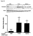
- 230000003287 optical effect Effects 0.000 description 5
- 102000013415 peroxidase activity proteins Human genes 0.000 description 5
- 108040007629 peroxidase activity proteins Proteins 0.000 description 5
- 230000008961 swelling Effects 0.000 description 5
- 238000001262 western blot Methods 0.000 description 5
- YBJHBAHKTGYVGT-ZKWXMUAHSA-N (+)-Biotin Chemical compound N1C(=O)N[C@@H]2[C@H](CCCCC(=O)O)SC[C@@H]21 YBJHBAHKTGYVGT-ZKWXMUAHSA-N 0.000 description 4
- 208000005189 Embolism Diseases 0.000 description 4
- PIWKPBJCKXDKJR-UHFFFAOYSA-N Isoflurane Chemical compound FC(F)OC(Cl)C(F)(F)F PIWKPBJCKXDKJR-UHFFFAOYSA-N 0.000 description 4
- 230000004913 activation Effects 0.000 description 4
- 230000001154 acute effect Effects 0.000 description 4
- 230000036770 blood supply Effects 0.000 description 4
- 208000026106 cerebrovascular disease Diseases 0.000 description 4
- 239000003153 chemical reaction reagent Substances 0.000 description 4
- 230000034994 death Effects 0.000 description 4
- 230000003247 decreasing effect Effects 0.000 description 4
- 238000011156 evaluation Methods 0.000 description 4
- 210000001723 extracellular space Anatomy 0.000 description 4
- 210000002865 immune cell Anatomy 0.000 description 4
- 230000005764 inhibitory process Effects 0.000 description 4
- 229960002725 isoflurane Drugs 0.000 description 4
- 230000000670 limiting effect Effects 0.000 description 4
- 239000012528 membrane Substances 0.000 description 4
- 239000000546 pharmaceutical excipient Substances 0.000 description 4
- 239000000843 powder Substances 0.000 description 4
- 239000003001 serine protease inhibitor Substances 0.000 description 4
- 239000000243 solution Substances 0.000 description 4
- 239000003381 stabilizer Substances 0.000 description 4
- 238000001356 surgical procedure Methods 0.000 description 4
- 239000003981 vehicle Substances 0.000 description 4
- 108091032973 (ribonucleotides)n+m Proteins 0.000 description 3
- BSYNRYMUTXBXSQ-FOQJRBATSA-N 59096-14-9 Chemical compound CC(=O)OC1=CC=CC=C1[14C](O)=O BSYNRYMUTXBXSQ-FOQJRBATSA-N 0.000 description 3
- CSCPPACGZOOCGX-UHFFFAOYSA-N Acetone Chemical compound CC(C)=O CSCPPACGZOOCGX-UHFFFAOYSA-N 0.000 description 3
- 239000005552 B01AC04 - Clopidogrel Substances 0.000 description 3
- LFQSCWFLJHTTHZ-UHFFFAOYSA-N Ethanol Chemical compound CCO LFQSCWFLJHTTHZ-UHFFFAOYSA-N 0.000 description 3
- 102000009123 Fibrin Human genes 0.000 description 3
- 108010073385 Fibrin Proteins 0.000 description 3
- BWGVNKXGVNDBDI-UHFFFAOYSA-N Fibrin monomer Chemical compound CNC(=O)CNC(=O)CN BWGVNKXGVNDBDI-UHFFFAOYSA-N 0.000 description 3
- WSFSSNUMVMOOMR-UHFFFAOYSA-N Formaldehyde Chemical compound O=C WSFSSNUMVMOOMR-UHFFFAOYSA-N 0.000 description 3
- 206010018985 Haemorrhage intracranial Diseases 0.000 description 3
- 208000008574 Intracranial Hemorrhages Diseases 0.000 description 3
- 108010093008 Kinins Proteins 0.000 description 3
- 102000002397 Kinins Human genes 0.000 description 3
- 238000012313 Kruskal-Wallis test Methods 0.000 description 3
- 102000003827 Plasma Kallikrein Human genes 0.000 description 3
- 108090000113 Plasma Kallikrein Proteins 0.000 description 3
- 102000003978 Tissue Plasminogen Activator Human genes 0.000 description 3
- 108090000373 Tissue Plasminogen Activator Proteins 0.000 description 3
- 230000002159 abnormal effect Effects 0.000 description 3
- 239000000654 additive Substances 0.000 description 3
- VREFGVBLTWBCJP-UHFFFAOYSA-N alprazolam Chemical compound C12=CC(Cl)=CC=C2N2C(C)=NN=C2CN=C1C1=CC=CC=C1 VREFGVBLTWBCJP-UHFFFAOYSA-N 0.000 description 3
- 239000003242 anti bacterial agent Substances 0.000 description 3
- 229940088710 antibiotic agent Drugs 0.000 description 3
- 230000004888 barrier function Effects 0.000 description 3
- 230000017531 blood circulation Effects 0.000 description 3
- 238000004364 calculation method Methods 0.000 description 3
- 230000003727 cerebral blood flow Effects 0.000 description 3
- 230000002490 cerebral effect Effects 0.000 description 3
- GKTWGGQPFAXNFI-HNNXBMFYSA-N clopidogrel Chemical compound C1([C@H](N2CC=3C=CSC=3CC2)C(=O)OC)=CC=CC=C1Cl GKTWGGQPFAXNFI-HNNXBMFYSA-N 0.000 description 3
- 229960003009 clopidogrel Drugs 0.000 description 3
- 230000035602 clotting Effects 0.000 description 3
- 238000011161 development Methods 0.000 description 3
- 229960002768 dipyridamole Drugs 0.000 description 3
- IZEKFCXSFNUWAM-UHFFFAOYSA-N dipyridamole Chemical compound C=12N=C(N(CCO)CCO)N=C(N3CCCCC3)C2=NC(N(CCO)CCO)=NC=1N1CCCCC1 IZEKFCXSFNUWAM-UHFFFAOYSA-N 0.000 description 3
- 238000002474 experimental method Methods 0.000 description 3
- 229950003499 fibrin Drugs 0.000 description 3
- 239000012530 fluid Substances 0.000 description 3
- 210000003714 granulocyte Anatomy 0.000 description 3
- 238000003119 immunoblot Methods 0.000 description 3
- 230000004054 inflammatory process Effects 0.000 description 3
- 238000007917 intracranial administration Methods 0.000 description 3
- 210000002540 macrophage Anatomy 0.000 description 3
- 210000000274 microglia Anatomy 0.000 description 3
- 230000003448 neutrophilic effect Effects 0.000 description 3
- 229940099990 ogen Drugs 0.000 description 3
- 229920002866 paraformaldehyde Polymers 0.000 description 3
- 229940110272 plasma derived c1-inhibitor Drugs 0.000 description 3
- 230000002265 prevention Effects 0.000 description 3
- 108090000765 processed proteins & peptides Proteins 0.000 description 3
- 239000000700 radioactive tracer Substances 0.000 description 3
- 210000003625 skull Anatomy 0.000 description 3
- 230000001732 thrombotic effect Effects 0.000 description 3
- 229960000187 tissue plasminogen activator Drugs 0.000 description 3
- 230000001052 transient effect Effects 0.000 description 3
- 230000008733 trauma Effects 0.000 description 3
- 239000011782 vitamin Substances 0.000 description 3
- 229940088594 vitamin Drugs 0.000 description 3
- 229930003231 vitamin Natural products 0.000 description 3
- 235000013343 vitamin Nutrition 0.000 description 3
- VBICKXHEKHSIBG-UHFFFAOYSA-N 1-monostearoylglycerol Chemical compound CCCCCCCCCCCCCCCCCC(=O)OCC(O)CO VBICKXHEKHSIBG-UHFFFAOYSA-N 0.000 description 2
- QKNYBSVHEMOAJP-UHFFFAOYSA-N 2-amino-2-(hydroxymethyl)propane-1,3-diol;hydron;chloride Chemical compound Cl.OCC(N)(CO)CO QKNYBSVHEMOAJP-UHFFFAOYSA-N 0.000 description 2
- 108010088751 Albumins Proteins 0.000 description 2
- 102000009027 Albumins Human genes 0.000 description 2
- 108090001008 Avidin Proteins 0.000 description 2
- 101800004538 Bradykinin Proteins 0.000 description 2
- 102400000967 Bradykinin Human genes 0.000 description 2
- 206010008190 Cerebrovascular accident Diseases 0.000 description 2
- 206010010356 Congenital anomaly Diseases 0.000 description 2
- 206010067276 Cytotoxic oedema Diseases 0.000 description 2
- 102000002045 Endothelin Human genes 0.000 description 2
- 108050009340 Endothelin Proteins 0.000 description 2
- 241000283074 Equus asinus Species 0.000 description 2
- LYCAIKOWRPUZTN-UHFFFAOYSA-N Ethylene glycol Chemical compound OCCO LYCAIKOWRPUZTN-UHFFFAOYSA-N 0.000 description 2
- 206010015866 Extravasation Diseases 0.000 description 2
- 108010080865 Factor XII Proteins 0.000 description 2
- 102000000429 Factor XII Human genes 0.000 description 2
- DHMQDGOQFOQNFH-UHFFFAOYSA-N Glycine Chemical compound NCC(O)=O DHMQDGOQFOQNFH-UHFFFAOYSA-N 0.000 description 2
- QXZGBUJJYSLZLT-UHFFFAOYSA-N H-Arg-Pro-Pro-Gly-Phe-Ser-Pro-Phe-Arg-OH Natural products NC(N)=NCCCC(N)C(=O)N1CCCC1C(=O)N1C(C(=O)NCC(=O)NC(CC=2C=CC=CC=2)C(=O)NC(CO)C(=O)N2C(CCC2)C(=O)NC(CC=2C=CC=CC=2)C(=O)NC(CCCN=C(N)N)C(O)=O)CCC1 QXZGBUJJYSLZLT-UHFFFAOYSA-N 0.000 description 2
- 206010061218 Inflammation Diseases 0.000 description 2
- 102100026061 Mannan-binding lectin serine protease 1 Human genes 0.000 description 2
- 101710117390 Mannan-binding lectin serine protease 1 Proteins 0.000 description 2
- 102100026046 Mannan-binding lectin serine protease 2 Human genes 0.000 description 2
- 101710117460 Mannan-binding lectin serine protease 2 Proteins 0.000 description 2
- 206010060860 Neurological symptom Diseases 0.000 description 2
- 239000004677 Nylon Substances 0.000 description 2
- 229920001213 Polysorbate 20 Polymers 0.000 description 2
- 238000000692 Student's t-test Methods 0.000 description 2
- 229930006000 Sucrose Natural products 0.000 description 2
- CZMRCDWAGMRECN-UGDNZRGBSA-N Sucrose Chemical compound O[C@H]1[C@H](O)[C@@H](CO)O[C@@]1(CO)O[C@@H]1[C@H](O)[C@@H](O)[C@H](O)[C@@H](CO)O1 CZMRCDWAGMRECN-UGDNZRGBSA-N 0.000 description 2
- 239000007983 Tris buffer Substances 0.000 description 2
- 230000000996 additive effect Effects 0.000 description 2
- 238000010171 animal model Methods 0.000 description 2
- 210000002565 arteriole Anatomy 0.000 description 2
- 210000001367 artery Anatomy 0.000 description 2
- 238000003556 assay Methods 0.000 description 2
- 229940075791 berinert Drugs 0.000 description 2
- 239000012620 biological material Substances 0.000 description 2
- 229960002685 biotin Drugs 0.000 description 2
- 235000020958 biotin Nutrition 0.000 description 2
- 239000011616 biotin Substances 0.000 description 2
- 230000000740 bleeding effect Effects 0.000 description 2
- QXZGBUJJYSLZLT-FDISYFBBSA-N bradykinin Chemical compound NC(=N)NCCC[C@H](N)C(=O)N1CCC[C@H]1C(=O)N1[C@H](C(=O)NCC(=O)N[C@@H](CC=2C=CC=CC=2)C(=O)N[C@@H](CO)C(=O)N2[C@@H](CCC2)C(=O)N[C@@H](CC=2C=CC=CC=2)C(=O)N[C@@H](CCCNC(N)=N)C(O)=O)CCC1 QXZGBUJJYSLZLT-FDISYFBBSA-N 0.000 description 2
- 230000003925 brain function Effects 0.000 description 2
- 239000000872 buffer Substances 0.000 description 2
- 210000001736 capillary Anatomy 0.000 description 2
- 210000001168 carotid artery common Anatomy 0.000 description 2
- 210000000269 carotid artery external Anatomy 0.000 description 2
- 210000004004 carotid artery internal Anatomy 0.000 description 2
- 210000004027 cell Anatomy 0.000 description 2
- 230000004154 complement system Effects 0.000 description 2
- 239000002131 composite material Substances 0.000 description 2
- 108700005721 conestat alfa Proteins 0.000 description 2
- 239000000470 constituent Substances 0.000 description 2
- 231100000433 cytotoxic Toxicity 0.000 description 2
- 230000001472 cytotoxic effect Effects 0.000 description 2
- 230000002950 deficient Effects 0.000 description 2
- 230000006735 deficit Effects 0.000 description 2
- 238000013461 design Methods 0.000 description 2
- 201000010099 disease Diseases 0.000 description 2
- ZUBDGKVDJUIMQQ-UBFCDGJISA-N endothelin-1 Chemical compound C([C@@H](C(=O)N[C@@H](CC(C)C)C(=O)N[C@@H](CC(O)=O)C(=O)N[C@@H]([C@@H](C)CC)C(=O)N[C@@H]([C@@H](C)CC)C(=O)N[C@@H](CC=1C2=CC=CC=C2NC=1)C(O)=O)NC(=O)[C@H]1NC(=O)[C@H](CC=2C=CC=CC=2)NC(=O)[C@@H](CC=2C=CC(O)=CC=2)NC(=O)[C@H](C(C)C)NC(=O)[C@H]2CSSC[C@@H](C(N[C@H](CO)C(=O)N[C@@H](CO)C(=O)N[C@H](CC(C)C)C(=O)N[C@@H](CCSC)C(=O)N[C@H](CC(O)=O)C(=O)N[C@@H](CCCCN)C(=O)N[C@@H](CCC(O)=O)C(=O)N2)=O)NC(=O)[C@@H](CO)NC(=O)[C@H](N)CSSC1)C1=CNC=N1 ZUBDGKVDJUIMQQ-UBFCDGJISA-N 0.000 description 2
- 238000005516 engineering process Methods 0.000 description 2
- 230000007717 exclusion Effects 0.000 description 2
- 230000036251 extravasation Effects 0.000 description 2
- 230000001605 fetal effect Effects 0.000 description 2
- 229940012952 fibrinogen Drugs 0.000 description 2
- 239000000945 filler Substances 0.000 description 2
- 239000012634 fragment Substances 0.000 description 2
- 238000004108 freeze drying Methods 0.000 description 2
- 230000004927 fusion Effects 0.000 description 2
- 230000013595 glycosylation Effects 0.000 description 2
- 238000006206 glycosylation reaction Methods 0.000 description 2
- 238000010438 heat treatment Methods 0.000 description 2
- 238000007490 hematoxylin and eosin (H&E) staining Methods 0.000 description 2
- 230000002008 hemorrhagic effect Effects 0.000 description 2
- 238000003364 immunohistochemistry Methods 0.000 description 2
- 238000011065 in-situ storage Methods 0.000 description 2
- 230000003834 intracellular effect Effects 0.000 description 2
- 210000003093 intracellular space Anatomy 0.000 description 2
- 238000001990 intravenous administration Methods 0.000 description 2
- 238000010253 intravenous injection Methods 0.000 description 2
- 239000007788 liquid Substances 0.000 description 2
- 238000001325 log-rank test Methods 0.000 description 2
- 239000006166 lysate Substances 0.000 description 2
- 230000007246 mechanism Effects 0.000 description 2
- 230000001404 mediated effect Effects 0.000 description 2
- 230000004048 modification Effects 0.000 description 2
- 238000012986 modification Methods 0.000 description 2
- 230000009456 molecular mechanism Effects 0.000 description 2
- 230000002981 neuropathic effect Effects 0.000 description 2
- 229920001778 nylon Polymers 0.000 description 2
- 230000007170 pathology Effects 0.000 description 2
- 239000000137 peptide hydrolase inhibitor Substances 0.000 description 2
- 230000035699 permeability Effects 0.000 description 2
- 239000000256 polyoxyethylene sorbitan monolaurate Substances 0.000 description 2
- 235000010486 polyoxyethylene sorbitan monolaurate Nutrition 0.000 description 2
- 230000035935 pregnancy Effects 0.000 description 2
- 239000003755 preservative agent Substances 0.000 description 2
- 230000000750 progressive effect Effects 0.000 description 2
- 230000000770 proinflammatory effect Effects 0.000 description 2
- 238000011321 prophylaxis Methods 0.000 description 2
- 238000003753 real-time PCR Methods 0.000 description 2
- 230000019705 regulation of vascular permeability Effects 0.000 description 2
- 230000001105 regulatory effect Effects 0.000 description 2
- 230000002441 reversible effect Effects 0.000 description 2
- MTCFGRXMJLQNBG-UHFFFAOYSA-N serine Chemical compound OCC(N)C(O)=O MTCFGRXMJLQNBG-UHFFFAOYSA-N 0.000 description 2
- 239000011780 sodium chloride Substances 0.000 description 2
- 230000000087 stabilizing effect Effects 0.000 description 2
- 239000005720 sucrose Substances 0.000 description 2
- 208000024891 symptom Diseases 0.000 description 2
- 230000009885 systemic effect Effects 0.000 description 2
- LENZDBCJOHFCAS-UHFFFAOYSA-N tris Chemical compound OCC(N)(CO)CO LENZDBCJOHFCAS-UHFFFAOYSA-N 0.000 description 2
- QAPSNMNOIOSXSQ-YNEHKIRRSA-N 1-[(2r,4s,5r)-4-[tert-butyl(dimethyl)silyl]oxy-5-(hydroxymethyl)oxolan-2-yl]-5-methylpyrimidine-2,4-dione Chemical compound O=C1NC(=O)C(C)=CN1[C@@H]1O[C@H](CO)[C@@H](O[Si](C)(C)C(C)(C)C)C1 QAPSNMNOIOSXSQ-YNEHKIRRSA-N 0.000 description 1
- PRDFBSVERLRRMY-UHFFFAOYSA-N 2'-(4-ethoxyphenyl)-5-(4-methylpiperazin-1-yl)-2,5'-bibenzimidazole Chemical compound C1=CC(OCC)=CC=C1C1=NC2=CC=C(C=3NC4=CC(=CC=C4N=3)N3CCN(C)CC3)C=C2N1 PRDFBSVERLRRMY-UHFFFAOYSA-N 0.000 description 1
- HSTOKWSFWGCZMH-UHFFFAOYSA-N 3,3'-diaminobenzidine Chemical compound C1=C(N)C(N)=CC=C1C1=CC=C(N)C(N)=C1 HSTOKWSFWGCZMH-UHFFFAOYSA-N 0.000 description 1
- 241001316595 Acris Species 0.000 description 1
- 102000007469 Actins Human genes 0.000 description 1
- 108010085238 Actins Proteins 0.000 description 1
- 108700028369 Alleles Proteins 0.000 description 1
- GUBGYTABKSRVRQ-XLOQQCSPSA-N Alpha-Lactose Chemical compound O[C@@H]1[C@@H](O)[C@@H](O)[C@@H](CO)O[C@H]1O[C@@H]1[C@@H](CO)O[C@H](O)[C@H](O)[C@H]1O GUBGYTABKSRVRQ-XLOQQCSPSA-N 0.000 description 1
- 102000013455 Amyloid beta-Peptides Human genes 0.000 description 1
- 108010090849 Amyloid beta-Peptides Proteins 0.000 description 1
- 206010002091 Anaesthesia Diseases 0.000 description 1
- 241001550224 Apha Species 0.000 description 1
- BSYNRYMUTXBXSQ-UHFFFAOYSA-N Aspirin Chemical compound CC(=O)OC1=CC=CC=C1C(O)=O BSYNRYMUTXBXSQ-UHFFFAOYSA-N 0.000 description 1
- 208000025978 Athletic injury Diseases 0.000 description 1
- 238000000035 BCA protein assay Methods 0.000 description 1
- 102000010183 Bradykinin receptor Human genes 0.000 description 1
- 108050001736 Bradykinin receptor Proteins 0.000 description 1
- 208000007204 Brain death Diseases 0.000 description 1
- 208000014644 Brain disease Diseases 0.000 description 1
- 241000283707 Capra Species 0.000 description 1
- 102000003847 Carboxypeptidase B2 Human genes 0.000 description 1
- 108090000201 Carboxypeptidase B2 Proteins 0.000 description 1
- 108010067225 Cell Adhesion Molecules Proteins 0.000 description 1
- 102000016289 Cell Adhesion Molecules Human genes 0.000 description 1
- 208000018652 Closed Head injury Diseases 0.000 description 1
- 206010010071 Coma Diseases 0.000 description 1
- 108090000695 Cytokines Proteins 0.000 description 1
- 102000004127 Cytokines Human genes 0.000 description 1
- FBPFZTCFMRRESA-KVTDHHQDSA-N D-Mannitol Chemical compound OC[C@@H](O)[C@@H](O)[C@H](O)[C@H](O)CO FBPFZTCFMRRESA-KVTDHHQDSA-N 0.000 description 1
- 241000282326 Felis catus Species 0.000 description 1
- 102000008946 Fibrinogen Human genes 0.000 description 1
- 108010049003 Fibrinogen Proteins 0.000 description 1
- 108010010803 Gelatin Proteins 0.000 description 1
- WQZGKKKJIJFFOK-GASJEMHNSA-N Glucose Natural products OC[C@H]1OC(O)[C@H](O)[C@@H](O)[C@@H]1O WQZGKKKJIJFFOK-GASJEMHNSA-N 0.000 description 1
- 102100031181 Glyceraldehyde-3-phosphate dehydrogenase Human genes 0.000 description 1
- 239000004471 Glycine Substances 0.000 description 1
- 102000003886 Glycoproteins Human genes 0.000 description 1
- 108090000288 Glycoproteins Proteins 0.000 description 1
- 206010018852 Haematoma Diseases 0.000 description 1
- 206010019196 Head injury Diseases 0.000 description 1
- 208000005139 Hereditary Angioedema Types I and II Diseases 0.000 description 1
- 206010019860 Hereditary angioedema Diseases 0.000 description 1
- 101001046686 Homo sapiens Integrin alpha-M Proteins 0.000 description 1
- 206010020772 Hypertension Diseases 0.000 description 1
- 206010058558 Hypoperfusion Diseases 0.000 description 1
- 206010021639 Incontinence Diseases 0.000 description 1
- 102100022338 Integrin alpha-M Human genes 0.000 description 1
- 206010022840 Intraventricular haemorrhage Diseases 0.000 description 1
- 108010003195 Kallidin Proteins 0.000 description 1
- FYSKZKQBTVLYEQ-FSLKYBNLSA-N Kallidin Chemical compound NCCCC[C@H](N)C(=O)N[C@@H](CCCN=C(N)N)C(=O)N1CCC[C@H]1C(=O)N1[C@H](C(=O)NCC(=O)N[C@@H](CC=2C=CC=CC=2)C(=O)N[C@@H](CO)C(=O)N2[C@@H](CCC2)C(=O)N[C@@H](CC=2C=CC=CC=2)C(=O)N[C@@H](CCCN=C(N)N)C(O)=O)CCC1 FYSKZKQBTVLYEQ-FSLKYBNLSA-N 0.000 description 1
- 102000001399 Kallikrein Human genes 0.000 description 1
- 108060005987 Kallikrein Proteins 0.000 description 1
- 108010077861 Kininogens Proteins 0.000 description 1
- 102000010631 Kininogens Human genes 0.000 description 1
- ONIBWKKTOPOVIA-BYPYZUCNSA-N L-Proline Chemical compound OC(=O)[C@@H]1CCCN1 ONIBWKKTOPOVIA-BYPYZUCNSA-N 0.000 description 1
- GUBGYTABKSRVRQ-QKKXKWKRSA-N Lactose Natural products OC[C@H]1O[C@@H](O[C@H]2[C@H](O)[C@@H](O)C(O)O[C@@H]2CO)[C@H](O)[C@@H](O)[C@H]1O GUBGYTABKSRVRQ-QKKXKWKRSA-N 0.000 description 1
- 229930195725 Mannitol Natural products 0.000 description 1
- 241000699660 Mus musculus Species 0.000 description 1
- 101100326461 Mus musculus C1ra gene Proteins 0.000 description 1
- 101100326462 Mus musculus C1rb gene Proteins 0.000 description 1
- 101100329495 Mus musculus C1sa gene Proteins 0.000 description 1
- 101100329496 Mus musculus C1sb gene Proteins 0.000 description 1
- GXCLVBGFBYZDAG-UHFFFAOYSA-N N-[2-(1H-indol-3-yl)ethyl]-N-methylprop-2-en-1-amine Chemical compound CN(CCC1=CNC2=C1C=CC=C2)CC=C GXCLVBGFBYZDAG-UHFFFAOYSA-N 0.000 description 1
- 206010028851 Necrosis Diseases 0.000 description 1
- 206010028980 Neoplasm Diseases 0.000 description 1
- 241000283973 Oryctolagus cuniculus Species 0.000 description 1
- 240000007594 Oryza sativa Species 0.000 description 1
- 235000007164 Oryza sativa Nutrition 0.000 description 1
- 239000002033 PVDF binder Substances 0.000 description 1
- 206010033799 Paralysis Diseases 0.000 description 1
- 208000028361 Penetrating Head injury Diseases 0.000 description 1
- 102000007079 Peptide Fragments Human genes 0.000 description 1
- 108010033276 Peptide Fragments Proteins 0.000 description 1
- ONIBWKKTOPOVIA-UHFFFAOYSA-N Proline Natural products OC(=O)C1CCCN1 ONIBWKKTOPOVIA-UHFFFAOYSA-N 0.000 description 1
- 229940124158 Protease/peptidase inhibitor Drugs 0.000 description 1
- 108010029485 Protein Isoforms Proteins 0.000 description 1
- 102000001708 Protein Isoforms Human genes 0.000 description 1
- 239000012083 RIPA buffer Substances 0.000 description 1
- 238000002123 RNA extraction Methods 0.000 description 1
- 238000011529 RT qPCR Methods 0.000 description 1
- 241000384016 Rattus morotaiensis Species 0.000 description 1
- 101001065563 Rattus norvegicus Lymphocyte antigen 6B Proteins 0.000 description 1
- 208000031074 Reinjury Diseases 0.000 description 1
- 206010039203 Road traffic accident Diseases 0.000 description 1
- 206010039330 Ruptured cerebral aneurysm Diseases 0.000 description 1
- 102000003800 Selectins Human genes 0.000 description 1
- 108090000184 Selectins Proteins 0.000 description 1
- 229940122055 Serine protease inhibitor Drugs 0.000 description 1
- 101710102218 Serine protease inhibitor Proteins 0.000 description 1
- 241001522306 Serinus serinus Species 0.000 description 1
- 102000008847 Serpin Human genes 0.000 description 1
- 108050000761 Serpin Proteins 0.000 description 1
- VYPSYNLAJGMNEJ-UHFFFAOYSA-N Silicium dioxide Chemical compound O=[Si]=O VYPSYNLAJGMNEJ-UHFFFAOYSA-N 0.000 description 1
- XUIMIQQOPSSXEZ-UHFFFAOYSA-N Silicon Chemical compound [Si] XUIMIQQOPSSXEZ-UHFFFAOYSA-N 0.000 description 1
- 229920002472 Starch Polymers 0.000 description 1
- 108010090804 Streptavidin Proteins 0.000 description 1
- 208000008116 Traumatic Cerebral Hemorrhage Diseases 0.000 description 1
- 230000001133 acceleration Effects 0.000 description 1
- 229960001138 acetylsalicylic acid Drugs 0.000 description 1
- 239000012190 activator Substances 0.000 description 1
- 238000013019 agitation Methods 0.000 description 1
- 230000000961 alloantigen Effects 0.000 description 1
- 150000001413 amino acids Chemical class 0.000 description 1
- 230000003321 amplification Effects 0.000 description 1
- 230000037005 anaesthesia Effects 0.000 description 1
- 230000003501 anti-edematous effect Effects 0.000 description 1
- 230000003110 anti-inflammatory effect Effects 0.000 description 1
- 230000000702 anti-platelet effect Effects 0.000 description 1
- 239000003146 anticoagulant agent Substances 0.000 description 1
- 230000010100 anticoagulation Effects 0.000 description 1
- 239000000427 antigen Substances 0.000 description 1
- 108091007433 antigens Proteins 0.000 description 1
- 102000036639 antigens Human genes 0.000 description 1
- 239000003443 antiviral agent Substances 0.000 description 1
- 238000013459 approach Methods 0.000 description 1
- 210000000576 arachnoid Anatomy 0.000 description 1
- 230000003542 behavioural effect Effects 0.000 description 1
- 230000009286 beneficial effect Effects 0.000 description 1
- WQZGKKKJIJFFOK-VFUOTHLCSA-N beta-D-glucose Chemical compound OC[C@H]1O[C@@H](O)[C@H](O)[C@@H](O)[C@@H]1O WQZGKKKJIJFFOK-VFUOTHLCSA-N 0.000 description 1
- 230000023555 blood coagulation Effects 0.000 description 1
- 210000004958 brain cell Anatomy 0.000 description 1
- 208000029028 brain injury Diseases 0.000 description 1
- 230000003139 buffering effect Effects 0.000 description 1
- 239000004067 bulking agent Substances 0.000 description 1
- 239000000969 carrier Substances 0.000 description 1
- 230000015556 catabolic process Effects 0.000 description 1
- 230000012292 cell migration Effects 0.000 description 1
- 230000001413 cellular effect Effects 0.000 description 1
- 230000004640 cellular pathway Effects 0.000 description 1
- 210000004289 cerebral ventricle Anatomy 0.000 description 1
- 210000001175 cerebrospinal fluid Anatomy 0.000 description 1
- 238000006243 chemical reaction Methods 0.000 description 1
- 239000007795 chemical reaction product Substances 0.000 description 1
- 239000003795 chemical substances by application Substances 0.000 description 1
- 230000001684 chronic effect Effects 0.000 description 1
- 229940088949 cinryze Drugs 0.000 description 1
- 230000004087 circulation Effects 0.000 description 1
- 230000015271 coagulation Effects 0.000 description 1
- 238000005345 coagulation Methods 0.000 description 1
- 230000001149 cognitive effect Effects 0.000 description 1
- 230000000052 comparative effect Effects 0.000 description 1
- 230000001447 compensatory effect Effects 0.000 description 1
- 230000024203 complement activation Effects 0.000 description 1
- 230000000295 complement effect Effects 0.000 description 1
- 239000002299 complementary DNA Substances 0.000 description 1
- 238000007906 compression Methods 0.000 description 1
- 230000006835 compression Effects 0.000 description 1
- 238000007796 conventional method Methods 0.000 description 1
- 239000013256 coordination polymer Substances 0.000 description 1
- 230000036757 core body temperature Effects 0.000 description 1
- 230000002596 correlated effect Effects 0.000 description 1
- 230000000875 corresponding effect Effects 0.000 description 1
- 230000000991 decompressive effect Effects 0.000 description 1
- 230000007812 deficiency Effects 0.000 description 1
- 238000001514 detection method Methods 0.000 description 1
- 230000006866 deterioration Effects 0.000 description 1
- 238000009826 distribution Methods 0.000 description 1
- 239000003937 drug carrier Substances 0.000 description 1
- 230000004064 dysfunction Effects 0.000 description 1
- 230000002996 emotional effect Effects 0.000 description 1
- 239000003995 emulsifying agent Substances 0.000 description 1
- 210000002889 endothelial cell Anatomy 0.000 description 1
- 206010015037 epilepsy Diseases 0.000 description 1
- 235000013861 fat-free Nutrition 0.000 description 1
- 235000013312 flour Nutrition 0.000 description 1
- 210000003194 forelimb Anatomy 0.000 description 1
- 239000008273 gelatin Substances 0.000 description 1
- 229920000159 gelatin Polymers 0.000 description 1
- 235000019322 gelatine Nutrition 0.000 description 1
- 235000011852 gelatine desserts Nutrition 0.000 description 1
- 239000008103 glucose Substances 0.000 description 1
- 235000001727 glucose Nutrition 0.000 description 1
- 108020004445 glyceraldehyde-3-phosphate dehydrogenase Proteins 0.000 description 1
- YQEMORVAKMFKLG-UHFFFAOYSA-N glycerine monostearate Natural products CCCCCCCCCCCCCCCCCC(=O)OC(CO)CO YQEMORVAKMFKLG-UHFFFAOYSA-N 0.000 description 1
- SVUQHVRAGMNPLW-UHFFFAOYSA-N glycerol monostearate Natural products CCCCCCCCCCCCCCCCC(=O)OCC(O)CO SVUQHVRAGMNPLW-UHFFFAOYSA-N 0.000 description 1
- 230000012010 growth Effects 0.000 description 1
- 230000036541 health Effects 0.000 description 1
- 230000002949 hemolytic effect Effects 0.000 description 1
- 238000000265 homogenisation Methods 0.000 description 1
- 102000044507 human SERPING1 Human genes 0.000 description 1
- WGCNASOHLSPBMP-UHFFFAOYSA-N hydroxyacetaldehyde Natural products OCC=O WGCNASOHLSPBMP-UHFFFAOYSA-N 0.000 description 1
- 230000000596 hypostatic effect Effects 0.000 description 1
- 238000003365 immunocytochemistry Methods 0.000 description 1
- 238000003125 immunofluorescent labeling Methods 0.000 description 1
- 230000001771 impaired effect Effects 0.000 description 1
- 230000000415 inactivating effect Effects 0.000 description 1
- 208000015181 infectious disease Diseases 0.000 description 1
- 238000007918 intramuscular administration Methods 0.000 description 1
- 238000007912 intraperitoneal administration Methods 0.000 description 1
- 238000007913 intrathecal administration Methods 0.000 description 1
- 230000006623 intrinsic pathway Effects 0.000 description 1
- 230000003447 ipsilateral effect Effects 0.000 description 1
- 239000008101 lactose Substances 0.000 description 1
- 230000003902 lesion Effects 0.000 description 1
- 230000023404 leukocyte cell-cell adhesion Effects 0.000 description 1
- 239000012160 loading buffer Substances 0.000 description 1
- 230000033001 locomotion Effects 0.000 description 1
- 238000007726 management method Methods 0.000 description 1
- 239000000594 mannitol Substances 0.000 description 1
- 235000010355 mannitol Nutrition 0.000 description 1
- 238000004519 manufacturing process Methods 0.000 description 1
- 239000011159 matrix material Substances 0.000 description 1
- 238000005259 measurement Methods 0.000 description 1
- 108020004999 messenger RNA Proteins 0.000 description 1
- 235000013336 milk Nutrition 0.000 description 1
- 239000008267 milk Substances 0.000 description 1
- 210000004080 milk Anatomy 0.000 description 1
- 238000002156 mixing Methods 0.000 description 1
- 230000003990 molecular pathway Effects 0.000 description 1
- 238000010172 mouse model Methods 0.000 description 1
- 230000017074 necrotic cell death Effects 0.000 description 1
- 239000013642 negative control Substances 0.000 description 1
- 230000004770 neurodegeneration Effects 0.000 description 1
- 230000009251 neurologic dysfunction Effects 0.000 description 1
- 230000007971 neurological deficit Effects 0.000 description 1
- 208000015015 neurological dysfunction Diseases 0.000 description 1
- 230000000926 neurological effect Effects 0.000 description 1
- 238000001422 normality test Methods 0.000 description 1
- 230000037000 normothermia Effects 0.000 description 1
- 238000003199 nucleic acid amplification method Methods 0.000 description 1
- 210000000056 organ Anatomy 0.000 description 1
- 230000003204 osmotic effect Effects 0.000 description 1
- 230000036407 pain Effects 0.000 description 1
- 238000007911 parenteral administration Methods 0.000 description 1
- 230000003950 pathogenic mechanism Effects 0.000 description 1
- 230000004963 pathophysiological condition Effects 0.000 description 1
- 230000007310 pathophysiology Effects 0.000 description 1
- 230000035515 penetration Effects 0.000 description 1
- 239000000813 peptide hormone Substances 0.000 description 1
- 230000009984 peri-natal effect Effects 0.000 description 1
- 230000002085 persistent effect Effects 0.000 description 1
- 239000005426 pharmaceutical component Substances 0.000 description 1
- 229940124531 pharmaceutical excipient Drugs 0.000 description 1
- 230000000144 pharmacologic effect Effects 0.000 description 1
- 229960004923 phenprocoumon Drugs 0.000 description 1
- DQDAYGNAKTZFIW-UHFFFAOYSA-N phenprocoumon Chemical compound OC=1C2=CC=CC=C2OC(=O)C=1C(CC)C1=CC=CC=C1 DQDAYGNAKTZFIW-UHFFFAOYSA-N 0.000 description 1
- 230000000704 physical effect Effects 0.000 description 1
- 210000003446 pia mater Anatomy 0.000 description 1
- 239000006187 pill Substances 0.000 description 1
- 229920002981 polyvinylidene fluoride Polymers 0.000 description 1
- 239000002243 precursor Substances 0.000 description 1
- 238000002203 pretreatment Methods 0.000 description 1
- 208000037920 primary disease Diseases 0.000 description 1
- 230000008569 process Effects 0.000 description 1
- 239000000047 product Substances 0.000 description 1
- 230000000069 prophylactic effect Effects 0.000 description 1
- QQONPFPTGQHPMA-UHFFFAOYSA-N propylene Natural products CC=C QQONPFPTGQHPMA-UHFFFAOYSA-N 0.000 description 1
- 125000004805 propylene group Chemical group [H]C([H])([H])C([H])([*:1])C([H])([H])[*:2] 0.000 description 1
- 230000001681 protective effect Effects 0.000 description 1
- 230000001698 pyrogenic effect Effects 0.000 description 1
- 239000002464 receptor antagonist Substances 0.000 description 1
- 229940044551 receptor antagonist Drugs 0.000 description 1
- 108020003175 receptors Proteins 0.000 description 1
- 102000005962 receptors Human genes 0.000 description 1
- 238000011084 recovery Methods 0.000 description 1
- 230000009467 reduction Effects 0.000 description 1
- 238000011160 research Methods 0.000 description 1
- 230000029058 respiratory gaseous exchange Effects 0.000 description 1
- 238000010839 reverse transcription Methods 0.000 description 1
- 235000009566 rice Nutrition 0.000 description 1
- 238000011808 rodent model Methods 0.000 description 1
- 229940009560 ruconest Drugs 0.000 description 1
- 150000003839 salts Chemical group 0.000 description 1
- 238000005070 sampling Methods 0.000 description 1
- 230000035939 shock Effects 0.000 description 1
- 239000000741 silica gel Substances 0.000 description 1
- 229910002027 silica gel Inorganic materials 0.000 description 1
- 229910052710 silicon Inorganic materials 0.000 description 1
- 239000010703 silicon Substances 0.000 description 1
- 229920002379 silicone rubber Polymers 0.000 description 1
- 235000020183 skimmed milk Nutrition 0.000 description 1
- 230000000192 social effect Effects 0.000 description 1
- 238000002415 sodium dodecyl sulfate polyacrylamide gel electrophoresis Methods 0.000 description 1
- RYYKJJJTJZKILX-UHFFFAOYSA-M sodium octadecanoate Chemical compound [Na+].CCCCCCCCCCCCCCCCCC([O-])=O RYYKJJJTJZKILX-UHFFFAOYSA-M 0.000 description 1
- 239000007787 solid Substances 0.000 description 1
- 239000002904 solvent Substances 0.000 description 1
- 210000000273 spinal nerve root Anatomy 0.000 description 1
- 238000009987 spinning Methods 0.000 description 1
- 230000002269 spontaneous effect Effects 0.000 description 1
- 238000010561 standard procedure Methods 0.000 description 1
- 239000008107 starch Substances 0.000 description 1
- 235000019698 starch Nutrition 0.000 description 1
- 238000007619 statistical method Methods 0.000 description 1
- 239000008227 sterile water for injection Substances 0.000 description 1
- 210000002330 subarachnoid space Anatomy 0.000 description 1
- 238000007920 subcutaneous administration Methods 0.000 description 1
- 239000006228 supernatant Substances 0.000 description 1
- 230000001629 suppression Effects 0.000 description 1
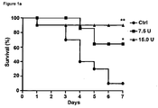
- 230000004083 survival effect Effects 0.000 description 1
- 239000000454 talc Substances 0.000 description 1
- 229910052623 talc Inorganic materials 0.000 description 1
- 230000008685 targeting Effects 0.000 description 1
- 238000002560 therapeutic procedure Methods 0.000 description 1
- 208000037816 tissue injury Diseases 0.000 description 1
- 230000009466 transformation Effects 0.000 description 1
- 238000011830 transgenic mouse model Methods 0.000 description 1
- 230000032258 transport Effects 0.000 description 1
- 230000000472 traumatic effect Effects 0.000 description 1
- 230000008728 vascular permeability Effects 0.000 description 1
- 210000005166 vasculature Anatomy 0.000 description 1
- 210000003462 vein Anatomy 0.000 description 1
- 210000000264 venule Anatomy 0.000 description 1
- 229940019333 vitamin k antagonists Drugs 0.000 description 1
- 238000003260 vortexing Methods 0.000 description 1
- 238000005406 washing Methods 0.000 description 1
- 238000009736 wetting Methods 0.000 description 1
- 239000000080 wetting agent Substances 0.000 description 1
- DGVVWUTYPXICAM-UHFFFAOYSA-N β‐Mercaptoethanol Chemical compound OCCS DGVVWUTYPXICAM-UHFFFAOYSA-N 0.000 description 1
Images


Classifications
-
- A—HUMAN NECESSITIES
- A61—MEDICAL OR VETERINARY SCIENCE; HYGIENE
- A61K—PREPARATIONS FOR MEDICAL, DENTAL OR TOILETRY PURPOSES
- A61K38/00—Medicinal preparations containing peptides
- A61K38/16—Peptides having more than 20 amino acids; Gastrins; Somatostatins; Melanotropins; Derivatives thereof
- A61K38/55—Protease inhibitors
- A61K38/57—Protease inhibitors from animals; from humans
-
- A—HUMAN NECESSITIES
- A61—MEDICAL OR VETERINARY SCIENCE; HYGIENE
- A61P—SPECIFIC THERAPEUTIC ACTIVITY OF CHEMICAL COMPOUNDS OR MEDICINAL PREPARATIONS
- A61P25/00—Drugs for disorders of the nervous system
-
- A—HUMAN NECESSITIES
- A61—MEDICAL OR VETERINARY SCIENCE; HYGIENE
- A61P—SPECIFIC THERAPEUTIC ACTIVITY OF CHEMICAL COMPOUNDS OR MEDICINAL PREPARATIONS
- A61P3/00—Drugs for disorders of the metabolism
- A61P3/08—Drugs for disorders of the metabolism for glucose homeostasis
- A61P3/10—Drugs for disorders of the metabolism for glucose homeostasis for hyperglycaemia, e.g. antidiabetics
-
- A—HUMAN NECESSITIES
- A61—MEDICAL OR VETERINARY SCIENCE; HYGIENE
- A61P—SPECIFIC THERAPEUTIC ACTIVITY OF CHEMICAL COMPOUNDS OR MEDICINAL PREPARATIONS
- A61P31/00—Antiinfectives, i.e. antibiotics, antiseptics, chemotherapeutics
- A61P31/04—Antibacterial agents
-
- A—HUMAN NECESSITIES
- A61—MEDICAL OR VETERINARY SCIENCE; HYGIENE
- A61P—SPECIFIC THERAPEUTIC ACTIVITY OF CHEMICAL COMPOUNDS OR MEDICINAL PREPARATIONS
- A61P31/00—Antiinfectives, i.e. antibiotics, antiseptics, chemotherapeutics
- A61P31/12—Antivirals
-
- A—HUMAN NECESSITIES
- A61—MEDICAL OR VETERINARY SCIENCE; HYGIENE
- A61P—SPECIFIC THERAPEUTIC ACTIVITY OF CHEMICAL COMPOUNDS OR MEDICINAL PREPARATIONS
- A61P31/00—Antiinfectives, i.e. antibiotics, antiseptics, chemotherapeutics
- A61P31/12—Antivirals
- A61P31/14—Antivirals for RNA viruses
- A61P31/18—Antivirals for RNA viruses for HIV
-
- A—HUMAN NECESSITIES
- A61—MEDICAL OR VETERINARY SCIENCE; HYGIENE
- A61P—SPECIFIC THERAPEUTIC ACTIVITY OF CHEMICAL COMPOUNDS OR MEDICINAL PREPARATIONS
- A61P35/00—Antineoplastic agents
-
- A—HUMAN NECESSITIES
- A61—MEDICAL OR VETERINARY SCIENCE; HYGIENE
- A61P—SPECIFIC THERAPEUTIC ACTIVITY OF CHEMICAL COMPOUNDS OR MEDICINAL PREPARATIONS
- A61P43/00—Drugs for specific purposes, not provided for in groups A61P1/00-A61P41/00
-
- A—HUMAN NECESSITIES
- A61—MEDICAL OR VETERINARY SCIENCE; HYGIENE
- A61P—SPECIFIC THERAPEUTIC ACTIVITY OF CHEMICAL COMPOUNDS OR MEDICINAL PREPARATIONS
- A61P7/00—Drugs for disorders of the blood or the extracellular fluid
- A61P7/10—Antioedematous agents; Diuretics
-
- A—HUMAN NECESSITIES
- A61—MEDICAL OR VETERINARY SCIENCE; HYGIENE
- A61P—SPECIFIC THERAPEUTIC ACTIVITY OF CHEMICAL COMPOUNDS OR MEDICINAL PREPARATIONS
- A61P9/00—Drugs for disorders of the cardiovascular system
-
- A—HUMAN NECESSITIES
- A61—MEDICAL OR VETERINARY SCIENCE; HYGIENE
- A61P—SPECIFIC THERAPEUTIC ACTIVITY OF CHEMICAL COMPOUNDS OR MEDICINAL PREPARATIONS
- A61P9/00—Drugs for disorders of the cardiovascular system
- A61P9/10—Drugs for disorders of the cardiovascular system for treating ischaemic or atherosclerotic diseases, e.g. antianginal drugs, coronary vasodilators, drugs for myocardial infarction, retinopathy, cerebrovascula insufficiency, renal arteriosclerosis
Definitions
- the subject of the present invention is, in the most general aspect, the prevention and/or treatment of a secondary edema.
- the present invention relates to a C1-Inhibitor for use in a method of preventing the formation and/or reducing the size of a secondary edema of the central nervous system (CNS) in a subject wherein the subject has or has had at least one disorder selected from the group consisting of stroke, ischemic stroke, hemorrhagic stroke, perinatal stroke, traumatic brain injury and spinal cord injury.
- the secondary edema of the CNS is a secondary brain edema.
- Another subject of the present invention is the treatment of disorders associated with an increased permeability of the blood brain barrier or the blood spinal cord barrier.
- a third subject is a plasma-derived C1-inhibitor for use in a method of preventing, reducing or treating brain ischemia-reperfusion injury.
- brain ischemia and the subsequent injury due to reperfusion is complex and involves a myriad of distinct molecular and cellular pathways.
- persisting ischemia is structural disintegration of the blood-brain-barrier which in consequence leads to the formation of brain edema.
- Excessive edema can harm otherwise healthy brain regions simply by mechanic compression and is a frequent cause of worsening of neurological symptoms in stroke patients.
- Up to now convincing strategies on a pharmacological basis to combat edema formation in acute ischemic stroke are lacking.
- Brain edema is defined as an increase in brain volume resulting from a localized or diffuse abnormal accumulation of fluid within the brain parenchyma.
- brain edema is classified into 4 different groups: vasogenic, cytotoxic, hydrocephalic (or interstitial) and osmotic (or hypostatic) edema.
- vasogenic, cytotoxic, hydrocephalic (or interstitial) and osmotic (or hypostatic) edema Despite this classification of distinct forms of edema, in most clinical situations there is a combination of different types of edema depending on the time course of the disease.
- the cytotoxic and/or vasogenic brain edema seem to play major roles.
- mediators involved in brain edema formation many mediators can be mentioned (e.g. Bradykinin), which have many properties other than their effects on brain edema formation ( Nag et al. (2009) Acta Neuropathol.; 118:197-217 ).
- brain edema Initially in brain edema, the changes in brain volume are compensated by a decrease in cerebrospinal fluid and blood volume, whereby the primary brain edema is mainly a cytotoxic edema. In large hemispheric lesions, progressive swelling exceeds these compensatory mechanisms and an increase in the intracranial pressure (i.e. formation of secondary or malignant brain edema) results in herniations of cerebral tissue leading to death. Hence vasogenic, malignant brain edema continues to be a major cause of mortality after diverse types of brain pathologies such as major cerebral infarcts, hemorrhages, trauma, infections and tumors. The lack of effective treatment for brain edema remains a stimulus for continued interest and research.
- ischemic stroke secondary brain edema is a frequent cause of secondary infarct growth and subsequent deterioration of neurological symptoms during the course of ischemic stroke ( Ayata and Ropper (2002) J Clin Neurosci.; 9:113-124 ; Bardutzky and Schwab (2007) Stroke; 38:3084-3094 ).
- malignant middle cerebral artery (MCA) infarction is a term used to describe complete MCA territory infarction with significant space occupying effect and herniation of brain tissue.
- the incidence of malignant MCA infarction is estimated to be less than 1% of all strokes.
- the mortality with conservative forms of medical treatment is approximately 80% and coma terminates in brain death within 2-5 days of onset.
- the kallikrein-kinin system (KKS) is initiated by blood coagulation factor XII (FXII, Hageman factor) and plays an important role in the regulation of vascular permeability and edema formation ( Leeb-Lundberg et al. (2005) Pharmacol. Rev.; 571:27-77 ). The activation of the KKS was recently proven also in stroke patients ( Wagner et al. (2002) J. Neurol. Sci.; 202:75-76 ). Kinins (e. g. bradykinin, kallidin) constitute the end products of the KKS.
- Kinins are highly active proinflammatory peptide hormones which are released by kallikreins from their precursors, kininogens, during various kinds of tissue injury including brain ischemia.
- the cellular effects of kinins are mediated by two different bradykinin receptors, B1 R and B2R. Activation of these receptors triggers inflammatory processes in the target organ such as the release of proinflammatory cytokines or the attraction of immune cells as well as increased vascular permeability.
- C1-esterase inhibitor (C1-INH) is a 478 amino acid glycoprotein belonging to the superfamily of serine protease inhibitors called serpins. Its designation originates from the initial description as the only known physiological inhibitor of the classical complement pathway in blood and tissue. However, C1-INH is also a major regulator of the KKS by blocking activated FXII and plasma kallikrein. Apart from several other functions (e.g. FXIa inhibition), it is the only known physiological inhibitor of C1s and C1r, the activated homologous serin proteases of the first component of the complement system.
- the technical problem underlying the present invention was to provide alternative and/or improved means and methods for successfully targeting secondary brain edema that form the basis or may allow the development of more satisfactory medicaments for the treatment and/or prevention of the secondary brain edema.
- Such an initial injury or primary disorder could be an occlusion of a blood vessel in the brain, e.g. an ischemic stroke, or a hemorrhage in the brain, e.g. a hemorrhagic stroke.
- the present invention relates to a C1-Inhibitor for use in a method of preventing the formation and/or reducing the size of a secondary edema of the central nervous system (CNS) in a subject wherein the subject has or has had at least one disorder selected from the group consisting of stroke, ischemic stroke, hemorrhagic stroke, perinatal stroke, traumatic brain injury, and spinal cord injury.
- the hemorrhagic stroke is a cerebral hemorrhage or a subarachnoidal hemorrhage.
- the secondary edema of the central nervous system is a secondary brain edema or a secondary spinal cord edema.
- C1-inhibitor refers to the proteins or fragments thereof that function as serine protease inhibitors to inhibit proteases associated with the complement system, preferably proteases C1r and C1s as well as MASP-1 and MASP-2, with the kallikrein-kinin system, preferably plasma kallikrein and factor Xlla, and with the coagulation system, preferably factor Xla.
- C1-INH can serve as an antiinflammatory molecule that reduces the selectins-mediated leukocyte adhesion to endothelial cells.
- C1-INH as used here can be a native serine protease inhibitor or active fragment thereof, or it can comprise a recombinant peptide, a synthetic peptide, peptide mimetic, or peptide fragment that provides similar functional properties - e.g., the inhibition of proteases C1 and C1 s, and/or MASP-1 and MASP-2 and/or factor Xlla and/or factor Xla.
- the structure and function of C1-Inhibitor see U.S. Patent 4,915,945 ; U.S. Patent 5,939,389 ; U.S. Patent 6,248,365 ; U.S. Patent 7,053,176 ; and WO 2007/073186 .
- the inhibitor is a plasma-derived or a recombinant C1-Inhibitor.
- said inhibitor is identical with the naturally occurring human protein or a variant thereof.
- the C1-INH shall encompass all natural occurring alleles which have the same function as the C1-inhibitor.
- said inhibitor is the human C1 Esterase Inhibitor.
- the C1-inhibitor according to the present invention is modified to improve bioavailability and/or half-life, to improve efficacy and/ or to reduce potential side effects.
- the modification can be realized by recombinant or other steps. Examples for such a modification could be a glycosylation or an albumin fusion of the described C1-inhibitor.
- a glycosylation or an albumin fusion of proteins see WO 01/79271 , which is hereby incorporated in their entirety.
- C1-Inhibitor can be produced according to methods known to one of skill in the art.
- plasma-derived C1-INH can be prepared by collecting blood plasma from several donors. Donors of plasma should be healthy as defined in the art. Preferably, the plasma of several (1000 or more) healthy donors is pooled and optionally further processed.
- An exemplary process for preparing C1-inhibitor for therapeutic purposes is disclosed in U.S. Patent 4,915,945 , the disclosure of which is hereby incorporated in its entirety.
- C1-INH can be collected and concentrated from natural tissue sources using techniques known in the art. Commercially available products comprising C1-inhibitor are, e.g.
- edema of the central nervous system refers to an excess accumulation of water in the intracellular and/or extracellular spaces of the central nervous system (CNS).
- CNS central nervous system
- Cerebral edema or “brain edema” refers to an excess accumulation of water in the intracellular and/or extracellular spaces of the brain.
- malignant or secondary edema of CNS preferably a secondary brain edema
- its involved (molecular) pathomechanisms are largely unknown and seem to be rather complex.
- edema formation subsequent to disturbed cerebral blood flow and cerebral bleeding the vasogenic and/or cytotoxic brain edema seem to play major roles.
- the primary edema of CNS preferably the primary brain edema
- the secondary edema of CNS preferably the secondary brain edema
- the secondary brain edema occurs later, i.e. hours or even days after the insult, and is mainly a vasogenic edema.
- TBI traumatic brain injury
- the malignant cerebral edema is a rare ( ⁇ 10%), but often fatal ( ⁇ 100%) complication. It is diagnosed by a rapid increase in intracranial pressure (ICP) within hours after injury that is refractory to medical management.
- ICP intracranial pressure
- the terms “secondary brain edema” or “secondary cerebral edema” or “malignant brain edema” or “malignant cerebral edema” refer to any delayed post-injury brain swelling i.e. the late swelling occurs within hours or days after the initial injury.
- the secondary brain edema according to the invention is substantially a vasogenic edema.
- intravascular proteins and fluid penetrate into cerebral parenchymal extracellular space. Once plasma constituents cross the blood brain barrier, the edema spreads and this may be quite fast and widespread.
- secondary spinal cord edema refers to any delayed post-injury swelling of the spinal cord, i.e. the late swelling occurs within hours or days after the initial injury.
- the secondary spinal cord edema according to the invention is substantially a vasogenic edema.
- intravascular proteins and fluid penetrate into cerebral parenchymal extracellular space. Once plasma constituents cross the blood spinal cord barrier, the edema spreads and this may be quite fast and widespread.
- the secondary edema occurs 1 to 10 days, more preferably 2 to 5 days after the initial injury leading to the at least one disorder, which is related to the secondary brain edema.
- the secondary edema appears 2, 3, 4, or 5 days after the initial insult or at any time each between.
- preventing the formation of a secondary edema refers to methods of use of the C1-inhibitor where the formation of a secondary edema is prevented in total or partly.
- Prevention of the formation of a secondary edema in total means that a secondary edema does not occur at all if the C1-inhibitor is administered in advance of the formation.
- Preventing the formation of a secondary edema partly means reducing the size of a secondary edema in a scenario where the C1-inhibitor is administered at a time before the secondary edema has started to occur and the size of the later occurring edema will be smaller than the size of an edema in an untreated patient.
- the size of the secondary edema i.e. the volume of the secondary edema, is prevented by at least 10%, 20%, 30%, 40%, 50%, 60%, 70%, 80%, 90% (or by any percentage in between) compared to the size of the untreated secondary edema.
- reducing comprises lowering the likelihood of a secondary edema in an individual or in an animal, lowering the severity of any symptoms, and/or lowering the proportion of patients in a population at risk for secondary edema following an initial injury.
- reducing refers to decreasing, lowering, lessening, limiting, ameliorating, or improving a condition of secondary edema.
- Reducing the size of a secondary edema may include, for example, protecting against the occurrence of a secondary edema; reducing the risk of a secondary edema, reducing the severity of a secondary edema as it develops, or once it has developed; limiting the damage of a secondary edema; reducing the spread of a secondary edema, e.g. limiting the volume of the edema developed after an initial insult; or improving conditions in the brain or the spinal cord associated with a secondary edema.
- reducing the size of a secondary edema refers to methods of use of the C1-inhibitor where the size of a secondary edema is reduced independently whether the formation of the secondary edema has started at the moment of administration or not. Therefore reducing the size of a secondary edema means reducing the volume of a secondary edema in a scenario where the C1-inhibitor is administered to reduce the size of an already existing secondary edema which will occur after the administration as well as in a scenario where the C1-INH is administered to reduce the size of an already occurred, but still growing secondary edema.
- reducing the size of a secondary edema means that the size of the secondary edema is reduced by at least 10%, 20%, 30%, 40%, 50%, 60%, 70%, 80%, 90% (or by any percent in between) compared to the size or the volume of the untreated secondary edema, i.e. in absence of therapy.
- the size of the secondary edema is reduced by at least 10%, preferably by at least 20%, more preferably by at least 30%, more preferably by at least 40%, more preferably by at least 50%, more preferably by at least 60%, more preferably by at least 70%, more preferably by at least 80%, more preferably by at least 90% compared to the volume of the untreated secondary edema.
- said treatment of a secondary edema in a subject is prophylactic and/or therapeutic.
- the use of the C1 inhibitor according to the invention can be used prophylactically and/or therapeutically to prevent the formation of a secondary edema and/or to reduce the size of a secondary edema.
- the disorder which is related to the secondary edema is induced by an initial injury, which is used herein alternatively to the term "initial insult", i.e. the initial insult leads to the at least one disorder which is related to the secondary edema.
- An Example for an initial insult according to the present invention is a brain ischemia.
- this initial injury could be an occlusion of a blood vessel, e.g. an ischemic stroke, or a hemorrhage, e.g. a hemorrhagic stroke.
- the initial insult is an occlusion of a blood vessel in the brain or a hemorrhage in the brain.
- the at least one disorder which is related to the secondary edema, is selected from the group consisting of stroke, ischemic stroke, hemorrhagic stroke, perinatal stroke, traumatic brain injury, and spinal cord injury.
- stroke as used in the present invention is well-known in the art and sometimes also referred to as cerebrovascular accident (CVA) or cerebral infarction.
- CVA cerebrovascular accident
- a stroke is a medical condition that is medically defined by reduced blood supply to the brain resulting in loss of brain function, inter alia due to ischemia. Said reduction in blood supply can be caused, for example, by thrombosis or embolism.
- stroke can be caused by haemorrhagic processes.
- strokes are generally classified into two major categories, i.e., i) ischemic and ii) hemorrhagic strokes.
- Ischemia is due to an interruption in blood circulation and hemorrhage is due to a rupture of a blood vessel or an abnormal vascular structure, both scenarios ultimately leading to a damage of brain tissue.
- About 87% of strokes are caused by ischemia, and the remainder by hemorrhage.
- Some hemorrhages develop inside areas of ischemia ("hemorrhagic transformation"). It is unknown how many hemorrhages actually start as ischemic stroke.
- said stroke is therefore preferably an ischemic stroke or a hemorrhagic stroke.
- thrombosis obstruction of a blood vessel by a blood clot forming locally
- embolism idem due to a blood clot/embolus from elsewhere in the body
- systemic hypoperfusion generally decrease in blood supply, e.g. in shock.
- the thrombosis can occur preferably in arteries, veins, arterioles, venules, and capillaries whereas the embolism can occur preferably in arteries, arterioles, and capillaries.
- a hemorrhagic stroke i.e. an intracranial hemorrhage
- intracranial hemorrhage is the accumulation of blood anywhere within the skull vault.
- intra-axial hemorrhage blood inside the brain
- extra-axial hemorrhage blood inside the skull but outside the brain.
- said hemorrhagic stroke is a cerebral hemorrhage or a subarachnoidal hemorrhage.
- a cerebral hemorrhage or intracerebral hemorrhage is a subtype of intracranial hemorrhage that occurs within the brain tissue itself. Cerebral hemorrhage can be caused by chronic hypertension or brain trauma, or it can be drug induced, e.g. by antiplatelet treatment (e.g. acetylsalicylic acid) or by anticoagulation treatment (e.g. vitamin K antagonists like phenprocoumon), or it can occur spontaneously in hemorrhagic stroke.
- Non-traumatic intracerebral hemorrhage is a spontaneous bleeding into the brain tissue.
- a cerebral hemorrhage is an intra-axial hemorrhage; that is, it occurs within the brain tissue rather than outside of it.
- intra-axial hemorrhages There are two main kinds of intra-axial hemorrhages: intraparenchymal hemorrhage and intraventricular hemorrhage (blood in the ventricular system).
- intracranial hemorrhage is extra-axial hemorrhage, such as epidural, subdural, and subarachnoid hematomas, which all occur within the skull but outside of the brain tissue.
- a subarachnoidal hemorrhage is bleeding into the subarachnoid space - the area between the arachnoid membrane and the pia mater surrounding the brain. This may occur spontaneously, usually from a ruptured cerebral aneurysm, or may result from head injury.
- a perinatal stroke as used in the present invention is a focal disease of brain blood vessels that lead to injury in the brain during the fetal or newborn period.
- Perinatal refers to the timeframe that extends all the way from the middle of pregnancy (fetal life) through birth and the first month of life. Therefore a perinatal stroke means a stroke that occurs in a baby anywhere from after 28 weeks of pregnancy up to 28 days after birth. In some cases, this can lead to childhood epilepsy.
- a traumatic brain injury also known as intracranial injury, according to the present invention occurs when an external force traumatically injures the brain.
- TBI can be classified based on severity, mechanism (closed or penetrating head injury), or other features (e.g. occurring in a specific location or over a widespread area) and can cause a host of physical, cognitive, social, emotional, and behavioral effects, and outcome can range from complete recovery to permanent disability or death.
- Traumatic brain injury is defined as damage to the brain resulting from external mechanical force, such as rapid acceleration or deceleration, impact, blast waves, or penetration by a projectile. Brain function is temporarily or permanently impaired and structural damage may or may not be detectable with current technology.
- spinal cord injury refers to any injury to the spinal cord that is caused by trauma instead of disease. Depending on where the spinal cord and nerve roots are damaged, the symptoms can vary widely, from pain to paralysis to incontinence. Spinal cord injuries are described at various levels of “incomplete”, which can vary from having no effect on the patient to a “complete” injury which means a total loss of function: Spinal cord injuries have many causes, but are typically associated with major trauma from e.g. motor vehicle accidents, falls, sports injuries, and violence.
- the subject having the initial injury is a subject who is not congenitally deficient in C1-inhibitor, i.e. the C1-inhibitor is not administered to a subject who has a congenital deficiency of C1 esterase inhibitor.
- the C1-inhibitor is used to prevent the formation and/or to reduce the size of a secondary edema in a human, i.e. a preferred subject of the invention is a human being.
- a preferred subject of the invention is a human being.
- the C1-inhibitor can also be administered to a subject which is an animal, preferably a domestic animal, more preferably a dog, a cat or a horse.
- a pharmaceutical composition comprising C1-INH is prepared for use in the treatment of secondary edema of the CNS.
- Methods of formulating pharmaceutical compositions comprising C1-INH are known in the art. For example, if a powder or lyophilized form of C1-INH (e.g., by freeze drying) is provided and an aqueous pharmaceutical is desired, the powder can be dissolved by mixing with aqueous components of the pharmaceutical formulation and stirred using suitable techniques such as vortexing or gentle agitation.
- C1-INH is provided in lyophilized form and combined with aqueous pharmaceutical components (e.g., additional active components or inactive components such as fillers, stabilizers, solvents, or carriers) prior to administration.
- a pharmaceutical composition can comprise at least one additive such as a filler, bulking agent, buffer, stabilizer, or excipient.
- Standard pharmaceutical formulation techniques are well known to persons skilled in the art (see , e.g., 2005 Physicians' Desk Reference®, Thomson Healthcare: Montvale, NJ, 2004 ; Remington: The Science and Practice of Pharmacy, 20th ed., Gennado et al., Eds. Lippincott Williams & Wilkins: Philadelphia, PA, 2000 ).
- Suitable pharmaceutical additives include, e.g., mannitol, starch, glucose, lactose, sucrose, gelatin, malt, rice, flour, chalk, silica gel, sodium stearate, glycerol monostearate, talc, sodium chloride, dried skim milk, glycerol, propylene, glycol, water, ethanol, and the like.
- the pharmaceutical compositions may also contain pH buffering reagents and wetting or emulsifying agents.
- the compositions may contain preservatives or stabilizers.
- the formulation of pharmaceutical compositions may vary depending on the intended route of administrations and other parameters (see, e.g., Rowe et al., Handbook of Pharmaceutical Excipients, 4th ed., APhA Publications, 2003 ).
- the pharmaceutical composition may be a lyophilized cake or powder.
- the lyophilized composition may be reconstituted for administration by intravenous injection, for example with Sterile Water for Injection, USP.
- the composition may be a sterile, non-pyrogenic solution.
- the composition is delivered in powder form in a pill or tablet.
- compositions may comprise C1-INH as the sole active compounds or may be delivered in combination with at least one other compound, composition, or biological material.
- examples of such compounds include vitamins, antibiotics, or compounds intended to remove or inhibit blood clot formation in the brain (e.g., tissue plasminogen activator, acetylsalicylic acid, clopidogrel, or dipyridamole).
- kits for the treatment of secondary edema of the CNS comprise (a) C1-INH, (b) instructions for use in the treatment of secondary edema of the CNS or brain ischemia-reperfusion injury or for use in the treatment of an increased permeability of the blood brain barrier or the blood spinal cord barrier and optionally (c) at least one further therapeutically active compound or drug.
- the C1-INH component may be in liquid or solid form (e.g. after lyophilization). If in liquid form, the C1-INH may comprise additives such as stabilizers and/or preservatives such as proline, glycine, or sucrose or other additives that enhance shelf-life.
- the kit may contain additional compounds such as therapeutically active compounds or drugs that are to be administered before, at the same time or after administration of the C1-INH.
- additional compounds such as therapeutically active compounds or drugs that are to be administered before, at the same time or after administration of the C1-INH.
- such compounds include vitamins, antibiotics, anti-viral agents, etc.
- compounds intended to remove or inhibit blood clot formation in the brain e.g., tissue plasminogen activator, acetylsalicylic acid, clopidogrel, or dipyridamole
- tissue plasminogen activator e.g., tissue plasminogen activator, acetylsalicylic acid, clopidogrel, or dipyridamole
- instructions for use of the kits will include directions to use the kit components in the treatment of secondary edema of the CNS.
- the instructions may further contain information regarding how to prepare (e.g. dilute or reconstitute, in the case of freeze-dried protein) the C1-Inhibitor.
- the instructions may further include guidance regarding the dosage and frequency of administration.
- a formulation of the C1-inhibitor can be delivered to the individual by any pharmaceutically suitable means of administration.
- Various delivery systems are known and can be used to administer the composition by any convenient route.
- the formulation of the C1-inhibitor is administered systemically.
- the therapeutic protein is formulated for parenteral or enteral (e.g., oral, vaginal or rectal) delivery according to conventional methods.
- a parenteral administration may include, without limitation, intravenous, subcutaneous, intramuscular, intraperitoneal, intracerebral, subdural, by intrathecal injection or by an injection directly into the brain, intrapulmonar, transdermal or intranasal administration.
- the most preferential route of administration is intravenous administration.
- the formulations can be administered continuously by infusion or by bolus injection. Some formulations encompass slow release systems.
- the C1-INH is administered 5, 10, 20, 30, 40, or 50 minutes, or 1, 2, 3, 4, 5, 6, 7, 8, 9, 10, 11, 12, 13, 14, 15, 16, 17, 18, 19, 20, 21, 22, 23, 24, 36, 48, 72, 96, 120, or 240 hours (or at any time in between) after the initial injury has started.
- the administration take place at the latest 10 days after the initial injury, preferably at the latest 5 days, more preferably at the latest 3 days, more preferably at the latest 1 day, more preferably at the latest 12 hours, more preferably at the latest 6 hours, more preferably at the latest 3 hours, more preferably at the latest 1 hour, more preferably at the latest 30 minutes and even more preferably at directly after the initial injury (or at any time in between).
- the preferred administration should be as quick as possible following the occurrence of the initial injury.
- treatment with C1-INH can be started immediately or up to ten days after start of reperfusion following the occlusion, which was caused by the initial insult.
- such treatment occurs as soon as possible following the start of reperfusion.
- treatment occurs 5, 10, 20, 30, 40, or 50 minutes, or 1, 2, 3, 4, 5, 6, 7, 8, 9, 10, 11, 12, 13, 14, 15, 16, 17, 18, 19, 20, 21, 22, 23, 24, 36, 48, 72, 96, 120, or 240 hours (or at any time in between) after the start of reperfusion following the initial injury (or at any time in between).
- the administration take place at latest 10 days after start of reperfusion following the initial injury, preferably at latest 5 days, more preferably at latest 3 days, more preferably at latest 1 day, more preferably at latest 12 hours, more preferably at latest 6 hours, more preferably at latest 3 hours, more preferably at latest 1 hour, more preferably at latest 30 minutes and even more preferably at directly after start of reperfusion following the initial injury.
- the treatment can be started 30 minutes up to ten days after start of reperfusion. In preferred embodiments, treatment occurs 30, 40, or 50 minutes, or 1, 2, 3, 4, 5, 6, 7, 8, 9, 10, 11, 12, 13, 14, 15, 16, 17, 18, 19, 20, 21, 22, 23, 24, 36, 48, 72, 96, 120, or 240 hours after start of reperfusion (or at any time in between).
- the administration take place not later than 10 days after start of reperfusion, preferably not later than 5 days, more preferably not later than 3 days, more preferably not later than 1 day, more preferably not later than 12 hours, more preferably not later than 6 hours, more preferably not later than 3 hours, more preferably not later than 1 hour, more preferably not later than 45 minutes and even more preferably not later than 30 minutes after start of reperfusion (or at any time in between).
- such treatment occurs between 30 minutes and 10 days after start of reperfusion, more preferably between 30 minutes and 5 days, more preferably between 30 minutes and 3 days and more preferably between 30 minutes and 1 day after start of reperfusion (or at any time period in between).
- Administration to a patient may occur in a single dose or in repeated administrations, and in any of a variety of physiologically acceptable salt forms, and/or with an acceptable pharmaceutical carrier and/or additive as part of a pharmaceutical composition. Therefore in certain embodiments the C1-inhibitor is administered (i) in a single dose as injection or infusion, or (ii) in multiple doses, preferably in two doses, each as injection or infusion, or (iii) as a long-term infusion or application.
- the long-term-infusion/application is administered over a period of time, preferably over a period of 30 minutes to 2 weeks, more preferably 30 minutes to 1 week, more preferably 30 minutes to 6 days, more preferably 30 minutes to 5 days, more preferably 30 minutes to 4 days, more preferably 30 minutes to 3 days, more preferably 30 minutes to 2 days, more preferably 30 minutes to 1 day, more preferably 30 minutes to 12 hours, more preferably 30 minutes to 6 hours (or any time period in between).
- the administration to a patient occurs in a double dose, once after the initial insult and before the start of reperfusion and once after the start of reperfusion following the initial injury.
- composition comprising C1-INH may be administered to a patient in therapeutically effective amounts.
- a therapeutically effective amount may vary with the subject's age, general condition, and gender, as well as the severity of the medical condition in the subject.
- the dosage may be determined by a physician and adjusted, as necessary, to suit the observed effects of the treatment.
- the dose of C1-INH may range from approximately 1 U/kg to 5000 U/kg of bodyweight.
- the dose of C1-INH is 1, 5, 7.5, 10, 15, 20, 25, 30, 50, 75, 100, 125, 150, 175, 200, 225, 250, 275, 300, 325, 350, 375, 400, 450, 500, 1000, 1500, 2000, 2500, 3000, 3500, 4000, or 4500 U/kg of bodyweight (or any value in between).
- Exemplary therapeutic ranges for C1-INH administration are also disclosed in U.S. Patent 5,939,389 , the disclosure of which is incorporated in its entirety.
- the C1-inhibitor is administered in a dose of 1 to 1000 units per kg body weight, more preferably 5 to 500 units per kg body weight, more preferably 10 to 200 units per kg body weight and most preferably in a dose of 20 to 100 units per kg body weight.
- the administered pharmaceutical compositions may comprise C1-INH as the sole active compounds or may be delivered in combination with at least one other compound, composition, or biological material.
- examples of such compounds include vitamins, antibiotics, or compounds intended to remove or inhibit blood clot formation in the brain (e.g., tissue plasminogen activator, acetylsalicylic acid, clopidogrel, or dipyridamole).
- Induction of cerebral ischemia in mice Focal cerebral ischemia was induced in 6-8-weeks or 20 weeks old mice by 60 min transient middle cerebral artery occlusion (tMCAO) as described ( Kleinschnitz et al., J Exp Med 2006 ; PloS Biol 2010 ). Mice were anesthetized with 2.5% isoflurane (Abbott, Wiesbaden, Germany) in a 70% N 2 O/30% O 2 mixture. Core body temperature was maintained at 37°C throughout surgery by using a feedback-controlled heating device.
- tMCAO transient middle cerebral artery occlusion
- the proximal common carotid artery and the external carotid artery were ligated and a standardized silicon rubber-coated 6.0 nylon monofilament (6021; Doccol Corp., Redlands, CA, USA) was inserted and advanced via the right internal carotid artery to occlude the origin of the right MCA.
- the intraluminal suture was left in situ for 60 minutes. Then animals were re-anesthetized and the occluding monofilament was withdrawn to allow for reperfusion.
- mice On day 1 (24h post initial insult) and day 5 after tMCAO, neurological deficits were scored and quantified according to Bederson ( Bederson JB, Pitts LH, Tsuji M, Nishimura MC, Davis RL, Bartkowski H. Rat middle cerebral artery occlusion: evaluation of the model and development of a neurologic examination. Stroke. 1986;173:472-476 ): 0, no deficit; 1, forelimb flexion; 2, as for 1, plus decreased resistance to lateral push; 3, unidirectional circling; 4, longitudinal spinning; 5, no movement.
- For the grip test Moran PM, Higgins LS, Cordell B, Moser PC.
- the left common carotid artery and external carotid artery were isolated and ligated.
- a standardized silicon-coated 4.0 nylon monofilament (Ethilon ® ; Johnson & Johnson, St-Stevens-Woluwe, Belgium) with its tip blunted by heat was inserted into the internal carotid artery and advanced cranially to the origin of the middle cerebral artery until a gentle resistance was felt.
- the occluding filament was left in situ for 90 minutes. Then animals were re-anesthetized and the occluding monofilament was withdrawn to allow for reperfusion.
- mice received a single intravenous injection of plasmatic human C1-INH (Berninert ® P, CSL Behring GmbH, Marburg, Germany) at a dose of 7.5 units (U) or 15.0 U diluted in 150 ⁇ l carrier solution (isotonic saline).
- the respective doses were chosen based on previously published work in rodent models of cerebral ischemia and 15.0 U corresponds to the amount of C1-INH required to obtain 90% to 95% inhibition of complement haemolytic activity in mice ( Longhi L et al., Crit Care Med 2009 ; Storini C et al., Neurobiol Dis, 2005 ).
- C1-INH was intravenously injected 90 min after the induction of tMCAO (immediately after induction of reperfusion) at a dose of 20 U/kg body weight.
- Control mice and rats received equal volumes of isotonic saline (vehicle).
- Brain edema area % AL + Al + AC ⁇ 100 / AC ⁇ 2 ⁇ 100
- AL represents the total area of TTC-negative (ischemic) brain tissue
- Al represents the total area of viable tissue of the ipsilateral (stroked) hemisphere
- AC represents the total area of the contralateral (healthy) hemisphere.
- TTC (2,3,5-triphenyltetrazolium chloride) staining needed for calculation of brain edema in rats, was performed using the following protocol: Rats were sacrificed 24h after tMCAO. Brains were quickly removed and cut in six 2-mm thick coronal sections using a razor blade (VWR International GmbH, Darmstadt, Germany). The slices were stained for 15 min at 37°C with 2% TTC (Merck Eurolab GmbH, Darmstadt, Germany) in PBS to visualize the infarctions ( Bederson JB, Pitts LH, Germano SM, Nishimura MC, Davis RL, Bartkowski HM. Evaluation of 2,3,5-triphenyltetrazolium chloride as a stain for detection and quantification of experimental cerebral infarction in rats. Stroke. 1986;176:1304-1308 ).
- Cryo-embedded brains were cut into 10- ⁇ m thick slices and fixed in acetone for staining of neutrophilic granulocytes or in 4% PFA in PBS for staining of microglia/macrophages and occludin. Blocking of epitopes was achieved by pre-treatment with bovine serum albumin (BSA) in PBS for 45 min to prevent unspecific binding.
- BSA bovine serum albumin
- rat anti-mouse Ly-6B.2 alloantigen neutrilic granulocytes; MCA771GA, AbD Serotec, Germany
- rat anti-mouse CD11b microglia/macrophages; MCA711, AbD Serotec, Germany
- slides were incubated with a biotinylated anti-rat IgG (BA-4001, Vector Laboratories, USA) diluted 1:100 in PBS containing 1% BSA for 45 min at room temperature.
- the secondary antibody was linked via streptavidin to a biotinylated peroxidase (POD) according to the manufacturer's instructions (Vectorstain ABC Kit, Peroxidase Standard PK-4000, Vector Laboratories, Inc., California, USA). Antigens were visualized via POD using the chromogen 3,3'- Diaminobenzidin (DAB) (Kem-En-Tec Diagnostics, Denmark).
- DAB chromogen 3,3'- Diaminobenzidin
- thrombosis index For calculation of the thrombosis index the whole brain was sliced 24h after tMCAO. H&E staining was performed according to standard procedures. For quantification, stainings were examined in a blinded fashion under a microscope (Axiophot2, Carl Zeiss AG) equipped with a CCD camera (Visitron Systems). The number of occluded blood vessels within the ischemic basal ganglia was counted in every tenth slice for control mice, or mice treated with 7.5 U C1-INH or 15.0 U C1-INH, respectively, using a 40-fold magnification.
- Relative gene expression levels of endothelin-1 (assay ID: Mm 00438656_m1, Applied Biosystems, Germany) were quantified with the fluorescent TaqMan ® technology.
- GAPDH TeqMan ® Predeveloped Assay Reagents for gene expression, part number: 4352339E, Applied Biosystems, Germany
- the PCR was performed with equal amounts of cDNA in the StepOnePlusTM Real-Time PCR System (Applied Biosystems, Germany) using the TaqMan ® Universal 2x PCR Master Mix (Applied Biosystems, Germany).
- Brain cortices or basal ganglia were dissected from native brains and homogenized in RIPA buffer (25 mM Tris pH 7.4, 150 mM NaCl, 1% NP-40) containing 0.1% SDS and 4% proteinase inhibitor (complete protease inhibitor cocktail, Roche). Samples were sonified for 10 sec. Afterwards tissue lysates were centrifuged at 15.000 ⁇ g for 30 min at 4°C and supernatants were used for BCA protein assay and subsequent Western blot analysis. The total lysates were treated with 4x SDS-PAGE loading buffer (final conc.
- TBS-T 50 mM Tris-HCl pH 7.5, 0.05% Tween-20
- membranes were incubated for 1h with HRP-conjugated donkey anti-rabbit IgG (for fibrinogen and occludin) (Dianova, Germany) or donkey anti-mouse IgG (for actin) (Dianova, Germany) at a dilution of 1:5000 and were finally developed using ECLplus (GE Healthcare) ( Kraft et al., Thrombin-activatable fibrinolysis inhibitor (TAFI) deficient mice are susceptible to intracerebral thrombosis and ischemic stroke. PLoS One 2010 ).
- mice receiving 15.0 U C1-INH showed significantly better Bederson scores than controls also at more advanced stages of infarct development, i.e. on day 5 after tMCAO (Bederson score: median 2.0 [2.0, 3.0] [control] vs. 0.0 [0.0, 1.0] [15.0 U], respectively; p ⁇ 0.01) ( Fig. 1 b) .
- C1-INH plays an important role in the regulation of vascular permeability and suppression of inflammation by inactivating key proteases of the contact-kinin system such as factor Xlla or plasma kallikrein ( Alvin E. Davis III, Pedro Mejia, Fengxin Lu, Molecular Immunology 2008 ). Consequently, the extent of blood-brain-barrier damage and edema formation in the ischemic hemispheres was addressed.
- endothelin-1 has been shown to be critically involved in regulating vascular integrity and edema formation under various pathophysiological conditions including ischemic stroke ( Matsuo Y, Mihara Si , Ninomiya M, Fujimoto M. Protective effect of endothelin type A receptor antagonist on brain edema and injury after transient middle cerebral artery occlusion in rats. Stroke. 2001;32:2143-2148 ; Barone FC, Globus MY, Price WJ, White RF, Storer BL, Feuerstein GZ, Busto R, Ohlstein EH.
- endothelin levels increase in rat focal and global ischemia. J Cereb Blood Flow Metab. 1994;14:337-342 ). 24h after tMCAO endothelin-1 mRNA levels were significantly elevated in the cortices and basal ganglia of vehicle-treated mice and mice receiving 7.5 U C1-INH compared with sham operated mice (relative gene expression cortex: 1.0 ⁇ 0.2 [sham] vs. 16.0 ⁇ 6.3 [control] or 15.6 ⁇ 6.8 [7.5 U], respectively, p ⁇ 0.0001; relative gene expression basal ganglia: 1.0 ⁇ 0.2 [sham] vs.
- occludin In line with a blood-brain-barrier stabilizing effect of C1-INH in stroke immunoreactivity against the tight junction protein occludin was preserved in vessels of the ischemic basal ganglia from mice treated with 15.0 U C1-INH but was down-regulated in control mice or mice receiving 7.5 U C1-INH as demonstrated by immunohistochemistry.
- Western blot analysis To quantify occludin protein expression in more detail Western blot analysis have been also performed ( Fig. 2d ). Again, the amount of occludin on day 1 after tMCAO in the ischemic basal ganglia from untreated mice was low (optical density: 0.08 ⁇ 0.10).
- C1-INH has been shown to inhibit cell migration from the vasculature to inflammation sites by binding of cell adhesion molecules ( Cai S, Davis III, AE, 2004, J Immunol ). Therefore, the numbers of immune cells invading the ischemic brain by immunocytochemistry have been quantified. 24h after the induction of tMCAO significantly more neutrophilic granulocytes (mean 299.1 ⁇ 138.1 [control] vs. 107.2 ⁇ 109.5 [15.0 U], p ⁇ 0.05) as well as macrophages/microglia cells (mean 676.3 ⁇ 150.4 [control] vs.
- C1-INH also acts on FXlla, the prime activator of the intrinsic pathway of blood coagulation ( Alvin E. Davis III, Pedro Mejia, Fengxin Lu, Molecular Immunology 2008 ). Therefore, we analysed the impact of C1-INH on thrombotic activity after brain ischemia/reperfusion injury. Indeed, the amount of fibrin(ogen) detected by Western blot in the ischemic cortex (mean optical density 2.8 ⁇ 1.1 [control] vs. 1.7 ⁇ 0.8 [7.5 U] or 0.03 ⁇ 0.02 [15.0 U], respectively; p ⁇ 0.0001 [control vs. 15.0 U]) and basal ganglia (mean optical density 2.8 ⁇ 1.0 [control] vs.
- C57Bl/6 mice also received 7.5 U or 15.0 U C1-INH in a delayed setting that is, 5h after start of reperfusion (i.e. 6h after the induction of tMCAO).
Description
- The subject of the present invention is, in the most general aspect, the prevention and/or treatment of a secondary edema. In particular, the present invention relates to a C1-Inhibitor for use in a method of preventing the formation and/or reducing the size of a secondary edema of the central nervous system (CNS) in a subject wherein the subject has or has had at least one disorder selected from the group consisting of stroke, ischemic stroke, hemorrhagic stroke, perinatal stroke, traumatic brain injury and spinal cord injury. Preferably the secondary edema of the CNS is a secondary brain edema. Another subject of the present invention is the treatment of disorders associated with an increased permeability of the blood brain barrier or the blood spinal cord barrier. And a third subject is a plasma-derived C1-inhibitor for use in a method of preventing, reducing or treating brain ischemia-reperfusion injury.
- In this specification, a number of documents are cited.
- The pathology of brain ischemia and the subsequent injury due to reperfusion (brain ischemia-reperfusion injury) is complex and involves a myriad of distinct molecular and cellular pathways. Among these one characteristic of persisting ischemia is structural disintegration of the blood-brain-barrier which in consequence leads to the formation of brain edema. Excessive edema can harm otherwise healthy brain regions simply by mechanic compression and is a frequent cause of worsening of neurological symptoms in stroke patients. Up to now convincing strategies on a pharmacological basis to combat edema formation in acute ischemic stroke are lacking.
- Brain edema is defined as an increase in brain volume resulting from a localized or diffuse abnormal accumulation of fluid within the brain parenchyma. In general, brain edema is classified into 4 different groups: vasogenic, cytotoxic, hydrocephalic (or interstitial) and osmotic (or hypostatic) edema. Despite this classification of distinct forms of edema, in most clinical situations there is a combination of different types of edema depending on the time course of the disease. Regarding edema formation subsequent to disturbed cerebral blood flow and cerebral bleeding the cytotoxic and/or vasogenic brain edema seem to play major roles. Moreover, looking at the mediators involved in brain edema formation, many mediators can be mentioned (e.g. Bradykinin), which have many properties other than their effects on brain edema formation (Nag et al. (2009) Acta Neuropathol.; 118:197-217).
- Initially in brain edema, the changes in brain volume are compensated by a decrease in cerebrospinal fluid and blood volume, whereby the primary brain edema is mainly a cytotoxic edema. In large hemispheric lesions, progressive swelling exceeds these compensatory mechanisms and an increase in the intracranial pressure (i.e. formation of secondary or malignant brain edema) results in herniations of cerebral tissue leading to death. Hence vasogenic, malignant brain edema continues to be a major cause of mortality after diverse types of brain pathologies such as major cerebral infarcts, hemorrhages, trauma, infections and tumors. The lack of effective treatment for brain edema remains a stimulus for continued interest and research.
- Regarding ischemic stroke, secondary brain edema is a frequent cause of secondary infarct growth and subsequent deterioration of neurological symptoms during the course of ischemic stroke (Ayata and Ropper (2002) J Clin Neurosci.; 9:113-124 ; Bardutzky and Schwab (2007) Stroke; 38:3084-3094). In ischemic stroke malignant middle cerebral artery (MCA) infarction is a term used to describe complete MCA territory infarction with significant space occupying effect and herniation of brain tissue. The incidence of malignant MCA infarction is estimated to be less than 1% of all strokes. The mortality with conservative forms of medical treatment is approximately 80% and coma terminates in brain death within 2-5 days of onset. Death usually occurs from progressive swelling of the ischemic brain tissue, brain tissue shifts, focal increase in intracranial pressure, and the extension of ischemia to adjacent vascular territories. Survivors of this kind of stroke are disabled with poor quality of life. So far no medication has proven to persistently reduce brain edema in cerebral ischemia and often the last treatment approach as life saving procedure is the decompressive hemicraniectomy. Furthermore, molecular mechanisms underlying edema formation and successive neuronal degeneration in ischemic stroke are largely unknown.
- The kallikrein-kinin system (KKS) is initiated by blood coagulation factor XII (FXII, Hageman factor) and plays an important role in the regulation of vascular permeability and edema formation (Leeb-Lundberg et al. (2005) Pharmacol. Rev.; 571:27-77). The activation of the KKS was recently proven also in stroke patients ( Wagner et al. (2002) J. Neurol. Sci.; 202:75-76). Kinins (e. g. bradykinin, kallidin) constitute the end products of the KKS. Kinins are highly active proinflammatory peptide hormones which are released by kallikreins from their precursors, kininogens, during various kinds of tissue injury including brain ischemia. The cellular effects of kinins are mediated by two different bradykinin receptors, B1 R and B2R. Activation of these receptors triggers inflammatory processes in the target organ such as the release of proinflammatory cytokines or the attraction of immune cells as well as increased vascular permeability.
- Recently, blockade of B1 R, but not B2R, reduced blood-brain-barrier damage and edema formation in experimental models of focal cerebral ischemia (Austinat et al. (2009) Stroke; 40:285-293 ) and traumatic brain injury (Raslan et al. (2010) J. Cereb. Blood Flow Metab.; 30:1477-1486) in mice suggesting functional relevance of KKS on brain edema formation in the acute phase of ischemic stroke and traumatic brain injury.
- In studies preventing the activation of KKS via inhibition of FXII, which is activated physiologically upon contact with negatively charged surfaces (contact activation), neuropathological outcome following acute experimental stroke (Hagedorn et al. (2010) Circulation; 121:1510-1517 ; Kleinschnitz et al. (2006) JEM; 203(3):513). was investigated.
- C1-esterase inhibitor (C1-INH) is a 478 amino acid glycoprotein belonging to the superfamily of serine protease inhibitors called serpins. Its designation originates from the initial description as the only known physiological inhibitor of the classical complement pathway in blood and tissue. However, C1-INH is also a major regulator of the KKS by blocking activated FXII and plasma kallikrein. Apart from several other functions (e.g. FXIa inhibition), it is the only known physiological inhibitor of C1s and C1r, the activated homologous serin proteases of the first component of the complement system.
- Previous studies have demonstrated a beneficial role of C1-INH formulations in animal models of ischemic stroke itself (De Simoni et al. (2004) Am. J. Pathol.; 164:1857-1863; Gesuete et al. (2009) Ann. Neurol.; 66:332-342) as well as of traumatic brain injury (Longhi et al. (2009) Crit. Care Med.; 37:659-665) and traumatic spinal cord injury (Tei et al. (2008) Neurol. Res.; 30:761-767) but the underlying molecular mechanisms are largely unknown. Furthermore, these studies were focussing on acute neuropathological outcomes whereas effects on (secondary) brain edema, i.e. preventing its formation and/or reducing its size, were not disclosed. In addition, de Simoni et al. (2004; Am. J. Pathol.; 164:1857-1863) disclose that plasma-derived C1-inhibitor (15U/mouse) was just effective in the murine model of ischemia/reperfusion injury when given at the start of reperfusion, whilst efficacy was completely lost when given 30 minutes after start of reperfusion.
- Hence, it is apparent that there still exists a need for a medication for the treatment or prophylaxis of a secondary brain edema occurring after an occlusion of a blood vessel in the brain. Therefore, it is an object of the present invention to satisfy such a need.
- Thus, the technical problem underlying the present invention was to provide alternative and/or improved means and methods for successfully targeting secondary brain edema that form the basis or may allow the development of more satisfactory medicaments for the treatment and/or prevention of the secondary brain edema.
- The solution to this technical problem is achieved by providing the embodiments characterized in the claims.
- Surprisingly the applicant has discovered that a secondary brain edema occurring subsequent to an initial injury could be prevented or reduced by the administration of C1-inhibitor. Such an initial injury or primary disorder could be an occlusion of a blood vessel in the brain, e.g. an ischemic stroke, or a hemorrhage in the brain, e.g. a hemorrhagic stroke.
- Accordingly, in general the present invention relates to a C1-Inhibitor for use in a method of preventing the formation and/or reducing the size of a secondary edema of the central nervous system (CNS) in a subject wherein the subject has or has had at least one disorder selected from the group consisting of stroke, ischemic stroke, hemorrhagic stroke, perinatal stroke, traumatic brain injury, and spinal cord injury. In particular the hemorrhagic stroke is a cerebral hemorrhage or a subarachnoidal hemorrhage.
- In a preferred embodiment of the invention the secondary edema of the central nervous system (CNS) is a secondary brain edema or a secondary spinal cord edema.
- According to the present invention the terms "C1-inhibitor," "C1 esterase Inhibitor," and "C1-INH" refer to the proteins or fragments thereof that function as serine protease inhibitors to inhibit proteases associated with the complement system, preferably proteases C1r and C1s as well as MASP-1 and MASP-2, with the kallikrein-kinin system, preferably plasma kallikrein and factor Xlla, and with the coagulation system, preferably factor Xla. In addition, C1-INH can serve as an antiinflammatory molecule that reduces the selectins-mediated leukocyte adhesion to endothelial cells. C1-INH as used here can be a native serine protease inhibitor or active fragment thereof, or it can comprise a recombinant peptide, a synthetic peptide, peptide mimetic, or peptide fragment that provides similar functional properties - e.g., the inhibition of proteases C1 and C1 s, and/or MASP-1 and MASP-2 and/or factor Xlla and/or factor Xla. For further disclosure regarding the structure and function of C1-Inhibitor, see
U.S. Patent 4,915,945 ;U.S. Patent 5,939,389 ;U.S. Patent 6,248,365 ;U.S. Patent 7,053,176 ; andWO 2007/073186 . - Therefore in a preferred embodiment of the present invention the inhibitor is a plasma-derived or a recombinant C1-Inhibitor. In a further preferred embodiment said inhibitor is identical with the naturally occurring human protein or a variant thereof. The C1-INH shall encompass all natural occurring alleles which have the same function as the C1-inhibitor. In the most preferred embodiment said inhibitor is the human C1 Esterase Inhibitor.
- In another preferred embodiment the C1-inhibitor according to the present invention is modified to improve bioavailability and/or half-life, to improve efficacy and/ or to reduce potential side effects. The modification can be realized by recombinant or other steps. Examples for such a modification could be a glycosylation or an albumin fusion of the described C1-inhibitor. For further disclosure regarding the glycosylation and the albumin fusion of proteins see
WO 01/79271 - In various embodiments, C1-Inhibitor can be produced according to methods known to one of skill in the art. For example, plasma-derived C1-INH can be prepared by collecting blood plasma from several donors. Donors of plasma should be healthy as defined in the art. Preferably, the plasma of several (1000 or more) healthy donors is pooled and optionally further processed. An exemplary process for preparing C1-inhibitor for therapeutic purposes is disclosed in
U.S. Patent 4,915,945 , the disclosure of which is hereby incorporated in its entirety. Alternatively, in some embodiments C1-INH can be collected and concentrated from natural tissue sources using techniques known in the art. Commercially available products comprising C1-inhibitor are, e.g. plasma-derived Cinryze® (Viropharma), recombinant Ruconest® or Rhucin® (both Pharming), and plasma-derived Berinert® (CSL Behring). Berinert® is indicated for treatment of hereditary angioedema and congenital deficiencies. Recombinant C1-INH can be prepared by known methods. - The term "edema of the central nervous system" or "edema of CNS" refers to an excess accumulation of water in the intracellular and/or extracellular spaces of the central nervous system (CNS). The term "cerebral edema" or "brain edema" refers to an excess accumulation of water in the intracellular and/or extracellular spaces of the brain. The pathophysiology even of acute or primary edema of CNS, preferably primary brain edema formation, and in particular the occurrence of temporally-delayed (i.e. hours or days after stroke onset) malignant or secondary edema of CNS, preferably a secondary brain edema, and its involved (molecular) pathomechanisms are largely unknown and seem to be rather complex. Regarding edema formation subsequent to disturbed cerebral blood flow and cerebral bleeding the vasogenic and/or cytotoxic brain edema seem to play major roles.
- The primary edema of CNS, preferably the primary brain edema, is an edema occurring during the initial insult or shortly or immediately (i.e. within minutes) after the insult. It is mainly a cytotoxic edema resulting from abnormal water uptake by injured brain cells.
- In contrast, the secondary edema of CNS, preferably the secondary brain edema, occurs later, i.e. hours or even days after the insult, and is mainly a vasogenic edema. In e.g. traumatic brain injury (TBI) the malignant cerebral edema is a rare (<10%), but often fatal (∼100%) complication. It is diagnosed by a rapid increase in intracranial pressure (ICP) within hours after injury that is refractory to medical management.
- Therefore as used herein, the terms "secondary brain edema" or "secondary cerebral edema" or "malignant brain edema" or "malignant cerebral edema" refer to any delayed post-injury brain swelling i.e. the late swelling occurs within hours or days after the initial injury. In particular the secondary brain edema according to the invention is substantially a vasogenic edema. In this type of edema, due to breakdown of the blood brain barrier, normally excluded intravascular proteins and fluid penetrate into cerebral parenchymal extracellular space. Once plasma constituents cross the blood brain barrier, the edema spreads and this may be quite fast and widespread.
- And as used herein, the term "secondary spinal cord edema" refers to any delayed post-injury swelling of the spinal cord, i.e. the late swelling occurs within hours or days after the initial injury. In particular the secondary spinal cord edema according to the invention is substantially a vasogenic edema. In this type of edema normally excluded intravascular proteins and fluid penetrate into cerebral parenchymal extracellular space. Once plasma constituents cross the blood spinal cord barrier, the edema spreads and this may be quite fast and widespread.
- Preferably the secondary edema occurs 1 to 10 days, more preferably 2 to 5 days after the initial injury leading to the at least one disorder, which is related to the secondary brain edema. In some embodiments the secondary edema appears 2, 3, 4, or 5 days after the initial insult or at any time each between.
- The term "preventing the formation of a secondary edema" refers to methods of use of the C1-inhibitor where the formation of a secondary edema is prevented in total or partly. Prevention of the formation of a secondary edema in total means that a secondary edema does not occur at all if the C1-inhibitor is administered in advance of the formation. Preventing the formation of a secondary edema partly means reducing the size of a secondary edema in a scenario where the C1-inhibitor is administered at a time before the secondary edema has started to occur and the size of the later occurring edema will be smaller than the size of an edema in an untreated patient. In a preferred embodiment of the invention the size of the secondary edema, i.e. the volume of the secondary edema, is prevented by at least 10%, 20%, 30%, 40%, 50%, 60%, 70%, 80%, 90% (or by any percentage in between) compared to the size of the untreated secondary edema.
- As used here, the term "reducing" comprises lowering the likelihood of a secondary edema in an individual or in an animal, lowering the severity of any symptoms, and/or lowering the proportion of patients in a population at risk for secondary edema following an initial injury. Thus, "reducing" refers to decreasing, lowering, lessening, limiting, ameliorating, or improving a condition of secondary edema. Reducing the size of a secondary edema may include, for example, protecting against the occurrence of a secondary edema; reducing the risk of a secondary edema, reducing the severity of a secondary edema as it develops, or once it has developed; limiting the damage of a secondary edema; reducing the spread of a secondary edema, e.g. limiting the volume of the edema developed after an initial insult; or improving conditions in the brain or the spinal cord associated with a secondary edema.
- The term "reducing the size of a secondary edema" refers to methods of use of the C1-inhibitor where the size of a secondary edema is reduced independently whether the formation of the secondary edema has started at the moment of administration or not. Therefore reducing the size of a secondary edema means reducing the volume of a secondary edema in a scenario where the C1-inhibitor is administered to reduce the size of an already existing secondary edema which will occur after the administration as well as in a scenario where the C1-INH is administered to reduce the size of an already occurred, but still growing secondary edema. In a preferred embodiment of the invention reducing the size of a secondary edema means that the size of the secondary edema is reduced by at least 10%, 20%, 30%, 40%, 50%, 60%, 70%, 80%, 90% (or by any percent in between) compared to the size or the volume of the untreated secondary edema, i.e. in absence of therapy.
- Therefore, in a preferred embodiment of the invention the size of the secondary edema is reduced by at least 10%, preferably by at least 20%, more preferably by at least 30%, more preferably by at least 40%, more preferably by at least 50%, more preferably by at least 60%, more preferably by at least 70%, more preferably by at least 80%, more preferably by at least 90% compared to the volume of the untreated secondary edema.
- In certain embodiments of the invention said treatment of a secondary edema in a subject is prophylactic and/or therapeutic. The use of the C1 inhibitor according to the invention can be used prophylactically and/or therapeutically to prevent the formation of a secondary edema and/or to reduce the size of a secondary edema.
- The disorder which is related to the secondary edema is induced by an initial injury, which is used herein alternatively to the term "initial insult", i.e. the initial insult leads to the at least one disorder which is related to the secondary edema. An Example for an initial insult according to the present invention is a brain ischemia. Preferably this initial injury could be an occlusion of a blood vessel, e.g. an ischemic stroke, or a hemorrhage, e.g. a hemorrhagic stroke. More preferably the initial insult is an occlusion of a blood vessel in the brain or a hemorrhage in the brain.
- The at least one disorder, which is related to the secondary edema, is selected from the group consisting of stroke, ischemic stroke, hemorrhagic stroke, perinatal stroke, traumatic brain injury, and spinal cord injury.
- The term "stroke" as used in the present invention is well-known in the art and sometimes also referred to as cerebrovascular accident (CVA) or cerebral infarction. A stroke is a medical condition that is medically defined by reduced blood supply to the brain resulting in loss of brain function, inter alia due to ischemia. Said reduction in blood supply can be caused, for example, by thrombosis or embolism. In addition stroke can be caused by haemorrhagic processes. Hence, strokes are generally classified into two major categories, i.e., i) ischemic and ii) hemorrhagic strokes. Ischemia is due to an interruption in blood circulation and hemorrhage is due to a rupture of a blood vessel or an abnormal vascular structure, both scenarios ultimately leading to a damage of brain tissue. About 87% of strokes are caused by ischemia, and the remainder by hemorrhage. Some hemorrhages develop inside areas of ischemia ("hemorrhagic transformation"). It is unknown how many hemorrhages actually start as ischemic stroke.
- According to the present invention, said stroke is therefore preferably an ischemic stroke or a hemorrhagic stroke.
- In an ischemic stroke, blood supply to part of the brain is decreased, leading to dysfunction and necrosis of the brain tissue in that area. There are mainly three causative reasons: thrombosis (obstruction of a blood vessel by a blood clot forming locally), embolism (idem due to a blood clot/embolus from elsewhere in the body) and systemic hypoperfusion (general decrease in blood supply, e.g. in shock). According to the invention the thrombosis can occur preferably in arteries, veins, arterioles, venules, and capillaries whereas the embolism can occur preferably in arteries, arterioles, and capillaries.
- A hemorrhagic stroke, i.e. an intracranial hemorrhage, is the accumulation of blood anywhere within the skull vault. A distinction is made between intra-axial hemorrhage (blood inside the brain) and extra-axial hemorrhage (blood inside the skull but outside the brain).
- In a particularly preferred embodiment of the invention, said hemorrhagic stroke is a cerebral hemorrhage or a subarachnoidal hemorrhage.
- A cerebral hemorrhage or intracerebral hemorrhage is a subtype of intracranial hemorrhage that occurs within the brain tissue itself. Cerebral hemorrhage can be caused by chronic hypertension or brain trauma, or it can be drug induced, e.g. by antiplatelet treatment (e.g. acetylsalicylic acid) or by anticoagulation treatment (e.g. vitamin K antagonists like phenprocoumon), or it can occur spontaneously in hemorrhagic stroke. Non-traumatic intracerebral hemorrhage is a spontaneous bleeding into the brain tissue. A cerebral hemorrhage is an intra-axial hemorrhage; that is, it occurs within the brain tissue rather than outside of it. There are two main kinds of intra-axial hemorrhages: intraparenchymal hemorrhage and intraventricular hemorrhage (blood in the ventricular system). The other category of intracranial hemorrhage is extra-axial hemorrhage, such as epidural, subdural, and subarachnoid hematomas, which all occur within the skull but outside of the brain tissue.
- A subarachnoidal hemorrhage is bleeding into the subarachnoid space - the area between the arachnoid membrane and the pia mater surrounding the brain. This may occur spontaneously, usually from a ruptured cerebral aneurysm, or may result from head injury.
- A perinatal stroke as used in the present invention is a focal disease of brain blood vessels that lead to injury in the brain during the fetal or newborn period. Perinatal refers to the timeframe that extends all the way from the middle of pregnancy (fetal life) through birth and the first month of life. Therefore a perinatal stroke means a stroke that occurs in a baby anywhere from after 28 weeks of pregnancy up to 28 days after birth. In some cases, this can lead to childhood epilepsy.
- A traumatic brain injury (TBI), also known as intracranial injury, according to the present invention occurs when an external force traumatically injures the brain. TBI can be classified based on severity, mechanism (closed or penetrating head injury), or other features (e.g. occurring in a specific location or over a widespread area) and can cause a host of physical, cognitive, social, emotional, and behavioral effects, and outcome can range from complete recovery to permanent disability or death. Traumatic brain injury is defined as damage to the brain resulting from external mechanical force, such as rapid acceleration or deceleration, impact, blast waves, or penetration by a projectile. Brain function is temporarily or permanently impaired and structural damage may or may not be detectable with current technology.
- The term "spinal cord injury" (SCI) as use herein refers to any injury to the spinal cord that is caused by trauma instead of disease. Depending on where the spinal cord and nerve roots are damaged, the symptoms can vary widely, from pain to paralysis to incontinence. Spinal cord injuries are described at various levels of "incomplete", which can vary from having no effect on the patient to a "complete" injury which means a total loss of function: Spinal cord injuries have many causes, but are typically associated with major trauma from e.g. motor vehicle accidents, falls, sports injuries, and violence.
- In certain embodiments of the invention the subject having the initial injury is a subject who is not congenitally deficient in C1-inhibitor, i.e. the C1-inhibitor is not administered to a subject who has a congenital deficiency of C1 esterase inhibitor.
- In a preferred embodiment of the invention the C1-inhibitor is used to prevent the formation and/or to reduce the size of a secondary edema in a human, i.e. a preferred subject of the invention is a human being. But according to the invention the C1-inhibitor can also be administered to a subject which is an animal, preferably a domestic animal, more preferably a dog, a cat or a horse.
- In certain embodiments, a pharmaceutical composition comprising C1-INH is prepared for use in the treatment of secondary edema of the CNS. Methods of formulating pharmaceutical compositions comprising C1-INH are known in the art. For example, if a powder or lyophilized form of C1-INH (e.g., by freeze drying) is provided and an aqueous pharmaceutical is desired, the powder can be dissolved by mixing with aqueous components of the pharmaceutical formulation and stirred using suitable techniques such as vortexing or gentle agitation. In other embodiments, C1-INH is provided in lyophilized form and combined with aqueous pharmaceutical components (e.g., additional active components or inactive components such as fillers, stabilizers, solvents, or carriers) prior to administration.
- In certain embodiments, a pharmaceutical composition can comprise at least one additive such as a filler, bulking agent, buffer, stabilizer, or excipient. Standard pharmaceutical formulation techniques are well known to persons skilled in the art (see, e.g., 2005 Physicians' Desk Reference®, Thomson Healthcare: Montvale, NJ, 2004; Remington: The Science and Practice of Pharmacy, 20th ed., Gennado et al., Eds. Lippincott Williams & Wilkins: Philadelphia, PA, 2000). Suitable pharmaceutical additives include, e.g., mannitol, starch, glucose, lactose, sucrose, gelatin, malt, rice, flour, chalk, silica gel, sodium stearate, glycerol monostearate, talc, sodium chloride, dried skim milk, glycerol, propylene, glycol, water, ethanol, and the like. In certain embodiments, the pharmaceutical compositions may also contain pH buffering reagents and wetting or emulsifying agents. In further embodiments, the compositions may contain preservatives or stabilizers.
- The formulation of pharmaceutical compositions may vary depending on the intended route of administrations and other parameters (see, e.g., Rowe et al., Handbook of Pharmaceutical Excipients, 4th ed., APhA Publications, 2003). In some embodiments, the pharmaceutical composition may be a lyophilized cake or powder. The lyophilized composition may be reconstituted for administration by intravenous injection, for example with Sterile Water for Injection, USP. In other embodiments, the composition may be a sterile, non-pyrogenic solution. In still further embodiments, the composition is delivered in powder form in a pill or tablet.
- The described pharmaceutical compositions may comprise C1-INH as the sole active compounds or may be delivered in combination with at least one other compound, composition, or biological material. Examples of such compounds include vitamins, antibiotics, or compounds intended to remove or inhibit blood clot formation in the brain (e.g., tissue plasminogen activator, acetylsalicylic acid, clopidogrel, or dipyridamole).
- Also disclosed are kits for the treatment of secondary edema of the CNS. In certain embodiments, the kits comprise (a) C1-INH, (b) instructions for use in the treatment of secondary edema of the CNS or brain ischemia-reperfusion injury or for use in the treatment of an increased permeability of the blood brain barrier or the blood spinal cord barrier and optionally (c) at least one further therapeutically active compound or drug. The C1-INH component may be in liquid or solid form (e.g. after lyophilization). If in liquid form, the C1-INH may comprise additives such as stabilizers and/or preservatives such as proline, glycine, or sucrose or other additives that enhance shelf-life.
- In certain embodiments, the kit may contain additional compounds such as therapeutically active compounds or drugs that are to be administered before, at the same time or after administration of the C1-INH. Examples of such compounds include vitamins, antibiotics, anti-viral agents, etc. In other embodiments, compounds intended to remove or inhibit blood clot formation in the brain (e.g., tissue plasminogen activator, acetylsalicylic acid, clopidogrel, or dipyridamole) can be included with the kit.
- In various embodiments, instructions for use of the kits will include directions to use the kit components in the treatment of secondary edema of the CNS. The instructions may further contain information regarding how to prepare (e.g. dilute or reconstitute, in the case of freeze-dried protein) the C1-Inhibitor. The instructions may further include guidance regarding the dosage and frequency of administration.
- A formulation of the C1-inhibitor can be delivered to the individual by any pharmaceutically suitable means of administration. Various delivery systems are known and can be used to administer the composition by any convenient route. In a preferred embodiment the formulation of the C1-inhibitor is administered systemically. For systemic use, the therapeutic protein is formulated for parenteral or enteral (e.g., oral, vaginal or rectal) delivery according to conventional methods. A parenteral administration may include, without limitation, intravenous, subcutaneous, intramuscular, intraperitoneal, intracerebral, subdural, by intrathecal injection or by an injection directly into the brain, intrapulmonar, transdermal or intranasal administration. The most preferential route of administration is intravenous administration. The formulations can be administered continuously by infusion or by bolus injection. Some formulations encompass slow release systems.
- In certain embodiments regarding the treatment of a secondary edema, the C1-INH is administered 5, 10, 20, 30, 40, or 50 minutes, or 1, 2, 3, 4, 5, 6, 7, 8, 9, 10, 11, 12, 13, 14, 15, 16, 17, 18, 19, 20, 21, 22, 23, 24, 36, 48, 72, 96, 120, or 240 hours (or at any time in between) after the initial injury has started. In a preferred embodiment the administration take place at the latest 10 days after the initial injury, preferably at the latest 5 days, more preferably at the latest 3 days, more preferably at the latest 1 day, more preferably at the latest 12 hours, more preferably at the latest 6 hours, more preferably at the latest 3 hours, more preferably at the latest 1 hour, more preferably at the latest 30 minutes and even more preferably at directly after the initial injury (or at any time in between).
- With regard to the long half-life of human C1 esterase inhibitor and/or the prophylactic treatment the preferred administration should be as quick as possible following the occurrence of the initial injury.
- In other preferred embodiments treatment with C1-INH can be started immediately or up to ten days after start of reperfusion following the occlusion, which was caused by the initial insult. Preferably, such treatment occurs as soon as possible following the start of reperfusion. In certain embodiments, treatment occurs 5, 10, 20, 30, 40, or 50 minutes, or 1, 2, 3, 4, 5, 6, 7, 8, 9, 10, 11, 12, 13, 14, 15, 16, 17, 18, 19, 20, 21, 22, 23, 24, 36, 48, 72, 96, 120, or 240 hours (or at any time in between) after the start of reperfusion following the initial injury (or at any time in between). In a preferred embodiment the administration take place at latest 10 days after start of reperfusion following the initial injury, preferably at latest 5 days, more preferably at latest 3 days, more preferably at latest 1 day, more preferably at latest 12 hours, more preferably at latest 6 hours, more preferably at latest 3 hours, more preferably at latest 1 hour, more preferably at latest 30 minutes and even more preferably at directly after start of reperfusion following the initial injury.
- In certain embodiments regarding the treatment of brain ischemia-reperfusion injury with the plasma-derived C1-inhibitor the treatment can be started 30 minutes up to ten days after start of reperfusion. In preferred embodiments, treatment occurs 30, 40, or 50 minutes, or 1, 2, 3, 4, 5, 6, 7, 8, 9, 10, 11, 12, 13, 14, 15, 16, 17, 18, 19, 20, 21, 22, 23, 24, 36, 48, 72, 96, 120, or 240 hours after start of reperfusion (or at any time in between). In other embodiments the administration take place not later than 10 days after start of reperfusion, preferably not later than 5 days, more preferably not later than 3 days, more preferably not later than 1 day, more preferably not later than 12 hours, more preferably not later than 6 hours, more preferably not later than 3 hours, more preferably not later than 1 hour, more preferably not later than 45 minutes and even more preferably not later than 30 minutes after start of reperfusion (or at any time in between). Preferably, such treatment occurs between 30 minutes and 10 days after start of reperfusion, more preferably between 30 minutes and 5 days, more preferably between 30 minutes and 3 days and more preferably between 30 minutes and 1 day after start of reperfusion (or at any time period in between).
- Administration to a patient may occur in a single dose or in repeated administrations, and in any of a variety of physiologically acceptable salt forms, and/or with an acceptable pharmaceutical carrier and/or additive as part of a pharmaceutical composition. Therefore in certain embodiments the C1-inhibitor is administered (i) in a single dose as injection or infusion, or (ii) in multiple doses, preferably in two doses, each as injection or infusion, or (iii) as a long-term infusion or application. The long-term-infusion/application is administered over a period of time, preferably over a period of 30 minutes to 2 weeks, more preferably 30 minutes to 1 week, more preferably 30 minutes to 6 days, more preferably 30 minutes to 5 days, more preferably 30 minutes to 4 days, more preferably 30 minutes to 3 days, more preferably 30 minutes to 2 days, more preferably 30 minutes to 1 day, more preferably 30 minutes to 12 hours, more preferably 30 minutes to 6 hours (or any time period in between).
- In a preferred embodiment the administration to a patient occurs in a double dose, once after the initial insult and before the start of reperfusion and once after the start of reperfusion following the initial injury.
- The composition comprising C1-INH may be administered to a patient in therapeutically effective amounts. Generally, a therapeutically effective amount may vary with the subject's age, general condition, and gender, as well as the severity of the medical condition in the subject. The dosage may be determined by a physician and adjusted, as necessary, to suit the observed effects of the treatment. In certain embodiments, the dose of C1-INH may range from approximately 1 U/kg to 5000 U/kg of bodyweight. In various embodiments, the dose of C1-INH is 1, 5, 7.5, 10, 15, 20, 25, 30, 50, 75, 100, 125, 150, 175, 200, 225, 250, 275, 300, 325, 350, 375, 400, 450, 500, 1000, 1500, 2000, 2500, 3000, 3500, 4000, or 4500 U/kg of bodyweight (or any value in between). Exemplary therapeutic ranges for C1-INH administration are also disclosed in
U.S. Patent 5,939,389 , the disclosure of which is incorporated in its entirety. Preferably the C1-inhibitor is administered in a dose of 1 to 1000 units per kg body weight, more preferably 5 to 500 units per kg body weight, more preferably 10 to 200 units per kg body weight and most preferably in a dose of 20 to 100 units per kg body weight. - The administered pharmaceutical compositions may comprise C1-INH as the sole active compounds or may be delivered in combination with at least one other compound, composition, or biological material. Examples of such compounds include vitamins, antibiotics, or compounds intended to remove or inhibit blood clot formation in the brain (e.g., tissue plasminogen activator, acetylsalicylic acid, clopidogrel, or dipyridamole).
- The Figures show:
-
Figure 1 : C1-INH reduces mortality and improves functional outcome after acute ischemic stroke in mice. (a) Mortality in C1-INH-treated mice (7.5 U or 15.0 U, respectively) and controls untilday 7 after tMCAO (n=10/group); **p=0.0087, *p=0.0215, log-rank test compared with control mice. (b) Long-term functional outcome (Bederson score) onday 5 after tMCAO (n=3-9/group); **p<0.01, Kruskal-Wallis test followed by Dunn's multiple comparison test. -
Figure 2 : C1-INH shows profound blood-brain-barrier stabilizing and anti-edematous effects in ischemic stroke. (a) Upper panel: Representative corresponding coronal brain sections of control mice (Ctrl) and mice treated 1h post initial insult with 7.5 U C1-INH or 15.0 U C1-INH onday 1 after tMCAO (24h post initial insult) after injection of the vascular tracer Evan's blue. Vascular leakage was significantly decreased after C1-INH treatment. Note that Evan's blue extravasation was even nearly absent in areas where infarction was present also in mice receiving C1-INH (basal ganglia, red arrow). Lower panel: Volume of Evan's blue extravasation determined by planimetry in the ischemic hemisphere of treated and untreated mice 24h after tMCAO (n=7-10/group); *p<0.05, 1-way ANOVA, Bonferroni post-hoc test compared with untreated control mice. (b) Edema formation as measured by the brain water content in the ischemic hemisphere of control mice (Ctrl) and mice treated 1h post initial insult with 7.5 U C1-INH or 15.0 U C1-INH onday 1 after tMCAO (24h post initial insult) (n=5/group); ***p<0.0001, 1-way ANOVA, Bonferroni post-hoc test compared with untreated control mice. (c) Relative gene expression of endothelin-1 in the cortex and basal ganglia of sham-operated mice, controls (Ctrl) and mice treated 1h post initial insult with 7.5 U C1-INH or 15.0 U C1-INH (n=6-14/group) 24h after tMCAO. Note that 15.0 U C1-INH prevented the induction of endothelin-1 in both brain regions; ***p<0.0001, ###p<0.0001, 2-way ANOVA, Bonferroni post-hoc test compared with sham-operated mice (cortex (*) or basal ganglia (#), respectively). (d) Western blot analysis of occludin expression in the ischemic basal ganglia onday 1 after tMCAO (24h post initial insult) in control mice or mice receiving 7.5 U C1-INH or 15.0 U C1-INH, respectively (treatment 1 h post initial insult) (n=4/group), *p<0.05, 1-way ANOVA, Bonferroni post-hoc test compared with untreated mice. -
Figure 3 : C1-INH treatment reduces brain edema formation upon stroke in rats. Rats were subjected to 90 min of tMCAO and treated with 20 U/kg C1-INH immediately after reperfusion. The extent of brain edema was calculated by planimetry from TTC-stained brain sections on day 1 (24h post initial insult) (n=15/group); ***p<0.0001, two-tailed Student's t-test compared with vehicle-treated controls. -
Figure 4 : C1-INH treatment reduces intracerebral thrombus formation after stroke. (a) Upper panel: Accumulation of fibrin(ogen) in the infarcted (i) and contralateral (c) cortices and basal ganglia of control mice (Ctrl) and mice treated with 7.5 U C1-INH or 15.0 U C1-INH as determined by immunoblotting 24h after tMCAO. Two representative immunoblots of each group are shown. Lower panel: Densitometric quantification of thrombus formation in the mouse groups and brain regions indicated above (n=3-5/group); ***p<0.0001, ##p<0.01, 2-way ANOVA, Bonferroni post-hoc test compared with controls (cortex (*) or basal ganglia (#), respectively). (b) Upper panel: Representative H&E staining from the infarcted basal ganglia of control mice (Ctrl) and mice treated with 15.0 U C1-INH onday 1 after tMCAO. Thrombotic vessels were abundant in control mice (arrowheads) while the microvascular patency was significantly increased in mice receiving 15.0 U C1-INH (arrow) and this was confirmed by calculation of the thrombosis index (n=5/group) (lower panel); *p<0.05, 1-way ANOVA, Bonferroni post-hoc test compared with controls. Scale bar: 100 µm. -
Figure 5 : Delayed C1-INH treatment reduces intracerebral thrombus formation. Upper panel: Accumulation of fibrin(ogen) in the infarcted cortices and basal ganglia of control mice (Ctrl) and mice treated with 7.5 U C1-INH or 15.0 U C1-INH 6h post stroke as determined by immunoblotting 24h after tMCAO. Lower panel: Densitometric quantification of thrombus formation in the mouse groups and brain regions indicated above (n=4/group); *p<0.05, **p<0.01, ###p<0.0001, 2-way ANOVA, Bonferroni post-hoc test compared with controls (cortex (*) or basal ganglia (#), respectively). -
Figure 6 : C1-INH is still effective when applied in a delayed setting after tMCAO. (a) Neurological Bederson score (upper panel) and grip test score (lower panel) onday 1 after tMCAO (24h post initial insult) in the mouse groups indicated above (n=8-16/group). Mice receiving 6h post initial insult 15.0 U C1-INH performed significantly better compared with controls or mice receiving the lower dose (7.5 U) of C1-INH; *p<0.05, Kruskal-Wallis test followed by Dunn's multiple comparison test. - The examples illustrate the present invention while in no way limiting it.
- Ischemia model. C57Bl/6 mice and CD rats were included in the study which was conducted in accordance with institutional guidelines for the use of experimental animals, and the protocols were approved by governmental authorities (Regierung von Unterfranken, Würzburg, Germany; Regierungspräsidium Giessen, Germany). Focal cerebral ischemia was induced for 60 min (6-8 weeks or 20 weeks old C57Bl/6 mice) or 90 min (7-9 weeks old CD rats) by transient middle cerebral artery occlusion (tMCAO) using the intraluminal filament technique (Longa, E.Z., Weinstein, P.R., Carlson, S., & Cummins, R., Reversible middle cerebral artery occlusion without craniectomy in rats. Stroke 1989; 20 (1):84-91 ; Kleinschnitz et al Stroke 2011 ). Animals were controlled for critical physiological parameters that can affect stroke outcome (e.g. cerebral blood flow). All stroke experiments were performed in accordance with the recently published ARRIVE guidelines (http://www.nc3rs.org/ARRIVE). Animals were randomly assigned to the operators by an independent person not involved in data acquisition and analysis. We performed surgery and evaluation of all read-out parameters while being blinded to the experimental groups. The detailed study design including exclusion criteria is given in the below.
- Induction of cerebral ischemia in mice. Focal cerebral ischemia was induced in 6-8-weeks or 20 weeks old mice by 60 min transient middle cerebral artery occlusion (tMCAO) as described (Kleinschnitz et al., J Exp Med 2006 ; PloS Biol 2010 ). Mice were anesthetized with 2.5% isoflurane (Abbott, Wiesbaden, Germany) in a 70% N2O/30% O2 mixture. Core body temperature was maintained at 37°C throughout surgery by using a feedback-controlled heating device. Following a midline skin incision in the neck, the proximal common carotid artery and the external carotid artery were ligated and a standardized silicon rubber-coated 6.0 nylon monofilament (6021; Doccol Corp., Redlands, CA, USA) was inserted and advanced via the right internal carotid artery to occlude the origin of the right MCA. The intraluminal suture was left in situ for 60 minutes. Then animals were re-anesthetized and the occluding monofilament was withdrawn to allow for reperfusion.
- Assessment of functional outcome in mice. On day 1 (24h post initial insult) and
day 5 after tMCAO, neurological deficits were scored and quantified according to Bederson (Bederson JB, Pitts LH, Tsuji M, Nishimura MC, Davis RL, Bartkowski H. Rat middle cerebral artery occlusion: evaluation of the model and development of a neurologic examination. Stroke. 1986;173:472-476): 0, no deficit; 1, forelimb flexion; 2, as for 1, plus decreased resistance to lateral push; 3, unidirectional circling; 4, longitudinal spinning; 5, no movement. For the grip test (Moran PM, Higgins LS, Cordell B, Moser PC. Age-related learning deficits in transgenic mice expressing the 751-amino acid isoform of human beta-amyloid precursor protein. Proc Natal Acad Sci U S A. 1995;9212:5341-5345 ), the mouse was placed midway on a string between two supports and rated as follows: 0, falls off; 1, hangs on to string by one or both fore paws; 2, as for 1, and attempts to climb on to string; 3, hangs on to string by one or both fore paws plus one or both hind paws; 4, hangs on to string by fore and hind paws plus tail wrapped around string; 5, escape (to the supports). - Induction of cerebral ischemia in rats. 7-9 weeks old rats were subjected to 90 min of tMCAO using an intraluminal filament technique (Longa, E.Z., Weinstein, P.R., Carlson, S., & Cummins, R., Reversible middle cerebral artery occlusion without craniectomy in rats. Stroke 1989; 20 (1):84-91). In detail, anesthesia was induced in spontaneously breathing animals in an isoflurane chamber with 5% isoflurane (CP Pharma, Burgdorf, Germany) and was subsequently maintained with 2.5% isoflurane via a face mask. During surgery, animals were placed on a heating device to ensure normothermia (37°C). Following a midline skin incision in the neck, the left common carotid artery and external carotid artery were isolated and ligated. Following arteriotomy a standardized silicon-coated 4.0 nylon monofilament (Ethilon®; Johnson & Johnson, St-Stevens-Woluwe, Belgium) with its tip blunted by heat was inserted into the internal carotid artery and advanced cranially to the origin of the middle cerebral artery until a gentle resistance was felt. The occluding filament was left in situ for 90 minutes. Then animals were re-anesthetized and the occluding monofilament was withdrawn to allow for reperfusion.
- C1-INH treatment. 1h or 6h after the induction of tMCAO (initial insult) mice received a single intravenous injection of plasmatic human C1-INH (Berninert® P, CSL Behring GmbH, Marburg, Germany) at a dose of 7.5 units (U) or 15.0 U diluted in 150 µl carrier solution (isotonic saline). The respective doses were chosen based on previously published work in rodent models of cerebral ischemia and 15.0 U corresponds to the amount of C1-INH required to obtain 90% to 95% inhibition of complement haemolytic activity in mice (Longhi L et al., Crit Care Med 2009 ; Storini C et al., Neurobiol Dis, 2005 ). In rats, C1-INH was intravenously injected 90 min after the induction of tMCAO (immediately after induction of reperfusion) at a dose of 20 U/kg body weight. Control mice and rats received equal volumes of isotonic saline (vehicle).
- Stroke study design. Vehicle-treated mice or rats or mice or rats receiving C1-INH were randomly assigned to the operators by an independent person not involved in data acquisition and analysis. We performed surgery and evaluation of all read-out parameters while blinded to the experimental groups. The following conditions excluded animals from end-point analyses (exclusion criteria):
- 1. Death within 24h after MCAO
- 2. Subarachnoidal hemorrhage (SAH) (as macroscopically assessed during brain sampling or by MRI)
- 3. Bederson score = 0 (24h after tMCAO, mice only)
- Determination of blood-brain-barrier leakage and brain edema. To determine blood-brain-
barrier leakage 100 µl of 2% Evan's Blue tracer (Sigma Aldrich, Germany) diluted in 0.9% NaCl was i. v. injected 1 h after the induction auf tMCAO (Austinat et al., Stroke 2011). After 24h C1-INH-treated mice and controls were transcardially perfused with 4% paraformaldehyd (PFA) and brains were quickly removed and cut in 2 mm thick coronal sections using a mouse brain slice matrix (Harvard Apparatus, Holliston, MA, USA). Planimetric measurements (ImageJ software, National Institutes of Health, USA) of the brain parenchyma stained by Evan's Blue were performed to estimate blood-brain-barrier damage. - To assess the extent of secondary brain edema, C1-INH-treated mice or controls were sacrificed 24h after tMCAO. Brains were removed, hemispheres separated, and weighed to assess the wet weight (WW). Thereafter, the hemispheres were dried for 72h at 60°C and the dry weight (DW) was determined. Hemispheric water content (%) was calculated using the following formula: ((WW-DW)/WW) x 100 ( Austinat et al., Stroke 2009 ).
- In rats, the extent of secondary brain edema was calculated by planimetry from TTC-stained brain sections according to the following equation:

- TTC (2,3,5-triphenyltetrazolium chloride) staining, needed for calculation of brain edema in rats, was performed using the following protocol: Rats were sacrificed 24h after tMCAO. Brains were quickly removed and cut in six 2-mm thick coronal sections using a razor blade (VWR International GmbH, Darmstadt, Germany). The slices were stained for 15 min at 37°C with 2% TTC (Merck Eurolab GmbH, Darmstadt, Germany) in PBS to visualize the infarctions (Bederson JB, Pitts LH, Germano SM, Nishimura MC, Davis RL, Bartkowski HM. Evaluation of 2,3,5-triphenyltetrazolium chloride as a stain for detection and quantification of experimental cerebral infarction in rats. Stroke. 1986;176:1304-1308).
- Histology and immunohistochemistry. Cryo-embedded brains were cut into 10-µm thick slices and fixed in acetone for staining of neutrophilic granulocytes or in 4% PFA in PBS for staining of microglia/macrophages and occludin. Blocking of epitopes was achieved by pre-treatment with bovine serum albumin (BSA) in PBS for 45 min to prevent unspecific binding. For staining of invading immune cells (Austinat et al., 2009) rat anti-mouse Ly-6B.2 alloantigen (neutrophilic granulocytes; MCA771GA, AbD Serotec, Germany) at a dilution of 1:1000 and rat anti-mouse CD11b (microglia/macrophages; MCA711, AbD Serotec, Germany) at a dilution of 1:100 in PBS containing 1% BSA was added overnight at 4°C. Afterwards, slides were incubated with a biotinylated anti-rat IgG (BA-4001, Vector Laboratories, USA) diluted 1:100 in PBS containing 1% BSA for 45 min at room temperature. Following treatment with Avidin/Biotin blocking solution (Avidin/Biotin Blocking Kit, Sp-2001, Vector Laboratories, Inc., California, USA) to inhibit endogenous peroxidase activity, the secondary antibody was linked via streptavidin to a biotinylated peroxidase (POD) according to the manufacturer's instructions (Vectorstain ABC Kit, Peroxidase Standard PK-4000, Vector Laboratories, Inc., California, USA). Antigens were visualized via POD using the
chromogen 3,3'- Diaminobenzidin (DAB) (Kem-En-Tec Diagnostics, Denmark). For quantification of immune cells identical brain sections (thickness 10-µm) at the level of the basal ganglia (0.5 mm anterior from bregma) from C1-INH-treated mice and controls were selected and cell counting was performed from 5 subsequent slices (distance 10 µm) from 4 different animals under a Nikon microscope Eclipse 50i (Nikon, Germany) (Austinat et al., 2009). - For immunofluorescence staining against occludin rabbit anti-mouse occludin antibody (ab 31721, Abcam, UK) was applied overnight (4°C) at a dilution of 1:100 in PBS containing 1% BSA. Proteins were detected with Cy3-labeled goat anti-rabbit secondary antibodies at a dilution of 1:300 in 1% BSA in PBS. For staining of DNA a fluorescent Hoechst dye (Hoechst 33342, Sigma-Aldrich, Germany) was added for 30 min at a concentration of 0.4 mg/ml. sections were analyzed under an Axiophot 2 (Zeiss, Germany).
- For calculation of the thrombosis index the whole brain was sliced 24h after tMCAO. H&E staining was performed according to standard procedures. For quantification, stainings were examined in a blinded fashion under a microscope (Axiophot2, Carl Zeiss AG) equipped with a CCD camera (Visitron Systems). The number of occluded blood vessels within the ischemic basal ganglia was counted in every tenth slice for control mice, or mice treated with 7.5 U C1-INH or 15.0 U C1-INH, respectively, using a 40-fold magnification.
- Negative controls for all histological experiments included omission of primary or secondary antibody and produced no detectable signal (not shown).
- PCR studies. Tissue homogenization, RNA isolation and Real-time RT-PCR were performed as described (Austinat et al., Stroke 2009). Total RNA was prepared with a Miccra D-8 power homogenizer (ART, Germany) using the TRIzol reagent® (Invitrogen, Germany) and was quantified spectrophotometrically. Then, 1 µg of total RNA were reversely transcribed with the TaqMan® Reverse Transcription Reagents (Applied Biosystems, Germany) according to the manufacturer's protocol using random hexamers. Relative gene expression levels of endothelin-1 (assay ID: Mm 00438656_m1, Applied Biosystems, Germany) were quantified with the fluorescent TaqMan® technology. GAPDH (TaqMan® Predeveloped Assay Reagents for gene expression, part number: 4352339E, Applied Biosystems, Germany) was used as an endogenous control to normalize the amount of sample RNA. The PCR was performed with equal amounts of cDNA in the StepOnePlus™ Real-Time PCR System (Applied Biosystems, Germany) using the TaqMan® Universal 2x PCR Master Mix (Applied Biosystems, Germany). Reactions (total volume 12.5 µl) were incubated at 50°C for 2 min, at 95°C for 10 min followed by 40 cycles of 15 s at 95°C and 1 min at 60°C. Water controls were included to ensure specificity. Each sample was measured in triplicate and data points were examined for integrity by analysis of the amplification plot. The comparative Ct method was used for relative quantification of gene expression as described (Livak KJ, Schmittgen TD. Analysis of relative gene expression data using real-time quantitative PCR and the 2(-Delta Delta C(T)) Method. Methods. 2001;25:402-408 ).
- Western blot. Brain cortices or basal ganglia were dissected from native brains and homogenized in RIPA buffer (25 mM Tris pH 7.4, 150 mM NaCl, 1% NP-40) containing 0.1% SDS and 4% proteinase inhibitor (complete protease inhibitor cocktail, Roche). Samples were sonified for 10 sec. Afterwards tissue lysates were centrifuged at 15.000×g for 30 min at 4°C and supernatants were used for BCA protein assay and subsequent Western blot analysis. The total lysates were treated with 4x SDS-PAGE loading buffer (final conc. 62.5 mM Tris pH 6.8, 3% betamercaptoethanol, 8% SDS, 15% glycerol) at 95°C for 5 min. 20 µg of total protein was electrophoresed and transferred to a PVDF membrane. After blocking for 30 min with blocking buffer (5% nonfat dry milk, 50 mM Tris-HCl pH 7.5, 0.05% Tween-20) membranes were incubated with the primary antibody at 4°C overnight at the following dilutions: anti-Fibrinogen pAb 1:500 (Acris Antibodies), anti-occludin pAb 1:1000 (Abcam, UK), and anti-Actin mAb 1:75.000 (Dianova). After a washing step with TBS-T (50 mM Tris-HCl pH 7.5, 0.05% Tween-20), membranes were incubated for 1h with HRP-conjugated donkey anti-rabbit IgG (for fibrinogen and occludin) (Dianova, Germany) or donkey anti-mouse IgG (for actin) (Dianova, Germany) at a dilution of 1:5000 and were finally developed using ECLplus (GE Healthcare) (Kraft et al., Thrombin-activatable fibrinolysis inhibitor (TAFI) deficient mice are susceptible to intracerebral thrombosis and ischemic stroke. PLoS One 2010).
- All results were expressed as mean ± standard deviation (s.d.) except for ordinal functional outcome scales which were depicted as scatter plots including median with the 25% percentile and the 75% percentile given in brackets in the text. Numbers of experiments to detect a standardized effect size on infarct volumes ≥ 0.15 were calculated via a priori power analysis with the following assumptions: α = 0.05, β= 0.2, mean,
standard deviation 10% of the mean. For statistical analysis, the GraphPad Prism 5.0 software package (La Jolla, CA, USA) was used. Data were tested for Gaussian distribution with the D'Agostino and Pearson omnibus normality test and then analyzed by 1-way ANOVA or in case of measuring the effects of two factors simultaneously 2-way ANOVA with post hoc Bonferroni adjustment for p values. Non-parametric functional outcome scores were compared by Kruskal-Wallis test with post hoc Dunn's multiple comparison test. For comparison of survival curves the Logrank test was used. Rat data were compared by unpaired, two-tailed Student's t-test (stroke size, brain edema) or non-parametric Mann Whitney test (functional scores). P-values < 0.05 were considered statistically significant. - The functional outcome and mortality of C1-INH-treated mice and controls over a longer time period after ischemic stroke was determined (
Fig. 1a ,b ). Seven days after 60 min of tMCAO, 9 out of 10 control mice (90%) had died, which is in line with previous reports (Kleinschnitz et al., PLoS Biol 2010). In contrast, 7 out of 10 mice (70%) treated with 7.5 U C1-INH and 9 out of 10 mice (90%) treated with the higher dose of 15.0 U C1-INH survived until day 7 (p<0.05 or p<0.01, respectively) (Fig. 1 a) . In line with these findings, mice receiving 15.0 U C1-INH showed significantly better Bederson scores than controls also at more advanced stages of infarct development, i.e. onday 5 after tMCAO (Bederson score: median 2.0 [2.0, 3.0] [control] vs. 0.0 [0.0, 1.0] [15.0 U], respectively; p<0.01) (Fig. 1 b) . - C1-INH plays an important role in the regulation of vascular permeability and suppression of inflammation by inactivating key proteases of the contact-kinin system such as factor Xlla or plasma kallikrein (Alvin E. Davis III, Pedro Mejia, Fengxin Lu, Molecular Immunology 2008). Consequently, the extent of blood-brain-barrier damage and edema formation in the ischemic hemispheres was addressed. On
day 1 after tMCAO the integrity of the blood-brain-barrier as determined by the volume of the vascular tracer Evan's Blue leaking into the brain parenchyma was preserved in mice treated with 15.0 U C1-INH 1 h post stroke and less pronounced also after injection of 7.5 U C1-INH in comparison with treatment naïve controls (mean 51.6 ± 30.6 mm3 [control] vs. 33.1 ± 25.0 mm3 [7.5 U] or 13.9 ± 11.4 mm3 [15.0 U], respectively; p<0.05 [control vs. 15.0 U]) (Fig. 2a ). This finding correlated with dramatically less secondary brain edema formation (wet/dry weight method) following therapeutic C1-INH application (mean 4.3 ± 1.1% [control] vs. 2.9 ± 1.0% [7.5 U] or 0.2 ± 0.9% [15.0 U], respectively; p<0.0001 [control vs. 15.0 U]) (Fig. 2b ), a result that could also be confirmed in rats (Fig. 3 ). Importantly, almost no blood-brain-barrier disruption was found in the brain regions (basal ganglia) where infarcts were regularly present also in C1-INH-treated mice (Fig. 2a , arrow). This indicates that the lesser edema seen in the C1-INH group was a specific phenomenon and mechanistically relevant but not simply due to smaller infarct volumes in these animals. - The expression of endothelin-1 in the ischemic brains of C1-INH-treated mice and controls has also been analyzed. Endothelin-1 has been shown to be critically involved in regulating vascular integrity and edema formation under various pathophysiological conditions including ischemic stroke (Matsuo Y, Mihara Si , Ninomiya M, Fujimoto M. Protective effect of endothelin type A receptor antagonist on brain edema and injury after transient middle cerebral artery occlusion in rats. Stroke. 2001;32:2143-2148 ; Barone FC, Globus MY, Price WJ, White RF, Storer BL, Feuerstein GZ, Busto R, Ohlstein EH. Endothelin levels increase in rat focal and global ischemia. J Cereb Blood Flow Metab. 1994;14:337-342). 24h after tMCAO endothelin-1 mRNA levels were significantly elevated in the cortices and basal ganglia of vehicle-treated mice and mice receiving 7.5 U C1-INH compared with sham operated mice (relative gene expression cortex: 1.0 ± 0.2 [sham] vs. 16.0 ± 6.3 [control] or 15.6 ± 6.8 [7.5 U], respectively, p<0.0001; relative gene expression basal ganglia: 1.0 ± 0.2 [sham] vs. 4.3 ± 1.2 [control] or 4.2 ± 1.5 [7.5 U], respectively, p<0.0001) (
Fig. 2c ). In contrast, no significant induction of endothelin-1 transcripts was observed in either brain region after treatment with 15.0 U C1-INH (p>0.05). Again, endothelin-1 expression remained low also in the basal ganglia after high-dose (15.0 U) C1-INH treatment (Fig. 2c ) although the basal ganglia were uniformly included into the infarcted areas in all animals - In line with a blood-brain-barrier stabilizing effect of C1-INH in stroke immunoreactivity against the tight junction protein occludin was preserved in vessels of the ischemic basal ganglia from mice treated with 15.0 U C1-INH but was down-regulated in control mice or mice receiving 7.5 U C1-INH as demonstrated by immunohistochemistry. To quantify occludin protein expression in more detail Western blot analysis have been also performed (
Fig. 2d ). Again, the amount of occludin onday 1 after tMCAO in the ischemic basal ganglia from untreated mice was low (optical density: 0.08 ± 0.10). In contrast, significantly more occludin protein was detectable after treatment with 7.5 U (optical density: 0.6 ± 0.3, p < 0.05) or 15.0 U (optical density: 0.5 ± 0.2, p < 0.05) C1-INH, respectively. - C1-INH has been shown to inhibit cell migration from the vasculature to inflammation sites by binding of cell adhesion molecules (Cai S, Davis III, AE, 2004, J Immunol). Therefore, the numbers of immune cells invading the ischemic brain by immunocytochemistry have been quantified. 24h after the induction of tMCAO significantly more neutrophilic granulocytes (mean 299.1 ± 138.1 [control] vs. 107.2 ± 109.5 [15.0 U], p<0.05) as well as macrophages/microglia cells (mean 676.3 ± 150.4 [control] vs. 117.1 ± 64.9 [15.0 U], p<0.0001) had entered the ischemic basal ganglia of untreated control mice than of mice that had been treated with 15.0 U C1-
INH 1h post stroke. In contrast, the lower dose of 7,5U C1-INH was unable to reduce cell trafficking after focal cerebral ischemia (p>0.05). - C1-INH also acts on FXlla, the prime activator of the intrinsic pathway of blood coagulation (Alvin E. Davis III, Pedro Mejia, Fengxin Lu, Molecular Immunology 2008 ). Therefore, we analysed the impact of C1-INH on thrombotic activity after brain ischemia/reperfusion injury. Indeed, the amount of fibrin(ogen) detected by Western blot in the ischemic cortex (mean optical density 2.8 ± 1.1 [control] vs. 1.7 ± 0.8 [7.5 U] or 0.03 ± 0.02 [15.0 U], respectively; p<0.0001 [control vs. 15.0 U]) and basal ganglia (mean optical density 2.8 ± 1.0 [control] vs. 1.2 ± 0.7 [7.5 U] or 0.3 ± 0.2 [15.0 U], respectively; p<0.001 [control vs. 15.0 U]) was significantly reduced on
day 1 post stroke following high-dose (15.0 U) C1-INH application 1h after the induction of tMCAO (Fig. 4a ). Accordingly, the microvascular patency was increased in C1-INH-treated mice compared with naïve controls (thrombosis index: 15.8 ± 3.0 [control] vs. 12.2 ± 2.8 [7.5 U] or 9.8 ± 2.4 [15.0 U], respectively; p<0.05 [control vs. 15.0 U]) (Fig. 4b ). Importantly, thrombotic activity was still significantly reduced in the cortices and basal ganglia when C1-INH was applied in a delayed setting, i.e. 6h after tMCAO (Fig. 5 ). - In an attempt to extend the therapeutic time window of exogenously applied C1-INH, C57Bl/6 mice also received 7.5 U or 15.0 U C1-INH in a delayed setting that is, 5h after start of reperfusion (i.e. 6h after the induction of tMCAO). Most notably, neurological dysfunction was still significantly less in the 15.0 U C1-INH group compared with the 7.5 U C1-INH group or control animals on day 1 (Bederson score: median 3.0 [3.0, 4.0] [control] vs. 3.0 [2.0, 3.0] [7.5 U] or 3.0 [1.0, 3.0] [15.0 U], respectively, p<0.05; grip test score median 3.0 [1.0, 4.0] [control] vs. 4.0 [3.0, 4.0] [7.5 U] or 4.0 [3.0, 5.0] [15.0 U], respectively, p<0.05) (
Fig. 6 ).
Claims (16)
- A C1-inhibitor for use in a method of preventing the formation and/or reducing the size of a secondary edema of the central nervous system (CNS) in a subject wherein the subject has or has had at least one disorder selected from the group consisting of stroke, ischemic stroke, hemorrhagic stroke, perinatal stroke, traumatic brain injury, and spinal cord injury.
- The inhibitor for use according to claim 1 wherein said secondary edema of the CNS is a secondary brain edema or a secondary spinal cord edema.
- The inhibitor for use according to claim 1 or 2 wherein said secondary edema is substantially a vasogenic edema.
- The inhibitor for use according to any one of the claims 1 to 3 wherein staid secondary edema occurs 1 to 10 days, preferably 2 to 5 days after an initial insult leading to the at least one disorder according to claim 1.
- The inhibitor for use according to claim 4 wherein the initial injury is an occlusion of a blood vessel or a hemorrhage in the brain.
- The inhibitor for use according to claim 5 wherein said subject is a human.
- The inhibitor for use according to any one of the claims 1 to 6 wherein said inhibitor is a plasma-derived or a recombinant C1-Inhibitor.
- The inhibitor for use according to any one of the claims 1 to 7 wherein said inhibitor is identical with the naturally occurring human protein or a variant thereof.
- The inhibitor for use according to any one of the claims 1 to 8 wherein said inhibitor is the human C1 Esterase Inhibitor.
- The inhibitor for use according to any one of the claims 1 to 9 wherein the size of the secondary edema is reduced by at least 10%, preferably by at least 20%, more preferably by at least 30% compared to the size of the untreated secondary brain edema.
- The inhibitor for use according to any one of the claims 1 to 10 wherein said inhibitor is administered intravenously or subcutaneously.
- The inhibitor for use according to any one of the claims 1 to 11 wherein said inhibitor is administered in a dose of 1 to 1000 units per kg body weight, preferably 5 to 500 units per kg body weight.
- The inhibitor for use according to any one of the claims 1 to 12 wherein said inhibitor is administered (i) in a single dose as an injection or as an infusion, or (ii) in multiple doses, preferably in two doses, each as an injection or as an infusion, or (iii) as a long term infusion or application.
- The inhibitor for use according to any one of the claims 1 to 13 wherein said inhibitor is administered at the latest 10 days after the initial insult, preferably at the latest 5 days, more preferably at the latest 3 days after the initial insult.
- The inhibitor for use according to any one of the claims 1 to 14 wherein said inhibitor is administered at the latest 10 days after start of reperfusion following the initial insult, preferably at the latest 5 days, more preferably at the latest 3 days after start of reperfusion following the initial insult.
- The inhibitor for use according to any one of the claims 1 to 15 wherein said inhibitor is administered twice, once after the initial insult and before start of reperfusion and the second time after start of reperfusion following the initial insult.
Priority Applications (1)
| Application Number | Priority Date | Filing Date | Title |
|---|---|---|---|
| EP12808401.9A EP2793935B1 (en) | 2011-12-22 | 2012-12-21 | Use of c1-inhibitor for the treatment of secondary edema of the central nervous system |
Applications Claiming Priority (4)
| Application Number | Priority Date | Filing Date | Title |
|---|---|---|---|
| EP11195254 | 2011-12-22 | ||
| US201261587371P | 2012-01-17 | 2012-01-17 | |
| EP12808401.9A EP2793935B1 (en) | 2011-12-22 | 2012-12-21 | Use of c1-inhibitor for the treatment of secondary edema of the central nervous system |
| PCT/EP2012/076691 WO2013093027A1 (en) | 2011-12-22 | 2012-12-21 | Use of c1-inhibitor for the treatment of secondary edema of the central nervous system |
Publications (2)
| Publication Number | Publication Date |
|---|---|
| EP2793935A1 EP2793935A1 (en) | 2014-10-29 |
| EP2793935B1 true EP2793935B1 (en) | 2016-05-25 |
Family
ID=48667767
Family Applications (1)
| Application Number | Title | Priority Date | Filing Date |
|---|---|---|---|
| EP12808401.9A Active EP2793935B1 (en) | 2011-12-22 | 2012-12-21 | Use of c1-inhibitor for the treatment of secondary edema of the central nervous system |
Country Status (11)
| Country | Link |
|---|---|
| US (1) | US9452203B2 (en) |
| EP (1) | EP2793935B1 (en) |
| JP (1) | JP6127063B2 (en) |
| KR (1) | KR102022231B1 (en) |
| CN (1) | CN104080474B (en) |
| AU (1) | AU2012318275B2 (en) |
| CA (1) | CA2858984C (en) |
| DK (1) | DK2793935T3 (en) |
| ES (1) | ES2587863T3 (en) |
| HK (1) | HK1201729A1 (en) |
| WO (1) | WO2013093027A1 (en) |
Families Citing this family (8)
| Publication number | Priority date | Publication date | Assignee | Title |
|---|---|---|---|---|
| US20210069016A1 (en) * | 2008-11-13 | 2021-03-11 | Gholam A. Peyman | Neurodegenerative Disorder Treatment Method |
| CA2889170C (en) | 2012-10-25 | 2021-09-07 | True North Therapeutics, Inc. | Anti-complement c1s antibodies and uses thereof |
| CN108610418B (en) | 2012-11-02 | 2022-11-01 | 美国比奥维拉迪维股份有限公司 | Anti-complement C1s antibodies and uses thereof |
| ME02865B (en) | 2013-03-15 | 2018-04-20 | Shire Viropharma Inc | C1-inh compositions for use in the prevention and treatment of hereditary angioedema (hae). |
| US20160130324A1 (en) * | 2014-10-31 | 2016-05-12 | Shire Human Genetic Therapies, Inc. | C1 Inhibitor Fusion Proteins and Uses Thereof |
| WO2016081889A1 (en) * | 2014-11-21 | 2016-05-26 | Kurt Baekgaard Osther | Recombinant c1 esterase inhibitor and use thereof |
| HRP20230093T1 (en) | 2015-04-06 | 2023-03-31 | Bioverativ Usa Inc. | Humanized anti-c1s antibodies and methods of use thereof |
| CA3005906A1 (en) * | 2015-11-19 | 2017-05-26 | Shire Human Genetic Therapies, Inc. | Recombinant human c1 esterase inhibitor and uses thereof |
Family Cites Families (8)
| Publication number | Priority date | Publication date | Assignee | Title |
|---|---|---|---|---|
| DE3228502A1 (en) | 1982-07-30 | 1984-02-02 | Behringwerke Ag, 3550 Marburg | METHOD FOR PRODUCING THE C1 INACTIVATOR AND ITS USE |
| DE4222534A1 (en) | 1992-07-09 | 1994-01-13 | Behringwerke Ag | Use of complement inhibitors for the manufacture of a medicament for the prophylaxis and therapy of inflammatory bowel and skin diseases and purpura |
| DE4244735A1 (en) | 1992-08-24 | 1994-03-31 | Behringwerke Ag | Medical use of C1 activator - to combat complications of therapy with cytokines, mediators or growth factors |
| JP3941174B2 (en) | 1996-08-26 | 2007-07-04 | 富士ゼロックス株式会社 | Composite material and manufacturing method thereof |
| WO2001019392A1 (en) | 1999-09-16 | 2001-03-22 | Byk Gulden Lomberg Chemische Fabrik Gmbh | Combination of c1-inh and lung surfactant for the treatment of respiratory disorders |
| CA2747325A1 (en) | 2000-04-12 | 2001-10-25 | Human Genome Sciences, Inc. | Albumin fusion proteins |
| EP1965831B1 (en) | 2005-12-21 | 2011-07-13 | Pharming Intellectual Property B.V. | Use of c1 inhibitor for the prevention of ischemia-reperfusion injury |
| PT2380587T (en) * | 2005-12-21 | 2018-01-19 | Pharming Intellectual Property B V | Use of c1 inhibitor for the prevention of ischemia-reperfusion injury |
-
2012
- 2012-12-21 WO PCT/EP2012/076691 patent/WO2013093027A1/en active Application Filing
- 2012-12-21 CA CA2858984A patent/CA2858984C/en active Active
- 2012-12-21 AU AU2012318275A patent/AU2012318275B2/en active Active
- 2012-12-21 US US14/364,097 patent/US9452203B2/en active Active
- 2012-12-21 DK DK12808401.9T patent/DK2793935T3/en active
- 2012-12-21 ES ES12808401.9T patent/ES2587863T3/en active Active
- 2012-12-21 JP JP2014548078A patent/JP6127063B2/en active Active
- 2012-12-21 KR KR1020147020461A patent/KR102022231B1/en active IP Right Grant
- 2012-12-21 CN CN201280063159.7A patent/CN104080474B/en active Active
- 2012-12-21 EP EP12808401.9A patent/EP2793935B1/en active Active
-
2015
- 2015-03-05 HK HK15102269.3A patent/HK1201729A1/en unknown
Non-Patent Citations (1)
| Title |
|---|
| None * |
Also Published As
| Publication number | Publication date |
|---|---|
| KR102022231B1 (en) | 2019-09-19 |
| AU2012318275A1 (en) | 2013-07-11 |
| ES2587863T3 (en) | 2016-10-27 |
| CA2858984C (en) | 2021-01-05 |
| DK2793935T3 (en) | 2016-09-05 |
| JP6127063B2 (en) | 2017-05-10 |
| US20140371425A1 (en) | 2014-12-18 |
| US9452203B2 (en) | 2016-09-27 |
| CN104080474A (en) | 2014-10-01 |
| HK1201729A1 (en) | 2015-09-11 |
| CA2858984A1 (en) | 2013-06-27 |
| JP2015502389A (en) | 2015-01-22 |
| AU2012318275B2 (en) | 2016-05-19 |
| EP2793935A1 (en) | 2014-10-29 |
| CN104080474B (en) | 2016-04-27 |
| WO2013093027A1 (en) | 2013-06-27 |
| KR20140107567A (en) | 2014-09-04 |
Similar Documents
| Publication | Publication Date | Title |
|---|---|---|
| EP2793935B1 (en) | Use of c1-inhibitor for the treatment of secondary edema of the central nervous system | |
| Göb et al. | Blocking of plasma kallikrein ameliorates stroke by reducing thromboinflammation | |
| Kraft et al. | The phosphodiesterase-4 inhibitor rolipram protects from ischemic stroke in mice by reducing blood–brain-barrier damage, inflammation and thrombosis | |
| Delvigne et al. | Epidemiology and prevention of ovarian hyperstimulation syndrome (OHSS): a review | |
| US6248365B1 (en) | Use of complement inhibitors for the preparation of a pharmaceutical for the prophylaxis and therapy of inflammatory intestinal and skin disorders as well as purpura | |
| KR101956585B1 (en) | Combination therapy using immunoglobulin and c1-inhibitor | |
| Albert-Weissenberger et al. | C1-Inhibitor protects from focal brain trauma in a cortical cryolesion mice model by reducing thrombo-inflammation | |
| Amaral et al. | Progesterone supplementation attenuates hypertension and the autoantibody to the angiotensin II type I receptor in response to elevated interleukin-6 during pregnancy | |
| JP2015509984A (en) | Use of antithrombin in the treatment of preeclampsia | |
| Regal et al. | Interactions between the complement and endothelin systems in normal pregnancy and following placental ischemia | |
| AU2005206159B2 (en) | Use of substances having oxytocin antagonistic properties for the preparation of a medicament for treating hypertension | |
| CN111818938A (en) | Treatment and prevention of preeclampsia | |
| Cirak et al. | The effects of endothelin antagonist BQ-610 on cerebral vascular wall following experimental subarachnoid hemorrhage and cerebral vasospasm | |
| WO2007008855A2 (en) | Method of treating human volume expansion mediated hypertension employing resibufogenin | |
| US7375076B2 (en) | Methods of reducing vascular permeability in tissue by inhibition of tissue plasminogen activator (tPA) and tPA inhibitors useful therein | |
| Wang et al. | Protective and detrimental effects of neuroectodermal cell–derived tissue factor in mouse models of stroke | |
| Mannucci et al. | von Willebrand disease in the 21st century: current approaches and new challenges | |
| KR20210072770A (en) | Compositions and methods for treating inflammatory nervous system disorders | |
| Song et al. | Up-regulation of TGF-β via the activation of extracellular signal-regulated kinase 1 and 2 induced by prorenin in human renal mesangial cells | |
| Tamanna et al. | ACE2: A key modulator of RAS and pregnancy | |
| Elkhrbash | The role of angiotensin AT1 and AT2 receptors in the development of apoptosis and inflammation following experimental myocardial infarction | |
| US20200062855A1 (en) | Compositions and methods of promoting wound healing | |
| WO2023158731A2 (en) | Peptide hormone to prevent or treat vasospasm, brain injury or cerebral edema | |
| S Hernandes et al. | Hernandes et al., 2018. pdf | |
| Kwon et al. | AT1 receptor blocker losartan protects against 1 mechanical ventilation-induced diaphragmatic dysfunction 2 3 4 Revision# 2 August 31, 2015 5 |
Legal Events
| Date | Code | Title | Description |
|---|---|---|---|
| PUAI | Public reference made under article 153(3) epc to a published international application that has entered the european phase |
Free format text: ORIGINAL CODE: 0009012 |
|
| 17P | Request for examination filed |
Effective date: 20140722 |
|
| AK | Designated contracting states |
Kind code of ref document: A1 Designated state(s): AL AT BE BG CH CY CZ DE DK EE ES FI FR GB GR HR HU IE IS IT LI LT LU LV MC MK MT NL NO PL PT RO RS SE SI SK SM TR |
|
| DAX | Request for extension of the european patent (deleted) | ||
| REG | Reference to a national code |
Ref country code: HK Ref legal event code: DE Ref document number: 1201729 Country of ref document: HK |
|
| GRAP | Despatch of communication of intention to grant a patent |
Free format text: ORIGINAL CODE: EPIDOSNIGR1 |
|
| INTG | Intention to grant announced |
Effective date: 20151022 |
|
| GRAP | Despatch of communication of intention to grant a patent |
Free format text: ORIGINAL CODE: EPIDOSNIGR1 |
|
| GRAS | Grant fee paid |
Free format text: ORIGINAL CODE: EPIDOSNIGR3 |
|
| GRAA | (expected) grant |
Free format text: ORIGINAL CODE: 0009210 |
|
| INTG | Intention to grant announced |
Effective date: 20160405 |
|
| AK | Designated contracting states |
Kind code of ref document: B1 Designated state(s): AL AT BE BG CH CY CZ DE DK EE ES FI FR GB GR HR HU IE IS IT LI LT LU LV MC MK MT NL NO PL PT RO RS SE SI SK SM TR |
|
| REG | Reference to a national code |
Ref country code: GB Ref legal event code: FG4D |
|
| REG | Reference to a national code |
Ref country code: CH Ref legal event code: EP |
|
| REG | Reference to a national code |
Ref country code: IE Ref legal event code: FG4D Ref country code: AT Ref legal event code: REF Ref document number: 801694 Country of ref document: AT Kind code of ref document: T Effective date: 20160615 |
|
| REG | Reference to a national code |
Ref country code: CH Ref legal event code: NV Representative=s name: FIAMMENGHI-FIAMMENGHI, CH |
|
| REG | Reference to a national code |
Ref country code: DE Ref legal event code: R096 Ref document number: 602012019028 Country of ref document: DE |
|
| REG | Reference to a national code |
Ref country code: SE Ref legal event code: TRGR |
|
| REG | Reference to a national code |
Ref country code: NL Ref legal event code: FP |
|
| REG | Reference to a national code |
Ref country code: DK Ref legal event code: T3 Effective date: 20160901 |
|
| REG | Reference to a national code |
Ref country code: LT Ref legal event code: MG4D |
|
| REG | Reference to a national code |
Ref country code: ES Ref legal event code: FG2A Ref document number: 2587863 Country of ref document: ES Kind code of ref document: T3 Effective date: 20161027 |
|
| PG25 | Lapsed in a contracting state [announced via postgrant information from national office to epo] |
Ref country code: FI Free format text: LAPSE BECAUSE OF FAILURE TO SUBMIT A TRANSLATION OF THE DESCRIPTION OR TO PAY THE FEE WITHIN THE PRESCRIBED TIME-LIMIT Effective date: 20160525 Ref country code: LT Free format text: LAPSE BECAUSE OF FAILURE TO SUBMIT A TRANSLATION OF THE DESCRIPTION OR TO PAY THE FEE WITHIN THE PRESCRIBED TIME-LIMIT Effective date: 20160525 Ref country code: NO Free format text: LAPSE BECAUSE OF FAILURE TO SUBMIT A TRANSLATION OF THE DESCRIPTION OR TO PAY THE FEE WITHIN THE PRESCRIBED TIME-LIMIT Effective date: 20160825 |
|
| REG | Reference to a national code |
Ref country code: FR Ref legal event code: PLFP Year of fee payment: 5 |
|
| REG | Reference to a national code |
Ref country code: AT Ref legal event code: MK05 Ref document number: 801694 Country of ref document: AT Kind code of ref document: T Effective date: 20160525 |
|
| PG25 | Lapsed in a contracting state [announced via postgrant information from national office to epo] |
Ref country code: GR Free format text: LAPSE BECAUSE OF FAILURE TO SUBMIT A TRANSLATION OF THE DESCRIPTION OR TO PAY THE FEE WITHIN THE PRESCRIBED TIME-LIMIT Effective date: 20160826 Ref country code: PT Free format text: LAPSE BECAUSE OF FAILURE TO SUBMIT A TRANSLATION OF THE DESCRIPTION OR TO PAY THE FEE WITHIN THE PRESCRIBED TIME-LIMIT Effective date: 20160926 Ref country code: LV Free format text: LAPSE BECAUSE OF FAILURE TO SUBMIT A TRANSLATION OF THE DESCRIPTION OR TO PAY THE FEE WITHIN THE PRESCRIBED TIME-LIMIT Effective date: 20160525 Ref country code: RS Free format text: LAPSE BECAUSE OF FAILURE TO SUBMIT A TRANSLATION OF THE DESCRIPTION OR TO PAY THE FEE WITHIN THE PRESCRIBED TIME-LIMIT Effective date: 20160525 |
|
| PG25 | Lapsed in a contracting state [announced via postgrant information from national office to epo] |
Ref country code: CZ Free format text: LAPSE BECAUSE OF FAILURE TO SUBMIT A TRANSLATION OF THE DESCRIPTION OR TO PAY THE FEE WITHIN THE PRESCRIBED TIME-LIMIT Effective date: 20160525 Ref country code: RO Free format text: LAPSE BECAUSE OF FAILURE TO SUBMIT A TRANSLATION OF THE DESCRIPTION OR TO PAY THE FEE WITHIN THE PRESCRIBED TIME-LIMIT Effective date: 20160525 Ref country code: SK Free format text: LAPSE BECAUSE OF FAILURE TO SUBMIT A TRANSLATION OF THE DESCRIPTION OR TO PAY THE FEE WITHIN THE PRESCRIBED TIME-LIMIT Effective date: 20160525 Ref country code: EE Free format text: LAPSE BECAUSE OF FAILURE TO SUBMIT A TRANSLATION OF THE DESCRIPTION OR TO PAY THE FEE WITHIN THE PRESCRIBED TIME-LIMIT Effective date: 20160525 |
|
| PG25 | Lapsed in a contracting state [announced via postgrant information from national office to epo] |
Ref country code: AT Free format text: LAPSE BECAUSE OF FAILURE TO SUBMIT A TRANSLATION OF THE DESCRIPTION OR TO PAY THE FEE WITHIN THE PRESCRIBED TIME-LIMIT Effective date: 20160525 Ref country code: PL Free format text: LAPSE BECAUSE OF FAILURE TO SUBMIT A TRANSLATION OF THE DESCRIPTION OR TO PAY THE FEE WITHIN THE PRESCRIBED TIME-LIMIT Effective date: 20160525 Ref country code: SM Free format text: LAPSE BECAUSE OF FAILURE TO SUBMIT A TRANSLATION OF THE DESCRIPTION OR TO PAY THE FEE WITHIN THE PRESCRIBED TIME-LIMIT Effective date: 20160525 Ref country code: BE Free format text: LAPSE BECAUSE OF FAILURE TO SUBMIT A TRANSLATION OF THE DESCRIPTION OR TO PAY THE FEE WITHIN THE PRESCRIBED TIME-LIMIT Effective date: 20160525 |
|
| REG | Reference to a national code |
Ref country code: DE Ref legal event code: R097 Ref document number: 602012019028 Country of ref document: DE |
|
| REG | Reference to a national code |
Ref country code: HK Ref legal event code: GR Ref document number: 1201729 Country of ref document: HK |
|
| PLBE | No opposition filed within time limit |
Free format text: ORIGINAL CODE: 0009261 |
|
| STAA | Information on the status of an ep patent application or granted ep patent |
Free format text: STATUS: NO OPPOSITION FILED WITHIN TIME LIMIT |
|
| 26N | No opposition filed |
Effective date: 20170228 |
|
| PG25 | Lapsed in a contracting state [announced via postgrant information from national office to epo] |
Ref country code: SI Free format text: LAPSE BECAUSE OF FAILURE TO SUBMIT A TRANSLATION OF THE DESCRIPTION OR TO PAY THE FEE WITHIN THE PRESCRIBED TIME-LIMIT Effective date: 20160525 |
|
| PG25 | Lapsed in a contracting state [announced via postgrant information from national office to epo] |
Ref country code: MC Free format text: LAPSE BECAUSE OF FAILURE TO SUBMIT A TRANSLATION OF THE DESCRIPTION OR TO PAY THE FEE WITHIN THE PRESCRIBED TIME-LIMIT Effective date: 20160525 |
|
| REG | Reference to a national code |
Ref country code: IE Ref legal event code: MM4A |
|
| PG25 | Lapsed in a contracting state [announced via postgrant information from national office to epo] |
Ref country code: LU Free format text: LAPSE BECAUSE OF NON-PAYMENT OF DUE FEES Effective date: 20161221 |
|
| REG | Reference to a national code |
Ref country code: FR Ref legal event code: PLFP Year of fee payment: 6 |
|
| PG25 | Lapsed in a contracting state [announced via postgrant information from national office to epo] |
Ref country code: IE Free format text: LAPSE BECAUSE OF NON-PAYMENT OF DUE FEES Effective date: 20161221 |
|
| PG25 | Lapsed in a contracting state [announced via postgrant information from national office to epo] |
Ref country code: HU Free format text: LAPSE BECAUSE OF FAILURE TO SUBMIT A TRANSLATION OF THE DESCRIPTION OR TO PAY THE FEE WITHIN THE PRESCRIBED TIME-LIMIT; INVALID AB INITIO Effective date: 20121221 |
|
| PG25 | Lapsed in a contracting state [announced via postgrant information from national office to epo] |
Ref country code: HR Free format text: LAPSE BECAUSE OF FAILURE TO SUBMIT A TRANSLATION OF THE DESCRIPTION OR TO PAY THE FEE WITHIN THE PRESCRIBED TIME-LIMIT Effective date: 20160525 Ref country code: CY Free format text: LAPSE BECAUSE OF FAILURE TO SUBMIT A TRANSLATION OF THE DESCRIPTION OR TO PAY THE FEE WITHIN THE PRESCRIBED TIME-LIMIT Effective date: 20160525 Ref country code: MK Free format text: LAPSE BECAUSE OF FAILURE TO SUBMIT A TRANSLATION OF THE DESCRIPTION OR TO PAY THE FEE WITHIN THE PRESCRIBED TIME-LIMIT Effective date: 20160525 Ref country code: IS Free format text: LAPSE BECAUSE OF FAILURE TO SUBMIT A TRANSLATION OF THE DESCRIPTION OR TO PAY THE FEE WITHIN THE PRESCRIBED TIME-LIMIT Effective date: 20160525 |
|
| PG25 | Lapsed in a contracting state [announced via postgrant information from national office to epo] |
Ref country code: BG Free format text: LAPSE BECAUSE OF FAILURE TO SUBMIT A TRANSLATION OF THE DESCRIPTION OR TO PAY THE FEE WITHIN THE PRESCRIBED TIME-LIMIT Effective date: 20160525 |
|
| PG25 | Lapsed in a contracting state [announced via postgrant information from national office to epo] |
Ref country code: MT Free format text: LAPSE BECAUSE OF NON-PAYMENT OF DUE FEES Effective date: 20161221 |
|
| PG25 | Lapsed in a contracting state [announced via postgrant information from national office to epo] |
Ref country code: AL Free format text: LAPSE BECAUSE OF FAILURE TO SUBMIT A TRANSLATION OF THE DESCRIPTION OR TO PAY THE FEE WITHIN THE PRESCRIBED TIME-LIMIT Effective date: 20160525 Ref country code: TR Free format text: LAPSE BECAUSE OF FAILURE TO SUBMIT A TRANSLATION OF THE DESCRIPTION OR TO PAY THE FEE WITHIN THE PRESCRIBED TIME-LIMIT Effective date: 20160525 |
|
| PGFP | Annual fee paid to national office [announced via postgrant information from national office to epo] |
Ref country code: ES Payment date: 20230110 Year of fee payment: 11 Ref country code: CH Payment date: 20230101 Year of fee payment: 11 |
|
| P01 | Opt-out of the competence of the unified patent court (upc) registered |
Effective date: 20230428 |
|
| PGFP | Annual fee paid to national office [announced via postgrant information from national office to epo] |
Ref country code: NL Payment date: 20231116 Year of fee payment: 12 |
|
| PGFP | Annual fee paid to national office [announced via postgrant information from national office to epo] |
Ref country code: GB Payment date: 20231102 Year of fee payment: 12 |
|
| PGFP | Annual fee paid to national office [announced via postgrant information from national office to epo] |
Ref country code: SE Payment date: 20231110 Year of fee payment: 12 Ref country code: IT Payment date: 20231110 Year of fee payment: 12 Ref country code: FR Payment date: 20231108 Year of fee payment: 12 Ref country code: DK Payment date: 20231214 Year of fee payment: 12 Ref country code: DE Payment date: 20231024 Year of fee payment: 12 |
|
| PGFP | Annual fee paid to national office [announced via postgrant information from national office to epo] |
Ref country code: ES Payment date: 20240115 Year of fee payment: 12 |