EP2793024A1 - Disk-type microfluidic system and method for measuring deformability of blood cells - Google Patents
Disk-type microfluidic system and method for measuring deformability of blood cells Download PDFInfo
- Publication number
- EP2793024A1 EP2793024A1 EP12857936.4A EP12857936A EP2793024A1 EP 2793024 A1 EP2793024 A1 EP 2793024A1 EP 12857936 A EP12857936 A EP 12857936A EP 2793024 A1 EP2793024 A1 EP 2793024A1
- Authority
- EP
- European Patent Office
- Prior art keywords
- blood cells
- curve
- blood
- disk
- plasma
- Prior art date
- Legal status (The legal status is an assumption and is not a legal conclusion. Google has not performed a legal analysis and makes no representation as to the accuracy of the status listed.)
- Withdrawn
Links
Images




Classifications
-
- G—PHYSICS
- G01—MEASURING; TESTING
- G01N—INVESTIGATING OR ANALYSING MATERIALS BY DETERMINING THEIR CHEMICAL OR PHYSICAL PROPERTIES
- G01N33/00—Investigating or analysing materials by specific methods not covered by groups G01N1/00 - G01N31/00
- G01N33/48—Biological material, e.g. blood, urine; Haemocytometers
- G01N33/50—Chemical analysis of biological material, e.g. blood, urine; Testing involving biospecific ligand binding methods; Immunological testing
- G01N33/80—Chemical analysis of biological material, e.g. blood, urine; Testing involving biospecific ligand binding methods; Immunological testing involving blood groups or blood types or red blood cells
-
- G—PHYSICS
- G01—MEASURING; TESTING
- G01N—INVESTIGATING OR ANALYSING MATERIALS BY DETERMINING THEIR CHEMICAL OR PHYSICAL PROPERTIES
- G01N33/00—Investigating or analysing materials by specific methods not covered by groups G01N1/00 - G01N31/00
- G01N33/48—Biological material, e.g. blood, urine; Haemocytometers
- G01N33/483—Physical analysis of biological material
- G01N33/487—Physical analysis of biological material of liquid biological material
- G01N33/49—Blood
-
- G—PHYSICS
- G01—MEASURING; TESTING
- G01N—INVESTIGATING OR ANALYSING MATERIALS BY DETERMINING THEIR CHEMICAL OR PHYSICAL PROPERTIES
- G01N33/00—Investigating or analysing materials by specific methods not covered by groups G01N1/00 - G01N31/00
- G01N33/48—Biological material, e.g. blood, urine; Haemocytometers
- G01N33/483—Physical analysis of biological material
- G01N33/487—Physical analysis of biological material of liquid biological material
- G01N33/49—Blood
- G01N33/491—Blood by separating the blood components
-
- A—HUMAN NECESSITIES
- A61—MEDICAL OR VETERINARY SCIENCE; HYGIENE
- A61B—DIAGNOSIS; SURGERY; IDENTIFICATION
- A61B5/00—Measuring for diagnostic purposes; Identification of persons
- A61B5/14—Devices for taking samples of blood ; Measuring characteristics of blood in vivo, e.g. gas concentration within the blood, pH-value of blood
-
- B—PERFORMING OPERATIONS; TRANSPORTING
- B04—CENTRIFUGAL APPARATUS OR MACHINES FOR CARRYING-OUT PHYSICAL OR CHEMICAL PROCESSES
- B04B—CENTRIFUGES
- B04B5/00—Other centrifuges
- B04B5/04—Radial chamber apparatus for separating predominantly liquid mixtures, e.g. butyrometers
-
- G—PHYSICS
- G01—MEASURING; TESTING
- G01N—INVESTIGATING OR ANALYSING MATERIALS BY DETERMINING THEIR CHEMICAL OR PHYSICAL PROPERTIES
- G01N33/00—Investigating or analysing materials by specific methods not covered by groups G01N1/00 - G01N31/00
- G01N33/48—Biological material, e.g. blood, urine; Haemocytometers
- G01N33/50—Chemical analysis of biological material, e.g. blood, urine; Testing involving biospecific ligand binding methods; Immunological testing
- G01N33/53—Immunoassay; Biospecific binding assay; Materials therefor
- G01N33/543—Immunoassay; Biospecific binding assay; Materials therefor with an insoluble carrier for immobilising immunochemicals
- G01N33/554—Immunoassay; Biospecific binding assay; Materials therefor with an insoluble carrier for immobilising immunochemicals the carrier being a biological cell or cell fragment, e.g. bacteria, yeast cells
- G01N33/555—Red blood cell
-
- G—PHYSICS
- G01—MEASURING; TESTING
- G01N—INVESTIGATING OR ANALYSING MATERIALS BY DETERMINING THEIR CHEMICAL OR PHYSICAL PROPERTIES
- G01N35/00—Automatic analysis not limited to methods or materials provided for in any single one of groups G01N1/00 - G01N33/00; Handling materials therefor
- G01N35/00029—Automatic analysis not limited to methods or materials provided for in any single one of groups G01N1/00 - G01N33/00; Handling materials therefor provided with flat sample substrates, e.g. slides
- G01N35/00069—Automatic analysis not limited to methods or materials provided for in any single one of groups G01N1/00 - G01N33/00; Handling materials therefor provided with flat sample substrates, e.g. slides whereby the sample substrate is of the bio-disk type, i.e. having the format of an optical disk
-
- G—PHYSICS
- G01—MEASURING; TESTING
- G01N—INVESTIGATING OR ANALYSING MATERIALS BY DETERMINING THEIR CHEMICAL OR PHYSICAL PROPERTIES
- G01N35/00—Automatic analysis not limited to methods or materials provided for in any single one of groups G01N1/00 - G01N33/00; Handling materials therefor
- G01N35/02—Automatic analysis not limited to methods or materials provided for in any single one of groups G01N1/00 - G01N33/00; Handling materials therefor using a plurality of sample containers moved by a conveyor system past one or more treatment or analysis stations
- G01N35/04—Details of the conveyor system
- G01N2035/0439—Rotary sample carriers, i.e. carousels
- G01N2035/0446—Combinations of the above
- G01N2035/0449—Combinations of the above using centrifugal transport of liquid
-
- G—PHYSICS
- G01—MEASURING; TESTING
- G01N—INVESTIGATING OR ANALYSING MATERIALS BY DETERMINING THEIR CHEMICAL OR PHYSICAL PROPERTIES
- G01N21/00—Investigating or analysing materials by the use of optical means, i.e. using sub-millimetre waves, infrared, visible or ultraviolet light
- G01N21/01—Arrangements or apparatus for facilitating the optical investigation
- G01N21/03—Cuvette constructions
- G01N21/07—Centrifugal type cuvettes
Definitions
- the present invention relates to a disk-shaped microfluidic system and a method for measuring a degree of deformation of blood cells.
- blood separation, plasma extraction, property analysis of blood, and the like are performed by equipment or apparatuses suitable for the respective purposes.
- the blood is divided into the plasma and blood cells by an apparatus such as a centrifuge, and the divided plasma is extracted by pipetting and the like.
- properties of the blood such as viscosity, hematocrit, and the like of the blood may be analyzed by dedicated equipment for measuring each property.
- a series of processes through the dedicated equipment have an advantage of more accurately performing each process, but there are disadvantages that in order to check the blood condition, a large amount of blood may be used, and a long testing time for checking all blood conditions is taken due to non-continuous processes.
- the present invention has been made in an effort to provide a disk-shaped microfluidic system, and a method for measuring a degree of deformation of blood cells, having advantages of extracting plasma by centrifuging blood and simultaneously checking a blood condition.
- a first aspect of the present invention provides a method for measuring a degree of deformation of blood cells.
- the method includes: supplying blood to a centrifugal container of a disk; centrifuging the blood in the centrifugal container to blood cells and plasma by rotating the disk and detecting an actual moving distance of the blood cells in the centrifugal container every hour; and calculating a first curve representing the actual moving distance of the blood cells in the centrifugal container every hour and a second curve representing a theoretical moving distance of the blood cells every hour and measuring a degree of deformation of the blood cells by comparing the first curve and the second curve.
- the detecting of the actual moving distance of the blood cells may use an image acquired by photographing the inside of the centrifugal container every hour.
- the detecting of the actual moving distance of the blood cells may be performed by calculating a center of mass with respect to a dark area by analyzing contrast in the image and measuring a short distance of the dark area based on the center of mass.
- the measuring of the degree of deformation of the blood cells may be performed by calculating the second curve by using the following equation and using at least one of a height difference between the first curve and the second curve and a slope difference between the first curve and the second curve.
- ⁇ 6 ⁇ p p ⁇ d p 3 ⁇ r ⁇ p ⁇ 6 ⁇ p p - p f ⁇ d p 3 ⁇ w 2 ⁇ r p - 3 ⁇ ⁇ ⁇ d ⁇ r ⁇ p u f ⁇ 1 + ⁇ 1 / 3 ⁇ exp 5 ⁇ ⁇ 3 ⁇ 1 - ⁇
- ⁇ is a circle constant
- p p is the density of the blood
- dp is a diameter of the blood cells
- r" p is a moving acceleration of the blood cells
- p f is the density of the plasma
- w is an angular velocity of the disk
- r p is a moving distance of the blood cells
- r'p is a moving speed of the blood cells
- u f is the viscosity of the plasma
- ⁇ is a volume ratio of the blood cells to the entire volume of the blood.
- a second aspect of the present invention provides a disk-shaped microfluidic system including: a disk including a centrifugal container to which blood is supplied and centrifuging the blood in the centrifugal container to blood cells and plasma; a photographing unit positioned at the upper side of the disk and photographing inside of the centrifugal container every hour; and a controller connected with the photographing unit, detecting an actual moving distance of the blood cells in the centrifugal container every hour by using the image photographed by the photographing unit, detecting and displaying a first curve representing the actual moving distance of the blood cells in the centrifugal container every hour and a second curve representing a theoretical moving distance of the blood cells every hour, and measuring a degree of deformation of the blood cells by comparing the first curve and the second curve.
- the controller may calculate the second curve using the following equation and measure the degree of deformation of the blood cells by using at least one of a height difference between the first curve and the second curve and a slope difference between the first curve and the second curve.
- ⁇ 6 ⁇ p p ⁇ d p 3 ⁇ r ⁇ p ⁇ 6 ⁇ p p - p f ⁇ d p 3 ⁇ w 2 ⁇ r p - 3 ⁇ ⁇ ⁇ d p ⁇ r ⁇ p u f ⁇ 1 + ⁇ 1 / 3 ⁇ exp 5 ⁇ ⁇ 3 ⁇ 1 - ⁇
- ⁇ is a circle constant
- p p is the density of the blood
- dp is a diameter of the blood cells
- r"p is a moving acceleration of the blood cells
- p f is the density of the plasma
- w is an angular velocity of the disk
- r p is a moving distance of the blood cells
- r' p is a moving speed of the blood cells
- u f is the viscosity of the plasma
- ⁇ is a volume ratio of the blood cells to the entire volume of the blood.
- the disk may further include a plasma container connected with the centrifugal container, and a micro-valve connected between the centrifugal container and the plasma container. According to the exemplary embodiment of the present invention, it is possible to provide a disk-shaped microfluidic system and a method for measuring a degree of blood cells that extract plasma by centrifuging blood, and simultaneously checking a blood condition.
- the word “comprise” and variations such as “comprises” or “comprising” will be understood to imply the inclusion of stated elements but not the exclusion of any other elements.
- the word “on” means positioning on or below the object portion, and does not essentially mean positioning on the upper side of the object portion based on a gravity direction.
- FIG. 1 and FIG. 2 a disk-shaped microfluidic system according to a first exemplary embodiment of the present invention will be described with reference to FIG. 1 and FIG. 2 .
- FIG. 1 illustrates a disk-shaped microfluidic system according to the first exemplary embodiment of the present invention.
- the disk-shaped microfluidic system centrifuges blood and at the same time determines a blood condition, and includes a disk 100, driver 200, a sensor 300, an adjustor 400, an illuminator 500, a mirror 600, a photographing unit 700, and a controller 800.
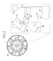
- FIG. 2 illustrates the disk of FIG. 1 .
- the disk 100 is formed in the shape of a round disk, and centrifuges externally supplied blood into blood cells and plasma by rotating itself based on a center axis C.
- an injection port 110, a centrifugal container 120, a waste flow channel 130, a waste receiving container 140, a micro-valve 150, a plasma container 160, and an air discharge port 170 may be formed by intaglio through a mass production method, such as injection molding using MEMS technology such as photolithography or insert molding having an opposite shape, hot embossing, UV-molding, and casting.
- the disk 100 may be made of a metal material, a ceramic material, and a polymer material, such as cyclic olefin copolymer (COC), polymethylmethacrylate (PMMA), polystyrene (PS), polycarbonate (PC), polydimethylsiloxane (PDMS), polytetrafluoroethylene (Teflon), and polyvinylchloride (PVC).
- COC cyclic olefin copolymer
- PMMA polymethylmethacrylate
- PS polystyrene
- PC polycarbonate
- PDMS polydimethylsiloxane
- Teflon polytetrafluoroethylene
- PVC polyvinylchloride
- the injection port 110 is adjacent to the central shaft C to be disposed in the disk 100, and is a passage to which blood is supplied.
- the centrifugal container 120 is connected with the injection port 110, and is a container in which the blood supplied from the injection port 110 is placed. When the disk 100 rotates, the blood is centrifuged to separate blood cells and plasma in the centrifugal container 120.
- the waste flow channel 130 is a passage which is positioned between the centrifugal container 120 and the injection port 110, and through which the blood overflowing from the centrifugal container 120 moves to the waste receiving container 140 while rotating the disk 100.
- the waste receiving container 140 is a container which is connected with the centrifugal container 120 by the waste flow channel 130, and receives the blood flowing from the centrifugal container 120 while rotating the disk 100.
- the micro-valve 150 is a passage which is connected between the centrifugal container 120 and the plasma container 160, of which a position is determined by considering hematocrit of the blood, and through which only the centrifuged plasma passes in the centrifugal container 120 by determining the position thereof.
- the micro-valve 150 may be disposed to correspond to one width D of 30 % to 60 % of an entire width WD of the centrifugal container 120 by considering 30 % to 60 % as a general range of the hematocrit.
- the micro-valve 150 may adjust opening and closing according to an angular velocity of the disk 100.
- the opening and closing of the micro-valve 150 is adjusted due to a difference between a first pressure formed around the micro-valve 150 by centrifugal force according to rotation of the disk 100 and a second pressure formed by surface tension in the micro-valve 150.
- a first pressure formed around the micro-valve 150 by centrifugal force according to rotation of the disk 100 and a second pressure formed by surface tension in the micro-valve 150.
- the micro-valve 150 opens and then the plasma moves to the plasma container 160 from the centrifugal container 120 through the micro-valve 150
- the second pressure is larger than the first pressure
- the micro-valve 150 closes and then the blood does not move to the plasma container 160 from the centrifugal container 120 through the micro-valve 150.
- each of the closing and the opening of the micro-valve 150 may be adjusted in response to each of the centrifugation and the plasma extraction by adjusting the angular velocity of the disk 100.
- the plasma container 160 is a container which is connected with the centrifugal container 120 through the micro-valve 150 to receive the plasma centrifuged from the blood in the centrifugal container 120.
- the plasma received in the plasma container 160 may move to another container through one channel connected with the plasma container 160 to be stored.
- the air discharge port 170 is connected to each of the centrifugal container 120, the waste receiving container 140, and the plasma container 160, and a passage through which air occupied in each container is discharged when a fluid such as blood and plasma are supplied to each container.
- FIG. 3 shows photographs illustrating centrifugation of blood in the disk of the disk-shaped microfluidic system according to the first exemplary embodiment of the present invention.
- FIG. 3 (a) to (d) are photographs illustrating an experiment process of centrifugation of a small amount of blood performed by using the disk 100 of an actually manufactured disk-shaped microfluidic system with the passage of time.
- centrifugation of blood BL injected into the centrifugal container 120 by centrifugal force which is caused by rotating the disk 100 is performed.
- the blood BL is accurately divided into blood cells B and plasma L, and the blood cells B are precipitated in an outer direction of a rotation center, and are no longer precipitated after a predetermined time as illustrated in FIG. 3 (d) .
- the centrifugation of the blood BL is performed, the movement of the blood or the plasma is prevented by the micro-valve 150.
- the micro-valve 150 is opened by increasing the angular velocity of the disk 100 to induce the extraction of the centrifuged plasma L.
- the plasma L separated on an upper portion of the centrifugal container 120 moves to the plasma container 160 through the micro-valve 150.
- the driver 200 supports the disk 100, and includes a driving means of a motor and the like to rotate the disk 100.
- the sensor 300 is positioned to be adjacent to the disk 100 and serves to sense the number of revolutions of the disk 100.
- the adjustor 400 is connected with the sensor 300 and the driver 200, and serves to receive a signal from the sensor 300 sensing the number of revolutions of the disk 100 to adjust the rotation of the disk 100 by the driver 200.
- the angular velocity of the disk 100 may be adjusted by the adjustor 400.
- the illuminator 500 is connected with the adjustor 400, and reflects light to the mirror 600 in a flash form in synchronization with the number of revolutions of the disk 100.
- the mirror 600 is positioned at a lower side of the disk 100, and reflects the light irradiated from the illuminator 500 in a disk 100 direction.
- the photographing unit 700 is positioned at the upper side of the disk 100 to correspond to the centrifugal container 120 of the disk 100 and the mirror 600, and photographs an inside of the centrifugal container 120 every hour by using the light in synchronization with the number of revolutions of the disk 100.
- the photographing unit 700 photographs the inside of the centrifugal container 120 every hour.
- the controller 800 is connected with the photographing unit 700, detects an actual moving distance of the blood cells in the centrifugal container 120 every hour by using the image photographed by the photographing unit 700, calculates a first curve showing an actual moving distance of the blood cells every hour and a second curve showing a theoretical moving distance of the blood cells every hour, and calculates hematocrit of the blood and viscosity of the plasma by comparing the first curve and the second curve. That is, the controller 800 checks the blood condition by measuring a degree of deformation of the blood cells centrifuged from the blood in the disk 100.
- the controller 800 calculates the second curve using the following equation, and measures a degree of deformation of blood cells using at least one of a height difference between the first curve and the second curve and a slope difference between the first curve and the second curve.
- ⁇ 6 ⁇ p p ⁇ d p 3 ⁇ r ⁇ p ⁇ 6 ⁇ p p - p f ⁇ d p 3 ⁇ w 2 ⁇ r p - 3 ⁇ ⁇ ⁇ d p ⁇ r ⁇ p u f ⁇ 1 + ⁇ 1 / 3 ⁇ exp 5 ⁇ ⁇ 3 ⁇ 1 - ⁇
- ⁇ is a circle constant
- p p is the density of the blood
- dp is a diameter of the blood cells
- r" p is a moving acceleration of the blood cells
- p f is the density of the plasma
- w is an angular velocity of the disk
- rp is a moving distance of the blood cells
- r'p is a moving speed of the blood cells
- u f is the viscosity of the plasma
- ⁇ is a volume ratio of the blood cells to the entire volume the blood.
- FIG. 4 to FIG. 6 are diagrams for description of a method for measuring a degree of deformation of blood cells according to the second exemplary embodiment of the present invention.
- the blood is supplied to the centrifugal container 120 of the disk 100.
- the blood in the centrifugal container 120 is centrifuged into blood cells and plasma by rotating the disk 100, and an actual moving distance of the blood cells in the centrifugal container 120 is detected every hour.
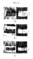
- FIG. 4 shows photographs illustrating an image of the inside of the centrifugal container every hour.
- the first curve showing the actual moving distance of the blood cells every hour and the second curve showing the theoretical moving distance of the blood cells every hour are calculated, and the degree of deformation of the blood cells is measured by comparing the first curve and the second curve.
- FIG. 5 illustrates deformation of the blood cells in the centrifugation process of blood.
- FIG. 5 (a) illustrates blood cells B that are evenly spread in the plasma L in the blood BL.
- the amount of materials existing in the blood BL, excluding the blood cells B, is small enough to be negligible, or the size of the material is very small and thus it is permissible for it to be considered as a part of the plasma L.
- FIG. 5 (a) to (d) illustrate precipitation of the blood cells B when a centrifugal force is generated due to rotation of the disk in the disk-shaped microfluidic system.
- unit cells of blood cells B are far from each other at an initial stage and thus precipitation speed of the blood cells B becomes fast.
- the unit cells of the blood cells B become close to each other as shown in FIG. 5 (c) , and thus the precipitation speed is slowed.
- the unit cells of the blood cells B are deformed and thus fill the gaps between every neighboring blood cells B such that the height of the precipitation layer is slowly lowered.
- the method for measuring the degree of deformation of the blood cells measures the degree of deformation of the blood cells based on that fact that the decrease speed of the height of the precipitation layer is closely related to the degree of deformation of the blood cells.
- FIG. 6 is a graph illustrating a first curve that shows actual moving distance of the blood cells every hour and a second curve that shows a theoretical moving distance of the blood cells every hour.
- the x-axis represents time (second) and the y-axis represents moving distance (mm) of the blood cells.
- a first curve G1 which is an experimental graph showing the actual moving distance of the blood cells every hour is detected by analyzing the image obtained through the photographing unit 700 of the disk-shaped microfluidic system, as described above.
- ⁇ is a circle constant
- p p is the density of the blood
- dp is a diameter of the blood cells
- r"p is a moving acceleration of the blood cells
- pf is the density of the plasma
- w is an angular velocity of the disk
- r p is a moving distance of the blood cells
- r'p is a moving speed of the blood cells
- u f is the viscosity of the plasma
- ⁇ is a volume ratio of the blood cells to the entire volume of the blood.
- the equation is configured based on a dynamic force balance formed by a plurality of globular particles, when the globular particles existing in a specific fluid are precipitated by centrifugal force.
- a dynamic force balance formed by a plurality of globular particles, when the globular particles existing in a specific fluid are precipitated by centrifugal force.
- r"p, r'p, and rp are calculated by applying predetermined theoretical values to ⁇ , p p , dp, p f , w, u f , and ⁇ .
- the degree of deformation of the blood cells is measured by comparing the first curve G1 and the second curve G2.
- the blood cell is assumed to be a rigid body, and thus it can be observed that no further precipitation is performed after a predetermined time has elapsed. This is because, after the blood cell particles are aligned in the bottom surface, the blood cells do not fill the gaps due to the characteristic of the rigid body so that the blood cells can maintain a constant distance without further precipitation.
- the first curve G1 which is an actual graph, that precipitation is continuously performed in the actual blood cells even after a constant time elapsed. This is also caused due to the degree of deformation of the blood cells. Therefore, a difference between the first curve G1 and the second curve G1 indicates the degree of deformation of the blood cells.
- the method for measuring the degree of deformation of the blood cells measures the degree of deformation of the blood cells using at least one of a difference of height H (i.e., moving distance difference of the blood cells, which is the y-axis) between the first curve G1 and the second curve G2 and a slope difference between the first curve G1 and the second curve G2.
- a difference of height H i.e., moving distance difference of the blood cells, which is the y-axis
- the height difference is a difference of the precipitation height between the second curve G2, which is a theoretical graph
- the first curve G1 which is the experimental graph
- the slope difference is a difference of precipitation speed (i.e., moving speed difference of blood cells).
- the degree of deformation of blood cells which are important properties of the blood may be measured, and the measured degree of deformation of the blood cells may be used as a criterion for estimating the blood condition.
- the blood is centrifuged to extract the plasma, and simultaneously the blood condition is checked, and as a result, additional equipment for checking the blood condition is not required, and simultaneously, additional time for checking the blood condition is not required.
Landscapes
- Health & Medical Sciences (AREA)
- Life Sciences & Earth Sciences (AREA)
- Engineering & Computer Science (AREA)
- Biomedical Technology (AREA)
- Hematology (AREA)
- Chemical & Material Sciences (AREA)
- Immunology (AREA)
- Physics & Mathematics (AREA)
- Molecular Biology (AREA)
- Pathology (AREA)
- General Health & Medical Sciences (AREA)
- Urology & Nephrology (AREA)
- Analytical Chemistry (AREA)
- Biochemistry (AREA)
- General Physics & Mathematics (AREA)
- Food Science & Technology (AREA)
- Medicinal Chemistry (AREA)
- Cell Biology (AREA)
- Biophysics (AREA)
- Microbiology (AREA)
- Ecology (AREA)
- Biotechnology (AREA)
- Mycology (AREA)
- Heart & Thoracic Surgery (AREA)
- Medical Informatics (AREA)
- Surgery (AREA)
- Animal Behavior & Ethology (AREA)
- Public Health (AREA)
- Veterinary Medicine (AREA)
- Investigating Or Analysing Biological Materials (AREA)
Abstract
Description
- The present invention relates to a disk-shaped microfluidic system and a method for measuring a degree of deformation of blood cells.
- Generally, blood separation, plasma extraction, property analysis of blood, and the like are performed by equipment or apparatuses suitable for the respective purposes. The blood is divided into the plasma and blood cells by an apparatus such as a centrifuge, and the divided plasma is extracted by pipetting and the like. Further, properties of the blood such as viscosity, hematocrit, and the like of the blood may be analyzed by dedicated equipment for measuring each property.
- A series of processes through the dedicated equipment have an advantage of more accurately performing each process, but there are disadvantages that in order to check the blood condition, a large amount of blood may be used, and a long testing time for checking all blood conditions is taken due to non-continuous processes.
- The above information disclosed in this Background section is only for enhancement of understanding of the background of the invention and therefore it may contain information that does not form the prior art that is already known in this country to a person of ordinary skill in the art.
- The present invention has been made in an effort to provide a disk-shaped microfluidic system, and a method for measuring a degree of deformation of blood cells, having advantages of extracting plasma by centrifuging blood and simultaneously checking a blood condition.
- A first aspect of the present invention provides a method for measuring a degree of deformation of blood cells. The method includes: supplying blood to a centrifugal container of a disk; centrifuging the blood in the centrifugal container to blood cells and plasma by rotating the disk and detecting an actual moving distance of the blood cells in the centrifugal container every hour; and calculating a first curve representing the actual moving distance of the blood cells in the centrifugal container every hour and a second curve representing a theoretical moving distance of the blood cells every hour and measuring a degree of deformation of the blood cells by comparing the first curve and the second curve.
- The detecting of the actual moving distance of the blood cells may use an image acquired by photographing the inside of the centrifugal container every hour.
- The detecting of the actual moving distance of the blood cells may be performed by calculating a center of mass with respect to a dark area by analyzing contrast in the image and measuring a short distance of the dark area based on the center of mass.
- The measuring of the degree of deformation of the blood cells may be performed by calculating the second curve by using the following equation and using at least one of a height difference between the first curve and the second curve and a slope difference between the first curve and the second curve.

- In the equation, π is a circle constant, pp is the density of the blood, dp is a diameter of the blood cells, r"p is a moving acceleration of the blood cells, pf is the density of the plasma, w is an angular velocity of the disk, rp is a moving distance of the blood cells, r'p is a moving speed of the blood cells, uf is the viscosity of the plasma, and θ is a volume ratio of the blood cells to the entire volume of the blood.
- A second aspect of the present invention provides a disk-shaped microfluidic system including: a disk including a centrifugal container to which blood is supplied and centrifuging the blood in the centrifugal container to blood cells and plasma; a photographing unit positioned at the upper side of the disk and photographing inside of the centrifugal container every hour; and a controller connected with the photographing unit, detecting an actual moving distance of the blood cells in the centrifugal container every hour by using the image photographed by the photographing unit, detecting and displaying a first curve representing the actual moving distance of the blood cells in the centrifugal container every hour and a second curve representing a theoretical moving distance of the blood cells every hour, and measuring a degree of deformation of the blood cells by comparing the first curve and the second curve.
- The controller may calculate the second curve using the following equation and measure the degree of deformation of the blood cells by using at least one of a height difference between the first curve and the second curve and a slope difference between the first curve and the second curve.

- In the equation, π is a circle constant, pp is the density of the blood, dp is a diameter of the blood cells, r"p is a moving acceleration of the blood cells, pf is the density of the plasma, w is an angular velocity of the disk, rp is a moving distance of the blood cells, r'p is a moving speed of the blood cells, uf is the viscosity of the plasma, and θ is a volume ratio of the blood cells to the entire volume of the blood.
- The disk may further include a plasma container connected with the centrifugal container, and a micro-valve connected between the centrifugal container and the plasma container. According to the exemplary embodiment of the present invention, it is possible to provide a disk-shaped microfluidic system and a method for measuring a degree of blood cells that extract plasma by centrifuging blood, and simultaneously checking a blood condition.
-
-
FIG. 1 illustrates a disk-shaped microfluidic system according to a first exemplary embodiment of the present invention. -
FIG. 2 illustrates a disk shown inFIG. 1 . -
FIG. 3 shows photographs illustrating centrifugation of blood using the disk-shaped microfluidic system according to the first exemplary embodiment of the present invention. -
FIG. 4 to FIG. 6 are provided for description of a method for measuring a degree of deformation of blood cells according to a second exemplary embodiment of the present invention. - The present invention will be described more fully hereinafter with reference to the accompanying drawings, in which exemplary embodiments of the invention are shown. As those skilled in the art would realize, the described embodiments may be modified in various different ways, all without departing from the spirit or scope of the present invention.
- The drawings and description are to be regarded as illustrative in nature and not restrictive. Like reference numerals designate like elements throughout the specification.
- Further, the size and thickness of each configuration shown in the drawings are arbitrarily shown for better understanding and ease of description, but the present invention is not limited thereto.
- In the drawings, for better understanding and ease of description, the thickness of some layers and areas is exaggerated. It will be understood that when an element such as a layer, film, region, or substrate is referred to as being "on" another element, it can be directly on the other element or intervening elements may also be present.
- In addition, unless explicitly described to the contrary, the word "comprise" and variations such as "comprises" or "comprising" will be understood to imply the inclusion of stated elements but not the exclusion of any other elements. Further, in the specification, the word "on" means positioning on or below the object portion, and does not essentially mean positioning on the upper side of the object portion based on a gravity direction.
- Hereinafter, a disk-shaped microfluidic system according to a first exemplary embodiment of the present invention will be described with reference to
FIG. 1 andFIG. 2 . -
FIG. 1 illustrates a disk-shaped microfluidic system according to the first exemplary embodiment of the present invention. - As shown in
FIG. 1 , the disk-shaped microfluidic system according to the first exemplary embodiment of the present invention centrifuges blood and at the same time determines a blood condition, and includes adisk 100,driver 200, asensor 300, anadjustor 400, anilluminator 500, amirror 600, a photographingunit 700, and acontroller 800. -
FIG. 2 illustrates the disk ofFIG. 1 . - As shown in
FIG. 2 , thedisk 100 is formed in the shape of a round disk, and centrifuges externally supplied blood into blood cells and plasma by rotating itself based on a center axis C. In thedisk 100, aninjection port 110, acentrifugal container 120, awaste flow channel 130, awaste receiving container 140, a micro-valve 150, aplasma container 160, and anair discharge port 170 may be formed by intaglio through a mass production method, such as injection molding using MEMS technology such as photolithography or insert molding having an opposite shape, hot embossing, UV-molding, and casting. Thedisk 100 may be made of a metal material, a ceramic material, and a polymer material, such as cyclic olefin copolymer (COC), polymethylmethacrylate (PMMA), polystyrene (PS), polycarbonate (PC), polydimethylsiloxane (PDMS), polytetrafluoroethylene (Teflon), and polyvinylchloride (PVC). - The
injection port 110 is adjacent to the central shaft C to be disposed in thedisk 100, and is a passage to which blood is supplied. - The
centrifugal container 120 is connected with theinjection port 110, and is a container in which the blood supplied from theinjection port 110 is placed. When thedisk 100 rotates, the blood is centrifuged to separate blood cells and plasma in thecentrifugal container 120. - The
waste flow channel 130 is a passage which is positioned between thecentrifugal container 120 and theinjection port 110, and through which the blood overflowing from thecentrifugal container 120 moves to thewaste receiving container 140 while rotating thedisk 100. - The
waste receiving container 140 is a container which is connected with thecentrifugal container 120 by thewaste flow channel 130, and receives the blood flowing from thecentrifugal container 120 while rotating thedisk 100. - The micro-valve 150 is a passage which is connected between the
centrifugal container 120 and theplasma container 160, of which a position is determined by considering hematocrit of the blood, and through which only the centrifuged plasma passes in thecentrifugal container 120 by determining the position thereof. In more detail, the micro-valve 150 may be disposed to correspond to one width D of 30 % to 60 % of an entire width WD of thecentrifugal container 120 by considering 30 % to 60 % as a general range of the hematocrit. The micro-valve 150 may adjust opening and closing according to an angular velocity of thedisk 100. In detail, the opening and closing of the micro-valve 150 is adjusted due to a difference between a first pressure formed around the micro-valve 150 by centrifugal force according to rotation of thedisk 100 and a second pressure formed by surface tension in the micro-valve 150. For example, when the first pressure is larger than the second pressure, the micro-valve 150 opens and then the plasma moves to theplasma container 160 from thecentrifugal container 120 through themicro-valve 150, and when the second pressure is larger than the first pressure, themicro-valve 150 closes and then the blood does not move to theplasma container 160 from thecentrifugal container 120 through themicro-valve 150. Since the first pressure is proportional to the angular velocity of thedisk 100, the second pressure is adjusted to be larger than the first pressure when the blood is centrifuged, and the first pressure is adjusted to be larger than the second pressure when the plasma is extracted by adjusting the angular velocity of thedisk 100, and as a result, each of the closing and the opening of themicro-valve 150 may be adjusted in response to each of the centrifugation and the plasma extraction by adjusting the angular velocity of thedisk 100. - The
plasma container 160 is a container which is connected with thecentrifugal container 120 through the micro-valve 150 to receive the plasma centrifuged from the blood in thecentrifugal container 120. The plasma received in theplasma container 160 may move to another container through one channel connected with theplasma container 160 to be stored. - The
air discharge port 170 is connected to each of thecentrifugal container 120, thewaste receiving container 140, and theplasma container 160, and a passage through which air occupied in each container is discharged when a fluid such as blood and plasma are supplied to each container. - Hereinafter, centrifugation of blood in the disk of the disk-shaped microfluidic system according to the first exemplary embodiment of the present invention will be described with reference to
FIG. 3 . -
FIG. 3 shows photographs illustrating centrifugation of blood in the disk of the disk-shaped microfluidic system according to the first exemplary embodiment of the present invention. -
FIG. 3 (a) to (d) are photographs illustrating an experiment process of centrifugation of a small amount of blood performed by using thedisk 100 of an actually manufactured disk-shaped microfluidic system with the passage of time. As shown inFIG. 3 (a) to (d) , centrifugation of blood BL injected into thecentrifugal container 120 by centrifugal force which is caused by rotating thedisk 100 is performed. In this process, as illustrated inFIG. 3 (b) , the blood BL is accurately divided into blood cells B and plasma L, and the blood cells B are precipitated in an outer direction of a rotation center, and are no longer precipitated after a predetermined time as illustrated inFIG. 3 (d) . Further, while the centrifugation of the blood BL is performed, the movement of the blood or the plasma is prevented by themicro-valve 150. - Thereafter, the micro-valve 150 is opened by increasing the angular velocity of the
disk 100 to induce the extraction of the centrifuged plasma L. The plasma L separated on an upper portion of thecentrifugal container 120 moves to theplasma container 160 through the micro-valve 150. As a result, it may be verified that a small amount of blood may be centrifuged and the plasma may be extracted by a convenient and efficient method using thedisk 100 of the disk-shaped microfluidic system according to the first exemplary embodiment of the present invention. - Referring back to
FIG. 1 , thedriver 200 supports thedisk 100, and includes a driving means of a motor and the like to rotate thedisk 100. - The
sensor 300 is positioned to be adjacent to thedisk 100 and serves to sense the number of revolutions of thedisk 100. - The
adjustor 400 is connected with thesensor 300 and thedriver 200, and serves to receive a signal from thesensor 300 sensing the number of revolutions of thedisk 100 to adjust the rotation of thedisk 100 by thedriver 200. The angular velocity of thedisk 100 may be adjusted by theadjustor 400. - The
illuminator 500 is connected with theadjustor 400, and reflects light to themirror 600 in a flash form in synchronization with the number of revolutions of thedisk 100. - The
mirror 600 is positioned at a lower side of thedisk 100, and reflects the light irradiated from theilluminator 500 in adisk 100 direction. - The photographing
unit 700 is positioned at the upper side of thedisk 100 to correspond to thecentrifugal container 120 of thedisk 100 and themirror 600, and photographs an inside of thecentrifugal container 120 every hour by using the light in synchronization with the number of revolutions of thedisk 100. The photographingunit 700 photographs the inside of thecentrifugal container 120 every hour. - The
controller 800 is connected with the photographingunit 700, detects an actual moving distance of the blood cells in thecentrifugal container 120 every hour by using the image photographed by the photographingunit 700, calculates a first curve showing an actual moving distance of the blood cells every hour and a second curve showing a theoretical moving distance of the blood cells every hour, and calculates hematocrit of the blood and viscosity of the plasma by comparing the first curve and the second curve. That is, thecontroller 800 checks the blood condition by measuring a degree of deformation of the blood cells centrifuged from the blood in thedisk 100. - The
controller 800 calculates the second curve using the following equation, and measures a degree of deformation of blood cells using at least one of a height difference between the first curve and the second curve and a slope difference between the first curve and the second curve.
- In the equation, π is a circle constant, pp is the density of the blood, dp is a diameter of the blood cells, r"p is a moving acceleration of the blood cells, pf is the density of the plasma, w is an angular velocity of the disk, rp is a moving distance of the blood cells, r'p is a moving speed of the blood cells, uf is the viscosity of the plasma, and θ is a volume ratio of the blood cells to the entire volume the blood.
- Hereinafter, a method for measuring a degree of deformation of blood cells according to a second exemplary embodiment of the present invention using the disk-shaped microfluidic system according to the first exemplary embodiment of the present invention described above will be described with reference to
FIG. 4 to FIG. 6 . -
FIG. 4 to FIG. 6 are diagrams for description of a method for measuring a degree of deformation of blood cells according to the second exemplary embodiment of the present invention. - First, the blood is supplied to the
centrifugal container 120 of thedisk 100. - Next, the blood in the
centrifugal container 120 is centrifuged into blood cells and plasma by rotating thedisk 100, and an actual moving distance of the blood cells in thecentrifugal container 120 is detected every hour. -
FIG. 4 shows photographs illustrating an image of the inside of the centrifugal container every hour. - In detail, as shown in
FIG. 4 , original photographs (left image of each of (a), (b), and (c)) of the inside of thecentrifugal container 120 every hour are corrected to post-images (right image of each of (a), (b), and (c)) by analyzing contrast. In this case, in the post-image, the precipitation of the blood cells is changed to white, and the background is changed to black. Thereafter, a center of mass of a dark area (DA) which is the precipitation of the blood cells of the post-image is calculated, and a short distance (SL) of the dark area (DA) is measured based on the center of mass to detect the actual moving distance of the blood cells in thecentrifugal container 120 every hour. Such a process is performed with respect to the entire image frame, and as a result, data showing a precipitated height of the blood cells with time may be obtained. The detection may be performed by thecontroller 800. - Next, a degree of deformation of the blood cells is measured.
- In detail, the first curve showing the actual moving distance of the blood cells every hour and the second curve showing the theoretical moving distance of the blood cells every hour are calculated, and the degree of deformation of the blood cells is measured by comparing the first curve and the second curve.
- Hereinafter, the method for measuring the degree of deformation of the blood cells will be described in further detail.
-
FIG. 5 illustrates deformation of the blood cells in the centrifugation process of blood. -
FIG. 5 (a) illustrates blood cells B that are evenly spread in the plasma L in the blood BL. The amount of materials existing in the blood BL, excluding the blood cells B, is small enough to be negligible, or the size of the material is very small and thus it is permissible for it to be considered as a part of the plasma L. -
FIG. 5 (a) to (d) illustrate precipitation of the blood cells B when a centrifugal force is generated due to rotation of the disk in the disk-shaped microfluidic system. As shown inFIG. 5 (b) , unit cells of blood cells B are far from each other at an initial stage and thus precipitation speed of the blood cells B becomes fast. When a predetermined time elapses, the unit cells of the blood cells B become close to each other as shown inFIG. 5 (c) , and thus the precipitation speed is slowed. After the unit cells of the blood cells B are completely arranged in the bottom surface, the unit cells of the blood cells B are deformed and thus fill the gaps between every neighboring blood cells B such that the height of the precipitation layer is slowly lowered. That is, the method for measuring the degree of deformation of the blood cells according to the secondary exemplary embodiment of the present invention measures the degree of deformation of the blood cells based on that fact that the decrease speed of the height of the precipitation layer is closely related to the degree of deformation of the blood cells. -
FIG. 6 is a graph illustrating a first curve that shows actual moving distance of the blood cells every hour and a second curve that shows a theoretical moving distance of the blood cells every hour. InFIG. 6 , the x-axis represents time (second) and the y-axis represents moving distance (mm) of the blood cells. - As illustrated in
FIG. 5 , a first curve G1 which is an experimental graph showing the actual moving distance of the blood cells every hour is detected by analyzing the image obtained through the photographingunit 700 of the disk-shaped microfluidic system, as described above. - A second curve G2 which is a theoretical graph showing the theoretical moving distance of the blood cells every hour is calculated by the following equation.

- In the equation, π is a circle constant, pp is the density of the blood, dp is a diameter of the blood cells, r"p is a moving acceleration of the blood cells, pf is the density of the plasma, w is an angular velocity of the disk, rp is a moving distance of the blood cells, r'p is a moving speed of the blood cells, uf is the viscosity of the plasma, and θ is a volume ratio of the blood cells to the entire volume of the blood.
- The equation is configured based on a dynamic force balance formed by a plurality of globular particles, when the globular particles existing in a specific fluid are precipitated by centrifugal force. First, when one globular particle is precipitated by centrifugal force in the specific fluid, movement of the particle forms a dynamic force balance by centrifugal force, buoyancy force, and drag force, and when the blood cell is assumed to be a rigid body, that is, a particle without deformation, a viscosity value of the fluid including a plurality of particles instead of the viscosity of the fluid is applied by considering a condition where the plurality of particles are precipitated, and as a result, the equation is derived by mathematizing the condition.
- Iterations are performed based on the equation to calculate the second curve G2. When the second curve G2 is calculated, r"p, r'p, and rp are calculated by applying predetermined theoretical values to π, pp, dp, pf, w, uf, and θ.
- The degree of deformation of the blood cells is measured by comparing the first curve G1 and the second curve G2.
- In the second curve G2, which is a theoretical graph, the blood cell is assumed to be a rigid body, and thus it can be observed that no further precipitation is performed after a predetermined time has elapsed. This is because, after the blood cell particles are aligned in the bottom surface, the blood cells do not fill the gaps due to the characteristic of the rigid body so that the blood cells can maintain a constant distance without further precipitation. On the other hand, it can be observed in the first curve G1, which is an actual graph, that precipitation is continuously performed in the actual blood cells even after a constant time elapsed. This is also caused due to the degree of deformation of the blood cells. Therefore, a difference between the first curve G1 and the second curve G1 indicates the degree of deformation of the blood cells.
- That is, the method for measuring the degree of deformation of the blood cells according to the second exemplary embodiment of the present invention measures the degree of deformation of the blood cells using at least one of a difference of height H (i.e., moving distance difference of the blood cells, which is the y-axis) between the first curve G1 and the second curve G2 and a slope difference between the first curve G1 and the second curve G2. Here, the height difference is a difference of the precipitation height between the second curve G2, which is a theoretical graph, and the first curve G1, which is the experimental graph, and the slope difference is a difference of precipitation speed (i.e., moving speed difference of blood cells).
- As such, by comparing the first curve G1 which is the experimental graph showing a development of the blood centrifugation and the second curve G2 which is the theoretical graph, the degree of deformation of blood cells which are important properties of the blood may be measured, and the measured degree of deformation of the blood cells may be used as a criterion for estimating the blood condition.
- As described above, by the disk-shaped microfluidic system according to the first exemplary embodiment of the present invention and the method for measuring the degree of deformation of blood cells according to the second exemplary embodiment of the present invention, the blood is centrifuged to extract the plasma, and simultaneously the blood condition is checked, and as a result, additional equipment for checking the blood condition is not required, and simultaneously, additional time for checking the blood condition is not required..
- While this invention has been described in connection with what is presently considered to be practical exemplary embodiments, it is to be understood that the invention is not limited to the disclosed embodiments, but, on the contrary, is intended to cover various modifications and equivalent arrangements included within the spirit and scope of the appended claims.
Claims (7)
- A method for measuring a degree of deformation of blood cells, comprising:supplying blood to a centrifugal container of a disk;centrifuging the blood in the centrifugal container to blood cells and plasma by rotating the disk and detecting an actual moving distance of the blood cells in the centrifugal container every hour; andcalculating a first curve representing the actual moving distance of the blood cells in the centrifugal container every hour and a second curve representing a theoretical moving distance of the blood cells every hour and measuring a degree of deformation of the blood cells by comparing the first curve and the second curve.
- The method of claim 1, wherein
the detecting of the actual moving distance of the blood cells uses an image acquired by photographing the inside of the centrifugal container every hour. - The method of claim 2, wherein
the detecting of the actual moving distance of the blood cells is performed by calculating a center of mass with respect to a dark area by analyzing contrast in the image and measuring a short distance of the dark area based on the center of mass. - The method of claim 1, wherein
the measuring of the degree of deformation of the blood cells is performed by calculating the second curve by using the following equation and using at least one of a height difference between the first curve and the second curve and a slope difference between the first curve and the second curve:
in the equation, π is a circle constant, pp is the density of the blood, dp is a diameter of the blood cells, r"p is a moving acceleration of the blood cells, pf is the density of the plasma, w is an angular velocity of the disk, rp is a moving distance of the blood cells, r'p is a moving speed of the blood cells, uf is the viscosity of the plasma, and θ is a volume ratio of the blood cells to the entire volume of the blood. - A disk-shaped microfluidic system comprising:a disk including a centrifugal container to which blood is supplied and centrifuging the blood in the centrifugal container to blood cells and plasma;a photographing unit positioned at the upper side of the disk and photographing inside of the centrifugal container every hour; anda controller connected with the photographing unit, detecting an actual moving distance of the blood cells in the centrifugal container every hour by using the image photographed by the photographing unit, detecting and displaying a first curve representing the actual moving distance of the blood cells in the centrifugal container every hour and a second curve representing a theoretical moving distance of the blood cells every hour, and measuring a degree of deformation of the blood cells by comparing the first curve and the second curve.
- The disk-shaped microfluidic system of claim 5, wherein the controller calculates the second curve using the following equation and measures the degree of deformation of the blood cells by using at least one of a height difference between the first curve and the second curve and a slope difference between the first curve and the second curve:

in the equation, π is a circle constant, pp is the density of the blood, dp is a diameter of the blood cells, r"p is a moving acceleration of the blood cells, pf is the density of the plasma, w is an angular velocity of the disk, rp is a moving distance of the blood cells, r'p is a moving speed of the blood cells, uf is the viscosity of the plasma, and θ is a volume ratio of the blood cells to the entire volume of the blood. - The disk-shaped microfluidic system of claim 5, wherein
the disk further comprises:a plasma container connected with the centrifugal container; anda micro-valve connected between the centrifugal container and the plasma container.
Applications Claiming Priority (2)
| Application Number | Priority Date | Filing Date | Title |
|---|---|---|---|
| KR1020110133259A KR101252260B1 (en) | 2011-12-12 | 2011-12-12 | Microfluidic disc system and method for measuring deformability of cells in blood |
| PCT/KR2012/010796 WO2013089432A1 (en) | 2011-12-12 | 2012-12-12 | Disk-type microfluidic system and method for measuring deformability of blood cells |
Publications (2)
| Publication Number | Publication Date |
|---|---|
| EP2793024A1 true EP2793024A1 (en) | 2014-10-22 |
| EP2793024A4 EP2793024A4 (en) | 2015-08-12 |
Family
ID=48442657
Family Applications (1)
| Application Number | Title | Priority Date | Filing Date |
|---|---|---|---|
| EP12857936.4A Withdrawn EP2793024A4 (en) | 2011-12-12 | 2012-12-12 | METHOD AND MICROFLUIDIC SYSTEM OF DISK TYPE FOR MEASURING DEFORMABILITY OF BLOOD CELLS |
Country Status (6)
| Country | Link |
|---|---|
| US (1) | US9383372B2 (en) |
| EP (1) | EP2793024A4 (en) |
| JP (1) | JP5898333B2 (en) |
| KR (1) | KR101252260B1 (en) |
| CN (1) | CN103988077B (en) |
| WO (1) | WO2013089432A1 (en) |
Families Citing this family (6)
| Publication number | Priority date | Publication date | Assignee | Title |
|---|---|---|---|---|
| GB201416944D0 (en) * | 2014-09-25 | 2014-11-12 | Benson Viscometers Ltd | An Apparatus for monitoring blood coagulation |
| EP3075454B1 (en) * | 2015-04-02 | 2018-05-09 | Sigma Laborzentrifugen GmbH | Laboratory centrifuge and method for operating the same |
| CN108463708B (en) | 2015-11-06 | 2021-10-08 | 密歇根大学董事会 | Droplet-based microfluidic rheometer system |
| KR102083514B1 (en) | 2018-06-22 | 2020-03-02 | 광주과학기술원 | Apparatus and method for measuring deformability of red blood cells |
| KR102078352B1 (en) * | 2018-09-18 | 2020-02-17 | 주식회사 두오텍 | Blood separator |
| CN112557261B (en) * | 2020-12-07 | 2022-12-09 | 昆明理工大学 | Erythrocyte separation detection device and separation detection method based on C-shaped microcolumn |
Family Cites Families (11)
| Publication number | Priority date | Publication date | Assignee | Title |
|---|---|---|---|---|
| US6506606B1 (en) | 1995-06-06 | 2003-01-14 | Brigham And Women's Hospital | Method and apparatus for determining erythrocyte sedimentation rate and hematocrit |
| AU702403B2 (en) | 1995-12-05 | 1999-02-18 | Gamera Bioscience | Devices and methods for using centripetal acceleration to drive fluid movement in a microfluidics system with on-board informatics |
| US20010022654A1 (en) * | 1997-08-28 | 2001-09-20 | Visco Technologies, Inc. | Method & apparatus for determining red blood cell deformability by imaging red blood cells while applying vibratory energy |
| US6204066B1 (en) * | 1999-06-25 | 2001-03-20 | Robert A. Levine | Rapid method for determining the erythrocyte sedimentation rate in a sample of anticoagulated whole blood |
| US7542131B2 (en) * | 2003-06-23 | 2009-06-02 | Sewon Meditech, Inc. | Apparatus for measuring blood cell deformability |
| KR20030061746A (en) * | 2003-06-23 | 2003-07-22 | 신세현 | Blood cell rheometer |
| WO2008021626A2 (en) * | 2006-08-17 | 2008-02-21 | Caridianbct, Inc. | Blood processing apparatus with robust automated process control |
| KR101335727B1 (en) | 2007-08-22 | 2013-12-04 | 삼성전자주식회사 | Centrifugal force-based disk type microfluidic device for blood chemistry analysis |
| WO2009091186A2 (en) * | 2008-01-15 | 2009-07-23 | Lg Electronics Inc. | Health diagnosis system using bio-disc and method for diagnosing health using the same |
| KR100915195B1 (en) * | 2008-02-29 | 2009-09-04 | 고려대학교 산학협력단 | Apparatus and method for measuring blood cell deformability using rotating disc |
| KR20100101526A (en) * | 2009-03-09 | 2010-09-17 | 삼성전자주식회사 | Strobo thin film chemical analysis apparatus and assay method using the same |
-
2011
- 2011-12-12 KR KR1020110133259A patent/KR101252260B1/en not_active Expired - Fee Related
-
2012
- 2012-12-12 WO PCT/KR2012/010796 patent/WO2013089432A1/en not_active Ceased
- 2012-12-12 JP JP2014545833A patent/JP5898333B2/en not_active Expired - Fee Related
- 2012-12-12 US US14/360,675 patent/US9383372B2/en not_active Expired - Fee Related
- 2012-12-12 CN CN201280061012.4A patent/CN103988077B/en not_active Expired - Fee Related
- 2012-12-12 EP EP12857936.4A patent/EP2793024A4/en not_active Withdrawn
Also Published As
| Publication number | Publication date |
|---|---|
| KR101252260B1 (en) | 2013-04-08 |
| JP5898333B2 (en) | 2016-04-06 |
| CN103988077A (en) | 2014-08-13 |
| US20140342391A1 (en) | 2014-11-20 |
| WO2013089432A1 (en) | 2013-06-20 |
| JP2015503102A (en) | 2015-01-29 |
| US9383372B2 (en) | 2016-07-05 |
| EP2793024A4 (en) | 2015-08-12 |
| CN103988077B (en) | 2015-12-23 |
Similar Documents
| Publication | Publication Date | Title |
|---|---|---|
| US9383372B2 (en) | Disk-type microfluidic system and method for measuring deformability of blood cells | |
| US9012228B2 (en) | Disk-type microfluid system and method for checking blood status | |
| EP2552589B1 (en) | Liquid distribution and metering | |
| TWI393874B (en) | Microfluidic device using centrifugal force and sample analyzing method using the microfluidic device | |
| US9933348B2 (en) | Sequential aliqoting and determination of an indicator of sedimentation rate | |
| US7943386B2 (en) | Apparatus and method for determining the volume fractions of the phases in a suspension | |
| TW201015070A (en) | Centrifugal-based microfluidic apparatus, method of fabricating the same, and method of testing samples using the microfluidic apparatus | |
| US10919040B2 (en) | Method for sample separation and collection | |
| EP3001173A1 (en) | Capillary fluid flow measurment and capillary flow device therefore | |
| WO2013052890A2 (en) | Devices for detecting a particle in a sample and methods for use thereof | |
| ES2716479T3 (en) | Method for calibrating analytical machines of characteristic blood parameters related to their density, such as the erythrocyte sedimentation rate and / or the aggregation rate of red blood cells | |
| CA3158306A1 (en) | Shape analysis device | |
| Kim et al. | Smart microfluidic device for automated exosome quantification and classification using deep learning | |
| Khodaparastasgarabad et al. | A novel microfluidic high-throughput resistive pulse sensing device for cells analysis | |
| WO2015166218A1 (en) | Optical analysis of fluid volumes under centrifugation | |
| KR20100043956A (en) | Bio disk and method for measuring blood using the same | |
| WO2022209189A1 (en) | Pressure measurement method, control method, pressure measurement device and analysis device | |
| Heiskanen et al. | Machine vision based measurement of dynamic contact angles in microchannel flows | |
| Senf | Effect of viscosity on high throughput deterministic lateral displacement (DLD) devices | |
| JP2009156683A (en) | Microchip | |
| Pezzuoli | Nanofluidic platforms for sensing applications in biomedical and environmental fields |
Legal Events
| Date | Code | Title | Description |
|---|---|---|---|
| PUAI | Public reference made under article 153(3) epc to a published international application that has entered the european phase |
Free format text: ORIGINAL CODE: 0009012 |
|
| 17P | Request for examination filed |
Effective date: 20140610 |
|
| AK | Designated contracting states |
Kind code of ref document: A1 Designated state(s): AL AT BE BG CH CY CZ DE DK EE ES FI FR GB GR HR HU IE IS IT LI LT LU LV MC MK MT NL NO PL PT RO RS SE SI SK SM TR |
|
| DAX | Request for extension of the european patent (deleted) | ||
| RA4 | Supplementary search report drawn up and despatched (corrected) |
Effective date: 20150713 |
|
| RIC1 | Information provided on ipc code assigned before grant |
Ipc: G01N 33/49 20060101AFI20150707BHEP Ipc: G01N 33/555 20060101ALI20150707BHEP Ipc: A61B 5/145 20060101ALI20150707BHEP Ipc: B04B 5/04 20060101ALI20150707BHEP |
|
| 17Q | First examination report despatched |
Effective date: 20170907 |
|
| STAA | Information on the status of an ep patent application or granted ep patent |
Free format text: STATUS: THE APPLICATION IS DEEMED TO BE WITHDRAWN |
|
| 18D | Application deemed to be withdrawn |
Effective date: 20180118 |