EP2792374B1 - Method for preparing biocompatible and biodegradable biomaterials based on collagen and granules of hydroxyapatite/beta-tricalcium phosphate for use in surgery, and biomaterials thus obtained - Google Patents
Method for preparing biocompatible and biodegradable biomaterials based on collagen and granules of hydroxyapatite/beta-tricalcium phosphate for use in surgery, and biomaterials thus obtained Download PDFInfo
- Publication number
- EP2792374B1 EP2792374B1 EP14164704.0A EP14164704A EP2792374B1 EP 2792374 B1 EP2792374 B1 EP 2792374B1 EP 14164704 A EP14164704 A EP 14164704A EP 2792374 B1 EP2792374 B1 EP 2792374B1
- Authority
- EP
- European Patent Office
- Prior art keywords
- collagen
- granules
- hydroxyapatite
- layer
- tricalcium phosphate
- Prior art date
- Legal status (The legal status is an assumption and is not a legal conclusion. Google has not performed a legal analysis and makes no representation as to the accuracy of the status listed.)
- Not-in-force
Links
- 108010035532 Collagen Proteins 0.000 title claims description 92
- 102000008186 Collagen Human genes 0.000 title claims description 92
- 229920001436 collagen Polymers 0.000 title claims description 92
- 239000008187 granular material Substances 0.000 title claims description 67
- QORWJWZARLRLPR-UHFFFAOYSA-H tricalcium bis(phosphate) Chemical compound [Ca+2].[Ca+2].[Ca+2].[O-]P([O-])([O-])=O.[O-]P([O-])([O-])=O QORWJWZARLRLPR-UHFFFAOYSA-H 0.000 title claims description 58
- 238000001356 surgical procedure Methods 0.000 title claims description 29
- 238000000034 method Methods 0.000 title claims description 23
- 229910052588 hydroxylapatite Inorganic materials 0.000 title claims description 17
- XYJRXVWERLGGKC-UHFFFAOYSA-D pentacalcium;hydroxide;triphosphate Chemical compound [OH-].[Ca+2].[Ca+2].[Ca+2].[Ca+2].[Ca+2].[O-]P([O-])([O-])=O.[O-]P([O-])([O-])=O.[O-]P([O-])([O-])=O XYJRXVWERLGGKC-UHFFFAOYSA-D 0.000 title claims description 16
- 239000012620 biological material Substances 0.000 title description 10
- 239000000243 solution Substances 0.000 claims description 35
- 210000000988 bone and bone Anatomy 0.000 claims description 30
- 239000011159 matrix material Substances 0.000 claims description 26
- 239000000463 material Substances 0.000 claims description 24
- 239000002131 composite material Substances 0.000 claims description 21
- 210000001519 tissue Anatomy 0.000 claims description 19
- 239000007864 aqueous solution Substances 0.000 claims description 10
- 230000000399 orthopedic effect Effects 0.000 claims description 10
- 238000004108 freeze drying Methods 0.000 claims description 9
- 230000005484 gravity Effects 0.000 claims description 9
- 238000004519 manufacturing process Methods 0.000 claims description 9
- 230000008929 regeneration Effects 0.000 claims description 6
- 238000011069 regeneration method Methods 0.000 claims description 6
- 238000001816 cooling Methods 0.000 claims description 2
- 230000008021 deposition Effects 0.000 claims description 2
- 239000010410 layer Substances 0.000 description 34
- 230000003902 lesion Effects 0.000 description 29
- 210000000845 cartilage Anatomy 0.000 description 13
- 238000010438 heat treatment Methods 0.000 description 12
- 239000000017 hydrogel Substances 0.000 description 9
- 229910052573 porcelain Inorganic materials 0.000 description 9
- 238000002360 preparation method Methods 0.000 description 9
- 239000012530 fluid Substances 0.000 description 8
- 229920001661 Chitosan Polymers 0.000 description 7
- 239000000523 sample Substances 0.000 description 7
- 239000000512 collagen gel Substances 0.000 description 6
- 239000011521 glass Substances 0.000 description 6
- 239000007943 implant Substances 0.000 description 6
- 239000012528 membrane Substances 0.000 description 6
- 239000000203 mixture Substances 0.000 description 6
- 230000008569 process Effects 0.000 description 6
- 239000008280 blood Substances 0.000 description 5
- 210000004369 blood Anatomy 0.000 description 5
- 229920000642 polymer Polymers 0.000 description 5
- XLYOFNOQVPJJNP-UHFFFAOYSA-N water Substances O XLYOFNOQVPJJNP-UHFFFAOYSA-N 0.000 description 5
- 210000001185 bone marrow Anatomy 0.000 description 4
- 210000004027 cell Anatomy 0.000 description 4
- 229910052500 inorganic mineral Inorganic materials 0.000 description 4
- 239000011707 mineral Substances 0.000 description 4
- 229910000391 tricalcium phosphate Inorganic materials 0.000 description 4
- 229910000831 Steel Inorganic materials 0.000 description 3
- 239000013078 crystal Substances 0.000 description 3
- 230000035876 healing Effects 0.000 description 3
- 230000004820 osteoconduction Effects 0.000 description 3
- 230000008439 repair process Effects 0.000 description 3
- 239000010959 steel Substances 0.000 description 3
- 230000017423 tissue regeneration Effects 0.000 description 3
- 102000010834 Extracellular Matrix Proteins Human genes 0.000 description 2
- 108010037362 Extracellular Matrix Proteins Proteins 0.000 description 2
- 230000002776 aggregation Effects 0.000 description 2
- 239000002977 biomimetic material Substances 0.000 description 2
- 230000017531 blood circulation Effects 0.000 description 2
- 239000001506 calcium phosphate Substances 0.000 description 2
- 230000007547 defect Effects 0.000 description 2
- 210000002744 extracellular matrix Anatomy 0.000 description 2
- 238000003780 insertion Methods 0.000 description 2
- 230000037431 insertion Effects 0.000 description 2
- 229910052751 metal Inorganic materials 0.000 description 2
- 230000004819 osteoinduction Effects 0.000 description 2
- 230000001172 regenerating effect Effects 0.000 description 2
- 210000002435 tendon Anatomy 0.000 description 2
- 229940078499 tricalcium phosphate Drugs 0.000 description 2
- 235000019731 tricalcium phosphate Nutrition 0.000 description 2
- 239000008215 water for injection Substances 0.000 description 2
- 206010061728 Bone lesion Diseases 0.000 description 1
- 241000283690 Bos taurus Species 0.000 description 1
- 229920002101 Chitin Polymers 0.000 description 1
- 108010066259 Collagraft Proteins 0.000 description 1
- 241000238424 Crustacea Species 0.000 description 1
- 241000283073 Equus caballus Species 0.000 description 1
- 108010080379 Fibrin Tissue Adhesive Proteins 0.000 description 1
- 108010011593 Healos Proteins 0.000 description 1
- 201000009859 Osteochondrosis Diseases 0.000 description 1
- 101710172711 Structural protein Proteins 0.000 description 1
- ATJFFYVFTNAWJD-UHFFFAOYSA-N Tin Chemical compound [Sn] ATJFFYVFTNAWJD-UHFFFAOYSA-N 0.000 description 1
- 230000009102 absorption Effects 0.000 description 1
- 238000010521 absorption reaction Methods 0.000 description 1
- 238000005054 agglomeration Methods 0.000 description 1
- 238000004220 aggregation Methods 0.000 description 1
- 235000001014 amino acid Nutrition 0.000 description 1
- 150000001413 amino acids Chemical class 0.000 description 1
- 210000001188 articular cartilage Anatomy 0.000 description 1
- 230000006399 behavior Effects 0.000 description 1
- 239000011173 biocomposite Substances 0.000 description 1
- 230000003592 biomimetic effect Effects 0.000 description 1
- 230000015572 biosynthetic process Effects 0.000 description 1
- 230000002051 biphasic effect Effects 0.000 description 1
- 230000000740 bleeding effect Effects 0.000 description 1
- 210000001124 body fluid Anatomy 0.000 description 1
- 239000010839 body fluid Substances 0.000 description 1
- 210000002449 bone cell Anatomy 0.000 description 1
- 230000008468 bone growth Effects 0.000 description 1
- 239000000316 bone substitute Substances 0.000 description 1
- 210000003321 cartilage cell Anatomy 0.000 description 1
- 238000006243 chemical reaction Methods 0.000 description 1
- 239000000470 constituent Substances 0.000 description 1
- 238000005520 cutting process Methods 0.000 description 1
- 230000006196 deacetylation Effects 0.000 description 1
- 238000003381 deacetylation reaction Methods 0.000 description 1
- 238000001514 detection method Methods 0.000 description 1
- 230000004069 differentiation Effects 0.000 description 1
- 239000000835 fiber Substances 0.000 description 1
- 102000034240 fibrous proteins Human genes 0.000 description 1
- 108091005899 fibrous proteins Proteins 0.000 description 1
- 239000000945 filler Substances 0.000 description 1
- 230000012010 growth Effects 0.000 description 1
- 230000007062 hydrolysis Effects 0.000 description 1
- 238000006460 hydrolysis reaction Methods 0.000 description 1
- 238000001727 in vivo Methods 0.000 description 1
- 230000006698 induction Effects 0.000 description 1
- 230000036512 infertility Effects 0.000 description 1
- 230000001788 irregular Effects 0.000 description 1
- 238000005304 joining Methods 0.000 description 1
- 239000007791 liquid phase Substances 0.000 description 1
- 210000004324 lymphatic system Anatomy 0.000 description 1
- 238000002844 melting Methods 0.000 description 1
- 230000008018 melting Effects 0.000 description 1
- 210000002901 mesenchymal stem cell Anatomy 0.000 description 1
- 239000002184 metal Substances 0.000 description 1
- 239000013081 microcrystal Substances 0.000 description 1
- 238000002156 mixing Methods 0.000 description 1
- 239000002159 nanocrystal Substances 0.000 description 1
- 229920005615 natural polymer Polymers 0.000 description 1
- 210000000653 nervous system Anatomy 0.000 description 1
- 230000011164 ossification Effects 0.000 description 1
- 210000000963 osteoblast Anatomy 0.000 description 1
- 230000000278 osteoconductive effect Effects 0.000 description 1
- 210000004409 osteocyte Anatomy 0.000 description 1
- 210000004663 osteoprogenitor cell Anatomy 0.000 description 1
- 239000012071 phase Substances 0.000 description 1
- 230000010399 physical interaction Effects 0.000 description 1
- 230000002980 postoperative effect Effects 0.000 description 1
- 239000000843 powder Substances 0.000 description 1
- 238000003825 pressing Methods 0.000 description 1
- 230000035755 proliferation Effects 0.000 description 1
- 230000001737 promoting effect Effects 0.000 description 1
- 235000018102 proteins Nutrition 0.000 description 1
- 102000004169 proteins and genes Human genes 0.000 description 1
- 108090000623 proteins and genes Proteins 0.000 description 1
- 230000009103 reabsorption Effects 0.000 description 1
- 238000000926 separation method Methods 0.000 description 1
- 239000002356 single layer Substances 0.000 description 1
- 238000005245 sintering Methods 0.000 description 1
- 230000004936 stimulating effect Effects 0.000 description 1
- 239000000758 substrate Substances 0.000 description 1
- 210000001835 viscera Anatomy 0.000 description 1
- 230000037303 wrinkles Effects 0.000 description 1
Images
Classifications
-
- A—HUMAN NECESSITIES
- A61—MEDICAL OR VETERINARY SCIENCE; HYGIENE
- A61L—METHODS OR APPARATUS FOR STERILISING MATERIALS OR OBJECTS IN GENERAL; DISINFECTION, STERILISATION OR DEODORISATION OF AIR; CHEMICAL ASPECTS OF BANDAGES, DRESSINGS, ABSORBENT PADS OR SURGICAL ARTICLES; MATERIALS FOR BANDAGES, DRESSINGS, ABSORBENT PADS OR SURGICAL ARTICLES
- A61L27/00—Materials for grafts or prostheses or for coating grafts or prostheses
- A61L27/14—Macromolecular materials
- A61L27/22—Polypeptides or derivatives thereof, e.g. degradation products
- A61L27/24—Collagen
-
- A—HUMAN NECESSITIES
- A61—MEDICAL OR VETERINARY SCIENCE; HYGIENE
- A61K—PREPARATIONS FOR MEDICAL, DENTAL OR TOILETRY PURPOSES
- A61K38/00—Medicinal preparations containing peptides
- A61K38/16—Peptides having more than 20 amino acids; Gastrins; Somatostatins; Melanotropins; Derivatives thereof
- A61K38/17—Peptides having more than 20 amino acids; Gastrins; Somatostatins; Melanotropins; Derivatives thereof from animals; from humans
- A61K38/39—Connective tissue peptides, e.g. collagen, elastin, laminin, fibronectin, vitronectin, cold insoluble globulin [CIG]
-
- A—HUMAN NECESSITIES
- A61—MEDICAL OR VETERINARY SCIENCE; HYGIENE
- A61L—METHODS OR APPARATUS FOR STERILISING MATERIALS OR OBJECTS IN GENERAL; DISINFECTION, STERILISATION OR DEODORISATION OF AIR; CHEMICAL ASPECTS OF BANDAGES, DRESSINGS, ABSORBENT PADS OR SURGICAL ARTICLES; MATERIALS FOR BANDAGES, DRESSINGS, ABSORBENT PADS OR SURGICAL ARTICLES
- A61L27/00—Materials for grafts or prostheses or for coating grafts or prostheses
- A61L27/40—Composite materials, i.e. containing one material dispersed in a matrix of the same or different material
- A61L27/44—Composite materials, i.e. containing one material dispersed in a matrix of the same or different material having a macromolecular matrix
- A61L27/46—Composite materials, i.e. containing one material dispersed in a matrix of the same or different material having a macromolecular matrix with phosphorus-containing inorganic fillers
-
- A—HUMAN NECESSITIES
- A61—MEDICAL OR VETERINARY SCIENCE; HYGIENE
- A61L—METHODS OR APPARATUS FOR STERILISING MATERIALS OR OBJECTS IN GENERAL; DISINFECTION, STERILISATION OR DEODORISATION OF AIR; CHEMICAL ASPECTS OF BANDAGES, DRESSINGS, ABSORBENT PADS OR SURGICAL ARTICLES; MATERIALS FOR BANDAGES, DRESSINGS, ABSORBENT PADS OR SURGICAL ARTICLES
- A61L2430/00—Materials or treatment for tissue regeneration
- A61L2430/24—Materials or treatment for tissue regeneration for joint reconstruction
Definitions
- the present invention relates to a method for preparing a biomaterial, essentially consisting of collagen in combination with granules of hydroxyapatite/ ⁇ -tricalcium phosphate (HA/ ⁇ -TCP), and to the biomaterial thus obtained; the biomaterial finds application in several areas of surgery, such as maxillofacial surgery, dentistry, aesthetic surgery and particularly orthopedic surgery.
- implants namely, the introduction into the body of materials that can replace damaged parts of the same; in particular, implants are very common for replacing bones or sections thereof, or for temporarily fill voids in the bone structure, that are to be afterwards replenished by the growth of fresh bone tissue.
- Examples of these surgery areas are the maxillofacial surgery, in which damaged parts of the face are reconstructed; dentistry, in which the implants are mainly directed to create a stable seat in the maxillar or mandibular bone for metal elements to which prostheses will then be fixed; aesthetic surgery, in which implants are mostly directed to fill wrinkles; and orthopedic surgery, in which the insertion of material into the damaged part has generally the function of providing a temporary substitute for natural tissues and a suitable substrate for their re-growth during the healing process (and possibly also a promoting effect for such re-growth).
- Osteochondral lesions i.e., affecting both the bone and the cartilage connected thereto, are the most serious ones in the typical classifications of cartilage defects; in the classification of defects according to Outerbridge, the osteochondral defect represents level IV, which is the maximum level in terms of severity of the lesion.
- These lesions were formerly treated only with the implant of metal prostheses, but more recently implant materials which are more similar to the tissues to be repaired or regenerated have been used, which have better expectations of success in the long run; in fact, especially in the case of younger patients, obtaining the regeneration of a new bone and cartilage tissue in the area of the lesion in a joint allows the affected patient to regain the original form of the joint, i.e.
- Products have long been known and used, which consist of or contain hydroxyapatite (Ca 5 (PO 4 ) 3 OH) and/or the crystalline form ⁇ of tricalcium phosphate (Ca 3 (PO 4 ) 2 ), for the preparation of scaffolds useful for the bone tissue regeneration.
- Hydroxyapatite (often referred to by the abbreviation HA in the field, which will be used hereafter) is a material with a high biomimetic attitude against the bone tissue, with a porosity very similar to that of the spongy tissue of the bone and capable of accommodating osteoblasts, thus allowing them to settle and transform into osteocytes; moreover, it has excellent biocompatibility, and when placed in direct contact with the bone it shows osteoconduction and osteointegration and, in the presence of bone growth induction factors, also osteoinduction. Moreover, this material is the main mineral constituent of the bones, being 60% of the calcified human skeleton, and is therefore a natural candidate for the production of bone prostheses or fillers.
- HA has a poor degradability by body fluids and is therefore resistant to in vivo absorption.
- HA is therefore used in a mixture with a second mineral component, the crystalline form ⁇ of tricalcium phosphate (Ca 3 (PO 4 ) 2 ), commonly known as ⁇ -tricalcium phosphate or by its abbreviation ⁇ -TCP, which will be used hereafter.
- ⁇ -TCP has a faster resorption than HA, and therefore promotes the regrowth of the bone tissue.
- HA/ ⁇ -TCP may have different ratios of the two components.
- OpteMx ® by Exactech is available as preformed parts (e.g. parallelepiped or cylindrical in shape) consisting of the porous HA/ ⁇ -TCP composition only, and has osteogenesis and limited osteoinduction properties when mixed with bone marrow blood.
- these materials are very fragile and thus not flexible, and poorly suitable for use in case of bone lumens or surfaces which are irregular in shape.
- HA/ ⁇ -TCP mixtures can be filled into a flexible matrix; the matrix material must be such that the final composite retains biocompatibility features.
- collagen the most important structural protein in the human body, forming molecular wires which strengthen the tendons and large, elastic sheets that support the skin and internal organs.
- Bones and teeth are made of mineral crystals added to collagen.
- Collagen is a relatively simple protein, consisting of three chains, each containing more than 1400 aminoacids, wrapped together in a triple narrow helix.
- the product MasterGraft ® Putty by Medtronic, having osteoconduction properties, is formed by the same granules as the product MasterGraft ® mentioned above, evenly distributed in a bovine collagen matrix.
- Integra MozaikTM by Integra LifeSciences Corporation (Plainsboro, New Jersey, USA), with osteoconductive properties, consists of a mixture containing about 80% ⁇ -TCP and 20% collagen.
- Collagraft ® Bone Graft Matrix by Zimmer (Warsaw, Indiana, USA) is sold in the form of strips made of HA/ ⁇ -TCP in a collagen matrix, which when added to autogenic bone marrow blood promote the bone repair process.
- the product Healos ® by DePuy (Warsaw, Indiana, USA) consists of a matrix of cross-linked collagen fibers coated with HA; this product, combined with marrow blood, offers an excellent environment for the proliferation and differentiation of osteoprogenitor cells.
- Patent application WO 2011/064724 A1 describes mono-, bi- or multi-layer biomimetic materials for use in orthopedic surgery, and the process for their production.
- the basic material described in this application is a bicomponent material made of the natural polymer collagen added with chitosan (a polymer obtained by basic deacetylation of chitin, the natural component of exoskeleton of crustaceans).
- the mono-layer biomimetic materials of this application are made of collagen-chitosan only.
- This bicomponent material contains between 30 and 90% (preferably 50-80%) by weight of collagen, the remainder being chitosan; the role of chitosan is said to be for favoring the fibration of the bicomponent material.
- This application also describes a double layer material, comprising a layer of the collagen-chitosan material only, and a second layer of the same polymers containing nano- or microdimensional crystals or granules of HA (the preferred size of these crystals or granules being between 30 nm and 10 ⁇ m).
- a double layer material is said to be useful for the healing of osteochondral lesions, and is used by contacting the polymers-only layer with the lesion area in the cartilage, and the layer containing HA with the lesion area in the bone tissue.
- double layers are obtained by preparing the collagen-chitosan material according to the same complex process mentioned above, and in particular preparing a layer made of polymers only, a separate layer made of polymers loaded with the nano- or micro-crystals or granules of HA, and then joining the two layers separately produced.
- the double layer materials of this application though more suitable for the treatment of osteochondral lesions than polymeric materials in which HA is uniformly distributed, are of cumbersome preparation, due to the complexity of the preparation of the bicomponent polymeric material, and the need of producing the two layers separately.
- the present invention in a first aspect relates to a method for the production of composite materials consisting of hydroxyapatite and ⁇ -tricalcium phosphate in a collagen matrix, as defined by claim 1 which comprises the following steps:
- the product of the invention is particularly suitable for the treatment and regeneration of tissues in osteochondral lesions, having an area mineralized with granules of HA/ ⁇ -TCP where bone cells will be localized, and a fibrous protein area (collagen), where cartilage cells of the same patient will migrate, thus finding their ideal environment for reproduction and generation of the extracellular matrix; this product may however be used also in other surgery areas, for instance in maxillofacial surgery, dentistry, and aesthetic surgery.
- the invention relates to a method for producing composites of HA/ ⁇ -TCP and collagen, which comprises the above-mentioned steps a) to d).
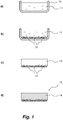
- the method is described hereafter with reference to Figure 1 .
- a solution 11 of collagen in water is prepared in a container 10.
- the collagen concentration in the solution can range from about 0.5 to 2% by weight, preferably about 1%; collagen is preferably obtained from equine tendon.
- the desired amount of collagen is added to water for injections (WFI) and the system is brought to a temperature of between about 60 °C and the hydrolysis temperature of collagen, which is about 75 °C; a temperature of between 60 and 65 °C is preferably used.
- WFI water for injections
- the resulting solution appears as a very fluid, slightly opalescent liquid phase.
- step b) ( Fig. 1.b ), the solution 11 thus formed is kept at a temperature of between about 60 and 75 °C, preferably at the same temperature at which it was produced in step a), and its free surface is covered with a layer of the granules of hydroxyapatite/ ⁇ -tricalcium phosphate having a size of between 0.3 and 5 mm; the granules are shown in the Figure as a whole with reference numeral 12.
- the granules HA/ ⁇ -TCP comprise the HA component in amounts of from 50 to 70%, preferably about 60%, by weight; the use of granules where HA is within the indicated range ensures optimal reabsorption timing in the body.
- These granules are commercially available and are manufactured and sold for example by Eurocoating S.p.A. of Ciré-Pergine (Trento) with the name OSPROLIFETM.
- smaller granules up to about 1-1.2 mm
- are single granules (“primary" granules)
- larger ones, up to 5 mm are derived from thermal aggregation in the production of primary granules.
- the granules may possibly be sieved to select the desired grain size fraction.
- step c) ( Fig. 1.c )
- the solution added with the granules of HA/ ⁇ -TCP is then cooled to a temperature between -20 and -30 °C, thus causing the formation of a hydrogel 13, i.e. a gelatinous phase consisting of a network of collagen chains where the water of the starting solution is absorbed; the hydrogel has sufficient mechanical consistency and can be extracted from container 10; the lower layer of the hydrogel is formed by the deposit of the granules of HA/ ⁇ -TCP surrounded by the hydrogel matrix, while the upper layer is formed by collagen hydrogel only.
- a hydrogel 13 i.e. a gelatinous phase consisting of a network of collagen chains where the water of the starting solution is absorbed
- the hydrogel has sufficient mechanical consistency and can be extracted from container 10
- the lower layer of the hydrogel is formed by the deposit of the granules of HA/ ⁇ -TCP surrounded by the hydrogel matrix, while the upper layer is formed by collagen hydrogel
- step d) the hydrogel is subjected to lyophilization by means of methods known in the field, in order to remove water, thus obtaining a dehydrated (lyophilized) hydrogel 14, ( Fig. 1.d ) which incorporates granules 12; the hydrogel consists of a highly elastic, composite double-layer matrix, with a clear and evident separation of the two components, collagen and granules of HA/ ⁇ -TCP, where the inorganic part is perfectly integrated in the organic matrix; this particular structure gives rise to a synergy of behaviors that is the basis for achieving the desired results, leading to a higher yield than that obtained by the two elements taken separately.
- the method of the present invention allows to produce easily, as the direct product of the method, a double-layer composite material especially tailored for the treatment of osteochondral lesions.
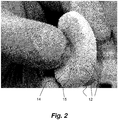
- the invention relates to the scaffold obtained by means of the above-described method; an example of scaffold obtainable according to the invention is shown in Figure 2 , which depicts the reproduction of a photograph of a sample produced by the inventors and which highlights its elasticity.
- This scaffold 15 consists of a matrix of collagen chains entangled together to form the dehydrated hydrogel 14, which only in a part thereof incorporates the granules of HA/ ⁇ -TCP 12. No chemical reaction occurs between collagen and HA/ ⁇ -TCP of the granules in the product, but rather, only a purely physical interaction.
- the product is a scaffold for the regeneration of tissues for use in different areas of surgery, and in particular for the regeneration of bone and sub-chondral tissues in orthophedic field;
- this scaffold uses the known regenerative collagen properties and HA/ ⁇ -TCP reconstructive properties, in the form of a biphasic matrix where the components are perfectly separated from each other, but at the same time integrated into each other.
- the product size may vary within wide ranges but in view of the intended uses as scaffolds for the insertion into bone or joint cavities, typically its thickness can range from 2 to 7 mm, preferably from 4 to 6 mm, while the thickness of each of the two component layers (layer of HA/ ⁇ -TCP granules incorporated into the collagen matrix, and collagen-only layer) can range from 1 to 6 mm, preferably from 2 to 4 mm.
- the product naturally retains the high biocompatibility and biodegradability features of the two component materials and is therefore especially recommended for orthopedic surgery.
- the product of the invention allows, where there is a lesion affecting both the bone and the cartilage, the positioning of the scaffold with the part containing collagen only facing the lesion affecting the cartilage and the part containing the granules facing the bone lesion.
- An access route to the damaged area of the joint must first be opened, e.g. arthroscopically (i.e. through some small holes into which the arthroscope and the ancillary instruments are introduced) or, in the case of larger lesions, through incision in the skin and in the tissues overlying the joint.
- arthroscopically i.e. through some small holes into which the arthroscope and the ancillary instruments are introduced
- larger lesions through incision in the skin and in the tissues overlying the joint.
- the lesion area is then cleaned by carefully removing all the malacic cartilage up to preparing a healthy and consistent edge, with well-defined borders and a shape as regular as possible.
- the bone tissue immediately beneath the removed cartilage layer is cleaned and gently removed, should it be in any way damaged or missing.
- This latter operation is also intended to stimulate the sub-chondral tissue: the bleeding caused by a small lesion already present, or even induced (Steadman technique), provides a bone marrow blood flow in the repair area which is known to be filled with active cells in tissue repair processes, such as, for example, mesenchymal stem cells, capable of differentiating into cells of new tissue, both bone and cartilaginous tissue.
- a template is usually used, generally made of a malleable, sterile plastic material, or an equally sterile tin foil, having a size useful for detection, overlapping the template on the lesion and cutting the edges thereof over the contours of the lesion, so as to obtain a template having a shape and size matching as much as possible those of the osteochondral lesion to be covered.
- the composite biomaterial of the invention is then cut, in a sterile environment, respecting the exact size of the lesion, so as to obtain a scaffold which is accurate and, most importantly, which is perfectly adhering to the edges of the lesion and which is equalized to the level of the surrounding cartilage, as much as possible, without interruption.
- the scaffold thus obtained is then positioned so as to completely cover the lesion area previously prepared, making sure that the side with the prevalence of bone substitute (granules of HA/ ⁇ -TCP) is facing the sub-chondral tissue, thus keeping the side consisting of collagen only positioned towards the joint space.
- fibrin glue to form a continuum between the healthy native tissue and the artificial biocomposite tissue, so as to allow the host cells to pass into the meshes of the composite scaffold, thus completing the colonization of the new cover tissue and stimulating the production of new extracellular matrix, both of the bone and of the cartilage tissue.
- the tourniquet is removed to check the tightness of the graft just positioned into the lesion site and the kinematic efficiency of the joint; with the restoration of the blood stream, the material of the scaffold is soaked in the patient's bone marrow blood, thus creating a three-dimensional environment suitable for tissue regeneration.
- the skin is sutured; the post-operative period usually requires absolute immobility, with the limb stretched, for at least 36 hours, after which the rehabilitation process can begin.
- the present invention will be further described by the following examples.
- the amounts of the scaffold components are reported based on the size of the container where the mixing is made and based on the final thickness of the scaffold.
- the container is then stored at -30 °C for at least 24 hours.
- the frozen product is subjected to lyophilization.
- a composite material made of collagen/granules having a size of 4.5x4.5 cm with a thickness of 2 mm of collagen only and 5 mm of granules of HA/ ⁇ -TCP in the collagen matrix is thus obtained.
- 16 grams of 1% collagen gel in aqueous solution are weighed in a glass container having dimensions of 5x5x2 cm.
- the glass container containing the collagen solution is heated to 70 °C.
- the temperature of the collagen solution is checked. When the collagen solution reaches a temperature of 62 °C, it appears as a slightly viscous, slightly opalescent fluid.
- the heating plate is turned off and 3 grams of granules of HA/ ⁇ -TCP, with a size of between 0.3 and 0.6 mm, are evenly distributed over the collagen solution surface.
- the granules settle, by gravity, on the bottom of the glass container forming a well-defined layer.
- the container is then stored at -30 °C for at least 24 hours.
- the frozen product is subjected to lyophilization.
- a composite material of collagen/granules having a size of 4.5x4.5 cm with a thickness of 2 mm of collagen only and 3 mm of granules of HA/ ⁇ -TCP in the collagen matrix is thus obtained.
- the container is then stored at - 30 °C for at least 24 hours.
- the frozen product is subjected to lyophilization.
- a composite material of collagen/granules having a size of 3x4.5 cm with a thickness of 4 mm of collagen only and 2 mm of granules of HA/ ⁇ -TCP in the collagen matrix is thus obtained.
- the container is then stored at - 30 °C for at least 24 hours.
- the frozen product is subjected to lyophilization.
- a composite material of collagen/granules having a size of 3x4.5 cm with a thickness of 2 mm of collagen only and 3 mm of granules of HA/ ⁇ -TCP in the collagen matrix is thus obtained.
- the container is then stored at - 20 °C for at least 24 hours.
- the frozen product is subjected to lyophilization.
- a composite material of collagen/granules having a size of 3x4.5 cm with a thickness of 3 mm of collagen only and 3 mm of granules of HA/ ⁇ -TCP in the collagen matrix is thus obtained.
- the container is then stored at -30 °C for at least 24 hours.
- the frozen product is subjected to lyophilization.
- a composite material of collagen/granules having a size of 31x31 cm with a thickness of 4 mm of collagen only and 2 mm of granules of HA/ ⁇ -TCP in the collagen matrix is thus obtained.
Landscapes
- Health & Medical Sciences (AREA)
- Chemical & Material Sciences (AREA)
- Life Sciences & Earth Sciences (AREA)
- Medicinal Chemistry (AREA)
- Public Health (AREA)
- Veterinary Medicine (AREA)
- General Health & Medical Sciences (AREA)
- Animal Behavior & Ethology (AREA)
- Epidemiology (AREA)
- Engineering & Computer Science (AREA)
- Dermatology (AREA)
- Transplantation (AREA)
- Oral & Maxillofacial Surgery (AREA)
- Composite Materials (AREA)
- Inorganic Chemistry (AREA)
- Materials Engineering (AREA)
- Biophysics (AREA)
- Zoology (AREA)
- Gastroenterology & Hepatology (AREA)
- Bioinformatics & Cheminformatics (AREA)
- Immunology (AREA)
- Pharmacology & Pharmacy (AREA)
- Proteomics, Peptides & Aminoacids (AREA)
- Biomedical Technology (AREA)
- Materials For Medical Uses (AREA)
- Chemical Kinetics & Catalysis (AREA)
- Polymers & Plastics (AREA)
- Organic Chemistry (AREA)
Description
- The present invention relates to a method for preparing a biomaterial, essentially consisting of collagen in combination with granules of hydroxyapatite/β-tricalcium phosphate (HA/β-TCP), and to the biomaterial thus obtained; the biomaterial finds application in several areas of surgery, such as maxillofacial surgery, dentistry, aesthetic surgery and particularly orthopedic surgery.
- In modern surgery, wide use is made of implants, namely, the introduction into the body of materials that can replace damaged parts of the same; in particular, implants are very common for replacing bones or sections thereof, or for temporarily fill voids in the bone structure, that are to be afterwards replenished by the growth of fresh bone tissue. Examples of these surgery areas are the maxillofacial surgery, in which damaged parts of the face are reconstructed; dentistry, in which the implants are mainly directed to create a stable seat in the maxillar or mandibular bone for metal elements to which prostheses will then be fixed; aesthetic surgery, in which implants are mostly directed to fill wrinkles; and orthopedic surgery, in which the insertion of material into the damaged part has generally the function of providing a temporary substitute for natural tissues and a suitable substrate for their re-growth during the healing process (and possibly also a promoting effect for such re-growth).
- In orthopedic surgery, a particularly challenging application is the treatment of articular lesions, because of the need of preserving the mobility of the part (which may be not an issue in other surgery areas), and because in many cases the damage to be repaired involves both bone and cartilage tissues.
- The description that follows will refer mainly to orthopedic surgery, but the biomaterials described below are of general application, at least in the other surgery areas mentioned before.
- Osteochondral lesions, i.e., affecting both the bone and the cartilage connected thereto, are the most serious ones in the typical classifications of cartilage defects; in the classification of defects according to Outerbridge, the osteochondral defect represents level IV, which is the maximum level in terms of severity of the lesion. These lesions were formerly treated only with the implant of metal prostheses, but more recently implant materials which are more similar to the tissues to be repaired or regenerated have been used, which have better expectations of success in the long run; in fact, especially in the case of younger patients, obtaining the regeneration of a new bone and cartilage tissue in the area of the lesion in a joint allows the affected patient to regain the original form of the joint, i.e. of perfect efficiency, and above all a repair tissue which is fully integrated with the circulatory system, the lymphatic system, and even with the nervous system. To this end, materials have been developed which form the so-called "scaffolds", i.e., supports having a three-dimensional structure on which the patient's repairing cells cling and migrate during the process of rehabilitating the space left by the osteochondral damage.
- Products have long been known and used, which consist of or contain hydroxyapatite (Ca5(PO4)3OH) and/or the crystalline form β of tricalcium phosphate (Ca3(PO4)2), for the preparation of scaffolds useful for the bone tissue regeneration.
- Hydroxyapatite (often referred to by the abbreviation HA in the field, which will be used hereafter) is a material with a high biomimetic attitude against the bone tissue, with a porosity very similar to that of the spongy tissue of the bone and capable of accommodating osteoblasts, thus allowing them to settle and transform into osteocytes; moreover, it has excellent biocompatibility, and when placed in direct contact with the bone it shows osteoconduction and osteointegration and, in the presence of bone growth induction factors, also osteoinduction. Moreover, this material is the main mineral constituent of the bones, being 60% of the calcified human skeleton, and is therefore a natural candidate for the production of bone prostheses or fillers. It has been produced synthetically since the 70s, using a sintering process (agglomeration of a powder at a temperature below its melting point), and has been used clinically for about 20 years. However, HA has a poor degradability by body fluids and is therefore resistant to in vivo absorption.
- For the production of scaffolds for bone regrowth, HA is therefore used in a mixture with a second mineral component, the crystalline form β of tricalcium phosphate (Ca3(PO4)2), commonly known as β-tricalcium phosphate or by its abbreviation β-TCP, which will be used hereafter. β-TCP has a faster resorption than HA, and therefore promotes the regrowth of the bone tissue. These mixtures, referred to in the field by the abbreviation HA/β-TCP, may have different ratios of the two components.
- A variety of biocompatible and biodegradable materials consisting of HA/β-TCP for use in orthopedic surgery is known.
- The product OpteMx® by Exactech (Gainesville, Florida, USA) is available as preformed parts (e.g. parallelepiped or cylindrical in shape) consisting of the porous HA/β-TCP composition only, and has osteogenesis and limited osteoinduction properties when mixed with bone marrow blood.
- The product MasterGraft® by Medtronic (Minneapolis, Minnesota, USA), available in granules of the HA/β-TCP composition only, has osteoconduction properties. However, these materials are very fragile and thus not flexible, and poorly suitable for use in case of bone lumens or surfaces which are irregular in shape.
- In order to overcome the problem, HA/β-TCP mixtures can be filled into a flexible matrix; the matrix material must be such that the final composite retains biocompatibility features. Widely used to this purpose is collagen, the most important structural protein in the human body, forming molecular wires which strengthen the tendons and large, elastic sheets that support the skin and internal organs. Bones and teeth are made of mineral crystals added to collagen. Collagen is a relatively simple protein, consisting of three chains, each containing more than 1400 aminoacids, wrapped together in a triple narrow helix.
- Several products are known on the market which consist of HA and/or β-TCP in a collagen matrix.
- The product MasterGraft®Putty by Medtronic, having osteoconduction properties, is formed by the same granules as the product MasterGraft® mentioned above, evenly distributed in a bovine collagen matrix.
- The product Integra Mozaik™ by Integra LifeSciences Corporation (Plainsboro, New Jersey, USA), with osteoconductive properties, consists of a mixture containing about 80% β-TCP and 20% collagen.
- The product Collagraft® Bone Graft Matrix by Zimmer (Warsaw, Indiana, USA) is sold in the form of strips made of HA/β-TCP in a collagen matrix, which when added to autogenic bone marrow blood promote the bone repair process.
- Finally, the product Healos® by DePuy (Warsaw, Indiana, USA) consists of a matrix of cross-linked collagen fibers coated with HA; this product, combined with marrow blood, offers an excellent environment for the proliferation and differentiation of osteoprogenitor cells.
- In cases of lesions affecting both the bone and the cartilage, the healing of the cartilage lesion is promoted by collagen, while that of the bone by the mineral components; known products, wherein HA and/or β-TCP are evenly distributed in the collagen matrix, are not optimal for these complex lesions.
- Patent application
WO 2011/064724 A1 describes mono-, bi- or multi-layer biomimetic materials for use in orthopedic surgery, and the process for their production. The basic material described in this application is a bicomponent material made of the natural polymer collagen added with chitosan (a polymer obtained by basic deacetylation of chitin, the natural component of exoskeleton of crustaceans). The mono-layer biomimetic materials of this application are made of collagen-chitosan only. This bicomponent material contains between 30 and 90% (preferably 50-80%) by weight of collagen, the remainder being chitosan; the role of chitosan is said to be for favoring the fibration of the bicomponent material. The production process of this collagen-chitosan material is however rather complex, requiring a rather complex sequence of steps carried out at controlled and different pH values. This application also describes a double layer material, comprising a layer of the collagen-chitosan material only, and a second layer of the same polymers containing nano- or microdimensional crystals or granules of HA (the preferred size of these crystals or granules being between 30 nm and 10 µm). A double layer material is said to be useful for the healing of osteochondral lesions, and is used by contacting the polymers-only layer with the lesion area in the cartilage, and the layer containing HA with the lesion area in the bone tissue. These double layers are obtained by preparing the collagen-chitosan material according to the same complex process mentioned above, and in particular preparing a layer made of polymers only, a separate layer made of polymers loaded with the nano- or micro-crystals or granules of HA, and then joining the two layers separately produced. The double layer materials of this application, though more suitable for the treatment of osteochondral lesions than polymeric materials in which HA is uniformly distributed, are of cumbersome preparation, due to the complexity of the preparation of the bicomponent polymeric material, and the need of producing the two layers separately. - It is the object of the invention to overcome the problems still existing in the field, and in particular to provide a composite useful as a matrix for the regeneration of tissues in several areas of surgery, and in particular in orthopedic surgery for the treatment of lesions affecting both the bone tissue and the cartilaginous tissue at the same time.
- This object is achieved by the present invention, which in a first aspect relates to a method for the production of composite materials consisting of hydroxyapatite and β-tricalcium phosphate in a collagen matrix, as defined by claim 1 which comprises the following steps:
- a) in a container, preparing an aqueous solution of collagen at a concentration between 0.5% and 2% by weight;
- b) distributing a layer of granules of hydroxyapatite/β-tricalcium phosphate having a size between 0.3 and 5 mm on the surface of said aqueous solution prepared in step a) and waiting for the deposition by gravity of said granules on the bottom of the layer of collagen;
- c) cooling the product obtained in step b) to a temperature between -20 and -30 °C;
- d) subjecting to lyophilization the product obtained in step c). Wherein the product obtained by the above-described method, consists of a double-layer composite, where a first layer consists of collagen only and a second layer consists of granules of hydroxyapatite/β-tricalcium phosphate or aggregates thereof immersed in the collagen matrix.
-
-
Figure 1 diagrammatically shows the steps of the method of the invention; -
Figure 2 is a reproduction of a photograph which shows a scaffold for regenerating osteochondral lesions, obtained with a composite of the invention. - The product of the invention is particularly suitable for the treatment and regeneration of tissues in osteochondral lesions, having an area mineralized with granules of HA/β-TCP where bone cells will be localized, and a fibrous protein area (collagen), where cartilage cells of the same patient will migrate, thus finding their ideal environment for reproduction and generation of the extracellular matrix; this product may however be used also in other surgery areas, for instance in maxillofacial surgery, dentistry, and aesthetic surgery.
- According to a first aspect, the invention relates to a method for producing composites of HA/β-TCP and collagen, which comprises the above-mentioned steps a) to d). The method is described hereafter with reference to
Figure 1 . - In step a) (
Fig. 1.a ), asolution 11 of collagen in water is prepared in acontainer 10. The collagen concentration in the solution can range from about 0.5 to 2% by weight, preferably about 1%; collagen is preferably obtained from equine tendon. In order to prepare the solution, the desired amount of collagen is added to water for injections (WFI) and the system is brought to a temperature of between about 60 °C and the hydrolysis temperature of collagen, which is about 75 °C; a temperature of between 60 and 65 °C is preferably used. The resulting solution appears as a very fluid, slightly opalescent liquid phase. - In step b) (
Fig. 1.b ), thesolution 11 thus formed is kept at a temperature of between about 60 and 75 °C, preferably at the same temperature at which it was produced in step a), and its free surface is covered with a layer of the granules of hydroxyapatite/β-tricalcium phosphate having a size of between 0.3 and 5 mm; the granules are shown in the Figure as a whole withreference numeral 12. - Working at these temperatures ensures that the solution remains fluid, so that the granules poured on the upper surface thereof can, by gravity, cross the thickness thereof to form a layer on the bottom of
container 10. - For the purposes of the invention, the granules HA/β-TCP comprise the HA component in amounts of from 50 to 70%, preferably about 60%, by weight; the use of granules where HA is within the indicated range ensures optimal reabsorption timing in the body. These granules are commercially available and are manufactured and sold for example by Eurocoating S.p.A. of Ciré-Pergine (Trento) with the name OSPROLIFE™. Typically, smaller granules (up to about 1-1.2 mm) are single granules ("primary" granules), while larger ones, up to 5 mm, are derived from thermal aggregation in the production of primary granules. If desired, in view of the application, the granules may possibly be sieved to select the desired grain size fraction.
- In step c) (
Fig. 1.c ), the solution added with the granules of HA/β-TCP is then cooled to a temperature between -20 and -30 °C, thus causing the formation of ahydrogel 13, i.e. a gelatinous phase consisting of a network of collagen chains where the water of the starting solution is absorbed; the hydrogel has sufficient mechanical consistency and can be extracted fromcontainer 10; the lower layer of the hydrogel is formed by the deposit of the granules of HA/β-TCP surrounded by the hydrogel matrix, while the upper layer is formed by collagen hydrogel only. Finally, in step d), the hydrogel is subjected to lyophilization by means of methods known in the field, in order to remove water, thus obtaining a dehydrated (lyophilized)hydrogel 14, (Fig. 1.d ) which incorporatesgranules 12; the hydrogel consists of a highly elastic, composite double-layer matrix, with a clear and evident separation of the two components, collagen and granules of HA/β-TCP, where the inorganic part is perfectly integrated in the organic matrix; this particular structure gives rise to a synergy of behaviors that is the basis for achieving the desired results, leading to a higher yield than that obtained by the two elements taken separately. - Different from the teachings of
WO 2011/064724 A1 , thus, the method of the present invention allows to produce easily, as the direct product of the method, a double-layer composite material especially tailored for the treatment of osteochondral lesions. - In a second aspect, the invention relates to the scaffold obtained by means of the above-described method; an example of scaffold obtainable according to the invention is shown in
Figure 2 , which depicts the reproduction of a photograph of a sample produced by the inventors and which highlights its elasticity. Thisscaffold 15 consists of a matrix of collagen chains entangled together to form thedehydrated hydrogel 14, which only in a part thereof incorporates the granules of HA/β-TCP 12. No chemical reaction occurs between collagen and HA/β-TCP of the granules in the product, but rather, only a purely physical interaction. Therefore, the product is a scaffold for the regeneration of tissues for use in different areas of surgery, and in particular for the regeneration of bone and sub-chondral tissues in orthophedic field; this scaffold uses the known regenerative collagen properties and HA/β-TCP reconstructive properties, in the form of a biphasic matrix where the components are perfectly separated from each other, but at the same time integrated into each other. The product size may vary within wide ranges but in view of the intended uses as scaffolds for the insertion into bone or joint cavities, typically its thickness can range from 2 to 7 mm, preferably from 4 to 6 mm, while the thickness of each of the two component layers (layer of HA/β-TCP granules incorporated into the collagen matrix, and collagen-only layer) can range from 1 to 6 mm, preferably from 2 to 4 mm. - The product naturally retains the high biocompatibility and biodegradability features of the two component materials and is therefore especially recommended for orthopedic surgery.
- Given the particular configuration thereof, the product of the invention allows, where there is a lesion affecting both the bone and the cartilage, the positioning of the scaffold with the part containing collagen only facing the lesion affecting the cartilage and the part containing the granules facing the bone lesion.
- The mode of using the composite biomaterials of the invention in orthopedic surgery, which will be apparent to the surgeon, can be summarized as follows.
- An access route to the damaged area of the joint must first be opened, e.g. arthroscopically (i.e. through some small holes into which the arthroscope and the ancillary instruments are introduced) or, in the case of larger lesions, through incision in the skin and in the tissues overlying the joint. These operations are normally carried out in the absence of blood flow in the limb to be operated, which is usually inhibited with a tight pressing band in proximal position.
- The lesion area is then cleaned by carefully removing all the malacic cartilage up to preparing a healthy and consistent edge, with well-defined borders and a shape as regular as possible.
- Afterwards, the bone tissue immediately beneath the removed cartilage layer is cleaned and gently removed, should it be in any way damaged or missing. This latter operation is also intended to stimulate the sub-chondral tissue: the bleeding caused by a small lesion already present, or even induced (Steadman technique), provides a bone marrow blood flow in the repair area which is known to be filled with active cells in tissue repair processes, such as, for example, mesenchymal stem cells, capable of differentiating into cells of new tissue, both bone and cartilaginous tissue.
- Once the procedures for preparing the lesion area have been completed, the extent of the same is measured while maintaining the sterility of the site to be treated. A template is usually used, generally made of a malleable, sterile plastic material, or an equally sterile tin foil, having a size useful for detection, overlapping the template on the lesion and cutting the edges thereof over the contours of the lesion, so as to obtain a template having a shape and size matching as much as possible those of the osteochondral lesion to be covered.
- With the sterile template thus obtained, the composite biomaterial of the invention is then cut, in a sterile environment, respecting the exact size of the lesion, so as to obtain a scaffold which is accurate and, most importantly, which is perfectly adhering to the edges of the lesion and which is equalized to the level of the surrounding cartilage, as much as possible, without interruption.
- The scaffold thus obtained is then positioned so as to completely cover the lesion area previously prepared, making sure that the side with the prevalence of bone substitute (granules of HA/β-TCP) is facing the sub-chondral tissue, thus keeping the side consisting of collagen only positioned towards the joint space. In this step, it is important to ensure that the graft is well-leveled on the profile of the articular cartilage, without bumps or excessive depressions which would have a direct impact on the mobility of the joint and on the quality of the regenerated tissue. In order to fix the composite biomaterial in the area of the graft, it is preferable to use fibrin glue, to form a continuum between the healthy native tissue and the artificial biocomposite tissue, so as to allow the host cells to pass into the meshes of the composite scaffold, thus completing the colonization of the new cover tissue and stimulating the production of new extracellular matrix, both of the bone and of the cartilage tissue.
- At the end of surgery, the tourniquet is removed to check the tightness of the graft just positioned into the lesion site and the kinematic efficiency of the joint; with the restoration of the blood stream, the material of the scaffold is soaked in the patient's bone marrow blood, thus creating a three-dimensional environment suitable for tissue regeneration.
- Finally, the skin is sutured; the post-operative period usually requires absolute immobility, with the limb stretched, for at least 36 hours, after which the rehabilitation process can begin.
- Other ways of using the biomaterials of the invention in other areas of surgery will be apparent to specialized surgeons.
- The present invention will be further described by the following examples. In the examples, the amounts of the scaffold components are reported based on the size of the container where the mixing is made and based on the final thickness of the scaffold.
- Preparation of a membrane made of collagen and granules of HA/β-TCP, with a final thickness of 7 mm.
- 16 grams of 1% collagen gel in aqueous solution are weighed in a glass container having dimensions of 5x5x2 cm. With the aid of a heating plate, the glass container containing the collagen solution is heated to 70 °C. Using a probe thermometer, the temperature of the collagen solution is checked. When the collagen solution reaches a temperature of 62 °C, it appears as a slightly viscous, slightly opalescent fluid. The heating plate is turned off and 5 grams of granules of HA/β-TCP, with a size of between 0.3 and 0.6 mm, are evenly distributed over the collagen solution surface. The granules settle, by gravity, on the bottom of the glass container forming a well-defined layer. The container is then stored at -30 °C for at least 24 hours. The frozen product is subjected to lyophilization. A composite material made of collagen/granules having a size of 4.5x4.5 cm with a thickness of 2 mm of collagen only and 5 mm of granules of HA/β-TCP in the collagen matrix is thus obtained.
- Preparation of a membrane made of collagen and granules of HA/β-TCP, with a final thickness of 5 mm. 16 grams of 1% collagen gel in aqueous solution are weighed in a glass container having dimensions of 5x5x2 cm. With the aid of a heating plate, the glass container containing the collagen solution is heated to 70 °C. Using a probe thermometer, the temperature of the collagen solution is checked. When the collagen solution reaches a temperature of 62 °C, it appears as a slightly viscous, slightly opalescent fluid. The heating plate is turned off and 3 grams of granules of HA/β-TCP, with a size of between 0.3 and 0.6 mm, are evenly distributed over the collagen solution surface. The granules settle, by gravity, on the bottom of the glass container forming a well-defined layer. The container is then stored at -30 °C for at least 24 hours. The frozen product is subjected to lyophilization. A composite material of collagen/granules having a size of 4.5x4.5 cm with a thickness of 2 mm of collagen only and 3 mm of granules of HA/β-TCP in the collagen matrix is thus obtained.
- Preparation of a membrane made of collagen and granules of HA/β-TCP, with a final thickness of 6 mm.
- 12.5 grams of 1% collagen gel in aqueous solution are weighed in a porcelain container having dimensions of 4.5x5x1.5 cm. With the aid of a heating plate, the porcelain container containing the collagen solution is heated to 70 °C. Using a probe thermometer, the temperature of the collagen solution is checked. When the collagen solution reaches a temperature of 62 °C, it appears as a slightly viscous, slightly opalescent fluid. The heating plate is turned off and 1.8 grams of granules of HA/β-TCP, with a size of between 0.3 and 0.6 mm, are evenly distributed over the collagen solution surface. The granules settle, by gravity, on the bottom of the porcelain container forming a well-defined layer. The container is then stored at - 30 °C for at least 24 hours. The frozen product is subjected to lyophilization. A composite material of collagen/granules having a size of 3x4.5 cm with a thickness of 4 mm of collagen only and 2 mm of granules of HA/β-TCP in the collagen matrix is thus obtained.
- Preparation of a membrane made of collagen and granules of HA/β-TCP, with a final thickness of 5 mm.
- 10.2 grams of 1% collagen gel in aqueous solution are weighed in a porcelain container having dimensions of 4.5x5x1.5 cm. With the aid of a heating plate, the porcelain container containing the collagen solution is heated to 70 °C. Using a probe thermometer, the temperature of the collagen solution is checked. When the collagen solution reaches a temperature of 62 °C, it appears as a slightly viscous, slightly opalescent fluid. The heating plate is turned off and 1.5 grams of granules of HA/β-TCP, with a size of between 0.5 and 1 mm, are evenly distributed over the collagen solution surface. The granules settle, by gravity, on the bottom of the porcelain container forming a well-defined layer. The container is then stored at - 30 °C for at least 24 hours. The frozen product is subjected to lyophilization. A composite material of collagen/granules having a size of 3x4.5 cm with a thickness of 2 mm of collagen only and 3 mm of granules of HA/β-TCP in the collagen matrix is thus obtained.
- Preparation of a membrane made of collagen and granules of HA/β-TCP, with a final thickness of 6 mm.
- 12.5 grams of 1% collagen gel in aqueous solution are weighed in a porcelain container having dimensions of 4.5x5x1.5 cm. With the aid of a heating plate, the porcelain container containing the collagen solution is heated to 70 °C. Using a probe thermometer, the temperature of the collagen solution is checked. When the collagen solution reaches a temperature of 62 °C, it appears as a slightly viscous, slightly opalescent fluid. The heating plate is turned off and 1.6 grams of granules of HA/β-TCP, with a size of between 0.5 and 1 mm, are evenly distributed over the collagen solution surface. The granules settle, by gravity, on the bottom of the porcelain container forming a well-defined layer. The container is then stored at - 20 °C for at least 24 hours. The frozen product is subjected to lyophilization. A composite material of collagen/granules having a size of 3x4.5 cm with a thickness of 3 mm of collagen only and 3 mm of granules of HA/β-TCP in the collagen matrix is thus obtained.
- Preparation of a membrane made of collagen and granules of HA/β-TCP, with a final thickness of 6 mm.
- 360 grams of 1% collagen gel in aqueous solution are weighed in a steel container having dimensions of 35x35x5 cm. With the aid of a heating plate, the steel container containing the collagen solution is heated to 70 °C. Using a probe thermometer, the temperature of the collagen solution is checked. When the collagen solution reaches a temperature of 62 °C, it appears as a slightly viscous, slightly opalescent fluid. The heating plate is turned off and 52 grams of granules of HA/β-TCP, with a size of between 0.3 and 0.6 mm, are evenly distributed over the collagen solution surface. The granules settle, by gravity, on the bottom of the steel container forming a well-defined layer. The container is then stored at -30 °C for at least 24 hours. The frozen product is subjected to lyophilization. A composite material of collagen/granules having a size of 31x31 cm with a thickness of 4 mm of collagen only and 2 mm of granules of HA/β-TCP in the collagen matrix is thus obtained.
Claims (9)
- Method for the production of a double-layer composite material consisting of hydroxyapatite and β-tricalcium phosphate in a collagen matrix, in which a first layer consists of collagen and a second layer consists of granules of hydroxyapatite/β-tricalcium phosphate or their aggregates immersed in a matrix of collagen, which comprises the following steps:a) in a container (10), preparing an aqueous solution of collagen (11) at a concentration between 0.5% and 2% by weight;b) distributing a layer of granules of hydroxyapatite/β-tricalcium phosphate having a size between 0.3 and 5 mm on the surface of said aqueous solution prepared in step a) and wait for the deposition by gravity of said granules (12) on the bottom of the layer of collagen solution.c) cooling the product obtained in step b) to a temperature between -20 and - 30 °C;d) subjecting to lyophilization the product obtained in step c).
- Method according to claim 1, wherein said granules of hydroxyapatite/β-tricalcium phosphate comprise between 50 and 70% by weight of hydroxyapatite.
- Method according to claim 2, wherein said granules of hydroxyapatite/β-tricalcium phosphate comprise 60% by weight of hydroxyapatite.
- Method according to any one of the preceding claims, wherein the solution produced in step a) has a collagen concentration of 1% by weight.
- Method according to any one of the preceding claims, wherein the solution of step a) is produced at a temperature between 60 and 65 °C.
- Double-layer composite material, in which a first layer consists of collagen and a second layer consists of granules of hydroxyapatite/β-tricalcium phosphate or their aggregates, having dimensions between 0.3 and 5 mm, immersed in a matrix of collagen.
- Scaffold (15) for use in orthopedic surgery formed by a material of claim 6, having a thickness between 2 and 7 mm and wherein the thickness of said second layer is variable between 1 and 6 mm.
- Scaffold according to claim 7, wherein said use is for the regeneration treatment of bone and sub-chondral tissues.
- A double-layer composite material as defined in claim 6, for use in maxillofacial surgery, dentistry, or aesthetic surgery.
Applications Claiming Priority (1)
| Application Number | Priority Date | Filing Date | Title |
|---|---|---|---|
| IT000636A ITMI20130636A1 (en) | 2013-04-18 | 2013-04-18 | METHOD FOR THE PREPARATION OF BIO-COMPATIBLE AND BIODEGRADABLE BIOMATERIALS BASED ON COLLAGEN AND HYDROXYAPATITIS GRANULES / ¿-TRICALCOSUS PHOSPHATE FOR USE IN ORTHOPEDIC SURGERY AND BIOMATERIALS SO OBTAINED |
Publications (2)
| Publication Number | Publication Date |
|---|---|
| EP2792374A1 EP2792374A1 (en) | 2014-10-22 |
| EP2792374B1 true EP2792374B1 (en) | 2017-07-12 |
Family
ID=48579264
Family Applications (1)
| Application Number | Title | Priority Date | Filing Date |
|---|---|---|---|
| EP14164704.0A Not-in-force EP2792374B1 (en) | 2013-04-18 | 2014-04-15 | Method for preparing biocompatible and biodegradable biomaterials based on collagen and granules of hydroxyapatite/beta-tricalcium phosphate for use in surgery, and biomaterials thus obtained |
Country Status (3)
| Country | Link |
|---|---|
| US (2) | US9415091B2 (en) |
| EP (1) | EP2792374B1 (en) |
| IT (1) | ITMI20130636A1 (en) |
Families Citing this family (1)
| Publication number | Priority date | Publication date | Assignee | Title |
|---|---|---|---|---|
| ITUB20154229A1 (en) * | 2015-10-08 | 2017-04-08 | Novagenit S R L | A PROCESS FOR THE PREPARATION OF COLLAGEN FILM |
Family Cites Families (9)
| Publication number | Priority date | Publication date | Assignee | Title |
|---|---|---|---|---|
| US4668295A (en) * | 1985-04-25 | 1987-05-26 | University Of Dayton | Surgical cements |
| JPH06505175A (en) * | 1991-02-11 | 1994-06-16 | ボシュ アンド ロム インコーポレイテッド | Bio-based medical adhesives and their uses |
| GB0110726D0 (en) * | 2001-05-02 | 2001-06-27 | Biocomposites Ltd | Bone implant composition |
| US6626950B2 (en) * | 2001-06-28 | 2003-09-30 | Ethicon, Inc. | Composite scaffold with post anchor for the repair and regeneration of tissue |
| ITMI20050343A1 (en) * | 2005-03-04 | 2006-09-05 | Fin Ceramica Faenza S R L | CARTRIDGE REPLACEMENT AND OSTEOCINDRAL INCLUDING A MULTILAYER STRUCTURE AND ITS USE |
| CN101370465A (en) * | 2006-01-23 | 2009-02-18 | 奥赛恩治疗公司 | Bone cement composite material containing particles in non-uniform spatial distribution and device for implementation |
| GB2440721A (en) * | 2006-08-11 | 2008-02-13 | Univ Cambridge Tech | Composite biomaterial formed by cooling a fluid composition on a porous solid and removing solidified crystals of the liquid carrier |
| US8308814B2 (en) * | 2009-03-27 | 2012-11-13 | Depuy Mitek, Inc. | Methods and devices for preparing and implanting tissue scaffolds |
| IT1396934B1 (en) * | 2009-11-25 | 2012-12-20 | Fin Ceramica Faenza Spa | COMPOSITE BIO-MIMETIC MATERIALS, RELATED PREPARATION PROCESS AND THEIR USE FOR THE IMPLEMENTATION OF MONO-, BI- OR MULTI-LAYER STRUCTURES FOR THE REGENERATION OF BONE, CARTILAGE OR OSTEOCARTILAGE |
-
2013
- 2013-04-18 IT IT000636A patent/ITMI20130636A1/en unknown
-
2014
- 2014-04-10 US US14/249,456 patent/US9415091B2/en not_active Expired - Fee Related
- 2014-04-15 EP EP14164704.0A patent/EP2792374B1/en not_active Not-in-force
-
2016
- 2016-06-29 US US15/196,727 patent/US20160303286A1/en not_active Abandoned
Non-Patent Citations (1)
| Title |
|---|
| None * |
Also Published As
| Publication number | Publication date |
|---|---|
| US20140314849A1 (en) | 2014-10-23 |
| US9415091B2 (en) | 2016-08-16 |
| US20160303286A1 (en) | 2016-10-20 |
| ITMI20130636A1 (en) | 2014-10-19 |
| EP2792374A1 (en) | 2014-10-22 |
Similar Documents
| Publication | Publication Date | Title |
|---|---|---|
| EP1855734B1 (en) | Biomaterial | |
| JP5289772B2 (en) | Cartilage-like structure having multi-layer structure and osteochondral substitute and use thereof | |
| TWI394597B (en) | Biodegradable scaffold bone graft for orthopaedic use | |
| US8343229B2 (en) | Textured bone block implants | |
| JP2010500070A (en) | Biomaterial | |
| US6998135B1 (en) | Demineralized corticocancellous bone sheet | |
| Hinz et al. | A new resorbable bone void filler in trauma: early clinical experience and histologic evaluation | |
| WO2011064724A1 (en) | Biomimetic composite materials, preparation process thereof and use thereof to produce mono-, bi- or multi -layer structures for the regeneration of bone, cartilaginous and osteocartilaginous tissue | |
| RU2595087C1 (en) | Method for lower jaw bone repair with homogeneous transplant with one-stage dental implantation (versions) | |
| US9415091B2 (en) | Method for preparing biocompatible and biodegradable biomaterials based on collagen and granules of hydroxyapatite/β-tricalcium phosphate for use in surgery, and biomaterials thus obtained | |
| JP2026511115A (en) | bone growth composition | |
| US20110153029A1 (en) | Bioresorbable and flexible membranes exhibiting asymmetric osteoconductive behavior in both faces | |
| Yogishwarappa et al. | Customized osteomesh cranioplasty | |
| Heikkilä | Use of bioactive glasses as bone substitutes in orthopaedics and traumatology | |
| PL232494B1 (en) | Method for obtaining a transplant of cortical spongy bone debris for regeneration of bone tissue, and the transplant for this regeneration | |
| Weisgerber | Design, characterization, and reinforcement of mineralized collagen-glycosaminoglycan scaffolds for orthopedic wound repair | |
| HK1104802B (en) | Biomaterial |
Legal Events
| Date | Code | Title | Description |
|---|---|---|---|
| PUAI | Public reference made under article 153(3) epc to a published international application that has entered the european phase |
Free format text: ORIGINAL CODE: 0009012 |
|
| 17P | Request for examination filed |
Effective date: 20140415 |
|
| AK | Designated contracting states |
Kind code of ref document: A1 Designated state(s): AL AT BE BG CH CY CZ DE DK EE ES FI FR GB GR HR HU IE IS IT LI LT LU LV MC MK MT NL NO PL PT RO RS SE SI SK SM TR |
|
| AX | Request for extension of the european patent |
Extension state: BA ME |
|
| RBV | Designated contracting states (corrected) |
Designated state(s): AL AT BE BG CH CY CZ DE DK EE ES FI FR GB GR HR HU IE IS IT LI LT LU LV MC MK MT NL NO PL PT RO RS SE SI SK SM TR |
|
| R17P | Request for examination filed (corrected) |
Effective date: 20150421 |
|
| 17Q | First examination report despatched |
Effective date: 20160307 |
|
| GRAP | Despatch of communication of intention to grant a patent |
Free format text: ORIGINAL CODE: EPIDOSNIGR1 |
|
| INTG | Intention to grant announced |
Effective date: 20170405 |
|
| GRAS | Grant fee paid |
Free format text: ORIGINAL CODE: EPIDOSNIGR3 |
|
| GRAA | (expected) grant |
Free format text: ORIGINAL CODE: 0009210 |
|
| AK | Designated contracting states |
Kind code of ref document: B1 Designated state(s): AL AT BE BG CH CY CZ DE DK EE ES FI FR GB GR HR HU IE IS IT LI LT LU LV MC MK MT NL NO PL PT RO RS SE SI SK SM TR |
|
| REG | Reference to a national code |
Ref country code: GB Ref legal event code: FG4D |
|
| REG | Reference to a national code |
Ref country code: CH Ref legal event code: EP |
|
| REG | Reference to a national code |
Ref country code: AT Ref legal event code: REF Ref document number: 907741 Country of ref document: AT Kind code of ref document: T Effective date: 20170715 |
|
| REG | Reference to a national code |
Ref country code: IE Ref legal event code: FG4D |
|
| REG | Reference to a national code |
Ref country code: DE Ref legal event code: R096 Ref document number: 602014011633 Country of ref document: DE |
|
| REG | Reference to a national code |
Ref country code: NL Ref legal event code: MP Effective date: 20170712 |
|
| REG | Reference to a national code |
Ref country code: LT Ref legal event code: MG4D |
|
| REG | Reference to a national code |
Ref country code: AT Ref legal event code: MK05 Ref document number: 907741 Country of ref document: AT Kind code of ref document: T Effective date: 20170712 |
|
| PG25 | Lapsed in a contracting state [announced via postgrant information from national office to epo] |
Ref country code: AT Free format text: LAPSE BECAUSE OF FAILURE TO SUBMIT A TRANSLATION OF THE DESCRIPTION OR TO PAY THE FEE WITHIN THE PRESCRIBED TIME-LIMIT Effective date: 20170712 Ref country code: LT Free format text: LAPSE BECAUSE OF FAILURE TO SUBMIT A TRANSLATION OF THE DESCRIPTION OR TO PAY THE FEE WITHIN THE PRESCRIBED TIME-LIMIT Effective date: 20170712 Ref country code: HR Free format text: LAPSE BECAUSE OF FAILURE TO SUBMIT A TRANSLATION OF THE DESCRIPTION OR TO PAY THE FEE WITHIN THE PRESCRIBED TIME-LIMIT Effective date: 20170712 Ref country code: NL Free format text: LAPSE BECAUSE OF FAILURE TO SUBMIT A TRANSLATION OF THE DESCRIPTION OR TO PAY THE FEE WITHIN THE PRESCRIBED TIME-LIMIT Effective date: 20170712 Ref country code: FI Free format text: LAPSE BECAUSE OF FAILURE TO SUBMIT A TRANSLATION OF THE DESCRIPTION OR TO PAY THE FEE WITHIN THE PRESCRIBED TIME-LIMIT Effective date: 20170712 Ref country code: NO Free format text: LAPSE BECAUSE OF FAILURE TO SUBMIT A TRANSLATION OF THE DESCRIPTION OR TO PAY THE FEE WITHIN THE PRESCRIBED TIME-LIMIT Effective date: 20171012 Ref country code: SE Free format text: LAPSE BECAUSE OF FAILURE TO SUBMIT A TRANSLATION OF THE DESCRIPTION OR TO PAY THE FEE WITHIN THE PRESCRIBED TIME-LIMIT Effective date: 20170712 |
|
| PG25 | Lapsed in a contracting state [announced via postgrant information from national office to epo] |
Ref country code: IS Free format text: LAPSE BECAUSE OF FAILURE TO SUBMIT A TRANSLATION OF THE DESCRIPTION OR TO PAY THE FEE WITHIN THE PRESCRIBED TIME-LIMIT Effective date: 20171112 Ref country code: BG Free format text: LAPSE BECAUSE OF FAILURE TO SUBMIT A TRANSLATION OF THE DESCRIPTION OR TO PAY THE FEE WITHIN THE PRESCRIBED TIME-LIMIT Effective date: 20171012 Ref country code: RS Free format text: LAPSE BECAUSE OF FAILURE TO SUBMIT A TRANSLATION OF THE DESCRIPTION OR TO PAY THE FEE WITHIN THE PRESCRIBED TIME-LIMIT Effective date: 20170712 Ref country code: GR Free format text: LAPSE BECAUSE OF FAILURE TO SUBMIT A TRANSLATION OF THE DESCRIPTION OR TO PAY THE FEE WITHIN THE PRESCRIBED TIME-LIMIT Effective date: 20171013 Ref country code: PL Free format text: LAPSE BECAUSE OF FAILURE TO SUBMIT A TRANSLATION OF THE DESCRIPTION OR TO PAY THE FEE WITHIN THE PRESCRIBED TIME-LIMIT Effective date: 20170712 Ref country code: ES Free format text: LAPSE BECAUSE OF FAILURE TO SUBMIT A TRANSLATION OF THE DESCRIPTION OR TO PAY THE FEE WITHIN THE PRESCRIBED TIME-LIMIT Effective date: 20170712 Ref country code: LV Free format text: LAPSE BECAUSE OF FAILURE TO SUBMIT A TRANSLATION OF THE DESCRIPTION OR TO PAY THE FEE WITHIN THE PRESCRIBED TIME-LIMIT Effective date: 20170712 |
|
| REG | Reference to a national code |
Ref country code: DE Ref legal event code: R097 Ref document number: 602014011633 Country of ref document: DE |
|
| REG | Reference to a national code |
Ref country code: FR Ref legal event code: PLFP Year of fee payment: 5 |
|
| PG25 | Lapsed in a contracting state [announced via postgrant information from national office to epo] |
Ref country code: CZ Free format text: LAPSE BECAUSE OF FAILURE TO SUBMIT A TRANSLATION OF THE DESCRIPTION OR TO PAY THE FEE WITHIN THE PRESCRIBED TIME-LIMIT Effective date: 20170712 Ref country code: RO Free format text: LAPSE BECAUSE OF FAILURE TO SUBMIT A TRANSLATION OF THE DESCRIPTION OR TO PAY THE FEE WITHIN THE PRESCRIBED TIME-LIMIT Effective date: 20170712 Ref country code: DK Free format text: LAPSE BECAUSE OF FAILURE TO SUBMIT A TRANSLATION OF THE DESCRIPTION OR TO PAY THE FEE WITHIN THE PRESCRIBED TIME-LIMIT Effective date: 20170712 |
|
| PLBE | No opposition filed within time limit |
Free format text: ORIGINAL CODE: 0009261 |
|
| STAA | Information on the status of an ep patent application or granted ep patent |
Free format text: STATUS: NO OPPOSITION FILED WITHIN TIME LIMIT |
|
| PG25 | Lapsed in a contracting state [announced via postgrant information from national office to epo] |
Ref country code: EE Free format text: LAPSE BECAUSE OF FAILURE TO SUBMIT A TRANSLATION OF THE DESCRIPTION OR TO PAY THE FEE WITHIN THE PRESCRIBED TIME-LIMIT Effective date: 20170712 Ref country code: SK Free format text: LAPSE BECAUSE OF FAILURE TO SUBMIT A TRANSLATION OF THE DESCRIPTION OR TO PAY THE FEE WITHIN THE PRESCRIBED TIME-LIMIT Effective date: 20170712 Ref country code: SM Free format text: LAPSE BECAUSE OF FAILURE TO SUBMIT A TRANSLATION OF THE DESCRIPTION OR TO PAY THE FEE WITHIN THE PRESCRIBED TIME-LIMIT Effective date: 20170712 |
|
| 26N | No opposition filed |
Effective date: 20180413 |
|
| PGFP | Annual fee paid to national office [announced via postgrant information from national office to epo] |
Ref country code: DE Payment date: 20180420 Year of fee payment: 5 |
|
| PG25 | Lapsed in a contracting state [announced via postgrant information from national office to epo] |
Ref country code: SI Free format text: LAPSE BECAUSE OF FAILURE TO SUBMIT A TRANSLATION OF THE DESCRIPTION OR TO PAY THE FEE WITHIN THE PRESCRIBED TIME-LIMIT Effective date: 20170712 |
|
| PGFP | Annual fee paid to national office [announced via postgrant information from national office to epo] |
Ref country code: GB Payment date: 20180418 Year of fee payment: 5 |
|
| PG25 | Lapsed in a contracting state [announced via postgrant information from national office to epo] |
Ref country code: MC Free format text: LAPSE BECAUSE OF FAILURE TO SUBMIT A TRANSLATION OF THE DESCRIPTION OR TO PAY THE FEE WITHIN THE PRESCRIBED TIME-LIMIT Effective date: 20170712 |
|
| REG | Reference to a national code |
Ref country code: CH Ref legal event code: PL |
|
| REG | Reference to a national code |
Ref country code: IE Ref legal event code: MM4A |
|
| PG25 | Lapsed in a contracting state [announced via postgrant information from national office to epo] |
Ref country code: LU Free format text: LAPSE BECAUSE OF NON-PAYMENT OF DUE FEES Effective date: 20180415 |
|
| PG25 | Lapsed in a contracting state [announced via postgrant information from national office to epo] |
Ref country code: LI Free format text: LAPSE BECAUSE OF NON-PAYMENT OF DUE FEES Effective date: 20180430 Ref country code: CH Free format text: LAPSE BECAUSE OF NON-PAYMENT OF DUE FEES Effective date: 20180430 |
|
| PG25 | Lapsed in a contracting state [announced via postgrant information from national office to epo] |
Ref country code: IE Free format text: LAPSE BECAUSE OF NON-PAYMENT OF DUE FEES Effective date: 20180415 |
|
| REG | Reference to a national code |
Ref country code: DE Ref legal event code: R119 Ref document number: 602014011633 Country of ref document: DE |
|
| GBPC | Gb: european patent ceased through non-payment of renewal fee |
Effective date: 20190415 |
|
| PG25 | Lapsed in a contracting state [announced via postgrant information from national office to epo] |
Ref country code: DE Free format text: LAPSE BECAUSE OF NON-PAYMENT OF DUE FEES Effective date: 20191101 Ref country code: MT Free format text: LAPSE BECAUSE OF NON-PAYMENT OF DUE FEES Effective date: 20180415 Ref country code: GB Free format text: LAPSE BECAUSE OF NON-PAYMENT OF DUE FEES Effective date: 20190415 |
|
| PG25 | Lapsed in a contracting state [announced via postgrant information from national office to epo] |
Ref country code: TR Free format text: LAPSE BECAUSE OF FAILURE TO SUBMIT A TRANSLATION OF THE DESCRIPTION OR TO PAY THE FEE WITHIN THE PRESCRIBED TIME-LIMIT Effective date: 20170712 |
|
| PG25 | Lapsed in a contracting state [announced via postgrant information from national office to epo] |
Ref country code: PT Free format text: LAPSE BECAUSE OF FAILURE TO SUBMIT A TRANSLATION OF THE DESCRIPTION OR TO PAY THE FEE WITHIN THE PRESCRIBED TIME-LIMIT Effective date: 20170712 Ref country code: HU Free format text: LAPSE BECAUSE OF FAILURE TO SUBMIT A TRANSLATION OF THE DESCRIPTION OR TO PAY THE FEE WITHIN THE PRESCRIBED TIME-LIMIT; INVALID AB INITIO Effective date: 20140415 |
|
| PG25 | Lapsed in a contracting state [announced via postgrant information from national office to epo] |
Ref country code: MK Free format text: LAPSE BECAUSE OF NON-PAYMENT OF DUE FEES Effective date: 20170712 Ref country code: CY Free format text: LAPSE BECAUSE OF FAILURE TO SUBMIT A TRANSLATION OF THE DESCRIPTION OR TO PAY THE FEE WITHIN THE PRESCRIBED TIME-LIMIT Effective date: 20170712 |
|
| PG25 | Lapsed in a contracting state [announced via postgrant information from national office to epo] |
Ref country code: AL Free format text: LAPSE BECAUSE OF FAILURE TO SUBMIT A TRANSLATION OF THE DESCRIPTION OR TO PAY THE FEE WITHIN THE PRESCRIBED TIME-LIMIT Effective date: 20170712 |
|
| REG | Reference to a national code |
Ref country code: BE Ref legal event code: MM Effective date: 20200430 |
|
| PG25 | Lapsed in a contracting state [announced via postgrant information from national office to epo] |
Ref country code: BE Free format text: LAPSE BECAUSE OF NON-PAYMENT OF DUE FEES Effective date: 20200430 |
|
| PGFP | Annual fee paid to national office [announced via postgrant information from national office to epo] |
Ref country code: FR Payment date: 20210423 Year of fee payment: 8 |
|
| PG25 | Lapsed in a contracting state [announced via postgrant information from national office to epo] |
Ref country code: FR Free format text: LAPSE BECAUSE OF NON-PAYMENT OF DUE FEES Effective date: 20220430 |
|
| PGFP | Annual fee paid to national office [announced via postgrant information from national office to epo] |
Ref country code: IT Payment date: 20230403 Year of fee payment: 10 |
|
| PG25 | Lapsed in a contracting state [announced via postgrant information from national office to epo] |
Ref country code: IT Free format text: LAPSE BECAUSE OF NON-PAYMENT OF DUE FEES Effective date: 20240415 |