EP2791163B2 - Process for t cell expansion - Google Patents
Process for t cell expansion Download PDFInfo
- Publication number
- EP2791163B2 EP2791163B2 EP12815750.0A EP12815750A EP2791163B2 EP 2791163 B2 EP2791163 B2 EP 2791163B2 EP 12815750 A EP12815750 A EP 12815750A EP 2791163 B2 EP2791163 B2 EP 2791163B2
- Authority
- EP
- European Patent Office
- Prior art keywords
- ebv
- prt
- fragment
- protein
- cells
- Prior art date
- Legal status (The legal status is an assumption and is not a legal conclusion. Google has not performed a legal analysis and makes no representation as to the accuracy of the status listed.)
- Active
Links
Images


Classifications
-
- C—CHEMISTRY; METALLURGY
- C12—BIOCHEMISTRY; BEER; SPIRITS; WINE; VINEGAR; MICROBIOLOGY; ENZYMOLOGY; MUTATION OR GENETIC ENGINEERING
- C12M—APPARATUS FOR ENZYMOLOGY OR MICROBIOLOGY; APPARATUS FOR CULTURING MICROORGANISMS FOR PRODUCING BIOMASS, FOR GROWING CELLS OR FOR OBTAINING FERMENTATION OR METABOLIC PRODUCTS, i.e. BIOREACTORS OR FERMENTERS
- C12M23/00—Constructional details, e.g. recesses, hinges
- C12M23/24—Gas permeable parts
-
- A—HUMAN NECESSITIES
- A61—MEDICAL OR VETERINARY SCIENCE; HYGIENE
- A61K—PREPARATIONS FOR MEDICAL, DENTAL OR TOILETRY PURPOSES
- A61K39/00—Medicinal preparations containing antigens or antibodies
- A61K39/12—Viral antigens
-
- A—HUMAN NECESSITIES
- A61—MEDICAL OR VETERINARY SCIENCE; HYGIENE
- A61K—PREPARATIONS FOR MEDICAL, DENTAL OR TOILETRY PURPOSES
- A61K39/00—Medicinal preparations containing antigens or antibodies
- A61K39/12—Viral antigens
- A61K39/235—Adenoviridae
-
- A—HUMAN NECESSITIES
- A61—MEDICAL OR VETERINARY SCIENCE; HYGIENE
- A61K—PREPARATIONS FOR MEDICAL, DENTAL OR TOILETRY PURPOSES
- A61K39/00—Medicinal preparations containing antigens or antibodies
- A61K39/12—Viral antigens
- A61K39/245—Herpetoviridae, e.g. herpes simplex virus
-
- A—HUMAN NECESSITIES
- A61—MEDICAL OR VETERINARY SCIENCE; HYGIENE
- A61K—PREPARATIONS FOR MEDICAL, DENTAL OR TOILETRY PURPOSES
- A61K40/00—Cellular immunotherapy
- A61K40/10—Cellular immunotherapy characterised by the cell type used
- A61K40/11—T-cells, e.g. tumour infiltrating lymphocytes [TIL] or regulatory T [Treg] cells; Lymphokine-activated killer [LAK] cells
-
- A—HUMAN NECESSITIES
- A61—MEDICAL OR VETERINARY SCIENCE; HYGIENE
- A61K—PREPARATIONS FOR MEDICAL, DENTAL OR TOILETRY PURPOSES
- A61K40/00—Cellular immunotherapy
- A61K40/40—Cellular immunotherapy characterised by antigens that are targeted or presented by cells of the immune system
- A61K40/46—Viral antigens
-
- A—HUMAN NECESSITIES
- A61—MEDICAL OR VETERINARY SCIENCE; HYGIENE
- A61P—SPECIFIC THERAPEUTIC ACTIVITY OF CHEMICAL COMPOUNDS OR MEDICINAL PREPARATIONS
- A61P31/00—Antiinfectives, i.e. antibiotics, antiseptics, chemotherapeutics
- A61P31/12—Antivirals
-
- A—HUMAN NECESSITIES
- A61—MEDICAL OR VETERINARY SCIENCE; HYGIENE
- A61P—SPECIFIC THERAPEUTIC ACTIVITY OF CHEMICAL COMPOUNDS OR MEDICINAL PREPARATIONS
- A61P31/00—Antiinfectives, i.e. antibiotics, antiseptics, chemotherapeutics
- A61P31/12—Antivirals
- A61P31/14—Antivirals for RNA viruses
-
- A—HUMAN NECESSITIES
- A61—MEDICAL OR VETERINARY SCIENCE; HYGIENE
- A61P—SPECIFIC THERAPEUTIC ACTIVITY OF CHEMICAL COMPOUNDS OR MEDICINAL PREPARATIONS
- A61P31/00—Antiinfectives, i.e. antibiotics, antiseptics, chemotherapeutics
- A61P31/12—Antivirals
- A61P31/20—Antivirals for DNA viruses
-
- A—HUMAN NECESSITIES
- A61—MEDICAL OR VETERINARY SCIENCE; HYGIENE
- A61P—SPECIFIC THERAPEUTIC ACTIVITY OF CHEMICAL COMPOUNDS OR MEDICINAL PREPARATIONS
- A61P31/00—Antiinfectives, i.e. antibiotics, antiseptics, chemotherapeutics
- A61P31/12—Antivirals
- A61P31/20—Antivirals for DNA viruses
- A61P31/22—Antivirals for DNA viruses for herpes viruses
-
- A—HUMAN NECESSITIES
- A61—MEDICAL OR VETERINARY SCIENCE; HYGIENE
- A61P—SPECIFIC THERAPEUTIC ACTIVITY OF CHEMICAL COMPOUNDS OR MEDICINAL PREPARATIONS
- A61P37/00—Drugs for immunological or allergic disorders
- A61P37/02—Immunomodulators
- A61P37/04—Immunostimulants
-
- C—CHEMISTRY; METALLURGY
- C12—BIOCHEMISTRY; BEER; SPIRITS; WINE; VINEGAR; MICROBIOLOGY; ENZYMOLOGY; MUTATION OR GENETIC ENGINEERING
- C12N—MICROORGANISMS OR ENZYMES; COMPOSITIONS THEREOF; PROPAGATING, PRESERVING, OR MAINTAINING MICROORGANISMS; MUTATION OR GENETIC ENGINEERING; CULTURE MEDIA
- C12N5/00—Undifferentiated human, animal or plant cells, e.g. cell lines; Tissues; Cultivation or maintenance thereof; Culture media therefor
- C12N5/06—Animal cells or tissues; Human cells or tissues
- C12N5/0602—Vertebrate cells
- C12N5/0634—Cells from the blood or the immune system
- C12N5/0636—T lymphocytes
-
- C—CHEMISTRY; METALLURGY
- C12—BIOCHEMISTRY; BEER; SPIRITS; WINE; VINEGAR; MICROBIOLOGY; ENZYMOLOGY; MUTATION OR GENETIC ENGINEERING
- C12N—MICROORGANISMS OR ENZYMES; COMPOSITIONS THEREOF; PROPAGATING, PRESERVING, OR MAINTAINING MICROORGANISMS; MUTATION OR GENETIC ENGINEERING; CULTURE MEDIA
- C12N7/00—Viruses; Bacteriophages; Compositions thereof; Preparation or purification thereof
-
- A—HUMAN NECESSITIES
- A61—MEDICAL OR VETERINARY SCIENCE; HYGIENE
- A61K—PREPARATIONS FOR MEDICAL, DENTAL OR TOILETRY PURPOSES
- A61K39/00—Medicinal preparations containing antigens or antibodies
- A61K2039/555—Medicinal preparations containing antigens or antibodies characterised by a specific combination antigen/adjuvant
- A61K2039/55511—Organic adjuvants
- A61K2039/55522—Cytokines; Lymphokines; Interferons
- A61K2039/55527—Interleukins
-
- A—HUMAN NECESSITIES
- A61—MEDICAL OR VETERINARY SCIENCE; HYGIENE
- A61K—PREPARATIONS FOR MEDICAL, DENTAL OR TOILETRY PURPOSES
- A61K2239/00—Indexing codes associated with cellular immunotherapy of group A61K40/00
- A61K2239/31—Indexing codes associated with cellular immunotherapy of group A61K40/00 characterized by the route of administration
-
- A—HUMAN NECESSITIES
- A61—MEDICAL OR VETERINARY SCIENCE; HYGIENE
- A61K—PREPARATIONS FOR MEDICAL, DENTAL OR TOILETRY PURPOSES
- A61K2239/00—Indexing codes associated with cellular immunotherapy of group A61K40/00
- A61K2239/38—Indexing codes associated with cellular immunotherapy of group A61K40/00 characterised by the dose, timing or administration schedule
-
- C—CHEMISTRY; METALLURGY
- C12—BIOCHEMISTRY; BEER; SPIRITS; WINE; VINEGAR; MICROBIOLOGY; ENZYMOLOGY; MUTATION OR GENETIC ENGINEERING
- C12N—MICROORGANISMS OR ENZYMES; COMPOSITIONS THEREOF; PROPAGATING, PRESERVING, OR MAINTAINING MICROORGANISMS; MUTATION OR GENETIC ENGINEERING; CULTURE MEDIA
- C12N2501/00—Active agents used in cell culture processes, e.g. differentation
- C12N2501/20—Cytokines; Chemokines
- C12N2501/23—Interleukins [IL]
- C12N2501/2304—Interleukin-4 (IL-4)
-
- C—CHEMISTRY; METALLURGY
- C12—BIOCHEMISTRY; BEER; SPIRITS; WINE; VINEGAR; MICROBIOLOGY; ENZYMOLOGY; MUTATION OR GENETIC ENGINEERING
- C12N—MICROORGANISMS OR ENZYMES; COMPOSITIONS THEREOF; PROPAGATING, PRESERVING, OR MAINTAINING MICROORGANISMS; MUTATION OR GENETIC ENGINEERING; CULTURE MEDIA
- C12N2501/00—Active agents used in cell culture processes, e.g. differentation
- C12N2501/20—Cytokines; Chemokines
- C12N2501/23—Interleukins [IL]
- C12N2501/2307—Interleukin-7 (IL-7)
-
- C—CHEMISTRY; METALLURGY
- C12—BIOCHEMISTRY; BEER; SPIRITS; WINE; VINEGAR; MICROBIOLOGY; ENZYMOLOGY; MUTATION OR GENETIC ENGINEERING
- C12N—MICROORGANISMS OR ENZYMES; COMPOSITIONS THEREOF; PROPAGATING, PRESERVING, OR MAINTAINING MICROORGANISMS; MUTATION OR GENETIC ENGINEERING; CULTURE MEDIA
- C12N2506/00—Differentiation of animal cells from one lineage to another; Differentiation of pluripotent cells
- C12N2506/11—Differentiation of animal cells from one lineage to another; Differentiation of pluripotent cells from blood or immune system cells
-
- C—CHEMISTRY; METALLURGY
- C12—BIOCHEMISTRY; BEER; SPIRITS; WINE; VINEGAR; MICROBIOLOGY; ENZYMOLOGY; MUTATION OR GENETIC ENGINEERING
- C12N—MICROORGANISMS OR ENZYMES; COMPOSITIONS THEREOF; PROPAGATING, PRESERVING, OR MAINTAINING MICROORGANISMS; MUTATION OR GENETIC ENGINEERING; CULTURE MEDIA
- C12N2710/00—MICROORGANISMS OR ENZYMES; COMPOSITIONS THEREOF; PROPAGATING, PRESERVING, OR MAINTAINING MICROORGANISMS; MUTATION OR GENETIC ENGINEERING; CULTURE MEDIA dsDNA viruses
- C12N2710/00011—Details
- C12N2710/10011—Adenoviridae
- C12N2710/10034—Use of virus or viral component as vaccine, e.g. live-attenuated or inactivated virus, VLP, viral protein
-
- C—CHEMISTRY; METALLURGY
- C12—BIOCHEMISTRY; BEER; SPIRITS; WINE; VINEGAR; MICROBIOLOGY; ENZYMOLOGY; MUTATION OR GENETIC ENGINEERING
- C12N—MICROORGANISMS OR ENZYMES; COMPOSITIONS THEREOF; PROPAGATING, PRESERVING, OR MAINTAINING MICROORGANISMS; MUTATION OR GENETIC ENGINEERING; CULTURE MEDIA
- C12N2710/00—MICROORGANISMS OR ENZYMES; COMPOSITIONS THEREOF; PROPAGATING, PRESERVING, OR MAINTAINING MICROORGANISMS; MUTATION OR GENETIC ENGINEERING; CULTURE MEDIA dsDNA viruses
- C12N2710/00011—Details
- C12N2710/16011—Herpesviridae
- C12N2710/16111—Cytomegalovirus, e.g. human herpesvirus 5
- C12N2710/16134—Use of virus or viral component as vaccine, e.g. live-attenuated or inactivated virus, VLP, viral protein
-
- C—CHEMISTRY; METALLURGY
- C12—BIOCHEMISTRY; BEER; SPIRITS; WINE; VINEGAR; MICROBIOLOGY; ENZYMOLOGY; MUTATION OR GENETIC ENGINEERING
- C12N—MICROORGANISMS OR ENZYMES; COMPOSITIONS THEREOF; PROPAGATING, PRESERVING, OR MAINTAINING MICROORGANISMS; MUTATION OR GENETIC ENGINEERING; CULTURE MEDIA
- C12N2710/00—MICROORGANISMS OR ENZYMES; COMPOSITIONS THEREOF; PROPAGATING, PRESERVING, OR MAINTAINING MICROORGANISMS; MUTATION OR GENETIC ENGINEERING; CULTURE MEDIA dsDNA viruses
- C12N2710/00011—Details
- C12N2710/18011—Nimaviridae
- C12N2710/18034—Use of virus or viral component as vaccine, e.g. live-attenuated or inactivated virus, VLP, viral protein
-
- Y—GENERAL TAGGING OF NEW TECHNOLOGICAL DEVELOPMENTS; GENERAL TAGGING OF CROSS-SECTIONAL TECHNOLOGIES SPANNING OVER SEVERAL SECTIONS OF THE IPC; TECHNICAL SUBJECTS COVERED BY FORMER USPC CROSS-REFERENCE ART COLLECTIONS [XRACs] AND DIGESTS
- Y02—TECHNOLOGIES OR APPLICATIONS FOR MITIGATION OR ADAPTATION AGAINST CLIMATE CHANGE
- Y02A—TECHNOLOGIES FOR ADAPTATION TO CLIMATE CHANGE
- Y02A50/00—TECHNOLOGIES FOR ADAPTATION TO CLIMATE CHANGE in human health protection, e.g. against extreme weather
- Y02A50/30—Against vector-borne diseases, e.g. mosquito-borne, fly-borne, tick-borne or waterborne diseases whose impact is exacerbated by climate change
Definitions
- the present invention relates to a novel process for expanding T cells, in particular antigen specific T cells, such as allogeneic T cells, cell populations therefrom, pharmaceutical compositions comprising the said cell populations and use of the cells and compositions for treatment, particular the treatment or prophylaxis of virus infection, especially in immune compromised patients.
- T cells in particular antigen specific T cells, such as allogeneic T cells, cell populations therefrom, pharmaceutical compositions comprising the said cell populations and use of the cells and compositions for treatment, particular the treatment or prophylaxis of virus infection, especially in immune compromised patients.
- Bone compromised patients are susceptible to opportunistic virus infection. This is a huge problem in bone marrow transplant patients because their immune cells are routinely depleted as part of the bone marrow transplant procedure and other times rendered non-functional due to steroid treatment for Graft versus Host Disease (GvHD) which is a common complication of bone marrow transplantation. Latent viruses such as Cytomegalovirus (CMV) and Adenoviruses (ADV) become re-activated and the body is unable to fight the infection.
- CMV Cytomegalovirus
- ADV Adenoviruses
- bone marrow transplantation as used herein describes all forms of allogeneic haematopoietic stem cell transplantation (allo-HSCT) including procedures involving stem cell donation from related or unrelated donors or from cord blood.
- a practice of immune reconstitution has developed and this involves the transplant (adoptive transfer) into the transplant patient of immune cells from a matched HLA donor, usually the same donor who provided the bone marrow. These cells appear to engraft in the patient to provide long-term immunity to pathogens or at least interim assistance in fighting infection until the patient's own immune system is fully reconstituted through the engraftment of the donor's haematopoietic stem cells which will then develop into a diverse array of blood cells and immune cells.
- Exogenous IL-2 has traditionally been employed in the culture since this cytokine has been characterised as a T cell growth factor. Generally, the source of exogenous of IL-2 is required once or twice a week during the expansion process.
- the present inventors have devised an in vitro expansion process for rapid expansion of antigen specific T cells, such as allogeneic antigen specific T cells comprising the steps culturing, in a vessel comprising a gas permeable culture surface, a population of PBMCs (in particular allogeneic PBMCs) in the presence of a reconstituted peptide or reconstituted peptide mix relevant to a target antigen(s), wherein the peptide or peptide mix are reconstituted by adding water for injection (WFI), wherein the peptides contain at least 2 but no more than 50 amino acids, wherein the culturing is performed and in the presence of at least one exogenous cytokine selected from the group comprising IL-4, IL-7 and IL-15, and the process is performed without the addition of exogenous IL-2 and wherein media and nutrients are not added or changed after initiation of the expansion process and wherein the expansion provides the desired population of T cells in 14 days or less.
- WFI water for injection
- the process of the present disclosure has many advantages in that it reduces the time and resources required to expand the antigen-specific T cells, it is robust and minimises the risk of contamination and these aspects are of huge practical significance because the process can be made GMP compliant and will make the therapy accessible to a larger number of patients in need thereof.
- Rapid expansion refers to a process in a therapeutic product is obtained within less than 18 days, such as 7-10 days.
- the antigen specific T cell population obtained from the process is at least equivalent to the product obtained in the prior art methods but may in a number of respects may have improved properties, for example they may exhibit less or zero anergy or exhaustion in comparison to cells prepared by prior art processes.
- T cells The activation of T cells is tightly controlled by many positive and negative regulatory processes. This allows the body to provide immunity to pathogens whilst minimising autoimmunity. Activation of T cells occurs when specific molecules on the surface of professional antigen presenting cells such as dendritic cells bind the T cell receptor and also provide a co-stimulatory factor.
- hyporesponsiveness can occur, for example when antigen on antigen presenting cells engages the T cell receptor in the absence of a co-stimulatory signal. This hyporesponsiveness may be manifest by reduced levels of cytokine secretion, for example lower levels of interferon-gamma secretion and/or lower levels of IL-13 secretion.
- the lack of anergy may be illustrated in that cells prepared by the present process on average (mean) secrete more interferon gamma than cells prepared by the prior art process, when the secretion of the population as whole is measured and then divided by the number of cells in the relevant population to provide the mean.
- the absolute number of cells for dosing may not be the most definitive factor in providing an effective product. Rather the functionality of the cells administered may be more relevant. That is to say administering cells wherein the number of hyporesponsive cells are minimised.
- the present process optimises the amount of functional cells in that the culturing of the cells is for a relatively short period, generally 14 days or less. Thus the cells are not artificially activated to the point where they start to become unviable or hyporesponsive.
- the cells are expanded in the absence of exogenous IL-2 and instead in the presence of only endogenous IL-2.
- IL-2 is known to be a T cell stimulator, the empirical observation when employed in the present method is that it seems to drive proliferation too fast and may result in a some disadvantages.
- a small amount of exogenous IL-2 is employed, e.g. 10 units or less per ml.
- exogenous factor is one that is not present in the culture of PBMCs without addition or where the naturally occurring amounts present in the cell culture are augmented by addition of exogenous amounts of the factor.
- Expanding the cells for a reduced period of time and/or without the addition of exogenous IL-2 means that the artificial conditions (for example sustained periods of expansion and/or high levels of stimulation) of the prior art process are avoided and thus anergy/exhaustion resulting from the same may be minimised.
- the current process balances generating a sufficient population of antigen-specific T cells whilst minimising but not necessarily eliminating the non-target populations of cells.
- the expanded cell population obtained from the current process does not contain more than 80% of the target antigen specific T cell population and this is a marked departure from the established wisdom of the field.
- the population of antigen- specific T cells generated is sufficient to continue expanding in vivo. This may be in part because the cells produced are not anergic/exhausted.
- the cells administered to the patient by infusion are intended to continue expanding in vivo. It is believed that antigen-specific T cells generated by the process described herein are suitable for the intended therapeutic purpose.
- cell populations in the expanded product according to the present disclosure are at least not harmful and may in fact be beneficial, for example because the overall population of cells infused into the patient may be more representative of the natural environment in vivo and thus more compatible therewith.
- Anergy in the context of the present specification is intended to refer to a T cells' functional unresponsiveness in one or more ways in the presence of antigen, for example failure to undergo antigen-specific expansion and/or failure to secrete cytokines such as TNF- ⁇ or interferon-gamma.
- Populations, as employed herein comprise a number of individual cells. The relevant population will not be considered anergic to the extent that at least 80% of the population are not anergic in any respect, for example 85, 90, 95 or 100% of the relevant cells are not anergic.
- anergy is not a binary analysis but should be viewed within a range of the quality of T cell responses as measured by one or more functional assays.
- telomeres As described above anergy in the context of the present specifications is intended to be a generic term that refers to reduced cell function in one or more relevant ways.
- the term includes cell exhaustion, for example where the cells are no longer able to divide.
- the cell is then referred to as senescent.
- Cells stop dividing because the telomeres, protective lengths of DNA on the end of a chromosome required for replication, shorten with each cell division, eventually being reduced to a point which through biological mechanisms limits further cell division.
- the anergy is hyporesponsiveness.
- anergy may be reversible in vitro by addition of IL-2. This feature may be employed as an assay for certain types of anergy.
- PD-1 programmed cell death protein 1 Uniprot Q15116
- PD-1 ligand a cell surface marker that is exhibited by anergic cells
- 10% or less such as 9, 8, 7, 6, 5, 4, 3, 2 or 1% of the expanded antigen specific T cell population express PD-1 on their surface.
- Blimp-1 may be another cell surface marker of anergy.
- PD-1 is not permanently expressed on the T cells and expression is reversible Markers for cells that may apoptotic may include down regulation of one or more of CD4, CD8, HSA, CD45RB or a combination of the same and/or upregulation of one or more CD3/TCR, CD69 and CD25).
- T cell is a term commonly employed in the art and intended to include all CD3+ cells including thymocytes, immature T lymphocytes, mature T lymphocytes, resting T lymphocytes or activated T lymphocytes.
- a T cell can be a T helper (Th) cell, for example a T helper 1 (Th1) or a T helper 2 (Th2) cell, although other grouping of T cell populations are being discovered based on intensive research.
- the T cell can be a CD4+ T cell, CD8+ T cell, CD4+CD8+ T cell, CD4-CD8- T cell or any other subset of T cells.
- TCR CD3+ associated T cell receptor
- the target population of cells can be selected employing this marker.
- the target antigen specific T cell population is biased towards a CD4+ population. That is to say the antigen specific response is focused in the CD4+ cells.
- the populations contains more CD4+ cells than, for example CD8+ cells.
- Antigen specific T cell population refers to T cells which are specific for the antigen which they target. Specific refers to the ability of the relevant T cells to discriminate against antigen targets and other entities to which they are not specific.
- the vessel comprising a gas permeable culture surface is a significant part of the process because it allows efficient expansion in the presence of all the required nutrients (T cell expansion media), without the need to change the media or provide additional factors.
- T cell expansion media the required nutrients
- the vessel can support the volume of media required whilst allowing the gas exchange with the environment to ensure oxygen levels are sufficient for cell growth.
- the process of the present disclosure suitably is performed in a vessel comprising a gas-permeable culture surface.
- Vessel as employed herein is intended to refer to any type of container suitable for retaining the cells, media etc, for example a bag, such as an infusion type bag (provided with a gas permeable portion) or a rigid vessel, such as the GRex TM system see Figure 1 .
- the gas permeable culture surface facilitates rapid expansion of cells and minimizes the number of media changes required. This allows the manufacture of the expanded cells in a so-called one touch process, wherein all the components for the process are inserted at the one time and can be left without interference until the expansion is complete. The only step that remains is the harvest the expanded cells.
- WO 2005/035728 incorporated herein by reference describes how to prepare a gas permeable vessel (i.e. a vessel comprising a gas permeable culture surface).
- a gas permeable vessel i.e. a vessel comprising a gas permeable culture surface.
- silicone gas permeable material is employed.
- the system employed is a GRex TM system from Wilson Wolf.
- the system allows the cells to be expanded in 20 days or less, for example 14, 13, 12, 11, 10 days or less, such as 14 days or 10 days.
- Rapid expansion as employed herein is expansion for about 18 days or less, such as 17, 16, 15, 14, 13, 12, 11, 10, 9, 8 or 7, such as 14, 13, 12, 11, 10, 9, 8 or 7 days. 9 or 10 days seems optimal in most cases. This rapid expansion allows all the nutrients and media for the culture to be added, for example at the beginning of the process and the cells can grow and divide without any further intervention. This is advantageous because it minimizes contamination and potential for errors and also minimizes human resource required to expand the cells.
- the system can also be adapted to render it a closed systems to allow the product to be manufactured aseptically, for example a described in US provisional application serial number 61/550,246 .
- the system is a modified system as described in PCT/GB2012/052587 .
- a system of this arrangement is show in Figure 3 , by way of example.
- the whole expansion process is performed aseptically in a closed system.
- the system is seeded with about 0.5 to 2 million cells per cm 2 of surface area.
- a GRex-10 with a surface area of 10 cm 2 , a minimum of 5 million and up to 20 million cells would be seeded.
- the system is seeded with about 20 million effector cells which equates to about 10cm 2 of cells.
- the present invention relates to ex vivo processing of cells and the T cell products obtained therefrom. Usually the present invention does not include the step of obtaining the sample from the patient.
- the step of obtaining a suitable sample from the donor is a routine technique, which involves taking a blood sample. This process presents little risk to donors and does not need to be performed by a doctor but can be performed by appropriately trained support staff.
- the sample derived from the patient is approximately 200ml of blood, or less, for example 50-100ml.
- PBMCs from a mobilised blood sample may be employed in the expansion process.
- a mobilised sample is one where the donor has received G-CSF (granulocyte colony-stimulatory factor) and other factors which stimulate bone marrow to produce stem cells and then release them into the blood. Blood samples taken at this time have not been employed for later use in immune reconstitution therapy of the patient, simply because it was considered unsuitable.
- G-CSF granulocyte colony-stimulatory factor
- Mobilised blood refers to a blood sample from a donor who has been mobilised by treatment with agent such as G-CSF.
- agent such as G-CSF.
- mobilisation increases the number of stems cells in the peripheral blood.
- Mobilised apheresis refers to a sample from a donor who has been mobilised by treatment with agent such as G-CSF.
- agent such as G-CSF.
- the process of mobilisation increases the number of stems cells in the peripheral blood.
- the PBMCs for T cell expansion are obtained from the blood or apheresis product by Ficoll density gradient separation known to those skilled in the art.
- T cell expansion media generally comprises serum, media and any cytokines employed in the expansion step, for example as specified in the consistory clause or claims, as appropriate.
- the media is Advanced RPMI media or RPMI media 1640, available from Life Technologies.
- Advance RPMI media contains animal derived products and is usually employed with about 2% human serum.
- RPMI media 1640 does not contain animal derived products and is usually employed with 10% human serum.
- the RPMI media 1640 is generally more convenient for use in the present method.
- the medium comprises 45% advanced RPMI, 45% EHAA, 10% FCs and 200 mM L-glutamine.
- the cell expansion medium comprises 10% Human AB serum, 200 mM L-glutamine, 45% Earle's Ham's amino acids (EHAA or Click's medium) and 45% advanced RPMI or RPMI-1640.
- cytokines employed are discussed below.
- the T cell expansion medium employed is not changed or supplemented during the expansion process.
- Cell expansion as employed herein refers to increasing the number of the target cells in a population of cells as a result of cell division.
- T cell expansion may be evaluated by counting viable CD3+ cells (i.e. the target population of cells is CD3+).
- Viable cells can be tested by cell staining with Trypan blue (and light microscopy) or 7-amino-actinomycin D, vital dye emitting at 670nm (or ViaProbe a commercial ready-to-use solution of 7AAD) and flow cytometry, employing a technique known to those skilled in the art. Where the stain penetrates into the cells the cells are considered not viable. Cells which do not take up dye are considered viable.
- An exemplary method may employ about 5 ⁇ L of 7AAD and about 5 ⁇ L of Annexin-V (a phospholipid-binding protein which binds to external phospholipid phosphatidylserine exposed during apotosis) per approximate 100 ⁇ L of cells suspension.
- Annexin-V a phospholipid-binding protein which binds to external phospholipid phosphatidylserine exposed during apotosis
- This mixture may be incubated at ambient temperature for about 15 minutes the absence of light.
- the analysis may then be performed employing flow cytometry. See for example MG Wing, AMP Montgomery, S. Songsivilai and JV Watson. An Improved Method for the Detection of Cell Surface Antigens in Samples of Low Viability using Flow Cytometry. J Immunol Methods 126: 21-27 1990 .
- TO-PRO-3 is a carbocyanine monomer nucleic acid stain with far-red fluorescence similar to Alexa Fluor 647 or Cy 5 dyes. It is useful as a nuclear counterstain and dead cell indicator, and is among the highest-sensitivity probes for nucleic acid detection.
- Viruses against which antigen specific T cell populations can be expanded include cytomegalovirus, adenovirus, varicella zoster virus, BK virus, human papillomavirus, hepatitis B virus, hepatitis C virus, Epstein-Barr virus, Kaposi's sarcoma-associated herpes virus and human T-lymphotropic virus, such as cytomegalovirus or adenovirus.
- Antigen employed in the process includes full-length polypeptides, fragments of polypeptides or peptides.
- Peptide as employed herein is intended to refer to short polymers of amino acids linked by peptide bonds, wherein the peptides contain at least 2 but generally not more than 50 amino acids.
- the peptides employed are sufficiently long to present one or more linear epitopes, for example are on average 7, 8, 9, 10, 11, 12, 13, 14, 15, 16, 17, 18, 19 or 20 amino acids long.
- some of the peptides of the mixture overlap (in relation to the sequence of a single antigen), that is to say that they are from a single antigen and are arranged such that portions of the fragments and certain sequence of amino acids from the parent sequence occur in more than one peptide fragment of the mix.
- 1, 2, 3, 4, 5, 6, 7, 8, 9, 10, 11, 12, 13, 14 or 15 amino acids overlap in each peptide.
- the peptide libraries for each protein are 15 amino acids long and overlap by 11 amino acids so that all potential epitopes can be presented from a protein.
- the peptides can be longer, for example 20 amino acids overlapping by 15 or 30 amino acids overlapping by 25.
- the target virus is CMV and, for example the antigen employed to the target the virus is pp65.
- the sequence for human cytomegalovirus (strain AD169) is in the UniProt database under number P06725.
- the recombinant protein can be purchased from Miltenyi Biotech.
- the latter company also provide PepTivator ® CMV pp65 which is a peptide pool that consists mainly of 15-mer peptides with 11-amino acid (aa) overlap, covering the complete sequence of the pp65 protein of human cytomegalovirus.
- Further target antigens for CMV include pp50 and IE-1 (also known as UL123).
- Target EBV antigens include EBNA1, LMP1, LMP2 and BARF1.
- Suitable peptides sequences for these antigens include those in sequence ID NO: 1 to 335 disclosed in the associated sequence listing. Included with the present application is sequence listing comprising 340 sequences of antigens.
- adenovirus target antigens include the hexon and penton.
- BK virus target antigens include large T antigen and small t-antigen.
- the peptide mix comprises or consists of 2-1000 peptides, more specifically 2-500, for example 2-400, 2-300, 2-200 or 2-100 such as 2, 3, 4, 5, 6, 7, 8, 9, 10, 11, 12, 13, 14, 15, 16, 17, 18, 19, 20, 21, 22, 23, 24, 25, 26, 27, 28, 29, 30, 31, 32, 33, 34, 35, 36, 37, 38, 39, 40, 41, 42, 43, 44, 45, 46, 47, 48, 49, 50, 51, 52, 53, 54, 55, 56, 57, 58, 59, 60, 61, 62, 63, 64, 65, 66, 67, 68, 69, 70, 71, 72, 73, 74, 75, 76, 77, 78, 79, 80, 81, 82, 83, 84, 85, 86, 87, 88, 89, 90, 91, 92, 93, 94, 95, 96, 97, 98, 99, 100, 101, 102, 103, 104, 105,
- 5 to 500ng of peptide or each peptide library are employed per ml of culture, such as 5, 50, 100, 150, 200, 250, 300, 350, 400 or 450ng/ml, in particular 5ng/ml.
- 1, 2, 3 or 4ng/ml of peptide may be employed.
- peptides employed at a concentration of about 5ng/ml may result in an expanded antigen specific T cell population that at day 9 or day 10 secrete interferon gamma at levels equivalent or higher to cells expanded employing higher concentrations of peptides.
- the peptides employed are GMP grade. That is to say they are manufactured using good manufacturing practice, which means they are suitable for use for the preparation of a therapeutic product.
- Cytokines that may be employed in the process of the current disclosure include IL-1, IL-2, IL-4, IL-6 IL-7, IL-12 and IL-15.
- IL-2 controls T-cell clonal expansion and contraction, and promotes lymphocyte differentiation.
- IL-2 and IL-15 can also support memory cell division and have been used in combination with antigen-driven stimulation, for the expansion of CTL.
- IL-7 regulates peripheral T-cell homeostasis, and contributes to the generation and long-term survival of both CD4 and CD8 memory T lymphocytes in vivo.
- the cytokines employed in the process according to the present disclosure are independently selected from IL-4, IL-7 and IL-15, especially IL-4 and IL-7.
- the cytokines employed are IL-4 and/or IL-7. Whilst not wishing to be bound by theory the inventors believe that these cytokines have a role to play in shaping the frequency, repertoire and expansion of viral antigen-specific T cells.
- the repertoire of T cells may be determined by ELISPOT analysis after stimulation with peptide libraries aliquotted into pools such that each peptide is uniquely represented in two pools ( Kern, F., N. Faulhaber, C. Frommel, E. Khatamzas, S. Prosch, C. Schonemann, I. Kretzschmar, R. Volkmer-Engert, H. D. Volk, and P. Reinke. 2000. Analysis of CD8 T cell reactivity to cytomegalovirus using protein-spanning pools of overlapping pentadecapeptides. Eur J Immunol. 30:1676-1682 and Straathof, K. C., A. M. Leen, E. L. Buza, G. Taylor, M. H. Huls, H. E. Heslop, C. M. Rooney, and C. M. Bollard. 2005.
- IL-4 is generally employed at a final concentration of 250 ng/ml of culture or less, such as 200 ng/ml or less.
- IL-7 is generally employed at a final concentration of 50 ng/ml of culture or less, such as 20 ng/ml or less, in particular 10 ng/ml.
- a suitable final concentration is 50 ng/ml of culture or less, such as 20 ng/ml or less, in particular 10 ng/ml.
- a further 10mls medium containing IL-4 (1666 units per mL) and IL-7 (10ng per ml) is added.
- IL-12 has a role in Th1 focussing and exogenous IL-12 may be omitted if a balanced Th1/Th2 is desired. In one embodiment the process of the present disclosure does not employ exogenous IL-12. However, in the context of the present T cell product a Th1 response in the CD4+ population is thought to be desirable.
- the number of expanded cells may be 10, 20, 30, 40 50, 60, 70, 80, 90, 100 or 200% higher than cells expanded employing a similar protocol replacing IL-4 with IL-2.
- the number of antigen specific T cells provided from the IL-2 and IL-4/7 protocols are the same in terms of number of specific gamma secreting cells the IL-2 cells are much less viable. Cell viability may be tested as described herein.
- this safety threshold will become the gold standard within the context of the optimal dosing for an antigen-specific T cell product aimed at providing immune reconstitution following bone marrow transplantation.
- each final expanded T cell product may vary in the exact composition due to inherent differences in the starting donor sample the safety profile of the product can be controlled by monitoring the proportion of antigen specific T cells to residual cells.
- the product obtained from the process of the present invention can be controlled to ensure it is safe, in a clinical context.
- the expanded antigen-specific T cell population obtained is biased to producing a CD4+ cell population.
- the process of the present disclosure is employed to provide a cell population comprising a CD4+ T cell population, for example a Th1 population.
- a Th1 population as employed herein is intended to refer to a CD4+ population wherein 5% of the cells or more, such as 10, 20, 30, 40, 50, 60, 70, 80, 90% or more are classified as Th1.
- Memory T cells are a component of Th1 cells.
- the population of cells obtained from the process comprises a sub-population of memory T cells, for example the memory T cells represent 10, 20, 30, 40, 50 or 60% of the expanded cells and will generally express effector memory markers including CD27, CD28, CD62L and CD45RO. This will be a significantly higher than the population of memory cells prior to expansion.
- Target antigen as employed herein is intended to refer to the antigen which is employed to the generate specificity in the T cells to the therapeutic target, such as a particular virus, for example CMV, adenovirus, EBV or BK Virus.
- a particular virus for example CMV, adenovirus, EBV or BK Virus.
- the cells infected by target virus or cancer cells will usually express the target antigen and hence will themselves become a target for clearance by the immune system.
- the immune system has the extraordinary capability to remove (kill) an infected cell from the body while ensuring minimal escape opportunities for the pathogen infecting the cell in question.
- Residual CD3-, CD56+, and NK cells in the final cell population are acceptable since these are potentially beneficial.
- the cell populations expanded using the process of the present disclosure comprises the desired T cell population and generally will not consist only of the desired population.
- the final product administered to the patient will include a number of other cells that the process did not target the expansion of.
- the desired population of CD4+ and CD8+ cells comprises about 60% or less, 70% or less, or 80% or less of the total population of cells.
- Frequency of the cell populations may be measured employing a gamma-IFN Elispot assay which is known to persons skilled in the art.
- the T cells population obtained from the process are diverse when analysed by spectratyping, and without the emergence of dominant clone. That is to say the T cell diversity in the starting sample is substantially represented in the expanded T cells, i.e. the expansion is not generally the expansion of a single clone.
- the relevant population of T cells prepared has a T cell receptor on the surface.
- the cell populations according to the present disclosure have one or more advantageous properties in comparison to cells prepared by the prior art method.
- the antigen specific T cells of the present disclosure have an average cell diameter which is 95% or less, for example 90% or less, such as 85% or less, more specifically 80% or less of the maximum cell diameter.
- the average cell diameter of cells in the relevant T cell population is in the range 10 to 14 microns and the average cell diameter is about 10, 11, 12, 13 or 14 microns.
- Release Criteria for the product includes: Identity: Greater than 50% CD3+ of CD45+ cells Viability: Greater than 70% of CD45+ cells Safety: CD3+ Dose not exceed specified number of T cells Virus Specific: Minimum 100 Virus specific Cells per Kg defined by specific IFNg production
- the cells cultures of the present invention exhibit generally low toxicity, for example are associated with few toxicity intolerance responses, for example inflammatory responses, cell damage, flu like symptom, nausea, hair loss or the like.
- the cell populations according to the present disclosure may also provide one or more the following advantageous properties, for example levels of interferon gamma secretion, in vivo proliferation, up-regulation of a T cell activation marker (for example T cell receptors) may be high relative to the total number of antigen specific T cells in the population.
- a T cell activation marker for example T cell receptors
- gamma-capture may be used to select a target cell population.
- High levels of interferon gamma secretion as employed herein are intended to refer to the fact that on average (for example expressed as a mean) cells in a populations prepared by the current method may secrete higher levels of interferon gamma than cells prepared by prior art methods. In assaying this attribute, it may be necessary to rest the cells for a period after expansion before measuring the level of interferon gamma secretion. Although this is can be measured at the population level - when measured at the single cell level it will reveal that a higher percentage of cells will be antigen specific-i.e. producing interferon-gamma compared with cells made using the prior art
- the cells of the present disclosure may show enhanced antigen specificity, for example in an assay disclosure herein, in comparison to cells prepared by a prior art method.
- the cell populations of the present disclosure show comparable avidity (not significantly different) to cell populations prepared by a prior art method.
- the therapeutic antigen-specific T cell population provided may technically be a sub-therapeutic dose in the composition of the invention.
- the cells expand further to assist in reconstituting the patients' immune repertoire.
- the effectiveness of the cells in vivo is likely to compromised or reduced in one or more respects.
- the T cell populations provided by the present disclosure are effective in expanding in vivo to provide an appropriate immune response to cells infected by a target virus and/or cancer cells associated with a target virus.
- compositions comprising the allogeneic antigen-specific T cell populations according to the invention.
- These compositions may comprise a diluent, carrier, stabilizer, surfactant, pH adjustment or any other pharmaceutically acceptable excipient added to the cell population after the main process steps.
- An excipient will generally have a function of stabilizing the formulation, prolonging half-life, rendering the composition more compatible with the in vivo system of the patient or the like.
- a protein stabilizing agent is added to the cell culture after manufacturing, for example albumin, in particular human serum album, which may act as a stabilizing agent.
- albumin in particular human serum album
- the amounts albumin employed in the formulation may be 1 to 50%w/w, for example 10 to 50% w/w, such as about 2.25, 4.5 or 12.5% w/w.
- the formulation also contains a cryopreservative, for example glycerol or DMSO.
- a cryopreservative for example glycerol or DMSO.
- the quantity of DMSO is generally 12% or less such as about 10%w/w.
- the process of the present invention comprises the further step of preparing a pharmaceutical formulation by adding a pharmaceutically acceptable excipient, in particular an excipient as described herein, for example diluent, stabilizer and/or preservative.
- a pharmaceutically acceptable excipient in particular an excipient as described herein, for example diluent, stabilizer and/or preservative.
- Excipient as employed herein is a generic term to cover all ingredients added to the T cell population that do not have a biological or physiological function.
- composition of an allogeneic expanded antigen specific T cell population for a patient, wherein the composition comprises:
- the expanded antigen specific T cells are capable of further expansion in vitro and in vivo, significant levels expansion for example include 2, 3, 4, 5 fold expansion or more.
- At least 70% of the relevant cells are viable as measured by dye exclusion or flow cytometry, for example 75%, 80%, 85% or more of the cells are viable.
- the expanded antigen specific T cell population are capable of producing Th1 cytokines, for example biologically relevant levels thereof.
- Cell proliferation may be assayed by labelling cells with fluorescent compound CFSE to monitor division to a given stimulus.
- cells are labelled with CFSE and antigen is added which stimulates some cells to divide. These cells can be monitored as when they divide the amount of dye in each daughter cell is halved thus halving the brightness of the cell as detected by flow cytometry. Therefore the number of divisions the cell population has undergone can be determined.
- the final formulation will be filled into a suitable container, for example an infusion bag or cryovial.
- the process according to the present disclosure comprises the further step of filling the T cell population or pharmaceutical formulation thereof into a suitable container, such as an infusion bag and sealing the same.
- the container filled with the T cell population of the present disclosure or a pharmaceutical composition comprising the same is frozen for storage and transport, for example is store at about -135°C.
- the process of the present disclosure comprises the further step of freezing the T cell population of the present disclosure or a pharmaceutical composition comprising the same.
- the "product" is frozen by reducing the temperature by 1°C per minute to ensure the crystals formed do not disrupt the cell structure. This process may be continued until the sample has reached about -100°C.
- a product according to the present disclosure is intended to refer to a cultured cell population of the present disclosure or a pharmaceutical composition comprising the same.
- the product is transferred, shipped, transported in a frozen form to the patient's location.
- the product according to the present disclosure is provided in a form suitable for parenteral administration, for example, infusion, slow injection or bolus injection.
- the formulation is provided in a form suitable for intravenous infusion.
- the present disclosure provides a method of transport a product according to the present disclosure, from the place of manufacture, or a convenient collection point to the vicinity of the intended patient, for example where the T cell product is stored at or below 0°C during transit, such as below -100°C.
- the temperature fluctuations of the T cell product are monitored during storage and/or transport.
- a product of the present disclosure for use in treatment, for example in the treatment of a viral pathogen such as adenovirus, CMV , EBV, human polyoma virus, herpes simplex virus, varicella zoster virus, hepatitis, rotavirus or similar.
- a viral pathogen such as adenovirus, CMV , EBV, human polyoma virus, herpes simplex virus, varicella zoster virus, hepatitis, rotavirus or similar.
- the treatment is of an immunosuppressed patient.
- a method of treating a patient with a product according to the present disclosure comprising the step of administering a therapeutically effective amount of product defined herein.
- Therapeutically effective amount does not necessarily mean an amount that is immediately therapeutically effective but includes a dose which is suitable for expansion in vivo (after administration) to provide a therapeutic effect.
- This procedure describes the production and cryopreservation of an adoptive cellular therapy, produced using a Wilson Wolf GRex system Reagents Manufacturer Catalogue No. RPMI 1640 Invitrogen Ltd 61870 Human AB serum DMSO Wak chemie WAK-DMSO-70 Lymphoprep AxisSheild 11114445 Human Serum Albumin (HSA), 4.5% BPL PL08801/0006 IL-4 CellGenix 1003 IL-7 CellGenix 1010 AdV-5 Miltenyi 76106 WFI Gibco A12873
- a peptide or peptide mix from the target virus such as CMV pp65 or PepTivator ® EQUIPMENT Manufacturer Specifications Centrifuge Rotina 46R Not Specified 300 x 'g' Digital Balance Not specified Weight Plasma Press Baxter N/A CO 2 Incubator Not specified 37 ⁇ 2°C, 5 ⁇ 1°C, Heat Sealer Baxter N/A Sterile Welder Terumo N/A Class II Biological safety cabinet Not specified N/A Kryo 560-16 Planer TBC Vacuum chamber Multivac N/A Sepax Blood Processor Biosafe N/A Micropipettes Gilson N/A
- IL-4 Dilute a 50 ⁇ g vial of IL-4 with 250 ⁇ l of Water For Injection, producing a 200 ⁇ g/ml stock solution. 20 ⁇ L of diluted sample is added per culture when required. The remaining sample is aliquoted into 50 ⁇ L aliquots in 1.8ml Nunc Cryovials. If using a frozen batch of IL-4 ensure the material is fully thawed before use.
- the peptide is reconstituted by adding 2ml of WFI into the 100 ⁇ g/peptide vial. 100 ⁇ l of the reconstituted peptide is removed and further diluted in 900 ⁇ l of WFI. 20 ⁇ L of the diluted peptide is then added to each culture. 50 ⁇ l aliquots of the reconstituted peptide are dispensed into Nunc cryovials. The remaining 1500 ⁇ l of concentrated peptide (50 ⁇ g/ml) is released for use in the QC assays.
- PBMC are prepared by density gradient centrifugation on the sepax device, a protocol known to those skilled in the art.
- a 50 ⁇ g vial of IL-4 (USP grade, CellGenix cat 1003-050) was diluted with 250 ⁇ L of WFI (USP Grade Invitrogen Cat A12873) to produce a 200 ⁇ g/ml stock solution.
- the stock solution was stored at -80°C with an aliquot being left for use in the pot inoculation.
- a 50 ⁇ g vial of IL-4 (GMP, Cellgenix cat 1010-050) was diluted with 200 ⁇ L of WFI (USP Grade Invitrogen Cat A12873) to produce a stock.
- WFI USP Grade Invitrogen Cat A12873
- the stock was diluted 1:25 with WFI to produce a working stock, which was then stored at -80°C with an aliquot being left for use in the pot inoculation.
- a 60nmol/peptide peptide (GMP PepTivator pp65) was reconstituted in 2ml of WFI (USP Grade Invitrogen Cat A12873). 100 ⁇ l of the reconstituted peptide was removed and further diluted in 900 ⁇ l of WFI. 20 ⁇ L of the diluted peptide was retained for pot inoculation with the remaining volume aliquoted and stored at -80°C.
- WFI USP Grade Invitrogen Cat A12873
- a 5L bag of RPMI + Glutamax (Invitrogen Cat 61870) was connected to a 600ml transfer pack and 500ml of RPMI transferred (product washing). The RPMI was welded onto a 2 nd 600ml transfer pack and a further 100ml transferred (pot inoculation),
- Lymphoprep (Axis Shield Cat 1114740) was drained into a 1000ml transfer pack and set aside for use within the DGBS (Density gradient based separation) cycle
- the 500ml bag of RPMI and 100ml of Lymphoprep were connected to the CS900.02 along with the remaining contents of the incoming whole blood materials (97ml).
- the V128 DGBS cycle was selected on the Sepax and the kit installed. On completion of the cycle 42ml of PBMC's were eluted and the following testing performed.
- the pot was then incubated for 10 days in a 37 ⁇ 2°C, 5 ⁇ 1% CO 2 , 95% humidity incubator.
- the pot On day 10 the pot is removed from the incubator and gently agitated to resuspend the cells from the base of the Wilson Wolf biopot. A 2ml sample is removed with 2 x 250 ⁇ L being using for Mycoplasma testing. The remaining sample is used in the following in process testing
- a 230ml bag of 4.5% HAS (BPL) was prepared and the remaining cell suspension was diluted before being spun at 300 x 'g' for 10 minutes.
- the cell pack the supernatant is removed using a plasma press before the cells are resuspened and diluted in a further 230ml of 4.5% HSA.
- the cells are given a final spin at 300 x 'g' for 10 minutes before the supernatant is again removed.
- the pellet is resuspended in fresh 4.5% HSA.
- a 3ml sample was removed from the Cryocyte bag with 2ml being inoculated into one of each of a set of Bactecs. The remaining 1ml of sample was stored at -80°C and sent for Endotoxin testing.
- the Cryocyte bag containing the cell product was placed within a controlled rate freezer and the cells frozen at a rate of -1°C/min until -30°C and then reduced at -2°C/min to -100°C. On completion of the freezing cycle the cells were placed with the vapour phase of a temperature monitored LN 2 Dewar. The following safety testing results were obtained prior to product release. Sterility No growth detected Mycoplasma Negative Endotoxin ⁇ 10 EU/ml
- the product was released by the QP before being shipped to the relevant stem cell labs within a validated, temperature monitored dry shipper. Upon arrival the cells were stored within the vapour phase of the stem cells labs dewars until infusion.
- a 50 ⁇ g vial of IL-4 (USP grade, CellGenix cat 1003-050) was diluted with 250 ⁇ L of WFI (USP Grade Invitrogen Cat A12873) to produce a 200 ⁇ g/ml stock solution.
- the stock solution was stored at -80°C with an aliquot being left for use in the pot inoculation
- a 50 ⁇ g vial of IL-4 (GMP, Cellgenix cat 1010-050) was diluted with 200 ⁇ L of WFI (USP Grade Invitrogen Cat A12873) to produce a stock.
- WFI USP Grade Invitrogen Cat A12873
- the stock was diluted 1:25 with WFI to produce a working stock, which was then stored at -80°C with an aliquot being left for use in the pot inoculation.
- a 60nmol/peptide peptide (GMP PepTivator hexon V) was reconstituted in 2ml of WFI (USP Grade Invitrogen Cat A12873). 100 ⁇ l of the reconstituted peptide was removed and further diluted in 900 ⁇ l of WFI. 20 ⁇ L of the diluted peptide was retained for pot inoculation with the remaining volume aliquoted and stored at -80°C.
- WFI USP Grade Invitrogen Cat A12873
- a 5L bag of RPMI + Glutamax (Invitrogen Cat 61870) was connected to a 600ml transfer pack and 500ml of RPMI transferred (product washing). The RPMI was welded onto a 2 nd 600ml transfer pack and a further 100ml transferred (pot inoculation),
- Lymphoprep (Axis Shield Cat 1114740) was drained into a 1000ml transfer pack and set aside for use within the DGBS (Density gradient based separation) cycle
- the 500ml bag of RPMI and 100ml of Lymphoprep were connected to the CS900.02 along with the remaining contents of the incoming whole blood materials (97ml).
- the V128 DGBS cycle was selected on the Sepax and the kit installed. On completion of the cycle 42ml of PBMC's were eluted and the following testing performed.
- the pot was then incubated for 10 days in a 37 ⁇ 2°C, 5 ⁇ 1% CO 2 , 95% humidity incubator.
- the pot On day 10 the pot is removed from the incubator and gently agitated to resuspend the cells from the base of the Wilson Wolfe biopot. A 2ml sample is removed with 2 x 250 ⁇ L being using for Mycoplasma testing. The remaining sample is used in the following in process testing:
- a 230ml bag of 4.5% HAS (BPL) was prepared and the remaining cell suspension was diluted before being spun at 300 x 'g' for 10 minutes.
- the cell pack the supernatant is removed using a plasma press before the cells are resuspended and diluted in a further 230ml of 4.5% HSA.
- the cells are given a final spin at 300 x 'g' for 10 minutes before the supernatant is again removed.
- the pellet is resuspended in fresh 4.5% HSA.
- a 3ml sample was removed from the Cryocyte bag with 2ml being inoculated into one of each of a set of Bactecs. The remaining 1ml of sample was stored at -80°C and sent for Endotoxin testing.
- the Cryocyte bag containing the cell product was placed within a controlled rate freezer and the cells frozen at a rate of -1°C/min until -30°C and then reduced at -2°C/min to -100°C. On completion of the freezing cycle the cells were placed with the vapour phase of a temperature monitored LN 2 Dewar. The following safety testing results were obtained prior to product release. Sterility No growth detected Mycoplasma Negative Endotoxin ⁇ 10 EU/ml
- the product was released by the QP before being shipped to the relevant stem cell labs within a validated, temperature monitored dry shipper. Upon arrival the cells were stored within the vapour phase of the stem cells labs dewars until infusion.
- the starting population of cells was cultured in an adapted G-Rex system as shown in the Figures employing RPMI 1640 media in the presence of 10% human serum, IL-4, IL-7 and an overlapping peptide pool specific for the desired antigen.
- the peptide employed was pp65.
- adenovirus (ADV) overlapping peptides for the ad 5 hexon were employed.
- the melange was cultured for 12 about 12 days at 37°C, without stirring or agitation.
- Adeno and CMV specific cytokine production was measured using elispot and the results are shown in Figure 7 .
- Figure 8 shows a comparison of two expansion protocol, wherein the only cytokine employed is IL2 or IL4.
- Figure 9 show the expansion protocol of Example 7 employing different concentrations of peptides. At around day 9 or 10 the system employing 5ng/ml (CMV) seems to have improved performance in terms of cell expansion.
- CMV 5ng/ml
- Figure 10 shows IFN-gamma production of the cells cultured using different concentrations of peptide.
Landscapes
- Health & Medical Sciences (AREA)
- Life Sciences & Earth Sciences (AREA)
- Chemical & Material Sciences (AREA)
- General Health & Medical Sciences (AREA)
- Engineering & Computer Science (AREA)
- Organic Chemistry (AREA)
- Virology (AREA)
- Public Health (AREA)
- Animal Behavior & Ethology (AREA)
- Veterinary Medicine (AREA)
- Immunology (AREA)
- Bioinformatics & Cheminformatics (AREA)
- Zoology (AREA)
- Wood Science & Technology (AREA)
- Medicinal Chemistry (AREA)
- Microbiology (AREA)
- Biomedical Technology (AREA)
- Biotechnology (AREA)
- Pharmacology & Pharmacy (AREA)
- Genetics & Genomics (AREA)
- Epidemiology (AREA)
- Biochemistry (AREA)
- General Engineering & Computer Science (AREA)
- Nuclear Medicine, Radiotherapy & Molecular Imaging (AREA)
- General Chemical & Material Sciences (AREA)
- Chemical Kinetics & Catalysis (AREA)
- Mycology (AREA)
- Oncology (AREA)
- Communicable Diseases (AREA)
- Molecular Biology (AREA)
- Cell Biology (AREA)
- Hematology (AREA)
- Sustainable Development (AREA)
- Clinical Laboratory Science (AREA)
- Medicines Containing Material From Animals Or Micro-Organisms (AREA)
- Micro-Organisms Or Cultivation Processes Thereof (AREA)
- Peptides Or Proteins (AREA)
- Developmental Biology & Embryology (AREA)
- Apparatus Associated With Microorganisms And Enzymes (AREA)
- Materials For Medical Uses (AREA)
Description
- The present invention relates to a novel process for expanding T cells, in particular antigen specific T cells, such as allogeneic T cells, cell populations therefrom, pharmaceutical compositions comprising the said cell populations and use of the cells and compositions for treatment, particular the treatment or prophylaxis of virus infection, especially in immune compromised patients.
- Immune compromised patients are susceptible to opportunistic virus infection. This is a huge problem in bone marrow transplant patients because their immune cells are routinely depleted as part of the bone marrow transplant procedure and other times rendered non-functional due to steroid treatment for Graft versus Host Disease (GvHD) which is a common complication of bone marrow transplantation. Latent viruses such as Cytomegalovirus (CMV) and Adenoviruses (ADV) become re-activated and the body is unable to fight the infection. The term bone marrow transplantation as used herein describes all forms of allogeneic haematopoietic stem cell transplantation (allo-HSCT) including procedures involving stem cell donation from related or unrelated donors or from cord blood.
- A practice of immune reconstitution has developed and this involves the transplant (adoptive transfer) into the transplant patient of immune cells from a matched HLA donor, usually the same donor who provided the bone marrow. These cells appear to engraft in the patient to provide long-term immunity to pathogens or at least interim assistance in fighting infection until the patient's own immune system is fully reconstituted through the engraftment of the donor's haematopoietic stem cells which will then develop into a diverse array of blood cells and immune cells.
- Years of clinical research into the adoptive transfer of donor immune cells to achieve immune reconstitution in a patient following a bone marrow transplant has illustrated the benefits of this approach as well as the challenges of optimising the approach to ensure a consistently efficacious an safe result. In some cases, the number of donor immune cells which are necessary to effect immune reconstitution against a specific pathogen cannot be obtained through simple mechanical selection systems. In such cases, the minimum dosing of the therapeutic immune cells, in particular antigen-specific T cells which demonstrate an adaptive memory immune response against the target pathogen, can be obtained by expanding the desired donor T cell population on an ex vivo basis using a cell culture system. Prior art indicates that the process of expansion of the cells from the donor sample generally takes about 21 days and the focus has been to expand the specific cells in order to obtain the highest possible number (yield) of the relevant cell populations as well as the highest possible purity of the relevant cell populations, so for example to obtain a population which is as close to 100 per cent pure for the target cells. This obviously takes long periods of expansion and repeated antigen stimulation to ensure the cells keep expanding, through cell culture. Whilst not wishing to be bound by theory, it is likely that the thinking behind this was two-fold, firstly the larger the number of relevant cells the more effective the treatment will be and secondly by ensuring a highly purified target population toxicities and unwanted effects from contaminating cell products are avoided.
- Exogenous IL-2 has traditionally been employed in the culture since this cytokine has been characterised as a T cell growth factor. Generally, the source of exogenous of IL-2 is required once or twice a week during the expansion process.
- The inventors have established a rapid process for the expansion of antigen-specific T cells that provides one or more of the following advantages in that it is:
- 1) efficient, robust, viable, and/or economically cost-effective for producing the cell product
- 2) provides a therapeutic dose (yield) of the antigen-specific T cells within a minimum period of culture time
- 3) minimises contamination from other cells which may cause unwanted toxicities in the patient, in particular Graft vs Host Disease in a clinically relevant context
- 4) ensures compliance with GMP production requirements through the development of new and inventive technological adaptions to existing technology which does not conform to GMP requirements
- 5) provides the option to omit the addition of exogenous IL-2
- 6) allows expansion from a donor blood sample which is considered mobilised as defined by the donor having received G-CSF prior to making the bone marrow (haematopoietic stem cell) donation
- 7) provides a product with equivalent or improved characteristics over the prior art by optimising a desirable balance be reached between the adequate expansion of antigen-specific T cell populations and the presence of potentially harmful contaminating cell populations, and
- 8) allows expansion from a small donor sample in the range 50-100mls.
- This will be explained in more detail below.
- In addition to these practical improvements the present inventors have reason to believe that the prior art methods of culturing cells for prolonged periods particularly in the presence of IL-2 may result in T cell populations which demonstrate a certain degree of exhaustion or anergy. That is to say there may be a large number of the desired cells present in the population but many of these may not be functioning or may be functioning sub-optimally, for example hyporesponsive in one more or more functional aspects.
- The present inventors have devised an in vitro expansion process for rapid expansion of antigen specific T cells, such as allogeneic antigen specific T cells comprising the steps culturing, in a vessel comprising a gas permeable culture surface, a population of PBMCs (in particular allogeneic PBMCs) in the presence of a reconstituted peptide or reconstituted peptide mix relevant to a target antigen(s), wherein the peptide or peptide mix are reconstituted by adding water for injection (WFI), wherein the peptides contain at least 2 but no more than 50 amino acids, wherein the culturing is performed and in the presence of at least one exogenous cytokine selected from the group comprising IL-4, IL-7 and IL-15, and the process is performed without the addition of exogenous IL-2 and wherein media and nutrients are not added or changed after initiation of the expansion process and wherein the expansion provides the desired population of T cells in 14 days or less.
- The process of the present disclosure has many advantages in that it reduces the time and resources required to expand the antigen-specific T cells, it is robust and minimises the risk of contamination and these aspects are of huge practical significance because the process can be made GMP compliant and will make the therapy accessible to a larger number of patients in need thereof.
-
- Figure 1
- is a diagrammatic representation of the GRex system available from Wilson Wolf
- Figure 2
- is a diagrammatic representation of apparatus employed in
Figure 3 - Figure 3
- is a diagrammatic representation of a modified GRex system suitable for use as a closed system
- Figure 4A & B
- is a graph of two samples showing that 50-100ml of blood draw is sufficient to generate a sufficient dose of adenovirus specific T cells grown using IL-4/7 but not without.
- Figure 5A & B
- shows the spot forming cells in a cytokine specific assay for expanded cells from donor II and III which were cultured in the presence of CMV peptide pp65, IL4 and IL7 or in the absence of cytokines
- Figure 6A & 6B
- Shows the cell growth rates for CMV and adeno specific expanded populations
- Figure 7
- Shows adenovirus and CMV specific cytokine production using Elispot
- Figure 8
- IL-4 expansion protocol vs IL-2 expansion protocol
- Figure 9
- Culture with peptide concentrations of 5ng/ml (CMV), 50ng/ml (10XCMV) or 250ng/ml (50XCMV)
- Figure 10
- Shows IFNγ productions at different peptide concentrations
- Rapid expansion as employed herein refers to a process in a therapeutic product is obtained within less than 18 days, such as 7-10 days.
- The antigen specific T cell population obtained from the process is at least equivalent to the product obtained in the prior art methods but may in a number of respects may have improved properties, for example they may exhibit less or zero anergy or exhaustion in comparison to cells prepared by prior art processes.
- The activation of T cells is tightly controlled by many positive and negative regulatory processes. This allows the body to provide immunity to pathogens whilst minimising autoimmunity. Activation of T cells occurs when specific molecules on the surface of professional antigen presenting cells such as dendritic cells bind the T cell receptor and also provide a co-stimulatory factor.
- It is documented that anergy or hyporesponsiveness can occur, for example when antigen on antigen presenting cells engages the T cell receptor in the absence of a co-stimulatory signal. This hyporesponsiveness may be manifest by reduced levels of cytokine secretion, for example lower levels of interferon-gamma secretion and/or lower levels of IL-13 secretion.
- The lack of anergy, for example may be illustrated in that cells prepared by the present process on average (mean) secrete more interferon gamma than cells prepared by the prior art process, when the secretion of the population as whole is measured and then divided by the number of cells in the relevant population to provide the mean.
- In vitro at least certain types of anergy are reversible, for example by the addition of IL-2. In vitro it is not known how anergic cells respond in the case of adoptive transfer of cells to a patient whether anergic cells can be revived to perform their natural functions.
- Whilst not wishing to be bound by theory when preparing therapeutic antigen specific T cell populations, the absolute number of cells for dosing may not be the most definitive factor in providing an effective product. Rather the functionality of the cells administered may be more relevant. That is to say administering cells wherein the number of hyporesponsive cells are minimised.
- The present process optimises the amount of functional cells in that the culturing of the cells is for a relatively short period, generally 14 days or less. Thus the cells are not artificially activated to the point where they start to become unviable or hyporesponsive.
- In one embodiment the cells are expanded in the absence of exogenous IL-2 and instead in the presence of only endogenous IL-2. Although IL-2 is known to be a T cell stimulator, the empirical observation when employed in the present method is that it seems to drive proliferation too fast and may result in a some disadvantages.
- In one embodiment a small amount of exogenous IL-2 is employed, e.g. 10 units or less per ml.
- An exogenous factor is one that is not present in the culture of PBMCs without addition or where the naturally occurring amounts present in the cell culture are augmented by addition of exogenous amounts of the factor.
- Expanding the cells for a reduced period of time and/or without the addition of exogenous IL-2 means that the artificial conditions (for example sustained periods of expansion and/or high levels of stimulation) of the prior art process are avoided and thus anergy/exhaustion resulting from the same may be minimised.
- The current process balances generating a sufficient population of antigen-specific T cells whilst minimising but not necessarily eliminating the non-target populations of cells.
- Generally the expanded cell population obtained from the current process does not contain more than 80% of the target antigen specific T cell population and this is a marked departure from the established wisdom of the field.
- Whilst not wishing to be bound by theory it is believed that the population of antigen- specific T cells generated is sufficient to continue expanding in vivo. This may be in part because the cells produced are not anergic/exhausted.
- The cells administered to the patient by infusion are intended to continue expanding in vivo. It is believed that antigen-specific T cells generated by the process described herein are suitable for the intended therapeutic purpose.
- Other cell populations in the expanded product according to the present disclosure are at least not harmful and may in fact be beneficial, for example because the overall population of cells infused into the patient may be more representative of the natural environment in vivo and thus more compatible therewith.
- Anergy in the context of the present specification is intended to refer to a T cells' functional unresponsiveness in one or more ways in the presence of antigen, for example failure to undergo antigen-specific expansion and/or failure to secrete cytokines such as TNF-α or interferon-gamma. Populations, as employed herein comprise a number of individual cells. The relevant population will not be considered anergic to the extent that at least 80% of the population are not anergic in any respect, for example 85, 90, 95 or 100% of the relevant cells are not anergic. Of course anergy is not a binary analysis but should be viewed within a range of the quality of T cell responses as measured by one or more functional assays.
- As described above anergy in the context of the present specifications is intended to be a generic term that refers to reduced cell function in one or more relevant ways. The term includes cell exhaustion, for example where the cells are no longer able to divide. The cell is then referred to as senescent. Cells stop dividing because the telomeres, protective lengths of DNA on the end of a chromosome required for replication, shorten with each cell division, eventually being reduced to a point which through biological mechanisms limits further cell division.
- In one embodiment the anergy is hyporesponsiveness.
- In one embodiment anergy may be reversible in vitro by addition of IL-2. This feature may be employed as an assay for certain types of anergy.
- There are certain cell surface markers that are exhibited by anergic cells, for example PD-1 (programmed
cell death protein 1 Uniprot Q15116) or PD-1 ligand. In oneembodiment 10% or less such as 9, 8, 7, 6, 5, 4, 3, 2 or 1% of the expanded antigen specific T cell population express PD-1 on their surface. Blimp-1 may be another cell surface marker of anergy. - In another embodiment, PD-1 is not permanently expressed on the T cells and expression is reversible Markers for cells that may apoptotic may include down regulation of one or more of CD4, CD8, HSA, CD45RB or a combination of the same and/or upregulation of one or more CD3/TCR, CD69 and CD25). "T cell" is a term commonly employed in the art and intended to include all CD3+ cells including thymocytes, immature T lymphocytes, mature T lymphocytes, resting T lymphocytes or activated T lymphocytes. A T cell can be a T helper (Th) cell, for example a T helper 1 (Th1) or a T helper 2 (Th2) cell, although other grouping of T cell populations are being discovered based on intensive research. The T cell can be a CD4+ T cell, CD8+ T cell, CD4+CD8+ T cell, CD4-CD8- T cell or any other subset of T cells.
- The distinguishing feature of the T cells which are the product of the current process is that the T cell has a CD3+ associated T cell receptor (TCR) which provides the antigen-specificity which is a key aspect of the efficacy and safety of the cells for the intended purpose of reconstituting immunity on a pathogen-specific basis in the patient. The target population of cells can be selected employing this marker.
- In one embodiment the target antigen specific T cell population is biased towards a CD4+ population. That is to say the antigen specific response is focused in the CD4+ cells. In one embodiment the populations contains more CD4+ cells than, for example CD8+ cells.
- Antigen specific T cell population as employed herein refers to T cells which are specific for the antigen which they target. Specific refers to the ability of the relevant T cells to discriminate against antigen targets and other entities to which they are not specific.
- The vessel comprising a gas permeable culture surface is a significant part of the process because it allows efficient expansion in the presence of all the required nutrients (T cell expansion media), without the need to change the media or provide additional factors. Thus the vessel can support the volume of media required whilst allowing the gas exchange with the environment to ensure oxygen levels are sufficient for cell growth.
- The process of the present disclosure suitably is performed in a vessel comprising a gas-permeable culture surface. Vessel as employed herein is intended to refer to any type of container suitable for retaining the cells, media etc, for example a bag, such as an infusion type bag (provided with a gas permeable portion) or a rigid vessel, such as the GRex™ system see
Figure 1 . The gas permeable culture surface facilitates rapid expansion of cells and minimizes the number of media changes required. This allows the manufacture of the expanded cells in a so-called one touch process, wherein all the components for the process are inserted at the one time and can be left without interference until the expansion is complete. The only step that remains is the harvest the expanded cells. -
WO 2005/035728 incorporated herein by reference describes how to prepare a gas permeable vessel (i.e. a vessel comprising a gas permeable culture surface). In one embodiment silicone gas permeable material is employed. - In one embodiment the system employed is a GRex™ system from Wilson Wolf. The system allows the cells to be expanded in 20 days or less, for example 14, 13, 12, 11, 10 days or less, such as 14 days or 10 days.
- Rapid expansion as employed herein is expansion for about 18 days or less, such as 17, 16, 15, 14, 13, 12, 11, 10, 9, 8 or 7, such as 14, 13, 12, 11, 10, 9, 8 or 7 days. 9 or 10 days seems optimal in most cases. This rapid expansion allows all the nutrients and media for the culture to be added, for example at the beginning of the process and the cells can grow and divide without any further intervention. This is advantageous because it minimizes contamination and potential for errors and also minimizes human resource required to expand the cells.
- The system can also be adapted to render it a closed systems to allow the product to be manufactured aseptically, for example a described in
US provisional application serial number 61/550,246 PCT/GB2012/052587 - Thus in one embodiment the system is a closed system suitable for the aseptic culturing therapeutic cells comprising:
- (i) a vessel comprising:
- a gas permeable portion suitable for supporting cell growth and allowing delivery of gases to the cells during culturing, and
- at least one wall adjoined to a base,
- wherein said vessel defines an internal volume and said vessel is adapted to contain a requisite volume of medium to support a cell culture,
- (ii) a vent comprising a conduit defining an interior orifice and an exterior orifice distal therefrom in fluid communication with each other, wherein the conduit extends from the exterior of the closed systems through a structural feature of the system and extends into the internal volume of the vessel and terminates therein with the interior orifice, wherein the interior orifice is arranged such that during filling and emptying of liquid medium it is not susceptible to blockage by liquid,
wherein the exterior orifice is adapted to connect to an aseptic filter thereby allowing passage of gases through the filter into the vessel or out of the vessel, as required to achieve the entry and exit of fluids and cells into the vessel, - (iii) a port or ports adapted to allow introduction of fluids and cells aseptically into the vessel, a port or ports adapted to allow fluids to exit the system without exposing the system to the external environment and adapted such that cells grown therein may exit the system under gravity when the system is orientated to put the cells in fluid communication with the exit port and the latter is opened.
- A system of this arrangement is show in
Figure 3 , by way of example. - In one embodiment the whole expansion process is performed aseptically in a closed system.
- In one embodiment the system is seeded with about 0.5 to 2 million cells per cm2 of surface area. In a GRex-10 with a surface area of 10 cm2, a minimum of 5 million and up to 20 million cells would be seeded.
- In one embodiment the system is seeded with about 20 million effector cells which equates to about 10cm2 of cells.
- The present invention relates to ex vivo processing of cells and the T cell products obtained therefrom. Usually the present invention does not include the step of obtaining the sample from the patient.
- The step of obtaining a suitable sample from the donor is a routine technique, which involves taking a blood sample. This process presents little risk to donors and does not need to be performed by a doctor but can be performed by appropriately trained support staff. In one embodiment the sample derived from the patient is approximately 200ml of blood, or less, for example 50-100ml.
- Surprisingly sufficient numbers of antigen specific T cells can be generated using this small amount of blood.
- Surprisingly the inventors have found that PBMCs from a mobilised blood sample may be employed in the expansion process. A mobilised sample is one where the donor has received G-CSF (granulocyte colony-stimulatory factor) and other factors which stimulate bone marrow to produce stem cells and then release them into the blood. Blood samples taken at this time have not been employed for later use in immune reconstitution therapy of the patient, simply because it was considered unsuitable. Nawa et al in Bone Marrow Transplantation (2000) 25, 1035-1040, for example suggest that G-CSF results in reduced ability to secrete interferon gamma, IL-4 and also reduces proliferative responses. Shantaram et al in Blood, 15 September 2001, Vol 98, also suggest that there is decreased immune functions of blood cells after mobilization with G-CSF. Other research has suggested that G-CSF may skew the T cell population to the Th2 group, which may be less effective in controlling an intracellular viral infection.
- However, it is inconvenient to require donors to return to hospital after haematopoietic stem cell donation to obtain further biological samples. In contrast it is very convenient to take a sample of blood for expansion to produce an immune reconstitution product at the time the as the stem cell sample for transplantation is taken from the donor.
- Thus employing mobilised blood for expansion provides a practical advantage to health care workers and donors.
- Mobilised blood as employed herein refers to a blood sample from a donor who has been mobilised by treatment with agent such as G-CSF. The process of mobilisation increases the number of stems cells in the peripheral blood.
- Mobilised apheresis as employed herein refers to a sample from a donor who has been mobilised by treatment with agent such as G-CSF. The process of mobilisation increases the number of stems cells in the peripheral blood.
- Typically the PBMCs for T cell expansion are obtained from the blood or apheresis product by Ficoll density gradient separation known to those skilled in the art.
- As is known to the skilled person expansion of T cells is generally performed in a suitable T cell expansion media. T cell expansion media generally comprises serum, media and any cytokines employed in the expansion step, for example as specified in the consistory clause or claims, as appropriate.
- In one embodiment the media is Advanced RPMI media or RPMI media 1640, available from Life Technologies. Advance RPMI media contains animal derived products and is usually employed with about 2% human serum. In contrast RPMI media 1640 does not contain animal derived products and is usually employed with 10% human serum. The RPMI media 1640 is generally more convenient for use in the present method.
- Alternative serum free media is available from AQIX RS-I for lymphocyte culture.
- In one embodiment the medium comprises 45% advanced RPMI, 45% EHAA, 10% FCs and 200 mM L-glutamine.
- In one embodiment the cell expansion medium comprises 10% Human AB serum, 200 mM L-glutamine, 45% Earle's Ham's amino acids (EHAA or Click's medium) and 45% advanced RPMI or RPMI-1640.
- In one embodiment the cytokines employed are discussed below.
- In one embodiment the T cell expansion medium employed is not changed or supplemented during the expansion process.
- Cell expansion as employed herein refers to increasing the number of the target cells in a population of cells as a result of cell division.
- T cell expansion may be evaluated by counting viable CD3+ cells (i.e. the target population of cells is CD3+).
- Viable cells can be tested by cell staining with Trypan blue (and light microscopy) or 7-amino-actinomycin D, vital dye emitting at 670nm (or ViaProbe a commercial ready-to-use solution of 7AAD) and flow cytometry, employing a technique known to those skilled in the art. Where the stain penetrates into the cells the cells are considered not viable. Cells which do not take up dye are considered viable. An exemplary method may employ about 5 µL of 7AAD and about 5 µL of Annexin-V (a phospholipid-binding protein which binds to external phospholipid phosphatidylserine exposed during apotosis) per approximate 100µL of cells suspension. This mixture may be incubated at ambient temperature for about 15 minutes the absence of light. The analysis may then be performed employing flow cytometry. See for example MG Wing, AMP Montgomery, S. Songsivilai and JV Watson. An Improved Method for the Detection of Cell Surface Antigens in Samples of Low Viability using Flow Cytometry. J Immunol Methods 126: 21-27 1990.
- An alternative stain is TO-PRO-3 which is a carbocyanine monomer nucleic acid stain with far-red fluorescence similar to Alexa Fluor 647 or
Cy 5 dyes. It is useful as a nuclear counterstain and dead cell indicator, and is among the highest-sensitivity probes for nucleic acid detection. - Viruses against which antigen specific T cell populations can be expanded include cytomegalovirus, adenovirus, varicella zoster virus, BK virus, human papillomavirus, hepatitis B virus, hepatitis C virus, Epstein-Barr virus, Kaposi's sarcoma-associated herpes virus and human T-lymphotropic virus, such as cytomegalovirus or adenovirus.
- Antigen employed in the process includes full-length polypeptides, fragments of polypeptides or peptides.
- Peptide as employed herein is intended to refer to short polymers of amino acids linked by peptide bonds, wherein the peptides contain at least 2 but generally not more than 50 amino acids.
- The peptides employed are sufficiently long to present one or more linear epitopes, for example are on average 7, 8, 9, 10, 11, 12, 13, 14, 15, 16, 17, 18, 19 or 20 amino acids long.
- In one embodiment some of the peptides of the mixture overlap (in relation to the sequence of a single antigen), that is to say that they are from a single antigen and are arranged such that portions of the fragments and certain sequence of amino acids from the parent sequence occur in more than one peptide fragment of the mix.
- In one
embodiment - In one embodiment the target virus is CMV and, for example the antigen employed to the target the virus is pp65. The sequence for human cytomegalovirus (strain AD169) is in the UniProt database under number P06725. The recombinant protein can be purchased from Miltenyi Biotech. The latter company also provide PepTivator® CMV pp65 which is a peptide pool that consists mainly of 15-mer peptides with 11-amino acid (aa) overlap, covering the complete sequence of the pp65 protein of human cytomegalovirus. Further target antigens for CMV include pp50 and IE-1 (also known as UL123). Target EBV antigens include EBNA1, LMP1, LMP2 and BARF1. Examples of suitable peptides sequences for these antigens include those in sequence ID NO: 1 to 335 disclosed in the associated sequence listing. Included with the present application is sequence listing comprising 340 sequences of antigens. For adenovirus target antigens include the hexon and penton.
- For BK virus target antigens include large T antigen and small t-antigen.
- In one embodiment the peptide mix comprises or consists of 2-1000 peptides, more specifically 2-500, for example 2-400, 2-300, 2-200 or 2-100 such as 2, 3, 4, 5, 6, 7, 8, 9, 10, 11, 12, 13, 14, 15, 16, 17, 18, 19, 20, 21, 22, 23, 24, 25, 26, 27, 28, 29, 30, 31, 32, 33, 34, 35, 36, 37, 38, 39, 40, 41, 42, 43, 44, 45, 46, 47, 48, 49, 50, 51, 52, 53, 54, 55, 56, 57, 58, 59, 60, 61, 62, 63, 64, 65, 66, 67, 68, 69, 70, 71, 72, 73, 74, 75, 76, 77, 78, 79, 80, 81, 82, 83, 84, 85, 86, 87, 88, 89, 90, 91, 92, 93, 94, 95, 96, 97, 98, 99, 100, 101, 102, 103, 104, 105, 106, 107, 108, 109, 110, 111, 112, 113, 114, 115, 116, 117, 118, 119, 120, 121, 122, 123, 124, 125, 126, 127, 128, 129, 130, 131, 132, 133, 134, 135, 136, 137, 138, 139, 140, 141, 142, 143, 144, 145, 146, 147, 148, 149, 150, 151, 152, 153, 154, 155, 156, 157, 158, 159, 160, 161, 162, 163, 164, 165, 166, 167, 168, 169, 170, 171, 172, 173, 174, 175, 176, 177, 178, 179, 180, 181, 182, 183, 184, 185, 186, 187, 188, 189, 190, 191, 192, 193, 194, 195, 196, 197, 198, 199 or 200 peptides.
- In one
embodiment 5 to 500ng of peptide or each peptide library are employed per ml of culture, such as 5, 50, 100, 150, 200, 250, 300, 350, 400 or 450ng/ml, in particular 5ng/ml. Alternatively 1, 2, 3 or 4ng/ml of peptide may be employed. - Advantageously, peptides employed at a concentration of about 5ng/ml (such as 4, 5, 6, 7, 8, 9 or 10ng/ml) may result in an expanded antigen specific T cell population that at
day 9 orday 10 secrete interferon gamma at levels equivalent or higher to cells expanded employing higher concentrations of peptides. - In one embodiment the peptides employed are GMP grade. That is to say they are manufactured using good manufacturing practice, which means they are suitable for use for the preparation of a therapeutic product.
- Cytokines that may be employed in the process of the current disclosure include IL-1, IL-2, IL-4, IL-6 IL-7, IL-12 and IL-15.
- A large amount of, as yet non-definitive, literature underlines how IL-2, IL-7 and IL-15 play non-redundant roles in shaping the representation of memory cells. IL-2 controls T-cell clonal expansion and contraction, and promotes lymphocyte differentiation. IL-2 and IL-15 can also support memory cell division and have been used in combination with antigen-driven stimulation, for the expansion of CTL. IL-7 regulates peripheral T-cell homeostasis, and contributes to the generation and long-term survival of both CD4 and CD8 memory T lymphocytes in vivo.
- In one embodiment the cytokines employed in the process according to the present disclosure are independently selected from IL-4, IL-7 and IL-15, especially IL-4 and IL-7.
- In one embodiment the cytokines employed are IL-4 and/or IL-7. Whilst not wishing to be bound by theory the inventors believe that these cytokines have a role to play in shaping the frequency, repertoire and expansion of viral antigen-specific T cells.
- The repertoire of T cells may be determined by ELISPOT analysis after stimulation with peptide libraries aliquotted into pools such that each peptide is uniquely represented in two pools (Kern, F., N. Faulhaber, C. Frommel, E. Khatamzas, S. Prosch, C. Schonemann, I. Kretzschmar, R. Volkmer-Engert, H. D. Volk, and P. Reinke. 2000. Analysis of CD8 T cell reactivity to cytomegalovirus using protein-spanning pools of overlapping pentadecapeptides. Eur J Immunol. 30:1676-1682 and Straathof, K. C., A. M. Leen, E. L. Buza, G. Taylor, M. H. Huls, H. E. Heslop, C. M. Rooney, and C. M. Bollard. 2005.
- Characterization of ).
- IL-4 is generally employed at a final concentration of 250 ng/ml of culture or less, such as 200 ng/ml or less.
- IL-7 is generally employed at a final concentration of 50 ng/ml of culture or less, such as 20 ng/ml or less, in particular 10 ng/ml.
- If IL-15 is employed a suitable final concentration is 50 ng/ml of culture or less, such as 20 ng/ml or less, in particular 10 ng/ml.
- In one embodiment in about 20 mls per GRex-10 (for example 20 x 106 PBMCs) a further 10mls medium containing IL-4 (1666 units per mL) and IL-7 (10ng per ml) is added.
- IL-12 has a role in Th1 focussing and exogenous IL-12 may be omitted if a balanced Th1/Th2 is desired. In one embodiment the process of the present disclosure does not employ exogenous IL-12. However, in the context of the present T cell product a Th1 response in the CD4+ population is thought to be desirable.
- In one embodiment when IL-4 is employed the in the expansion process of the present disclosure at
day 10 orday 11 the number of expanded cells may be 10, 20, 30, 40 50, 60, 70, 80, 90, 100 or 200% higher than cells expanded employing a similar protocol replacing IL-4 with IL-2. - However, even where after about 9, 10 or 11 days the number of antigen specific T cells provided from the IL-2 and IL-4/7 protocols are the same in terms of number of specific gamma secreting cells the IL-2 cells are much less viable. Cell viability may be tested as described herein.
- When exogenous IL-2 is employed in the rapid expansion system hyper-proliferation of T cells is generated. When this hyper-rapid expansion occurs then the balance of desirable T cells and the residual cells is suboptimal in that the expansion happens so rapidly that many that the residual cells have not died and thus remain present in the total cell population. Thus the present inventors have reconciled the inherently incompatible factors of rapid expansion with the selectivity of culturing the cells for a period of time and have found that the omission of IL-2 improves the ratio of desired cells to residual cells. What is more in the
period 7 to 14 days such as 10 days the ratio of desired cells to residual cells is a cross-over point where the cultured product becomes suitable for use in therapy. This cross-over point is defined as when a sufficient minimum dose of therapeutic T cells is achieved within a dose formulation which falls within the safety threshold of no more than 5 x 10⁵ CD3+ T cells per kg of patient body weight. - It is expected by the inventors that this safety threshold will become the gold standard within the context of the optimal dosing for an antigen-specific T cell product aimed at providing immune reconstitution following bone marrow transplantation.
- Whilst each final expanded T cell product may vary in the exact composition due to inherent differences in the starting donor sample the safety profile of the product can be controlled by monitoring the proportion of antigen specific T cells to residual cells. Advantageously, the product obtained from the process of the present invention can be controlled to ensure it is safe, in a clinical context.
- In one embodiment the expanded antigen-specific T cell population obtained is biased to producing a CD4+ cell population.
- In one embodiment the process of the present disclosure is employed to provide a cell population comprising a CD4+ T cell population, for example a Th1 population. A Th1 population as employed herein is intended to refer to a CD4+ population wherein 5% of the cells or more, such as 10, 20, 30, 40, 50, 60, 70, 80, 90% or more are classified as Th1.
- Memory T cells are a component of Th1 cells.
- In one embodiment the population of cells obtained from the process comprises a sub-population of memory T cells, for example the memory T cells represent 10, 20, 30, 40, 50 or 60% of the expanded cells and will generally express effector memory markers including CD27, CD28, CD62L and CD45RO. This will be a significantly higher than the population of memory cells prior to expansion.
- Target antigen as employed herein is intended to refer to the antigen which is employed to the generate specificity in the T cells to the therapeutic target, such as a particular virus, for example CMV, adenovirus, EBV or BK Virus. Thus the cells infected by target virus or cancer cells will usually express the target antigen and hence will themselves become a target for clearance by the immune system. The immune system has the extraordinary capability to remove (kill) an infected cell from the body while ensuring minimal escape opportunities for the pathogen infecting the cell in question.
- Residual CD3-, CD56+, and NK cells in the final cell population are acceptable since these are potentially beneficial.
- The cell populations expanded using the process of the present disclosure comprises the desired T cell population and generally will not consist only of the desired population. The final product administered to the patient will include a number of other cells that the process did not target the expansion of. In one embodiment the desired population of CD4+ and CD8+ cells comprises about 60% or less, 70% or less, or 80% or less of the total population of cells. Frequency of the cell populations may be measured employing a gamma-IFN Elispot assay which is known to persons skilled in the art.
- In one embodiment the T cells population obtained from the process are diverse when analysed by spectratyping, and without the emergence of dominant clone. That is to say the T cell diversity in the starting sample is substantially represented in the expanded T cells, i.e. the expansion is not generally the expansion of a single clone.
- In one embodiment the relevant population of T cells prepared has a T cell receptor on the surface. In one or more embodiments the cell populations according to the present disclosure have one or more advantageous properties in comparison to cells prepared by the prior art method.
- In one embodiment the antigen specific T cells of the present disclosure have an average cell diameter which is 95% or less, for example 90% or less, such as 85% or less, more specifically 80% or less of the maximum cell diameter.
- In one embodiment the average cell diameter of cells in the relevant T cell population is in the
range 10 to 14 microns and the average cell diameter is about 10, 11, 12, 13 or 14 microns. - We believe sufficient cells even for the highest doses can be prepared employing the method of the present disclosure.
- Release Criteria for the product includes:
Identity: Greater than 50% CD3+ of CD45+ cells Viability: Greater than 70% of CD45+ cells Safety: CD3+ Dose not exceed specified number of T cells Virus Specific: Minimum 100 Virus specific Cells per Kg defined by specific IFNg production - Advantageously the cells cultures of the present invention exhibit generally low toxicity, for example are associated with few toxicity intolerance responses, for example inflammatory responses, cell damage, flu like symptom, nausea, hair loss or the like.
- In some embodiments the cell populations according to the present disclosure may also provide one or more the following advantageous properties, for example levels of interferon gamma secretion, in vivo proliferation, up-regulation of a T cell activation marker (for example T cell receptors) may be high relative to the total number of antigen specific T cells in the population.
- In one embodiment gamma-capture may be used to select a target cell population.
- High levels of interferon gamma secretion as employed herein are intended to refer to the fact that on average (for example expressed as a mean) cells in a populations prepared by the current method may secrete higher levels of interferon gamma than cells prepared by prior art methods. In assaying this attribute, it may be necessary to rest the cells for a period after expansion before measuring the level of interferon gamma secretion. Although this is can be measured at the population level - when measured at the single cell level it will reveal that a higher percentage of cells will be antigen specific-i.e. producing interferon-gamma compared with cells made using the prior art
- In one embodiment the cells of the present disclosure may show enhanced antigen specificity, for example in an assay disclosure herein, in comparison to cells prepared by a prior art method.
- In one embodiment the cell populations of the present disclosure show comparable avidity (not significantly different) to cell populations prepared by a prior art method.
- The therapeutic antigen-specific T cell population provided may technically be a sub-therapeutic dose in the composition of the invention. However, after infusion into the patient the cells expand further to assist in reconstituting the patients' immune repertoire. Thus if the cells transfused are exhausted or anergic then the effectiveness of the cells in vivo is likely to compromised or reduced in one or more respects.
- Thus "potency" or "biological effectiveness" of the cells is a really important consideration in the reconstitution abilities of the target therapeutic T cells.
- The present inventors believe that this insight will lead to a paradigm shift in the current thinking and approach to T cell therapy in the field.
- In one embodiment the T cell populations provided by the present disclosure are effective in expanding in vivo to provide an appropriate immune response to cells infected by a target virus and/or cancer cells associated with a target virus.
- The present invention also extends to compositions comprising the allogeneic antigen-specific T cell populations according to the invention. These compositions may comprise a diluent, carrier, stabilizer, surfactant, pH adjustment or any other pharmaceutically acceptable excipient added to the cell population after the main process steps. An excipient will generally have a function of stabilizing the formulation, prolonging half-life, rendering the composition more compatible with the in vivo system of the patient or the like.
- In one embodiment a protein stabilizing agent is added to the cell culture after manufacturing, for example albumin, in particular human serum album, which may act as a stabilizing agent. The amounts albumin employed in the formulation may be 1 to 50%w/w, for example 10 to 50% w/w, such as about 2.25, 4.5 or 12.5% w/w.
- In one embodiment the formulation also contains a cryopreservative, for example glycerol or DMSO. The quantity of DMSO is generally 12% or less such as about 10%w/w.
- In one embodiment the process of the present invention comprises the further step of preparing a pharmaceutical formulation by adding a pharmaceutically acceptable excipient, in particular an excipient as described herein, for example diluent, stabilizer and/or preservative.
- Excipient as employed herein is a generic term to cover all ingredients added to the T cell population that do not have a biological or physiological function.
- In one aspect there is provided a pharmaceutical composition of an allogeneic expanded antigen specific T cell population for a patient, wherein the composition comprises:
- a population of CD3+ T cells, where in the total population of CD3+ does not exceed 5 x 10⁵ cells per Kg of patient, and said population of CD3+ T cells comprises a therapeutic T cell population of CD4+ cells characterization as a Th1 population, for example with some CD8+ CTLs;
- optionally the population of non-CD3+ cells is 20 per cent or less of the total cell population of the composition
- a population of at least 70% CD3+ T cells and less than 30 percent of non CD3+ cells, where the total population of CD3+ cells does not exceed 5 x 10⁴ cells (or alternatively 1x10⁵) per Kg of patient, and said population of CD3+ T cells comprises a therapeutic T cell population of CD4+ cells which have a primary characterization as Th1,
- In one embodiment the expanded antigen specific T cells are capable of further expansion in vitro and in vivo, significant levels expansion for example include 2, 3, 4, 5 fold expansion or more.
- In one embodiment at least 70% of the relevant cells are viable as measured by dye exclusion or flow cytometry, for example 75%, 80%, 85% or more of the cells are viable.
- In one embodiment the expanded antigen specific T cell population are capable of producing Th1 cytokines, for example biologically relevant levels thereof.
- Cell proliferation may be assayed by labelling cells with fluorescent compound CFSE to monitor division to a given stimulus. In short cells are labelled with CFSE and antigen is added which stimulates some cells to divide. These cells can be monitored as when they divide the amount of dye in each daughter cell is halved thus halving the brightness of the cell as detected by flow cytometry. Therefore the number of divisions the cell population has undergone can be determined.
- Once the final formulation has been prepared it will be filled into a suitable container, for example an infusion bag or cryovial.
- In one embodiment the process according to the present disclosure comprises the further step of filling the T cell population or pharmaceutical formulation thereof into a suitable container, such as an infusion bag and sealing the same.
- In one embodiment the container filled with the T cell population of the present disclosure or a pharmaceutical composition comprising the same is frozen for storage and transport, for example is store at about -135°C.
- In one embodiment the process of the present disclosure comprises the further step of freezing the T cell population of the present disclosure or a pharmaceutical composition comprising the same. In one embodiment the "product" is frozen by reducing the temperature by 1°C per minute to ensure the crystals formed do not disrupt the cell structure. This process may be continued until the sample has reached about -100°C.
- A product according to the present disclosure is intended to refer to a cultured cell population of the present disclosure or a pharmaceutical composition comprising the same.
- In one embodiment the product is transferred, shipped, transported in a frozen form to the patient's location.
- In one embodiment the product according to the present disclosure is provided in a form suitable for parenteral administration, for example, infusion, slow injection or bolus injection. In one embodiment the formulation is provided in a form suitable for intravenous infusion.
- In one aspect the present disclosure provides a method of transport a product according to the present disclosure, from the place of manufacture, or a convenient collection point to the vicinity of the intended patient, for example where the T cell product is stored at or below 0°C during transit, such as below -100°C.
- In one embodiment the temperature fluctuations of the T cell product are monitored during storage and/or transport.
- In one embodiment there is provided a product of the present disclosure for use in treatment, for example in the treatment of a viral pathogen such as adenovirus, CMV , EBV, human polyoma virus, herpes simplex virus, varicella zoster virus, hepatitis, rotavirus or similar.
- In one embodiment the treatment is of an immunosuppressed patient.
- In one embodiment there is a provided a method of treating a patient with a product according to the present disclosure comprising the step of administering a therapeutically effective amount of product defined herein.
- Therapeutically effective amount does not necessarily mean an amount that is immediately therapeutically effective but includes a dose which is suitable for expansion in vivo (after administration) to provide a therapeutic effect.
- It is envisaged that more than one embodiment described herein may be combined, as technically appropriate.
- In the context of this specification "comprising" is to be interpreted as "including".
- Aspects of the disclosure comprising certain elements are also intended to extend to alternative embodiments "consisting" or "consisting essentially" of the relevant elements.
- Sili U et al Large-scale expansion of dendritic cell-primed polycolonal human cytotoxic T-lymphocyte lines using lymphoblastoid cells for adoptive immunotherapy. J. Immuother. 2003 May-Jun: 26(3): 241-56
- Leen AM et al Contact-activated monocytes: efficient antigen presenting cells for the stimulation of antigen-specific T cells. J Immunother. 2007 Jan:30(1): 96-107.
- Bollard CM et al The generation and characterization of LMP2-specific CTL for use as adoptive transfer from patients with relapsed EBV-positive Hodgkin disease J. Immunother.
- MG Wing, et al An Improved Method for the Detection of Cell Surface Antigens in Samples of Low Viability using Flow Cytometry. J Immunol Methods 126:21-27, 1990
- DR Parks, et al Chapter 29 Flow Cytometry and Fluorescence-Activated Cell Sorting (FACS). Handbook of Experimental Immunology, DM Weir (ed), Blackwell Scientific Publications, MA, 1986
- I Schmid et al Dead Cell Discrimination with 7-aminoactinomycin D in Combination with Dual Colour Immunofluorescence in Single Laser Flow Cytometry. Cytometry 13:204, 1992
- F de Boer et al Extensive early apoptosis in frozen thawed CD34+ stem cells decreases threshold doses for haematological recovery after autologous peripheral blood progenitor cell transplant. Bone marrow Transplant 29:249-255, 2002
- RS Anthony, et al Flow cytometry using annexin V can detect early apoptosis in peripheral blood stem cell harvests from patients with leukemia and lymphoma. Bone Marrow Transplant 21:441-446, 1998. James W Tung, et al. Modern Flow Cytometry: A Practical Approach. Clin Lab Med 27(3):453, 2007 (September)
- This procedure describes the production and cryopreservation of an adoptive cellular therapy, produced using a Wilson Wolf GRex system
Reagents Manufacturer Catalogue No. RPMI 1640 Invitrogen Ltd 61870 Human AB serum DMSO Wak chemie WAK-DMSO-70 Lymphoprep AxisSheild 11114445 Human Serum Albumin (HSA), 4.5% BPL PL08801/0006 IL-4 CellGenix 1003 IL-7 CellGenix 1010 AdV-5 Miltenyi 76106 WFI Gibco A12873 - A peptide or peptide mix from the target virus such as CMV pp65 or PepTivator®
EQUIPMENT Manufacturer Specifications Centrifuge Rotina 46R Not Specified 300 x 'g' Digital Balance Not specified Weight Plasma Press Baxter N/A CO2 Incubator Not specified 37 ± 2°C, 5 ± 1°C, Heat Sealer Baxter N/A Sterile Welder Terumo N/A Class II Biological safety cabinet Not specified N/A Kryo 560-16 Planer TBC Vacuum chamber Multivac N/A Sepax Blood Processor Biosafe N/A Micropipettes Gilson N/A - All donors must be screened for Markers of Infectious Diseases (Hepatitis B surface antigen, Hepatitis C, Syphilis, HTLV I and II and HIV markers) within 30 days of the donor blood product extraction. Should a donor have tested positive for any infectious disease, the product collection will not be carried out and the processing cancelled.
- Dilute a 50µg vial of IL-4 with 250µl of Water For Injection, producing a 200µg/ml stock solution. 20µL of diluted sample is added per culture when required. The remaining sample is aliquoted into 50µL aliquots in 1.8ml Nunc Cryovials. If using a frozen batch of IL-4 ensure the material is fully thawed before use.
- Dilute 50µg vial of IL-7 with 200µl of Water For Injection (WFI), The solution is further diluted 1:25, by adding 240µl of WFI to produce a 10µg/ml working stock. 20µl of this is then used per culture. The remaining sample is aliquoted into 50µl aliquots in 1.8ml Nunc Cryovials. If using a frozen batch of IL-7 ensure the material is fully thawed before use
- The peptide is reconstituted by adding 2ml of WFI into the 100µg/peptide vial. 100µl of the reconstituted peptide is removed and further diluted in 900µl of WFI. 20µL of the diluted peptide is then added to each culture. 50µl aliquots of the reconstituted peptide are dispensed into Nunc cryovials. The remaining 1500µl of concentrated peptide (50µg/ml) is released for use in the QC assays.
- Spike a 5L bag of RPMI the coupler from a 600ml transfer pack. Place the transfer pack onto a tared balance and transfer approximately 500ml (500g) of RPMI. Once all the buffer has been transferred heat seal the line three times. Label as 'Patient Identifier, 500ml for washing and leave at 2-8ºC until required for use.
- Weld on a fresh 600ml transfer pack onto the remaining tubing connected to the 5L bag of RPMI, using a tared balance transfer approximately 100ml of RPMI into the bag.
- Spike a bottle of Lymphoprep with the coupler from a 1000ml bag. Spike the Lymphoprep with two air inlets and transfer 100ml of into the bag. Label as 'Patient Identifier, 100ml Lymphoprep.
- PBMC are prepared by density gradient centrifugation on the sepax device, a protocol known to those skilled in the art.
- A 50µg vial of IL-4 (USP grade, CellGenix cat 1003-050) was diluted with 250µL of WFI (USP Grade Invitrogen Cat A12873) to produce a 200µg/ml stock solution. The stock solution was stored at -80°C with an aliquot being left for use in the pot inoculation.
- A 50µg vial of IL-4 (GMP, Cellgenix cat 1010-050) was diluted with 200µL of WFI (USP Grade Invitrogen Cat A12873) to produce a stock. The stock was diluted 1:25 with WFI to produce a working stock, which was then stored at -80°C with an aliquot being left for use in the pot inoculation.
- A 60nmol/peptide peptide (GMP PepTivator pp65) was reconstituted in 2ml of WFI (USP Grade Invitrogen Cat A12873). 100µl of the reconstituted peptide was removed and further diluted in 900µl of WFI. 20µL of the diluted peptide was retained for pot inoculation with the remaining volume aliquoted and stored at -80°C.
- A 5L bag of RPMI + Glutamax (Invitrogen Cat 61870) was connected to a 600ml transfer pack and 500ml of RPMI transferred (product washing). The RPMI was welded onto a 2nd 600ml transfer pack and a further 100ml transferred (pot inoculation),
- 100ml of Lymphoprep (Axis Shield Cat 1114740) was drained into a 1000ml transfer pack and set aside for use within the DGBS (Density gradient based separation) cycle
- 100ml of non-mobilised aphaeresis arrived on site, after a temperature monitored shipment at 2-8°C, from a matched donor. A 3ml sample (STA) of the cell product was removed and 1ml inoculated into a set of Bactecs. The following QC and in-process testing was also performed on the remaining starting material with the results displayed in the table below.
- Cell Count, performed on automated cell counter
- Absolute T cell Enumeration via Trucount (CD3-FITC, CD8-PE, CD45-PerCP)
- Viability Stain (CD3-FITC, CD45-PE, CD8-PerCP)
- The 500ml bag of RPMI and 100ml of Lymphoprep were connected to the CS900.02 along with the remaining contents of the incoming whole blood materials (97ml). The V128 DGBS cycle was selected on the Sepax and the kit installed. On completion of the cycle 42ml of PBMC's were eluted and the following testing performed.
- A 1ml sample of removed from the elution bag and the following tests were performed
- Cell Count, performed on automated cell counter
- Absolute T cell Enumeration via Trucount (CD3-FITC, CD8-PE, CD45-PerCP)
- Viability Stain (CD3-FITC, Cd45-PE, CD8-PerCP)
- 20 x 106 WBC's (410µl of cell suspension) were inoculated into the Wilson Wolf Biopot (GP-40 Bioreactor) along with 2ml of Human AB serum (GMP German Blood Service) and 20µl of the IL-4/IL-7 and peptide. The total volume is made up to 20ml using RPMI (19.6ml).
- The pot was then incubated for 10 days in a 37± 2°C, 5 ± 1% CO2, 95% humidity incubator.
- On
day 10 the pot is removed from the incubator and gently agitated to resuspend the cells from the base of the Wilson Wolf biopot. A 2ml sample is removed with 2 x 250µL being using for Mycoplasma testing. The remaining sample is used in the following in process testing - Manual cell count with Trypan Blue
- T Cell Identity CD3-FITC, CD56-PE, CD45-PerCP
- A 230ml bag of 4.5% HAS (BPL) was prepared and the remaining cell suspension was diluted before being spun at 300 x 'g' for 10 minutes. On completion of the spin the cell pack the supernatant is removed using a plasma press before the cells are resuspened and diluted in a further 230ml of 4.5% HSA. The cells are given a final spin at 300 x 'g' for 10 minutes before the supernatant is again removed. The pellet is resuspended in fresh 4.5% HSA.
- A 1ml sample is removed and the following QC and in process testing performed
• Intra-cellular gamma stainIFN-Gamma + T cells 15.07% - 100ml of 20% DMSO (Cryosure-DMSO, Wak-Chemie) solution in 4.5% HSA (BPL) was prepared and chilled on ice packs
The cell dose required is =3 x 10⁴ T cells/kg
Patient Weight =16kg
Therefore Cells Required =480,000 T cells
The following volumes were transferred into a suitably sized Cryocyte bag via a Cryocyte manifold set.Cell Suspension 0.13ml 4.5% has 9.87ml 20% DMSO in 4.5% HSA 10.0ml - A 3ml sample was removed from the Cryocyte bag with 2ml being inoculated into one of each of a set of Bactecs. The remaining 1ml of sample was stored at -80°C and sent for Endotoxin testing.
- The Cryocyte bag containing the cell product was placed within a controlled rate freezer and the cells frozen at a rate of -1°C/min until -30°C and then reduced at -2°C/min to -100°C. On completion of the freezing cycle the cells were placed with the vapour phase of a temperature monitored LN2 Dewar. The following safety testing results were obtained prior to product release.
Sterility No growth detected Mycoplasma Negative Endotoxin ≤10 EU/ml - Once requested by the patients physician the product was released by the QP before being shipped to the relevant stem cell labs within a validated, temperature monitored dry shipper. Upon arrival the cells were stored within the vapour phase of the stem cells labs dewars until infusion.
- A 50µg vial of IL-4 (USP grade, CellGenix cat 1003-050) was diluted with 250µL of WFI (USP Grade Invitrogen Cat A12873) to produce a 200µg/ml stock solution. The stock solution was stored at -80°C with an aliquot being left for use in the pot inoculation
- A 50µg vial of IL-4 (GMP, Cellgenix cat 1010-050) was diluted with 200µL of WFI (USP Grade Invitrogen Cat A12873) to produce a stock. The stock was diluted 1:25 with WFI to produce a working stock, which was then stored at -80°C with an aliquot being left for use in the pot inoculation.
- A 60nmol/peptide peptide (GMP PepTivator hexon V) was reconstituted in 2ml of WFI (USP Grade Invitrogen Cat A12873). 100µl of the reconstituted peptide was removed and further diluted in 900µl of WFI. 20µL of the diluted peptide was retained for pot inoculation with the remaining volume aliquoted and stored at -80°C.
- A 5L bag of RPMI + Glutamax (Invitrogen Cat 61870) was connected to a 600ml transfer pack and 500ml of RPMI transferred (product washing). The RPMI was welded onto a 2nd 600ml transfer pack and a further 100ml transferred (pot inoculation),
- 100ml of Lymphoprep (Axis Shield Cat 1114740) was drained into a 1000ml transfer pack and set aside for use within the DGBS (Density gradient based separation) cycle
- 100ml of non-mobilised apheresis arrived on site, after a temperature monitored shipment at 2-8°C, from a matched donor. A 3ml sample (STA) of the cell product was removed and 1ml inoculated into a set of Bactecs. The following QC and in-process testing was also performed on the remaining starting material with the results displayed in the table below.
- Cell Count, performed on automated cell counter
- Absolute T cell Enumeration via Trucount (CD3-FITC, CD8-PE, CD45-PerCP)
- Viability Stain (CD3-FITC, CD45-PE, CD8-PerCP)
- The 500ml bag of RPMI and 100ml of Lymphoprep were connected to the CS900.02 along with the remaining contents of the incoming whole blood materials (97ml). The V128 DGBS cycle was selected on the Sepax and the kit installed. On completion of the cycle 42ml of PBMC's were eluted and the following testing performed.
- A 1ml sample of removed from the elution bag and the following tests were performed:
- Cell Count, performed on automated cell counter;
- Absolute T cell Enumeration via Trucount (CD3-FITC, CD8-PE, CD45-PerCP); and
- Viability Stain (CD3-FITC, Cd45-PE, CD8-PerCP).
- 20 x 106 WBC's (410µl) of cell suspension) were inoculated into the Wilson Wolf Biopot (GP-40 Bioreactor) along with 2ml of Human AB serum (GMP German Blood Service) and 20µl of the IL-4/IL-7 and peptide. The total volume is made up to 20ml using RPMI (19.6ml).
- The pot was then incubated for 10 days in a 37± 2°C, 5 ± 1% CO2, 95% humidity incubator.
- On
day 10 the pot is removed from the incubator and gently agitated to resuspend the cells from the base of the Wilson Wolfe biopot. A 2ml sample is removed with 2 x 250µL being using for Mycoplasma testing. The remaining sample is used in the following in process testing: - Manual cell count with Trypan Blue; and
- T Cell Identity CD3-FITC, CD56-PE, CD45-PerCP.
- A 230ml bag of 4.5% HAS (BPL) was prepared and the remaining cell suspension was diluted before being spun at 300 x 'g' for 10 minutes. On completion of the spin the cell pack the supernatant is removed using a plasma press before the cells are resuspended and diluted in a further 230ml of 4.5% HSA. The cells are given a final spin at 300 x 'g' for 10 minutes before the supernatant is again removed. The pellet is resuspended in fresh 4.5% HSA.
- A 1ml sample is removed and the following QC and in process testing performed: Intra-cellular gamma stain
IFN-Gamma + T cells 17.14% - 100ml of 20% DMSO (Cryosure-DMSO, Wak-Chemie) solution in 4.5% HSA (BPL) was prepared and chilled on ice packs
The cell dose required is =3 x 10⁴ T cells/kg
Patient Weight =16kg
Therefore Cells Required =480,000 T cells - The following volumes were transferred into a suitably sized Cryocyte bag via a Cryocyte manifold set.
Cell Suspension 0.13ml 4.5% HSA 9.87ml 20% DMSO in 4.5% HSA 10.0ml - A 3ml sample was removed from the Cryocyte bag with 2ml being inoculated into one of each of a set of Bactecs. The remaining 1ml of sample was stored at -80°C and sent for Endotoxin testing.
- The Cryocyte bag containing the cell product was placed within a controlled rate freezer and the cells frozen at a rate of -1°C/min until -30°C and then reduced at -2°C/min to -100°C. On completion of the freezing cycle the cells were placed with the vapour phase of a temperature monitored LN2 Dewar. The following safety testing results were obtained prior to product release.
Sterility No growth detected Mycoplasma Negative Endotoxin ≤10 EU/ml - Once requested by the patients physician the product was released by the QP before being shipped to the relevant stem cell labs within a validated, temperature monitored dry shipper. Upon arrival the cells were stored within the vapour phase of the stem cells labs dewars until infusion.
- The starting population of cells was cultured in an adapted G-Rex system as shown in the Figures employing RPMI 1640 media in the presence of 10% human serum, IL-4, IL-7 and an overlapping peptide pool specific for the desired antigen. For CMV specific expansion then the peptide employed was pp65. For adenovirus (ADV) overlapping peptides for the
ad 5 hexon were employed. A seed density of 0.5, 1 or 2 x10⁶/cm2 was employed. The results established that 2 x10⁶/cm2 lead to maximal cell expansion seeTable 1 Seeding Density for CMV expansion: Day 0Day 5Day 9Day 12Donor I no cyto 20000000 18300000 13500000 14100000 IL4/7 20000000 29100000 1080000001 20000000 no cyto 10000000 7800000 6900000 8100000 IL4/7 10000000 13500000 43500000 81000000 no cyto 5000000 1800000 2100000 1900000 IL4/7 5000000 4200000 6400000 3900000 Donor II no cyto 20000000 13500000 15000000 22800000 IL4/7 20000000 14700000 34500000 120000000 no cyto 10000000 4500000 5500000 5000000 IL4/7 10000000 12500000 32000000 75000000 no cyto 5000000 4200000 2100000 2700000 IL4/7 5000000 2700000 6300000 21000000 Donor III no cyto 20000000 9900000 11100000 13500000 IL4/7 20000000 13500000 24000000 49000000 no cyto 10000000 5100000 6000000 5500000 IL4/7 10000000 10500000 12000000 24000000 no cyto 5000000 2100000 3500000 3000000 IL4/7 5000000 2500000 4200000 4500000 - The melange was cultured for 12 about 12 days at 37°C, without stirring or agitation.
- Samples were taken daily and analysed or as required.
- The total cell count of the expanded population was measured. The results for two donors are shown in
Figure 4A and4B . - The amount of cells specific for CMV which secrete cytokines were tested. The results for two donors are shown in
Figure 5A and5B . - Cell growth rates were measures and results for CMV specific expansions are shown in
Figure 6A and for adeno specific expansions are shown inFigure 6B . - Adeno and CMV specific cytokine production was measured using elispot and the results are shown in
Figure 7 . -
Figure 8 shows a comparison of two expansion protocol, wherein the only cytokine employed is IL2 or IL4. -
Figure 9 show the expansion protocol of Example 7 employing different concentrations of peptides. At aroundday -
Figure 10 shows IFN-gamma production of the cells cultured using different concentrations of peptide. - At around
day -
- <110> Cell Medica Limited et al
- <120> Process for T Cell Expansion
- <130> P000073_WO
- <150>
GB 1121308.9
<151> 2011-12-12 - <160> 340
- <170> PatentIn version 3.5
- <210> 1
<211> 15
<212> PRT
<213> Virus Herpesviridae - <220>
<223> EBV-protein EBNA1 fragment - <400> 1

- <210> 2
<211> 15
<212> PRT
<213> Virus Herpesviridae - <220>
<223> EBV-protein EBNA1 fragment - <400> 2

- <210> 3
<211> 15
<212> PRT
<213> Virus Herpesviridae - <220>
<223> EBV-protein EBNA1 fragment - <400> 3

- <210> 4
<211> 15
<212> PRT
<213> Virus Herpesviridae - <220>
<223> EBV-protein EBNA1 fragment - <400> 4

- <210> 5
<211> 15
<212> PRT
<213> Virus Herpesviridae - <220>
<223> EBV-protein EBNA1 fragment - <400> 5

- <210> 6
<211> 15
<212> PRT
<213> Virus Herpesviridae - <220>
<223> EBV-protein EBNA1 fragment - <400> 6

- <210> 7
<211> 15
<212> PRT
<213> Virus Herpesviridae - <220>
<223> EBV-protein EBNA1 fragment - <400> 7

- <210> 8
<211> 15
<212> PRT
<213> Virus Herpesviridae - <220>
<223> EBV-protein EBNA1 fragment - <400> 8

- <210> 9
<211> 15
<212> PRT
<213> Virus Herpesviridae - <220>
<223> EBV-protein EBNA1 fragment - <400> 9

- <210> 10
<211> 15
<212> PRT
<213> Virus Herpesviridae - <220>
<223> EBV-protein EBNA1 fragment - <400> 10

- <210> 11
<211> 15
<212> PRT
<213> Virus Herpesviridae - <220>
<223> EBV-protein EBNA1 fragment - <400> 11

- <210> 12
<211> 15
<212> PRT
<213> Virus Herpesviridae - <220>
<223> EBV-protein EBNA1 fragment - <400> 12

- <210> 13
<211> 15
<212> PRT
<213> Virus Herpesviridae - <220>
<223> EBV-protein EBNA1 fragment - <400> 13

- <210> 14
<211> 15
<212> PRT
<213> Virus Herpesviridae - <220>
<223> EBV-protein EBNA1 fragment - <400> 14

- <210> 15
<211> 15
<212> PRT
<213> Virus Herpesviridae - <220>
<223> EBV-protein EBNA1 fragment - <400> 15

- <210> 16
<211> 15
<212> PRT
<213> Virus Herpesviridae - <220>
<223> EBV-protein EBNA1 fragment - <400> 16

- <210> 17
<211> 15
<212> PRT
<213> Virus Herpesviridae - <220>
<223> EBV-protein EBNA1 fragment - <400> 17

- <210> 18
<211> 15
<212> PRT
<213> Virus Herpesviridae - <220>
<223> EBV-protein EBNA1 fragment - <400> 18

- <210> 19
<211> 15
<212> PRT
<213> Virus Herpesviridae - <220>
<223> EBV-protein EBNA1 fragment - <400> 19

- <210> 20
<211> 15
<212> PRT
<213> Virus Herpesviridae - <220>
<223> EBV-protein EBNA1 fragment - <400> 20

- <210> 21
<211> 15
<212> PRT
<213> Virus Herpesviridae - <220>
<223> EBV-protein EBNA1 fragment - <400> 21

- <210> 22
<211> 15
<212> PRT
<213> Virus Herpesviridae - <220>
<223> EBV-protein EBNA1 fragment - <400> 22

- <210> 23
<211> 15
<212> PRT
<213> Virus Herpesviridae - <220>
<223> EBV-protein EBNA1 fragment - <400> 23

- <210> 24
<211> 15
<212> PRT
<213> Virus Herpesviridae - <220>
<223> EBV-protein EBNA1 fragment - <400> 24

- <210> 25
<211> 15
<212> PRT
<213> Virus Herpesviridae - <220>
<223> EBV-protein EBNA1 fragment - <400> 25

- <210> 26
<211> 15
<212> PRT
<213> Virus Herpesviridae - <220>
<223> EBV-protein EBNA1 fragment - <400> 26

- <210> 27
<211> 15
<212> PRT
<213> Virus Herpesviridae - <220>
<223> EBV-protein EBNA1 fragment - <400> 27

- <210> 28
<211> 15
<212> PRT
<213> Virus Herpesviridae - <220>
<223> EBV-protein EBNA1 fragment - <400> 28

- <210> 29
<211> 15
<212> PRT
<213> Virus Herpesviridae - <220>
<223> EBV-protein EBNA1 fragment - <400> 29

- <210> 30
<211> 15
<212> PRT
<213> Virus Herpesviridae - <220>
<223> EBV-protein EBNA1 fragment - <400> 30

- <210> 31
<211> 15
<212> PRT
<213> Virus Herpesviridae - <220>
<223> EBV-protein EBNA1 fragment - <400> 31

- <210> 32
<211> 15
<212> PRT
<213> Virus Herpesviridae - <220>
<223> EBV-protein EBNA1 fragment - <400> 32

- <210> 33
<211> 15
<212> PRT
<213> Virus Herpesviridae - <220>
<223> EBV-protein EBNA1 fragment - <400> 33

- <210> 34
<211> 15
<212> PRT
<213> Virus Herpesviridae - <220>
<223> EBV-protein EBNA1 fragment - <400> 34

- <210> 35
<211> 15
<212> PRT
<213> Virus Herpesviridae - <220>
<223> EBV-protein EBNA1 fragment - <400> 35

- <210> 36
<211> 15
<212> PRT
<213> Virus Herpesviridae - <220>
<223> EBV-protein EBNA1 fragment - <400> 36

- <210> 37
<211> 15
<212> PRT
<213> Virus Herpesviridae - <220>
<223> EBV-protein EBNA1 fragment - <400> 37

- <210> 38
<211> 15
<212> PRT
<213> Virus Herpesviridae - <220>
<223> EBV-protein EBNA1 fragment - <400> 38

- <210> 39
<211> 15
<212> PRT
<213> Virus Herpesviridae - <220>
<223> EBV-protein EBNA1 fragment - <400> 39

- <210> 40
<211> 15
<212> PRT
<213> Virus Herpesviridae - <220>
<223> EBV-protein EBNA1 fragment - <400> 40

- <210> 41
<211> 15
<212> PRT
<213> Virus Herpesviridae - <220>
<223> EBV-protein EBNA1 fragment - <400> 41

- <210> 42
<211> 15
<212> PRT
<213> Virus Herpesviridae - <220>
<223> EBV-protein EBNA1 fragment - <400> 42

- <210> 43
<211> 15
<212> PRT
<213> Virus Herpesviridae - <220>
<223> EBV-protein EBNA1 fragment - <400> 43

- <210> 44
<211> 15
<212> PRT
<213> Virus Herpesviridae - <220>
<223> EBV-protein EBNA1 fragment - <400> 44

- <210> 45
<211> 15
<212> PRT
<213> Virus Herpesviridae - <220>
<223> EBV-protein EBNA1 fragment - <400> 45

- <210> 46
<211> 15
<212> PRT
<213> Virus Herpesviridae - <220>
<223> EBV-protein EBNA1 fragment - <400> 46

- <210> 47
<211> 15
<212> PRT
<213> Virus Herpesviridae - <220>
<223> EBV-protein EBNA1 fragment - <400> 47

- <210> 48
<211> 15
<212> PRT
<213> Virus Herpesviridae - <220>
<223> EBV-protein EBNA1 fragment - <400> 48

- <210> 49
<211> 15
<212> PRT
<213> Virus Herpesviridae - <220>
<223> EBV-protein EBNA1 fragment - <400> 49

- <210> 50
<211> 15
<212> PRT
<213> Virus Herpesviridae - <220>
<223> EBV-protein EBNA1 fragment - <400> 50

- <210> 51
<211> 15
<212> PRT
<213> Virus Herpesviridae - <220>
<223> EBV-protein EBNA1 fragment - <400> 51

- <210> 52
<211> 15
<212> PRT
<213> Virus Herpesviridae - <220>
<223> EBV-protein EBNA1 fragment - <400> 52

- <210> 53
<211> 15
<212> PRT
<213> Virus Herpesviridae - <220>
<223> EBV-protein EBNA1 fragment - <400> 53

- <210> 54
<211> 15
<212> PRT
<213> Virus Herpesviridae - <220>
<223> EBV-protein EBNA1 fragment - <400> 54

- <210> 55
<211> 15
<212> PRT
<213> Virus Herpesviridae - <220>
<223> EBV-protein EBNA1 fragment - <400> 55

- <210> 56
<211> 15
<212> PRT
<213> Virus Herpesviridae - <220>
<223> EBV-protein EBNA1 fragment - <400> 56

- <210> 57
<211> 15
<212> PRT
<213> Virus Herpesviridae - <220>
<223> EBV-protein EBNA1 fragment - <400> 57

- <210> 58
<211> 15
<212> PRT
<213> Virus Herpesviridae - <220>
<223> EBV-protein EBNA1 fragment - <400> 58

- <210> 59
<211> 15
<212> PRT
<213> Virus Herpesviridae - <220>
<223> EBV-protein EBNA1 fragment - <400> 59

- <210> 60
<211> 15
<212> PRT
<213> Virus Herpesviridae - <220>
<223> EBV-protein EBNA1 fragment - <400> 60

- <210> 61
<211> 15
<212> PRT
<213> Virus Herpesviridae - <220>
<223> EBV-protein EBNA1 fragment - <400> 61

- <210> 62
<211> 15
<212> PRT
<213> Virus Herpesviridae - <220>
<223> EBV-protein EBNA1 fragment - <400> 62

- <210> 63
<211> 15
<212> PRT
<213> Virus Herpesviridae - <220>
<223> EBV-protein EBNA1 fragment - <400> 63

- <210> 64
<211> 15
<212> PRT
<213> Virus Herpesviridae - <220>
<223> EBV-protein EBNA1 fragment) - <400> 64

- <210> 65
<211> 15
<212> PRT
<213> Virus Herpesviridae - <220>
<223> EBV-protein EBNA1 fragment - <400> 65

- <210> 66
<211> 15
<212> PRT
<213> Virus Herpesviridae - <220>
<223> EBV-protein EBNA1 fragment - <400> 66

- <210> 67
<211> 15
<212> PRT
<213> Virus Herpesviridae - <220>
<223> EBV-protein EBNA1 fragment - <400> 67

- <210> 68
<211> 15
<212> PRT
<213> Virus Herpesviridae - <220>
<223> EBV-protein EBNA1 fragment - <400> 68

- <210> 69
<211> 15
<212> PRT
<213> Virus Herpesviridae - <220>
<223> EBV-protein EBNA1 fragment - <400> 69

- <210> 70
<211> 15
<212> PRT
<213> Virus Herpesviridae - <220>
<223> EBV-protein EBNA1 fragment - <400> 70

- <210> 71
<211> 15
<212> PRT
<213> Virus Herpesviridae - <220>
<223> EBV-protein EBNA1 fragment - <400> 71

- <210> 72
<211> 15
<212> PRT
<213> Virus Herpesviridae - <220>
<223> EBV-protein EBNA1 fragment - <400> 72

- <210> 73
<211> 15
<212> PRT
<213> Virus Herpesviridae - <220>
<223> EBV-protein EBNA1 fragment - <400> 73

- <210> 74
<211> 15
<212> PRT
<213> Virus Herpesviridae - <220>
<223> EBV-protein EBNA1 fragment - <400> 74

- <210> 75
<211> 15
<212> PRT
<213> Virus Herpesviridae - <220>
<223> EBV-protein EBNA1 fragment - <400> 75

- <210> 76
<211> 15
<212> PRT
<213> Virus Herpesviridae - <220>
<223> EBV-protein EBNA1 fragment - <400> 76

- <210> 77
<211> 15
<212> PRT
<213> Virus Herpesviridae - <220>
<223> EBV-protein EBNA1 fragment - <400> 77

- <210> 78
<211> 15
<212> PRT
<213> Virus Herpesviridae - <220>
<223> EBV-protein EBNA1 fragment - <400> 78

- <210> 79
<211> 15
<212> PRT
<213> Virus Herpesviridae - <220>
<223> EBV-protein EBNA1 fragment - <400> 79

- <210> 80
<211> 15
<212> PRT
<213> Virus Herpesviridae - <220>
<223> EBV-protein EBNA1 fragment - <400> 80

- <210> 81
<211> 15
<212> PRT
<213> Virus Herpesviridae - <220>
<223> EBV-protein EBNA1 fragment - <400> 81

- <210> 82
<211> 15
<212> PRT
<213> Virus Herpesviridae - <220>
<223> EBV-protein EBNA1 fragment - <400> 82

- <210> 83
<211> 15
<212> PRT
<213> Virus Herpesviridae - <220>
<223> EBV-protein EBNA1 fragment - <400> 83

- <210> 84
<211> 15
<212> PRT
<213> Virus Herpesviridae - <220>
<223> EBV-protein EBNA1 fragment - <400> 84

- <210> 85
<211> 15
<212> PRT
<213> Virus Herpesviridae - <220>
<223> EBV-protein EBNA1 fragment - <400> 85

- <210> 86
<211> 15
<212> PRT
<213> Virus Herpesviridae - <220>
<223> EBV-protein EBNA1 fragment - <400> 86

- <210> 87
<211> 15
<212> PRT
<213> Virus Herpesviridae - <220>
<223> EBV-protein EBNA1 fragment - <400> 87

- <210> 88
<211> 15
<212> PRT
<213> Virus Herpesviridae - <220>
<223> EBV-protein EBNA1 fragment - <400> 88

- <210> 89
<211> 15
<212> PRT
<213> Virus Herpesviridae - <220>
<223> EBV-protein EBNA1 fragment - <400> 89

- <210> 90
<211> 15
<212> PRT
<213> Virus Herpesviridae - <220>
<223> EBV-protein EBNA1 fragment - <400> 90

- <210> 91
<211> 15
<212> PRT
<213> Virus Herpesviridae - <220>
<223> EBV-protein EBNA1 fragment - <400> 91

- <210> 92
<211> 15
<212> PRT
<213> Virus Herpesviridae - <220>
<223> EBV-protein EBNA1 fragment - <400> 92

- <210> 93
<211> 15
<212> PRT
<213> Virus Herpesviridae - <220>
<223> EBV-protein EBNA1 fragment - <400> 93

- <210> 94
<211> 13
<212> PRT
<213> Virus Herpesviridae - <220>
<223> EBV-protein EBNA1 fragment - <400> 94

- <210> 95
<211> 15
<212> PRT
<213> Virus Herpesviridae - <220>
<223> EBV-protein LMP1 fragment - <400> 95

- <210> 96
<211> 15
<212> PRT
<213> Virus Herpesviridae - <220>
<223> EBV-protein LMP1 fragment - <400> 96

- <210> 97
<211> 15
<212> PRT
<213> Virus Herpesviridae - <220>
<223> EBV-protein LMP1 fragment - <400> 97

- <210> 98
<211> 15
<212> PRT
<213> Virus Herpesviridae - <220>
<223> EBV-protein LMP1 fragment - <400> 98

- <210> 99
<211> 15
<212> PRT
<213> Virus Herpesviridae - <220>
<223> EBV-protein LMP1 fragment - <400> 99

- <210> 100
<211> 15
<212> PRT
<213> Virus Herpesviridae - <220>
<223> EBV - protein LMP1 fragment - <400> 100

- <210> 101
<211> 15
<212> PRT
<213> Virus Herpesviridae - <220>
<223> EBV - protein LMP1 fragment - <400> 101

- <210> 102
<211> 15
<212> PRT
<213> Virus Herpesviridae - <220>
<223> EBV - protein LMP1 fragment - <400> 102

- <210> 103
<211> 15
<212> PRT
<213> Virus - <220>
<223> EBV - protein LMP1 - <400> 103

- <210> 104
<211> 15
<212> PRT
<213> Virus Herpesviridae - <220>
<223> EBV-protein LMP1 fragment - <400> 104

- <210> 105
<211> 15
<212> PRT
<213> Virus Herpesviridae - <220>
<223> EBV-protein LMP1 fragment - <400> 105

- <210> 106
<211> 15
<212> PRT
<213> Virus Herpesviridae - <220>
<223> EBV-protein LMP1 fragment - <400> 106

- <210> 107
<211> 15
<212> PRT
<213> Virus Herpesviridae - <220>
<223> EBV-protein LMP1 fragment - <400> 107

- <210> 108
<211> 15
<212> PRT
<213> Virus Herpesviridae - <220>
<223> EBV-protein LMP1 fragment - <400> 108

- <210> 109
<211> 15
<212> PRT
<213> Virus Herpesviridae - <220>
<223> EBV-protein LMP1 fragment - <400> 109

- <210> 110
<211> 15
<212> PRT
<213> Virus Herpesviridae - <220>
<223> EBV-protein LMP1 fragment - <400> 110

- <210> 111
<211> 15
<212> PRT
<213> Virus Herpesviridae - <220>
<223> EBV-protein LMP1 fragment - <400> 111

- <210> 112
<211> 15
<212> PRT
<213> Virus Herpesviridae - <220>
<223> EBV-protein LMP1 fragment - <400> 112

- <210> 113
<211> 15
<212> PRT
<213> Virus Herpesviridae - <220>
<223> EBV-protein LMP1 fragment - <400> 113

- <210> 114
<211> 15
<212> PRT
<213> Virus Herpesviridae - <220>
<223> EBV-protein LMP1 fragment - <400> 114

- <210> 115
<211> 15
<212> PRT
<213> Virus Herpesviridae - <220>
<223> EBV-protein LMP1 fragment - <400> 115

- <210> 116
<211> 15
<212> PRT
<213> Virus Herpesviridae - <220>
<223> EBV-protein LMP1 fragment - <400> 116

- <210> 117
<211> 15
<212> PRT
<213> Virus Herpesviridae - <220>
<223> EBV-protein LMP1 fragment - <400> 117

- <210> 118
<211> 15
<212> PRT
<213> Virus Herpesviridae - <220>
<223> EBV-protein LMP1 fragment - <400> 118

- <210> 119
<211> 15
<212> PRT
<213> Virus Herpesviridae - <220>
<223> EBV-protein LMP1 fragment - <400> 119

- <210> 120
<211> 15
<212> PRT
<213> Virus Herpesviridae - <220>
<223> EBV-protein LMP1 fragment - <400> 120

- <210> 121
<211> 15
<212> PRT
<213> Virus Herpesviridae - <220>
<223> EBV-protein LMP1 fragment - <400> 121

- <210> 122
<211> 15
<212> PRT
<213> Virus Herpesviridae - <220>
<223> EBV-protein LMP1 fragment - <400> 122

- <210> 123
<211> 15
<212> PRT
<213> Virus Herpesviridae - <220>
<223> EBV-protein LMP1 fragment - <400> 123

- <210> 124
<211> 15
<212> PRT
<213> Virus Herpesviridae - <220>
<223> EBV-protein LMP1 fragment - <400> 124

- <210> 125
<211> 15
<212> PRT
<213> Virus Herpesviridae - <220>
<223> EBV-protein LMP1 fragment - <400> 125

- <210> 126
<211> 15
<212> PRT
<213> Virus Herpesviridae - <220>
<223> EBV-protein LMP1 fragment - <400> 126

- <210> 127
<211> 15
<212> PRT
<213> Virus Herpesviridae - <220>
<223> EBV-protein LMP1 fragment - <400> 127

- <210> 128
<211> 15
<212> PRT
<213> Virus Herpesviridae - <220>
<223> EBV-protein LMP1 fragment - <400> 128

- <210> 129
<211> 15
<212> PRT
<213> Virus Herpesviridae - <220>
<223> EBV-protein LMP1 fragment - <400> 129

- <210> 130
<211> 15
<212> PRT
<213> Virus Herpesviridae - <220>
<223> EBV-protein LMP1 fragment - <400> 130

- <210> 131
<211> 15
<212> PRT
<213> Virus Herpesviridae - <220>
<223> EBV-protein LMP1 fragment - <400> 131

- <210> 132
<211> 15
<212> PRT
<213> Virus Herpesviridae - <220>
<223> EBV-protein LMP1 fragment - <400> 132

- <210> 133
<211> 15
<212> PRT
<213> Virus Herpesviridae - <220>
<223> EBV-protein LMP1 fragment - <400> 133

- <210> 134
<211> 15
<212> PRT
<213> Virus Herpesviridae - <220>
<223> EBV-protein LMP1 fragment - <400> 134

- <210> 135
<211> 15
<212> PRT
<213> Virus Herpesviridae - <220>
<223> EBV-protein LMP1 fragment - <400> 135

- <210> 136
<211> 15
<212> PRT
<213> Virus Herpesviridae - <220>
<223> EBV-protein LMP1 fragment - <400> 136

- <210> 137
<211> 15
<212> PRT
<213> Virus Herpesviridae - <220>
<223> EBV-protein LMP1 fragment - <400> 137

- <210> 138
<211> 15
<212> PRT
<213> Virus Herpesviridae - <220>
<223> EBV-protein LMP1 fragment - <400> 138

- <210> 139
<211> 15
<212> PRT
<213> Virus Herpesviridae - <220>
<223> EBV-protein LMP1 fragment - <400> 139

- <210> 140
<211> 15
<212> PRT
<213> Virus Herpesviridae - <220>
<223> EBV-protein LMP1 fragment - <400> 140

- <210> 141
<211> 15
<212> PRT
<213> Virus Herpesviridae - <220>
<223> EBV-protein LMP1 fragment - <400> 141

- <210> 142
<211> 15
<212> PRT
<213> Virus Herpesviridae - <220>
<223> EBV-protein LMP1 fragment - <400> 142

- <210> 143
<211> 15
<212> PRT
<213> Virus Herpesviridae - <220>
<223> EBV-protein LMP1 fragment - <400> 143

- <210> 144
<211> 15
<212> PRT
<213> Virus Herpesviridae - <220>
<223> EBV-protein LMP1 fragment - <400> 144

- <210> 145
<211> 15
<212> PRT
<213> Virus Herpesviridae - <220>
<223> EBV-protein LMP1 fragment - <400> 145

- <210> 146
<211> 15
<212> PRT
<213> Virus Herpesviridae - <220>
<223> EBV-protein LMP1 fragment - <400> 146

- <210> 147
<211> 15
<212> PRT
<213> Virus Herpesviridae - <220>
<223> EBV-protein LMP1 fragment - <400> 147

- <210> 148
<211> 15
<212> PRT
<213> Virus Herpesviridae - <220>
<223> EBV-protein LMP1 fragment - <400> 148

- <210> 149
<211> 15
<212> PRT
<213> Virus Herpesviridae - <220>
<223> EBV-protein LMP1 fragment - <400> 149

- <210> 150
<211> 15
<212> PRT
<213> Virus Herpesviridae - <220>
<223> EBV-protein LMP1 fragment - <400> 150

- <210> 151
<211> 15
<212> PRT
<213> Virus Herpesviridae - <220>
<223> EBV-protein LMP1 fragment - <400> 151

- <210> 152
<211> 15
<212> PRT
<213> Virus Herpesviridae - <220>
<223> EBV-protein LMP1 fragment - <400> 152

- <210> 153
<211> 15
<212> PRT
<213> Virus Herpesviridae - <220>
<223> EBV-protein LMP1 fragment - <400> 153

- <210> 154
<211> 15
<212> PRT
<213> Virus Herpesviridae - <220>
<223> EBV-protein LMP1 fragment - <400> 154

- <210> 155
<211> 15
<212> PRT
<213> Virus Herpesviridae - <220>
<223> EBV-protein LMP1 fragment - <400> 155

- <210> 156
<211> 15
<212> PRT
<213> Virus Herpesviridae - <220>
<223> EBV-protein LMP1 fragment - <400> 156

- <210> 157
<211> 15
<212> PRT
<213> Virus Herpesviridae - <220>
<223> EBV-protein LMP1 fragment - <400> 157

- <210> 158
<211> 15
<212> PRT
<213> Virus Herpesviridae - <220>
<223> EBV-protein LMP1 fragment - <400> 158

- <210> 159
<211> 15
<212> PRT
<213> Virus Herpesviridae - <220>
<223> EBV-protein LMP1 fragment - <400> 159

- <210> 160
<211> 15
<212> PRT
<213> Virus Herpesviridae - <220>
<223> EBV-protein LMP1 fragment - <400> 160

- <210> 161
<211> 15
<212> PRT
<213> Virus Herpesviridae - <220>
<223> EBV - protein LMP1 fragment - <400> 161

- <210> 162
<211> 15
<212> PRT
<213> Virus Herpesviridae - <220>
<223> EBV-protein LMP1 fragment - <400> 162

- <210> 163
<211> 15
<212> PRT
<213> Virus Herpesviridae - <220>
<223> EBV-protein LMP1 fragment - <400> 163

- <210> 164
<211> 15
<212> PRT
<213> Virus Herpesviridae - <220>
<223> EBV-protein LMP1 fragment - <400> 164

- <210> 165
<211> 15
<212> PRT
<213> Virus Herpesviridae - <220>
<223> EBV-protein LMP1 fragment - <400> 165

- <210> 166
<211> 15
<212> PRT
<213> Virus Herpesviridae - <220>
<223> EBV-protein LMP1 fragment - <400> 166

- <210> 167
<211> 15
<212> PRT
<213> Virus Herpesviridae - <220>
<223> EBV-protein LMP1 fragment - <400> 167

- <210> 168
<211> 15
<212> PRT
<213> Virus Herpesviridae - <220>
<223> EBV-protein LMP1 fragment - <400> 168

- <210> 169
<211> 15
<212> PRT
<213> Virus Herpesviridae - <220>
<223> EBV-protein LMP1 fragment - <400> 169

- <210> 170
<211> 15
<212> PRT
<213> Virus Herpesviridae - <220>
<223> EBV-protein LMP1 fragment - <400> 170

- <210> 171
<211> 15
<212> PRT
<213> Virus Herpesviridae - <220>
<223> EBV-protein LMP1 fragment - <400> 171

- <210> 172
<211> 15
<212> PRT
<213> Virus Herpesviridae - <220>
<223> EBV-protein LMP1 fragment - <400> 172

- <210> 173
<211> 15
<212> PRT
<213> Virus Herpesviridae - <220>
<223> EBV-protein LMP1 fragment - <400> 173

- <210> 174
<211> 15
<212> PRT
<213> Virus Herpesviridae - <220>
<223> EBV-protein LMP1 fragment - <400> 174

- <210> 175
<211> 15
<212> PRT
<213> Virus Herpesviridae - <220>
<223> EBV-protein LMP1 fragment - <400> 175

- <210> 176
<211> 15
<212> PRT
<213> Virus Herpesviridae - <220>
<223> EBV-protein LMP1 fragment - <400> 176

- <210> 177
<211> 15
<212> PRT
<213> Virus Herpesviridae - <220>
<223> EBV-protein LMP2 fragment - <400> 177

- <210> 178
<211> 15
<212> PRT
<213> Virus Herpesviridae - <220>
<223> EBV-protein LMP2 fragment - <400> 178

- <210> 179
<211> 15
<212> PRT
<213> Virus Herpesviridae - <220>
<223> EBV-protein LMP2 fragment - <400> 179

- <210> 180
<211> 15
<212> PRT
<213> Virus Herpesviridae - <220>
<223> EBV-protein LMP2 fragment - <400> 180

- <210> 181
<211> 15
<212> PRT
<213> Virus Herpesviridae - <220>
<223> EBV-protein LMP2 fragment - <400> 181

- <210> 182
<211> 15
<212> PRT
<213> Virus Herpesviridae - <220>
<223> EBV-protein LMP2 fragment - <400> 182

- <210> 183
<211> 15
<212> PRT
<213> Virus Herpesviridae - <220>
<223> EBV-protein LMP2 fragment - <400> 183

- <210> 184
<211> 15
<212> PRT
<213> Virus Herpesviridae - <220>
<223> EBV-protein LMP2 fragment - <400> 184

- <210> 185
<211> 15
<212> PRT
<213> Virus Herpesviridae - <220>
<223> EBV-protein LMP2 fragment - <400> 185

- <210> 186
<211> 15
<212> PRT
<213> Virus Herpesviridae - <220>
<223> EBV-protein LMP2 fragment - <400> 186

- <210> 187
<211> 15
<212> PRT
<213> Virus Herpesviridae - <220>
<223> EBV-protein LMP2 fragment - <400> 187

- <210> 188
<211> 15
<212> PRT
<213> Virus Herpesviridae - <220>
<223> EBV-protein LMP2 fragment - <400> 188

- <210> 189
<211> 15
<212> PRT
<213> Virus Herpesviridae - <220>
<223> EBV-protein LMP2 fragment - <400> 189

- <210> 190
<211> 15
<212> PRT
<213> Virus Herpesviridae - <220>
<223> EBV-protein LMP2 fragment - <400> 190

- <210> 191
<211> 15
<212> PRT
<213> Virus Herpesviridae - <220>
<223> EBV-protein LMP2 fragment - <400> 191

- <210> 192
<211> 15
<212> PRT
<213> Virus Herpesviridae - <220>
<223> EBV-protein LMP2 fragment - <400> 192

- <210> 193
<211> 15
<212> PRT
<213> Virus Herpesviridae - <220>
<223> EBV-protein LMP2 fragment - <400> 193

- <210> 194
<211> 15
<212> PRT
<213> Virus Herpesviridae - <220>
<223> EBV-protein LMP2 fragment - <400> 194

- <210> 195
<211> 15
<212> PRT
<213> Virus Herpesviridae - <220>
<223> EBV-protein LMP2 fragment - <400> 195

- <210> 196
<211> 15
<212> PRT
<213> Virus Herpesviridae - <220>
<223> EBV-protein LMP2 fragment - <400> 196

- <210> 197
<211> 15
<212> PRT
<213> Virus Herpesviridae - <220>
<223> EBV-protein LMP2 fragment - <400> 197

- <210> 198
<211> 15
<212> PRT
<213> Virus Herpesviridae - <220>
<223> EBV-protein LMP2 fragment - <400> 198

- <210> 199
<211> 15
<212> PRT
<213> Virus Herpesviridae - <220>
<223> EBV-protein LMP2 fragment - <400> 199

- <210> 200
<211> 15
<212> PRT
<213> Virus Herpesviridae - <220>
<223> EBV-protein LMP2 fragment - <400> 200

- <210> 201
<211> 15
<212> PRT
<213> Virus Herpesviridae - <220>
<223> EBV-protein LMP2 fragment - <400> 201

- <210> 202
<211> 15
<212> PRT
<213> Virus Herpesviridae - <220>
<223> EBV-protein LMP2 fragment - <400> 202

- <210> 203
<211> 15
<212> PRT
<213> Virus Herpesviridae - <220>
<223> EBV-protein LMP2 fragment - <400> 203

- <210> 204
<211> 15
<212> PRT
<213> Virus Herpesviridae - <220>
<223> EBV-protein LMP2 fragment - <400> 204

- <210> 205
<211> 15
<212> PRT
<213> Virus Herpesviridae - <220>
<223> EBV-protein LMP2 fragment - <400> 205

- <210> 206
<211> 15
<212> PRT
<213> Virus Herpesviridae - <220>
<223> EBV-protein LMP2 fragment - <400> 206

- <210> 207
<211> 15
<212> PRT
<213> Virus Herpesviridae - <220>
<223> EBV-protein LMP2 fragment - <400> 207

- <210> 208
<211> 15
<212> PRT
<213> Virus Herpesviridae - <220>
<223> EBV-protein LMP2 fragment - <400> 208

- <210> 209
<211> 15
<212> PRT
<213> Virus Herpesviridae - <220>
<223> EBV-protein LMP2 fragment - <400> 209

- <210> 210
<211> 15
<212> PRT
<213> Virus Herpesviridae - <220>
<223> EBV-protein LMP2 fragment - <400> 210

- <210> 211
<211> 15
<212> PRT
<213> Virus Herpesviridae - <220>
<223> EBV-protein LMP2 fragment - <400> 211

- <210> 212
<211> 15
<212> PRT
<213> Virus Herpesviridae - <220>
<223> EBV-protein LMP2 fragment - <400> 212

- <210> 213
<211> 15
<212> PRT
<213> Virus Herpesviridae - <220>
<223> EBV-protein LMP2 fragment - <400> 213

- <210> 214
<211> 15
<212> PRT
<213> Virus Herpesviridae - <220>
<223> EBV-protein LMP2 fragment - <400> 214

- <210> 215
<211> 15
<212> PRT
<213> Virus Herpesviridae - <220>
<223> EBV-protein LMP2 fragment - <400> 215

- <210> 216
<211> 15
<212> PRT
<213> Virus Herpesviridae - <220>
<223> EBV-protein LMP2 fragment - <400> 216

- <210> 217
<211> 15
<212> PRT
<213> Virus Herpesviridae - <220>
<223> EBV-protein LMP2 fragment - <400> 217

- <210> 218
<211> 15
<212> PRT
<213> Virus Herpesviridae - <220>
<223> EBV-protein LMP2 fragment - <400> 218

- <210> 219
<211> 15
<212> PRT
<213> Virus Herpesviridae - <220>
<223> EBV-protein LMP2 fragment - <400> 219

- <210> 220
<211> 15
<212> PRT
<213> Virus Herpesviridae - <220>
<223> EBV-protein LMP2 fragment - <400> 220

- <210> 221
<211> 15
<212> PRT
<213> Virus Herpesviridae - <220>
<223> EBV - protein LMP2 fragment - <400> 221

- <210> 222
<211> 15
<212> PRT
<213> Virus Herpesviridae - <220>
<223> EBV-protein LMP2 fragment - <400> 222

- <210> 223
<211> 15
<212> PRT
<213> Virus Herpesviridae - <220>
<223> EBV-protein LMP2 fragment - <400> 223

- <210> 224
<211> 15
<212> PRT
<213> Virus Herpesviridae - <220>
<223> EBV-protein LMP2 fragment - <400> 224

- <210> 225
<211> 15
<212> PRT
<213> Virus Herpesviridae - <220>
<223> EBV-protein LMP2 fragment - <400> 225

- <210> 226
<211> 15
<212> PRT
<213> Virus Herpesviridae - <220>
<223> EBV-protein LMP2 fragment - <400> 226

- <210> 227
<211> 15
<212> PRT
<213> Virus Herpesviridae - <220>
<223> EBV-protein LMP2 fragment - <400> 227

- <210> 228
<211> 15
<212> PRT
<213> Virus Herpesviridae - <220>
<223> EBV-protein LMP2 fragment - <400> 228

- <210> 229
<211> 15
<212> PRT
<213> Virus Herpesviridae - <220>
<223> EBV-protein LMP2 fragment - <400> 229

- <210> 230
<211> 15
<212> PRT
<213> Virus Herpesviridae - <220>
<223> EBV-protein LMP2 fragment - <400> 230

- <210> 231
<211> 15
<212> PRT
<213> Virus Herpesviridae - <220>
<223> EBV-protein LMP2 fragment - <400> 231

- <210> 232
<211> 15
<212> PRT
<213> Virus Herpesviridae - <220>
<223> EBV-protein LMP2 fragment - <400> 232

- <210> 233
<211> 15
<212> PRT
<213> Virus Herpesviridae - <220>
<223> EBV-protein LMP2 fragment - <400> 233

- <210> 234
<211> 15
<212> PRT
<213> Virus Herpesviridae - <220>
<223> EBV-protein LMP2 fragment - <400> 234

- <210> 235
<211> 15
<212> PRT
<213> Virus Herpesviridae - <220>
<223> EBV-protein LMP2 fragment - <400> 235

- <210> 236
<211> 15
<212> PRT
<213> Virus Herpesviridae - <220>
<223> EBV-protein LMP2 fragment - <400> 236

- <210> 237
<211> 15
<212> PRT
<213> Virus Herpesviridae - <220>
<223> EBV-protein LMP2 fragment - <400> 237

- <210> 238
<211> 15
<212> PRT
<213> Virus Herpesviridae - <220>
<223> EBV-protein LMP2 fragment - <400> 238

- <210> 239
<211> 15
<212> PRT
<213> Virus Herpesviridae - <220>
<223> EBV-protein LMP2 fragment - <400> 239

- <210> 240
<211> 15
<212> PRT
<213> Virus Herpesviridae - <220>
<223> EBV-protein LMP2 fragment - <400> 240

- <210> 241
<211> 15
<212> PRT
<213> Virus Herpesviridae - <220>
<223> EBV-protein LMP2 fragment - <400> 241

- <210> 242
<211> 15
<212> PRT
<213> Virus Herpesviridae - <220>
<223> EBV-protein LMP2 fragment - <400> 242

- <210> 243
<211> 15
<212> PRT
<213> Virus Herpesviridae - <220>
<223> EBV-protein LMP2 fragment - <400> 243

- <210> 244
<211> 15
<212> PRT
<213> Virus Herpesviridae - <220>
<223> EBV-protein LMP2 fragment - <400> 244

- <210> 245
<211> 15
<212> PRT
<213> Virus Herpesviridae - <220>
<223> EBV-protein LMP2 fragment - <400> 245

- <210> 246
<211> 15
<212> PRT
<213> Virus Herpesviridae - <220>
<223> EBV-protein LMP2 fragment - <400> 246

- <210> 247
<211> 15
<212> PRT
<213> Virus Herpesviridae - <220>
<223> EBV-protein LMP2 fragment - <400> 247

- <210> 248
<211> 15
<212> PRT
<213> Virus Herpesviridae - <220>
<223> EBV-protein LMP2 fragment - <400> 248

- <210> 249
<211> 15
<212> PRT
<213> Virus Herpesviridae - <220>
<223> EBV-protein LMP2 fragment - <400> 249

- <210> 250
<211> 15
<212> PRT
<213> Virus Herpesviridae - <220>
<223> EBV-protein LMP2 fragment - <400> 250

- <210> 251
<211> 15
<212> PRT
<213> Virus Herpesviridae - <220>
<223> EBV - protein LMP2 fragment - <400> 251

- <210> 252
<211> 15
<212> PRT
<213> Virus Herpesviridae - <220>
<223> EBV-protein LMP2 fragment - <400> 252

- <210> 253
<211> 15
<212> PRT
<213> Virus Herpesviridae - <220>
<223> EBV-protein LMP2 fragment - <400> 253

- <210> 254
<211> 15
<212> PRT
<213> Virus Herpesviridae - <220>
<223> EBV-protein LMP2 fragment - <400> 254

- <210> 255
<211> 15
<212> PRT
<213> Virus Herpesviridae - <220>
<223> EBV-protein LMP2 fragment - <400> 255

- <210> 256
<211> 15
<212> PRT
<213> Virus Herpesviridae - <220>
<223> EBV-protein LMP2 fragment - <400> 256

- <210> 257
<211> 15
<212> PRT
<213> Virus Herpesviridae - <220>
<223> EBV-protein LMP2 fragment - <400> 257

- <210> 258
<211> 15
<212> PRT
<213> Virus Herpesviridae - <220>
<223> EBV-protein LMP2 fragment - <400> 258

- <210> 259
<211> 15
<212> PRT
<213> Virus Herpesviridae - <220>
<223> EBV-protein LMP2 fragment - <400> 259

- <210> 260
<211> 15
<212> PRT
<213> Virus Herpesviridae - <220>
<223> EBV-protein LMP2 fragment - <400> 260

- <210> 261
<211> 15
<212> PRT
<213> Virus Herpesviridae - <220>
<223> EBV-protein LMP2 fragment - <400> 261

- <210> 262
<211> 15
<212> PRT
<213> Virus Herpesviridae - <220>
<223> EBV-protein LMP2 fragment - <400> 262

- <210> 263
<211> 15
<212> PRT
<213> Virus Herpesviridae - <220>
<223> EBV-protein LMP2 fragment - <400> 263

- <210> 264
<211> 15
<212> PRT
<213> Virus Herpesviridae - <220>
<223> EBV-protein LMP2 fragment - <400> 264

- <210> 265
<211> 15
<212> PRT
<213> Virus Herpesviridae - <220>
<223> EBV-protein LMP2 fragment - <400> 265

- <210> 266
<211> 15
<212> PRT
<213> Virus Herpesviridae - <220>
<223> EBV-protein LMP2 fragment - <400> 266

- <210> 267
<211> 15
<212> PRT
<213> Virus Herpesviridae - <220>
<223> EBV-protein LMP2 fragment - <400> 267

- <210> 268
<211> 15
<212> PRT
<213> Virus - <220>
<223> EBV - protein LMP2 - <400> 268

- <210> 269
<211> 15
<212> PRT
<213> Virus Herpesviridae - <220>
<223> EBV-protein LMP2 fragment - <400> 269

- <210> 270
<211> 15
<212> PRT
<213> Virus Herpesviridae - <220>
<223> EBV-protein LMP2 fragment - <400> 270

- <210> 271
<211> 15
<212> PRT
<213> Virus Herpesviridae - <220>
<223> EBV-protein LMP2 fragment - <400> 271

- <210> 272
<211> 15
<212> PRT
<213> Virus Herpesviridae - <220>
<223> EBV-protein LMP2 fragment - <400> 272

- <210> 273
<211> 15
<212> PRT
<213> Virus Herpesviridae - <220>
<223> EBV-protein LMP2 fragment - <400> 273

- <210> 274
<211> 15
<212> PRT
<213> Virus Herpesviridae - <220>
<223 EBV-protein LMP2 fragment - <400> 274

- <210> 275
<211> 15
<212> PRT
<213> Virus Herpesviridae - <220>
<223> EBV-protein LMP2 fragment - <400> 275

- <210> 276
<211> 15
<212> PRT
<213> Virus Herpesviridae - <220>
<223> EBV-protein LMP2 fragment - <400> 276

- <210> 277
<211> 15
<212> PRT
<213> Virus Herpesviridae - <220>
<223> EBV-protein LMP2 fragment - <400> 277

- <210> 278
<211> 15
<212> PRT
<213> Virus Herpesviridae - <220>
<223> EBV-protein LMP2 fragment - <400> 278

- <210> 279
<211> 15
<212> PRT
<213> Virus Herpesviridae - <220>
<223> EBV-protein LMP2 fragment - <400> 279

- <210> 280
<211> 15
<212> PRT
<213> Virus Herpesviridae - <220>
<223> EBV-protein LMP2 fragment - <400> 280

- <210> 281
<211> 15
<212> PRT
<213> Virus Herpesviridae - <220>
<223> EBV-protein LMP2 fragment - <400> 281

- <210> 282
<211> 15
<212> PRT
<213> Virus Herpesviridae - <220>
<223> EBV-protein LMP2 fragment - <400> 282

- <210> 283
<211> 15
<212> PRT
<213> Virus Herpesviridae - <220>
<223> EBV-protein LMP2 fragment - <400> 283

- <210> 284
<211> 15
<212> PRT
<213> Virus Herpesviridae - <220>
<223> EBV-protein LMP2 fragment - <400> 284

- <210> 285
<211> 15
<212> PRT
<213> Virus Herpesviridae - <220>
<223> EBV-protein LMP2 fragment - <400> 285

- <210> 286
<211> 15
<212> PRT
<213> Virus Herpesviridae - <220>
<223> EBV-protein LMP2 fragment - <400> 286

- <210> 287
<211> 15
<212> PRT
<213> Virus Herpesviridae - <220>
<223> EBV-protein LMP2 fragment - <400> 287

- <210> 288
<211> 15
<212> PRT
<213> Virus Herpesviridae - <220>
<223> EBV-protein LMP2 fragment - <400> 288

- <210> 289
<211> 15
<212> PRT
<213> Virus Herpesviridae - <220>
<223> EBV-protein LMP2 fragments - <400> 289

- <210> 290
<211> 15
<212> PRT
<213> Virus Herpesviridae - <220>
<223> EBV-protein LMP2 fragment - <400> 290

- <210> 291
<211> 15
<212> PRT
<213> Virus Herpesviridae - <220>
<223> EBV-protein LMP2 fragment - <400> 291

- <210> 292
<211> 15
<212> PRT
<213> Virus Herpesviridae - <220>
<223> EBV-protein LMP2 fragment - <400> 292

- <210> 293
<211> 15
<212> PRT
<213> Virus Herpesviridae - <220>
<223> EBV-protein LMP2 fragment - <400> 293

- <210> 294
<211> 15
<212> PRT
<213> Virus Herpesviridae - <220>
<223> EBV-protein LMP2 fragment - <400> 294

- <210> 295
<211> 15
<212> PRT
<213> Virus Herpesviridae - <220>
<223> EBV-protein LMP2 fragment - <400> 295

- <210> 296
<211> 15
<212> PRT
<213> Virus Herpesviridae - <220>
<223> EBV-protein LMP2 fragment - <400> 296

- <210> 297
<211> 15
<212> PRT
<213> Virus Herpesviridae - <220>
<223> EBV-protein LMP2 fragment - <400> 297

- <210> 298
<211> 13
<212> PRT
<213> Virus Herpesviridae - <220>
<223> EBV-protein LMP2 fragment - <400> 298

- <210> 299
<211> 15
<212> PRT
<213> Virus Herpesviridae - <220>
<223> EBV-protein BARF 1 fragment - <400> 299

- <210> 300
<211> 15
<212> PRT
<213> Virus Herpesviridae - <220>
<223> EBV-protein BARF 1 fragment - <400> 300

- <210> 301
<211> 15
<212> PRT
<213> Virus Herpesviridae - <220>
<223> EBV-protein BARF 1 fragment - <400> 301

- <210> 302
<211> 15
<212> PRT
<213> Virus Herpesviridae - <220>
<223> EBV-protein BARF 1 fragment - <400> 302

- <210> 303
<211> 15
<212> PRT
<213> Virus Herpesviridae - <220>
<223> EBV-protein BARF 1 fragment - <400> 303

- <210> 304
<211> 15
<212> PRT
<213> Virus Herpesviridae - <220>
<223> EBV-protein BARF 1 fragment - <400> 304

- <210> 305
<211> 15
<212> PRT
<213> Virus - <220>
<223> EBV -protein BARF 1 - <400> 305

- <210> 306
<211> 15
<212> PRT
<213> Virus Herpesviridae - <220>
<223> EBV-protein BARF 1 fragment - <400> 306

- <210> 307
<211> 15
<212> PRT
<213> Virus Herpesviridae - <220>
<223> EBV-protein BARF 1 fragment - <400> 307

- <210> 308
<211> 15
<212> PRT
<213> Virus Herpesviridae - <220>
<223> EBV-protein BARF 1 fragment - <400> 308

- <210> 309
<211> 15
<212> PRT
<213> Virus Herpesviridae - <220>
<223> EBV-protein BARF 1 fragment - <400> 309

- <210> 310
<211> 15
<212> PRT
<213> Virus Herpesviridae - <220>
<223> EBV-protein BARF 1 fragment - <400> 310

- <210> 311
<211> 15
<212> PRT
<213> Virus Herpesviridae - <220>
<223> EBV-protein BARF 1 fragment - <400> 311

- <210> 312
<211> 15
<212> PRT
<213> Virus Herpesviridae - <220>
<223> EBV-protein BARF 1 fragment - <400> 312

- <210> 313
<211> 15
<212> PRT
<213> Virus Herpesviridae - <220>
<223> EBV-protein BARF 1 fragment - <400> 313

- <210> 314
<211> 15
<212> PRT
<213> Virus Herpesviridae - <220>
<223> EBV-protein BARF 1 fragment - <400> 314

- <210> 315
<211> 15
<212> PRT
<213> Virus Herpesviridae - <220>
<223> EBV-protein BARF 1 fragment - <400> 315

- <210> 316
<211> 15
<212> PRT
<213> Virus Herpesviridae - <220>
<223> EBV-protein BARF 1 fragment - <400> 316

- <210> 317
<211> 15
<212> PRT
<213> Virus Herpesviridae - <220>
<223> EBV-protein BARF 1 fragment - <400> 317

- <210> 318
<211> 15
<212> PRT
<213> Virus Herpesviridae - <220>
<223> EBV-protein BARF 1 fragment - <400> 318

- <210> 319
<211> 15
<212> PRT
<213> Virus Herpesviridae - <220>
<223> EBV-protein BARF 1 fragment - <400> 319

- <210> 320
<211> 15
<212> PRT
<213> Virus Herpesviridae - <220>
<223> EBV-protein BARF 1 fragment - <400> 320

- <210> 321
<211> 15
<212> PRT
<213> Virus Herpesviridae - <220>
<223> EBV-protein BARF 1 fragment - <400> 321

- <210> 322
<211> 15
<212> PRT
<213> Virus Herpesviridae - <220>
<223> EBV-protein BARF 1 fragment - <400> 322

- <210> 323
<211> 15
<212> PRT
<213> Virus Herpesviridae - <220>
<223> EBV-protein BARF 1 fragment - <400> 323

- <210> 324
<211> 15
<212> PRT
<213> Virus Herpesviridae - <220>
<223> EBV-protein BARF 1 fragment - <400> 324

- <210> 325
<211> 15
<212> PRT
<213> Virus Herpesviridae - <220>
<223> EBV-protein BARF 1 fragment - <400> 325

- <210> 326
<211> 15
<212> PRT
<213> Virus Herpesviridae - <220>
<223> EBV-protein BARF 1 fragment - <400> 326

- <210> 327
<211> 15
<212> PRT
<213> Virus Herpesviridae - <220>
<223> EBV-protein BARF 1 fragment - <400> 327

- <210> 328
<211> 15
<212> PRT
<213> Virus Herpesviridae - <220>
<223> EBV-protein BARF 1 fragment - <400> 328

- <210> 329
<211> 15
<212> PRT
<213> Virus Herpesviridae - <220>
<223> EBV-protein BARF 1 fragment - <400> 329

- <210> 330
<211> 15
<212> PRT
<213> Virus Herpesviridae - <220>
<223> EBV-protein BARF 1 fragment - <400> 330

- <210> 331
<211> 15
<212> PRT
<213> Virus Herpesviridae - <220>
<223> EBV-protein BARF 1 fragment - <400> 331

- <210> 332
<211> 15
<212> PRT
<213> Virus Herpesviridae - <220>
<223> EBV-protein BARF 1 fragment - <400> 332

- <210> 333
<211> 15
<212> PRT
<213> Virus Herpesviridae - <220>
<223> EBV-protein BARF 1 fragment - <400> 333

- <210> 334
<211> 15
<212> PRT
<213> Virus Herpesviridae - <220>
<223> EBV -protein BARF 1 fragment - <400> 334

- <210> 335
<211> 15
<212> PRT
<213> Virus Herpesviridae - <220>
<223> EBV-protein BARF 1 fragment - <400> 335

- <210> 336
<211> 15
<212> PRT
<213> Virus Herpesviridae - <220>
<223> EBV-protein BARF 1 fragment - <400> 336

- <210> 337
<211> 15
<212> PRT
<213> Virus Herpesviridae - <220>
<223> EBV-protein BARF 1 fragment - <400> 337

- <210> 338
<211> 15
<212> PRT
<213> Virus Herpesviridae - <220>
<223> EBV-protein BARF 1 fragment - <400> 338

- <210> 339
<211> 15
<212> PRT
<213> Virus Herpesviridae - <220>
<223> EBV-protein BARF 1 fragment - <400> 339

- <210> 340
<211> 220
<212> PRT
<213> Virus Herpesviridae - <220>
<223> EBV-protein BHRF 1 - <400> 340

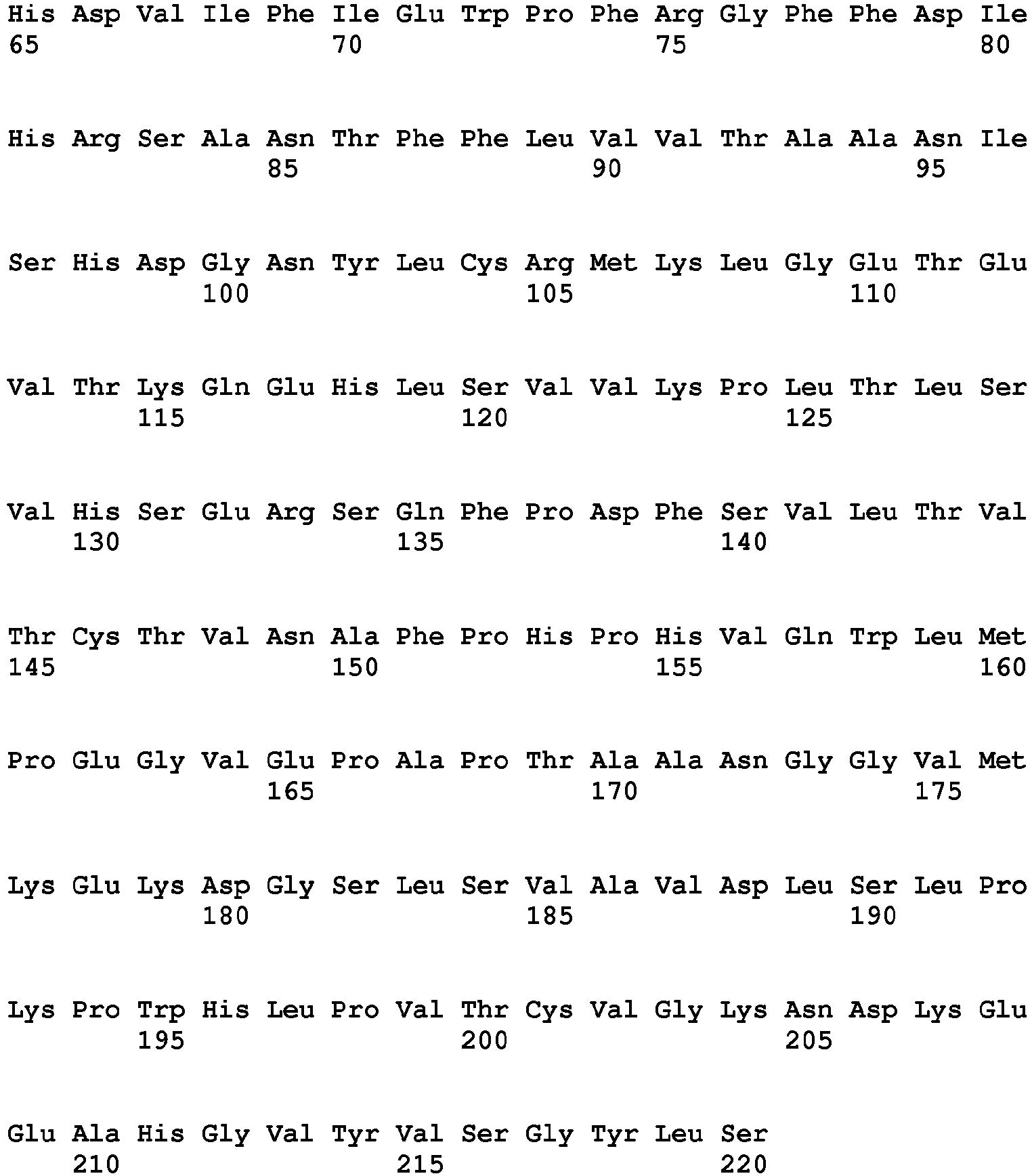
| WBC Count | 34.4 x 10⁶/ml |
| Lymphocyte % | 80.5% |
| Absolute CD3 Conc | 21.6 x 10⁶ |
| Viability | 98.87% |
| Haematocrit | 1.6% |
| WBC Count | 48.0 x 10⁶/ml |
| Lymphocyte % | 79.2% |
| Absolute CD3 Conc | 20.66 x 10⁶/ml |
| Viability | 97.58% |
| Volume Recovered | 12.5ml |
| WBC Cell Count | 3.6 x 10⁶ |
| Viability | 73% |
| WBC Count | 34.4 x 10⁶/ml |
| Lymphocyte % | 80.5% |
| Absolute CD3 Conc | 21.6 x 10⁶ |
| Viability | 98.87% |
| Haematocrit | 1.6% |
| WBC Count | 48.0 x 10⁶/ml |
| Lymphocyte % | 79.2% |
| Absolute CD3 Conc | 20.66 x 10⁶/ml |
| Viability | 97.58% |
| Volume Recovered | 12.5ml |
| WBC Cell Count | 3.6 x 10⁶ |
| Viability | 97.22% |
Claims (14)
- An in vitro expansion process for rapid expansion of antigen specific T cells, comprising the step of culturing in a gas permeable vessel a population of PBMCs with the addition of an antigen selected from the group consisting of a reconstituted peptide and a reconstituted peptide mix for a target antigen(s); wherein the peptide or peptide mix are reconstituted by adding water for injection (WFI) wherein the peptides contain at least 2 but not more than 50 amino acids;wherein the culturing is performed in the presence of at least one exogenous cytokine, selected from the group comprising IL-4, IL-7, IL-15, and the process is performed without the addition of exogenous IL-2; andwherein media and nutrients are not added or changed after initiation of the expansion process, andwherein the expansion provides the desired population of T cells in 14 days or less.
- An in vitro expansion process according to claim 1, wherein the exogenous cytokine is a combination of IL-4 and IL-7.
- An in vitro expansion process according to claim 1 or 2, wherein the antigen specific cells are allogeneic.
- An in vitro process according to any one of claims 1 to 3, wherein the culture is performed in the presence of a T cell expansion medium.
- An in vitro process according to any one of claims 1 to 4, wherein the target antigen is a CMV specific antigen
- An in vitro process according to any one of claims 1 to 5, wherein the peptide mix comprises between 2 and 500 peptides, 2 to 400 peptides, 2 to 300 or 2 to 200 peptides
- An in vitro process according to claim 6, wherein the peptides overlap by 2, 3, 4, 5, 6, 7, 8, 9, 10,11, 12 or more amino acids.
- An in vitro process according to any one of claims 1 to 7, wherein the peptides are on average 10, 11, 12, 13, 14, 15, 16, 17, 18, 19 or 20 amino acids in length.
- An in vitro process according to any one of claims 1 to 8, wherein the initial PBMC sample derived from a donor is a mobilised sample or mobilised apheresis sample
- An in vitro process according to any one of claims 1 to 9, wherein the gas permeable vessel is a G-Rex system or a derivative or an equivalent system
- An in vitro process according to any one of claims 1 to 10, wherein the process is performed in a clean room.
- A process according as defined in any one of claims 1 to 11, which comprises the further step of preparing a pharmaceutically acceptable composition by adding a diluent, stabilizer, preservative and/or other pharmaceutically acceptable excipient.
- A process according to claim 12, which comprises the further step of filling the antigen specific T cell population or a pharmaceutical composition comprising the same into a container.
- A process according to any one of claims 1 to 13, wherein the process consists of expanding the antigen-specific T cells for 14, 13, 12, 11, 10, 9, 8, 7 days or less.
Applications Claiming Priority (2)
| Application Number | Priority Date | Filing Date | Title |
|---|---|---|---|
| GBGB1121308.9A GB201121308D0 (en) | 2011-12-12 | 2011-12-12 | Process |
| PCT/GB2012/053113 WO2013088147A1 (en) | 2011-12-12 | 2012-12-12 | Process for t cell expansion |
Related Child Applications (1)
| Application Number | Title | Priority Date | Filing Date |
|---|---|---|---|
| EP17158484.0 Division-Into | 2017-02-28 |
Publications (3)
| Publication Number | Publication Date |
|---|---|
| EP2791163A1 EP2791163A1 (en) | 2014-10-22 |
| EP2791163B1 EP2791163B1 (en) | 2017-03-01 |
| EP2791163B2 true EP2791163B2 (en) | 2024-11-27 |
Family
ID=45560301
Family Applications (2)
| Application Number | Title | Priority Date | Filing Date |
|---|---|---|---|
| EP12815750.0A Active EP2791163B2 (en) | 2011-12-12 | 2012-12-12 | Process for t cell expansion |
| EP12816092.6A Withdrawn EP2791323A2 (en) | 2011-12-12 | 2012-12-12 | Method of treatment employing therapeutic t cell product from mobilised donors |
Family Applications After (1)
| Application Number | Title | Priority Date | Filing Date |
|---|---|---|---|
| EP12816092.6A Withdrawn EP2791323A2 (en) | 2011-12-12 | 2012-12-12 | Method of treatment employing therapeutic t cell product from mobilised donors |
Country Status (8)
| Country | Link |
|---|---|
| US (6) | US20140341951A1 (en) |
| EP (2) | EP2791163B2 (en) |
| JP (6) | JP2015505852A (en) |
| AU (2) | AU2012351843B2 (en) |
| ES (1) | ES2626492T5 (en) |
| GB (1) | GB201121308D0 (en) |
| HK (1) | HK1203219A1 (en) |
| WO (2) | WO2013088147A1 (en) |
Families Citing this family (48)
| Publication number | Priority date | Publication date | Assignee | Title |
|---|---|---|---|---|
| CA2772076A1 (en) | 2009-08-24 | 2011-03-10 | Baylor College Of Medicine | Generation of ctl lines with specificity against multiple tumor antigens or multiple viruses |
| GB201121308D0 (en) | 2011-12-12 | 2012-01-25 | Cell Medica Ltd | Process |
| EP2812431B1 (en) | 2012-02-09 | 2019-07-17 | Baylor College Of Medicine | Pepmixes to generate multiviral ctls with broad specificity |
| CN110241086A (en) * | 2012-06-11 | 2019-09-17 | 威尔逊沃夫制造公司 | Improved cell culture processes for adoptive cellular therapy |
| ES2980788T3 (en) | 2014-04-10 | 2024-10-03 | H Lee Moffitt Cancer Ct & Res | Enhanced expansion of tumor-infiltrating lymphocytes for adoptive cell therapy |
| GB201509202D0 (en) | 2015-05-28 | 2015-07-15 | Ge Healthcare Bio Sciences Ab | Semi-static cell culture |
| HK1254397A1 (en) * | 2015-06-26 | 2019-07-19 | 纪念斯隆-凯特林癌症中心 | Methods of treating cmv retinitis by t cell therapy |
| US11167024B2 (en) | 2015-09-18 | 2021-11-09 | Baylor College Of Medicine | Immunogenic antigen identification from a pathogen and correlation to clinical efficacy |
| KR101867942B1 (en) * | 2015-10-22 | 2018-06-20 | 가톨릭대학교 산학협력단 | Method for enrichment and expansion of virus antigen-specific T cells |
| RU2761555C2 (en) | 2015-10-22 | 2021-12-09 | Джуно Терапьютикс Гмбх | Methods, sets, means and devices for transduction |
| JP7092662B2 (en) * | 2015-10-30 | 2022-06-28 | チルドレンズ ナショナル メディカル センター | Generation of HPV antigen-specific T cells from naive T cell populations |
| US20190032013A1 (en) * | 2016-01-29 | 2019-01-31 | Tract Therapeutics, Inc. | Immune cell selection, expansion, and use |
| CN109715196A (en) | 2016-06-13 | 2019-05-03 | 转矩医疗股份有限公司 | Compositions and methods for promoting immune cell function |
| US11141434B2 (en) | 2016-07-07 | 2021-10-12 | Iovance Biotherapeutics, Inc. | Programmed death 1 ligand 1 (PD-L1) binding proteins and methods of use thereof |
| CN110099998A (en) | 2016-10-26 | 2019-08-06 | 艾欧凡斯生物治疗公司 | The tumor infiltrating lymphocyte of freezen protective stimulates again |
| TWI788307B (en) | 2016-10-31 | 2023-01-01 | 美商艾歐凡斯生物治療公司 | Engineered artificial antigen presenting cells for tumor infiltrating lymphocyte expansion |
| TWI826360B (en) | 2016-11-17 | 2023-12-21 | 美商艾歐凡斯生物治療公司 | Remnant tumor infiltrating lymphocytes and methods of preparing and using the same |
| ES3055314T3 (en) | 2017-01-06 | 2026-02-11 | Iovance Biotherapeutics Inc | Expansion of tumor infiltrating lymphocytes with potassium channel agonists and therapeutic uses thereof |
| JOP20190224A1 (en) | 2017-03-29 | 2019-09-26 | Iovance Biotherapeutics Inc | Operations for the production of tumor-infiltrating lymphocytes and their use in immunotherapy |
| US11254913B1 (en) | 2017-03-29 | 2022-02-22 | Iovance Biotherapeutics, Inc. | Processes for production of tumor infiltrating lymphocytes and uses of same in immunotherapy |
| US10821134B2 (en) | 2017-05-17 | 2020-11-03 | Board Of Regents, The University Of Texas System | BK virus specific T cells |
| KR102920712B1 (en) | 2017-05-24 | 2026-01-30 | 노파르티스 아게 | Antibody-cytokine engrafted proteins and their use in cancer therapy |
| TW201919662A (en) | 2017-06-05 | 2019-06-01 | 美商艾歐凡斯生物治療公司 | Methods of using tumor infiltrating lymphocytes in double-refractory melanoma |
| CA3074826A1 (en) | 2017-09-05 | 2019-03-14 | Torque Therapeutics, Inc. | Therapeutic protein compositions and methods of making and using the same |
| EP3737743A1 (en) | 2018-01-08 | 2020-11-18 | Iovance Biotherapeutics, Inc. | Processes for generating til products enriched for tumor antigen-specific t-cells |
| US11713446B2 (en) | 2018-01-08 | 2023-08-01 | Iovance Biotherapeutics, Inc. | Processes for generating TIL products enriched for tumor antigen-specific T-cells |
| WO2019210131A1 (en) | 2018-04-27 | 2019-10-31 | Iovance Biotherapeutics, Inc. | Closed process for expansion and gene editing of tumor infiltrating lymphocytes and uses of same in immunotherapy |
| WO2019217753A1 (en) * | 2018-05-10 | 2019-11-14 | Iovance Biotherapeutics, Inc. | Processes for production of tumor infiltrating lymphocytes and uses of same in immunotherapy |
| FR3082524B1 (en) * | 2018-06-18 | 2022-03-25 | Univ Paris Sud | BK-VIRUS NEPHROPATHY RISK STRATIFICATION METHOD AFTER KIDNEY TRANSPLANTATION |
| BR112021008549A2 (en) | 2018-11-05 | 2022-01-04 | Iovance Biotherapeutics Inc | Method of treating non-small cell lung cancer with a population of tumor-infiltrating lymphocytes |
| US12453697B2 (en) | 2018-11-05 | 2025-10-28 | Iovance Biotherapeutics, Inc. | Processes for production of tumor infiltrating lymphocytes and uses of the same in immunotherapy |
| JP7863702B2 (en) | 2018-11-08 | 2026-05-22 | セルキュア インコーポレイテッド | T cell composition with improved phenotypic characteristics |
| KR102947433B1 (en) | 2019-04-12 | 2026-04-06 | 씨4 테라퓨틱스, 인코포레이티드 | Tricyclic Decomposer of Icarus and Aeolus |
| US10772914B1 (en) | 2019-04-18 | 2020-09-15 | Baylor College Of Medicine | EBV-specific immune cells |
| US12516291B2 (en) | 2019-12-11 | 2026-01-06 | Iovance Biotherapeutics, Inc. | Processes for the production of tumor infiltrating lymphocytes (TILs) and methods of using the same |
| EP4139440B1 (en) | 2020-04-22 | 2026-08-19 | Iovance Biotherapeutics, Inc. | Systems and methods for coordinating manufacturing of cells for patient-specific immunotherapy |
| WO2021222479A1 (en) | 2020-04-28 | 2021-11-04 | Lyell Immunopharma, Inc. | Methods for culturing cells |
| CA3195019A1 (en) | 2020-10-06 | 2022-04-14 | Maria Fardis | Treatment of nsclc patients with tumor infiltrating lymphocyte therapies |
| US11781100B2 (en) | 2020-12-03 | 2023-10-10 | Applied Materials, Inc. | All-in-one bioreactor for therapeutic cells manufacturing |
| JP2023554395A (en) | 2020-12-17 | 2023-12-27 | アイオバンス バイオセラピューティクス,インコーポレイテッド | Treatment with tumor-infiltrating lymphocyte therapy in combination with CTLA-4 and PD-1 inhibitors |
| US20240060044A1 (en) * | 2021-02-25 | 2024-02-22 | Senthilkumar NATESAN | A novel method of generating t cells from peripheral blood precursors and their uses thereof |
| EP4314253A2 (en) | 2021-03-25 | 2024-02-07 | Iovance Biotherapeutics, Inc. | Methods and compositions for t-cell coculture potency assays and use with cell therapy products |
| IL310226A (en) | 2021-07-18 | 2024-03-01 | Gamida Cell Ltd | NK cell populations for treatment |
| WO2023159088A1 (en) * | 2022-02-15 | 2023-08-24 | Marker Therapeutics, Inc. | Compositions and methods for antigen-specific t cell expansion |
| EP4507704A1 (en) | 2022-04-15 | 2025-02-19 | Iovance Biotherapeutics, Inc. | Til expansion processes using specific cytokine combinations and/or akti treatment |
| CN119948142A (en) * | 2022-09-15 | 2025-05-06 | 生命科技股份有限公司 | Bioreactor capable of operating in static mode and dynamic mode and method of using the same |
| WO2024145700A1 (en) * | 2023-01-02 | 2024-07-11 | Sociedade Beneficente Israelita Brasileira Hospital Albert Einstein | Method of expanding virus-specific lymphocytes, composition, and use of a composition |
| KR20260111629A (en) * | 2023-11-06 | 2026-07-15 | 이뮤니티바이오, 인크. | T cells trained by viruses |
Family Cites Families (112)
| Publication number | Priority date | Publication date | Assignee | Title |
|---|---|---|---|---|
| US5641628A (en) | 1989-11-13 | 1997-06-24 | Children's Medical Center Corporation | Non-invasive method for isolation and detection of fetal DNA |
| WO1994002156A1 (en) | 1992-07-16 | 1994-02-03 | The Board Of Trustees Of Leland Stanford Junior University | Methods for using dendritic cells to activate t cells |
| US6821778B1 (en) | 1993-12-01 | 2004-11-23 | The Board Of Trustees Of Leland Stanford Junior University | Methods for using dendritic cells to activate gamma/delta-T cell receptor-positive T cells |
| EP1025856A3 (en) | 1994-03-08 | 2002-10-16 | Dana-Farber Cancer Institute | Methods for modulating T Cell unresponsiveness |
| AUPM446594A0 (en) | 1994-03-16 | 1994-04-14 | Csl Limited | Cytotoxic t-cell epitopes identified within epstein-barr virus |
| WO1995027722A1 (en) | 1994-04-06 | 1995-10-19 | Immunex Corporation | Interleukin-15 |
| JP3288708B2 (en) | 1994-05-16 | 2002-06-04 | ロシュ ダイアグノスティクス ゲゼルシャフト ミット ベシュレンクテル ハフツング | Method of immunomodulation by means of adoptive transfer of antigen-specific cytotoxic T cells |
| US6300090B1 (en) | 1994-07-29 | 2001-10-09 | The Rockefeller University | Methods of use of viral vectors to deliver antigen to dendritic cells |
| US5827642A (en) | 1994-08-31 | 1998-10-27 | Fred Hutchinson Cancer Research Center | Rapid expansion method ("REM") for in vitro propagation of T lymphocytes |
| ATE211172T1 (en) | 1995-06-06 | 2002-01-15 | Organon Teknika Bv | EPSTEIN-BARR VIRUS PEPTIDES, AND ANTIBODIES AGAINST THESE PEPTIDES |
| FR2745185B1 (en) | 1996-02-28 | 1998-05-15 | Sanofi Sa | USE OF IL-7 IN THE TREATMENT OF AUTOIMMUNE DISEASES, PARTICULARLY INSULIN-DEPENDENT DIABETES MELLITUS |
| US6699477B2 (en) | 1996-05-24 | 2004-03-02 | The Council Of The Queensland Institute Of Medical Research | EBV CTL epitopes |
| US5962318A (en) | 1996-11-15 | 1999-10-05 | St. Jude Children's Research Hospital | Cytotoxic T lymphocyte-mediated immunotherapy |
| JP2001509677A (en) | 1997-01-31 | 2001-07-24 | エピミューン,インコーポレイティド | Peptides for activating CTL and peptide-bearing antigen-presenting cells |
| US6528307B1 (en) | 1997-04-25 | 2003-03-04 | The Wistar Institute Of Anatomy And Biology | Cytolytic T-cell clones against colorectal carcinoma |
| US7951383B2 (en) | 1997-05-23 | 2011-05-31 | The United States Of America As Represented By The Department Of Health And Human Services | Attenuated parainfluenza virus (PIV) vaccines |
| AUPO784197A0 (en) | 1997-07-10 | 1997-08-07 | Csl Limited | Treatment of nasopharyngeal carcinoma |
| ATE428769T1 (en) | 1997-10-27 | 2009-05-15 | Univ Rockefeller | METHOD AND COMPOSITION FOR PRODUCING MATURE DENDRITIC CELLS |
| US20020155108A1 (en) | 1998-05-04 | 2002-10-24 | Biocrystal, Ltd. | Method for ex vivo loading of antigen presenting cells with antigen, and a vaccine comprising the loaded cells |
| AU3373300A (en) | 1999-02-24 | 2000-09-14 | Wistar Institute Of Anatomy And Biology, The | Method of modifying cytotoxic cells and uses thereof |
| US6828147B1 (en) | 1999-02-24 | 2004-12-07 | The Wistar Institute Of Anatomy And Biology | Method of modifying cytotoxic cells and uses thereof |
| US7005131B1 (en) | 1999-08-13 | 2006-02-28 | The Rockefeller University | Protective antigen of Epstein Barr Virus |
| US20150037297A1 (en) | 1999-08-30 | 2015-02-05 | David S Terman | Sickled Erythrocytes and Progenitors Target Cytotoxics to Tumors |
| ES2383418T3 (en) | 1999-11-24 | 2012-06-21 | Red Cliff Holding | ANTIANGIOGENIC CELLULAR AGENT FOR CANCER THERAPY. |
| DE10009341A1 (en) | 2000-02-22 | 2001-09-06 | Florian Kern | Method for antigen-specific stimulation of T lymphocytes |
| US20020155127A1 (en) | 2000-06-02 | 2002-10-24 | Danher Wang | Genetic vaccine against human immunodeficiency virus |
| EP1297017B1 (en) | 2000-06-19 | 2012-06-13 | Beth Israel Deaconess Medical Center | Compositions and methods of monoclonal and polyclonal antibodies specific for t cell subpopulations |
| EP1326961B1 (en) | 2000-09-15 | 2007-08-22 | Ortho-McNeil Pharmaceutical, Inc. | Compositions and methods for inducing specific cytolytic t cell responses |
| EP1229043A1 (en) | 2001-01-30 | 2002-08-07 | Cyto-Barr B.V. | Peptides derived from Epstein Barr virus (EBV) proteins LMP1, LMP2 and BARF1 antibody reagents reactive therewith |
| GB0107628D0 (en) | 2001-03-27 | 2001-05-16 | Avidex Ltd | Substances |
| US20040022761A1 (en) | 2001-05-11 | 2004-02-05 | Banchereau Jacques F | Compositions and methods for producing antigen-presenting cells |
| ATE438710T1 (en) | 2001-05-15 | 2009-08-15 | Ortho Mcneil Janssen Pharm | EX-VIVO PRIMING TO GENERATE CD40 LIGAND OF SPECIFIC CYTOTOXIC T LYMPHOCYTES FOR THE TREATMENT OF AUTOIMMUNE AND ALLERGIC DISEASES |
| ES2255574T3 (en) | 2001-09-28 | 2006-07-01 | Heinrich-Gunter Schumacher | MILLING BLADE WITH A BLADE GUIDE CO0NSTITUTED BY PARTIAL GUIDES. |
| US20030153073A1 (en) | 2001-11-07 | 2003-08-14 | Paul Rogers | Expansion of T cells in vitro and expanded T cell populations |
| US20030148982A1 (en) | 2001-11-13 | 2003-08-07 | Brenner Malcolm K. | Bi-spcific chimeric T cells |
| DE60234115D1 (en) | 2001-11-29 | 2009-12-03 | Dandrit Biotech As | PHARMACEUTICAL COMPOSITION FOR INDUCING AN IMMUNE RESPONSE TO A HUMAN OR ANIMAL |
| US20040096457A1 (en) | 2001-12-11 | 2004-05-20 | Huber Brigitte T | Treatment and prevention of ebv infection and ebv-associated disorders |
| WO2003057171A2 (en) | 2002-01-03 | 2003-07-17 | The Trustees Of The University Of Pennsylvania | Activation and expansion of t-cells using an engineered multivalent signaling platform |
| US7745140B2 (en) | 2002-01-03 | 2010-06-29 | The Trustees Of The University Of Pennsylvania | Activation and expansion of T-cells using an engineered multivalent signaling platform as a research tool |
| WO2004041849A1 (en) | 2002-11-07 | 2004-05-21 | The Council Of The Queensland Institute Of Medical Research | Epstein barr virus peptide epitopes, polyepitopes and delivery system therefor |
| AU2002953238A0 (en) | 2002-12-09 | 2003-01-02 | The Corporation Of The Trustees Of The Order Of The Sisters Of Mercy In Queensland | In vitro immunization |
| IL158140A0 (en) | 2003-09-25 | 2004-03-28 | Hadasit Med Res Service | Multiepitope polypeptides for cancer immunotherapy |
| CN101120083B (en) | 2003-10-08 | 2013-03-27 | 威尔森沃尔夫制造公司 | Method and device for cell culture using gas permeable material |
| US20050221481A1 (en) | 2004-03-30 | 2005-10-06 | Istituto Superiore Di Sanita' | Amplification of T cells from human cord blood in serum-deprived culture stimulated with stem cell factor, interleukin-7 and interleukin-2 |
| US7785806B2 (en) | 2004-04-28 | 2010-08-31 | Vaxdesign Corporation | Method for determining the immunogenicity of an antigen |
| AU2005250408B2 (en) | 2004-05-27 | 2010-09-23 | The Trustees Of The University Of Pennsylvania | Novel artificial antigen presenting cells and uses therefor |
| US7785875B2 (en) | 2004-07-03 | 2010-08-31 | Mogam Biotechnology Research Institute | Polynucleotide encoding HCV epitopes which can bind to various HLA supertypes, immunogenic composition comprising same and method of inducing an HCV-specific immune response using same |
| US20060045883A1 (en) | 2004-08-26 | 2006-03-02 | Jeffrey Molldrem | Anti-cancer vaccines |
| JP4731867B2 (en) | 2004-10-01 | 2011-07-27 | 国立大学法人三重大学 | Method for inducing CD8 positive cytotoxic T lymphocytes |
| US20060204509A1 (en) | 2005-03-14 | 2006-09-14 | Harty John T | Accelerated CD8+ T-cell memory after dendritic cell vaccination |
| EP1712615A1 (en) | 2005-04-15 | 2006-10-18 | Txcell | In vitro production of a cell population using feeder cells |
| DE602006010934D1 (en) | 2005-04-20 | 2010-01-21 | Hoffmann La Roche | Method for identifying epitopes associated with immunogenicity in biopharmaceuticals |
| WO2006124412A2 (en) | 2005-05-11 | 2006-11-23 | The Trustees Of The University Of Pennsylvania | Methods for the rapid expansion of antigen specific t-cells |
| US20070003531A1 (en) | 2005-06-30 | 2007-01-04 | University Of Connecticut | Methods for improving immunotherapy by enhancing survival of antigen-specific cytotoxic T lymphocytes |
| US20100035282A1 (en) | 2005-08-03 | 2010-02-11 | Maria Chiara Bonini | Use of common gamma chain cytokines for the visualization, isolation and genetic modification of memory t lymphocytes |
| JP4824389B2 (en) | 2005-10-28 | 2011-11-30 | 株式会社医学生物学研究所 | Cytotoxic T cell epitope peptide that specifically attacks Epstein-Barr virus infected cells and uses thereof |
| US20070196389A1 (en) | 2005-11-18 | 2007-08-23 | The Ohio State University Research Foundation | Viral gene products and methods for vaccination to prevent viral associated diseases |
| PT1989544E (en) | 2006-03-02 | 2011-09-26 | Antitope Ltd | T cell assays |
| WO2007121276A2 (en) | 2006-04-12 | 2007-10-25 | Biocept, Inc. | Enrichment of circulating fetal dna |
| WO2008025992A2 (en) | 2006-09-01 | 2008-03-06 | Cambridge Enterprise Limited | Methods of expanding non-effector cd8 t-cell populations |
| CA2670107A1 (en) | 2006-11-22 | 2008-06-05 | The Board Of Trustees Of The University Of Arkansas | Multi-epitope peptide-loaded dendritic cell immunotherapy for cancer |
| JP2010512149A (en) | 2006-12-07 | 2010-04-22 | ウィルソン ウォルフ マニュファクチャリング コーポレイション | Apparatus and method effective for cell culture |
| WO2008073312A2 (en) | 2006-12-08 | 2008-06-19 | Attenuon, Llc | Urokinase-type plasminogen activator receptor epitope, monoclonal antibodies derived therefrom and methods of use thereof |
| DE102006060824B4 (en) | 2006-12-21 | 2011-06-01 | Johannes-Gutenberg-Universität Mainz | Detection of Individual T Cell Response Patterns Against Tumor-associated Antigens (TAA) in Tumor Patients as a Basis for Individual Therapeutic Vaccination of Patients |
| WO2009042215A2 (en) | 2007-09-27 | 2009-04-02 | The Board Of Trustees Of The University Of Arkansas | Inhibition of dendritic cell-driven regulatory t cell activation and potentiation of tumor antigen-specific t cell responses by interleukin-15 and map kinase inhibitor |
| EP2060583A1 (en) | 2007-10-23 | 2009-05-20 | Ganymed Pharmaceuticals AG | Identification of tumor-associated markers for diagnosis and therapy |
| US20100254958A1 (en) | 2007-10-24 | 2010-10-07 | Anne Letsch | Antigen-Specific T-Cell Preparations from Bone Marrow |
| EP3085707B1 (en) | 2007-11-01 | 2019-02-27 | Mayo Foundation for Medical Education and Research | Hla-dr binding peptides and their uses |
| EP2065462A1 (en) | 2007-11-27 | 2009-06-03 | Helmholtz Zentrum München Deutsches Forschungszentrum für Gesundheit und Umwelt (GmbH) | Ex vivo method for producing a preparation containing CD4+ T cells specific for EBV structural antigens |
| EP2321407A4 (en) | 2008-09-11 | 2012-07-18 | Univ Florida | SYSTEM AND METHOD FOR THE PRODUCTION OF T-LYMPHOCYTES |
| CN101824400B (en) | 2009-03-05 | 2012-08-08 | 中国科学院微生物研究所 | Method for amplifying and multiplying T cells with antigenic specificity |
| CA2772076A1 (en) | 2009-08-24 | 2011-03-10 | Baylor College Of Medicine | Generation of ctl lines with specificity against multiple tumor antigens or multiple viruses |
| JP5833443B2 (en) | 2009-08-29 | 2015-12-16 | 株式会社バイオメッドコア | Method for measuring antigen-specific T cell inducibility |
| GB0917090D0 (en) | 2009-09-29 | 2009-11-11 | Ucl Biomedica Plc | T-cell receptor |
| US20130115617A1 (en) | 2009-12-08 | 2013-05-09 | John R. Wilson | Methods of cell culture for adoptive cell therapy |
| WO2011072088A2 (en) | 2009-12-08 | 2011-06-16 | Wilson Wolf Manufacturing Corporation | Improved methods of cell culture for adoptive cell therapy |
| US8956860B2 (en) | 2009-12-08 | 2015-02-17 | Juan F. Vera | Methods of cell culture for adoptive cell therapy |
| US9115402B2 (en) | 2010-05-14 | 2015-08-25 | Dana-Farber Cancer Institute, Inc. | Compositions and methods of identifying tumor specific neoantigens |
| US9114100B2 (en) | 2010-05-17 | 2015-08-25 | Duke University | Methods of treatment using ex vivo expansion of cord blood T cells |
| EP2600890A1 (en) | 2010-08-04 | 2013-06-12 | Instituto Europeo di Oncologia S.r.l. | Method of antigen loading for immunotherapy |
| US8741642B2 (en) | 2010-10-22 | 2014-06-03 | Virginia Commonwealth University | Methods for producing autologous immune cells resistant to myeloid-derived suppressor cells effects |
| EP2453243A1 (en) | 2010-11-11 | 2012-05-16 | Cosmo S.p.A. | Method for the diagnosis and/or follow up of the evolution of a tumor |
| US20130045491A1 (en) | 2011-07-19 | 2013-02-21 | Derya Unutmaz | Methods for activating t cells |
| SG11201400513PA (en) | 2011-09-08 | 2014-06-27 | Yeda Res & Dev | Anti third party central memory t cells, methods of producing same and use of same in transplantation and disease treatment |
| GB201121308D0 (en) | 2011-12-12 | 2012-01-25 | Cell Medica Ltd | Process |
| BR112014014591A8 (en) | 2011-12-12 | 2017-07-04 | Baylor College Medicine | process for the in vitro expansion of antigen-specific t cells, expanded autologous or allogeneic t-cell product, pharmaceutical composition, and method of treatment of a patient, and uses of an engineered cell line, mixed pulsed dendritic cells antigen-peptide, t-apcs pulsed with antigen-peptide / peptide mixtures, and antigen-peptide mixtures with pbmcs |
| EP2812431B1 (en) | 2012-02-09 | 2019-07-17 | Baylor College Of Medicine | Pepmixes to generate multiviral ctls with broad specificity |
| US20130217122A1 (en) | 2012-02-21 | 2013-08-22 | The Trustees Of The University Of Pennsylvania | Expansion of Interferon-Gamma-Producing T-Cells Using Glypican-3 Peptide Library |
| EP2847322B1 (en) | 2012-05-08 | 2019-06-26 | Western University Of Health Sciences | Standardized ex vivo platforms for the antigen-specific expansion of cd4+ t cell populations |
| AU2014351871B2 (en) | 2013-11-22 | 2020-02-13 | Cellectis | Method for generating batches of allogeneic T-cells with averaged potency |
| JP5870082B2 (en) | 2013-12-19 | 2016-02-24 | 住友ゴム工業株式会社 | Heavy duty tire |
| EP3097416A2 (en) | 2014-01-21 | 2016-11-30 | INSERM (Institut National de la Santé et de la Recherche Médicale) | Methods for testing t cell priming efficacy in a subject |
| WO2016073595A1 (en) | 2014-11-05 | 2016-05-12 | The United States Of America, As Represented By The Secretary, Department Of Health & Human Services | T cells and dendritic cells for polyomavirus therapy |
| EP3215165B1 (en) | 2014-11-05 | 2020-12-23 | Memorial Sloan Kettering Cancer Center | Methods of selecting t cell line and donor thereof for adoptive cellular therapy |
| CA2980039A1 (en) | 2015-03-20 | 2016-09-29 | Children's National Medical Center | Generating virus or other antigen-specific t cells from a naive t cell population |
| HK1254397A1 (en) | 2015-06-26 | 2019-07-19 | 纪念斯隆-凯特林癌症中心 | Methods of treating cmv retinitis by t cell therapy |
| US11167024B2 (en) | 2015-09-18 | 2021-11-09 | Baylor College Of Medicine | Immunogenic antigen identification from a pathogen and correlation to clinical efficacy |
| CN109475578A (en) | 2016-05-25 | 2019-03-15 | 昆士兰医学研究所理事会 | Methods of using allogeneic T cells to treat autoimmune diseases |
| JP7002769B2 (en) | 2016-09-16 | 2022-02-04 | ベイラー カレッジ オブ メディスン | Platform for activation and expansion of virus-specific T cells |
| TW201814042A (en) | 2016-09-26 | 2018-04-16 | 新加坡商泰莎治療私人有限公司 | T cell expansion method |
| EP3641807A4 (en) | 2017-06-22 | 2021-04-21 | The Westmead Institute For Medical Research | T ADOPTIVE 2 LYMPHOCYTE THERAPY |
| CA3074734A1 (en) | 2017-09-06 | 2019-03-14 | Nant Holdings Ip, Llc | Hla tissue matching and methods therefor |
| BR112021019120A2 (en) | 2019-03-25 | 2021-11-30 | Baylor College Medicine | T cells specific for antigens of multiple respiratory viruses and methods of producing and using them therapeutically |
| TWI867324B (en) | 2019-05-08 | 2024-12-21 | 美商百歐恩泰美國公司 | T cell manufacturing compositions and methods |
| CA3143627A1 (en) | 2019-06-20 | 2020-12-24 | Janssen Sciences Ireland Unlimited Company | Carbohydrate nanocarrier delivery of hepatitis b virus (hbv) vaccines |
| US20220257654A1 (en) | 2019-07-23 | 2022-08-18 | Baylor College Of Medicine | Antigen-specific t cell banks and methods of making and using the same therapeutically |
| WO2021021937A1 (en) | 2019-07-29 | 2021-02-04 | Baylor College Of Medicine | Antigen-specific t cell banks and methods of making and using the same therapeutically |
| EP4013897A4 (en) | 2019-08-16 | 2024-07-24 | Baylor College of Medicine | T-CELL COMPOSITIONS SPECIFIC FOR A THIRD PARTY VIRUS, AND METHODS OF MANUFACTURE AND USE THEREOF IN ANTIVIRAL PROPHYLAXIS |
| WO2021133667A1 (en) | 2019-12-23 | 2021-07-01 | Baylor College Of Medicine | T cell performance assay as a prognostic factor for clinical outcome |
| WO2021189084A1 (en) | 2020-03-20 | 2021-09-23 | Baylor College Of Medicine | Sars-cov2-specific t cell compositions and their use in treating and preventing coronavirus and other respiratory virus infections |
| EP3974029A1 (en) | 2020-09-25 | 2022-03-30 | Universidad Autónoma de Madrid | Memory t cells as adoptive cell therapy for viral infections |
| WO2023159088A1 (en) | 2022-02-15 | 2023-08-24 | Marker Therapeutics, Inc. | Compositions and methods for antigen-specific t cell expansion |
-
2011
- 2011-12-12 GB GBGB1121308.9A patent/GB201121308D0/en not_active Ceased
-
2012
- 2012-12-12 EP EP12815750.0A patent/EP2791163B2/en active Active
- 2012-12-12 ES ES12815750T patent/ES2626492T5/en active Active
- 2012-12-12 WO PCT/GB2012/053113 patent/WO2013088147A1/en not_active Ceased
- 2012-12-12 US US14/364,626 patent/US20140341951A1/en not_active Abandoned
- 2012-12-12 HK HK15103777.6A patent/HK1203219A1/en unknown
- 2012-12-12 JP JP2014546634A patent/JP2015505852A/en active Pending
- 2012-12-12 AU AU2012351843A patent/AU2012351843B2/en not_active Ceased
- 2012-12-12 EP EP12816092.6A patent/EP2791323A2/en not_active Withdrawn
- 2012-12-12 AU AU2012351842A patent/AU2012351842B2/en active Active
- 2012-12-12 US US14/364,621 patent/US20150044258A1/en not_active Abandoned
- 2012-12-12 JP JP2014546633A patent/JP6203745B2/en active Active
- 2012-12-12 WO PCT/GB2012/053114 patent/WO2013088148A2/en not_active Ceased
-
2015
- 2015-12-28 US US14/981,440 patent/US20160184361A1/en not_active Abandoned
-
2017
- 2017-07-06 JP JP2017132663A patent/JP6657145B2/en active Active
-
2018
- 2018-06-22 US US16/016,305 patent/US11963979B2/en active Active
-
2019
- 2019-11-08 JP JP2019203285A patent/JP2020022505A/en not_active Withdrawn
-
2022
- 2022-01-27 US US17/586,698 patent/US20220152111A1/en active Pending
- 2022-04-13 JP JP2022066435A patent/JP2022089916A/en active Pending
-
2023
- 2023-10-17 JP JP2023179166A patent/JP2023174861A/en not_active Withdrawn
-
2025
- 2025-10-17 US US19/361,743 patent/US20260042987A1/en active Pending
Non-Patent Citations (14)
Also Published As
| Publication number | Publication date |
|---|---|
| JP2017169594A (en) | 2017-09-28 |
| US20190134092A1 (en) | 2019-05-09 |
| AU2012351842A1 (en) | 2014-08-07 |
| US20260042987A1 (en) | 2026-02-12 |
| ES2626492T3 (en) | 2017-07-25 |
| AU2012351843A1 (en) | 2014-07-31 |
| EP2791163B1 (en) | 2017-03-01 |
| AU2012351843B2 (en) | 2016-05-12 |
| EP2791163A1 (en) | 2014-10-22 |
| JP2015505852A (en) | 2015-02-26 |
| ES2626492T8 (en) | 2018-07-13 |
| JP2023174861A (en) | 2023-12-08 |
| AU2012351842B2 (en) | 2016-05-26 |
| US20160184361A1 (en) | 2016-06-30 |
| GB201121308D0 (en) | 2012-01-25 |
| JP2020022505A (en) | 2020-02-13 |
| HK1203219A1 (en) | 2015-10-23 |
| JP2015501653A (en) | 2015-01-19 |
| US20140341951A1 (en) | 2014-11-20 |
| WO2013088148A3 (en) | 2013-09-06 |
| US11963979B2 (en) | 2024-04-23 |
| HK1203210A1 (en) | 2015-10-23 |
| JP6657145B2 (en) | 2020-03-04 |
| JP6203745B2 (en) | 2017-09-27 |
| WO2013088148A2 (en) | 2013-06-20 |
| JP2022089916A (en) | 2022-06-16 |
| ES2626492T5 (en) | 2025-04-15 |
| US20150044258A1 (en) | 2015-02-12 |
| WO2013088147A1 (en) | 2013-06-20 |
| US20220152111A1 (en) | 2022-05-19 |
| EP2791323A2 (en) | 2014-10-22 |
Similar Documents
| Publication | Publication Date | Title |
|---|---|---|
| US20220152111A1 (en) | Process for t cell expansion | |
| US11155784B2 (en) | Process of expanding T cells | |
| US20190359938A1 (en) | Adoptive transfer of cd8+ t cell clones derived from central memory cells | |
| HK1203210B (en) | Process for t cell expansion | |
| BR102023000028A2 (en) | METHOD OF EXPANSION OF VIRUS-SPECIFIC LYMPHOCYTES, COMPOSITION AND USE OF A COMPOSITION | |
| WO2024145700A1 (en) | Method of expanding virus-specific lymphocytes, composition, and use of a composition | |
| CN120500272A (en) | Cell cryopreservation method and application thereof | |
| HK40003036A (en) | Process of expanding t cells |
Legal Events
| Date | Code | Title | Description |
|---|---|---|---|
| PUAI | Public reference made under article 153(3) epc to a published international application that has entered the european phase |
Free format text: ORIGINAL CODE: 0009012 |
|
| 17P | Request for examination filed |
Effective date: 20140710 |
|
| AK | Designated contracting states |
Kind code of ref document: A1 Designated state(s): AL AT BE BG CH CY CZ DE DK EE ES FI FR GB GR HR HU IE IS IT LI LT LU LV MC MK MT NL NO PL PT RO RS SE SI SK SM TR |
|
| 17Q | First examination report despatched |
Effective date: 20141202 |
|
| DAX | Request for extension of the european patent (deleted) | ||
| REG | Reference to a national code |
Ref country code: HK Ref legal event code: DE Ref document number: 1203210 Country of ref document: HK |
|
| REG | Reference to a national code |
Ref country code: DE Ref legal event code: R079 Ref document number: 602012029350 Country of ref document: DE Free format text: PREVIOUS MAIN CLASS: C07K0014005000 Ipc: C12M0001040000 |
|
| RIC1 | Information provided on ipc code assigned before grant |
Ipc: C12N 5/0783 20100101ALI20160719BHEP Ipc: A61K 39/235 20060101ALI20160719BHEP Ipc: A61K 35/17 20150101ALI20160719BHEP Ipc: A61K 39/245 20060101ALI20160719BHEP Ipc: C12M 1/04 20060101AFI20160719BHEP Ipc: A61K 39/12 20060101ALI20160719BHEP |
|
| GRAP | Despatch of communication of intention to grant a patent |
Free format text: ORIGINAL CODE: EPIDOSNIGR1 |
|
| INTG | Intention to grant announced |
Effective date: 20160919 |
|
| STAA | Information on the status of an ep patent application or granted ep patent |
Free format text: STATUS: GRANT OF PATENT IS INTENDED |
|
| GRAS | Grant fee paid |
Free format text: ORIGINAL CODE: EPIDOSNIGR3 |
|
| GRAA | (expected) grant |
Free format text: ORIGINAL CODE: 0009210 |
|
| STAA | Information on the status of an ep patent application or granted ep patent |
Free format text: STATUS: THE PATENT HAS BEEN GRANTED |
|
| AK | Designated contracting states |
Kind code of ref document: B1 Designated state(s): AL AT BE BG CH CY CZ DE DK EE ES FI FR GB GR HR HU IE IS IT LI LT LU LV MC MK MT NL NO PL PT RO RS SE SI SK SM TR |
|
| REG | Reference to a national code |
Ref country code: GB Ref legal event code: FG4D |
|
| REG | Reference to a national code |
Ref country code: CH Ref legal event code: EP Ref country code: AT Ref legal event code: REF Ref document number: 871367 Country of ref document: AT Kind code of ref document: T Effective date: 20170315 |
|
| REG | Reference to a national code |
Ref country code: IE Ref legal event code: FG4D |
|
| REG | Reference to a national code |
Ref country code: DE Ref legal event code: R096 Ref document number: 602012029350 Country of ref document: DE |
|
| REG | Reference to a national code |
Ref country code: CH Ref legal event code: NV Representative=s name: RENTSCH PARTNER AG, CH |
|
| REG | Reference to a national code |
Ref country code: NL Ref legal event code: MP Effective date: 20170301 |
|
| REG | Reference to a national code |
Ref country code: LT Ref legal event code: MG4D |
|
| REG | Reference to a national code |
Ref country code: ES Ref legal event code: FG2A Ref document number: 2626492 Country of ref document: ES Kind code of ref document: T3 Effective date: 20170725 |
|
| PG25 | Lapsed in a contracting state [announced via postgrant information from national office to epo] |
Ref country code: LT Free format text: LAPSE BECAUSE OF FAILURE TO SUBMIT A TRANSLATION OF THE DESCRIPTION OR TO PAY THE FEE WITHIN THE PRESCRIBED TIME-LIMIT Effective date: 20170301 Ref country code: GR Free format text: LAPSE BECAUSE OF FAILURE TO SUBMIT A TRANSLATION OF THE DESCRIPTION OR TO PAY THE FEE WITHIN THE PRESCRIBED TIME-LIMIT Effective date: 20170602 Ref country code: NO Free format text: LAPSE BECAUSE OF FAILURE TO SUBMIT A TRANSLATION OF THE DESCRIPTION OR TO PAY THE FEE WITHIN THE PRESCRIBED TIME-LIMIT Effective date: 20170601 Ref country code: HR Free format text: LAPSE BECAUSE OF FAILURE TO SUBMIT A TRANSLATION OF THE DESCRIPTION OR TO PAY THE FEE WITHIN THE PRESCRIBED TIME-LIMIT Effective date: 20170301 Ref country code: FI Free format text: LAPSE BECAUSE OF FAILURE TO SUBMIT A TRANSLATION OF THE DESCRIPTION OR TO PAY THE FEE WITHIN THE PRESCRIBED TIME-LIMIT Effective date: 20170301 |
|
| PG25 | Lapsed in a contracting state [announced via postgrant information from national office to epo] |
Ref country code: BG Free format text: LAPSE BECAUSE OF FAILURE TO SUBMIT A TRANSLATION OF THE DESCRIPTION OR TO PAY THE FEE WITHIN THE PRESCRIBED TIME-LIMIT Effective date: 20170601 Ref country code: LV Free format text: LAPSE BECAUSE OF FAILURE TO SUBMIT A TRANSLATION OF THE DESCRIPTION OR TO PAY THE FEE WITHIN THE PRESCRIBED TIME-LIMIT Effective date: 20170301 Ref country code: RS Free format text: LAPSE BECAUSE OF FAILURE TO SUBMIT A TRANSLATION OF THE DESCRIPTION OR TO PAY THE FEE WITHIN THE PRESCRIBED TIME-LIMIT Effective date: 20170301 Ref country code: SE Free format text: LAPSE BECAUSE OF FAILURE TO SUBMIT A TRANSLATION OF THE DESCRIPTION OR TO PAY THE FEE WITHIN THE PRESCRIBED TIME-LIMIT Effective date: 20170301 |
|
| PG25 | Lapsed in a contracting state [announced via postgrant information from national office to epo] |
Ref country code: NL Free format text: LAPSE BECAUSE OF FAILURE TO SUBMIT A TRANSLATION OF THE DESCRIPTION OR TO PAY THE FEE WITHIN THE PRESCRIBED TIME-LIMIT Effective date: 20170301 |
|
| REG | Reference to a national code |
Ref country code: CH Ref legal event code: PCAR Free format text: NEW ADDRESS: BELLERIVESTRASSE 203 POSTFACH, 8034 ZUERICH (CH) |
|
| PG25 | Lapsed in a contracting state [announced via postgrant information from national office to epo] |
Ref country code: CZ Free format text: LAPSE BECAUSE OF FAILURE TO SUBMIT A TRANSLATION OF THE DESCRIPTION OR TO PAY THE FEE WITHIN THE PRESCRIBED TIME-LIMIT Effective date: 20170301 Ref country code: SK Free format text: LAPSE BECAUSE OF FAILURE TO SUBMIT A TRANSLATION OF THE DESCRIPTION OR TO PAY THE FEE WITHIN THE PRESCRIBED TIME-LIMIT Effective date: 20170301 Ref country code: RO Free format text: LAPSE BECAUSE OF FAILURE TO SUBMIT A TRANSLATION OF THE DESCRIPTION OR TO PAY THE FEE WITHIN THE PRESCRIBED TIME-LIMIT Effective date: 20170301 Ref country code: EE Free format text: LAPSE BECAUSE OF FAILURE TO SUBMIT A TRANSLATION OF THE DESCRIPTION OR TO PAY THE FEE WITHIN THE PRESCRIBED TIME-LIMIT Effective date: 20170301 |
|
| PG25 | Lapsed in a contracting state [announced via postgrant information from national office to epo] |
Ref country code: IS Free format text: LAPSE BECAUSE OF FAILURE TO SUBMIT A TRANSLATION OF THE DESCRIPTION OR TO PAY THE FEE WITHIN THE PRESCRIBED TIME-LIMIT Effective date: 20170701 Ref country code: SM Free format text: LAPSE BECAUSE OF FAILURE TO SUBMIT A TRANSLATION OF THE DESCRIPTION OR TO PAY THE FEE WITHIN THE PRESCRIBED TIME-LIMIT Effective date: 20170301 Ref country code: PL Free format text: LAPSE BECAUSE OF FAILURE TO SUBMIT A TRANSLATION OF THE DESCRIPTION OR TO PAY THE FEE WITHIN THE PRESCRIBED TIME-LIMIT Effective date: 20170301 Ref country code: PT Free format text: LAPSE BECAUSE OF FAILURE TO SUBMIT A TRANSLATION OF THE DESCRIPTION OR TO PAY THE FEE WITHIN THE PRESCRIBED TIME-LIMIT Effective date: 20170703 |
|
| REG | Reference to a national code |
Ref country code: DE Ref legal event code: R026 Ref document number: 602012029350 Country of ref document: DE |
|
| PLBI | Opposition filed |
Free format text: ORIGINAL CODE: 0009260 |
|
| PLAX | Notice of opposition and request to file observation + time limit sent |
Free format text: ORIGINAL CODE: EPIDOSNOBS2 |
|
| REG | Reference to a national code |
Ref country code: FR Ref legal event code: PLFP Year of fee payment: 6 |
|
| REG | Reference to a national code |
Ref country code: HK Ref legal event code: GR Ref document number: 1203210 Country of ref document: HK |
|
| 26 | Opposition filed |
Opponent name: STRAWMAN LIMITED Effective date: 20171130 |
|
| PG25 | Lapsed in a contracting state [announced via postgrant information from national office to epo] |
Ref country code: DK Free format text: LAPSE BECAUSE OF FAILURE TO SUBMIT A TRANSLATION OF THE DESCRIPTION OR TO PAY THE FEE WITHIN THE PRESCRIBED TIME-LIMIT Effective date: 20170301 |
|
| PG25 | Lapsed in a contracting state [announced via postgrant information from national office to epo] |
Ref country code: SI Free format text: LAPSE BECAUSE OF FAILURE TO SUBMIT A TRANSLATION OF THE DESCRIPTION OR TO PAY THE FEE WITHIN THE PRESCRIBED TIME-LIMIT Effective date: 20170301 |
|
| PLAF | Information modified related to communication of a notice of opposition and request to file observations + time limit |
Free format text: ORIGINAL CODE: EPIDOSCOBS2 |
|
| PLBB | Reply of patent proprietor to notice(s) of opposition received |
Free format text: ORIGINAL CODE: EPIDOSNOBS3 |
|
| RAP2 | Party data changed (patent owner data changed or rights of a patent transferred) |
Owner name: CELL MEDICA LIMITED Owner name: BAYLOR COLLEGE OF MEDICINE |
|
| REG | Reference to a national code |
Ref country code: DE Ref legal event code: R081 Ref document number: 602012029350 Country of ref document: DE Owner name: ALLOVIR, INC, HOUSTON, US Free format text: FORMER OWNER: CELL MEDICA LIMITED, LONDON, GB Ref country code: DE Ref legal event code: R082 Ref document number: 602012029350 Country of ref document: DE Representative=s name: STOLMAR & PARTNER PATENTANWAELTE PARTG MBB, DE Ref country code: DE Ref legal event code: R081 Ref document number: 602012029350 Country of ref document: DE Owner name: BAYLOR COLLEGE OF MEDICINE, HOUSTON, US Free format text: FORMER OWNER: CELL MEDICA LIMITED, LONDON, GB Ref country code: DE Ref legal event code: R081 Ref document number: 602012029350 Country of ref document: DE Owner name: CELL MEDICA LIMITED, GB Free format text: FORMER OWNER: CELL MEDICA LIMITED, LONDON, GB |
|
| RIN2 | Information on inventor provided after grant (corrected) |
Inventor name: VALDES, JUAN F. VERA Inventor name: ROONEY, CLIONA M. Inventor name: KNAUS, RAINER, LUDWIG Inventor name: NEWTON, KATY, REBECCA Inventor name: LEEN, ANN M. |
|
| REG | Reference to a national code |
Ref country code: IE Ref legal event code: MM4A |
|
| PG25 | Lapsed in a contracting state [announced via postgrant information from national office to epo] |
Ref country code: LU Free format text: LAPSE BECAUSE OF NON-PAYMENT OF DUE FEES Effective date: 20171212 Ref country code: MT Free format text: LAPSE BECAUSE OF NON-PAYMENT OF DUE FEES Effective date: 20171212 |
|
| REG | Reference to a national code |
Ref country code: BE Ref legal event code: MM Effective date: 20171231 |
|
| PG25 | Lapsed in a contracting state [announced via postgrant information from national office to epo] |
Ref country code: IE Free format text: LAPSE BECAUSE OF NON-PAYMENT OF DUE FEES Effective date: 20171212 |
|
| PG25 | Lapsed in a contracting state [announced via postgrant information from national office to epo] |
Ref country code: BE Free format text: LAPSE BECAUSE OF NON-PAYMENT OF DUE FEES Effective date: 20171231 |
|
| PLAB | Opposition data, opponent's data or that of the opponent's representative modified |
Free format text: ORIGINAL CODE: 0009299OPPO |
|
| R26 | Opposition filed (corrected) |
Opponent name: STRAWMAN LIMITED Effective date: 20171130 |
|
| REG | Reference to a national code |
Ref country code: CH Ref legal event code: PUEA Owner name: CELL MEDICA LIMITED, US Free format text: FORMER OWNER: CELL MEDICA LIMITED, GB |
|
| PLCK | Communication despatched that opposition was rejected |
Free format text: ORIGINAL CODE: EPIDOSNREJ1 |
|
| PG25 | Lapsed in a contracting state [announced via postgrant information from national office to epo] |
Ref country code: HU Free format text: LAPSE BECAUSE OF FAILURE TO SUBMIT A TRANSLATION OF THE DESCRIPTION OR TO PAY THE FEE WITHIN THE PRESCRIBED TIME-LIMIT; INVALID AB INITIO Effective date: 20121212 Ref country code: MC Free format text: LAPSE BECAUSE OF FAILURE TO SUBMIT A TRANSLATION OF THE DESCRIPTION OR TO PAY THE FEE WITHIN THE PRESCRIBED TIME-LIMIT Effective date: 20170301 |
|
| APBM | Appeal reference recorded |
Free format text: ORIGINAL CODE: EPIDOSNREFNO |
|
| APBP | Date of receipt of notice of appeal recorded |
Free format text: ORIGINAL CODE: EPIDOSNNOA2O |
|
| APAH | Appeal reference modified |
Free format text: ORIGINAL CODE: EPIDOSCREFNO |
|
| APBQ | Date of receipt of statement of grounds of appeal recorded |
Free format text: ORIGINAL CODE: EPIDOSNNOA3O |
|
| PG25 | Lapsed in a contracting state [announced via postgrant information from national office to epo] |
Ref country code: CY Free format text: LAPSE BECAUSE OF FAILURE TO SUBMIT A TRANSLATION OF THE DESCRIPTION OR TO PAY THE FEE WITHIN THE PRESCRIBED TIME-LIMIT Effective date: 20170301 |
|
| PG25 | Lapsed in a contracting state [announced via postgrant information from national office to epo] |
Ref country code: MK Free format text: LAPSE BECAUSE OF FAILURE TO SUBMIT A TRANSLATION OF THE DESCRIPTION OR TO PAY THE FEE WITHIN THE PRESCRIBED TIME-LIMIT Effective date: 20170301 |
|
| PG25 | Lapsed in a contracting state [announced via postgrant information from national office to epo] |
Ref country code: TR Free format text: LAPSE BECAUSE OF FAILURE TO SUBMIT A TRANSLATION OF THE DESCRIPTION OR TO PAY THE FEE WITHIN THE PRESCRIBED TIME-LIMIT Effective date: 20170301 |
|
| PG25 | Lapsed in a contracting state [announced via postgrant information from national office to epo] |
Ref country code: AL Free format text: LAPSE BECAUSE OF FAILURE TO SUBMIT A TRANSLATION OF THE DESCRIPTION OR TO PAY THE FEE WITHIN THE PRESCRIBED TIME-LIMIT Effective date: 20170301 |
|
| RAP2 | Party data changed (patent owner data changed or rights of a patent transferred) |
Owner name: ALLOVIR, INC. Owner name: BAYLOR COLLEGE OF MEDICINE |
|
| REG | Reference to a national code |
Ref country code: DE Ref legal event code: R082 Ref document number: 602012029350 Country of ref document: DE Representative=s name: STOLMAR & PARTNER PATENTANWAELTE PARTG MBB, DE Ref country code: DE Ref legal event code: R081 Ref document number: 602012029350 Country of ref document: DE Owner name: ALLOVIR, INC, HOUSTON, US Free format text: FORMER OWNERS: BAYLOR COLLEGE OF MEDICINE, HOUSTON, TEX., US; CELL MEDICA LIMITED, LONDON, GB Ref country code: DE Ref legal event code: R081 Ref document number: 602012029350 Country of ref document: DE Owner name: BAYLOR COLLEGE OF MEDICINE, HOUSTON, US Free format text: FORMER OWNERS: BAYLOR COLLEGE OF MEDICINE, HOUSTON, TEX., US; CELL MEDICA LIMITED, LONDON, GB |
|
| REG | Reference to a national code |
Ref country code: GB Ref legal event code: 732E Free format text: REGISTERED BETWEEN 20201015 AND 20201021 |
|
| REG | Reference to a national code |
Ref country code: AT Ref legal event code: PC Ref document number: 871367 Country of ref document: AT Kind code of ref document: T Owner name: ALLOVIR, INC., US Effective date: 20210215 |
|
| APBU | Appeal procedure closed |
Free format text: ORIGINAL CODE: EPIDOSNNOA9O |
|
| P01 | Opt-out of the competence of the unified patent court (upc) registered |
Effective date: 20230523 |
|
| REG | Reference to a national code |
Ref country code: CH Ref legal event code: PK Free format text: BERICHTIGUNGEN |
|
| PLAY | Examination report in opposition despatched + time limit |
Free format text: ORIGINAL CODE: EPIDOSNORE2 |
|
| RIN2 | Information on inventor provided after grant (corrected) |
Inventor name: WILSON, JOHN ROBERT Inventor name: LEEN, ANN M. Inventor name: VALDES, JUAN F. VERA Inventor name: ROONEY, CLIONA M. Inventor name: NEWTON, KATY, REBECCA Inventor name: KNAUS, RAINER, LUDWIG |
|
| PLBC | Reply to examination report in opposition received |
Free format text: ORIGINAL CODE: EPIDOSNORE3 |
|
| PLAP | Information related to despatch of examination report in opposition + time limit deleted |
Free format text: ORIGINAL CODE: EPIDOSDORE2 |
|
| PLAT | Information related to reply to examination report in opposition deleted |
Free format text: ORIGINAL CODE: EPIDOSDORE3 |
|
| PLAY | Examination report in opposition despatched + time limit |
Free format text: ORIGINAL CODE: EPIDOSNORE2 |
|
| PUAH | Patent maintained in amended form |
Free format text: ORIGINAL CODE: 0009272 |
|
| STAA | Information on the status of an ep patent application or granted ep patent |
Free format text: STATUS: PATENT MAINTAINED AS AMENDED |
|
| 27A | Patent maintained in amended form |
Effective date: 20241127 |
|
| AK | Designated contracting states |
Kind code of ref document: B2 Designated state(s): AL AT BE BG CH CY CZ DE DK EE ES FI FR GB GR HR HU IE IS IT LI LT LU LV MC MK MT NL NO PL PT RO RS SE SI SK SM TR |
|
| REG | Reference to a national code |
Ref country code: DE Ref legal event code: R102 Ref document number: 602012029350 Country of ref document: DE |
|
| REG | Reference to a national code |
Ref country code: ES Ref legal event code: DC2A Ref document number: 2626492 Country of ref document: ES Kind code of ref document: T5 Effective date: 20250415 |
|
| PGFP | Annual fee paid to national office [announced via postgrant information from national office to epo] |
Ref country code: ES Payment date: 20250115 Year of fee payment: 13 |
|
| REG | Reference to a national code |
Ref country code: AT Ref legal event code: UEP Ref document number: 871367 Country of ref document: AT Kind code of ref document: T Effective date: 20241127 |
|
| REG | Reference to a national code |
Ref country code: CH Ref legal event code: U11 Free format text: ST27 STATUS EVENT CODE: U-0-0-U10-U11 (AS PROVIDED BY THE NATIONAL OFFICE) Effective date: 20260618 |
|
| PGFP | Annual fee paid to national office [announced via postgrant information from national office to epo] |
Ref country code: GB Payment date: 20260527 Year of fee payment: 14 |
|
| PGFP | Annual fee paid to national office [announced via postgrant information from national office to epo] |
Ref country code: IT Payment date: 20260622 Year of fee payment: 14 |
|
| PGFP | Annual fee paid to national office [announced via postgrant information from national office to epo] |
Ref country code: DE Payment date: 20260527 Year of fee payment: 14 |
|
| PGFP | Annual fee paid to national office [announced via postgrant information from national office to epo] |
Ref country code: AT Payment date: 20260609 Year of fee payment: 14 |
|
| PGFP | Annual fee paid to national office [announced via postgrant information from national office to epo] |
Ref country code: CH Payment date: 20260618 Year of fee payment: 14 |
|
| PGFP | Annual fee paid to national office [announced via postgrant information from national office to epo] |
Ref country code: FR Payment date: 20260526 Year of fee payment: 14 |