EP2776584B1 - Method and primers for the detection of microcystin-producing toxic cyanobacteria - Google Patents
Method and primers for the detection of microcystin-producing toxic cyanobacteria Download PDFInfo
- Publication number
- EP2776584B1 EP2776584B1 EP12795832.0A EP12795832A EP2776584B1 EP 2776584 B1 EP2776584 B1 EP 2776584B1 EP 12795832 A EP12795832 A EP 12795832A EP 2776584 B1 EP2776584 B1 EP 2776584B1
- Authority
- EP
- European Patent Office
- Prior art keywords
- microcystin
- seq
- mcyb
- sample
- dna
- Prior art date
- Legal status (The legal status is an assumption and is not a legal conclusion. Google has not performed a legal analysis and makes no representation as to the accuracy of the status listed.)
- Active
Links
- SRUWWOSWHXIIIA-UKPGNTDSSA-N Cyanoginosin Chemical compound N1C(=O)[C@H](CCCN=C(N)N)NC(=O)[C@@H](C)[C@H](C(O)=O)NC(=O)[C@H](CC(C)C)NC(=O)[C@H](C)NC(=O)C(=C)N(C)C(=O)CC[C@H](C(O)=O)N(C)C(=O)[C@@H](C)[C@@H]1\C=C\C(\C)=C\[C@H](C)[C@@H](O)CC1=CC=CC=C1 SRUWWOSWHXIIIA-UKPGNTDSSA-N 0.000 title claims description 57
- 108010067094 microcystin Proteins 0.000 title claims description 56
- 238000000034 method Methods 0.000 title claims description 37
- 238000001514 detection method Methods 0.000 title claims description 35
- 231100000331 toxic Toxicity 0.000 title claims description 28
- 230000002588 toxic effect Effects 0.000 title claims description 28
- 241000192700 Cyanobacteria Species 0.000 title claims description 26
- 239000000523 sample Substances 0.000 claims description 79
- 108090000623 proteins and genes Proteins 0.000 claims description 48
- 239000013615 primer Substances 0.000 claims description 43
- 238000003753 real-time PCR Methods 0.000 claims description 38
- 238000003752 polymerase chain reaction Methods 0.000 claims description 34
- 241000192531 Anabaena sp. Species 0.000 claims description 22
- 241000192710 Microcystis aeruginosa Species 0.000 claims description 19
- 241000192524 Planktothrix agardhii Species 0.000 claims description 14
- 238000006243 chemical reaction Methods 0.000 claims description 12
- 150000007523 nucleic acids Chemical class 0.000 claims description 9
- 238000012544 monitoring process Methods 0.000 claims description 6
- 108020004707 nucleic acids Proteins 0.000 claims description 6
- 102000039446 nucleic acids Human genes 0.000 claims description 6
- 239000011541 reaction mixture Substances 0.000 claims description 6
- 108091028043 Nucleic acid sequence Proteins 0.000 claims description 3
- 230000009089 cytolysis Effects 0.000 claims description 3
- 239000002751 oligonucleotide probe Substances 0.000 claims description 3
- 239000003155 DNA primer Substances 0.000 claims description 2
- 108020005187 Oligonucleotide Probes Proteins 0.000 claims description 2
- 239000002253 acid Substances 0.000 claims description 2
- 108020004414 DNA Proteins 0.000 description 67
- 241000530769 Planktothrix Species 0.000 description 28
- DIDLWIPCWUSYPF-UHFFFAOYSA-N microcystin-LR Natural products COC(Cc1ccccc1)C(C)C=C(/C)C=CC2NC(=O)C(NC(CCCNC(=N)N)C(=O)O)NC(=O)C(C)C(NC(=O)C(NC(CC(C)C)C(=O)O)NC(=O)C(C)NC(=O)C(=C)N(C)C(=O)CCC(NC(=O)C2C)C(=O)O)C(=O)O DIDLWIPCWUSYPF-UHFFFAOYSA-N 0.000 description 25
- 231100000765 toxin Toxicity 0.000 description 23
- 239000003053 toxin Substances 0.000 description 23
- 108700012359 toxins Proteins 0.000 description 23
- 241000192701 Microcystis Species 0.000 description 22
- 230000003321 amplification Effects 0.000 description 22
- 230000007613 environmental effect Effects 0.000 description 22
- 238000003199 nucleic acid amplification method Methods 0.000 description 22
- 241000192542 Anabaena Species 0.000 description 21
- 238000011002 quantification Methods 0.000 description 19
- 108010049746 Microcystins Proteins 0.000 description 15
- XLYOFNOQVPJJNP-UHFFFAOYSA-N water Substances O XLYOFNOQVPJJNP-UHFFFAOYSA-N 0.000 description 13
- JIGDOBKZMULDHS-UHFFFAOYSA-N cyanogenosin-RR Natural products N1C(=O)C(CCCN=C(N)N)NC(=O)C(C)C(C(O)=O)NC(=O)C(CCCN=C(N)N)NC(=O)C(C)NC(=O)C(=C)N(C)C(=O)CCC(C(O)=O)NC(=O)C(C)C1C=CC(C)=CC(C)C(OC)CC1=CC=CC=C1 JIGDOBKZMULDHS-UHFFFAOYSA-N 0.000 description 12
- JIGDOBKZMULDHS-UUHBQKJESA-N microcystin RR Chemical compound C([C@H](OC)[C@@H](C)\C=C(/C)\C=C\[C@H]1[C@@H](C(=O)N[C@H](CCC(=O)N(C)C(=C)C(=O)N[C@H](C)C(=O)N[C@@H](CCCN=C(N)N)C(=O)N[C@H]([C@H](C)C(=O)N[C@@H](CCCN=C(N)N)C(=O)N1)C(O)=O)C(O)=O)C)C1=CC=CC=C1 JIGDOBKZMULDHS-UUHBQKJESA-N 0.000 description 12
- 238000003556 assay Methods 0.000 description 11
- 241000559509 Planktothrix agardhii NIVA-CYA 126/8 Species 0.000 description 10
- 238000007400 DNA extraction Methods 0.000 description 9
- 108091034117 Oligonucleotide Proteins 0.000 description 9
- 238000004458 analytical method Methods 0.000 description 9
- 239000000047 product Substances 0.000 description 9
- 241001463754 Microcystis aeruginosa UV027 Species 0.000 description 8
- 239000002773 nucleotide Substances 0.000 description 8
- 125000003729 nucleotide group Chemical group 0.000 description 8
- 239000002987 primer (paints) Substances 0.000 description 8
- 230000006037 cell lysis Effects 0.000 description 7
- 238000001914 filtration Methods 0.000 description 7
- 238000004128 high performance liquid chromatography Methods 0.000 description 7
- 238000002360 preparation method Methods 0.000 description 7
- WEVYAHXRMPXWCK-UHFFFAOYSA-N Acetonitrile Chemical compound CC#N WEVYAHXRMPXWCK-UHFFFAOYSA-N 0.000 description 6
- 238000002965 ELISA Methods 0.000 description 6
- OKKJLVBELUTLKV-UHFFFAOYSA-N Methanol Chemical compound OC OKKJLVBELUTLKV-UHFFFAOYSA-N 0.000 description 6
- 150000002500 ions Chemical class 0.000 description 6
- 238000001294 liquid chromatography-tandem mass spectrometry Methods 0.000 description 6
- 230000035945 sensitivity Effects 0.000 description 6
- 239000002904 solvent Substances 0.000 description 6
- 238000011529 RT qPCR Methods 0.000 description 5
- 239000000284 extract Substances 0.000 description 5
- 239000013505 freshwater Substances 0.000 description 5
- 230000005764 inhibitory process Effects 0.000 description 5
- 239000000463 material Substances 0.000 description 5
- 239000002245 particle Substances 0.000 description 5
- 238000005464 sample preparation method Methods 0.000 description 5
- 108091093088 Amplicon Proteins 0.000 description 4
- IJGRMHOSHXDMSA-UHFFFAOYSA-N Atomic nitrogen Chemical compound N#N IJGRMHOSHXDMSA-UHFFFAOYSA-N 0.000 description 4
- IXBQSRWSVIBXNC-HSKGSTCASA-N Nodularin Chemical compound C([C@H](OC)[C@@H](C)\C=C(/C)\C=C\[C@H]1[C@@H](C(=O)N[C@H](CCC(=O)N(C)C(=C\C)/C(=O)N[C@H]([C@H](C)C(=O)N[C@@H](CCCN=C(N)N)C(=O)N1)C(O)=O)C(O)=O)C)C1=CC=CC=C1 IXBQSRWSVIBXNC-HSKGSTCASA-N 0.000 description 4
- IXBQSRWSVIBXNC-UHFFFAOYSA-N Nodularin Natural products N1C(=O)C(CCCN=C(N)N)NC(=O)C(C)C(C(O)=O)NC(=O)C(=CC)N(C)C(=O)CCC(C(O)=O)NC(=O)C(C)C1C=CC(C)=CC(C)C(OC)CC1=CC=CC=C1 IXBQSRWSVIBXNC-UHFFFAOYSA-N 0.000 description 4
- 230000015572 biosynthetic process Effects 0.000 description 4
- 239000008367 deionised water Substances 0.000 description 4
- 229910021641 deionized water Inorganic materials 0.000 description 4
- 238000010438 heat treatment Methods 0.000 description 4
- 238000002347 injection Methods 0.000 description 4
- 239000007924 injection Substances 0.000 description 4
- 238000004895 liquid chromatography mass spectrometry Methods 0.000 description 4
- 238000005259 measurement Methods 0.000 description 4
- -1 methylenenitrilo Chemical class 0.000 description 4
- FEVBMCJUKWWWBT-QVWKUIOOSA-N microcystin LF Chemical compound C([C@H](OC)[C@@H](C)\C=C(/C)\C=C\[C@H]1[C@@H](C(=O)N[C@H](CCC(=O)N(C)C(=C)C(=O)N[C@H](C)C(=O)N[C@@H](CC(C)C)C(=O)N[C@H]([C@H](C)C(=O)N[C@@H](CC=2C=CC=CC=2)C(=O)N1)C(O)=O)C(O)=O)C)C1=CC=CC=C1 FEVBMCJUKWWWBT-QVWKUIOOSA-N 0.000 description 4
- CJIASZBWXIFQMU-LNXRSHCCSA-N microcystin LW Chemical compound C([C@H](OC)[C@@H](C)\C=C(/C)\C=C\[C@H]1[C@@H](C(=O)N[C@H](CCC(=O)N(C)C(=C)C(=O)N[C@H](C)C(=O)N[C@@H](CC(C)C)C(=O)N[C@H]([C@H](C)C(=O)N[C@@H](CC=2C3=CC=CC=C3NC=2)C(=O)N1)C(O)=O)C(O)=O)C)C1=CC=CC=C1 CJIASZBWXIFQMU-LNXRSHCCSA-N 0.000 description 4
- 108010065793 nodularin Proteins 0.000 description 4
- 238000000746 purification Methods 0.000 description 4
- 241000894007 species Species 0.000 description 4
- 239000000725 suspension Substances 0.000 description 4
- 102000016928 DNA-directed DNA polymerase Human genes 0.000 description 3
- 108010014303 DNA-directed DNA polymerase Proteins 0.000 description 3
- 108090000364 Ligases Proteins 0.000 description 3
- 241001287970 Microcystis wesenbergii NIES-107 Species 0.000 description 3
- 241000018388 Nodularia sphaerocarpa PCC 7804 Species 0.000 description 3
- DTQVDTLACAAQTR-UHFFFAOYSA-N Trifluoroacetic acid Chemical compound OC(=O)C(F)(F)F DTQVDTLACAAQTR-UHFFFAOYSA-N 0.000 description 3
- JLCPHMBAVCMARE-UHFFFAOYSA-N [3-[[3-[[3-[[3-[[3-[[3-[[3-[[3-[[3-[[3-[[3-[[5-(2-amino-6-oxo-1H-purin-9-yl)-3-[[3-[[3-[[3-[[3-[[3-[[5-(2-amino-6-oxo-1H-purin-9-yl)-3-[[5-(2-amino-6-oxo-1H-purin-9-yl)-3-hydroxyoxolan-2-yl]methoxy-hydroxyphosphoryl]oxyoxolan-2-yl]methoxy-hydroxyphosphoryl]oxy-5-(5-methyl-2,4-dioxopyrimidin-1-yl)oxolan-2-yl]methoxy-hydroxyphosphoryl]oxy-5-(6-aminopurin-9-yl)oxolan-2-yl]methoxy-hydroxyphosphoryl]oxy-5-(6-aminopurin-9-yl)oxolan-2-yl]methoxy-hydroxyphosphoryl]oxy-5-(6-aminopurin-9-yl)oxolan-2-yl]methoxy-hydroxyphosphoryl]oxy-5-(6-aminopurin-9-yl)oxolan-2-yl]methoxy-hydroxyphosphoryl]oxyoxolan-2-yl]methoxy-hydroxyphosphoryl]oxy-5-(5-methyl-2,4-dioxopyrimidin-1-yl)oxolan-2-yl]methoxy-hydroxyphosphoryl]oxy-5-(4-amino-2-oxopyrimidin-1-yl)oxolan-2-yl]methoxy-hydroxyphosphoryl]oxy-5-(5-methyl-2,4-dioxopyrimidin-1-yl)oxolan-2-yl]methoxy-hydroxyphosphoryl]oxy-5-(5-methyl-2,4-dioxopyrimidin-1-yl)oxolan-2-yl]methoxy-hydroxyphosphoryl]oxy-5-(6-aminopurin-9-yl)oxolan-2-yl]methoxy-hydroxyphosphoryl]oxy-5-(6-aminopurin-9-yl)oxolan-2-yl]methoxy-hydroxyphosphoryl]oxy-5-(4-amino-2-oxopyrimidin-1-yl)oxolan-2-yl]methoxy-hydroxyphosphoryl]oxy-5-(4-amino-2-oxopyrimidin-1-yl)oxolan-2-yl]methoxy-hydroxyphosphoryl]oxy-5-(4-amino-2-oxopyrimidin-1-yl)oxolan-2-yl]methoxy-hydroxyphosphoryl]oxy-5-(6-aminopurin-9-yl)oxolan-2-yl]methoxy-hydroxyphosphoryl]oxy-5-(4-amino-2-oxopyrimidin-1-yl)oxolan-2-yl]methyl [5-(6-aminopurin-9-yl)-2-(hydroxymethyl)oxolan-3-yl] hydrogen phosphate Polymers Cc1cn(C2CC(OP(O)(=O)OCC3OC(CC3OP(O)(=O)OCC3OC(CC3O)n3cnc4c3nc(N)[nH]c4=O)n3cnc4c3nc(N)[nH]c4=O)C(COP(O)(=O)OC3CC(OC3COP(O)(=O)OC3CC(OC3COP(O)(=O)OC3CC(OC3COP(O)(=O)OC3CC(OC3COP(O)(=O)OC3CC(OC3COP(O)(=O)OC3CC(OC3COP(O)(=O)OC3CC(OC3COP(O)(=O)OC3CC(OC3COP(O)(=O)OC3CC(OC3COP(O)(=O)OC3CC(OC3COP(O)(=O)OC3CC(OC3COP(O)(=O)OC3CC(OC3COP(O)(=O)OC3CC(OC3COP(O)(=O)OC3CC(OC3COP(O)(=O)OC3CC(OC3COP(O)(=O)OC3CC(OC3COP(O)(=O)OC3CC(OC3CO)n3cnc4c(N)ncnc34)n3ccc(N)nc3=O)n3cnc4c(N)ncnc34)n3ccc(N)nc3=O)n3ccc(N)nc3=O)n3ccc(N)nc3=O)n3cnc4c(N)ncnc34)n3cnc4c(N)ncnc34)n3cc(C)c(=O)[nH]c3=O)n3cc(C)c(=O)[nH]c3=O)n3ccc(N)nc3=O)n3cc(C)c(=O)[nH]c3=O)n3cnc4c3nc(N)[nH]c4=O)n3cnc4c(N)ncnc34)n3cnc4c(N)ncnc34)n3cnc4c(N)ncnc34)n3cnc4c(N)ncnc34)O2)c(=O)[nH]c1=O JLCPHMBAVCMARE-UHFFFAOYSA-N 0.000 description 3
- 239000000872 buffer Substances 0.000 description 3
- 239000013522 chelant Substances 0.000 description 3
- 230000000295 complement effect Effects 0.000 description 3
- 230000034994 death Effects 0.000 description 3
- 231100000517 death Toxicity 0.000 description 3
- 238000010790 dilution Methods 0.000 description 3
- 239000012895 dilution Substances 0.000 description 3
- 238000005516 engineering process Methods 0.000 description 3
- 231100000334 hepatotoxic Toxicity 0.000 description 3
- 230000003082 hepatotoxic effect Effects 0.000 description 3
- 231100000784 hepatotoxin Toxicity 0.000 description 3
- 238000009396 hybridization Methods 0.000 description 3
- 238000004519 manufacturing process Methods 0.000 description 3
- BDAGIHXWWSANSR-UHFFFAOYSA-N methanoic acid Natural products OC=O BDAGIHXWWSANSR-UHFFFAOYSA-N 0.000 description 3
- 238000011160 research Methods 0.000 description 3
- 230000002441 reversible effect Effects 0.000 description 3
- 238000006177 thiolation reaction Methods 0.000 description 3
- 241000192537 Anabaena cylindrica Species 0.000 description 2
- XKRFYHLGVUSROY-UHFFFAOYSA-N Argon Chemical compound [Ar] XKRFYHLGVUSROY-UHFFFAOYSA-N 0.000 description 2
- XEEYBQQBJWHFJM-UHFFFAOYSA-N Iron Chemical compound [Fe] XEEYBQQBJWHFJM-UHFFFAOYSA-N 0.000 description 2
- 241000107845 Microcystis aeruginosa PCC 7806 Species 0.000 description 2
- 241000107957 Microcystis aeruginosa PCC 7820 Species 0.000 description 2
- 241000848295 Microcystis aeruginosa UTEX LB 2388 Species 0.000 description 2
- 239000012901 Milli-Q water Substances 0.000 description 2
- 241000059630 Nodularia <Cyanobacteria> Species 0.000 description 2
- 241000192673 Nostoc sp. Species 0.000 description 2
- 241001316083 Planktothrix agardhii NIVA-CYA 299 Species 0.000 description 2
- UIIMBOGNXHQVGW-UHFFFAOYSA-M Sodium bicarbonate Chemical compound [Na+].OC([O-])=O UIIMBOGNXHQVGW-UHFFFAOYSA-M 0.000 description 2
- 238000009825 accumulation Methods 0.000 description 2
- 230000006154 adenylylation Effects 0.000 description 2
- 239000011543 agarose gel Substances 0.000 description 2
- 230000008901 benefit Effects 0.000 description 2
- 238000005119 centrifugation Methods 0.000 description 2
- 239000003153 chemical reaction reagent Substances 0.000 description 2
- 238000011161 development Methods 0.000 description 2
- 230000000694 effects Effects 0.000 description 2
- ZMMJGEGLRURXTF-UHFFFAOYSA-N ethidium bromide Chemical compound [Br-].C12=CC(N)=CC=C2C2=CC=C(N)C=C2[N+](CC)=C1C1=CC=CC=C1 ZMMJGEGLRURXTF-UHFFFAOYSA-N 0.000 description 2
- 229960005542 ethidium bromide Drugs 0.000 description 2
- 238000002474 experimental method Methods 0.000 description 2
- 238000000605 extraction Methods 0.000 description 2
- 239000011152 fibreglass Substances 0.000 description 2
- 238000013467 fragmentation Methods 0.000 description 2
- 238000006062 fragmentation reaction Methods 0.000 description 2
- 239000007789 gas Substances 0.000 description 2
- 230000014509 gene expression Effects 0.000 description 2
- 230000036541 health Effects 0.000 description 2
- 238000005040 ion trap Methods 0.000 description 2
- 201000007270 liver cancer Diseases 0.000 description 2
- 244000144972 livestock Species 0.000 description 2
- 239000000203 mixture Substances 0.000 description 2
- 229910052757 nitrogen Inorganic materials 0.000 description 2
- 231100000252 nontoxic Toxicity 0.000 description 2
- 230000003000 nontoxic effect Effects 0.000 description 2
- 108090000765 processed proteins & peptides Proteins 0.000 description 2
- 102000004196 processed proteins & peptides Human genes 0.000 description 2
- 238000012552 review Methods 0.000 description 2
- VWDWKYIASSYTQR-UHFFFAOYSA-N sodium nitrate Chemical compound [Na+].[O-][N+]([O-])=O VWDWKYIASSYTQR-UHFFFAOYSA-N 0.000 description 2
- 238000011895 specific detection Methods 0.000 description 2
- 238000003786 synthesis reaction Methods 0.000 description 2
- 238000004885 tandem mass spectrometry Methods 0.000 description 2
- YQFCSOPLXLDHPK-CWXGWYCISA-N (5R,8S,11R,12S,15S,18S,19S,22R)-15-[3-(diaminomethylideneamino)propyl]-8-[2-(4-hydroxyphenyl)ethyl]-18-[(1E,3E,5S,6S)-6-methoxy-3,5-dimethyl-7-phenylhepta-1,3-dienyl]-1,5,12,19-tetramethyl-2-methylidene-3,6,9,13,16,20,25-heptaoxo-1,4,7,10,14,17,21-heptazacyclopentacosane-11,22-dicarboxylic acid Chemical compound CO[C@@H](Cc1ccccc1)[C@@H](C)\C=C(/C)\C=C\[C@@H]1NC(=O)[C@H](CCCN=C(N)N)NC(=O)[C@@H](C)[C@@H](NC(=O)[C@H](CCc2ccc(O)cc2)NC(=O)[C@@H](C)NC(=O)C(=C)N(C)C(=O)CC[C@@H](NC(=O)[C@H]1C)C(O)=O)C(O)=O YQFCSOPLXLDHPK-CWXGWYCISA-N 0.000 description 1
- 102100037149 3-oxoacyl-[acyl-carrier-protein] synthase, mitochondrial Human genes 0.000 description 1
- OSWFIVFLDKOXQC-UHFFFAOYSA-N 4-(3-methoxyphenyl)aniline Chemical compound COC1=CC=CC(C=2C=CC(N)=CC=2)=C1 OSWFIVFLDKOXQC-UHFFFAOYSA-N 0.000 description 1
- FBEHFRAORPEGFH-UHFFFAOYSA-N Allyxycarb Chemical compound CNC(=O)OC1=CC(C)=C(N(CC=C)CC=C)C(C)=C1 FBEHFRAORPEGFH-UHFFFAOYSA-N 0.000 description 1
- 241000441560 Anabaena inaequalis Species 0.000 description 1
- 241001447592 Anabaena lapponica Species 0.000 description 1
- 241001447591 Anabaena lapponica 966 Species 0.000 description 1
- 241000208199 Buxus sempervirens Species 0.000 description 1
- 108091026890 Coding region Proteins 0.000 description 1
- 241001464430 Cyanobacterium Species 0.000 description 1
- 102000053602 DNA Human genes 0.000 description 1
- 241000192534 Dolichospermum flos-aquae Species 0.000 description 1
- 241001315206 Dolichospermum flos-aquae NIVA-CYA 267/4 Species 0.000 description 1
- 241001316063 Dolichospermum lemmermannii var. minor NIVA-CYA 266/1 Species 0.000 description 1
- 241001316061 Dolichospermum lemmermannii var. minor NIVA-CYA 83/1 Species 0.000 description 1
- 238000012286 ELISA Assay Methods 0.000 description 1
- 102000004190 Enzymes Human genes 0.000 description 1
- 108090000790 Enzymes Proteins 0.000 description 1
- 241000192601 Fischerella Species 0.000 description 1
- 230000005526 G1 to G0 transition Effects 0.000 description 1
- 241000350665 Hapalosiphon Species 0.000 description 1
- 206010073069 Hepatic cancer Diseases 0.000 description 1
- 206010019663 Hepatic failure Diseases 0.000 description 1
- 101001098439 Homo sapiens 3-oxoacyl-[acyl-carrier-protein] synthase, mitochondrial Proteins 0.000 description 1
- 102000004867 Hydro-Lyases Human genes 0.000 description 1
- 108090001042 Hydro-Lyases Proteins 0.000 description 1
- DKNPRRRKHAEUMW-UHFFFAOYSA-N Iodine aqueous Chemical compound [K+].I[I-]I DKNPRRRKHAEUMW-UHFFFAOYSA-N 0.000 description 1
- 241000124008 Mammalia Species 0.000 description 1
- DIAQQISRBBDJIM-DRSCAGMXSA-N Microcystin la Chemical compound C([C@H](OC)[C@@H](C)\C=C(/C)\C=C\[C@H]1[C@@H](C(=O)N[C@H](CCC(=O)N(C)C(=C)C(=O)N[C@H](C)C(=O)N[C@@H](CC(C)C)C(=O)N[C@H]([C@H](C)C(=O)N[C@@H](C)C(=O)N1)C(O)=O)C(O)=O)C)C1=CC=CC=C1 DIAQQISRBBDJIM-DRSCAGMXSA-N 0.000 description 1
- 241001315099 Microcystis aeruginosa NIVA-CYA 228/1 Species 0.000 description 1
- 241001315146 Microcystis aeruginosa NIVA-CYA 57 Species 0.000 description 1
- 241000107857 Microcystis aeruginosa PCC 7005 Species 0.000 description 1
- 241001569637 Microcystis aeruginosa PCC 7941 Species 0.000 description 1
- 229910002651 NO3 Inorganic materials 0.000 description 1
- NHNBFGGVMKEFGY-UHFFFAOYSA-N Nitrate Chemical compound [O-][N+]([O-])=O NHNBFGGVMKEFGY-UHFFFAOYSA-N 0.000 description 1
- 241000192656 Nostoc Species 0.000 description 1
- 101710163270 Nuclease Proteins 0.000 description 1
- 238000010222 PCR analysis Methods 0.000 description 1
- 241000192608 Phormidium Species 0.000 description 1
- ZYFVNVRFVHJEIU-UHFFFAOYSA-N PicoGreen Chemical compound CN(C)CCCN(CCCN(C)C)C1=CC(=CC2=[N+](C3=CC=CC=C3S2)C)C2=CC=CC=C2N1C1=CC=CC=C1 ZYFVNVRFVHJEIU-UHFFFAOYSA-N 0.000 description 1
- 241000048858 Planktothrix agardhii NIVA-CYA 116 Species 0.000 description 1
- 241000048851 Planktothrix agardhii NIVA-CYA 15 Species 0.000 description 1
- 241000048849 Planktothrix agardhii NIVA-CYA 21 Species 0.000 description 1
- 241000048842 Planktothrix agardhii NIVA-CYA 59 Species 0.000 description 1
- 241000048839 Planktothrix agardhii NIVA-CYA 68 Species 0.000 description 1
- 241000425712 Planktothrix agardhii PCC 7811 Species 0.000 description 1
- 241000609866 Planktothrix rubescens Species 0.000 description 1
- 101001001483 Serratia sp. (strain ATCC 39006) Probable acyl carrier protein PigG Proteins 0.000 description 1
- 241000192707 Synechococcus Species 0.000 description 1
- 229910052771 Terbium Inorganic materials 0.000 description 1
- 102000003929 Transaminases Human genes 0.000 description 1
- 108090000340 Transaminases Proteins 0.000 description 1
- XBJFCYDKBDVADW-UHFFFAOYSA-N acetonitrile;formic acid Chemical compound CC#N.OC=O XBJFCYDKBDVADW-UHFFFAOYSA-N 0.000 description 1
- 125000000539 amino acid group Chemical group 0.000 description 1
- 238000000137 annealing Methods 0.000 description 1
- 229910052786 argon Inorganic materials 0.000 description 1
- 238000004113 cell culture Methods 0.000 description 1
- 239000013592 cell lysate Substances 0.000 description 1
- 238000011208 chromatographic data Methods 0.000 description 1
- 238000003776 cleavage reaction Methods 0.000 description 1
- 230000001010 compromised effect Effects 0.000 description 1
- 239000012141 concentrate Substances 0.000 description 1
- 238000009833 condensation Methods 0.000 description 1
- 230000005494 condensation Effects 0.000 description 1
- 238000007796 conventional method Methods 0.000 description 1
- 108010073357 cyanoginosin LR Proteins 0.000 description 1
- 125000004122 cyclic group Chemical group 0.000 description 1
- 230000001419 dependent effect Effects 0.000 description 1
- 238000004807 desolvation Methods 0.000 description 1
- 239000003599 detergent Substances 0.000 description 1
- 238000000132 electrospray ionisation Methods 0.000 description 1
- 125000001495 ethyl group Chemical group [H]C([H])([H])C([H])([H])* 0.000 description 1
- 238000011156 evaluation Methods 0.000 description 1
- 230000005284 excitation Effects 0.000 description 1
- 229940047127 fiore Drugs 0.000 description 1
- 235000019253 formic acid Nutrition 0.000 description 1
- 238000009472 formulation Methods 0.000 description 1
- 108091008053 gene clusters Proteins 0.000 description 1
- 230000002068 genetic effect Effects 0.000 description 1
- 230000000322 hemodialysis Effects 0.000 description 1
- 206010073071 hepatocellular carcinoma Diseases 0.000 description 1
- 231100000844 hepatocellular carcinoma Toxicity 0.000 description 1
- 238000003018 immunoassay Methods 0.000 description 1
- 239000003112 inhibitor Substances 0.000 description 1
- 229910052742 iron Inorganic materials 0.000 description 1
- 229910052747 lanthanoid Inorganic materials 0.000 description 1
- 150000002602 lanthanoids Chemical class 0.000 description 1
- 238000012417 linear regression Methods 0.000 description 1
- 231100000835 liver failure Toxicity 0.000 description 1
- 208000007903 liver failure Diseases 0.000 description 1
- 208000014018 liver neoplasm Diseases 0.000 description 1
- 230000014759 maintenance of location Effects 0.000 description 1
- 238000001819 mass spectrum Methods 0.000 description 1
- 239000011159 matrix material Substances 0.000 description 1
- 230000007246 mechanism Effects 0.000 description 1
- 244000005700 microbiome Species 0.000 description 1
- CYAJEMFRSQGFIG-ISWIILBPSA-N microcystin-LA Natural products CO[C@@H](Cc1ccccc1)[C@@H](C)C=C(C)C=C[C@H](NC(=O)CNC(=O)[C@@H](C)[C@@H](NC(=O)[C@H](CC(C)C)NC(=O)[C@@H](C)NC(=O)C(=C)N(C)C(=O)CC[C@@H](C)C(=O)O)C(=O)O)[C@H](C)C(=O)N CYAJEMFRSQGFIG-ISWIILBPSA-N 0.000 description 1
- ZYZCGGRZINLQBL-GWRQVWKTSA-N microcystin-LR Chemical compound C([C@H](OC)[C@@H](C)\C=C(/C)\C=C\[C@H]1[C@@H](C(=O)N[C@H](CCC(=O)N(C)C(=C)C(=O)N[C@H](C)C(=O)N[C@@H](CC(C)C)C(=O)N[C@H]([C@H](C)C(=O)N[C@@H](CCCNC(N)=N)C(=O)N1)C(O)=O)C(O)=O)C)C1=CC=CC=C1 ZYZCGGRZINLQBL-GWRQVWKTSA-N 0.000 description 1
- 238000012986 modification Methods 0.000 description 1
- 230000004048 modification Effects 0.000 description 1
- 239000006199 nebulizer Substances 0.000 description 1
- 230000003287 optical effect Effects 0.000 description 1
- 238000002205 phenol-chloroform extraction Methods 0.000 description 1
- UEZVMMHDMIWARA-UHFFFAOYSA-M phosphonate Chemical group [O-]P(=O)=O UEZVMMHDMIWARA-UHFFFAOYSA-M 0.000 description 1
- 231100000572 poisoning Toxicity 0.000 description 1
- 230000000607 poisoning effect Effects 0.000 description 1
- 229930001118 polyketide hybrid Natural products 0.000 description 1
- 125000003308 polyketide hybrid group Chemical group 0.000 description 1
- 229920001184 polypeptide Polymers 0.000 description 1
- 230000009862 primary prevention Effects 0.000 description 1
- 230000008569 process Effects 0.000 description 1
- JUJWROOIHBZHMG-UHFFFAOYSA-N pyridine Substances C1=CC=NC=C1 JUJWROOIHBZHMG-UHFFFAOYSA-N 0.000 description 1
- UMJSCPRVCHMLSP-UHFFFAOYSA-N pyridine Natural products COC1=CC=CN=C1 UMJSCPRVCHMLSP-UHFFFAOYSA-N 0.000 description 1
- 108091008146 restriction endonucleases Proteins 0.000 description 1
- 238000005070 sampling Methods 0.000 description 1
- 230000007017 scission Effects 0.000 description 1
- 239000013535 sea water Substances 0.000 description 1
- 238000004062 sedimentation Methods 0.000 description 1
- 238000000926 separation method Methods 0.000 description 1
- 238000002864 sequence alignment Methods 0.000 description 1
- 229910000030 sodium bicarbonate Inorganic materials 0.000 description 1
- 239000000243 solution Substances 0.000 description 1
- 108010068698 spleen exonuclease Proteins 0.000 description 1
- ODJLBQGVINUMMR-HZXDTFASSA-N strophanthidin Chemical group C1([C@H]2CC[C@]3(O)[C@H]4[C@@H]([C@]5(CC[C@H](O)C[C@@]5(O)CC4)C=O)CC[C@@]32C)=CC(=O)OC1 ODJLBQGVINUMMR-HZXDTFASSA-N 0.000 description 1
- 239000006228 supernatant Substances 0.000 description 1
- 238000001685 time-resolved fluorescence spectroscopy Methods 0.000 description 1
- 238000012546 transfer Methods 0.000 description 1
- 238000013519 translation Methods 0.000 description 1
- 229910021642 ultra pure water Inorganic materials 0.000 description 1
- 239000012498 ultrapure water Substances 0.000 description 1
- 238000002525 ultrasonication Methods 0.000 description 1
- 238000000825 ultraviolet detection Methods 0.000 description 1
- 230000000007 visual effect Effects 0.000 description 1
- 239000003643 water by type Substances 0.000 description 1
Images


Classifications
-
- C—CHEMISTRY; METALLURGY
- C12—BIOCHEMISTRY; BEER; SPIRITS; WINE; VINEGAR; MICROBIOLOGY; ENZYMOLOGY; MUTATION OR GENETIC ENGINEERING
- C12Q—MEASURING OR TESTING PROCESSES INVOLVING ENZYMES, NUCLEIC ACIDS OR MICROORGANISMS; COMPOSITIONS OR TEST PAPERS THEREFOR; PROCESSES OF PREPARING SUCH COMPOSITIONS; CONDITION-RESPONSIVE CONTROL IN MICROBIOLOGICAL OR ENZYMOLOGICAL PROCESSES
- C12Q1/00—Measuring or testing processes involving enzymes, nucleic acids or microorganisms; Compositions therefor; Processes of preparing such compositions
- C12Q1/68—Measuring or testing processes involving enzymes, nucleic acids or microorganisms; Compositions therefor; Processes of preparing such compositions involving nucleic acids
- C12Q1/6876—Nucleic acid products used in the analysis of nucleic acids, e.g. primers or probes
- C12Q1/6888—Nucleic acid products used in the analysis of nucleic acids, e.g. primers or probes for detection or identification of organisms
- C12Q1/6895—Nucleic acid products used in the analysis of nucleic acids, e.g. primers or probes for detection or identification of organisms for plants, fungi or algae
-
- C—CHEMISTRY; METALLURGY
- C12—BIOCHEMISTRY; BEER; SPIRITS; WINE; VINEGAR; MICROBIOLOGY; ENZYMOLOGY; MUTATION OR GENETIC ENGINEERING
- C12Q—MEASURING OR TESTING PROCESSES INVOLVING ENZYMES, NUCLEIC ACIDS OR MICROORGANISMS; COMPOSITIONS OR TEST PAPERS THEREFOR; PROCESSES OF PREPARING SUCH COMPOSITIONS; CONDITION-RESPONSIVE CONTROL IN MICROBIOLOGICAL OR ENZYMOLOGICAL PROCESSES
- C12Q1/00—Measuring or testing processes involving enzymes, nucleic acids or microorganisms; Compositions therefor; Processes of preparing such compositions
- C12Q1/68—Measuring or testing processes involving enzymes, nucleic acids or microorganisms; Compositions therefor; Processes of preparing such compositions involving nucleic acids
- C12Q1/6844—Nucleic acid amplification reactions
- C12Q1/6851—Quantitative amplification
-
- C—CHEMISTRY; METALLURGY
- C12—BIOCHEMISTRY; BEER; SPIRITS; WINE; VINEGAR; MICROBIOLOGY; ENZYMOLOGY; MUTATION OR GENETIC ENGINEERING
- C12Q—MEASURING OR TESTING PROCESSES INVOLVING ENZYMES, NUCLEIC ACIDS OR MICROORGANISMS; COMPOSITIONS OR TEST PAPERS THEREFOR; PROCESSES OF PREPARING SUCH COMPOSITIONS; CONDITION-RESPONSIVE CONTROL IN MICROBIOLOGICAL OR ENZYMOLOGICAL PROCESSES
- C12Q1/00—Measuring or testing processes involving enzymes, nucleic acids or microorganisms; Compositions therefor; Processes of preparing such compositions
- C12Q1/68—Measuring or testing processes involving enzymes, nucleic acids or microorganisms; Compositions therefor; Processes of preparing such compositions involving nucleic acids
- C12Q1/6876—Nucleic acid products used in the analysis of nucleic acids, e.g. primers or probes
- C12Q1/6883—Nucleic acid products used in the analysis of nucleic acids, e.g. primers or probes for diseases caused by alterations of genetic material
-
- C—CHEMISTRY; METALLURGY
- C12—BIOCHEMISTRY; BEER; SPIRITS; WINE; VINEGAR; MICROBIOLOGY; ENZYMOLOGY; MUTATION OR GENETIC ENGINEERING
- C12Q—MEASURING OR TESTING PROCESSES INVOLVING ENZYMES, NUCLEIC ACIDS OR MICROORGANISMS; COMPOSITIONS OR TEST PAPERS THEREFOR; PROCESSES OF PREPARING SUCH COMPOSITIONS; CONDITION-RESPONSIVE CONTROL IN MICROBIOLOGICAL OR ENZYMOLOGICAL PROCESSES
- C12Q1/00—Measuring or testing processes involving enzymes, nucleic acids or microorganisms; Compositions therefor; Processes of preparing such compositions
- C12Q1/68—Measuring or testing processes involving enzymes, nucleic acids or microorganisms; Compositions therefor; Processes of preparing such compositions involving nucleic acids
- C12Q1/6844—Nucleic acid amplification reactions
- C12Q1/686—Polymerase chain reaction [PCR]
-
- C—CHEMISTRY; METALLURGY
- C12—BIOCHEMISTRY; BEER; SPIRITS; WINE; VINEGAR; MICROBIOLOGY; ENZYMOLOGY; MUTATION OR GENETIC ENGINEERING
- C12Q—MEASURING OR TESTING PROCESSES INVOLVING ENZYMES, NUCLEIC ACIDS OR MICROORGANISMS; COMPOSITIONS OR TEST PAPERS THEREFOR; PROCESSES OF PREPARING SUCH COMPOSITIONS; CONDITION-RESPONSIVE CONTROL IN MICROBIOLOGICAL OR ENZYMOLOGICAL PROCESSES
- C12Q2600/00—Oligonucleotides characterized by their use
- C12Q2600/142—Toxicological screening, e.g. expression profiles which identify toxicity
-
- C—CHEMISTRY; METALLURGY
- C12—BIOCHEMISTRY; BEER; SPIRITS; WINE; VINEGAR; MICROBIOLOGY; ENZYMOLOGY; MUTATION OR GENETIC ENGINEERING
- C12Q—MEASURING OR TESTING PROCESSES INVOLVING ENZYMES, NUCLEIC ACIDS OR MICROORGANISMS; COMPOSITIONS OR TEST PAPERS THEREFOR; PROCESSES OF PREPARING SUCH COMPOSITIONS; CONDITION-RESPONSIVE CONTROL IN MICROBIOLOGICAL OR ENZYMOLOGICAL PROCESSES
- C12Q2600/00—Oligonucleotides characterized by their use
- C12Q2600/158—Expression markers
Definitions
- the present invention relates to the field of detection of microcystin-producing toxic cyanobacteria from environmental samples. Particularly, the present invention provides a polymerase chain reaction (PCR) based assay method for the detection of said toxic cyanobacteria. The present invention further provides materials such as primers, primer pairs and probes designed for the method of the invention.
- PCR polymerase chain reaction
- Microcystins, cyclic heptapeptide hepatotoxins that form a major cyanotoxin group with over 90 known structural variants are produced by strains of Anabaena, Microcystis, Planktothrix and Nostoc (Sivonen and Jones, 1999), although isolated occurrences of microcystin-producing Hapalosiphon (Prinsep et al., 1992), Phormidium (Izaguirre et al., 2007) and Fischerella (Fiore et al., 2009) have been described.
- WO2011003184 discloses primers designed for the detection of microcystin-producing cyanobacteria.
- the primers disclosed are complementary to the conserved region of the ss-ketoacyl synthase (KS) domain of the mcyD gene of Microcystis aeruginosa strain UTCC 299, or to the conserved region of the first dehydratase domain of the mcyD gene of Microcystis aeruginosa UTCC 300.
- KS ss-ketoacyl synthase
- the target sequences for amplification are located in hepatotoxin-associated aminotransferase domain sequences derived from the mcyE gene of the microcystin synthetase gene complex and the ndaF gene of the nodularin synthetase gene complex.
- Sample preparation and label technology choices are important in qPCR. Sample loss during multi-step DNA extraction processes combined with inefficient amplification caused by incomplete removal of PCR inhibitors can result in significant quantification error (Wilson, 1997). Addressing these problems would improve PCR reliability. Increased specificity, in turn, can be achieved with sequence-specific labelled probes (e.g., TaqMan), and unlike the more commonly used prompt fluorophores, lanthanide chelate labels allow detection by time-resolved fluorometry, leading to improved sensitivity due to lower background signals, since any autofluorescence decays during the time window between excitation and measurement (Soini and Lövgren, 1987)
- sequence-specific labelled probes e.g., TaqMan
- lanthanide chelate labels allow detection by time-resolved fluorometry, leading to improved sensitivity due to lower background signals, since any autofluorescence decays during the time window between excitation and measurement (Soini and Lövgren, 1987)
- the aim of this study was to develop a quantitative real-time PCR method for the detection of potentially microcystin-producing Anabaena, Microcystis and Planktothrix.
- DNA extraction and simple cell lysis were compared in terms of qPCR performance and template yield.
- the developed qPCR assay was applied to environmental sample analysis, and the correlation between gene copy numbers and microcystin concentrations examined.
- the present invention is directed to a method for detecting the presence of microcystin-producing toxic cyanobacteria in a sample comprising the steps of:
- the samples prepared for the method of the invention comprising cyanobacterial material are preferably environmental samples, such as water samples which can be seawater samples or freshwater samples or other suitable environmental samples. While the method is mainly directed to the detection of toxic cyanobacteria, such as Microcystis aeruginosa, Planktothrix agardhii and Anabaena sp ., it can also be used for the quantification of the microcystin-producing toxic cyanobacteria in said samples. This can be done by performing quantitative-PCR (qPCR) on the extracted DNA using the primers of the present invention, and determining the gene copy number of the mcyB gene. Further, the toxic microcystin concentration in the sample may also be monitored by correlating said gene copy number obtained by qPCR to a corresponding microcystin concentration.
- qPCR quantitative-PCR
- the nucleic acid which is used as template in the PCR of step b) can be extracted and purified from the lysed cells by conventional methods.
- the suspension obtained from the lysed cells is preferably used directly as PCR template.
- an undiluted water sample suspected to comprise toxic cyanobacteria can be first filtered. Then filtered cells are suspended in sterile deionized water and cell lysis is carried out by heat treatment. The obtained suspension and nucleic acid therein can be used as such for the reaction mix of step b).
- step b) said primers amplify at least part of the target sequence in the mcyB gene as set forth in SEQ ID NO:1 corresponding to positions 6147-6251 of Microcystis aeruginosa genome described in Figure 4 , or as set forth in SEQ ID NO:2 corresponding to positions 6203-6305 of Planktothrix agardhii genome described in Figure 4 , or as set forth in SEQ ID NO:3 corresponding to positions 6170-6272 of Anabaena sp. described in Figure 4 .
- the amplicon i.e. the target sequence, which is amplified in step b
- the amplicon comprises at least 20, preferably at least 50, more preferably at least 80, and most preferably 103 or 105 consecutive nucleotides of the target sequence as defined by SEQ ID NO:1, SEQ ID NO:2 or SEQ ID NO:3.
- the primers used in the method comprise or consist of one of the following sequences:
- primers comprising or consisting of any of the sequences as set forth in SEQ ID NOS:4-7.
- the primer has 28-40 nucleotides. More preferably, the primer comprises less than 30, 35 or 40 nucleotides.
- probes comprising or consisting of any of the sequences as set forth in SEQ ID NOS:8-13.
- the probe has less than 40 nucleotides.
- the oligonucleotide primers and probes are short sequences of nucleotides (such as RNA or DNA, preferably DNA), typically with twenty-five to thirty or fewer bases.
- automated synthesizers allow the synthesis of oligonucleotides up to 160 to 200 bases and the present oligonucleotides may be elongated to add, e.g., a restriction enzyme cleavage site, to the oligonucleotide.
- the typical length of the primers is preferably 26-32, more preferably 28-30 nucleotides.
- kits for the detection of the presence of microcystin-producing toxic cyanobacteria may comprise primers and probes as defined above. Such primers and probes are described above and in the Examples below.
- said kit comprises means for a real-time polymerase chain reaction, such as labelled probes, polymerase enzymes, buffers and nucleotides.
- specifically hybridizing means complementary hybridization between an oligonucleotide and a target sequence.
- specifically refers to the specificity shown by the complementary hybridization, which allows for minor mismatches between the oligonucleotide and the sequence that may not jeopardize the annealing for detection of hybridization signals.
- Cyanobacterial strains used in this study are listed in Table 1. Strains were purchased from the Pasteur Culture Collection (PCC, Paris, France) and the Norwegian Institute for Water Research Cyanobacterial Culture Collection (NIVA, Oslo, Norway) and maintained as recommended by the providers. All NIVA strains were cultured in Z8 (Staub, 1961, modified NIVA 1972, 1976). Strains from PCC were cultured in either BG 11 (Sigma), nitrate omitted BG11 0 , or BG11 0 with added NaNO 3 and NaHCO 3 using formulations described by PCC. Three additional strains from other sources were also maintained.
- Microcystis aeruginosa NIES-107 (National Institute of Environmental Studies, Tsukuba, Japan) was cultured in Z8.
- Anabaena lapponica strain 966 (Finnish Environment Institute, Dr. Jarkko Rapala) as well as Anabaena sp. 90 (University of Helsinki, Prof. Kaarina Sivonen) were cultured in modified Z8 with no added nitrogen. All cultures were maintained at 23 °C using PowerGlo 20 W aquarium bulbs (Hagen, Japan) as the light source.
- Cyanobacterial cells were microscopically counted from 2.5 week old cultures of NIVA-CYA 267/4, NIVA-CYA 140 and NIVA-CYA 299.
- Samples were diluted 1:100-1:200 in deionized water and preserved in Lugol's iodine. After a 16 h sedimentation of 10 ml sample aliquots counting was carried out on a Nikon TE-200 inverted microscope (Nikon, Japan) with 10x and 40x objectives. Cells from enumerated cultures were harvested for mcyB quantification. Environmental samples were collected from Hauninen reservoir, Raisio, Finland during April-June 2008 (11 samples) and from freshwater lakes at ⁇ land Islands during July 2008 (13 samples). All samples, including extracted DNA and other PCR templates, were kept frozen at -20 °C until further analysis. Culture and environmental samples were either frozen fresh or freeze-dried.
- the mobile phase consisted of acetonitrile (HPLC S grade, Rathburn, Walkerburn, UK) (solvent B) - Milli-Q ultrapure water (Millipore, Molsheim, France) (solvent A) both containing 0.05% trifluoroacetic acid (TFA; Fluka, Buchs, Switzerland) with the following linear gradient programme: 0 min 25% B, 7 min 70% B, 10 min 70% B, 10.1 min 25% B; stop time 15 min; flow-rate 1 ml/min. Injection volumes were 10 ⁇ l. Additionally, the samples were analysed on a Merck Purospher STAR RP-18e column, 55 mm ⁇ 4 mm I.D. with 3 ⁇ m particles (Spoof and Meriluoto, 2005).
- microcystins a) extract of Microcystis aeruginosa PCC 7820 and b) extract of Microcystis aeruginosa NIES-107 as described in (Spoof et al., 2003).
- LC-MS experiments were performed on an Agilent 1100 Series HPLC system coupled to a Waters Micromass (Manchester, UK) Quattro Micro triple-quadrupole mass spectrometer equipped with an electrospray interface. Toxins were quantified on a Merck Purospher STAR RP-18 endcapped column (30 mm ⁇ 4 mm, 3 ⁇ m particles).
- the mobile phase consisted of a gradient of 0.1% aqueous formic acid (solvent A) and acetonitrile (solvent B) with the following linear gradient program: 25% B to 70% B over 10 min, then to 90% B over 2 min, where it was held for 1 min.
- Injection interval was 16 min, injection volume 10 ⁇ l, flow rate 0.5 ml/min, and column oven temperature 40 °C.
- Capillary voltage was set at 3.8 kV and cone voltage at 40 V (dmMC-RR and MC-RR) or 75 V (rest of the microcystins and nodularin).
- Desolvation gas (nitrogen) temperature and flow rate were set at 300 °C and 650 L/h, respectively.
- Ion source temperature was set at 150 °C. Ions were detected in the positive electrospray ionization mode.
- the monitored signals in the selected ion recording (SIR) mode were m / z [dmMC-RR+2H] 2+ 512.8, [MC-RR+2H] 2+ 519.8, [MC-LF+H] + 986.5 and [MC-LF+Na] + 1008.5, [dmMC-LR+H] + 986.5, [MC-LR+H] + 995.5, [MC-LY+H] + 1002.5, [MC-LW+H] + 1025.5 and [MC-LW+Na] + 1047.5, [MC-YR+H] + 1045.5, [dmNod+H] + 811.5 and [Nod+H] + 825.5. Data acquisition was done with Masslynx v.
- LC-MS-MS experiments were carried out on an Agilent 1200 Rapid Resolution (RR) LC coupled to a Bruker Daltonics HCT Ultra ion trap mass spectrometer (Bremen, Germany) with electrospray ion (ESI) source.
- the 1200 RR LC system included a binary pump, a vacuum degasser, a SL autosampler, and a thermostatted column compartment.
- the ion trap was operated in the positive electrospray ion mode.
- Ion source parameters were set as follows: dry temperature 350 °C, nebulizer pressure 40 psi, dry gas flow 10.0 L/min, capillary voltage 4.0 kV.
- An MS scan range from 500 to 1200 m/z with the Smart Parameter Setting (SPS) function was employed.
- the ICC target was set to 300 000 with a maximum accumulation time of 100 ms.
- Abundant MS-MS fragmentation was assisted by the SmartFrag setting. Separation of toxins was achieved on Ascentis C 18 , 50 mm ⁇ 3 mm I.D. column with 3 ⁇ m particles (Supelco) at 40°C. Injection volumes were 5 ⁇ l.
- the mobile phase consisted of water-acetonitrile-formic acid (99:1:0.1; solvent A) and acetonitrile-formic acid (100:0.1; solvent B) with the following linear gradient programme: 0 min 25% B, 5 min 70% B, 6 min 70% B, 6.1 min 25% B; stop time 10 min: flow-rate 0.5 ml/min.
- Primary monitored signals were the same as with the Quattro micro instrument but mass spectra acquired with the HCT Ultra instrument allowed for identification of additional toxins based on fragmentation patterns. Data acquisition was done with Bruker Compass 1.3 software.
- Extracts were diluted with water according to HPLC results to adjust the microcystin concentrations to the working range of the assay. Two different dilutions were made of some samples.
- Genomic DNA was extracted from freeze-dried cells of cultured strains or freeze-dried ⁇ land Islands environmental samples using the NucleoSpin Plant II DNA extraction kit (Macherey-Nagel, Düren, Germany) according to the manufacturer's instructions. DNA concentration and quality were determined spectrophotometrically (ND-1000, NanoDrop Technologies, Wilmington, DE, USA). Filtering and cell lysis was performed as follows: 10 ml of harvested cell culture was suspended in 90 ml of sterile deionized water, and vacuum filtered on a fiberglass filter ( ⁇ 47 mm GF/C, Whatman). In the case of Hauninen reservoir samples, 50 ml of undiluted water was filtered.
- Pieces of each filter were cut out and suspended in 100 ⁇ l of sterile deionized water. Cell lysis was carried out at 80 °C for 5 min. The suspension was thereafter used directly as PCR template. Filter piece dimensions were recorded for quantification purposes. Microscopically counted culture samples were treated using both methods described above, with a few modifications: cells were harvested as aliquots of both 5 ml and 10 ml, and subjected either to filtration or DNA extraction. Sample preparation was carried out on freshly harvested cells. Cells were collected for DNA extraction by centrifugation (4 °C, 3220 g, 20 min, Eppendorf 5810R, Hamburg, Germany).
- mcyB sequences (Nishizawa et al., 1999; Tillett et al., 2000; Christiansen et al., 2003; Rouhiainen et al., 2004; Kaneko et al., 2007) were retrieved from the GenBank Nucleotide database, and primers and probes were designed based on the sequence alignment. Oligonucleotides were manufactured by Thermo Scientific (Ulm, Germany) and biomers.net (Ulm, Germany) (Table 2). Probes were synthesized with 5'-C6-aminolinkers and 3'-phosphate groups.
- All detection probes were labeled at the 5' end aminolinker with an organic Tb 3+ chelate (2,2',2",2'"- ⁇ 6,6'- ⁇ 4"-[2-(4-Isothiocyanatophenyl)ethyl]pyratzole-1",3"-diyl ⁇ bis(pyridine)-2,2'-diyl ⁇ bis(methylenenitrilo) ⁇ tetrakis(acetato) ⁇ terbium(III)) as described previously (Nurmi et al., 2002). All quencher probes had a BHQ1 (Black hole quencher® 1) molecule conjugated at their 3'-ends by the oligonucleotide manufacturer.
- BHQ1 Black hole quencher® 1
- Thermocycling was performed using a PTC-200 Thermal Cycler (MJ Research, Watertown, MA, USA), starting with 10 min at 95 °C, followed by 35 cycles of 30 s at 95 °C, 30 s at 58 °C and 1 min at 72 °C, and ending with 10 min at 72 °C.
- the PCR products were analyzed on a 2 % agarose gel stained with ethidium bromide (0.5 ng L -1 ).
- amplification product accumulation is monitored by measuring the long-life fluorescence obtained from chelate moieties freed from detection probes by the 5' ⁇ 3' exonuclease activity of the DNA polymerase. Background fluorescence is kept at minimum with the help of quencher probes. Detection probe specificity was confirmed using qPCR and purified genomic DNA from all cultured strains.
- Each qPCR reaction contained 4 nmol of dNTPs, 2 pmol of each forward primer and 6 pmol of the reverse primer, 0.2 units of DyNAzyme II HotStart DNA polymerase, 1X DyNAzyme II HotStart buffer, 0.25 pmol detection probe (either mcyB-mP, mcyB-pP or mcyB-aP), 2.5 pmol of corresponding quencher probe (mcyB-mQ or mcyB-pQ, for mcyB-aQ the amount 3.0 pmol was used) and 1 ng of template DNA.
- the reactions were filled with sterile Milli-Q water to 20 ⁇ l.
- mcyB primers and detection probes were tested on extracted DNA and heat-treated cells of 30 cyanobacterial strains. Probe specificities, microcystins and other toxins produced by the strains are listed in Table 1. No false negatives were observed. No amplification products were observed from non-microcystin-producing strains, except for nodularin-producing Nodularia harveyana PCC 7804. However, the Nodularia amplification product was not detected by any of the detection probes in qPCR. Thus, all genus-specific detection probes performed with 100 % specificity and sensitivity.
- Each of the three possible primer combinations could be used to amplify the target mcyB sequence from all microcystin-producing strains independent of genus, but amplification efficiences varied from strain to strain (data not shown). When the three forward primers were used together, efficient amplification was achieved.
- Sensitivities and amplification efficiencies were determined using standards prepared from purified PCR products.
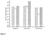
- the analytical sensitivity was 10 mcyB copies per reaction and log-linear range 10 to 10 7 copies of mcyB for all three target genera ( Figure 1 ).
- PCR templates were prepared from culture samples of known cell density.
- the quality of extracted DNA varied, A 260 /A 280 ratios ranged from 1.7 to 2.0. Heat-treated samples were too dilute to be examined spectrophotometrically. Filtration and heat-treatment of samples yielded 4-330 times the mcyB copy numbers of the corresponding extracted DNA ( Figure 2 ).
- the target sequence is located within the second thiolation motif of mcyB , in a peptidyl carrier protein domain coding sequence, the domain that serves to transfer the growing polypeptide chain from McyB to McyC for further elongation (Nishizawa et al., 1999; Tillett et al., 2000) To our knowledge, this is the first time it has been used as a target for qPCR detection. Unlike the mcyB first adenylation domain targeted in many studies (e.g.
- the second condensation, adenylation and thiolation motifs of mcyB have their counterparts in the ndaA gene (Moffitt and Neilan, 2004), and the amplicon in Nodularia shares 80%, 75% and 76% sequence identity with the corresponding amplicons in Anabaena, Microcystis and Planktothrix, respectively.
- the detection probes were shown to hybridize only with their intended targets, mcyB qPCR specificity was not compromised by the amplification of ndaA.
- generel primers provide information about the abundance of all toxic species (Al-Tebrineh et al., 2011), genus-specific detection has the added benefit of population dynamics monitoring.
- the sensitive label technology (Nurmi et al., 2002) used provides the assay with good sensitivity and a broad quantification range.
- microcystins are relatively stable and can persist in the water for weeks (Lahti et al., 1997), thus trace amounts could have been present in the samples, even after the producing organism no longer was.
- a qPCR method to detect and quantify all potentially microcystin-producing members of three major bloom-forming genera, coupled to efficient sample preparation, provides a means for fast early-warning monitoring.
- Potentially microcystin-producing cyanobacteria can be detected with good sensitivity and a broad quantification range, responding to the significant variance in cell densities in the environment.
- the ⁇ land Islands gene quantification results show that small separated sample collection points are not enough to provide us with general gene copy number guidelines.
- Cyanobacterial species Strain a Cyanotoxins produced b mcyB amplicon detected with genus-specific probes mcyB- aP, Anabaena mcyB -mP, Microcystis mcyB- pP, Planktothrix Anabaena sp. 90 MC-RR, MC-LR, dmMC-LR, dmMC-RR + - - Anabaena lemmermannii var. minor NIVA-CYA 83/1 dm-MC-LR , MC-RR, dmMC-RR, MC-LR + - - Anabaena lemmermannii var.
- PCC 7422 - - - - Planktothrix agardhii NIVA-CYA 15 didmMC-RR , dmMC-RR - - + Planktothrix agardhii NIVA-CYA 59/1 dmMC-RR , MC-LR, dmMC-LR - - + Planktothrix agardhii NIVA-CYA 299 dmMC-RR - - + Planktothrix agardhii PCC 7805 - - - - Planktothrix agardhii NIVA-CYA 12 - - - - Planktothrix agardhii NIVA-CYA 21 - - - - Planktothrix agardhii NIVA-CYA 21 - - - - Plan
- Oligonucleotide 5'-3' sequence T m /°C Oligonucleotide type Specificity mcyBHF03A GCTTTAATCCACAAGAAGCTTTATTAGC 56,1 forward primer Anabaena mcyBHF03M AGATTTTAATCCACAAGAAGCTTTATTAGC 56,1 forward primer Microcystis mcyBHF03P GGTTTAATCAACAAGAGGCTTTATTAGC 55,9 forward primer Planktothrix mcyBHR04 CTGTTGCCTCCTAGTTCAAAAAATGACT 58,5 reverse primer All three target genera mcyB-aP ACTGAATTATTGGAGGTAGAGGTGAGTGATAC 58,9 detection probe Anabaena mcyB-aQ CCTCTACCTCCAATAATTCA 43,6 quencher probe Anabaena mcyB-mP GGGTGAGTTATTAGAAG
Description
- The present invention relates to the field of detection of microcystin-producing toxic cyanobacteria from environmental samples. Particularly, the present invention provides a polymerase chain reaction (PCR) based assay method for the detection of said toxic cyanobacteria. The present invention further provides materials such as primers, primer pairs and probes designed for the method of the invention.
- In recent years, toxic cyanobacterial blooms have gained increasing amounts of attention by the scientific community, authorities and general public worldwide. Toxins produced by cyanobacterial mass occurrences have caused deaths of wildlife and livestock (see Stewart et al., 2008, for a review), and have been connected to human illness and death (Turner et al., 1990; Pilotto et al., 1997; Jochimsen et al., 1998). Microcystins, cyclic heptapeptide hepatotoxins that form a major cyanotoxin group with over 90 known structural variants (Welker and von Döhren, 2006) are produced by strains of Anabaena, Microcystis, Planktothrix and Nostoc (Sivonen and Jones, 1999), although isolated occurrences of microcystin-producing Hapalosiphon (Prinsep et al., 1992), Phormidium (Izaguirre et al., 2007) and Fischerella (Fiore et al., 2009) have been described. Of these eight genera, mostly Anabaena, Microcystis and Planktothrix cause freshwater hepatotoxic blooms. To better understand the dynamics of cyanobacterial blooms and mechanisms of toxin production, and predict the risks associated with these phenomena, easy-to-use detection methods for different microcystin variants and toxin-producing cyanobacteria are needed. Continuous low-level exposure to microcystins has been connected with increased risk of hepatic cancer (Yu, 1995; Ueno et al., 1996; Svircev et al., 2009), which further stresses the need for these methods.
- Reliable identification of microcystin- and non-microcystin-producing cyanobacteria can be challenging. Mixed blooms of morphologically indistinct toxic and non-toxic strains of the same species are common (Vezie et al., 1998). The discovery of the mcyS gene cluster revealed a group of potential targets for PCR-based detection of microcystin producers.
- Genes connected to the non-proteinogenic, rare amino acid residues in the toxin structure are especially well suited for this purpose (Mbedi et al., 2005). Many studies describe nucleic acid based methods to detect microcystin-producing cyanobacteria, and their use in attempt to evaluate the risk associated with toxic blooms (see Sivonen, 2008, for a review). For instance,
WO2011003184 discloses primers designed for the detection of microcystin-producing cyanobacteria. The primers disclosed are complementary to the conserved region of the ss-ketoacyl synthase (KS) domain of the mcyD gene of Microcystis aeruginosa strain UTCC 299, or to the conserved region of the first dehydratase domain of the mcyD gene of Microcystis aeruginosa UTCC 300. InWO2006128230 , another PCR-based method for detection of hepatotoxic cyanobacteria is disclosed. The target sequences for amplification are located in hepatotoxin-associated aminotransferase domain sequences derived from the mcyE gene of the microcystin synthetase gene complex and the ndaF gene of the nodularin synthetase gene complex. - However, few real-time quantitative PCRs capable of detecting more than one microcystin-producing genera have been described (Al-Tebrineh et al., 2011; Vaitomaa et al., 2003; Briand et al., 2008), and currently the majority of published qPCR methods concentrate on only one genus, most often Microcystis (Foulds et al., 2002; Kurmayer and Kutzenberger, 2003; Rinta-Kanto et al., 2005; Furukawa et al., 2006; Fortin et al., 2010).
- Sample preparation and label technology choices are important in qPCR. Sample loss during multi-step DNA extraction processes combined with inefficient amplification caused by incomplete removal of PCR inhibitors can result in significant quantification error (Wilson, 1997). Addressing these problems would improve PCR reliability. Increased specificity, in turn, can be achieved with sequence-specific labelled probes (e.g., TaqMan), and unlike the more commonly used prompt fluorophores, lanthanide chelate labels allow detection by time-resolved fluorometry, leading to improved sensitivity due to lower background signals, since any autofluorescence decays during the time window between excitation and measurement (Soini and Lövgren, 1987)
- The aim of this study was to develop a quantitative real-time PCR method for the detection of potentially microcystin-producing Anabaena, Microcystis and Planktothrix. To achieve reliable quantification, DNA extraction and simple cell lysis were compared in terms of qPCR performance and template yield. The developed qPCR assay was applied to environmental sample analysis, and the correlation between gene copy numbers and microcystin concentrations examined.
-
-
Figure 1 . Standard curves for the mcyB qPCR assay.Base 10 log mcyB copy numbers are plotted against their respective treshold cycles (Ct) for a) Anabaena, b) Microcystis and c) Planktothrix. A range of 101-107 copies of the mcyB target sequence could be detected for each target genera. Amplification efficiences were similar (92.7-94.2 %) for all targets. Variation in Ct within the four replicate reactions was very low, standard deviations are shown as error bars mostly hidden inside the symbols. -
Figure 2 . Comparison of two qPCR sample preparation methods. Microscopically determined cell amounts (patterned columns) were compared to detected mcyB gene copies in extracted genomic DNA (white columns) and in filtered and disrupted cells (grey columns). Comparisons were made for microcystin-producing Microcystis aeruginosa NIVA-CYA 140, Anabaena cf. flos-aquae NIVA-CYA 267/4 and Planktothrix agardhii NIVA-CYA 299. Filtering and cell lysis yielded consistently higher copy number compared to DNA extracted from the same amount of cells. Error bars indicate both inter- and intra-sample copy number variation. -
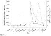
Figure 3 . The development of Planktothrix mcyB gene copy numbers (■, solid line), Planktothrix cell numbers (▲, dotted line), and total microcystin concentrations determined by LC-MS (□, dashed line) in Hauninen reservoir (Raisio, Finland) during April-June 2008. Cell numbers are based on microscopic counting of 100 µm filament units and an estimate of 30 cells per one such unit. A clear positive correlation between mcyB copy numbers and total microcystin concentration (dmMC-RR and dmMC-LR) was observed. No potentially toxic Microcystis or Anabaena was detected. -
Figure 4 . Target sequences in mcyB gene of Anabaena sp., Microcystis aeruginosa and Planktothrix agardhii, and primers and probes used in the Example. -
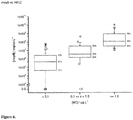
Figure 5 . Correlation of total gene copy number of the mcyB gene to microcystin concentrations [MC] in an environmental aquatic sample measured by ELISA. -
Figure 6 . Correlation of total gene copy number of the mcyB gene to microcystin concentrations [MC] in an environmental aquatic sample measured by HPLC. -
Figure 7 . Correlation of total gene copy number of the mcyB gene to microcystin concentrations [MC] in an environmental aquatic sample measured by LC-MS. - The present invention is defined by the appended claims.
- The present invention is directed to a method for detecting the presence of microcystin-producing toxic cyanobacteria in a sample comprising the steps of:
- a) performing lysis of the cells in said sample;
- b) contacting nucleic acid obtained from the lysed cells in a polymerase chain reaction mix with primers hybridizing with the nucleic acid sequence of SEQ ID N° 1, SEQ ID N° 2 and SEQ ID N° 3 in the mcyB gene present in Microcystis aeruginosa, Planktothrix agardhii and Anabaena sp., wherein said primers amplify at least part of the target sequence in the mcyB gene
- c) performing a polymerase chain reaction with a reaction mix obtained from step b) so that the sequences of said mcyB gene are specifically amplified, if said sequences are present in the sample; and
- d) detecting the presence of amplified nucleid acid sequences, wherein the presence of amplified mcyB gene sequence is indicative of the presence of microcystin-producing toxic cyanobacteria in the sample.
- The samples prepared for the method of the invention comprising cyanobacterial material are preferably environmental samples, such as water samples which can be seawater samples or freshwater samples or other suitable environmental samples.
While the method is mainly directed to the detection of toxic cyanobacteria, such as Microcystis aeruginosa, Planktothrix agardhii and Anabaena sp., it can also be used for the quantification of the microcystin-producing toxic cyanobacteria in said samples. This can be done by performing quantitative-PCR (qPCR) on the extracted DNA using the primers of the present invention, and determining the gene copy number of the mcyB gene. Further, the toxic microcystin concentration in the sample may also be monitored by correlating said gene copy number obtained by qPCR to a corresponding microcystin concentration. - In step a), the nucleic acid which is used as template in the PCR of step b) can be extracted and purified from the lysed cells by conventional methods. However, the suspension obtained from the lysed cells is preferably used directly as PCR template. Accordingly, in one embodiment of the invention an undiluted water sample suspected to comprise toxic cyanobacteria can be first filtered. Then filtered cells are suspended in sterile deionized water and cell lysis is carried out by heat treatment. The obtained suspension and nucleic acid therein can be used as such for the reaction mix of step b).
- In step b), said primers amplify at least part of the target sequence in the mcyB gene as set forth in SEQ ID NO:1 corresponding to positions 6147-6251 of Microcystis aeruginosa genome described in
Figure 4 , or as set forth in SEQ ID NO:2 corresponding to positions 6203-6305 of Planktothrix agardhii genome described inFigure 4 , or as set forth in SEQ ID NO:3 corresponding to positions 6170-6272 of Anabaena sp. described inFigure 4 . - In embodiments of the invention, the amplicon, i.e. the target sequence, which is amplified in step b), comprises at least 20, preferably at least 50, more preferably at least 80, and most preferably 103 or 105 consecutive nucleotides of the target sequence as defined by SEQ ID NO:1, SEQ ID NO:2 or SEQ ID NO:3.
- Preferably, the primers used in the method comprise or consist of one of the following sequences:
- 5'-GCTTTAATCCACAAGAAGCTTTATTAGC-3' (SEQ ID NO:4)
- 5'-AGATTTTAATCCACAAGAAGCTTTATTAGC-3' (SEQ ID NO:5)
- 5'-GGTTTAATCAACAAGAGGCTTTATTAGC-3' (SEQ ID NO:6)
- 5'-CTGTTGCCTCCTAGTTCAAAAAATGACT-3' (SEQ ID NO:7)
- 5'-ACTGAATTATTGGAGGTAGAGGTGAGTGATAC-3' (SEQ ID NO:8)
- 5'-CCTCTACCTCCAATAATTCA-3' (SEQ ID NO:9)
- 5'- GGGTGAGTTATTAGAAGCAGAAGTTAGTAACAG-3' (SEQ ID NO:10)
- 5'-TTCTGCTTCTAATAACTCACC-3' (SEQ ID NO:11)
- 5'-GGGGTGAATTATTAGAAATAGAAGTAAGTGACAA-3' (SEQ ID NO:12)
- 5'-TTACTTCTATTTCTAATAATTCACC-3' (SEQ ID NO:13)
- Also disclosed are primers comprising or consisting of any of the sequences as set forth in SEQ ID NOS:4-7. Preferably, the primer has 28-40 nucleotides. More preferably, the primer comprises less than 30, 35 or 40 nucleotides.
- Further, are provided probes comprising or consisting of any of the sequences as set forth in SEQ ID NOS:8-13. Preferably, the probe has less than 40 nucleotides.
- The oligonucleotide primers and probes are short sequences of nucleotides (such as RNA or DNA, preferably DNA), typically with twenty-five to thirty or fewer bases. However, automated synthesizers allow the synthesis of oligonucleotides up to 160 to 200 bases and the present oligonucleotides may be elongated to add, e.g., a restriction enzyme cleavage site, to the oligonucleotide. The typical length of the primers is preferably 26-32, more preferably 28-30 nucleotides.
- The present invention also provides kits for the detection of the presence of microcystin-producing toxic cyanobacteria. The kit may comprise primers and probes as defined above. Such primers and probes are described above and in the Examples below.
- Preferably, said kit comprises means for a real-time polymerase chain reaction, such as labelled probes, polymerase enzymes, buffers and nucleotides.
- It is well-known in the art that the sequences of same gene vary somewhat in strains of a microbial species and, thus, the sequence of the mcyB gene present in Microcystis aeruginosa, Planktothrix agardhii and Anabaena sp. is not 100% identical in all strains of the species. However, it is clear from the description herein, particularly from the Example below, that a person skilled in the art would recognize the sequence of the mcyB gene in any related strain based on the similarity and homology of these gene sequences.
- Herein the term "specifically hybridizing" means complementary hybridization between an oligonucleotide and a target sequence. The term "specifically" refers to the specificity shown by the complementary hybridization, which allows for minor mismatches between the oligonucleotide and the sequence that may not jeopardize the annealing for detection of hybridization signals.
- The present invention is further described in the following example, which is not intended to limit the scope of the invention.
- Cyanobacterial strains used in this study are listed in Table 1. Strains were purchased from the Pasteur Culture Collection (PCC, Paris, France) and the Norwegian Institute for Water Research Cyanobacterial Culture Collection (NIVA, Oslo, Norway) and maintained as recommended by the providers. All NIVA strains were cultured in Z8 (Staub, 1961, modified NIVA 1972, 1976). Strains from PCC were cultured in either BG 11 (Sigma), nitrate omitted BG110, or BG110 with added NaNO3 and NaHCO3 using formulations described by PCC. Three additional strains from other sources were also maintained. Microcystis aeruginosa NIES-107 (National Institute of Environmental Studies, Tsukuba, Japan) was cultured in Z8. Anabaena lapponica strain 966 (Finnish Environment Institute, Dr. Jarkko Rapala) as well as Anabaena sp. 90 (University of Helsinki, Prof. Kaarina Sivonen) were cultured in modified Z8 with no added nitrogen. All cultures were maintained at 23 °C using PowerGlo 20 W aquarium bulbs (Hagen, Japan) as the light source. Cyanobacterial cells were microscopically counted from 2.5 week old cultures of NIVA-CYA 267/4, NIVA-CYA 140 and NIVA-CYA 299. Samples were diluted 1:100-1:200 in deionized water and preserved in Lugol's iodine. After a 16 h sedimentation of 10 ml sample aliquots counting was carried out on a Nikon TE-200 inverted microscope (Nikon, Japan) with 10x and 40x objectives. Cells from enumerated cultures were harvested for mcyB quantification. Environmental samples were collected from Hauninen reservoir, Raisio, Finland during April-June 2008 (11 samples) and from freshwater lakes at Åland Islands during July 2008 (13 samples). All samples, including extracted DNA and other PCR templates, were kept frozen at -20 °C until further analysis. Culture and environmental samples were either frozen fresh or freeze-dried.
- Samples of eight to ten mg freeze-dried cyanobacterial material or freeze-dried GF/C fiberglass filters (Ø 25 mm; Whatman, Maidstone, UK) containing filtered cyanobacterial material were extracted with 1.2 ml of 75% methanol (HPLC grade; Rathburn, Walkerburn, UK). Extracts were treated for 15 min in a bath ultrasonicator (Bandelin Sonorex RK 156, Berlin, Germany) and additionally for 1 min with a probe sonicator (Bandelin Sonopuls HD 2070 with 3 mm microtip, 30% pulse, 30% energy). After centrifugation at 10,000 x g for 10 minutes, the supernatants were divided into aliquots and evaporated to dryness with argon at 40 °C. Extracts intended for microcystin analysis by HPLC-DAD and LC-ESI-MS-MS were resuspended in 75% methanol while those aimed for ELISA in water.
- Samples were analysed using an Agilent (Waldbronn, Germany) 1100 series HPLC system consisting of a degasser, a quaternary pump, a thermostatted column compartment at 40 °C and a diode-array detector operated at 200-300 nm (quantification at 238 nm). The stationary phase was either a Purospher STAR column, 3 µm particles, 55 mm × 4 mm from Merck (Darmstadt, Germany) or an Ascentis RP-Amide column, 3 µm particles, 100 mm × 4.6 mm I.D. from Supelco (Bellefonte, PA, USA). The mobile phase consisted of acetonitrile (HPLC S grade, Rathburn, Walkerburn, UK) (solvent B) - Milli-Q ultrapure water (Millipore, Molsheim, France) (solvent A) both containing 0.05% trifluoroacetic acid (TFA; Fluka, Buchs, Switzerland) with the following linear gradient programme: 0
min 25% B, 7 min 70% B, 10 min 70% B, 10.1min 25% B;stop time 15 min; flow-rate 1 ml/min. Injection volumes were 10 µl. Additionally, the samples were analysed on a Merck Purospher STAR RP-18e column, 55 mm × 4 mm I.D. with 3 µm particles (Spoof and Meriluoto, 2005). Individual laboratory-purified microcystins and following reference samples were used for the identification of microcystins: a) extract of Microcystis aeruginosa PCC 7820 and b) extract of Microcystis aeruginosa NIES-107 as described in (Spoof et al., 2003). - LC-MS experiments were performed on an Agilent 1100 Series HPLC system coupled to a Waters Micromass (Manchester, UK) Quattro Micro triple-quadrupole mass spectrometer equipped with an electrospray interface. Toxins were quantified on a Merck Purospher STAR RP-18 endcapped column (30 mm × 4 mm, 3 µm particles). The mobile phase consisted of a gradient of 0.1% aqueous formic acid (solvent A) and acetonitrile (solvent B) with the following linear gradient program: 25% B to 70% B over 10 min, then to 90% B over 2 min, where it was held for 1 min. Injection interval was 16 min,
injection volume 10 µl, flow rate 0.5 ml/min, and column oven temperature 40 °C. Capillary voltage was set at 3.8 kV and cone voltage at 40 V (dmMC-RR and MC-RR) or 75 V (rest of the microcystins and nodularin). Desolvation gas (nitrogen) temperature and flow rate were set at 300 °C and 650 L/h, respectively. Ion source temperature was set at 150 °C. Ions were detected in the positive electrospray ionization mode. The monitored signals in the selected ion recording (SIR) mode were m/z [dmMC-RR+2H]2+ 512.8, [MC-RR+2H]2+ 519.8, [MC-LF+H]+ 986.5 and [MC-LF+Na]+ 1008.5, [dmMC-LR+H]+ 986.5, [MC-LR+H]+ 995.5, [MC-LY+H]+ 1002.5, [MC-LW+H]+ 1025.5 and [MC-LW+Na]+ 1047.5, [MC-YR+H]+ 1045.5, [dmNod+H]+ 811.5 and [Nod+H]+ 825.5. Data acquisition was done with Masslynx v. 4.0 software (Micromass). LC-MS-MS experiments were carried out on an Agilent 1200 Rapid Resolution (RR) LC coupled to a Bruker Daltonics HCT Ultra ion trap mass spectrometer (Bremen, Germany) with electrospray ion (ESI) source. The 1200 RR LC system included a binary pump, a vacuum degasser, a SL autosampler, and a thermostatted column compartment. The ion trap was operated in the positive electrospray ion mode. Ion source parameters were set as follows: dry temperature 350 °C, nebulizer pressure 40 psi, dry gas flow 10.0 L/min, capillary voltage 4.0 kV. An MS scan range from 500 to 1200 m/z with the Smart Parameter Setting (SPS) function was employed. The ICC target was set to 300 000 with a maximum accumulation time of 100 ms. Abundant MS-MS fragmentation was assisted by the SmartFrag setting. Separation of toxins was achieved on Ascentis C18, 50 mm × 3 mm I.D. column with 3 µm particles (Supelco) at 40°C. Injection volumes were 5 µl. The mobile phase consisted of water-acetonitrile-formic acid (99:1:0.1; solvent A) and acetonitrile-formic acid (100:0.1; solvent B) with the following linear gradient programme: 0min 25% B, 5 min 70% B, 6 min 70% B, 6.1min 25% B;stop time 10 min: flow-rate 0.5 ml/min. Primary monitored signals were the same as with the Quattro micro instrument but mass spectra acquired with the HCT Ultra instrument allowed for identification of additional toxins based on fragmentation patterns. Data acquisition was done with Bruker Compass 1.3 software. - Samples were analysed with the Quantiplate Microcystin kit (Envirologix, Portland, ME, USA) using the protocol provided by the manufacturer. Extracts were diluted with water according to HPLC results to adjust the microcystin concentrations to the working range of the assay. Two different dilutions were made of some samples.
- Two methods were used to prepare samples for PCR analysis. Genomic DNA was extracted from freeze-dried cells of cultured strains or freeze-dried Åland Islands environmental samples using the NucleoSpin Plant II DNA extraction kit (Macherey-Nagel, Düren, Germany) according to the manufacturer's instructions. DNA concentration and quality were determined spectrophotometrically (ND-1000, NanoDrop Technologies, Wilmington, DE, USA). Filtering and cell lysis was performed as follows: 10 ml of harvested cell culture was suspended in 90 ml of sterile deionized water, and vacuum filtered on a fiberglass filter (Ø 47 mm GF/C, Whatman). In the case of Hauninen reservoir samples, 50 ml of undiluted water was filtered. Pieces of each filter were cut out and suspended in 100 µl of sterile deionized water. Cell lysis was carried out at 80 °C for 5 min. The suspension was thereafter used directly as PCR template. Filter piece dimensions were recorded for quantification purposes. Microscopically counted culture samples were treated using both methods described above, with a few modifications: cells were harvested as aliquots of both 5 ml and 10 ml, and subjected either to filtration or DNA extraction. Sample preparation was carried out on freshly harvested cells. Cells were collected for DNA extraction by centrifugation (4 °C, 3220 g, 20 min, Eppendorf 5810R, Hamburg, Germany).
- mcyB sequences (Nishizawa et al., 1999; Tillett et al., 2000; Christiansen et al., 2003; Rouhiainen et al., 2004; Kaneko et al., 2007) were retrieved from the GenBank Nucleotide database, and primers and probes were designed based on the sequence alignment. Oligonucleotides were manufactured by Thermo Scientific (Ulm, Germany) and biomers.net (Ulm, Germany) (Table 2). Probes were synthesized with 5'-C6-aminolinkers and 3'-phosphate groups. All detection probes were labeled at the 5' end aminolinker with an organic Tb3+ chelate (2,2',2",2'"-{{6,6'-{4"-[2-(4-Isothiocyanatophenyl)ethyl]pyratzole-1",3"-diyl}bis(pyridine)-2,2'-diyl}bis(methylenenitrilo)}tetrakis(acetato)}terbium(III)) as described previously (Nurmi et al., 2002). All quencher probes had a BHQ1 (Black hole quencher® 1) molecule conjugated at their 3'-ends by the oligonucleotide manufacturer. Primer specificity was tested on the cyanobacterial strains listed in Table 1. Individual PCR reactions contained 5 nmol of dNTPs (Finnzymes, Espoo, Finland), 2.67 pmol of each forward primer and 8 pmol of the reverse primer, 0.5 units of DyNAzyme II HotStart DNA polymerase (Finnzymes), 1X DyNAzyme II HotStart buffer and 1 µl of template, either 1 ng of genomic DNA or 1 µl of suspension containing lysed cells. The reaction volume, 20 µl, was filled with sterile Milli-Q water. Thermocycling was performed using a PTC-200 Thermal Cycler (MJ Research, Watertown, MA, USA), starting with 10 min at 95 °C, followed by 35 cycles of 30 s at 95 °C, 30 s at 58 °C and 1 min at 72 °C, and ending with 10 min at 72 °C. The PCR products were analyzed on a 2 % agarose gel stained with ethidium bromide (0.5 ng L-1).
- The principle of the real-time quantitative PCR technique has been described in detail elsewhere (Nurmi et al., 2002). Briefly, amplification product accumulation is monitored by measuring the long-life fluorescence obtained from chelate moieties freed from detection probes by the 5'→3' exonuclease activity of the DNA polymerase. Background fluorescence is kept at minimum with the help of quencher probes. Detection probe specificity was confirmed using qPCR and purified genomic DNA from all cultured strains. Each qPCR reaction contained 4 nmol of dNTPs, 2 pmol of each forward primer and 6 pmol of the reverse primer, 0.2 units of DyNAzyme II HotStart DNA polymerase, 1X DyNAzyme II HotStart buffer, 0.25 pmol detection probe (either mcyB-mP, mcyB-pP or mcyB-aP), 2.5 pmol of corresponding quencher probe (mcyB-mQ or mcyB-pQ, for mcyB-aQ the amount 3.0 pmol was used) and 1 ng of template DNA. The reactions were filled with sterile Milli-Q water to 20 µl. All reactions were run on ThermoFast 96 Robotic PCR Plates (Abgene, Surrey, UK) with Applied Biosystems Optical Caps (Foster City, CA, USA). The qPCR runs were started with 5 min at 95 °C, followed by 8 cycles of 30 s at 95 °C and 1 min at 62 °C. After cycle 8 the temperature was lowered to 35 °C for 15 s for fluorescence measurement. Measurements were repeated every second cycle until after cycle 40, and were carried out with Victor2 1420 Multilabel Counter (PerkinElmer Life Sciences Wallac, Turku, Finland) using the manufacturer's standard protocol for time-resolved measurement of Tb fluorescence.
- Double-stranded DNA standards were produced by PCR from Anabaena sp. 90, PCC 7806 (Microcystis) and NIVA-CYA 299 (Planktothrix) as described above. Purification of mcyB amplification products was performed using the Qiaquick PCR Purification kit (Qiagen, Hilden, Germany), and subsequent quantification using the QuantIt PicoGreen kit (Invitrogen, Eugene, OR, USA), both according to manufacturer's instructions. Molar concentrations of the purified amplification products were calculated, and a series of 10-fold dilutions (n=7) of each genus-specific standard was prepared. The standards were kept at - 20 °C until use. qPCRs were run as described above. Treshold cycles (Ct) were plotted against log mcyB copy numbers, and amplification efficiences (E) were calculated from the slope of the linear regression (E=10-1/slope-1, where the value 1.00 corresponds to an efficiency of 100%).
- Series of 10-fold dilutions (n=4) of templates prepared from microscopically counted culture samples were prepared. The aims were to compare qPCR quantification and cell densities and to assess amplification efficiency and possible PCR inhibition. All qPCRs were performed as described above, except for the increased template amount, 4 ng of DNA or 4 µl of heat-treated sample. Culture samples were analyzed in four replicate reactions, undiluted environmental samples in triplicate. Environmental samples that yielded a mcyB negative result were tested for PCR inhibition by adding 1 ng of Anabaena sp. 90 genomic DNA to each reaction and visualizing the PCR products on ethidium bromide stained agarose gels as described above.
- Specificity of the mcyB primers and detection probes was tested on extracted DNA and heat-treated cells of 30 cyanobacterial strains. Probe specificities, microcystins and other toxins produced by the strains are listed in Table 1. No false negatives were observed. No amplification products were observed from non-microcystin-producing strains, except for nodularin-producing Nodularia harveyana PCC 7804. However, the Nodularia amplification product was not detected by any of the detection probes in qPCR. Thus, all genus-specific detection probes performed with 100 % specificity and sensitivity. Each of the three possible primer combinations could be used to amplify the target mcyB sequence from all microcystin-producing strains independent of genus, but amplification efficiences varied from strain to strain (data not shown). When the three forward primers were used together, efficient amplification was achieved.
- Sensitivities and amplification efficiencies were determined using standards prepared from purified PCR products. The analytical sensitivity was 10 mcyB copies per reaction and log-
linear range 10 to 107 copies of mcyB for all three target genera (Figure 1 ). Amplification efficiences and correlation coefficients derived from the standard curves were 93.8% (r2=0.997), 91.7% (r2=0.999) and 95.3% (r2=0.998) for Anabaena, Microcystis and Planktothrix, respectively. - To compare the two sample preparation methods, PCR templates were prepared from culture samples of known cell density. The amplification efficiences obtained from templates prepared by filtration and heat-treatment were either higher or equal to those of corresponding purified DNA templates (89-100 %, average r2=0,9993±0,0008 and 83-97 %, average r2=0 9976±0,0018, respectively). The quality of extracted DNA varied, A260/A280 ratios ranged from 1.7 to 2.0. Heat-treated samples were too dilute to be examined spectrophotometrically. Filtration and heat-treatment of samples yielded 4-330 times the mcyB copy numbers of the corresponding extracted DNA (
Figure 2 ). The greatest difference was observed in NIVA-CYA 299, a Planktothrix agardhii strain, where the detected copy numbers of filtered samples were only 1,5 times the cell counts, but 330 times the copy numbers in corresponding DNA extracted samples. For Anabaena, the copy numbers in DNA and cell lysates were of the same magnitude as cell counts. For Microcystis, the same was true for cell counts and copy numbers from extracted DNA, whereas filtration and heat-disruption of cells yielded copy numbers roughly 30 times the cell amount. - A total of 24 environmental samples were analyzed. The majority of Åland Islands lake samples (9/13) contained mcyB gene copies originating from either Microcystis only, or Microcystis together with Anabaena or Planktothrix (Table 3). Microcystins were present in all samples which in qPCR yielded a positive result for mcyB. However, a linear correlation between total copy numbers and toxin concentrations could not be established. Four samples did not contain target genera mcyB copies, nor showed evidence of PCR inhibition that could have caused false negative results. ELISA results were positive for all mcyB negative Åland Islands samples, whereas HPLC-DAD results for the same samples were all negative and LC-MS-MS analyses resulted in concentrations ranging from 0 to 0.67 µg microcystins per g of sample (dry weight).
- Five out of eleven Hauninen reservoir samples were positive for mcyB. In agreement with microscopical examinations, the population was dominated by Planktothrix, and no potentially toxic Microcystis or Anabaena were detected. PCR inhibition was not observed on visual examination of amplification products obtained from spiked samples. A clear positive correlation between Planktothrix mcyB copy numbers and total microcystin concentrations was found (
Figure 3 ). Filament amounts increased gradually from April 3rd onward and reached a peak on May 29th, after which a rapid decrease was observed. The simultaneous peaking of total microcystin concentration and Planktothrix-specific mcyB copy numbers did not coincide with the highest observed population density of Planktothrix. Two main microcystin variants were present in toxin-containing samples, dmMC-RR and dmMC-LR. - Identification of a suitable target for the detection of several toxic genera is crucial. In the qPCR assay described here the target sequence is located within the second thiolation motif of mcyB, in a peptidyl carrier protein domain coding sequence, the domain that serves to transfer the growing polypeptide chain from McyB to McyC for further elongation (Nishizawa et al., 1999; Tillett et al., 2000) To our knowledge, this is the first time it has been used as a target for qPCR detection. Unlike the mcyB first adenylation domain targeted in many studies (e.g. (Mbedi et al., 2005; Kurmayer and Kutzenberger, 2003; Nonnemann and Zimba, 2002; Mikalsen et al., 2003), the second thiolation motif is not likely to be affected by microcystin structure variation. Simultaneous detection of several potentially toxic genera, independent on toxin profiles, is therefore possible. The wide range of potential hepatotoxin producers deteted was demonstrated by the amplification of ndA, homologous to mcyB, from the nodularin-producing Nodularia harveyana PCC 7804. The second condensation, adenylation and thiolation motifs of mcyB have their counterparts in the ndaA gene (Moffitt and Neilan, 2004), and the amplicon in Nodularia shares 80%, 75% and 76% sequence identity with the corresponding amplicons in Anabaena, Microcystis and Planktothrix, respectively. However, since the detection probes were shown to hybridize only with their intended targets, mcyB qPCR specificity was not compromised by the amplification of ndaA.
- The majority of the existing quantitative mcyB qPCR assays are based on a few probes (if TaqMan detection chemistry is used) and primer pairs (Kurmayer and Kutzenberger, 2003; Nonnemann and Zimba, 2002; Kaebernick et al., 2002), which have a single target genus. With the new primers and genus-specific probes designed to detect a different part of the same gene, quantitative mcyB detection is expanded to include also Anabaena and Planktothrix, and an addition of a new target to the repertoir assays designed to detect more than one microcystin-producing genera is made. While generel primers provide information about the abundance of all toxic species (Al-Tebrineh et al., 2011), genus-specific detection has the added benefit of population dynamics monitoring. The sensitive label technology (Nurmi et al., 2002) used provides the assay with good sensitivity and a broad quantification range. With the detection limit of 10 mcyB copies per reaction and a range of seven orders of magnitude the current assay is very similar to previously published methods (Al-Tebrineh et al., 2011; Vaitomaa et al., 2003; Foulds et al., 2002; Kurmayer and Kutzenberger, 2003; Rinta-Kanto et al., 2005; Furukawa et al., 2006).
- Differences between the amplification efficiences of qPCR standards and the samples can have a substantial effect on the quantification result (Wilson, 1997). The same error source exists also between samples prepared by different methods, and any comparisons need to be made carefully. In this study, the theoretical copy number differences between sample types caused by variation in the observed amplification efficiences remained in the range of 3-5 fold. This cannot explain the difference of 1-2 orders of magnitude in detected copy numbers (for Microcystis and Planktothrix), showing that there is an actual significant difference between the amounts of available template per cell that simple lysis of the cyanobacterial cells and extraction and purification of genomic DNA produce. A number of reasons could cause such results, likely sample material loss and inefficient cell lysis during DNA extraction. It is also possible that discrepancies between gene copy numbers and cell counts is to some degree caused by errors in microscopic quantification, although an enumeration performed by an expert will make it considerably less probable.
- Based on our observations, if one gene copy per cell was assumed, in a heat-treated sample Microcystis mcyB numbers would result in overestimation of cell concentration. However, under the same assumption, quantification of mcyB from purified genomic would DNA lead to underestimation of cell concentration, especially in the case of Planktothrix. Quantitative PCR results leading to cell number overestimation have been observed before for Anabaena and Microcystis (Vaitomaa et al., 2003), and studies have supported cyanobacterial cells hosting multiple genomes (Labarre et al., 1989; Becker et al., 2002). If this were the case, it would explain overestimations, but at the same time make underestimations more severe. Therefore we chose to use filtration and heat-treatment in the analysis of environmental samples whenever possible, and to compare copy numbers directly to toxin concentrations, avoiding the problematic translation of gene copies into cell numbers.
- Similar results to the comparison made here have been reported for qualitative analyses before - alternative sample preparation methods have yielded templates comparable to purified DNA (Hisbergues et al., 2003; Ouahid et al., 2005). Quantitative performance assessments of different sample preparation methods have found phenol-chloroform extraction of DNA to yield higher copy numbers and therefore also higher estimations of cell concentrations than a commercial reagent kit for DNA extraction (Schober and Kurmayer, 2006). Rasmussen et al., 2008 found that ultrasonication and microwave treatment in detergent solutions performed as well as DNA extraction by a commercial reagent kit. Our observations agree with previously published results which indicate that cell lysis and DNA release without purification is a valid method for sample preparation preceding detection and quantification of mcy gene targets.
- The cyanobacterial population in Hauninen reservoir was followed by quantifying mcyB copy numbers and total microcystin concentrations during spring and early summer 2008. Although in theory the presence of microcystin synthetase genes only reveals the potential for toxin production, the positive correlation between Planktothrix mcyB copies and toxin concentrations was evident. Since neither of the two other targeted toxin producers were detected, Planktothrix was assumed the sole microcystin producer genus of the reservoir. This conclusion was supported by the microcystin variants found in the samples, MC-dmLR and MC-dmRR, which are typical for Planktothrix (Fastner et al., 1999). Filament counts had no clear connection to mcyB copy numbers or toxin concentrations, which indicated that the Planktothrix population in Hauninen reservoir was a mix of toxic and non-toxic strains, their relative proportions varying widely during the monitoring period. Inactive mcy-genotypes in Planktothrix have been reported, and they appear to be fairly common (Kurmayer et al., 2004; Ostermaier and Kurmayer, 2009). The presence of such genotypes in the Hauninen reservoir could not be inferenced based on the observed toxin concentrations and gene copy numbers, and was therefore assumed negligible.
- In contrast to Hauninen reservoir, single samples collected from separate freshwater lakes on Åland Islands did not show clear correlation between microcystin concentrations and mcyB copy numbers, although no false positive qualitative mcyB results (i.e. gene copies detected but no microcystins present in the sample) were observed. Environmental factors can introduce variation into mcy gene expression patterns and toxin production (Kaebernick et al., 2000; Sevilla et al., 2008), and therefore differeces in the lake environments most likely contributed to this result. Another possible affecting factor was sample preparation. Considering the results on qPCR performance of templates produced by different methods, it is likely that reduced yields from DNA extractions introduced method-dependent error to the quantification results.
- Four Åland samples showed very low concentrations of microcystins or no toxins altogether when analyzed with HPLC-DAD and LC-MS-MS methods, and yielded no signal in qPCR. The ELISA assay, however, indicated the presence of varied amounts of microcystins in these samples. Since microcystin immunoassays can be affected by the sample matrix (Metcalf et al., 2000), and the other two methods employed did not indicate high toxin concentrations, overestimated or false positive ELISA results cannot be excluded as a possibility. Microcystins are relatively stable and can persist in the water for weeks (Lahti et al., 1997), thus trace amounts could have been present in the samples, even after the producing organism no longer was.
- As awareness of the health risks of toxic cyanobacterial mass occurrences has grown, the demand for monitoring methods has increased. Guidelines and legislation concerning assessment of cyanobacterial risks are being brought up to date with the current scientific knowledge. A qPCR method to detect and quantify all potentially microcystin-producing members of three major bloom-forming genera, coupled to efficient sample preparation, provides a means for fast early-warning monitoring. Potentially microcystin-producing cyanobacteria can be detected with good sensitivity and a broad quantification range, responding to the significant variance in cell densities in the environment. Although in good qualitative agreement with chromatographic data, the Åland Islands gene quantification results show that small separated sample collection points are not enough to provide us with general gene copy number guidelines. Such guidelines could be established only on analysis of larger sample numbers. The Hauninen reservoir Planktothrix population showcases the unreliability of microscopic quantification in toxin concentration estimation, and emphasises the benefits of qPCR detection. Together all results encourage the use of molecular methods in environmental monitoring of microcystin-producing cyanobacteria.
Table 1. Specificity of mcyB genus-specific probes and the toxins produced by the cyanobacterial strains used in this study. Cyanobacterial species Straina Cyanotoxins producedb mcyB amplicon detected with genus-specific probes mcyB-aP, Anabaena mcyB-mP, Microcystis mcyB-pP, Planktothrix Anabaena sp. 90 MC-RR, MC-LR, dmMC-LR, dmMC-RR + - - Anabaena lemmermannii var. minor NIVA-CYA 83/1 dm-MC-LR, MC-RR, dmMC-RR, MC-LR + - - Anabaena lemmermannii var. minor NIVA-CYA 266/1 dmMC-RR, didmMC-RR, MC-RR + - - Anabaena flos-aquae NIVA-CYA 267/4 m/z 1059, m/z 1045, dmMC-LR, MC-LR, m/z 1015, m/z 1029* + - - Anabaena flos-aquae NIVA-CYA 499 - - - - Anabaena lapponica 966 CYN - - - Anabaena cylindrica PCC 73105 - - - - Anabaena cylindrica PCC 7938 - - - - Microcystis aeruginosa NIES 107 MC-RR, dmMC-RR, MC-LR, dmMC-LR, MC-YR, dmMC-YR - + - Microcystis aeruginosa NIVA-CYA 57 dmMC-RR, didmMC-RR, MC-RR, MC-LR, dmMC-LR - + - Microcystis aeruginosa NIVA-CYA 140 MC-LR, dmMC-LR, m/z 1009 - + - Microcystis aeruginosa NIVA-CYA 228/1 dmMC-RR, didmMC-RR, MC-RR, MC-LR, dmMC-LR, MC-YR, dmMC-YR - + - Microcystis aeruginosa NIVA-CYA 495 m/z 1045, nVz 1031, MC-YR** - + - Microcystis aeruginosa PCC 7806 MC-LR, dmMC-LR - + - Microcystis aeruginosa PCC 7820 MC-LR, dmMC-LR, MC-LY, MC-LW, MC-LF - + - Microcystis aeruginosa PCC 7941 MC-LR, dmMC-LR, MC-YR - + - Microcystis aeruginosa NIVA-CYA 22 - - - - Microcystis aeruginosa NIVA-CYA 464 - - - - Microcystis aeruginosa PCC 7005 - - - - Nodularia harveyana PCC 7804 NOD m/z 839, NOD-R, dmNOD-R*** - - - Nostoc sp. PCC 6310 - - - - Nostoc sp. PCC 7422 - - - - Planktothrix agardhii NIVA- CYA 15didmMC-RR, dmMC-RR - - + Planktothrix agardhii NIVA- CYA 59/1dmMC-RR, MC-LR, dmMC-LR - - + Planktothrix agardhii NIVA-CYA 299 dmMC-RR - - + Planktothrix agardhii PCC 7805 - - - - Planktothrix agardhii NIVA-CYA 12 - - - - Planktothrix agardhii NIVA-CYA 21 - - - - Planktothrix agardhii NIVA-CYA 116 - - - - Planktothrix agardhii PCC 7811 - - - - "Strain names include abbreviations of culture collection names. PCC, Pasteur Culture Collection, Paris, France; NIVA-CYA, Norwegian Institute for Water Research Culture Collection, Oslo, Norway; and NIES, National Institute of Environmental Studies, Tsukuba, Japan, bBoldfacing indicates the main toxin variants, tentative identifications are asterisked: *m/z 1059 MC-HtyR, m/z 1045 MC-[ASP3] HtyR, m/z 1029 MC-FR, m/z 1015 MC-[ASP3]FR, ** m/z 1031 and m/z 1045 unknown MCs with retention times longer than MC-LR, *** m/z 839 L-HarNOD. Table 2. Sequences of primers, detection probes and quencher probes. Oligonucleotide 5'-3' sequence Tm/°C Oligonucleotide type Specificity mcyBHF03A GCTTTAATCCACAAGAAGCTTTATTAGC 56,1 forward primer Anabaena mcyBHF03M AGATTTTAATCCACAAGAAGCTTTATTAGC 56,1 forward primer Microcystis mcyBHF03P GGTTTAATCAACAAGAGGCTTTATTAGC 55,9 forward primer Planktothrix mcyBHR04 CTGTTGCCTCCTAGTTCAAAAAATGACT 58,5 reverse primer All three target genera mcyB-aP ACTGAATTATTGGAGGTAGAGGTGAGTGATAC 58,9 detection probe Anabaena mcyB-aQ CCTCTACCTCCAATAATTCA 43,6 quencher probe Anabaena mcyB-mP GGGTGAGTTATTAGAAGCAGAAGTTAGTAACAG 58,4 detection probe Microcystis mcyB-mQ TTCTGCTTCTAATAACTCACC 43,4 quencher probe Microcystis mcyB- pP GGGGTGAATTATTAGAAATAGAAGTAAGTGACAA 59,2 detection probe Planktothrix mcyB-pQ TTACTTCTATTTCTAATAATTCACC 43,9 quencher probe Planktothrix Table 3. Genus-specific mcyB copy numbers, microcystin amounts and identified microcystin variants in 13 lakes on Åland Islands during the time period from July 14th to July 16th 2008. Lake mcyB copies / g sample dry weight Microcystin concentrations in MC-LR equivalents, µg total MC / g sample dry weight Anabaena Microcystis Planktothrix HPLC-DAD ELISA LC-MS-MS Microcystin variants identified by LC-MS-MS Godby träsk - 1,0E+08 - 40.4 62.2 24.5 dmMC-RR, MC-RR, MC-YR, dmMC-LR, MC-LR Brantsbole träsk 4,1E+07 2,0E+08 - 43.0 94.0 95.3 MC-YR, dmMC-LR, MC-LR, MC-LY,MC-LW, MC-LF Gloskärs träsk - - - 0 6.6 0.67 dmMC-LR Tjudö träsk - 1,4E+09 - 67.9 49.5 45.7 dmMC-RR, MC-RR, MC-dmMC-LR, MC-LR, MC-LA Markusbölefjärden - - - 0 1.2 0.37 MC-LR Nåtö hemviken - 3,9E+11 - 114.8 137.8 85.6 dmMC-RR, MC-RR, MC-YR, dmMC-LR, MC-LR Hammarlands - 1,3E+09 - 0.97 1.5 1.65 MC-LR långträsk Lemböte byträsk - 2,8E+10 - 0 3.4 2.5 MC-YR, dmMC-LR, MC-LR Vargata träsk - 1,3E+11 - 78.3 145.1 71.3 dmMC-RR, MC-RR, MC-YR, dmMC-LR, MC-LR Kaldersfjärden - 1,3E+09 3,3E+08 0 8.3 11.8 MC-YR, dmMC-LR, MC-LR Norsträsk - 4,0E+07 - 0 6.1 1.4 dmMC-LR, MC-LR 1,*ang,sjiin - - - 0 0.1 0 - Slussfjärden - - - 0 0.9 0 - -
- Al-Tebrineh, J., Gehringer, M.M., Akcaalan, R., Neilan, B.A., 2011. A new quantitative PCR assay for the detection of hepatotoxigenic cyanobacteria. Toxicon 57, 546-554.
- Becker, S., Fahrbach, M., Boger, P., Ernst, A., 2002. Quantitative tracing, by Taq nuclease assays, of a synechococcus ecotype in a highly diversified natural population. Appl. Environ. Microbiol. 68, 4486-4494.
- Briand, E., Gugger, M., Francois, J.C., Bernard, C., Humbert, J.F., Quiblier, C., 2008. Temporal variations in the dynamics of potentially microcystin-producing strains in a bloom-forming Planktothrix agardhii (cyanobacteria) population. Appl. Environ. Microbiol. 74, 3839-3848.
- Christiansen, G., Fastner, J., Erhard, M., Börner, T., Dittmann, E., 2003. Microcystin biosynthesis in Planktothrix: genes, evolution, and manipulation. J. Bacteriol. 185, 564-572.
- Fastner, J., Erhard, M., Carmichael, W.W., Sun, F., Rinehart, K.L., Ronicke, H., Chorus, I., 1999. Characterization and diversity of microcystins in natural blooms and strains of the genera Microcystis and Planktothrix from German freshwaters. Arch. Hydrobiol. 45, 147-163.
- Fiore, M.F., Genuario, D.B., da Silva, C.S., Shishido, T.K., Moraes, L.A., Cantusio Neto, R., Silva-Stenico, M.E., 2009. Microcystin production by a freshwater spring cyanobacterium of the genus Fischerella. Toxicon 53, 754-761.
- Fortin, N., Aranda-Rodriguez, R., Jing, H., Pick, F., Bird, D., Greer, C.W., 2010. Detection of microcystin-producing cyanobacteria in Missisquoi Bay, Quebec, Canada, using quantitative PCR. Appl. Environ. Microbiol. 76, 5105-5112.
- Foulds, I.V., Granacki, A., Xiao, C., Krull, U., Castle, A., Horgen, P.A., 2002. Quantification of microcystin-producing cyanobacteria and E. coli in water by 5'-nuclease PCR. J. Appl. Microbiol. 93, 825-834.
- Furukawa, K., Noda, N., Tsuneda, S., Saito, T., Itayama, T., Inamori, Y., 2006. Highly sensitive real-time PCR assay for quantification of toxic cyanobacteria based on microcystin synthetase A gene. J Biosci Bioeng 102, 90-96.
- Hisbergues, M., Christiansen, G., Rouhiainen, L., Sivonen, K., Börner, T., 2003. PCR-based identification of microcystin-producing genotypes of different cyanobacterial genera. Arch. Microbiol. 180, 402-410.
- Izaguirre, G., Jungblut, A.D., Neilan, B.A., 2007. Benthic cyanobacteria (Oscillatoriaceae) that produce microcystin-LR, isolated from four reservoirs in southern California. Water Res. 41, 492-498.
- Jochimsen, E.M., Carmichael, W.W., An, J.S., Cardo, D.M., Cookson, S.T., Holmes, C.E., Antunes, M.B., de Melo Filho, D.A., Lyra, T.M., Barreto, V.S., Azevedo, S.M., Jarvis, W.R., 1998. Liver failure and death after exposure to microcystins at a hemodialysis center in Brazil. N. Engl. J. Med. 338, 873-878.
- Kaebernick, M., Dittmann, E., Börner, T., Neilan, B.A., 2002. Multiple alternate transcripts direct the biosynthesis of microcystin, a cyanobacterial nonribosomal peptide. Appl. Environ. Microbiol. 68, 449-455.
- Kaebernick, M., Neilan, B.A., Börner, T., Dittmann, E., 2000. Light and the transcriptional response of the microcystin biosynthesis gene cluster. Appl. Environ. Microbiol. 66, 3387-3392.
- Kaneko, T., Nakajima, N., Okamoto, S., Suzuki, I., Tanabe, Y., Tamaoki, M., Nakamura, Y., Kasai, F., Watanabe, A., Kawashima, K., Kishida, Y., Ono, A., Shimizu, Y., Takahashi, C., Minami, C., Fujishiro, T., Kohara, M., Katoh, M., Nakazaki, N., Nakayama, S., Yamada, M., Tabata, S., Watanabe, M.M., 2007. Complete genomic structure of the bloom-forming toxic cyanobacterium Microcystis aeruginosa NIES-843. DNA Res. 14, 247-256.
- Kurmayer, R., Christiansen, G., Fastner, J., Börner, T., 2004. Abundance of active and inactive microcystin genotypes in populations of the toxic cyanobacterium Planktothrix spp. Environ. Microbiol. 6, 831-841.
- Kurmayer, R., Kutzenberger, T., 2003. Application of real-time PCR for quantification of microcystin genotypes in a population of the toxic cyanobacterium Microcystis sp. Appl. Environ. Microbiol. 69, 6723-6730.
- Labarre, J., Chauvat, F., Thuriaux, P., 1989. Insertional mutagenesis by random cloning of antibiotic resistance genes into the genome of the cyanobacterium Synechocystis strain PCC 6803. J. Bacteriol. 171, 3449-3457.
- Lahti, K., Rapala, J., Färdig, M., Niemelä, M., Sivonen, K., 1997. Persistence of cyanobacterial hepatotoxin, microcystin-LR in particulate material and dissolved in lake water. Water Res. 31, 1005-1012.
- Mbedi, S., Welker, M., Fastner, J., Wiedner, C., 2005. Variability of the microcystin synthetase gene cluster in the genus Planktothrix (Oscillatoriales, Cyanobacteria). FEMS Microbiol. Lett. 245, 299-306.
- Metcalf, J.S., Hyenstrand, P., Beattie, K.A., Codd, G.A., 2000. Effects of physicochemical variables and cyanobacterial extracts on the immunoassay of microcystin-LR by two ELISA kits. J. Appl. Microbiol. 89, 532-538.
- Mikalsen, B., Boison, G., Skulberg, O.M., Fastner, J., Davies, W., Gabrielsen, T.M., Rudi, K., Jakobsen, K.S., 2003. Natural variation in the microcystin synthetase operon mcyABC and impact on microcystin production in Microcystis strains. J. Bacteriol. 185, 2774-2785.
- Moffitt, M.C., Neilan, B.A., 2004. Characterization of the nodularin synthetase gene cluster and proposed theory of the evolution of cyanobacterial hepatotoxins. Appl. Environ. Microbiol. 70, 6353-6362.
- Nishizawa, T., Asayama, M., Fujii, K., Harada, K., Shirai, M., 1999. Genetic analysis of the peptide synthetase genes for a cyclic heptapeptide microcystin in Microcystis spp. J Biochem 126, 520-529.
- Nonnemann, D., Zimba, P.V., 2002. A PCR-based test to assess the potential for microcystin occurrence in channel catfish production ponds. J. Phycol. 38, 230-233.
- Nurmi, J., Wikman, T., Karp, M., Lövgren, T., 2002. High-performance real-time quantitative RT-PCR using lanthanide probes and a dual-temperature hybridization assay. Anal. Chem. 74, 3525-3532.
- Ostermaier, V., Kurmayer, R., 2009. Distribution and Abundance of Nontoxic Mutants of Cyanobacteria in Lakes of the Alps. Microb. Ecol. 58, 323-333.
- Ouahid, Y., Perez-Silva, G., del Campo, F.F., 2005. Identification of potentially toxic environmental Microcystis by individual and multiple PCR amplification of specific microcystin synthetase gene regions. Environ. Toxicol. 20, 235-242.
- Pilotto, L.S., Douglas, R.M., Burch, M.D., Cameron, S., Beers, M., Rouch, G.J., Robinson, P., Kirk, M., Cowie, C.T., Hardiman, S., Moore, C., Attewell, R.G., 1997. Health effects of exposure to cyanobacteria (blue-green algae) during recreational water-related activities. Aust. N. Zealand J. Public Health 21, 562-566.
- Prinsep, M., Caplan, F., Moore, R., Patterson, G., Honkanen, R., Boynton, A., 1992. Microcystin-LA from a blue-green alga belonging to the stigonematales. Phytochemistry 31, 1247-1248.
- Rasmussen, J.P., Barbez, P.H., Burgoyne, L.A., Saint, C.P., 2008. Rapid preparation of cyanobacterial DNA for real-time PCR analysis. Lett. Appl. Microbiol. 46, 14-19.
- Rinta-Kanto, J.M., Ouellette, A.J., Boyer, G.L., Twiss, M.R., Bridgeman, T.B., Wilhelm, S.W., 2005. Quantification of toxic Microcystis spp. during the 2003 and 2004 blooms in western Lake Erie using quantitative real-time PCR. Environ. Sci. Technol. 39, 4198-4205.
- Rouhiainen, L., Vakkilainen, T., Siemer, B.L., Buikema, W., Haselkorn, R., Sivonen, K., 2004. Genes coding for hepatotoxic heptapeptides (microcystins) in the cyanobacterium Anabaena strain 90. Appl. Environ. Microbiol. 70, 686-692.
- Schober, E., Kurmayer, R., 2006. Evaluation of different DNA sampling techniques for the application of the real-time PCR method for the quantification of cyanobacteria in water. Lett. Appl. Microbiol. 42, 412-417.
- Sevilla, E., Martin-Luna, B., Vela, L., Bes, M.T., Fillat, M.F., Peleato, M.L., 2008. Iron availability affects mcyD expression and microcystin-LR synthesis in Microcystis aeruginosa PCC7806. Environ. Microbiol. 10, 2476-2483.
- Sivonen, K., 2008. Emerging high throughput analyses of cyanobacterial toxins and toxic cyanobacteria. Adv. Exp. Med. Biol. 619, 539-557.
- Sivonen, K., Jones, G., 1999. Cyanobacterial toxins, in: Bartram, J., Chorus, I. (Eds.). Spon Press, UK, pp. 41-111.
- Soini, E., Lövgren, T., 1987. Time-Resolved Fluorescence of Lanthanide Probed and Applications in Biotechnology. Crit. Rev. Anal. Chem. 18, 105-154.
- Spoof, L., Meriluoto, J., 2005. Analysis of microcystins by high-performance liquid chromatography with photodiode-array detection., in: Meriluoto, J., Codd, G.A. (Eds.). Åbo Akademi University Press, Turku, Finland, pp. 77-84.
- Spoof, L., Vesterkvist, P., Lindholm, T., Meriluoto, J., 2003. Screening for cyanobacterial hepatotoxins, microcystins and nodularin in environmental water samples by reversed-phase liquid chromatography-electrospray ionisation mass spectrometry. J. Chromatogr. A 1020, 105-119.
- Staub, R., 1961. Ernährungsphysiologisch-autökologische Untersuchungen an der planktischen Blaualge Oscillatoria rubescens DC. Schweitz. Z. Hydrol. 23, 82-198.
- Stewart, I., Seawright, A.A., Shaw, G.R., 2008. Cyanobacterial poisoning in livestock, wild mammals and birds--an overview. Adv. Exp. Med. Biol. 619, 613-637.
- Svircev, Z., Krstic, S., Miladinov-Mikov, M., Baltic, V., Vidovic, M., 2009. Freshwater cyanobacterial blooms and primary liver cancer epidemiological studies in Serbia. J. Environ. Sci. Health. C. Environ. Carcinog. Ecotoxicol. Rev. 27, 36-55.
- Tillett, D., Dittmann, E., Erhard, M., von Döhren, H., Börner, T., Neilan, B.A., 2000. Structural organization of microcystin biosynthesis in Microcystis aeruginosa PCC7806: an integrated peptide-polyketide synthetase system. Chem. Biol. 7, 753-764.
- Turner, P.C., Gammie, A.J., Hollinrake, K., Codd, G.A., 1990. Pneumonia associated with contact with cyanobacteria. BMJ 300, 1440-1441.
- Ueno, Y., Nagata, S., Tsutsumi, T., Hasegawa, A., Watanabe, M.F., Park, H.D., Chen, G.C., Chen, G., Yu, S.Z., 1996. Detection of microcystins, a blue-green algal hepatotoxin, in drinking water sampled in Haimen and Fusui, endemic areas of primary liver cancer in China, by highly sensitive immunoassay. Carcinogenesis 17, 1317-1321.
- Vaitomaa, J., Rantala, A., Halinen, K., Rouhiainen, L., Tallberg, P., Mokelke, L., Sivonen, K., 2003. Quantitative real-time PCR for determination of microcystin synthetase E copy numbers for Microcystis and Anabaena in lakes. Appl. Environ. Microbiol. 69, 7289-7297.
- Vezie, C., Brient, L., Sivonen, K., Bertru, G., Lefeuvre, J., Salkinoja-Salonen, M., 1998. Variation of Microcystin Content of Cyanobacterial Blooms and Isolated Strains in Lake Grand-Lieu (France). Microb. Ecol. 35, 126-135.
- Welker, M., von Döhren, H., 2006. Cyanobacterial peptides - nature's own combinatorial biosynthesis. FEMS Microbiol. Rev. 30, 530-563.
- Wilson, I.G., 1997. Inhibition and facilitation of nucleic acid amplification. Appl. Environ. Microbiol. 63, 3741-3751.
- Yu, S.Z., 1995. Primary prevention of hepatocellular carcinoma. J. Gastroenterol. Hepatol. 10, 674-682.
-
- <110> Turun yliopisto
- <120> Method and primers for the detection of microcystin-producing toxic
cyanobacteria - <160> 20
- <170> BiSSAP 1.0
- <210> 1
<211> 105
<212> DNA
<213> Microcystis aeruginosa UV027 - <220>
<221> SOURCE
<222> 1..105
<223> /mol_type="DNA" /organism="Microcystis aeruginosa UV027" - <400> 1

- <210> 2
<211> 103
<212> DNA
<213> Planktothrix agardhii NIVA-CYA 126/8 - <220>
<221> source
<222> 1..103
<223> /mol_type="DNA" /note="A PCR primer" /organism="Planktothrix agardhii NIVA-CYA 126/8" - <400> 2

- <210> 3
<211> 103
<212> DNA
<213> Anabaena sp. - <220>
<221> SOURCE
<222> 1..103
<223> /mol_type="DNA" /organism="Anabaena sp." - <400> 3

- <210> 4
<211> 28
<212> DNA
<213> Anabaena sp. - <220>
<221> SOURCE
<222> 1..28
<223> /mol_type="DNA" /organism="Anabaena sp." - <400> 4
gctttaatcc acaagaagct ttattagc 28 - <210> 5
<211> 30
<212> DNA
<213> Microcystis aeruginosa UV027 - <220>
<221> SOURCE
<222> 1..30
<223> /mol_type="DNA" /organism="Microcystis aeruginosa UV027" - <400> 5
agattttaat ccacaagaag ctttattagc 30 - <210> 6
<211> 28
<212> DNA
<213> Planktothrix agardhii NIVA-CYA 126/8 - <220>
<221> SOURCE
<222> 1..28
<223> /mol_type="DNA" /organism="Planktothrix agardhii NIVA-CYA 126/8" - <400> 6
ggtttaatca acaagaggct ttattagc 28 - <210> 7
<211> 28
<212> DNA
<213> Planktothrix agardhii NIVA-CYA 126/8 - <220>
<221> SOURCE
<222> 1..28
<223> /mol_type="DNA" /organism="Planktothrix agardhii NIVA-CYA 126/8" - <400> 7
ctgttgcctc ctagttcaaa aaatgact 28 - <210> 8
<211> 32
<212> DNA
<213> Anabaena sp. - <220>
<221> source
<222> 1..32
<223> /mol_type="DNA" /note="A PCR primer" /organism="Anabaena sp." - <400> 8
actgaattat tggaggtaga ggtgagtgat ac 32 - <210> 9
<211> 20
<212> DNA
<213> Anabaena sp. - <220>
<221> SOURCE
<222> 1..20
<223> /mol_type="DNA" /organism="Anabaena sp." - <400> 9
cctctacctc caataattca 20 - <210> 10
<211> 33
<212> DNA
<213> Microcystis aeruginosa UV027 - <220>
<221> SOURCE
<222> 1..33
<223> /mol_type="DNA" /organism="Microcystis aeruginosa UV027" - <400> 10
gggtgagtta ttagaagcag aagttagtaa cag 33 - <210> 11
<211> 21
<212> DNA
<213> Microcystis aeruginosa UV027 - <220>
<221> source
<222> 1..21
<223> /mol_type="DNA" /note="A PCR primer" /organism="Microcystis aeruginosa UV027" - <400> 11
ttctgcttct aataactcac c 21 - <210> 12
<211> 34
<212> DNA
<213> Planktothrix agardhii NIVA-CYA 126/8 - <220>
<221> source
<222> 1..34
<223> /mol_type="DNA" /note="A PCR primer" /organism="Planktothrix agardhii NIVA-CYA 126/8" - <400> 12
ggggtgaatt attagaaata gaagtaagtg acaa 34 - <210> 13
<211> 25
<212> DNA
<213> Planktothrix agardhii NIVA-CYA 126/8 - <220>
<221> source
<222> 1..25
<223> /mol_type="DNA" /note="A PCR primer" /organism="Planktothrix agardhii NIVA-CYA 126/8" - <400> 13
ttacttctat ttctaataat tcacc 25 - <210> 14
<211> 120
<212> DNA
<213> Anabaena sp. - <220>
<221> source
<222> 1..120
<223> /mol_type="DNA" /organism="Anabaena sp." - <400> 14

- <210> 15
<211> 120
<212> DNA
<213> Microcystis aeruginosa - <220>
<221> source
<222> 1..120
<223> /mol_type="DNA" /organism="Microcystis aeruginosa" - <400> 15

- <210> 16
<211> 120
<212> DNA
<213> Planktothrix agardhii - <220>
<221> source
<222> 1..120
<223> /mol_type="DNA" /organism="Planktothrix agardhii" - <400> 16

- <210> 17
<211> 28
<212> DNA
<213> Microcystis aeruginosa - <220>
<221> source
<222> 1..28
<223> /mol_type="DNA" /organism="Microcystis aeruginosa" - <400> 17
agtcattttt tgaactagga ggcaacag 28 - <210> 18
<211> 20
<212> DNA
<213> Anabaena <cyanobacteria> - <220>
<221> source
<222> 1..20
<223> /mol_type="DNA" /organism="Anabaena <cyanobacteria>" - <400> 18
tgaattattg gaggtagagg 20 - <210> 19
<211> 21
<212> DNA
<213> Microcystis aeruginosa - <220>
<221> source
<222> 1..21
<223> /mol_type="DNA" /organism="Microcystis aeruginosa" - <400> 19
ggtgagttat tagaagcaga a 21 - <210> 20
<211> 25
<212> DNA
<213> Planktothrix agardhii - <220>
<221> source
<222> 1..25
<223> /mol_type="DNA" /organism="Planktothrix agardhii" - <400> 20
ggtgaattat tagaaataga agtaa 25
Claims (9)
- Method for detecting the presence of microcystin-producing toxic cyanobacteria in a sample comprising the steps of:a) performing lysis of the cells in said sample;b) contacting nucleic acid obtained from the lysed cells in a polymerase chain reaction mix with primers hybridizing with the nucleic acid sequence of SEQ ID NO:1, SEQ ID NO:2 and SEQ ID NO:3 in the mcyB gene present in Microcystis aeruginosa, Planktothrix agardhii and Anabaena sp., wherein said primers amplify at least part of said sequences in the mcyB gene;c) performing a polymerase chain reaction with a reaction mix obtained from step b) so that the sequences of said mcyB gene are amplified, if said sequences are present in the sample; andd) detecting the presence of amplified nucleid acid sequences of said mcyB gene, wherein the presence of amplified mcyB gene sequence is indicative of the presence of microcystin-producing toxic cyanobacteria in the sample.
- The method according to claim 1, wherein at least one of the primes comprises one of the following sequences:5'-GCTTTAATCCACAAGAAGCTTTATTAGC-3' (SEQ ID NO:4)5'-AGATTTTAATCCACAAGAAGCTTTATTAGC-3' (SEQ ID NO:5)5'-GGTTTAATCAACAAGAGGCTTTATTAGC-3' (SEQ ID NO:6)5'-CTGTTGCCTCCTAGTTCAAAAAATGACT-3' (SEQ ID NO:7)
- The method according to claim 1 or 2, wherein said polymerase chain reaction mix is for real-time polymerase chain reaction.
- The method according to claim 3, wherein at least one of the following probes is used in the reaction:5'-ACTGAATTATTGGAGGTAGAGGTGAGTGATAC-3' (SEQ ID NO:8)5'-CCTCTACCTCCAATAATTCA-3' (SEQ ID NO:9)5'- GGGTGAGTTATTAGAAGCAGAAGTTAGTAACAG-3' (SEQ ID NO:10)5'-TTCTGCTTCTAATAACTCACC-3' (SEQ ID NO:11)5'-GGGGTGAATTATTAGAAATAGAAGTAAGTGACAA-3' (SEQ ID NO:12)5'-TTACTTCTATTTCTAATAATTCACC-3' (SEQ ID NO:13)
- The method according to claim 3 or 4, wherein the real-time polymerase chain reaction is a quantitative-PCR reaction and the method comprises a further step of determining the gene copy number of the mcyB gene in said sample.
- The method according to claim 5 comprising a further step of monitoring the toxic microcystin concentration in the sample by correlating said gene copy number obtained by quantitative-PCR to the corresponding microcystin concentration.
- A kit for the detection of the presence of microcystin-producing toxic cyanobacteria in a sample comprising a set of oligonucleotide primers as set forth in SEQ ID NOS:4-7 and any of the oligonucleotide probes as set forth in SEQ ID NOS:8-13.
- The kit according to claim 7, wherein said microcystin-producing toxic cyanobacteria is selected from the group consisting of Microcystis aeruginosa, Planktothrix agardhii and Anabaena sp.
- Use of a kit according to claim 7 or 8 for the detection of the presence of microcystin-producing toxic cyanobacteria in a sample.
Priority Applications (2)
| Application Number | Priority Date | Filing Date | Title |
|---|---|---|---|
| PL12795832T PL2776584T3 (en) | 2011-11-09 | 2012-11-09 | Method and primers for the detection of microcystin-producing toxic cyanobacteria |
| HRP20190129TT HRP20190129T1 (en) | 2011-11-09 | 2019-01-21 | Method and primers for the detection of microcystin-producing toxic cyanobacteria |
Applications Claiming Priority (3)
| Application Number | Priority Date | Filing Date | Title |
|---|---|---|---|
| US201161557496P | 2011-11-09 | 2011-11-09 | |
| FI20116109 | 2011-11-09 | ||
| PCT/FI2012/051098 WO2013068654A1 (en) | 2011-11-09 | 2012-11-09 | Method and primers for the detection of microcystin-producing toxic cyanobacteria |
Publications (2)
| Publication Number | Publication Date |
|---|---|
| EP2776584A1 EP2776584A1 (en) | 2014-09-17 |
| EP2776584B1 true EP2776584B1 (en) | 2019-01-02 |
Family
ID=48288593
Family Applications (1)
| Application Number | Title | Priority Date | Filing Date |
|---|---|---|---|
| EP12795832.0A Active EP2776584B1 (en) | 2011-11-09 | 2012-11-09 | Method and primers for the detection of microcystin-producing toxic cyanobacteria |
Country Status (6)
| Country | Link |
|---|---|
| US (1) | US20150093748A1 (en) |
| EP (1) | EP2776584B1 (en) |
| JP (1) | JP2014532438A (en) |
| CN (1) | CN104145025A (en) |
| PL (1) | PL2776584T3 (en) |
| WO (1) | WO2013068654A1 (en) |
Families Citing this family (8)
| Publication number | Priority date | Publication date | Assignee | Title |
|---|---|---|---|---|
| CN104278081A (en) * | 2013-07-03 | 2015-01-14 | 宁波大学 | Method for detecting microcystins with high throughput by using LAMP-LFD chip |
| CN103484394B (en) * | 2013-07-04 | 2015-02-18 | 河南师范大学 | Toxic microcystis bacterial strain and purification method of toxins produced by toxic microcystis bacterial strain |
| CN109843907A (en) * | 2016-10-17 | 2019-06-04 | 新南创新有限公司 | Recombinate Microcystin production |
| CN117683836A (en) * | 2017-05-09 | 2024-03-12 | 辛利斯生物制药有限责任公司 | Methods for modifying microcystins and arthrosrottoxins |
| CN110157725A (en) * | 2019-05-21 | 2019-08-23 | 武汉藻优生物科技有限公司 | Make not produce the production poison microalgae that malicious microalgae generates the method for Microcystin and obtains |
| CN111518936A (en) * | 2019-09-02 | 2020-08-11 | 广州微芯生物科技有限公司 | Fluorescent quantitative PCR method for detecting toxigenic microcystis aeruginosa and corresponding kit |
| KR102421942B1 (en) * | 2020-10-20 | 2022-07-19 | 한국수자원공사 | Primer set for detecting Microcystis, and detection method of Microcystis sp using the same |
| CN113652472B (en) * | 2021-07-27 | 2022-07-26 | 壹健生物科技(苏州)有限公司 | Probe combination, chip, kit and method for detecting toxigenic microcystis types |
Family Cites Families (5)
| Publication number | Priority date | Publication date | Assignee | Title |
|---|---|---|---|---|
| CN1428434A (en) * | 2002-11-14 | 2003-07-09 | 中国科学院水生生物研究所 | Method for detecting microcystos toxigenicity |
| FI20030771A (en) * | 2003-05-21 | 2004-11-22 | Helsingin Yliopisto | Method for the determination of toxic cyanobacteria |
| PT1888766E (en) | 2005-05-31 | 2013-04-18 | Newsouth Innovations Pty Ltd | Detection of hepatotoxic cyanobacteria |
| WO2007005649A2 (en) * | 2005-06-30 | 2007-01-11 | Applera Corporation | Proximity probing of target proteins comprising restriction and/or extension field |
| WO2011003184A1 (en) | 2009-07-08 | 2011-01-13 | National Research Council Of Canada | Detection of microcystin-producing cyanobacteria |
-
2012
- 2012-11-09 WO PCT/FI2012/051098 patent/WO2013068654A1/en active Application Filing
- 2012-11-09 JP JP2014540530A patent/JP2014532438A/en active Pending
- 2012-11-09 PL PL12795832T patent/PL2776584T3/en unknown
- 2012-11-09 US US14/357,124 patent/US20150093748A1/en not_active Abandoned
- 2012-11-09 EP EP12795832.0A patent/EP2776584B1/en active Active
- 2012-11-09 CN CN201280066592.6A patent/CN104145025A/en active Pending
Non-Patent Citations (1)
| Title |
|---|
| None * |
Also Published As
| Publication number | Publication date |
|---|---|
| PL2776584T3 (en) | 2019-05-31 |
| CN104145025A (en) | 2014-11-12 |
| EP2776584A1 (en) | 2014-09-17 |
| US20150093748A1 (en) | 2015-04-02 |
| JP2014532438A (en) | 2014-12-08 |
| WO2013068654A1 (en) | 2013-05-16 |
Similar Documents
| Publication | Publication Date | Title |
|---|---|---|
| EP2776584B1 (en) | Method and primers for the detection of microcystin-producing toxic cyanobacteria | |
| Hautala et al. | Quantitative PCR detection and improved sample preparation of microcystin-producing Anabaena, Microcystis and Planktothrix | |
| Ye et al. | Diversity and dynamics of microcystin—Producing cyanobacteria in China's third largest lake, Lake Taihu | |
| Furukawa et al. | Highly sensitive real-time PCR assay for quantification of toxic cyanobacteria based on microcystin synthetase A gene | |
| AU2006209416B2 (en) | Method of quantitatively analysing microorganism targeting rRNA | |
| NO326359B1 (en) | Method of showing different nucleotide sequences in a single sample as well as kits for carrying out the method | |
| Cai et al. | Label-free technology for the amplified detection of microRNA based on the allosteric hairpin DNA switch and hybridization chain reaction | |
| EP1922415A1 (en) | Use of both rd9 and is6110 as nucleic acid targets for the diagnosis of tuberculosis, and provision of multiplex-compliant is6110 and rd9 targets | |
| Yen et al. | Detection and quantification of major toxigenic Microcystis genotypes in Moo-Tan reservoir and associated water treatment plant | |
| KR101990163B1 (en) | Method for detecting toxin-producing clostridium difficile | |
| CN106801103B (en) | Detection primer group, detection kit and multiplex PCR detection method for streptococcus agalactiae | |
| Pedro et al. | Polymerase chain reaction (PCR) detection of the predominant microcystin-producing genotype of cyanobacteria in Mozambican lakes | |
| DE102015012691A1 (en) | Method for the quantitative detection of Vibrio parahaemolyticus, Vibrio vulnificus and Vibrio cholerae | |
| US20130011831A1 (en) | Compositions And Methods For The Rapid Detection Of Legionella pneumophila | |
| DK2776584T3 (en) | METHOD AND PRIMARY FOR DETECTING MICROCYSTIN-PRODUCING TOXIC CYANOBACTERIES | |
| CN108350508B (en) | Method for measuring equol-producing ability | |
| WO2011003184A1 (en) | Detection of microcystin-producing cyanobacteria | |
| EP1329522B2 (en) | Method of detecting norwalk-like virus (gi) | |
| Tian et al. | Cloning and analysis of mold-specific genes in the dimorphic fungus Histoplasma capsulatum | |
| Tomioka et al. | Quantification of Microcystis in a eutrophic lake by simple DNA extraction and SYBR Green real-time PCR | |
| Chang et al. | Evaluation of DNA extraction methods and dilution treatment for detection and quantification of Acanthamoeba in water and biofilm by real-time PCR | |
| JP2006061134A (en) | Primer for detection of mycobacterium tuberculosis and method for detecting and identifying the same bacterium | |
| JP2008161170A (en) | Oligonucleotide for highly-sensitive detection of bacterium of genus salmonella and detection method and detection kit each using the same | |
| Wang et al. | Rapid quantification of Vibrio vulnificus in clams (Protochaca staminea) using real-time PCR | |
| JP2005006556A (en) | Method for detecting bacterium harmful to beer |
Legal Events
| Date | Code | Title | Description |
|---|---|---|---|
| PUAI | Public reference made under article 153(3) epc to a published international application that has entered the european phase |
Free format text: ORIGINAL CODE: 0009012 |
|
| 17P | Request for examination filed |
Effective date: 20140606 |
|
| AK | Designated contracting states |
Kind code of ref document: A1 Designated state(s): AL AT BE BG CH CY CZ DE DK EE ES FI FR GB GR HR HU IE IS IT LI LT LU LV MC MK MT NL NO PL PT RO RS SE SI SK SM TR |
|
| DAX | Request for extension of the european patent (deleted) | ||
| STAA | Information on the status of an ep patent application or granted ep patent |
Free format text: STATUS: EXAMINATION IS IN PROGRESS |
|
| 17Q | First examination report despatched |
Effective date: 20170609 |
|
| GRAP | Despatch of communication of intention to grant a patent |
Free format text: ORIGINAL CODE: EPIDOSNIGR1 |
|
| STAA | Information on the status of an ep patent application or granted ep patent |
Free format text: STATUS: GRANT OF PATENT IS INTENDED |
|
| GRAJ | Information related to disapproval of communication of intention to grant by the applicant or resumption of examination proceedings by the epo deleted |
Free format text: ORIGINAL CODE: EPIDOSDIGR1 |
|
| GRAP | Despatch of communication of intention to grant a patent |
Free format text: ORIGINAL CODE: EPIDOSNIGR1 |
|
| INTG | Intention to grant announced |
Effective date: 20180528 |
|
| INTG | Intention to grant announced |
Effective date: 20180608 |
|
| INTG | Intention to grant announced |
Effective date: 20180621 |
|
| GRAS | Grant fee paid |
Free format text: ORIGINAL CODE: EPIDOSNIGR3 |
|
| GRAA | (expected) grant |
Free format text: ORIGINAL CODE: 0009210 |
|
| STAA | Information on the status of an ep patent application or granted ep patent |
Free format text: STATUS: THE PATENT HAS BEEN GRANTED |
|
| AK | Designated contracting states |
Kind code of ref document: B1 Designated state(s): AL AT BE BG CH CY CZ DE DK EE ES FI FR GB GR HR HU IE IS IT LI LT LU LV MC MK MT NL NO PL PT RO RS SE SI SK SM TR |
|
| REG | Reference to a national code |
Ref country code: GB Ref legal event code: FG4D |
|
| REG | Reference to a national code |
Ref country code: CH Ref legal event code: EP Ref country code: AT Ref legal event code: REF Ref document number: 1084430 Country of ref document: AT Kind code of ref document: T Effective date: 20190115 |
|
| REG | Reference to a national code |
Ref country code: DE Ref legal event code: R096 Ref document number: 602012055470 Country of ref document: DE |
|
| REG | Reference to a national code |
Ref country code: HR Ref legal event code: TUEP Ref document number: P20190129 Country of ref document: HR |
|
| REG | Reference to a national code |
Ref country code: IE Ref legal event code: FG4D |
|
| REG | Reference to a national code |
Ref country code: PT Ref legal event code: SC4A Ref document number: 2776584 Country of ref document: PT Date of ref document: 20190214 Kind code of ref document: T Free format text: AVAILABILITY OF NATIONAL TRANSLATION Effective date: 20190208 |
|
| REG | Reference to a national code |
Ref country code: DK Ref legal event code: T3 Effective date: 20190221 |
|
| REG | Reference to a national code |
Ref country code: HR Ref legal event code: T1PR Ref document number: P20190129 Country of ref document: HR |
|
| REG | Reference to a national code |
Ref country code: SE Ref legal event code: TRGR |
|
| REG | Reference to a national code |
Ref country code: NL Ref legal event code: FP |
|
| REG | Reference to a national code |
Ref country code: NO Ref legal event code: T2 Effective date: 20190102 |
|
| REG | Reference to a national code |
Ref country code: ES Ref legal event code: FG2A Ref document number: 2710206 Country of ref document: ES Kind code of ref document: T3 Effective date: 20190423 |
|
| REG | Reference to a national code |
Ref country code: LT Ref legal event code: MG4D |
|
| REG | Reference to a national code |
Ref country code: HU Ref legal event code: AG4A Ref document number: E041275 Country of ref document: HU |
|
| REG | Reference to a national code |
Ref country code: AT Ref legal event code: MK05 Ref document number: 1084430 Country of ref document: AT Kind code of ref document: T Effective date: 20190102 |
|
| PG25 | Lapsed in a contracting state [announced via postgrant information from national office to epo] |
Ref country code: LT Free format text: LAPSE BECAUSE OF FAILURE TO SUBMIT A TRANSLATION OF THE DESCRIPTION OR TO PAY THE FEE WITHIN THE PRESCRIBED TIME-LIMIT Effective date: 20190102 |
|
| PG25 | Lapsed in a contracting state [announced via postgrant information from national office to epo] |
Ref country code: BG Free format text: LAPSE BECAUSE OF FAILURE TO SUBMIT A TRANSLATION OF THE DESCRIPTION OR TO PAY THE FEE WITHIN THE PRESCRIBED TIME-LIMIT Effective date: 20190402 Ref country code: RS Free format text: LAPSE BECAUSE OF FAILURE TO SUBMIT A TRANSLATION OF THE DESCRIPTION OR TO PAY THE FEE WITHIN THE PRESCRIBED TIME-LIMIT Effective date: 20190102 Ref country code: GR Free format text: LAPSE BECAUSE OF FAILURE TO SUBMIT A TRANSLATION OF THE DESCRIPTION OR TO PAY THE FEE WITHIN THE PRESCRIBED TIME-LIMIT Effective date: 20190403 Ref country code: IS Free format text: LAPSE BECAUSE OF FAILURE TO SUBMIT A TRANSLATION OF THE DESCRIPTION OR TO PAY THE FEE WITHIN THE PRESCRIBED TIME-LIMIT Effective date: 20190502 Ref country code: LV Free format text: LAPSE BECAUSE OF FAILURE TO SUBMIT A TRANSLATION OF THE DESCRIPTION OR TO PAY THE FEE WITHIN THE PRESCRIBED TIME-LIMIT Effective date: 20190102 |
|
| REG | Reference to a national code |
Ref country code: DE Ref legal event code: R097 Ref document number: 602012055470 Country of ref document: DE |
|
| PG25 | Lapsed in a contracting state [announced via postgrant information from national office to epo] |
Ref country code: AL Free format text: LAPSE BECAUSE OF FAILURE TO SUBMIT A TRANSLATION OF THE DESCRIPTION OR TO PAY THE FEE WITHIN THE PRESCRIBED TIME-LIMIT Effective date: 20190102 Ref country code: AT Free format text: LAPSE BECAUSE OF FAILURE TO SUBMIT A TRANSLATION OF THE DESCRIPTION OR TO PAY THE FEE WITHIN THE PRESCRIBED TIME-LIMIT Effective date: 20190102 Ref country code: EE Free format text: LAPSE BECAUSE OF FAILURE TO SUBMIT A TRANSLATION OF THE DESCRIPTION OR TO PAY THE FEE WITHIN THE PRESCRIBED TIME-LIMIT Effective date: 20190102 Ref country code: SK Free format text: LAPSE BECAUSE OF FAILURE TO SUBMIT A TRANSLATION OF THE DESCRIPTION OR TO PAY THE FEE WITHIN THE PRESCRIBED TIME-LIMIT Effective date: 20190102 Ref country code: RO Free format text: LAPSE BECAUSE OF FAILURE TO SUBMIT A TRANSLATION OF THE DESCRIPTION OR TO PAY THE FEE WITHIN THE PRESCRIBED TIME-LIMIT Effective date: 20190102 Ref country code: CZ Free format text: LAPSE BECAUSE OF FAILURE TO SUBMIT A TRANSLATION OF THE DESCRIPTION OR TO PAY THE FEE WITHIN THE PRESCRIBED TIME-LIMIT Effective date: 20190102 |
|
| PLBE | No opposition filed within time limit |
Free format text: ORIGINAL CODE: 0009261 |
|
| STAA | Information on the status of an ep patent application or granted ep patent |
Free format text: STATUS: NO OPPOSITION FILED WITHIN TIME LIMIT |
|
| REG | Reference to a national code |
Ref country code: HR Ref legal event code: ODRP Ref document number: P20190129 Country of ref document: HR Payment date: 20191011 Year of fee payment: 8 |
|
| PG25 | Lapsed in a contracting state [announced via postgrant information from national office to epo] |
Ref country code: SM Free format text: LAPSE BECAUSE OF FAILURE TO SUBMIT A TRANSLATION OF THE DESCRIPTION OR TO PAY THE FEE WITHIN THE PRESCRIBED TIME-LIMIT Effective date: 20190102 |
|
| 26N | No opposition filed |
Effective date: 20191003 |
|
| PG25 | Lapsed in a contracting state [announced via postgrant information from national office to epo] |
Ref country code: SI Free format text: LAPSE BECAUSE OF FAILURE TO SUBMIT A TRANSLATION OF THE DESCRIPTION OR TO PAY THE FEE WITHIN THE PRESCRIBED TIME-LIMIT Effective date: 20190102 |
|
| PG25 | Lapsed in a contracting state [announced via postgrant information from national office to epo] |
Ref country code: TR Free format text: LAPSE BECAUSE OF FAILURE TO SUBMIT A TRANSLATION OF THE DESCRIPTION OR TO PAY THE FEE WITHIN THE PRESCRIBED TIME-LIMIT Effective date: 20190102 |
|
| PG25 | Lapsed in a contracting state [announced via postgrant information from national office to epo] |
Ref country code: LU Free format text: LAPSE BECAUSE OF NON-PAYMENT OF DUE FEES Effective date: 20191109 Ref country code: MC Free format text: LAPSE BECAUSE OF FAILURE TO SUBMIT A TRANSLATION OF THE DESCRIPTION OR TO PAY THE FEE WITHIN THE PRESCRIBED TIME-LIMIT Effective date: 20190102 |
|
| REG | Reference to a national code |
Ref country code: HR Ref legal event code: ODRP Ref document number: P20190129 Country of ref document: HR Payment date: 20201014 Year of fee payment: 9 |
|
| PG25 | Lapsed in a contracting state [announced via postgrant information from national office to epo] |
Ref country code: CY Free format text: LAPSE BECAUSE OF FAILURE TO SUBMIT A TRANSLATION OF THE DESCRIPTION OR TO PAY THE FEE WITHIN THE PRESCRIBED TIME-LIMIT Effective date: 20190102 |
|
| PG25 | Lapsed in a contracting state [announced via postgrant information from national office to epo] |
Ref country code: MT Free format text: LAPSE BECAUSE OF FAILURE TO SUBMIT A TRANSLATION OF THE DESCRIPTION OR TO PAY THE FEE WITHIN THE PRESCRIBED TIME-LIMIT Effective date: 20190102 |
|
| REG | Reference to a national code |
Ref country code: HR Ref legal event code: ODRP Ref document number: P20190129 Country of ref document: HR Payment date: 20211014 Year of fee payment: 10 |
|
| PGFP | Annual fee paid to national office [announced via postgrant information from national office to epo] |
Ref country code: NO Payment date: 20211119 Year of fee payment: 10 Ref country code: HR Payment date: 20211014 Year of fee payment: 10 Ref country code: IE Payment date: 20211119 Year of fee payment: 10 Ref country code: NL Payment date: 20211118 Year of fee payment: 10 Ref country code: DE Payment date: 20211119 Year of fee payment: 10 Ref country code: FI Payment date: 20211118 Year of fee payment: 10 Ref country code: ES Payment date: 20211209 Year of fee payment: 10 Ref country code: DK Payment date: 20211117 Year of fee payment: 10 Ref country code: FR Payment date: 20211115 Year of fee payment: 10 Ref country code: SE Payment date: 20211117 Year of fee payment: 10 Ref country code: PT Payment date: 20211019 Year of fee payment: 10 Ref country code: GB Payment date: 20211118 Year of fee payment: 10 |
|
| PGFP | Annual fee paid to national office [announced via postgrant information from national office to epo] |
Ref country code: IT Payment date: 20211118 Year of fee payment: 10 Ref country code: HU Payment date: 20211019 Year of fee payment: 10 Ref country code: CH Payment date: 20211118 Year of fee payment: 10 Ref country code: BE Payment date: 20211118 Year of fee payment: 10 |
|
| PGFP | Annual fee paid to national office [announced via postgrant information from national office to epo] |
Ref country code: PL Payment date: 20211104 Year of fee payment: 10 |
|
| PG25 | Lapsed in a contracting state [announced via postgrant information from national office to epo] |
Ref country code: MK Free format text: LAPSE BECAUSE OF FAILURE TO SUBMIT A TRANSLATION OF THE DESCRIPTION OR TO PAY THE FEE WITHIN THE PRESCRIBED TIME-LIMIT Effective date: 20190102 |
|
| REG | Reference to a national code |
Ref country code: HR Ref legal event code: PBON Ref document number: P20190129 Country of ref document: HR Effective date: 20221109 |
|
| REG | Reference to a national code |
Ref country code: DE Ref legal event code: R119 Ref document number: 602012055470 Country of ref document: DE |
|
| REG | Reference to a national code |
Ref country code: DK Ref legal event code: EBP Effective date: 20221130 |
|
| REG | Reference to a national code |
Ref country code: NO Ref legal event code: MMEP |
|
| REG | Reference to a national code |
Ref country code: CH Ref legal event code: PL |
|
| REG | Reference to a national code |
Ref country code: SE Ref legal event code: EUG |
|
| REG | Reference to a national code |
Ref country code: NL Ref legal event code: MM Effective date: 20221201 |
|
| GBPC | Gb: european patent ceased through non-payment of renewal fee |
Effective date: 20221109 |
|
| REG | Reference to a national code |
Ref country code: BE Ref legal event code: MM Effective date: 20221130 |
|
| PG25 | Lapsed in a contracting state [announced via postgrant information from national office to epo] |
Ref country code: PT Free format text: LAPSE BECAUSE OF NON-PAYMENT OF DUE FEES Effective date: 20230509 Ref country code: NO Free format text: LAPSE BECAUSE OF NON-PAYMENT OF DUE FEES Effective date: 20221130 Ref country code: LI Free format text: LAPSE BECAUSE OF NON-PAYMENT OF DUE FEES Effective date: 20221130 Ref country code: HU Free format text: LAPSE BECAUSE OF NON-PAYMENT OF DUE FEES Effective date: 20221110 Ref country code: HR Free format text: LAPSE BECAUSE OF NON-PAYMENT OF DUE FEES Effective date: 20221109 Ref country code: CH Free format text: LAPSE BECAUSE OF NON-PAYMENT OF DUE FEES Effective date: 20221130 |
|
| PG25 | Lapsed in a contracting state [announced via postgrant information from national office to epo] |
Ref country code: SE Free format text: LAPSE BECAUSE OF NON-PAYMENT OF DUE FEES Effective date: 20221110 Ref country code: NL Free format text: LAPSE BECAUSE OF NON-PAYMENT OF DUE FEES Effective date: 20221201 |
|
| PG25 | Lapsed in a contracting state [announced via postgrant information from national office to epo] |
Ref country code: IT Free format text: LAPSE BECAUSE OF NON-PAYMENT OF DUE FEES Effective date: 20221109 Ref country code: IE Free format text: LAPSE BECAUSE OF NON-PAYMENT OF DUE FEES Effective date: 20221109 Ref country code: GB Free format text: LAPSE BECAUSE OF NON-PAYMENT OF DUE FEES Effective date: 20221109 Ref country code: DK Free format text: LAPSE BECAUSE OF NON-PAYMENT OF DUE FEES Effective date: 20221130 Ref country code: DE Free format text: LAPSE BECAUSE OF NON-PAYMENT OF DUE FEES Effective date: 20230601 |
|
| PG25 | Lapsed in a contracting state [announced via postgrant information from national office to epo] |
Ref country code: FR Free format text: LAPSE BECAUSE OF NON-PAYMENT OF DUE FEES Effective date: 20221130 Ref country code: BE Free format text: LAPSE BECAUSE OF NON-PAYMENT OF DUE FEES Effective date: 20221130 |
|
| REG | Reference to a national code |
Ref country code: ES Ref legal event code: FD2A Effective date: 20231228 |
|
| PG25 | Lapsed in a contracting state [announced via postgrant information from national office to epo] |
Ref country code: ES Free format text: LAPSE BECAUSE OF NON-PAYMENT OF DUE FEES Effective date: 20221110 |
|
| PG25 | Lapsed in a contracting state [announced via postgrant information from national office to epo] |
Ref country code: ES Free format text: LAPSE BECAUSE OF NON-PAYMENT OF DUE FEES Effective date: 20221110 |