EP2774997A1 - Detection of Androgen Receptor (AR) mutations in circulating tumor DNA from plasma samples of castration-resistant prostate cancer patients using locked nucleic acid-clamp PCR - Google Patents
Detection of Androgen Receptor (AR) mutations in circulating tumor DNA from plasma samples of castration-resistant prostate cancer patients using locked nucleic acid-clamp PCR Download PDFInfo
- Publication number
- EP2774997A1 EP2774997A1 EP13157898.1A EP13157898A EP2774997A1 EP 2774997 A1 EP2774997 A1 EP 2774997A1 EP 13157898 A EP13157898 A EP 13157898A EP 2774997 A1 EP2774997 A1 EP 2774997A1
- Authority
- EP
- European Patent Office
- Prior art keywords
- lna
- seq
- sequence
- dna
- blocker
- Prior art date
- Legal status (The legal status is an assumption and is not a legal conclusion. Google has not performed a legal analysis and makes no representation as to the accuracy of the status listed.)
- Withdrawn
Links
- 230000035772 mutation Effects 0.000 title claims abstract description 165
- 108020004414 DNA Proteins 0.000 title claims description 151
- 102000001307 androgen receptors Human genes 0.000 title claims description 81
- 108010080146 androgen receptors Proteins 0.000 title claims description 81
- 238000001514 detection method Methods 0.000 title abstract description 30
- 206010028980 Neoplasm Diseases 0.000 title description 40
- 206010060862 Prostate cancer Diseases 0.000 title description 24
- 208000000236 Prostatic Neoplasms Diseases 0.000 title description 23
- 102000039446 nucleic acids Human genes 0.000 claims abstract description 49
- 108020004707 nucleic acids Proteins 0.000 claims abstract description 49
- 150000007523 nucleic acids Chemical class 0.000 claims abstract description 49
- 238000000034 method Methods 0.000 claims abstract description 47
- 239000000178 monomer Substances 0.000 claims description 77
- 125000003729 nucleotide group Chemical group 0.000 claims description 67
- 239000002773 nucleotide Substances 0.000 claims description 58
- 239000012634 fragment Substances 0.000 claims description 56
- 238000011144 upstream manufacturing Methods 0.000 claims description 47
- 108090000623 proteins and genes Proteins 0.000 claims description 37
- 108091028043 Nucleic acid sequence Proteins 0.000 claims description 33
- 102000004169 proteins and genes Human genes 0.000 claims description 30
- LKJPYSCBVHEWIU-KRWDZBQOSA-N (R)-bicalutamide Chemical compound C([C@@](O)(C)C(=O)NC=1C=C(C(C#N)=CC=1)C(F)(F)F)S(=O)(=O)C1=CC=C(F)C=C1 LKJPYSCBVHEWIU-KRWDZBQOSA-N 0.000 claims description 25
- 229960000997 bicalutamide Drugs 0.000 claims description 25
- 238000002560 therapeutic procedure Methods 0.000 claims description 20
- 239000000203 mixture Substances 0.000 claims description 17
- 239000003814 drug Substances 0.000 claims description 14
- 229940079593 drug Drugs 0.000 claims description 13
- MKXKFYHWDHIYRV-UHFFFAOYSA-N flutamide Chemical compound CC(C)C(=O)NC1=CC=C([N+]([O-])=O)C(C(F)(F)F)=C1 MKXKFYHWDHIYRV-UHFFFAOYSA-N 0.000 claims description 10
- 229960002074 flutamide Drugs 0.000 claims description 10
- 125000003275 alpha amino acid group Chemical group 0.000 claims 1
- 108020004705 Codon Proteins 0.000 description 37
- XXYIANZGUOSQHY-XLPZGREQSA-N thymidine 3'-monophosphate Chemical compound O=C1NC(=O)C(C)=CN1[C@@H]1O[C@H](CO)[C@@H](OP(O)(O)=O)C1 XXYIANZGUOSQHY-XLPZGREQSA-N 0.000 description 37
- 238000010790 dilution Methods 0.000 description 34
- 239000012895 dilution Substances 0.000 description 34
- 210000002381 plasma Anatomy 0.000 description 32
- 238000011282 treatment Methods 0.000 description 30
- 235000018102 proteins Nutrition 0.000 description 27
- 108091092584 GDNA Proteins 0.000 description 24
- 238000003556 assay Methods 0.000 description 23
- 238000010586 diagram Methods 0.000 description 23
- 101000928259 Homo sapiens NADPH:adrenodoxin oxidoreductase, mitochondrial Proteins 0.000 description 22
- 102000046818 human AR Human genes 0.000 description 22
- 239000000523 sample Substances 0.000 description 22
- 238000011529 RT qPCR Methods 0.000 description 20
- 101000836540 Homo sapiens Aldo-keto reductase family 1 member B1 Proteins 0.000 description 19
- 101000809450 Homo sapiens Amphiregulin Proteins 0.000 description 19
- 108091034117 Oligonucleotide Proteins 0.000 description 19
- 150000001413 amino acids Chemical class 0.000 description 18
- 210000004369 blood Anatomy 0.000 description 17
- 239000008280 blood Substances 0.000 description 17
- 235000001014 amino acid Nutrition 0.000 description 16
- 238000004458 analytical method Methods 0.000 description 16
- 238000000137 annealing Methods 0.000 description 16
- 238000012163 sequencing technique Methods 0.000 description 15
- 230000037429 base substitution Effects 0.000 description 14
- 230000004044 response Effects 0.000 description 14
- 230000003321 amplification Effects 0.000 description 13
- 238000003199 nucleic acid amplification method Methods 0.000 description 13
- TWRXJAOTZQYOKJ-UHFFFAOYSA-L Magnesium chloride Chemical compound [Mg+2].[Cl-].[Cl-] TWRXJAOTZQYOKJ-UHFFFAOYSA-L 0.000 description 12
- 239000000090 biomarker Substances 0.000 description 12
- 210000004027 cell Anatomy 0.000 description 12
- UYTPUPDQBNUYGX-UHFFFAOYSA-N guanine Chemical compound O=C1NC(N)=NC2=C1N=CN2 UYTPUPDQBNUYGX-UHFFFAOYSA-N 0.000 description 12
- 230000005764 inhibitory process Effects 0.000 description 12
- 230000000295 complement effect Effects 0.000 description 11
- 230000027455 binding Effects 0.000 description 10
- 238000005457 optimization Methods 0.000 description 10
- 238000012360 testing method Methods 0.000 description 10
- RWQNBRDOKXIBIV-UHFFFAOYSA-N thymine Chemical compound CC1=CNC(=O)NC1=O RWQNBRDOKXIBIV-UHFFFAOYSA-N 0.000 description 10
- 238000001962 electrophoresis Methods 0.000 description 9
- MUMGGOZAMZWBJJ-DYKIIFRCSA-N Testostosterone Chemical compound O=C1CC[C@]2(C)[C@H]3CC[C@](C)([C@H](CC4)O)[C@@H]4[C@@H]3CCC2=C1 MUMGGOZAMZWBJJ-DYKIIFRCSA-N 0.000 description 8
- JLCPHMBAVCMARE-UHFFFAOYSA-N [3-[[3-[[3-[[3-[[3-[[3-[[3-[[3-[[3-[[3-[[3-[[5-(2-amino-6-oxo-1H-purin-9-yl)-3-[[3-[[3-[[3-[[3-[[3-[[5-(2-amino-6-oxo-1H-purin-9-yl)-3-[[5-(2-amino-6-oxo-1H-purin-9-yl)-3-hydroxyoxolan-2-yl]methoxy-hydroxyphosphoryl]oxyoxolan-2-yl]methoxy-hydroxyphosphoryl]oxy-5-(5-methyl-2,4-dioxopyrimidin-1-yl)oxolan-2-yl]methoxy-hydroxyphosphoryl]oxy-5-(6-aminopurin-9-yl)oxolan-2-yl]methoxy-hydroxyphosphoryl]oxy-5-(6-aminopurin-9-yl)oxolan-2-yl]methoxy-hydroxyphosphoryl]oxy-5-(6-aminopurin-9-yl)oxolan-2-yl]methoxy-hydroxyphosphoryl]oxy-5-(6-aminopurin-9-yl)oxolan-2-yl]methoxy-hydroxyphosphoryl]oxyoxolan-2-yl]methoxy-hydroxyphosphoryl]oxy-5-(5-methyl-2,4-dioxopyrimidin-1-yl)oxolan-2-yl]methoxy-hydroxyphosphoryl]oxy-5-(4-amino-2-oxopyrimidin-1-yl)oxolan-2-yl]methoxy-hydroxyphosphoryl]oxy-5-(5-methyl-2,4-dioxopyrimidin-1-yl)oxolan-2-yl]methoxy-hydroxyphosphoryl]oxy-5-(5-methyl-2,4-dioxopyrimidin-1-yl)oxolan-2-yl]methoxy-hydroxyphosphoryl]oxy-5-(6-aminopurin-9-yl)oxolan-2-yl]methoxy-hydroxyphosphoryl]oxy-5-(6-aminopurin-9-yl)oxolan-2-yl]methoxy-hydroxyphosphoryl]oxy-5-(4-amino-2-oxopyrimidin-1-yl)oxolan-2-yl]methoxy-hydroxyphosphoryl]oxy-5-(4-amino-2-oxopyrimidin-1-yl)oxolan-2-yl]methoxy-hydroxyphosphoryl]oxy-5-(4-amino-2-oxopyrimidin-1-yl)oxolan-2-yl]methoxy-hydroxyphosphoryl]oxy-5-(6-aminopurin-9-yl)oxolan-2-yl]methoxy-hydroxyphosphoryl]oxy-5-(4-amino-2-oxopyrimidin-1-yl)oxolan-2-yl]methyl [5-(6-aminopurin-9-yl)-2-(hydroxymethyl)oxolan-3-yl] hydrogen phosphate Polymers Cc1cn(C2CC(OP(O)(=O)OCC3OC(CC3OP(O)(=O)OCC3OC(CC3O)n3cnc4c3nc(N)[nH]c4=O)n3cnc4c3nc(N)[nH]c4=O)C(COP(O)(=O)OC3CC(OC3COP(O)(=O)OC3CC(OC3COP(O)(=O)OC3CC(OC3COP(O)(=O)OC3CC(OC3COP(O)(=O)OC3CC(OC3COP(O)(=O)OC3CC(OC3COP(O)(=O)OC3CC(OC3COP(O)(=O)OC3CC(OC3COP(O)(=O)OC3CC(OC3COP(O)(=O)OC3CC(OC3COP(O)(=O)OC3CC(OC3COP(O)(=O)OC3CC(OC3COP(O)(=O)OC3CC(OC3COP(O)(=O)OC3CC(OC3COP(O)(=O)OC3CC(OC3COP(O)(=O)OC3CC(OC3COP(O)(=O)OC3CC(OC3CO)n3cnc4c(N)ncnc34)n3ccc(N)nc3=O)n3cnc4c(N)ncnc34)n3ccc(N)nc3=O)n3ccc(N)nc3=O)n3ccc(N)nc3=O)n3cnc4c(N)ncnc34)n3cnc4c(N)ncnc34)n3cc(C)c(=O)[nH]c3=O)n3cc(C)c(=O)[nH]c3=O)n3ccc(N)nc3=O)n3cc(C)c(=O)[nH]c3=O)n3cnc4c3nc(N)[nH]c4=O)n3cnc4c(N)ncnc34)n3cnc4c(N)ncnc34)n3cnc4c(N)ncnc34)n3cnc4c(N)ncnc34)O2)c(=O)[nH]c1=O JLCPHMBAVCMARE-UHFFFAOYSA-N 0.000 description 8
- OPTASPLRGRRNAP-UHFFFAOYSA-N cytosine Chemical compound NC=1C=CNC(=O)N=1 OPTASPLRGRRNAP-UHFFFAOYSA-N 0.000 description 8
- 125000006239 protecting group Chemical group 0.000 description 8
- 230000000875 corresponding effect Effects 0.000 description 7
- 238000005259 measurement Methods 0.000 description 7
- 108091093088 Amplicon Proteins 0.000 description 6
- 101100243943 Caenorhabditis elegans pid-5 gene Proteins 0.000 description 6
- 101000775732 Homo sapiens Androgen receptor Proteins 0.000 description 6
- 230000002280 anti-androgenic effect Effects 0.000 description 6
- 239000000051 antiandrogen Substances 0.000 description 6
- 238000011161 development Methods 0.000 description 6
- 230000018109 developmental process Effects 0.000 description 6
- 108020001756 ligand binding domains Proteins 0.000 description 6
- 229910001629 magnesium chloride Inorganic materials 0.000 description 6
- 102000040430 polynucleotide Human genes 0.000 description 6
- 108091033319 polynucleotide Proteins 0.000 description 6
- 239000002157 polynucleotide Substances 0.000 description 6
- 239000000092 prognostic biomarker Substances 0.000 description 6
- 238000011896 sensitive detection Methods 0.000 description 6
- 238000004448 titration Methods 0.000 description 6
- 210000004881 tumor cell Anatomy 0.000 description 6
- 108091032973 (ribonucleotides)n+m Proteins 0.000 description 5
- GFFGJBXGBJISGV-UHFFFAOYSA-N Adenine Chemical compound NC1=NC=NC2=C1N=CN2 GFFGJBXGBJISGV-UHFFFAOYSA-N 0.000 description 5
- 229930024421 Adenine Natural products 0.000 description 5
- 208000005443 Circulating Neoplastic Cells Diseases 0.000 description 5
- 229960000643 adenine Drugs 0.000 description 5
- 238000002844 melting Methods 0.000 description 5
- 230000008018 melting Effects 0.000 description 5
- 108020004999 messenger RNA Proteins 0.000 description 5
- 230000000869 mutational effect Effects 0.000 description 5
- 229940113082 thymine Drugs 0.000 description 5
- 101100243942 Caenorhabditis elegans pid-4 gene Proteins 0.000 description 4
- HMFHBZSHGGEWLO-SOOFDHNKSA-N D-ribofuranose Chemical compound OC[C@H]1OC(O)[C@H](O)[C@@H]1O HMFHBZSHGGEWLO-SOOFDHNKSA-N 0.000 description 4
- 102000016928 DNA-directed DNA polymerase Human genes 0.000 description 4
- 108010014303 DNA-directed DNA polymerase Proteins 0.000 description 4
- PYMYPHUHKUWMLA-LMVFSUKVSA-N Ribose Natural products OC[C@@H](O)[C@@H](O)[C@@H](O)C=O PYMYPHUHKUWMLA-LMVFSUKVSA-N 0.000 description 4
- HMFHBZSHGGEWLO-UHFFFAOYSA-N alpha-D-Furanose-Ribose Natural products OCC1OC(O)C(O)C1O HMFHBZSHGGEWLO-UHFFFAOYSA-N 0.000 description 4
- 238000001574 biopsy Methods 0.000 description 4
- 239000003795 chemical substances by application Substances 0.000 description 4
- 229940104302 cytosine Drugs 0.000 description 4
- 238000013461 design Methods 0.000 description 4
- 230000004064 dysfunction Effects 0.000 description 4
- 230000000694 effects Effects 0.000 description 4
- 230000006870 function Effects 0.000 description 4
- YPQLFJODEKMJEF-UHFFFAOYSA-N hydroxyflutamide Chemical compound CC(C)(O)C(=O)NC1=CC=C([N+]([O-])=O)C(C(F)(F)F)=C1 YPQLFJODEKMJEF-UHFFFAOYSA-N 0.000 description 4
- 239000003446 ligand Substances 0.000 description 4
- 230000023603 positive regulation of transcription initiation, DNA-dependent Effects 0.000 description 4
- 239000000126 substance Substances 0.000 description 4
- 229960003604 testosterone Drugs 0.000 description 4
- 230000005751 tumor progression Effects 0.000 description 4
- 108091026890 Coding region Proteins 0.000 description 3
- 241000282414 Homo sapiens Species 0.000 description 3
- 206010061309 Neoplasm progression Diseases 0.000 description 3
- 108010077850 Nuclear Localization Signals Proteins 0.000 description 3
- 238000012300 Sequence Analysis Methods 0.000 description 3
- 229940030495 antiandrogen sex hormone and modulator of the genital system Drugs 0.000 description 3
- 230000001640 apoptogenic effect Effects 0.000 description 3
- 238000013459 approach Methods 0.000 description 3
- 230000015572 biosynthetic process Effects 0.000 description 3
- 201000011510 cancer Diseases 0.000 description 3
- 238000011109 contamination Methods 0.000 description 3
- 238000005516 engineering process Methods 0.000 description 3
- 230000002255 enzymatic effect Effects 0.000 description 3
- 238000002474 experimental method Methods 0.000 description 3
- 210000002540 macrophage Anatomy 0.000 description 3
- 239000000463 material Substances 0.000 description 3
- 230000007246 mechanism Effects 0.000 description 3
- 201000001441 melanoma Diseases 0.000 description 3
- 230000001338 necrotic effect Effects 0.000 description 3
- 239000013610 patient sample Substances 0.000 description 3
- 230000003285 pharmacodynamic effect Effects 0.000 description 3
- 125000002467 phosphate group Chemical group [H]OP(=O)(O[H])O[*] 0.000 description 3
- 229920000642 polymer Polymers 0.000 description 3
- 230000008569 process Effects 0.000 description 3
- 239000000047 product Substances 0.000 description 3
- 238000003753 real-time PCR Methods 0.000 description 3
- 230000035945 sensitivity Effects 0.000 description 3
- 210000002966 serum Anatomy 0.000 description 3
- 230000004614 tumor growth Effects 0.000 description 3
- XLYOFNOQVPJJNP-UHFFFAOYSA-N water Substances O XLYOFNOQVPJJNP-UHFFFAOYSA-N 0.000 description 3
- RQFCJASXJCIDSX-UHFFFAOYSA-N 14C-Guanosin-5'-monophosphat Natural products C1=2NC(N)=NC(=O)C=2N=CN1C1OC(COP(O)(O)=O)C(O)C1O RQFCJASXJCIDSX-UHFFFAOYSA-N 0.000 description 2
- 108020005345 3' Untranslated Regions Proteins 0.000 description 2
- LNQVTSROQXJCDD-KQYNXXCUSA-N 3'-AMP Chemical compound C1=NC=2C(N)=NC=NC=2N1[C@@H]1O[C@H](CO)[C@@H](OP(O)(O)=O)[C@H]1O LNQVTSROQXJCDD-KQYNXXCUSA-N 0.000 description 2
- 230000004568 DNA-binding Effects 0.000 description 2
- 108060002716 Exonuclease Proteins 0.000 description 2
- 206010064571 Gene mutation Diseases 0.000 description 2
- 102000002812 Heat-Shock Proteins Human genes 0.000 description 2
- 108010004889 Heat-Shock Proteins Proteins 0.000 description 2
- 101000984753 Homo sapiens Serine/threonine-protein kinase B-raf Proteins 0.000 description 2
- QIVBCDIJIAJPQS-VIFPVBQESA-N L-tryptophane Chemical compound C1=CC=C2C(C[C@H](N)C(O)=O)=CNC2=C1 QIVBCDIJIAJPQS-VIFPVBQESA-N 0.000 description 2
- 206010027476 Metastases Diseases 0.000 description 2
- 238000010222 PCR analysis Methods 0.000 description 2
- 108010029485 Protein Isoforms Proteins 0.000 description 2
- 102000001708 Protein Isoforms Human genes 0.000 description 2
- 108091028664 Ribonucleotide Proteins 0.000 description 2
- 102100027103 Serine/threonine-protein kinase B-raf Human genes 0.000 description 2
- VYPSYNLAJGMNEJ-UHFFFAOYSA-N Silicium dioxide Chemical compound O=[Si]=O VYPSYNLAJGMNEJ-UHFFFAOYSA-N 0.000 description 2
- QIVBCDIJIAJPQS-UHFFFAOYSA-N Tryptophan Natural products C1=CC=C2C(CC(N)C(O)=O)=CNC2=C1 QIVBCDIJIAJPQS-UHFFFAOYSA-N 0.000 description 2
- ISAKRJDGNUQOIC-UHFFFAOYSA-N Uracil Chemical compound O=C1C=CNC(=O)N1 ISAKRJDGNUQOIC-UHFFFAOYSA-N 0.000 description 2
- LNQVTSROQXJCDD-UHFFFAOYSA-N adenosine monophosphate Natural products C1=NC=2C(N)=NC=NC=2N1C1OC(CO)C(OP(O)(O)=O)C1O LNQVTSROQXJCDD-UHFFFAOYSA-N 0.000 description 2
- 239000000556 agonist Substances 0.000 description 2
- 239000005557 antagonist Substances 0.000 description 2
- 239000002246 antineoplastic agent Substances 0.000 description 2
- 230000008901 benefit Effects 0.000 description 2
- 210000000601 blood cell Anatomy 0.000 description 2
- 238000010241 blood sampling Methods 0.000 description 2
- 229910052799 carbon Inorganic materials 0.000 description 2
- 125000004432 carbon atom Chemical group C* 0.000 description 2
- 230000008859 change Effects 0.000 description 2
- 210000003040 circulating cell Anatomy 0.000 description 2
- 235000018417 cysteine Nutrition 0.000 description 2
- XUJNEKJLAYXESH-UHFFFAOYSA-N cysteine Natural products SCC(N)C(O)=O XUJNEKJLAYXESH-UHFFFAOYSA-N 0.000 description 2
- 230000007423 decrease Effects 0.000 description 2
- 238000003745 diagnosis Methods 0.000 description 2
- 201000010099 disease Diseases 0.000 description 2
- 208000037265 diseases, disorders, signs and symptoms Diseases 0.000 description 2
- 102000013165 exonuclease Human genes 0.000 description 2
- 230000002068 genetic effect Effects 0.000 description 2
- RQFCJASXJCIDSX-UUOKFMHZSA-N guanosine 5'-monophosphate Chemical compound C1=2NC(N)=NC(=O)C=2N=CN1[C@@H]1O[C@H](COP(O)(O)=O)[C@@H](O)[C@H]1O RQFCJASXJCIDSX-UUOKFMHZSA-N 0.000 description 2
- 238000000338 in vitro Methods 0.000 description 2
- 239000003112 inhibitor Substances 0.000 description 2
- 238000002955 isolation Methods 0.000 description 2
- 210000004185 liver Anatomy 0.000 description 2
- 238000004519 manufacturing process Methods 0.000 description 2
- 239000013642 negative control Substances 0.000 description 2
- 125000004430 oxygen atom Chemical group O* 0.000 description 2
- 238000010837 poor prognosis Methods 0.000 description 2
- 239000013074 reference sample Substances 0.000 description 2
- 239000002336 ribonucleotide Substances 0.000 description 2
- 125000002652 ribonucleotide group Chemical group 0.000 description 2
- 238000003786 synthesis reaction Methods 0.000 description 2
- 238000010189 synthetic method Methods 0.000 description 2
- 230000001225 therapeutic effect Effects 0.000 description 2
- 210000001519 tissue Anatomy 0.000 description 2
- 229960004799 tryptophan Drugs 0.000 description 2
- 239000010981 turquoise Substances 0.000 description 2
- NVKAWKQGWWIWPM-ABEVXSGRSA-N 17-β-hydroxy-5-α-Androstan-3-one Chemical compound C1C(=O)CC[C@]2(C)[C@H]3CC[C@](C)([C@H](CC4)O)[C@@H]4[C@@H]3CC[C@H]21 NVKAWKQGWWIWPM-ABEVXSGRSA-N 0.000 description 1
- KMEMIMRPZGDOMG-UHFFFAOYSA-N 2-cyanoethoxyphosphonamidous acid Chemical compound NP(O)OCCC#N KMEMIMRPZGDOMG-UHFFFAOYSA-N 0.000 description 1
- ASJSAQIRZKANQN-CRCLSJGQSA-N 2-deoxy-D-ribose Chemical compound OC[C@@H](O)[C@@H](O)CC=O ASJSAQIRZKANQN-CRCLSJGQSA-N 0.000 description 1
- 108020003589 5' Untranslated Regions Proteins 0.000 description 1
- 244000144725 Amygdalus communis Species 0.000 description 1
- 101100190462 Caenorhabditis elegans pid-1 gene Proteins 0.000 description 1
- 108010077544 Chromatin Proteins 0.000 description 1
- 108020004635 Complementary DNA Proteins 0.000 description 1
- 108020004394 Complementary RNA Proteins 0.000 description 1
- 102000053602 DNA Human genes 0.000 description 1
- 238000007399 DNA isolation Methods 0.000 description 1
- 102000052510 DNA-Binding Proteins Human genes 0.000 description 1
- 108700020911 DNA-Binding Proteins Proteins 0.000 description 1
- KCXVZYZYPLLWCC-UHFFFAOYSA-N EDTA Chemical compound OC(=O)CN(CC(O)=O)CCN(CC(O)=O)CC(O)=O KCXVZYZYPLLWCC-UHFFFAOYSA-N 0.000 description 1
- 102000004190 Enzymes Human genes 0.000 description 1
- 108090000790 Enzymes Proteins 0.000 description 1
- 108700024394 Exon Proteins 0.000 description 1
- 108010033040 Histones Proteins 0.000 description 1
- 102000006947 Histones Human genes 0.000 description 1
- 101000904173 Homo sapiens Progonadoliberin-1 Proteins 0.000 description 1
- QNAYBMKLOCPYGJ-REOHCLBHSA-N L-alanine Chemical compound C[C@H](N)C(O)=O QNAYBMKLOCPYGJ-REOHCLBHSA-N 0.000 description 1
- LKJPYSCBVHEWIU-UHFFFAOYSA-N N-[4-cyano-3-(trifluoromethyl)phenyl]-3-[(4-fluorophenyl)sulfonyl]-2-hydroxy-2-methylpropanamide Chemical compound C=1C=C(C#N)C(C(F)(F)F)=CC=1NC(=O)C(O)(C)CS(=O)(=O)C1=CC=C(F)C=C1 LKJPYSCBVHEWIU-UHFFFAOYSA-N 0.000 description 1
- 101710163270 Nuclease Proteins 0.000 description 1
- 108700026244 Open Reading Frames Proteins 0.000 description 1
- 238000012408 PCR amplification Methods 0.000 description 1
- 238000002944 PCR assay Methods 0.000 description 1
- 229910019142 PO4 Inorganic materials 0.000 description 1
- 102100024028 Progonadoliberin-1 Human genes 0.000 description 1
- 108010026552 Proteome Proteins 0.000 description 1
- 108010091528 Proto-Oncogene Proteins B-raf Proteins 0.000 description 1
- 102000018471 Proto-Oncogene Proteins B-raf Human genes 0.000 description 1
- 238000011531 Quantitect SYBR Green PCR kit Methods 0.000 description 1
- 101150040459 RAS gene Proteins 0.000 description 1
- 108020004511 Recombinant DNA Proteins 0.000 description 1
- 108091027981 Response element Proteins 0.000 description 1
- 108010085012 Steroid Receptors Proteins 0.000 description 1
- 101000996723 Sus scrofa Gonadotropin-releasing hormone receptor Proteins 0.000 description 1
- AYFVYJQAPQTCCC-UHFFFAOYSA-N Threonine Natural products CC(O)C(N)C(O)=O AYFVYJQAPQTCCC-UHFFFAOYSA-N 0.000 description 1
- 239000004473 Threonine Substances 0.000 description 1
- 102000040945 Transcription factor Human genes 0.000 description 1
- 108091023040 Transcription factor Proteins 0.000 description 1
- 239000006035 Tryptophane Substances 0.000 description 1
- 108091023045 Untranslated Region Proteins 0.000 description 1
- 102000006943 Uracil-DNA Glycosidase Human genes 0.000 description 1
- 108010072685 Uracil-DNA Glycosidase Proteins 0.000 description 1
- 238000004847 absorption spectroscopy Methods 0.000 description 1
- 238000009825 accumulation Methods 0.000 description 1
- 239000002253 acid Substances 0.000 description 1
- 150000007513 acids Chemical class 0.000 description 1
- 230000004913 activation Effects 0.000 description 1
- 125000000848 adenin-9-yl group Chemical group [H]N([H])C1=C2N=C([H])N(*)C2=NC([H])=N1 0.000 description 1
- 235000004279 alanine Nutrition 0.000 description 1
- 238000007844 allele-specific PCR Methods 0.000 description 1
- 239000003098 androgen Substances 0.000 description 1
- 229940041181 antineoplastic drug Drugs 0.000 description 1
- 238000013475 authorization Methods 0.000 description 1
- 125000002619 bicyclic group Chemical group 0.000 description 1
- -1 bicyclic nucleic acid Chemical class 0.000 description 1
- 239000013060 biological fluid Substances 0.000 description 1
- 230000000903 blocking effect Effects 0.000 description 1
- 239000000117 blood based biomarker Substances 0.000 description 1
- 210000000988 bone and bone Anatomy 0.000 description 1
- 238000010804 cDNA synthesis Methods 0.000 description 1
- 229940097647 casodex Drugs 0.000 description 1
- 230000015556 catabolic process Effects 0.000 description 1
- 238000004113 cell culture Methods 0.000 description 1
- 238000006243 chemical reaction Methods 0.000 description 1
- 238000002512 chemotherapy Methods 0.000 description 1
- 210000003483 chromatin Anatomy 0.000 description 1
- 238000005352 clarification Methods 0.000 description 1
- 238000003759 clinical diagnosis Methods 0.000 description 1
- 239000002299 complementary DNA Substances 0.000 description 1
- 239000003184 complementary RNA Substances 0.000 description 1
- 150000001875 compounds Chemical class 0.000 description 1
- 238000007796 conventional method Methods 0.000 description 1
- 230000002596 correlated effect Effects 0.000 description 1
- 210000000805 cytoplasm Anatomy 0.000 description 1
- 125000000847 cytosin-1-yl group Chemical group [*]N1C(=O)N=C(N([H])[H])C([H])=C1[H] 0.000 description 1
- 230000003247 decreasing effect Effects 0.000 description 1
- 238000006731 degradation reaction Methods 0.000 description 1
- 238000004925 denaturation Methods 0.000 description 1
- 230000036425 denaturation Effects 0.000 description 1
- 239000005547 deoxyribonucleotide Substances 0.000 description 1
- 125000002637 deoxyribonucleotide group Chemical group 0.000 description 1
- 230000001419 dependent effect Effects 0.000 description 1
- 230000006866 deterioration Effects 0.000 description 1
- 238000003748 differential diagnosis Methods 0.000 description 1
- 238000010494 dissociation reaction Methods 0.000 description 1
- 230000005593 dissociations Effects 0.000 description 1
- 238000010828 elution Methods 0.000 description 1
- 230000006862 enzymatic digestion Effects 0.000 description 1
- 238000006911 enzymatic reaction Methods 0.000 description 1
- 230000008995 epigenetic change Effects 0.000 description 1
- 230000001973 epigenetic effect Effects 0.000 description 1
- 229940011871 estrogen Drugs 0.000 description 1
- 239000000262 estrogen Substances 0.000 description 1
- 230000002349 favourable effect Effects 0.000 description 1
- 238000001914 filtration Methods 0.000 description 1
- 150000002243 furanoses Chemical group 0.000 description 1
- 238000001502 gel electrophoresis Methods 0.000 description 1
- 238000013412 genome amplification Methods 0.000 description 1
- XLXSAKCOAKORKW-UHFFFAOYSA-N gonadorelin Chemical compound C1CCC(C(=O)NCC(N)=O)N1C(=O)C(CCCN=C(N)N)NC(=O)C(CC(C)C)NC(=O)CNC(=O)C(NC(=O)C(CO)NC(=O)C(CC=1C2=CC=CC=C2NC=1)NC(=O)C(CC=1NC=NC=1)NC(=O)C1NC(=O)CC1)CC1=CC=C(O)C=C1 XLXSAKCOAKORKW-UHFFFAOYSA-N 0.000 description 1
- 125000003738 guanin-9-yl group Chemical group O=C1N([H])C(N([H])[H])=NC2=C1N=C([H])N2[*] 0.000 description 1
- 150000002391 heterocyclic compounds Chemical class 0.000 description 1
- 238000004128 high performance liquid chromatography Methods 0.000 description 1
- 239000000710 homodimer Substances 0.000 description 1
- 238000001794 hormone therapy Methods 0.000 description 1
- 238000009396 hybridization Methods 0.000 description 1
- 230000008676 import Effects 0.000 description 1
- 238000012606 in vitro cell culture Methods 0.000 description 1
- 238000001727 in vivo Methods 0.000 description 1
- 238000011065 in-situ storage Methods 0.000 description 1
- 238000010348 incorporation Methods 0.000 description 1
- 230000004054 inflammatory process Effects 0.000 description 1
- 238000012432 intermediate storage Methods 0.000 description 1
- 210000003734 kidney Anatomy 0.000 description 1
- 238000011528 liquid biopsy Methods 0.000 description 1
- 230000003211 malignant effect Effects 0.000 description 1
- 239000003550 marker Substances 0.000 description 1
- 230000001404 mediated effect Effects 0.000 description 1
- 239000012528 membrane Substances 0.000 description 1
- 239000002207 metabolite Substances 0.000 description 1
- 230000009401 metastasis Effects 0.000 description 1
- 208000010658 metastatic prostate carcinoma Diseases 0.000 description 1
- 125000002496 methyl group Chemical group [H]C([H])([H])* 0.000 description 1
- 238000012986 modification Methods 0.000 description 1
- 230000004048 modification Effects 0.000 description 1
- 238000007479 molecular analysis Methods 0.000 description 1
- 238000010369 molecular cloning Methods 0.000 description 1
- 238000012544 monitoring process Methods 0.000 description 1
- 210000004897 n-terminal region Anatomy 0.000 description 1
- 210000004940 nucleus Anatomy 0.000 description 1
- 238000004806 packaging method and process Methods 0.000 description 1
- 210000005259 peripheral blood Anatomy 0.000 description 1
- 239000011886 peripheral blood Substances 0.000 description 1
- NBIIXXVUZAFLBC-UHFFFAOYSA-K phosphate Chemical compound [O-]P([O-])([O-])=O NBIIXXVUZAFLBC-UHFFFAOYSA-K 0.000 description 1
- 239000010452 phosphate Substances 0.000 description 1
- 238000003752 polymerase chain reaction Methods 0.000 description 1
- 239000012985 polymerization agent Substances 0.000 description 1
- 238000006116 polymerization reaction Methods 0.000 description 1
- 102000054765 polymorphisms of proteins Human genes 0.000 description 1
- 229920001184 polypeptide Polymers 0.000 description 1
- 238000002360 preparation method Methods 0.000 description 1
- 125000002924 primary amino group Chemical group [H]N([H])* 0.000 description 1
- 108090000765 processed proteins & peptides Proteins 0.000 description 1
- 102000004196 processed proteins & peptides Human genes 0.000 description 1
- 238000004393 prognosis Methods 0.000 description 1
- 238000004451 qualitative analysis Methods 0.000 description 1
- 238000011002 quantification Methods 0.000 description 1
- 238000004445 quantitative analysis Methods 0.000 description 1
- 108700042226 ras Genes Proteins 0.000 description 1
- 102000005962 receptors Human genes 0.000 description 1
- 108020003175 receptors Proteins 0.000 description 1
- 238000002271 resection Methods 0.000 description 1
- 238000007480 sanger sequencing Methods 0.000 description 1
- 238000012216 screening Methods 0.000 description 1
- 238000010187 selection method Methods 0.000 description 1
- 239000000377 silicon dioxide Substances 0.000 description 1
- 239000000243 solution Substances 0.000 description 1
- 238000010561 standard procedure Methods 0.000 description 1
- 102000005969 steroid hormone receptors Human genes 0.000 description 1
- 150000003431 steroids Chemical class 0.000 description 1
- 239000006228 supernatant Substances 0.000 description 1
- 238000001356 surgical procedure Methods 0.000 description 1
- 229920001059 synthetic polymer Polymers 0.000 description 1
- 229940124597 therapeutic agent Drugs 0.000 description 1
- 125000003294 thymin-1-yl group Chemical group [H]N1C(=O)N(*)C([H])=C(C1=O)C([H])([H])[H] 0.000 description 1
- 230000005748 tumor development Effects 0.000 description 1
- 229940035893 uracil Drugs 0.000 description 1
- 125000000845 uracil-1-yl group Chemical group [*]N1C(=O)N([H])C(=O)C([H])=C1[H] 0.000 description 1
- 210000002700 urine Anatomy 0.000 description 1
- 238000005303 weighing Methods 0.000 description 1
Images










Classifications
-
- C—CHEMISTRY; METALLURGY
- C12—BIOCHEMISTRY; BEER; SPIRITS; WINE; VINEGAR; MICROBIOLOGY; ENZYMOLOGY; MUTATION OR GENETIC ENGINEERING
- C12Q—MEASURING OR TESTING PROCESSES INVOLVING ENZYMES, NUCLEIC ACIDS OR MICROORGANISMS; COMPOSITIONS OR TEST PAPERS THEREFOR; PROCESSES OF PREPARING SUCH COMPOSITIONS; CONDITION-RESPONSIVE CONTROL IN MICROBIOLOGICAL OR ENZYMOLOGICAL PROCESSES
- C12Q1/00—Measuring or testing processes involving enzymes, nucleic acids or microorganisms; Compositions therefor; Processes of preparing such compositions
- C12Q1/68—Measuring or testing processes involving enzymes, nucleic acids or microorganisms; Compositions therefor; Processes of preparing such compositions involving nucleic acids
- C12Q1/6844—Nucleic acid amplification reactions
- C12Q1/686—Polymerase chain reaction [PCR]
-
- C—CHEMISTRY; METALLURGY
- C12—BIOCHEMISTRY; BEER; SPIRITS; WINE; VINEGAR; MICROBIOLOGY; ENZYMOLOGY; MUTATION OR GENETIC ENGINEERING
- C12Q—MEASURING OR TESTING PROCESSES INVOLVING ENZYMES, NUCLEIC ACIDS OR MICROORGANISMS; COMPOSITIONS OR TEST PAPERS THEREFOR; PROCESSES OF PREPARING SUCH COMPOSITIONS; CONDITION-RESPONSIVE CONTROL IN MICROBIOLOGICAL OR ENZYMOLOGICAL PROCESSES
- C12Q1/00—Measuring or testing processes involving enzymes, nucleic acids or microorganisms; Compositions therefor; Processes of preparing such compositions
- C12Q1/68—Measuring or testing processes involving enzymes, nucleic acids or microorganisms; Compositions therefor; Processes of preparing such compositions involving nucleic acids
- C12Q1/6876—Nucleic acid products used in the analysis of nucleic acids, e.g. primers or probes
- C12Q1/6883—Nucleic acid products used in the analysis of nucleic acids, e.g. primers or probes for diseases caused by alterations of genetic material
- C12Q1/6886—Nucleic acid products used in the analysis of nucleic acids, e.g. primers or probes for diseases caused by alterations of genetic material for cancer
-
- C—CHEMISTRY; METALLURGY
- C12—BIOCHEMISTRY; BEER; SPIRITS; WINE; VINEGAR; MICROBIOLOGY; ENZYMOLOGY; MUTATION OR GENETIC ENGINEERING
- C12Q—MEASURING OR TESTING PROCESSES INVOLVING ENZYMES, NUCLEIC ACIDS OR MICROORGANISMS; COMPOSITIONS OR TEST PAPERS THEREFOR; PROCESSES OF PREPARING SUCH COMPOSITIONS; CONDITION-RESPONSIVE CONTROL IN MICROBIOLOGICAL OR ENZYMOLOGICAL PROCESSES
- C12Q2525/00—Reactions involving modified oligonucleotides, nucleic acids, or nucleotides
- C12Q2525/10—Modifications characterised by
- C12Q2525/113—Modifications characterised by incorporating modified backbone
-
- C—CHEMISTRY; METALLURGY
- C12—BIOCHEMISTRY; BEER; SPIRITS; WINE; VINEGAR; MICROBIOLOGY; ENZYMOLOGY; MUTATION OR GENETIC ENGINEERING
- C12Q—MEASURING OR TESTING PROCESSES INVOLVING ENZYMES, NUCLEIC ACIDS OR MICROORGANISMS; COMPOSITIONS OR TEST PAPERS THEREFOR; PROCESSES OF PREPARING SUCH COMPOSITIONS; CONDITION-RESPONSIVE CONTROL IN MICROBIOLOGICAL OR ENZYMOLOGICAL PROCESSES
- C12Q2600/00—Oligonucleotides characterized by their use
- C12Q2600/106—Pharmacogenomics, i.e. genetic variability in individual responses to drugs and drug metabolism
-
- C—CHEMISTRY; METALLURGY
- C12—BIOCHEMISTRY; BEER; SPIRITS; WINE; VINEGAR; MICROBIOLOGY; ENZYMOLOGY; MUTATION OR GENETIC ENGINEERING
- C12Q—MEASURING OR TESTING PROCESSES INVOLVING ENZYMES, NUCLEIC ACIDS OR MICROORGANISMS; COMPOSITIONS OR TEST PAPERS THEREFOR; PROCESSES OF PREPARING SUCH COMPOSITIONS; CONDITION-RESPONSIVE CONTROL IN MICROBIOLOGICAL OR ENZYMOLOGICAL PROCESSES
- C12Q2600/00—Oligonucleotides characterized by their use
- C12Q2600/156—Polymorphic or mutational markers
Definitions
- the present invention is directed to a PCR method, novel LNA-blockers and a kit thereof for the detection of nucleic acid mutation(s) in free circulating DNA as present in plasma.
- Prognostic biomarkers predict the natural course of tumor growth. They can be used to distinguish between disease courses with a good and poor prognosis. They also help to identify whether or not a patient will benefit from a treatment [5]. Predictive biomarkers assess the likelihood of a patient responding to a particular medication or help identifying patients becoming during the therapy resistant to treatments, prediction and early detection of resistance should prevent unnecessary prescriptions and live space for further therapeutic approaches. Pharmacodynamic (PD) biomarkers, on the other hand, help to establish the optimal dose for a particular drug [2, 5].
- PD Pharmacodynamic
- ctDNA Cell-free circulating tumor DNA in the plasma, serum, urine or stool can also be used as a liquid biopsy. Changes in ctDNA levels have been associated with tumor burden and malignant progression. In recent years, many attempts have been made to demonstrate the potential use of ctDNA for identifying therapeutic targets. ctDNA was expected to be used for identifying resistance to certain therapeutic agents due to specific mutations [61].
- Circulating tumor DNA (ctDNA) a clinical biomarker:
- circulating free DNA in human blood from subjects was first described in 1948 by Mandel and M arrangement [62]but it was not until 1994 that ctDNA was discovered in the blood of tumor patients (mutated Ras gene fragments) and associated with malignant tumor progression. Approximately 3.3% ctDNA will be released per day from a tumor weighing 100 g (approximately 3 x 10 10 tumor cells) [61]. Circulating free (tumor) nucleic acids are usually present in the plasma or serum in the form of short fragments of between 70 and 200 bp in length. Very large fragments of up to 21 kb in length have also been detected, [10, 63].
- cfDNA concentration of cfDNA in biological fluids is usually very low and varies greatly between individuals within a range of 1-100 ng/ml [64]. A correlation has nevertheless been confirmed between high cfDNA concentrations (possibly increased by ctDNA) in tumor patients and low concentrations in subjects [65].
- ctDNA is shed passively by the primary tumor into the bloodstream via apoptotic and/or necrotic processes. Necrotic or apoptotic cells are normally phagocytized by macrophages. Macrophages that digest necrotic cells may release this DNA into surrounding tissue. In vitro cell culture experiments have shown that macrophages are either activated during this process or die off during DNA release [65, 66].
- ctDNA The active release of tumor cells may itself be an additional mechanism for the release of ctDNA, drawing 3. Besides, the mechanisms for ctDNA formation described above, Fleischhacker and Schmidt (2007) published that ctDNA can also be released as a consequence of inflammatory processes during tumor progression in tumor patients [67]. In addition, ctDNA levels are also determined by the rate of degradation by DNases and by physiological filtering of the blood. Nucleic acids in the blood are filtered by the liver and kidneys and have a half-life ranging between 15 minutes and several hours in circulating blood [67]. Bendich et al. (1965) published that double-strand ctDNA was detected for longer in circulating blood than single-strand ctDNA [68].
- this ctDNA circulates primarily in the form of mono- or oligonucleosomes (this first chromatin packaging step of higher cells is a complex of DNA and histones) or is bound to DNA-binding proteins on the surface of blood cells [66,69].
- This ctDNA can be isolated from the blood and used for measuring genetic and/or epigenetic parameters [65].
- ctDNA analysis provides a means of demonstrating tumor progression at a molecular level and of documenting the mutational status of the tumor. The detection of mutations in tumor tissue has an influence on disease prognosis and choice of treatment.
- ctDNA For the performance of ctDNA mutation analyses, a large quantity of ctDNA must be present in blood. ctDNA is typically to be found in the presence of a high background of wild-type, circulating DNA, which makes analysis difficult. Although a lot of evidence exists of the prognostic and predictive relevance of ctDNA as a biomarker, there have also been publications which have shown no correlation at all between the accumulation of ctDNA and tumor progression [66, 72].
- Analytical methods for the sensitive detection of point mutations in ctDNA In order to detect mutated DNA by means of Sanger sequencing in a wild-type (WT) background, the mutated DNA must account for at least 25% of the total DNA content [73]. In clinical samples, the proportion of mutated DNA is between 100 and 10,000 times smaller than thiswhich means that mutation detection is impossible using conventional methods. Analytical methods are available for quantifying the concentration of wild-type and mutated DNA simultaneously. It was observed that some tumor patients develop resistance during drug treatment because of mutations, with individual SNPs (single nucleotide polymorphisms) often being the cause of resistance to a particular therapy.
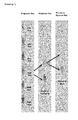
- Amplification of the Wild-type (WT) sequence can be suppressed in two ways. Either the amplification of the WT by binding of the primer to the WT [79,80] or the inhibition of amplification of the WT, drawing 4) [81,82]. In both cases, an oligonucleotide is designed which has 100% complementarity to the WT sequence. LNAs (locked nucleic acids) nucleotide analogues [83, 84] are used as blockers. These nucleotide analogues have the property of binding to the complementary WT sequence with a high degree of affinity. Achievable detection limits of 1:100-1:10,000 are described [83_85].
- One drawback of this method is the relatively high consumables cost of the blockers. The risk of false-positive products, as associated with allele-specific PCR, is largely eliminated.
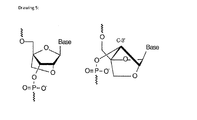
- LNAs consist of modified RNA nucleotides wherein the LNA monomer consists of a bicyclic nucleic acid in which the ribose subunit has been modified with an additional methyl group. This connects the 2' oxygen atom to the 4' carbon atom. This covalent bond 'locks' ribose in the 3'-endo (North) conformation which occurs mainly in the A-form of DNA or RNA, drawing 5.
- Amplification of the WT fraction is suppressed as a result of further synthesis by the DNA polymerase being inhibited ('locked'/'clamped') once the blocker is reached. Amplification of the mutated fraction continues unhindered because the blocker is bound more weakly as a result of the base-base mispairing and becomes detached again during elongation.
- the 100% complementarity of the LNA probe binding to the WT is referred to as 'perfect match' and the weaker binding of the probe to the mutation as a result of the base-base mispairing as 'mismatch'.
- the Tm of the perfect match is higher than that of the mismatch.
- Blood was first enriched for melanoma cells using an antibody-mediated negative selection procedure before whole genome amplification (WGA). Mutant BRAF in the WGA-amplified genomic DNA was further amplified by a two-step real-time PCR protocol. The method is here directed to circulating cells and to DNA isolated from circulating cells.
- the present invention provides an improved LNA clamp qRT-PCR method wherein ctDNA is detectable in patient plasma with a reasonable detection limit.
- the invention is directed to a PCR method for detecting the presence of at least one nucleotide Mutation in DNA polynucleotide sequence encoding for a protein or fragments thereof isolated from patient plasma, characterized in that
- the PCR method is a LNA clamp qRT-PCR.
- DNA polynucleotide sequences encoding for a protein are well known for a large number of proteins as well as the amino acid and nucleotide mutations resulting into dysfunction of the protein.
- Androgen Receptor (AR) genes were sequenced and gene mutations thereof were identified. Such gene mutations are potential target for developing novel treatment. AR is important in the development and progression of prostate cancer. It was observed that in some patients with anti-Androgen Receptor drugs might lead to a resistance and decrease of the hormonal therapy efficiency leading to castration-resistant prostate cancer (CRPC). Despite an initial favorable response, patients progress to a more aggressive, castrate-resistant phenotype that is characterized by the presence of certain mutations of the Androgen Receptor (AR) gene in the circulating tumor cells (CTCs).
- CTCs castration-resistant prostate cancer
- the DNA polynucleotide sequence is encoding for an Androgen Receptor (AR) or fragments thereof.
- AR Androgen Receptor
- the DNA polynucleotide sequence is encoding for an Androgen Receptor (AR) or fragments thereof with Mutation at the amino acid position 741 and/or 877.
- AR Androgen Receptor
- the DNA polynucleotide sequence is encoding for an Androgen Receptor (AR) or fragments thereof with Mutation at the amino acid position 741 and/or 877 resulting into patient with castration-resistant prostate cancer (CRPC).
- AR Androgen Receptor
- CRPC castration-resistant prostate cancer
- the DNA polynucleotide sequence is encoding for an Androgen Receptor (AR) or fragments thereof with Mutation at the amino acid position 741 and/or 877 resulting into patient with castration-resistant prostate cancer (CRPC) being treated with Bicalutamide or Flutamide.
- AR Androgen Receptor
- CRPC castration-resistant prostate cancer
- NCBI National Center for Biotechnology Information
- DNA polynucleotide sequence is isolated from patient plasma and corresponds to circulating free DNA comprising circulating tumor DNA (ctDNA) from tumor cells and further circulating DNA from healthy cells.
- the DNA Polynucleotide sequence is mostly in the form of short or long fragments.
- the DNA Polynucleotide sequence comprises at least one nucleotide Mutation. More preferably, the DNA Polynucleotide sequence comprises at least one nucleotide Mutation wherein said Mutation lead to an amino acid Mutation of the encoded protein.
- the DNA Polynucleotide sequence comprises at least one nucleotide Mutation means that at least one nucleotide of the DNA polynucleotide sequence was replaced by another nucleotide.
- the nucleotide replacement is a mutation of the DNA polynucleotide sequence.
- At least one nucleotide Mutation means preferably 1 to 5 Mutations.
- At least one nucleotide Mutation means more preferably, 1 or 2 Mutations, even more preferably 1 Mutation.
- the Mutation is characteristic for patient showing a resistance to certain therapy drugs. More preferably, the Mutation is characteristic for patient showing a resistance to Bicalutamid or Flutamide.
- the DNA polynucleotide sequence presents Mutation in nucleotide codons 741 and/or 877 of the Androgen Receptor (AR) tumor DNA. Even more preferably, the DNA polynucleotide sequence presents Mutation in nucleotide codons 741 and/or 877 of the Androgen Receptor (AR) tumor DNA wherein Mutation corresponds to a single nucleotide base change.
- the Mutation is the replacement of Adenine (A) with Guanine (G) in the 1st position of the codon corresponding to amino position 877.
- the Mutation is the replacement of Guanine (G) with Thymine (T) in the 3 rd position of the codon 741.
- Drawing 6 shows the position of Mutations T877A and W741C.
- Upstream and downstream primers are short oligonucleotide (10 to 25 nucleic bases) complementing/hybridizing specifically to the 5' and 3' extremity, respectively, of the DNA oligonucleotide sequence encoding for a polypeptide or a protein.
- Upstream primers and downstream primers are preferably SEQ ID 1:AR1_FOR_W741C GACCAGATGGCTGTCATTCA SEQ ID 2: AR5_REV_W741C AAAGCCGCCTCATACTGGAT SEQ ID 3: AR1_FOR_T877A CCCTACAGATTGCGAGAGAG SEQ ID 4: AR5_REV_T877A CAAGGCACTGCAGAGGAGTA
- the LNA-blocker is defined as a LNA-oligonucleotide sequence of 6 to 25 bases comprising a mixture of nucleic acid bases and LNA-monomer bases wherein the LNA-oligonucleotide sequence is complementing/hybridizing a specific DNA sequence of a DNA oligonucleotide sequence encoding for a protein or fragments thereof or a DNA target gene wherein the specific DNA sequence is a Wild type DNA sequence for which one or more Mutations were identified and found related to none-response to a pharmaceutical treatment.
- the LNA-blocker is complementing a Wild type DNA sequence that is selected because of identified mutation(s) within this sequence. These Mutations are qualifying as a potential biomarker for identifying patient not-responding to a given pharmaceutical treatment.
- the LNA-oligonucleotide sequence is encoding for a protein wherein nucleotide mutations thereof results into dysfunction of the encoded protein.
- the LNA-oligonucleotide sequence is encoding for an Androgen Receptor (AR) or fragments thereof wherein nucleotide mutations thereof results into patient suffering from castration-resistant prostate cancer (CRPC).
- AR Androgen Receptor
- CRPC castration-resistant prostate cancer
- the Wild type DNA sequence is defined as such that one Mutation was identified within this sequence and found related to none-response to a pharmaceutical treatment. More preferably, the Wild type DNA sequence is defined as such that one Mutation was identified within this sequence and found related to none-response to a pharmaceutical treatment thereof due to dysfunctionment of the encoded protein. Even more preferably, the Wild type DNA sequence is defined as such that one Mutation was identified within this sequence and found related to none-response to a pharmaceutical treatment thereof due to dysfunctionment of the encoded protein and resulting into castration-resistant prostate cancer (CRPC) wherein the pharmaceutical treatment consists into administration of Flutamide or Bicalutamide to a patient in need.
- the Mutation is defined as above.
- the LNA-blocker is defined as a LNA-oligonucleotide sequence of 6, 7, 8, 9, 10, 11, 12, 13, 14, 15, 16, 17, 18, 19 or 20 bases comprising a mixture of nucleic acid bases and LNA-monomer bases. More preferably, the LNA-blocker is defined as a LNA-oligonucleotide sequence of bases comprising a mixture of 10 to 18 or 11 to 17 nucleic acid bases and LNA-monomer bases. Even more preferably, the LNA-blocker is defined as a LNA-oligonucleotide sequence of 11 or 17 bases, independently from each other, comprising a mixture of nucleic acid bases and LNA-monomer bases. Even more preferably, the LNA-blocker is defined as a LNA-oligonucleotide sequence of 11 bases selected from SEQ ID 5: W741C_LNA.
- the LNA-blocker is defined as a LNA-oligonucleotide sequence of 17 bases selected from SEQ ID 6: T877A_LNA.
- the LNA-blocker comprises 1 to 12, 2 to 12 or 3 to 12 LNA-monomer bases with the proviso that the LNA-blocker comprises at least 1 nucleic acid base.
- the LNA-blocker comprises 7 to 11 LNA-monomer bases. More preferably, the LNA-blocker comprises 8, 9 or 10 LNA-monomer bases.
- the LNA-blocker comprises 8 LNA-monomer bases and is corresponds to SEQ ID 5: W741C_LNA.
- the LNA-blocker comprises 10 LNA-monomer bases and is corresponds to SEQ ID 6: T877A_LNA.
- the LNA-blocker is defined as beginning [5'] and ending [3'] with a LNA-monomer base.
- the LNA-blocker is defined as a LNA-oligonucleotide sequence of 10 bases selected from SEQ ID 7: W741C_LNA_np.
- the LNA-blocker is defined as a LNA-oligonucleotide sequence of 16 bases selected from SEQ ID 8: T877A_LNA_np.
- the LNA-blocker is defined as having a LNA-monomer base positioned in the central portion of the LNA-oligonucleotide sequence.
- the LNA-blocker is defined having a LNA-monomer base positioned in the central portion of the LNA-oligonucleotide sequence wherein said LNA-monomer base is at the position of the Mutation.
- Central portion of the LNA-oligonucleotide sequence means that the LNA-monomer base complementing the Mutation to be detected is located in the middle +/- 2 nucleotide of the LNA-oligonucleotide sequence.
- the LNA-monomer base at the position of the Mutation corresponds to the Wild type nucleic acid base.
- the LNA-blocker is defined as SEQ ID 5: W741C_LNA having a LNA-monomer base G* 6 at the Mutation position.
- the LNA-blocker is defined as SEQ ID 6: T877A_LNA having a LNA-monomer base A* 7 at the Mutation position.
- the LNA-blocker is carrying a protecting group defined as a 3' Inverted Thymidine (3InvdT) at the 3' extremity.
- the LNA-blocker is SEQ ID 5: W741C_LNA comprising a 3InvdT.
- the LNA-blocker is SEQ ID 6: T877A_LNA comprising a 3InvdT.
- the LNA-blocker is defined as
- the LNA-blocker is SEQ ID 5: W741C_LNA of 11 bases complementing/hybridizing a DNA at the specific position of the codon 741 of AR wherein the LNA-blocker comprises 8 LNA-monomer bases, begins with T* LNA-monomer base, ends G* LNA-monomer base and carries a protecting group defined as an 3' Inverted Thymidine (3InvdT).
- W741C_LNA of 11 bases complementing/hybridizing a DNA at the specific position of the codon 741 of AR
- the LNA-blocker comprises 8 LNA-monomer bases, begins with T* LNA-monomer base, ends G* LNA-monomer base and carries a protecting group defined as an 3' Inverted Thymidine (3InvdT).
- the LNA-blocker is SEQ ID 6: T877A_LNA of 17 bases complementing/hybridizing a DNA at the specific position of the codon T877A of AR wherein the LNA-blocker comprises 10 LNA-monomer bases, begins with C* LNA-monomer base, ends C* LNA-monomer base and carries a protecting group defined as an 3' Inverted Thymidine (3InvdT).
- T877A_LNA of 17 bases complementing/hybridizing a DNA at the specific position of the codon T877A of AR
- the LNA-blocker comprises 10 LNA-monomer bases, begins with C* LNA-monomer base, ends C* LNA-monomer base and carries a protecting group defined as an 3' Inverted Thymidine (3InvdT).
- the LNA-blocker is SEQ ID 7: W741C_LNA_np.
- the LNA-blocker is SEQ ID 8: T877A_LNA_np.
- LNA-Blockers SEQ ID 5: W741C_LNA T*-C*-C*- T -G*-G*- A -T*-G*-G*/3InvdT/ T*-C*-C*- T -G*-G*- A -T*-G*-G*/3InvdT/ with highlighted LNA-monomer position of the nucleotide Mutation T* 1 -C* 2 -C* 3 - T 4 -G* 5 -G* 6 - A 7 -T* 8 -G* 9 -G* 10 /3InvdT 11 / with nucleotide position numbering SEQ ID 6: T877A_LNA C*-A*-G*- T-T -C*-A*-C*-T*- T-T-T -G*- A -C*-C*/3Invd
- DNA stands for genomic DNA and fragments thereof.
- the annealing step is conducted at a temperature of 52 to 60 C°.
- the annealing step is conducted at a temperature of 54 to 58 C°.
- the LNA blocker is defined as SEQ ID 5: W741C_LNA
- the upstream primer is defined as SEQ ID 1: W741C
- the downstream primer is defined as SEQ ID 2: AR5_REV_W741C then the annealing step is conducted at a temperature of 55 C°.
- the LNA blocker is defined as SEQ ID 6: T877A_LNA
- the upstream primer is defined as SEQ ID 3: the T877A
- downstream primer is defined as SEQ ID 4: AR5_REV_T877A then the annealing step is conducted at a temperature of 60 C°.
- the elongating step is conducted at a temperature of 72 to 74 C°.
- the LNA blocker is defined as SEQ ID 5: W741C_LNA
- the upstream primer is defined as SEQ ID 1: W741C
- the downstream primer is defined as SEQ ID 2: AR5_REV_W741C then the elongating step is conducted at a temperature of 74 C°.
- the LNA blocker is defined as SEQ ID 6: T877A_LNA
- the upstream primer is defined as SEQ ID 3: T877A
- the downstream primer is defined as SEQ ID 4: AR5_REV_T877A then the elongating step is conducted at a temperature of 72 C°.
- the method is for detecting the presence of at least one nucleotide Mutation in a DNA polynucleotide sequence encoding for a protein or fragments thereof in patient plasma wherein a patient with a Mutation is a patient showing a resistance to certain therapy drugs.
- a patient with a Mutation is a patient showing a mutation in AR.
- Said patient shows a resistance to certain therapy drugs and developing a castration-resistant prostate cancer (CRPC).
- the patient with a Mutation is a patient showing a resistance to therapy drugs such as Bicalutamide and/or Flutamide and developing a castration-resistant prostate cancer (CRPC).
- the PCR method is for detecting the presence of at least one nucleotide Mutation in a DNA polynucleotide sequence encoding for a Androgen Receptor (AR) or fragments thereof isolated from patient plasma, characterized in that
- the PCR method is for detecting the presence of at least one nucleotide Mutation in a DNA polynucleotide sequence encoding for an Androgen Receptor (AR) or fragments thereof with Mutation at the amino acid position 741 and/or 877 isolated from patient plasma, characterized in that
- the PCR method is for detecting the presence of at least one nucleotide Mutation in a DNA polynucleotide sequence encoding for an Androgen Receptor (AR) or fragments thereof isolated from patient plasma, characterized in that
- the PCR method for detecting the presence of at least one nucleotide Mutation in a DNA polynucleotide sequence encoding for an Androgen Receptor (AR) or fragments thereof isolated from patient plasma characterized in that
- the invention is directed to a LNA-blocker defined as a LNA-oligonucleotide sequence of 6 to 25 bases comprising a mixture of nucleic acid bases and LNA-monomer bases wherein the LNA-oligonucleotide sequence is complementing/hybridizing a specific DNA sequence of a DNA polynucleotide sequence encoding for a protein or fragments thereof or a DNA target gene wherein the specific DNA sequence is a Wild type DNA sequence for which one or more Mutations were identified and found related to none-response to a pharmaceutical treatment.
- the LNA-blocker is complementing a Wild type DNA sequence that is selected because of identified mutation(s) within this sequence. These Mutations are qualifying as a potential biomarker for identifying patient not-responding to a given pharmaceutical treatment.
- the LNA-oligonucleotide sequence is encoding for a protein wherein nucleotide mutations thereof results into dysfunction of the encoded protein. More preferably, the LNA-oligonucleotide sequence is encoding for an Androgen Receptor (AR) or fragments thereof wherein nucleotide mutations thereof results into patient suffering from castration-resistant prostate cancer (CRPC).
- AR Androgen Receptor
- CRPC castration-resistant prostate cancer
- the Wild type DNA sequence is defined as such that one Mutation was identified within this sequence and found related to none-response to a pharmaceutical treatment. More preferably, the Wild type DNA sequence is defined as such that one Mutation was identified within this sequence and found related to none-response to a pharmaceutical treatment thereof due to dysfunctionment of the encoded protein. Even more preferably, the Wild type DNA sequence is defined as such that one Mutation was identified within this sequence and found related to none-response to a pharmaceutical treatment thereof due to dysfunctionment of the encoded protein and resulting into castration-resistant prostate cancer (CRPC) wherein the pharmaceutical treatment consists into administration of Flutamide or Bicalutamide to a patient in need.
- CRPC castration-resistant prostate cancer
- the Mutation is defined as above.
- the LNA-blocker is defined as a LNA-oligonucleotide sequence of 6, 7, 8, 9, 10, 11, 12, 13, 14, 15, 16, 17, 18, 19 or 20 bases comprising a mixture of nucleic acid bases and LNA-monomer bases. More preferably, the LNA-blocker is defined as a LNA-oligonucleotide sequence of bases comprising a mixture of 10 to 18 or 11 to 17 nucleic acid bases and LNA-monomer bases. Even more preferably, the LNA-blocker is defined as a LNA-oligonucleotide sequence of 11 or 17 bases, independently from each other, comprising a mixture of nucleic acid bases and LNA-monomer bases. Even more preferably, the LNA-blocker is defined as a LNA-oligonucleotide sequence of 11 bases selected from SEQ ID 5: W741C_LNA.
- the LNA-blocker is defined as a LNA-oligonucleotide sequence of 17 bases selected from SEQ ID 6: T877A_LNA.
- the LNA-blocker comprises 1 to 12, 2 to 12 or 3 to 12 LNA-monomer bases with the proviso that the LNA-blocker comprises at least 1 nucleic acid base.
- the LNA-blocker comprises 7 to 11 LNA-monomer bases. More preferably, the LNA-blocker comprises 8, 9 or 10 LNA-monomer bases.
- the LNA-blocker comprises 8 LNA-monomer bases and is corresponds to SEQ ID 5: W741C_LNA.
- the LNA-blocker comprises 10 LNA-monomer bases and is corresponds to SEQ ID 6: T877A_LNA.
- the LNA-blocker is defined as beginning [5'] and ending [3'] with a LNA-monomer base.
- the LNA-blocker is defined as a LNA-oligonucleotide sequence of 10 bases selected from SEQ ID 7: W741C_LNA_np.
- the LNA-blocker is defined as a LNA-oligonucleotide sequence of 16 bases selected from SEQ ID 8: T877A_LNA_np.
- the LNA-blocker is defined as having a LNA-monomer base positioned in the central portion of the LNA-oligonucleotide sequence.
- the LNA-blocker is defined having a LNA-monomer base positioned in the central portion of the LNA-oligonucleotide sequence wherein said LNA-monomer base is at the position of the Mutation.
- Central portion of the LNA-oligonucleotide sequence means that the LNA-monomer base complementing the Mutation to be detected is located in the middle +/- 2 nucleotide of the LNA-oligonucleotide sequence.
- the LNA-monomer base at the position of the Mutation corresponds to the Wild type nucleic acid base.
- the LNA-blocker is defined as SEQ ID 5: W741C_LNA having a LNA-monomer base G* 6 at the Mutation position.
- the LNA-blocker is defined as SEQ ID 6: T877A_LNA having a LNA-monomer base A* 7 at the Mutation position.
- the LNA-blocker is carrying a protecting group defined as a 3' Inverted Thymidine (3InvdT) at the 3' extremity.
- the LNA-blocker is SEQ ID 5: W741C_LNA comprising a 3lnvdT.
- the LNA-blocker is SEQ ID 6: T877A_LNA comprising a 3lnvdT.
- the LNA-blocker is defined as
- the LNA-blocker is SEQ ID 5: W741C_LNA of 11 bases complementing/hybridizing a DNA at the specific position of the codon 741 of AR wherein the LNA-blocker comprises 8 LNA-monomer bases, begins with T* LNA-monomer base, ends G* LNA-monomer base and carries a protecting group defined as an 3' Inverted Thymidine (3InvdT).
- W741C_LNA of 11 bases complementing/hybridizing a DNA at the specific position of the codon 741 of AR
- the LNA-blocker comprises 8 LNA-monomer bases, begins with T* LNA-monomer base, ends G* LNA-monomer base and carries a protecting group defined as an 3' Inverted Thymidine (3InvdT).
- the LNA-blocker is SEQ ID 6: T877A_LNA of 17 bases complementing/hybridizing a DNA at the specific position of the codon T877A of AR wherein the LNA-blocker comprises 10 LNA-monomer bases, begins with C* LNA-monomer base, ends C* LNA-monomer base and carries a protecting group defined as an 3' Inverted Thymidine (3InvdT).
- T877A_LNA of 17 bases complementing/hybridizing a DNA at the specific position of the codon T877A of AR
- the LNA-blocker comprises 10 LNA-monomer bases, begins with C* LNA-monomer base, ends C* LNA-monomer base and carries a protecting group defined as an 3' Inverted Thymidine (3InvdT).
- the LNA-blocker is SEQ ID 7: W741C_LNA_np.
- the LNA-blocker is SEQ ID 8: T877A_LNA_np.
- LNA-Blockers SEQ ID 5: W741C_LNA T*-C*-C*- T -G*-G*- A -T*-G*-G*/3InvdT/ T*-C*-C*- T -G*-G*- A -T*-G*-G*/3InvdT/ with highlighted LNA-monomer position of the nucleotide Mutation T* 1 -C* 2 -C* 3 - T 4 -G* 5 -G* 6 - A 7 -T* 8 -G* 9 -G* 10 /3InvdT 11 / with nucleotide position numbering SEQ ID 6: T877A_LNA C*-A*-G*- T-T -C*-A*-C*-T*- T-T-T -G*- A -C*-C*/3Invd
- DNA stands for genomic DNA and fragments thereof.
- the invention is directed to
- the location of the primer has an influence on blocker efficiency.
- the upstream primer is designed so that it's attached to the template strand just a few bases upstream of the LNA-blocker. The closeness of the blocker to the 3'-end of the upstream primer prevented any elongation of the upstream primer at an early stage. If the upstream primer were to be located further upstream of the blocker, , it would continue to be elongated until polymerization was stopped by the LNA-blocker. But the melting temperature of the elongated upstream primer then exceeds the melting temperature of the LNA-blocker, which detach from the upstream primer between the elongation and denaturation steps, and the upstream primer will then continue to be elongated until the end.
- the invention is directed to a kit for detecting the presence of at least one nucleotide Mutation in a DNA polynucleotide sequence encoding for a protein or fragments thereof isolated from patient plasma, wherein the kit comprises a LNA-blocker as described in the second aspect wherein the DNA polynucleotide sequence or fragments thereof is a natural circulating tumor DNA fragments isolated from patient plasma.
- LNA-blocker definition as in second aspect is here enclosed.
- the kit for detecting the presence of at least one nucleotide Mutation in a DNA polynucleotide sequence encoding for an Androgen Receptor (AR) or fragments thereof isolated from patient plasma wherein the kit comprises a LNA-blocker defined as SEQ ID 5: W741C_LNA, SEQ ID 6: T877A_LNA, SEQ ID 7: W741C_LNA np or SEQ ID 8: T877A_LNA_np.
- the kit comprises a LNA-blocker and an upstream primer wherein the upstream primer and the LNA-blocker are closely annealing onto the DNA fragments isolated from patient plasma.
- the kit comprises a LNA-blocker and an upstream primer wherein the upstream primer and the LNA-blocker are closely annealing onto the DNA fragments isolated from patient plasma and the LNA-blocker is defined as SEQ ID 5: W741C_LNA, SEQ ID 6: T877A_LNA, SEQ ID 7: W741C_LNA_np or SEQ ID 8: T877A_LNA_np and the upstream primer is defined as SEQ ID 1 W741C or SEQ ID 3 T877A.
- the kit comprises a LNA-blocker, an upstream primer and a downstream primer wherein the upstream primer and the LNA-blocker are closely annealing onto the DNA fragments isolated from patient plasma.
- the kit comprises a LNA-blocker, an upstream primer and a downstream primer wherein the upstream primer and the LNA-blocker are closely annealing onto the DNA fragments isolated from patient plasma and the LNA-blocker is defined as SEQ ID 5: W741C_LNA, SEQ ID 6: T877A_LNA, SEQ ID 7: W741C_LNA_np or SEQ ID 8: T877A_LNA_np, the upstream primer is defined as SEQ ID 1 W741C or SEQ ID 3 T877A and the downstream primer of SEQ ID 2: AR5_REV_W741C or SEQ ID 4: AR5_REV_T877A.
- the upstream primer is defined as SEQ ID 1 W741C or SEQ ID 3 T877A
- closely means that the distance between the last nucleic acid base of the upstream primer and the first nucleic acid base of the LNA-blocker is corresponding to a sequence of 3 to 10 nucleic acid bases of the DNA isolated from patient plasma.
- the last nucleic acid base of the upstream primer means the 3' nucleic acid base of the upstream primer.
- the first nucleic acid base of the LNA-blocker means the 5' nucleic acid base of the LNA-blocker.
- sequence of 3 to 10 nucleic acid bases of the DNA between the last nucleic acid base of the upstream primer and the first nucleic acid base of the LNA-blocker is not complemented by the upstream primer or the LNA-blocker.
- closely means that the distance between the last nucleic acid base of the upstream primer and the first nucleic acid base of the LNA-blocker is corresponding to a sequence of 3 to 7 or 4 to 6 nucleic acid bases of the DNA isolated from patient plasma, even more preferably 4 or 6.
- the upstream primer is SEQ ID 1 W741C or SEQ ID 3 T877A.
- the downstream primer is SEQ ID 2: AR5_REV_W741C or SEQ ID 4: AR5_REV_T877A.
- the LNA blocker is SEQ ID 5: W741C_LNA, SEQ ID 6: T877A_LNA, SEQ ID 7: W741C_LNA_np or SEQ ID 8: T877A_LNA_np.
- the DNA polynucleotide sequence is encoding for a protein wherein oligonucleotide mutations thereof results into dysfunction of the encoded protein leading to castration-resistant prostate cancer (CRPC).
- CRPC castration-resistant prostate cancer
- the DNA polynucleotide sequence is encoding for an Androgen Receptor (AR) or fragments thereof wherein oligonucleotide mutations thereof results into castration-resistant prostate cancer (CRPC) due to administration to a patient in need of Flutamide or Bicalutamide.
- AR Androgen Receptor
- CRPC castration-resistant prostate cancer
- PCR stands for polymerase chain reaction.
- LNA clamp qRT-PCR stands for Locked Nucleic Acids clamp quantitative Real Time-PCR. Such PCR is well known in the art and is conducted on standard real time PCR device. Primers are ordered from a primer provider. The LNA blockers are marketed by the company EXIQON in Denmark.
- PCR amplification is detected using a SYBR-Green moiety as provided by Qiagen in the QuantiTect-SYBR-Green PCR kit.
- PCR Primers are used in standard chemical synthesis methods or are produced using enzymatic methods or any other known method. Such methods can range from standard enzymatic digestion followed by nucleotide fragment isolation (see for example, Sambrook et al., Molecular Cloning: A Laboratory Manual, 2nd Edition (Cold Spring Harbor Laboratory Press, Cold Spring Harbor, N.Y., 1989 ) Chapters 5, 6, incorporated herein by reference) to purely synthetic methods, for example, by the cyanoethyl phosphoramidite method using a Milligen or Beckman System 1Plus DNA synthesizer (for example, Model 8700 automated synthesizer of Milligen-Biosearch, Burlington, Mass. or ABI Model 380B).
- Primers are a subset of probes which are capable of supporting some type of enzymatic manipulation and which can hybridize with a target nucleic acid such that the enzymatic manipulation can occur.
- a primer can be made from any combination of nucleotides or nucleotide derivatives or analogs available in the art which do not interfere with the enzymatic manipulation.
- a nucleotide is a molecule that contains a base moiety, a sugar moiety and a phosphate moiety. Nucleotides can be linked together through their phosphate moieties and sugar moieties creating an internucleoside linkage.
- the base moiety of a nucleotide can be adenin-9-yl (A), cytosin-1-yl (C), guanin-9-yl (G), uracil-1-yl (U), and thymin-1-yl (T).
- the sugar moiety of a nucleotide is a ribose or a deoxyribose.
- the phosphate moiety of a nucleotide is pentavalent phosphate.
- a non-limiting example of a nucleotide would be 3'-AMP (3'-adenosine monophosphate) or 5'-GMP (5'-guanosine monophosphate).
- nucleotide includes nucleotides and nucleotide analogs, preferably groups of nucleotides comprising oligonucleotides, and refers to any compound containing a heterocyclic compound bound to a phosphorylated sugar by an N-glycosyl link or any monomer capable of complementary base pairing or any polymer capable of hybridizing to an oligonucleotide.
- oligonucleotide means a naturally occurring or synthetic polymer of nucleotides, preferably a polymer comprising at least three nucleotides and more preferably a polymer capable of hybridization.
- Oligonucleotides may be single-stranded, double-stranded, partially single-stranded or partially double-stranded ribonucleic or deoxyribonucleic acids, including selected nucleic acid sequences, heteroduplexes, chimeric and hybridized nucleotides and oligonucleotides conjugated to one or more nonoligonucleotide molecules.
- nucleotides comprising a oligonucleotide are naturally occurring deoxyribonucleotides, such as adenine, cytosine, guanine or thymine linked to 2'-deoxyribose, or ribonucleotides such as adenine, cytosine, guanine or uracil linked to ribose.
- an oligonucleotide also can contain nucleotide analogs, including non-naturally occurring synthetic nucleotides or modified naturally occurring nucleotides.
- nucleotide analogs are well known in the art and commercially available, as are polynucleotides containing such nucleotide analogs ( Lin et al., Nucl. Acids Res. 22:5220-5234 (1994 ); Jellinek et al., Biochemistry 34:11363-11372 (1995 ); Pagratis et al., Nature Biotechnol. 15:68-73 (1997 ), each of which is incorporated herein by reference).
- a polynucleotide comprising naturally occurring nucleotides and phosphodiester bonds can be chemically synthesized or can be produced using recombinant DNA methods, using an appropriate polynucleotide as a template.
- a polynucleotide comprising nucleotide analogs or covalent bonds other than phosphodiester bonds generally will be chemically synthesized, although an enzyme such as T7 polymerase can incorporate certain types of nucleotide analogs into a polynucleotide and, therefore, can be used to produce such a polynucleotide recombinantly from an appropriate template (Jellinek et al., supra, 1995, incorporated herein by reference).
- primer pair refers to a set of primers Including a 5' "upstream primer” or “forward primer” that hybridizes with the complement of the 5' end of the DNA sequence to be amplified and a 3' "downstream primer” or “reverse primer” that hybridizes with the 3' end of the sequence to be amplified.
- upstream and downstream or “forward” and “reverse” are not intended to be limiting, but rather provide ii!ustrative orientation in particular embodiments.
- gDNA means genomic DNA.
- the androgen receptor (steroid receptor) is divided structurally into four domains: an N-terminal region with a transactivation function, a DNA-binding domain with a nuclear localization signal (NLS) and a ligand-binding domain (LBD) [95].
- the AR acts as a ligand-dependent transcription factor. It is located in the cytoplasm in the absence of testosterone and forms a complex with heat shock proteins. If testosterone or its metabolite dihydrotestosterone (DHT) binds to the AR, a change in conformation occurs which leads to dissociation of the heat shock proteins.
- DHT dihydrotestosterone
- the NLS can then be detected by import proteins and the AR is translocated into the nucleus, where, as a homodimer, it binds to the androgen response element (ARE) upstream of the target genes [95-97].
- ARE androgen response element
- T877A in exon H of the human AR a missense mutation in the ligand-binding domain of the AR in the LNCaP human prostate cancer cell line
- This mutation resulted in an increase in the range of ligands which led to AR activation.
- substances such as estrogens and hydroxyflutamide also led to transactivation [99, 101].
- the T877A mutation is located in the AF-2 domain of exon H. AF-2 binds coregulatory proteins and interacts with the transactivation domain.
- the T877A missense mutation has also been detected in some cases of metastatic prostate cancer treated with hydroxyflutamide [102].
- Bicalutamide did not activate the mutated AR in LNCaP and continued to act as an antagonist [101, 103].
- An AR mutation has also been detected in patients under Bicalutamide therapy [104].
- Yoshida et al. (2005) received the KUCaP cell line containing a W741C mutation (exon E of the human AR) from a liver metastasis of prostate cancer in a patient [105].
- W741C mutation exon E of the human AR
- Bicalutamide exerts an agonist effect on the AR. Flutamide continues to act as an antagonist.
- the genomic sequence of the human androgen receptor comes from the NG_009014 RefSeq (Reference Sequence) database sequence of the National Center for Biotechnology Information (NCBI).
- the mRNA sequence of the human androgen receptor was taken from the NG_000044 RefSeqGene sequence.
- the primers were ordered from TIB MOLBIOL GmbH (Berlin, Germany) and purified by means of HPLC.
- the two primer pairs AR1_FOR_W741C and AR5_REV_W741C and the primer pairs AR1_FOR_T877A and AR5_REV_T877A amplify the genomic DNA region in which the base substitutions (T877A, W741C) in the human AR sequence are located.
- the locked nucleic acid (LNA) probes were designed in-house and ordered from the company EXIQON in Denmark. In order to prevent the DNA polymerase-induced elongation, an inverted thymine (3InvdT) was added to the 3' of the LNA probe.
- the LNA probes consisted of a mixture of normal bases and specific LNA monomers, which are indicated by a '*' in Table 2.5.
- the T877A LNA clamp is complementary to a 16 nt region (genomic DNA (gDNA) region: 184673 bp - 184688 bp) which covers codon 877 of the human AR. The complementary region which covers codon 877 is highlighted in bold.
- the W741C LNA clamp is complementary to a 10 nt region (gDNA region: 178494 bp - 178503 bp) which covers codon 741.
- the complementary region which covers codon 741 is highlighted in bold.
- Both LNA clamps were designed using EXIQON's LNA Oligo Tools LNA Oligo Tm Prediction and LNA Oligo Optimizer.
- venous blood was collected into EDTA plasma Monovettes ® (Sarstedt) in the hospital and centrifuged for 10 min at 1,900 x g and 4 °C using a swing-out rotor.
- the supernatant (plasma) was then divided into aliquots, placed in intermediate storage at a temperature of -20 °C and transported to Bayer Pharma AG, where it was stored at -80 °C.
- these frozen plasma samples were thawed at RT and centrifuged for 10 min at 16,000 x g and 4 °C in order to remove additional cell debris and contamination of circulating nucleic acids by gDNA and RNA of dead blood cells.
- 1 ml plasma was obtained from each of 6 healthy blood donors by Clinical Pharmacology, Bayer Pharma AG, treated in the same way and prepared for free-circulating DNA isolation.
- Free, circulating DNA was isolated with the QIAGEN QIAamp ® Circulating Nucleic Acid Kit using a vacuum (QIAGEN QIAvac24 Plus ® vacuum pump) in accordance with the manufacturer's instructions.
- a carrier RNA (1 ⁇ g) was added to increase binding of the rare nucleic acids to the silica membrane. Because of the low yield of cfDNA, the RNA carrier used and the buffer components, quantification by means of UV absorption spectrometry was not possible.
- the cfDNA content was determined by means of qRT-PCR. A fixed pipetting volume of 5 ⁇ l was used for this, see 2.4.3. The minimal volume of 1 ml plasma per patient/subject was used for the assay. Elution was performed using buffer AVE in a minimal volume of 25 ⁇ l (approximately 20 ⁇ l eluate).
- qRT-PCR quantitative real-time PCR
- QuantiTect SYBR Green PCR + UNG Kit and the 7500 Fast Real-Time PCR Cycler (Applied Biosystems) were used in accordance with the manufacturer's instructions.
- the qRT-PCR was analyzed using the ABI 7500 v.2.0.5 software package.
- uracil N-glycosylase was added in accordance with the manufacturer's instructions. Both of the following were used initially for the qualitative analysis: a melting curve run after the qRT-PCR in accordance with the manufacturer's instructions and an analysis of the PCR products by means of gel electrophoresis. The optimal annealing temperatures were determined in advance by means of conventional PCR with a temperature gradient.
- the LNA clamp qRT-PCR was developed to detect gDNA displaying missense mutations (T877A & W741C) in the human AR.
- each sample template was amplified in two independent PCR reactions with and without LNA probe which is 100% complementary to WT.
- the data were analyzed using a modified delta-delta Ct method (Ct or cycle threshold: cycle in which the fluorescence rises significantly above the background fluorescence and the amplification is in the exponential phase).
- Ct or cycle threshold cycle in which the fluorescence rises significantly above the background fluorescence and the amplification is in the exponential phase.
- the delta Ct value was determined from the difference between the Ct values of the sample with LNAs (WT is inhibited) and without LNAs (WT is not inhibited).
- the delta-delta Ct was determined from the difference between the delta Ct value of the WT sample and the delta Ct value of the sample to be analyzed. This value reflects the efficiency of the blocker [86, 120].
- the annealing temperature had to be 60 °C instead of 55 C, and the elongation temp. 72 C instead of 74 C.
- the LNA clamp qRT-PCR products (pool of the duplicates or triplicates) were analyzed by means of sequencing.
- the sequencing was carried out by the company Services in Molecular Biology (SMB) in the Institute of Biology at Humboldt University, Berlin, using the chain-termination method [121]. 50 ng DNA (259 bp and 279 bp amplicons) in 5 ⁇ L RNase-free water were used per sequencing batch.
- the AR5_T877A_REV and AR5_W741C_REV primers were used for the sequencing.
- the LNA blockers were designed for both mutations, along with the primers for amplification.
- the design of the LNA blockers it was important for these to be located in a central position over the base substitution to be analyzed. They are made up of a mixture of deoxynucleotides and LNA monomers.
- a probe consisting of LNA monomers alone would have increased the self-complementarity of the probe, on the other hand. This would have also resulted in excessive inhibition of the gDNA of the mutation.
- a duplet or triplet of LNA monomers was placed directly over the codon to be analyzed in order to ensure the greatest possible discrimination between inhibited WT and amplified mutation.
- LNA monomers were added to both LNA blockers at the 5'- and 3'-end in order to protect them from the exonuclease function of the DNA polymerase.
- the LNA blockers were designed so that the Tm of the perfect match (WT + blocker) was higher than the elongation temperature.
- the Tm of the mismatch (mutation + blocker) needed to be lower than the elongation temperature. The purpose of this was to inhibit amplification of the WT template during elongation. Amplification of the mutation needed to be made possible, on the other hand.
- the Tm of the blockers was initially established on the basis of the standard elongation temperature of 72 °C.
- the annealing temperature was determined empirically by means of conventional PCR. For both assays, a temperature gradient of 53-63°C was run in 1°C steps. Optimal annealing temperatures were established of 55 C for the W741C assay and 60 °C for the T877A assay (data not shown). The optimal elongation temperature was also determined empirically. Temperatures of 72-76°C were tested in 1°C steps for both assays. A temperature of 74°C was selected for the W741C assay and 72 °C for the T877A assay.
- the elongation temperatures for both assays were 2 C below the specific Tm values of the LNA blockers (Tm of T877A-LNA: 74°C; Tm of W741C-LNA: 76 C). This ensured that the blocker remained bound firmly to the WT during elongation. Although an elongation temperature lower than 72 C/74 C would have resulted in even greater inhibition of WT, there would also then have been a risk of mutant inhibition. Both fragments were amplified with the aid of the primers without the formation of non-specific fragments. This was verified by means of subsequent melting curve and gel analysis (data not shown).
- NTC non-template control
- H2O instead of DNA
- LNA blocker specificity primary for amplifying the W741C amplicon and T877A blocker, vice versa
- the prostate cancer cell line LAP-C4 was used as a model for the wild type, LNCaP as a model for the T877A mutant and KUCaP-1 as a model for the W741C mutant.
- 5 ng, 25 ng and 50 ng gDNA were used in the T877A assay (data not shown).
- the LNA concentration was first titrated roughly in the range of 1-10 ⁇ M. After no increase in blocker efficiency was detected in the T877A assay above a concentration of 1 ⁇ M, a fine titration was performed in the range of 0.1-1 ⁇ M.
- Drawing 11 shows the optimization of the T877A batch.
- LNAs were used in final concentrations of 0.1-1 ⁇ M and 25 ng gDNA.
- the delta Ct value relating to the final LNA concentration in ⁇ M was applied.
- the delta-delta Ct value has been represented as an arrow and is a measure of blocker efficiency.
- the Ct values of the WT and of the mutation were determined in the presence and absence of the LNA blocker. The difference in the Ct values yielded the delta Ct.
- Blocker efficiency was expressed in the form of the delta-delta Ct value and calculated as the difference between the delta Ct values for WT and mutation.
- An optimal delta-delta Ct value results from a high delta Ct value for the WT (strong inhibition) and a delta Ct value as low as possible (little/no inhibition) for the mutation.
- the parameters inside a gray border were used for all other experiments involving the T877A batch. The error bars indicate the standard deviation.
- Table 3.15 Delta-delta Ct values of the T877A-LNA batch c(T877A-LNA) Delta-delta Ct 0,1_ ⁇ M 4,3_ 0,2_ ⁇ M 9,1 0,3_ ⁇ M_ 10,7_ 0,4_ ⁇ M_ 11,3_ 0,5_ ⁇ M_ 11,6_ 1,0_ ⁇ M_ 11,2_
- Drawing 12 shows the optimization of the LNA concentration of the W741C batch.
- LNAs were used in final concentrations of 0.5-5 ⁇ M and 25 ng gDNA.
- the delta Ct value relating to the final LNA concentration in ⁇ M was applied.
- the delta-delta Ct value has been represented as an arrow and is a measure of blocker efficiency.
- the Ct values of the WT and of the mutation were determined in the presence and absence of the LNA blocker. The difference in the Ct values yielded the delta Ct.
- Blocker efficiency was expressed in the form of the delta-delta Ct value and calculated as the difference between the delta Ct values for WT and mutation.
- An optimal delta-delta Ct value results from a high delta Ct value for the WT (strong inhibition) and a delta Ct value as low as possible (little/no inhibition) for the mutation.
- MgCl2 was added in a final concentration of 4 mM to improve efficiency.
- the parameters inside a gray border were used for all other experiments involving the W741C batch. The error bars indicate the standard deviation.
- Table 3.16 Delta-delta Ct values of the W741C-LNA batch c(W741C-LNA) Delta-delta Ct 0,5_ ⁇ M 2,9 1,0_ ⁇ M 5,0 2,0_ ⁇ M_ 10,3 3,0_ ⁇ M_ 11,0 4,0_ ⁇ M_ 11,0 5,0_ ⁇ M_ 11,2
- Drawing 13 shows the determination of the mutant DNA detection limit in the presence of an excess of WT DNA using standard dilutions.
- the quantity of gDNA used in each case was 25 ng.
- the delta Ct value relating to WT DNA, mutant DNA and dilutions of 1:10-1:10,000 (mutant DNA in WT DNA) was applied.
- the delta Ct value was calculated from the Ct value of the dilutions or of the WT (reference) with and without addition of LNAs.
- the delta-delta Ct value was obtained from the difference in the Ct value for the dilutions and for the WT (reference).
- the top diagram shows the dilution series for the T877A batch.
- the bottom diagram shows the dilution series for the W741C batch.
- Drawing 14 shows the sequencing results for W741C/T877A-LNA batches in standard dilutions.
- Top diagram Part of the electropherogram showing the sequence covering the T877A blocker (16 nt) from a dilution of 1:10-1:10,000 (mutant DNA in WT DNA) with the addition of T877A-LNA in a final concentration of 0.5 ⁇ M.
- Bottom diagram Part of the electropherogram showing the sequence covering the W741C blocker (10 nt) from a dilution of 1:10-1:10,000 (mutant DNA in WT DNA) with the addition of W741C-LNA in a final concentration of 3 ⁇ M.
- the base calling sequence has been shown using WT and mutation + LNA for illustration (above the electropherogram).
- Codon 741 and 877 (point mutations) have been placed inside a frame. Codons 740-742 and 876-878 have also been highlighted. The hotspot has been marked with an arrow. The horizontal grey arrow marks the decrease in the concentration of the mutation. Cytosine, adenine,thymine, and guanine.
- the established LNA assay was analyzed for applicability for the sensitive detection of castration resistance-relevant point mutations in the human AR of prostate cancer patients. Seventy percent of the patients were under or had received Bicalutamide therapy. Only 10% were under or had received hydroxyflutamide therapy. Results are based onto W741C batch. In connection with this, the aim was to identify potential Bicalutamide resistance in clinical samples. Measurements were performed on double samples with a volume of 5 ⁇ l from the patient and subject samples used in each, either with or without addition of W741C LNA blocker. Because there was a significant variation between the clinical samples in terms of their initial DNA concentration (0.02-21.64 ng/ ⁇ l), it was not possible to calculate a blocker efficiency (delta-delta Ct).
- the horizontal lines indicate the upper and lower imaginary threshold as the positive and negative standard deviation. Delta Ct values falling below the negative threshold were classified as putatively positive for the W741 C base substitution. Black bars correspond to patients under Bicalutamide therapy and white bars represent treatment-naive patients. The gray column corresponds to the arithmetic mean for the subject samples. All values were determined in duplicate.
- PID 11 was classified as positive for the base substitution by means of qRT-PCR.
- a nucleotide mutation in codon W741C was found in the sequencing. The explanation for this was another single nucleotide polymorphism (SNP) in codon 741 (W741R). If an SNP occurred within the LNA blocker sequence or within the triplet sequence to be analyzed, the LNA was bound to the complementary sequence with a lower affinity than the one calculated theoretically. The consequence was weaker inhibition of the sequence by the LNA (smaller delta Ct value), which resulted in the detection of another mutation in the same codon.
- Drawing 16 shows sequence analysis of PID 1 - PID 19 with addition of W741C-LNA.
- Codon 741 is inside a gray frame.
- Base substitution occurred in PID 5 (CCA -> ACA, highlighted in red).
- the position of the possible base substitution was marked with a black arrow.
- SNPs resulting from automatic base calling have been represented beneath the electropherogram.
- the theoretical sequence has been noted above the electropherogram; differences from automatic base calling have been represented in dark grey letters.
- the diagram shows the development of the prostate cancer of the patient over time as indicated by his PSA levels.
- PSA values were determined in a clinical routine lab using the Hybritech assay (Beckman-Coulter). The patient presented in Oct. 2008 (approx. 3.5 yrs. after last screening) with a PSA beyond 250 ng/ml.
- Drawing 18 X axis is the Time axis, from Oct 2008 to Dec 2009.
- Y axis is the PSA level axis.
Landscapes
- Chemical & Material Sciences (AREA)
- Life Sciences & Earth Sciences (AREA)
- Health & Medical Sciences (AREA)
- Organic Chemistry (AREA)
- Proteomics, Peptides & Aminoacids (AREA)
- Zoology (AREA)
- Wood Science & Technology (AREA)
- Engineering & Computer Science (AREA)
- Genetics & Genomics (AREA)
- Analytical Chemistry (AREA)
- Immunology (AREA)
- Molecular Biology (AREA)
- General Engineering & Computer Science (AREA)
- Biotechnology (AREA)
- Biophysics (AREA)
- Physics & Mathematics (AREA)
- Biochemistry (AREA)
- Bioinformatics & Cheminformatics (AREA)
- Microbiology (AREA)
- General Health & Medical Sciences (AREA)
- Chemical Kinetics & Catalysis (AREA)
- Pathology (AREA)
- Oncology (AREA)
- Hospice & Palliative Care (AREA)
- Measuring Or Testing Involving Enzymes Or Micro-Organisms (AREA)
Abstract
The present invention is directed to a PCR method, novel LNA-blockers and a kit thereof for the detection of nucleic acid mutation(s) in free circulating DNA as present in plasma.
Description
- The present invention is directed to a PCR method, novel LNA-blockers and a kit thereof for the detection of nucleic acid mutation(s) in free circulating DNA as present in plasma.
- Despite the enormous advances made in recent years in our understanding at a molecular level of how cancer develops and progresses and the discovery of potential anticancer agents, more than 90% of these drugs have not been granted a Marketing Authorization. Many drugs fail in the late stages of clinical development (Phase III) because of variable activity in patients, unpredictable safety aspects and inadequate strategies for combating resistance. Hence, there is a need for a more rapid, cost-effective strategy for evaluating anticancer drugs [1, 2]. Identifying patients who will benefit from a particular treatment is still a challenge. However, the difficulty with this lies in the development of suitable clinical tools for discovering indicators or "biomarkers" for cancer [5]. These biomarkers can be divided into a number of different categories, drawing 1. Prognostic biomarkers predict the natural course of tumor growth. They can be used to distinguish between disease courses with a good and poor prognosis. They also help to identify whether or not a patient will benefit from a treatment [5]. Predictive biomarkers assess the likelihood of a patient responding to a particular medication or help identifying patients becoming during the therapy resistant to treatments, prediction and early detection of resistance should prevent unnecessary prescriptions and live space for further therapeutic approaches. Pharmacodynamic (PD) biomarkers, on the other hand, help to establish the optimal dose for a particular drug [2, 5].
- The qualitative and quantitative measurement of these biomarkers is another difficulty. Obtaining sufficient tumor biopsy material for biomarker measurement is a major challenge. Tumor biopsy or resection (surgical removal) is typically performed only at the time of diagnosis. Although this seems acceptable for the analysis of prognostic biomarkers, it is limiting in respect of the assessment of predictive biomarkers because these biomarkers are normally measured during the course of treatment [5]. An alternative to re-biopsy is to obtain tumor material using a non-invasive method, e.g. by means of blood sampling. State-of-the-art technologies have therefore been established such as circulating tumor cell (CTC) analysis [8, 9], the performance of mutation-specific PCR assays using circulating tumor DNA (ctDNA) [10] and plasma/serum proteome analysis. Non-invasive technologies also include in situ tumor analysis at a molecular level and the use of specific autoantibodies against tumor cells [11].
- Cell-free circulating tumor DNA (ctDNA) in the plasma, serum, urine or stool can also be used as a liquid biopsy. Changes in ctDNA levels have been associated with tumor burden and malignant progression. In recent years, many attempts have been made to demonstrate the potential use of ctDNA for identifying therapeutic targets. ctDNA was expected to be used for identifying resistance to certain therapeutic agents due to specific mutations [61].
- The presence of circulating free DNA (cfDNA) in human blood from subjects was first described in 1948 by Mandel and Métais [62]but it was not until 1994 that ctDNA was discovered in the blood of tumor patients (mutated Ras gene fragments) and associated with malignant tumor progression. Approximately 3.3% ctDNA will be released per day from a tumor weighing 100 g (approximately 3 x 10¹⁰ tumor cells) [61]. Circulating free (tumor) nucleic acids are usually present in the plasma or serum in the form of short fragments of between 70 and 200 bp in length. Very large fragments of up to 21 kb in length have also been detected, [10, 63]. The concentration of cfDNA in biological fluids is usually very low and varies greatly between individuals within a range of 1-100 ng/ml [64]. A correlation has nevertheless been confirmed between high cfDNA concentrations (possibly increased by ctDNA) in tumor patients and low concentrations in subjects [65]. In early tumor development, ctDNA is shed passively by the primary tumor into the bloodstream via apoptotic and/or necrotic processes. Necrotic or apoptotic cells are normally phagocytized by macrophages. Macrophages that digest necrotic cells may release this DNA into surrounding tissue. In vitro cell culture experiments have shown that macrophages are either activated during this process or die off during DNA release [65, 66]. The active release of tumor cells may itself be an additional mechanism for the release of ctDNA, drawing 3. Besides, the mechanisms for ctDNA formation described above, Fleischhacker and Schmidt (2007) published that ctDNA can also be released as a consequence of inflammatory processes during tumor progression in tumor patients [67]. In addition, ctDNA levels are also determined by the rate of degradation by DNases and by physiological filtering of the blood. Nucleic acids in the blood are filtered by the liver and kidneys and have a half-life ranging between 15 minutes and several hours in circulating blood [67]. Bendich et al. (1965) published that double-strand ctDNA was detected for longer in circulating blood than single-strand ctDNA [68]. An excess of dying cells, as often observed in tumors, leads to saturation of the process and to an increased concentration of ctDNA in the blood [66]. In the peripheral blood, this ctDNA circulates primarily in the form of mono- or oligonucleosomes (this first chromatin packaging step of higher cells is a complex of DNA and histones) or is bound to DNA-binding proteins on the surface of blood cells [66,69]. This ctDNA can be isolated from the blood and used for measuring genetic and/or epigenetic parameters [65]. ctDNA analysis provides a means of demonstrating tumor progression at a molecular level and of documenting the mutational status of the tumor. The detection of mutations in tumor tissue has an influence on disease prognosis and choice of treatment. It is necessary both to know the mutational status of the tumor at diagnosis and to detect newly acquired mutations during the course of treatment. In this connection, it has been possible, using quantitative methods of measurement, to establish a correlation between ctDNA levels and tumor size and stage and/or a poor prognosis [70],presenting an opportunity for monitoring patients longitudinally during treatment and for predicting (non-)response to a medication [65]. There is also a strong correlation between the ctDNA mutation data and the mutational status of the primary tumor [71]. At present time, mutational status testing is still often performed using formalin-fixed paraffin-embedded material. A major problem when it comes to detecting the mutational status of a tumor is the lack of specificity and sensitivity of the detection methods. For the performance of ctDNA mutation analyses, a large quantity of ctDNA must be present in blood. ctDNA is typically to be found in the presence of a high background of wild-type, circulating DNA, which makes analysis difficult. Although a lot of evidence exists of the prognostic and predictive relevance of ctDNA as a biomarker, there have also been publications which have shown no correlation at all between the accumulation of ctDNA and tumor progression [66, 72].
- Analytical methods for the sensitive detection of point mutations in ctDNA: In order to detect mutated DNA by means of Sanger sequencing in a wild-type (WT) background, the mutated DNA must account for at least 25% of the total DNA content [73]. In clinical samples, the proportion of mutated DNA is between 100 and 10,000 times smaller than thiswhich means that mutation detection is impossible using conventional methods. Analytical methods are available for quantifying the concentration of wild-type and mutated DNA simultaneously. It was observed that some tumor patients develop resistance during drug treatment because of mutations, with individual SNPs (single nucleotide polymorphisms) often being the cause of resistance to a particular therapy. Some of the sensitive analytical methods for detecting SNPs are summarized below for illustration purposes.
- With the aid of LNA clamp PCR, enrichment of the mutated DNA sequence to be analyzed occurs during the PCR itself. Amplification of the Wild-type (WT) sequence can be suppressed in two ways.
Either the amplification of the WT by binding of the primer to the WT [79,80] or the inhibition of amplification of the WT, drawing 4) [81,82]. In both cases, an oligonucleotide is designed which has 100% complementarity to the WT sequence. LNAs (locked nucleic acids) nucleotide analogues [83, 84] are used as blockers. These nucleotide analogues have the property of binding to the complementary WT sequence with a high degree of affinity. Achievable detection limits of 1:100-1:10,000 are described [83_85]. One drawback of this method is the relatively high consumables cost of the blockers. The risk of false-positive products, as associated with allele-specific PCR, is largely eliminated. - Obika et al. (1997) [87] designed 'LNA' oligonucleotides at almost the same time as Koshkin et al. (1998) [88]. The Danish biotech company EXIQON is specialized into the design and production of 'LNA' oligonucleotides since 1997. LNAs consist of modified RNA nucleotides wherein the LNA monomer consists of a bicyclic nucleic acid in which the ribose subunit has been modified with an additional methyl group. This connects the 2' oxygen atom to the 4' carbon atom. This covalent bond 'locks' ribose in the 3'-endo (North) conformation which occurs mainly in the A-form of DNA or RNA, drawing 5. This conformation is ideal for Watson-Crick binding. Without this bridge, the ring may oscillate between the North and South conformation. The 'locked' conformation enhances base stacking because the LNA modification stabilizes the water structure in the vicinity of the methylene bridge and thus also the DNA duplex. For this reason, complementary DNA or RNA is bound with much higher affinity. Thermal stability is also enhanced. For each incorporated LNA monomer, the melting temperature (Tm) increases by approximately 8 C. LNA monomers also display nuclease resistance (endo-, exonuclease) [89]. As a result of this altered thermal stability, LNA oligomers are able to achieve greater sensitivity and specificity than conventional DNA oligomers, especially in relation to the discrimination of WT and mutated DNA fragments containing individual base mutations [89].
- Amplification of the WT fraction is suppressed as a result of further synthesis by the DNA polymerase being inhibited ('locked'/'clamped') once the blocker is reached. Amplification of the mutated fraction continues unhindered because the blocker is bound more weakly as a result of the base-base mispairing and becomes detached again during elongation. The 100% complementarity of the LNA probe binding to the WT is referred to as 'perfect match' and the weaker binding of the probe to the mutation as a result of the base-base mispairing as 'mismatch'. As a result of the perfect base pair match between the probe and the WT, the Tm of the perfect match is higher than that of the mismatch. This difference in temperature is increased in the probe compared with normal deoxynucleotides (maximum dTm 0.5-3 C) by the incorporation of LNA monomers. The higher the probe dTm between perfect match and mismatch, the better the discrimination of the mutation in the WT background [89].
- Oldenburg et al., Journal of Investigative Dermatology (2008) 128, 398-402 describe an approach to detect circulating melanoma cells harboring a common point mutation in the BRAF kinase. In the first step, primer binding to wild-type BRAF is competitively blocked by a locked nucleic acid (LNA) oligonucleotide. In the second step, the LNA-blocking approach is combined with a mutant-specific forward primer. To determine the clinical utility of this method, they tested its ability to detect human blood spiked with a defined number of BRAF1799T4A-mutated melanoma cells as a surrogate for real patient samples. Blood was first enriched for melanoma cells using an antibody-mediated negative selection procedure before whole genome amplification (WGA). Mutant BRAF in the WGA-amplified genomic DNA was further amplified by a two-step real-time PCR protocol. The method is here directed to circulating cells and to DNA isolated from circulating cells.
-
-
Drawing 1 : Various functions of biomarker-based tests. Prognostic biomarkers predict the natural course of tumor growth. Their purpose is to identify the need for medication. Predictive biomarkers identify substances to which a patient is likely to respond. Pharmacodynamic biomarkers help to establish the optimal dose. Diagram taken from [7]. -
Drawing 3 : Source of circulating tumor DNA. Large quantities of ctDNA circulate in the blood of tumor patients. This ctDNA could have been released either from a primary tumor or (micro-) metastases or from apoptotic circulating tumor cells (2). The ctDNA can be extracted from the blood and genetic and/or epigenetic changes can be determined. Diagram taken from [65]. -
Drawing 4 : LNA blocker as an inhibitor of amplification. By binding to the wild-type fragment, the blocker inhibits polymerase-induced chain elongation. Mutant amplification can continue unhindered as a result of the mispairing with the LNA blocker. Diagram taken from [86]. -
Drawing 5 : The chemical structure of an LNA monomer. An LNA monomer contains a ribonucleotide which is closed by means of a methylene bridge between the 2' oxygen atom and the 4' carbon atom. The bicyclic nucleotide is thus forced into a 3'-endo conformation. The diagram on the left illustrates the chemical structure. The diagram on the right illustrates the 'locked' 3'-endo conformation of the furanose ring. Diagram taken from [90]. -
Drawing 6 : Schematic representation of the mRNA sequence of the human AR.
The diagram shows the 4,314 bp long mRNA sequence of the human androgen receptor (RefSeq: NM000044). 5' UTR (untranslated region) and 3' UTR flank the ORF (open reading frame, grey brackets). The mRNA consists of exons A to H and codes for AR isoform I. The region of exon A identified with a '1' codes for the transactivation domain. Exon B and exon C code for the DNA-binding domain ('2'). The region between exon D and exon H codes for the ligand-binding domain ('3'). The W741C (exon E) and T877A (exon H) hotspots are located in this region (grey frame).Drawing 7 : Schematic representation of the genomic DNA region containing the T877A point mutation in the human AR. The diagram shows exon H of the WT and mutant gDNA sequence of the human AR. Primer AR1_FOR_T877A and primer AR5_REV_T877A amplify the 259 bp long AR_T877A fragment. Primer AR1_FOR_T877A was designed so that it attached to the template strand just a few bases upstream of the blocker. The LNA blocker binds complementarily to the 16 nt long WT sequence in whichcodon 877 is located. It consists of a mixture of LNA monomers (identified with a '*') and deoxynucleotides.Codon 877 is highlighted in turquoise. The base substitution (A -> G) is highlighted in red incodon 877. The amino acid is annotated above the WT and mutation sequence.Drawing 8 : Schematic representation of the genomic DNA region containing the W741C point mutation in the human AR. The diagram shows exon E of the WT and mutant gDNA sequence of the human AR. Primer AR1_FOR_W741C and primer AR5_REV_W741C amplify the 279 bp long AR_W741C fragment. Primer AR1_FOR_W741C was designed so that it attached to the template strand just a few bases upstream of the blocker. The LNA blocker binds complementarily to the 10 nt long WT sequence in whichcodon 741 is located. It consists of a mixture of LNA monomers (identified with a '+') and deoxynucleotides.Codon 741 is highlighted in turquoise. The base substitution (G -> T) is highlighted in dark grey incodon 741. The amino acid is annotated above the WT and mutation sequence. -
Drawing 9 : Schematic representation of the genomic DNA region containing the W741C point mutation in the human AR. The diagram shows exon E of the genomic DNA sequence of the human AR (highlighted in yellow). Primer AR1_FOR_W741C and primer AR5_REV_W741C (grey letters) amplify the 279 bp long AR_W741C fragment. Primer AR5_REV_W741C is located in the adjoining intron. The coding region (see reading frame +3) is represented by the amino acid sequence. The LNA™ clamp binds complementarily to a 10 nt long sequence (dark grey letters) in whichcodon 741 is located (highlighted in pink). The base substitution (G to T) is highlighted in dark grey incodon 741.Drawing 10 : Schematic representation of the genomic DNA region containing the T877A point mutation in the human AR. The diagram shows exon H of the genomic DNA sequence of the human AR (highlighted in yellow). Primer AR1_FOR_T877A and primer AR5_REV_T877A (blue letters) amplify the 259 bp long AR_T877A fragment. Primer AR1_FOR_T877A is partially located in the intron. The coding region (see reading frame +1) is represented by the amino acid sequence. The 3' UTR region of exon H comes after the stop codon (***). The LNA™ clamp binds complementarily to a 16 nt long sequence (dark grey letters) in whichcodon 877 is located (highlighted in pink). The base substitution (A to G) is highlighted in dark grey incodon 877. -
Drawing 11 : Optimization of the LNA concentration of the T877A batch. -
Drawing 12 : Optimization of the LNA concentration of the W741C batch. -
Drawing 13 : Determination of the mutant DNA detection limit in the presence of an excess of WT DNA using standard dilutions. -
Drawing 14 : Sequencing results for W741C/T877A-LNA batches in standard dilutions. -
Drawing 15 : Determination of Bicalutamide resistance of clinical samples using W741C-LNA blockers. -
Drawing 16 : Sequencing results for the clinical samples for the identification of Bicalutamide resistance. -
Drawing 17 : 3' Inverted Thymidine (3InvdT). -
Drawing 18 : Individual case report forpatient 5. The diagram shows the development of the prostate cancer of the patient over time as indicated by his PSA levels. PSA values were determined in a clinical routine lab using the Hybritech assay (Beckman-Coulter). Patient was treated with Bicalutamide from Oct. 2008 to Sept. 2009 when (after initial return to normal) the PSA level increased under therapy indicating a clinical diagnosis of castration resistance. Blood used to obtain plasma for PCR analysis was drawn in Nov. 2011, 26 months after end of Bicalutamide therapy. - Patients with advanced prostate cancer are often treated with the aid of antiandrogens such as Bicalutamide or Flutamide. After a period of treatment, urologists identify the presence of castration-resistant prostate cancer on the basis of an increase in the PSA value and a general deterioration in the clinical picture. This is attributed to tumor cell resistance to antiandrogen therapy. The two point mutations T877A/W741C in the human AR are considered to be a key factor in resistance to treatment with Bicalutamide and Flutamide respectively. The standard methods, as described above, for analyzing point mutations or SNPs achieve detection limits within the range of 1:100-1:1,000, and at 1:10,000 in rare cases. The main challenge in terms of detection lies in the ratio of mutated to non-mutated DNA fragments.
- The present invention provides an improved LNA clamp qRT-PCR method wherein ctDNA is detectable in patient plasma with a reasonable detection limit.
- In a first aspect, the invention is directed to a PCR method for detecting the presence of at least one nucleotide Mutation in DNA polynucleotide sequence encoding for a protein or fragments thereof isolated from patient plasma, characterized in that
- A LNA-blocker is used and
- A DNA polynucleotide sequence or fragments thereof is a natural circulating tumor DNA.
- Preferred Features:
- The PCR method comprises the steps of
- 1. Denaturing the DNA polynucleotide sequences,
- 2. Annealing the DNA polynucleotide sequences with a LNA-blocker, an upstream primer and a downstream primer,
- 3. Elongating the DNA polynucleotide sequence in presence of a polymerization agent,
- 4. Optionally repeating Denaturing, Annealing and Elongation steps as necessary, and
- 5. Detecting amplified DNA polynucleotide sequences.
- Preferably, the PCR method is a LNA clamp qRT-PCR.
- DNA polynucleotide sequences encoding for a protein are well known for a large number of proteins as well as the amino acid and nucleotide mutations resulting into dysfunction of the protein. For example, Androgen Receptor (AR) genes were sequenced and gene mutations thereof were identified. Such gene mutations are potential target for developing novel treatment. AR is important in the development and progression of prostate cancer. It was observed that in some patients with anti-Androgen Receptor drugs might lead to a resistance and decrease of the hormonal therapy efficiency leading to castration-resistant prostate cancer (CRPC). Despite an initial favorable response, patients progress to a more aggressive, castrate-resistant phenotype that is characterized by the presence of certain mutations of the Androgen Receptor (AR) gene in the circulating tumor cells (CTCs).
- Preferably, the DNA polynucleotide sequence is encoding for an Androgen Receptor (AR) or fragments thereof.
- More preferably, the DNA polynucleotide sequence is encoding for an Androgen Receptor (AR) or fragments thereof with Mutation at the
amino acid position 741 and/or 877. - Even more preferably, the DNA polynucleotide sequence is encoding for an Androgen Receptor (AR) or fragments thereof with Mutation at the
amino acid position 741 and/or 877 resulting into patient with castration-resistant prostate cancer (CRPC). - Even more preferably, the DNA polynucleotide sequence is encoding for an Androgen Receptor (AR) or fragments thereof with Mutation at the
amino acid position 741 and/or 877 resulting into patient with castration-resistant prostate cancer (CRPC) being treated with Bicalutamide or Flutamide. - The invention is directed to Androgen Receptor (AR) Mutation at the
amino acid position 741 wherein the Wild amino acid Threonine (Thr) is replaced by Alanine (Ala). (Thr [Wild] => Ala [Mut]). The invention is directed to Androgen Receptor (AR) Mutation at theamino acid position 877 wherein the Wild amino acid Tryptophan (Trp) is replaced by Cysteine (Cys). (Trp [Wild] => Cys [Mut]). - The DNA polynucleotide sequence encoding for Homo sapiens Androgen Receptor (AR) is fully documented under the reference Sequence: NG_009014.2 of the National Center for Biotechnology Information (NCBI).
- The mRNA sequence of the Homo sapiens Androgen Receptor (AR),
transcript variant 1, is documented under the reference Sequence: NM_000044.3 of the National Center for Biotechnology Information (NCBI). - The protein sequence of the Homo sapiens Androgen Receptor (AR),
isoform 1 is documented under the reference Sequence: NP_000035.2 of the National Center for Biotechnology Information (NCBI). - DNA polynucleotide sequence is isolated from patient plasma and corresponds to circulating free DNA comprising circulating tumor DNA (ctDNA) from tumor cells and further circulating DNA from healthy cells. The DNA Polynucleotide sequence is mostly in the form of short or long fragments. Preferably, the DNA Polynucleotide sequence comprises at least one nucleotide Mutation. More preferably, the DNA Polynucleotide sequence comprises at least one nucleotide Mutation wherein said Mutation lead to an amino acid Mutation of the encoded protein.
- The DNA Polynucleotide sequence comprises at least one nucleotide Mutation means that at least one nucleotide of the DNA polynucleotide sequence was replaced by another nucleotide. The nucleotide replacement is a mutation of the DNA polynucleotide sequence.
- "At least one nucleotide Mutation" means preferably 1 to 5 Mutations. "At least one nucleotide Mutation" means more preferably, 1 or 2 Mutations, even more preferably 1 Mutation. Preferably, the Mutation is characteristic for patient showing a resistance to certain therapy drugs. More preferably, the Mutation is characteristic for patient showing a resistance to Bicalutamid or Flutamide.
- More preferably, the DNA polynucleotide sequence presents Mutation in
nucleotide codons 741 and/or 877 of the Androgen Receptor (AR) tumor DNA. Even more preferably, the DNA polynucleotide sequence presents Mutation innucleotide codons 741 and/or 877 of the Androgen Receptor (AR) tumor DNA wherein Mutation corresponds to a single nucleotide base change. Preferably, the Mutation is the replacement of Adenine (A) with Guanine (G) in the 1st position of the codon corresponding toamino position 877. - Preferably, the Mutation is the replacement of Guanine (G) with Thymine (T) in the 3rd position of the
codon 741. - DNA AR Codon W741C (Exon E):
- Wild => Mutation
- 5'T-G-G3' => 5'T-G-T3'
- DNA AR Codon T877A (Exon H):
- Wild => Mutation
- 5,A-C-T3' => 5'G-C-T3'
-
Drawing 6 shows the position of Mutations T877A and W741C. - Upstream and downstream primers are short oligonucleotide (10 to 25 nucleic bases) complementing/hybridizing specifically to the 5' and 3' extremity, respectively, of the DNA oligonucleotide sequence encoding for a polypeptide or a protein.
Upstream primers and downstream primers are preferablySEQ ID 1:AR1_FOR_W741C GACCAGATGGCTGTCATTCA SEQ ID 2: AR5_REV_W741C AAAGCCGCCTCATACTGGAT SEQ ID 3: AR1_FOR_T877A CCCTACAGATTGCGAGAGAG SEQ ID 4: AR5_REV_T877A CAAGGCACTGCAGAGGAGTA - The LNA-blocker is defined as a LNA-oligonucleotide sequence of 6 to 25 bases comprising a mixture of nucleic acid bases and LNA-monomer bases wherein the LNA-oligonucleotide sequence is complementing/hybridizing a specific DNA sequence of a DNA oligonucleotide sequence encoding for a protein or fragments thereof or a DNA target gene wherein the specific DNA sequence is a Wild type DNA sequence for which one or more Mutations were identified and found related to none-response to a pharmaceutical treatment. In other words, the LNA-blocker is complementing a Wild type DNA sequence that is selected because of identified mutation(s) within this sequence. These Mutations are qualifying as a potential biomarker for identifying patient not-responding to a given pharmaceutical treatment.
- Preferably, the LNA-oligonucleotide sequence is encoding for a protein wherein nucleotide mutations thereof results into dysfunction of the encoded protein.
- More preferably, the LNA-oligonucleotide sequence is encoding for an Androgen Receptor (AR) or fragments thereof wherein nucleotide mutations thereof results into patient suffering from castration-resistant prostate cancer (CRPC).
- Preferably, the Wild type DNA sequence is defined as such that one Mutation was identified within this sequence and found related to none-response to a pharmaceutical treatment. More preferably, the Wild type DNA sequence is defined as such that one Mutation was identified within this sequence and found related to none-response to a pharmaceutical treatment thereof due to dysfunctionment of the encoded protein. Even more preferably, the Wild type DNA sequence is defined as such that one Mutation was identified within this sequence and found related to none-response to a pharmaceutical treatment thereof due to dysfunctionment of the encoded protein and resulting into castration-resistant prostate cancer (CRPC) wherein the pharmaceutical treatment consists into administration of Flutamide or Bicalutamide to a patient in need.
Preferably, the Mutation is defined as above. - Preferably, the LNA-blocker is defined as a LNA-oligonucleotide sequence of 6, 7, 8, 9, 10, 11, 12, 13, 14, 15, 16, 17, 18, 19 or 20 bases comprising a mixture of nucleic acid bases and LNA-monomer bases. More preferably, the LNA-blocker is defined as a LNA-oligonucleotide sequence of bases comprising a mixture of 10 to 18 or 11 to 17 nucleic acid bases and LNA-monomer bases. Even more preferably, the LNA-blocker is defined as a LNA-oligonucleotide sequence of 11 or 17 bases, independently from each other, comprising a mixture of nucleic acid bases and LNA-monomer bases. Even more preferably, the LNA-blocker is defined as a LNA-oligonucleotide sequence of 11 bases selected from SEQ ID 5: W741C_LNA.
- Even more preferably, the LNA-blocker is defined as a LNA-oligonucleotide sequence of 17 bases selected from SEQ ID 6: T877A_LNA.
- In first embodiment, the LNA-blocker comprises 1 to 12, 2 to 12 or 3 to 12 LNA-monomer bases with the proviso that the LNA-blocker comprises at least 1 nucleic acid base. Preferably, the LNA-blocker comprises 7 to 11 LNA-monomer bases. More preferably, the LNA-blocker comprises 8, 9 or 10 LNA-monomer bases.
- Even more preferably, the LNA-blocker comprises 8 LNA-monomer bases and is corresponds to SEQ ID 5: W741C_LNA.
- Even more preferably, the LNA-blocker comprises 10 LNA-monomer bases and is corresponds to SEQ ID 6: T877A_LNA.
- In second embodiment, the LNA-blocker is defined as beginning [5'] and ending [3'] with a LNA-monomer base.
- Preferably, the LNA-blocker is defined as a LNA-oligonucleotide sequence of 10 bases selected from SEQ ID 7: W741C_LNA_np.
- Preferably, the LNA-blocker is defined as a LNA-oligonucleotide sequence of 16 bases selected from SEQ ID 8: T877A_LNA_np.
- In third embodiment, the LNA-blocker is defined as having a LNA-monomer base positioned in the central portion of the LNA-oligonucleotide sequence. Preferably, the LNA-blocker is defined having a LNA-monomer base positioned in the central portion of the LNA-oligonucleotide sequence wherein said LNA-monomer base is at the position of the Mutation.
- Central portion of the LNA-oligonucleotide sequence means that the LNA-monomer base complementing the Mutation to be detected is located in the middle +/- 2 nucleotide of the LNA-oligonucleotide sequence.
- The LNA-monomer base at the position of the Mutation corresponds to the Wild type nucleic acid base.
- More preferably, the LNA-blocker is defined as SEQ ID 5: W741C_LNA having a LNA-monomer base G*6 at the Mutation position.
- More preferably, the LNA-blocker is defined as SEQ ID 6: T877A_LNA having a LNA-monomer base A*7 at the Mutation position.
- In a fourth embodiment, the LNA-blocker is carrying a protecting group defined as a 3' Inverted Thymidine (3InvdT) at the 3' extremity.
Preferably, the LNA-blocker is SEQ ID 5: W741C_LNA comprising a 3InvdT.
Preferably, the LNA-blocker is SEQ ID 6: T877A_LNA comprising a 3InvdT. - In a fifth embodiment, the LNA-blocker is defined as
- the LNA-oligonucleotide sequence of 6 to 25 bases comprising a mixture of nucleic acid bases and LNA-monomer bases wherein the LNA-oligonucleotide sequence is complementing/hybridizing a DNA polynucleotide sequence at a specific DNA sequence of a DNA polynucleotide sequence encoding for a protein or fragments thereof or a DNA target gene wherein the specific DNA sequence is a Wild type DNA sequence for which one or more Mutations were identified and found related to none-response to a pharmaceutical treatment,
- the LNA-oligonucleotide sequence comprises 1 to 12, 2 to 12 or 3 to 12 LNA-monomer bases, the LNA-oligonucleotide sequence is beginning and ending with a LNA-monomer base and
- the LNA-blocker is carrying a protecting group defined as an 3' Inverted Thymidine (3InvdT).
- Preferably, the LNA-blocker is SEQ ID 5: W741C_LNA of 11 bases complementing/hybridizing a DNA at the specific position of the
codon 741 of AR wherein the LNA-blocker
comprises 8 LNA-monomer bases,
begins with T* LNA-monomer base,
ends G* LNA-monomer base and
carries a protecting group defined as an 3' Inverted Thymidine (3InvdT). - Preferably, the LNA-blocker is SEQ ID 6: T877A_LNA of 17 bases complementing/hybridizing a DNA at the specific position of the codon T877A of AR wherein the LNA-blocker
comprises 10 LNA-monomer bases,
begins with C* LNA-monomer base,
ends C* LNA-monomer base and
carries a protecting group defined as an 3' Inverted Thymidine (3InvdT). - Preferably, the LNA-blocker is SEQ ID 7: W741C_LNA_np.
- Preferably, the LNA-blocker is SEQ ID 8: T877A_LNA_np.
LNA-Blockers SEQ ID 5: W741C_LNA T*-C*-C*-T-G*-G*-A-T*-G*-G*/3InvdT/ T*-C*-C*-T-G*-G*-A-T*-G*-G*/3InvdT/ with highlighted LNA-monomer position of the nucleotide Mutation T*1-C*2-C*3-T 4-G*5-G*6-A 7-T*8-G*9-G*10/3InvdT11/ with nucleotide position numbering SEQ ID 6: T877A_LNA C*-A*-G*-T-T-C*-A*-C*-T*-T-T-T-G*-A-C*-C*/3InvdT/ C*-A*-G*-T-T-C*-A*-C*-T*-T-T-T-G*-A-C*-C*/3InvdT/ with highlighted LNA-monomer position of the nucleotide Mutation C*1-A*2-G*3-T 4-T 5-C*6-A*7-C*8-T*9-T 10-T 11-T 12-G*13-A14-C*15-C*16/3InvdT17/ with nucleotide position numbering SEQ ID 7: W741C_LNA_np T*-C*-C*-T-G*-G*-A-T*-G*-G* SEQ ID 8: T877A_LNA_np C*-A*-G*-T-T-C*-A*-C*-T*-T-T-T-G*-A-C*-C* "3InvdT" means 3' Inverted Thymidine.
"np" means non-protected.
"A*", "G*", "C*" and "T*" stand for LNA-monomer base. A, G, C, and T stand for natural and synthetic nucleic acid base. - DNA stands for genomic DNA and fragments thereof.
- LNA-monomers are described above and in drawing 5.
- The annealing step is conducted at a temperature of 52 to 60 C°. Preferably, the annealing step is conducted at a temperature of 54 to 58 C°.
- Preferably, when the method for detecting a Mutation is directed to the Mutation in the codon corresponding to the amino acid 741of AR, the LNA blocker is defined as SEQ ID 5: W741C_LNA, the upstream primer is defined as SEQ ID 1: W741C, the downstream primer is defined as SEQ ID 2: AR5_REV_W741C then the annealing step is conducted at a temperature of 55 C°.
- Preferably, when the method for detecting a Mutation is directed to the Mutation in the codon corresponding to the
amino acid 877 of AR, the LNA blocker is defined as SEQ ID 6: T877A_LNA, the upstream primer is defined as SEQ ID 3: the T877A, downstream primer is defined as SEQ ID 4: AR5_REV_T877A then the annealing step is conducted at a temperature of 60 C°. - The elongating step is conducted at a temperature of 72 to 74 C°.
- Preferably, when the method for detecting a Mutation is directed to the Mutation in the codon corresponding to the
amino acid 741 of AR, the LNA blocker is defined as SEQ ID 5: W741C_LNA, the upstream primer is defined as SEQ ID 1: W741C, the downstream primer is defined as SEQ ID 2: AR5_REV_W741C then the elongating step is conducted at a temperature of 74 C°. - Preferably, when the method for detecting a Mutation is directed to the Mutation in the codon corresponding to the
amino acid 877 of AR, the LNA blocker is defined as SEQ ID 6: T877A_LNA, the upstream primer is defined as SEQ ID 3: T877A, the downstream primer is defined as SEQ ID 4: AR5_REV_T877A then the elongating step is conducted at a temperature of 72 C°. - Preferably, the method is for detecting the presence of at least one nucleotide Mutation in a DNA polynucleotide sequence encoding for a protein or fragments thereof in patient plasma wherein a patient with a Mutation is a patient showing a resistance to certain therapy drugs. Preferably, the patient with a Mutation is a patient showing a mutation in AR. Said patient shows a resistance to certain therapy drugs and developing a castration-resistant prostate cancer (CRPC). More preferably, the patient with a Mutation is a patient showing a resistance to therapy drugs such as Bicalutamide and/or Flutamide and developing a castration-resistant prostate cancer (CRPC).
- Preferably, the PCR method is for detecting the presence of at least one nucleotide Mutation in a DNA polynucleotide sequence encoding for a Androgen Receptor (AR) or fragments thereof isolated from patient plasma,
characterized in that - A LNA-blocker is used and
- A DNA polynucleotide sequence or fragments thereof is a natural circulating
tumor DNA. - More preferably, the PCR method is for detecting the presence of at least one nucleotide Mutation in a DNA polynucleotide sequence encoding for an Androgen Receptor (AR) or fragments thereof with Mutation at the
amino acid position 741 and/or 877 isolated from patient plasma, characterized in that - A LNA-blocker is used and
- A DNA polynucleotide sequence or fragments thereof is a natural circulating tumor DNA,
- Even more preferably, the PCR method is for detecting the presence of at least one nucleotide Mutation in a DNA polynucleotide sequence encoding for an Androgen Receptor (AR) or fragments thereof isolated from patient plasma, characterized in that
- A LNA-blocker is SEQ ID 5: W741C_LNA and
- A DNA polynucleotide sequence or fragments thereof is a natural circulating
tumor DNA. - Even more preferably, the PCR method for detecting the presence of at least one nucleotide Mutation in a DNA polynucleotide sequence encoding for an Androgen Receptor (AR) or fragments thereof isolated from patient plasma,
characterized in that - A LNA-blocker is SEQ ID 6: T877A_LNA and
- A DNA polynucleotide sequence or fragments thereof is a natural circulating tumor DNA.
- Embodiments and preferred features can be combined together and are within the scope of the invention.
- In a second aspect, the invention is directed to a LNA-blocker defined as a LNA-oligonucleotide sequence of 6 to 25 bases comprising a mixture of nucleic acid bases and LNA-monomer bases wherein the LNA-oligonucleotide sequence is complementing/hybridizing a specific DNA sequence of a DNA polynucleotide sequence encoding for a protein or fragments thereof or a DNA target gene wherein the specific DNA sequence is a Wild type DNA sequence for which one or more Mutations were identified and found related to none-response to a pharmaceutical treatment. In other words, the LNA-blocker is complementing a Wild type DNA sequence that is selected because of identified mutation(s) within this sequence. These Mutations are qualifying as a potential biomarker for identifying patient not-responding to a given pharmaceutical treatment.
- Preferably, the LNA-oligonucleotide sequence is encoding for a protein wherein nucleotide mutations thereof results into dysfunction of the encoded protein.
More preferably, the LNA-oligonucleotide sequence is encoding for an Androgen Receptor (AR) or fragments thereof wherein nucleotide mutations thereof results into patient suffering from castration-resistant prostate cancer (CRPC). - Preferably, the Wild type DNA sequence is defined as such that one Mutation was identified within this sequence and found related to none-response to a pharmaceutical treatment. More preferably, the Wild type DNA sequence is defined as such that one Mutation was identified within this sequence and found related to none-response to a pharmaceutical treatment thereof due to dysfunctionment of the encoded protein. Even more preferably, the Wild type DNA sequence is defined as such that one Mutation was identified within this sequence and found related to none-response to a pharmaceutical treatment thereof due to dysfunctionment of the encoded protein and resulting into castration-resistant prostate cancer (CRPC) wherein the pharmaceutical treatment consists into administration of Flutamide or Bicalutamide to a patient in need.
- Preferably, the Mutation is defined as above.
- Preferably, the LNA-blocker is defined as a LNA-oligonucleotide sequence of 6, 7, 8, 9, 10, 11, 12, 13, 14, 15, 16, 17, 18, 19 or 20 bases comprising a mixture of nucleic acid bases and LNA-monomer bases. More preferably, the LNA-blocker is defined as a LNA-oligonucleotide sequence of bases comprising a mixture of 10 to 18 or 11 to 17 nucleic acid bases and LNA-monomer bases. Even more preferably, the LNA-blocker is defined as a LNA-oligonucleotide sequence of 11 or 17 bases, independently from each other, comprising a mixture of nucleic acid bases and LNA-monomer bases. Even more preferably, the LNA-blocker is defined as a LNA-oligonucleotide sequence of 11 bases selected from SEQ ID 5: W741C_LNA.
- Even more preferably, the LNA-blocker is defined as a LNA-oligonucleotide sequence of 17 bases selected from SEQ ID 6: T877A_LNA.
- In first embodiment, the LNA-blocker comprises 1 to 12, 2 to 12 or 3 to 12 LNA-monomer bases with the proviso that the LNA-blocker comprises at least 1 nucleic acid base. Preferably, the LNA-blocker comprises 7 to 11 LNA-monomer bases. More preferably, the LNA-blocker comprises 8, 9 or 10 LNA-monomer bases.
- Even more preferably, the LNA-blocker comprises 8 LNA-monomer bases and is corresponds to SEQ ID 5: W741C_LNA.
- Even more preferably, the LNA-blocker comprises 10 LNA-monomer bases and is corresponds to SEQ ID 6: T877A_LNA.
- In second embodiment, the LNA-blocker is defined as beginning [5'] and ending [3'] with a LNA-monomer base.
- Preferably, the LNA-blocker is defined as a LNA-oligonucleotide sequence of 10 bases selected from SEQ ID 7: W741C_LNA_np.
- Preferably, the LNA-blocker is defined as a LNA-oligonucleotide sequence of 16 bases selected from SEQ ID 8: T877A_LNA_np.
- In third embodiment, the LNA-blocker is defined as having a LNA-monomer base positioned in the central portion of the LNA-oligonucleotide sequence. Preferably, the LNA-blocker is defined having a LNA-monomer base positioned in the central portion of the LNA-oligonucleotide sequence wherein said LNA-monomer base is at the position of the Mutation.
- Central portion of the LNA-oligonucleotide sequence means that the LNA-monomer base complementing the Mutation to be detected is located in the middle +/- 2 nucleotide of the LNA-oligonucleotide sequence.
- The LNA-monomer base at the position of the Mutation corresponds to the Wild type nucleic acid base.
- More preferably, the LNA-blocker is defined as SEQ ID 5: W741C_LNA having a LNA-monomer base G*6 at the Mutation position.
- More preferably, the LNA-blocker is defined as SEQ ID 6: T877A_LNA having a LNA-monomer base A*7 at the Mutation position.
- In a fourth embodiment, the LNA-blocker is carrying a protecting group defined as a 3' Inverted Thymidine (3InvdT) at the 3' extremity.
Preferably, the LNA-blocker is SEQ ID 5: W741C_LNA comprising a 3lnvdT.
Preferably, the LNA-blocker is SEQ ID 6: T877A_LNA comprising a 3lnvdT. - In a fifth embodiment, the LNA-blocker is defined as
- the LNA-oligonucleotide sequence of 6 to 25 bases comprising a mixture of nucleic acid bases and LNA-monomer bases wherein the LNA-oligonucleotide sequence is complementing/hybridizing a DNA polynucleotide sequence at a specific DNA sequence of a DNA polynucleotide sequence encoding for a protein or fragments thereof or a DNA target gene wherein the specific DNA sequence is a Wild type DNA sequence for which one or more Mutations were identified and found related to none-response to a pharmaceutical treatment,
- the LNA-oligonucleotide sequence comprises 1 to 12, 2 to 12 or 3 to 12 LNA-monomer bases, the LNA-oligonucleotide sequence is beginning and ending with a LNA-monomer base and
- the LNA-blocker is carrying a protecting group defined as an 3' Inverted Thymidine (3InvdT).
- Preferably, the LNA-blocker is SEQ ID 5: W741C_LNA of 11 bases complementing/hybridizing a DNA at the specific position of the
codon 741 of AR wherein the LNA-blocker
comprises 8 LNA-monomer bases,
begins with T* LNA-monomer base,
ends G* LNA-monomer base and
carries a protecting group defined as an 3' Inverted Thymidine (3InvdT). - Preferably, the LNA-blocker is SEQ ID 6: T877A_LNA of 17 bases complementing/hybridizing a DNA at the specific position of the codon T877A of AR wherein the LNA-blocker
comprises 10 LNA-monomer bases,
begins with C* LNA-monomer base,
ends C* LNA-monomer base and
carries a protecting group defined as an 3' Inverted Thymidine (3InvdT). - Preferably, the LNA-blocker is SEQ ID 7: W741C_LNA_np.
- Preferably, the LNA-blocker is SEQ ID 8: T877A_LNA_np.
LNA-Blockers SEQ ID 5: W741C_LNA T*-C*-C*-T-G*-G*-A-T*-G*-G*/3InvdT/ T*-C*-C*-T-G*-G*-A-T*-G*-G*/3InvdT/ with highlighted LNA-monomer position of the nucleotide Mutation T*1-C*2-C*3-T 4-G*5-G*6-A 7-T*8-G*9-G*10/3InvdT11/ with nucleotide position numbering SEQ ID 6: T877A_LNA C*-A*-G*-T-T-C*-A*-C*-T*-T-T-T-G*-A-C*-C*/3InvdT/ C*-A*-G*-T-T-C*-A*-C*-T*-T-T-T-G*-A-C*-C*/3InvdT/ with highlighted LNA-monomer position of the nucleotide Mutation C*1-A*2-G*3-T 4-T 5-C*6-A*7-C*8-T*9-T 10-T 11-T 12-G*13-A 14-C*15-C*16/3InvdT17/ with nucleotide position numbering SEQ ID 7: W741C_LNA_np T*-C*-C*-T-G*-G*-A-T*-G*-G* SEQ ID 8: T877A_LNA_np C*-A*-G*-T-T-C*-A*-C*-T*-T-T-T-G*-A-C*-C* "3InvdT" means 3' Inverted Thymidine.
"np" means non-protected.
"A*" "G*", "C*" and "T*" stand for LNA-monomer base. A, G, C, and T stand for natural and synthetic nucleic acid base. - DNA stands for genomic DNA and fragments thereof.
- LNA-monomers are described above and drawing 5.
- Embodiments and preferred features can be combined together and are within the scope of the invention.
- In a third aspect, the invention is directed to
- the upstream primer of
SEQ ID 1 W741C orSEQ ID 3 T877A and - the downstream primer of SEQ ID 2: AR5_REV_W741C or SEQ ID 4: AR5_REV_T877A.
- Primers:
SEQ ID 1: AR1_FOR_W741C GACCAGATGGCTGTCATTCA SEQ ID 2: AR5_REV_W741C AAAGCCGCCTCATACTGGAT SEQ ID 3: AR1_FOR_T877A CCCTACAGATTGCGAGAGAG SEQ ID 4: AR5_REV_T877A CAAGGCACTGCAGAGGAGTA - The location of the primer has an influence on blocker efficiency. The upstream primer is designed so that it's attached to the template strand just a few bases upstream of the LNA-blocker. The closeness of the blocker to the 3'-end of the upstream primer prevented any elongation of the upstream primer at an early stage. If the upstream primer were to be located further upstream of the blocker, , it would continue to be elongated until polymerization was stopped by the LNA-blocker. But the melting temperature of the elongated upstream primer then exceeds the melting temperature of the LNA-blocker, which detach from the upstream primer between the elongation and denaturation steps, and the upstream primer will then continue to be elongated until the end.
- In a fourth aspect, the invention is directed to a kit for detecting the presence of at least one nucleotide Mutation in a DNA polynucleotide sequence encoding for a protein or fragments thereof isolated from patient plasma, wherein the kit comprises a LNA-blocker as described in the second aspect wherein the DNA polynucleotide sequence or fragments thereof is a natural circulating tumor DNA fragments isolated from patient plasma.
LNA-blocker definition as in second aspect is here enclosed. - Preferably, the kit for detecting the presence of at least one nucleotide Mutation in a DNA polynucleotide sequence encoding for an Androgen Receptor (AR) or fragments thereof isolated from patient plasma, wherein the kit comprises a LNA-blocker defined as SEQ ID 5: W741C_LNA, SEQ ID 6: T877A_LNA, SEQ ID 7: W741C_LNA np or SEQ ID 8: T877A_LNA_np.
- In a first embodiment, the kit comprises a LNA-blocker and an upstream primer wherein the upstream primer and the LNA-blocker are closely annealing onto the DNA fragments isolated from patient plasma.
- Preferably, the kit comprises a LNA-blocker and an upstream primer wherein the upstream primer and the LNA-blocker are closely annealing onto the DNA fragments isolated from patient plasma and the LNA-blocker is defined as SEQ ID 5: W741C_LNA, SEQ ID 6: T877A_LNA, SEQ ID 7: W741C_LNA_np or SEQ ID 8: T877A_LNA_np and the upstream primer is defined as
SEQ ID 1 W741C orSEQ ID 3 T877A. - In a second embodiment, the kit comprises a LNA-blocker, an upstream primer and a downstream primer wherein the upstream primer and the LNA-blocker are closely annealing onto the DNA fragments isolated from patient plasma.
- Preferably, the kit comprises a LNA-blocker, an upstream primer and a downstream primer wherein the upstream primer and the LNA-blocker are closely annealing onto the DNA fragments isolated from patient plasma and the LNA-blocker is defined as SEQ ID 5: W741C_LNA, SEQ ID 6: T877A_LNA, SEQ ID 7: W741C_LNA_np or SEQ ID 8: T877A_LNA_np, the upstream primer is defined as
SEQ ID 1 W741C orSEQ ID 3 T877A and the downstream primer of SEQ ID 2: AR5_REV_W741C or SEQ ID 4: AR5_REV_T877A. - Preferably, closely means that the distance between the last nucleic acid base of the upstream primer and the first nucleic acid base of the LNA-blocker is corresponding to a sequence of 3 to 10 nucleic acid bases of the DNA isolated from patient plasma.
- The last nucleic acid base of the upstream primer means the 3' nucleic acid base of the upstream primer.
- The first nucleic acid base of the LNA-blocker means the 5' nucleic acid base of the LNA-blocker.
- For clarification purpose, the sequence of 3 to 10 nucleic acid bases of the DNA between the last nucleic acid base of the upstream primer and the first nucleic acid base of the LNA-blocker is not complemented by the upstream primer or the LNA-blocker.
- More preferably, closely means that the distance between the last nucleic acid base of the upstream primer and the first nucleic acid base of the LNA-blocker is corresponding to a sequence of 3 to 7 or 4 to 6 nucleic acid bases of the DNA isolated from patient plasma, even more preferably 4 or 6.
- Preferably, the upstream primer is
SEQ ID 1 W741C orSEQ ID 3 T877A. - Preferably, the downstream primer is SEQ ID 2: AR5_REV_W741C or SEQ ID 4: AR5_REV_T877A. Preferably, the LNA blocker is SEQ ID 5: W741C_LNA, SEQ ID 6: T877A_LNA, SEQ ID 7: W741C_LNA_np or SEQ ID 8: T877A_LNA_np.
- Preferably, the DNA polynucleotide sequence is encoding for a protein wherein oligonucleotide mutations thereof results into dysfunction of the encoded protein leading to castration-resistant prostate cancer (CRPC).
- More preferably, the DNA polynucleotide sequence is encoding for an Androgen Receptor (AR) or fragments thereof wherein oligonucleotide mutations thereof results into castration-resistant prostate cancer (CRPC) due to administration to a patient in need of Flutamide or Bicalutamide.
- Embodiments and preferred features can be combined together and are within the scope of the invention.
- PCR stands for polymerase chain reaction.
- LNA clamp qRT-PCR stands for Locked Nucleic Acids clamp quantitative Real Time-PCR. Such PCR is well known in the art and is conducted on standard real time PCR device. Primers are ordered from a primer provider. The LNA blockers are marketed by the company EXIQON in Denmark.
- PCR amplification is detected using a SYBR-Green moiety as provided by Qiagen in the QuantiTect-SYBR-Green PCR kit.
- PCR Primers are used in standard chemical synthesis methods or are produced using enzymatic methods or any other known method. Such methods can range from standard enzymatic digestion followed by nucleotide fragment isolation (see for example, Sambrook et al., Molecular Cloning: A Laboratory Manual, 2nd Edition (Cold Spring Harbor Laboratory Press, Cold Spring Harbor, N.Y., 1989)
Chapters - A polynucleotide comprising naturally occurring nucleotides and phosphodiester bonds can be chemically synthesized or can be produced using recombinant DNA methods, using an appropriate polynucleotide as a template. In comparison, a polynucleotide comprising nucleotide analogs or covalent bonds other than phosphodiester bonds generally will be chemically synthesized, although an enzyme such as T7 polymerase can incorporate certain types of nucleotide analogs into a polynucleotide and, therefore, can be used to produce such a polynucleotide recombinantly from an appropriate template (Jellinek et al., supra, 1995, incorporated herein by reference). The term "primer pair" refers to a set of primers Including a 5' "upstream primer" or "forward primer" that hybridizes with the complement of the 5' end of the DNA sequence to be amplified and a 3' "downstream primer" or "reverse primer" that hybridizes with the 3' end of the sequence to be amplified. As will be recognized by those of skill in the art, the terms "upstream" and "downstream" or "forward" and "reverse" are not intended to be limiting, but rather provide ii!ustrative orientation in particular embodiments.
- The androgen receptor (steroid receptor) is divided structurally into four domains: an N-terminal region with a transactivation function, a DNA-binding domain with a nuclear localization signal (NLS) and a ligand-binding domain (LBD) [95]. The AR acts as a ligand-dependent transcription factor. It is located in the cytoplasm in the absence of testosterone and forms a complex with heat shock proteins. If testosterone or its metabolite dihydrotestosterone (DHT) binds to the AR, a change in conformation occurs which leads to dissociation of the heat shock proteins. The NLS can then be detected by import proteins and the AR is translocated into the nucleus, where, as a homodimer, it binds to the androgen response element (ARE) upstream of the target genes [95-97].
A publication by Watson and Sawyers (2009) included the hypothesis of a mutation in the AR ligand-binding domain leading to a gain of function in the AR as a result of the use of alternative steroid ligands [94]. Binding to alternative ligands is suggested by the altered structure of the binding pocket [98]. In 1990, Verlscholte et al. discovered a missense mutation in the ligand-binding domain of the AR in the LNCaP human prostate cancer cell line (T877A in exon H of the human AR) [99]. This mutation resulted in an increase in the range of ligands which led to AR activation. In addition to glucocorticosteroids [100], substances such as estrogens and hydroxyflutamide also led to transactivation [99, 101]. The T877A mutation is located in the AF-2 domain of exon H. AF-2 binds coregulatory proteins and interacts with the transactivation domain. The T877A missense mutation has also been detected in some cases of metastatic prostate cancer treated with hydroxyflutamide [102]. Bicalutamide, on the other hand, did not activate the mutated AR in LNCaP and continued to act as an antagonist [101, 103]. An AR mutation has also been detected in patients under Bicalutamide therapy [104]. Yoshida et al. (2005) received the KUCaP cell line containing a W741C mutation (exon E of the human AR) from a liver metastasis of prostate cancer in a patient [105]. As a result of this W741C mutation, Bicalutamide exerts an agonist effect on the AR. Flutamide continues to act as an antagonist. -
Table 2.3: Kits used Kit Manufacturer Agilent_DNA_1000_Kit Agilent_Technologies MinElute_PCR_Purifiction_Kit QIAGEN QIAamp® DNA_Mini_and_Blood_Mini_kit QIAGEN QIAamp® Circulating_Nucleic_Acid_Kit QIAGEN QuantiTect_SYBR_Green_PCR_+_UNG_Kit QIAGEN - The genomic sequence of the human androgen receptor comes from the NG_009014 RefSeq (Reference Sequence) database sequence of the National Center for Biotechnology Information (NCBI). The mRNA sequence of the human androgen receptor was taken from the NG_000044 RefSeqGene sequence. The primers were ordered from TIB MOLBIOL GmbH (Berlin, Germany) and purified by means of HPLC. The two primer pairs AR1_FOR_W741C and AR5_REV_W741C and the primer pairs AR1_FOR_T877A and AR5_REV_T877A amplify the genomic DNA region in which the base substitutions (T877A, W741C) in the human AR sequence are located.
- The locked nucleic acid (LNA) probes were designed in-house and ordered from the company EXIQON in Denmark. In order to prevent the DNA polymerase-induced elongation, an inverted thymine (3InvdT) was added to the 3' of the LNA probe. The LNA probes consisted of a mixture of normal bases and specific LNA monomers, which are indicated by a '*' in Table 2.5. The T877A LNA clamp is complementary to a 16 nt region (genomic DNA (gDNA) region: 184673 bp - 184688 bp) which covers
codon 877 of the human AR. The complementary region which coverscodon 877 is highlighted in bold. The W741C LNA clamp is complementary to a 10 nt region (gDNA region: 178494 bp - 178503 bp) which coverscodon 741. The complementary region which coverscodon 741 is highlighted in bold. Both LNA clamps were designed using EXIQON's LNA Oligo Tools LNA Oligo Tm Prediction and LNA Oligo Optimizer.Table 2.5: Oligonucleotides and amplicons Primers Sequence [5'-3'] Tm Amplicon/size AR1_FOR_W741C GACCAGATGGCTGTCATTCA 60 °C AR_W741C (279 bp in Exon E) AR5_REV_W741C AAAGCCGCCTCATACTGGAT 61 °C AR1_FOR_T877A CCCTACAGATTGCGAGAGAG 58 °C AR_T877A (259 bp in Exon H) AR5_REV_T877A CAAGGCACTGCAGAGGAGTA 59°C LNA-blockers W741C_LNA T*-C*-C*-T-G*-G*-A-T*-G*-G*/3InvdT/ 76 °C T877A_LNA C*-A*-G*-T-T-C*-A*-C*-T*-T-T-T-G*-A-C*-C*/3InvdT/ 74 °C - In order to obtain 4 ml plasma per patient, venous blood was collected into EDTA plasma Monovettes® (Sarstedt) in the hospital and centrifuged for 10 min at 1,900 x g and 4 °C using a swing-out rotor. The supernatant (plasma) was then divided into aliquots, placed in intermediate storage at a temperature of -20 °C and transported to Bayer Pharma AG, where it was stored at -80 °C. For subsequent tests, these frozen plasma samples were thawed at RT and centrifuged for 10 min at 16,000 x g and 4 °C in order to remove additional cell debris and contamination of circulating nucleic acids by gDNA and RNA of dead blood cells. As a control, 1 ml plasma was obtained from each of 6 healthy blood donors by Clinical Pharmacology, Bayer Pharma AG, treated in the same way and prepared for free-circulating DNA isolation.
- Free, circulating DNA (cfDNA) was isolated with the QIAGEN QIAamp® Circulating Nucleic Acid Kit using a vacuum (QIAGEN QIAvac24 Plus® vacuum pump) in accordance with the manufacturer's instructions. A carrier RNA (1 µg) was added to increase binding of the rare nucleic acids to the silica membrane. Because of the low yield of cfDNA, the RNA carrier used and the buffer components, quantification by means of UV absorption spectrometry was not possible. The cfDNA content was determined by means of qRT-PCR. A fixed pipetting volume of 5 µl was used for this, see 2.4.3. The minimal volume of 1 ml plasma per patient/subject was used for the assay. Elution was performed using buffer AVE in a minimal volume of 25 µl (approximately 20 µl eluate).
- For the quantitative real-time PCR (qRT-PCR), the QuantiTect SYBR Green PCR + UNG Kit and the 7500 Fast Real-Time PCR Cycler (Applied Biosystems) were used in accordance with the manufacturer's instructions. The qRT-PCR was analyzed using the ABI 7500 v.2.0.5 software package. In order to prevent contamination from any previous PCR, uracil N-glycosylase was added in accordance with the manufacturer's instructions. Both of the following were used initially for the qualitative analysis: a melting curve run after the qRT-PCR in accordance with the manufacturer's instructions and an analysis of the PCR products by means of gel electrophoresis. The optimal annealing temperatures were determined in advance by means of conventional PCR with a temperature gradient. The LNA clamp qRT-PCR was developed to detect gDNA displaying missense mutations (T877A & W741C) in the human AR. In order to determine the ratio of mutated to WT-blocked AR amplification, each sample (template) was amplified in two independent PCR reactions with and without LNA probe which is 100% complementary to WT. The data were analyzed using a modified delta-delta Ct method (Ct or cycle threshold: cycle in which the fluorescence rises significantly above the background fluorescence and the amplification is in the exponential phase). The delta Ct value was determined from the difference between the Ct values of the sample with LNAs (WT is inhibited) and without LNAs (WT is not inhibited). This was done both for the WT sample (no point mutation in the AR reference sample) and for the sample to be analyzed (pure mutant, mutant in WT background, patient sample). The delta-delta Ct was determined from the difference between the delta Ct value of the WT sample and the delta Ct value of the sample to be analyzed. This value reflects the efficiency of the blocker [86, 120].
- (I) Delta Ct (WT) = Ct (WT with LNAs) - Ct (WT without LNAs).
- (II) Delta Ct (mutant) = Ct (mutant with LNAs) - Ct (mutant without LNAs).
- (I+II) Delta-delta Ct (blocker efficiency) = delta Ct (WT) - delta Ct (mutant).
- All the cell culture samples were determined in triplicate and repeated in three different runs. Because it was not possible to determine the DNA concentration of the clinical samples, measurements were performed on each 5 µl as duplicates with and without LNA blocker. All the LNA qRT-PCRs were pipetted under sterile conditions under a laminar flow hood in order to prevent contamination with gDNA. A negative control (water instead of template) was run at the same time.
Table 2.14: PCR cycle of the W741C AR amplicon Step Time [s] Temp. [°C] Number of cycles 1._Aktivation_UNG_ 120 50 1 2._Initiale_Denaturation_ 15 95 1 3._Denaturation_ 15 94 4._Annealing_ 30 55 3.-5.,_50_Wdh._ 5._Elongation 30 74 - It should be noted that, during the PCR cycle of the T877A AR amplicon, the annealing temperature had to be 60 °C instead of 55 C, and the elongation temp. 72 C instead of 74 C.
- In order to verify the point mutations in the human AR (T877A & W741C), the LNA clamp qRT-PCR products (pool of the duplicates or triplicates) were analyzed by means of sequencing. The sequencing was carried out by the company Services in Molecular Biology (SMB) in the Institute of Biology at Humboldt University, Berlin, using the chain-termination method [121]. 50 ng DNA (259 bp and 279 bp amplicons) in 5 µL RNase-free water were used per sequencing batch. The AR5_T877A_REV and AR5_W741C_REV primers were used for the sequencing.
- As a first step, the LNA blockers were designed for both mutations, along with the primers for amplification. With regard to the design of the LNA blockers, it was important for these to be located in a central position over the base substitution to be analyzed. They are made up of a mixture of deoxynucleotides and LNA monomers. A probe consisting of LNA monomers alone would have increased the self-complementarity of the probe, on the other hand. This would have also resulted in excessive inhibition of the gDNA of the mutation. A duplet or triplet of LNA monomers was placed directly over the codon to be analyzed in order to ensure the greatest possible discrimination between inhibited WT and amplified mutation. LNA monomers were added to both LNA blockers at the 5'- and 3'-end in order to protect them from the exonuclease function of the DNA polymerase. The LNA blockers were designed so that the Tm of the perfect match (WT + blocker) was higher than the elongation temperature. The Tm of the mismatch (mutation + blocker) needed to be lower than the elongation temperature. The purpose of this was to inhibit amplification of the WT template during elongation. Amplification of the mutation needed to be made possible, on the other hand. The Tm of the blockers was initially established on the basis of the standard elongation temperature of 72 °C. With regard to the primer design, it was important for the primers to be located upstream and downstream of the point mutations to be analyzed (see
Drawing 8Drawing 7Drawing 7Drawing 8Drawing 9Drawing 10 ). - First, the annealing temperature was determined empirically by means of conventional PCR. For both assays, a temperature gradient of 53-63°C was run in 1°C steps. Optimal annealing temperatures were established of 55 C for the W741C assay and 60 °C for the T877A assay (data not shown). The optimal elongation temperature was also determined empirically. Temperatures of 72-76°C were tested in 1°C steps for both assays. A temperature of 74°C was selected for the W741C assay and 72 °C for the T877A assay. The elongation temperatures for both assays were 2 C below the specific Tm values of the LNA blockers (Tm of T877A-LNA: 74°C; Tm of W741C-LNA: 76 C). This ensured that the blocker remained bound firmly to the WT during elongation. Although an elongation temperature lower than 72 C/74 C would have resulted in even greater inhibition of WT, there would also then have been a risk of mutant inhibition. Both fragments were amplified with the aid of the primers without the formation of non-specific fragments. This was verified by means of subsequent melting curve and gel analysis (data not shown). The non-template control (NTC; H2O instead of DNA) included in each run and the control for LNA blocker specificity (primer for amplifying the W741C amplicon and T877A blocker, vice versa) were both negative (data not shown).
- In order to optimize the concentration of gDNA, the prostate cancer cell line LAP-C4 was used as a model for the wild type, LNCaP as a model for the T877A mutant and KUCaP-1 as a model for the W741C mutant. In order to establish the optimal gDNA concentration, 5 ng, 25 ng and 50 ng gDNA were used in the T877A assay (data not shown). The LNA concentration was first titrated roughly in the range of 1-10 µM. After no increase in blocker efficiency was detected in the T877A assay above a concentration of 1 µM, a fine titration was performed in the range of 0.1-1 µM. There was an increase in blocker efficiency up to an LNA concentration of 0.5 µM, and it then remained constant with additional increases in the LNA concentration (see
Drawing 11Drawing 11 ). For the T877A batch, 25 ng gDNA and a final LNA concentration of 0.5 µM were established for subsequent tests. -
Drawing 11 shows the optimization of the T877A batch. LNAs were used in final concentrations of 0.1-1 µM and 25 ng gDNA. The delta Ct value relating to the final LNA concentration in µM was applied. The delta Ct values of the WT (LAP-C4) are represented in black and those of the mutation (LNCaP) in white. All the tests were performed in technical replicates (n=3). The delta-delta Ct value has been represented as an arrow and is a measure of blocker efficiency. The Ct values of the WT and of the mutation were determined in the presence and absence of the LNA blocker. The difference in the Ct values yielded the delta Ct. Blocker efficiency was expressed in the form of the delta-delta Ct value and calculated as the difference between the delta Ct values for WT and mutation. An optimal delta-delta Ct value results from a high delta Ct value for the WT (strong inhibition) and a delta Ct value as low as possible (little/no inhibition) for the mutation. The parameters inside a gray border were used for all other experiments involving the T877A batch. The error bars indicate the standard deviation.Table 3.15: Delta-delta Ct values of the T877A-LNA batch c(T877A-LNA) Delta- delta Ct 0, 1_µM 4,3_ 0, 2_µM 9,1 0, 3_µM_ 10,7_ 0, 4_µM_ 11,3_ 0, 5_µM_ 11,6_ 1, 0_µM_ 11,2_ - No titration of the gDNA was performed for the W741C-LNA batch. The optimal total DNA of 25 ng was used on the basis of the titration results of the preceding T877A-LNA batch. Because the T877A LNA blocker displayed its greatest effectiveness as an inhibitor at a final concentration of 0.5 µM, titration was performed from a final concentration of 0.5-5 µM in the W741C-LNA batch. There was an increase in blocker efficiency up to an LNA concentration of 3 µM, and it then remained constant with additional increases in the LNA concentration (see
Drawing 12Drawing 12 ). Because inhibition of the WT DNA was less effective than that of the WT DNA in the T877A-LNA batch even in the higher concentration range (0.5-5 µM), an attempt was made to increase the efficiency of the PCR by titration of MgCl2. One of the effects of MgCl2, as a cofactor of the DNA polymerase, is to increase the efficiency of the PCR. The highest LNA blocker efficiency was reached at a final MgCl2 concentration of 4 mM (standard: 2.5 mM). At a final LNA concentration of 3 µM, the delta-delta Ct was increased by MgCl2 from approximately 8 to approximately 11 (data not shown). For the W741C batch, 25 ng gDNA and a final LNA concentration of 3 µM were established for subsequent tests. -
Drawing 12 shows the optimization of the LNA concentration of the W741C batch. For optimization of the W741C batch, LNAs were used in final concentrations of 0.5-5 µM and 25 ng gDNA. The delta Ct value relating to the final LNA concentration in µM was applied. The delta Ct values of the WT (LAP-C4) are represented in black and those of the mutation (KUCaP) in white. All the tests were performed in technical replicates (n=3). The delta-delta Ct value has been represented as an arrow and is a measure of blocker efficiency. The Ct values of the WT and of the mutation were determined in the presence and absence of the LNA blocker. The difference in the Ct values yielded the delta Ct. Blocker efficiency was expressed in the form of the delta-delta Ct value and calculated as the difference between the delta Ct values for WT and mutation. An optimal delta-delta Ct value results from a high delta Ct value for the WT (strong inhibition) and a delta Ct value as low as possible (little/no inhibition) for the mutation. MgCl2 was added in a final concentration of 4 mM to improve efficiency. The parameters inside a gray border were used for all other experiments involving the W741C batch. The error bars indicate the standard deviation.Table 3.16: Delta-delta Ct values of the W741C-LNA batch c(W741C-LNA) Delta- delta Ct 0, 5_µM 2,9 1, 0_µM 5,0 2, 0_µM_ 10,3 3, 0_µM_ 11,0 4, 0_µM_ 11,0 5, 0_µM_ 11,2 - In order to determine the detection limit at which the previously established LNA assays were capable of detecting mutated tumor DNA in the presence of an excess of WT, standard dilution series in the range of 1:10-1:10,000 (mutant DNA in WT DNA) were prepared. It was found that the more mutated DNA a sample contained, the smaller the differences were between amplification with and without LNA blocker. The greater the WT background was in each batch, the weaker was the discrimination between the dilution and the WT with LNAs (see
Drawing 13Drawing 13Drawing 13 -
Drawing 13 shows the determination of the mutant DNA detection limit in the presence of an excess of WT DNA using standard dilutions. The quantity of gDNA used in each case was 25 ng. The delta Ct value relating to WT DNA, mutant DNA and dilutions of 1:10-1:10,000 (mutant DNA in WT DNA) was applied. The delta Ct value was calculated from the Ct value of the dilutions or of the WT (reference) with and without addition of LNAs. The delta-delta Ct value was obtained from the difference in the Ct value for the dilutions and for the WT (reference). The top diagram shows the dilution series for the T877A batch. The bottom diagram shows the dilution series for the W741C batch. All the tests were performed in technical replicates (n=3). The error bars indicate the standard deviation.Table 3.17: Delta-delta Ct values of the W741C and T877A standard dilutions Sample Delta-delta Ct W741C- LNA 3 µMDelta- delta Ct Mut 10,3 13,0 1:10 6,6 9,2 1:100 3,2 5,2 1:1000 1,0 2,8 1:10000 0,3 1,3 WT 0,0 0,0 - In order to confirm the results of the LNA clamp qRT-PCR analysis of the standard dilutions, the PCR fragments were subsequently sequenced. The sequencing showed that a detection limit of > 1:1,000 and < 1:10,000 was achieved with the aid of the T877A-LNA batch. At a dilution of 1:100, a very weak peak was detectable for the WT in addition to the peak for the mutation (
Drawing 14Fig. 3 .22, top diagram). A detection limit of >= 1:1,000 was achieved with the aid of the W741C-LNA batch (significant peak for adenine, seeDrawing 14 - W741C hotspot (highlighted in bold ):
- 3'-GGT-5' → 3'-TGT-5'
- 5'-CCA-3' → 5'-ACA-3' (reverse compl., electropherogram sequence)
- T877A hotspot (highlighted in bold ):
- 3'-TCA-5' → 3'-TCG-5'
- 5'-AGT-3' → 5'-AGC-3' (reverse compl., electropherogram sequence)
-
Drawing 14 shows the sequencing results for W741C/T877A-LNA batches in standard dilutions. Top diagram: Part of the electropherogram showing the sequence covering the T877A blocker (16 nt) from a dilution of 1:10-1:10,000 (mutant DNA in WT DNA) with the addition of T877A-LNA in a final concentration of 0.5 µM. Bottom diagram: Part of the electropherogram showing the sequence covering the W741C blocker (10 nt) from a dilution of 1:10-1:10,000 (mutant DNA in WT DNA) with the addition of W741C-LNA in a final concentration of 3 µM. The base calling sequence has been shown using WT and mutation + LNA for illustration (above the electropherogram). It is reversely complementary to the template strand (3'-5'; see top sequence).Codon 741 and 877 (point mutations) have been placed inside a frame. Codons 740-742 and 876-878 have also been highlighted. The hotspot has been marked with an arrow. The horizontal grey arrow marks the decrease in the concentration of the mutation. Cytosine, adenine,thymine, and guanine. - The established LNA assay was analyzed for applicability for the sensitive detection of castration resistance-relevant point mutations in the human AR of prostate cancer patients. Seventy percent of the patients were under or had received Bicalutamide therapy. Only 10% were under or had received hydroxyflutamide therapy. Results are based onto W741C batch. In connection with this, the aim was to identify potential Bicalutamide resistance in clinical samples. Measurements were performed on double samples with a volume of 5 µl from the patient and subject samples used in each, either with or without addition of W741C LNA blocker. Because there was a significant variation between the clinical samples in terms of their initial DNA concentration (0.02-21.64 ng/µl), it was not possible to calculate a blocker efficiency (delta-delta Ct). A reference sample with the same concentration was lacking for this. Six negative controls (subjects) were also run for the batch. From preliminary tests with plasma from subjects, it was possible to identify by means of qRT-PCR analysis that approximately 0.1 ng/µl (0.06 - 0.17 ng/µl) cfDNA was used on average (data not shown). In
Drawing 15codon 741. They were therefore classified as Bicalutamide-resistant. This concerned Patient Identity (PID) 4, 5 and 11. These were present at the time of blood sampling during (possibly PID 11) or after Bicalutamide therapy (PID 4 and PID 5) (seeDrawing 15 ). None of the treatment-naive patients (white bars,Drawing 15 ) displayed a delta CT value below the lower threshold.
Drawing 15 shows the determination of Bicalutamide resistance of clinical samples using W741C-LNA blockers. The delta Ct values of the clinical and subject samples calculated using the difference in the Ct values with and without addition of W741C-LNA were applied. As a reference, the delta Ct value was calculated from the average values for the subject samples (n=6) and applied with the standard deviation. The horizontal lines indicate the upper and lower imaginary threshold as the positive and negative standard deviation. Delta Ct values falling below the negative threshold were classified as putatively positive for the W741 C base substitution. Black bars correspond to patients under Bicalutamide therapy and white bars represent treatment-naive patients. The gray column corresponds to the arithmetic mean for the subject samples. All values were determined in duplicate. - The sequencing of the samples for confirming a base substitution and putative Bicalutamide resistance has been summarized in
Drawing 16Drawing 16PID 5 highlighted in dark grey). The wild-type cytosine base in the same position is more weakly detectable. No clear statement could be made regarding thePID 4 patient sample which also yielded a positive result in the qRT-PCR.PID 11 was classified as positive for the base substitution by means of qRT-PCR. A nucleotide mutation in codon W741C was found in the sequencing. The explanation for this was another single nucleotide polymorphism (SNP) in codon 741 (W741R). If an SNP occurred within the LNA blocker sequence or within the triplet sequence to be analyzed, the LNA was bound to the complementary sequence with a lower affinity than the one calculated theoretically. The consequence was weaker inhibition of the sequence by the LNA (smaller delta Ct value), which resulted in the detection of another mutation in the same codon. -
Drawing 16 shows sequence analysis of PID 1 -PID 19 with addition of W741C-LNA. For additional annotation, seeDrawing 14Codon 741 is inside a gray frame. Base substitution occurred in PID 5 (CCA -> ACA, highlighted in red). The position of the possible base substitution (hotspot) was marked with a black arrow. SNPs resulting from automatic base calling have been represented beneath the electropherogram. The theoretical sequence has been noted above the electropherogram; differences from automatic base calling have been represented in dark grey letters. - The above results demonstrate the level of sensitivity with which mutated sequences can be amplified in a high WT DNA background using the LNA clamp PCR method. In summary, it can be said that the results of the in vitro tests with the standard dilutions in the presence and absence of the probe correlated with the results of the clinical samples with regard to the sensitive detection of point mutations. It was also possible to detect unknown SNPs sensitively using this method.
- Our work focused on the applicability of ctDNA as a blood-based biomarker. Specifically, the LNA clamp qRT PCR method for the sensitive detection of point mutations in the human AR was established and validated. Patients with advanced prostate cancer are treated with the aid of antiandrogens (Bicalutamide, hydroxyflutamide). These inhibit the AR competitively with testosterone [93]. Point mutations in the LBD of the AR (T877A and W741C) lead to the administered antiandrogens having agonist activity and thus to unhindered tumor growth [99, 101, 106]. The difficulty posed by detection lies in the ratio of mutant to WT DNA (between 1:100 and 1:10,000). For the establishment of an assay, primers and LNA blockers were designed as a first step. For optimal blocker efficiency, different annealing and elongation temperatures were tested, and titrations were performed with different gDNA and LNA concentrations. For the T877A assay, the optimal blocker efficiency was achieved with 25 ng gDNA and a final LNA concentration of 0.5 µM (delta-delta Ct of approximately 13; AT: 60 °C; ET: 72 °C). In the W741C assay, the final LNA concentration was increased to 3 µM. The blocker efficiency was also increased by the addition of MgCl2 (in a final concentration of 4 mM) (delta-delta Ct of approximately 11; AT: 55 °C; ET: 74°C). In order to determine the detection limit at which both assays still distinguished between mutant DNA and WT background, standard dilutions (1:10 - 1:10,000) were used. Both assays still identified mutant DNA in a background of >= 1,000 x WT. Sequencing confirmed the results. The applicability of the W741C assay and thus the detection of Bicalutamide resistance was then tested on clinical samples. 3/19 patients were classified as mutation-positive (
PID PID 5 actually had a W741C mutation. The sequence ofPID 4 could not be evaluated.PID 11 had a different SNP incodon 741 which also resulted in a lower delta Ct value. While the clinical significance of this SNP is unclear, this result also demonstrates that our invention can detect deviations from wild type. It was shown that the LNA clamp qRT-PCR was capable of detecting mutated sequences in a background of >= 1,000 x WT DNA not only in vitro but also in vivo. Because this method has sufficient potential for the sensitive detection of point mutations, it could be used for diagnostic purposes in the future.Primers AR1_FOR_W741C GACCAGATGGCTGTCATTCA AR5_REV_W741C AAAGCCGCCTCATACTGGAT AR1_FOR_T877A CCCTACAGATTGCGAGAGAG AR5_REV_T877A CAAGGCACTGCAGAGGAGTA LNA-blockers W741C_LNA T*-C*-C*-T-G*-G*-A-T*-G*-G*/3InvdT/ T877A_LNA C*-A*-G*-T-T-C*-A*-C*-T*-T-T-T-G*-A-C*-C*/3InvdT/ - The diagram shows the development of the prostate cancer of the patient over time as indicated by his PSA levels.
- PSA values were determined in a clinical routine lab using the Hybritech assay (Beckman-Coulter). The patient presented in Oct. 2008 (approx. 3.5 yrs. after last screening) with a PSA beyond 250 ng/ml.
- Biopsy and further differential diagnosis confirmed presence of prostate cancer already metastasized to the bones .Therefore, combined antiandrogen therapy was commenced, LHRH to reduce testosterone production and bicalutamid to block the androgen receptor in the tumor cells. Response to treatment was initally checked monthly. When PSA levels decreased towards normal, Bicalutamid was continued for another six months. Because resistane is known to evolve over time, PSA measurements where then again done more frequently, even though in June 2009 the marker had returned to normal, eight months into the therapy. Indeed, a development to castration resistant prostate cancer was diagnosed in Sept. 2009, because two consecutive PSA measurements had shown an increase beyond normal values. Therefore, androgen receptor blocking treatment was switched from Bicalutamid to Flutamid.
- However, further increases in PSA under flutamid indicated no success of this therapy, therefore the patient was switched to general chemotherapy using doxotaxel (DTX) to which the tumor responded. After several cycles, patient was switched to further therapies (not shown).
- Blood used to obtain plasma for PCR analysis was drawn in Nov. 2011, 26 months after end of Bicalutamid therapy. Our molecular analysis revealed the mechanism of resistance to Bicalutamid in a low invasive fashion.
-
Drawing 18 : X axis is the Time axis, from Oct 2008 to Dec 2009. Y axis is the PSA level axis. - References
- [1] G. J. Kello_ and C. C. Sigman, _Cancer biomarkers: selecting the right drug for the right patient.,_ Nature reviews. Drug discovery, vol. 11, pp. 201_214, Feb. 2012.
- [2] L. A. Devriese, E. E. Voest, J. H. Beijnen, and J. H. M. Schellens, _Circulating tumor cells as pharmacodynamic biomarker in early clinical oncological trials,_ Cancer Treatment Reviews, vol. 37, no. 8, pp. 579_589, 2011.
- [5] C. L. Sawyers, _The cancer biomarker problem.,_ Nature, vol. 452, pp. 548_52, Apr. 2008.
- [7] Medizinreport, _Personalisierte Medizin in der Onkologie: Fortschritt oder falsches Versprechen,_ Deutsches Arzteblatt, vol. 108, no. 37, pp. 1904_1909, 2011.
- [8] M. Cristofanilli,_Circulating tumor cells, disease progression, and survival in metastatic breast cancer.,_ The New England Journal of Medicine, vol. 351, pp. 781_791, June 2004.
- [9] S. Nagrath, L. Sequist, S. Maheswaran, and D. Bell, _Isolation of rare circulating tumour cells in cancer patients by microchip technology,_ Nature, vol. 450, no. 7173, pp. 1235_1239, 2007.
- [10] F. Diehl, M. Li, D. Dressman, Y. He, D. Shen, S. Szabo, L. a. Diaz, S. N. Goodman, K. a. David, H. Juhl, K. W. Kinzler, and B. Vogelstein, _Detection and quanti_cation of mutations in the plasma of patients with colorectal tumors.,_ Proceedings of the National Academy of Sciences of the United States of America, vol. 102, pp. 16368_ 73, Nov. 2005.
- [11] X. Wang, J. Yu, A. Sreekumar, S. Varambally, R. Shen, D. Giacherio, R. Mehra, J. E. Montie, K. J. Pienta, M. G. Sanda, P. W. Kanto_, M. a. Rubin, J. T. Wei, D. Ghosh, and A. M. Chinnaiyan, _Autoantibody signatures in prostate cancer.,_ The New England journal of medicine, vol. 353, pp. 1224_35, Sept. 2005.
- [61] H. Schwarzenbach, D. S. B. Hoon, and K. Pantel, _Cell-free nucleic acids as biomarkers in cancer patients.,_ Nature reviews. Cancer, vol. 11, pp. 426_37, June 2011.
- [62] P. Mandel and P. Métais, _Les acides nucléiques du plasma sanguin chez l'homme.,_ Comptes Rendus de l'Académie des Sciences, vol. 142, pp. 241_-243, 1948.
- [63] S. Jahr, H. Hentze, S. Englisch, D. Hardt, F. O. Fackelmayer, and R.-d. Hesch, _DNA Fragments in the Blood Plasma of Cancer Patients : Quantitations and Evidence for Their Origin from Apoptotic and Necrotic Cells DNA Fragments in the Blood Plasma of Cancer Patients : Quantitations and Evidence for Their Origin from Apoptotic and Necr,_ Cancer Research, vol. 61, pp. 1659_1665, 2001.
- [65] C. Alix-Panabières, H. Schwarzenbach, and K. Pantel, _Circulating tumor cells and circulating tumor DNA.,_ Annual review of medicine, vol. 63, pp. 199_215, Jan. 2012.
- [66] C. Roth, K. Pantel, V. Müller, B. Rack, S. Kasimir-Bauer, W. Janni, and H. Schwarzenbach, _Apoptosis-related deregulation of proteolytic activities and high serum levels of circulating nucleosomes and DNA in blood correlate with breast cancer progression.,_ BMC cancer, vol. 11, p. 4, Jan. 2011.
- [67] M. Fleischhacker and B. Schmidt, _Circulating nucleic acids (CNAs) and cancer_a survey.,_ Biochimica et biophysica acta, vol. 1775, pp. 181_232, Jan. 2007.
- [68] A. Bendich, T. Wilczok, and E. Borenfreund, _DNA as a Possible Factor in Oncogenesis, _Science, vol. 148, no. 3668, pp. 374_376, 1965.
- [69] P. Laktaniov and S. Tamkovich, _Cell Surface Bound Nucleic Acids: Free and Cell Surface Bound Nucleic Acids in Blood of Healthy Donors and Breast Cancer Patients, _ Annals of the New York Academy of Sciences, vol. 9, no. 1022, pp. 221_227, 2004.
- [70] H. Schwarzenbach, C. Alix-Panabières, I. Müller, N. Letang, J.-P. Vendrell, X. Rebillard, and K. Pantel, _Cell-free tumor DNA in blood plasma as a marker for circulating tumor cells in prostate cancer.,_ Clinical cancer research : an o_cial journal of the American Association for Cancer Research, vol. 15, pp. 1032_8, Feb. 2009.
- [71] E. A. Punnoose, S. Atwal, W. Liu, R. Raja, B. M. Fine, B. G. M. Hughes, R. J. Hicks, G. M. Hampton, L. C. Amler, A. Pirzkall, and M. R. Lackner, _Evaluation of Circulating Tumor Cells and Circulating Tumor DNA in Non-Small Cell Lung Cancer: Association with Clinical Endpoints in a Phase II Clinical Trial of Pertuzumab and Erlotinib.,_ Clinical cancer research : an o_cial journal of the American Association for Cancer Research, vol. 18, Apr. 2012.
- [72] B. Schmidt, S. Weickmann, C. Witt, and M. Fleischhacker, _Integrity of cell-free plasma DNA in patients with lung cancer and nonmalignant lung disease.,_ Annals of the New York Academy of Sciences, vol. 1137, pp. 207_13, Aug. 2008.
- [73] W. Pao and M. Ladanyi, _Epidermal growth factor receptor mutation testing in lung cancer: searching for the ideal method.,_ Clinical cancer research : an o_cial journal of the American Association for Cancer Research, vol. 13, pp. 4954_5, Sept. 2007.
- [80] C. Rhodes, C. Honsinger, D. Porter, and G. Sorenson, _Analysis of the allele-speci_c PCR method for the detection of neoplastic disease,_ Diagnostic Molecular Pathology, vol. 6, no. 1, pp. 49_57, 1997.
- [81] H. Orum, P. E. Nielsen, M. Egholm, R. H. Berg, O. Buchardt, and C. Stanley, _Single base pair mutation analysis by PNA directed PCR clamping.,_ Nucleic acids research, vol. 21, pp. 5332_6, Nov. 1993.
- [82] M. Behn, C. Thiede, a. Neubauer, W. Pankow, and M. Schuermann, _Facilitated detection of oncogene mutations from exfoliated tissue material by a PNA-mediated 'enriched PCR' protocol.,_The Journal of pathology, vol. 190, pp. 69_75, Jan. 2000.
- [83] P. L. Dominguez and M. S. Kolodney, _Wild-type blocking polymerase chain reaction for detection of single nucleotide minority mutations from clinical specimens.,_ Oncogene, vol. 24, pp. 6830_4, Oct. 2005.
- [84] A. Senescau, A. Berry, and F. Benoit-Vical, _Use of a locked-nucleic-acid oligomer in the clamped-probe assay for detection of a minority Pfcrt K76T mutant population of Plasmodium falciparum,_ Journal of Clinical Microbiology, vol. 43, no. 7, pp. 3304_ 3308, 2005.
- [85] J.-D. Luo, E.-C. Chan, C.-L. Shih, T.-L. Chen, Y. Liang, T.-L. Hwang, and C.-C. Chiou, _Detection of rare mutant K-ras DNA in a single-tube reaction using peptide nucleic acid as both PCR clamp and sensor probe.,_ Nucleic acids research, vol. 34, p. e12, Jan. 2006.
- [87] S. Obika, D. Nanbu, Y. Hari, K.-i. Morio, Y. In, T. Ishda, and T. Imanishi, _Synthesis of 2'-O,4'-C-Methyleneuridine and -cytidine. Novel Bicyclic Nucleosides Having a Fixed C3-endo Sugar Puckering,_ Tetrahedron Letters, vol. 38, no. 50, pp. 8735_8738, 1997.
- [88] A. A. Koshkin, S. K. Singh, P. Nielsen, V. K. Rajwanshi, R. Kumar, M. Meldgaard, E. Olsen, and J. Wengel, _LNA (Locked Nucleic Acids): Synthesis of the Adenine, Cytosine, Guanine, 5-Methylcytosine, Thymine and Uracil Bicyclonucleoside Monomers, Oligomerisation, and Unprecedented Nucleic Acid Recognition,_Tetrahedron, vol. 54, pp. 3607_3630, 1998.
- [89] Y. You, B. G. Moreira, M. a. Behlke, and R. Owczarzy, _Design of LNA probes that improve mismatch discrimination.,_ Nucleic acids research, vol. 34, p. e60, Jan. 2006.
- [95] H. Faus and B. Haendler, _Post-translational modi_cations of steroid receptors.,_ Biomedicine & pharmacotherapy = Biomédecine & pharmacothérapie, vol. 60, pp. 520_8, Nov. 2006.
- [96] S. Kato, T. Sato, T. Watanabe, S. Takemasa, Y. Masuhiro, F. Ohtake, and T. Matsumoto, _Function of nuclear sex hormone receptors in gene regulation.,_ Cancer chemotherapy and pharmacology, vol. 56, pp. 4_9, Nov. 2005.
- [97] I.J. McEwan,_The Nuclear Receptor Superfamily,_ Methods in Molecular Biology, vol. 505, pp. 3_18, 2009.
- [98] P. M. Matias, P. Donner, R. Coelho, M. Thomaz, C. Peixoto, S. Macedo, N. Otto, S. Joschko, P. Scholz, a. Wegg, S. Bäsler, M. Schäfer, U. Egner, and M. a. Carrondo, _Structural evidence for ligand speci_city in the binding domain of the human androgen receptor. Implications for pathogenic gene mutations.,_The Journal of biological chemistry, vol. 275, pp. 26164_71, Aug. 2000.
- [99] J. Veldscholte, C. Ris-Stalpers, G. Kuiper, and G. Jenser, _A Mutation in the Ligand Binding Domain of the Androgen Receptor of Human LNCaP Cells A_ects Steroid Binding Characteristics and Response to Anti-Androgens,_ Biochemical and Biophysical Research Communications, vol. 173, no. 2, pp. 534_540,1990.
- [100] C.-Y. Chang, P. J. Walther, and D. P. Mcdonnell, _Glucocorticoids Manifest Androgenic Activity in a Cell Line Derived from a Metastatic Prostate Cancer Glucocorticoids Manifest Androgenic Activity in a Cell Line Derived from a .
- [101] J. Veldscholte, C. a. Berrevoets, a. O. Brinkmann, J. a. Grootegoed, and E. Mulder, _Anti-androgens and the mutated androgen receptor of LNCaP cells: di_erential e_ects on binding a_nity, heat-shock protein interaction, and transcription activation., _ Biochemistry, vol. 31, pp. 2393_9, Mar. 1992.
- [102] J. Gaddipati, D. McLeod, and H. Heidenberg, _Frequent detection of .
- [103] M. Taplin, G. Bubley, Y. Ko, E. Small, and M. Upton, _Selection for androgen receptor mutations in prostate cancers treated with androgen antagonist,_ Cancer research, vol. 59, no. 11, pp. 2511_2515,1999.
- Sequence 'W741C_LNA': Invalid character 'x' at
location 1. - Sequence 'W741C_LNA': Invalid character 'x' at
location 2. - Sequence 'W741C_LNA': Invalid character 'x' at
location 3. - Sequence 'W741C_LNA': Invalid character 'x' at
location 5. - Sequence 'W741C_LNA': Invalid character 'x' at
location 6. - Sequence 'W741C_LNA': Invalid character 'x' at
location 8. - Sequence 'W741C_LNA': Invalid character 'x' at
location 9. - Sequence 'W741C_LNA': Invalid character 'x' at
location 10. - Sequence 'W741C_LNA': Invalid character 'x' at
location 11. - Sequence 'T877A_LNA': Invalid character 'x' at
location 1. - Sequence 'T877A_LNA': Invalid character 'x' at
location 2. - Sequence 'T877A_LNA': Invalid character 'x' at
location 3. - Sequence 'T877A_LNA': Invalid character 'x' at
location 6. - Sequence 'T877A_LNA': Invalid character 'x' at
location 7. - Sequence 'T877A_LNA': Invalid character 'x' at
location 8. - Sequence 'T877A_LNA': Invalid character 'x' at
location 9. - Sequence 'T877A_LNA': Invalid character 'x' at
location 13. - Sequence 'T877A_LNA': Invalid character 'x' at
location 15. - Sequence 'T877A_LNA': Invalid character 'x' at
location 16. - Sequence 'T877A_LNA': Invalid character 'x' at
location 17. - Sequence 'W741C_LNA_np': Invalid character 'x' at
location 1. - Sequence 'W741C_LNA_np': Invalid character 'x' at
location 2. - Sequence 'W741C_LNA_np': Invalid character 'x' at
location 3. - Sequence 'W741C_LNA_np': Invalid character 'x' at
location 5. - Sequence 'W741C_LNA_np': Invalid character 'x' at
location 6. - Sequence 'W741C_LNA_np': Invalid character 'x' at
location 8. - Sequence 'W741C_LNA_np': Invalid character 'x' at
location 9. - Sequence 'W741C_LNA_np': Invalid character 'x' at
location 10. - Sequence 'T877A_LNA_np': Invalid character 'x' at
location 1. - Sequence 'T877A_LNA_np': Invalid character 'x' at
location 2. - Sequence 'T877A_LNA_np': Invalid character 'x' at
location 3. - Sequence 'T877A_LNA_np': Invalid character 'x' at
location 6. - Sequence 'T877A_LNA_np': Invalid character 'x' at
location 7. - Sequence 'T877A_LNA_np': Invalid character 'x' at
location 8. - Sequence 'T877A_LNA_np': Invalid character 'x' at
location 9. - Sequence 'T877A_LNA_np': Invalid character 'x' at
location 13. - Sequence 'T877A_LNA_np': Invalid character 'x' at
location 15. - Sequence 'T877A_LNA_np': Invalid character 'x' at
location 16.
Claims (17)
- A PCR method for detecting the presence of at least one nucleotide Mutation in a DNA polynucleotide sequence encoding for a protein or fragments thereof isolated from patient plasma,
characterized in thato a LNA-blocker is used ando said DNA polynucleotide sequence or fragments thereof is a natural circulating DNA. - The PCR method according to claim 1, wherein the protein is an Androgen Receptor (AR).
- The PCR method according to claim 2 wherein the LNA-blocker is a LNA-oligonucleotide sequence of 6 to 25 bases comprising a mixture of nucleic acid bases and LNA-monomer bases, wherein said LNA-oligonucleotide sequence is complementing/hybridizing a specific DNA sequence of a DNA polynucleotide sequence or fragments thereof, wherein said specific DNA sequence is a Wild type DNA sequence for which one or more Mutations were identified and which result into patient with CRPC.
- The PCR method according to claim 2 or 3, wherein the Mutation is located at the amino acid position 741 and/or 877 of the Androgen Receptor (AR) sequence.
- The PCR method according to claims 1 to 4 wherein the LNA-blocker is selected from
SEQ ID 5: W741C_LNA,
SEQ ID 6: T877A_LNA,
SEQ ID 7: W741C_LNA_np and
SEQ ID 8: T877A_LNA_np. - The PCR method according to claims 1 to 5 for which a
upstream primer is selected from
SEQ ID 1 W741C and
SEQ ID 3 T877A. - The PCR method according to claims 1 to 6 for which a
downstream primer is selected from
SEQ ID 2: AR5_REV_W741C and
SEQ ID 4: AR5_REV_T877A. - The PCR method according to claim 1 to 7 wherein the Mutation is found in patient showing a resistance to therapy drugs such as Bicalutamide and/or Flutamide.
- A LNA-blocker that is defined as a LNA-oligonucleotide sequence of 6 to 25 bases comprising a mixture of nucleic acid bases and LNA-monomer bases wherein said LNA-oligonucleotide sequence is complementing/hybridizing a specific DNA sequence of a DNA polynucleotide sequence encoding for an Androgen Receptor (AR) or fragments thereof wherein the specific DNA sequence is a Wild type DNA sequence for which one or more Mutations were identified and which result into patient with CRPC.
- the LNA-blocker according to claim 9 selected from
SEQ ID 5: W741C_LNA,
SEQ ID 6: T877A_LNA,
SEQ ID 7: W741C_LNA_np and
SEQ ID 8: T877A_LNA_np. - A upstream primer is selected from
SEQ ID 1 W741C and
SEQ ID 3 T877A. - A downstream primer is selected from
SEQ ID 2: AR5_REV_W741C and
SEQ ID 4: AR5_REV_T877A. - A kit for detecting the presence of at least one nucleotide Mutation in a DNA polynucleotide sequence encoding for a protein or fragments thereof isolated from patient plasma, wherein said DNA polynucleotide sequence or fragments thereof is a natural circulating DNA fragments isolated from patient plasma and the kit comprises a LNA-blocker according to claim 9 or 10.
- The kit according to claim 13 comprising additionally an upstream primer and wherein the upstream primer and the LNA-blocker are closely localized onto the DNA polynucleotide sequence wherein closely means that the distance between the last nucleic acid base of the upstream primer and the first nucleic acid base of the LNA-blocker is corresponding to a sequence of 3 to 10 nucleic acid bases of the DNA polynucleotide sequence isolated from patient plasma
- The kit according to claim 13 or 14 wherein the DNA polynucleotide sequence is encoding for an Androgen Receptor (AR).
- The kit according to claim 13 to 15 wherein the upstream primer is selected from SEQ ID 1 W741C and SEQ ID 3 T877A.
- The kit according to claim 13 to 16 comprising additionally a downstream primer selected from SEQ ID 2: AR5_REV_W741C and SEQ ID 4: AR5_REV_T877A.
Priority Applications (1)
| Application Number | Priority Date | Filing Date | Title |
|---|---|---|---|
| EP13157898.1A EP2774997A1 (en) | 2013-03-05 | 2013-03-05 | Detection of Androgen Receptor (AR) mutations in circulating tumor DNA from plasma samples of castration-resistant prostate cancer patients using locked nucleic acid-clamp PCR |
Applications Claiming Priority (1)
| Application Number | Priority Date | Filing Date | Title |
|---|---|---|---|
| EP13157898.1A EP2774997A1 (en) | 2013-03-05 | 2013-03-05 | Detection of Androgen Receptor (AR) mutations in circulating tumor DNA from plasma samples of castration-resistant prostate cancer patients using locked nucleic acid-clamp PCR |
Publications (1)
| Publication Number | Publication Date |
|---|---|
| EP2774997A1 true EP2774997A1 (en) | 2014-09-10 |
Family
ID=47827020
Family Applications (1)
| Application Number | Title | Priority Date | Filing Date |
|---|---|---|---|
| EP13157898.1A Withdrawn EP2774997A1 (en) | 2013-03-05 | 2013-03-05 | Detection of Androgen Receptor (AR) mutations in circulating tumor DNA from plasma samples of castration-resistant prostate cancer patients using locked nucleic acid-clamp PCR |
Country Status (1)
| Country | Link |
|---|---|
| EP (1) | EP2774997A1 (en) |
Cited By (9)
| Publication number | Priority date | Publication date | Assignee | Title |
|---|---|---|---|---|
| WO2017031230A1 (en) * | 2015-08-17 | 2017-02-23 | Neogenomics Laboratories, Inc. | High-sensitivity sequencing to detect btk inhibitor resistance |
| CN108018342A (en) * | 2017-12-04 | 2018-05-11 | 合肥艾迪康临床检验所有限公司 | Detect the primer and method of AR gene mutations |
| WO2018107183A1 (en) * | 2016-12-09 | 2018-06-14 | Plexbio Co., Ltd. | Image differentiated multiplex assays for multiplex detection of dna mutations |
| WO2018171637A1 (en) * | 2017-03-23 | 2018-09-27 | 陈汉奎 | Detection of rare gene mutation |
| US20190071732A1 (en) * | 2016-04-15 | 2019-03-07 | Predicine, Inc. | Systems and methods for detecting genetic alterations |
| US10227657B2 (en) | 2015-07-01 | 2019-03-12 | Neogenomics Laboratories, Inc. | Method for high sensitivity detection of MYD88 mutations |
| US10894975B2 (en) | 2016-12-09 | 2021-01-19 | Plexbio Co., Ltd. | Image differentiated multiplex assays for multiplex detection of DNA mutations |
| US10982286B2 (en) | 2016-01-22 | 2021-04-20 | Mayo Foundation For Medical Education And Research | Algorithmic approach for determining the plasma genome abnormality PGA and the urine genome abnormality UGA scores based on cell free cfDNA copy number variations in plasma and urine |
| US11796535B2 (en) | 2016-09-16 | 2023-10-24 | Plexbio Co., Ltd. | Methods and systems for multiplex assays |
Citations (2)
| Publication number | Priority date | Publication date | Assignee | Title |
|---|---|---|---|---|
| JP2004254600A (en) * | 2003-02-26 | 2004-09-16 | Sumitomo Chem Co Ltd | Mutant androgen receptor and cells for evaluating its activity |
| WO2009068033A2 (en) * | 2007-11-26 | 2009-06-04 | Santaris Pharma A/S | Lna antagonists targeting the androgen receptor |
-
2013
- 2013-03-05 EP EP13157898.1A patent/EP2774997A1/en not_active Withdrawn
Patent Citations (2)
| Publication number | Priority date | Publication date | Assignee | Title |
|---|---|---|---|---|
| JP2004254600A (en) * | 2003-02-26 | 2004-09-16 | Sumitomo Chem Co Ltd | Mutant androgen receptor and cells for evaluating its activity |
| WO2009068033A2 (en) * | 2007-11-26 | 2009-06-04 | Santaris Pharma A/S | Lna antagonists targeting the androgen receptor |
Non-Patent Citations (53)
| Title |
|---|
| A. A. KOSHKIN; S. K. SINGH; P. NIELSEN; V. K. RAJWANSHI; R. KUMAR; M. MELDGAARD; E. OLSEN; J. WENGEL: "LNA (Locked Nucleic Acids): Synthesis of the Adenine, Cytosine, Guanine, 5-Methylcytosine, Thymine and Uracil Bicyclonucleoside Monomers, Oligomerisation, and Unprecedented Nucleic Acid Recognition", TETRAHEDRON, vol. 54, 1998, pages 3607 - 3630, XP004110505, DOI: doi:10.1016/S0040-4020(98)00094-5 |
| A. BENDICH; T. WILCZOK; E. BORENFREUND: "DNA as a Possible Factor in Oncogenesis", SCIENCE, vol. 148, no. 3668, 1965, pages 374 - 376 |
| A. SENESCAU; A. BERRY; F. BENOIT-VICAL: "Use of a locked-nucleic-acid oligomer in the clamped-probe assay for detection of a minority Pfcrt K76T mutant population of Plasmodium falciparum", JOURNAL OF CLINICAL MICROBIOLOGY, vol. 43, no. 7, 2005, pages 3304 - 3308, XP002609843, DOI: doi:10.1128/JCM.43.7.3304-3308.2005 |
| B. SCHMIDT; S. WEICKMANN; C. WITT; M. FLEISCHHACKER: "Annals of the New York Academy of Sciences", vol. 1137, August 2008, article "Integrity of cell-free plasma DNA in patients with lung cancer and nonmalignant lung disease", pages: 207 - 13 |
| BEANE R L ET AL: "Inhibiting gene expression with locked nucleic acids (LNAs) that target chromosomal DNA", BIOCHEMISTRY, AMERICAN CHEMICAL SOCIETY, US, vol. 46, no. 25, 26 June 2007 (2007-06-26), pages 7572 - 7580, XP002517552, ISSN: 0006-2960, DOI: 10.1021/BI700227G * |
| C. ALIX-PANABIÈRES; H. SCHWARZENBACH; K. PANTEL: "Circulating tumor cells and circulating tumor DNA.", ANNUAL REVIEW OF MEDICINE, vol. 63, January 2012 (2012-01-01), pages 199 - 215 |
| C. L. SAWYERS: "The cancer biomarker probem", NATURE, vol. 452, April 2008 (2008-04-01), pages 548 - 52 |
| C. RHODES; C. HONSINGER; D. PORTER; G. SORENSON: "Analysis of the allele-speci_c PCR method for the detection of neoplastic disease", DIAGNOSTIC MOLECULAR PATHOLOGY, vol. 6, no. 1, 1997, pages 49 - 57, XP009038885, DOI: doi:10.1097/00019606-199702000-00008 |
| C. ROTH; K. PANTEL; V. MÜLLER; B. RACK; S. KASIMIR-BAUER; W. JANNI; H. SCHWARZENBACH: "Apoptosis-related deregulation of proteolytic activities and high serum levels of circulating nucleosomes and DNA in blood correlate with breast cancer progression", BMC CANCER, vol. 11, January 2011 (2011-01-01), pages 4, XP021087248, DOI: doi:10.1186/1471-2407-11-4 |
| C.-Y. CHANG; P. J. WALTHER; D. P. MCDONNELL: "Glucocorticoids Manifest Androgenic Activity in a Cell Line Derived from a Metastatic Prostate Cancer Glucocorticoids Manifest Androgenic Activity in a Cell Line Derived from a Metastatic Prostate Cancer 1", CANCER RESEARCH, vol. 61, 2001, pages 8712 - 8717 |
| DOMINGUEZ PATRICK L ET AL: "Wild-type blocking polymerase chain reaction for detection of single nucleotide minority mutations from clinical specimens", ONCOGENE, NATURE PUBLISHING GROUP, GB, vol. 24, no. 45, 1 October 2005 (2005-10-01), pages 6830 - 6834, XP002503989, ISSN: 0950-9232, [retrieved on 20050822], DOI: 10.1038/SJ.ONC.1208832 * |
| E. A. PUNNOOSE; S. ATWAL; W. LIU; R. RAJA; B. M. FINE; B. G. M. HUGHES; R. J.HICKS; G. M. HAMPTON; L. C. AMLER; A. PIRZKALL: "Evaluation of Circulating Tumor Cells and Circulating Tumor DNA in Non-Small Cell Lung Cancer: Association with Clinical Endpoints in a Phase II Clinical Trial of Pertuzumab and Erotinib", CLINICAL CANCER RESEARCH : AN O_CIAL JOURNAL OF THE AMERICAN ASSOCIATION FOR CANCER RESEARCH, vol. 18, April 2012 (2012-04-01), XP055073510, DOI: doi:10.1158/1078-0432.CCR-11-3148 |
| F. DIEHL; M. LI; D. DRESSMAN; Y. HE; D. SHEN; S. SZABO; L. A. DIAZ; S. N. GOODMAN; K. A. DAVID; H. JUHL: "Detection and quanti_cation of mutations in the plasma of patients with colorectal tumors", PROCEEDINGS OF THE NATIONAL ACADEMY OF SCIENCES OF THE UNITED STATES OF AMERICA, vol. 102, November 2005 (2005-11-01), pages 16368 - 73 |
| G. J. KELLO; C. C. SIGMAN: "Cancer biomarkers: selecting the right drug for the right patient", NATURE REVIEWS. DRUG DISCOVERY, vol. 11, February 2012 (2012-02-01), pages 201 - 214, XP055059459, DOI: doi:10.1038/nrd3651 |
| H. FAUS; B. HAENDLER: "Post-translational modi_cations of steroid receptors", BIOMEDICINE & PHARMACOTHERAPY = BIOMEDECINE & PHARMACOTHÉRAPIE, vol. 60, November 2006 (2006-11-01), pages 520 - 8 |
| H. ORUM; P. E. NIELSEN; M. EGHOLM; R. H. BERG; O. BUCHARDT; C. STANLEY: "Single base pair mutation analysis by PNA directed PCR clamping", NUCLEIC ACIDS RESEARCH, vol. 21, November 1993 (1993-11-01), pages 5332 - 6 |
| H. SCHWARZENBACH; C. ALIX-PANABIÈRES; I. MÜLLER; N. LETANG; J.-P. VENDRELL; X. REBILLARD; K. PANTEL: "Cell-free tumor DNA in blood plasma as a marker for circulating tumor cells in prostate cancer", CLINICAL CANCER RESEARCH : AN O_CIAL JOURNAL OF THE AMERICAN ASSOCIATION FOR CANCER RESEARCH, vol. 15, February 2009 (2009-02-01), pages 1032 - 8, XP055083221, DOI: doi:10.1158/1078-0432.CCR-08-1910 |
| H. SCHWARZENBACH; D. S. B. HOON; K. PANTEL: "Cell-free nucleic acids as biomarkers in cancer patients", NATURE REVIEWS. CANCER, vol. 11, June 2011 (2011-06-01), pages 426 - 37, XP055247315, DOI: doi:10.1038/nrc3066 |
| HU R ET AL: "Molecular processes leading to aberrant androgen receptor signaling and castration resistance in prostate cancer", EXPERT REVIEW OF ENDOCRINOLOGY AND METABOLISM 2010 EXPERT REVIEWS LTD. GBR, vol. 5, no. 5, September 2010 (2010-09-01), pages 753 - 764, XP008164085, ISSN: 1744-6651 * |
| HUMMELSHOJ L ET AL: "LOCKED NUCLEIC ACID INHIBITS AMPLIFICATION OF CONTAMINATING DNA IN REAL-TIME PCR", BIOTECHNIQUES, INFORMA HEALTHCARE, US, vol. 38, no. 4, 1 August 2005 (2005-08-01), pages 605 - 610, XP001247310, ISSN: 0736-6205, DOI: 10.2144/05384RR01 * |
| I. J. MCEWAN: "The Nuclear Receptor Superfamily", METHODS IN MOLECULAR BIOLOGY, vol. 505, 2009, pages 3 - 18 |
| IKUTA ET AL., ANN REV. BIOCHEM., vol. 53, 1984, pages 323 - 356 |
| J. GADDIPATI; D. MCLEOD; H. HEIDENBERG: "Frequent detection of codon 877 mutation in the androgen receptor gene in advanced prostate cancers", CANCER RESEARCH, vol. 54, no. 11, 1994, pages 2861 - 2864, XP002972373 |
| J. VELDSCHOLTE; C. A. BERREVOETS; A. O. BRINKMANN; J. A. GROOTEGOED; E. MULDER: "Anti-androgens and the mutated androgen receptor of LNCaP cells: di_erential e_ects on binding a_nity, heat-shock protein interaction, and transcription activation", BIOCHEMISTRY, vol. 31, March 1992 (1992-03-01), pages 2393 - 9 |
| J. VELDSCHOLTE; C. RIS-STALPERS; G. KUIPER; G. JENSER: "A Mutation in the Ligand Binding Domain of the Androgen Receptor of Human LNCaP Cells A_ects Steroid Binding Characteristics and Response to Anti-Androgens", BIOCHEMICAL AND BIOPHYSICAL RESEARCH COMMUNICATIONS, vol. 173, no. 2, 1990, pages 534 - 540, XP002972371, DOI: doi:10.1016/S0006-291X(05)80067-1 |
| J.-D. LUO; E.-C. CHAN; C.-L. SHIH; T.-L. CHEN; Y. LIANG; T.-L. HWANG; C.-C. CHIOU: "Detection of rare mutant K-ras DNA in a single-tube reaction using peptide nucleic acid as both PCR clamp and sensor probe", NUCLEIC ACIDS RESEARCH, vol. 34, January 2006 (2006-01-01), pages E12 |
| JELLINEK ET AL., BIOCHEMISTRY, vol. 34, 1995, pages 11363 - 11372 |
| JINMING ZHOU ET AL: "Design and Synthesis of Androgen Receptor Antagonists with Bulky Side Chains for Overcoming Antiandrogen Resistance", JOURNAL OF MEDICINAL CHEMISTRY, AMERICAN CHEMICAL SOCIETY, US, vol. 52, no. 17, 1 January 2009 (2009-01-01), pages 5546 - 5550, XP008148833, ISSN: 0022-2623, DOI: 10.1021/JM801218K * |
| L. A. DEVRIESE; E. E. VOEST; J. H. BEIJNEN; J. H. M. SCHELLENS: "Circulating tumor cells as pharmacodynamic biomarker in early clinical oncological trials", CANCER TREATMENT REVIEWS, vol. 37, no. 8, 2011, pages 579 - 589, XP028317494, DOI: doi:10.1016/j.ctrv.2011.04.006 |
| LIN ET AL., NUCL. ACIDS RES., vol. 22, 1994, pages 5220 - 5234 |
| M. BEHN; C. THIEDE; A. NEUBAUER; W. PANKOW; M. SCHUERMANN: "Facilitated detection of oncogene mutations from exfoliated tissue material by a PNA-mediated 'enriched PCR' protocol.", THE JOURNAL OF PATHOLOGY, vol. 190, January 2000 (2000-01-01), pages 69 - 75, XP002383370 |
| M. CRISTOFANILLI: "Circulating tumor cells, disease progression, and survival in metastatic breast cancer", THE NEW ENGLAND JOURNAL OF MEDICINE, vol. 351, June 2004 (2004-06-01), pages 781 - 791, XP002458149, DOI: doi:10.1056/NEJMoa040766 |
| M. FLEISCHHACKER; B. SCHMIDT: "Circulating nucleic acids (CNAs) and cancer_a survey", BIOCHIMICA ET BIOPHYSICA ACTA, vol. 1775, January 2007 (2007-01-01), pages 181 - 232, XP002490780, DOI: doi:10.1016/j.bbcan.2006.10.001 |
| M. TAPLIN; G. BUBLEY; Y. KO; E. SMALL; M. UPTON: "selection for androgen receptor mutations in prostate cancers treated with androgen antagonist", CANCER RESEARCH, vol. 59, no. 11, 1999, pages 2511 - 2515, XP002191550 |
| MEDIZINREPORT: "Personalisierte Medizin in der Onkologie: Fortschritt oder falsches Versprechen", DEUTSCHES ARZTEBLATT, vol. 108, no. 37, 2011, pages 1904 - 1909 |
| NARANG ET AL., METHODS ENZYMOL., vol. 65, 1980, pages 610 - 620 |
| NIELSEN ET AL., BIOCONJUG. CHEM., vol. 5, 1994, pages 3 - 7 |
| OLDENBURG ET AL., JOURNAL OF INVESTIGATIVE DERMATOLOGY, vol. 128, 2008, pages 398 - 402 |
| OLDENBURG R P ET AL: "Selective amplification of rare mutations using locked nucleic acid oligonucleotides that competitively inhibit primer binding to wild-type DNA", JOURNAL OF INVESTIGATIVE DERMATOLOGY, NATURE PUBLISHING GROUP, GB, vol. 128, no. 2, 1 January 2007 (2007-01-01), pages 398 - 402, XP008135588, ISSN: 0022-202X, [retrieved on 20070621], DOI: 10.1038/SJ/JID.5700920 * |
| P. L. DOMINGUEZ; M. S. KOLODNEY: "Wild-type blocking polymerase chain reaction for detection of single nucleotide minority mutations from clinical specimens", ONCOGENE, vol. 24, October 2005 (2005-10-01), pages 6830 - 4, XP002503989, DOI: doi:10.1038/SJ.ONC.1208832 |
| P. LAKTANIOV; S. TAMKOVICH: "Annals of the New York Academy of Sciences", vol. 9, 2004, article "Cell Surface Bound Nucleic Acids: Free and Cell Surface Bound Nucleic Acids in Blood of Healthy Donors and Breast Cancer Patients", pages: 221 - 227 |
| P. M. MATIAS; P. DONNER; R. COELHO; M. THOMAZ; C. PEIXOTO; S. MACEDO; N. OTTO; S. JOSCHKO; P. SCHOLZ; A. WEGG: "Structural evidence for ligand speci_city in the binding domain of the human androgen receptor. Implications for pathogenic gene mutations", THE JOURNAL OF BIOLOGICAL CHEMISTRY, vol. 275, August 2000 (2000-08-01), pages 26164 - 71 |
| P. MANDEL; P. M6TAIS: "Les acides nucleiques du plasma sanguin chez I'homme", COMPTES RENDUS DE L'ACADÉMIE DES SCIENCES, vol. 142, 1948, pages 241 - 243 |
| PAGRATIS ET AL., NATURE BIOTECHNOL., vol. 15, 1997, pages 68 - 73 |
| RUTE B. MARQUES ET AL: "Modulation of Androgen Receptor Signaling in Hormonal Therapy-Resistant Prostate Cancer Cell Lines", PLOS ONE, vol. 6, no. 8, 4 August 2011 (2011-08-04), pages e23144, XP055074622, DOI: 10.1371/journal.pone.0023144 * |
| S. JAHR; H. HENTZE; S. ENGLISCH; D. HARDT; F. O. FACKELMAYER; R.-D. HESCH: "DNA Fragments in the Blood Plasma of Cancer Patients : Quantitations and Evidence for Their Origin from Apoptotic and Necrotic Cells DNA Fragments in the Blood Plasma of Cancer Patients : Quantitations and Evidence for Their Origin from Apoptotic and Necr", CANCER RESEARCH, vol. 61, 2001, pages 1659 - 1665, XP002494286 |
| S. KATO; T. SATO; T. WATANABE; S. TAKEMASA; Y. MASUHIRO; F. OHTAKE; T. MATSUMOTO: "Function of nuclear sex hormone receptors in gene regulation", CANCER CHEMOTHERAPY AND PHARMACOLOGY, vol. 56, November 2005 (2005-11-01), pages 4 - 9, XP019514718, DOI: doi:10.1007/s00280-005-0102-8 |
| S. NAGRATH; L. SEQUIST; S. MAHESWARAN; D. BELL: "Isolation of rare circulating tumour cells in cancer patients by microchip technology", NATURE, vol. 450, no. 7173, 2007, pages 1235 - 1239, XP002576863, DOI: doi:10.1038/NATURE06385 |
| S. OBIKA; D. NANBU; Y. HARI; K.-I. MORIO; Y. IN; T. ISHDA; T. LMANISHI: "Synthesis of 2'-O,4'-C-Methyleneuridine and -cytidine. Novel Bicyclic Nucleosides Having a Fixed C3-endo Sugar Puckering", TETRAHEDRON LETTERS, vol. 38, no. 50, 1997, pages 8735 - 8738, XP004097164, DOI: doi:10.1016/S0040-4039(97)10322-7 |
| SAMBROOK ET AL.: "Molecular Cloning: A Laboratory Manual", 1989, COLD SPRING HARBOR LABORATORY PRESS |
| W. PAO; M. LADANYI: "Epidermal growth factor receptor mutation testing in lung cancer: searching for the ideal method", CLINICAL CANCER RESEARCH : AN O_CIAL JOURNAL OF THE AMERICAN ASSOCIATION FOR CANCER RESEARCH, vol. 13, September 2007 (2007-09-01), pages 4954 - 5, XP002609846 |
| X. WANG; J. YU; A. SREEKUMAR; S. VARAMBALLY; R. SHEN; D. GIACHERIO; R. MEHRA; J. E. MONTIE; K. J. PIENTA; M. G. SANDA: "Autoantibody signatures in prostate cancer", THE NEW ENGLAND JOURNAL OF MEDICINE, vol. 353, September 2005 (2005-09-01), pages 1224 - 35 |
| Y. YOU; B. G. MOREIRA; M. A. BEHLKE; R. OWCZARZY: "Design of LNA probes that improve mismatch discrimination", NUCLEIC ACIDS RESEARCH, vol. 34, January 2006 (2006-01-01), pages E60, XP002642516 |
Cited By (11)
| Publication number | Priority date | Publication date | Assignee | Title |
|---|---|---|---|---|
| US10227657B2 (en) | 2015-07-01 | 2019-03-12 | Neogenomics Laboratories, Inc. | Method for high sensitivity detection of MYD88 mutations |
| WO2017031230A1 (en) * | 2015-08-17 | 2017-02-23 | Neogenomics Laboratories, Inc. | High-sensitivity sequencing to detect btk inhibitor resistance |
| US10253370B2 (en) | 2015-08-17 | 2019-04-09 | Neogenomics Laboratories, Inc. | High-sensitivity sequencing to detect BTK inhibitor resistance |
| US10982286B2 (en) | 2016-01-22 | 2021-04-20 | Mayo Foundation For Medical Education And Research | Algorithmic approach for determining the plasma genome abnormality PGA and the urine genome abnormality UGA scores based on cell free cfDNA copy number variations in plasma and urine |
| US20190071732A1 (en) * | 2016-04-15 | 2019-03-07 | Predicine, Inc. | Systems and methods for detecting genetic alterations |
| US11702702B2 (en) * | 2016-04-15 | 2023-07-18 | Predicine, Inc. | Systems and methods for detecting genetic alterations |
| US11796535B2 (en) | 2016-09-16 | 2023-10-24 | Plexbio Co., Ltd. | Methods and systems for multiplex assays |
| WO2018107183A1 (en) * | 2016-12-09 | 2018-06-14 | Plexbio Co., Ltd. | Image differentiated multiplex assays for multiplex detection of dna mutations |
| US10894975B2 (en) | 2016-12-09 | 2021-01-19 | Plexbio Co., Ltd. | Image differentiated multiplex assays for multiplex detection of DNA mutations |
| WO2018171637A1 (en) * | 2017-03-23 | 2018-09-27 | 陈汉奎 | Detection of rare gene mutation |
| CN108018342A (en) * | 2017-12-04 | 2018-05-11 | 合肥艾迪康临床检验所有限公司 | Detect the primer and method of AR gene mutations |
Similar Documents
| Publication | Publication Date | Title |
|---|---|---|
| EP2774997A1 (en) | Detection of Androgen Receptor (AR) mutations in circulating tumor DNA from plasma samples of castration-resistant prostate cancer patients using locked nucleic acid-clamp PCR | |
| Branford et al. | Detection of BCR-ABL mutations and resistance to imatinib mesylate | |
| AU2013267660B2 (en) | Methods of detecting single nucleotide polymorphisms at codon 600 of human BRAF | |
| EP2903597B1 (en) | Use of microvesicles in diagnosis, prognosis, and treatment of medical diseases and conditions | |
| Herreros-Villanueva et al. | KRAS mutations: analytical considerations | |
| EP2971158B1 (en) | Detection of bisulfite converted nucleotide sequences | |
| KR20190020649A (en) | Plasma-based detection of inverse lymphoma kinase (ALK) nucleic acids and ALK fusion transcripts and their use in the diagnosis and treatment of cancer | |
| CN111670254B (en) | Improved detection of microsatellite instability | |
| WO2012095378A1 (en) | High resolution melting analysis as a prescreening tool | |
| EP3494236B1 (en) | Method for conducting early detection of colon cancer and/or of colon cancer precursor cells and for monitoring colon cancer recurrence | |
| NZ566387A (en) | Method to predict or monitor the response of a patient to an ErbB receptor drug by screening for mutations in an ErbB receptor | |
| Malentacchi et al. | Pilot investigation of the mutation profile of PIK3CA/PTEN genes (PI3K pathway) in grade 3 endometrial cancer | |
| AU2016286088A1 (en) | Single nucleotide polymorphism in HLA-B*15:02 and use thereof | |
| KR101875199B1 (en) | Methods and kits for the detection of BCR-ABL fusion gene mutant using PNA mediated Real-time PCR clamping | |
| KR20160115913A (en) | Method and kit for determining the genome integrity and/or the quality of a library of dna sequences obtained by deterministic restriction site whole genome amplification | |
| AU2011272592A1 (en) | Highly sensitive mutated gene detection method | |
| Scholl et al. | Rapid screening and sensitive detection of NPM1 (nucleophosmin) exon 12 mutations in acute myeloid leukaemia | |
| JP2013074888A (en) | METHOD FOR DETECTING MUTATION AT GENE IL28B (rs8099917) AND ITPA(rs1127354) | |
| WO2006123955A2 (en) | Methods for the assesssment of risk of developing lung cancer using analysis of genetic polymorphisms | |
| CN110709522A (en) | Method for measuring nucleic acid mass of biological sample | |
| MX2015003386A (en) | Method for detection of braf and pi 3k mutations. | |
| EP2716767A1 (en) | Method for determining the prognosis of pancreatic cancer | |
| JP7335871B2 (en) | Multiplex detection of short nucleic acids | |
| JP2016189746A (en) | Primer pair, probe, drug resistant hepatitis C virus detection kit, drug resistant hepatitis C virus detection method, and prognosis prediction method for hepatitis C | |
| WO2017031230A1 (en) | High-sensitivity sequencing to detect btk inhibitor resistance |
Legal Events
| Date | Code | Title | Description |
|---|---|---|---|
| PUAI | Public reference made under article 153(3) epc to a published international application that has entered the european phase |
Free format text: ORIGINAL CODE: 0009012 |
|
| 17P | Request for examination filed |
Effective date: 20130305 |
|
| AK | Designated contracting states |
Kind code of ref document: A1 Designated state(s): AL AT BE BG CH CY CZ DE DK EE ES FI FR GB GR HR HU IE IS IT LI LT LU LV MC MK MT NL NO PL PT RO RS SE SI SK SM TR |
|
| AX | Request for extension of the european patent |
Extension state: BA ME |
|
| STAA | Information on the status of an ep patent application or granted ep patent |
Free format text: STATUS: THE APPLICATION HAS BEEN WITHDRAWN |
|
| 18W | Application withdrawn |
Effective date: 20150108 |