EP2773749B1 - Human mast cell lines, preparation and uses - Google Patents
Human mast cell lines, preparation and uses Download PDFInfo
- Publication number
- EP2773749B1 EP2773749B1 EP12797734.6A EP12797734A EP2773749B1 EP 2773749 B1 EP2773749 B1 EP 2773749B1 EP 12797734 A EP12797734 A EP 12797734A EP 2773749 B1 EP2773749 B1 EP 2773749B1
- Authority
- EP
- European Patent Office
- Prior art keywords
- kit
- cells
- mast cell
- rosa
- human
- Prior art date
- Legal status (The legal status is an assumption and is not a legal conclusion. Google has not performed a legal analysis and makes no representation as to the accuracy of the status listed.)
- Active
Links
- 210000003630 histaminocyte Anatomy 0.000 title claims description 320
- 241000282414 Homo sapiens Species 0.000 title claims description 146
- 238000002360 preparation method Methods 0.000 title description 4
- 210000004027 cell Anatomy 0.000 claims description 453
- 235000011449 Rosa Nutrition 0.000 claims description 231
- 102000005962 receptors Human genes 0.000 claims description 139
- 108020003175 receptors Proteins 0.000 claims description 139
- 102200076881 rs121913507 Human genes 0.000 claims description 114
- 230000035772 mutation Effects 0.000 claims description 87
- 230000014509 gene expression Effects 0.000 claims description 78
- 238000000034 method Methods 0.000 claims description 78
- NTYJJOPFIAHURM-UHFFFAOYSA-N Histamine Chemical compound NCCC1=CN=CN1 NTYJJOPFIAHURM-UHFFFAOYSA-N 0.000 claims description 72
- 230000004913 activation Effects 0.000 claims description 64
- 230000035755 proliferation Effects 0.000 claims description 43
- 102000004388 Interleukin-4 Human genes 0.000 claims description 41
- 108090000978 Interleukin-4 Proteins 0.000 claims description 41
- 229940028885 interleukin-4 Drugs 0.000 claims description 39
- 230000000877 morphologic effect Effects 0.000 claims description 39
- 229960001340 histamine Drugs 0.000 claims description 36
- 239000002609 medium Substances 0.000 claims description 34
- 101000897042 Homo sapiens Nucleotide pyrophosphatase Proteins 0.000 claims description 31
- 102100021969 Nucleotide pyrophosphatase Human genes 0.000 claims description 31
- 238000011282 treatment Methods 0.000 claims description 31
- 102000001400 Tryptase Human genes 0.000 claims description 30
- 108060005989 Tryptase Proteins 0.000 claims description 30
- 230000004083 survival effect Effects 0.000 claims description 29
- 239000012528 membrane Substances 0.000 claims description 28
- 239000000758 substrate Substances 0.000 claims description 27
- 238000005469 granulation Methods 0.000 claims description 23
- 230000003179 granulation Effects 0.000 claims description 23
- 239000001963 growth medium Substances 0.000 claims description 22
- 208000008585 mastocytosis Diseases 0.000 claims description 22
- 206010020751 Hypersensitivity Diseases 0.000 claims description 21
- 206010028980 Neoplasm Diseases 0.000 claims description 21
- 239000003446 ligand Substances 0.000 claims description 21
- LWGJTAZLEJHCPA-UHFFFAOYSA-N n-(2-chloroethyl)-n-nitrosomorpholine-4-carboxamide Chemical compound ClCCN(N=O)C(=O)N1CCOCC1 LWGJTAZLEJHCPA-UHFFFAOYSA-N 0.000 claims description 19
- 101001008874 Homo sapiens Mast/stem cell growth factor receptor Kit Proteins 0.000 claims description 18
- 230000006907 apoptotic process Effects 0.000 claims description 18
- 102100027754 Mast/stem cell growth factor receptor Kit Human genes 0.000 claims description 16
- 230000003213 activating effect Effects 0.000 claims description 16
- 208000026935 allergic disease Diseases 0.000 claims description 16
- 102000007478 beta-N-Acetylhexosaminidases Human genes 0.000 claims description 16
- 108010085377 beta-N-Acetylhexosaminidases Proteins 0.000 claims description 16
- 210000004700 fetal blood Anatomy 0.000 claims description 16
- 230000007815 allergy Effects 0.000 claims description 15
- MZOFCQQQCNRIBI-VMXHOPILSA-N (3s)-4-[[(2s)-1-[[(2s)-1-[[(1s)-1-carboxy-2-hydroxyethyl]amino]-4-methyl-1-oxopentan-2-yl]amino]-5-(diaminomethylideneamino)-1-oxopentan-2-yl]amino]-3-[[2-[[(2s)-2,6-diaminohexanoyl]amino]acetyl]amino]-4-oxobutanoic acid Chemical compound OC[C@@H](C(O)=O)NC(=O)[C@H](CC(C)C)NC(=O)[C@H](CCCN=C(N)N)NC(=O)[C@H](CC(O)=O)NC(=O)CNC(=O)[C@@H](N)CCCCN MZOFCQQQCNRIBI-VMXHOPILSA-N 0.000 claims description 14
- 108060008682 Tumor Necrosis Factor Proteins 0.000 claims description 14
- 239000000427 antigen Substances 0.000 claims description 14
- 108091007433 antigens Proteins 0.000 claims description 14
- 102000036639 antigens Human genes 0.000 claims description 14
- 238000012216 screening Methods 0.000 claims description 14
- 230000019491 signal transduction Effects 0.000 claims description 14
- 102000004127 Cytokines Human genes 0.000 claims description 13
- 230000004069 differentiation Effects 0.000 claims description 13
- 239000003795 chemical substances by application Substances 0.000 claims description 12
- 230000003394 haemopoietic effect Effects 0.000 claims description 12
- 238000001727 in vivo Methods 0.000 claims description 12
- 230000001965 increasing effect Effects 0.000 claims description 12
- 230000005764 inhibitory process Effects 0.000 claims description 12
- 101000934338 Homo sapiens Myeloid cell surface antigen CD33 Proteins 0.000 claims description 11
- 102100025243 Myeloid cell surface antigen CD33 Human genes 0.000 claims description 11
- 238000010171 animal model Methods 0.000 claims description 11
- 230000015572 biosynthetic process Effects 0.000 claims description 11
- 238000004519 manufacturing process Methods 0.000 claims description 11
- 238000003786 synthesis reaction Methods 0.000 claims description 11
- 101000716729 Homo sapiens Kit ligand Proteins 0.000 claims description 10
- 230000006870 function Effects 0.000 claims description 10
- 230000012010 growth Effects 0.000 claims description 10
- 239000003102 growth factor Substances 0.000 claims description 10
- 102000055151 human KITLG Human genes 0.000 claims description 10
- 239000012678 infectious agent Substances 0.000 claims description 10
- 208000015181 infectious disease Diseases 0.000 claims description 10
- 230000003247 decreasing effect Effects 0.000 claims description 8
- 239000002243 precursor Substances 0.000 claims description 8
- 230000002265 prevention Effects 0.000 claims description 8
- 238000010361 transduction Methods 0.000 claims description 8
- 230000026683 transduction Effects 0.000 claims description 8
- 230000005754 cellular signaling Effects 0.000 claims description 7
- 230000003111 delayed effect Effects 0.000 claims description 7
- 230000003993 interaction Effects 0.000 claims description 7
- 230000003458 metachromatic effect Effects 0.000 claims description 7
- 108020004707 nucleic acids Proteins 0.000 claims description 7
- 102000039446 nucleic acids Human genes 0.000 claims description 7
- 150000007523 nucleic acids Chemical class 0.000 claims description 7
- 230000009466 transformation Effects 0.000 claims description 7
- 206010000830 Acute leukaemia Diseases 0.000 claims description 6
- 206010025323 Lymphomas Diseases 0.000 claims description 6
- 238000012258 culturing Methods 0.000 claims description 6
- 239000012729 immediate-release (IR) formulation Substances 0.000 claims description 6
- 230000004054 inflammatory process Effects 0.000 claims description 6
- 230000008569 process Effects 0.000 claims description 6
- 206010061218 Inflammation Diseases 0.000 claims description 5
- 230000008859 change Effects 0.000 claims description 5
- 238000003745 diagnosis Methods 0.000 claims description 5
- 244000005700 microbiome Species 0.000 claims description 5
- 102000009410 Chemokine receptor Human genes 0.000 claims description 4
- 108050000299 Chemokine receptor Proteins 0.000 claims description 4
- 230000000295 complement effect Effects 0.000 claims description 4
- 108010057085 cytokine receptors Proteins 0.000 claims description 4
- 208000027866 inflammatory disease Diseases 0.000 claims description 4
- 238000012423 maintenance Methods 0.000 claims description 4
- 230000002062 proliferating effect Effects 0.000 claims description 4
- 208000023275 Autoimmune disease Diseases 0.000 claims description 3
- 241000124008 Mammalia Species 0.000 claims description 3
- 208000024780 Urticaria Diseases 0.000 claims description 3
- 230000001747 exhibiting effect Effects 0.000 claims description 3
- 230000006698 induction Effects 0.000 claims description 3
- 201000010659 intrinsic asthma Diseases 0.000 claims description 3
- 238000012544 monitoring process Methods 0.000 claims description 3
- 230000001737 promoting effect Effects 0.000 claims description 3
- 230000001575 pathological effect Effects 0.000 claims 2
- 102000000852 Tumor Necrosis Factor-alpha Human genes 0.000 claims 1
- 201000010435 allergic urticaria Diseases 0.000 claims 1
- 239000000203 mixture Substances 0.000 description 37
- 102000009438 IgE Receptors Human genes 0.000 description 25
- 108010073816 IgE Receptors Proteins 0.000 description 25
- 239000000047 product Substances 0.000 description 25
- IAZDPXIOMUYVGZ-UHFFFAOYSA-N Dimethylsulphoxide Chemical compound CS(C)=O IAZDPXIOMUYVGZ-UHFFFAOYSA-N 0.000 description 24
- 210000004379 membrane Anatomy 0.000 description 23
- 238000000338 in vitro Methods 0.000 description 20
- 230000007170 pathology Effects 0.000 description 19
- 230000000694 effects Effects 0.000 description 18
- 239000006228 supernatant Substances 0.000 description 16
- 108010014095 Histidine decarboxylase Proteins 0.000 description 15
- 102100037095 Histidine decarboxylase Human genes 0.000 description 15
- 239000008188 pellet Substances 0.000 description 15
- 102100040247 Tumor necrosis factor Human genes 0.000 description 14
- 238000012360 testing method Methods 0.000 description 14
- 238000005259 measurement Methods 0.000 description 13
- 102000003858 Chymases Human genes 0.000 description 12
- 108090000227 Chymases Proteins 0.000 description 12
- 108090000695 Cytokines Proteins 0.000 description 12
- 102100031573 Hematopoietic progenitor cell antigen CD34 Human genes 0.000 description 12
- 101000777663 Homo sapiens Hematopoietic progenitor cell antigen CD34 Proteins 0.000 description 12
- 239000005517 L01XE01 - Imatinib Substances 0.000 description 12
- 241000699666 Mus <mouse, genus> Species 0.000 description 12
- 230000001419 dependent effect Effects 0.000 description 12
- KTUFNOKKBVMGRW-UHFFFAOYSA-N imatinib Chemical compound C1CN(C)CCN1CC1=CC=C(C(=O)NC=2C=C(NC=3N=C(C=CN=3)C=3C=NC=CC=3)C(C)=CC=2)C=C1 KTUFNOKKBVMGRW-UHFFFAOYSA-N 0.000 description 12
- 229960002411 imatinib Drugs 0.000 description 12
- 238000004321 preservation Methods 0.000 description 12
- 238000010186 staining Methods 0.000 description 12
- 210000000130 stem cell Anatomy 0.000 description 12
- 238000000684 flow cytometry Methods 0.000 description 11
- 238000013537 high throughput screening Methods 0.000 description 11
- 230000002401 inhibitory effect Effects 0.000 description 11
- 239000013612 plasmid Substances 0.000 description 11
- 239000013598 vector Substances 0.000 description 11
- ZBNZXTGUTAYRHI-UHFFFAOYSA-N Dasatinib Chemical compound C=1C(N2CCN(CCO)CC2)=NC(C)=NC=1NC(S1)=NC=C1C(=O)NC1=C(C)C=CC=C1Cl ZBNZXTGUTAYRHI-UHFFFAOYSA-N 0.000 description 10
- 239000002067 L01XE06 - Dasatinib Substances 0.000 description 10
- 230000005856 abnormality Effects 0.000 description 10
- 229960002448 dasatinib Drugs 0.000 description 10
- 238000011534 incubation Methods 0.000 description 10
- FWMNVWWHGCHHJJ-SKKKGAJSSA-N 4-amino-1-[(2r)-6-amino-2-[[(2r)-2-[[(2r)-2-[[(2r)-2-amino-3-phenylpropanoyl]amino]-3-phenylpropanoyl]amino]-4-methylpentanoyl]amino]hexanoyl]piperidine-4-carboxylic acid Chemical compound C([C@H](C(=O)N[C@H](CC(C)C)C(=O)N[C@H](CCCCN)C(=O)N1CCC(N)(CC1)C(O)=O)NC(=O)[C@H](N)CC=1C=CC=CC=1)C1=CC=CC=C1 FWMNVWWHGCHHJJ-SKKKGAJSSA-N 0.000 description 9
- 108020004414 DNA Proteins 0.000 description 9
- 108010002386 Interleukin-3 Proteins 0.000 description 9
- 102000000646 Interleukin-3 Human genes 0.000 description 9
- MHMNJMPURVTYEJ-UHFFFAOYSA-N fluorescein-5-isothiocyanate Chemical compound O1C(=O)C2=CC(N=C=S)=CC=C2C21C1=CC=C(O)C=C1OC1=CC(O)=CC=C21 MHMNJMPURVTYEJ-UHFFFAOYSA-N 0.000 description 9
- 229940076264 interleukin-3 Drugs 0.000 description 9
- HTTJABKRGRZYRN-UHFFFAOYSA-N Heparin Chemical compound OC1C(NC(=O)C)C(O)OC(COS(O)(=O)=O)C1OC1C(OS(O)(=O)=O)C(O)C(OC2C(C(OS(O)(=O)=O)C(OC3C(C(O)C(O)C(O3)C(O)=O)OS(O)(=O)=O)C(CO)O2)NS(O)(=O)=O)C(C(O)=O)O1 HTTJABKRGRZYRN-UHFFFAOYSA-N 0.000 description 8
- 201000008736 Systemic mastocytosis Diseases 0.000 description 8
- 241000700605 Viruses Species 0.000 description 8
- 230000002159 abnormal effect Effects 0.000 description 8
- 150000001413 amino acids Chemical group 0.000 description 8
- 229960002897 heparin Drugs 0.000 description 8
- 230000000638 stimulation Effects 0.000 description 8
- 239000000126 substance Substances 0.000 description 8
- 230000008685 targeting Effects 0.000 description 8
- 210000001519 tissue Anatomy 0.000 description 8
- 108091003079 Bovine Serum Albumin Proteins 0.000 description 7
- 108020004705 Codon Proteins 0.000 description 7
- 108010043121 Green Fluorescent Proteins Proteins 0.000 description 7
- 102000004144 Green Fluorescent Proteins Human genes 0.000 description 7
- 108700020796 Oncogene Proteins 0.000 description 7
- 108091000080 Phosphotransferase Proteins 0.000 description 7
- -1 TNF-alpha Chemical compound 0.000 description 7
- 230000001413 cellular effect Effects 0.000 description 7
- 230000002939 deleterious effect Effects 0.000 description 7
- 239000005090 green fluorescent protein Substances 0.000 description 7
- 230000003834 intracellular effect Effects 0.000 description 7
- 102000020233 phosphotransferase Human genes 0.000 description 7
- 230000011664 signaling Effects 0.000 description 7
- 230000001225 therapeutic effect Effects 0.000 description 7
- 229950003937 tolonium Drugs 0.000 description 7
- HNONEKILPDHFOL-UHFFFAOYSA-M tolonium chloride Chemical compound [Cl-].C1=C(C)C(N)=CC2=[S+]C3=CC(N(C)C)=CC=C3N=C21 HNONEKILPDHFOL-UHFFFAOYSA-M 0.000 description 7
- 241000894006 Bacteria Species 0.000 description 6
- 102000004190 Enzymes Human genes 0.000 description 6
- 108090000790 Enzymes Proteins 0.000 description 6
- LFQSCWFLJHTTHZ-UHFFFAOYSA-N Ethanol Chemical compound CCO LFQSCWFLJHTTHZ-UHFFFAOYSA-N 0.000 description 6
- 241001465754 Metazoa Species 0.000 description 6
- 241001080024 Telles Species 0.000 description 6
- 235000001014 amino acid Nutrition 0.000 description 6
- 229940024606 amino acid Drugs 0.000 description 6
- 238000003556 assay Methods 0.000 description 6
- 210000001185 bone marrow Anatomy 0.000 description 6
- 239000000872 buffer Substances 0.000 description 6
- 230000020411 cell activation Effects 0.000 description 6
- 238000003501 co-culture Methods 0.000 description 6
- 210000000805 cytoplasm Anatomy 0.000 description 6
- 238000002474 experimental method Methods 0.000 description 6
- 239000008187 granular material Substances 0.000 description 6
- 229920000669 heparin Polymers 0.000 description 6
- 230000001939 inductive effect Effects 0.000 description 6
- 238000002372 labelling Methods 0.000 description 6
- 239000002773 nucleotide Substances 0.000 description 6
- 125000003729 nucleotide group Chemical group 0.000 description 6
- 230000026731 phosphorylation Effects 0.000 description 6
- 238000006366 phosphorylation reaction Methods 0.000 description 6
- 108090000623 proteins and genes Proteins 0.000 description 6
- OYPRJOBELJOOCE-UHFFFAOYSA-N Calcium Chemical compound [Ca] OYPRJOBELJOOCE-UHFFFAOYSA-N 0.000 description 5
- 241000699670 Mus sp. Species 0.000 description 5
- 239000012980 RPMI-1640 medium Substances 0.000 description 5
- 208000034615 apoptosis-related disease Diseases 0.000 description 5
- 230000003416 augmentation Effects 0.000 description 5
- 239000011575 calcium Substances 0.000 description 5
- 229910052791 calcium Inorganic materials 0.000 description 5
- 239000003153 chemical reaction reagent Substances 0.000 description 5
- 238000001514 detection method Methods 0.000 description 5
- 238000010790 dilution Methods 0.000 description 5
- 239000012895 dilution Substances 0.000 description 5
- CJAONIOAQZUHPN-KKLWWLSJSA-N ethyl 12-[[2-[(2r,3r)-3-[2-[(12-ethoxy-12-oxododecyl)-methylamino]-2-oxoethoxy]butan-2-yl]oxyacetyl]-methylamino]dodecanoate Chemical compound CCOC(=O)CCCCCCCCCCCN(C)C(=O)CO[C@H](C)[C@@H](C)OCC(=O)N(C)CCCCCCCCCCCC(=O)OCC CJAONIOAQZUHPN-KKLWWLSJSA-N 0.000 description 5
- 239000012894 fetal calf serum Substances 0.000 description 5
- 210000000350 mc(t) Anatomy 0.000 description 5
- 229920000609 methyl cellulose Polymers 0.000 description 5
- 239000001923 methylcellulose Substances 0.000 description 5
- 235000018102 proteins Nutrition 0.000 description 5
- 102000004169 proteins and genes Human genes 0.000 description 5
- 239000007787 solid Substances 0.000 description 5
- 230000001988 toxicity Effects 0.000 description 5
- 231100000419 toxicity Toxicity 0.000 description 5
- 238000001262 western blot Methods 0.000 description 5
- OMRLTNCLYHKQCK-DHGKCCLASA-N 4-nitrophenyl N-acetyl-beta-D-glucosaminide Chemical compound CC(=O)N[C@@H]1[C@@H](O)[C@H](O)[C@@H](CO)O[C@H]1OC1=CC=C([N+]([O-])=O)C=C1 OMRLTNCLYHKQCK-DHGKCCLASA-N 0.000 description 4
- SQDAZGGFXASXDW-UHFFFAOYSA-N 5-bromo-2-(trifluoromethoxy)pyridine Chemical compound FC(F)(F)OC1=CC=C(Br)C=N1 SQDAZGGFXASXDW-UHFFFAOYSA-N 0.000 description 4
- CSCPPACGZOOCGX-UHFFFAOYSA-N Acetone Chemical compound CC(C)=O CSCPPACGZOOCGX-UHFFFAOYSA-N 0.000 description 4
- 108090000672 Annexin A5 Proteins 0.000 description 4
- 102000004121 Annexin A5 Human genes 0.000 description 4
- 229920001287 Chondroitin sulfate Polymers 0.000 description 4
- DHMQDGOQFOQNFH-UHFFFAOYSA-N Glycine Chemical compound NCC(O)=O DHMQDGOQFOQNFH-UHFFFAOYSA-N 0.000 description 4
- 101000599852 Homo sapiens Intercellular adhesion molecule 1 Proteins 0.000 description 4
- 102100037877 Intercellular adhesion molecule 1 Human genes 0.000 description 4
- 101100150900 Neurospora crassa (strain ATCC 24698 / 74-OR23-1A / CBS 708.71 / DSM 1257 / FGSC 987) svf-1 gene Proteins 0.000 description 4
- 102100033810 RAC-alpha serine/threonine-protein kinase Human genes 0.000 description 4
- 102000002689 Toll-like receptor Human genes 0.000 description 4
- 108020000411 Toll-like receptor Proteins 0.000 description 4
- 239000013566 allergen Substances 0.000 description 4
- 238000004113 cell culture Methods 0.000 description 4
- 230000024245 cell differentiation Effects 0.000 description 4
- 238000012512 characterization method Methods 0.000 description 4
- 229940059329 chondroitin sulfate Drugs 0.000 description 4
- 239000002299 complementary DNA Substances 0.000 description 4
- HNDVDQJCIGZPNO-UHFFFAOYSA-N histidine Natural products OC(=O)C(N)CC1=CN=CN1 HNDVDQJCIGZPNO-UHFFFAOYSA-N 0.000 description 4
- 239000003112 inhibitor Substances 0.000 description 4
- 238000003780 insertion Methods 0.000 description 4
- 230000037431 insertion Effects 0.000 description 4
- 239000002555 ionophore Substances 0.000 description 4
- 230000000236 ionophoric effect Effects 0.000 description 4
- 230000000670 limiting effect Effects 0.000 description 4
- 239000007788 liquid Substances 0.000 description 4
- 230000003449 preventive effect Effects 0.000 description 4
- 238000011160 research Methods 0.000 description 4
- 102220197803 rs121913521 Human genes 0.000 description 4
- 230000035945 sensitivity Effects 0.000 description 4
- QZAYGJVTTNCVMB-UHFFFAOYSA-N serotonin Chemical compound C1=C(O)C=C2C(CCN)=CNC2=C1 QZAYGJVTTNCVMB-UHFFFAOYSA-N 0.000 description 4
- DAEPDZWVDSPTHF-UHFFFAOYSA-M sodium pyruvate Chemical compound [Na+].CC(=O)C([O-])=O DAEPDZWVDSPTHF-UHFFFAOYSA-M 0.000 description 4
- 239000000243 solution Substances 0.000 description 4
- 230000002269 spontaneous effect Effects 0.000 description 4
- UCSJYZPVAKXKNQ-HZYVHMACSA-N streptomycin Chemical compound CN[C@H]1[C@H](O)[C@@H](O)[C@H](CO)O[C@H]1O[C@@H]1[C@](C=O)(O)[C@H](C)O[C@H]1O[C@@H]1[C@@H](NC(N)=N)[C@H](O)[C@@H](NC(N)=N)[C@H](O)[C@H]1O UCSJYZPVAKXKNQ-HZYVHMACSA-N 0.000 description 4
- 238000006467 substitution reaction Methods 0.000 description 4
- 230000001131 transforming effect Effects 0.000 description 4
- HIYAVKIYRIFSCZ-CYEMHPAKSA-N 5-(methylamino)-2-[[(2S,3R,5R,6S,8R,9R)-3,5,9-trimethyl-2-[(2S)-1-oxo-1-(1H-pyrrol-2-yl)propan-2-yl]-1,7-dioxaspiro[5.5]undecan-8-yl]methyl]-1,3-benzoxazole-4-carboxylic acid Chemical compound O=C([C@@H](C)[C@H]1O[C@@]2([C@@H](C[C@H]1C)C)O[C@@H]([C@@H](CC2)C)CC=1OC2=CC=C(C(=C2N=1)C(O)=O)NC)C1=CC=CN1 HIYAVKIYRIFSCZ-CYEMHPAKSA-N 0.000 description 3
- 102000019034 Chemokines Human genes 0.000 description 3
- 108010012236 Chemokines Proteins 0.000 description 3
- 238000002965 ELISA Methods 0.000 description 3
- WZUVPPKBWHMQCE-UHFFFAOYSA-N Haematoxylin Chemical compound C12=CC(O)=C(O)C=C2CC2(O)C1C1=CC=C(O)C(O)=C1OC2 WZUVPPKBWHMQCE-UHFFFAOYSA-N 0.000 description 3
- 241000282412 Homo Species 0.000 description 3
- 101000669447 Homo sapiens Toll-like receptor 4 Proteins 0.000 description 3
- 108090001005 Interleukin-6 Proteins 0.000 description 3
- 102000004889 Interleukin-6 Human genes 0.000 description 3
- 208000030514 Leukocyte adhesion deficiency type II Diseases 0.000 description 3
- 206010027476 Metastases Diseases 0.000 description 3
- 208000028558 Myeloid hemopathy Diseases 0.000 description 3
- 108090000412 Protein-Tyrosine Kinases Proteins 0.000 description 3
- 102000004022 Protein-Tyrosine Kinases Human genes 0.000 description 3
- 102100039360 Toll-like receptor 4 Human genes 0.000 description 3
- GLNADSQYFUSGOU-GPTZEZBUSA-J Trypan blue Chemical compound [Na+].[Na+].[Na+].[Na+].C1=C(S([O-])(=O)=O)C=C2C=C(S([O-])(=O)=O)C(/N=N/C3=CC=C(C=C3C)C=3C=C(C(=CC=3)\N=N\C=3C(=CC4=CC(=CC(N)=C4C=3O)S([O-])(=O)=O)S([O-])(=O)=O)C)=C(O)C2=C1N GLNADSQYFUSGOU-GPTZEZBUSA-J 0.000 description 3
- 240000008042 Zea mays Species 0.000 description 3
- 238000002835 absorbance Methods 0.000 description 3
- 238000009825 accumulation Methods 0.000 description 3
- 230000002776 aggregation Effects 0.000 description 3
- 238000004220 aggregation Methods 0.000 description 3
- 208000030961 allergic reaction Diseases 0.000 description 3
- AVKUERGKIZMTKX-NJBDSQKTSA-N ampicillin Chemical compound C1([C@@H](N)C(=O)N[C@H]2[C@H]3SC([C@@H](N3C2=O)C(O)=O)(C)C)=CC=CC=C1 AVKUERGKIZMTKX-NJBDSQKTSA-N 0.000 description 3
- 229960000723 ampicillin Drugs 0.000 description 3
- 238000004458 analytical method Methods 0.000 description 3
- 230000003266 anti-allergic effect Effects 0.000 description 3
- 239000000090 biomarker Substances 0.000 description 3
- 210000004369 blood Anatomy 0.000 description 3
- 239000008280 blood Substances 0.000 description 3
- 239000003710 calcium ionophore Substances 0.000 description 3
- HIYAVKIYRIFSCZ-UHFFFAOYSA-N calcium ionophore A23187 Natural products N=1C2=C(C(O)=O)C(NC)=CC=C2OC=1CC(C(CC1)C)OC1(C(CC1C)C)OC1C(C)C(=O)C1=CC=CN1 HIYAVKIYRIFSCZ-UHFFFAOYSA-N 0.000 description 3
- 230000003021 clonogenic effect Effects 0.000 description 3
- 238000007796 conventional method Methods 0.000 description 3
- 102000003675 cytokine receptors Human genes 0.000 description 3
- 230000001086 cytosolic effect Effects 0.000 description 3
- 201000010099 disease Diseases 0.000 description 3
- 208000037265 diseases, disorders, signs and symptoms Diseases 0.000 description 3
- 229940082150 encore Drugs 0.000 description 3
- 230000028993 immune response Effects 0.000 description 3
- 238000003365 immunocytochemistry Methods 0.000 description 3
- 238000002347 injection Methods 0.000 description 3
- 239000007924 injection Substances 0.000 description 3
- 150000002617 leukotrienes Chemical class 0.000 description 3
- 210000004940 nucleus Anatomy 0.000 description 3
- 230000002018 overexpression Effects 0.000 description 3
- 230000037361 pathway Effects 0.000 description 3
- 150000003180 prostaglandins Chemical class 0.000 description 3
- 238000000746 purification Methods 0.000 description 3
- 238000003127 radioimmunoassay Methods 0.000 description 3
- 230000002829 reductive effect Effects 0.000 description 3
- 230000000717 retained effect Effects 0.000 description 3
- 238000012163 sequencing technique Methods 0.000 description 3
- 210000003491 skin Anatomy 0.000 description 3
- 238000002798 spectrophotometry method Methods 0.000 description 3
- 229940121358 tyrosine kinase inhibitor Drugs 0.000 description 3
- 239000005483 tyrosine kinase inhibitor Substances 0.000 description 3
- 108091032973 (ribonucleotides)n+m Proteins 0.000 description 2
- 102100031585 ADP-ribosyl cyclase/cyclic ADP-ribose hydrolase 1 Human genes 0.000 description 2
- 208000031261 Acute myeloid leukaemia Diseases 0.000 description 2
- DCXYFEDJOCDNAF-UHFFFAOYSA-N Asparagine Natural products OC(=O)C(N)CC(N)=O DCXYFEDJOCDNAF-UHFFFAOYSA-N 0.000 description 2
- 102100031172 C-C chemokine receptor type 1 Human genes 0.000 description 2
- 101710149814 C-C chemokine receptor type 1 Proteins 0.000 description 2
- 102100025222 CD63 antigen Human genes 0.000 description 2
- 241000283707 Capra Species 0.000 description 2
- 208000006343 Cutaneous Mastocytosis Diseases 0.000 description 2
- 101100536354 Drosophila melanogaster tant gene Proteins 0.000 description 2
- 229920001917 Ficoll Polymers 0.000 description 2
- 238000002738 Giemsa staining Methods 0.000 description 2
- 102000053187 Glucuronidase Human genes 0.000 description 2
- 108010060309 Glucuronidase Proteins 0.000 description 2
- 239000004471 Glycine Substances 0.000 description 2
- 229920002971 Heparan sulfate Polymers 0.000 description 2
- 101000777636 Homo sapiens ADP-ribosyl cyclase/cyclic ADP-ribose hydrolase 1 Proteins 0.000 description 2
- 101000934368 Homo sapiens CD63 antigen Proteins 0.000 description 2
- 101000980823 Homo sapiens Leukocyte surface antigen CD53 Proteins 0.000 description 2
- 108010001336 Horseradish Peroxidase Proteins 0.000 description 2
- 102000002177 Hypoxia-inducible factor-1 alpha Human genes 0.000 description 2
- 108050009527 Hypoxia-inducible factor-1 alpha Proteins 0.000 description 2
- 108060003951 Immunoglobulin Proteins 0.000 description 2
- 102000004890 Interleukin-8 Human genes 0.000 description 2
- 108090001007 Interleukin-8 Proteins 0.000 description 2
- KFZMGEQAYNKOFK-UHFFFAOYSA-N Isopropanol Chemical compound CC(C)O KFZMGEQAYNKOFK-UHFFFAOYSA-N 0.000 description 2
- KZSNJWFQEVHDMF-BYPYZUCNSA-N L-valine Chemical compound CC(C)[C@H](N)C(O)=O KZSNJWFQEVHDMF-BYPYZUCNSA-N 0.000 description 2
- 201000001779 Leukocyte adhesion deficiency Diseases 0.000 description 2
- 102100024221 Leukocyte surface antigen CD53 Human genes 0.000 description 2
- 208000033776 Myeloid Acute Leukemia Diseases 0.000 description 2
- 229930182555 Penicillin Natural products 0.000 description 2
- JGSARLDLIJGVTE-MBNYWOFBSA-N Penicillin G Chemical compound N([C@H]1[C@H]2SC([C@@H](N2C1=O)C(O)=O)(C)C)C(=O)CC1=CC=CC=C1 JGSARLDLIJGVTE-MBNYWOFBSA-N 0.000 description 2
- 102000035195 Peptidases Human genes 0.000 description 2
- 108091005804 Peptidases Proteins 0.000 description 2
- 108010004729 Phycoerythrin Proteins 0.000 description 2
- RJKFOVLPORLFTN-LEKSSAKUSA-N Progesterone Chemical compound C1CC2=CC(=O)CC[C@]2(C)[C@@H]2[C@@H]1[C@@H]1CC[C@H](C(=O)C)[C@@]1(C)CC2 RJKFOVLPORLFTN-LEKSSAKUSA-N 0.000 description 2
- 239000004365 Protease Substances 0.000 description 2
- 102000016611 Proteoglycans Human genes 0.000 description 2
- 108010067787 Proteoglycans Proteins 0.000 description 2
- 108700008625 Reporter Genes Proteins 0.000 description 2
- 108010090804 Streptavidin Proteins 0.000 description 2
- 101500017934 Sus scrofa Saposin-B Proteins 0.000 description 2
- 210000001744 T-lymphocyte Anatomy 0.000 description 2
- 241000897276 Termes Species 0.000 description 2
- MUMGGOZAMZWBJJ-DYKIIFRCSA-N Testostosterone Chemical compound O=C1CC[C@]2(C)[C@H]3CC[C@](C)([C@H](CC4)O)[C@@H]4[C@@H]3CCC2=C1 MUMGGOZAMZWBJJ-DYKIIFRCSA-N 0.000 description 2
- 102000008228 Toll-like receptor 2 Human genes 0.000 description 2
- 108010060888 Toll-like receptor 2 Proteins 0.000 description 2
- KZSNJWFQEVHDMF-UHFFFAOYSA-N Valine Natural products CC(C)C(N)C(O)=O KZSNJWFQEVHDMF-UHFFFAOYSA-N 0.000 description 2
- QYSXJUFSXHHAJI-XFEUOLMDSA-N Vitamin D3 Natural products C1(/[C@@H]2CC[C@@H]([C@]2(CCC1)C)[C@H](C)CCCC(C)C)=C/C=C1\C[C@@H](O)CCC1=C QYSXJUFSXHHAJI-XFEUOLMDSA-N 0.000 description 2
- SHGAZHPCJJPHSC-YCNIQYBTSA-N all-trans-retinoic acid Chemical class OC(=O)\C=C(/C)\C=C\C=C(/C)\C=C\C1=C(C)CCCC1(C)C SHGAZHPCJJPHSC-YCNIQYBTSA-N 0.000 description 2
- 201000009961 allergic asthma Diseases 0.000 description 2
- 230000001028 anti-proliverative effect Effects 0.000 description 2
- 229960001230 asparagine Drugs 0.000 description 2
- 235000009582 asparagine Nutrition 0.000 description 2
- 208000006673 asthma Diseases 0.000 description 2
- 239000012298 atmosphere Substances 0.000 description 2
- 230000009286 beneficial effect Effects 0.000 description 2
- 239000013592 cell lysate Substances 0.000 description 2
- 238000005119 centrifugation Methods 0.000 description 2
- 238000002512 chemotherapy Methods 0.000 description 2
- 150000001875 compounds Chemical class 0.000 description 2
- 238000012217 deletion Methods 0.000 description 2
- 230000037430 deletion Effects 0.000 description 2
- BFMYDTVEBKDAKJ-UHFFFAOYSA-L disodium;(2',7'-dibromo-3',6'-dioxido-3-oxospiro[2-benzofuran-1,9'-xanthene]-4'-yl)mercury;hydrate Chemical compound O.[Na+].[Na+].O1C(=O)C2=CC=CC=C2C21C1=CC(Br)=C([O-])C([Hg])=C1OC1=C2C=C(Br)C([O-])=C1 BFMYDTVEBKDAKJ-UHFFFAOYSA-L 0.000 description 2
- 231100000673 dose–response relationship Toxicity 0.000 description 2
- 239000003814 drug Substances 0.000 description 2
- 238000001493 electron microscopy Methods 0.000 description 2
- 239000012149 elution buffer Substances 0.000 description 2
- 239000002158 endotoxin Substances 0.000 description 2
- 239000003797 essential amino acid Substances 0.000 description 2
- 235000020776 essential amino acid Nutrition 0.000 description 2
- 239000013604 expression vector Substances 0.000 description 2
- 210000002950 fibroblast Anatomy 0.000 description 2
- 238000001943 fluorescence-activated cell sorting Methods 0.000 description 2
- 238000007710 freezing Methods 0.000 description 2
- 230000008014 freezing Effects 0.000 description 2
- 201000011243 gastrointestinal stromal tumor Diseases 0.000 description 2
- ZDXPYRJPNDTMRX-UHFFFAOYSA-N glutamine Natural products OC(=O)C(N)CCC(N)=O ZDXPYRJPNDTMRX-UHFFFAOYSA-N 0.000 description 2
- 210000003958 hematopoietic stem cell Anatomy 0.000 description 2
- 238000012188 high-throughput screening assay Methods 0.000 description 2
- 102000055152 human KIT Human genes 0.000 description 2
- 210000000987 immune system Anatomy 0.000 description 2
- 238000003018 immunoassay Methods 0.000 description 2
- 102000018358 immunoglobulin Human genes 0.000 description 2
- 229940072221 immunoglobulins Drugs 0.000 description 2
- 238000003364 immunohistochemistry Methods 0.000 description 2
- 238000010253 intravenous injection Methods 0.000 description 2
- 229920006008 lipopolysaccharide Polymers 0.000 description 2
- 210000004742 mc(tc) Anatomy 0.000 description 2
- 230000007246 mechanism Effects 0.000 description 2
- 230000001404 mediated effect Effects 0.000 description 2
- 108020004999 messenger RNA Proteins 0.000 description 2
- 230000009456 molecular mechanism Effects 0.000 description 2
- 210000005087 mononuclear cell Anatomy 0.000 description 2
- 210000000440 neutrophil Anatomy 0.000 description 2
- IPSIPYMEZZPCPY-UHFFFAOYSA-N new fuchsin Chemical compound [Cl-].C1=CC(=[NH2+])C(C)=CC1=C(C=1C=C(C)C(N)=CC=1)C1=CC=C(N)C(C)=C1 IPSIPYMEZZPCPY-UHFFFAOYSA-N 0.000 description 2
- QYSGYZVSCZSLHT-UHFFFAOYSA-N octafluoropropane Chemical compound FC(F)(F)C(F)(F)C(F)(F)F QYSGYZVSCZSLHT-UHFFFAOYSA-N 0.000 description 2
- 230000003287 optical effect Effects 0.000 description 2
- 229940094443 oxytocics prostaglandins Drugs 0.000 description 2
- 244000045947 parasite Species 0.000 description 2
- 229940049954 penicillin Drugs 0.000 description 2
- 239000012071 phase Substances 0.000 description 2
- 231100000683 possible toxicity Toxicity 0.000 description 2
- XJMOSONTPMZWPB-UHFFFAOYSA-M propidium iodide Chemical compound [I-].[I-].C12=CC(N)=CC=C2C2=CC=C(N)C=C2[N+](CCC[N+](C)(CC)CC)=C1C1=CC=CC=C1 XJMOSONTPMZWPB-UHFFFAOYSA-M 0.000 description 2
- 230000004044 response Effects 0.000 description 2
- 229940076279 serotonin Drugs 0.000 description 2
- 230000035939 shock Effects 0.000 description 2
- 238000002741 site-directed mutagenesis Methods 0.000 description 2
- 238000002415 sodium dodecyl sulfate polyacrylamide gel electrophoresis Methods 0.000 description 2
- 229940054269 sodium pyruvate Drugs 0.000 description 2
- 229960001471 sodium selenite Drugs 0.000 description 2
- 239000011781 sodium selenite Substances 0.000 description 2
- 229960005322 streptomycin Drugs 0.000 description 2
- 210000002536 stromal cell Anatomy 0.000 description 2
- 238000007920 subcutaneous administration Methods 0.000 description 2
- 238000002560 therapeutic procedure Methods 0.000 description 2
- 238000001890 transfection Methods 0.000 description 2
- 238000004627 transmission electron microscopy Methods 0.000 description 2
- 210000004881 tumor cell Anatomy 0.000 description 2
- 230000001173 tumoral effect Effects 0.000 description 2
- 235000002374 tyrosine Nutrition 0.000 description 2
- 210000003954 umbilical cord Anatomy 0.000 description 2
- 239000004474 valine Substances 0.000 description 2
- 229940088594 vitamin Drugs 0.000 description 2
- 239000011782 vitamin Substances 0.000 description 2
- 229930003231 vitamin Natural products 0.000 description 2
- 235000013343 vitamin Nutrition 0.000 description 2
- QYSXJUFSXHHAJI-YRZJJWOYSA-N vitamin D3 Chemical compound C1(/[C@@H]2CC[C@@H]([C@]2(CCC1)C)[C@H](C)CCCC(C)C)=C\C=C1\C[C@@H](O)CCC1=C QYSXJUFSXHHAJI-YRZJJWOYSA-N 0.000 description 2
- 235000005282 vitamin D3 Nutrition 0.000 description 2
- 239000011647 vitamin D3 Substances 0.000 description 2
- 229940021056 vitamin d3 Drugs 0.000 description 2
- 108091005957 yellow fluorescent proteins Proteins 0.000 description 2
- FUFLCEKSBBHCMO-UHFFFAOYSA-N 11-dehydrocorticosterone Natural products O=C1CCC2(C)C3C(=O)CC(C)(C(CC4)C(=O)CO)C4C3CCC2=C1 FUFLCEKSBBHCMO-UHFFFAOYSA-N 0.000 description 1
- VOXZDWNPVJITMN-ZBRFXRBCSA-N 17β-estradiol Chemical compound OC1=CC=C2[C@H]3CC[C@](C)([C@H](CC4)O)[C@@H]4[C@@H]3CCC2=C1 VOXZDWNPVJITMN-ZBRFXRBCSA-N 0.000 description 1
- YMHOBZXQZVXHBM-UHFFFAOYSA-N 2,5-dimethoxy-4-bromophenethylamine Chemical compound COC1=CC(CCN)=C(OC)C=C1Br YMHOBZXQZVXHBM-UHFFFAOYSA-N 0.000 description 1
- HVAUUPRFYPCOCA-AREMUKBSSA-N 2-O-acetyl-1-O-hexadecyl-sn-glycero-3-phosphocholine Chemical compound CCCCCCCCCCCCCCCCOC[C@@H](OC(C)=O)COP([O-])(=O)OCC[N+](C)(C)C HVAUUPRFYPCOCA-AREMUKBSSA-N 0.000 description 1
- BTJIUGUIPKRLHP-UHFFFAOYSA-N 4-nitrophenol Chemical compound OC1=CC=C([N+]([O-])=O)C=C1 BTJIUGUIPKRLHP-UHFFFAOYSA-N 0.000 description 1
- 241000238876 Acari Species 0.000 description 1
- 229920001817 Agar Polymers 0.000 description 1
- PQSUYGKTWSAVDQ-ZVIOFETBSA-N Aldosterone Chemical compound C([C@@]1([C@@H](C(=O)CO)CC[C@H]1[C@@H]1CC2)C=O)[C@H](O)[C@@H]1[C@]1(C)C2=CC(=O)CC1 PQSUYGKTWSAVDQ-ZVIOFETBSA-N 0.000 description 1
- PQSUYGKTWSAVDQ-UHFFFAOYSA-N Aldosterone Natural products C1CC2C3CCC(C(=O)CO)C3(C=O)CC(O)C2C2(C)C1=CC(=O)CC2 PQSUYGKTWSAVDQ-UHFFFAOYSA-N 0.000 description 1
- 102000002260 Alkaline Phosphatase Human genes 0.000 description 1
- 108020004774 Alkaline Phosphatase Proteins 0.000 description 1
- 108091005950 Azurite Proteins 0.000 description 1
- 102100026189 Beta-galactosidase Human genes 0.000 description 1
- CPELXLSAUQHCOX-UHFFFAOYSA-M Bromide Chemical compound [Br-] CPELXLSAUQHCOX-UHFFFAOYSA-M 0.000 description 1
- 102100021943 C-C motif chemokine 2 Human genes 0.000 description 1
- 101710155857 C-C motif chemokine 2 Proteins 0.000 description 1
- 102100032367 C-C motif chemokine 5 Human genes 0.000 description 1
- 102000004031 Carboxy-Lyases Human genes 0.000 description 1
- 108090000489 Carboxy-Lyases Proteins 0.000 description 1
- 102000053642 Catalytic RNA Human genes 0.000 description 1
- 108090000994 Catalytic RNA Proteins 0.000 description 1
- 108091005944 Cerulean Proteins 0.000 description 1
- 108010055166 Chemokine CCL5 Proteins 0.000 description 1
- 108010077544 Chromatin Proteins 0.000 description 1
- 108091005960 Citrine Proteins 0.000 description 1
- 108091026890 Coding region Proteins 0.000 description 1
- 208000035473 Communicable disease Diseases 0.000 description 1
- 108010028773 Complement C5 Proteins 0.000 description 1
- MFYSYFVPBJMHGN-ZPOLXVRWSA-N Cortisone Chemical compound O=C1CC[C@]2(C)[C@H]3C(=O)C[C@](C)([C@@](CC4)(O)C(=O)CO)[C@@H]4[C@@H]3CCC2=C1 MFYSYFVPBJMHGN-ZPOLXVRWSA-N 0.000 description 1
- MFYSYFVPBJMHGN-UHFFFAOYSA-N Cortisone Natural products O=C1CCC2(C)C3C(=O)CC(C)(C(CC4)(O)C(=O)CO)C4C3CCC2=C1 MFYSYFVPBJMHGN-UHFFFAOYSA-N 0.000 description 1
- 108091005943 CyPet Proteins 0.000 description 1
- 102000016928 DNA-directed DNA polymerase Human genes 0.000 description 1
- 108010014303 DNA-directed DNA polymerase Proteins 0.000 description 1
- 241000725619 Dengue virus Species 0.000 description 1
- 108091005941 EBFP Proteins 0.000 description 1
- 108091005947 EBFP2 Proteins 0.000 description 1
- 108091005942 ECFP Proteins 0.000 description 1
- 238000008157 ELISA kit Methods 0.000 description 1
- 206010073306 Exposure to radiation Diseases 0.000 description 1
- 241000233866 Fungi Species 0.000 description 1
- 206010051066 Gastrointestinal stromal tumour Diseases 0.000 description 1
- 241001505523 Gekko gecko Species 0.000 description 1
- 108010017213 Granulocyte-Macrophage Colony-Stimulating Factor Proteins 0.000 description 1
- 102100039620 Granulocyte-macrophage colony-stimulating factor Human genes 0.000 description 1
- 108010022901 Heparin Lyase Proteins 0.000 description 1
- 229920000209 Hexadimethrine bromide Polymers 0.000 description 1
- 101100220044 Homo sapiens CD34 gene Proteins 0.000 description 1
- 101000779418 Homo sapiens RAC-alpha serine/threonine-protein kinase Proteins 0.000 description 1
- 101000798015 Homo sapiens RAC-beta serine/threonine-protein kinase Proteins 0.000 description 1
- 101000738771 Homo sapiens Receptor-type tyrosine-protein phosphatase C Proteins 0.000 description 1
- 101000831496 Homo sapiens Toll-like receptor 3 Proteins 0.000 description 1
- 101000997832 Homo sapiens Tyrosine-protein kinase JAK2 Proteins 0.000 description 1
- 108091058560 IL8 Proteins 0.000 description 1
- 206010061598 Immunodeficiency Diseases 0.000 description 1
- 102000018071 Immunoglobulin Fc Fragments Human genes 0.000 description 1
- 108010091135 Immunoglobulin Fc Fragments Proteins 0.000 description 1
- 201000009004 Indolent systemic mastocytosis Diseases 0.000 description 1
- 102100037850 Interferon gamma Human genes 0.000 description 1
- 108010074328 Interferon-gamma Proteins 0.000 description 1
- 108090000174 Interleukin-10 Proteins 0.000 description 1
- 102000003814 Interleukin-10 Human genes 0.000 description 1
- 108010065805 Interleukin-12 Proteins 0.000 description 1
- 102000013462 Interleukin-12 Human genes 0.000 description 1
- 108090000176 Interleukin-13 Proteins 0.000 description 1
- 102000003816 Interleukin-13 Human genes 0.000 description 1
- 102000000743 Interleukin-5 Human genes 0.000 description 1
- 108010002616 Interleukin-5 Proteins 0.000 description 1
- 102000015696 Interleukins Human genes 0.000 description 1
- 108010063738 Interleukins Proteins 0.000 description 1
- 102100020880 Kit ligand Human genes 0.000 description 1
- DCXYFEDJOCDNAF-REOHCLBHSA-N L-asparagine Chemical compound OC(=O)[C@@H](N)CC(N)=O DCXYFEDJOCDNAF-REOHCLBHSA-N 0.000 description 1
- OUYCCCASQSFEME-QMMMGPOBSA-N L-tyrosine Chemical compound OC(=O)[C@@H](N)CC1=CC=C(O)C=C1 OUYCCCASQSFEME-QMMMGPOBSA-N 0.000 description 1
- WHXSMMKQMYFTQS-UHFFFAOYSA-N Lithium Chemical compound [Li] WHXSMMKQMYFTQS-UHFFFAOYSA-N 0.000 description 1
- 108060001084 Luciferase Proteins 0.000 description 1
- 239000005089 Luciferase Substances 0.000 description 1
- 240000002769 Morchella esculenta Species 0.000 description 1
- 235000002779 Morchella esculenta Nutrition 0.000 description 1
- MSFSPUZXLOGKHJ-UHFFFAOYSA-N Muraminsaeure Natural products OC(=O)C(C)OC1C(N)C(O)OC(CO)C1O MSFSPUZXLOGKHJ-UHFFFAOYSA-N 0.000 description 1
- 241000699729 Muridae Species 0.000 description 1
- 241000699660 Mus musculus Species 0.000 description 1
- 241000204031 Mycoplasma Species 0.000 description 1
- 102000007399 Nuclear hormone receptor Human genes 0.000 description 1
- 240000005373 Panax quinquefolius Species 0.000 description 1
- 235000003140 Panax quinquefolius Nutrition 0.000 description 1
- 108010013639 Peptidoglycan Proteins 0.000 description 1
- 108010029485 Protein Isoforms Proteins 0.000 description 1
- 102000001708 Protein Isoforms Human genes 0.000 description 1
- 102100032315 RAC-beta serine/threonine-protein kinase Human genes 0.000 description 1
- 238000011530 RNeasy Mini Kit Methods 0.000 description 1
- 208000035977 Rare disease Diseases 0.000 description 1
- 108090000873 Receptor Protein-Tyrosine Kinases Proteins 0.000 description 1
- 102000004278 Receptor Protein-Tyrosine Kinases Human genes 0.000 description 1
- 102100037422 Receptor-type tyrosine-protein phosphatase C Human genes 0.000 description 1
- 102100023606 Retinoic acid receptor alpha Human genes 0.000 description 1
- 206010039085 Rhinitis allergic Diseases 0.000 description 1
- 241001116459 Sequoia Species 0.000 description 1
- 108020004459 Small interfering RNA Proteins 0.000 description 1
- 108010039445 Stem Cell Factor Proteins 0.000 description 1
- 238000012338 Therapeutic targeting Methods 0.000 description 1
- AUYYCJSJGJYCDS-LBPRGKRZSA-N Thyrolar Chemical class IC1=CC(C[C@H](N)C(O)=O)=CC(I)=C1OC1=CC=C(O)C(I)=C1 AUYYCJSJGJYCDS-LBPRGKRZSA-N 0.000 description 1
- 108010060804 Toll-Like Receptor 4 Proteins 0.000 description 1
- 102000008233 Toll-Like Receptor 4 Human genes 0.000 description 1
- 102100024324 Toll-like receptor 3 Human genes 0.000 description 1
- 102000004887 Transforming Growth Factor beta Human genes 0.000 description 1
- 108090001012 Transforming Growth Factor beta Proteins 0.000 description 1
- 108700019146 Transgenes Proteins 0.000 description 1
- 239000007984 Tris EDTA buffer Substances 0.000 description 1
- 102100033444 Tyrosine-protein kinase JAK2 Human genes 0.000 description 1
- 241000545067 Venus Species 0.000 description 1
- 241000711975 Vesicular stomatitis virus Species 0.000 description 1
- 108010003533 Viral Envelope Proteins Proteins 0.000 description 1
- 239000002253 acid Substances 0.000 description 1
- 230000009471 action Effects 0.000 description 1
- 239000013543 active substance Substances 0.000 description 1
- 230000033289 adaptive immune response Effects 0.000 description 1
- 230000001464 adherent effect Effects 0.000 description 1
- 239000008272 agar Substances 0.000 description 1
- 238000000246 agarose gel electrophoresis Methods 0.000 description 1
- 230000016571 aggressive behavior Effects 0.000 description 1
- 238000013019 agitation Methods 0.000 description 1
- 229960002478 aldosterone Drugs 0.000 description 1
- 230000000172 allergic effect Effects 0.000 description 1
- 201000010105 allergic rhinitis Diseases 0.000 description 1
- 230000003321 amplification Effects 0.000 description 1
- 230000002052 anaphylactic effect Effects 0.000 description 1
- 230000002590 anti-leukotriene effect Effects 0.000 description 1
- 229940125715 antihistaminic agent Drugs 0.000 description 1
- 239000000739 antihistaminic agent Substances 0.000 description 1
- 230000001640 apoptogenic effect Effects 0.000 description 1
- YZXBAPSDXZZRGB-DOFZRALJSA-N arachidonic acid Chemical class CCCCC\C=C/C\C=C/C\C=C/C\C=C/CCCC(O)=O YZXBAPSDXZZRGB-DOFZRALJSA-N 0.000 description 1
- 150000001508 asparagines Chemical group 0.000 description 1
- 208000010668 atopic eczema Diseases 0.000 description 1
- 230000001580 bacterial effect Effects 0.000 description 1
- 230000010310 bacterial transformation Effects 0.000 description 1
- 239000011324 bead Substances 0.000 description 1
- 230000006399 behavior Effects 0.000 description 1
- 108010005774 beta-Galactosidase Proteins 0.000 description 1
- 102000006995 beta-Glucosidase Human genes 0.000 description 1
- 108010047754 beta-Glucosidase Proteins 0.000 description 1
- 230000004071 biological effect Effects 0.000 description 1
- 230000005540 biological transmission Effects 0.000 description 1
- 108091005948 blue fluorescent proteins Proteins 0.000 description 1
- 239000001506 calcium phosphate Substances 0.000 description 1
- 229910000389 calcium phosphate Inorganic materials 0.000 description 1
- 235000011010 calcium phosphates Nutrition 0.000 description 1
- 230000015556 catabolic process Effects 0.000 description 1
- 230000003197 catalytic effect Effects 0.000 description 1
- 230000003915 cell function Effects 0.000 description 1
- 230000004663 cell proliferation Effects 0.000 description 1
- 239000006285 cell suspension Substances 0.000 description 1
- 230000007248 cellular mechanism Effects 0.000 description 1
- 239000002975 chemoattractant Substances 0.000 description 1
- 210000003483 chromatin Anatomy 0.000 description 1
- 238000004587 chromatography analysis Methods 0.000 description 1
- 230000001684 chronic effect Effects 0.000 description 1
- 239000011035 citrine Substances 0.000 description 1
- 238000010367 cloning Methods 0.000 description 1
- 239000003636 conditioned culture medium Substances 0.000 description 1
- 210000002808 connective tissue Anatomy 0.000 description 1
- 229960004544 cortisone Drugs 0.000 description 1
- 239000012228 culture supernatant Substances 0.000 description 1
- 210000004748 cultured cell Anatomy 0.000 description 1
- 108091007930 cytoplasmic receptors Proteins 0.000 description 1
- 230000001085 cytostatic effect Effects 0.000 description 1
- 231100000433 cytotoxic Toxicity 0.000 description 1
- 230000001472 cytotoxic effect Effects 0.000 description 1
- 230000006378 damage Effects 0.000 description 1
- 238000006731 degradation reaction Methods 0.000 description 1
- 230000029087 digestion Effects 0.000 description 1
- 230000001079 digestive effect Effects 0.000 description 1
- 238000006471 dimerization reaction Methods 0.000 description 1
- 238000009826 distribution Methods 0.000 description 1
- 229940079593 drug Drugs 0.000 description 1
- 239000000428 dust Substances 0.000 description 1
- 239000012636 effector Substances 0.000 description 1
- 238000004520 electroporation Methods 0.000 description 1
- 108010048367 enhanced green fluorescent protein Proteins 0.000 description 1
- 230000007613 environmental effect Effects 0.000 description 1
- 230000002255 enzymatic effect Effects 0.000 description 1
- 229940088598 enzyme Drugs 0.000 description 1
- 210000003979 eosinophil Anatomy 0.000 description 1
- 230000002327 eosinophilic effect Effects 0.000 description 1
- 210000000981 epithelium Anatomy 0.000 description 1
- 229930182833 estradiol Natural products 0.000 description 1
- 229960005309 estradiol Drugs 0.000 description 1
- GNBHRKFJIUUOQI-UHFFFAOYSA-N fluorescein Chemical compound O1C(=O)C2=CC=CC=C2C21C1=CC=C(O)C=C1OC1=CC(O)=CC=C21 GNBHRKFJIUUOQI-UHFFFAOYSA-N 0.000 description 1
- 238000001917 fluorescence detection Methods 0.000 description 1
- 108010021843 fluorescent protein 583 Proteins 0.000 description 1
- 108091006047 fluorescent proteins Proteins 0.000 description 1
- 102000034287 fluorescent proteins Human genes 0.000 description 1
- 230000004907 flux Effects 0.000 description 1
- 235000013305 food Nutrition 0.000 description 1
- 239000012634 fragment Substances 0.000 description 1
- 239000012737 fresh medium Substances 0.000 description 1
- 108010027225 gag-pol Fusion Proteins Proteins 0.000 description 1
- 230000002496 gastric effect Effects 0.000 description 1
- 210000001035 gastrointestinal tract Anatomy 0.000 description 1
- 238000001415 gene therapy Methods 0.000 description 1
- 230000002068 genetic effect Effects 0.000 description 1
- 238000010353 genetic engineering Methods 0.000 description 1
- 210000004602 germ cell Anatomy 0.000 description 1
- 239000010931 gold Substances 0.000 description 1
- 229910052737 gold Inorganic materials 0.000 description 1
- 210000003714 granulocyte Anatomy 0.000 description 1
- 230000005484 gravity Effects 0.000 description 1
- 230000005802 health problem Effects 0.000 description 1
- 210000005260 human cell Anatomy 0.000 description 1
- 230000007062 hydrolysis Effects 0.000 description 1
- 238000006460 hydrolysis reaction Methods 0.000 description 1
- 230000009610 hypersensitivity Effects 0.000 description 1
- 238000003384 imaging method Methods 0.000 description 1
- 230000036039 immunity Effects 0.000 description 1
- 238000012151 immunohistochemical method Methods 0.000 description 1
- 238000001114 immunoprecipitation Methods 0.000 description 1
- 238000012744 immunostaining Methods 0.000 description 1
- 238000009169 immunotherapy Methods 0.000 description 1
- 238000010874 in vitro model Methods 0.000 description 1
- 230000006882 induction of apoptosis Effects 0.000 description 1
- 230000002458 infectious effect Effects 0.000 description 1
- 230000002757 inflammatory effect Effects 0.000 description 1
- 230000028709 inflammatory response Effects 0.000 description 1
- 230000000977 initiatory effect Effects 0.000 description 1
- 230000015788 innate immune response Effects 0.000 description 1
- 229940096397 interleukin-8 Drugs 0.000 description 1
- XKTZWUACRZHVAN-VADRZIEHSA-N interleukin-8 Chemical compound C([C@H](NC(=O)[C@H](CC(O)=O)NC(=O)[C@H](CC=1C2=CC=CC=C2NC=1)NC(=O)[C@@H](NC(C)=O)CCSC)C(=O)N[C@@H](CC(O)=O)C(=O)N[C@@H](CC(O)=O)C(=O)N[C@@H](CC(C)C)C(=O)N[C@@H](CC(N)=O)C(=O)N[C@@H](CC=1C=CC=CC=1)C(=O)N[C@@H]([C@@H](C)O)C(=O)NCC(=O)N[C@@H](CCSC)C(=O)N1[C@H](CCC1)C(=O)N1[C@H](CCC1)C(=O)N[C@@H](C)C(=O)N[C@H](CC(O)=O)C(=O)N[C@H](CCC(O)=O)C(=O)N[C@H](CC(O)=O)C(=O)N[C@H](CC=1C=CC(O)=CC=1)C(=O)N[C@H](CO)C(=O)N1[C@H](CCC1)C(N)=O)C1=CC=CC=C1 XKTZWUACRZHVAN-VADRZIEHSA-N 0.000 description 1
- 229940047122 interleukins Drugs 0.000 description 1
- 210000004495 interstitial cells of cajal Anatomy 0.000 description 1
- 230000000968 intestinal effect Effects 0.000 description 1
- 210000004347 intestinal mucosa Anatomy 0.000 description 1
- 230000008316 intracellular mechanism Effects 0.000 description 1
- 238000002955 isolation Methods 0.000 description 1
- 210000003734 kidney Anatomy 0.000 description 1
- 150000002632 lipids Chemical class 0.000 description 1
- 238000001638 lipofection Methods 0.000 description 1
- 239000007791 liquid phase Substances 0.000 description 1
- 229910052744 lithium Inorganic materials 0.000 description 1
- 230000007774 longterm Effects 0.000 description 1
- 210000004698 lymphocyte Anatomy 0.000 description 1
- 210000005210 lymphoid organ Anatomy 0.000 description 1
- 239000006166 lysate Substances 0.000 description 1
- 239000012139 lysis buffer Substances 0.000 description 1
- 238000002826 magnetic-activated cell sorting Methods 0.000 description 1
- 230000036210 malignancy Effects 0.000 description 1
- 230000003211 malignant effect Effects 0.000 description 1
- 210000004962 mammalian cell Anatomy 0.000 description 1
- 239000003550 marker Substances 0.000 description 1
- 230000032123 mast cell apoptotic process Effects 0.000 description 1
- 201000008749 mast-cell sarcoma Diseases 0.000 description 1
- 239000000463 material Substances 0.000 description 1
- 230000035800 maturation Effects 0.000 description 1
- 230000037306 mature skin Effects 0.000 description 1
- 210000002752 melanocyte Anatomy 0.000 description 1
- 210000004779 membrane envelope Anatomy 0.000 description 1
- 102000006240 membrane receptors Human genes 0.000 description 1
- 108020004084 membrane receptors Proteins 0.000 description 1
- 238000000386 microscopy Methods 0.000 description 1
- BMGQWWVMWDBQGC-IIFHNQTCSA-N midostaurin Chemical compound CN([C@H]1[C@H]([C@]2(C)O[C@@H](N3C4=CC=CC=C4C4=C5C(=O)NCC5=C5C6=CC=CC=C6N2C5=C43)C1)OC)C(=O)C1=CC=CC=C1 BMGQWWVMWDBQGC-IIFHNQTCSA-N 0.000 description 1
- 229950010895 midostaurin Drugs 0.000 description 1
- 230000005012 migration Effects 0.000 description 1
- 238000013508 migration Methods 0.000 description 1
- 210000003470 mitochondria Anatomy 0.000 description 1
- 230000004048 modification Effects 0.000 description 1
- 238000012986 modification Methods 0.000 description 1
- 230000000051 modifying effect Effects 0.000 description 1
- 210000001616 monocyte Anatomy 0.000 description 1
- 238000002703 mutagenesis Methods 0.000 description 1
- 231100000350 mutagenesis Toxicity 0.000 description 1
- VMGAPWLDMVPYIA-HIDZBRGKSA-N n'-amino-n-iminomethanimidamide Chemical compound N\N=C\N=N VMGAPWLDMVPYIA-HIDZBRGKSA-N 0.000 description 1
- 239000013642 negative control Substances 0.000 description 1
- 230000006654 negative regulation of apoptotic process Effects 0.000 description 1
- 238000003199 nucleic acid amplification method Methods 0.000 description 1
- 210000000056 organ Anatomy 0.000 description 1
- 208000038009 orphan disease Diseases 0.000 description 1
- 239000002245 particle Substances 0.000 description 1
- 244000052769 pathogen Species 0.000 description 1
- 230000001991 pathophysiological effect Effects 0.000 description 1
- 230000008289 pathophysiological mechanism Effects 0.000 description 1
- 230000007310 pathophysiology Effects 0.000 description 1
- 210000005259 peripheral blood Anatomy 0.000 description 1
- 239000011886 peripheral blood Substances 0.000 description 1
- 210000004976 peripheral blood cell Anatomy 0.000 description 1
- 150000008106 phosphatidylserines Chemical class 0.000 description 1
- 239000013600 plasmid vector Substances 0.000 description 1
- 229920001184 polypeptide Polymers 0.000 description 1
- 239000013641 positive control Substances 0.000 description 1
- 230000003389 potentiating effect Effects 0.000 description 1
- 239000002244 precipitate Substances 0.000 description 1
- 102000004196 processed proteins & peptides Human genes 0.000 description 1
- 108090000765 processed proteins & peptides Proteins 0.000 description 1
- 239000000186 progesterone Substances 0.000 description 1
- 229960003387 progesterone Drugs 0.000 description 1
- 238000004393 prognosis Methods 0.000 description 1
- 230000000770 proinflammatory effect Effects 0.000 description 1
- 230000002285 radioactive effect Effects 0.000 description 1
- 238000001959 radiotherapy Methods 0.000 description 1
- 230000009467 reduction Effects 0.000 description 1
- BOLDJAUMGUJJKM-LSDHHAIUSA-N renifolin D Natural products CC(=C)[C@@H]1Cc2c(O)c(O)ccc2[C@H]1CC(=O)c3ccc(O)cc3O BOLDJAUMGUJJKM-LSDHHAIUSA-N 0.000 description 1
- 210000001533 respiratory mucosa Anatomy 0.000 description 1
- 210000002345 respiratory system Anatomy 0.000 description 1
- 229930002330 retinoic acid Natural products 0.000 description 1
- 108091008726 retinoic acid receptors α Proteins 0.000 description 1
- 230000001177 retroviral effect Effects 0.000 description 1
- 238000003757 reverse transcription PCR Methods 0.000 description 1
- 230000002441 reversible effect Effects 0.000 description 1
- 210000003705 ribosome Anatomy 0.000 description 1
- 108091092562 ribozyme Proteins 0.000 description 1
- 210000002966 serum Anatomy 0.000 description 1
- JUJBNYBVVQSIOU-UHFFFAOYSA-M sodium;4-[2-(4-iodophenyl)-3-(4-nitrophenyl)tetrazol-2-ium-5-yl]benzene-1,3-disulfonate Chemical compound [Na+].C1=CC([N+](=O)[O-])=CC=C1N1[N+](C=2C=CC(I)=CC=2)=NC(C=2C(=CC(=CC=2)S([O-])(=O)=O)S([O-])(=O)=O)=N1 JUJBNYBVVQSIOU-UHFFFAOYSA-M 0.000 description 1
- 229940043517 specific immunoglobulins Drugs 0.000 description 1
- 239000003270 steroid hormone Substances 0.000 description 1
- 238000003756 stirring Methods 0.000 description 1
- 239000012089 stop solution Substances 0.000 description 1
- 238000003860 storage Methods 0.000 description 1
- 238000010254 subcutaneous injection Methods 0.000 description 1
- 239000007929 subcutaneous injection Substances 0.000 description 1
- 239000000725 suspension Substances 0.000 description 1
- 229960003604 testosterone Drugs 0.000 description 1
- 125000003831 tetrazolyl group Chemical group 0.000 description 1
- ZRKFYGHZFMAOKI-QMGMOQQFSA-N tgfbeta Chemical compound C([C@H](NC(=O)[C@H](C(C)C)NC(=O)CNC(=O)[C@H](CCC(O)=O)NC(=O)[C@H](CCCNC(N)=N)NC(=O)[C@H](CC(N)=O)NC(=O)[C@H](CC(C)C)NC(=O)[C@H]([C@@H](C)O)NC(=O)[C@H](CCC(O)=O)NC(=O)[C@H]([C@@H](C)O)NC(=O)[C@H](CC(C)C)NC(=O)CNC(=O)[C@H](C)NC(=O)[C@H](CO)NC(=O)[C@H](CCC(N)=O)NC(=O)[C@@H](NC(=O)[C@H](C)NC(=O)[C@H](C)NC(=O)[C@@H](NC(=O)[C@H](CC(C)C)NC(=O)[C@@H](N)CCSC)C(C)C)[C@@H](C)CC)C(=O)N[C@@H]([C@@H](C)O)C(=O)N[C@@H](C(C)C)C(=O)N[C@@H](CC=1C=CC=CC=1)C(=O)N[C@@H](C)C(=O)N1[C@@H](CCC1)C(=O)N[C@@H]([C@@H](C)O)C(=O)N[C@@H](CC(N)=O)C(=O)N[C@@H](CCC(O)=O)C(=O)N[C@@H](C)C(=O)N[C@@H](CC=1C=CC=CC=1)C(=O)N[C@@H](CCCNC(N)=N)C(=O)N[C@@H](C)C(=O)N[C@@H](CC(C)C)C(=O)N1[C@@H](CCC1)C(=O)N1[C@@H](CCC1)C(=O)N[C@@H](CCCNC(N)=N)C(=O)N[C@@H](CCC(O)=O)C(=O)N[C@@H](CCCNC(N)=N)C(=O)N[C@@H](CO)C(=O)N[C@@H](CCCNC(N)=N)C(=O)N[C@@H](CC(C)C)C(=O)N[C@@H](CC(C)C)C(O)=O)C1=CC=C(O)C=C1 ZRKFYGHZFMAOKI-QMGMOQQFSA-N 0.000 description 1
- 238000010257 thawing Methods 0.000 description 1
- 239000005495 thyroid hormone Substances 0.000 description 1
- 229940036555 thyroid hormone Drugs 0.000 description 1
- 230000002463 transducing effect Effects 0.000 description 1
- 238000011830 transgenic mouse model Methods 0.000 description 1
- 102000027257 transmembrane receptors Human genes 0.000 description 1
- 108091008578 transmembrane receptors Proteins 0.000 description 1
- QORWJWZARLRLPR-UHFFFAOYSA-H tricalcium bis(phosphate) Chemical compound [Ca+2].[Ca+2].[Ca+2].[O-]P([O-])([O-])=O.[O-]P([O-])([O-])=O QORWJWZARLRLPR-UHFFFAOYSA-H 0.000 description 1
- GWBUNZLLLLDXMD-UHFFFAOYSA-H tricopper;dicarbonate;dihydroxide Chemical compound [OH-].[OH-].[Cu+2].[Cu+2].[Cu+2].[O-]C([O-])=O.[O-]C([O-])=O GWBUNZLLLLDXMD-UHFFFAOYSA-H 0.000 description 1
- OUYCCCASQSFEME-UHFFFAOYSA-N tyrosine Natural products OC(=O)C(N)CC1=CC=C(O)C=C1 OUYCCCASQSFEME-UHFFFAOYSA-N 0.000 description 1
- 150000004917 tyrosine kinase inhibitor derivatives Chemical class 0.000 description 1
- 150000003668 tyrosines Chemical class 0.000 description 1
- 238000000870 ultraviolet spectroscopy Methods 0.000 description 1
- 229960005486 vaccine Drugs 0.000 description 1
- 230000035899 viability Effects 0.000 description 1
- 230000003612 virological effect Effects 0.000 description 1
- 210000001835 viscera Anatomy 0.000 description 1
- 239000011534 wash buffer Substances 0.000 description 1
Images


Classifications
-
- A—HUMAN NECESSITIES
- A61—MEDICAL OR VETERINARY SCIENCE; HYGIENE
- A61K—PREPARATIONS FOR MEDICAL, DENTAL OR TOILETRY PURPOSES
- A61K49/00—Preparations for testing in vivo
- A61K49/0004—Screening or testing of compounds for diagnosis of disorders, assessment of conditions, e.g. renal clearance, gastric emptying, testing for diabetes, allergy, rheuma, pancreas functions
- A61K49/0008—Screening agents using (non-human) animal models or transgenic animal models or chimeric hosts, e.g. Alzheimer disease animal model, transgenic model for heart failure
-
- C—CHEMISTRY; METALLURGY
- C12—BIOCHEMISTRY; BEER; SPIRITS; WINE; VINEGAR; MICROBIOLOGY; ENZYMOLOGY; MUTATION OR GENETIC ENGINEERING
- C12N—MICROORGANISMS OR ENZYMES; COMPOSITIONS THEREOF; PROPAGATING, PRESERVING, OR MAINTAINING MICROORGANISMS; MUTATION OR GENETIC ENGINEERING; CULTURE MEDIA
- C12N5/00—Undifferentiated human, animal or plant cells, e.g. cell lines; Tissues; Cultivation or maintenance thereof; Culture media therefor
- C12N5/06—Animal cells or tissues; Human cells or tissues
- C12N5/0602—Vertebrate cells
- C12N5/0634—Cells from the blood or the immune system
- C12N5/0642—Granulocytes, e.g. basopils, eosinophils, neutrophils, mast cells
-
- G—PHYSICS
- G01—MEASURING; TESTING
- G01N—INVESTIGATING OR ANALYSING MATERIALS BY DETERMINING THEIR CHEMICAL OR PHYSICAL PROPERTIES
- G01N33/00—Investigating or analysing materials by specific methods not covered by groups G01N1/00 - G01N31/00
- G01N33/48—Biological material, e.g. blood, urine; Haemocytometers
- G01N33/50—Chemical analysis of biological material, e.g. blood, urine; Testing involving biospecific ligand binding methods; Immunological testing
- G01N33/5005—Chemical analysis of biological material, e.g. blood, urine; Testing involving biospecific ligand binding methods; Immunological testing involving human or animal cells
- G01N33/5008—Chemical analysis of biological material, e.g. blood, urine; Testing involving biospecific ligand binding methods; Immunological testing involving human or animal cells for testing or evaluating the effect of chemical or biological compounds, e.g. drugs, cosmetics
- G01N33/5044—Chemical analysis of biological material, e.g. blood, urine; Testing involving biospecific ligand binding methods; Immunological testing involving human or animal cells for testing or evaluating the effect of chemical or biological compounds, e.g. drugs, cosmetics involving specific cell types
- G01N33/5047—Cells of the immune system
Definitions
- the present disclosure relates to a novel human mast cell line (identified herein as a ROSA KIT WT line) whose cells express the wild type KIT receptor (also identified as CD117, KIT wild-type or KIT WT herein). text) as well as the high affinity IgE receptor (also identified as Fc ⁇ R1 or FcepsilonR1 receptor in the present text), as well as any derived cell line, any derived cell clone, and any mutant of said human mast cell line having retained at least one of said morphological, ultrastructural, phenotypic and / or functional characteristics of said ROSA KIT WT line.
- This line was obtained from cells from the cord blood of a healthy human subject.
- a preferred human mast cell line according to the invention is the human mast cell line identified as ROSA KIT WT as registered under CNCM registration number I-4551 with the CNCM on November 2, 2011.
- a particular object of the invention thus relates to a mast cell cell line that can be obtained at the end of the process described above.
- a line has a mutation of the KIT receptor.
- this mutation may be responsible for i) the acquisition by the line of an independence vis-à-vis the SCF for its survival and growth; ii) the in vivo acquisition of tumorigenicity in the mammal; iii) an increase in the ability of the line to release a mediator of inflammation or allergy in the presence of specific IgE; and antigen capable of binding to said specific IgE, in the presence of IgE and anti-IgE, or in the presence of anti-Fc ⁇ R1 receptor antibody, or by activation of a TLR-type receptor, a complement moiety receptor, or a cytokine or chemokine receptor; iv) a change, typically an activation, of the intra-mast cell signaling in the lineage; and / or v) an interaction of said mutated KIT receptor or at least one of
- Such models can be advantageously used to evaluate in vivo the interest, in particular the preventive or therapeutic interest, of a candidate molecule; to verify the efficacy in terms of prevention or therapeutic treatment of the molecules identified using the cellular tools described in the present invention, usable in vitro ; or to verify in vivo the possible toxicity of said molecules.
- a particular object of the invention thus relates to the human mast cell line identified in the present text as " ROSA KIT WT" as registered under the filing number CNCM I-4551 with the CNCM (National Collection of Culture of Microorganisms at the Pasteur Institute, 25, rue du Dondel Roux, F-75724 Paris Cedex 15, France) on November 2, 2011.
- the description also relates to any mutant of the "ROSA KIT WT" line retaining at least one characteristic of the ROSA KIT WT line, typically a morphological, ultrastructural, phenotypic or functional characteristic, preferably several of said characteristics, and even more preferably all the characteristics. of said line.
- a preferred subject of the invention is a human mast cell line having all the morphological, ultrastructural, phenotypic and functional characteristics identified above.
- ROSA KIT WT line and the ROSA KIT WT lines as well as their clones have the capacity to proliferate in basic culture media (eg RPMI-1640 with 10% fetal calf serum) supplemented with cytokine, typically SCF, while maintaining their morphological, ultrastructural, phenotypic and functional characteristics, thereby reducing the cost of production cells in large quantities.
- basic culture media eg RPMI-1640 with 10% fetal calf serum
- cytokine typically SCF
- the ROSA KIT WT line and the ROSA KIT WT lines are also advantageously capable of maturing during co-culture with stromal cell sublayers, preferably during a long-term co-culture (typically more than 30 days) with such underlays.
- the inventors have thus cultivated the ROSA KIT WT line on a sub-layer of mouse stromal cells (line MS-5) and have demonstrated that this treatment induces a very important differentiation of the cells of the ROSA KIT WT line, for example, by a very marked increase in the number of granulations in the cells, associated with a significant decrease in the ratio of the core area to the cell area.
- the various events of this cascade can easily be demonstrated and measured in the context of a method according to the invention as described later in the present text, using techniques known to those skilled in the art.
- the cells and lines according to the invention are, moreover, activatable by other molecules capable of targeting specifically or otherwise other mast cell membrane receptors, for example a ligand of the TLR2 receptor (such as peptidoglycan or PGN), a ligand of the TLR4 receptor (such as lipopolysaccharide or LPS), or a nonspecific activator (such as calcium ionophore A23187).
- a ligand of the TLR2 receptor such as peptidoglycan or PGN
- a ligand of the TLR4 receptor such as lipopolysaccharide or LPS
- a nonspecific activator such as calcium ionophore A23187.
- a particular subject of the invention thus relates to a primary culture of mast cells obtained from hematopoietic precursors or progenitors originating from the umbilical cord blood of a healthy human subject ("normal") whose mast cells express a normal KIT receptor (KIT WT ), ie, a functional receiver having a normal structure.
- KIT WT normal KIT receptor
- Such a method typically makes it possible, by successive passes in the presence of human SCF, to prepare the ROSA KIT WT line of human mast cells and the lines of the ROSA KIT WT type according to the invention.
- the method further comprises a step of transforming the cells in culture with a nucleic acid responsible for the expression by the cell of a mutated KIT receptor.
- the nucleic acid is preferably an oncogene encoding a mutated KIT receptor whose function is modified. It is typically a KIT receiver with an abnormal structure.
- the mutation can inhibit the normal function of KIT or, on the contrary, activate it, for example activate it constitutively. It is preferably a KIT activating mutation associated with a pathology in which the mast cells are involved, for example to a pathology in which the mast cells have a deleterious role.
- the oncogene may be, for example, an oncogene encoding the KIT mutated into D816V or the mutated KIT Delta 417-419 insY (two KIT abnormalities frequently encountered during mastocytosis), respectively referred to as "oncogene KIT D816V” and “oncogene KIT”. Delta 417-419 insY Colour
- an oncogene is immortalizing, ie, it allows the culture of transformed cells in a medium lacking SCF.
- the invention thus relates to a method for preparing a mast cell line exhibiting a KIT receptor mutation, comprising the transformation of a human mast cell line according to the invention, typically of a ROSA KIT WT type cell line, by introducing, into the cells of said line, a nucleic acid encoding a mutated KIT receptor, so as to obtain a mast cell line having said KIT mutation, typically using an expression vector.
- this method may further comprise a step of selecting the cells effectively transformed (for example by demonstrating the expression of a reporter gene introduced at the same time as the oncogene in the cells).
- the expression vector is a retroviral vector, preferably a lentiviral vector.
- This vector can be easily chosen by those skilled in the art from the vectors allowing the expression of a transgene in mammalian cells.
- the reporter gene can also be easily selected by those skilled in the art from the genes encoding known markers such as, for example, luciferase, GFP (Green Fluorescent Protein) and its derivatives such as EGFP, blue fluorescence-emitting proteins (EBFP, EBFP2, Azurite, mKamala1), cyan or light blue fluorescent proteins (ECFP, Cerulean, CyPet), yellow fluorescent proteins (YFP, Citrine, Venus, YPet), DsRed and its derivatives, Keima and its derivatives, glucuronidase (GUS), beta-glucosidase, alkaline phosphatase, horseradish peroxidase ("horseradish peroxidase" or HRP) and beta-galactosidase (LacZ).
- GUS glucuronidase
- beta-glucosidase alkaline phosphatase
- horseradish peroxidase horseradish peroxidase
- a particular object of the invention thus relates to a mast cell cell line that can be obtained at the end of the process described above.
- Such a line has a mutation of the KIT receptor.
- this mutation may be responsible for i) the acquisition by the line of an independence vis-à-vis the SCF for its survival and growth; ii) the in vivo acquisition of tumorigenicity in the mammal; iii) an increase in the ability of the line to release a mediator of inflammation or allergy in the presence of specific IgE; and antigen capable of binding to said specific IgE, in the presence of IgE and anti-IgE, or in the presence of anti-Fc ⁇ R1 receptor antibody, or by activation of a TLR-type receptor, a complement fraction receptor, or a cytokine or chemokine receptor; iv) a change, typically an activation, of the intra-mast cell signaling in the lineage; and / or v) an interaction of said mutated KIT receptor or at least one of its signaling pathways with at least one other mast cell receptor or at least one of its signaling pathways.
- the description also relates to any derived cell line, cellular clone or mutant of such a human mast cell line according to the invention having a KIT receptor mutation, having preserved at least one, preferably several, ideally all of its morphological characteristics. , ultrastructural, phenotypic and / or functional, preferably of its functional characteristics.
- a particular object of the invention thus relates to the human mast cell line identified in the present text as " ROSA KIT D816V " as registered under the filing number CNCM I-4552 with the CNCM November 2, 2011, derived of the human mast cell line ROSA KIT WT.
- the description also relates to any cell, any cell clone, any population of cells and any cell subpopulation resulting from said "ROSA KIT D816V” line, as well as any cell line derived from said "ROSA KIT D816V” line.
- the description also relates to any mutant of the "ROSA KIT D816V" line retaining at least one characteristic of the ROSA KIT D816V line, typically a morphological, ultrastructural, phenotypic or functional characteristic, preferably several of said characteristics, and even more preferably all the characteristics. of said line.
- Another particular object of the invention relates to the human mast cell line identified in the present text as "ROSA KIT Delta 417-419 insY " as registered under the filing number CNCM I-4553 with the CNCM the 2 November 2011, derived from the human mast cell line ROSA KIT WT.
- the description furthermore relates to any mutant of the line "ROSA KIT Delta 417-419 insY” retaining at least one characteristic of the ROSA KIT Delta 417-419 insY line, typically a morphological, ultrastructural, phenotypic or functional characteristic, preferably several of said characteristics, even more preferably all the characteristics of said line.
- the invention furthermore relates to a non-human animal model comprising at least one cell originating from a mastocyte cell line according to the invention, preferably a line chosen from the line ROSA KIT D816V, the line ROSA KIT Delta 417-419 insY and a line of type ROSA KIT D816V or ROSA KIT Delta 417-419 insY. It also relates to the use of such a line for evaluating in vivo the interest, in particular the preventive or therapeutic interest, of a candidate molecule.
- the cells and cell lines according to the invention can also be used for example in the context of pathophysiological studies relating in particular to the pathologies mentioned in the present description, or for the high-throughput screening of molecules of interest. They can be used in particular to screen molecules specifically targeting the high affinity IgE receptor, the signaling pathway induced by the activation of the high affinity IgE receptor, the KIT receptor (mormal or mutated), the signaling pathway activation of the normal KIT receptor or an abnormal cell signaling pathway induced by the activation of a mutated KIT receptor (eg KIT D816V and KIT Delta 417-419 insY), in the latter case for example to identify molecules with antiproliferative properties.
- a mutated KIT receptor eg KIT D816V and KIT Delta 417-419 insY
- the cells and lines of the invention may also be used to screen an agent or molecule of interest useful for the prevention, diagnosis, treatment and / or monitoring of a pathology, typically a pathology in which the mast cells play a beneficial or deleterious role.
- the targeted pathology is preferably chosen from an allergy, an inflammatory disease, an autoimmune disease, an infection, non-allergic asthma, an urticaria, and a tumor, typically a mastocytosis, a myeloid hemopathy, for example an acute leukemia. myeloid, a lymphoma and a solid tumor, for example a gastrointestinal stromal tumor (GIST).
- GIST gastrointestinal stromal tumor
- the molecule of interest may be a molecule having a cytotoxic or cytostatic effect on the cells.
- it may be a molecule increasing the therapeutic efficacy of another molecule used as a drug, a molecule increasing or restoring the sensitivity of cells to a molecule to which they are resistant, a molecule , reducing or delaying the appearance of such a resistance, or a molecule allowing an alternative treatment compared to existing treatments.
- the detection or measurement of the signal transduction in the mast cells can be carried out for example by the Western-blot technique using one or more antibodies specific for one or more intracytoplasmic or intranuclear substrates. These antibodies can recognize the substrate (s) in native form, or in activated form (for example, but not only, in phosphorylated form).
- Such a technique makes it possible, on the one hand, to measure the level of expression of said substrate (s) in native form and to compare this (these) level (x) between cells treated differently, but also to measure the level of activation of said one or more substrates and compare this (these) level (x) between cells treated differently.
- the determination or measurement of the level of survival can be performed by counting the percentage of living cells after use of a vital dye such as trypan blue.
- the determination or measurement of the proliferation of mast cells according to the invention can be carried out by direct measurement of the size of a tumor or by indirect measurement using a fluorescent marker such as GFP, for example, whose intensity The lumen will be proportional to the size of the tumor, and can be monitored in animals by real-time imaging of the tumor site and / or the whole body.
- the determination or measurement of apoptosis of mast cells according to the invention can be carried out by measuring the binding of annexin V.
- the loss of membrane asymmetry, measured by the binding of Annexin V to phosphatidylserines, can be detected using the APOPTESTTM-Fluos kit (Dako, France): after exposure, 10 6 cells are incubated (10-15 min.) in 100 ⁇ l of cold labeling solution (1 ⁇ l of Annexin-V / FITC and 2 ⁇ l of , 5 ⁇ l of propidium iodide (PI) 250 ⁇ g / ml) in 96 ⁇ l of labeling buffer. Then, 250 ⁇ l of labeling buffer are added and the samples are analyzed on a FacsCan® flow cytometer.
- APOPTESTTM-Fluos kit Dako, France
- the cells according to the invention and more particularly the cells derived from the transformed lines ROSA KIT D816V and ROSA KIT Delta 417-419 insY can be used in screening tests for high throughput of libraries of candidate molecules. , looking for molecules that inhibit the proliferation, survival and / or activation of said cells.
- cytokines were added to the cell suspension to direct differentiation to mast cell: SCF at 80 ng / mL (R & D), IL-6 at 50 ng / mL (R & D) and IL-3 at 1 ng / mL ( R & D).
- IL-3 and IL-6 are added only during the first week of culture. The cells were then placed in an oven at 37 ° C. under a humid atmosphere containing 5% CO 2 .
- 50,000 cells treated with IL-4 (20 ng / ml) and IgE (2 ⁇ g / ml) for 1 or 4 days were activated for 30 minutes at 37 ° C. in the presence of 5% of CO 2 using different concentrations. anti-IgE antibody.
- the supernatants and the pellets are recovered and frozen at -80 ° C. until the histamine assay.
- the histamine assay was performed in supernatants and pellets using a radioimmunoassay kit (histamine radioimmunoassay (RIA) Immunotech, Marseille, France) according to the indications of the supplier ( MOREL and DELAAGE. J Allergy Clin Immunol. 1988 Oct; 82 (4): 646-54 - VALENT et al. Proc Natl Acad Sci USA. 1989 Jul; 86 (14): 5542-6 ).
- RIA horse radioimmunoassay
- the cells of the ROSA KIT WT line have a relatively immature human mast cell phenotype (chymase negativity, relatively small metachromasia).
- culture of human CD34 + hematopoietic progenitors on a sublayer of mouse lipoblastic cells (MS-5 line) allowed terminal mast cell differentiation of these progenitors ( AROCK et al. Ann NY Acad Sci. 1994 May 28; 725: 59-68 ).
- the non-adherent cells of the co-culture were analyzed for their morphological appearance after MGG staining ( Figure 9A ), their metachromasia with toluidine blue ( Figure 9B ), their chymase content (immunocytochemistry) ( Figure 9C ) and the expression of different surface markers, including KIT, Fc ⁇ R1, CD54 and CD203c ( Figure 9D ).
- the analysis of these results shows that the cells of the ROSA KIT WT line can acquire a fully mature human mast cell phenotype (MCTC) after this co-culture time.
- the cells show a considerable increase in the number of granulations in their cytoplasm (MGG staining) which are metachromatic (staining with toluidine blue).
- MMG staining metachromatic (staining with toluidine blue).
- co-cultured cells express chymase and are more strongly positive than non-co-cultured cells for expression of Fc ⁇ R1, CD54 and CD203c, all of which are maturation markers.
- EXAMPLE 2 OBTAINING IMMORTALIZED LINES OF HUMAN MASTOCYTES TRANSFECTED BY A RECEIVER KIT OF ABNORMAL STRUCTURE : THE LINES ROSA KIT D816V AND ROSA KIT Delta 417-419 InsY
- the cells were transfected with lentiviral vectors providing a construct coding for the mutated KIT D816V or the mutated KIT Delta 417-419 Insy.
- KIT D816V FÉGER et al. Int Arch Allergy Immunol. 2002 Feb; 127 (2): 110-4
- KIT Delta 417-419 ins Y; BODEMER et al. J Invest Dermatol 2010; 30 (3): 804-15 we have shown that the D816V mutation is capable of inducing systemic mastocytosis in transgenic mice ( ZAPPULLA et al. J Exp Med. 2005 Dec 19; 202 (12): 1635-41 ).
- the cDNA encoding the short human KIT short isoform was excised from the pBS-hkitWT vector by Sal I-Acc65 digestion.
- the cDNA fragment was subcloned into the pENTR1A vector (Invitrogen, France). Both mutations of KIT (mutated KIT in D816V or the mutated KIT Delta 417-419 insY) were introduced into the vector pENTR1A-hkitWT encoding the fluorescent protein in green (GFP) using a QuickChange TM Kit of site-directed mutagenesis (Stratagene , Netherlands) according to the manufacturer's instructions.
- plasmid 5-10 ⁇ l of plasmid are added per tube of bacteria (XL10-Gold ® Ultracompetent Cells # 200314, Stratagene) and the bacteria are subjected to a thermal shock (30 minutes on the ice then 30-40 seconds at 42 ° C. and then 2 1 ml of medium (soc) without ampicillin per tube is then added and incubated for 30 minutes at 37 ° C. with stirring
- the transformed bacteria are then plated with sterile beads on a Petri dish containing LB + ampicillin medium (2 petri dishes for each plasmid encoding either KITD816V or KITD 417-419 insY). These Petri dishes are then incubated at 37 ° C overnight.
- a bacterial colony is pricked for each plasmid and suspended in 200 ml of LB + ampicillin medium which is incubated at 37 ° C overnight.
- the preparation is then centrifuged for 15 minutes at 4 ° C at 8000 g, the supernatant is removed and is added on the pellet 12 ml of RES buffer containing RNAase (NucIeoBond ® AX), and then 12 ml of LYS lysis buffer and mixed with precaution by reversing the tube 8 times.
- the mixture is incubated for 5 minutes at room temperature.
- the lysate is then passed through the NucIeoBond ® xtra column which is allowed to empty by gravity.
- the column is rinsed with 15 ml of buffer EQU and then the filter is thrown by inverting the column.
- the column is washed with 25 ml of WASH wash buffer and the plasmid DNA is eluted with ELU elution buffer.
- the elution buffer was collected with the plasmid DNA in a 50 ml tube and the eluted plasmid DNA was precipitated by addition of isopropanol at room temperature for 2 minutes. It is centrifuged at 15000 g for 30 minutes at room temperature, the supernatant is carefully removed and 70% ethanol is added at room temperature to the pellets and centrifuged at 15000 g for 5 minutes at room temperature, and then cautiously removed.
- 293T cells human kidney epithelium
- the lentiviral infection was performed by the calcium phosphate method following the method described in: Zufferey et al. J Virol. 1998 December; 72 (12): 9873-9880 .
- the 293T cells (5 ⁇ 10 6 cells) were inoculated into 75 cm 2 (T-75) culture flasks and infected the following day with 8 ⁇ g of Gag-pol, 3 ⁇ g of a coding plasmid.
- the two lines are easily frozen by conventional freezing techniques (see above).
- the two lines have a different doubling time, of the order of 48 hours for the line ROSA KIT Delta 417-419 insY and of the order of 72 hours for the line ROSA KIT D816V.
- Their morphological appearance is also different. In fact, whereas after staining with MGG, the cells of the ROSA KIT Delta 417-419 insY line appear homogeneous and relatively immature with few grains, the cells of the ROSA KIT D816V line appear more mature and more granular ( figure 11 ).
- the inventors have analyzed the structure of the KIT receiver in the 3 lines ROSA KIT WT, ROSA KIT Delta 417-419 insY and ROSA KIT D816V.
- the total RNA was extracted from the cells of each line using an RNeasy Mini kit (Qiagen SA, Courtaboeuf, France).
- the RNA was reverse transcribed into cDNA using a first-strata StrataScript synthesis system (Stratagene, Massy, France) and random hexamer primers in a total volume of 25 mL, according to the manufacturer's instructions.
- the number of living cells is then measured for each condition by reading the absorbance at 450 nm using a MultiSkan-MS plate reader (Thermo-LabSystems).
- the results obtained for each line are expressed in% of living cells (the DMSO condition alone at the final 0.1% being considered as containing 100% of living cells).
Landscapes
- Health & Medical Sciences (AREA)
- Life Sciences & Earth Sciences (AREA)
- Engineering & Computer Science (AREA)
- Biomedical Technology (AREA)
- Immunology (AREA)
- Chemical & Material Sciences (AREA)
- Hematology (AREA)
- Urology & Nephrology (AREA)
- Bioinformatics & Cheminformatics (AREA)
- Biotechnology (AREA)
- Cell Biology (AREA)
- Molecular Biology (AREA)
- General Health & Medical Sciences (AREA)
- Microbiology (AREA)
- Zoology (AREA)
- Biochemistry (AREA)
- Toxicology (AREA)
- Pathology (AREA)
- Organic Chemistry (AREA)
- Wood Science & Technology (AREA)
- Genetics & Genomics (AREA)
- Analytical Chemistry (AREA)
- Medicinal Chemistry (AREA)
- Physics & Mathematics (AREA)
- Tropical Medicine & Parasitology (AREA)
- General Physics & Mathematics (AREA)
- Food Science & Technology (AREA)
- General Engineering & Computer Science (AREA)
- Gastroenterology & Hepatology (AREA)
- Animal Behavior & Ethology (AREA)
- Public Health (AREA)
- Veterinary Medicine (AREA)
- Epidemiology (AREA)
- Rheumatology (AREA)
- Endocrinology (AREA)
- Diabetes (AREA)
- Micro-Organisms Or Cultivation Processes Thereof (AREA)
- Measuring Or Testing Involving Enzymes Or Micro-Organisms (AREA)
- Investigating Or Analysing Biological Materials (AREA)
Description
La présente description concerne une nouvelle lignée de mastocytes humains (identifiée dans le présent texte en tant que lignée ROSA KIT WT) dont les cellules expriment le récepteur KIT sauvage (également identifié en tant que CD117, KIT wild-type ou KIT WT dans le présent texte) ainsi que le récepteur de haute affinité des IgE (également identifié en tant que récepteur FcεR1 ou FcepsilonR1 dans le présent texte), ainsi que toute lignée cellulaire dérivée, tout clone cellulaire dérivé, et tout mutant de ladite lignée cellulaire mastocytaire humaine ayant conservé au moins une desdites caractéristiques morphologiques, ultrastructurales, phénotypiques et/ou fonctionnelles de ladite lignée ROSA KIT WT. Cette lignée a été obtenue à partir de cellules provenant du sang de cordon d'un sujet humain sain. La description concerne également des lignées particulières de cellules dérivées de la lignée ROSA KIT WT, en particulier les lignées identifiées dans le présent texte en tant que « ROSA KIT D816V » et « ROSA KIT Delta 417-419 insY » qui présentent un récepteur KIT (CD117) muté.
La description concerne aussi les utilisations des lignées décrites en recherche, en diagnostic, et/ou en thérapie, en particulier pour le criblage de molécules d'intérêt dans le traitement des pathologies dans lesquelles les mastocytes sont impliqués (allergies, maladies inflammatoires, cancers), en particulier dans lesquelles les mastocytes ont un rôle délétère.The present disclosure relates to a novel human mast cell line (identified herein as a ROSA KIT WT line) whose cells express the wild type KIT receptor (also identified as CD117, KIT wild-type or KIT WT herein). text) as well as the high affinity IgE receptor (also identified as FcεR1 or FcepsilonR1 receptor in the present text), as well as any derived cell line, any derived cell clone, and any mutant of said human mast cell line having retained at least one of said morphological, ultrastructural, phenotypic and / or functional characteristics of said ROSA KIT WT line. This line was obtained from cells from the cord blood of a healthy human subject. The description also relates to particular lines of cells derived from the ROSA KIT WT line, in particular the lines identified in the present text as "ROSA KIT D816V" and "ROSA KIT Delta 417-419 insY" which have a KIT receptor ( CD117) mutated.
The description also relates to the uses of the lines described in research, diagnosis, and / or therapy, in particular for the screening of molecules of interest in the treatment of pathologies in which the mast cells are involved (allergies, inflammatory diseases, cancers). , in particular in which the mast cells have a deleterious role.
L'allergie est une des pathologies les plus fréquentes (15-20%) et constitue, quelle que soit sa forme, un problème de santé d'autant plus préoccupant qu'on constate qu'elle est en constante augmentation dans les pays industrialisés. L'allergie est liée, dans la plupart des cas, à une déviation de la réponse immune normale conduisant à la synthèse d'immunoglobulines particulières, de type E (IgE), dirigées contre des antigènes de l'environnement (pollens, acariens de poussières, poils et phanères d'animaux de compagnie, antigènes alimentaires, etc.). Les cellules IgE-dépendantes effectrices de la réaction allergique sont les mastocytes (MC). Les mastocytes sont des cellules à distribution tissulaire ubiquitaire, issues des cellules hématopoïétiques multipotentes (non engagées) CD34+, qui jouent un rôle important dans l'initiation de la réponse immunitaire innée et adaptative, ainsi que dans les réactions allergiques IgE-dépendantes et dans diverses réactions inflammatoires. En effet, les mastocytes expriment à leur surface un grand nombre de récepteurs de haute affinité pour les Immunoglobulines de type E (Fcepsilon R1 ou FcεR1). Ces récepteurs fixent le fragment Fc de ces Immunoglobulines, ce qui les stabilisent à la surface de la cellule pendant des mois, voire des années. Lors d'un contact avec l'allergène correspondant à ces IgE, il s'en suit une agrégation des récepteurs par l'allergène, conduisant à l'activation d'une cascade de la signalisation (« transduction du signal ») à l'intérieur des cellules. Cette activation est suivie, en quelques secondes, par la dégranulation massive des cellules avec libération de médiateurs préformés comme l'histamine, l'héparine et des protéases telle que la tryptase. Quelques minutes après, la cellule activée synthétise de novo des quantités importantes de dérivés de l'acide arachidonique (leucotriènes et prostaglandines) à partir des lipides de la membrane. Ces médiateurs préformés et néosynthétisés libérés précocément par le mastocyte activé jouent un rôle central dans la réaction allergique immédiate qui peut aller de la simple rhinite allergique jusqu'au choc anaphylactique parfois mortel. En outre, quelques heures après l'activation IgE-dépendante, le mastocyte synthétise et libère une myriade de cytokines et de chimiokines qui interviennent dans la phase retardée de la réaction allergique en attirant et en activant d'autres types cellulaires (polynucléaires éosinophiles et neutrophiles, lymphocytes, monocytes) qui participent ainsi à la destruction chronique des tissus, comme c'est le cas par exemple au cours de l'asthme allergique.
Il n'existe à l'heure actuelle aucune thérapeutique facilement administrable et agissant de façon systémique ciblant les mécanismes moléculaires de l'activation IgE-dépendante. En effet, la plupart des thérapeutiques "anti-allergiques" mises en oeuvre actuellement sont purement symptomatiques et visent à inhiber les effets délétères des médiateurs mastocytaires libérés lors de l'activation de ces cellules (anti-histaminiques, anti-leucotriènes, etc.).
Pour pouvoir étudier les mécanismes moléculaires liés à l'activation IgE-dépendante des mastocytes et pour mettre au point des méthodes de criblage de molécules capable d'inhiber spécifiquement cette activation, il faut pouvoir disposer de populations pures de mastocytes humains fonctionnels, idéalement en quantité importante. Une des possibilités consiste à purifier des mastocytes humains à partir de tissus. Bien que cela soit théoriquement possible, en pratique le rendement de ces techniques est faible (au maximum 1 à 3 millions de mastocytes) et les méthodes utilisées, en plus d'être coûteuses, nuisent à l'obtention de mastocytes fonctionnels.
Pour contourner cette difficulté, les inventeurs ont mis au point une technique de culture en milieu liquide en présence continue de Stem Cell Factor (SCF) pour obtenir des populations pures de mastocytes humains normaux en partant de cellules souches CD34+ de sang de cordon ombilical. Ces cellules CD34+ sont d'abord purifiées par immunoaffinité et mises en culture en milieu liquide en présence de SCF qui favorise leur différenciation en mastocytes. Au bout de 8 à 10 semaines de culture dans ces conditions, ils obtiennent des populations pures (plus d'environ 99%) de mastocytes qui ne prolifèrent plus et qui ne sont pas congelables. Ces cellules sont ensuite utilisées dans des tests d'activation IgE-dépendants. Ce type de culture primaire présente toutefois les inconvénients majeurs identifiés ci-dessous:
- il est nécessaire de mettre en culture de façon répétée des cellules CD34+ provenant de différents sangs de cordon de façon à pouvoir disposer de quantités suffisantes de mastocytes tout au long des expériences, ceci ayant de plus un coût élevé lié aux réactifs utilisés pour la purification des cellules CD34+,
- le rendement d'obtention des mastocytes en fin de culture est extrêmement variable d'un sang de cordon à un autre et ne dépasse jamais 200 millions de cellules au final,
- les caractéristiques morphologiques et fonctionnelles des mastocytes obtenus en fin de culture varient d'un sang de cordon à l'autre. Les expériences nécessitent donc d'être renouvellées avec plusieurs lots de mastocytes pour obtenir des résultats statistiquement significatifs.
There is currently no readily administrable and systemically acting therapeutic targeting the molecular mechanisms of IgE-dependent activation. Indeed, most of the "anti-allergic" therapies currently used are purely symptomatic and aim to inhibit the deleterious effects of mast cell mediators released during the activation of these cells (antihistamines, anti-leukotrienes, etc.). .
In order to study the molecular mechanisms related to IgE-dependent activation of mast cells and to develop methods for screening molecules capable of specifically inhibiting this activation, it is necessary to have pure populations of functional human mast cells, ideally in quantity. important. One of the possibilities is to purify human mast cells from tissue. Although this is theoretically possible, in practice the efficiency of these techniques is low (at most 1 to 3 million mast cells) and the methods used, in addition to being expensive, hinder the production of functional mast cells.
To circumvent this difficulty, the inventors have developed a culture technique in a liquid medium in the continuous presence of Stem Cell Factor (SCF) to obtain pure populations of normal human mast cells starting from CD34 + stem cells of umbilical cord blood. These CD34 + cells are first purified by immunoaffinity and cultured in a liquid medium in the presence of SCF which promotes their differentiation into mast cells. After 8 to 10 weeks of culture under these conditions, they obtain pure populations (more than about 99%) of mast cells that no longer proliferate and which are not freezable. These cells are then used in IgE-dependent activation assays. This type of primary culture, however, has the major drawbacks identified below:
- it is necessary to repeatedly culture CD34 + cells from different cord blood so that sufficient amounts of mast cells can be obtained throughout the experiments, while having a high cost associated with the reagents used for the purification of the cells. CD34 + cells,
- the yield of obtaining mast cells at the end of culture is extremely variable from one cord blood to another and never exceeds 200 million cells in the end,
- the morphological and functional characteristics of mast cells obtained at the end of culture vary from one cord blood to another. The experiments therefore need to be renewed with several batches of mast cells to obtain statistically significant results.
Outre son rôle au cours des réactions allergiques et/ou inflammatoires, le mastocyte humain est par ailleurs impliqué dans un groupe de pathologies tumorales rares : les mastocytoses. Les mastocytoses constituent un groupe hétérogène d'affections caractérisées par l'accumulation ou la prolifération anormale de mastocytes dans différents organes ou tissus. Elles sont rares et qualifiées de "maladies orphelines" (incidence de 2/300000 patients/an), d'apparition le plus souvent sporadique mais parfois familiale, et surtout très hétérogènes quant à leur expression clinique, leurs modalités évolutives et leur pronostic. La physiopathologie de ces maladies demeure mal connue et les traitements sont peu spécifiques. La peau est le seul tissu atteint dans les mastocytoses cutanées, affections bénignes observées préférentiellement chez l'enfant, souvent spontanément résolutives. Les mastocytoses systémiques sont définies par l'atteinte d'un ou plusieurs viscères ou tissus, généralement la möelle osseuse, avec ou sans atteinte cutanée. Elles représentent 10 à 30% des mastocytoses, surviennent généralement chez l'adulte (âge moyen au diagnostic: 60 ans) sans prédilection de sexe, et s'associent relativement fréquemment à une hémopathie myéloïde.In addition to its role during allergic and / or inflammatory reactions, the human mast cell is also involved in a group of rare tumoral pathologies: mastocytosis. Mastocytosis is a heterogeneous group of conditions characterized by the abnormal accumulation or proliferation of mast cells in different organs or tissues. They are rare and described as "orphan diseases" (incidence of 2/300000 patients / year), most often sporadic but sometimes familial, and above all very heterogeneous with regard to their clinical expression, their evolutionary modalities and their prognosis. The pathophysiology of these diseases remains poorly understood and the treatments are not very specific. The skin is the only tissue affected in cutaneous mastocytosis, benign diseases observed preferentially in children, often spontaneously resolving. Systemic mastocytosis is defined by the involvement of one or more viscera or tissues, usually the bone marrow, with or without skin involvement. They represent 10 to 30% of mastocytosis, usually occur in adults (average age at diagnosis: 60 years) without sex predilection, and are associated relatively frequently with myeloid hemopathy.
L'étude du mécanisme physiopathologique des mastocytoses s'est d'abord orientée vers la recherche d'anomalies de synthèse relatives au ligand de KIT, le Stem Cell Factor (SCF). Les conclusions de ces études ont permis d'écarter cette hypothèse. Les équipes se sont alors intéressées directement au récepteur KIT. En effet, l'activation de KIT a été constatée en l'absence du ligand SCF dans des lignées de cellules mastocytaires (lignée HMC-1). Des cas de mutations activatrices de KIT ont également été observées dans des hémopathies myéloïdes.The study of the pathophysiological mechanism of mastocytosis was initially oriented towards the search for synthesis anomalies related to the KIT ligand, Stem Cell Factor (SCF). The conclusions of these studies made it possible to reject this hypothesis. The teams then became interested in the KIT receiver. Indeed, activation of KIT was observed in the absence of the SCF ligand in mast cell lines (HMC-1 line). Cases of activating mutations of KIT have also been observed in myeloid hemopathies.
Le récepteur KIT est un récepteur monocaténaire transmembranaire qui appartient à la famille des récepteurs à activité tyrosine kinase intrinsèque. Il comporte, au niveau intracytoplasmique, un domaine kinase 1, site de liaison de l'ATP, et un domaine kinase 2, site de l'activité phosphotransférase. Il est exprimé par différents types cellulaires : les mastocytes, les progéniteurs hématopoïétiques, les mélanocytes, les cellules germinales et les cellules interstitielles de Cajal. L'activation du récepteur KIT provoque sa dimérisation et sa phosphorylation. Les tyrosines phosphorylées servent de sites de liaison pour des molécules qui relaient la transduction du signal. Ainsi, différentes voies de signalisation sont activées et engendrent des signaux de prolifération, de survie ou d'activation cellulaire selon la réceptivité de la cellule.
Concernant la structure de KIT au cours des mastocytoses, les résultats des études ont montré les faits suivants :
- Chez les patients adultes atteints de mastocytose systémique indolente, on retrouve dans plus de 85% des cas la même mutation activatrice acquise de KIT, affectant l'acide aminé occupant la position 816 de la séquence d'acides aminés SEQ ID NO :2 du récepteur KIT sauvage (KIT WT) (mutation Asp816Val ou mutation D816V conduisant à la substitution de l'asparagine occupant la position 816 de SEQ ID NO :2 par une valine - voir SEQ ID NO :4 correspondant à la séquence en acides aminés du récepteur mutant KIT D816V), située au niveau du domaine phosphotransférase (correspondant aux acides aminés occupant les positions 762 à 937 de SEQ ID NO : 2). Les autres patients adultes présentent soit une structure normale du récepteur KIT WT (correspondant à SEQ ID NO :2), soit de rares anomalies ou mutations (délétions, insertions, etc.) pouvant se situer soit dans le domaine phosphotransférase, soit dans le domaine juxta-membranaire du récepteur KIT (correspondant aux acides aminés occupant les positions 543 à 582 de SEQ ID NO : 2). La plupart de ces anomalies, et en particulier KIT D816V, sont transformantes aussi bien in vitro sur lignées cellulaires qu'in vivo chez la souris.
- Chez les enfants, la situation est différente. Si plus de 80% des enfants présentent une anomalie de KIT (le reste présentant un KIT WT), la mutation KIT D816V n'est pas majoritaire chez ces patients. En effet, 36% des enfants présentent cette mutation tandis que 44% des enfants présentent des mutations particulières (du type délétion, substitution, duplication ou insertion d'un ou de plusieurs nucléotides) de l'exon 8 (correspondant aux nucléotides occupant les positions 1237 à 1352 de la séquence nucléotidique SEQ ID NO : 1 codant le récepteur KIT WT) ou de l'exon 9 (correspondant aux nucléotides occupant les positions 1353 à 1547 de la séquence nucléotidique SEQ ID NO : 1 codant le récepteur KIT WT de séquence SEQ ID NO :2), formant ensemble le domaine « Ig5-like », qui codent pour la partie extramembranaire du récepteur (correspondant aux acides aminés occupant les positions 26 à 519 de SEQ ID NO : 2), plusieurs de ces mutations particulières ayant été décrites dans d'autres types de pathologies malignes comme des leucémies aiguës myéloïdes ou des tumeurs gastro-intestinales stromales. Les mutations KIT Delta417-419 insY, KIT S4761, KIT ITD502-503, et KIT K509I sont des exemples de telles anomalies susceptibles d'affecter la séquence SEQ ID NO :2 du récepteur KIT sauvage. Là encore, ces anomalies sont transformantes in vitro et in vivo.
Regarding the structure of KIT during mastocytosis, the results of the studies showed the following facts:
- In adult patients with indolent systemic mastocytosis, more than 85% of cases have the same acquired KIT activating mutation, affecting the amino acid at position 816 of the amino acid sequence SEQ ID NO: 2 of the receptor Wild KIT (WT KIT) (Asp816Val mutation or D816V mutation leading to the substitution of asparagine at position 816 of SEQ ID NO: 2 by a valine - see SEQ ID NO: 4 corresponding to the amino acid sequence of the mutant receptor KIT D816V), located in the phosphotransferase domain (corresponding to the amino acids occupying positions 762 to 937 of SEQ ID NO: 2). The other adult patients have either a normal structure of the KIT WT receptor (corresponding to SEQ ID NO: 2), or rare anomalies or mutations (deletions, insertions, etc.) that may be located either in the phosphotransferase domain or in the field. juxta-membrane of the KIT receptor (corresponding to the amino acids occupying the positions 543 to 582 of SEQ ID NO: 2). Most of these abnormalities, and in particular KIT D816V, are transforming both in vitro on cell lines and in vivo in mice.
- In children, the situation is different. If more than 80% of children have a KIT abnormality (the rest have a WT KIT), the KIT D816V mutation is not a majority in these patients. Indeed, 36% of children have this mutation while 44% of children have specific mutations (deletion, substitution, duplication or insertion of one or more nucleotides) of exon 8 (corresponding to the nucleotides occupying the positions 1237 to 1352 of the nucleotide sequence SEQ ID NO: 1 encoding the KIT WT receptor) or exon 9 (corresponding to the nucleotides occupying the positions 1353 to 1547 of the nucleotide sequence SEQ ID NO: 1 encoding the sequence KIT WT receptor SEQ ID NO: 2), together forming the "Ig5-like" domain, which encode the extramembrane portion of the receptor (corresponding to the amino acids occupying positions 26 to 519 of SEQ ID NO: 2), several of these particular mutations having summer described in other types of malignancies such as acute myeloid leukemias or gastrointestinal stromal tumors. The KIT mutations Delta417-419 insY, KIT S4761, KIT ITD502-503, and KIT K509I are examples of such abnormalities likely to affect the SEQ ID NO: 2 sequence of the wild-type KIT receptor. Again, these abnormalities are transforming in vitro and in vivo.
A l'heure actuelle, le traitement des mastocytoses, en particulier chez l'adulte, est décevant. En effet, si l'Imatinib a montré une certaine efficacité chez les patients présentant un KIT WT ou des anomalies de KIT au niveau juxta-membranaire, cette molécule est totalement inactive sur la mutation KIT D816V, la plus fréquemment rencontrée chez l'adulte. En outre bien que le Dasatinib, autre inhibiteur de tyrosine-kinases, ait une activité intéressante in vitro sur la mutation D816V, son passage en thérapeutique s'est avéré décevant du fait d'une moindre efficacité que prévue et d'une grande toxicité chez l'homme. Le traitement des formes agressives avec mutation KIT D816V fait donc actuellement appel à des chimiothérapies peu spécifiques et lourdes, non dénuées d'effets secondaires.
Il devient donc nécessaire de disposer de modèles cellulaires de mastocytes humains aisément manipulables, présentant la mutation activatrice de KIT D816V, qui permettent d'effectuer des tests de criblage, en particulier à haut débit, afin de rechercher et d'identifier des molécules capable de cibler spécifiquement et donc plus efficacement cette anomalie de KIT.At present, the treatment of mastocytosis, especially in adults, is disappointing. Indeed, if Imatinib has shown some efficacy in patients with WT KIT or KIT abnormalities at the juxta-membrane level, this molecule is totally inactive on the mutation KIT D816V, the most frequently encountered in adults. In addition, although Dasatinib, another tyrosine kinase inhibitor, has an interesting in vitro activity on the D816V mutation, its switch to therapeutics has been disappointing because of a lower efficacy than expected and a high level of toxicity. the man. The treatment of aggressive forms with mutation KIT D816V is therefore currently using unspecific and heavy chemotherapies, not without side effects.
It is therefore necessary to have readily manipulable human mast cell models with the activating mutation of KIT D816V, which make it possible to carry out screening tests, in particular at high rate, in order to search for and identify molecules capable of specifically target and thus more effectively this KIT anomaly.
les lignées mastocytaires humaines actuellement disponibles sont les suivantes:
- 1) La lignée HMC-1 dérivée du sang périphérique d'un patient atteint de mastocytose. Outre le fait qu'il s'agit d'une lignée leucémique présentant de nombreuses autres anomalies moléculaires pouvant être impliquées dans la prolifération anormale de ces cellules (non retrouvées chez les patients atteints de mastocytose systémique), cette lignée présente des limitations majeures :
- elle n'exprime absolument pas le récepteur FcepsilonR1 et n'est donc pas activable par ce biais (cf.
Guhl et al., « Mast ce// lines HMC-1 and LAD2 in comparison with mature skin mast cells drastically reduced levels of tryptase and chymase in mast cell lines » ; Vol. 19, no. 9, septembre 2010, pages 845-847 - elle ne comprend pas de clone ne portant que la mutation KIT D816V (mutation responsable de l'activité tyrosine kinase spontanée du récepteur KIT, rencontrées chez plus de 85% des patients atteints de mastocytose systémique), et
- elle ne comprend pas de clone n'exprimant que le récepteur KIT sauvage (KIT WT)
- elle n'exprime absolument pas le récepteur FcepsilonR1 et n'est donc pas activable par ce biais (cf.
- 3) La lignée LUVA: il s'agit d'une lignée dérivée de cellules de sang périphérique d'un patient allergique. Elle est indépendante de tout facteur de croissance pour sa prolifération et présente le récepteur de haute affinité aux IgE. Elle est donc potentiellement intéressante mais là encore, c'est une lignée obtenue à partir d'un prélèvement effectué chez un patient non sain. En outre son temps de doublement long la rend aussi impropre à une utilisation dans le cadre d'un criblage in vitro à haut débit. Enfin, Cette lignée ne permet pas d'étudier la signalisation par le récepteur KIT WT et ne permet pas le criblage à haut débit de molécules inhibant spécifiquement ce récepteur.
- 4) La lignée USF-MC1 (
WO2010/105215
- 1) The HMC-1 line derived from the peripheral blood of a patient with mastocytosis. In addition to the fact that it is a leukemic line with many other molecular abnormalities that may be involved in the abnormal proliferation of these cells (not found in patients with systemic mastocytosis), this line has major limitations:
- it absolutely does not express the FcepsilonR1 receptor and is therefore not activatable by this means (cf.
Guhl et al., "Mast ce lines and drastically reduced levels of tryptase and chymase in mast cell lines"; Flight. 19, no. 9, September 2010, pages 845-847 - it does not include a clone bearing only the mutation KIT D816V (mutation responsible for the spontaneous tyrosine kinase activity of the KIT receptor, found in more than 85% of patients with systemic mastocytosis), and
- it does not include a clone expressing only the wild KIT receiver (KIT WT)
- it absolutely does not express the FcepsilonR1 receptor and is therefore not activatable by this means (cf.
- 3) The LUVA line: this is a line derived from peripheral blood cells of an allergic patient. It is independent of any growth factor for its proliferation and presents the high affinity IgE receptor. It is therefore potentially interesting but again, it is a line obtained from a sample taken from a non-healthy patient. In addition, its long doubling time also makes it unsuitable for use in high throughput in vitro screening. Finally, this line does not make it possible to study the signaling by the KIT WT receptor and does not allow the high-throughput screening of molecules specifically inhibiting this receptor.
- 4) The USF-MC1 line (
WO2010 / 105215
Le seul modèle cellulaire de mastocytes humains, portant une mutation de KIT, disponible à l'heure actuelle est la lignée HMC-1 et ses deux sous clones HMC-1.1 et HMC-1.2. En effet, HMC-1, comme le sous-clone HMC-1.1, présente une mutation juxtamembranaire KIT V560G et est sensible à l'imatinib, tandis que le sous-clone HMC-1.2 présente deux mutations de KIT (V560G et D816V) et est sensible au Dasatinib mais pas à l'Imatinib. Les inconvénients majeurs de ces lignées sont les suivants:
- 1) il n'existe pas de clone ne portant que la mutation D816V permettant d'interpréter un éventuel effet sélectif d'une molécule sur la mutation D816V isolée.
- 2) il n'existe pas de clone n'exprimant que KIT WT permettant de mesurer une éventuelle toxicité non spécifique de KIT D816V d'une molécule ciblant potentiellement cette anomalie.
- 3) la lignée HMC-1 est une lignée leucémique présentant de nombreuses anomalies moléculaires autres que le KIT muté, pouvant être impliquées dans la prolifération anormale de ces cellules, et non retrouvées chez les patients atteints de mastocytose systémique.
- 4) ces lignées n'expriment pas le récepteur de haute affinité pour les IgE et interdisent donc toute analyse des éventuelles interactions entre les voies de signalisation de KIT et celles du FcepsilonR1.
- 1) there is no clone carrying only the mutation D816V to interpret a possible selective effect of a molecule on the isolated D816V mutation.
- 2) there is no clone expressing only KIT WT to measure a possible nonspecific toxicity of KIT D816V of a molecule potentially targeting this anomaly.
- 3) the HMC-1 line is a leukemic line with numerous molecular abnormalities other than the mutated KIT, which may be involved in the abnormal proliferation of these cells, and not found in patients with systemic mastocytosis.
- 4) These lines do not express the high affinity receptor for IgE and therefore prohibit any analysis of possible interactions between KIT signaling pathways and those of FcepsilonR1.
Ainsi, à l'heure actuelle, les modèles cellulaires utilisables dans le contexte des allergies ou des pathologies malignes mentionnées ci-dessus présentent de nombreuses limitations, soit parce qu'ils sont peu reproductibles et coûteux à produire (modèles de cultures primaires), soit parce qu'ils s'éloignent du mastocyte en terme de propriétés biologiques.Thus, at present, the cellular models that can be used in the context of the allergies or malignant diseases mentioned above have many limitations, either because they are not very reproducible and expensive to produce (primary culture models), or because they are moving away from the mastocyte in terms of biological properties.
Les inventeurs décrivent à présent, et pour la première fois, une lignée de mastocytes humains exprimant le récepteur KIT sauvage ainsi que le récepteur de haute affinité des IgE (FcεR1), pouvant être activés par l'un et/ou l'autre de ces récepteurs, ayant un temps de doublement rapide (d'au plus 72 heures), i.e. compatible avec une production en masse (en vue de tests de criblage à haut débit), facilement congelables et décongelables, et dont les caractéristiques morphologiques, ultrastructurales, phénotypiques et fonctionnelles restent stables dans le temps. Les cellules de cette lignée, obtenues à partir de progéniteurs hématopoïétiques provenant du sang de cordon ombilical d'un sujet humain sain, présentent les caractéristiques les plus proches connues à ce jour des caractéristiques des mastocytes humains normaux.The inventors now describe, for the first time, a human mast cell line expressing the wild type KIT receptor as well as the high affinity IgE receptor (FcεR1), which can be activated by one and / or the other of these receptors, having a fast doubling time (not exceeding 72 hours), ie compatible with mass production (for high throughput screening tests), easily freeze-free and defrostable, and having morphological, ultrastructural, phenotypic characteristics and functional remain stable over time. The cells of this line, obtained from hematopoietic progenitors from the umbilical cord blood of a healthy human subject, have the closest characteristics known to date of the characteristics of normal human mast cells.
Un objet particulier de la présente invention concerne ainsi une lignée cellulaire mastocytaire humaine présentant les caractéristiques morphologiques, ultrastructurales et phénotypiques suivantes : i) présence de granulations intra-cytoplasmiques métachromatiques, ii) expression des récepteurs FcεR1 et KIT sauvage (CD117), et des marqueurs CD33, CD203c et CD300a.A particular object of the present invention thus relates to a human mast cell line having the following morphological, ultrastructural and phenotypic characteristics: i) presence of metachromatic intra-cytoplasmic granulations, ii) expression of FcεR1 and wild KIT receptors (CD117), and markers CD33, CD203c and CD300a.
Un autre objet particulier de la présente invention concerne une lignée cellulaire mastocytaire humaine présentant les caractéristiques fonctionnelles suivantes : i) dépendance stricte vis-à-vis du SCF pour sa survie et sa croissance, ii), temps de doublement d'au plus 72 heures, typiquement de 48 heures, iii) expression du FcεR1 augmentée par traitement avec l'interleukine 4 (IL-4) et/ou avec des IgE humaines, iv) expression du CD117 diminuée par traitement avec l'interleukine 4 (IL-4), v) augmentation immédiate de l'expression membranaire de CD203c et/ou libération immédiate d'histamine, de bêta-hexosaminidase et de tryptase, et de manière retardée de TNF-α, par activation :
- i) en présence d'IgE spécifique et de l'antigène capable de se lier à ladite IgE spécifique,
- ii) en présence d'IgE et d'anti-IgE, ou
- iii) en présence d'anticorps anti-récepteur FcεR1.
- i) in the presence of specific IgE and the antigen capable of binding to said specific IgE,
- ii) in the presence of IgE and anti-IgE, or
- iii) in the presence of anti-FcεR1 receptor antibodies.
Un objet préféré de l'invention est une lignée cellulaire mastocytaire humaine présentant l'ensemble des caractéristiques morphologiques, ultrastructurales, phénotypiques et fonctionnelles identifiées ci-dessus.A preferred subject of the invention is a human mast cell line having all the morphological, ultrastructural, phenotypic and functional characteristics identified above.
La description concerne aussi toute lignée cellulaire dérivée, clone cellulaire ou mutant d'une lignée cellulaire mastocytaire humaine selon l'invention ayant conservé au moins une, de préférence plusieurs, idéalement l'ensemble des caractéristiques morphologiques, ultrastructurales, phénotypiques et/ou fonctionnelles identifiées ci-dessus.The description also relates to any cell line derived, cell clone or mutant of a human mast cell line according to the invention having preserved at least one, preferably several, ideally all the morphological, ultrastructural, phenotypic and / or functional characteristics identified. above.
Une lignée cellulaire mastocytaire humaine préférée selon l'invention est la lignée cellulaire mastocytaire humaine identifiée comme ROSA KIT WT telle qu'enregistrée sous le numéro de dépôt CNCM I-4551 auprès de la CNCM le 2 novembre 2011.A preferred human mast cell line according to the invention is the human mast cell line identified as ROSA KIT WT as registered under CNCM registration number I-4551 with the CNCM on November 2, 2011.
Les inventeurs décrivent en outre des clones cellulaires dérivés de la lignée ROSA présentant respectivement les mutations activatrices de KIT les plus fréquemment rencontrées dans les mastocytoses (KIT D816V et KIT Delta 417-419 insY), ainsi que des lignées cellulaires mastocytaires préférées, dérivées de la lignée mastocytaire humaine ROSA KIT WT, lesdites lignées dérivées étant respectivement identifiées comme ROSA KIT D816V (telle qu'enregistrée sous le numéro de dépôt CNCM I-4552 auprès de la CNCM le 2 novembre 2011) et ROSA KIT Delta 417-419 insY (telle qu'enregistrée sous le numéro de dépôt CNCM I-4553 auprès de la CNCM le 2 novembre 2011).The inventors furthermore describe cell clones derived from the ROSA line respectively presenting the KIT activating mutations most frequently encountered in mastocytoses (KIT D816V and KIT Delta 417-419 insY), as well as preferred mastocyte cell lines, derived from the human mast cell line ROSA KIT WT, said derived lines being respectively identified as ROSA KIT D816V (as registered under the CNCM registration number I-4552 with the CNCM on November 2, 2011) and ROSA KIT Delta 417-419 insY (such registered under CNCM registration number I-4553 with the CNCM on November 2, 2011).
Sont également décrits les lignées cellulaires mastocytaires dérivées, ainsi que les clones cellulaires, et les mutants (naturels ou obtenus de manière artificielle) desdites lignées ROSA KIT WT, ROSA KIT D816V et Delta 417-419 insY, ayant conservé au moins une de leurs caractéristiques.Also described are derived mast cell lines, cell clones, and mutants (naturally or artificially obtained) of ROSA KIT WT, ROSA KIT D816V, and Delta 417-419 insY, having retained at least one of their characteristics. .
L'invention concerne en outre un procédé de préparation d'une population de mastocytes humains capables de proliférer pendant une période supérieure à 6 mois dont au moins 99% des cellules présentent les caractéristiques des cellules d'une lignée cellulaire mastocytaire humaine selon l'invention de type ROSA KIT WT, ainsi que toute population de mastocytes humains, clone cellulaire ou lignée cellulaire mastocytaire humaine susceptible d'être obtenue à l'issue d'un tel procédé. Ce procédé comprend la culture de précurseurs hématopoïétiques provenant du sang de cordon ombilical d'un sujet humain sain dans un milieu comprenant au moins 25 ng/ml, de préférence au moins 50 ng/ml, de Stem Cell Factor (SCF) humain, et l'obtention d'une population de mastocytes humains présentant les caractéristiques souhaitées.The invention further relates to a method for preparing a population of human mast cells capable of proliferating for a period greater than 6 months, of which at least 99% cells have the characteristics of the cells of a human mast cell line according to the invention of the ROSA KIT WT type, as well as any population of human mast cells, cell clone or human mast cell line that can be obtained at the end of such a method. This method comprises culturing hematopoietic precursors from umbilical cord blood of a healthy human subject in a medium comprising at least 25 ng / ml, preferably at least 50 ng / ml, of human Stem Cell Factor (SCF), and obtaining a population of human mast cells with the desired characteristics.
L'invention concerne aussi un procédé de préparation d'une lignée cellulaire mastocytaire présentant une mutation du récepteur KIT, de préférence une mutation activatrice du récepteur KIT (responsable de l'activation du récepteur KIT de manière constitutive) associée à une pathologie choisie parmi une mastocytose, une leucémie aiguë, un lymphome et une tumeur solide, comprenant la transformation d'une lignée cellulaire mastocytaire humaine selon l'invention, typiquement d'une lignée cellulaire de type ROSA KIT WT, par introduction, dans les cellules de ladite lignée, d'un acide nucléique codant pour un récepteur KIT muté, de manière à obtenir une lignée cellulaire mastocytaire présentant ladite mutation de KIT.The invention also relates to a method for preparing a mast cell line having a KIT receptor mutation, preferably an activating mutation of the KIT receptor (responsible for activating the KIT receptor constitutively) associated with a pathology selected from a mastocytosis, an acute leukemia, a lymphoma and a solid tumor, comprising the transformation of a human mast cell line according to the invention, typically of a cell line of the ROSA KIT WT type, by introduction into the cells of said line, of a nucleic acid encoding a mutated KIT receptor, so as to obtain a mast cell line having said KIT mutation.
Un objet particulier de l'invention concerne ainsi une lignée cellulaire mastocytaire susceptible d'être obtenue à l'issue du procédé décrit ci-dessus. Une telle lignée présente une mutation du récepteur KIT. Avantageusement, cette mutation peut être responsable i) de l'acquisition par la lignée d'une indépendance vis-à-vis du SCF pour sa survie et sa croissance ; ii) de l'acquisition par la lignée d'une tumorigénicité in vivo chez le mammifère, iii) d'une augmentation de la capacité de la lignée à libérer un médiateur de l'inflammation ou de l'allergie en présence d'IgE spécifique et de l'antigène capable de se lier à ladite IgE spécifique, en présence d'IgE et d'anti-IgE, ou en présence d'anticorps anti-récepteur FcεR1, ou par activation d'un récepteur de type TLR, d'un récepteur aux fractions du complément, ou d'un récepteur de cytokine ou de chimiokine ; iv) d'un changement, typiquement d'une activation, de la signalisation intra-mastocytaire dans la lignée ; et/ou v) d'une interaction dudit récepteur KIT muté ou de l'une au moins de ses voies de signalisation avec au moins un autre récepteur mastocytaire ou l'une au moins de ses voies de signalisation.
La description concerne aussi toute lignée cellulaire dérivée, clone cellulaire ou mutant d'une telle lignée cellulaire mastocytaire humaine selon l'invention présentant une mutation du récepteur KIT, ayant conservé au moins une, de préférence plusieurs, idéalement l'ensemble de ses caractéristiques morphologiques, ultrastructurales, phénotypiques et/ou fonctionnelles, de préférence de ses caractéristiques fonctionnelles.A particular object of the invention thus relates to a mast cell cell line that can be obtained at the end of the process described above. Such a line has a mutation of the KIT receptor. Advantageously, this mutation may be responsible for i) the acquisition by the line of an independence vis-à-vis the SCF for its survival and growth; ii) the in vivo acquisition of tumorigenicity in the mammal; iii) an increase in the ability of the line to release a mediator of inflammation or allergy in the presence of specific IgE; and antigen capable of binding to said specific IgE, in the presence of IgE and anti-IgE, or in the presence of anti-FcεR1 receptor antibody, or by activation of a TLR-type receptor, a complement moiety receptor, or a cytokine or chemokine receptor; iv) a change, typically an activation, of the intra-mast cell signaling in the lineage; and / or v) an interaction of said mutated KIT receptor or at least one of its signaling pathways with at least one other mast cell receptor or at least one of its signaling pathways.
The description also relates to any derived cell line, cellular clone or mutant of such a human mast cell line according to the invention having a KIT receptor mutation, having preserved at least one, preferably several, ideally all of its morphological characteristics. , ultrastructural, phenotypic and / or functional, preferably of its functional characteristics.
D'autres objets particuliers de l'invention sont les cellules, clones cellulaires, populations de cellules et sous-populations de cellules provenant d'une lignée cellulaire mastocytaire selon l'invention telle que décrite dans le présent texte, ainsi que les compositions comprenant de tels objets.Other particular objects of the invention are the cells, cell clones, cell populations and subpopulations of cells originating from a mastocyte cell line according to the invention as described in the present text, as well as compositions comprising such objects.
L'invention concerne aussi un kit (ensemble d'outils) permettant le criblage d'un agent d'intérêt comprenant i) au moins un produit choisi parmi une lignée cellulaire mastocytaire, une cellule, un clone cellulaire, une population de cellules, une sous-population de cellules ou une composition selon l'invention, et de préférence ii) des instructions écrites a) expliquant les différentes étapes de la culture et/ou de la conservation des cellules, b) détaillant la composition du ou des milieux de culture et/ou du milieu de conservation des cellules et/ou listant un ou des facteurs de croissance susceptibles d'être utilisés dans le cadre de la culture et/ou de la conservation desdites cellules, et/ou c) détaillant les utilisations possibles desdits lignées, cellules, clones cellulaire, populations de cellules, sous-populations de cellules et compositions ; ainsi que éventuellement iii) un produit supplémentaire choisi parmi un ou plusieurs milieux de culture, un ou plusieurs milieux d'entretien, un ou plusieurs facteur de croissance permettant ou favorisant la culture des cellules mastocytaires, et toute combinaison desdits produits.The invention also relates to a kit (set of tools) for screening an agent of interest comprising i) at least one product selected from a mast cell line, a cell, a cell clone, a cell population, a sub-population of cells or a composition according to the invention, and preferably ii) written instructions a) explaining the different steps of the culture and / or preservation of the cells, b) detailing the composition of the culture medium or media and / or the cell conservation medium and / or listing one or more growth factors that may be used in the context of the culture and / or preservation of said cells, and / or c) detailing the possible uses of said lines cells, cell clones, cell populations, cell subpopulations and compositions; and optionally iii) an additional product selected from one or more culture media, one or more maintenance media, one or more growth factor allowing or promoting the culture of mast cells, and any combination of said products.
L'invention concerne par ailleurs un modèle animal non humain comprenant au moins une cellule provenant d'une lignée cellulaire mastocytaire selon l'invention, de préférence d'une lignée choisie parmi la lignée ROSA KIT D816V, la lignée ROSA KIT Delta 417-419 insY et une lignée de type ROSA KIT D816V ou ROSA KIT Delta 417-419 insY. Elle concerne également l'utilisation d'une telle lignée pour évaluer in vivo l'intérêt, en particulier l'intérêt préventif ou thérapeutique, d'une molécule candidate.The invention furthermore relates to a non-human animal model comprising at least one cell originating from a mastocyte cell line according to the invention, preferably a line chosen from the line ROSA KIT D816V, the line ROSA KIT Delta 417-419 insY and a line of type ROSA KIT D816V or ROSA KIT Delta 417-419 insY. It also relates to the use of such a line for evaluating in vivo the interest, in particular the preventive or therapeutic interest, of a candidate molecule.
Les lignées cellulaires selon l'invention représentent un progrès en terme économique car elles sont faciles à manipuler et à amplifier à moindre coût.
Ces lignées cellulaires peuvent être utilisées comme modèles cellulaires en particulier pour cribler des molécules ciblant spécifiquement le récepteur de haute affinité aux IgE, la voie de signalisation induite par l'activation du récepteur de haute affinité aux IgE, le récepteur KIT (mormal ou muté), la voie de signalisation cellulaire induite par l'activation du récepteur KIT normal ou une voie de signalisation cellulaire anormale induite par l'activation d'un récepteur KIT muté (par exemple KIT D816V et KIT Delta 417-419 insY).The cell lines according to the invention represent a progress in economic terms because they are easy to manipulate and amplify at a lower cost.
These cell lines can be used as cell models, in particular for screening molecules specifically targeting the high affinity IgE receptor, the signaling pathway induced by the activation of the high affinity IgE receptor, the KIT receptor (mormal or mutated). , the cell signaling pathway induced by activation of the normal KIT receptor or an abnormal cell signaling pathway induced by activation of a mutated KIT receptor (eg KIT D816V and KIT Delta 417-419 insY).
Ces lignées peuvent être utilisées pour cribler un agent d'intérêt, typiquement un agent utile à la prévention, au diagnostic, au traitement et/ou au suivi d'une pathologie, typiquement d'une pathologie dans laquelle les mastocytes jouent un rôle, par exemple un rôle délétère. La pathologie visée est de préférence choisie parmi une maladie allergie (par exemple asthme allergique) ; une maladie inflammatoire ; une maladie auto-immune ; une maladie infectieuse ; l'asthme non allergique ; un urticaire ; ou une tumeur, typiquement une mastocytose, une leucémie aigüe, un lymphome ou une tumeur solide.These lines can be used to screen an agent of interest, typically an agent useful for the prevention, diagnosis, treatment and / or monitoring of a pathology, typically a pathology in which the mast cells play a role, for example. example a deleterious role. The targeted pathology is preferably selected from an allergic disease (for example allergic asthma); an inflammatory disease; an autoimmune disease; an infectious disease; non-allergic asthma; hives; or a tumor, typically mastocytosis, acute leukemia, lymphoma or solid tumor.
L'invention concerne des lignées de mastocytes humains présentant ou non un récepteur KIT de structure normale et un récepteur FcεR1, ainsi que leur procédé d'obtention et leurs utilisations.
L'invention concerne également les cellules, clones cellulaires (également identifiés dans le présent texte par le terme « clone »), populations de cellules ou sous-populations de cellules provenant des lignées cellulaires mastocytaires décrites dans le présent texte. Sont également décrits toute lignée cellulaire dérivée obtenue à partir d'une telle cellule, d'une telle population, d'une telle sous-population, ou d'un tel clone cellulaire et tout mutant d'une lignée cellulaire décrite dans le présent texte conservant au moins une caractéristique de ladite lignée, de préférence plusieurs, typiquement toutes les caractéristiques de ladite lignée. Un tel mutant peut être généré de manière spontanée (mutation naturelle ou spontanée) ou peut être créé par l'homme de manière volontaire par manipulation génétique (mutation artificielle).
L'invention concerne par ailleurs une composition comprenant une cellule, un clone cellulaire, une population de cellules (par exemple une lignée) et/ou une sous-population de cellules telles que décrits dans le présent texte. Il peut s'agir par exemple d'une culture cellulaire, typiquement d'une culture de mastocytes humains, de préférence de mastocytes présentant les caractéristiques morphologiques, ultrastructurales, phénotypiques et/ou fonctionnelles décrites dans le présent texte.The invention relates to human mast cell lines with or without a KIT receptor of normal structure and a FcεR1 receptor, as well as their method of production and their uses.
The invention also relates to cells, cell clones (also identified herein by the term "clone"), cell populations or subpopulations of cells from the mast cell cell lines described herein. Also described are any derived cell line derived from such a cell, such a population, such subpopulation, or such cell clone, and any mutant of a cell line described herein. retaining at least one characteristic of said line, preferably more than one, typically all the characteristics of said line. Such a mutant can be generated spontaneously (natural or spontaneous mutation) or can be created by humans voluntarily by genetic manipulation (artificial mutation).
The invention furthermore relates to a composition comprising a cell, a cell clone, a population of cells (for example a line) and / or a subpopulation of cells as described in the present text. It may be for example a cell culture, typically a culture of human mast cells , preferably mast cells having the morphological, ultrastructural, phenotypic and / or functional characteristics described in the present text.
Le terme « culture » désigne ici, de manière générale, une cellule ainsi qu'une population de cellules (ensemble de cellules) cultivées in vitro.
Une culture développée directement à partir d'un prélèvement cellulaire ou tissulaire est dénommée « culture primaire » et comprend généralement à une population hétérogène de cellules.
Le terme « lignée » désigne une population homogène de cellules obtenue après au moins une étape de sous-culture d'une culture primaire (étape également identifiée par le terme « passage » qui permet d'obtenir un grand nombre de cellules à partir de cellules préexistantes), typiquement après plusieurs étapes consécutives de sous-culture (ou passages consécutifs) de cellules provenant d'une population de cellules issue d'une culture primaire.
Le terme « clone » ou « clone cellulaire » désigne un ensemble de cellules issues d'une seule cellule, d'une culture primaire ou d'une lignée (
A culture grown directly from a cell or tissue sample is referred to as a " primary culture " and typically includes a heterogeneous population of cells.
The term " line " designates a homogeneous population of cells obtained after at least one step of subculturing a primary culture (a step also identified by the term " Pass " which makes it possible to obtain a large number of cells from pre-existing cells), typically after several consecutive subculture steps (or consecutive passages) of cells from a cell population derived from a primary culture.
The term " clone " or " cell clone " refers to a set of cells from a single cell, primary culture, or lineage (
L'invention concerne en outre un kit (ensemble d'outils) permettant le criblage in vitro d'un agent d'intérêt comprenant i) au moins un produit choisi parmi une lignée cellulaire mastocytaire, une cellule, un clone cellulaire, une population de cellules, une sous-population de cellules ou une composition selon l'invention, et de préférence ii) des instructions écrites a) expliquant les différentes étapes de la culture et/ou de la conservation des cellules, b) détaillant la composition du ou des milieux de culture et/ou du milieu de conservation des cellules et/ou listant un ou des facteurs de croissance susceptibles d'être utilisés dans le cadre de la culture et/ou de la conservation desdites cellules, et/ou c) détaillant les utilisations possibles desdits lignées, cellules, clones cellulaire, populations de cellules, sous-populations de cellules et compositions ; ainsi que éventuellement iii) un produit supplémentaire choisi parmi un ou plusieurs milieux de culture, un ou plusieurs milieux d'entretien, un ou plusieurs facteur de croissance permettant ou favorisant la culture des cellules mastocytaires, et toute combinaison desdits produits.The invention further relates to a kit (set of tools) for in vitro screening of an agent of interest comprising i) at least one product selected from a mast cell line, a cell, a cell clone, a population of cells, a sub-population of cells or a composition according to the invention, and preferably ii) written instructions a) explaining the different steps of the culture and / or preservation of the cells, b) detailing the composition of the culture media and / or the cell preservation medium and / or listing one or more growth factors that can be used in the context of the culture and / or preservation of said cells, and / or c) detailing the uses possible lines, cells, cell clones, cell populations, cell subpopulations and compositions; and optionally iii) an additional product selected from one or more culture media, one or more maintenance media, one or more growth factor allowing or promoting the culture of mast cells, and any combination of said products.
Un exemple de milieu de culture susceptible d'être proposé dans le kit est le suivant : milieu Iscove Modified Dulbecco Medium (IMDM)-Glutamax® (Invitrogen) supplémenté avec lesproduits suivants : Pénicilline/Streptomycine 100 U/ml (P/S) (Invitrogen), 1% de pyruvate de sodium (Invitrogen), 1% de vitamines (Invitrogen), 1% de Glutamine (Invitrogen), 2% d'acides aminés non essentiels (Invitrogen), 1% d'une solution commerciale d'insuline-transferrine-sélénite de sodium (Invitrogen) et 0,3% d'albumine bovine (BSA) (PAA).
Un milieu de conservation typique comprend : 90% de sérum de veau foetal et 10% de Diméthylsulfoxyde (DMSO).An example of a culture medium that can be proposed in the kit is the following: Iscove Modified Medium Dulbecco Medium (IMDM) -Glutamax® (Invitrogen) supplemented with the following products: Penicillin / Streptomycin 100 U / ml (P / S) ( Invitrogen), 1% sodium pyruvate (Invitrogen), 1% vitamins (Invitrogen), 1% Glutamine (Invitrogen), 2% non-essential amino acids (Invitrogen), 1% of a commercial solution of insulin-transferrin-sodium selenite (Invitrogen) and 0.3% bovine albumin (BSA) (PAA).
A typical preservation medium comprises: 90% fetal calf serum and 10% dimethyl sulfoxide (DMSO).
Des facteurs de croissance susceptibles d'être utilisés dans le cadre de la culture et/ou de la conservation des cellules selon l'invention sont par exemple dans la liste constituée par le SCF et/ou l'IL-4.Growth factors that can be used in the context of the culture and / or preservation of the cells according to the invention are for example in the list constituted by SCF and / or IL-4.
Un kit préféré selon l'invention, utilisable dans les tests de criblage à haut débit de molécules pouvant inhiber l'activation du récepteur de haute affinité aux IgE, comprend par exemple :
- a) au moins un produit choisi parmi la lignée cellulaire mastocytaire ROSA KIT WT, un produit issu de ladite lignée choisi parmi une cellule, un clone cellulaire, une population de cellules, une sous-population de cellules, ou une composition selon l'invention comprenant un produit choisi parmi ladite lignée, ladite cellule, ledit clone, ladite population et/ou ladite sous-population, et de préférence : b) des instructions écrites expliquant les différentes étapes de la culture et/ou de la conservation desdites cellules, détaillant la composition du ou des milieux de culture et/ou du milieu de conservation desdites cellules et/ou listant un ou des facteurs de croissance susceptibles d'être utilisés dans le cadre de la culture et/ou de la conservation desdites cellules, c) des instructions écrites décrivant les différentes étapes, et/ou identifiant des réactifs, permettant d'obtenir une activation du récepteur de haute affinité des IgE exprimé par les cellules de la lignée cellulaire mastocytaire ROSA KIT WT, et/ou d) l'identification des médiateurs à doser pour s'assurer que l'activation dudit récepteur de haute affinité des IgE a été effective.
- a) at least one product selected from the ROSA KIT WT mast cell line, a product derived from said cell line, a cell clone, a cell population, a cell subpopulation, or a composition according to the invention; comprising a product selected from said line, said cell, said clone, said population and / or said subpopulation, and preferably: b) written instructions explaining the different stages of cultivation and / or preservation of said cells, detailing the composition of the culture medium (s) and / or the preservation medium of said cells and / or listing one or more growth factors that may be used in the context of the culture and / or the preservation of said cells, (c) written instructions describing the different steps, and / or identifying reagents, for obtaining activation of the high affinity IgE receptor expressed by the cells of the cellular mast cell erythematous ROSA KIT WT, and / or d) identifying the mediators to be assayed to ensure that activation of said high affinity IgE receptor has been effective.
Un autre kit préféré selon l'invention, utilisable dans les tests de criblage à haut débit de molécules visant à inhiber spécifiquement l'activité catalytique due récepteur KIT D816V, comprend par exemple :
- a) au moins un produit 1 choisi parmi la lignée cellulaire mastocytaire ROSA KIT D816V, un produit issu de ladite lignée choisi parmi une cellule, un clone cellulaire, une population de cellules, une sous-population de cellules, ou une composition selon l'invention comprenant un produit choisi parmi ladite lignée, ladite cellule, ledit clone, ladite population et/ou ladite sous-population, et de préférence : b) au moins un produit 2 choisi parmi la lignée cellulaire mastocytaire ROSA KIT WT, un produit issu de ladite lignée choisi parmi une cellule, un clone cellulaire, une population de cellules, une sous-population de cellules, ou une composition selon l'invention comprenant un produit choisi parmi ladite lignée, ladite cellule, ledit clone, ladite population et/ou ladite sous-population, et c) au moins un produit 3 choisi parmi la lignée cellulaire mastocytaire ROSA KIT Delta 417-419 InsY, un produit issu de ladite lignée choisi parmi une cellule, un clone cellulaire, une population de cellules, une sous-population de cellules, ou une composition selon l'invention comprenant un produit choisi parmi ladite lignée, ladite cellule, ledit clone, ladite population et/ou ladite sous-population et de préférence d) des instructions écrites expliquant les différentes étapes de la culture et/ou de la conservation des cellules, détaillant la composition du ou des milieux de culture et/ou du milieu de conservation des cellules et/ou listant un ou des facteurs de croissance susceptibles d'être utilisés dans le cadre de la culture et/ou de la conservation desdites cellules, et de préférence e) des instructions écrites décrivant les différentes étapes et/ou identifiant des réactifs permettant de mesurer le degré de prolifération de chacun des 3 produits et permettant d'estimer la spécificité de l'inhibition de la prolifération obtenue en fonction de la nature du récepteur KIT porté par l'un et/ou l'autre des trois produits.
- a) at least one product 1 selected from the mastocyte cell line ROSA KIT D816V, a product derived from said cell line, a cell clone, a cell population, a cell subpopulation, or a composition according to invention comprising a product selected from said line, said cell, said clone, said population and / or said subpopulation, and preferably: b) at least one product 2 selected from the ROSA KIT WT mast cell line, a product derived from said line selected from a cell, a cell clone, a cell population, a cell subpopulation, or a composition according to the invention comprising a product selected from said line, said cell, said clone, said population and / or said subpopulation, and c) at least one product 3 selected from the mastocyte cell line ROSA KIT Delta 417-419 InsY, a product from said line selected from a cell, a cell clone, a cell population, a sub-population of cells, or a composition according to the invention comprising a product chosen from said line, said cell, said clone, said population and / or said subpopulation and preferably d) written instructions explaining the different steps in the culture and / or storage of the cells, detailing the composition of the culture medium (s) and / or the cell preservation medium and / or listing one or more growth factors that may be used in the framework for the culture and / or preservation of said cells, and preferably e) written instructions describing the different steps and / or identifying reagents making it possible to measure the degree of proliferation of each of the 3 products and making it possible to estimate the specificity of the inhibition of the proliferation obtained as a function of the nature of the KIT receptor carried by the one and / or the other of the three products.
L'invention concerne aussi un modèle animal non humain, typiquement un modèle immunoinsuffisant, comprenant au moins une cellule ou au moins une population de cellules selon l'invention, de préférence une cellule ou population de cellules provenant d'une lignée cellulaire mastocytaire selon l'invention, par exemple d'une lignée cellulaire présentant une mutation du récepteur KIT, de préférence une mutation activatrice du récepteur KIT, de préférence une cellule provenant d'une lignée choisie parmi la lignée ROSA KIT D816V, la lignée ROSA KIT Delta 417-419 insY et une lignée de type ROSA KIT D816V ou ROSA KIT Delta 417-419 insY. L'invention concerne également un modèle animal non humain comprenant une tumeur formée par une cellule selon l'invention.
De tels modèles peuvent être avantageusement utilisés pour évaluer in vivo l'intérêt, en particulier l'intérêt préventif ou thérapeutique, d'une molécule candidate ; pour vérifier l'efficacité en termes de prévention ou de traitement thérapeutique des molécules identifiées à l'aide des outils cellulaires décrits dans la présente invention, utilisables in vitro ; ou encore pour vérifier in vivo l'éventuelle toxicité desdites molécules.The invention also relates to a non- human animal model, typically an immunoinsatisfactory model, comprising at least one cell or at least one population of cells according to the invention, preferably a cell or population of cells from a mast cell line according to the invention. invention, for example a cell line having a KIT receptor mutation, preferably a KIT receptor activating mutation, preferably a cell from a line selected from the ROSA KIT D816V line, the ROSA KIT Delta 417- 419 insY and a line of type ROSA KIT D816V or ROSA KIT Delta 417-419 insY. The invention also relates to a non-human animal model comprising a tumor formed by a cell according to the invention.
Such models can be advantageously used to evaluate in vivo the interest, in particular the preventive or therapeutic interest, of a candidate molecule; to verify the efficacy in terms of prevention or therapeutic treatment of the molecules identified using the cellular tools described in the present invention, usable in vitro ; or to verify in vivo the possible toxicity of said molecules.
Les mastocytes sont des cellules du système immunitaire, qui interviennent dans la réponse inflammatoire, notamment dans les phénomènes d'allergie et d'hypersensibilité. Elles sont localisées dans le tissu conjonctif, notamment au niveau de la peau, des tractus digestif et respiratoire, et dans les muqueuses intestinales et respiratoires. Il existe également un petit nombre de mastocytes dans la moelle osseuse et dans les organes lymphoïdes.
Les mastocytes matures, quelle que soit leur localisation, ont en commun certaines caractéristiques telles que la présence de nombreuses granulations intra cytoplasmiques métachromatiques (i.e. aptes à transformer le bleu de toluidine en rouge violacé du fait de la présence d'héparine dans lesdites granulations). Il s'agit de cellules arrondies d'un diamètre de 13 à 22 mm ; elles possèdent un noyau arrondi, unique, central ou le plus souvent excentré. Le cytoplasme est entièrement rempli de granulations capables de recouvrir le noyau du fait de leur l'abondance.
Ces granulations renferment différentes substances chimiques synthétisées par les mastocytes, notamment l'histamine, la sérotonine, des protéoglycannes tels que l'héparine ou le chondroïtine sulfate, des enzymes, notamment des protéases, des cytokines telles que le TNF-alpha, et des facteurs "chimioattractants" des éosinophiles et des neutrophiles (
Les mastocytes constituent en fait une population cellulaire très hétérogène. En effet, deux sous-populations mastocytaires distinctes qui présentent des caractéristiques biochimiques et fonctionnelles très différentes ont été caractérisées : les mastocytes muqueux et les mastocytes séreux. Ces deux sous-populations peuvent être différenciées par les substances actives élaborées et stockées dans les granules. Ainsi, chez la souris, les mastocytes muqueux produisent principalement de l'histamine, de la chymase et du chondroïtine sulfate A et E, et les mastocytes séreux produisent principalement de l'histamine, de la sérotonine, de la chymase, de la tryptase et de l'héparine. Chez l'homme, les mastocytes muqueux également dénommés MCT (tryptase +) produisent principalement de l'histamine, de la tryptase, de l'héparine, et du chondroïtine sulfate A et E, et les mastocytes séreux également dénommés MCTC (tryptase+ et chymase+) produisent en outre de la chymase. Mast cells are cells of the immune system, which are involved in the inflammatory response, especially in the phenomena of allergy and hypersensitivity. They are located in the connective tissue, particularly in the skin, digestive and respiratory tracts, and in the intestinal and respiratory mucosa. There is also a small number of mast cells in the bone marrow and lymphoid organs.
Mature mast cells, regardless of their location, have in common certain characteristics, such as the presence of numerous metachromatic intra cytoplasmic granulations (ie capable of transforming toluidine blue into purplish red due to the presence of heparin in said granules). These are rounded cells with a diameter of 13 to 22 mm; they have a rounded core, single, central or usually eccentric. The cytoplasm is completely filled with granules capable of covering the nucleus because of their abundance.
These granules contain different chemicals synthesized by mast cells, including histamine, serotonin, proteoglycans such as heparin or chondroitin sulfate, enzymes, including proteases, cytokines such as TNF-alpha, and other factors. "chemoattractants" of eosinophils and neutrophils (
Mast cells are in fact a very heterogeneous cell population. In fact, two distinct mastocytic subpopulations with very different biochemical and functional characteristics have been characterized: mucosal mast cells and serous mast cells. These two subpopulations can be differentiated by the active substances elaborated and stored in the granules. Thus, in mice, mucosal mast cells mainly produce histamine, chymase and chondroitin sulfate A and E, and serous mast cells mainly produce histamine, serotonin, chymase, tryptase and heparin. In humans, mucosal mast cells also called MCT (tryptase +) mainly produce histamine, tryptase, heparin, and chondroitin sulfate A and E, and serous mast cells also called MCTC (tryptase + and chymase + ) also produce chymase.
Les mastocytes sont dérivés de précurseurs hématopoïétiques (
L'obtention de cultures in vitro de mastocytes humains s'est avérée plus difficile. Des cultures de cellules de moelle osseuse ou de cordon ombilical en présence d'IL-3 ont conduit à l'apparition de granulocytes basophiles (
Les cultures de mastocytes constituent un outil utile pour l'étude des mécanismes impliqués dans différents phénomènes inflammatoires et/ou immunitaires, par exemple dans le cadre de la réponse allergique, ou de la réponse immunitaire à l'agression par divers pathogènes (
Lors d'une culture primaire de mastocytes obtenus à partir de cellules CD34+ (précurseurs hématopoïétiques) de sang de cordon humain provenant d'un individu sain, les inventeurs sont parvenus à isoler et caractériser une nouvelle lignée mastocytaire humaine, identifiée en tant que « ROSA KIT WT » dans le cadre de la présente invention. Cette lignée présente des caractéristiques morphologiques, ultrastructurales, phénotypiques et fonctionnelles comparables à celles de mastocytes humains normaux.
La lignée ROSA KIT WT est en outre avantageusement négative pour les agents infectieux les plus courants: VIH, HbS, HbC et mycoplasmes.During a primary culture of mast cells obtained from CD34 + (hematopoietic precursor) cells of human cord blood from a healthy individual, the inventors succeeded in isolating and characterizing a new human mast cell line, identified as "ROSA KIT WT "in the context of the present invention. This line has morphological, ultrastructural, phenotypic and functional characteristics comparable to those of normal human mast cells.
The ROSA KIT WT line is also advantageously negative for the most common infectious agents: HIV, HbS, HbC and mycoplasma.
Un objet particulier de l'invention concerne ainsi la lignée cellulaire mastocytaire humaine identifiée dans le présent texte en tant que « ROSA KIT WT » telle qu'enregistrée sous le numéro de dépôt CNCM I-4551 auprès de la CNCM (Collection Nationale de Culture de Microorganismes auprès de l'Institut Pasteur, 25, rue du Docteur Roux, F-75724 Paris Cedex 15, France) le 2 novembre 2011.
Comme indiqué précédemment, les inventeurs décrivent également toute cellule, tout clone cellulaire (par exemple obtenu par la technique des dilutions limites de la lignée cellulaire), toute population de cellules et toute sous-population de cellules issue de ladite lignée « ROSA KIT WT », ainsi que toute lignée cellulaire dérivée de ladite lignée « ROSA KIT WT » obtenue à partir d'une telle cellule, d'une telle population, d'une telle sous-population ou d'un tel clone cellulaire, conservant au moins une caractéristique de la lignée ROSA KIT WT, typiquement une caractéristique morphologique, ultrastructurale, phénotypique ou fonctionnelle, de préférence une caractéristique fonctionnelle, encore plus préférentiellement plusieurs desdites caractéristiques, de manière encore plus préférée toutes les caractéristiques de ladite lignée. La description concerne en outre tout mutant de la lignée « ROSA KIT WT » conservant au moins une caractéristique de la lignée ROSA KIT WT, typiquement une caractéristique morphologique, ultrastructurale, phénotypique ou fonctionnelle, de préférence plusieurs desdites caractéristiques, encore plus préférentiellement toutes les caractéristiques de ladite lignée.A particular object of the invention thus relates to the human mast cell line identified in the present text as " ROSA KIT WT " as registered under the filing number CNCM I-4551 with the CNCM (National Collection of Culture of Microorganisms at the Pasteur Institute, 25, rue du Docteur Roux, F-75724 Paris Cedex 15, France) on November 2, 2011.
As indicated above, the inventors also describe any cell, any cell clone (for example obtained by the technique of limiting dilutions of the cell line), any cell population and any subpopulation of cells resulting from said "ROSA KIT WT" line, as well as any cell line derived from said "ROSA KIT WT" line obtained from such a cell, of such a population, of such a sub-population or of such a cell clone, retaining at least one characteristic of the ROSA KIT WT line, typically a morphological, ultrastructural, phenotypic or functional characteristic, preferably a functional characteristic, more preferably more than one of said characteristics. , even more preferably all the characteristics of said line. The description also relates to any mutant of the "ROSA KIT WT" line retaining at least one characteristic of the ROSA KIT WT line, typically a morphological, ultrastructural, phenotypic or functional characteristic, preferably several of said characteristics, and even more preferably all the characteristics. of said line.
Des cellules considérées comme dérivées d'une lignée cellulaire selon l'invention sont par exemples les cellules modifiées par transformation génétique, par exemple par mutagenèse, à l'aide de techniques connues de l'homme du métier.Cells considered to be derived from a cell line according to the invention are, for example, cells modified by genetic transformation, for example by mutagenesis, using techniques known to those skilled in the art.
La présente invention concerne aussi une lignée cellulaire mastocytaire humaine, identifiée dans le présent texte en tant que « lignée de type ROSA KIT WT », présentant au moins l'une, de préférence plusieurs, des caractéristiques morphologiques, ultrastructurales, phénotypiques, ou fonctionnelles suivantes :
- phénotype mastocytaire
- présence de granulations intra-cytoplasmiques métachromatiques,
- présence de granulations intra-cytoplasmiques contenant de l'héparine,
- présence de granulations intra-cytoplasmiques contenant de l'histamine, de la tryptase, de bêta-hexosaminidase, et/ou de l'histidine décarboxylase (HDC),
- expression du récepteur KIT sauvage (également identifié en tant que CD117, KIT wild-type ou KIT WT dans le présent texte) et activation possible de la cellule mastocytaire par ce récepteur,
- expression du récepteur de haute affinité des IgE (également identifié en tant que FcεR1 ou FcepsilonR1 dans le présent texte) et activation possible de la cellule mastocytaire par ce récepteur,
- expression des marqueurs CD33, CD203c, CD300a, CD4 et/ou CD9 (caractéristiques du phénotype des mastocytes humains isolés à partir de différents tissus de sujets humains sains), de préférence des marqueurs CD33, CD203c et CD300a,
- dépendance vis-à-vis du Stem Cell Factor (SCF) pour sa survie et sa croissance, typiquement dans le cadre d'une culture continue,
- présente un temps de doublement court, d'au plus 72 heures, typiquement d'environ 48 heures, compatible avec l'obtention rapide d'un grand nombre de cellules (utilisables dans le cadre de méthode de criblage in vitro à haut débit de chimiotèques importantes) ;
- augmentation de l'expression du FcεR1 par traitement avec l'interleukine 4 (IL-4) et/ou avec des IgE humaines,
- diminution de l'expression du CD117 par traitement avec l'interleukine 4 (IL-4),
- augmentation immédiate de l'expression membranaire de CD203c et/ou libération immédiate d'histamine, de bêta-hexosaminidase et/ou de tryptase, et, de préférence de manière retardée de TNF-α, par activation de préférence :
- i) en présence d'IgE spécifique et de l'antigène capable de se lier à ladite IgE spécifique,
- ii) en présence d'IgE et d'anti-IgE,
- iii) en présence d'anticorps anti-récepteur FcεR1,
- iv) en présence d'un ligand d'un autre récepteur, choisi par exemple parmi un récepteur TLR (« Toll like Receptor »), tel que TLR-2, TLR-3, TLR-4, TLR-9 ; un récepteur pour une fraction du complément (par exemple un récepteur pour une fraction C3 et/ou une fraction C5 du complément) ; et un récepteur de cytokine ou de chimiokine, et/ou
- v) en présence d'un activateur non spécifique (tel que le ionophore calcique A23187).
- mast cell phenotype
- presence of metachromatic intra-cytoplasmic granulations,
- presence of intracytoplasmic granulations containing heparin,
- presence of intracytoplasmic granulations containing histamine, tryptase, beta-hexosaminidase, and / or histidine decarboxylase (HDC),
- expression of the wild-type KIT receptor (also identified as CD117, KIT wild-type or KIT WT in this text) and possible activation of the mast cell by this receptor,
- expression of the high affinity IgE receptor (also identified as FcεR1 or FcepsilonR1 in the present text) and possible activation of the mast cell by this receptor,
- expression of the markers CD33, CD203c, CD300a, CD4 and / or CD9 (characteristics of the phenotype of human mast cells isolated from different tissues of healthy human subjects), preferably markers CD33, CD203c and CD300a,
- dependence on the Stem Cell Factor (SCF) for survival and growth, typically as part of a continuous culture,
- has a short doubling time, of at most 72 hours, typically about 48 hours, compatible with the rapid obtaining of a large number of cells (usable in the context of high throughput screening of chemoblock in vitro method) important);
- increased expression of FcεR1 by treatment with interleukin-4 (IL-4) and / or with human IgE,
- decreased expression of CD117 by treatment with interleukin-4 (IL-4),
- immediate increase of the membrane expression of CD203c and / or immediate release of histamine, beta-hexosaminidase and / or tryptase, and preferably delayed TNF-α, by activation preferably:
- i) in the presence of specific IgE and the antigen capable of binding to said specific IgE,
- ii) in the presence of IgE and anti-IgE,
- iii) in the presence of anti-FcεR1 receptor antibodies,
- iv) in the presence of a ligand of another receptor, chosen for example from a TLR ("Toll like Receptor") receptor, such as TLR-2, TLR-3, TLR-4, TLR-9; a receptor for a complement fraction (for example a receptor for a C3 fraction and / or a C5 complement fraction); and a cytokine or chemokine receptor, and / or
- v) in the presence of a nonspecific activator (such as calcium ionophore A23187).
Un objet préféré de l'invention est une lignée cellulaire mastocytaire humaine présentant l'ensemble des caractéristiques morphologiques, ultrastructurales, phénotypiques et fonctionnelles identifiées ci-dessus.A preferred subject of the invention is a human mast cell line having all the morphological, ultrastructural, phenotypic and functional characteristics identified above.
Un objet particulier de la présente invention concerne par ailleurs une lignée cellulaire mastocytaire humaine de type ROSA KIT WT, présentant au moins une, typiquement plusieurs, de préférence l'ensemble des caractéristiques morphologiques, ultrastructurales et/ou phénotypiques suivantes : i) présence de granulations intra-cytoplasmiques métachromatiques, ii) expression des récepteurs FcεR1 et KIT sauvage (CD117), et des marqueurs CD33, CD203c et CD300a.A particular object of the present invention also relates to a human mast cell line of the ROSA KIT WT type, presenting at least one, typically several, preferably all of the following morphological, ultrastructural and / or phenotypic characteristics: i) presence of granulations intra-cytoplasmic metachromatic, ii) expression of FcεR1 and wild-type KIT receptors (CD117), and markers CD33, CD203c and CD300a.
Un autre objet particulier de la présente invention concerne une lignée cellulaire mastocytaire humaine de type ROSA KIT WT présentant les caractéristiques fonctionnelles suivantes : i) dépendance, de préférence dépendance stricte, vis-à-vis du SCF pour sa survie et sa croissance, ii), temps de doublement d'au plus 72 heures, de préférence d'environ 48 heures, iii) expression du FcεR1 augmentée par traitement avec l'interleukine 4 (IL-4) et/ou avec des IgE humaines, iv) expression du CD117 diminuée par traitement avec l'interleukine 4 (IL-4), v) augmentation immédiate de l'expression membranaire de CD203c et/ou libération immédiate d'histamine, de bêta-hexosaminidase et de tryptase, et de manière retardée de TNF-α, par activation :
- i) en présence d'IgE spécifique et de l'antigène capable de se lier à ladite IgE spécifique,
- ii) en présence d'IgE et d'anti-IgE, et/ou
- iii) en présence d'anticorps anti-récepteur FcεR1.
- i) in the presence of specific IgE and the antigen capable of binding to said specific IgE,
- ii) in the presence of IgE and anti-IgE, and / or
- iii) in the presence of anti-FcεR1 receptor antibodies.
La présente description concerne également, comme indiqué précédemment, toute lignée cellulaire dérivée de la lignée cellulaire mastocytaire humaine de type ROSA KIT WT décrite ci-dessus et tout clone cellulaire ou mutant de ladite lignée, lesdits lignée cellulaire dérivée, clone cellulaire et mutant ayant conservé au moins une, de préférence plusieurs (par exemple au moins une caractéristique phénotypique et au moins une caractéristique fonctionnelle), idéalement l'ensemble des caractéristiques morphologiques, ultrastructurales, phénotypiques et/ou fonctionnelles identifiées ci-dessus caractéristiques de ladite lignée cellulaire mastocytaire humaine de type ROSA KIT WT.The present description also relates, as previously indicated, to any cell line derived from the human mast cell line of the ROSA KIT WT type described above and any cell clone or mutant of said line, said cell line derived, cell clone and mutant having retained at least one, preferably more than one (for example at least one phenotypic characteristic and at least one functional characteristic), ideally all of the morphological, ultrastructural, phenotypic and / or functional characteristics identified above that are characteristic of said human mast cell line. type ROSA KIT WT.
La lignée ROSA KIT WT et les lignées de type ROSA KIT WT présentent un récepteur KIT de structure normale, ont un temps de doublement court, d'environ 48 heures, sont congelables et décongelables de manière répétée à l'aide de méthodes classiques connues de l'homme du métier (la congélation-décongélation n'entraînant pas de modification morphologique et/ou fonctionnelle des cellules), et ont un coût de production faible, ce qui rend possible leur production en grandes quantités, et leur exploitation dans des tests de criblage à haut débit.
La lignée ROSA KIT WT et les lignées de type ROSA KIT WT de même que leurs clones (cf. clones isolés et analysés, identifiés par les inventeurs en tant que « ROSA 1 » à « ROSA 8 ») possèdent la capacité de proliférer dans des milieux de culture basiques (par exemple du RPMI-1640 avec 10% sérum de veau foetal) complémentés en cytokine, typiquement en SCF, tout en conservant leurs caractéristiques morphologiques, ultrastructurales, phénotypiques et fonctionnelles, ce qui réduit d'autant le coût de production des cellules en grande quantité.The ROSA KIT WT line and the ROSA KIT WT type lines have a KIT receptor of normal structure, have a short doubling time, of about 48 hours, are repeatedly freeze-thawable using known conventional methods of those skilled in the art (freeze-thawing not involving morphological and / or functional modification of the cells), and have a low production cost, which makes possible their production in large quantities, and their exploitation in tests of high throughput screening.
The ROSA KIT WT line and the ROSA KIT WT lines as well as their clones (see isolated clones and analyzed, identified by the inventors as "
La lignée ROSA KIT WT et les lignées de type ROSA KIT WT sont également avantageusement capables de maturer lors de la co-culture avec des sous-couches de cellules stromales, de préférence lors d'une co-culture à long terme (typiquement plus de 30 jours) avec de telles sous-couches. Les inventeurs ont ainsi cultivé la lignée ROSA KIT WT sur une sous-couche de cellules stromales de souris (lignée MS-5) et ont démontré que ce traitement induit une différenciation très importante des cellules de la lignée ROSA KIT WT, se traduisant par exemple par une augmentation très nette du nombre de granulations dans les cellules, associée à une diminutrion significative du rapport surface noyau/surface cellule.The ROSA KIT WT line and the ROSA KIT WT lines are also advantageously capable of maturing during co-culture with stromal cell sublayers, preferably during a long-term co-culture (typically more than 30 days) with such underlays. The inventors have thus cultivated the ROSA KIT WT line on a sub-layer of mouse stromal cells (line MS-5) and have demonstrated that this treatment induces a very important differentiation of the cells of the ROSA KIT WT line, for example, by a very marked increase in the number of granulations in the cells, associated with a significant decrease in the ratio of the core area to the cell area.
Les cellules et lignées selon l'invention sont en outre, comme indiqué précédemment, avantageusement activables par le couple IgE-anti IgE ou par un couple IgE spécifiques-allergènes correspondants et peuvent donc être utilisées par exemple pour des études physiopathologiques des mécanismes de l'allergie et pour mettre au point et/ou mettre en oeuvre des tests de criblage à haut débit de molécules à la recherche de propriétés antiallergiques. L'activation des mastocytes par un tel couple conduit à une dégranulation avec libération immédiate typiquement d'histamine et de bêta-hexosaminidase, ladite libération étant classiquement associée à une augmentation importante de l'expression membranaire du CD203c, et à une libération retardée de TNF-alpha. Les différents événements de cette cascade peuvent aisément être mis en évidence et mesurés dans le cadre d'une méthode selon l'invention telles que décrites plus loin dans le présent texte, à l'aide de techniques connues de l'homme du métier. Les cellules et lignées selon l'invention sont par ailleurs activables par d'autres molécules capables de cibler spécifiquement ou non d'autres récepteurs membranaires des mastocytes, par exemple un ligand du récepteur TLR2 (tel que le peptidoglycane ou PGN), un ligand du récepteur TLR4 (tel que le lipopolysaccharide ou LPS), ou un activateur non spécifique (tel que le ionophore calcique A23187).The cells and lines according to the invention are, moreover, as indicated above, advantageously activatable by the IgE-anti IgE pair or by a specific IgE-allergen pair and can therefore be used for example for physiopathological studies of the mechanisms of the allergy and to develop and / or implement high throughput screening of molecules for antiallergic properties. Activation of mast cells by such a pair leads to degranulation with immediate release typically of histamine and beta-hexosaminidase, said release being conventionally associated with a significant increase in membrane expression of CD203c, and delayed release of TNF. -alpha. The various events of this cascade can easily be demonstrated and measured in the context of a method according to the invention as described later in the present text, using techniques known to those skilled in the art. The cells and lines according to the invention are, moreover, activatable by other molecules capable of targeting specifically or otherwise other mast cell membrane receptors, for example a ligand of the TLR2 receptor (such as peptidoglycan or PGN), a ligand of the TLR4 receptor (such as lipopolysaccharide or LPS), or a nonspecific activator (such as calcium ionophore A23187).
Un autre objet de l'invention concerne un procédé de préparation d'une population de mastocytes humains dont au moins 80% des cellules, de préférence au moins 95%, encore plus préférentiellement au moins 98%, de manière tout à fait préférée au moins 99%, idéalement 100% des cellules présentent les caractéristiques des cellules d'une lignée mastocytaire humaine selon l'invention, de préférence les caractéristiques suivantes :
- i) prolifération possible pendant une période supérieure à six mois,
- ii) renferment des granulations contenant de l'histamine, de la tryptase et/ou de l'héparine, et
- iii) expriment le récepteur KIT sauvage (CD117 ou KIT WT) et le récepteur FcεR1.
- i) possible proliferation for a period exceeding six months,
- ii) contain granules containing histamine, tryptase and / or heparin, and
- iii) express the wild-type KIT receptor (CD117 or KIT WT) and the FcεR1 receptor.
Selon un mode de mise en oeuvre préféré de la présente invention, les cellules sont cultivées dans ce milieu pendant au moins 2 jours (48 heures), par exemple 72 heures, typiquement entre 30 et 100 jours, de préférence entre 50 à 70 jours pour obtenir une population pure (100% de mastocytes) ou substantiellement pure (99% de mastocytes) de mastocytes. Les cellules peuvent ensuite être maintenues en culture (« entretenues ») dans le même milieu aussi longtemps que souhaité par l'homme du métier.
Le procédé selon l'invention permet ainsi d'obtenir une population de mastocytes humains plus ou moins pure selon la durée de culture des cellules, de préférence une lignée cellulaire mastocytaire humaine. Les cellules ainsi obtenues conservent avantageusement les caractéristiques des mastocytes d'origine.
Un objet particulier de l'invention concerne ainsi une culture primaire de mastocytes obtenus à partir de précurseurs ou progéniteurs hématopoïétiques provenant du sang de cordon ombilical d'un sujet humain sain (« normal ») dont les mastocytes exprime un récepteur KIT normal (KIT WT), i.e., un récepteur fonctionnel présentant une structure normale.
Un tel procédé permet typiquement, par passages successifs en présence de SCF humain, de préparer la lignée ROSA KIT WT de mastocytes humains et les lignées de type ROSA KIT WT selon l'invention.According to a preferred embodiment of the present invention, the cells are cultured in this medium for at least 2 days (48 hours), for example 72 hours, typically between 30 and 100 days, preferably between 50 to 70 days for obtain a pure (100% mast cells) or substantially pure (99% mast cells) population of mast cells. The cells can then be maintained in culture ("maintained") in the same medium as long as desired by those skilled in the art.
The method according to the invention thus makes it possible to obtain a more or less pure population of human mast cells according to the cell culture time, preferably a human mast cell line. The cells thus obtained advantageously retain the characteristics of the mast cells of origin.
A particular subject of the invention thus relates to a primary culture of mast cells obtained from hematopoietic precursors or progenitors originating from the umbilical cord blood of a healthy human subject ("normal") whose mast cells express a normal KIT receptor (KIT WT ), ie, a functional receiver having a normal structure.
Such a method typically makes it possible, by successive passes in the presence of human SCF, to prepare the ROSA KIT WT line of human mast cells and the lines of the ROSA KIT WT type according to the invention.
Selon un mode de réalisation particulier du procédé selon l'invention, il est possible d'obtenir des cellules mastocytaires qui expriment le récepteur de haute affinité des IgE (FcεR1) en nombre élevé en prévoyant, dans le procédé décrit précédemment, une étape de mise en contact des précurseurs hématopoïétiques avec de l'interleukine 4 (IL-4) (par exemple 20 ng/ml pendant quatre à cinq jours) et/ou des IgE monomériques (par exemple 10 µg/ml pendant quatre à cinq jours).
Le niveau d'expression des récepteurs FcεR1 peut être mesuré à l'aide de la méthode de cytométrie en flux, typiquement après immunomarquage avec un anticorps anti-FcεR1 associé à un fluorochrome tel que FITC (fluorescein isothiocyanate 1) ou APC (Allophycocyanine). L'élévation du nombre de récepteurs FcεR1 augmente le niveau de dégranulation des cellules stimulées :
- i) en présence d'IgE spécifique et de l'antigène capable de se lier à ladite IgE spécifique,
- ii) en présence d'IgE et d'anti-IgE, et/ou
- iii) en présence d'anticorps anti-récepteur FcεR1,
The expression level of the FcεR1 receptors can be measured using the flow cytometry method, typically after immunostaining with an anti-FcεR1 antibody associated with a fluorochrome such as FITC (fluorescein isothiocyanate 1) or APC (Allophycocyanine). The increase in the number of FcεR1 receptors increases the level of degranulation of the stimulated cells:
- i) in the presence of specific IgE and the antigen capable of binding to said specific IgE,
- ii) in the presence of IgE and anti-IgE, and / or
- iii) in the presence of anti-FcεR1 receptor antibodies,
L'invention couvre par ailleurs toute cellule, population de cellules, sous-population de cellules, lignée cellulaire mastocytaire humaine ou clone cellulaire susceptible d'être obtenue à l'issue d'un procédé selon l'invention.The invention also covers any cell, cell population, cell subpopulation, human mast cell line or cell clone that can be obtained at the end of a process according to the invention.
Selon encore un autre mode de mise en oeuvre selon l'invention, le procédé comprend en outre une étape de transformation des cellules en culture par un acide nucléique responsable de l'expression par la cellule d'un récepteur KIT muté.
L'acide nucléique est de préférence un oncogène codant un récepteur KIT muté dont la fonction se trouve modifiée. Il s'agit typiquement d'un récepteur KIT présentant une structure anormale. La mutation peut inhiber la fonction normale de KIT ou au contraire l'activer, par exemple l'activer de manière constitutive. Il s'agit de préférence d'une mutation activatrice de KIT associée à une pathologie dans laquelle les mastocytes sont impliqués, par exemple à une pathologie dans laquelle les mastocytes ont un rôle délétère. Il s'agit typiquement d'une mutation activatrice du récepteur KIT associée à une pathologie choisie de préférence parmi une mastocytose, une leucémie aigüe, un lymphome et une tumeur solide.
L'oncogène peut être par exemple un oncogène codant pour le KIT muté en D816V ou pour le KIT muté Delta 417-419 insY (deux anomalies de KIT fréquemment rencontrées au cours des mastocytoses), respectivement dénommés « oncogène KIT D816V » et « oncogène KIT Delta 417-419 insY ». Un tel oncogène est immortalisant, i.e., il permet la culture des cellules transformées dans un milieu dépourvu de SCF.According to yet another embodiment of the invention, the method further comprises a step of transforming the cells in culture with a nucleic acid responsible for the expression by the cell of a mutated KIT receptor.
The nucleic acid is preferably an oncogene encoding a mutated KIT receptor whose function is modified. It is typically a KIT receiver with an abnormal structure. The mutation can inhibit the normal function of KIT or, on the contrary, activate it, for example activate it constitutively. It is preferably a KIT activating mutation associated with a pathology in which the mast cells are involved, for example to a pathology in which the mast cells have a deleterious role. It is typically an activating mutation of the KIT receptor associated with a pathology preferably selected from mastocytosis, acute leukemia, lymphoma and solid tumor.
The oncogene may be, for example, an oncogene encoding the KIT mutated into D816V or the mutated KIT Delta 417-419 insY (two KIT abnormalities frequently encountered during mastocytosis), respectively referred to as "oncogene KIT D816V" and "oncogene KIT". Delta 417-419 insY ». Such an oncogene is immortalizing, ie, it allows the culture of transformed cells in a medium lacking SCF.
L'invention concerne ainsi un procédé de préparation d'une lignée cellulaire mastocytaire présentant une mutation du récepteur KIT, comprenant la transformation d'une lignée cellulaire mastocytaire humaine selon l'invention, typiquement d'une lignée cellulaire de type ROSA KIT WT, par introduction, dans les cellules de ladite lignée, d'un acide nucléique codant pour un récepteur KIT muté, de manière à obtenir une lignée cellulaire mastocytaire présentant ladite mutation de KIT, typiquement à l'aide d'un vecteur d'expression. Avantageusement, ce procédé peut en outre comprendre une étape de sélection des cellules effectivement transformée (par exemple par mise en évidence de l'expression d'un gène rapporteur introduit en même temps que l'oncogène dans les cellules).The invention thus relates to a method for preparing a mast cell line exhibiting a KIT receptor mutation, comprising the transformation of a human mast cell line according to the invention, typically of a ROSA KIT WT type cell line, by introducing, into the cells of said line, a nucleic acid encoding a mutated KIT receptor, so as to obtain a mast cell line having said KIT mutation, typically using an expression vector. Advantageously, this method may further comprise a step of selecting the cells effectively transformed (for example by demonstrating the expression of a reporter gene introduced at the same time as the oncogene in the cells).
Dans un mode de réalisation préféré du procédé de l'invention, le vecteur d'expression est un vecteur rétroviral, de préférence un vecteur lentiviral. Ce vecteur peut être facilement choisi par l'homme du métier parmi les vecteurs permettant l'expression d'un transgène dans les cellules de mammifères. Le gène rapporteur peut également être facilement sélectionné par l'homme du métier parmi les gènes codant des marqueurs connus tels que par exemple la luciférase, la GFP (Green Fluorescent Protein) et ses dérivés tels que l'EGFP, les protéines émettant une fluorescence bleue (EBFP, EBFP2, Azurite, mKamala1), les protéines émettant une fluorescence cyan ou bleu clair (ECFP, Cerulean, CyPet), les protéines émettant une fluorescence jaune (YFP, Citrine, Venus, YPet), le DsRed et ses dérivés, le Keima et ses dérivés, la glucuronidase (GUS), la beta -Glucosidase, la phosphatase alcaline, la peroxidase du raifort (« horseradish peroxidase » ou HRP) et la beta-galactosidase (LacZ).
Un tel procédé permet d'obtenir une population de cellules, en particulier une lignée cellulaire mastocytaire, présentant une mutation de KIT. Lorsque la mutation est une mutation activatrice de KIT, le procédé permet d'obtenir des cellules mastocytaires exprimant un récepteur KIT activé de manière constitutive.In a preferred embodiment of the method of the invention, the expression vector is a retroviral vector, preferably a lentiviral vector. This vector can be easily chosen by those skilled in the art from the vectors allowing the expression of a transgene in mammalian cells. The reporter gene can also be easily selected by those skilled in the art from the genes encoding known markers such as, for example, luciferase, GFP (Green Fluorescent Protein) and its derivatives such as EGFP, blue fluorescence-emitting proteins (EBFP, EBFP2, Azurite, mKamala1), cyan or light blue fluorescent proteins (ECFP, Cerulean, CyPet), yellow fluorescent proteins (YFP, Citrine, Venus, YPet), DsRed and its derivatives, Keima and its derivatives, glucuronidase (GUS), beta-glucosidase, alkaline phosphatase, horseradish peroxidase ("horseradish peroxidase" or HRP) and beta-galactosidase (LacZ).
Such a method makes it possible to obtain a population of cells, in particular a mast cell line, having a KIT mutation. When the mutation is a KIT activating mutation, the method provides for mast cells expressing a constitutively activated KIT receptor.
Un objet particulier de l'invention concerne ainsi une lignée cellulaire mastocytaire susceptible d'être obtenue à l'issue du procédé décrit ci-dessus. Une lignée cellulaire dérivée, un clone cellulaire ou un mutant de ladite lignée ayant conservé au moins une caractéristique de ladite lignée, typiquement une caractéristique morphologique, ultrastructurale, phénotypique ou fonctionnelle, de préférence plusieurs desdites caractéristiques, encore plus préférentiellement toutes les caractéristiques de ladite lignée, sont également décrits.A particular object of the invention thus relates to a mast cell cell line that can be obtained at the end of the process described above. A derived cell line, a cell clone or a mutant of said line having preserved at least one characteristic of said line, typically a morphological, ultrastructural, phenotypic or functional characteristic, preferably several of said characteristics, even more preferably all the characteristics of said line , are also described.
Une telle lignée présente une mutation du récepteur KIT. Avantageusement, cette mutation peut être responsable i) de l'acquisition par la lignée d'une indépendance vis-à-vis du SCF pour sa survie et sa croissance ; ii) de l'acquisition par la lignée d'une tumorigénicité in vivo chez le mammifère, iii) d'une augmentation de la capacité de la lignée à libérer un médiateur de l'inflammation ou de l'allergie en présence d'IgE spécifique et de l'antigène capable de se lier à ladite IgE spécifique, en présence d'IgE et d'anti-IgE, ou en présence d'anticorps anti-récepteur FcεR1, ou par activation d'un récepteur de type TLR, d'un récepteur de fraction du complément, ou d'un récepteur de cytokine ou de chimiokine ; iv) d'un changement, typiquement d'une activation, de la signalisation intra-mastocytaire dans la lignée ; et/ou v) d'une interaction dudit récepteur KIT muté ou de l'une au moins de ses voies de signalisation avec au moins un autre récepteur mastocytaire ou l'une au moins de ses voies de signalisation.
La description concerne aussi toute lignée cellulaire dérivée, clone cellulaire ou mutant d'une telle lignée cellulaire mastocytaire humaine selon l'invention présentant une mutation du récepteur KIT, ayant conservé au moins une, de préférence plusieurs, idéalement l'ensemble de ses caractéristiques morphologiques, ultrastructurales, phénotypiques et/ou fonctionnelles, de préférence de ses caractéristiques fonctionnelles.Such a line has a mutation of the KIT receptor. Advantageously, this mutation may be responsible for i) the acquisition by the line of an independence vis-à-vis the SCF for its survival and growth; ii) the in vivo acquisition of tumorigenicity in the mammal; iii) an increase in the ability of the line to release a mediator of inflammation or allergy in the presence of specific IgE; and antigen capable of binding to said specific IgE, in the presence of IgE and anti-IgE, or in the presence of anti-FcεR1 receptor antibody, or by activation of a TLR-type receptor, a complement fraction receptor, or a cytokine or chemokine receptor; iv) a change, typically an activation, of the intra-mast cell signaling in the lineage; and / or v) an interaction of said mutated KIT receptor or at least one of its signaling pathways with at least one other mast cell receptor or at least one of its signaling pathways.
The description also relates to any derived cell line, cellular clone or mutant of such a human mast cell line according to the invention having a KIT receptor mutation, having preserved at least one, preferably several, ideally all of its morphological characteristics. , ultrastructural, phenotypic and / or functional, preferably of its functional characteristics.
Les inventeurs ont ainsi transfecté des cellules de la lignée ROSA KIT WT à l'aide de vecteurs lentiviraux apportant un acide nucléique de séquence SEQ ID NO : 3 codant pour le récepteur KIT muté en D816V (SEQ ID NO: 4) ou apportant un acide nucléique de séquence SEQ ID NO : 5 codant pour le récepteur KIT muté Delta 417-419 insY (SEQ ID NO : 6), et ont pu établir deux autres nouvelles lignées indépendantes du SCF pour leur prolifération, i.e. les lignées identifiées dans le présent texte en tant que « ROSA KIT D816V » et « ROSA KIT Delta 417-419 insY ». Ces lignées, particulièrement faciles à cultiver en grandes quantités, présentent un phénotype très proche de celui des mastocytes anormaux rencontrés au cours des mastocytoses.The inventors have thus transfected cells of the ROSA KIT WT line using lentiviral vectors supplying a nucleic acid of sequence SEQ ID NO: 3 coding for the KIT receptor mutated in D816V (SEQ ID NO: 4) or providing an acid. nucleotide sequence SEQ ID NO: 5 encoding the mutated KIT receptor Delta 417-419 insY (SEQ ID NO: 6), and were able to establish two other new lines independent of the SCF for their proliferation, ie the lines identified in the present text as "ROSA KIT D816V " and "ROSA KIT Delta 417-419 insY ". These lines, particularly easy to grow in large quantities, have a phenotype very similar to that of abnormal mast cells encountered during mastocytosis.
Un objet particulier de l'invention concerne ainsi la lignée cellulaire mastocytaire humaine identifiée dans le présent texte en tant que « ROSA KIT D816V » telle qu'enregistrée sous le numéro de dépôt CNCM I-4552 auprès de la CNCM le 2 novembre 2011, dérivée de la lignée mastocytaire humaine ROSA KIT WT. Comme indiqué précédemment, la description concerne également toute cellule, tout clone cellulaire, toute population de cellules et toute sous-population de cellules issue de ladite lignée « ROSA KIT D816V », ainsi que toute lignée cellulaire dérivée de ladite lignée « ROSA KIT D816V » obtenue à partir d'une telle cellule, d'une telle population, d'une telle sous-population ou d'un tel clone, conservant au moins une caractéristique de la lignée ROSA KIT D816V, typiquement une caractéristique morphologique, ultrastructurale, phénotypique ou fonctionnelle, de préférence plusieurs desdites caractéristiques, encore plus préférentiellement toutes les caractéristiques de ladite lignée. La description concerne en outre tout mutant de la lignée « ROSA KIT D816V » conservant au moins une caractéristique de la lignée ROSA KIT D816V, typiquement une caractéristique morphologique, ultrastructurale, phénotypique ou fonctionnelle, de préférence plusieurs desdites caractéristiques, encore plus préférentiellement toutes les caractéristiques de ladite lignée.A particular object of the invention thus relates to the human mast cell line identified in the present text as " ROSA KIT D816V " as registered under the filing number CNCM I-4552 with the CNCM November 2, 2011, derived of the human mast cell line ROSA KIT WT. As indicated above, the description also relates to any cell, any cell clone, any population of cells and any cell subpopulation resulting from said "ROSA KIT D816V" line, as well as any cell line derived from said "ROSA KIT D816V" line. obtained from such a cell, such a population, such a subpopulation or such a clone, retaining at least one characteristic of the ROSA KIT D816V line, typically a morphological, ultrastructural, phenotypic or functional, preferably more than one of said characteristics, even more preferably all the characteristics of said line. The description also relates to any mutant of the "ROSA KIT D816V" line retaining at least one characteristic of the ROSA KIT D816V line, typically a morphological, ultrastructural, phenotypic or functional characteristic, preferably several of said characteristics, and even more preferably all the characteristics. of said line.
La présente invention concerne aussi une lignée cellulaire mastocytaire humaine, dérivée de la lignée ROSA KIT WT, identifiée dans le présent texte en tant que « lignée de type ROSA KIT D816V », présentant au moins l'une, de préférence plusieurs, de manière encore plus préférée l'ensemble, des caractéristiques morphologiques, ultrastructurales, phénotypiques ou fonctionnelles suivantes :
- croissance totalement indépendante de la présence de cytokine, SCF en particulier,
- culture possible dans des milieux de culture peu onéreux, comme par exemple du RPMI-1640
contenant 10% de sérum de veau foetal, - phénotype mastocytaire,
- expression simultanée des marqueurs CD33, CD203c et CD300a (caractéristiques du phénotype des mastocytes humains)
- expression du récepteur KIT présentant la mutation D816V (l'expression membranaire de KIT étant significativement plus élevée que dans la lignée parentale ROSA KIT WT),
- expression des ARN messagers de KIT D816V,
- expression d'un récepteur FcepsilonR1 fonctionnel (l'agrégation par le couple IgE-anti IgE induisant une surexpression membranaire du CD203c démontre le caractère fonctionnel du récepteur FcεR1),
- présence de granulations intra-cytoplasmiques contenant de l'histamine, de la tryptase, de la bêta-hexosaminidase, et/ou de l'histidine décarboxylase
- temps de doublement rapide d'au plus 72 heures, typiquement d'environ 48h,
- facilement congelable et décongelable à l'aide de méthodes classiques connues de l'homme du métier,
- capacité à former des colonies en milieu semi-solide, lesdites colonies, lorsqu'elles sont retransférées individuellement en milieu liquide, étant elles-mêmes capables de donner naissance à des sous-clones de la lignée parentale, et
- résistance à l'Imatinib et sensibilité au Dasatinib en terme d'inhibition de prolifération
- growth totally independent of the presence of cytokine, SCF in particular,
- cultivation possible in inexpensive culture media, such as, for example, RPMI-1640 containing 10% fetal calf serum,
- mast cell phenotype,
- simultaneous expression of markers CD33, CD203c and CD300a (characteristics of the human mast cell phenotype)
- expression of the KIT receptor with the D816V mutation (the membrane expression of KIT being significantly higher than in the parental line ROSA KIT WT),
- expression of the messenger RNAs of KIT D816V,
- expression of a functional FcepsilonR1 receptor (aggregation by the IgE-anti IgE couple inducing membrane overexpression of CD203c demonstrates the functional character of the FcεR1 receptor),
- presence of intracytoplasmic granulations containing histamine, tryptase, beta-hexosaminidase, and / or histidine decarboxylase
- fast doubling time of at most 72 hours, typically about 48 hours,
- easily freeze-free and defrostable using conventional methods known to those skilled in the art,
- ability to form colonies in semi-solid medium, said colonies, when individually retransferred to a liquid medium, being themselves capable of giving rise to subclones of the parental line, and
- Imatinib resistance and Dasatinib sensitivity in terms of proliferation inhibition
Un autre objet particulier de l'invention concerne la lignée cellulaire mastocytaire humaine identifiée dans le présent texte en tant que « ROSA KIT Delta 417-419 insY » telle qu'enregistrée sous le numéro de dépôt CNCM I-4553 auprès de la CNCM le 2 novembre 2011, dérivée de la lignée mastocytaire humaine ROSA KIT WT. Comme indiqué précédemment, la description concerne également toute cellule, tout clone cellulaire, toute population de cellules et toute sous-population de cellules issue de ladite lignée « ROSA KIT Delta 417-419 insY », ainsi que toute lignée cellulaire dérivée de ladite lignée « ROSA KIT Delta 417-419 insY » obtenue à partir d'une telle cellule, d'une telle population, d'une telle sous-population ou d'un tel clone, conservant au moins une caractéristique de la lignée ROSA KIT Delta 417-419 insY, typiquement une caractéristique morphologique, ultrastructurale, phénotypique ou fonctionnelle, de préférence plusieurs desdites caractéristiques, encore plus préférentiellement toutes les caractéristiques de ladite lignée. La description concerne en outre tout mutant de la lignée « ROSA KIT Delta 417-419 insY » conservant au moins une caractéristique de la lignée ROSA KIT Delta 417-419 insY, typiquement une caractéristique morphologique, ultrastructurale, phénotypique ou fonctionnelle, de préférence plusieurs desdites caractéristiques, encore plus préférentiellement toutes les caractéristiques de ladite lignée.Another particular object of the invention relates to the human mast cell line identified in the present text as "ROSA KIT Delta 417-419 insY " as registered under the filing number CNCM I-4553 with the CNCM the 2 November 2011, derived from the human mast cell line ROSA KIT WT. As indicated above, the description also relates to any cell, any cell clone, any population of cells and any cell subpopulation resulting from said line "ROSA KIT Delta 417-419 insY", as well as any cell line derived from said line " ROSA KIT Delta 417-419 insY "obtained from such a cell, such a population, such a subpopulation or such a clone, retaining at least one characteristic of the ROSA KIT line Delta 417- 419 insY, typically a morphological, ultrastructural, phenotypic or functional characteristic, preferably more than one of said characteristics, even more preferably all the characteristics of said line. The description furthermore relates to any mutant of the line "ROSA KIT Delta 417-419 insY" retaining at least one characteristic of the ROSA KIT Delta 417-419 insY line, typically a morphological, ultrastructural, phenotypic or functional characteristic, preferably several of said characteristics, even more preferably all the characteristics of said line.
La présente invention concerne aussi une lignée cellulaire mastocytaire humaine, dérivée de la lignée ROSA KIT WT, identifiée dans le présent texte en tant que « lignée de type ROSA KIT Delta 417-419 insY », présentant au moins l'une, de préférence plusieurs, encore plus préférentiellement l'ensemble, des caractéristiques morphologiques, ultrastructurales, phénotypiques ou fonctionnelles suivantes :
- croissance totalement indépendante de la présence de cytokine, SCF en particulier,
- culture possible dans des milieux de culture peu onéreux, comme par exemple du RPMI-1640
contenant 10% de sérum de veau foetal, - phénotype mastocytaire,
- expression simultanée des marqueurs CD33, CD203c et CD300a (caractéristiques du phénotype des mastocytes humains)
- expression du récepteur KIT présentant la mutation Delta 417-149 insY (l'expression membranaire de KIT étant significativement plus élevée que dans la lignée parentale ROSA KIT WT),
- expression des ARN messagers de KIT Delta 417-149 insY,
- expression d'un récepteur FcepsilonR1 fonctionnel (l'agrégation par le couple IgE-anti IgE induisant une surexpression membranaire du CD203c démontre le caractère fonctionnel du récepteur FcεR1),
- présence de granulations intra-cytoplasmiques contenant de l'histamine, de la tryptase, de la bêta-hexosaminidase, et/ou de l'histidine décarboxylase,
- temps de doublement rapide d'au plus 72 heures, typiquement d'environ 48h,
- facilement congelable et décongelable à l'aide de méthodes classiques connues de l'homme du métier,
- capacité à former des colonies en milieu semi-solide, lesdites colonies, lorsqu'elles sont retransférées individuellement en milieu liquide, étant elles-mêmes capables de donner naissance à des sous-clones de la lignée parentale, et
- sensibilité à l'Imatinib et au Dasatinib en terme d'inhibition de prolifération.
- growth totally independent of the presence of cytokine, SCF in particular,
- cultivation possible in inexpensive culture media, such as, for example, RPMI-1640 containing 10% fetal calf serum,
- mast cell phenotype,
- simultaneous expression of markers CD33, CD203c and CD300a (characteristics of the human mast cell phenotype)
- expression of the KIT receptor presenting the Delta 417-149 insY mutation (the membrane expression of KIT being significantly higher than in the parental ROSA KIT WT line),
- expression of messenger RNAs of KIT Delta 417-149 insY,
- expression of a functional FcepsilonR1 receptor (aggregation by the IgE-anti IgE couple inducing membrane overexpression of CD203c demonstrates the functional character of the FcεR1 receptor),
- presence of intracytoplasmic granulations containing histamine, tryptase, beta-hexosaminidase, and / or histidine decarboxylase,
- fast doubling time of at most 72 hours, typically about 48 hours,
- easily freeze-free and defrostable using conventional methods known to those skilled in the art,
- ability to form colonies in semi-solid medium, said colonies, when individually retransferred to a liquid medium, being themselves capable of giving rise to subclones of the parental line, and
- sensitivity to Imatinib and Dasatinib in terms of proliferation inhibition.
Les lignées« ROSA KIT D816V », « ROSA KIT Delta 417-419 insY » ainsi que les lignées de type ROSA KIT D816V ou ROSA KIT Delta 417-419 insY prolifèrent de manière autonome et peuvent avantageusement être cultivées dans un milieu dépourvu de SCF comme indiqué ci-dessus.The lines "ROSA KIT D816V", "ROSA KIT Delta 417-419 insY" as well as the lines of type ROSA KIT D816V or ROSA KIT Delta 417-419 insY proliferate autonomously and can advantageously be grown in a medium lacking SCF as indicated above.
D'autres méthodes, distinctes de la transfection par vecteur lentiviral et connues de l'homme du métier, peuvent être mises en oeuvre pour obtenir la transformation les cellules telles que l'électroporation (
L'invention concerne aussi un procédé d'obtention ou d'isolement de cellules clonales comprenant la dilution limite d'une lignée cellulaire selon l'invention.The invention also relates to a process for obtaining or isolating clonal cells comprising limiting dilution of a cell line according to the invention.
L'invention concerne en outre des clones cellulaires dérivés de la lignée ROSA présentant respectivement les mutations activatrices de KIT les plus fréquemment rencontrées dans les mastocytoses (KIT D816V et KIT Delta 417-419 insY).The invention further relates to cell clones derived from the ROSA line respectively presenting the KIT activating mutations most frequently encountered in mastocytoses (KIT D816V and KIT Delta 417-419 insY).
Sont également considérés comme des objets de la présente description les lignées cellulaires mastocytaires dérivées, ainsi que les clones cellulaires, et les mutants (naturels ou obtenus de manière artificielle) desdites lignées ROSA KIT WT, ROSA KIT D816V et Delta 417-419 insY, ayant conservé au moins une de leurs caractéristiques, de préférence plusieurs, de manière encore plus préférée l'ensemble de leurs caractéristiques.Also contemplated herein are derived mast cell lines, cell clones, and mutants (naturally or artificially obtained) of ROSA lines KIT WT, ROSA KIT D816V and Delta 417-419 insY, having preserved at least one of their characteristics, preferably several, even more preferably all of their characteristics.
L'invention concerne par ailleurs un modèle animal non humain comprenant au moins une cellule provenant d'une lignée cellulaire mastocytaire selon l'invention, de préférence d'une lignée choisie parmi la lignée ROSA KIT D816V, la lignée ROSA KIT Delta 417-419 insY et une lignée de type ROSA KIT D816V ou ROSA KIT Delta 417-419 insY. Elle concerne également l'utilisation d'une telle lignée pour évaluer in vivo l'intérêt, en particulier l'intérêt préventif ou thérapeutique, d'une molécule candidate.The invention furthermore relates to a non-human animal model comprising at least one cell originating from a mastocyte cell line according to the invention, preferably a line chosen from the line ROSA KIT D816V, the line ROSA KIT Delta 417-419 insY and a line of type ROSA KIT D816V or ROSA KIT Delta 417-419 insY. It also relates to the use of such a line for evaluating in vivo the interest, in particular the preventive or therapeutic interest, of a candidate molecule.
Les cellules selon l'invention représentent un progrès en terme économique car elles sont faciles à manipuler, à cultiver et à amplifier en très grandes quantités en utilisant des réactifs et matériels peu onéreux. Elles sont avantageusement utilisables en particulier en recherche ou dans le cadre de méthodes de criblage à haut débit de molécules d'intérêt. Les modèles animaux selon l'invention qui comprennent de telles cellules sont eux-mêmes avantageusement utilisables notamment pour vérifier l'efficacité des molécules criblées sur la prévention ou le traitement des pathologies dans lesquelles les mastocytes jouent un rôle, en particulier exercent un rôle délétère (mastocytoses par exemple).The cells according to the invention represent a progress in economic terms because they are easy to handle, grow and amplify in very large quantities using inexpensive reagents and materials. They are advantageously used especially in research or in the context of high-throughput screening methods of molecules of interest. The animal models according to the invention which comprise such cells are themselves advantageously usable in particular for verifying the effectiveness of the screened molecules on the prevention or the treatment of pathologies in which the mast cells play a role, in particular exert a deleterious role ( mastocytosis for example).
Les cellules et lignées cellulaires selon l'invention peuvent être utilisées comme outil de recherche, en particulier comme modèles cellulaires.
Elles présentent un intérêt tout particulier en tant que modèle in vitro pour l'étude des mécanismes cellulaires et/ou intracellulaires qui sont impliqués dans l'activation de ces cellules. Les cellules mastocytaires selon l'invention peuvent ainsi être utilisées pour tester in vitro la capacité d'activation mastocytaire d'un agent ou d'une molécule d'intérêt ou au contraire sa capacité à inhiber l'activation mastocytaire, en particulier l'activation IgE-dépendante desdites cellules médiée par le récepteur de haute affinité des IgE, ou l'activation mastocytaire médiée par le récepteur KIT, à l'aide de technique connues de l'homme du métier. Il est également possible d'utiliser ces cellules pour tester la capacité d'un agent ou d'une molécule d'intérêt à moduler (i.e. activer, inhiber ou modifier) la tumorigénicité, la clonogénicité, la survie, l'apoptose, la prolifération, la différentiation, l'activation, la fonction, le phénotype, l'aspect morphologique et/ou ultrastructural de mastocytes humains.
Les cellules mastocytaires et lignées selon l'invention peuvent ainsi être utilisés pour étudier l'activation mastocytaire en réponse à une substance choisie par exemple parmi un allergène, un microorganisme (par exemple une bactérie, un virus et/ou un parasite) et un produit dérivé de l'un de ces microorganismes.The cells and cell lines according to the invention can be used as a research tool, in particular as cell models.
They are of particular interest as an in vitro model for studying the cellular and / or intracellular mechanisms that are involved in the activation of these cells. The mastocyte cells according to the invention can thus be used to test in vitro the mast cell activation capacity of an agent or a molecule of interest or, on the contrary, its capacity to inhibit mast cell activation, in particular activation. IgE-dependent of said cells mediated by the high affinity IgE receptor, or KIT receptor mediated mastocytic activation, using techniques known to those skilled in the art. It is also possible to use these cells to test the ability of an agent or a molecule of interest to modulate (ie activate, inhibit or modify) tumorigenicity, clonogenicity, survival, apoptosis, proliferation , differentiation, activation, function, phenotype, morphological and / or ultrastructural appearance of human mast cells.
The mastocyte cells and lines according to the invention can thus be used to study mast cell activation in response to a substance chosen, for example, from an allergen, a microorganism (for example a bacterium, a virus and / or a parasite) and a product. derived from one of these microorganisms.
Les cellules et lignées cellulaires selon l'invention peuvent aussi être utilisées par exemple dans le cadre d'études physiopathologiques portant en particulier sur les pathologies mentionnées dans la présente description, ou pour le criblage à haut débit de molécules d'intérêt. Elles peuvent être utilisées en particulier pour cribler des molécules ciblant spécifiquement le récepteur de haute affinité aux IgE, la voie de signalisation induite par l'activation du récepteur de haute affinité aux IgE, le récepteur KIT (mormal ou muté), la voie de signalisation cellulaire induite par l'activation du récepteur KIT normal ou une voie de signalisation cellulaire anormale induite par l'activation d'un récepteur KIT muté (par exemple KIT D816V et KIT Delta 417-419 insY), dans ce dernier cas par exemple pour identifier des molécules présentant des propriétés antiprolifératives.
Les cellules et lignées de l'invention peuvent aussi être utilisées pour cribler un agent ou une molécule d'intérêt utile à la prévention, au diagnostic, au traitement et/ou au suivi d'une pathologie, typiquement d'une pathologie dans laquelle les mastocytes jouent un rôle bénéfique ou délétère.
La pathologie visée est de préférence choisie parmi une allergie, une maladie inflammatoire, une maladie auto-immune, une infection, l'asthme non allergique, un urticaire, et une tumeur, typiquement une mastocytose, une hémopathie myéloïde, par exemple une leucémie aigüe myéloïde, un lymphome et une tumeur solide, par exemple une tumeur gastrointestinale stromale (GIST).
Les cellules mastocytaires selon l'invention peuvent aussi par exemple être utilisées dans une méthode selon l'invention de criblage d'antigène vaccinaux ou de réactifs de diagnostic. Elles peuvent aussi être utilisées pour identifier de nouveaux biomarqueurs permettant de diagnostiquer une pathologie dans laquelle les mastocytes ont un rôle bénéfique ou délétère, ou de nouveaux biomarqueurs signalant la sensibilité ou au contraire la résistance à une molécule utilisée dans le traitement ou la prévention d'une telle pathologie.The cells and cell lines according to the invention can also be used for example in the context of pathophysiological studies relating in particular to the pathologies mentioned in the present description, or for the high-throughput screening of molecules of interest. They can be used in particular to screen molecules specifically targeting the high affinity IgE receptor, the signaling pathway induced by the activation of the high affinity IgE receptor, the KIT receptor (mormal or mutated), the signaling pathway activation of the normal KIT receptor or an abnormal cell signaling pathway induced by the activation of a mutated KIT receptor (eg KIT D816V and KIT Delta 417-419 insY), in the latter case for example to identify molecules with antiproliferative properties.
The cells and lines of the invention may also be used to screen an agent or molecule of interest useful for the prevention, diagnosis, treatment and / or monitoring of a pathology, typically a pathology in which the mast cells play a beneficial or deleterious role.
The targeted pathology is preferably chosen from an allergy, an inflammatory disease, an autoimmune disease, an infection, non-allergic asthma, an urticaria, and a tumor, typically a mastocytosis, a myeloid hemopathy, for example an acute leukemia. myeloid, a lymphoma and a solid tumor, for example a gastrointestinal stromal tumor (GIST).
The mast cell cells according to the invention can also be used, for example, in a method according to the invention for screening for vaccine antigen or diagnostic reagents. They can also be used to identify new biomarkers for diagnosing a pathology in which mast cells have a beneficial or deleterious role, or new biomarkers signaling sensitivity or resistance to a molecule used in the treatment or prevention of such a pathology.
L'identification de tels biomarqueurs peut être réalisée par analyse du profil d'expression de cellules sensibles à une molécule connue particulière, de cellules différentes résistantes à la même molécule et/ou de cellules résistantes à différentes molécules connues. L'identification peut aussi être réalisée par RT-PCR, Western Blot, Immunohistochimie, etc.The identification of such biomarkers can be performed by analyzing the expression profile of cells sensitive to a particular known molecule, different cells resistant to the same molecule and / or cells resistant to different known molecules. Identification can also be performed by RT-PCR, Western Blot, Immunohistochemistry, etc.
La molécule d'intérêt peut être une molécule ayant un effet cytotoxique ou cytostatique sur les cellules. Alternativement, il peut s'agir d'une molécule augmentant l'efficacité thérapeutique d'une autre molécule utilisée comme médicament, d'une molécule augmentant ou restaurant la sensibilité des cellules à une molécule à laquelle elles sont résistantes, d'une molécule prévenant, réduisant ou retardant l'apparition d'une telle résistance, ou encore d'une molécule permettant un traitement alternatif par rapport aux traitements existants.The molecule of interest may be a molecule having a cytotoxic or cytostatic effect on the cells. Alternatively, it may be a molecule increasing the therapeutic efficacy of another molecule used as a drug, a molecule increasing or restoring the sensitivity of cells to a molecule to which they are resistant, a molecule , reducing or delaying the appearance of such a resistance, or a molecule allowing an alternative treatment compared to existing treatments.
La molécule d'intérêt peut être une molécule naturelle, recombinante ou synthétique.
La molécule susceptible d'être testée peut être choisie par exemple parmi une molécule chimique, un polypeptide, une protéine, un acide nucléique (par exemple un siRNA, un ribozyme, etc.) et/ou un anticorps. Différentes doses de la molécule peuvent être testées à l'aide d'une même méthode.The molecule of interest can be a natural, recombinant or synthetic molecule.
The molecule that can be tested can be chosen for example from a chemical molecule, a polypeptide, a protein, a nucleic acid (for example a siRNA, a ribozyme, etc.) and / or an antibody. Different doses of the molecule can be tested using the same method.
Les cellules mastocytaires selon l'invention peuvent en outre être utilisées pour tester in vitro l'impact d'un traitement thérapeutique choisi parmi une exposition à un rayonnement (par exemple une radiothérapie), une chimiothérapie, une immunothérapie, une thérapie génique, et toute combinaison de ces traitements.The mastocyte cells according to the invention may also be used to test in vitro the impact of a therapeutic treatment chosen from exposure to radiation (for example radiotherapy), chemotherapy, immunotherapy, gene therapy, and any combination of these treatments.
Les méthodes selon l'invention décrites ci-dessous qui mettent en oeuvre des cellules ou populations cellulaires selon l'invention, en particulier des lignées cellulaires selon l'invention, illustrent des exemples d'applications possibles des produits selon l'invention.The methods according to the invention described below which use cells or cell populations according to the invention, in particular cell lines according to the invention, illustrate examples of possible applications of the products according to the invention.
Chaque méthode décrite dans le présent texte peut évidemment comprendre, lorsque le texte ci-dessous ne l'indique pas, une étape d'application, à des cellules témoins, du traitement décrit pour les cellules étudiées à des fins de comparaison, cette étape étant réalisée à l'identique mais en l'absence de la molécule à tester lorsqu'il s'agit d'un témoin ou contrôle négatif, ou en l'absence d'une molécule connue pour son efficacité vis-à-vis du paramètre à tester lorsqu'il s'agit d'un témoin ou contrôle positif.Each method described in the present text can obviously comprise, when the text below does not indicate it, a step of application, to control cells, of the treatment described for the cells studied for comparison purposes, this step being performed identically but in the absence of the test molecule when it is a control or negative control, or in the absence of a molecule known for its effectiveness vis-à-vis the parameter to test for a control or positive control.
Une méthode selon l'invention permet d'évaluer la capacité d'au moins une molécule candidate à moduler la tumorigénicité, la clonogénicité, la survie, l'apoptose, la prolifération, la différentiation, l'activation, la fonction, le phénotype, l'aspect morphologique et/ou ultrastructural de mastocytes humains. Cette méthode comprend :
- a) la mise en contact d'une cellule, d'un clone cellulaire, d'une lignée cellulaire, d'une population de cellules mastocytaires, d'une sous-population de cellules ou d'une composition selon l'invention, typiquement d'une lignée cellulaire mastocytaire, avec au moins une molécule candidate telle que définie précédemment ; et
- b) la détermination, à l'aide de techniques connues de l'homme du métier, de la tumorigénicité, la clonogénicité, la survie, l'apoptose, la prolifération, la différentiation, l'activation, la fonction, le phénotype, l'aspect morphologique et/ou ultrastructural des mastocytes appartenant à ladite lignée, cellule, clone cellulaire, population de cellules, sous-population de cellules, ou composition, exposés à ladite au moins une molécule candidate, ladite détermination permettant d'évaluer la capacité correspondante de ladite au moins une molécule candidate à moduler la tumorigénicité, la clonogénicité, la survie, l'apoptose, la prolifération, la différentiation, l'activation, la fonction, le phénotype, l'aspect morphologique et/ou ultrastructural de mastocytes humains.
- a) bringing into contact a cell, a cell clone, a cell line, a population of mastocyte cells, a cell subpopulation or a composition according to the invention, typically a mastocyte cell line, with at least one candidate molecule as defined above; and
- b) the determination, using techniques known to those skilled in the art, of the tumorigenicity, the clonogenicity, the survival, the apoptosis, the proliferation, the differentiation, the activation, the function, the phenotype, the morphological and / or ultrastructural appearance of the mast cells belonging to said line, cell, cell clone, cell population, cell subpopulation, or composition, exposed to said at least one candidate molecule, said determination making it possible to evaluate the corresponding capacity of said at least one candidate molecule for modulating the tumorigenicity, clonogenicity, survival, apoptosis, proliferation, differentiation, activation, function, phenotype, morphological and / or ultrastructural appearance of human mast cells.
La détermination de la tumorigénicité des mastocytes peut être réalisée par exemple par injection d'une ou de plusieurs de ces cellules par voie sous cutanée ou intraveineuse à des souris immunodéprimées (souris SCID, NOD-SCID et/ou NSG) et préalablement irradiées de façon sub-léthale. Selon le type d'injection, les souris sont sacrifiées soit lorsqu'apparaît une tumeur sous-cutanée au point d'injection, soit lorsque l'état général de la souris s'altère (cas de l'injection intraveineuse). La nature mastocytaire de la prolifération peut ensuite être confirmée par marquage spécifique en immuno-histochimie ou en immuno-cytochimie des cellules tumorales (positivité pour la tryptase et le CD45 humain) soit au niveau des cellules de la tumeur sous-cutanée (en cas d'injection sous-cutanée) soit au niveau des cellules médullaires (en cas d'injection intra-veineuse).
La détermination de la clonogénicité des mastocytes peut être réalisée par exemple par culture d'un nombre déterminé de ces cellules en milieu-semi-solide à base de méthylcellulose ou d'Agar en boite de Pétri, et mesure du nombre de clones ou de colonies obtenus après un temps d'incubation variable (préférentiellement entre 7 et 21 jours) à l'étuve à 37°C sous 5% de CO2 dans l'air.
La détermination de la survie, de l'apoptose et/ou de la prolifération des mastocytes peuvent être réalisée respectivement par détermination du pourcentage de cellules vivantes après coloration par un colorant vital comme le bleu trypan ; par détermination cytofluorimétrique du pourcentage de cellules apoptotiques ayant fixées l'annexine V marquée par FITC ; et par détermination de la réduction des sels de tétrazolium ou technique au MTT (Le jaune de tétrazolium MTT (3 - (4, 5-dimethylthiazolyl-2) -2,5-diphényltétrazolium) est réduit dans de cellules métaboliquement actives, donc qui prolifèrent, en partie par l'action d'enzymes de type déshydrogénase. Le formazan intracellulaire résultant donne un précipité pourpre pouvant être solubilisé et quantifié par mesure spectrophotométrique).
La détermination de la différentiation des mastocytes peut être évaluée par mise en évidence de marqueurs de la différentiation des mastocytes humains tels que l'augmentation du nombre de granulations intra cytoplasmiques métachromatiques, l'augmentation de l'expression du récepteur de haute affinité des IgE (FcεRI) et/ou l'apparition d'une positivité pour la chymase (i.e. production de chymase par les mastocytes).
La détermination de l'activation des mastocytes peut être évaluée par mise en évidence de la libération de médiateurs tels que définis plus bas dans la description.
La modulation de la fonction des mastocytes peut être mise en évidence par exemple, mais pas uniquement, par la capacité de ces cellules à englober et à détruire des bactéries ou des virus, ou par la capacité de ces cellules à présenter l'antigène à d'autres cellules du système immunitaire à l'aide de techniques connues de l'homme du métier.
La détermination du phénotype et de l'aspect morphologique des mastocytes peut être réalisée par microscopie optique ou électronique et la détermination de l'utltrastructure des mastocytes par microscopie électronique.The determination of the tumorigenicity of the mast cells may be carried out for example by injection of one or more of these cells subcutaneously or intravenously into immunocompromised mice (SCID, NOD-SCID and / or NSG mice) and previously radiographically irradiated. sub-lethal. Depending on the type of injection, the mice are sacrificed either when a subcutaneous tumor appears at the injection site or when the general state of the mouse deteriorates (case of intravenous injection). The mastocyte nature of the proliferation can then be confirmed by specific immunohistochemistry or immunocytochemistry labeling of the tumor cells (positivity for tryptase and human CD45) or at the level of the subcutaneous tumor cells (in case of subcutaneous injection) or at the level of the medullary cells (in case of intravenous injection).
The determination of the clonogenicity of the mast cells can be carried out for example by culturing a determined number of these cells in medium-semisolid medium based on methylcellulose or agar in Petri dishes, and measuring the number of clones or colonies. obtained after a variable incubation time (preferably between 7 and 21 days) in an oven at 37 ° C under 5% CO2 in the air.
The determination of the survival, apoptosis and / or proliferation of mast cells can be carried out respectively by determining the percentage of living cells after staining with a vital dye such as trypan blue; by cytofluorometric determination of the percentage of apoptotic cells having fixed FITC-labeled annexin V; and by determining the reduction of tetrazolium salts or technical MTT (MTT (3 - (4,5-dimethylthiazolyl-2, 5-diphenyltetrazolium) tetrazolium yellow is reduced in metabolically active cells, thus proliferating, in part by the action of dehydrogenase-like enzymes. The resulting intracellular formazan gives a purple precipitate which can be solubilized and quantified by spectrophotometric measurement).
The determination of mast cell differentiation can be evaluated by demonstrating markers of human mast cell differentiation such as the increase in the number of metachromatic intra cytoplasmic granulations, the increase in the expression of the high affinity IgE receptor ( FcεRI) and / or the appearance of positivity for chymase (ie production of chymase by mast cells).
The determination of mast cell activation can be evaluated by highlighting the release of mediators as defined below in the description.
Modulation of mast cell function can be demonstrated, for example, but not only, by the ability of these cells to encompass and destroy bacteria or viruses, or by the ability of these cells to present the antigen to the body. other cells of the immune system using techniques known to those skilled in the art.
The determination of the phenotype and the morphological appearance of the mast cells can be carried out by optical or electronic microscopy and the determination of the mast cell utltrastructure by electron microscopy.
Une autre méthode selon l'invention permet de déterminer la capacité d'une molécule candidate à interférer avec la liaison d'au moins un ligand ou d'au moins un substrat à au moins un récepteur de mastocyte humain ou à au moins un autre substrat mastocytaire ; à moduler (i.e. activer, inhiber ou modifier comme indiqué précédemment) la transduction d'au moins un signal dans un mastocyte humain ; et/ou à moduler la synthèse et/ou la libération d'au moins un médiateur par un mastocyte humain. Cette méthode comprend :
- a) la mise en contact d'une cellule, d'un clone cellulaire, d'une lignée cellulaire, d'une population de cellules mastocytaires, d'une sous-population de cellules ou d'une composition selon l'invention, typiquement d'une lignée cellulaire mastocytaire, avec au moins une molécule candidate telle que définie précédemment, et
- b) la mise en évidence ou la mesure i) d'une liaison d'au moins un ligand ou d'au moins un substrat à au moins un récepteur de mastocyte humain ou à au moins un autre substrat mastocytaire, ii) de la synthèse ou de la libération d'au moins un médiateur par un mastocyte humain, et/ou iii) de la transduction d'au moins un signal dans les mastocytes de ladite lignée, de la cellule, du clone cellulaire, de la population de cellules, de la sous-population de cellules, ou de la composition, de manière à déterminer respectivement i) la capacité de ladite au moins une molécule candidate à interférer avec la liaison d'au moins un ligand ou d'au moins un substrat à au moins un récepteur présent à la surface d'un mastocyte humain ou à au moins un autre substrat mastocytaire, ii) à moduler la synthèse ou la libération d'au moins un médiateur par un mastocyte humain et/ou iii) à moduler la transduction d'au moins un signal dans un mastocyte humain.
- a) bringing into contact a cell, a cell clone, a cell line, a population of mastocyte cells, a cell subpopulation or a composition according to the invention, typically a mast cell line, with at least one candidate molecule as defined above, and
- b) detecting or measuring i) a binding of at least one ligand or at least one substrate to at least one human mast cell receptor or at least one other mast cell substrate, ii) the synthesis or releasing at least one mediator by a human mast cell, and / or iii) transducing at least one signal into the mast cells of said line, cell, cell clone, cell population, of the cell subpopulation, or composition, so as to respectively determine i) the ability of said at least one candidate molecule to interfere with the binding of at least one ligand or at least one substrate to at least one a receptor present on the surface of a human mast cell or at least one other mast cell substrate, ii) modulating the synthesis or release of least a mediator by a human mast cell and / or iii) modulating the transduction of at least one signal in a human mast cell.
Par « récepteur de mastocyte humain » on entend les récepteurs présents à la surface des mastocytes ainsi que les récepteurs présents dans les mastocytes qui sont impliqués dans la transmission d'au moins un signal intracellulaire initié par l'activation du récepteur. Des récepteurs présents à la surface des mastocytes sont par exemple FcεR1, KIT WT, FcyR1, TLR2, TLR3, TLR4, CA3R, EP3/4, A3R, CCR1 et CCR1/5. Il peut également s'agir d'un récepteur muté, par exemple un récepteur KIT muté.
Des récepteurs présents à l'intérieur des mastocytes sont par exemple, mais pas uniquement, des récepteurs cytoplasmiques capables de lier spécifiquement une hormone stéroïde telle que par exemple la testostérone, l'oestradiol, la progestérone, la cortisone, ou l'aldostérone ; une hormone thyroïdienne ; la vitamine D3 ; et les dérivés de l'acide rétinoïque.
Une méthode préférée selon l'invention permet de déterminer la capacité d'une molécule candidate à interférer avec la liaison d'au moins un ligand ou d'au moins un substrat à au moins un récepteur choisi par FcεR1, KIT WT, et un récepteur KIT muté tel que décrit dans la présente demande.By " human mast cell receptor " is meant the receptors present on the surface of the mast cells as well as the receptors present in the mast cells which are involved in the transmission of at least one intracellular signal initiated by the activation of the receptor. Receptors present on the surface of mast cells are e.g. FcεR1, WT KIT, FcyR1, TLR2, TLR3, TLR4, CA 3 R, EP 3/4, A 3 R, CCR1 and CCR1 / 5. It can also be a mutated receiver, for example a mutated KIT receiver.
Receptors present inside the mast cells are for example, but not only, cytoplasmic receptors capable of specifically binding a steroid hormone such as, for example, testosterone, estradiol, progesterone, cortisone, or aldosterone; a thyroid hormone; vitamin D3; and derivatives of retinoic acid.
A preferred method according to the invention makes it possible to determine the capacity of a candidate molecule to interfere with the binding of at least one ligand or at least one substrate to at least one receptor chosen by FcεR1, KIT WT, and a receptor KIT mutated as described in this application.
Par « ligand » on entend toute molécule, de quelque nature chimique que ce soit, capable de se lier spécifiquement à un récepteur mastocytaire membranaire (par exemple mais pas uniquement l'IgE qui se lie à son récepteur de haute affinité le FcεRI ; ou le Stem Cell Factor qui se lie à son récepteur spécifique KIT) ou cytoplasmique (par exemple, mais pas uniquement, l'acide rétinoïque tout-trans qui se lie à son récepteur RARalpha, ou la vitamine D3 qui se lie à son récepteur VD3R) et capable d'induire par sa liaison un changement de conformation, une activation, une inhibition, une surexpression, une sous-expression et/ou une dégradation de ce récepteur..By " ligand " is meant any molecule, of any chemical nature whatsoever, capable of binding specifically to a membrane mast cell receptor (for example but not only IgE which binds to its high affinity receptor the FcεRI; Stem Cell Factor that binds to its specific receptor KIT) or cytoplasmic (eg, but not only, all-trans retinoic acid that binds to its RARalpha receptor, or vitamin D3 that binds to its VD3R receptor) and capable of inducing by its binding a conformational change, activation, inhibition, overexpression, sub-expression and / or degradation of this receptor.
Par « substrat mastocytaire » on entend toute molécule, de quelque nature chimique que ce soit, présente à l'intérieur, à l'extérieur ou dans la membrane du mastocyte et capable d'être activée directement ou indirectement après liaison d'un ligand à un récepteur mastocytaire membranaire, intracytoplasmique ou nucléaire (par exemple, mais pas uniquement, la molécule protéique Syk, substrat du récepteur FcεRI et qui est recrutée quand ce récepteur est activé, ou la molécule STAT-5, substrat de la molécule JAK2, elle-même recrutée et activée lors de l'activation du récepteur KIT).By " mastocyte substrate " is meant any molecule, of any chemical nature, present inside, outside or in the mast cell membrane and capable of being activated directly or indirectly after binding of a ligand to a membrane, intracytoplasmic or nuclear mast cell receptor (for example, but not only, the protein molecule Syk, substrate of the FcεRI receptor and which is recruited when this receptor is activated, or the molecule STAT-5, substrate of the molecule JAK2, itself even recruited and activated when activating the KIT receiver).
Par « signal » on entend tout évènement intracellulaire, membranaire ou extracellulaire engendré par l'activation d'un récepteur mastocytaire membranaire et/ou intracellulaire et induisant la différenciation, la prolifération, l'apoptose, l'activation et/ou la migration du mastocyte.By " signal " is meant any intracellular, membrane or extracellular event generated by the activation of a membrane and / or intracellular mast cell receptor and inducing the differentiation, proliferation, apoptosis, activation and / or migration of the mastocyte .
Le terme « médiateur » désigne toute substance, de quelque nature chimique que ce soit, stockée dans les granulations ou le cytoplasme (histamine, tryptase, HDC) ou néo-synthétisée (prostaglandine, leucotriène, cytokines comme le TNF-alpha et/ou chimiokines comme l'Interleukine-8) par le mastocyte, et libérée dans le cytoplasme, la membrane ou le milieu environnant du mastocyte, spontanément ou après activation de cette cellule.The term " mediator " refers to any substance, of any chemical nature, stored in granules or cytoplasm (histamine, tryptase, HDC) or neo-synthesized (prostaglandin, leukotriene, cytokines such as TNF-alpha and / or chemokines). as Interleukin-8) by the mast cell, and released into the cytoplasm, membrane or surrounding environment of the mast cell, spontaneously or after activation of this cell.
La mise en évidence ou la mesure d'une liaison d'un ligand ou d'un substrat à un récepteur de mastocyte humain ou à un autre substrat mastocytaire peut être réalisée par exemple par utilisation d'un ligand, d'un substrat radioactif, d'un substrat fluorescent, ou d'un substrat marqué avec une enzyme ; et/ou par immunoprécipitation coordonnée du ligand et/ou du substrat et du récepteur et/ou d'un autre substrat ou d'un autre ligand.
La mise en évidence ou la mesure de la synthèse ou de la libération d'un médiateur par un mastocyte humain peut être réalisée par exemple par mesure directe du médiateur à l'intérieur ou à l'extérieur du mastocyte en utilisant une technique d'immuno-marquage ou une technique immuno-enzymatique (ELISA) spécifiques du médiateur, ou une technique de chromatographie en phase liquide ou en phase gazeuse.
La mise en évidence ou la mesure de la transduction d'un signal dans les mastocytes peut être réalisée par exemple par la technique de western-blot utilisant un ou des anticorps spécifiques d'un ou plusieurs substrats intracytoplasmiques ou intranucléaires. Ces anticorps peuvent reconnaitre le ou les substrats sous forme native, ou sous forme activée (par exemple, mais pas uniquement, sous forme phosphorylée). Une telle technique permet d'une part de mesurer le niveau d'expression dudit ou desdits substrats sous forme native et de comparer ce(ces) niveau(x) entre cellules traitées différemment, mais aussi de mesurer le niveau d'activation dudit ou desdits substrats et de comparer ce(ces) niveau(x) entre cellules traitées différemment.The detection or measurement of a binding of a ligand or a substrate to a human mast cell receptor or to another mastocyte substrate can be carried out for example by using a ligand, a radioactive substrate, a fluorescent substrate, or a substrate labeled with an enzyme; and / or by coordinated immunoprecipitation of the ligand and / or substrate and receptor and / or other substrate or ligand.
The detection or measurement of the synthesis or release of a mediator by a human mast cell can be carried out for example by direct measurement of the mediator inside or outside the mast cell using an immunoassay technique. or mediator-specific enzymatic immunoassay (ELISA), or a liquid phase or gas phase chromatography technique.
The detection or measurement of the signal transduction in the mast cells can be carried out for example by the Western-blot technique using one or more antibodies specific for one or more intracytoplasmic or intranuclear substrates. These antibodies can recognize the substrate (s) in native form, or in activated form (for example, but not only, in phosphorylated form). Such a technique makes it possible, on the one hand, to measure the level of expression of said substrate (s) in native form and to compare this (these) level (x) between cells treated differently, but also to measure the level of activation of said one or more substrates and compare this (these) level (x) between cells treated differently.
Dans le cas de l'allergie par exemple, les cellules selon l'invention et plus particulièrement les cellules issues de la culture primaire ROSA KIT WT, peuvent être utilisées dans des tests de criblage à haut débit de bibliothèques de molécules candidates (ou molécules test), à la recherche de molécules inhibant l'activation IgE-dépendantes des dites cellules.In the case of allergy, for example, the cells according to the invention, and more particularly the cells derived from the primary culture ROSA KIT WT, can be used in screening tests of high-throughput libraries of candidate molecules (or test molecules ), looking for molecules inhibiting the IgE-dependent activation of said cells.
Une autre méthode selon l'invention permet de déterminer la capacité d'une molécule candidate à augmenter ou à diminuer la survie et/ou la prolifération, et/ou à inhiber ou induire l'apoptose des mastocytes humains. Cette méthode comprend :
- a) la mise en contact d'une cellule, d'un clone cellulaire, d'une lignée cellulaire, d'une population de cellules mastocytaires, d'une sous-population de cellules ou d'une composition selon l'invention, typiquement d'une lignée cellulaire mastocytaire, avec au moins une molécule candidate telle que définie précédemment ; et
- b) la détermination du niveau de la survie et/ou de la prolifération et/ou de l'apoptose des mastocytes de ladite lignée, de la cellule, du clone cellulaire, de la population de cellules, de la sous-population de cellules, ou des cellules au sein de la composition, une augmentation ou une diminution de la survie et/ou de la prolifération et/ou l'inhibition ou l'induction de l'apoptose desdites cellules déterminant la capacité de la au moins une molécule candidate respectivement à augmenter ou diminuer la survie et/ou la prolifération et/ou à inhiber ou induire l'apoptose des mastocytes humains.
- a) bringing into contact a cell, a cell clone, a cell line, a population of mastocyte cells, a cell subpopulation or a composition according to the invention, typically a mastocyte cell line, with at least one candidate molecule as defined above; and
- b) determining the level of the survival and / or proliferation and / or apoptosis of the mast cells of said line, cell, cell clone, cell population, cell subpopulation, or cells within the composition, an increase or decrease in survival and / or proliferation and / or inhibition or induction of apoptosis of said cells determining the capacity of the at least one candidate molecule respectively to increase or decrease survival and / or proliferation and / or to inhibit or induce apoptosis of human mast cells.
La détermination ou la mesure du niveau de la survie peut être réalisée par décompte du pourcentage de cellules vivantes après utilisation d'un colorant vital comme le bleu trypan. La détermination ou la mesure de la prolifération des mastocytes selon l'invention peut être réalisée par mesure directe de la taille d'une tumeur ou par mesure indirecte à l'aide d'un marqueur fluorescent comme la GFP par exemple, dont l'intensité lumineuse sera proportionnelle à la taille de la tumeur, et qui pourra être suivie chez l'animal par imagerie en temps réel du site de la tumeur et/ou du corps entier.
La détermination ou la mesure de l'apoptose des mastocytes selon l'invention peut être réalisée par mesure de la fixation de l'annexine V. La perte de l'asymétrie membranaire, mesurée par la liaison de l'Annexine V aux phosphatidylsérines, peut être détectée au moyen du kit APOPTESTTM-Fluos (Dako, France) : après exposition, 106 cellules sont incubées (10-15 min.) dans 100 µl de solution de marquage froide (1 µl d'Annexine-V/FITC et 2,5 µl d'iodure de propidium (IP) 250 µg/ml) dans 96 µl de tampon de marquage. Ensuite, 250 µl de tampon de marquage sont ajoutés et les échantillons sont analysés sur un cytomètre de flux FacsCan®.The determination or measurement of the level of survival can be performed by counting the percentage of living cells after use of a vital dye such as trypan blue. The determination or measurement of the proliferation of mast cells according to the invention can be carried out by direct measurement of the size of a tumor or by indirect measurement using a fluorescent marker such as GFP, for example, whose intensity The lumen will be proportional to the size of the tumor, and can be monitored in animals by real-time imaging of the tumor site and / or the whole body.
The determination or measurement of apoptosis of mast cells according to the invention can be carried out by measuring the binding of annexin V. The loss of membrane asymmetry, measured by the binding of Annexin V to phosphatidylserines, can be detected using the APOPTESTTM-Fluos kit (Dako, France): after exposure, 10 6 cells are incubated (10-15 min.) in 100 μl of cold labeling solution (1 μl of Annexin-V / FITC and 2 μl of , 5 μl of propidium iodide (PI) 250 μg / ml) in 96 μl of labeling buffer. Then, 250 μl of labeling buffer are added and the samples are analyzed on a FacsCan® flow cytometer.
Une molécule capable de diminuer ou inhiber la prolifération ou la survie et/ou d'induire ou d'augmenter l'apoptose des mastocytes humains peut être utilisée dans la prévention ou le traitement d'une tumeur telle que décrite dans le présent texte, par exemple d'une mastocytose. L'efficacité d'une telle molécule ou de plusieurs d'entre elles peut être évaluée in vitro ou ex vivo par exemple par la mesure du niveau de libération d'un ou de plusieurs médiateurs par les mastocytes impliqués dans la mastocytose, typiquement par les mastocytes d'un sujet souffrant de mastocytose, typiquement d'un sujet humain, un niveau de libération diminué par rapport au niveau observé sur les mêmes mastocytes non exposé à ladite ou auxdites molécules révélant l'efficacité de ladite ou desdites molécules.A molecule capable of decreasing or inhibiting proliferation or survival and / or inducing or increasing apoptosis of human mast cells can be used in the prevention or treatment of a tumor as described herein, by example of a mastocytosis. The efficacy of one or more of these molecules can be evaluated in vitro or ex vivo, for example by measuring the level of release of one or more mediators by the mast cells involved in mastocytosis, typically by the mast cells of a subject suffering from mastocytosis, typically from a human subject, a level of release decreased compared to the level observed on the same mast cells not exposed to said one or more molecules revealing the effectiveness of said one or more molecules.
Dans le cadre des mastocytoses par exemple, les cellules selon l'invention et plus particulièrement les cellules issues des lignées transformées ROSA KIT D816V et ROSA KIT Delta 417-419 insY peuvent être utilisées dans des tests de criblage à haut débit de bibliothèques de molécules candidates, à la recherche de molécules inhibant la prolifération, la survie et/ou l'activation des dites cellules.In the context of mastocytosis, for example, the cells according to the invention and more particularly the cells derived from the transformed lines ROSA KIT D816V and ROSA KIT Delta 417-419 insY can be used in screening tests for high throughput of libraries of candidate molecules. , looking for molecules that inhibit the proliferation, survival and / or activation of said cells.
L'invention concerne en outre une méthode d'évaluation de la toxicité d'une molécule candidate capable de ou susceptible de cibler les mastocytes humains porteurs d'une ou de plusieurs mutations du récepteur KIT, typiquement d'une mutation activatrice de KIT, par exemple d'une mutation située dans le domaine phosphotransférase du récepteur KIT WT (correspondant aux acides aminés occupant les positions 762 à 937 de SEQ ID NO : 2) telle que par exemple la mutation D816V (Asp816Val), la mutation A814V (Ala814Val) ou la mutation R815K (Arg815Lys) ; dans le domaine juxta-membranaire du récepteur KIT telle que par exemple la mutation V559I (Val559IIe), la mutation V560G (Val560Gly) ou la mutation D572A (Asp572Ala) ; ou dans le domaine extra-membranaire du récepteur KIT, telle que par exemple la mutation KIT Delta 417-419insY, la mutation KIT S476I (Ser476IIe), la mutation KIT ITD502-503 (« Internal Tandem Duplication », ou duplication interne en tandem, i.e. répétition deux fois des acides aminés 502 et 503), ou la mutation KIT K509I (Lys509IIe), ladite méthode comprenant :
- a) la mise en contact d'une cellule, d'un clone cellulaire, d'une lignée cellulaire, d'une population de cellules mastocytaires, d'une sous-population de cellules mastocytaires ou d'une composition selon l'invention, de préférence de la lignée cellulaire mastocytaire ROSA KIT WT, avec au moins une molécule candidate telle que définie précédemment capable de ou susceptible de cibler les mastocytes humains porteurs d'une mutation du récepteur KIT ; et
- b) l'évaluation de l'éventuelle toxicité de ladite molécule vis-à-vis de ladite cellule, dudit clone cellulaire, de ladite lignée cellulaire, de ladite population de cellules mastocytaires, de ladite sous-population de cellules, de préférence de ladite lignée cellulaire mastocytaire ROSA KIT WT, typiquement par détermination et/ou mesure de la survie des cellules.
- a) bringing into contact a cell, a cell clone, a cell line, a mastocyte cell population, a mastocyte cell subpopulation or a composition according to the invention, preferably the mastocyte cell line ROSA KIT WT, with at least one candidate molecule as defined above capable of targeting or capable of targeting human mast cells carrying a KIT receptor mutation; and
- b) evaluating the possible toxicity of said molecule with respect to said cell, said cell clone, said cell line, said mast cell cell population, said cell subpopulation, preferably said ROSA KIT WT mast cell line, typically by determining and / or measuring cell survival.
La présente invention concerne par ailleurs des méthodes permettant de tester l'intérêt, en particulier l'efficacité en terme de prévention ou de traitement d'une pathologie, ou la toxicité d'au moins une molécule candidate sur des modèles animaux selon l'invention comprenant une cellule mastocytaire selon l'invention, typiquement une population de cellules mastocytaires.The present invention also relates to methods for testing the interest, in particular the effectiveness in terms of prevention or treatment of a pathology, or the toxicity of at least one candidate molecule on animal models according to the invention. comprising a mast cell according to the invention, typically a population of mast cells.
L'invention enseigne ainsi une méthode particulière de détermination de la capacité d'au moins une molécule candidate à prévenir ou traiter au moins une tumeur et ses éventuelles métastases. Cette méthode comprend typiquement :
- a) l'administration de la au moins une molécule candidate à un modèle animal selon l'invention, typiquement un modèle animal non humain comprenant au moins une cellule provenant d'une lignée cellulaire mastocytaire selon l'invention, de préférence d'une lignée choisie parmi la lignée ROSA KIT D816V, la lignée ROSA KIT Delta 417-419 insY et une lignée de type ROSA KIT D816V ou ROSA KIT Delta 417-419 insY; et
- b) la mesure de la prolifération des cellules de ladite au moins une tumeur et desdites éventuelles métastases dudit modèle animal, l'inhibition ou la diminution de la prolifération desdites cellules chez ledit modèle animal indiquant que la au moins une molécule candidate peut être utilisée pour prévenir ou traiter ladite au moins une tumeur et ses éventuelles métastases.
- a) the administration of the at least one candidate molecule to an animal model according to the invention, typically a non-human animal model comprising at least one cell originating from a mastocyte cell line according to the invention, preferably from a line selected from the line ROSA KIT D816V, the line ROSA KIT Delta 417-419 insY and a line type ROSA KIT D816V or ROSA KIT Delta 417-419 insY; and
- b) measuring the proliferation of the cells of said at least one tumor and said possible metastases of said animal model, the inhibition or decrease of the proliferation of said cells in said animal model indicating that the at least one candidate molecule can be used to to prevent or treat said at least one tumor and its possible metastases.
Une telle méthode permet d'évaluer in vivo l'intérêt, en particulier l'intérêt préventif ou thérapeutique, d'une molécule candidate criblée à l'aide de l'une des méthodes praticables in vitro ou ex vivo décrites dans le présent texte qui utilisent une cellule selon l'invention.Such a method makes it possible to evaluate in vivo the interest, in particular the preventive or therapeutic interest, of a candidate molecule screened using one of the practicable methods in vitro or ex vivo described in the present text which use a cell according to the invention.
Une autre méthode selon l'invention permet de déterminer la capacité d'une molécule candidate à moduler l'infectiosité d'au moins un agent infectieux vis-à-vis des mastocytes humains. Cette méthode comprend :
- a) la mise en contact d'une cellule, d'un clone cellulaire, d'une lignée cellulaire, d'une population de cellules mastocytaires, d'une sous-population de cellules ou d'une composition selon l'invention, typiquement d'une lignée cellulaire mastocytaire, avec au moins une molécule candidate telle que définie précédemment et au moins un agent infectieux ; et
- b) la détermination de la capacité dudit au moins un agent infectieux à infecter le mastocyte, ladite détermination permettant de déterminer la capacité de la au moins une molécule candidate à moduler l'infectiosité dudit au moins un agent infectieux vis-à-vis des mastocytes humains.
- a) bringing into contact a cell, a cell clone, a cell line, a population of mastocyte cells, a cell subpopulation or a composition according to the invention, typically a mast cell line, with at least one candidate molecule as defined above and at least one infectious agent; and
- b) determining the ability of said at least one infectious agent to infect the mast cell, said determination making it possible to determine the capacity of the at least one candidate molecule to modulate the infectivity of said at least one infectious agent with respect to the mast cells humans.
L'agent infectieux peut être un microorganisme, par exemple un virus tel que le virus de la Dengue, une bactérie, un champignon ou un parasite.The infectious agent may be a microorganism, for example a virus such as the Dengue virus, a bacterium, a fungus or a parasite.
Une autre méthode selon l'invention permet de déterminer la capacité d'une molécule candidate à moduler l'infectiosité d'un virus vis-à-vis des mastocytes humains, ainsi que son aptitude à se répliquer dans une cellule selon l'invention par mise en contact d'une première population de cellules mastocytaires infectées par ledit virus avec une seconde population de cellules mastocytaires non infectées par ledit virus et détermination de la capacité dudit virus à infecter la seconde population de cellules mastocytaires.Another method according to the invention makes it possible to determine the capacity of a candidate molecule to modulate the infectivity of a virus with respect to human mast cells, as well as its ability to replicate in a cell according to the invention by contacting a first population of mast cells infected with said virus with a second population of mast cells that are not infected with said virus and determining the ability of said virus to infect the second population of mast cells.
Les figures et exemples suivants illustrent l'invention en décrivant l'obtention de cultures et de lignées de mastocytes humains conformes à l'invention, sans en limiter la portée.The following figures and examples illustrate the invention by describing the obtaining of cultures and human mast cell lines according to the invention, without limiting the scope thereof.
-
Figure 1 : caractérisation des cellules ROSA KIT WT par coloration de May-Grünwald Giemsa (MGG), coloration au bleu de toluidine et microscopie électronique à transmission (TEM).Figure 1 : characterization of ROSA KIT WT cells by May-Grünwald Giemsa staining (MGG), toluidine blue staining and transmission electron microscopy (TEM). -
Figure 2 : Expression du récepteur KIT (histogramme de gauche) et du récepteur de haute affinité aux IgE (FcεR1) par les cellules de la lignée ROSA KIT WT (analyse par cytométrie en flux).Figure 2 : Expression of the KIT receptor (histogram on the left) and the high affinity IgE receptor (FcεR1) by the cells of the ROSA KIT WT line (flow cytometry analysis). -
Figure 3 : Mise en évidence du contenu en tryptase et en HDC des cellules de la lignée ROSA KIT WT par méthode immunocytochimique sur cellules cytocentrifugées.Figure 3 : Demonstration of the tryptase and HDC content of cells of the ROSA KIT WT line by immunocytochemical method on cytocentrifuged cells. -
Figure 4 : Etude par cytométrie en flux de différents composés, dont l'IL-4, sur l'expression de KIT et de FcεR1 par les cellules de la lignée ROSA KIT WT.Figure 4 : Flow cytometric study of different compounds, including IL-4, on the expression of KIT and FcεR1 by cells of the ROSA KIT WT line. -
Figure 5 : Etude par cytométrie en flux de l'effet de différents traitements sur l'expression du CD203c membranaire par les cellules de la lignée ROSA KIT WT.Figure 5 : Flow cytometric study of the effect of different treatments on the expression of membrane CD203c by cells of the ROSA KIT WT line. -
Figure 6 : Mesure par méthode spectrophotométrique de l'activité β-hexosaminidase libérée par les cellules de la lignée ROSA KIT WT après leur stimulation.Figure 6 : Spectrophotometric measurement of the β-hexosaminidase activity released by cells of the ROSA KIT WT line after stimulation. -
Figure 7 : Mesure du pourcentage d'histamine libérée par les cellules de la lignée ROSA KIT WT après leur stimulation par le couple IgE-anti-IgE.Figure 7 : Measurement of the percentage of histamine released by the cells of the ROSA KIT WT line after stimulation by the IgE-anti-IgE pair. -
Figure 8 : Mesure du TNF-α libéré dans le surnageant des cellules de la lignée ROSA KIT WT après leur stimulation pendant 6 heures par le couple IgE-anti-IgE.Figure 8 Measurement of the TNF-α released in the supernatant of the cells of the ROSA KIT WT line after stimulation for 6 hours by the IgE-anti-IgE pair. -
Figure 9 : Aspect morphologique et phénotypique des cellules de la lignée ROSA KIT WT co-cultivées pendant plus de trois mois sur une sous-couche de cellules lipoblastiques de souris MS-5 (A : coloration au MGG ; B : coloration au bleu de toluidine ; C : marquage par immunocytochimie de la chymase ; D : analyse par cytométrie en flux de différents marqueurs membranaires).Figure 9 : Morphological and phenotypic appearance of cells of the ROSA KIT WT line co-cultured for more than three months on a sublayer of MS-5 mouse lipoblast cells (A: MGG staining, B: toluidine blue staining; : immunocytochemistry labeling of chymase, D: flow cytometric analysis of different membrane markers). -
Figure 10 : Mesure de l'efficacité d'infection par cytométrie en flux (% de cellules positives pour la GFP) 4 jours après contact des cellules ROSA KIT WT avec les différentes constructions lentivirales utilisées à concentrations croissantes.Figure 10 : Measurement of the efficiency of flow cytometry infection (% of GFP-positive cells) 4 days after contact of the ROSA KIT WT cells with the different lentiviral constructs used at increasing concentrations. -
Figure 11 : Aspect morphologique des cellules des lignées ROSA KIT Delta 417-419 insY et KIT D816V après coloration au MGG. Présence de cellules immatures, sans aucune granulation pour la lignée ROSA KIT Delta 417-419 insY (flèche rouge) tandis que dans la lignée ROSA KIT D816V on retrouve des cellules en voie d'apoptose, avec de nombreux grains.Figure 11 : Morphological appearance of ROSA KIT Delta 417-419 insY and KIT D816V cells after MGG staining. Presence of immature cells without any granulation for the ROSA KIT line Delta 417-419 insY (red arrow) while in the line ROSA KIT D816V we find cells in the process of apoptosis, with many grains. -
Figure 12 : Etude par cytométrie en flux de l'expression de certains marqueurs de membrane par des cellules des lignées ROSA KIT Delta 417-419 insY et KIT D816V traitées ou non par du SCF (80 ng/mL) et par des cellules de la lignée ROSA KIT WT.Figure 12 : Flow cytometric study of the expression of certain membrane markers by cells of the ROSA KIT Delta 417-419 insY and KIT D816V lines treated or not with SCF (80 ng / ml) and by cells of the ROSA line KIT WT. -
Figure 13 : Exemple de l'aspect d'une culture en milieu semi-solide (méthylcellulose à 1%) des cellules de la lignée ROSA KIT Delta 417-419 insY après incubation pendant 15 jours en boite de Pétri. Les colonies, de tailles variables, sont identifiables à l'oeil nu.Figure 13 : Example of the appearance of a culture in semisolid medium (methylcellulose 1%) lineage cells ROSA KIT Delta 417-419 INSY after incubation for 15 days in a Petri dish. Colonies of varying sizes are identifiable with the naked eye. -
Figure 14 : Mise en évidence par Western Blot de la phosphorylation spontanée de KIT dans les lignées HMC-1.2, ROSA KIT D816V et ROSA KIT Delta 417-419 InsY. Les lysats cellulaires préparés à partir des cellules non stimulées ou stimulées pendant 5 minutes avec du SCF humain ont été soumis à une électrophorèse en SDS-PAGE et traités par un anticorps anti-P KIT Y703 (Cell signaling, réf : 3073S). Les membranes ont ensuite été déshybridées puis réhybridées avec un anticorps anti-KIT (Santacruz Biotechnology, réf : J2709).Figure 14 : Western Blot detection of spontaneous KIT phosphorylation in HMC-1.2, ROSA KIT D816V and ROSA KIT Delta 417-419 InsY lines. Cell lysates prepared from unstimulated cells or stimulated for 5 minutes with human SCF were electrophoresed on SDS-PAGE and treated with anti-P KIT Y703 antibody (Cell signaling, ref: 3073S). The membranes were then dehybridized and then rehybridized with an anti-KIT antibody (Santacruz Biotechnology, ref: J2709). -
Figure 15 : Mise en évidence de l'effet des inhibiteurs de tyrosine kinase sur la prolifération des lignées ROSA KIT WT, ROSA KIT Delta 417-419 InsY et ROSA KIT D816V. Les valeurs présentées sont comparées avec celles obtenues sans inhibiteur. Les résultats représentent la moyenne +/- écart-type de triplicats.Figure 15 : Demonstration of the effect of tyrosine kinase inhibitors on the proliferation of ROSA KIT WT, ROSA KIT Delta 417-419 InsY and ROSA KIT D816V lines. The values presented are compared with those obtained without inhibitor. The results represent the mean +/- standard deviation of triplicates. -
Figure 16 : Structure du récepteur KIT (CD117) et principales anomalies rencontrées au cours des mastocytoses.Figure 16 : Structure of the KIT receptor (CD117) and major abnormalities encountered during mastocytosis. -
Figure 17 : Effet de l'inhibition spécifique d'AKT sur la prolifération des deux lignées cellulaires ROSA.
Les cellules ROSAKIT WT (-) or ROSAKIT D816V (---) sont ensemencées pendant 48 heures en présence de concentrations variables (0.01 à 5µM) de l'inhibiteur puissant et sélectif de AKT1/AKT2, le composé A6730 (dilué dans du DMSO à une concentration finale de 0.1%), ou en présence de DMSO seul (concentration finale de 0.1 %) dans SFM (avec rhSCF à 80 ng/mL pour les cellules ROSA KIT WT mais pas pour les cellules ROSA KIT D816V). A la fin de cette période d'incubation, 10µL de MTT ont été ajoutés dans chaque puits (3-(4,5-Dimethyltiazol-2-yl)-2,5-diphenyltetrazolium bromide) et les cellules ont été incubées pendant 3 heures supplémentaires dans un incubateur à 37°C. Le nombre de cellules vivantes a ensuite été mesuré pour chaque condition par lecture de l'absorbance à 450 nm. Les données apparaissant sur laFigure 17 représentent les moyennes ± la déviation standard obtenues à partir de 3 expériences indépendantes et sont exprimées en pourcentage de prolifération pour chaque condition par rapport au contrôle (DMSO seul) représentant 100% de prolifération.Figure 17 : Effect of the specific inhibition of AKT on the proliferation of the two ROSA cell lines.
The ROSA KIT WT (-) or ROSA KIT D816V (---) cells are inoculated for 48 hours in the presence of varying concentrations (0.01 to 5 μM) of the potent and selective AKT1 / AKT2 inhibitor, the compound A6730 (diluted in DMSO at a final concentration of 0.1%), or in the presence of DMSO alone (0.1% final concentration) in SFM (with rhSCF at 80 ng / mL for ROSA KIT WT cells but not for ROSA KIT D816V cells). At the end of this incubation period, 10 μl of MTT were added to each well (3- (4,5-Dimethyltiazol-2-yl) -2,5-diphenyltetrazolium bromide) and the cells were incubated for 3 hours. in an incubator at 37 ° C. The number of living cells was then measured for each condition by reading the absorbance at 450 nm. The data appearing on theFigure 17 represent the mean ± standard deviation obtained from 3 independent experiments and are expressed as percent proliferation for each condition versus control (DMSO alone) representing 100% proliferation. -
Figure 18 : Effet de trois inhibiteurs de tyrosine kinase sur la prolifération des deux lignées cellulaires ROSA.
Les cellules ont été ensemencées 48 heures en présence d'Imatinib, Dasatinib ou Midostaurin (1µM, fourni dans du DMSO à une concentration finale de 0.1 %) ou en présence de DMSO seul (concentration finale de 0.1 %) dans un milieu de culture classique (contenant du SCF humain à 80 ng/mL pour les cellules ROSAKIT WT mais sans SCF pour les cellules ROSAKIT D816V). A la fin de cette période d'incubation, 10pL de MTT ont été ajoutés dans chaque puits (3-(4,5-Dimethyltiazol-2-yl)-2,5-diphenyltetrazolium bromide) et les cellules ont été incubées pendant 3 heures additionnelles dans un incubateur à 37°C. Le nombre de cellules vivantes a ensuite été mesuré pour chaque condition par lecture de l'absorbance à 450 nm. Les données apparaissant sur laFigure 18 représentent les moyennes ± la déviation standard obtenues à partir de 3 expériences indépendantes. *** : valeur significativement différentes du contrôle (DMSO seul) à p<0.0001.Figure 18 : Effect of three tyrosine kinase inhibitors on the proliferation of the two ROSA cell lines.
The cells were inoculated for 48 hours in the presence of Imatinib, Dasatinib or Midostaurin (1 .mu.M, supplied in DMSO at a final concentration of 0.1%) or in the presence of DMSO alone (final concentration of 0.1%) in a conventional culture medium. (containing human SCF at 80 ng / mL for ROSA KIT WT cells but without SCF for ROSA KIT D816V cells). At the end of this incubation period, 10 μL of MTT was added to each well (3- (4,5-Dimethyltiazol-2-yl) -2,5-diphenyltetrazolium bromide) and the cells were incubated for 3 hours. additional in an incubator at 37 ° C. The number of living cells was then measured for each condition by reading the absorbance at 450 nm. The data appearing on theFigure 18 represent the means ± standard deviation obtained from 3 independent experiments. ***: significantly different values of the control (DMSO alone) at p <0.0001. -
Figure 19 : Démonstration par Western-blot de la phosphorylation spontanée de KIT, AKT et STAT-5 dans les cellules ROSAKIT D816V.Figure 19 : Western-blot demonstration of spontaneous phosphorylation of KIT, AKT and STAT-5 in ROSA KIT D816V cells.
Les lysats cellulaires provenant de lignées cellulaires ROSA non stimulées et de la lignée cellulaire HMC-1.2 ou de cellules identiques stimulées pendant 10 minutes avec du SCF humain ont été soumises à une électrophorèse en SDS-PAGE et traitées avec un KIT anti-humain total ou avec un anti-P-KIT Y703 (A), avec un AKT anti-humain total ou un anti-P-AKT S473 (B), ou avec un STAT-5 anti-humain total ou un anti-P-STAT-5 Y694 (C). D : un GAPDPH anti-humain a été utilisé comme contrôle de charge.Cell lysates from unstimulated ROSA cell lines and the HMC-1.2 cell line or identical cells stimulated for 10 minutes with human SCF were electrophoresed on SDS-PAGE and treated with a total anti-human KIT or with an anti-P-KIT Y703 (A), with a total anti-human AKT or anti-P-AKT S473 (B), or with a total anti-human STAT-5 or anti-P-STAT-5 Y694 (C). D: an anti-human GAPDPH was used as a charge control.
Le prélèvement de sang de cordon ombilical normal a été recueilli sur héparinate de lithium le 9 septembre 2009. Le sang (40mL) a été immédiatement dilué au demi dans du tampon PBS (Invitrogen) et les cellules mononucléées ont été séparées des autres éléments sanguins par centrifugation à 700g pendant 30 min sur un gradient de Ficoll (Eurobio). Les cellules mononucléées, qui forment un anneau à l'interface entre le Ficoll et le sérum dilué, ont été récupérées et lavées avec du tampon PBS. La numération et la viabilité cellulaire ont été effectuées en utilisant le bleu trypan (Sigma). Les progéniteurs hématopoïétiques de sang de cordon exprimant l'antigène de surface CD34 ont été ensuite sélectionnés positivement par tri immuno-magnétique en utilisant le système MACS (Milteny). 85.104 cellules CD34+ ont ainsi été obtenues.
Ces cellules CD34+ purifiées ont ensuite été ensemencées à 5.104 par mL (17 ml au total) en milieu Iscove Modified Dulbecco Medium (IMDM)-Glutamax® (Invitrogen) supplémenté avec de la Pénicilline/Streptomycine 100 U/ml (P/S) (Invitrogen), 1% de pyruvate de sodium (Invitrogen), 1% de vitamines (Invitrogen), 1% de Glutamine (Invitrogen), 2% d'acides aminés non essentiels (Invitrogen), 1% d'une solution commerciale d'insuline-transferrine-sélénite de sodium (Invitrogen) et 0,3% d'albumine bovine (BSA) (PAA).
Différentes cytokines ont été ajoutées à la suspension cellulaire afin d'orienter la différenciation vers le mastocyte : SCF à 80 ng/mL (R&D), IL-6 à 50 ng /mL (R&D) et IL-3 à 1 ng/mL (R&D). L'IL-3 et l'IL-6 n'étant ajoutées que pendant la première semaine de culture. Les cellules ont ensuite été placées dans une étuve à 37°C, sous atmosphère humide contenant 5% de CO2.Normal umbilical cord blood collection was collected on lithium heparin on September 9, 2009. The blood (40mL) was immediately diluted by half in PBS buffer (Invitrogen) and the mononuclear cells were separated from the other blood elements by centrifugation at 700 g for 30 min on a gradient of Ficoll (Eurobio). The mononuclear cells, which form a ring at the interface between the Ficoll and the diluted serum, were recovered and washed with PBS buffer. Cell count and viability were performed using trypan blue (Sigma). The hematopoietic cord blood progenitors expressing the CD34 surface antigen were then positively selected by immuno-magnetic sorting using the MACS system (Milteny). 85.10 4 CD34 + cells were thus obtained.
These purified CD34 + cells were then seeded at 5 × 10 4 per ml (17 ml in total) in Iscove Modified Dulbecco Medium (IMDM) -Glutamax® medium (Invitrogen) supplemented with Penicillin / Streptomycin 100 U / ml (P / S). (Invitrogen), 1% sodium pyruvate (Invitrogen), 1% vitamins (Invitrogen), 1% Glutamine (Invitrogen), 2% non-essential amino acids (Invitrogen), 1% of a commercial solution of insulin-transferrin-sodium selenite (Invitrogen) and 0.3% bovine albumin (BSA) (PAA).
Various cytokines were added to the cell suspension to direct differentiation to mast cell: SCF at 80 ng / mL (R & D), IL-6 at 50 ng / mL (R & D) and IL-3 at 1 ng / mL ( R & D). IL-3 and IL-6 are added only during the first week of culture. The cells were then placed in an oven at 37 ° C. under a humid atmosphere containing 5% CO 2 .
Le milieu a été renouvelé une à deux fois par semaine et les cellules ont été entretenues à 7.105 par mL pendant 8 à 12 semaines jusqu'à obtention d'une population pure de mastocytes.
A l'inverse du comportement habituel de ce type de culture, les cellules du cordon du 9 septembre 2009 dénommées « ROSA KIT WT » ont continué à proliférer dans le même milieu de culture contenant 80 ng/mL de SCF humain jusqu'à ce jour avec un temps de doublement de 48 heures. Les cellules sont donc maintenues en culture depuis le début par dilution au 1/3 ou 1/4 dans du milieu de culture neuf tous les 3 ou 4 jours. Les cellules sont congelables et décongelables de façon répétée en utilisant les méthodes de congélation classiques (Serum de Veau Foetal (SVF) décomplémenté (PAA) contenant 10% de DMSO (Sigma)).The medium was renewed once or twice a week and the cells were maintained at 7 × 10 5 per ml for 8 to 12 weeks until a pure population of mast cells was obtained.
In contrast to the usual behavior of this type of culture, the cells of the cord of 9 September 2009 called " ROSA KIT WT " continued to proliferate in the same culture medium containing 80 ng / mL of human SCF to date with a doubling time of 48 hours. The cells are therefore maintained in culture from the beginning by dilution to 1/3 or 1/4 in new culture medium every 3 or 4 days. The cells are freeze-free and repeatedly thawed using conventional freezing methods (Decomplemented Fetal Calf Serum (FBS) containing 10% DMSO (Sigma)).
L'identification de la lignée ROSA KIT WT comme lignée mastocytaire a été effectuée par coloration au bleu de toluidine et coloration de May-Grünwald Giemsa (MGG), étude en microscopie électronique à transmission et mise en évidence de la tryptase et de l'histidine décarboxylase (HDC) intracellulaires.
Après coloration au MGG (
La caractérisation morphologique a été complétée par une observation en microscopie électronique (
After MGG staining (
Morphological characterization was completed by observation in electron microscopy (
Des marquages directs à différents temps de la culture ont été réalisés en cytométrie en flux sur les cellules de la lignée ROSA KIT WT en culture avec un anticorps dirigé contre la chaine alpha (α) du récepteur à haute affinité aux IgE couplé au fluorochrome FITC (Fluorescéine) (Biolegend) ou avec l'anticorps anti KIT (CD117) humain couplé à l'APC (Biolegend).
- On utilise 1 ml de cellules en culture à
environ 106 cellules/ml pour chaque tube. - Les cellules sont centrifugées pour éliminer le milieu de culture.
- Les culots sont repris dans 100 µl de PBS+
SVF 1%, et incubés pendant 45 mn à +4°C en présence d'anticorps anti IgE humain FITC (20 µl/1 million de cellules), ou en présence d'anticorps anti KIT humain couplé à l'APC (20 µl/1 million de cellules). - On effectue deux rinçages dans 3 ml de PBS, 0,1% SVF, puis les tubes sont centrifugés.
- Les culots sont recueillis dans 300 µl de PBS, 0,1% SVF.
- 1 ml of cells in culture are used at about 10 6 cells / ml for each tube.
- The cells are centrifuged to remove the culture medium.
- The pellets are taken up in 100 μl of PBS +
SVF 1%, and incubated for 45 minutes at + 4 ° C. in the presence of anti-human IgE FITC antibodies (20 μl / 1 million cells), or in the presence of anti-human antibodies. Human KIT coupled to APC (20 μl / 1 million cells). - Two rinses are carried out in 3 ml of PBS, 0.1% FCS, and then the tubes are centrifuged.
- The pellets are collected in 300 μl of PBS, 0.1% FCS.
Comme le montre la
La présence de tryptase et de l'histidine décarboxylase (HDC) intracellulaires a été mise en évidence dans les cellules de la lignée ROSA KIT WT par des méthodes immuno-histochimiques sur cellules cyto-centrifugées.
La tryptase et l'HDC sont détectées en utilisant un anticorps monoclonal anti-tryptase (Dako) ou un anticorps anti HDC (Santa Cruz).
Des marquages indirects ont été réalisés sur lames après cytocentrifugation des cellules pendant 10 minutes à 500 tours par minutes.
- Après cytocentrifugation les cellules sont fixées avec de l'acétone pendant 8 mn.
- Elles sont ensuite rincées à 2 reprises en TBS et séchées à l'air
pendant 20 min, puis incubées avec le 1 er anticorps (anti tryptase dilué au 1 /250ème dans du TBS-1%BSA, ou antiHDC dilué au 1/250) 1 heure à température ambiante en chambre humide.Après 3 rinçages en TBS (3 x 5 min), les cellules sont incubées avec le 2ème anticorps (Biotinylated Goat anti-Mouse IgG (Dako)) 30 min à température ambiante en chambre humide, puis rincées trois fois (5 min à chaque fois) en TBS. - Les cellules sont ensuite incubées avec le troisième anticorps (Streptavidin AP (Dako)) 30 min à température ambiante en chambre humide.
Après 3 rinçages en TBS (3 x 5 min), on applique sur chaque lame 100µl deNew Fuchsin pendant 10 minutes. On rince à nouveau 3 fois (5 min à chaque fois) en TBS, puis on incube à température ambiantependant 3 min avec de l'hématoxyline. On rince enfin 5 min avec H2O et on monte les lames en Aquatex.
Tryptase and HDC are detected using an anti-tryptase monoclonal antibody (Dako) or an anti-HDC antibody (Santa Cruz).
Indirect markings were performed on slides after cytocentrifugation of the cells for 10 minutes at 500 revolutions per minute.
- After cytocentrifugation the cells are fixed with acetone for 8 min.
- They were then rinsed 2 times in TBS and air-dried for 20 min and then incubated with 1 antibody (anti tryptase diluted to 1 / 250th in TBS-1% BSA, or anti HDC diluted to 1 / 250) 1 hour at room temperature in a humid chamber. After 3 rinses in TBS (3 x 5 min), the cells are incubated with the 2nd antibody (Biotinylated Goat anti Mouse IgG (Dako)) for 30 min at room temperature in a humid chamber, then rinsed three times (5 min each time). ) in TBS.
- The cells are then incubated with the third antibody (Streptavidin AP (Dako)) for 30 min at room temperature in a humid chamber. After 3 rinses in TBS (3 x 5 min), 100μl New Fuchsin is applied to each slide for 10 minutes. Rinsed again 3 times (5 min each time) in TBS, then incubated at room temperature for 3 min with hematoxylin. Finally rinsed for 5 min with H 2 O and mounted the slides in Aquatex.
On observe un marquage positif des deux enzymes dans la quasi-totalité des cellules ROSA (
Comme il a été montré dans la littérature que l'interleukine-4 (IL-4) induit une diminution d'expression de KIT et une augmentation d'expression du FcεR1 par les mastocytes humains normaux, les inventeurs ont voulu vérifier si ce phénomène pouvait aussi être observé sur la lignée ROSA KIT WT. Pour cela, ils ont utilisé la technique suivante :
- On utilise 10 ml de cellules en culture à environ 2.105 cellules/ml pour chaque tube.
- Les cellules sont centrifugées et le culot est repris dans 10 ml du milieu de culture neuf puis les cellules sont traitées par l'IL-4 (20ng/ml) (R&D)
pendant 5 jours. - Les cellules sont centrifugées pour éliminer le milieu de culture.
- Les culots sont repris dans 100 µl de PBS+
SVF 1%, et incubés pendant 45 min à +4°C en présence d'anticorps anti IgE humain FITC (20 µl/1 million de cellules (Merck Bioscience)), ou en présence d'anticorps anti KIT humain couplé à l'APC (20 µl/1 million de cellules) (Biolegend). - On effectue deux rinçages dans 3 ml de PBS, 0,1% SVF, puis les tubes sont centrifugés.
- Les culots sont recueillis dans 300 µl de PBS, 0,1% SVF.
- 10 ml of cells in culture are used at about 2.10 5 cells / ml for each tube.
- The cells are centrifuged and the pellet is taken up in 10 ml of the new culture medium and the cells are then treated with IL-4 (20 ng / ml) (R & D) for 5 days.
- The cells are centrifuged to remove the culture medium.
- The pellets are taken up in 100 μl of PBS +
SVF 1%, and incubated for 45 min at + 4 ° C. in the presence of anti-human IgE FITC antibody (20 μl / 1 million cells (Merck Bioscience)), or in the presence anti-human KIT antibody coupled with APC (20 μl / 1 million cells) (Biolegend). - Two rinses are carried out in 3 ml of PBS, 0.1% FCS, and then the tubes are centrifuged.
- The pellets are collected in 300 μl of PBS, 0.1% FCS.
Les résultats obtenus sont présentés sur la
Afin de savoir si les cellules de la lignée ROSA KIT WT sont activables par le couple IgE-anticorps anti-IgE, conduisant à une dégranulation avec libération immédiate d'histamine et de β-hexosaminidase associée à une augmentation importante de l'expression membranaire du CD203c et à une libération retardée de TNF-alpha, des cellules sont traitées par IL-4 (20ng/ml) et IgE (2µg/ml) (Merck Bioscience) pendant 5 jours puis stimulées par anti IgE (5 ou 10 µg/ml) (Biovalley) ou par le calcium ionophore (Cai) (Sigma) (1 µmol/l) pendant 1 ou 6 heures. Le surnageant de 1 heure ainsi que le culot sont utilisés pour doser l'histamine et pour mesurer l'expression de CD203c à la surface cellulaire alors que le TNF-alpha est mesuré par méthode ELISA spécifique dans le surnageant des cellules après 6 heures de stimulation.In order to know if the cells of the ROSA KIT WT line are activatable by the IgE-anti-IgE antibody pair, leading to degranulation with immediate release of histamine and β-hexosaminidase associated with a significant increase in the membrane expression of CD203c and at a delayed release of TNF-alpha, cells are treated with IL-4 (20 ng / ml) and IgE (2 μg / ml) (Merck Bioscience) for 5 days then stimulated with anti IgE (5 or 10 μg / ml). ) (Biovalley) or ionophore calcium (CaI) (Sigma) (1 μmol / l) for 1 or 6 hours. The 1-hour supernatant and the pellet are used to assay histamine and to measure expression of CD203c on the cell surface while TNF-alpha is measured by specific ELISA method in the cell supernatant after 6 hours of stimulation.
Un ml de cellules traitées par IL-4 (20ng/ml) et IgE (2µg/ml) pendant 5 jours sont lavées deux fois par 3ml de PBS puis remises dans du milieu de culture à une concentration de 1.106 cellules/ml. Ces cellules sont ensuite stimulées par l'anti-IgE (5 ou 10 µg/ml) ou par l'ionophore calcique (Cai) (1 µmol/l) pendant 1 heure. Les cellules sont lavées avec du PBS et remises dans 100 µl PBS, 1%SVF en présence d'un anticorps dirigé contre le CD203c humain couplé au phycoérythrine (PE) (20 µl/1 million de cellules) (Biolegend).One ml of cells treated with IL-4 (20 ng / ml) and IgE (2 μg / ml) for 5 days are washed twice with 3 ml of PBS and then put back into culture medium at a concentration of 1.10 6 cells / ml. These cells are then stimulated with anti-IgE (5 or 10 μg / ml) or with calcium ionophore (Cai) (1 μmol / l) for 1 hour. The cells are washed with PBS and put back into 100 μl PBS, 1% FCS in the presence of an antibody directed against the human CD203c coupled to phycoerythrin (PE) (20 μl / 1 million cells) (Biolegend).
Les résultats, présentés dans la
Nous avons aussi mesuré l'activation des cellules de la lignée ROSA KIT WT en déterminant l'activité de la β-hexosaminidase libérée. Les cellules ROSA traitées par IL-4 (20ng/ml) et IgE (2µg/ml), sont activées ensuite pendant 1 heure à 37°C en présence de 5% de CO2 par l'anticorps anti-IgE (5 µg/ml) ou par le calcium ionophore (Cai) (1 µmol/l).
Les cellules sont ensuite centrifugées et les surnageants sont récupérés et congelés à -80°C jusqu'au moment du dosage. Les cellules sont ensuite lysées. L'activité de la β-hexosaminidase est mesurée dans le surnageant ainsi que dans le culot cellulaire comme décrit par
L'hydrolyse du p-Nitrophenyl-2-acetamido-2-deoxy-β-D-glucopyranoside (ou 4-Nitrophenyl N-acetyl β-D-glucosaminide) par la α-hexosaminidase entraine la libération d'un produit chromophore : le p-Nitrophenol. Ce dernier est mesuré par spectrophotométrie 405 nm.
- On
ajoute 50 µl du p-Nitrophenyl-2-acetamido-2-deoxy-β-D-glucopyranoside (Sigma) à 5µl de surnageant Incubation 2 heures à 37°c sous agitation.- Ajout de 150µl de solution d'arrêt (7,5 g de glycine dans 500 ml d'H2O PH=10,7).
- Lecture à 405 nm.
The cells are then centrifuged and the supernatants are recovered and frozen at -80 ° C until assayed. The cells are then lysed. The activity of β-hexosaminidase is measured in the supernatant as well as in the cell pellet as described by
Hydrolysis of p-Nitrophenyl-2-acetamido-2-deoxy-β-D-glucopyranoside (or 4-Nitrophenyl N-acetyl β-D-glucosaminide) with α-hexosaminidase results in the release of a chromophore product: the p-Nitrophenol. The latter is measured by 405 nm spectrophotometry.
- 50 μl of p-Nitrophenyl-2-acetamido-2-deoxy-β-D-glucopyranoside (Sigma) is added to 5 μl of supernatant.
- Incubation for 2 hours at 37 ° C. with shaking.
- Addition of 150 μl of stop solution (7.5 g of glycine in 500 ml of H 2 O PH = 10.7).
- Reading at 405 nm.
Les résultats obtenus et présentés dans la
50 000 cellules traitées par IL-4 (20ng/ml) et IgE (2µg/ml) pendant 1 ou 4 jours ont été activées pendant 30 minutes à 37°C en présence de 5% de CO2 à l'aide de différentes concentrations de l'anticorps anti-IgE. Les surnageants ainsi que les culots sont récupérés et congelés à -80°C jusqu'au moment du dosage de l'histamine.50,000 cells treated with IL-4 (20 ng / ml) and IgE (2 μg / ml) for 1 or 4 days were activated for 30 minutes at 37 ° C. in the presence of 5% of CO 2 using different concentrations. anti-IgE antibody. The supernatants and the pellets are recovered and frozen at -80 ° C. until the histamine assay.
Le dosage de l'histamine a été réalisé dans les surnageants et les culots en utilisant un kit de dosage radio-immunologique (histamine radio-immunoassay (RIA) ; Immunotech, Marseille,France) conformément aux indications du fournisseur (
Le pourcentage net de libération d'histamine est calculé en utilisant la formule suivante :
- % de libération = S - S contrôle x 100/ (S+P) - S contrôle
- Dans laquelle :
- S= Histamine dans le surnageant
- C= histamine dans le culot.
- % release = S - S control x 100 / (S + P) - S control
- In which :
- S = Histamine in the supernatant
- C = histamine in the pellet.
Les résultats obtenus (
A l'aide d'un kit ELISA spécifique (Enzyme Linked ImmunoSorbent Assay, R&D), nous avons quantifié la libération tardive de TNF-alpha par les cellules ROSA traités par IL-4 (20ng/ml) et IgE (2µg/ml), activées ensuite pendant 6 heures à 37°C en présence de 5% de CO2 par différentes concentrations de l'anticorps anti IgE (5 ou 10 µg/ml) ou par le calcium ionophore (Cai) (1 µmol/l).
Les cellules sont ensuite centrifugées et les surnageants sont récupérés et congelés à -80°C jusqu'au moment du dosage de TNF-alpha.Using a specific ELISA kit (Enzyme Linked ImmunoSorbent Assay, R & D), we quantified the late release of TNF-alpha by ROSA cells treated with IL-4 (20 ng / ml) and IgE (2 μg / ml). then activated for 6 hours at 37 ° C. in the presence of 5% of CO 2 by different concentrations of the anti-IgE antibody (5 or 10 μg / ml) or by ionophore calcium (Cai) (1 μmol / l).
The cells are then centrifuged and the supernatants are recovered and frozen at -80 ° C until the TNF-alpha assay.
Les résultats obtenus et présentés dans la
Les cellules de la lignée ROSA KIT WT présentent un phénotype de mastocytes humains relativement immatures (négativité de la chymase, métachromasie relativement peu importante). Nous avions précédemment démontré que la culture de progéniteurs hématopoïétiques humains CD34+ sur une sous-couche de cellules lipoblastiques de souris (lignée MS-5) permettait la différenciation mastocytaire terminale de ces progéniteurs (
Dans le but d'obtenir et de caractériser des sous-clones SCF-indépendant de la lignée ROSA de départ, les cellules ont été transfectées par des vecteurs lentiviraux apportant une construction codant pour le KIT muté en D816V ou le KIT muté Delta 417-419 insY. Ces deux anomalies acquises de la structure du récepteur KIT sont rencontrées fréquemment respectivement au cours des mastocytoses systémiques de l'adulte (KIT D816V ;
Pour préparer les constructions correspondantes, l'ADNc codant pour l'isoforme courte de KIT sauvage humain a été excisé du vecteur pBS-hkitWT par digestion Sal I-Acc65. Le fragment d'ADNc a été sous-cloné dans le vecteur pENTR1A (Invitrogen, France). Les deux mutations de KIT (KIT muté en D816V ou le KIT muté Delta 417-419 insY) ont été introduites dans le vecteur pENTR1A-hkitWT codant pour la protéine fluorescent en vert (GFP) en utilisant un Kit QuickChange™ de mutagénèse dirigée (Stratagene, Pays-Bas) conformément aux instructions du fabricant. Pour le mutant KIT D816V, le codon 816 de KIT WT (GAC) a été remplacé par le codon GTC. Pour le mutant KIT Delta 417-419 InsY, les codons 417/418/419 de KIT WT (ACTTACGAC) ont été remplacés par le codon TAC.To prepare the corresponding constructs, the cDNA encoding the short human KIT short isoform was excised from the pBS-hkitWT vector by Sal I-Acc65 digestion. The cDNA fragment was subcloned into the pENTR1A vector (Invitrogen, France). Both mutations of KIT (mutated KIT in D816V or the mutated KIT Delta 417-419 insY) were introduced into the vector pENTR1A-hkitWT encoding the fluorescent protein in green (GFP) using a QuickChange ™ Kit of site-directed mutagenesis (Stratagene , Netherlands) according to the manufacturer's instructions. For the mutant KIT D816V, codon 816 of KIT WT (GAC) was replaced by the GTC codon. For KIT Delta 417-419 InsY mutant,
On ajoute 5-10 µl de plasmide par tube de bactéries (XL10-Gold® Ultracompetent Cells #200314, Stratagene) et on fait subir aux bactéries un choc thermique (30 minutes sur la glace puis 30-40 secondes à 42°C puis 2 minutes sur glace. On ajoute ensuite 1 ml de milieu (soc) sans ampicilline par tube et on incube 30 minutes à 37°C sous agitation. Les bactéries transformées sont ensuite étalées à l'aide de billes stériles sur boite de Pétri contenant du milieu LB+ampicilline (2 boites de Pétri pour chaque plasmide codant soit pour KITD816V soit pour KITD 417-419 insY). Ces boites de Pétri sont ensuite incubées à 37°C toute la nuit. Après cette incubation, on pique une colonie bactérienne pour chaque plasmide et on la suspend dans 200 mL de milieu LB+ampicilline qui est mis à incuber à 37°C toute la nuit. La préparation est ensuite centrifugée 15 minutes à 4°C à 8000g, le surnageant est éliminé et on ajoute sur le culot 12 ml de tampon RES contenant la RNAase (NucIeoBond® AX), puis 12 ml du tampon de lyse LYS et on mélange avec précaution en inversant le tube 8 fois. On incube le mélange 5 minutes à température ambiante. On passe ensuite le lysat dans la colonne NucIeoBond® xtra qu'on laisse se vider par gravité. On rince la colonne avec 15 ml de tampon EQU puis on jette le filtre en retournant la colonne. On lave la colonne avec 25 ml de tampon de lavage WASH et on élue l'ADN plasmidique avec le tampon d'élution ELU. On collecte le tampon d'élution avec l'ADN plasmidique dans un tube de 50 ml et on précipite l'ADN plasmidique élué par ajout d'isopropanol à température ambiante pendant 2 minutes. On centrifuge à 15000 g pendant 30 minutes à température ambiante, on élimine précautionneusement le surnageant et on ajoute de l'éthanol à 70% à température ambiante sur les culots et on centrifuge à 15000 g pendant 5 minutes à température ambiante, puis on élimine précautionneusement et complètement l'éthanol avec une pipette et on laisse sécher le culot à température ambiante. On dissout ensuite le culot dans un volume adéquat de tampon TE et on détermine le rendement et la pureté de l'ADN plasmidique par spectrophotométrie UV (nanodrop). L'intégrité du plasmide est confirmée par électrophorèse sur gel d'agarose5-10 μl of plasmid are added per tube of bacteria (XL10-Gold ® Ultracompetent Cells # 200314, Stratagene) and the bacteria are subjected to a thermal shock (30 minutes on the ice then 30-40 seconds at 42 ° C. and then 2 1 ml of medium (soc) without ampicillin per tube is then added and incubated for 30 minutes at 37 ° C. with stirring The transformed bacteria are then plated with sterile beads on a Petri dish containing LB + ampicillin medium (2 petri dishes for each plasmid encoding either KITD816V or KITD 417-419 insY). These Petri dishes are then incubated at 37 ° C overnight. After this incubation, a bacterial colony is pricked for each plasmid and suspended in 200 ml of LB + ampicillin medium which is incubated at 37 ° C overnight. The preparation is then centrifuged for 15 minutes at 4 ° C at 8000 g, the supernatant is removed and is added on the pellet 12 ml of RES buffer containing RNAase (NucIeoBond ® AX), and then 12 ml of LYS lysis buffer and mixed with precaution by reversing the tube 8 times. The mixture is incubated for 5 minutes at room temperature. The lysate is then passed through the NucIeoBond ® xtra column which is allowed to empty by gravity. The column is rinsed with 15 ml of buffer EQU and then the filter is thrown by inverting the column. The column is washed with 25 ml of WASH wash buffer and the plasmid DNA is eluted with ELU elution buffer. The elution buffer was collected with the plasmid DNA in a 50 ml tube and the eluted plasmid DNA was precipitated by addition of isopropanol at room temperature for 2 minutes. It is centrifuged at 15000 g for 30 minutes at room temperature, the supernatant is carefully removed and 70% ethanol is added at room temperature to the pellets and centrifuged at 15000 g for 5 minutes at room temperature, and then cautiously removed. and completely ethanol with a pipette and allow the pellet to dry at room temperature. The pellet is then dissolved in a suitable volume of TE buffer and the yield and purity of the plasmid DNA is determined by UV spectrophotometry (nanodrop). The integrity of the plasmid is confirmed by agarose gel electrophoresis
Pour produire des stocks lentiviraux, les cellules 293T (épithélium de rein humain) ont été utilisées et l'infection lentivirale a été effectuée par la méthode du phosphate de calcium en suivant la méthode décrite dans :
Pour ce faire, les cellules 293T (5 x 10⁶ cellules) ont été ensemencées dans des flasks de culture de 75 cm2 (T-75) et infectées le lendemain avec 8 µg de Gag-pol, 3 µg d'un plasmide codant pour l'enveloppe virale du virus de la stomatite vésiculaire G (VSV-G) et 15µg de l'ADN plasmidique codant soit pour KITD816V soit pour KIT Delta 417-418 419 inserY. Vingt-quatre heures plus tard, le surnageant de culture est filtré avec un filtre 0,45µm (low protein binding Durapore ; Milipore). Le surnageant filtré a été ensuite ultracentrifugé à 20 000 rpm pendant 2h sous vide. Après centrifugation, le surnageant a été éliminé et le culot (virus) a été repris dans 200µl de PBS. Ces stocks viraux ont été aliquotés dans des tubes et congelés à -80°C jusqu'à leur utilisation.To produce lentiviral stocks, 293T cells (human kidney epithelium) were used and the lentiviral infection was performed by the calcium phosphate method following the method described in:
To do this, the 293T cells (5 × 10 6 cells) were inoculated into 75 cm 2 (T-75) culture flasks and infected the following day with 8 μg of Gag-pol, 3 μg of a coding plasmid. for the vesicular stomatitis virus viral envelope G (VSV-G) and 15 μg of the plasmid DNA encoding either KITD816V or KIT Delta 417-418 419 inserY. Twenty-four hours later, the culture supernatant is filtered with a 0.45μm (low protein binding Durapore, Milipore) filter. The filtered supernatant was then ultracentrifuged at 20,000 rpm for 2h under vacuum. After centrifugation, the supernatant was removed and the pellet (virus) was taken up in 200 μl of PBS. These viral stocks were aliquoted in tubes and frozen at -80 ° C until use.
Pour l'infection des cellules ROSA KIT WT, les cellules (106) ont été incubées pendant 1 heure dans 1 mL de milieu contenant du SCF (80 ng/ml) et 8 µg/ml de polybrène (hexadimethrine bromide). Les cellules ont ensuite été incubées pendant 3 heures avec des quantités variables de particules infectieuses, centrifugées et diluées dans du milieu neuf contenant du SCF. L'efficacité de l'infection a été mesurée en cytométrie en flux (détection de la fluorescence verte de la GFP) 4 jours après (
Les cellules ont ensuite été repiquées dans le même milieu pendant 3 semaines, puis les cellules infectées positives pour la GFP ont été sélectionnées par tri cellulaire en cytométrie en flux et cultivées immédiatement dans un milieu sans SCF. Elles sont, depuis, entretenues par dilution régulière (tous les 3 ou 4 jours) dans du milieu neuf sans SCF.
Ceci nous a permis d'établir deux nouvelles lignées SCF indépendantes, ROSA KIT D816V et ROSA KIT Delta 417-419 insY dont les principales caractéristiques sont décrites ci-dessous.
Ces deux lignées sont utilisables pour le criblage à haut débit de molécules à visée anti-proliférative dirigées soit contre la molécule de KIT muté, soit contre l'une ou l'autre des molécules intracellulaires impliquées dans la transduction du signal KIT muté.Cells were then subcultured in the same medium for 3 weeks, then GFP-positive infected cells were screened by flow cytometric cell sorting and cultured immediately in SCF-free medium. They have since been maintained by regular dilution (every 3 or 4 days) in new environment without SCF.
This allowed us to establish two new independent SCF lines, ROSA KIT D816V and ROSA KIT Delta 417-419 insY whose main features are described below.
These two lines are useful for the high-throughput screening of anti-proliferative molecules targeted either against the mutated KIT molecule or against one or other of the intracellular molecules involved in the transduction of the mutated KIT signal.
Ces deux lignées sont facilement congelables par des techniques de congélation classiques (voir plus haut). Les deux lignées ont un temps de doublement différent, de l'ordre de 48 heures pour la lignée ROSA KIT Delta 417-419 insY et de l'ordre de 72 heures pour la lignée ROSA KIT D816V. Leur aspect morphologique est aussi différent. En effet, alors qu'après coloration au MGG, les cellules de la lignée ROSA KIT Delta 417-419 insY apparaissent homogènes et relativement immatures avec peu de grains, les cellules de la lignée ROSA KIT D816V apparaissent plus matures et plus granuleuses (
Par ailleurs, nous avons étudié l'expression, par cytométrie en flux, de certains marqueurs membranaires présents sur les cellules des lignées ROSA KIT Delta 417-419 insY et KIT D816V, mises en présence ou non de SCF, en comparant cette expression avec celle des cellules de la lignée ROSA KIT WT. Les résultats obtenus (présentés dans la
- Les cellules des lignées ROSA KIT Delta 417-419 insY et KIT D816V expriment spontanément (en absence de SCF) plus de CD38, de CD203c et de KIT que les cellules de la lignée ROSA KIT WT, cette augmentation de l'expression étant totalement abolie en présence de SCF,
- Les cellules de la lignée KIT D816V expriment plus de CD54 que les cellules des deux autres lignées, que ce soit en présence ou en absence de SCF,
- Les cellules de la lignée KIT D816V expriment plus de FcεRI que les cellules des deux autres lignées en absence de SCF, ce phénomène étant aboli par l'ajout de SCF,
- Les cellules des lignées ROSA KIT Delta 417-419 insY et KIT D816V expriment, en présence de SCF, plus de CD63 que les cellules de la lignée ROSA KIT WT, cette augmentation de l'expression étant totalement abolie en absence de SCF,
- L'expression de CD53 et du FcεERI est augmentée dans les 3 lignées par traitement par l'IL-4 (20 ng/mL),
- L'expression de KIT est diminuée dans les trois lignées par traitement par l'IL-4 (20 ng/mL).
- The cells of the ROSA KIT Delta 417-419 insY and KIT D816V express spontaneously (in the absence of SCF) more CD38, CD203c and KIT than the cells of the ROSA KIT WT line, this increase in the expression being completely abolished. in the presence of SCF,
- The cells of the KIT D816V line express more CD54 than the cells of the other two lines, whether in the presence or absence of SCF,
- The cells of the KIT D816V line express more FcεRI than the cells of the other two lines in the absence of SCF, this phenomenon being abolished by the addition of SCF,
- The cells of the ROSA KIT Delta 417-419 insY and KIT D816V lines express, in the presence of SCF, more CD63 than the cells of the ROSA KIT WT line, this increase in the expression being completely abolished in the absence of SCF,
- The expression of CD53 and FcεERI is increased in the 3 lines by treatment with IL-4 (20 ng / ml),
- KIT expression was decreased in all three lines by IL-4 treatment (20 ng / mL).
De plus, les inventeurs ont étudié la capacité de clonage en milieu semi-solide (méthylcellulose) des deux lignées ROSA KIT Delta 417-419 insY et KIT D816V, en absence de SCF, en la comparant à celle des cellules de la lignée ROSA KIT WT (en présence de SCF à 80 ng/mL). Pour ce faire, les cellules ont été ensemencées à raison de 5000 par boite de Pétri contenant 1 mL de milieu de culture additionné de 1% de méthylcellulose (concentration finale) et mises à incuber à 37°C en atmosphère humide contenant 5% de CO2 dans l'air. Le décompte des colonies a été effectué après 28 jours d'incubation pour les ROSA KIT WT et 15 jours d'incubation pour les ROSA KIT Delta 417-419 insY et KIT D816V. Un exemple de culture en milieu semi-solide est montré dans la
Les résultats de ces expériences montrent que les cellules des lignées ROSA KIT WT et KIT Delta 417-419 insY ont une clonogénicité comparable de l'ordre de 7%, tandis que les cellules de la lignée ROSA KIT D816V sont moins clonogéniques (pourcentage de cellules clonogéniques aux alentours de 4%), ce qui est en accord avec le temps de doublement un peu plus long et l'aspect plus mature des cellules de la lignée ROSA KIT D816V.The results of these experiments show that the cells of the ROSA KIT WT and KIT Delta 417-419 insY lines have a comparable clonogenicity of the order of 7%, whereas the cells of the ROSA KIT D816V line are less clonogenic (cell percentage). clonogenic at around 4%), which is in agreement with the slightly longer doubling time and the more mature appearance of cells of the ROSA KIT D816V line.
Par ailleurs, les inventeurs ont analysé la structure du récepteur KIT dans les 3 lignées ROSA KIT WT, ROSA KIT Delta 417-419 insY et ROSA KIT D816V. Pour ce faire, l'ARN total a été extrait à partir des cellules de chaque lignée en utilisant un kit RNeasy Mini (Qiagen SA, Courtaboeuf, France). L'ARN a été reverse transcrit en ADNc en utilisant un système de synthèse StrataScript premier brin (Stratagene, Massy, France) et des amorces hexamères aléatoires dans un volume total de 25 mL, selon les instructions du fabricant. Les séquences codantes de KIT ont ensuite été amplifiées par PCR à partir de 2,5 mL d'ADNc, en utilisant l'ADN polymérase HotStarTaq (Qiagen SA) et les amorces déjà publiées (
Les résultats obtenus confirment la présence du KIT sauvage dans les 3 lignées ROSA KIT WT, ROSA KIT Delta 417-419 insY et ROSA KIT D816V. En outre, pour la lignée ROSA D816V, le séquençage de KIT montre que le codon 816 de KIT WT (GAC) a été remplacé par GTC. Pour la lignée ROSA KIT Delta 417-419 insY, le séquençage de KIT montre que les codons 417/418/419 de KIT WT (ACTTACGAC) ont été remplacés par le codon TAC. Ces résultats montrent que la lignée ROSA KIT WT présente bien uniquement un KIT sauvage, expliquant sa dépendance vis-à-vis du SCF pour sa croissance, tandis que l'on retrouve la structure de KIT attendue dans les lignées ROSA KIT Delta 417-419 insY et ROSA KIT D816V, ce qui pourrait expliquer leur indépendance vis-à-vis du SCF pour leur prolifération.
Nous avons ensuite vérifié par la méthode de Western Blot la présence de KIT et son état de phosphorylation dans les trois lignées ROSA KIT WT, ROSA KIT Delta 417-419 insY et ROSA KIT D816V, traitées ou non par du SCF humain recombinant (80 ng/mL) et ce, en comparaison avec la lignée HMC-1.2. Les résultats de ces expériences, présentés dans la
The results obtained confirm the presence of the wild KIT in the 3 lines ROSA KIT WT, ROSA KIT Delta 417-419 insY and ROSA KIT D816V. In addition, for the ROSA line D816V, KIT sequencing shows that codon 816 of KIT WT (GAC) has been replaced by GTC. For the ROSA KIT Delta 417-419 insY line, KIT sequencing shows that
We then verified by the Western Blot method the presence of KIT and its state of phosphorylation in the three lines ROSA KIT WT, ROSA KIT Delta 417-419 insY and ROSA KIT D816V, treated or not with recombinant human SCF (80 ng / mL) compared to the HMC-1.2 line. The results of these experiments, presented in the
Enfin, les inventeurs ont évalué l'effet de deux molécules inhibitrices de l'activité tyrosine kinase, l'imatinib et le dasatinib (toutes les deux fournies par Sequoia Research) sur la prolifération des 3 lignées ROSA KIT WT, ROSA KIT D816V et ROSA KIT Delta 417-419 InsY. Pour ce faire, les cellules ont été ensemencées à une concentration de départ de 3.105 cellules par mL dans des plaques 96 puits (100 µL par puits) et incubées à 37°C à l'étuve (5% de CO2 dans l'air) pendant 48 heures en présence d'un µM/L d'imatinib ou de dasatinib (dans du DMSO apporté à 0,1% en concentration finale) ou en présence de DMSO seul (0,1% concentration finale) dans leur milieu de culture habituel (contenant du SCF humain à 80 ng/mL pour la lignée ROSA KIT WT mais sans SCF pour les lignées ROSA KIT D816V et ROSA KIT Delta 417-419 InsY). A la fin de cette incubation, on ajoute dans chaque puits 10 µL de WST-1 (Roche Applied Science) et les cellules sont incubées pendant 3 heures supplémentaires à l'étuve à 37°C. Le nombre de cellules vivantes est alors mesuré pour chaque condition par lecture de l'absorbance à 450 nm en utilisant un lecteur de plaques MultiSkan-MS (Thermo-LabSystems).
Les résultats obtenus pour chaque lignée (présentés dans la
The results obtained for each line (presented in the
Les résultats obtenus sont parfaitement conformes à ceux de la littérature (
-
1.
Razin E, Ihle JN, Seldin D, Mencia-Huerta JM, Katz HR, LeBlanc PA, Hein A, Caulfield JP, Austen KF, Stevens RL. Interleukin 3: A differentiation and growth factor for the mouse mast cell that contains chondroitin sulfate E proteoglycan. J Immunol. 1984 Mar;132(3) :1479-86 Razin E, Ihle JN, Seldin D, Mencia-Huerta JM, Katz HR, LeBlanc PA, Hein A, JP Caulfield, Austen KF, Stevens RL. Interleukin 3: A differentiation and growth factor for the mouse cell that contains chondroitin sulfate E proteoglycan. J Immunol. 1984 Mar; 132 (3): 1479-86 -
2.
Levi-Schaffer F, Austen KF, Gravallese PM, Stevens RL. Coculture of interleukin 3-dependent mouse mast cells with fibroblasts results in a phenotypic change of the mast cells. Proc Natl Acad Sci USA. 1986 Sep;83(17):6485-8 Levi-Schaffer F, Austen KF, Gravallese PM, Stevens RL. Coculture of interleukin 3-dependent mouse mast cells with fibroblasts results in a phenotypic change of the mast cells. Proc Natl Acad Sci USA. 1986 Sep; 83 (17): 6485-8 -
3.
Tadokoro K, Stadler BM, De Weck AL. Factor-dependent in vitro growth of human normal bone marrow-derived basophil-like cells. J Exp Med. 1983 Sep 1 ;158(3):857-71 Tadokoro K, BM Stadler, De Weck AL. Factor-dependent in vitro growth of normal human bone marrow-derived basophil-like cells. J Exp Med. 1983 Sep 1; 158 (3): 857-71 -
4.
Moqbel R, Wakelin D, MacDonald AJ, King SJ, Grencis RK, Kay AB. Release of leukotrienes during rapid expulsion of Trichinella spiralis from immune rats. Immunology, 1987 Mar;60(3):425-30 Moqbel R, Wakelin D, AJ MacDonald, King SJ, Grencis RK, Kay AB. Of leukotrienes during rapid expulsion of Trichinella spiralis from immune rats. Immunology, 1987 Mar; 60 (3): 425-30 -
5.
Dayton ET, Pharr P, Ogawa M, Serafin WE, Austen KF, Levi-Schaffer F, Stevens RL.3T3 fibroblasts induce cloned interleukin 3-dependent mouse mast cells to resemble connective tissue mast cells in granular constituency.Proc Natl Acad Sci USA. 1988 Jan;85(2):569-72 Dayton ET, Pharr P, Ogawa M, Serafin WE, Austen KF, Levi-Schaffer F, Stevens RL.3T3 fibroblasts induce cloned interleukin 3-dependent mouse mast cells to resemble connective tissue mast cells in granular constituency.Proc Natl Acad Sci USA. 1988 Jan; 85 (2): 569-72 -
6.
Butterfield JH, Weiler D, Dewald G, Gleich GJ. Establishment of an immature mast cell line from a patient with mast cell leukemia. Leuk Res. 1988;12(4):345-55 Butterfield JH, Weiler D, Dewald G, Gleich GJ. Establishment of an immature mast cell line from a patient with mast cell leukemia. Leuk Res. 1988; 12 (4): 345-55 -
7.
Befus AD. Mast cells are that polymorphic! Reg Immunol. 1989 May-Jun;2(3):176-87 Befus AD. Mast cells are that polymorphic! Reg Immunol. 1989 May-Jun; 2 (3): 176-87 -
8.
Burd PR, Rogers HW, Gordon JR, Martin CA, Jayaraman S, Wilson SD, Dvorak AM, Galli SJ, Dorf ME Interleukin 3-dependent and -independent mast cells stimulated with IgE and antigen express multiple cytokines. J Exp Med. 1989 Jul 1;170(1):245-57 PR Burd, Rogers HW, Gordon JR, Martin CA, Jayaraman S, SD Wilson, Dvorak AM, Galli SJ, Dorf ME Interleukin 3-dependent and -independent mast cells stimulated with IgE and antigen express multiple cytokines. J Exp Med. 1989 Jul 1; 170 (1): 245-57 -
9.
Furitsu T, Saito H, Dvorak AM, Schwartz LB, Irani AM, Burdick JF, Ishizaka K, Ishizaka T. Development of human mast cells in vitro. Proc Natl Acad Sci USA. 1989 Dec;86(24):10039-43 Furitsu T, Saito H, Dvorak AM, Schwartz LB, Irani AM, Burdick JF, Ishizaka K, Ishizaka, T. Development of human mast cells in vitro. Proc Natl Acad Sci USA. 1989 Dec; 86 (24): 10039-43 -
10.
Galli SJ.New insights into "the riddle of the mast cells": microenvironmental régulation of mast cell development and phenotypic heterogeneity.Lab Invest. 1990 Jan;62(1):5-33 Galli SJ.New insights into "the riddle of the mast cells": microenvironmental regulation of mast cell development and phenotypic heterogeneity.Lab Invest. 1990 Jan; 62 (1): 5-33 -
11.
Schaeffer WI. Terminology associated with cell, tissue, and organ culture, molecular biology, and molecular genetics. Tissue Culture Association Terminology Committee. In Vitro Cell Dev Biol. 1990 Jan;26(1):97-101 Schaeffer WI. Terminology associated with cell, tissue, and organ culture, molecular biology, and molecular genetics. Tissue Culture Association Terminology Committee. In Vitro Cell Dev Biol. 1990 Jan; 26 (1): 97-101 -
12.
Furitsu T, Tsujimura T, Tono T, Ikeda H, Kitayama H, Koshimizu U, Sugahara H, Butterfield JH, Ashman LK, Kanayama Y, et al. Identification of mutations in the coding sequence of the proto-oncogene c-kit in a human mast cell leukemia cell line causing ligand-independent activation of c-kit product. J Clin Invest. 1993 Oct;92(4):1736-44 Furitsu T, Tsujimura T, Tono T, Ikeda H, Kitayama H, Koshimizu U, Sugahara H, Butterfield JH, Ashman LK, Kanayama Y, et al. Identification of mutations in the coding sequence of the proto-oncogene c-kit in human cell cell leukemia cell line ligand-independent activation of c-kit product. J Clin Invest. 1993 Oct; 92 (4): 1736-44 -
13.
Valent P. The riddle of the mast cell: kit(CD117)-ligand as the missing link? Immunol Today. 1994 Mar;15(3):111-4 Valent P. The riddle of the cell mast: kit (CD117) -ligand as the missing link? Immunol Today. 1994 Mar; 15 (3): 111-4 -
14.
Abraham SN, Malaviya R. Mast cells in infection and immunity. Infect Immun. 1997 Sep;65(9):3501-8 Abraham SN, Malaviya R. Mast cells in infection and immunity. Infect Immun. 1997 Sep; 65 (9): 3501-8 -
15.
Kirshenbaum AS, Goff JP, Semere T, Foster B, Scott LM, Metcalfe DD. Demonstration that human mast cells arise from a progenitor cell population that is CD34(+), c-kit(+), and expresses aminopeptidase N (CD13). Blood. 1999 Oct 1;94(7):2333-42 Kirshenbaum AS, JP Goff, Semere T, Foster B, LM Scott, Metcalfe DD. Demonstration that human mast cells arise from a progenitor cell population that is CD34 (+), c-kit (+), and expresses aminopeptidase N (CD13). Blood. 1999 Oct 1; 94 (7): 2333-42 -
16.
Varadaradjalou S, Féger F, Thieblemont N, Hamouda NB, Pleau JM, Dy M, Arock M. Toll-like receptor 2 (TLR2) and TLR4 differentially activate human mast cells. Eur J Immunol. 2003 Apr;33(4):899-906 Varadaradjalou S, Féger F, Thieblemont N, Hamouda NB, Plum JM, Dy M, Arock M. Toll-like receptor 2 (TLR2) and TLR4 differentially activate human mast cells. Eur J Immunol. 2003 Apr; 33 (4): 899-906 -
17.
Kirshenbaum AS, Akin C, Wu Y, Rottem M, Goff JP, Beaven MA, Rao VK, Metcalfe DD. Characterization of novel stem cell factor responsive human mast cell lines LAD 1 and 2 established from a patient with mast cell sarcoma/leukemia; activation following aggregation of FcepsilonRI or FcgammaRI. Leuk Res 2003; 27(8):677-82 Kirshenbaum AS, Akin C, Wu Y, Rottem M, JP Goff, MA Beaven, Rao VK, Metcalfe DD. Characterization of novel stem cell factor responsive human mast cell lines LAD -
18.
Yoshikubo T, Inoue T, Noguchi M, Okabe H. Differentiation and maintenance of mast cells from CD34(+) human cord blood cells. Exp Hematol. 2006; 34(3):320-9 Yoshikubo T, Inoue T, Noguchi M, Okabe H. Differentiation and maintenance of mast cells from CD34 (+) human cord blood cells. Exp Hematol. 2006; 34 (3): 320-9 -
19.
Valent P, Akin C, Escribano L, Födinger M, Hartmann K, Brockow K, Castells M, Sperr WR, Kluin-Nelemans HC, Hamdy NA, Lortholary O, Robyn J, van Doormaal J, Sotlar K, Hauswirth AW, Arock M, Hermine O, Hellmann A, Triggiani M, Niedoszytko M, Schwartz LB, Orfao A, Horny HP, Metcalfe DD. Standards and standardization in mastocytosis: consensus statements on diagnostics, treatment recommendations and response criteria. Eur J Clin Invest 2007; 37(6):435-53 Valent P, Akin C, Escribano L, Födinger M, Hartmann K, Brockow K, Castells M, Sperr WR, Kluin-Nelemans HC, Hamdy NA, Lortholary O, Robyn J, van Doormaal J, Sotlar K, Hauswirth AW, Arock M , Hermine O, Hellmann A, Triggiani M, Niedoszytko M, Schwartz LB, Orfao A, Horny HP, Metcalfe DD. Standards and standardization in mastocytosis: consensus statements on diagnoses, treatment recommendations and response criteria. Eur J Clin Invest 2007; 37 (6): 435-53 -
20.
Malbec O, Roget K, Schiffer C, lannascoli B, Dumas AR, Arock M, Daëron M. Peritoneal cell-derived mast cells: an in vitro model of mature serosal-type mouse mast cells. J Immunol 2007; 178(10):6465-75 Malbec O, Roget K, Schiffer C, lannascoli B, Dumas AR, Arock M, Daëron M. Peritoneal cell-derived mast cells: an in vitro model of mature serosal-type mouse mast cells. J Immunol 2007; 178 (10): 6465-75 -
21.
Mayerhofer M, Gleixner KV, Hoelbl A, Florian S, Hoermann G, Aichberger KJ, Bilban M, Esterbauer H, Krauth MT, Sperr WR, Longley JB, Kralovics R, Moriggl R, Zappulla J, Liblau RS, Schwarzinger I, Sexl V, Sillaber C, Valent P. Unique effects of KIT D816V in BaF3 cells: induction of cluster formation, histamine synthesis, and early mast cell differentiation antigens. J Immunol. 2008 Apr 15;180(8):5466-76 Mayerhofer M, Gleixner KV, Hoelbl A, Florian S, Hoermann G, Aichberger KJ, Bilban M, Esterbauer H, Krauth MT, Sperr WR, Longley JB, Kralovics R, Moriggl R, Zappulla J, Liblau RS, Schwarzinger I, Sexl V , Sillaber C, Valent P. Unique effects of KIT D816V in BaF3 cells: induction of cluster formation, histamine synthesis, and early mast cell differentiation antigens. J Immunol. 2008 Apr 15; 180 (8): 5466-76 -
22.
Bodemer C, Hermine O, Palmérini F, Yang Y, Grandpeix-Guyodo C, Leventhal PS, Hadj-Rabia S, Nasca L, Georgin-Lavialle S, Cohen-Akenine A, Launay JM, Barete S, Feger F, Arock M, Catteau B, Sans B, Stalder JF, Skowron F, Thomas L, Lorette G, Plantin P, Bordigoni P, Lortholary O, de Prost Y, Moussy A, Sobol H, Dubreuil P. Pediatric mastocytosis is a clonal disease associated with D816V and other activating c-KIT mutations. J Invest Dermatol 2010; 30(3):804-15 Bodemer C, Hermine O, Palmérini F, Yang Y, Grandpeix-Guyodo C, Leventhal PS, Hadj-Rabia S, Nasca L, Georgin-Lavialle S, Cohen-Akenine A, Launay JM, Barete S, Feger F, Arock M, Catteau B, B, Stalder JF, Skowron F, Thomas L, Lorette G, Plantin P, Bordigoni P, Lortholary O, Prost Y, Moussy A, Sobol H, Dubreuil P. Pediatric mastocytosis is a clonal disease associated with D816V and other activating c-mutations. J Invest Dermatol 2010; 30 (3): 804-15 -
23.
Laidlaw TM, Steinke JW, Tĩñana AM, Feng C, Xing W, Lam BK, Paruchuri S, Boyce JA, Borish L. Characterization of a novel human mast cell line that responds to stem cell factor and expresses functional FceRI. J Allergy Clin Immunol. 2011; 127(3):815-22 Laidlaw TM, Steinke JW, Tĩñana AM, Feng C, Xing W, Lam BK, Paruchuri S, Boyce JA, Borish L. Characterization of a novel human cell line that responds to stem cell factor and expresses functional FceRI. J Allergy Clin Immunol. 2011; 127 (3): 815-22
- <110> Centre National de la Recherche Scientifique; Ecole Normale Supérieure de Cachan; Medizinische Universität Wien<110> National Center for Scientific Research; Ecole Normale Supérieure of Cachan; Medizinische Universität Wien
- <120> Lignées de mastocytes humains, préparations et utilisations<120> Human mast cell lines, preparations and uses
- <130> B1166<130> B1166
- <160> 6<160> 6
- <170> PatentIn version 3.3<170> PatentIn version 3.3
-
<210> 1
<211> 2931
<212> DNA
<213> Homo sapiens<210> 1
<211> 2931
<212> DNA
<213> Homo sapiens -
<220>
<221> exon
<222> (1237)..(1352)
<223> Exon 8<220>
<221> exon
<222> (1237) .. (1352)
<223> Exon 8 -
<220>
<221> exon
<222> (1353)..(1547)
<223> Exon 9<220>
<221> exon
<222> (1353) .. (1547)
<223> Exon 9 -
<400> 1

 <400> 1
-
<210> 2
<211> 976
<212> PRT
<213> Homo sapiens<210> 2
<211> 976
<212> PRT
<213> Homo sapiens -
<220>
<221> DOMAIN
<222> (26)..(519)
<223> Domaine extra-cellulaire<220>
<221> DOMAIN
<222> (26) .. (519)
<223> Extracellular domain -
<220>
<221> DOMAIN
<222> (543)..(582)
<223> Domaine juxta-membranaire<220>
<221> DOMAIN
<222> (543) .. (582)
<223> Juxta-membrane domain -
<220>
<221> DOMAIN
<222> (762)..(937)
<223> Domaine phosphotransferase<220>
<221> DOMAIN
<222> (762) .. (937)
<223> Phosphotransferase domain -
<400> 2


 <400> 2
-
<210> 3
<211> 2919
<212> DNA
<213> Homo sapiens<210> 3
<211> 2919
<212> DNA
<213> Homo sapiens -
<220>
<221> misc_feature
<223> KIT D816V<220>
<221> misc_feature
<223> KIT D816V -
<220>
<221> misc_feature
<222> (2434)..(2436)
<223> mutation<220>
<221> misc_feature
<222> (2434) .. (2436)
<223> mutation -
<400> 3

 <400> 3
-
<210> 4
<211> 976
<212> PRT
<213> Homo sapiens<210> 4
<211> 976
<212> PRT
<213> Homo sapiens -
<220>
<221> MISC_FEATURE
<223> Protéine KIT D816V<220>
<221> MISC_FEATURE
<223> Protein KIT D816V -
<220>
<221> MISC_FEATURE
<222> (816)..(816)
<223> Mutation<220>
<221> MISC_FEATURE
<222> (816) .. (816)
<223> Mutation -
<400> 4


 <400> 4
-
<210> 5
<211> 2913
<212> DNA
<213> Homo sapiens<210> 5
<211> 2913
<212> DNA
<213> Homo sapiens -
<220>
<221> misc_feature
<223> KIT Delta 417-419 InsY<220>
<221> misc_feature
<223> Delta 417-419 InsY KIT -
<220>
<221> misc_feature
<222> (417)..(419)
<223> Mutation<220>
<221> misc_feature
<222> (417) .. (419)
<223> Mutation -
<400> 5

 <400> 5
-
<210> 6
<211> 974
<212> PRT
<213> Homo sapiens<210> 6
<211> 974
<212> PRT
<213> Homo sapiens -
<220>
<221> MISC_FEATURE
<223> KIT Delta 417-419 insY<220>
<221> MISC_FEATURE
<223> KIT Delta 417-419 insY -
<220>
<221> MISC_FEATURE
<222> (417)..(419)
<223> Mutation<220>
<221> MISC_FEATURE
<222> (417) .. (419)
<223> Mutation -
<400> 6
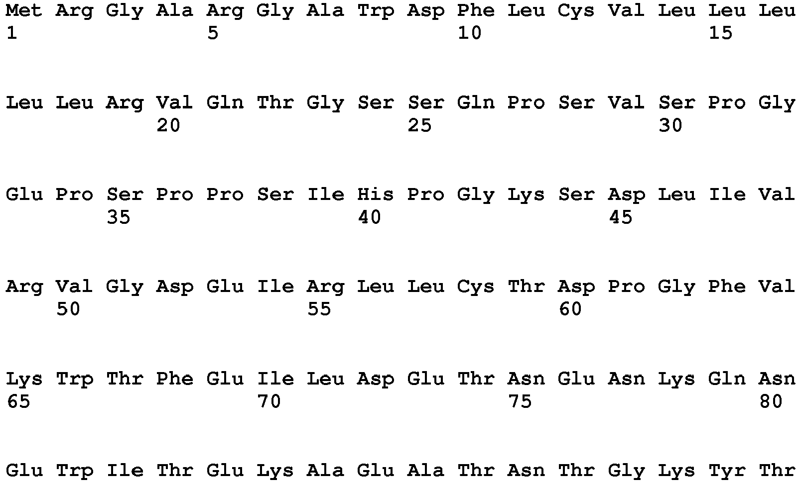


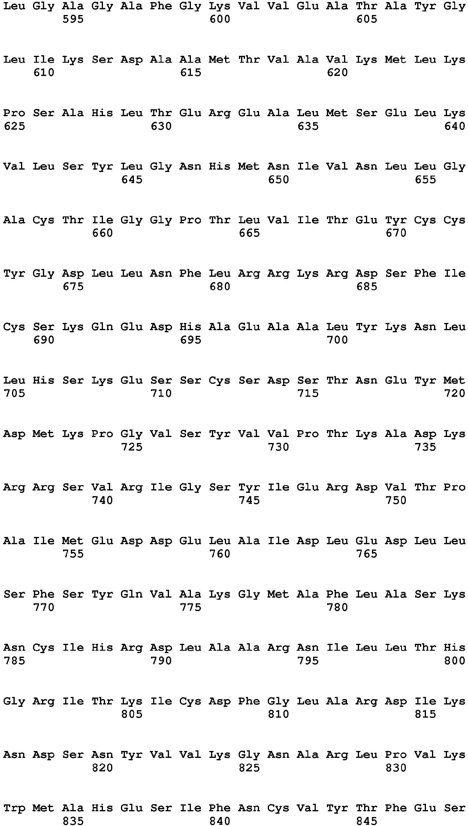 <400> 6
Claims (15)
- Human mast cell line having the following morphological, ultrastructural and phenotypic characteristics: i) presence of metachromatic intra-cytoplasmic granulations, ii) expression of FcεR1 and wild-type KIT (CD117) receptors, and of CD33, CD203c and CD300a markers, and also the following functional characteristics: i) strict dependence with respect to SCF for its survival and its growth and ii) doubling time of at most 72 hours.
- Human mast cell line according to claim 1, further having the following functional features: iii) FcεR1 expression increased by treatment with interleukin 4 (IL-4) and/or with human IgEs, iv) CD117 expression decreased by treatment with interleukin 4 (IL-4), v) immediate increase in membrane expression of CD203c and/or immediate release of histamine, of beta-hexosaminidase and of tryptase, and delayed release of TNF-α, by activation:i) in the presence of specific IgE and of the antigen capable of binding to said specific IgE,ii) in the presence of IgE and of anti-IgE, oriii) in the presence of anti-FcεR1 receptor antibody.
- Human mast cell line according to claim 1 or 2, identified as ROSA KIT WT as registered under deposit number CNCM 1-4551 with the CNCM [French National Collection of Microorganism Cultures] on November 2, 2011.
- Cell line derived from the human mast cell line identified as ROSA KIT WT according to claim 3, said derived line being identified as ROSA KIT D816V and being as registered under deposit number CNCM 1-4552 with the CNCM on November 2, 2011.
- Cell line derived from the human mast cell line identified as ROSA KIT WT according to claim 3, said derived line being identified as ROSA KIT Delta 417-419 insY and being as registered under deposit number CNCM 1-4553 with the CNCM on November 2, 2011.
- Process for preparing a population of human mast cells capable of proliferating for a period greater than 6 months, of which at least 99% of the cells have the characteristics of the cells of a human mast cell line according to claim 1 or 2, which comprises culturing haematopoietic precursors originating from the umbilical cord blood of a healthy human subject in a medium comprising at least 50 ng/ml of human Stem Cell Factor (SCF) and obtaining a population of human mast cells capable of proliferating for a period greater than 6 months, of which at least 99% of the cells have the characteristics of the cells of a human mast cell line according to claim 1 or 2.
- Process for preparing a mast cell line exhibiting a KIT receptor mutation, preferably an activating mutation associated with a pathological condition chosen from mastocytosis, acute leukaemia, a lymphoma and a solid tumour, comprising the transformation of the human mast cell line according to one of claims 1 to 3, by introducing, into the cells of said line, a nucleic acid encoding a mutated KIT receptor, and obtaining a mast cell line exhibiting said KIT mutation.
- Mast cell line which can be obtained at the end of the process according to claim 7, characterized in that it exhibits a KIT receptor mutation responsible i) for the acquisition of independence with respect to SCF for its survival and its growth; ii) for the acquisition of tumorigenicity in vivo in mammals, iii) for an increase in the capacity to release a mediator of inflammation or of an allergy in the presence of specific IgE and of the antigen capable of binding to said specific IgE, in the presence of IgE and of anti-IgE, or in the presence of anti-FcεR1 receptor antibody, or by activation of a receptor of TLR type, of a complement fraction receptor, or of a cytokine or chemokine receptor; iv) for a change, typically an activation, of the intra-mast cell signaling; and/or v) for an interaction of said mutated KIT receptor or of at least one of it signaling pathways with at least one other mast cell receptor or at least one of its signaling pathways.
- Kit for screening for an agent of interest comprising i) at least one product chosen from the mast cell line according to one of claims 1-5 or 8; ii) a supplementary product chosen from one or more culture media, one or more maintenance media, one or more growth factors enabling or promoting the culturing of the mast cells; and any combination of said products.
- Non-human animal model comprising at least one cell originating from the mast cell line according to one of claims 1-5 or 8.
- Use of the mast cell line according to one of claims 1-5 or 8, for screening for an agent of interest which is of use in the prevention, diagnosis, treatment and/or monitoring of a pathological condition, preferably chosen from an allergy, an inflammatory disease, an autoimmune disease, an infection, non-allergic asthma, urticaria, and a tumour, typically mastocytosis, acute leukaemia, lymphoma and a solid tumour.
- Method for evaluating the capacity of at least one candidate molecule to modulate the tumorigenicity, the clonogenicity, the survival, the apoptosis, the proliferation, the differentiation, the activation, the function, the phenotype, the morphological appearance and/or the ultrastructural appearance of human mast cells, comprising:a) bringing the mast cell line according to one of claims 1-5 or 8 into contact with at least one candidate molecule; andb) determining the tumorigenicity, the clonogenicity, the survival, the apoptosis, the proliferation, the differentiation, the activation, the function, the phenotype, the morphological appearance and/or the ultrastructural appearance of the mast cells belonging to said line exposed to said at least one candidate molecule, said determining making it possible to evaluate the corresponding capacity of said at least one candidate molecule to modulate the tumorigenicity, the clonogenicity, the survival, the apoptosis, the proliferation, the differentiation, the activation, the function, the phenotype, the morphological appearance and/or the ultrastructural appearance of human mast cells.
- Method for determining the capacity of at least one candidate molecule to interfere with the binding of at least one ligand or of at least one substrate to at least one human mast cell receptor or to at least one other mast cell substrate; to modulate the transduction of at least one signal in a human mast cell; and/or to modulate the synthesis and/or the release of at least one mediator by a human mast cell, comprising:a) bringing the mast cell line according to one of claims 1-5 or 8 into contact with at least one candidate molecule; andb) demonstrating or measuring i) binding of at least one ligand or of at least one substrate to at least one human mast cell receptor or to at least one other mast cell substrate, ii) the synthesis or the release of at least one mediator by a human mast cell, and/or iii) the transduction of at least one signal in the mast cells of said line so as to determine respectively i) the capacity of said at least one candidate molecule to interfere with the binding of at least one ligand or of at least one substrate to at least one receptor present at the surface of a human mast cell or to at least one other mast cell substrate, ii) to modulate the synthesis or the release of at least one mediator by a human mast cell and/or iii) to modulate the transduction of at least one signal in a human mast cell.
- Method for determining the capacity of at least one candidate molecule to increase or to decrease the survival and/or the proliferation, and/or to inhibit or induce the apoptosis of human mast cells, comprising:a) bringing the mast cell line according to one of claims 1-5 or 8 into contact with at least one candidate molecule; andb) determining the level of survival and/or of proliferation and/or of apoptosis of the mast cells of said line, an increase or a decrease in the survival and/or the proliferation and/or the inhibition or the induction of the apoptosis of said cells determining the capacity of the at least one candidate molecule respectively to increase or decrease the survival and/or the proliferation and/or to inhibit or to induce the apoptosis of human mast cells.
- Method for determining the capacity of at least one candidate molecule to modulate the infectiousness of at least one infectious agent with respect to human mast cells, said method comprising:a) bringing the mast cell line according to one of claims 1-5 or 8 into contact with at least one candidate molecule and at least one infectious agent; andb) determining the capacity of said at least one infectious agent to infect the mast cell, said determining making it possible to determine the capacity of the at least one candidate molecule to modulate the infectiousness of said at least one infectious agent with respect to human mast cells.
Applications Claiming Priority (2)
| Application Number | Priority Date | Filing Date | Title |
|---|---|---|---|
| FR1159902A FR2981945B1 (en) | 2011-11-02 | 2011-11-02 | HUMAN MASTOCYTE LINES, PREPARATION AND USES |
| PCT/EP2012/071731 WO2013064639A1 (en) | 2011-11-02 | 2012-11-02 | Human mast cell lines, preparation and uses |
Publications (2)
| Publication Number | Publication Date |
|---|---|
| EP2773749A1 EP2773749A1 (en) | 2014-09-10 |
| EP2773749B1 true EP2773749B1 (en) | 2017-01-04 |
Family
ID=47297117
Family Applications (1)
| Application Number | Title | Priority Date | Filing Date |
|---|---|---|---|
| EP12797734.6A Active EP2773749B1 (en) | 2011-11-02 | 2012-11-02 | Human mast cell lines, preparation and uses |
Country Status (4)
| Country | Link |
|---|---|
| US (1) | US10383955B2 (en) |
| EP (1) | EP2773749B1 (en) |
| FR (1) | FR2981945B1 (en) |
| WO (1) | WO2013064639A1 (en) |
Cited By (1)
| Publication number | Priority date | Publication date | Assignee | Title |
|---|---|---|---|---|
| WO2023106947A1 (en) | 2021-12-08 | 2023-06-15 | Gdański Uniwersytet Medyczny | Use of 2-methoxyestradiol in the treatment of mastocytosis |
Families Citing this family (2)
| Publication number | Priority date | Publication date | Assignee | Title |
|---|---|---|---|---|
| CA3008848A1 (en) | 2015-12-18 | 2017-06-22 | Tega Therapeutics, Inc. | Cellular glycosaminoglycan compositions and methods of making and using |
| CA3152781A1 (en) * | 2019-08-27 | 2021-03-04 | Tega Therapeutics, Inc. | Heparin and heparan sulfate from modified mst cells and methods of making and using |
Family Cites Families (2)
| Publication number | Priority date | Publication date | Assignee | Title |
|---|---|---|---|---|
| WO2003065986A2 (en) * | 2002-02-04 | 2003-08-14 | The Government Of The United States Of America, Asrepresented By The Secretary, Dept. Of Health And Human Services | FcϵRI-BEARING HUMAN MAST CELL LINES |
| WO2010105215A2 (en) * | 2009-03-12 | 2010-09-16 | University Of South Florida | Human mast cell line and uses thereof |
-
2011
- 2011-11-02 FR FR1159902A patent/FR2981945B1/en active Active
-
2012
- 2012-11-02 WO PCT/EP2012/071731 patent/WO2013064639A1/en active Application Filing
- 2012-11-02 US US14/355,938 patent/US10383955B2/en active Active
- 2012-11-02 EP EP12797734.6A patent/EP2773749B1/en active Active
Non-Patent Citations (1)
| Title |
|---|
| None * |
Cited By (1)
| Publication number | Priority date | Publication date | Assignee | Title |
|---|---|---|---|---|
| WO2023106947A1 (en) | 2021-12-08 | 2023-06-15 | Gdański Uniwersytet Medyczny | Use of 2-methoxyestradiol in the treatment of mastocytosis |
Also Published As
| Publication number | Publication date |
|---|---|
| FR2981945B1 (en) | 2016-09-23 |
| US10383955B2 (en) | 2019-08-20 |
| US20140298497A1 (en) | 2014-10-02 |
| FR2981945A1 (en) | 2013-05-03 |
| EP2773749A1 (en) | 2014-09-10 |
| WO2013064639A1 (en) | 2013-05-10 |
Similar Documents
| Publication | Publication Date | Title |
|---|---|---|
| JP6259012B2 (en) | How to treat alopecia | |
| Klebanoff et al. | Retinoic acid controls the homeostasis of pre-cDC–derived splenic and intestinal dendritic cells | |
| Daniel et al. | Immune modulatory treatment of trinitrobenzene sulfonic acid colitis with calcitriol is associated with a change of a T helper (Th) 1/Th17 to a Th2 and regulatory T cell profile | |
| Ohta et al. | Macrophage migration inhibitory factor (MIF) promotes cell survival and proliferation of neural stem/progenitor cells | |
| Dahan et al. | Notch-1 signaling regulates intestinal epithelial barrier function, through interaction with CD4+ T cells, in mice and humans | |
| Hollmann et al. | Inhibition of acid sphingomyelinase allows for selective targeting of CD4+ conventional versus Foxp3+ regulatory T cells | |
| EP2279245B1 (en) | Drug discovery methods involving a preclinical, in vitro isolated gastrointestinal epithelial stem cell-like progenitor cell system | |
| JP2023071728A (en) | Differentiation and use of human microglia-like cells from pluripotent stem cells and hematopoietic progenitors | |
| Sarmento et al. | A novel role for Kruppel-like factor 14 (KLF14) in T-regulatory cell differentiation | |
| Peligero-Cruz et al. | IL18 signaling promotes homing of mature Tregs into the thymus | |
| Borek et al. | BMP7 aberrantly induced in the psoriatic epidermis instructs inflammation-associated Langerhans cells | |
| EP2773749B1 (en) | Human mast cell lines, preparation and uses | |
| Tulic et al. | Thymic indoleamine 2, 3-dioxygenase-positive eosinophils in young children: potential role in maturation of the naive immune system | |
| Muro et al. | Rasal3-mediated T cell survival is essential for inflammatory responses | |
| van Wigcheren et al. | Myeloid-derived suppressor cells and tolerogenic dendritic cells are distinctively induced by PI3K and Wnt signaling pathways | |
| Rossi et al. | Stimulation of treg cells to inhibit osteoclastogenesis in gorham-stout disease | |
| Maneechotesuwan et al. | Der p 1 suppresses indoleamine 2, 3-dioxygenase in dendritic cells from house dust mite–sensitive patients with asthma | |
| Cui et al. | Non-heme iron overload impairs monocyte to macrophage differentiation via mitochondrial oxidative stress | |
| Hwang et al. | Aryl hydrocarbon receptor ligands indoxyl 3-sulfate and indole-3-carbinol inhibit FMS-like tyrosine kinase 3 ligand-induced bone marrow-derived plasmacytoid dendritic cell differentiation | |
| Zak et al. | Myeloid reprogramming by JAK inhibition enhances checkpoint blockade therapy | |
| Lee et al. | Ssu72 is a T-cell receptor-responsive modifier that is indispensable for regulatory T cells | |
| Venkatasubramanian et al. | TOLLIP optimizes dendritic cell maturation to lipopolysaccharide and Mycobacterium tuberculosis | |
| Liu et al. | Metformin rejuvenates Nap1l2‐impaired immunomodulation of bone marrow mesenchymal stem cells via metabolic reprogramming | |
| JP2017526702A (en) | A method of treating cancer comprising administering a PPAR-γ agonist | |
| Jiao et al. | Notch2-dependent GATA3+ Treg cells alleviate allergic rhinitis by suppressing the Th2 cell response |
Legal Events
| Date | Code | Title | Description |
|---|---|---|---|
| PUAI | Public reference made under article 153(3) epc to a published international application that has entered the european phase |
Free format text: ORIGINAL CODE: 0009012 |
|
| 17P | Request for examination filed |
Effective date: 20140514 |
|
| AK | Designated contracting states |
Kind code of ref document: A1 Designated state(s): AL AT BE BG CH CY CZ DE DK EE ES FI FR GB GR HR HU IE IS IT LI LT LU LV MC MK MT NL NO PL PT RO RS SE SI SK SM TR |
|
| DAX | Request for extension of the european patent (deleted) | ||
| 17Q | First examination report despatched |
Effective date: 20150612 |
|
| GRAP | Despatch of communication of intention to grant a patent |
Free format text: ORIGINAL CODE: EPIDOSNIGR1 |
|
| INTG | Intention to grant announced |
Effective date: 20160602 |
|
| RIN1 | Information on inventor provided before grant (corrected) |
Inventor name: SALEH, ROSINE Inventor name: VALENT, PETER Inventor name: WEDEH, GHAITH Inventor name: AROCK, MICHEL Inventor name: AUCLAIR, CHRISTIAN |
|
| GRAJ | Information related to disapproval of communication of intention to grant by the applicant or resumption of examination proceedings by the epo deleted |
Free format text: ORIGINAL CODE: EPIDOSDIGR1 |
|
| RIN1 | Information on inventor provided before grant (corrected) |
Inventor name: SALEH, ROSINE Inventor name: AUCLAIR, CHRISTIAN Inventor name: WEDEH, GHAITH Inventor name: AROCK, MICHEL Inventor name: VALENT, PETER |
|
| INTC | Intention to grant announced (deleted) | ||
| GRAR | Information related to intention to grant a patent recorded |
Free format text: ORIGINAL CODE: EPIDOSNIGR71 |
|
| GRAS | Grant fee paid |
Free format text: ORIGINAL CODE: EPIDOSNIGR3 |
|
| INTG | Intention to grant announced |
Effective date: 20161027 |
|
| GRAA | (expected) grant |
Free format text: ORIGINAL CODE: 0009210 |
|
| AK | Designated contracting states |
Kind code of ref document: B1 Designated state(s): AL AT BE BG CH CY CZ DE DK EE ES FI FR GB GR HR HU IE IS IT LI LT LU LV MC MK MT NL NO PL PT RO RS SE SI SK SM TR |
|
| REG | Reference to a national code |
Ref country code: GB Ref legal event code: FG4D Free format text: NOT ENGLISH |
|
| REG | Reference to a national code |
Ref country code: CH Ref legal event code: EP |
|
| REG | Reference to a national code |
Ref country code: AT Ref legal event code: REF Ref document number: 859279 Country of ref document: AT Kind code of ref document: T Effective date: 20170115 |
|
| REG | Reference to a national code |
Ref country code: IE Ref legal event code: FG4D Free format text: LANGUAGE OF EP DOCUMENT: FRENCH |
|
| REG | Reference to a national code |
Ref country code: DE Ref legal event code: R096 Ref document number: 602012027473 Country of ref document: DE |
|
| REG | Reference to a national code |
Ref country code: LT Ref legal event code: MG4D Ref country code: NL Ref legal event code: MP Effective date: 20170104 |
|
| PG25 | Lapsed in a contracting state [announced via postgrant information from national office to epo] |
Ref country code: NL Free format text: LAPSE BECAUSE OF FAILURE TO SUBMIT A TRANSLATION OF THE DESCRIPTION OR TO PAY THE FEE WITHIN THE PRESCRIBED TIME-LIMIT Effective date: 20170104 |
|
| PG25 | Lapsed in a contracting state [announced via postgrant information from national office to epo] |
Ref country code: FI Free format text: LAPSE BECAUSE OF FAILURE TO SUBMIT A TRANSLATION OF THE DESCRIPTION OR TO PAY THE FEE WITHIN THE PRESCRIBED TIME-LIMIT Effective date: 20170104 Ref country code: IS Free format text: LAPSE BECAUSE OF FAILURE TO SUBMIT A TRANSLATION OF THE DESCRIPTION OR TO PAY THE FEE WITHIN THE PRESCRIBED TIME-LIMIT Effective date: 20170504 Ref country code: GR Free format text: LAPSE BECAUSE OF FAILURE TO SUBMIT A TRANSLATION OF THE DESCRIPTION OR TO PAY THE FEE WITHIN THE PRESCRIBED TIME-LIMIT Effective date: 20170405 Ref country code: LT Free format text: LAPSE BECAUSE OF FAILURE TO SUBMIT A TRANSLATION OF THE DESCRIPTION OR TO PAY THE FEE WITHIN THE PRESCRIBED TIME-LIMIT Effective date: 20170104 Ref country code: NO Free format text: LAPSE BECAUSE OF FAILURE TO SUBMIT A TRANSLATION OF THE DESCRIPTION OR TO PAY THE FEE WITHIN THE PRESCRIBED TIME-LIMIT Effective date: 20170404 Ref country code: HR Free format text: LAPSE BECAUSE OF FAILURE TO SUBMIT A TRANSLATION OF THE DESCRIPTION OR TO PAY THE FEE WITHIN THE PRESCRIBED TIME-LIMIT Effective date: 20170104 |
|
| PG25 | Lapsed in a contracting state [announced via postgrant information from national office to epo] |
Ref country code: BG Free format text: LAPSE BECAUSE OF FAILURE TO SUBMIT A TRANSLATION OF THE DESCRIPTION OR TO PAY THE FEE WITHIN THE PRESCRIBED TIME-LIMIT Effective date: 20170404 Ref country code: LV Free format text: LAPSE BECAUSE OF FAILURE TO SUBMIT A TRANSLATION OF THE DESCRIPTION OR TO PAY THE FEE WITHIN THE PRESCRIBED TIME-LIMIT Effective date: 20170104 Ref country code: RS Free format text: LAPSE BECAUSE OF FAILURE TO SUBMIT A TRANSLATION OF THE DESCRIPTION OR TO PAY THE FEE WITHIN THE PRESCRIBED TIME-LIMIT Effective date: 20170104 Ref country code: SE Free format text: LAPSE BECAUSE OF FAILURE TO SUBMIT A TRANSLATION OF THE DESCRIPTION OR TO PAY THE FEE WITHIN THE PRESCRIBED TIME-LIMIT Effective date: 20170104 Ref country code: PT Free format text: LAPSE BECAUSE OF FAILURE TO SUBMIT A TRANSLATION OF THE DESCRIPTION OR TO PAY THE FEE WITHIN THE PRESCRIBED TIME-LIMIT Effective date: 20170504 Ref country code: PL Free format text: LAPSE BECAUSE OF FAILURE TO SUBMIT A TRANSLATION OF THE DESCRIPTION OR TO PAY THE FEE WITHIN THE PRESCRIBED TIME-LIMIT Effective date: 20170104 Ref country code: ES Free format text: LAPSE BECAUSE OF FAILURE TO SUBMIT A TRANSLATION OF THE DESCRIPTION OR TO PAY THE FEE WITHIN THE PRESCRIBED TIME-LIMIT Effective date: 20170104 |
|
| REG | Reference to a national code |
Ref country code: DE Ref legal event code: R097 Ref document number: 602012027473 Country of ref document: DE |
|
| PG25 | Lapsed in a contracting state [announced via postgrant information from national office to epo] |
Ref country code: IT Free format text: LAPSE BECAUSE OF FAILURE TO SUBMIT A TRANSLATION OF THE DESCRIPTION OR TO PAY THE FEE WITHIN THE PRESCRIBED TIME-LIMIT Effective date: 20170104 Ref country code: EE Free format text: LAPSE BECAUSE OF FAILURE TO SUBMIT A TRANSLATION OF THE DESCRIPTION OR TO PAY THE FEE WITHIN THE PRESCRIBED TIME-LIMIT Effective date: 20170104 Ref country code: SK Free format text: LAPSE BECAUSE OF FAILURE TO SUBMIT A TRANSLATION OF THE DESCRIPTION OR TO PAY THE FEE WITHIN THE PRESCRIBED TIME-LIMIT Effective date: 20170104 Ref country code: CZ Free format text: LAPSE BECAUSE OF FAILURE TO SUBMIT A TRANSLATION OF THE DESCRIPTION OR TO PAY THE FEE WITHIN THE PRESCRIBED TIME-LIMIT Effective date: 20170104 Ref country code: RO Free format text: LAPSE BECAUSE OF FAILURE TO SUBMIT A TRANSLATION OF THE DESCRIPTION OR TO PAY THE FEE WITHIN THE PRESCRIBED TIME-LIMIT Effective date: 20170104 |
|
| PLBE | No opposition filed within time limit |
Free format text: ORIGINAL CODE: 0009261 |
|
| STAA | Information on the status of an ep patent application or granted ep patent |
Free format text: STATUS: NO OPPOSITION FILED WITHIN TIME LIMIT |
|
| PG25 | Lapsed in a contracting state [announced via postgrant information from national office to epo] |
Ref country code: DK Free format text: LAPSE BECAUSE OF FAILURE TO SUBMIT A TRANSLATION OF THE DESCRIPTION OR TO PAY THE FEE WITHIN THE PRESCRIBED TIME-LIMIT Effective date: 20170104 Ref country code: SM Free format text: LAPSE BECAUSE OF FAILURE TO SUBMIT A TRANSLATION OF THE DESCRIPTION OR TO PAY THE FEE WITHIN THE PRESCRIBED TIME-LIMIT Effective date: 20170104 |
|
| REG | Reference to a national code |
Ref country code: FR Ref legal event code: PLFP Year of fee payment: 6 |
|
| 26N | No opposition filed |
Effective date: 20171005 |
|
| PG25 | Lapsed in a contracting state [announced via postgrant information from national office to epo] |
Ref country code: SI Free format text: LAPSE BECAUSE OF FAILURE TO SUBMIT A TRANSLATION OF THE DESCRIPTION OR TO PAY THE FEE WITHIN THE PRESCRIBED TIME-LIMIT Effective date: 20170104 |
|
| PG25 | Lapsed in a contracting state [announced via postgrant information from national office to epo] |
Ref country code: MC Free format text: LAPSE BECAUSE OF FAILURE TO SUBMIT A TRANSLATION OF THE DESCRIPTION OR TO PAY THE FEE WITHIN THE PRESCRIBED TIME-LIMIT Effective date: 20170104 |
|
| PG25 | Lapsed in a contracting state [announced via postgrant information from national office to epo] |
Ref country code: LU Free format text: LAPSE BECAUSE OF NON-PAYMENT OF DUE FEES Effective date: 20171102 |
|
| REG | Reference to a national code |
Ref country code: IE Ref legal event code: MM4A |
|
| PG25 | Lapsed in a contracting state [announced via postgrant information from national office to epo] |
Ref country code: MT Free format text: LAPSE BECAUSE OF FAILURE TO SUBMIT A TRANSLATION OF THE DESCRIPTION OR TO PAY THE FEE WITHIN THE PRESCRIBED TIME-LIMIT Effective date: 20170104 |
|
| PG25 | Lapsed in a contracting state [announced via postgrant information from national office to epo] |
Ref country code: IE Free format text: LAPSE BECAUSE OF NON-PAYMENT OF DUE FEES Effective date: 20171102 |
|
| PG25 | Lapsed in a contracting state [announced via postgrant information from national office to epo] |
Ref country code: HU Free format text: LAPSE BECAUSE OF FAILURE TO SUBMIT A TRANSLATION OF THE DESCRIPTION OR TO PAY THE FEE WITHIN THE PRESCRIBED TIME-LIMIT; INVALID AB INITIO Effective date: 20121102 |
|
| REG | Reference to a national code |
Ref country code: AT Ref legal event code: UEP Ref document number: 859279 Country of ref document: AT Kind code of ref document: T Effective date: 20170104 |
|
| PG25 | Lapsed in a contracting state [announced via postgrant information from national office to epo] |
Ref country code: CY Free format text: LAPSE BECAUSE OF NON-PAYMENT OF DUE FEES Effective date: 20170104 |
|
| PG25 | Lapsed in a contracting state [announced via postgrant information from national office to epo] |
Ref country code: MK Free format text: LAPSE BECAUSE OF FAILURE TO SUBMIT A TRANSLATION OF THE DESCRIPTION OR TO PAY THE FEE WITHIN THE PRESCRIBED TIME-LIMIT Effective date: 20170104 |
|
| PG25 | Lapsed in a contracting state [announced via postgrant information from national office to epo] |
Ref country code: TR Free format text: LAPSE BECAUSE OF FAILURE TO SUBMIT A TRANSLATION OF THE DESCRIPTION OR TO PAY THE FEE WITHIN THE PRESCRIBED TIME-LIMIT Effective date: 20170104 |
|
| PG25 | Lapsed in a contracting state [announced via postgrant information from national office to epo] |
Ref country code: AL Free format text: LAPSE BECAUSE OF FAILURE TO SUBMIT A TRANSLATION OF THE DESCRIPTION OR TO PAY THE FEE WITHIN THE PRESCRIBED TIME-LIMIT Effective date: 20170104 |
|
| PGFP | Annual fee paid to national office [announced via postgrant information from national office to epo] |
Ref country code: GB Payment date: 20231129 Year of fee payment: 12 |
|
| PGFP | Annual fee paid to national office [announced via postgrant information from national office to epo] |
Ref country code: FR Payment date: 20231127 Year of fee payment: 12 Ref country code: DE Payment date: 20231130 Year of fee payment: 12 Ref country code: CH Payment date: 20231202 Year of fee payment: 12 Ref country code: AT Payment date: 20231128 Year of fee payment: 12 |
|
| PGFP | Annual fee paid to national office [announced via postgrant information from national office to epo] |
Ref country code: BE Payment date: 20231129 Year of fee payment: 12 |