EP2750690B1 - Soluble mediator - Google Patents
Soluble mediator Download PDFInfo
- Publication number
- EP2750690B1 EP2750690B1 EP12850386.9A EP12850386A EP2750690B1 EP 2750690 B1 EP2750690 B1 EP 2750690B1 EP 12850386 A EP12850386 A EP 12850386A EP 2750690 B1 EP2750690 B1 EP 2750690B1
- Authority
- EP
- European Patent Office
- Prior art keywords
- cells
- cell
- soluble
- glycoprotein
- seq
- Prior art date
- Legal status (The legal status is an assumption and is not a legal conclusion. Google has not performed a legal analysis and makes no representation as to the accuracy of the status listed.)
- Not-in-force
Links
- 108010065524 CD52 Antigen Proteins 0.000 claims description 281
- 238000000034 method Methods 0.000 claims description 99
- 102000040430 polynucleotide Human genes 0.000 claims description 85
- 108091033319 polynucleotide Proteins 0.000 claims description 85
- 239000002157 polynucleotide Substances 0.000 claims description 85
- 108090000623 proteins and genes Proteins 0.000 claims description 80
- 102000004169 proteins and genes Human genes 0.000 claims description 73
- 108020001507 fusion proteins Proteins 0.000 claims description 63
- 102000037865 fusion proteins Human genes 0.000 claims description 63
- 239000013598 vector Substances 0.000 claims description 47
- 239000000427 antigen Substances 0.000 claims description 43
- 102000036639 antigens Human genes 0.000 claims description 43
- 108091007433 antigens Proteins 0.000 claims description 43
- 208000037265 diseases, disorders, signs and symptoms Diseases 0.000 claims description 43
- 108090000765 processed proteins & peptides Proteins 0.000 claims description 42
- 201000010099 disease Diseases 0.000 claims description 40
- 239000000523 sample Substances 0.000 claims description 36
- 206010067584 Type 1 diabetes mellitus Diseases 0.000 claims description 32
- 206010061218 Inflammation Diseases 0.000 claims description 31
- 230000001965 increasing effect Effects 0.000 claims description 31
- 230000004054 inflammatory process Effects 0.000 claims description 31
- 150000001720 carbohydrates Chemical class 0.000 claims description 28
- 208000023275 Autoimmune disease Diseases 0.000 claims description 24
- 238000011282 treatment Methods 0.000 claims description 21
- 239000003814 drug Substances 0.000 claims description 14
- 238000007877 drug screening Methods 0.000 claims description 9
- 229940079593 drug Drugs 0.000 claims description 8
- 238000012216 screening Methods 0.000 claims description 7
- 229940127003 anti-diabetic drug Drugs 0.000 claims description 4
- 239000003472 antidiabetic agent Substances 0.000 claims description 4
- 238000002560 therapeutic procedure Methods 0.000 claims description 4
- 238000000338 in vitro Methods 0.000 claims description 3
- 241000486679 Antitype Species 0.000 claims description 2
- 239000013074 reference sample Substances 0.000 claims description 2
- 125000003275 alpha amino acid group Chemical group 0.000 claims 1
- 210000004027 cell Anatomy 0.000 description 445
- 102000013135 CD52 Antigen Human genes 0.000 description 268
- 210000001744 T-lymphocyte Anatomy 0.000 description 146
- 230000014509 gene expression Effects 0.000 description 103
- 229960000814 tetanus toxoid Drugs 0.000 description 101
- 230000006870 function Effects 0.000 description 87
- 108020004414 DNA Proteins 0.000 description 73
- 102000003886 Glycoproteins Human genes 0.000 description 69
- 108090000288 Glycoproteins Proteins 0.000 description 69
- 210000003819 peripheral blood mononuclear cell Anatomy 0.000 description 67
- 235000018102 proteins Nutrition 0.000 description 66
- 230000001629 suppression Effects 0.000 description 52
- 230000002441 reversible effect Effects 0.000 description 46
- 210000003162 effector t lymphocyte Anatomy 0.000 description 41
- 230000004044 response Effects 0.000 description 40
- 241000282414 Homo sapiens Species 0.000 description 39
- 241000699670 Mus sp. Species 0.000 description 38
- 239000000203 mixture Substances 0.000 description 35
- 230000000694 effects Effects 0.000 description 34
- 150000001413 amino acids Chemical group 0.000 description 32
- 239000003795 chemical substances by application Substances 0.000 description 32
- 101710143293 Sialic acid-binding Ig-like lectin 10 Proteins 0.000 description 29
- 102100027164 Sialic acid-binding Ig-like lectin 10 Human genes 0.000 description 29
- 235000014633 carbohydrates Nutrition 0.000 description 29
- 230000001404 mediated effect Effects 0.000 description 29
- 239000002609 medium Substances 0.000 description 29
- 210000000582 semen Anatomy 0.000 description 29
- 206010012601 diabetes mellitus Diseases 0.000 description 27
- 238000002474 experimental method Methods 0.000 description 27
- 210000003289 regulatory T cell Anatomy 0.000 description 26
- 108010002350 Interleukin-2 Proteins 0.000 description 24
- 102000000588 Interleukin-2 Human genes 0.000 description 24
- 230000028993 immune response Effects 0.000 description 24
- 210000004369 blood Anatomy 0.000 description 23
- 239000008280 blood Substances 0.000 description 23
- 125000000837 carbohydrate group Chemical group 0.000 description 23
- 239000008194 pharmaceutical composition Substances 0.000 description 23
- WQZGKKKJIJFFOK-GASJEMHNSA-N Glucose Natural products OC[C@H]1OC(O)[C@H](O)[C@@H](O)[C@@H]1O WQZGKKKJIJFFOK-GASJEMHNSA-N 0.000 description 22
- WQZGKKKJIJFFOK-VFUOTHLCSA-N beta-D-glucose Chemical compound OC[C@H]1O[C@@H](O)[C@H](O)[C@@H](O)[C@@H]1O WQZGKKKJIJFFOK-VFUOTHLCSA-N 0.000 description 22
- 206010040047 Sepsis Diseases 0.000 description 21
- 239000008103 glucose Substances 0.000 description 21
- 239000012528 membrane Substances 0.000 description 21
- 210000004988 splenocyte Anatomy 0.000 description 21
- 210000004379 membrane Anatomy 0.000 description 20
- 239000002953 phosphate buffered saline Substances 0.000 description 20
- 101001043809 Homo sapiens Interleukin-7 receptor subunit alpha Proteins 0.000 description 19
- 102100021593 Interleukin-7 receptor subunit alpha Human genes 0.000 description 19
- LOKCTEFSRHRXRJ-UHFFFAOYSA-I dipotassium trisodium dihydrogen phosphate hydrogen phosphate dichloride Chemical compound P(=O)(O)(O)[O-].[K+].P(=O)(O)([O-])[O-].[Na+].[Na+].[Cl-].[K+].[Cl-].[Na+] LOKCTEFSRHRXRJ-UHFFFAOYSA-I 0.000 description 19
- 230000006052 T cell proliferation Effects 0.000 description 18
- 210000004698 lymphocyte Anatomy 0.000 description 18
- VDABVNMGKGUPEY-UHFFFAOYSA-N 6-carboxyfluorescein succinimidyl ester Chemical compound C=1C(O)=CC=C2C=1OC1=CC(O)=CC=C1C2(C1=C2)OC(=O)C1=CC=C2C(=O)ON1C(=O)CCC1=O VDABVNMGKGUPEY-UHFFFAOYSA-N 0.000 description 17
- 108010021064 CTLA-4 Antigen Proteins 0.000 description 17
- 229940045513 CTLA4 antagonist Drugs 0.000 description 17
- 102100039498 Cytotoxic T-lymphocyte protein 4 Human genes 0.000 description 17
- 101000801234 Homo sapiens Tumor necrosis factor receptor superfamily member 18 Proteins 0.000 description 17
- IQFYYKKMVGJFEH-XLPZGREQSA-N Thymidine Chemical compound O=C1NC(=O)C(C)=CN1[C@@H]1O[C@H](CO)[C@@H](O)C1 IQFYYKKMVGJFEH-XLPZGREQSA-N 0.000 description 17
- 102100033728 Tumor necrosis factor receptor superfamily member 18 Human genes 0.000 description 17
- 210000004443 dendritic cell Anatomy 0.000 description 17
- 102100037850 Interferon gamma Human genes 0.000 description 16
- 108010074328 Interferon-gamma Proteins 0.000 description 16
- 241000699666 Mus <mouse, genus> Species 0.000 description 16
- 238000012360 testing method Methods 0.000 description 16
- 101000884271 Homo sapiens Signal transducer CD24 Proteins 0.000 description 15
- 210000001616 monocyte Anatomy 0.000 description 15
- 230000035755 proliferation Effects 0.000 description 15
- 102000004127 Cytokines Human genes 0.000 description 14
- 108090000695 Cytokines Proteins 0.000 description 14
- 102100038081 Signal transducer CD24 Human genes 0.000 description 14
- 150000001875 compounds Chemical class 0.000 description 14
- 239000002158 endotoxin Substances 0.000 description 14
- 238000004519 manufacturing process Methods 0.000 description 14
- 230000002829 reductive effect Effects 0.000 description 14
- 230000028327 secretion Effects 0.000 description 13
- 239000000243 solution Substances 0.000 description 13
- 238000002965 ELISA Methods 0.000 description 12
- 229940024606 amino acid Drugs 0.000 description 12
- 235000001014 amino acid Nutrition 0.000 description 12
- NOESYZHRGYRDHS-UHFFFAOYSA-N insulin Chemical compound N1C(=O)C(NC(=O)C(CCC(N)=O)NC(=O)C(CCC(O)=O)NC(=O)C(C(C)C)NC(=O)C(NC(=O)CN)C(C)CC)CSSCC(C(NC(CO)C(=O)NC(CC(C)C)C(=O)NC(CC=2C=CC(O)=CC=2)C(=O)NC(CCC(N)=O)C(=O)NC(CC(C)C)C(=O)NC(CCC(O)=O)C(=O)NC(CC(N)=O)C(=O)NC(CC=2C=CC(O)=CC=2)C(=O)NC(CSSCC(NC(=O)C(C(C)C)NC(=O)C(CC(C)C)NC(=O)C(CC=2C=CC(O)=CC=2)NC(=O)C(CC(C)C)NC(=O)C(C)NC(=O)C(CCC(O)=O)NC(=O)C(C(C)C)NC(=O)C(CC(C)C)NC(=O)C(CC=2NC=NC=2)NC(=O)C(CO)NC(=O)CNC2=O)C(=O)NCC(=O)NC(CCC(O)=O)C(=O)NC(CCCNC(N)=N)C(=O)NCC(=O)NC(CC=3C=CC=CC=3)C(=O)NC(CC=3C=CC=CC=3)C(=O)NC(CC=3C=CC(O)=CC=3)C(=O)NC(C(C)O)C(=O)N3C(CCC3)C(=O)NC(CCCCN)C(=O)NC(C)C(O)=O)C(=O)NC(CC(N)=O)C(O)=O)=O)NC(=O)C(C(C)CC)NC(=O)C(CO)NC(=O)C(C(C)O)NC(=O)C1CSSCC2NC(=O)C(CC(C)C)NC(=O)C(NC(=O)C(CCC(N)=O)NC(=O)C(CC(N)=O)NC(=O)C(NC(=O)C(N)CC=1C=CC=CC=1)C(C)C)CC1=CN=CN1 NOESYZHRGYRDHS-UHFFFAOYSA-N 0.000 description 12
- 238000000746 purification Methods 0.000 description 12
- XLYOFNOQVPJJNP-UHFFFAOYSA-N water Substances O XLYOFNOQVPJJNP-UHFFFAOYSA-N 0.000 description 12
- 102000000447 Peptide-N4-(N-acetyl-beta-glucosaminyl) Asparagine Amidase Human genes 0.000 description 11
- 108010055817 Peptide-N4-(N-acetyl-beta-glucosaminyl) Asparagine Amidase Proteins 0.000 description 11
- 239000000463 material Substances 0.000 description 11
- 230000007420 reactivation Effects 0.000 description 11
- 238000012546 transfer Methods 0.000 description 11
- 108010006232 Neuraminidase Proteins 0.000 description 10
- 102000005348 Neuraminidase Human genes 0.000 description 10
- 238000011579 SCID mouse model Methods 0.000 description 10
- 102000007073 Sialic Acid Binding Immunoglobulin-like Lectins Human genes 0.000 description 10
- 108010047827 Sialic Acid Binding Immunoglobulin-like Lectins Proteins 0.000 description 10
- 230000004913 activation Effects 0.000 description 10
- 239000000872 buffer Substances 0.000 description 10
- 108020004999 messenger RNA Proteins 0.000 description 10
- -1 polydimethylsiloxanes Polymers 0.000 description 10
- 239000000843 powder Substances 0.000 description 10
- 102000004196 processed proteins & peptides Human genes 0.000 description 10
- 239000000126 substance Substances 0.000 description 10
- 201000000596 systemic lupus erythematosus Diseases 0.000 description 10
- DGVVWUTYPXICAM-UHFFFAOYSA-N β‐Mercaptoethanol Chemical compound OCCS DGVVWUTYPXICAM-UHFFFAOYSA-N 0.000 description 10
- 108091032973 (ribonucleotides)n+m Proteins 0.000 description 9
- 102000007469 Actins Human genes 0.000 description 9
- 108010085238 Actins Proteins 0.000 description 9
- 241001465754 Metazoa Species 0.000 description 9
- 241000283973 Oryctolagus cuniculus Species 0.000 description 9
- DNIAPMSPPWPWGF-UHFFFAOYSA-N Propylene glycol Chemical compound CC(O)CO DNIAPMSPPWPWGF-UHFFFAOYSA-N 0.000 description 9
- FAPWRFPIFSIZLT-UHFFFAOYSA-M Sodium chloride Chemical compound [Na+].[Cl-] FAPWRFPIFSIZLT-UHFFFAOYSA-M 0.000 description 9
- 238000003556 assay Methods 0.000 description 9
- 238000009472 formulation Methods 0.000 description 9
- 238000011534 incubation Methods 0.000 description 9
- 229920001184 polypeptide Polymers 0.000 description 9
- 206010039073 rheumatoid arthritis Diseases 0.000 description 9
- 125000005629 sialic acid group Chemical group 0.000 description 9
- 235000000346 sugar Nutrition 0.000 description 9
- 150000008163 sugars Chemical class 0.000 description 9
- 230000001225 therapeutic effect Effects 0.000 description 9
- 208000011231 Crohn disease Diseases 0.000 description 8
- LFQSCWFLJHTTHZ-UHFFFAOYSA-N Ethanol Chemical compound CCO LFQSCWFLJHTTHZ-UHFFFAOYSA-N 0.000 description 8
- 101000980814 Homo sapiens CAMPATH-1 antigen Proteins 0.000 description 8
- 239000007760 Iscove's Modified Dulbecco's Medium Substances 0.000 description 8
- 102100031988 Tumor necrosis factor ligand superfamily member 6 Human genes 0.000 description 8
- 230000001154 acute effect Effects 0.000 description 8
- 238000004458 analytical method Methods 0.000 description 8
- 238000004113 cell culture Methods 0.000 description 8
- 239000003937 drug carrier Substances 0.000 description 8
- 239000012636 effector Substances 0.000 description 8
- 238000000684 flow cytometry Methods 0.000 description 8
- 239000012634 fragment Substances 0.000 description 8
- 238000006206 glycosylation reaction Methods 0.000 description 8
- 102000043738 human CD52 Human genes 0.000 description 8
- 230000022532 regulation of transcription, DNA-dependent Effects 0.000 description 8
- 208000030836 Hashimoto thyroiditis Diseases 0.000 description 7
- 102000003814 Interleukin-10 Human genes 0.000 description 7
- 108090000174 Interleukin-10 Proteins 0.000 description 7
- 108010065805 Interleukin-12 Proteins 0.000 description 7
- 102000013462 Interleukin-12 Human genes 0.000 description 7
- 229940124034 Phospholipase C inhibitor Drugs 0.000 description 7
- 235000009582 asparagine Nutrition 0.000 description 7
- 229940112129 campath Drugs 0.000 description 7
- 239000006143 cell culture medium Substances 0.000 description 7
- 230000013595 glycosylation Effects 0.000 description 7
- 230000001771 impaired effect Effects 0.000 description 7
- 210000002540 macrophage Anatomy 0.000 description 7
- 239000000546 pharmaceutical excipient Substances 0.000 description 7
- 239000003371 phospholipase C inhibitor Substances 0.000 description 7
- 102000005962 receptors Human genes 0.000 description 7
- 108020003175 receptors Proteins 0.000 description 7
- 230000000284 resting effect Effects 0.000 description 7
- 238000002415 sodium dodecyl sulfate polyacrylamide gel electrophoresis Methods 0.000 description 7
- 208000024891 symptom Diseases 0.000 description 7
- 239000003826 tablet Substances 0.000 description 7
- 210000001519 tissue Anatomy 0.000 description 7
- CIWBSHSKHKDKBQ-JLAZNSOCSA-N Ascorbic acid Chemical compound OC[C@H](O)[C@H]1OC(=O)C(O)=C1O CIWBSHSKHKDKBQ-JLAZNSOCSA-N 0.000 description 6
- DCXYFEDJOCDNAF-UHFFFAOYSA-N Asparagine Natural products OC(=O)C(N)CC(N)=O DCXYFEDJOCDNAF-UHFFFAOYSA-N 0.000 description 6
- WVDDGKGOMKODPV-UHFFFAOYSA-N Benzyl alcohol Chemical compound OCC1=CC=CC=C1 WVDDGKGOMKODPV-UHFFFAOYSA-N 0.000 description 6
- 241000283707 Capra Species 0.000 description 6
- 101000836954 Homo sapiens Sialic acid-binding Ig-like lectin 10 Proteins 0.000 description 6
- 102000018071 Immunoglobulin Fc Fragments Human genes 0.000 description 6
- 108010091135 Immunoglobulin Fc Fragments Proteins 0.000 description 6
- 102000008394 Immunoglobulin Fragments Human genes 0.000 description 6
- 108010021625 Immunoglobulin Fragments Proteins 0.000 description 6
- 102000004877 Insulin Human genes 0.000 description 6
- 108090001061 Insulin Proteins 0.000 description 6
- 108010076504 Protein Sorting Signals Proteins 0.000 description 6
- 241000700159 Rattus Species 0.000 description 6
- 239000000853 adhesive Substances 0.000 description 6
- 230000001070 adhesive effect Effects 0.000 description 6
- 229960001230 asparagine Drugs 0.000 description 6
- 210000001185 bone marrow Anatomy 0.000 description 6
- 238000006243 chemical reaction Methods 0.000 description 6
- 230000001684 chronic effect Effects 0.000 description 6
- 238000011161 development Methods 0.000 description 6
- 230000018109 developmental process Effects 0.000 description 6
- 238000002405 diagnostic procedure Methods 0.000 description 6
- 238000010790 dilution Methods 0.000 description 6
- 239000012895 dilution Substances 0.000 description 6
- 239000013604 expression vector Substances 0.000 description 6
- 239000012530 fluid Substances 0.000 description 6
- 102000052379 human SIGLEC10 Human genes 0.000 description 6
- 238000003119 immunoblot Methods 0.000 description 6
- 229940125396 insulin Drugs 0.000 description 6
- 239000011159 matrix material Substances 0.000 description 6
- 230000007246 mechanism Effects 0.000 description 6
- 239000003607 modifier Substances 0.000 description 6
- 150000002482 oligosaccharides Chemical class 0.000 description 6
- 229920001223 polyethylene glycol Polymers 0.000 description 6
- 238000002360 preparation method Methods 0.000 description 6
- 235000004252 protein component Nutrition 0.000 description 6
- 235000002639 sodium chloride Nutrition 0.000 description 6
- 239000002904 solvent Substances 0.000 description 6
- 238000010186 staining Methods 0.000 description 6
- 230000004083 survival effect Effects 0.000 description 6
- 210000002700 urine Anatomy 0.000 description 6
- 238000005406 washing Methods 0.000 description 6
- 230000003442 weekly effect Effects 0.000 description 6
- 238000011510 Elispot assay Methods 0.000 description 5
- 102100027581 Forkhead box protein P3 Human genes 0.000 description 5
- PEDCQBHIVMGVHV-UHFFFAOYSA-N Glycerine Chemical compound OCC(O)CO PEDCQBHIVMGVHV-UHFFFAOYSA-N 0.000 description 5
- 208000024869 Goodpasture syndrome Diseases 0.000 description 5
- 101000861452 Homo sapiens Forkhead box protein P3 Proteins 0.000 description 5
- 101000638161 Homo sapiens Tumor necrosis factor ligand superfamily member 6 Proteins 0.000 description 5
- 101150106931 IFNG gene Proteins 0.000 description 5
- 108090000978 Interleukin-4 Proteins 0.000 description 5
- DCXYFEDJOCDNAF-REOHCLBHSA-N L-asparagine Chemical compound OC(=O)[C@@H](N)CC(N)=O DCXYFEDJOCDNAF-REOHCLBHSA-N 0.000 description 5
- 102000007079 Peptide Fragments Human genes 0.000 description 5
- 108010033276 Peptide Fragments Proteins 0.000 description 5
- 239000004698 Polyethylene Substances 0.000 description 5
- 229920002684 Sepharose Polymers 0.000 description 5
- 230000006044 T cell activation Effects 0.000 description 5
- 230000005867 T cell response Effects 0.000 description 5
- 239000000443 aerosol Substances 0.000 description 5
- 208000026935 allergic disease Diseases 0.000 description 5
- 206010003246 arthritis Diseases 0.000 description 5
- 239000003153 chemical reaction reagent Substances 0.000 description 5
- 239000002299 complementary DNA Substances 0.000 description 5
- 239000006071 cream Substances 0.000 description 5
- 239000003623 enhancer Substances 0.000 description 5
- 230000002708 enhancing effect Effects 0.000 description 5
- 238000003114 enzyme-linked immunosorbent spot assay Methods 0.000 description 5
- 238000001943 fluorescence-activated cell sorting Methods 0.000 description 5
- ZDXPYRJPNDTMRX-UHFFFAOYSA-N glutamine Natural products OC(=O)C(N)CCC(N)=O ZDXPYRJPNDTMRX-UHFFFAOYSA-N 0.000 description 5
- 239000004615 ingredient Substances 0.000 description 5
- 239000003112 inhibitor Substances 0.000 description 5
- 230000002401 inhibitory effect Effects 0.000 description 5
- 239000003550 marker Substances 0.000 description 5
- 239000002674 ointment Substances 0.000 description 5
- 229920001542 oligosaccharide Polymers 0.000 description 5
- 239000013612 plasmid Substances 0.000 description 5
- 230000002265 prevention Effects 0.000 description 5
- 210000005000 reproductive tract Anatomy 0.000 description 5
- 230000000638 stimulation Effects 0.000 description 5
- 239000006228 supernatant Substances 0.000 description 5
- 208000011580 syndromic disease Diseases 0.000 description 5
- 230000000699 topical effect Effects 0.000 description 5
- 208000001072 type 2 diabetes mellitus Diseases 0.000 description 5
- ZADWXFSZEAPBJS-UHFFFAOYSA-N 1-methyltryptophan Chemical compound C1=CC=C2N(C)C=C(CC(N)C(O)=O)C2=C1 ZADWXFSZEAPBJS-UHFFFAOYSA-N 0.000 description 4
- 206010003827 Autoimmune hepatitis Diseases 0.000 description 4
- 102100022002 CD59 glycoprotein Human genes 0.000 description 4
- 241000282472 Canis lupus familiaris Species 0.000 description 4
- PHEDXBVPIONUQT-UHFFFAOYSA-N Cocarcinogen A1 Natural products CCCCCCCCCCCCCC(=O)OC1C(C)C2(O)C3C=C(C)C(=O)C3(O)CC(CO)=CC2C2C1(OC(C)=O)C2(C)C PHEDXBVPIONUQT-UHFFFAOYSA-N 0.000 description 4
- 208000015943 Coeliac disease Diseases 0.000 description 4
- DHMQDGOQFOQNFH-UHFFFAOYSA-N Glycine Chemical compound NCC(O)=O DHMQDGOQFOQNFH-UHFFFAOYSA-N 0.000 description 4
- 208000015023 Graves' disease Diseases 0.000 description 4
- 102000006354 HLA-DR Antigens Human genes 0.000 description 4
- 108010058597 HLA-DR Antigens Proteins 0.000 description 4
- 101100005713 Homo sapiens CD4 gene Proteins 0.000 description 4
- 101000897400 Homo sapiens CD59 glycoprotein Proteins 0.000 description 4
- 101000890554 Homo sapiens Retinal dehydrogenase 2 Proteins 0.000 description 4
- 101001074035 Homo sapiens Zinc finger protein GLI2 Proteins 0.000 description 4
- 108060003951 Immunoglobulin Proteins 0.000 description 4
- 241000713666 Lentivirus Species 0.000 description 4
- 241000124008 Mammalia Species 0.000 description 4
- OVRNDRQMDRJTHS-FMDGEEDCSA-N N-acetyl-beta-D-glucosamine Chemical compound CC(=O)N[C@H]1[C@H](O)O[C@H](CO)[C@@H](O)[C@@H]1O OVRNDRQMDRJTHS-FMDGEEDCSA-N 0.000 description 4
- ISWSIDIOOBJBQZ-UHFFFAOYSA-N Phenol Natural products OC1=CC=CC=C1 ISWSIDIOOBJBQZ-UHFFFAOYSA-N 0.000 description 4
- 102100040070 Retinal dehydrogenase 2 Human genes 0.000 description 4
- 201000009594 Systemic Scleroderma Diseases 0.000 description 4
- 206010042953 Systemic sclerosis Diseases 0.000 description 4
- 102000014384 Type C Phospholipases Human genes 0.000 description 4
- 108010079194 Type C Phospholipases Proteins 0.000 description 4
- 102100035558 Zinc finger protein GLI2 Human genes 0.000 description 4
- 230000009471 action Effects 0.000 description 4
- 230000001464 adherent effect Effects 0.000 description 4
- 239000000556 agonist Substances 0.000 description 4
- 230000000735 allogeneic effect Effects 0.000 description 4
- 229940037003 alum Drugs 0.000 description 4
- 206010002026 amyotrophic lateral sclerosis Diseases 0.000 description 4
- 239000011324 bead Substances 0.000 description 4
- 230000000903 blocking effect Effects 0.000 description 4
- 239000002775 capsule Substances 0.000 description 4
- 230000003915 cell function Effects 0.000 description 4
- 239000001913 cellulose Substances 0.000 description 4
- 229920002678 cellulose Polymers 0.000 description 4
- 235000010980 cellulose Nutrition 0.000 description 4
- 238000003776 cleavage reaction Methods 0.000 description 4
- 230000001419 dependent effect Effects 0.000 description 4
- 201000001981 dermatomyositis Diseases 0.000 description 4
- 239000003085 diluting agent Substances 0.000 description 4
- 239000006185 dispersion Substances 0.000 description 4
- 231100000673 dose–response relationship Toxicity 0.000 description 4
- 229930004094 glycosylphosphatidylinositol Natural products 0.000 description 4
- 239000000017 hydrogel Substances 0.000 description 4
- 210000000987 immune system Anatomy 0.000 description 4
- 238000003318 immunodepletion Methods 0.000 description 4
- 102000018358 immunoglobulin Human genes 0.000 description 4
- CGIGDMFJXJATDK-UHFFFAOYSA-N indomethacin Chemical compound CC1=C(CC(O)=O)C2=CC(OC)=CC=C2N1C(=O)C1=CC=C(Cl)C=C1 CGIGDMFJXJATDK-UHFFFAOYSA-N 0.000 description 4
- 230000003834 intracellular effect Effects 0.000 description 4
- 210000004153 islets of langerhan Anatomy 0.000 description 4
- 210000004962 mammalian cell Anatomy 0.000 description 4
- 230000004048 modification Effects 0.000 description 4
- 238000012986 modification Methods 0.000 description 4
- NAFSTSRULRIERK-UHFFFAOYSA-M monosodium urate Chemical compound [Na+].N1C([O-])=NC(=O)C2=C1NC(=O)N2 NAFSTSRULRIERK-UHFFFAOYSA-M 0.000 description 4
- 201000006417 multiple sclerosis Diseases 0.000 description 4
- 102000039446 nucleic acids Human genes 0.000 description 4
- 108020004707 nucleic acids Proteins 0.000 description 4
- 150000007523 nucleic acids Chemical class 0.000 description 4
- 210000000056 organ Anatomy 0.000 description 4
- PHEDXBVPIONUQT-RGYGYFBISA-N phorbol 13-acetate 12-myristate Chemical compound C([C@]1(O)C(=O)C(C)=C[C@H]1[C@@]1(O)[C@H](C)[C@H]2OC(=O)CCCCCCCCCCCCC)C(CO)=C[C@H]1[C@H]1[C@]2(OC(C)=O)C1(C)C PHEDXBVPIONUQT-RGYGYFBISA-N 0.000 description 4
- 229920002635 polyurethane Polymers 0.000 description 4
- 239000004814 polyurethane Substances 0.000 description 4
- 230000008569 process Effects 0.000 description 4
- 239000000047 product Substances 0.000 description 4
- 230000001105 regulatory effect Effects 0.000 description 4
- 238000011160 research Methods 0.000 description 4
- 230000007017 scission Effects 0.000 description 4
- 210000002966 serum Anatomy 0.000 description 4
- 239000011780 sodium chloride Substances 0.000 description 4
- 229940124597 therapeutic agent Drugs 0.000 description 4
- 239000003981 vehicle Substances 0.000 description 4
- MZOFCQQQCNRIBI-VMXHOPILSA-N (3s)-4-[[(2s)-1-[[(2s)-1-[[(1s)-1-carboxy-2-hydroxyethyl]amino]-4-methyl-1-oxopentan-2-yl]amino]-5-(diaminomethylideneamino)-1-oxopentan-2-yl]amino]-3-[[2-[[(2s)-2,6-diaminohexanoyl]amino]acetyl]amino]-4-oxobutanoic acid Chemical compound OC[C@@H](C(O)=O)NC(=O)[C@H](CC(C)C)NC(=O)[C@H](CCCN=C(N)N)NC(=O)[C@H](CC(O)=O)NC(=O)CNC(=O)[C@@H](N)CCCCN MZOFCQQQCNRIBI-VMXHOPILSA-N 0.000 description 3
- 102100034540 Adenomatous polyposis coli protein Human genes 0.000 description 3
- 206010064539 Autoimmune myocarditis Diseases 0.000 description 3
- 241000282693 Cercopithecidae Species 0.000 description 3
- 206010056370 Congestive cardiomyopathy Diseases 0.000 description 3
- 201000010046 Dilated cardiomyopathy Diseases 0.000 description 3
- 102000004190 Enzymes Human genes 0.000 description 3
- 108090000790 Enzymes Proteins 0.000 description 3
- 108010010803 Gelatin Proteins 0.000 description 3
- 102000008214 Glutamate decarboxylase Human genes 0.000 description 3
- 108091022930 Glutamate decarboxylase Proteins 0.000 description 3
- 206010018498 Goitre Diseases 0.000 description 3
- 208000001204 Hashimoto Disease Diseases 0.000 description 3
- 208000013260 Hirata disease Diseases 0.000 description 3
- 101000946889 Homo sapiens Monocyte differentiation antigen CD14 Proteins 0.000 description 3
- 206010020751 Hypersensitivity Diseases 0.000 description 3
- 208000022559 Inflammatory bowel disease Diseases 0.000 description 3
- 206010022472 Insulin autoimmune syndrome Diseases 0.000 description 3
- 238000012313 Kruskal-Wallis test Methods 0.000 description 3
- OUYCCCASQSFEME-QMMMGPOBSA-N L-tyrosine Chemical compound OC(=O)[C@@H](N)CC1=CC=C(O)C=C1 OUYCCCASQSFEME-QMMMGPOBSA-N 0.000 description 3
- GUBGYTABKSRVRQ-QKKXKWKRSA-N Lactose Natural products OC[C@H]1O[C@@H](O[C@H]2[C@H](O)[C@@H](O)C(O)O[C@@H]2CO)[C@H](O)[C@@H](O)[C@H]1O GUBGYTABKSRVRQ-QKKXKWKRSA-N 0.000 description 3
- 208000019693 Lung disease Diseases 0.000 description 3
- 102100035877 Monocyte differentiation antigen CD14 Human genes 0.000 description 3
- 241000699660 Mus musculus Species 0.000 description 3
- OVRNDRQMDRJTHS-UHFFFAOYSA-N N-acelyl-D-glucosamine Natural products CC(=O)NC1C(O)OC(CO)C(O)C1O OVRNDRQMDRJTHS-UHFFFAOYSA-N 0.000 description 3
- MBLBDJOUHNCFQT-LXGUWJNJSA-N N-acetylglucosamine Natural products CC(=O)N[C@@H](C=O)[C@@H](O)[C@H](O)[C@H](O)CO MBLBDJOUHNCFQT-LXGUWJNJSA-N 0.000 description 3
- 229910019142 PO4 Inorganic materials 0.000 description 3
- 206010034277 Pemphigoid Diseases 0.000 description 3
- 208000021386 Sjogren Syndrome Diseases 0.000 description 3
- HEMHJVSKTPXQMS-UHFFFAOYSA-M Sodium hydroxide Chemical compound [OH-].[Na+] HEMHJVSKTPXQMS-UHFFFAOYSA-M 0.000 description 3
- 229920002472 Starch Polymers 0.000 description 3
- 108700029229 Transcriptional Regulatory Elements Proteins 0.000 description 3
- 108060008682 Tumor Necrosis Factor Proteins 0.000 description 3
- 102000000852 Tumor Necrosis Factor-alpha Human genes 0.000 description 3
- 241000700605 Viruses Species 0.000 description 3
- 206010047642 Vitiligo Diseases 0.000 description 3
- 208000027418 Wounds and injury Diseases 0.000 description 3
- 238000010521 absorption reaction Methods 0.000 description 3
- 230000007815 allergy Effects 0.000 description 3
- 230000003321 amplification Effects 0.000 description 3
- 239000003242 anti bacterial agent Substances 0.000 description 3
- 210000000612 antigen-presenting cell Anatomy 0.000 description 3
- 235000010323 ascorbic acid Nutrition 0.000 description 3
- 229960005070 ascorbic acid Drugs 0.000 description 3
- 239000011668 ascorbic acid Substances 0.000 description 3
- 208000006673 asthma Diseases 0.000 description 3
- 230000004888 barrier function Effects 0.000 description 3
- 239000011230 binding agent Substances 0.000 description 3
- 239000000969 carrier Substances 0.000 description 3
- 230000032823 cell division Effects 0.000 description 3
- 238000001516 cell proliferation assay Methods 0.000 description 3
- 239000003636 conditioned culture medium Substances 0.000 description 3
- 230000001143 conditioned effect Effects 0.000 description 3
- 238000012258 culturing Methods 0.000 description 3
- 230000006378 damage Effects 0.000 description 3
- 230000003247 decreasing effect Effects 0.000 description 3
- 208000035475 disorder Diseases 0.000 description 3
- 239000003797 essential amino acid Substances 0.000 description 3
- 235000020776 essential amino acid Nutrition 0.000 description 3
- 239000000499 gel Substances 0.000 description 3
- 239000008273 gelatin Substances 0.000 description 3
- 229920000159 gelatin Polymers 0.000 description 3
- 235000019322 gelatine Nutrition 0.000 description 3
- 235000011852 gelatine desserts Nutrition 0.000 description 3
- 208000024908 graft versus host disease Diseases 0.000 description 3
- 210000002064 heart cell Anatomy 0.000 description 3
- 238000010438 heat treatment Methods 0.000 description 3
- 201000001421 hyperglycemia Diseases 0.000 description 3
- 210000002865 immune cell Anatomy 0.000 description 3
- 230000036039 immunity Effects 0.000 description 3
- 238000001727 in vivo Methods 0.000 description 3
- 230000005764 inhibitory process Effects 0.000 description 3
- 238000007912 intraperitoneal administration Methods 0.000 description 3
- 238000001990 intravenous administration Methods 0.000 description 3
- 239000008101 lactose Substances 0.000 description 3
- 239000007788 liquid Substances 0.000 description 3
- 239000013028 medium composition Substances 0.000 description 3
- 229940126619 mouse monoclonal antibody Drugs 0.000 description 3
- 229950006780 n-acetylglucosamine Drugs 0.000 description 3
- 230000004770 neurodegeneration Effects 0.000 description 3
- 238000003199 nucleic acid amplification method Methods 0.000 description 3
- 229940013982 octagam Drugs 0.000 description 3
- 239000003921 oil Substances 0.000 description 3
- 235000019198 oils Nutrition 0.000 description 3
- 239000002245 particle Substances 0.000 description 3
- 235000021317 phosphate Nutrition 0.000 description 3
- 229940068917 polyethylene glycols Drugs 0.000 description 3
- 239000003755 preservative agent Substances 0.000 description 3
- 229960004063 propylene glycol Drugs 0.000 description 3
- QELSKZZBTMNZEB-UHFFFAOYSA-N propylparaben Chemical compound CCCOC(=O)C1=CC=C(O)C=C1 QELSKZZBTMNZEB-UHFFFAOYSA-N 0.000 description 3
- 239000011535 reaction buffer Substances 0.000 description 3
- 238000010188 recombinant method Methods 0.000 description 3
- 230000003248 secreting effect Effects 0.000 description 3
- 230000011664 signaling Effects 0.000 description 3
- 210000003491 skin Anatomy 0.000 description 3
- 239000007921 spray Substances 0.000 description 3
- 239000003381 stabilizer Substances 0.000 description 3
- 239000008107 starch Substances 0.000 description 3
- 235000019698 starch Nutrition 0.000 description 3
- 238000006467 substitution reaction Methods 0.000 description 3
- 230000008718 systemic inflammatory response Effects 0.000 description 3
- 239000000454 talc Substances 0.000 description 3
- 229910052623 talc Inorganic materials 0.000 description 3
- 239000002562 thickening agent Substances 0.000 description 3
- 206010043778 thyroiditis Diseases 0.000 description 3
- 238000013518 transcription Methods 0.000 description 3
- 230000035897 transcription Effects 0.000 description 3
- 238000001890 transfection Methods 0.000 description 3
- 230000009261 transgenic effect Effects 0.000 description 3
- OUYCCCASQSFEME-UHFFFAOYSA-N tyrosine Natural products OC(=O)C(N)CC1=CC=C(O)C=C1 OUYCCCASQSFEME-UHFFFAOYSA-N 0.000 description 3
- AUTOLBMXDDTRRT-JGVFFNPUSA-N (4R,5S)-dethiobiotin Chemical compound C[C@@H]1NC(=O)N[C@@H]1CCCCCC(O)=O AUTOLBMXDDTRRT-JGVFFNPUSA-N 0.000 description 2
- IIZPXYDJLKNOIY-JXPKJXOSSA-N 1-palmitoyl-2-arachidonoyl-sn-glycero-3-phosphocholine Chemical compound CCCCCCCCCCCCCCCC(=O)OC[C@H](COP([O-])(=O)OCC[N+](C)(C)C)OC(=O)CCC\C=C/C\C=C/C\C=C/C\C=C/CCCCC IIZPXYDJLKNOIY-JXPKJXOSSA-N 0.000 description 2
- CFKMVGJGLGKFKI-UHFFFAOYSA-N 4-chloro-m-cresol Chemical compound CC1=CC(O)=CC=C1Cl CFKMVGJGLGKFKI-UHFFFAOYSA-N 0.000 description 2
- 101710103970 ADP,ATP carrier protein Proteins 0.000 description 2
- 101710133192 ADP,ATP carrier protein, mitochondrial Proteins 0.000 description 2
- 108091006112 ATPases Proteins 0.000 description 2
- 206010048998 Acute phase reaction Diseases 0.000 description 2
- 208000026872 Addison Disease Diseases 0.000 description 2
- 229940123702 Adenosine A2a receptor antagonist Drugs 0.000 description 2
- 102000057290 Adenosine Triphosphatases Human genes 0.000 description 2
- 206010002556 Ankylosing Spondylitis Diseases 0.000 description 2
- 241000416162 Astragalus gummifer Species 0.000 description 2
- IJGRMHOSHXDMSA-UHFFFAOYSA-N Atomic nitrogen Chemical compound N#N IJGRMHOSHXDMSA-UHFFFAOYSA-N 0.000 description 2
- 208000023328 Basedow disease Diseases 0.000 description 2
- DWRXFEITVBNRMK-UHFFFAOYSA-N Beta-D-1-Arabinofuranosylthymine Natural products O=C1NC(=O)C(C)=CN1C1C(O)C(O)C(CO)O1 DWRXFEITVBNRMK-UHFFFAOYSA-N 0.000 description 2
- 208000008439 Biliary Liver Cirrhosis Diseases 0.000 description 2
- 208000033222 Biliary cirrhosis primary Diseases 0.000 description 2
- 208000010392 Bone Fractures Diseases 0.000 description 2
- 206010065687 Bone loss Diseases 0.000 description 2
- 101150043916 Cd52 gene Proteins 0.000 description 2
- 206010008909 Chronic Hepatitis Diseases 0.000 description 2
- 208000006545 Chronic Obstructive Pulmonary Disease Diseases 0.000 description 2
- 108091026890 Coding region Proteins 0.000 description 2
- 108020004705 Codon Proteins 0.000 description 2
- 206010009900 Colitis ulcerative Diseases 0.000 description 2
- 108010037462 Cyclooxygenase 2 Proteins 0.000 description 2
- 108010001237 Cytochrome P-450 CYP2D6 Proteins 0.000 description 2
- 102100021704 Cytochrome P450 2D6 Human genes 0.000 description 2
- FBPFZTCFMRRESA-FSIIMWSLSA-N D-Glucitol Natural products OC[C@H](O)[C@H](O)[C@@H](O)[C@H](O)CO FBPFZTCFMRRESA-FSIIMWSLSA-N 0.000 description 2
- FBPFZTCFMRRESA-KVTDHHQDSA-N D-Mannitol Chemical compound OC[C@@H](O)[C@@H](O)[C@H](O)[C@H](O)CO FBPFZTCFMRRESA-KVTDHHQDSA-N 0.000 description 2
- FBPFZTCFMRRESA-JGWLITMVSA-N D-glucitol Chemical compound OC[C@H](O)[C@@H](O)[C@H](O)[C@H](O)CO FBPFZTCFMRRESA-JGWLITMVSA-N 0.000 description 2
- 102000016911 Deoxyribonucleases Human genes 0.000 description 2
- 108010053770 Deoxyribonucleases Proteins 0.000 description 2
- 206010012438 Dermatitis atopic Diseases 0.000 description 2
- 102100036912 Desmin Human genes 0.000 description 2
- 108010044052 Desmin Proteins 0.000 description 2
- 208000007342 Diabetic Nephropathies Diseases 0.000 description 2
- 206010012689 Diabetic retinopathy Diseases 0.000 description 2
- 102000009093 Dihydrolipoyllysine-residue acetyltransferase Human genes 0.000 description 2
- 108010073112 Dihydrolipoyllysine-residue acetyltransferase Proteins 0.000 description 2
- KCXVZYZYPLLWCC-UHFFFAOYSA-N EDTA Chemical compound OC(=O)CN(CC(O)=O)CCN(CC(O)=O)CC(O)=O KCXVZYZYPLLWCC-UHFFFAOYSA-N 0.000 description 2
- 108010039471 Fas Ligand Protein Proteins 0.000 description 2
- 241000282326 Felis catus Species 0.000 description 2
- 206010018366 Glomerulonephritis acute Diseases 0.000 description 2
- 102000018899 Glutamate Receptors Human genes 0.000 description 2
- 108010027915 Glutamate Receptors Proteins 0.000 description 2
- 239000004471 Glycine Substances 0.000 description 2
- 108010017080 Granulocyte Colony-Stimulating Factor Proteins 0.000 description 2
- 102000004269 Granulocyte Colony-Stimulating Factor Human genes 0.000 description 2
- 208000003807 Graves Disease Diseases 0.000 description 2
- 208000003084 Graves Ophthalmopathy Diseases 0.000 description 2
- 206010019755 Hepatitis chronic active Diseases 0.000 description 2
- 241000282412 Homo Species 0.000 description 2
- VEXZGXHMUGYJMC-UHFFFAOYSA-N Hydrochloric acid Chemical compound Cl VEXZGXHMUGYJMC-UHFFFAOYSA-N 0.000 description 2
- 208000000038 Hypoparathyroidism Diseases 0.000 description 2
- 206010021143 Hypoxia Diseases 0.000 description 2
- 102100022297 Integrin alpha-X Human genes 0.000 description 2
- 102000013691 Interleukin-17 Human genes 0.000 description 2
- 108050003558 Interleukin-17 Proteins 0.000 description 2
- 208000003456 Juvenile Arthritis Diseases 0.000 description 2
- 206010059176 Juvenile idiopathic arthritis Diseases 0.000 description 2
- UTLPKQYUXOEJIL-UHFFFAOYSA-N LSM-3822 Chemical compound N1=CC=2C3=NC(C=4OC=CC=4)=NN3C(N)=NC=2N1CCC1=CC=CC=C1 UTLPKQYUXOEJIL-UHFFFAOYSA-N 0.000 description 2
- 241000186779 Listeria monocytogenes Species 0.000 description 2
- 208000004852 Lung Injury Diseases 0.000 description 2
- 208000016604 Lyme disease Diseases 0.000 description 2
- TWRXJAOTZQYOKJ-UHFFFAOYSA-L Magnesium chloride Chemical compound [Mg+2].[Cl-].[Cl-] TWRXJAOTZQYOKJ-UHFFFAOYSA-L 0.000 description 2
- 102000017099 Myelin-Associated Glycoprotein Human genes 0.000 description 2
- 108010013731 Myelin-Associated Glycoprotein Proteins 0.000 description 2
- 108010000123 Myelin-Oligodendrocyte Glycoprotein Proteins 0.000 description 2
- 102100023302 Myelin-oligodendrocyte glycoprotein Human genes 0.000 description 2
- 208000009525 Myocarditis Diseases 0.000 description 2
- 206010028980 Neoplasm Diseases 0.000 description 2
- MWUXSHHQAYIFBG-UHFFFAOYSA-N Nitric oxide Chemical compound O=[N] MWUXSHHQAYIFBG-UHFFFAOYSA-N 0.000 description 2
- 239000002033 PVDF binder Substances 0.000 description 2
- 241001524178 Paenarthrobacter ureafaciens Species 0.000 description 2
- 235000019483 Peanut oil Nutrition 0.000 description 2
- 201000011152 Pemphigus Diseases 0.000 description 2
- 241000721454 Pemphigus Species 0.000 description 2
- 102000015439 Phospholipases Human genes 0.000 description 2
- 108010064785 Phospholipases Proteins 0.000 description 2
- 206010035226 Plasma cell myeloma Diseases 0.000 description 2
- 239000004952 Polyamide Substances 0.000 description 2
- 239000002202 Polyethylene glycol Substances 0.000 description 2
- 208000012654 Primary biliary cholangitis Diseases 0.000 description 2
- ATUOYWHBWRKTHZ-UHFFFAOYSA-N Propane Chemical compound CCC ATUOYWHBWRKTHZ-UHFFFAOYSA-N 0.000 description 2
- 102100038280 Prostaglandin G/H synthase 2 Human genes 0.000 description 2
- 201000004681 Psoriasis Diseases 0.000 description 2
- 206010037660 Pyrexia Diseases 0.000 description 2
- 239000012980 RPMI-1640 medium Substances 0.000 description 2
- 238000011529 RT qPCR Methods 0.000 description 2
- 102100034091 Receptor-type tyrosine-protein phosphatase-like N Human genes 0.000 description 2
- 108020004511 Recombinant DNA Proteins 0.000 description 2
- 108010008281 Recombinant Fusion Proteins Proteins 0.000 description 2
- 102000007056 Recombinant Fusion Proteins Human genes 0.000 description 2
- 208000025747 Rheumatic disease Diseases 0.000 description 2
- 239000006146 Roswell Park Memorial Institute medium Substances 0.000 description 2
- 108010005173 SERPIN-B5 Proteins 0.000 description 2
- 238000010818 SYBR green PCR Master Mix Methods 0.000 description 2
- 206010039710 Scleroderma Diseases 0.000 description 2
- VYPSYNLAJGMNEJ-UHFFFAOYSA-N Silicium dioxide Chemical compound O=[Si]=O VYPSYNLAJGMNEJ-UHFFFAOYSA-N 0.000 description 2
- 102000002669 Small Ubiquitin-Related Modifier Proteins Human genes 0.000 description 2
- 108010043401 Small Ubiquitin-Related Modifier Proteins Proteins 0.000 description 2
- 102000011011 Sphingosine 1-phosphate receptors Human genes 0.000 description 2
- 108050001083 Sphingosine 1-phosphate receptors Proteins 0.000 description 2
- 108010015330 Steroid 17-alpha-Hydroxylase Proteins 0.000 description 2
- 102000001854 Steroid 17-alpha-Hydroxylase Human genes 0.000 description 2
- 206010072148 Stiff-Person syndrome Diseases 0.000 description 2
- 108010090804 Streptavidin Proteins 0.000 description 2
- 229930006000 Sucrose Natural products 0.000 description 2
- CZMRCDWAGMRECN-UGDNZRGBSA-N Sucrose Chemical compound O[C@H]1[C@H](O)[C@@H](CO)O[C@@]1(CO)O[C@@H]1[C@H](O)[C@@H](O)[C@H](O)[C@@H](CO)O1 CZMRCDWAGMRECN-UGDNZRGBSA-N 0.000 description 2
- 229920001615 Tragacanth Polymers 0.000 description 2
- 206010069363 Traumatic lung injury Diseases 0.000 description 2
- 206010048302 Tubulointerstitial nephritis Diseases 0.000 description 2
- 108060008683 Tumor Necrosis Factor Receptor Proteins 0.000 description 2
- 206010052428 Wound Diseases 0.000 description 2
- XLOMVQKBTHCTTD-UHFFFAOYSA-N Zinc monoxide Chemical compound [Zn]=O XLOMVQKBTHCTTD-UHFFFAOYSA-N 0.000 description 2
- 230000003213 activating effect Effects 0.000 description 2
- 239000004480 active ingredient Substances 0.000 description 2
- 230000004658 acute-phase response Effects 0.000 description 2
- OIRDTQYFTABQOQ-KQYNXXCUSA-N adenosine Chemical compound C1=NC=2C(N)=NC=NC=2N1[C@@H]1O[C@H](CO)[C@@H](O)[C@H]1O OIRDTQYFTABQOQ-KQYNXXCUSA-N 0.000 description 2
- 239000002467 adenosine A2a receptor antagonist Substances 0.000 description 2
- CEGOLXSVJUTHNZ-UHFFFAOYSA-K aluminium tristearate Chemical compound [Al+3].CCCCCCCCCCCCCCCCCC([O-])=O.CCCCCCCCCCCCCCCCCC([O-])=O.CCCCCCCCCCCCCCCCCC([O-])=O CEGOLXSVJUTHNZ-UHFFFAOYSA-K 0.000 description 2
- 229940063655 aluminum stearate Drugs 0.000 description 2
- 238000000540 analysis of variance Methods 0.000 description 2
- 210000004102 animal cell Anatomy 0.000 description 2
- 238000005571 anion exchange chromatography Methods 0.000 description 2
- 239000003963 antioxidant agent Substances 0.000 description 2
- 235000006708 antioxidants Nutrition 0.000 description 2
- 238000013459 approach Methods 0.000 description 2
- 239000007864 aqueous solution Substances 0.000 description 2
- 125000000613 asparagine group Chemical group N[C@@H](CC(N)=O)C(=O)* 0.000 description 2
- QVGXLLKOCUKJST-UHFFFAOYSA-N atomic oxygen Chemical compound [O] QVGXLLKOCUKJST-UHFFFAOYSA-N 0.000 description 2
- 201000008937 atopic dermatitis Diseases 0.000 description 2
- 230000005784 autoimmunity Effects 0.000 description 2
- 210000003719 b-lymphocyte Anatomy 0.000 description 2
- 235000019445 benzyl alcohol Nutrition 0.000 description 2
- CADWTSSKOVRVJC-UHFFFAOYSA-N benzyl(dimethyl)azanium;chloride Chemical compound [Cl-].C[NH+](C)CC1=CC=CC=C1 CADWTSSKOVRVJC-UHFFFAOYSA-N 0.000 description 2
- IQFYYKKMVGJFEH-UHFFFAOYSA-N beta-L-thymidine Natural products O=C1NC(=O)C(C)=CN1C1OC(CO)C(O)C1 IQFYYKKMVGJFEH-UHFFFAOYSA-N 0.000 description 2
- 230000015572 biosynthetic process Effects 0.000 description 2
- 210000005178 buccal mucosa Anatomy 0.000 description 2
- 229910052799 carbon Inorganic materials 0.000 description 2
- 238000005266 casting Methods 0.000 description 2
- YCIMNLLNPGFGHC-UHFFFAOYSA-N catechol Chemical compound OC1=CC=CC=C1O YCIMNLLNPGFGHC-UHFFFAOYSA-N 0.000 description 2
- 230000020411 cell activation Effects 0.000 description 2
- 210000000170 cell membrane Anatomy 0.000 description 2
- OSASVXMJTNOKOY-UHFFFAOYSA-N chlorobutanol Chemical compound CC(C)(O)C(Cl)(Cl)Cl OSASVXMJTNOKOY-UHFFFAOYSA-N 0.000 description 2
- 210000000349 chromosome Anatomy 0.000 description 2
- 208000025302 chronic primary adrenal insufficiency Diseases 0.000 description 2
- 238000009535 clinical urine test Methods 0.000 description 2
- 238000010367 cloning Methods 0.000 description 2
- 230000000052 comparative effect Effects 0.000 description 2
- 208000029078 coronary artery disease Diseases 0.000 description 2
- 230000016396 cytokine production Effects 0.000 description 2
- 230000001086 cytosolic effect Effects 0.000 description 2
- 238000012217 deletion Methods 0.000 description 2
- 230000037430 deletion Effects 0.000 description 2
- 210000005045 desmin Anatomy 0.000 description 2
- 238000001514 detection method Methods 0.000 description 2
- 208000033679 diabetic kidney disease Diseases 0.000 description 2
- 238000000502 dialysis Methods 0.000 description 2
- 239000002270 dispersing agent Substances 0.000 description 2
- 239000002612 dispersion medium Substances 0.000 description 2
- 239000002552 dosage form Substances 0.000 description 2
- 239000006196 drop Substances 0.000 description 2
- 238000012377 drug delivery Methods 0.000 description 2
- 230000004064 dysfunction Effects 0.000 description 2
- 238000001378 electrochemiluminescence detection Methods 0.000 description 2
- 238000010828 elution Methods 0.000 description 2
- 238000005516 engineering process Methods 0.000 description 2
- 201000010063 epididymitis Diseases 0.000 description 2
- DANUORFCFTYTSZ-UHFFFAOYSA-N epinigericin Natural products O1C2(C(CC(C)(O2)C2OC(C)(CC2)C2C(CC(O2)C2C(CC(C)C(O)(CO)O2)C)C)C)C(C)C(OC)CC1CC1CCC(C)C(C(C)C(O)=O)O1 DANUORFCFTYTSZ-UHFFFAOYSA-N 0.000 description 2
- 239000005038 ethylene vinyl acetate Substances 0.000 description 2
- 238000010195 expression analysis Methods 0.000 description 2
- 238000004108 freeze drying Methods 0.000 description 2
- 229930182830 galactose Natural products 0.000 description 2
- 239000003862 glucocorticoid Substances 0.000 description 2
- 235000011187 glycerol Nutrition 0.000 description 2
- 201000003872 goiter Diseases 0.000 description 2
- 230000013632 homeostatic process Effects 0.000 description 2
- 229920001477 hydrophilic polymer Polymers 0.000 description 2
- 230000007954 hypoxia Effects 0.000 description 2
- 230000008629 immune suppression Effects 0.000 description 2
- 108020004201 indoleamine 2,3-dioxygenase Proteins 0.000 description 2
- 102000006639 indoleamine 2,3-dioxygenase Human genes 0.000 description 2
- 229960000905 indomethacin Drugs 0.000 description 2
- 230000008595 infiltration Effects 0.000 description 2
- 238000001764 infiltration Methods 0.000 description 2
- 208000027866 inflammatory disease Diseases 0.000 description 2
- 230000002757 inflammatory effect Effects 0.000 description 2
- 230000000977 initiatory effect Effects 0.000 description 2
- 208000014674 injury Diseases 0.000 description 2
- 230000000302 ischemic effect Effects 0.000 description 2
- 238000002955 isolation Methods 0.000 description 2
- 238000002372 labelling Methods 0.000 description 2
- 239000000787 lecithin Substances 0.000 description 2
- 235000010445 lecithin Nutrition 0.000 description 2
- 229940067606 lecithin Drugs 0.000 description 2
- 210000000265 leukocyte Anatomy 0.000 description 2
- 230000000670 limiting effect Effects 0.000 description 2
- 210000004185 liver Anatomy 0.000 description 2
- 239000006210 lotion Substances 0.000 description 2
- 231100000515 lung injury Toxicity 0.000 description 2
- 210000001165 lymph node Anatomy 0.000 description 2
- RLSSMJSEOOYNOY-UHFFFAOYSA-N m-cresol Chemical compound CC1=CC=CC(O)=C1 RLSSMJSEOOYNOY-UHFFFAOYSA-N 0.000 description 2
- HQKMJHAJHXVSDF-UHFFFAOYSA-L magnesium stearate Chemical compound [Mg+2].CCCCCCCCCCCCCCCCCC([O-])=O.CCCCCCCCCCCCCCCCCC([O-])=O HQKMJHAJHXVSDF-UHFFFAOYSA-L 0.000 description 2
- 238000001840 matrix-assisted laser desorption--ionisation time-of-flight mass spectrometry Methods 0.000 description 2
- 238000005259 measurement Methods 0.000 description 2
- 235000010270 methyl p-hydroxybenzoate Nutrition 0.000 description 2
- OSWPMRLSEDHDFF-UHFFFAOYSA-N methyl salicylate Chemical compound COC(=O)C1=CC=CC=C1O OSWPMRLSEDHDFF-UHFFFAOYSA-N 0.000 description 2
- 244000005700 microbiome Species 0.000 description 2
- 238000010369 molecular cloning Methods 0.000 description 2
- 239000003068 molecular probe Substances 0.000 description 2
- 206010028417 myasthenia gravis Diseases 0.000 description 2
- 201000000050 myeloid neoplasm Diseases 0.000 description 2
- 208000015122 neurodegenerative disease Diseases 0.000 description 2
- DANUORFCFTYTSZ-BIBFWWMMSA-N nigericin Chemical compound C([C@@H]1C[C@H]([C@H]([C@]2([C@@H](C[C@](C)(O2)C2O[C@@](C)(CC2)C2[C@H](CC(O2)[C@@H]2[C@H](C[C@@H](C)[C@](O)(CO)O2)C)C)C)O1)C)OC)[C@H]1CC[C@H](C)C([C@@H](C)C(O)=O)O1 DANUORFCFTYTSZ-BIBFWWMMSA-N 0.000 description 2
- 125000003729 nucleotide group Chemical group 0.000 description 2
- 201000008482 osteoarthritis Diseases 0.000 description 2
- 239000001301 oxygen Substances 0.000 description 2
- 229910052760 oxygen Inorganic materials 0.000 description 2
- LXCFILQKKLGQFO-UHFFFAOYSA-N p-hydroxybenzoic acid methyl ester Natural products COC(=O)C1=CC=C(O)C=C1 LXCFILQKKLGQFO-UHFFFAOYSA-N 0.000 description 2
- 229940056211 paraffin Drugs 0.000 description 2
- 239000012188 paraffin wax Substances 0.000 description 2
- 239000006072 paste Substances 0.000 description 2
- 239000000312 peanut oil Substances 0.000 description 2
- AQIXEPGDORPWBJ-UHFFFAOYSA-N pentan-3-ol Chemical compound CCC(O)CC AQIXEPGDORPWBJ-UHFFFAOYSA-N 0.000 description 2
- 239000008177 pharmaceutical agent Substances 0.000 description 2
- 239000010452 phosphate Substances 0.000 description 2
- NBIIXXVUZAFLBC-UHFFFAOYSA-K phosphate Chemical compound [O-]P([O-])([O-])=O NBIIXXVUZAFLBC-UHFFFAOYSA-K 0.000 description 2
- 229920001200 poly(ethylene-vinyl acetate) Polymers 0.000 description 2
- 229920002647 polyamide Polymers 0.000 description 2
- 229920000515 polycarbonate Polymers 0.000 description 2
- 239000004417 polycarbonate Substances 0.000 description 2
- 208000005987 polymyositis Diseases 0.000 description 2
- 229920001343 polytetrafluoroethylene Polymers 0.000 description 2
- 229920000915 polyvinyl chloride Polymers 0.000 description 2
- 229920002981 polyvinylidene fluoride Polymers 0.000 description 2
- 239000001267 polyvinylpyrrolidone Substances 0.000 description 2
- 229920000036 polyvinylpyrrolidone Polymers 0.000 description 2
- 235000013855 polyvinylpyrrolidone Nutrition 0.000 description 2
- 229940124606 potential therapeutic agent Drugs 0.000 description 2
- 238000001556 precipitation Methods 0.000 description 2
- 239000003380 propellant Substances 0.000 description 2
- XJMOSONTPMZWPB-UHFFFAOYSA-M propidium iodide Chemical compound [I-].[I-].C12=CC(N)=CC=C2C2=CC=C(N)C=C2[N+](CCC[N+](C)(CC)CC)=C1C1=CC=CC=C1 XJMOSONTPMZWPB-UHFFFAOYSA-M 0.000 description 2
- 229960003415 propylparaben Drugs 0.000 description 2
- 230000002685 pulmonary effect Effects 0.000 description 2
- 201000008158 rapidly progressive glomerulonephritis Diseases 0.000 description 2
- 238000003753 real-time PCR Methods 0.000 description 2
- 230000010076 replication Effects 0.000 description 2
- 229920005989 resin Polymers 0.000 description 2
- 239000011347 resin Substances 0.000 description 2
- GHMLBKRAJCXXBS-UHFFFAOYSA-N resorcinol Chemical compound OC1=CC=CC(O)=C1 GHMLBKRAJCXXBS-UHFFFAOYSA-N 0.000 description 2
- 230000000717 retained effect Effects 0.000 description 2
- 238000003757 reverse transcription PCR Methods 0.000 description 2
- 150000003839 salts Chemical class 0.000 description 2
- 238000003345 scintillation counting Methods 0.000 description 2
- 238000013207 serial dilution Methods 0.000 description 2
- 238000004088 simulation Methods 0.000 description 2
- 239000000600 sorbitol Substances 0.000 description 2
- 241000894007 species Species 0.000 description 2
- 210000000952 spleen Anatomy 0.000 description 2
- 230000004936 stimulating effect Effects 0.000 description 2
- 238000007920 subcutaneous administration Methods 0.000 description 2
- 239000005720 sucrose Substances 0.000 description 2
- 239000000375 suspending agent Substances 0.000 description 2
- 239000000725 suspension Substances 0.000 description 2
- 230000009885 systemic effect Effects 0.000 description 2
- 229940104230 thymidine Drugs 0.000 description 2
- 210000001541 thymus gland Anatomy 0.000 description 2
- 230000009466 transformation Effects 0.000 description 2
- 238000011830 transgenic mouse model Methods 0.000 description 2
- 230000014616 translation Effects 0.000 description 2
- 102000027257 transmembrane receptors Human genes 0.000 description 2
- 108091008578 transmembrane receptors Proteins 0.000 description 2
- 102000003298 tumor necrosis factor receptor Human genes 0.000 description 2
- 241000701447 unidentified baculovirus Species 0.000 description 2
- 235000013311 vegetables Nutrition 0.000 description 2
- 238000001262 western blot Methods 0.000 description 2
- 230000029663 wound healing Effects 0.000 description 2
- HDTRYLNUVZCQOY-UHFFFAOYSA-N α-D-glucopyranosyl-α-D-glucopyranoside Natural products OC1C(O)C(O)C(CO)OC1OC1C(O)C(O)C(O)C(CO)O1 HDTRYLNUVZCQOY-UHFFFAOYSA-N 0.000 description 1
- MTCFGRXMJLQNBG-REOHCLBHSA-N (2S)-2-Amino-3-hydroxypropansäure Chemical compound OC[C@H](N)C(O)=O MTCFGRXMJLQNBG-REOHCLBHSA-N 0.000 description 1
- KFEUJDWYNGMDBV-UHFFFAOYSA-N (N-Acetyl)-glucosamin-4-beta-galaktosid Natural products OC1C(NC(=O)C)C(O)OC(CO)C1OC1C(O)C(O)C(O)C(CO)O1 KFEUJDWYNGMDBV-UHFFFAOYSA-N 0.000 description 1
- IHPYMWDTONKSCO-UHFFFAOYSA-N 2,2'-piperazine-1,4-diylbisethanesulfonic acid Chemical compound OS(=O)(=O)CCN1CCN(CCS(O)(=O)=O)CC1 IHPYMWDTONKSCO-UHFFFAOYSA-N 0.000 description 1
- ZIIUUSVHCHPIQD-UHFFFAOYSA-N 2,4,6-trimethyl-N-[3-(trifluoromethyl)phenyl]benzenesulfonamide Chemical compound CC1=CC(C)=CC(C)=C1S(=O)(=O)NC1=CC=CC(C(F)(F)F)=C1 ZIIUUSVHCHPIQD-UHFFFAOYSA-N 0.000 description 1
- CHHHXKFHOYLYRE-UHFFFAOYSA-M 2,4-Hexadienoic acid, potassium salt (1:1), (2E,4E)- Chemical compound [K+].CC=CC=CC([O-])=O CHHHXKFHOYLYRE-UHFFFAOYSA-M 0.000 description 1
- JKMHFZQWWAIEOD-UHFFFAOYSA-N 2-[4-(2-hydroxyethyl)piperazin-1-yl]ethanesulfonic acid Chemical compound OCC[NH+]1CCN(CCS([O-])(=O)=O)CC1 JKMHFZQWWAIEOD-UHFFFAOYSA-N 0.000 description 1
- KISWVXRQTGLFGD-UHFFFAOYSA-N 2-[[2-[[6-amino-2-[[2-[[2-[[5-amino-2-[[2-[[1-[2-[[6-amino-2-[(2,5-diamino-5-oxopentanoyl)amino]hexanoyl]amino]-5-(diaminomethylideneamino)pentanoyl]pyrrolidine-2-carbonyl]amino]-3-hydroxypropanoyl]amino]-5-oxopentanoyl]amino]-5-(diaminomethylideneamino)p Chemical compound C1CCN(C(=O)C(CCCN=C(N)N)NC(=O)C(CCCCN)NC(=O)C(N)CCC(N)=O)C1C(=O)NC(CO)C(=O)NC(CCC(N)=O)C(=O)NC(CCCN=C(N)N)C(=O)NC(CO)C(=O)NC(CCCCN)C(=O)NC(C(=O)NC(CC(C)C)C(O)=O)CC1=CC=C(O)C=C1 KISWVXRQTGLFGD-UHFFFAOYSA-N 0.000 description 1
- QKNYBSVHEMOAJP-UHFFFAOYSA-N 2-amino-2-(hydroxymethyl)propane-1,3-diol;hydron;chloride Chemical compound Cl.OCC(N)(CO)CO QKNYBSVHEMOAJP-UHFFFAOYSA-N 0.000 description 1
- UAIUNKRWKOVEES-UHFFFAOYSA-N 3,3',5,5'-tetramethylbenzidine Chemical compound CC1=C(N)C(C)=CC(C=2C=C(C)C(N)=C(C)C=2)=C1 UAIUNKRWKOVEES-UHFFFAOYSA-N 0.000 description 1
- QFVHZQCOUORWEI-UHFFFAOYSA-N 4-[(4-anilino-5-sulfonaphthalen-1-yl)diazenyl]-5-hydroxynaphthalene-2,7-disulfonic acid Chemical compound C=12C(O)=CC(S(O)(=O)=O)=CC2=CC(S(O)(=O)=O)=CC=1N=NC(C1=CC=CC(=C11)S(O)(=O)=O)=CC=C1NC1=CC=CC=C1 QFVHZQCOUORWEI-UHFFFAOYSA-N 0.000 description 1
- FWMNVWWHGCHHJJ-SKKKGAJSSA-N 4-amino-1-[(2r)-6-amino-2-[[(2r)-2-[[(2r)-2-[[(2r)-2-amino-3-phenylpropanoyl]amino]-3-phenylpropanoyl]amino]-4-methylpentanoyl]amino]hexanoyl]piperidine-4-carboxylic acid Chemical compound C([C@H](C(=O)N[C@H](CC(C)C)C(=O)N[C@H](CCCCN)C(=O)N1CCC(N)(CC1)C(O)=O)NC(=O)[C@H](N)CC=1C=CC=CC=1)C1=CC=CC=C1 FWMNVWWHGCHHJJ-SKKKGAJSSA-N 0.000 description 1
- 102100033731 40S ribosomal protein S9 Human genes 0.000 description 1
- 102100022464 5'-nucleotidase Human genes 0.000 description 1
- IJJWOSAXNHWBPR-HUBLWGQQSA-N 5-[(3as,4s,6ar)-2-oxo-1,3,3a,4,6,6a-hexahydrothieno[3,4-d]imidazol-4-yl]-n-(6-hydrazinyl-6-oxohexyl)pentanamide Chemical compound N1C(=O)N[C@@H]2[C@H](CCCCC(=O)NCCCCCC(=O)NN)SC[C@@H]21 IJJWOSAXNHWBPR-HUBLWGQQSA-N 0.000 description 1
- QRXMUCSWCMTJGU-UHFFFAOYSA-N 5-bromo-4-chloro-3-indolyl phosphate Chemical compound C1=C(Br)C(Cl)=C2C(OP(O)(=O)O)=CNC2=C1 QRXMUCSWCMTJGU-UHFFFAOYSA-N 0.000 description 1
- 206010000349 Acanthosis Diseases 0.000 description 1
- QTBSBXVTEAMEQO-UHFFFAOYSA-M Acetate Chemical compound CC([O-])=O QTBSBXVTEAMEQO-UHFFFAOYSA-M 0.000 description 1
- NIXOWILDQLNWCW-UHFFFAOYSA-M Acrylate Chemical compound [O-]C(=O)C=C NIXOWILDQLNWCW-UHFFFAOYSA-M 0.000 description 1
- 206010000871 Acute monocytic leukaemia Diseases 0.000 description 1
- 208000006696 Adrenocorticotropic hormone deficiency Diseases 0.000 description 1
- 108010011170 Ala-Trp-Arg-His-Pro-Gln-Phe-Gly-Gly Proteins 0.000 description 1
- 201000004384 Alopecia Diseases 0.000 description 1
- 206010001766 Alopecia totalis Diseases 0.000 description 1
- 208000024827 Alzheimer disease Diseases 0.000 description 1
- 108700023418 Amidases Proteins 0.000 description 1
- 102000052866 Amino Acyl-tRNA Synthetases Human genes 0.000 description 1
- 108700028939 Amino Acyl-tRNA Synthetases Proteins 0.000 description 1
- 208000037259 Amyloid Plaque Diseases 0.000 description 1
- 239000004475 Arginine Substances 0.000 description 1
- 102000003823 Aromatic-L-amino-acid decarboxylases Human genes 0.000 description 1
- 108090000121 Aromatic-L-amino-acid decarboxylases Proteins 0.000 description 1
- 102000005427 Asialoglycoprotein Receptor Human genes 0.000 description 1
- 201000001320 Atherosclerosis Diseases 0.000 description 1
- 206010071576 Autoimmune aplastic anaemia Diseases 0.000 description 1
- 208000031212 Autoimmune polyendocrinopathy Diseases 0.000 description 1
- 230000003844 B-cell-activation Effects 0.000 description 1
- 102100024222 B-lymphocyte antigen CD19 Human genes 0.000 description 1
- 241000894006 Bacteria Species 0.000 description 1
- 102100033735 Bactericidal permeability-increasing protein Human genes 0.000 description 1
- 208000009137 Behcet syndrome Diseases 0.000 description 1
- 208000009299 Benign Mucous Membrane Pemphigoid Diseases 0.000 description 1
- 102000004506 Blood Proteins Human genes 0.000 description 1
- 108010017384 Blood Proteins Proteins 0.000 description 1
- 241000283690 Bos taurus Species 0.000 description 1
- 239000011547 Bouin solution Substances 0.000 description 1
- 108091003079 Bovine Serum Albumin Proteins 0.000 description 1
- 102100021943 C-C motif chemokine 2 Human genes 0.000 description 1
- 101710155857 C-C motif chemokine 2 Proteins 0.000 description 1
- 239000002126 C01EB10 - Adenosine Substances 0.000 description 1
- 102000017420 CD3 protein, epsilon/gamma/delta subunit Human genes 0.000 description 1
- 108050005493 CD3 protein, epsilon/gamma/delta subunit Proteins 0.000 description 1
- 210000004366 CD4-positive T-lymphocyte Anatomy 0.000 description 1
- 101150013553 CD40 gene Proteins 0.000 description 1
- 201000002829 CREST Syndrome Diseases 0.000 description 1
- UXVMQQNJUSDDNG-UHFFFAOYSA-L Calcium chloride Chemical compound [Cl-].[Cl-].[Ca+2] UXVMQQNJUSDDNG-UHFFFAOYSA-L 0.000 description 1
- 108010050543 Calcium-Sensing Receptors Proteins 0.000 description 1
- 206010007027 Calculus urinary Diseases 0.000 description 1
- 102000000584 Calmodulin Human genes 0.000 description 1
- 108010041952 Calmodulin Proteins 0.000 description 1
- OKTJSMMVPCPJKN-UHFFFAOYSA-N Carbon Chemical compound [C] OKTJSMMVPCPJKN-UHFFFAOYSA-N 0.000 description 1
- 206010007559 Cardiac failure congestive Diseases 0.000 description 1
- 208000031229 Cardiomyopathies Diseases 0.000 description 1
- 208000024172 Cardiovascular disease Diseases 0.000 description 1
- 241000700198 Cavia Species 0.000 description 1
- 229920002284 Cellulose triacetate Polymers 0.000 description 1
- 229920002101 Chitin Polymers 0.000 description 1
- 206010008609 Cholangitis sclerosing Diseases 0.000 description 1
- 108010084976 Cholesterol Side-Chain Cleavage Enzyme Proteins 0.000 description 1
- 108010009685 Cholinergic Receptors Proteins 0.000 description 1
- 206010009137 Chronic sinusitis Diseases 0.000 description 1
- KRKNYBCHXYNGOX-UHFFFAOYSA-K Citrate Chemical compound [O-]C(=O)CC(O)(CC([O-])=O)C([O-])=O KRKNYBCHXYNGOX-UHFFFAOYSA-K 0.000 description 1
- 108010044226 Class 8 Receptor-Like Protein Tyrosine Phosphatases Proteins 0.000 description 1
- 241000193468 Clostridium perfringens Species 0.000 description 1
- 108700010070 Codon Usage Proteins 0.000 description 1
- 102000008186 Collagen Human genes 0.000 description 1
- 108010035532 Collagen Proteins 0.000 description 1
- 102000004266 Collagen Type IV Human genes 0.000 description 1
- 108010042086 Collagen Type IV Proteins 0.000 description 1
- 238000011537 Coomassie blue staining Methods 0.000 description 1
- 229920002261 Corn starch Polymers 0.000 description 1
- 206010061788 Corneal infection Diseases 0.000 description 1
- 208000028006 Corneal injury Diseases 0.000 description 1
- 239000000055 Corticotropin-Releasing Hormone Substances 0.000 description 1
- 241000699800 Cricetinae Species 0.000 description 1
- 201000003883 Cystic fibrosis Diseases 0.000 description 1
- SHZGCJCMOBCMKK-UHFFFAOYSA-N D-mannomethylose Natural products CC1OC(O)C(O)C(O)C1O SHZGCJCMOBCMKK-UHFFFAOYSA-N 0.000 description 1
- WQZGKKKJIJFFOK-QTVWNMPRSA-N D-mannopyranose Chemical compound OC[C@H]1OC(O)[C@@H](O)[C@@H](O)[C@@H]1O WQZGKKKJIJFFOK-QTVWNMPRSA-N 0.000 description 1
- 108010045579 Desmoglein 1 Proteins 0.000 description 1
- 102000007577 Desmoglein 3 Human genes 0.000 description 1
- 108010032035 Desmoglein 3 Proteins 0.000 description 1
- 102100034579 Desmoglein-1 Human genes 0.000 description 1
- 239000004375 Dextrin Substances 0.000 description 1
- 229920001353 Dextrin Polymers 0.000 description 1
- 201000003066 Diffuse Scleroderma Diseases 0.000 description 1
- 102100024746 Dihydrofolate reductase Human genes 0.000 description 1
- 241000589566 Elizabethkingia meningoseptica Species 0.000 description 1
- 206010060742 Endocrine ophthalmopathy Diseases 0.000 description 1
- 206010014952 Eosinophilia myalgia syndrome Diseases 0.000 description 1
- 241000283086 Equidae Species 0.000 description 1
- 241000160765 Erebia ligea Species 0.000 description 1
- 241000588724 Escherichia coli Species 0.000 description 1
- 208000010201 Exanthema Diseases 0.000 description 1
- 208000009386 Experimental Arthritis Diseases 0.000 description 1
- 102100035650 Extracellular calcium-sensing receptor Human genes 0.000 description 1
- 108050001049 Extracellular proteins Proteins 0.000 description 1
- 108010074860 Factor Xa Proteins 0.000 description 1
- 108091006020 Fc-tagged proteins Proteins 0.000 description 1
- 101000736086 Felis catus PC4 and SFRS1-interacting protein Proteins 0.000 description 1
- 102000016359 Fibronectins Human genes 0.000 description 1
- 108010067306 Fibronectins Proteins 0.000 description 1
- 229920001917 Ficoll Polymers 0.000 description 1
- 208000004262 Food Hypersensitivity Diseases 0.000 description 1
- 206010016946 Food allergy Diseases 0.000 description 1
- 206010017076 Fracture Diseases 0.000 description 1
- PNNNRSAQSRJVSB-SLPGGIOYSA-N Fucose Natural products C[C@H](O)[C@@H](O)[C@H](O)[C@H](O)C=O PNNNRSAQSRJVSB-SLPGGIOYSA-N 0.000 description 1
- 241000233866 Fungi Species 0.000 description 1
- 206010064147 Gastrointestinal inflammation Diseases 0.000 description 1
- 208000007465 Giant cell arteritis Diseases 0.000 description 1
- 206010018367 Glomerulonephritis chronic Diseases 0.000 description 1
- 102100031132 Glucose-6-phosphate isomerase Human genes 0.000 description 1
- 108010070600 Glucose-6-phosphate isomerase Proteins 0.000 description 1
- 102000005744 Glycoside Hydrolases Human genes 0.000 description 1
- 108010031186 Glycoside Hydrolases Proteins 0.000 description 1
- 102000018884 Golgi Matrix Proteins Human genes 0.000 description 1
- 108010052778 Golgi Matrix Proteins Proteins 0.000 description 1
- 201000005569 Gout Diseases 0.000 description 1
- 206010018634 Gouty Arthritis Diseases 0.000 description 1
- 108010017213 Granulocyte-Macrophage Colony-Stimulating Factor Proteins 0.000 description 1
- 102100039620 Granulocyte-macrophage colony-stimulating factor Human genes 0.000 description 1
- 208000035895 Guillain-Barré syndrome Diseases 0.000 description 1
- HVLSXIKZNLPZJJ-TXZCQADKSA-N HA peptide Chemical compound C([C@@H](C(=O)N[C@@H](CC(O)=O)C(=O)N[C@@H](C(C)C)C(=O)N1[C@@H](CCC1)C(=O)N[C@@H](CC(O)=O)C(=O)N[C@@H](CC=1C=CC(O)=CC=1)C(=O)N[C@@H](C)C(O)=O)NC(=O)[C@H]1N(CCC1)C(=O)[C@@H](N)CC=1C=CC(O)=CC=1)C1=CC=C(O)C=C1 HVLSXIKZNLPZJJ-TXZCQADKSA-N 0.000 description 1
- 239000007995 HEPES buffer Substances 0.000 description 1
- 208000031886 HIV Infections Diseases 0.000 description 1
- 208000037357 HIV infectious disease Diseases 0.000 description 1
- 206010019280 Heart failures Diseases 0.000 description 1
- 102000002812 Heat-Shock Proteins Human genes 0.000 description 1
- 108010004889 Heat-Shock Proteins Proteins 0.000 description 1
- 208000018565 Hemochromatosis Diseases 0.000 description 1
- 208000035186 Hemolytic Autoimmune Anemia Diseases 0.000 description 1
- 201000004331 Henoch-Schoenlein purpura Diseases 0.000 description 1
- 206010019617 Henoch-Schonlein purpura Diseases 0.000 description 1
- 206010019668 Hepatic fibrosis Diseases 0.000 description 1
- 206010019939 Herpes gestationis Diseases 0.000 description 1
- 208000017604 Hodgkin disease Diseases 0.000 description 1
- 208000010747 Hodgkins lymphoma Diseases 0.000 description 1
- 101000657066 Homo sapiens 40S ribosomal protein S9 Proteins 0.000 description 1
- 101000678236 Homo sapiens 5'-nucleotidase Proteins 0.000 description 1
- 101000980825 Homo sapiens B-lymphocyte antigen CD19 Proteins 0.000 description 1
- 101000871785 Homo sapiens Bactericidal permeability-increasing protein Proteins 0.000 description 1
- 101100383049 Homo sapiens CD52 gene Proteins 0.000 description 1
- 101001046687 Homo sapiens Integrin alpha-E Proteins 0.000 description 1
- 101000599940 Homo sapiens Interferon gamma Proteins 0.000 description 1
- 101001018097 Homo sapiens L-selectin Proteins 0.000 description 1
- 101000581981 Homo sapiens Neural cell adhesion molecule 1 Proteins 0.000 description 1
- 101000861263 Homo sapiens Steroid 21-hydroxylase Proteins 0.000 description 1
- 101000914484 Homo sapiens T-lymphocyte activation antigen CD80 Proteins 0.000 description 1
- 101000664703 Homo sapiens Transcription factor SOX-10 Proteins 0.000 description 1
- 208000035533 House dust allergy Diseases 0.000 description 1
- 108091006905 Human Serum Albumin Proteins 0.000 description 1
- 102000008100 Human Serum Albumin Human genes 0.000 description 1
- 208000000203 Hyaline Membrane Disease Diseases 0.000 description 1
- WOBHKFSMXKNTIM-UHFFFAOYSA-N Hydroxyethyl methacrylate Chemical compound CC(=C)C(=O)OCCO WOBHKFSMXKNTIM-UHFFFAOYSA-N 0.000 description 1
- LCWXJXMHJVIJFK-UHFFFAOYSA-N Hydroxylysine Natural products NCC(O)CC(N)CC(O)=O LCWXJXMHJVIJFK-UHFFFAOYSA-N 0.000 description 1
- PMMYEEVYMWASQN-DMTCNVIQSA-N Hydroxyproline Chemical compound O[C@H]1CN[C@H](C(O)=O)C1 PMMYEEVYMWASQN-DMTCNVIQSA-N 0.000 description 1
- 229920002153 Hydroxypropyl cellulose Polymers 0.000 description 1
- 208000031814 IgA Vasculitis Diseases 0.000 description 1
- 208000010159 IgA glomerulonephritis Diseases 0.000 description 1
- 206010021263 IgA nephropathy Diseases 0.000 description 1
- DGAQECJNVWCQMB-PUAWFVPOSA-M Ilexoside XXIX Chemical compound C[C@@H]1CC[C@@]2(CC[C@@]3(C(=CC[C@H]4[C@]3(CC[C@@H]5[C@@]4(CC[C@@H](C5(C)C)OS(=O)(=O)[O-])C)C)[C@@H]2[C@]1(C)O)C)C(=O)O[C@H]6[C@@H]([C@H]([C@@H]([C@H](O6)CO)O)O)O.[Na+] DGAQECJNVWCQMB-PUAWFVPOSA-M 0.000 description 1
- 208000032571 Infant acute respiratory distress syndrome Diseases 0.000 description 1
- 108010001127 Insulin Receptor Proteins 0.000 description 1
- 206010022489 Insulin Resistance Diseases 0.000 description 1
- 102100036721 Insulin receptor Human genes 0.000 description 1
- 102100034343 Integrase Human genes 0.000 description 1
- 108090001005 Interleukin-6 Proteins 0.000 description 1
- 102000004889 Interleukin-6 Human genes 0.000 description 1
- 108010038498 Interleukin-7 Receptors Proteins 0.000 description 1
- 102000010782 Interleukin-7 Receptors Human genes 0.000 description 1
- 108010002335 Interleukin-9 Proteins 0.000 description 1
- 102000000585 Interleukin-9 Human genes 0.000 description 1
- 108010036012 Iodide peroxidase Proteins 0.000 description 1
- 208000012528 Juvenile dermatomyositis Diseases 0.000 description 1
- 208000011200 Kawasaki disease Diseases 0.000 description 1
- ODKSFYDXXFIFQN-BYPYZUCNSA-P L-argininium(2+) Chemical compound NC(=[NH2+])NCCC[C@H]([NH3+])C(O)=O ODKSFYDXXFIFQN-BYPYZUCNSA-P 0.000 description 1
- SHZGCJCMOBCMKK-DHVFOXMCSA-N L-fucopyranose Chemical group C[C@@H]1OC(O)[C@@H](O)[C@H](O)[C@@H]1O SHZGCJCMOBCMKK-DHVFOXMCSA-N 0.000 description 1
- ZDXPYRJPNDTMRX-VKHMYHEASA-N L-glutamine Chemical compound OC(=O)[C@@H](N)CCC(N)=O ZDXPYRJPNDTMRX-VKHMYHEASA-N 0.000 description 1
- HNDVDQJCIGZPNO-YFKPBYRVSA-N L-histidine Chemical compound OC(=O)[C@@H](N)CC1=CN=CN1 HNDVDQJCIGZPNO-YFKPBYRVSA-N 0.000 description 1
- KDXKERNSBIXSRK-YFKPBYRVSA-N L-lysine Chemical compound NCCCC[C@H](N)C(O)=O KDXKERNSBIXSRK-YFKPBYRVSA-N 0.000 description 1
- FFEARJCKVFRZRR-BYPYZUCNSA-N L-methionine Chemical compound CSCC[C@H](N)C(O)=O FFEARJCKVFRZRR-BYPYZUCNSA-N 0.000 description 1
- 102100033467 L-selectin Human genes 0.000 description 1
- AYFVYJQAPQTCCC-GBXIJSLDSA-N L-threonine Chemical compound C[C@@H](O)[C@H](N)C(O)=O AYFVYJQAPQTCCC-GBXIJSLDSA-N 0.000 description 1
- QIVBCDIJIAJPQS-VIFPVBQESA-N L-tryptophane Chemical class C1=CC=C2C(C[C@H](N)C(O)=O)=CNC2=C1 QIVBCDIJIAJPQS-VIFPVBQESA-N 0.000 description 1
- 201000010743 Lambert-Eaton myasthenic syndrome Diseases 0.000 description 1
- 239000004166 Lanolin Substances 0.000 description 1
- 102000003960 Ligases Human genes 0.000 description 1
- 108090000364 Ligases Proteins 0.000 description 1
- 241000239218 Limulus Species 0.000 description 1
- 208000001244 Linear IgA Bullous Dermatosis Diseases 0.000 description 1
- 206010024769 Local reaction Diseases 0.000 description 1
- 208000000185 Localized scleroderma Diseases 0.000 description 1
- 208000005777 Lupus Nephritis Diseases 0.000 description 1
- KDXKERNSBIXSRK-UHFFFAOYSA-N Lysine Natural products NCCCCC(N)C(O)=O KDXKERNSBIXSRK-UHFFFAOYSA-N 0.000 description 1
- 239000004472 Lysine Substances 0.000 description 1
- 229910021380 Manganese Chloride Inorganic materials 0.000 description 1
- GLFNIEUTAYBVOC-UHFFFAOYSA-L Manganese chloride Chemical compound Cl[Mn]Cl GLFNIEUTAYBVOC-UHFFFAOYSA-L 0.000 description 1
- 229930195725 Mannitol Natural products 0.000 description 1
- 244000246386 Mentha pulegium Species 0.000 description 1
- 235000016257 Mentha pulegium Nutrition 0.000 description 1
- 235000004357 Mentha x piperita Nutrition 0.000 description 1
- 208000029725 Metabolic bone disease Diseases 0.000 description 1
- 206010027480 Metastatic malignant melanoma Diseases 0.000 description 1
- 229920000168 Microcrystalline cellulose Polymers 0.000 description 1
- 206010049567 Miller Fisher syndrome Diseases 0.000 description 1
- 229920001730 Moisture cure polyurethane Polymers 0.000 description 1
- 208000035489 Monocytic Acute Leukemia Diseases 0.000 description 1
- 206010027982 Morphoea Diseases 0.000 description 1
- 208000012192 Mucous membrane pemphigoid Diseases 0.000 description 1
- 101710135898 Myc proto-oncogene protein Proteins 0.000 description 1
- 102100038895 Myc proto-oncogene protein Human genes 0.000 description 1
- 102000047918 Myelin Basic Human genes 0.000 description 1
- 101710107068 Myelin basic protein Proteins 0.000 description 1
- 206010028570 Myelopathy Diseases 0.000 description 1
- 206010064550 Myocarditis post infection Diseases 0.000 description 1
- 102000003505 Myosin Human genes 0.000 description 1
- 108060008487 Myosin Proteins 0.000 description 1
- 201000002481 Myositis Diseases 0.000 description 1
- KCWZGJVSDFYRIX-YFKPBYRVSA-N N(gamma)-nitro-L-arginine methyl ester Chemical compound COC(=O)[C@@H](N)CCCN=C(N)N[N+]([O-])=O KCWZGJVSDFYRIX-YFKPBYRVSA-N 0.000 description 1
- NTNWOCRCBQPEKQ-YFKPBYRVSA-N N(omega)-methyl-L-arginine Chemical compound CN=C(N)NCCC[C@H](N)C(O)=O NTNWOCRCBQPEKQ-YFKPBYRVSA-N 0.000 description 1
- KFEUJDWYNGMDBV-LODBTCKLSA-N N-acetyllactosamine Chemical group O[C@@H]1[C@@H](NC(=O)C)[C@H](O)O[C@H](CO)[C@H]1O[C@H]1[C@H](O)[C@@H](O)[C@@H](O)[C@@H](CO)O1 KFEUJDWYNGMDBV-LODBTCKLSA-N 0.000 description 1
- HESSGHHCXGBPAJ-UHFFFAOYSA-N N-acetyllactosamine Natural products CC(=O)NC(C=O)C(O)C(C(O)CO)OC1OC(CO)C(O)C(O)C1O HESSGHHCXGBPAJ-UHFFFAOYSA-N 0.000 description 1
- 230000006051 NK cell activation Effects 0.000 description 1
- 238000011789 NOD SCID mouse Methods 0.000 description 1
- 206010028974 Neonatal respiratory distress syndrome Diseases 0.000 description 1
- 206010029164 Nephrotic syndrome Diseases 0.000 description 1
- 102100027347 Neural cell adhesion molecule 1 Human genes 0.000 description 1
- 229940123921 Nitric oxide synthase inhibitor Drugs 0.000 description 1
- 239000000020 Nitrocellulose Substances 0.000 description 1
- 108091028043 Nucleic acid sequence Proteins 0.000 description 1
- 108010047956 Nucleosomes Proteins 0.000 description 1
- 241001195348 Nusa Species 0.000 description 1
- BZQFBWGGLXLEPQ-UHFFFAOYSA-N O-phosphoryl-L-serine Natural products OC(=O)C(N)COP(O)(O)=O BZQFBWGGLXLEPQ-UHFFFAOYSA-N 0.000 description 1
- 206010030113 Oedema Diseases 0.000 description 1
- 108091034117 Oligonucleotide Proteins 0.000 description 1
- 206010049088 Osteopenia Diseases 0.000 description 1
- 208000001132 Osteoporosis Diseases 0.000 description 1
- 206010033078 Otitis media Diseases 0.000 description 1
- 108010058846 Ovalbumin Proteins 0.000 description 1
- 239000007990 PIPES buffer Substances 0.000 description 1
- 208000002193 Pain Diseases 0.000 description 1
- 241000282577 Pan troglodytes Species 0.000 description 1
- 206010033645 Pancreatitis Diseases 0.000 description 1
- 208000037273 Pathologic Processes Diseases 0.000 description 1
- 241001494479 Pecora Species 0.000 description 1
- 208000000450 Pelvic Pain Diseases 0.000 description 1
- 208000008223 Pemphigoid Gestationis Diseases 0.000 description 1
- 208000027086 Pemphigus foliaceus Diseases 0.000 description 1
- 208000031845 Pernicious anaemia Diseases 0.000 description 1
- 102000004160 Phosphoric Monoester Hydrolases Human genes 0.000 description 1
- 108090000608 Phosphoric Monoester Hydrolases Proteins 0.000 description 1
- 101150069562 Pigy gene Proteins 0.000 description 1
- 206010065159 Polychondritis Diseases 0.000 description 1
- 206010036105 Polyneuropathy Diseases 0.000 description 1
- 239000004721 Polyphenylene oxide Substances 0.000 description 1
- 239000004743 Polypropylene Substances 0.000 description 1
- 108010076181 Proinsulin Proteins 0.000 description 1
- 102000007327 Protamines Human genes 0.000 description 1
- 108010007568 Protamines Proteins 0.000 description 1
- 108700039882 Protein Glutamine gamma Glutamyltransferase 2 Proteins 0.000 description 1
- 102100038095 Protein-glutamine gamma-glutamyltransferase 2 Human genes 0.000 description 1
- 201000001263 Psoriatic Arthritis Diseases 0.000 description 1
- 208000036824 Psoriatic arthropathy Diseases 0.000 description 1
- 238000011531 Quantitect SYBR Green PCR kit Methods 0.000 description 1
- 239000013614 RNA sample Substances 0.000 description 1
- 108010092799 RNA-directed DNA polymerase Proteins 0.000 description 1
- 239000012979 RPMI medium Substances 0.000 description 1
- 238000010240 RT-PCR analysis Methods 0.000 description 1
- 206010071141 Rasmussen encephalitis Diseases 0.000 description 1
- 208000004160 Rasmussen subacute encephalitis Diseases 0.000 description 1
- 241000700157 Rattus norvegicus Species 0.000 description 1
- 208000033464 Reiter syndrome Diseases 0.000 description 1
- 206010063837 Reperfusion injury Diseases 0.000 description 1
- 206010038687 Respiratory distress Diseases 0.000 description 1
- 208000002392 Rheumatic Nodule Diseases 0.000 description 1
- 208000034189 Sclerosis Diseases 0.000 description 1
- MTCFGRXMJLQNBG-UHFFFAOYSA-N Serine Natural products OCC(N)C(O)=O MTCFGRXMJLQNBG-UHFFFAOYSA-N 0.000 description 1
- 108010071390 Serum Albumin Proteins 0.000 description 1
- 102000007562 Serum Albumin Human genes 0.000 description 1
- 201000010001 Silicosis Diseases 0.000 description 1
- 206010040844 Skin exfoliation Diseases 0.000 description 1
- DWAQJAXMDSEUJJ-UHFFFAOYSA-M Sodium bisulfite Chemical compound [Na+].OS([O-])=O DWAQJAXMDSEUJJ-UHFFFAOYSA-M 0.000 description 1
- 102100020886 Sodium/iodide cotransporter Human genes 0.000 description 1
- 208000006045 Spondylarthropathies Diseases 0.000 description 1
- 108010011732 Steroid 21-Hydroxylase Proteins 0.000 description 1
- 102000014169 Steroid 21-Hydroxylase Human genes 0.000 description 1
- 102100027545 Steroid 21-hydroxylase Human genes 0.000 description 1
- 208000007107 Stomach Ulcer Diseases 0.000 description 1
- 241000194017 Streptococcus Species 0.000 description 1
- 238000000692 Student's t-test Methods 0.000 description 1
- 208000034972 Sudden Infant Death Diseases 0.000 description 1
- 206010042440 Sudden infant death syndrome Diseases 0.000 description 1
- 208000002847 Surgical Wound Diseases 0.000 description 1
- 108091008874 T cell receptors Proteins 0.000 description 1
- 102000016266 T-Cell Antigen Receptors Human genes 0.000 description 1
- 102100027222 T-lymphocyte activation antigen CD80 Human genes 0.000 description 1
- 208000002240 Tennis Elbow Diseases 0.000 description 1
- 102000002933 Thioredoxin Human genes 0.000 description 1
- AYFVYJQAPQTCCC-UHFFFAOYSA-N Threonine Natural products CC(O)C(N)C(O)=O AYFVYJQAPQTCCC-UHFFFAOYSA-N 0.000 description 1
- 239000004473 Threonine Substances 0.000 description 1
- 208000005485 Thrombocytosis Diseases 0.000 description 1
- 108010034949 Thyroglobulin Proteins 0.000 description 1
- 102000009843 Thyroglobulin Human genes 0.000 description 1
- 208000024799 Thyroid disease Diseases 0.000 description 1
- 102100027188 Thyroid peroxidase Human genes 0.000 description 1
- 102100029337 Thyrotropin receptor Human genes 0.000 description 1
- 206010051222 Toxic oil syndrome Diseases 0.000 description 1
- 101710120037 Toxin CcdB Proteins 0.000 description 1
- 108091023040 Transcription factor Proteins 0.000 description 1
- 102000040945 Transcription factor Human genes 0.000 description 1
- 102100038808 Transcription factor SOX-10 Human genes 0.000 description 1
- 101710150448 Transcriptional regulator Myc Proteins 0.000 description 1
- HDTRYLNUVZCQOY-WSWWMNSNSA-N Trehalose Natural products O[C@@H]1[C@@H](O)[C@@H](O)[C@@H](CO)O[C@@H]1O[C@@H]1[C@H](O)[C@@H](O)[C@@H](O)[C@@H](CO)O1 HDTRYLNUVZCQOY-WSWWMNSNSA-N 0.000 description 1
- 239000007983 Tris buffer Substances 0.000 description 1
- 108010028230 Trp-Ser- His-Pro-Gln-Phe-Glu-Lys Proteins 0.000 description 1
- QIVBCDIJIAJPQS-UHFFFAOYSA-N Tryptophan Natural products C1=CC=C2C(CC(N)C(O)=O)=CNC2=C1 QIVBCDIJIAJPQS-UHFFFAOYSA-N 0.000 description 1
- 102000005506 Tryptophan Hydroxylase Human genes 0.000 description 1
- 108010031944 Tryptophan Hydroxylase Proteins 0.000 description 1
- 108050002568 Tumor necrosis factor ligand superfamily member 6 Proteins 0.000 description 1
- 102100040245 Tumor necrosis factor receptor superfamily member 5 Human genes 0.000 description 1
- 102000003425 Tyrosinase Human genes 0.000 description 1
- 108060008724 Tyrosinase Proteins 0.000 description 1
- 108091000117 Tyrosine 3-Monooxygenase Proteins 0.000 description 1
- 102000048218 Tyrosine 3-monooxygenases Human genes 0.000 description 1
- 102000044159 Ubiquitin Human genes 0.000 description 1
- 108090000848 Ubiquitin Proteins 0.000 description 1
- 201000006704 Ulcerative Colitis Diseases 0.000 description 1
- 206010064996 Ulcerative keratitis Diseases 0.000 description 1
- 206010046851 Uveitis Diseases 0.000 description 1
- 206010047112 Vasculitides Diseases 0.000 description 1
- 206010047115 Vasculitis Diseases 0.000 description 1
- 206010047124 Vasculitis necrotising Diseases 0.000 description 1
- NNLVGZFZQQXQNW-ADJNRHBOSA-N [(2r,3r,4s,5r,6s)-4,5-diacetyloxy-3-[(2s,3r,4s,5r,6r)-3,4,5-triacetyloxy-6-(acetyloxymethyl)oxan-2-yl]oxy-6-[(2r,3r,4s,5r,6s)-4,5,6-triacetyloxy-2-(acetyloxymethyl)oxan-3-yl]oxyoxan-2-yl]methyl acetate Chemical compound O([C@@H]1O[C@@H]([C@H]([C@H](OC(C)=O)[C@H]1OC(C)=O)O[C@H]1[C@@H]([C@@H](OC(C)=O)[C@H](OC(C)=O)[C@@H](COC(C)=O)O1)OC(C)=O)COC(=O)C)[C@@H]1[C@@H](COC(C)=O)O[C@@H](OC(C)=O)[C@H](OC(C)=O)[C@H]1OC(C)=O NNLVGZFZQQXQNW-ADJNRHBOSA-N 0.000 description 1
- FJWGYAHXMCUOOM-QHOUIDNNSA-N [(2s,3r,4s,5r,6r)-2-[(2r,3r,4s,5r,6s)-4,5-dinitrooxy-2-(nitrooxymethyl)-6-[(2r,3r,4s,5r,6s)-4,5,6-trinitrooxy-2-(nitrooxymethyl)oxan-3-yl]oxyoxan-3-yl]oxy-3,5-dinitrooxy-6-(nitrooxymethyl)oxan-4-yl] nitrate Chemical compound O([C@@H]1O[C@@H]([C@H]([C@H](O[N+]([O-])=O)[C@H]1O[N+]([O-])=O)O[C@H]1[C@@H]([C@@H](O[N+]([O-])=O)[C@H](O[N+]([O-])=O)[C@@H](CO[N+]([O-])=O)O1)O[N+]([O-])=O)CO[N+](=O)[O-])[C@@H]1[C@@H](CO[N+]([O-])=O)O[C@@H](O[N+]([O-])=O)[C@H](O[N+]([O-])=O)[C@H]1O[N+]([O-])=O FJWGYAHXMCUOOM-QHOUIDNNSA-N 0.000 description 1
- DGEZNRSVGBDHLK-UHFFFAOYSA-N [1,10]phenanthroline Chemical compound C1=CN=C2C3=NC=CC=C3C=CC2=C1 DGEZNRSVGBDHLK-UHFFFAOYSA-N 0.000 description 1
- 230000002159 abnormal effect Effects 0.000 description 1
- 238000002835 absorbance Methods 0.000 description 1
- 150000001242 acetic acid derivatives Chemical class 0.000 description 1
- 102000034337 acetylcholine receptors Human genes 0.000 description 1
- 239000002253 acid Substances 0.000 description 1
- 150000007513 acids Chemical class 0.000 description 1
- 239000013543 active substance Substances 0.000 description 1
- 231100000851 acute glomerulonephritis Toxicity 0.000 description 1
- 230000003044 adaptive effect Effects 0.000 description 1
- 229960005305 adenosine Drugs 0.000 description 1
- 239000002313 adhesive film Substances 0.000 description 1
- 239000012790 adhesive layer Substances 0.000 description 1
- 239000002671 adjuvant Substances 0.000 description 1
- 238000001042 affinity chromatography Methods 0.000 description 1
- 230000032683 aging Effects 0.000 description 1
- 235000010443 alginic acid Nutrition 0.000 description 1
- 239000000783 alginic acid Substances 0.000 description 1
- 229920000615 alginic acid Polymers 0.000 description 1
- 229960001126 alginic acid Drugs 0.000 description 1
- 150000004781 alginic acids Chemical class 0.000 description 1
- 125000000217 alkyl group Chemical group 0.000 description 1
- 231100000360 alopecia Toxicity 0.000 description 1
- HDTRYLNUVZCQOY-LIZSDCNHSA-N alpha,alpha-trehalose Chemical compound O[C@@H]1[C@@H](O)[C@H](O)[C@@H](CO)O[C@@H]1O[C@@H]1[C@H](O)[C@@H](O)[C@H](O)[C@@H](CO)O1 HDTRYLNUVZCQOY-LIZSDCNHSA-N 0.000 description 1
- WNROFYMDJYEPJX-UHFFFAOYSA-K aluminium hydroxide Chemical compound [OH-].[OH-].[OH-].[Al+3] WNROFYMDJYEPJX-UHFFFAOYSA-K 0.000 description 1
- PNEYBMLMFCGWSK-UHFFFAOYSA-N aluminium oxide Inorganic materials [O-2].[O-2].[O-2].[Al+3].[Al+3] PNEYBMLMFCGWSK-UHFFFAOYSA-N 0.000 description 1
- 102000005922 amidase Human genes 0.000 description 1
- 229940035676 analgesics Drugs 0.000 description 1
- 208000007502 anemia Diseases 0.000 description 1
- 229940035674 anesthetics Drugs 0.000 description 1
- 238000010171 animal model Methods 0.000 description 1
- 239000000730 antalgic agent Substances 0.000 description 1
- 230000000844 anti-bacterial effect Effects 0.000 description 1
- 229940088710 antibiotic agent Drugs 0.000 description 1
- 229940121375 antifungal agent Drugs 0.000 description 1
- 239000003429 antifungal agent Substances 0.000 description 1
- 239000004599 antimicrobial Substances 0.000 description 1
- 239000003908 antipruritic agent Substances 0.000 description 1
- 210000003433 aortic smooth muscle cell Anatomy 0.000 description 1
- 238000002617 apheresis Methods 0.000 description 1
- 230000006907 apoptotic process Effects 0.000 description 1
- 239000012736 aqueous medium Substances 0.000 description 1
- 239000007900 aqueous suspension Substances 0.000 description 1
- ODKSFYDXXFIFQN-UHFFFAOYSA-N arginine Natural products OC(=O)C(N)CCCNC(N)=N ODKSFYDXXFIFQN-UHFFFAOYSA-N 0.000 description 1
- 238000003491 array Methods 0.000 description 1
- 108010006523 asialoglycoprotein receptor Proteins 0.000 description 1
- 150000001508 asparagines Chemical class 0.000 description 1
- 230000001363 autoimmune Effects 0.000 description 1
- 201000005000 autoimmune gastritis Diseases 0.000 description 1
- 201000000448 autoimmune hemolytic anemia Diseases 0.000 description 1
- 201000011385 autoimmune polyendocrine syndrome Diseases 0.000 description 1
- 208000010928 autoimmune thyroid disease Diseases 0.000 description 1
- 239000000022 bacteriostatic agent Substances 0.000 description 1
- 230000003385 bacteriostatic effect Effects 0.000 description 1
- 210000002469 basement membrane Anatomy 0.000 description 1
- 238000002869 basic local alignment search tool Methods 0.000 description 1
- 210000000227 basophil cell of anterior lobe of hypophysis Anatomy 0.000 description 1
- 239000003659 bee venom Substances 0.000 description 1
- 235000013871 bee wax Nutrition 0.000 description 1
- 239000012166 beeswax Substances 0.000 description 1
- 230000008901 benefit Effects 0.000 description 1
- 235000012216 bentonite Nutrition 0.000 description 1
- 229960000686 benzalkonium chloride Drugs 0.000 description 1
- 229960001950 benzethonium chloride Drugs 0.000 description 1
- UREZNYTWGJKWBI-UHFFFAOYSA-M benzethonium chloride Chemical compound [Cl-].C1=CC(C(C)(C)CC(C)(C)C)=CC=C1OCCOCC[N+](C)(C)CC1=CC=CC=C1 UREZNYTWGJKWBI-UHFFFAOYSA-M 0.000 description 1
- 229910052790 beryllium Inorganic materials 0.000 description 1
- ATBAMAFKBVZNFJ-UHFFFAOYSA-N beryllium atom Chemical compound [Be] ATBAMAFKBVZNFJ-UHFFFAOYSA-N 0.000 description 1
- 239000003833 bile salt Substances 0.000 description 1
- 229940093761 bile salts Drugs 0.000 description 1
- 239000003124 biologic agent Substances 0.000 description 1
- 238000001574 biopsy Methods 0.000 description 1
- HUTDDBSSHVOYJR-UHFFFAOYSA-H bis[(2-oxo-1,3,2$l^{5},4$l^{2}-dioxaphosphaplumbetan-2-yl)oxy]lead Chemical compound [Pb+2].[Pb+2].[Pb+2].[O-]P([O-])([O-])=O.[O-]P([O-])([O-])=O HUTDDBSSHVOYJR-UHFFFAOYSA-H 0.000 description 1
- 239000010836 blood and blood product Substances 0.000 description 1
- 210000005208 blood dendritic cell Anatomy 0.000 description 1
- 229940125691 blood product Drugs 0.000 description 1
- 210000001124 body fluid Anatomy 0.000 description 1
- 210000003123 bronchiole Anatomy 0.000 description 1
- 208000000594 bullous pemphigoid Diseases 0.000 description 1
- 239000001273 butane Substances 0.000 description 1
- LRHPLDYGYMQRHN-UHFFFAOYSA-N butyl alcohol Substances CCCCO LRHPLDYGYMQRHN-UHFFFAOYSA-N 0.000 description 1
- 125000000484 butyl group Chemical group [H]C([*])([H])C([H])([H])C([H])([H])C([H])([H])[H] 0.000 description 1
- 210000004899 c-terminal region Anatomy 0.000 description 1
- 239000001110 calcium chloride Substances 0.000 description 1
- 235000011148 calcium chloride Nutrition 0.000 description 1
- 229910001628 calcium chloride Inorganic materials 0.000 description 1
- 235000012241 calcium silicate Nutrition 0.000 description 1
- 201000011510 cancer Diseases 0.000 description 1
- JGPOSNWWINVNFV-UHFFFAOYSA-N carboxyfluorescein diacetate succinimidyl ester Chemical compound C=1C(OC(=O)C)=CC=C2C=1OC1=CC(OC(C)=O)=CC=C1C2(C1=C2)OC(=O)C1=CC=C2C(=O)ON1C(=O)CCC1=O JGPOSNWWINVNFV-UHFFFAOYSA-N 0.000 description 1
- 230000000747 cardiac effect Effects 0.000 description 1
- 239000012159 carrier gas Substances 0.000 description 1
- 239000004359 castor oil Substances 0.000 description 1
- 235000019438 castor oil Nutrition 0.000 description 1
- 230000015556 catabolic process Effects 0.000 description 1
- 230000004663 cell proliferation Effects 0.000 description 1
- 239000002458 cell surface marker Substances 0.000 description 1
- 210000004671 cell-free system Anatomy 0.000 description 1
- 230000001413 cellular effect Effects 0.000 description 1
- 230000007969 cellular immunity Effects 0.000 description 1
- 230000005754 cellular signaling Effects 0.000 description 1
- 229920002301 cellulose acetate Polymers 0.000 description 1
- 210000002230 centromere Anatomy 0.000 description 1
- 208000011902 cervical lymphadenopathy Diseases 0.000 description 1
- 229940082500 cetostearyl alcohol Drugs 0.000 description 1
- 238000012512 characterization method Methods 0.000 description 1
- 239000002738 chelating agent Substances 0.000 description 1
- 239000013043 chemical agent Substances 0.000 description 1
- 230000014564 chemokine production Effects 0.000 description 1
- 201000010415 childhood type dermatomyositis Diseases 0.000 description 1
- 210000004978 chinese hamster ovary cell Anatomy 0.000 description 1
- 229960004926 chlorobutanol Drugs 0.000 description 1
- 229960002242 chlorocresol Drugs 0.000 description 1
- 150000005827 chlorofluoro hydrocarbons Chemical class 0.000 description 1
- 201000001883 cholelithiasis Diseases 0.000 description 1
- 238000004587 chromatography analysis Methods 0.000 description 1
- 208000019069 chronic childhood arthritis Diseases 0.000 description 1
- 231100000850 chronic interstitial nephritis Toxicity 0.000 description 1
- 208000020832 chronic kidney disease Diseases 0.000 description 1
- 230000006720 chronic neuroinflammation Effects 0.000 description 1
- 208000013507 chronic prostatitis Diseases 0.000 description 1
- 208000027157 chronic rhinosinusitis Diseases 0.000 description 1
- 230000037326 chronic stress Effects 0.000 description 1
- 201000010002 cicatricial pemphigoid Diseases 0.000 description 1
- 150000001860 citric acid derivatives Chemical class 0.000 description 1
- 239000011248 coating agent Substances 0.000 description 1
- 238000000576 coating method Methods 0.000 description 1
- 206010009887 colitis Diseases 0.000 description 1
- 229920001436 collagen Polymers 0.000 description 1
- 239000008119 colloidal silica Substances 0.000 description 1
- 229940075614 colloidal silicon dioxide Drugs 0.000 description 1
- 239000003086 colorant Substances 0.000 description 1
- 230000000295 complement effect Effects 0.000 description 1
- 238000007906 compression Methods 0.000 description 1
- 230000006835 compression Effects 0.000 description 1
- 230000021615 conjugation Effects 0.000 description 1
- 210000000795 conjunctiva Anatomy 0.000 description 1
- 239000000470 constituent Substances 0.000 description 1
- 238000013270 controlled release Methods 0.000 description 1
- 238000007796 conventional method Methods 0.000 description 1
- 239000008120 corn starch Substances 0.000 description 1
- 210000004351 coronary vessel Anatomy 0.000 description 1
- IDLFZVILOHSSID-OVLDLUHVSA-N corticotropin Chemical compound C([C@@H](C(=O)N[C@@H](CO)C(=O)N[C@@H](CCSC)C(=O)N[C@@H](CCC(O)=O)C(=O)N[C@@H](CC=1NC=NC=1)C(=O)N[C@@H](CC=1C=CC=CC=1)C(=O)N[C@@H](CCCNC(N)=N)C(=O)N[C@@H](CC=1C2=CC=CC=C2NC=1)C(=O)NCC(=O)N[C@@H](CCCCN)C(=O)N1[C@@H](CCC1)C(=O)N[C@@H](C(C)C)C(=O)NCC(=O)N[C@@H](CCCCN)C(=O)N[C@@H](CCCCN)C(=O)N[C@@H](CCCNC(N)=N)C(=O)N[C@@H](CCCNC(N)=N)C(=O)N1[C@@H](CCC1)C(=O)N[C@@H](C(C)C)C(=O)N[C@@H](CCCCN)C(=O)N[C@@H](C(C)C)C(=O)N[C@@H](CC=1C=CC(O)=CC=1)C(=O)N1[C@@H](CCC1)C(=O)N[C@@H](CC(N)=O)C(=O)NCC(=O)N[C@@H](C)C(=O)N[C@@H](CCC(O)=O)C(=O)N[C@@H](CC(O)=O)C(=O)N[C@@H](CCC(O)=O)C(=O)N[C@@H](CO)C(=O)N[C@@H](C)C(=O)N[C@@H](CCC(O)=O)C(=O)N[C@@H](C)C(=O)N[C@@H](CC=1C=CC=CC=1)C(=O)N1[C@@H](CCC1)C(=O)N[C@@H](CC(C)C)C(=O)N[C@@H](CCC(O)=O)C(=O)N[C@@H](CC=1C=CC=CC=1)C(O)=O)NC(=O)[C@@H](N)CO)C1=CC=C(O)C=C1 IDLFZVILOHSSID-OVLDLUHVSA-N 0.000 description 1
- 229960000258 corticotropin Drugs 0.000 description 1
- 238000004132 cross linking Methods 0.000 description 1
- 210000004748 cultured cell Anatomy 0.000 description 1
- HPXRVTGHNJAIIH-UHFFFAOYSA-N cyclohexanol Chemical compound OC1CCCCC1 HPXRVTGHNJAIIH-UHFFFAOYSA-N 0.000 description 1
- 230000007812 deficiency Effects 0.000 description 1
- 230000002950 deficient Effects 0.000 description 1
- 230000003412 degenerative effect Effects 0.000 description 1
- 238000006731 degradation reaction Methods 0.000 description 1
- 230000001934 delay Effects 0.000 description 1
- YSMODUONRAFBET-UHFFFAOYSA-N delta-DL-hydroxylysine Natural products NCC(O)CCC(N)C(O)=O YSMODUONRAFBET-UHFFFAOYSA-N 0.000 description 1
- 230000000779 depleting effect Effects 0.000 description 1
- 239000003599 detergent Substances 0.000 description 1
- 229950006137 dexfosfoserine Drugs 0.000 description 1
- 235000019425 dextrin Nutrition 0.000 description 1
- 239000008121 dextrose Substances 0.000 description 1
- 238000003745 diagnosis Methods 0.000 description 1
- 235000014113 dietary fatty acids Nutrition 0.000 description 1
- 238000009792 diffusion process Methods 0.000 description 1
- 230000029087 digestion Effects 0.000 description 1
- 108020001096 dihydrofolate reductase Proteins 0.000 description 1
- UGMCXQCYOVCMTB-UHFFFAOYSA-K dihydroxy(stearato)aluminium Chemical compound CCCCCCCCCCCCCCCCCC(=O)O[Al](O)O UGMCXQCYOVCMTB-UHFFFAOYSA-K 0.000 description 1
- 201000011257 dilated cardiomyopathy 1B Diseases 0.000 description 1
- GXGAKHNRMVGRPK-UHFFFAOYSA-N dimagnesium;dioxido-bis[[oxido(oxo)silyl]oxy]silane Chemical compound [Mg+2].[Mg+2].[O-][Si](=O)O[Si]([O-])([O-])O[Si]([O-])=O GXGAKHNRMVGRPK-UHFFFAOYSA-N 0.000 description 1
- 235000013870 dimethyl polysiloxane Nutrition 0.000 description 1
- 230000003467 diminishing effect Effects 0.000 description 1
- XEYBRNLFEZDVAW-ARSRFYASSA-N dinoprostone Chemical compound CCCCC[C@H](O)\C=C\[C@H]1[C@H](O)CC(=O)[C@@H]1C\C=C/CCCC(O)=O XEYBRNLFEZDVAW-ARSRFYASSA-N 0.000 description 1
- 229960002986 dinoprostone Drugs 0.000 description 1
- ZPWVASYFFYYZEW-UHFFFAOYSA-L dipotassium hydrogen phosphate Chemical compound [K+].[K+].OP([O-])([O-])=O ZPWVASYFFYYZEW-UHFFFAOYSA-L 0.000 description 1
- 235000019797 dipotassium phosphate Nutrition 0.000 description 1
- 229910000396 dipotassium phosphate Inorganic materials 0.000 description 1
- 150000002016 disaccharides Chemical class 0.000 description 1
- 208000022602 disease susceptibility Diseases 0.000 description 1
- ZGSPNIOCEDOHGS-UHFFFAOYSA-L disodium [3-[2,3-di(octadeca-9,12-dienoyloxy)propoxy-oxidophosphoryl]oxy-2-hydroxypropyl] 2,3-di(octadeca-9,12-dienoyloxy)propyl phosphate Chemical compound [Na+].[Na+].CCCCCC=CCC=CCCCCCCCC(=O)OCC(OC(=O)CCCCCCCC=CCC=CCCCCC)COP([O-])(=O)OCC(O)COP([O-])(=O)OCC(OC(=O)CCCCCCCC=CCC=CCCCCC)COC(=O)CCCCCCCC=CCC=CCCCCC ZGSPNIOCEDOHGS-UHFFFAOYSA-L 0.000 description 1
- BNIILDVGGAEEIG-UHFFFAOYSA-L disodium hydrogen phosphate Chemical compound [Na+].[Na+].OP([O-])([O-])=O BNIILDVGGAEEIG-UHFFFAOYSA-L 0.000 description 1
- PMMYEEVYMWASQN-UHFFFAOYSA-N dl-hydroxyproline Natural products OC1C[NH2+]C(C([O-])=O)C1 PMMYEEVYMWASQN-UHFFFAOYSA-N 0.000 description 1
- 230000002500 effect on skin Effects 0.000 description 1
- 229920001971 elastomer Polymers 0.000 description 1
- 239000000806 elastomer Substances 0.000 description 1
- 239000003792 electrolyte Substances 0.000 description 1
- 238000004520 electroporation Methods 0.000 description 1
- 239000003995 emulsifying agent Substances 0.000 description 1
- 238000005538 encapsulation Methods 0.000 description 1
- 206010014599 encephalitis Diseases 0.000 description 1
- 208000028208 end stage renal disease Diseases 0.000 description 1
- 201000000523 end stage renal failure Diseases 0.000 description 1
- 239000003344 environmental pollutant Substances 0.000 description 1
- YSMODUONRAFBET-UHNVWZDZSA-N erythro-5-hydroxy-L-lysine Chemical compound NC[C@H](O)CC[C@H](N)C(O)=O YSMODUONRAFBET-UHNVWZDZSA-N 0.000 description 1
- 210000003743 erythrocyte Anatomy 0.000 description 1
- BEFDCLMNVWHSGT-UHFFFAOYSA-N ethenylcyclopentane Chemical compound C=CC1CCCC1 BEFDCLMNVWHSGT-UHFFFAOYSA-N 0.000 description 1
- 201000005884 exanthem Diseases 0.000 description 1
- 230000007717 exclusion Effects 0.000 description 1
- 230000001747 exhibiting effect Effects 0.000 description 1
- 210000001808 exosome Anatomy 0.000 description 1
- 239000003889 eye drop Substances 0.000 description 1
- 229940012356 eye drops Drugs 0.000 description 1
- 208000004996 familial dilated cardiomyopathy Diseases 0.000 description 1
- 235000013861 fat-free Nutrition 0.000 description 1
- 239000000194 fatty acid Substances 0.000 description 1
- 229930195729 fatty acid Natural products 0.000 description 1
- 150000004665 fatty acids Chemical class 0.000 description 1
- 238000000855 fermentation Methods 0.000 description 1
- 230000004151 fermentation Effects 0.000 description 1
- 210000004700 fetal blood Anatomy 0.000 description 1
- 239000012894 fetal calf serum Substances 0.000 description 1
- 102000005525 fibrillarin Human genes 0.000 description 1
- 108020002231 fibrillarin Proteins 0.000 description 1
- 239000000945 filler Substances 0.000 description 1
- 238000001914 filtration Methods 0.000 description 1
- 239000000796 flavoring agent Substances 0.000 description 1
- 230000003311 flocculating effect Effects 0.000 description 1
- 235000020932 food allergy Nutrition 0.000 description 1
- 235000013355 food flavoring agent Nutrition 0.000 description 1
- 235000003599 food sweetener Nutrition 0.000 description 1
- 230000002538 fungal effect Effects 0.000 description 1
- IECPWNUMDGFDKC-MZJAQBGESA-N fusidic acid Chemical class O[C@@H]([C@@H]12)C[C@H]3\C(=C(/CCC=C(C)C)C(O)=O)[C@@H](OC(C)=O)C[C@]3(C)[C@@]2(C)CC[C@@H]2[C@]1(C)CC[C@@H](O)[C@H]2C IECPWNUMDGFDKC-MZJAQBGESA-N 0.000 description 1
- 230000004927 fusion Effects 0.000 description 1
- 150000002270 gangliosides Chemical class 0.000 description 1
- 230000002496 gastric effect Effects 0.000 description 1
- 239000007903 gelatin capsule Substances 0.000 description 1
- 239000003349 gelling agent Substances 0.000 description 1
- 239000003193 general anesthetic agent Substances 0.000 description 1
- 230000002068 genetic effect Effects 0.000 description 1
- 239000011521 glass Substances 0.000 description 1
- 230000004914 glial activation Effects 0.000 description 1
- 125000005456 glyceride group Chemical group 0.000 description 1
- ZEMPKEQAKRGZGQ-XOQCFJPHSA-N glycerol triricinoleate Natural products CCCCCC[C@@H](O)CC=CCCCCCCCC(=O)OC[C@@H](COC(=O)CCCCCCCC=CC[C@@H](O)CCCCCC)OC(=O)CCCCCCCC=CC[C@H](O)CCCCCC ZEMPKEQAKRGZGQ-XOQCFJPHSA-N 0.000 description 1
- 229960002449 glycine Drugs 0.000 description 1
- 239000001963 growth medium Substances 0.000 description 1
- 210000004837 gut-associated lymphoid tissue Anatomy 0.000 description 1
- 229940093915 gynecological organic acid Drugs 0.000 description 1
- 238000001631 haemodialysis Methods 0.000 description 1
- 230000035876 healing Effects 0.000 description 1
- 230000000322 hemodialysis Effects 0.000 description 1
- 208000007475 hemolytic anemia Diseases 0.000 description 1
- 230000002008 hemorrhagic effect Effects 0.000 description 1
- ZFGMDIBRIDKWMY-PASTXAENSA-N heparin Chemical compound CC(O)=N[C@@H]1[C@@H](O)[C@H](O)[C@@H](COS(O)(=O)=O)O[C@@H]1O[C@@H]1[C@@H](C(O)=O)O[C@@H](O[C@H]2[C@@H]([C@@H](OS(O)(=O)=O)[C@@H](O[C@@H]3[C@@H](OC(O)[C@H](OS(O)(=O)=O)[C@H]3O)C(O)=O)O[C@@H]2O)CS(O)(=O)=O)[C@H](O)[C@H]1O ZFGMDIBRIDKWMY-PASTXAENSA-N 0.000 description 1
- 229920000669 heparin Polymers 0.000 description 1
- 230000002440 hepatic effect Effects 0.000 description 1
- 208000006454 hepatitis Diseases 0.000 description 1
- 231100000283 hepatitis Toxicity 0.000 description 1
- 239000000833 heterodimer Substances 0.000 description 1
- HNDVDQJCIGZPNO-UHFFFAOYSA-N histidine Natural products OC(=O)C(N)CC1=CN=CN1 HNDVDQJCIGZPNO-UHFFFAOYSA-N 0.000 description 1
- 125000000487 histidyl group Chemical group [H]N([H])C(C(=O)O*)C([H])([H])C1=C([H])N([H])C([H])=N1 0.000 description 1
- 235000001050 hortel pimenta Nutrition 0.000 description 1
- 208000033519 human immunodeficiency virus infectious disease Diseases 0.000 description 1
- 230000004727 humoral immunity Effects 0.000 description 1
- 229930195733 hydrocarbon Natural products 0.000 description 1
- 150000002430 hydrocarbons Chemical class 0.000 description 1
- QJHBJHUKURJDLG-UHFFFAOYSA-N hydroxy-L-lysine Natural products NCCCCC(NO)C(O)=O QJHBJHUKURJDLG-UHFFFAOYSA-N 0.000 description 1
- 229960002591 hydroxyproline Drugs 0.000 description 1
- 239000001863 hydroxypropyl cellulose Substances 0.000 description 1
- 235000010977 hydroxypropyl cellulose Nutrition 0.000 description 1
- 230000004047 hyperresponsiveness Effects 0.000 description 1
- 230000002218 hypoglycaemic effect Effects 0.000 description 1
- 230000002267 hypothalamic effect Effects 0.000 description 1
- 230000002519 immonomodulatory effect Effects 0.000 description 1
- 230000001900 immune effect Effects 0.000 description 1
- 230000008105 immune reaction Effects 0.000 description 1
- 208000026278 immune system disease Diseases 0.000 description 1
- 230000006058 immune tolerance Effects 0.000 description 1
- 238000003018 immunoassay Methods 0.000 description 1
- 230000002163 immunogen Effects 0.000 description 1
- 208000015446 immunoglobulin a vasculitis Diseases 0.000 description 1
- 229940027941 immunoglobulin g Drugs 0.000 description 1
- 229940072221 immunoglobulins Drugs 0.000 description 1
- 238000003364 immunohistochemistry Methods 0.000 description 1
- 230000000495 immunoinflammatory effect Effects 0.000 description 1
- 229960001438 immunostimulant agent Drugs 0.000 description 1
- 230000001506 immunosuppresive effect Effects 0.000 description 1
- 238000010348 incorporation Methods 0.000 description 1
- 230000006698 induction Effects 0.000 description 1
- 230000001939 inductive effect Effects 0.000 description 1
- 239000003701 inert diluent Substances 0.000 description 1
- 230000001524 infective effect Effects 0.000 description 1
- 230000028709 inflammatory response Effects 0.000 description 1
- 238000001802 infusion Methods 0.000 description 1
- 208000030603 inherited susceptibility to asthma Diseases 0.000 description 1
- 239000007972 injectable composition Substances 0.000 description 1
- 238000002347 injection Methods 0.000 description 1
- 239000007924 injection Substances 0.000 description 1
- 230000000266 injurious effect Effects 0.000 description 1
- 230000015788 innate immune response Effects 0.000 description 1
- 229910052500 inorganic mineral Inorganic materials 0.000 description 1
- 238000003780 insertion Methods 0.000 description 1
- 230000037431 insertion Effects 0.000 description 1
- 238000007689 inspection Methods 0.000 description 1
- 230000010354 integration Effects 0.000 description 1
- 108010021315 integrin beta7 Proteins 0.000 description 1
- 230000003993 interaction Effects 0.000 description 1
- 230000019189 interleukin-1 beta production Effects 0.000 description 1
- 230000000968 intestinal effect Effects 0.000 description 1
- 238000010212 intracellular staining Methods 0.000 description 1
- 238000007918 intramuscular administration Methods 0.000 description 1
- 238000007913 intrathecal administration Methods 0.000 description 1
- 238000010253 intravenous injection Methods 0.000 description 1
- 229940029329 intrinsic factor Drugs 0.000 description 1
- 150000002500 ions Chemical class 0.000 description 1
- 239000007951 isotonicity adjuster Substances 0.000 description 1
- 208000018937 joint inflammation Diseases 0.000 description 1
- 230000000366 juvenile effect Effects 0.000 description 1
- 201000002215 juvenile rheumatoid arthritis Diseases 0.000 description 1
- 108010011519 keratan-sulfate endo-1,4-beta-galactosidase Proteins 0.000 description 1
- 210000003734 kidney Anatomy 0.000 description 1
- 208000017169 kidney disease Diseases 0.000 description 1
- 235000019388 lanolin Nutrition 0.000 description 1
- 229940039717 lanolin Drugs 0.000 description 1
- 239000010410 layer Substances 0.000 description 1
- 230000003902 lesion Effects 0.000 description 1
- 208000029631 linear IgA Dermatosis Diseases 0.000 description 1
- 150000002632 lipids Chemical class 0.000 description 1
- 238000001638 lipofection Methods 0.000 description 1
- 239000012669 liquid formulation Substances 0.000 description 1
- 229940057995 liquid paraffin Drugs 0.000 description 1
- 244000144972 livestock Species 0.000 description 1
- 230000033001 locomotion Effects 0.000 description 1
- 239000007937 lozenge Substances 0.000 description 1
- 239000000314 lubricant Substances 0.000 description 1
- 206010025135 lupus erythematosus Diseases 0.000 description 1
- 210000003563 lymphoid tissue Anatomy 0.000 description 1
- 239000006166 lysate Substances 0.000 description 1
- 239000012139 lysis buffer Substances 0.000 description 1
- 235000011147 magnesium chloride Nutrition 0.000 description 1
- 229910001629 magnesium chloride Inorganic materials 0.000 description 1
- 239000000391 magnesium silicate Substances 0.000 description 1
- 235000019359 magnesium stearate Nutrition 0.000 description 1
- 235000019793 magnesium trisilicate Nutrition 0.000 description 1
- 229940099273 magnesium trisilicate Drugs 0.000 description 1
- 229910000386 magnesium trisilicate Inorganic materials 0.000 description 1
- 238000012423 maintenance Methods 0.000 description 1
- 238000007726 management method Methods 0.000 description 1
- 239000011565 manganese chloride Substances 0.000 description 1
- 235000002867 manganese chloride Nutrition 0.000 description 1
- 239000000594 mannitol Substances 0.000 description 1
- 235000010355 mannitol Nutrition 0.000 description 1
- 125000000311 mannosyl group Chemical group C1([C@@H](O)[C@@H](O)[C@H](O)[C@H](O1)CO)* 0.000 description 1
- 238000004949 mass spectrometry Methods 0.000 description 1
- MIKKOBKEXMRYFQ-WZTVWXICSA-N meglumine amidotrizoate Chemical compound C[NH2+]C[C@H](O)[C@@H](O)[C@H](O)[C@H](O)CO.CC(=O)NC1=C(I)C(NC(C)=O)=C(I)C(C([O-])=O)=C1I MIKKOBKEXMRYFQ-WZTVWXICSA-N 0.000 description 1
- 230000004060 metabolic process Effects 0.000 description 1
- 239000002207 metabolite Substances 0.000 description 1
- 229910052751 metal Inorganic materials 0.000 description 1
- 239000002184 metal Substances 0.000 description 1
- 150000002739 metals Chemical class 0.000 description 1
- 208000021039 metastatic melanoma Diseases 0.000 description 1
- 229940071648 metered dose inhaler Drugs 0.000 description 1
- MYWUZJCMWCOHBA-VIFPVBQESA-N methamphetamine Chemical compound CN[C@@H](C)CC1=CC=CC=C1 MYWUZJCMWCOHBA-VIFPVBQESA-N 0.000 description 1
- 229930182817 methionine Natural products 0.000 description 1
- 125000002496 methyl group Chemical group [H]C([H])([H])* 0.000 description 1
- STZCRXQWRGQSJD-GEEYTBSJSA-M methyl orange Chemical compound [Na+].C1=CC(N(C)C)=CC=C1\N=N\C1=CC=C(S([O-])(=O)=O)C=C1 STZCRXQWRGQSJD-GEEYTBSJSA-M 0.000 description 1
- 229940012189 methyl orange Drugs 0.000 description 1
- 239000004292 methyl p-hydroxybenzoate Substances 0.000 description 1
- 229960001047 methyl salicylate Drugs 0.000 description 1
- 230000011987 methylation Effects 0.000 description 1
- 238000007069 methylation reaction Methods 0.000 description 1
- 229960002216 methylparaben Drugs 0.000 description 1
- 229940016286 microcrystalline cellulose Drugs 0.000 description 1
- 235000019813 microcrystalline cellulose Nutrition 0.000 description 1
- 239000008108 microcrystalline cellulose Substances 0.000 description 1
- 210000000274 microglia Anatomy 0.000 description 1
- 238000000520 microinjection Methods 0.000 description 1
- 238000000386 microscopy Methods 0.000 description 1
- 235000013336 milk Nutrition 0.000 description 1
- 239000008267 milk Substances 0.000 description 1
- 210000004080 milk Anatomy 0.000 description 1
- 230000003278 mimic effect Effects 0.000 description 1
- 235000010755 mineral Nutrition 0.000 description 1
- 239000011707 mineral Substances 0.000 description 1
- 239000002480 mineral oil Substances 0.000 description 1
- 235000010446 mineral oil Nutrition 0.000 description 1
- 239000003595 mist Substances 0.000 description 1
- 238000002156 mixing Methods 0.000 description 1
- 210000005087 mononuclear cell Anatomy 0.000 description 1
- 150000002772 monosaccharides Chemical class 0.000 description 1
- 230000004660 morphological change Effects 0.000 description 1
- 238000010172 mouse model Methods 0.000 description 1
- 210000002200 mouth mucosa Anatomy 0.000 description 1
- 239000002324 mouth wash Substances 0.000 description 1
- 229940051866 mouthwash Drugs 0.000 description 1
- 208000001725 mucocutaneous lymph node syndrome Diseases 0.000 description 1
- 210000004877 mucosa Anatomy 0.000 description 1
- 210000003205 muscle Anatomy 0.000 description 1
- 230000035772 mutation Effects 0.000 description 1
- 230000002107 myocardial effect Effects 0.000 description 1
- IJDNQMDRQITEOD-UHFFFAOYSA-N n-butane Chemical compound CCCC IJDNQMDRQITEOD-UHFFFAOYSA-N 0.000 description 1
- OFBQJSOFQDEBGM-UHFFFAOYSA-N n-pentane Natural products CCCCC OFBQJSOFQDEBGM-UHFFFAOYSA-N 0.000 description 1
- 201000003631 narcolepsy Diseases 0.000 description 1
- 229920005615 natural polymer Polymers 0.000 description 1
- 239000006199 nebulizer Substances 0.000 description 1
- 230000000626 neurodegenerative effect Effects 0.000 description 1
- 230000009907 neuroendocrine response Effects 0.000 description 1
- 210000002569 neuron Anatomy 0.000 description 1
- 230000002981 neuropathic effect Effects 0.000 description 1
- 230000003472 neutralizing effect Effects 0.000 description 1
- 201000002652 newborn respiratory distress syndrome Diseases 0.000 description 1
- 239000000236 nitric oxide synthase inhibitor Substances 0.000 description 1
- 229920001220 nitrocellulos Polymers 0.000 description 1
- 229910052757 nitrogen Inorganic materials 0.000 description 1
- 239000002736 nonionic surfactant Substances 0.000 description 1
- 231100000252 nontoxic Toxicity 0.000 description 1
- 230000003000 nontoxic effect Effects 0.000 description 1
- 239000000346 nonvolatile oil Substances 0.000 description 1
- 238000010606 normalization Methods 0.000 description 1
- 210000001623 nucleosome Anatomy 0.000 description 1
- 239000002773 nucleotide Substances 0.000 description 1
- 235000015097 nutrients Nutrition 0.000 description 1
- 208000007892 occupational asthma Diseases 0.000 description 1
- GLDOVTGHNKAZLK-UHFFFAOYSA-N octadecan-1-ol Chemical compound CCCCCCCCCCCCCCCCCCO GLDOVTGHNKAZLK-UHFFFAOYSA-N 0.000 description 1
- 238000005457 optimization Methods 0.000 description 1
- 150000007524 organic acids Chemical class 0.000 description 1
- 235000005985 organic acids Nutrition 0.000 description 1
- 229940092253 ovalbumin Drugs 0.000 description 1
- 210000000496 pancreas Anatomy 0.000 description 1
- 230000036961 partial effect Effects 0.000 description 1
- 230000001575 pathological effect Effects 0.000 description 1
- 230000009054 pathological process Effects 0.000 description 1
- 239000008188 pellet Substances 0.000 description 1
- 201000001976 pemphigus vulgaris Diseases 0.000 description 1
- 230000008447 perception Effects 0.000 description 1
- 239000002304 perfume Substances 0.000 description 1
- 208000028169 periodontal disease Diseases 0.000 description 1
- 201000001245 periodontitis Diseases 0.000 description 1
- 210000005259 peripheral blood Anatomy 0.000 description 1
- 239000011886 peripheral blood Substances 0.000 description 1
- 208000033808 peripheral neuropathy Diseases 0.000 description 1
- 230000035699 permeability Effects 0.000 description 1
- 102000013415 peroxidase activity proteins Human genes 0.000 description 1
- 108040007629 peroxidase activity proteins Proteins 0.000 description 1
- 239000003208 petroleum Substances 0.000 description 1
- 239000008024 pharmaceutical diluent Substances 0.000 description 1
- 230000004526 pharmaceutical effect Effects 0.000 description 1
- 239000012071 phase Substances 0.000 description 1
- 229960003742 phenol Drugs 0.000 description 1
- 150000003013 phosphoric acid derivatives Chemical class 0.000 description 1
- BZQFBWGGLXLEPQ-REOHCLBHSA-N phosphoserine Chemical compound OC(=O)[C@@H](N)COP(O)(O)=O BZQFBWGGLXLEPQ-REOHCLBHSA-N 0.000 description 1
- 239000002504 physiological saline solution Substances 0.000 description 1
- 239000006187 pill Substances 0.000 description 1
- 230000007505 plaque formation Effects 0.000 description 1
- 229920003023 plastic Polymers 0.000 description 1
- 239000004033 plastic Substances 0.000 description 1
- 229920000435 poly(dimethylsiloxane) Polymers 0.000 description 1
- 229920000570 polyether Polymers 0.000 description 1
- 229920000573 polyethylene Polymers 0.000 description 1
- 230000007824 polyneuropathy Effects 0.000 description 1
- 229920005862 polyol Polymers 0.000 description 1
- 150000003077 polyols Chemical class 0.000 description 1
- 229920001155 polypropylene Polymers 0.000 description 1
- 229920001296 polysiloxane Polymers 0.000 description 1
- 229920000136 polysorbate Polymers 0.000 description 1
- 230000004481 post-translational protein modification Effects 0.000 description 1
- 208000001685 postmenopausal osteoporosis Diseases 0.000 description 1
- 235000010241 potassium sorbate Nutrition 0.000 description 1
- 239000004302 potassium sorbate Substances 0.000 description 1
- 229940069338 potassium sorbate Drugs 0.000 description 1
- 239000002243 precursor Substances 0.000 description 1
- 238000004321 preservation Methods 0.000 description 1
- 208000012934 primary antiphospholipid syndrome Diseases 0.000 description 1
- 201000000742 primary sclerosing cholangitis Diseases 0.000 description 1
- 238000004393 prognosis Methods 0.000 description 1
- 230000009696 proliferative response Effects 0.000 description 1
- 230000002035 prolonged effect Effects 0.000 description 1
- 239000001294 propane Substances 0.000 description 1
- 235000010232 propyl p-hydroxybenzoate Nutrition 0.000 description 1
- 239000004405 propyl p-hydroxybenzoate Substances 0.000 description 1
- XEYBRNLFEZDVAW-UHFFFAOYSA-N prostaglandin E2 Natural products CCCCCC(O)C=CC1C(O)CC(=O)C1CC=CCCCC(O)=O XEYBRNLFEZDVAW-UHFFFAOYSA-N 0.000 description 1
- 201000007094 prostatitis Diseases 0.000 description 1
- 229950008679 protamine sulfate Drugs 0.000 description 1
- 239000012460 protein solution Substances 0.000 description 1
- 230000005855 radiation Effects 0.000 description 1
- 206010037844 rash Diseases 0.000 description 1
- 208000002574 reactive arthritis Diseases 0.000 description 1
- 230000009467 reduction Effects 0.000 description 1
- 230000008439 repair process Effects 0.000 description 1
- 230000010410 reperfusion Effects 0.000 description 1
- 230000003252 repetitive effect Effects 0.000 description 1
- 230000003362 replicative effect Effects 0.000 description 1
- 230000000241 respiratory effect Effects 0.000 description 1
- 230000003938 response to stress Effects 0.000 description 1
- 208000037803 restenosis Diseases 0.000 description 1
- 238000004366 reverse phase liquid chromatography Methods 0.000 description 1
- 230000000552 rheumatic effect Effects 0.000 description 1
- 201000003068 rheumatic fever Diseases 0.000 description 1
- 201000007529 rheumatic myocarditis Diseases 0.000 description 1
- 108010038196 saccharide-binding proteins Proteins 0.000 description 1
- CVHZOJJKTDOEJC-UHFFFAOYSA-N saccharin Chemical compound C1=CC=C2C(=O)NS(=O)(=O)C2=C1 CVHZOJJKTDOEJC-UHFFFAOYSA-N 0.000 description 1
- 229940081974 saccharin Drugs 0.000 description 1
- 235000019204 saccharin Nutrition 0.000 description 1
- 239000000901 saccharin and its Na,K and Ca salt Substances 0.000 description 1
- 229920006395 saturated elastomer Polymers 0.000 description 1
- 208000010157 sclerosing cholangitis Diseases 0.000 description 1
- 238000012163 sequencing technique Methods 0.000 description 1
- 239000008159 sesame oil Substances 0.000 description 1
- 235000011803 sesame oil Nutrition 0.000 description 1
- 229920002379 silicone rubber Polymers 0.000 description 1
- 238000002741 site-directed mutagenesis Methods 0.000 description 1
- 239000011734 sodium Substances 0.000 description 1
- 229910052708 sodium Inorganic materials 0.000 description 1
- 235000010267 sodium hydrogen sulphite Nutrition 0.000 description 1
- 239000001488 sodium phosphate Substances 0.000 description 1
- 229910000162 sodium phosphate Inorganic materials 0.000 description 1
- 108010013351 sodium-iodide symporter Proteins 0.000 description 1
- 239000007790 solid phase Substances 0.000 description 1
- 235000010199 sorbic acid Nutrition 0.000 description 1
- 239000004334 sorbic acid Substances 0.000 description 1
- 229940075582 sorbic acid Drugs 0.000 description 1
- 238000001179 sorption measurement Methods 0.000 description 1
- 239000003549 soybean oil Substances 0.000 description 1
- 235000012424 soybean oil Nutrition 0.000 description 1
- 208000020431 spinal cord injury Diseases 0.000 description 1
- 230000003393 splenic effect Effects 0.000 description 1
- 201000005671 spondyloarthropathy Diseases 0.000 description 1
- 238000010561 standard procedure Methods 0.000 description 1
- 238000007619 statistical method Methods 0.000 description 1
- SFVFIFLLYFPGHH-UHFFFAOYSA-M stearalkonium chloride Chemical compound [Cl-].CCCCCCCCCCCCCCCCCC[N+](C)(C)CC1=CC=CC=C1 SFVFIFLLYFPGHH-UHFFFAOYSA-M 0.000 description 1
- 238000011146 sterile filtration Methods 0.000 description 1
- 239000008223 sterile water Substances 0.000 description 1
- 230000001954 sterilising effect Effects 0.000 description 1
- 238000004659 sterilization and disinfection Methods 0.000 description 1
- 239000011550 stock solution Substances 0.000 description 1
- 238000003860 storage Methods 0.000 description 1
- 230000035882 stress Effects 0.000 description 1
- 238000010254 subcutaneous injection Methods 0.000 description 1
- 239000007929 subcutaneous injection Substances 0.000 description 1
- 150000005846 sugar alcohols Polymers 0.000 description 1
- 239000013589 supplement Substances 0.000 description 1
- 230000009469 supplementation Effects 0.000 description 1
- 239000000829 suppository Substances 0.000 description 1
- 239000004094 surface-active agent Substances 0.000 description 1
- 239000003765 sweetening agent Substances 0.000 description 1
- 229920001059 synthetic polymer Polymers 0.000 description 1
- 238000007910 systemic administration Methods 0.000 description 1
- 230000009392 systemic autoimmunity Effects 0.000 description 1
- 230000001839 systemic circulation Effects 0.000 description 1
- 206010043207 temporal arteritis Diseases 0.000 description 1
- 208000012267 terminal ileitis Diseases 0.000 description 1
- OULAJFUGPPVRBK-UHFFFAOYSA-N tetratriacontyl alcohol Natural products CCCCCCCCCCCCCCCCCCCCCCCCCCCCCCCCCCO OULAJFUGPPVRBK-UHFFFAOYSA-N 0.000 description 1
- 230000008719 thickening Effects 0.000 description 1
- 108060008226 thioredoxin Proteins 0.000 description 1
- 229940094937 thioredoxin Drugs 0.000 description 1
- 229960002175 thyroglobulin Drugs 0.000 description 1
- 210000001685 thyroid gland Anatomy 0.000 description 1
- 108040006218 thyroid-stimulating hormone receptor activity proteins Proteins 0.000 description 1
- 238000011200 topical administration Methods 0.000 description 1
- 235000010487 tragacanth Nutrition 0.000 description 1
- 239000000196 tragacanth Substances 0.000 description 1
- 229940116362 tragacanth Drugs 0.000 description 1
- FGMPLJWBKKVCDB-UHFFFAOYSA-N trans-L-hydroxy-proline Natural products ON1CCCC1C(O)=O FGMPLJWBKKVCDB-UHFFFAOYSA-N 0.000 description 1
- 230000005026 transcription initiation Effects 0.000 description 1
- 230000037317 transdermal delivery Effects 0.000 description 1
- 238000013271 transdermal drug delivery Methods 0.000 description 1
- 230000001131 transforming effect Effects 0.000 description 1
- 238000013519 translation Methods 0.000 description 1
- 230000008733 trauma Effects 0.000 description 1
- 238000011269 treatment regimen Methods 0.000 description 1
- LENZDBCJOHFCAS-UHFFFAOYSA-N tris Chemical compound OCC(N)(CO)CO LENZDBCJOHFCAS-UHFFFAOYSA-N 0.000 description 1
- 239000003656 tris buffered saline Substances 0.000 description 1
- RYFMWSXOAZQYPI-UHFFFAOYSA-K trisodium phosphate Chemical compound [Na+].[Na+].[Na+].[O-]P([O-])([O-])=O RYFMWSXOAZQYPI-UHFFFAOYSA-K 0.000 description 1
- 229960004799 tryptophan Drugs 0.000 description 1
- 125000000430 tryptophan group Chemical group [H]N([H])C(C(=O)O*)C([H])([H])C1=C([H])N([H])C2=C([H])C([H])=C([H])C([H])=C12 0.000 description 1
- 208000035408 type 1 diabetes mellitus 1 Diseases 0.000 description 1
- 208000019206 urinary tract infection Diseases 0.000 description 1
- 208000008281 urolithiasis Diseases 0.000 description 1
- 238000001291 vacuum drying Methods 0.000 description 1
- 238000009777 vacuum freeze-drying Methods 0.000 description 1
- 230000002792 vascular Effects 0.000 description 1
- 235000015112 vegetable and seed oil Nutrition 0.000 description 1
- 235000019871 vegetable fat Nutrition 0.000 description 1
- 239000008158 vegetable oil Substances 0.000 description 1
- 208000034280 venom allergy Diseases 0.000 description 1
- 230000003612 virological effect Effects 0.000 description 1
- 239000011782 vitamin Substances 0.000 description 1
- 235000013343 vitamin Nutrition 0.000 description 1
- 229940088594 vitamin Drugs 0.000 description 1
- 229930003231 vitamin Natural products 0.000 description 1
- 239000008215 water for injection Substances 0.000 description 1
- 239000001993 wax Substances 0.000 description 1
- 210000000707 wrist Anatomy 0.000 description 1
- 239000011787 zinc oxide Substances 0.000 description 1
Images

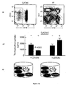

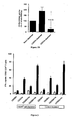


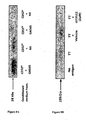
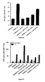


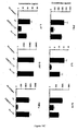

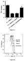


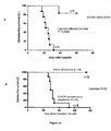

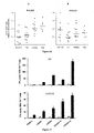
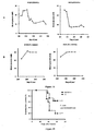

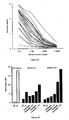


Classifications
-
- A—HUMAN NECESSITIES
- A61—MEDICAL OR VETERINARY SCIENCE; HYGIENE
- A61K—PREPARATIONS FOR MEDICAL, DENTAL OR TOILETRY PURPOSES
- A61K38/00—Medicinal preparations containing peptides
- A61K38/16—Peptides having more than 20 amino acids; Gastrins; Somatostatins; Melanotropins; Derivatives thereof
- A61K38/17—Peptides having more than 20 amino acids; Gastrins; Somatostatins; Melanotropins; Derivatives thereof from animals; from humans
- A61K38/177—Receptors; Cell surface antigens; Cell surface determinants
-
- A—HUMAN NECESSITIES
- A61—MEDICAL OR VETERINARY SCIENCE; HYGIENE
- A61K—PREPARATIONS FOR MEDICAL, DENTAL OR TOILETRY PURPOSES
- A61K39/00—Medicinal preparations containing antigens or antibodies
- A61K39/46—Cellular immunotherapy
- A61K39/461—Cellular immunotherapy characterised by the cell type used
- A61K39/4611—T-cells, e.g. tumor infiltrating lymphocytes [TIL], lymphokine-activated killer cells [LAK] or regulatory T cells [Treg]
-
- A—HUMAN NECESSITIES
- A61—MEDICAL OR VETERINARY SCIENCE; HYGIENE
- A61K—PREPARATIONS FOR MEDICAL, DENTAL OR TOILETRY PURPOSES
- A61K39/00—Medicinal preparations containing antigens or antibodies
- A61K39/46—Cellular immunotherapy
- A61K39/462—Cellular immunotherapy characterized by the effect or the function of the cells
- A61K39/4621—Cellular immunotherapy characterized by the effect or the function of the cells immunosuppressive or immunotolerising
-
- A—HUMAN NECESSITIES
- A61—MEDICAL OR VETERINARY SCIENCE; HYGIENE
- A61K—PREPARATIONS FOR MEDICAL, DENTAL OR TOILETRY PURPOSES
- A61K39/00—Medicinal preparations containing antigens or antibodies
- A61K39/46—Cellular immunotherapy
- A61K39/463—Cellular immunotherapy characterised by recombinant expression
- A61K39/4632—T-cell receptors [TCR]; antibody T-cell receptor constructs
-
- A—HUMAN NECESSITIES
- A61—MEDICAL OR VETERINARY SCIENCE; HYGIENE
- A61K—PREPARATIONS FOR MEDICAL, DENTAL OR TOILETRY PURPOSES
- A61K39/00—Medicinal preparations containing antigens or antibodies
- A61K39/46—Cellular immunotherapy
- A61K39/464—Cellular immunotherapy characterised by the antigen targeted or presented
- A61K39/4643—Vertebrate antigens
- A61K39/46433—Antigens related to auto-immune diseases; Preparations to induce self-tolerance
-
- A—HUMAN NECESSITIES
- A61—MEDICAL OR VETERINARY SCIENCE; HYGIENE
- A61K—PREPARATIONS FOR MEDICAL, DENTAL OR TOILETRY PURPOSES
- A61K39/00—Medicinal preparations containing antigens or antibodies
- A61K39/46—Cellular immunotherapy
- A61K39/464—Cellular immunotherapy characterised by the antigen targeted or presented
- A61K39/4643—Vertebrate antigens
- A61K39/4644—Cancer antigens
- A61K39/464454—Enzymes
-
- A—HUMAN NECESSITIES
- A61—MEDICAL OR VETERINARY SCIENCE; HYGIENE
- A61K—PREPARATIONS FOR MEDICAL, DENTAL OR TOILETRY PURPOSES
- A61K47/00—Medicinal preparations characterised by the non-active ingredients used, e.g. carriers or inert additives; Targeting or modifying agents chemically bound to the active ingredient
- A61K47/50—Medicinal preparations characterised by the non-active ingredients used, e.g. carriers or inert additives; Targeting or modifying agents chemically bound to the active ingredient the non-active ingredient being chemically bound to the active ingredient, e.g. polymer-drug conjugates
- A61K47/51—Medicinal preparations characterised by the non-active ingredients used, e.g. carriers or inert additives; Targeting or modifying agents chemically bound to the active ingredient the non-active ingredient being chemically bound to the active ingredient, e.g. polymer-drug conjugates the non-active ingredient being a modifying agent
- A61K47/68—Medicinal preparations characterised by the non-active ingredients used, e.g. carriers or inert additives; Targeting or modifying agents chemically bound to the active ingredient the non-active ingredient being chemically bound to the active ingredient, e.g. polymer-drug conjugates the non-active ingredient being a modifying agent the modifying agent being an antibody, an immunoglobulin or a fragment thereof, e.g. an Fc-fragment
-
- A—HUMAN NECESSITIES
- A61—MEDICAL OR VETERINARY SCIENCE; HYGIENE
- A61P—SPECIFIC THERAPEUTIC ACTIVITY OF CHEMICAL COMPOUNDS OR MEDICINAL PREPARATIONS
- A61P29/00—Non-central analgesic, antipyretic or antiinflammatory agents, e.g. antirheumatic agents; Non-steroidal antiinflammatory drugs [NSAID]
-
- A—HUMAN NECESSITIES
- A61—MEDICAL OR VETERINARY SCIENCE; HYGIENE
- A61P—SPECIFIC THERAPEUTIC ACTIVITY OF CHEMICAL COMPOUNDS OR MEDICINAL PREPARATIONS
- A61P3/00—Drugs for disorders of the metabolism
- A61P3/08—Drugs for disorders of the metabolism for glucose homeostasis
- A61P3/10—Drugs for disorders of the metabolism for glucose homeostasis for hyperglycaemia, e.g. antidiabetics
-
- A—HUMAN NECESSITIES
- A61—MEDICAL OR VETERINARY SCIENCE; HYGIENE
- A61P—SPECIFIC THERAPEUTIC ACTIVITY OF CHEMICAL COMPOUNDS OR MEDICINAL PREPARATIONS
- A61P31/00—Antiinfectives, i.e. antibiotics, antiseptics, chemotherapeutics
- A61P31/04—Antibacterial agents
-
- A—HUMAN NECESSITIES
- A61—MEDICAL OR VETERINARY SCIENCE; HYGIENE
- A61P—SPECIFIC THERAPEUTIC ACTIVITY OF CHEMICAL COMPOUNDS OR MEDICINAL PREPARATIONS
- A61P37/00—Drugs for immunological or allergic disorders
- A61P37/02—Immunomodulators
- A61P37/06—Immunosuppressants, e.g. drugs for graft rejection
-
- A—HUMAN NECESSITIES
- A61—MEDICAL OR VETERINARY SCIENCE; HYGIENE
- A61P—SPECIFIC THERAPEUTIC ACTIVITY OF CHEMICAL COMPOUNDS OR MEDICINAL PREPARATIONS
- A61P43/00—Drugs for specific purposes, not provided for in groups A61P1/00-A61P41/00
-
- A—HUMAN NECESSITIES
- A61—MEDICAL OR VETERINARY SCIENCE; HYGIENE
- A61P—SPECIFIC THERAPEUTIC ACTIVITY OF CHEMICAL COMPOUNDS OR MEDICINAL PREPARATIONS
- A61P5/00—Drugs for disorders of the endocrine system
- A61P5/48—Drugs for disorders of the endocrine system of the pancreatic hormones
- A61P5/50—Drugs for disorders of the endocrine system of the pancreatic hormones for increasing or potentiating the activity of insulin
-
- C—CHEMISTRY; METALLURGY
- C07—ORGANIC CHEMISTRY
- C07K—PEPTIDES
- C07K14/00—Peptides having more than 20 amino acids; Gastrins; Somatostatins; Melanotropins; Derivatives thereof
- C07K14/435—Peptides having more than 20 amino acids; Gastrins; Somatostatins; Melanotropins; Derivatives thereof from animals; from humans
- C07K14/705—Receptors; Cell surface antigens; Cell surface determinants
- C07K14/70592—CD52
-
- C—CHEMISTRY; METALLURGY
- C07—ORGANIC CHEMISTRY
- C07K—PEPTIDES
- C07K16/00—Immunoglobulins [IGs], e.g. monoclonal or polyclonal antibodies
- C07K16/18—Immunoglobulins [IGs], e.g. monoclonal or polyclonal antibodies against material from animals or humans
-
- C—CHEMISTRY; METALLURGY
- C07—ORGANIC CHEMISTRY
- C07K—PEPTIDES
- C07K16/00—Immunoglobulins [IGs], e.g. monoclonal or polyclonal antibodies
- C07K16/18—Immunoglobulins [IGs], e.g. monoclonal or polyclonal antibodies against material from animals or humans
- C07K16/28—Immunoglobulins [IGs], e.g. monoclonal or polyclonal antibodies against material from animals or humans against receptors, cell surface antigens or cell surface determinants
- C07K16/2803—Immunoglobulins [IGs], e.g. monoclonal or polyclonal antibodies against material from animals or humans against receptors, cell surface antigens or cell surface determinants against the immunoglobulin superfamily
-
- C—CHEMISTRY; METALLURGY
- C07—ORGANIC CHEMISTRY
- C07K—PEPTIDES
- C07K16/00—Immunoglobulins [IGs], e.g. monoclonal or polyclonal antibodies
- C07K16/18—Immunoglobulins [IGs], e.g. monoclonal or polyclonal antibodies against material from animals or humans
- C07K16/28—Immunoglobulins [IGs], e.g. monoclonal or polyclonal antibodies against material from animals or humans against receptors, cell surface antigens or cell surface determinants
- C07K16/2863—Immunoglobulins [IGs], e.g. monoclonal or polyclonal antibodies against material from animals or humans against receptors, cell surface antigens or cell surface determinants against receptors for growth factors, growth regulators
-
- C—CHEMISTRY; METALLURGY
- C07—ORGANIC CHEMISTRY
- C07K—PEPTIDES
- C07K16/00—Immunoglobulins [IGs], e.g. monoclonal or polyclonal antibodies
- C07K16/18—Immunoglobulins [IGs], e.g. monoclonal or polyclonal antibodies against material from animals or humans
- C07K16/28—Immunoglobulins [IGs], e.g. monoclonal or polyclonal antibodies against material from animals or humans against receptors, cell surface antigens or cell surface determinants
- C07K16/2866—Immunoglobulins [IGs], e.g. monoclonal or polyclonal antibodies against material from animals or humans against receptors, cell surface antigens or cell surface determinants against receptors for cytokines, lymphokines, interferons
-
- C—CHEMISTRY; METALLURGY
- C07—ORGANIC CHEMISTRY
- C07K—PEPTIDES
- C07K16/00—Immunoglobulins [IGs], e.g. monoclonal or polyclonal antibodies
- C07K16/18—Immunoglobulins [IGs], e.g. monoclonal or polyclonal antibodies against material from animals or humans
- C07K16/28—Immunoglobulins [IGs], e.g. monoclonal or polyclonal antibodies against material from animals or humans against receptors, cell surface antigens or cell surface determinants
- C07K16/2893—Immunoglobulins [IGs], e.g. monoclonal or polyclonal antibodies against material from animals or humans against receptors, cell surface antigens or cell surface determinants against CD52
-
- C—CHEMISTRY; METALLURGY
- C12—BIOCHEMISTRY; BEER; SPIRITS; WINE; VINEGAR; MICROBIOLOGY; ENZYMOLOGY; MUTATION OR GENETIC ENGINEERING
- C12N—MICROORGANISMS OR ENZYMES; COMPOSITIONS THEREOF; PROPAGATING, PRESERVING, OR MAINTAINING MICROORGANISMS; MUTATION OR GENETIC ENGINEERING; CULTURE MEDIA
- C12N5/00—Undifferentiated human, animal or plant cells, e.g. cell lines; Tissues; Cultivation or maintenance thereof; Culture media therefor
- C12N5/06—Animal cells or tissues; Human cells or tissues
- C12N5/0602—Vertebrate cells
- C12N5/0634—Cells from the blood or the immune system
- C12N5/0636—T lymphocytes
- C12N5/0637—Immunosuppressive T lymphocytes, e.g. regulatory T cells or Treg
-
- C—CHEMISTRY; METALLURGY
- C12—BIOCHEMISTRY; BEER; SPIRITS; WINE; VINEGAR; MICROBIOLOGY; ENZYMOLOGY; MUTATION OR GENETIC ENGINEERING
- C12Q—MEASURING OR TESTING PROCESSES INVOLVING ENZYMES, NUCLEIC ACIDS OR MICROORGANISMS; COMPOSITIONS OR TEST PAPERS THEREFOR; PROCESSES OF PREPARING SUCH COMPOSITIONS; CONDITION-RESPONSIVE CONTROL IN MICROBIOLOGICAL OR ENZYMOLOGICAL PROCESSES
- C12Q1/00—Measuring or testing processes involving enzymes, nucleic acids or microorganisms; Compositions therefor; Processes of preparing such compositions
- C12Q1/68—Measuring or testing processes involving enzymes, nucleic acids or microorganisms; Compositions therefor; Processes of preparing such compositions involving nucleic acids
- C12Q1/6876—Nucleic acid products used in the analysis of nucleic acids, e.g. primers or probes
- C12Q1/6883—Nucleic acid products used in the analysis of nucleic acids, e.g. primers or probes for diseases caused by alterations of genetic material
-
- G—PHYSICS
- G01—MEASURING; TESTING
- G01N—INVESTIGATING OR ANALYSING MATERIALS BY DETERMINING THEIR CHEMICAL OR PHYSICAL PROPERTIES
- G01N33/00—Investigating or analysing materials by specific methods not covered by groups G01N1/00 - G01N31/00
- G01N33/48—Biological material, e.g. blood, urine; Haemocytometers
- G01N33/50—Chemical analysis of biological material, e.g. blood, urine; Testing involving biospecific ligand binding methods; Immunological testing
- G01N33/5005—Chemical analysis of biological material, e.g. blood, urine; Testing involving biospecific ligand binding methods; Immunological testing involving human or animal cells
- G01N33/5008—Chemical analysis of biological material, e.g. blood, urine; Testing involving biospecific ligand binding methods; Immunological testing involving human or animal cells for testing or evaluating the effect of chemical or biological compounds, e.g. drugs, cosmetics
- G01N33/502—Chemical analysis of biological material, e.g. blood, urine; Testing involving biospecific ligand binding methods; Immunological testing involving human or animal cells for testing or evaluating the effect of chemical or biological compounds, e.g. drugs, cosmetics for testing non-proliferative effects
-
- G—PHYSICS
- G01—MEASURING; TESTING
- G01N—INVESTIGATING OR ANALYSING MATERIALS BY DETERMINING THEIR CHEMICAL OR PHYSICAL PROPERTIES
- G01N33/00—Investigating or analysing materials by specific methods not covered by groups G01N1/00 - G01N31/00
- G01N33/48—Biological material, e.g. blood, urine; Haemocytometers
- G01N33/50—Chemical analysis of biological material, e.g. blood, urine; Testing involving biospecific ligand binding methods; Immunological testing
- G01N33/5005—Chemical analysis of biological material, e.g. blood, urine; Testing involving biospecific ligand binding methods; Immunological testing involving human or animal cells
- G01N33/5008—Chemical analysis of biological material, e.g. blood, urine; Testing involving biospecific ligand binding methods; Immunological testing involving human or animal cells for testing or evaluating the effect of chemical or biological compounds, e.g. drugs, cosmetics
- G01N33/5044—Chemical analysis of biological material, e.g. blood, urine; Testing involving biospecific ligand binding methods; Immunological testing involving human or animal cells for testing or evaluating the effect of chemical or biological compounds, e.g. drugs, cosmetics involving specific cell types
- G01N33/5047—Cells of the immune system
- G01N33/505—Cells of the immune system involving T-cells
-
- G—PHYSICS
- G01—MEASURING; TESTING
- G01N—INVESTIGATING OR ANALYSING MATERIALS BY DETERMINING THEIR CHEMICAL OR PHYSICAL PROPERTIES
- G01N33/00—Investigating or analysing materials by specific methods not covered by groups G01N1/00 - G01N31/00
- G01N33/48—Biological material, e.g. blood, urine; Haemocytometers
- G01N33/50—Chemical analysis of biological material, e.g. blood, urine; Testing involving biospecific ligand binding methods; Immunological testing
- G01N33/53—Immunoassay; Biospecific binding assay; Materials therefor
- G01N33/564—Immunoassay; Biospecific binding assay; Materials therefor for pre-existing immune complex or autoimmune disease, i.e. systemic lupus erythematosus, rheumatoid arthritis, multiple sclerosis, rheumatoid factors or complement components C1-C9
-
- G—PHYSICS
- G01—MEASURING; TESTING
- G01N—INVESTIGATING OR ANALYSING MATERIALS BY DETERMINING THEIR CHEMICAL OR PHYSICAL PROPERTIES
- G01N33/00—Investigating or analysing materials by specific methods not covered by groups G01N1/00 - G01N31/00
- G01N33/48—Biological material, e.g. blood, urine; Haemocytometers
- G01N33/50—Chemical analysis of biological material, e.g. blood, urine; Testing involving biospecific ligand binding methods; Immunological testing
- G01N33/68—Chemical analysis of biological material, e.g. blood, urine; Testing involving biospecific ligand binding methods; Immunological testing involving proteins, peptides or amino acids
- G01N33/6872—Intracellular protein regulatory factors and their receptors, e.g. including ion channels
-
- G—PHYSICS
- G01—MEASURING; TESTING
- G01N—INVESTIGATING OR ANALYSING MATERIALS BY DETERMINING THEIR CHEMICAL OR PHYSICAL PROPERTIES
- G01N33/00—Investigating or analysing materials by specific methods not covered by groups G01N1/00 - G01N31/00
- G01N33/48—Biological material, e.g. blood, urine; Haemocytometers
- G01N33/50—Chemical analysis of biological material, e.g. blood, urine; Testing involving biospecific ligand binding methods; Immunological testing
- G01N33/68—Chemical analysis of biological material, e.g. blood, urine; Testing involving biospecific ligand binding methods; Immunological testing involving proteins, peptides or amino acids
- G01N33/6893—Chemical analysis of biological material, e.g. blood, urine; Testing involving biospecific ligand binding methods; Immunological testing involving proteins, peptides or amino acids related to diseases not provided for elsewhere
- G01N33/6896—Neurological disorders, e.g. Alzheimer's disease
-
- A—HUMAN NECESSITIES
- A61—MEDICAL OR VETERINARY SCIENCE; HYGIENE
- A61K—PREPARATIONS FOR MEDICAL, DENTAL OR TOILETRY PURPOSES
- A61K2239/00—Indexing codes associated with cellular immunotherapy of group A61K39/46
- A61K2239/31—Indexing codes associated with cellular immunotherapy of group A61K39/46 characterized by the route of administration
-
- A—HUMAN NECESSITIES
- A61—MEDICAL OR VETERINARY SCIENCE; HYGIENE
- A61K—PREPARATIONS FOR MEDICAL, DENTAL OR TOILETRY PURPOSES
- A61K38/00—Medicinal preparations containing peptides
-
- A—HUMAN NECESSITIES
- A61—MEDICAL OR VETERINARY SCIENCE; HYGIENE
- A61K—PREPARATIONS FOR MEDICAL, DENTAL OR TOILETRY PURPOSES
- A61K38/00—Medicinal preparations containing peptides
- A61K38/04—Peptides having up to 20 amino acids in a fully defined sequence; Derivatives thereof
- A61K38/10—Peptides having 12 to 20 amino acids
-
- A—HUMAN NECESSITIES
- A61—MEDICAL OR VETERINARY SCIENCE; HYGIENE
- A61K—PREPARATIONS FOR MEDICAL, DENTAL OR TOILETRY PURPOSES
- A61K48/00—Medicinal preparations containing genetic material which is inserted into cells of the living body to treat genetic diseases; Gene therapy
-
- C—CHEMISTRY; METALLURGY
- C07—ORGANIC CHEMISTRY
- C07K—PEPTIDES
- C07K2317/00—Immunoglobulins specific features
- C07K2317/70—Immunoglobulins specific features characterized by effect upon binding to a cell or to an antigen
- C07K2317/76—Antagonist effect on antigen, e.g. neutralization or inhibition of binding
-
- C—CHEMISTRY; METALLURGY
- C07—ORGANIC CHEMISTRY
- C07K—PEPTIDES
- C07K2319/00—Fusion polypeptide
- C07K2319/30—Non-immunoglobulin-derived peptide or protein having an immunoglobulin constant or Fc region, or a fragment thereof, attached thereto
-
- G—PHYSICS
- G01—MEASURING; TESTING
- G01N—INVESTIGATING OR ANALYSING MATERIALS BY DETERMINING THEIR CHEMICAL OR PHYSICAL PROPERTIES
- G01N2333/00—Assays involving biological materials from specific organisms or of a specific nature
- G01N2333/435—Assays involving biological materials from specific organisms or of a specific nature from animals; from humans
- G01N2333/705—Assays involving receptors, cell surface antigens or cell surface determinants
- G01N2333/70592—CD52
-
- G—PHYSICS
- G01—MEASURING; TESTING
- G01N—INVESTIGATING OR ANALYSING MATERIALS BY DETERMINING THEIR CHEMICAL OR PHYSICAL PROPERTIES
- G01N2800/00—Detection or diagnosis of diseases
- G01N2800/26—Infectious diseases, e.g. generalised sepsis
-
- G—PHYSICS
- G01—MEASURING; TESTING
- G01N—INVESTIGATING OR ANALYSING MATERIALS BY DETERMINING THEIR CHEMICAL OR PHYSICAL PROPERTIES
- G01N2800/00—Detection or diagnosis of diseases
- G01N2800/28—Neurological disorders
- G01N2800/285—Demyelinating diseases; Multipel sclerosis
-
- G—PHYSICS
- G01—MEASURING; TESTING
- G01N—INVESTIGATING OR ANALYSING MATERIALS BY DETERMINING THEIR CHEMICAL OR PHYSICAL PROPERTIES
- G01N2800/00—Detection or diagnosis of diseases
- G01N2800/50—Determining the risk of developing a disease
-
- G—PHYSICS
- G01—MEASURING; TESTING
- G01N—INVESTIGATING OR ANALYSING MATERIALS BY DETERMINING THEIR CHEMICAL OR PHYSICAL PROPERTIES
- G01N2800/00—Detection or diagnosis of diseases
- G01N2800/52—Predicting or monitoring the response to treatment, e.g. for selection of therapy based on assay results in personalised medicine; Prognosis
Definitions
- the present disclosure generally relates to cell populations and soluble mediators capable of suppressing T-cell activation, and to the use of such cell populations and soluble mediators to suppress T-cell activation, such as in the treatment of diseases or conditions mediated by effector T-cell function.
- the disclosure also relates to methods of detecting the presence of a marker in a subject, which marker is indicative of the subject's susceptibility to diseases or conditions mediated by effector T-cell function.
- Treg cells also known as suppressor T-cells
- Treg cells are subpopulations of T-cells that maintain immune homeostasis and help avert autoimmune disease (Sakaguchi et al., 2008; Shevach, 2006; Vignali et al., 2008).
- Interest in Treg cells is focused predominantly on prototypic CD4 + CD25 + Treg cells that are programmed by the transcription factor FoxP3 (Fontenot et al., 2003; Hori et al., 2003).
- Treg cells are characterised in the mouse both as 'natural', thymus-derived and induced 'adaptive' cells that suppress the activation, proliferation and functions of other T-cells (Sakaguchi et al., 2008; Shevach, 2006).
- CD4 + Treg cells are not as reliably distinguished by FoxP3 expression (Roncarolo and Gregori, 2008; Allan et al., 2007; Gavin et al., 2006).
- CD4 + T-cells with markers of either naive or memory cells were shown to have similar suppressor functions despite low and high expression, respectively, of FoxP3 (Miyara et al., 2009).
- Other surface markers of human CD4 + CD25 + FoxP3 + Treg cells such as decreased expression of the IL-7 receptor, CD127 (Liu et al., 2006; Seddiki et al., 2006), are not specific for Treg cells.
- Treg cells induced by administration of autoantigens have been shown to protect against some autoimmune diseases in certain animal models (reviewed by von Herrath and Harrison, 2003).
- NOD nonobese diabetic
- T1D type 1 diabetes
- CD4 + Treg cells induced by administered pancreatic islet autoantigens such as insulin (Bergerot et al., 1994) or glutamic acid decarboxylase 65 (GAD65) (Tisch et al., 1999), or transfer of CD4 + Treg cells induced by proinsulin (Every et al., 2006) or a putative pancreatic islet antigen (Tang et al., 2004), have been shown to protect against autoimmune diabetes.
- Treg cells have been studied in resting, polyspecific populations and not during the host's response to a particular antigen.
- proinsulin- and GAD65-specific human CD4 + T-cell clones were generated and Treg clones were distinguished by their suppressor function in vitro (Dromey et al., 2011).
- the cell surface membrane-anchored glycoprotein CD52 was shown to be upregulated in these expanded CD4 + Treg clones.
- the mechanism of immune suppression has not previously been characterized.
- the present inventors have identified a soluble mediator of Treg cell suppression. Accordingly, the present disclosure provides any one or more of:
- the soluble CD52 glycoprotein comprises an amino acid sequence at least 60% identical to the amino acid sequence of any one or more of GQNDTSQTSSPS (SEQ ID NO: 3), SQNATSQSSPS (SEQ ID NO: 4), GQATTAASGTNKNSTSTKKTPLKS (SEQ ID NO: 5), GQNSTAVTTPANKAATTAAATTKAAATTATKTTTAVRKTPGKPPKA (SEQ ID NO: 6) or GNSTTPRMTTKKVKSATPA (SEQ ID NO:7) and a carbohydrate.
- GQNDTSQTSSPS SEQ ID NO: 3
- SQNATSQSSPS SEQ ID NO: 4
- GQATTAASGTNKNSTSTKKTPLKS SEQ ID NO: 5
- GQNSTAVTTPANKAATTAAATTKAAATTATKTTTAVRKTPGKPPKA SEQ ID NO: 6
- GNSTTPRMTTKKVKSATPA SEQ ID NO:7
- the glycoprotein comprises an amino acid sequence which is at least 65%, at least 70%, at least 75%, at least 80%, at least 85%, at least 90%, at least 95% identical, or is 100% identical, to any one or more of the amino acid sequences identified in SEQ ID NOs: 3, 4, 5, 6 or 7.
- the glycoprotein comprises an amino acid sequence at least 60% at least 65%, at least 70%, at least 75%, at least 80%, at least 85%, at least 90%, at least 95% identical, or is 100% identical to the amino acid sequence of SEQ ID NO: 3, which represents the human soluble CD52 fragment.
- any one or more of the soluble CD52 glycoprotein, fusion protein, polynucleotide and vector is present in an amount sufficient to suppress effector T-cell function and/or an immune response.
- the soluble CD52 glycoprotein, fusion protein, polynucleotide and vector is present in an amount sufficient such that the suppression of the immune response results in tolerance to at least one antigen such an autoantigen.
- any one or more of the soluble CD52 glycoprotein and fusion protein is capable of suppressing effector T-cell function and/or is capable of reducing an immune response such as an immune response to an autoantigen.
- the composition comprises one or more of the soluble CD52 glycoprotein or fusion protein, and is formulated for mucosal and/or transdermal administration.
- the composition further comprises insulin and/or an autoantigen.
- the present disclosure also provides a fusion protein comprising soluble CD52 glycoprotein as a first protein, and a second protein.
- the soluble CD52 glycoprotein of the fusion protein comprises an amino acid sequence which is at least 60%, at least 65%, at least 70%, at least 75%, at least 80%, at least 85%, at least 90%, at least 95% identical, or is 100% identical, to any one or more of the amino acid sequences identified in SEQ ID NOs: 3, 4, 5, 6 or 7.
- the fusion protein is capable of suppressing effector T-cell function and/or is capable of reducing an immune response such as an immune response to an autoantigen.
- the fusion protein reduces the immune response to an extent that it results in tolerance to at least one antigen such an autoantigen.
- the second protein may be any protein capable of increasing the stability and/or solubility of the soluble CD52 glycoprotein, of enhancing the process of making the soluble CD52 glycoprotein by recombinant methods, or of enhancing the therapeutic effect of the soluble CD52 glycoprotein.
- the second protein may comprise an antibody fragment, such as an Fc.
- the fusion protein is soluble.
- the present disclosure also provides an isolated or recombinant polynucleotide encoding the fusion protein disclosed herein.
- the present disclosure also provides a vector comprising the polynucleotide disclosed herein.
- the present disclosure also provides an isolated cell comprising the polynucleotide and/or the vector disclosed herein.
- the cell may be a mammalian cell.
- the cell is as HEK293T cell.
- the cell is a Daudi B lymphoblast cell.
- the present disclosure provides a method of producing the fusion protein, comprising expressing the polynucleotide or vector disclosed herein under glycosylation-permitting conditions.
- the glycosylation-permitting conditions comprise expressing the fusion protein in a host cell, such as a mammalian cell.
- the present disclosure also provides for the use of any one or more of:
- the present disclosure also provides a method of treating or preventing a disease or condition mediated by effector T-cell function, inflammation or sepsis, in a subject, the method comprising administering a therapeutically effective amount of any one or more of:
- the soluble CD52 glycoprotein or fusion protein is administered at a mucosal or transdermal site.
- the present disclosure also provides any one or more of:
- the medicament is formulated for administration at a mucosal or transdermal site.
- the disease mediated by effector T-cell function is an autoimmune disease, such as type I diabetes or rheumatoid arthritis.
- the condition mediated by effector T-cell function is an allograft rejection or a graft-versus-host reaction.
- the present disclosure also provides a method of diagnosing a subject's susceptibility to developing a disease or condition mediated by effector T-cell function, inflammation or sepsis, the method comprising:
- the present disclosure also provides a method of diagnosing a subject's susceptibility to developing a disease or condition mediated by effector T-cell function, inflammation or sepsis, the method comprising:
- the present disclosure also provides a method of diagnosing a subject's susceptibility to developing a disease or condition mediated by effector T-cell function, inflammation or sepsis, the method comprising:
- the frequency of CD52 hi cells is determined by detecting the level of membrane bound CD52 in the sample, by detecting the level of expression of CD52 protein in the sample, and/or by detecting the level of expression of CD52 mRNA in the sample.
- the sample is taken from a subject to which an antigen has been administered.
- the sample is taken from a local disease site in the subject.
- the present disclosure also provides a method of determining a subject's suitability for entry into a drug screening trial, comprising performing the method of the invention and identifying the subject as being more suitable for entry into a drug screening trial if the subject has a lower level of soluble CD52 glycoprotein, a lower frequency of CD52 hi cells, or a reduced activity of CD52 hi cells than the reference sample.
- the drug screening trial is an anti-diabetic drug screening trial.
- the present disclosure also provides a method of identifying an agent capable of mimicking the effector T-cell-suppressing, and/or immune response suppressing, function of a soluble CD52 glycoprotein, the method comprising determining whether a test agent suppresses effector T-cell function and/or an immune response.
- the present disclosure also provides a method of identifying a potential therapeutic agent for the treatment or prevention of a disease or condition mediated by effector T-cell function, inflammation or sepsis, the method comprising contacting a test agent with a CD52 hi cell or CD52 hi cell population, and detecting any one or more of the level of soluble CD52 glycoprotein produced by the cell or cell population, the frequency of CD52 hi cells and/or the activity of CD52 hi cells, and identifying the test agent as a potential therapeutic agent for the treatment or prevention of a disease or condition mediated by effector T-cell function, inflammation or sepsis, if the level of soluble CD52 glycoprotein, the frequency of CD52 hi cells and/or the activity of CD52 hi cells is increased after contact with the test agent.
- composition of matter, group of steps or group of compositions of matter shall be taken to encompass one and a plurality (i.e. one or more) of those steps, compositions of matter, groups of steps or group of compositions of matter.
- the term “about” followed by a designated value is to be interpreted as also encompassing the exact designated value itself (for example, “about 10" also encompasses 10 exactly).
- immune response has its ordinary meaning in the art, and includes both humoral and cellular immunity.
- An immune response may be mediated by one or more of, T-cell activation, B-cell activation, natural killer cell activation, activation of antigen presenting cells (e.g., B cells, DCs, monocytes and/or macrophages), cytokine production, chemokine production, specific cell surface marker expression, in particular, expression of co-stimulatory molecules.
- the immune response which is suppressed using the methods of the invention is at least effector T cell function by reducing the survival, activity and/or proliferation of this cell type.
- the immune response which is suppressed using the methods of the invention is at least one or more of monocyte, macrophage or dendritic cell function by reducing the survival, activity and/or proliferation of one or more of these cell types.
- the immune response is suppressed to an extent such that it induces tolerance to an antigen such as an autoantigen.
- tolerance refers to a state of immune unresponsiveness to a specific antigen or group of antigens to which a subject is normally responsive. Immune tolerance is achieved under conditions that suppress the immune reaction and is not just the absence of an immune response.
- treating include administering a therapeutically effective amount of an agent sufficient to reduce or eliminate at least one symptom of disease.
- preventing include administering a therapeutically effective amount of an agent sufficient to prevent the manifestation of at least one symptom of disease.
- the term "suppressing” includes reducing by any quantifiable amount.
- the term "subject" refers to an animal, e.g., a mammal.
- the subject is mammal, for example a human.
- Other preferred embodiments include livestock animals such as horses, cattle, sheep and goats, as well as companion animals such as cats and dogs.
- the term "host” refers to any organism from which soluble CD52 can be isolated or in which soluble CD52 can be produced, by any means.
- the host may be whole organism or may be a cell derived therefrom.
- the host may be an animal, e.g., a mammal. In a preferred embodiment, the host is mammalian, for example a human.
- Other preferred hosts include mice, rats, monkeys, hamsters, guinea-pigs, rabbits, and any animal or cell which may serve as a suitable host from which soluble CD52 can be isolated or in which soluble CD52 can be produced.
- CD52 is a surface glycosylphosphatidylinositol (GPI)-anchored glycoprotein present on most lymphoid cells, initially recognised as the target of complement-binding CAMPATH monoclonal antibodies used therapeutically to deplete lymphocytes (Treumann et al., 1995; Xia et al., 1991; Hale, 2001).
- GPI surface glycosylphosphatidylinositol
- the mRNA transcript of the human CD52 gene is shown in SEQ ID NO: 1 and the translated amino acid sequence is shown in SEQ ID NO: 2.
- Mature CD52 tethered by its GPI anchor comprises only 12 amino acids and an asparagine (N)-linked terminal carbohydrate.
- soluble CD52 glycoprotein soluble CD52
- soluble glycoprotein soluble glycoprotein
- Membrane-anchored CD52 can be cleaved (for example, enzymatically) to release a soluble peptide fragment comprising the amino acid sequence GQNDTSQTSSPS (SEQ ID NO: 3).
- the soluble CD52 glycoprotein disclosed herein may comprise an amino acid sequence at least 60% identical to the amino acid sequence of SEQ ID NO: 3 or at least 60% identical to the amino acid sequence of other known, orthologous CD52 soluble fragment sequences.
- orthologous sequences of the soluble CD52 peptide fragment are encompassed by the present disclosure.
- sequences include but are not limited to the monkey sequence SQNATSQSSPS (SEQ ID NO: 4), the mouse sequence GQATTAASGTNKNSTSTKKTPLKS (SEQ ID NO: 5), the rat sequence GQNSTAVTTPANKAATTAAATTKAAATTATKTTTAVRKTPGKPPKA (SEQ ID NO: 6), the dog sequence GNSTTPRMTTKKVKSATPA (SEQ ID NO:7), and other orthologous sequences readily identifiable from known CD52 polypeptide and polynucleotide sequences.
- Percentage identity to any of the amino acid or polynucleotide sequences disclosed herein may be determined by methods known in the art. For example, amino acid and polynucleotide sequences can be compared manually or by using computer-based sequence comparison and identification tools that employ algorithms such as BLAST (Basic Local Alignment Search Tool; Altschul et al., 1993); see also www.ncbi.nlm.nih.gov/BLAST/), the Clustal method of alignment (Higgins and Sharp, 1989) and others, wherein appropriate parameters for each specific sequence comparison can be selected as would be understood by a person skilled in the art.
- BLAST Basic Local Alignment Search Tool
- Altschul et al., 1993 see also www.ncbi.nlm.nih.gov/BLAST/
- Clustal method of alignment Higgins and Sharp, 1989
- the amino acid sequence of the peptide portion of the glycoprotein disclosed herein can be at least 65%, at least 70%, at least 75%, at least 80%, at least 85%, at least 90%, at least 95% identical, or at least 99% identical to any one or more of the amino acid sequences identified in SEQ ID NOs: 3, 4, 5, 6 or 7.
- the amino acid sequence of the peptide portion of the glycoprotein disclosed herein can be 100% identical to any one of the amino acid sequences identified in SEQ ID NOs: 3, 4, 5, 6 or 7.
- Isolated soluble CD52 glycoprotein may be used to produce pharmaceutical compositions of the invention.
- isolated is used herein to define the isolation of the soluble CD52 glycoprotein so that it is present in a form suitable for application in a pharmaceutical composition.
- the glycoprotein disclosed herein is isolated from other components of a host cell or fluid or expression system to the extent that is required for subsequent formulation of the glycoprotein as a pharmaceutical composition.
- the isolated glycoprotein is therefore provided in a form which is substantially free of other components of a host cell (for example, proteins) which may hinder the pharmaceutical effect of the glycoprotein.
- the isolated glycoprotein may be free or substantially free of material with which it is naturally associated such as other glycoproteins, polypeptides or nucleic acids with which it is found in its natural environment, or the environment in which it is prepared (e.g. cell culture) when such preparation is by recombinant DNA technology practised in vitro or in vivo.
- Soluble glycoprotein can be isolated from a host cell or fluid or expression system by methods known in the art.
- soluble is used herein to define a peptide or glycoprotein which is not bound to a cell membrane.
- the soluble peptide or glycoprotein may be able to move freely in any solvent or fluid, such as a bodily fluid.
- the soluble peptide or glycoprotein may be able to circulate in blood.
- the carbohydrate may be any carbohydrate moiety attached to the soluble CD52 peptide fragment.
- the carbohydrate may be any carbohydrate moiety found to be attached to the extracellular portion of the CD52 protein in a host.
- the carbohydrate may be any carbohydrate capable of being attached to the extracellular portion of the CD52 protein by a glycosylation reaction known to take place in a host.
- Carbohydrate moieties present on a naturally occurring CD52 glycoprotein can be identified by known methods, such as those described in Schröter et al. (1999). Such carbohydrate moieties may be identified from CD52 glycoproteins present in any host cell expressing CD52, and particularly lymphocytes, such as CD4 + or CD8 + T-cells, monocytes or genital tract cells, such as sperm cells or epididymal duct cells. Thus, the precise structure of the carbohydrate moiety can be determined by applying methods such as mass spectrometry (e.g.
- MALDI-TOF Matrix-assisted Laser Desorption/Ionization - Time of Flight Mass Spectrometry
- HPAEC-PAD High pH anion exchange chromatography
- methylation analysis endo- ⁇ -galactosidase digestion, and other methods.
- the N-glycans may be separated from a naturally occurring CD52 glycoprotein using known cleavage enzymes such as peptide-N4-(N-acetyl- ⁇ -D-glucosaminyl)asparagines amidase F ('PNGase F' from Flavobacterium meningosepticum, recombinant from Escherichia coli; obtainable from commercial suppliers such as Roche).
- the N-glycans can be isolated for further characterisation using known chromatographic methods, such as C8-reversed phase chromatography.
- the carbohydrate may comprise one or more bi-, tri- or tetra-antennary sugars, which may be terminally sialylated.
- the carbohydrate may comprise one or more tetra-antennary sugars.
- the sugars may be branched or unbranched.
- the sugars may comprise a proximal fucose.
- the carbohydrate may be fucosylated.
- the sugars may comprise one or more N-acetyllactosamine repeats.
- the sugars may comprise polylactosamine units.
- the sugars may comprise a mannose core.
- the carbohydrate may have any one or more of the structures described in Treumann et al. (1995).
- the carbohydrate may have any of the following structures:
- the carbohydrate may comprise one or more sialic acids.
- the one or more sialic acids may be located in any portion of the carbohydrate.
- the one or more sialic acids may be terminal sialic acids.
- the carbohydrate may comprise terminal ⁇ 2-6 sialic acids.
- the carbohydrate may comprise one or more surface ⁇ 2-6-sialyllactose groups.
- the one or more sialic acids may be attached to galactose in ⁇ 1-4 linkage with N-acetylglucosamine.
- the present disclosure demonstrates that the soluble CD52 glycoprotein, exerts its suppressive effect at least in part via binding to the sialic acid binding Ig-likelectin-10 (Siglec-10), a cell surface transmembrane receptor and immunoglobulin superfamily member bearing two cytoplasmic immunoreceptor tyrosine-based inhibition motifs (ITIMs) (Munday et al., 2001; Crocker et al., 2007).
- the soluble glycoprotein disclosed herein may be capable of binding to Siglec-10.
- the soluble glycoprotein disclosed herein may comprise a carbohydrate moiety capable of binding to Siglec-10.
- the carbohydrate moiety comprises one or more surface ⁇ 2-6- or ⁇ 2-3-sialyllactose groups that are capable of binding to Siglec-10.
- the carbohydrate moiety may comprise any other surface groups that are capable of binding to Siglec-10.
- the soluble glycoprotein disclosed herein may be capable of binding to Siglec-10 derived from any species.
- the soluble glycoprotein disclosed herein may be capable of binding to Siglec-10 derived from any mammalian species.
- the soluble glycoprotein disclosed herein is capable of binding to human Siglec-10.
- the polypeptide sequence of human Siglec-10 is defined in Munday et al. (2001), in GenBank Accession No. AF310233.1, and in SEQ ID NO: 69.
- the soluble glycoprotein disclosed herein may be capable of effecting signalling via the Siglec-10 receptor.
- the soluble glycoprotein disclosed herein may be capable of binding to Siglec-10 to any extent sufficient to effect signalling via the Siglec-10 receptor.
- the precise level of binding to Siglec-10 can vary. Methods for determining whether a given glycoprotein is capable of binding to Siglec-10, and for determining whether a given glycoprotein is capable of effecting signalling via the Siglec-10 receptor are known in the art.
- N-linked CD52 carbohydrate which the glycoprotein disclosed herein may comprise are those derived or derivable from host lymphocyte CD52 glycoproteins or genital tract cell CD52 glycoproteins.
- carbohydrate moieties Due to the complex nature of many naturally occurring carbohydrate moieties known to be linked to the extracellular protein portion of human CD52 and the many variations in these structures that may arise from varying glycosylation patterns, it will be understood that the precise nature of the carbohydrate moiety present in the glycoprotein disclosed herein may vary. As stated above, methods are available to precisely identify particular carbohydrate moieties from naturally occurring CD52 glycoproteins. In addition, a number of different carbohydrate moieties can be added to the soluble peptide fragment of CD52 by expressing CD52 under varying glycosylation conditions. For example, the soluble glycoprotein disclosed herein may be expressed in and/or isolated from host lymphocyte cells, monocytes or host genital tract cells (e.g.
- sperm cells, or epididymal duct cells may therefore comprise different carbohydrate groups as a result.
- the inventors have shown that soluble CD52 present in human semen, similarly to soluble CD52 released from lymphocytes such as Daudi B cells, is capable of suppressing T-cell function and/or an immune response.
- Alternative host cells providing different glycosylation conditions may be selected for expression of soluble CD52 in order to provide alternative forms of carbohydrate on the soluble glycoprotein.
- the carbohydrate may be attached to any one or more amino acid in the peptide which is capable of having a carbohydrate moiety attached thereto.
- the carbohydrate may be attached to one or more asparagine, serine, threonine, tyrosine, hydroxylysine, hydroxyproline, phosphoserine or tryptophan residues if present in the amino acid sequence.
- the carbohydrate is attached to the asparagine (N) residue in a peptide portion having a sequence at least 60%, at least 65%, at least 70%, at least 75%, at least 80%, at least 85%, at least 90%, at least 95% identical, or is 100% identical, to the amino acid sequence set out in SEQ ID NO: 3.
- the present disclosure also provides variants, mutants, biologically active fragments, modifications, analogs and/or derivatives of the glycoprotein disclosed herein.
- Such compounds can be identified by screening for compounds which mimic the structure and/or function of the polypeptide disclosed herein, using methods including any of the methods disclosed herein.
- the glycoprotein disclosed herein is preferably capable of suppressing the activity ("function") of immune cells including lymphocytes (such as a T-cell) and monocytes.
- the glycoprotein disclosed herein is capable of suppressing one or more of effector T-cell, monocyte, macrophage and dendritic cell function. Effector T-cells, monocytes, macrophages and dendritic cells and their functions will be known to a person skilled in the art.
- T-cells can be readily identified by the presence of any of one or more T-cell markers known in the art.
- the glycoprotein disclosed herein is capable of reducing T-cell proliferation in response to antigen challenge, and/or capable of reducing T-cell cytokine production (such as production of any one or more of IFN- ⁇ , IL-2, IL-10, IL-17, G-CSF, TNF- ⁇ , and other cytokines known to be secreted by activated T-cells).
- soluble CD52 is capable of reducing IFN- ⁇ production by T-cells.
- soluble CD52 is capable of reducing IL-1 ⁇ secretion by monocytes, macrophages and dendritic cells.
- the glycoprotein disclosed herein is capable of reducing an immune response in a host.
- the inventors have shown that the glycoprotein disclosed herein is capable of reducing effector T-cell function in response to challenge with any antigen.
- the suppressive function is not dependent on the particular antigen used in the challenge.
- the glycoprotein disclosed herein is capable of reducing an immune response to any antigen.
- the antigen is an autoantigen.
- the methods may comprise determining the effect of the glycoprotein disclosed herein on one or more of effector T-cell, monocyte, macrophage and dendritic cell proliferation and/or on the production of any one or more of IFN- ⁇ , IL-2, IL-10, IL-17, G-CSF, TNF- ⁇ , and other cytokines known to be secreted by activated T-cells, monocytes, macrophages or dendritic cells.
- the peptide portion of the CD52 glycoprotein disclosed herein may, for example, be conjugated to a second protein as a fusion protein.
- the second protein may be any protein capable of increasing the stability and/or solubility of the glycoprotein, of enhancing the process of making the glycoprotein by recombinant methods, or of enhancing the therapeutic effect of the glycoprotein.
- the second protein may capable of increasing the half life of the glycoprotein disclosed herein.
- the second protein can be of any suitable length.
- the second protein may be relatively short.
- the second protein may consist of any of 1, 2, 3, 4, 5, 6, 7, 8, 9, 10, or more amino acids.
- the second protein may comprise at least 1, at least 2, at least 3, at least 4, at least 5, at least 6, at least 7, at least 8, at least 9 or at least 10 amino acids.
- the second protein may also comprise more than 10 amino acids.
- the second protein may comprise at least 10, at least 15, at least 20, at least 25, at least 30, or at least 50 amino acids.
- the second protein is an antibody fragment.
- Suitable antibody fragments include any antibody fragment that is capable of activating the immune system.
- the antibody fragment may be a fragment crystallizable (Fc) region (which can be a single polypeptide) or any one or more heavy chain constant domains (e.g. C H domains 2, 3 and/or 4) from an Fc region.
- the second protein is an Fc fragment.
- the second protein may be a purification tag.
- purification tags include (without limitation) a His tag, T7 tag, FLAG tag, S-tag, HA tag, c-Myc tag, DHFR, a chitin binding domain, a calmodulin binding domain, a cellulose binding domain, a Strep 2 tag (a purification tag encoding eight amino acids that binds to Strep -Tactin, a specifically engineered streptavidin (Schmidt and Skerra, 2007), and others.
- the second protein may increase the solubility of the expressed protein.
- proteins include (without limitation) NusA, thioredoxin, small ubiquitin-like modifier (SUMO), ubiquitin and others known in the art.
- the second protein may increase the solubility of the expressed protein as well as enhancing purification methods.
- Such proteins include (without limitation) GST, MBP, T7 gene 10, and others known in the art.
- the purification tag may optionally be removed from the fusion protein after its production. Suitable methods of removing a purification tag from a fusion protein will vary depending on the particular purification tag used. Such methods will be generally known in the art.
- the fusion protein disclosed herein may comprise one or more of any of the second proteins described above, in any combination.
- the fusion protein may comprise an antibody fragment (such as an Fc) and a purification tag (such as a Strep 2 tag).
- the present disclosure further provides isolated or recombinant polynucleotides encoding the protein component of the soluble CD52 glycoprotein, or the fusion protein.
- the sequences of such polynucleotides are derivable from the amino acid sequences of the CD52 protein and soluble CD52 peptide fragment described herein and of the second protein comprised within the fusion protein.
- the polynucleotides disclosed herein may also encode a full length CD52 protein, which may, for example, be a mature form thereof, or a precursor thereof.
- isolated polynucleotide is intended to mean a polynucleotide which has generally been separated from the polynucleotide sequences with which it is associated or linked in its native state.
- the isolated polynucleotide is at least 60% free, more preferably at least 75% free, and more preferably at least 90% free from other components with which it is naturally associated.
- polynucleotide is used interchangeably herein with the terms “nucleic acid molecule", “gene” and "mRNA”.
- the term "recombinant" in the context of a polynucleotide refers to the polynucleotide when present in a cell, or in a cell-free expression system, in an altered amount compared to its native state.
- the cell is a cell that does not naturally comprise the polynucleotide.
- the cell may be a cell which comprises a non-endogenous polynucleotide resulting in an altered, preferably increased, amount of production of the encoded polypeptide.
- a recombinant polynucleotide of the invention includes polynucleotides which have not been separated from other components of the transgenic (recombinant) cell, or cell-free expression system, in which it is present, and polynucleotides produced in such cells or cell-free systems which are subsequently purified away from at least some other components.
- Polynucleotide refers to an oligonucleotide, a polynucleotide or any fragment thereof. It may be DNA or RNA of genomic or synthetic origin, double-stranded or single-stranded, and combined with carbohydrate, lipids, protein, or other materials to perform a particular activity defined herein.
- polynucleotides disclosed herein may possess, when compared to naturally occurring molecules (such as genomic polynucleotides encoding CD52 or a soluble fragment thereof), one or more mutations which are deletions, insertions, or substitutions of nucleotide residues.
- Mutants can be either naturally occurring (that is to say, isolated from a natural source) or synthetic (for example, made by performing site-directed mutagenesis or DNA shuffling techniques as broadly described by Harayama (1998)). It is thus apparent that polynucleotides of the invention can be either naturally occurring or recombinant.
- the particular sequence of the polynucleotide can be determined from the peptide sequence. Due to the redundancy of the genetic code, different sequences can be used to encode the same peptide.
- the polynucleotide sequence may be specifically altered so as to enhance its expression in a particular host cell. Such a process is well known in the art as "codon optimization".
- the polynucleotide disclosed herein may be codon optimized to enhance expression in a host cell.
- the polynucleotide disclosed herein can be inserted into a nucleotide vector in order to facilitate expression of the protein component of the glycoprotein or the fusion protein.
- the present disclosure provides a vector comprising a polynucleotide encoding the protein component of the glycoprotein disclosed herein or the fusion protein disclosed herein.
- the vector can be either RNA or DNA, either prokaryotic or eukaryotic, and may be a transposon (such as described in US 5,792,294 ), a virus or a plasmid.
- the polynucleotide encoding the protein component of the glycoprotein or the fusion protein is operably linked to a promoter which is capable of expressing the peptide under suitable conditions.
- "Operably linked” as used herein refers to a functional relationship between two or more nucleic acid (e.g., DNA) segments. Typically, it refers to the functional relationship of a transcriptional regulatory element to a transcribed sequence.
- a promoter is operably linked to a coding sequence, such as a polynucleotide defined herein, if it stimulates or modulates the transcription of the coding sequence in an appropriate cell or cell-free expression system.
- promoter transcriptional regulatory elements that are operably linked to a transcribed sequence are physically contiguous to the transcribed sequence, i.e., they are cis-acting.
- some transcriptional regulatory elements, such as enhancers need not be physically contiguous or located in close proximity to the coding sequences whose transcription they enhance.
- the vector is preferably an expression vector.
- an expression vector is a DNA or RNA vector that is capable of transforming a host cell and of effecting expression of a specified polynucleotide molecule.
- the expression vector is also capable of replicating within the host cell.
- Expression vectors can be either prokaryotic or eukaryotic, and are typically viruses or plasmids.
- the expression vectors disclosed herein include any vectors that function (i.e., direct gene expression) in the recombinant cells disclosed herein (including in animal cells) or in a suitable cell-free expression system.
- the expression vectors disclosed herein may contain regulatory sequences such as transcription control sequences, translation control sequences, origins of replication, and other regulatory sequences that are compatible with the recombinant cell or cell-free expression system and that control the expression of polynucleotide molecules disclosed herein.
- the vectors disclosed herein may include transcription control sequences.
- Transcription control sequences are sequences which control the initiation, elongation, and termination of transcription.
- Particularly important transcription control sequences are those which control transcription initiation, such as promoter, enhancer, operator and repressor sequences.
- Suitable transcription control sequences include any transcription control sequence that can function in at least one of the recombinant cells or cell-free expression systems described herein. A variety of such transcription control sequences are known to those skilled in the art.
- the vectors disclosed herein may also contain (a) secretory signals (i.e., signal segment nucleic acid sequences) to enable an expressed protein or peptide to be secreted from a cell that produces the peptide and/or (b) fusion sequences which lead to the expression of peptides disclosed herein as fusion proteins.
- suitable signal segments include any signal segment capable of directing the secretion of a glycoprotein or fusion protein disclosed herein.
- the vectors may also include intervening and/or untranslated sequences surrounding and/or within the nucleic acid sequence(s) encoding the peptide disclosed herein.
- the polynucleotide or vector can be expressed in a host cell or in a cell-free expression system in order to produce the glycoprotein or fusion protein disclosed herein.
- Such expression may be performed, for example in a mammalian cell, a baculovirus expression system, a fungal expression system (which may be selected so as to permit glycosylation of the expressed protein).
- the host cell can be any cell capable of producing the glycoprotein or fusion protein disclosed herein.
- the host cell is capable of permitting glycosylation of the protein component of the glycoprotein disclosed herein.
- Suitable host cells can be readily identified by the skilled artisan, and include, for example, animal cells, such as mammalian cells.
- the host cell is a CHO cell, a myeloma cell (such as the mouse myeloma NS-O or SP2-O cells) or a HEK293T cell.
- the host cell is a Daudi B lymphoblast cell (Hu et al., 2009).
- the polynucleotide or vector can be introduced into a host cell for administration to a subject.
- the pharmaceutical composition disclosed herein may comprise a cell comprising the polynucleotide or vector disclosed herein.
- the cell may be an isolated cell.
- the cell is preferably a recombinant cell.
- the cell is preferably transfected with a polynucleotide or vector disclosed herein.
- Any host cell suitable for administration to a subject may be used.
- the cell may be a cell taken from the subject to be treated.
- the cell may be an autologous cell.
- one or more cells may be taken from a subject, a polynucleotide or vector as disclosed herein may be introduced into the subject's cell, and the cell may then be administered to the same subject.
- the cell may be a lymphocyte, such as a T-cell, such as a CD4 + T-cell.
- Methods for taking a suitable cell sample from a subject in this regard will be known in the art. Where the cell to be used is a lymphocyte, the methods may include lymphocytapheresis.
- Other suitable host cells which need not necessarily be derived from the subject to be treated, can equally be used. Expression of the polynucleotide or vector in the cell preferably results in the production and/or secretion of the glycoprotein disclosed herein.
- Transformation of a polynucleotide into a host cell can be accomplished by any suitable method known in the art. Transformation techniques include, but are not limited to, transfection, electroporation, microinjection, lipofection, and adsorption.
- a recombinant cell may remain unicellular or may grow into a tissue, organ or a multicellular organism.
- Transformed polynucleotide molecules as disclosed herein can remain extrachromosomal or can integrate into one or more sites within a chromosome of the transformed (i.e., recombinant) cell in such a manner that their ability to be expressed is retained.
- Suitable host cells to transform include any cell that can be transformed with a polynucleotide as disclosed herein.
- Host cells can be either endogenously (i.e., naturally) capable of producing polypeptides of the present invention or can be rendered capable of producing such polypeptides after being transformed with at least one polynucleotide molecule as disclosed herein.
- Recombinant DNA technologies can be used to improve expression of a transformed polynucleotide molecule by manipulating, for example, the number of copies of the polynucleotide molecule within a host cell, the efficiency with which those polynucleotide molecules are transcribed, the efficiency with which the resultant transcripts are translated, and the efficiency of post-translational modifications.
- Recombinant techniques useful for increasing the expression of polynucleotide molecules as disclosed herein include, but are not limited to, operatively linking polynucleotide molecules to high-copy number plasmids, integration of the polynucleotide molecule into one or more host cell chromosomes, addition of vector stability sequences to plasmids, substitutions or modifications of transcription control signals (e.g., promoters, operators, enhancers), substitutions or modifications of translational control signals (e.g., ribosome binding sites, Shine-Dalgarno sequences), modification of polynucleotide molecules as disclosed herein to correspond to the codon usage of the host cell, and the deletion of sequences that destabilize transcripts.
- transcription control signals e.g., promoters, operators, enhancers
- translational control signals e.g., ribosome binding sites, Shine-Dalgarno sequences
- the host cell may be cultured under conditions effective to produce the glycoprotein or fusion protein.
- the glycoprotein or fusion protein can be isolated by conventional methods known in the art.
- an isolated glycoprotein or fusion protein as described herein is produced by culturing a cell capable of expressing the glycoprotein or fusion protein under conditions effective to produce the glycoprotein or fusion protein, and isolating the glycoprotein or fusion protein.
- Effective culture conditions include, but are not limited to, effective media, bioreactor, temperature, pH and oxygen conditions that permit glycoprotein or fusion protein production, and in particular, that permit glycosylation.
- An effective medium refers to any medium in which a cell is cultured to produce a glycoprotein or fusion protein as disclosed herein.
- Such medium typically comprises an aqueous medium having assimilable carbon, nitrogen and phosphate sources, and appropriate salts, minerals, metals and other nutrients, such as vitamins.
- Host cells can be cultured in conventional fermentation bioreactors, shake flasks, test tubes, microtiter dishes, and Petri plates. Culturing can be carried out at a temperature, pH and oxygen content appropriate for a recombinant cell. Such culturing conditions are within the expertise of one of ordinary skill in the art.
- Suitable cell-free expression systems include those that permit glycosylation of the protein component of the glycoprotein or fusion protein. Such conditions can be determined by a person skilled in the art.
- the glycoprotein disclosed herein may also be produced by inducing expression of CD52 in an isolated host cell and isolating soluble CD52 glycoprotein produced by the host cell.
- the glycoprotein may be produced using a cell which naturally produces soluble CD52.
- Suitable cells will be identifiable to the person skilled in the art and include (without limitation) lymphocytes, cells of the genital tract area (such as sperm cells), Daudi B lymphoblast cells (Hu et al., 2009), K562 cells, and others. Additional cell lines capable of naturally producing soluble CD52 can be identified by screening for soluble CD52 secretion. Thus, cancer cells can be screened for their ability to secrete soluble CD52.
- the methods of producing the glycoprotein disclosed herein from an isolated host cell which naturally produces soluble CD52 may comprise stimulating the host cell to produce higher levels of soluble CD52. This may be achieved, for example, by contacting the host cell with an antigen. Any antigen may be used. In one example, the antigen is an autoantigen, such as GAD65. In another example, the antigen is tetanus toxoid.
- the methods of producing the glycoprotein disclosed herein from an isolated host cell which naturally produces soluble CD52 may also comprise selecting a cell which naturally expresses CD52 and contacting the cell with an enzyme capable of cleaving the extracellular portion of membrane-bound CD52 to release soluble CD52.
- Suitable enzymes are known in the art and include phospholipases such as phospholipase C.
- the methods described herein can be performed on isolated cells or cell populations of a size sufficient to produce the desired quantity of soluble CD52.
- the present disclosure also provides isolated cells and cell populations exhibiting high levels of expression of CD52.
- “high” it is meant that the expression levels of CD52 are relatively high compared to CD52 expression levels in a given population of cells.
- the given population of cells may be, for example, a population of lymphocytes.
- the lymphocyte population may comprise Treg cells and non-Treg cells.
- the lymphocyte population may have been contacted with an antigen in order to stimulate lymphocyte activity.
- the population of cells may be cells of the genital tract, such as sperm cells.
- CD52 lo cells exhibit relatively low levels of CD52 relative to a given population of cells.
- a cell may be determined to be a CD52 hi cell if the level of expression of CD52 in that cell falls within the top 1%, 5%, 10%, 20%, 30%, 40% or 50% CD52 expression levels in a population of cells.
- a CD52 hi cell has an expression level within the top 10% of CD52 expression observed in a population of cells.
- a cell may be determined to be a CD52 lo cell if the level of expression of CD52 in that cell falls within the bottom 1%, 5%, 10%, 20%, 30%, 40% or 50% CD52 expression levels in a population of cells.
- a CD52 lo cell has an expression level within the bottom 10% of CD52 expression observed in a population of cells.
- the CD52 hi cell may be isolated from the population of cells from which it is identified.
- a population of CD52 hi cells may be isolated from the initial cell population from which the CD52 hi cells are identified.
- the cell populations disclosed herein may be enriched for CD52 hi cells.
- the present disclosure therefore provides an isolated cell population comprising a plurality of CD52 hi cells.
- the CD52 hi cells may comprise at least 15%, 20%, 30%, 40%, 50%, 60%, 70%, 80%, 90% or 95% of the total enriched cell population.
- the isolated CD52 hi cells and populations of CD52 hi cells disclosed herein are capable of producing the soluble CD52 glycoprotein disclosed herein.
- the CD52 hi cells are CD4 + CD52 hi cells.
- the CD52 hi cells are CD8 + CD52 hi cells. Additional markers that characterise these cells include any one or more of glucocorticoid-induced tumor necrosis factor receptor related protein (GITR), CD127, Fas ligand (FasL or CD95L), sphingosine-1-phosphate receptor (SIPR), the GPI-anchored glycoprotein CD24, CD25, FoxP3, CTLA-4, and other markers, in any combination.
- GITR glucocorticoid-induced tumor necrosis factor receptor related protein
- CD127 CD127
- Fas ligand Fas ligand
- SIPR sphingosine-1-phosphate receptor
- GPI-anchored glycoprotein CD24 CD25, FoxP3, CTLA-4, and other markers, in any combination.
- GITR, CD127, Fas L, S1PR and CD24 expression levels may be higher in CD52 hi Treg cells compared to CD52 lo cells. These markers can therefore be used to further define a CD52 hi cell or a CD52 hi cell population as described herein.
- the function of a given cell may be used to define a CD52 hi cell or a CD52 hi cell population as described herein.
- the ability of a cell expressing CD52 to reduce effector T-cell function as described herein can be used to identify a CD52 hi cell or a CD52 hi cell population.
- CD52 hi cells or a CD52 hi cell population as described herein may be cultured so as to produce medium comprising the soluble glycoprotein disclosed herein. Suitable culture conditions will be apparent to the person skilled in the art.
- the cultured cells may additionally be induced to increase their level of expression of soluble CD52 by any suitable method, including by contacting the cells with antigen.
- the disclosure invention also provides a pharmaceutical composition
- a pharmaceutical composition comprising cells, preferably immune cells, and a pharmaceutically acceptable carrier, wherein the cells have been treated ex vivo with any one or more of:
- the cells of the composition may be, for example, whole blood or a cellular fraction thereof such as peripheral blood mononuclear cells (PBMCs).
- PBMCs peripheral blood mononuclear cells
- ex vivo treated cells can be used in the present invention, for example for treating or preventing a disease or condition mediated by effector T-cell function, inflammation or sepsis.
- the cells are autologous in respect to the subject to which they will be administered. In another embodiment, the cells are allogeneic.
- the present disclosure provides a pharmaceutical composition
- a pharmaceutical composition comprising any one or more of the soluble CD52 glycoprotein, fusion protein, polynucleotide, vector, cell, cell populations and cell medium described herein, and any agent capable of increasing the level of expression of CD52 in a cell, and a pharmaceutically acceptable carrier.
- a pharmaceutically acceptable carrier includes a carrier suitable for use in administration to animals, such as mammals and at least preferably humans.
- pharmaceutically acceptable means approved by a regulatory agency of the Federal or a state government or listed in the U.S. Pharmacopeia or other generally recognized pharmacopeia for use in animals, and more particularly in humans.
- carrier refers to a diluent, excipient, or vehicle with which the therapeutic is administered.
- Such pharmaceutical carriers can be sterile liquids, such as water and oils, including those of petroleum, animal, vegetable or synthetic origin, such as peanut oil, soybean oil, mineral oil, sesame oil and the like.
- compositions can be prepared by mixing the desired compounds having the appropriate degree of purity with optional pharmaceutically acceptable carriers, excipients, or stabilizers ( Remington's Pharmaceutical Sciences, 16th edition, Osol, A. ed. (1980 )), in the form of lyophilized formulations, aqueous solutions or aqueous suspensions.
- Acceptable carriers, excipients, or stabilizers are preferably nontoxic to recipients at the dosages and concentrations employed, and include buffers such as Tris, HEPES, PIPES, phosphate, citrate, and other organic acids; antioxidants including ascorbic acid and methionine; preservatives (such as octadecyldimethylbenzyl ammonium chloride; hexamethonium chloride; benzalkonium chloride, benzethonium chloride; phenol, butyl or benzyl alcohol; alkyl parabens such as methyl or propyl paraben; catechol; resorcinol; cyclohexanol; 3-pentanol; and m-cresol); low molecular weight (less than about 10 residues) polypeptides; proteins, such as serum albumin, gelatin, or immunoglobulins; hydrophilic polymers such as polyvinylpyrrolidone; amino acids such as glycine,
- Such carriers include ion exchangers, alumina, aluminum stearate, lecithin, serum proteins, such as human serum albumin, buffer substances such as glycine, sorbic acid, potassium sorbate, partial glyceride mixtures of saturated vegetable fatty acids, water, salts, or electrolytes such as protamine sulfate, disodium hydrogen phosphate, potassium hydrogen phosphate, sodium chloride, colloidal silica, magnesium trisilicate, polyvinyl pyrrolidone, and cellulose-based substances.
- ion exchangers alumina, aluminum stearate, lecithin
- serum proteins such as human serum albumin
- buffer substances such as glycine, sorbic acid, potassium sorbate, partial glyceride mixtures of saturated vegetable fatty acids, water, salts, or electrolytes
- protamine sulfate disodium hydrogen phosphate
- potassium hydrogen phosphate sodium chloride
- colloidal silica magnesium trisilicate
- a pharmaceutical composition as disclosed herein is formulated to be compatible with its intended route of administration.
- routes of administration include parenteral (e.g., intravenous, intradermal, subcutaneous, intramuscular, intraperitoneal, intrathecal), mucosal (e.g., oral, rectal, intranasal, buccal, vaginal, respiratory), enteral (e.g., orally, such as by tablets, capsules or drops, rectally) and transdermal (topical, e.g., epicutaneous, inhalational, intranasal, eyedrops, vaginal).
- parenteral e.g., intravenous, intradermal, subcutaneous, intramuscular, intraperitoneal, intrathecal
- mucosal e.g., oral, rectal, intranasal, buccal, vaginal, respiratory
- enteral e.g., orally, such as by tablets, capsules or drops, rectally
- transdermal topical, e.g.
- Solutions or suspensions used for parenteral, intradermal, enteral or subcutaneous application can include the following components: a sterile diluent such as water for injection, saline solution, fixed oils, polyethylene glycols, glycerine, propylene glycol or other synthetic solvents; antibacterial agents such as benzyl alcohol or methyl parabens; antioxidants such as ascorbic acid or sodium bisulfite; chelating agents such as ethylenediaminetetraacetic acid; buffers such as acetates, citrates or phosphates and agents for the adjustment of tonicity such as sodium chloride or dextrose. pH can be adjusted with acids or bases, such as hydrochloric acid or sodium hydroxide.
- the parenteral preparation can be enclosed in ampoules, disposable syringes or multiple dose vials made of glass or plastic.
- Transdermal delivery is accomplished by exposing the therapeutic agent to a patient's skin for an extended period of time.
- Transdermal patches have the added advantage of providing controlled delivery of a pharmaceutical agent to the body (see, for example, Transdermal and Topical Drug Delivery: From Theory to Clinical Practice, Williams (ed), Pharmaceutical Press, UK (2003 ); Transdermal Drug Delivery: Developmental Issues and Research Initiatives, Hadgraft and Guy (eds.), Marcel Dekker, Inc., (1989 )).
- a simple adhesive patch can be prepared from a backing material and an acrylate adhesive. The therapeutic agent and any enhancer are formulated into the adhesive casting solution and allowed to mix thoroughly. The solution is cast directly onto the backing material and the casting solvent is evaporated in an oven, leaving an adhesive film. The release liner can be attached to complete the system.
- a polyurethane matrix patch can be employed to deliver the therapeutic agent.
- the layers of this patch comprise a backing, a polyurethane drug/enhancer matrix, a membrane, an adhesive, and a release liner.
- the polyurethane matrix is prepared using a room temperature curing polyurethane prepolymer. Addition of water, alcohol, and complex to the prepolymer results in the formation of a tacky firm elastomer that can be directly cast only the backing material.
- a further embodiment of this invention will utilize a hydrogel matrix patch.
- the hydrogel matrix will comprise alcohol, water, drug, and several hydrophilic polymers. This hydrogel matrix can be incorporated into a transdermal patch between the backing and the adhesive layer.
- the rate of release is typically controlled by a membrane placed between the reservoir and the skin, by diffusion from a monolithic device, or by the skin itself serving as a rate-controlling barrier in the delivery system (see US 4,816,258 ; 4,927,408 ; 4,904,475 ; 4,588,580 , 4,788,062 ).
- the rate of delivery will be dependent, in part, upon the nature of the membrane. For example, the rate of delivery across membranes within the body is generally higher than across dermal barriers.
- Suitable permeable membrane materials may be selected based on the desired degree of permeability, the nature of the complex, and the mechanical considerations related to constructing the device.
- Exemplary permeable membrane materials include a wide variety of natural and synthetic polymers, such as polydimethylsiloxanes (silicone rubbers), ethylenevinylacetate copolymer (EVA), polyurethanes, polyurethane-polyether copolymers, polyethylenes, polyamides, polyvinylchlorides (PVC), polypropylenes, polycarbonates, polytetrafluoroethylenes (PTFE), cellulosic materials, e.g., cellulose triacetate and cellulose nitrate/acetate, and hydrogels, e.g., 2-hydroxyethylmethacrylate (HEMA).
- siloxanes silicone rubbers
- EVA ethylenevinylacetate copolymer
- PVC polyurethanes
- polyurethane-polyether copolymers
- compositions according to the invention may also include one or more preservatives or bacteriostatic agents, e.g., methyl hydroxybenzoate, propyl hydroxybenzoate, chlorocresol, benzalkonium chlorides, and the like.
- preservatives or bacteriostatic agents e.g., methyl hydroxybenzoate, propyl hydroxybenzoate, chlorocresol, benzalkonium chlorides, and the like.
- bacteriostatic agents e.g., methyl hydroxybenzoate, propyl hydroxybenzoate, chlorocresol, benzalkonium chlorides, and the like.
- active ingredients such as antimicrobial agents, particularly antibiotics, anesthetics, analgesics, and antipruritic agents.
- Topical preparations can be prepared by combining the pharmaceutical agent- chemical modifier complex with conventional pharmaceutical diluents and carriers commonly used in topical dry, liquid, cream and aerosol formulations.
- Ointment and creams may, for example, be formulated with an aqueous or oily base with the addition of suitable thickening and/or gelling agents.
- bases may include water and/or an oil such as liquid paraffin or a vegetable oil such as peanut oil or castor oil.
- Thickening agents which may be used according to the nature of the base include soft paraffin, aluminum stearate, cetostearyl alcohol, propylene glycol, polyethylene glycols, woolfat, hydrogenated lanolin, beeswax, and the like.
- Lotions may be formulated with an aqueous or oily base and will, in general, also include one or more of the following: stabilizing agents, emulsifying agents, dispersing agents, suspending agents, thickening agents, coloring agents, perfumes, and the like.
- Powders may be formed with the aid of any suitable powder base, e.g., talc, lactose, starch, and the like. Drops may be formulated with an aqueous base or non-aqueous base also comprising one or more dispersing agents, suspending agents, solubilizing agents, and the like.
- Dosage forms for the topical administration include powders, sprays, ointments, pastes, creams, lotions, gels, solutions, patches and inhalants.
- the active compound may be mixed under sterile conditions with a pharmaceutically- acceptable carrier, and with any preservatives, buffers, or propellants which may be required.
- the ointments, pastes, creams and gels also may contain excipients, such as animal and vegetable fats, oils, waxes, paraffins, starch, tragacanth, cellulose derivatives, polyethylene glycols, silicones, bentonites, talc and zinc oxide, or mixtures thereof.
- Powders and sprays also can contain excipients such as lactose, talc, aluminum hydroxide, calcium silicates and polyamide powder, or mixtures of these substances.
- Sprays can additionally contain customary propellants, such as chlorofluorohydrocarbons and volatile unsubstituted hydrocarbons, such as butane and propane.
- Mucosal (for example, gastrointestinal, sublingual, buccal, nasal, pulmonary, vaginal, corneal, and ocular membranes) drug delivery provides for an efficient entry of active substances to systemic circulation and reduce immediate metabolism by the liver and intestinal wall flora (see, for example, Lee, 2001; Song et al., 2004; Hearnden et al., 2012)
- Transmucosal drug dosage forms e.g., tablet, suppository, ointment, gel, salves, creams, pessary, membrane, and powder
- an oral formulation such as a lozenge, tablet, or capsule
- the method of manufacture of these formulations are known in the art, including but not limited to, the addition of the harmaceutical agent-chemical modifier complex to a pre-manufactured tablet; cold compression of an inert filler, a binder, and either a pharmaceutical agent-chemical modifier complex or a substance containing the complex (as described in US 4,806,356 ) and encapsulation.
- Oral compositions generally include an inert diluent or an edible carrier. They can be enclosed in gelatin capsules or compressed into tablets. For the purpose of oral therapeutic administration, the active compound is incorporated with excipients and used in the form of tablets, troches, or capsules. Oral compositions are also prepared using a fluid carrier for use as a mouthwash, wherein the compound in the fluid carrier is applied orally and swished and expectorated or swallowed. Pharmaceutically compatible binding agents, and/or adjuvant materials can be included as part of the composition.
- the tablets, pills, capsules, troches and the like can contain any of the following ingredients, or compounds of a similar nature: a binder such as microcrystalline cellulose, gum tragacanth or gelatin; an excipient such as starch or lactose, a disintegrating agent such as alginic acid, Primogel, or corn starch; a lubricant such as magnesium stearate or Sterotes; a glidant such as colloidal silicon dioxide; a sweetening agent such as sucrose or saccharin; or a flavoring agent such as peppermint, methyl salicylate, or orange flavoring.
- a binder such as microcrystalline cellulose, gum tragacanth or gelatin
- an excipient such as starch or lactose, a disintegrating agent such as alginic acid, Primogel, or corn starch
- a lubricant such as magnesium stearate or Sterotes
- a glidant such as colloidal silicon dioxide
- Another oral formulation is one that can be applied with an adhesive, such as the cellulose derivative, hydroxypropyl cellulose, to the oral mucosa, for example as described in US 4,940,587 .
- This buccal adhesive formulation when applied to the buccal mucosa, allows for controlled release of the pharmaceutical agent-chemical modifier complex into the mouth and through the buccal mucosa.
- aerosol For delivery to the nasal and/or pulmonary membranes, typically an aerosol formulation will be employed.
- aerosol includes any gas-borne suspended phase of the pharmaceutical agent-chemical modifier complex which is capable of being inhaled into the bronchioles or nasal passages.
- aerosol includes a gas-borne suspension of droplets of the compounds of the instant invention, as may be produced in a metered dose inhaler or nebulizer, or in a mist sprayer. Aerosol also includes a dry powder composition of the pharmaceutical agent-chemical modifier complex suspended in air or other carrier gas, which may be delivered by inhalation from an inhaler device.
- penetrants appropriate to the barrier to be permeated can be used in the formulation.
- penetrants are generally known in the art, and include, for example, for mucosal administration, detergents, bile salts, and fusidic acid derivatives.
- compositions suitable for injectable use include sterile aqueous solutions (where water soluble) or dispersions and sterile powders for the extemporaneous preparation of sterile injectable solutions or dispersion.
- suitable carriers include physiological saline, bacteriostatic water, CremophorTM (BASF, Parsippany, N.J.) or phosphate buffered saline (PBS).
- the composition must be sterile and should be fluid to the extent that easy syringability exists. It must be stable under the conditions of manufacture and storage and must be preserved against the contaminating action of microorganisms such as bacteria and fungi.
- the carrier is a solvent or dispersion medium containing, for example, water, ethanol, polyol (e.g., glycerol, propylene glycol, and liquid polyetheylene glycol, and the like), and suitable mixtures thereof.
- the proper fluidity is maintained, for example, by the use of a coating such as lecithin, by the maintenance of the required particle size in the case of dispersion and by the use of surfactants.
- Prevention of the action of microorganisms may be achieved by various antibacterial and antifungal agents, for example, parabens, chlorobutanol, phenol, ascorbic acid and the like.
- isotonic agents for example, sugars, polyalcohols such as manitol, sorbitol, sodium chloride in the composition.
- Prolonged absorption of the injectable compositions is brought about by including in the composition an agent which delays absorption, for example, aluminum monostearate and gelatin.
- Sterile injectable solutions may be prepared by incorporating the active compound in the required amount in an appropriate solvent with one or a combination of ingredients enumerated above, as required, followed by filtered sterilization.
- dispersions are prepared by incorporating the active compound into a sterile vehicle which contains a basic dispersion medium and the required other ingredients from those enumerated above.
- the preferred methods of preparation are vacuum drying and freeze-drying which yields a powder of the active ingredient plus any additional desired ingredient from a previously sterile-filtered solution thereof.
- a pharmaceutically acceptable vehicle is understood to designate a compound or a combination of compounds entering into a pharmaceutical composition which does not cause side effects and which makes it possible, for example, to facilitate the administration of the active compound, to increase its life and/or its efficacy in the body, to increase its solubility in solution or alternatively to enhance its preservation.
- These pharmaceutically acceptable vehicles are well known and will be adapted by persons skilled in the art according to the nature and the mode of administration of the active compound chosen.
- compositions to be used for in vivo administration should be Sterile. This is readily accomplished by filtration through sterile filtration membranes, prior to or following lyophilization and reconstitution.
- the composition may be stored in lyophilized form or in solution if administered systemically. If in lyophilized form, it is typically formulated in combination with other ingredients for reconstitution with an appropriate diluent at the time for use.
- An example of a liquid formulation is a sterile, clear, colourless unpreserved solution filled in a single-dose vial for subcutaneous injection.
- compositions generally are placed into a container having a sterile access port, for example, an intravenous solution bag or vial having a stopper pierceable by a hypodermic injection needle.
- the compositions are preferably administered parenterally, for example, as intravenous injections or infusions or administered into a body cavity.
- compositions disclosed herein may further comprise an additional therapeutic agent known to suppress effector T-cell function and/or an immune response.
- the composition further comprises insulin.
- the composition further comprises an autoantigen.
- autoantigens useful in compositions of the invention include, but are not limited to, those listed in Table 1 (Lernmark, 2001). Table 1. Recombinant or purified autoantigens recognized by autoantibodies associated with human autoimmune disorders. Autoantigen Autoimmune disease A.
- Acetylcholine receptor Myasthenia gravis Actin Chronic active hepatitis, primary biliary cirrhosis Adenine nucleotide translocator (ANT) Dilated cardiomyopathy, myocarditis ⁇ -Adrenoreceptor Dilated cardiomyopathy Aromatic L-amino acid decarboxylase Autoimmune polyendocrine syndrome type I (APS-I) Asialoglycoprotein receptor Autoimmune hepatitis Bactericidal/permeability-increasing protein (Bpi) Cystic fibrosis vasculitides Calcium-sensing receptor Acquired hypoparathyroidism Cholesterol side-chain cleavage enzyme (CYPIIa) APS-I Collagen type IV ⁇ 3 -chain Goodpasture syndrome Cytochrome P450 2D6 (CYP2D6) Desmin Autoimmune hepatitis Desmin Crohn disease, coronary artery disease Desmoglein
- the soluble CD52 glycoprotein, fusion protein, polynucleotide, vector, cell, cell populations, cell medium and pharmaceutical composition described herein, and any agent capable of increasing the level of expression of CD52 in a cell may be used to suppress effector T-cell function, inflammation or sepsis.
- the soluble CD52 glycoprotein, fusion protein, polynucleotide, vector, cell, cell populations, cell medium and pharmaceutical composition described herein, and any agent capable of increasing the level of expression of CD52 in a cell may be used to treat any disease or condition mediated by effector T-cells, involving inflammation or sepsis.
- the disease or condition mediated by effector T-cells may be an autoimmune disease, allograft rejection, a graft-versus-host reaction, or an allergic disease.
- autoimmune disease refers to any disease in which the body produces an immunogenic (i.e., immune system) response to some constituent of its own tissue.
- Autoimmune diseases can be classified into those in which predominantly one organ is affected (eg, hemolytic anemia and anti-immune thyroiditis), and those in which the autoimmune disease process is diffused through many tissues (eg, systemic lupus erythematosus).
- the autoimmune disease may be (but is not limited to) any one or more of insulin-dependent diabetes mellitus (or type 1 diabetes), insulin autoimmune syndrome, rheumatoid arthritis, psoriatic arthritis, chronic lyme arthritis, lupus, multiple sclerosis, inflammatory bowel disease including Crohn's disease, ulcerative colitis, celiac disease, autoimmune thyroid disease, autoimmune myocarditis, autoimmune hepatitis, pemphigus, anti-tubular basement membrane disease (kidney), familial dilated cardiomyopathy, Goodpasture's syndrome, Sjogren's syndrome, myasthenia gravis, polyendocrine failure, vitiligo, peripheral neuropathy, autoimmnune polyglandular syndrome type I, acute glomerulonephritis, adult-onset idiopathic hypoparathyroidism (AOIH), alopecia totalis, Hashimoto's thyroiditis, Graves' disease, Addison's disease, chronic bery
- the autoimmune disease is type 1 diabetes.
- the autoimmune disease is rheumatoid arthritis.
- the condition is an allograft rejection or a graft-versus-host reaction.
- the methods disclosed herein may comprise administering any one or more of the soluble CD 52 glycoprotein, fusion protein, polynucleotide, vector, cell, cell populations, cell medium, agent and pharmaceutical composition to a transplant recipient.
- the allergic disease may be (but is not limited to) any one or more of a food allergy, airborne allergy, house dust mite allergy, cat allergy, or bee venom allergy, or other allergy.
- Inflammation may arise as a response to an injury or abnormal stimulation caused by a physical, chemical, or biologic agent.
- An inflammation reaction may include the local reactions and resulting morphologic changes, destruction or removal of injurious material such as an infective organism, and responses that lead to repair and healing.
- Inflammation occurs in inflammatory disorders.
- inflammatory when used in reference to a disorder refers to a pathological process which is caused by, resulting from, or resulting in inflammation that is inappropriate or which does not resolve in the normal manner. Inflammatory disorders may be systemic or localized to particular tissues or organs.
- Inflammation is known to occur in many disorders (some of which are autoimmune diseases) which include, but are not limited to, Systemic Inflammatory Response (SIRS); Alzheimer's Disease (and associated conditions and symptoms including: chronic neuroinflammation, glial activation; increased microglia; neuritic plaque formation; Amyotrophic Lateral Sclerosis (ALS), arthritis (and associated conditions and symptoms including, but not limited to: acute joint inflammation, antigen-induced arthritis, arthritis associated with chronic lymphocytic thyroiditis, collagen-induced arthritis, juvenile arthritis, rheumatoid arthritis, osteoarthritis, prognosis and streptococcus-induced arthritis, spondyloarthropathies, and gouty arthritis), asthma (and associated conditions and symptoms, including: bronchial asthma; chronic obstructive airway disease, chronic obstructive pulmonary disease, juvenile asthma and occupational asthma); cardiovascular diseases (and associated conditions and symptoms, including atherosclerosis, autoimmune myocarditis, chronic cardiac hypoxia, congestive heart failure,
- goitre and struma lymphomatosa Hashimoto's thyroiditis, lymphadenoid goitre
- lung injury acute hemorrhagic lung injury, Goodpasture's syndrome, acute ischemic reperfusion
- myocardial dysfunction caused by occupational and environmental pollutants (e.g. susceptibility to toxic oil syndrome silicosis), radiation trauma, and efficiency of wound healing responses (e.g. burn or thermal wounds, chronic wounds, surgical wounds and spinal cord injuries), septicaemia, acute phase response (e.g.
- febrile response general inflammatory response
- acute respiratory distress response acute systemic inflammatory response
- wound healing acute systemic inflammatory response
- adhesion immuno-inflammatory response
- neuroendocrine response fever development and resistance
- acute-phase response stress response
- disease susceptibility repetitive motion stress, tennis elbow, and pain management and response.
- the methods of treatment may comprise administering a therapeutically effective amount of any one or more of the soluble CD52 glycoprotein, fusion protein, polynucleotide, vector, cell, cell populations, cell medium or pharmaceutical composition described herein, or any agent capable of increasing the level of expression of CD52 in a cell, to a subject in need thereof.
- the 'therapeutically effective amount' may be determined by a clinician and may vary from one patient to another, depending on factors such as age, weight, gender, and other factors.
- the present disclosure also provides methods of determining a subject's susceptibility to any disease or condition mediated by effector T-cells, inflammation or sepsis as described herein.
- the diagnostic methods may be based on the detection of any one or more of the level of soluble CD52, the frequency of CD52 hi cells and the function of CD52 hi cells in a sample taken from the subject.
- the level of soluble CD52 may be determined by any suitable method known in the art.
- the level of soluble CD52 may be determined by immunoassay, using antibodies that bind to soluble CD52.
- Suitable antibodies include the humanized rat monoclonal antibody CAMPATH-1G, fluorescent-labelled mouse monoclonal antibodies to human CD52 (such as CF1D12), rabbit polyclonal antibody to CD52 (Santa Cruz Biotechnology, Santa Cruz, CA, USA) and others.
- the frequency of CD52 hi cells may be detected, for example, by detecting the level of cell membrane bound CD52 in the sample, by detecting the level of expression of CD52 protein in the sample, and/or by detecting the level of expression of CD52 mRNA in the sample.
- CD52 hi cells may be determined using any suitable method, including any of the methods disclosed herein.
- the diagnostic methods may be performed on any suitable sample taken from the subject.
- the sample is taken from a mammalian subject such as a human subject, and may originate from a number of sources, including for example, peripheral blood mononuclear cells (PMBC), leukopheresis or apheresis blood product, bone marrow, cord blood, liver, thymus, tissue biopsy, tumour, lymph node tissue, gut associated lymphoid tissue, mucosa associated lymph node tissue, spleen tissue, or any other lymphoid tissue, or from any disease site, including the pancreas.
- the cell sample originates from PBMC from a blood sample obtained from the peripheral blood of a subject.
- the diagnostic methods may comprise detecting the level of any one or more of soluble CD52, the frequency of CD52 hi cells and the function of CD52 hi cells in a sample comprising PMBCs which have been contacted with an antigen.
- the methods may comprise a step of contacting the sample with an antigen.
- the antigen may be an autoantigen.
- the level of soluble CD52, the frequency of CD52 hi cells and/or the function of CD52 hi cells may be determined in order to identify a subject's suitability for entry into a drug screening trial.
- a subject exhibits a lower level of soluble CD52 glycoprotein, a lower frequency of CD52 hi cells, and/or a decreased function of CD52 hi cells, that subject may be identified as particularly suitable for inclusion in a screening trial for a drug intended to be used in the treatment of any disease or condition mediated by effector T-cells, inflammation, or sepsis, as described herein.
- the screen may be performed in order to identify putative anti-diabetic drugs (in particular, anti-type 1 diabetes drugs).
- the diagnostic methods described herein may further comprise a step of determining a reference level of soluble CD52, of the frequency of CD52 hi cells and/or of the function of CD52 hi cells from a sample taken from one or more healthy subjects.
- the reference level may be predetermined. Comparing the level of soluble CD52, frequency of CD52 hi cells and/or function of CD52 hi cells in a sample taken from a subject to the reference level can indicate the subject's susceptibility to any disease or condition mediated by effector T-cells, inflammation, or sepsis, as described herein.
- the level of soluble CD52, frequency of CD52 hi cells and/or function of CD52 hi cells in the sample taken from a subject is lower than the reference level, that subject may be deemed to be more susceptible to developing a disease or condition mediated by effector T-cells, inflammation, or sepsis, as described herein.
- a greater difference between the sample level and the reference level may indicate a greater susceptibility of the subject to developing a disease or condition mediated by effector T-cells, inflammation, or sepsis, as described herein.
- the present disclosure also provides a method of screening for an agent capable of suppressing effector T-cell function and/or an immune response, the method comprising contacting a cell or cell population described herein (for example, a CD52 hi cell or cell population) with a test agent and subsequently detecting the level of soluble CD52, the frequency of CD52 hi cells and/or the function of CD52 hi cells, wherein a higher level of soluble CD52 glycoprotein, a higher frequency of CD52 hi cells, and/or an enhanced function of CD52 hi cells after contact with the test agent indicates that the test agent may be potentially suitable for use as an agent capable of suppressing effector T-cell function and/or an immune response.
- a cell or cell population described herein for example, a CD52 hi cell or cell population
- the present disclosure also provides a method of identifying an agent capable of mimicking the effector T-cell-suppressing, and/or immune system suppressing, function of a soluble CD52 glycoprotein, the method comprising contacting a cell or cell population described herein (for example, a CD52 hi cell or cell population) with a test agent and subsequently detecting the level of soluble CD52, the frequency of CD52 hi cells and/or the function of CD52 hi cells, wherein a lower level of soluble CD52 glycoprotein, a lower frequency of CD52 hi cells, and/or a reduced function of CD52 hi cells after contact with the test agent indicates that the test agent is capable of mimicking the effector T-cell-suppressing, and/or immune system suppressing, function of a soluble CD52 glycoprotein.
- a cell or cell population described herein for example, a CD52 hi cell or cell population
- Venous blood drawn into sodium heparin tubes was obtained with informed consent and Human Research Ethics Committee approval from 5 healthy young adults (3 males, 2 females) and a young adult male at risk for T1D, all known to have blood T-cell responses to GAD65. All donors had been vaccinated to tetanus toxoid.
- PBMCs Peripheral blood mononuclear cells
- Ficoll/Hypaque Amersham Pharmacia Biotech AB, Uppsala, Sweden
- PBS human tonicity phosphate buffered saline
- Iscove's modified Dulbecco's medium Gibco, Melbourne, Australia
- IMDM complete Iscove's modified Dulbecco's medium
- Reagents and suppliers were as follows: fluorescent-labelled mouse monoclonal antibodies to human CD52 (clone CF1D12) and CD24 (clone SN3) (Caltag), FoxP3, GITR, ICOS, CD25,CD127 and human Siglec-10 (clone 5G6) (Biolegend, San Diego, CA, USA); mouse IgG3 (Caltag); rabbit polyclonal antibody to CD52 (Santa Cruz Biotechnology, Santa Cruz, CA, USA); HRP-conjugated horse anti-rabbit IgG and antimouse IgG (Cell Signaling, Arundal, QLD, Australia); ECL reagent (GE Healthcare, Rydalmere, NSW, Australia), humanized rat monoclonal antibody (CAMPATH-1G) to CD52 (Bayer Healthcare, Pymble, NSW, Australia), mouse monoclonal antibodies to human IFN- ⁇ (Mabtech, Sydney, NSW, Australia), and IL-10Ra (clone 37607), goat anti-human T
- C. perfringens type V C. perfringens type V
- 3 H-thymidine ICN, Sydney, Australia
- 0.4 ⁇ m Corning Costar transwells Crown Scientific, Minto, NSW, Australia
- Protein G and A-Sepharose WEHI Monoclonal Lab, Bundoora, Victoria Australia
- phospholipase C U7322
- D (1,10-phenanthroline) inhibitors Sigma-Aldrich Pty. Ltd.
- TT Tetanus toxoid
- CSL Parkville, Victoria, Australia
- Recombinant GAD65 produced in Baculovirus and purified as described was purchased from Dr Peter Van Endert, Hopital Necker, Paris.
- the endotoxin concentration of the GAD65 stock solution was 1.2 EU/mg/ml.
- TT and GAD65 were used at concentrations of 10 Lyons flocculating units (LFU)/ml and 5 ⁇ g/ml, respectively, unless otherwise stated.
- Cytokines and soluble IL-2 receptor- ⁇ (CD25) were assayed in media by Milliplex MAP bead arrays (Abacus ALS, Brisbane, Australia).
- GAD65-specific CD4 + T-cell clones previously generated and screened for GAD65-specific suppressor function, were thawed and cultured as described (Dromey et al., 2011). Initially, suppressor and non-suppressor clones were screened for surface markers against an array of solid phase antibodies (Medsaic Ptd Ltd, Sydney, Australia) (Belov et al., 2003). Clones (1 ⁇ 10 6 ) were taken directly from culture and analysed resting or after stimulation for 24 hrs with plate-bound anti-CD3 (5 ⁇ g/ml). For phenotyping by flow cytometry, cells were stained on ice with the appropriate concentrations of labelled antibodies. Staining for intracellular FoxP3 and intracellular CTLA-4 was combined.
- PBMCs stained with carboxyfluorescein succinimidyl ester were cultured in IMDM in 96-well round-bottom plates, without or with GAD65 or TT, at 2 ⁇ 10 5 in 200 ⁇ l in replicates of six. After 7 days, replicates were pooled, washed in 0.1% BSA-PBS and stained on ice with anti-human CD4-PE, -PECy7 or -APC and CD52-PE (clone CF1D12) antibodies.
- CFSE dim CD4 + cells that had undergone division in response to GAD65 were sorted in a FACSAria (BD Biosciences, North Ryde, NSW, Australia) into fractions with the highest to the lowest CD52 expression, and single cells cloned as described (Dromey et al., 2011). Subsequently, in response to GAD65 or TT, CD52 hi and CD52 lo populations corresponding, respectively, to the upper 10% and lower 10% of CD52 expression on undivided CD4 + cells were sorted for further study. These cut-offs were chosen because the majority of GAD65-specific suppressor clones generated were from the upper 10% of CD52 + cells (see Table 1).
- PBMCs Using PBMCs from the same donor over 4 consecutive weeks, the inter-assay coefficient of variation of the CD52 hi to CD52 lo ratio in response to GAD65 was 21.8%. Resting PBMCs were sorted into CD4 + CD52 hi and CD52 lo cells, and also collected unsorted as a control. In separate experiments, prior to CFSE labelling, PBMCs were depleted of CD25 + cells by AutoMACS selection (Miltenyi Biotec); isotype-matched monoclonal antibodies were used for control 'depletions'.
- AutoMACS selection Miltenyi Biotec
- the function of GAD65- or TT-activated CD52 hi and CD52 lo CD4 + cells was analysed in two ways. First, sorted CD52 hi or CD52 lo cells were co-cultured with TT-activated CD4 + T-cells at a 1:1 ratio (1 ⁇ 10 4 /well) in 6 wells of a 96-well plate. Each well also contained 5 ⁇ 10 4 irradiated autologous PBMCs as APCs and TT to stimulate proliferation of the autologous TT-activated CD4 + T-cells. GAD65 was added to 3 of the 6 wells to re-stimulate sorted cells. As a control, irradiated PBMCs were also cultured with or without GAD65.
- CD52 hi or CD52 lo CD4 + cells were cultured alone or in combination at a 1:1 ratio in 6 replicate wells of a 96-well ELISpot plate (Millipore PVDF MultiScreen HTS) containing pre-bound anti-IFN- ⁇ antibody. Each well also contained four times the number of irradiated autologous PBMCs as APCs. GAD65 or TT was added to 3 of the 6 wells to re-stimulate sorted cells.
- Table 1 Suppressor clones derived from GAD65-activated CD4 + T-cells fractionated according to CD52 expression * CD52 fraction ⁇ Number of clones generated GAD65-specific suppressor clones (%) Upper 5% 86 9 (10.5) Upper 10% 94 13 (13.8) Upper 20% 87 5 (5.7) Lower 80% 60 2 (3.3) * PBMCs from a healthy individual known to have GAD65-reactive T-cells were labeled with CFSE and incubated with GAD65 for 7 days.
- CD52 + fraction 240 single, viable (propidium-iodide negative) CFSE dim CD4 + cells that had undergone division were FACS sorted into wells of 96-well plates and cloned as pervious described (Dromey et al, 2001). ⁇ Corresponding to CD52 expression on undivided CD4 + T-cells.
- CD52 hi cells were sorted for high expression of two other GPI-anchored glycoproteins, CD24 and CD59, as well as for CD62L, HLA-DR, CD80 and ICOS, but these populations did not suppress proliferation of TT-specific T-cells (data not shown).
- suppressor CD52 hi CD4 + cells were most likely derived from pre-existing CD52 hi CD4 + cells because depletion of these cells from resting PBMCs increased the response of residual T-cells to GAD65 ( Figure 2D ).
- PBMCs were labelled with anti-CD25 ⁇ antibody and depleted of CD25 hi cells on an AutoMACS column (84% compared to isotype control antibody 'depletion'). Cells were then labelled with CFSE and incubated with TT for 7 days before being sorted into CD52 hi and CD52 lo cells, reactivated by TT and analysed by ELISpot assay.
- CD52 hi CD4 + cells Following depletion of CD25 hi cells, the proportion of divided CD52 hi CD4 + cells in response to TT increased (18.1% versus 11.8% with control depletion) but their suppressor function after reactivation with TT remained unchanged ( Figure 3 ). Thus, suppressor CD52 hi CD4 + cells do not appear to be derived from the population of CD4 + CD25 + T-cells.
- CD4 + CD25 + Treg cells have high expression of CD25, FoxP3, CTLA-4 and glucocorticoid-induced tumor necrosis factor receptor related protein (GITR) (Sakaguchi et al., 2008; Shevach, 2006) and low expression of CD 127 (Seddiki et al., 2006; Liu et al., 2006).
- GITR glucocorticoid-induced tumor necrosis factor receptor related protein
- CD52 hi CD4 + T-cells had similar expression of CD25, FoxP3 and CTLA-4, and consistently higher expression of CD127, compared to CD52 lo CD4 + T-cells ( Figure 4 ).
- CD52 hi CD4 + T-cells are a novel population of suppressor cells which are not characterized by expression of markers used to define human CD4 + CD25 + Treg cells, and which are detected in the context of activation by antigen, implying that they contribute to T-cell homeostasis during T-cell division.
- CD52 hi and CD52 lo CD4 + T-cells were sorted from three individuals, 7 days after activation by GAD65.
- Total RNA was extracted from cells with the RNAeasy Mini Kit (Qiagen, Melbourne, Australia), treated with RNase-free DNase (Qiagen) and quantified with the Agilent 2100 Bioanalyser.
- cDNA was reverse transcribed from 10ng RNA/reaction. Primers for PCR, designed with PrimerExpress software and synthesized by Sigma-Aldrich (Castle Hill, NSW, Australia), were:
- CD52, CD127 and GITR transcripts were higher in CD52 hi cells than CD52 lo cells ( Figure 5 ).
- Example 6 Suppression by CD52 hi cells is not influenced by the level of expression of CD24
- CFSE-labelled PBMCs were incubated with TT for 7 days and sorted into CD52 hi CD24 lo , CD52 hi CD24 hi , CD52 lo CD24 lo and CD52 lo CD24 hi CD4 + T-cells. Each population (5,000 cells) was incubated with sorted CD52 lo responder cells (5,000) and irradiated PBMCs (20,000) and analysed by ELISpot assay. Results are mean+sem of triplicates.
- CD24 structurally related to CD52 (Tone et al, 1999), was higher on CD52 hi CD4 + T-cells. Although antigen-activated CD24 hi CD4 + T-cells, unlike CD52 hi CD4 + T-cells, were not suppressive it was important to determine if CD24 better delineated CD52 hi CD4 + T-cells with suppressor function.
- TT-activated PBMCs were sorted into four distinct CD4 + populations according to both CD52 and CD24 expression and then tested for suppressor function after re-activation with TT. This revealed that suppression by CD52 hi cells was not influenced by expression of CD24 ( Figure 6 ).
- CFSE-labelled PBMCs were incubated with TT for 7 days and sorted into CD52 hi and CD52 lo CD4 + cells.
- Sorted cells (100,000 each) were incubated with irradiated autologous PBMCs (400,000) and TT in 48-well plates; in the presence of the transwell both compartments contained irradiated PBMCs and TT.
- 3 H-thymidine uptake by cells in the bottom compartment was measured after 48 hrs.
- CD52 hi CD4 + Treg suppression is mediated at least in part by a soluble mediator.
- inhibitory cytokines have previously been investigated as possible soluble mediators of Treg suppression, though results have been inconclusive and the general perception has remained that cell-cell contact is essential for Treg suppressor function.
- the results disclosed herein suggested that CD52 hi CD4 + T-cells either removed a soluble factor required for the function of responder T-cells or produced a soluble factor that suppressed responder T-cells.
- IL-2 The role of IL-2 was investigated in a number of experiments including the use of quantitative real time RT-PCR to determine expression levels.
- quantitative RT-PCR analysis total RNA was extracted from cells with the RNAeasy Mini Kit (Qiagen, Melbourne, Australia), treated with RNase-free DNase (Qiagen) and quantified with the Agilent 2100 Bioanalyser. cDNA was reverse transcribed from 10ng RNA/reaction. Primers for PCR, designed with PrimerExpress software and synthesized by Sigma-Aldrich (Castle Hill, NSW, Australia), were:
- Treg suppression was then found to be unchanged in the presence of agents that block the action or production of factors reported to mediate suppression by CD4 + Treg cells (Sakaguchi et al., 2008, 2009; Shevach, 2006, 2009; Vignali et al., 2008).
- IL-35 a novel suppressor cytokine, IL-35, a heterodimer of IL-27 ⁇ (EBi3) and IL-12 ⁇ (p35) subunits, was shown to be secreted by CD4 + CD25 + Treg cells (which also required cell-cell contact for suppression) (Collison et al., 2007).
- IL-35 was unable to be measured directly because antibodies to IL-35 or its receptor were not available.
- Example 10 Soluble CD52 is a mediator of CD52 hi Treg suppression
- CFSE-labelled PBMCs were incubated with GAD65 for 7 days and sorted into CD52 hi and CD52 lo CD4 + T-cells. Sorted cells were re-activated with GAD65 and media collected after 24 hrs. Media were concentrated 10-fold, fractionated by SDS-PAGE, transferred to a PDVF membrane and blotted with a rabbit polyclonal antibody to CD52 in order to detect the presence of soluble CD52 in the media.
- CFSE-labelled PBMCs were incubated with TT for 7 days and sorted into CD52 hi CD4 + T-cells. Sorted cells were re-activated with TT and media collected after 24 hrs and subjected to immunoblotting as above. Separately, CFSE-labelled PBMCs were incubated with TT for 7 days and sorted into CD52 hi and CD52 lo CD4 + T-cells, which then were incubated together (5,000 of each) in ELISpot plates with irradiated PBMCs (20,000) and TT ⁇ phospholipase C inhibitor U73122. Results are mean+sem of triplicates.
- the monoclonal antibody CF1D12 which interacts with the terminal carbohydrate on the CD52 peptide (Hale, 2001), prevented suppression by CD52 hi of CD52 lo CD4 + T-cells ( Figure 8D ). Together, these findings indicated that suppression by CD52 hi CD4 + T-cells was due to soluble CD52, released by phospholipase cleavage in response to stimulation by antigen.
- CD52 might be released spontaneously from some cell lines, such as the Daudi B lymphoblast cell line in which GPI biosynthesis is defective due to a deficiency of the PIGY gene product (Hu et al., 2009).
- Media from sorted CD4 + CD52 hi and CD52 lo cells were collected 24 hrs after re-activation of cells with GAD65 or TT.
- Media from cell lines (Daudi, Raji, Jurkat and K562) were collected and concentrated 10-fold by freeze-drying. Samples were fractionated by SDSPAGE and transferred to a PVDF membrane. After blocking with 5% non-fat milk the membrane was incubated with rabbit polyclonal antibody to CD52 (1 ug/mL), washed, incubated with goat anti-rabbit IgG-horseradish peroxidase antibody and visualized by enhanced chemiluminescence.
- PBMCs 200,000 cells were cultured for 7 days in IMDM containing 20% Daudi cell conditioned medium with TT and either anti-CD52 (CF1D12) or isotype control antibody (10 ⁇ g/mL).
- TT TT
- isotype control antibody 10 ⁇ g/mL
- Daudi medium was incubated overnight with rabbit anti-CD52 polyclonal antibody (1 ⁇ g/ml medium) followed by precipitation with protein G-Sepharose for 1 h at 4°C. Results (mean ⁇ sem) are representative of three independent experiments.
- mature cell surface CD52 was cloned in a lentivirus vector as a fusion protein in-frame with the Fc fragment of immunoglobulin G and a C-terminal Strep-tag sequence for purification.
- An Fc only construct was cloned as a control. Constructs were expressed stably in Daudi cells or transiently in HEK293T cells and soluble recombinant proteins purified from medium by elution with desthiobiotin from Streptactin resin.
- a CD52-Fc construct comprising the CD52 SigP and extracellular domain (ECD) joined to the Fc fragment was generated by PCR.
- Primers used were: 2F, 2R1, 1F2 and 1R2.
- PCR products were digested with Bam HI/ Eco RI and ligated into the FTGW lentivirus vector (Herold MJ et al., 2008). Clones were also verified by sequencing.
- Lentivirus particles were produced by CaPO4-mediated transfection of HEK293T cells seeded in 6 cm dishes with 10 ug of vector DNA together with three helper plasmids (pMDLRRE, pRSV-REV, and pVSV-g).
- Virus-containing cell culture medium was collected 48 hrs after transfection and passed through a 0.45 ⁇ m filter. One milliliter was used to transduce 1 ⁇ 10 6 Daudi cells grown in DME media supplemented with 10% FCS, 100mM non-essential amino acids, 2mM glutamine and 5 ⁇ 10 -5 M 2-mercaptoethanol. Cells were screened for the highest expression of protein by intracellular staining and flow cytometry. CD52-Fc or Fc control proteins were purified from medium by single-step affinity chromatography on Streptactin resin and elution with 2.5mM desthiobiotin in 100mM Tris-HCl, 150mM NaCl, 1mM EDTA, pH 8.0, as per the manufacturer's instructions. After dialysis, SDS-PAGE revealed single Coomassie blue-stained bands of predicted size whose specificity was confirmed by Western blotting.
- PBMCs (2 ⁇ 10 5 cells/well) or purified CD4 + T-cells (5 ⁇ 10 4 cells/well) in complete IMDM medium-5% heat-inactivated pooled human serum were incubated in round-bottomed 96-well plates with or without 10 Lfu/ml TT and different concentrations of CD52-Fc or Fc proteins, in a total volume of 200 ⁇ l, at 37°C in 5% CO 2 -air for up to 7 days. 3 H-thymidine (1 ⁇ Ci/well) was added and after a further 18 h cells were harvested and radioactivity incorporated into DNA was measured by scintillation counting. Medium was sampled for assay of cytokines after 48hr incubation.
- DCs Dendritic cells
- Purified DCs were pre-incubated with CD52-Fc or Fc protein at 3.3 ⁇ M for 30min at 37°C and washed twice. They were then serially diluted from 6000 cells/well in a 96-round bottom well plate and incubated with CFSE-labelled CD4 + T-cells (5 ⁇ 10 4 /well) isolated from a different donor. After 6 days, the allogeneic T-cell response was measured as frequency of dividing CFSE lo cells determined by flow cytometry.
- PBMCs (200,000) were cultured with TT for 7 days and purified CD4 + T-cells (20,000) with anti-CD3 (100 ng/ml) and anti-CD28 (200 ng/ml) antibody for 48 hr, with 4 times the number of irradiated PBMCs in 200 ⁇ l round bottom wells, in the presence of recombinant CD52-Fc or Fc protein control protein at the indicated concentrations. 3 H-thymidine uptake was measured over the final 16 h of incubation. Results (mean ⁇ sem of triplicates) are representative of six independent experiments.
- CD52-Fc (20 ⁇ g) was incubated with or without PNGase F (1,000 units) in 20 ⁇ l PBS overnight at 37°C in order to cleave N-linked carbohydrate, and the reaction terminated by heating at 75°C for 10 min.
- PNGase F cleaves asparagine-linked oligosaccharides between two N-acetylglucosamine subunits immediately adjacent to the asparagine residue to generate a truncated carbohydrate with one N-acetylglucosamine residue remaining on the asparagine.
- PBMCs were incubated with TT and treated or untreated CD52-Fc (final 2.5 ⁇ M) for 7 days at 37°C, and 3H-thymidine uptake then measured as above.
- CD52-Fc (3.3 ⁇ M) was incubated with neuraminidase (1 unit) or carrier buffer only in 20 ⁇ l for 30 min at 37°C, as recommended by the supplier.
- PBMCs were then incubated with TT ⁇ neuraminidase-treated or untreated CD52-Fc (final 3.4 ⁇ M) in an ELISpot plate and developed after 24 h at 37°C for IFN- ⁇ spots.
- PBMCs were incubated in an ELISpot plate with TT and CD52-Fc (3.4 ⁇ M) and different concentrations of affinity-purified goat antibody to the extracellular domain of Siglec-10, or Fc (3.4 ⁇ M) ⁇ antibody, or different concentrations of recombinant Siglec-10-Fc, before non-adherent cells were transferred to an ELISpot plate for 24 hrs before development of IFN- ⁇ spots.
- CD4 + T-cells (20,000) were incubated in triplicate ELISpot plate wells at 37°C with TT, together with CD52-Fc or Fc (3.4 ⁇ M each) and anti-human Siglec antibodies (10 ⁇ g/ml each) or recombinant human Siglec 2-Fc (20 ⁇ g/ml), as indicated in Figure 12E . After 20 h, wells were washed and developed for IFN- ⁇ spots.
- sialoside sequence is recognized by human sialic acid binding Ig-like lectin-10 (Siglec-10), a cell surface transmembrane receptor and immunoglobulin superfamily member bearing two cytoplasmic immunoreceptor tyrosine-based inhibition motifs (ITIMs) (Munday et al., 2001; Crocker et al, 2007).
- ITIMs cytoplasmic immunoreceptor tyrosine-based inhibition motifs
- Siglec-10 has not been detected on mouse T-cells (Crocker et al., 2007) and some other Siglecs are not expressed on human T-cells (Nguyen et al., 2006) we found that Siglec-10 was expressed on human CD4 + T-cells and was upregulated by activation ( Figure 12B ).
- Example 14 CD52 hi T-cells protect against autoimmune disease
- mice C57/B16, NODLt and RIP.B7/ NODSCID mice were bred and maintained at the Walter and Eliza Hall Institute of Medical Research.
- OVA-specific class I restricted TCR transgenic mice Hogquist et al., 1993
- OVA-specific class II restricted TCR transgenic mice Barnden et al., 1998) have been previously described.
- Foxp3 GFP reporter mice were provided by Dr Yifan Zhang.
- Monoclonal anti-CD52 antibodies were obtained from MBL International, clone BTG-2, PE conjugation or unlabeled. Polyclonal anti-CD52 antibody obtained from Santa Cruz Biotechnology, Inc (sc27555) was used for Western Blot analysis. Monoclonal anti-CD4 (L3T4, clone GK1.5) and anti-CD8a (Ly-2, clone 53-6.7) antibodies were obtained from eBiosciences.
- Anti-CD25 (clone 3c7) was obtained from BioLegend.
- Anti-CD3 (clone 2c11), anti-CD28 (clone 37.51) and isotype control monoclonal antibodies were from WEHI Monoclonal Antibody Lab.
- Flow cytometric analyses were done on a FACSAria with the FACS Diva software. Cells were sorted with a MoFlow cell sorter (Cytomation, Fort Collins, CO).
- Spleens were harvested and passed through a 70 ⁇ m mesh, treated with erythrocyte lysis buffer and washed.
- splenocytes were cultivated on plate bound anti-CD3 (2 ⁇ g/ml) plus soluble anti-CD28 (1 ⁇ g/ml) for 3 days.
- OTI or OTII splenocytes were incubated with 0.5 ⁇ g/ml OTI or 5 ⁇ g/ml OTII peptide for 4 days before analysed.
- naive or activated splenocytes from C57/B16, OTI or OTII, NODLt or Foxp3-GFP mice were labelled with either CD3-FITC (eBioscience), CD4-APC (eBioscience), CD8-APC (eBisoscience) and CD52-PE (MBL International). Labelled cells were separated with a MoFlow Cytometer and purity was ⁇ 95%. Isolated cells were either used for RNA purification, for T-cell proliferation assays or in vivo experiments.
- Sorted naive or activated CD4 + CD52 hi or CD8 + CD52 hi T-cells (2 ⁇ 10 4 , in transwell experiments 1 ⁇ 10 5 ) were cultured with CD4 + CD52 lo or CD8 + CD52 lo T-cells at a ratio of 1:1 and stimulated with 1 ⁇ g/ml soluble anti-CD3 (2c11) plus (8 ⁇ 104, in transwell experiment 4 ⁇ 10 5 ) irradiated T-cell depleted APCs (2000rad irradiation dose).
- 0.4 ⁇ M transwells (Corning, polycarbonate membrane transwell inserts Cat No 3413) were placed in between cells.
- Naive splenocytes or CD4 + CD52 lo or CD8 + CD52 lo T-cells were resuspended in warm PBS+ 0.1% BSA at a cell number of 10 ⁇ 10 6 per ml. 5 ⁇ M CFSE was added and quickly resuspended. Cells were incubated at 37degrees for 5 min before washed 3 times with cold buffer containing at least 10% BSA or FCS. CFSE labelled responder cells were incubated with CD4 + CD52 hi or CD8 + CD52 hi T-cells (plus additional controls) for up to 7 days and analysed using the FACS Aria.
- CD4 + CD52 hi or CD8 + CD52 hi T-cells were stained with the cell division marker PKH.26 (Sigma) according to manufacturers recommendations. Briefly, up to 1 ⁇ 10 7 cells were resuspended in Diluent C (provided by the kit) and mixed with 2 ⁇ M PKH.26 for 4 min at room temperature. Cells were washed 3 times with buffer containing at least 10% FCS. Responding CD4 + CD52 lo or CD8 + CD52 lo T-cells were stained with CFSE as described above. Cells were cultivated alone or together for 4-6 days.
- PKH.26 cell division marker
- RNA was prepared from sorted T-cells using the RNeasy kit from Qiagen.
- the cDNA was synthesized using oligo-dT primers (Qiagen, 0.4 ⁇ g/ ⁇ l) and M-MLV reverse transcriptase (4000U, Applied Biosystems), following the manufacturers recommendations.
- Realtime RT-PCR was performed in an ABI PRISM 7900 cycler (Applied Biosystems) using a Quantitect SYBR Green PCR Kit (Qiagen, Cat No 204143) and specific primers optimised to amplify 100-150bp fragments of different genes.
- a threshold was set in the linear part of the amplification curve and the number of cycles needed to reach the threshold was calculated for every gene.
- Relative mRNA expression was determined by normalization to a reference gene (b-Actin or RPS9).
- CD52 hi depleted total splenocytes or splenocytes depleteted of CD3 + , CD4 + or CD8 + CD52 hi T-cells were injected iv into recipient mice.
- Recipient mice were either irradiated male NOD mice (8 week old male NOD mice, 750rad irradiation dose, 4 hours before transfer of 1 to 1.2 ⁇ 10 7 cells) or 8-week old RIP.B7/NOD.SCID mice, receiving 2 ⁇ 10 6 cells. Mice were monitored for signs of diabetes measuring urine glucose 3 times a week using Diastix from Bayer. If urine glucose exceeds 20mM, blood glucose is measured. Mice are designated diabetic if consecutive blood glucose readings are above 20mM glucose.
- Example 15 The frequency of CD52 hi CD4 + T-cells generated in response to simulation by GAD65 is impaired in type 1 diabetes
- PBMCs stained with CFSE were cultured with GAD65 or TT for 7 days before determination of CD52 hi CD4 + T-cell frequency by flow cytometric analysis.
- Example 16 T-cell suppression by CD52 hi CD4 + cells generated in response to GAD65 is impaired in pre-clinical type 1 diabetes
- CFSE-labelled PBMCs from individuals with islet cell autoantibodies at risk for type 1 diabetes were incubated with GAD65 for 7 days and sorted into CD52 hi and CD52 lo CD4 + T-cells according to the methods described herein. Sorted cells (5,000) were incubated in ELISpot plates with irradiated PBMCs (20,000).
- CD52 hi CD4 + cells generated in response to GAD65 is impaired in comparison to suppressor function of CD52 hi CD4 + cells generated in response to TT.
- Results are representative of 6 at-risk subjects.
- CD52 hi CD4 + cell suppressor function is impaired in pre-clinical T1D.
- Example 17 Soluble CD52 dramatically reduces blood glucose levels in NOD mice.
- mice Female NOD mice were monitored by weekly testing for urine glucose and diabetes was diagnosed in mice with a positive urine test by a blood glucose concentration > 14mM. As soon as hyperglycemia was confirmed mice were given either CD52-Fc or Fc, 20 ⁇ g i.p., six doses on alternate days, and their blood glucose concentrations then monitored twice weekly.
- Soluble CD52-Fc was shown to reduce blood glucose levels ( Figure 18 ). As shown, administration of CD52-Fc had a rapid and significant effect to reduce blood glucose levels, demonstrating the suitability of soluble CD52 as a therapeutic for the treatment of autoimmune diseases such as type 1 diabetes.
- Example 18 Development of diabetes in NOD.SCID mice after transfer from diabetic NOD mice of splenocytes treated ex vivo with hCD52-Fc or Fc
- rhCD52 Fc- or Fc-treated diabetic NOD splenocytes were injected into NOD.SCID mice.
- Splenocytes from female diabetic mice were isolated and incubated with either 50ug/ml recombinant human CD52-Fc or Fc protein for 1.5 hr in 'CD52 buffer' (Tris buffered saline + 2mM of MgCl2, CaCl2 and MnCl2 + 5mM glucose + 1% mouse serum). Cells were re-suspended in PBS and 1 ⁇ 10 7 cells were injected into male NOD.SCID mice (12 per group).
- Example 19 Human CD52-Fc suppresses mouse OT-II cells
- Mouse ovalbumin (Ova)-specific TCR transgenic CD4 (OT-II) T-cells are a convenient model for testing immune suppression since approximately half of the CD4 T-cells are specific for ovalbumin and T-cell responses are therefore strong and predictable.
- Splenocytes (1 ⁇ 10 5 ) from 10 week-old female OT-II mice were incubated for 3 days in round bottom 96-well plates in 200ml RPMI-1640 medium containing 5% FCS and the concentrations indicated in Figure 20 of ova protein or peptide, or anti-CD3 antibody (clone 2C-11), and recombinant human CD52-Fc or Fc protein. 3 H-thymidine uptake was measured over the last 16 h of culture. Results are mean ⁇ sem of triplicates.
- CD52-Fc significantly reduced T-cell proliferation in response to stimulation by ova protein or peptide in a dose-dependent manner, providing further evidence of the therapeutic potential of soluble CD52 in treating autoimmune diseases.
- Example 20 Seminal fluid-derived soluble CD52 suppresses human T-cell proliferation
- CD52 was identified in human semen samples using the following ELISA protocol. Initially, seminal fluid (SF) was centrifuged at 500g for 5 min to pellet sperm, confirmed by microscopic inspection of the supernatant. Anti-human CD52 antibody (Biolegend #338202) was used as the capture agent (1:100 in PBS; 50 ⁇ l/well overnight; 4°C). Wells were washed 3 times in PBS-0.01% Tween, followed by 3 times in PBS. A solution of 5% BSA/PBS (BSA Sigma A7906) was used to block wells (200 ⁇ l/well, 1hr at room temperature (RT)). Washing was performed as above. Blank wells were included as controls.
- Semen samples were diluted in 5% BSA/PBS and added at 50 ⁇ l/well. Samples were incubated in the wells for 3hr at RT. Washing was performed as above. For detection, Campath mAb-HRP was used at 1:1000 in 5% BSA/PBS (100 ⁇ l/well; 1 h at RT). Washing was performed as above. 3,3',5,5'-Tetramethylbenzidine (TMB) was added and the plates read at 450nm.
- TMB 3,3',5,5'-Tetramethylbenzidine
- CD52 immunodepletion was performed according to the following protocol. 200 ⁇ l Protein G-Sepharose was aliquoted into 2 Eppendorf tubes, followed by washing ⁇ 2 with 1ml PBS, the supernatant being discarded. 5mg Campath mAb was added to one Eppendorf and 5mg 'Octagam' (pooled human immunoglobulin) to the other. The tubes were rotated for 1.5 h at 4°C followed by washing ⁇ 3 with 1ml PBS each. Supernatants were discarded. 500 ⁇ l PBS was added, mixed well and samples split evenly into 5 ⁇ Eppendorf tubes for each sample. Tubes containing Campath and Octagam were spun arid supernatants discarded.
- Semen samples were added to the appropriate tubes (5 ⁇ different semen samples), i.e. semen 160 ⁇ l + 160 ⁇ l PBS followed by rotation overnight at 4°C. Tubes were spun and supernatants collected for use in T-cell assays at 1:20 (already diluted 1:2 therefore 1:10 into assay).
- T-cell proliferation in response to antigen (TT) in PBMCs from healthy donors was measured by CFSE dye dilution (Mannering et al., 2003).
- CFSE-labelled cells (2 ⁇ 10 5 /well, 100 ⁇ l) were cultured in 96-well round bottom plates in replicates of 6 with medium alone or with TT ⁇ CF1D12 anti-CD52 mAb (final concentration 20 ⁇ g/ml). The latter was added at either at 0 or 20 hr, the later time to allow initiation of activation of T-cells given that the receptor for soluble CD52, Siglec-10, was shown to be up-regulated by activation ( Figure 12B ). Unstained cells were also included and used to set the compensations on the flow cytometer.
- the cell division index (CDI) was calculated as the ratio of the number of divided CFSE dim CD4 + cells per 20,000 undivided CFSE bright CD4 + cells in the presence of antigen to the number of divided CFSE dim CD4 + cells per 20,000 undivided CFSE bright CD4 + in the absence of antigen.
- Figure 21 illustrates the presence of soluble CD52 in 26 semen samples over serial dilutions.
- semen contains high levels of soluble CD52 that titer out over several log dilutions.
- antigen alone dramatically increased T-cell proliferation (see 'No semen' bars in Figure 23 ).
- T-cell proliferation was dramatically reduced (see 'TT' for semen samples #14, #20 and #22 in Figure 23 ).
- semen increases unstimulated and decreased antigen-stimulated proliferation.
- semen-derived soluble CD52 achieved the same suppressive effect on effector T-cell function (exemplified in this Example by T-cell proliferation) as lymphocyte-derived soluble CD52, demonstrating that alternative carbohydrate moieties can be present on the soluble glycoprotein disclosed herein without diminishing its inhibitory function.
- THP-1 cells human acute monocytic leukemia cell line
- RPMI- . 1640 medium supplemented with 10% FCS, 2mM glutamine and 50 ⁇ M 2-mercaptoethanol.
- Cells were seeded at 2 x 10 5 /well in IMDM containing 5% pooled, heat-inactivated human serum, 100mM non-essential amino acids, 2 mM glutamine and 50 ⁇ M 2-mercaptoethanol (IP5 medium) at 37°C under 5% CO 2 .
- THP-1 cells were also differentiated for 3 hr with 500nM phorbol-12-myristate-13-acetate (PMA). The cells were then washed and seeded at 2 x 10 5 /well in IP-5 medium and incubated overnight at 37°C under 5% CO 2 . The next morning the medium was changed and cells were incubated with CD52-Fc or Fc control (50 ⁇ g/ml) in presence of alum (100 ⁇ g/ml) for 16 hour. Medium was collected and the concentration of IL-1 ⁇ measured by ELISA. The results of this experiment are summarized in Figure 26 .
- Bone marrow from 10 week-old C57/B6 mice was differentiated for 7 days in granulocyte-macrophage colony stimulating factor (10ng/ml) in KDS-RPMI medium-10% FCS.
- Bone marrow-derived dendritic cells (BMDCs) were collected, washed and seeded at 2x10 4 /well in a 96-well plate. Cells were incubated with 40 ⁇ g/ml mouse CD52-Fc or PBS (Control) in presence of LPS (800 ng/ml), CPG (0.8 pM) or Listeria monocytogenes (8x10⁶/well).
- Mouse CD52-Fc (250 ⁇ g) was incubated with neuraminidase from Arthrobacter ureafaciens (2 unit) or reaction buffer (250 mM sodium phosphate, pH 6.0) at 37°C overnight, and the reaction terminated by heating at 75°C for 5 minutes.
- THP-1 cells were incubated with neuraminidase- or reaction buffer-treated mCD52-Fc (final 12.5 ⁇ g/ml) in presence of LPS (100ng/ml) for 24 hr. Media were collected and the concentration of IL-1 ⁇ measured by ELISA. The results of this experiment are summarized in Figure 28 .
- Mouse CD52-Fc (300 ⁇ g) was treated with or without PNGase F under the same conditions, according to the instructions of the manufacturer (BioLabs Inc.). Removal of N-linked oligosaccharide with reduction in the molecular weight of CD52-Fc was confirmed by SDS-PAGE and Coomassie Blue staining. The protein solutions were then desalted by dialysis against pure sterile water. THP-1 cells were seeded at 2 x 10 5 /well in IP5 medium and incubated with treated CD52-Fc (final 30 ⁇ g/ml) or glycosylated CD52-Fc in presence of 100ng/ml LPS for 24 hr. Media were collected and the concentration of IL-1 ⁇ measured by ELISA. The results of this experiment are summarized in Figure 29 .
- CD52-Fc in a dose-dependent manner suppressed IL-1 ⁇ secretion by the human THP1 monocyte line and by mouse bone marrow-derived dendritic cells. Furthermore, as shown for T cells, this suppressive effect of CD52-Fc depends on its oligosaccharide moiety because it was abrogated by prior treatment of CD52-Fc with neuraminidase to remove terminal sialic acids or with PNGase-F to remove the N-linked oligosaccharide itself.
Description
- The present disclosure generally relates to cell populations and soluble mediators capable of suppressing T-cell activation, and to the use of such cell populations and soluble mediators to suppress T-cell activation, such as in the treatment of diseases or conditions mediated by effector T-cell function. The disclosure also relates to methods of detecting the presence of a marker in a subject, which marker is indicative of the subject's susceptibility to diseases or conditions mediated by effector T-cell function.
- Regulatory T-cells (Treg cells; also known as suppressor T-cells) are subpopulations of T-cells that maintain immune homeostasis and help avert autoimmune disease (Sakaguchi et al., 2008; Shevach, 2006; Vignali et al., 2008). Interest in Treg cells is focused predominantly on prototypic CD4+ CD25+ Treg cells that are programmed by the transcription factor FoxP3 (Fontenot et al., 2003; Hori et al., 2003). In resting polyspecific populations these Treg cells are characterised in the mouse both as 'natural', thymus-derived and induced 'adaptive' cells that suppress the activation, proliferation and functions of other T-cells (Sakaguchi et al., 2008; Shevach, 2006). However, in human blood CD4+ Treg cells are not as reliably distinguished by FoxP3 expression (Roncarolo and Gregori, 2008; Allan et al., 2007; Gavin et al., 2006). Thus, CD4+ T-cells with markers of either naive or memory cells were shown to have similar suppressor functions despite low and high expression, respectively, of FoxP3 (Miyara et al., 2009). Other surface markers of human CD4+ CD25+ FoxP3+ Treg cells such as decreased expression of the IL-7 receptor, CD127 (Liu et al., 2006; Seddiki et al., 2006), are not specific for Treg cells.
- Aside from the paucity of specific cell surface markers, the mechanisms underlying suppression by CD4+ CD25+ FoxP3+ Treg cells remain controversial. In the mouse, suppression ex vivo has been shown to require cell-cell contact but has been attributed to multiple mechanisms (Vignali et al., 2008; Shevach, 2009; Sakaguchi et al., 2009); even less is known about the function of similar human Treg cells. Furthermore, other types of both CD4+ and CD8+ Treg cells that differ in proposed mechanisms of suppressor function have been described in the context of various tissue sites or diseases (Vignali et al., 2008).
- Treg cells induced by administration of autoantigens have been shown to protect against some autoimmune diseases in certain animal models (reviewed by von Herrath and Harrison, 2003). For example, in the nonobese diabetic (NOD) mouse model of
type 1 diabetes (T1D) CD4+ Treg cells induced by administered pancreatic islet autoantigens such as insulin (Bergerot et al., 1994) or glutamic acid decarboxylase 65 (GAD65) (Tisch et al., 1999), or transfer of CD4+ Treg cells induced by proinsulin (Every et al., 2006) or a putative pancreatic islet antigen (Tang et al., 2004), have been shown to protect against autoimmune diabetes. However, in these models Treg cells have been studied in resting, polyspecific populations and not during the host's response to a particular antigen. Recently, proinsulin- and GAD65-specific human CD4+ T-cell clones were generated and Treg clones were distinguished by their suppressor function in vitro (Dromey et al., 2011). The cell surface membrane-anchored glycoprotein CD52 was shown to be upregulated in these expanded CD4+ Treg clones. However, the mechanism of immune suppression has not previously been characterized. - The present inventors have identified a soluble mediator of Treg cell suppression. Accordingly, the present disclosure provides any one or more of:
- i) soluble CD52 glycoprotein,
- ii) a fusion protein comprising soluble CD52 glycoprotein as a first protein, and a second protein;
- iii) a polynucleotide encoding the peptide portion of soluble CD52 glycoprotein of part i) or the fusion protein of part ii);
- iv) a vector comprising the polynucleotide of part iii);
- v) an isolated cell comprising the polynucleotide of part iii) or the vector of part iv); for use in therapy.
- In a preferred embodiment, the soluble CD52 glycoprotein comprises an amino acid sequence at least 60% identical to the amino acid sequence of any one or more of GQNDTSQTSSPS (SEQ ID NO: 3), SQNATSQSSPS (SEQ ID NO: 4), GQATTAASGTNKNSTSTKKTPLKS (SEQ ID NO: 5), GQNSTAVTTPANKAATTAAATTKAAATTATKTTTAVRKTPGKPPKA (SEQ ID NO: 6) or GNSTTPRMTTKKVKSATPA (SEQ ID NO:7) and a carbohydrate. More preferably, the glycoprotein comprises an amino acid sequence which is at least 65%, at least 70%, at least 75%, at least 80%, at least 85%, at least 90%, at least 95% identical, or is 100% identical, to any one or more of the amino acid sequences identified in SEQ ID NOs: 3, 4, 5, 6 or 7.
- In one example, the glycoprotein comprises an amino acid sequence at least 60% at least 65%, at least 70%, at least 75%, at least 80%, at least 85%, at least 90%, at least 95% identical, or is 100% identical to the amino acid sequence of SEQ ID NO: 3, which represents the human soluble CD52 fragment.
- Preferably, any one or more of the soluble CD52 glycoprotein, fusion protein, polynucleotide and vector, is present in an amount sufficient to suppress effector T-cell function and/or an immune response.
- In a further embodiment, the soluble CD52 glycoprotein, fusion protein, polynucleotide and vector, is present in an amount sufficient such that the suppression of the immune response results in tolerance to at least one antigen such an autoantigen.
- In another embodiment, any one or more of the soluble CD52 glycoprotein and fusion protein, is capable of suppressing effector T-cell function and/or is capable of reducing an immune response such as an immune response to an autoantigen.
- In an embodiment, the composition comprises one or more of the soluble CD52 glycoprotein or fusion protein, and is formulated for mucosal and/or transdermal administration.
- In a further embodiment, the composition further comprises insulin and/or an autoantigen.
- The present disclosure also provides a fusion protein comprising soluble CD52 glycoprotein as a first protein, and a second protein.
- Preferably, the soluble CD52 glycoprotein of the fusion protein comprises an amino acid sequence which is at least 60%, at least 65%, at least 70%, at least 75%, at least 80%, at least 85%, at least 90%, at least 95% identical, or is 100% identical, to any one or more of the amino acid sequences identified in SEQ ID NOs: 3, 4, 5, 6 or 7.
- Preferably, the fusion protein is capable of suppressing effector T-cell function and/or is capable of reducing an immune response such as an immune response to an autoantigen. In an embodiment, the fusion protein reduces the immune response to an extent that it results in tolerance to at least one antigen such an autoantigen.
- The second protein may be any protein capable of increasing the stability and/or solubility of the soluble CD52 glycoprotein, of enhancing the process of making the soluble CD52 glycoprotein by recombinant methods, or of enhancing the therapeutic effect of the soluble CD52 glycoprotein. In one example, the second protein may comprise an antibody fragment, such as an Fc.
- Preferably, the fusion protein is soluble.
- The present disclosure also provides an isolated or recombinant polynucleotide encoding the fusion protein disclosed herein.
- The present disclosure also provides a vector comprising the polynucleotide disclosed herein.
- The present disclosure also provides an isolated cell comprising the polynucleotide and/or the vector disclosed herein. The cell may be a mammalian cell. In one example, the cell is as HEK293T cell. In another example, the cell is a Daudi B lymphoblast cell.
- In addition, the present disclosure provides a method of producing the fusion protein, comprising expressing the polynucleotide or vector disclosed herein under glycosylation-permitting conditions.
- In an embodiment, the glycosylation-permitting conditions comprise expressing the fusion protein in a host cell, such as a mammalian cell.
- The present disclosure also provides for the use of any one or more of:
- i) soluble CD52 glycoprotein,
- ii) a fusion protein comprising soluble CD52 glycoprotein as a first protein, and a second protein;
- iii) a polynucleotide encoding the peptide portion of soluble CD52 glycoprotein of part i) or the fusion protein of part ii);
- iv) a vector comprising the polynucleotide of part iii);
- v) an isolated cell comprising the polynucleotide of part iii) or the vector of part iv);
- vi) an isolated CD52hi cell capable of producing soluble CD52 glycoprotein;
- vii) an isolated cell population comprising a plurality of CD52hi cells capable of producing soluble CD52 glycoprotein;
- viii) cell culture medium, or a fraction thereof comprising soluble CD52 glycoprotein, isolated from a cell culture comprising the cell of part vi) or the cell population of part vii);
- ix) an agent capable of increasing the level of expression of soluble CD52 glycoprotein by a cell; and
- x) the pharmaceutical composition of the invention,
- The present disclosure also provides a method of treating or preventing a disease or condition mediated by effector T-cell function, inflammation or sepsis, in a subject, the method comprising administering a therapeutically effective amount of any one or more of:
- i) soluble CD52 glycoprotein,
- ii) a fusion protein comprising soluble CD52 glycoprotein as a first protein, and a second protein;
- iii) a polynucleotide encoding the peptide portion of soluble CD52 glycoprotein of part i) or the fusion protein of part ii);
- iv) a vector comprising the polynucleotide of part iii);
- v) an isolated cell comprising the polynucleotide of part iii) or the vector of part iv);
- vi) an isolated CD52hi cell capable of producing soluble CD52 glycoprotein;
- vii) an isolated cell population comprising a plurality of CD52hi cells capable of producing soluble CD52 glycoprotein;
- viii) cell culture medium, or a fraction thereof comprising soluble CD52 glycoprotein, isolated from a cell culture comprising the cell of part vi) or the cell population of part vii);
- ix) an agent capable of increasing the level of expression of soluble CD52 glycoprotein by a cell; and
- x) the pharmaceutical composition of the invention,
- In an embodiment, the soluble CD52 glycoprotein or fusion protein, is administered at a mucosal or transdermal site.
- The present disclosure also provides any one or more of:
- i) soluble CD52 glycoprotein,
- ii) a fusion protein comprising soluble CD52 glycoprotein as a first protein, and a second protein;
- iii) a polynucleotide encoding the peptide portion of soluble CD52 glycoprotein of part i) or the fusion protein of part ii);
- iv) a vector comprising the polynucleotide of part iii);
- v) an isolated cell comprising the polynucleotide of part iii) or the vector of part iv);
- vi) an isolated CD52hi cell capable of producing soluble CD52 glycoprotein;
- vii) an isolated cell population comprising a plurality of CD52hi cells capable of producing soluble CD52 glycoprotein;
- viii) cell culture medium, or a fraction thereof comprising soluble CD52 glycoprotein, isolated from a cell culture comprising the cell of part vi) or the cell population of part vii);
- ix) an agent capable of increasing the level of expression of soluble CD52 glycoprotein by a cell; and
- x) the pharmaceutical composition of the invention,
- Furthermore, the present disclosure provides for the use of any one or more of:
- i) soluble CD52 glycoprotein,
- ii) a fusion protein comprising soluble CD52 glycoprotein as a first protein, and a second protein;
- iii) a polynucleotide encoding the peptide portion of soluble CD52 glycoprotein of part i) or the fusion protein of part ii);
- iv) a vector comprising the polynucleotide of part iii);
- v) an isolated cell comprising the polynucleotide of part iii) or the vector of part iv);
- vi) an isolated CD52hi cell capable of producing soluble CD52 glycoprotein;
- vii) an isolated cell population comprising a plurality of CD52hi cells capable of producing soluble CD52 glycoprotein;
- viii) cell culture medium, or a fraction thereof comprising soluble CD52 glycoprotein, isolated from a cell culture comprising the cell of part vi) or the cell population of part vii);
- ix) an agent capable of increasing the level of expression of soluble CD52 glycoprotein by a cell; and
- x) the pharmaceutical composition of the invention,
- In an embodiment, the medicament is formulated for administration at a mucosal or transdermal site.
- In one example, the disease mediated by effector T-cell function is an autoimmune disease, such as type I diabetes or rheumatoid arthritis. In another example, the condition mediated by effector T-cell function is an allograft rejection or a graft-versus-host reaction.
- The present disclosure also provides a method of diagnosing a subject's susceptibility to developing a disease or condition mediated by effector T-cell function, inflammation or sepsis, the method comprising:
- detecting the level of soluble CD52 glycoprotein in a sample taken from the subject; and
- comparing the level of soluble CD52 glycoprotein detected in the sample taken from the subject with a reference level determined from one or more healthy subjects,
- The present disclosure also provides a method of diagnosing a subject's susceptibility to developing a disease or condition mediated by effector T-cell function, inflammation or sepsis, the method comprising:
- detecting the frequency of CD52hi cells in a sample taken from a subject; and
- comparing the frequency of CD52hi cells detected in the sample taken from the subject with a reference level determined from one or more healthy subjects,
- The present disclosure also provides a method of diagnosing a subject's susceptibility to developing a disease or condition mediated by effector T-cell function, inflammation or sepsis, the method comprising:
- detecting the activity of CD52hi cells in a sample taken from a subject; and
- comparing the activity of CD52hi cells detected in the sample taken from the subject with a reference level determined from one or more healthy subjects,
- In one example, the frequency of CD52hi cells is determined by detecting the level of membrane bound CD52 in the sample, by detecting the level of expression of CD52 protein in the sample, and/or by detecting the level of expression of CD52 mRNA in the sample.
- In an embodiment, the sample is taken from a subject to which an antigen has been administered.
- In another embodiment, the sample is taken from a local disease site in the subject.
- The present disclosure also provides a method of determining a subject's suitability for entry into a drug screening trial, comprising performing the method of the invention and identifying the subject as being more suitable for entry into a drug screening trial if the subject has a lower level of soluble CD52 glycoprotein, a lower frequency of CD52hi cells, or a reduced activity of CD52hi cells than the reference sample. For example, the drug screening trial is an anti-diabetic drug screening trial.
- In addition, the present disclosure also provides a method of identifying an agent capable of mimicking the effector T-cell-suppressing, and/or immune response suppressing, function of a soluble CD52 glycoprotein, the method comprising determining whether a test agent suppresses effector T-cell function and/or an immune response.
- The present disclosure also provides a method of identifying a potential therapeutic agent for the treatment or prevention of a disease or condition mediated by effector T-cell function, inflammation or sepsis, the method comprising contacting a test agent with a CD52hi cell or CD52hi cell population, and detecting any one or more of the level of soluble CD52 glycoprotein produced by the cell or cell population, the frequency of CD52hi cells and/or the activity of CD52hi cells, and identifying the test agent as a potential therapeutic agent for the treatment or prevention of a disease or condition mediated by effector T-cell function, inflammation or sepsis, if the level of soluble CD52 glycoprotein, the frequency of CD52hi cells and/or the activity of CD52hi cells is increased after contact with the test agent.
- The features of any embodiment described herein shall be taken to apply mutatis mutandis to any other embodiment unless specifically stated otherwise.
- The present disclosure is not to be limited in scope by the specific embodiments described herein, which are intended for the purpose of exemplification only. Functionally-equivalent products, compositions and methods are clearly within the scope of the invention, as described herein.
- Throughout this specification, unless specifically stated otherwise or the context requires otherwise, reference to a single step, composition of matter, group of steps or group of compositions of matter shall be taken to encompass one and a plurality (i.e. one or more) of those steps, compositions of matter, groups of steps or group of compositions of matter.
- The invention is hereinafter described by way of the following non-limiting Examples and with reference to the accompanying figures.
-
-
Figure 1 : GAD65-specific CD4 + suppressor T-cell clones display higher expression of CD52.- (A) Proliferation of a GAD65-specific T-cell clone in the presence of an autologous suppressor clone. GAD65 used was human recombinant glutamic acid decarboxylase 65. A fixed number (25,000) of GAD65-specific non-Treg clone (3.19) cells was co-cultured with increasing numbers of an autologous GAD65-specific Treg clone (1.4) in the presence or absence of GAD65 and irradiated PBMCs (100,000) as antigen presenting cells. 3H-thymidine uptake was measured after 72 hr. Results (mean±sem of triplicates) are representative of multiple autologous suppressor and non-suppressor clone pairs as previously described (Dromey et al., 2011). (B) Activated GAD65-specific suppressor clones have higher expression of CD52. Flow cytometric histograms of CD52 expression by autologous GAD65-specific suppressor (solid line) and non-suppressor (dashed line) clones following overnight stimulation by plate-bound anti-CD3 antibody. Staining by isotype control antibody is shown in grey. The result is representative of 3 clone pairs from 3 individuals.
-
Figure 2 : High expression of CD52 is a marker of antigen-activated blood CD4 + T-cells with suppressor function.- (A) Proliferation of tetanus toxoid (TT)-stimulated, FACS-sorted CD4+ T-cells re-activated with TT in the presence of GAD65-activated and sorted CD52hi or CD52lo CD4+ cells. Activated CD4+ cells were generated by incubating CFSE-labelled PBMCs with either GAD65 or TT for 7 days (A1). GAD65-activated CD52hi CD4+ and CD52lo CD4+ T-cells, and TT-activated CD4+ T-cells, were then isolated by FACS. In the presence of GAD65, proliferation of cells re-activated by TT is suppressed by GAD65-activated CD52hi CD4+ cells. 3H-thymidine uptake was measured over the last 16 h of a 3-day culture (A2). Results (mean±sem of triplicates) are representative of independent experiments on cells from 5 individuals.
- (B) IFN-γ-secretion by GAD65-activated and sorted CD4+ T-cells in the absence or presence of GAD65. CFSE-labelled PBMCs were incubated with GAD65 for 7 days and sorted into CD52hi and CD52lo CD4+ T-cells. Sorted cells (5,000) were incubated in ELISpot plates with irradiated PBMCs (20,000). Results (mean+sem of triplicates) are representative of multiple independent experiments on cells from 5 individuals.
- (C) IFN-γ secretion by TT-activated and sorted CD4+ T-cells in the absence or presence of TT ± IL-2 (10U/ml). As in (B), except that CD52hi and CD52lo CD4+ populations were sorted from CFSE-labelled PBMCs activated by TT. Results are mean+sem of triplicates.
- (D) Proliferation of PBMCs initially depleted by FACS of either CD52hi or CD52lo CD4+ cells before CFSE labelling and incubation in the absence or presence of GAD65 for 7 days. Results are representative of two experiments.
-
Figure 3 : CD4+CD52hi T-cells are not derivedfrom resting CD4+CD25+ T-cells. IFN-γ-secretion by TT-activated and sorted CD4+ T-cells in the absence (open bars) or presence (filled bars) of TT, after initially depleting CD25hi cells from PBMCs. -
Figure 4 : Antigen-activated CD52hi CD4 + T-cells are not distinguished by markers of conventional CD4 + CD25 + Treg cells.
Flow cytometric expression of (A) CD25, (B) FoxP3, (C) surface and (D) intracellular CTLA-4, (E) GITR, (F) CD127, (G) CD24 and (H) CD59 on divided CD52hi (black line) and CD52lo (grey line) CD4+ T-cells, following incubation of PBMCs with TT for 7 days. Staining by isotype control antibody is shown as grey fill. Results are representative of 5 individuals. -
Figure 5 : CD52 gene expression is higher in CD52hi CD4 + T-cells relative to CD52lo CD4 + T-cells.
Expression of genes in CD52hi relative to CD52lo CD4+ T-cells. Quantitative RT-PCR was performed in triplicate RNA samples extracted from sorted CFSE-labelled CD52hi and CD52lo CD4+ T-cells from three individuals, 7 days after activation by GAD65. Results are expressed as median + interquartile range. -
Figure 6 : CD24 expression does not delineate CD52hi CD4 + T-cells with suppressor function.
IFN-γ secretion by TT-activated and sorted CD52lo CD4+ T-cells re-stimulated with TT in the presence of TT-stimulated and sorted CD52 and CD24 subpopulations. Results are mean+sem of triplicates. -
Figure 7 : Cell-cell contact is not required for suppression by CD52hi CD4 + T-cells. -
Figure 8 : Release of soluble CD52 accounts for suppression by CD52hi CD4 + T-cells.- (A) Immunoblotting of media conditioned by GAD65-activated CD52hi or CD52lo CD4+ T-cells then re-activated by GAD65. CFSE-labelled PBMCs were incubated with GAD65 for 7 days and sorted into CD52hi and CD52lo CD4+ T-cells. Sorted cells were re-activated with GAD65 and media collected after 24 hrs. Media were concentrated 10-fold, fractionated by SDS-PAGE, transferred to a PDVF membrane and blotted with a rabbit polyclonal antibody to CD52. The approximate molecular weight of native soluble CD52 is indicated.
- (B) Immunoblotting of media conditioned by TT-activated PBMCs +/- the phospholipase C inhibitor U73122. CFSE-labelled PBMCs were incubated with TT and media collected after 1 hr was processed as in (A).
- (C) Effect of phospholipase C inhibitor on suppression by TT-activated CD52hi CD4+ T-cells. CFSE-labelled PBMCs were incubated with TT for 7 days and sorted into CD52hi and CD52lo CD4+ T-cells, which then were incubated together (5,000 of each) in ELISpot plates with irradiated PBMCs (20,000) and TT ± the phospholipase C inhibitor U73122. Results are mean+sem of triplicates. There was no effect of U73122 in the absence of TT.
- (D) Effect of antibody to the carbohydrate moiety of CD52 on suppression by TT-activated CD52hi CD4+ T-cells. Procedures were as in (C) except that cells in the ELISpot assay were incubated with or without TT and either 10µg/ml anti-CD52 (CF1D12) or isotype control (IgG3) monoclonal antibody. Results (mean±sem) are representative of three independent experiments.
-
Figure 9 : Soluble CD52 produced from Daudi cells directly suppresses T-cell proliferation and effector function.- (A) Immunoblotting of media conditioned by cells lines. Media were concentrated 10-fold, fractionated by SDS-PAGE, transferred to a PDVF membrane and blotted with a rabbit polyclonal antibody to CD52.
- (B) Suppression of T-cell proliferation by Daudi cell conditioned medium. PBMCs (200,000 cells) were cultured for 7 days in IMDM containing 20% Daudi cell conditioned medium with TT and either anti-CD52 (CF1D12) or isotype control antibody (10 µg/mL). To deplete soluble CD52, Daudi medium was incubated overnight with rabbit anti-CD52 polyclonal antibody (1µg/ml medium) followed by precipitation with protein G-Sepharose for 1 h at 4°C. Results (mean±sem) are representative of three independent experiments.
-
Figure 10 : DNA constructs for expression in lentivirus vector.
SigP = signal peptide; ECD = extracellular domain; Strep2 =purification tag encoding 8 amino acids that binds to Strep-Tactin, a specifically engineered streptavidin. -
Figure 11 : Soluble CD52 fusion protein directly suppresses T-cell proliferation and effector function.
Suppression of T-cell proliferation by recombinant CD52-Fc. PBMCs (200,000) were cultured with TT for 7 days (A) and purified CD4+ T-cells (20,000) with anti-CD3 (100 ng/ml) and anti-CD28 (200 ng/ml) antibody for 48 hrs (B), with 4 times the number of irradiated PBMCs in 200µl round bottom wells, in the presence of recombinant CD52-Fc or Fc protein control protein at the indicated concentrations. 3H-thymidine uptake was measured over the final 16 hrs of incubation. Results (mean±sem of triplicates) are representative of six independent experiments.- (C) Suppression of cytokine secretion by recombinant CD52-Fc. Media from PBMCs activated with TT in (C) ± 3.3 µM CD52-Fc or Fc proteins were sampled after 48 hrs incubation and assayed for cytokines by multiplex bead array.
- (D) Impaired suppression by CD52-Fc after cleavage of N-linked carbohydrate. CD52-Fc (20 µg) was incubated with or without PNGase F (1,000 units) in 20 µl PBS for 1 h at 37°C, and the reaction terminated by heating at 75°C for 10 min. PBMCs incubated with TT and treated or untreated CD52-Fc (final 2.5 µM) for 7 days at 37°C, and 3H-thymidine uptake then measured as in (C). Upper panel shows the determination by SDS-PAGE and Coomassie staining of the decrease in size of CD52-Fc after PNGase F treatment.
-
Figure 12 : CD52 carbohydrate binding to Siglec-10 is required for soluble CD52 effector function.- (A) Suppression of T-cell activation by CD52-Fc ± treatment with neuraminidase. CD52-Fc (3.3 µM) was incubated with neuraminidase (1 unit) or carrier buffer only in 20 µl for 30 min at 37°C. PBMCs were then incubated with TT ± neuraminidase-treated or untreated CD52-Fc (final 0.33 µM) in a 48-well plate for 1 h at 37°C before non-adherent cells were transferred to an ELISpot plate and developed after 24 h at 37°C for IFN-γ spots.
- (B) Siglec-10 expression on human T-cells after T-cell activation. Flow cytometric histograms of Siglec-10 expression on CD4+ T-cells after incubation of PBMCs with TT or soluble anti-CD3 antibody for 4 days.
- (C) Suppression of T-cell function by CD52-Fc when co-incubated with anti-Siglec-10 antibody. PBMCs were incubated in an ELISpot plate with TT and CD52-Fc (3.4 µM) and different concentrations of affinity-purified goat antibody to the extracellular domain of Siglec-10, or Fc (0.34 µM) ± antibody before non-adherent cells were transferred to an ELISpot plate for 24 h for development of IFN-γ spots.
- (D) Suppression of T-cell function by CD52-Fc when co-incubated with soluble recombinant Siglec-10-Fc. PBMCs were incubated in a 48-well plate with TT and CD52-Fc (3.4 µM) and different concentrations of recombinant Siglec-10-Fc before non-adherent cells were transferred to an ELISpot plate for 24 hrs for development of IFN-γ spots.
- (E) Blockade of Siglec-10 but not other Siglecs reduces T-cell suppression by CD52-Fc. CD4+ T-cells (20,000) were incubated in triplicate ELISpot plate wells at 37°C with TT, together with CD52-Fc or Fc (3.4 µM each) and anti-human Siglec antibodies (10 µg/ml each) or recombinant human Siglec 2-Fc (20 µg/ml), as indicated. After 20 hrs, wells were washed and developed for IFN-γ spots.
-
Figure 13 : CD52-Fc does not affect the T-cell stimulatory capacity of purified blood dendritic cells.
FACS-sorted human blood CD1b/c+ DC were pre-incubated with CD52-Fc or Fc protein, washed twice and co-cultured with allogeneic CFSE-labeled CD4+ T-cells for 6 days. The frequency of dividing CD4+ T-cells identified as CFSElo was determined by flow cytometry. The result is representative of two independent experiments with different donors. Similar results were obtained for CD304+ plasmacytoid DC and for CD14+monocytes (data not shown). -
Figure 14 : Transfer of CD52hi-depleted splenocytes induces rapid onset of diabetes in NOD.SCID mice.
Total splenocytes from wild-type NOD mice depleted or sham depleted of CD52hi cells were injected iv into (A) 8 week-old RIP.B7/NOD.SCID mice (2×10⁶ cells) and (B) irradiated (750 rad) 8 week-old male NOD mice (1.2×10⁷ cells). Mice were monitored by measuring urine glucose twice weekly using Diastix (Bayer) and diabetes confirmed by a blood glucose measurement >14mM on consecutive days. Results show percentage survival over time after transfer of the respective cell populations. -
Figure 15 : CD52hi-depleted CD3 + T-cells accelerate onset of diabetes in irradiated NOD mice.
Irradiated (750 rad) 8 week-old male NOD mice were injected with 1.2×10⁷ splenocytes or CD3+CD52hi depleted splenocytes derived from 10 week-old non-diabetic female NOD mice. Mice were monitored by measuring urine glucose twice weekly using Diastix (Bayer) and diabetes confirmed by a blood glucose measurement >14mM on consecutive days. Results show (A) percentage survival over time after transfer of the respective cell populations and (B) insulitis score (n=4/group) after 4 weeks. -
Figure 16 : The frequency of CD52hi CD4 + T-cells generated in response to simulation by GAD65 is impaired intype 1 diabetes.
The proportion of CD52hi CD4+ T-cells expanded from PBMCs in response to (A) GAD65 and (B) TT is shown for individuals in the following categories: Pre-TID - at risk fortype 1 diabetes; T1D - withtype 1 diabetes; Healthy - disease-free HLA DR3 and/or DR4 young adults; T2D -type 2 diabetes. The horizontal bar is the median for each group. Overall P values for analysis of variance shown were determined by the Kruskal-Wallis test; Dunn's multiple comparison test then revealed significant differences between both Pre-TID and T1D compared Healthy or T2D at P < 0.05. -
Figure 17 : Suppression by CD52hi CD4 + cells generated in response to GAD65 is impaired in pre-clinical T1D.
IFN-γ-secretion by TT- or GAD65-activated and sorted CD4+ T-cells in the absence (open) or presence (filled) of the antigen. Results (mean+sem of triplicates) are representative of experiments on cells from six individuals with islet cell autoantibodies at risk fortype 1 diabetes. -
Figure 18 : Treatment with CD52-Fc reverses hyperglycemia in NOD mice with recent-onset diabetes.
Blood glucose levels in female NOD mice were monitored by weekly testing for urine glucose and diabetes was diagnosed in mice with a positive urine test by a blood glucose concentration > 14mM. As soon as hyperglycemia was confirmed mice were given either CD52-Fc or Fc, 20µg i.p., six doses on alternate days, and their blood glucose concentrations then monitored twice weekly. Results are shown for two pairs of mice that received either CD52-Fc (A) or Fc control (B). -
Figure 19 : Development of diabetes in NOD.SCID mice after transfer from diabetic NOD mice of splenocytes treated ex vivo with hCD52-Fc or Fc.
Recombinant human CD52 Fc- or Fc-treated diabetic NOD splenocytes were injected into NOD.SCID mice. Splenocytes from female diabetic mice were isolated and incubated with either recombinant hCD52-Fc or Fc protein in 'CD52 buffer'. Cells were re-suspended and injected into male NOD.SCID mice (6 per group; see Methods Example 18). -
Figure 20 : Human CD52-Fc suppresses proliferation of mouse ovalbumin (Ova)-specific TCR transgenic CD4 (OT-II) T-cells.
Splenocytes (1×10⁵) from 10 week-old female OT-II mice were incubated for 3 days in round bottom 96-well plates in 200ml RPMI-1640 medium containing 5% FCS and the indicated concentrations of ova protein or peptide, or anti-CD3 antibody (clone 2C-11), and recombinant human CD52-Fc or Fc protein. 3H-thymidine uptake was measured over the last 16 h of culture. Results are mean±sem of triplicates. -
Figure 21 : Identification by ELISA of CD52 in human semen samples.
Absorbance at 450nm is shown for soluble CD52 in serial dilutions of semen samples (n=26). -
Figure 22 : Semen-derived CD52 suppresses human T-cell proliferation.
The effect on T-cell proliferation (Cell Division Index; CDI) calculated from CFSE dye dilution in response to tetanus toxoid (TT, 5Lfu/ml) is shown for two semen samples (at a dilution of 1:20) without immunodepletion or depleted with control IgG ('Octagam') or anti-CD52 IgG (Campath). -
Figure 23 : T-cell proliferation (CFSE dye dilution) to tetanus toxoid: effect of semen ± blocking antibody to CD52.
The effect on T-cell proliferation (CDI, calculated from CFSE dye dilution) in response to tetanus toxoid (TT, 5Lfu/ml) is shown for semen samples (1:20) ± blocking antibody CF1D12 (20µg/ml). -
Figure 24 : h CD52-Fc suppresses IL-1β secretion by THP1 cells in response to LPS. THP-1 cells were incubated with different doses of CD52-Fc or Fc control in presence of LPS, medium collected and the concentration of IL-1β measured by ELISA. -
Figure 25 : h CD52-Fc suppresses IL-1β secretion by THP1 cells in response to Pam3CSK.
THP-1 cells were incubated with different doses of CD52-Fc or Fc control in presence of the TLR-2 agonist Pam3CSK, media collected and the concentration of IL-1β measured by ELISA. -
Figure 26 : h CD52-Fc (50µg/ml) suppresses IL-1β secretion by differentiated THP1 cells in response to alum.
THP-1 cells were differentiated with phorbol-12-myristate-13-acetate (PMA), washed and incubated with CD52-Fc or Fc control. Medium was collected and the concentration of IL-1β measured by ELISA. -
Figure 27 : mCD52-Fc suppresses secretion of IL-1β by mouse bone marrow-derived dendritic cells in response to a range of innate immune stimuli.
Bone marrow-derived dendritic cells (BMDCs) from C57/B6 mice were incubated with mouse CD52-Fc or PBS (Control) in presence of LPS, CPG or Listeria monocytogenes, primed with LPS and then stimulated with the known inflammsome agonists, monosodium urate (MSU), alum and nigericin. Cytokine concentrations in the media were measured by multiplex cytokine array assay. -
Figure 28 : Treatment with A. ureafaciens neuraminidase abolishes the suppressive effect of mCD52-Fc on LPS-induced IL-1β production by THP-1 cells.
THP-1 cells were incubated with neuraminidase- or reaction buffer-treated mCD52-Fc in presence of LPS. Media were collected and the concentration of IL-1β measured by ELISA. -
Figure 29 : Treatment with PNGase-F to remove N-linked oligosaccharide abolishes the suppressive effect of hCD52-Fc on LPS-induced IL-1β section by THP-1 cells.
Human CD52-Fc (300µg) treated with or without PNGase F to remove N-linked oligosaccharide was used to treat THP-1 cells in the presence of LPS. Media were collected and the concentration of IL-1β measured by ELISA. -
- SEQ ID NO: 1 Human CD52 mRNA transcript (NCBI Reference Sequence: NM_001803.2)
- SEQ ID NO: 2 Amino acid sequence of human CD52
- SEQ ID NO: 3 12 amino acid soluble peptide of human CD52
- SEQ ID NO: 4 Orthologous monkey soluble CD52 peptide
- SEQ ID NO: 5 Orthologous mouse soluble CD52 peptide
- SEQ ID NO: 6 Orthologous rat soluble CD52 peptide
- SEQ ID NO: 7 Orthologous dog soluble CD52 peptide
- SEQ ID NO: 8 CD52 F primer
- SEQ ID NO: 9 CD52 R primer
- SEQ ID NO: 10 FOXP3 F primer
- SEQ ID NO: 11 FOXP3 R primer
- SEQ ID NO: 12 CTLA-4 F primer
- SEQ ID NO: 13 CTLA-4 R primer
- SEQ ID NO: 14 GITR F primer
- SEQ ID NO: 15 GITR R primer
- SEQ ID NO: 16 CD127 F primer
- SEQ ID NO: 17 CD127 R primer
- SEQ ID NO: 18 IL-2α forward primer
- SEQ ID NO: 19 IL-2α reverse primer
- SEQ ID NO: 20 IL-27β forward primer
- SEQ ID NO: 21 IL-27β reverse primer
- SEQ ID NO: 22 IL-12α forward primer
- SEQ ID NO: 23 IL-12α reverse primer
- SEQ ID NO: 24 1F1 primer
- SEQ ID NO: 25 1R1 primer
- SEQ ID NO: 26 1F2 primer
- SEQ ID NO: 27 1R2 primer
- SEQ ID NO: 28 2F primer
- SEQ ID NO: 29 2R1 primer
- SEQ ID NO: 30 2R2 primer
- SEQ ID NO: 31 CD52 forward primer
- SEQ ID NO: 32 CD52 reverse primer
- SEQ ID NO: 33 IL-2 forward primer
- SEQ ID NO: 34 IL-2 reverse primer
- SEQ ID NO: 35 IL-4 forward primer
- SEQ ID NO: 36 IL-4 reverse primer
- SEQ ID NO: 37 IL-10 forward primer
- SEQ ID NO: 38 IL-10 reverse primer
- SEQ ID NO: 39 IL-13 forward primer
- SEQ ID NO: 40 IL-13 reverse primer
- SEQ ID NO: 41 FoxP3 forward primer
- SEQ ID NO: 42 FoxP3 reverse primer
- SEQ ID NO: 43 CD127 forward primer
- SEQ ID NO: 44 CD127 reverse primer
- SEQ ID NO: 45 CTLA-4 forward primer
- SEQ ID NO: 46 CTLA-4 reverse primer
- SEQ ID NO: 47 FASLG forward primer
- SEQ ID NO: 48 FASLG reverse primer
- SEQ ID NO: 49 TGFb1 forward primer
- SEQ ID NO: 50 TGFb1 reverse primer
- SEQ ID NO: 51 TGFb2 forward primer
- SEQ ID NO: 52 TGFb2 reverse primer
- SEQ ID NO: 53 IFNg forward primer
- SEQ ID NO: 54 IFNg reverse primer
- SEQ ID NO: 55 IL-12 alpha forward primer
- SEQ ID NO: 56 IL-12 alpha reverse primer
- SEQ ID NO: 57 Ebi3 forward primer
- SEQ ID NO: 58 Ebi3 reverse primer
- SEQ ID NO: 59 RARA forward primer
- SEQ ID NO: 60 RARA reverse primer
- SEQ ID NO: 61 GITR forward primer
- SEQ ID NO: 62 GITR reverse primer
- SEQ ID NO: 63 GRANZMB forward primer
- SEQ ID NO: 64 GRANZMB reverse primer
- SEQ ID NO: 65 ALDH1A2 forward primer
- SEQ ID NO: 66 ALDH1A2 reverse primer
- SEQ ID NO: 67 ACTIN forward primer
- SEQ ID NO: 68 ACTIN reverse primer
- SEQ ID NO: 69 Human Siglec-10 protein sequence (GenBank Accession No. AF310233.1)
- Unless specifically defined otherwise, all technical and scientific terms used herein shall be taken to have the same meaning as commonly understood by one of ordinary skill in the art (e.g., in cell culture, molecular genetics, immunology, immunohistochemistry, protein chemistry, and biochemistry).
- Unless otherwise indicated, the recombinant protein, cell culture, and immunological techniques utilized in the present invention are standard procedures, well known to those skilled in the art. Such techniques are described and explained throughout the literature in sources such as, J. Perbal, A Practical Guide to Molecular Cloning, John Wiley and Sons (1984), J. Sambrook et al., Molecular Cloning: A Laboratory Manual, Cold Spring Harbour Laboratory Press (1989), T.A. Brown (editor), Essential Molecular Biology: A Practical Approach, ), D.M. Glover and B.D. Hames (editors), DNA Cloning: A Practical Approach, Volumes 1-4, IRL Press (1995 and 1996), and F.M. Ausubel et al., (editors), Current Protocols in Molecular Biology, Greene Pub. Associates and Wiley-Interscience (1988, including all updates until present), Ed Harlow and David Lane (editors) Antibodies: A Laboratory Manual, Cold Spring Harbour Laboratory, (1988), and J.E. Coligan et al. (editors) Current Protocols in Immunology, John Wiley & Sons (including all updates until present).
- The term "and/or", e.g., "X and/or Y" shall be understood to mean either "X and Y" or "X or Y" and shall be taken to provide explicit support for both meanings or for either meaning.
- As used herein, the term "about", unless stated to the contrary, refers to +/- 20%, more preferably +/- 10%, more preferably +/- 5%, of the designated value. For the avoidance of doubt, the term "about" followed by a designated value is to be interpreted as also encompassing the exact designated value itself (for example, "about 10" also encompasses 10 exactly).
- Throughout this specification the word "comprise", or variations such as "comprises" or "comprising", will be understood to imply the inclusion of a stated element, integer or step, or group of elements, integers or steps, but not the exclusion of any other element, integer or step, or group of elements, integers or steps.
- As used herein, the term "immune response" has its ordinary meaning in the art, and includes both humoral and cellular immunity. An immune response may be mediated by one or more of, T-cell activation, B-cell activation, natural killer cell activation, activation of antigen presenting cells (e.g., B cells, DCs, monocytes and/or macrophages), cytokine production, chemokine production, specific cell surface marker expression, in particular, expression of co-stimulatory molecules. In a preferred embodiment, the immune response which is suppressed using the methods of the invention is at least effector T cell function by reducing the survival, activity and/or proliferation of this cell type. In another preferred embodiment, the immune response which is suppressed using the methods of the invention is at least one or more of monocyte, macrophage or dendritic cell function by reducing the survival, activity and/or proliferation of one or more of these cell types. In a further preferred embodiment, the immune response is suppressed to an extent such that it induces tolerance to an antigen such as an autoantigen.
- As used herein, "tolerance" refers to a state of immune unresponsiveness to a specific antigen or group of antigens to which a subject is normally responsive. Immune tolerance is achieved under conditions that suppress the immune reaction and is not just the absence of an immune response.
- As used herein, the terms "treating", "treat" or "treatment" include administering a therapeutically effective amount of an agent sufficient to reduce or eliminate at least one symptom of disease.
- As used herein, the terms "preventing", "prevent" or "prevention" include administering a therapeutically effective amount of an agent sufficient to prevent the manifestation of at least one symptom of disease.
- As used herein, the term "suppressing" includes reducing by any quantifiable amount.
- As used herein, the term "subject" refers to an animal, e.g., a mammal. In a preferred embodiment, the subject is mammal, for example a human. Other preferred embodiments include livestock animals such as horses, cattle, sheep and goats, as well as companion animals such as cats and dogs.
- As used herein, the term "host" refers to any organism from which soluble CD52 can be isolated or in which soluble CD52 can be produced, by any means. The host may be whole organism or may be a cell derived therefrom. The host may be an animal, e.g., a mammal. In a preferred embodiment, the host is mammalian, for example a human. Other preferred hosts include mice, rats, monkeys, hamsters, guinea-pigs, rabbits, and any animal or cell which may serve as a suitable host from which soluble CD52 can be isolated or in which soluble CD52 can be produced.
- As used herein, the terms "linked", "attached", "conjugated", "bound", "coupled" or variations thereof are used broadly to refer to any form of covalent or non-covalent association which joins one entity to another for any period of time.
- The present disclosure describes, for the first time, the suppression of immune cells such as effector T-cells, monocytes and dendritic cells by a soluble CD52 glycoprotein fragment. CD52 is a surface glycosylphosphatidylinositol (GPI)-anchored glycoprotein present on most lymphoid cells, initially recognised as the target of complement-binding CAMPATH monoclonal antibodies used therapeutically to deplete lymphocytes (Treumann et al., 1995; Xia et al., 1991; Hale, 2001). The mRNA transcript of the human CD52 gene is shown in SEQ ID NO: 1 and the translated amino acid sequence is shown in SEQ ID NO: 2. Mature CD52 tethered by its GPI anchor comprises only 12 amino acids and an asparagine (N)-linked terminal carbohydrate.
- Unless stated otherwise, the terms "soluble CD52 glycoprotein", "soluble CD52", "soluble glycoprotein" and variations thereof are used interchangeably herein.
- Membrane-anchored CD52 can be cleaved (for example, enzymatically) to release a soluble peptide fragment comprising the amino acid sequence GQNDTSQTSSPS (SEQ ID NO: 3). The soluble CD52 glycoprotein disclosed herein may comprise an amino acid sequence at least 60% identical to the amino acid sequence of SEQ ID NO: 3 or at least 60% identical to the amino acid sequence of other known, orthologous CD52 soluble fragment sequences. Thus, orthologous sequences of the soluble CD52 peptide fragment are encompassed by the present disclosure. Such sequences include but are not limited to the monkey sequence SQNATSQSSPS (SEQ ID NO: 4), the mouse sequence GQATTAASGTNKNSTSTKKTPLKS (SEQ ID NO: 5), the rat sequence GQNSTAVTTPANKAATTAAATTKAAATTATKTTTAVRKTPGKPPKA (SEQ ID NO: 6), the dog sequence GNSTTPRMTTKKVKSATPA (SEQ ID NO:7), and other orthologous sequences readily identifiable from known CD52 polypeptide and polynucleotide sequences.
- Percentage identity to any of the amino acid or polynucleotide sequences disclosed herein may be determined by methods known in the art. For example, amino acid and polynucleotide sequences can be compared manually or by using computer-based sequence comparison and identification tools that employ algorithms such as BLAST (Basic Local Alignment Search Tool; Altschul et al., 1993); see also www.ncbi.nlm.nih.gov/BLAST/), the Clustal method of alignment (Higgins and Sharp, 1989) and others, wherein appropriate parameters for each specific sequence comparison can be selected as would be understood by a person skilled in the art. The amino acid sequence of the peptide portion of the glycoprotein disclosed herein can be at least 65%, at least 70%, at least 75%, at least 80%, at least 85%, at least 90%, at least 95% identical, or at least 99% identical to any one or more of the amino acid sequences identified in SEQ ID NOs: 3, 4, 5, 6 or 7. For example, the amino acid sequence of the peptide portion of the glycoprotein disclosed herein can be 100% identical to any one of the amino acid sequences identified in SEQ ID NOs: 3, 4, 5, 6 or 7.
- Isolated soluble CD52 glycoprotein may be used to produce pharmaceutical compositions of the invention. The term "isolated" is used herein to define the isolation of the soluble CD52 glycoprotein so that it is present in a form suitable for application in a pharmaceutical composition. Thus, the glycoprotein disclosed herein is isolated from other components of a host cell or fluid or expression system to the extent that is required for subsequent formulation of the glycoprotein as a pharmaceutical composition. The isolated glycoprotein is therefore provided in a form which is substantially free of other components of a host cell (for example, proteins) which may hinder the pharmaceutical effect of the glycoprotein. Thus, the isolated glycoprotein may be free or substantially free of material with which it is naturally associated such as other glycoproteins, polypeptides or nucleic acids with which it is found in its natural environment, or the environment in which it is prepared (e.g. cell culture) when such preparation is by recombinant DNA technology practised in vitro or in vivo. Soluble glycoprotein can be isolated from a host cell or fluid or expression system by methods known in the art.
- The term "soluble" is used herein to define a peptide or glycoprotein which is not bound to a cell membrane. The soluble peptide or glycoprotein may be able to move freely in any solvent or fluid, such as a bodily fluid. For example, the soluble peptide or glycoprotein may be able to circulate in blood.
- The carbohydrate may be any carbohydrate moiety attached to the soluble CD52 peptide fragment. For example, the carbohydrate may be any carbohydrate moiety found to be attached to the extracellular portion of the CD52 protein in a host. Thus, the carbohydrate may be any carbohydrate capable of being attached to the extracellular portion of the CD52 protein by a glycosylation reaction known to take place in a host.
- Carbohydrate moieties present on a naturally occurring CD52 glycoprotein can be identified by known methods, such as those described in Schröter et al. (1999). Such carbohydrate moieties may be identified from CD52 glycoproteins present in any host cell expressing CD52, and particularly lymphocytes, such as CD4+ or CD8+ T-cells, monocytes or genital tract cells, such as sperm cells or epididymal duct cells. Thus, the precise structure of the carbohydrate moiety can be determined by applying methods such as mass spectrometry (e.g. Matrix-assisted Laser Desorption/Ionization - Time of Flight Mass Spectrometry (MALDI-TOF)), Mono-Q anion-exchange chromatography, high pH anion exchange chromatography (HPAEC-PAD), methylation analysis, endo-β-galactosidase digestion, and other methods. The N-glycans may be separated from a naturally occurring CD52 glycoprotein using known cleavage enzymes such as peptide-N4-(N-acetyl-β-D-glucosaminyl)asparagines amidase F ('PNGase F' from Flavobacterium meningosepticum, recombinant from Escherichia coli; obtainable from commercial suppliers such as Roche). The N-glycans can be isolated for further characterisation using known chromatographic methods, such as C8-reversed phase chromatography. In one example, the carbohydrate may comprise one or more bi-, tri- or tetra-antennary sugars, which may be terminally sialylated. For example, the carbohydrate may comprise one or more tetra-antennary sugars. The sugars may be branched or unbranched. The sugars may comprise a proximal fucose. Thus, the carbohydrate may be fucosylated. The sugars may comprise one or more N-acetyllactosamine repeats. Thus, the sugars may comprise polylactosamine units. In addition, the sugars may comprise a mannose core.
- The carbohydrate may have any one or more of the structures described in Treumann et al. (1995). Thus, for example, the carbohydrate may have any of the following structures:
- A

- B

- C

- Thus, the carbohydrate may comprise one or more sialic acids. The one or more sialic acids may be located in any portion of the carbohydrate. For example, the one or more sialic acids may be terminal sialic acids. In one particular example, the carbohydrate may comprise terminal α2-6 sialic acids. Thus, the carbohydrate may comprise one or more surface α2-6-sialyllactose groups. The one or more sialic acids may be attached to galactose in β1-4 linkage with N-acetylglucosamine.
- The present disclosure demonstrates that the soluble CD52 glycoprotein, exerts its suppressive effect at least in part via binding to the sialic acid binding Ig-likelectin-10 (Siglec-10), a cell surface transmembrane receptor and immunoglobulin superfamily member bearing two cytoplasmic immunoreceptor tyrosine-based inhibition motifs (ITIMs) (Munday et al., 2001; Crocker et al., 2007). Thus, the soluble glycoprotein disclosed herein may be capable of binding to Siglec-10. For example, the soluble glycoprotein disclosed herein may comprise a carbohydrate moiety capable of binding to Siglec-10. In one example, the carbohydrate moiety comprises one or more surface α2-6- or α2-3-sialyllactose groups that are capable of binding to Siglec-10. Alternatively, the carbohydrate moiety may comprise any other surface groups that are capable of binding to Siglec-10.
- The soluble glycoprotein disclosed herein may be capable of binding to Siglec-10 derived from any species. For example, the soluble glycoprotein disclosed herein may be capable of binding to Siglec-10 derived from any mammalian species. Preferably, the soluble glycoprotein disclosed herein is capable of binding to human Siglec-10. The polypeptide sequence of human Siglec-10 is defined in Munday et al. (2001), in GenBank Accession No. AF310233.1, and in SEQ ID NO: 69.
- The soluble glycoprotein disclosed herein may be capable of effecting signalling via the Siglec-10 receptor. Thus, the soluble glycoprotein disclosed herein may be capable of binding to Siglec-10 to any extent sufficient to effect signalling via the Siglec-10 receptor. Thus, the precise level of binding to Siglec-10 can vary. Methods for determining whether a given glycoprotein is capable of binding to Siglec-10, and for determining whether a given glycoprotein is capable of effecting signalling via the Siglec-10 receptor are known in the art.
- Further examples of the N-linked CD52 carbohydrate which the glycoprotein disclosed herein may comprise are those derived or derivable from host lymphocyte CD52 glycoproteins or genital tract cell CD52 glycoproteins.
- Due to the complex nature of many naturally occurring carbohydrate moieties known to be linked to the extracellular protein portion of human CD52 and the many variations in these structures that may arise from varying glycosylation patterns, it will be understood that the precise nature of the carbohydrate moiety present in the glycoprotein disclosed herein may vary. As stated above, methods are available to precisely identify particular carbohydrate moieties from naturally occurring CD52 glycoproteins. In addition, a number of different carbohydrate moieties can be added to the soluble peptide fragment of CD52 by expressing CD52 under varying glycosylation conditions. For example, the soluble glycoprotein disclosed herein may be expressed in and/or isolated from host lymphocyte cells, monocytes or host genital tract cells (e.g. sperm cells, or epididymal duct cells) or seminal fluid and may therefore comprise different carbohydrate groups as a result. The inventors have shown that soluble CD52 present in human semen, similarly to soluble CD52 released from lymphocytes such as Daudi B cells, is capable of suppressing T-cell function and/or an immune response. Alternative host cells providing different glycosylation conditions may be selected for expression of soluble CD52 in order to provide alternative forms of carbohydrate on the soluble glycoprotein.
- The carbohydrate may be attached to any one or more amino acid in the peptide which is capable of having a carbohydrate moiety attached thereto. For example, the carbohydrate may be attached to one or more asparagine, serine, threonine, tyrosine, hydroxylysine, hydroxyproline, phosphoserine or tryptophan residues if present in the amino acid sequence. In one example, the carbohydrate is attached to the asparagine (N) residue in a peptide portion having a sequence at least 60%, at least 65%, at least 70%, at least 75%, at least 80%, at least 85%, at least 90%, at least 95% identical, or is 100% identical, to the amino acid sequence set out in SEQ ID NO: 3.
- The present disclosure also provides variants, mutants, biologically active fragments, modifications, analogs and/or derivatives of the glycoprotein disclosed herein. Such compounds can be identified by screening for compounds which mimic the structure and/or function of the polypeptide disclosed herein, using methods including any of the methods disclosed herein.
- The glycoprotein disclosed herein is preferably capable of suppressing the activity ("function") of immune cells including lymphocytes (such as a T-cell) and monocytes. For example, the glycoprotein disclosed herein is capable of suppressing one or more of effector T-cell, monocyte, macrophage and dendritic cell function. Effector T-cells, monocytes, macrophages and dendritic cells and their functions will be known to a person skilled in the art.
- T-cells can be readily identified by the presence of any of one or more T-cell markers known in the art. The glycoprotein disclosed herein is capable of reducing T-cell proliferation in response to antigen challenge, and/or capable of reducing T-cell cytokine production (such as production of any one or more of IFN-γ, IL-2, IL-10, IL-17, G-CSF, TNF-α, and other cytokines known to be secreted by activated T-cells). For example, soluble CD52 is capable of reducing IFN-γ production by T-cells.
- In another example, soluble CD52 is capable of reducing IL-1β secretion by monocytes, macrophages and dendritic cells.
- Accordingly, the glycoprotein disclosed herein is capable of reducing an immune response in a host. The inventors have shown that the glycoprotein disclosed herein is capable of reducing effector T-cell function in response to challenge with any antigen. The suppressive function is not dependent on the particular antigen used in the challenge. Thus, the glycoprotein disclosed herein is capable of reducing an immune response to any antigen. In one example, the antigen is an autoantigen.
- Any known methods of determining the suppression of T-cell function and/or an immune response can be used, such as (but not limited to) those described in the examples herein. Thus, the methods may comprise determining the effect of the glycoprotein disclosed herein on one or more of effector T-cell, monocyte, macrophage and dendritic cell proliferation and/or on the production of any one or more of IFN-γ, IL-2, IL-10, IL-17, G-CSF, TNF-α, and other cytokines known to be secreted by activated T-cells, monocytes, macrophages or dendritic cells.
- The peptide portion of the CD52 glycoprotein disclosed herein may, for example, be conjugated to a second protein as a fusion protein. The second protein may be any protein capable of increasing the stability and/or solubility of the glycoprotein, of enhancing the process of making the glycoprotein by recombinant methods, or of enhancing the therapeutic effect of the glycoprotein. Thus, the second protein may capable of increasing the half life of the glycoprotein disclosed herein.
- The second protein can be of any suitable length. In one embodiment, the second protein may be relatively short. For example, the second protein may consist of any of 1, 2, 3, 4, 5, 6, 7, 8, 9, 10, or more amino acids. The second protein may comprise at least 1, at least 2, at least 3, at least 4, at least 5, at least 6, at least 7, at least 8, at least 9 or at least 10 amino acids. The second protein may also comprise more than 10 amino acids. For example, the second protein may comprise at least 10, at least 15, at least 20, at least 25, at least 30, or at least 50 amino acids.
- In one example, the second protein is an antibody fragment. Suitable antibody fragments include any antibody fragment that is capable of activating the immune system. The antibody fragment may be a fragment crystallizable (Fc) region (which can be a single polypeptide) or any one or more heavy chain constant domains (e.g. CH domains 2, 3 and/or 4) from an Fc region. In one example, the second protein is an Fc fragment.
- In another example, the second protein may be a purification tag. Many examples of purification tags are known, and include (without limitation) a His tag, T7 tag, FLAG tag, S-tag, HA tag, c-Myc tag, DHFR, a chitin binding domain, a calmodulin binding domain, a cellulose binding domain, a
Strep 2 tag (a purification tag encoding eight amino acids that binds to Strep-Tactin, a specifically engineered streptavidin (Schmidt and Skerra, 2007), and others. - The second protein may increase the solubility of the expressed protein. Such proteins include (without limitation) NusA, thioredoxin, small ubiquitin-like modifier (SUMO), ubiquitin and others known in the art.
- The second protein may increase the solubility of the expressed protein as well as enhancing purification methods. Such proteins include (without limitation) GST, MBP,
T7 gene 10, and others known in the art. - The purification tag may optionally be removed from the fusion protein after its production. Suitable methods of removing a purification tag from a fusion protein will vary depending on the particular purification tag used. Such methods will be generally known in the art.
- The fusion protein disclosed herein may comprise one or more of any of the second proteins described above, in any combination. Thus, the fusion protein may comprise an antibody fragment (such as an Fc) and a purification tag (such as a
Strep 2 tag). - The present disclosure further provides isolated or recombinant polynucleotides encoding the protein component of the soluble CD52 glycoprotein, or the fusion protein. The sequences of such polynucleotides are derivable from the amino acid sequences of the CD52 protein and soluble CD52 peptide fragment described herein and of the second protein comprised within the fusion protein. The polynucleotides disclosed herein may also encode a full length CD52 protein, which may, for example, be a mature form thereof, or a precursor thereof.
- The term "isolated polynucleotide" is intended to mean a polynucleotide which has generally been separated from the polynucleotide sequences with which it is associated or linked in its native state. Preferably, the isolated polynucleotide is at least 60% free, more preferably at least 75% free, and more preferably at least 90% free from other components with which it is naturally associated. Furthermore, the term "polynucleotide" is used interchangeably herein with the terms "nucleic acid molecule", "gene" and "mRNA".
- The term "recombinant" in the context of a polynucleotide refers to the polynucleotide when present in a cell, or in a cell-free expression system, in an altered amount compared to its native state. In one embodiment, the cell is a cell that does not naturally comprise the polynucleotide. However, the cell may be a cell which comprises a non-endogenous polynucleotide resulting in an altered, preferably increased, amount of production of the encoded polypeptide. A recombinant polynucleotide of the invention includes polynucleotides which have not been separated from other components of the transgenic (recombinant) cell, or cell-free expression system, in which it is present, and polynucleotides produced in such cells or cell-free systems which are subsequently purified away from at least some other components.
- "Polynucleotide" refers to an oligonucleotide, a polynucleotide or any fragment thereof. It may be DNA or RNA of genomic or synthetic origin, double-stranded or single-stranded, and combined with carbohydrate, lipids, protein, or other materials to perform a particular activity defined herein.
- The polynucleotides disclosed herein may possess, when compared to naturally occurring molecules (such as genomic polynucleotides encoding CD52 or a soluble fragment thereof), one or more mutations which are deletions, insertions, or substitutions of nucleotide residues. Mutants can be either naturally occurring (that is to say, isolated from a natural source) or synthetic (for example, made by performing site-directed mutagenesis or DNA shuffling techniques as broadly described by Harayama (1998)). It is thus apparent that polynucleotides of the invention can be either naturally occurring or recombinant.
- The particular sequence of the polynucleotide can be determined from the peptide sequence. Due to the redundancy of the genetic code, different sequences can be used to encode the same peptide. In addition, the polynucleotide sequence may be specifically altered so as to enhance its expression in a particular host cell. Such a process is well known in the art as "codon optimization". Thus, the polynucleotide disclosed herein may be codon optimized to enhance expression in a host cell.
- The polynucleotide disclosed herein can be inserted into a nucleotide vector in order to facilitate expression of the protein component of the glycoprotein or the fusion protein. Accordingly, the present disclosure provides a vector comprising a polynucleotide encoding the protein component of the glycoprotein disclosed herein or the fusion protein disclosed herein. The vector can be either RNA or DNA, either prokaryotic or eukaryotic, and may be a transposon (such as described in
US 5,792,294 ), a virus or a plasmid. - Preferably, the polynucleotide encoding the protein component of the glycoprotein or the fusion protein is operably linked to a promoter which is capable of expressing the peptide under suitable conditions. "Operably linked" as used herein refers to a functional relationship between two or more nucleic acid (e.g., DNA) segments. Typically, it refers to the functional relationship of a transcriptional regulatory element to a transcribed sequence. For example, a promoter is operably linked to a coding sequence, such as a polynucleotide defined herein, if it stimulates or modulates the transcription of the coding sequence in an appropriate cell or cell-free expression system. Generally, promoter transcriptional regulatory elements that are operably linked to a transcribed sequence are physically contiguous to the transcribed sequence, i.e., they are cis-acting. However, some transcriptional regulatory elements, such as enhancers, need not be physically contiguous or located in close proximity to the coding sequences whose transcription they enhance.
- The vector is preferably an expression vector. As used herein, an expression vector is a DNA or RNA vector that is capable of transforming a host cell and of effecting expression of a specified polynucleotide molecule. Preferably, the expression vector is also capable of replicating within the host cell. Expression vectors can be either prokaryotic or eukaryotic, and are typically viruses or plasmids. The expression vectors disclosed herein include any vectors that function (i.e., direct gene expression) in the recombinant cells disclosed herein (including in animal cells) or in a suitable cell-free expression system.
- In particular, the expression vectors disclosed herein may contain regulatory sequences such as transcription control sequences, translation control sequences, origins of replication, and other regulatory sequences that are compatible with the recombinant cell or cell-free expression system and that control the expression of polynucleotide molecules disclosed herein. In particular, the vectors disclosed herein may include transcription control sequences. Transcription control sequences are sequences which control the initiation, elongation, and termination of transcription. Particularly important transcription control sequences are those which control transcription initiation, such as promoter, enhancer, operator and repressor sequences. Suitable transcription control sequences include any transcription control sequence that can function in at least one of the recombinant cells or cell-free expression systems described herein. A variety of such transcription control sequences are known to those skilled in the art.
- The vectors disclosed herein may also contain (a) secretory signals (i.e., signal segment nucleic acid sequences) to enable an expressed protein or peptide to be secreted from a cell that produces the peptide and/or (b) fusion sequences which lead to the expression of peptides disclosed herein as fusion proteins. Examples of suitable signal segments include any signal segment capable of directing the secretion of a glycoprotein or fusion protein disclosed herein. The vectors may also include intervening and/or untranslated sequences surrounding and/or within the nucleic acid sequence(s) encoding the peptide disclosed herein.
- The polynucleotide or vector can be expressed in a host cell or in a cell-free expression system in order to produce the glycoprotein or fusion protein disclosed herein. Such expression may be performed, for example in a mammalian cell, a baculovirus expression system, a fungal expression system (which may be selected so as to permit glycosylation of the expressed protein).
- The host cell can be any cell capable of producing the glycoprotein or fusion protein disclosed herein. Thus, in one example, the host cell is capable of permitting glycosylation of the protein component of the glycoprotein disclosed herein. Suitable host cells can be readily identified by the skilled artisan, and include, for example, animal cells, such as mammalian cells. In one example, the host cell is a CHO cell, a myeloma cell (such as the mouse myeloma NS-O or SP2-O cells) or a HEK293T cell. In another example, the host cell is a Daudi B lymphoblast cell (Hu et al., 2009).
- In addition, the polynucleotide or vector can be introduced into a host cell for administration to a subject. Thus, the pharmaceutical composition disclosed herein may comprise a cell comprising the polynucleotide or vector disclosed herein. The cell may be an isolated cell. The cell is preferably a recombinant cell. Thus, the cell is preferably transfected with a polynucleotide or vector disclosed herein. Any host cell suitable for administration to a subject may be used. In one example, the cell may be a cell taken from the subject to be treated. Thus, the cell may be an autologous cell. Accordingly, one or more cells may be taken from a subject, a polynucleotide or vector as disclosed herein may be introduced into the subject's cell, and the cell may then be administered to the same subject. In one example, the cell may be a lymphocyte, such as a T-cell, such as a CD4+ T-cell. Methods for taking a suitable cell sample from a subject in this regard will be known in the art. Where the cell to be used is a lymphocyte, the methods may include lymphocytapheresis. Other suitable host cells, which need not necessarily be derived from the subject to be treated, can equally be used. Expression of the polynucleotide or vector in the cell preferably results in the production and/or secretion of the glycoprotein disclosed herein.
- Transformation of a polynucleotide into a host cell can be accomplished by any suitable method known in the art. Transformation techniques include, but are not limited to, transfection, electroporation, microinjection, lipofection, and adsorption. A recombinant cell may remain unicellular or may grow into a tissue, organ or a multicellular organism. Transformed polynucleotide molecules as disclosed herein can remain extrachromosomal or can integrate into one or more sites within a chromosome of the transformed (i.e., recombinant) cell in such a manner that their ability to be expressed is retained.
- Suitable host cells to transform include any cell that can be transformed with a polynucleotide as disclosed herein. Host cells can be either endogenously (i.e., naturally) capable of producing polypeptides of the present invention or can be rendered capable of producing such polypeptides after being transformed with at least one polynucleotide molecule as disclosed herein.
- Recombinant DNA technologies can be used to improve expression of a transformed polynucleotide molecule by manipulating, for example, the number of copies of the polynucleotide molecule within a host cell, the efficiency with which those polynucleotide molecules are transcribed, the efficiency with which the resultant transcripts are translated, and the efficiency of post-translational modifications. Recombinant techniques useful for increasing the expression of polynucleotide molecules as disclosed herein include, but are not limited to, operatively linking polynucleotide molecules to high-copy number plasmids, integration of the polynucleotide molecule into one or more host cell chromosomes, addition of vector stability sequences to plasmids, substitutions or modifications of transcription control signals (e.g., promoters, operators, enhancers), substitutions or modifications of translational control signals (e.g., ribosome binding sites, Shine-Dalgarno sequences), modification of polynucleotide molecules as disclosed herein to correspond to the codon usage of the host cell, and the deletion of sequences that destabilize transcripts.
- The host cell may be cultured under conditions effective to produce the glycoprotein or fusion protein. Once expressed in the host cell, the glycoprotein or fusion protein can be isolated by conventional methods known in the art. Thus, in one embodiment, an isolated glycoprotein or fusion protein as described herein is produced by culturing a cell capable of expressing the glycoprotein or fusion protein under conditions effective to produce the glycoprotein or fusion protein, and isolating the glycoprotein or fusion protein. Effective culture conditions include, but are not limited to, effective media, bioreactor, temperature, pH and oxygen conditions that permit glycoprotein or fusion protein production, and in particular, that permit glycosylation. An effective medium refers to any medium in which a cell is cultured to produce a glycoprotein or fusion protein as disclosed herein. Such medium typically comprises an aqueous medium having assimilable carbon, nitrogen and phosphate sources, and appropriate salts, minerals, metals and other nutrients, such as vitamins. Host cells can be cultured in conventional fermentation bioreactors, shake flasks, test tubes, microtiter dishes, and Petri plates. Culturing can be carried out at a temperature, pH and oxygen content appropriate for a recombinant cell. Such culturing conditions are within the expertise of one of ordinary skill in the art.
- Any cell-free expression system suitable for the expression of the polynucleotide disclosed herein can also be used. Suitable cell-free expression systems include those that permit glycosylation of the protein component of the glycoprotein or fusion protein. Such conditions can be determined by a person skilled in the art.
- The glycoprotein disclosed herein may also be produced by inducing expression of CD52 in an isolated host cell and isolating soluble CD52 glycoprotein produced by the host cell. Thus, the glycoprotein may be produced using a cell which naturally produces soluble CD52. Suitable cells will be identifiable to the person skilled in the art and include (without limitation) lymphocytes, cells of the genital tract area (such as sperm cells), Daudi B lymphoblast cells (Hu et al., 2009), K562 cells, and others. Additional cell lines capable of naturally producing soluble CD52 can be identified by screening for soluble CD52 secretion. Thus, cancer cells can be screened for their ability to secrete soluble CD52.
- The methods of producing the glycoprotein disclosed herein from an isolated host cell which naturally produces soluble CD52 may comprise stimulating the host cell to produce higher levels of soluble CD52. This may be achieved, for example, by contacting the host cell with an antigen. Any antigen may be used. In one example, the antigen is an autoantigen, such as GAD65. In another example, the antigen is tetanus toxoid.
- The methods of producing the glycoprotein disclosed herein from an isolated host cell which naturally produces soluble CD52 may also comprise selecting a cell which naturally expresses CD52 and contacting the cell with an enzyme capable of cleaving the extracellular portion of membrane-bound CD52 to release soluble CD52. Suitable enzymes are known in the art and include phospholipases such as phospholipase C.
- The methods described herein can be performed on isolated cells or cell populations of a size sufficient to produce the desired quantity of soluble CD52.
- The present disclosure also provides isolated cells and cell populations exhibiting high levels of expression of CD52. By "high" it is meant that the expression levels of CD52 are relatively high compared to CD52 expression levels in a given population of cells. The given population of cells may be, for example, a population of lymphocytes. The lymphocyte population may comprise Treg cells and non-Treg cells. In addition, the lymphocyte population may have been contacted with an antigen in order to stimulate lymphocyte activity. Alternatively, the population of cells may be cells of the genital tract, such as sperm cells. By contrast, CD52lo cells exhibit relatively low levels of CD52 relative to a given population of cells.
- In one example, a cell may be determined to be a CD52hi cell if the level of expression of CD52 in that cell falls within the top 1%, 5%, 10%, 20%, 30%, 40% or 50% CD52 expression levels in a population of cells. Preferably, a CD52hi cell has an expression level within the top 10% of CD52 expression observed in a population of cells.
- In one example, a cell may be determined to be a CD52lo cell if the level of expression of CD52 in that cell falls within the bottom 1%, 5%, 10%, 20%, 30%, 40% or 50% CD52 expression levels in a population of cells. Preferably, a CD52lo cell has an expression level within the bottom 10% of CD52 expression observed in a population of cells.
- The CD52hi cell may be isolated from the population of cells from which it is identified. Alternatively, a population of CD52hi cells may be isolated from the initial cell population from which the CD52hi cells are identified. Thus, the cell populations disclosed herein may be enriched for CD52hi cells.
- The present disclosure therefore provides an isolated cell population comprising a plurality of CD52hi cells. The CD52hi cells may comprise at least 15%, 20%, 30%, 40%, 50%, 60%, 70%, 80%, 90% or 95% of the total enriched cell population.
- The isolated CD52hi cells and populations of CD52hi cells disclosed herein are capable of producing the soluble CD52 glycoprotein disclosed herein.
- These isolated cells and cell populations may be further defined by the presence of one or more additional cell markers. In one example, the CD52hi cells are CD4+ CD52hi cells. Alternatively, the CD52hi cells are CD8+CD52hi cells. Additional markers that characterise these cells include any one or more of glucocorticoid-induced tumor necrosis factor receptor related protein (GITR), CD127, Fas ligand (FasL or CD95L), sphingosine-1-phosphate receptor (SIPR), the GPI-anchored glycoprotein CD24, CD25, FoxP3, CTLA-4, and other markers, in any combination. The inventors have found that GITR, CD127, Fas L, S1PR and CD24 expression levels may be higher in CD52hi Treg cells compared to CD52lo cells. These markers can therefore be used to further define a CD52hi cell or a CD52hi cell population as described herein.
- In addition, the function of a given cell may be used to define a CD52hi cell or a CD52hi cell population as described herein. For example, the ability of a cell expressing CD52 to reduce effector T-cell function as described herein can be used to identify a CD52hi cell or a CD52hi cell population.
- CD52hi cells or a CD52hi cell population as described herein may be cultured so as to produce medium comprising the soluble glycoprotein disclosed herein. Suitable culture conditions will be apparent to the person skilled in the art. The cultured cells may additionally be induced to increase their level of expression of soluble CD52 by any suitable method, including by contacting the cells with antigen.
- The disclosure invention also provides a pharmaceutical composition comprising cells, preferably immune cells, and a pharmaceutically acceptable carrier, wherein the cells have been treated ex vivo with any one or more of:
- i) soluble CD52 glycoprotein,
- ii) a fusion protein comprising soluble CD52 glycoprotein as a first protein, and a second protein;
- iii) a polynucleotide encoding the peptide portion of soluble CD52 glycoprotein of part i) or the fusion protein of part ii);
- iv) a vector comprising the polynucleotide of part iii);
- v) an isolated cell comprising the polynucleotide of part iii) or the vector of part iv);
- vi) an isolated CD52hi cell capable of producing soluble CD52 glycoprotein;
- vii) an isolated cell population comprising a plurality of CD52hi cells capable of producing soluble CD52 glycoprotein;
- viii) cell culture medium, or a fraction thereof comprising soluble CD52 glycoprotein, isolated from a cell culture comprising the cell of part vi) or the cell population of part vii); and
- ix) an agent capable of increasing the level of expression of soluble CD52 glycoprotein by a cell;
- The cells of the composition may be, for example, whole blood or a cellular fraction thereof such as peripheral blood mononuclear cells (PBMCs).
- Such ex vivo treated cells can be used in the present invention, for example for treating or preventing a disease or condition mediated by effector T-cell function, inflammation or sepsis.
- In one embodiment, the cells are autologous in respect to the subject to which they will be administered. In another embodiment, the cells are allogeneic.
- The present disclosure provides a pharmaceutical composition comprising any one or more of the soluble CD52 glycoprotein, fusion protein, polynucleotide, vector, cell, cell populations and cell medium described herein, and any agent capable of increasing the level of expression of CD52 in a cell, and a pharmaceutically acceptable carrier.
- A pharmaceutically acceptable carrier includes a carrier suitable for use in administration to animals, such as mammals and at least preferably humans. In one example, the term "pharmaceutically acceptable" means approved by a regulatory agency of the Federal or a state government or listed in the U.S. Pharmacopeia or other generally recognized pharmacopeia for use in animals, and more particularly in humans. The term "carrier" refers to a diluent, excipient, or vehicle with which the therapeutic is administered. Such pharmaceutical carriers can be sterile liquids, such as water and oils, including those of petroleum, animal, vegetable or synthetic origin, such as peanut oil, soybean oil, mineral oil, sesame oil and the like.
- Therapeutic compositions can be prepared by mixing the desired compounds having the appropriate degree of purity with optional pharmaceutically acceptable carriers, excipients, or stabilizers (Remington's Pharmaceutical Sciences, 16th edition, Osol, A. ed. (1980)), in the form of lyophilized formulations, aqueous solutions or aqueous suspensions. Acceptable carriers, excipients, or stabilizers are preferably nontoxic to recipients at the dosages and concentrations employed, and include buffers such as Tris, HEPES, PIPES, phosphate, citrate, and other organic acids; antioxidants including ascorbic acid and methionine; preservatives (such as octadecyldimethylbenzyl ammonium chloride; hexamethonium chloride; benzalkonium chloride, benzethonium chloride; phenol, butyl or benzyl alcohol; alkyl parabens such as methyl or propyl paraben; catechol; resorcinol; cyclohexanol; 3-pentanol; and m-cresol); low molecular weight (less than about 10 residues) polypeptides; proteins, such as serum albumin, gelatin, or immunoglobulins; hydrophilic polymers such as polyvinylpyrrolidone; amino acids such as glycine, glutamine, asparagine, histidine, arginine, or lysine; monosaccharides, disaccharides, and other carbohydrates including glucose, mannose, or dextrins; sugars such as sucrose, mannitol, trehalose or sorbitol; salt-forming counter-ions such as sodium; and/or non-ionic surfactants such as TWEEN™, PLURONICS™ or polyethylene glycol (PEG). Additional examples of such carriers include ion exchangers, alumina, aluminum stearate, lecithin, serum proteins, such as human serum albumin, buffer substances such as glycine, sorbic acid, potassium sorbate, partial glyceride mixtures of saturated vegetable fatty acids, water, salts, or electrolytes such as protamine sulfate, disodium hydrogen phosphate, potassium hydrogen phosphate, sodium chloride, colloidal silica, magnesium trisilicate, polyvinyl pyrrolidone, and cellulose-based substances.
- A pharmaceutical composition as disclosed herein is formulated to be compatible with its intended route of administration. Examples of routes of administration include parenteral (e.g., intravenous, intradermal, subcutaneous, intramuscular, intraperitoneal, intrathecal), mucosal (e.g., oral, rectal, intranasal, buccal, vaginal, respiratory), enteral (e.g., orally, such as by tablets, capsules or drops, rectally) and transdermal (topical, e.g., epicutaneous, inhalational, intranasal, eyedrops, vaginal). Solutions or suspensions used for parenteral, intradermal, enteral or subcutaneous application can include the following components: a sterile diluent such as water for injection, saline solution, fixed oils, polyethylene glycols, glycerine, propylene glycol or other synthetic solvents; antibacterial agents such as benzyl alcohol or methyl parabens; antioxidants such as ascorbic acid or sodium bisulfite; chelating agents such as ethylenediaminetetraacetic acid; buffers such as acetates, citrates or phosphates and agents for the adjustment of tonicity such as sodium chloride or dextrose. pH can be adjusted with acids or bases, such as hydrochloric acid or sodium hydroxide. The parenteral preparation can be enclosed in ampoules, disposable syringes or multiple dose vials made of glass or plastic.
- Transdermal delivery is accomplished by exposing the therapeutic agent to a patient's skin for an extended period of time. Transdermal patches have the added advantage of providing controlled delivery of a pharmaceutical agent to the body (see, for example, Transdermal and Topical Drug Delivery: From Theory to Clinical Practice, Williams (ed), Pharmaceutical Press, UK (2003); Transdermal Drug Delivery: Developmental Issues and Research Initiatives, Hadgraft and Guy (eds.), Marcel Dekker, Inc., (1989)). For example, a simple adhesive patch can be prepared from a backing material and an acrylate adhesive. The therapeutic agent and any enhancer are formulated into the adhesive casting solution and allowed to mix thoroughly. The solution is cast directly onto the backing material and the casting solvent is evaporated in an oven, leaving an adhesive film. The release liner can be attached to complete the system.
- Alternatively, a polyurethane matrix patch can be employed to deliver the therapeutic agent. The layers of this patch comprise a backing, a polyurethane drug/enhancer matrix, a membrane, an adhesive, and a release liner. The polyurethane matrix is prepared using a room temperature curing polyurethane prepolymer. Addition of water, alcohol, and complex to the prepolymer results in the formation of a tacky firm elastomer that can be directly cast only the backing material.
- A further embodiment of this invention will utilize a hydrogel matrix patch. Typically, the hydrogel matrix will comprise alcohol, water, drug, and several hydrophilic polymers. This hydrogel matrix can be incorporated into a transdermal patch between the backing and the adhesive layer.
- For passive delivery systems, the rate of release is typically controlled by a membrane placed between the reservoir and the skin, by diffusion from a monolithic device, or by the skin itself serving as a rate-controlling barrier in the delivery system (see
US 4,816,258 ;4,927,408 ;4,904,475 ;4,588,580 ,4,788,062 ). The rate of delivery will be dependent, in part, upon the nature of the membrane. For example, the rate of delivery across membranes within the body is generally higher than across dermal barriers. - Suitable permeable membrane materials may be selected based on the desired degree of permeability, the nature of the complex, and the mechanical considerations related to constructing the device. Exemplary permeable membrane materials include a wide variety of natural and synthetic polymers, such as polydimethylsiloxanes (silicone rubbers), ethylenevinylacetate copolymer (EVA), polyurethanes, polyurethane-polyether copolymers, polyethylenes, polyamides, polyvinylchlorides (PVC), polypropylenes, polycarbonates, polytetrafluoroethylenes (PTFE), cellulosic materials, e.g., cellulose triacetate and cellulose nitrate/acetate, and hydrogels, e.g., 2-hydroxyethylmethacrylate (HEMA).
- Other items may be contained in the device, such as other conventional components of therapeutic products, depending upon the desired device characteristics. For example, the compositions according to the invention may also include one or more preservatives or bacteriostatic agents, e.g., methyl hydroxybenzoate, propyl hydroxybenzoate, chlorocresol, benzalkonium chlorides, and the like. These pharmaceutical compositions also can contain other active ingredients such as antimicrobial agents, particularly antibiotics, anesthetics, analgesics, and antipruritic agents.
- Another embodiment of this invention provides for the topical delivery of pharmaceutical composition. This treatment regimen is suitable either for the systemic administration of the pharmaceutical agent or for localized therapy, i.e., directly to pathological or diseased tissue. Topical preparations can be prepared by combining the pharmaceutical agent- chemical modifier complex with conventional pharmaceutical diluents and carriers commonly used in topical dry, liquid, cream and aerosol formulations. Ointment and creams may, for example, be formulated with an aqueous or oily base with the addition of suitable thickening and/or gelling agents. Such bases may include water and/or an oil such as liquid paraffin or a vegetable oil such as peanut oil or castor oil. Thickening agents which may be used according to the nature of the base include soft paraffin, aluminum stearate, cetostearyl alcohol, propylene glycol, polyethylene glycols, woolfat, hydrogenated lanolin, beeswax, and the like. Lotions may be formulated with an aqueous or oily base and will, in general, also include one or more of the following: stabilizing agents, emulsifying agents, dispersing agents, suspending agents, thickening agents, coloring agents, perfumes, and the like. Powders may be formed with the aid of any suitable powder base, e.g., talc, lactose, starch, and the like. Drops may be formulated with an aqueous base or non-aqueous base also comprising one or more dispersing agents, suspending agents, solubilizing agents, and the like.
- Dosage forms for the topical administration include powders, sprays, ointments, pastes, creams, lotions, gels, solutions, patches and inhalants. The active compound may be mixed under sterile conditions with a pharmaceutically- acceptable carrier, and with any preservatives, buffers, or propellants which may be required. The ointments, pastes, creams and gels also may contain excipients, such as animal and vegetable fats, oils, waxes, paraffins, starch, tragacanth, cellulose derivatives, polyethylene glycols, silicones, bentonites, talc and zinc oxide, or mixtures thereof. Powders and sprays also can contain excipients such as lactose, talc, aluminum hydroxide, calcium silicates and polyamide powder, or mixtures of these substances. Sprays can additionally contain customary propellants, such as chlorofluorohydrocarbons and volatile unsubstituted hydrocarbons, such as butane and propane.
- Mucosal (for example, gastrointestinal, sublingual, buccal, nasal, pulmonary, vaginal, corneal, and ocular membranes) drug delivery provides for an efficient entry of active substances to systemic circulation and reduce immediate metabolism by the liver and intestinal wall flora (see, for example, Lee, 2001; Song et al., 2004; Hearnden et al., 2012) Transmucosal drug dosage forms (e.g., tablet, suppository, ointment, gel, salves, creams, pessary, membrane, and powder) are typically held in contact with the mucosal membrane and disintegrate and/or dissolve rapidly to allow immediate systemic absorption.
- For delivery to the buccal or sublingual membranes, typically an oral formulation, such as a lozenge, tablet, or capsule will be used. The method of manufacture of these formulations are known in the art, including but not limited to, the addition of the harmaceutical agent-chemical modifier complex to a pre-manufactured tablet; cold compression of an inert filler, a binder, and either a pharmaceutical agent-chemical modifier complex or a substance containing the complex (as described in
US 4,806,356 ) and encapsulation. - Oral compositions generally include an inert diluent or an edible carrier. They can be enclosed in gelatin capsules or compressed into tablets. For the purpose of oral therapeutic administration, the active compound is incorporated with excipients and used in the form of tablets, troches, or capsules. Oral compositions are also prepared using a fluid carrier for use as a mouthwash, wherein the compound in the fluid carrier is applied orally and swished and expectorated or swallowed. Pharmaceutically compatible binding agents, and/or adjuvant materials can be included as part of the composition. The tablets, pills, capsules, troches and the like can contain any of the following ingredients, or compounds of a similar nature: a binder such as microcrystalline cellulose, gum tragacanth or gelatin; an excipient such as starch or lactose, a disintegrating agent such as alginic acid, Primogel, or corn starch; a lubricant such as magnesium stearate or Sterotes; a glidant such as colloidal silicon dioxide; a sweetening agent such as sucrose or saccharin; or a flavoring agent such as peppermint, methyl salicylate, or orange flavoring.
- Another oral formulation is one that can be applied with an adhesive, such as the cellulose derivative, hydroxypropyl cellulose, to the oral mucosa, for example as described in
US 4,940,587 . This buccal adhesive formulation, when applied to the buccal mucosa, allows for controlled release of the pharmaceutical agent-chemical modifier complex into the mouth and through the buccal mucosa. - For delivery to the nasal and/or pulmonary membranes, typically an aerosol formulation will be employed. The term "aerosol" includes any gas-borne suspended phase of the pharmaceutical agent-chemical modifier complex which is capable of being inhaled into the bronchioles or nasal passages. Specifically, aerosol includes a gas-borne suspension of droplets of the compounds of the instant invention, as may be produced in a metered dose inhaler or nebulizer, or in a mist sprayer. Aerosol also includes a dry powder composition of the pharmaceutical agent-chemical modifier complex suspended in air or other carrier gas, which may be delivered by inhalation from an inhaler device.
- For mucosal or transdermal administration, penetrants appropriate to the barrier to be permeated can be used in the formulation. Such penetrants are generally known in the art, and include, for example, for mucosal administration, detergents, bile salts, and fusidic acid derivatives.
- Pharmaceutical compositions suitable for injectable use include sterile aqueous solutions (where water soluble) or dispersions and sterile powders for the extemporaneous preparation of sterile injectable solutions or dispersion. For intravenous administration, suitable carriers include physiological saline, bacteriostatic water, Cremophor™ (BASF, Parsippany, N.J.) or phosphate buffered saline (PBS). In all cases, the composition must be sterile and should be fluid to the extent that easy syringability exists. It must be stable under the conditions of manufacture and storage and must be preserved against the contaminating action of microorganisms such as bacteria and fungi. The carrier is a solvent or dispersion medium containing, for example, water, ethanol, polyol (e.g., glycerol, propylene glycol, and liquid polyetheylene glycol, and the like), and suitable mixtures thereof. The proper fluidity is maintained, for example, by the use of a coating such as lecithin, by the maintenance of the required particle size in the case of dispersion and by the use of surfactants. Prevention of the action of microorganisms may be achieved by various antibacterial and antifungal agents, for example, parabens, chlorobutanol, phenol, ascorbic acid and the like. In many cases, it is preferable to include isotonic agents, for example, sugars, polyalcohols such as manitol, sorbitol, sodium chloride in the composition. Prolonged absorption of the injectable compositions is brought about by including in the composition an agent which delays absorption, for example, aluminum monostearate and gelatin.
- Sterile injectable solutions may be prepared by incorporating the active compound in the required amount in an appropriate solvent with one or a combination of ingredients enumerated above, as required, followed by filtered sterilization. Generally, dispersions are prepared by incorporating the active compound into a sterile vehicle which contains a basic dispersion medium and the required other ingredients from those enumerated above. In the case of sterile powders for the preparation of sterile injectable solutions, the preferred methods of preparation are vacuum drying and freeze-drying which yields a powder of the active ingredient plus any additional desired ingredient from a previously sterile-filtered solution thereof.
- A pharmaceutically acceptable vehicle is understood to designate a compound or a combination of compounds entering into a pharmaceutical composition which does not cause side effects and which makes it possible, for example, to facilitate the administration of the active compound, to increase its life and/or its efficacy in the body, to increase its solubility in solution or alternatively to enhance its preservation. These pharmaceutically acceptable vehicles are well known and will be adapted by persons skilled in the art according to the nature and the mode of administration of the active compound chosen.
- Pharmaceutical compositions to be used for in vivo administration should be Sterile. This is readily accomplished by filtration through sterile filtration membranes, prior to or following lyophilization and reconstitution. The composition may be stored in lyophilized form or in solution if administered systemically. If in lyophilized form, it is typically formulated in combination with other ingredients for reconstitution with an appropriate diluent at the time for use. An example of a liquid formulation is a sterile, clear, colourless unpreserved solution filled in a single-dose vial for subcutaneous injection.
- Pharmaceutical compositions generally are placed into a container having a sterile access port, for example, an intravenous solution bag or vial having a stopper pierceable by a hypodermic injection needle. The compositions are preferably administered parenterally, for example, as intravenous injections or infusions or administered into a body cavity.
- The pharmaceutical compositions disclosed herein may further comprise an additional therapeutic agent known to suppress effector T-cell function and/or an immune response.
- In one embodiment, the composition further comprises insulin.
- In another embodiment, the composition further comprises an autoantigen. Examples of autoantigens useful in compositions of the invention include, but are not limited to, those listed in Table 1 (Lernmark, 2001).
Table 1. Recombinant or purified autoantigens recognized by autoantibodies associated with human autoimmune disorders. Autoantigen Autoimmune disease A. Cell or organ-specific autoimmunity Acetylcholine receptor Myasthenia gravis Actin Chronic active hepatitis, primary biliary cirrhosis Adenine nucleotide translocator (ANT) Dilated cardiomyopathy, myocarditis β-Adrenoreceptor Dilated cardiomyopathy Aromatic L-amino acid decarboxylase Autoimmune polyendocrine syndrome type I (APS-I) Asialoglycoprotein receptor Autoimmune hepatitis Bactericidal/permeability-increasing protein (Bpi) Cystic fibrosis vasculitides Calcium-sensing receptor Acquired hypoparathyroidism Cholesterol side-chain cleavage enzyme (CYPIIa) APS-I Collagen type IV α3-chain Goodpasture syndrome Cytochrome P450 2D6 (CYP2D6) Desmin Autoimmune hepatitis Desmin Crohn disease, coronary artery disease Desmoglein 1 Pemphigus foliaceus Desmoglein 3 Pemphigus vulgaris F-actin Autoimmune hepatitis GM gangliosides Guillain-Barré syndrome Glutamate decarboxylase (GAD65) Type 1 diabetes, stiff man syndrome Glutamate receptor (GLUR) Rasmussen encephalitis H/K ATPase Autoimmune gastritis 17-α-Hydroxylase (CYP17) APS-I 21-Hydroxylase (CYP21) Addison disease IA-2 (ICA512) Type 1 diabetes Insulin Type 1 diabetes, insulin hypoglycemic syndrome (Hirata disease Insulin receptor Type B insulin resistance, acanthosis, systemic lupus erythematosus (SLE) Intrinsic factor type 1 Pernicious anemia Leukocyte function-associated antigen (LFA-1) Treatment-resistant Lyme arthritis Myelin-associated glycoprotein (MAG) Polyneuropathy Myelin basic protein Multiple sclerosis, demvelinating diseases Myelin oligodendrocyte glycoprotein (MOG) Multiple sclerosis Myosin Rheumatic fever p-80-Coilin Atopic dermatitis Pyruvate dehydrogenase complex-E2 (PDC-E2) Primary biliary cirrhosis Sodium iodide symporter (NIS) Graves disease, autoimmune hypothyroidism SOX-10 Vitiligo Thyroid and eye muscle shared protein Thyroid associated ophthalmopathy Thyroglobulin Autoimmune thyroiditis Thyroid peroxidase Autoimmune Hashimoto thyroiditis Thyrotropin receptor Graves disease Tissue transglutaminase Coeliac disease Transcription coactivator p75 Atopic dermatitis Tryptophan hydroxylase APS-I Tyrosinase Vitiligo, metastatic melanoma Tyrosine hydroxylase APS-I B. Systemic autoimmunity ACTH ACTH deficiency Aminoacvl-tRNA histidyl synthetase Myositis, dermatomyositis Aminoacyl-tRNA synthetase (several) Polymyositis, dermatomvositis Cardiolipin SLE Carbonic anhvdrase II SLE, Sjögren syndrome, systemic sclerosis Collagen (multiple types) Rheumatoid arthritis (RA), SLE, progressive systemic sclerosis Centromere-associated proteins Systemic sclerosis DNA-dependent nucleosome-stimulated ATPase Dermatomyositis Fibrillarin Scleroderma Fibronectin SLE, RA, morphea Glucose-6-phosphate isomerase RA β32-Glycoprotein I (β32-GPI) Primary antiphospholipid syndrome Golgin (95, 97, 160, 180) Sjögren Syndrome, SLE, RA Heat shock protein Various immune-related disorders Hemidesmosomal protein 180 Bullous pemphigoid, herpes gestationis, cicatricial pemphigoid - The soluble CD52 glycoprotein, fusion protein, polynucleotide, vector, cell, cell populations, cell medium and pharmaceutical composition described herein, and any agent capable of increasing the level of expression of CD52 in a cell, may be used to suppress effector T-cell function, inflammation or sepsis. Thus, the soluble CD52 glycoprotein, fusion protein, polynucleotide, vector, cell, cell populations, cell medium and pharmaceutical composition described herein, and any agent capable of increasing the level of expression of CD52 in a cell, may be used to treat any disease or condition mediated by effector T-cells, involving inflammation or sepsis.
- In one example, the disease or condition mediated by effector T-cells may be an autoimmune disease, allograft rejection, a graft-versus-host reaction, or an allergic disease. The term "autoimmune disease" refers to any disease in which the body produces an immunogenic (i.e., immune system) response to some constituent of its own tissue. Autoimmune diseases can be classified into those in which predominantly one organ is affected (eg, hemolytic anemia and anti-immune thyroiditis), and those in which the autoimmune disease process is diffused through many tissues (eg, systemic lupus erythematosus). The autoimmune disease may be (but is not limited to) any one or more of insulin-dependent diabetes mellitus (or type 1 diabetes), insulin autoimmune syndrome, rheumatoid arthritis, psoriatic arthritis, chronic lyme arthritis, lupus, multiple sclerosis, inflammatory bowel disease including Crohn's disease, ulcerative colitis, celiac disease, autoimmune thyroid disease, autoimmune myocarditis, autoimmune hepatitis, pemphigus, anti-tubular basement membrane disease (kidney), familial dilated cardiomyopathy, Goodpasture's syndrome, Sjogren's syndrome, myasthenia gravis, polyendocrine failure, vitiligo, peripheral neuropathy, autoimmnune polyglandular syndrome type I, acute glomerulonephritis, adult-onset idiopathic hypoparathyroidism (AOIH), alopecia totalis, Hashimoto's thyroiditis, Graves' disease, Addison's disease, chronic beryllium syndrome, ankylosing spondylitis, juvenile dermatomyositis, polychondritis, scleroderma, regional enteritis, distal ileitis, granulomatous enteritis, regional ileitis, and terminal ileitis, amyotrophic lateral sclerosis, ankylosing spondylitis, autoimmune aplastic anemia, autoimmune hemolytic anemia, Behcet's disease, Celiac disease, chronic active hepatitis, CREST syndrome, dermatomyositis, dilated cardiomyopathy, eosinophilia-myalgia syndrome, epidermolisis bullosa acquisita (EBA), giant cell arteritis, Goodpasture's syndrome, Guillain-Barr syndrome, hemochromatosis, Henoch-Schonlein purpura, idiopathic IgA nephropathy, insulin autoimmune syndrome, juvenile rheumatoid arthritis, Lambert-Eaton syndrome, linear IgA dermatosis, myocarditis, narcolepsy, necrotizing vasculitis, neonatal lupus syndrome (NLE), nephrotic syndrome, pemphigoid, pemphigus, polymyositis, primary sclerosing cholangitis, psoriasis, rapidly-progressive glomerulonephritis (RPGN), Reiter's syndrome, stiff-man syndrome, inflammatory bowel disease, osteoarthritis, thyroiditis, and others. In one example, the autoimmune disease is
type 1 diabetes. In another example, the autoimmune disease is rheumatoid arthritis. In another example, the condition is an allograft rejection or a graft-versus-host reaction. Thus, the methods disclosed herein may comprise administering any one or more of the soluble CD 52 glycoprotein, fusion protein, polynucleotide, vector, cell, cell populations, cell medium, agent and pharmaceutical composition to a transplant recipient. - The allergic disease may be (but is not limited to) any one or more of a food allergy, airborne allergy, house dust mite allergy, cat allergy, or bee venom allergy, or other allergy.
- Inflammation may arise as a response to an injury or abnormal stimulation caused by a physical, chemical, or biologic agent. An inflammation reaction may include the local reactions and resulting morphologic changes, destruction or removal of injurious material such as an infective organism, and responses that lead to repair and healing.
- Inflammation occurs in inflammatory disorders. The term "inflammatory" when used in reference to a disorder refers to a pathological process which is caused by, resulting from, or resulting in inflammation that is inappropriate or which does not resolve in the normal manner. Inflammatory disorders may be systemic or localized to particular tissues or organs. Inflammation is known to occur in many disorders (some of which are autoimmune diseases) which include, but are not limited to, Systemic Inflammatory Response (SIRS); Alzheimer's Disease (and associated conditions and symptoms including: chronic neuroinflammation, glial activation; increased microglia; neuritic plaque formation; Amyotrophic Lateral Sclerosis (ALS), arthritis (and associated conditions and symptoms including, but not limited to: acute joint inflammation, antigen-induced arthritis, arthritis associated with chronic lymphocytic thyroiditis, collagen-induced arthritis, juvenile arthritis, rheumatoid arthritis, osteoarthritis, prognosis and streptococcus-induced arthritis, spondyloarthropathies, and gouty arthritis), asthma (and associated conditions and symptoms, including: bronchial asthma; chronic obstructive airway disease, chronic obstructive pulmonary disease, juvenile asthma and occupational asthma); cardiovascular diseases (and associated conditions and symptoms, including atherosclerosis, autoimmune myocarditis, chronic cardiac hypoxia, congestive heart failure, coronary artery disease, cardiomyopathy and cardiac cell dysfunction, including: aortic smooth muscle cell activation, cardiac cell apoptosis and immunomodulation of cardiac cell function); diabetes (and associated conditions, including autoimmune diabetes, insulin-dependent (Type 1) diabetes, diabetic periodontitis, diabetic retinopathy, and diabetic nephropathy); gastrointestinal inflammations (and related conditions and symptoms, including celiac disease, associated osteopenia, chronic colitis, Crohn's disease, inflammatory bowel disease and ulcerative colitis); gastric ulcers; hepatic inflammations such as viral and other types of hepatitis, cholesterol gallstones and hepatic fibrosis; HIV infection (and associated conditions, including- degenerative responses, neurodegenerative responses, and HIV associated Hodgkin's Disease); Kawasaki's Syndrome (and associated diseases and conditions, including mucocutaneous lymph node syndrome, cervical lymphadenopathy, coronary artery lesions, edema, fever, increased leukocytes, mild anemia, skin peeling, rash, conjunctiva redness, thrombocytosis); nephropathies (and associated diseases and conditions, including diabetic nephropathy, endstage renal disease, acute and chronic glomerulonephritis, acute and chronic interstitial nephritis, lupus nephritis, Goodpasture's syndrome, hemodialysis survival and renal ischemic reperfusion injury); neurodegenerative diseases or neuropathological conditions (and associated diseases and conditions, including acute neurodegeneration, induction of IL-I in aging and neurodegenerative disease, IL-I induced plasticity of hypothalamic neurons and chronic stress hyperresponsiveness, myelopathy); ophthalmopathies (and associated diseases and conditions, including diabetic retinopathy, Graves' ophthalmopathy, inflammation associated with corneal injury or infection including corneal ulceration, and uveitis), osteoporosis (and associated diseases and conditions, including alveolar, femoral, radial, vertebral or wrist bone loss or fracture incidence, postmenopausal bone loss, fracture incidence or rate of bone loss); otitis media (adult or paediatric); pancreatitis or pancreatic acinitis; periodontal disease (and associated diseases and conditions, including adult, early onset and diabetic); pulmonary diseases, including chronic lung disease, chronic sinusitis, hyaline membrane disease, hypoxia and pulmonary disease in SIDS; restenosis of coronary or other vascular grafts; rheumatism including rheumatoid arthritis, rheumatic Aschoff bodies, rheumatic diseases and rheumatic myocarditis; thyroiditis including chronic lymphocytic thyroiditis; urinary tract infections including chronic prostatitis, chronic pelvic pain syndrome and urolithiasis; immunological disorders, including autoimmune diseases, such as alopecia aerata, autoimmune myocarditis, Graves' disease, Graves ophthalmopathy, lichen sclerosis, multiple sclerosis, psoriasis, systemic lupus erythematosus, systemic sclerosis, thyroid diseases (e.g. goitre and struma lymphomatosa (Hashimoto's thyroiditis, lymphadenoid goitre); lung injury (acute hemorrhagic lung injury, Goodpasture's syndrome, acute ischemic reperfusion), myocardial dysfunction, caused by occupational and environmental pollutants (e.g. susceptibility to toxic oil syndrome silicosis), radiation trauma, and efficiency of wound healing responses (e.g. burn or thermal wounds, chronic wounds, surgical wounds and spinal cord injuries), septicaemia, acute phase response (e.g. febrile response), general inflammatory response, acute respiratory distress response, acute systemic inflammatory response, wound healing, adhesion, immuno-inflammatory response, neuroendocrine response, fever development and resistance, acute-phase response, stress response, disease susceptibility, repetitive motion stress, tennis elbow, and pain management and response.
- The methods of treatment may comprise administering a therapeutically effective amount of any one or more of the soluble CD52 glycoprotein, fusion protein, polynucleotide, vector, cell, cell populations, cell medium or pharmaceutical composition described herein, or any agent capable of increasing the level of expression of CD52 in a cell, to a subject in need thereof.
- The 'therapeutically effective amount' may be determined by a clinician and may vary from one patient to another, depending on factors such as age, weight, gender, and other factors.
- Based on the inventors' finding that soluble CD52 is a mediator of Treg function, the present disclosure also provides methods of determining a subject's susceptibility to any disease or condition mediated by effector T-cells, inflammation or sepsis as described herein. The diagnostic methods may be based on the detection of any one or more of the level of soluble CD52, the frequency of CD52hi cells and the function of CD52hi cells in a sample taken from the subject.
- The level of soluble CD52 may be determined by any suitable method known in the art. For example, the level of soluble CD52 may be determined by immunoassay, using antibodies that bind to soluble CD52. Suitable antibodies include the humanized rat monoclonal antibody CAMPATH-1G, fluorescent-labelled mouse monoclonal antibodies to human CD52 (such as CF1D12), rabbit polyclonal antibody to CD52 (Santa Cruz Biotechnology, Santa Cruz, CA, USA) and others.
- The frequency of CD52hi cells may be detected, for example, by detecting the level of cell membrane bound CD52 in the sample, by detecting the level of expression of CD52 protein in the sample, and/or by detecting the level of expression of CD52 mRNA in the sample.
- The function of CD52hi cells may be determined using any suitable method, including any of the methods disclosed herein.
- The diagnostic methods may be performed on any suitable sample taken from the subject. In one example, the sample is taken from a mammalian subject such as a human subject, and may originate from a number of sources, including for example, peripheral blood mononuclear cells (PMBC), leukopheresis or apheresis blood product, bone marrow, cord blood, liver, thymus, tissue biopsy, tumour, lymph node tissue, gut associated lymphoid tissue, mucosa associated lymph node tissue, spleen tissue, or any other lymphoid tissue, or from any disease site, including the pancreas. In a preferred embodiment, the cell sample originates from PBMC from a blood sample obtained from the peripheral blood of a subject.
- The diagnostic methods may comprise detecting the level of any one or more of soluble CD52, the frequency of CD52hi cells and the function of CD52hi cells in a sample comprising PMBCs which have been contacted with an antigen. Thus, the methods may comprise a step of contacting the sample with an antigen. In one example, the antigen may be an autoantigen.
- In one particular application of the diagnostic methods disclosed herein, the level of soluble CD52, the frequency of CD52hi cells and/or the function of CD52hi cells may be determined in order to identify a subject's suitability for entry into a drug screening trial. Thus, if a subject exhibits a lower level of soluble CD52 glycoprotein, a lower frequency of CD52hi cells, and/or a decreased function of CD52hi cells, that subject may be identified as particularly suitable for inclusion in a screening trial for a drug intended to be used in the treatment of any disease or condition mediated by effector T-cells, inflammation, or sepsis, as described herein. In one example, the screen may be performed in order to identify putative anti-diabetic drugs (in particular,
anti-type 1 diabetes drugs). - The diagnostic methods described herein may further comprise a step of determining a reference level of soluble CD52, of the frequency of CD52hi cells and/or of the function of CD52hi cells from a sample taken from one or more healthy subjects. Alternatively, the reference level may be predetermined. Comparing the level of soluble CD52, frequency of CD52hi cells and/or function of CD52hi cells in a sample taken from a subject to the reference level can indicate the subject's susceptibility to any disease or condition mediated by effector T-cells, inflammation, or sepsis, as described herein. For example, if the level of soluble CD52, frequency of CD52hi cells and/or function of CD52hi cells in the sample taken from a subject is lower than the reference level, that subject may be deemed to be more susceptible to developing a disease or condition mediated by effector T-cells, inflammation, or sepsis, as described herein. A greater difference between the sample level and the reference level may indicate a greater susceptibility of the subject to developing a disease or condition mediated by effector T-cells, inflammation, or sepsis, as described herein. It will be appreciated that the exact values indicating an increased risk of a subject developing a disease or condition mediated by effector T-cells, inflammation, or sepsis, will vary depending on a number of factors including the particular disease or condition being diagnosed, the sample used for the diagnosis, the population of healthy individuals used to prepare the reference level, and other factors as will be understood by a person skilled in the art.
- The present disclosure also provides a method of screening for an agent capable of suppressing effector T-cell function and/or an immune response, the method comprising contacting a cell or cell population described herein (for example, a CD52hi cell or cell population) with a test agent and subsequently detecting the level of soluble CD52, the frequency of CD52hi cells and/or the function of CD52hi cells, wherein a higher level of soluble CD52 glycoprotein, a higher frequency of CD52hi cells, and/or an enhanced function of CD52hi cells after contact with the test agent indicates that the test agent may be potentially suitable for use as an agent capable of suppressing effector T-cell function and/or an immune response.
- In another embodiment, the present disclosure also provides a method of identifying an agent capable of mimicking the effector T-cell-suppressing, and/or immune system suppressing, function of a soluble CD52 glycoprotein, the method comprising contacting a cell or cell population described herein (for example, a CD52hi cell or cell population) with a test agent and subsequently detecting the level of soluble CD52, the frequency of CD52hi cells and/or the function of CD52hi cells, wherein a lower level of soluble CD52 glycoprotein, a lower frequency of CD52hi cells, and/or a reduced function of CD52hi cells after contact with the test agent indicates that the test agent is capable of mimicking the effector T-cell-suppressing, and/or immune system suppressing, function of a soluble CD52 glycoprotein.
- The invention will now be further described with reference to the following, non-limiting examples.
- Venous blood drawn into sodium heparin tubes was obtained with informed consent and Human Research Ethics Committee approval from 5 healthy young adults (3 males, 2 females) and a young adult male at risk for T1D, all known to have blood T-cell responses to GAD65. All donors had been vaccinated to tetanus toxoid. Peripheral blood mononuclear cells (PBMCs) were isolated on Ficoll/Hypaque (Amersham Pharmacia Biotech AB, Uppsala, Sweden), washed twice in human tonicity phosphate buffered saline (PBS) and resuspended in Iscove's modified Dulbecco's medium (Gibco, Melbourne, Australia) containing 5% pooled, heat-inactivated human serum, 100mM non-essential amino acids, 2mM glutamine and 5×10⁻⁵M 2-mercaptoethanol (complete Iscove's modified Dulbecco's medium [IMDM]).
- Reagents and suppliers were as follows: fluorescent-labelled mouse monoclonal antibodies to human CD52 (clone CF1D12) and CD24 (clone SN3) (Caltag), FoxP3, GITR, ICOS, CD25,CD127 and human Siglec-10 (clone 5G6) (Biolegend, San Diego, CA, USA); mouse IgG3 (Caltag); rabbit polyclonal antibody to CD52 (Santa Cruz Biotechnology, Santa Cruz, CA, USA); HRP-conjugated horse anti-rabbit IgG and antimouse IgG (Cell Signaling, Arundal, QLD, Australia); ECL reagent (GE Healthcare, Rydalmere, NSW, Australia), humanized rat monoclonal antibody (CAMPATH-1G) to CD52 (Bayer Healthcare, Pymble, NSW, Australia), mouse monoclonal antibodies to human IFN-γ (Mabtech, Sydney, NSW, Australia), and IL-10Ra (clone 37607), goat anti-human TGF-βRII and goat affinity-purified antibody to human Siglec-10 and recombinant human Siglec-10-Fc (R & D Systems, Minneapolis, MN); IL-2 (NCIBRB Preclinical Repository, Rockville, MD); synthetic human CD52 peptide (GL Biochem, Shanghai); indomethacin, nitro-L-arginine methylester, 1-methyl-dl-tryptophan, SCH58261 (adenosine A2A receptor antagonist) (Sigma-Aldrich, St. Louis, MO, USA); carboxyfluorescein diacetate succinimidyl ester (CFSE) (Molecular Probes, Eugene, OR, USA); neuraminidase (C. perfringens type V) (Sigma-Aldrich, Castle Hill, Australia); 3H-thymidine (ICN, Sydney, Australia); 0.4µm Corning Costar transwells (Crown Scientific, Minto, NSW, Australia); Protein G and A-Sepharose (WEHI Monoclonal Lab, Bundoora, Victoria Australia), phospholipase C (U7322) and D (1,10-phenanthroline) inhibitors (Sigma-Aldrich Pty. Ltd. NSW, Australia), phospholipase C (Molecular Probes, Eugene, OR, USA), PNGase F (New England Biolabs, Ipswich, MA, USA), Strep-Tactin Sepharose (IBA GmbH Gottingen, Germany). Tetanus toxoid (TT) was generously provided by CSL (Parkville, Victoria, Australia). Recombinant GAD65 produced in Baculovirus and purified as described (Bach et al., 1997) was purchased from Dr Peter Van Endert, Hopital Necker, Paris. The endotoxin concentration of the GAD65 stock solution, measured by Limulus lysate assay (BioWhittaker, Walkerville, MD, USA), was 1.2 EU/mg/ml. TT and GAD65 were used at concentrations of 10 Lyons flocculating units (LFU)/ml and 5µg/ml, respectively, unless otherwise stated. Cytokines and soluble IL-2 receptor-α (CD25) were assayed in media by Milliplex MAP bead arrays (Abacus ALS, Brisbane, Australia).
- Replicates were expressed as mean±sem. Significance between groups was determined by unpaired (2-tail) Student t test, using GraphPad Prism version 3.0cx for Macintosh (GraphPad Software Inc., San Diego, CA).
- GAD65-specific CD4+ T-cell clones, previously generated and screened for GAD65-specific suppressor function, were thawed and cultured as described (Dromey et al., 2011). Initially, suppressor and non-suppressor clones were screened for surface markers against an array of solid phase antibodies (Medsaic Ptd Ltd, Sydney, Australia) (Belov et al., 2003). Clones (1×10⁶) were taken directly from culture and analysed resting or after stimulation for 24 hrs with plate-bound anti-CD3 (5 µg/ml). For phenotyping by flow cytometry, cells were stained on ice with the appropriate concentrations of labelled antibodies. Staining for intracellular FoxP3 and intracellular CTLA-4 was combined.
- Screening pairs of autologous suppressor and non-suppressor clones for differences in surface phenotype using a CD antibody array revealed that activated suppressor clones were consistently found to have higher expression of CD52, a result which was confirmed by flow cytometry (see
Figure 1 ). Thus, CD52 was identified as a potential marker of Treg cells. - PBMCs stained with carboxyfluorescein succinimidyl ester (CFSE) were cultured in IMDM in 96-well round-bottom plates, without or with GAD65 or TT, at 2×10⁵ in 200µl in replicates of six. After 7 days, replicates were pooled, washed in 0.1% BSA-PBS and stained on ice with anti-human CD4-PE, -PECy7 or -APC and CD52-PE (clone CF1D12) antibodies. Viable (propidium-iodide negative) CFSEdim CD4+ cells that had undergone division in response to GAD65 were sorted in a FACSAria (BD Biosciences, North Ryde, NSW, Australia) into fractions with the highest to the lowest CD52 expression, and single cells cloned as described (Dromey et al., 2011). Subsequently, in response to GAD65 or TT, CD52hi and CD52lo populations corresponding, respectively, to the upper 10% and lower 10% of CD52 expression on undivided CD4+ cells were sorted for further study. These cut-offs were chosen because the majority of GAD65-specific suppressor clones generated were from the upper 10% of CD52+ cells (see Table 1).
- Using PBMCs from the same donor over 4 consecutive weeks, the inter-assay coefficient of variation of the CD52hi to CD52lo ratio in response to GAD65 was 21.8%. Resting PBMCs were sorted into CD4+CD52hi and CD52lo cells, and also collected unsorted as a control. In separate experiments, prior to CFSE labelling, PBMCs were depleted of CD25+ cells by AutoMACS selection (Miltenyi Biotec); isotype-matched monoclonal antibodies were used for control 'depletions'.
- The function of GAD65- or TT-activated CD52hi and CD52lo CD4+ cells was analysed in two ways. First, sorted CD52hi or CD52lo cells were co-cultured with TT-activated CD4+ T-cells at a 1:1 ratio (1×10⁴/well) in 6 wells of a 96-well plate. Each well also contained 5×10⁴ irradiated autologous PBMCs as APCs and TT to stimulate proliferation of the autologous TT-activated CD4+ T-cells. GAD65 was added to 3 of the 6 wells to re-stimulate sorted cells. As a control, irradiated PBMCs were also cultured with or without GAD65. After 48 hrs, 3H thymidine (37 kBq) was added to each well, and the cells harvested 16 hrs later. Second, sorted CD52hi or CD52lo CD4+ cells (5-20,000 each) were cultured alone or in combination at a 1:1 ratio in 6 replicate wells of a 96-well ELISpot plate (Millipore PVDF MultiScreen HTS) containing pre-bound anti-IFN-γ antibody. Each well also contained four times the number of irradiated autologous PBMCs as APCs. GAD65 or TT was added to 3 of the 6 wells to re-stimulate sorted cells. After 24 hrs, cells were removed by washing and spots developed by incubation with biotinylated second antibody, followed by streptavidin-alkaline phosphatase and BCIP/NBT colour reagent. Results were expressed as IFN-γ spots/5,000 CD4+ cells.
- A majority (22/29, 76%) of GAD65-specific suppressor clones was found to be derived from GAD65-activated CD4+ T-cells with the highest CD52 expression (upper 10%) (Table 1). Thus, suppressor clones appeared to be derived from primary blood CD52hi CD4+T-cells rather than being an artefact of the cloning conditions.
Table 1: Suppressor clones derived from GAD65-activated CD4+ T-cells fractionated according to CD52 expression* CD52 fraction† Number of clones generated GAD65-specific suppressor clones (%) Upper 5%86 9 (10.5) Upper 10%94 13 (13.8) Upper 20%87 5 (5.7) Lower 80%60 2 (3.3) *PBMCs from a healthy individual known to have GAD65-reactive T-cells were labeled with CFSE and incubated with GAD65 for 7 days. From each CD52+ fraction, 240 single, viable (propidium-iodide negative) CFSEdim CD4+ cells that had undergone division were FACS sorted into wells of 96-well plates and cloned as pervious described (Dromey et al, 2001).
†Corresponding to CD52 expression on undivided CD4+ T-cells. - As the majority of GAD65-specific CD4+ suppressor clones were derived from divided cells with CD52 expression corresponding to the upper 10% on undivided CD4+ cells this threshold could be used to define a CD52hi CD4+ population after activation. When re-activated with GAD65, sorted CD52hi but not CD52lo CD4+ cells suppressed proliferation of autologous TT-specific CD4+ T-cells (
Figure 2A ). To ensure that suppression was specific for CD52hi cells and not due to the method of their selection GAD65-activated CD4+ cells were sorted for high expression of two other GPI-anchored glycoproteins, CD24 and CD59, as well as for CD62L, HLA-DR, CD80 and ICOS, but these populations did not suppress proliferation of TT-specific T-cells (data not shown). - Functional differences between sorted CD52hi and CD52lo CD4+ T-cells after reactivation with GAD65 were also demonstrated by ELISpot assay. A lower proportion of CD52hi than CD52lo cells secreted IFN-γ and addition of CD52hi to CD52lo cells reduced the number of IFN-γ secreting cells in response to re-activation [compare CD52hi + CD52lo (p< 0.002) with CD52lo + CD52lo cells (p<0.0002) in
Figure 2B ]. Suppression was not unique to CD52hi CD4+ T-cells activated by GAD65 and was also observed when tetanus toxoid (TT) was used as the activating antigen (Figure 2C ). Because T-cell responses to TT were stronger, subsequent studies mostly employed TT as antigen. Supplementation with a low concentration of IL-2 (10U/ml) increased the number of both CD52hi and CD52lo IFN-γ secreting cells in response to reactivation, but did not alter suppression by CD52hi cells (Figure 2C ). CD52hi CD4+ cells that were sorted from non-activated, polyspecific PBMCs exhibited weak, usually significant suppression of T-cells activated by GAD65 or TT (data not shown). However, after antigen activation, suppressor CD52hi CD4+ cells were most likely derived from pre-existing CD52hi CD4+ cells because depletion of these cells from resting PBMCs increased the response of residual T-cells to GAD65 (Figure 2D ). - PBMCs were labelled with anti-CD25α antibody and depleted of CD25hi cells on an AutoMACS column (84% compared to isotype control antibody 'depletion'). Cells were then labelled with CFSE and incubated with TT for 7 days before being sorted into CD52hi and CD52lo cells, reactivated by TT and analysed by ELISpot assay.
- Following depletion of CD25hi cells, the proportion of divided CD52hi CD4+ cells in response to TT increased (18.1% versus 11.8% with control depletion) but their suppressor function after reactivation with TT remained unchanged (
Figure 3 ). Thus, suppressor CD52hi CD4+ cells do not appear to be derived from the population of CD4+CD25+ T-cells. - Flow cytometric expression of (A) CD25α, (B) FoxP3, (C) surface and (D) intracellular CTLA-4, (E) GITR, (F) CD127, (G) CD24 and (H) CD59 on divided CD52hi (black line) and CD52lo (grey line) CD4+ T-cells, following incubation of PBMCs with TT for 7 days. Staining by isotype control antibody is shown as grey fill. Results are representative of 5 individuals.
- CD4+CD25+ Treg cells have high expression of CD25, FoxP3, CTLA-4 and glucocorticoid-induced tumor necrosis factor receptor related protein (GITR) (Sakaguchi et al., 2008; Shevach, 2006) and low expression of CD 127 (Seddiki et al., 2006; Liu et al., 2006). In contrast, except for higher expression of GITR, CD52hi CD4+ T-cells had similar expression of CD25, FoxP3 and CTLA-4, and consistently higher expression of CD127, compared to CD52lo CD4+ T-cells (
Figure 4 ). Expression of the GPI-anchored glycoprotein, CD24, structurally related to CD52 (Tone et al., 1999), was higher on CD52hi CD4+ T-cells but this was not the case for GPI-anchored CD59 (Figure 4 ) or CD73, or for CD103, CD40, β7 integrin, ICOS and PD-1 (data not shown). Thus, CD52hi CD4+ T-cells are a novel population of suppressor cells which are not characterized by expression of markers used to define human CD4+CD25+ Treg cells, and which are detected in the context of activation by antigen, implying that they contribute to T-cell homeostasis during T-cell division. - The expression of the CD52 gene and of genes for proteins found to have increased expression on CD52hi CD4+ T-cells was investigated by quantitative real time RT-PCR. CFSE-labelled CD52hi and CD52lo CD4+ T-cells were sorted from three individuals, 7 days after activation by GAD65. Total RNA was extracted from cells with the RNAeasy Mini Kit (Qiagen, Melbourne, Australia), treated with RNase-free DNase (Qiagen) and quantified with the Agilent 2100 Bioanalyser. cDNA was reverse transcribed from 10ng RNA/reaction. Primers for PCR, designed with PrimerExpress software and synthesized by Sigma-Aldrich (Castle Hill, NSW, Australia), were:
- CD52 F: CAA ACT GGA CTC TCA GGA CAA A (SEQ ID NO: 8)
- CD52 R: CAA CTG AAG CAG AAG AGG TGG A (SEQ ID NO: 9)
- FOXP3 F: ATG GTT TCT GAA GAA GGC AAA C (SEQ ID NO: 10)
- FOXP3 R: GGA CTA CTT CAA GTT CCA CAA CA (SEQ ID NO: 11)
- CTLA-4 F: AAC CTA CAT GAT GGG GAA TGA G (SEQ ID NO: 12)
- CTLA-4 R: TTA CAT AAA TCT GGG TTC CGT T (SEQ ID NO: 13)
- GITR F: GGG AAA TTC AGT TTT GGC TTC (SEQ ID NO: 14)
- GITR R: ACA GCG TTG TGG GTC TTG TT (SEQ ID NO: 15)
- CD127 F: CCT TTT GAC CTG AGT GTC GTC T (SEQ ID NO: 16)
- CD127 R: CGT CCA TTT GTT TTC ATC CTT T (SEQ ID NO: 17)
- Power SYBR Green PCR Master Mix was from Applied Biosystems. Triplicate samples of cDNA were subjected to 40 cycles of amplification in an ABI Prism 7900 instrument, according to the manufacturer's protocol. mRNA expression, normalized to endogenous β-actin expression, was quantified by the comparative critical threshold (Ct) method according to the formula 2-ΔΔCt, as described in the ABI User Bulletin 2 (docs.appliedbiosystems.com/pebiodocs/04303859.pdf).
- Consistent with the flow cytometric expression analysis, CD52, CD127 and GITR transcripts were higher in CD52hi cells than CD52lo cells (
Figure 5 ). - In order to analyze expression of the structurally related CD24 GPI-anchored glycoprotein, CFSE-labelled PBMCs were incubated with TT for 7 days and sorted into CD52hiCD24lo, CD52hiCD24hi, CD52loCD24lo and CD52loCD24hi CD4+ T-cells. Each population (5,000 cells) was incubated with sorted CD52lo responder cells (5,000) and irradiated PBMCs (20,000) and analysed by ELISpot assay. Results are mean+sem of triplicates.
- Expression of the GPI-anchored glycoprotein, CD24, structurally related to CD52 (Tone et al, 1999), was higher on CD52hi CD4+ T-cells. Although antigen-activated CD24hi CD4+ T-cells, unlike CD52hi CD4+ T-cells, were not suppressive it was important to determine if CD24 better delineated CD52hi CD4+ T-cells with suppressor function. TT-activated PBMCs were sorted into four distinct CD4+ populations according to both CD52 and CD24 expression and then tested for suppressor function after re-activation with TT. This revealed that suppression by CD52hi cells was not influenced by expression of CD24 (
Figure 6 ). - 3H-thymidine uptake (cpm) by TT-activated and sorted CD52hi and CD52lo CD4+ cells either combined or separated by a semi-permeable 0.4µm transwell and re-activated with TT. CFSE-labelled PBMCs were incubated with TT for 7 days and sorted into CD52hi and CD52lo CD4+ cells. Sorted cells (100,000 each) were incubated with irradiated autologous PBMCs (400,000) and TT in 48-well plates; in the presence of the transwell both compartments contained irradiated PBMCs and TT. 3H-thymidine uptake by cells in the bottom compartment was measured after 48 hrs.
- The suppressor function of antigen-activated CD52hi CD4+ T-cells was retained across a transwell without cell-cell contact (
Figure 7 ). Thus, the present disclosure demonstrates that CD52hi CD4+ Treg suppression is mediated at least in part by a soluble mediator. As discussed in Vignali et al. (2008), inhibitory cytokines have previously been investigated as possible soluble mediators of Treg suppression, though results have been inconclusive and the general perception has remained that cell-cell contact is essential for Treg suppressor function. The results disclosed herein suggested that CD52hi CD4+ T-cells either removed a soluble factor required for the function of responder T-cells or produced a soluble factor that suppressed responder T-cells. - The role of IL-2 was investigated in a number of experiments including the use of quantitative real time RT-PCR to determine expression levels. In the quantitative RT-PCR analysis, total RNA was extracted from cells with the RNAeasy Mini Kit (Qiagen, Melbourne, Australia), treated with RNase-free DNase (Qiagen) and quantified with the Agilent 2100 Bioanalyser. cDNA was reverse transcribed from 10ng RNA/reaction. Primers for PCR, designed with PrimerExpress software and synthesized by Sigma-Aldrich (Castle Hill, NSW, Australia), were:
- IL-2α '5 TACAGGATGCAACTCCTGTCTT (SEQ ID NO: 18), '3 GCTCCAGTTGTAGCTGTGTTTT (SEQ ID NO: 19);
- IL-27β '5 GCTGTTCTCCATGGCTCCCTAC (SEQ ID NO: 20), '3 GTCGGGCTTGATGATGTGCT (SEQ ID NO: 21);
- IL-12α '5 CTCCAGAAGGCCAGACAAACTC (SEQ ID NO: 22), '3 CCAATGGTAAACAGGCCTCCAC (SEQ ID NO: 23).
- Power SYBR Green PCR Master Mix was from Applied Biosystems. cDNA was subjected to 40 cycles of amplification in an ABI Prism 7900 instrument, according to the manufacturer's protocol. mRNA expression, normalized to endogenous β-actin expression, was quantified by the comparative critical threshold (Ct) method according to the formula 2-ΔΔCt, as described in the ABI User Bulletin 2 (docs.appliedbiosystems.com/pebiodocs/04303859.pdf).
- Consumption or degradation of IL-2 by CD52hi CD4+ T-cells was considered an unlikely mechanism of suppression for several reasons: i) exogenous IL-2 did not overcome suppression (
Figure 2C ); ii) quantitative RT-PCR revealed that IL-2 gene expression was actually higher in CD52hi cells; thus, 24 h after re-activation by GAD65 the expression of IL-2α mRNA in CD52hi relative to CD52lo cells was 1.54±0.15 (mean±sem, n=3); iii) IL-2 concentration in the medium of CD52hi cells was higher than for CD52lo cells, both resting (89.5±4.82 v 64.9±3.10 pg/ml) and after re-activation with GAD65 (138.7±4.16 v 82.4±1.78 pg/ml) (mean±sem, n=3; P=0.02, Kruskal-Wallis test); iv) in the media in which IL-2 was measured, soluble IL-2 receptor-α (CD25) was undetectable (data not shown). Thus, the removal of IL-2 was thought to be an unlikely mechanism of CD52hi Treg suppression. - Treg suppression was then found to be unchanged in the presence of agents that block the action or production of factors reported to mediate suppression by CD4+ Treg cells (Sakaguchi et al., 2008, 2009; Shevach, 2006, 2009; Vignali et al., 2008). These included neutralizing monoclonal antibodies to IL-10Rα or TGF-βRII singly or in combination (10µg/ml each), the cyclooxygenase-2 (COX-2) inhibitor indomethacin (20µM) (which blocks prostaglandin E2 production), the pan nitric oxide synthase inhibitor N(G)-monomethyl-L-arginine (800µM) (which blocks nitric oxide production), the indoleamine-2,3-dioxygenase (IDO) inhibitor 1-methyl-dl-tryptophan (200 µM) (which blocks production of inhibitory tryptophan metabolites) and the adenosine A2A receptor antagonist SCH58261 (20 µM) (which blocks the action of adenosine) (data not shown). Recently, a novel suppressor cytokine, IL-35, a heterodimer of IL-27β (EBi3) and IL-12α (p35) subunits, was shown to be secreted by CD4+CD25+ Treg cells (which also required cell-cell contact for suppression) (Collison et al., 2007). IL-35 was unable to be measured directly because antibodies to IL-35 or its receptor were not available. However, 24 h after re-activation by GAD65 the expression of IL-27β and IL-12α mRNA was lower in CD52hi than CD52lo CD4+ cells (0.423±0.188 vs 1.38±0.224; mean±sem, n=3), indicating that IL-35 is unlikely to account for suppression by CD52hi CD4+ T-cells.
- CFSE-labelled PBMCs were incubated with GAD65 for 7 days and sorted into CD52hi and CD52lo CD4+ T-cells. Sorted cells were re-activated with GAD65 and media collected after 24 hrs. Media were concentrated 10-fold, fractionated by SDS-PAGE, transferred to a PDVF membrane and blotted with a rabbit polyclonal antibody to CD52 in order to detect the presence of soluble CD52 in the media.
- The phospholipase C inhibitor U73122 was then analysed as a potential inhibitor of soluble CD52 production. CFSE-labelled PBMCs were incubated with TT for 7 days and sorted into CD52hi CD4+ T-cells. Sorted cells were re-activated with TT and media collected after 24 hrs and subjected to immunoblotting as above. Separately, CFSE-labelled PBMCs were incubated with TT for 7 days and sorted into CD52hi and CD52lo CD4+ T-cells, which then were incubated together (5,000 of each) in ELISpot plates with irradiated PBMCs (20,000) and TT ± phospholipase C inhibitor U73122. Results are mean+sem of triplicates.
- In addition, antibody to the carbohydrate moiety of CD52 was analysed as another potential inhibitor of suppression by TT-activated CD52hi CD4+ T-cells. Procedures were as described for the phospholipase C inhibitor U73122 above except that cells in the ELISpot assay were incubated with or without TT and either 10µg/ml anti-CD52 (CF1D12) or isotype control (IgG3) monoclonal antibody. Results (mean± sem) are representative of three independent experiments.
- Immunoblotting revealed that CD52 was present in the medium of CD52hi CD4+ T-cells that had divided in response to GAD65, and increased in quantity after their re-activation by GAD65 (
Figure 8A ). The same result was found with TT as antigen and the phospholipase C inhibitor, U73122, added before re-activation with TT reduced the quantity of CD52 in the medium (Figure 8B ). Moreover, inhibition of phospholipase C reversed suppression by CD52hi CD4+ T-cells in a dose-dependent manner (Figure 8C ). The monoclonal antibody CF1D12, which interacts with the terminal carbohydrate on the CD52 peptide (Hale, 2001), prevented suppression by CD52hi of CD52lo CD4+ T-cells (Figure 8D ). Together, these findings indicated that suppression by CD52hi CD4+ T-cells was due to soluble CD52, released by phospholipase cleavage in response to stimulation by antigen. - In considering a more abundant source of native soluble CD52 it was postulated that CD52 might be released spontaneously from some cell lines, such as the Daudi B lymphoblast cell line in which GPI biosynthesis is defective due to a deficiency of the PIGY gene product (Hu et al., 2009).
- Media from sorted CD4+CD52hi and CD52lo cells were collected 24 hrs after re-activation of cells with GAD65 or TT. Media from cell lines (Daudi, Raji, Jurkat and K562) were collected and concentrated 10-fold by freeze-drying. Samples were fractionated by SDSPAGE and transferred to a PVDF membrane. After blocking with 5% non-fat milk the membrane was incubated with rabbit polyclonal antibody to CD52 (1 ug/mL), washed, incubated with goat anti-rabbit IgG-horseradish peroxidase antibody and visualized by enhanced chemiluminescence.
- Separately, PBMCs (200,000 cells) were cultured for 7 days in IMDM containing 20% Daudi cell conditioned medium with TT and either anti-CD52 (CF1D12) or isotype control antibody (10 µg/mL). To deplete soluble CD52, Daudi medium was incubated overnight with rabbit anti-CD52 polyclonal antibody (1µg/ml medium) followed by precipitation with protein G-Sepharose for 1 h at 4°C. Results (mean±sem) are representative of three independent experiments.
- Screening several cell lines revealed the presence of CD52 in culture media of Daudi and K562 cells (
Figure 9A ). Daudi medium suppressed TT-activated proliferation of PBMCs and suppression was reversed either by CF1D12 antibody or by immunodepletion (confirmed by immunoblotting) of CD52 (Figure 9B ), demonstrating that T-cell suppression was due to CD52 in the medium. CD52 was soluble and not present in exosomes or membrane particles because suppression was unaffected by centrifuging the medium at 100,000×g for 30 min (data not shown). - To further explore the immunosuppressive function of soluble CD52, mature cell surface CD52 was cloned in a lentivirus vector as a fusion protein in-frame with the Fc fragment of immunoglobulin G and a C-terminal Strep-tag sequence for purification. An Fc only construct was cloned as a control. Constructs were expressed stably in Daudi cells or transiently in HEK293T cells and soluble recombinant proteins purified from medium by elution with desthiobiotin from Streptactin resin.
- The scheme for constructing DNAs encoding fusion proteins is shown in
Figure 10 . A mutated human IgG1 Fc fragment (Armour et al., 2003) joined to the signal peptide (SigP) sequence of CD52 was generated by PCR. This included a flexible GGSGG linker and two cleavage sites for Precission and Factor Xa proteases between the SigP and Fc fragment, and a Strep-tag II sequence for purification (Schmidt and Skerra, 2007) at the terminus of the Fc fragment. Primers, as designated inFigure 10 , used to generate and clone Fc constructs, were: - 1F1: GAAGTTCTGTTCCAGGGGCCCATCGAAGGTCGTGGTG (SEQ ID NO: 24);
- 1R1: TCATTTTTCGAACTGCGGGTGGCTCCAGGCGCTTTTACCCGGAGACAG (SEQ ID NO: 25);
- 1F2: GGGGGTTCCGGGGGACTGGAAGTTCTGTTC (SEQ ID NO: 26);
- 1R2: CTTGATATCGAATTCTCATTTTTCGAACTG (SEQ ID NO: 27);
- 2F: CGCTGTTACGGATCCCCACCATGAAGCGCTTCCTC (SEQ ID NO: 28);
- 2R1: TCCACCGCTACCTCCTGAGGGGCTGCTGGT (SEQ ID NO: 29);
- 2R2: TCCACCGCTACCTCCTGAGAGTCCAGTTTG (SEQ ID NO: 30).
- A CD52-Fc construct comprising the CD52 SigP and extracellular domain (ECD) joined to the Fc fragment was generated by PCR. Primers used were: 2F, 2R1, 1F2 and 1R2. PCR products were digested with BamHI/EcoRI and ligated into the FTGW lentivirus vector (Herold MJ et al., 2008). Clones were also verified by sequencing. Lentivirus particles were produced by CaPO4-mediated transfection of HEK293T cells seeded in 6 cm dishes with 10 ug of vector DNA together with three helper plasmids (pMDLRRE, pRSV-REV, and pVSV-g). Virus-containing cell culture medium was collected 48 hrs after transfection and passed through a 0.45 µm filter. One milliliter was used to transduce 1×10⁶ Daudi cells grown in DME media supplemented with 10% FCS, 100mM non-essential amino acids, 2mM glutamine and 5×10⁻⁵ M 2-mercaptoethanol. Cells were screened for the highest expression of protein by intracellular staining and flow cytometry. CD52-Fc or Fc control proteins were purified from medium by single-step affinity chromatography on Streptactin resin and elution with 2.5mM desthiobiotin in 100mM Tris-HCl, 150mM NaCl, 1mM EDTA, pH 8.0, as per the manufacturer's instructions. After dialysis, SDS-PAGE revealed single Coomassie blue-stained bands of predicted size whose specificity was confirmed by Western blotting.
- PBMCs (2×10⁵ cells/well) or purified CD4+ T-cells (5×10⁴ cells/well) in complete IMDM medium-5% heat-inactivated pooled human serum were incubated in round-bottomed 96-well plates with or without 10 Lfu/ml TT and different concentrations of CD52-Fc or Fc proteins, in a total volume of 200 µl, at 37°C in 5% CO2-air for up to 7 days. 3H-thymidine (1 µCi/well) was added and after a further 18 h cells were harvested and radioactivity incorporated into DNA was measured by scintillation counting. Medium was sampled for assay of cytokines after 48hr incubation. Dendritic cells (DCs) were isolated from PBMCs as described (Mittag et al., 2011). In brief, PBMCs were first enriched for DCs by magnetic bead depletion of cells labelled with antibodies to lineage markers (CD3, CD19, CD56). Cells were then stained with fluorescent antibodies to HLA-DR, CD11c, CD1b/c, CD304 and CD14 and flow sorted to purify CD1b/c+HLA-DR+CD11c+ conventional DC, CD304+HLA-DR+CD11c- plasmacytoid DC and CD14+CD16-CD11c+ monocytes. Purified DCs were pre-incubated with CD52-Fc or Fc protein at 3.3µM for 30min at 37°C and washed twice. They were then serially diluted from 6000 cells/well in a 96-round bottom well plate and incubated with CFSE-labelled CD4+ T-cells (5×10⁴/well) isolated from a different donor. After 6 days, the allogeneic T-cell response was measured as frequency of dividing CFSElo cells determined by flow cytometry.
- As described above, PBMCs (200,000) were cultured with TT for 7 days and purified CD4+ T-cells (20,000) with anti-CD3 (100 ng/ml) and anti-CD28 (200 ng/ml) antibody for 48 hr, with 4 times the number of irradiated PBMCs in 200µl round bottom wells, in the presence of recombinant CD52-Fc or Fc protein control protein at the indicated concentrations. 3H-thymidine uptake was measured over the final 16 h of incubation. Results (mean±sem of triplicates) are representative of six independent experiments.
- Media from PBMCs activated with TT ± 3.3 µM CD52-Fc or Fc proteins were sampled after 48 h incubation and assayed for cytokines by multiplex bead array.
- CD52-Fc (20 µg) was incubated with or without PNGase F (1,000 units) in 20 µl PBS overnight at 37°C in order to cleave N-linked carbohydrate, and the reaction terminated by heating at 75°C for 10 min. Specifically, PNGase F cleaves asparagine-linked oligosaccharides between two N-acetylglucosamine subunits immediately adjacent to the asparagine residue to generate a truncated carbohydrate with one N-acetylglucosamine residue remaining on the asparagine. PBMCs were incubated with TT and treated or untreated CD52-Fc (final 2.5 µM) for 7 days at 37°C, and 3H-thymidine uptake then measured as above.
- With PBMCs, the proliferative response of T-cells to TT was suppressed by CD52-Fc in a dose-dependent manner (
Figure 11A ), and CD52-Fc suppressed the secretion of cytokines typifying different T-cell lineages (Figure 11C ). The effect of CD52-Fc on T-cell function was direct because it suppressed proliferation of purified CD4+ T-cells in response to T-cell receptor cross-linking with anti-CD3 antibody and co-stimulation with anti-CD28 antibody (Figure 11B ). Evidence that CD52-Fc did not require antigen-presenting cells for T-cell suppression was obtained by showing that exposure of purified dendritic cells to CD52-Fc did not affect their ability to elicit an allogeneic T-cell response (Figure 13 ). - As shown (
Figure 8D ), the ability of the CF1D12 antibody to block suppression by native CD52 implied that suppression may be mediated by the carbohydrate moiety of CD52. To examine its role in recombinant CD52-Fc, the N-linked carbohydrate was cleaved with the endoglycosidase PNGase F. This reduced the molecular weight of CD52-Fc from ∼48 to ∼30 kDa as predicted from loss of the carbohydrate and reduced its suppressive effect (Figure 11D ), confirming the role of the carbohydrate moiety in mediating the suppressive effect of soluble CD52. - To further explore the role of the CD52 carbohydrate moiety in mediating T-cell suppression, CD52-Fc (3.3 µM) was incubated with neuraminidase (1 unit) or carrier buffer only in 20 µl for 30 min at 37°C, as recommended by the supplier. PBMCs were then incubated with TT ± neuraminidase-treated or untreated CD52-Fc (final 3.4 µM) in an ELISpot plate and developed after 24 h at 37°C for IFN-γ spots.
- Separately, PBMCs were incubated in an ELISpot plate with TT and CD52-Fc (3.4 µM) and different concentrations of affinity-purified goat antibody to the extracellular domain of Siglec-10, or Fc (3.4 µM) ± antibody, or different concentrations of recombinant Siglec-10-Fc, before non-adherent cells were transferred to an ELISpot plate for 24 hrs before development of IFN-γ spots.
- In order to investigate the possibility that soluble CD52 may act via other Siglec receptors than Siglec-10, CD4+ T-cells (20,000) were incubated in triplicate ELISpot plate wells at 37°C with TT, together with CD52-Fc or Fc (3.4 µM each) and anti-human Siglec antibodies (10 µg/ml each) or recombinant human Siglec 2-Fc (20 µg/ml), as indicated in
Figure 12E . After 20 h, wells were washed and developed for IFN-γ spots. - Treatment with neuraminidase to remove terminal sialic acids reduced suppression by CD52-Fc (
Figure 12A ). The complex polylactosamine structure of the CD52 carbohydrate is proposed to terminate in α2-6 and possibly α2-3 sialic acids decorating galactose in β1-4 linkage with N-acetylglucosamine (Treumann et al., 1995). This sialoside sequence is recognized by human sialic acid binding Ig-like lectin-10 (Siglec-10), a cell surface transmembrane receptor and immunoglobulin superfamily member bearing two cytoplasmic immunoreceptor tyrosine-based inhibition motifs (ITIMs) (Munday et al., 2001; Crocker et al, 2007). Although Siglec-10 has not been detected on mouse T-cells (Crocker et al., 2007) and some other Siglecs are not expressed on human T-cells (Nguyen et al., 2006) we found that Siglec-10 was expressed on human CD4+ T-cells and was upregulated by activation (Figure 12B ). Notably, suppression of T-cell function by CD52-Fc was reduced either by antibody to the extracellular domain of Siglec-10 (Figures 12C ,12E ) or by soluble recombinant Siglec-10-Fc (Figure 12D ). The same concentrations of Siglec-10-Fc also reduced suppression by CD52hi CD4+ T-cells (data not shown), indicating that both recombinant and native CD52 recognize Siglec-10. T-cell suppression by CD52-Fc was not reduced to the same extent by antibodies to other Siglecs than Siglec-10 or by recombinant human Siglec 2-Fc. These findings show that suppression by CD52 could be accounted for at least in part by its interaction with Siglec-10. - C57/B16, NODLt and RIP.B7/ NODSCID mice were bred and maintained at the Walter and Eliza Hall Institute of Medical Research. OVA-specific class I restricted TCR transgenic mice (Hogquist et al., 1993) and OVA-specific class II restricted TCR transgenic mice (Barnden et al., 1998) have been previously described. Foxp3GFP reporter mice were provided by Dr Yifan Zhang.
- Cells were cultured in RPMI media supplemented with 10% FCS, 1:100 GIBCO™ GlutaMAX™-I Supplement (Invitrogen), 1:1000 2-mercaptoethanol (Sigma), 1:100 NEAA (gibco). Monoclonal anti-CD52 antibodies were obtained from MBL International, clone BTG-2, PE conjugation or unlabeled. Polyclonal anti-CD52 antibody obtained from Santa Cruz Biotechnology, Inc (sc27555) was used for Western Blot analysis. Monoclonal anti-CD4 (L3T4, clone GK1.5) and anti-CD8a (Ly-2, clone 53-6.7) antibodies were obtained from eBiosciences. Anti-CD25 (clone 3c7) was obtained from BioLegend. CD3-FITC antibody, FoxP3 staining kit was obtained from eBioscience. Anti-CD3 (clone 2c11), anti-CD28 (clone 37.51) and isotype control monoclonal antibodies were from WEHI Monoclonal Antibody Lab. Flow cytometric analyses were done on a FACSAria with the FACS Diva software. Cells were sorted with a MoFlow cell sorter (Cytomation, Fort Collins, CO).
- Spleens were harvested and passed through a 70µm mesh, treated with erythrocyte lysis buffer and washed. For activation of cells, splenocytes were cultivated on plate bound anti-CD3 (2µg/ml) plus soluble anti-CD28 (1µg/ml) for 3 days. OTI or OTII splenocytes were incubated with 0.5µg/ml OTI or 5µg/ml OTII peptide for 4 days before analysed. For cell sorting experiments, naive or activated splenocytes from C57/B16, OTI or OTII, NODLt or Foxp3-GFP mice were labelled with either CD3-FITC (eBioscience), CD4-APC (eBioscience), CD8-APC (eBisoscience) and CD52-PE (MBL International). Labelled cells were separated with a MoFlow Cytometer and purity was ∼95%. Isolated cells were either used for RNA purification, for T-cell proliferation assays or in vivo experiments.
- Sorted naive or activated CD4+CD52hi or CD8+CD52hi T-cells (2×10⁴, in
transwell experiments 1×10⁵) were cultured with CD4+CD52lo or CD8+CD52lo T-cells at a ratio of 1:1 and stimulated with 1µg/ml soluble anti-CD3 (2c11) plus (8×10⁴, intranswell experiment 4×10⁵) irradiated T-cell depleted APCs (2000rad irradiation dose). 0.4µM transwells (Corning, polycarbonate membrane transwell inserts Cat No 3413) were placed in between cells. In blocking experiments, 15µg/ml anti-CD52 (rat IgG2a, MBL International) or isotype control was added. Proliferation assays were performed for 72 h in 96 well round bottom plates in a final volume of 200µl RPMI medium that contained 10% fetal calf serum. 1µCi/well [3H] thymidine was added for the last 10 hours of the experiment and thymidine incorporation was measured by scintillation counting. Alternatively, CFSE labelled responder cells were used as readout for proliferation. Naive splenocytes or CD4+CD52lo or CD8+CD52lo T-cells were resuspended in warm PBS+ 0.1% BSA at a cell number of 10×106 per ml. 5µM CFSE was added and quickly resuspended. Cells were incubated at 37degrees for 5 min before washed 3 times with cold buffer containing at least 10% BSA or FCS. CFSE labelled responder cells were incubated with CD4+CD52hi or CD8+CD52hi T-cells (plus additional controls) for up to 7 days and analysed using the FACS Aria. - CD4+CD52hi or CD8+CD52hi T-cells were stained with the cell division marker PKH.26 (Sigma) according to manufacturers recommendations. Briefly, up to 1×10⁷ cells were resuspended in Diluent C (provided by the kit) and mixed with 2µM PKH.26 for 4 min at room temperature. Cells were washed 3 times with buffer containing at least 10% FCS. Responding CD4+CD52lo or CD8+CD52lo T-cells were stained with CFSE as described above. Cells were cultivated alone or together for 4-6 days.
- Total RNA was prepared from sorted T-cells using the RNeasy kit from Qiagen. The cDNA was synthesized using oligo-dT primers (Qiagen, 0.4 µg/µl) and M-MLV reverse transcriptase (4000U, Applied Biosystems), following the manufacturers recommendations. Realtime RT-PCR was performed in an ABI PRISM 7900 cycler (Applied Biosystems) using a Quantitect SYBR Green PCR Kit (Qiagen, Cat No 204143) and specific primers optimised to amplify 100-150bp fragments of different genes. A threshold was set in the linear part of the amplification curve and the number of cycles needed to reach the threshold was calculated for every gene. Relative mRNA expression was determined by normalization to a reference gene (b-Actin or RPS9).
-
- FORW- GTT GTG ATT CAG ATA CAA ACA GGA (SEQ ID NO: 31)
- REV- AGG TAT TGG CAA AGA AGA GGA A (SEQ ID NO: 32)
-
- FORW - TCA AGC TCC ACT TCA AGC TCT AC (SEQ ID NO: 33)
- REV- CCT GTA ATT CTC CAT CCT GCT C (SEQ ID NO: 34)
-
- FORW - TGA GAG AGA TCA TCG GCA TTT T (SEQ ID NO: 35)
- REV- CTC TCT GTG GTG TTC TTC GTT G (SEQ ID NO: 36)
-
- FORW - TCG GAA ATG ATC CAG TTT TAC C (SEQ ID NO: 37)
- REV- ATC CTG AGG GTC TTC AGC TTC (SEQ ID NO: 38)
-
- FORW - GAG-CTG-AGC-AAC-ATC-ACA-CAA (SEQ ID NO: 39)
- REV - AATCCAGGGCTACACAGAACC (SEQ ID NO: 40)
-
- FORW - ATG-TTC-GCC-TAC-TTC-AGA-AAC-C (SEQ ID NO: 41)
- REV - CAA-ATT- CAT- CTA- CGG-TCC-ACA-C (SEQ ID NO: 42)
-
- FORW - GCC CAC CAG AAA CAG TTA GAA G (SEQ ID NO: 43)
- REV - AGT CAG GGG ACC TAG AGG AAA G (SEQ ID NO: 44)
-
- FORW - AGT TTC CTG GTC ACT GCT GTT T (SEQ ID NO: 45)
- REV - TTT TCA CAT TCT GGC TCT GTT G (SEQ ID NO: 46)
-
- FORW - CGG-TGG-TAT-TTT-TCA-TGG-TTC-T (SEQ ID NO: 47)
- REV - TGA-TAC-TTT-AAG-GCT-TTG-GTT-GG (SEQ ID NO: 48)
-
- FORW - TAT TGC TTC AGC TCC ACA GAG A (SEQ ID NO: 49)
- REV - CAG ACA GAA GTT GGC ATG GTA G (SEQ ID NO: 50)
-
- FORW - TAA GAG GGA TCT TGG ATG GAA A (SEQ ID NO: 51)
- REV - CTG AGG ACT TTG GTG TGT TGA G (SEQ ID NO: 52)
-
- FORW - CAA-AAG-GAT-GGT-GAC-ATG-AAA-A (SEQ ID NO: 53)
- REV - TTG CTG TTG CTG AAG AAG GTA G (SEQ ID NO: 54)
-
- FORW - TCA CGC TAC CTC CTC TTT TTG G (SEQ ID NO: 55)
- REV - CAT CTG TGG TCT TCA GCA GGT TT (SEQ ID NO: 56)
-
- FORW - CCT TCC CGG ACA TCT TCT CTC T (SEQ ID NO: 57) REV - GCA ATA CTT GGC ATG GGG TTT (SEQ ID NO: 58)
-
- FORW - GGA CAA GAA CTG CAT CAT CAA C (SEQ ID NO: 59)
- REV - GCT TGG GTG CCT CTT TCT TC (SEQ ID NO: 60)
-
- FORW - CCT-AGG-TCA-GCC-GAG-TGT-AGT-T (SEQ ID NO: 61)
- REV - CAC-ATA-TGC-ACC-TTT-CTT-TTG-G (SEQ ID NO: 62)
-
- FORW - TCC TTA TTC GAG AGG ACT TTG TG (SEQ ID NO: 63)
- REV - CTG GGT CTT CTC CTG TTC TTT G (SEQ ID NO: 64)
-
- FORW - ACA GGA GAG CAA GTG TGT GAA G (SEQ ID NO: 65)
- REV - TCC ACA CAG AAC CAA GAG AGA A (SEQ ID NO: 66)
-
- FORW - GAT CTG GCA CCA CAC CTT CT (SEQ ID NO: 67)
- REV - GGG GTG TTG AAG GTC TCA AA (SEQ ID NO: 68)
- CD52hi depleted total splenocytes or splenocytes depleteted of CD3+, CD4+ or CD8+CD52hi T-cells were injected iv into recipient mice. Recipient mice were either irradiated male NOD mice (8 week old male NOD mice, 750rad irradiation dose, 4 hours before transfer of 1 to 1.2×10⁷ cells) or 8-week old RIP.B7/NOD.SCID mice, receiving 2×10⁶ cells. Mice were monitored for signs of diabetes measuring
urine glucose 3 times a week using Diastix from Bayer. If urine glucose exceeds 20mM, blood glucose is measured. Mice are designated diabetic if consecutive blood glucose readings are above 20mM glucose. - 4 weeks postadoptive transfer of cells mice were sacrificed. Pancreata were harvested and fixed overnight in Bouin's solution and then transferred to 70% ethanol. Fixed pancreata were embedded in paraffin blocks, a minimum of 12 8-µm sections were cut at least 150 µm apart. The sections were stained with haematoxylin-eosin and evaluated for incidence and severity of insulitis in light microscopy independently by two investigators. A minimum of 10 islets from each mouse were observed and the degree of mononuclear cell infiltration was scored using the following ranking: 0 = no infiltration; 1 = peri-ductal infiltrate; 2 = peri-islet infiltrate; 3 = intra-islet infiltrate; 4 = beta cell destruction.
- Transfer of CD52hi-depleted splenic lymphocytes from 8 week-old NOD mice into NOD.scid mice lead to rapid onset of diabetes; non-depleted cells had no effect (
Figure 14 ). Transfer of CD52hi-depleted CD3+ T-cells accelerated diabetes onset, but was not as efficient as the depletion of total lymphocyte CD52hi cells (Figure 15 ). Thus, CD52hi lymphocytes were shown to protect against autoimmune diabetes. - PBMCs stained with CFSE were cultured with GAD65 or TT for 7 days before determination of CD52hi CD4+ T-cell frequency by flow cytometric analysis.
- Individuals with and at risk for
type 1 diabetes have fewer CD52hi CD4+ T-cells than healthy individuals in response to GAD65 but not TT (Figure 16 ). The horizontal bar is the median for each group. Overall P values for analysis of variance were determined by the Kruskal-Wallis test; Dunn's multiple comparison test then revealed significant differences between both Pre-TID and T1D compared Healthy or T2D at P < 0.05. - CFSE-labelled PBMCs from individuals with islet cell autoantibodies at risk for
type 1 diabetes were incubated with GAD65 for 7 days and sorted into CD52hi and CD52lo CD4+ T-cells according to the methods described herein. Sorted cells (5,000) were incubated in ELISpot plates with irradiated PBMCs (20,000). - As shown in
Figure 17 , suppressor function of CD52hi CD4+ cells generated in response to GAD65 is impaired in comparison to suppressor function of CD52hi CD4+ cells generated in response to TT. Results are representative of 6 at-risk subjects. Thus, CD52hi CD4+ cell suppressor function is impaired in pre-clinical T1D. - Female NOD mice were monitored by weekly testing for urine glucose and diabetes was diagnosed in mice with a positive urine test by a blood glucose concentration > 14mM. As soon as hyperglycemia was confirmed mice were given either CD52-Fc or Fc, 20µg i.p., six doses on alternate days, and their blood glucose concentrations then monitored twice weekly.
- Soluble CD52-Fc was shown to reduce blood glucose levels (
Figure 18 ). As shown, administration of CD52-Fc had a rapid and significant effect to reduce blood glucose levels, demonstrating the suitability of soluble CD52 as a therapeutic for the treatment of autoimmune diseases such astype 1 diabetes. - 5×10⁶ rhCD52 Fc- or Fc-treated diabetic NOD splenocytes were injected into NOD.SCID mice. Splenocytes from female diabetic mice were isolated and incubated with either 50ug/ml recombinant human CD52-Fc or Fc protein for 1.5 hr in 'CD52 buffer' (Tris buffered saline + 2mM of MgCl2, CaCl2 and MnCl2 + 5mM glucose + 1% mouse serum). Cells were re-suspended in PBS and 1×10⁷ cells were injected into male NOD.SCID mice (12 per group).
- Treatment of splenocytes from diabetic NOD mice ex vivo with CD52-Fc resulted in an increase in the diabetes-free survival in NOD.SCID mice into which the treated splenocytes were implanted (
Figure 19 ). This is yet further evidence of the therapeutic use of soluble CD52 for the treatment of autoimmune diseases such astype 1 diabetes. - Mouse ovalbumin (Ova)-specific TCR transgenic CD4 (OT-II) T-cells are a convenient model for testing immune suppression since approximately half of the CD4 T-cells are specific for ovalbumin and T-cell responses are therefore strong and predictable. Splenocytes (1×10⁵) from 10 week-old female OT-II mice were incubated for 3 days in round bottom 96-well plates in 200ml RPMI-1640 medium containing 5% FCS and the concentrations indicated in
Figure 20 of ova protein or peptide, or anti-CD3 antibody (clone 2C-11), and recombinant human CD52-Fc or Fc protein. 3H-thymidine uptake was measured over the last 16 h of culture. Results are mean±sem of triplicates. - As shown in
Figure 20 , CD52-Fc significantly reduced T-cell proliferation in response to stimulation by ova protein or peptide in a dose-dependent manner, providing further evidence of the therapeutic potential of soluble CD52 in treating autoimmune diseases. - CD52 was identified in human semen samples using the following ELISA protocol. Initially, seminal fluid (SF) was centrifuged at 500g for 5 min to pellet sperm, confirmed by microscopic inspection of the supernatant. Anti-human CD52 antibody (Biolegend #338202) was used as the capture agent (1:100 in PBS; 50µl/well overnight; 4°C). Wells were washed 3 times in PBS-0.01% Tween, followed by 3 times in PBS. A solution of 5% BSA/PBS (BSA Sigma A7906) was used to block wells (200µl/well, 1hr at room temperature (RT)). Washing was performed as above. Blank wells were included as controls. Semen samples were diluted in 5% BSA/PBS and added at 50µl/well. Samples were incubated in the wells for 3hr at RT. Washing was performed as above. For detection, Campath mAb-HRP was used at 1:1000 in 5% BSA/PBS (100µl/well; 1 h at RT). Washing was performed as above. 3,3',5,5'-Tetramethylbenzidine (TMB) was added and the plates read at 450nm.
- CD52 immunodepletion was performed according to the following protocol. 200µl Protein G-Sepharose was aliquoted into 2 Eppendorf tubes, followed by washing ×2 with 1ml PBS, the supernatant being discarded. 5mg Campath mAb was added to one Eppendorf and 5mg 'Octagam' (pooled human immunoglobulin) to the other. The tubes were rotated for 1.5 h at 4°C followed by washing ×3 with 1ml PBS each. Supernatants were discarded. 500µl PBS was added, mixed well and samples split evenly into 5× Eppendorf tubes for each sample. Tubes containing Campath and Octagam were spun arid supernatants discarded. Semen samples were added to the appropriate tubes (5× different semen samples), i.e. semen 160µl + 160µl PBS followed by rotation overnight at 4°C. Tubes were spun and supernatants collected for use in T-cell assays at 1:20 (already diluted 1:2 therefore 1:10 into assay).
- T-cell proliferation in response to antigen (TT) in PBMCs from healthy donors was measured by CFSE dye dilution (Mannering et al., 2003). CFSE-labelled cells (2×10⁵/well, 100 µl) were cultured in 96-well round bottom plates in replicates of 6 with medium alone or with TT ± CF1D12 anti-CD52 mAb (final concentration 20µg/ml). The latter was added at either at 0 or 20 hr, the later time to allow initiation of activation of T-cells given that the receptor for soluble CD52, Siglec-10, was shown to be up-regulated by activation (
Figure 12B ). Unstained cells were also included and used to set the compensations on the flow cytometer. The cell division index (CDI) was calculated as the ratio of the number of divided CFSEdim CD4+ cells per 20,000 undivided CFSEbright CD4+ cells in the presence of antigen to the number of divided CFSEdim CD4+ cells per 20,000 undivided CFSEbright CD4+ in the absence of antigen. -
Figure 21 illustrates the presence of soluble CD52 in 26 semen samples over serial dilutions. Generally, semen contains high levels of soluble CD52 that titer out over several log dilutions. - As shown in
Figure 22 , antigen (TT) alone dramatically increased T-cell proliferation (see 'No semen' bars inFigure 22 ). However, this effect was significantly reduced in the presence of semen (see 'TT' forsemen samples # 1 and #15 inFigure 22 ). A single round of immunodepletion of CD52 using the anti-CD52 antibody Campath partially reversed the inhibitory effect of semen (see 'Campath + TT' bars forsemen samples # 1 and #15 inFigure 22 ). No significant reversal was seen with the control IgG immunodepleted samples. - As shown in
Figure 23 , antigen (TT) alone dramatically increased T-cell proliferation (see 'No semen' bars inFigure 23 ). Addition of the anti-CD52 antibody CF1D12 further increased T-cell proliferation. However, in the presence of semen, T-cell proliferation was dramatically reduced (see 'TT' forsemen samples # 14, #20 and #22 inFigure 23 ). Thus, semen increases unstimulated and decreased antigen-stimulated proliferation. Addition of the anti-CD52 antibody CF1D12 partially reversed the inhibitory effect of semen. - Thus, semen-derived soluble CD52 achieved the same suppressive effect on effector T-cell function (exemplified in this Example by T-cell proliferation) as lymphocyte-derived soluble CD52, demonstrating that alternative carbohydrate moieties can be present on the soluble glycoprotein disclosed herein without diminishing its inhibitory function.
- THP-1 cells (human acute monocytic leukemia cell line) were grown in RPMI- . 1640 medium supplemented with 10% FCS, 2mM glutamine and 50µM 2-mercaptoethanol. Cells were seeded at 2 x 10⁵/well in IMDM containing 5% pooled, heat-inactivated human serum, 100mM non-essential amino acids, 2 mM glutamine and 50µM 2-mercaptoethanol (IP5 medium) at 37°C under 5% CO2.
- Cells were incubated with different doses of CD52-Fc or Fc control in presence of LPS (100ng/ml) for 24 hr. Medium was collected and the concentration of IL-Iβ measured by ELISA. The results of this experiment are summarized in
Figure 24 . - In a further experiment, cells were incubated with different doses of CD52-Fc or Fc control in presence of the TLR-2 agonist Pam3CSK (100ng/ml) for 24 hr. Media were then collected and the concentration of IL-1β measured by ELISA. The results of this experiment are summarized in
Figure 25 . - THP-1 cells were also differentiated for 3 hr with 500nM phorbol-12-myristate-13-acetate (PMA). The cells were then washed and seeded at 2 x 10⁵/well in IP-5 medium and incubated overnight at 37°C under 5% CO2. The next morning the medium was changed and cells were incubated with CD52-Fc or Fc control (50 µg/ml) in presence of alum (100 µg/ml) for 16 hour. Medium was collected and the concentration of IL-1β measured by ELISA. The results of this experiment are summarized in
Figure 26 . - Bone marrow from 10 week-old C57/B6 mice was differentiated for 7 days in granulocyte-macrophage colony stimulating factor (10ng/ml) in KDS-RPMI medium-10% FCS. Bone marrow-derived dendritic cells (BMDCs) were collected, washed and seeded at 2x10⁴/well in a 96-well plate. Cells were incubated with 40µg/ml mouse CD52-Fc or PBS (Control) in presence of LPS (800 ng/ml), CPG (0.8 pM) or Listeria monocytogenes (8x10⁶/well). In addition, cells were primed for 3 hr with LPS (100ng/ml) and then stimulated with the known inflammsome agonists, monosodium urate (MSU) (150µg/ml), alum (150µg/ml) and nigericin (1µM). After 24 hr, media were collected and cytokine concentrations measured by multiplex cytokine array assay. The results of this experiment using IL-1β are summarized in
Figure 27 . Similar results were obtained for IL-1α, TNF-α, MCP-1, IL-6, IL-9 and IL-12 (data not shown). - Mouse CD52-Fc (250 µg) was incubated with neuraminidase from Arthrobacter ureafaciens (2 unit) or reaction buffer (250 mM sodium phosphate, pH 6.0) at 37°C overnight, and the reaction terminated by heating at 75°C for 5 minutes. THP-1 cells were incubated with neuraminidase- or reaction buffer-treated mCD52-Fc (final 12.5µg/ml) in presence of LPS (100ng/ml) for 24 hr. Media were collected and the concentration of IL-1β measured by ELISA. The results of this experiment are summarized in
Figure 28 . - Mouse CD52-Fc (300µg) was treated with or without PNGase F under the same conditions, according to the instructions of the manufacturer (BioLabs Inc.). Removal of N-linked oligosaccharide with reduction in the molecular weight of CD52-Fc was confirmed by SDS-PAGE and Coomassie Blue staining. The protein solutions were then desalted by dialysis against pure sterile water. THP-1 cells were seeded at 2 x 10⁵/well in IP5 medium and incubated with treated CD52-Fc (final 30µg/ml) or glycosylated CD52-Fc in presence of 100ng/ml LPS for 24 hr. Media were collected and the concentration of IL-1β measured by ELISA. The results of this experiment are summarized in
Figure 29 . - In response to a range of inflammatory stimuli, CD52-Fc in a dose-dependent manner suppressed IL-1β secretion by the human THP1 monocyte line and by mouse bone marrow-derived dendritic cells. Furthermore, as shown for T cells, this suppressive effect of CD52-Fc depends on its oligosaccharide moiety because it was abrogated by prior treatment of CD52-Fc with neuraminidase to remove terminal sialic acids or with PNGase-F to remove the N-linked oligosaccharide itself. These findings demonstrate that the suppressive effects of CD52-Fc shown for T cells extend to other cell types that participate in innate immunity and, again similar to T cells, are presumably mediated by a Siglec receptor.
- The present application claims priority from
US 61/560,254 filed 15 November 2011 US 61/705,633 filed 26 September 2012 -
- Allan et al. (2007). Int. Immunol. 19: 345-354.
- Altschul et al. (1993) J. Mol. Biol. 215: 403410
- Armour (2003) Mol. Immunol. 40: 585-93.
- Bach et al (1997) J Autoimmun 10:375-386.
- Barnden et al. (1998) Immunol. Cells Biol. 76: 34.
- Belov et al. (2003) Proteomics 3: 2147-2154.
- Bergerot et al. (1994) J. Autoimmun. 7: 655-663.
- Collison et al. (2007) Nature 450: 566-569.
- Crocker et al. (2007) Nat. Rev. Immunol. 7: 255-266.
- Dromey et al. (2011) J. Autoimmunity 36: 47-55.
- Every et al. (2006) J. Immunol. 176: 4608-4615.
- Fontenot et al. (2003) Nat. Immunol. 4: 330-336.
- Gavin et al. (2006) Proc. Natl. Acad. Sci. U.S.A. 103: 6659-6664.
- Hale (2001) J. Biol. Regul. Homeost. Agents 15: 386-91.
- Harayama (1998) Trends Biotechnol. 16: 76-82.
- Hearnden et al. (2012) Ad. Drug Deliver. Rev. 64:16-28.
- Herold et al. (2008) Proc. Natl. Acad. Sci. U.S.A. 105: 18507-18512.
- Higgins and Sharp (1989) CABIOS. 5: 151-153.
- Hogquist et al. (1993) J. Exp. Med. 177: 1469.
- Hori et al. (2003) Science 299: 1057-1061.
- Hu et al. (2009) Exp. Hematol. 237: 423-434.
- Lee (2001) J. Natl. Inst. Monogr. (2001) 29: 41-44.
- Lernmark (2001) J Clin Invest 108:1091-1096.
- Liu et al. (2006) J Exp Med 203: 1701-1711.
- Mannering et al. (2003) J. Immunol. Meth. 283:173-83.
- Mittag et al. (2011) J. Immunol. 186: 6207-17.
- Miyara et al. (2009) Immunity 30: 899-911.
- Munday et al. (2001) Biochem. J. 355: 489-497.
- Ngyuen et al. (2006) Proc. Natl. Acad. Sci. 103: 7765-7770.
- Roncarolo and Gregori (2008) Eur. J. Immunol. 38: 925-927.
- Sakaguchi et al. (2008) Cell 133: 775-787.
- Sakaguchi et al. (2009) Int. Immunol. 21: 1105-1111.
- Schmidt and Skerra (2007) Nat. Protoc. 2: 1528-35.
- Schröter et al (1999) J. Biol. Chem. 274: 29862-29873.
- Seddiki et al. (2006) J. Exp. Med. 203: 1693-1700.
- Shevach (2006) Immunity 25: 195-201.
- Shevach (2009) Immunity 30: 636-645.
- Song et al. (2004) Crit. Rev. Ther. Drug Carrier Syst. 21:195-256.
- Tang et al. (2004) J. Exp. Med. 199: 1455-1465.
- Tisch et al. (1999) J. Immunol. 163: 1178-1187.
- Tone et al. (1999) Biochim. Biophys. Acta. 1446: 334-340.
- Treumann et al. (1995) J. Biol. Chem. 270: 6088-6099.
- Vignali et al. (2008) Nat. Rev. Immunol. 8: 523-532.
- von Herrath and Harrison (2003) Nat. Rev. Immunol. 3: 223-232.
- Xia et al. (1991) Eur. J. Immunol. 21: 1677-1684.
-
- <110> The Walter and Eliza Hall Institute of Medical Research
- <120> soluble Mediator
- <130> A/16/247
- <150>
US 61/560,254
<151> 2011-11-15 - <150> us
61/705,633
<151> 2012-09-26 - <160> 69
- <170> PatentIn version 3.5
- <210> 1
<211> 523
<212> DNA
<213> Homo sapiens - <400> 1

- <210> 2
<211> 61
<212> PRT
<213> Homo sapiens - <400> 2

- <210> 3
<211> 12
<212> PRT
<213> Homo sapiens - <400> 3

- <210> 4
<211> 11
<212> PRT
<213> Pan troglodytes - <400> 4

- <210> 5
<211> 24
<212> PRT
<213> Mus musculus - <400> 5

- <210> 6
<211> 46
<212> PRT
<213> Rattus norvegicus - <400> 6

- <210> 7
<211> 19
<212> PRT
<213> Canis familiaris - <400> 7

- <210> 8
<211> 22
<212> DNA
<213> Artificial sequence - <220>
<223> CD52-F primer - <400> 8
caaactggac tctcaggacaaa 22 - <210> 9
<211> 22
<212> DNA
<213> Artificial Sequence - <220>
<223> CD52-R primer - <400> 9
caactgaagc agaagaggtgga 22 - <210> 10
<211> 22
<212> DNA
<213> Artificial Sequence - <220>
<223> FOXP3-F primer - <400> 10
atggtttctg aagaaggcaaac 22 - <210> 11
<211> 23
<212> DNA
<213> Artificial sequence - <220>
<223> FOXP3-R primer - <400> 11
ggactacttc aagttccaca aca 23 - <210> 12
<211> 22
<212> DNA
<213> Artificial sequence - <220>
<223> CTLA-4 F primer - <400> 12
aacctacatg atggggaatgag 22 - <210> 13
<211> 22
<212> DNA
<213> Artificial Sequence - <220>
<223> CTLA-4 R primer - <400> 13
ttacataaat ctgggttccgtt 22 - <210> 14
<211> 21
<212> DNA
<213> Artificial Sequence - <220>
<223> GITR-F primer - <400> 14
gggaaattca gttttggctt c 21 - <210> 15
<211> 20
<212> DNA
<213> Artificial Sequence - <220>
<223> GITR-R primer - <400> 15
acagcgttgt gggtcttgtt 20 - <210> 16
<211> 22
<212> DNA
<213> Artificial Sequence - <220>
<223> CD127 F primer - <400> 16
ccttttgacc tgagtgtcgtct 22 - <210> 17
<211> 22
<212> DNA
<213> Artificial Sequence - <220>
<223> CD127 R primer - <400> 17
cgtccatttg ttttcatcct tt 22 - <210> 18
<211> 22
<212> DNA
<213> Artificial Sequence - <220>
<223> IL2 alpha forward primer - <400> 18
tacaggatgc aactcctgtctt 22 - <210> 19
<211> 22
<212> DNA
<213> Artificial Sequence - <220>
<223> IL2 alpha reverse primer - <400> 19
gctccagttg tagctgtgtt tt 22 - <210> 20
<211> 22
<212> DNA
<213> Artificial Sequence - <220>
<223> IL2 beta forward primer - <400> 20
gctgttctcc atggctccctac 22 - <210> 21
<211> 20
<212> DNA
<213> Artificial Sequence - <220>
<223> IL2 beta reverse primer - <400> 21
gtcgggcttg atgatgtgct 20 - <210> 22
<211> 22
<212> DNA
<213> Artificial Sequence - <220>
<223> IL12 alpha forward primer - <400> 22
ctccagaagg ccagacaaactc 22 - <210> 23
<211> 22
<212> DNA
<213> Artificial Sequence - <220>
<223> IL12 alpha reverse primer - <400> 23
ccaatggtaa acaggcctccac 22 - <210> 24
<211> 37
<212> DNA
<213> Artificial Sequence - <220>
<223> 1F1 primer - <400> 24
gaagttctgt tccaggggcc catcgaaggt cgtggtg 37 - <210> 25
<211> 48
<212> DNA
<213> Artificial sequence - <220>
<223> 1R1 primer - <400> 25
tcatttttcg aactgcgggt ggctccaggc gcttttaccc ggagacag 48 - <210> 26
<211> 30
<212> DNA
<213> Artificial Sequence - <220>
<223> 1F2 primer - <400> 26
gggggttccg ggggactgga agttctgttc 30 - <210> 27
<211> 30
<212> DNA
<213> Artificial Sequence - <220>
<223> 1R2 primer - <400> 27
cttgatatcg aattctcatt tttcgaactg 30 - <210> 28
<211> 35
<212> DNA
<213> Artificial Sequence - <220>
<223> 2F primer - <400> 28
cgctgttacg gatccccacc atgaagcgct tcctc 35 - <210> 29
<211> 30
<212> DNA
<213> Artificial Sequence - <220>
<223> 2R1 primer - <400> 29
tccaccgcta cctcctgagg ggctgctggt 30 - <210> 30
<211> 30
<212> DNA
<213> Artificial Sequence - <220>
<223> 2R2 primer - <400> 30
tccaccgcta cctcctgaga gtccagtttg 30 - <210> 31
<211> 24
<212> DNA
<213> Artificial Sequence - <220>
<223> CD52 forward primer - <400> 31
gttgtgattc agatacaaac agga 24 - <210> 32
<211> 22
<212> DNA
<213> Artificial sequence - <220>
<223> CD52 reverse primer - <400> 32
aggtattggc aaagaagaggaa 22 - <210> 33
<211> 23
<212> DNA
<213> Artificial Sequence - <220>
<223> IL-2 forward primer - <400> 33
tcaagctcca cttcaagctc tac 23 - <210> 34
<211> 22
<212> DNA
<213> Artificial Sequence - <220>
<223> IL-2 reverse primer - <400> 34
cctgtaattc tccatcctgctc 22 - <210> 35
<211> 22
<212> DNA
<213> Artificial Sequence - <220>
<223> IL-4 forward primer - <400> 35
tgagagagat catcggcatt tt 22 - <210> 36
<211> 22
<212> DNA
<213> Artificial Sequence - <220>
<223> IL-4 reverse primer - <400> 36
ctctctgtgg tgttcttcgt tg 22 - <210> 37
<211> 22
<212> DNA
<213> Artificial sequence - <220>
<223> IL-10 forward primer - <400> 37
tcggaaatga tccagttttacc 22 - <210> 38
<211> 21
<212> DNA
<213> Artificial Sequence - <220>
<223> IL-10 reverse primer - <400> 38
atcctgaggg tcttcagctt c 21 - <210> 39
<211> 21
<212> DNA
<213> Artificial Sequence - <220>
<223> IL-13 forward primer - <400> 39
gagctgagca acatcacaca a 21 - <210> 40
<211> 21
<212> DNA
<213> Artificial Sequence - <220>
<223> IL-13 reverse primer - <400> 40
aatccagggc tacacagaac c 21 - <210> 41
<211> 22
<212> DNA
<213> Artificial sequence - <220>
<223> FOX3P forward primer - <400> 41
atgttcgcct acttcagaaacc 22 - <210> 42
<211> 22
<212> DNA
<213> Artificial Sequence - <220>
<223> FOXP3 reverse primer - <400> 42
caaattcatc tacggtccacac 22 - <210> 43
<211> 22
<212> DNA
<213> Artificial Sequence - <220>
<223> CD127 forward primer - <400> 43
gcccaccaga aacagttaga ag 22 - <210> 44
<211> 22
<212> DNA
<213> Artificial Sequence - <220>
<223> CD127 reverse primer - <400> 44
agtcagggga cctagaggaaag 22 - <210> 45
<211> 22
<212> DNA
<213> Artificial sequence - <220>
<223> CTLA-4 forward primer - <400> 45
agtttcctgg tcactgctgttt 22 - <210> 46
<211> 22
<212> DNA
<213> Artificial sequence - <220>
<223> CTLA-4 reverse primer - <400> 46
ttttcacatt ctggctctgt tg 22 - <210> 47
<211> 22
<212> DNA
<213> Artificial sequence - <220>
<223> FASLG forward primer - <400> 47
cggtggtatt tttcatggttct 22 - <210> 48
<211> 23
<212> DNA
<213> Artificial sequence - <220>
<223> FASLG reverse primer - <400> 48
tgatacttta aggctttggt tgg 23 - <210> 49
<211> 22
<212> DNA
<213> Artificial Sequence - <220>
<223> TGFb1 forward primer - <400> 49
tattgcttca gctccacagaga 22 - <210> 50
<211> 22
<212> DNA
<213> Artificial Sequence - <220>
<223> TGFb1 reverse primer - <400> 50
cagacagaag ttggcatggtag 22 - <210> 51
<211> 22
<212> DNA
<213>Artificial Sequence - <220>
<223> TGFb2 forward primer - <400> 51
taagagggat cttggatggaaa 22 - <210> 52
<211> 22
<212> DNA
<213> Artificial Sequence - <220>
<223> TGFb2 reverse primer - <400> 52
ctgaggactt tggtgtgttgag 22 - <210> 53
<211> 22
<212> DNA
<213> Artificial Sequence - <220>
<223> IFNg forward primer - <400> 53
caaaaggatg gtgacatgaaaa 22 - <210> 54
<211> 22
<212> DNA
<213> Artificial sequence - <220>
<223> IFNg reverse primer - <400> 54
ttgctgttgc tgaagaaggtag 22 - <210> 55
<211> 22
<212> DNA
<213> Artificial sequence - <220>
<223> IL-12 alpha forward primer - <400> 55
tcacgctacc tcctcttttt gg 22 - <210> 56
<211> 23
<212> DNA
<213> Artificial Sequence - <220>
<223> IL-12 alpha reverse primer - <400> 56
catctgtggt cttcagcagg ttt 23 - <210> 57
<211> 22
<212> DNA
<213> Artificial Sequence - <220>
<223> Ebi3 forward primer - <400> 57
ccttcccgga catcttctct ct 22 - <210> 58
<211> 21
<212> DNA
<213> Artificial Sequence - <220>
<223> Ebi3 reverse primer - <400> 58
gcaatacttg gcatggggtt t 21 - <210> 59
<211> 22
<212> DNA
<213> Artificial sequence - <220>
<223> RARA forward primer - <400> 59
ggacaagaac tgcatcatcaac 22 - <210> 60
<211> 20
<212> DNA
<213> Artificial sequence - <220>
<223> RARA reverse primer - <400> 60
gcttgggtgc ctctttcttc 20 - <210> 61
<211> 22
<212> DNA
<213> Artificial Sequence - <220>
<223> GITR forward primer - <400> 61
cctaggtcag ccgagtgtag tt 22 - <210> 62
<211> 22
<212> DNA
<213> Artificial Sequence - <220>
<223> GITR reverse primer - <400> 62
cacatatgca cctttctttt gg 22 - <210> 63
<211> 23
<212> DNA
<213> Artificial Sequence - <220>
<223> GRANZB forward primer - <400> 63
tccttattcg agaggacttt gtg 23 - <210> 64
<211> 22
<212> DNA
<213> Artificial Sequence - <220>
<223> GRANZB reverse primer - <400> 64
ctgggtcttc tcctgttctt tg 22 - <210> 65
<211> 22
<212> DNA
<213> Artificial Sequence - <220>
<223> ALDH1A forward primer - <400> 65
acaggagagc aagtgtgtgaag 22 - <210> 66
<211> 22
<212> DNA
<213> Artificial Sequence - <220>
<223> ALDH1A2 reverse primer - <400> 66
tccacacaga accaagagagaa 22 - <210> 67
<211> 20
<212> DNA
<213> Artificial Sequence - <220>
<223> ACTIN forward primer - <400> 67
gatctggcac cacaccttct 20 - <210> 68
<211> 20
<212> DNA
<213> Artificial Sequence - <220>
<223> ACTIN reverse primer - <400> 68
ggggtgttga aggtctcaaa 20 - <210> 69
<211> 697
<212> PRT
<213> Homo sapiens - <400> 69


Claims (11)
- Any one or more of:i) soluble CD52 glycoprotein,ii) a fusion protein comprising soluble CD52 glycoprotein as a first protein, and a second protein;iii) a polynucleotide encoding the peptide portion of soluble CD52 glycoprotein of part i) or the fusion protein of part ii);iv) a vector comprising the polynucleotide of part iii);v) an isolated cell comprising the polynucleotide of part iii) or the vector of part iv);for use in therapy.
- The soluble CD52 glycoprotein for the use of claim 1, wherein the soluble CD52 glycoprotein comprises the amino acid sequence of GQNDTSQTSSPS (SEQ ID NO: 3) or GQATTAASGTNKNSTSTKKTPLKS (SEQ ID NO: 5), and a carbohydrate.
- The soluble CD52 glycoprotein, fusion protein, polynucleotide, vector or cell for the use of claim 1 or 2, wherein said therapy comprises treating or preventing an autoimmune disease or inflammation.
- The soluble CD52 glycoprotein, fusion protein, polynucleotide, vector or cell for the use of claim 3, wherein the autoimmune disease is type 1 diabetes.
- The soluble CD52 glycoprotein or fusion protein for the use of any preceding claim wherein the soluble CD52 glycoprotein or fusion protein is administered at a mucosal or transdermal site.
- An in vitro method of diagnosing a subject's susceptibility to developing an autoimmune disease or inflammation, the method comprising:detecting the level of soluble CD52 glycoprotein in a sample taken from the subject; andcomparing the level of soluble CD52 glycoprotein detected in the sample taken from the subject with a reference level determined from one or more healthy subjects,wherein a lower level of soluble CD52 glycoprotein detected in the sample taken from the subject compared to the reference level indicates that the subject has an increased susceptibility to developing an autoimmune disease or inflammation.
- The method of claim 6, wherein the sample is taken from a subject to which an antigen has been administered.
- The method of claim 6 or 7, wherein the sample is taken from a local disease site in the subject.
- The method of any of claims 6 to 8, wherein the autoimmune disease is type 1 diabetes.
- A method of determining a subject's suitability for entry into a drug screening trial, comprising performing the method of any of claims 6 to 9 and identifying the subject as being more suitable for entry into a drug screening trial if the subject has a lower level of soluble CD52 glycoprotein than the reference sample, wherein the drug screening trial is an anti-diabetic drug screening trial, a screening trial for a drug intended to be used in the treatment of an autoimmune disease or inflammation, or a screen performed in order to identify putative anti-diabetic drugs.
- The method of claim 10, wherein the screen is to identify anti-type 1 diabetes drugs.
Priority Applications (2)
| Application Number | Priority Date | Filing Date | Title |
|---|---|---|---|
| EP18193427.4A EP3456338B1 (en) | 2011-11-15 | 2012-11-15 | Soluble cd52 for use in the treatment or prevention of multiple sclerosis or rheumatoid arthritis |
| HRP20182001TT HRP20182001T1 (en) | 2011-11-15 | 2018-11-28 | Soluble mediator |
Applications Claiming Priority (3)
| Application Number | Priority Date | Filing Date | Title |
|---|---|---|---|
| US201161560254P | 2011-11-15 | 2011-11-15 | |
| US201261705633P | 2012-09-26 | 2012-09-26 | |
| PCT/AU2012/001411 WO2013071355A1 (en) | 2011-11-15 | 2012-11-15 | Soluble mediator |
Related Child Applications (2)
| Application Number | Title | Priority Date | Filing Date |
|---|---|---|---|
| EP18193427.4A Division EP3456338B1 (en) | 2011-11-15 | 2012-11-15 | Soluble cd52 for use in the treatment or prevention of multiple sclerosis or rheumatoid arthritis |
| EP18193427.4A Division-Into EP3456338B1 (en) | 2011-11-15 | 2012-11-15 | Soluble cd52 for use in the treatment or prevention of multiple sclerosis or rheumatoid arthritis |
Publications (3)
| Publication Number | Publication Date |
|---|---|
| EP2750690A1 EP2750690A1 (en) | 2014-07-09 |
| EP2750690A4 EP2750690A4 (en) | 2015-06-10 |
| EP2750690B1 true EP2750690B1 (en) | 2018-11-07 |
Family
ID=48428838
Family Applications (2)
| Application Number | Title | Priority Date | Filing Date |
|---|---|---|---|
| EP18193427.4A Active EP3456338B1 (en) | 2011-11-15 | 2012-11-15 | Soluble cd52 for use in the treatment or prevention of multiple sclerosis or rheumatoid arthritis |
| EP12850386.9A Not-in-force EP2750690B1 (en) | 2011-11-15 | 2012-11-15 | Soluble mediator |
Family Applications Before (1)
| Application Number | Title | Priority Date | Filing Date |
|---|---|---|---|
| EP18193427.4A Active EP3456338B1 (en) | 2011-11-15 | 2012-11-15 | Soluble cd52 for use in the treatment or prevention of multiple sclerosis or rheumatoid arthritis |
Country Status (17)
| Country | Link |
|---|---|
| US (5) | US9585969B2 (en) |
| EP (2) | EP3456338B1 (en) |
| JP (1) | JP6004594B2 (en) |
| KR (1) | KR102134326B1 (en) |
| CN (1) | CN103930123B (en) |
| AU (1) | AU2012339618B2 (en) |
| BR (1) | BR112014011758A2 (en) |
| CA (2) | CA2852133A1 (en) |
| ES (2) | ES2703236T3 (en) |
| HK (1) | HK1199618A1 (en) |
| HR (1) | HRP20182001T1 (en) |
| LT (1) | LT2750690T (en) |
| MX (1) | MX357655B (en) |
| PT (1) | PT2750690T (en) |
| RU (1) | RU2660580C2 (en) |
| SG (1) | SG11201402310SA (en) |
| WO (1) | WO2013071355A1 (en) |
Families Citing this family (8)
| Publication number | Priority date | Publication date | Assignee | Title |
|---|---|---|---|---|
| BR112014011758A2 (en) | 2011-11-15 | 2017-05-09 | The Walter And Eliza Hall Inst Of Medical Res | pharmaceutical composition, fusion protein, polynucleotide, vector, isolated cell, methods for producing a fusion protein, for treating or preventing a disease or condition, for diagnosing an individual's susceptibility to developing a disease or condition, for determining suitability of the individual to enter a drug classification experiment, and to identify an agent, use of a combination, and combination. |
| WO2013185165A1 (en) * | 2012-06-14 | 2013-12-19 | The Walter And Eliza Hall Institute Of Medical Research | Cd-52 antibodies and their use in determining and enhancing an immune response in a subject |
| SG11201503843VA (en) * | 2012-11-15 | 2015-06-29 | Inst Medical W & E Hall | Soluble mediator |
| ES2842212T3 (en) * | 2015-01-26 | 2021-07-13 | Cellectis | Anti-CLL1 Specific Single Chain Chimeric Antigen Receptors (scCAR) for Cancer Immunotherapy |
| EP3935073A4 (en) * | 2019-03-07 | 2022-11-23 | The Walter and Eliza Hall Institute of Medical Research | Immunosuppressive glycoforms of soluble cd52 |
| AU2019201584B1 (en) * | 2019-03-07 | 2019-12-05 | Macquarie University | Immunosuppressive glycoforms of soluble cd52 |
| CN111909235B (en) * | 2019-05-08 | 2022-12-06 | 广西大学 | Method for separating sperm releasing protein by utilizing Trans Well chamber |
| CN114315937A (en) * | 2022-01-13 | 2022-04-12 | 陕西科技大学 | Biomass nanometer permeation enhancer for pressure flooding and fracturing and preparation method and application thereof |
Family Cites Families (18)
| Publication number | Priority date | Publication date | Assignee | Title |
|---|---|---|---|---|
| GB8317576D0 (en) | 1983-06-29 | 1983-08-03 | Shaw A S W | Consumer tobacco products |
| US4588580B2 (en) | 1984-07-23 | 1999-02-16 | Alaz Corp | Transdermal administration of fentanyl and device therefor |
| US4904475A (en) | 1985-05-03 | 1990-02-27 | Alza Corporation | Transdermal delivery of drugs from an aqueous reservoir |
| GB8514665D0 (en) | 1985-06-11 | 1985-07-10 | Eroceltique Sa | Oral pharmaceutical composition |
| US4788062A (en) | 1987-02-26 | 1988-11-29 | Alza Corporation | Transdermal administration of progesterone, estradiol esters, and mixtures thereof |
| US4816258A (en) | 1987-02-26 | 1989-03-28 | Alza Corporation | Transdermal contraceptive formulations |
| US4927408A (en) | 1988-10-03 | 1990-05-22 | Alza Corporation | Electrotransport transdermal system |
| US5792294A (en) | 1995-11-16 | 1998-08-11 | Otis Elevator Company | Method of replacing sheave liner |
| ATE513563T1 (en) * | 2000-10-09 | 2011-07-15 | Isis Innovation | THERAPEUTIC AND TOLERANCE-INDUCING ANTIBODIES |
| ATE443259T1 (en) | 2001-09-20 | 2009-10-15 | Univ Texas | DETERMINATION OF CIRCULATIVE THERAPEUTIC ANTIBODIES, ANTIGENS AND ANTIGEN-ANTIBODY COMPLEXES USING ELISA TESTS |
| MXPA05012475A (en) * | 2003-05-23 | 2006-05-25 | Wyeth Corp | Gitr ligand and gitr ligand-related molecules and antibodies and uses thereof. |
| RU2403262C2 (en) * | 2003-06-11 | 2010-11-10 | Уайт | Isolated glycoprotein ib alpha polypeptide of human thrombocytes, fused protein, dna molecule (versions), expression vector (versions), cell (versions), polypeptide expression method, fused protein expression method, pharmaceutical composition (versions), method of inhibiting boding of blood cells to biological tissue in biological system, method of inhibiting bonding of protein to biological tissue in biological system and disorder treatment method |
| US20050069958A1 (en) * | 2003-09-26 | 2005-03-31 | Mills Rhonda A. | Method for simultaneous evaluation of a sample containing a cellular target and a soluble analyte |
| CA2548947A1 (en) * | 2003-12-22 | 2005-07-14 | Genzyme Corporation | Anti-cd52 antibody treatment for diabetes |
| WO2008028229A1 (en) * | 2006-09-05 | 2008-03-13 | The Walter And Eliza Hall Institute Of Medical Research | Methods of identifying markers |
| EP2297306B1 (en) | 2008-05-30 | 2016-09-14 | University Of Louisville Research Foundation, Inc. | Human facilitating cells |
| JP2013520999A (en) * | 2010-03-04 | 2013-06-10 | ベット・セラピューティクス・インコーポレイテッド | Monoclonal antibody against CD52 |
| BR112014011758A2 (en) * | 2011-11-15 | 2017-05-09 | The Walter And Eliza Hall Inst Of Medical Res | pharmaceutical composition, fusion protein, polynucleotide, vector, isolated cell, methods for producing a fusion protein, for treating or preventing a disease or condition, for diagnosing an individual's susceptibility to developing a disease or condition, for determining suitability of the individual to enter a drug classification experiment, and to identify an agent, use of a combination, and combination. |
-
2012
- 2012-11-15 BR BR112014011758A patent/BR112014011758A2/en not_active Application Discontinuation
- 2012-11-15 US US14/351,501 patent/US9585969B2/en active Active
- 2012-11-15 CA CA2852133A patent/CA2852133A1/en not_active Abandoned
- 2012-11-15 SG SG11201402310SA patent/SG11201402310SA/en unknown
- 2012-11-15 CN CN201280056027.1A patent/CN103930123B/en not_active Expired - Fee Related
- 2012-11-15 KR KR1020147016049A patent/KR102134326B1/en active IP Right Grant
- 2012-11-15 MX MX2014005688A patent/MX357655B/en active IP Right Grant
- 2012-11-15 JP JP2014541481A patent/JP6004594B2/en not_active Expired - Fee Related
- 2012-11-15 RU RU2014122847A patent/RU2660580C2/en active
- 2012-11-15 ES ES12850386T patent/ES2703236T3/en active Active
- 2012-11-15 EP EP18193427.4A patent/EP3456338B1/en active Active
- 2012-11-15 LT LTEP12850386.9T patent/LT2750690T/en unknown
- 2012-11-15 WO PCT/AU2012/001411 patent/WO2013071355A1/en active Application Filing
- 2012-11-15 AU AU2012339618A patent/AU2012339618B2/en not_active Ceased
- 2012-11-15 PT PT12850386T patent/PT2750690T/en unknown
- 2012-11-15 ES ES18193427T patent/ES2814300T3/en active Active
- 2012-11-15 EP EP12850386.9A patent/EP2750690B1/en not_active Not-in-force
-
2013
- 2013-03-25 CA CA2890797A patent/CA2890797C/en active Active
-
2015
- 2015-01-05 HK HK15100044.9A patent/HK1199618A1/en not_active IP Right Cessation
-
2016
- 2016-12-19 US US15/384,137 patent/US10010582B2/en active Active
-
2018
- 2018-05-09 US US15/975,591 patent/US10328124B2/en not_active Expired - Fee Related
- 2018-11-28 HR HRP20182001TT patent/HRP20182001T1/en unknown
- 2018-12-12 US US16/218,287 patent/US10413589B2/en not_active Expired - Fee Related
-
2019
- 2019-08-21 US US16/547,322 patent/US11123403B2/en active Active
Non-Patent Citations (3)
| Title |
|---|
| DINARELLO CHARLES A: "A clinical perspective of IL-1 beta as the gatekeeper of inflammation", EUROPEAN JOURNAL OF IMMUNOLOGY, vol. 41, no. 5, May 2011 (2011-05-01), pages 1203 - 1217 * |
| N. PORAKISHVILI ET AL: "Expansion of CD4+ T cells with a cytotoxic phenotype in patients with B-chronic lymphocytic leukaemia (B-CLL)", CLINICAL AND EXPERIMENTAL IMMUNOLOGY, vol. 126, no. 1, 1 October 2001 (2001-10-01), GB, pages 29 - 36, XP055287869, ISSN: 0009-9104, DOI: 10.1046/j.1365-2249.2001.01639.x * |
| RUDIKOFF S ET AL: "Single amino acid substitution altering antigen-binding specificity", PROCEEDINGS NATIONAL ACADEMY OF SCIENCES PNAS, NATIONAL ACADEMY OF SCIENCES, US, vol. 79, 1 March 1982 (1982-03-01), pages 1979 - 1983, XP007901436, ISSN: 0027-8424, DOI: 10.1073/PNAS.79.6.1979 * |
Also Published As
Similar Documents
| Publication | Publication Date | Title |
|---|---|---|
| US11123403B2 (en) | Soluble mediator | |
| Peligero-Cruz et al. | IL18 signaling promotes homing of mature Tregs into the thymus | |
| Huang et al. | IL-10 deficiency accelerates type 1 diabetes development via modulation of innate and adaptive immune cells and gut microbiota in BDC2. 5 NOD mice | |
| CA2560055A1 (en) | Method of inducing or modulating immune response | |
| EP2919799B1 (en) | Soluble mediator | |
| Yasuda et al. | Identification of canine natural CD3-positive T cells expressing an invariant T-cell receptor alpha chain | |
| US20230141417A1 (en) | Methods and compositions for inactivating interleukin-2-inducible t-cell kinase (itk) | |
| Finzel et al. | On the Complex Pathogenesis of Sjögren’s Syndrome: Salivary Gland Epithelial Cells and Cells of the Innate and Adaptive Immune System | |
| Manner | Invariant NKT Cells Regulate Experimental | |
| Manner | Invariant NKT Cells Regulate | |
| Gadola | Antigen presentation by MHC class I and CD1 molecules |
Legal Events
| Date | Code | Title | Description |
|---|---|---|---|
| PUAI | Public reference made under article 153(3) epc to a published international application that has entered the european phase |
Free format text: ORIGINAL CODE: 0009012 |
|
| 17P | Request for examination filed |
Effective date: 20140328 |
|
| AK | Designated contracting states |
Kind code of ref document: A1 Designated state(s): AL AT BE BG CH CY CZ DE DK EE ES FI FR GB GR HR HU IE IS IT LI LT LU LV MC MK MT NL NO PL PT RO RS SE SI SK SM TR |
|
| RIN1 | Information on inventor provided before grant (corrected) |
Inventor name: BANDALA SANCHEZ, ESTHER Inventor name: DROMEY, JAMES Inventor name: RASHIDI, MARYAM Inventor name: HARRISON, LEONARD CHARLES Inventor name: ZHANG, YUXIA |
|
| DAX | Request for extension of the european patent (deleted) | ||
| RA4 | Supplementary search report drawn up and despatched (corrected) |
Effective date: 20150513 |
|
| RIC1 | Information provided on ipc code assigned before grant |
Ipc: C12N 15/86 20060101ALI20150507BHEP Ipc: A61K 38/10 20060101AFI20150507BHEP Ipc: A61P 37/06 20060101ALI20150507BHEP Ipc: C12N 15/12 20060101ALI20150507BHEP Ipc: A61P 5/50 20060101ALI20150507BHEP Ipc: G01N 33/15 20060101ALI20150507BHEP Ipc: A61K 38/28 20060101ALI20150507BHEP |
|
| REG | Reference to a national code |
Ref country code: HK Ref legal event code: DE Ref document number: 1199618 Country of ref document: HK |
|
| 17Q | First examination report despatched |
Effective date: 20160719 |
|
| STAA | Information on the status of an ep patent application or granted ep patent |
Free format text: STATUS: EXAMINATION IS IN PROGRESS |
|
| GRAP | Despatch of communication of intention to grant a patent |
Free format text: ORIGINAL CODE: EPIDOSNIGR1 |
|
| STAA | Information on the status of an ep patent application or granted ep patent |
Free format text: STATUS: GRANT OF PATENT IS INTENDED |
|
| INTG | Intention to grant announced |
Effective date: 20180703 |
|
| RIN1 | Information on inventor provided before grant (corrected) |
Inventor name: DROMEY, JAMES Inventor name: HARRISON, LEONARD CHARLES Inventor name: BANDALA SANCHEZ, ESTHER Inventor name: ZHANG, YUXIA Inventor name: RASHIDI, MARYAM |
|
| GRAS | Grant fee paid |
Free format text: ORIGINAL CODE: EPIDOSNIGR3 |
|
| GRAA | (expected) grant |
Free format text: ORIGINAL CODE: 0009210 |
|
| STAA | Information on the status of an ep patent application or granted ep patent |
Free format text: STATUS: THE PATENT HAS BEEN GRANTED |
|
| AK | Designated contracting states |
Kind code of ref document: B1 Designated state(s): AL AT BE BG CH CY CZ DE DK EE ES FI FR GB GR HR HU IE IS IT LI LT LU LV MC MK MT NL NO PL PT RO RS SE SI SK SM TR |
|
| REG | Reference to a national code |
Ref country code: GB Ref legal event code: FG4D |
|
| REG | Reference to a national code |
Ref country code: CH Ref legal event code: EP Ref country code: AT Ref legal event code: REF Ref document number: 1061283 Country of ref document: AT Kind code of ref document: T Effective date: 20181115 |
|
| REG | Reference to a national code |
Ref country code: HR Ref legal event code: TUEP Ref document number: P20182001 Country of ref document: HR Ref country code: IE Ref legal event code: FG4D |
|
| REG | Reference to a national code |
Ref country code: DE Ref legal event code: R096 Ref document number: 602012053308 Country of ref document: DE |
|
| REG | Reference to a national code |
Ref country code: PT Ref legal event code: SC4A Ref document number: 2750690 Country of ref document: PT Date of ref document: 20190110 Kind code of ref document: T Free format text: AVAILABILITY OF NATIONAL TRANSLATION Effective date: 20181221 |
|
| REG | Reference to a national code |
Ref country code: NL Ref legal event code: FP |
|
| REG | Reference to a national code |
Ref country code: HR Ref legal event code: T1PR Ref document number: P20182001 Country of ref document: HR |
|
| REG | Reference to a national code |
Ref country code: SE Ref legal event code: TRGR |
|
| REG | Reference to a national code |
Ref country code: ES Ref legal event code: FG2A Ref document number: 2703236 Country of ref document: ES Kind code of ref document: T3 Effective date: 20190307 |
|
| REG | Reference to a national code |
Ref country code: AT Ref legal event code: MK05 Ref document number: 1061283 Country of ref document: AT Kind code of ref document: T Effective date: 20181107 |
|
| REG | Reference to a national code |
Ref country code: GR Ref legal event code: EP Ref document number: 20180403842 Country of ref document: GR Effective date: 20190422 |
|
| PG25 | Lapsed in a contracting state [announced via postgrant information from national office to epo] |
Ref country code: BG Free format text: LAPSE BECAUSE OF FAILURE TO SUBMIT A TRANSLATION OF THE DESCRIPTION OR TO PAY THE FEE WITHIN THE PRESCRIBED TIME-LIMIT Effective date: 20190207 Ref country code: NO Free format text: LAPSE BECAUSE OF FAILURE TO SUBMIT A TRANSLATION OF THE DESCRIPTION OR TO PAY THE FEE WITHIN THE PRESCRIBED TIME-LIMIT Effective date: 20190207 Ref country code: FI Free format text: LAPSE BECAUSE OF FAILURE TO SUBMIT A TRANSLATION OF THE DESCRIPTION OR TO PAY THE FEE WITHIN THE PRESCRIBED TIME-LIMIT Effective date: 20181107 Ref country code: IS Free format text: LAPSE BECAUSE OF FAILURE TO SUBMIT A TRANSLATION OF THE DESCRIPTION OR TO PAY THE FEE WITHIN THE PRESCRIBED TIME-LIMIT Effective date: 20190307 Ref country code: AT Free format text: LAPSE BECAUSE OF FAILURE TO SUBMIT A TRANSLATION OF THE DESCRIPTION OR TO PAY THE FEE WITHIN THE PRESCRIBED TIME-LIMIT Effective date: 20181107 |
|
| PG25 | Lapsed in a contracting state [announced via postgrant information from national office to epo] |
Ref country code: AL Free format text: LAPSE BECAUSE OF FAILURE TO SUBMIT A TRANSLATION OF THE DESCRIPTION OR TO PAY THE FEE WITHIN THE PRESCRIBED TIME-LIMIT Effective date: 20181107 Ref country code: RS Free format text: LAPSE BECAUSE OF FAILURE TO SUBMIT A TRANSLATION OF THE DESCRIPTION OR TO PAY THE FEE WITHIN THE PRESCRIBED TIME-LIMIT Effective date: 20181107 |
|
| REG | Reference to a national code |
Ref country code: CH Ref legal event code: PL |
|
| PG25 | Lapsed in a contracting state [announced via postgrant information from national office to epo] |
Ref country code: PL Free format text: LAPSE BECAUSE OF FAILURE TO SUBMIT A TRANSLATION OF THE DESCRIPTION OR TO PAY THE FEE WITHIN THE PRESCRIBED TIME-LIMIT Effective date: 20181107 Ref country code: CZ Free format text: LAPSE BECAUSE OF FAILURE TO SUBMIT A TRANSLATION OF THE DESCRIPTION OR TO PAY THE FEE WITHIN THE PRESCRIBED TIME-LIMIT Effective date: 20181107 Ref country code: DK Free format text: LAPSE BECAUSE OF FAILURE TO SUBMIT A TRANSLATION OF THE DESCRIPTION OR TO PAY THE FEE WITHIN THE PRESCRIBED TIME-LIMIT Effective date: 20181107 |
|
| REG | Reference to a national code |
Ref country code: DE Ref legal event code: R097 Ref document number: 602012053308 Country of ref document: DE |
|
| PG25 | Lapsed in a contracting state [announced via postgrant information from national office to epo] |
Ref country code: LI Free format text: LAPSE BECAUSE OF NON-PAYMENT OF DUE FEES Effective date: 20181130 Ref country code: MC Free format text: LAPSE BECAUSE OF FAILURE TO SUBMIT A TRANSLATION OF THE DESCRIPTION OR TO PAY THE FEE WITHIN THE PRESCRIBED TIME-LIMIT Effective date: 20181107 Ref country code: SM Free format text: LAPSE BECAUSE OF FAILURE TO SUBMIT A TRANSLATION OF THE DESCRIPTION OR TO PAY THE FEE WITHIN THE PRESCRIBED TIME-LIMIT Effective date: 20181107 Ref country code: EE Free format text: LAPSE BECAUSE OF FAILURE TO SUBMIT A TRANSLATION OF THE DESCRIPTION OR TO PAY THE FEE WITHIN THE PRESCRIBED TIME-LIMIT Effective date: 20181107 Ref country code: SK Free format text: LAPSE BECAUSE OF FAILURE TO SUBMIT A TRANSLATION OF THE DESCRIPTION OR TO PAY THE FEE WITHIN THE PRESCRIBED TIME-LIMIT Effective date: 20181107 Ref country code: RO Free format text: LAPSE BECAUSE OF FAILURE TO SUBMIT A TRANSLATION OF THE DESCRIPTION OR TO PAY THE FEE WITHIN THE PRESCRIBED TIME-LIMIT Effective date: 20181107 Ref country code: CH Free format text: LAPSE BECAUSE OF NON-PAYMENT OF DUE FEES Effective date: 20181130 |
|
| PLBE | No opposition filed within time limit |
Free format text: ORIGINAL CODE: 0009261 |
|
| STAA | Information on the status of an ep patent application or granted ep patent |
Free format text: STATUS: NO OPPOSITION FILED WITHIN TIME LIMIT |
|
| REG | Reference to a national code |
Ref country code: HR Ref legal event code: ODRP Ref document number: P20182001 Country of ref document: HR Payment date: 20190916 Year of fee payment: 8 |
|
| 26N | No opposition filed |
Effective date: 20190808 |
|
| PG25 | Lapsed in a contracting state [announced via postgrant information from national office to epo] |
Ref country code: SI Free format text: LAPSE BECAUSE OF FAILURE TO SUBMIT A TRANSLATION OF THE DESCRIPTION OR TO PAY THE FEE WITHIN THE PRESCRIBED TIME-LIMIT Effective date: 20181107 |
|
| PG25 | Lapsed in a contracting state [announced via postgrant information from national office to epo] |
Ref country code: MT Free format text: LAPSE BECAUSE OF NON-PAYMENT OF DUE FEES Effective date: 20181115 |
|
| PG25 | Lapsed in a contracting state [announced via postgrant information from national office to epo] |
Ref country code: TR Free format text: LAPSE BECAUSE OF FAILURE TO SUBMIT A TRANSLATION OF THE DESCRIPTION OR TO PAY THE FEE WITHIN THE PRESCRIBED TIME-LIMIT Effective date: 20181107 |
|
| PG25 | Lapsed in a contracting state [announced via postgrant information from national office to epo] |
Ref country code: HU Free format text: LAPSE BECAUSE OF FAILURE TO SUBMIT A TRANSLATION OF THE DESCRIPTION OR TO PAY THE FEE WITHIN THE PRESCRIBED TIME-LIMIT; INVALID AB INITIO Effective date: 20121115 Ref country code: MK Free format text: LAPSE BECAUSE OF NON-PAYMENT OF DUE FEES Effective date: 20181107 Ref country code: CY Free format text: LAPSE BECAUSE OF FAILURE TO SUBMIT A TRANSLATION OF THE DESCRIPTION OR TO PAY THE FEE WITHIN THE PRESCRIBED TIME-LIMIT Effective date: 20181107 |
|
| REG | Reference to a national code |
Ref country code: HR Ref legal event code: ODRP Ref document number: P20182001 Country of ref document: HR Payment date: 20200923 Year of fee payment: 9 |
|
| PGFP | Annual fee paid to national office [announced via postgrant information from national office to epo] |
Ref country code: NL Payment date: 20210921 Year of fee payment: 10 Ref country code: LU Payment date: 20210921 Year of fee payment: 10 Ref country code: HR Payment date: 20210921 Year of fee payment: 10 Ref country code: IT Payment date: 20210921 Year of fee payment: 10 Ref country code: IE Payment date: 20210921 Year of fee payment: 10 Ref country code: FR Payment date: 20210921 Year of fee payment: 10 |
|
| REG | Reference to a national code |
Ref country code: HR Ref legal event code: ODRP Ref document number: P20182001 Country of ref document: HR Payment date: 20210921 Year of fee payment: 10 |
|
| PGFP | Annual fee paid to national office [announced via postgrant information from national office to epo] |
Ref country code: LV Payment date: 20210923 Year of fee payment: 10 Ref country code: GR Payment date: 20210921 Year of fee payment: 10 Ref country code: SE Payment date: 20210922 Year of fee payment: 10 Ref country code: BE Payment date: 20210921 Year of fee payment: 10 Ref country code: GB Payment date: 20210921 Year of fee payment: 10 |
|
| PGFP | Annual fee paid to national office [announced via postgrant information from national office to epo] |
Ref country code: PT Payment date: 20210921 Year of fee payment: 10 |
|
| PGFP | Annual fee paid to national office [announced via postgrant information from national office to epo] |
Ref country code: ES Payment date: 20211206 Year of fee payment: 10 Ref country code: LT Payment date: 20211014 Year of fee payment: 10 Ref country code: DE Payment date: 20210921 Year of fee payment: 10 |
|
| REG | Reference to a national code |
Ref country code: HR Ref legal event code: PBON Ref document number: P20182001 Country of ref document: HR Effective date: 20221115 |
|
| REG | Reference to a national code |
Ref country code: DE Ref legal event code: R119 Ref document number: 602012053308 Country of ref document: DE |
|
| REG | Reference to a national code |
Ref country code: LT Ref legal event code: MM4D Effective date: 20221115 |
|
| P01 | Opt-out of the competence of the unified patent court (upc) registered |
Effective date: 20230523 |
|
| REG | Reference to a national code |
Ref country code: SE Ref legal event code: EUG |
|
| REG | Reference to a national code |
Ref country code: NL Ref legal event code: MM Effective date: 20221201 |
|
| GBPC | Gb: european patent ceased through non-payment of renewal fee |
Effective date: 20221115 |
|
| REG | Reference to a national code |
Ref country code: BE Ref legal event code: MM Effective date: 20221130 |
|
| PG25 | Lapsed in a contracting state [announced via postgrant information from national office to epo] |
Ref country code: PT Free format text: LAPSE BECAUSE OF NON-PAYMENT OF DUE FEES Effective date: 20230515 Ref country code: LV Free format text: LAPSE BECAUSE OF NON-PAYMENT OF DUE FEES Effective date: 20221115 Ref country code: LT Free format text: LAPSE BECAUSE OF NON-PAYMENT OF DUE FEES Effective date: 20221115 Ref country code: HR Free format text: LAPSE BECAUSE OF NON-PAYMENT OF DUE FEES Effective date: 20221115 |
|
| PG25 | Lapsed in a contracting state [announced via postgrant information from national office to epo] |
Ref country code: SE Free format text: LAPSE BECAUSE OF NON-PAYMENT OF DUE FEES Effective date: 20221116 Ref country code: NL Free format text: LAPSE BECAUSE OF NON-PAYMENT OF DUE FEES Effective date: 20221201 Ref country code: LU Free format text: LAPSE BECAUSE OF NON-PAYMENT OF DUE FEES Effective date: 20221115 Ref country code: GR Free format text: LAPSE BECAUSE OF NON-PAYMENT OF DUE FEES Effective date: 20230609 |
|
| PG25 | Lapsed in a contracting state [announced via postgrant information from national office to epo] |
Ref country code: IT Free format text: LAPSE BECAUSE OF NON-PAYMENT OF DUE FEES Effective date: 20221115 Ref country code: IE Free format text: LAPSE BECAUSE OF NON-PAYMENT OF DUE FEES Effective date: 20221115 Ref country code: GB Free format text: LAPSE BECAUSE OF NON-PAYMENT OF DUE FEES Effective date: 20221115 Ref country code: DE Free format text: LAPSE BECAUSE OF NON-PAYMENT OF DUE FEES Effective date: 20230601 |
|
| PG25 | Lapsed in a contracting state [announced via postgrant information from national office to epo] |
Ref country code: FR Free format text: LAPSE BECAUSE OF NON-PAYMENT OF DUE FEES Effective date: 20221130 Ref country code: BE Free format text: LAPSE BECAUSE OF NON-PAYMENT OF DUE FEES Effective date: 20221130 |
|
| REG | Reference to a national code |
Ref country code: ES Ref legal event code: FD2A Effective date: 20240102 |
|
| PG25 | Lapsed in a contracting state [announced via postgrant information from national office to epo] |
Ref country code: ES Free format text: LAPSE BECAUSE OF NON-PAYMENT OF DUE FEES Effective date: 20221116 |
|
| PG25 | Lapsed in a contracting state [announced via postgrant information from national office to epo] |
Ref country code: ES Free format text: LAPSE BECAUSE OF NON-PAYMENT OF DUE FEES Effective date: 20221116 |