EP2745834B1 - Salipro particles - Google Patents
Salipro particles Download PDFInfo
- Publication number
- EP2745834B1 EP2745834B1 EP12197904.1A EP12197904A EP2745834B1 EP 2745834 B1 EP2745834 B1 EP 2745834B1 EP 12197904 A EP12197904 A EP 12197904A EP 2745834 B1 EP2745834 B1 EP 2745834B1
- Authority
- EP
- European Patent Office
- Prior art keywords
- lipids
- particle
- particles
- saposin
- hydrophobic
- Prior art date
- Legal status (The legal status is an assumption and is not a legal conclusion. Google has not performed a legal analysis and makes no representation as to the accuracy of the status listed.)
- Active
Links
- 239000002245 particle Substances 0.000 title claims description 374
- 150000002632 lipids Chemical class 0.000 claims description 272
- 102000017852 Saposin Human genes 0.000 claims description 166
- 108050007079 Saposin Proteins 0.000 claims description 166
- 230000002209 hydrophobic effect Effects 0.000 claims description 160
- 239000003599 detergent Substances 0.000 claims description 92
- 108090000765 processed proteins & peptides Proteins 0.000 claims description 91
- 102000004196 processed proteins & peptides Human genes 0.000 claims description 86
- 108010052285 Membrane Proteins Proteins 0.000 claims description 85
- 229920001184 polypeptide Polymers 0.000 claims description 85
- 239000003795 chemical substances by application Substances 0.000 claims description 73
- 108090000623 proteins and genes Proteins 0.000 claims description 71
- 230000004576 lipid-binding Effects 0.000 claims description 70
- 102000004169 proteins and genes Human genes 0.000 claims description 69
- 239000000203 mixture Substances 0.000 claims description 58
- 102000018697 Membrane Proteins Human genes 0.000 claims description 56
- 238000000034 method Methods 0.000 claims description 56
- 230000008569 process Effects 0.000 claims description 34
- 150000001875 compounds Chemical class 0.000 claims description 30
- PEDCQBHIVMGVHV-UHFFFAOYSA-N glycerol Substances OCC(O)CO PEDCQBHIVMGVHV-UHFFFAOYSA-N 0.000 claims description 24
- 239000000232 Lipid Bilayer Substances 0.000 claims description 23
- 239000003814 drug Substances 0.000 claims description 22
- 150000001413 amino acids Chemical class 0.000 claims description 20
- 210000004556 brain Anatomy 0.000 claims description 20
- 229940079593 drug Drugs 0.000 claims description 20
- 102000004895 Lipoproteins Human genes 0.000 claims description 17
- 108090001030 Lipoproteins Proteins 0.000 claims description 17
- 239000000523 sample Substances 0.000 claims description 17
- 238000004587 chromatography analysis Methods 0.000 claims description 16
- 150000002894 organic compounds Chemical class 0.000 claims description 16
- TZCPCKNHXULUIY-RGULYWFUSA-N 1,2-distearoyl-sn-glycero-3-phosphoserine Chemical compound CCCCCCCCCCCCCCCCCC(=O)OC[C@H](COP(O)(=O)OC[C@H](N)C(O)=O)OC(=O)CCCCCCCCCCCCCCCCC TZCPCKNHXULUIY-RGULYWFUSA-N 0.000 claims description 15
- ZWZWYGMENQVNFU-UHFFFAOYSA-N Glycerophosphorylserin Natural products OC(=O)C(N)COP(O)(=O)OCC(O)CO ZWZWYGMENQVNFU-UHFFFAOYSA-N 0.000 claims description 15
- 239000012528 membrane Substances 0.000 claims description 14
- 150000003904 phospholipids Chemical class 0.000 claims description 14
- 239000004480 active ingredient Substances 0.000 claims description 13
- 230000008685 targeting Effects 0.000 claims description 13
- 239000007788 liquid Substances 0.000 claims description 12
- 238000000746 purification Methods 0.000 claims description 12
- 238000001338 self-assembly Methods 0.000 claims description 12
- 238000011282 treatment Methods 0.000 claims description 12
- 239000008194 pharmaceutical composition Substances 0.000 claims description 10
- WTJKGGKOPKCXLL-RRHRGVEJSA-N phosphatidylcholine Chemical compound CCCCCCCCCCCCCCCC(=O)OC[C@H](COP([O-])(=O)OCC[N+](C)(C)C)OC(=O)CCCCCCCC=CCCCCCCCC WTJKGGKOPKCXLL-RRHRGVEJSA-N 0.000 claims description 9
- HVYWMOMLDIMFJA-DPAQBDIFSA-N cholesterol Chemical compound C1C=C2C[C@@H](O)CC[C@]2(C)[C@@H]2[C@@H]1[C@@H]1CC[C@H]([C@H](C)CCCC(C)C)[C@@]1(C)CC2 HVYWMOMLDIMFJA-DPAQBDIFSA-N 0.000 claims description 8
- 238000004519 manufacturing process Methods 0.000 claims description 8
- 239000000047 product Substances 0.000 claims description 8
- 238000011160 research Methods 0.000 claims description 8
- 230000027455 binding Effects 0.000 claims description 6
- 239000002537 cosmetic Substances 0.000 claims description 6
- 235000015872 dietary supplement Nutrition 0.000 claims description 6
- 108091005706 peripheral membrane proteins Proteins 0.000 claims description 6
- 102000001845 Lipid-Linked Proteins Human genes 0.000 claims description 5
- 239000002872 contrast media Substances 0.000 claims description 5
- 108020001507 fusion proteins Proteins 0.000 claims description 5
- 102000037865 fusion proteins Human genes 0.000 claims description 5
- 108091005630 lipid-anchored proteins Proteins 0.000 claims description 5
- 102000035160 transmembrane proteins Human genes 0.000 claims description 5
- 108091005703 transmembrane proteins Proteins 0.000 claims description 5
- 238000002255 vaccination Methods 0.000 claims description 5
- PAZGBAOHGQRCBP-DDDNOICHSA-N 1-palmitoyl-2-oleoyl-sn-glycero-3-phosphoglycerol Chemical compound CCCCCCCCCCCCCCCC(=O)OC[C@H](COP(O)(=O)OCC(O)CO)OC(=O)CCCCCCC\C=C/CCCCCCCC PAZGBAOHGQRCBP-DDDNOICHSA-N 0.000 claims description 4
- 229930182558 Sterol Natural products 0.000 claims description 4
- 230000000975 bioactive effect Effects 0.000 claims description 4
- 235000012000 cholesterol Nutrition 0.000 claims description 4
- 201000010099 disease Diseases 0.000 claims description 4
- 208000037265 diseases, disorders, signs and symptoms Diseases 0.000 claims description 4
- 238000007877 drug screening Methods 0.000 claims description 4
- 238000009472 formulation Methods 0.000 claims description 4
- 235000003702 sterols Nutrition 0.000 claims description 4
- 239000013543 active substance Substances 0.000 claims description 3
- OFHCOWSQAMBJIW-AVJTYSNKSA-N alfacalcidol Chemical compound C1(/[C@@H]2CC[C@@H]([C@]2(CCC1)C)[C@H](C)CCCC(C)C)=C\C=C1\C[C@@H](O)C[C@H](O)C1=C OFHCOWSQAMBJIW-AVJTYSNKSA-N 0.000 claims description 3
- 150000001982 diacylglycerols Chemical class 0.000 claims description 3
- 230000000378 dietary effect Effects 0.000 claims description 3
- 238000007865 diluting Methods 0.000 claims description 3
- 238000009509 drug development Methods 0.000 claims description 3
- 150000002270 gangliosides Chemical class 0.000 claims description 3
- 150000002339 glycosphingolipids Chemical class 0.000 claims description 3
- 230000001681 protective effect Effects 0.000 claims description 3
- 238000001542 size-exclusion chromatography Methods 0.000 claims description 3
- 150000003432 sterols Chemical class 0.000 claims description 3
- 238000001042 affinity chromatography Methods 0.000 claims description 2
- 239000011324 bead Substances 0.000 claims description 2
- 238000000502 dialysis Methods 0.000 claims description 2
- 210000004884 grey matter Anatomy 0.000 claims description 2
- DDOVBCWVTOHGCU-QMXMISKISA-N n-[(e,2s,3r)-3-hydroxy-1-[(2r,3r,4s,5r,6r)-3,4,5-trihydroxy-6-(hydroxymethyl)oxan-2-yl]oxynonadec-4-en-2-yl]octadecanamide Chemical compound CCCCCCCCCCCCCCCCCC(=O)N[C@H]([C@H](O)\C=C\CCCCCCCCCCCCCC)CO[C@@H]1O[C@H](CO)[C@H](O)[C@H](O)[C@H]1O DDOVBCWVTOHGCU-QMXMISKISA-N 0.000 claims description 2
- 230000036961 partial effect Effects 0.000 claims description 2
- 238000005199 ultracentrifugation Methods 0.000 claims description 2
- 210000004885 white matter Anatomy 0.000 claims description 2
- 238000002405 diagnostic procedure Methods 0.000 claims 1
- FAPWRFPIFSIZLT-UHFFFAOYSA-M Sodium chloride Chemical compound [Na+].[Cl-] FAPWRFPIFSIZLT-UHFFFAOYSA-M 0.000 description 74
- 238000002523 gelfiltration Methods 0.000 description 65
- 239000000872 buffer Substances 0.000 description 59
- 235000018102 proteins Nutrition 0.000 description 58
- 239000011780 sodium chloride Substances 0.000 description 37
- JKMHFZQWWAIEOD-UHFFFAOYSA-N 2-[4-(2-hydroxyethyl)piperazin-1-yl]ethanesulfonic acid Chemical compound OCC[NH+]1CCN(CCS([O-])(=O)=O)CC1 JKMHFZQWWAIEOD-UHFFFAOYSA-N 0.000 description 35
- 239000000243 solution Substances 0.000 description 30
- 238000002360 preparation method Methods 0.000 description 28
- 241000282414 Homo sapiens Species 0.000 description 26
- VFLDPWHFBUODDF-FCXRPNKRSA-N curcumin Chemical compound C1=C(O)C(OC)=CC(\C=C\C(=O)CC(=O)\C=C\C=2C=C(OC)C(O)=CC=2)=C1 VFLDPWHFBUODDF-FCXRPNKRSA-N 0.000 description 24
- 238000010828 elution Methods 0.000 description 24
- 238000002474 experimental method Methods 0.000 description 23
- -1 anionic phospholipids Chemical class 0.000 description 22
- 238000011534 incubation Methods 0.000 description 22
- 229940109262 curcumin Drugs 0.000 description 16
- 239000004148 curcumin Substances 0.000 description 16
- 238000006243 chemical reaction Methods 0.000 description 14
- 101000741885 Homo sapiens Protection of telomeres protein 1 Proteins 0.000 description 13
- 102100038745 Protection of telomeres protein 1 Human genes 0.000 description 13
- 239000007864 aqueous solution Substances 0.000 description 13
- 239000002502 liposome Substances 0.000 description 13
- 235000001014 amino acid Nutrition 0.000 description 12
- 229940024606 amino acid Drugs 0.000 description 12
- 235000012754 curcumin Nutrition 0.000 description 12
- VFLDPWHFBUODDF-UHFFFAOYSA-N diferuloylmethane Natural products C1=C(O)C(OC)=CC(C=CC(=O)CC(=O)C=CC=2C=C(OC)C(O)=CC=2)=C1 VFLDPWHFBUODDF-UHFFFAOYSA-N 0.000 description 12
- 230000000052 comparative effect Effects 0.000 description 11
- 238000002156 mixing Methods 0.000 description 10
- 102000004874 Synaptophysin Human genes 0.000 description 9
- 108090001076 Synaptophysin Proteins 0.000 description 9
- 230000004568 DNA-binding Effects 0.000 description 8
- 241000700157 Rattus norvegicus Species 0.000 description 8
- PZBFGYYEXUXCOF-UHFFFAOYSA-N TCEP Chemical compound OC(=O)CCP(CCC(O)=O)CCC(O)=O PZBFGYYEXUXCOF-UHFFFAOYSA-N 0.000 description 8
- 210000004027 cell Anatomy 0.000 description 8
- 230000008520 organization Effects 0.000 description 8
- 239000003981 vehicle Substances 0.000 description 8
- 108010071619 Apolipoproteins Proteins 0.000 description 7
- 102000007592 Apolipoproteins Human genes 0.000 description 7
- 241000283690 Bos taurus Species 0.000 description 7
- 241000283073 Equus caballus Species 0.000 description 7
- 241000699660 Mus musculus Species 0.000 description 7
- 206010028980 Neoplasm Diseases 0.000 description 7
- 241000283973 Oryctolagus cuniculus Species 0.000 description 7
- 230000002378 acidificating effect Effects 0.000 description 7
- 238000013467 fragmentation Methods 0.000 description 7
- 238000006062 fragmentation reaction Methods 0.000 description 7
- 238000010348 incorporation Methods 0.000 description 7
- IAZDPXIOMUYVGZ-UHFFFAOYSA-N Dimethylsulphoxide Chemical compound CS(C)=O IAZDPXIOMUYVGZ-UHFFFAOYSA-N 0.000 description 6
- 239000012615 aggregate Substances 0.000 description 6
- 230000008901 benefit Effects 0.000 description 6
- 230000005661 hydrophobic surface Effects 0.000 description 6
- RAXXELZNTBOGNW-UHFFFAOYSA-N imidazole Natural products C1=CNC=N1 RAXXELZNTBOGNW-UHFFFAOYSA-N 0.000 description 6
- 239000002107 nanodisc Substances 0.000 description 6
- 239000003960 organic solvent Substances 0.000 description 6
- 239000006228 supernatant Substances 0.000 description 6
- 102000004190 Enzymes Human genes 0.000 description 5
- 108090000790 Enzymes Proteins 0.000 description 5
- 241001465754 Metazoa Species 0.000 description 5
- 241000269368 Xenopus laevis Species 0.000 description 5
- 238000007792 addition Methods 0.000 description 5
- 125000000539 amino acid group Chemical group 0.000 description 5
- 238000004458 analytical method Methods 0.000 description 5
- 230000001580 bacterial effect Effects 0.000 description 5
- 201000011510 cancer Diseases 0.000 description 5
- 230000000694 effects Effects 0.000 description 5
- 229940088598 enzyme Drugs 0.000 description 5
- 230000003993 interaction Effects 0.000 description 5
- 239000003446 ligand Substances 0.000 description 5
- 238000002439 negative-stain electron microscopy Methods 0.000 description 5
- 230000007928 solubilization Effects 0.000 description 5
- 238000005063 solubilization Methods 0.000 description 5
- 238000003860 storage Methods 0.000 description 5
- 210000001519 tissue Anatomy 0.000 description 5
- 101710167800 Capsid assembly scaffolding protein Proteins 0.000 description 4
- 108010010224 NK-lysin Proteins 0.000 description 4
- 101710130420 Probable capsid assembly scaffolding protein Proteins 0.000 description 4
- 102100036197 Prosaposin Human genes 0.000 description 4
- 101710152403 Prosaposin Proteins 0.000 description 4
- 101710204410 Scaffold protein Proteins 0.000 description 4
- 230000000340 anti-metabolite Effects 0.000 description 4
- 229940100197 antimetabolite Drugs 0.000 description 4
- 239000002256 antimetabolite Substances 0.000 description 4
- 239000000470 constituent Substances 0.000 description 4
- 238000005516 engineering process Methods 0.000 description 4
- 238000003780 insertion Methods 0.000 description 4
- 230000037431 insertion Effects 0.000 description 4
- 230000002132 lysosomal effect Effects 0.000 description 4
- 239000002609 medium Substances 0.000 description 4
- 239000002105 nanoparticle Substances 0.000 description 4
- 239000013642 negative control Substances 0.000 description 4
- 108010082406 peptide permease Proteins 0.000 description 4
- AQHHHDLHHXJYJD-UHFFFAOYSA-N propranolol Chemical compound C1=CC=C2C(OCC(O)CNC(C)C)=CC=CC2=C1 AQHHHDLHHXJYJD-UHFFFAOYSA-N 0.000 description 4
- 239000000126 substance Substances 0.000 description 4
- 238000011144 upstream manufacturing Methods 0.000 description 4
- 102100021935 C-C motif chemokine 26 Human genes 0.000 description 3
- 108010078791 Carrier Proteins Proteins 0.000 description 3
- 241000196324 Embryophyta Species 0.000 description 3
- 241000588724 Escherichia coli Species 0.000 description 3
- 102100021186 Granulysin Human genes 0.000 description 3
- 101710168479 Granulysin Proteins 0.000 description 3
- 101000897493 Homo sapiens C-C motif chemokine 26 Proteins 0.000 description 3
- 101500025562 Homo sapiens Saposin-A Proteins 0.000 description 3
- 102000014150 Interferons Human genes 0.000 description 3
- 108010050904 Interferons Proteins 0.000 description 3
- OKKJLVBELUTLKV-UHFFFAOYSA-N Methanol Chemical compound OC OKKJLVBELUTLKV-UHFFFAOYSA-N 0.000 description 3
- JGSARLDLIJGVTE-MBNYWOFBSA-N Penicillin G Chemical compound N([C@H]1[C@H]2SC([C@@H](N2C1=O)C(O)=O)(C)C)C(=O)CC1=CC=CC=C1 JGSARLDLIJGVTE-MBNYWOFBSA-N 0.000 description 3
- 241000282898 Sus scrofa Species 0.000 description 3
- 108060008682 Tumor Necrosis Factor Proteins 0.000 description 3
- 238000002835 absorbance Methods 0.000 description 3
- 230000002776 aggregation Effects 0.000 description 3
- 238000004220 aggregation Methods 0.000 description 3
- 239000003963 antioxidant agent Substances 0.000 description 3
- 235000006708 antioxidants Nutrition 0.000 description 3
- 239000012867 bioactive agent Substances 0.000 description 3
- 230000015572 biosynthetic process Effects 0.000 description 3
- 210000000170 cell membrane Anatomy 0.000 description 3
- 238000005119 centrifugation Methods 0.000 description 3
- 235000018417 cysteine Nutrition 0.000 description 3
- 125000000151 cysteine group Chemical group N[C@@H](CS)C(=O)* 0.000 description 3
- 238000012217 deletion Methods 0.000 description 3
- 230000037430 deletion Effects 0.000 description 3
- CYQFCXCEBYINGO-IAGOWNOFSA-N delta1-THC Chemical compound C1=C(C)CC[C@H]2C(C)(C)OC3=CC(CCCCC)=CC(O)=C3[C@@H]21 CYQFCXCEBYINGO-IAGOWNOFSA-N 0.000 description 3
- 230000006866 deterioration Effects 0.000 description 3
- 239000000032 diagnostic agent Substances 0.000 description 3
- 229940039227 diagnostic agent Drugs 0.000 description 3
- 238000010790 dilution Methods 0.000 description 3
- 239000012895 dilution Substances 0.000 description 3
- 239000003596 drug target Substances 0.000 description 3
- 150000002190 fatty acyls Chemical group 0.000 description 3
- 239000012634 fragment Substances 0.000 description 3
- 238000007710 freezing Methods 0.000 description 3
- 230000008014 freezing Effects 0.000 description 3
- 230000006870 function Effects 0.000 description 3
- 238000003384 imaging method Methods 0.000 description 3
- 230000001965 increasing effect Effects 0.000 description 3
- 229940079322 interferon Drugs 0.000 description 3
- 239000011159 matrix material Substances 0.000 description 3
- 230000034217 membrane fusion Effects 0.000 description 3
- GLVAUDGFNGKCSF-UHFFFAOYSA-N mercaptopurine Chemical compound S=C1NC=NC2=C1NC=N2 GLVAUDGFNGKCSF-UHFFFAOYSA-N 0.000 description 3
- 239000000693 micelle Substances 0.000 description 3
- 230000001717 pathogenic effect Effects 0.000 description 3
- 238000009877 rendering Methods 0.000 description 3
- 239000011347 resin Substances 0.000 description 3
- 229920005989 resin Polymers 0.000 description 3
- 239000002904 solvent Substances 0.000 description 3
- 150000003408 sphingolipids Chemical class 0.000 description 3
- 238000006467 substitution reaction Methods 0.000 description 3
- 238000010257 thawing Methods 0.000 description 3
- 238000004627 transmission electron microscopy Methods 0.000 description 3
- 238000011179 visual inspection Methods 0.000 description 3
- 239000011782 vitamin Substances 0.000 description 3
- 235000013343 vitamin Nutrition 0.000 description 3
- 229940088594 vitamin Drugs 0.000 description 3
- 229930003231 vitamin Natural products 0.000 description 3
- SNICXCGAKADSCV-JTQLQIEISA-N (-)-Nicotine Chemical compound CN1CCC[C@H]1C1=CC=CN=C1 SNICXCGAKADSCV-JTQLQIEISA-N 0.000 description 2
- KILNVBDSWZSGLL-KXQOOQHDSA-N 1,2-dihexadecanoyl-sn-glycero-3-phosphocholine Chemical compound CCCCCCCCCCCCCCCC(=O)OC[C@H](COP([O-])(=O)OCC[N+](C)(C)C)OC(=O)CCCCCCCCCCCCCCC KILNVBDSWZSGLL-KXQOOQHDSA-N 0.000 description 2
- BIABMEZBCHDPBV-MPQUPPDSSA-N 1,2-palmitoyl-sn-glycero-3-phospho-(1'-sn-glycerol) Chemical compound CCCCCCCCCCCCCCCC(=O)OC[C@H](COP(O)(=O)OC[C@@H](O)CO)OC(=O)CCCCCCCCCCCCCCC BIABMEZBCHDPBV-MPQUPPDSSA-N 0.000 description 2
- SGTNSNPWRIOYBX-UHFFFAOYSA-N 2-(3,4-dimethoxyphenyl)-5-{[2-(3,4-dimethoxyphenyl)ethyl](methyl)amino}-2-(propan-2-yl)pentanenitrile Chemical compound C1=C(OC)C(OC)=CC=C1CCN(C)CCCC(C#N)(C(C)C)C1=CC=C(OC)C(OC)=C1 SGTNSNPWRIOYBX-UHFFFAOYSA-N 0.000 description 2
- 239000005541 ACE inhibitor Substances 0.000 description 2
- RZVAJINKPMORJF-UHFFFAOYSA-N Acetaminophen Chemical compound CC(=O)NC1=CC=C(O)C=C1 RZVAJINKPMORJF-UHFFFAOYSA-N 0.000 description 2
- APKFDSVGJQXUKY-KKGHZKTASA-N Amphotericin-B Natural products O[C@H]1[C@@H](N)[C@H](O)[C@@H](C)O[C@H]1O[C@H]1C=CC=CC=CC=CC=CC=CC=C[C@H](C)[C@@H](O)[C@@H](C)[C@H](C)OC(=O)C[C@H](O)C[C@H](O)CC[C@@H](O)[C@H](O)C[C@H](O)C[C@](O)(C[C@H](O)[C@H]2C(O)=O)O[C@H]2C1 APKFDSVGJQXUKY-KKGHZKTASA-N 0.000 description 2
- 108010059886 Apolipoprotein A-I Proteins 0.000 description 2
- 102000005666 Apolipoprotein A-I Human genes 0.000 description 2
- IJGRMHOSHXDMSA-UHFFFAOYSA-N Atomic nitrogen Chemical compound N#N IJGRMHOSHXDMSA-UHFFFAOYSA-N 0.000 description 2
- 108010062877 Bacteriocins Proteins 0.000 description 2
- KXDAEFPNCMNJSK-UHFFFAOYSA-N Benzamide Chemical compound NC(=O)C1=CC=CC=C1 KXDAEFPNCMNJSK-UHFFFAOYSA-N 0.000 description 2
- 101100298222 Caenorhabditis elegans pot-1 gene Proteins 0.000 description 2
- 229940127291 Calcium channel antagonist Drugs 0.000 description 2
- KLWPJMFMVPTNCC-UHFFFAOYSA-N Camptothecin Natural products CCC1(O)C(=O)OCC2=C1C=C3C4Nc5ccccc5C=C4CN3C2=O KLWPJMFMVPTNCC-UHFFFAOYSA-N 0.000 description 2
- 208000024172 Cardiovascular disease Diseases 0.000 description 2
- 102000000844 Cell Surface Receptors Human genes 0.000 description 2
- 108010001857 Cell Surface Receptors Proteins 0.000 description 2
- HEDRZPFGACZZDS-UHFFFAOYSA-N Chloroform Chemical compound ClC(Cl)Cl HEDRZPFGACZZDS-UHFFFAOYSA-N 0.000 description 2
- 108010077544 Chromatin Proteins 0.000 description 2
- 102100030497 Cytochrome c Human genes 0.000 description 2
- 108010075031 Cytochromes c Proteins 0.000 description 2
- KLFKZIQAIPDJCW-HTIIIDOHSA-N Dipalmitoylphosphatidylserine Chemical compound CCCCCCCCCCCCCCCC(=O)OCC(COP(O)(=O)OC[C@H](N)C(O)=O)OC(=O)CCCCCCCCCCCCCCC KLFKZIQAIPDJCW-HTIIIDOHSA-N 0.000 description 2
- AOJJSUZBOXZQNB-TZSSRYMLSA-N Doxorubicin Chemical compound O([C@H]1C[C@@](O)(CC=2C(O)=C3C(=O)C=4C=CC=C(C=4C(=O)C3=C(O)C=21)OC)C(=O)CO)[C@H]1C[C@H](N)[C@H](O)[C@H](C)O1 AOJJSUZBOXZQNB-TZSSRYMLSA-N 0.000 description 2
- ULGZDMOVFRHVEP-RWJQBGPGSA-N Erythromycin Chemical compound O([C@@H]1[C@@H](C)C(=O)O[C@@H]([C@@]([C@H](O)[C@@H](C)C(=O)[C@H](C)C[C@@](C)(O)[C@H](O[C@H]2[C@@H]([C@H](C[C@@H](C)O2)N(C)C)O)[C@H]1C)(C)O)CC)[C@H]1C[C@@](C)(OC)[C@@H](O)[C@H](C)O1 ULGZDMOVFRHVEP-RWJQBGPGSA-N 0.000 description 2
- GHASVSINZRGABV-UHFFFAOYSA-N Fluorouracil Chemical compound FC1=CNC(=O)NC1=O GHASVSINZRGABV-UHFFFAOYSA-N 0.000 description 2
- 102000003688 G-Protein-Coupled Receptors Human genes 0.000 description 2
- 108090000045 G-Protein-Coupled Receptors Proteins 0.000 description 2
- WHUUTDBJXJRKMK-UHFFFAOYSA-N Glutamic acid Natural products OC(=O)C(N)CCC(O)=O WHUUTDBJXJRKMK-UHFFFAOYSA-N 0.000 description 2
- DHMQDGOQFOQNFH-UHFFFAOYSA-N Glycine Chemical compound NCC(O)=O DHMQDGOQFOQNFH-UHFFFAOYSA-N 0.000 description 2
- 102000004269 Granulocyte Colony-Stimulating Factor Human genes 0.000 description 2
- 108010017080 Granulocyte Colony-Stimulating Factor Proteins 0.000 description 2
- 102000004457 Granulocyte-Macrophage Colony-Stimulating Factor Human genes 0.000 description 2
- 108010017213 Granulocyte-Macrophage Colony-Stimulating Factor Proteins 0.000 description 2
- 108010051696 Growth Hormone Proteins 0.000 description 2
- HTTJABKRGRZYRN-UHFFFAOYSA-N Heparin Chemical compound OC1C(NC(=O)C)C(O)OC(COS(O)(=O)=O)C1OC1C(OS(O)(=O)=O)C(O)C(OC2C(C(OS(O)(=O)=O)C(OC3C(C(O)C(O)C(O3)C(O)=O)OS(O)(=O)=O)C(CO)O2)NS(O)(=O)=O)C(C(O)=O)O1 HTTJABKRGRZYRN-UHFFFAOYSA-N 0.000 description 2
- RPTUSVTUFVMDQK-UHFFFAOYSA-N Hidralazin Chemical compound C1=CC=C2C(NN)=NN=CC2=C1 RPTUSVTUFVMDQK-UHFFFAOYSA-N 0.000 description 2
- 241000282412 Homo Species 0.000 description 2
- 101000733802 Homo sapiens Apolipoprotein A-I Proteins 0.000 description 2
- 101000695522 Homo sapiens Synaptophysin Proteins 0.000 description 2
- 108090001005 Interleukin-6 Proteins 0.000 description 2
- 102000004889 Interleukin-6 Human genes 0.000 description 2
- 108090000862 Ion Channels Proteins 0.000 description 2
- 102000004310 Ion Channels Human genes 0.000 description 2
- DCXYFEDJOCDNAF-REOHCLBHSA-N L-asparagine Chemical compound OC(=O)[C@@H](N)CC(N)=O DCXYFEDJOCDNAF-REOHCLBHSA-N 0.000 description 2
- CKLJMWTZIZZHCS-REOHCLBHSA-N L-aspartic acid Chemical compound OC(=O)[C@@H](N)CC(O)=O CKLJMWTZIZZHCS-REOHCLBHSA-N 0.000 description 2
- ROHFNLRQFUQHCH-YFKPBYRVSA-N L-leucine Chemical compound CC(C)C[C@H](N)C(O)=O ROHFNLRQFUQHCH-YFKPBYRVSA-N 0.000 description 2
- KDXKERNSBIXSRK-YFKPBYRVSA-N L-lysine Chemical compound NCCCC[C@H](N)C(O)=O KDXKERNSBIXSRK-YFKPBYRVSA-N 0.000 description 2
- COLNVLDHVKWLRT-QMMMGPOBSA-N L-phenylalanine Chemical compound OC(=O)[C@@H](N)CC1=CC=CC=C1 COLNVLDHVKWLRT-QMMMGPOBSA-N 0.000 description 2
- AYFVYJQAPQTCCC-GBXIJSLDSA-N L-threonine Chemical compound C[C@@H](O)[C@H](N)C(O)=O AYFVYJQAPQTCCC-GBXIJSLDSA-N 0.000 description 2
- OUYCCCASQSFEME-QMMMGPOBSA-N L-tyrosine Chemical compound OC(=O)[C@@H](N)CC1=CC=C(O)C=C1 OUYCCCASQSFEME-QMMMGPOBSA-N 0.000 description 2
- NNJVILVZKWQKPM-UHFFFAOYSA-N Lidocaine Chemical compound CCN(CC)CC(=O)NC1=C(C)C=CC=C1C NNJVILVZKWQKPM-UHFFFAOYSA-N 0.000 description 2
- KDXKERNSBIXSRK-UHFFFAOYSA-N Lysine Natural products NCCCCC(N)C(O)=O KDXKERNSBIXSRK-UHFFFAOYSA-N 0.000 description 2
- 102000007651 Macrophage Colony-Stimulating Factor Human genes 0.000 description 2
- 108010046938 Macrophage Colony-Stimulating Factor Proteins 0.000 description 2
- 238000005481 NMR spectroscopy Methods 0.000 description 2
- PVNIIMVLHYAWGP-UHFFFAOYSA-N Niacin Chemical compound OC(=O)C1=CC=CN=C1 PVNIIMVLHYAWGP-UHFFFAOYSA-N 0.000 description 2
- FVJZSBGHRPJMMA-IOLBBIBUSA-N PG(18:0/18:0) Chemical compound CCCCCCCCCCCCCCCCCC(=O)OC[C@H](COP(O)(=O)OC[C@@H](O)CO)OC(=O)CCCCCCCCCCCCCCCCC FVJZSBGHRPJMMA-IOLBBIBUSA-N 0.000 description 2
- SMWDFEZZVXVKRB-UHFFFAOYSA-N Quinoline Chemical compound N1=CC=CC2=CC=CC=C21 SMWDFEZZVXVKRB-UHFFFAOYSA-N 0.000 description 2
- VMHLLURERBWHNL-UHFFFAOYSA-M Sodium acetate Chemical compound [Na+].CC([O-])=O VMHLLURERBWHNL-UHFFFAOYSA-M 0.000 description 2
- 102100038803 Somatotropin Human genes 0.000 description 2
- 239000012505 Superdex™ Substances 0.000 description 2
- 108010076818 TEV protease Proteins 0.000 description 2
- CYQFCXCEBYINGO-UHFFFAOYSA-N THC Natural products C1=C(C)CCC2C(C)(C)OC3=CC(CCCCC)=CC(O)=C3C21 CYQFCXCEBYINGO-UHFFFAOYSA-N 0.000 description 2
- NKANXQFJJICGDU-QPLCGJKRSA-N Tamoxifen Chemical compound C=1C=CC=CC=1C(/CC)=C(C=1C=CC(OCCN(C)C)=CC=1)/C1=CC=CC=C1 NKANXQFJJICGDU-QPLCGJKRSA-N 0.000 description 2
- 241000723792 Tobacco etch virus Species 0.000 description 2
- 102000000852 Tumor Necrosis Factor-alpha Human genes 0.000 description 2
- KZSNJWFQEVHDMF-UHFFFAOYSA-N Valine Chemical compound CC(C)C(N)C(O)=O KZSNJWFQEVHDMF-UHFFFAOYSA-N 0.000 description 2
- 238000010521 absorption reaction Methods 0.000 description 2
- UCTWMZQNUQWSLP-UHFFFAOYSA-N adrenaline Chemical compound CNCC(O)C1=CC=C(O)C(O)=C1 UCTWMZQNUQWSLP-UHFFFAOYSA-N 0.000 description 2
- 239000000443 aerosol Substances 0.000 description 2
- 125000000217 alkyl group Chemical group 0.000 description 2
- APKFDSVGJQXUKY-INPOYWNPSA-N amphotericin B Chemical compound O[C@H]1[C@@H](N)[C@H](O)[C@@H](C)O[C@H]1O[C@H]1/C=C/C=C/C=C/C=C/C=C/C=C/C=C/[C@H](C)[C@@H](O)[C@@H](C)[C@H](C)OC(=O)C[C@H](O)C[C@H](O)CC[C@@H](O)[C@H](O)C[C@H](O)C[C@](O)(C[C@H](O)[C@H]2C(O)=O)O[C@H]2C1 APKFDSVGJQXUKY-INPOYWNPSA-N 0.000 description 2
- 229960003942 amphotericin b Drugs 0.000 description 2
- 229940044094 angiotensin-converting-enzyme inhibitor Drugs 0.000 description 2
- 230000000844 anti-bacterial effect Effects 0.000 description 2
- 230000000843 anti-fungal effect Effects 0.000 description 2
- 230000003110 anti-inflammatory effect Effects 0.000 description 2
- 230000001028 anti-proliverative effect Effects 0.000 description 2
- 239000000427 antigen Substances 0.000 description 2
- 108091007433 antigens Proteins 0.000 description 2
- 102000036639 antigens Human genes 0.000 description 2
- 230000003078 antioxidant effect Effects 0.000 description 2
- 238000013459 approach Methods 0.000 description 2
- 239000008346 aqueous phase Substances 0.000 description 2
- 206010003246 arthritis Diseases 0.000 description 2
- 238000003556 assay Methods 0.000 description 2
- 239000000480 calcium channel blocker Substances 0.000 description 2
- VSJKWCGYPAHWDS-FQEVSTJZSA-N camptothecin Chemical compound C1=CC=C2C=C(CN3C4=CC5=C(C3=O)COC(=O)[C@]5(O)CC)C4=NC2=C1 VSJKWCGYPAHWDS-FQEVSTJZSA-N 0.000 description 2
- 229940127093 camptothecin Drugs 0.000 description 2
- 229960000830 captopril Drugs 0.000 description 2
- FAKRSMQSSFJEIM-RQJHMYQMSA-N captopril Chemical compound SC[C@@H](C)C(=O)N1CCC[C@H]1C(O)=O FAKRSMQSSFJEIM-RQJHMYQMSA-N 0.000 description 2
- 125000004432 carbon atom Chemical group C* 0.000 description 2
- 239000000969 carrier Substances 0.000 description 2
- 230000015556 catabolic process Effects 0.000 description 2
- 230000003197 catalytic effect Effects 0.000 description 2
- 125000002091 cationic group Chemical group 0.000 description 2
- 230000008859 change Effects 0.000 description 2
- 229960005091 chloramphenicol Drugs 0.000 description 2
- WIIZWVCIJKGZOK-RKDXNWHRSA-N chloramphenicol Chemical compound ClC(Cl)C(=O)N[C@H](CO)[C@H](O)C1=CC=C([N+]([O-])=O)C=C1 WIIZWVCIJKGZOK-RKDXNWHRSA-N 0.000 description 2
- 210000003483 chromatin Anatomy 0.000 description 2
- MYSWGUAQZAJSOK-UHFFFAOYSA-N ciprofloxacin Chemical compound C12=CC(N3CCNCC3)=C(F)C=C2C(=O)C(C(=O)O)=CN1C1CC1 MYSWGUAQZAJSOK-UHFFFAOYSA-N 0.000 description 2
- 239000000039 congener Substances 0.000 description 2
- 238000002425 crystallisation Methods 0.000 description 2
- 230000008025 crystallization Effects 0.000 description 2
- 230000003247 decreasing effect Effects 0.000 description 2
- 229960004166 diltiazem Drugs 0.000 description 2
- HSUGRBWQSSZJOP-RTWAWAEBSA-N diltiazem Chemical compound C1=CC(OC)=CC=C1[C@H]1[C@@H](OC(C)=O)C(=O)N(CCN(C)C)C2=CC=CC=C2S1 HSUGRBWQSSZJOP-RTWAWAEBSA-N 0.000 description 2
- 108010002824 discoidal lipoproteins Proteins 0.000 description 2
- ZGSPNIOCEDOHGS-UHFFFAOYSA-L disodium [3-[2,3-di(octadeca-9,12-dienoyloxy)propoxy-oxidophosphoryl]oxy-2-hydroxypropyl] 2,3-di(octadeca-9,12-dienoyloxy)propyl phosphate Chemical compound [Na+].[Na+].CCCCCC=CCC=CCCCCCCCC(=O)OCC(OC(=O)CCCCCCCC=CCC=CCCCCC)COP([O-])(=O)OCC(O)COP([O-])(=O)OCC(OC(=O)CCCCCCCC=CCC=CCCCCC)COC(=O)CCCCCCCC=CCC=CCCCCC ZGSPNIOCEDOHGS-UHFFFAOYSA-L 0.000 description 2
- 238000009826 distribution Methods 0.000 description 2
- VSJKWCGYPAHWDS-UHFFFAOYSA-N dl-camptothecin Natural products C1=CC=C2C=C(CN3C4=CC5=C(C3=O)COC(=O)C5(O)CC)C4=NC2=C1 VSJKWCGYPAHWDS-UHFFFAOYSA-N 0.000 description 2
- 229960004242 dronabinol Drugs 0.000 description 2
- 238000012377 drug delivery Methods 0.000 description 2
- 238000001035 drying Methods 0.000 description 2
- 238000000635 electron micrograph Methods 0.000 description 2
- 238000001493 electron microscopy Methods 0.000 description 2
- 150000002170 ethers Chemical class 0.000 description 2
- 229960002949 fluorouracil Drugs 0.000 description 2
- 229930182478 glucoside Natural products 0.000 description 2
- 150000008131 glucosides Chemical class 0.000 description 2
- 229930182470 glycoside Natural products 0.000 description 2
- 229960002897 heparin Drugs 0.000 description 2
- 229920000669 heparin Polymers 0.000 description 2
- 229940088597 hormone Drugs 0.000 description 2
- 239000005556 hormone Substances 0.000 description 2
- 102000050055 human SYP Human genes 0.000 description 2
- 230000028993 immune response Effects 0.000 description 2
- CGIGDMFJXJATDK-UHFFFAOYSA-N indomethacin Chemical compound CC1=C(CC(O)=O)C2=CC(OC)=CC=C2N1C(=O)C1=CC=C(Cl)C=C1 CGIGDMFJXJATDK-UHFFFAOYSA-N 0.000 description 2
- 230000001939 inductive effect Effects 0.000 description 2
- 239000012678 infectious agent Substances 0.000 description 2
- 239000003112 inhibitor Substances 0.000 description 2
- NOESYZHRGYRDHS-UHFFFAOYSA-N insulin Chemical compound N1C(=O)C(NC(=O)C(CCC(N)=O)NC(=O)C(CCC(O)=O)NC(=O)C(C(C)C)NC(=O)C(NC(=O)CN)C(C)CC)CSSCC(C(NC(CO)C(=O)NC(CC(C)C)C(=O)NC(CC=2C=CC(O)=CC=2)C(=O)NC(CCC(N)=O)C(=O)NC(CC(C)C)C(=O)NC(CCC(O)=O)C(=O)NC(CC(N)=O)C(=O)NC(CC=2C=CC(O)=CC=2)C(=O)NC(CSSCC(NC(=O)C(C(C)C)NC(=O)C(CC(C)C)NC(=O)C(CC=2C=CC(O)=CC=2)NC(=O)C(CC(C)C)NC(=O)C(C)NC(=O)C(CCC(O)=O)NC(=O)C(C(C)C)NC(=O)C(CC(C)C)NC(=O)C(CC=2NC=NC=2)NC(=O)C(CO)NC(=O)CNC2=O)C(=O)NCC(=O)NC(CCC(O)=O)C(=O)NC(CCCNC(N)=N)C(=O)NCC(=O)NC(CC=3C=CC=CC=3)C(=O)NC(CC=3C=CC=CC=3)C(=O)NC(CC=3C=CC(O)=CC=3)C(=O)NC(C(C)O)C(=O)N3C(CCC3)C(=O)NC(CCCCN)C(=O)NC(C)C(O)=O)C(=O)NC(CC(N)=O)C(O)=O)=O)NC(=O)C(C(C)CC)NC(=O)C(CO)NC(=O)C(C(C)O)NC(=O)C1CSSCC2NC(=O)C(CC(C)C)NC(=O)C(NC(=O)C(CCC(N)=O)NC(=O)C(CC(N)=O)NC(=O)C(NC(=O)C(N)CC=1C=CC=CC=1)C(C)C)CC1=CN=CN1 NOESYZHRGYRDHS-UHFFFAOYSA-N 0.000 description 2
- 238000000111 isothermal titration calorimetry Methods 0.000 description 2
- 229960000318 kanamycin Drugs 0.000 description 2
- 229930027917 kanamycin Natural products 0.000 description 2
- SBUJHOSQTJFQJX-NOAMYHISSA-N kanamycin Chemical compound O[C@@H]1[C@@H](O)[C@H](O)[C@@H](CN)O[C@@H]1O[C@H]1[C@H](O)[C@@H](O[C@@H]2[C@@H]([C@@H](N)[C@H](O)[C@@H](CO)O2)O)[C@H](N)C[C@@H]1N SBUJHOSQTJFQJX-NOAMYHISSA-N 0.000 description 2
- 229930182823 kanamycin A Natural products 0.000 description 2
- 229960004194 lidocaine Drugs 0.000 description 2
- 230000007774 longterm Effects 0.000 description 2
- 239000012139 lysis buffer Substances 0.000 description 2
- 238000004949 mass spectrometry Methods 0.000 description 2
- 229960001428 mercaptopurine Drugs 0.000 description 2
- 238000001000 micrograph Methods 0.000 description 2
- 239000000178 monomer Substances 0.000 description 2
- BQJCRHHNABKAKU-KBQPJGBKSA-N morphine Chemical compound O([C@H]1[C@H](C=C[C@H]23)O)C4=C5[C@@]12CCN(C)[C@@H]3CC5=CC=C4O BQJCRHHNABKAKU-KBQPJGBKSA-N 0.000 description 2
- 230000007935 neutral effect Effects 0.000 description 2
- 229960002715 nicotine Drugs 0.000 description 2
- SNICXCGAKADSCV-UHFFFAOYSA-N nicotine Natural products CN1CCCC1C1=CC=CN=C1 SNICXCGAKADSCV-UHFFFAOYSA-N 0.000 description 2
- 238000005192 partition Methods 0.000 description 2
- 230000000144 pharmacologic effect Effects 0.000 description 2
- 239000012071 phase Substances 0.000 description 2
- CPJSUEIXXCENMM-UHFFFAOYSA-N phenacetin Chemical compound CCOC1=CC=C(NC(C)=O)C=C1 CPJSUEIXXCENMM-UHFFFAOYSA-N 0.000 description 2
- 239000000843 powder Substances 0.000 description 2
- 229960003712 propranolol Drugs 0.000 description 2
- 108010091085 protozoan amoebapore proteins Proteins 0.000 description 2
- LOUPRKONTZGTKE-LHHVKLHASA-N quinidine Chemical compound C([C@H]([C@H](C1)C=C)C2)C[N@@]1[C@H]2[C@@H](O)C1=CC=NC2=CC=C(OC)C=C21 LOUPRKONTZGTKE-LHHVKLHASA-N 0.000 description 2
- 102000005962 receptors Human genes 0.000 description 2
- 108020003175 receptors Proteins 0.000 description 2
- 238000000235 small-angle X-ray scattering Methods 0.000 description 2
- 235000017281 sodium acetate Nutrition 0.000 description 2
- 239000001632 sodium acetate Substances 0.000 description 2
- 239000007787 solid Substances 0.000 description 2
- 238000000527 sonication Methods 0.000 description 2
- 150000003431 steroids Chemical class 0.000 description 2
- 229960005404 sulfamethoxazole Drugs 0.000 description 2
- 229940124530 sulfonamide Drugs 0.000 description 2
- 150000003456 sulfonamides Chemical class 0.000 description 2
- JLKIGFTWXXRPMT-UHFFFAOYSA-N sulphamethoxazole Chemical compound O1C(C)=CC(NS(=O)(=O)C=2C=CC(N)=CC=2)=N1 JLKIGFTWXXRPMT-UHFFFAOYSA-N 0.000 description 2
- 238000002198 surface plasmon resonance spectroscopy Methods 0.000 description 2
- 229940124597 therapeutic agent Drugs 0.000 description 2
- 230000001225 therapeutic effect Effects 0.000 description 2
- 230000007704 transition Effects 0.000 description 2
- 239000002691 unilamellar liposome Substances 0.000 description 2
- 229960001722 verapamil Drugs 0.000 description 2
- XLYOFNOQVPJJNP-UHFFFAOYSA-N water Substances O XLYOFNOQVPJJNP-UHFFFAOYSA-N 0.000 description 2
- WWUZIQQURGPMPG-UHFFFAOYSA-N (-)-D-erythro-Sphingosine Natural products CCCCCCCCCCCCCC=CC(O)C(N)CO WWUZIQQURGPMPG-UHFFFAOYSA-N 0.000 description 1
- KZJWDPNRJALLNS-VPUBHVLGSA-N (-)-beta-Sitosterol Natural products O[C@@H]1CC=2[C@@](C)([C@@H]3[C@H]([C@H]4[C@@](C)([C@H]([C@H](CC[C@@H](C(C)C)CC)C)CC4)CC3)CC=2)CC1 KZJWDPNRJALLNS-VPUBHVLGSA-N 0.000 description 1
- CSVWWLUMXNHWSU-UHFFFAOYSA-N (22E)-(24xi)-24-ethyl-5alpha-cholest-22-en-3beta-ol Natural products C1CC2CC(O)CCC2(C)C2C1C1CCC(C(C)C=CC(CC)C(C)C)C1(C)CC2 CSVWWLUMXNHWSU-UHFFFAOYSA-N 0.000 description 1
- OILXMJHPFNGGTO-UHFFFAOYSA-N (22E)-(24xi)-24-methylcholesta-5,22-dien-3beta-ol Natural products C1C=C2CC(O)CCC2(C)C2C1C1CCC(C(C)C=CC(C)C(C)C)C1(C)CC2 OILXMJHPFNGGTO-UHFFFAOYSA-N 0.000 description 1
- RQOCXCFLRBRBCS-UHFFFAOYSA-N (22E)-cholesta-5,7,22-trien-3beta-ol Natural products C1C(O)CCC2(C)C(CCC3(C(C(C)C=CCC(C)C)CCC33)C)C3=CC=C21 RQOCXCFLRBRBCS-UHFFFAOYSA-N 0.000 description 1
- XMAYWYJOQHXEEK-OZXSUGGESA-N (2R,4S)-ketoconazole Chemical compound C1CN(C(=O)C)CCN1C(C=C1)=CC=C1OC[C@@H]1O[C@@](CN2C=NC=C2)(C=2C(=CC(Cl)=CC=2)Cl)OC1 XMAYWYJOQHXEEK-OZXSUGGESA-N 0.000 description 1
- YKFCISHFRZHKHY-NGQGLHOPSA-N (2s)-2-amino-3-(3,4-dihydroxyphenyl)-2-methylpropanoic acid;trihydrate Chemical compound O.O.O.OC(=O)[C@](N)(C)CC1=CC=C(O)C(O)=C1.OC(=O)[C@](N)(C)CC1=CC=C(O)C(O)=C1 YKFCISHFRZHKHY-NGQGLHOPSA-N 0.000 description 1
- DNXIKVLOVZVMQF-UHFFFAOYSA-N (3beta,16beta,17alpha,18beta,20alpha)-17-hydroxy-11-methoxy-18-[(3,4,5-trimethoxybenzoyl)oxy]-yohimban-16-carboxylic acid, methyl ester Natural products C1C2CN3CCC(C4=CC=C(OC)C=C4N4)=C4C3CC2C(C(=O)OC)C(O)C1OC(=O)C1=CC(OC)=C(OC)C(OC)=C1 DNXIKVLOVZVMQF-UHFFFAOYSA-N 0.000 description 1
- SGKRLCUYIXIAHR-AKNGSSGZSA-N (4s,4ar,5s,5ar,6r,12ar)-4-(dimethylamino)-1,5,10,11,12a-pentahydroxy-6-methyl-3,12-dioxo-4a,5,5a,6-tetrahydro-4h-tetracene-2-carboxamide Chemical compound C1=CC=C2[C@H](C)[C@@H]([C@H](O)[C@@H]3[C@](C(O)=C(C(N)=O)C(=O)[C@H]3N(C)C)(O)C3=O)C3=C(O)C2=C1O SGKRLCUYIXIAHR-AKNGSSGZSA-N 0.000 description 1
- PORPENFLTBBHSG-MGBGTMOVSA-N 1,2-dihexadecanoyl-sn-glycerol-3-phosphate Chemical compound CCCCCCCCCCCCCCCC(=O)OC[C@H](COP(O)(O)=O)OC(=O)CCCCCCCCCCCCCCC PORPENFLTBBHSG-MGBGTMOVSA-N 0.000 description 1
- WTBFLCSPLLEDEM-JIDRGYQWSA-N 1,2-dioleoyl-sn-glycero-3-phospho-L-serine Chemical compound CCCCCCCC\C=C/CCCCCCCC(=O)OC[C@H](COP(O)(=O)OC[C@H](N)C(O)=O)OC(=O)CCCCCCC\C=C/CCCCCCCC WTBFLCSPLLEDEM-JIDRGYQWSA-N 0.000 description 1
- 101150076401 16 gene Proteins 0.000 description 1
- BFPYWIDHMRZLRN-UHFFFAOYSA-N 17alpha-ethynyl estradiol Natural products OC1=CC=C2C3CCC(C)(C(CC4)(O)C#C)C4C3CCC2=C1 BFPYWIDHMRZLRN-UHFFFAOYSA-N 0.000 description 1
- VHVPQPYKVGDNFY-DFMJLFEVSA-N 2-[(2r)-butan-2-yl]-4-[4-[4-[4-[[(2r,4s)-2-(2,4-dichlorophenyl)-2-(1,2,4-triazol-1-ylmethyl)-1,3-dioxolan-4-yl]methoxy]phenyl]piperazin-1-yl]phenyl]-1,2,4-triazol-3-one Chemical compound O=C1N([C@H](C)CC)N=CN1C1=CC=C(N2CCN(CC2)C=2C=CC(OC[C@@H]3O[C@](CN4N=CN=C4)(OC3)C=3C(=CC(Cl)=CC=3)Cl)=CC=2)C=C1 VHVPQPYKVGDNFY-DFMJLFEVSA-N 0.000 description 1
- KLEXDBGYSOIREE-UHFFFAOYSA-N 24xi-n-propylcholesterol Natural products C1C=C2CC(O)CCC2(C)C2C1C1CCC(C(C)CCC(CCC)C(C)C)C1(C)CC2 KLEXDBGYSOIREE-UHFFFAOYSA-N 0.000 description 1
- GOLORTLGFDVFDW-UHFFFAOYSA-N 3-(1h-benzimidazol-2-yl)-7-(diethylamino)chromen-2-one Chemical compound C1=CC=C2NC(C3=CC4=CC=C(C=C4OC3=O)N(CC)CC)=NC2=C1 GOLORTLGFDVFDW-UHFFFAOYSA-N 0.000 description 1
- UMCMPZBLKLEWAF-BCTGSCMUSA-N 3-[(3-cholamidopropyl)dimethylammonio]propane-1-sulfonate Chemical compound C([C@H]1C[C@H]2O)[C@H](O)CC[C@]1(C)[C@@H]1[C@@H]2[C@@H]2CC[C@H]([C@@H](CCC(=O)NCCC[N+](C)(C)CCCS([O-])(=O)=O)C)[C@@]2(C)[C@@H](O)C1 UMCMPZBLKLEWAF-BCTGSCMUSA-N 0.000 description 1
- GUQQBLRVXOUDTN-XOHPMCGNSA-N 3-[dimethyl-[3-[[(4r)-4-[(3r,5s,7r,8r,9s,10s,12s,13r,14s,17r)-3,7,12-trihydroxy-10,13-dimethyl-2,3,4,5,6,7,8,9,11,12,14,15,16,17-tetradecahydro-1h-cyclopenta[a]phenanthren-17-yl]pentanoyl]amino]propyl]azaniumyl]-2-hydroxypropane-1-sulfonate Chemical compound C([C@H]1C[C@H]2O)[C@H](O)CC[C@]1(C)[C@@H]1[C@@H]2[C@@H]2CC[C@H]([C@@H](CCC(=O)NCCC[N+](C)(C)CC(O)CS([O-])(=O)=O)C)[C@@]2(C)[C@@H](O)C1 GUQQBLRVXOUDTN-XOHPMCGNSA-N 0.000 description 1
- USSIQXCVUWKGNF-UHFFFAOYSA-N 6-(dimethylamino)-4,4-diphenylheptan-3-one Chemical compound C=1C=CC=CC=1C(CC(C)N(C)C)(C(=O)CC)C1=CC=CC=C1 USSIQXCVUWKGNF-UHFFFAOYSA-N 0.000 description 1
- WYWHKKSPHMUBEB-UHFFFAOYSA-N 6-Mercaptoguanine Natural products N1C(N)=NC(=S)C2=C1N=CN2 WYWHKKSPHMUBEB-UHFFFAOYSA-N 0.000 description 1
- STQGQHZAVUOBTE-UHFFFAOYSA-N 7-Cyan-hept-2t-en-4,6-diinsaeure Natural products C1=2C(O)=C3C(=O)C=4C(OC)=CC=CC=4C(=O)C3=C(O)C=2CC(O)(C(C)=O)CC1OC1CC(N)C(O)C(C)O1 STQGQHZAVUOBTE-UHFFFAOYSA-N 0.000 description 1
- OQMZNAMGEHIHNN-UHFFFAOYSA-N 7-Dehydrostigmasterol Natural products C1C(O)CCC2(C)C(CCC3(C(C(C)C=CC(CC)C(C)C)CCC33)C)C3=CC=C21 OQMZNAMGEHIHNN-UHFFFAOYSA-N 0.000 description 1
- 108010011170 Ala-Trp-Arg-His-Pro-Gln-Phe-Gly-Gly Proteins 0.000 description 1
- JBMKAUGHUNFTOL-UHFFFAOYSA-N Aldoclor Chemical class C1=C(Cl)C(S(=O)(=O)N)=CC2=C1NC=NS2(=O)=O JBMKAUGHUNFTOL-UHFFFAOYSA-N 0.000 description 1
- 206010002198 Anaphylactic reaction Diseases 0.000 description 1
- 241000256186 Anopheles <genus> Species 0.000 description 1
- WZPBZJONDBGPKJ-UHFFFAOYSA-N Antibiotic SQ 26917 Natural products O=C1N(S(O)(=O)=O)C(C)C1NC(=O)C(=NOC(C)(C)C(O)=O)C1=CSC(N)=N1 WZPBZJONDBGPKJ-UHFFFAOYSA-N 0.000 description 1
- 102000003669 Antiporters Human genes 0.000 description 1
- 108090000084 Antiporters Proteins 0.000 description 1
- 241000039087 Apostates Species 0.000 description 1
- 101100298219 Arabidopsis thaliana POT1B gene Proteins 0.000 description 1
- 101100298224 Arabidopsis thaliana POT2 gene Proteins 0.000 description 1
- 239000004475 Arginine Substances 0.000 description 1
- DCXYFEDJOCDNAF-UHFFFAOYSA-N Asparagine Natural products OC(=O)C(N)CC(N)=O DCXYFEDJOCDNAF-UHFFFAOYSA-N 0.000 description 1
- 102000035101 Aspartic proteases Human genes 0.000 description 1
- 108091005502 Aspartic proteases Proteins 0.000 description 1
- 201000002909 Aspergillosis Diseases 0.000 description 1
- 208000036641 Aspergillus infections Diseases 0.000 description 1
- BSYNRYMUTXBXSQ-UHFFFAOYSA-N Aspirin Chemical compound CC(=O)OC1=CC=CC=C1C(O)=O BSYNRYMUTXBXSQ-UHFFFAOYSA-N 0.000 description 1
- NOWKCMXCCJGMRR-UHFFFAOYSA-N Aziridine Chemical compound C1CN1 NOWKCMXCCJGMRR-UHFFFAOYSA-N 0.000 description 1
- 108010001478 Bacitracin Proteins 0.000 description 1
- 108010006654 Bleomycin Proteins 0.000 description 1
- 108091003079 Bovine Serum Albumin Proteins 0.000 description 1
- YDNKGFDKKRUKPY-JHOUSYSJSA-N C16 ceramide Natural products CCCCCCCCCCCCCCCC(=O)N[C@@H](CO)[C@H](O)C=CCCCCCCCCCCCCC YDNKGFDKKRUKPY-JHOUSYSJSA-N 0.000 description 1
- 108050007957 Cadherin Proteins 0.000 description 1
- 241000244203 Caenorhabditis elegans Species 0.000 description 1
- 102000055006 Calcitonin Human genes 0.000 description 1
- 108060001064 Calcitonin Proteins 0.000 description 1
- 241000282472 Canis lupus familiaris Species 0.000 description 1
- 241000283707 Capra Species 0.000 description 1
- OKTJSMMVPCPJKN-UHFFFAOYSA-N Carbon Chemical compound [C] OKTJSMMVPCPJKN-UHFFFAOYSA-N 0.000 description 1
- DLGOEMSEDOSKAD-UHFFFAOYSA-N Carmustine Chemical compound ClCCNC(=O)N(N=O)CCCl DLGOEMSEDOSKAD-UHFFFAOYSA-N 0.000 description 1
- 102100035882 Catalase Human genes 0.000 description 1
- 108010053835 Catalase Proteins 0.000 description 1
- GNWUOVJNSFPWDD-XMZRARIVSA-M Cefoxitin sodium Chemical compound [Na+].N([C@]1(OC)C(N2C(=C(COC(N)=O)CS[C@@H]21)C([O-])=O)=O)C(=O)CC1=CC=CS1 GNWUOVJNSFPWDD-XMZRARIVSA-M 0.000 description 1
- 229930186147 Cephalosporin Natural products 0.000 description 1
- HOKIDJSKDBPKTQ-GLXFQSAKSA-N Cephalosporin C Natural products S1CC(COC(=O)C)=C(C(O)=O)N2C(=O)[C@@H](NC(=O)CCC[C@@H](N)C(O)=O)[C@@H]12 HOKIDJSKDBPKTQ-GLXFQSAKSA-N 0.000 description 1
- 108091006146 Channels Proteins 0.000 description 1
- 239000004099 Chlortetracycline Substances 0.000 description 1
- 241000251571 Ciona intestinalis Species 0.000 description 1
- LPZCCMIISIBREI-MTFRKTCUSA-N Citrostadienol Natural products CC=C(CC[C@@H](C)[C@H]1CC[C@H]2C3=CC[C@H]4[C@H](C)[C@@H](O)CC[C@]4(C)[C@H]3CC[C@]12C)C(C)C LPZCCMIISIBREI-MTFRKTCUSA-N 0.000 description 1
- HZZVJAQRINQKSD-UHFFFAOYSA-N Clavulanic acid Natural products OC(=O)C1C(=CCO)OC2CC(=O)N21 HZZVJAQRINQKSD-UHFFFAOYSA-N 0.000 description 1
- GJSURZIOUXUGAL-UHFFFAOYSA-N Clonidine Chemical compound ClC1=CC=CC(Cl)=C1NC1=NCCN1 GJSURZIOUXUGAL-UHFFFAOYSA-N 0.000 description 1
- 241001327965 Clonorchis sinensis Species 0.000 description 1
- 108091026890 Coding region Proteins 0.000 description 1
- 102000007644 Colony-Stimulating Factors Human genes 0.000 description 1
- 108010071942 Colony-Stimulating Factors Proteins 0.000 description 1
- RYGMFSIKBFXOCR-UHFFFAOYSA-N Copper Chemical compound [Cu] RYGMFSIKBFXOCR-UHFFFAOYSA-N 0.000 description 1
- 102400000739 Corticotropin Human genes 0.000 description 1
- 101800000414 Corticotropin Proteins 0.000 description 1
- CMSMOCZEIVJLDB-UHFFFAOYSA-N Cyclophosphamide Chemical compound ClCCN(CCCl)P1(=O)NCCCO1 CMSMOCZEIVJLDB-UHFFFAOYSA-N 0.000 description 1
- PMATZTZNYRCHOR-CGLBZJNRSA-N Cyclosporin A Chemical compound CC[C@@H]1NC(=O)[C@H]([C@H](O)[C@H](C)C\C=C\C)N(C)C(=O)[C@H](C(C)C)N(C)C(=O)[C@H](CC(C)C)N(C)C(=O)[C@H](CC(C)C)N(C)C(=O)[C@@H](C)NC(=O)[C@H](C)NC(=O)[C@H](CC(C)C)N(C)C(=O)[C@H](C(C)C)NC(=O)[C@H](CC(C)C)N(C)C(=O)CN(C)C1=O PMATZTZNYRCHOR-CGLBZJNRSA-N 0.000 description 1
- 108010036949 Cyclosporine Proteins 0.000 description 1
- UHDGCWIWMRVCDJ-CCXZUQQUSA-N Cytarabine Chemical compound O=C1N=C(N)C=CN1[C@H]1[C@@H](O)[C@H](O)[C@@H](CO)O1 UHDGCWIWMRVCDJ-CCXZUQQUSA-N 0.000 description 1
- 102000004127 Cytokines Human genes 0.000 description 1
- 108090000695 Cytokines Proteins 0.000 description 1
- ARVGMISWLZPBCH-UHFFFAOYSA-N Dehydro-beta-sitosterol Natural products C1C(O)CCC2(C)C(CCC3(C(C(C)CCC(CC)C(C)C)CCC33)C)C3=CC=C21 ARVGMISWLZPBCH-UHFFFAOYSA-N 0.000 description 1
- 206010012335 Dependence Diseases 0.000 description 1
- 241000168726 Dictyostelium discoideum Species 0.000 description 1
- IIUZTXTZRGLYTI-UHFFFAOYSA-N Dihydrogriseofulvin Natural products COC1CC(=O)CC(C)C11C(=O)C(C(OC)=CC(OC)=C2Cl)=C2O1 IIUZTXTZRGLYTI-UHFFFAOYSA-N 0.000 description 1
- GZDFHIJNHHMENY-UHFFFAOYSA-N Dimethyl dicarbonate Chemical compound COC(=O)OC(=O)OC GZDFHIJNHHMENY-UHFFFAOYSA-N 0.000 description 1
- 235000017274 Diospyros sandwicensis Nutrition 0.000 description 1
- 241000255581 Drosophila <fruit fly, genus> Species 0.000 description 1
- 102000002322 Egg Proteins Human genes 0.000 description 1
- 108010000912 Egg Proteins Proteins 0.000 description 1
- 108010061435 Enalapril Proteins 0.000 description 1
- 241000204733 Entamoeba dispar Species 0.000 description 1
- 241000224432 Entamoeba histolytica Species 0.000 description 1
- 241001464851 Entamoeba invadens Species 0.000 description 1
- DNVPQKQSNYMLRS-NXVQYWJNSA-N Ergosterol Natural products CC(C)[C@@H](C)C=C[C@H](C)[C@H]1CC[C@H]2C3=CC=C4C[C@@H](O)CC[C@]4(C)[C@@H]3CC[C@]12C DNVPQKQSNYMLRS-NXVQYWJNSA-N 0.000 description 1
- BFPYWIDHMRZLRN-SLHNCBLASA-N Ethinyl estradiol Chemical compound OC1=CC=C2[C@H]3CC[C@](C)([C@](CC4)(O)C#C)[C@@H]4[C@@H]3CCC2=C1 BFPYWIDHMRZLRN-SLHNCBLASA-N 0.000 description 1
- 241000242711 Fasciola hepatica Species 0.000 description 1
- 102000008857 Ferritin Human genes 0.000 description 1
- 108050000784 Ferritin Proteins 0.000 description 1
- 238000008416 Ferritin Methods 0.000 description 1
- 229940123414 Folate antagonist Drugs 0.000 description 1
- 108091006027 G proteins Proteins 0.000 description 1
- 102000030782 GTP binding Human genes 0.000 description 1
- 108091000058 GTP-Binding Proteins 0.000 description 1
- 229930182566 Gentamicin Natural products 0.000 description 1
- CEAZRRDELHUEMR-URQXQFDESA-N Gentamicin Chemical compound O1[C@H](C(C)NC)CC[C@@H](N)[C@H]1O[C@H]1[C@H](O)[C@@H](O[C@@H]2[C@@H]([C@@H](NC)[C@@](C)(O)CO2)O)[C@H](N)C[C@@H]1N CEAZRRDELHUEMR-URQXQFDESA-N 0.000 description 1
- JZNWSCPGTDBMEW-UHFFFAOYSA-N Glycerophosphorylethanolamin Natural products NCCOP(O)(=O)OCC(O)CO JZNWSCPGTDBMEW-UHFFFAOYSA-N 0.000 description 1
- 229930186217 Glycolipid Natural products 0.000 description 1
- 102000028180 Glycophorins Human genes 0.000 description 1
- 108091005250 Glycophorins Proteins 0.000 description 1
- UXWOXTQWVMFRSE-UHFFFAOYSA-N Griseoviridin Natural products O=C1OC(C)CC=C(C(NCC=CC=CC(O)CC(O)C2)=O)SCC1NC(=O)C1=COC2=N1 UXWOXTQWVMFRSE-UHFFFAOYSA-N 0.000 description 1
- 108010010234 HDL Lipoproteins Proteins 0.000 description 1
- 102000015779 HDL Lipoproteins Human genes 0.000 description 1
- 229930195098 Hamycin Natural products 0.000 description 1
- 101710154606 Hemagglutinin Proteins 0.000 description 1
- 201000002563 Histoplasmosis Diseases 0.000 description 1
- 101001086862 Homo sapiens Pulmonary surfactant-associated protein B Proteins 0.000 description 1
- 101001134276 Homo sapiens S-methyl-5'-thioadenosine phosphorylase Proteins 0.000 description 1
- 102000004157 Hydrolases Human genes 0.000 description 1
- 108090000604 Hydrolases Proteins 0.000 description 1
- 206010020772 Hypertension Diseases 0.000 description 1
- XQFRJNBWHJMXHO-RRKCRQDMSA-N IDUR Chemical compound C1[C@H](O)[C@@H](CO)O[C@H]1N1C(=O)NC(=O)C(I)=C1 XQFRJNBWHJMXHO-RRKCRQDMSA-N 0.000 description 1
- HEFNNWSXXWATRW-UHFFFAOYSA-N Ibuprofen Chemical compound CC(C)CC1=CC=C(C(C)C(O)=O)C=C1 HEFNNWSXXWATRW-UHFFFAOYSA-N 0.000 description 1
- 108060003951 Immunoglobulin Proteins 0.000 description 1
- 102000008394 Immunoglobulin Fragments Human genes 0.000 description 1
- 108010021625 Immunoglobulin Fragments Proteins 0.000 description 1
- 208000022559 Inflammatory bowel disease Diseases 0.000 description 1
- 102000004877 Insulin Human genes 0.000 description 1
- 108090001061 Insulin Proteins 0.000 description 1
- 102100040018 Interferon alpha-2 Human genes 0.000 description 1
- 102000006992 Interferon-alpha Human genes 0.000 description 1
- 108010047761 Interferon-alpha Proteins 0.000 description 1
- 108010079944 Interferon-alpha2b Proteins 0.000 description 1
- 102000003996 Interferon-beta Human genes 0.000 description 1
- 108090000467 Interferon-beta Proteins 0.000 description 1
- 102000008070 Interferon-gamma Human genes 0.000 description 1
- 108010074328 Interferon-gamma Proteins 0.000 description 1
- 108010065805 Interleukin-12 Proteins 0.000 description 1
- 108010002350 Interleukin-2 Proteins 0.000 description 1
- 108090000978 Interleukin-4 Proteins 0.000 description 1
- 108090001007 Interleukin-8 Proteins 0.000 description 1
- 108010063738 Interleukins Proteins 0.000 description 1
- 102000015696 Interleukins Human genes 0.000 description 1
- UETNIIAIRMUTSM-UHFFFAOYSA-N Jacareubin Natural products CC1(C)OC2=CC3Oc4c(O)c(O)ccc4C(=O)C3C(=C2C=C1)O UETNIIAIRMUTSM-UHFFFAOYSA-N 0.000 description 1
- QNAYBMKLOCPYGJ-REOHCLBHSA-N L-alanine Chemical compound C[C@H](N)C(O)=O QNAYBMKLOCPYGJ-REOHCLBHSA-N 0.000 description 1
- AGPKZVBTJJNPAG-WHFBIAKZSA-N L-isoleucine Chemical compound CC[C@H](C)[C@H](N)C(O)=O AGPKZVBTJJNPAG-WHFBIAKZSA-N 0.000 description 1
- 102000003855 L-lactate dehydrogenase Human genes 0.000 description 1
- 108700023483 L-lactate dehydrogenases Proteins 0.000 description 1
- FFEARJCKVFRZRR-BYPYZUCNSA-N L-methionine Chemical compound CSCC[C@H](N)C(O)=O FFEARJCKVFRZRR-BYPYZUCNSA-N 0.000 description 1
- FBOZXECLQNJBKD-ZDUSSCGKSA-N L-methotrexate Chemical compound C=1N=C2N=C(N)N=C(N)C2=NC=1CN(C)C1=CC=C(C(=O)N[C@@H](CCC(O)=O)C(O)=O)C=C1 FBOZXECLQNJBKD-ZDUSSCGKSA-N 0.000 description 1
- QIVBCDIJIAJPQS-VIFPVBQESA-N L-tryptophane Chemical compound C1=CC=C2C(C[C@H](N)C(O)=O)=CNC2=C1 QIVBCDIJIAJPQS-VIFPVBQESA-N 0.000 description 1
- KZSNJWFQEVHDMF-BYPYZUCNSA-N L-valine Chemical compound CC(C)[C@H](N)C(O)=O KZSNJWFQEVHDMF-BYPYZUCNSA-N 0.000 description 1
- 241000282838 Lama Species 0.000 description 1
- ROHFNLRQFUQHCH-UHFFFAOYSA-N Leucine Natural products CC(C)CC(N)C(O)=O ROHFNLRQFUQHCH-UHFFFAOYSA-N 0.000 description 1
- 108010000817 Leuprolide Proteins 0.000 description 1
- 102000004882 Lipase Human genes 0.000 description 1
- 108090001060 Lipase Proteins 0.000 description 1
- 239000004367 Lipase Substances 0.000 description 1
- 102000003820 Lipoxygenases Human genes 0.000 description 1
- 108090000128 Lipoxygenases Proteins 0.000 description 1
- 108010007859 Lisinopril Proteins 0.000 description 1
- 102000004083 Lymphotoxin-alpha Human genes 0.000 description 1
- 108090000542 Lymphotoxin-alpha Proteins 0.000 description 1
- 239000004472 Lysine Substances 0.000 description 1
- 102100037611 Lysophospholipase Human genes 0.000 description 1
- 241000124008 Mammalia Species 0.000 description 1
- 102000003939 Membrane transport proteins Human genes 0.000 description 1
- 108090000301 Membrane transport proteins Proteins 0.000 description 1
- XADCESSVHJOZHK-UHFFFAOYSA-N Meperidine Chemical compound C=1C=CC=CC=1C1(C(=O)OCC)CCN(C)CC1 XADCESSVHJOZHK-UHFFFAOYSA-N 0.000 description 1
- QXKHYNVANLEOEG-UHFFFAOYSA-N Methoxsalen Chemical compound C1=CC(=O)OC2=C1C=C1C=COC1=C2OC QXKHYNVANLEOEG-UHFFFAOYSA-N 0.000 description 1
- BYBLEWFAAKGYCD-UHFFFAOYSA-N Miconazole Chemical compound ClC1=CC(Cl)=CC=C1COC(C=1C(=CC(Cl)=CC=1)Cl)CN1C=NC=C1 BYBLEWFAAKGYCD-UHFFFAOYSA-N 0.000 description 1
- 229940122255 Microtubule inhibitor Drugs 0.000 description 1
- ZFMITUMMTDLWHR-UHFFFAOYSA-N Minoxidil Chemical compound NC1=[N+]([O-])C(N)=CC(N2CCCCC2)=N1 ZFMITUMMTDLWHR-UHFFFAOYSA-N 0.000 description 1
- 229930192392 Mitomycin Natural products 0.000 description 1
- 208000031888 Mycoses Diseases 0.000 description 1
- NWIBSHFKIJFRCO-WUDYKRTCSA-N Mytomycin Chemical compound C1N2C(C(C(C)=C(N)C3=O)=O)=C3[C@@H](COC(N)=O)[C@@]2(OC)[C@@H]2[C@H]1N2 NWIBSHFKIJFRCO-WUDYKRTCSA-N 0.000 description 1
- CRJGESKKUOMBCT-VQTJNVASSA-N N-acetylsphinganine Chemical compound CCCCCCCCCCCCCCC[C@@H](O)[C@H](CO)NC(C)=O CRJGESKKUOMBCT-VQTJNVASSA-N 0.000 description 1
- ZDZOTLJHXYCWBA-VCVYQWHSSA-N N-debenzoyl-N-(tert-butoxycarbonyl)-10-deacetyltaxol Chemical compound O([C@H]1[C@H]2[C@@](C([C@H](O)C3=C(C)[C@@H](OC(=O)[C@H](O)[C@@H](NC(=O)OC(C)(C)C)C=4C=CC=CC=4)C[C@]1(O)C3(C)C)=O)(C)[C@@H](O)C[C@H]1OC[C@]12OC(=O)C)C(=O)C1=CC=CC=C1 ZDZOTLJHXYCWBA-VCVYQWHSSA-N 0.000 description 1
- 108010045510 NADPH-Ferrihemoprotein Reductase Proteins 0.000 description 1
- 241000224438 Naegleria fowleri Species 0.000 description 1
- UIQMVEYFGZJHCZ-SSTWWWIQSA-N Nalorphine Chemical compound C([C@@H](N(CC1)CC=C)[C@@H]2C=C[C@@H]3O)C4=CC=C(O)C5=C4[C@@]21[C@H]3O5 UIQMVEYFGZJHCZ-SSTWWWIQSA-N 0.000 description 1
- DDUHZTYCFQRHIY-UHFFFAOYSA-N Negwer: 6874 Natural products COC1=CC(=O)CC(C)C11C(=O)C(C(OC)=CC(OC)=C2Cl)=C2O1 DDUHZTYCFQRHIY-UHFFFAOYSA-N 0.000 description 1
- 229930193140 Neomycin Natural products 0.000 description 1
- 208000012902 Nervous system disease Diseases 0.000 description 1
- 208000025966 Neurological disease Diseases 0.000 description 1
- SNIOPGDIGTZGOP-UHFFFAOYSA-N Nitroglycerin Chemical compound [O-][N+](=O)OCC(O[N+]([O-])=O)CO[N+]([O-])=O SNIOPGDIGTZGOP-UHFFFAOYSA-N 0.000 description 1
- 239000000006 Nitroglycerin Substances 0.000 description 1
- 208000010191 Osteitis Deformans Diseases 0.000 description 1
- 208000001132 Osteoporosis Diseases 0.000 description 1
- 101710093908 Outer capsid protein VP4 Proteins 0.000 description 1
- 101710135467 Outer capsid protein sigma-1 Proteins 0.000 description 1
- 229930012538 Paclitaxel Natural products 0.000 description 1
- 208000027868 Paget disease Diseases 0.000 description 1
- 241001494479 Pecora Species 0.000 description 1
- 229930182555 Penicillin Natural products 0.000 description 1
- 229930195708 Penicillin V Natural products 0.000 description 1
- 108010058864 Phospholipases A2 Proteins 0.000 description 1
- 108091000080 Phosphotransferase Proteins 0.000 description 1
- 108010040201 Polymyxins Proteins 0.000 description 1
- ONIBWKKTOPOVIA-UHFFFAOYSA-N Proline Natural products OC(=O)C1CCCN1 ONIBWKKTOPOVIA-UHFFFAOYSA-N 0.000 description 1
- 101710176177 Protein A56 Proteins 0.000 description 1
- 102100022050 Protein canopy homolog 2 Human genes 0.000 description 1
- 102100032617 Pulmonary surfactant-associated protein B Human genes 0.000 description 1
- 229940123934 Reductase inhibitor Drugs 0.000 description 1
- LCQMZZCPPSWADO-UHFFFAOYSA-N Reserpilin Natural products COC(=O)C1COCC2CN3CCc4c([nH]c5cc(OC)c(OC)cc45)C3CC12 LCQMZZCPPSWADO-UHFFFAOYSA-N 0.000 description 1
- QEVHRUUCFGRFIF-SFWBKIHZSA-N Reserpine Natural products O=C(OC)[C@@H]1[C@H](OC)[C@H](OC(=O)c2cc(OC)c(OC)c(OC)c2)C[C@H]2[C@@H]1C[C@H]1N(C2)CCc2c3c([nH]c12)cc(OC)cc3 QEVHRUUCFGRFIF-SFWBKIHZSA-N 0.000 description 1
- IWUCXVSUMQZMFG-AFCXAGJDSA-N Ribavirin Chemical compound N1=C(C(=O)N)N=CN1[C@H]1[C@H](O)[C@H](O)[C@@H](CO)O1 IWUCXVSUMQZMFG-AFCXAGJDSA-N 0.000 description 1
- 102000000505 Ribonucleotide Reductases Human genes 0.000 description 1
- 108010041388 Ribonucleotide Reductases Proteins 0.000 description 1
- MTCFGRXMJLQNBG-UHFFFAOYSA-N Serine Natural products OCC(N)C(O)=O MTCFGRXMJLQNBG-UHFFFAOYSA-N 0.000 description 1
- 108010061312 Sphingomyelin Phosphodiesterase Proteins 0.000 description 1
- 108010023197 Streptokinase Proteins 0.000 description 1
- 101710172711 Structural protein Proteins 0.000 description 1
- 239000000219 Sympatholytic Substances 0.000 description 1
- 102000003673 Symporters Human genes 0.000 description 1
- 108090000088 Symporters Proteins 0.000 description 1
- 229940123237 Taxane Drugs 0.000 description 1
- 239000004098 Tetracycline Substances 0.000 description 1
- WKDDRNSBRWANNC-UHFFFAOYSA-N Thienamycin Natural products C1C(SCCN)=C(C(O)=O)N2C(=O)C(C(O)C)C21 WKDDRNSBRWANNC-UHFFFAOYSA-N 0.000 description 1
- AYFVYJQAPQTCCC-UHFFFAOYSA-N Threonine Natural products CC(O)C(N)C(O)=O AYFVYJQAPQTCCC-UHFFFAOYSA-N 0.000 description 1
- 239000004473 Threonine Substances 0.000 description 1
- 102000009843 Thyroglobulin Human genes 0.000 description 1
- 108010034949 Thyroglobulin Proteins 0.000 description 1
- 108090000373 Tissue Plasminogen Activator Proteins 0.000 description 1
- 102000003978 Tissue Plasminogen Activator Human genes 0.000 description 1
- 108050006955 Tissue-type plasminogen activator Proteins 0.000 description 1
- 241000242585 Tripedalia cystophora Species 0.000 description 1
- QIVBCDIJIAJPQS-UHFFFAOYSA-N Tryptophan Natural products C1=CC=C2C(CC(N)C(O)=O)=CNC2=C1 QIVBCDIJIAJPQS-UHFFFAOYSA-N 0.000 description 1
- 102000014384 Type C Phospholipases Human genes 0.000 description 1
- 108010079194 Type C Phospholipases Proteins 0.000 description 1
- 108091006293 Uniporters Proteins 0.000 description 1
- 102000037089 Uniporters Human genes 0.000 description 1
- OIRDTQYFTABQOQ-UHTZMRCNSA-N Vidarabine Chemical compound C1=NC=2C(N)=NC=NC=2N1[C@@H]1O[C@H](CO)[C@@H](O)[C@@H]1O OIRDTQYFTABQOQ-UHTZMRCNSA-N 0.000 description 1
- 229940122803 Vinca alkaloid Drugs 0.000 description 1
- 241001464837 Viridiplantae Species 0.000 description 1
- 206010047700 Vomiting Diseases 0.000 description 1
- PCWZKQSKUXXDDJ-UHFFFAOYSA-N Xanthotoxin Natural products COCc1c2OC(=O)C=Cc2cc3ccoc13 PCWZKQSKUXXDDJ-UHFFFAOYSA-N 0.000 description 1
- JLPULHDHAOZNQI-JLOPVYAASA-N [(2r)-3-hexadecanoyloxy-2-[(9e,12e)-octadeca-9,12-dienoyl]oxypropyl] 2-(trimethylazaniumyl)ethyl phosphate Chemical compound CCCCCCCCCCCCCCCC(=O)OC[C@H](COP([O-])(=O)OCC[N+](C)(C)C)OC(=O)CCCCCCC\C=C\C\C=C\CCCCC JLPULHDHAOZNQI-JLOPVYAASA-N 0.000 description 1
- 230000001133 acceleration Effects 0.000 description 1
- 229960001138 acetylsalicylic acid Drugs 0.000 description 1
- 229960004150 aciclovir Drugs 0.000 description 1
- MKUXAQIIEYXACX-UHFFFAOYSA-N aciclovir Chemical compound N1C(N)=NC(=O)C2=C1N(COCCO)C=N2 MKUXAQIIEYXACX-UHFFFAOYSA-N 0.000 description 1
- 102000010126 acid sphingomyelin phosphodiesterase activity proteins Human genes 0.000 description 1
- 125000004423 acyloxy group Chemical group 0.000 description 1
- 239000002671 adjuvant Substances 0.000 description 1
- 230000002411 adverse Effects 0.000 description 1
- 235000004279 alanine Nutrition 0.000 description 1
- DCSBSVSZJRSITC-UHFFFAOYSA-M alendronate sodium trihydrate Chemical compound O.O.O.[Na+].NCCCC(O)(P(O)(O)=O)P(O)([O-])=O DCSBSVSZJRSITC-UHFFFAOYSA-M 0.000 description 1
- SHGAZHPCJJPHSC-YCNIQYBTSA-N all-trans-retinoic acid Chemical compound OC(=O)\C=C(/C)\C=C\C=C(/C)\C=C\C1=C(C)CCCC1(C)C SHGAZHPCJJPHSC-YCNIQYBTSA-N 0.000 description 1
- 208000026935 allergic disease Diseases 0.000 description 1
- 229960004821 amikacin Drugs 0.000 description 1
- LKCWBDHBTVXHDL-RMDFUYIESA-N amikacin Chemical compound O([C@@H]1[C@@H](N)C[C@H]([C@@H]([C@H]1O)O[C@@H]1[C@@H]([C@@H](N)[C@H](O)[C@@H](CO)O1)O)NC(=O)[C@@H](O)CCN)[C@H]1O[C@H](CN)[C@@H](O)[C@H](O)[C@H]1O LKCWBDHBTVXHDL-RMDFUYIESA-N 0.000 description 1
- 229940126575 aminoglycoside Drugs 0.000 description 1
- 229960003022 amoxicillin Drugs 0.000 description 1
- LSQZJLSUYDQPKJ-NJBDSQKTSA-N amoxicillin Chemical compound C1([C@@H](N)C(=O)N[C@H]2[C@H]3SC([C@@H](N3C2=O)C(O)=O)(C)C)=CC=C(O)C=C1 LSQZJLSUYDQPKJ-NJBDSQKTSA-N 0.000 description 1
- 229960000723 ampicillin Drugs 0.000 description 1
- AVKUERGKIZMTKX-NJBDSQKTSA-N ampicillin Chemical compound C1([C@@H](N)C(=O)N[C@H]2[C@H]3SC([C@@H](N3C2=O)C(O)=O)(C)C)=CC=CC=C1 AVKUERGKIZMTKX-NJBDSQKTSA-N 0.000 description 1
- 229960003116 amyl nitrite Drugs 0.000 description 1
- 230000036783 anaphylactic response Effects 0.000 description 1
- 208000003455 anaphylaxis Diseases 0.000 description 1
- 125000000129 anionic group Chemical group 0.000 description 1
- 239000005557 antagonist Substances 0.000 description 1
- 229940045799 anthracyclines and related substance Drugs 0.000 description 1
- 239000003242 anti bacterial agent Substances 0.000 description 1
- 230000001093 anti-cancer Effects 0.000 description 1
- 229940121363 anti-inflammatory agent Drugs 0.000 description 1
- 239000002260 anti-inflammatory agent Substances 0.000 description 1
- 230000000845 anti-microbial effect Effects 0.000 description 1
- 230000000840 anti-viral effect Effects 0.000 description 1
- 229940088710 antibiotic agent Drugs 0.000 description 1
- 239000003146 anticoagulant agent Substances 0.000 description 1
- 229940127219 anticoagulant drug Drugs 0.000 description 1
- 229940121375 antifungal agent Drugs 0.000 description 1
- 229940127218 antiplatelet drug Drugs 0.000 description 1
- 239000003434 antitussive agent Substances 0.000 description 1
- 229940124584 antitussives Drugs 0.000 description 1
- 239000012736 aqueous medium Substances 0.000 description 1
- 239000003125 aqueous solvent Substances 0.000 description 1
- ODKSFYDXXFIFQN-UHFFFAOYSA-N arginine Natural products OC(=O)C(N)CCCNC(N)=N ODKSFYDXXFIFQN-UHFFFAOYSA-N 0.000 description 1
- 206010003119 arrhythmia Diseases 0.000 description 1
- 239000000823 artificial membrane Substances 0.000 description 1
- 230000001174 ascending effect Effects 0.000 description 1
- 235000009582 asparagine Nutrition 0.000 description 1
- 229960001230 asparagine Drugs 0.000 description 1
- 235000003704 aspartic acid Nutrition 0.000 description 1
- 238000000429 assembly Methods 0.000 description 1
- 230000000712 assembly Effects 0.000 description 1
- FQCKMBLVYCEXJB-MNSAWQCASA-L atorvastatin calcium Chemical compound [Ca+2].C=1C=CC=CC=1C1=C(C=2C=CC(F)=CC=2)N(CC[C@@H](O)C[C@@H](O)CC([O-])=O)C(C(C)C)=C1C(=O)NC1=CC=CC=C1.C=1C=CC=CC=1C1=C(C=2C=CC(F)=CC=2)N(CC[C@@H](O)C[C@@H](O)CC([O-])=O)C(C(C)C)=C1C(=O)NC1=CC=CC=C1 FQCKMBLVYCEXJB-MNSAWQCASA-L 0.000 description 1
- WZPBZJONDBGPKJ-VEHQQRBSSA-N aztreonam Chemical compound O=C1N(S([O-])(=O)=O)[C@@H](C)[C@@H]1NC(=O)C(=N/OC(C)(C)C(O)=O)\C1=CSC([NH3+])=N1 WZPBZJONDBGPKJ-VEHQQRBSSA-N 0.000 description 1
- 229960003644 aztreonam Drugs 0.000 description 1
- 229960003071 bacitracin Drugs 0.000 description 1
- 229930184125 bacitracin Natural products 0.000 description 1
- CLKOFPXJLQSYAH-ABRJDSQDSA-N bacitracin A Chemical compound C1SC([C@@H](N)[C@@H](C)CC)=N[C@@H]1C(=O)N[C@@H](CC(C)C)C(=O)N[C@H](CCC(O)=O)C(=O)N[C@@H]([C@@H](C)CC)C(=O)N[C@@H]1C(=O)N[C@H](CCCN)C(=O)N[C@@H]([C@@H](C)CC)C(=O)N[C@H](CC=2C=CC=CC=2)C(=O)N[C@@H](CC=2N=CNC=2)C(=O)N[C@H](CC(O)=O)C(=O)N[C@@H](CC(N)=O)C(=O)NCCCC1 CLKOFPXJLQSYAH-ABRJDSQDSA-N 0.000 description 1
- OGBUMNBNEWYMNJ-UHFFFAOYSA-N batilol Chemical class CCCCCCCCCCCCCCCCCCOCC(O)CO OGBUMNBNEWYMNJ-UHFFFAOYSA-N 0.000 description 1
- HAVZTGSQJIEKPI-UHFFFAOYSA-N benzothiadiazine Chemical compound C1=CC=C2C=NNSC2=C1 HAVZTGSQJIEKPI-UHFFFAOYSA-N 0.000 description 1
- 239000003782 beta lactam antibiotic agent Substances 0.000 description 1
- 239000003781 beta lactamase inhibitor Substances 0.000 description 1
- MJVXAPPOFPTTCA-UHFFFAOYSA-N beta-Sistosterol Natural products CCC(CCC(C)C1CCC2C3CC=C4C(C)C(O)CCC4(C)C3CCC12C)C(C)C MJVXAPPOFPTTCA-UHFFFAOYSA-N 0.000 description 1
- LGJMUZUPVCAVPU-UHFFFAOYSA-N beta-Sitostanol Natural products C1CC2CC(O)CCC2(C)C2C1C1CCC(C(C)CCC(CC)C(C)C)C1(C)CC2 LGJMUZUPVCAVPU-UHFFFAOYSA-N 0.000 description 1
- OQFSQFPPLPISGP-UHFFFAOYSA-N beta-carboxyaspartic acid Natural products OC(=O)C(N)C(C(O)=O)C(O)=O OQFSQFPPLPISGP-UHFFFAOYSA-N 0.000 description 1
- 229940126813 beta-lactamase inhibitor Drugs 0.000 description 1
- NJKOMDUNNDKEAI-UHFFFAOYSA-N beta-sitosterol Natural products CCC(CCC(C)C1CCC2(C)C3CC=C4CC(O)CCC4C3CCC12C)C(C)C NJKOMDUNNDKEAI-UHFFFAOYSA-N 0.000 description 1
- 239000003613 bile acid Substances 0.000 description 1
- 230000031018 biological processes and functions Effects 0.000 description 1
- 229960001561 bleomycin Drugs 0.000 description 1
- OYVAGSVQBOHSSS-UAPAGMARSA-O bleomycin A2 Chemical compound N([C@H](C(=O)N[C@H](C)[C@@H](O)[C@H](C)C(=O)N[C@@H]([C@H](O)C)C(=O)NCCC=1SC=C(N=1)C=1SC=C(N=1)C(=O)NCCC[S+](C)C)[C@@H](O[C@H]1[C@H]([C@@H](O)[C@H](O)[C@H](CO)O1)O[C@@H]1[C@H]([C@@H](OC(N)=O)[C@H](O)[C@@H](CO)O1)O)C=1N=CNC=1)C(=O)C1=NC([C@H](CC(N)=O)NC[C@H](N)C(N)=O)=NC(N)=C1C OYVAGSVQBOHSSS-UAPAGMARSA-O 0.000 description 1
- 210000001124 body fluid Anatomy 0.000 description 1
- 239000010839 body fluid Substances 0.000 description 1
- 229940098773 bovine serum albumin Drugs 0.000 description 1
- 229960002624 bretylium tosilate Drugs 0.000 description 1
- KVWNWTZZBKCOPM-UHFFFAOYSA-M bretylium tosylate Chemical compound CC1=CC=C(S([O-])(=O)=O)C=C1.CC[N+](C)(C)CC1=CC=CC=C1Br KVWNWTZZBKCOPM-UHFFFAOYSA-M 0.000 description 1
- 239000008366 buffered solution Substances 0.000 description 1
- FFSAXUULYPJSKH-UHFFFAOYSA-N butyrophenone Chemical compound CCCC(=O)C1=CC=CC=C1 FFSAXUULYPJSKH-UHFFFAOYSA-N 0.000 description 1
- BBBFJLBPOGFECG-VJVYQDLKSA-N calcitonin Chemical compound N([C@H](C(=O)N[C@@H](CC(C)C)C(=O)NCC(=O)N[C@@H](CCCCN)C(=O)N[C@@H](CC(C)C)C(=O)N[C@@H](CO)C(=O)N[C@@H](CCC(N)=O)C(=O)N[C@@H](CCC(O)=O)C(=O)N[C@@H](CC(C)C)C(=O)N[C@@H](CC=1NC=NC=1)C(=O)N[C@@H](CCCCN)C(=O)N[C@@H](CC(C)C)C(=O)N[C@@H](CCC(N)=O)C(=O)N[C@@H]([C@@H](C)O)C(=O)N[C@@H](CC=1C=CC(O)=CC=1)C(=O)N1[C@@H](CCC1)C(=O)N[C@@H](CCCNC(N)=N)C(=O)N[C@@H]([C@@H](C)O)C(=O)N[C@@H](CC(N)=O)C(=O)N[C@@H]([C@@H](C)O)C(=O)NCC(=O)N[C@@H](CO)C(=O)NCC(=O)N[C@@H]([C@@H](C)O)C(=O)N1[C@@H](CCC1)C(N)=O)C(C)C)C(=O)[C@@H]1CSSC[C@H](N)C(=O)N[C@@H](CO)C(=O)N[C@@H](CC(N)=O)C(=O)N[C@@H](CC(C)C)C(=O)N[C@@H](CO)C(=O)N[C@@H]([C@@H](C)O)C(=O)N1 BBBFJLBPOGFECG-VJVYQDLKSA-N 0.000 description 1
- 229960004015 calcitonin Drugs 0.000 description 1
- 238000004364 calculation method Methods 0.000 description 1
- 238000011088 calibration curve Methods 0.000 description 1
- 238000007707 calorimetry Methods 0.000 description 1
- 229930003827 cannabinoid Natural products 0.000 description 1
- 239000003557 cannabinoid Substances 0.000 description 1
- 239000002775 capsule Substances 0.000 description 1
- 229910052799 carbon Inorganic materials 0.000 description 1
- YAYRGNWWLMLWJE-UHFFFAOYSA-L carboplatin Chemical compound O=C1O[Pt](N)(N)OC(=O)C11CCC1 YAYRGNWWLMLWJE-UHFFFAOYSA-L 0.000 description 1
- 229960004562 carboplatin Drugs 0.000 description 1
- 229960005243 carmustine Drugs 0.000 description 1
- 229960000603 cefalotin Drugs 0.000 description 1
- 229960002682 cefoxitin Drugs 0.000 description 1
- ORFOPKXBNMVMKC-DWVKKRMSSA-N ceftazidime Chemical compound S([C@@H]1[C@@H](C(N1C=1C([O-])=O)=O)NC(=O)\C(=N/OC(C)(C)C(O)=O)C=2N=C(N)SC=2)CC=1C[N+]1=CC=CC=C1 ORFOPKXBNMVMKC-DWVKKRMSSA-N 0.000 description 1
- 229960000484 ceftazidime Drugs 0.000 description 1
- 230000008568 cell cell communication Effects 0.000 description 1
- 229940083181 centrally acting adntiadrenergic agent methyldopa Drugs 0.000 description 1
- 229940124587 cephalosporin Drugs 0.000 description 1
- HOKIDJSKDBPKTQ-GLXFQSAKSA-M cephalosporin C(1-) Chemical compound S1CC(COC(=O)C)=C(C([O-])=O)N2C(=O)[C@@H](NC(=O)CCC[C@@H]([NH3+])C([O-])=O)[C@@H]12 HOKIDJSKDBPKTQ-GLXFQSAKSA-M 0.000 description 1
- 150000001780 cephalosporins Chemical class 0.000 description 1
- VUFGUVLLDPOSBC-XRZFDKQNSA-M cephalothin sodium Chemical compound [Na+].N([C@H]1[C@@H]2N(C1=O)C(=C(CS2)COC(=O)C)C([O-])=O)C(=O)CC1=CC=CS1 VUFGUVLLDPOSBC-XRZFDKQNSA-M 0.000 description 1
- 229940106189 ceramide Drugs 0.000 description 1
- ZVEQCJWYRWKARO-UHFFFAOYSA-N ceramide Natural products CCCCCCCCCCCCCCC(O)C(=O)NC(CO)C(O)C=CCCC=C(C)CCCCCCCCC ZVEQCJWYRWKARO-UHFFFAOYSA-N 0.000 description 1
- 229930183167 cerebroside Natural products 0.000 description 1
- 150000001784 cerebrosides Chemical class 0.000 description 1
- 239000003153 chemical reaction reagent Substances 0.000 description 1
- CYDMQBQPVICBEU-UHFFFAOYSA-N chlorotetracycline Natural products C1=CC(Cl)=C2C(O)(C)C3CC4C(N(C)C)C(O)=C(C(N)=O)C(=O)C4(O)C(O)=C3C(=O)C2=C1O CYDMQBQPVICBEU-UHFFFAOYSA-N 0.000 description 1
- ZPEIMTDSQAKGNT-UHFFFAOYSA-N chlorpromazine Chemical compound C1=C(Cl)C=C2N(CCCN(C)C)C3=CC=CC=C3SC2=C1 ZPEIMTDSQAKGNT-UHFFFAOYSA-N 0.000 description 1
- 229960001076 chlorpromazine Drugs 0.000 description 1
- 229960004475 chlortetracycline Drugs 0.000 description 1
- CYDMQBQPVICBEU-XRNKAMNCSA-N chlortetracycline Chemical compound C1=CC(Cl)=C2[C@](O)(C)[C@H]3C[C@H]4[C@H](N(C)C)C(O)=C(C(N)=O)C(=O)[C@@]4(O)C(O)=C3C(=O)C2=C1O CYDMQBQPVICBEU-XRNKAMNCSA-N 0.000 description 1
- 235000019365 chlortetracycline Nutrition 0.000 description 1
- 239000012501 chromatography medium Substances 0.000 description 1
- 229960001265 ciclosporin Drugs 0.000 description 1
- LOUPRKONTZGTKE-UHFFFAOYSA-N cinchonine Natural products C1C(C(C2)C=C)CCN2C1C(O)C1=CC=NC2=CC=C(OC)C=C21 LOUPRKONTZGTKE-UHFFFAOYSA-N 0.000 description 1
- 229960003405 ciprofloxacin Drugs 0.000 description 1
- DQLATGHUWYMOKM-UHFFFAOYSA-L cisplatin Chemical compound N[Pt](N)(Cl)Cl DQLATGHUWYMOKM-UHFFFAOYSA-L 0.000 description 1
- 229960004316 cisplatin Drugs 0.000 description 1
- HZZVJAQRINQKSD-PBFISZAISA-N clavulanic acid Chemical compound OC(=O)[C@H]1C(=C/CO)/O[C@@H]2CC(=O)N21 HZZVJAQRINQKSD-PBFISZAISA-N 0.000 description 1
- 229960003324 clavulanic acid Drugs 0.000 description 1
- 229960002227 clindamycin Drugs 0.000 description 1
- KDLRVYVGXIQJDK-AWPVFWJPSA-N clindamycin Chemical compound CN1C[C@H](CCC)C[C@H]1C(=O)N[C@H]([C@H](C)Cl)[C@@H]1[C@H](O)[C@H](O)[C@@H](O)[C@@H](SC)O1 KDLRVYVGXIQJDK-AWPVFWJPSA-N 0.000 description 1
- KNHUKKLJHYUCFP-UHFFFAOYSA-N clofibrate Chemical compound CCOC(=O)C(C)(C)OC1=CC=C(Cl)C=C1 KNHUKKLJHYUCFP-UHFFFAOYSA-N 0.000 description 1
- 229960001214 clofibrate Drugs 0.000 description 1
- 229960002896 clonidine Drugs 0.000 description 1
- 229940047120 colony stimulating factors Drugs 0.000 description 1
- 239000002299 complementary DNA Substances 0.000 description 1
- 230000021615 conjugation Effects 0.000 description 1
- 229910052802 copper Inorganic materials 0.000 description 1
- 239000010949 copper Substances 0.000 description 1
- 229960000258 corticotropin Drugs 0.000 description 1
- IDLFZVILOHSSID-OVLDLUHVSA-N corticotropin Chemical compound C([C@@H](C(=O)N[C@@H](CO)C(=O)N[C@@H](CCSC)C(=O)N[C@@H](CCC(O)=O)C(=O)N[C@@H](CC=1NC=NC=1)C(=O)N[C@@H](CC=1C=CC=CC=1)C(=O)N[C@@H](CCCNC(N)=N)C(=O)N[C@@H](CC=1C2=CC=CC=C2NC=1)C(=O)NCC(=O)N[C@@H](CCCCN)C(=O)N1[C@@H](CCC1)C(=O)N[C@@H](C(C)C)C(=O)NCC(=O)N[C@@H](CCCCN)C(=O)N[C@@H](CCCCN)C(=O)N[C@@H](CCCNC(N)=N)C(=O)N[C@@H](CCCNC(N)=N)C(=O)N1[C@@H](CCC1)C(=O)N[C@@H](C(C)C)C(=O)N[C@@H](CCCCN)C(=O)N[C@@H](C(C)C)C(=O)N[C@@H](CC=1C=CC(O)=CC=1)C(=O)N1[C@@H](CCC1)C(=O)N[C@@H](CC(N)=O)C(=O)NCC(=O)N[C@@H](C)C(=O)N[C@@H](CCC(O)=O)C(=O)N[C@@H](CC(O)=O)C(=O)N[C@@H](CCC(O)=O)C(=O)N[C@@H](CO)C(=O)N[C@@H](C)C(=O)N[C@@H](CCC(O)=O)C(=O)N[C@@H](C)C(=O)N[C@@H](CC=1C=CC=CC=1)C(=O)N1[C@@H](CCC1)C(=O)N[C@@H](CC(C)C)C(=O)N[C@@H](CCC(O)=O)C(=O)N[C@@H](CC=1C=CC=CC=1)C(O)=O)NC(=O)[C@@H](N)CO)C1=CC=C(O)C=C1 IDLFZVILOHSSID-OVLDLUHVSA-N 0.000 description 1
- 229960004397 cyclophosphamide Drugs 0.000 description 1
- 229930182912 cyclosporin Natural products 0.000 description 1
- XUJNEKJLAYXESH-UHFFFAOYSA-N cysteine Natural products SCC(N)C(O)=O XUJNEKJLAYXESH-UHFFFAOYSA-N 0.000 description 1
- 229960000684 cytarabine Drugs 0.000 description 1
- 230000001086 cytosolic effect Effects 0.000 description 1
- 229940127089 cytotoxic agent Drugs 0.000 description 1
- 239000002254 cytotoxic agent Substances 0.000 description 1
- 231100000599 cytotoxic agent Toxicity 0.000 description 1
- 229960000975 daunorubicin Drugs 0.000 description 1
- STQGQHZAVUOBTE-VGBVRHCVSA-N daunorubicin Chemical compound O([C@H]1C[C@@](O)(CC=2C(O)=C3C(=O)C=4C=CC=C(C=4C(=O)C3=C(O)C=21)OC)C(C)=O)[C@H]1C[C@H](N)[C@H](O)[C@H](C)O1 STQGQHZAVUOBTE-VGBVRHCVSA-N 0.000 description 1
- 238000006731 degradation reaction Methods 0.000 description 1
- 206010012601 diabetes mellitus Diseases 0.000 description 1
- 229960001259 diclofenac Drugs 0.000 description 1
- DCOPUUMXTXDBNB-UHFFFAOYSA-N diclofenac Chemical compound OC(=O)CC1=CC=CC=C1NC1=C(Cl)C=CC=C1Cl DCOPUUMXTXDBNB-UHFFFAOYSA-N 0.000 description 1
- 235000014113 dietary fatty acids Nutrition 0.000 description 1
- BPHQZTVXXXJVHI-UHFFFAOYSA-N dimyristoyl phosphatidylglycerol Chemical compound CCCCCCCCCCCCCC(=O)OCC(COP(O)(=O)OCC(O)CO)OC(=O)CCCCCCCCCCCCC BPHQZTVXXXJVHI-UHFFFAOYSA-N 0.000 description 1
- 239000006185 dispersion Substances 0.000 description 1
- 239000002934 diuretic Substances 0.000 description 1
- 230000001882 diuretic effect Effects 0.000 description 1
- 239000003534 dna topoisomerase inhibitor Substances 0.000 description 1
- NLEBIOOXCVAHBD-QKMCSOCLSA-N dodecyl beta-D-maltoside Chemical compound O[C@@H]1[C@@H](O)[C@H](OCCCCCCCCCCCC)O[C@H](CO)[C@H]1O[C@@H]1[C@H](O)[C@@H](O)[C@H](O)[C@@H](CO)O1 NLEBIOOXCVAHBD-QKMCSOCLSA-N 0.000 description 1
- SYELZBGXAIXKHU-UHFFFAOYSA-N dodecyldimethylamine N-oxide Chemical compound CCCCCCCCCCCC[N+](C)(C)[O-] SYELZBGXAIXKHU-UHFFFAOYSA-N 0.000 description 1
- 229960004679 doxorubicin Drugs 0.000 description 1
- 229960003722 doxycycline Drugs 0.000 description 1
- 239000006196 drop Substances 0.000 description 1
- RMEDXOLNCUSCGS-UHFFFAOYSA-N droperidol Chemical compound C1=CC(F)=CC=C1C(=O)CCCN1CC=C(N2C(NC3=CC=CC=C32)=O)CC1 RMEDXOLNCUSCGS-UHFFFAOYSA-N 0.000 description 1
- 229960000394 droperidol Drugs 0.000 description 1
- 238000007876 drug discovery Methods 0.000 description 1
- 235000013345 egg yolk Nutrition 0.000 description 1
- 210000002969 egg yolk Anatomy 0.000 description 1
- 239000012149 elution buffer Substances 0.000 description 1
- 229960000873 enalapril Drugs 0.000 description 1
- GBXSMTUPTTWBMN-XIRDDKMYSA-N enalapril Chemical compound C([C@@H](C(=O)OCC)N[C@@H](C)C(=O)N1[C@@H](CCC1)C(O)=O)CC1=CC=CC=C1 GBXSMTUPTTWBMN-XIRDDKMYSA-N 0.000 description 1
- PJWPNDMDCLXCOM-UHFFFAOYSA-N encainide Chemical compound C1=CC(OC)=CC=C1C(=O)NC1=CC=CC=C1CCC1N(C)CCCC1 PJWPNDMDCLXCOM-UHFFFAOYSA-N 0.000 description 1
- 229960001142 encainide Drugs 0.000 description 1
- 230000007368 endocrine function Effects 0.000 description 1
- 229940007078 entamoeba histolytica Drugs 0.000 description 1
- DNVPQKQSNYMLRS-SOWFXMKYSA-N ergosterol Chemical compound C1[C@@H](O)CC[C@]2(C)[C@H](CC[C@]3([C@H]([C@H](C)/C=C/[C@@H](C)C(C)C)CC[C@H]33)C)C3=CC=C21 DNVPQKQSNYMLRS-SOWFXMKYSA-N 0.000 description 1
- 229960003276 erythromycin Drugs 0.000 description 1
- AQNDDEOPVVGCPG-UHFFFAOYSA-N esmolol Chemical compound COC(=O)CCC1=CC=C(OCC(O)CNC(C)C)C=C1 AQNDDEOPVVGCPG-UHFFFAOYSA-N 0.000 description 1
- 229960003745 esmolol Drugs 0.000 description 1
- 229960002568 ethinylestradiol Drugs 0.000 description 1
- VJJPUSNTGOMMGY-MRVIYFEKSA-N etoposide Chemical compound COC1=C(O)C(OC)=CC([C@@H]2C3=CC=4OCOC=4C=C3[C@@H](O[C@H]3[C@@H]([C@@H](O)[C@@H]4O[C@H](C)OC[C@H]4O3)O)[C@@H]3[C@@H]2C(OC3)=O)=C1 VJJPUSNTGOMMGY-MRVIYFEKSA-N 0.000 description 1
- 229960005420 etoposide Drugs 0.000 description 1
- 230000005284 excitation Effects 0.000 description 1
- 230000001747 exhibiting effect Effects 0.000 description 1
- 239000003925 fat Chemical class 0.000 description 1
- 229930195729 fatty acid Natural products 0.000 description 1
- 239000000194 fatty acid Substances 0.000 description 1
- 150000004665 fatty acids Chemical class 0.000 description 1
- ODKNJVUHOIMIIZ-RRKCRQDMSA-N floxuridine Chemical compound C1[C@H](O)[C@@H](CO)O[C@H]1N1C(=O)NC(=O)C(F)=C1 ODKNJVUHOIMIIZ-RRKCRQDMSA-N 0.000 description 1
- 229960004884 fluconazole Drugs 0.000 description 1
- RFHAOTPXVQNOHP-UHFFFAOYSA-N fluconazole Chemical compound C1=NC=NN1CC(C=1C(=CC(F)=CC=1)F)(O)CN1C=NC=N1 RFHAOTPXVQNOHP-UHFFFAOYSA-N 0.000 description 1
- XRECTZIEBJDKEO-UHFFFAOYSA-N flucytosine Chemical compound NC1=NC(=O)NC=C1F XRECTZIEBJDKEO-UHFFFAOYSA-N 0.000 description 1
- 229960004413 flucytosine Drugs 0.000 description 1
- 229960000390 fludarabine Drugs 0.000 description 1
- GIUYCYHIANZCFB-FJFJXFQQSA-N fludarabine phosphate Chemical compound C1=NC=2C(N)=NC(F)=NC=2N1[C@@H]1O[C@H](COP(O)(O)=O)[C@@H](O)[C@@H]1O GIUYCYHIANZCFB-FJFJXFQQSA-N 0.000 description 1
- 239000007850 fluorescent dye Substances 0.000 description 1
- 239000004052 folic acid antagonist Substances 0.000 description 1
- 230000000855 fungicidal effect Effects 0.000 description 1
- ZZUFCTLCJUWOSV-UHFFFAOYSA-N furosemide Chemical compound C1=C(Cl)C(S(=O)(=O)N)=CC(C(O)=O)=C1NCC1=CC=CO1 ZZUFCTLCJUWOSV-UHFFFAOYSA-N 0.000 description 1
- 229960003883 furosemide Drugs 0.000 description 1
- 230000000799 fusogenic effect Effects 0.000 description 1
- 229960002963 ganciclovir Drugs 0.000 description 1
- IRSCQMHQWWYFCW-UHFFFAOYSA-N ganciclovir Chemical compound O=C1NC(N)=NC2=C1N=CN2COC(CO)CO IRSCQMHQWWYFCW-UHFFFAOYSA-N 0.000 description 1
- 235000013922 glutamic acid Nutrition 0.000 description 1
- 239000004220 glutamic acid Substances 0.000 description 1
- ZDXPYRJPNDTMRX-UHFFFAOYSA-N glutamine Natural products OC(=O)C(N)CCC(N)=O ZDXPYRJPNDTMRX-UHFFFAOYSA-N 0.000 description 1
- 150000002313 glycerolipids Chemical class 0.000 description 1
- 150000002327 glycerophospholipids Chemical class 0.000 description 1
- 229960003711 glyceryl trinitrate Drugs 0.000 description 1
- PCHJSUWPFVWCPO-UHFFFAOYSA-N gold Chemical compound [Au] PCHJSUWPFVWCPO-UHFFFAOYSA-N 0.000 description 1
- 229910052737 gold Inorganic materials 0.000 description 1
- 239000010931 gold Substances 0.000 description 1
- 230000005484 gravity Effects 0.000 description 1
- 229960002867 griseofulvin Drugs 0.000 description 1
- DDUHZTYCFQRHIY-RBHXEPJQSA-N griseofulvin Chemical compound COC1=CC(=O)C[C@@H](C)[C@@]11C(=O)C(C(OC)=CC(OC)=C2Cl)=C2O1 DDUHZTYCFQRHIY-RBHXEPJQSA-N 0.000 description 1
- 239000000122 growth hormone Substances 0.000 description 1
- ACGDKVXYNVEAGU-UHFFFAOYSA-N guanethidine Chemical compound NC(N)=NCCN1CCCCCCC1 ACGDKVXYNVEAGU-UHFFFAOYSA-N 0.000 description 1
- 229960003602 guanethidine Drugs 0.000 description 1
- 229950006942 hamycin Drugs 0.000 description 1
- 238000010438 heat treatment Methods 0.000 description 1
- 239000000185 hemagglutinin Substances 0.000 description 1
- 230000002363 herbicidal effect Effects 0.000 description 1
- 229940022353 herceptin Drugs 0.000 description 1
- 231100000086 high toxicity Toxicity 0.000 description 1
- HNDVDQJCIGZPNO-UHFFFAOYSA-N histidine Natural products OC(=O)C(N)CC1=CN=CN1 HNDVDQJCIGZPNO-UHFFFAOYSA-N 0.000 description 1
- 238000001794 hormone therapy Methods 0.000 description 1
- 229960002474 hydralazine Drugs 0.000 description 1
- 230000005660 hydrophilic surface Effects 0.000 description 1
- 125000001165 hydrophobic group Chemical group 0.000 description 1
- 230000000148 hypercalcaemia Effects 0.000 description 1
- 208000030915 hypercalcemia disease Diseases 0.000 description 1
- 208000026621 hypolipoproteinemia Diseases 0.000 description 1
- 229960001680 ibuprofen Drugs 0.000 description 1
- 229960004716 idoxuridine Drugs 0.000 description 1
- 229960001101 ifosfamide Drugs 0.000 description 1
- HOMGKSMUEGBAAB-UHFFFAOYSA-N ifosfamide Chemical compound ClCCNP1(=O)OCCCN1CCCl HOMGKSMUEGBAAB-UHFFFAOYSA-N 0.000 description 1
- ZSKVGTPCRGIANV-ZXFLCMHBSA-N imipenem Chemical compound C1C(SCC\N=C\N)=C(C(O)=O)N2C(=O)[C@H]([C@H](O)C)[C@H]21 ZSKVGTPCRGIANV-ZXFLCMHBSA-N 0.000 description 1
- 229960002182 imipenem Drugs 0.000 description 1
- 102000018358 immunoglobulin Human genes 0.000 description 1
- 230000001506 immunosuppresive effect Effects 0.000 description 1
- 238000001727 in vivo Methods 0.000 description 1
- 229960000905 indomethacin Drugs 0.000 description 1
- 230000006698 induction Effects 0.000 description 1
- 208000015181 infectious disease Diseases 0.000 description 1
- 230000001524 infective effect Effects 0.000 description 1
- 208000027866 inflammatory disease Diseases 0.000 description 1
- 230000002757 inflammatory effect Effects 0.000 description 1
- 238000001802 infusion Methods 0.000 description 1
- 238000002347 injection Methods 0.000 description 1
- 239000007924 injection Substances 0.000 description 1
- 230000000266 injurious effect Effects 0.000 description 1
- 229940125396 insulin Drugs 0.000 description 1
- 230000010354 integration Effects 0.000 description 1
- 229960003130 interferon gamma Drugs 0.000 description 1
- 229940047122 interleukins Drugs 0.000 description 1
- 230000007794 irritation Effects 0.000 description 1
- AGPKZVBTJJNPAG-UHFFFAOYSA-N isoleucine Natural products CCC(C)C(N)C(O)=O AGPKZVBTJJNPAG-UHFFFAOYSA-N 0.000 description 1
- 229960000310 isoleucine Drugs 0.000 description 1
- BPHPUYQFMNQIOC-NXRLNHOXSA-N isopropyl beta-D-thiogalactopyranoside Chemical compound CC(C)S[C@@H]1O[C@H](CO)[C@H](O)[C@H](O)[C@H]1O BPHPUYQFMNQIOC-NXRLNHOXSA-N 0.000 description 1
- MOYKHGMNXAOIAT-JGWLITMVSA-N isosorbide dinitrate Chemical compound [O-][N+](=O)O[C@H]1CO[C@@H]2[C@H](O[N+](=O)[O-])CO[C@@H]21 MOYKHGMNXAOIAT-JGWLITMVSA-N 0.000 description 1
- 229960000201 isosorbide dinitrate Drugs 0.000 description 1
- 229960004130 itraconazole Drugs 0.000 description 1
- 229960004125 ketoconazole Drugs 0.000 description 1
- GFIJNRVAKGFPGQ-LIJARHBVSA-N leuprolide Chemical compound CCNC(=O)[C@@H]1CCCN1C(=O)[C@H](CCCNC(N)=N)NC(=O)[C@H](CC(C)C)NC(=O)[C@@H](CC(C)C)NC(=O)[C@@H](NC(=O)[C@H](CO)NC(=O)[C@H](CC=1C2=CC=CC=C2NC=1)NC(=O)[C@H](CC=1N=CNC=1)NC(=O)[C@H]1NC(=O)CC1)CC1=CC=C(O)C=C1 GFIJNRVAKGFPGQ-LIJARHBVSA-N 0.000 description 1
- 229960004338 leuprorelin Drugs 0.000 description 1
- 230000000670 limiting effect Effects 0.000 description 1
- 235000019421 lipase Nutrition 0.000 description 1
- 229940002661 lipitor Drugs 0.000 description 1
- 150000002634 lipophilic molecules Chemical class 0.000 description 1
- 229960002394 lisinopril Drugs 0.000 description 1
- RLAWWYSOJDYHDC-BZSNNMDCSA-N lisinopril Chemical compound C([C@H](N[C@@H](CCCCN)C(=O)N1[C@@H](CCC1)C(O)=O)C(O)=O)CC1=CC=CC=C1 RLAWWYSOJDYHDC-BZSNNMDCSA-N 0.000 description 1
- 239000002171 loop diuretic Substances 0.000 description 1
- 239000006166 lysate Substances 0.000 description 1
- 108010045758 lysosomal proteins Proteins 0.000 description 1
- 239000003120 macrolide antibiotic agent Substances 0.000 description 1
- 208000027202 mammary Paget disease Diseases 0.000 description 1
- 238000007726 management method Methods 0.000 description 1
- 229940099262 marinol Drugs 0.000 description 1
- 229960004961 mechlorethamine Drugs 0.000 description 1
- HAWPXGHAZFHHAD-UHFFFAOYSA-N mechlorethamine Chemical class ClCCN(C)CCCl HAWPXGHAZFHHAD-UHFFFAOYSA-N 0.000 description 1
- 230000002503 metabolic effect Effects 0.000 description 1
- 229960001797 methadone Drugs 0.000 description 1
- 229930182817 methionine Natural products 0.000 description 1
- 229960000485 methotrexate Drugs 0.000 description 1
- 229960004469 methoxsalen Drugs 0.000 description 1
- 239000012022 methylating agents Substances 0.000 description 1
- TTWJBBZEZQICBI-UHFFFAOYSA-N metoclopramide Chemical compound CCN(CC)CCNC(=O)C1=CC(Cl)=C(N)C=C1OC TTWJBBZEZQICBI-UHFFFAOYSA-N 0.000 description 1
- 229960004503 metoclopramide Drugs 0.000 description 1
- 229960002509 miconazole Drugs 0.000 description 1
- 231100000782 microtubule inhibitor Toxicity 0.000 description 1
- 229960003632 minoxidil Drugs 0.000 description 1
- 229960004857 mitomycin Drugs 0.000 description 1
- 229960001156 mitoxantrone Drugs 0.000 description 1
- KKZJGLLVHKMTCM-UHFFFAOYSA-N mitoxantrone Chemical compound O=C1C2=C(O)C=CC(O)=C2C(=O)C2=C1C(NCCNCCO)=CC=C2NCCNCCO KKZJGLLVHKMTCM-UHFFFAOYSA-N 0.000 description 1
- 150000002759 monoacylglycerols Chemical class 0.000 description 1
- 229960005181 morphine Drugs 0.000 description 1
- 108091005763 multidomain proteins Proteins 0.000 description 1
- 239000012023 mustard compounds Substances 0.000 description 1
- CSDTZUBPSYWZDX-UHFFFAOYSA-N n-pentyl nitrite Chemical compound CCCCCON=O CSDTZUBPSYWZDX-UHFFFAOYSA-N 0.000 description 1
- 229960000938 nalorphine Drugs 0.000 description 1
- 239000007922 nasal spray Substances 0.000 description 1
- 229940097496 nasal spray Drugs 0.000 description 1
- 229960004927 neomycin Drugs 0.000 description 1
- 229960000808 netilmicin Drugs 0.000 description 1
- ZBGPYVZLYBDXKO-HILBYHGXSA-N netilmycin Chemical compound O([C@@H]1[C@@H](N)C[C@H]([C@@H]([C@H]1O)O[C@@H]1[C@]([C@H](NC)[C@@H](O)CO1)(C)O)NCC)[C@H]1OC(CN)=CC[C@H]1N ZBGPYVZLYBDXKO-HILBYHGXSA-N 0.000 description 1
- 239000002547 new drug Substances 0.000 description 1
- VVGIYYKRAMHVLU-UHFFFAOYSA-N newbouldiamide Natural products CCCCCCCCCCCCCCCCCCCC(O)C(O)C(O)C(CO)NC(=O)CCCCCCCCCCCCCCCCC VVGIYYKRAMHVLU-UHFFFAOYSA-N 0.000 description 1
- 229960003512 nicotinic acid Drugs 0.000 description 1
- 235000001968 nicotinic acid Nutrition 0.000 description 1
- 239000011664 nicotinic acid Substances 0.000 description 1
- HYIMSNHJOBLJNT-UHFFFAOYSA-N nifedipine Chemical compound COC(=O)C1=C(C)NC(C)=C(C(=O)OC)C1C1=CC=CC=C1[N+]([O-])=O HYIMSNHJOBLJNT-UHFFFAOYSA-N 0.000 description 1
- 229960001597 nifedipine Drugs 0.000 description 1
- 229910052757 nitrogen Inorganic materials 0.000 description 1
- OSTGTTZJOCZWJG-UHFFFAOYSA-N nitrosourea Chemical compound NC(=O)N=NO OSTGTTZJOCZWJG-UHFFFAOYSA-N 0.000 description 1
- 229960001180 norfloxacin Drugs 0.000 description 1
- OGJPXUAPXNRGGI-UHFFFAOYSA-N norfloxacin Chemical compound C1=C2N(CC)C=C(C(O)=O)C(=O)C2=CC(F)=C1N1CCNCC1 OGJPXUAPXNRGGI-UHFFFAOYSA-N 0.000 description 1
- 229960000988 nystatin Drugs 0.000 description 1
- VQOXZBDYSJBXMA-NQTDYLQESA-N nystatin A1 Chemical compound O[C@H]1[C@@H](N)[C@H](O)[C@@H](C)O[C@H]1O[C@H]1/C=C/C=C/C=C/C=C/CC/C=C/C=C/[C@H](C)[C@@H](O)[C@@H](C)[C@H](C)OC(=O)C[C@H](O)C[C@H](O)C[C@H](O)CC[C@@H](O)[C@H](O)C[C@](O)(C[C@H](O)[C@H]2C(O)=O)O[C@H]2C1 VQOXZBDYSJBXMA-NQTDYLQESA-N 0.000 description 1
- 239000002674 ointment Substances 0.000 description 1
- 239000003401 opiate antagonist Substances 0.000 description 1
- 229940005483 opioid analgesics Drugs 0.000 description 1
- 239000000668 oral spray Substances 0.000 description 1
- 229940041678 oral spray Drugs 0.000 description 1
- 229940094443 oxytocics prostaglandins Drugs 0.000 description 1
- LSQZJLSUYDQPKJ-UHFFFAOYSA-N p-Hydroxyampicillin Natural products O=C1N2C(C(O)=O)C(C)(C)SC2C1NC(=O)C(N)C1=CC=C(O)C=C1 LSQZJLSUYDQPKJ-UHFFFAOYSA-N 0.000 description 1
- 229960001592 paclitaxel Drugs 0.000 description 1
- 229960005489 paracetamol Drugs 0.000 description 1
- 238000007911 parenteral administration Methods 0.000 description 1
- 244000052769 pathogen Species 0.000 description 1
- 239000008188 pellet Substances 0.000 description 1
- 229940049954 penicillin Drugs 0.000 description 1
- 229940056360 penicillin g Drugs 0.000 description 1
- 229940056367 penicillin v Drugs 0.000 description 1
- 239000000816 peptidomimetic Substances 0.000 description 1
- 229960000482 pethidine Drugs 0.000 description 1
- 238000005191 phase separation Methods 0.000 description 1
- 229960003893 phenacetin Drugs 0.000 description 1
- BPLBGHOLXOTWMN-MBNYWOFBSA-N phenoxymethylpenicillin Chemical compound N([C@H]1[C@H]2SC([C@@H](N2C1=O)C(O)=O)(C)C)C(=O)COC1=CC=CC=C1 BPLBGHOLXOTWMN-MBNYWOFBSA-N 0.000 description 1
- COLNVLDHVKWLRT-UHFFFAOYSA-N phenylalanine Natural products OC(=O)C(N)CC1=CC=CC=C1 COLNVLDHVKWLRT-UHFFFAOYSA-N 0.000 description 1
- 229960002895 phenylbutazone Drugs 0.000 description 1
- VYMDGNCVAMGZFE-UHFFFAOYSA-N phenylbutazonum Chemical compound O=C1C(CCCC)C(=O)N(C=2C=CC=CC=2)N1C1=CC=CC=C1 VYMDGNCVAMGZFE-UHFFFAOYSA-N 0.000 description 1
- 150000008104 phosphatidylethanolamines Chemical class 0.000 description 1
- 150000003905 phosphatidylinositols Chemical class 0.000 description 1
- 102000020233 phosphotransferase Human genes 0.000 description 1
- 229960002292 piperacillin Drugs 0.000 description 1
- WCMIIGXFCMNQDS-IDYPWDAWSA-M piperacillin sodium Chemical compound [Na+].O=C1C(=O)N(CC)CCN1C(=O)N[C@H](C=1C=CC=CC=1)C(=O)N[C@@H]1C(=O)N2[C@@H](C([O-])=O)C(C)(C)S[C@@H]21 WCMIIGXFCMNQDS-IDYPWDAWSA-M 0.000 description 1
- 229960002702 piroxicam Drugs 0.000 description 1
- QYSPLQLAKJAUJT-UHFFFAOYSA-N piroxicam Chemical compound OC=1C2=CC=CC=C2S(=O)(=O)N(C)C=1C(=O)NC1=CC=CC=N1 QYSPLQLAKJAUJT-UHFFFAOYSA-N 0.000 description 1
- 235000002378 plant sterols Nutrition 0.000 description 1
- 239000013612 plasmid Substances 0.000 description 1
- 239000000106 platelet aggregation inhibitor Substances 0.000 description 1
- 150000003058 platinum compounds Chemical class 0.000 description 1
- 239000002459 polyene antibiotic agent Substances 0.000 description 1
- 229930001119 polyketide Natural products 0.000 description 1
- 125000000830 polyketide group Chemical group 0.000 description 1
- 108091033319 polynucleotide Proteins 0.000 description 1
- 239000002157 polynucleotide Substances 0.000 description 1
- 102000040430 polynucleotide Human genes 0.000 description 1
- 235000020004 porter Nutrition 0.000 description 1
- 230000003389 potentiating effect Effects 0.000 description 1
- 239000002244 precipitate Substances 0.000 description 1
- 239000002243 precursor Substances 0.000 description 1
- 229960005205 prednisolone Drugs 0.000 description 1
- OIGNJSKKLXVSLS-VWUMJDOOSA-N prednisolone Chemical compound O=C1C=C[C@]2(C)[C@H]3[C@@H](O)C[C@](C)([C@@](CC4)(O)C(=O)CO)[C@@H]4[C@@H]3CCC2=C1 OIGNJSKKLXVSLS-VWUMJDOOSA-N 0.000 description 1
- 229960004618 prednisone Drugs 0.000 description 1
- XOFYZVNMUHMLCC-ZPOLXVRWSA-N prednisone Chemical compound O=C1C=C[C@]2(C)[C@H]3C(=O)C[C@](C)([C@@](CC4)(O)C(=O)CO)[C@@H]4[C@@H]3CCC2=C1 XOFYZVNMUHMLCC-ZPOLXVRWSA-N 0.000 description 1
- 150000003135 prenol lipids Chemical class 0.000 description 1
- FYPMFJGVHOHGLL-UHFFFAOYSA-N probucol Chemical compound C=1C(C(C)(C)C)=C(O)C(C(C)(C)C)=CC=1SC(C)(C)SC1=CC(C(C)(C)C)=C(O)C(C(C)(C)C)=C1 FYPMFJGVHOHGLL-UHFFFAOYSA-N 0.000 description 1
- 229960003912 probucol Drugs 0.000 description 1
- 229960000244 procainamide Drugs 0.000 description 1
- REQCZEXYDRLIBE-UHFFFAOYSA-N procainamide Chemical compound CCN(CC)CCNC(=O)C1=CC=C(N)C=C1 REQCZEXYDRLIBE-UHFFFAOYSA-N 0.000 description 1
- CPTBDICYNRMXFX-UHFFFAOYSA-N procarbazine Chemical compound CNNCC1=CC=C(C(=O)NC(C)C)C=C1 CPTBDICYNRMXFX-UHFFFAOYSA-N 0.000 description 1
- 229960000624 procarbazine Drugs 0.000 description 1
- WIKYUJGCLQQFNW-UHFFFAOYSA-N prochlorperazine Chemical compound C1CN(C)CCN1CCCN1C2=CC(Cl)=CC=C2SC2=CC=CC=C21 WIKYUJGCLQQFNW-UHFFFAOYSA-N 0.000 description 1
- 229960003111 prochlorperazine Drugs 0.000 description 1
- 150000003180 prostaglandins Chemical class 0.000 description 1
- 230000004952 protein activity Effects 0.000 description 1
- 239000000649 purine antagonist Substances 0.000 description 1
- 239000003790 pyrimidine antagonist Substances 0.000 description 1
- 229960001404 quinidine Drugs 0.000 description 1
- BJOIZNZVOZKDIG-MDEJGZGSSA-N reserpine Chemical compound O([C@H]1[C@@H]([C@H]([C@H]2C[C@@H]3C4=C([C]5C=CC(OC)=CC5=N4)CCN3C[C@H]2C1)C(=O)OC)OC)C(=O)C1=CC(OC)=C(OC)C(OC)=C1 BJOIZNZVOZKDIG-MDEJGZGSSA-N 0.000 description 1
- 229960003147 reserpine Drugs 0.000 description 1
- 229930002330 retinoic acid Natural products 0.000 description 1
- 150000004492 retinoid derivatives Chemical class 0.000 description 1
- 238000012552 review Methods 0.000 description 1
- 229960000329 ribavirin Drugs 0.000 description 1
- HZCAHMRRMINHDJ-DBRKOABJSA-N ribavirin Natural products O[C@@H]1[C@H](O)[C@@H](CO)O[C@H]1N1N=CN=C1 HZCAHMRRMINHDJ-DBRKOABJSA-N 0.000 description 1
- MDMGHDFNKNZPAU-UHFFFAOYSA-N roserpine Natural products C1C2CN3CCC(C4=CC=C(OC)C=C4N4)=C4C3CC2C(OC(C)=O)C(OC)C1OC(=O)C1=CC(OC)=C(OC)C(OC)=C1 MDMGHDFNKNZPAU-UHFFFAOYSA-N 0.000 description 1
- 150000003313 saccharo lipids Chemical class 0.000 description 1
- 150000003839 salts Chemical class 0.000 description 1
- 102000012498 secondary active transmembrane transporter activity proteins Human genes 0.000 description 1
- 108040003878 secondary active transmembrane transporter activity proteins Proteins 0.000 description 1
- 230000019491 signal transduction Effects 0.000 description 1
- KZJWDPNRJALLNS-VJSFXXLFSA-N sitosterol Chemical compound C1C=C2C[C@@H](O)CC[C@]2(C)[C@@H]2[C@@H]1[C@@H]1CC[C@H]([C@H](C)CC[C@@H](CC)C(C)C)[C@@]1(C)CC2 KZJWDPNRJALLNS-VJSFXXLFSA-N 0.000 description 1
- 235000015500 sitosterol Nutrition 0.000 description 1
- 229950005143 sitosterol Drugs 0.000 description 1
- NLQLSVXGSXCXFE-UHFFFAOYSA-N sitosterol Natural products CC=C(/CCC(C)C1CC2C3=CCC4C(C)C(O)CCC4(C)C3CCC2(C)C1)C(C)C NLQLSVXGSXCXFE-UHFFFAOYSA-N 0.000 description 1
- 150000003384 small molecules Chemical class 0.000 description 1
- 230000000391 smoking effect Effects 0.000 description 1
- 238000001179 sorption measurement Methods 0.000 description 1
- 241000894007 species Species 0.000 description 1
- 238000001228 spectrum Methods 0.000 description 1
- WWUZIQQURGPMPG-KRWOKUGFSA-N sphingosine Chemical compound CCCCCCCCCCCCC\C=C\[C@@H](O)[C@@H](N)CO WWUZIQQURGPMPG-KRWOKUGFSA-N 0.000 description 1
- 230000002269 spontaneous effect Effects 0.000 description 1
- 239000007858 starting material Substances 0.000 description 1
- 238000001370 static light scattering Methods 0.000 description 1
- 230000003637 steroidlike Effects 0.000 description 1
- 229960005202 streptokinase Drugs 0.000 description 1
- 238000012916 structural analysis Methods 0.000 description 1
- SKIVFJLNDNKQPD-UHFFFAOYSA-N sulfacetamide Chemical compound CC(=O)NS(=O)(=O)C1=CC=C(N)C=C1 SKIVFJLNDNKQPD-UHFFFAOYSA-N 0.000 description 1
- 229960002673 sulfacetamide Drugs 0.000 description 1
- NCEXYHBECQHGNR-QZQOTICOSA-N sulfasalazine Chemical compound C1=C(O)C(C(=O)O)=CC(\N=N\C=2C=CC(=CC=2)S(=O)(=O)NC=2N=CC=CC=2)=C1 NCEXYHBECQHGNR-QZQOTICOSA-N 0.000 description 1
- 229960001940 sulfasalazine Drugs 0.000 description 1
- NCEXYHBECQHGNR-UHFFFAOYSA-N sulfasalazine Natural products C1=C(O)C(C(=O)O)=CC(N=NC=2C=CC(=CC=2)S(=O)(=O)NC=2N=CC=CC=2)=C1 NCEXYHBECQHGNR-UHFFFAOYSA-N 0.000 description 1
- 229960000894 sulindac Drugs 0.000 description 1
- MLKXDPUZXIRXEP-MFOYZWKCSA-N sulindac Chemical compound CC1=C(CC(O)=O)C2=CC(F)=CC=C2\C1=C/C1=CC=C(S(C)=O)C=C1 MLKXDPUZXIRXEP-MFOYZWKCSA-N 0.000 description 1
- 239000000725 suspension Substances 0.000 description 1
- 230000000948 sympatholitic effect Effects 0.000 description 1
- 229960001603 tamoxifen Drugs 0.000 description 1
- DKPFODGZWDEEBT-QFIAKTPHSA-N taxane Chemical class C([C@]1(C)CCC[C@@H](C)[C@H]1C1)C[C@H]2[C@H](C)CC[C@@H]1C2(C)C DKPFODGZWDEEBT-QFIAKTPHSA-N 0.000 description 1
- RCINICONZNJXQF-MZXODVADSA-N taxol Chemical compound O([C@@H]1[C@@]2(C[C@@H](C(C)=C(C2(C)C)[C@H](C([C@]2(C)[C@@H](O)C[C@H]3OC[C@]3([C@H]21)OC(C)=O)=O)OC(=O)C)OC(=O)[C@H](O)[C@@H](NC(=O)C=1C=CC=CC=1)C=1C=CC=CC=1)O)C(=O)C1=CC=CC=C1 RCINICONZNJXQF-MZXODVADSA-N 0.000 description 1
- 229940063683 taxotere Drugs 0.000 description 1
- NRUKOCRGYNPUPR-QBPJDGROSA-N teniposide Chemical compound COC1=C(O)C(OC)=CC([C@@H]2C3=CC=4OCOC=4C=C3[C@@H](O[C@H]3[C@@H]([C@@H](O)[C@@H]4O[C@@H](OC[C@H]4O3)C=3SC=CC=3)O)[C@@H]3[C@@H]2C(OC3)=O)=C1 NRUKOCRGYNPUPR-QBPJDGROSA-N 0.000 description 1
- 229960001278 teniposide Drugs 0.000 description 1
- 238000012360 testing method Methods 0.000 description 1
- 229960002180 tetracycline Drugs 0.000 description 1
- 229930101283 tetracycline Natural products 0.000 description 1
- 235000019364 tetracycline Nutrition 0.000 description 1
- 150000003522 tetracyclines Chemical class 0.000 description 1
- 239000003451 thiazide diuretic agent Substances 0.000 description 1
- 229960002175 thyroglobulin Drugs 0.000 description 1
- 229960003087 tioguanine Drugs 0.000 description 1
- MNRILEROXIRVNJ-UHFFFAOYSA-N tioguanine Chemical compound N1C(N)=NC(=S)C2=NC=N[C]21 MNRILEROXIRVNJ-UHFFFAOYSA-N 0.000 description 1
- 229960000187 tissue plasminogen activator Drugs 0.000 description 1
- 229960000707 tobramycin Drugs 0.000 description 1
- NLVFBUXFDBBNBW-PBSUHMDJSA-S tobramycin(5+) Chemical compound [NH3+][C@@H]1C[C@H](O)[C@@H](C[NH3+])O[C@@H]1O[C@H]1[C@H](O)[C@@H](O[C@@H]2[C@@H]([C@@H]([NH3+])[C@H](O)[C@@H](CO)O2)O)[C@H]([NH3+])C[C@@H]1[NH3+] NLVFBUXFDBBNBW-PBSUHMDJSA-S 0.000 description 1
- 238000011200 topical administration Methods 0.000 description 1
- 229940044693 topoisomerase inhibitor Drugs 0.000 description 1
- 229960000303 topotecan Drugs 0.000 description 1
- UCFGDBYHRUNTLO-QHCPKHFHSA-N topotecan Chemical compound C1=C(O)C(CN(C)C)=C2C=C(CN3C4=CC5=C(C3=O)COC(=O)[C@]5(O)CC)C4=NC2=C1 UCFGDBYHRUNTLO-QHCPKHFHSA-N 0.000 description 1
- 231100000331 toxic Toxicity 0.000 description 1
- 230000002588 toxic effect Effects 0.000 description 1
- 230000001988 toxicity Effects 0.000 description 1
- 231100000419 toxicity Toxicity 0.000 description 1
- 231100000765 toxin Toxicity 0.000 description 1
- 239000003053 toxin Substances 0.000 description 1
- 108700012359 toxins Proteins 0.000 description 1
- 150000003626 triacylglycerols Chemical class 0.000 description 1
- 150000003852 triazoles Chemical class 0.000 description 1
- 229960003962 trifluridine Drugs 0.000 description 1
- VSQQQLOSPVPRAZ-RRKCRQDMSA-N trifluridine Chemical compound C1[C@H](O)[C@@H](CO)O[C@H]1N1C(=O)NC(=O)C(C(F)(F)F)=C1 VSQQQLOSPVPRAZ-RRKCRQDMSA-N 0.000 description 1
- IEDVJHCEMCRBQM-UHFFFAOYSA-N trimethoprim Chemical compound COC1=C(OC)C(OC)=CC(CC=2C(=NC(N)=NC=2)N)=C1 IEDVJHCEMCRBQM-UHFFFAOYSA-N 0.000 description 1
- 229960001082 trimethoprim Drugs 0.000 description 1
- NOYPYLRCIDNJJB-UHFFFAOYSA-N trimetrexate Chemical compound COC1=C(OC)C(OC)=CC(NCC=2C(=C3C(N)=NC(N)=NC3=CC=2)C)=C1 NOYPYLRCIDNJJB-UHFFFAOYSA-N 0.000 description 1
- 229960001099 trimetrexate Drugs 0.000 description 1
- DCXXMTOCNZCJGO-UHFFFAOYSA-N tristearoylglycerol Chemical compound CCCCCCCCCCCCCCCCCC(=O)OCC(OC(=O)CCCCCCCCCCCCCCCCC)COC(=O)CCCCCCCCCCCCCCCCC DCXXMTOCNZCJGO-UHFFFAOYSA-N 0.000 description 1
- 102000003390 tumor necrosis factor Human genes 0.000 description 1
- OUYCCCASQSFEME-UHFFFAOYSA-N tyrosine Natural products OC(=O)C(N)CC1=CC=C(O)C=C1 OUYCCCASQSFEME-UHFFFAOYSA-N 0.000 description 1
- 241000712461 unidentified influenza virus Species 0.000 description 1
- 239000004474 valine Substances 0.000 description 1
- 229940124549 vasodilator Drugs 0.000 description 1
- 239000003071 vasodilator agent Substances 0.000 description 1
- 229960003636 vidarabine Drugs 0.000 description 1
- JXLYSJRDGCGARV-CFWMRBGOSA-N vinblastine Chemical compound C([C@H](C[C@]1(C(=O)OC)C=2C(=CC3=C([C@]45[C@H]([C@@]([C@H](OC(C)=O)[C@]6(CC)C=CCN([C@H]56)CC4)(O)C(=O)OC)N3C)C=2)OC)C[C@@](C2)(O)CC)N2CCC2=C1NC1=CC=CC=C21 JXLYSJRDGCGARV-CFWMRBGOSA-N 0.000 description 1
- 229960003048 vinblastine Drugs 0.000 description 1
- 229960004528 vincristine Drugs 0.000 description 1
- OGWKCGZFUXNPDA-XQKSVPLYSA-N vincristine Chemical compound C([N@]1C[C@@H](C[C@]2(C(=O)OC)C=3C(=CC4=C([C@]56[C@H]([C@@]([C@H](OC(C)=O)[C@]7(CC)C=CCN([C@H]67)CC5)(O)C(=O)OC)N4C=O)C=3)OC)C[C@@](C1)(O)CC)CC1=C2NC2=CC=CC=C12 OGWKCGZFUXNPDA-XQKSVPLYSA-N 0.000 description 1
- OGWKCGZFUXNPDA-UHFFFAOYSA-N vincristine Natural products C1C(CC)(O)CC(CC2(C(=O)OC)C=3C(=CC4=C(C56C(C(C(OC(C)=O)C7(CC)C=CCN(C67)CC5)(O)C(=O)OC)N4C=O)C=3)OC)CN1CCC1=C2NC2=CC=CC=C12 OGWKCGZFUXNPDA-UHFFFAOYSA-N 0.000 description 1
- 238000012800 visualization Methods 0.000 description 1
- 150000003722 vitamin derivatives Chemical class 0.000 description 1
- 239000011534 wash buffer Substances 0.000 description 1
- 238000005406 washing Methods 0.000 description 1
- 239000001993 wax Chemical class 0.000 description 1
- 238000002424 x-ray crystallography Methods 0.000 description 1
- 229960002555 zidovudine Drugs 0.000 description 1
- HBOMLICNUCNMMY-XLPZGREQSA-N zidovudine Chemical compound O=C1NC(=O)C(C)=CN1[C@@H]1O[C@H](CO)[C@@H](N=[N+]=[N-])C1 HBOMLICNUCNMMY-XLPZGREQSA-N 0.000 description 1
- 239000002132 β-lactam antibiotic Substances 0.000 description 1
- 229940124586 β-lactam antibiotics Drugs 0.000 description 1
- 229940126085 β‑Lactamase Inhibitor Drugs 0.000 description 1
Images


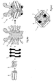
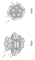
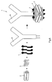


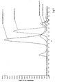
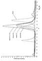

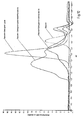


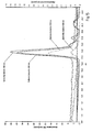
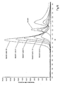
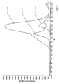

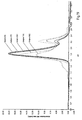
Classifications
-
- A—HUMAN NECESSITIES
- A61—MEDICAL OR VETERINARY SCIENCE; HYGIENE
- A61K—PREPARATIONS FOR MEDICAL, DENTAL OR TOILETRY PURPOSES
- A61K47/00—Medicinal preparations characterised by the non-active ingredients used, e.g. carriers or inert additives; Targeting or modifying agents chemically bound to the active ingredient
- A61K47/50—Medicinal preparations characterised by the non-active ingredients used, e.g. carriers or inert additives; Targeting or modifying agents chemically bound to the active ingredient the non-active ingredient being chemically bound to the active ingredient, e.g. polymer-drug conjugates
- A61K47/69—Medicinal preparations characterised by the non-active ingredients used, e.g. carriers or inert additives; Targeting or modifying agents chemically bound to the active ingredient the non-active ingredient being chemically bound to the active ingredient, e.g. polymer-drug conjugates the conjugate being characterised by physical or galenical forms, e.g. emulsion, particle, inclusion complex, stent or kit
- A61K47/6905—Medicinal preparations characterised by the non-active ingredients used, e.g. carriers or inert additives; Targeting or modifying agents chemically bound to the active ingredient the non-active ingredient being chemically bound to the active ingredient, e.g. polymer-drug conjugates the conjugate being characterised by physical or galenical forms, e.g. emulsion, particle, inclusion complex, stent or kit the form being a colloid or an emulsion
- A61K47/6917—Medicinal preparations characterised by the non-active ingredients used, e.g. carriers or inert additives; Targeting or modifying agents chemically bound to the active ingredient the non-active ingredient being chemically bound to the active ingredient, e.g. polymer-drug conjugates the conjugate being characterised by physical or galenical forms, e.g. emulsion, particle, inclusion complex, stent or kit the form being a colloid or an emulsion the form being a lipoprotein vesicle, e.g. HDL or LDL proteins
-
- A—HUMAN NECESSITIES
- A61—MEDICAL OR VETERINARY SCIENCE; HYGIENE
- A61K—PREPARATIONS FOR MEDICAL, DENTAL OR TOILETRY PURPOSES
- A61K9/00—Medicinal preparations characterised by special physical form
- A61K9/10—Dispersions; Emulsions
- A61K9/127—Liposomes
- A61K9/1275—Lipoproteins; Chylomicrons; Artificial HDL, LDL, VLDL, protein-free species thereof; Precursors thereof
-
- A—HUMAN NECESSITIES
- A61—MEDICAL OR VETERINARY SCIENCE; HYGIENE
- A61K—PREPARATIONS FOR MEDICAL, DENTAL OR TOILETRY PURPOSES
- A61K38/00—Medicinal preparations containing peptides
- A61K38/16—Peptides having more than 20 amino acids; Gastrins; Somatostatins; Melanotropins; Derivatives thereof
- A61K38/164—Peptides having more than 20 amino acids; Gastrins; Somatostatins; Melanotropins; Derivatives thereof from bacteria
-
- A—HUMAN NECESSITIES
- A61—MEDICAL OR VETERINARY SCIENCE; HYGIENE
- A61K—PREPARATIONS FOR MEDICAL, DENTAL OR TOILETRY PURPOSES
- A61K47/00—Medicinal preparations characterised by the non-active ingredients used, e.g. carriers or inert additives; Targeting or modifying agents chemically bound to the active ingredient
- A61K47/06—Organic compounds, e.g. natural or synthetic hydrocarbons, polyolefins, mineral oil, petrolatum or ozokerite
- A61K47/24—Organic compounds, e.g. natural or synthetic hydrocarbons, polyolefins, mineral oil, petrolatum or ozokerite containing atoms other than carbon, hydrogen, oxygen, halogen, nitrogen or sulfur, e.g. cyclomethicone or phospholipids
-
- A—HUMAN NECESSITIES
- A61—MEDICAL OR VETERINARY SCIENCE; HYGIENE
- A61K—PREPARATIONS FOR MEDICAL, DENTAL OR TOILETRY PURPOSES
- A61K47/00—Medicinal preparations characterised by the non-active ingredients used, e.g. carriers or inert additives; Targeting or modifying agents chemically bound to the active ingredient
- A61K47/30—Macromolecular organic or inorganic compounds, e.g. inorganic polyphosphates
- A61K47/42—Proteins; Polypeptides; Degradation products thereof; Derivatives thereof, e.g. albumin, gelatin or zein
-
- A—HUMAN NECESSITIES
- A61—MEDICAL OR VETERINARY SCIENCE; HYGIENE
- A61K—PREPARATIONS FOR MEDICAL, DENTAL OR TOILETRY PURPOSES
- A61K47/00—Medicinal preparations characterised by the non-active ingredients used, e.g. carriers or inert additives; Targeting or modifying agents chemically bound to the active ingredient
- A61K47/50—Medicinal preparations characterised by the non-active ingredients used, e.g. carriers or inert additives; Targeting or modifying agents chemically bound to the active ingredient the non-active ingredient being chemically bound to the active ingredient, e.g. polymer-drug conjugates
- A61K47/51—Medicinal preparations characterised by the non-active ingredients used, e.g. carriers or inert additives; Targeting or modifying agents chemically bound to the active ingredient the non-active ingredient being chemically bound to the active ingredient, e.g. polymer-drug conjugates the non-active ingredient being a modifying agent
- A61K47/54—Medicinal preparations characterised by the non-active ingredients used, e.g. carriers or inert additives; Targeting or modifying agents chemically bound to the active ingredient the non-active ingredient being chemically bound to the active ingredient, e.g. polymer-drug conjugates the non-active ingredient being a modifying agent the modifying agent being an organic compound
- A61K47/543—Lipids, e.g. triglycerides; Polyamines, e.g. spermine or spermidine
- A61K47/544—Phospholipids
-
- A—HUMAN NECESSITIES
- A61—MEDICAL OR VETERINARY SCIENCE; HYGIENE
- A61K—PREPARATIONS FOR MEDICAL, DENTAL OR TOILETRY PURPOSES
- A61K47/00—Medicinal preparations characterised by the non-active ingredients used, e.g. carriers or inert additives; Targeting or modifying agents chemically bound to the active ingredient
- A61K47/50—Medicinal preparations characterised by the non-active ingredients used, e.g. carriers or inert additives; Targeting or modifying agents chemically bound to the active ingredient the non-active ingredient being chemically bound to the active ingredient, e.g. polymer-drug conjugates
- A61K47/51—Medicinal preparations characterised by the non-active ingredients used, e.g. carriers or inert additives; Targeting or modifying agents chemically bound to the active ingredient the non-active ingredient being chemically bound to the active ingredient, e.g. polymer-drug conjugates the non-active ingredient being a modifying agent
- A61K47/62—Medicinal preparations characterised by the non-active ingredients used, e.g. carriers or inert additives; Targeting or modifying agents chemically bound to the active ingredient the non-active ingredient being chemically bound to the active ingredient, e.g. polymer-drug conjugates the non-active ingredient being a modifying agent the modifying agent being a protein, peptide or polyamino acid
- A61K47/64—Drug-peptide, drug-protein or drug-polyamino acid conjugates, i.e. the modifying agent being a peptide, protein or polyamino acid which is covalently bonded or complexed to a therapeutically active agent
-
- A—HUMAN NECESSITIES
- A61—MEDICAL OR VETERINARY SCIENCE; HYGIENE
- A61K—PREPARATIONS FOR MEDICAL, DENTAL OR TOILETRY PURPOSES
- A61K47/00—Medicinal preparations characterised by the non-active ingredients used, e.g. carriers or inert additives; Targeting or modifying agents chemically bound to the active ingredient
- A61K47/50—Medicinal preparations characterised by the non-active ingredients used, e.g. carriers or inert additives; Targeting or modifying agents chemically bound to the active ingredient the non-active ingredient being chemically bound to the active ingredient, e.g. polymer-drug conjugates
- A61K47/69—Medicinal preparations characterised by the non-active ingredients used, e.g. carriers or inert additives; Targeting or modifying agents chemically bound to the active ingredient the non-active ingredient being chemically bound to the active ingredient, e.g. polymer-drug conjugates the conjugate being characterised by physical or galenical forms, e.g. emulsion, particle, inclusion complex, stent or kit
- A61K47/6921—Medicinal preparations characterised by the non-active ingredients used, e.g. carriers or inert additives; Targeting or modifying agents chemically bound to the active ingredient the non-active ingredient being chemically bound to the active ingredient, e.g. polymer-drug conjugates the conjugate being characterised by physical or galenical forms, e.g. emulsion, particle, inclusion complex, stent or kit the form being a particulate, a powder, an adsorbate, a bead or a sphere
- A61K47/6927—Medicinal preparations characterised by the non-active ingredients used, e.g. carriers or inert additives; Targeting or modifying agents chemically bound to the active ingredient the non-active ingredient being chemically bound to the active ingredient, e.g. polymer-drug conjugates the conjugate being characterised by physical or galenical forms, e.g. emulsion, particle, inclusion complex, stent or kit the form being a particulate, a powder, an adsorbate, a bead or a sphere the form being a solid microparticle having no hollow or gas-filled cores
- A61K47/6929—Medicinal preparations characterised by the non-active ingredients used, e.g. carriers or inert additives; Targeting or modifying agents chemically bound to the active ingredient the non-active ingredient being chemically bound to the active ingredient, e.g. polymer-drug conjugates the conjugate being characterised by physical or galenical forms, e.g. emulsion, particle, inclusion complex, stent or kit the form being a particulate, a powder, an adsorbate, a bead or a sphere the form being a solid microparticle having no hollow or gas-filled cores the form being a nanoparticle, e.g. an immuno-nanoparticle
-
- A—HUMAN NECESSITIES
- A61—MEDICAL OR VETERINARY SCIENCE; HYGIENE
- A61K—PREPARATIONS FOR MEDICAL, DENTAL OR TOILETRY PURPOSES
- A61K9/00—Medicinal preparations characterised by special physical form
- A61K9/10—Dispersions; Emulsions
- A61K9/127—Liposomes
- A61K9/1274—Non-vesicle bilayer structures, e.g. liquid crystals, tubules, cubic phases, cochleates; Sponge phases
-
- A—HUMAN NECESSITIES
- A61—MEDICAL OR VETERINARY SCIENCE; HYGIENE
- A61K—PREPARATIONS FOR MEDICAL, DENTAL OR TOILETRY PURPOSES
- A61K9/00—Medicinal preparations characterised by special physical form
- A61K9/14—Particulate form, e.g. powders, Processes for size reducing of pure drugs or the resulting products, Pure drug nanoparticles
- A61K9/141—Intimate drug-carrier mixtures characterised by the carrier, e.g. ordered mixtures, adsorbates, solid solutions, eutectica, co-dried, co-solubilised, co-kneaded, co-milled, co-ground products, co-precipitates, co-evaporates, co-extrudates, co-melts; Drug nanoparticles with adsorbed surface modifiers
- A61K9/145—Intimate drug-carrier mixtures characterised by the carrier, e.g. ordered mixtures, adsorbates, solid solutions, eutectica, co-dried, co-solubilised, co-kneaded, co-milled, co-ground products, co-precipitates, co-evaporates, co-extrudates, co-melts; Drug nanoparticles with adsorbed surface modifiers with organic compounds
-
- A—HUMAN NECESSITIES
- A61—MEDICAL OR VETERINARY SCIENCE; HYGIENE
- A61K—PREPARATIONS FOR MEDICAL, DENTAL OR TOILETRY PURPOSES
- A61K9/00—Medicinal preparations characterised by special physical form
- A61K9/14—Particulate form, e.g. powders, Processes for size reducing of pure drugs or the resulting products, Pure drug nanoparticles
- A61K9/141—Intimate drug-carrier mixtures characterised by the carrier, e.g. ordered mixtures, adsorbates, solid solutions, eutectica, co-dried, co-solubilised, co-kneaded, co-milled, co-ground products, co-precipitates, co-evaporates, co-extrudates, co-melts; Drug nanoparticles with adsorbed surface modifiers
- A61K9/146—Intimate drug-carrier mixtures characterised by the carrier, e.g. ordered mixtures, adsorbates, solid solutions, eutectica, co-dried, co-solubilised, co-kneaded, co-milled, co-ground products, co-precipitates, co-evaporates, co-extrudates, co-melts; Drug nanoparticles with adsorbed surface modifiers with organic macromolecular compounds
-
- A—HUMAN NECESSITIES
- A61—MEDICAL OR VETERINARY SCIENCE; HYGIENE
- A61K—PREPARATIONS FOR MEDICAL, DENTAL OR TOILETRY PURPOSES
- A61K39/00—Medicinal preparations containing antigens or antibodies
Definitions
- the invention relates to lipoprotein particles, their use as a delivery or carrier system for hydrophobic agents, such as hydrophobic drugs or membrane proteins, and to a process for the manufacture of such disc-like lipoproteins.
- Membrane proteins are encoded by approximately 30% of all ORF ( Wallin and von Heijne, Protein Science 1998 Apr; 7 (4):1029-38 ) and represent an important class of drug targets since the majority of drugs, i.e. more than 60%, target in fact this class of proteins ( Overington et al., Nature Reviews Drug Discovery 5, 993-996 (December 2006 )). Membrane proteins play essential roles in many biological processes, such as signal transduction, transport of molecules and energy, recognition and cell-to-cell communication. Yet, membrane proteins are difficult to study due to their insolubility and tendency to aggregate when extracted from their natural lipid bilayer environment. In order to maintain the integrity of membrane proteins, an artificial hydrophobic environment is needed. Here, detergent micelles are most commonly employed which may, however, negatively impact on biocompatibility, can have adverse affects on membrane protein activity and may interfere with experimental conditions for assays.
- hydrophobic compounds and/or hydrophobic proteins are prone to aggregation, leading to locally highly concentrated drug particles that may cause high toxicity, unwanted immune responses and render the drug inactive ( Allen and Cullis, SCIENCE, 303 (5665): 1818-1822, MAR 19, 2004 ).
- EP 1 596 828 B1 describes disc-shaped bioactive agent delivery particles comprising an apolipoprotein which tightly surrounds a lipid bilayer in a double belt-like fashion. The interior of said particles is formed by the hydrophobic region of the lipid bilayer. This is in contrast to liposomes, which are closed spherical bilayer shells containing an aqueous interior.
- the disc-shaped bioactive agent delivery particles described in EP 1 596 828 B1 have a Stokes diameter of about 10 nm and are proposed for use as delivery vehicles for hydrophobic pharmaceutical drugs such as amphotericin B or camptothecin.
- EP 1 345 959 B1 describes a similar type of nanoscale particle with a diameter of about 10 nm and a height of about 5.5 nm.
- the particles are disc-shaped and composed of (i) an artificial membrane scaffold protein, (ii) a phospholipid bilayer and (iii) at least one hydrophobic or partially hydrophobic membrane protein.
- Said membrane scaffold protein again surrounds the lipid bilayer in a double belt-like fashion and is a derivative or a truncated form of human apolipoprotein A-1, lacks the N-terminal globular domain of human apolipoprotein A-1, is amphipathic and forms at least one ⁇ -helix and will, in aqueous environment, self-assemble with a phospholipid or mixture of phospholipids into a nanoscale particle of this discoidal shape.
- Such an engineered membrane scaffold protein (MSP) shall provide stability, size homogeneity and useful functionalities to the nanoscale discoidal lipoprotein particle.
- the saposin-family comprises 4 small ( ⁇ 80 amino acids) proteins, saposin A to D, that bind and/or interact with lipids and function as essential cofactors for several lysosomal enzymes in sphingolipid catabolism (cf. Bruhn, Biochem J. (2005) 389, 249-257 and references cited therein). Saposins have been described to prefer negatively charged lipids and low pH, exhibiting markedly increased activities at acidic pH, with a pH optimum at the intra-lysosomal pH of 4.75. Saposin A, B, C, and D are proteolytically hydrolyzed from a single large precursor protein, prosaposin.
- Saposin C is capable of inducing membrane fusion of phospholipid-containing vesicles in an acidic environment ( Archives of Biochemistry and Biophysics 2003 Jul 1; 415(1): 43-53 ), a feature not exhibited by the other saposins.
- Qi et al. (2009) Clin Cancer Res 15(18):5840-5851 report on saposin C-coupled dioleoylphosphatidylserine nanovesicles (SapC-DOPS) that contain an aqueous interior, have a mean diameter of about 190 nm and show tumor-targeting activity in vivo.
- saposin C acts as homing peptide for the liposome it is attached to. Saposin C then targets the liposome to cancer cells exposing phosphatidylserine on the outer leaflet of the cell membrane. The authors believe that a unique acidic microenvironment around cancer cells due to extracellular leakage of lysosomal enzymes makes tumor tissue an optimal target for saposin C.
- SapC-DOPS liposomes are prepared by drying solvent-dissolved phospholipids under N 2 ( g ), dispersing the dry phospholipids in acidic buffer (pH 5) containing purified saposin C, diluting the mixture 50 x in a physiologic aqueous solution and facilitating nanovesicle assembly by subsequent sonication.
- WO 2012/154825 A1 (inventors: Qi, Xiaoyang and Flick, Matthew J.) concerns a method for imaging a site of arthritis in an animal, wherein nanovesicle comprising a membrane-associated lysosomal protein (saposin C) incorporated into a phospholipid are used that are described to have a high fusogenic affinity for phosphatidylserine-rich domains on the surfaces of target cell membranes.
- saposin C membrane-associated lysosomal protein
- WO 2008/051818 A2 (inventor: Qi, Xiaoyang) concerns compositions and methods that are described as useful for the spontaneous formation of liposomes wherein the composition comprises anionic long chain lipids in combination with short chain lipids and a prosaposin-derived protein or polypeptide such as Saposin C.
- Popovic et al., PNAS, Vol. 109, No.8 (2012) 2908-2912 report on the structure of saposin A detergent discs.
- Saposin A exists in a soluble and a lipid/detergent-bound state. In the absence of lipid, saposin A adopts a closed monomeric apo conformation.
- the saposin A detergent disc structure reported by Popovic et al. reveals two chains of saposin A in an open conformation encapsulating 40 internally bound detergent molecules organized in a highly ordered bilayer-like hydrophobic core.
- Popovic et al. also describe the preparation of soluble lipid-saposin A complexes at pH 4.75 by a method requiring multiple steps.
- a uniform fraction of large unilamellar liposome vesicles is prepared by drying chloroform-dissolved lipids under N 2 (g), dispersing the dry lipids by vortex mixing in acidic buffer (50 mM sodium acetate pH 4.8, 150 mM NaCl), submitting the suspension to 10 cycles of freezing and thawing, blending in a vortex mixer for 5 min and extruding the mixture through a 200 nm filter.
- hydrophobic agents could potentially benefit from the apolipoprotein- or saposin A-derived nanodisc technology described in the prior art.
- due to the 3.2 nm size limitation of the saposin A derived particles it seems that - if at all - only small molecules may be incorporated into such particles at the acidic pH disclosed therein.
- bulky hydrophobic compounds and large biomolecules such as (oligomeric) membrane proteins can be incorporated into the Apolipoprotein A derived nanodiscs of the prior art, the maximum possible diameter is still limited by the double-belt like Apolipoprotein A perimeter of these particles.
- the 10 nm Apolipoprotein A derived nanodiscs may be too large for certain applications.
- nanoscale lipoprotein particles the size of which can be controlled and/or the size of which adjusts to the nature of the incorporated molecules and which are easy to produce and maintain a uniform size, quality and composition over time.
- a disc-shaped particle comprising a lipid binding polypeptide, lipids and a hydrophobic agent, wherein the particle does not comprise a hydrophilic or aqueous core, wherein the hydrophobic agent is neither a lipid nor a detergent, and wherein the lipid binding polypeptide is
- the invention also provides a pharmaceutical or diagnostic composition that comprises the particles of the invention.
- the invention further provides a process for preparing a disc-shaped particle comprising a lipid binding polypeptide and lipids, wherein the lipid binding polypeptide is a saposin-like protein (SAPLIP) as described in point a) above or a derivative form thereof as described in point b) above, and wherein the particle does not comprise a hydrophilic or aqueous core, the process comprising the step of
- the particles of the invention can be used as a hydrophobic agent delivery vehicle, as a tool for drug development, drug screening, membrane protein research, and as a vaccination formulation.
- the association of the hydrophobic agent with lipids and a saposin-like protein or a derivative or truncated form thereof provides a robust structure which is stable in aqueous solutions over a wide pH range, in particular at physiological pH, and allows larger particles than the 3.2 nm saposin A-derived lipoprotein particles of the prior art.
- Such particles are obtainable by the method of the invention which operates at a pH of from 6.0 to 10, thus allowing for self-assembly of the particles of the invention.
- the saposin-like protein-lipoprotein particles obtainable by the method of the invention differ from the particles of the prior art in multiple features, for example by their inherent size flexibility and ability to adapt to the respective size of the hydrophobic agent that is to be incorporated into the lipoprotein particles.
- the Salipro particles of the invention have proven to be capable of the incorporation of a variety of lipids, membrane proteins and hydrophobic compounds at a physiological pH, giving rise to nanoscale complexes that are soluble and stable in an aqueous environment.
- the Salipro particles of the invention are robust over concentrating using standard centrifugal filter units, freezing and thawing. Moreover, practical experiments revealed that the Salipro particles of the invention display a certain degree of thermostability. In addition, it is possible to freeze-dry, store and re-hydrate the particles of the invention without any major quality deterioration observable.
- the lipid binding polypeptide is a saposin-like protein (SAPLIP) or a derivative or truncated form thereof.
- SAPLIP saposin-like protein
- SAPLIP saposin-like protein
- the SAPLIP family is characterized by the saposin-fold, a conserved alpha-helical three-dimensional structure that is stabilized by highly conserved intramolecular disulphide bonds ( Munford et al. (1995), Journal of Lipid Research, vol. 36, no. 8, 1653-1663 and Bruhn (2005), Biochem J 389 (15): 249-257 ).
- SAPLIP saposin-like protein
- the SAPLIPs adopt a monomeric compact four-helix bundle-type structure, the saposin fold.
- This fold is exemplified by the structure of the closed apo form of human saposin A (Protein Data Bank (PDB) ID code: 2DOB, Ahn et al. (2006) Protein Sci. 15: 1849-1857 ) or the structures of saposin C (PDB ID code: 1M12; de Alba et al. (2003) Biochemistry 42, 14729-14740 ), NK-lysin (PDB ID code: 1NKL; Liepinsh et al. (1997) Nat. Struct. Biol.
- PDB Protein Data Bank
- amoebapore A (PDB ID code: 10F9) and granulysin (PDB ID code: 1L9L; Anderson et al. (2003) J. Mol. Biol. 325, 355-365 ) which are all nearly identical and easily super-imposable.
- SAPLIPs undergo a conformational change upon binding to ligands such as lipids or detergent molecules.
- ligands such as lipids or detergent molecules.
- SAPLIPs adopt a V-shaped or boomerang-shaped conformation with exposed hydrophobic surfaces that contact the bound lipids.
- the open conformation is exemplified by the saposin A detergent disc structure of the prior art (PDB ID code: 4DDJ; Popovic et al., PNAS, Vol. 109, No.8 (2012) 2908-2912 ) and the structure of saposin C bound to SDS detergent micelles (PDB ID code: 1SN6; Hawkins et al. (2005) J.Mol.Biol. 346: 1381-1392 ).
- the lipid binding polypeptide preferably is amphipathic, with one part of its structure more or less hydrophilic and facing the aqueous solvent and the other part more or less hydrophobic and facing the hydrophobic center of the particle which comprises the lipids.
- the lipid binding polypeptide is preferably characterized by amphipathic ⁇ -helices with more hydrophobic residues (such as A, C, F, G, I, L, M, V, W or Y) predominantly on one face of the helix and more polar or charged residues (such as D, E, N, Q, S, T, H, K, or R) on the other face of the helix.
- amino acid residues as used herein are as follows: A, Ala, alanine; V, Val, valine; L, Leu, leucine; I, lie, isoleucine; P, Pro, proline; F, Phe, phenylalanine; W, Trp, tryptophan; M, Met, methionine; G, Gly, glycine; S, Ser, serine; T, Thr, threonine; C, Cys, cysteine; Y, Tyr, tyrosine; N, Asn, asparagine; Q, Gln, glutamine; D, Asp, aspartic acid; E, Glu, glutamic acid; K, Lys, lysine; R, Arg, arginine; and H, His, histidine.
- the lipid binding polypeptide of the invention does not enclose the lipids in a double belt-like fashion (cf. Figure 1 ) but, rather, the particles of the invention are held together by a core comprising the lipids which is surrounded by two or more approximately V-shaped or boomerang-shaped lipid binding polypeptide arranged in a head-to-tail orientation with substantially no direct protein-protein contacts between the individual lipid binding polypeptides within a given particle of the invention (cf. Figures 2 to 7 ).
- this arrangement of lipid binding polypeptides and lipids in the particles of the invention provides the size flexibility that is observed when bulky hydrophobic agents or increasing amounts of lipids are incorporated into the inventive particles.
- the lipid binding polypeptide serves primarily as a structural protein, providing the scaffold for the disc-like structure of the lipoprotein particles of the invention. For this reason, structural features, in particular the saposin-fold that is characteristic of the SAPLIPs, are more important for defining the lipid binding polypeptide of the invention as compared to mere sequence determinants.
- SAPLIPs examples are saposins A, B, C or D (for example from Homo sapiens [cf. SEQ ID NO. 1 to 4], Equus caballus, Bos taurus, Mus musculus, Oryctolagus cuniculus, Rattus norvegicus or Xenopus laevis); Surfactant protein B (for example from Homo sapiens, Canis familiaris, Mus musculus, Oryctolagus cuniculus, Ovis aries or Rattus norvegicus); Granulysin (for example from Homo sapiens; cf. SEQ ID NO. 5); NK-lysin (for example from Sus scrofa; cf.
- Homo sapiens cf. SEQ ID NO. 1 to 4
- Surfactant protein B for example from Homo sapiens, Canis familiaris, Mus musculus, Oryctolagus cuniculus, Ovis aries or Rattus norvegicus
- NK-lysin orthologues for example from Equus caballus or Bos taurus
- Amoebapores for example from Entamoeba histolytica
- Amoebapore orthologues for example from Entamoeba dispar or Entamoeba invadens
- Amoebapore-like protein for example from Fasciola hepatica
- Naegleriapores for example from Naegleria fowleri
- Clornorin for example from Clonorchis sinensis
- Prosaposin for example from Homo sapiens, Equus caballus, Bos taurus, Mus musculus, Oryctolagus cuniculus, Rattus norvegicus or Xenopus laevis
- MSAP for example from Homo sapiens
- Sequences of specific SAPLIPs used according to the invention are given in Figure 4 A and 4 B of Bruhn (2005), Biochem J 389 (15): 249-257 , which figures and sequences specified therein are hereby specifically incorporated by reference.
- the sequences of particular SAPLIPs used according to the invention are given in the sequence listing as follows: SEQ ID NO. 1 Saposin A [Homo sapiens]; SEQ ID NO. 2 Saposin B [Homo sapiens]; SEQ ID NO. 3 Saposin C [Homo sapiens]; SEQ ID NO. 4 Saposin D [Homo sapiens]; SEQ ID NO. 5 Granulysin [Homo sapiens]; SEQ ID NO. 6 NK-lysin [Sus scrofa].
- a SAPLIP used according to the invention may also be a polypeptide comprising the saposin-fold as part of a multidomain protein. This is for example the case in acid sphingomyelinase (from Homo sapiens, Caenorhabditis elegans, Ciona intestinalis, Anopheles, Drosophila, Mus musculus or Rattus norvegicus); GDSL (Gly-Asp-Ser-Leu) lipase such as acyloxy hydrolase (from Homo sapiens or Rattus norvegicus); Countin (from Dictyostelium discoideum); J3-crystallin (from Tripedalia cystophora) and Plant aspartic proteases (from Viridiplantae).
- a further SAPLIP used according to the invention can be bacteriocin AS-48. Bacteriocin AS-48 displays antimicrobial activity, is also able to bind lipids and possesses the same fold as
- the invention is described in more detail for saposin A or a derivative or truncated from thereof as lipid binding polypeptide, and whereas saposin A or a derivative or truncated from thereof as lipid binding polypeptide is a preferred embodiment, the invention shall not be limited thereby. Rather, the invention explicitly extends to the entire family of saposin-like proteins (SAPLIPs) as lipid binding polypeptides of the invention.
- SAPLIPs saposin-like proteins
- the SAPLIP is saposin A, B, C or D, in particular a saposin selected from (Homo sapiens, Equus caballus, Bos taurus, Mus musculus, Oryctolagus cuniculus, Rattus norvegicus or Xenopus laevis) saposin A, saposin B, saposin C or saposin D.
- the SAPLIP is (Homo sapiens, Equus caballus, Bos taurus, Mus musculus, Oryctolagus cuniculus, Rattus norvegicus or Xenopus laevis) saposin A, saposin B or saposin D.
- Saposin C is special among the saposins in that it is capable of inducing membrane fusion, a feature which is not exhibited by the other saposins.
- the membrane fusion activity may not always be desirable.
- the lipid binding polypeptide is a saposin-like protein (SAPLIP) or a derivative or truncated form thereof, provided that the SAPLIP is not saposin C or provided that the SAPLIP is not saposin C or a derivative or truncated form thereof.
- SAPLIP saposin-like protein
- the SAPLIP is of human origin (i.e. a Homo sapiens SAPLIP).
- the SAPLIP is saposin A, preferably (Homo sapiens, Equus caballus, Bos taurus, Mus musculus, Oryctolagus cuniculus, Rattus norvegicus or Xenopus laevis) saposin A, and particularly preferred human saposin A, the amino acid sequence of which is given as SEQ ID NO. 1.
- Saposin A is a known protein. Its expression, purification and crystallization as LDAO-detergent complex is for example, described in PNAS, Vol. 109, No.8 (2012) 2908-2912 (Popovic et al.).
- the lipid binding polypeptide comprises the full length sequence of a SAPLIP.
- the lipid binding polypeptide is a derivative of a SAPLIP, in particular a polypeptide comprising an amino acid sequence with at least 40, 50 or 60 %, preferably at least 75 % identity to the full length sequence of the respective SAPLIP.
- the lipid binding polypeptide can comprise a sequence having an identity with the full length sequence of a SAPLIP of at least 80 %, 85 %, 90 % or 95 %.
- sequence identity refers to a degree of identity between proteins that can be calculated by optimal alignment of the sequences using a scoring matrix such as the Blosum62 matrix described in Henikoff S. and Henikoff JG., P. N. A. S. USA 1992, 89: 10915-10919 . Calculation of the percentage identity and optimal alignment of two sequences using the Blosum62 similarity matrix and the algorithm of Needleman and Wunsch (J. Mol. Biol. 1970, 48: 443-453 ) can be performed using the GAP program of the Genetics Computer Group (GCG, Madison, WI, USA) using the default parameters of the program.
- GAP Genetics Computer Group
- a derivative of a SAPLIP is a polypeptide comprising a sequence having one or more amino acid deletions, additions, insertions and/or substitutions in the amino acid sequence of the respective SAPLIP.
- the SAPLIP derivative can be a polypeptide comprising a sequence of a particular SAPLIP in which 1 to 40, preferably 1 to 30, and in particular 1 to 20 or 1 to 15 amino acids have been deleted, added, inserted and/or substituted.
- deletion refers to the removal of 1, 2, 3, 4, 5 or more amino acid residues from the respective starting sequence.
- insertion or “addition” as used herein refers to the insertion or addition of 1, 2, 3, 4, 5 or more amino acid residues to the respective starting sequence.
- substitution refers to the exchange of an amino acid residue located at a certain position for a different one.
- the lipid binding polypeptide is a derivative of saposin A that comprises one or more fragments of SEQ ID NO. 1.
- Preferred fragments correspond to the helices ⁇ 1, ⁇ 2, ⁇ 3 and ⁇ 4 of saposin A, wherein helix ⁇ 1 is formed by the following continuous stretch of amino acids: "SLPCDICKDVVTAAGDMLK”; helix ⁇ 2 is formed by the following continuous stretch of amino acids: "ATEEEILVYLEKTCDWL”; helix ⁇ 3 is formed by the following continuous stretch of amino acids: "PNMSASCKEIVDSYLPVILDIIKGEMS"; and helix ⁇ 4 is formed by the following continuous stretch of amino acids: "PGEVCSAL".
- the derivative of saposin A is a polypeptide comprising a sequence selected from helices ⁇ 1, ⁇ 2, ⁇ 3, ⁇ 4 of saposin A and combinations thereof, in particular wherein the polypeptide comprises the sequences of helices ⁇ 1, ⁇ 2 and ⁇ 3 of saposin A.
- the fragments of saposin A such as its helices ⁇ 1, ⁇ 2, ⁇ 3, ⁇ 4, may have one or more amino acid deletions, additions, insertions and/or substitutions in the amino acid sequence.
- lipid binding polypeptide When a derivative or truncated form of saposin A is used as lipid binding polypeptide according to the invention, said derivative or truncated form should be amphipathic, form at least one alpha helix, and be capable of self-assembling together with solubilized lipids into lipoprotein particles when employed in the preparation process according to the invention which is described in detail below.
- amphipathic refers to polypeptides or molecules having both hydrophilic and hydrophobic regions.
- the six cysteine residues corresponding to the six cysteines in the SAPLIP founding member saposin A should be present. It is referred in this respect to the positions of the cysteines in the sequence comparison in Figure 4 A and 4 B of Bruhn (2005), Biochem J 389 (15): 249-257 .
- the lipid binding polypeptide according to the invention may also include one or more non-natural amino acids; amino acid analogs, or a peptidomimetic structure, in which the peptide bond is replaced by a structure more resistant to metabolic degradation.
- the lipid binding polypeptide comprises additionally a tag such as a His6-tag, Strep-tag, Avi-tag, Myc-tag, Gst-tag, JS-tag, cysteine-tag, FLAG-tag or other tags known in the art at the N-terminus or at the C-terminus.
- a tag such as a His6-tag, Strep-tag, Avi-tag, Myc-tag, Gst-tag, JS-tag, cysteine-tag, FLAG-tag or other tags known in the art at the N-terminus or at the C-terminus.
- the lipid binding polypeptide of the invention can also be a chimeric polypeptide.
- chimeric refers to two or more molecules that are capable of existing separately and are joined together to form a single molecule having the desired functionality of all of its constituent molecules.
- the constituent molecules of a chimeric molecule may be joined synthetically by chemical conjugation or, where the constituent molecules are all polypeptides or analogs thereof, polynucleotides encoding the polypeptides may be fused together recombinantly such that a single continuous polypeptide is expressed.
- Such a chimeric molecule is also known as "fusion protein".
- the various constituents of the chimeric polypeptide can be directly attached to each other or can be coupled through one or more linkers.
- the lipid binding polypeptide is a chimeric polypeptide further comprising a functional moiety such as a targeting moiety or a bioactive moiety.
- such a targeting moiety can, for example, serve to target the particles of the invention to a particular cell or tissue type, or to the infectious agent itself.
- the particle includes a targeting moiety attached to a lipid binding polypeptide or lipid component.
- the hydrophobic agent that is incorporated into the particle has a targeting capability.
- the targeting moiety can for example have receptor recognition properties so that the particles can be targeted to a specific cell surface receptor.
- the particles of the invention may be targeted to a particular cell type known to harbor a particular type of infectious agent, for example by modifying the lipid binding polypeptide component of the particles to render it capable of interacting with a receptor on the surface of the cell type being targeted.
- the targeting moiety is selected from the group consisting of natural or synthetic ligands, antibodies and antibody fragments or other biomolecules suitable for targeting purposes.
- the lipid binding polypeptide comprises a bioactive moiety
- this can be selected, for example, from a drug, a cytotoxic agent, an enzyme, a label, a fluorophore, a contrast agent and a radiolabel.
- Suitable lipids for the purpose of the invention can be selected from naturally occurring lipids, synthetic lipids, modified lipids, fats, waxes, sterols, fat-soluble vitamins, monoglycerides, diglycerides, triglycerides, phospholipids, fatty acids, glycerolipids, glycerophospholipids, sphingolipids, saccharolipids, polyketides, sterol lipids and prenol lipids or combinations thereof.
- lipid as used herein is art-recognized and refers to a substance of biological or synthetic origin that is soluble or partially soluble in organic solvents or which partitions into a hydrophobic environment when present in aqueous phase.
- lipid as used herein is not meant as a single lipid molecule in the particle of the invention. In fact, it is meant as a plurality of the same lipid molecules or a plurality of at least two different kinds of lipid molecules being present therein.
- the lipids are lipid bilayer forming lipids and/or biocompatible lipids.
- biocompatible denotes being biologically compatible by not producing a toxic, injurious, or immunological response in living tissue.
- bilayer-forming lipid refers to a lipid that is capable of forming a lipid bilayer with a hydrophobic interior and a hydrophilic exterior. Any bilayer-forming lipid that is capable of associating with a SAPLIP or a derivative or truncated form thereof to assemble into a disc-shaped structure may be used in accordance with the invention.
- Bilayer-forming lipids include, but are not limited to, phospholipids, sphingolipids, glycolipids, alkylphospholipids, ether lipids, and plasmalogens.
- One type of bilayer-forming lipid may be used or a mixture of two or more types.
- Particles may also include lipids that are not bilayer-forming lipids.
- lipids include, but are not limited to, cholesterol, cardiolipin, phosphatidylethanolamine (this lipid may form bilayers under certain circumstances), oxysterols, plant sterols, ergosterol, sitosterol, cationic lipids, cerebrosides, sphingosine, ceramide, diacylglycerol, monoacylglycerol, triacylglycerol, gangliosides, ether lipids, alkylphospholipids, plasmalogens, prostaglandins, and lysophospholipids.
- the lipids are eukaryotic lipids and/or lipids present in the white and grey matter of the brain.
- Preferred lipids are phospholipids, glycosphingolipids, sterols, phosphatidylcholine, phosphatidylserine (PS), 2-oleoyl-1-pamlitoyl-sn-glycero-3-phosphocholine (POPC), 2-oleoyl-1-pamlitoyl-sn-glycero-3-glycerol (POPG), 2-oleoyl-1-pamlitoyl-sn-glycero-3-phosphoethanolamine (POPE), diacylglycerol, cholesterol, sphingomyelin, galactosylceramide, gangliosides, phosphatidylinositoles and sulphogalactoceramides or combinations thereof.
- Preferred lipids are eukaryotic lipids.
- the lipids comprise or consist of phospholipids.
- suitable phospholipids include, but are not limited to, DMPC, DMPG, POPC, dipalmitoylphosphatidylcholine (DPPC), dipalmitoylphosphatidylserine (DPPS), cardiolipin, dipalmitoylphosphatidylglycerol (DPPG), distearoylphosphatidylglycerol (DSPG), egg yolk phosphatidylcholine (egg PC), soy bean phosphatidylcholine, phosphatidylinositol, phosphatidic acid, sphingomyelin, and cationic phospholipids.
- a lipid comprised in the particle of the invention may be a modified lipid including one or more bound functional moieties, such as a targeting moiety or a bioactive moiety as described above.
- lipids While many lipids have a similarly amphiphilic general structure as compared to detergents, i.e. a polar hydrophilic head group and a nonpolar hydrophobic tail - lipids differ from detergents in the shape of the monomers, in the type of aggregates formed in solution, and in the concentration range required for aggregation.
- Lipids are generally substantially cylindrical in structure; the volume occupied by the hydrophobic tail is similar to the volume occupied by the polar head group.
- Detergents monomers are generally more cone-shaped; the volume occupied by the hydrophobic tail is smaller than the volume occupied by the polar head group.
- Detergents tend to aggregate into spherical or ellipsoid micelles that are water soluble without forming bilayer structures in the absence of lipids (cf. handbook "Detergents and their uses in membrane protein science” from Anatrace, www.anatrace.com).
- detergent examples include anionic detergents such as alkylbenzenesulfonates or bile acids, cationic detergents and non-ionic or zwitterionic detergents such as lauryl-dimethyl amine-oxides (LDAO), Fos-Cholines, CHAPS/CHAPSO, alkyl glycosides such as short, medium or longer chain alkyl maltosides and glucosides.
- anionic detergents such as alkylbenzenesulfonates or bile acids
- cationic detergents and non-ionic or zwitterionic detergents such as lauryl-dimethyl amine-oxides (LDAO), Fos-Cholines, CHAPS/CHAPSO, alkyl glycosides such as short, medium or longer chain alkyl maltosides and glucosides.
- LDAO lauryl-dimethyl amine-oxides
- CHAPS/CHAPSO alkyl glycosides
- alkyl glycosides such as short,
- the particles of the invention further comprise a hydrophobic agent that is different from the lipids otherwise comprised in the particle.
- the hydrophobic agent according to the invention is neither a lipid nor a detergent.
- the hydrophobic agent that can be incorporated into the particles of the invention can be a hydrophobic organic compound and/or a hydrophobic biomolecule. It can be a therapeutically or biologically active hydrophobic agent or a hydrophobic agent which simply stabilizes the discoidal shape of the particle of the invention.
- a hydrophobic agent is an agent, i.e. a compound and/or a biomolecule, which does not fully penetrate into or remain soluble in water and/or which tends to aggregate and/or partition into a hydrophobic environment when present in aqueous phase.
- the hydrophobic agent is effectively solubilized in the hydrophobic interior of the particle. Thereby it can maintain its native functionalities such as e.g. catalytic activity or ligand binding.
- Hydrophobic agents comprised in the particles of the invention generally include at least one hydrophobic (e. g. lipophilic) region capable of associating with or integrating into the hydrophobic portion of a lipid bilayer.
- at least a portion of the hydrophobic agent is intercalated between or penetrates into the hydrophobic portions (e.g. fatty acyl chains) of the lipid molecules in the interior of the particle.
- the hydrophobic agent can, for example, be a biologically active agent, a drug, an active ingredient of a drug, an active ingredient of a cosmetic product, an active ingredient of a plant protective product, a dietary and/or nutritional supplement, a diagnostic probe, a contrast agent, a label and/or an indicator.
- Hydrophobic drugs which can be included in the particle of the invention and administered to a patient in need thereof may be any drugs having low solubility in an aqueous environment.
- the low solubility in aqueous environment may only be apparent under certain conditions, e.g. certain pH or temperature ranges or when the concentration of the hydrophobic agent exceeds a certain threshold.
- Drugs which, for example, can be included in the particle of the invention and administered to a patient in need thereof are such for the treatment of cancer, inflammatory or infective conditions, cardiovascular diseases, neurological disorders and rheumatism among others.
- the hydrophobic agent can be an anti-oxidant, a vitamin, an anti-proliferative agent, a hormone, a steroid, or an enzyme. It can be an herbicidal or a fungicidal compound.
- hydrophobic drugs that can be incorporated into the particles of the invention include: curcumin, sulfonamide, such as sulfonamide, sulfamethoxazole and sulfacetamide; trimethoprim, particularly in combination with sulfamethoxazole; a quinoline such as norfloxacin and ciprofloxacin; a beta-lactam compound including a penicillin such as penicillin G, penicillin V, ampicillin, amoxicillin, and piperacillin, a cephalosporin such as cephalosporin C, cephalothin, cefoxitin and ceftazidime, other beta-lactam antibiotics such as imipenem, and aztreonam; a beta lactamase inhibitor such as clavulanic acid; an aminoglycoside such as gentamycin, amikacin, tobramycin, neomycin, kanamycin and netilmicin; a t
- hydrophobic agents into the particles of the invention is that they effectively become solubilized in the stable structure of the particles, which thereby can serve as deposit and/or delivery vehicle for the hydrophobic agents in aqueous environment, which is e.g. present in the majority of body fluids and tissues.
- the particles of the invention offer the advantage that from the outside they are hydrophilic whilst the hydrophobic agent is effectively solubilized in the hydrophobic interior of the particle, by which means the hydrophobic agent can maintain its native functionalities such as e.g. catalytic activity or ligand binding.
- the particles of the invention seem to be biocompatible.
- the lipoprotein particles of the invention have also proven capable of stably incorporating hydrophobic biomolecules such as e.g. a protein comprising a hydrophobic moiety.
- proteins comprising a hydrophobic moiety can, for example, be selected from a membrane protein, an integral transmembrane protein, an integral monotopic membrane protein, a peripheral membrane protein, an amphitropic protein in a lipid-bound state, a lipid-anchored protein and a chimeric protein with a fused hydrophobic and/or transmembrane domain.
- Integral membrane proteins are membrane proteins which are permanently bound to the lipid bilayer and usually require a detergent or apolar solvent to become displaced form the membrane.
- Transmembrane proteins are integral membrane proteins that span across the membrane at least once.
- transmembrane proteins examples include G-protein coupled receptors (GPCRs), porters such as uniporters, symporter or antiporters, channels such as ion channels or enzymes.
- GPCRs G-protein coupled receptors
- porters such as uniporters, symporter or antiporters
- channels such as ion channels or enzymes.
- Integral monotopic membrane proteins are permanently attached to the membrane only from one side and do not span across the membrane.
- This class includes membrane proteins that are tethered to the membrane via alpha-helical transmembrane anchors. Examples include cytochrome P450 oxidases and glycophorin A.
- Peripheral membrane proteins are only temporarily or indirectly associated with the lipid bilayer or integral membrane proteins incorporated therein. Peripheral membrane proteins usually dissociate from membranes following treatment with a polar reagent with an elevated pH or high salt concentrations. Examples of peripheral membrane proteins include phospholipase A2 or C, lipoxygenases and cytochrome c.
- Lipid-anchored proteins are bound to the lipid bilayer via lipidated, in particular prenylated or GPI-anchored amino acid residues.
- Examples include bacterial lipoproteins, G proteins and certain kinases.
- Amphitropic proteins are proteins that exist in at least two conformational states, a lipid free, water-soluble state and a lipid bound state. Upon association with lipids, amphitropic proteins undergo a conformational change allowing them to become reversibly or irreversibly membrane-associated. Examples of amphitropic proteins are pore-forming toxins and antibacterial peptides.
- the particles of the invention are discshaped. They can have a Stokes radius (hydrodynamic radius) R s in the range of from 2 nm to 200 nm, in particular from 3 nm to 150 nm, preferably from 3 nm to 100 nm.
- R s hydrodynamic radius
- the skilled person knows how to determine the Stokes radius. This is preferably done by analytical gel filtration (size exclusion chromatography), in comparison with standards of known Stokes radii.
- the particles can be subjected to a gel filtration step on e.g. a Superdex 200 HR10 30 gel filtration column and eluted with a suitable buffer at pH 7.5 and 0.5 ml/min at room temperature. Absorbance is monitored at 280 nm for protein.
- the standard proteins should span R s values above and below that of the particle of interest.
- a calibration curve is generated by plotting the elution position vs R s for the standard proteins. This generally gives an approximately linear plot, but otherwise, it is satisfactory to draw lines between the points and read the R s of the protein of interest from its elution position on this standard curve.
- the Stokes radius will be larger than 3.2 nm, in particular at least 3.5 nm, at least 5.0 nm or at least 10.0 nm.
- the particles of the invention may also be examined via transmission electron microscopy or, if the particles are large enough via negative-stain electron microscopy and single particle analysis as described in Example 12 below.
- the lipids assemble into a discoidal bilayer-like structure of discrete size in the interior of the particle.
- the lipid binding polypeptide component generally defines the boundary of the discoidal bilayer and provides structure and stability to the particle.
- the interior of the particle includes a hydrophobic region (e. g., comprised of lipid fatty acyl chains).
- particles of the invention do not comprise a hydrophilic or aqueous core.
- the particles are disc-shaped, having generally a flat, discoidal, roughly circular lipid bilayer circumscribed by amphipathic ⁇ -helices provided by two or more lipid binding polypeptides, which are associated with hydrophobic surfaces of the bilayer around the periphery of the disc.
- Illustrative examples of disc-shaped particles of the invention are schematically depicted in Figures 2 to 7 .
- the discoidal shape of the particles of the invention can be approximated by a cylinder with a ratio of the maximum height to the maximum diameter (major axis length) of at least 1.0: 1.1, in particular 1.0:1.5 or 1.0:2.0.
- the maximum height of the discoidal particle generally is at least 3.5 nm, in particular at least 5 nm, as determined by transmission electron microscopy or, if the particles are large enough, via negative-stain electron microscopy and single particle analysis.
- the particle of the invention has a top, a bottom and a circumferential side surface, with the maximum diameter (major axis length) of the top and bottom surface being larger than the height of the circumferential side surface.
- the lipid binding polypeptide is at least partially located to surround the circumferential side surface of the particle.
- the maximum diameter (major axis length) of the disc-shaped particle of the invention is from between 2 nm to 200 nm, in particular from 3 nm to 150 nm, preferably from 3 nm to 100 nm.
- the maximum diameter (major axis length) of the disc-shaped particle is from 3 nm to 80 nm, in particular from 3 nm to 60 nm. Practical experiments have shown that particles having a maximum diameter (major axis length) of 3 nm to 20 nm are particularly easily obtainable with the method of the invention.
- the particles are defined by a substantially monodisperse population of disk structures, as assessed by the gel filtration elution profile on for example a HiLoad SuperdexTM 200 16/60 GL column.
- the predominant interaction between the lipid binding polypeptide and a lipid bilayer in a particle is through hydrophobic interactions between residues on the hydrophobic faces of amphipathic ⁇ -helices of the lipid binding polypeptide molecules and hydrophobic surfaces of lipids, for example, phospholipid fatty acyl chains, at the edge of the bilayer at the periphery of the bioactive agent delivery particle.
- An amphipathic ⁇ -helix of lipid binding polypeptide molecule includes both a hydrophobic surface in contact with a hydrophobic surface of the lipid bilayer at the periphery of the particle, and a hydrophilic surface facing the exterior of the particle and in contact with the aqueous environment when the particle is suspended in aqueous medium.
- the particles are stable in aqueous solution and may be lyophilized for long term storage, followed by reconstitution in aqueous solution.
- Stability or “stable” as used herein means low to undetectable levels of particle fragmentation, low to undetectable levels of aggregation or quality deterioration during preparation, transportation, and storage of the particles.
- the particles according to the invention are stable in aqueous solutions at a pH of from 5.0 to 8.0 and a temperature of from -210°C to 4 °C for at least 1 day, at least 2 days, at least 7 days, at least 2 weeks, at least 1 month, at least 6 months or at least 12 months, as determined, for example, by visual inspection (clear and precipitate-free solution) or analytical gel filtration (less than 50 %, in particular from 1 to 40 % fragmentation of the particles).
- the particles of the invention are also stable at temperatures from 4 °C to 40°C in aqueous solutions at a pH of from 5.0 to 8.0 for at least 1 day, at least 2 days, at least 7 days, at least 2 weeks, at least 1 month or at least 3 months as determined, for example, by visual inspection (clear and precipitate-free solution) or analytical gel filtration (less than 50 %, in particular from 1 to 40 % fragmentation of the particles).
- the particles of the invention have also proven to be stable in aqueous solutions at a pH of from 5.0 to 8.0 and a temperature of from 40 °C to 75 °C for at least 10 minutes, as determined, for example, by visual inspection (clear and precipitate-free solution) or analytical gel filtration (less than 50 %, in particular from 1 and 40 % fragmentation of the particles).
- the particles may be lyophilized for long term storage, followed by reconstitution in aqueous solution.
- the particles of the invention are stable in lyophilized form at -210 °C to 40 °C, in particular from -210 °C to 25 °C for at least 1 day, at least 2 days, at least 7 days, at least 2 weeks, at least 1 month, at least 6 months or at least 12 months, as determined, for example, by analytical gel filtration after reconstitution in an appropriate buffer at pH 7.5 (less than 50 %, in particular less than 40 % or from 1 to 40 % fragmentation of the particles).
- “Fragmentation” as used herein means that in the gel filtration elution profile, the size of the peak (i.e.
- peak height corresponding to the particle of the invention has decreased at the expense of the peak size of free non-lipid-bound SAPLIP and/or free lipids and/or aggregates, as compared to the peak size of the freshly prepared particle of the invention. Accordingly, a fragmentation of 40 % for example means that the peak size (i.e. the height of the peak in the gel filtration elution profile) has decreased by 40 % as compared to the peak size prior to storage (100 %).
- the particles of the invention are particularly stable also in aqueous solutions that are substantially free of detergents.
- substantially free of detergents means that the aqueous solution comprises less than 0.001 % (w/v) of detergent based on the total volume of the aqueous solution.
- the invention further provides a process for preparing a disc-shaped particle comprising a lipid binding polypeptide and lipids, wherein the lipid binding polypeptide is a saposin-like protein (SAPLIP) as described for the disc-shaped particle according to the invention above or a derivative form thereof as described for the disc-shaped particle according to the invention above, and wherein the particle does not comprise a hydrophilic or aqueous core, the process comprising the step of
- SAPLIP saposin-like protein
- Also described herein is a self-assembly of the particles at a pH of from 5.0 to 10 or 5.0 to 8.5; in particular from 6.0 to 8.0 and most preferably from 7.0 to 8.0.
- the lipid binding polypeptide and the lipids are as described above.
- the lipid binding polypeptide is contacted with detergent-stabilized lipids in a lipid enviroment.
- the lipid binding polypeptide is contacted in step (a) with the solubilized lipids in an aqueous liquid.
- the aqueous liquid is a buffered solution at a pH of from 5.0 to 10.0 or from 5.0 to 8.5; in particular from 6.0 to 8.0 and most preferably from 7.0 to 8.0.
- the lipids have been solubilized by means of an organic solvent or a detergent.
- the lipids used in step (a) are in a detergent-solubilized state.
- Practical experiments have shown that a wide variety of detergents can be used to solubilize the lipids for being employed in the process of the invention.
- the process of the invention works very well with lipids solubilized in solutions comprising 0.01 to 5.0 %, in particular 0.1 to 1.0 % alkyl glycosides such as short or longer chain alkyl maltosides and glucosides.
- other suitable detergents may be used as well.
- the ability of a given detergent to solubilize a given lipid or lipid mix can easily be inspected visually by the formation of a clear solution devoid of aggregates, precipitates or phase separation.
- the lipids can be directly employed in the process of the invention without the need for an upstream liposome preparation step. This is advantageous as it simplifies the manufacturing process of the SAPLIP lipoprotein particles. In view of the solubilized nature of the lipids used as starting material for step (a) of the process of the invention, it is believed that the lipids are substantially not in the form of liposomes.
- the lipid binding polypeptides of the invention generally do not require detergents or other solvents during purification, storage or handling.
- the lipid binding polypeptide used in step (a) can be in a detergent-solubilized state.
- the molar ratio of lipid binding polypeptide to lipids in step (a) is at least 1:1, in particular at least 1:3, preferably at least 1:5 or 1:10.
- a pH of from 6.0 to 10.0 allows the components brought into contact with each other in step (a) to self-assemble into the particle of the invention.
- a pH of from 6.0 to 10.0 preferably of from 6.0 to 8.5
- SAPLIP-lipoprotein particles should be best assembled at or close to the saposins' natural pH optimum of 4.75, it was found that SAPLIP-lipoprotein particles can be obtained by a simplified and reliable process if a more neutral or basic pH is maintained during preparation of the particles.
- Step (b) of the process of the invention may comprises diluting the mixture obtained in step (a) with a liquid that contains less amounts of detergent than the mixture obtained in step (a).
- a dilution step further induces and facilitates the self-assembly of the particles of the invention. Without wanting to be bound by this theory, it is believed that such a dilution step effectively removes solvent or detergent molecules from the hydrophobic surfaces of the lipid binding polypeptide and the lipids, thereby triggering the particle self-assembly process of the invention via enhanced hydrophobic interactions of the components.
- step (b) may take place in exactly the same composition as prepared in step (a)
- step (b) comprises an organic solvent/detergent removal or dilution step.
- Step (b) can, for example, be a gel filtration step.
- the mixture obtained in step (a) is subjected to a gel filtration step, whereby the gel filtration buffer or other solution is a liquid that contains less amounts of detergent than the mixture obtained in step (a).
- step (a) and/or (b) is preferably performed at a temperature of from 1 °C to 95 °C, in particular from 15 °C to 80° C, particularly preferred at 30 °C to 40 °C, and/or near the gel to liquid crystalline phase transition temperature of the lipids used.
- the phase transition temperature of the lipids used can easily be found in the literature or determined by calorimetry.
- a purification of the particles of the invention by at least partial removal of free lipids and/or free lipid binding polypeptide is performed.
- Suitable purification methods are chromatographic methods, in particular size-exclusion chromatography, ultracentrifugation, dialysis, contacting with detergent-binding biobeads, use of concentrators, affinity chromatography, magnetic beads and/or membrane/filters to remove unbound/non-incorporated lipids and/or hydrophobic compounds.
- the products of the process of the invention are particles comprising the lipid binding polypeptide and the lipid component.
- These particles of the invention are referred to as Salipro particles in the further description of the invention and in certain embodiments can have any or all of the characteristics described above for the Salipro particles comprising a lipid binding polypeptide, lipids and a hydrophobic agent.
- the process of the invention can also be used to incorporate a hydrophobic agent into Salipro particles, thereby resulting in product particles comprising a lipid binding polypeptide, lipids and a hydrophobic agent.
- the hydrophobic agent can have any or all of the characteristics described above for the various hydrophobic agents that can be comprised in the particles of the invention.
- step a) of the process of the invention is simply modified in that the lipid binding polypeptide is contacted with lipids in a liquid environment comprising the hydrophobic agent that is to be incorporated into the particle.
- Practical experiments have shown that the process of the invention allows for stable incorporation of both hydrophobic organic compounds, such as for example curcumin, as well as monomeric or oligomeric membrane proteins into the particles of the invention.
- the hydrophobic agent has been solubilized by means of a suited organic solvent (such as for example DMSO, methanol and/or chloroform) or a detergent prior to being employed in step (a).
- a suited organic solvent such as for example DMSO, methanol and/or chloroform
- the hydrophobic agent used in step (a) is in a detergent-solubilized state.
- the detergent can be the same or different from the detergent used to solubilize the lipids.
- the detergent used to solubilize the lipids and/or the hydrophobic agent that are/is employed in step (a) is not carried over in substantial amounts into the finished particles of the invention.
- the amount of detergent in the particles obtainable by the process of the invention can be low to undetectable.
- the particle of the invention does not comprise any substantial amounts of detergent, in particular less than 0.1 wt-%, preferably less than 0.01 wt-%, particularly preferred less than 0.001 wt-% detergent based on the weight of the particle.
- the amount of detergent present in the particles can be determined, for example, by mass spectrometry.
- step (a) If the hydrophobic agent used in step (a) is in a detergent-solubilized state, practical experiments have shown that it is advantageous to use detergents for purification and/or solubilization of the hydrophobic agent with short- to medium-chain hydrophobic tails. This is particularly true if a membrane protein is incorporated as hydrophobic agent.
- "Short chain hydrophobic tails" as used herein means C2 to C9, such as for example in n-Nonyl- ⁇ -maltoside (NM);
- “medium chain hydrophobic tails” as used herein means C10 to C15, such as for example in n-Decyl-ß-maltoside (DM) or n-Dodecyl-ß-maltoside (DDM).
- the detergent used for purification and/or solubilization of the hydrophobic agent has from 2 to 12 carbon atoms in its hydrophobic tail, preferable from 2 to 10 and most preferred from 2 to 9 carbon atoms in its hydrophobic
- the molar ratio of lipid binding polypeptide to hydrophobic agent in step (a) is at least 1:0.5, preferably at least 1:2 or at least 1:5, particularly preferred at least 1:10. In another embodiment, the molar ratio of lipid binding polypeptide to hydrophobic agent in step (a) is from 1:0.5 and 1:10000, in particular from 1:0.5 and 1:1000 or from 1:0.5 and 1:500. Practical experiments have shown that the stoichiometry of components in and the size of the resulting particles can be controlled by adjusting the ratio of the starting components accordingly.
- the invention is also directed to the particle obtainable by the process of the invention described above, i.e. irrespective of the process that was actually used to prepare the particle.
- the particles obtainable by the process of the invention differ from the particles of the prior art in multiple features, for example by their inherent size flexibility and ability to adapt to the respective size of the hydrophobic agent that is to be incorporated into the lipoprotein particles.
- the invention also provides a pharmaceutical composition for delivering a hydrophobic agent to an individual in need thereof, wherein the composition comprises particles of the invention and wherein the hydrophobic agent is an active ingredient and/or wherein in addition to the hydrophobic agent an active ingredient is present.
- the pharmaceutical composition can optionally comprise a (further) pharmaceutically acceptable vehicle, carrier or adjuvant.
- the individual components of the particles and the pharmaceutical composition should be pharmaceutically acceptable.
- pharmaceutically acceptable refers to components, compounds or agents that are, within the scope of sound medical judgment, suitable for use in contact with the tissues of humans and lower animals without undue toxicity, irritation, allergic response and the like, and are commensurate with a reasonable benefit/risk ratio.
- the invention provides a method of treating an individual in need thereof, with a therapeutically effective amount of the pharmaceutical composition described above.
- a "therapeutically effective amount" of the pharmaceutical composition is that amount effective for treating or lessening the severity of the disease or condition to be treated.
- the term "individual”, as used herein, means an animal, for example, a mammal, and more specifically a human.
- compositions of this invention can be administered to humans and other animals orally, rectally, parenterally, intracisternally, intravaginally, intraperitoneally, topically (as by powders, ointments, or drops), bucally, as an aerosol, an oral or nasal spray, or the like, depending on the severity of the disease or condition being treated.
- the pharmaceutical composition can be formulated for enteral, parenteral and/or topical administration. It can be administered as a capsule, infusion or injection, a brushable or potable composition or as an aerosol.
- the particles of the invention are present in solid form, as a dispersion or in solution.
- the particles of the invention are also useful for diagnostic and/or cosmetic applications.
- particles which comprise detectable substances such as labels, markers or indicators
- the labels, markers or indicators may themselves be comprised in or formed by the hydrophobic agent, however they may also be attached to the lipid binding polypeptide or the lipid components of the particle.
- diagnostic and life science research tools according to the invention include particles with labeled incorporated hydrophobic proteins, labeled lipid binding polypeptide, labeled lipids, incorporated fluorophores or contrast agents (for example for MR imaging).
- the label can for example be a fluorescent label.
- the particles of the invention are useful as a tool for drug development, drug screening and/or membrane protein research.
- a membrane protein drug target such as a cell surface receptor or ion channel
- a membrane protein drug target such as a cell surface receptor or ion channel
- Such particles can then be employed in assays to study the activity of the drug target membrane protein in its native lipid bilayer environment or used in drug screenings to identify new drugs.
- the particles of the invention may also be fixed to a solid support making them useful for applications such as Surface Plasmon Resonance (SPR) or biosensor applications.
- SPR Surface Plasmon Resonance
- the particles of the invention are generally useful for rendering otherwise insoluble membrane proteins soluble in aqueous solutions in a native-like bilayer microenvironment.
- the invention provides a wide variety of new applications in membrane protein research.
- the particles of the invention allow studying membrane proteins incorporated in the particles of the invention by methods such as nuclear magnetic resonance (NMR), X-ray crystallography, electron microscopy (EM), mass spectrometry, isothermal titration calorimetry (ITC), differential light scattering, small-angle X-ray scattering (SAXS) and the like.
- the particles of the invention are useful as vaccination formulation, as carrier thereof or as drug delivery vehicle.
- Many pathogenic antigens that could be particularly potent in vaccinations are exposed on the surface and/or comprised in the outer cell membrane of the pathogens, i.e. derived from pathogenic lipids, other hydrophobic biomolecules or membrane proteins.
- the major antigens on Influenza viruses are present in the integral membrane protein Hemagglutinin.
- the particles of the invention are also useful to serve as antigen-presenting delivery vehicles for generating antibodies against hydrophobic agents or biomolecules in suitable host animals such as e.g. rabbits, goats or lamas.
- the particle of the invention can be used in cosmetics, in particular wherein the particle of the invention includes skin nourishing and/or nurturing agents, such as vitamins and/or antioxidants.
- Figure 1 depicts - in schematic form - the shape and molecular organization of the Apolipoprotein A-1 containing nanosdisc particles 10 of the prior art (e.g. EP 1 596 828 B1 discussed above).
- the particle of the prior art 10 is disc-shaped comprising an apolipoprotein scaffold protein 11 which tightly surrounds a lipid bilayer formed by lipids 3 in a double belt-like fashion.
- the interior of said particles is formed by the hydrophobic region of the lipids 3 in the lipid bilayer.
- the Stokes diameter of the particle 10 is in the range of 10 nm.
- Figure 2 depicts - in schematic form - the preparation as well as shape and molecular organization of Salipro particles 1 according to the invention comprising hydrophobic organic compounds 4, depicted in a) as side view and in b) as top view.
- the particle 1 of the invention is prepared by mixing purified lipid binding polypeptide 2 with lipids 3 and the hydrophobic organic compounds 4 to be incorporated and allowing the self-assembly of the particle 1 at a pH of from about 6.0 to about 10.0.
- the lipid binding polypeptide 2 is a saposin-like protein (SAPLIP) and comprises 4 amphipathic helices which are depicted as cylinders.
- SAPLIP saposin-like protein
- the lipids 3 are amphiphilic comprising a hydrophilic head group (depicted as circle) and hydrophobic tails such as fatty acyl chains (depicted as zigzag line).
- the hydrophobic organic compounds 4 are embedded in the hydrophobic portion of the lipid bilayer formed by the lipids 3.
- the hydrophobic organic compound 4 can, for example, be a biologically active agent, a drug, an active ingredient of a drug, an active ingredient of a cosmetic product, an active ingredient of a plant protective product, a dietary and/or nutritional supplement, a diagnostic probe, a contrast agent, a label and/or an indicator.
- FIG 3 depicts - in schematic form - the preparation as well as shape and molecular organization of Salipro particles 1 comprising an integral monomeric membrane protein 5 according to the invention, depicted in a) as side view and in b) as top view.
- the membrane protein 5 can be an integral transmembrane protein in monomeric form, as depicted in Figures 3 and 4 . However, it can also be in an oligomeric state as depicted in Figure 5 or a peripheral membrane protein, an amphitropic protein in a lipid-bound state, a lipid-anchored protein or a chimeric protein with a fused hydrophobic and/or transmembrane domain, all of which may be in a monomeric or oligomeric state.
- the particle 1 of the invention is prepared by mixing purified SAPLIP 2 with lipids 3 and the membrane protein 5 to be incorporated and allowing the self-assembly of the particle 1 at a pH of from about 6.0 to about 10.0.
- the membrane protein 5 can be associated with detergent molecules 6 and/or lipids 12.
- the detergent molecules 6 can come from the purification and/or solubilization of the membrane protein 5.
- the lipids 12 associated with the membrane protein 5 can be a carry-over from the membrane protein's native lipid environment prior to its purification.
- the membrane protein 5 is embedded in the hydrophobic portion of the lipid bilayer and adopts a similar conformation as in its native membrane-bound state.
- the particle 1 may optionally comprise lipids 12 and/or detergent molecules 6 derived from the purified membrane protein 5. In certain embodiments, the particle 1 does not comprise any substantial amounts of detergent molecules 6, in particular less than 0.1 wt-% detergent molecules based on the weight of the particle.
- the particle of the invention 1 is flexible in size. Schematic figures 2 to 7 are not drawn to scale. Depending on the size of the membrane protein 5 incorporated in the particle 1, the particle depicted in Figure 3 can be substantially larger than the lipid-only particle depicted in Figure 7 or the particle comprising low molecular weight organic hydrophobic compounds 4 as depicted in Figure 2 . Generally, an increase in particle size will also be reflected by the number of SAPLIP molecules 2 per particle, which can be more than two.
- the particle of the invention may for example comprise two to twenty, in particular two to ten SAPLIP molecules 2.
- the size of the particle 1 can also be influenced by the amounts of lipids 3 added in step (a) of its preparation.
- Figure 4 depicts - in schematic form - the preparation as well as shape and molecular organization of Salipro particles 1 comprising an integral monomeric membrane protein 5 and hydrophobic organic compounds 4 according to the invention, depicted in a) as side view and in b) as top view.
- the particle 1 of the invention is prepared by mixing purified SAPLIP 2 with lipids 3, the membrane protein 5 and the hydrophobic organic compounds 4 to be incorporated and allowing the self-assembly of the particle 1 at a pH of from about 6.0 to about 10.0.
- FIG. 5 is a schematic illustration of Salipro particles 1 according to the invention comprising an oligomeric membrane protein 7; depicted in a) as side view and in b) as top view.
- the particle 1 is flexible in size and adapts to the size of the hydrophobic agent 7 incorporated therein.
- the particle 1 comprises three SAPLIP molecules 2 per particle which are arranged in a head-to-tail fashion.
- the hydrodynamic radius of a particle comprising three SAPLIP molecules is in the range of from 5 to 20 nm, depending on the hydrophobic agent incorporated therein.
- FIG 6 depicts - in schematic form - the preparation as well as shape and molecular organization of Salipro particles 1 comprising a targeting component such as an antibody 8 and hydrophobic organic compounds 4.
- the targeting component 8 may be in the form of a lipid-anchored protein as depicted in Figure 6 or in the form of a chimeric protein with a fused hydrophobic and/or transmembrane domain.
- the targeting component may also be attached to or comprised in either the lipid binding polypeptide 2, one of the lipids 3 and/or the hydrophobic compound 4.
- Figure 7 depicts - in schematic form - the preparation as well as shape and molecular organization of Salipro particles according to the invention.
- the particle 1 of the invention is prepared by mixing purified SAPLIP 2 with lipids 3 and allowing the self-assembly of the particle 1 at a pH of from about 6.0 to about 10.0.
- the lipids 3 are amphiphilic comprising a hydrophilic head group (depicted as circle) and hydrophobic tails such as fatty acyl chains (depicted as zigzag line).
- the conformation of the SAPLIP 2 is different as compared to its lipid-bound "open" conformation in the particle of the invention 1 (cf.
- the SAPLIP 2 adopts a four-helix-bundle-type structure with the hydrophobic portions of its amphipathic helices facing the inside of the four-helix bundle.
- the SAPLIPs 2 adopt a V-shaped or boomerang-shaped conformation (cf. Fig 7b and Fig 7a , right side) with the hydrophobic portions of its amphipathic helices contacting the hydrophobic region of the lipids 3 in the lipid bilayer.
- Figure 7a shows the particle 1 and its preparation as side view, Figure 7b as top view.
- the particle of the invention 1 is approximately disc-shaped, having a flat, discoidal, roughly circular to square-shaped lipid bilayer circumscribed by the amphipathic ⁇ -helices of two SAPLIP molecules 2.
- the lipids 3 assemble into a discoidal bilayer-like structure of discrete size in the interior of the particle 1.
- the SAPLIPs 2 define the boundary of the discoidal bilayer in the particle 1, the interior of which is hydrophobic, i.e. comprised of lipid fatty acyl chains and lacking a hydrophilic or aqueous core.
- the particle 1 is held together mainly by the hydrophobic interactions of the lipids 3 within the bilayer core of the particle 1 and hydrophobic interactions between the lipids 3 and the hydrophobic portions of the amphiphilic helices of the SAPLIPs 2 facing the interior of the particle.
- the SAPLIPs 2 are arranged in a head-to-tail fashion and there are substantially no intermolecular protein-protein contacts between the SAPLIPs 2 in the particle 1.
- the particle 1 In its smallest form, the particle 1 is thought to contain two SAPLIP molecules 2 and at least around 10 lipid molecules 3.
- the particle of the invention 1 is flexible in size. Depending on its size and the molar ratio of components used in its preparation, it can accommodate multiple, i.e. more than two, SAPLIP molecules 2, many more lipids 3 and optionally further components.
- the particle may contain two to twenty, in particular two to ten SAPLIP molecules 2.
- the maximum height of the Salipro particle 1 in its lipid-only form corresponds to the height of the lipid bilayer and is about 5 nm.
- the particle 1 has a top (seen in Fig. 7b ), a bottom and a circumferential side surface, with the maximum diameter (major axis length) of the top and bottom surface being larger than the height of the circumferential side surface.
- Purified saposin A was prepared as follows. Saposin A protein expression was carried out using a vector with the coding region for human saposin A (SEQ ID NO: 1) inserted into a pNIC-Bsa4 plasmid and transformed and expressed in E. coli Rosetta gami-2 (DE3) (Novagen) strains. Cells were grown at 37 °C in TB medium supplemented with Tetracycline, Chloramphenicol and Kanamycin and induced with 0.7 mM IPTG. Three hours after induction, the cells were collected by centrifugation at 12.000 x g for 15 min.
- lysis buffer (20 mM Hepes pH 7.5, 150 mM NaCl, 20 mM Imidazol) and disrupted by sonication. Lysates were subjected to centrifugation at 26.000 x g for 30 min, the supernatant heated to 85 °C for 10 min, followed by an additional centrifugation step at 26.000 x g for 30 min.
- Preparative IMAC purification was performed by batch-adsorption of the supernatant by end-over-end rotation with Ni SepharoseTM 6 Fast Flow medium for 60 min.
- the chromatography medium was packed in a 10-mm-(i.d.) open gravity flow column and unbound proteins were removed by washing with 15 bed volumes of lysis buffer.
- the resin was washed with 15 bed volumes of wash buffer WB2 (20 mM Hepes pH 7.5, 150 mM NaCl, 40 mM Imidazol).
- Saposin A was eluted by addition of five bed volumes of elution buffer EB (20 mM Hepes pH 7.5, 150 mM NaCl, 400 mM Imidazol).
- the eluate was dialyzed overnight against gel filtration buffer GF pH 7.5 (20 mM Hepes pH 7.5, 150 mM NaCl) supplemented with recombinant TEV protease.
- TEV protease containing an uncleavable His-tag was removed from the eluate by passing it over 2 ml IMAC resin.
- Cleaved target proteins were concentrated to a volume of 5 ml using centrifugal filter units and loaded onto a HiLoad SuperdexTM 200 16/60 GL column using an AKTAexplorerTM 10 chromatography system (both GE Healthcare). Peak fractions were pooled and concentrated to 1.2 mg/ml protein.
- the protein sample was flash frozen in liquid nitrogen and stored at -80 C.
- saposin+Lipids pH 7.5 For the reconstitution of Salipro particles (denoted "Saposin+Lipids pH 7.5" in Figure 8 ), 10 ⁇ l of purified saposin A (1.2 mg/ml, 20 mM Hepes pH 7.5, 150 mM NaCl) were mixed with 1.6 ⁇ l of a 1 % (w/v) LDAO-detergent solution and 4 ⁇ l of a brain-lipid solution (5 mg/ml brain lipids, Sigma-Aldrich; 50 mM Hepes pH 7.5, 150 mM NaCl, 0.28 % (w/v) DDM) and incubated for 10 min at 37 °C.
- a brain-lipid solution 5 mg/ml brain lipids, Sigma-Aldrich; 50 mM Hepes pH 7.5, 150 mM NaCl, 0.28 % (w/v) DDM
- Example 2 was performed again, with the exception that, instead of GF-buffer pH 7.5, GF-buffer pH 4.75 was used (denoted "Saposin+Lipids pH 4.75" in Figure 8 ).
- the aim of this experiment was to assess the importance of the presence of lipids for the method according to the invention.
- 10 ⁇ l of purified saposin A (1.2 mg/ml, 20 mM Hepes pH 7.5, 150 mM NaCl) were mixed with 1.6 ⁇ l of a 1 % (w/v) LDAO-detergent solution and 4 ⁇ l of a brain-lipid solution (5 mg/ml brain lipids, Sigma-Aldrich; 50 mM Hepes pH 7.5, 150 mM NaCl, 0.28 % (w/v) DDM) and incubated for 10 min at 37 °C.
- a brain-lipid solution 5 mg/ml brain lipids, Sigma-Aldrich; 50 mM Hepes pH 7.5, 150 mM NaCl, 0.28 % (w/v) DDM
- GF-buffer pH 7.5 49.4 ⁇ l GF-buffer pH 7.5 was added to the mixture to obtain a final reaction volume of 65 ⁇ l After 10 min incubation at room temperature, the mixture was subjected to a gel-filtration step as above, using GF-buffer 7.5 (cf. "Saposin+LDAO+Lipids pH 7.5" in Figure 9 ).
- POPG, POPC, brain lipids and PS were dissolved to 20 mg/ml in GF-buffer pH 7.5 supplemented with 1 % (w/v) DDM, incubated for 1h at 37 °C with intermediate vortex-mixing and stored at -80 °C.
- the lipid solutions were diluted to 5 mg/ml with GF-buffer pH 7.5 supplemented with 0.03 % (w/v) DDM, leading to a final composition of 5 mg/ml lipids, 50 mM Hepes pH 7.5, 150 mM NaCl, 0.28 % (w/v) DDM.
- Salipro Particles comprising proteinaceous hydrophobic agents
- membrane proteins were incorporated into the Sailpro nanoparticles of the invention by using the same approach described above, i.e. mixing purified saposin A with lipids and the membrane protein to be incorporated at physiological pH followed by gel filtration.
- Salipro particles comprising the bacterial membrane protein YbgH (Salipro-YbgH, denoted "Saposin+Detergent+Lipids+MemProt YbgH pH 7.5" in Figure 11 )
- 10 ⁇ l of purified saposin A (1.2 mg/ml, 20 mM Hepes pH 7.5, 150 mM NaCl) were mixed with 1.6 ⁇ l of a 1 % (w/v) LDAO-detergent solution and 4 ⁇ l of a brain-lipid solution (5 mg/ml brain lipids, Sigma-Aldrich; 50 mM Hepes pH 7.5, 150 mM NaCl, 0.28 % (w/v) DDM) and 2.6 ⁇ l of purified membrane protein YbgH (10 mg/ml, 20 mM Hepes pH 7.5, 150 mM NaCl, 5 % glycerol, 0.03 % (w/v) DDM,
- Example 5 was performed again as described, with the exception that, instead of GF-buffer pH 7.5, GF-buffer pH 4.75 was used (cf. "Saposin+Detergent+Lipids+MemProt YbgH pH 4.75" in Figure 11 ).
- Salipro particles comprising another prokaryotic membrane protein, the MATE transporter, were prepared (Salipro-MATE, cf. "Saposin+Detergent+Lipids+MemProt MATE" in Figure 12 ).
- 10 ⁇ l of purified saposin A (1.2 mg/ml, 20 mM Hepes pH 7.5, 150 mM NaCl) were mixed with 2 ⁇ l of a 1 % (w/v) DDM-detergent solution, 5 ⁇ l of a brain-lipid solution (5 mg/ml brain lipids, Sigma-Aldrich; 50 mM Hepes pH 7.5, 150 mM NaCl, 0.28 % (w/v) DDM) and 7 ⁇ l of purified membrane protein MATE (9 mg/ml, 20 mM Hepes pH 7.5, 150 mM NaCl, 5 % glycerol, 0.01% DMNG, 0.5 mM TCEP) and incubated for 10 min at 37 °C.
- lipid-only Salipro particle control 10 ⁇ l of purified saposin A were mixed with 2 ⁇ l DDM-detergent (1 % w/v), 5 ⁇ l of a brain-lipid solution and incubated for 10 min at 37 °C. Subsequently, 48 ⁇ l GF-buffer pH 7.5 were added to the mixture to obtain a final reaction volume of 65 ⁇ l. After 10 min incubation at room temperature, the mixture was subjected to a gel-filtration step as above (cf. "Saposin+Detergent+Lipids in Figure 12 ).
- the results, which are depicted in figure 12 demonstrate that also the prokaryotic membrane transporter MATE could easily be incorporated into Salipro particles by following the method of the invention.
- the elution profile of Salipro-MATE (cf. Example 6 and "Saposin+Detergent+Lipids+MemProt MATE" in Figure 12 ) displays one major peak at 1.5 ml elution volume, corresponding to the membrane protein incorporated into Salipro particles and a smaller, lower molecular weight peak at 1.8 ml elution volume, corresponding to lipid-only Salipro-particles, eluting at the same volume as the lipid-only Salipro-particles(cf lipid-only Salipro particle control "Saposin+Detergent+Lipids" and cf. Fig.10 ).
- Salipro particles comprising another prokaryotic membrane protein, the S. oenidensis peptide transporter POT1, were prepared (Salipro-POT1, cf. "Saposin+Lipids+MemProt POT1" in Figure 13 ).
- a brain-lipid solution (5 mg/ml brain lipids, Sigma-Aldrich; 50 mM Hepes pH 7.5, 150 mM NaCl, 0.28 % (w/v) DDM) were incubated for 10 min at 37 °C, supplemented with 250 ⁇ l of purified membrane protein POT1 (10 mg/ml, 20 mM Hepes pH 7.5, 150 mM NaCl, 5 % glycerol, 0.3 % NM, 0.5 mM TCEP) and incubated for 30 s at 37 °C.
- purified membrane protein POT1 10 mg/ml, 20 mM Hepes pH 7.5, 150 mM NaCl, 5 % glycerol, 0.3 % NM, 0.5 mM TCEP
- saposin A 1.2 mg/ml, 20 mM Hepes pH 7.5, 150 mM NaCl
- saposin A 1.2 mg/ml, 20 mM Hepes pH 7.5, 150 mM NaCl
- 2 ml of GF-buffer pH 7.5 were added and the sample was centrifuged for 2 min at 14000 rpm.
- the supernatant was subjected to a gel-filtration step on a HiLoad SuperdexTM 200 16/60 GL column using an AKTAexplorerTM 10 chromatography system (both GE Healthcare) using GF-buffer 7.5 (cf. "Saposin+Lipids+MemProt POT1" in Figure 13 ).
- the elution profile of Salipro-POT1 displays one major peak corresponding to the membrane protein incorporated into Salipro particles and a smaller, lower molecular weight peak corresponding to lipid-only Salipro-particles. Accordingly, saposin A, lipids and the membrane protein associated in such way to form water-soluble particles with an incorporated membrane protein.
- Salipro particles comprising eukaryotic membrane protein, purified human Synaptophysin (SYP), were prepared (Salipro-SYP, cf. "Saposin+Lipids+MemProt SYP" in Figure 14 ).
- a brain-lipid solution (5 mg/ml brain lipids, Sigma-Aldrich; 50 mM Hepes pH 7.5, 150 mM NaCl, 0.28 % (w/v) DDM) were incubated for 10 min at 37 °C, supplemented with 800 ⁇ l of purified membrane protein SYP (4.2 mg/ml, 20 mM Hepes pH 7.5, 150 mM NaCl, 5 % glycerol, 0.03 % (w/v) DDM) and incubated for 5 min at 37 °C.
- a brain-lipid solution 5 mg/ml brain lipids, Sigma-Aldrich; 50 mM Hepes pH 7.5, 150 mM NaCl, 0.28 % (w/v) DDM
- purified membrane protein SYP (4.2 mg/ml, 20 mM Hepes pH 7.5, 150 mM NaCl, 5 % glycerol, 0.03 % (w/v) DDM)
- saposin A 1.2 mg/ml, 20 mM Hepes pH 7.5, 150 mM NaCl
- saposin A 1.2 mg/ml, 20 mM Hepes pH 7.5, 150 mM NaCl
- 900 ⁇ l GF-buffer pH 7.5 900 ⁇ l
- 1.9 ml of GF-buffer pH 7.5 were added and the sample was centrifuged for 2 min at 14000 rpm.
- the supernatant was subjected to a gel-filtration step on a HiLoad SuperdexTM 200 16/60 GL column using an AKTAexplorerTM 10 chromatography system (both GE Healthcare) using GF-buffer 7.5 (cf. "Saposin+Lipids+MemProt SYP" in Figure 14 ).
- a brain-lipid solution (5 mg/ml brain lipids, Sigma-Aldrich; 50 mM Hepes pH 7.5, 150 mM NaCl, 0.28 % (w/v) DDM) were incubated for 10 min at 37 °C. Then, 460 ⁇ l of purified saposin A (1.2 mg/ml, 20 mM Hepes pH 7.5, 150 mM NaCl) were added, the mixture was incubated for 1 min at 37°C and subsequently diluted with 630 ⁇ l GF-buffer pH 7.5.
- Salipro-SYP elutes as a single peak with a minor descending shoulder indicating the presence of lipid-only Salipro particles at that position (cf. "Saposin+Lipids+MemProt SYP" and "Saposin+Lipids” in Figure 14 ).
- Salipro Particles comprising hydrophobic organic compounds
- Salipro particles as carriers for hydrophobic compounds, such as lipophilic drugs
- hydrophobic compounds such as lipophilic drugs
- Curcumin was used as hydrophobic model drug for testing incorporation into Salipro particles using the method of the invention. Curcumin was primarily chosen because of its various pharmacologic implications (such as anti-cancer, anti-inflammatory, anti-oxidant and anti-proliferative activity) and its fluorescence in lipid environments. Using its UV-absorbance and fluorescence characteristics (excitation 420 nm, emission 500 nm) in a lipid environment, incorporation of Curcumin into Salipro particles can easily be followed.
- Curcumin 10 mg/ml, in DMSO
- 60 ⁇ l of a brain-lipid solution 5 mg/ml brain lipids, Sigma-Aldrich; 50 mM Hepes pH 7.5, 150 mM NaCl, 0.28 % (w/v) DDM
- 100 ⁇ l of purified saposin A 1.2 mg/ml, 20 mM Hepes pH 7.5, 150 mM NaCl
- saposin A 1.2 mg/ml, 20 mM Hepes pH 7.5, 150 mM NaCl
- Salipro particles of the invention are capable of rendering otherwise insoluble lipophilic compounds soluble, by incorporation into the lipid environment of the Salipro nanoscale particles.
- Salipro Particles adjust to the size/nature of the incorporated molecules
- Varying amounts (“Lipids 5": 1 ⁇ l, “Lipids 12.5": 2.5 ⁇ l, “Lipids 25”: 5 ⁇ l, “Lipids 50”: 10 ⁇ l, “Lipids 100”: 20 ⁇ l) of a brain-lipid solution (5 mg/ml brain lipids, Sigma-Aldrich; 50 mM Hepes pH 7.5, 150 mM NaCl, 0.28 % (w/v) DDM) were incubated for 10 min at 37 °C. 10 ⁇ l of purified saposin A were added, incubated for 1 min at 37 °C and GF-buffer pH 7.5 was added to a final volume of 41 ⁇ l.
- a brain-lipid solution 5 mg/ml brain lipids, Sigma-Aldrich; 50 mM Hepes pH 7.5, 150 mM NaCl, 0.28 % (w/v) DDM
- saposin A As negative control, 10 ⁇ l of purified saposin A (1.2 mg/ml, 20 mM Hepes pH 7.5, 150 mM NaCl) were incubated for 1 min at 37 °C and 31 ⁇ l GF-buffer pH 7.5 were added. After 10 min incubation at RT, 24 ⁇ l of GF-buffer pH 7.5 were added and the sample was subjected to a gel-filtration step on a SuperdexTM 200 5/150 GL analytical gel filtration column using a ⁇ KTAmicroTM chromatography system (both GE Healthcare) equipped with the Autosampler A-905, which automatically injected 25 ⁇ l of protein containing sample. Analytical gel filtration runs were performed at 4 °C at a flow rate of 0.2 ml/min in GF-buffer 7.5 (cf. "Saposin" in Figure 16 ).
- Salipro particles comprising small (27 kDa) human Synaptophysin (Saposin-SYP), the tetrameric (4x 56 kDa) E. coli peptide transporter POT1 (Saposin-POT1) and lipid-only Salipro particles were prepared as described in Examples 7 and 8.
- the samples were subjected to gel filtration on a HiLoad SuperdexTM 200 16/60 GL column using an AKTAexplorerTM 10 chromatography system (both GE Healthcare) with GF-buffer 7.5.
- the size of the Salipro particles of the invention is flexible and seems to adjust to the size of the hydrophobic agent incorporated therein.
- the "empty", i.e. lipid-only, Salipro particles have an average hydrodynamic radius of about 3 nm
- Salipro particles comprising the oligomeric POT1 transporter expand showing an average hydrodynamic radius of about 10 nm.
- Salipro particles with the incorporated bacterial peptide transporter (Salipro-POT1) were prepared as described in Example 7 and analyzed with negative-stain electron microscopy.
- Salipro-POT1 Samples of Salipro-POT1 were applied to glow-discharged copper grids coated with a thin carbon film and stained with uranyl formiate. Imaging was performed using a JEOL JEM2100F electron microscope at an acceleration voltage of 200 kV. Micrographs were recorded on a 4k CCD camera. An exemplary electron micrograph of purified Salipro-POT1 is shown in Figure 18 a) . Salipro-POT resembles a generally square-shaped particle that adopts various orientations on the grid, i.e. various side- or top-/bottom views of the particle were visible, allowing for 3D single-particle reconstruction.
- the appearance and the dimensions of the square-shaped Salipro-POT1 disc are in good agreement with the homotetrameric membrane protein complex formed by the bacterial peptide transporter POT1 in its native bacterial membrane environment.
- the thickness of the disc is about 5 nm, which is pronounced of the dimensions of a lipid bilayer and in accordance with expectations from the known structure of POT1 which mainly consists of transmembrane helices and lacks large cytosolic domains.
- the diameters and maximum diameter (major axis length) are in the range of 12 nm ⁇ 2 nm.
- each particle seems to be composed of four POT1 proteins and four saposin A molecules. This indicates a certain flexibility of saposin A to assemble into homogenous and stable lipoprotein-complexes, by adapting to the size of the incorporated hydrophobic agent.
- Salipro particles may therefore be composed of two saposin A molecules surrounding a lipid/detergent core in the empty, lipid-only state or when loaded with low-molecular weight and/or monomeric hydrophobic agents (cf. Figure 2 to 4 and 6 to 7 ), as well as several saposin A molecules around larger hydrophobic lipid/protein assemblies, as is the case for example with the homotetrameric POT1 (cf. Figures 5 and 18 ).
- Salipro particles are capable of incorporating a variety of lipids, membrane proteins and hydrophobic compounds at physiological pH, giving rise to nanoscale complexes that are soluble in an aqueous environment.
- their respective stability over time, temperature and various treatment conditions was tested.
- samples of lipid-only Salipro particles (65 ul each), prepared as described in Example 7 above, were flash-frozen, stored at -80 °C, thawed and incubated at either 0 °C, 37 °C, 50 °C, 73 °C or 95 °C for 10 min.
- the subsequent analysis was performed via a gel-filtration step on a Superdex TM 200 5/150 GL analytical gel filtration column using a AKTAmicroTM chromatography system (both GE Healthcare) equipped with the Autosampler A-905, which automatically injected 25 ⁇ l of protein containing sample.
- Analytical gel filtration runs were performed at 4 °C at a flow rate of 0.2 ml/min in GF-buffer 7.5.
- Salipro particles of the invention are also robust over concentrating using standard centrifugal filter units, freezing and thawing. In addition, it is possible to freeze-dry, store and re-hydrate the Salipro particles without significant quality deterioration.
Description
- The invention relates to lipoprotein particles, their use as a delivery or carrier system for hydrophobic agents, such as hydrophobic drugs or membrane proteins, and to a process for the manufacture of such disc-like lipoproteins.
- Membrane proteins and hydrophobic compounds are notoriously difficult to handle and represent two of the major challenges for pharmaceutical or life-science research and applications:
- (i) rendering insoluble hydrophobic compounds or membrane proteins soluble in aqueous solutions and (ii) the administration of such hydrophobic matter as therapeutic and diagnostic agents.
- Membrane proteins are encoded by approximately 30% of all ORF (Wallin and von Heijne, Protein Science 1998 Apr; 7 (4):1029-38) and represent an important class of drug targets since the majority of drugs, i.e. more than 60%, target in fact this class of proteins (Overington et al., Nature Reviews Drug Discovery 5, 993-996 (December 2006)). Membrane proteins play essential roles in many biological processes, such as signal transduction, transport of molecules and energy, recognition and cell-to-cell communication. Yet, membrane proteins are difficult to study due to their insolubility and tendency to aggregate when extracted from their natural lipid bilayer environment. In order to maintain the integrity of membrane proteins, an artificial hydrophobic environment is needed. Here, detergent micelles are most commonly employed which may, however, negatively impact on biocompatibility, can have adverse affects on membrane protein activity and may interfere with experimental conditions for assays.
- Another major pharmacological challenge is represented by the administration and delivery of hydrophobic compounds and/or hydrophobic proteins as therapeutic or diagnostic agents. Due to the limited solubility of these hydrophobic agents, they are prone to aggregation, leading to locally highly concentrated drug particles that may cause high toxicity, unwanted immune responses and render the drug inactive (Allen and Cullis, SCIENCE, 303 (5665): 1818-1822, MAR 19, 2004).
- Therefore, applications that incorporate hydrophobic agents such as membrane proteins, drugs or diagnostic compounds into soluble particles are highly desired. Current methods that address these two challenges involve amongst others liposomes and reconstituted high-density lipoprotein (rHDL) particles (Chan and Boxer, Current Opinion in chemical Biology 11:1-7, 2007).
-
EP 1 596 828 B1EP 1 596 828 B1 -
EP 1 345 959 B1 - However, there are several drawbacks with this currently available nanodisc technology in that, for example, a removal of detergent is required during assembly of the particles. Moreover, the size homogeneity provided by the tight double-belt like fit of the apolipoprotein-derived MSP seems to go at the expense of a fixed minimum particle size and a limitation as to the maximum diameters obtainable with the methods of the prior art.
- The saposin-family comprises 4 small (~ 80 amino acids) proteins, saposin A to D, that bind and/or interact with lipids and function as essential cofactors for several lysosomal enzymes in sphingolipid catabolism (cf. Bruhn, Biochem J. (2005) 389, 249-257 and references cited therein). Saposins have been described to prefer negatively charged lipids and low pH, exhibiting markedly increased activities at acidic pH, with a pH optimum at the intra-lysosomal pH of 4.75. Saposin A, B, C, and D are proteolytically hydrolyzed from a single large precursor protein, prosaposin. The complete amino acid sequences for saposins A, B, C and D have been reported as well as the genomic organization and cDNA sequence of prosaposin (O'Brien et al. (1988) Science 241, 1098- 1101; Furst et al (1992) Biochim Biophys Acta 1126: 1-16).
- Saposin C is capable of inducing membrane fusion of phospholipid-containing vesicles in an acidic environment (Archives of Biochemistry and Biophysics 2003 Jul 1; 415(1): 43-53), a feature not exhibited by the other saposins. Qi et al. (2009) Clin Cancer Res 15(18):5840-5851 report on saposin C-coupled dioleoylphosphatidylserine nanovesicles (SapC-DOPS) that contain an aqueous interior, have a mean diameter of about 190 nm and show tumor-targeting activity in vivo. In SapC-DOPS, saposin C or a peptide derived thereof acts as homing peptide for the liposome it is attached to. Saposin C then targets the liposome to cancer cells exposing phosphatidylserine on the outer leaflet of the cell membrane. The authors believe that a unique acidic microenvironment around cancer cells due to extracellular leakage of lysosomal enzymes makes tumor tissue an optimal target for saposin C. According to Qi et al., SapC-DOPS liposomes are prepared by drying solvent-dissolved phospholipids under N2 (g), dispersing the dry phospholipids in acidic buffer (pH 5) containing purified saposin C, diluting the mixture 50 x in a physiologic aqueous solution and facilitating nanovesicle assembly by subsequent sonication.
-
WO 2012/154825 A1 (inventors: Qi, Xiaoyang and Flick, Matthew J.) concerns a method for imaging a site of arthritis in an animal, wherein nanovesicle comprising a membrane-associated lysosomal protein (saposin C) incorporated into a phospholipid are used that are described to have a high fusogenic affinity for phosphatidylserine-rich domains on the surfaces of target cell membranes. As stated in Example 3 ofWO 2012/154825 A1 , the Saposin C nanovesicles are prepared as described in Qi et al. (see above). -
WO 2008/051818 A2 (inventor: Qi, Xiaoyang) concerns compositions and methods that are described as useful for the spontaneous formation of liposomes wherein the composition comprises anionic long chain lipids in combination with short chain lipids and a prosaposin-derived protein or polypeptide such as Saposin C. - Popovic et al., PNAS, Vol. 109, No.8 (2012) 2908-2912 report on the structure of saposin A detergent discs. Saposin A exists in a soluble and a lipid/detergent-bound state. In the absence of lipid, saposin A adopts a closed monomeric apo conformation. By contrast, the saposin A detergent disc structure reported by Popovic et al. reveals two chains of saposin A in an open conformation encapsulating 40 internally bound detergent molecules organized in a highly ordered bilayer-like hydrophobic core.
- Besides the crystallization of saposin A detergent discs, Popovic et al. also describe the preparation of soluble lipid-saposin A complexes at pH 4.75 by a method requiring multiple steps. First, a uniform fraction of large unilamellar liposome vesicles is prepared by drying chloroform-dissolved lipids under N2 (g), dispersing the dry lipids by vortex mixing in acidic buffer (50 mM sodium acetate pH 4.8, 150 mM NaCl), submitting the suspension to 10 cycles of freezing and thawing, blending in a vortex mixer for 5 min and extruding the mixture through a 200 nm filter. Mixing the thus prepared large unilamellar liposome vesicles with purified saposin A in acidic buffer resulted in soluble lipid-saposin A particles. The particle showed a narrow size distribution around an average hydrodynamic (Stokes) radius of 3.2 nm and contained about 5:1 lipid molecules per saposin A chain. The exact size of the particles was only moderately affected by the lipid to protein molar ratio and the composition of the liposomes. The authors observed similar 3.2 nm particles regardless of whether or not anionic phospholipids, cholesterol, or glycosphingolipids were present in the liposomal mixtures. In all cases, a single peak was observed in the size range of a Stokes radius of 3.2 nm, indicating a relatively narrow distribution of species. Hence, the technology of this publication is limited to a pH value of 4.75, to the aforementioned size of the particles, and includes a laborious upstream liposome preparation step.
- Against this background, there is a need for a reliable and easy to perform method for generating stable, defined lipoprotein-particle compositions for the solubilization of membrane proteins and other hydrophobic compounds. This is particularly true in view of the elaborate and multi-step processes disclosed in the prior art for preparing discoidal lipoprotein particles.
- A wide variety of hydrophobic agents could potentially benefit from the apolipoprotein- or saposin A-derived nanodisc technology described in the prior art. However, due to the 3.2 nm size limitation of the saposin A derived particles, it seems that - if at all - only small molecules may be incorporated into such particles at the acidic pH disclosed therein. Whereas bulky hydrophobic compounds and large biomolecules such as (oligomeric) membrane proteins can be incorporated into the Apolipoprotein A derived nanodiscs of the prior art, the maximum possible diameter is still limited by the double-belt like Apolipoprotein A perimeter of these particles. In addition, the 10 nm Apolipoprotein A derived nanodiscs may be too large for certain applications.
- Hence, there is a need for advanced nanodisc technology and superior liproprotein particles with flexible and controllable size ranges featuring the ability to adapt to the respective size of the hydrophobic agent that is to be incorporated into the lipoprotein particles. Such particles shall allow for simple integration of membrane proteins and other hydrophobic components which, for example, can be pharmaceutically or biologically active compounds or diagnostic compounds.
- The problem underlying the invention is seen in the provision of nanoscale lipoprotein particles, the size of which can be controlled and/or the size of which adjusts to the nature of the incorporated molecules and which are easy to produce and maintain a uniform size, quality and composition over time.
- This problem is solved by a disc-shaped particle comprising a lipid binding polypeptide, lipids and a hydrophobic agent, wherein the particle does not comprise a hydrophilic or aqueous core, wherein the hydrophobic agent is neither a lipid nor a detergent, and wherein the lipid binding polypeptide is
- (a) a saposin-like protein (SAPLIP) belonging to the SAPLIP family of lipid interacting proteins or
- (b) a derivative form thereof, wherein the derivate is
- (i) a protein having at least 80 % sequence identity to the full length sequence of SEQ ID NO. 1, 2, 3,4, 5 or 6;
- (ii)a protein having at least 40 % sequence identity to the full length sequence of SEQ ID NO. 1, 2, 3, 4, 5 or 6, wherein said protein is amphipathic, forms at least one alpha helix, and is capable of self-assembling together with solubilized lipids into lipoprotein particles when employed in the method of
claim 10; or - (iii) a protein comprising the sequence of SEQ ID NO. 1, 2, 3, 4, 5 or 6 in which 1 to 40 amino acids have been deleted, added, inserted and/or substituted.
- The invention also provides a pharmaceutical or diagnostic composition that comprises the particles of the invention.
- The invention further provides a process for preparing a disc-shaped particle comprising a lipid binding polypeptide and lipids, wherein the lipid binding polypeptide is a saposin-like protein (SAPLIP) as described in point a) above or a derivative form thereof as described in point b) above, and wherein the particle does not comprise a hydrophilic or aqueous core, the process comprising the step of
- a) contacting the lipid binding polypeptide with detergent-solubilized lipids in a liquid environment;
- b) allowing for the self-assembly of the particle at a pH of from 6.0 to 10.0.
- Finally, the particles of the invention can be used as a hydrophobic agent delivery vehicle, as a tool for drug development, drug screening, membrane protein research, and as a vaccination formulation.
- Without being bound to this theory, it appears that the association of the hydrophobic agent with lipids and a saposin-like protein or a derivative or truncated form thereof provides a robust structure which is stable in aqueous solutions over a wide pH range, in particular at physiological pH, and allows larger particles than the 3.2 nm saposin A-derived lipoprotein particles of the prior art. Such particles are obtainable by the method of the invention which operates at a pH of from 6.0 to 10, thus allowing for self-assembly of the particles of the invention.
- The saposin-like protein-lipoprotein particles obtainable by the method of the invention (designated "Salipro particles" herein) differ from the particles of the prior art in multiple features, for example by their inherent size flexibility and ability to adapt to the respective size of the hydrophobic agent that is to be incorporated into the lipoprotein particles.
- Contrary to the expectation that saposin-lipoprotein particles should be best assembled at or close to the saposins' natural pH optimum of 4.75, it was found that saposin-lipoprotein particles with improved properties and an extended application spectrum can be obtained when a more neutral or basic pH is maintained during preparation of the particles. Surprisingly, it was found that at a pH of from 6.0 to 10 and in the presence of solubilized lipids, purified saposin-like protein or a derivative or truncated form thereof self-assembles into stable lipoprotein particles without the need of a laborious upstream liposome preparation step. This simple and reliable preparation method failed to yield satisfactory results when particle assembly was attempted at the saposins' natural pH optimum of 4.75 or in the absence of lipids.
- The Salipro particles of the invention have proven to be capable of the incorporation of a variety of lipids, membrane proteins and hydrophobic compounds at a physiological pH, giving rise to nanoscale complexes that are soluble and stable in an aqueous environment.
- The Salipro particles of the invention are robust over concentrating using standard centrifugal filter units, freezing and thawing. Moreover, practical experiments revealed that the Salipro particles of the invention display a certain degree of thermostability. In addition, it is possible to freeze-dry, store and re-hydrate the particles of the invention without any major quality deterioration observable.
- The lipid binding polypeptide is a saposin-like protein (SAPLIP) or a derivative or truncated form thereof.
The term "saposin-like protein" (SAPLIP) as used herein is art-recognized and includes all members of the saposin-like protein (SAPLIP) family of lipid interacting proteins. The SAPLIP family is characterized by the saposin-fold, a conserved alpha-helical three-dimensional structure that is stabilized by highly conserved intramolecular disulphide bonds (Munford et al. (1995), Journal of Lipid Research, vol. 36, no. 8, 1653-1663 and Bruhn (2005), Biochem J 389 (15): 249-257). Examples of members of the saposin-like protein (SAPLIP) family according to the invention are described in Munford et al. (1995), Journal of Lipid Research, vol. 36, no. 8, 1653-1663 and Bruhn (2005), Biochem J 389 (15): 249-257. - In the ligand-free (i.e. detergent-free/lipid-free), "closed" state, the SAPLIPs adopt a monomeric compact four-helix bundle-type structure, the saposin fold. This fold is exemplified by the structure of the closed apo form of human saposin A (Protein Data Bank (PDB) ID code: 2DOB, Ahn et al. (2006) Protein Sci. 15: 1849-1857) or the structures of saposin C (PDB ID code: 1M12; de Alba et al. (2003) ), NK-lysin (PDB ID code: 1NKL; Liepinsh et al. (1997) Nat. Struct. Biol. 4, 793-795), amoebapore A (PDB ID code: 10F9) and granulysin (PDB ID code: 1L9L; Anderson et al. (2003) J. Mol. Biol. 325, 355-365) which are all nearly identical and easily super-imposable.
- SAPLIPs undergo a conformational change upon binding to ligands such as lipids or detergent molecules. In the ligand-bound "open" conformation, SAPLIPs adopt a V-shaped or boomerang-shaped conformation with exposed hydrophobic surfaces that contact the bound lipids. The open conformation is exemplified by the saposin A detergent disc structure of the prior art (PDB ID code: 4DDJ; Popovic et al., PNAS, Vol. 109, No.8 (2012) 2908-2912) and the structure of saposin C bound to SDS detergent micelles (PDB ID code: 1SN6; Hawkins et al. (2005) J.Mol.Biol. 346: 1381-1392).
- In the particles of the invention, the lipid binding polypeptide preferably is amphipathic, with one part of its structure more or less hydrophilic and facing the aqueous solvent and the other part more or less hydrophobic and facing the hydrophobic center of the particle which comprises the lipids. The lipid binding polypeptide is preferably characterized by amphipathic α-helices with more hydrophobic residues (such as A, C, F, G, I, L, M, V, W or Y) predominantly on one face of the helix and more polar or charged residues (such as D, E, N, Q, S, T, H, K, or R) on the other face of the helix.
- The abbreviations for amino acid residues as used herein are as follows: A, Ala, alanine; V, Val, valine; L, Leu, leucine; I, lie, isoleucine; P, Pro, proline; F, Phe, phenylalanine; W, Trp, tryptophan; M, Met, methionine; G, Gly, glycine; S, Ser, serine; T, Thr, threonine; C, Cys, cysteine; Y, Tyr, tyrosine; N, Asn, asparagine; Q, Gln, glutamine; D, Asp, aspartic acid; E, Glu, glutamic acid; K, Lys, lysine; R, Arg, arginine; and H, His, histidine.
- Contrary to the apolipoprotein-derived nanodiscs of the prior art, the lipid binding polypeptide of the invention does not enclose the lipids in a double belt-like fashion (cf.
Figure 1 ) but, rather, the particles of the invention are held together by a core comprising the lipids which is surrounded by two or more approximately V-shaped or boomerang-shaped lipid binding polypeptide arranged in a head-to-tail orientation with substantially no direct protein-protein contacts between the individual lipid binding polypeptides within a given particle of the invention (cf.Figures 2 to 7 ). Without wanting to be bound to this theory, it is believed that this arrangement of lipid binding polypeptides and lipids in the particles of the invention provides the size flexibility that is observed when bulky hydrophobic agents or increasing amounts of lipids are incorporated into the inventive particles. - Whereas the ability to interact with lipids as well as the above described amphipathic nature and three-dimensional structure is highly conserved among SAPLIPs, they are highly diverse on the amino acid sequence level, with sequence identities below the usual threshold zone of 25-30% identity to define homology (cf. sequence comparison in
Figure 4 A and 4 B of Bruhn (2005), Biochem J 389 (15): 249-257. - In the lipoprotein particles of the invention, the lipid binding polypeptide serves primarily as a structural protein, providing the scaffold for the disc-like structure of the lipoprotein particles of the invention. For this reason, structural features, in particular the saposin-fold that is characteristic of the SAPLIPs, are more important for defining the lipid binding polypeptide of the invention as compared to mere sequence determinants.
- Examples of SAPLIPs according to the invention are saposins A, B, C or D (for example from Homo sapiens [cf. SEQ ID NO. 1 to 4], Equus caballus, Bos taurus, Mus musculus, Oryctolagus cuniculus, Rattus norvegicus or Xenopus laevis); Surfactant protein B (for example from Homo sapiens, Canis familiaris, Mus musculus, Oryctolagus cuniculus, Ovis aries or Rattus norvegicus); Granulysin (for example from Homo sapiens; cf. SEQ ID NO. 5); NK-lysin (for example from Sus scrofa; cf. SEQ ID NO. 6); NK-lysin orthologues (for example from Equus caballus or Bos taurus); Amoebapores (for example from Entamoeba histolytica); Amoebapore orthologues (for example from Entamoeba dispar or Entamoeba invadens); Amoebapore-like protein (for example from Fasciola hepatica); Naegleriapores (for example from Naegleria fowleri); Clornorin (for example from Clonorchis sinensis); Prosaposin (for example from Homo sapiens, Equus caballus, Bos taurus, Mus musculus, Oryctolagus cuniculus, Rattus norvegicus or Xenopus laevis) and MSAP (for example from Homo sapiens).
- Sequences of specific SAPLIPs used according to the invention are given in
Figure 4 A and 4 B of Bruhn (2005), Biochem J 389 (15): 249-257, which figures and sequences specified therein are hereby specifically incorporated by reference. The sequences of particular SAPLIPs used according to the invention are given in the sequence listing as follows: SEQ ID NO. 1 Saposin A [Homo sapiens]; SEQ ID NO. 2 Saposin B [Homo sapiens]; SEQ ID NO. 3 Saposin C [Homo sapiens]; SEQ ID NO. 4 Saposin D [Homo sapiens]; SEQ ID NO. 5 Granulysin [Homo sapiens]; SEQ ID NO. 6 NK-lysin [Sus scrofa]. - A SAPLIP used according to the invention may also be a polypeptide comprising the saposin-fold as part of a multidomain protein. This is for example the case in acid sphingomyelinase (from Homo sapiens, Caenorhabditis elegans, Ciona intestinalis, Anopheles, Drosophila, Mus musculus or Rattus norvegicus); GDSL (Gly-Asp-Ser-Leu) lipase such as acyloxy hydrolase (from Homo sapiens or Rattus norvegicus); Countin (from Dictyostelium discoideum); J3-crystallin (from Tripedalia cystophora) and Plant aspartic proteases (from Viridiplantae).A further SAPLIP used according to the invention can be bacteriocin AS-48. Bacteriocin AS-48 displays antimicrobial activity, is also able to bind lipids and possesses the same fold as the remaining SAPLIP family members but is devoid of any disulphide bridges.
- Whereas, in the following, the invention is described in more detail for saposin A or a derivative or truncated from thereof as lipid binding polypeptide, and whereas saposin A or a derivative or truncated from thereof as lipid binding polypeptide is a preferred embodiment, the invention shall not be limited thereby. Rather, the invention explicitly extends to the entire family of saposin-like proteins (SAPLIPs) as lipid binding polypeptides of the invention. Due to the high degree of structural und functional conservation among SAPLIPs, the features and advantages of certain embodiments of the invention with saposin A as lipid binding polypeptide are expected to also apply to other embodiments using other SAPLIPs or derivatives or truncated forms thereof as lipid binding polypeptide of the invention.
- According to a preferred embodiment, the SAPLIP is saposin A, B, C or D, in particular a saposin selected from (Homo sapiens, Equus caballus, Bos taurus, Mus musculus, Oryctolagus cuniculus, Rattus norvegicus or Xenopus laevis) saposin A, saposin B, saposin C or saposin D. In one embodiment, the SAPLIP is (Homo sapiens, Equus caballus, Bos taurus, Mus musculus, Oryctolagus cuniculus, Rattus norvegicus or Xenopus laevis) saposin A, saposin B or saposin D.
- Saposin C is special among the saposins in that it is capable of inducing membrane fusion, a feature which is not exhibited by the other saposins. The membrane fusion activity may not always be desirable. According to a particular embodiment of the invention the lipid binding polypeptide is a saposin-like protein (SAPLIP) or a derivative or truncated form thereof, provided that the SAPLIP is not saposin C or provided that the SAPLIP is not saposin C or a derivative or truncated form thereof.
- In one embodiment, the SAPLIP is of human origin (i.e. a Homo sapiens SAPLIP).
- In a preferred embodiment, the SAPLIP is saposin A, preferably (Homo sapiens, Equus caballus, Bos taurus, Mus musculus, Oryctolagus cuniculus, Rattus norvegicus or Xenopus laevis) saposin A, and particularly preferred human saposin A, the amino acid sequence of which is given as SEQ ID NO. 1. Saposin A is a known protein. Its expression, purification and crystallization as LDAO-detergent complex is for example, described in PNAS, Vol. 109, No.8 (2012) 2908-2912 (Popovic et al.).
- According to one embodiment of the invention, the lipid binding polypeptide comprises the full length sequence of a SAPLIP. In another embodiment, the lipid binding polypeptide is a derivative of a SAPLIP, in particular a polypeptide comprising an amino acid sequence with at least 40, 50 or 60 %, preferably at least 75 % identity to the full length sequence of the respective SAPLIP. In particular, the lipid binding polypeptide can comprise a sequence having an identity with the full length sequence of a SAPLIP of at least 80 %, 85 %, 90 % or 95 %.
- The term "Sequence identity" as used herein refers to a degree of identity between proteins that can be calculated by optimal alignment of the sequences using a scoring matrix such as the Blosum62 matrix described in Henikoff S. and Henikoff JG., P. N. A. S. USA 1992, 89: 10915-10919. Calculation of the percentage identity and optimal alignment of two sequences using the Blosum62 similarity matrix and the algorithm of Needleman and Wunsch (J. Mol. Biol. 1970, 48: 443-453) can be performed using the GAP program of the Genetics Computer Group (GCG, Madison, WI, USA) using the default parameters of the program.
- As a comparison for amino acid alignments the EMBL-online tool "EMBOSS Stretcher" (http://www.ebi.ac.uk/Tools/psa/emboss_stretcher/) is used, using the programs default settings.
- Also described herein, a derivative of a SAPLIP is a polypeptide comprising a sequence having one or more amino acid deletions, additions, insertions and/or substitutions in the amino acid sequence of the respective SAPLIP. For example, the SAPLIP derivative can be a polypeptide comprising a sequence of a particular SAPLIP in which 1 to 40, preferably 1 to 30, and in particular 1 to 20 or 1 to 15 amino acids have been deleted, added, inserted and/or substituted.
- The term "deletion" as used herein refers to the removal of 1, 2, 3, 4, 5 or more amino acid residues from the respective starting sequence.
- The term "insertion" or "addition" as used herein refers to the insertion or addition of 1, 2, 3, 4, 5 or more amino acid residues to the respective starting sequence.
- The term "substitution" as used herein refers to the exchange of an amino acid residue located at a certain position for a different one.
- According to another embodiment of the invention, the lipid binding polypeptide is a derivative of saposin A that comprises one or more fragments of SEQ ID NO. 1. Preferred fragments correspond to the helices α1, α2, α3 and α4 of saposin A, wherein helix α1 is formed by the following continuous stretch of amino acids: "SLPCDICKDVVTAAGDMLK"; helix α2 is formed by the following continuous stretch of amino acids: "ATEEEILVYLEKTCDWL"; helix α3 is formed by the following continuous stretch of amino acids: "PNMSASCKEIVDSYLPVILDIIKGEMS"; and helix α4 is formed by the following continuous stretch of amino acids: "PGEVCSAL". According to a particular embodiment of the invention, the derivative of saposin A is a polypeptide comprising a sequence selected from helices α1, α2, α3, α4 of saposin A and combinations thereof, in particular wherein the polypeptide comprises the sequences of helices α1, α2 and α3 of saposin A. The fragments of saposin A, such as its helices α1, α2, α3, α4, may have one or more amino acid deletions, additions, insertions and/or substitutions in the amino acid sequence.
- When a derivative or truncated form of saposin A is used as lipid binding polypeptide according to the invention, said derivative or truncated form should be amphipathic, form at least one alpha helix, and be capable of self-assembling together with solubilized lipids into lipoprotein particles when employed in the preparation process according to the invention which is described in detail below. As used herein, the term "amphipathic" refers to polypeptides or molecules having both hydrophilic and hydrophobic regions.
- Preferably, if a derivative of a SAPLIP is used, the six cysteine residues corresponding to the six cysteines in the SAPLIP founding member saposin A should be present. It is referred in this respect to the positions of the cysteines in the sequence comparison in
Figure 4 A and 4 B of Bruhn (2005), Biochem J 389 (15): 249-257. - The lipid binding polypeptide according to the invention may also include one or more non-natural amino acids; amino acid analogs, or a peptidomimetic structure, in which the peptide bond is replaced by a structure more resistant to metabolic degradation.
- According to another embodiment of the invention the lipid binding polypeptide comprises additionally a tag such as a His6-tag, Strep-tag, Avi-tag, Myc-tag, Gst-tag, JS-tag, cysteine-tag, FLAG-tag or other tags known in the art at the N-terminus or at the C-terminus.
- The lipid binding polypeptide of the invention can also be a chimeric polypeptide. As used herein, "chimeric" refers to two or more molecules that are capable of existing separately and are joined together to form a single molecule having the desired functionality of all of its constituent molecules. The constituent molecules of a chimeric molecule may be joined synthetically by chemical conjugation or, where the constituent molecules are all polypeptides or analogs thereof, polynucleotides encoding the polypeptides may be fused together recombinantly such that a single continuous polypeptide is expressed. Such a chimeric molecule is also known as "fusion protein". The various constituents of the chimeric polypeptide can be directly attached to each other or can be coupled through one or more linkers.
- In one embodiment, the lipid binding polypeptide is a chimeric polypeptide further comprising a functional moiety such as a targeting moiety or a bioactive moiety.
- When the particles of the invention are used as drug delivery vehicles, such a targeting moiety can, for example, serve to target the particles of the invention to a particular cell or tissue type, or to the infectious agent itself. In some embodiments, the particle includes a targeting moiety attached to a lipid binding polypeptide or lipid component. In some embodiments, the hydrophobic agent that is incorporated into the particle has a targeting capability. The targeting moiety can for example have receptor recognition properties so that the particles can be targeted to a specific cell surface receptor. For example, the particles of the invention may be targeted to a particular cell type known to harbor a particular type of infectious agent, for example by modifying the lipid binding polypeptide component of the particles to render it capable of interacting with a receptor on the surface of the cell type being targeted. In one embodiment, the targeting moiety is selected from the group consisting of natural or synthetic ligands, antibodies and antibody fragments or other biomolecules suitable for targeting purposes.
- When the lipid binding polypeptide comprises a bioactive moiety, this can be selected, for example, from a drug, a cytotoxic agent, an enzyme, a label, a fluorophore, a contrast agent and a radiolabel.
- Suitable lipids for the purpose of the invention can be selected from naturally occurring lipids, synthetic lipids, modified lipids, fats, waxes, sterols, fat-soluble vitamins, monoglycerides, diglycerides, triglycerides, phospholipids, fatty acids, glycerolipids, glycerophospholipids, sphingolipids, saccharolipids, polyketides, sterol lipids and prenol lipids or combinations thereof.
- The term "lipid" as used herein is art-recognized and refers to a substance of biological or synthetic origin that is soluble or partially soluble in organic solvents or which partitions into a hydrophobic environment when present in aqueous phase.
- The term "lipid" as used herein is not meant as a single lipid molecule in the particle of the invention. In fact, it is meant as a plurality of the same lipid molecules or a plurality of at least two different kinds of lipid molecules being present therein.
- According to a preferred embodiment, the lipids are lipid bilayer forming lipids and/or biocompatible lipids.
- The term "biocompatible" as used herein denotes being biologically compatible by not producing a toxic, injurious, or immunological response in living tissue.
- As used herein, "bilayer-forming lipid" refers to a lipid that is capable of forming a lipid bilayer with a hydrophobic interior and a hydrophilic exterior. Any bilayer-forming lipid that is capable of associating with a SAPLIP or a derivative or truncated form thereof to assemble into a disc-shaped structure may be used in accordance with the invention. Bilayer-forming lipids include, but are not limited to, phospholipids, sphingolipids, glycolipids, alkylphospholipids, ether lipids, and plasmalogens. One type of bilayer-forming lipid may be used or a mixture of two or more types.
- Particles may also include lipids that are not bilayer-forming lipids. Such lipids include, but are not limited to, cholesterol, cardiolipin, phosphatidylethanolamine (this lipid may form bilayers under certain circumstances), oxysterols, plant sterols, ergosterol, sitosterol, cationic lipids, cerebrosides, sphingosine, ceramide, diacylglycerol, monoacylglycerol, triacylglycerol, gangliosides, ether lipids, alkylphospholipids, plasmalogens, prostaglandins, and lysophospholipids.
- According to a preferred embodiment, the lipids are eukaryotic lipids and/or lipids present in the white and grey matter of the brain. Preferred lipids, for example, are phospholipids, glycosphingolipids, sterols, phosphatidylcholine, phosphatidylserine (PS), 2-oleoyl-1-pamlitoyl-sn-glycero-3-phosphocholine (POPC), 2-oleoyl-1-pamlitoyl-sn-glycero-3-glycerol (POPG), 2-oleoyl-1-pamlitoyl-sn-glycero-3-phosphoethanolamine (POPE), diacylglycerol, cholesterol, sphingomyelin, galactosylceramide, gangliosides, phosphatidylinositoles and sulphogalactoceramides or combinations thereof. Preferred lipids are eukaryotic lipids. Phosphatidylserine (PS) is a particularly preferred lipid.
- In one embodiment, the lipids comprise or consist of phospholipids. Examples of suitable phospholipids include, but are not limited to, DMPC, DMPG, POPC, dipalmitoylphosphatidylcholine (DPPC), dipalmitoylphosphatidylserine (DPPS), cardiolipin, dipalmitoylphosphatidylglycerol (DPPG), distearoylphosphatidylglycerol (DSPG), egg yolk phosphatidylcholine (egg PC), soy bean phosphatidylcholine, phosphatidylinositol, phosphatidic acid, sphingomyelin, and cationic phospholipids.
- In some embodiments, a lipid comprised in the particle of the invention may be a modified lipid including one or more bound functional moieties, such as a targeting moiety or a bioactive moiety as described above.
- The term "detergent" as used herein is art-recognized and not comprised in the definition of "lipids" as used herein.
- While many lipids have a similarly amphiphilic general structure as compared to detergents, i.e. a polar hydrophilic head group and a nonpolar hydrophobic tail - lipids differ from detergents in the shape of the monomers, in the type of aggregates formed in solution, and in the concentration range required for aggregation. Lipids are generally substantially cylindrical in structure; the volume occupied by the hydrophobic tail is similar to the volume occupied by the polar head group. Detergents monomers are generally more cone-shaped; the volume occupied by the hydrophobic tail is smaller than the volume occupied by the polar head group. Detergents tend to aggregate into spherical or ellipsoid micelles that are water soluble without forming bilayer structures in the absence of lipids (cf. handbook "Detergents and their uses in membrane protein science" from Anatrace, www.anatrace.com).
- Examples of compounds comprised in the term "detergent" as used herein include anionic detergents such as alkylbenzenesulfonates or bile acids, cationic detergents and non-ionic or zwitterionic detergents such as lauryl-dimethyl amine-oxides (LDAO), Fos-Cholines, CHAPS/CHAPSO, alkyl glycosides such as short, medium or longer chain alkyl maltosides and glucosides.
- The particles of the invention further comprise a hydrophobic agent that is different from the lipids otherwise comprised in the particle. The hydrophobic agent according to the invention is neither a lipid nor a detergent.
- The hydrophobic agent that can be incorporated into the particles of the invention can be a hydrophobic organic compound and/or a hydrophobic biomolecule. It can be a therapeutically or biologically active hydrophobic agent or a hydrophobic agent which simply stabilizes the discoidal shape of the particle of the invention. A hydrophobic agent is an agent, i.e. a compound and/or a biomolecule, which does not fully penetrate into or remain soluble in water and/or which tends to aggregate and/or partition into a hydrophobic environment when present in aqueous phase. By being comprised in the particle of the invention, the hydrophobic agent is effectively solubilized in the hydrophobic interior of the particle. Thereby it can maintain its native functionalities such as e.g. catalytic activity or ligand binding.
- Hydrophobic agents comprised in the particles of the invention generally include at least one hydrophobic (e. g. lipophilic) region capable of associating with or integrating into the hydrophobic portion of a lipid bilayer. In some embodiments, at least a portion of the hydrophobic agent is intercalated between or penetrates into the hydrophobic portions (e.g. fatty acyl chains) of the lipid molecules in the interior of the particle.
- The hydrophobic agent can, for example, be a biologically active agent, a drug, an active ingredient of a drug, an active ingredient of a cosmetic product, an active ingredient of a plant protective product, a dietary and/or nutritional supplement, a diagnostic probe, a contrast agent, a label and/or an indicator.
- Hydrophobic drugs which can be included in the particle of the invention and administered to a patient in need thereof may be any drugs having low solubility in an aqueous environment. The low solubility in aqueous environment may only be apparent under certain conditions, e.g. certain pH or temperature ranges or when the concentration of the hydrophobic agent exceeds a certain threshold.
- Drugs which, for example, can be included in the particle of the invention and administered to a patient in need thereof are such for the treatment of cancer, inflammatory or infective conditions, cardiovascular diseases, neurological disorders and rheumatism among others. The hydrophobic agent can be an anti-oxidant, a vitamin, an anti-proliferative agent, a hormone, a steroid, or an enzyme. It can be an herbicidal or a fungicidal compound.
- Some specific examples of hydrophobic drugs that can be incorporated into the particles of the invention include: curcumin, sulfonamide, such as sulfonamide, sulfamethoxazole and sulfacetamide; trimethoprim, particularly in combination with sulfamethoxazole; a quinoline such as norfloxacin and ciprofloxacin; a beta-lactam compound including a penicillin such as penicillin G, penicillin V, ampicillin, amoxicillin, and piperacillin, a cephalosporin such as cephalosporin C, cephalothin, cefoxitin and ceftazidime, other beta-lactam antibiotics such as imipenem, and aztreonam; a beta lactamase inhibitor such as clavulanic acid; an aminoglycoside such as gentamycin, amikacin, tobramycin, neomycin, kanamycin and netilmicin; a tetracycine such as chlortetracycline and doxycycline; chloramphenicol; a macrolide such as erythromycin; or miscellaneous antibiotics such as clindamycin, a polymyxin, and bacitracin for antibacterial, and in some cases antifungal infections; a polyene antibiotic such as amphotericin B, nystatin, and hamycin; flucytosine; an imidazole or a triazole such as ketoconazole, miconazole, itraconazole and fluconazole; griseofulvin for anti-Fungal diseases such as aspergillosis, candidaisis or histoplasmosis; zidovudine, acyclovir, ganciclovir, vidarabine, idoxuridine, trifluridine, an interferon (e.g, interferon alpha-2a or interferon alpha-2b) and ribavirin for anti-viral disease; aspirin, phenylbutazone, phenacetin, acetaminophen, ibuprofen, indomethacin, sulindac, piroxicam, diclofenac; gold and steroidal antiinflammatories for inflammatory diseases such as arthritis; an ACE inhibitor such as captopril, enalapril, and lisinopril; the organo nitrates such as amyl nitrite, nitroglycerin and isosorbide dinitrate; the calcium channel blockers such as diltiazem, nifedipine and verapamil; the beta adrenegic antagonists such as propranolol for cardiovascular disease; a diuretic such as a thiazide; e.g., benzothiadiazine or a loop diuretic such as furosemide; a sympatholytic agent such as methyldopa, clonidine, gunabenz, guanethidine and reserpine; a vasodilator such as hydralazine and minoxidil; a calcium channel blocker such as verapamil; an ACE inhibitor such as captopril for the treatment of hypertension; quinidine, procainamide, lidocaine, encainide, propranolol, esmolol, bretylium and diltiazem for the treatment of cardiac arrhythmia; lovostatin, lipitor, clofibrate, cholestryamine, probucol, and nicotinic acid for the treatment of hypolipoproteinemias; an anthracycline such as doxorubicin, daunorubicin and idambicin; a covalent DNA binding compound, a covalent DNA binding compound and a platinum compound such as cisplatin and carboplatin; a folate antagonist such as methotrexate and trimetrexate; an antimetabolite and a pyrimidine antagonist such as fluorouracil, 5-fluorouracil and fluorodeoxyuridine; an antimetabolite and a purine antagonist such as mercaptopurine, 6-mercaptopurine and thioguanine; an antimetabolite and a sugar modified analog such as cytarabine and fludarabine; an antimetabolite and a ribonucleotide reductase inhibitor such as hydoxyurea; a covalent DNA binding compound and a nitrogen mustard compound such as cyclophosphamide and ifosfamide; a covalent DNA binding compound and an alkane sulfonate such as busulfane; a nitrosourea such as carmustine; a covalent DNA binding compound and a methylating agent such as procarbazine; a covalent DNA binding compound and an aziridine such as mitomycin; a non covalent DNA binding compound; a non covalent DNA binding compound such as mitoxantrone and, bleomycin; an inhibitor of chromatin function and a topoisomerase inhibitor such as etoposide, teniposide, camptothecin and topotecan; an inhibitor of chromatin function and a microtubule inhibitor such as the vinca alkaloids including vincristine, vinblastin, vindisine, and paclitaxel, taxotere or another taxane; a compound affecting endocrine function such as prednisone, prednisolone, tamoxifen, leuprolide, ethinyl estradiol, an antibody such as herceptin; a gene such as the p-53 gene, the p 16 gene, the MIT gene, and the gene E-cadherin; a cytokine such as the interleukins, particularly, IL-I , IL-2, IL-4, IL-6, IL-8 and IL-12, the tumor necrosis factors such as tumor necrosis factor-alpha and tumor necrosis factor-beta, the colony stimulating factors such as granulocyte colony stimulating factor (G-CSF), macrophage colony stimulating factor (M-CSF) and, granulocyte macrophage colony stimulating factor (GM-CSF) an interferon such as interferon-alpha, interferon -beta 1 , interferon-beta 2, and interferon-gamma; all-trans retinoic acid or another retinoid for the treatment of cancer; an immunosupressive agent such as: cyclosporine, an immune globulin, and sulfasazine, methoxsalen and thalidoimide; insulin and glucogon for diabetes; calcitonin and sodium alendronate for treatment of osteoporosis, hypercalcemia and Paget's Disease; morphine and related opioids; meperidine or a congener; methadone or a congener; an opioid antagonist such as nalorphine; a centrally active antitussive agent such as dexthromethrophan; tetrahydrocannabinol or marinol, lidocaine and bupivicaine for pain management; chlorpromazine, prochlorperazine; a cannabinoid such as tetrahydrocannabinol, a butyrophenone such as droperidol; a benzamide such as metoclopramide for the treatment of nausea and vomiting; heparin, coumarin, streptokinase, tissue plasminogen activator factor(t-PA) as anticoagulant, antithrombolytic or antiplatelet drugs; heparin, sulfasalazine, nicotine and steroids and tumor necrosis factor-alpha for the treatment of inflammatory bowel disease; nicotine for the treatment of smoking addiction; growth hormone, luetinizing hormone, corticotropin, and somatotropin for hormonal therapy; and adrenaline for general anaphylaxis.
- The benefit of including hydrophobic agents into the particles of the invention is that they effectively become solubilized in the stable structure of the particles, which thereby can serve as deposit and/or delivery vehicle for the hydrophobic agents in aqueous environment, which is e.g. present in the majority of body fluids and tissues. Compared to classic means of solubilization via detergents or organic solvents, the particles of the invention offer the advantage that from the outside they are hydrophilic whilst the hydrophobic agent is effectively solubilized in the hydrophobic interior of the particle, by which means the hydrophobic agent can maintain its native functionalities such as e.g. catalytic activity or ligand binding. Moreover, in contrast to most detergents and organic solvents, the particles of the invention seem to be biocompatible.
- Besides hydrophobic organic compounds, the lipoprotein particles of the invention have also proven capable of stably incorporating hydrophobic biomolecules such as e.g. a protein comprising a hydrophobic moiety. According to the invention, proteins comprising a hydrophobic moiety can, for example, be selected from a membrane protein, an integral transmembrane protein, an integral monotopic membrane protein, a peripheral membrane protein, an amphitropic protein in a lipid-bound state, a lipid-anchored protein and a chimeric protein with a fused hydrophobic and/or transmembrane domain.
- Integral membrane proteins are membrane proteins which are permanently bound to the lipid bilayer and usually require a detergent or apolar solvent to become displaced form the membrane. Transmembrane proteins are integral membrane proteins that span across the membrane at least once.
- Examples of transmembrane proteins that can be incorporated into the particles of the invention are G-protein coupled receptors (GPCRs), porters such as uniporters, symporter or antiporters, channels such as ion channels or enzymes.
- Integral monotopic membrane proteins are permanently attached to the membrane only from one side and do not span across the membrane. This class includes membrane proteins that are tethered to the membrane via alpha-helical transmembrane anchors. Examples include cytochrome P450 oxidases and glycophorin A.
- Peripheral membrane proteins are only temporarily or indirectly associated with the lipid bilayer or integral membrane proteins incorporated therein. Peripheral membrane proteins usually dissociate from membranes following treatment with a polar reagent with an elevated pH or high salt concentrations. Examples of peripheral membrane proteins include phospholipase A2 or C, lipoxygenases and cytochrome c.
- Lipid-anchored proteins are bound to the lipid bilayer via lipidated, in particular prenylated or GPI-anchored amino acid residues. Examples include bacterial lipoproteins, G proteins and certain kinases.
- Amphitropic proteins are proteins that exist in at least two conformational states, a lipid free, water-soluble state and a lipid bound state. Upon association with lipids, amphitropic proteins undergo a conformational change allowing them to become reversibly or irreversibly membrane-associated. Examples of amphitropic proteins are pore-forming toxins and antibacterial peptides.
- The particles of the invention are discshaped. They can have a Stokes radius (hydrodynamic radius) Rs in the range of from 2 nm to 200 nm, in particular from 3 nm to 150 nm, preferably from 3 nm to 100 nm. The skilled person knows how to determine the Stokes radius. This is preferably done by analytical gel filtration (size exclusion chromatography), in comparison with standards of known Stokes radii. In particular, the particles can be subjected to a gel filtration step on e.g. a Superdex 200
HR10 30 gel filtration column and eluted with a suitable buffer at pH 7.5 and 0.5 ml/min at room temperature. Absorbance is monitored at 280 nm for protein. The column is calibrated using a mixture of protein standards of known Stokes radii such as e.g. thyroglobulin 669 kDa (Rs = 8.5 nm), ferritin 440 kDa (Rs = 6.1 nm) catalase 232 kDa (Rs = 4.6 nm), lactate dehydrogenase 140 kDa (Rs = 4.1 nm), bovine serum albumin 66 kDa (Rs = 3.55 nm) and horse heart cytochrome c 12.4 kDa (Rs = 1.8 nm). The standard proteins should span R s values above and below that of the particle of interest. A calibration curve is generated by plotting the elution position vs R s for the standard proteins. This generally gives an approximately linear plot, but otherwise, it is satisfactory to draw lines between the points and read the R s of the protein of interest from its elution position on this standard curve. - In some embodiments, e.g. when a bulky hydrophobic agent or higher amounts of lipids are present in the particles, the Stokes radius will be larger than 3.2 nm, in particular at least 3.5 nm, at least 5.0 nm or at least 10.0 nm.
- The particles of the invention, and in particular the substantially disc-like shape thereof, may also be examined via transmission electron microscopy or, if the particles are large enough via negative-stain electron microscopy and single particle analysis as described in Example 12 below.
- Structural analysis has indicated that in the particles of the invention, the lipids assemble into a discoidal bilayer-like structure of discrete size in the interior of the particle. The lipid binding polypeptide component generally defines the boundary of the discoidal bilayer and provides structure and stability to the particle. The interior of the particle includes a hydrophobic region (e. g., comprised of lipid fatty acyl chains). In contrast to liposomes, particles of the invention do not comprise a hydrophilic or aqueous core. The particles are disc-shaped, having generally a flat, discoidal, roughly circular lipid bilayer circumscribed by amphipathic α-helices provided by two or more lipid binding polypeptides, which are associated with hydrophobic surfaces of the bilayer around the periphery of the disc. Illustrative examples of disc-shaped particles of the invention are schematically depicted in
Figures 2 to 7 . - The discoidal shape of the particles of the invention can be approximated by a cylinder with a ratio of the maximum height to the maximum diameter (major axis length) of at least 1.0: 1.1, in particular 1.0:1.5 or 1.0:2.0. The maximum height of the discoidal particle generally is at least 3.5 nm, in particular at least 5 nm, as determined by transmission electron microscopy or, if the particles are large enough, via negative-stain electron microscopy and single particle analysis. The particle of the invention has a top, a bottom and a circumferential side surface, with the maximum diameter (major axis length) of the top and bottom surface being larger than the height of the circumferential side surface. In some embodiments of the particle of the invention, the lipid binding polypeptide is at least partially located to surround the circumferential side surface of the particle.
- In some embodiments of the invention, the maximum diameter (major axis length) of the disc-shaped particle of the invention, as determined by transmission electron microscopy or, if the particles are large enough, via negative-stain electron microscopy and single particle analysis is from between 2 nm to 200 nm, in particular from 3 nm to 150 nm, preferably from 3 nm to 100 nm. In another embodiment, the maximum diameter (major axis length) of the disc-shaped particle is from 3 nm to 80 nm, in particular from 3 nm to 60 nm. Practical experiments have shown that particles having a maximum diameter (major axis length) of 3 nm to 20 nm are particularly easily obtainable with the method of the invention.
- In preferred embodiments of the invention, the particles are defined by a substantially monodisperse population of disk structures, as assessed by the gel filtration elution profile on for example a HiLoad Superdex™ 200 16/60 GL column.
- Generally, the predominant interaction between the lipid binding polypeptide and a lipid bilayer in a particle is through hydrophobic interactions between residues on the hydrophobic faces of amphipathic α-helices of the lipid binding polypeptide molecules and hydrophobic surfaces of lipids, for example, phospholipid fatty acyl chains, at the edge of the bilayer at the periphery of the bioactive agent delivery particle. An amphipathic α-helix of lipid binding polypeptide molecule includes both a hydrophobic surface in contact with a hydrophobic surface of the lipid bilayer at the periphery of the particle, and a hydrophilic surface facing the exterior of the particle and in contact with the aqueous environment when the particle is suspended in aqueous medium.
- In some embodiments, the particles are stable in aqueous solution and may be lyophilized for long term storage, followed by reconstitution in aqueous solution. "Stability" or "stable" as used herein means low to undetectable levels of particle fragmentation, low to undetectable levels of aggregation or quality deterioration during preparation, transportation, and storage of the particles.
- In a preferred embodiment, the particles according to the invention are stable in aqueous solutions at a pH of from 5.0 to 8.0 and a temperature of from -210°C to 4 °C for at least 1 day, at least 2 days, at least 7 days, at least 2 weeks, at least 1 month, at least 6 months or at least 12 months, as determined, for example, by visual inspection (clear and precipitate-free solution) or analytical gel filtration (less than 50 %, in particular from 1 to 40 % fragmentation of the particles). Practical experiments have shown that the particles of the invention are also stable at temperatures from 4 °C to 40°C in aqueous solutions at a pH of from 5.0 to 8.0 for at least 1 day, at least 2 days, at least 7 days, at least 2 weeks, at least 1 month or at least 3 months as determined, for example, by visual inspection (clear and precipitate-free solution) or analytical gel filtration (less than 50 %, in particular from 1 to 40 % fragmentation of the particles). The particles of the invention have also proven to be stable in aqueous solutions at a pH of from 5.0 to 8.0 and a temperature of from 40 °C to 75 °C for at least 10 minutes, as determined, for example, by visual inspection (clear and precipitate-free solution) or analytical gel filtration (less than 50 %, in particular from 1 and 40 % fragmentation of the particles). In some embodiments, the particles may be lyophilized for long term storage, followed by reconstitution in aqueous solution. In some embodiments the particles of the invention are stable in lyophilized form at -210 °C to 40 °C, in particular from -210 °C to 25 °C for at least 1 day, at least 2 days, at least 7 days, at least 2 weeks, at least 1 month, at least 6 months or at least 12 months, as determined, for example, by analytical gel filtration after reconstitution in an appropriate buffer at pH 7.5 (less than 50 %, in particular less than 40 % or from 1 to 40 % fragmentation of the particles). "Fragmentation" as used herein means that in the gel filtration elution profile, the size of the peak (i.e. peak height) corresponding to the particle of the invention has decreased at the expense of the peak size of free non-lipid-bound SAPLIP and/or free lipids and/or aggregates, as compared to the peak size of the freshly prepared particle of the invention. Accordingly, a fragmentation of 40 % for example means that the peak size (i.e. the height of the peak in the gel filtration elution profile) has decreased by 40 % as compared to the peak size prior to storage (100 %).
- Practical experiments have shown that the particles of the invention are particularly stable also in aqueous solutions that are substantially free of detergents. Substantially free of detergents means that the aqueous solution comprises less than 0.001 % (w/v) of detergent based on the total volume of the aqueous solution.
- The invention further provides a process for preparing a disc-shaped particle comprising a lipid binding polypeptide and lipids, wherein the lipid binding polypeptide is a saposin-like protein (SAPLIP) as described for the disc-shaped particle according to the invention above or a derivative form thereof as described for the disc-shaped particle according to the invention above, and wherein the particle does not comprise a hydrophilic or aqueous core, the process comprising the step of
- a) contacting the lipid binding polypeptide with detergent-solubilized lipids in a liquid environment;
- b) allowing for the self-assembly of the particle at a pH of from 6.0 to 10.0.
- Also described herein is a self-assembly of the particles at a pH of from 5.0 to 10 or 5.0 to 8.5; in particular from 6.0 to 8.0 and most preferably from 7.0 to 8.0.
- In some embodiments of the process, the lipid binding polypeptide and the lipids are as described above.
- In step a), the lipid binding polypeptide is contacted with detergent-stabilized lipids in a lipid enviroment. In one embodiment of the process of the invention, the lipid binding polypeptide is contacted in step (a) with the solubilized lipids in an aqueous liquid. Also described herein is that the aqueous liquid is a buffered solution at a pH of from 5.0 to 10.0 or from 5.0 to 8.5; in particular from 6.0 to 8.0 and most preferably from 7.0 to 8.0.
- In a particular embodiment of this disclosure the lipids have been solubilized by means of an organic solvent or a detergent. According to the invention the lipids used in step (a) are in a detergent-solubilized state. Practical experiments have shown that a wide variety of detergents can be used to solubilize the lipids for being employed in the process of the invention. For example, the process of the invention works very well with lipids solubilized in solutions comprising 0.01 to 5.0 %, in particular 0.1 to 1.0 % alkyl glycosides such as short or longer chain alkyl maltosides and glucosides. However, depending on the type of lipids employed, other suitable detergents may be used as well. The ability of a given detergent to solubilize a given lipid or lipid mix can easily be inspected visually by the formation of a clear solution devoid of aggregates, precipitates or phase separation.
- In contrast to the processes of the prior art, the lipids can be directly employed in the process of the invention without the need for an upstream liposome preparation step. This is advantageous as it simplifies the manufacturing process of the SAPLIP lipoprotein particles. In view of the solubilized nature of the lipids used as starting material for step (a) of the process of the invention, it is believed that the lipids are substantially not in the form of liposomes.
- Practical experiments have shown that the lipid binding polypeptides of the invention generally do not require detergents or other solvents during purification, storage or handling. However, the lipid binding polypeptide used in step (a) can be in a detergent-solubilized state.
- In one embodiment of the process of the invention, the molar ratio of lipid binding polypeptide to lipids in step (a) is at least 1:1, in particular at least 1:3, preferably at least 1:5 or 1:10.
- In step (b) of the process of the invention, a pH of from 6.0 to 10.0, preferably of from 6.0 to 8.5, allows the components brought into contact with each other in step (a) to self-assemble into the particle of the invention. Contrary to the expectation based on the prior art that SAPLIP-lipoprotein particles should be best assembled at or close to the saposins' natural pH optimum of 4.75, it was found that SAPLIP-lipoprotein particles can be obtained by a simplified and reliable process if a more neutral or basic pH is maintained during preparation of the particles. Surprisingly, it was found that at a pH of from 6.0 to 10 and in the presence of solubilized lipids, purified saposin-like protein or a derivative or truncated form thereof self-assembles into stable lipoprotein particles without the need of an upstream liposome preparation step. This simple and reliable preparation method failed to yield satisfactory results when particle assembly was attempted at the saposins' natural pH optimum of 4.75 or in the absence of lipids.
- Step (b) of the process of the invention may comprises diluting the mixture obtained in step (a) with a liquid that contains less amounts of detergent than the mixture obtained in step (a). Practical experiments have shown that such a dilution step further induces and facilitates the self-assembly of the particles of the invention. Without wanting to be bound by this theory, it is believed that such a dilution step effectively removes solvent or detergent molecules from the hydrophobic surfaces of the lipid binding polypeptide and the lipids, thereby triggering the particle self-assembly process of the invention via enhanced hydrophobic interactions of the components.
- Whereas the particle self-assembly step (b) may take place in exactly the same composition as prepared in step (a), practical experiments have shown that preferably step (b) comprises an organic solvent/detergent removal or dilution step. Step (b) can, for example, be a gel filtration step. In certain embodiments, the mixture obtained in step (a) is subjected to a gel filtration step, whereby the gel filtration buffer or other solution is a liquid that contains less amounts of detergent than the mixture obtained in step (a).
- In the process of the invention step (a) and/or (b) is preferably performed at a temperature of from 1 °C to 95 °C, in particular from 15 °C to 80° C, particularly preferred at 30 °C to 40 °C, and/or near the gel to liquid crystalline phase transition temperature of the lipids used. The phase transition temperature of the lipids used can easily be found in the literature or determined by calorimetry.
- According to one embodiment of the process of the invention in step (b) or in a optional subsequent step (c) a purification of the particles of the invention by at least partial removal of free lipids and/or free lipid binding polypeptide is performed. Suitable purification methods are chromatographic methods, in particular size-exclusion chromatography, ultracentrifugation, dialysis, contacting with detergent-binding biobeads, use of concentrators, affinity chromatography, magnetic beads and/or membrane/filters to remove unbound/non-incorporated lipids and/or hydrophobic compounds.
- The products of the process of the invention are particles comprising the lipid binding polypeptide and the lipid component. These particles of the invention are referred to as Salipro particles in the further description of the invention and in certain embodiments can have any or all of the characteristics described above for the Salipro particles comprising a lipid binding polypeptide, lipids and a hydrophobic agent.
- The process of the invention can also be used to incorporate a hydrophobic agent into Salipro particles, thereby resulting in product particles comprising a lipid binding polypeptide, lipids and a hydrophobic agent. The hydrophobic agent can have any or all of the characteristics described above for the various hydrophobic agents that can be comprised in the particles of the invention.
- If Salipro particles with an incorporated hydrophobic agent shall be prepared, step a) of the process of the invention is simply modified in that the lipid binding polypeptide is contacted with lipids in a liquid environment comprising the hydrophobic agent that is to be incorporated into the particle. Practical experiments have shown that the process of the invention allows for stable incorporation of both hydrophobic organic compounds, such as for example curcumin, as well as monomeric or oligomeric membrane proteins into the particles of the invention.
- In one embodiment, the hydrophobic agent has been solubilized by means of a suited organic solvent (such as for example DMSO, methanol and/or chloroform) or a detergent prior to being employed in step (a). Preferably, the hydrophobic agent used in step (a) is in a detergent-solubilized state. The detergent can be the same or different from the detergent used to solubilize the lipids.
- In one embodiment of the invention, the detergent used to solubilize the lipids and/or the hydrophobic agent that are/is employed in step (a) is not carried over in substantial amounts into the finished particles of the invention. In particular, the amount of detergent in the particles obtainable by the process of the invention can be low to undetectable. In one embodiment, the particle of the invention does not comprise any substantial amounts of detergent, in particular less than 0.1 wt-%, preferably less than 0.01 wt-%, particularly preferred less than 0.001 wt-% detergent based on the weight of the particle. The amount of detergent present in the particles can be determined, for example, by mass spectrometry.
- If the hydrophobic agent used in step (a) is in a detergent-solubilized state, practical experiments have shown that it is advantageous to use detergents for purification and/or solubilization of the hydrophobic agent with short- to medium-chain hydrophobic tails. This is particularly true if a membrane protein is incorporated as hydrophobic agent. "Short chain hydrophobic tails" as used herein means C2 to C9, such as for example in n-Nonyl-β-maltoside (NM); "medium chain hydrophobic tails" as used herein means C10 to C15, such as for example in n-Decyl-ß-maltoside (DM) or n-Dodecyl-ß-maltoside (DDM). In one embodiment, the detergent used for purification and/or solubilization of the hydrophobic agent has from 2 to 12 carbon atoms in its hydrophobic tail, preferable from 2 to 10 and most preferred from 2 to 9 carbon atoms in its hydrophobic tail.
- In some embodiments, the molar ratio of lipid binding polypeptide to hydrophobic agent in step (a) is at least 1:0.5, preferably at least 1:2 or at least 1:5, particularly preferred at least 1:10. In another embodiment, the molar ratio of lipid binding polypeptide to hydrophobic agent in step (a) is from 1:0.5 and 1:10000, in particular from 1:0.5 and 1:1000 or from 1:0.5 and 1:500. Practical experiments have shown that the stoichiometry of components in and the size of the resulting particles can be controlled by adjusting the ratio of the starting components accordingly.
- The invention is also directed to the particle obtainable by the process of the invention described above, i.e. irrespective of the process that was actually used to prepare the particle. The particles obtainable by the process of the invention differ from the particles of the prior art in multiple features, for example by their inherent size flexibility and ability to adapt to the respective size of the hydrophobic agent that is to be incorporated into the lipoprotein particles.
- The invention also provides a pharmaceutical composition for delivering a hydrophobic agent to an individual in need thereof, wherein the composition comprises particles of the invention and wherein the hydrophobic agent is an active ingredient and/or wherein in addition to the hydrophobic agent an active ingredient is present.
- Besides the particles of the invention, the pharmaceutical composition can optionally comprise a (further) pharmaceutically acceptable vehicle, carrier or adjuvant.
- When the particles of the invention are for use in a pharmaceutical composition, the individual components of the particles and the pharmaceutical composition should be pharmaceutically acceptable. As used herein, the term "pharmaceutically acceptable" refers to components, compounds or agents that are, within the scope of sound medical judgment, suitable for use in contact with the tissues of humans and lower animals without undue toxicity, irritation, allergic response and the like, and are commensurate with a reasonable benefit/risk ratio.
- In yet another aspect, the invention provides a method of treating an individual in need thereof, with a therapeutically effective amount of the pharmaceutical composition described above. A "therapeutically effective amount" of the pharmaceutical composition, as used herein, is that amount effective for treating or lessening the severity of the disease or condition to be treated. The term "individual", as used herein, means an animal, for example, a mammal, and more specifically a human.
- The pharmaceutical compositions of this invention can be administered to humans and other animals orally, rectally, parenterally, intracisternally, intravaginally, intraperitoneally, topically (as by powders, ointments, or drops), bucally, as an aerosol, an oral or nasal spray, or the like, depending on the severity of the disease or condition being treated. In particular, the pharmaceutical composition can be formulated for enteral, parenteral and/or topical administration. It can be administered as a capsule, infusion or injection, a brushable or potable composition or as an aerosol. In some embodiments of the pharmaceutical composition of the invention, the particles of the invention are present in solid form, as a dispersion or in solution.
- The particles of the invention are also useful for diagnostic and/or cosmetic applications. For example, particles which comprise detectable substances, such as labels, markers or indicators, may be used as diagnostic agents and applied for diagnostic purposes. The labels, markers or indicators may themselves be comprised in or formed by the hydrophobic agent, however they may also be attached to the lipid binding polypeptide or the lipid components of the particle. Examples of diagnostic and life science research tools according to the invention include particles with labeled incorporated hydrophobic proteins, labeled lipid binding polypeptide, labeled lipids, incorporated fluorophores or contrast agents (for example for MR imaging). The label can for example be a fluorescent label.
- Further, the particles of the invention are useful as a tool for drug development, drug screening and/or membrane protein research.
- For example, a membrane protein drug target, such as a cell surface receptor or ion channel, may be incorporated into the particles of the invention and solubilized thereby. Such particles can then be employed in assays to study the activity of the drug target membrane protein in its native lipid bilayer environment or used in drug screenings to identify new drugs.
- Moreover, the particles of the invention may also be fixed to a solid support making them useful for applications such as Surface Plasmon Resonance (SPR) or biosensor applications.
- The particles of the invention are generally useful for rendering otherwise insoluble membrane proteins soluble in aqueous solutions in a native-like bilayer microenvironment. Hence, the invention provides a wide variety of new applications in membrane protein research. For example, the particles of the invention allow studying membrane proteins incorporated in the particles of the invention by methods such as nuclear magnetic resonance (NMR), X-ray crystallography, electron microscopy (EM), mass spectrometry, isothermal titration calorimetry (ITC), differential light scattering, small-angle X-ray scattering (SAXS) and the like.
- In another aspect, the particles of the invention are useful as vaccination formulation, as carrier thereof or as drug delivery vehicle. Many pathogenic antigens that could be particularly potent in vaccinations are exposed on the surface and/or comprised in the outer cell membrane of the pathogens, i.e. derived from pathogenic lipids, other hydrophobic biomolecules or membrane proteins. For example, the major antigens on Influenza viruses are present in the integral membrane protein Hemagglutinin. With the particles of the invention, such pathogenic hydrophobic biomolecules can effectively be incorporated into the particles which then can be used as antigen-presenting delivery vehicles in vaccination formulations. Along these lines, the particles of the invention are also useful to serve as antigen-presenting delivery vehicles for generating antibodies against hydrophobic agents or biomolecules in suitable host animals such as e.g. rabbits, goats or lamas.
- Furthermore, the particle of the invention can be used in cosmetics, in particular wherein the particle of the invention includes skin nourishing and/or nurturing agents, such as vitamins and/or antioxidants.
- Further features and advantages of the invention will emerge from the following detailed description of some of its embodiments shown by way of non-limiting examples in the accompanying drawings, in which:
- Figure 1
- is a schematic illustration of the shape and molecular organization of the Apolipoprotein A-1 containing nanosdisc particles of the prior art (
e.g. EP 1 596 828 B1 discussed above). - Figure 2
- is a schematic illustration of Salipro particles according to the invention comprising hydrophobic organic compounds and their preparation; depicted in a) as side view and in b) as top view.
- Figure 3
- is a schematic illustration of Salipro particles according to the invention comprising a membrane protein and their preparation; depicted in a) as side view and in b) as top view.
- Figure 4
- is a schematic illustration of Salipro particles according to the invention comprising a membrane protein and hydrophobic organic compounds and their preparation; depicted in a) as side view and in b) as top view.
- Figure 5
- is a schematic illustration of Salipro particles according to the invention comprising an oligomeric membrane protein; depicted in a) as side view and in b) as top view.
- Figure 6
- is a schematic illustration of Salipro particles according to the invention comprising a targeting component such as an antibody and hydrophobic organic compounds and their preparation.
- Figure 7
- is a schematic illustration of Salipro particles according to the invention and their preparation; depicted in a) as side view and in b) as top view.
- Figure 8
- is a gel filtration elution profile of Salipro particles ("Saposin+Lipids pH 7.5") according to the invention and comparison experiments ("Saposin+Lipids pH 4.75"; "Saposin pH 7.5") as described in Example 2.
- Figure 9
- is a gel filtration elution profile of Salipro particles ("Saposin+LDAO+Lipids pH 7.5") according to the invention and comparison experiments ("Saposin+LDAO pH 7.5"; "Saposin+LDAO pH 4.75"; "Saposin+LDAO+Lipids pH 4.75") as described in Example 3.
- Figure 10
- is a gel filtration elution profile of Salipro particles according to the invention using a variety of different lipids as described in Example 4.
- Figure 11
- is a gel filtration elution profile of Salipro particles comprising the membrane protein YbgH ("Saposin+Detergent+Lipids+MemProt YbgH pH 7.5") according to the invention and comparison experiments ("Saposin+Detergent+Lipids+MemProt YbgH pH 4.75"; "Saposin+Detergent+MemProt YbgH pH 7.5"; "Saposin+Detergent+MemProt YbgH pH 4.75") as described in Example 5.
- Figure 12
- is a gel filtration elution profile of Salipro particles ("Saposin+Detergent+Lipids") and Salipro particles comprising the membrane protein MATE ("Saposin+Detergent+Lipids+MemProt MATE") according to the invention and comparison experiments ("Saposin"; "Saposin+Detergent+MemProt MATE") as described in Example 6.
- Figure 13
- is a gel filtration elution profile of Salipro particles ("Saposin+ Lipids") or Salipro particles comprising the homotetrameric membrane protein POT1 ("Saposin+Lipids+MemProt POT1") according to the invention as described in Example 7.
- Figure 14
- is a gel filtration elution profile of Salipro particles ("Saposin+ Lipids") or Salipro particles comprising the human membrane protein Synaptophysin ("Saposin+Lipids+MemProt SYP") according to the invention as described in Example 8.
- Figure 15
- is a gel filtration elution profile of Salipro particles ("lipid-only Salipro") or Salipro particles comprising the lipophilic drug Curcumin ("Salipro-Curcumin") according to the invention as described in Example 9.
- Figure 16
- is a gel filtration elution profile of Salipro particles ("lipid-only Salipro") according to the invention that were prepared with varying amounts of lipids as described in Example 10.
- Figure 17
- is a gel filtration elution profile of Salipro particles ("lipid-only Salipro") and Salipro
particles comprising POT 1 or Synaptophysin ("Saposin-POT1"; "Saposin-SYP") according to the invention as described in Example 11. - Figure 18
- shows single particle analysis of Salipro-POT1 particles with negative-stain electron microscopy as described in Example 12, wherein a) depicts an electron micrograph and b) class averages of Salipro-POT1 particles. Inlets c) and d) show surface rendered 3D reconstruction of Salipro-POT1 in top and side view respectively.
- Figure 19
- is a gel filtration elution profile of Salipro particles according to the invention that were subjected to different heat treatments as described in Example 13.
-
Figure 1 depicts - in schematic form - the shape and molecular organization of the Apolipoprotein A-1 containingnanosdisc particles 10 of the prior art (e.g. EP 1 596 828 B1 discussed above). The particle of theprior art 10 is disc-shaped comprising anapolipoprotein scaffold protein 11 which tightly surrounds a lipid bilayer formed bylipids 3 in a double belt-like fashion. The interior of said particles is formed by the hydrophobic region of thelipids 3 in the lipid bilayer. The Stokes diameter of theparticle 10 is in the range of 10 nm. -
Figure 2 depicts - in schematic form - the preparation as well as shape and molecular organization ofSalipro particles 1 according to the invention comprising hydrophobicorganic compounds 4, depicted in a) as side view and in b) as top view. - The
particle 1 of the invention is prepared by mixing purifiedlipid binding polypeptide 2 withlipids 3 and the hydrophobicorganic compounds 4 to be incorporated and allowing the self-assembly of theparticle 1 at a pH of from about 6.0 to about 10.0. The lipidbinding polypeptide 2 is a saposin-like protein (SAPLIP) and comprises 4 amphipathic helices which are depicted as cylinders. Thelipids 3 are amphiphilic comprising a hydrophilic head group (depicted as circle) and hydrophobic tails such as fatty acyl chains (depicted as zigzag line). As is the case also for the followingfigures 3 to 6 , a detailed description of the general structural features ofparticle 1 and itslipid 3 andSAPLIP 2 components can be found in the description of the basic lipid-only Salipro particle of the invention below which is depicted inFigure 7 . - In the
particle 1, the hydrophobicorganic compounds 4 are embedded in the hydrophobic portion of the lipid bilayer formed by thelipids 3. The hydrophobicorganic compound 4 can, for example, be a biologically active agent, a drug, an active ingredient of a drug, an active ingredient of a cosmetic product, an active ingredient of a plant protective product, a dietary and/or nutritional supplement, a diagnostic probe, a contrast agent, a label and/or an indicator. -
Figure 3 depicts - in schematic form - the preparation as well as shape and molecular organization ofSalipro particles 1 comprising an integralmonomeric membrane protein 5 according to the invention, depicted in a) as side view and in b) as top view. Themembrane protein 5 can be an integral transmembrane protein in monomeric form, as depicted inFigures 3 and4 . However, it can also be in an oligomeric state as depicted inFigure 5 or a peripheral membrane protein, an amphitropic protein in a lipid-bound state, a lipid-anchored protein or a chimeric protein with a fused hydrophobic and/or transmembrane domain, all of which may be in a monomeric or oligomeric state. - The
particle 1 of the invention is prepared by mixing purifiedSAPLIP 2 withlipids 3 and themembrane protein 5 to be incorporated and allowing the self-assembly of theparticle 1 at a pH of from about 6.0 to about 10.0. Themembrane protein 5 can be associated withdetergent molecules 6 and/orlipids 12. Thedetergent molecules 6 can come from the purification and/or solubilization of themembrane protein 5. Thelipids 12 associated with themembrane protein 5 can be a carry-over from the membrane protein's native lipid environment prior to its purification. In theparticle 1, themembrane protein 5 is embedded in the hydrophobic portion of the lipid bilayer and adopts a similar conformation as in its native membrane-bound state. Theparticle 1 may optionally compriselipids 12 and/ordetergent molecules 6 derived from the purifiedmembrane protein 5. In certain embodiments, theparticle 1 does not comprise any substantial amounts ofdetergent molecules 6, in particular less than 0.1 wt-% detergent molecules based on the weight of the particle. - The particle of the
invention 1 is flexible in size. Schematicfigures 2 to 7 are not drawn to scale. Depending on the size of themembrane protein 5 incorporated in theparticle 1, the particle depicted inFigure 3 can be substantially larger than the lipid-only particle depicted inFigure 7 or the particle comprising low molecular weight organichydrophobic compounds 4 as depicted inFigure 2 . Generally, an increase in particle size will also be reflected by the number ofSAPLIP molecules 2 per particle, which can be more than two. The particle of the invention may for example comprise two to twenty, in particular two to tenSAPLIP molecules 2. The size of theparticle 1 can also be influenced by the amounts oflipids 3 added in step (a) of its preparation. -
Figure 4 depicts - in schematic form - the preparation as well as shape and molecular organization ofSalipro particles 1 comprising an integralmonomeric membrane protein 5 and hydrophobicorganic compounds 4 according to the invention, depicted in a) as side view and in b) as top view. Theparticle 1 of the invention is prepared by mixing purifiedSAPLIP 2 withlipids 3, themembrane protein 5 and the hydrophobicorganic compounds 4 to be incorporated and allowing the self-assembly of theparticle 1 at a pH of from about 6.0 to about 10.0. -
Figure 5 is a schematic illustration ofSalipro particles 1 according to the invention comprising anoligomeric membrane protein 7; depicted in a) as side view and in b) as top view. Theparticle 1 is flexible in size and adapts to the size of thehydrophobic agent 7 incorporated therein. In the embodiment depicted inFigure 5 , theparticle 1 comprises threeSAPLIP molecules 2 per particle which are arranged in a head-to-tail fashion. The hydrodynamic radius of a particle comprising three SAPLIP molecules is in the range of from 5 to 20 nm, depending on the hydrophobic agent incorporated therein. -
Figure 6 depicts - in schematic form - the preparation as well as shape and molecular organization ofSalipro particles 1 comprising a targeting component such as anantibody 8 and hydrophobicorganic compounds 4. The targetingcomponent 8 may be in the form of a lipid-anchored protein as depicted inFigure 6 or in the form of a chimeric protein with a fused hydrophobic and/or transmembrane domain. The targeting component may also be attached to or comprised in either thelipid binding polypeptide 2, one of thelipids 3 and/or thehydrophobic compound 4. -
Figure 7 depicts - in schematic form - the preparation as well as shape and molecular organization of Salipro particles according to the invention. Theparticle 1 of the invention is prepared by mixing purifiedSAPLIP 2 withlipids 3 and allowing the self-assembly of theparticle 1 at a pH of from about 6.0 to about 10.0. Thelipids 3 are amphiphilic comprising a hydrophilic head group (depicted as circle) and hydrophobic tails such as fatty acyl chains (depicted as zigzag line). In the "closed" apo state (cf.Fig 7a , left side) the conformation of theSAPLIP 2 is different as compared to its lipid-bound "open" conformation in the particle of the invention 1 (cf.Fig 7a , right side). In the closed state, theSAPLIP 2 adopts a four-helix-bundle-type structure with the hydrophobic portions of its amphipathic helices facing the inside of the four-helix bundle. In the "open" lipid-bound conformation inparticle 1, theSAPLIPs 2 adopt a V-shaped or boomerang-shaped conformation (cf.Fig 7b and Fig 7a , right side) with the hydrophobic portions of its amphipathic helices contacting the hydrophobic region of thelipids 3 in the lipid bilayer. -
Figure 7a shows theparticle 1 and its preparation as side view,Figure 7b as top view. The particle of theinvention 1 is approximately disc-shaped, having a flat, discoidal, roughly circular to square-shaped lipid bilayer circumscribed by the amphipathic α-helices of twoSAPLIP molecules 2. Thelipids 3 assemble into a discoidal bilayer-like structure of discrete size in the interior of theparticle 1. TheSAPLIPs 2 define the boundary of the discoidal bilayer in theparticle 1, the interior of which is hydrophobic, i.e. comprised of lipid fatty acyl chains and lacking a hydrophilic or aqueous core. Theparticle 1 is held together mainly by the hydrophobic interactions of thelipids 3 within the bilayer core of theparticle 1 and hydrophobic interactions between thelipids 3 and the hydrophobic portions of the amphiphilic helices of theSAPLIPs 2 facing the interior of the particle. TheSAPLIPs 2 are arranged in a head-to-tail fashion and there are substantially no intermolecular protein-protein contacts between theSAPLIPs 2 in theparticle 1. In its smallest form, theparticle 1 is thought to contain twoSAPLIP molecules 2 and at least around 10lipid molecules 3. However, the particle of theinvention 1 is flexible in size. Depending on its size and the molar ratio of components used in its preparation, it can accommodate multiple, i.e. more than two,SAPLIP molecules 2, manymore lipids 3 and optionally further components. For example, the particle may contain two to twenty, in particular two to tenSAPLIP molecules 2. - The maximum height of the
Salipro particle 1 in its lipid-only form corresponds to the height of the lipid bilayer and is about 5 nm. Theparticle 1 has a top (seen inFig. 7b ), a bottom and a circumferential side surface, with the maximum diameter (major axis length) of the top and bottom surface being larger than the height of the circumferential side surface. - The following examples serve to further explain the invention in more detail, specifically with reference to certain embodiments and figures, which, however, are not intended to limit the present disclosure.
- The following abbreviations will be used:
- POT1:
- Prokaryotic Membrane Protein, Peptide Transporter
- POT2:
- Prokaryotic Membrane Protein, Peptide Transporter
- MATE:
- Prokaryotic Membrane Protein, MATE Transporter
- SYP:
- Human Membrane Protein
- YbGH
- Prokaryotic Membrane Protein (from Escherichia coli), Peptide Transporter (also known as DtpD)
- DDM:
- Dodecyl-ß-D-Maltoside
- LDAO:
- N,N-dimethyldodecylamine-N-oxide;
- PS:
- phosphatidylserine
- POPC:
- 2-oleoyl-1-pamlitoyl-sn-glycero-3-phosphocholine
- POPG:
- 2-oleoyl-1-pamlitoyl-sn-glycero-3-glycerol
- TEV:
- Tobacco Etch Virus
- TCEP:
- tris(2-carboxyethyl)phosphine
- RT:
- room temperature
- Saposin A:
- 1.2 mg/ml saposin A (human), 20 mM Hepes pH 7.5, 150 mM NaCl
- Brain-lipid-solution:
- 5 mg/ml brain lipids, 50 mM Hepes pH 7.5, 150 mM NaCl, 0.28 % (w/v) DDM
- Brain lipids:
- Sigma-Aldrich, Brain Extract from bovine brain, Type I, Folch Fraction I; B-1502
- GF-buffer pH 7.5:
- 50 mM Hepes pH 7.5, 150 mM NaCl)
- GF-buffer pH 4.75:
- 50 mM sodium acetate pH 4.75, 150 mM NaCl
- Purified saposin A was prepared as follows. Saposin A protein expression was carried out using a vector with the coding region for human saposin A (SEQ ID NO: 1) inserted into a pNIC-Bsa4 plasmid and transformed and expressed in E. coli Rosetta gami-2 (DE3) (Novagen) strains. Cells were grown at 37 °C in TB medium supplemented with Tetracycline, Chloramphenicol and Kanamycin and induced with 0.7 mM IPTG. Three hours after induction, the cells were collected by centrifugation at 12.000 x g for 15 min. The supernatant was discarded, the cell pellet was resuspended using lysis buffer (20 mM Hepes pH 7.5, 150 mM NaCl, 20 mM Imidazol) and disrupted by sonication. Lysates were subjected to centrifugation at 26.000 x g for 30 min, the supernatant heated to 85 °C for 10 min, followed by an additional centrifugation step at 26.000 x g for 30 min. Preparative IMAC purification was performed by batch-adsorption of the supernatant by end-over-end rotation with
Ni Sepharose™ 6 Fast Flow medium for 60 min. After binding of saposin A to the IMAC resin, the chromatography medium was packed in a 10-mm-(i.d.) open gravity flow column and unbound proteins were removed by washing with 15 bed volumes of lysis buffer. The resin was washed with 15 bed volumes of wash buffer WB2 (20 mM Hepes pH 7.5, 150 mM NaCl, 40 mM Imidazol). Saposin A was eluted by addition of five bed volumes of elution buffer EB (20 mM Hepes pH 7.5, 150 mM NaCl, 400 mM Imidazol). The eluate was dialyzed overnight against gel filtration buffer GF pH 7.5 (20 mM Hepes pH 7.5, 150 mM NaCl) supplemented with recombinant TEV protease. TEV protease containing an uncleavable His-tag was removed from the eluate by passing it over 2 ml IMAC resin. Cleaved target proteins were concentrated to a volume of 5 ml using centrifugal filter units and loaded onto a HiLoad Superdex™ 200 16/60 GL column using anAKTAexplorer™ 10 chromatography system (both GE Healthcare). Peak fractions were pooled and concentrated to 1.2 mg/ml protein. The protein sample was flash frozen in liquid nitrogen and stored at -80 C. - For the reconstitution of Salipro particles (denoted "Saposin+Lipids pH 7.5" in
Figure 8 ), 10 µl of purified saposin A (1.2 mg/ml, 20 mM Hepes pH 7.5, 150 mM NaCl) were mixed with 1.6 µl of a 1 % (w/v) LDAO-detergent solution and 4 µl of a brain-lipid solution (5 mg/ml brain lipids, Sigma-Aldrich; 50 mM Hepes pH 7.5, 150 mM NaCl, 0.28 % (w/v) DDM) and incubated for 10 min at 37 °C. Subsequently, 49.4 µl GF-buffer pH 7.5 was added to the mixture to obtain a final reaction volume of 65 µl. After 10 min incubation at room temperature, the mixture was subjected to a gel-filtration step on a Superdex™ 200 5/150 GL analytical gel filtration column using an AKTAmicro™ chromatography system (both GE Healthcare) equipped with the Autosampler A-905, which automatically injected 25 µl of protein containing sample. Analytical gel filtration runs were performed at 4 °C at a flow rate of 0.2 ml/min in GF-buffer pH 7.5. - Example 2 was performed again, with the exception that, instead of GF-buffer pH 7.5, GF-buffer pH 4.75 was used (denoted "Saposin+Lipids pH 4.75" in
Figure 8 ). - As negative control (denoted "Saposin pH 7.5" in
Figure 8 ), 10 µl of purified saposin A were incubated for 10 min at 37 °C. Subsequently, 55 µl GF-buffer pH 7.5 was added to the mixture to obtain a final reaction volume of 65 µl. After 10 min incubation at room temperature, the mixture was subjected to a gel-filtration step, as described above. - Incubation of saposin A with brain lipids at pH 7.5 as in Example 2 resulted in a shift of the peak towards higher molecular weight stable lipid-protein particles (cf. "Saposin + Lipids pH 7.5" in
Figure 8 ). In contrast, no stable lipid-protein particles were observed after incubation with brain lipids at pH 4.75 as in Comparative Example 2 (cf. "Saposin + Lipids pH 4.75" inFigure 8 ). - The aim of this experiment was to assess the importance of the presence of lipids for the method according to the invention.
- 10 µl of purified saposin A (1.2 mg/ml, 20 mM Hepes pH 7.5, 150 mM NaCl) were mixed with 1.6 µl of a 1 % (w/v) LDAO-detergent solution and 4 µl of a brain-lipid solution (5 mg/ml brain lipids, Sigma-Aldrich; 50 mM Hepes pH 7.5, 150 mM NaCl, 0.28 % (w/v) DDM) and incubated for 10 min at 37 °C. Subsequently, 49.4 µl GF-buffer pH 7.5 was added to the mixture to obtain a final reaction volume of 65 µl After 10 min incubation at room temperature, the mixture was subjected to a gel-filtration step as above, using GF-buffer 7.5 (cf. "Saposin+LDAO+Lipids pH 7.5" in
Figure 9 ). - 10 µl of purified saposin A were mixed with 1.6 µl LDAO-detergent (1%) and 4 µl of a brain-lipid solution and incubated for 10 min at 37 °C. Subsequently, 49.4 µl GF-buffer pH 4.75 was added to the mixture to obtain a final reaction volume of 65 µl. After 10 min incubation at room temperature, the mixture was subjected to a gel-filtration step as above, using GF-buffer 4.75 (cf. "Saposin+LDAO+Lipids pH 4.75" in
Figure 9 ). - In an attempt to generate stable saposin A-detergent particles with the method of the invention (denoted "Saposin+LDAO pH 7.5" in
Figure 9 ), 15 µl of purified saposin A (1.2 mg/ml, 20 mM Hepes pH 7.5, 150 mM NaCl) were mixed with 1.6 µl of a 1 % (w/v) LDAO-detergent solution and incubated for 10 min at 37 °C. Subsequently, 48.4 µl GF-buffer pH 7.5 was added to the mixture to obtain a final reaction volume of 65 µl. After 10 min incubation at room temperature, the mixture was subjected to a gel-filtration step on a Superdex™ 200 5/150 GL analytical gel filtration column using a AKTAmicro™ chromatography system (both GE Healthcare) equipped with the Autosampler A-905, which automatically injected 25 µl of protein containing sample. Analytical gel filtration runs were performed at 4 °C at a flow rate of 0.2 ml/min in GF-buffer pH 7.5. - The experiment was performed again as described in the previous paragraph with the exception that, instead of GF-buffer pH 7.5, GF-buffer pH 4.75 was used (cf. "Saposin+LDAO pH 4.75" in
Figure 9 ). - The results, which are depicted in
figure 9 , demonstrate that the process for preparing Salipro particles according to the invention requires the presence of lipids (cf. Example 3 and "Saposin+LDAO+Lipids pH 7.5" inFigure 9 and Comparative Example 3 and "Saposin+LDAO pH 7.5" inFigure 9 ) and fails to yield stable particles at a lysosomal pH of 4.75 as in Comparative Example 3 (cf. "Saposin+LDAO+Lipids pH 7.5" and "Saposin+LDAO+Lipids pH 4.75" inFigure 9 ). Whereas the presence of detergent did not hinder the formation of stable Salipro particles, it was not possible to obtain the particles of the invention in the presence of detergent alone (cf. Example 3 and "Saposin+LDAO+Lipids pH 7.5" inFigure 9 and Comparative Example 3 and "Saposin+LDAO pH 7.5" inFigure 9 ). - POPG, POPC, brain lipids and PS (from Sigma-Aldrich or Avanti Polar Lipids, all in powder form) were dissolved to 20 mg/ml in GF-buffer pH 7.5 supplemented with 1 % (w/v) DDM, incubated for 1h at 37 °C with intermediate vortex-mixing and stored at -80 °C. For use, the lipid solutions were diluted to 5 mg/ml with GF-buffer pH 7.5 supplemented with 0.03 % (w/v) DDM, leading to a final composition of 5 mg/ml lipids, 50 mM Hepes pH 7.5, 150 mM NaCl, 0.28 % (w/v) DDM.
- 10 µl of purified saposin A (1.2 mg/ml, 20 mM Hepes pH 7.5, 150 mM NaCl) were mixed with 5 µl of the respective lipid solution (PG, PC, brain lipids, PS) and incubated for 10 min at 37 °C. Subsequently, 45 µl GF-buffer pH 7.5 supplemented with 0.03 % (w/v) DDM were added to the mixture to obtain a final reaction volume of 65 µl. After 10 min incubation at room temperature, the mixture was subjected to a gel-filtration step on a Superdex™ 200 5/150 GL analytical gel filtration column using an AKTAmicro™ chromatography system (both GE Healthcare) equipped with the Autosampler A-905, which automatically injected 25 µl of protein containing sample. Analytical gel filtration runs were performed at 4 °C at a flow rate of 0.2 ml/min in GF-buffer pH 7.5 (cf. "Saposin+PG", "Saposin+PC", "Saposin+brain lipids" and "Saposin+PS" in
Figure 10 ). - As a negative control, 10 µl of purified saposin A were incubated for 10 min at 37 °C. Subsequently, 55 µl GF-buffer pH 7.5 was added to the mixture to obtain a final reaction volume of 65 µl. After 10 min incubation at room temperature, the mixture was subjected to a gel-filtration step, as above (cf. "Saposin" in
Figure 10 ). - The results, which are depicted in
figure 10 , demonstrate that the process for preparing Salipro particles according to the invention works with a variety of different lipids. Of note, the size of the Salipro paricles is variable and can be influenced, for example, by the lipids used in the preparation of the particles (cf.Figure 10 ). - To demonstrate the capability of Salipro particles as carriers for hydrophobic biomolecules, membrane proteins were incorporated into the Sailpro nanoparticles of the invention by using the same approach described above, i.e. mixing purified saposin A with lipids and the membrane protein to be incorporated at physiological pH followed by gel filtration.
- For the reconstitution of Salipro particles comprising the bacterial membrane protein YbgH (Salipro-YbgH, denoted "Saposin+Detergent+Lipids+MemProt YbgH pH 7.5" in
Figure 11 ), 10 µl of purified saposin A (1.2 mg/ml, 20 mM Hepes pH 7.5, 150 mM NaCl) were mixed with 1.6 µl of a 1 % (w/v) LDAO-detergent solution and 4 µl of a brain-lipid solution (5 mg/ml brain lipids, Sigma-Aldrich; 50 mM Hepes pH 7.5, 150 mM NaCl, 0.28 % (w/v) DDM) and 2.6 µl of purified membrane protein YbgH (10 mg/ml, 20 mM Hepes pH 7.5, 150 mM NaCl, 5 % glycerol, 0.03 % (w/v) DDM, 0.5 mM TCEP) and incubated for 10 min at 37 °C. Subsequently, 46,8 µl GF-buffer pH 7.5 were added to the mixture to obtain a final reaction volume of 65 µl. After 10 min incubation at room temperature, the mixture was subjected to a gel-filtration step on a Superdex™ 200 5/150 GL analytical gel filtration column using a AKTAmicro™ chromatography system (both GE Healthcare) equipped with the Autosampler A-905, which automatically injected 25 µl of protein containing sample. Analytical gel filtration runs were performed at 4 °C at a flow rate of 0.2 ml/min in GF-buffer 7.5. - Example 5 was performed again as described, with the exception that, instead of GF-buffer pH 7.5, GF-buffer pH 4.75 was used (cf. "Saposin+Detergent+Lipids+MemProt YbgH pH 4.75" in
Figure 11 ). - In addition, 10 µl of purified saposin A were mixed with 1.6 µl of a 1 % (w/v) LDAO-detergent solution and 2.6 µl of purified membrane protein YbgH (10 mg/ml, 20 mM Hepes pH 7.5, 150 mM NaCl, 5 % glycerol, 0.03 % (w/v) DDM, 0.5 mM TCEP) and incubated for 10 min at 37 °C. Subsequently, 50.8 µl of GF-buffer pH 7.5 were added to the mixture to obtain a final reaction volume of 65 µl. After 10 min incubation at room temperature, the mixture was subjected to a gel-filtration step as above (cf. "Saposin+Detergent+MemProt YbgH pH 7.5" in
Figure 11 ). - Finally, 10 µl of purified saposin A were mixed with 1.6 µl of a 1 % (w/v) LDAO-detergent solution and 2.6 µl of purified membrane protein YbgH (10 mg/ml, 20 mM Hepes pH 7.5, 150 mM NaCl, 5 % glycerol, 0.03 % (w/v) DDM, 0.5 mM TCEP) and incubated for 10 min at 37 °C. Subsequently, 50.8 µl GF-buffer pH 4.75 were added to the mixture to obtain a final reaction volume of 65 µl. After 10 min incubation at room temperature, the mixture was subjected to a gel-filtration step as above, using GF-buffer 4.75 (cf. "Saposin+Detergent+MemProt YbgH pH 4.75" in
Figure 11 ). - The results, which are depicted in
figure 11 , demonstrate that the process for preparing Salipro particles according to the invention allows for additional incorporation of membrane proteins into the particles (cf. Example 5). The elution profile of Salipro-YbgH (cf. "Saposin+Detergent+Lipids+MemProt YbgH pH 7.5" inFigure 11 ) displays two major peaks, with the first peak at an elution volume of 1.5 ml corresponding to the membrane protein incorporated into Salipro particles and the second peak at 1.8 ml corresponding to lipid-only Salipro-particles, eluting at the same volume as has been observed before (cf.Fig. 10 , lipid-only Salipro-particles). With the method according to the invention, saposin A, lipids and the membrane protein self-assemble into water-soluble, lipid-protein particles with the membrane protein incorporated therein. - In addition, the results demonstrate that also the process for generating Salipro particles comprising hydrophobic agents according to the invention requires the presence of lipids (cf. Example 5 and "Saposin+Detergent+Lipids+MemProt YbgH pH 7.5" in
Figure 11 and Comparative Example 5 and "Saposin+Detergent+MemProt YbgH pH 7.5" inFigure 11 ) and fails to yield stable particles at a lysosomal pH of 4.75 (cf. Comparative Example 5 and "Saposin+Detergent+Lipids+MemProt YbgH pH 4.75" inFigure 11 ). In the absence of lipids (cf. Comparative Example 5 and "Saposin+Detergent+MemProt YbgH pH 7.5" inFigure 11 ), the elution profile corresponded to that of purified saposin A with an ascending shoulder, the latter of which is most likely formed by YbgH alone. - Salipro particles comprising another prokaryotic membrane protein, the MATE transporter, were prepared (Salipro-MATE, cf. "Saposin+Detergent+Lipids+MemProt MATE" in
Figure 12 ). - 10 µl of purified saposin A (1.2 mg/ml, 20 mM Hepes pH 7.5, 150 mM NaCl) were mixed with 2 µl of a 1 % (w/v) DDM-detergent solution, 5 µl of a brain-lipid solution (5 mg/ml brain lipids, Sigma-Aldrich; 50 mM Hepes pH 7.5, 150 mM NaCl, 0.28 % (w/v) DDM) and 7 µl of purified membrane protein MATE (9 mg/ml, 20 mM Hepes pH 7.5, 150 mM NaCl, 5 % glycerol, 0.01% DMNG, 0.5 mM TCEP) and incubated for 10 min at 37 °C. Subsequently, 41 µl GF-buffer pH 7.5 were added to the mixture to obtain a final reaction volume of 65 µl. After 10 min incubation at room temperature, the mixture was subjected to a gel-filtration step on a Superdex™ 200 5/150 GL analytical gel filtration column using a AKTAmicro™ chromatography system (both GE Healthcare) equipped with the Autosampler A-905, which automatically injected 25 µl of protein containing sample. Analytical gel filtration runs were performed at 4 °C at a flow rate of 0.2 ml/min in GF-buffer 7.5 (cf. "Saposin+Detergent+Lipids+MemProt MATE" in
Figure 12 ). - 10 µl of purified saposin A were mixed with 2 µl of a 1 % (w/v) DDM-detergent solution and 7 µl of purified membrane protein MATE (9 mg/ml, 20 mM Hepes pH 7.5, 150 mM NaCl, 5 % glycerol, 0.01% DMNG, 0.5 mM TCEP) and incubated for 10 min at 37 °C. Subsequently, 46 µl GF-buffer pH 7.5 were added to the mixture to obtain a final reaction volume of 65 µl. After 10 min incubation at room temperature, the mixture was subjected to a gel-filtration·step as above (cf. "Saposin+Detergent+MemProt MATE" in
Figure 12 ). - As lipid-only Salipro particle control, 10 µl of purified saposin A were mixed with 2 µl DDM-detergent (1 % w/v), 5 µl of a brain-lipid solution and incubated for 10 min at 37 °C. Subsequently, 48 µl GF-buffer pH 7.5 were added to the mixture to obtain a final reaction volume of 65 µl. After 10 min incubation at room temperature, the mixture was subjected to a gel-filtration step as above (cf. "Saposin+Detergent+Lipids in
Figure 12 ). - As negative control, 10 µl of purified saposin A were incubated for 10 min at 37 °C. Subsequently, 55 µl GF-buffer pH 7.5 was added to the mixture to obtain a final reaction volume of 65 µl. After 10 min incubation at room temperature, the mixture was subjected to a gel-filtration step, as above (cf. "Saposin" in
Figure 12 ). - The results, which are depicted in
figure 12 , demonstrate that also the prokaryotic membrane transporter MATE could easily be incorporated into Salipro particles by following the method of the invention. The elution profile of Salipro-MATE (cf. Example 6 and "Saposin+Detergent+Lipids+MemProt MATE" inFigure 12 ) displays one major peak at 1.5 ml elution volume, corresponding to the membrane protein incorporated into Salipro particles and a smaller, lower molecular weight peak at 1.8 ml elution volume, corresponding to lipid-only Salipro-particles, eluting at the same volume as the lipid-only Salipro-particles(cf lipid-only Salipro particle control "Saposin+Detergent+Lipids" and cf.Fig.10 ). - Salipro particles comprising another prokaryotic membrane protein, the S. oenidensis peptide transporter POT1, were prepared (Salipro-POT1, cf. "Saposin+Lipids+MemProt POT1" in
Figure 13 ). - 230 µl of a brain-lipid solution (5 mg/ml brain lipids, Sigma-Aldrich; 50 mM Hepes pH 7.5, 150 mM NaCl, 0.28 % (w/v) DDM) were incubated for 10 min at 37 °C, supplemented with 250 µl of purified membrane protein POT1 (10 mg/ml, 20 mM Hepes pH 7.5, 150 mM NaCl, 5 % glycerol, 0.3 % NM, 0.5 mM TCEP) and incubated for 30 s at 37 °C. Subsequently, 460 µl of purified saposin A (1.2 mg/ml, 20 mM Hepes pH 7.5, 150 mM NaCl) were added, incubated for 1 min at 37 °C, supplemented with 630 µl GF-buffer pH 7.5. After 10 min incubation at RT, 2 ml of GF-buffer pH 7.5 were added and the sample was centrifuged for 2 min at 14000 rpm. The supernatant was subjected to a gel-filtration step on a HiLoad Superdex™ 200 16/60 GL column using an
AKTAexplorer™ 10 chromatography system (both GE Healthcare) using GF-buffer 7.5 (cf. "Saposin+Lipids+MemProt POT1" inFigure 13 ). - The experiment was performed again as described in the previous paragraph, with the exception that no
POT 1 was added (cf. "Saposin+Lipids" inFigure 13 ). - The elution profile of Salipro-POT1 displays one major peak corresponding to the membrane protein incorporated into Salipro particles and a smaller, lower molecular weight peak corresponding to lipid-only Salipro-particles. Accordingly, saposin A, lipids and the membrane protein associated in such way to form water-soluble particles with an incorporated membrane protein.
- Salipro particles comprising eukaryotic membrane protein, purified human Synaptophysin (SYP), were prepared (Salipro-SYP, cf. "Saposin+Lipids+MemProt SYP" in
Figure 14 ). - 500 µl of a brain-lipid solution (5 mg/ml brain lipids, Sigma-Aldrich; 50 mM Hepes pH 7.5, 150 mM NaCl, 0.28 % (w/v) DDM) were incubated for 10 min at 37 °C, supplemented with 800 µl of purified membrane protein SYP (4.2 mg/ml, 20 mM Hepes pH 7.5, 150 mM NaCl, 5 % glycerol, 0.03 % (w/v) DDM) and incubated for 5 min at 37 °C. Then, 900 µl of purified saposin A (1.2 mg/ml, 20 mM Hepes pH 7.5, 150 mM NaCl) were added, incubated for 1 min at 37 °C, supplemented with 900 µl GF-buffer pH 7.5. After 10 min incubation at RT, 1.9 ml of GF-buffer pH 7.5 were added and the sample was centrifuged for 2 min at 14000 rpm. The supernatant was subjected to a gel-filtration step on a HiLoad Superdex™ 200 16/60 GL column using an
AKTAexplorer™ 10 chromatography system (both GE Healthcare) using GF-buffer 7.5 (cf. "Saposin+Lipids+MemProt SYP" inFigure 14 ). - 230 µl of a brain-lipid solution (5 mg/ml brain lipids, Sigma-Aldrich; 50 mM Hepes pH 7.5, 150 mM NaCl, 0.28 % (w/v) DDM) were incubated for 10 min at 37 °C. Then, 460 µl of purified saposin A (1.2 mg/ml, 20 mM Hepes pH 7.5, 150 mM NaCl) were added, the mixture was incubated for 1 min at 37°C and subsequently diluted with 630 µl GF-buffer pH 7.5. After 10 min incubation at RT, 2 ml of GF-buffer pH 7.5 were added and the sample was centrifuged for 2 min at 14000 rpm. The supernatant was subjected to a gel-filtration step on a HiLoad Superdex™ 200 16/60 GL column using an
AKTAexplorer™ 10 chromatography system (both GE Healthcare) using GF-buffer 7.5 (cf. "Saposin+Lipids" inFigure 14 ). - Salipro-SYP elutes as a single peak with a minor descending shoulder indicating the presence of lipid-only Salipro particles at that position (cf. "Saposin+Lipids+MemProt SYP" and "Saposin+Lipids" in
Figure 14 ). - In order to assess the capability of Salipro particles as carriers for hydrophobic compounds, such as lipophilic drugs, it was evaluated whether it would be possible to incorporate such compounds into the Salipro particles of the invention by using the same approach as described above for incorporation of membrane proteins, i.e. mixing purified saposin A with lipids and the compound to be incorporated at physiological pH with a subsequent gel filtration step.
- Curcumin was used as hydrophobic model drug for testing incorporation into Salipro particles using the method of the invention. Curcumin was primarily chosen because of its various pharmacologic implications (such as anti-cancer, anti-inflammatory, anti-oxidant and anti-proliferative activity) and its fluorescence in lipid environments. Using its UV-absorbance and fluorescence characteristics (
excitation 420 nm, emission 500 nm) in a lipid environment, incorporation of Curcumin into Salipro particles can easily be followed. - 1 µl of Curcumin (10 mg/ml, in DMSO) was mixed with 60 µl of a brain-lipid solution (5 mg/ml brain lipids, Sigma-Aldrich; 50 mM Hepes pH 7.5, 150 mM NaCl, 0.28 % (w/v) DDM) and incubated for 15 min at 37 °C. Subsequently, 100 µl of purified saposin A (1.2 mg/ml, 20 mM Hepes pH 7.5, 150 mM NaCl) were added, incubated for 10 min at 37 °C and 89 µl GF-buffer pH 7.5 added, followed by incubation of the mixture for 10 min at RT. Then, 250 µl of GF-buffer pH 7.5 were added and the sample was subjected to a gel-filtration step on a Superdex™ 200 10/300 GL column using an
AKTAexplorer™ 10 chromatography system (both GE Healthcare) using GF-buffer 7.5. UV absorbance was measured at 280 nm (protein) and at 420 nm (Curcumin), cf. "Salipro-Curcumin" inFigure 15 . - The experiment was performed again as described in the previous paragraph, with the exception that no Curcumin was added (cf. "Lipid-only Salipro" in
Figure 15 ). - The results, which are depicted in
Figure 15 , demonstrate that it is possible to incorporate Curcumin into the Salipro particles of the invention, as indicated by the presence of both an absorption peak at 280 nm and a fluorescence peak at the exact position where the characteristic Salipro-protein absorption peak is present, while lipid-only Salipro particles exhibit only a minute fluorescence peak (cf. "Lipid-only Salipro" inFigure 15 ). Similar results were obtained when Curcumin emission at 500 nm was recorded. At 280 nm, purified Salipro-Curcumin particles exhibit a monodisperse peak which is almost identical to lipid-only Salipro particles (cf. "Salipro-Curcumin" and "Lipid-only Salipro" inFigure 15 ). - Further experiments demonstrated that, in the absence of saposin A, a mixture comprising lipids and Curcumin is not capable of self-assembling into soluble lipid-Curcumin complexes. Salipro particles of the invention are capable of rendering otherwise insoluble lipophilic compounds soluble, by incorporation into the lipid environment of the Salipro nanoscale particles.
- The results of example 4 above indicated that the Salipro particles obtainable by the method of the invention are inherently flexible in size (cf. the effect of the different lipids used in the preparation of the particles in
figure 10 ). This could be confirmed in subsequent experiments using increasing amounts of lipids in the preparation of the Salipro particles according to the method of the invention. - Varying amounts ("
Lipids 5": 1 µl, "Lipids 12.5": 2.5 µl, "Lipids 25": 5 µl, "Lipids 50": 10 µl, "Lipids 100": 20 µl) of a brain-lipid solution (5 mg/ml brain lipids, Sigma-Aldrich; 50 mM Hepes pH 7.5, 150 mM NaCl, 0.28 % (w/v) DDM) were incubated for 10 min at 37 °C. 10 µl of purified saposin A were added, incubated for 1 min at 37 °C and GF-buffer pH 7.5 was added to a final volume of 41 µl. After 10 min incubation at RT, 24 µl of GF-buffer pH 7.5 were added and the samples were subjected to a gel-filtration step on a Superdex™ 200 5/150 GL analytical gel filtration column using a ÄKTAmicro™ chromatography system (both GE Healthcare) equipped with the Autosampler A-905, which automatically injected 25 µl of protein containing sample. Analytical gel filtration runs were performed at 4 °C at a flow rate of 0.2 ml/min in GF-buffer 7.5 (cf. "Saposin+Lipids 5", "Saposin+Lipids 12.5", "Saposin+Lipids 25", "Saposin+Lipids 50", "Saposin+Lipids 100" inFigure 16 ). - As negative control, 10 µl of purified saposin A (1.2 mg/ml, 20 mM Hepes pH 7.5, 150 mM NaCl) were incubated for 1 min at 37 °C and 31 µl GF-buffer pH 7.5 were added. After 10 min incubation at RT, 24 µl of GF-buffer pH 7.5 were added and the sample was subjected to a gel-filtration step on a Superdex™ 200 5/150 GL analytical gel filtration column using a ÄKTAmicro™ chromatography system (both GE Healthcare) equipped with the Autosampler A-905, which automatically injected 25 µl of protein containing sample. Analytical gel filtration runs were performed at 4 °C at a flow rate of 0.2 ml/min in GF-buffer 7.5 (cf. "Saposin" in
Figure 16 ). - The results, which are depicted in
Figure 16 , demonstrate that the Salipro particles obtainable with the method of the invention are flexible in size, whereby the size can be controlled, for example, by the amount of lipids added during preparation of the particles. - The inherent flexibility in Salipro particle size also becomes apparent from experiments in which differently sized membrane proteins are incorporated into Salipro particles of the invention.
- Salipro particles comprising small (27 kDa) human Synaptophysin (Saposin-SYP), the tetrameric (4x 56 kDa) E. coli peptide transporter POT1 (Saposin-POT1) and lipid-only Salipro particles were prepared as described in Examples 7 and 8. The samples were subjected to gel filtration on a HiLoad Superdex™ 200 16/60 GL column using an
AKTAexplorer™ 10 chromatography system (both GE Healthcare) with GF-buffer 7.5. - As evident from the comparison of the elution profiles in
Figure 17 , the size of the Salipro particles of the invention is flexible and seems to adjust to the size of the hydrophobic agent incorporated therein. Whereas the "empty", i.e. lipid-only, Salipro particles have an average hydrodynamic radius of about 3 nm, Salipro particles comprising the oligomeric POT1 transporter, expand showing an average hydrodynamic radius of about 10 nm. - Salipro particles with the incorporated bacterial peptide transporter (Salipro-POT1) were prepared as described in Example 7 and analyzed with negative-stain electron microscopy.
- Samples of Salipro-POT1 were applied to glow-discharged copper grids coated with a thin carbon film and stained with uranyl formiate. Imaging was performed using a JEOL JEM2100F electron microscope at an acceleration voltage of 200 kV. Micrographs were recorded on a 4k CCD camera. An exemplary electron micrograph of purified Salipro-POT1 is shown in
Figure 18 a) . Salipro-POT resembles a generally square-shaped particle that adopts various orientations on the grid, i.e. various side- or top-/bottom views of the particle were visible, allowing for 3D single-particle reconstruction. - Single Salipro-POT1 particles were picked from micrographs and processed using the EMAN2 suite. For the 3D-reconstruction, no symmetry was used.
Figure 18 b) depicts class averages of the particles andfigures 18 c) and d) surface rendered 3D reconstruction of Salipro-POT1, in top view and side view, respectively. - The appearance and the dimensions of the square-shaped Salipro-POT1 disc are in good agreement with the homotetrameric membrane protein complex formed by the bacterial peptide transporter POT1 in its native bacterial membrane environment. The thickness of the disc is about 5 nm, which is reminiscent of the dimensions of a lipid bilayer and in accordance with expectations from the known structure of POT1 which mainly consists of transmembrane helices and lacks large cytosolic domains. The diameters and maximum diameter (major axis length) are in the range of 12 nm ± 2 nm. Given the size and the apparent stoichiometry of the Salipro-POT1 particles, each particle seems to be composed of four POT1 proteins and four saposin A molecules. This indicates a certain flexibility of saposin A to assemble into homogenous and stable lipoprotein-complexes, by adapting to the size of the incorporated hydrophobic agent.
- Salipro particles may therefore be composed of two saposin A molecules surrounding a lipid/detergent core in the empty, lipid-only state or when loaded with low-molecular weight and/or monomeric hydrophobic agents (cf.
Figure 2 to 4 and 6 to 7 ), as well as several saposin A molecules around larger hydrophobic lipid/protein assemblies, as is the case for example with the homotetrameric POT1 (cf.Figures 5 and18 ). - As demonstrated by the examples above, Salipro particles are capable of incorporating a variety of lipids, membrane proteins and hydrophobic compounds at physiological pH, giving rise to nanoscale complexes that are soluble in an aqueous environment. To confirm practical applicability of the Salipro particles of the invention, their respective stability over time, temperature and various treatment conditions was tested.
- To assess thermostability, samples of lipid-only Salipro particles (65 ul each), prepared as described in Example 7 above, were flash-frozen, stored at -80 °C, thawed and incubated at either 0 °C, 37 °C, 50 °C, 73 °C or 95 °C for 10 min. The subsequent analysis was performed via a gel-filtration step on a Superdex™ 200 5/150 GL analytical gel filtration column using a AKTAmicro™ chromatography system (both GE Healthcare) equipped with the Autosampler A-905, which automatically injected 25 µl of protein containing sample. Analytical gel filtration runs were performed at 4 °C at a flow rate of 0.2 ml/min in GF-buffer 7.5.
- The results, which are depicted in
Figure 19 , demonstrate that the Salipro particles of the invention display a certain thermostability. - Further experiments revealed that the Salipro particles of the invention are also robust over concentrating using standard centrifugal filter units, freezing and thawing. In addition, it is possible to freeze-dry, store and re-hydrate the Salipro particles without significant quality deterioration.
-
- <110> Frauenfeld, Jens
- <120> Salipro particles
- <130> 121549EP
- <160> 6
- <170> PatentIn version 3.5
- <210> 1
<211> 81
<212> PRT
<213> Homo sapiens - <400> 1

- <210> 2
<211> 79
<212> PRT
<213> Homo sapiens - <400> 2


- <210> 3
<211> 80
<212> PRT
<213> Homo sapiens - <400> 3

- <210> 4
<211> 78
<212> PRT
<213> Homo sapiens - <400> 4

- <210> 5
<211> 145
<212> PRT
<213> Homo sapiens - <400> 5

- <210> 6
<211> 129
<212> PRT
<213> Sus scrofa - <400> 6


Claims (18)
- Disc-shaped Particle comprising- a lipid binding polypeptide,- lipids and- a hydrophobic agent,wherein the particle does not comprise a hydrophilic or aqueous core,
wherein the hydrophobic agent is neither a lipid nor a detergent, and
wherein the lipid binding polypeptide is(a) a saposin-like protein (SAPLIP) belonging to the SAPLIP family of lipid interacting proteins or(b) a derivative form thereof, wherein the derivate is(i) a protein having at least 80 % sequence identity to the full length sequence of SEQ ID NO. 1, 2, 3, 4, 5 or 6;(ii) a protein having at least 40 % sequence identity to the full length sequence of SEQ ID NO. 1, 2, 3, 4, 5 or 6, wherein said protein is amphipathic, forms at least one alpha helix, and is capable of self-assembling together with solubilized lipids into lipoprotein particles when employed in the method of claim 10; or(iii) a protein comprising the sequence of SEQ ID NO. 1, 2, 3, 4, 5 or 6 in which 1 to 40 amino acids have been deleted, added, inserted and/or substituted. - Particle according to claim 1, wherein the particle has a maximum diameter of from 2 nm to 200 nm, in particular from 3 nm to 150 nm, preferably from 3 nm to 100 nm.
- Particle according to any one of the preceding claims,
wherein the hydrophobic agent is selected from the group consisting of a hydrophobic organic compound and a hydrophobic biomolecule. - Particle according to claim 3, wherein the hydrophobic organic compound and/or the hydrophobic biomolecule is selected from the group consisting of a biologically active agent, a drug, an active ingredient of a drug, an active ingredient of a cosmetic product, an active ingredient of a plant protective product, a dietary and/or nutritional supplement, a diagnostic probe, a contrast agent, a label and an indicator.
- Particle according to any one of claims 3 or 4, wherein the hydrophobic biomolecule is a protein comprising a hydrophobic moiety, in particular a protein selected from the group consisting of a membrane protein, an integral transmembrane protein, an integral monotopic membrane protein, a peripheral membrane protein, an amphitropic protein in a lipid-bound state, a lipid-anchored protein and a chimeric protein with a fused hydrophobic and/or transmembrane domain.
- Particle according to any one of the preceding claims,
wherein the lipids are lipid bilayer forming lipids and/or biocompatible lipids. - Particle according to any one of the preceding claims,
wherein the lipids are selected from the group consisting of eukaryotic lipids, phospholipids and/or lipids present in the white and grey matter of the brain, in particular wherein the lipids are selected from the group consisting of phospholipids, glycosphingolipids, sterols, phosphatidylcholine, phosphatidylserine (PS), 2-oleoyl-1-pamlitoyl-sn-glycero-3-phosphocholine (POPC), 2-oleoyl-1-pamlitoyl-sn-glycero-3-glycerol (POPG), 2-oleoyl-1-pamlitoyl-sn-glycero-3-phosphoethanolamine (POPE), diacylglycerol, cholesterol, sphingomyelin, galactosylceramide, gangliosides, phosphatidylinositoles and sulphogalactoceramides or combinations thereof and particularly preferred wherein the lipids comprise phosphatidylserine (PS). - Particle according to any one of the preceding claims,
wherein the lipid binding polypeptide is a chimeric polypeptide further comprising a functional moiety such as a targeting moiety or a bioactive moiety. - Particle according to any one of the preceding claims,
wherein the lipid binding polypeptide is saposin A or a derivative or truncated form thereof, in particular a derivative of saposin A comprising an amino acid sequence with at least 60 % sequence identity to SEQ ID NO. 1. - Process for preparing a disc-shaped particle comprising a lipid binding polypeptide and lipids,
wherein the lipid binding polypeptide is a saposin-like protein (SAPLIP) as described in point a) of claim 1 above or a derivative form thereof as described in point b) of claim 1 above, and wherein the particle does not comprise a hydrophilic or aqueous core,
the process comprising the step ofa) contacting the lipid binding polypeptide with detergent-solubilized lipids in a liquid environment;b) allowing for the self-assembly of the particle at a pH of from 6.0 to 10.0. - Process according to claim 10, wherein step b) comprises diluting the mixture obtained in step a) with a liquid containing less amounts of detergent than the mixture obtained in step a).
- Process according to claims 10 or 11, wherein the process comprises in step b) or as a subsequent step c) the purification of the particles by at least partial removal of free lipids and/or free lipid binding polypeptide, wherein optionally the purification is performed by chromatography, in particular size-exclusion chromatography; ultracentrifugation; dialysis; contacting with detergent-binding biobeads; use of concentrators; affinity chromatography, magnetic beads and/or membrane/filters to remove unbound/non-incorporated lipids and/or hydrophobic compounds.
- Process according to any one of claims 10 to 12, wherein the particle is as defined in claim 3, the lipids are as defined in claims 6 or 7 and/or the lipid binding polypeptide is as defined in claims 8 or 9.
- Process according to any one of claims 10 to 13 for preparing a particle according to any one of claims 1 to 9, wherein in step a) the lipid binding polypeptide is contacted with lipids in a liquid environment comprising the hydrophobic agent that is to be incorporated into the particle, and optionally, wherein the hydrophobic agent is as defined in claims 3 to 5.
- A particle obtainable according to the process of any one of claims 10 to 14.
- A pharmaceutical composition for delivering a hydrophobic agent to an individual in need thereof, comprising a particle according to any one of claims 1 to 9 or 15 wherein the hydrophobic agent is an active ingredient and/or wherein in addition to the hydrophobic agent an active ingredient is present.
- Particle according to any one of claims 1 to 9 or 15 for use in medicine, in particular for use in preventing, treating or lessening the severity of a disease or for use in a diagnostic method or a cosmetic treatment or for use as vaccination formulation.
- Use of a particle according to any one of claims 1 to 9 or 15 as hydrophobic agent delivery particle, as a tool for drug development, drug screening or for membrane protein research.
Priority Applications (8)
| Application Number | Priority Date | Filing Date | Title |
|---|---|---|---|
| ES12197904.1T ES2608857T3 (en) | 2012-12-18 | 2012-12-18 | Salipro particles |
| EP12197904.1A EP2745834B1 (en) | 2012-12-18 | 2012-12-18 | Salipro particles |
| DK12197904.1T DK2745834T3 (en) | 2012-12-18 | 2012-12-18 | Salipro particles |
| US14/649,095 US9884128B2 (en) | 2012-12-18 | 2013-12-12 | Salipro particles |
| JP2015547026A JP6293779B2 (en) | 2012-12-18 | 2013-12-12 | Salipro particles |
| PCT/EP2013/076404 WO2014095576A1 (en) | 2012-12-18 | 2013-12-12 | Salipro particles |
| CN201380066602.0A CN105188672B (en) | 2012-12-18 | 2013-12-12 | Salipro particles |
| US15/867,270 US20180236099A1 (en) | 2012-12-18 | 2018-01-10 | Salipro particles |
Applications Claiming Priority (1)
| Application Number | Priority Date | Filing Date | Title |
|---|---|---|---|
| EP12197904.1A EP2745834B1 (en) | 2012-12-18 | 2012-12-18 | Salipro particles |
Publications (2)
| Publication Number | Publication Date |
|---|---|
| EP2745834A1 EP2745834A1 (en) | 2014-06-25 |
| EP2745834B1 true EP2745834B1 (en) | 2016-09-28 |
Family
ID=47458700
Family Applications (1)
| Application Number | Title | Priority Date | Filing Date |
|---|---|---|---|
| EP12197904.1A Active EP2745834B1 (en) | 2012-12-18 | 2012-12-18 | Salipro particles |
Country Status (7)
| Country | Link |
|---|---|
| US (2) | US9884128B2 (en) |
| EP (1) | EP2745834B1 (en) |
| JP (1) | JP6293779B2 (en) |
| CN (1) | CN105188672B (en) |
| DK (1) | DK2745834T3 (en) |
| ES (1) | ES2608857T3 (en) |
| WO (1) | WO2014095576A1 (en) |
Families Citing this family (11)
| Publication number | Priority date | Publication date | Assignee | Title |
|---|---|---|---|---|
| ES2608857T3 (en) * | 2012-12-18 | 2017-04-17 | Salipro Biotech Ag | Salipro particles |
| WO2016019030A1 (en) | 2014-07-31 | 2016-02-04 | Illumina, Inc. | Hybrid nanopore sensors |
| WO2016208742A1 (en) * | 2015-06-25 | 2016-12-29 | 参天製薬株式会社 | Injection |
| EP3284460A1 (en) | 2016-08-19 | 2018-02-21 | Salipro Biotech AG | Saposin lipoprotein particles and libraries from crude membranes |
| WO2018181538A1 (en) * | 2017-03-31 | 2018-10-04 | 株式会社カネカ | Nanodisc and method for producing same |
| CN108444876B (en) * | 2018-03-09 | 2020-06-16 | 国家纳米科学中心 | Method for determining state of protein ligand adsorbed on surface of nanoparticle |
| CN109134643B (en) * | 2018-08-03 | 2021-12-31 | 五邑大学 | In-vitro recombination method of complex-structure membrane protein-liposome |
| EP3725304A1 (en) * | 2019-04-15 | 2020-10-21 | Salipro Biotech AB | Production of salipro particles |
| CN110292916B (en) * | 2019-06-26 | 2020-11-03 | 中国科学院过程工程研究所 | Phospholipid nanodisk chromatography medium and preparation method and application thereof |
| EP4123307A1 (en) | 2021-07-22 | 2023-01-25 | Salipro Biotech AB | Methods for structure determination and drug design using salipro particles |
| WO2023001912A1 (en) | 2021-07-22 | 2023-01-26 | Salipro Biotech AB | Methods for structure determination and drug design using salipro particles |
Family Cites Families (20)
| Publication number | Priority date | Publication date | Assignee | Title |
|---|---|---|---|---|
| US5652339A (en) | 1993-12-31 | 1997-07-29 | Rotkreuzstiftung Zentrallaboratorium | Method of producing reconstituted lipoproteins |
| US20020177551A1 (en) | 2000-05-31 | 2002-11-28 | Terman David S. | Compositions and methods for treatment of neoplastic disease |
| US7083958B2 (en) | 2000-11-20 | 2006-08-01 | The Board Of Trustees Of The University Of Illinois | Membrane scaffold proteins |
| US7592008B2 (en) | 2000-11-20 | 2009-09-22 | The Board Of Trustees Of The University Of Illinois, A Body Corporate And Politic Of The State Of Illinois | Membrane scaffold proteins |
| US7048949B2 (en) | 2000-11-20 | 2006-05-23 | The Board Of Trustees Of The University Of Illinois | Membrane scaffold proteins |
| US7622437B2 (en) | 2000-11-20 | 2009-11-24 | The Board Of Trustees Of The University Of Illinois | Tissue factor compositions and methods |
| WO2004073684A2 (en) | 2003-02-14 | 2004-09-02 | Children's Hospital & Research Center At Oakland | Lipophilic drug delivery vehicle and methods of use thereof |
| US7834147B2 (en) | 2003-04-28 | 2010-11-16 | Childrens Hospital Medical Center | Saposin C-DOPS: a novel anti-tumor agent |
| JP4786538B2 (en) | 2003-10-01 | 2011-10-05 | チルドレンズ ホスピタル アンド リサーチ センター アット オークランド | Lipophilic drug delivery vehicle and methods of use thereof |
| US20070117179A1 (en) | 2005-09-27 | 2007-05-24 | Invitrogen Corporation | In vitro protein synthesis systems for membrane proteins that include adolipoproteins and phospholipid-adolipoprotein particles |
| CA2666953A1 (en) * | 2006-10-20 | 2008-05-02 | Children's Hospital Medical Center | Spontaneous forming ellipsoidal phospholipid unilamellar vesicles |
| EP2118129A4 (en) | 2007-03-01 | 2010-04-28 | Life Technologies Corp | Isolated phospholipid-protein particles |
| US8227392B2 (en) * | 2008-01-25 | 2012-07-24 | Exxonmobil Research And Engineering Company | Base stocks and lubricant blends containing poly-alpha olefins |
| WO2009097587A2 (en) | 2008-01-30 | 2009-08-06 | The Rockefeller University | Nanoscale bound bilayers, methods of use and production |
| US20120020878A1 (en) | 2008-11-07 | 2012-01-26 | Children's Hospital Medical Center | Fusogenic properties of saposin c and related proteins and peptides for application to transmembrane drug delivery systems |
| US10525152B2 (en) | 2009-10-09 | 2020-01-07 | Signablok, Inc. | Methods and compositions for targeted imaging |
| US20120190609A1 (en) | 2010-08-30 | 2012-07-26 | Martin Bader | Method for producing a lipid particle, the lipid particle itself and its use |
| WO2012154825A1 (en) * | 2011-05-09 | 2012-11-15 | Thornton Sherry L | Method for imaging a site of arthritis in an animal |
| ES2608857T3 (en) * | 2012-12-18 | 2017-04-17 | Salipro Biotech Ag | Salipro particles |
| LT3043814T (en) * | 2013-09-13 | 2021-02-25 | Salipro Biotech AB | Antigen and method for production thereof |
-
2012
- 2012-12-18 ES ES12197904.1T patent/ES2608857T3/en active Active
- 2012-12-18 EP EP12197904.1A patent/EP2745834B1/en active Active
- 2012-12-18 DK DK12197904.1T patent/DK2745834T3/en active
-
2013
- 2013-12-12 JP JP2015547026A patent/JP6293779B2/en active Active
- 2013-12-12 WO PCT/EP2013/076404 patent/WO2014095576A1/en active Application Filing
- 2013-12-12 US US14/649,095 patent/US9884128B2/en active Active
- 2013-12-12 CN CN201380066602.0A patent/CN105188672B/en active Active
-
2018
- 2018-01-10 US US15/867,270 patent/US20180236099A1/en not_active Abandoned
Also Published As
| Publication number | Publication date |
|---|---|
| US9884128B2 (en) | 2018-02-06 |
| EP2745834A1 (en) | 2014-06-25 |
| ES2608857T3 (en) | 2017-04-17 |
| JP2016504312A (en) | 2016-02-12 |
| DK2745834T3 (en) | 2017-01-16 |
| CN105188672B (en) | 2017-07-25 |
| WO2014095576A1 (en) | 2014-06-26 |
| JP6293779B2 (en) | 2018-03-14 |
| CN105188672A (en) | 2015-12-23 |
| US20160082125A1 (en) | 2016-03-24 |
| US20180236099A1 (en) | 2018-08-23 |
Similar Documents
| Publication | Publication Date | Title |
|---|---|---|
| EP2745834B1 (en) | Salipro particles | |
| AU2004212944B2 (en) | Lipophilic drug delivery vehicle and methods of use thereof | |
| JP7460700B2 (en) | Saposin lipoprotein particles and libraries from crude membranes | |
| CN113873999B (en) | Production of Salipro particles | |
| JP3813439B2 (en) | Method for producing pharmaceutical composition of lipid particles containing lipid agent and protein | |
| JP2007526907A (en) | Lipophilic drug delivery vehicle and methods of use thereof | |
| EP2213735A1 (en) | Peptide imparting antibody-binding ability to liposome and liposome modified with the same | |
| KR100996975B1 (en) | Liposome coated with protein to prolong circulation time in bloodstream and preparation method thereof | |
| US20070003480A1 (en) | Liposomal composition comprising haptotactic peptides | |
| Kailash et al. | Niosomes (non ionic surfactant vesicles) preparation and stability in biological environment | |
| KR20230163366A (en) | Liposomes and their use for enzyme delivery |
Legal Events
| Date | Code | Title | Description |
|---|---|---|---|
| PUAI | Public reference made under article 153(3) epc to a published international application that has entered the european phase |
Free format text: ORIGINAL CODE: 0009012 |
|
| 17P | Request for examination filed |
Effective date: 20121218 |
|
| AK | Designated contracting states |
Kind code of ref document: A1 Designated state(s): AL AT BE BG CH CY CZ DE DK EE ES FI FR GB GR HR HU IE IS IT LI LT LU LV MC MK MT NL NO PL PT RO RS SE SI SK SM TR |
|
| AX | Request for extension of the european patent |
Extension state: BA ME |
|
| R17P | Request for examination filed (corrected) |
Effective date: 20141222 |
|
| RBV | Designated contracting states (corrected) |
Designated state(s): AL AT BE BG CH CY CZ DE DK EE ES FI FR GB GR HR HU IE IS IT LI LT LU LV MC MK MT NL NO PL PT RO RS SE SI SK SM TR |
|
| 17Q | First examination report despatched |
Effective date: 20150223 |
|
| GRAP | Despatch of communication of intention to grant a patent |
Free format text: ORIGINAL CODE: EPIDOSNIGR1 |
|
| INTG | Intention to grant announced |
Effective date: 20151202 |
|
| GRAS | Grant fee paid |
Free format text: ORIGINAL CODE: EPIDOSNIGR3 |
|
| GRAP | Despatch of communication of intention to grant a patent |
Free format text: ORIGINAL CODE: EPIDOSNIGR1 |
|
| INTG | Intention to grant announced |
Effective date: 20160518 |
|
| GRAS | Grant fee paid |
Free format text: ORIGINAL CODE: EPIDOSNIGR3 |
|
| GRAA | (expected) grant |
Free format text: ORIGINAL CODE: 0009210 |
|
| AK | Designated contracting states |
Kind code of ref document: B1 Designated state(s): AL AT BE BG CH CY CZ DE DK EE ES FI FR GB GR HR HU IE IS IT LI LT LU LV MC MK MT NL NO PL PT RO RS SE SI SK SM TR |
|
| REG | Reference to a national code |
Ref country code: GB Ref legal event code: FG4D |
|
| REG | Reference to a national code |
Ref country code: CH Ref legal event code: EP |
|
| REG | Reference to a national code |
Ref country code: AT Ref legal event code: REF Ref document number: 832132 Country of ref document: AT Kind code of ref document: T Effective date: 20161015 |
|
| RAP2 | Party data changed (patent owner data changed or rights of a patent transferred) |
Owner name: SALIPRO BIOTECH AB |
|
| RIN2 | Information on inventor provided after grant (corrected) |
Inventor name: FRAUENFELD, JENS |
|
| REG | Reference to a national code |
Ref country code: IE Ref legal event code: FG4D |
|
| REG | Reference to a national code |
Ref country code: DE Ref legal event code: R096 Ref document number: 602012023471 Country of ref document: DE |
|
| REG | Reference to a national code |
Ref country code: CH Ref legal event code: NV Representative=s name: SCHMAUDER AND PARTNER AG PATENT- UND MARKENANW, CH |
|
| REG | Reference to a national code |
Ref country code: FR Ref legal event code: PLFP Year of fee payment: 5 |
|
| REG | Reference to a national code |
Ref country code: CH Ref legal event code: PK Free format text: BERICHTIGUNG INHABER |
|
| REG | Reference to a national code |
Ref country code: SE Ref legal event code: TRGR |
|
| REG | Reference to a national code |
Ref country code: NL Ref legal event code: FP |
|
| REG | Reference to a national code |
Ref country code: DK Ref legal event code: T3 Effective date: 20170109 |
|
| REG | Reference to a national code |
Ref country code: LT Ref legal event code: MG4D |
|
| PG25 | Lapsed in a contracting state [announced via postgrant information from national office to epo] |
Ref country code: RS Free format text: LAPSE BECAUSE OF FAILURE TO SUBMIT A TRANSLATION OF THE DESCRIPTION OR TO PAY THE FEE WITHIN THE PRESCRIBED TIME-LIMIT Effective date: 20160928 Ref country code: FI Free format text: LAPSE BECAUSE OF FAILURE TO SUBMIT A TRANSLATION OF THE DESCRIPTION OR TO PAY THE FEE WITHIN THE PRESCRIBED TIME-LIMIT Effective date: 20160928 Ref country code: HR Free format text: LAPSE BECAUSE OF FAILURE TO SUBMIT A TRANSLATION OF THE DESCRIPTION OR TO PAY THE FEE WITHIN THE PRESCRIBED TIME-LIMIT Effective date: 20160928 Ref country code: LT Free format text: LAPSE BECAUSE OF FAILURE TO SUBMIT A TRANSLATION OF THE DESCRIPTION OR TO PAY THE FEE WITHIN THE PRESCRIBED TIME-LIMIT Effective date: 20160928 |
|
| REG | Reference to a national code |
Ref country code: NO Ref legal event code: T2 Effective date: 20160928 |
|
| PG25 | Lapsed in a contracting state [announced via postgrant information from national office to epo] |
Ref country code: LV Free format text: LAPSE BECAUSE OF FAILURE TO SUBMIT A TRANSLATION OF THE DESCRIPTION OR TO PAY THE FEE WITHIN THE PRESCRIBED TIME-LIMIT Effective date: 20160928 Ref country code: GR Free format text: LAPSE BECAUSE OF FAILURE TO SUBMIT A TRANSLATION OF THE DESCRIPTION OR TO PAY THE FEE WITHIN THE PRESCRIBED TIME-LIMIT Effective date: 20161229 |
|
| REG | Reference to a national code |
Ref country code: ES Ref legal event code: FG2A Ref document number: 2608857 Country of ref document: ES Kind code of ref document: T3 Effective date: 20170417 |
|
| PG25 | Lapsed in a contracting state [announced via postgrant information from national office to epo] |
Ref country code: RO Free format text: LAPSE BECAUSE OF FAILURE TO SUBMIT A TRANSLATION OF THE DESCRIPTION OR TO PAY THE FEE WITHIN THE PRESCRIBED TIME-LIMIT Effective date: 20160928 Ref country code: EE Free format text: LAPSE BECAUSE OF FAILURE TO SUBMIT A TRANSLATION OF THE DESCRIPTION OR TO PAY THE FEE WITHIN THE PRESCRIBED TIME-LIMIT Effective date: 20160928 |
|
| PG25 | Lapsed in a contracting state [announced via postgrant information from national office to epo] |
Ref country code: PL Free format text: LAPSE BECAUSE OF FAILURE TO SUBMIT A TRANSLATION OF THE DESCRIPTION OR TO PAY THE FEE WITHIN THE PRESCRIBED TIME-LIMIT Effective date: 20160928 Ref country code: SM Free format text: LAPSE BECAUSE OF FAILURE TO SUBMIT A TRANSLATION OF THE DESCRIPTION OR TO PAY THE FEE WITHIN THE PRESCRIBED TIME-LIMIT Effective date: 20160928 Ref country code: IS Free format text: LAPSE BECAUSE OF FAILURE TO SUBMIT A TRANSLATION OF THE DESCRIPTION OR TO PAY THE FEE WITHIN THE PRESCRIBED TIME-LIMIT Effective date: 20170128 Ref country code: CZ Free format text: LAPSE BECAUSE OF FAILURE TO SUBMIT A TRANSLATION OF THE DESCRIPTION OR TO PAY THE FEE WITHIN THE PRESCRIBED TIME-LIMIT Effective date: 20160928 Ref country code: PT Free format text: LAPSE BECAUSE OF FAILURE TO SUBMIT A TRANSLATION OF THE DESCRIPTION OR TO PAY THE FEE WITHIN THE PRESCRIBED TIME-LIMIT Effective date: 20170130 Ref country code: SK Free format text: LAPSE BECAUSE OF FAILURE TO SUBMIT A TRANSLATION OF THE DESCRIPTION OR TO PAY THE FEE WITHIN THE PRESCRIBED TIME-LIMIT Effective date: 20160928 Ref country code: BG Free format text: LAPSE BECAUSE OF FAILURE TO SUBMIT A TRANSLATION OF THE DESCRIPTION OR TO PAY THE FEE WITHIN THE PRESCRIBED TIME-LIMIT Effective date: 20161228 |
|
| REG | Reference to a national code |
Ref country code: DE Ref legal event code: R097 Ref document number: 602012023471 Country of ref document: DE |
|
| PLBE | No opposition filed within time limit |
Free format text: ORIGINAL CODE: 0009261 |
|
| STAA | Information on the status of an ep patent application or granted ep patent |
Free format text: STATUS: NO OPPOSITION FILED WITHIN TIME LIMIT |
|
| 26N | No opposition filed |
Effective date: 20170629 |
|
| PG25 | Lapsed in a contracting state [announced via postgrant information from national office to epo] |
Ref country code: MC Free format text: LAPSE BECAUSE OF FAILURE TO SUBMIT A TRANSLATION OF THE DESCRIPTION OR TO PAY THE FEE WITHIN THE PRESCRIBED TIME-LIMIT Effective date: 20160928 |
|
| PG25 | Lapsed in a contracting state [announced via postgrant information from national office to epo] |
Ref country code: LU Free format text: LAPSE BECAUSE OF NON-PAYMENT OF DUE FEES Effective date: 20161218 |
|
| PG25 | Lapsed in a contracting state [announced via postgrant information from national office to epo] |
Ref country code: SI Free format text: LAPSE BECAUSE OF FAILURE TO SUBMIT A TRANSLATION OF THE DESCRIPTION OR TO PAY THE FEE WITHIN THE PRESCRIBED TIME-LIMIT Effective date: 20160928 |
|
| REG | Reference to a national code |
Ref country code: FR Ref legal event code: PLFP Year of fee payment: 6 |
|
| PG25 | Lapsed in a contracting state [announced via postgrant information from national office to epo] |
Ref country code: HU Free format text: LAPSE BECAUSE OF FAILURE TO SUBMIT A TRANSLATION OF THE DESCRIPTION OR TO PAY THE FEE WITHIN THE PRESCRIBED TIME-LIMIT; INVALID AB INITIO Effective date: 20121218 |
|
| PG25 | Lapsed in a contracting state [announced via postgrant information from national office to epo] |
Ref country code: CY Free format text: LAPSE BECAUSE OF FAILURE TO SUBMIT A TRANSLATION OF THE DESCRIPTION OR TO PAY THE FEE WITHIN THE PRESCRIBED TIME-LIMIT Effective date: 20160928 Ref country code: MK Free format text: LAPSE BECAUSE OF FAILURE TO SUBMIT A TRANSLATION OF THE DESCRIPTION OR TO PAY THE FEE WITHIN THE PRESCRIBED TIME-LIMIT Effective date: 20160928 |
|
| PG25 | Lapsed in a contracting state [announced via postgrant information from national office to epo] |
Ref country code: MT Free format text: LAPSE BECAUSE OF NON-PAYMENT OF DUE FEES Effective date: 20161218 |
|
| PG25 | Lapsed in a contracting state [announced via postgrant information from national office to epo] |
Ref country code: AL Free format text: LAPSE BECAUSE OF FAILURE TO SUBMIT A TRANSLATION OF THE DESCRIPTION OR TO PAY THE FEE WITHIN THE PRESCRIBED TIME-LIMIT Effective date: 20160928 Ref country code: TR Free format text: LAPSE BECAUSE OF FAILURE TO SUBMIT A TRANSLATION OF THE DESCRIPTION OR TO PAY THE FEE WITHIN THE PRESCRIBED TIME-LIMIT Effective date: 20160928 |
|
| REG | Reference to a national code |
Ref country code: AT Ref legal event code: UEP Ref document number: 832132 Country of ref document: AT Kind code of ref document: T Effective date: 20160928 |
|
| PGFP | Annual fee paid to national office [announced via postgrant information from national office to epo] |
Ref country code: BE Payment date: 20221219 Year of fee payment: 11 |
|
| PGFP | Annual fee paid to national office [announced via postgrant information from national office to epo] |
Ref country code: ES Payment date: 20230125 Year of fee payment: 11 Ref country code: CH Payment date: 20221220 Year of fee payment: 11 |
|
| P01 | Opt-out of the competence of the unified patent court (upc) registered |
Effective date: 20230514 |
|
| PGFP | Annual fee paid to national office [announced via postgrant information from national office to epo] |
Ref country code: GB Payment date: 20231220 Year of fee payment: 12 |
|
| PGFP | Annual fee paid to national office [announced via postgrant information from national office to epo] |
Ref country code: SE Payment date: 20231220 Year of fee payment: 12 Ref country code: NO Payment date: 20231221 Year of fee payment: 12 Ref country code: NL Payment date: 20231220 Year of fee payment: 12 Ref country code: IT Payment date: 20231221 Year of fee payment: 12 Ref country code: IE Payment date: 20231220 Year of fee payment: 12 Ref country code: FR Payment date: 20231220 Year of fee payment: 12 Ref country code: DK Payment date: 20231220 Year of fee payment: 12 Ref country code: DE Payment date: 20231220 Year of fee payment: 12 Ref country code: AT Payment date: 20231222 Year of fee payment: 12 |
|
| PGFP | Annual fee paid to national office [announced via postgrant information from national office to epo] |
Ref country code: BE Payment date: 20231220 Year of fee payment: 12 |
|
| PGFP | Annual fee paid to national office [announced via postgrant information from national office to epo] |
Ref country code: ES Payment date: 20240125 Year of fee payment: 12 |