EP2732050B1 - Nucleic acid amplification - Google Patents
Nucleic acid amplification Download PDFInfo
- Publication number
- EP2732050B1 EP2732050B1 EP12737335.5A EP12737335A EP2732050B1 EP 2732050 B1 EP2732050 B1 EP 2732050B1 EP 12737335 A EP12737335 A EP 12737335A EP 2732050 B1 EP2732050 B1 EP 2732050B1
- Authority
- EP
- European Patent Office
- Prior art keywords
- mixture
- amplification
- nucleic acid
- less
- concentration
- Prior art date
- Legal status (The legal status is an assumption and is not a legal conclusion. Google has not performed a legal analysis and makes no representation as to the accuracy of the status listed.)
- Active
Links
- 230000003321 amplification Effects 0.000 title claims description 74
- 238000003199 nucleic acid amplification method Methods 0.000 title claims description 74
- 108020004707 nucleic acids Proteins 0.000 title claims description 37
- 102000039446 nucleic acids Human genes 0.000 title claims description 37
- 150000007523 nucleic acids Chemical class 0.000 title claims description 37
- 239000000203 mixture Substances 0.000 claims description 49
- 239000000872 buffer Substances 0.000 claims description 41
- 238000000034 method Methods 0.000 claims description 40
- 239000002585 base Substances 0.000 claims description 23
- 238000006073 displacement reaction Methods 0.000 claims description 22
- HEMHJVSKTPXQMS-UHFFFAOYSA-M Sodium hydroxide Chemical compound [OH-].[Na+] HEMHJVSKTPXQMS-UHFFFAOYSA-M 0.000 claims description 18
- 239000006172 buffering agent Substances 0.000 claims description 18
- 102000004190 Enzymes Human genes 0.000 claims description 16
- 108090000790 Enzymes Proteins 0.000 claims description 16
- NLXLAEXVIDQMFP-UHFFFAOYSA-N Ammonia chloride Chemical compound [NH4+].[Cl-] NLXLAEXVIDQMFP-UHFFFAOYSA-N 0.000 claims description 15
- 239000002773 nucleotide Substances 0.000 claims description 14
- 125000003729 nucleotide group Chemical group 0.000 claims description 14
- 238000011901 isothermal amplification Methods 0.000 claims description 13
- 238000001514 detection method Methods 0.000 claims description 11
- 239000007793 ph indicator Substances 0.000 claims description 11
- KWYUFKZDYYNOTN-UHFFFAOYSA-M Potassium hydroxide Chemical compound [OH-].[K+] KWYUFKZDYYNOTN-UHFFFAOYSA-M 0.000 claims description 9
- 239000003513 alkali Substances 0.000 claims description 8
- 235000019270 ammonium chloride Nutrition 0.000 claims description 8
- 108700028369 Alleles Proteins 0.000 claims description 7
- 230000035484 reaction time Effects 0.000 claims description 7
- WMFOQBRAJBCJND-UHFFFAOYSA-M Lithium hydroxide Chemical compound [Li+].[OH-] WMFOQBRAJBCJND-UHFFFAOYSA-M 0.000 claims description 6
- 150000003242 quaternary ammonium salts Chemical class 0.000 claims description 6
- 150000003467 sulfuric acid derivatives Chemical class 0.000 claims description 5
- 150000003868 ammonium compounds Chemical class 0.000 claims description 4
- 230000000295 complement effect Effects 0.000 claims description 4
- 238000012544 monitoring process Methods 0.000 claims description 3
- 229960000789 guanidine hydrochloride Drugs 0.000 claims description 2
- PJJJBBJSCAKJQF-UHFFFAOYSA-N guanidinium chloride Chemical compound [Cl-].NC(N)=[NH2+] PJJJBBJSCAKJQF-UHFFFAOYSA-N 0.000 claims description 2
- 159000000003 magnesium salts Chemical class 0.000 claims description 2
- 239000011541 reaction mixture Substances 0.000 claims 1
- 239000003153 chemical reaction reagent Substances 0.000 description 40
- 108020004414 DNA Proteins 0.000 description 30
- 238000006243 chemical reaction Methods 0.000 description 25
- 239000012530 fluid Substances 0.000 description 20
- 239000000523 sample Substances 0.000 description 19
- 238000007397 LAMP assay Methods 0.000 description 15
- LENZDBCJOHFCAS-UHFFFAOYSA-N tris Chemical compound OCC(N)(CO)CO LENZDBCJOHFCAS-UHFFFAOYSA-N 0.000 description 12
- 239000007983 Tris buffer Substances 0.000 description 11
- 230000003139 buffering effect Effects 0.000 description 11
- CSNNHWWHGAXBCP-UHFFFAOYSA-L Magnesium sulfate Chemical compound [Mg+2].[O-][S+2]([O-])([O-])[O-] CSNNHWWHGAXBCP-UHFFFAOYSA-L 0.000 description 10
- 230000000694 effects Effects 0.000 description 9
- 108010014303 DNA-directed DNA polymerase Proteins 0.000 description 6
- 102000016928 DNA-directed DNA polymerase Human genes 0.000 description 6
- 229910021607 Silver chloride Inorganic materials 0.000 description 6
- KWIUHFFTVRNATP-UHFFFAOYSA-N glycine betaine Chemical compound C[N+](C)(C)CC([O-])=O KWIUHFFTVRNATP-UHFFFAOYSA-N 0.000 description 6
- HKZLPVFGJNLROG-UHFFFAOYSA-M silver monochloride Chemical compound [Cl-].[Ag+] HKZLPVFGJNLROG-UHFFFAOYSA-M 0.000 description 6
- JKMHFZQWWAIEOD-UHFFFAOYSA-N 2-[4-(2-hydroxyethyl)piperazin-1-yl]ethanesulfonic acid Chemical compound OCC[NH+]1CCN(CCS([O-])(=O)=O)CC1 JKMHFZQWWAIEOD-UHFFFAOYSA-N 0.000 description 5
- 150000001875 compounds Chemical class 0.000 description 5
- 229910052943 magnesium sulfate Inorganic materials 0.000 description 5
- 235000019341 magnesium sulphate Nutrition 0.000 description 5
- DVLFYONBTKHTER-UHFFFAOYSA-N 3-(N-morpholino)propanesulfonic acid Chemical compound OS(=O)(=O)CCCN1CCOCC1 DVLFYONBTKHTER-UHFFFAOYSA-N 0.000 description 4
- -1 MgSO4 Chemical class 0.000 description 4
- FSVCELGFZIQNCK-UHFFFAOYSA-N N,N-bis(2-hydroxyethyl)glycine Chemical compound OCCN(CCO)CC(O)=O FSVCELGFZIQNCK-UHFFFAOYSA-N 0.000 description 4
- WCUXLLCKKVVCTQ-UHFFFAOYSA-M Potassium chloride Chemical compound [Cl-].[K+] WCUXLLCKKVVCTQ-UHFFFAOYSA-M 0.000 description 4
- 239000003795 chemical substances by application Substances 0.000 description 4
- 150000002500 ions Chemical class 0.000 description 4
- 108090000623 proteins and genes Proteins 0.000 description 4
- 238000011002 quantification Methods 0.000 description 4
- 239000000243 solution Substances 0.000 description 4
- IHPYMWDTONKSCO-UHFFFAOYSA-N 2,2'-piperazine-1,4-diylbisethanesulfonic acid Chemical compound OS(=O)(=O)CCN1CCN(CCS(O)(=O)=O)CC1 IHPYMWDTONKSCO-UHFFFAOYSA-N 0.000 description 3
- QKNYBSVHEMOAJP-UHFFFAOYSA-N 2-amino-2-(hydroxymethyl)propane-1,3-diol;hydron;chloride Chemical compound Cl.OCC(N)(CO)CO QKNYBSVHEMOAJP-UHFFFAOYSA-N 0.000 description 3
- RZQXOGQSPBYUKH-UHFFFAOYSA-N 3-[[1,3-dihydroxy-2-(hydroxymethyl)propan-2-yl]azaniumyl]-2-hydroxypropane-1-sulfonate Chemical compound OCC(CO)(CO)NCC(O)CS(O)(=O)=O RZQXOGQSPBYUKH-UHFFFAOYSA-N 0.000 description 3
- VEXZGXHMUGYJMC-UHFFFAOYSA-M Chloride anion Chemical compound [Cl-] VEXZGXHMUGYJMC-UHFFFAOYSA-M 0.000 description 3
- 102000053602 DNA Human genes 0.000 description 3
- 102100034343 Integrase Human genes 0.000 description 3
- 239000007993 MOPS buffer Substances 0.000 description 3
- 108091092878 Microsatellite Proteins 0.000 description 3
- JOCBASBOOFNAJA-UHFFFAOYSA-N N-tris(hydroxymethyl)methyl-2-aminoethanesulfonic acid Chemical compound OCC(CO)(CO)NCCS(O)(=O)=O JOCBASBOOFNAJA-UHFFFAOYSA-N 0.000 description 3
- SEQKRHFRPICQDD-UHFFFAOYSA-N N-tris(hydroxymethyl)methylglycine Chemical compound OCC(CO)(CO)[NH2+]CC([O-])=O SEQKRHFRPICQDD-UHFFFAOYSA-N 0.000 description 3
- 108010092799 RNA-directed DNA polymerase Proteins 0.000 description 3
- FAPWRFPIFSIZLT-UHFFFAOYSA-M Sodium chloride Chemical compound [Na+].[Cl-] FAPWRFPIFSIZLT-UHFFFAOYSA-M 0.000 description 3
- 229960003237 betaine Drugs 0.000 description 3
- 239000007998 bicine buffer Substances 0.000 description 3
- 230000005669 field effect Effects 0.000 description 3
- 238000010348 incorporation Methods 0.000 description 3
- QGZKDVFQNNGYKY-UHFFFAOYSA-O Ammonium Chemical compound [NH4+] QGZKDVFQNNGYKY-UHFFFAOYSA-O 0.000 description 2
- 108091093088 Amplicon Proteins 0.000 description 2
- 108700016232 Arg(2)-Sar(4)- dermorphin (1-4) Proteins 0.000 description 2
- 239000007995 HEPES buffer Substances 0.000 description 2
- 239000007990 PIPES buffer Substances 0.000 description 2
- 239000007994 TES buffer Substances 0.000 description 2
- 208000034953 Twin anemia-polycythemia sequence Diseases 0.000 description 2
- 239000002253 acid Substances 0.000 description 2
- BFNBIHQBYMNNAN-UHFFFAOYSA-N ammonium sulfate Chemical compound N.N.OS(O)(=O)=O BFNBIHQBYMNNAN-UHFFFAOYSA-N 0.000 description 2
- 229910052921 ammonium sulfate Inorganic materials 0.000 description 2
- 239000001166 ammonium sulphate Substances 0.000 description 2
- 235000011130 ammonium sulphate Nutrition 0.000 description 2
- 239000012472 biological sample Substances 0.000 description 2
- 239000006227 byproduct Substances 0.000 description 2
- OGGXGZAMXPVRFZ-UHFFFAOYSA-M dimethylarsinate Chemical compound C[As](C)([O-])=O OGGXGZAMXPVRFZ-UHFFFAOYSA-M 0.000 description 2
- OGGXGZAMXPVRFZ-UHFFFAOYSA-N dimethylarsinic acid Chemical compound C[As](C)(O)=O OGGXGZAMXPVRFZ-UHFFFAOYSA-N 0.000 description 2
- 239000000975 dye Substances 0.000 description 2
- 238000005516 engineering process Methods 0.000 description 2
- 239000007850 fluorescent dye Substances 0.000 description 2
- 229910052739 hydrogen Inorganic materials 0.000 description 2
- 239000001257 hydrogen Substances 0.000 description 2
- 238000003780 insertion Methods 0.000 description 2
- 230000037431 insertion Effects 0.000 description 2
- 239000000463 material Substances 0.000 description 2
- 238000002156 mixing Methods 0.000 description 2
- BASFCYQUMIYNBI-UHFFFAOYSA-N platinum Chemical compound [Pt] BASFCYQUMIYNBI-UHFFFAOYSA-N 0.000 description 2
- 239000001103 potassium chloride Substances 0.000 description 2
- 235000011164 potassium chloride Nutrition 0.000 description 2
- 150000003839 salts Chemical group 0.000 description 2
- 239000011780 sodium chloride Substances 0.000 description 2
- 239000000758 substrate Substances 0.000 description 2
- SXGZJKUKBWWHRA-UHFFFAOYSA-N 2-(N-morpholiniumyl)ethanesulfonate Chemical compound [O-]S(=O)(=O)CC[NH+]1CCOCC1 SXGZJKUKBWWHRA-UHFFFAOYSA-N 0.000 description 1
- CFBILACNYSPRPM-UHFFFAOYSA-N 2-amino-2-(hydroxymethyl)propane-1,3-diol;2-[[1,3-dihydroxy-2-(hydroxymethyl)propan-2-yl]amino]acetic acid Chemical compound OCC(N)(CO)CO.OCC(CO)(CO)NCC(O)=O CFBILACNYSPRPM-UHFFFAOYSA-N 0.000 description 1
- 238000010953 Ames test Methods 0.000 description 1
- 231100000039 Ames test Toxicity 0.000 description 1
- 241000713838 Avian myeloblastosis virus Species 0.000 description 1
- 108010043461 Deep Vent DNA polymerase Proteins 0.000 description 1
- 101000794020 Homo sapiens Bromodomain-containing protein 8 Proteins 0.000 description 1
- 101001006782 Homo sapiens Kinesin-associated protein 3 Proteins 0.000 description 1
- 101000615355 Homo sapiens Small acidic protein Proteins 0.000 description 1
- 108060001084 Luciferase Proteins 0.000 description 1
- 239000005089 Luciferase Substances 0.000 description 1
- FYYHWMGAXLPEAU-UHFFFAOYSA-N Magnesium Chemical compound [Mg] FYYHWMGAXLPEAU-UHFFFAOYSA-N 0.000 description 1
- JLVVSXFLKOJNIY-UHFFFAOYSA-N Magnesium ion Chemical compound [Mg+2] JLVVSXFLKOJNIY-UHFFFAOYSA-N 0.000 description 1
- WGKGADVPRVLHHZ-ZHRMCQFGSA-N N-[(1R,2R,3S)-2-hydroxy-3-phenoxazin-10-ylcyclohexyl]-4-(trifluoromethoxy)benzenesulfonamide Chemical compound O[C@H]1[C@@H](CCC[C@@H]1N1C2=CC=CC=C2OC2=C1C=CC=C2)NS(=O)(=O)C1=CC=C(OC(F)(F)F)C=C1 WGKGADVPRVLHHZ-ZHRMCQFGSA-N 0.000 description 1
- YNLCVAQJIKOXER-UHFFFAOYSA-N N-[tris(hydroxymethyl)methyl]-3-aminopropanesulfonic acid Chemical compound OCC(CO)(CO)NCCCS(O)(=O)=O YNLCVAQJIKOXER-UHFFFAOYSA-N 0.000 description 1
- 108091034117 Oligonucleotide Proteins 0.000 description 1
- 229910052581 Si3N4 Inorganic materials 0.000 description 1
- 102100021255 Small acidic protein Human genes 0.000 description 1
- QAOWNCQODCNURD-UHFFFAOYSA-L Sulfate Chemical compound [O-]S([O-])(=O)=O QAOWNCQODCNURD-UHFFFAOYSA-L 0.000 description 1
- UZMAPBJVXOGOFT-UHFFFAOYSA-N Syringetin Natural products COC1=C(O)C(OC)=CC(C2=C(C(=O)C3=C(O)C=C(O)C=C3O2)O)=C1 UZMAPBJVXOGOFT-UHFFFAOYSA-N 0.000 description 1
- 101000865057 Thermococcus litoralis DNA polymerase Proteins 0.000 description 1
- 239000007997 Tricine buffer Substances 0.000 description 1
- 229920004890 Triton X-100 Polymers 0.000 description 1
- 239000013504 Triton X-100 Substances 0.000 description 1
- 125000000217 alkyl group Chemical group 0.000 description 1
- 238000000137 annealing Methods 0.000 description 1
- 125000003118 aryl group Chemical group 0.000 description 1
- 238000003556 assay Methods 0.000 description 1
- 239000000090 biomarker Substances 0.000 description 1
- 230000015572 biosynthetic process Effects 0.000 description 1
- 239000008366 buffered solution Substances 0.000 description 1
- 229950004243 cacodylic acid Drugs 0.000 description 1
- 238000004364 calculation method Methods 0.000 description 1
- 229940075397 calomel Drugs 0.000 description 1
- 230000000711 cancerogenic effect Effects 0.000 description 1
- 231100000315 carcinogenic Toxicity 0.000 description 1
- 230000015556 catabolic process Effects 0.000 description 1
- 150000001793 charged compounds Polymers 0.000 description 1
- UHZZMRAGKVHANO-UHFFFAOYSA-M chlormequat chloride Chemical compound [Cl-].C[N+](C)(C)CCCl UHZZMRAGKVHANO-UHFFFAOYSA-M 0.000 description 1
- GTKRFUAGOKINCA-UHFFFAOYSA-M chlorosilver;silver Chemical compound [Ag].[Ag]Cl GTKRFUAGOKINCA-UHFFFAOYSA-M 0.000 description 1
- 238000004891 communication Methods 0.000 description 1
- 239000002299 complementary DNA Substances 0.000 description 1
- 238000004590 computer program Methods 0.000 description 1
- 230000021615 conjugation Effects 0.000 description 1
- 239000000470 constituent Substances 0.000 description 1
- 230000001186 cumulative effect Effects 0.000 description 1
- 231100000433 cytotoxic Toxicity 0.000 description 1
- 230000001472 cytotoxic effect Effects 0.000 description 1
- 238000006731 degradation reaction Methods 0.000 description 1
- 238000012217 deletion Methods 0.000 description 1
- 230000037430 deletion Effects 0.000 description 1
- 230000001419 dependent effect Effects 0.000 description 1
- 238000003745 diagnosis Methods 0.000 description 1
- 238000009792 diffusion process Methods 0.000 description 1
- KCFYHBSOLOXZIF-UHFFFAOYSA-N dihydrochrysin Natural products COC1=C(O)C(OC)=CC(C2OC3=CC(O)=CC(O)=C3C(=O)C2)=C1 KCFYHBSOLOXZIF-UHFFFAOYSA-N 0.000 description 1
- 239000000539 dimer Substances 0.000 description 1
- ZOMNIUBKTOKEHS-UHFFFAOYSA-L dimercury dichloride Chemical compound Cl[Hg][Hg]Cl ZOMNIUBKTOKEHS-UHFFFAOYSA-L 0.000 description 1
- 239000003792 electrolyte Substances 0.000 description 1
- 230000002255 enzymatic effect Effects 0.000 description 1
- 238000002474 experimental method Methods 0.000 description 1
- 238000007667 floating Methods 0.000 description 1
- GNBHRKFJIUUOQI-UHFFFAOYSA-N fluorescein Chemical compound O1C(=O)C2=CC=CC=C2C21C1=CC=C(O)C=C1OC1=CC(O)=CC=C21 GNBHRKFJIUUOQI-UHFFFAOYSA-N 0.000 description 1
- 238000012252 genetic analysis Methods 0.000 description 1
- 230000002068 genetic effect Effects 0.000 description 1
- 238000010438 heat treatment Methods 0.000 description 1
- 230000007062 hydrolysis Effects 0.000 description 1
- 238000006460 hydrolysis reaction Methods 0.000 description 1
- GPRLSGONYQIRFK-UHFFFAOYSA-N hydron Chemical compound [H+] GPRLSGONYQIRFK-UHFFFAOYSA-N 0.000 description 1
- VDEGQTCMQUFPFH-UHFFFAOYSA-N hydroxy-dimethyl-arsine Natural products C[As](C)O VDEGQTCMQUFPFH-UHFFFAOYSA-N 0.000 description 1
- 238000002372 labelling Methods 0.000 description 1
- 230000007774 longterm Effects 0.000 description 1
- 239000011777 magnesium Substances 0.000 description 1
- 229910052749 magnesium Inorganic materials 0.000 description 1
- 150000002681 magnesium compounds Chemical class 0.000 description 1
- 229910001425 magnesium ion Inorganic materials 0.000 description 1
- 238000004519 manufacturing process Methods 0.000 description 1
- 238000005259 measurement Methods 0.000 description 1
- 238000012986 modification Methods 0.000 description 1
- 230000004048 modification Effects 0.000 description 1
- 231100000219 mutagenic Toxicity 0.000 description 1
- 230000003505 mutagenic effect Effects 0.000 description 1
- 230000035772 mutation Effects 0.000 description 1
- 239000013642 negative control Substances 0.000 description 1
- 238000007826 nucleic acid assay Methods 0.000 description 1
- 230000003287 optical effect Effects 0.000 description 1
- 150000002894 organic compounds Chemical class 0.000 description 1
- 238000002161 passivation Methods 0.000 description 1
- 244000052769 pathogen Species 0.000 description 1
- 230000001717 pathogenic effect Effects 0.000 description 1
- 229910052697 platinum Inorganic materials 0.000 description 1
- 239000002574 poison Substances 0.000 description 1
- 231100000614 poison Toxicity 0.000 description 1
- 231100000572 poisoning Toxicity 0.000 description 1
- 230000000607 poisoning effect Effects 0.000 description 1
- 238000012545 processing Methods 0.000 description 1
- 239000000047 product Substances 0.000 description 1
- 230000001737 promoting effect Effects 0.000 description 1
- KXXXUIKPSVVSAW-UHFFFAOYSA-K pyranine Chemical compound [Na+].[Na+].[Na+].C1=C2C(O)=CC(S([O-])(=O)=O)=C(C=C3)C2=C2C3=C(S([O-])(=O)=O)C=C(S([O-])(=O)=O)C2=C1 KXXXUIKPSVVSAW-UHFFFAOYSA-K 0.000 description 1
- 238000011897 real-time detection Methods 0.000 description 1
- 238000005096 rolling process Methods 0.000 description 1
- 239000004065 semiconductor Substances 0.000 description 1
- HQVNEWCFYHHQES-UHFFFAOYSA-N silicon nitride Chemical compound N12[Si]34N5[Si]62N3[Si]51N64 HQVNEWCFYHHQES-UHFFFAOYSA-N 0.000 description 1
- 239000001509 sodium citrate Substances 0.000 description 1
- NLJMYIDDQXHKNR-UHFFFAOYSA-K sodium citrate Chemical compound O.O.[Na+].[Na+].[Na+].[O-]C(=O)CC(O)(CC([O-])=O)C([O-])=O NLJMYIDDQXHKNR-UHFFFAOYSA-K 0.000 description 1
- 238000011895 specific detection Methods 0.000 description 1
- 238000002798 spectrophotometry method Methods 0.000 description 1
- 230000002269 spontaneous effect Effects 0.000 description 1
- 238000012289 standard assay Methods 0.000 description 1
- 238000003860 storage Methods 0.000 description 1
- 239000000126 substance Substances 0.000 description 1
- 229910021653 sulphate ion Inorganic materials 0.000 description 1
- 230000002194 synthesizing effect Effects 0.000 description 1
- 238000012360 testing method Methods 0.000 description 1
Images

Classifications
-
- C—CHEMISTRY; METALLURGY
- C12—BIOCHEMISTRY; BEER; SPIRITS; WINE; VINEGAR; MICROBIOLOGY; ENZYMOLOGY; MUTATION OR GENETIC ENGINEERING
- C12Q—MEASURING OR TESTING PROCESSES INVOLVING ENZYMES, NUCLEIC ACIDS OR MICROORGANISMS; COMPOSITIONS OR TEST PAPERS THEREFOR; PROCESSES OF PREPARING SUCH COMPOSITIONS; CONDITION-RESPONSIVE CONTROL IN MICROBIOLOGICAL OR ENZYMOLOGICAL PROCESSES
- C12Q1/00—Measuring or testing processes involving enzymes, nucleic acids or microorganisms; Compositions therefor; Processes of preparing such compositions
- C12Q1/68—Measuring or testing processes involving enzymes, nucleic acids or microorganisms; Compositions therefor; Processes of preparing such compositions involving nucleic acids
- C12Q1/6844—Nucleic acid amplification reactions
- C12Q1/6858—Allele-specific amplification
-
- C—CHEMISTRY; METALLURGY
- C12—BIOCHEMISTRY; BEER; SPIRITS; WINE; VINEGAR; MICROBIOLOGY; ENZYMOLOGY; MUTATION OR GENETIC ENGINEERING
- C12Q—MEASURING OR TESTING PROCESSES INVOLVING ENZYMES, NUCLEIC ACIDS OR MICROORGANISMS; COMPOSITIONS OR TEST PAPERS THEREFOR; PROCESSES OF PREPARING SUCH COMPOSITIONS; CONDITION-RESPONSIVE CONTROL IN MICROBIOLOGICAL OR ENZYMOLOGICAL PROCESSES
- C12Q1/00—Measuring or testing processes involving enzymes, nucleic acids or microorganisms; Compositions therefor; Processes of preparing such compositions
- C12Q1/68—Measuring or testing processes involving enzymes, nucleic acids or microorganisms; Compositions therefor; Processes of preparing such compositions involving nucleic acids
- C12Q1/6813—Hybridisation assays
- C12Q1/6816—Hybridisation assays characterised by the detection means
- C12Q1/6825—Nucleic acid detection involving sensors
-
- C—CHEMISTRY; METALLURGY
- C12—BIOCHEMISTRY; BEER; SPIRITS; WINE; VINEGAR; MICROBIOLOGY; ENZYMOLOGY; MUTATION OR GENETIC ENGINEERING
- C12Q—MEASURING OR TESTING PROCESSES INVOLVING ENZYMES, NUCLEIC ACIDS OR MICROORGANISMS; COMPOSITIONS OR TEST PAPERS THEREFOR; PROCESSES OF PREPARING SUCH COMPOSITIONS; CONDITION-RESPONSIVE CONTROL IN MICROBIOLOGICAL OR ENZYMOLOGICAL PROCESSES
- C12Q1/00—Measuring or testing processes involving enzymes, nucleic acids or microorganisms; Compositions therefor; Processes of preparing such compositions
- C12Q1/68—Measuring or testing processes involving enzymes, nucleic acids or microorganisms; Compositions therefor; Processes of preparing such compositions involving nucleic acids
- C12Q1/6844—Nucleic acid amplification reactions
- C12Q1/6848—Nucleic acid amplification reactions characterised by the means for preventing contamination or increasing the specificity or sensitivity of an amplification reaction
-
- G—PHYSICS
- G01—MEASURING; TESTING
- G01N—INVESTIGATING OR ANALYSING MATERIALS BY DETERMINING THEIR CHEMICAL OR PHYSICAL PROPERTIES
- G01N27/00—Investigating or analysing materials by the use of electric, electrochemical, or magnetic means
- G01N27/26—Investigating or analysing materials by the use of electric, electrochemical, or magnetic means by investigating electrochemical variables; by using electrolysis or electrophoresis
- G01N27/403—Cells and electrode assemblies
- G01N27/414—Ion-sensitive or chemical field-effect transistors, i.e. ISFETS or CHEMFETS
Definitions
- the present invention relates to a method and kit for amplifying a quantity of nucleic acid.
- the invention is particularly relevant to isothermal amplification techniques.
- the amplified nucleic acid may be detected by a sensor.
- thermocycling or isothermal amplification.
- Isothermal techniques include SDA, LAMP, SMAP, ICAN, SMART.
- SDA SDA
- LAMP SMAP
- ICAN SMART
- the reaction proceeds at a constant temperature using strand displacement reactions.
- Amplification can be completed in a single step, by incubating the mixture of samples, primers, DNA polymerase with strand displacement activity, and substrates at a constant temperature.
- LAMP Loop-mediated isothermal amplification
- target-specific amplification is achieved by the use of 4 to 6 different primers specifically designed to recognize 6 to 8 distinct regions on the target gene, respectively.
- LAMP is further described in Eiken Chemical's patent EP2045337 'Process for synthesizing nucleic acid'. Such methods typically amplify nucleic acid copies 10 9 -10 10 times in 15-60 minutes.
- strand displacement techniques use Tris and sulphate compounds (such as MgSO4, NH4SO4) to maintain enzyme functionality.
- Tris is an organic compound (more formally known as tris (hydroxymethyl) aminomethane, with the formula (HOCH2)3CNH2). Strand displacement techniques, such as LAMP, use Tris as a buffer, which maintain the reaction at the optimal pH. The recommended concentration of Tris and Sulphates is 20mM or more and 12-20mM respectively.
- a nucleic acid assay requires a secondary detection technology such as spectrophotometry, turbidity, LFD (lateral flow dipsticks) or luciferase.
- fluorescent reagents require labelling to allow UV fluorescence, making it expensive.
- reagents such as SYBR green binds to DNA making it inherently carcinogenic; the Ames Test shows it to be both mutagenic and cytotoxic.
- SYBR green is not specific and attaches to any double stranded DNA thus increasing background signal. Turbidity measurements require expensive instrumentation to provide quantification.
- the reagents used in LFD require secondary conjugation which is susceptible to non-specific detection.
- kits of reagents combinable to form a mixture for use in isothermally amplifying a nucleic acid comprising: a magnesium salt, a quaternary ammonium salt, an alkali base.
- the buffer capacity of the mixture may be set to less than an expected concentration of protons released during amplification divided by a threshold pH change to be detected by a sensor exposed to the mixture.
- the buffer capacity of the mixture may be set to less than one half of an expected concentration of protons released during amplification divided by a threshold pH change to be detected by a sensor exposed to the mixture.
- the threshold pH change to be detected may be the limit of detection of said sensor.
- the buffer capacity of the mixture at the operating conditions for the amplification may be less than 10mM, preferably less than 5mM, more preferably less than 1mM.
- the concentration of buffering agents in the mixture may be less than 5mM, more preferably less than 3mM, less than 2mM, or less than 1mM.
- the concentration of sulphate compounds, if present, may be less than 15mM, preferably less than 10mM, less than 8mM, less than 5mM, or less than 1 mM.
- the concentration of the quaternary ammonium salt, preferably ammonium chloride, is between 2mM and 15mM.
- the concentration of the alkali base sets the pH of the mixture between 6 and 9, preferably between 7 and 8.8, more preferably between 8.3 and 8.6.
- the alkali base is one of NaOH, KOH or LiOH.
- primers used in the amplification of the nucleic acid, which primers are allele specific such that amplification indicates the presence of a target nucleic acid.
- the isothermal amplification may be Strand Displacement amplification, preferably Loop-mediated isothermal amplification (LAMP).
- LAMP Loop-mediated isothermal amplification
- the buffering capacity of the mixture may substantially mask the expected amount of protons released in the absence of amplification.
- kit may also have a strand displacement enzyme, nucleotides, and primers, preferably wherein at least one of these is stored separately from the remaining reagents.
- a method of using a kit of reagents for isothermal amplification a pH sensor or pH indicator amplifying the nucleic acid using isothermal amplification; and detecting a change in pH due to the amplification using the pH sensor or pH indicator.
- the pH indicator may be a colorimetric or fluorescent dye and the pH sensor may be an Ion Sensitive Field Effect Transistor (ISFET).
- ISFET Ion Sensitive Field Effect Transistor
- the method may determine a reaction time needed to change the pH of the mixture greater than a predetermined amount of change and quantifying a starting concentration of the nucleic acid based on the reaction time.
- the mixture may be in fluid communication with a reference electrode, preferably a Silver-Silver Chloride electrode.
- the mixture may comprise one or more allele specific primers having at least one base complementary to a target Single Nucleotide Polymorphism (SNP) of the nucleic acid, the method further comprising identifying said at least one base of the nucleic acid depending on whether amplification proceeds, as detected by the pH sensor or pH indicator.
- SNP Single Nucleotide Polymorphism
- the amplification may change the proton concentration of the mixture by more than 10% of the buffer capacity of the mixture.
- a method comprising isothermally amplifying a nucleic acid using the novel kit of reagents.
- LAMP amplification methods use DNA polymerases with displacement activity under standard assay conditions such as: 20mM Tris-HCl (pH8.8), 10mM KCI, 10mM (NH)4SO4, 2.5mM MgSO4, 0.1% Triton X-100, 0.8M Betaine, DNA/RNA, dNTP and Bst polymerase.
- TrisHCl has the ability to absorb counter ions (H+ and OH-) so as to help keep the solution at a stable pH level within a range optimal for the polymerase to act.
- polymerase such as Bst
- electronic sensors such as ISFETs
- reference electrodes such as Platinum, Ag/AgCl, calomel, etc.
- Ag/AgCl electrodes Tris forms a Tris-Ag complex on the electrode which deteriorates the Ag/AgCl performance and Sulphate-containing reagents can poison the Ag/AgCl electrode.
- a preferred system using the present method comprises a pH sensor or indicator, microfluidic structure, a nucleic acid sample, reagents, and a reference electrode when needed to set a voltage potential of the sample.
- the reagents and sample are combined into one fluid to enable amplification. Protons are released during amplification and the change in pH is measured with a pH sensor or indicator.
- the pH sensor or indicator is an ISFET (Ion Sensitive Field Effect Transistor).
- ISFET Ion Sensitive Field Effect Transistor
- the pH sensor(s) 3 may be one or more Ion Sensitive Field Effect Transistors (ISFET) on a CMOS microchip 7, having thereupon microfluidic chambers 8 defined by voids in substrate 2.
- Reagents and nucleic acid sample may be combined before or after being added to one or more chamber(s) exposed to the ISFET(s).
- Each ISFET outputs an electrical signal which is monitored by a signal processor.
- the passivation layer of the ISFET can be functionalised to be sensitive to protons (hydrogen ions). As the nucleic acid amplifies, protons will be released and be detected by the signal processor as a change in the electrical output of the ISFET.
- a pH indicator may be used to detect protons released during amplification.
- the pH indicator may be a colorimetric or fluorescent dye, which changes optical properties such as emitted wavelength from the dye as the pH of the contacting fluid changes.
- pH indicators include Fluorescein, Pyranine, and pHrodo dye (available from Life Technology).
- the microfluidic structure may be a well, chamber, or channel to receive the sample proximate the sensor or indicator and may comprise means for delivering the sample to the sensor or indicator.
- the microfluidic structure also helps reduce diffusion of protons away from the sensor or indicator.
- ISFETs are used to illustrate the pH detection scheme but other pH sensors could be used.
- the chamber may be defined by a cavity in a material such as SU-8, which is deposited on top of the microchip and selectively etched away to leave said cavities.
- FIG. 2 shows an ISFET with a floating gate and sensing layer made of Silicon Nitride which is exposed to the fluid electrolyte.
- ISFETS are further described in patent US2004134798 .
- each ISFET generates a normalised output signal from the difference between the ISFET and a reference signal.
- the reference signal may be derived from another ISFET exposed to a negative control reaction or a FET located on the chip but not exposed to fluctuating pH. Thus any common drift or noise on the chip will be cancelled by taking the difference between these signals.
- the preferred amplification reaction is an isothermal amplification reaction, preferably a strand displacement reaction.
- a strand displacement reaction is provided by a polymerase with strand displacement activity and reaction conditions where strand displacement is possible.
- Examples of strand displacement reactions include Strand displacement amplification (SDA), multiple displacement amplification (MDA), rolling circle amplification (RCA) or Loop mediated isothermal Amplification (LAMP).
- step 1 a double stranded DNA template at an elevated temperature is in dynamic equilibrium. Primers F2 can anneal to the single strand at the complementary position.
- step 2 a polymerase with strand displacement activity enables nucleotides to extend along the template from the 3' end of F2.
- the incorporation of nucleotides is a reaction that has hydrogen ions (protons) as one of the by-products.
- step 3 the F3 primer anneals to the F3c region on the template and begins displacement of the strands.
- the top strand is synthesized in step 4 releasing further protons.
- the bottom strand becomes a single strand (step 5) which forms a stem-loop as F1c anneals to F1 at the 5' end in step 6.
- the BIP primers anneal to the other end of the strand and nucleotide extend from B2, releasing more protons.
- Primer B3 displaces the strands and promotes extension to create the double strand shown in step 7.
- the structure in step 8 has a double ended stem-loop from which continuous displacement and extension to amplify the template. As before the extension is associated with proton release.
- the strand displacement polymerase used in the isothermal amplification reaction described herein may be chosen from the group: phi29-DNA-Polymerase, Klenow DNA-Polymerase, Vent DNA Polymerase, Deep Vent DNA Polymerase, Bst DNA Polymerase, 9oNm(TM) DNA Polymerase, and mutants and variants thereof.
- a buffering agent is a weak acid and its conjugate base used to maintain the acidity (pH) of a solution near a chosen operating point such that the pH varies insignificantly when a small amount of strong acid or base is added, or in the present case, when a small amount of protons are released during the incorporation of nucleotides.
- a buffering agent is a compound added to a mixture having the primary purpose of providing buffering against changes in pH. As used herein, a compound whose primary purpose is other than buffering or whose buffering effect is much less than another compound in the mixture is not a buffering agent.
- Buffering agents for nucleic acid amplification reactions typically have a pKa value between 6 and 8.5, and has a buffering range between 6 and 9.
- ammonium (NH4+) has some buffering capability but its main purpose is not to buffer the mixture and with a pKa of 9.24 operating in a mixture of pH 8, it is not a very strong buffer compared to Tris.
- buffering agent may be one of the following common buffering agents TAPS, Bicine, Tris, Tricine, TAPSO, HEPES, TES, MOPS, PIPES, Cacodylate, SSC, and MES
- TRIS is used with BST enzyme at a pH of 8.5.
- concentration of buffering agent is less than 10mM, more preferably less than 8mM, less than 5mM, or less than 1 mM.
- the buffering agent is Tris or Hepes.
- the concentration of sulphate compounds in the combined fluid is less than 15mM, preferably less than 10mM, less than 8mM, less than 5mM, or less than 1 mM.
- Ammonium chloride can be used instead of ammonium sulphate whilst still allowing good amplification yield. More generally other quaternary salts can be substituted for ammonium chloride. Quaternary ammonium salts are positively charged polyatomic ions of the structure NR4+, where R is an alkyl or aryl group. Guanidine hydrochloride and ammonium chloride are examples of quaternary ammonium salts.
- the range of concentration of quaternary ammonium salts in the combined fluid is greater than 2mM, 5mM, or 8mM.
- ammonium (NH4+) has some buffering capability, thus the final concentration of ammonium compounds, such as ammonium chloride, in the combined fluid needs to be minimised whilst still maintaining optimal amplification yield.
- the concentration of ammonium compounds in the combined fluid is less than 15mM, preferably less than 10mM.
- Magnesium is useful in promoting nucleotide incorporation in the template.
- concentration of magnesium compounds for example magnesium sulphate, in the combined fluid is preferably greater than 0.5mM, greater than 1 mM, greater than 2mM, or greater than 4mM.
- concentration of magnesium ion in the combined fluid is dependent on the concentration of dNTP, template and primers. In general, the preferred ratio of dNTP to magnesium sulphate in the combined fluid is less than 1:2, less than 1:3, less than 1:4 or less than 1:5.
- the chloride ion concentration being preferably more than 10mM, more than 20mM, more than 30mM, more than 40mM or more than 50mM. In one embodiment, the chloride ion concentration in the fluid is between 40mM and 60mM.
- an alkali base such as NaOH, LiOH or KOH
- the concentration of the alkali base is designed to set the pH of the combined solution between 6 and 9, more preferably between 7 and 8.8, most preferably between 8 and 8.6, these pH ranges being desirable for certain enzymes to operate.
- the preferred starting pH is more than 7, more preferably more than 8.2 and less than 8.8, more preferably less than 8.6.
- the concentration of other reagents may be kept at normal amounts. See Notomi T et. al. Nucleic Acids Res. 2000 Jun 15; 28(12): E63 .
- the amount of Bst polymerase is at least 0.3 Unit per microliter of combined fluid; the concentration of Betaine is 0-1.5M, preferably 0.8M-1 M; and the total concentration of primers is between 2m and 6.2uM.
- the above reagent concentrations have been found to provide good amplification yield and at the same time low buffering capacity such that a pH sensor can be used to detect protons released during amplification of the nucleic acid.
- the process can take place at a fixed temperature, reducing the sensor signal drift associated with thermocycling, thus making the sensor signals more stable. Additionally the process is highly compatible with semiconductor platforms. For example, the optimal enzymatic temperature can be achieved and monitored with on-chip heating elements and temperature sensors; there is less concern over thermal expansion and thermal fatigue associated with thermocycling; and the reagents are chosen so as not to affect the electrodes on the microchip.
- isothermal methods require a set temperature, which is determined by the reagents being used.
- the enzymes function best between 60 and 65°C.
- the reagents/buffer of preferred embodiments described herein enables a wider operating temperature.
- isothermal amplification unlike thermocycling, does not involve discrete steps, each step doubling the DNA, it is difficult to estimate how much amplification has taken place at a given time.
- isothermal amplification methods normally encourage excess amplification with the side effect that the background (i.e. non-specific) amplification or fluorescent background level is very high.
- the present method enables real time detection of the amplification process such that the process can be stopped when sufficient yield has been obtained.
- the present method takes place on a microchip, the fluid being monitored by a pH sensor and heated by elements in the chip surface, the temperature can be dropped or raised to a point where amplification becomes suspended. This ensures that there is sufficient desired DNA beyond the background DNA without waiting unnecessarily to be sure that sufficient amplification has occurred.
- the reagents are provided in the concentrations above when combined. Some reagents may be stored separately prior to mixing having their own required conditions for stability. For example, the enzyme may be stored long term in a moderately buffered solution separate from the other reagents to ensure stability of the enzyme. Upon mixing with the remaining reagents, the buffering agent becomes sufficiently diluted so as not to significantly mask a pH change. In addition, primers for specific genes of interest may be provided in a separate solution or in a lyophilized form. The conditions and pre-mix concentrations will be known to or derivable by the skilled person in consideration of the reagent to be used.
- a DNA sample may be prepared and divided into one or more chambers or wells. Each chamber or well is exposed to an ISFET on a microchip.
- the DNA is combined with reagents for loop-mediated isothermal amplification.
- the following are individually preferred reagents and concentrations, wherein the combination is the most preferred kit of reagents:
- the ISFET signals are taken differentially with respect to a reference FET and are monitored by a signal processor.
- the chamber and fluid is heated to 60°C by heaters integrated with the microchip. After a predetermined reaction period, sufficient template amplification, if possible, should have occurred to be detected as a change in the ISFET signal.
- the signal can also be continuously monitored to determine when the amplification and thus signal change has crossed a threshold amount.
- the method may be used to identify one or more bases in a nucleic acid strand as illustrated, in a preferred embodiment, by option A of Figure 4 .
- the identification may be a single base or a unique sequence.
- bases of interest include unique sequences, Single Nucleotide Polymorphism (SNP), deletions, insertions, Short Tandem Repeats (STP) and mutations that may be inherited or somatically derived.
- Pathogen detection is also possible whereby the method may detect the presence of an organism or strain of organism.
- Primers used in the amplification such as the FIP (forward inner primer) and BIP (back inner primer) oligos can be designed to include or exclude the sequence, SNP, or STP region. In this way the amplification or lack thereof indicates the presence or absence of the base(s)/sequence to be identified.
- two or more chambers orwells may be used to perform concurrent amplification of the DNA.
- Each well has added to it a different set of primers, each set of primers being adapted to detect a different base in the sample DNA.
- the DNA will therefore only amplify in the presence of the complementary set of primers, producing protons, whilst the others will not.
- the DNA will be considered homozygous, i.e. having identical alleles (mutant or wildtype) on both genes.
- the DNA will be considered heterozygous, i.e. having different alleles (mutant and wildtype) on the genes.
- the signals from ISFETs can be compared in real-time to output a signal representing the difference between amplification by-products in each well.
- the method may be used to quantify the amount of DNA in a sample as illustrated in Figure 4 , option B.
- the proton concentration at a given time will be proportional to the quantity of DNA in the fluid and the cumulative quantity of previous protons generated and which have not diffused away from the sensing area.
- the standard may be derived from a model, experimental data, or one or more separate internal control reactions undergoing an amplification reaction in parallel with the assay.
- the standard may be represented as a look-up-table on a storage medium or as a quantification equation in a computer program.
- Figure 7 shows an exemplary graph of DNA quantity versus time to detect that quantity. The graph can be interpolated or extrapolated or can be used to extract a best-fit equation through the data to estimate DNA quantities once a reaction time has been measured.
- the reaction time is the period from when amplification begins (i.e. when all reagents and conditions for amplification are present) to when the pH change becomes greater than a threshold.
- the pH change may be detected by monitoring the pH sensor signal or pH indicator.
- the present method may also be used to detect RNA template through the use of a reverse transcriptase (RTase) enzyme such as avian myeloblastosis virus (AMV RTase) together with DNA polymerase.
- RTase reverse transcriptase
- AMV RTase avian myeloblastosis virus
- cDNA can be synthesized from template RNA and amplified with the present technique and then detected using an pH sensor or indicator.
- the total contribution is ideally minimised. However some minimal buffer may be required to stabilise the enzymes. Selection of the buffer agent (if present at all), total reagent buffer capacity, and concentrations should be made in consideration of the expected protons generated by the amplification reaction.
- the amount of protons generated will depend on the amount of starting template, amplification conditions and amplification time (assuming excess nucleotides, enzyme and primers).
- the starting template will depend on the donor, type of biological sample taken and the amplification time may be chosen by the operator or manufacturer of the test. However from a knowledge of the amplification time, biological sample type, and donor type, one can calculate an expected amount (or range of amount) of protons to be generated.
- the buffer capacity of the mixture can then be chosen such that a pH change greater than a threshold amount will result from the expected (or lowest expected) proton generation due to amplification even in the presence of the buffered mixture.
- the pH change threshold may be the limit of detection of the sensor and associated circuitry. Alternatively, the pH change threshold may be 0.1 pH, more preferably 0.2pH, most preferably 0.5pH.
- Table 1 below provides properties of common buffering agents with pKas at 25C between 6.15 and 8.43.
- concentration of these buffering agents should be minimized in the reaction to achieve greater pH change in a shorter amplification reaction time.
- the buffering agents may be optionally provided to reduce background noise, stabilize the enzyme and/or stabilize the initial reaction.
- Table 2 provides a calculation of the effect of the buffer capacity of amplification reactions suitable for Bst enzyme by varying the concentrations of buffer agents. As can be seen, a number of buffer options will satisfy the requirement above, having a buffer capacity of less than 4.34mM (4340uM). Each buffer agent and concentration listed in table 2 satisfying this condition are individually preferable and considered within the scope of this invention.
- the buffer capacity is reduced from the calculated maximum by a factor to ensure that sufficient pH signal is detected.
- the buffer capacity may be less than one-half, preferably less than one-fifth, or less than one-tenth the maximum buffer capacity for which a pH change due to an expected proton release from an amplification reaction is detectable.
- the buffer capacity may be set to 1/10 of 4.34mM, i.e. 0.434mM.
- the buffer capacity of the reagents in the fluid is arranged to mask a pH change that would otherwise result even in the absence of successful amplification of the target nucleic acid.
- This change can be considered ionic background noise, which may result from non-specific amplification or spontaneous degradation and hydrolysis of nucleotides, primers, or template.
- Non-specific amplification refers to nucleic acid amplification products that are not derived from the targeted region of the template nucleic acid. Typically this results from primer-dimer formation and/or primers annealing to non-targeted regions of the template DNA.
- the total buffer capacity of the mixture is set such that background noise can be ignored.
- the amount of background noise that may be produced during the method can be estimated or found from experiment and expressed as a change in pH.
- the buffer capacity of the reagents can be increased, beyond the minimal amount suggested above for providing a low limit of detection, to an amount that masks the background by absorbing the protons released or consumed due to the background. Therefore no signal is detected by the pH sensor or indicator unless and until there is sufficient proton release which should thus correspond to specific nucleotide insertion for the target template nucleic acid.
- a DNA sample and reagents are added to multiple microfluidic chambers.
- the pH drop due to background in the absence of a buffer is estimated at 0.1 pH.
- a small amount of buffer is provided to each chamber to mask this estimated effect.
- the sensor signal is monitored before, during, and after the chemical reactions take place. Only in the chamber(s) where there is a nucleic acid amplification reaction releasing significant protons will there be a detectable change in the sensor signal.
- the buffer capacity is greater than 0.5mM.
- Different reagents such as allele specific primers, may be used to detect the presence or absence of genetic biomarkers on the sample.
- the reaction may be amplification of DNA and the reagents may comprise primers and nucleotides suitable for thermocycling or isothermal amplification.
- protons are released beyond the estimated background effect masked by the buffer.
- the pH change is detected as a change in the sensor signal.
Landscapes
- Chemical & Material Sciences (AREA)
- Life Sciences & Earth Sciences (AREA)
- Organic Chemistry (AREA)
- Health & Medical Sciences (AREA)
- Engineering & Computer Science (AREA)
- Proteomics, Peptides & Aminoacids (AREA)
- Zoology (AREA)
- Wood Science & Technology (AREA)
- Analytical Chemistry (AREA)
- Immunology (AREA)
- General Health & Medical Sciences (AREA)
- Molecular Biology (AREA)
- Biochemistry (AREA)
- Physics & Mathematics (AREA)
- Chemical Kinetics & Catalysis (AREA)
- Biotechnology (AREA)
- Biophysics (AREA)
- Bioinformatics & Cheminformatics (AREA)
- General Engineering & Computer Science (AREA)
- Microbiology (AREA)
- Genetics & Genomics (AREA)
- Microelectronics & Electronic Packaging (AREA)
- Electrochemistry (AREA)
- General Physics & Mathematics (AREA)
- Pathology (AREA)
- Measuring Or Testing Involving Enzymes Or Micro-Organisms (AREA)
Description
- The present invention relates to a method and kit for amplifying a quantity of nucleic acid. The invention is particularly relevant to isothermal amplification techniques. The amplified nucleic acid may be detected by a sensor.
- When performing genetic analysis, there is generally a need to amplify the number of copies in the sample, as the number present in the sample is generally too few to be detected.
- This can be done using, for example, thermocycling or isothermal amplification. Isothermal techniques include SDA, LAMP, SMAP, ICAN, SMART. The reaction proceeds at a constant temperature using strand displacement reactions. Amplification can be completed in a single step, by incubating the mixture of samples, primers, DNA polymerase with strand displacement activity, and substrates at a constant temperature.
- In one technique, called Loop-mediated isothermal amplification (LAMP), target-specific amplification is achieved by the use of 4 to 6 different primers specifically designed to recognize 6 to 8 distinct regions on the target gene, respectively. LAMP is further described in Eiken Chemical's patent
EP2045337 'Process for synthesizing nucleic acid'. Such methods typically amplify nucleic acid copies 109-1010 times in 15-60 minutes. - In addition to the primers, strand displacement techniques use Tris and sulphate compounds (such as MgSO4, NH4SO4) to maintain enzyme functionality.
- Tris is an organic compound (more formally known as tris (hydroxymethyl) aminomethane, with the formula (HOCH2)3CNH2). Strand displacement techniques, such as LAMP, use Tris as a buffer, which maintain the reaction at the optimal pH. The recommended concentration of Tris and Sulphates is 20mM or more and 12-20mM respectively.
- Once the nucleic acid is amplified, a nucleic acid assay requires a secondary detection technology such as spectrophotometry, turbidity, LFD (lateral flow dipsticks) or luciferase. However, such known techniques have drawbacks. Fluorescent reagents require labelling to allow UV fluorescence, making it expensive. Furthermore, reagents such as SYBR green binds to DNA making it inherently carcinogenic; the Ames Test shows it to be both mutagenic and cytotoxic. Also SYBR green is not specific and attaches to any double stranded DNA thus increasing background signal. Turbidity measurements require expensive instrumentation to provide quantification. Lastly, the reagents used in LFD require secondary conjugation which is susceptible to non-specific detection.
- The existing isothermal techniques are not suitable for systems employing pH detection. Thus there is a need in the art for a kit and method for isothermally amplifying nucleic acids and efficiently detecting them with a safe inexpensive device. Surprisingly, the inventors have found that the reagents used may increase the yield of amplification.
- According to a first aspect of the invention there is provided a kit of reagents combinable to form a mixture for use in isothermally amplifying a nucleic acid, the kit of reagents comprising: a magnesium salt, a quaternary ammonium salt, an alkali base. The buffer capacity of the mixture may be set to less than an expected concentration of protons released during amplification divided by a threshold pH change to be detected by a sensor exposed to the mixture.
- The buffer capacity of the mixture may be set to less than one half of an expected concentration of protons released during amplification divided by a threshold pH change to be detected by a sensor exposed to the mixture.
- The threshold pH change to be detected may be the limit of detection of said sensor. The buffer capacity of the mixture at the operating conditions for the amplification may be less than 10mM, preferably less than 5mM, more preferably less than 1mM.
- The concentration of buffering agents in the mixture may be less than 5mM, more preferably less than 3mM, less than 2mM, or less than 1mM.
- The concentration of sulphate compounds, if present, may be less than 15mM, preferably less than 10mM, less than 8mM, less than 5mM, or less than 1 mM.
- The concentration of the quaternary ammonium salt, preferably ammonium chloride, is between 2mM and 15mM.
- The concentration of the alkali base sets the pH of the mixture between 6 and 9, preferably between 7 and 8.8, more preferably between 8.3 and 8.6.
- The alkali base is one of NaOH, KOH or LiOH.
- There may also be one or more primers used in the amplification of the nucleic acid, which primers are allele specific such that amplification indicates the presence of a target nucleic acid.
- The isothermal amplification may be Strand Displacement amplification, preferably Loop-mediated isothermal amplification (LAMP).
- The buffering capacity of the mixture may substantially mask the expected amount of protons released in the absence of amplification.
- There kit may also have a strand displacement enzyme, nucleotides, and primers, preferably wherein at least one of these is stored separately from the remaining reagents.
- According to a second aspect of the invention there is provided a method of using a kit of reagents for isothermal amplification a pH sensor or pH indicator; amplifying the nucleic acid using isothermal amplification; and detecting a change in pH due to the amplification using the pH sensor or pH indicator.
- The pH indicator may be a colorimetric or fluorescent dye and the pH sensor may be an Ion Sensitive Field Effect Transistor (ISFET).
- The method may determine a reaction time needed to change the pH of the mixture greater than a predetermined amount of change and quantifying a starting concentration of the nucleic acid based on the reaction time.
- The mixture may be in fluid communication with a reference electrode, preferably a Silver-Silver Chloride electrode.
- The mixture may comprise one or more allele specific primers having at least one base complementary to a target Single Nucleotide Polymorphism (SNP) of the nucleic acid, the method further comprising identifying said at least one base of the nucleic acid depending on whether amplification proceeds, as detected by the pH sensor or pH indicator.
- The amplification may change the proton concentration of the mixture by more than 10% of the buffer capacity of the mixture.
- According to a third aspect of the invention there is provided a method comprising isothermally amplifying a nucleic acid using the novel kit of reagents.
- Specific embodiments of the invention will now be described by way of example only with reference to the accompanying figures, in which:
-
Figure 1 is a series of chemical reactions is a strand displacement amplification technique such LAMP; -
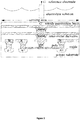
Figure 2 is a profile view of an ISFET exposed to a sample; -
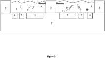
Figure 3 is a profile view of a pH detection system; -
Figure 4 is a flowchart of a method to monitor amplification of a DNA sample for (option A) identifying the DNA or (option B) calculating the quantity of DNA; -
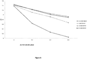
Figure 5 is a chart of buffering capacity of reagents in a normal LAMP recipe; -
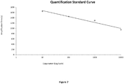
Figure 6 is chart showing the buffer capacity of different concentrations of NH4Cl; and -
Figure 7 is a chart of a standard curve for quantification of DNA in a sample. - Conventional LAMP amplification methods use DNA polymerases with displacement activity under standard assay conditions such as: 20mM Tris-HCl (pH8.8), 10mM KCI, 10mM (NH)4SO4, 2.5mM MgSO4, 0.1% Triton X-100, 0.8M Betaine, DNA/RNA, dNTP and Bst polymerase.
- The inventors found that these conventional reagents do not permit detection by pH sensors primarily because of their ability to mask the production of protons during amplification.
- Indeed, all these constituents have a different pKa which means they have a different impact on the buffer capacity of the mixture.
- Among the above mentioned reagents, TrisHCl has the ability to absorb counter ions (H+ and OH-) so as to help keep the solution at a stable pH level within a range optimal for the polymerase to act.
- The inventors found that replacing TrisHCl with NaOH reduces the buffer capacity while setting the pH to where polymerase (such as Bst) can operate. Moreover, NaOH makes the two strands in double-stranded DNA less stringently bound, allowing displacement polymerase to break them apart more easily, thus speeding up the reaction and increase the efficiency of the strand displacement enzyme.
- Additionally, electronic sensors, such as ISFETs, use reference electrodes such as Platinum, Ag/AgCl, calomel, etc. Some of these materials, in particular Ag/AgCl electrodes, react with these standard reagents. For instance, with Ag/AgCl electrodes Tris forms a Tris-Ag complex on the electrode which deteriorates the Ag/AgCl performance and Sulphate-containing reagents can poison the Ag/AgCl electrode.
- A preferred system using the present method comprises a pH sensor or indicator, microfluidic structure, a nucleic acid sample, reagents, and a reference electrode when needed to set a voltage potential of the sample. The reagents and sample are combined into one fluid to enable amplification. Protons are released during amplification and the change in pH is measured with a pH sensor or indicator.
- Preferably, the pH sensor or indicator is an ISFET (Ion Sensitive Field Effect Transistor). This is shown in
Figure 3 , wherein the pH sensor(s) 3 may be one or more Ion Sensitive Field Effect Transistors (ISFET) on aCMOS microchip 7, having thereuponmicrofluidic chambers 8 defined by voids insubstrate 2. Reagents and nucleic acid sample may be combined before or after being added to one or more chamber(s) exposed to the ISFET(s). Each ISFET outputs an electrical signal which is monitored by a signal processor. The passivation layer of the ISFET can be functionalised to be sensitive to protons (hydrogen ions). As the nucleic acid amplifies, protons will be released and be detected by the signal processor as a change in the electrical output of the ISFET. - In an alternative embodiment, a pH indicator may be used to detect protons released during amplification. For example, the pH indicator may be a colorimetric or fluorescent dye, which changes optical properties such as emitted wavelength from the dye as the pH of the contacting fluid changes. Examples of pH indicators include Fluorescein, Pyranine, and pHrodo dye (available from Life Technology).
- The microfluidic structure may be a well, chamber, or channel to receive the sample proximate the sensor or indicator and may comprise means for delivering the sample to the sensor or indicator. The microfluidic structure also helps reduce diffusion of protons away from the sensor or indicator. In the following embodiments, ISFETs are used to illustrate the pH detection scheme but other pH sensors could be used. The chamber may be defined by a cavity in a material such as SU-8, which is deposited on top of the microchip and selectively etched away to leave said cavities.
-
Figure 2 shows an ISFET with a floating gate and sensing layer made of Silicon Nitride which is exposed to the fluid electrolyte. ISFETS are further described in patentUS2004134798 . Preferably each ISFET generates a normalised output signal from the difference between the ISFET and a reference signal. The reference signal may be derived from another ISFET exposed to a negative control reaction or a FET located on the chip but not exposed to fluctuating pH. Thus any common drift or noise on the chip will be cancelled by taking the difference between these signals. - The preferred amplification reaction is an isothermal amplification reaction, preferably a strand displacement reaction. As used herein, a strand displacement reaction is provided by a polymerase with strand displacement activity and reaction conditions where strand displacement is possible. Examples of strand displacement reactions include Strand displacement amplification (SDA), multiple displacement amplification (MDA), rolling circle amplification (RCA) or Loop mediated isothermal Amplification (LAMP).
- As an example, the steps in the chemical reaction of the LAMP method are illustrated in
Figure 1 . Instep 1, a double stranded DNA template at an elevated temperature is in dynamic equilibrium. Primers F2 can anneal to the single strand at the complementary position. In step 2 a polymerase with strand displacement activity enables nucleotides to extend along the template from the 3' end of F2. The incorporation of nucleotides is a reaction that has hydrogen ions (protons) as one of the by-products. Instep 3, the F3 primer anneals to the F3c region on the template and begins displacement of the strands. The top strand is synthesized instep 4 releasing further protons. The bottom strand becomes a single strand (step 5) which forms a stem-loop as F1c anneals to F1 at the 5' end instep 6. At the same time the BIP primers, anneal to the other end of the strand and nucleotide extend from B2, releasing more protons. Primer B3 displaces the strands and promotes extension to create the double strand shown instep 7. The structure instep 8 has a double ended stem-loop from which continuous displacement and extension to amplify the template. As before the extension is associated with proton release. - The strand displacement polymerase used in the isothermal amplification reaction described herein may be chosen from the group: phi29-DNA-Polymerase, Klenow DNA-Polymerase, Vent DNA Polymerase, Deep Vent DNA Polymerase, Bst DNA Polymerase, 9oNm(TM) DNA Polymerase, and mutants and variants thereof.
- The skilled person will appreciate that the optimal reagent concentrations will depend on the selection of the polymerase and that some modification to the preferred reagents below will be normal practice from knowledge of or experimentation with the polymerase. Guidance on appropriate conditions is available from the enzyme manufacturers.
- The present method does not require any buffering agent and it is preferable that minimal buffering agent is present. A buffering agent is a weak acid and its conjugate base used to maintain the acidity (pH) of a solution near a chosen operating point such that the pH varies insignificantly when a small amount of strong acid or base is added, or in the present case, when a small amount of protons are released during the incorporation of nucleotides. A buffering agent is a compound added to a mixture having the primary purpose of providing buffering against changes in pH. As used herein, a compound whose primary purpose is other than buffering or whose buffering effect is much less than another compound in the mixture is not a buffering agent. Buffering agents for nucleic acid amplification reactions typically have a pKa value between 6 and 8.5, and has a buffering range between 6 and 9. For example, ammonium (NH4+) has some buffering capability but its main purpose is not to buffer the mixture and with a pKa of 9.24 operating in a mixture of
pH 8, it is not a very strong buffer compared to Tris. - The choice of buffering agent, enzyme, and initial pH of the system are interdependent. For example, whilst the buffering agent may be one of the following common buffering agents TAPS, Bicine, Tris, Tricine, TAPSO, HEPES, TES, MOPS, PIPES, Cacodylate, SSC, and MES, in one embodiment TRIS is used with BST enzyme at a pH of 8.5. Preferably the concentration of buffering agent is less than 10mM, more preferably less than 8mM, less than 5mM, or less than 1 mM. Preferably the buffering agent is Tris or Hepes.
- To reduce the effect of poisoning on the reference electrode, the concentration of sulphate compounds in the combined fluid is less than 15mM, preferably less than 10mM, less than 8mM, less than 5mM, or less than 1 mM.
- Ammonium chloride can be used instead of ammonium sulphate whilst still allowing good amplification yield. More generally other quaternary salts can be substituted for ammonium chloride. Quaternary ammonium salts are positively charged polyatomic ions of the structure NR4+, where R is an alkyl or aryl group. Guanidine hydrochloride and ammonium chloride are examples of quaternary ammonium salts.
- Preferably the range of concentration of quaternary ammonium salts in the combined fluid is greater than 2mM, 5mM, or 8mM. However, ammonium (NH4+) has some buffering capability, thus the final concentration of ammonium compounds, such as ammonium chloride, in the combined fluid needs to be minimised whilst still maintaining optimal amplification yield. To reduce the buffering capacity, the concentration of ammonium compounds in the combined fluid is less than 15mM, preferably less than 10mM.
- Magnesium is useful in promoting nucleotide incorporation in the template. The concentration of magnesium compounds, for example magnesium sulphate, in the combined fluid is preferably greater than 0.5mM, greater than 1 mM, greater than 2mM, or greater than 4mM. The concentration of magnesium ion in the combined fluid is dependent on the concentration of dNTP, template and primers. In general, the preferred ratio of dNTP to magnesium sulphate in the combined fluid is less than 1:2, less than 1:3, less than 1:4 or less than 1:5.
- Since high chloride concentration aids the Ag/AgCl electrode, monovalent salt such as sodium chloride or potassium chloride is added, the chloride ion concentration being preferably more than 10mM, more than 20mM, more than 30mM, more than 40mM or more than 50mM. In one embodiment, the chloride ion concentration in the fluid is between 40mM and 60mM.
- To set the starting pH of the fluid an alkali base, such as NaOH, LiOH or KOH, is added to the fluid. The concentration of the alkali base is designed to set the pH of the combined solution between 6 and 9, more preferably between 7 and 8.8, most preferably between 8 and 8.6, these pH ranges being desirable for certain enzymes to operate. For Bst polymerase, the preferred starting pH is more than 7, more preferably more than 8.2 and less than 8.8, more preferably less than 8.6.
- The concentration of other reagents may be kept at normal amounts. See Notomi T et. al. Nucleic Acids Res. 2000 Jun 15; 28(12): E63. For example in one embodiment, the amount of Bst polymerase is at least 0.3 Unit per microliter of combined fluid; the concentration of Betaine is 0-1.5M, preferably 0.8M-1 M; and the total concentration of primers is between 2m and 6.2uM.
- The above reagent concentrations have been found to provide good amplification yield and at the same time low buffering capacity such that a pH sensor can be used to detect protons released during amplification of the nucleic acid.
- The process can take place at a fixed temperature, reducing the sensor signal drift associated with thermocycling, thus making the sensor signals more stable. Additionally the process is highly compatible with semiconductor platforms. For example, the optimal enzymatic temperature can be achieved and monitored with on-chip heating elements and temperature sensors; there is less concern over thermal expansion and thermal fatigue associated with thermocycling; and the reagents are chosen so as not to affect the electrodes on the microchip.
- Typically, isothermal methods require a set temperature, which is determined by the reagents being used. For example, in LAMP the enzymes function best between 60 and 65°C. Advantageously the reagents/buffer of preferred embodiments described herein enables a wider operating temperature.
- Because isothermal amplification, unlike thermocycling, does not involve discrete steps, each step doubling the DNA, it is difficult to estimate how much amplification has taken place at a given time. As a result, such isothermal amplification methods normally encourage excess amplification with the side effect that the background (i.e. non-specific) amplification or fluorescent background level is very high. The present method enables real time detection of the amplification process such that the process can be stopped when sufficient yield has been obtained. In the case where the present method takes place on a microchip, the fluid being monitored by a pH sensor and heated by elements in the chip surface, the temperature can be dropped or raised to a point where amplification becomes suspended. This ensures that there is sufficient desired DNA beyond the background DNA without waiting unnecessarily to be sure that sufficient amplification has occurred.
- The reagents are provided in the concentrations above when combined. Some reagents may be stored separately prior to mixing having their own required conditions for stability. For example, the enzyme may be stored long term in a moderately buffered solution separate from the other reagents to ensure stability of the enzyme. Upon mixing with the remaining reagents, the buffering agent becomes sufficiently diluted so as not to significantly mask a pH change. In addition, primers for specific genes of interest may be provided in a separate solution or in a lyophilized form. The conditions and pre-mix concentrations will be known to or derivable by the skilled person in consideration of the reagent to be used.
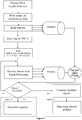
- As illustrated by the flowchart of
Figure 4 , a DNA sample may be prepared and divided into one or more chambers or wells. Each chamber or well is exposed to an ISFET on a microchip. The DNA is combined with reagents for loop-mediated isothermal amplification. The following are individually preferred reagents and concentrations, wherein the combination is the most preferred kit of reagents: - Bst polymerase of at least 0.3 Unit per microliter;
- a concentration of Betaine of 1M;
- a total concentration of primers of 5uM;
- a concentration of Magnesium Sulphate of 5mM;
- a concentration of Tris of 1 mM;
- a concentration of Ammonium Sulphate of zero;
- a concentration of NaOH of 1.2mM which sets the pH of the combined fluid to 8.5pH;
- a concentration of Ammonium Chloride of 5mM; and
- a concentration of Potassium Chloride of 50mM.
- The ISFET signals are taken differentially with respect to a reference FET and are monitored by a signal processor. The chamber and fluid is heated to 60°C by heaters integrated with the microchip. After a predetermined reaction period, sufficient template amplification, if possible, should have occurred to be detected as a change in the ISFET signal. The signal can also be continuously monitored to determine when the amplification and thus signal change has crossed a threshold amount.
- The method may be used to identify one or more bases in a nucleic acid strand as illustrated, in a preferred embodiment, by option A of
Figure 4 . The identification may be a single base or a unique sequence. In the case where a unique sequence is identified, it is possible to identify certain bases that are associated with medical conditions and this knowledge of the bases can provide a method for diagnosis. Examples of bases of interest include unique sequences, Single Nucleotide Polymorphism (SNP), deletions, insertions, Short Tandem Repeats (STP) and mutations that may be inherited or somatically derived. Pathogen detection is also possible whereby the method may detect the presence of an organism or strain of organism. - Primers used in the amplification such as the FIP (forward inner primer) and BIP (back inner primer) oligos can be designed to include or exclude the sequence, SNP, or STP region. In this way the amplification or lack thereof indicates the presence or absence of the base(s)/sequence to be identified.
- In the system shown in
Figure 1 , two or more chambers orwells may be used to perform concurrent amplification of the DNA. Each well has added to it a different set of primers, each set of primers being adapted to detect a different base in the sample DNA. The DNA will therefore only amplify in the presence of the complementary set of primers, producing protons, whilst the others will not. Where only one chamber experiences amplification, the DNA will be considered homozygous, i.e. having identical alleles (mutant or wildtype) on both genes. Where two chambers experience amplification, the DNA will be considered heterozygous, i.e. having different alleles (mutant and wildtype) on the genes. One can thus determine the identity of the base(s) in the sample DNA by monitoring the ISFETs signals to detect a fluctuation combined with knowledge of the primer set in the corresponding well. To reduce signal processing requirements, the signals from ISFETs can be compared in real-time to output a signal representing the difference between amplification by-products in each well. - The method may be used to quantify the amount of DNA in a sample as illustrated in
Figure 4 , option B. The proton concentration at a given time will be proportional to the quantity of DNA in the fluid and the cumulative quantity of previous protons generated and which have not diffused away from the sensing area. By knowing the signal change from the start of the reaction at a given time and comparing this to a standard, one can determine the quantity of DNA in the sample at the start of the amplification. Thus one works backwards from the current quantity and time to determine the quantity of starting DNA in the sample. - The standard may be derived from a model, experimental data, or one or more separate internal control reactions undergoing an amplification reaction in parallel with the assay. The standard may be represented as a look-up-table on a storage medium or as a quantification equation in a computer program.
Figure 7 shows an exemplary graph of DNA quantity versus time to detect that quantity. The graph can be interpolated or extrapolated or can be used to extract a best-fit equation through the data to estimate DNA quantities once a reaction time has been measured. - The reaction time is the period from when amplification begins (i.e. when all reagents and conditions for amplification are present) to when the pH change becomes greater than a threshold. The pH change may be detected by monitoring the pH sensor signal or pH indicator.
- The present method may also be used to detect RNA template through the use of a reverse transcriptase (RTase) enzyme such as avian myeloblastosis virus (AMV RTase) together with DNA polymerase. cDNA can be synthesized from template RNA and amplified with the present technique and then detected using an pH sensor or indicator.
- Whilst most compounds contribute some buffering capacity to the mixture, the total contribution is ideally minimised. However some minimal buffer may be required to stabilise the enzymes. Selection of the buffer agent (if present at all), total reagent buffer capacity, and concentrations should be made in consideration of the expected protons generated by the amplification reaction. The amount of protons generated will depend on the amount of starting template, amplification conditions and amplification time (assuming excess nucleotides, enzyme and primers). The starting template will depend on the donor, type of biological sample taken and the amplification time may be chosen by the operator or manufacturer of the test. However from a knowledge of the amplification time, biological sample type, and donor type, one can calculate an expected amount (or range of amount) of protons to be generated.
- The buffer capacity of the mixture can then be chosen such that a pH change greater than a threshold amount will result from the expected (or lowest expected) proton generation due to amplification even in the presence of the buffered mixture. The pH change threshold may be the limit of detection of the sensor and associated circuitry. Alternatively, the pH change threshold may be 0.1 pH, more preferably 0.2pH, most preferably 0.5pH.
- Buffer capacity is defined by equation (I):

- In an exemplary embodiment employing an ISFET having a lower detection limit of 0.5 pH, exposed to a 35ul amplification reaction, wherein after 30 minutes the experimental yield is 50ug of amplicons. The total protons released from the amplicon yield will be approximately 2.17mM (assuming the molecular weight of a base pair is 650g/mole);


- Table 1 below provides properties of common buffering agents with pKas at 25C between 6.15 and 8.43. The concentration of these buffering agents should be minimized in the reaction to achieve greater pH change in a shorter amplification reaction time. However, the buffering agents may be optionally provided to reduce background noise, stabilize the enzyme and/or stabilize the initial reaction. Table 2 provides a calculation of the effect of the buffer capacity of amplification reactions suitable for Bst enzyme by varying the concentrations of buffer agents. As can be seen, a number of buffer options will satisfy the requirement above, having a buffer capacity of less than 4.34mM (4340uM). Each buffer agent and concentration listed in table 2 satisfying this condition are individually preferable and considered within the scope of this invention.
Table 1 Properties of various Buffer Agents Buffer Name pKa at 25°C Buffer Range Full Compound Name TAPS 8.43 7.7-9.1 3-{[tris(hydroxymethyl)methyl]amino}propanesulfonic acid Bicine 8.35 7.6-9.0 N,N-bis(2-hydroxyethyl)glycine Tris 8.06 7.5-9.0 tris(hydroxymethyl)methylamine Tricine 8.05 7.4-8.8 N-tris(hydroxymethyl)methylglycine TAPSO 7.635 7.0-8.2 3-[N-Tris(hydroxymethyl)methylamino]-2-hydroxypropanesulfonic Acid HEPES 7.48 6.8-8.2 4-2-hydroxyethyl-1-piperazineethanesulfonic acid TES 7.4 6.8-8.2 2-{[tris(hydroxymethyl)methyl]amino}ethanesulfonic acid MOPS 7.2 6.5-7.9 3-(N-morpholino)propanesulfonic acid PIPES 6.76 6.1-7.5 piperazine-N,N'-bis(2-ethanesulfonic acid) Cacodylate 6.27 5.0-7.4 dimethylarsinic acid SSC 7 6.5-7.5 saline sodium citrate MES 6.15 5.5-6.7 2-(N-morpholino)ethanesulfonic acid Table 2 Buffer capacity at various conditions Hepes @pH 9 8.5 8 7.5 7 40mM 8865 17121 23580 18235 20mM 5199 8916 12071 9628 10mM 3352 3367 4813 6317 5324 5mM 3024 2451 2762 3440 3173 Tris @pH 9 8.5 8 7.5 7 40mM 19536.4 23631 16160 7809 20mM 10535 12170 8361 4415 10mM 4824 6035 6441 4462 2718 5mM 3761 3785 3575 2512 1869 MOPS @pH 9 8.5 8 7.5 7 6.5 40mM 5721 11589 21050 22871 14673 20mM 3628 6149 10806 11946 8286 10mM 3051 2581 3430 5685 6483 5092 5mM 2874 2058 2070 3124 3752 3496 Bicine @pH 9 8.5 8 7.5 7 6.5 40mM 23891 20371 10553 4791.26 3164 20mM 12713 10541 5558 2906 2531 10mM 6139 7124 5626 3061 1963 2215 5mM 4418 4329 3168 1812 1492 2057 - In some embodiments, the buffer capacity is reduced from the calculated maximum by a factor to ensure that sufficient pH signal is detected. The buffer capacity may be less than one-half, preferably less than one-fifth, or less than one-tenth the maximum buffer capacity for which a pH change due to an expected proton release from an amplification reaction is detectable. Thus in the example above the buffer capacity may be set to 1/10 of 4.34mM, i.e. 0.434mM.
- In one embodiment, the buffer capacity of the reagents in the fluid is arranged to mask a pH change that would otherwise result even in the absence of successful amplification of the target nucleic acid. This change can be considered ionic background noise, which may result from non-specific amplification or spontaneous degradation and hydrolysis of nucleotides, primers, or template. Non-specific amplification refers to nucleic acid amplification products that are not derived from the targeted region of the template nucleic acid. Typically this results from primer-dimer formation and/or primers annealing to non-targeted regions of the template DNA.
- In one embodiment, the total buffer capacity of the mixture is set such that background noise can be ignored. For example, the amount of background noise that may be produced during the method can be estimated or found from experiment and expressed as a change in pH. The buffer capacity of the reagents can be increased, beyond the minimal amount suggested above for providing a low limit of detection, to an amount that masks the background by absorbing the protons released or consumed due to the background. Therefore no signal is detected by the pH sensor or indicator unless and until there is sufficient proton release which should thus correspond to specific nucleotide insertion for the target template nucleic acid.
- In one embodiment, a DNA sample and reagents are added to multiple microfluidic chambers. In an exemplary embodiment, the pH drop due to background in the absence of a buffer is estimated at 0.1 pH. A small amount of buffer is provided to each chamber to mask this estimated effect. The sensor signal is monitored before, during, and after the chemical reactions take place. Only in the chamber(s) where there is a nucleic acid amplification reaction releasing significant protons will there be a detectable change in the sensor signal. Thus in one embodiment the buffer capacity is greater than 0.5mM.
- Different reagents, such as allele specific primers, may be used to detect the presence or absence of genetic biomarkers on the sample. The reaction may be amplification of DNA and the reagents may comprise primers and nucleotides suitable for thermocycling or isothermal amplification. As amplification of target DNA proceeds in one or more chambers, protons are released beyond the estimated background effect masked by the buffer. The pH change is detected as a change in the sensor signal.
Claims (14)
- A reaction mixture for use in isothermally amplifying a nucleic acid, the mixture comprising: a magnesium salt, an alkali base, and at least one ammonium compound selected from the group consisting of a quaternary ammonium salt, ammonium chloride and guanidine hydrochloride, wherein the mixture has a buffer capacity of less than 10 mM.
- The mixture of claim 1, wherein the buffer capacity of the mixture is set to less than an expected concentration of protons to be released during isothermal amplification of a nucleic acid divided by a predetermined threshold pH change to be detected by a pH sensor exposed to the mixture during said amplification.
- The mixture of claim 1, wherein the buffer capacity of the mixture is set to less than one half of an expected concentration of protons to be released during isothermal amplification of a nucleic acid divided by a predetermined threshold pH change to be detected by a pH sensor exposed to the mixture during said amplification.
- The mixture of claim 2 or 3, wherein the predetermined threshold pH change to be detected is the limit of detection of said pH sensor exposed to the mixture during said amplification.
- The mixture of any preceding claim, further comprising a buffering agent, and wherein the concentration of buffering agents in the mixture is less than 5mM, more preferably less than 3mM, less than 2mM, or less than 1 mM.
- The mixture of any preceding claim, wherein the mixture further comprises sulphate compounds at a concentration of less than 15mM.
- The mixture of any preceding claim, wherein the concentration of ammonium compounds in the mixture is less than 15mM.
- The mixture of any preceding claim, wherein the concentration of the alkali base sets the pH of the mixture between 6 and 9, preferably between 7 and 8.8, more preferably between 8 and 8.6.
- The mixture of any preceding claim, wherein the alkali base is one of NaOH, KOH or LiOH.
- The mixture of any preceding claim, further comprising a strand displacement enzyme, nucleotides, and primers, preferably wherein the primers are allele specific such that amplification indicates the presence of a target nucleic acid.
- A method of monitoring amplification of a nucleic acid comprising the steps of:providing a nucleic acid sample and an amplification mixture according to any one of claims 1 to 10 and a pH sensor or pH indicator;amplifying the nucleic acid using isothermal amplification; anddetecting a change in pH due to the amplification using the pH sensor or pH indicator.
- The method of claim 11, further comprising:measuring a reaction time needed for the pH change to become greater than a predetermined threshold; andusing the reaction time and the pH change to quantify the amount of nucleic acid in the sample.
- The method of claim 11 or 12, wherein the mixture comprises one or more allele specific primers having at least one base complementary to a target Single Nucleotide Polymorphism (SNP) of the nucleic acid, the method further comprising identifying said at least one base of the nucleic acid depending on whether amplification proceeds, as detected by the pH sensor or pH indicator.
- The method of any one of claims 11 to 13, wherein the amplification changes the proton concentration of the mixture by more than 10% of the buffer capacity of the mixture.
Applications Claiming Priority (2)
| Application Number | Priority Date | Filing Date | Title |
|---|---|---|---|
| GBGB1112140.7A GB201112140D0 (en) | 2011-07-14 | 2011-07-14 | Nucleic acid amplification |
| PCT/GB2012/051694 WO2013008042A1 (en) | 2011-07-14 | 2012-07-16 | Nucleic acid amplification |
Publications (2)
| Publication Number | Publication Date |
|---|---|
| EP2732050A1 EP2732050A1 (en) | 2014-05-21 |
| EP2732050B1 true EP2732050B1 (en) | 2017-11-15 |
Family
ID=44586619
Family Applications (1)
| Application Number | Title | Priority Date | Filing Date |
|---|---|---|---|
| EP12737335.5A Active EP2732050B1 (en) | 2011-07-14 | 2012-07-16 | Nucleic acid amplification |
Country Status (7)
| Country | Link |
|---|---|
| US (2) | US9469870B2 (en) |
| EP (1) | EP2732050B1 (en) |
| JP (1) | JP6139519B2 (en) |
| CN (1) | CN103687962B (en) |
| ES (1) | ES2656856T3 (en) |
| GB (2) | GB201112140D0 (en) |
| WO (1) | WO2013008042A1 (en) |
Families Citing this family (22)
| Publication number | Priority date | Publication date | Assignee | Title |
|---|---|---|---|---|
| GB201112140D0 (en) | 2011-07-14 | 2011-08-31 | Dna Electronics | Nucleic acid amplification |
| WO2015033229A2 (en) * | 2013-09-04 | 2015-03-12 | Credo Biomedical Pte Ltd. | Assay test device, kit and method of using |
| BR112016022673B1 (en) | 2014-03-31 | 2021-12-28 | Impossible Foods Inc. | MEAT REPLICA COMPOSITION |
| CN103923998A (en) * | 2014-04-23 | 2014-07-16 | 浙江大学 | Reaction system applied to real-time electrochemical pH detection of nucleic acid amplification and detection method |
| EP3594360B1 (en) | 2014-04-24 | 2021-06-23 | Lucira Health, Inc. | Colorimetric detection of nucleic acid amplification |
| US10249741B2 (en) * | 2014-05-13 | 2019-04-02 | Joseph T. Smith | System and method for ion-selective, field effect transistor on flexible substrate |
| EP3430372A4 (en) | 2016-03-14 | 2019-10-16 | Lucira Health, Inc. | Devices and methods for biological assay sample preparation and delivery |
| US11291995B2 (en) | 2016-03-14 | 2022-04-05 | Lucira Health, Inc. | Selectively vented biological assay devices and associated methods |
| JP6937774B2 (en) | 2016-03-14 | 2021-09-22 | ルシラ ヘルス インコーポレイテッド | Systems and methods for performing biological assays |
| US11080848B2 (en) | 2017-04-06 | 2021-08-03 | Lucira Health, Inc. | Image-based disease diagnostics using a mobile device |
| US10549275B2 (en) | 2017-09-14 | 2020-02-04 | Lucira Health, Inc. | Multiplexed biological assay device with electronic readout |
| GB2570157A (en) * | 2018-01-15 | 2019-07-17 | Dnanudge Ltd | Molecule detection |
| KR20200138790A (en) | 2018-03-29 | 2020-12-10 | 파운데이션 오브 이노베이티브 뉴 다이아그노스틱스 | Antibodies or antibody combinations for detecting an antigen related to mycobacteria in a urine sample of a subject and a method of using the same |
| GB201809420D0 (en) * | 2018-06-08 | 2018-07-25 | Imperial Innovations Ltd | Devices and method for detecting an amplification event |
| US10467679B1 (en) | 2019-04-15 | 2019-11-05 | Dnanudge Limited | Product recommendation device and method |
| USD910200S1 (en) | 2018-12-21 | 2021-02-09 | Lucira Health, Inc. | Test tube |
| US10811140B2 (en) | 2019-03-19 | 2020-10-20 | Dnanudge Limited | Secure set-up of genetic related user account |
| US10699806B1 (en) | 2019-04-15 | 2020-06-30 | Dnanudge Limited | Monitoring system, wearable monitoring device and method |
| US20220283149A1 (en) * | 2019-08-06 | 2022-09-08 | Gmd Biotech, Inc | All-in-one molecular diagnostics device |
| USD953561S1 (en) | 2020-05-05 | 2022-05-31 | Lucira Health, Inc. | Diagnostic device with LED display |
| USD962470S1 (en) | 2020-06-03 | 2022-08-30 | Lucira Health, Inc. | Assay device with LCD display |
| US20220268729A1 (en) * | 2021-02-24 | 2022-08-25 | Robert Bosch Gmbh | Measuring ion strength using closed-loop electrochemical ph modulation |
Family Cites Families (16)
| Publication number | Priority date | Publication date | Assignee | Title |
|---|---|---|---|---|
| ES2369818T3 (en) | 1998-11-09 | 2011-12-07 | Eiken Kagaku Kabushiki Kaisha | NUCLEIC ACID SYNTHESIS PROCEDURE. |
| EA007414B1 (en) * | 2000-08-23 | 2006-10-27 | Такара Био Инк. | Chimeric oligonucleotide primer for method of amplifying nucleic acid; product for amplifying nucleic acid; probe for a method of detecting target nucleic acid; a material having an immobilized amplified nucleic acid and process for producing thereof; a method for determining a nucleotide sequence |
| US8114591B2 (en) | 2001-03-09 | 2012-02-14 | Dna Electronics Ltd. | Sensing apparatus and method |
| GB0105831D0 (en) | 2001-03-09 | 2001-04-25 | Toumaz Technology Ltd | Method for dna sequencing utilising enzyme linked field effect transistors |
| US7534873B2 (en) * | 2003-08-29 | 2009-05-19 | Applied Biosystems, Llc | Method and materials for quaternary amine catalyzed bisulfite conversion of cytosine to uracil |
| EP1756136B1 (en) | 2004-05-21 | 2014-10-29 | Mo Bio Laboratories, Inc. | Kits and processes for removing contaminants from nucleic acids in environmental and biological samples |
| EP2442108B1 (en) * | 2006-07-14 | 2016-11-16 | The Regents of The University of California | Cancer biomarkers and methods of use thereof |
| JP4918409B2 (en) * | 2006-07-26 | 2012-04-18 | 西川ゴム工業株式会社 | Nucleic acid sequence amplification method |
| US20100056410A1 (en) | 2006-09-25 | 2010-03-04 | Advanced Technology Materials, Inc. | Compositions and methods for the removal of photoresist for a wafer rework application |
| JP5368321B2 (en) * | 2007-03-02 | 2013-12-18 | ディ・エヌ・エイ・エレクトロニクス・リミテッド | QPCR using solid phase pH detection |
| WO2009072156A1 (en) * | 2007-12-07 | 2009-06-11 | Emanuela Manna | Deodorizing and sanitizing compositions |
| CN102203282B (en) | 2008-06-25 | 2014-04-30 | 生命技术公司 | Methods and apparatus for measuring analytes using large scale FET arrays |
| WO2010037085A1 (en) * | 2008-09-29 | 2010-04-01 | The Board Of Trustees Of The University Of Illinois | Dna sequencing and amplification systems using nanoscale field effect sensor arrays |
| JP5572943B2 (en) * | 2008-12-17 | 2014-08-20 | 株式会社島津製作所 | DNA amplification method and DNA amplification kit from blood sample |
| ES2351294B8 (en) * | 2009-07-02 | 2012-06-06 | Consejo Superior De Investigaciones Científicas (Csic) | METHOD FOR THE REPLICATION, AMPLIFICATION, OR SEQUENCING OF A MOLD DNA. |
| GB201112140D0 (en) | 2011-07-14 | 2011-08-31 | Dna Electronics | Nucleic acid amplification |
-
2011
- 2011-07-14 GB GBGB1112140.7A patent/GB201112140D0/en not_active Ceased
-
2012
- 2012-07-16 JP JP2014519635A patent/JP6139519B2/en active Active
- 2012-07-16 CN CN201280034733.6A patent/CN103687962B/en active Active
- 2012-07-16 EP EP12737335.5A patent/EP2732050B1/en active Active
- 2012-07-16 GB GB1212637.1A patent/GB2494239B/en active Active
- 2012-07-16 WO PCT/GB2012/051694 patent/WO2013008042A1/en active Application Filing
- 2012-07-16 ES ES12737335.5T patent/ES2656856T3/en active Active
- 2012-07-16 US US14/232,817 patent/US9469870B2/en active Active
-
2016
- 2016-09-30 US US15/282,879 patent/US9840733B2/en active Active
Non-Patent Citations (1)
| Title |
|---|
| None * |
Also Published As
| Publication number | Publication date |
|---|---|
| CN103687962A (en) | 2014-03-26 |
| GB201112140D0 (en) | 2011-08-31 |
| US20170016058A1 (en) | 2017-01-19 |
| GB2494239B (en) | 2014-05-21 |
| JP6139519B2 (en) | 2017-05-31 |
| CN103687962B (en) | 2015-12-09 |
| US9469870B2 (en) | 2016-10-18 |
| GB201212637D0 (en) | 2012-08-29 |
| US20140170661A1 (en) | 2014-06-19 |
| EP2732050A1 (en) | 2014-05-21 |
| WO2013008042A1 (en) | 2013-01-17 |
| ES2656856T3 (en) | 2018-02-28 |
| US9840733B2 (en) | 2017-12-12 |
| JP2014520544A (en) | 2014-08-25 |
| GB2494239A (en) | 2013-03-06 |
Similar Documents
| Publication | Publication Date | Title |
|---|---|---|
| EP2732050B1 (en) | Nucleic acid amplification | |
| Deprez et al. | Sensitivity and accuracy of quantitative real-time polymerase chain reaction using SYBR green I depends on cDNA synthesis conditions | |
| Cai et al. | Phosphorothioated primers lead to loop-mediated isothermal amplification at low temperatures | |
| Fraga et al. | Real‐time PCR | |
| US20120276533A1 (en) | Method for Simultaneously Detecting Polymorphisms of Acetaldehyde Dehydrogenase 2 and Alcohol Dehydrogenase 2 | |
| JP6638122B2 (en) | Target nucleic acid detection method and kit | |
| MX2014012214A (en) | Compositions and methods for quantifying a nucleic acid sequence in a sample. | |
| Olsen et al. | Empirical evaluation of humpback whale telomere length estimates; quality control and factors causing variability in the singleplex and multiplex qPCR methods | |
| US20200362404A1 (en) | Electrochemical Detection Of Polymerase Reactions By Specific Metal-Phosphate Complex Formation | |
| CN110265089B (en) | Nucleic acid quantitative analysis method based on assistance of intelligent equipment and application thereof | |
| CN103597094A (en) | Method and device for monitoring real-time polymerase chain reaction (PCR) utilizing electro-active hydrolysis probe (E-TAG probe) | |
| Wang et al. | A three-way junction structure-based isothermal exponential amplification strategy for sensitive detection of 3′-terminal 2′-O-methylated plant microRNA | |
| ATE533847T1 (en) | METHOD FOR DETECTION AND QUANTIFICATION OF AN ENDOGENE DNA SEQUENCE FROM WHEAT | |
| Umer et al. | A novel DNA binding protein-based platform for electrochemical detection of miRNA | |
| JP5597939B2 (en) | Method for detecting a target nucleotide sequence using a partially competitive probe | |
| US20220025449A1 (en) | Method for detecting a target analyte in a sample using an s-shaped function for a slope data set | |
| EP2524969A1 (en) | Method, microchip and mixed reagent for analysis of nucleic acid base sequence | |
| JP5888470B2 (en) | Method for measuring telomerase activity | |
| US10760116B2 (en) | Analysis kit, analyzer, and methods for analyzing template nucleic acid or target substance | |
| WO2004063390A3 (en) | Compositions and methods for determining canine gender | |
| EP4310195A1 (en) | Method for nucleic acid detection using signal-mediated amplification of rna technology and rna aptamers | |
| EP3608422A1 (en) | Electrochemical detection of polymerase reactions by specific metal-phosphate complex formation | |
| CN103525912A (en) | Method for detecting MTHFR (Methylene Tetrahydrofolate Reductase) gene polymorphism by using high-resolution melting curve analysis technology |
Legal Events
| Date | Code | Title | Description |
|---|---|---|---|
| PUAI | Public reference made under article 153(3) epc to a published international application that has entered the european phase |
Free format text: ORIGINAL CODE: 0009012 |
|
| 17P | Request for examination filed |
Effective date: 20140121 |
|
| AK | Designated contracting states |
Kind code of ref document: A1 Designated state(s): AL AT BE BG CH CY CZ DE DK EE ES FI FR GB GR HR HU IE IS IT LI LT LU LV MC MK MT NL NO PL PT RO RS SE SI SK SM TR |
|
| DAX | Request for extension of the european patent (deleted) | ||
| RAP1 | Party data changed (applicant data changed or rights of an application transferred) |
Owner name: DNAE GROUP HOLDINGS LIMITED |
|
| 17Q | First examination report despatched |
Effective date: 20160729 |
|
| STAA | Information on the status of an ep patent application or granted ep patent |
Free format text: STATUS: EXAMINATION IS IN PROGRESS |
|
| GRAP | Despatch of communication of intention to grant a patent |
Free format text: ORIGINAL CODE: EPIDOSNIGR1 |
|
| STAA | Information on the status of an ep patent application or granted ep patent |
Free format text: STATUS: GRANT OF PATENT IS INTENDED |
|
| INTG | Intention to grant announced |
Effective date: 20170721 |
|
| GRAS | Grant fee paid |
Free format text: ORIGINAL CODE: EPIDOSNIGR3 |
|
| GRAA | (expected) grant |
Free format text: ORIGINAL CODE: 0009210 |
|
| STAA | Information on the status of an ep patent application or granted ep patent |
Free format text: STATUS: THE PATENT HAS BEEN GRANTED |
|
| AK | Designated contracting states |
Kind code of ref document: B1 Designated state(s): AL AT BE BG CH CY CZ DE DK EE ES FI FR GB GR HR HU IE IS IT LI LT LU LV MC MK MT NL NO PL PT RO RS SE SI SK SM TR |
|
| REG | Reference to a national code |
Ref country code: AT Ref legal event code: REF Ref document number: 946346 Country of ref document: AT Kind code of ref document: T Effective date: 20171115 Ref country code: GB Ref legal event code: FG4D Ref country code: CH Ref legal event code: EP |
|
| REG | Reference to a national code |
Ref country code: IE Ref legal event code: FG4D |
|
| REG | Reference to a national code |
Ref country code: DE Ref legal event code: R096 Ref document number: 602012039803 Country of ref document: DE |
|
| REG | Reference to a national code |
Ref country code: NL Ref legal event code: FP |
|
| REG | Reference to a national code |
Ref country code: SE Ref legal event code: TRGR |
|
| REG | Reference to a national code |
Ref country code: ES Ref legal event code: FG2A Ref document number: 2656856 Country of ref document: ES Kind code of ref document: T3 Effective date: 20180228 |
|
| REG | Reference to a national code |
Ref country code: LT Ref legal event code: MG4D |
|
| REG | Reference to a national code |
Ref country code: AT Ref legal event code: MK05 Ref document number: 946346 Country of ref document: AT Kind code of ref document: T Effective date: 20171115 |
|
| PG25 | Lapsed in a contracting state [announced via postgrant information from national office to epo] |
Ref country code: LT Free format text: LAPSE BECAUSE OF FAILURE TO SUBMIT A TRANSLATION OF THE DESCRIPTION OR TO PAY THE FEE WITHIN THE PRESCRIBED TIME-LIMIT Effective date: 20171115 Ref country code: NO Free format text: LAPSE BECAUSE OF FAILURE TO SUBMIT A TRANSLATION OF THE DESCRIPTION OR TO PAY THE FEE WITHIN THE PRESCRIBED TIME-LIMIT Effective date: 20180215 Ref country code: FI Free format text: LAPSE BECAUSE OF FAILURE TO SUBMIT A TRANSLATION OF THE DESCRIPTION OR TO PAY THE FEE WITHIN THE PRESCRIBED TIME-LIMIT Effective date: 20171115 |
|
| PG25 | Lapsed in a contracting state [announced via postgrant information from national office to epo] |
Ref country code: BG Free format text: LAPSE BECAUSE OF FAILURE TO SUBMIT A TRANSLATION OF THE DESCRIPTION OR TO PAY THE FEE WITHIN THE PRESCRIBED TIME-LIMIT Effective date: 20180215 Ref country code: GR Free format text: LAPSE BECAUSE OF FAILURE TO SUBMIT A TRANSLATION OF THE DESCRIPTION OR TO PAY THE FEE WITHIN THE PRESCRIBED TIME-LIMIT Effective date: 20180216 Ref country code: LV Free format text: LAPSE BECAUSE OF FAILURE TO SUBMIT A TRANSLATION OF THE DESCRIPTION OR TO PAY THE FEE WITHIN THE PRESCRIBED TIME-LIMIT Effective date: 20171115 Ref country code: RS Free format text: LAPSE BECAUSE OF FAILURE TO SUBMIT A TRANSLATION OF THE DESCRIPTION OR TO PAY THE FEE WITHIN THE PRESCRIBED TIME-LIMIT Effective date: 20171115 Ref country code: AT Free format text: LAPSE BECAUSE OF FAILURE TO SUBMIT A TRANSLATION OF THE DESCRIPTION OR TO PAY THE FEE WITHIN THE PRESCRIBED TIME-LIMIT Effective date: 20171115 Ref country code: HR Free format text: LAPSE BECAUSE OF FAILURE TO SUBMIT A TRANSLATION OF THE DESCRIPTION OR TO PAY THE FEE WITHIN THE PRESCRIBED TIME-LIMIT Effective date: 20171115 |
|
| REG | Reference to a national code |
Ref country code: FR Ref legal event code: PLFP Year of fee payment: 7 |
|
| PG25 | Lapsed in a contracting state [announced via postgrant information from national office to epo] |
Ref country code: SK Free format text: LAPSE BECAUSE OF FAILURE TO SUBMIT A TRANSLATION OF THE DESCRIPTION OR TO PAY THE FEE WITHIN THE PRESCRIBED TIME-LIMIT Effective date: 20171115 Ref country code: DK Free format text: LAPSE BECAUSE OF FAILURE TO SUBMIT A TRANSLATION OF THE DESCRIPTION OR TO PAY THE FEE WITHIN THE PRESCRIBED TIME-LIMIT Effective date: 20171115 Ref country code: EE Free format text: LAPSE BECAUSE OF FAILURE TO SUBMIT A TRANSLATION OF THE DESCRIPTION OR TO PAY THE FEE WITHIN THE PRESCRIBED TIME-LIMIT Effective date: 20171115 Ref country code: CY Free format text: LAPSE BECAUSE OF FAILURE TO SUBMIT A TRANSLATION OF THE DESCRIPTION OR TO PAY THE FEE WITHIN THE PRESCRIBED TIME-LIMIT Effective date: 20171115 Ref country code: CZ Free format text: LAPSE BECAUSE OF FAILURE TO SUBMIT A TRANSLATION OF THE DESCRIPTION OR TO PAY THE FEE WITHIN THE PRESCRIBED TIME-LIMIT Effective date: 20171115 |
|
| REG | Reference to a national code |
Ref country code: DE Ref legal event code: R097 Ref document number: 602012039803 Country of ref document: DE |
|
| PG25 | Lapsed in a contracting state [announced via postgrant information from national office to epo] |
Ref country code: PL Free format text: LAPSE BECAUSE OF FAILURE TO SUBMIT A TRANSLATION OF THE DESCRIPTION OR TO PAY THE FEE WITHIN THE PRESCRIBED TIME-LIMIT Effective date: 20171115 Ref country code: RO Free format text: LAPSE BECAUSE OF FAILURE TO SUBMIT A TRANSLATION OF THE DESCRIPTION OR TO PAY THE FEE WITHIN THE PRESCRIBED TIME-LIMIT Effective date: 20171115 Ref country code: SM Free format text: LAPSE BECAUSE OF FAILURE TO SUBMIT A TRANSLATION OF THE DESCRIPTION OR TO PAY THE FEE WITHIN THE PRESCRIBED TIME-LIMIT Effective date: 20171115 |
|
| PLBE | No opposition filed within time limit |
Free format text: ORIGINAL CODE: 0009261 |
|
| STAA | Information on the status of an ep patent application or granted ep patent |
Free format text: STATUS: NO OPPOSITION FILED WITHIN TIME LIMIT |
|
| 26N | No opposition filed |
Effective date: 20180817 |
|
| PG25 | Lapsed in a contracting state [announced via postgrant information from national office to epo] |
Ref country code: SI Free format text: LAPSE BECAUSE OF FAILURE TO SUBMIT A TRANSLATION OF THE DESCRIPTION OR TO PAY THE FEE WITHIN THE PRESCRIBED TIME-LIMIT Effective date: 20171115 |
|
| REG | Reference to a national code |
Ref country code: CH Ref legal event code: PL |
|
| PG25 | Lapsed in a contracting state [announced via postgrant information from national office to epo] |
Ref country code: LU Free format text: LAPSE BECAUSE OF NON-PAYMENT OF DUE FEES Effective date: 20180716 Ref country code: MC Free format text: LAPSE BECAUSE OF FAILURE TO SUBMIT A TRANSLATION OF THE DESCRIPTION OR TO PAY THE FEE WITHIN THE PRESCRIBED TIME-LIMIT Effective date: 20171115 |
|
| REG | Reference to a national code |
Ref country code: BE Ref legal event code: MM Effective date: 20180731 |
|
| REG | Reference to a national code |
Ref country code: IE Ref legal event code: MM4A |
|
| PG25 | Lapsed in a contracting state [announced via postgrant information from national office to epo] |
Ref country code: CH Free format text: LAPSE BECAUSE OF NON-PAYMENT OF DUE FEES Effective date: 20180731 Ref country code: IE Free format text: LAPSE BECAUSE OF NON-PAYMENT OF DUE FEES Effective date: 20180716 Ref country code: LI Free format text: LAPSE BECAUSE OF NON-PAYMENT OF DUE FEES Effective date: 20180731 |
|
| PG25 | Lapsed in a contracting state [announced via postgrant information from national office to epo] |
Ref country code: BE Free format text: LAPSE BECAUSE OF NON-PAYMENT OF DUE FEES Effective date: 20180731 |
|
| PG25 | Lapsed in a contracting state [announced via postgrant information from national office to epo] |
Ref country code: MT Free format text: LAPSE BECAUSE OF NON-PAYMENT OF DUE FEES Effective date: 20180716 |
|
| PG25 | Lapsed in a contracting state [announced via postgrant information from national office to epo] |
Ref country code: TR Free format text: LAPSE BECAUSE OF FAILURE TO SUBMIT A TRANSLATION OF THE DESCRIPTION OR TO PAY THE FEE WITHIN THE PRESCRIBED TIME-LIMIT Effective date: 20171115 |
|
| PG25 | Lapsed in a contracting state [announced via postgrant information from national office to epo] |
Ref country code: PT Free format text: LAPSE BECAUSE OF FAILURE TO SUBMIT A TRANSLATION OF THE DESCRIPTION OR TO PAY THE FEE WITHIN THE PRESCRIBED TIME-LIMIT Effective date: 20171115 Ref country code: HU Free format text: LAPSE BECAUSE OF FAILURE TO SUBMIT A TRANSLATION OF THE DESCRIPTION OR TO PAY THE FEE WITHIN THE PRESCRIBED TIME-LIMIT; INVALID AB INITIO Effective date: 20120716 |
|
| PG25 | Lapsed in a contracting state [announced via postgrant information from national office to epo] |
Ref country code: MK Free format text: LAPSE BECAUSE OF NON-PAYMENT OF DUE FEES Effective date: 20171115 |
|
| PG25 | Lapsed in a contracting state [announced via postgrant information from national office to epo] |
Ref country code: IS Free format text: LAPSE BECAUSE OF FAILURE TO SUBMIT A TRANSLATION OF THE DESCRIPTION OR TO PAY THE FEE WITHIN THE PRESCRIBED TIME-LIMIT Effective date: 20180315 Ref country code: AL Free format text: LAPSE BECAUSE OF FAILURE TO SUBMIT A TRANSLATION OF THE DESCRIPTION OR TO PAY THE FEE WITHIN THE PRESCRIBED TIME-LIMIT Effective date: 20171115 |
|
| P01 | Opt-out of the competence of the unified patent court (upc) registered |
Effective date: 20230523 |
|
| PGFP | Annual fee paid to national office [announced via postgrant information from national office to epo] |
Ref country code: NL Payment date: 20230719 Year of fee payment: 12 |
|
| PGFP | Annual fee paid to national office [announced via postgrant information from national office to epo] |
Ref country code: IT Payment date: 20230719 Year of fee payment: 12 Ref country code: GB Payment date: 20230720 Year of fee payment: 12 Ref country code: ES Payment date: 20230926 Year of fee payment: 12 |
|
| PGFP | Annual fee paid to national office [announced via postgrant information from national office to epo] |
Ref country code: SE Payment date: 20230719 Year of fee payment: 12 Ref country code: FR Payment date: 20230725 Year of fee payment: 12 Ref country code: DE Payment date: 20230719 Year of fee payment: 12 |