EP2723384B1 - Treatment of proteinopathies - Google Patents
Treatment of proteinopathies Download PDFInfo
- Publication number
- EP2723384B1 EP2723384B1 EP12802206.8A EP12802206A EP2723384B1 EP 2723384 B1 EP2723384 B1 EP 2723384B1 EP 12802206 A EP12802206 A EP 12802206A EP 2723384 B1 EP2723384 B1 EP 2723384B1
- Authority
- EP
- European Patent Office
- Prior art keywords
- disease
- lysosomal
- polypeptide
- synuclein
- diseases
- Prior art date
- Legal status (The legal status is an assumption and is not a legal conclusion. Google has not performed a legal analysis and makes no representation as to the accuracy of the status listed.)
- Active
Links
Images


Classifications
-
- C—CHEMISTRY; METALLURGY
- C07—ORGANIC CHEMISTRY
- C07D—HETEROCYCLIC COMPOUNDS
- C07D487/00—Heterocyclic compounds containing nitrogen atoms as the only ring hetero atoms in the condensed system, not provided for by groups C07D451/00 - C07D477/00
- C07D487/02—Heterocyclic compounds containing nitrogen atoms as the only ring hetero atoms in the condensed system, not provided for by groups C07D451/00 - C07D477/00 in which the condensed system contains two hetero rings
- C07D487/04—Ortho-condensed systems
-
- A—HUMAN NECESSITIES
- A61—MEDICAL OR VETERINARY SCIENCE; HYGIENE
- A61P—SPECIFIC THERAPEUTIC ACTIVITY OF CHEMICAL COMPOUNDS OR MEDICINAL PREPARATIONS
- A61P1/00—Drugs for disorders of the alimentary tract or the digestive system
- A61P1/16—Drugs for disorders of the alimentary tract or the digestive system for liver or gallbladder disorders, e.g. hepatoprotective agents, cholagogues, litholytics
-
- A—HUMAN NECESSITIES
- A61—MEDICAL OR VETERINARY SCIENCE; HYGIENE
- A61P—SPECIFIC THERAPEUTIC ACTIVITY OF CHEMICAL COMPOUNDS OR MEDICINAL PREPARATIONS
- A61P21/00—Drugs for disorders of the muscular or neuromuscular system
- A61P21/02—Muscle relaxants, e.g. for tetanus or cramps
-
- A—HUMAN NECESSITIES
- A61—MEDICAL OR VETERINARY SCIENCE; HYGIENE
- A61P—SPECIFIC THERAPEUTIC ACTIVITY OF CHEMICAL COMPOUNDS OR MEDICINAL PREPARATIONS
- A61P25/00—Drugs for disorders of the nervous system
- A61P25/14—Drugs for disorders of the nervous system for treating abnormal movements, e.g. chorea, dyskinesia
- A61P25/16—Anti-Parkinson drugs
-
- A—HUMAN NECESSITIES
- A61—MEDICAL OR VETERINARY SCIENCE; HYGIENE
- A61P—SPECIFIC THERAPEUTIC ACTIVITY OF CHEMICAL COMPOUNDS OR MEDICINAL PREPARATIONS
- A61P25/00—Drugs for disorders of the nervous system
- A61P25/18—Antipsychotics, i.e. neuroleptics; Drugs for mania or schizophrenia
-
- A—HUMAN NECESSITIES
- A61—MEDICAL OR VETERINARY SCIENCE; HYGIENE
- A61P—SPECIFIC THERAPEUTIC ACTIVITY OF CHEMICAL COMPOUNDS OR MEDICINAL PREPARATIONS
- A61P25/00—Drugs for disorders of the nervous system
- A61P25/24—Antidepressants
-
- A—HUMAN NECESSITIES
- A61—MEDICAL OR VETERINARY SCIENCE; HYGIENE
- A61P—SPECIFIC THERAPEUTIC ACTIVITY OF CHEMICAL COMPOUNDS OR MEDICINAL PREPARATIONS
- A61P25/00—Drugs for disorders of the nervous system
- A61P25/28—Drugs for disorders of the nervous system for treating neurodegenerative disorders of the central nervous system, e.g. nootropic agents, cognition enhancers, drugs for treating Alzheimer's disease or other forms of dementia
-
- A—HUMAN NECESSITIES
- A61—MEDICAL OR VETERINARY SCIENCE; HYGIENE
- A61P—SPECIFIC THERAPEUTIC ACTIVITY OF CHEMICAL COMPOUNDS OR MEDICINAL PREPARATIONS
- A61P29/00—Non-central analgesic, antipyretic or antiinflammatory agents, e.g. antirheumatic agents; Non-steroidal antiinflammatory drugs [NSAID]
-
- A—HUMAN NECESSITIES
- A61—MEDICAL OR VETERINARY SCIENCE; HYGIENE
- A61P—SPECIFIC THERAPEUTIC ACTIVITY OF CHEMICAL COMPOUNDS OR MEDICINAL PREPARATIONS
- A61P3/00—Drugs for disorders of the metabolism
- A61P3/02—Nutrients, e.g. vitamins, minerals
-
- A—HUMAN NECESSITIES
- A61—MEDICAL OR VETERINARY SCIENCE; HYGIENE
- A61P—SPECIFIC THERAPEUTIC ACTIVITY OF CHEMICAL COMPOUNDS OR MEDICINAL PREPARATIONS
- A61P3/00—Drugs for disorders of the metabolism
- A61P3/08—Drugs for disorders of the metabolism for glucose homeostasis
-
- A—HUMAN NECESSITIES
- A61—MEDICAL OR VETERINARY SCIENCE; HYGIENE
- A61P—SPECIFIC THERAPEUTIC ACTIVITY OF CHEMICAL COMPOUNDS OR MEDICINAL PREPARATIONS
- A61P3/00—Drugs for disorders of the metabolism
- A61P3/08—Drugs for disorders of the metabolism for glucose homeostasis
- A61P3/10—Drugs for disorders of the metabolism for glucose homeostasis for hyperglycaemia, e.g. antidiabetics
-
- A—HUMAN NECESSITIES
- A61—MEDICAL OR VETERINARY SCIENCE; HYGIENE
- A61P—SPECIFIC THERAPEUTIC ACTIVITY OF CHEMICAL COMPOUNDS OR MEDICINAL PREPARATIONS
- A61P3/00—Drugs for disorders of the metabolism
- A61P3/12—Drugs for disorders of the metabolism for electrolyte homeostasis
- A61P3/14—Drugs for disorders of the metabolism for electrolyte homeostasis for calcium homeostasis
-
- A—HUMAN NECESSITIES
- A61—MEDICAL OR VETERINARY SCIENCE; HYGIENE
- A61P—SPECIFIC THERAPEUTIC ACTIVITY OF CHEMICAL COMPOUNDS OR MEDICINAL PREPARATIONS
- A61P31/00—Antiinfectives, i.e. antibiotics, antiseptics, chemotherapeutics
- A61P31/04—Antibacterial agents
-
- A—HUMAN NECESSITIES
- A61—MEDICAL OR VETERINARY SCIENCE; HYGIENE
- A61P—SPECIFIC THERAPEUTIC ACTIVITY OF CHEMICAL COMPOUNDS OR MEDICINAL PREPARATIONS
- A61P35/00—Antineoplastic agents
-
- A—HUMAN NECESSITIES
- A61—MEDICAL OR VETERINARY SCIENCE; HYGIENE
- A61P—SPECIFIC THERAPEUTIC ACTIVITY OF CHEMICAL COMPOUNDS OR MEDICINAL PREPARATIONS
- A61P43/00—Drugs for specific purposes, not provided for in groups A61P1/00-A61P41/00
-
- A—HUMAN NECESSITIES
- A61—MEDICAL OR VETERINARY SCIENCE; HYGIENE
- A61P—SPECIFIC THERAPEUTIC ACTIVITY OF CHEMICAL COMPOUNDS OR MEDICINAL PREPARATIONS
- A61P5/00—Drugs for disorders of the endocrine system
- A61P5/14—Drugs for disorders of the endocrine system of the thyroid hormones, e.g. T3, T4
-
- A—HUMAN NECESSITIES
- A61—MEDICAL OR VETERINARY SCIENCE; HYGIENE
- A61P—SPECIFIC THERAPEUTIC ACTIVITY OF CHEMICAL COMPOUNDS OR MEDICINAL PREPARATIONS
- A61P9/00—Drugs for disorders of the cardiovascular system
-
- A—HUMAN NECESSITIES
- A61—MEDICAL OR VETERINARY SCIENCE; HYGIENE
- A61P—SPECIFIC THERAPEUTIC ACTIVITY OF CHEMICAL COMPOUNDS OR MEDICINAL PREPARATIONS
- A61P9/00—Drugs for disorders of the cardiovascular system
- A61P9/06—Antiarrhythmics
-
- A—HUMAN NECESSITIES
- A61—MEDICAL OR VETERINARY SCIENCE; HYGIENE
- A61P—SPECIFIC THERAPEUTIC ACTIVITY OF CHEMICAL COMPOUNDS OR MEDICINAL PREPARATIONS
- A61P9/00—Drugs for disorders of the cardiovascular system
- A61P9/10—Drugs for disorders of the cardiovascular system for treating ischaemic or atherosclerotic diseases, e.g. antianginal drugs, coronary vasodilators, drugs for myocardial infarction, retinopathy, cerebrovascula insufficiency, renal arteriosclerosis
-
- A—HUMAN NECESSITIES
- A61—MEDICAL OR VETERINARY SCIENCE; HYGIENE
- A61P—SPECIFIC THERAPEUTIC ACTIVITY OF CHEMICAL COMPOUNDS OR MEDICINAL PREPARATIONS
- A61P9/00—Drugs for disorders of the cardiovascular system
- A61P9/14—Vasoprotectives; Antihaemorrhoidals; Drugs for varicose therapy; Capillary stabilisers
-
- C—CHEMISTRY; METALLURGY
- C12—BIOCHEMISTRY; BEER; SPIRITS; WINE; VINEGAR; MICROBIOLOGY; ENZYMOLOGY; MUTATION OR GENETIC ENGINEERING
- C12Q—MEASURING OR TESTING PROCESSES INVOLVING ENZYMES, NUCLEIC ACIDS OR MICROORGANISMS; COMPOSITIONS OR TEST PAPERS THEREFOR; PROCESSES OF PREPARING SUCH COMPOSITIONS; CONDITION-RESPONSIVE CONTROL IN MICROBIOLOGICAL OR ENZYMOLOGICAL PROCESSES
- C12Q1/00—Measuring or testing processes involving enzymes, nucleic acids or microorganisms; Compositions therefor; Processes of preparing such compositions
- C12Q1/34—Measuring or testing processes involving enzymes, nucleic acids or microorganisms; Compositions therefor; Processes of preparing such compositions involving hydrolase
- C12Q1/44—Measuring or testing processes involving enzymes, nucleic acids or microorganisms; Compositions therefor; Processes of preparing such compositions involving hydrolase involving esterase
-
- G—PHYSICS
- G01—MEASURING; TESTING
- G01N—INVESTIGATING OR ANALYSING MATERIALS BY DETERMINING THEIR CHEMICAL OR PHYSICAL PROPERTIES
- G01N33/00—Investigating or analysing materials by specific methods not covered by groups G01N1/00 - G01N31/00
- G01N33/48—Biological material, e.g. blood, urine; Haemocytometers
- G01N33/50—Chemical analysis of biological material, e.g. blood, urine; Testing involving biospecific ligand binding methods; Immunological testing
- G01N33/5005—Chemical analysis of biological material, e.g. blood, urine; Testing involving biospecific ligand binding methods; Immunological testing involving human or animal cells
- G01N33/5008—Chemical analysis of biological material, e.g. blood, urine; Testing involving biospecific ligand binding methods; Immunological testing involving human or animal cells for testing or evaluating the effect of chemical or biological compounds, e.g. drugs, cosmetics
-
- G—PHYSICS
- G01—MEASURING; TESTING
- G01N—INVESTIGATING OR ANALYSING MATERIALS BY DETERMINING THEIR CHEMICAL OR PHYSICAL PROPERTIES
- G01N33/00—Investigating or analysing materials by specific methods not covered by groups G01N1/00 - G01N31/00
- G01N33/48—Biological material, e.g. blood, urine; Haemocytometers
- G01N33/50—Chemical analysis of biological material, e.g. blood, urine; Testing involving biospecific ligand binding methods; Immunological testing
- G01N33/53—Immunoassay; Biospecific binding assay; Materials therefor
- G01N33/573—Immunoassay; Biospecific binding assay; Materials therefor for enzymes or isoenzymes
-
- G—PHYSICS
- G01—MEASURING; TESTING
- G01N—INVESTIGATING OR ANALYSING MATERIALS BY DETERMINING THEIR CHEMICAL OR PHYSICAL PROPERTIES
- G01N2333/00—Assays involving biological materials from specific organisms or of a specific nature
- G01N2333/90—Enzymes; Proenzymes
- G01N2333/914—Hydrolases (3)
- G01N2333/924—Hydrolases (3) acting on glycosyl compounds (3.2)
-
- G—PHYSICS
- G01—MEASURING; TESTING
- G01N—INVESTIGATING OR ANALYSING MATERIALS BY DETERMINING THEIR CHEMICAL OR PHYSICAL PROPERTIES
- G01N2500/00—Screening for compounds of potential therapeutic value
- G01N2500/10—Screening for compounds of potential therapeutic value involving cells
-
- Y—GENERAL TAGGING OF NEW TECHNOLOGICAL DEVELOPMENTS; GENERAL TAGGING OF CROSS-SECTIONAL TECHNOLOGIES SPANNING OVER SEVERAL SECTIONS OF THE IPC; TECHNICAL SUBJECTS COVERED BY FORMER USPC CROSS-REFERENCE ART COLLECTIONS [XRACs] AND DIGESTS
- Y02—TECHNOLOGIES OR APPLICATIONS FOR MITIGATION OR ADAPTATION AGAINST CLIMATE CHANGE
- Y02A—TECHNOLOGIES FOR ADAPTATION TO CLIMATE CHANGE
- Y02A50/00—TECHNOLOGIES FOR ADAPTATION TO CLIMATE CHANGE in human health protection, e.g. against extreme weather
- Y02A50/30—Against vector-borne diseases, e.g. mosquito-borne, fly-borne, tick-borne or waterborne diseases whose impact is exacerbated by climate change
Definitions
- Proteinopathies are diseases, disorders, and/or conditions associated with abnormalities in the production, folding, aggregation, metabolism, or degradation of proteins. Typically, proteinopathies are associated with and/or characterized by accumulation of one or more particular proteins into aggregates. Protein aggregates are observed in a variety of different types of diseases, disorders, and/or conditions, including cognitive impairment disorders, proliferative diseases, inflammatory diseases, cardiovascular diseases, immunologic diseases, ocular diseases, mitochondrial diseases, neurodegenerative diseases, and lysosomal storage diseases.
- the present invention relates to the finding that activation of lysosomal enzymes, through increased levels and/or increased activity, can provide effective treatment for, and even prophylaxis of, certain proteinopathies.
- the present invention provides novel insights into lysosomal activity and its effects on protein aggregation, and demonstrates that biochemical pathways linked to lysosomal function regulate levels of protein aggregation in various contexts, specifically including various cell cultures (including both neuronal and non-neuronal cultures), mammalian organisms (e.g., mice), and human brain.
- the present invention specifically relates to the insight that, in some instances, increased trafficking of lysosomal enzymes can provide effective treatment (and/or prophylaxis) of certain proteinopathies.
- the present invention relates to the specific and surprising finding that, in some embodiments, increasing level and/or activity of lysosomal enzymes can provide effective treatment of, and in some embodiments prophylaxis of, proteinopathies other than lysosomal storage diseases.
- the present invention teaches particularly that increasing level and/or activity of lysosomal enzymes can provide effective treatment of, and in some embodiments prophylaxis of, certain neurodegenerative diseases, disorders, and/or conditions.
- the present invention demonstrates that increasing level and/or activity of lysosomal enzymes can provide effective treatment of, and in some embodiments prophylaxis of, Parkinson's Disease.
- the present invention relates to the particular finding that increasing level and/or activity of lysosomal enzyme, glucocerebrosidase (GCase), can provide effective treatment, and even prophylaxis of, certain proteinopathies.
- GCase glucocerebrosidase
- the present invention relates to the particular finding that increasing lysosomal degradation capacity can provide effective treatment for, and even prophylaxis of, certain proteinopathies.
- activating GCase activity in brain cells reduces ⁇ -synuclein levels in those cells.
- activation of GCase at a level that will reduce glucosylceramide (GlcCer) substrate levels may deplete or reverse aggregates (e.g., ⁇ -synuclein aggregates) that have already formed, and also prevent there ability to disseminate from cell-to-cell.
- glucosylceramide GlcCer
- levels and/or activity of GCase polypeptide may be increased by small molecules.
- the small molecules bind directly to GCase polypeptide. More suitably, the small molecules bind at a site apart for the GCase polypeptide's catalytic or active site.
- GCase polypeptide is wild-type.
- GCase polypeptide may be mutant.
- gangliosides influence stabilization and enhancement of ⁇ -synuclein aggregates.
- activation of sphingolipid metabolizing enzymes e.g., ⁇ -hexosaminidase or ⁇ -galactosidase isoform 1
- sphingolipid metabolizing enzymes e.g., ⁇ -hexosaminidase or ⁇ -galactosidase isoform 1
- sphingolipid metabolizing enzymes e.g., ⁇ -hexosaminidase or ⁇ -galactosidase isoform 1
- deplete or reverse aggregates e.g., ⁇ -synuclein aggregates
- saposin polypeptides are useful in the treatment of proteinopathies.
- the present invention further relates to the finding that, at least in some instances, existence and/or degree of protein aggregate accumulation in a proteinopathy may be impacted by activity of Ca 2+ signaling pathway. In some particular instances, existence and/or degree of protein aggregate accumulation is affected by Ca 2+ channel-mediated signaling. Described herein are methods and reagents for treating gain of function proteinopathic diseases, disorders, and/or conditions with agents that block Ca 2+ channels; in some instances, such agents affect protein folding of one or more lysosomal enzymes, and therefore affect level and/or activity of such enzymes in the lysosome.
- the present invention further relates to the demonstration that, at least in some instances, existence or extent of aggregate accumulation may be affected by oxidative stress and/or may not be affected by level and/or activity of at least a particular lysosomal enzyme (e.g., GCase).
- a particular lysosomal enzyme e.g., GCase.
- Described herein are methods and reagents for treating lysosomal storage diseases with agents that affect oxidative stress, as an alternative to or in addition to agents that affect level and/or activity of one or more lysosomal enzymes (e.g., in the lysosome).
- the present invention relates to the demonstration that, at least on some instances, existence and/or degree of protein aggregate accumulation in a proteinopathy may be impacted by activity of protein trafficking pathways. In some particular instances, existence and/or degree of protein aggregate accumulation is affected by trafficking of one or more lysosomal enzymes. Described herein are methods and reagents for treating lysosomal storage diseases with agents that affect protein trafficking; in some instances, such agents affect protein trafficking of one or more lysosomal enzymes, and therefore affect level and/or activity of such enzymes in the lysosome.
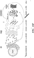
- the present invention relates to the specific finding that improving lysosomal function through improved trafficking of lysosomal enzymes from the endoplasmic reticulum to the golgi apparatus then finally to the endosome-lysosome system through enhancement of Rab function and/or activation of GCase, results in enhancement of lysosomal proteolysis.
- lysosomal proteolysis is enhanced by enhancement of proteolytic activity of acid hydrolases (enzymes that are commonly located in the lysosomes and have optimum enzymatic activity at acidic pHs, e.g., nucleases, proteases, glycosidases, lipases, phosphatases, sulfatases, phospholipases, and all lysosomal enzymes).
- acid hydrolases enzymes that are commonly located in the lysosomes and have optimum enzymatic activity at acidic pHs, e.g., nucleases, proteases, glycosidases, lipases, phosphatases, sulfatases, phospholipases, and all lysosomal enzymes.
- lysosomal proteolysis is enhanced by enhancement of the absolute number of lysosomal vesicles.
- lysosomal proteolysis is enhanced by enhancement of the amount of acid hydrolases
- lysosomal proteolysis is enhanced by enhancement of the exocytosis of cellular storage materials.
- increasing trafficking of lysosomal enzymes can provide effective treatment of, and in some instances prophylaxis of, certain neurodegenerative diseases, disorders, and/or conditions.
- Rab1a polypeptide is useful in the treatment of lysosomal storage diseases as well as other types of proteinopathies.
- antioxidants are useful in the treatment of lysosomal storage diseases as well as other types of proteinopathies.
- the invention relates to a small molecule NCGC00188758, N-(4-ethynylphenyl)-5,7-dimethylpyrazolo[1,5-a]pyrimidine-3-carboxamide, for use in treating or preventing a neurodegenerative proteinopathic disease.
- the neurodegenerative proteinopathic disease of the first aspect of the invention may be a synucleinopathic disease and may be selected from the group consisting of: Parkinson's disease; Lewy body disease; dementia with Lewy bodies; multiple system atrophy; neurodegeneration with brain iron accumulation type I, Parkinsonism-dementia complex of Guam; Hallervorden-Spatz disease; and frontotemporal dementia.
- the neurodegenerative proteinopathic disease may be Parkinson's disease.
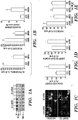
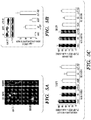
- FIGS 1A-1G show that GCase polypeptide knockdown (KD) results in compromised lysosomal degradation and causes accumulation of ⁇ -synuclein.
- Fig. 1A KD of GCase polypeptide in cortical neurons by GCase polypeptide shRNA is shown by western blot. Neural specific enolase (NSE) was used as a loading control. Four replicates are shown. Scrb, scrambled shRNA.
- FIG. 1C GlcCer immunofluorescence (top) and neutral lipids were visualized by BODIPY 493 fluorescence (bottom). Nuclei were visualized with DAPI. The arrows indicate cells with increased diffuse staining, whereas the arrowhead indicates a cell with punctated lipid accumulations.
- Fig. 1E Proteolysis of long-lived proteins in neurons assessed at 8 hr.
- GCase polypeptide KD is shown by western blot and ⁇ -tub was used as a loading control. Molecular weight (MW) is indicated in kDa. For all analyses, values are the mean ⁇ standard error of the mean (SEM).
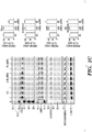
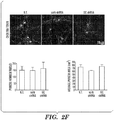
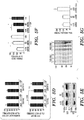
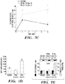
- Figures 2A-2G demonstrate the specificity of the shRNA GCase polypeptide lenti-infection system and changes to lysosomal protein levels upon GCase polypeptide knockdown.
- Fig.2A Lysates from transduced primary neurons were digested with endoglycosidase H (endo H) or PNGase. Nonspecific band (N.S.) is noted. N.T., not transduced.
- Fig.2D Measurement of sphingolipids upon GCase polypeptide knock-down in neurons by LC/MS/MS analysis.
- Figures 3A-3B show the generation of induced pluripotent stem cells from Gaucher disease patient fibroblasts.
- Fig.3B G-banding karyotype analysis of GD iPS cells showing normal chromosomal number, size, and genomic structure.
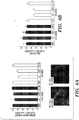
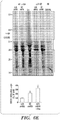
- Figures 4A-4F show the compromised proteolysis of long-lived proteins and specific accumulation of endogenous ⁇ -synuclein in human GD dopaminergic neurons.
- FIG.4E Western blot of T-sol lysates from iPS neurons. Htt, huntingtin; CBB, Coomassie brilliant blue.
- FIG.4F Western blot from Figure 4E was quantified by densitometry.
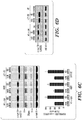
- Figures 5A-5I show the expression of human ⁇ -synuclein in primary cortical neurons and the effect of lysosomal inhibition with leupeptin treatment or GCase polypeptide knockdown.
- Neurons were infected with WT ⁇ -synuclein-expressing lentiviral vectors at moi 3 and analyzed at 7 days post-infection (dp).
- Fig.5A Immunostaining analysis using mAb's specific for human ⁇ -synuclein, syn211 and LB509, reveals the typical punctated pattern expected for synaptic enrichment in neuronal extensions. Approximately 60%-70% of cells were transduced.
- FIG.5E Western blot of LC3-II upon GCase polypeptide knockdown or leupeptin treatment. NSE was used as a loading control. MW is indicated in kDa.
- Figures 6A-6H demonstrate that GCase polypeptide depletion enhances ⁇ -synuclein-mediated neurotoxicity through aggregation-dependent mechanisms.
- Neurons expressing human ⁇ -synuclein proteins and GCase polypeptide shRNA were analyzed at 7 dpi.
- Fig.6B Neurotoxicity was assessed by neuronal volume analysis.
- Fig.6D ⁇ -synuclein western blot of T-sol fractions (leu, leupeptin; NT, not transduced). NSE was used as a loading control.
- Fig.6E Western blot of T-insoluble ⁇ -synuclein. Quantification is shown below.
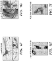
- Figures 7A-7I show that GlcCer directly influences the in vivo fibril formation of recombinant ⁇ -synuclein and stabilizes soluble oligomeric species.
- Fig.7B Analysis of 100,000 x g soluble ⁇ -synuclein at 1 and 5 hr by SEC (115-38 ⁇ and 36-27 ⁇ fractions), then SDS-PAGE/western blot (syn211). The MW is indicated in kDa.
- Fig.7E Centrifugal sedimentation analysis at 28 hr (s, supernatant; p, pellet).
- Fig. 7F EM analysis of ⁇ -synuclein aggregates showing a mixture of fibrillar (i-ii) and amorphous (iv-v) structures at 24 hr. Panels ii-v show immmuno-EM analysis using mAb syn505. Scale bars: 100 nm for i-iii; 500 nm for iv and v.
- FIG.7G Immuno-EM analysis with syn505 of ⁇ -synuclein+PC25/GlcCer75 reactions at 15 hr.
- GIcCer lipid tubules are ⁇ 50 nm in width. Scale bars: 100 nm for i and iii; 500 nm for ii.
- Fig.7H Immuno-EM analysis with syn505 of ⁇ -synuclein+PC25/GlcCer75 reactions at 24 hr showing fibrillar structures of 10-14 nm in width with twisted (i) or straight (ii) morphologies that appear to extend from GlcCer tubules. Scale bars: 100 nm.
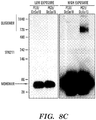
- Figures 8A-8F demonstrate that GlcCer specifically affects the in vitro formation of ⁇ -synuclein fibrils and soluble oligomers in pH-dependent manner.
- Purified ⁇ -synuclein was incubated with lipid dispersions as described in Figures 7A-7I .
- Fig.8A Amyloid formation was assessed at 36 hr at pH 5.0, 37°C (2 mg/ml in 0.1 M sodium acetate buffer) or pH 7.4, 37°C (2 mg/ml in 0.1 M sodium phosphate buffer). Values are expressed as fold-change relative to the control reaction of each pH condition.
- PC50%/polyethylene glycol (PEG) 50% was used in the pH 5.0 condition as a control.
- Fig.8C 100,000 x g soluble ⁇ -synuclein/ lipid reactions at pH 5.0 were analyzed by native gel electrophoresis/western blot. The marker indicates the apparent MW in kilodaltons according to globular protein standards (native mark, Invitrogen).
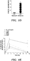
- Fig.8D Densitometric quantification of the oligomer:monomer ratio detected by native gel/western blot analysis.
- Fig.8E Levels of 100,000 x g ⁇ -synuclein soluble oligomers were determined after 3 and 15 hr incubation at pH 5.0 in the presence of PC25/lactosylceramide 75 (LacCer), PC25/galactosylceramide 75 (GalCer), PC25/Glucosylsphingosine75 (GluSph) by SDS-PAGE.
- Fig.8F Sedimentation analysis of ⁇ -synuclein/lipid reactions at pH 5.0 after 3 and 15 hr incubations. S, supenatant; P, pellet. No soluble ⁇ -synuclein(oligomers or monomers) was detected at 15 hr since it was completely converted into the pelletable fraction (P).Values are the mean ⁇ SEM for all quantifications.
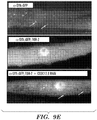
- Figures 9A-9E show the accumulation of sphingolipids in a mouse GD model.
- Fig.9B Gangliosides were analyzed by thin layer chromatography (TLC).
- Fig.9C Accumulation of ⁇ -synuclein in GD mice expressing D409H GCase polypeptide.
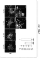
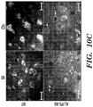
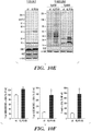
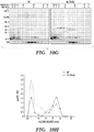
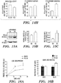
- Figures 10A-10H show ⁇ -synuclein accumulation and soluble oligomer formation in GD mice. Analysis of 12-week-old GD mice (4L/PS-NA).
- Fig. 10C Costaining of ⁇ -synuclein and neuronal marker NeuN.
- FIG.10D Left: Quantification of neuronal spheroids. ND, not detected. Middle: Quantification of neuronal number by NeuN immunostaining. Right: Quantification of ⁇ -synuclein aggregates by immunostaining.
- Fig.10E Sequential extraction analysis of Ctx. pAb SNL-1 and mAb syn202 detect total endogenous ⁇ -synuclein, whereas syn505 detects oxidized/nitrated and misfolded ⁇ -synuclein. NSE and ⁇ -tub were used as loading controls.
- Fig.10F Quantification of T-sol monomers (18 kDa, left), T-sol oligomers (>18 kDa, middle), and T-insoluble ⁇ -synuclein (total lane, right).
- Fig.10G Native SEC/SDS-PAGE/western blot of T-sol fractions. Radius, ⁇ .
- Fig. 10H Chromatographic profile obtained by syn202 densitometry. The values are representative of independent SEC analyses from three mice. The MW is indicated in kDa for each blot. For all quantifications, values are the mean ⁇ SEM.
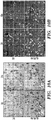
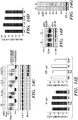
- FIGS 11A-11L show that accumulation of T-sol ⁇ -synuclein oligomers occurs in GD brain.
- Figs. 11A-11C Healthy controls.
- Figs. 11D and 11E Type I non-neuronopathic GD.
- Fig.11F Atypical Parkinson's disease (APD).
- Fig.11G dementia with Lewy bodies (DLB).
- FIG. 11H and 11I Analysis of cortical material obtained from infants with type II acute neuronopathic GD.
- Fig.11J Cortical lysates from a 3-year old child with neuronopathic type III GD.
- Fig. 11K DLB with a heterozygous mutation in GBA1.
- Fig.11L Analysis of the 45 A-sized fraction with syn303, which preferentially detects pathological oligomeric ⁇ -synuclein. Bands migrating at 18, 44, and 75 kDa were detected with both syn303 and syn211 (arrows).
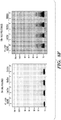
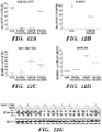
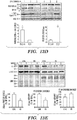
- Figures 12A-12E show the quantification of GCase polypeptide activity, GCase protein levels, and ⁇ -synuclein oligomer levels in human GD brain.
- the samples analyzed here are the same as those presented in Figures 11A-11L and Table 15.
- Fig.12A GCase polypeptide activity was determined in whole-cell homogenate of cortical samples. The data were grouped according to the presence of GCase polypeptide mutations, and also neuropathological differences (with or without synucleinopathy).
- Fig.12B The GCase polypeptide activity in the P2 fraction of heterozygous GCase polypeptide mutant carriers and WT brain reveals a more dramatic decrease in activity (50%) compared to whole cell measurements.
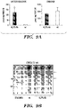
- Fig.12C ⁇ -synuclein oligomers were quantified by densitometric analysis of SEC/SDS-PAGE/western blot analysis with mAb syn211 (representative examples shown in Figures 11A-11L ; some GD heterozygote blots are not shown in Figures 11A-11L but quantified and presented in the graph).
- Fig.12D Quantification of the 45 ⁇ -sized fractions with mAb syn303. Representative examples are shown in Figure 11L . Some samples could not be analyzed by syn303 due to sample limitation.
- Fig.12E Western blot of GCase polypeptide in the same samples analyzed and presented in Figures 11A-11L . T-sol lysates were treated with endo H to reveal levels of the mature GCase polypeptide forms. NSE was used as a loading control. MW is indicated in kDa. The lines in panels A-D represent the mean values.
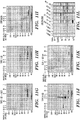
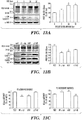
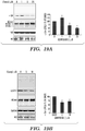
- Figures 13A-13F demonstrate that elevated levels of ⁇ -synuclein inhibit the intracellular trafficking of GCase polypeptide and decrease lysosomal GCase polypeptide function.
- Fig. 13B Post-ER/ER GCase polypeptide in cortical neurons expressing human WT, A53T, or ⁇ 71-82 ⁇ -synuclein.
- ⁇ -synuclein levels were determined by syn211 (human-specific) and syn202 (human and mouse). NSE was used as a loading control.
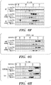
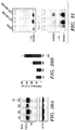
- Figures 14A-14H demonstrate the modulation of lysosomal GCase polypeptide maturation and activity by ⁇ -synuclein expression in primary neurons and human brain.
- Fig. 14A Enrichment of lysosomal or microsomal organelles by subcellular centrifugal fractionation. Western blot analysis of neuronal cultures infected at moi 3 with empty vector (vect), WT, A53T, or ⁇ 71-82 ⁇ -synuclein expressing lentivirus and harvested at dpi 7. Antibodies against GRP78 and calnexin were used to validate microsome enrichment, while antibodies against LAMP1 and 2 were used to validate lysosomal enrichment.
- Coomassie brilliant blue (CBB) is used as a loading control. MW is indicated along the left side of the blot in kDa.
- Fig. 14C Accumulation and retention of ER GCase polypeptide upon expression of human WT ⁇ -synuclein in primary cultures. Endo H and PNGase F/GCase polypeptide western blot analysis of T-sol neuronal lysates transduced to express WT or ⁇ 71-82 ⁇ -synuclein.
- Vect and N-terminal truncated polyQ expanded huntingtin protein were used as controls. Endo H sensitive GCase polypeptide immunoreactive smears migrating below 60 kDa indicated the levels of ER-localized GCase polypeptide. PNGase F was used to determine the migration of deglycosylated GCase polypeptide. ⁇ -Tub was used as a loading control.
- Fig. 14D Quantification of GCase polypeptide mRNA levels by real-time PCR from infected neuronal cultures.
- GCase polypeptide activity was measured in P2 (middle) and P3 (right) fractions of C5 and C6 (values are the mean of three repeated-measurements ⁇ SEM, *p ⁇ 0.05).
- values are the mean ⁇ SEM.
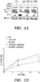
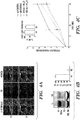
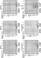
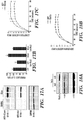
- Figures 15A-15C show that GCase polypeptide activation increases proteolysis in human dopamine neurons. Neurons were treated with 100 ⁇ M IFG or vehicle control (veh) for 5 days followed by 1 day wash-out to remove IFG.
- Fig.15A Western blot analysis of GCase polypeptide. Neural specific enolase (NSE) was used as a loading control.
- Figures 16A-16B demonstrate the enhancement of long-lived proteolysis by allosteric activation of GCase polypeptide in human midbrain iPS dopamine neurons from a PD patient. Neurons were treated with an allosteric activator of GCase polypeptide and proteolysis of long-lived ( Fig.16A ) or short-lived ( Fig.16B ) was determined by radioactive pulse-chase.
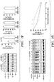
- Figures 17A-17C demonstrate that GCase polypeptide overexpression increases lysosomal proteolysis in non-neuronal cells.
- Hela cells were transfected with GFP or myc-GCase polypeptide expression constructs.
- Fig. 17A Overexpression levels were determined by western blot using anti-GCase polypeptide or myc antibodies. GAPDH was used as a loading control. Two replicates shown.
- Fig. 17B Proteolysis of long-lived proteins was determined in transfected Hela cells by radioactive pulse-chase after 36 hrs.
- Lysosomal inhibitors leupeptin (Leu) and ammonium chloride (NH 4 Cl) were used to determine the amount of lysosomal proteolysis in each condition.
- Figures 18A-18B show the reduction of ⁇ -synuclein and enhancement of lysosomal function by Rab1a polypeptide overexpression.
- Fig. 18A Human iPS dopamine neurons from a PD patient were transduced with Rab1a polypeptide expressing lentivirus. Overexpression of Rab 1a polypeptide was confirmed at moi 5 by western blot. ⁇ -synuclein levels were determined by western blot using mAb syn211. ⁇ -tubulin was used as a loading control.
- Fig. 18B Cathepsin B activity was assessed in transfected hela cells as described in Figures 17A-17C .
- Figures 19A-19B demonstrate the reduction of ⁇ -synuclein by allosteric activation of GCase polypeptide in human midbrain dopamine neurons.
- Fig. 19B Neurons generated from a PD patient were treated and analyzed as described in ( Fig. 19A ).
- Figures 20A-20B show that combination of GCase chaperone IFG and antioxidants enhance post-ER GCase polypeptide in PD iPS midbrain dopamine neurons.
- Fig. 20A Neurons from a PD patient were treated with PBS (veh), IFG, n-acetyl-cysteine (NAC), or both IFG + NAC and GCase polypeptide maturation was determined by western blot. ⁇ iii tubulin was used as a loading control.
- Fig. 20A Neurons from a PD patient were treated with PBS (veh), IFG, n-acetyl-cysteine (NAC), or both IFG + NAC and GCase polypeptide maturation was determined by western blot. ⁇ iii tubulin was used as a loading control.
- Figure 21 shows the sedementation analysis of ⁇ -synuclein at pH 5.0 in the presence of GM1 ganglioside or total brain gangliosides.
- Samples were incubated for 0 or 15 hrs, centrifuged at 100,000 g for 30 min to sediment ⁇ -synuclein aggregates, and analyzed by SDS-PAGE / western blot using syn211.
- the monomeric form migrates at 18 kDa and oligomeric forms migrate above 19 kDa. s, supernatant fraction; p, pellet fraction.
- activating agent refers to an agent that increases level and/or activity of a target entity as compared with its level and/or activity under comparable conditions absent the activating agent.
- an activating agent can increase level and/or activity of a target entity by at least about 5%, including at least about 10%, at least about 15%, at least about 20%, at least about 30%, at least about 40%, at least about 50%, at least about 60%, at least about 70%, at least about 80%, at least about 90%, at least about 95% or more, as compared with its level and/or activity under comparable conditions absent the activating agent.
- an activating agent increases level and/or activity of its target entity to a point within a predetermined range of a reference level and/or activity.
- a reference level and/or activity is the level and/or activity observed with a wild type version of the target entity in its natural context.
- an activating agent binds directly to its target. In some embodiments, an activating agent binds indirectly (i.e., by binding with a physically distinct entity that binds to the target).
- an activating agent does not interact physically, either directly or indirectly, with its target, but increases level and/or activity of the target through other action (e.g., binding to a regulatory site in a nucleic acid that increases expression of the target; activation or inhibition of an enzyme that modifies the target and alters its activity, etc).
- an activating agent stabilizes and/or increases half-life of its target entity.
- an activating agent stabilizes its target entity in a particular three-dimensional conformation.
- an activating agent competes with an inhibitor for binding to its target entity.
- an activating agent prevents or reduces aggregation of the target entity.
- an activating agent stabilizes interaction of its target entity with another entity (e.g., a substrate protein, RNA, or DNA, a small molecule, peptide, or carbohydrate).
- an activating agent binds to a target entity and increases the interaction of that target entity with another entity as compared with its interaction under comparable conditions absent the activating agent.
- an activating agent-mediated increase in interaction of a target entity with another entity increases level and/or activity of that target entity as compared with its level and/or activity under comparable conditions absent the activating agent.
- an activating agent binds to a target entity and decreases interaction of that target entity with another entity as compared with its interaction under comparable conditions absent the activating agent.
- an activating agent-mediated decrease in interaction of the target entity with another entity increases level and/or activity of that target entity as compared with its level and/or activity under comparable conditions absent the activating agent.
- an activating agent may be or comprise a compound of any chemical class (e.g., a small molecule, metal, nucleic acid, polypeptide, lipid and/or carbohydrate).
- an activating agent is or comprises an antibody or antibody mimic.
- an activating agent is or comprises a nucleic acid agent (e.g., an antisense oligonucleotide, a siRNA, a shRNA, etc) or mimic thereof.
- an activating agent is or comprises a small molecule.
- an activating agent is or comprises a naturally-occurring compound (e.g., small molecule). In some embodiments, an activating agent has a chemical structure that is generated and/or modified by the hand of man. In general, an activating agent increases level or activity of one or more target entities present in and/or produced by a cell or organism.
- a target entity is or comprises a polypeptide.
- a target entity is or comprises a nucleic acid (e.g., a nucleic acid that encodes or regulates [e.g., by altering expression and/or activity of] a polypeptide). In some embodiments, a target entity is or comprises a carbohydrate.
- a target entity is or comprises a lipid. In some embodiments, a target entity is or comprises an enzyme. In some embodiments, a target entity is or comprises a lysosomal enzyme. In some embodiments, a target entity is or comprises a polypeptide involved in cellular trafficking.
- Amyloidopathy As used herein, the term “amyloidopathy” or “amyloidopathic” refers to diseases, disorders, and/or conditions that are associated with or characterized by pathological accumulation of the any disease-linked protein exhibiting amyloid conformation (i.e., ⁇ -pleated sheet), including but not limited to Alzheimer's disease, vascular dementia, and cognitive impairment.
- Antioxidant refers to an entity, e.g., small molecule, polypeptide, nucleic acid, saccharide, lipid, inorganic agent (e.g., metal, mineral, etc), or combinations thereof that inhibits the oxidation, nitration, or nitrosylation of another entity.
- entity e.g., small molecule, polypeptide, nucleic acid, saccharide, lipid, inorganic agent (e.g., metal, mineral, etc), or combinations thereof that inhibits the oxidation, nitration, or nitrosylation of another entity.
- ⁇ -galactosidase polypeptide As used herein, the term " ⁇ -galactosidase polypeptide" or “beta-gal polypeptide” refers to a polypeptide that is a ⁇ -galactosidase enzyme. Those of ordinary skill in the art will appreciate that ⁇ -galactosidase is a hydrolase enzyme that catalyzes hydrolysis of ⁇ -glycosidic bond formed between a galactose and its organic moiety.
- ⁇ -galactosidase enzyme has different sub-cellular locations, i.e., ⁇ -galactosidase isoform 1 localized in lysosome and ⁇ -galactosidase isoform 2 localized in perinuclear region of the cytoplasm.
- Substrates of ⁇ -galactosidase enzyme include ganglioside G M1 , lactosylceramides, lactose, and various glycoproteins.
- Representative known ⁇ -galactosidase polypeptides include those listed below in Table 1.
- the ⁇ -galactosidase polypeptide is a ⁇ -galactosidase polypeptide homolog.
- the term " ⁇ -galactosidase polypeptide homolog" comprises a polypeptide whose amino acid sequence includes at least one sequence element comprising conserved residues found in polypeptides of Table 1; in some such embodiments, such sequence element comprises at least 3, 4, 5, 6, 7, 8, 9, 10 or more residues whose identity and relative position is preserved. In some embodiments, such sequence element comprises at least 3, 4, 5, 6, 7, 8, 9, 10 or more consecutive residues.
- a " ⁇ -galactosidase polypeptide homolog” is or comprises a polypeptide whose amino acid sequence shows at least 50%, 55%, 60%, 65%, 70%, 75%, 80%, 85%, 90%, 95%, 96%, 97%, 98%, 99% or greater overall sequence identity with one or more polypeptides in Table 1 and/or shares at least one characteristic sequence element with one or more polypeptides in Table 1.
- a characteristic sequence element includes one or more catalytic residues and/or one or more conserved residues found in polypeptides of Table 1.
- Calcium channel blocker refers to an agent that blocks voltage-dependent calcium channels. Synonyms of the term “calcium channel blocker” are calcium channel antagonists, calcium channel inhibitors and calcium entry blockers and these terms are used interchangeably herein.
- Exemplary calcium channel blockers include, but are not limited to amlodipine, felodipine, isradipine, lacidipine, nicardipine, nifedipine, niguldipine, niludipine, nimodipine, nisoldipine, nitrendipine, nivaldipine, ryosidine, anipamil, diltiazem, fendiline, flunarizine, gallopamil, mibefradil, prenylamine, tiapamil, verapamil, perhexyline maleate, fendiline, prenylamine, and derivatives of any of thereof.
- Characteristic sequence element refers to a distinctive core sequence or structural element that is found in all members of a family of polypeptides, small molecule, or nucleic acids, and therefore can be used by those of ordinary skill in the art to define members of the family.
- Combination therapy refers to those situations in which two or more different pharmaceutical agents are administered in overlapping regimens so that the subject is simultaneously exposed to both agents.
- Comparable is used herein to describe two (or more) sets of conditions or circumstances that are sufficiently similar to one another to permit comparison of results obtained or phenomena observed.
- comparable sets of conditions or circumstances are characterized by a plurality of substantially identical features and one or a small number of varied features.
- sets of conditions are comparable to one another when characterized by a sufficient number and type of substantially identical features to warrant a reasonable conclusion that differences in results obtained or phenomena observed under the different sets of conditions or circumstances are caused by or indicative of the variation in those features that are varied.
- Dosing regimen refers to a set of unit doses (typically more than one) that are administered individually to a subject, typically separated by periods of time.
- a given therapeutic agent has a recommended dosing regimen, which may involve one or more doses.
- a dosing regimen comprises a plurality of doses each of which are separated from one another by a time period of the same length; in some embodiments, a dosing regime comprises a plurality of doses and at least two different time periods separating individual doses. In some embodiments, all doses within a dosing regimen are of the same unit dose amount.
- a dosing regimen comprises a first dose in a first dose amount, followed by one or more additional doses in a second dose amount different from the first dose amount. In some embodiments, a dosing regimen comprises a first dose in a first dose amount, followed by one or more additional doses in a second dose amount same as the first dose amount.
- Enzyme Replacement Therapy refers to the administration of an enzyme to a subject that shows, prior to such administration, a reduced level of activity of the enzyme as compared with that observed, on average, across a population of normal individuals of the same species (e.g., humans).
- Equivalent Dosage is used herein to compare dosages of different pharmaceutically active agents that effect the same biological result. Dosages of two different agents are considered to be “equivalent” to one another in accordance with the present invention if they achieve a comparable level or extent of the biological result. In some embodiments, equivalent dosages of different pharmaceutical agents for use in accordance with the present invention are determined using in vitro and/or in vivo assays as described herein.
- one or more lysosomal activating agents for use in accordance with the present invention is utilized at a dose equivalent to a dose of a reference lysosomal activating agent; in some such embodiments, the reference lysosomal activating agent for such purpose is selected from the group consisting of small molecule allosteric activators (e.g., pyrazolpyrimidines), imminosugars (e.g., isofagomine), antioxidants (e.g., n-acetyl-cysteine), and regulators of cellular trafficking (e.g., Rab1a polypeptide).
- small molecule allosteric activators e.g., pyrazolpyrimidines
- imminosugars e.g., isofagomine
- antioxidants e.g., n-acetyl-cysteine
- regulators of cellular trafficking e.g., Rab1a polypeptide
- Gain of Function Disease typically refers to a disease characterized by increased aggregation-associated proteotoxicity. In such diseases, aggregation exceeds clearance inside and/or outside of the cell. Gain of function diseases are often associated with aging and are also referred to as gain of toxic function diseases.
- Exemplary gain of function diseases include, but are not limited to neurodegenerative diseases associated with aggregation of polyglutamine repeats in proteins or repeats at other amino acids such as alanine, Lewy body diseases, and other disorders associated with ⁇ -synuclein aggregation, amyotrophic lateral sclerosis, transthyretin-associated aggregation diseases, Alzheimer's disease, age-associated macular degeneration, inclusion body myositosis, and prion diseases.
- neurodegenerative diseases associated with aggregation of polyglutamine repeats in proteins or repeats at other amino acids such as alanine, Lewy body diseases, and other disorders associated with ⁇ -synuclein aggregation, amyotrophic lateral sclerosis, transthyretin-associated aggregation diseases, Alzheimer's disease, age-associated macular degeneration, inclusion body myositosis, and prion diseases.
- Neurodegenerative diseases associated with aggregation of polyglutamine include, but are not limited to, Huntington's disease, dentatorubral and pallidoluysian atrophy, several forms of spino-cerebellar ataxia, and spinal and bulbar muscular atrophy.
- Alzheimer's disease is characterized by the formation of two types of aggregates: intracellular and extracellular aggregates of A ⁇ peptide and intracellular aggregates of the microtubule associated protein tau.
- Transthyretin-associated aggregation diseases include, for example, senile systemic amyloidoses, familial amyloidotic neuropathy, and familial amyloid cardiomyopathy.
- Lewy body diseases are characterized by an aggregation of ⁇ -synuclein protein and include, for example, Parkinson's disease.
- Prion diseases also known as transmissible spongiform encephalopathies
- Exemplary human prion diseases are Creutzfeldt-Jakob Disease (CJD), Variant Creutzfeldt-Jakob Disease, Gerstmann-Straussler-Scheinker Syndrome. Fatal Familial Insomnia and Kuru.
- Gene therapy refers to the administration to a subject (e.g., a human subject) of a nucleic acid (or a nucleic acid derived from the nucleic acid as, for example, by reverse transcription) encoding a polypeptide. In many embodiments, such administration is performed so that the polypeptide is expressed in or by cells of the subject after the administration. Nucleic acids may be incorporated into the genome of the cell or remain permanently in the cell as an episome (a genetic particle of certain cells that can exist either autonomously in the cytoplasm or as part of a chromosome). Gene therapy also encompasses delivery of nucleic acids that do not integrate or remain permanently in the cell to which they are delivered.
- Glucocerebrosidase polypeptide refers to a polypeptide that is a ⁇ -glucocerebrosidase enzyme. Those of ordinary skill in the art will appreciate that a glucocerebrosidase is naturally found localized in the lysosome, where it hydrolyses the ⁇ -glucosidic linkage of glucosylceramide.
- This naturally occurring glucocerebrosidase enzyme is also known as acid ⁇ -glucosidase, alglucerase, ⁇ -glucocerebrosidase, D-glucosyl-N-acylsphingosine glucosylhydrolase, GBA1, Glcm_human, Gluc, glucocerebrosidase ⁇ -glucosidase, glucosphingosine glucosylhydrolase, glucosylceramidase, glucosylceramide ⁇ -glucosidase, or imiglucerase.
- Representative known glucocerebrosidase polypeptides include those listed below in Table 2.
- glucocerebrosidase polypeptide can be a gluocerebrosidase polypeptide homolog.
- the term "glucocerebrosidase polypeptide homolog" comprises a polypeptide whose amino acid sequence includes at least one sequence element comprising conserved residues found in polypeptides of Table 2; in some such embodiments, such sequence element comprises at least 3, 4, 5, 6, 7, 8, 9, 10 or more residues whose identity and relative position is preserved. In some embodiments, such sequence element comprises at least 3, 4, 5, 6, 7, 8, 9, 10 or more consecutive residues.
- a "glucocerebrosidase polypeptide homolog” is or comprises a polypeptide whose amino acid sequence shows at least 50%, 55%, 60%, 65%, 70%, 75%, 80%, 85%, 90%, 95%, 96%, 97%, 98%, 99% or greater overall sequence identity with one or more polypeptides in Table 2 and/or shares at least one characteristic sequence element with one or more polypeptides in Table 2.
- a characteristic sequence element includes one or more catalytic residues and/or one or more conserved residues found in polypeptides of Table 2.
- Glucosylceramide synthase polypeptide refers to a polypeptide that shares at least one characteristic sequence element and/or overall sequence identity with a glucosyltransferase enzyme involved in the production of glucosylceramide-based glycosphingolipids, and similarly shows glycosyltransferase activity.
- glucosylceramide synthase regulates the production of glycosphingolipid conjugates called gangliosides (such as G M3 ) via glucosyl transfer to ceramide.
- Representative known glucosylceramide synthase polypeptides include those listed below in Table 3.
- a glucosylceramide synthase polypeptide is or comprises a polypeptide whose amino acid sequence includes at least one element comprising conserved residues found in polypeptides of Table 3.
- the glucosylceramide synthase polypeptide can be a glucosylceramide synthase polypeptide homolog.
- the term "glucosylceramide synthase polypeptide homolog" comprises a polypeptide whose amino acid sequence includes at least one sequence element comprising conserved residues found in polypeptides of Table 3; in some such embodiments, such sequence element comprises at least 3, 4, 5, 6, 7, 8, 9, 10 or more residues whose identity and relative position is preserved. In some embodiments, such sequence element comprises at least 3, 4, 5, 6, 7, 8, 9, 10 or more consecutive residues.
- a "glucosylceramide synthase polypeptide homolog” is or comprises a polypeptide whose amino acid sequence shows at least 50%, 55%, 60%, 65%, 70%, 75%, 80%, 85%, 90%, 95%, 96%, 97%, 98%, 99% or greater overall sequence identity with one or more polypeptides in Table 3 and/or shares at least one characteristic sequence element with one or more polypeptides in Table 3.
- a characteristic sequence element includes one or more catalytic residues and/or one or more conserved residues found in polypeptides of Table 3.
- Hexosaminidase polypeptide As used herein, the term “hexosaminidase polypeptide” or “ ⁇ -hexosaminidase polypeptide” refers to a polypeptide that is a ⁇ -hexosaminidase enzyme. Those of ordinary skill in the art will appreciate that ⁇ -hexosaminidase enzyme participates in hydrolysis of terminal N-acetyl-D-hexosamine residues in N-acetyl- ⁇ -D-hexosaminides.
- ⁇ -hexosaminidase enzyme and the cofactor G M2 activator protein catalyze the degradation of the G M2 gangliosides and other molecules containing terminal N-acetyl hexosamines.
- Lysosomal ⁇ -hexosaminidase enzymes are dimeric in structure and three active dimeric isozymes are produced through the combination of ⁇ - and ⁇ -subunits (encoded by HEXA and HEXB genes, respectively).
- Hexosaminidase isozyme A can hydrolyze G M2 ganglioside in vivo and has an ⁇ / ⁇ heterodimer subunit composition.
- Hexosaminidase isozyme B has a ⁇ / ⁇ homodimer subunit composition and hexosaminidase isozyme S has an ⁇ / ⁇ homodimer subunit composition.
- Representative known hexosaminidase polypeptides include those listed below in Table 4.
- the hexosaminidase polypeptide can be a hexosaminidase polypeptide homolog.
- the term "hexosaminidase polypeptide homolog" comprises a polypeptide whose amino acid sequence includes at least one sequence element comprising conserved residues found in polypeptides of Table 4; in some such embodiments, such sequence element comprises at least 3, 4, 5, 6, 7, 8, 9, 10 or more residues whose identity and relative position is preserved. In some embodiments, such sequence element comprises at least 3, 4, 5, 6, 7, 8, 9, 10 or more consecutive residues.
- a "hexosaminidase polypeptide homolog” is or comprises a polypeptide whose amino acid sequence shows at least 50%, 55%, 60%, 65%, 70%, 75%, 80%, 85%, 90%, 95%, 96%, 97%, 98%, 99% or greater overall sequence identity with one or more polypeptides in Table 4 and/or shares at least one characteristic sequence element with one or more polypeptides in Table 4.
- a characteristic sequence element includes one or more catalytic residues and/or one or more conserved residues found in polypeptides of Table 4.
- a reference measurement is one that was taken under comparable conditions.
- a reference measurement is or comprises a historical value.
- a reference measurement is or comprises a measurement in the same individual at a different time (e.g., prior to initiation of a particular treatment or event).
- a reference measurement is or comprises a measurement in a control individual (or multiple control individuals); in some such embodiments, a "control" individual is one who a) has not been exposed to a particular treatment or event, and/or b) displays a different (as compared with the test individual) susceptibility to or affliction with a proteinopathy, but optionally shares one or more features such as race, age (e.g., approximate, for example within a range), weight (e.g., approximate, for example within a range), height (e.g., approximate, for example within a range), temperament, geographic residence, eating habits, exercise habits, etc with a test individual.
- a reference measurement is a measurement taken in a different setting (for example, in a setting in which such treatment or event does not occur or has not occurred).
- Loss of function disease typically refers to a disease characterized by by inefficient folding of a protein resulting in excessive degradation of the protein.
- Exemplary loss of function diseases include, but are not limited to cystic fibrosis, lysosomal storage diseases, and Von Hippel-Lindau (VHL) Disease.
- cystic fibrosis the mutated or defective enzyme is the cystic fibrosis transmembrane conductance regulator (CFTR).
- CFTR cystic fibrosis transmembrane conductance regulator
- One of the most common mutations of this protein is ⁇ F508 which is a deletion ( ⁇ ) of three nucleotides resulting in a loss of the amino acid phenylalanine (F) at the 508 position on the protein.
- Lysosomal enzyme refers to an enzyme that functions in the lysosome.
- Some examples of lysosomal enzymes include, but are not limited to ⁇ -galactosidase A; ⁇ -glucosidase; ⁇ -glucosidase; ⁇ -hexosaminidase A; ⁇ -hexosaminidase B; ⁇ -L-iduronidase; ⁇ -galactosidase; ⁇ -glucuronidase; ⁇ -glucuronidase; ⁇ -fucosidase; sulfatases; acid ceramidases; NPC 1 ; acid sphingomyelinase; cathepsins (A, D, H, S, Z); H(+)-ATPases; sialidase; ⁇ -galactocerebrosidase; arylsulfatase; iduronate
- a lysosomal enzyme can be a lysosomal enzyme homolog.
- a "lysosomal enzyme homolog" is or comprises a polypeptide whose amino acid sequence includes at least one sequence element comprising conserved residues found in polypeptides of Table 5; in some such embodiments, such sequence element comprises at least 3, 4, 5, 6, 7, 8, 9, 10 or more residues whose identity and relative position is preserved. In some embodiments, such sequence element comprises at least 3, 4, 5, 6, 7, 8, 9, 10 or more consecutive residues.
- a "lysosomal enzyme homolog” is or comprises a polypeptide whose amino acid sequence shows at least 50%, 55%, 60%, 65%, 70%, 75%, 80%, 85%, 90%, 95%, 96%, 97%, 98%, 99% or greater overall sequence identity with one or more polypeptides in Table 5 and/or shares at least one characteristic sequence element with one or more polypeptides in Table 5.
- a characteristic sequence element includes one or more catalytic residues and/or one or more conserved residues found in polypeptides of Table 5.
- Lysosomal storage diseases refers to a group of genetic disorders that result from deficiency in at least one of the enzymes (e.g., acid hydrolases) that are required to break macromolecules down to peptides, amino acids, monosaccharides, nucleic acids and fatty acids in lysosomes.
- enzymes e.g., acid hydrolases
- Lysosomal storage diseases may result from non-lysosomal proteins that may or may not have enzymatic activity such as: a deficiency in a protein involved in trafficking an acid hydrolase to the lysosome such as lysosomal integral membrane protein 2 (LIMP2); deficiency of an ER-resident protein involved in post-translational modifications of acid hydrolases such as that found in multiple sulfatase deficiency (MSD); deficiency in a protein found in the Golgi apparatus that is involved in trafficking acid hydrolases and other lysosomal proteins to the lysosomal compartment such as N-acetylglucosamine-1-phosphotransferase which is deficient in Inclusion cell disease (I-cell disease); deficiency in an acid hydrolase cofactor such as sphingolipid activator proteins (saposin A, B, C, D); deficiency of a membrane fusion protein such as ceroid lipofuscinosis neuron
- mutant refers to an entity that shows significant structural identity with a reference entity but differs structurally from the reference entity in the presence or level of one or more chemical moieties as compared with the reference entity. In many embodiments, a mutant also differs functionally from its reference entity. In general, whether a particular entity is properly considered to be a "mutant" of a reference entity is based on its degree of structural identity with the reference entity. As will be appreciated by those skilled in the art, any biological or chemical reference entity has certain characteristic structural elements. A mutant, by definition, is a distinct chemical entity that shares one or more such characteristic structural elements.
- a small molecule may have a characteristic core structural element (e.g., a macrocycle core) and/or one or more characteristic pendent moieties so that a mutant of the small molecule is one that shares the core structural element and the characteristic pendent moieties but differs in other pendent moieties and/or in types of bonds present (single vs double, E vs Z, etc) within the core, a polypeptide may have a characteristic sequence element comprised of a plurality of amino acids having designated positions relative to one another in linear or three-dimensional space and/or contributing to a particular biological function, a nucleic acid may have a characteristic sequence element comprised of a plurality of nucleotide residues having designated positions relative to on another in linear or three-dimensional space.
- a characteristic core structural element e.g., a macrocycle core
- one or more characteristic pendent moieties so that a mutant of the small molecule is one that shares the core structural element and the characteristic pendent moieties but
- a mutant polypeptide may differ from a reference polypeptide as a result of one or more differences in amino acid sequence and/or one or more differences in chemical moieties (e.g., carbohydrates, lipids, etc) covalently attached to the polypeptide backbone.
- a mutant polypeptide shows an overall sequence identity with a reference polypeptide that is at least 85%, 86%, 87%, 88%, 89%, 90%, 91%, 92%, 93%, 94%, 95%, 96%, 97%, or 99%.
- a mutant polypeptide does not share at least one characteristic sequence element with a reference polypeptide.
- the reference polypeptide has one or more biological activities.
- a mutant polypeptide shares one or more of the biological activities of the reference polypeptide. In some embodiments, a mutant polypeptide lacks one or more of the biological activities of the reference polypeptide. In some embodiments, a mutant polypeptide shows a reduced level of one or more biological activities as compared with the reference polypeptide.
- composition refers to an active agent, formulated together with one or more pharmaceutically acceptable carriers.
- active agent is present in unit dose amount appropriate for administration in a therapeutic regimen that shows a statistically significant probability of achieving a predetermined therapeutic effect when administered to a relevant population.
- compositions may be specially formulated for administration in solid or liquid form, including those adapted for the following: oral administration, for example, drenches (aqueous or non-aqueous solutions or suspensions), tablets, e.g., those targeted for buccal, sublingual, and systemic absorption, boluses, powders, granules, pastes for application to the tongue; parenteral administration, for example, by subcutaneous, intramuscular, intravenous or epidural injection as, for example, a sterile solution or suspension, or sustained-release formulation; topical application, for example, as a cream, ointment, or a controlled-release patch or spray applied to the skin, lungs, or oral cavity; intravaginally or intrarectally, for example, as a pessary, cream, or foam; sublingually; ocularly; transdermally; or nasally, pulmonary, and to other mucosal surfaces.
- oral administration for example, drenches (aqueous or non-aqueous solutions or suspension
- pharmaceutically acceptable refers to those compounds, materials, compositions, and/or dosage forms which are, within the scope of sound medical judgment, suitable for use in contact with the tissues of human beings and animals without excessive toxicity, irritation, allergic response, or other problem or complication, commensurate with a reasonable benefit/risk ratio.
- pharmaceutically acceptable carrier means a pharmaceutically-acceptable material, composition or vehicle, such as a liquid or solid filler, diluent, excipient, or solvent encapsulating material, involved in carrying or transporting the subject compound from one organ, or portion of the body, to another organ, or portion of the body.
- a pharmaceutically-acceptable material such as a liquid or solid filler, diluent, excipient, or solvent encapsulating material, involved in carrying or transporting the subject compound from one organ, or portion of the body, to another organ, or portion of the body.
- Each carrier must be “acceptable” in the sense of being compatible with the other ingredients of the formulation and not injurious to the patient.
- materials which can serve as pharmaceutically-acceptable carriers include: sugars, such as lactose, glucose and sucrose; starches, such as corn starch and potato starch; cellulose, and its derivatives, such as sodium carboxymethyl cellulose, ethyl cellulose and cellulose acetate; powdered tragacanth; malt; gelatin; talc; excipients, such as cocoa butter and suppository waxes; oils, such as peanut oil, cottonseed oil, safflower oil, sesame oil, olive oil, corn oil and soybean oil; glycols, such as propylene glycol; polyols, such as glycerin, sorbitol, mannitol and polyethylene glycol; esters, such as ethyl oleate and ethyl laurate; agar; buffering agents, such as magnesium hydroxide and aluminum hydroxide; alginic acid; pyrogen-free water; isotonic saline; Ring
- compositions that are appropriate for use in pharmaceutical contexts, i.e., salts which are, within the scope of sound medical judgment, suitable for use in contact with the tissues of humans and lower animals without undue toxicity, irritation, allergic response and the like, and are commensurate with a reasonable benefit/risk ratio.
- Pharmaceutically acceptable salts are well known in the art. For example, S. M. Berge, et al. describes pharmaceutically acceptable salts in detail in J. Pharmaceutical Sciences, 66: 1-19 (1977 ).
- pharmaceutically acceptable salt include, but are not limited to, nontoxic acid addition salts, which are salts of an amino group formed with inorganic acids such as hydrochloric acid, hydrobromic acid, phosphoric acid, sulfuric acid and perchloric acid or with organic acids such as acetic acid, maleic acid, tartaric acid, citric acid, succinic acid or malonic acid or by using other methods used in the art such as ion exchange.
- nontoxic acid addition salts which are salts of an amino group formed with inorganic acids such as hydrochloric acid, hydrobromic acid, phosphoric acid, sulfuric acid and perchloric acid or with organic acids such as acetic acid, maleic acid, tartaric acid, citric acid, succinic acid or malonic acid or by using other methods used in the art such as ion exchange.
- pharmaceutically acceptable salts include, but are not limited to, adipate, alginate, ascorbate, aspartate, benzenesulfonate, benzoate, bisulfate, borate, butyrate, camphorate, camphorsulfonate, citrate, cyclopentanepropionate, digluconate, dodecylsulfate, ethanesulfonate, formate, fumarate, glucoheptonate, glycerophosphate, gluconate, hemisulfate, heptanoate, hexanoate, hydroiodide, 2-hydroxy-ethanesulfonate, lactobionate, lactate, laurate, lauryl sulfate, malate, maleate, malonate, methanesulfonate, 2-naphthalenesulfonate, nicotinate, nitrate, oleate, oxalate, palmitate
- Representative alkali or alkaline earth metal salts include sodium, lithium, potassium, calcium, magnesium, and the like.
- pharmaceutically acceptable salts include, when appropriate, nontoxic ammonium, quaternary ammonium, and amine cations formed using counterions such as halide, hydroxide, carboxylate, sulfate, phosphate, nitrate, alkyl having from 1 to 6 carbon atoms, sulfonate and aryl sulfonate.
- Polypeptide in general, is a string of at least two residues (e.g., amino acids) linked to one another by peptide bonds. In some embodiments, a polypeptide includes one or more moieties other than such residues. For example, in some embodiments, a polypeptide comprises one or more glycan moieties attached to its residues (e.g., is a glycopeptide). In some embodiments, a polypeptide comprises one or more polyethylene glycol moieties (i.e., is pegylated). In some embodiments, a polypeptide comprises one or more polypeptide chain linked by one or more disulfide bonds or associated by other means.
- a polypeptide includes amino acid residues. In some embodiments, a polypeptide includes one or more residues that are not amino acids. In some embodiments, a polypeptide includes one or more residues that is an amino acid that does not occur in nature.
- Pharmacological chaperone refers to a molecule, such as small molecule, polypeptide, nucleic acid, lipid, or carbohydrate that specifically binds to a protein and has one or more of the following effects: enhancing the formation of a stable molecular conformation of the protein; inducing trafficking of the protein from the ER to another cellular location, preferably a native cellular location, i.e., preventing ER-associated degradation of the protein; preventing aggregation of misfolded proteins; and/or restoring or enhancing at least partial wild-type function and/or activity of the protein.
- a pharmacological chaperone acts on one or more lysosomal enzymes.
- a pharmacological chaperone is an entity that binds to a lysosomal enzyme so that its proper folding, trafficking, non-aggregation, and/or activity is increased relative to that observed absent the pharmacological chaperone.
- Proteinopathy As used herein, the term “proteinopathy” or “proteinopathic” refers to a disease, disorder, and/or condition associated with the pathogenic aggregation and/or accumulation of one or more types of proteins, for example, but not limited to ⁇ -synuclein, ⁇ -amyloid, and/or tau proteins. In some embodiments, a proteinopathy is characterized by an anomaly in one or more of protein production, folding, aggregation, metabolism, or degradation (e.g. autophagy), transportation, etc. In some embodiments, proteinopathies are neurodegenerative diseases. In some embodiments, proteinopathies are inflammatory diseases. In some embodiments, proteinopathies are cardiovascular diseases. In some embodiments, proteinopathies are proliferative diseases.
- proteins implicated in proteinopathies include: ⁇ -synuclein in the case of Parkinson's disease, Lewy body disease, and other synucleinopathies; tau and ⁇ -amyloid in the case of Alzheimer's disease and certain other neurodegenerative diseases; SOD1 and TDP-43 in the case of amyotrophic lateral sclerosis; huntingtin in the case of Huntington's disease; rhodopsin in the case of retinitis pigmentosa; and proteins involved in lysosomal storage diseases.
- proteostasis refers to the concentration, conformation, binding interactions, e.g., quaternary structure, and location of proteins making up the proteome. Proteostasis is influenced by the chemistry of protein folding/misfolding and by numerous regulated networks of interacting and competing biological pathways that influence protein synthesis, folding, conformation, binding interactions, trafficking, disaggregation and degradation. In some embodiments, proteostatis is controlled, for example, by altering level and/or activity of one or more nucleic acids or proteins. In some embodiments, proteostasis is controlled through transcriptional and/or translational changes.
- Rab polypeptide refers to a polypeptide that shares a characteristic sequence element and/or overall degree of sequence identitiy with a member of the Rab family of small guanosine triphosphates (GTPases) that regulate multiple steps of vesicle trafficking and membrane fusion, including but not limited to vesicles of the endosome-lysosome system, synaptic vesicles of neurons, exocytosis of cellular storage materials, and the transport of newly synthesized proteins from endoplasmic reticulum to the Golgi apparatus and within Golgi compartments.
- GTPases small guanosine triphosphates
- An example of Rab polypeptide is Rab1a polypeptide.
- Table 6 provides nucleic acid sequence encoding Rab1a polypeptide. Table 6 provides representative examples of Rab polypeptide sequences.
- a Rab polypeptide is a Rab polypeptide homolog.
- the term "Rab polypeptide homolog” comprises a polypeptide whose amino acid sequence includes at least one sequence element comprising conserved residues found in polypeptides of Table 6; in some such embodiments, such sequence element comprises at least 3, 4, 5, 6, 7, 8, 9, 10 or more residues whose identity and relative position is preserved. In some embodiments, such sequence element comprises at least 3, 4, 5, 6, 7, 8, 9, 10 or more consecutive residues.
- a "Rab polypeptide homolog” is or comprises a polypeptide whose amino acid sequence shows at least 50%, 55%, 60%, 65%, 70%, 75%, 80%, 85%, 90%, 95%, 96%, 97%, 98%, 99% or greater overall sequence identity with one or more polypeptides in Table 6 and/or shares at least one characteristic sequence element with one or more polypeptides in Table 6.
- a characteristic sequence element includes one or more catalytic residues and/or one or more conserved residues found in polypeptides of Table 6.
- sample refers to a biological sample obtained or derived from a source of interest, as described herein.
- a source of interest comprises an organism, such as an animal or human.
- a biological sample comprises biological tissue or fluid.
- a biological sample is or comprises bone marrow; blood; blood cells; ascites; tissue or fine needle biopsy samples; cell-containing body fluids; free floating nucleic acids; sputum; saliva; urine; cerebrospinal fluid, peritoneal fluid; pleural fluid; feces; lymph; gynecological fluids; skin swabs; vaginal swabs; oral swabs; nasal swabs; washings or lavages such as a ductal lavages or broncheoalveolar lavages; aspirates; scrapings; bone marrow specimens; tissue biopsy specimens; surgical specimens; feces, other body fluids, secretions, and/or excretions; and/or cells therefrom, etc.
- a biological sample is or comprises cells obtained from an individual.
- a sample is a "primary sample" obtained directly from a source of interest by any appropriate means.
- a primary biological sample is obtained by methods selected from the group consisting of biopsy (e.g., fine needle aspiration or tissue biopsy), surgery, collection of body fluid (e.g., blood, lymph, feces etc .), etc.
- the term "sample” refers to a preparation that is obtained by processing (e.g., by removing one or more components of and/or by adding one or more agents to) a primary sample. For example, filtering using a semi-permeable membrane.
- Such a "processed sample” may comprise, for example nucleic acids or proteins extracted from a sample or obtained by subjecting a primary sample to techniques such as amplification or reverse transcription of mRNA, isolation and/or purification of certain components, etc.
- Saposin polypeptide refers to a polypeptide that shares at least one characteristic sequence element and/or overall sequence identity with a saposin protein domain. Saposins are small heat-stable lysosomal proteins that serve as activators of various lysosomal lipid-degrading enzymes by isolating the lipid substrate form the membrane surroundings and making it more accessible to the soluble degradative enzymes.
- Saposins are synthesized as a single precursor molecule, prosaposin, which contains four saposin-B domains (four each of SapB1 and SapB2), yielding the active saposins after proteolytic cleavage (saposin A, B, C, and D), and two saposin-A domains (SapA) that are removed in the activation process.
- Representative known saposin polypeptides include those listed below in Table 7.
- saposin polypeptide is a saposin polypeptide homolog.
- the term "saposin polypeptide homolog" is or comprises a polypeptide whose amino acid sequence includes at least one sequence element comprising conserved residues found in polypeptides of Table 7; in some such embodiments, such sequence element comprises at least 3, 4, 5, 6, 7, 8, 9, 10 or more residues whose identity and relative position is preserved. In some embodiments, such sequence element comprises at least 3, 4, 5, 6, 7, 8, 9, 10 or more consecutive residues.
- a "saposin polypeptide homolog” is or comprises a polypeptide whose amino acid sequence shows at least 50%, 55%, 60%, 65%, 70%, 75%, 80%, 85%, 90%, 95%, 96%, 97%, 98%, 99% or greater overall sequence identity with one or more polypeptides in Table 7 and/or shares at least one characteristic sequence element with one or more polypeptides in Table 7.
- a characteristic sequence element includes one or more catalytic residues and/or one or more conserved residues found in polypeptides of Table 7.
- Small molecule includes any chemical or other moiety whose molecular weight is less than about 5000 daltons (Da). In some embodiments, small molecules have molecular weights below about 2500, about 1000, or about 500 daltons. In some embodiments, small molecules are not polymers. In some embodiments, small molecules are not peptides. In some embodiments, small molecules are not nucleic acids. In some embodiments, small molecules have biological activity and/or act to affect biological processes. In some embodiments, small molecules are natural products. In some embodiments, small molecules are not natural products (e.g., were first prepared by chemical synthesis).
- Sphingolipid metabolizing enzyme refers to enzymes that control synthesis and degradation of sphingolipids. These enzymes co-ordinate interconversion of sphigolipid metabolites (e.g, ceramide, sphingosine, diacyglycerol, or sphingosine-1-phosphate).
- sphingolipid metabolizing enzyme refers to enzymes that control synthesis and degradation of sphingolipids. These enzymes co-ordinate interconversion of sphigolipid metabolites (e.g, ceramide, sphingosine, diacyglycerol, or sphingosine-1-phosphate).
- Exemplary sphingolipid metabolizing enzymes include, but are not limited to serine palmitoyltransferase, 3-ketodihydrosphingosine reductase, ceramide galactosyltransferase, glucosylceramide synthase, sphingomyelin synthase, and/or various lysosomal enzymes such as ⁇ -hexosaminidase, ⁇ -galactosidase.
- Representative known sphingolipid metabolizing enzymes include those listed below in Table 8.
- a sphingolipid metabolizing enzyme can be a sphingolipid metabolizing enzyme homolog.
- a "sphingolipid metabolizing enzyme homolog” is or comprises a polypeptide whose amino acid sequence includes at least one sequence element comprising conserved residues found in polypeptides of Table 8; in some such embodiments, such sequence element comprises at least 3, 4, 5, 6, 7, 8, 9, 10 or more residues whose identity and relative position is preserved. In some embodiments, such sequence element comprises at least 3, 4, 5, 6, 7, 8, 9, 10 or more consecutive residues.
- a "sphingolipid metabolizing enzyme homolog" is or comprises a polypeptide whose amino acid sequence shows at least 50%, 55%, 60%, 65%, 70%, 75%, 80%, 85%, 90%, 95%, 96%, 97%, 98%, 99% or greater overall sequence identity with one or more polypeptides in Table 8 and/or shares at least one characteristic sequence element with one or more polypeptides in Table 8.
- a characteristic sequence element includes one or more catalytic residues and/or one or more conserved residues found in polypeptides of Table 8.
- Stability refers to inducing or stabilizing a lysosomal enzyme in its wild-type or functionally identical conformation.
- functionally identical used herein means that while there may be minor variations in the conformation as almost all proteins exhibit some conformational flexibility in their physiological state, conformational flexibility does not result in protein aggregation, elimination through the endoplasmic reticulum-associated degradation pathway, impairment of protein function, and/or improper transport within the cell.
- Stabilization can be determined by any one of: increased enzyme half-life in the cell; increased levels of the enzyme in the lysosome; or increased hydrolytic activity as measured in cellular lysates using an artificial substrate.
- subject refers to any organism upon which embodiments of the invention may be used or administered, e.g., for experimental, diagnostic, prophylactic, and/or therapeutic purposes.
- Typical subjects include animals (e.g., mammals such as mice, rats, rabbits, non-human primates, and humans; insects; worms; etc .).
- a human includes pre and post natal forms.
- subject carries mutant allele for the lysosomal enzyme targeted by the administered lysosomal activating agent.
- the term “substantially” refers to the qualitative condition of exhibiting total or near-total extent or degree of a characteristic or property of interest.
- One of ordinary skill in the biological arts will understand that biological and chemical phenomena rarely, if ever, go to completion and/or proceed to completeness or achieve or avoid an absolute result.
- the term “substantially” is therefore used herein to capture the potential lack of completeness inherent in many biological and chemical phenomena.
- Susceptible to An individual who is "susceptible to" a disease, disorder, and/or condition (e.g., any disease, disorder, and/or condition, including, but not limited to, any disease, disorder, and/or condition described herein) is at risk for developing the disease, disorder, and/or condition.
- an individual who is susceptible to a disease, disorder, and/or condition does not display any symptoms of the disease, disorder, and/or condition.
- an individual who is susceptible to a disease, disorder, and/or condition has not been diagnosed with the disease, disorder, and/or condition.
- an individual who is susceptible to a disease, disorder, and/or condition is an individual who has been exposed to conditions associated with development of the disease, disorder, and/or condition (e.g., the individual has been exposed to an infectious agent; the individual has been exposed to an environmental hazard thought to cause the disease, disorder, and/or condition; etc .).
- a risk of developing a disease, disorder, and/or condition is a population-based risk (e.g., an individual carries a gene and/or allele associated with the disease, disorder, and/or condition).
- Synucleinopathy refers to diseases, disorders, and/or conditions that are associated with or characterized by pathological accumulation of the protein ⁇ -synuclein, including but not limited to Parkinson's disease, Lewy body disease, multiple system atrophy, Hallervorden-Spatz disease, and frontotemporal dementia.
- tauopathy refers to diseases, disorders, and/or conditions that are associated with or characterized by pathological accumulation of the tau protein, including but not limited to Alzheimer's disease, frontotemporal dementia, and progressive supranuclear palsy.
- therapeutic agent refers to any agent that elicits a desired pharmacological effect when administered to an organism.
- an agent is considered to be a therapeutic agent if it demonstrates a statistically significant effect across an appropriate population.
- the appropriate population may be a population of model organisms.
- an appropriate population may be defined by various criteria, such as a certain age group, gender, genetic background, preexisting clinical conditions, etc.
- a therapeutic agent is any substance that can be used to alleviate, ameliorate, relieve, inhibit, delay onset of, reduce severity of, and/or reduce incidence of one or more symptoms or features of a disease, disorder, and/or condition.
- therapeutically effective amount refers to an amount of a therapeutic agent whose administration, when viewed in a relevant population, correlates with or is reasonably expected to correlate with achievement of a particular therapeutic effect.
- the therapeutic effect may be objective (i.e., measurable by some test or marker) or subjective (i.e., subject gives an indication of or feels an effect).
- a therapeutically effective amount of a substance is an amount that is sufficient, when administered to a subject suffering from or susceptible to a disease, disorder, and/or condition, to treat, diagnose, prevent, and/or delay and /or alleviate one or more symptoms of the disease, disorder, and/or condition.
- therapeutically effective amount is the amount that increases post-ER forms of lysosomal enzymes in target cells. In some embodiments, therapeutically effective amount is the amount that increases lysosomal proteolysis in target cells. In some embodiments, therapeutically effective amount is the amount that reduces sphingolipid levels in target cells. In some embodiments, therapeutically effective amount is the amount that reduces glucosylceramide levels in target cells. In some embodiments, therapeutically effective amount is the amount that reduces ⁇ -synuclein levels in target cells. Disease progression can be monitored by clinical observations, laboratory and neuroimaging investigations apparent to a person skilled in the art. A therapeutically effective amount is commonly administered in a dosing regimen that may comprise multiple unit doses.
- a therapeutically effective amount may vary, for example, depending on route of administration, on combination with other pharmaceutical agents.
- the specific therapeutically effective amount (and/or unit dose) for any particular patient may depend upon a variety of factors including the disorder being treated and the severity of the disorder; the activity of the specific pharmaceutical agent employed; the specific composition employed; the age, body weight, general health, sex and diet of the patient; the time of administration, route of administration, and/or rate of excretion or metabolism of the specific fusion protein employed; the duration of the treatment; and like factors as is well known in the medical arts.
- an effective amount may be administered via a single dose or via multiple doses within a treatment regimen.
- individual doses or compositions are considered to contain a "therapeutically effective amount" when they contain an amount effective as a dose in the context of a treatment regimen.
- a dose or amount may be considered to be effective if it is or has been demonstrated to show statistically significant effectiveness when administered to a population of patients; a particular result need not be achieved in a particular individual patient in order for an amount to be considered to be therapeutically effective as described herein.
- trafficking refers to movement of a polypeptide or vesicle through the endoplasmic reticulum to a predetermined location within the cell, cell membrane, or into the extracellular environment.
- the term as used herein refers to the movement of a polypeptide (e.g., a lysosomal enzyme) through the ER and/or into the lysosome.
- treatment refers to any administration of a pharmaceutical agent that alleviates, ameliorates, relieves, inhibits, reduces severity of and/or reduces incidence of at least one symptom or feature of a particular disease, disorder, and/or condition.
- Treatment includes prevention of worsening of the disease condition, i.e., halting the development of additional symptoms from the time the subject is diagnosed with the disease based on some symptoms.
- Treatment in the context of this application may also be defined as reduction in alpha-synuclein levels in the subject.
- treatment is therapeutic in that it is administered to a subject who displays at least one sign or symptom of a disease, disorder, and/or condition.
- Prophylaxis refers to administration of the pharmaceutical agent that delays onset of at least one symptom from exhibiting in a subject, wherein the subject has not exhibited prior signs or symptoms of the relevant disease, disorder, and/or condition.
- Unit dose refers to an amount administered as a single dose and/or in a physically discrete unit of a pharmaceutical composition.
- a unit dose contains a predetermined quantity of an active agent.
- a unit dose contains an entire single dose of the agent.
- more than one unit dose is administered to achieve a total single dose.
- administration of multiple unit doses is required, or expected to be required, in order to achieve an intended effect.
- a unit dose may be, for example, a volume of liquid (e.g., an acceptable carrier) containing a predetermined quantity of one or more therapeutic agents, a predetermined amount of one or more therapeutic agents in solid form, a sustained release formulation or drug delivery device containing a predetermined amount of one or more therapeutic agents, etc. It will be appreciated that a unit dose may be present in a formulation that includes any of a variety of components in addition to the therapeutic agent(s). For example, acceptable carriers (e.g., pharmaceutically acceptable carriers), diluents, stabilizers, buffers, preservatives, etc., may be included as described infra.
- acceptable carriers e.g., pharmaceutically acceptable carriers
- diluents e.g., diluents, stabilizers, buffers, preservatives, etc.
- a total appropriate daily dosage of a particular therapeutic agent may comprise a portion, or a plurality, of unit doses, and may be decided, for example, by the attending physician within the scope of sound medical judgment.
- the specific effective dose level for any particular subject or organism may depend upon a variety of factors including the disorder being treated and the severity of the disorder; activity of specific active compound employed; specific composition employed; age, body weight, general health, sex and diet of the subject; time of administration, and rate of excretion of the specific active compound employed; duration of the treatment; drugs and/or additional therapies used in combination or coincidental with specific compound(s) employed, and like factors well known in the medical arts.
- Wild-type As used herein, the term "wild-type” has its art-understood meaning that refers to an entity having a structure and/or activity as found in nature in a "normal” (as contrasted with mutant, diseased, altered, etc) state or context. Those of ordinary skill in the art will appreciate that wild type genes and polypeptides often exist in multiple different forms (e.g., alleles).
- any particular small molecule compound depicted or described herein may be comprised of any available or appropriate isotope of the atoms that comprise the compound.
- isotopes are atoms having the same atomic number but different mass numbers.
- isotopes of hydrogen include tritium and deuterium; isotopes of carbon include 11 C, 13 C, and 14 C.
- substituted means that any one or more hydrogens on the designated atom or group is replaced with a selection from the indicated group, provided that the designated atom's normal valence is not exceeded.
- 2 hydrogens on the atom are replaced.
- aromatic moieties are substituted by an oxo group
- the aromatic ring is replaced by the corresponding partially unsaturated ring.
- a pyridyl group substituted by oxo is a pyridone.
- particular substituents and/or variables are permissible only if such combinations result in stable compounds or useful synthetic intermediates.
- a stable compound or stable structure is one that is sufficiently robust to survive the environment(s) to which it is exposed in practice of the present invention.
- a compound or structure is stable if it is sufficiently robust to be isolated and/or purified.
- a compound or structure is stable if it is sufficiently robust to be compounded into a pharmaceutical composition.
- a compound or structure is stable if it is sufficiently robust to be utilized in a functional assay as described herein.
- Suitable groups that may be present on an "optionally substituted" position include, but are not limited to, e.g., halogen, cyano, hydroxyl, amino, nitro, oxo, azido, alkanoyl (such as a C 2 -C 6 alkanoyl group such as acyl or the like); carboxamido; alkylcarboxamide; alkyl groups, alkoxy groups, alkylthio groups including those having one or more thioether linkages, alkylsulfinyl groups including those having one or more sulfinyl linkages, alkylsulfonyl groups including those having one or more sulfonyl linkages, mono- and diaminoalkyl groups including groups having one or more N atoms, all of the foregoing optional alkyl substituents may have one or more methylene group replaced by an oxygen or -NH-, and have from about 1 to about 8, from about 1 to about 6, or from 1 to about
- a dash (“-") that is not between two letters or symbols is used to indicate a point of attachment for a substituent.
- Alkyl includes both branched and straight chain saturated aliphatic hydrocarbon groups, having the specified number of carbon atoms.
- C 1 -C 2 alkyl means an alkyl group having from 1 to about 2 carbon atoms, e.g., methyl and ethyl, respectively.
- Alkylene is a straight or branched saturated bivalent carbon chain having the indicated number of carbon atoms.
- Alkylester is an alkyl group as defined above attached through an ester linkage.
- Alkylsulfonyl is a group of the formula alkyl -(SO 2 )-, where the alkyl group is an alkyl group as defined above having the defined number of carbon atoms.
- An exemplary alkylsulfonyl group is methylsulfonyl.
- Alkylthio indicates an alkyl group as defined above attached through a sulfur linkage, i.e. a group of the formula alkyl -S-. Examples include ethylthio and pentylthio.
- Alkoxy means an alkyl group, as defined above, with the indicated number of carbon atoms attached via an oxygen bridge.
- Cycloalkyl is a saturated hydrocarbon ring group, having the specified number of carbon atoms, usually from 3 to about 7 carbon atoms.
- Examples of cycloalkyl groups include cyclopropyl, cyclobutyl, cyclopentyl, or cyclohexyl as well as bridged or caged saturated ring groups such as norborane or adamantane.
- a "mono- or bicyclic carbocycle” is a 3 to 8 membered saturated, partially unsaturated, or aromatic ring containing only carbon ring atoms or a 6 to 11 membered saturated, partially unsaturated, or aromatic bicyclic carbocyclic ring system containing only carbon ring atoms. Unless otherwise indicated, the carbocyclic group may be attached to its pendant group at any carbon atom that results in a stable structure. When indicated the carbocyclic rings described herein may be substituted on any available ring carbon if the resulting compound is stable.
- Carbocyclic groups include, cycloalkyl groups, such as cyclopropyl and cyclohexyl; cycloalkenyl groups, such as cyclohexenyl, bridged cycloalkyl groups; and aryl groups, such as phenyl.
- Halo or "halogen” means fluoro, chloro, bromo, or iodo.
- Heterocycloalkyl is a saturated cyclic group having the indicated number of ring atoms containing from 1 to about 3 heteroatoms chosen from N, O, and S, with remaining ring atoms being carbon.
- heterocycloalkyl groups include, tetrahydrofuranyl and pyrrolidinyl groups.
- “Mono- or bicyclic heterocycle” is a 5- to 8-membered saturated, partially unsaturated, or aromatic ring containing from 1 to about 4 heteroatoms chosen from N, O, and S, with remaining ring atoms being carbon, or a 7 to 11 membered bicyclic saturated, partially unsaturated, or aromatic heterocylic ring system, each containing at least 1 heteroatom in the multiple ring system chosen from N, O, and S and containing up to about 4 heteroatoms independently chosen from N, O, and S in each ring of the multiple ring system.
- the heterocyclic ring may be attached to the group it substitutes at any heteroatom or carbon atom that results in a stable structure.
- heterocyclic rings described herein may be substituted on carbon or on a nitrogen atom if the resulting compound is stable.
- a nitrogen atom in the heterocycle may optionally be quaternized. It is preferred that the total number of heteroatoms in a heterocyclic groups is not more than 4 and that the total number of S and O atoms in a heterocyclic group is not more than 2, more preferably not more than 1.
- heterocyclic groups include, pyridyl, indolyl, pyrimidinyl, pyridizinyl, pyrazinyl, imidazolyl, oxazolyl, furanyl, thiophenyl, thiazolyl, triazolyl, tetrazolyl, isoxazolyl, quinolinyl, pyrrolyl, pyrazolyl, benz[b]thiophenyl, isoquinolinyl, quinazolinyl, quinoxalinyl, thienyl, isoindolyl, dihydroisoindolyl, 5,6,7,8-tetrahydroisoquinoline, pyridinyl, pyrimidinyl, furanyl, thienyl, pyrrolyl, pyrazolyl, pyrrolidinyl, morpholinyl, piperazinyl, piperidinyl, and pyrrolidin
- “Mono- and/ or di-alkylamino” means secondary or tertiary alkyl amino groups, wherein the alkyl groups are as defined above and have the indicated number of carbon atoms. The point of attachment of the alkylamino group is on the nitrogen.
- the alkyl groups are independently chosen. Examples of mono- and di-alkylamino groups include ethylamino, dimethylamino, and methyl-propyl-amino.
- Haloalkyl means both branched and straight-chain alkyl groups having the specified number of carbon atoms, substituted with 1 or more halogen atoms, generally up to the maximum allowable number of halogen atoms.
- haloalkyl include, but are not limited to, trifluoromethyl, difluoromethyl, 2-fluoroethyl, and penta-fluoroethyl.
- Haloalkoxy indicates a haloalkyl group as defined above attached through an oxygen bridge (oxygen of an alcohol radical).
- chiral refers to molecules, which have the property of non-superimposability of the mirror image partner.
- Steps are compounds, which have identical chemical constitution, but differ with regard to the arrangement of the atoms or groups in space.
- a “diastereomer” is a stereoisomer with two or more centers of chirality and whose molecules are not mirror images of one another. Diastereomers have different physical properties, e.g., melting points, boiling points, spectral properties, and reactivities. Mixtures of diastereomers may separate under high resolution analytical procedures such as electrophoresis, crystallization in the presence of a resolving agent, or chromatography, using, for example a chiral HPLC column.
- Enantiomers refer to two stereoisomers of a compound, which are non-superimposable mirror images of one another.
- a 50:50 mixture of enantiomers is referred to as a racemic mixture or a racemate, which may occur where there has been no stereoselection or stereospecificity in a chemical reaction or process.
- a “racemic mixture” or “racemate” is an equimolar (or 50:50) mixture of two enantiomeric species, devoid of optical activity. A racemic mixture may occur where there has been no stereoselection or stereospecificity in a chemical reaction or process.
- Table 1 Representative amino acid sequences for ⁇ -galactosidase polypeptides. Name Exemplary Sequence Genbank Accession number ⁇ -galactosidase, isoform 1 (Homo sapiens) AAA51823.1 ⁇ -galactosidase, isoform 2 (Homo sapiens) NP_0011290 74.1 ⁇ -galactosidase, isoform 3 (Homo sapiens) NP_0010732 79.1 Table 2.
- Representative amino acid sequences for ⁇ -glucocerebrosidase polypeptides Name Exemplary Sequence Genbank Accession number Lysosomal ⁇ -glucocerebrosidase, GBA1 (Homo sapiens) AAH03356.1 Non-lysosomal ⁇ -glucocerebrosidase, GBA2 (Homo sapiens) NP_065995.1 Table 3. Representative amino acid sequences for glucosylceramide synthase polypeptide. Name Exemplary Sequence Genbank Accession number glucosylceramide synthase (Homo sapiens) NP_003349.1 Table 4. Representative amino acid sequences for hexosaminidase polypeptides.
- Rab polypeptides Name Exemplary Sequence Genbank Accession number Rab1a (Homo sapiens) NP_004152.1 Rab1a (Homo sapiens) Nucleotide sequence NM_004161. 4 Rab6a (Homo sapiens) NP_942599.1 Rab11a (Homo sapiens) NP_004654.1 Table 7. Representative amino acid sequences for saposin polypeptides. Name Exemplary Sequence Genbank Accession number Proactivator polypeptide isoform a preprotein (Homo sapiens) NP_002769.1 Saposin C (Homo P07602 sapiens) Table 8. Representative amino acid sequences for sphingolipid metabolizing enzymes.
- Described herein are methods and compositions relating to treatment (whether therapeutic or prophylactic) of proteinopathic diseases, disorders, and/or conditions and/or to identification and/or characterization of agents useful for such treatment. Also described herein are methods of administering one or more therapeutic agents that activate lysosomal enzymes in an individual diagnosed with, at risk of, or suspected of having a proteinopathic disease, disorder, and/or condition. In particular, described herein are methods of increasing levels and/or functional activity of lysosomal enzymes for effective treatment and/or prophylaxis of certain proteinopathies. Specifically described herein are methods that achieve increased trafficking of lysosomal enzymes, thereby providing effective treatment and/or prophylaxis of certain proteinopathies.
- a lysosomal activating agent e.g., an agent that increases trafficking of a lysosomal enzyme
- an antioxidant either alone or in combination with each other for the effective treatment and/or prophylaxis of proteinopathies.
- described herein are methods that achieve increased activity of protein trafficking pathways for effective treatment and/or prophylaxis of certain proteinopathies.
- described herein are methods for using agents that affect protein trafficking, and therefore affect level and/or activity of lysosomal enzymes in the lysosome.
- Described herein are methods and compositions are useful in medicine. Described herein are methods and compositions are particularly useful in the treatment and/or prophylaxis of proteinopathies. Described herein are methods and compositions are surprisingly useful in treatment and/or prophylaxis of proteinopathies other than lysosomal storage diseases. Provided methods and compositions are surprisingly useful in treatment and/or prophylaxis of neurodegenerative proteinopathies.
- Described herein are methods and compositions additionally permit identification and/or characterization of new agents, combinations of agents, and/or therapeutic regimens that are useful in medicine, in treatment and/or prophylaxis of proteinopathies, in treatment and/or prophylaxis of proteinopathies other than lysosomal storage diseases, and/or in treatment and/or prophylaxis of neurodegenerative proteinopathies.
- proteinopathy refers to diseases, disorders, and/or conditions that is associated with the pathogenic accumulation and/or aggregation of one or more types of proteins.
- a proteinopathy may involve pathological alterations in one or more of protein production, folding, metabolism, degradation (e.g., autophagic, lysosomal, proteosomal), transportation or trafficking, secretion, etc.
- Autophagy may include microautophagy, macroautophagy, chaperone-mediated autophagy, mitophagy, pexophagy.
- a proteinopathy may involve efficiency of transport or the ability of a protein to be transported out of the endoplasmic reticulum to its native location within cell, cell membrane, or into the extracellular environment.
- the native location of a lysosomal enzyme is the lysosome.
- the regular trafficking pathway for a protein comprises of: endoplasmic reticulum ⁇ Golgi apparatus ⁇ endosomes ⁇ lysosomes, but mutant proteins and/or certain wild-type proteins whose folding and trafficking may be incomplete would be unstable in the endoplasmic reticulum and their trafficking along the normal transport pathway would be retarded.
- a proteinopathy may involve regulatory intracellular signaling pathways.
- temporal cellular proteostasis adaptation is necessary, due to the presence of an ever-changing proteome during development and the presence of new proteins and the accumulation of misfolded proteins upon aging. Because the fidelity of the proteome is challenged during development and aging, and by exposure to pathogens that demand high protein folding and trafficking capacity, cells utilize stress sensors and inducible pathways to respond to a loss of proteostatic control.
- HSR heat shock response
- URR unfolded protein response
- Ca 2+ calcium ion
- HSR pathway refers to enhanced expression of heat shock proteins (chaperone/cochaperone/folding enzymes) in the cytosol that can have an effect on proteostasis of proteins folded and trafficked within the secretory pathway as a soluble lumenal enzyme.
- Cytosolic factors including chaperones are likely essential for adapting the secretory pathway to be more folding and trafficking permissive ( Bush et al., J Biol Chem 272: 9086, 1997 ; Liao et al., J Cell Biochem 99: 1085, 2006 ; Westerheide et al., J Biol Chem 279: 56053, 2004 ).
- UPR pathway refers to a stress sensing mechanism in the endoplasmic reticulum (ER) wherein the ER responds to the accumulation of unfolded proteins in its lumen by activating up to three integrated arms of intracellular signaling pathways, e.g., UPR-associated stress sensors, IRE1, ATF6, and PERK, collectively referred to as the unfolded protein response, that regulate the expression of numerous genes that function within the secretory pathway ( Ron et al., Nat Rev Mol Cell Biol 8: 519, 2007 ; Schroeder et al., Ann Rev Biochem 74: 739, 2005 ).
- UPR associated chaperones include, but are not limited to BiP, GRP94, and calreticulin.
- the Ca 2+ ion is a universal and important signaling ion in the cell.
- Ca 2+ signaling affects numerous cellular functions by diverse pathways, and is a primary regulator of endoplasmic reticulum (ER) function ( Berridge et al., Nat Rev Mol Cell Biol 4: 517, 2003 ; Burdakov et al., Cell Calcium 38: 303, 2005 ; Gorlach et al., Antioxid Redox Signal 8: 1391, 2006 ).
- ER endoplasmic reticulum
- ER calcium receptors include, for example, ryanodine receptors (RyR), inositol 3-phosphate receptors (IP3R) and sarcoplasmic/endoplasmic calcium (SERCA) pump proteins.
- RyR and IP3R mediate efflux of calcium from the ER whereas SERCA pump proteins mediate influx of calcium into the ER.
- RyR1, RyR2 and RyR3 There are three RyR subtypes, RyR1, RyR2 and RyR3.
- Described herein are methods directed to increased lysosomal degradation in a cell of a subject suffering from a proteinopathic disease, disorder, and/or condition by administering at least one lysosomal activating agent that can increase the level and/or activity of both wild-type and mutant lysosomal enzymes.
- Described herein is a method directed to reducing the level of ⁇ -synuclein levels in a cell of a subject comprising administering to the subject an ⁇ -synuclein level reducing amount of agent capable of activating GCase activity, such as a Rab1a polypeptide or a homolog thereof capable of activating GCase activity.
- the subject is first diagnosed as having increased level of ⁇ -synuclein prior to administering the agent capavle of activating GCase activity.
- the subject is at increased risk of having increased ⁇ -synuclein levels.
- Proteinopathy may involve lipid accumulation.
- pathological accumulations of lactosylceramide, glucosylceramide (GlcCer), G M2 -ganglioside, and asialo-G M2 are found in Nieman-Pick Type C disease, which is a lysosomal cholesterol storage disease that is not associated with deficient acid sphingomyelinase due to missense mutaions in the gene encoding the enzyme ( Vanier et al., Brian Pathology 8: 163-74, 1998 ).
- Nieman-Pick Type C disease is a lysosomal cholesterol storage disease that is not associated with deficient acid sphingomyelinase due to missense mutaions in the gene encoding the enzyme ( Vanier et al., Brian Pathology 8: 163-74, 1998 ).
- Nieman-Pick Type C disease is a lysosomal cholesterol storage disease that is not associated with deficient acid sphingomyelinase due to miss
- a healthy endosomal trafficking system is critical to neuronal function ( Buckley et al., J Physiol 525: 11, 2000 ). Disruption of glycosphingolipid metabolism, including GlcCer, impairs cellular trafficking and causes cholesterol sequestration and accumulation ( Pagano et al., Traffic 1(11): 807, 2000 ; Sillence et al., J Lipid Res 43(11): 1837, 2002 ; Helms et al., Traffic 5(4): 247-54, 2004 ). Accumulated glycolipids form "lipid rafts" that can sequester proteins important in maintaining normal trafficking in the endosomal system.
- the defective trafficking of lipids observed in fibroblasts from Niemann-Pick Type C cells can be reversed by treatment with a potent inhibitor of glycosphingolipid biosynthesis ( Lachmann et al., Neurobiol Dis.16(3): 654, 2004 ), further underscoring the involvement of GlcCer and other lipids in the pathology of this disease.
- glucosylceramide synthase the enzyme that catalyzes the first step in the biosynthesis of glycosphingolipids delay onset of a proteinopathic disease, disorder, and/or condition through the following potential mechanisms: substrate reduction; lessen the extent of aggregation of a protein (e.g, ⁇ -synuclein); act as an anti-inflammatory agent; or inhibit non-lysosomal GCase resulting in altered levels of neuronal glycosphingolipids.
- a protein e.g, ⁇ -synuclein
- act as an anti-inflammatory agent or inhibit non-lysosomal GCase resulting in altered levels of neuronal glycosphingolipids.
- association with lipid rafts is required for normal localization of ⁇ -synuclein to its native cellular location, the synapses ( Fortin et al., J Neurosci 24(30): 6715-23, 2004 ). Mutations associated with the pathology of Parkinson's disease disrupt this association. Thus, changes in lipid raft composition that also disrupt this association could contribute to Parkinson's disease by impairing normal localization and distribution of ⁇ -synuclein.
- Described herein are methods directed to reducing lipid accumulation caused by a proteinopathic disease, disorder and/or condition in a cell of a subject by administering at least one lysosomal activating agent to the subject.
- these methods are directed to reducing GlcCer accumulation by administering at least one lysosomal activating agent.
- Exemplary proteins whose aggregation is observed in certain proteinopathies include ⁇ -synuclein (synucleinopathies such as Parkinson's diseases (PD) and Lewy body disease), tau proteins (tauopathies such as Alzheimer's Disease), amyloid beta proteins (amyloidopathies such as vascular dementia, cognitive impairment, and Alzheimer's Disease), SOD1 (SOD1 proteinopathies such as amyotrophic lateral sclerosis), TDP-43 (TDP-43 proteinopathies such as amyotrophic lateral sclerosis), huntingtin (Huntington's disease), rhodopsin (retinitis pigmentosa) and/or a number of proteins (e.g., glucosylceramide) in the case of the diseases collectively known as lysosomal storage disease.
- ⁇ -synuclein synucleinopathies such as Parkinson's diseases (PD) and Lewy body disease
- tau proteins tauopathies such as Alzheimer'
- Described herein are methods for reducing ⁇ -synuclein levels in the cell of a subject by administering at least one lysosomal activating agent.
- Protein aggregates are observed in a variety of different types of disorders, diseases, and/or conditions, including cognitive impairment disorders, proliferative diseases, inflammatory diseases, cardiovascular diseases, immunologic diseases, ocular diseases, mitochondrial diseases, neurodegenerative diseases, and lysosomal storage diseases.
- Described herein are methods and compositions related to neurodegenerative diseases. Many neurodegenerative diseases are linked to intracellular and/or extracellular accumulation of specific protein aggregates. In many cases, it is thought that the protein aggregates exert toxic effects on the brain, and contribute to disease pathology.
- Neurodegenerative proteinopathies are typically associated with aggregates in the following structures: cytosol, e.g., PD and Huntington's disease; nucleus, e.g., spinocerebellar ataxia type 1 (SCA1); endoplasmic reticulum (ER), e.g., familial encephalopathy with neuroserpin inclusion bodies; extracellular proteins, e.g., amyloid beta in Alzheimer's disease (AD).
- cytosol e.g., PD and Huntington's disease
- nucleus e.g., spinocerebellar ataxia type 1 (SCA1)
- ER endoplasmic reticulum
- extracellular proteins e.g., amyloid beta in Alzheimer's disease (AD).
- Mitochondrial dysfunction and oxidative stress can also play a role in neurodegenerative disease pathogenesis ( Lin et al., Nature 443: 787, 2006 ).
- Synucleinopathies are a diverse group of neurodegenerative proteinopathies that share common pathological lesions composed of aggregates of conformational and posttranslational modification of the protein ⁇ -synuclein in certain populations of neurons and glia.
- PD is a neurodegenerative movement disorder characterized by the accumulation of the pre-synaptic ⁇ -synuclein protein in the form of Lewy body inclusions ( Spillantini et al., Nature 388(6645); 839, 1997 ).
- Other neurodegenerative disorders characterized by ⁇ -synuclein accumulation include, multiple systems atrophy, dementia with Lewy bodies, and Lewy body mutant of Alzheimer's disease.
- Pathological ⁇ -synuclein is also recognized as a subset of the proteinacious lesions detected in neurodegeneration with brain iron accumulate type I, amyotrophic lateral sclerosis/Parkinson's dementia complex of Guam, and familial AD.
- ⁇ -synuclein interacts and accelerates the aggregation of tau, another aggregation -prone protein of the central nervous system that is found in neurofibrillary tangles that characterize sporadic AD ( Giasson et al., Sci. Aging Knowl. Environ. 18: or6, 2003 ).
- ⁇ -synuclein all which stabilize and accelerate protein aggregation, have been found in rare familial forms of PD ( Hardy et al., Am. J. Epidmeiol. 164(2): 126, 2006 ).
- Several in vivo and cell culture models have demonstrated that overexpression and aggregation of ⁇ -synuclein cause neurotoxicity ( Dawson et al., Neuron 66: 646, 2010 ).
- Synucleins are small proteins (123 to 143 amino acids) and the primary structure is usually divided into three distinct domains: an amphipahtic N-terminal region characterized by negative imperfect repeats of the consensus sequence KTKEGV. This sequence results in all synucleins having in common a highly conserved ⁇ -helical lipid-binding motif; a central hydrophobic region which includes the non-A ⁇ component of Alzheimer's disease amyloid plaque (NAC) region involved in protein aggregation; and a highly acidic and proline-rich C-terminal region that has no distinct structural propensity.
- NAC Alzheimer's disease amyloid plaque
- Human synuclein family members include ⁇ -synuclein, ⁇ -synuclein, and ⁇ -synuclein and all synuclein genes are relatively well conserved both within and between species ( Cookson MR, Molecular Neurodegeneration 4(9): 1750, 2009 ).
- the most recently cloned synuclein protein, synoretin has a close homology to ⁇ -synuclein, and is predominantly expressed in the retina.
- Table 9 provides representative examples of known ⁇ -synuclein, ⁇ -synuclein, and ⁇ -synuclein sequences.
- Table 9 Representative amino acid sequences of ⁇ -, ⁇ -, and ⁇ -synuclein.
- ⁇ -synuclein also referred to as non-amyloid component of senile plaques precursor protein (NACP), SYN 1 or synelfin
- NACP senile plaques precursor protein
- SYN 1 or synelfin is a heat-stable, "natively unfolded" protein of poorly defined function. It is predominantly expressed in the central nervous system (CNS) neurons where it is localized to presynaptic terminals.
- CNS central nervous system
- Electron microscopy analysis have suggested that ⁇ -synuclein is localized in close proximity to synaptic vesicles at axonal termini, pointing to a role for ⁇ -synuclein in neurotransmission or synaptic organization.
- biochemical analysis have revealed that a small fraction of ⁇ -synuclein may be associated with vesicular membranes, but most ⁇ -synuclein is cytosolic.
- ⁇ -synuclein is the major component of several proteinaceous inclusions characteristic of specific neurodegenerative diseases. Pathological synuclein aggregations are restricted to the ⁇ -synuclein isoforms, as ⁇ - and ⁇ -synucleins have not been detected in these inclusions. The presence of ⁇ -synuclein positive aggregates is disease specific. Lewy bodies, neuronal fibrous cytoplasmic inclusions that are histopathological hallmarks of PD and DLBD are strongly labeled with antibodies to ⁇ -synuclein.
- Dystrophic ubiquitin-positive neurites associated with PD pathology termed Lewy neurites (LN) and CA2/CA3 ubiquitin neurites are also ⁇ -synuclein positive.
- LN Lewy neurites
- CA2/CA3 ubiquitin neurites are also ⁇ -synuclein positive.
- pale bodies, putative precursors of LBs, threadlike structures in the perikarya of slightly swollen neurons and glial silver positive inclusions in the midbrains of patients with LB diseases are also immunoreactive for ⁇ -synuclein.
- ⁇ -synuclein is likely the major component of glial cell inclusions (GCIs) and neuronal cytoplasmic inclusions in MSA and brain iron accumulation type 1 (PANK1).
- ⁇ -synuclein immunoreactivity is present in some dystrophic neurites in senile plaques in Alzheimer's Disease (AD) and in the cord and cortex in ALS. ⁇ -synuclein immunoreactivity is prominent in transgenic and toxin-induced mouse models of PD, AD, ALS, and HD.
- ⁇ -synuclein is the actual building block of the fibrillary components of LBs, LNs, and GCIs. Immunoelectron microscopic studies have demonstrated that these fibrils are intensely labeled with ⁇ -synuclein antibodies in situ. Sarcosyl-insoluble ⁇ -synuclein filaments with straight and twisted morphologies can also be observed in extracts of DLBD and MSA brains. Moreover, ⁇ -synuclein can assemble in vitro into elongated homopolymers with similar widths as sarcosyl-insoluble fibrils or filaments visualized in situ.
- Polymerization is associated with a concomitant change in secondary structure from random coil to anti-parallel ⁇ -sheet structure consistent with the Thioflavine-S reactivity of these filaments.
- the PD-association with ⁇ -synuclein mutation, A53T may accelerate this process, as recombinant A53T ⁇ -synuclein has a greater propensity to polymerize than wild-type ⁇ -synuclein.
- This mutation also affects the ultrastructure of the polymers; the filaments are slightly wider and are more twisted in appearance, as if assembled from two protofilaments.
- the A30P mutation may also modestly increase the propensity of ⁇ -synuclein to polymerize, but the pathological effects of this mutation also may be related to its reduced binding to vesicles. Interestingly, carboxyl-terminally truncated ⁇ -synuclein may be more prone to form filaments than the full-length protein.
- Described herein is the surprising insight that synucleinopathies can be effectively treated by activating lysosomal activity. Further described herein are methods of reducing both soluble and insoluble ⁇ -synuclein toxicity in a cell by administering a lysosomal activating agent. Also described herein is a method of reducing the accumulation of ⁇ -synuclein in a cell, the method comprising administering to a cell a therapeutically effective amount of a provided lysosomal activating agent.
- Described herein is a method of reducing ⁇ -synuclein toxicity and/or accumulation in a cell, the method comprising administering to a cell a therapeutically effective amount of a provided lysosomal activating agent in combination with one or more of another therapeutic agent.
- the cell is a neuronal cell.
- the cell is a non-neuronal cell.
- the cell expresses ⁇ -synuclein.
- the synucleinopathy is Parkinson's disease, diffuse Lewy body disease, and/or multiple system atrophy disorder.
- PD is a neurodegenerative disorder characterized by bradykinesia, rigidity, tremor, and postural instability.
- the pathologic hallmark of PD is loss of neurons in the substantia nigra pars compacta (SNpc) and the appearance of Lewy bodies in remaining neurons. It appears that more than about 50% of the cells in the SNpc need to be lost before motor symptoms appear.
- Associated symptoms often include small handwriting (micrographia), seborrhea, orthostatic hypotension, urinary difficulties, constipation and other gastrointestinal dysfunction, sleep disorders, depression and other neuropsychiatric phenomena, dementia, and smelling disturbances (occurs early).
- Patients with Parkinsonism have greater mortality, about two times compared to general population without PD. This is attributed to greater frailty or reduced mobility.
- Diagnosis of PD is mainly clinical and is based on the clinical findings listed above. Parkinsonism, refers to any combination of two of bradykinesia, rigidity, and/or tremor. PD is the most common cause of parkinsonism. Other causes of parkinsonism are side effects of drugs, mainly the major tranquilizers, such as Haldol, strokes involving the basal ganglia, and other neurodegenerative disorders, such as DLBD, progressive supranuclear palsy (PSP), frontotemporal dementia (FTD), MSA, and Huntington's disease.
- drugs mainly the major tranquilizers, such as Haldol, strokes involving the basal ganglia, and other neurodegenerative disorders, such as DLBD, progressive supranuclear palsy (PSP), frontotemporal dementia (FTD), MSA, and Huntington's disease.
- the pathological hallmark of PD is the Lewy body, an intracytoplasmatic inclusion body typically seen in affected neurons of the substantia nigra and to a variable extent, in the cortex. Recently, ⁇ -synuclein has been identified as the main component of Lewy bodies in sporadic Parkinsonism.
- PD Parkinson's disease
- Environmental influences which may contribute to PD may include drinking well water, farming and industrial exposure to heavy metals (e.g., iron, zinc, copper, mercury, magnesium and manganese), alkylated phosphates, and orthonal chlorines.
- Paraquat a herbicide
- Cigarette smoking is associated with a decreased incidence of PD.
- the current consensus is that PD may either be caused by an uncommon toxin combined with high genetic susceptibility or a common toxin combined with relatively low genetic susceptibility.
- Some subjects that are at risk of developing PD can be identified for example by genetic analysis. There is good evidence for certain genetic factors being associated with PD. Large pedigrees of autosomal dominantly inherited PDs have been reported. For example, a mutation in ⁇ -synuclein is responsible for one pedigree and triplication of the SNCA gene (the gene coding for ⁇ -synuclein) is associated with PD in others.
- DLBD a synucleinopathy.
- DLBD is the second most common cause of neurodegenerative dementia on older people, it effects 7% of the general population older than 65 years and 30% of those aged over 80 years. It is part of a range of clinical presentations that share a neurotic pathology based on normal aggregation of the synaptic protein ⁇ -synuclein.
- DLBD has many of the clinical and pathological characteristics of the dementia that occurs during the course of PD.
- a "one year rule" can been used to separate DLBD from PD.
- onset of dementia within 12 months of Parkinsonism qualifies as DLBD, whereas more than 12 months of Parkinsonism before onset of dementia qualifies as PD.
- the central features of DLBD include progressive cognitive decline of sufficient magnitude to interfere with normal social and occupational function. Prominent or persistent memory impairment does not necessarily occur in the early stages, but it is evident with progression in most cases. Deficits on tests of attention and of frontal cortical skills and visual spatial ability can be especially prominent core diagnostic features, two of which are essential for diagnosis of probable and one for possible DLBD are fluctuating cognition with pronounced variations in attention and alertness, recurrent visual hallucinations that are typically well-formed and detailed, and spontaneous features of Parkinsonism.
- DLBD Alzheimer's Disease
- REM sleep behavior disorder REM sleep behavior disorder
- depression there can be some supportive features, such as repeated falls, syncope, transient loss of consciousness, neuroleptic sensitivity, systematized delusions, hallucinations and other modalities, REM sleep behavior disorder, and depression.
- Patients with DLBD do better than those with Alzheimer's Disease in tests of verbal memory, but worse on visual performance tests. This profile can be maintained across the range of severity of the disease, but can be harder to recognize in the later stages owing to global difficulties.
- DLBD typically presents with recurring episodes of confusion on a background of progressive deterioration.
- Patients with DLBD show a combination of cortical and subcortical neuropsychological impairments with substantial attention deficits and prominent frontal subcortical and visual spatial dysfunction. These help differentiate this disorder from Alzheimer's disease.
- Rapid eye movement (REM) sleep behavior disorder is a parasomnia manifested by vivid and frightening dreams associated with simple or complex motor behavior during REM sleep. This disorder is frequently associated with the synucleinopathies, DLBD, PD, and MSA, but it rarely occurs in amyloidopathies and tauopathies.
- the neuropsychological pattern of impairment in REM sleep behavior disorder/dementia is similar to that reported in DLBD and qualitatively different from that reported in Alzheimer's disease.
- Neuropathological studies of REM sleep behavior disorder associated with neurodegenerative disorder have shown Lewy body disease or multiple system atrophy.
- REM sleep wakefulness disassociations (REM sleep behavior disorder, daytime hypersomnolence, hallucinations, cataplexy) characteristic of narcolepsy can explain several features of DLBD, as well as PD. Sleep disorders could contribute to the fluctuations typical of DLBD, and their treatment can improve fluctuations and quality of life. Subjects at risk of developing DLBD can be identified. Repeated falls, syncope, transient loss of consciousness, and depression are common in older people with cognitive impairment and can serve as (a red flag) to a possible diagnosis of DLBD. By contrast, narcoleptic sensitivity in REM sleep behavior disorder can be highly predictive of DLBD. Their detection depends on the clinicians having a high index of suspicion and asking appropriate screening questions.
- Consensus criteria for diagnosing DLBD include ubiquitin immunohistochemistry for Lewy body identification and staging into three categories; brain stem predominant, limbic, or neocortical, depending on the numbers and distribution of Lewy bodies.
- the recently-developed ⁇ -synuclein immunohistochemistry can visualize more Lewy bodies and is also better at indicating previously under recognized neurotic pathology, termed Lewy neurites.
- Use of antibodies to ⁇ -synuclein moves the diagnostic rating for many DLBD cases from brain stem and limbic groups into the neocortical group.
- ⁇ -synuclein In most patients with DLBD, there are no genetic mutations in the ⁇ -synuclein or other Parkinson's disease-associated genes. Pathological up-regulation of normal, wild-type ⁇ -synuclein due to increased mRNA expression is a possible mechanism, or Lewy bodies may form because ⁇ -synuclein becomes insoluble or more able to aggregate. Another possibility is that ⁇ -synuclein is abnormally processed, for example, by a dysfunctional proteasome system and that toxic "proto fibrils" are therefore produced. Sequestering of these toxic fibrils into Lewy bodies could reflect an effort by the neurons to combat biological stress inside the cell, rather than their simply being neurodegenerative debris.
- Target symptoms for the accurate diagnosis of DLBD can include extrapyramidal motor features, cognitive impairment, neuropsychiatric features (including hallucinations, depression, sleep disorder, and associated behavioral disturbances), or autonomic dysfunction.
- Methods described herein can be used in combination with one or more other medications for treating DLBD.
- the lowest acceptable doses of levodopa can be used to treat DLBD.
- D2-receptor antagonists particularly traditional neuroleptic agents, can provoke severe sensitivity reactions in DLBD subjects with an increase in mortality of two to three times.
- Cholinesterase inhibitors discussed above are also used in the treatment of DLBD.
- MSA is a neurodegenerative disease marked by a combination of symptoms; affecting movement, cognition, autonomic and other body functions, hence the label "multiple system atrophy".
- the cause of MSA is unknown.
- Symptoms of MSA vary in distribution of onset and severity from person to person. Because of this, the nomenclature initially included three distinct terms: Shy-Drager syndrome, striatonigral degeneration (SD), and olivopontocerebellar atrophy (OPCA).
- the most prominent symptoms are those involving the autonomic system; blood pressure, urinary function, and other functions not involving conscious control.
- Striatonigral degeneration causes Parkinsonism symptoms, such as slowed movements and rigidity, while OPCA principally affects balance, coordination and speech.
- the symptoms for MSA can also include orthostatic hypertension, male impotence, urinary difficulties, constipation, speech and swallowing difficulties, and blurred vision.
- MSA The initial diagnosis of MSA is usually made by carefully interviewing the patient and performing a physical examination.
- Several types of brain imaging including computer tomography, scans, MRI, and positron emission tomography (PET), can be used as corroborative studies.
- An incomplete and relatively poor response to dopamine replacement therapy, such as Sinemet may be a clue that the presentation of bradykinesia and rigidity (parkinsonism) is not due to PD.
- a characteristic involvement of multiple brain systems with prominent autonomic dysfunction is a defining feature of MSA and one that at autopsy confirms the diagnosis.
- Patients with MSA can have the presence of glial cytoplasmic inclusions in certain types of brain cells, as well. Prototypic Lewy bodies are not present in MSA.
- MSA ⁇ -synuclein staining by immunohistochemistry
- Methods described herein can be used in combination with one or more alternative medications for treating MSA.
- the drugs that can be used to treat various symptoms of MSA become less effective as the disease progresses.
- Levodopa and dopamine agonists used to treat PD are sometimes effective for the slowness and rigidity of MSA.
- Orthostatic hypertension can be improved with cortisone, midodrine, or other drugs that raise blood pressure.
- Male impotence may be treated with penile implants or drugs.
- Incontinence may be treated with medication or catheterization. Constipation may improve with increased dietary fiber or laxatives.
- Amyloid precursor protein serves a variety of physiological functions, including modulation of synaptic function, facilitation of neuronal growth and survival, protection against oxidative stress, and surveillance against neuroactive compounds, toxins and pathogens.
- Two catabolic pathways have been described for processing of APP: the non-amyloidogenic and amyloidogenic cascade.
- the non-amyloidogenic pathway leads to formation of extracellular soluble N-terminal part of APP generated by ⁇ -secretase mediated cleavage.
- the amyloidogenic pathway results in the formation of the amyloid beta (A ⁇ ) peptide by successive ⁇ -secretase and ⁇ -secretase cleavages.
- a ⁇ is thought to be intrinsically unstructured, meaning that it cannot acquire a unique tertiary fold but rather populates a set of structures.
- the A ⁇ extracellular form is A ⁇ 1-40, while the intraneuronal A ⁇ corresponds to A ⁇ 1-42.
- Activation of the ⁇ -secretase pathway in a pathological condition such as AD results in the accumulation of A ⁇ . This accumulation of A ⁇ resulting in diseases that are grouped under amyloidopathies.
- Described herein are methods related to amyloidopathies.
- a method of reducing amyloid beta toxicity in a cell comprising administering to a cell a therapeutically effective amount of such a provided compound.
- Described herein is a method of reducing the accumulation of amyloid beta proteins in a cell, the method comprising administering to a cell a therapeutically effective amount of such a provided compound.
- the cell is a neuronal cell.
- the cell is a non-neuronal cell.
- the cell expresses amyloid beta proteins.
- the amyloidopathy is Alzheimer's disease, vascular dementia, and/or cognitive impairment.
- Tauopathies are neurodegenerative disorders characterized by the presence of filamentous deposits, consisting of hyperphosphorylated tau protein, in neurons and glia. Abnormal tau phosphorylation and deposition in neurons and glial cells is one of the major features in tauopathies.
- the term tauopathy was first used to describe a family with frontotemporal dementia (FTD) and abundant tau deposits. This term is now used to identify a group of diseases with widespread tau pathology in which tau accumulation appears to be directly associated with pathogenesis.
- Major neurodegenerative tauopathies includes sporadic and hereditary diseases characterized by filamentous tau deposits in brain and spinal cord.
- tauopathies In the majority of tauopathies, glial, and neuronal tau inclusions are the sole or predominant CNS lesions.
- exemplary such tauopathies include amytrophic lateral sclerosis (ALS), parkinsonism, argyrophilic grain dementia, diffuse neurofibrillary tangles with calcification, frontotemporal dementia linked to chromosome 17, corticobasal degeneration, Pick's disease, progressive supranuclear palsy, progressive subcortical gliosis, and tangle only dementia.
- ALS amytrophic lateral sclerosis
- parkinsonism argyrophilic grain dementia
- diffuse neurofibrillary tangles with calcification frontotemporal dementia linked to chromosome 17
- corticobasal degeneration Pick's disease
- progressive supranuclear palsy progressive subcortical gliosis
- tangle only dementia tangle only dementia.
- tauopathies characterize a large group of diseases, disorders and conditions in which significant filaments and aggregates of tau protein are found.
- diseases, disorders, and conditions include sporadic and/or familial Alzheimer's Disease, amyotrophic lateral sclerosis/parkinsonism-dementia complex (ALS-FTDP), argyrophilic grain dementia, dementia pugilistica, diffuse neurofibrillary tangles with calcification, Down syndrome, frontotemporal dementia, parkinsonism linked to chromosome 17 (FTDP-17), Gerstmann-Straussler-Scheinker disease, Hallervorden-Spatz disease, inclusion body myositis, Creutzfeld-Jakob disease (CJD), multiple system atrophy, Niemann-Pick disease (NPC), Pick's disease, prion protein cerebral amyloid angiopathy, progressive supranuclear palsy (PSP), subacute sclerosing panencephalitis, tangle-predominant Alzheimer'
- tau pathology is found in conjunction with other abnormal protein lesions may be considered secondary tauopathies.
- Examples include AD and certain diseases where prion protein, Bri, or ⁇ -synuclein are aggregated. Although tau is probably not the initial pathological factor, tau aggregates contribute to the final degeneration.
- Tau deposits can also be found in several other neurodegenerative diseases in which tau pathology is evident in conjunction with other abnormal protein lesions protein.
- Abundant cytoplasmic inclusions consisting of aggregated hyperphosphorylated protein tau are a characteristic pathological observation in several neurodegenerative disorders such as AD, Pick's disease, frontotemporal dementia, cortico-basal degeneration, and progressive supranuclear palsy.
- Described herein are methods relevant to tauopathies.
- a method of reducing tau toxicity in a cell comprising administering to a cell a therapeutically effective amount of such a provided compound.
- Described herein is a method of reducing the accumulation of tau proteins in a cell, the method comprising administering to a cell a therapeutically effective amount of such a provided compound.
- the cell is a neuronal cell.
- the cell is a non-neuronal cell.
- the cell expresses tau proteins.
- the tauopathy is Alzheimer's disease.
- AD Alzheimer's disease
- cognitive impairment in the elderly and a leading cause of death in developing nations after cardiovascular disease, cancer, and stroke. Up to 70% of cases of dementia are due to AD, with vascular disease being the second most common cause.
- the frequency of AD among 60-year-olds is approximately 1%.
- the incidence of AD doubles approximately every 5 years. Forsyth, Phys. Ther. 78:1325, 1998 ; Evans et al., JAMA 262: 2551, 1989 .
- AD afflicts an estimated four million people in the U.S. alone at a cost of $100 billion per year. Schumock, J. Health Syst. Pharm. 55(52):17, 1998 ; Hay & Ernst, Am. J. Public Health 77:1169, 1987 .
- Alzheimers Disease is characterized by the deterioration of mental faculties (e.g., memory loss, confusion, loss of visual/spatial comprehension) and associated with both amyloidopathies and tauopathies.
- mental faculties e.g., memory loss, confusion, loss of visual/spatial comprehension
- tauopathies e.g., Alzheimer's disease
- the central role of the long form of amyloid ⁇ -peptide, in particular A ⁇ (1-42), in Alzheimer's disease has been established through a variety of histopathological, genetic and biochemical studies. Specifically, it has been found that deposition in the brain of A ⁇ (1-42) is an early and innate feature of all forms of Alzheimer's disease. This occurs before a diagnosis of Alzheimer's disease is possible and before the deposition of the shorter primary form of A ⁇ , A ⁇ (1-40).
- a ⁇ (1-42) in disease etiology, has been found that mutations in presenilin ( ⁇ -secretase) genes associated with early onset familial forms of Alzheimer's disease uniformly result in increased levels of A ⁇ (1-42). Additional mutations in APP raise total A ⁇ and in some cases raise A ⁇ (1-42) alone. Although the various APP mutations may influence the type, quantity, and location of A ⁇ deposited, it has been found that the predominant and initial species deposited in the brain parenchyma is long A ⁇ . In early deposits of A ⁇ , when most deposited protein is in the form of amorphous or diffuse plaques, virtually all of the A ⁇ is of the long form.
- a ⁇ (1-42) deposits were associated with elevated levels of A ⁇ (1-42), and the pattern of deposition is similar to that seen in human disease with A ⁇ (1-42) being deposited early followed by deposition of A ⁇ (1-40). Similar patterns and timing of deposition are seen in Down's Syndrome patients in which A ⁇ expression is elevated and deposition is accelerated.
- the association of Alzheimer's Diseases with amyloid plaques means that Alzheimer's Diseases is considered to be an amyloidopathy. Alzheimer's Disease is also associated with accumulation of tau aggregates and therefore is a tauopathy.
- Cognitive impairment and dementia are highly prevalent neurological conditions associated with any of a variety of diseases, disorders, and/or conditions.
- Dementia is commonly defined as a progressive decline in cognitive function due to damage or disease in the body beyond what is expected from normal aging.
- Dementia is described as a loss of mental function, involving problems with memory, reasoning, attention, language, and problem solving. Higher level functions are typically affected first. Dementia interferes with a person's ability to function in normal daily life.
- the cognitive impairment or dementia may stem from any etiology.
- exemplary causes of cognitive impairment and dementia include neurodegenerative diseases, neurological diseases, psychiatric disorders, genetic diseases, infectious diseases, metabolic diseases, cardiovascular diseases, vascular diseases, aging, trauma, malnutrition, childhood diseases, chemotherapy, autoimmune diseases, ocular diseases, and inflammatory diseases.
- Particular diseases that are associated with cognitive impairment or dementia include, but are not limited to, atherosclerosis, stroke, cerebrovascular disease, vascular dementia, multi-infarct dementia, Parkinson's disease and Parkinson's disease dementia, Lewy body disease, Pick's disease, Alzheimer's disease, mild cognitive impairment, Huntington's disease, AIDS and AIDS-related dementia, brain neoplasms, brain lesions, epilepsy, multiple sclerosis, Down's syndrome, retinitis pigmentosa, wet and dry forms of age related macular degeneration, ocular hypertension, glaucoma, corneal dystrophies, Rett's syndrome, progressive supranuclear palsy, frontal lobe syndrome, schizophrenia, traumatic brain injury, post coronary artery by-pass graft surgery, cognitive impairment due to electroconvulsive shock therapy, cognitive impairment due to chemotherapy, cognitive impairment due to a history of drug abuse, attention deficit disorder (ADD), attention deficit hyperactivity disorder (ADHD), autism, dyslexia, depression, bipolar disorder
- the degree of cognitive impairment may be assessed by a health care professional.
- a variety of standardized test are available for assessing cognition, including, but not limited to, the Mini-Mental Status Examination, the Dementia Symptom Assessment Scale, and the Alzheimer's Dementia Assessment Scale (ADAS).
- Such tests typically provide a measurable score of cognitive impairment.
- the cognitive impairment being treated or prevented is associated with Alzheimer's disease.
- the cognitive impairment is associated with a psychiatric disorder (e.g., schizophrenia).
- the cognitive impairment being treated or prevented is associated with a genetic disease.
- the cognitive impairment being treated or prevented is associated with an infectious disease (e.g., HIV, syphillis).
- Lysosomal storage diseases represent a set of disorders, diseases, and/or conditions characterized by a defect in lysosomal activity.
- lysosomal storage diseases result from a decrease in the level or activity of one or more lysosomal enzymes.
- Lysosomal activity disruptions involved in lysosomal storage diseases may interfere, for example, with degradation of lipids, proteins or organelles by the lysosome, with proper trafficking of molecules into or out of the lysosome, and/or with lysosome-mediated signaling.
- lysosomal storage diseases are associated with accumulation of aggregates of one or more proteins in the lysosome (particularly of one or more proteins that is a substrate for a relevant lysosomal enzyme); such lysosomal storage diseases may be considered to be proteinopathies as described herein.
- Insights provided with respect to links between lysosomal activity and proteinopathies are therefore applicable, in some cases, to appropriate lysosomal storage diseases. Methods and reagents for the treatment and/or prophylaxis of such lysosomal storage diseases are therefore described.
- lysosomal storage diseases include neurological involvement which can be (though not always) progressive and degenerative; symptoms may include developmental delay, ataxia, visual problems, seizures, etc.
- the lysosome when healthy, processes unwanted material into substances that can be utilized by cells. Lysosomal storage diseases typically result when one or more of the enzymes involved in this processing is or becomes defective or absent. Defect or absence of such an enzyme results in accumulation of unwanted material in cells, eventually damaging the cells.
- lysosomal storage diseases are genetic diseases that show autosomal recessive inheritance; some (e.g., Fabry disease and Hunter syndrome) are X-linked.
- Representative lysosomal storage diseases include, for example, Activator Deficiency/GM2 Gangliosidosis, Alpha-mannosidosis, Aspartylglucosaminuria, Cholesteryl ester storage disease, Chronic Hexosaminidase A Deficiency, Cystinosis, Danon disease, Fabry disease, Farber disease, Fucosidosis, Galactosialidosis, Gaucher Disease (e.g., Type I, Type II, Type III), GM1 gangliosidosis (e.g., Infantile, Late infantile/Juvenile, Adult/Chronic), I-Cell disease/Mucolipidosis II, Infantile Free Sialic Acid Storage Disease/ISSD, Juvenile Hexosaminidase A Deficiency, Krabbe disease (e.g., Infantile Onset, Late Onset), Metachromatic Leukodystrophy, Mucopolysaccharidoses disorders, Pseudo-Hurler poly
- Lysosomal storage diseases can result from a number of defects, including a primary defect in a lysosomal enzyme's activity, e.g., as in Gaucher disease or Fabry disease, or a defect in the post-translational processing of a lysosomal enzyme e.g., as in Mucosuphatidosis, or a defect in the trafficking of a lysosomal enzyme e.g., as in Mucolipidosis type IIIA, or a defect in a lysosomal protein that is not an enzyme e.g., as in Danon disease, or a defect in a non-lysosomal protein e.g., as in a mutant of Late Infantile Neuronal Ceroid Lipofuscinosis.
- lysosomal storage diseases there is often an accumulation of certain lipids e.g., glucosylceramide or cholesterol, or of certain proteins e.g., subunit c of ATP synthase, or of certain damaged organelles or organelle fragments e.g., fragmented mitochondria.
- Drug-induced stimulation of a cellular phagic response may be of therapeutic benefit in lysosomal storage diseases; such phagic responses may include microautophagy, macroautophagy, chaperone-mediated autophagy, mitophagy, pexophagy.
- lysosomal enzymes defects in which may result in or contribute to a lysosomal storage disease are listed in Table 10.
- Table 10. Lysosomal Storage Diseases and associated enzyme defects
- A. Glycogenosis Disorders Disease Name Enzyme Defect Substance Stored Pompe Disease Acid-al, 4-Glucosidase Glycogen ⁇ 1-4 linked Oligosaccharides B.
- Oligosaccharide/Glycoprotein Disorders Disease Name Enzyme Defect Substance Stored ⁇ -Mannosidosis ⁇ - ⁇ Mannosidase Mannose/ Oligosaccharides ⁇ -Mannosidosis ⁇ -Mannosidase Mannose/ Oligosaccharides Fucosidosis ⁇ -L-Fucosidase Fucosyl Oligosaccharides Aspartylglucosaminuria N-Aspartyl- ⁇ -Glucosaminidase Aspartylglucosamine Asparagines Sialidosis (Mucolipidosis I) ⁇ -Neuraminidase Sialyloligosaccharides Galactosialidosis (Goldberg Syndrome) Lysosomal Protective Protein Deficiency Sialyloligosaccharides Schindler Disease ⁇ -N-Acetyl-Galactosaminidase E.
- Lysosomal Enzyme Transport Disorders Disease Name Enzyme Defect Substance Stored Mucolipidosis II (I-Cell Disease) N-Acetylglucosamine -1-Phospho-transferase Heparan Sulfate Mucolipidosis III (Pseudo-Hurler Polydystrophy) Same as ML II F. Lysosomal Membrane Transport Disorders Disease Name Enzyme Defect Substance Stored Cystinosis Cystine Transport Protein Free Cystine Salla Disease Sialic Acid Transport Protein Free Sialic Acid and Glucuronic Acid Infantile Sialic Acid Storage Disease Sialic Acid Transport Protein Free Sialic Acid and Glucuronic Acid G.
- proteinopathies may include, for example, inflammatory diseases, disorders, and/or conditions; proliferative diseases, disorders, and/or conditions; cardiovascular diseases, disorders, and/or conditions; immunologic diseases, disorders, and/or conditions; ocular diseases, disorders, and/or conditions; and/or mitochondrial diseases, disorders, and/or conditions.
- inflammatory diseases, disorders, and/or conditions are characterized by intense episodes of inflammation that result in such symptoms as fever, rash, or joint swelling.
- the mammalian immune system provides a means for the recognition and elimination of foreign pathogens. While the immune system normally provides a line of defense against foreign pathogens, there are many instances where the immune response itself is involved in the progression of disease. Inflammatory diseases, disorders, and/or conditions are different from immune diseases, but also share a common characteristic in that both groups of disorders result from immune system attacking the body's own tissues, and also result in increased inflammation.
- proteinopathic inflammatory diseases, disorders, and/or conditions may include one or more of inflammatory pelvic disease, urethritis, skin sunburn, sinusitis, pneumonitis, encephalitis, meningitis, myocarditis, nephritis, osteomyelitis, myositis, hepatitis, gastritis, enteritis, dermatitis, gingivitis, appendicitis, pancreatitis, cholocystitus, irritable bowel syndrome, ulcerative colitis, glomerulonephritis, dermatomyositis, scleroderma, vasculitis, allergic disorders including asthma such as bronchial, allergic, intrinsic, extrinsic and dust asthma, particularly chronic or inveterate asthma (e.g.
- COPD chronic obstructive pulmonary disease
- multiple sclerosis rheumatoid arthritis
- disorders of the gastrointestinal tract including, without limitation, Coeliac disease, proctitis, eosinophilic gastro-enteritis, mastocytosis, pancreatitis, Crohn's disease, ulcerative colitis, food-related allergies which have effects remote from the gut, e.g. migraine, rhinitis and eczema.
- Conditions characterised by inflammation of the nasal mucus membrane including acute rhinitis, allergic, atrophic thinitis and chronic rhinitis including rhinitis caseosa, hypertrophic rhinitis, rhinitis purulenta, rhinitis sicca and rhinitis medicamentosa; membranous rhinitis including croupous, fibrinous and pseudomembranous rhinitis and scrofoulous rhinitis, seasonal rhinitis including rhinitis nervosa (hay fever) and vasomotor rhinitis, sarcoidosis, farmer's lung and related diseases, fibroid lung and idiopathic interstitial pneumonia, acute pancreatitis, chronic pancreatitis, and adult respiratory distress syndrome, and/or acute inflammatory responses (such as acute respiratory distress syndrome and ischemia/reperfusion injury).
- cell proliferative disorders, diseases, and/or conditions encompass a variety of conditions characterized by aberrant cell growth, preferably abnormally increased cellular proliferation.
- proteionopathic cell proliferative diseases , disorders, and/or conditions include, but are not limited to, cancer, immune-mediated responses and diseases (e.g., transplant rejection, graft vs host disease, immune reaction to gene therapy, autoimmune diseases, pathogen-induced immune dysregulation, etc.), certain circulatory diseases, and certain neurodegenerative diseases.
- cancer is a group of diseases which are characterized by uncontrolled growth and spread of abnormal cells.
- diseases are carcinomas, sarcomas, leukemias, lymphomas and the like.
- cancers include, but are not limited to leukemias and lymphomas such as cutaneous T-cell lymphomas (CTCL), peripheral T-cell lymphomas, lymphomas associated with human T-cell lymphotropic virus (HTLV) such as adult T-cell leukemia/lymphoma (ATLL), B-cell lymphoma, acute lymphocytic leukemia, acute nonlymphocytic leukemias, chronic lymphocytic leukemia, chronic myelogenous leukemia, acute myelogenous leukemia, Hodgkin's disease, non-Hodgkin's lymphomas, multiple myeloma, myelodysplastic syndrome, mesothelioma, common solid tumors of adults such as head and neck cancers (e.g., oral, laryngeal and esophageal), genitourinary cancers (e.g., prostate, bladder, renal, uterine, ovarian, testicular, rectal and colon), lung cancer
- immune-mediated responses and diseases include, rejection following transplantation of synthetic or organic grafting materials, cells, organs or tissue to replace all or part of the function of tissues, such as heart, kidney, liver, bone marrow, skin, cornea, vessels, lung, pancreas, intestine, limb, muscle, nerve tissue, duodenum, small-bowel, pancreatic-islet-cell, including xenotransplants, etc.; treatment of graft-versus-host disease, autoimmune diseases, such as rheumatoid arthritis, systemic lupus erythematosus, thyroiditis, Hashimoto's thyroiditis, multiple sclerosis, myasthenia gravis, type I diabetes uveitis, juvenile-onset or recent-onset diabetes mellitus, uveitis, Graves' disease, psoriasis, atopic dermatitis, Crohn's disease, ulcerative colitis, vasculitis, auto-antibody mediated diseases, aplastic anemia,
- immune-mediated responses and diseases relate to graft vs host disease (especially with allogenic cells), rheumatoid arthritis, systemic lupus erythematosus, psoriasis, atopic dermatitis, Crohn's disease, ulcerative colitis and/or multiple sclerosis.
- Examples also include, diseases caused or worsened by the host's own immune response.
- autoimmune diseases such as multiple sclerosis, lupus erythematosus, psoriasis, pulmonary fibrosis, and rheumatoid arthritis and diseases in which the immune response contributes to pathogenesis such as atherosclerosis, inflammatory diseases, osteomyelitis, ulcerative colitis, Crohn's disease, and graft versus host disease (GVHD) often resulting in organ transplant rejection.
- autoimmune diseases such as multiple sclerosis, lupus erythematosus, psoriasis, pulmonary fibrosis, and rheumatoid arthritis
- diseases in which the immune response contributes to pathogenesis such as atherosclerosis, inflammatory diseases, osteomyelitis, ulcerative colitis, Crohn's disease, and graft versus host disease (GVHD) often resulting in organ transplant rejection.
- GVHD graft versus host disease
- Additional exemplary inflammatory disease states include fibromyalgia, osteoarthritis, sarcoidosis, systemic sclerosis, Sjogren's syndrome, inflammations of the skin (e.g., psoriasis), glomerulonephritis, proliferative retinopathy, restenosis, and chronic inflammations.
- Cardiovascular diseases, disorders, and/or conditions are a leading cause of deaths worldwide. Over 50 million Americans have heart and cardiovascular related problems. By the time that cardiovascular heart problems are usually detected, the disease is usually quite advanced, having progressed for decades, and often too advanced to allow successful prevention of major permanent disability.
- cardiovascular disease may be a disease which involves the heart and/or blood vessels, arteries, and occasionally veins.
- the disease is a vascular disease.
- these problems are most commonly due to consequences of arterial disease, atherosclerosis, atheroma, but also can be related to infection, valvular and clotting problems.
- the proteinopathic diseases, disorders, and/or conditions are related to circulatory diseases, such as arteriosclerosis, atherosclerosis, vasculitis, polyarteritis nodosa, and/or myocarditis.
- Exemplary particular proteinopathic cardiovascular diseases, disorders, and/or conditions may include one or more of myocardial ischemia, myocardial infarction, vascular hyperplasia, cardiac hypertrophy, congestive heart failure, cardiomegaly, restenosis, atherosclerosis, hypertension, and/or angina pectoris.
- the proteinopathic cardiovascular disease, disorder or condition is atherosclerosis, a coronary heart disease, an acute coronary symptom, unstable angina pectoris or acute myocardial infarction, stable angina pectoris, stroke, ischemic stroke, inflammation or autoimmune disease associated atherosclerosis or restenosis.
- mitochondrial diseases, disorders, and/or conditions may be caused by mutations, acquired or inherited, in mitochondrial DNA or in nuclear genes that code for mitochondrial components. They may also be the result of acquired mitochondrial dysfunction due to adverse effects of drugs, infections, or other environmental causes.
- Mitochondria generate most of the cell's supply of adenosine triphosphate (ATP), used as a source of chemical energy.
- ATP adenosine triphosphate
- mitochondria are involved in a range of other processes, such as signaling, cellular differentiation, cell death, as well as the control of the cell cycle and cell growth ( McBride et al., Curr. Biol. 16(14): R551, 2006 ). Given their entral role in cell metabolism, damage and subsequent dysfunction in mitochondria is an important factor in a wide range of human diseases and may play role in the aging process.
- Mitochondrial DNA inheritance behaves differently from autosomal and sex-linked inheritance. Mitochondrial DNA, unlike nuclear DNA, is strictly inherited from the mother and each mitochondrial organelle typically contains multiple mtDNA copies. During cell division, the mitochondrial DNA copies segregate randomly between the two new mitochondria, and then those new mitochondria make more copies. As a result, if only a few of the mtDNA copies inherited from the mother are defective, mitochondrial division may cause most of the defective copies to end up in just one of the new mitochondria. Mitochondrial disease may become clinically apparent once the number of affected mitochondria reaches a certain level; this phenomenon is called 'threshold expression' . Mitochondrial DNA mutations occur frequently, due to the lack of the error checking capability that nuclear DNA has. This means that mitochondrial DNA disorders may occur spontaneously and relatively often. In addition, defects in enzymes that control mitochondrial DNA replication may cause mitochondrial DNA mutations.
- Mitochondrial diseases include any clinically heterogeneous multisystem disease characterized by mutations of the brain-mitochondrial encephalopathies and/or muscle-mitochondrial myopathies due to alterations in the protein complexes of the electron transport chain of oxidative phosphorylation.
- proteinopathic mitochondrial diseases may include one or more of Leber's hereditary optic atrophy, MERRF (Myoclonus Epilepsy with Ragged Red Fibers), MELAS (Mitochondrial Encephalopathy, Lactic Acidosis and Stroke-like episodes); Alper syndrome, Lowe syndrome, Lucas syndrome, Menke's kinky hair syndrome, Zellweger syndrome, mitochondrial myopathy, and rhizomelic chondrodysplasia punctata.
- MERRF Myoclonus Epilepsy with Ragged Red Fibers
- MELAS Mitochondrial Encephalopathy, Lactic Acidosis and Stroke-like episodes
- Alper syndrome Lowe syndrome, Luft syndrome, Menke's kinky hair syndrome, Zellweger syndrome, mitochondrial myopathy, and rhizomelic chondrodysplasia punctata.
- pathologies with etiology involving mitochondrial dysfunction include schizophrenia, bipolar disorder, dementia, Alzheimer's disease, Parkinson's disease, epilepsy, stroke, cardiovascular disease, retinitis pigmentosa.
- a common thread thought to link these seemingly-unrelated conditions is cellular damage causing oxidative stress.
- Described herein is a treatment for, and/or prophylaxis of, diseases that are characterized by mitochondrial dysfunction and oxidative stress.
- the lysosome is bound by a membrane and contains digestive enzymes, each of which can cleave a particular chemical bond found in natural materials.
- Most lysosomal enzymes work best in an acid environment, which is accomplished by a proton pump, built into the membrane surrounding the lysosome.
- Lysosomes digest materials taken into the cell from the outside (a process known as heterophagy) as well as other materials that originate in the cell's own cytoplasm (autophagy).
- the materials to be digested are ultimately incorporated into the same membrane-bounded compartments as the lysosomal enzymes.
- Selective degradative products can pass out of the lysosome by crossing the membrane, but the enzymes cannot. This sequestration, which protects the cell, persists because the admixture of the enzymes and the materials to digest takes place through fusion of membrane-bounded compartments.
- lysosomal enzymes include, for example, heparin sulfate sulfamidase, ⁇ -glucuronidase, ⁇ -galactosidase, ⁇ -mannosidase, hexoaminidases, ⁇ -Glucocerebrosidase, and others as listed in Table 5.
- lysosomal enzymes e.g., glucocerbrosidase
- Some lysosomal enzymes share a similar catalytic domain or active site consisting of an ( ⁇ / ⁇ ) 8 barrel with conserved functional amino acids located at the C-terminal ends of six of the eight strands constituting the ⁇ -barrel ( Durand P et al. Glycobiology 1997 ).
- the active-site of an enzyme is part of an enzyme where substrates bind and undergo a chemical reaction.
- the active site residues (amino acids or nucleotides) participate in recognition of the substrate and directly participate in the catalytic reaction mechanism.
- lysosomal enzyme-mediated diseases e.g., lysosomal storage diseases
- Naturally occurring glucocerebrosidase (GCase) encoded by the GBA or GBA1 gene is an enzyme that is active in the lysosomes where it hydrolysis the ⁇ -glucosidic linkage of the sphingolipid glucosylceramide (GlcCer) into a sugar (glucose) and a simpler fat molecule (ceramide).
- GlcCer sphingolipid glucosylceramide
- Representative GCase polypeptides are provided in Table 2.
- Non-lysosomal GCase polypeptides are encoded by GBA2 and GBA3 genes.
- Cytosolic GCase polypeptide in humans is encoded by GBA3 gene.
- Cytosolic GCase is a predominantly liver enzyme that efficiently hydrolyzes ⁇ -D-glucoside and ⁇ -D-galactoside, but not any known physiologic ⁇ -glycoside.
- GBA3 also has significant neutral glycosylceramidase activity, suggesting that it may be involved in a non-lysosomal catabolic pathway of glucosylceramide metabolism.
- GBA2 gene encodes a microsomal GCase polypeptide that catalyzes the hydrolysis of bile acid 3-O-glucosides as endogenous compounds. Subcellular localization of this protein in the liver indicated that the enzyme was mainly enriched in the microsomal fraction where it appeared to be confined to the endoplasmic reticulum. This putative transmembrane protein is thought to play a role in carbohydrate transport and metabolism.
- GBA2 and/or GBA3 proteins may also be useful in the treatment and/or prophylaxis of certain proteinopathies.
- GCase polypeptide defects cause Gaucher's disease (GD). Based on the rate of clinical progression and involvement of the nervous system, three types of GD have been described ( Grabowski, The Lancet 372(9645): 1263, 2008 ).
- Type I GD is classically defined as non-neuropathic and is typically characterized by hepatosplenomegaly, skeletal and hematopoietic system abnormalities. Phenotypic variation in type I GD has been observed, and a small subset of patients develop parkinsonism at variable ages throughout the course of the disease ( Bultron et al., J. Inherit. Metab. Dis. 33: 167, 2010 ; Tayebi et al., Mol. Genet. Metab.
- Types II and III are differentiated from type I by neurodegeneration of the central nervous system with either rapid (type II) or chronic progression (type III); however these forms can also show some phenotypic variation.
- a common feature of all three types is accumulation of GlcCer in the affected tissues.
- GD is a rare, autosomal recessive LSD that results from loss-of-function mutations in GCase polypeptide, that cleaves the ⁇ -glucosyl linkage of GlcCer ( Brady et al., J. Biol. Chem. 240: 39, 1965 ).
- Parkinsonism is often observed in a subset of adult onset type I GD patients ( Neudorfer et al., QJM 89: 691, 1996 ; Sidransky et al., Mol. Genet. Metab. 84: 302, 2005 ; Tayebi et al., Mol. Genet. Metab., 2003 ). Neuropathological analysis of these patients has revealed the presence of ⁇ -synuclein-positive Lewy bodies ( Wong et al., Mol. Genet. Metab. 82: 192, 2004 ).
- GBA1 mutations have also been identified in patients with the diagnosis of DLB ( Goker-Alpan et al., Neurology 67: 908, 2006 ; Neumann et al., Brian 132: 1783, 2009 ). Also, inhibitors of GCase polypeptide function have been shown to modulate ⁇ -synuclein levels ( Manning-Bog et al., Neurotoxicology 30: 1127, 2009 ).
- GlcCer accumulation specifically affects the conformation and solubility of ⁇ -synuclein by stabilizing the levels of soluble intermediates (see, for example, Example 3).
- GlcCer has the ability to prolong the lag phase of fibril growth and stabilize oligomeric intermediates at acidic pH (see, for example, Example 4). After the lag phase, GlcCer accelerated amyloid formation and formed fibrils that appeared to extend from GlcCer lipid tubules.
- GlcCer tubules provide a scaffold or platform for oligomeric intermediates to form that, once saturated proceed to rapid polymerization of fibrils. This ability may be a crucial step in pathogenesis, as the documentation of ⁇ -synuclein oligomers appears to be correlated with neurodegeneration in neuronal cultures, mouse models, and human neuropathic GD brain.
- GCase polypeptide loss-of-function mutations reduce lysosomal proteolysis in human dopamine neurons, and thus, suggesting GlcCer metabolism as a fundamental regulator of lysosomal activity.
- ⁇ -synuclein accumulation inhibits the lysosomal activity of GCase polypeptide, thus establishing a bidirectional positive feedback loop between ⁇ -synuclein and GCase polypeptide that comprises a self-propagating disease mechanism.
- elevation and/or formation of ⁇ -synuclein assemblies further inhibit the lysosomal maturation and activity of normal or wild-type GCase polypeptide, resulting in additional GlcCer accumulation and augmented ⁇ -synuclein oligomer formation (see for example, Example 7).
- lysosomal GCase occurs not only in patients that carry mutations in GCase polypeptide, but also in patients with sporadic forms of PD and other synucleinopathies and/or proteinopathies.
- lowering GlcCer levels in cells either by enhancing GCase polypeptide function or reducing substrate levels, will lead to reduction of ⁇ -synuclein levels in brain.
- This therapeutic strategy should break the pathogenic feedback loop and stop or possibly even reverse neurodegeneration.
- enhancing the function of GCase polypeptide provides therapeutic benefit in all neurodegenerative disorders characterized by the accumulation of ⁇ -synuclein.
- Allosteric sites on an enzyme are sites that are physically distinct from its active site. Allosteric sites bind to molecules in the cellular environment (e.g., enzymes called coenzymes or other nonorganic matter called cofactors) to form weak, noncovalent bonds with these molecules, causing a change in the conformation of the enzyme. This change in conformation translates to the active site, which then affects the reaction rate of the enzyme. Allosteric interactions can both inhibit and activate enzymes.
- Allosteric activating agents bind to allosteric sites and do not compete for the active site with the substrate.
- allosteric agents increase the stability of a lysosomal enzyme.
- allosteric agents increase the binding between a lysosomal enzyme and substrate.
- allosteric agents increase the trafficking of a lysosomal enzyme.
- treatment of PD dopamine neurons overexpressing ⁇ -synuclein with allosteric activating agents of GCase polypeptide results in dose-dependant decrease of ⁇ -synuclein.
- the treatment with GCase polypeptide activator increased the levels of total wild-type GCase and the post-ER forms, indicating enhancement of flux to the lysosome (see for example, Example 10).
- allosteric activating agents that do not interfere with the GCase enzyme active site provide methods of treating proteinopathic neurodegenerative disorders (e.g., associated with ⁇ -synuclein accumulation) by increasing levels and lysosomal trafficking of both mutant and/or wild-type GCase polypeptide.
- proteinopathic neurodegenerative disorders e.g., associated with ⁇ -synuclein accumulation
- other diseases characterized by neuronal and non-neuronal protein accumulation by increasing level and/or activity of GCase polypeptide and/or by reducing level and/or availability of a GCase polypeptide substrate such as GlcCer.
- Sphingolipids represent a major class of lipids which are ubiquitous constituents of membranes in eukaryotes. Sphingolipids were considered to play primarily structural roles in membrane formation. However, intensive research on sphingolipid metabolism and function has revealed members of the sphingolipid family, including ceramide, sphingosine, sphingosine-1-phosphate, and ceramide-1-phosphate, as bioactive molecules playing roles from regulation of signal transduction pathways, through direction of protein sorting to the mediation of cell-to-cell interactions and recognition.
- Sphingolipids have also been reported to dynamically cluster with sterols to form lipid microdomains or rafts, which function as hubs for effective signal transduction and protein sorting ( Bartke et al., Journal of Lipid Research S91-6, 2009 ).
- Sphingolipid synthesis starts with the condensation of L-serine and palmitoyl coenzyme A (palmitoyl-CoA) to 3-ketosphinganine, and its reduction to sphinganine in the endoplasmic reticulum.
- Serine palmitoyltransferase SPT
- Ceramide is central molecule that serves as the precursor for all major sphingolipids, that is, sphingomyelin (SM), glucosylceramide, and more complex sphingolipids in eukaryotic cells, and sphingolipid metabolism involves different key enzymes ( Hannun et al., Biochemistry 40: 4893, 2001 ; Gault et al., Adv Exp Med Biol. 688: 1, 2010 ).
- glycosphingolipids A complex group of lipids known as glycosphingolipids (GSL) contain dozens of different sphingolipid species differing by both the order and type of sugar residues attached to their headgroups.
- Gangliosides are complex glycosphingolipids (ceramide and oligosaccharide) with one or more sialic acids (e.g., n-acetylneuraminic acid, NeuNAc) linked on the sugar chain. Structural diversity of gangliosides results from the variation in the composition and sequence of the sugar residues. In all gangliosides, the ceramide is linked through its C-1 to a ⁇ -glucosyl residue, which in turn is bound to a ⁇ -galactosyl residue.
- sialic acids e.g., n-acetylneuraminic acid, NeuNAc
- G M1 (monosialotetrahexosylganglioside) conatins one sialic acid residue (monoasilo) and impacts neuronal plasticity and repair mechanisms, it also participates in the release of neurotrophins in the brain.
- G M2 is the second monoasilo ganglioside that has been characterized.
- Gangliosides are important constituents of cell-membranes and are associated with a plethora of biological functions, including cellular recognition and adhesion, signal transduction, growth regulation and differentiation. While they are present in most vertebrate cells and tissues, ganglio sides are particularly abundant in the nervous system where they are expressed most frequently as components of the outer leaflet of the plasma membranes of neural and glial cells. Ganglioside metabolism abnormalities is associated with various neurodegenerative diseases. For example, imbalance of ganglioside levels can result in apoptosis and disruption in Ca +2 signaling, both of which have been associated with Huntington's disease. ( Desplats et al., Neurobiol Dis. 27(3): 265, 2007 ).
- gangliosides stabilize and increase the formation of soluble ⁇ -synuclein oligomers.
- lowering of ganglioside levels provides strategies treating proteinopathic neurodegenerative disorders (e.g., associated with ⁇ -synulcein accumulation) by enhancing activity and/or level of ganglioside metabolizing enzymes.
- sphingolipid metabolizing enzyme substrate including but not limited to ganglioside G M1 , G M2 , G M3 .
- Protein trafficking defects may contribute to protein accumulation in certain proteinopathies.
- Membrane trafficking is essential for transport of proteins and other macromolecules to various destinations inside and outside of the cell.
- Membrane trafficking also underlies the fundamental need for cells to maintain cellular homeostasis, as well as to meet specific demands during signal perception and transduction.
- the pathways of membrane protein trafficking starting from the endoplasmic reticulum (ER), are long, branched, and occasionally even bidirectional.
- the blueprint of membrane trafficking system is conserved among eukaryotes and comprises the ER, the Golgi apparatus, endosomes, and lytic compartments (e.g., lysosomes).
- lytic compartments e.g., lysosomes.
- Studies have shown that accumulation of proteins in pathologically high amounts, or mutant forms of proteins with enhanced membrane association and oligomerization can result in neuronal demise with manifestations of heightened oxidative stress, mitochondrial degeneration, defects in lipid metabolism, and impaired membrane trafficking ( Chua et al., Brain Res Rev. 67(1-2): 268, 2011 ).
- Certain components of the eukaryotic membrane traffic machinery including for example Rab polypeptides and soluble N-ethylmaleimide sensitive factor attachment protein receptors (SNAREs), have been suggested to play an important role in impairment of
- GDP guanosine diphosphate
- GTP guanosine triphosphate
- GEF GTPase-activating protein
- Rab polypeptides are modified via the addition of a C-terminal lipid anchor by Rab geranylgeranyl transferase (RabGGT) with the aid of Rab escort protein (REP), thus enabling their membrane targeting and attachment.
- Rab guanine nucleotide dissociation inhibitors Rost GDIs extract Rab-GDP from membranes and keep them cytosolic. Activated Rab polypeptides recruit a myriad of effector proteins to mediate vesicle/carrier transport. There are approximately 70 types of Rab polypeptides identified in humans.
- Rab1a polypeptide has been identified through proteomics to be associated with both early and late endocytic vesicles ( Mukopadhyay et al. J Cell Sci 124: 765, 2011 ). There are two isoforms of Rab1 polypeptide; Rab1a and Rablb, which share 92% amino acid sequence homology and are thought to be functionally redundant in mammalian cells. Rab1 polypeptide has been established to function specifically at the ER-Golgi step of the secretory pathway ( Duvernay et al., Cell Signal 17: 1457, 2005 ).
- Rab1 polypeptide recruits the tethering factor p115 into a cis -SNARE complex that programs coat protein II (COPII) vesicles budding from the ER for fusion with the Golgi with the help of the cis -Golgi tethering protein GM130 complexed to GRASP65.
- COPII coat protein II
- Rab1a polypeptide has been described as a component of transcytotic vesicles. Rab1a polypeptide has been shown to be important for transport of early endocytic vesicles along microtubules.
- Rab1a polypeptide enhances lysosomal function by increase in cathepsin B activity.
- stimulation of membrane trafficking or secretory pathway provide activation of lysosomal enzyme trafficking. Also it is taught herein that stimulation of lysosomal enzyme trafficking results in increased lysosomal function, which leads to reduction in ⁇ -synuclein levels in both neuronal and non-neuronal cells.
- GCase polypeptide activity not only contributes to toxicity in patients with GBA1 mutations, but also affect the development of more common sporadic forms of PD and synucelinopathies that do not have mutations in the GBA1 gene.
- variation of ⁇ -synuclein in healthy control subjects can also alter ER-Golgi flux of GCase polypeptide, a property that may be potentiated by ⁇ -synuclein oligomerization.
- This fact is further demonstrated in the invention by normal GCase polypeptide activity in neurons expressing aggregation-incompetent ⁇ 71-82- ⁇ -synuclein as well as the increased immuno-reactivity to syn303 in controls that contain higher levels of GCase polypeptide in ER.
- proteinopathic neurodegenerative disorders e.g., associated with ⁇ -synuclein accumulation
- Described herein are methods relevant to oxidative or nitrative stress resulting in proteinopathies. Impaired mitochondrial function, oxidative stress, accumulation of protein aggregates, and autophagic stress are common in many proteinopathies including, but not limited to, neurodegenerative diseases ( Lee et al., Biochem. J. 441: 523, 2012 ).
- Oxidative stress can lead to the non-specific post-translational modifications of proteins and contributes to protein aggregation. Since the brain uses 20% of the inspired oxygen and 90% of the consumed oxygen to produce energy during oxidative phosphorylation, it is not surprising that neuronal cells are particularly sensitive to oxidative stress. During oxidative phosphorylation, neurons in the brain are vulnerable to oxidative damage because of their high metabolic activity, low antioxidant capacity and non-replicative nature. The highly abundant mitochondria in brain cells are a major site of generation and action of reactive oxygen species (ROS)/reactive nitrogen species (RNS).
- ROS reactive oxygen species
- RNS reactive nitrogen species
- ROS and RNS reactive lipid species
- H 2 O 2 hydrogen peroxide
- O 2 •- superoxide
- NO nitric oxide
- ONOO - peroxynitrite
- RLS reactive lipid species
- the nitrogen dioxide radical formed biologically from the reaction of NO with oxygen or decomposition from ONOO , reacts with tyrosine residues, resulting in 3-nitrotyrosine formation.
- S-nitrosation also referred to as S-nitrosylation
- This adduct has been detected in a broad range of pathologies, including Parkinson's disease, which is associated with both nitrated ⁇ -synuclein and S-nitrosated parkin.
- ONOO - - dependent modifications of proteins are widespread in brains of individuals with Alzheimer's disease.
- Oxidative stress is inseparably linked to mitochondrial dysfunction, as mitochondria are both generators of and targets for reactive species.
- Mitochondrial dysfunction which leads to increased oxidants, is linked to PD pathogenesis ( Banerjee et al., Biochem. Biophys. Acta 1792: 651, 2009 ).
- Mitochondrial turnover is dependent on autophagy, which declines with age and is frequently dysfunctional in neurodegenerative diseases. Thus, there is a crosstalk between autophagy, redox signalling and mitochondrial dysfunction in neurodegenerative diseases.
- oxidative stress may contribute to existence, nature and/or extent of protein aggregation in certain proteinopathies.
- SEC size exclusion chromatography
- soluble ⁇ -synuclein oligomers prominently reacted with the monoclonal antibody (mAb) syn303 (see for example, Example 6), an antibody generated against oxidized/nitrated ⁇ -synuclein that preferentially detects pathological conformations of the protein that exhibit toxic properties ( Tsika et al., J. Neurosci. 30: 3409, 2010 ).
- oligomeric ⁇ -synuclein in samples from type I GD without parkinsonism indicates that other factors, in addition to deficiency of GCase polypeptide, likely contribute to oligomerization of ⁇ -synuclein in neuronopathic GD.
- oxidation and nitration of ⁇ -synuclein have been shown to impede clearance and stabilize ⁇ -synuclein oligomers in vitro ( Hodara et al., J. Biol. Chem. 279: 47746, 2004 ), and chaperones have also been shown to abrogate ⁇ -synuclein toxicity and aggregation ( Auluck et al., Science 295: 865, 2002 ).
- ER post endoplasmic reticulum
- IGF isofagomine
- NAC n-acetyl-cysteine
- antioxidants for increasing GCase polypeptide maturation for treatment of PD. Also demonstrated herein is that combining small-molecules that stabilize and activate GCase polypeptide in addition to antioxidants results in an efficient disruption of the pathogenic feedback loop initiated by ⁇ -synuclein accumulation.
- combination therapies targeting two or more critical pathways leading to proteinopathies provide a greater benefit compared to therapies that target each pathway alone. It is taught herein that therapeutic targeting of two critical pathways leading to proteinopathies. In some cases, it is taught herein that therapeutic targeting of three critical pathways leading to proteinopathies. Without wishing to be bound by any theory, it is proposed that therapeutic targeting of one or more of the following three critical pathways in treatment of proteinopathies: lysosomal enzyme activation (increase in level or function); enhancement of the membrane trafficking pathway; and/or antioxidant function.
- the lysosomal enzyme is GCase.
- the Ca 2+ ion is a universal and important signaling ion in the cell.
- Ca 2+ signaling affects numerous cellular functions by diverse pathways and is a primary regulator of ER function ( Berridge et al., Nat Rev Mol Cell Biol. 4:517, 2003 ; Gorlach et al., Antioxid Redox Signal 8: 1391, 2006 ).
- Activation of Ca 2+ channels allows extracellular Ca 2+ to enter the cytosol, which subsequently induces further Ca 2+ ion release from the intracellular Ca 2+ stores, such as the ER, by activating RyRs, and/or the Ca 2+ ion channels within the ER membrane.
- ER Ca 2+ levels can be elevated by overexpressing the SERCA2b Ca 2+ influx pump or by inhibiting the RyR ER Ca 2+ efflux channels. This in turn can increase chaperone function and enhance the folding, trafficking, and function of mutated, misfolded, and degradation-prone lysosomal enzymes.
- post-translational regulation of the calnexin folding pathway by an elevated ER calcium concentration can enhance the capacity of this chaperone system to fold mutant misfolding-prone enzymes, increasing the folded mutant lysosomal enzyme population that can engage the trafficking receptor at the expense of ER-associated degradation, increasing the lysosomal enzyme concentration and activity.
- Calnexin (and calreticulin) is known to bind to glycoproteins through a lectin site with specificity for Glc 1 Man 9 GlcNAc 2 and/or through a polypeptide binding site that recognizes exposed hydrophobic surfaces in folding intermediates.
- Biochemical and X-ray crystallographic studies identify a single, ER-luminal, low-affinity Ca 2+ binding site ( K d ⁇ 0.15 ⁇ 0.05 mM) on the N-terminal ⁇ -sandwich of calnexin that may serve a structural role.
- Calreticulin's function seems to be regulated analogously, as there is a putative Ca 2+ binding site on its ER luminal N-terminal domain ( Schrag et al., Mol. Cell 8: 633, 2001 ; Brockmeier et al., Biochemistry 45:12906, 2006 ; Corbett et al., J Biol. Chem. 275: 27177, 2000 ).
- calcium channel blockers may provide effective treatment for, and/or prophylaxis of, certain proteinopathies. Without wishing to be bound by any particular theory, it is recognized that calcium channel blockers may be useful to increase levels and/or activity of one or more lysosomal enzymes, and in particular of GCase. In contrast to the literature, as taught herein, calcium channel blockers provide effective treatment for, and/or prophylaxis of, gain of function proteinopathies. Moreover, the particular insight that calcium channel blockers provide effective treatment for, and/or prophylaxis of, proteinopathic neurodegenerative diseases, disorders, or conditions, including particularly those associated with ⁇ -synuclein accumulation or aggregation is described herein.
- Saposin A, B, C, and D polypeptides are small heat-stable glycoproteins derived from a common precursor protein, prosaposin. These mature saposin polypeptides, as well as prosaposin polypeptide, activate several lysosomal hydrolases involved in the metabolism of various sphingolipids ( Morimoto et al., PNAS 87(9): 3493, 1990 ; Kishimoto et al., The Journal Lipid Research 33:1255, 1992 )
- saposin polypeptides are structurally similar to one another including placement of six cysteines, a glycosylation site, and conserved prolines in identical positions. In spite of the structural similarities, the specificity and mode of activation of sphingolipid hydrolases differs among individual saposin polypeptides. Saposin polypeptides appear to be lysosomal proteins, exerting their action upon lysosomal hydrolases.
- Prosaposin is a 70 kDa glycoprotein containing four domains, one for each saposin, placed in tandem. Prosaposin is proteolytically processed to saposins A, B, C and D, apparently within lysosomes. However, prosaposin also exists as an integral membrane protein not destined for lysosomal entry and exists uncleaved in many biological fluids such as seminal plasma, human milk, and cerebrospinal fluid, where it appears to have a different function.
- saposins The physiological significance of saposins is underlined by their accumulation in tissues of lysosomal storage disease patients and the occurrence of sphingolipidosis due to mutations in the prosaposin gene.
- saposin polypeptides may provide effective treatment for, and/or prophylaxis of, certain proteinopathies. Without wishing to be bound by any particular theory, it is recognized that saposin polypeptides may be useful to increase activity of one or more lysosomal enzymes, and in particular of GCase. Moreover, particular insight that saposin polypeptides provide effective treatment for, and/or prophylaxis of, proteinopathic neurodegenerative diseases, disorders, or conditions, including particularly those associated with ⁇ -synuclein accumulation or aggregation is described herein.
- lysosomal activating agents increase level and/or activity of one or more lysosomal components (e.g., of a lysosomal enzyme or activator thereof). In some embodiments such lysosomal activating agents decrease level and/or activity of one or more lysosomal components (e.g., an inhibitor or substrate of a lysosomal enzyme).
- Substrate inhibition therapy also referred to as substrate reduction or deprivation therapy, can be used as an alternative therapy for treatment of certain proteinopathic diseases.
- This strategy seeks to abate the accumulation of a substrate through inhibition of the enzyme that catalyzes the synthesis of the disease-inducing substrate.
- lysosome activating agents that arrest accumulation of a proteinpathy-inducing substrate and allow decrease in overall levels of that substrate is described herein. In some such cases, lysosome activating agent reduce glycoshphingolipid biosynthetic pathway.
- GlcCer synthesis is the first step in the glycosphingolipid biosynthetic pathway and by reducing GlcCer substrate synthesis, there could be an effect on the levels of more complex glycosphingolipids associated with various proteinopathic disease, disorder, and/or condition.
- lysosomal activating agents are or comprise small molecule agents.
- lysosomal activating agents are or comprise polypeptide agents (e.g., enzymatic polypeptides, regulatory polypeptides, antibodies, etc).
- lysosomal activating agents are or comprise nucleic acid agents.
- lysosomal activating agents are or comprise carbohydrate agents.
- lysosomal activating agents are or comprise lipid agents.
- lysosomal activating agents for use in accordance with the present disclosure are those that act as pharmacological chaperones, for example helping a misfolded enzyme to fold properly and/or to be trafficked from the endoplasmic reticulum to the lysosome. In some embodiments, lysosomal activating agents interact directly with a lysosomal enzyme.
- lysosomal activating agents interact directly with a lysosomal enzyme through the active site or substrate-binding site of that lysosomal enzyme. In some embodiments, lysosomal activating agents interact directly with a lysosomal enzyme but through a site other than the active site or substrate-binding site of that lysosomal enzyme. In some embodiments, lysosomal activating agents do not interact directly with a lysosomal enzyme. In some embodiments, lysosomal activating agents that do not interact directly with a lysosomal enzyme modulate protein proteostasis.
- lysosomal activating agents that do not interact directly with a lysosomal enzyme modulate calcium homeostatsis. In some embodiments, lysosomal activating agents that do not interact directly with a lysosomal enzyme modulate the biological folding capacity of the ER. In some embodiments, lysosomal activating agents are calcium blockers. In some embodiments lysosomal activating agents, particularly those that interact directly with a lysosomal enzyme inhibit activity of the enzyme. In some embodiments, lysosomal activating agents, particularly those that interact directly with a lysosomal enzyme, do not inhibit activity of the enzyme. In some embodiments, lysosomal activating agents are allosteric activators of a lysosomal enzyme.
- lysosomal activating agents for use in accordance with the present disclosure increase level and/or activity of wild type lysosomal enzymes.
- lysosomal activating agents for use in accordance with the present disclosure increase level and/or activity of mutant lysosomal enzymes.
- a particular lysosomal activating agent for use in accordance with the present disclosure increases level and/or activity of both a wild type target lysosomal enzyme and one or more mutants of that target lysosomal enzyme.
- a lysosomal activating agent comprises a pharmacological chaperone for a lysosomal enzyme other than GCase.
- a lysosomal enzyme other than GCase for example, Table 11 lists potential chaperones for certain lysosomal enzymes.
- the chaperones are administered to an individual who does not have any mutations in any of the lysosomal enzymes for which chaperones are administered. In some embodiments, the individual has mutations in any of the lysosomal enzymes for which chaperones are administered.
- a lysosomal activating agent for use in treatment of a particular disease, disorder, and/or condition is an agent not previously used for such disease, disorder, and/or condition.
- Table 11. Lysosomal enzymes and corresponding pharmacological chaperones. (Exemplary amino acid sequences of the lysosomal enzymes referenced by SEQ ID NOs. are shown in the Sequence Listing) LYSOSOMAL ENZYME SPECIFIC PHARMACOLOGICAL CHAPERONE ⁇ -Glucosidase (e.g., GenBank Accession No. Y00839: SEQ ID NO. 25 or SEQ ID NO.
- DNJ 1-deoxynojirimycin
- ⁇ -homonojirimycin castanospermine Acid ⁇ -Glucosidase ⁇ -glucocerebrosidase
- NM000203 SEQ ID NO. 38) 1-deoxyiduronojirimycin 2-carboxy-3,4,5-trideoxypiperidine B-Glucuronidase (e.g., GenBank Accession No. NM000181: SEQ ID NO. 39) 6-carboxy-isofagomine 2-carboxy-3,4,5-trideoxypiperidine Sialidase (e.g., GenBank Accession No. U84246: SEQ ID NO. 40) 2,6-dideoxy-2,6, imino-sialic acid Siastatin B Iduronate sulfatase (e.g., GenBank Accession No. AF 011889: SEQ ID NO.
- a lysosomal activating agent is a small molecule.
- a small molecule lysosomal activating agent increases level and/or activity of a lysosomal enzyme (including by increasing trafficking) as compared with that observed absent the agent.
- a small molecule lysosomal activating agent reduces level and/or activity of an inhibitor of a lysosomal enzyme (including by decreasing or otherwise interfering with trafficking), as compared with that observed absent the agent. Anything that activates enzyme or activates positive regulator or inhibits negative regulator (including substrate, e.g., agents that inhibit enzymes that catalyze synthesis of substrate).
- a small molecule lysosomal activating agent is or comprises a sugar, for example an iminosugar (e.g., isofagomine, N-butyl-deoxynojirimycin, N-nonyl-deoxynojirimycin, conduritol- ⁇ -epoxide).
- iminosugar e.g., isofagomine, N-butyl-deoxynojirimycin, N-nonyl-deoxynojirimycin, conduritol- ⁇ -epoxide.
- a iminosugar-based lysosomal activating agent is or comprises compound AMP-DMP or Genz-529468, or an analog thereof, for example as set forth in Ashe et al., PLoS ONE 6(6): e21758, 2011 .
- a small molecule lysosomal activating agent is or comprises of 1-phenyl-2-decanoylamino-3-morpholino-1-propanol (PDMP).
- PDMP 1-phenyl-2-decanoylamino-3-morpholino-1-propanol
- a lyososmal activating agent is or comprises compound N-((1R,2R)-1-(2,3-dihydrobenzo[b][1,4]dioxin-6-yl)-1-hydroxy-3-(pyrrolidin-1yl)propan-2-yl)octanamide (Genz-112638), or an analog thereof, for example as set forth in McEachern et al., Mol. Genetics and Metabolism 91: 259, 2007 .
- a lyososmal activating agent is or comprises compound 2-(2,3-dihydro-1H-inden-2-yl)-1-hydroxy-3-(pyrrolidin-1-yl)propan-2-yl)acetamide (CCG-203586), or an analog thereof, for example as set forth in Larsen et al., J. Lipid Res. 53: 282, 2012 .
- a small molecule lysosomal activating agent is or comprises a non-iminosugar compound.
- a lyososmal activating agent is or comprises compound EXEL-0346, or an analog thereof, for example as set forth in Richards et al., J. Med. Chem. 55: 4322, 2012 .
- a small molecule lysosomal activating agent is or comprises a non-iminosugar compound.
- a lyososmal activating agent is or comprises compound ML156, or an analog thereof, for example as set forth in Marugan et al., Med. Chem Commun. 3: 56, 2011 .
- a small molecule lysosomal activating agent is or comprises a non-imino sugar compound.
- a lyososmal activating agent is or comprises compound MLS000674724, NCGC00182292, NCGC00159568, NCGC00182186, NCGC00182510, or an analog thereof, for example as set forth in Goldin et al., PLoS ONE 7(1): e29861, 2012 .
- a small molecule lysosomal activating agent is or comprises a non-imino sugar compound.
- a lyososmal activating agent is or comprises compounds N-(4-methyl-2-morpholinoquinolin-6-yl)cyclohexanecarboxamide, N-(5-ethyl-1,3,4-thiadiazol-2-yl)-4-(phenylsulfonamido) benzamide, and 2-(4-(5-chloro-2-methoxyphenylamino)-6-(pyrrolidin-1-yl)-1,3,5-triazin-2-ylamino)ethanol, or an analog thereof, for example as set forth in Zheng et al., PNAS 104: 32, 2007 .
- a lyososmal activating agent is or comprises compounds with N4-phenyl modifications of N2-(2-hydroxyl)ethyl-6-(pyrrolidin-1-yl)-1,3,5-triazine-2,4-diamines, or an analog thereof, for example as set forth in Huang et al., Biorg. Med. Chem. Lett. 17, 2007 .
- a lyososmal activating agent is or comprises compounds 5-((4-methylphenyl)thio)quinazoline-2,4-diamine and 5-(3,5-dichlorophenoxy)- N -(4-pyridinyl)-2-furamide, or an analog thereof, for example as set forth in Tropak et al., ChemBioChem 9(16): 2650, 2008 and/or compounds with a quinazoline core for example as set forth in Marugan et al., J Med. Chem. 54(4): 1033, 2011 .
- a lyososmal activating agent is or comprises compounds 5-((4-methylphenyl)thio)quinazoline-2,4-diamine and 5-(3,5-dichlorophenoxy)- N -(4-pyridinyl)-2-furamide, or an analog thereof, for example as set forth in Tropak et al., ChemBioChem 9(16): 2650, 2008 .
- a lyososmal activating agent is or comprises compounds of Formula I or II, or an analog thereof, for example as set forth in WO 2012/061597 .
- a small molecule lysosomal activating agent is or comprises a substituted pyrazolopyrimidines, for example as described in Patnaik et al., J Med. Chem., 2012 , and/or Marugan et al., WO 2012/078855 .
- such a small molecule lysosomal activating agent has the structure of Formula (I): wherein the ring is a ring system of the formula
- small molecule lysosomal activating agents for use in accordance with the present disclosure have structures of Formulas II or III, which are subformulae of Formula I, and compounds in which the variables, e.g., R 1 -R 7 carry the following definitions are also disclosed.
- R 5a is hydrogen, C 1 -C 4 alkyl, C 3 -C 7 cycloalkyl, or 4- to 7-membered carbon attached heterocycloalkyl, having 1 or 2 heteroatoms independently chosen from N, S, and O.
- R 5a a is hydrogen or cyclopropyl.
- R 2 is hydrogen or methyl; and R 5 is C 1 -C 4 alkyl, difluoromethyl, or phenyl; R 7 is C 1 -C 4 alkyl, difluoromethyl, or phenyl; and R 5 and R 7 are not both phenyl.
- R 5 and R 7 are both methyl; or one of R 5 and R 7 is methyl and the other is phenyl; or one of R 5 and R 7 is methyl and the other is difluoromethyl.
- R 1 is (phenyl) C 0 -C 4 alkyl, (pyridyl) C 0 -C 4 alkyl, (pyrimidinyl) C 0 -C 4 alkyl, (C 3 - C 7 cycloalkyl) C 0 -C 4 alkyl, (pyrazolyl) C 0 -C 2 alkyl, (pyrrolyl) C 0 -C 2 alkyl, (imidazolyl) C 0 -C 2 alkyl, (thienyl) C 0 -C 2 alkyl, (furanyl) C 0 -C 2 alkyl, (oxazol1) C 0 -C 2 alkyl, (thiazolyl) C 0 -C 2 alkyl, pyrolidinyl, naphthyl, quinolinyl, isoquinolinyl, tetrahydronaphthyl, te
- R 1 and R 2 are joined to form a 5-to 7-membered heterocycloalkyl ring having 0 or additional heteroatoms chosen from N, O, and S, which 5- to 7-membered heterocycloalkyl ring is optionally fused to a phenyl or pyridyl; which 5- to 7-membered heterocycloalkyl ring is unsubstituted or substituted with one or more substituents independently chosen from halogen, hydroxyl, C 1 -C 2 alkyl, and C 1 -C 2 alkoxy.
- R 1 is (phenyl) C 0 -C 2 alkyl, substituted with at least one substituent chosen from cyano, trifluoromethyl, CH 3 C(0)NH-, ornR 1 is cyclohexyl, substituted with at least one trifluoromethyl, C 3 -C 6 alkyl; or R 1 is dihydroindenyl, quinolinyl, or isoquinolinyl; each of which R 1 may be substituted with one or more substituents independently chosen from halogen, hydroxyl, cyano, nitro, amino,-CHO, -COOH, C 1 -C 4 alkyl, C 1 -C 4 alkoxy, C 2 -C 4 alkanoyl, mono- or di- C 1 -C 4 alkylamino, C 1 -C 2 haloalkyl, and C 1 -C 2 haloalkoxy.
- R 2 is hydrogen or methyl
- R 7 is C 1 -C 4 alkyl, difluoromethyl, or phenyl. In some embodiments R 7 is difluoromethyl.
- R 2 is hydrogen or methyl; and R 7 is methyl or difluoromethyl; and R 1 is (phenyl) C 0 -C 2 alkyl, (pyridyl) C 0 -C 2 alkyl, (cyclohexyl) C 0 -C 2 alkyl, pyrazolyl, furanylnaphthyl, quinolinyl, isoquinolinyl, tetrahydronaphthyl, tetrahydroquinolinyl, tetrahydroisoquinolinyl tetrahydrofuranyl, morpholinyl, piperidinyl, piperazinyl, thiomorpholinyl, dihydroindenyl, benzo[b][1,4]dioxinyl, or benzo[d][1,3]dioxolyl, each of which is unsubstituted or substituted with one
- compounds of Formulas I, II, and/or III may be utilized at doses within the range of about 0.1 mg to about 140 mg per kilogram of body weight per day (about 0.5 mg to about 7 g per subject per day).
- the amount of compound that may be combined with the carrier materials to produce a single dosage form will vary depending upon the patient treated and the particular mode of administration. Dosage unit forms will generally contain between from about 1 mg to about 500 mg of each active compound. In certain embodiments, 25 mg to 500 mg, or 25 mg to 200 mg of small molecule lysosomal activating agent of Formula I are provided daily to a patient. Frequency of dosage may also vary depending on the small molecule lysosomal activating agent used and the particular disease treated.
- substituted pyrazolopyrimidine compounds are utilized at doses within the range of 10 ng/kg of body weight to about 100 mg/kg of body weight at a frequency of administration from once a day to once a month.
- Small molecule lysosomal activating agents may contain one or more asymmetric elements such as stereogenic centers, stereogenic axes and the like, e.g., asymmetric carbon atoms, so that the compounds can exist in different stereoisomeric forms.
- asymmetric elements such as stereogenic centers, stereogenic axes and the like, e.g., asymmetric carbon atoms, so that the compounds can exist in different stereoisomeric forms.
- Such compounds can be utilized in racemate or optically active form. In some embodiments, such compounds can be utilized as a stereoisomerically pure form.
- optically active forms can be obtained by asymmetric synthesis, synthesis from optically pure precursors, or by resolution of the racemates. Resolution of the racemates can also be accomplished, for example, by conventional methods such as crystallization in the presence of a resolving agent, or chromatography, using, for example a chiral HPLC column.
- compounds with two or more asymmetric elements compounds can be used as mixtures of di
- small molecule lysosomal activating agents may be provided in salt form.
- pharmaceutically acceptable salts include, but are not limited to, mineral or organic acid salts of basic residues such as amines; alkali or organic salts of acidic residues such as carboxylic acids; and the like.
- pharmaceutically acceptable salts include conventional non-toxic salts and specifically include quaternary ammonium salts of a parent compound formed, for example, from non-toxic inorganic or organic acids.
- conventional non-toxic acid salts include those derived from inorganic acids such as hydrochloric, hydrobromic, sulfuric, sulfamic, phosphoric, nitric and the like; and the salts prepared from organic acids such as acetic, propionic, succinic, glycolic, stearic, lactic, malic, tartaric, citric, ascorbic, pamoic, maleic, hydroxymaleic, phenylacetic, glutamic, benzoic, salicylic, mesylic, esylic, besylic, sulfanilic, 2-acetoxybenzoic, fumaric, toluenesulfonic, methanesulfonic, ethane disulfonic, oxalic, isethionic, HOOC-(CH 2 )n-COOH where n is 0-4, and the like. Lists of additional suitable salts may be found, e.g., in Remington's Pharmaceutical Sciences, 17th e
- a small molecule lysosomal activating agent is or comprises a calcium channel blocker, for example diltiazem and/or verapamil, or an analog thereof.
- a small molecule lysosomal activating agent is or comprises an inhibitor of RyR, for example dantrolene.
- a small molecule lysosomal activating agent is or comprises an antioxidant, for example n-acetyl-cysteine.
- small molecules that activate lysosomal GCase enzyme are particularly useful as lysosomal activating agents in accordance with the present disclosure.
- small molecules lysosomal activating agents bind to an allosteric site and activate lysosomal GCase enzyme.
- lysosomal activating agents for use in accordance with the present disclosure are or comprise polypeptides.
- lysosomal activating agents are or comprise antibodies or fragments thereof.
- lysosomal activating agents are enzymes (e.g., lysosomal enzymes).
- lysosomal activating agents are polypeptides that regulate level and/or activity of one or more lysosomal enzymes (including by affecting trafficking of such lysosomal enzymes).
- a lysosomal activating agent is or comprises a polypeptide that is and/or a nucleic acid that encodes a lysosomal enzyme.
- a lysosomal activating agent is or comprises a polypeptide that is and/or a nucleic acid that encodes an enzyme whose activity decreases level of a substrate for a lysosomal enzyme; in some such embodiments, the lysosomal activating agent is or comprises a polypeptide that is and/or a nucleic acid that encodes a lysosomal enzyme.
- lysosomal activating agents that are enzymes to subjects who lack or show a reduced level of activity for the relevant enzyme as compared with that level observed, on average, in a population of normal individuals, may be referred to, in some embodiments, as "enzyme replacement therapy”.
- provision of a polypeptide lysosomal activating agent that is or comprises a polypeptide through administration of a nucleic acid encoding the polypeptide, so that level of the polypeptide is increased after such administration may be referred to as "gene therapy”.
- a polypeptide lysosomal activating agent is or comprises a Rab polypeptide.
- Rab polypeptides constitute the largest branch of the Ras GTPase superfamily ( Grosshans et al., PNAS 103(32): 11821, 2006 ).
- Rab polypeptides regulate each of the four major steps in membrane traffic (i.e.: vesicle budding, vesicle delivery, vesicle tethering, and fusion of the vesicle membrane with that of the target compartment) using the guanine nucleotide-dependent switch mechanism as explained above.
- These different activities of Rab polypeptides are regulated by a diverse collection of effector molecules that bind to specific Rabs in their GTP-bound state.
- Rab effrectors Some non-limiting examples of known Rab effrectors are listed in Tables 12 and 13. Table 12.
- Yeast Rab polypeptide GTPase effectors Rab GTPase Rab Effector Effector function Sec4p Exocyst Tethering complex; tethering of Golgi-derived vesicles to the plasma membrane (PM) (Sec15p) SNARE-interacting protein; possible influence on PM Sro7p SNARE function via its binding to t-SNARE Sec9p Vps21p (Ypt51) Vac1p SM-family binding protein; required for late endosome (LE) to vacuole transport; possible influence on LE SNARE function via binding to SM-family protein Vps45p Ypt1p Sec34/35p Tethering complex; tethering of ER-derived and intra-Golgi vesicles to the Golgi (COG) Coiled-coil tether; tethering of ER
- Rab GTPase Rab Effector Effector function Rab1a p115, GM130 Coiled-coil tethers; tethering of ER-derived and intra-Golgi vesicles to the Golgi Giantin
- Possible recruitment of cis-Golgi tethering protein GM130 Golgin-84 Iporin Cytoskeleton-interacting proteins might link ER to Golgi and intra-Golgi transport to the intermediate filament network MICAL-1,-2,-3 Rab3 Rabphilin-3
- Regulatory protein Ca 2+ - and lipid-binding
- involved in docking and fusion of synaptic vesicles plays a role in their endocytosis via interaction with Rabaptin5 RIM1 & RIM2 Noc2 Potential protein scaffolds; possible role in synaptic vesicle fusion; bind 14-3-3, which binds Rabphilin3 Potential negative
- a polypeptide lysosomal activating agent comprises guanine nucleotide exchange factors.
- guanine nucleotide exchange factors are GEFs and/or GAPs.
- Rab polypeptides also undergo a membrane insertion and extraction cycle, which is partially coupled to the nucleotide cycle.
- Membrane insertion requires the irreversible modification of two carboxyl-terminal cysteines with isoprenyl lipid (geranylgeranyl) moieties.
- a protein called GDP dissociation inhibitor (GDI) binds to prenylated Rab polypeptides in their GDP-bound form, masking their isoprenyl anchor and thereby maintaining the Rab polypeptide in the cytosol.
- GDI GDP dissociation inhibitor
- GDF GDI displacement factor
- the active, membrane-bound Rab polypeptides are then able to fulfill their various functions in membrane traffic by binding to their specific effectors. After inactivation by their specific GAPs, the GDP-bound Rab polypeptides can be extracted from the membrane by GDI and recycled back to the cytosol.
- a polypeptide lysosomal activating agent comprises GDIs and/or GDFs
- a polypeptide lysosomal activating agent comprises an effector of Rab polypeptide.
- a polypeptide lysosomal activating agent is or comprises Rab1a polypeptide.
- a polypeptide lysosomal activating agent activates lysosomal hydrolases involved in the metabolism of various sphingolipids.
- the lysosomal activating agent is or comprises saposin polypeptide.
- the saposin polypeptide is or comprises saposin C polypeptide.
- lysosomal activating agents for use in accordance with the present disclosure are or comprise nucleic acids.
- lysosomal activating agents are or comprise RNA and/or DNA.
- lysosomal activating agents are or comprise RNAi agents (for example, miRNAs, siRNAs, shRNAs, antisense oligonucleotides, ribozymes), and/or gene therapy vectors.
- RNA interference or RNAi refers to sequence-specific inhibition of gene expression and/or reduction in target RNA levels mediated by an at least partly double-stranded RNA, which RNA comprises a portion that is substantially complementary to a target RNA. Typically, at least part of the substantially complementary portion is within the double stranded region of the RNA.
- RNAi can occur via selective intracellular degradation of RNA. In some embodiments, RNAi can occur by translational repression.
- RNAi agent is an RNA, optionally including one or more nucleotide analogs or modifications, having a structure characteristic of molecules that can mediate inhibition of gene expression through an RNAi mechanism.
- RNAi agents mediate inhibition of gene expression by causing degradation of target transcripts.
- RNAi agents mediate inhibition of gene expression by inhibiting translation of target transcripts.
- an RNAi agent includes a portion that is substantially complementary to a target RNA.
- RNAi agents are at least partly double-stranded.
- RNAi agents are single-stranded.
- exemplary RNAi agents can include siRNA, shRNA, and/or miRNA.
- RNAi agents may be composed entirely of natural RNA nucleotides (i.e., adenine, guanine, cytosine, and uracil).
- RNAi agents may include one or more non-natural RNA nucleotides (e.g., nucleotide analogs, DNA nucleotides, etc .) . Inclusion of non-natural RNA nucleic acid residues may be used to make the RNAi agent more resistant to cellular degradation than RNA.
- the term "RNAi agent" may refer to any RNA, RNA derivative, and/or nucleic acid encoding an RNA that induces an RNAi effect (e.g.
- an RNAi agent may comprise a blunt-ended ( i.e., without overhangs) dsRNA that can act as a Dicer substrate.
- a blunt-ended dsRNA which is ⁇ 25 base pairs length, which may optionally be chemically modified to abrogate an immune response.
- miRNAs refer to an RNAi agent that is approximately 21-23 nucleotides (nt) in length. miRNAs can range between 18-26 nucleotides in length. Typically, miRNAs are single-stranded. However, in some embodiments, miRNAs may be at least partially double-stranded. In certain embodiments, miRNAs may comprise an RNA duplex (referred to herein as a "duplex region") and may optionally further comprises one or two single-stranded overhangs. In some embodiments, an RNAi agents comprises a duplex region ranging from 15 to 29 bp in length and optionally further comprising one or two single-stranded overhangs.
- An miRNA may be formed from two RNA molecules that hybridize together, or may alternatively be generated from a single RNA molecule that includes a self-hybridizing portion.
- free 5' ends of miRNA molecules have phosphate groups, and free 3' ends have hydroxyl groups.
- the duplex portion of an miRNA usually, but does not necessarily, comprise one or more bulges consisting of one or more unpaired nucleotides.
- One strand of an miRNA includes a portion that hybridizes with a target RNA.
- one strand of the miRNA is not precisely complementary with a region of the target RNA, meaning that the miRNA hybridizes to the target RNA with one or more mismatches.
- one strand of the miRNA is precisely complementary with a region of the target RNA, meaning that the miRNA hybridizes to the target RNA with no mismatches.
- miRNAs are thought to mediate inhibition of gene expression by inhibiting translation of target transcripts.
- miRNAs may mediate inhibition of gene expression by causing degradation of target transcripts.
- siRNA short, interfering RNA
- siRNA refers to an RNAi agent comprising an RNA duplex (referred to herein as a "duplex region") that is approximately 19 basepairs (bp) in length and optionally further comprises one or two single-stranded overhangs.
- an RNAi agents comprises a duplex region ranging from 15 to 29 bp in length and optionally further comprising one or two single-stranded overhangs.
- An siRNA may be formed from two RNA molecules that hybridize together, or may alternatively be generated from a single RNA molecule that includes a self-hybridizing portion.
- siRNA molecules have phosphate groups, and free 3' ends have hydroxyl groups.
- the duplex portion of an siRNA may, but typically does not, comprise one or more bulges consisting of one or more unpaired nucleotides.
- One strand of an siRNA includes a portion that hybridizes with a target RNA.
- one strand of the siRNA is precisely complementary with a region of the target RNA, meaning that the siRNA hybridizes to the target RNA without a single mismatch.
- one or more mismatches between the siRNA and the targeted portion of the target RNA may exist. In some cases in which perfect complementarity is not achieved, any mismatches are generally located at or near the siRNA termini.
- siRNAs mediate inhibition of gene expression by causing degradation of target transcripts.
- shRNA short hairpin RNA
- RNAi agent comprising an RNA having at least two complementary portions hybridized or capable of hybridizing to form a double-stranded (duplex) structure sufficiently long to mediate RNAi (typically at least approximately 19 bp in length), and at least one single-stranded portion, typically ranging between approximately 1 and 10 nucleotides (nt) in length that forms a loop.
- an shRNA comprises a duplex portion ranging from 15 to 29 bp in length and at least one single-stranded portion, typically ranging between approximately 1 and 10 nt in length that forms a loop.
- the duplex portion may, but typically does not, comprise one or more bulges consisting of one or more unpaired nucleotides.
- siRNAs mediate inhibition of gene expression by causing degradation of target transcripts.
- shRNAs are thought to be processed into siRNAs by the conserved cellular RNAi machinery. Thus shRNAs may be precursors of siRNAs. Regardless, siRNAs in general are capable of inhibiting expression of a target RNA, similar to siRNAs.
- RNA and DNA enzymes can be designed to cleave to any RNA molecule, thereby increasing its rate of degradation ( Cotten et al, EMBO J. 8: 3861, 1989 ; Usman et al., Nucl. Acids Mol. Biol. 10: 243, 1996 ; Usman, et al., Curr. Opin. Struct. Biol. 1: 527, 1996 ; Sun, et al., Pharmacol. Rev., 52: 325, 2000 . See also e.g., Cotten et al, EMBO J. 8: 3861, 1989 ).
- nucleic acid lysosomal activating agents for use in accordance with the present disclosure have a nucleotide sequence that corresponds to or hybridizes with a portion of a polynucleotide that encodes a lysosomal enzyme.
- nucleic acid lysosomal activating agents for use in accordance with the present disclosure have a nucleotide sequence that includes a binding site for a gene expression regulator that controls expression of a lysosomal enzyme or regulator thereof.
- lysosomal activating agents are typically utilized in accordance with the present disclosure as part of a pharmaceutical composition formulated for delivery by an appropriate route, and/or comprising a single unit dose of a lysosomal activating agent for use in a therapeutic regimen (e.g., that is correlated with a particular biological effect or result).
- a pharmaceutical composition for use in accordance with the present disclosure may be formulated for a particular intended mode of administration and/or therapeutic application.
- Such compositions can include, depending on the formulation desired, one or more pharmaceutically-acceptable, non-toxic carriers or diluents, which are defined as vehicles commonly used to formulate pharmaceutical compositions for animal or human administration.
- a diluent is selected so as not to affect the biological activity of the combination. Examples of such diluents are distilled water, physiological phosphate-buffered saline, Ringer's solutions, dextrose solution, and Hank's solution.
- the pharmaceutical composition or formulation may also include other carriers, adjuvants, or nontoxic, nontherapeutic, nonimmunogenic stabilizers and the like.
- compositions for use in accordance with the present disclosure comprise at least one lysosomal activating agent and at least one pharmaceutically acceptable excipient. Such pharmaceutical compositions may optionally comprise and/or be administered in combination with one or more additional therapeutically active substances.
- provided pharmaceutical compositions are useful in medicine.
- provided pharmaceutical compositions are useful as prophylactic agents in the treatment or prevention of proteionpathies.
- provided pharmaceutical compositions are useful in therapeutic applications, for example in individuals suffering from PD.
- pharmaceutical compositions are formulated for administration to humans.
- compositions of the present disclosure may be specially formulated for administration in solid or liquid form, including those adapted for the following: oral administration, for example, drenches (aqueous or non-aqueous solutions or suspensions), tablets, e.g., those targeted for buccal, sublingual, and systemic absorption, boluses, powders, granules, pastes for application to the tongue; parenteral administration, for example, by subcutaneous, intramuscular, intravenous or epidural injection as, for example, a sterile solution or suspension, or sustained-release formulation; topical application, for example, as a cream, ointment, or a controlled-release patch or spray applied to the skin, lungs, or oral cavity; intravaginally or intrarectally, for example, as a pessary, cream or foam; sublingually; ocularly; transdermally; or nasally, pulmonary and to other mucosal surfaces.
- oral administration for example, drenches (aqueous or non-aqueous solutions
- compositions of lysosomal activating agents described herein include, conventional nontoxic salts or quaternary ammonium salts of a compound, e.g., from non-toxic organic or inorganic acids.
- conventional nontoxic salts include those derived from inorganic acids such as hydrochloride, hydrobromic, sulfuric, sulfamic, phosphoric, nitric, and the like; and the salts prepared from organic acids such as acetic, propionic, succinic, glycolic, stearic, lactic, malic, tartaric, citric, ascorbic, palmitic, maleic, hydroxymaleic, phenylacetic, glutamic, benzoic, salicyclic, sulfanilic, 2-acetoxybenzoic, fumaric, toluenesulfonic, methanesulfonic, ethane disulfonic, oxalic, isothionic, and the like.
- described lysosomal activating agents may contain one or more acidic functional groups and, thus, are capable of forming pharmaceutically-acceptable salts with pharmaceutically-acceptable bases.
- These salts can likewise be prepared in situ in the administration vehicle or the dosage form manufacturing process, or by separately reacting a purified compound (e.g., a small molecule Lysosomal activating agent) in its free acid form with a suitable base, such as the hydroxide, carbonate or bicarbonate of a pharmaceutically-acceptable metal cation, with ammonia, or with a pharmaceutically-acceptable organic primary, secondary or tertiary amine.
- a suitable base such as the hydroxide, carbonate or bicarbonate of a pharmaceutically-acceptable metal cation, with ammonia, or with a pharmaceutically-acceptable organic primary, secondary or tertiary amine.
- Representative alkali or alkaline earth salts include the lithium, sodium, potassium, calcium, magnesium, and aluminum salts and the like.
- Representative organic amines useful for the formation of base addition salts include ethylamine, diethylamine, ethylenediamine, ethanolamine, diethanolamine, piperazine and the like.
- wetting agents such as sodium lauryl sulfate and magnesium stearate, as well as coloring agents, release agents, coating agents, sweetening, flavoring and perfuming agents, preservatives and antioxidants can also be present in certain embodiments of pharmaceutical compositions.
- antioxidants examples include: water soluble antioxidants, such as ascorbic acid, cysteine hydrochloride, sodium bisulfate, sodium metabisulfite, sodium sulfite and the like; oil-soluble antioxidants, such as ascorbyl palmitate, butylated hydroxyanisole (BHA), butylated hydroxytoluene (BHT), lecithin, propyl gallate, alpha-tocopherol, and the like; and metal chelating agents, such as citric acid, ethylenediamine tetraacetic acid (EDTA), sorbitol, tartaric acid, phosphoric acid, and the like.
- water soluble antioxidants such as ascorbic acid, cysteine hydrochloride, sodium bisulfate, sodium metabisulfite, sodium sulfite and the like
- oil-soluble antioxidants such as ascorbyl palmitate, butylated hydroxyanisole (BHA), butylated hydroxytoluene (BHT), lecithin
- Formulations for use in accordance with the present disclosure include those suitable for oral, nasal, topical (including buccal and sublingual), rectal, vaginal and/or parenteral administration.
- the formulations may conveniently be presented in unit dosage form and may be prepared by any methods well known in the art of pharmacy.
- the amount of active ingredient combined with a carrier material to produce a single dosage form can vary depending upon the host being treated, and the particular mode of administration.
- the amount of active ingredient that can be combined with a carrier material to produce a single dosage form will generally be that amount of the lysosomal activating agent, which produces a therapeutic effect when administered according to an appropriate therapeutic regimen. Generally, this amount will constitute a weight percent of the total pharmaceutical compositions that is within a range from about 1% to about 99% of active ingredient in the composition, preferably from about 5% to about 70%, most preferably from about 10% to about 30%.
- a formulation as described herein comprises an excipient selected from the group consisting of cyclodextrins, liposomes, micelle forming agents, e.g., bile acids, and polymeric carriers, e.g., polyesters and polyanhydrides; and a lysosomal activating agent of the present disclosure.
- an aforementioned formulation renders orally bioavailable a described lysosomal activating agent of the present disclosure.
- compositions may comprise a pharmaceutically acceptable excipient, which, as used herein, may be or comprise solvents, dispersion media, diluents, or other liquid vehicles, dispersion or suspension aids, surface active agents, isotonic agents, thickening or emulsifying agents, preservatives, solid binders, lubricants and the like, as suited to the particular dosage form desired.
- a pharmaceutically acceptable excipient which, as used herein, may be or comprise solvents, dispersion media, diluents, or other liquid vehicles, dispersion or suspension aids, surface active agents, isotonic agents, thickening or emulsifying agents, preservatives, solid binders, lubricants and the like, as suited to the particular dosage form desired.
- Remington's The Science and Practice of Pharmacy, 21st Edition, A. R. Gennaro, discloses various excipients used in formulating pharmaceutical compositions and known techniques for the preparation thereof.
- Methods of preparing formulations or compositions comprising described lysosomal activating agents typically include a step of bringing into association a lysosomal activating agent of the present disclosure with a carrier and, optionally, one or more accessory ingredients.
- formulations may be prepared by uniformly and intimately bringing into association a lysosomal activating agent of the present disclosure with liquid carriers, or finely divided solid carriers, or both, and then, if necessary, shaping a product.
- Formulations described herein suitable for oral administration may be in the form of capsules, cachets, pills, tablets, lozenges (using a flavored basis, usually sucrose and acacia or tragacanth), powders, granules, or as a solution or a suspension in an aqueous or non-aqueous liquid, or as an oil-in-water or water-in-oil liquid emulsion, or as an elixir or syrup, or as pastilles (using an inert base, such as gelatin and glycerin, or sucrose and acacia) and/or as mouth washes and the like, each containing a predetermined amount of a lysosomal activating agent of the present disclosure as an active ingredient. Lysosomal activating agents described herein may also be administered as a bolus, electuary or paste.
- an active ingredient is mixed with one or more pharmaceutically-acceptable carriers, such as sodium citrate or dicalcium phosphate, and/or any of the following: fillers or extenders, such as starches, lactose, sucrose, glucose, mannitol, and/or silicic acid; binders, such as, for example, carboxymethylcellulose, alginates, gelatin, polyvinyl pyrrolidone, sucrose and/or acacia; humectants, such as glycerol; disintegrating agents, such as agar-agar, calcium carbonate, potato or tapioca starch, alginic acid, certain silicates, and sodium carbonate; solution retarding agents, such as paraffin; absorption accelerators, such as quaternary ammonium compounds; wetting agents, such as, for example, cetyl alcohol, glycerol monostearate
- compositions may also comprise buffering agents.
- Solid compositions of a similar type may also be employed as fillers in soft and hard-shelled gelatin capsules using such excipients as lactose or milk sugars, as well as high molecular weight polyethylene glycols and the like.
- Tablets may be made by compression or molding, optionally with one or more accessory ingredients.
- Compressed tablets may be prepared using binder (for example, gelatin or hydroxypropylmethyl cellulose), lubricant, inert diluent, preservative, disintegrant (for example, sodium starch glycolate or cross-linked sodium carboxymethyl cellulose), surface-active or dispersing agent.
- Molded tablets may be made in a suitable machine in which a mixture of the powdered lysosomal activating agent is moistened with an inert liquid diluent.
- Tablets and other solid dosage forms may optionally be scored or prepared with coatings and shells, such as enteric coatings and other coatings well known in the pharmaceutical-formulating art. They may alternatively or additionally be formulated so as to provide slow or controlled release of the active ingredient therein using, for example, hydroxypropylmethyl cellulose in varying proportions to provide the desired release profile, other polymer matrices, liposomes and/or microspheres. They may be formulated for rapid release, e.g., freeze-dried.
- compositions may be sterilized by, for example, filtration through a bacteria-retaining filter, or by incorporating sterilizing agents in the form of sterile solid compositions that can be dissolved in sterile water, or some other sterile injectable medium immediately before use.
- These compositions may also optionally contain opacifying agents and may be of a composition that they release the active ingredient(s) only, or preferentially, in a certain portion of the gastrointestinal tract, optionally, in a delayed manner.
- embedding compositions that can be used include polymeric substances and waxes.
- the active ingredient can also be in micro-encapsulated form, if appropriate, with one or more of the above-described excipients.
- Liquid dosage forms for oral administration of lysosomal activating agents of the disclosure include pharmaceutically acceptable emulsions, microemulsions, solutions, suspensions, syrups and elixirs.
- the liquid dosage forms may contain inert diluents commonly used in the art, such as, for example, water or other solvents, solubilizing agents and emulsifiers, such as ethyl alcohol, isopropyl alcohol, ethyl carbonate, ethyl acetate, benzyl alcohol, benzyl benzoate, propylene glycol, 1,3-butylene glycol, oils (in particular, cottonseed, groundnut, corn, germ, olive, castor and sesame oils), glycerol, tetrahydrofuryl alcohol, polyethylene glycols and fatty acid esters of sorbitan, and mixtures thereof.
- inert diluents commonly used in the art, such as, for example, water or other solvents
- oral compositions can also include adjuvants such as wetting agents, emulsifying and suspending agents, sweetening, flavoring, coloring, perfuming and preservative agents.
- adjuvants such as wetting agents, emulsifying and suspending agents, sweetening, flavoring, coloring, perfuming and preservative agents.
- Suspensions in addition to active lysosomal activating agents, may contain suspending agents as, for example, ethoxylated isostearyl alcohols, polyoxyethylene sorbitol and sorbitan esters, microcrystalline cellulose, aluminum metahydroxide, bentonite, agar-agar and tragacanth, and mixtures thereof.
- suspending agents as, for example, ethoxylated isostearyl alcohols, polyoxyethylene sorbitol and sorbitan esters, microcrystalline cellulose, aluminum metahydroxide, bentonite, agar-agar and tragacanth, and mixtures thereof.
- Formulations for rectal or vaginal administration may be presented as a suppository, which may be prepared by mixing one or more lysosomal activating agents of the disclosure with one or more suitable nonirritating excipients or carriers comprising, for example, cocoa butter, polyethylene glycol, a suppository wax or a salicylate, and which is solid at room temperature, but liquid at body temperature and, therefore, will melt in the rectum or vaginal cavity and release the active lysosomal activating agent.
- suitable nonirritating excipients or carriers comprising, for example, cocoa butter, polyethylene glycol, a suppository wax or a salicylate, and which is solid at room temperature, but liquid at body temperature and, therefore, will melt in the rectum or vaginal cavity and release the active lysosomal activating agent.
- Dosage forms for topical or transdermal administration of a lysosomal activating agent of this disclosure include powders, sprays, ointments, pastes, creams, lotions, gels, solutions, patches, and inhalants.
- the active lysosomal activating agent may be mixed under sterile conditions with a pharmaceutically-acceptable carrier, and with any preservatives, buffers, or propellants which may be required.
- Ointment, paste, cream and gel compositions may contain, in addition to an active lysosomal activating agent of this disclosure, excipients, such as animal and vegetable fats, oils, waxes, paraffins, starch, tragacanth, cellulose derivatives, polyethylene glycols, silicones, bentonites, silicic acid, talc and zinc oxide, or mixtures thereof.
- excipients such as animal and vegetable fats, oils, waxes, paraffins, starch, tragacanth, cellulose derivatives, polyethylene glycols, silicones, bentonites, silicic acid, talc and zinc oxide, or mixtures thereof.
- Powder and spray compositions can contain, in addition to a lysosomal activating agent of this disclosure, excipients such as lactose, talc, silicic acid, aluminum hydroxide, calcium silicates and polyamide powder, or mixtures of these substances.
- Sprays can additionally contain customary propellants, such as chlorofluorohydrocarbons and volatile unsubstituted hydrocarbons, such as butane and propane.
- Transdermal patches have the added advantage of providing controlled delivery of a lysosomal activating agent of the present disclosure to the body. Dissolving or dispersing the compound in the proper medium can make such dosage forms. Absorption enhancers can also be used to increase the flux of the lysosomal activating agent across the skin. Either providing a rate controlling membrane or dispersing the lysosomal activating agent in a polymer matrix or gel can control the rate of such flux.
- aqueous and nonaqueous carriers examples include water, ethanol, polyols (such as glycerol, propylene glycol, polyethylene glycol, and the like), and suitable mixtures thereof, vegetable oils, such as olive oil, and injectable organic esters, such as ethyl oleate.
- polyols such as glycerol, propylene glycol, polyethylene glycol, and the like
- vegetable oils such as olive oil
- injectable organic esters such as ethyl oleate.
- Proper fluidity can be maintained, for example, by the use of coating materials, such as lecithin, by the maintenance of the required particle size in the case of dispersions, and by the use of surfactants.
- compositions may also contain adjuvants such as preservatives, wetting agents, emulsifying agents and dispersing agents.
- adjuvants such as preservatives, wetting agents, emulsifying agents and dispersing agents.
- inclusion of one or more antibacterial and/or and antifungal agents, for example, paraben, chlorobutanol, phenol sorbic acid, and the like, may be desirable in certain embodiments. It may alternatively or additionally be desirable to include isotonic agents, such as sugars, sodium chloride, and the like into the compositions.
- prolonged absorption of the injectable pharmaceutical form may be brought about by the inclusion of agents which delay absorption such as aluminum monostearate and gelatin.
- the absorption of the drug in order to prolong the effect of a drug, it may be desirable to slow the absorption of the drug from subcutaneous or intramuscular injection. This may be accomplished by the use of a liquid suspension of crystalline or amorphous material having poor water solubility. The rate of absorption of the drug then depends upon its rate of dissolution, which in turn, may depend upon crystal size and crystalline form. Alternatively, delayed absorption of a parenterally-administered drug form is accomplished by dissolving or suspending the drug in an oil vehicle.
- Injectable depot forms are made by forming microencapsule matrices of the described lysosomal activating agents in biodegradable polymers such as polylactide-polyglycolide. Depending on the ratio of drug to polymer, and the nature of the particular polymer employed, the rate of drug release can be controlled. Examples of other biodegradable polymers include poly(orthoesters) and poly(anhydrides). Depot injectable formulations are also prepared by entrapping the drug in liposomes or microemulsions, which are compatible with body tissue.
- a described lysosomal activating agent or pharmaceutical preparation is administered orally. In other embodiments, a described lysosomal activating agent or pharmaceutical preparation is administered intravenously. Alternative routs of administration include sublingual, intramuscular, and transdermal administrations.
- lysosomal activating agents described herein When lysosomal activating agents described herein are administered as pharmaceuticals, to humans and animals, they can be given per se or as a pharmaceutical composition containing, for example, 0.1% to 99.5% (more preferably, 0.5% to 90%) of active ingredient in combination with a pharmaceutically acceptable carrier.
- Preparations described herein may be given orally, parenterally, topically, or rectally. They are of course given in forms suitable for the relevant administration route. For example, they are administered in tablets or capsule form, by injection, inhalation, eye lotion, ointment, suppository, etc. administration by injection, infusion or inhalation; topical by lotion or ointment; and rectal by suppositories. Oral administrations are preferred.
- Such lysosomal activating agents may be administered to humans and other animals for therapy by any suitable route of administration, including orally, nasally, as by, for example, a spray, rectally, intravaginally, parenterally, intracisternally and topically, as by powders, ointments or drops, including buccally and sublingually.
- lysosomal activating agents described herein which may be used in a suitable hydrated form, and/or the pharmaceutical compositions of the present disclosure, are formulated into pharmaceutically-acceptable dosage forms by conventional methods known to those of skill in the art.
- Actual dosage levels of the active ingredients in the pharmaceutical compositions of this disclosure may be varied so as to obtain an amount of the active ingredient that is effective to achieve the desired therapeutic response for a particular patient, composition, and mode of administration, without being toxic to the patient.
- the selected dosage level will depend upon a variety of factors including the activity of the particular lysosomal activating agent of the present disclosure employed, or the ester, salt or amide thereof, the route of administration, the time of administration, the rate of excretion or metabolism of the particular lysosomal activating agent being employed, the duration of the treatment, other drugs, compounds and/or materials used in combination with the particular lysosomal activating agent employed, the age, sex, weight, condition, general health and prior medical history of the patient being treated, and like factors well known in the medical arts.
- a physician or veterinarian having ordinary skill in the art can readily determine and prescribe the effective amount of the pharmaceutical composition required.
- the physician or veterinarian could start doses of described compounds employed in the pharmaceutical composition at levels lower than that required to achieve the desired therapeutic effect and then gradually increasing the dosage until the desired effect is achieved.
- one or more described lysosomal activating agents, or pharmaceutical compositions thereof is provided to a proteinopathic subject chronically.
- Chronic treatments include any form of repeated administration for an extended period of time, such as repeated administrations for one or more months, between a month and a year, one or more years, or longer.
- chronic treatment involves administering one or more described lysosomal activating agents, or pharmaceutical compositions thereof, repeatedly over the life of the subject.
- Preferred chronic treatments involve regular administrations, for example one or more times a day, one or more times a week, or one or more times a month.
- a suitable dose such as a daily dose of one or more described lysosomal activating agents, or pharmaceutical compositions thereof, will be that amount of the one or more described lysosomal activating agent that is the lowest dose effective to produce a therapeutic effect.
- Such an effective dose will generally depend upon the factors described above.
- doses of the lysosomal activating agents of this disclosure for a patient, when used for the indicated effects will range from about 0.0001 to about 100 mg per kg of body weight per day.
- the daily dosage will range from 0.001 to 50 mg of compound per kg of body weight, and even more preferably from 0.01 to 10 mg of compound per kg of body weight.
- the dose administered to a subject may be modified as the physiology of the subject changes due to age, disease progression, weight, or other factors.
- the effective daily dose of one or more described lysosomal activating agents may be administered as two, three, four, five, six, or more sub-doses administered separately at appropriate intervals throughout the day, optionally, in unit dosage forms.
- a described lysosomal activating agent While it is possible for a described lysosomal activating agent to be administered alone, it is preferable to administer a described compound as a pharmaceutical formulation (composition) as described above.
- Described lysosomal activating agents may be formulated for administration in any convenient way for use in human or veterinary medicine, by analogy with other pharmaceuticals.
- lysosomal activating agent for treating neurological conditions or diseases can be formulated or administered using methods that help the lysosomal activating agents cross the blood-brain barrier (BBB).
- BBB blood-brain barrier
- the vertebrate brain (and CNS) has a unique capillary system unlike that in any other organ in the body.
- the unique capillary system has morphologic characteristics which make up the blood-brain barrier (BBB).
- the blood-brain barrier acts as a system-wide cellular membrane that separates the brain interstitial space from the blood.
- the unique morphologic characteristics of the brain capillaries that make up the BBB are: (a) epithelial-like high resistance tight junctions which literally cement all endothelia of brain capillaries together, and (b) scanty pinocytosis or transendothelial channels, which are abundant in endothelia of peripheral organs. Due to the unique characteristics of the blood-brain barrier, hydrophilic drugs and peptides that readily gain access to other tissues in the body are barred from entry into the brain or their rates of entry and/or accumulation in the brain are very low.
- lysosomal activating agents that cross the BBB are particularly useful for treating proteinopathies.
- described compounds that cross the BBB are particularly useful for treating Parkinson's Disease (PD). Therefore it will be appreciated by a person of ordinary skill in the art that some of the lysosomal activating agent of this disclosure might readily cross the BBB.
- the lysosomal activating agents of this disclosure can be modified, for example, by the addition of various substituents that would make them less hydrophilic and allow them to more readily cross the BBB.
- invasive procedures for the direct delivery of drugs to the brain ventricles have experienced some success, they are limited in that they may only distribute the drug to superficial areas of the brain tissues, and not to the structures deep within the brain. Further, the invasive procedures are potentially harmful to the patient.
- Another approach to increasing the permeability of the BBB to drugs involves the intra-arterial infusion of hypertonic substances which transiently open the blood-brain barrier to allow passage of hydrophilic drugs.
- hypertonic substances are potentially toxic and may damage the blood-brain barrier.
- Antibodies are another method for delivery of compositions of this disclosure.
- an antibody that is reactive with a transferrin receptor present on a brain capillary endothelial cell can be conjugated to a neuropharmaceutical agent to produce an antibody-neuropharmaceutical agent conjugate (see US 5,004,697 ).
- Such methods are conducted under conditions whereby the antibody binds to the transferrin receptor on the brain capillary endothelial cell and the neuropharmaceutical agent is transferred across the blood brain barrier in a pharmaceutically active form.
- the uptake or transport of antibodies into the brain can also be greatly increased by cationizing the antibodies to form cationized antibodies having an isoelectric point of between about 8.0 to 11.0 (see US 5,527,527 ).
- a ligand-neuropharmaceutical agent fusion protein is another method useful for delivery of compositions to a host (see US 5,977,307 ).
- the ligand is reactive with a brain capillary endothelial cell receptor.
- the method is conducted under conditions whereby the ligand binds to the receptor on a brain capillary endothelial cell and the neuropharmaceutical agent is transferred across the blood brain barrier in a pharmaceutically active form.
- a ligand-neuropharmaceutical agent fusion protein which has both ligand binding and neuropharmaceutical characteristics, can be produced as a contiguous protein by using genetic engineering techniques.
- Gene constructs can be prepared comprising DNA encoding the ligand fused to DNA encoding the protein, polypeptide or peptide to be delivered across the blood brain barrier.
- the ligand coding sequence and the agent coding sequence are inserted in the expression vectors in a suitable manner for proper expression of the desired fusion protein.
- the gene fusion is expressed as a contiguous protein molecule containing both a ligand portion and a neuropharmaceutical agent portion.
- the permeability of the blood brain barrier can be increased by administering a blood brain barrier agonist, for example bradykinin (see US 5,112,596 ), or polypeptides called receptor mediated permeabilizers (RMP) (see US 5,268,164 ).
- a blood brain barrier agonist for example bradykinin (see US 5,112,596 ), or polypeptides called receptor mediated permeabilizers (RMP) (see US 5,268,164 ).
- Exogenous molecules can be administered to the host's bloodstream parenterally by subcutaneous, intravenous or intramuscular injection or by absorption through a bodily tissue, such as the digestive tract, the respiratory system or the skin.
- the form in which the molecule is administered e.g., capsule, tablet, solution, emulsion
- the administration of the exogenous molecule to the host's bloodstream and the intravenous injection of the agonist of blood-brain barrier permeability can occur simultaneously or sequentially in time.
- a therapeutic drug can be administered orally in tablet form while the intravenous administration of an agonist of blood-brain barrier permeability is given later ( e.g ., between 30 minutes later and several hours later). This allows time for the drug to be absorbed in the gastrointestinal tract and taken up by the bloodstream before the agonist is given to increase the permeability of the blood-brain barrier to the drug.
- an agonist of blood-brain barrier permeability e.g ., bradykinin
- a drug can be administered before or at the same time as an intravenous injection of a drug.
- co-administration is used herein to mean that the agonist of blood-brain barrier and the exogenous molecule will be administered at times that will achieve significant concentrations in the blood for producing the simultaneous effects of increasing the permeability of the blood-brain barrier and allowing the maximum passage of the exogenous molecule from the blood to the cells of the central nervous system.
- a described lysosomal activating agent can be formulated as a prodrug with a fatty acid carrier (and optionally with another neuroactive drug).
- the prodrug is stable in the environment of both the stomach and the bloodstream and may be delivered by ingestion.
- the prodrug passes readily through the blood brain barrier.
- the prodrug preferably has a brain penetration index of at least two times the brain penetration index of the drug alone.
- the prodrug which preferably is inactive, is hydrolyzed into the fatty acid carrier and a described compound or analog thereof (and optionally another drug).
- the carrier preferably is a normal component of the central nervous system and is inactive and harmless.
- the lysosomal activating agent and/or drug, once released from the fatty acid carrier, is active.
- the fatty acid carrier is a partially-saturated straight chain molecule having between about 16 and 26 carbon atoms, and more preferably 20 and 24 carbon atoms. Examples of fatty acid carriers are provided in U.S. Patents 4,939,174 ; 4,933,324 ; 5,994,932 ; 6,107,499 ; 6,258,836 ; and 6,407,137 .
- Administration of agents of the present disclosure may be for either prophylactic or therapeutic purposes.
- the lysosomal activating agent is provided in advance of disease symptoms.
- the prophylactic administration of the agent serves to prevent or reduce the rate of onset of symptoms of for example, Parkinson's disease (including idiopathic Parkinson's disease (PD)), Diffuse Lewy Body Disease (DLBD) also known as Dementia with Lewy Bodies (DLB), Combined Alzheimer's and Parkinson disease and multiple system atrophy (MSA).
- PD idiopathic Parkinson's disease
- DLBD Diffuse Lewy Body Disease
- DLB Dementia with Lewy Bodies
- MSA Combined Alzheimer's and Parkinson disease and multiple system atrophy
- the lysosomal activating agent is provided at (or shortly after) the onset of the appearance of symptoms of actual disease.
- the therapeutic administration of the lysosomal activating agent serves to reduce the severity and duration of the disease.
- compositions can include large, slowly metabolized macromolecules such as proteins, polysaccharides such as chitosan, polylactic acids, polyglycolic acids and copolymers (such as latex functionalized Sepharose.TM., agarose, cellulose, and the like), polymeric amino acids, amino acid copolymers, and lipid aggregates (such as oil droplets or liposomes). Additionally, these carriers can function as immunostimulating agents (i.e., adjuvants).
- macromolecules such as proteins, polysaccharides such as chitosan, polylactic acids, polyglycolic acids and copolymers (such as latex functionalized Sepharose.TM., agarose, cellulose, and the like), polymeric amino acids, amino acid copolymers, and lipid aggregates (such as oil droplets or liposomes).
- these carriers can function as immunostimulating agents (i.e., adjuvants).
- lysosomal activating agents of this disclosure can be administered as injectable dosages of a solution or suspension of the substance in a physiologically acceptable diluent with a pharmaceutical carrier that can be a sterile liquid such as water oils, saline, glycerol, or ethanol.
- a pharmaceutical carrier that can be a sterile liquid such as water oils, saline, glycerol, or ethanol.
- auxiliary substances such as wetting or emulsifying agents, surfactants, pH buffering substances and the like can be present in compositions.
- Other components of pharmaceutical compositions are those of petroleum, animal, vegetable, or synthetic origin, for example, peanut oil, soybean oil, and mineral oil.
- glycols such as propylene glycol or polyethylene glycol are preferred liquid carriers, particularly for injectable solutions.
- Antibodies can be administered in the form of a depot injection or implant preparation which can be formulated in such a manner as to permit a sustained release of the active ingredient.
- An exemplary composition comprises monoclonal antibody at 5 mg/mL, formulated in aqueous buffer consisting of 50 mM L-histidine, 150 mM NaCl, adjusted to pH 6.0 with HCl.
- Compositions for parenteral administration are typically substantially sterile, substantially isotonic and manufactured under GMP conditions of the FDA or similar body.
- compositions are prepared as injectables, either as liquid solutions or suspensions; solid forms suitable for solution in, or suspension in, liquid vehicles prior to injection can also be prepared.
- the preparation also can be emulsified or encapsulated in liposomes or micro particles such as polylactide, polyglycolide, or copolymer for enhanced adjuvant effect, as discussed above (see Langer, Science 249:1527, 1990 ) and Hanes, Advanced Drug Delivery Reviews 28: 97, 1997 ).
- the lysosomal activating agents of this disclosure can be administered in the form of a depot injection or implant preparation which can be formulated in such a manner as to permit a sustained or pulsatile release of the active ingredient.
- binders and carriers include, for example, polyalkylene glycols or triglycerides; such suppositories can be formed from mixtures containing the active ingredient in the range of 0.5% to 10%, preferably 1%-2%.
- Oral formulations include excipients, such as pharmaceutical grades of mannitol, lactose, starch, magnesium stearate, sodium saccharine, cellulose, and magnesium carbonate. These compositions take the form of solutions, suspensions, tablets, pills, capsules, sustained release formulations or powders and contain 10%-95% of active ingredient, preferably 25%-70%.
- Topical application can result in transdermal or intradermal delivery.
- Topical administration can be facilitated by co-administration of the lysosomal activating agent with cholera toxin or detoxified derivatives or subunits thereof or other similar bacterial toxins (See Glenn et al., Nature 391:851, 1998 ).
- Co-administration can be achieved by using the components as a mixture or as linked molecules obtained by chemical crosslinking or expression as a fusion protein.
- transdermal delivery can be achieved using a skin path or using transferosomes ( Paul et al., Eur. J. Immunol. 25: 3521, 1995 ; Cevc et al., Biochem. Biophys. Acta 1368: 201, 1998 ).
- compositions are provided in a form that can be refrigerated and/or frozen. In some embodiments, pharmaceutical compositions are provided in a form that cannot be refrigerated and/or frozen. In some embodiments, reconstituted solutions and/or liquid dosage forms may be stored for a certain period of time after reconstitution (e.g., 2 hours, 12 hours, 24 hours, 2 days, 5 days, 7 days, 10 days, 2 weeks, a month, two months, or longer).
- Liquid dosage forms and/or reconstituted solutions may comprise particulate matter and/or discoloration prior to administration.
- a solution should not be used if discolored or cloudy and/or if particulate matter remains after filtration.
- inventive compositions are administered using a device that delivers a metered dosage of composition.
- Suitable devices for use in delivering intradermal pharmaceutical compositions described herein include short needle devices such as those described in U.S. 4,886,499 , U.S. 5,190,521 , U.S. 5,328,483 , U.S. 5,527,288 , U.S. 4,270,537 , U.S. 5,015,235 , U.S. 5,141,496 , U.S. 5,417,662 .
- Intradermal compositions may also be administered by devices which limit the effective penetration length of a needle into the skin, such as those described in WO99/34850 , and functional equivalents thereof.
- Jet injection devices which deliver liquid compositions to the dermis via a liquid jet injector or via a needle which pierces the stratum corneum and produces a jet which reaches the dermis. Jet injection devices are described for example in U.S. 5,480,381 , U.S. 5,599,302 , U.S. 5,334,144 , U.S. 5,993,412 , U.S. 5,649,912 , U.S. 5,569,189 , U.S. 5,704,911 , U.S. 5,383,851 , U.S. Pat. No. 5,893,397 , U.S. 5,466,220 , U.S. 5,339,163 , U.S.
- compositions in accordance with the present disclosure are provided in a formulation and/or format appropriate for the relevant active pharmaceutical agent and/or route of delivery.
- Established formats and formulations for particular classes of agents are known in the art.
- nucleic acid agents e.g., gene therapy agents, RNAi agents, etc
- a nucleic acid vector system and/or cationic polymers
- various peptide molecular transporters including arginine-rich peptides, histidine-rich peptides, and cationic and neutral lipids
- various non-cationic polymers include liposomes; carbohydrates; and surfactant materials (see, for example, US Publications 2002/0150626 and 2004/242518 ; and US Patents 5,574,142 , 5,925,628 , 6,383,814 , 6,410,517 , 7,101,995 and 7,109,173 ).
- vector refers to a nucleic acid molecule capable of mediating entry of ( e.g., transferring, transporting, etc .) a second nucleic acid molecule into a cell.
- the transferred nucleic acid is generally linked to ( e.g ., inserted into) the vector nucleic acid molecule.
- a vector may include sequences that direct autonomous replication, or may include sequences sufficient to allow integration into cellular DNA.
- Useful vectors include, for example, plasmids (typically DNA molecules although RNA plasmids are known), cosmids, and viral vectors.
- viral vector may refer either to a nucleic acid molecule (e.g., a plasmid) that includes virus-derived nucleic acid elements that typically facilitate transfer or integration of the nucleic acid molecule (examples include retroviral or lentiviral vectors) or to a virus or viral particle that mediates nucleic acid transfer (examples include retroviruses or lentiviruses).
- viral vectors may include various viral components in addition to nucleic acid(s).
- RNAi can be induced using a "RNAi-inducing vector", which refers to a vector whose presence within a cell results in production of one or more RNAs that self-hybridize or hybridize to each other to form an RNAi agent (e.g. siRNA, shRNA, and/or miRNA).
- RNAi agent e.g. siRNA, shRNA, and/or miRNA.
- this term encompasses plasmids, e.g., DNA vectors (whose sequence may comprise sequence elements derived from a virus), or viruses (other than naturally occurring viruses or plasmids that have not been modified by the hand of man), whose presence within a cell results in production of one or more RNAs that self-hybridize or hybridize to each other to form an RNAi agent.
- the vector comprises a nucleic acid operably linked to expression signal(s) so that one or more RNAs that hybridize or self-hybridize to form an RNAi agent are transcribed when the vector is present within a cell.
- the vector provides a template for intracellular synthesis of the RNA or RNAs or precursors thereof.
- presence of a viral genome in a cell e.g. , following fusion of the viral envelope with the cell membrane
- RNAi for purposes of inducing RNAi, a vector is considered to be present within a cell if it is introduced into the cell, enters the cell, or is inherited from a parental cell, regardless of whether it is subsequently modified or processed within the cell.
- An RNAi-inducing vector is considered to be targeted to a transcript if presence of the vector within a cell results in production of one or more RNAs that hybridize to each other or self-hybridize to form an RNAi agent that is targeted to the transcript, i.e., if presence of the vector within a cell results in production of one or more RNAi agents targeted to the transcript.
- compositions for use in accordance with the present disclosure may comprise vaccine compositions.
- vaccine compositions typically comprise one or more antigens and one or more adjuvants.
- combination therapy involves administration of two or more lysosomal activating agents.
- the present disclosure utilizes at least one lysosomal activating agent in combination with one or more other therapeutic agents, for example including medications that are currently used to treat proteinopathies, and/or to reduce one or more side-effects of the relevant proteinopathy and/or of one or more treatments therefor.
- a lysosomal activating agent and an additional therapeutic agent are administered together in a single pharmaceutical compositions; in some embodiments, a lysosomal activating agent and an additional therapeutic agent are administered in separate pharmaceutical compositions. In some embodiments, one or more individual dose(s) of lysosomal activating agent and other therapeutic agent is/are administered together; in some embodiments, lysosomal activating agents and other therapeutic agent are administered according to distinct therapeutic regimens.
- one or more individual doses of lysosomal activating agent, and/or of other therapeutic agent is reduced in amount and/or frequency when the two agents are used in combination than when either is used alone in a reference therapeutic regimen correlated with some therapeutic benefit.
- a Lysosomal activating agent and/or another therapeutic agent will be used in accordance with the present disclosure at doses and/or exposures within the range of 50-100% of those utilized in such a reference therapeutic regimen (if one exists for the relevant agent).
- Two or more agents are typically considered to be administered "in combination” when a patient or individual is simultaneously exposed to both agents.
- two or more agents are considered to be administered "in combination” when a patient or individual simultaneously shows therapeutically relevant levels of the agents in a particular target tissue or sample (e.g., in brain, in serum, etc).
- suitable agents for use in combination therapy in accordance with the present disclosure include, for example, levodopa, carbidopa, amantidine (SYMMETREL®), anticholinergics (trihexyphenidyl, benztropine mesylate, procyclidine, artane, cogentin), COMT (Catechol-O-methyl transferase), MAOI (monoamine oxidase inhibitors), peripheral decarboxylase inhibitors, dopamine receptor agonist, e.g., bromocriptidine (Parlodel), pergolide (Permax), ropinirol (Requip), pramipexole (Mirapex), Ergolide.
- SYMMETREL® amantidine
- anticholinergics trihexyphenidyl, benztropine mesylate, procyclidine, artane, cogentin
- COMT Catechol-O-methyl transferase
- MAOI monoamine oxidase inhibitor
- suitable agents for use in combination therapy in accordance with the present disclosure include, for example, levodopa, D2-receptor antagonists, cholinesterase inhibitors.
- suitable agents for use in combination therapy in accordance with the present disclosure include, for example, allopregnanolone, a low cholesterol diet, or cholesterol-lowering agents such as the statins (e.g., LIPITOR; approved for to reduce certain LDL levels and/or to reduce risk of stroke in certain populations, to be administered at doses within the range of 10-80 mg/day, with a recommended start dose of 10 or 20 mg once daily or 40 mg once daily if a large (>45%) LDL-C reduction is required, or 10 mg once daily for pediatric subjects), fibrates such as fenofibrate (LIPIDIL), niacin, ezetimibe (ZETIA), and/or binding resins such as cholestyramine (QUESTRAN).
- statins e.g., LIPITOR; approved for to reduce certain LDL levels and/or to reduce risk of stroke in certain populations, to be administered at doses within the range of 10-80 mg/day, with a recommended start dose of 10 or 20 mg once daily or 40 mg
- compositions and formulations may be administered in combination with one or more treatments for Parkinson's Disease such as ACR-343, rotigotine(Schwarz), rotigotine patch (UCB), apomorphine (Amarin), apomorphine (Archimedes), AZD-3241 (Astra Zeneca), creatine (Avicena), AV-201 (Avigen), lisuride (Axxonis/ Biovail), nebicapone (BIAL Group), apomorphine (Mylan), CERE-120 (Ceregene), melevodopa + carbidopa (Cita Neuropharmaceuticals), piclozotan (Daiichi), GM1 Ganglioside (Fidia Farmaceutici), Altropane (Harvard University), Fluoratec (Harvard University), fipamezole (Juvantia Pharma), istradefylline (Kyowa Hakko Kogyo), GPI-1485 (MG
- compositions and formulations may be administered in combination with one or more treatments for Alzheimer's disease such as ARICEPT and EXCELON.
- compositions and formulations may be administered in combination with one or more treatments for motor neuronal disorders, such as AEOL-10150 (Aeolus Pharmaceuticals Inc), riluzole (Aventis Pharma AG), ALS-08 (Avicena Group Inc), creatine (Avicena Group Inc), arimoclomol (Biorex Research and Development Co), mecobalamin (Eisai Co Ltd), talampanel (Eli Lilly & Co), R-7010 (F Hoffmann-La Roche Ltd), edaravone (Mitsubishi-Tokyo Pharmaceuticals Inc), arundic acid (Ono Pharmaceutical Co Ltd), PYM-50018 (Phytopharm plc), RPI-MN (ReceptoPharm Inc), SB-509 (Sangamo BioSciences Inc), olesoxime (Trophos SA), sodium phenylbutyrate (Ucyclyd Pharma Inc), and R-pramipexole (University of Virginia).
- motor neuronal disorders such as AEOL-10150 (
- compositions and formulations may be administered in combination with one or more calcium channel blockers, including rate-limiting agents such as verapamil and dilitiazem, and the dihydropyridine group of calcium channel blockers ( Meredith et al., J of Hypertension 22: 1641, 2004 ).
- rate-limiting agents such as verapamil and dilitiazem
- dihydropyridine group of calcium channel blockers Meredith et al., J of Hypertension 22: 1641, 2004 ).
- calcium channel blockers are amlodipine, felodipine, isradipine, lacidipine, nicardipine, nifedipine, niguldipine, niludipine, nimodipine, nisoldipine, nitrendipine, nivaldipine, ryosidine, anipamil, fendiline, flunarizine, gallopamil, mibefradil, prenylamine, tiapamil, perhexyline maleate, fendiline and prenylamine and salts, esters, amides, prodrugs, or other derivatives of any of thereof.
- a lysosomal activating agent is used herein to treat PD and neurodegenerative diseases, disorders, and/or conditions other than lysosomal storage diseasesin combination with one or more L-type Ca 2+ channel blocker.
- compositions and formulations may be administered in combination with one or more inhibitors of one or more RyR including administration of a receptor antagonist and inhibiting the expression of the receptor, for example, by administering an antisense nucleic acid, or by using siRNA or shRNA.
- exemplary RyR receptor antagonists are dantrolene, ryanodine, azumolene, calquestrin and procaine.
- a lysosomal activating agent is used herein to treat a particular disease, disorder, and/or condition in combination with one or more agents previously used to treat the disease, disorder, and/or condition. In some such embodiments, a lysosomal activating agent is used herein to treat a particular disease, disorder, and/or condition in combination with one or more agents approved for treatment of the disease, disorder, and/or condition.
- methods of this disclosure are utilized in combination with one or more surgical therapies.
- surgical treatment is presently recommended for those who have failed medical management of PD.
- Unilateral thallamotomy can be used to reduce tremor. It is occasionally considered for patients with unilateral tremor not responding to medication. Bilateral procedures are typically not advised for treatment of PD.
- Unilateral deep brain stimulation of the thalamus for tremor may also be a benefit for tremor.
- Unilateral pallidotomy is an effective technique for reducing contralateral drug-induced dyskinesias.
- Gamma knife surgery -- thalamotomy or pallidotomy -- can be performed as a radiological alternative to conventional surgery.
- the currently preferred neurosurgical intervention for PD is, however, bilateral subthalamic nucleus stimulation. Neurotransplantation strategies remain experimental. In addition to surgery and medication, physical therapy in Parkinsonism maintains muscle tone, flexibility, and improves posture and gait.
- suitable agents for use in combination therapy in accordance with the present disclosure include, for example, anti-inflammatory agents, immunomodulators, immunosuppressive agents, and combinations thereof.
- anti-inflammatory agents include steroids, non-steroidal anti-inflammatory agents (NSAIDS) (such as, for example, salicylates, fenoprofen, naproxen, piroxicam tolmetin, indomethacin, sulindac, meclofenamate, etc .) , and disease modifying anti-rheumatoid drugs (DMARDS) (such as, for example, D-penicillamine, gold salts, hydroxychloroquine, azathioprine, methotrexate, cyclophosphamide, etc.).
- NSAIDS non-steroidal anti-inflammatory agents
- DARDS disease modifying anti-rheumatoid drugs
- methods of the disclosure can be used in combination with substrate inhibitor of GCase polypeptide.
- a substrate inhibitor of GCase polypeptide is N-butyl-deoxynojirimycin (ZAVESCA).
- Subjects with specific proteinopathic diseases, disorders and/or conditions exhibit characteristic symptoms. For example, patients having Parkinson's disease experience tremor, rigidity, bradykinesia, and postural imbalance. Patients having Lewy Body Dementia experience strong psychotic symptoms (visual hallucinations) in addition to mental decline such as memory loss and an inability to carry out simple tasks. Observable improvements in symptoms with lysosomal activating agent therapy, or a delay of onset of certain symptoms in patients at risk of developing a disorder, or a delay in progression of the disorder will be evidence of a favorable response to the therapy.
- measurable surrogate markers also may be useful for evaluating response to lysosomal activating agent therapy. For instance, some investigators have reported detecting higher levels of ⁇ -synuclein or oligomeric forms of ⁇ -synuclein in plasma of patients with Parkinson's disease ( Lee et al., J Neural Transm. 113(10):1435, 2006 ; El-Agnaf et al., FASEB J. 20: 419, 2006 ), while some have reported decreased plasma ⁇ -synuclein in Parkinson's patients compared with normal controls ( Li et al., Exp Neurol. 204(2):583, 2007 ).
- lysosomal degradation capacity or monitoring levels of ⁇ -synuclein in dopamine neurons from Parkinson's disease patients may be used as markers for determining or characterizing response to lysosomal activating agent therapy.
- the present disclosure provides systems for identifying and/or characterizing lysosomal activating agents.
- particularly useful lysosomal activating agents for use in accordance with the present disclosure are those that increase stability and/or trafficking of one or more lysosomal enzymes.
- particularly useful lysosomal activating agents for use in accordance with the present disclosure are those that increase stability and/or trafficking of one or more lysosomal enzymes in neuronal and/or non-neuronal cells.
- particularly useful lysosomal activating agents for use in accordance with the present disclosure are those that bind directly to a target lysosomal enzyme.
- particularly useful lysosomal activating agents for use in accordance with the present disclosure are those that do not significantly inhibit activity of their target lysosomal activating enzyme.
- particularly useful lysosomal activating agents for use in accordance with the present disclosure are those that increase level and/or activity of a wild-type lysosomal enzyme.
- the present disclosure provides systems for identifying and/or characterizing such agents.
- the present disclosure provides systems for identifying and/or characterizing an equivalent dose of a lysosomal activating agent of interest as compared with a reference lysosomal activating agent.
- assays can be utilized in accordance with the present disclosure to identify and/or characterize lysosomal activating agents and/or to otherwise assess lysosomal activity.
- assays that monitor protein trafficking, particularly of lysosomal enzymes and/or of proteins (e.g., lysosomal enzymes) to the lysosome may be employed.
- assays that monitor accumulation of proteins e.g., as observed in proteinopathies
- assays are employed as indirect read-outs of lysosomal activity and/or of effects of one or more potential or known lysosomal activating agents.
- protein accumulation in the ER can be detected and/or visualized using techniques that detect perinuclear localization in tubulovesicular profiles that co-localize with ER resident proteins such as BiP. These proteins are also reduced or absent at their native location within the cell such as at the cell surface or in another cellular compartment such as the lysosome. Protein accumulation in the cytoplasm can be detected using similar co-localization methods with cytosolic proteins.
- Exemplary methods for detecting and/or analyzing protein trafficking include, for example pulse-chase metabolic labeling (e.g. using radioactive or otherwise detectable labels) of proteins that are N- and O-glycosylated in the Golgi apparatus, for example combined with glycosidase treatment and immunoprecipitation to assess whether the proteins are undergoing full glycosylation in the Golgi, or whether they are being retained in the ER instead of trafficking to the Golgi for further glycosylation.
- pulse-chase metabolic labeling e.g. using radioactive or otherwise detectable labels
- Sensitive methods for visually detecting cellular localization of proteins also include fluorescent microscopy (e.g., using fluorescent proteins and/or fluorescent antibodies).
- Appropriate fluorescent moieties for use in such approaches include, for example, polypeptide moieties (that can, for example, be fused with a protein to be detected) including, for example, appropriate moieties from green fluorescent protein (GFP), cyan fluorescent protein, yellow fluorescent protein (YFP), and/or red fluorescent protein; small molecule or other detectable fluorescent markers (e.g., dyes, quantum dots, etc.,) can also be employed.
- dual labeling studies are particularly useful for co-localization studies.
- dual labeling studies e.g., in which both the lysosome and a protein of interest whose targeting to the lysosome is to be assessed.
- fluorescent imaging in protein trafficking see Watson et al., Adv Drug Deliv Rev. 57(1):43, 2005 .
- confocal microscopy for intracellular co-localization of proteins, see Miyashita et al., Methods Mol Biol. 261:399, 2004 .
- Fluorescence correlation spectroscopy is an ultrasensitive and non-invasive detection method capable of single-molecule and real-time resolution ( Vukojevic et al., Cell Mol Life Sci. 62(5): 535, 2005 ).
- Single-particle fluorescence imaging uses the high sensitivity of fluorescence to visualize individual molecules that have been selectively labeled with small fluorescent particles ( Cherry et al., Biochem Soc Trans. 31(Pt 5): 1028, 2003 ).
- For localization of proteins within lipid rafts see Latif et al., Endocrinology. 144(11): 4725, 2003 ).
- Fluorescence resonance energy transfer (FRET) microscopy is also used to study the structure and localization of proteins under physiological conditions ( Periasamy, J Biomed Opt. 6(3): 287, 2001 ).
- techniques such as ELISA and/or western-blot analysis can be employed, for example to monitor protein trafficking and/or accumulation.
- mass spectroscopy and/or chromatography e.g., thin layer chromatography
- chromatography e.g., thin layer chromatography
- techniques are employed that monitor oligomer formation, for example of a polypeptide that accumulates in a synucleinopathy.
- ⁇ -synuclein accumulates in oligomeric form.
- Levels of ⁇ -synuclein monomers and/or particular oligomers (e.g., dimers and/or tetramers), and/or optionally ratios thereof, can be monitored in accordance with the present disclosure to assess lysosomal activity and/or effects of a putative or known lysosomal activating agent.
- such techniques monitor oligomer levels in vivo, for example through use of brain slice assays.
- lysosomal activity and/or effects of putative or known lysosomal activating agents can be monitored by assessing morphological abnormalities in neurons (e.g., morphometric analysis).
- morphological abnormalities in neurons e.g., morphometric analysis.
- changes in neuron morphology in neurons transfected with tau-GFP included asymmetry, a reduction in the number of axons in the anterior and posterior projections abnormal axon bundling, axon blebbing, and reduced terminal arborisations.
- alterations in cell morphology including aggregation, cell size (cell area or cell density), polymegathism (variation of cell size such as coefficient of variation of mean cell area), pleomorphism (variation of cell shape such as percent of hexagonal cells or coefficient of variation of cell shape), cell perimeter, average cell side length, cell shape, and so forth can be assessed.
- morphology can be evaluated using for instance quantitative morphometric analysis according to methods described in, Ventimiglia et al., J Neurosci Methods. 57:63, 1995 and Wu et al., Cerebral Cortex. 14: 543, 2004 (high-throughput analysis); optionally together with image analysis software such as Image Pro-Plus software.
- Trafficking of proteins in cells occurs along pH gradients (i.e., ER pH about 7.0, Golgi pH about 6.2-7.0, trans-Golgi network pH about 6.0, early and late endosomes pH about 6.5, lysosomes pH about 4.5). Trafficking, lysosome/endosome morphologies, and luminal pHs are also disrupted in some proteinopathies ( Ivleva et al., Biomed Sci. 2: 398, 1991 ; Futerman and van Meer, Nat Rev Mol Cell Biol. 5: 554, 2004 ), and elevated pH in the endosome has been shown to promote a reversal of vesicular trafficking from endosomes to Golgi.
- pH gradients i.e., ER pH about 7.0, Golgi pH about 6.2-7.0, trans-Golgi network pH about 6.0, early and late endosomes pH about 6.5, lysosomes pH about 4.5.
- the growth rate of cells e.g., wild-type, untreated patient cells and lysosomal activating agent treated patient cells
- the growth rate of cells can be measured and compared using a fluorescent plate reader.
- Apoptosis and cell death assays can be utilized to assess pH-sensitivity on cell viability.
- lysosomal pH and pH effects on trafficking can be evaluated using a confocal microscope.
- pH-sensitive fluorescent probes that are endocytosed by the cells can be used to measure pH ranges in the lysosomes and endosomes (i.e., fluorescein is red at pH 5.0 and blue to green at pH 5.5 to 6.5).
- Lysosome morphology and pH can be compared in wild-type and lysosome activating agent treated and untreated patient cells.
- this assay can be run in parallel with a plate reader assay to determine the pH-sensitivity.
- trafficking of enzymes to the lysosome can be evaluated in cells at different pH's using the dual labeling experiments described above rates of endocytosis for cells (wild-type, chaperone treated and untreated patient cells) exposed to various pHs can be measured using Quantum dots or Dextran Blue.
- assays describing the use of fluorescent lipid analogs are described in Pagano, Phil Trans R Soc Lond B. 358-885-91, 2003 .
- biochemical assays can be used to assess protein function and/or edetermine whether the proteins are functional, and to assess the effects of restoring function, effects of restoring or disrupting function.
- assays are performed at one or more different points during trafficking (e.g., after release from the ER, after entrance into the lysosome, etc)
- protein activity assays are designed to measure the activity of a protein of interest in the presence or absence of a test agent. Details of such assays will depend on the specific protein whose activity is being assessed. For example, where the protein is an enzyme, intracellular enzyme activity assays using substrates are routine in the art can be used to assess enzyme activity. Ex vivo and in vivo evaluation of enzyme activity can be performed using normal animals and animal models of disease states.
- Example 1 describes: monitoring of levels of mature GCase polypeptide in lysosome by endoglycosidase H (endo H) treatment; monitoring of GCase activity by analysis of whole cell lysates; monitoring of cellular lipids by BODIPY 493 and immunostaining; monitoring of neurotoxicity by neurofilament (NF) immunostaining; and/or monitoring the affects of loss in GCase polypeptide function in lysosomal-mediated pathway by immunoflourescence analysis of LAMP1.
- Example 1 further describes assays for monitoring proteolysis of long lived proteins in neurons using neurons treated with lysosomal inhibitors ammonium chloride and leupeptin, and using radioactive pulse-chase experiments.
- Example 1 also described methods for monitoring accumulation of ⁇ -synuclein in primary and iPS neuronal cells by using immunoflourescence and western blot analysis.
- Example 3 describes monitoring of soluble high molecular weight oligomers of ⁇ -synuclein by using size exclusion chromatography, sodium dodecyl sulfate polyacrylamide gel electrophoresis (SDS-PAGE), western blot analysis, and immunostaining analysis.
- the present disclosure describes the use of different assays to delineate lysosomal enzyme substrate (e.g., GlcCer)-mediated specific aggregation of proteins (e.g., ⁇ -synuclein).
- lysosomal enzyme substrate e.g., GlcCer
- proteins e.g., ⁇ -synuclein
- Example 4 describes analysis of ⁇ -synuclein fibril formation with lipid dispersions mixtures of GlcCer under acidic conditions using electron microscopy (EM) and immuno EM, biochemical methods like centrifugal sedimentation analysis, native gel electrophoresis, and by using 8-anilino-1-napthalene sulfonate binding, a fluorescent dye used to detect aggregation-prone conformational intermediates ( Stryer, J. Mol. Biol. 13: 482, 1965 ).
- EM electron microscopy
- immuno EM biochemical methods like centrifugal sedimentation analysis,
- Example 5 describes analysis of brain tissues from a GD mouse model using histopathology, immunoflourescence and co-staining with neuron specific marker to identify intraneuronal and extraneuronal ⁇ -synuclein accumulations.
- Example 5 also describes the use of a C.elegans model for demonstrating GCase depletion-mediated accumulation of ⁇ -synuclein in vivo.
- Example 6 describes use of human postmortem brain samples obtained from patients with GD for analysis of co-relation between elevated levels of soluble oligomeric ⁇ -synuclein aggregation and neurodegeneration.
- the present disclosure describes assays to demonstrate decreased trafficking and activity of wild-type lysosomal enzyme (e.g., GCase) as a result of aggregation/accumulation of proteins (e.g., ⁇ -synuclein) in proteinopathies (e.g., PD).
- wild-type lysosomal enzyme e.g., GCase
- ⁇ -synuclein proteins
- proteinopathies e.g., PD
- Example 7 describes monitoring intracellular trafficking of wild-type GCase lysosomal enzyme for in vitro and in vivo models that overexpress ⁇ -synuclein by assessing various glycosylated forms of GCase polypeptide using SDS-PAGE, western blot, measuring enzymatic activity in lysosomal and microsomal enriched fractions, and endo H treatment.
- Example 8 describes treatment followed by wash-out (to activate the lysosomal enzyme by removing the active-site binder) of neuronal cells with a lysosomal activating agent, i.e., GCase pharmacological chaperone activator (e.g., IFG) and monitoring the increase in levels of GCase polypeptide by western blot and densitometric analysis, and monitoring the increased proteolytic activity of GCase polypeptide by radioactive pulse chase experiments.
- a lysosomal activating agent i.e., GCase pharmacological chaperone activator (e.g., IFG)
- Example 8 also describes an assay to identify lysosomal activating agents wherein GCase overexpression in a non-neuronal cell enhances lysosomal proteolysis (assessed by radioactive pulse-chase) as compared to control cells. Lysosomal inhibitors (e.g., leupeptin and ammonium chloride) completely reversed this effect indicating that GCase overexpression resulted in augmentation of primarily the lysosomal degradation pathway (as should a candidate lysosomal activating agent).
- Example 8 further describes an assay to identify a candidate lysosomal activating agent by measuring the effect of GCase overexpression on the activity of cathepsin B. In this assay GCase overexpression results in increased cathepsin B activity in degrading its substrate (as should a candidate lysosomal activating agent).
- the present disclosure also describes assays that identify and/or characterize potential lyososmal activating agents that stimulate the secretory pathway for treatment of proteinopathies. See for example, Example 9, which describes the effect of overexpressing Rab1a polypeptide in human PD neuronal cells that overexpress ⁇ -synuclein resulting in lysosomal trafficking defects. Rab1a polypeptide overexpression results in significant reduction of ⁇ -synuclein (as should a candidate lysosomal activating agent). Similarly, Rab1a polypeptide-mediated enhancement of lysosomal function is seen in non-neuronal cells transfected with Rab1a polypeptide by monitoring cathepsin B activity.
- the present disclosure additionally describes assays that identify and/or characterize potential lyososmal activating agents that bind to a allosteric site in a lysosomal enzyme. See for example, Examples 8 and 10, the later of which describes a dose-dependent decrease of ⁇ -synuclein in human PD neurons overexpressing ⁇ -synuclein after treatment with allosteric lysosomal activating agent. Such compounds do not require a washout step to activate the lysosomal enzyme.
- the present disclosure also describes assays that demonstrate that certain candidate lysosomal activating agents show/achieve greater stabilization and activation of lysosomal enzyme when combined together. See for example, Example 11, which describes that GCase polypeptide maturation in PD neurons was increased more significantly when neurons were treated in combination with two lysosomal modulating agents than when treated with either agent alone.
- the present disclosure also describes assays to test if a lysosomal activating agent physically interacts with lysosomal enzyme and/or for selection of candidate lysosomal activating agent for in vivo evaluation, see Example 13.
- agents of the chemical classes discussed above as lysosomal activating agents can be screened, tested, and/or confirmed as appropriate lysosomal activating agents for use in accordance with the present disclosure using such systems as described herein.
- Example 1 Depletion of GCase polypeptide compromises protein degradation capacity and increases ⁇ -synuclein levels in neurons
- shRNA-mediated KD of GCase polypeptide was achieved by lentiviral infection. This resulted in a 50% reduction in GCase polypeptide levels compared to nontransduced neurons or control scrambled (scrb) shRNA-infected neurons ( Figures 1A and 1B ).
- the level and activity of GCase polypeptide was monitored after shRNA treatment.
- the levels of mature lysosomal GCase polypeptide were analyzed by endoglycosidase H (endo H) treatment, an enzyme that cleaves high mannose moieties of endoplasmic reticulum (ER) proteins.
- ⁇ -synuclein levels after KD of GCase polypeptide was also performed in a human neuroglioma cell line (H4), which expresses wild-type (WT) ⁇ -synuclein under the control of a tetracycline-inducible promoter ("tet-off').
- WT wild-type
- tet-off' tetracycline-inducible promoter
- dopaminergic neurons were generated from induced pluripotent stem (iPS) cells derived from skin fibroblasts of a GD patient.
- Analysis of GD iPS cells revealed the expression of Oct4, Tra-1-60, SSEA-4, and nanog, indicating that GD iPS cells contain the essential pluripotency factors, as well as normal chromosomal number, size, and genomic structure ( Figures 3A and 3B ).
- Dopaminergic neurons were induced from iPS cells by a previously established protocol ( Seibler et al., J. Neurosci.
- Example 2 Depletion of GCase polypeptide enhances ⁇ -synuclein-mediated neurotoxicity through aggregation-dependent mechanisms
- KD of GCase polypeptide also enhanced the toxicity of titer-matched A53T ⁇ -synuclein-infected cells to the same extent as WT ⁇ -synuclein, whereas no toxicity was observed in ⁇ 71-82 ⁇ -synuclein-expressing neurons ( Figures 6A and 6B ). Toxicity by WT ⁇ -synuclein expression/GCase polypeptide KD was further verified by measurement of condensed nuclei ( Figure 5H , right).
- Example 3 Enhanced ⁇ -synuclein-mediated neurotoxicity by GCase polypeptide depletion is dependent on the formation of different ⁇ -synuclein species
- the increased ⁇ -synuclein levels and toxicity that occur with GCase polypeptide depletion may result from generalized lysosomal inhibition or may be due to alterations in GlcCer lipid metabolism.
- the lysosomal protein degradation was inhibited with leupeptin in WT ⁇ -synuclein-expressing neurons and the neurotoxicity was assessed. It was found that leupeptin treatment did not enhance ⁇ -synuclein-mediated neurotoxicity ( Figures 5D and 5H ).
- Biochemical analysis revealed an increase of T-insoluble ⁇ -synuclein in leupeptin-treated cells but no change in the amount of soluble ⁇ -synuclein ( Figures 6D and 6E ). This was corroborated by immunostaining analysis, which showed an increase in the total ⁇ -synuclein immunostaining intensity in leupeptin-treated compared to control cells ( Figure 5F ). SEC analysis also showed that soluble HMW ⁇ -synuclein were not detectable in neurons upon leupeptin treatment ( Figure 6H ), consistent with their rapid consumption into insoluble species.
- lipid dispersions made of mixtures of purified GlcCer and brain phosphatidylcholines (PCs) were incubated with ⁇ -synuclein at physiological conditions (pH 7.4, 37°C).
- Electron microscopy (EM) analysis indicated the formation of tubules consisting of polymerized GlcCer ( Figures 7G-7I ), similar to those previously observed in Gaucher cells in patients and mouse models ( Lee, PNAS 61: 484, 1968 ).
- the analysis of ⁇ -synuclein aggregation under physiological conditions showed that GlcCer had no effect on fibril formation ( Figure 8 ), consistent with previous observations ( Martinez et al., Biochemistry 46:1868, 2007 ).
- lysosome-enriched fractions (P2) of GCase polypeptide depleted neurons contained more ⁇ -synuclein compared to scrb shRNA control infected cells; however increased soluble ⁇ -synuclein in the supernatant fraction was observed (panel I).
- the ⁇ -synuclein detected in P2 was in the form of a T-sol 18 kDa monomer (panel I, left), as well as T-insoluble monomer and multimers (panel I, right).
- Immuno-EM with syn505 antibodies that preferentially detect misfolded ⁇ -synuclein demonstrated the formation of individual spherical structures of ⁇ 25-50 nm in diameter that occasionally appeared to coalesce to form larger amorphous structures ( Figure 7G , iii).
- Syn505 also detected ⁇ -synuclein directly on GlcCer tubular structures ( Figure 7G , i and ii) but not on GlcCer-alone reactions ( Figure 7I ), indicating an association of misfolded ⁇ -synuclein with GlcCer.
- ⁇ -synuclein-GlcCer reactions were further analyzed by 8-anilino-1-napthalene sulfonate (ANS) binding, a fluorescent dye used to detect aggregation-prone conformational intermediates (Stryer, 1965).
- ANS 8-anilino-1-napthalene sulfonate
- Enhanced ANS fluorescence was observed in soluble ⁇ -synuclein samples incubated with PC25/GlcCer75 compared to control reactions, indicating that GlcCer addition results in a conformational alteration that increases solvent-exposed hydrophobic regions ( Figure 7D ). Because hydrophobicity changes in proteins correlate with aggregation propensity, this observation indicates that GlcCer stabilizes the formation of a soluble assembly-competent intermediate species during the lag phase of the fibril formation reaction.
- Example 5 Accumulation of soluble and insoluble ⁇ -synuclein species occurs in GD mouse
- the neuropathological analysis here revealed the presence of eosinophilic spheroids, suggesting the presence of degenerating neurons, in multiple brain regions including the substantia nigra (SN) and cortex (Ctx) in GD mice compared to WT mice that exhibited normal neuronal architecture ( Figures 10A and 10D ). These degenerative changes occurred concomitantly with increased levels of ⁇ -synuclein in these regions ( Figure 10B ).
- Immunofluorescence analysis revealed the presence of ⁇ -synuclein accumulations in the form of punctated structures ( ⁇ 5 ⁇ m in diameter), whereas WT mice showed a normal neuropil staining pattern expected for ⁇ -synuclein ( Figures 10B-10D ).
- T-insoluble fractions showed the expected low levels of a-synuclein in WT mice and more aggregated ⁇ -synuclein in 4L/PS-NA mice as detected with both syn202 and syn505 ( Figures 10E , right, and 10F).
- Analysis of T-sol levels by SEC showed increased levels of putative oligomeric forms (120-70 ⁇ - and 51-44 ⁇ -sized species), whereas monomers were similar to control mice ( Figures 10G and 10H ).
- Quantification of the soluble HMW ⁇ -synuclein revealed a 4-fold increase in 4L/PS-NA mice compared to control mice ( Figures 10F and 10H ).
- Example 6 Elevated levels of soluble HMW ⁇ -synuclein in GD brain are associated with neurodegeneration
- Example 7 Overexpression of ⁇ -synuclein inhibits the intracellular trafficking of GCase polypeptide resulting in decreased GCase polypeptide activity
- ⁇ -synuclein was overexpressed in H4 cells and primary cortical neurons that express WT GCase polypeptide and the post-ER forms were measured to determine whether ⁇ -synuclein disrupts lysosomal maturation activity of GCase polypeptide.
- GCase polypeptide The intracellular trafficking of GCase polypeptide was assessed by SDS-PAGE/western blot, through molecular weight (MW) analysis of various GCase polypeptide forms that result from protein glycosylation. While the ER form of GCase polypeptide migrated at 60 kDa, the post-ER GCase polypeptide forms migrated above 60 kDa (Erickson et. al ., 1985).
- Example 8 Activation of GCase polypeptide as a treatment of neuronal and non-neuronal proteinopathies
- GCase polypeptide overexpression had the ability to directly enhance lysosomal proteolysis in a non-neuronal cell line.
- Hela cells were transfected with a myc-tagged GCase polypeptide expression construct and lysosomal proteolysis was assessed by radioactive pulse-chase ( Figure 17A ).
- GCase polypeptide overexpression resulted in a ⁇ 40% increase in proteolysis compared to GFP control transfected cells ( Figure 17B ). This effect was completely reversed by the addition of the well-established lysosomal inhibitors, leupeptin and ammonium chloride.
- GCase polypeptide overexpression resulted in augmentation of primarily a lysosomal mediated degradation pathway. This effect was also confirmed through a different assay, by measuring the effect of GCase polypeptide overexpression on the activity of the lysosomal protease cathepsin B.
- a cell-permeable fluorescent-tagged cathepsin B substrate (MAGIC RED cathepsin detection kit, Immunochemistry Technologies, www.immunochemistry.com) was added to transfected Hela cells, and degradation of this substrate was determined in living cells after substrate wash-out. This revealed increased cathepsin B activity in GCase polypeptide transfected cells compared to those expressing GFP ( Figure 17C ).
- Example 9 Stimulation of the secretory pathway as a treatment for neuronal and non-neuronal proteinopathies
- Rab1a polypeptide has been established to function specifically at the ER-Golgi step of the secretory pathway ( Duvernay et al., Cell Signal 17: 1457, 2005 ).
- the effect of Rab1a polypeptide was determined in human iPS dopamine neurons derived from reprogrammed fibroblasts of a PD patient (Coriell line ND27760). These cells harbor a triplication mutation in the genomic region containing SNCA which encodes for ⁇ -synuclein, leading to overexpression of the protein and lysosomal trafficking deficits.
- Example 10 Lysosomal GCase polypeptide activation by allosteric binding compounds reduces ⁇ -synuclein levels in human midbrain neurons from a PD patient
- Example 11 Treatment of neurons with a combination of chaperones of GCase polypeptide and antioxidants enhance post ER forms of GCase
- Example 12 Gangliosides influence ⁇ -synuclein aggregation
- Example 13 Exemplary assays to monitor modulation of lysosomal enzyme activity by lysosomal activating agent
- LC-MS hydrolysis asssay uses liquid chromatography linked to a mass spectrometer to assess the ability of a lysosomal enzyme (e.g., GCase) in a sample (spleen homogenate) to cleave the pro-fluorescent substrates 4-methylumbelliferyl- ⁇ -d-glucopyranoside (4MU-Glc, a blue fluorogenic substrate) or C12-BODIPY-GlcCer. Chromatography is then performed using HPLC on stopped enzymatic reactions. Activity of the lysosomal activating agents can be analyzed using this assay by monitoring the dose-dependent activation of substrate turnover.
- a lysosomal enzyme e.g., GCase
- MST Microscale thermophoresis
- MST is a recently developed technology that measures molecule movements under a controlled temperature gradient. This assay can be applied to determine if a lysosomal activating agent physically interacts with GCase polypeptide. MST uses fluorescently labeled polypeptide targets that, on binding to ligands, can show changes in the movement of the polypeptide molecule along the temperature gradient. This technique is best suited for binding analysis due to its low protein requirements and its sensitivity.
- ADME Absorption, distribution, metabolism and excretion
- PK pharmokinetics
- mice PK study Based on the potency of the lysosomal activating agents from the ADME studies, a mouse PK study can be initiated for in vivo proof-of-principle studies.
- anti-neural specific enolase pAb, Polysciences, http://www.polysciences.com, #16625, 1:2000 WB, 47 kDa
- anti-vimentin mAb, BD PharMingen, http://www.bdbiosciences.com, # 550513, 1:500 WB, 57 kDa
- anti-glucocerebrosidase pAb, Sigma-Aldrich, # G4171, 1:1000 WB, 55-70 kDa in Tris Glycine, 51-70 kDa in MOPS/Bis-Tris
- anti-alpha-tubulin mAb, Sigma-Aldrich, # T-6074, 1:5000 WB, 50 kDa
- anti-LC3 pAb, Abgent, http://www.abgent.com, # AP 1802a, 1:500 WB, 14-16 kDa
- anti-LC3 pAb, Cell Signaling, http://www.cellsign
- Anti-glucosylceramide (pAb, Glycobiotech, http://www.glycobiotech.com, #RAS_0011, 1:50 ICC), anti-Oct4 (pAb, Abcam, #ab19857, 1:400 ICC), anti-Tra-1-60 (mAb, Millipore, http://www.millipore.com, #MAB4360, 1:400 ICC), anti-SSEA-4 (mAb, Millipore, #MAB4304, 1:200 ICC), anti-Nanog (pAb, Abcam, #ab21624, 1:200 ICC), anti-Neuronal class III ⁇ -tubulin (TUJ1), Covance, http://www.covance.com, #MMS-435P, 1:2000 ICC).
- anti-tyrosine hydroxylase pAb, EMD chemicals, http://www.emdchemicals.com, #657012, 1:1000 ICC), anti-NeuN (mAb, Millipore, #MAB377, 1:100 IHC), anti-GRP78 BiP (4E3) (mAb, Abcam, #ab96483, 1:500 WB, 66 kDa), anti-calnexin (pAb, Enzo Life Sciences, http://www.enzolifesciences.com, #ADI-SPA-865, 1:500, WB, 90 kDa), anti-Tau, (pAb, Dako, http://www.dako.com, # A0024, 1:1000 WB), anti-huntingtin (mAb, Millipore, #MAB5490, 1:1000 WB).
- Lentiviral plasmids expressing shRNA against mouse GCase polypeptide or scrambled sequence control are in the pLKO.1 vector backbone and were obtained from Open Biosystems (http://www.openbiosystems.com, item # RMM3981-98834484, mouse: 5'-cga ctt cca gtt atc caa ctt-3') and propagated in DH5- ⁇ competent cells with 100 ⁇ g/ml carbenicillin (Sigma-Aldrich # C-9231).
- pCDNA plasmids expressing human WT and mutant ⁇ -synuclein's were previously described ( Mazzulli et al., J. Biol. Chem.
- the ⁇ -synuclein coding sequence was subcloned into pENTR1A (Invitrogen # A10462) at the Kpnl/Xhol sites and propagated in One Shot TOP10 competent cells (Invitrogen # C4040-10) with 25 ⁇ g/ml kanamycin (Fisher Scientific, http://www.fisherscientific.com, # BP906-5).
- the ⁇ -synuclein coding sequence from pENTR1A- ⁇ -synuclein constructs was transferred via recombination into the SIN-W-PGK lentiviral vector backbone containing the mouse phosphoglycerate kinase promoter ( Deglon et al., Hum. Gen Ther. 11:179, 2000 ) using the gateway cloning system (Invitrogen, LR recombination reaction, # 11791-020), followed by digestion with Psil to reduce pENTR1A background.
- SIN-W-PGK- ⁇ -synuclein constructs were propagated in TOP10 cells with 100 ⁇ g/ml carbenicillin.
- Cortical cells were seeded in 96-well plates at 50,000 cells/well, infected at DIV 5, and fixed in 4% paraformaldehyde at the indicated time points. The staining and analysis procedures have been described ( Tsika et al., J. Neurosci. 30: 3409, 2010 ).
- Infected cortical cells (8,000,000 cells/10 cm plate) were harvested in Triton X-100 lysis buffer and 100,000 x g Triton X-100 soluble lysate was loaded onto a Superdex 200 HR 10/300 column (GE healthcare, http://www. gelifesciences.com) as described previously ( Mazzulli et al., J. Neurosci. 26:10068, 2006 ). Quantification of ⁇ -synuclein oligomers has been described in detail previously ( Tsika et al., J. Neurosci. 30: 3409, 2010 ).
- Recombinant human ⁇ -synuclein was purified from BL21 CodonPlus (DE3)-RIL competent E. coli (Agilent) as described previously ( Mazzulli et al., J. Biol. Chem. 282: 31621, 2007 ). Purified ⁇ -synuclein was mixed with lipid dispersions and amyloid formation was determined by thioflavin T binding as described below.
- Infected cortical cells (8,000,000 cells/10 cm plate) were harvested in 0.25 M sucrose buffer containing 10 mM HEPES (pH 7.4) and 0.1M EDTA (SHB), homogenized, and centrifuged at 6,800 x g, 4°C, for 5 min. The remaining pellet was saved (P1). The supernatant was centrifuged at 17,000 x g, 4°C, for 10 min, supernatant removed (S), and the remaining pellet (P2) enriched in lysosomes was saved. Fraction S was centrifuged at 100,000 x g for 1 hr to obtain P3. Pellets were extracted in 1% Triton X-100 lysis buffer, then 2% SDS buffer as described above. Fractions were analyzed by western blot analysis or by measuring GCase polypeptide activity as described below.
- One-way ANOVA with Tukey's post-hoc test was used in proteolysis, neurotoxicity, immunostaining quantifications of LC3 and ⁇ -synuclein, P2 and P3 GCase polypeptide activity assays, ANS, and thioflavin T determinations.
- One-way ANOVA with Dunnet's post-hoc test was used for post-ER/ER GCase polypeptide ratios of cortical neurons.
- Two-tailed Student's t test was utilized for biochemical analyses, quantification of ⁇ -synuclein and GCase protein levels, BODIPY 493 fluorescence analysis, and lipidomic analysis. p values less than 0.05 were considered significant.
- Statistical calculations were performed with GraphPad Prism Software, Version 4.0 (http://www.graphpad.com).
- mice expressing V394L (4L) crossed to the hypomorphic prosaposin mutant mice (PS-NA) have been previously described (Sun et al ., 2005).
- PS-NA hypomorphic prosaposin mutant mice
- brains of 12 week old 4L/PS-NA mice were perfused and fixed in 4% paraformaldehyde and 8 ⁇ m sections of the substantia nigra (SN) and cortex (Ctx) were analyzed for neurodegeneration by hermatoxylin and eosin staining.
- ⁇ -synuclein immunofluorescence analysis sections were blocked in 10% goat serum/PBS with 0.4% Triton X-100, and incubated with anti- ⁇ -synuclein antibodies (1:200, abcam #ab85862), followed by anti-goat conjugated Alexa610 secondary antibodies. Images were captured with a Zeiss Apotome AxioV 200 microscope (400x).
- NeuN/ ⁇ -synuclein colocalization primary antibodies were diluted in 1x PBS (rabbit anti- ⁇ -synuclein [Abcam, ad52168], 1: 250 and mouse anti-NeuN [Millipore, MAB377], 1:100) and applied to the brain section over night at 4°C.
- mice Cortex from symptomatic 4L/PS-NA (12- to 14-week-old) or 42 week D409H homozygous mice were used. Brain samples were homogenized in 10 volumes of 1% Triton X-100 buffer (1% Triton X-100, 20 mM HEPES pH 7.4, 150 mM NaCI, 10% glycerol, 1mM EDTA, 1.5 mM MgCl 2 , 1 mM phenylmethanesulfonyl fluoride (PMSF), 50 mM NaF, 2 mM Na orthovanadate, and a protease inhibitor cocktail (Roche diagnostics, http://www.roche.com, # 11-836-170-001) with a Teflon pestle and centrifuged at 100,000 x g, 30 min, at 4°C.
- Triton X-100 buffer 1% Triton X-100, 20 mM HEPES pH 7.4, 150 mM NaCI, 10% glycerol
- the pellet was re-extracted in another 10 volumes of Triton X-100 buffer, centrifuged as before, and the supernatants were combined for Triton-soluble fractions.
- the Triton-soluble fractions were subjected to 4 freeze/thaw cycles to disrupt potential protein-lipid interactions.
- the remaining pellet was extracted in 5 volumes of 2% SDS, 50 mM Tris-CI, pH 7.4 by boiling for 10 min, sonication with a probe sonicator at 50% power (4 x 3 s pulses), then boiling for another 10 min.
- the SDS extraction was centrifuged at 20,000 x g, 20 min, at 25°C. Protein concentration of the Triton X-100 soluble fractions was determined by the BCA micro assay (Pierce, www.piercenet.com, # 23235).
- RNAi and fluorescent microscopy were conducted as described (Hamamichi et al ., 2008) by feeding UA50 [ balnl3 ; P unc-54 :: ⁇ -synuclein::gfp, P unc-54 ::tor-2, rol-6 (sul006) ] worms with bacteria that express dsRNA (Geneservice, http://www.geneservice.co.uk) targeting the worm ortholog of GBA (C33C12.8) with the following modification. Worms were grown on RNAi bacteria for an extra generation, and then scored at the L4 stage for misfolding. Analysis of a-synuclein accumulation was performed in duplicate, and candidates were scored as positive if RNAi treatment significantly enhanced puncta (80% of worms exhibited increased quantity and size of ⁇ -synuclein aggregates).
- Dermal fibroblasts from a GD patient were reprogrammed by infection with OCT4, SOX2, cMTC, and KLF4 as previously described ( Seibler et al., J. Neurosci. 31:5970, 2011 ). iPS cell colonies were picked and expanded on MEF feeder cells after 1-2 months. Pluripotency was determined by the expression of OCT4, Tra-1-60, SSEA4, and Nanog. Karyotype analysis by G-banding was performed by Cell Line Genetics (http://www.clgenetics.com). Neuronal differentiation was performed as described previously ( Seibler et al., J. Neurosci. 31:5970, 2011 ).
- BDNF brain-derived neurotrophic factor
- SHH sonic hedge hog
- FGF8 fibroblast growth factor 8
- BDNF brain-derived neurotrophic factor
- GDNF glial derived neurotrophic factor
- TGF ⁇ -3 transforming growth factor ⁇ -3
- iPS neurons were fixed in 4% PFA and analyzed for neuronal and catecholaminergic markers, as well as ⁇ -synuclein levels, by immunfluorescence and western blot of Triton soluble fractions.
- the Sequenom MassARRAY method was used for genotyping iPS neurons, controls, or PD brain (in Table 14, Table 17, and Table 18). Genomic material from was extracted using the DNeasy kit (QIAGEN, http://www.qiagen.com). DNA samples were genotyped using the sequenom method using MALDI-TOF mass spectrometry as a service provided by the Harvard Partners Center for Genetics and Genomics (http://pcpgm.partners.org/). Genotyping analysis for GBA1 mutations of samples presented in Table 15 was performed by gene sequencing as previously described ( Stone et al., Hum. Mutat. 15:181, 2000 ).
- the amount of cDNA was quantified by real-time PCR using the following primer sets (mouse SNCA: FW 5'-ggc agc tgg aaa gac aaa ag-3', REV 5'-cag ctc cct cca ctg tct tc-3'; mouse GBA1 : FW 5'- gcc agg ctc atc gga ttc ttc-3', REV 5'-cac ggg gtc aag aga gtc ac-3').
- Primers were selected based on their ability to amplify the target sequence at the same rate as the normalizing gene, actin (mouse actin primers: FW 5'-agc cat gta cgt agc cat cc-3', REV 5'-ctc tca gct gtg gtg gtg aa-3').
- Real-time PCR was performed using 500nM of each primer, 1:100 dilution of the cDNA reaction, and 2X SYBR Green PCR Master Mix (Applied Biosystems # 4309159). Cycle threshold (Ct) values of the target transcript were normalized to actin Ct values and plotted as % of control.
- Triton X-100 soluble fractions 50 ⁇ g/lane were used for Triton X-100 soluble fractions, while SDS fractions were loaded according the amount found in Triton X-100 soluble fractions (10-20 ⁇ l/lane).
- Gels were transferred onto polyvinylidene difluoride membranes (0.45 mM-pore immobilon FL; Millipore, http://www. millipore.com, #IPFL 000 10) in transfer buffer containing 20% methanol (Boston Bioproducts, www.bostonbioproducts.com, #BP-190) for 12-16 hr at 4°C.
- Blots were blocked in Odyssey blocking buffer (Li-Cor biosciences, www.licor.com, # 927-40000) containing 0.05% Tween, or 5% non-fat dry milk in TBS-T 0.2%, followed by incubation with primary antibodies (see above for dilutions), and detected with anti-mouse or -rabbit IgG conjugated to IRDye 680 or 800 (1:10,000, Li-Cor biosciences).
- blots were scanned after the blocking step to determine autofluorescent bands, and also after the addition of secondary Ab alone. Any nonspecific bands detected were not included in densitometric analyses. Densitometric and MW analyses were performed using Odyssey Software v 2.1, Li-Cor biosciences).
- T-sol lysates were denatured in 10 ⁇ l of Glycoprotein Denaturing Buffer and digested for 1 hr with 500 U of Endo H or PNGase F according to the manufacturer's instructions (New England Biolabs, http://www.neb.com, #P0702S [endo H], #P0704S [PNGase F]). Control reactions were incubated in parallel without glycosidase. 30-50 ⁇ g of sample was loaded onto either 4%- 12% MOPS NuPAGE gels with a bis-tris buffer ( Figure 2 ) or 10 % Tris-Glycine gels ( Figure 14 ).
- Primary antibodies were diluted into blocking buffer (see above for dilutions), incubated overnight at 4°C, and washed extensively in PBS-Triton X-100.
- Secondary antibodies anti-mouse or rabbit-conjugated Alexa 488 (1:400) or Alexa 568 [1:200], Invitrogen
- Primary antibodies were diluted in blocking buffer and incubated for 1 hr, followed by extensive washing in PBS-Triton X-100.
- DAPI 4,6-diamidino-2-phenylindole dihydrochloride
- Infected neuronal cultures were fixed and stained with either cholera toxin subunit B conjugated to AlexaFluor 488 for GM1 analysis (Invitrogen #34775), or anti-LAMP1 antibodies (1 D4B-c) followed by anti-rat IgG conjugated AlexaFluor 488. Images from 20x and 100x objectives were captured with equal exposure times, and particle size and number was determined in threshold-matched images using Image J software (http://rsbweb.nih.gov/ij/). DAPI staining was also quantified and used to determine the total cell number/field. 3-10 fields of view were assessed for each replicate, and three replicates were performed per condition.
- Intracellular neutral lipids were quantified by incorporation of 4,4-difluoro-1,3,5,7,8-pentamethyl-4-bora-3a,4a-diaza-s-indacene (BODIPY 493/503) dye, a fluorescent dye that detects neutral lipids (Invitrogen # D-3922) in living cultures.
- Cortical cells were grown on poly-D-lysine coated coverslips and infected with lentivirus expressing shRNA against GCase polypeptide as described above.
- BODIPY 493 (1 mg/ml stock, in ethanol) was added to live cultures in complete neurobasal media at a final concentration of 10 ⁇ g/ml and incubated for 30 min at 37°C, 5% CO 2 . Cells were then washed with warm PBS, fixed in PBS-buffered 4% paraformaldyhyde (w/v), and visualized under a fluorescent microscope. Total pixels from BODIPY fluorescence in the images were quantified using Adobe Photoshop software CS2 (Adobe Systems), and normalized to a nuclear stain.
- Cortical cells grown in 6 well clusters were infected with lentiviral vectors expressing GCase polypeptide shRNA as described and harvested at dpi 6.5 in PBS. Cells were harvested by centrifugation at 200 x g for 5 min and cell pellets were rapidly stored at - 80°C until analysis. Quantification of lipids was performed as a service provided by the lipidomics core facility at the Medical University of South Carolina (http://hcc.musc.edu/research/sharedresources/lipidomics/lipidomicsanalytics.htm). Glycosylceramide was analyzed by supercritical fluid chromatography/mass spectrometry (SFC/MS/MS). The samples (n of 3 for each condition) were normalized to total cellular phosphate levels (P) and expressed as femtomoles/nanomole Pi.
- SFC/MS/MS supercritical fluid chromatography/mass spectrometry
- Proteolysis of long-lived proteins was determined by radioactive pulse-chase using 3 H-leucine. This procedure was performed as described previously ( Kaushik et al. Methods Enzymol. 452: 297, 2009 ). Briefly, cortical cells were grown in 24 well clusters seeded at 33000 cells/well and infected at moi 3 with scrb or GCase polypeptide shRNA expressing lentiviral vectors. On dpi 4, 3 H-leucine (PerkinElmer, http://www.perkinelmer.com, # NET460A001 MC, final 5 ⁇ Ci/ml) was added in standard neurobasal medium containing B27 for 2 days to trace label proteins.
- cells were washed with conditioned neurobasal medium containing excess cold leucine (2.8 mM final) 2 times for 10 min, followed by 1 time for 2 hr to remove free amino acids released from short-lived proteins.
- Cells were incubated in cold media and 50 ⁇ l of media was removed at 0 and 8 hr after the washing was completed, and placed into 100 ⁇ l of 20% trichloroacetic acid.
- BSA was added (0.5 mg/ml, final) to facilitate the precipitation of proteins from the media and samples were incubated at 4°C for 8-16 hr.
- lipids were obtained from Avanti Polar lipids (http://www.avantilipids.com) including brain phosphotidylcholines (PC, # 840053P), Glucosyl ( ⁇ ) Ceramide (18:1, GIcCer, #860547), lactosylceramide (#86057P), galactosylceramide (#860521), and glucosylsphingosine (#860535).
- PC was dissolved in HPLC grade chloroform containing 1% ethanol stabilizer at 25 mg/ml, and stored in glass Teflon capped vials with a nitrogen gas overlay at -20°C.
- GlcCer and other sphingolipids were dissolved in chloroform:methanol:- water (80:20:2, v:v) at 10 mg/ml, and used immediately. Lipids were aliquoted and mixed thoroughly (either 90:10, or 25:75 molar ratio of PC:GlcCer mixtures) in glass test tubes, followed by drying under a nitrogen stream. The mixing in of PC was found to be required for solubility and stabilization of the sphingolipid dispersions in aqueous solution. The lipid film was hydrated in PBS, transferred to a polypropylene microcentrifuge tube, and sonicated in an ultrasonic cleaner bath (Cole-Parmer Instruments, http://www.
- ⁇ -synuclein was expressed in E. coli and purified by boiling followed by HPLC as described previously (Mazzulli et al 2007). Purified ⁇ -synuclein was diluted in either 0.1 M sodium acetate buffer, pH 5.0, or 0.1 M sodium phosphate buffer, pH 7.4, to 138 ⁇ M (2 mg/ml). Lipids at 1.38 mM were added to equal volumes of diluted ⁇ -synuclein for final concentrations of 69 ⁇ M ⁇ -synuclein and 690 ⁇ M liposomes (10:1 final lipid:protein ratio). The pH was then determined, and it was found that the addition of lipid dispersions did not alter the final pH of the samples.
- Evaporation was controlled with a mineral oil overlay, and samples were incubated at 37°C with constant shaking at 1000 rpm using an Eppendorf thermomixer compact (http://www.eppendorf.com, # 022670000). Samples were incubated for various times and aliquots were removed for kinetic analysis by thioflavin T. Ten microliters of sample was mixed with 190 ⁇ l of 10 ⁇ M of thioflavin T (Sigma-Aldrich, # T-3516) in 100 mM Glycine buffer, pH 8.5 and incubated at 25°C for 5 min.
- ANS 8-anilino-1-napthalene sulfonate
- Recombinant ⁇ -synuclein/lipid mixtures were removed at the indicated time points and 200 ng was analyzed by native gel electrophoresis using the NativePAGE Novex Bis-Tris gel system (Invitrogen). Gels were transferred to PVDF membranes and incubated with mAb syn 211, followed by horse radish peroxidase (HRP)-conjugated secondary antibodies. HRP was detected by enhanced chemiluminescence (Pierce # 32106) and exposed to film.
- HRP horse radish peroxidase
- ⁇ -synuclein/lipid incubations were absorbed onto 300 mesh carbon-coated copper grids, washed with PBS, and blocked with 1% BSA/ PBS for 10 min.
- Syn505 (1:100) was added to the grids in blocking solution for 30 min, followed by extensive washing with PBS.
- 15 nm gold-conjugated secondary antibodies were added for 30 min in block solution, followed by extensive washing in PBS.
- the samples were stained with 1% uranyl acetate and visualized with a JEOL 1011 transmission electron microscope located at the Program in Membrane Biology at the Massachusetts General Hospital.
- the assay for GCase polypeptide was performed as described (Marshall et al ., 2002). Cortical cells from 12 well cultures were infected with scrb or GCase polypeptide shRNA as described above, and harvested at dpi 6.5 in 50 ⁇ l of activity assay buffer (0.25% (v/v) Triton X-100 (Sigma-Aldrich # T-8787), 0.25% (w/v) Taurocholic acid (Sigma-Aldrich, # T9034), 1 mM EDTA, in citrate/phosphate buffer, pH 5.4) freeze/thawed twice, and incubated on ice for 30 min.
- activity assay buffer 0.25% (v/v) Triton X-100 (Sigma-Aldrich # T-8787), 0.25% (w/v) Taurocholic acid (Sigma-Aldrich, # T9034), 1 mM EDTA, in citrate/phosphate buffer, pH 5.4) freeze
- the samples were centrifuged at 20,000 x g for 20 min and 10 ⁇ l of the supernatant was used to determine GCase polypeptide activity in 1% BSA, with 1 mM 4-Methylumbelliferyl ⁇ -glucophyranoside (4-MU, Sigma-Aldrich, # M3633) in 50 ⁇ l total volume. After 40 min incubation at 37°C (the assay was determined to be linear through 90 min), the reaction was stopped by the addition of equi-volume 1M glycine, pH 12.5.
- Hexosaminidase A/B/S activity assay was performed as described previously ( Tropak et al., J. Biol. Chem. 279:13478, 2004 ) using 4-MU-N-acetyl-D-glucosaminide (Sigma # M2133).
- ⁇ -glucuronidase (GUSB) activity was determined with 4-MU-D-glucuronide (Sigma #M9130).
- Lysosomal acid phosphate activity was determined with 4-MU-phosphate (Sigma #M8168).
- An Agilent 1200 LC equipped with a quaternary pump, a G1315 diode array detector, and a G1321 fluorescent detector can be used.
- the mass of the fluorescent peaks can be verified by matching with the expected peaks for the substrate and product of the reaction.
- Different concentrations of the lysosomal activating agents can be used (e.g., from 0, 20nM-50 ⁇ M, 1:2 dilutions from 50 ⁇ M, 9 concentrations).
- Human spleen tissue can be homogenized using a food blender at the maximal speed for 5 min, followed by 10 passes in a motor-driven 50 mL glass-Teflon homogenizer. The homogenate can be centrifuged at 1000 g for 10 min. The supernatant can then be filtered using a 40 ⁇ m filter, and aliquots of resultant spleen homogenate can be stored frozen at -80°C until use.
- the assay buffer for the spleen homogenate is 50 mM citric acid, 115 mM K 2 HPO 4 , 110 mM KCl, 10 mM NaCl, 1 mM MgCl 2 , and 0.01% Tween-20 at pH 5.
- the buffer for the purified enzyme is 50 mM citric acid, KH 2 PO 4 (titrated to pH 5.9 for recombinant wild-type enzyme and pH 7 for N370S mutant) and 0.01% Tween-20.
- An automated pin-tool station (Kalypsys, San Diego, CA) can be used to transfer 23 nL/well of compound to the assay plate.
- the enzyme reaction is initiated by the addition of 2 ⁇ L/well substrate.
- Final concentrations of 2 mM for the 4-MU-Glc and 25 ⁇ M for C12-BODIPY-Cer can be used.
- the enzyme and the substrate can be incubated for 30-45 min at 37°C, and the reaction can then be terminated by the addition of 2 ⁇ L/well of stop solution (1 M NaOH and 1 M Glycine mixture, pH 10).
- the test lysosomal activating agent can be incubated in duplicate with CD-1 mouse liver microsomes at 37 °C.
- the reaction would contain microsomal protein in 100 mM potassium phosphate, 2 mM NADPH, 3 mM MgCl 2 , pH 7.4.
- a control can be run for each test agent omitting NADPH to detect NADPH-free degradation.
- an aliquot should be removed from each experimental and control reaction and mixed with an equal volume of ice-cold Stop Solution (0.3% AcOH in MeCN containing haloperidol, diclofenac, or other internal standard).
- Stopped reactions are then incubated at least ten minutes at -20 °C, and an additional volume of water is added.
- the samples are centrifuged to remove precipitated protein, and the supernatants are analyzed by LC/MS/MS to quantitate the remaining protein. Data are reported as % remaining by dividing by the time zero concentration value.
- CaCo-2 cells grown in tissue culture flasks are trypsinized, suspended in medium, and the suspensions applied to wells of a collagen-coated BioCoat Cell Environment in 24-well format (BD Biosciences) at 24,500 cells per well. The cells are allowed to grow and differentiate for 3 weeks, feeding at 2-day intervals.
- aliquots of the cell buffers can be analyzed by fluorescence to determine the transport of the impermeable dye Lucifer Yellow.
- the test agent is added either to the apical (A) or basolateral (B) side and amount of permeation to the other side is determined by LC/MS/MS.
- the A-side buffer contains 100 ⁇ M Lucifer yellow dye, in Transport Buffer (1.98 g/L glucose in 10 mM HEPES, 1x Hank's Balanced Salt Solution) pH 6.5, and the B-side buffer is Transport Buffer, pH 7.4. CaCo-2 cells are incubated with these buffers for 2 h. Data is expressed as permeability (Papp): where dQ/dt is the rate of permeation. In bidirectional permeability studies, the asymmetry index (AI) or efflux ratio is also calculated: An AI > 1 indicates a potential substrate for PGP or other active transport.
- the IP dosing solution can be prepared in 20% PEG 400+80% (20% HP- ⁇ -CD). Brain, liver samples can be homogenized with 3 volumes of PBS (pH 7.4) before sample extraction. The final concentration can be adjusted with a dilution factor of 4, assuming 1 g wet brain equals to 1 mL.
- LC-MS/MS analysis of samples can then be done with a Acquity UPLC BEH C18 column, flow rate 0.6 mL/min, with a mobile phase consisting of solvent A: H 2 O- 0.2%FA, 10mM NH4OAC, solvent B: MeOH- 0.2%FA, 10mM NH 4 OAC.
Description
- Proteinopathies are diseases, disorders, and/or conditions associated with abnormalities in the production, folding, aggregation, metabolism, or degradation of proteins. Typically, proteinopathies are associated with and/or characterized by accumulation of one or more particular proteins into aggregates. Protein aggregates are observed in a variety of different types of diseases, disorders, and/or conditions, including cognitive impairment disorders, proliferative diseases, inflammatory diseases, cardiovascular diseases, immunologic diseases, ocular diseases, mitochondrial diseases, neurodegenerative diseases, and lysosomal storage diseases.
-
WO 2012/078855 A1 and Patnaik et al. (Med. Chem.; 55; 5734-5748) relate to substituted pyrazolopyrimidines and dihydropyrazolopyrimidines and the use of these compounds in treating lysosolmal storage disorders such as Gaucher disease. - Clark et al (Mol. Gen. Metabol.; 102; S12; 2011) relates to improved pharmacological chaperones for the treatment of neuronopathic Gaucher and Parkinson's disease. This document refers to a certain compound known from
WO 2007/140212 A2WO 2008/144773 A1 , that generally links synucleinopathy with GCase activity. - Cooper et al. (Science; 313; 324-328, 2006) generally describes alpha-synucein (aSyn) misfolding being associated with neurodegenerative disorders such as Parkinson's disease. This document does not describe any involvement of GCase.
- Devine et al. (Movement Disorders; 26; 2160-2168, 2011) and Do Rosario Almeida (Frontiers in Neurology; 2; 65, 2012) describe the relation between Parkinson's disease and synucleinopathy.
-
WO 2007/012061 A2 relates to the use of antibodies that increase the lysosomal activity from the treatment of synucleinopatic disease, such as Parkinson's disease. The antibodies ofWO 2007/012061 A2 - Goldin et al. (PLoS ONE 7; e29861; 2012) describes the compound NCGC00182186 as a chaperone for GCase. Goldin suggests that NCGC00182186 may be suitable for the treatment of Gaucher disease.
- The present invention relates to the finding that activation of lysosomal enzymes, through increased levels and/or increased activity, can provide effective treatment for, and even prophylaxis of, certain proteinopathies. For example, the present invention provides novel insights into lysosomal activity and its effects on protein aggregation, and demonstrates that biochemical pathways linked to lysosomal function regulate levels of protein aggregation in various contexts, specifically including various cell cultures (including both neuronal and non-neuronal cultures), mammalian organisms (e.g., mice), and human brain. The present invention specifically relates to the insight that, in some instances, increased trafficking of lysosomal enzymes can provide effective treatment (and/or prophylaxis) of certain proteinopathies.
- The present invention relates to the specific and surprising finding that, in some embodiments, increasing level and/or activity of lysosomal enzymes can provide effective treatment of, and in some embodiments prophylaxis of, proteinopathies other than lysosomal storage diseases. The present invention teaches particularly that increasing level and/or activity of lysosomal enzymes can provide effective treatment of, and in some embodiments prophylaxis of, certain neurodegenerative diseases, disorders, and/or conditions. In some particular embodiments, the present invention demonstrates that increasing level and/or activity of lysosomal enzymes can provide effective treatment of, and in some embodiments prophylaxis of, Parkinson's Disease.
- The present invention relates to the particular finding that increasing level and/or activity of lysosomal enzyme, glucocerebrosidase (GCase), can provide effective treatment, and even prophylaxis of, certain proteinopathies.
- The present invention relates to the particular finding that increasing lysosomal degradation capacity can provide effective treatment for, and even prophylaxis of, certain proteinopathies.
- As described herein, activating GCase activity in brain cells reduces α-synuclein levels in those cells. Suitably, activation of GCase at a level that will reduce glucosylceramide (GlcCer) substrate levels may deplete or reverse aggregates (e.g., α-synuclein aggregates) that have already formed, and also prevent there ability to disseminate from cell-to-cell. Further described herein are a variety of approaches for lowering glucosylceramide levels, including, for example, increasing levels and/or activity of glucocerebrosidase (GCase) polypeptide and/or reducing GCase substrate levels by inhibition of glucosylceramide synthase.
- As described herein, levels and/or activity of GCase polypeptide may be increased by small molecules. Suitably, the small molecules bind directly to GCase polypeptide. More suitably, the small molecules bind at a site apart for the GCase polypeptide's catalytic or active site.
- Suitably, GCase polypeptide is wild-type. Alternatively, GCase polypeptide may be mutant.
- As described herein, gangliosides influence stabilization and enhancement of α-synuclein aggregates. Suitably, activation of sphingolipid metabolizing enzymes (e.g., β-hexosaminidase or β-galactosidase isoform 1) at a level that will reduce sphingolipid substrate levels may deplete or reverse aggregates (e.g., α-synuclein aggregates) that have already formed, and also prevent there ability to disseminate from cell-to-cell.
- Suitably, saposin polypeptides are useful in the treatment of proteinopathies.
- The present invention further relates to the finding that, at least in some instances, existence and/or degree of protein aggregate accumulation in a proteinopathy may be impacted by activity of Ca2+ signaling pathway. In some particular instances, existence and/or degree of protein aggregate accumulation is affected by Ca2+ channel-mediated signaling. Described herein are methods and reagents for treating gain of function proteinopathic diseases, disorders, and/or conditions with agents that block Ca2+ channels; in some instances, such agents affect protein folding of one or more lysosomal enzymes, and therefore affect level and/or activity of such enzymes in the lysosome.
- The present invention further relates to the demonstration that, at least in some instances, existence or extent of aggregate accumulation may be affected by oxidative stress and/or may not be affected by level and/or activity of at least a particular lysosomal enzyme (e.g., GCase). Described herein are methods and reagents for treating lysosomal storage diseases with agents that affect oxidative stress, as an alternative to or in addition to agents that affect level and/or activity of one or more lysosomal enzymes (e.g., in the lysosome).
- Still further, the present invention relates to the demonstration that, at least on some instances, existence and/or degree of protein aggregate accumulation in a proteinopathy may be impacted by activity of protein trafficking pathways. In some particular instances, existence and/or degree of protein aggregate accumulation is affected by trafficking of one or more lysosomal enzymes. Described herein are methods and reagents for treating lysosomal storage diseases with agents that affect protein trafficking; in some instances, such agents affect protein trafficking of one or more lysosomal enzymes, and therefore affect level and/or activity of such enzymes in the lysosome.
- The present invention relates to the specific finding that improving lysosomal function through improved trafficking of lysosomal enzymes from the endoplasmic reticulum to the golgi apparatus then finally to the endosome-lysosome system through enhancement of Rab function and/or activation of GCase, results in enhancement of lysosomal proteolysis. In some instances, lysosomal proteolysis is enhanced by enhancement of proteolytic activity of acid hydrolases (enzymes that are commonly located in the lysosomes and have optimum enzymatic activity at acidic pHs, e.g., nucleases, proteases, glycosidases, lipases, phosphatases, sulfatases, phospholipases, and all lysosomal enzymes). In some instances, lysosomal proteolysis is enhanced by enhancement of the absolute number of lysosomal vesicles. In some instances, lysosomal proteolysis is enhanced by enhancement of the amount of acid hydrolases per lysosomal compartment. In some instances, lysosomal proteolysis is enhanced by enhancement of the exocytosis of cellular storage materials. As described herein, increasing trafficking of lysosomal enzymes can provide effective treatment of, and in some instances prophylaxis of, certain neurodegenerative diseases, disorders, and/or conditions.
- As described herein, Rab1a polypeptide is useful in the treatment of lysosomal storage diseases as well as other types of proteinopathies.
- As described herein, antioxidants are useful in the treatment of lysosomal storage diseases as well as other types of proteinopathies.
- In a first aspect, the invention relates to a small molecule NCGC00188758, N-(4-ethynylphenyl)-5,7-dimethylpyrazolo[1,5-a]pyrimidine-3-carboxamide, for use in treating or preventing a neurodegenerative proteinopathic disease.
- Suitably, the neurodegenerative proteinopathic disease of the first aspect of the invention may be a synucleinopathic disease and may be selected from the group consisting of: Parkinson's disease; Lewy body disease; dementia with Lewy bodies; multiple system atrophy; neurodegeneration with brain iron accumulation type I, Parkinsonism-dementia complex of Guam; Hallervorden-Spatz disease; and frontotemporal dementia. Suitably, the neurodegenerative proteinopathic disease may be Parkinson's disease.
- The Figures of the Drawing are for illustration purposes only, not for limitation.
-
Figures 1A-1G show that GCase polypeptide knockdown (KD) results in compromised lysosomal degradation and causes accumulation of α-synuclein. (Fig. 1A ) KD of GCase polypeptide in cortical neurons by GCase polypeptide shRNA is shown by western blot. Neural specific enolase (NSE) was used as a loading control. Four replicates are shown. Scrb, scrambled shRNA. (Fig. 1B ) Left: GCase polypeptide levels (n = 6, *p < 0.01). Middle: Enzymatic activity of GCase polypeptide (n = 6, *p < 0.01). Right: Intracellular GlcCer quantification by MS (Pi, phosphate) (n = 3, *p < 0.05). (Fig. 1C ) GlcCer immunofluorescence (top) and neutral lipids were visualized by BODIPY 493 fluorescence (bottom). Nuclei were visualized with DAPI. The arrows indicate cells with increased diffuse staining, whereas the arrowhead indicates a cell with punctated lipid accumulations. (Fig. 1D ) Fluorescent intensity shown in (Fig. 1C ) was quantified and normalized to DAPI (n = 3, *p < 0.05). (Fig. 1E ) Proteolysis of long-lived proteins in neurons assessed at 8 hr. Lysosomal inhibitors leupeptin (leu) and ammonium chloride (NH4Cl) were used (n =4, *p < 0.05). (Fig. 1F ) Western blot of endogenous α-synuclein (mAb syn202) and Tau. Four replicates are shown. Protein and mRNA levels are shown under the blots (n = 4, *p < 0.05). α-Tub was used as a loading control. (Fig. 1G ) α-synuclein analysis in inducible H4 cells. Expression was turned off by doxycycline (DOX) and protein clearance was measured by western blot with mAb syn211. Quantifications are shown below (n = 6, *p < 0.05). GCase polypeptide KD is shown by western blot and α-tub was used as a loading control. Molecular weight (MW) is indicated in kDa. For all analyses, values are the mean ± standard error of the mean (SEM). -
Figures 2A-2G demonstrate the specificity of the shRNA GCase polypeptide lenti-infection system and changes to lysosomal protein levels upon GCase polypeptide knockdown. (Fig.2A ) Lysates from transduced primary neurons were digested with endoglycosidase H (endo H) or PNGase. Nonspecific band (N.S.) is noted. N.T., not transduced. (Fig.2B ) Radioactive pulse-chase was performed in N2a cells as described in Figure IE. Leupeptin (leu), ammonium chloride (NH4CI) (n = 3, values are the mean ± SEM, *p< 0.05). (Fig.2C ) Primary neurons were infected with scrb or GCase polypeptide shRNA constructs, and the levels of lysosomal proteins were determined by western blot. Right: Western blots were quantified by densitometry (n = 3, values are the mean ± SEM. *p < 0.05). M, MW marker. Three separate experiments are shown. Protein MW is indicated in kilodaltons (kDa). (Fig.2D ) Measurement of sphingolipids upon GCase polypeptide knock-down in neurons by LC/MS/MS analysis. Cer, ceramide; Sph 1-P, sphingosine-1-phosphate; Sph, sphingosine; dh Sph 1-P, dihydrosphingosine-1-phosphate; dh Sph, dihydrosphingosine; dhC16-Cer, dihydroceramide. (n = 3, values are the mean ± SEM). (Fig.2E ) β-Hexosaminidase (Hex) activity measurements in transduced neurons (n = 3, values are the mean ± SEM). (Fig.2F ) Ganglioside GM1 levels and staining pattern were assessed by cholera toxin subunit B-conjugated to Alexa Fluro 488. Nuclei were visualized by DAPI. Puncta number and area were quantified in the graphs below (n = 3, values are the mean ± SEM). N.T., nontransduced. Scale bar = 10 µm. (Fig.2G ) Neurons were infected with scrb or GCase polypeptide shRNA constructs and cellular distribution patterns ofLAMP 1 were assessed by immunocytochemistry. Quantification of LAMP1 puncta size is shown in the graph (n = 3, values are the mean ± SEM, *p < 0.05). Scale bars (top and middle) = 10 µm, bottom = 2 µm. -
Figures 3A-3B show the generation of induced pluripotent stem cells from Gaucher disease patient fibroblasts. (Fig.3A ) Induced pluripotent stem (iPS) cells were analyzed for pluripotency markers Oct4, Tra-1-60, SSEA-4, and nanog by immunofluorescence analysis. Nuclei were visualized by DAPI. Scale bars = 30 µm. (Fig.3B ) G-banding karyotype analysis of GD iPS cells showing normal chromosomal number, size, and genomic structure. -
Figures 4A-4F show the compromised proteolysis of long-lived proteins and specific accumulation of endogenous α-synuclein in human GD dopaminergic neurons. (Fig.4A ) Immunofluorescence analysis of WT and GD neurons generated from iPS cells with the neuronal marker β III tubulin and catecholaminergic marker tyrosine hydroxylase (TH). Nuclei were visualized by DAPI. Scale bars = 10 µm. (Fig.4B ) Western blot analysis of GCase polypeptide. NSE was used as a loading control. Bottom, quantification of GCase polypeptide activity (n = 3, *p < 0.05). (Fig.4C ) Long-lived protein degradation was assessed (n = 4, *p < 0.05). Inset, proteolysis of short-lived proteins (15 min post-chase). (Fig.4D ) α-synuclein immunofluorescence analysis using mAb LB509, β III tubulin. Scale bar = 30 µm. (Fig.4E ) Western blot of T-sol lysates from iPS neurons. Htt, huntingtin; CBB, Coomassie brilliant blue. (Fig.4F ) Western blot fromFigure 4E was quantified by densitometry. -
Figures 5A-5I show the expression of human α-synuclein in primary cortical neurons and the effect of lysosomal inhibition with leupeptin treatment or GCase polypeptide knockdown. Neurons were infected with WT α-synuclein-expressing lentiviral vectors at moi 3 and analyzed at 7 days post-infection (dp). (Fig.5A ) Immunostaining analysis using mAb's specific for human α-synuclein, syn211 and LB509, reveals the typical punctated pattern expected for synaptic enrichment in neuronal extensions. Approximately 60%-70% of cells were transduced. (Fig.5B ) WT, A53T, and Δ71-82 α-synuclein were expressed in primary neurons at moi 3 and analyzed by western blot. α-tub was used as a loading control. Bottom: α-synuclein protein levels were measured by densitometry using mAb syn202 which detects both mouse and human α-synuclein. Values represent the level of α-synuclein overexpression relative to endogenous mouse protein (n = 3, values are the mean ± SEM. *p < 0.05). (Fig.5C ) Neurotoxicity was assessed in neurons infected at moi 3, 10 dpi, by neurofilament immunostaining (top), or neuronal volume analysis (bottom) (n =4, values are the mean ± SEM, *p < 0.05 compared to vect + scrb shRNA condition, **p < 0.05 compared to all conditions tested. (Fig.5D ) Top: Neurotoxicity assessment by neurofilament immunostaining in either empty vector (vect) or WT α-synuclein infected cells with or without leupeptin treatment (n = 8). Bottom: Toxicity assessment by cell volume analysis (n = 4, values are the mean ± SEM, N.S., not significant). (Fig.5E ) Western blot of LC3-II upon GCase polypeptide knockdown or leupeptin treatment. NSE was used as a loading control. MW is indicated in kDa. (Fig.5F ) Neurons were analyzed by immunostaining for α-synuclein with mAb syn211 and pAb LC3 at 7 dpi and fluorescence intensity from the images, representing total α-synuclein (soluble and insoluble), was quantified (n = 3, *p < 0.05). (Fig.5G ) Total protein solubility was assessed upon scrb shRNA (1) GCase polypeptide knockdown (2) or scrb shRNA + leupeptin treatment (3) by sequential extraction in Triton X-100 (T-sol), then 2% SDS (T-insol). Fractions were analyzed by SDS-PAGE followed by Coomassie brilliant blue (CBB) staining to visualize total proteins. The MW is indicated in kDa. Right: Insoluble protein levels were quantified by densitometry (n = 3, *p < 0.05). (Fig.5H ) Immunostaining analysis of α-synuclein and LAMP1. Scale bars = 10 µm in each image. Right top: Percentage of cells with condensed nuclei was quantified. Only α-synuclein-positive neurons were counted (n = 3). Right middle: Percentage of neurons with α-synuclein/LAMP1 colocalized puncta was quantified (n = 3, *p < 0.05, compared to scrb shRNA, *p < 0.05 compared to scrb and GC shRNA). Right bottom: Percentage of neurons with α-synuclein/LAMP1 colocalized puncta that also contained a condensed nucleus was quantified. (Fig.5I ) Subcellular fractionation of neuronal lysates expressing human WT α-synuclein followed by western blot analysis of Triton X-100-soluble (left) and T-insoluble (right) extracts.LAMP 2 was used to validate lysosomal enrichment in the P2 fraction, and the cytosolic protein NSE was found enriched in the supernatant fraction (S) as expected. CBB was used as a loading control. Right: Densitometric quantification of α-synuclein levels in each fraction (n = 3). For each quantification, values are the mean ± SEM. One-way ANOVA with Tukey's post-hoc test was used. N.S. = not significant. Please see discussion in Example 4. -
Figures 6A-6H demonstrate that GCase polypeptide depletion enhances α-synuclein-mediated neurotoxicity through aggregation-dependent mechanisms. Neurons expressing human α-synuclein proteins and GCase polypeptide shRNA were analyzed at 7 dpi. (Fig.6A ) Neurofilament immunostaining was used to monitor neurite degeneration. Representative neurofilament immuno-fluorescence staining in WT α-synuclein expressing neurons is shown below. Nuclei were visualized by DAPI. Scale bars = 10 µm. (Fig.6B ) Neurotoxicity was assessed by neuronal volume analysis. (forFig.6A and Fig.6B : n = 8, *p < 0.001.) (Fig.6C ) Protein levels of human WT, A53T, and Δ71-82 α-synuclein (T-sol) by western blot. α-tub was used as a loading control. Quantification is shown below (n = 6, *p < 0.01). (Fig.6D ) α-synuclein western blot of T-sol fractions (leu, leupeptin; NT, not transduced). NSE was used as a loading control. (Fig.6E ) Western blot of T-insoluble α-synuclein. Quantification is shown below. The brackets show the signal used for quantification (n = 3, *p < 0.05, **p < 0.01 compared to scrb control). (Figs.6F-6H ) Native SEC/western blot analysis of T-sol lysates (Å, radius in angstroms). NSE was used as a loading control. Oligomeric α-synuclein (Void → 64 Å) was quantified (fold change: scrb shRNA = 1 ± 0.5; GC shRNA = 19.5 ± 6.0) (n = 3, values are the mean ± SEM, *p < 0.05). MW is indicated in kDa for each blot. For all quantifications, values are the mean ± SEM. -
Figures 7A-7I show that GlcCer directly influences the in vivo fibril formation of recombinant α-synuclein and stabilizes soluble oligomeric species. (Fig.7A ) Purified α-synuclein was incubated with mixtures of PC and GlcCer at pH 5.0, 37°C and amyloid formation was assessed by thioflavin T fluorescence (relative fluorescence units [RFU], n = 4, *p < 0.01). (Fig.7B ) Analysis of 100,000 x g soluble α-synuclein at 1 and 5 hr by SEC (115-38 Å and 36-27 Å fractions), then SDS-PAGE/western blot (syn211). The MW is indicated in kDa. (Fig.7C ) Soluble oligomers were quantified by densitometry (n = 3, "p < 0.05). (Fig.7D ) ANS fluorescence of α-synuclein species formed after 1 hr (n = 4, *p < 0.01). (Fig.7E ) Centrifugal sedimentation analysis at 28 hr (s, supernatant; p, pellet). α-synuclein was detected with Coomassie brilliant blue staining. Pelletable α-synuclein was quantified in the graph below (n = 3). Amyloid was measured from the same reactions by thioflavin T (n = 4, *p < 0.01). (Fig. 7F ) EM analysis of α-synuclein aggregates showing a mixture of fibrillar (i-ii) and amorphous (iv-v) structures at 24 hr. Panels ii-v show immmuno-EM analysis using mAb syn505. Scale bars: 100 nm for i-iii; 500 nm for iv and v. (Fig.7G ) Immuno-EM analysis with syn505 of α-synuclein+PC25/GlcCer75 reactions at 15 hr. GIcCer lipid tubules are ∼50 nm in width. Scale bars: 100 nm for i and iii; 500 nm for ii. (Fig.7H ) Immuno-EM analysis with syn505 of α-synuclein+PC25/GlcCer75 reactions at 24 hr showing fibrillar structures of 10-14 nm in width with twisted (i) or straight (ii) morphologies that appear to extend from GlcCer tubules. Scale bars: 100 nm. (Fig.7I ) Immuno-EM analysis of GlcCer lipid dispersions alone. Scale bar: 100 nm. For each graph in (Fig.7A ) and (Fig.7C )-(Fig.7E ), values are the mean ± SEM. -
Figures 8A-8F demonstrate that GlcCer specifically affects the in vitro formation of α-synuclein fibrils and soluble oligomers in pH-dependent manner. Purified α-synuclein was incubated with lipid dispersions as described inFigures 7A-7I . (Fig.8A ) Amyloid formation was assessed at 36 hr at pH 5.0, 37°C (2 mg/ml in 0.1 M sodium acetate buffer) or pH 7.4, 37°C (2 mg/ml in 0.1 M sodium phosphate buffer). Values are expressed as fold-change relative to the control reaction of each pH condition. PC50%/polyethylene glycol (PEG) 50% was used in the pH 5.0 condition as a control. (n = 6, *p < 0.05). (Fig.8B ) Kinetic analysis of fibril formation at pH 7.4, 37°C in the presence of G1cCer containing lipid dispersions. (n = 6). (Fig.8C ) 100,000 x g soluble α-synuclein/ lipid reactions at pH 5.0 were analyzed by native gel electrophoresis/western blot. The marker indicates the apparent MW in kilodaltons according to globular protein standards (native mark, Invitrogen). (Fig.8D ) Densitometric quantification of the oligomer:monomer ratio detected by native gel/western blot analysis. The band migrating at ∼50 kDa was quantified as the monomeric form (purified α-synuclein migrates at a higher than expected MW in native gel systems because of its elongated, non-globular structure) (n = 3, *p < 0.01). (Fig.8E ) Levels of 100,000 x g α-synuclein soluble oligomers were determined after 3 and 15 hr incubation at pH 5.0 in the presence of PC25/lactosylceramide 75 (LacCer), PC25/galactosylceramide 75 (GalCer), PC25/Glucosylsphingosine75 (GluSph) by SDS-PAGE. G1cCer was used as a control (n = 3, *p < 0.05). (Fig.8F ) Sedimentation analysis of α-synuclein/lipid reactions at pH 5.0 after 3 and 15 hr incubations. S, supenatant; P, pellet. No soluble α-synuclein(oligomers or monomers) was detected at 15 hr since it was completely converted into the pelletable fraction (P).Values are the mean ± SEM for all quantifications. -
Figures 9A-9E show the accumulation of sphingolipids in a mouse GD model. (Fig.9A ) LC/MS analysis of sphingolipids in cortex of 4L/PS-NA mice. Lactosylceramide and ceramide levels of 12-week-old 4L/PS-NA mice (values are the mean ± SEM, n = 3, *p < 0.05) (n = 3 mice). (Fig.9B ) Gangliosides were analyzed by thin layer chromatography (TLC). (Fig.9C ) Accumulation of α-synuclein in GD mice expressing D409H GCase polypeptide. Cortex from 42-week-old D409H homozygous mice and age-matched WT mice were analyzed for α-synuclein accumulations by immunofluorescence. Nuclei were visualized by DAPI. (Fig.9D ) Sequential extraction analysis of cortical tissue obtained from 42-week-old D409H mice. Left, T-sol levels of α-synuclein were measured in 42-week-old D409H mice with syn202, SNL-1, and syn505. NSE was used as a loading control. Right, T-insoluble α-synuclein was determined with syn202 and syn505. Vimentin (Vim) was used as a loading control. Bottom graph: The levels of T-insoluble α-synuclein were quantified by densitometry and normalized to Vim. Values are the mean ± SEM from three separate mice (n = 3, *p < 0.05). MW markers are indicated in kDa. (Fig.9E ) GCase polypeptide knockdown in C. elegans enhances α-synuclein accumulation in vivo. α-synuclein aggregates are monitored in the body-wall muscles of worms expressing a human α-synuclein::GFP fusion protein (top) (Hamamichi et al.,PNAS 105(2): 728-733, 2008). As previously shown, coexpression of the molecular chaperone-like protein, TOR-2 (worm ortholog of human torsinA), completely abolished α-synuclein::GFP aggregation (middle). Knockdown of a worm GCase polypeptide ortholog (C33C12.8) in α-synuclein::GFP+TOR-2 worms increased the amount of α-synuclein punctate structures (bottom). -
Figures 10A-10H show α-synuclein accumulation and soluble oligomer formation in GD mice. Analysis of 12-week-old GD mice (4L/PS-NA). (Fig.10A ) H & E stain of the substantia nigra (SN) and cortex (Ctx). The arrows indicate eosinophilic spheroids. Scale bars = 50 µm. (Fig. 10B ) Immunofluorescence of α-synuclein in SN and Ctx. Nuclei were visualized by DAPI. Scale bars = 20 µm. (Fig. 10C ) Costaining of α-synuclein and neuronal marker NeuN. Scale bars = 20 µm. (Fig.10D ) Left: Quantification of neuronal spheroids. ND, not detected. Middle: Quantification of neuronal number by NeuN immunostaining. Right: Quantification of α-synuclein aggregates by immunostaining. (Fig.10E ) Sequential extraction analysis of Ctx. pAb SNL-1 and mAb syn202 detect total endogenous α-synuclein, whereas syn505 detects oxidized/nitrated and misfolded α-synuclein. NSE and α-tub were used as loading controls. (Fig.10F ) Quantification of T-sol monomers (18 kDa, left), T-sol oligomers (>18 kDa, middle), and T-insoluble α-synuclein (total lane, right). (Fig.10G ) Native SEC/SDS-PAGE/western blot of T-sol fractions. Radius, Å. (Fig. 10H ) Chromatographic profile obtained by syn202 densitometry. The values are representative of independent SEC analyses from three mice. The MW is indicated in kDa for each blot. For all quantifications, values are the mean ± SEM. -
Figures 11A-11L show that accumulation of T-sol α-synuclein oligomers occurs in GD brain. Native SEC followed by SDS-PAGE/western blot of human cortical lysates (T-sol). Radius is in Å (horizontal), apparent MW is in kDa (vertical). Monomeric α-synuclein elutes at 34 Å. (Figs. 11A-11C ) Healthy controls. (Figs. 11D and 11E ) Type I non-neuronopathic GD. (Fig.11F ) Atypical Parkinson's disease (APD). (Fig.11G ) dementia with Lewy bodies (DLB). (Figs. 11H and 11I ) Analysis of cortical material obtained from infants with type II acute neuronopathic GD. (Fig.11J ) Cortical lysates from a 3-year old child with neuronopathic type III GD. (Fig. 11K ) DLB with a heterozygous mutation in GBA1. (Fig.11L ) Analysis of the 45 A-sized fraction with syn303, which preferentially detects pathological oligomeric α-synuclein. Bands migrating at 18, 44, and 75 kDa were detected with both syn303 and syn211 (arrows). -
Figures 12A-12E show the quantification of GCase polypeptide activity, GCase protein levels, and α-synuclein oligomer levels in human GD brain. The samples analyzed here are the same as those presented inFigures 11A-11L and Table 15. (Fig.12A ) GCase polypeptide activity was determined in whole-cell homogenate of cortical samples. The data were grouped according to the presence of GCase polypeptide mutations, and also neuropathological differences (with or without synucleinopathy). (Fig.12B ) The GCase polypeptide activity in the P2 fraction of heterozygous GCase polypeptide mutant carriers and WT brain reveals a more dramatic decrease in activity (50%) compared to whole cell measurements. (Fig.12C ) α-synuclein oligomers were quantified by densitometric analysis of SEC/SDS-PAGE/western blot analysis with mAb syn211 (representative examples shown inFigures 11A-11L ; some GD heterozygote blots are not shown inFigures 11A-11L but quantified and presented in the graph). Fractions corresponding to 36 Å-sized particles up to the column void volume were quantified as oligomeric α-synuclein, while 35-28 Å-sized fractions were counted as the monomeric form. (Fig.12D ) Quantification of the 45 Å-sized fractions with mAb syn303. Representative examples are shown inFigure 11L . Some samples could not be analyzed by syn303 due to sample limitation. (Fig.12E ) Western blot of GCase polypeptide in the same samples analyzed and presented inFigures 11A-11L . T-sol lysates were treated with endo H to reveal levels of the mature GCase polypeptide forms. NSE was used as a loading control. MW is indicated in kDa. The lines in panels A-D represent the mean values. -
Figures 13A-13F demonstrate that elevated levels of α-synuclein inhibit the intracellular trafficking of GCase polypeptide and decrease lysosomal GCase polypeptide function. (Fig. 13A ) Inducible H4 cells expressing human WT α-synuclein were analyzed by western blot for post-ER and ER GCase polypeptide (n = 6, *p < 0.01). α-tub was used as a loading control. (Fig. 13B ) Post-ER/ER GCase polypeptide in cortical neurons expressing human WT, A53T, or Δ71-82 α-synuclein. α-synuclein levels were determined by syn211 (human-specific) and syn202 (human and mouse). NSE was used as a loading control. (Fig. 13C ) GCase polypeptide activity in cortical neurons of P2 and P3 fractions (n = 6, *p < 0.01, compared to vect). (Fig. 13D ) Analysis of GCase polypeptide in cortex of 65- to 80-year-old controls.Samples samples Fig. 13E ) GCase polypeptide western blot of PD brain lysates. α-Tub and CBB were used as loading controls. GCase polypeptide levels were quantified below (n = 3 [control] or 6 [PD], *p = 0.02). Bottom: GCase polypeptide activity in P2 and P3 fractions (n = 3-6, *p = 0.04). MW for each blot is indicated in kDa. (Fig. 13F ) Pathogenic positive feedback mechanism of α-synuclein and GCase polypeptide depletion in the lysosome. (1) Lysosomal GlcCer accumulation accelerates and stabilizes soluble α-synuclein oligomers (bold arrow), which eventually convert into amyloid fibrils (thin arrow). (2)Accumulation of α-synuclein blocks the ER-Golgi trafficking of GCase polypeptide. (3) Decrease of GCase polypeptide in the lysosome further amplifies GlcCer accumulation and stabilization of soluble α-synuclein oligomers and results in a stronger inhibition of GCase polypeptide ER-Golgi trafficking with each pathogenic cycle. For all quantifications, values are the mean ± SEM. -
Figures 14A-14H demonstrate the modulation of lysosomal GCase polypeptide maturation and activity by α-synuclein expression in primary neurons and human brain. (Fig. 14A ) Enrichment of lysosomal or microsomal organelles by subcellular centrifugal fractionation. Western blot analysis of neuronal cultures infected at moi 3 with empty vector (vect), WT, A53T, or Δ71-82 α-synuclein expressing lentivirus and harvested atdpi 7. Antibodies against GRP78 and calnexin were used to validate microsome enrichment, while antibodies against LAMP1 and 2 were used to validate lysosomal enrichment. Coomassie brilliant blue (CBB) is used as a loading control. MW is indicated along the left side of the blot in kDa. (Fig. 14B ) Western blots were quantified by densitometric analysis (n = 3, *p < 0.05). (Fig. 14C ) Accumulation and retention of ER GCase polypeptide upon expression of human WT α-synuclein in primary cultures. Endo H and PNGase F/GCase polypeptide western blot analysis of T-sol neuronal lysates transduced to express WT or Δ71-82 α-synuclein. Vect and N-terminal truncated polyQ expanded huntingtin protein (Htt 548-72Q) were used as controls. Endo H sensitive GCase polypeptide immunoreactive smears migrating below 60 kDa indicated the levels of ER-localized GCase polypeptide. PNGase F was used to determine the migration of deglycosylated GCase polypeptide. α-Tub was used as a loading control. (Fig. 14D ) Quantification of GCase polypeptide mRNA levels by real-time PCR from infected neuronal cultures. (Fig. 14E ) The activity of various lysosomal hydrolases including β-glucuronidase (GUSB), acid phosphatase, hexosaminidase A/B/S (Hex), and GCase polypeptide, was determined in the P2 fractions of neuronal cultures infected with WT or A53T α-synuclein by 4-methylumbelliferyl-substrate cleavage (n = 3, *p < 0.05). (Fig. 14F ) Analysis of GCase polypeptide and activity levels of healthy control brains with variable levels of α-synuclein. Controlhuman brain samples Figure 13D were treated with endo H and analyzed by GCase polypeptide western blot. α-synuclein levels shown below with syn211. (Fig. 14G ) Western blot analysis of the SEC fraction corresponding to 45Å using mAb syn303. NSE was used as a loading control. The MW is in kDa. Syn303 detected elevated levels of the bands corresponding to 18, 44 kDa, and other HMW species. (Fig. 14H ) Left: Whole-cell GCase polypeptide activity of "high" and "low" containing α-synuclein samples. GCase polypeptide activity was measured in P2 (middle) and P3 (right) fractions of C5 and C6 (values are the mean of three repeated-measurements ± SEM, *p < 0.05). For quantification in (Fig. 14B ), (Fig. 14D), and (Fig. 14E ), values are the mean ± SEM. -
Figures 15A-15C show that GCase polypeptide activation increases proteolysis in human dopamine neurons. Neurons were treated with 100µM IFG or vehicle control (veh) for 5 days followed by 1 day wash-out to remove IFG. (Fig.15A ) Western blot analysis of GCase polypeptide. Neural specific enolase (NSE) was used as a loading control. (Fig. 15B ) Densitometric analysis of GCase polypeptide levels normalized to NSE (% of Veh, n=3, values are the mean ± SEM, *p<0.05). (Fig. 15C ) Proteolysis rate was determined by radioactive pulse-chase. Rates were determined by measurements at 0, 8, and 20 hrs after chasing and expressed as fold increase in protein degradation per hour. (n=4, values are the mean ± SEM, *p < 0.05). -
Figures 16A-16B demonstrate the enhancement of long-lived proteolysis by allosteric activation of GCase polypeptide in human midbrain iPS dopamine neurons from a PD patient. Neurons were treated with an allosteric activator of GCase polypeptide and proteolysis of long-lived (Fig.16A ) or short-lived (Fig.16B ) was determined by radioactive pulse-chase. -
Figures 17A-17C demonstrate that GCase polypeptide overexpression increases lysosomal proteolysis in non-neuronal cells. Hela cells were transfected with GFP or myc-GCase polypeptide expression constructs. (Fig. 17A ) Overexpression levels were determined by western blot using anti-GCase polypeptide or myc antibodies. GAPDH was used as a loading control. Two replicates shown. (Fig. 17B ) Proteolysis of long-lived proteins was determined in transfected Hela cells by radioactive pulse-chase after 36 hrs. Lysosomal inhibitors leupeptin (Leu) and ammonium chloride (NH4Cl) were used to determine the amount of lysosomal proteolysis in each condition. (Fig. 17C ) Cathepsin B activity was determined in living Hela cells transfected with GFP or GCase polypeptide using an artificial substrate that fluoresces upon cleavage. Activity was determined by measurement of relative fluorescence units (RFU) between 0 and 60 minutes after substrate washout. For (Fig. 17B) and (Fig. 17C ) n=4, values are the mean ± SEM, *p<0.05. -
Figures 18A-18B show the reduction of α-synuclein and enhancement of lysosomal function by Rab1a polypeptide overexpression. (Fig. 18A ) Human iPS dopamine neurons from a PD patient were transduced with Rab1a polypeptide expressing lentivirus. Overexpression of Rab 1a polypeptide was confirmed at moi 5 by western blot. α-synuclein levels were determined by western blot using mAb syn211. α-tubulin was used as a loading control. (Fig. 18B ) Cathepsin B activity was assessed in transfected hela cells as described inFigures 17A-17C . -
Figures 19A-19B demonstrate the reduction of α-synuclein by allosteric activation of GCase polypeptide in human midbrain dopamine neurons. (Fig. 19A ) Treatment of iPS neurons generated from an unaffected healthy control with the GCase polypeptide allosteric activator reduces α-synuclein levels. α-synuclein was detected with mAb syn211, and tubulin (tub) and huntingtin (htt) were used as loading controls. Right, α-synuclein levels were quantified by densitometry n=3, values are the mean ± SEM, *p<0.05. (Fig. 19B ) Neurons generated from a PD patient were treated and analyzed as described in (Fig. 19A ). -
Figures 20A-20B show that combination of GCase chaperone IFG and antioxidants enhance post-ER GCase polypeptide in PD iPS midbrain dopamine neurons. (Fig. 20A ) Neurons from a PD patient were treated with PBS (veh), IFG, n-acetyl-cysteine (NAC), or both IFG + NAC and GCase polypeptide maturation was determined by western blot. β iii tubulin was used as a loading control. (Fig. 20B ) The amount of post-ER GCase polypeptide was quantified by densitometry and normalized to tub n=4, values are the mean ± SEM, *p<0.05 compared to veh and IFG, **p<0.05 compared to veh, IFG, and NAC. -
Figure 21 shows the sedementation analysis of α-synuclein at pH 5.0 in the presence of GM1 ganglioside or total brain gangliosides. Samples were incubated for 0 or 15 hrs, centrifuged at 100,000 g for 30 min to sediment α-synuclein aggregates, and analyzed by SDS-PAGE / western blot using syn211. The monomeric form migrates at 18 kDa and oligomeric forms migrate above 19 kDa. s, supernatant fraction; p, pellet fraction. - In order for the present invention to be more readily understood, certain terms are first defined below; those of ordinary skill in the art will appreciate and understand the use and scope of these terms as defined below and/or otherwise used herein.
- Activating agent: The term "activating agent", as used herein, refers to an agent that increases level and/or activity of a target entity as compared with its level and/or activity under comparable conditions absent the activating agent. For example, an activating agent can increase level and/or activity of a target entity by at least about 5%, including at least about 10%, at least about 15%, at least about 20%, at least about 30%, at least about 40%, at least about 50%, at least about 60%, at least about 70%, at least about 80%, at least about 90%, at least about 95% or more, as compared with its level and/or activity under comparable conditions absent the activating agent. In some embodiments, an activating agent increases level and/or activity of its target entity to a point within a predetermined range of a reference level and/or activity. In some embodiments, a reference level and/or activity is the level and/or activity observed with a wild type version of the target entity in its natural context. In some embodiments, an activating agent binds directly to its target. In some embodiments, an activating agent binds indirectly (i.e., by binding with a physically distinct entity that binds to the target). In some embodiments, an activating agent does not interact physically, either directly or indirectly, with its target, but increases level and/or activity of the target through other action (e.g., binding to a regulatory site in a nucleic acid that increases expression of the target; activation or inhibition of an enzyme that modifies the target and alters its activity, etc). In some embodiments, an activating agent stabilizes and/or increases half-life of its target entity. In some embodiments, an activating agent stabilizes its target entity in a particular three-dimensional conformation. In some embodiments, an activating agent competes with an inhibitor for binding to its target entity. In some embodiments, an activating agent prevents or reduces aggregation of the target entity. In some embodiments, an activating agent stabilizes interaction of its target entity with another entity (e.g., a substrate protein, RNA, or DNA, a small molecule, peptide, or carbohydrate). In some embodiments, an activating agent binds to a target entity and increases the interaction of that target entity with another entity as compared with its interaction under comparable conditions absent the activating agent. In some embodiments, an activating agent-mediated increase in interaction of a target entity with another entity increases level and/or activity of that target entity as compared with its level and/or activity under comparable conditions absent the activating agent. In some embodiments, an activating agent binds to a target entity and decreases interaction of that target entity with another entity as compared with its interaction under comparable conditions absent the activating agent. In some embodiments, an activating agent-mediated decrease in interaction of the target entity with another entity increases level and/or activity of that target entity as compared with its level and/or activity under comparable conditions absent the activating agent. In general, an activating agent may be or comprise a compound of any chemical class (e.g., a small molecule, metal, nucleic acid, polypeptide, lipid and/or carbohydrate). In some embodiments, an activating agent is or comprises an antibody or antibody mimic. In some embodiments, an activating agent is or comprises a nucleic acid agent (e.g., an antisense oligonucleotide, a siRNA, a shRNA, etc) or mimic thereof. In some embodiments, an activating agent is or comprises a small molecule. In some embodiments, an activating agent is or comprises a naturally-occurring compound (e.g., small molecule). In some embodiments, an activating agent has a chemical structure that is generated and/or modified by the hand of man. In general, an activating agent increases level or activity of one or more target entities present in and/or produced by a cell or organism. In some embodiments, a target entity is or comprises a polypeptide. In some embodiments, a target entity is or comprises a nucleic acid (e.g., a nucleic acid that encodes or regulates [e.g., by altering expression and/or activity of] a polypeptide). In some embodiments, a target entity is or comprises a carbohydrate. In some embodiments, a target entity is or comprises a lipid. In some embodiments, a target entity is or comprises an enzyme. In some embodiments, a target entity is or comprises a lysosomal enzyme. In some embodiments, a target entity is or comprises a polypeptide involved in cellular trafficking.
- Amyloidopathy: As used herein, the term "amyloidopathy" or "amyloidopathic" refers to diseases, disorders, and/or conditions that are associated with or characterized by pathological accumulation of the any disease-linked protein exhibiting amyloid conformation (i.e., β-pleated sheet), including but not limited to Alzheimer's disease, vascular dementia, and cognitive impairment.
- Antioxidant: As used herein, the term "antioxidant" refers to an entity, e.g., small molecule, polypeptide, nucleic acid, saccharide, lipid, inorganic agent (e.g., metal, mineral, etc), or combinations thereof that inhibits the oxidation, nitration, or nitrosylation of another entity.
- β-galactosidase polypeptide: As used herein, the term "β-galactosidase polypeptide" or "beta-gal polypeptide" refers to a polypeptide that is a β-galactosidase enzyme. Those of ordinary skill in the art will appreciate that β-galactosidase is a hydrolase enzyme that catalyzes hydrolysis of β-glycosidic bond formed between a galactose and its organic moiety. β-galactosidase enzyme has different sub-cellular locations, i.e., β-
galactosidase isoform 1 localized in lysosome and β-galactosidase isoform 2 localized in perinuclear region of the cytoplasm. Substrates of β-galactosidase enzyme include ganglioside GM1, lactosylceramides, lactose, and various glycoproteins. Representative known β-galactosidase polypeptides include those listed below in Table 1. - In some embodiments, the β-galactosidase polypeptide is a β-galactosidase polypeptide homolog. The term "β-galactosidase polypeptide homolog" comprises a polypeptide whose amino acid sequence includes at least one sequence element comprising conserved residues found in polypeptides of Table 1; in some such embodiments, such sequence element comprises at least 3, 4, 5, 6, 7, 8, 9, 10 or more residues whose identity and relative position is preserved. In some embodiments, such sequence element comprises at least 3, 4, 5, 6, 7, 8, 9, 10 or more consecutive residues. Alternatively or additionally, in some embodiments, a "β-galactosidase polypeptide homolog" is or comprises a polypeptide whose amino acid sequence shows at least 50%, 55%, 60%, 65%, 70%, 75%, 80%, 85%, 90%, 95%, 96%, 97%, 98%, 99% or greater overall sequence identity with one or more polypeptides in Table 1 and/or shares at least one characteristic sequence element with one or more polypeptides in Table 1. In some embodiments, such a characteristic sequence element includes one or more catalytic residues and/or one or more conserved residues found in polypeptides of Table 1.
- Calcium channel blocker: The term "calcium channel blocker" refers to an agent that blocks voltage-dependent calcium channels. Synonyms of the term "calcium channel blocker" are calcium channel antagonists, calcium channel inhibitors and calcium entry blockers and these terms are used interchangeably herein. Exemplary calcium channel blockers include, but are not limited to amlodipine, felodipine, isradipine, lacidipine, nicardipine, nifedipine, niguldipine, niludipine, nimodipine, nisoldipine, nitrendipine, nivaldipine, ryosidine, anipamil, diltiazem, fendiline, flunarizine, gallopamil, mibefradil, prenylamine, tiapamil, verapamil, perhexyline maleate, fendiline, prenylamine, and derivatives of any of thereof.
- Characteristic sequence element: The term "characteristic sequence element" refers to a distinctive core sequence or structural element that is found in all members of a family of polypeptides, small molecule, or nucleic acids, and therefore can be used by those of ordinary skill in the art to define members of the family.
- Combination therapy: The term "combination therapy" refers to those situations in which two or more different pharmaceutical agents are administered in overlapping regimens so that the subject is simultaneously exposed to both agents.
- Comparable: The term "comparable" is used herein to describe two (or more) sets of conditions or circumstances that are sufficiently similar to one another to permit comparison of results obtained or phenomena observed. In some embodiments, comparable sets of conditions or circumstances are characterized by a plurality of substantially identical features and one or a small number of varied features. Those of ordinary skill in the art will appreciate that sets of conditions are comparable to one another when characterized by a sufficient number and type of substantially identical features to warrant a reasonable conclusion that differences in results obtained or phenomena observed under the different sets of conditions or circumstances are caused by or indicative of the variation in those features that are varied.
- Dosing regimen: As used herein, a "dosing regimen" or "therapeutic regimen" refers to a set of unit doses (typically more than one) that are administered individually to a subject, typically separated by periods of time. In some embodiments, a given therapeutic agent has a recommended dosing regimen, which may involve one or more doses. In some embodiments, a dosing regimen comprises a plurality of doses each of which are separated from one another by a time period of the same length; in some embodiments, a dosing regime comprises a plurality of doses and at least two different time periods separating individual doses. In some embodiments, all doses within a dosing regimen are of the same unit dose amount. In some embodiments, different doses within a dosing regimen are of different amounts. In some embodiments, a dosing regimen comprises a first dose in a first dose amount, followed by one or more additional doses in a second dose amount different from the first dose amount. In some embodiments, a dosing regimen comprises a first dose in a first dose amount, followed by one or more additional doses in a second dose amount same as the first dose amount.
- Enzyme Replacement Therapy: The term "enzyme replacement therapy", as used herein, refers to the administration of an enzyme to a subject that shows, prior to such administration, a reduced level of activity of the enzyme as compared with that observed, on average, across a population of normal individuals of the same species (e.g., humans).
- Equivalent Dosage: The term "equivalent dosage" is used herein to compare dosages of different pharmaceutically active agents that effect the same biological result. Dosages of two different agents are considered to be "equivalent" to one another in accordance with the present invention if they achieve a comparable level or extent of the biological result. In some embodiments, equivalent dosages of different pharmaceutical agents for use in accordance with the present invention are determined using in vitro and/or in vivo assays as described herein. In some embodiments, one or more lysosomal activating agents for use in accordance with the present invention is utilized at a dose equivalent to a dose of a reference lysosomal activating agent; in some such embodiments, the reference lysosomal activating agent for such purpose is selected from the group consisting of small molecule allosteric activators (e.g., pyrazolpyrimidines), imminosugars (e.g., isofagomine), antioxidants (e.g., n-acetyl-cysteine), and regulators of cellular trafficking (e.g., Rab1a polypeptide).
- Gain of Function Disease: The term "gain of function disease" typically refers to a disease characterized by increased aggregation-associated proteotoxicity. In such diseases, aggregation exceeds clearance inside and/or outside of the cell. Gain of function diseases are often associated with aging and are also referred to as gain of toxic function diseases. Exemplary gain of function diseases include, but are not limited to neurodegenerative diseases associated with aggregation of polyglutamine repeats in proteins or repeats at other amino acids such as alanine, Lewy body diseases, and other disorders associated with α-synuclein aggregation, amyotrophic lateral sclerosis, transthyretin-associated aggregation diseases, Alzheimer's disease, age-associated macular degeneration, inclusion body myositosis, and prion diseases. Neurodegenerative diseases associated with aggregation of polyglutamine include, but are not limited to, Huntington's disease, dentatorubral and pallidoluysian atrophy, several forms of spino-cerebellar ataxia, and spinal and bulbar muscular atrophy. Alzheimer's disease is characterized by the formation of two types of aggregates: intracellular and extracellular aggregates of Aβ peptide and intracellular aggregates of the microtubule associated protein tau. Transthyretin-associated aggregation diseases include, for example, senile systemic amyloidoses, familial amyloidotic neuropathy, and familial amyloid cardiomyopathy. Lewy body diseases are characterized by an aggregation of α-synuclein protein and include, for example, Parkinson's disease. Prion diseases (also known as transmissible spongiform encephalopathies) are characterized by aggregation of prion proteins. Exemplary human prion diseases are Creutzfeldt-Jakob Disease (CJD), Variant Creutzfeldt-Jakob Disease, Gerstmann-Straussler-Scheinker Syndrome. Fatal Familial Insomnia and Kuru.
- Gene therapy: The term "gene therapy", as used herein, refers to the administration to a subject (e.g., a human subject) of a nucleic acid (or a nucleic acid derived from the nucleic acid as, for example, by reverse transcription) encoding a polypeptide. In many embodiments, such administration is performed so that the polypeptide is expressed in or by cells of the subject after the administration. Nucleic acids may be incorporated into the genome of the cell or remain permanently in the cell as an episome (a genetic particle of certain cells that can exist either autonomously in the cytoplasm or as part of a chromosome). Gene therapy also encompasses delivery of nucleic acids that do not integrate or remain permanently in the cell to which they are delivered.
- Glucocerebrosidase polypeptide: As used herein, the term "glucocerebrosidase polypeptide" refers to a polypeptide that is a β-glucocerebrosidase enzyme. Those of ordinary skill in the art will appreciate that a glucocerebrosidase is naturally found localized in the lysosome, where it hydrolyses the β-glucosidic linkage of glucosylceramide. This naturally occurring glucocerebrosidase enzyme is also known as acid β-glucosidase, alglucerase, β-glucocerebrosidase, D-glucosyl-N-acylsphingosine glucosylhydrolase, GBA1, Glcm_human, Gluc, glucocerebrosidase β-glucosidase, glucosphingosine glucosylhydrolase, glucosylceramidase, glucosylceramide β-glucosidase, or imiglucerase. Representative known glucocerebrosidase polypeptides include those listed below in Table 2.
- In some embodiments, glucocerebrosidase polypeptide can be a gluocerebrosidase polypeptide homolog. The term "glucocerebrosidase polypeptide homolog" comprises a polypeptide whose amino acid sequence includes at least one sequence element comprising conserved residues found in polypeptides of Table 2; in some such embodiments, such sequence element comprises at least 3, 4, 5, 6, 7, 8, 9, 10 or more residues whose identity and relative position is preserved. In some embodiments, such sequence element comprises at least 3, 4, 5, 6, 7, 8, 9, 10 or more consecutive residues. Alternatively or additionally, in some embodiments, a "glucocerebrosidase polypeptide homolog" is or comprises a polypeptide whose amino acid sequence shows at least 50%, 55%, 60%, 65%, 70%, 75%, 80%, 85%, 90%, 95%, 96%, 97%, 98%, 99% or greater overall sequence identity with one or more polypeptides in Table 2 and/or shares at least one characteristic sequence element with one or more polypeptides in Table 2. In some embodiments, such a characteristic sequence element includes one or more catalytic residues and/or one or more conserved residues found in polypeptides of Table 2.
- Glucosylceramide synthase polypeptide: As used herein, the term "glucosylceramide synthase polypeptide" refers to a polypeptide that shares at least one characteristic sequence element and/or overall sequence identity with a glucosyltransferase enzyme involved in the production of glucosylceramide-based glycosphingolipids, and similarly shows glycosyltransferase activity. In nature, glucosylceramide synthase regulates the production of glycosphingolipid conjugates called gangliosides (such as GM3) via glucosyl transfer to ceramide. Representative known glucosylceramide synthase polypeptides include those listed below in Table 3. In some embodiments, a glucosylceramide synthase polypeptide is or comprises a polypeptide whose amino acid sequence includes at least one element comprising conserved residues found in polypeptides of Table 3.
- In some embodiments, the glucosylceramide synthase polypeptide can be a glucosylceramide synthase polypeptide homolog. The term "glucosylceramide synthase polypeptide homolog" comprises a polypeptide whose amino acid sequence includes at least one sequence element comprising conserved residues found in polypeptides of Table 3; in some such embodiments, such sequence element comprises at least 3, 4, 5, 6, 7, 8, 9, 10 or more residues whose identity and relative position is preserved. In some embodiments, such sequence element comprises at least 3, 4, 5, 6, 7, 8, 9, 10 or more consecutive residues. Alternatively or additionally, in some embodiments, a "glucosylceramide synthase polypeptide homolog" is or comprises a polypeptide whose amino acid sequence shows at least 50%, 55%, 60%, 65%, 70%, 75%, 80%, 85%, 90%, 95%, 96%, 97%, 98%, 99% or greater overall sequence identity with one or more polypeptides in Table 3 and/or shares at least one characteristic sequence element with one or more polypeptides in Table 3. In some embodiments, such a characteristic sequence element includes one or more catalytic residues and/or one or more conserved residues found in polypeptides of Table 3.
- Hexosaminidase polypeptide: As used herein, the term "hexosaminidase polypeptide" or "β-hexosaminidase polypeptide" refers to a polypeptide that is a β-hexosaminidase enzyme. Those of ordinary skill in the art will appreciate that β-hexosaminidase enzyme participates in hydrolysis of terminal N-acetyl-D-hexosamine residues in N-acetyl-β-D-hexosaminides. β-hexosaminidase enzyme and the cofactor GM2 activator protein catalyze the degradation of the GM2 gangliosides and other molecules containing terminal N-acetyl hexosamines. Lysosomal β-hexosaminidase enzymes are dimeric in structure and three active dimeric isozymes are produced through the combination of α- and β-subunits (encoded by HEXA and HEXB genes, respectively). Hexosaminidase isozyme A can hydrolyze GM2 ganglioside in vivo and has an α/β heterodimer subunit composition. Hexosaminidase isozyme B has a β/β homodimer subunit composition and hexosaminidase isozyme S has an α/α homodimer subunit composition. Representative known hexosaminidase polypeptides include those listed below in Table 4.
- In some embodiments, the hexosaminidase polypeptide can be a hexosaminidase polypeptide homolog. The term "hexosaminidase polypeptide homolog" comprises a polypeptide whose amino acid sequence includes at least one sequence element comprising conserved residues found in polypeptides of Table 4; in some such embodiments, such sequence element comprises at least 3, 4, 5, 6, 7, 8, 9, 10 or more residues whose identity and relative position is preserved. In some embodiments, such sequence element comprises at least 3, 4, 5, 6, 7, 8, 9, 10 or more consecutive residues. Alternatively or additionally, in some embodiments, a "hexosaminidase polypeptide homolog" is or comprises a polypeptide whose amino acid sequence shows at least 50%, 55%, 60%, 65%, 70%, 75%, 80%, 85%, 90%, 95%, 96%, 97%, 98%, 99% or greater overall sequence identity with one or more polypeptides in Table 4 and/or shares at least one characteristic sequence element with one or more polypeptides in Table 4. In some embodiments, such a characteristic sequence element includes one or more catalytic residues and/or one or more conserved residues found in polypeptides of Table 4.
- Improve, increase, or reduce: As used herein, the terms "improve," "increase" or "reduce," or grammatical equivalents, indicate values that are relative to a reference measurement. In some embodiments, a reference measurement is one that was taken under comparable conditions. In some embodiments, a reference measurement is or comprises a historical value. In some embodiments, a reference measurement is or comprises a measurement in the same individual at a different time (e.g., prior to initiation of a particular treatment or event). In some embodiments, a reference measurement is or comprises a measurement in a control individual (or multiple control individuals); in some such embodiments, a "control" individual is one who a) has not been exposed to a particular treatment or event, and/or b) displays a different (as compared with the test individual) susceptibility to or affliction with a proteinopathy, but optionally shares one or more features such as race, age (e.g., approximate, for example within a range), weight (e.g., approximate, for example within a range), height (e.g., approximate, for example within a range), temperament, geographic residence, eating habits, exercise habits, etc with a test individual. In some embodiments, a reference measurement is a measurement taken in a different setting (for example, in a setting in which such treatment or event does not occur or has not occurred).
- Loss of function disease: The term "loss of function disease" typically refers to a disease characterized by by inefficient folding of a protein resulting in excessive degradation of the protein. Exemplary loss of function diseases include, but are not limited to cystic fibrosis, lysosomal storage diseases, and Von Hippel-Lindau (VHL) Disease. In cystic fibrosis, the mutated or defective enzyme is the cystic fibrosis transmembrane conductance regulator (CFTR). One of the most common mutations of this protein is ΔF508 which is a deletion (Δ) of three nucleotides resulting in a loss of the amino acid phenylalanine (F) at the 508 position on the protein.
- Lysosomal enzyme: As used herein, the term "lysosomal enzyme" refers to an enzyme that functions in the lysosome. Some examples of lysosomal enzymes include, but are not limited to α-galactosidase A; β-glucosidase; α-glucosidase; β-hexosaminidase A; β-hexosaminidase B; α-L-iduronidase; β-galactosidase; β-glucuronidase; α-glucuronidase; α-fucosidase; sulfatases; acid ceramidases;
NPC 1 ; acid sphingomyelinase; cathepsins (A, D, H, S, Z); H(+)-ATPases; sialidase; β-galactocerebrosidase; arylsulfatase; iduronate-2-sulfatase; heparan N-sulfatase; α-N-acetylglucosaminidase; α-glucosaminide N-acetyltransferase; N-acetylglucosamine-6-sulfate sulfatase; N-acetylgalactosamine-6-sulfate sulfatase; arylsulfatase B; acid α-mannosidase; acid β-mannosidase; acid α-L-fucosidase; α-N-acetylneuraminidase; β-N-acetylglucosaminidase; and α-N-acetylgalactosaminidase. Representative known lysosomal enzymes include those listed below in Table 5. In some embodiments, a lysosomal enzyme can be a lysosomal enzyme homolog. A "lysosomal enzyme homolog" is or comprises a polypeptide whose amino acid sequence includes at least one sequence element comprising conserved residues found in polypeptides of Table 5; in some such embodiments, such sequence element comprises at least 3, 4, 5, 6, 7, 8, 9, 10 or more residues whose identity and relative position is preserved. In some embodiments, such sequence element comprises at least 3, 4, 5, 6, 7, 8, 9, 10 or more consecutive residues. Alternatively or additionally, in some embodiments, a "lysosomal enzyme homolog" is or comprises a polypeptide whose amino acid sequence shows at least 50%, 55%, 60%, 65%, 70%, 75%, 80%, 85%, 90%, 95%, 96%, 97%, 98%, 99% or greater overall sequence identity with one or more polypeptides in Table 5 and/or shares at least one characteristic sequence element with one or more polypeptides in Table 5. In some embodiments, such a characteristic sequence element includes one or more catalytic residues and/or one or more conserved residues found in polypeptides of Table 5. - Lysosomal storage diseases: As used herein, the term "lysosomal storage diseases" refers to a group of genetic disorders that result from deficiency in at least one of the enzymes (e.g., acid hydrolases) that are required to break macromolecules down to peptides, amino acids, monosaccharides, nucleic acids and fatty acids in lysosomes. Lysosomal storage diseases may result from non-lysosomal proteins that may or may not have enzymatic activity such as: a deficiency in a protein involved in trafficking an acid hydrolase to the lysosome such as lysosomal integral membrane protein 2 (LIMP2); deficiency of an ER-resident protein involved in post-translational modifications of acid hydrolases such as that found in multiple sulfatase deficiency (MSD); deficiency in a protein found in the Golgi apparatus that is involved in trafficking acid hydrolases and other lysosomal proteins to the lysosomal compartment such as N-acetylglucosamine-1-phosphotransferase which is deficient in Inclusion cell disease (I-cell disease); deficiency in an acid hydrolase cofactor such as sphingolipid activator proteins (saposin A, B, C, D); deficiency of a membrane fusion protein such as ceroid lipofuscinosis neuronal proteins (CLN1-9) that cause neuronal ceroid lipofuscinosis (NCL); deficiency of proteins involved in transporting substrates or metabolites of acid hydrolases to and from the lysosome such as Niemann-Pick type C protein, a cholesterol transporter, that is deficient in Niemann-Pick type C (NPC); and deficiency in lysosomal receptor or transport proteins which import substrates of acid hydrolases into the lysosomal lumen such as LAMP2A that is deficient in Dannon's disease. As a result, individuals suffering from lysosomal storage diseases have accumulated materials in lysosomes. Representative lysosomal storage diseases include those listed below in Table 10.
- Mutant: As used herein, the term "mutant" refers to an entity that shows significant structural identity with a reference entity but differs structurally from the reference entity in the presence or level of one or more chemical moieties as compared with the reference entity. In many embodiments, a mutant also differs functionally from its reference entity. In general, whether a particular entity is properly considered to be a "mutant" of a reference entity is based on its degree of structural identity with the reference entity. As will be appreciated by those skilled in the art, any biological or chemical reference entity has certain characteristic structural elements. A mutant, by definition, is a distinct chemical entity that shares one or more such characteristic structural elements. To give but a few examples, a small molecule may have a characteristic core structural element (e.g., a macrocycle core) and/or one or more characteristic pendent moieties so that a mutant of the small molecule is one that shares the core structural element and the characteristic pendent moieties but differs in other pendent moieties and/or in types of bonds present (single vs double, E vs Z, etc) within the core, a polypeptide may have a characteristic sequence element comprised of a plurality of amino acids having designated positions relative to one another in linear or three-dimensional space and/or contributing to a particular biological function, a nucleic acid may have a characteristic sequence element comprised of a plurality of nucleotide residues having designated positions relative to on another in linear or three-dimensional space. For example, a mutant polypeptide may differ from a reference polypeptide as a result of one or more differences in amino acid sequence and/or one or more differences in chemical moieties (e.g., carbohydrates, lipids, etc) covalently attached to the polypeptide backbone. In some embodiments, a mutant polypeptide shows an overall sequence identity with a reference polypeptide that is at least 85%, 86%, 87%, 88%, 89%, 90%, 91%, 92%, 93%, 94%, 95%, 96%, 97%, or 99%. Alternatively or additionally, in some embodiments, a mutant polypeptide does not share at least one characteristic sequence element with a reference polypeptide. In some embodiments, the reference polypeptide has one or more biological activities. In some embodiments, a mutant polypeptide shares one or more of the biological activities of the reference polypeptide. In some embodiments, a mutant polypeptide lacks one or more of the biological activities of the reference polypeptide. In some embodiments, a mutant polypeptide shows a reduced level of one or more biological activities as compared with the reference polypeptide.
- Pharmaceutical composition: As used herein, the term "pharmaceutical composition" refers to an active agent, formulated together with one or more pharmaceutically acceptable carriers. In some embodiments, active agent is present in unit dose amount appropriate for administration in a therapeutic regimen that shows a statistically significant probability of achieving a predetermined therapeutic effect when administered to a relevant population. In some embodiments, pharmaceutical compositions may be specially formulated for administration in solid or liquid form, including those adapted for the following: oral administration, for example, drenches (aqueous or non-aqueous solutions or suspensions), tablets, e.g., those targeted for buccal, sublingual, and systemic absorption, boluses, powders, granules, pastes for application to the tongue; parenteral administration, for example, by subcutaneous, intramuscular, intravenous or epidural injection as, for example, a sterile solution or suspension, or sustained-release formulation; topical application, for example, as a cream, ointment, or a controlled-release patch or spray applied to the skin, lungs, or oral cavity; intravaginally or intrarectally, for example, as a pessary, cream, or foam; sublingually; ocularly; transdermally; or nasally, pulmonary, and to other mucosal surfaces.
- Pharmaceutically acceptable: As used herein, the phrase "pharmaceutically acceptable" refers to those compounds, materials, compositions, and/or dosage forms which are, within the scope of sound medical judgment, suitable for use in contact with the tissues of human beings and animals without excessive toxicity, irritation, allergic response, or other problem or complication, commensurate with a reasonable benefit/risk ratio.
- Pharmaceutically acceptable carrier: As used herein, the term "pharmaceutically acceptable carrier" means a pharmaceutically-acceptable material, composition or vehicle, such as a liquid or solid filler, diluent, excipient, or solvent encapsulating material, involved in carrying or transporting the subject compound from one organ, or portion of the body, to another organ, or portion of the body. Each carrier must be "acceptable" in the sense of being compatible with the other ingredients of the formulation and not injurious to the patient. Some examples of materials which can serve as pharmaceutically-acceptable carriers include: sugars, such as lactose, glucose and sucrose; starches, such as corn starch and potato starch; cellulose, and its derivatives, such as sodium carboxymethyl cellulose, ethyl cellulose and cellulose acetate; powdered tragacanth; malt; gelatin; talc; excipients, such as cocoa butter and suppository waxes; oils, such as peanut oil, cottonseed oil, safflower oil, sesame oil, olive oil, corn oil and soybean oil; glycols, such as propylene glycol; polyols, such as glycerin, sorbitol, mannitol and polyethylene glycol; esters, such as ethyl oleate and ethyl laurate; agar; buffering agents, such as magnesium hydroxide and aluminum hydroxide; alginic acid; pyrogen-free water; isotonic saline; Ringer's solution; ethyl alcohol; pH buffered solutions; polyesters, polycarbonates and/or polyanhydrides; and other non-toxic compatible substances employed in pharmaceutical formulations.
- Pharmaceutically acceptable salt: The term "pharmaceutically acceptable salt", as used herein, refers to salts of such compounds that are appropriate for use in pharmaceutical contexts, i.e., salts which are, within the scope of sound medical judgment, suitable for use in contact with the tissues of humans and lower animals without undue toxicity, irritation, allergic response and the like, and are commensurate with a reasonable benefit/risk ratio. Pharmaceutically acceptable salts are well known in the art. For example, S. M. Berge, et al. describes pharmaceutically acceptable salts in detail in J. Pharmaceutical Sciences, 66: 1-19 (1977). In some embodiments, pharmaceutically acceptable salt include, but are not limited to, nontoxic acid addition salts, which are salts of an amino group formed with inorganic acids such as hydrochloric acid, hydrobromic acid, phosphoric acid, sulfuric acid and perchloric acid or with organic acids such as acetic acid, maleic acid, tartaric acid, citric acid, succinic acid or malonic acid or by using other methods used in the art such as ion exchange. In some embodiments, pharmaceutically acceptable salts include, but are not limited to, adipate, alginate, ascorbate, aspartate, benzenesulfonate, benzoate, bisulfate, borate, butyrate, camphorate, camphorsulfonate, citrate, cyclopentanepropionate, digluconate, dodecylsulfate, ethanesulfonate, formate, fumarate, glucoheptonate, glycerophosphate, gluconate, hemisulfate, heptanoate, hexanoate, hydroiodide, 2-hydroxy-ethanesulfonate, lactobionate, lactate, laurate, lauryl sulfate, malate, maleate, malonate, methanesulfonate, 2-naphthalenesulfonate, nicotinate, nitrate, oleate, oxalate, palmitate, pamoate, pectinate, persulfate, 3-phenylpropionate, phosphate, picrate, pivalate, propionate, stearate, succinate, sulfate, tartrate, thiocyanate, p-toluenesulfonate, undecanoate, valerate salts, and the like. Representative alkali or alkaline earth metal salts include sodium, lithium, potassium, calcium, magnesium, and the like. In some embodiments, pharmaceutically acceptable salts include, when appropriate, nontoxic ammonium, quaternary ammonium, and amine cations formed using counterions such as halide, hydroxide, carboxylate, sulfate, phosphate, nitrate, alkyl having from 1 to 6 carbon atoms, sulfonate and aryl sulfonate.
- Polypeptide: In general, a "polypeptide" is a string of at least two residues (e.g., amino acids) linked to one another by peptide bonds. In some embodiments, a polypeptide includes one or more moieties other than such residues. For example, in some embodiments, a polypeptide comprises one or more glycan moieties attached to its residues (e.g., is a glycopeptide). In some embodiments, a polypeptide comprises one or more polyethylene glycol moieties (i.e., is pegylated). In some embodiments, a polypeptide comprises one or more polypeptide chain linked by one or more disulfide bonds or associated by other means. In some embodiments, a polypeptide includes amino acid residues. In some embodiments, a polypeptide includes one or more residues that are not amino acids. In some embodiments, a polypeptide includes one or more residues that is an amino acid that does not occur in nature.
- Pharmacological chaperone: As used herein, the term "pharmacological chaperone" refers to a molecule, such as small molecule, polypeptide, nucleic acid, lipid, or carbohydrate that specifically binds to a protein and has one or more of the following effects: enhancing the formation of a stable molecular conformation of the protein; inducing trafficking of the protein from the ER to another cellular location, preferably a native cellular location, i.e., preventing ER-associated degradation of the protein; preventing aggregation of misfolded proteins; and/or restoring or enhancing at least partial wild-type function and/or activity of the protein. For example, in some embodiments, a pharmacological chaperone acts on one or more lysosomal enzymes. In some such embodiments, a pharmacological chaperone is an entity that binds to a lysosomal enzyme so that its proper folding, trafficking, non-aggregation, and/or activity is increased relative to that observed absent the pharmacological chaperone.
- Proteinopathy: As used herein, the term "proteinopathy" or "proteinopathic" refers to a disease, disorder, and/or condition associated with the pathogenic aggregation and/or accumulation of one or more types of proteins, for example, but not limited to α-synuclein, β-amyloid, and/or tau proteins. In some embodiments, a proteinopathy is characterized by an anomaly in one or more of protein production, folding, aggregation, metabolism, or degradation (e.g. autophagy), transportation, etc. In some embodiments, proteinopathies are neurodegenerative diseases. In some embodiments, proteinopathies are inflammatory diseases. In some embodiments, proteinopathies are cardiovascular diseases. In some embodiments, proteinopathies are proliferative diseases. Specific pathologies such as synucleinopathies, tauopathies, amyloidopathies, TDP-43 proteinopathies and others are examples of proteinopathies. Exemplary proteins implicated in proteinopathies include: α-synuclein in the case of Parkinson's disease, Lewy body disease, and other synucleinopathies; tau and β-amyloid in the case of Alzheimer's disease and certain other neurodegenerative diseases; SOD1 and TDP-43 in the case of amyotrophic lateral sclerosis; huntingtin in the case of Huntington's disease; rhodopsin in the case of retinitis pigmentosa; and proteins involved in lysosomal storage diseases.
- Proteostasis: The term "proteostasis", or "protein homeostasis", refers to the concentration, conformation, binding interactions, e.g., quaternary structure, and location of proteins making up the proteome. Proteostasis is influenced by the chemistry of protein folding/misfolding and by numerous regulated networks of interacting and competing biological pathways that influence protein synthesis, folding, conformation, binding interactions, trafficking, disaggregation and degradation. In some embodiments, proteostatis is controlled, for example, by altering level and/or activity of one or more nucleic acids or proteins. In some embodiments, proteostasis is controlled through transcriptional and/or translational changes.
- Rab polypeptide: As used herein, the term "Rab polypeptide" refers to a polypeptide that shares a characteristic sequence element and/or overall degree of sequence identitiy with a member of the Rab family of small guanosine triphosphates (GTPases) that regulate multiple steps of vesicle trafficking and membrane fusion, including but not limited to vesicles of the endosome-lysosome system, synaptic vesicles of neurons, exocytosis of cellular storage materials, and the transport of newly synthesized proteins from endoplasmic reticulum to the Golgi apparatus and within Golgi compartments. An example of Rab polypeptide is Rab1a polypeptide. Table 6 provides nucleic acid sequence encoding Rab1a polypeptide. Table 6 provides representative examples of Rab polypeptide sequences.
- In some embodiments, a Rab polypeptide is a Rab polypeptide homolog. The term "Rab polypeptide homolog" comprises a polypeptide whose amino acid sequence includes at least one sequence element comprising conserved residues found in polypeptides of Table 6; in some such embodiments, such sequence element comprises at least 3, 4, 5, 6, 7, 8, 9, 10 or more residues whose identity and relative position is preserved. In some embodiments, such sequence element comprises at least 3, 4, 5, 6, 7, 8, 9, 10 or more consecutive residues. Alternatively or additionally, in some embodiments, a "Rab polypeptide homolog" is or comprises a polypeptide whose amino acid sequence shows at least 50%, 55%, 60%, 65%, 70%, 75%, 80%, 85%, 90%, 95%, 96%, 97%, 98%, 99% or greater overall sequence identity with one or more polypeptides in Table 6 and/or shares at least one characteristic sequence element with one or more polypeptides in Table 6. In some embodiments, such a characteristic sequence element includes one or more catalytic residues and/or one or more conserved residues found in polypeptides of Table 6.
- Sample: As used herein, the term "sample" refers to a biological sample obtained or derived from a source of interest, as described herein. In some embodiments, a source of interest comprises an organism, such as an animal or human. In some embodiments, a biological sample comprises biological tissue or fluid. In some embodiments, a biological sample is or comprises bone marrow; blood; blood cells; ascites; tissue or fine needle biopsy samples; cell-containing body fluids; free floating nucleic acids; sputum; saliva; urine; cerebrospinal fluid, peritoneal fluid; pleural fluid; feces; lymph; gynecological fluids; skin swabs; vaginal swabs; oral swabs; nasal swabs; washings or lavages such as a ductal lavages or broncheoalveolar lavages; aspirates; scrapings; bone marrow specimens; tissue biopsy specimens; surgical specimens; feces, other body fluids, secretions, and/or excretions; and/or cells therefrom, etc. In some embodiments, a biological sample is or comprises cells obtained from an individual. In some embodiments, a sample is a "primary sample" obtained directly from a source of interest by any appropriate means. For example, in some embodiments, a primary biological sample is obtained by methods selected from the group consisting of biopsy (e.g., fine needle aspiration or tissue biopsy), surgery, collection of body fluid (e.g., blood, lymph, feces etc.), etc. In some embodiments, as will be clear from context, the term "sample" refers to a preparation that is obtained by processing (e.g., by removing one or more components of and/or by adding one or more agents to) a primary sample. For example, filtering using a semi-permeable membrane. Such a "processed sample" may comprise, for example nucleic acids or proteins extracted from a sample or obtained by subjecting a primary sample to techniques such as amplification or reverse transcription of mRNA, isolation and/or purification of certain components, etc.
- Saposin polypeptide: As used herein, the term "saposin" refers to a polypeptide that shares at least one characteristic sequence element and/or overall sequence identity with a saposin protein domain. Saposins are small heat-stable lysosomal proteins that serve as activators of various lysosomal lipid-degrading enzymes by isolating the lipid substrate form the membrane surroundings and making it more accessible to the soluble degradative enzymes. Saposins are synthesized as a single precursor molecule, prosaposin, which contains four saposin-B domains (four each of SapB1 and SapB2), yielding the active saposins after proteolytic cleavage (saposin A, B, C, and D), and two saposin-A domains (SapA) that are removed in the activation process. Representative known saposin polypeptides include those listed below in Table 7.
- In some embodiments, saposin polypeptide is a saposin polypeptide homolog. The term "saposin polypeptide homolog" is or comprises a polypeptide whose amino acid sequence includes at least one sequence element comprising conserved residues found in polypeptides of Table 7; in some such embodiments, such sequence element comprises at least 3, 4, 5, 6, 7, 8, 9, 10 or more residues whose identity and relative position is preserved. In some embodiments, such sequence element comprises at least 3, 4, 5, 6, 7, 8, 9, 10 or more consecutive residues. Alternatively or additionally, in some embodiments, a "saposin polypeptide homolog" is or comprises a polypeptide whose amino acid sequence shows at least 50%, 55%, 60%, 65%, 70%, 75%, 80%, 85%, 90%, 95%, 96%, 97%, 98%, 99% or greater overall sequence identity with one or more polypeptides in Table 7 and/or shares at least one characteristic sequence element with one or more polypeptides in Table 7. In some embodiments, such a characteristic sequence element includes one or more catalytic residues and/or one or more conserved residues found in polypeptides of Table 7.
- Small molecule: As used herein, the term "small molecule" includes any chemical or other moiety whose molecular weight is less than about 5000 daltons (Da). In some embodiments, small molecules have molecular weights below about 2500, about 1000, or about 500 daltons. In some embodiments, small molecules are not polymers. In some embodiments, small molecules are not peptides. In some embodiments, small molecules are not nucleic acids. In some embodiments, small molecules have biological activity and/or act to affect biological processes. In some embodiments, small molecules are natural products. In some embodiments, small molecules are not natural products (e.g., were first prepared by chemical synthesis).
- Sphingolipid metabolizing enzyme: As used herein, the term "sphingolipid metabolizing enzyme" refers to enzymes that control synthesis and degradation of sphingolipids. These enzymes co-ordinate interconversion of sphigolipid metabolites (e.g, ceramide, sphingosine, diacyglycerol, or sphingosine-1-phosphate). Exemplary sphingolipid metabolizing enzymes include, but are not limited to serine palmitoyltransferase, 3-ketodihydrosphingosine reductase, ceramide galactosyltransferase, glucosylceramide synthase, sphingomyelin synthase, and/or various lysosomal enzymes such as β-hexosaminidase, β-galactosidase. Representative known sphingolipid metabolizing enzymes include those listed below in Table 8. In some embodiments, a sphingolipid metabolizing enzyme can be a sphingolipid metabolizing enzyme homolog. A "sphingolipid metabolizing enzyme homolog" is or comprises a polypeptide whose amino acid sequence includes at least one sequence element comprising conserved residues found in polypeptides of Table 8; in some such embodiments, such sequence element comprises at least 3, 4, 5, 6, 7, 8, 9, 10 or more residues whose identity and relative position is preserved. In some embodiments, such sequence element comprises at least 3, 4, 5, 6, 7, 8, 9, 10 or more consecutive residues. Alternatively or additionally, in some embodiments, a "sphingolipid metabolizing enzyme homolog" is or comprises a polypeptide whose amino acid sequence shows at least 50%, 55%, 60%, 65%, 70%, 75%, 80%, 85%, 90%, 95%, 96%, 97%, 98%, 99% or greater overall sequence identity with one or more polypeptides in Table 8 and/or shares at least one characteristic sequence element with one or more polypeptides in Table 8. In some embodiments, such a characteristic sequence element includes one or more catalytic residues and/or one or more conserved residues found in polypeptides of Table 8.
- Stability: As used herein, the term "stability" refers to inducing or stabilizing a lysosomal enzyme in its wild-type or functionally identical conformation. The term functionally identical used herein means that while there may be minor variations in the conformation as almost all proteins exhibit some conformational flexibility in their physiological state, conformational flexibility does not result in protein aggregation, elimination through the endoplasmic reticulum-associated degradation pathway, impairment of protein function, and/or improper transport within the cell. Stabilization can be determined by any one of: increased enzyme half-life in the cell; increased levels of the enzyme in the lysosome; or increased hydrolytic activity as measured in cellular lysates using an artificial substrate.
- Subject: As used herein, the term "subject", "individual" or "patient" refers to any organism upon which embodiments of the invention may be used or administered, e.g., for experimental, diagnostic, prophylactic, and/or therapeutic purposes. Typical subjects include animals (e.g., mammals such as mice, rats, rabbits, non-human primates, and humans; insects; worms; etc.). A human includes pre and post natal forms. In some embodiments, subject carries mutant allele for the lysosomal enzyme targeted by the administered lysosomal activating agent.
- Substantially: As used herein, the term "substantially" refers to the qualitative condition of exhibiting total or near-total extent or degree of a characteristic or property of interest. One of ordinary skill in the biological arts will understand that biological and chemical phenomena rarely, if ever, go to completion and/or proceed to completeness or achieve or avoid an absolute result. The term "substantially" is therefore used herein to capture the potential lack of completeness inherent in many biological and chemical phenomena.
- Suffering from: An individual who is "suffering from" a disease, disorder, and/or condition (e.g., stroke) has been diagnosed with and/or exhibits one or more symptoms of the disease, disorder, and/or condition.
- Susceptible to: An individual who is "susceptible to" a disease, disorder, and/or condition (e.g., any disease, disorder, and/or condition, including, but not limited to, any disease, disorder, and/or condition described herein) is at risk for developing the disease, disorder, and/or condition. In some embodiments, an individual who is susceptible to a disease, disorder, and/or condition does not display any symptoms of the disease, disorder, and/or condition. In some embodiments, an individual who is susceptible to a disease, disorder, and/or condition has not been diagnosed with the disease, disorder, and/or condition. In some embodiments, an individual who is susceptible to a disease, disorder, and/or condition is an individual who has been exposed to conditions associated with development of the disease, disorder, and/or condition (e.g., the individual has been exposed to an infectious agent; the individual has been exposed to an environmental hazard thought to cause the disease, disorder, and/or condition; etc.). In some embodiments, a risk of developing a disease, disorder, and/or condition is a population-based risk (e.g., an individual carries a gene and/or allele associated with the disease, disorder, and/or condition).
- Synucleinopathy: As used herein, the term "synucleinopathy" or "α-synucleinopathy" refers to diseases, disorders, and/or conditions that are associated with or characterized by pathological accumulation of the protein α-synuclein, including but not limited to Parkinson's disease, Lewy body disease, multiple system atrophy, Hallervorden-Spatz disease, and frontotemporal dementia.
- Tauopathy: As used herein, the term "tauopathy" or "tauopathic" refers to diseases, disorders, and/or conditions that are associated with or characterized by pathological accumulation of the tau protein, including but not limited to Alzheimer's disease, frontotemporal dementia, and progressive supranuclear palsy.
- Therapeutic agent: As used herein, the phrase "therapeutic agent" refers to any agent that elicits a desired pharmacological effect when administered to an organism. In some embodiments, an agent is considered to be a therapeutic agent if it demonstrates a statistically significant effect across an appropriate population. In some embodiments, the appropriate population may be a population of model organisms. In some embodiments, an appropriate population may be defined by various criteria, such as a certain age group, gender, genetic background, preexisting clinical conditions, etc. In some embodiments, a therapeutic agent is any substance that can be used to alleviate, ameliorate, relieve, inhibit, delay onset of, reduce severity of, and/or reduce incidence of one or more symptoms or features of a disease, disorder, and/or condition.
- Therapeutically effective amount: The term "therapeutically effective amount", as used herein, refers to an amount of a therapeutic agent whose administration, when viewed in a relevant population, correlates with or is reasonably expected to correlate with achievement of a particular therapeutic effect. The therapeutic effect may be objective (i.e., measurable by some test or marker) or subjective (i.e., subject gives an indication of or feels an effect). In some embodiments, a therapeutically effective amount of a substance is an amount that is sufficient, when administered to a subject suffering from or susceptible to a disease, disorder, and/or condition, to treat, diagnose, prevent, and/or delay and /or alleviate one or more symptoms of the disease, disorder, and/or condition. In some embodiments, therapeutically effective amount is the amount that increases post-ER forms of lysosomal enzymes in target cells. In some embodiments, therapeutically effective amount is the amount that increases lysosomal proteolysis in target cells. In some embodiments, therapeutically effective amount is the amount that reduces sphingolipid levels in target cells. In some embodiments, therapeutically effective amount is the amount that reduces glucosylceramide levels in target cells. In some embodiments, therapeutically effective amount is the amount that reduces α-synuclein levels in target cells. Disease progression can be monitored by clinical observations, laboratory and neuroimaging investigations apparent to a person skilled in the art. A therapeutically effective amount is commonly administered in a dosing regimen that may comprise multiple unit doses. For any particular therapeutic agent, a therapeutically effective amount (and/or an appropriate unit dose within an effective dosing regimen) may vary, for example, depending on route of administration, on combination with other pharmaceutical agents. Also, the specific therapeutically effective amount (and/or unit dose) for any particular patient may depend upon a variety of factors including the disorder being treated and the severity of the disorder; the activity of the specific pharmaceutical agent employed; the specific composition employed; the age, body weight, general health, sex and diet of the patient; the time of administration, route of administration, and/or rate of excretion or metabolism of the specific fusion protein employed; the duration of the treatment; and like factors as is well known in the medical arts. Furthermore, an effective amount may be administered via a single dose or via multiple doses within a treatment regimen. In some embodiments, individual doses or compositions are considered to contain a "therapeutically effective amount" when they contain an amount effective as a dose in the context of a treatment regimen. Those of ordinary skill in the art will appreciate that a dose or amount may be considered to be effective if it is or has been demonstrated to show statistically significant effectiveness when administered to a population of patients; a particular result need not be achieved in a particular individual patient in order for an amount to be considered to be therapeutically effective as described herein.
- Trafficking: As used herein, the term "trafficking" refers to movement of a polypeptide or vesicle through the endoplasmic reticulum to a predetermined location within the cell, cell membrane, or into the extracellular environment. In some specific embodiments, the term as used herein refers to the movement of a polypeptide (e.g., a lysosomal enzyme) through the ER and/or into the lysosome.
- Treatment: As used herein, the term "treatment" (also "treat" or "treating") refers to any administration of a pharmaceutical agent that alleviates, ameliorates, relieves, inhibits, reduces severity of and/or reduces incidence of at least one symptom or feature of a particular disease, disorder, and/or condition. Treatment includes prevention of worsening of the disease condition, i.e., halting the development of additional symptoms from the time the subject is diagnosed with the disease based on some symptoms. Treatment in the context of this application may also be defined as reduction in alpha-synuclein levels in the subject. In some embodiments, treatment is therapeutic in that it is administered to a subject who displays at least one sign or symptom of a disease, disorder, and/or condition.
- Prophylaxis: As used herein, the term "prophylaxis" refers to administration of the pharmaceutical agent that delays onset of at least one symptom from exhibiting in a subject, wherein the subject has not exhibited prior signs or symptoms of the relevant disease, disorder, and/or condition.
- Unit dose: The expression "unit dose" as used herein refers to an amount administered as a single dose and/or in a physically discrete unit of a pharmaceutical composition. In many embodiments, a unit dose contains a predetermined quantity of an active agent. In some embodiments, a unit dose contains an entire single dose of the agent. In some embodiments, more than one unit dose is administered to achieve a total single dose. In some embodiments, administration of multiple unit doses is required, or expected to be required, in order to achieve an intended effect. A unit dose may be, for example, a volume of liquid (e.g., an acceptable carrier) containing a predetermined quantity of one or more therapeutic agents, a predetermined amount of one or more therapeutic agents in solid form, a sustained release formulation or drug delivery device containing a predetermined amount of one or more therapeutic agents, etc. It will be appreciated that a unit dose may be present in a formulation that includes any of a variety of components in addition to the therapeutic agent(s). For example, acceptable carriers (e.g., pharmaceutically acceptable carriers), diluents, stabilizers, buffers, preservatives, etc., may be included as described infra. It will be appreciated by those skilled in the art, in many embodiments, a total appropriate daily dosage of a particular therapeutic agent may comprise a portion, or a plurality, of unit doses, and may be decided, for example, by the attending physician within the scope of sound medical judgment. In some embodiments, the specific effective dose level for any particular subject or organism may depend upon a variety of factors including the disorder being treated and the severity of the disorder; activity of specific active compound employed; specific composition employed; age, body weight, general health, sex and diet of the subject; time of administration, and rate of excretion of the specific active compound employed; duration of the treatment; drugs and/or additional therapies used in combination or coincidental with specific compound(s) employed, and like factors well known in the medical arts.
- Wild-type: As used herein, the term "wild-type" has its art-understood meaning that refers to an entity having a structure and/or activity as found in nature in a "normal" (as contrasted with mutant, diseased, altered, etc) state or context. Those of ordinary skill in the art will appreciate that wild type genes and polypeptides often exist in multiple different forms (e.g., alleles).
- Small molecule chemical compound structures are described herein using standard nomenclature. Unless defined otherwise, all technical and scientific terms used herein have the same meaning as is commonly understood by one of skill in the art to which this invention belongs.
- Unless otherwise indicated (i.e., by implication or statement), it is understood that any particular small molecule compound depicted or described herein may be comprised of any available or appropriate isotope of the atoms that comprise the compound. As is understood by those skilled in the art, isotopes are atoms having the same atomic number but different mass numbers. By way of general example, and without limitation, isotopes of hydrogen include tritium and deuterium; isotopes of carbon include 11C, 13C, and 14C.
- The term "substituted" means that any one or more hydrogens on the designated atom or group is replaced with a selection from the indicated group, provided that the designated atom's normal valence is not exceeded. When the substituent is oxo (i.e., =O), then 2 hydrogens on the atom are replaced. When aromatic moieties are substituted by an oxo group, the aromatic ring is replaced by the corresponding partially unsaturated ring. For example a pyridyl group substituted by oxo is a pyridone. In certain embodiments, particular substituents and/or variables are permissible only if such combinations result in stable compounds or useful synthetic intermediates. A stable compound or stable structure is one that is sufficiently robust to survive the environment(s) to which it is exposed in practice of the present invention. For example, in some embodiments, a compound or structure is stable if it is sufficiently robust to be isolated and/or purified. In some embodiments, a compound or structure is stable if it is sufficiently robust to be compounded into a pharmaceutical composition. In some embodiments, a compound or structure is stable if it is sufficiently robust to be utilized in a functional assay as described herein.
- Suitable groups that may be present on an "optionally substituted" position include, but are not limited to, e.g., halogen, cyano, hydroxyl, amino, nitro, oxo, azido, alkanoyl (such as a C2-C6 alkanoyl group such as acyl or the like); carboxamido; alkylcarboxamide; alkyl groups, alkoxy groups, alkylthio groups including those having one or more thioether linkages, alkylsulfinyl groups including those having one or more sulfinyl linkages, alkylsulfonyl groups including those having one or more sulfonyl linkages, mono- and diaminoalkyl groups including groups having one or more N atoms, all of the foregoing optional alkyl substituents may have one or more methylene group replaced by an oxygen or -NH-, and have from about 1 to about 8, from about 1 to about 6, or from 1 to about 4 carbon atoms, cycloalkyl; phenyl; phenylalkyl with benzyl being an exemplary phenylalkyl group, phenylalkoxy with benzyloxy being an exemplary phenylalkoxy group.
- A dash ("-") that is not between two letters or symbols is used to indicate a point of attachment for a substituent.
- "Alkyl" includes both branched and straight chain saturated aliphatic hydrocarbon groups, having the specified number of carbon atoms. The term C1-C2 alkyl means an alkyl group having from 1 to about 2 carbon atoms, e.g., methyl and ethyl, respectively.
- "Alkylene" is a straight or branched saturated bivalent carbon chain having the indicated number of carbon atoms.
- "Alkylester" is an alkyl group as defined above attached through an ester linkage. The ester linkage may be in either orientation, e.g., a group of the formula -O(C=O) alkyl or a group of the formula -(C=O)O alkyl.
- "Alkanoyl" is an alkyl group as defined above, attached through a keto (-(C=O)-) bridge. Alkanoyl groups have the indicated number of carbon atoms, with the carbon of the keto group being included in the numbered carbon atoms. For example a C2 alkanoyl group is an acetyl group having the formula CH3(C=O)-.
- "Alkylsulfonyl" is a group of the formula alkyl -(SO2)-, where the alkyl group is an alkyl group as defined above having the defined number of carbon atoms. An exemplary alkylsulfonyl group is methylsulfonyl.
- "Alkylthio" indicates an alkyl group as defined above attached through a sulfur linkage, i.e. a group of the formula alkyl -S-. Examples include ethylthio and pentylthio.
- "Alkoxy" means an alkyl group, as defined above, with the indicated number of carbon atoms attached via an oxygen bridge.
- "Cycloalkyl" is a saturated hydrocarbon ring group, having the specified number of carbon atoms, usually from 3 to about 7 carbon atoms. Examples of cycloalkyl groups include cyclopropyl, cyclobutyl, cyclopentyl, or cyclohexyl as well as bridged or caged saturated ring groups such as norborane or adamantane.
- A "mono- or bicyclic carbocycle" is a 3 to 8 membered saturated, partially unsaturated, or aromatic ring containing only carbon ring atoms or a 6 to 11 membered saturated, partially unsaturated, or aromatic bicyclic carbocyclic ring system containing only carbon ring atoms. Unless otherwise indicated, the carbocyclic group may be attached to its pendant group at any carbon atom that results in a stable structure. When indicated the carbocyclic rings described herein may be substituted on any available ring carbon if the resulting compound is stable. Carbocyclic groups include, cycloalkyl groups, such as cyclopropyl and cyclohexyl; cycloalkenyl groups, such as cyclohexenyl, bridged cycloalkyl groups; and aryl groups, such as phenyl.
- "Halo" or "halogen" means fluoro, chloro, bromo, or iodo.
- "Heterocycloalkyl" is a saturated cyclic group having the indicated number of ring atoms containing from 1 to about 3 heteroatoms chosen from N, O, and S, with remaining ring atoms being carbon. Examples of heterocycloalkyl groups include, tetrahydrofuranyl and pyrrolidinyl groups.
- "Mono- or bicyclic heterocycle" is a 5- to 8-membered saturated, partially unsaturated, or aromatic ring containing from 1 to about 4 heteroatoms chosen from N, O, and S, with remaining ring atoms being carbon, or a 7 to 11 membered bicyclic saturated, partially unsaturated, or aromatic heterocylic ring system, each containing at least 1 heteroatom in the multiple ring system chosen from N, O, and S and containing up to about 4 heteroatoms independently chosen from N, O, and S in each ring of the multiple ring system. Unless otherwise indicated, the heterocyclic ring may be attached to the group it substitutes at any heteroatom or carbon atom that results in a stable structure. When indicated the heterocyclic rings described herein may be substituted on carbon or on a nitrogen atom if the resulting compound is stable. A nitrogen atom in the heterocycle may optionally be quaternized. It is preferred that the total number of heteroatoms in a heterocyclic groups is not more than 4 and that the total number of S and O atoms in a heterocyclic group is not more than 2, more preferably not more than 1. Examples of heterocyclic groups include, pyridyl, indolyl, pyrimidinyl, pyridizinyl, pyrazinyl, imidazolyl, oxazolyl, furanyl, thiophenyl, thiazolyl, triazolyl, tetrazolyl, isoxazolyl, quinolinyl, pyrrolyl, pyrazolyl, benz[b]thiophenyl, isoquinolinyl, quinazolinyl, quinoxalinyl, thienyl, isoindolyl, dihydroisoindolyl, 5,6,7,8-tetrahydroisoquinoline, pyridinyl, pyrimidinyl, furanyl, thienyl, pyrrolyl, pyrazolyl, pyrrolidinyl, morpholinyl, piperazinyl, piperidinyl, and pyrrolidinyl.
- "Mono- and/ or di-alkylamino" means secondary or tertiary alkyl amino groups, wherein the alkyl groups are as defined above and have the indicated number of carbon atoms. The point of attachment of the alkylamino group is on the nitrogen. The alkyl groups are independently chosen. Examples of mono- and di-alkylamino groups include ethylamino, dimethylamino, and methyl-propyl-amino.
- "Mono- or di-alkylcarboxamide" is a group of the formula -(C=O)Nalkyl1alkyl2, where the alkyl1 and alkyl2 groups are independently chosen alkyl groups as defined herein, attached through a carboxamide linkage. The carboxamide linkage may be in either orientation, e.g., -NH(C=O)- or -(C=O)NH-.
- "Haloalkyl" means both branched and straight-chain alkyl groups having the specified number of carbon atoms, substituted with 1 or more halogen atoms, generally up to the maximum allowable number of halogen atoms. Examples of haloalkyl include, but are not limited to, trifluoromethyl, difluoromethyl, 2-fluoroethyl, and penta-fluoroethyl.
- "Haloalkoxy" indicates a haloalkyl group as defined above attached through an oxygen bridge (oxygen of an alcohol radical).
- The term "chiral" refers to molecules, which have the property of non-superimposability of the mirror image partner.
- "Stereoisomers" are compounds, which have identical chemical constitution, but differ with regard to the arrangement of the atoms or groups in space.
- A "diastereomer" is a stereoisomer with two or more centers of chirality and whose molecules are not mirror images of one another. Diastereomers have different physical properties, e.g., melting points, boiling points, spectral properties, and reactivities. Mixtures of diastereomers may separate under high resolution analytical procedures such as electrophoresis, crystallization in the presence of a resolving agent, or chromatography, using, for example a chiral HPLC column.
- "Enantiomers" refer to two stereoisomers of a compound, which are non-superimposable mirror images of one another. A 50:50 mixture of enantiomers is referred to as a racemic mixture or a racemate, which may occur where there has been no stereoselection or stereospecificity in a chemical reaction or process.
- Stereochemical definitions and conventions used herein generally follow S. P. Parker, Ed., McGraw-Hill Dictionary of Chemical Terms (1984) McGraw-Hill Book Company, New York; and Eliel, E. and Wilen, S., Stereochemistry of Organic Compounds (1994) John Wiley & Sons, Inc., New York. Many organic compounds exist in optically active forms, i.e. , they have the ability to rotate the plane of plane-polarized light. In describing an optically active compound, the prefixes D and L or R and S are used to denote the absolute configuration of the molecule about its chiral center(s). The prefixes d and 1 or (+) and (-) are employed to designate the sign of rotation of plane-polarized light by the compound, with (-) or 1 meaning that the compound is levorotatory. A compound prefixed with (+) or d is dextrorotatory.
- A "racemic mixture" or "racemate" is an equimolar (or 50:50) mixture of two enantiomeric species, devoid of optical activity. A racemic mixture may occur where there has been no stereoselection or stereospecificity in a chemical reaction or process.
Table 1. Representative amino acid sequences for β-galactosidase polypeptides. Name Exemplary Sequence Genbank Accession number β-galactosidase, isoform 1 (Homo sapiens) 
AAA51823.1 β-galactosidase, isoform 2 (Homo sapiens) 
NP_0011290 74.1 
β-galactosidase, isoform 3 (Homo sapiens) 
NP_0010732 79.1 Table 2. Representative amino acid sequences for β-glucocerebrosidase polypeptides. Name Exemplary Sequence Genbank Accession number Lysosomal β-glucocerebrosidase, GBA1 (Homo sapiens) 
AAH03356.1 Non-lysosomal β-glucocerebrosidase, GBA2 (Homo sapiens) 
NP_065995.1 
Table 3. Representative amino acid sequences for glucosylceramide synthase polypeptide. Name Exemplary Sequence Genbank Accession number glucosylceramide synthase (Homo sapiens) 
NP_003349.1 Table 4. Representative amino acid sequences for hexosaminidase polypeptides. Name Exemplary Sequence Genbank Accession number β-hexosaminidase, β-subunit (Homo sapiens) 
NP_000512.1 β-hexosaminidase, α-subunit (Homo sapiens) 
NP_000511.2 Table 5. Representative amino acid sequences for lysosomal enzymes. Name Exemplary Sequence Genbank Accession number iduronate-2-sulfatase (Homo sapiens) 
NP_000193.1 
Acid sphingomyelinase (Homo sapiens) 
P17405 Galactosylceramid -ase (Homo sapiens) 
P54803 Acid Ceramidase (Homo sapiens) 
NP_808592.2 Table 6. Representative sequences for Rab polypeptides. Name Exemplary Sequence Genbank Accession number Rab1a (Homo sapiens) 
NP_004152.1 Rab1a (Homo sapiens) Nucleotide sequence 
NM_004161. 4 Rab6a (Homo sapiens) 
NP_942599.1 Rab11a (Homo sapiens) 
NP_004654.1 Table 7. Representative amino acid sequences for saposin polypeptides. Name Exemplary Sequence Genbank Accession number Proactivator polypeptide isoform a preprotein (Homo sapiens) 
NP_002769.1 Saposin C (Homo 
P07602 sapiens) 
Table 8. Representative amino acid sequences for sphingolipid metabolizing enzymes. Name Exemplary Sequence Genbank Accession number 3-keto-dihydrosphingosine reductase (Homo sapiens) 
NP_002026.1 sphingomyelin synthase (Homo sapiens) 
NP_001129729.1 ceramide galactosyltransfera se (Homo sapiens) 
Q16880.2 - Described herein are methods and compositions relating to treatment (whether therapeutic or prophylactic) of proteinopathic diseases, disorders, and/or conditions and/or to identification and/or characterization of agents useful for such treatment. Also described herein are methods of administering one or more therapeutic agents that activate lysosomal enzymes in an individual diagnosed with, at risk of, or suspected of having a proteinopathic disease, disorder, and/or condition. In particular, described herein are methods of increasing levels and/or functional activity of lysosomal enzymes for effective treatment and/or prophylaxis of certain proteinopathies. Specifically described herein are methods that achieve increased trafficking of lysosomal enzymes, thereby providing effective treatment and/or prophylaxis of certain proteinopathies.
- Described herein are methods for using a lysosomal activating agent (e.g., an agent that increases trafficking of a lysosomal enzyme), and/or an antioxidant either alone or in combination with each other for the effective treatment and/or prophylaxis of proteinopathies. In particular, described herein are methods that achieve increased activity of protein trafficking pathways for effective treatment and/or prophylaxis of certain proteinopathies. Specifically, among other things, described herein are methods for using agents that affect protein trafficking, and therefore affect level and/or activity of lysosomal enzymes in the lysosome. In particular, described herein are methods for lowering glucosylceramide levels resulting in reduction of α-synuclein accumulation or aggregation in cells.
- Described herein are methods and compositions are useful in medicine. Described herein are methods and compositions are particularly useful in the treatment and/or prophylaxis of proteinopathies. Described herein are methods and compositions are surprisingly useful in treatment and/or prophylaxis of proteinopathies other than lysosomal storage diseases. Provided methods and compositions are surprisingly useful in treatment and/or prophylaxis of neurodegenerative proteinopathies. Described herein are methods and compositions additionally permit identification and/or characterization of new agents, combinations of agents, and/or therapeutic regimens that are useful in medicine, in treatment and/or prophylaxis of proteinopathies, in treatment and/or prophylaxis of proteinopathies other than lysosomal storage diseases, and/or in treatment and/or prophylaxis of neurodegenerative proteinopathies.
- The term proteinopathy refers to diseases, disorders, and/or conditions that is associated with the pathogenic accumulation and/or aggregation of one or more types of proteins. In some embodiments, a proteinopathy may involve pathological alterations in one or more of protein production, folding, metabolism, degradation (e.g., autophagic, lysosomal, proteosomal), transportation or trafficking, secretion, etc. Autophagy may include microautophagy, macroautophagy, chaperone-mediated autophagy, mitophagy, pexophagy.
- In some embodiments, a proteinopathy may involve efficiency of transport or the ability of a protein to be transported out of the endoplasmic reticulum to its native location within cell, cell membrane, or into the extracellular environment. For example, the native location of a lysosomal enzyme is the lysosome. The regular trafficking pathway for a protein comprises of: endoplasmic reticulum→Golgi apparatus→endosomes→lysosomes, but mutant proteins and/or certain wild-type proteins whose folding and trafficking may be incomplete would be unstable in the endoplasmic reticulum and their trafficking along the normal transport pathway would be retarded.
- In some embodiments, a proteinopathy may involve regulatory intracellular signaling pathways. For example, in some embodiments, temporal cellular proteostasis adaptation is necessary, due to the presence of an ever-changing proteome during development and the presence of new proteins and the accumulation of misfolded proteins upon aging. Because the fidelity of the proteome is challenged during development and aging, and by exposure to pathogens that demand high protein folding and trafficking capacity, cells utilize stress sensors and inducible pathways to respond to a loss of proteostatic control. These include the heat shock response (HSR) pathway that regulates cytoplasmic proteostasis, unfolded protein response (UPR) pathway that helps maintain exocytic pathway proteostasis, the calcium ion (Ca2+) signaling pathway, and/or pathways associated with organismal longevity including, insulin/insulin growth factor receptor signaling pathway and pathways associated with dietary restriction as well as processes associated with the mitochondrial electron transport chain process.
- HSR pathway refers to enhanced expression of heat shock proteins (chaperone/cochaperone/folding enzymes) in the cytosol that can have an effect on proteostasis of proteins folded and trafficked within the secretory pathway as a soluble lumenal enzyme. Cytosolic factors including chaperones are likely essential for adapting the secretory pathway to be more folding and trafficking permissive (Bush et al., J Biol Chem 272: 9086, 1997; Liao et al., J Cell Biochem 99: 1085, 2006; Westerheide et al., J Biol Chem 279: 56053, 2004).
- UPR pathway refers to a stress sensing mechanism in the endoplasmic reticulum (ER) wherein the ER responds to the accumulation of unfolded proteins in its lumen by activating up to three integrated arms of intracellular signaling pathways, e.g., UPR-associated stress sensors, IRE1, ATF6, and PERK, collectively referred to as the unfolded protein response, that regulate the expression of numerous genes that function within the secretory pathway (Ron et al., Nat Rev Mol Cell Biol 8: 519, 2007; Schroeder et al., Ann Rev Biochem 74: 739, 2005). UPR associated chaperones include, but are not limited to BiP, GRP94, and calreticulin.
- The Ca2+ ion is a universal and important signaling ion in the cell. Ca2+ signaling affects numerous cellular functions by diverse pathways, and is a primary regulator of endoplasmic reticulum (ER) function (Berridge et al., Nat Rev Mol Cell Biol 4: 517, 2003; Burdakov et al., Cell Calcium 38: 303, 2005; Gorlach et al., Antioxid Redox Signal 8: 1391, 2006). Ca2+ homeostasis is also modulated by the activity of ER calcium receptors. ER calcium receptors include, for example, ryanodine receptors (RyR), inositol 3-phosphate receptors (IP3R) and sarcoplasmic/endoplasmic calcium (SERCA) pump proteins. RyR and IP3R mediate efflux of calcium from the ER whereas SERCA pump proteins mediate influx of calcium into the ER. There are three RyR subtypes, RyR1, RyR2 and RyR3. Emerging evidence indicates that calcium signaling may influence proteinopathic diseases, diorders, and/or conditions (Futerman et al., Nat Rev Mol Cell Biol 5: 554, 2004; LaFerla, Nat Rev Neurosci 3: 862, 2002; Petersen et al., Cell Calcium 38: 161, 2005). This hypothesis is supported by observations that manipulation of calcium homeostasis by SERCA pump inhibitors, such as thapsigargin enhances folding and trafficking of the ΔF508 cystic fibrosis transmembrane conductance regulator (CFTR) and curcumin (Egan et al., Nat Med 8: 485, 2002; Egan et al., Science 304: 600, 2004).
- Described herein are methods directed to increased lysosomal degradation in a cell of a subject suffering from a proteinopathic disease, disorder, and/or condition by administering at least one lysosomal activating agent that can increase the level and/or activity of both wild-type and mutant lysosomal enzymes.
- Described herein is a method directed to reducing the level of α-synuclein levels in a cell of a subject comprising administering to the subject an α-synuclein level reducing amount of agent capable of activating GCase activity, such as a Rab1a polypeptide or a homolog thereof capable of activating GCase activity. In some embodiments, the subject is first diagnosed as having increased level of α-synuclein prior to administering the agent capavle of activating GCase activity. In some embodiments, the subject is at increased risk of having increased α-synuclein levels.
- Proteinopathy may involve lipid accumulation. For example, pathological accumulations of lactosylceramide, glucosylceramide (GlcCer), GM2-ganglioside, and asialo-GM2 are found in Nieman-Pick Type C disease, which is a lysosomal cholesterol storage disease that is not associated with deficient acid sphingomyelinase due to missense mutaions in the gene encoding the enzyme (Vanier et al., Brian Pathology 8: 163-74, 1998). Without wishing to be bound by any particular theory, Applicants note that a variety of mechanisms have been proposed to explain this accumulation including, for example, defective lipid trafficking. A healthy endosomal trafficking system is critical to neuronal function (Buckley et al., J Physiol 525: 11, 2000). Disruption of glycosphingolipid metabolism, including GlcCer, impairs cellular trafficking and causes cholesterol sequestration and accumulation (Pagano et al., Traffic 1(11): 807, 2000; Sillence et al., J Lipid Res 43(11): 1837, 2002; Helms et al., Traffic 5(4): 247-54, 2004). Accumulated glycolipids form "lipid rafts" that can sequester proteins important in maintaining normal trafficking in the endosomal system. Moreover, the defective trafficking of lipids observed in fibroblasts from Niemann-Pick Type C cells can be reversed by treatment with a potent inhibitor of glycosphingolipid biosynthesis (Lachmann et al., Neurobiol Dis.16(3): 654, 2004), further underscoring the involvement of GlcCer and other lipids in the pathology of this disease. For example, inhibition of glucosylceramide synthase, the enzyme that catalyzes the first step in the biosynthesis of glycosphingolipids delay onset of a proteinopathic disease, disorder, and/or condition through the following potential mechanisms: substrate reduction; lessen the extent of aggregation of a protein (e.g, α-synuclein); act as an anti-inflammatory agent; or inhibit non-lysosomal GCase resulting in altered levels of neuronal glycosphingolipids.
- Further, association with lipid rafts is required for normal localization of α-synuclein to its native cellular location, the synapses (Fortin et al., J Neurosci 24(30): 6715-23, 2004). Mutations associated with the pathology of Parkinson's disease disrupt this association. Thus, changes in lipid raft composition that also disrupt this association could contribute to Parkinson's disease by impairing normal localization and distribution of α-synuclein.
- Described herein are methods directed to reducing lipid accumulation caused by a proteinopathic disease, disorder and/or condition in a cell of a subject by administering at least one lysosomal activating agent to the subject. Suitably these methods are directed to reducing GlcCer accumulation by administering at least one lysosomal activating agent.
- Exemplary proteins whose aggregation is observed in certain proteinopathies include α-synuclein (synucleinopathies such as Parkinson's diseases (PD) and Lewy body disease), tau proteins (tauopathies such as Alzheimer's Disease), amyloid beta proteins (amyloidopathies such as vascular dementia, cognitive impairment, and Alzheimer's Disease), SOD1 (SOD1 proteinopathies such as amyotrophic lateral sclerosis), TDP-43 (TDP-43 proteinopathies such as amyotrophic lateral sclerosis), huntingtin (Huntington's disease), rhodopsin (retinitis pigmentosa) and/or a number of proteins (e.g., glucosylceramide) in the case of the diseases collectively known as lysosomal storage disease. It will be appreciated by those of ordinary skill in the art that certain diseases, disorders, and/or conditions are associated with misfolding and/or aggregation of more than one different protein.
- Described herein are methods for reducing α-synuclein levels in the cell of a subject by administering at least one lysosomal activating agent.
- Protein aggregates are observed in a variety of different types of disorders, diseases, and/or conditions, including cognitive impairment disorders, proliferative diseases, inflammatory diseases, cardiovascular diseases, immunologic diseases, ocular diseases, mitochondrial diseases, neurodegenerative diseases, and lysosomal storage diseases.
- Described herein are methods and compositions related to neurodegenerative diseases. Many neurodegenerative diseases are linked to intracellular and/or extracellular accumulation of specific protein aggregates. In many cases, it is thought that the protein aggregates exert toxic effects on the brain, and contribute to disease pathology.
- Neurodegenerative proteinopathies are typically associated with aggregates in the following structures: cytosol, e.g., PD and Huntington's disease; nucleus, e.g., spinocerebellar ataxia type 1 (SCA1); endoplasmic reticulum (ER), e.g., familial encephalopathy with neuroserpin inclusion bodies; extracellular proteins, e.g., amyloid beta in Alzheimer's disease (AD).
- Mitochondrial dysfunction and oxidative stress can also play a role in neurodegenerative disease pathogenesis (Lin et al., Nature 443: 787, 2006).
- Described herein are methods and compositions related to synucleinopathies. Synucleinopathies are a diverse group of neurodegenerative proteinopathies that share common pathological lesions composed of aggregates of conformational and posttranslational modification of the protein α-synuclein in certain populations of neurons and glia.
- PD is a neurodegenerative movement disorder characterized by the accumulation of the pre-synaptic α-synuclein protein in the form of Lewy body inclusions (Spillantini et al., Nature 388(6645); 839, 1997). Other neurodegenerative disorders characterized by α-synuclein accumulation include, multiple systems atrophy, dementia with Lewy bodies, and Lewy body mutant of Alzheimer's disease. Pathological α-synuclein is also recognized as a subset of the proteinacious lesions detected in neurodegeneration with brain iron accumulate type I, amyotrophic lateral sclerosis/Parkinson's dementia complex of Guam, and familial AD.
- Certain evidence links that α-synuclein interacts and accelerates the aggregation of tau, another aggregation -prone protein of the central nervous system that is found in neurofibrillary tangles that characterize sporadic AD (Giasson et al., Sci. Aging Knowl. Environ. 18: or6, 2003). Several mutations in α-synuclein, all which stabilize and accelerate protein aggregation, have been found in rare familial forms of PD (Hardy et al., Am. J. Epidmeiol. 164(2): 126, 2006). Several in vivo and cell culture models have demonstrated that overexpression and aggregation of α-synuclein cause neurotoxicity (Dawson et al., Neuron 66: 646, 2010).
- Synucleins are small proteins (123 to 143 amino acids) and the primary structure is usually divided into three distinct domains: an amphipahtic N-terminal region characterized by negative imperfect repeats of the consensus sequence KTKEGV. This sequence results in all synucleins having in common a highly conserved α-helical lipid-binding motif; a central hydrophobic region which includes the non-Aβ component of Alzheimer's disease amyloid plaque (NAC) region involved in protein aggregation; and a highly acidic and proline-rich C-terminal region that has no distinct structural propensity.
- Human synuclein family members include α-synuclein, β-synuclein, and γ-synuclein and all synuclein genes are relatively well conserved both within and between species (Cookson MR, Molecular Neurodegeneration 4(9): 1750, 2009). The most recently cloned synuclein protein, synoretin has a close homology to γ-synuclein, and is predominantly expressed in the retina. Table 9 provides representative examples of known α-synuclein, β-synuclein, and γ-synuclein sequences.
Table 9. Representative amino acid sequences of α-, β-, and γ-synuclein. Name Exemplary Sequence Genbank Accession number α-synuclein (Homo sapiens) 
AAL 15443.1 β-synuclein (Homo sapiens) 
AAH02902.1 γ-synuclein (Homo sapiens) 
AAL05870.1 - α-synuclein, also referred to as non-amyloid component of senile plaques precursor protein (NACP),
SYN 1 or synelfin, is a heat-stable, "natively unfolded" protein of poorly defined function. It is predominantly expressed in the central nervous system (CNS) neurons where it is localized to presynaptic terminals. Electron microscopy analysis have suggested that α-synuclein is localized in close proximity to synaptic vesicles at axonal termini, pointing to a role for α-synuclein in neurotransmission or synaptic organization. Further, biochemical analysis have revealed that a small fraction of α-synuclein may be associated with vesicular membranes, but most α-synuclein is cytosolic. - Genetic and histopathological evidence supports the idea that α-synuclein is the major component of several proteinaceous inclusions characteristic of specific neurodegenerative diseases. Pathological synuclein aggregations are restricted to the α-synuclein isoforms, as β- and γ-synucleins have not been detected in these inclusions. The presence of α-synuclein positive aggregates is disease specific. Lewy bodies, neuronal fibrous cytoplasmic inclusions that are histopathological hallmarks of PD and DLBD are strongly labeled with antibodies to α-synuclein. Dystrophic ubiquitin-positive neurites associated with PD pathology, termed Lewy neurites (LN) and CA2/CA3 ubiquitin neurites are also α-synuclein positive. Furthermore, pale bodies, putative precursors of LBs, threadlike structures in the perikarya of slightly swollen neurons and glial silver positive inclusions in the midbrains of patients with LB diseases are also immunoreactive for α-synuclein. α-synuclein is likely the major component of glial cell inclusions (GCIs) and neuronal cytoplasmic inclusions in MSA and brain iron accumulation type 1 (PANK1). α-synuclein immunoreactivity is present in some dystrophic neurites in senile plaques in Alzheimer's Disease (AD) and in the cord and cortex in ALS. α-synuclein immunoreactivity is prominent in transgenic and toxin-induced mouse models of PD, AD, ALS, and HD.
- Further evidence supports the notion that α-synuclein is the actual building block of the fibrillary components of LBs, LNs, and GCIs. Immunoelectron microscopic studies have demonstrated that these fibrils are intensely labeled with α-synuclein antibodies in situ. Sarcosyl-insoluble α-synuclein filaments with straight and twisted morphologies can also be observed in extracts of DLBD and MSA brains. Moreover, α-synuclein can assemble in vitro into elongated homopolymers with similar widths as sarcosyl-insoluble fibrils or filaments visualized in situ. Polymerization is associated with a concomitant change in secondary structure from random coil to anti-parallel β-sheet structure consistent with the Thioflavine-S reactivity of these filaments. Furthermore, the PD-association with α-synuclein mutation, A53T, may accelerate this process, as recombinant A53T α-synuclein has a greater propensity to polymerize than wild-type α-synuclein. This mutation also affects the ultrastructure of the polymers; the filaments are slightly wider and are more twisted in appearance, as if assembled from two protofilaments. The A30P mutation may also modestly increase the propensity of α-synuclein to polymerize, but the pathological effects of this mutation also may be related to its reduced binding to vesicles. Interestingly, carboxyl-terminally truncated α-synuclein may be more prone to form filaments than the full-length protein.
- Current treatment options for synucleinopathic diseases include symptomatic medications such as carbidopa-levodopa, anticholinergics, and monoamine oxidase inhibitors, with widely variable benefit. Even for the best responders, i.e., patients with idiopathic Parkinson's disease, an initial good response to levodopa is typically overshadowed by drug-induced complications such as motor fluctuations and debilitating dyskinesia, following the first five to seven years of therapy. For the rest of the disorders, the current medications offer marginal symptomatic benefit. Given the severe debilitating nature of these disorders and their prevalence, there is a clear need in the art for novel approaches towards treating and managing synucleinopathies.
- Described herein, among other things, is the surprising insight that synucleinopathies can be effectively treated by activating lysosomal activity. Further described herein are methods of reducing both soluble and insoluble α-synuclein toxicity in a cell by administering a lysosomal activating agent. Also described herein is a method of reducing the accumulation of α-synuclein in a cell, the method comprising administering to a cell a therapeutically effective amount of a provided lysosomal activating agent. Described herein is a method of reducing α-synuclein toxicity and/or accumulation in a cell, the method comprising administering to a cell a therapeutically effective amount of a provided lysosomal activating agent in combination with one or more of another therapeutic agent. In some embodiments, the cell is a neuronal cell. Suitably, the cell is a non-neuronal cell. Suitably, the cell expresses α-synuclein. In some cases, the synucleinopathy is Parkinson's disease, diffuse Lewy body disease, and/or multiple system atrophy disorder.
- Described herein are methods related to PD, a synucleinopathy. PD is a neurodegenerative disorder characterized by bradykinesia, rigidity, tremor, and postural instability. The pathologic hallmark of PD is loss of neurons in the substantia nigra pars compacta (SNpc) and the appearance of Lewy bodies in remaining neurons. It appears that more than about 50% of the cells in the SNpc need to be lost before motor symptoms appear. Associated symptoms often include small handwriting (micrographia), seborrhea, orthostatic hypotension, urinary difficulties, constipation and other gastrointestinal dysfunction, sleep disorders, depression and other neuropsychiatric phenomena, dementia, and smelling disturbances (occurs early). Patients with Parkinsonism have greater mortality, about two times compared to general population without PD. This is attributed to greater frailty or reduced mobility.
- Diagnosis of PD is mainly clinical and is based on the clinical findings listed above. Parkinsonism, refers to any combination of two of bradykinesia, rigidity, and/or tremor. PD is the most common cause of parkinsonism. Other causes of parkinsonism are side effects of drugs, mainly the major tranquilizers, such as Haldol, strokes involving the basal ganglia, and other neurodegenerative disorders, such as DLBD, progressive supranuclear palsy (PSP), frontotemporal dementia (FTD), MSA, and Huntington's disease. The pathological hallmark of PD is the Lewy body, an intracytoplasmatic inclusion body typically seen in affected neurons of the substantia nigra and to a variable extent, in the cortex. Recently, α-synuclein has been identified as the main component of Lewy bodies in sporadic Parkinsonism.
- Although parkinsonism can be clearly traced to viruses, stroke, or toxins in a few individuals, in many cases, the etiology of Parkinson's disease is unknown. Environmental influences which may contribute to PD may include drinking well water, farming and industrial exposure to heavy metals (e.g., iron, zinc, copper, mercury, magnesium and manganese), alkylated phosphates, and orthonal chlorines. Paraquat (a herbicide) has also been associated with increased prevalence of Parkinsonism including PD. Cigarette smoking is associated with a decreased incidence of PD. The current consensus is that PD may either be caused by an uncommon toxin combined with high genetic susceptibility or a common toxin combined with relatively low genetic susceptibility.
- Some subjects that are at risk of developing PD can be identified for example by genetic analysis. There is good evidence for certain genetic factors being associated with PD. Large pedigrees of autosomal dominantly inherited PDs have been reported. For example, a mutation in α-synuclein is responsible for one pedigree and triplication of the SNCA gene (the gene coding for α-synuclein) is associated with PD in others.
- Described herein are methods related to DLBD, a synucleinopathy. DLBD is the second most common cause of neurodegenerative dementia on older people, it effects 7% of the general population older than 65 years and 30% of those aged over 80 years. It is part of a range of clinical presentations that share a neurotic pathology based on normal aggregation of the synaptic protein α-synuclein. DLBD has many of the clinical and pathological characteristics of the dementia that occurs during the course of PD. In addition to other clinical and neurologic diagnostic criteria, a "one year rule" can been used to separate DLBD from PD. According to this rule, onset of dementia within 12 months of Parkinsonism qualifies as DLBD, whereas more than 12 months of Parkinsonism before onset of dementia qualifies as PD. The central features of DLBD include progressive cognitive decline of sufficient magnitude to interfere with normal social and occupational function. Prominent or persistent memory impairment does not necessarily occur in the early stages, but it is evident with progression in most cases. Deficits on tests of attention and of frontal cortical skills and visual spatial ability can be especially prominent core diagnostic features, two of which are essential for diagnosis of probable and one for possible DLBD are fluctuating cognition with pronounced variations in attention and alertness, recurrent visual hallucinations that are typically well-formed and detailed, and spontaneous features of Parkinsonism. In addition, there can be some supportive features, such as repeated falls, syncope, transient loss of consciousness, neuroleptic sensitivity, systematized delusions, hallucinations and other modalities, REM sleep behavior disorder, and depression. Patients with DLBD do better than those with Alzheimer's Disease in tests of verbal memory, but worse on visual performance tests. This profile can be maintained across the range of severity of the disease, but can be harder to recognize in the later stages owing to global difficulties. DLBD typically presents with recurring episodes of confusion on a background of progressive deterioration. Patients with DLBD show a combination of cortical and subcortical neuropsychological impairments with substantial attention deficits and prominent frontal subcortical and visual spatial dysfunction. These help differentiate this disorder from Alzheimer's disease.
- Rapid eye movement (REM), sleep behavior disorder is a parasomnia manifested by vivid and frightening dreams associated with simple or complex motor behavior during REM sleep. This disorder is frequently associated with the synucleinopathies, DLBD, PD, and MSA, but it rarely occurs in amyloidopathies and tauopathies. The neuropsychological pattern of impairment in REM sleep behavior disorder/dementia is similar to that reported in DLBD and qualitatively different from that reported in Alzheimer's disease. Neuropathological studies of REM sleep behavior disorder associated with neurodegenerative disorder have shown Lewy body disease or multiple system atrophy. REM sleep wakefulness disassociations (REM sleep behavior disorder, daytime hypersomnolence, hallucinations, cataplexy) characteristic of narcolepsy can explain several features of DLBD, as well as PD. Sleep disorders could contribute to the fluctuations typical of DLBD, and their treatment can improve fluctuations and quality of life. Subjects at risk of developing DLBD can be identified. Repeated falls, syncope, transient loss of consciousness, and depression are common in older people with cognitive impairment and can serve as (a red flag) to a possible diagnosis of DLBD. By contrast, narcoleptic sensitivity in REM sleep behavior disorder can be highly predictive of DLBD. Their detection depends on the clinicians having a high index of suspicion and asking appropriate screening questions.
- Clinical diagnosis of synucleinopathic subjects that are affected by or at risk of developing LBD can be supported by neuroimaging investigations. Changes associated with DLBD include preservation of hippocampal, and medial temporal lobe volume on magnetic resonance imaging (MRI) and occipital hypoperfusion on single-photon emission computed tomography (SPECT). Other features, such as generalized atrophy, white matter changes, and rates of progression of whole brain atrophy are not helpful in differential diagnosis. Dopamine transporter loss in the caudate and putamen, a marker of nigrostriatal degeneration, can be detected by dopamenergic SPECT and can prove helpful in clinical differential diagnosis. A sensitivity of 83% and specificity of 100% has been reported for an abnormal scan with an autopsy diagnosis of DLBD.
- Consensus criteria for diagnosing DLBD include ubiquitin immunohistochemistry for Lewy body identification and staging into three categories; brain stem predominant, limbic, or neocortical, depending on the numbers and distribution of Lewy bodies. The recently-developed α-synuclein immunohistochemistry can visualize more Lewy bodies and is also better at indicating previously under recognized neurotic pathology, termed Lewy neurites. Use of antibodies to α-synuclein moves the diagnostic rating for many DLBD cases from brain stem and limbic groups into the neocortical group.
- In most patients with DLBD, there are no genetic mutations in the α-synuclein or other Parkinson's disease-associated genes. Pathological up-regulation of normal, wild-type α-synuclein due to increased mRNA expression is a possible mechanism, or Lewy bodies may form because α-synuclein becomes insoluble or more able to aggregate. Another possibility is that α-synuclein is abnormally processed, for example, by a dysfunctional proteasome system and that toxic "proto fibrils" are therefore produced. Sequestering of these toxic fibrils into Lewy bodies could reflect an effort by the neurons to combat biological stress inside the cell, rather than their simply being neurodegenerative debris.
- Target symptoms for the accurate diagnosis of DLBD can include extrapyramidal motor features, cognitive impairment, neuropsychiatric features (including hallucinations, depression, sleep disorder, and associated behavioral disturbances), or autonomic dysfunction.
- Methods described herein can be used in combination with one or more other medications for treating DLBD. For example, the lowest acceptable doses of levodopa can be used to treat DLBD. D2-receptor antagonists, particularly traditional neuroleptic agents, can provoke severe sensitivity reactions in DLBD subjects with an increase in mortality of two to three times. Cholinesterase inhibitors discussed above are also used in the treatment of DLBD.
- Described herein are methods related to MSA. MSA is a neurodegenerative disease marked by a combination of symptoms; affecting movement, cognition, autonomic and other body functions, hence the label "multiple system atrophy". The cause of MSA is unknown. Symptoms of MSA vary in distribution of onset and severity from person to person. Because of this, the nomenclature initially included three distinct terms: Shy-Drager syndrome, striatonigral degeneration (SD), and olivopontocerebellar atrophy (OPCA).
- In Shy-Drager syndrome, the most prominent symptoms are those involving the autonomic system; blood pressure, urinary function, and other functions not involving conscious control. Striatonigral degeneration causes Parkinsonism symptoms, such as slowed movements and rigidity, while OPCA principally affects balance, coordination and speech. The symptoms for MSA can also include orthostatic hypertension, male impotence, urinary difficulties, constipation, speech and swallowing difficulties, and blurred vision.
- The initial diagnosis of MSA is usually made by carefully interviewing the patient and performing a physical examination. Several types of brain imaging, including computer tomography, scans, MRI, and positron emission tomography (PET), can be used as corroborative studies. An incomplete and relatively poor response to dopamine replacement therapy, such as Sinemet, may be a clue that the presentation of bradykinesia and rigidity (parkinsonism) is not due to PD. A characteristic involvement of multiple brain systems with prominent autonomic dysfunction is a defining feature of MSA and one that at autopsy confirms the diagnosis. Patients with MSA can have the presence of glial cytoplasmic inclusions in certain types of brain cells, as well. Prototypic Lewy bodies are not present in MSA. However, α-synuclein staining by immunohistochemistry is prominent. In comparison to Parkinson's, in addition to the poor response to Sinemet, there are a few other observations that are strongly suggested for MSA, such as postural instability, low blood pressure on standing (orthostatic hypotension) and high blood pressure when lying down, urinary difficulties, impotence, constipation, speech and swallowing difficulties out of proportion to slowness and rigidity.
- Methods described herein can be used in combination with one or more alternative medications for treating MSA. Typically, the drugs that can be used to treat various symptoms of MSA become less effective as the disease progresses. Levodopa and dopamine agonists used to treat PD are sometimes effective for the slowness and rigidity of MSA. Orthostatic hypertension can be improved with cortisone, midodrine, or other drugs that raise blood pressure. Male impotence may be treated with penile implants or drugs. Incontinence may be treated with medication or catheterization. Constipation may improve with increased dietary fiber or laxatives.
- Amyloid precursor protein (APP) serves a variety of physiological functions, including modulation of synaptic function, facilitation of neuronal growth and survival, protection against oxidative stress, and surveillance against neuroactive compounds, toxins and pathogens. Two catabolic pathways have been described for processing of APP: the non-amyloidogenic and amyloidogenic cascade. The non-amyloidogenic pathway leads to formation of extracellular soluble N-terminal part of APP generated by α-secretase mediated cleavage. The amyloidogenic pathway results in the formation of the amyloid beta (Aβ) peptide by successive β-secretase and γ-secretase cleavages. Aβ is thought to be intrinsically unstructured, meaning that it cannot acquire a unique tertiary fold but rather populates a set of structures. The Aβ extracellular form is Aβ1-40, while the intraneuronal Aβ corresponds to Aβ1-42. Activation of the γ-secretase pathway in a pathological condition such as AD results in the accumulation of Aβ. This accumulation of Aβ resulting in diseases that are grouped under amyloidopathies.
- Described herein are methods related to amyloidopathies. For example, described herein is a method of reducing amyloid beta toxicity in a cell, the method comprising administering to a cell a therapeutically effective amount of such a provided compound. Described herein is a method of reducing the accumulation of amyloid beta proteins in a cell, the method comprising administering to a cell a therapeutically effective amount of such a provided compound. Suitably, the cell is a neuronal cell. Suitably, the cell is a non-neuronal cell. Suitably, the cell expresses amyloid beta proteins. In certain cases, the amyloidopathy is Alzheimer's disease, vascular dementia, and/or cognitive impairment.
- Tauopathies are neurodegenerative disorders characterized by the presence of filamentous deposits, consisting of hyperphosphorylated tau protein, in neurons and glia. Abnormal tau phosphorylation and deposition in neurons and glial cells is one of the major features in tauopathies. The term tauopathy, was first used to describe a family with frontotemporal dementia (FTD) and abundant tau deposits. This term is now used to identify a group of diseases with widespread tau pathology in which tau accumulation appears to be directly associated with pathogenesis. Major neurodegenerative tauopathies includes sporadic and hereditary diseases characterized by filamentous tau deposits in brain and spinal cord.
- In the majority of tauopathies, glial, and neuronal tau inclusions are the sole or predominant CNS lesions. Exemplary such tauopathies include amytrophic lateral sclerosis (ALS), parkinsonism, argyrophilic grain dementia, diffuse neurofibrillary tangles with calcification, frontotemporal dementia linked to
chromosome 17, corticobasal degeneration, Pick's disease, progressive supranuclear palsy, progressive subcortical gliosis, and tangle only dementia. - Additionally, tauopathies characterize a large group of diseases, disorders and conditions in which significant filaments and aggregates of tau protein are found. Exemplary such diseases, disorders, and conditions include sporadic and/or familial Alzheimer's Disease, amyotrophic lateral sclerosis/parkinsonism-dementia complex (ALS-FTDP), argyrophilic grain dementia, dementia pugilistica, diffuse neurofibrillary tangles with calcification, Down syndrome, frontotemporal dementia, parkinsonism linked to chromosome 17 (FTDP-17), Gerstmann-Straussler-Scheinker disease, Hallervorden-Spatz disease, inclusion body myositis, Creutzfeld-Jakob disease (CJD), multiple system atrophy, Niemann-Pick disease (NPC), Pick's disease, prion protein cerebral amyloid angiopathy, progressive supranuclear palsy (PSP), subacute sclerosing panencephalitis, tangle-predominant Alzheimer's disease, corticobasal degeneration, (CBD), myotonic dystrophy, non-guanamian motor neuron disease with neurofibrillary tangles, postencephalitic parkinsonism, prion protein cerebral amyloid angiopathy, progressive subcortical gliosis, subacute sclerosing panencephalitis, and tangle-only dementia.
- Neurodegenerative diseases where tau pathology is found in conjunction with other abnormal protein lesions may be considered secondary tauopathies. Examples include AD and certain diseases where prion protein, Bri, or α-synuclein are aggregated. Although tau is probably not the initial pathological factor, tau aggregates contribute to the final degeneration.
- Tau deposits can also be found in several other neurodegenerative diseases in which tau pathology is evident in conjunction with other abnormal protein lesions protein. Abundant cytoplasmic inclusions consisting of aggregated hyperphosphorylated protein tau are a characteristic pathological observation in several neurodegenerative disorders such as AD, Pick's disease, frontotemporal dementia, cortico-basal degeneration, and progressive supranuclear palsy.
- Described herein are methods relevant to tauopathies. For example, described herein is a method of reducing tau toxicity in a cell, the method comprising administering to a cell a therapeutically effective amount of such a provided compound. Described herein is a method of reducing the accumulation of tau proteins in a cell, the method comprising administering to a cell a therapeutically effective amount of such a provided compound. Suitably, the cell is a neuronal cell. Suitably, the cell is a non-neuronal cell. Suitably, the cell expresses tau proteins. In certain cases, the tauopathy is Alzheimer's disease.
- AD is the leading cause of dementia and cognitive impairment in the elderly and a leading cause of death in developing nations after cardiovascular disease, cancer, and stroke. Up to 70% of cases of dementia are due to AD, with vascular disease being the second most common cause. The frequency of AD among 60-year-olds is approximately 1%. The incidence of AD doubles approximately every 5 years. Forsyth, Phys. Ther. 78:1325, 1998; Evans et al., JAMA 262: 2551, 1989. AD afflicts an estimated four million people in the U.S. alone at a cost of $100 billion per year. Schumock, J. Health Syst. Pharm. 55(52):17, 1998; Hay & Ernst, Am. J. Public Health 77:1169, 1987.
- Alzheimers Disease is characterized by the deterioration of mental faculties (e.g., memory loss, confusion, loss of visual/spatial comprehension) and associated with both amyloidopathies and tauopathies. The central role of the long form of amyloid β-peptide, in particular Aβ(1-42), in Alzheimer's disease has been established through a variety of histopathological, genetic and biochemical studies. Specifically, it has been found that deposition in the brain of Aβ(1-42) is an early and innate feature of all forms of Alzheimer's disease. This occurs before a diagnosis of Alzheimer's disease is possible and before the deposition of the shorter primary form of Aβ, Aβ(1-40). Further implication of Aβ(1-42) in disease etiology comes from the observation that mutations in presenilin (γ-secretase) genes associated with early onset familial forms of Alzheimer's disease uniformly result in increased levels of Aβ(1-42). Additional mutations in APP raise total Aβ and in some cases raise Aβ(1-42) alone. Although the various APP mutations may influence the type, quantity, and location of Aβ deposited, it has been found that the predominant and initial species deposited in the brain parenchyma is long Aβ. In early deposits of Aβ, when most deposited protein is in the form of amorphous or diffuse plaques, virtually all of the Aβ is of the long form. These initial deposits of Aβ(1-42) then are able to seed the further deposition of both long and short forms of Aβ. In transgenic animals expressing Aβ, deposits were associated with elevated levels of Aβ(1-42), and the pattern of deposition is similar to that seen in human disease with Aβ(1-42) being deposited early followed by deposition of Aβ(1-40). Similar patterns and timing of deposition are seen in Down's Syndrome patients in which Aβ expression is elevated and deposition is accelerated. The association of Alzheimer's Diseases with amyloid plaques means that Alzheimer's Diseases is considered to be an amyloidopathy. Alzheimer's Disease is also associated with accumulation of tau aggregates and therefore is a tauopathy.
- Cognitive impairment and dementia are highly prevalent neurological conditions associated with any of a variety of diseases, disorders, and/or conditions. Dementia is commonly defined as a progressive decline in cognitive function due to damage or disease in the body beyond what is expected from normal aging. Dementia is described as a loss of mental function, involving problems with memory, reasoning, attention, language, and problem solving. Higher level functions are typically affected first. Dementia interferes with a person's ability to function in normal daily life.
- The cognitive impairment or dementia may stem from any etiology. Exemplary causes of cognitive impairment and dementia include neurodegenerative diseases, neurological diseases, psychiatric disorders, genetic diseases, infectious diseases, metabolic diseases, cardiovascular diseases, vascular diseases, aging, trauma, malnutrition, childhood diseases, chemotherapy, autoimmune diseases, ocular diseases, and inflammatory diseases. Particular diseases that are associated with cognitive impairment or dementia include, but are not limited to, atherosclerosis, stroke, cerebrovascular disease, vascular dementia, multi-infarct dementia, Parkinson's disease and Parkinson's disease dementia, Lewy body disease, Pick's disease, Alzheimer's disease, mild cognitive impairment, Huntington's disease, AIDS and AIDS-related dementia, brain neoplasms, brain lesions, epilepsy, multiple sclerosis, Down's syndrome, retinitis pigmentosa, wet and dry forms of age related macular degeneration, ocular hypertension, glaucoma, corneal dystrophies, Rett's syndrome, progressive supranuclear palsy, frontal lobe syndrome, schizophrenia, traumatic brain injury, post coronary artery by-pass graft surgery, cognitive impairment due to electroconvulsive shock therapy, cognitive impairment due to chemotherapy, cognitive impairment due to a history of drug abuse, attention deficit disorder (ADD), attention deficit hyperactivity disorder (ADHD), autism, dyslexia, depression, bipolar disorder, post-traumatic stress disorder, apathy, myasthenia gravis, cognitive impairment during waking hours due to sleep apnea, Tourette's syndrome, autoimmune vasculitis, systemic lupus erythematosus, polymyalgia rheumatica, hepatic conditions, metabolic diseases, Kufs' disease, adrenoleukodystrophy, metachromatic leukodystrophy, storage diseases, infectious vasculitis, syphillis, neurosyphillis, Lyme disease, complications from intracerebral hemorrhage, hypothyroidism, B12 deficiency, folic acid deficiency, niacin deficiency, thiamine deficiency, hydrocephalus, complications post anoxia, prion disease (Creutzfeldt-Jakob disease), Fragile X syndrome, phenylketonuria, malnutrition, neurofibromatosis, maple syrup urine disease, hypercalcemia, hypothyroidism, hypercalcemia, and hypoglycemia.
- The degree of cognitive impairment may be assessed by a health care professional. A variety of standardized test are available for assessing cognition, including, but not limited to, the Mini-Mental Status Examination, the Dementia Symptom Assessment Scale, and the Alzheimer's Dementia Assessment Scale (ADAS). Such tests typically provide a measurable score of cognitive impairment. In certain embodiments, the cognitive impairment being treated or prevented is associated with Alzheimer's disease. In certain embodiments, the cognitive impairment is associated with a psychiatric disorder (e.g., schizophrenia). In certain embodiments, the cognitive impairment being treated or prevented is associated with a genetic disease. In certain embodiments, the cognitive impairment being treated or prevented is associated with an infectious disease (e.g., HIV, syphillis).
- Lysosomal storage diseases represent a set of disorders, diseases, and/or conditions characterized by a defect in lysosomal activity. In many embodiments, lysosomal storage diseases result from a decrease in the level or activity of one or more lysosomal enzymes. Lysosomal activity disruptions involved in lysosomal storage diseases may interfere, for example, with degradation of lipids, proteins or organelles by the lysosome, with proper trafficking of molecules into or out of the lysosome, and/or with lysosome-mediated signaling. Many lysosomal storage diseases are associated with accumulation of aggregates of one or more proteins in the lysosome (particularly of one or more proteins that is a substrate for a relevant lysosomal enzyme); such lysosomal storage diseases may be considered to be proteinopathies as described herein.
- Insights provided with respect to links between lysosomal activity and proteinopathies are therefore applicable, in some cases, to appropriate lysosomal storage diseases. Methods and reagents for the treatment and/or prophylaxis of such lysosomal storage diseases are therefore described.
- Many lysosomal storage diseases include neurological involvement which can be (though not always) progressive and degenerative; symptoms may include developmental delay, ataxia, visual problems, seizures, etc. The lysosome, when healthy, processes unwanted material into substances that can be utilized by cells. Lysosomal storage diseases typically result when one or more of the enzymes involved in this processing is or becomes defective or absent. Defect or absence of such an enzyme results in accumulation of unwanted material in cells, eventually damaging the cells. In many embodiments, lysosomal storage diseases are genetic diseases that show autosomal recessive inheritance; some (e.g., Fabry disease and Hunter syndrome) are X-linked.
- Representative lysosomal storage diseases include, for example, Activator Deficiency/GM2 Gangliosidosis, Alpha-mannosidosis, Aspartylglucosaminuria, Cholesteryl ester storage disease, Chronic Hexosaminidase A Deficiency, Cystinosis, Danon disease, Fabry disease, Farber disease, Fucosidosis, Galactosialidosis, Gaucher Disease (e.g., Type I, Type II, Type III), GM1 gangliosidosis (e.g., Infantile, Late infantile/Juvenile, Adult/Chronic), I-Cell disease/Mucolipidosis II, Infantile Free Sialic Acid Storage Disease/ISSD, Juvenile Hexosaminidase A Deficiency, Krabbe disease (e.g., Infantile Onset, Late Onset), Metachromatic Leukodystrophy, Mucopolysaccharidoses disorders, Pseudo-Hurler polydystrophy/Mucolipidosis IIIA (e.g., MPSI Hurler Syndrome, MPSI Scheie Syndrome, MPS I Hurler-Scheie Syndrome, MPS II Hunter syndrome, Sanfilippo syndrome Type A/MPS III A, Sanfilippo syndrome Type B/MPS III B, Sanfilippo syndrome Type C/MPS III C, Sanfilippo syndrome Type D/MPS III D, Morquio Type A/MPS IVA, Morquio Type B/MPS IVB, MPS IX Hyaluronidase Deficiency, MPS VI Maroteaux-Lamy, MPS VII Sly Syndrome, Mucolipidosis I/Sialidosis, Mucolipidosis IIIC, Mucolipidosis type IV), Multiple sulfatase deficiency, Niemann-Pick Disease (e.g., Type A, Type B, Type C), Neuronal Ceroid Lipofuscinoses (e.g., CLN6 disease - Atypical Late Infantile, Late Onset mutant, Early Juvenile, Batten-Spielmeyer-Vogt/Juvenile NCL/CLN3 disease, Finnish Mutant Late Infantile CLN5, Jansky-Bielschowsky disease/Late infantile CLN2/TPP1 Disease, Kufs/Adult-onset NCL/CLN4 disease, Northern Epilepsy/mutant late infantile CLN8, Santavuori-Haltia/Infantile CLN1/PPT disease, Beta-mannosidosis), Pompe disease/Glycogen storage disease type II, Pycnodysostosis, Sandhoff disease/GM2 Gangliosidosis (e.g., Adult Onset, Infantile, Juvenile), Schindler disease, Salla disease/Sialic Acid Storage Disease, Tay-Sachs/GM2 gangliosidosis, Wolman disease, etc.
- Lysosomal storage diseases can result from a number of defects, including a primary defect in a lysosomal enzyme's activity, e.g., as in Gaucher disease or Fabry disease, or a defect in the post-translational processing of a lysosomal enzyme e.g., as in Mucosuphatidosis, or a defect in the trafficking of a lysosomal enzyme e.g., as in Mucolipidosis type IIIA, or a defect in a lysosomal protein that is not an enzyme e.g., as in Danon disease, or a defect in a non-lysosomal protein e.g., as in a mutant of Late Infantile Neuronal Ceroid Lipofuscinosis. In lysosomal storage diseases, there is often an accumulation of certain lipids e.g., glucosylceramide or cholesterol, or of certain proteins e.g., subunit c of ATP synthase, or of certain damaged organelles or organelle fragments e.g., fragmented mitochondria. Drug-induced stimulation of a cellular phagic response may be of therapeutic benefit in lysosomal storage diseases; such phagic responses may include microautophagy, macroautophagy, chaperone-mediated autophagy, mitophagy, pexophagy.
- Exemplary lysosomal enzymes, defects in which may result in or contribute to a lysosomal storage disease are listed in Table 10.
Table 10. Lysosomal Storage Diseases and associated enzyme defects A. Glycogenosis Disorders Disease Name Enzyme Defect Substance Stored Pompe Disease Acid-al, 4-Glucosidase Glycogen α□ 1-4 linked Oligosaccharides B. Glycolipidosis Disorders Disease Name Enzyme Defect Substance Stored GM1 Gangliodsidosis β-Galactosidase GM1 Gangliosides Tay-Sachs Disease β-Hexosaminidase A GM2 Ganglioside GM2 Gangliosidosis: AB Mutant GM2 Activator Protein GM2 Ganglioside Sandhoff Disease β-Hexosaminidase A&B GM2 Ganglioside Fabry Disease α-Galactosidase A Globosides Gaucher Disease Glucocerebrosidase Glucosylceramide Metachromatic Leukodystrophy Arylsulfatase A Sulphatides Krabbe Disease Galactosylceramidase Galactocerebroside Niemann-Pick, Types A and B Acid Sphingomyelinase Sphingomyelin Niemann-Pick, Type C Cholesterol Esterification Defect Sphingomyelin Niemann-Pick, Type D Unknown Sphingomyelin Farber Disease Acid Ceramidase Ceramide Wolman Disease Acid Lipase Cholesteryl Esters C. Mucopolysaccharide Disorders Disease Name Enzyme Defect Substance Stored Hurler Syndrome (MPS IH) α-L-Iduronidase Heparan & Dermatan Sulfates Scheie Syndrome (MPS IS) α-L-Iduronidase Heparan & Dermatan, Sulfates Hurler-Scheie (MPS IH/S) α-L-Iduronidase Heparan & Dermatan Sulfates Hunter Syndrome (MPS II) Iduronate Sulfatase Heparan & Dermatan Sulfates Sanfilippo A (MPS IIIA) Heparan N-Sulfatase Heparan Sulfate Sanfilippo B (MPS IIIB) α-N-Acetylglucosaminidase Heparan Sulfate Sanfilippo C (MPS IIIC) Acetyl-CoA-Glucosaminide Acetyltransferase Heparan Sulfate Sanfilippo D (MPS IIID) N-Acetylglucosamine -6-Sulfatase Heparan Sulfate Morquio A Galactosamine-6-Sulfatase Keratan Sulfate (MPS IVA) Morquio B (MPS IVB) β-Galactosidase Keratan Sulfate Maroteaux-Lamy (MPS VI) Arylsulfatase B Dermatan Sulfate Sly Syndrome (MPS VII) β-Glucuronidase D. Oligosaccharide/Glycoprotein Disorders Disease Name Enzyme Defect Substance Stored α-Mannosidosis α-□Mannosidase Mannose/ Oligosaccharides β-Mannosidosis β-Mannosidase Mannose/ Oligosaccharides Fucosidosis α-L-Fucosidase Fucosyl Oligosaccharides Aspartylglucosaminuria N-Aspartyl-β-Glucosaminidase Aspartylglucosamine Asparagines Sialidosis (Mucolipidosis I) α-Neuraminidase Sialyloligosaccharides Galactosialidosis (Goldberg Syndrome) Lysosomal Protective Protein Deficiency Sialyloligosaccharides Schindler Disease α-N-Acetyl-Galactosaminidase E. Lysosomal Enzyme Transport Disorders Disease Name Enzyme Defect Substance Stored Mucolipidosis II (I-Cell Disease) N-Acetylglucosamine -1-Phospho-transferase Heparan Sulfate Mucolipidosis III (Pseudo-Hurler Polydystrophy) Same as ML II F. Lysosomal Membrane Transport Disorders Disease Name Enzyme Defect Substance Stored Cystinosis Cystine Transport Protein Free Cystine Salla Disease Sialic Acid Transport Protein Free Sialic Acid and Glucuronic Acid Infantile Sialic Acid Storage Disease Sialic Acid Transport Protein Free Sialic Acid and Glucuronic Acid G. Other Disease Name Enzyme Defect Substance Stored Batten Disease (Juvenile Neuronal Ceroid Lipofuscinosis) Unknown Lipofuscins Infantile Neuronal Ceroid Lipofuscinosis Palmitoyl- Protein Thioesterase Lipofuscins Mucolipidosis IV Unknown Gangliosides & Hyaluronic Acid Prosaposin Saposins A, B, C or D - Other proteinopathies may include, for example, inflammatory diseases, disorders, and/or conditions; proliferative diseases, disorders, and/or conditions; cardiovascular diseases, disorders, and/or conditions; immunologic diseases, disorders, and/or conditions; ocular diseases, disorders, and/or conditions; and/or mitochondrial diseases, disorders, and/or conditions.
- In general, inflammatory diseases, disorders, and/or conditions are characterized by intense episodes of inflammation that result in such symptoms as fever, rash, or joint swelling. The mammalian immune system provides a means for the recognition and elimination of foreign pathogens. While the immune system normally provides a line of defense against foreign pathogens, there are many instances where the immune response itself is involved in the progression of disease. Inflammatory diseases, disorders, and/or conditions are different from immune diseases, but also share a common characteristic in that both groups of disorders result from immune system attacking the body's own tissues, and also result in increased inflammation.
- In certain embodiments, proteinopathic inflammatory diseases, disorders, and/or conditions may include one or more of inflammatory pelvic disease, urethritis, skin sunburn, sinusitis, pneumonitis, encephalitis, meningitis, myocarditis, nephritis, osteomyelitis, myositis, hepatitis, gastritis, enteritis, dermatitis, gingivitis, appendicitis, pancreatitis, cholocystitus, irritable bowel syndrome, ulcerative colitis, glomerulonephritis, dermatomyositis, scleroderma, vasculitis, allergic disorders including asthma such as bronchial, allergic, intrinsic, extrinsic and dust asthma, particularly chronic or inveterate asthma (e.g. late asthma airways hyper-responsiveness) and bronchitis, chronic obstructive pulmonary disease (COPD), multiple sclerosis, rheumatoid arthritis, disorders of the gastrointestinal tract, including, without limitation, Coeliac disease, proctitis, eosinophilic gastro-enteritis, mastocytosis, pancreatitis, Crohn's disease, ulcerative colitis, food-related allergies which have effects remote from the gut, e.g. migraine, rhinitis and eczema. Conditions characterised by inflammation of the nasal mucus membrane, including acute rhinitis, allergic, atrophic thinitis and chronic rhinitis including rhinitis caseosa, hypertrophic rhinitis, rhinitis purulenta, rhinitis sicca and rhinitis medicamentosa; membranous rhinitis including croupous, fibrinous and pseudomembranous rhinitis and scrofoulous rhinitis, seasonal rhinitis including rhinitis nervosa (hay fever) and vasomotor rhinitis, sarcoidosis, farmer's lung and related diseases, fibroid lung and idiopathic interstitial pneumonia, acute pancreatitis, chronic pancreatitis, and adult respiratory distress syndrome, and/or acute inflammatory responses (such as acute respiratory distress syndrome and ischemia/reperfusion injury).
- In general, cell proliferative disorders, diseases, and/or conditions encompass a variety of conditions characterized by aberrant cell growth, preferably abnormally increased cellular proliferation. For example, proteionopathic cell proliferative diseases , disorders, and/or conditions include, but are not limited to, cancer, immune-mediated responses and diseases (e.g., transplant rejection, graft vs host disease, immune reaction to gene therapy, autoimmune diseases, pathogen-induced immune dysregulation, etc.), certain circulatory diseases, and certain neurodegenerative diseases.
- In general, cancer is a group of diseases which are characterized by uncontrolled growth and spread of abnormal cells. Examples of such diseases are carcinomas, sarcomas, leukemias, lymphomas and the like.
- For example, cancers include, but are not limited to leukemias and lymphomas such as cutaneous T-cell lymphomas (CTCL), peripheral T-cell lymphomas, lymphomas associated with human T-cell lymphotropic virus (HTLV) such as adult T-cell leukemia/lymphoma (ATLL), B-cell lymphoma, acute lymphocytic leukemia, acute nonlymphocytic leukemias, chronic lymphocytic leukemia, chronic myelogenous leukemia, acute myelogenous leukemia, Hodgkin's disease, non-Hodgkin's lymphomas, multiple myeloma, myelodysplastic syndrome, mesothelioma, common solid tumors of adults such as head and neck cancers (e.g., oral, laryngeal and esophageal), genitourinary cancers (e.g., prostate, bladder, renal, uterine, ovarian, testicular, rectal and colon), lung cancer, breast cancer, pancreatic cancer, melanoma and other skin cancers, stomach cancer, brain tumors, liver cancer and thyroid cancer, and/or childhood solid tumors such as brain tumors, neuroblastoma, retinoblastoma, Wilms' tumor, bone tumors, and soft-tissue sarcomas.
- Examples of immune-mediated responses and diseases include, rejection following transplantation of synthetic or organic grafting materials, cells, organs or tissue to replace all or part of the function of tissues, such as heart, kidney, liver, bone marrow, skin, cornea, vessels, lung, pancreas, intestine, limb, muscle, nerve tissue, duodenum, small-bowel, pancreatic-islet-cell, including xenotransplants, etc.; treatment of graft-versus-host disease, autoimmune diseases, such as rheumatoid arthritis, systemic lupus erythematosus, thyroiditis, Hashimoto's thyroiditis, multiple sclerosis, myasthenia gravis, type I diabetes uveitis, juvenile-onset or recent-onset diabetes mellitus, uveitis, Graves' disease, psoriasis, atopic dermatitis, Crohn's disease, ulcerative colitis, vasculitis, auto-antibody mediated diseases, aplastic anemia, Evan's syndrome, autoimmune hemolytic anemia, and the like; and further to treatment of infectious diseases causing aberrant immune response and/or activation, such as traumatic or pathogen induced immune dysregulation, including for example, that which are caused by hepatitis B and C infections, HIV, Staphylococcus aureus infection, viral encephalitis, sepsis, parasitic diseases wherein damage is induced by an inflammatory response (e.g., leprosy). Other immune-mediated responses and diseases relate to graft vs host disease (especially with allogenic cells), rheumatoid arthritis, systemic lupus erythematosus, psoriasis, atopic dermatitis, Crohn's disease, ulcerative colitis and/or multiple sclerosis.
- Examples also include, diseases caused or worsened by the host's own immune response. For example, autoimmune diseases such as multiple sclerosis, lupus erythematosus, psoriasis, pulmonary fibrosis, and rheumatoid arthritis and diseases in which the immune response contributes to pathogenesis such as atherosclerosis, inflammatory diseases, osteomyelitis, ulcerative colitis, Crohn's disease, and graft versus host disease (GVHD) often resulting in organ transplant rejection. Additional exemplary inflammatory disease states include fibromyalgia, osteoarthritis, sarcoidosis, systemic sclerosis, Sjogren's syndrome, inflammations of the skin (e.g., psoriasis), glomerulonephritis, proliferative retinopathy, restenosis, and chronic inflammations.
- Cardiovascular diseases, disorders, and/or conditions are a leading cause of deaths worldwide. Over 50 million Americans have heart and cardiovascular related problems. By the time that cardiovascular heart problems are usually detected, the disease is usually quite advanced, having progressed for decades, and often too advanced to allow successful prevention of major permanent disability.
- In general, cardiovascular disease may be a disease which involves the heart and/or blood vessels, arteries, and occasionally veins. In some embodiments, the disease is a vascular disease. These problems are most commonly due to consequences of arterial disease, atherosclerosis, atheroma, but also can be related to infection, valvular and clotting problems. In some embodiments, the proteinopathic diseases, disorders, and/or conditions are related to circulatory diseases, such as arteriosclerosis, atherosclerosis, vasculitis, polyarteritis nodosa, and/or myocarditis.
- Exemplary particular proteinopathic cardiovascular diseases, disorders, and/or conditions may include one or more of myocardial ischemia, myocardial infarction, vascular hyperplasia, cardiac hypertrophy, congestive heart failure, cardiomegaly, restenosis, atherosclerosis, hypertension, and/or angina pectoris.
- In certain embodiments, the proteinopathic cardiovascular disease, disorder or condition is atherosclerosis, a coronary heart disease, an acute coronary symptom, unstable angina pectoris or acute myocardial infarction, stable angina pectoris, stroke, ischemic stroke, inflammation or autoimmune disease associated atherosclerosis or restenosis.
- In general, mitochondrial diseases, disorders, and/or conditions may be caused by mutations, acquired or inherited, in mitochondrial DNA or in nuclear genes that code for mitochondrial components. They may also be the result of acquired mitochondrial dysfunction due to adverse effects of drugs, infections, or other environmental causes.
- Mitochondria generate most of the cell's supply of adenosine triphosphate (ATP), used as a source of chemical energy. In addition to supplying cellular energy, mitochondria are involved in a range of other processes, such as signaling, cellular differentiation, cell death, as well as the control of the cell cycle and cell growth (McBride et al., Curr. Biol. 16(14): R551, 2006). Given their entral role in cell metabolism, damage and subsequent dysfunction in mitochondria is an important factor in a wide range of human diseases and may play role in the aging process.
- Mitochondrial DNA inheritance behaves differently from autosomal and sex-linked inheritance. Mitochondrial DNA, unlike nuclear DNA, is strictly inherited from the mother and each mitochondrial organelle typically contains multiple mtDNA copies. During cell division, the mitochondrial DNA copies segregate randomly between the two new mitochondria, and then those new mitochondria make more copies. As a result, if only a few of the mtDNA copies inherited from the mother are defective, mitochondrial division may cause most of the defective copies to end up in just one of the new mitochondria. Mitochondrial disease may become clinically apparent once the number of affected mitochondria reaches a certain level; this phenomenon is called 'threshold expression' . Mitochondrial DNA mutations occur frequently, due to the lack of the error checking capability that nuclear DNA has. This means that mitochondrial DNA disorders may occur spontaneously and relatively often. In addition, defects in enzymes that control mitochondrial DNA replication may cause mitochondrial DNA mutations.
- Mitochondrial diseases include any clinically heterogeneous multisystem disease characterized by mutations of the brain-mitochondrial encephalopathies and/or muscle-mitochondrial myopathies due to alterations in the protein complexes of the electron transport chain of oxidative phosphorylation. In some embodiments, proteinopathic mitochondrial diseases may include one or more of Leber's hereditary optic atrophy, MERRF (Myoclonus Epilepsy with Ragged Red Fibers), MELAS (Mitochondrial Encephalopathy, Lactic Acidosis and Stroke-like episodes); Alper syndrome, Lowe syndrome, Luft syndrome, Menke's kinky hair syndrome, Zellweger syndrome, mitochondrial myopathy, and rhizomelic chondrodysplasia punctata.
- Defects in nuclear genes lead to dysfunction of mitochondrial proteins. This is the case in Friedreich's ataxia, hereditary spastic paraplegia, and Wilson's disease (Chinnery et al., J. Neurol. Neurosurg. Psychiatr. 74(9): 1188, 2003). These diseases are inherited in a dominance relationship, as applies to most other genetic diseases. A variety of disorders can be caused by nuclear mutations of oxidative phosphorylation enzymes, such as coenzyme Q10 deficiency and Barth syndrome (Zeviani et al., Brian 127 (pt 10): 2153, 2004). Environmental influences may interact with hereditary predispositions and cause mitochondrial disease. For example, there may be a link between pesticide exposure and the later onset of Parkinson's disease (Gomez et al., Front Biosci. 12:1079, 2007).
- Other pathologies with etiology involving mitochondrial dysfunction include schizophrenia, bipolar disorder, dementia, Alzheimer's disease, Parkinson's disease, epilepsy, stroke, cardiovascular disease, retinitis pigmentosa. A common thread thought to link these seemingly-unrelated conditions is cellular damage causing oxidative stress.
- Described herein is a treatment for, and/or prophylaxis of, diseases that are characterized by mitochondrial dysfunction and oxidative stress.
- The lysosome is bound by a membrane and contains digestive enzymes, each of which can cleave a particular chemical bond found in natural materials. Most lysosomal enzymes work best in an acid environment, which is accomplished by a proton pump, built into the membrane surrounding the lysosome. Lysosomes digest materials taken into the cell from the outside (a process known as heterophagy) as well as other materials that originate in the cell's own cytoplasm (autophagy). The materials to be digested are ultimately incorporated into the same membrane-bounded compartments as the lysosomal enzymes. Selective degradative products can pass out of the lysosome by crossing the membrane, but the enzymes cannot. This sequestration, which protects the cell, persists because the admixture of the enzymes and the materials to digest takes place through fusion of membrane-bounded compartments.
- As described herein, insights and technologies relevant to the treatment of proteinopathies by modulation of lysosomal function are provided. In some cases, such modulation is achieved by increasing level and/or activity of one or more lysosomal enzymes. Representative such lysosomal enzymes include, for example, heparin sulfate sulfamidase, β-glucuronidase, β-galactosidase, β-mannosidase, hexoaminidases, β-Glucocerebrosidase, and others as listed in Table 5.
- Some lysosomal enzymes (e.g., glucocerbrosidase) share a similar catalytic domain or active site consisting of an (α/β)8 barrel with conserved functional amino acids located at the C-terminal ends of six of the eight strands constituting the β-barrel (Durand P et al. Glycobiology 1997). The active-site of an enzyme is part of an enzyme where substrates bind and undergo a chemical reaction. The active site residues (amino acids or nucleotides) participate in recognition of the substrate and directly participate in the catalytic reaction mechanism. Several mutations reported to be responsible for lysosomal enzyme-mediated diseases (e.g., lysosomal storage diseases) are located within these conserved regions of the lysosomal enzyme catalytic domains.
- Naturally occurring glucocerebrosidase (GCase) encoded by the GBA or GBA1 gene is an enzyme that is active in the lysosomes where it hydrolysis the β-glucosidic linkage of the sphingolipid glucosylceramide (GlcCer) into a sugar (glucose) and a simpler fat molecule (ceramide). Representative GCase polypeptides are provided in Table 2.
- Non-lysosomal GCase polypeptides are encoded by GBA2 and GBA3 genes. Cytosolic GCase polypeptide, in humans is encoded by GBA3 gene. Cytosolic GCase is a predominantly liver enzyme that efficiently hydrolyzes β-D-glucoside and β-D-galactoside, but not any known physiologic β-glycoside. GBA3 also has significant neutral glycosylceramidase activity, suggesting that it may be involved in a non-lysosomal catabolic pathway of glucosylceramide metabolism. GBA2 gene encodes a microsomal GCase polypeptide that catalyzes the hydrolysis of bile acid 3-O-glucosides as endogenous compounds. Subcellular localization of this protein in the liver indicated that the enzyme was mainly enriched in the microsomal fraction where it appeared to be confined to the endoplasmic reticulum. This putative transmembrane protein is thought to play a role in carbohydrate transport and metabolism.
- As described herein, increasing level and/or activity of GBA2 and/or GBA3 proteins may also be useful in the treatment and/or prophylaxis of certain proteinopathies.
- GCase polypeptide defects cause Gaucher's disease (GD). Based on the rate of clinical progression and involvement of the nervous system, three types of GD have been described (Grabowski, The Lancet 372(9645): 1263, 2008). Type I GD is classically defined as non-neuropathic and is typically characterized by hepatosplenomegaly, skeletal and hematopoietic system abnormalities. Phenotypic variation in type I GD has been observed, and a small subset of patients develop parkinsonism at variable ages throughout the course of the disease (Bultron et al., J. Inherit. Metab. Dis. 33: 167, 2010; Tayebi et al., Mol. Genet. Metab. 79: 104, 2003). Types II and III are differentiated from type I by neurodegeneration of the central nervous system with either rapid (type II) or chronic progression (type III); however these forms can also show some phenotypic variation. A common feature of all three types is accumulation of GlcCer in the affected tissues.
- Recent studies have suggested a link between mutations in lysosomal enzymes and neurological disorders other than lysosomal storage diseases. For example, a clinical link between Gauche disease (GD) and parkinsonism (Sidransky et al., Mol. Genet. Metab. 84: 302, 2005) suggested that mutations in the glucocerebrosidase (GCase) gene (GBA1) and alterations in sphingolipid metabolism contribute to the pathogenesis of synucleinopathies. GD is a rare, autosomal recessive LSD that results from loss-of-function mutations in GCase polypeptide, that cleaves the β-glucosyl linkage of GlcCer (Brady et al., J. Biol. Chem. 240: 39, 1965).
- Parkinsonism is often observed in a subset of adult onset type I GD patients (Neudorfer et al., QJM 89: 691, 1996; Sidransky et al., Mol. Genet. Metab. 84: 302, 2005; Tayebi et al., Mol. Genet. Metab., 2003). Neuropathological analysis of these patients has revealed the presence of α-synuclein-positive Lewy bodies (Wong et al., Mol. Genet. Metab. 82: 192, 2004). It has also been noted that patients with GD and parkinsonism frequently have relatives with parkinsonism that are heterozygous for GBA1 mutations (Goker-Alpan et al., J. Med. Genet. 41: 937, 2004). Several additional genetic studies in large patient cohorts demonstrated that patients with parkinsonism have an increased incidence of GBA1 mutations (Lill et al., The PDGene Database, Alzheimer Research Forum, 2008; Sidransky et al., N. Engl. J. Med. 361: 1651, 2009), making GBA1 the most common known genetic risk factor for PD to date. GBA1 mutations have also been identified in patients with the diagnosis of DLB (Goker-Alpan et al., Neurology 67: 908, 2006; Neumann et al., Brian 132: 1783, 2009). Also, inhibitors of GCase polypeptide function have been shown to modulate α-synuclein levels (Manning-Bog et al., Neurotoxicology 30: 1127, 2009).
- As demonstrated herein, among other things, either expression of GD-linked mutations or depletion of lysosomal enzyme GCase causes the accumulation of α-synuclein and results in neurodegeneration (see, for example, Examples 1 and 2).
- As additionally demonstrated herein, GlcCer accumulation specifically affects the conformation and solubility of α-synuclein by stabilizing the levels of soluble intermediates (see, for example, Example 3).
- As also demonstrated herein, GlcCer has the ability to prolong the lag phase of fibril growth and stabilize oligomeric intermediates at acidic pH (see, for example, Example 4). After the lag phase, GlcCer accelerated amyloid formation and formed fibrils that appeared to extend from GlcCer lipid tubules.
- Without wishing to be bound by any particular theory, it is proposed that GlcCer tubules provide a scaffold or platform for oligomeric intermediates to form that, once saturated proceed to rapid polymerization of fibrils. This ability may be a crucial step in pathogenesis, as the documentation of α-synuclein oligomers appears to be correlated with neurodegeneration in neuronal cultures, mouse models, and human neuropathic GD brain.
- As demonstrated herein, GCase polypeptide loss-of-function mutations reduce lysosomal proteolysis in human dopamine neurons, and thus, suggesting GlcCer metabolism as a fundamental regulator of lysosomal activity.
- Further demonstrated herein is that α-synuclein accumulation inhibits the lysosomal activity of GCase polypeptide, thus establishing a bidirectional positive feedback loop between α-synuclein and GCase polypeptide that comprises a self-propagating disease mechanism. As described herein, elevation and/or formation of α-synuclein assemblies further inhibit the lysosomal maturation and activity of normal or wild-type GCase polypeptide, resulting in additional GlcCer accumulation and augmented α-synuclein oligomer formation (see for example, Example 7). Thus, depletion of lysosomal GCase occurs not only in patients that carry mutations in GCase polypeptide, but also in patients with sporadic forms of PD and other synucleinopathies and/or proteinopathies.
- IAs described herein, lowering GlcCer levels in cells, either by enhancing GCase polypeptide function or reducing substrate levels, will lead to reduction of α-synuclein levels in brain. This therapeutic strategy should break the pathogenic feedback loop and stop or possibly even reverse neurodegeneration. According to the present invention, enhancing the function of GCase polypeptide provides therapeutic benefit in all neurodegenerative disorders characterized by the accumulation of α-synuclein.
- As demonstrated herein, overexpression of GCase polypeptide in non-neuronal Hela cells increased lysosomal proteolysis by approximately 40% (see for example, Example 8).
- As demonstrated herein, among other things, that allosteric agents result in GCase polypeptide activation. It is taught that treatment of dopamine neurons from a PD patient with allosteric activating agents of GCase polypeptide increased lysosomal degradation capacity (see for example, Example 8).
- Allosteric sites on an enzyme are sites that are physically distinct from its active site. Allosteric sites bind to molecules in the cellular environment (e.g., enzymes called coenzymes or other nonorganic matter called cofactors) to form weak, noncovalent bonds with these molecules, causing a change in the conformation of the enzyme. This change in conformation translates to the active site, which then affects the reaction rate of the enzyme. Allosteric interactions can both inhibit and activate enzymes.
- Allosteric activating agents bind to allosteric sites and do not compete for the active site with the substrate.
- As described herein, allosteric agents increase the stability of a lysosomal enzyme. Suitably, allosteric agents increase the binding between a lysosomal enzyme and substrate. Suitably, allosteric agents increase the trafficking of a lysosomal enzyme.
- As taught herein, treatment of PD dopamine neurons overexpressing α-synuclein with allosteric activating agents of GCase polypeptide results in dose-dependant decrease of α-synuclein. As additionally demonstrated herein, the treatment with GCase polypeptide activator increased the levels of total wild-type GCase and the post-ER forms, indicating enhancement of flux to the lysosome (see for example, Example 10).
- Without wishing to be bound by any particular theory, it is proposed that allosteric activating agents that do not interfere with the GCase enzyme active site provide methods of treating proteinopathic neurodegenerative disorders (e.g., associated with α-synuclein accumulation) by increasing levels and lysosomal trafficking of both mutant and/or wild-type GCase polypeptide.
- Therefore, described herein are methods of treating proteinopathic neurodegenerative disorders (e.g., associated with α-synuclein accumulation) and other diseases characterized by neuronal and non-neuronal protein accumulation by increasing level and/or activity of GCase polypeptide and/or by reducing level and/or availability of a GCase polypeptide substrate such as GlcCer.
- Sphingolipids represent a major class of lipids which are ubiquitous constituents of membranes in eukaryotes. Sphingolipids were considered to play primarily structural roles in membrane formation. However, intensive research on sphingolipid metabolism and function has revealed members of the sphingolipid family, including ceramide, sphingosine, sphingosine-1-phosphate, and ceramide-1-phosphate, as bioactive molecules playing roles from regulation of signal transduction pathways, through direction of protein sorting to the mediation of cell-to-cell interactions and recognition. Sphingolipids have also been reported to dynamically cluster with sterols to form lipid microdomains or rafts, which function as hubs for effective signal transduction and protein sorting (Bartke et al., Journal of Lipid Research S91-6, 2009).
- Sphingolipid synthesis starts with the condensation of L-serine and palmitoyl coenzyme A (palmitoyl-CoA) to 3-ketosphinganine, and its reduction to sphinganine in the endoplasmic reticulum. Serine palmitoyltransferase (SPT), a membrane-associated heterodimer consisting of two gene products, long-chain base (LCB) 1 and LCB2, is the rate-limiting enzyme for the sphingolipid synthesis (Hanada, 2003). Ceramide is central molecule that serves as the precursor for all major sphingolipids, that is, sphingomyelin (SM), glucosylceramide, and more complex sphingolipids in eukaryotic cells, and sphingolipid metabolism involves different key enzymes (Hannun et al., Biochemistry 40: 4893, 2001; Gault et al., Adv Exp Med Biol. 688: 1, 2010).
- A complex group of lipids known as glycosphingolipids (GSL) contain dozens of different sphingolipid species differing by both the order and type of sugar residues attached to their headgroups.
- Gangliosides are complex glycosphingolipids (ceramide and oligosaccharide) with one or more sialic acids (e.g., n-acetylneuraminic acid, NeuNAc) linked on the sugar chain. Structural diversity of gangliosides results from the variation in the composition and sequence of the sugar residues. In all gangliosides, the ceramide is linked through its C-1 to a β-glucosyl residue, which in turn is bound to a β-galactosyl residue. GM1 (monosialotetrahexosylganglioside) conatins one sialic acid residue (monoasilo) and impacts neuronal plasticity and repair mechanisms, it also participates in the release of neurotrophins in the brain. GM2 is the second monoasilo ganglioside that has been characterized.
- Gangliosides are important constituents of cell-membranes and are associated with a plethora of biological functions, including cellular recognition and adhesion, signal transduction, growth regulation and differentiation. While they are present in most vertebrate cells and tissues, ganglio sides are particularly abundant in the nervous system where they are expressed most frequently as components of the outer leaflet of the plasma membranes of neural and glial cells. Ganglioside metabolism abnormalities is associated with various neurodegenerative diseases. For example, imbalance of ganglioside levels can result in apoptosis and disruption in Ca+2 signaling, both of which have been associated with Huntington's disease. (Desplats et al., Neurobiol Dis. 27(3): 265, 2007).
- As demonstrated herein, among other things, that gangliosides cause accumulation of α-synulcein in vitro (see, for example, Example 12).
- As additionally demonstrated herein, gangliosides stabilize and increase the formation of soluble α-synuclein oligomers.
- Without wishing to be bound by any particular theory, it is proposed that lowering of ganglioside levels provides strategies treating proteinopathic neurodegenerative disorders (e.g., associated with α-synulcein accumulation) by enhancing activity and/or level of ganglioside metabolizing enzymes.
- Therefore, described herein are methods and compositions for the treatment and/or prophylaxis of certain proteinopathic diseases, disorders, and/or conditions, and particularly neurodegenerative proteinopathic diseases, disorders, and/or conditions (e.g., associated with α-synuclein accumulation), as well as other diseases characterized by neuronal and non-neuronal protein accumulation by increasing level and/or activity of lysosomal sphingolipid metabolizing polypeptides such as β- hexosaminidase A/B/S and β-
galactosidase isoform 1 polypeptides and/or by reducing level and/or availability of a sphingolipid metabolizing enzyme substrate including but not limited to ganglioside GM1, GM2, GM3. - As demonstrated herein, protein trafficking defects may contribute to protein accumulation in certain proteinopathies. Membrane trafficking is essential for transport of proteins and other macromolecules to various destinations inside and outside of the cell. Membrane trafficking also underlies the fundamental need for cells to maintain cellular homeostasis, as well as to meet specific demands during signal perception and transduction.
- The pathways of membrane protein trafficking, starting from the endoplasmic reticulum (ER), are long, branched, and occasionally even bidirectional. The blueprint of membrane trafficking system is conserved among eukaryotes and comprises the ER, the Golgi apparatus, endosomes, and lytic compartments (e.g., lysosomes). Studies have shown that accumulation of proteins in pathologically high amounts, or mutant forms of proteins with enhanced membrane association and oligomerization can result in neuronal demise with manifestations of heightened oxidative stress, mitochondrial degeneration, defects in lipid metabolism, and impaired membrane trafficking (Chua et al., Brain Res Rev. 67(1-2): 268, 2011). Certain components of the eukaryotic membrane traffic machinery, including for example Rab polypeptides and soluble N-ethylmaleimide sensitive factor attachment protein receptors (SNAREs), have been suggested to play an important role in impairment of membrane trafficking.
- The large Rab family of GTPases regulates lipid and protein traffic between intracellular membrane system of eukaryotic cells. Like other GTPases, Rab polypeptides switch between conformations, an inactive form bound to guanosine diphosphate (GDP), and an active form bound to guanosine triphosphate (GTP). A GDP/GTP exchange factor (GEF) catalyzes the conversion from GDP-bound to GTP-bound forms, and GTP hydrolysis to GDP is catalyzed by GTPase-activating protein (GAP). Rab polypeptides are modified via the addition of a C-terminal lipid anchor by Rab geranylgeranyl transferase (RabGGT) with the aid of Rab escort protein (REP), thus enabling their membrane targeting and attachment. Conversely, Rab guanine nucleotide dissociation inhibitors (Rab GDIs) extract Rab-GDP from membranes and keep them cytosolic. Activated Rab polypeptides recruit a myriad of effector proteins to mediate vesicle/carrier transport. There are approximately 70 types of Rab polypeptides identified in humans.
- Rab1a polypeptide has been identified through proteomics to be associated with both early and late endocytic vesicles (Mukopadhyay et al. J Cell Sci 124: 765, 2011). There are two isoforms of Rab1 polypeptide; Rab1a and Rablb, which share 92% amino acid sequence homology and are thought to be functionally redundant in mammalian cells. Rab1 polypeptide has been established to function specifically at the ER-Golgi step of the secretory pathway (Duvernay et al., Cell Signal 17: 1457, 2005). Specifically, Rab1 polypeptide recruits the tethering factor p115 into a cis-SNARE complex that programs coat protein II (COPII) vesicles budding from the ER for fusion with the Golgi with the help of the cis-Golgi tethering protein GM130 complexed to GRASP65. Recently, a role of Rab1a polypeptide in early-endosome-to-Golgi trafficking has been reported and Rab1a polypeptide has been described as a component of transcytotic vesicles. Rab1a polypeptide has been shown to be important for transport of early endocytic vesicles along microtubules.
- As demonstrated herein, overexpression of Rab1a polypeptide in human PD dopamine neurons overexpressing α-synuclein, results in dramatic reduction of α-synuclein levels in the neurons (see for example, Example 9). As additionally demonstrated herein, Rab1a polypeptide enhances lysosomal function by increase in cathepsin B activity.
- Without wishing to be bound by any particular theory, it is proposed that stimulation of membrane trafficking or secretory pathway provide activation of lysosomal enzyme trafficking. Also it is taught herein that stimulation of lysosomal enzyme trafficking results in increased lysosomal function, which leads to reduction in α-synuclein levels in both neuronal and non-neuronal cells.
- As taught herein, elevated α-synuclein results in disruption of lysosomal trafficking of GCase polypeptide, decreased GCase polypeptide activity and thus compromised lysosomal proteolysis. Accordingly, GCase polypeptide activity not only contributes to toxicity in patients with GBA1 mutations, but also affect the development of more common sporadic forms of PD and synucelinopathies that do not have mutations in the GBA1 gene.
- Also demonstrated herein is that variation of α-synuclein in healthy control subjects can also alter ER-Golgi flux of GCase polypeptide, a property that may be potentiated by α-synuclein oligomerization. This fact is further demonstrated in the invention by normal GCase polypeptide activity in neurons expressing aggregation-incompetent Δ71-82-α-synuclein as well as the increased immuno-reactivity to syn303 in controls that contain higher levels of GCase polypeptide in ER.
- Described herein therefore are methods of treating proteinopathic neurodegenerative disorders (e.g., associated with α-synuclein accumulation) in subjects with wild-type GCase polypeptide by increasing intracellular lysosomal trafficking of normal GCase polypeptide through stimulation of membrane trafficking or secretory pathway.
- Described herein are methods relevant to oxidative or nitrative stress resulting in proteinopathies. Impaired mitochondrial function, oxidative stress, accumulation of protein aggregates, and autophagic stress are common in many proteinopathies including, but not limited to, neurodegenerative diseases (Lee et al., Biochem. J. 441: 523, 2012).
- Oxidative stress can lead to the non-specific post-translational modifications of proteins and contributes to protein aggregation. Since the brain uses 20% of the inspired oxygen and 90% of the consumed oxygen to produce energy during oxidative phosphorylation, it is not surprising that neuronal cells are particularly sensitive to oxidative stress. During oxidative phosphorylation, neurons in the brain are vulnerable to oxidative damage because of their high metabolic activity, low antioxidant capacity and non-replicative nature. The highly abundant mitochondria in brain cells are a major site of generation and action of reactive oxygen species (ROS)/reactive nitrogen species (RNS). Specific forms of ROS and RNS include hydrogen peroxide (H2O2), superoxide(O2 •-), nitric oxide (NO), peroxynitrite (ONOO-) and reactive lipid species (RLS). Lipid peroxidation is a consistent feature of neurodegenerative diseases and biologically active RLS, such as HNE (4-hydroxynonenal), accumulates in brains of individuals with Parkinson's or Alzheimer's disease. Other mechanisms of protein modification are NO-dependent. For example, NO reacts with O2 •- and generates ONOO-, which is capable of initiating further protein oxidation and nitration. The nitrogen dioxide radical, formed biologically from the reaction of NO with oxygen or decomposition from ONOO , reacts with tyrosine residues, resulting in 3-nitrotyrosine formation. The addition of NO to thiol groups on proteins, S-nitrosation (also referred to as S-nitrosylation), has also been reported in neurodegenerative diseases. This adduct has been detected in a broad range of pathologies, including Parkinson's disease, which is associated with both nitrated α-synuclein and S-nitrosated parkin. Likewise, ONOO- - dependent modifications of proteins are widespread in brains of individuals with Alzheimer's disease. Studies have documented the presence of oxidized α-synuclein within Lewy bodies and neurites in brains of patients with various synucleinopathies (Giasson et al., J. Neurosci Res. 59(4): 528, 2000). Oxidative stress is inseparably linked to mitochondrial dysfunction, as mitochondria are both generators of and targets for reactive species. Mitochondrial dysfunction, which leads to increased oxidants, is linked to PD pathogenesis (Banerjee et al., Biochem. Biophys. Acta 1792: 651, 2009). Mitochondrial turnover is dependent on autophagy, which declines with age and is frequently dysfunctional in neurodegenerative diseases. Thus, there is a crosstalk between autophagy, redox signalling and mitochondrial dysfunction in neurodegenerative diseases.
- Demonstrated herein is that oxidative stress may contribute to existence, nature and/or extent of protein aggregation in certain proteinopathies. For example, among other things, it is shown by size exclusion chromatography (SEC) analysis that postmortem GD and PD brain samples have elevated levels of a previously undocumented 36-45 A-sized soluble oligomeric α-synuclein species whose presence and/or level correlates with a neurological phenotype (see, for example, Examples 5 and 6).
- Additionally demonstrated herein is that the soluble α-synuclein oligomers prominently reacted with the monoclonal antibody (mAb) syn303 (see for example, Example 6), an antibody generated against oxidized/nitrated α-synuclein that preferentially detects pathological conformations of the protein that exhibit toxic properties (Tsika et al., J. Neurosci. 30: 3409, 2010).
- Also demonstrated herein is that the pathological α-synuclein oligomers were also detected in infantile neuronopathic GD cases, and in a child with type III GD (see for example, Example 6), strongly suggesting that GBA1 mutations and specific alterations in the GlcCer metabolism pathway influence α-synuclein oligomerization that is not necessarily age dependent.
- Without wishing to be bound by any particular theory, it is proposed that the absence of oligomeric α-synuclein in samples from type I GD without parkinsonism indicates that other factors, in addition to deficiency of GCase polypeptide, likely contribute to oligomerization of α-synuclein in neuronopathic GD. For example, oxidation and nitration of α-synuclein have been shown to impede clearance and stabilize α-synuclein oligomers in vitro (Hodara et al., J. Biol. Chem. 279: 47746, 2004), and chaperones have also been shown to abrogate α-synuclein toxicity and aggregation (Auluck et al., Science 295: 865, 2002).
- In some cases, it is demonstrated that increased levels of oxidized α-synuclein oligomers only in brains of patients with GD that also exhibited parkinsonism or neuronopathic forms of the disease.
- Demonstrated herein is a 3-fold increase in amount of post endoplasmic reticulum (ER) or mature GCase polypeptide in PD neurons treated in combination with a chaperone for GCase polypeptide, isofagomine (IFG), and an antioxidant, n-acetyl-cysteine (NAC), compared to treatment with either alone.
- Without wishing to be bound by any particular theory, it is proposed the use of antioxidants for increasing GCase polypeptide maturation for treatment of PD. Also demonstrated herein is that combining small-molecules that stabilize and activate GCase polypeptide in addition to antioxidants results in an efficient disruption of the pathogenic feedback loop initiated by α-synuclein accumulation.
- Described herein therefore are methods of treating proteinopathic neurodegenerative disorders (e.g., associated with α-synuclein accumulation) by increasing level and/or activity of GCase polypeptide using combination therapy. Suitably, combination therapies targeting two or more critical pathways leading to proteinopathies provide a greater benefit compared to therapies that target each pathway alone. It is taught herein that therapeutic targeting of two critical pathways leading to proteinopathies. In some cases, it is taught herein that therapeutic targeting of three critical pathways leading to proteinopathies. Without wishing to be bound by any theory, it is proposed that therapeutic targeting of one or more of the following three critical pathways in treatment of proteinopathies: lysosomal enzyme activation (increase in level or function); enhancement of the membrane trafficking pathway; and/or antioxidant function. Suitably, the lysosomal enzyme is GCase.
- The Ca2+ ion is a universal and important signaling ion in the cell. Ca2+ signaling affects numerous cellular functions by diverse pathways and is a primary regulator of ER function (Berridge et al., Nat Rev Mol Cell Biol. 4:517, 2003; Gorlach et al., Antioxid Redox Signal 8: 1391, 2006). Activation of Ca2+ channels allows extracellular Ca2+ to enter the cytosol, which subsequently induces further Ca2+ ion release from the intracellular Ca2+ stores, such as the ER, by activating RyRs, and/or the Ca2+ ion channels within the ER membrane. Inhibiting this calcium-induced calcium release pathway minimizes depletion of the ER Ca2+ store, a process that appears to up-regulate the expression of a subset of cytosolic and ER chaperones, possibly by activation of signaling pathways that mitigate cellular stress (e.g., HSR, UPR). Thus, blocking Ca2+ channel activity enhances the capacity of the ER to fold misfolding-prone proteins, likely by modest up-regulation of a subset of molecular chaperones, including Bip and Hsp40.
- ER Ca2+ levels can be elevated by overexpressing the SERCA2b Ca2+ influx pump or by inhibiting the RyR ER Ca2+ efflux channels. This in turn can increase chaperone function and enhance the folding, trafficking, and function of mutated, misfolded, and degradation-prone lysosomal enzymes. For example, post-translational regulation of the calnexin folding pathway by an elevated ER calcium concentration can enhance the capacity of this chaperone system to fold mutant misfolding-prone enzymes, increasing the folded mutant lysosomal enzyme population that can engage the trafficking receptor at the expense of ER-associated degradation, increasing the lysosomal enzyme concentration and activity.
- Calnexin (and calreticulin) is known to bind to glycoproteins through a lectin site with specificity for Glc1Man9GlcNAc2 and/or through a polypeptide binding site that recognizes exposed hydrophobic surfaces in folding intermediates. Biochemical and X-ray crystallographic studies identify a single, ER-luminal, low-affinity Ca2+ binding site (K d ∼0.15 ± 0.05 mM) on the N-terminal β-sandwich of calnexin that may serve a structural role. Occupancy of this Ca2+ binding site enhances calnexin's binding to the oligosaccharide substructure of N-linked glycoproteins and its ability to suppress the aggregation of unglycosylated firefly luciferase, rationalizing why ER Ca2+ increases seem to increase the affinity or specificity of the interaction between calnexin and partially folded lysosomal enzyme mutants. There is another putative moderate-affinity Ca2+ binding site within the C-terminal domain of calnexin, but its cytoplasmic localization suggests that it is unlikely to influence the calnexin-lysosomal enzyme interaction. Calreticulin's function seems to be regulated analogously, as there is a putative Ca2+ binding site on its ER luminal N-terminal domain (Schrag et al., Mol. Cell 8: 633, 2001; Brockmeier et al., Biochemistry 45:12906, 2006; Corbett et al., J Biol. Chem. 275: 27177, 2000).
- Literature reports that manipulation of intracellular calcium homeostasis for treatment of loss of function diseases, disorders, and/or conditions, e.g., lysosomal storage diseases (Tong Ong et al., Nat Chem Biol. 6: 424, 2010; Mu et al., PLoS Biology 6(2): e26, 2008) using small molecules shows enhancement in the folding, trafficking, and function of endogenous mutant lysosomal enzymes in multiple cell lines associated with different lysosomal storage diseases. These small molecules post-translationally regulate calnexin's function, and unlike unfolded protein response activators, this category of proteostasis regulators does not induce transcription of stress responsive genes. The small molecules therefore restore function by repairing, rather than replacing, the damaged enzyme through altering calcium homeostasis.
- Insight that calcium channel blockers may provide effective treatment for, and/or prophylaxis of, certain proteinopathies is provided herein. Without wishing to be bound by any particular theory, it is recognized that calcium channel blockers may be useful to increase levels and/or activity of one or more lysosomal enzymes, and in particular of GCase. In contrast to the literature, as taught herein, calcium channel blockers provide effective treatment for, and/or prophylaxis of, gain of function proteinopathies. Moreover, the particular insight that calcium channel blockers provide effective treatment for, and/or prophylaxis of, proteinopathic neurodegenerative diseases, disorders, or conditions, including particularly those associated with α-synuclein accumulation or aggregation is described herein.
- Saposin A, B, C, and D polypeptides are small heat-stable glycoproteins derived from a common precursor protein, prosaposin. These mature saposin polypeptides, as well as prosaposin polypeptide, activate several lysosomal hydrolases involved in the metabolism of various sphingolipids (Morimoto et al., PNAS 87(9): 3493, 1990; Kishimoto et al., The Journal Lipid Research 33:1255, 1992)
- All four saposin polypeptides are structurally similar to one another including placement of six cysteines, a glycosylation site, and conserved prolines in identical positions. In spite of the structural similarities, the specificity and mode of activation of sphingolipid hydrolases differs among individual saposin polypeptides. Saposin polypeptides appear to be lysosomal proteins, exerting their action upon lysosomal hydrolases.
- Prosaposin is a 70 kDa glycoprotein containing four domains, one for each saposin, placed in tandem. Prosaposin is proteolytically processed to saposins A, B, C and D, apparently within lysosomes. However, prosaposin also exists as an integral membrane protein not destined for lysosomal entry and exists uncleaved in many biological fluids such as seminal plasma, human milk, and cerebrospinal fluid, where it appears to have a different function.
- The physiological significance of saposins is underlined by their accumulation in tissues of lysosomal storage disease patients and the occurrence of sphingolipidosis due to mutations in the prosaposin gene.
- Insight that saposin polypeptides may provide effective treatment for, and/or prophylaxis of, certain proteinopathies is described herein. Without wishing to be bound by any particular theory, it is recognized that saposin polypeptides may be useful to increase activity of one or more lysosomal enzymes, and in particular of GCase. Moreover, particular insight that saposin polypeptides provide effective treatment for, and/or prophylaxis of, proteinopathic neurodegenerative diseases, disorders, or conditions, including particularly those associated with α-synuclein accumulation or aggregation is described herein.
- Described herein are methods and reagents for treating proteinopathies by activating lysosomal activity. In some such embodiments, activation is achieved by administration of one or more lysosomal activating agents. In some embodiments such lysosomal activating agents increase level and/or activity of one or more lysosomal components (e.g., of a lysosomal enzyme or activator thereof). In some embodiments such lysosomal activating agents decrease level and/or activity of one or more lysosomal components (e.g., an inhibitor or substrate of a lysosomal enzyme).
- Substrate inhibition therapy, also referred to as substrate reduction or deprivation therapy, can be used as an alternative therapy for treatment of certain proteinopathic diseases. This strategy seeks to abate the accumulation of a substrate through inhibition of the enzyme that catalyzes the synthesis of the disease-inducing substrate. In some cases, lysosome activating agents that arrest accumulation of a proteinpathy-inducing substrate and allow decrease in overall levels of that substrate is described herein. In some such cases, lysosome activating agent reduce glycoshphingolipid biosynthetic pathway. For example, GlcCer synthesis is the first step in the glycosphingolipid biosynthetic pathway and by reducing GlcCer substrate synthesis, there could be an effect on the levels of more complex glycosphingolipids associated with various proteinopathic disease, disorder, and/or condition.
- Those of ordinary skill in the art, reading the present disclosure, will immediately appreciate that any of a variety of chemical entities and agents are useful as lysosomal activating agents in accordance with the present disclosure. To give but a few examples, in some embodiments, lysosomal activating agents are or comprise small molecule agents. In some embodiments, lysosomal activating agents are or comprise polypeptide agents (e.g., enzymatic polypeptides, regulatory polypeptides, antibodies, etc). In some embodiments, lysosomal activating agents are or comprise nucleic acid agents. In some embodiments, lysosomal activating agents are or comprise carbohydrate agents. In some embodiments, lysosomal activating agents are or comprise lipid agents.
- In some embodiments, lysosomal activating agents for use in accordance with the present disclosure are those that act as pharmacological chaperones, for example helping a misfolded enzyme to fold properly and/or to be trafficked from the endoplasmic reticulum to the lysosome. In some embodiments, lysosomal activating agents interact directly with a lysosomal enzyme.
- In some embodiments, lysosomal activating agents interact directly with a lysosomal enzyme through the active site or substrate-binding site of that lysosomal enzyme. In some embodiments, lysosomal activating agents interact directly with a lysosomal enzyme but through a site other than the active site or substrate-binding site of that lysosomal enzyme. In some embodiments, lysosomal activating agents do not interact directly with a lysosomal enzyme. In some embodiments, lysosomal activating agents that do not interact directly with a lysosomal enzyme modulate protein proteostasis. In some embodiments, lysosomal activating agents that do not interact directly with a lysosomal enzyme modulate calcium homeostatsis. In some embodiments, lysosomal activating agents that do not interact directly with a lysosomal enzyme modulate the biological folding capacity of the ER. In some embodiments, lysosomal activating agents are calcium blockers. In some embodiments lysosomal activating agents, particularly those that interact directly with a lysosomal enzyme inhibit activity of the enzyme. In some embodiments, lysosomal activating agents, particularly those that interact directly with a lysosomal enzyme, do not inhibit activity of the enzyme. In some embodiments, lysosomal activating agents are allosteric activators of a lysosomal enzyme.
- In some embodiments, lysosomal activating agents for use in accordance with the present disclosure increase level and/or activity of wild type lysosomal enzymes. Alternatively or additionally, in some embodiments, lysosomal activating agents for use in accordance with the present disclosure increase level and/or activity of mutant lysosomal enzymes. In some embodiments, a particular lysosomal activating agent for use in accordance with the present disclosure increases level and/or activity of both a wild type target lysosomal enzyme and one or more mutants of that target lysosomal enzyme.
- Alternatively or additionally, in some embodiments, a lysosomal activating agent comprises a pharmacological chaperone for a lysosomal enzyme other than GCase. For example, Table 11 lists potential chaperones for certain lysosomal enzymes.
- In some embodiments, the chaperones are administered to an individual who does not have any mutations in any of the lysosomal enzymes for which chaperones are administered. In some embodiments, the individual has mutations in any of the lysosomal enzymes for which chaperones are administered.
- In some embodiments, a lysosomal activating agent for use in treatment of a particular disease, disorder, and/or condition is an agent not previously used for such disease, disorder, and/or condition.
Table 11. Lysosomal enzymes and corresponding pharmacological chaperones. (Exemplary amino acid sequences of the lysosomal enzymes referenced by SEQ ID NOs. are shown in the Sequence Listing) LYSOSOMAL ENZYME SPECIFIC PHARMACOLOGICAL CHAPERONE α-Glucosidase (e.g., GenBank Accession No. Y00839: SEQ ID NO. 25 or SEQ ID NO. 26) 1-deoxynojirimycin (DNJ) α-homonojirimycin castanospermine Acid β-Glucosidase (β-glucocerebrosidase) (e.g., GenBank Accession No. J03059: SEQ ID NO. 27) isofagomine C-benzyl isofagomine and derivatives N-alkyl (C9-12)-DNJ Glucoimidazole (and derivatives) C-alkyl-IFG (and derivatives) N-alkyl- β-valeinamines Fluphenozine calystegines A3, B1, B2, and C1 α-Galactosidase A (e.g., GenBank Accession No. NM000169: SEQ ID NO. 28) 1-deoxygalactonojirimycin (DGJ) α-allo-homonojirimycin α-galacto-homonojirimycin β-1-C-butyl-deoxynojirimycin calystegines A2 and B2 N-methyl calystegines A2 and B2 Acid β-Galactosidase 4-epi-isofagomine (e.g., GenBank Accession No. M34423: SEQ ID NO. 29) 1 -deoxygalactonojirimyicn Galactocerebrosidase (Acid β-Galactosidase) (e.g., GenBank Accession No. D25283: SEQ ID NO. 30) 4-epi-isofagomine 1-deoxygalactonojirimycin Acid α-Mannosidase (e.g., GenBank Accession No. U68567: SEQ ID NO. 31) 1-deoxymannojirimycin Swainsonine Mannostatin A Acid β-Mannosidase (e.g., GenBank Accession No. U60337: SEQ ID NO. 32) 2-hydroxy-isofagomine Acid α-L-fucosidase (e.g., GenBank Accession No. NM000147: SEQ ID NO. 33) 1-deoxyfuconojirimycin β- homofuconojirimycin 2,5-imino-1,2,5-trideoxy-L-glucol 2,5-deoxy-2,5-imino-D-fucitol 2,5-imino-1,2,5-trideoxy-D-altritolα-N-Acetylglucosaminidase (e.g., GenBank Accession No. U40846: SEQ ID NO. 34) 1,2-dideoxy 2-N-acetamido-nojirimycin α-N-Acetylgalactosaminidase (e.g., GenBank Accession No. M62783: SEQ ID NO. 35) 1,2-dideoxy 2-N-acetamido-galactonojirimycin β-Hexosaminidase A (e.g., GenBank Accession No. NM000520: SEQ ID NO. 36) 2-N-acetylamino- isofagomine 1,2-dideoxy-2-acetamido-nojirimycin nagstatinβ-Hexosaminidase B (e.g., GenBank Accession No. NM000521: SEQ ID NO. 37) 2-N-acetamido- isofagomine 1,2-dideoxy-2-acetamido-nojirimycin nagstatinα-L-Iduronidase (e.g., GenBank Accession No. NM000203: SEQ ID NO. 38) 1-deoxyiduronojirimycin 2-carboxy-3,4,5-trideoxypiperidine B-Glucuronidase (e.g., GenBank Accession No. NM000181: SEQ ID NO. 39) 6-carboxy-isofagomine 2-carboxy-3,4,5-trideoxypiperidine Sialidase (e.g., GenBank Accession No. U84246: SEQ ID NO. 40) 2,6-dideoxy-2,6, imino-sialic acid Siastatin B Iduronate sulfatase (e.g., GenBank Accession No. AF 011889: SEQ ID NO. 41) 2,5-anhydromannitol-6-sulphate Acid sphingomyelinase (e.g., GenBank Accession No. M59916: SEQ ID NO. 42) desipramine, phosphatidylinositol-4,5-diphosphate - In some embodiments, a lysosomal activating agent is a small molecule. In some embodiments, a small molecule lysosomal activating agent increases level and/or activity of a lysosomal enzyme (including by increasing trafficking) as compared with that observed absent the agent. In some embodiments, a small molecule lysosomal activating agent reduces level and/or activity of an inhibitor of a lysosomal enzyme (including by decreasing or otherwise interfering with trafficking), as compared with that observed absent the agent. Anything that activates enzyme or activates positive regulator or inhibits negative regulator (including substrate, e.g., agents that inhibit enzymes that catalyze synthesis of substrate).
- In some embodiments, a small molecule lysosomal activating agent is or comprises a sugar, for example an iminosugar (e.g., isofagomine, N-butyl-deoxynojirimycin, N-nonyl-deoxynojirimycin, conduritol-β-epoxide).
- In some such embodiments, a iminosugar-based lysosomal activating agent is or comprises compound AMP-DMP or Genz-529468, or an analog thereof, for example as set forth in Ashe et al., PLoS ONE 6(6): e21758, 2011.
- In some embodiments, a small molecule lysosomal activating agent is or comprises of 1-phenyl-2-decanoylamino-3-morpholino-1-propanol (PDMP). In some such embodiments, a lyososmal activating agent is or comprises compound N-((1R,2R)-1-(2,3-dihydrobenzo[b][1,4]dioxin-6-yl)-1-hydroxy-3-(pyrrolidin-1yl)propan-2-yl)octanamide (Genz-112638), or an analog thereof, for example as set forth in McEachern et al., Mol. Genetics and Metabolism 91: 259, 2007. In some such embodiments, a lyososmal activating agent is or comprises compound 2-(2,3-dihydro-1H-inden-2-yl)-1-hydroxy-3-(pyrrolidin-1-yl)propan-2-yl)acetamide (CCG-203586), or an analog thereof, for example as set forth in Larsen et al., J. Lipid Res. 53: 282, 2012.
- In some embodiments, a small molecule lysosomal activating agent is or comprises a non-iminosugar compound. In some such embodiments, a lyososmal activating agent is or comprises compound EXEL-0346, or an analog thereof, for example as set forth in Richards et al., J. Med. Chem. 55: 4322, 2012.
- In some embodiments, a small molecule lysosomal activating agent is or comprises a non-iminosugar compound. In some such embodiments, a lyososmal activating agent is or comprises compound ML156, or an analog thereof, for example as set forth in Marugan et al., Med. Chem Commun. 3: 56, 2011.
- In some embodiments, a small molecule lysosomal activating agent is or comprises a non-imino sugar compound. In some such embodiments, a lyososmal activating agent is or comprises compound MLS000674724, NCGC00182292, NCGC00159568, NCGC00182186, NCGC00182510, or an analog thereof, for example as set forth in Goldin et al., PLoS ONE 7(1): e29861, 2012.
- In some embodiments, a small molecule lysosomal activating agent is or comprises a non-imino sugar compound. In some such embodiments, a lyososmal activating agent is or comprises compounds N-(4-methyl-2-morpholinoquinolin-6-yl)cyclohexanecarboxamide, N-(5-ethyl-1,3,4-thiadiazol-2-yl)-4-(phenylsulfonamido) benzamide, and 2-(4-(5-chloro-2-methoxyphenylamino)-6-(pyrrolidin-1-yl)-1,3,5-triazin-2-ylamino)ethanol, or an analog thereof, for example as set forth in Zheng et al., PNAS 104: 32, 2007.
- In some such embodiments, a lyososmal activating agent is or comprises compounds with N4-phenyl modifications of N2-(2-hydroxyl)ethyl-6-(pyrrolidin-1-yl)-1,3,5-triazine-2,4-diamines, or an analog thereof, for example as set forth in Huang et al., Biorg. Med. Chem. Lett. 17, 2007.
- In some such embodiments, a lyososmal activating agent is or comprises compounds 5-((4-methylphenyl)thio)quinazoline-2,4-diamine and 5-(3,5-dichlorophenoxy)-N-(4-pyridinyl)-2-furamide, or an analog thereof, for example as set forth in Tropak et al., ChemBioChem 9(16): 2650, 2008 and/or compounds with a quinazoline core for example as set forth in Marugan et al., J Med. Chem. 54(4): 1033, 2011.
- In some such embodiments, a lyososmal activating agent is or comprises compounds 5-((4-methylphenyl)thio)quinazoline-2,4-diamine and 5-(3,5-dichlorophenoxy)-N-(4-pyridinyl)-2-furamide, or an analog thereof, for example as set forth in Tropak et al., ChemBioChem 9(16): 2650, 2008.
- In some such embodiments, a lyososmal activating agent is or comprises compounds of Formula I or II, or an analog thereof, for example as set forth in
WO 2012/061597 . - In some particular embodiments, a small molecule lysosomal activating agent is or comprises a substituted pyrazolopyrimidines, for example as described in Patnaik et al., J Med. Chem., 2012, and/or
Marugan et al., WO 2012/078855 . - In some embodiments, such a small molecule lysosomal activating agent has the structure of Formula (I):


- (i) in which R5 is an optionally substituted vinyl group and R6 and R7 carry the definitions set forth below, or

- (ii) in which R5, R6, and R7 carry the definitions set forth below;
- In some embodiments, small molecule lysosomal activating agents for use in accordance with the present disclosure have structures of Formulas II or III, which are subformulae of Formula I, and compounds in which the variables, e.g., R1-R7 carry the following definitions are also disclosed.

- In Formula III, R5a is hydrogen, C1-C4 alkyl, C3-C7 cycloalkyl, or 4- to 7-membered carbon attached heterocycloalkyl, having 1 or 2 heteroatoms independently chosen from N, S, and O.
- In certain embodiments of Formula III, R5a a is hydrogen or cyclopropyl.
- In certain embodiments of Formula I and II in which: R2 is hydrogen or methyl; and R5 is C1-C4 alkyl, difluoromethyl, or phenyl; R7 is C1-C4 alkyl, difluoromethyl, or phenyl; and R5 and R7 are not both phenyl.
- In certain embodiments of Formula I and II: R5 and R7 are both methyl; or one of R5 and R7 is methyl and the other is phenyl; or one of R5 and R7 is methyl and the other is difluoromethyl.
- In certain embodiments of Formula I, II, and III: R1 is (phenyl) C0-C4 alkyl, (pyridyl) C0-C4 alkyl, (pyrimidinyl) C0-C4 alkyl, (C3- C7 cycloalkyl) C0-C4 alkyl, (pyrazolyl) C0-C2 alkyl, (pyrrolyl) C0-C2 alkyl, (imidazolyl) C0-C2 alkyl, (thienyl) C0-C2 alkyl, (furanyl) C0-C2 alkyl, (oxazol1) C0-C2 alkyl, (thiazolyl) C0-C2 alkyl, pyrolidinyl, naphthyl, quinolinyl, isoquinolinyl, tetrahydronaphthyl, tetrahydroquinolinyl, tetrahydroisoquinolinyl, tetrahydrofuranyl, piperazinyl, morpholinyl, piperidinyl,thiomorpholinyl,dihydroindenyl, benzo[b][1,4]dioxinyl, or benzo[d][1,3]dioxolyl, each of which is unsubstituted or substituted with one or more substituents independently chosen from halogen, hydroxyl, cyano, nitro, amino, -CHO,-COOH, C1-C6alkyl, C1-C6 alkoxy, C2-C6 alkanoyl, mono- or di-C1-C6 alkylamino, mono- or di-C1-C6 alkylcarboxamide, C1-C6 alkylester, C1-C6 alkylthio, C1-C6 alkylsulfonyl, C1-C2 haloalkyl, and C1-C2haloalkoxy, and with 0 or 1 substituents chosen from Y-Z- where Z is a covalent bond, C1-C4 alkylene, -S-, -O-, -NR-, -C(O)-, -NHC(O)-, or -C(O)NH-, where R is hydrogen or C1-C4 alkyl, and Y is phenyl or pyridyl, each of which is unsubstituted or substituted with 1 to 3 substituents independently chosen from halogen, hydroxyl, cyano, nitro, amino, C1-C4 alkyl, and C1-C4 alkoxy.
- In some embodiments of Formula I, II, and III: R1 and R2 are joined to form a 5-to 7-membered heterocycloalkyl ring having 0 or additional heteroatoms chosen from N, O, and S, which 5- to 7-membered heterocycloalkyl ring is optionally fused to a phenyl or pyridyl; which 5- to 7-membered heterocycloalkyl ring is unsubstituted or substituted with one or more substituents independently chosen from halogen, hydroxyl, C1-C2 alkyl, and C1-C2 alkoxy.
- In some embodiments of Formula I, II, and III in which R1 is (phenyl) C0-C2 alkyl, substituted with at least one substituent chosen from cyano, trifluoromethyl, CH3C(0)NH-, ornR1 is cyclohexyl, substituted with at least one trifluoromethyl, C3-C6 alkyl; or R1 is dihydroindenyl, quinolinyl, or isoquinolinyl; each of which R1 may be substituted with one or more substituents independently chosen from halogen, hydroxyl, cyano, nitro, amino,-CHO, -COOH, C1-C4 alkyl, C1-C4alkoxy, C2-C4 alkanoyl, mono- or di- C1-C4alkylamino, C1-C2 haloalkyl, and C1-C2 haloalkoxy.
- In some embodiments of Formula I, II, and III in which: R2 is hydrogen or methyl; and R7 is C1-C4 alkyl, difluoromethyl, or phenyl. In some embodiments R7 is difluoromethyl.
- In some embodiments of Formula I, II, and III in which: R2 is hydrogen or methyl; and R7 is methyl or difluoromethyl; and R1 is (phenyl) C0-C2 alkyl, (pyridyl) C0-C2 alkyl, (cyclohexyl) C0-C2 alkyl, pyrazolyl, furanylnaphthyl, quinolinyl, isoquinolinyl, tetrahydronaphthyl, tetrahydroquinolinyl, tetrahydroisoquinolinyl tetrahydrofuranyl, morpholinyl, piperidinyl, piperazinyl, thiomorpholinyl, dihydroindenyl, benzo[b][1,4]dioxinyl, or benzo[d][1,3]dioxolyl, each of which is unsubstituted or substituted with one or more substituents independently chosen from halogen, hydroxyl, cyano, nitro, amino, -CHO, -COOH, C1-C4 alkyl, C1C4 alkoxy, C2-C4alkanoyl, mono- or di-C1-C4 alkylamino, mono- or di-C1-C4 alkylcarboxamide, C1-C4 alkylester, C1-C2 alkylsulfonyl, trifluoromethyl, trifluoromethoxy, and difluoromethyl, and with 0 or 1 substituents chosen from Y-Z- where Z is a covalent bond, C1-C4 alkylene, -S-, -O-, -NR-,-C(O)-, -NHC(O)-, or -C(O)NH-, where R is hydrogen or C1-C4 alkyl, and Y is phenyl or pyridyl, each of which is unsubstituted or substituted with 1 to 3 substituents independently chosen from halogen, hydroxyl, C1-C2 alkyl, and C1-C2 alkoxy.
- Compounds of Formula I have the following tautomeric formulas:

- In some embodiments, compounds of Formulas I, II, and/or III may be utilized at doses within the range of about 0.1 mg to about 140 mg per kilogram of body weight per day (about 0.5 mg to about 7 g per subject per day). The amount of compound that may be combined with the carrier materials to produce a single dosage form will vary depending upon the patient treated and the particular mode of administration. Dosage unit forms will generally contain between from about 1 mg to about 500 mg of each active compound. In certain embodiments, 25 mg to 500 mg, or 25 mg to 200 mg of small molecule lysosomal activating agent of Formula I are provided daily to a patient. Frequency of dosage may also vary depending on the small molecule lysosomal activating agent used and the particular disease treated. However, for treatment of most diseases disorders, and/or conditions a dosage regimen of 4 times daily or less can be used and in certain embodiments, a dosage regimen of 1 or 2 times daily is used. In some embodiments, substituted pyrazolopyrimidine compounds are utilized at doses within the range of 10 ng/kg of body weight to about 100 mg/kg of body weight at a frequency of administration from once a day to once a month.
- Small molecule lysosomal activating agents may contain one or more asymmetric elements such as stereogenic centers, stereogenic axes and the like, e.g., asymmetric carbon atoms, so that the compounds can exist in different stereoisomeric forms. Such compounds can be utilized in racemate or optically active form. In some embodiments, such compounds can be utilized as a stereoisomerically pure form. As will be appreciated by those skilled in the art, optically active forms can be obtained by asymmetric synthesis, synthesis from optically pure precursors, or by resolution of the racemates. Resolution of the racemates can also be accomplished, for example, by conventional methods such as crystallization in the presence of a resolving agent, or chromatography, using, for example a chiral HPLC column. For compounds with two or more asymmetric elements, compounds can be used as mixtures of diastereomers.
- Those skilled in the art will appreciate that small molecule compounds often can be prepared in a variety of different forms (for example solvates, optical isomers, enantiomeric forms, polymorphs, free compound and salts). Any appropriate form may be utilized in accordance with the present disclosure.
- Those of ordinary skill in the art will further appreciate that small molecule lysosomal activating agents may be provided in salt form. Examples of pharmaceutically acceptable salts include, but are not limited to, mineral or organic acid salts of basic residues such as amines; alkali or organic salts of acidic residues such as carboxylic acids; and the like. In some embodiments, pharmaceutically acceptable salts include conventional non-toxic salts and specifically include quaternary ammonium salts of a parent compound formed, for example, from non-toxic inorganic or organic acids. For example, conventional non-toxic acid salts include those derived from inorganic acids such as hydrochloric, hydrobromic, sulfuric, sulfamic, phosphoric, nitric and the like; and the salts prepared from organic acids such as acetic, propionic, succinic, glycolic, stearic, lactic, malic, tartaric, citric, ascorbic, pamoic, maleic, hydroxymaleic, phenylacetic, glutamic, benzoic, salicylic, mesylic, esylic, besylic, sulfanilic, 2-acetoxybenzoic, fumaric, toluenesulfonic, methanesulfonic, ethane disulfonic, oxalic, isethionic, HOOC-(CH2)n-COOH where n is 0-4, and the like. Lists of additional suitable salts may be found, e.g., in Remington's Pharmaceutical Sciences, 17th ed., Mack Publishing Company, Easton, Pa., p. 1418 (1985).
- In some embodiments, a small molecule lysosomal activating agent is or comprises a calcium channel blocker, for example diltiazem and/or verapamil, or an analog thereof.
- In some embodiments, a small molecule lysosomal activating agent is or comprises an inhibitor of RyR, for example dantrolene.
- In some embodiments, a small molecule lysosomal activating agent is or comprises an antioxidant, for example n-acetyl-cysteine.
- In some embodiments, small molecules that activate lysosomal GCase enzyme are particularly useful as lysosomal activating agents in accordance with the present disclosure. In some such embodiments, small molecules lysosomal activating agents bind to an allosteric site and activate lysosomal GCase enzyme.
- In some embodiments, lysosomal activating agents for use in accordance with the present disclosure are or comprise polypeptides. In some such embodiments, lysosomal activating agents are or comprise antibodies or fragments thereof. In some such embodiments, lysosomal activating agents are enzymes (e.g., lysosomal enzymes). In some such embodiments, lysosomal activating agents are polypeptides that regulate level and/or activity of one or more lysosomal enzymes (including by affecting trafficking of such lysosomal enzymes).
- In some embodiments, a lysosomal activating agent is or comprises a polypeptide that is and/or a nucleic acid that encodes a lysosomal enzyme. In some embodiments, a lysosomal activating agent is or comprises a polypeptide that is and/or a nucleic acid that encodes an enzyme whose activity decreases level of a substrate for a lysosomal enzyme; in some such embodiments, the lysosomal activating agent is or comprises a polypeptide that is and/or a nucleic acid that encodes a lysosomal enzyme. Those skilled in the art will appreciate that provision of lysosomal activating agents that are enzymes to subjects who lack or show a reduced level of activity for the relevant enzyme as compared with that level observed, on average, in a population of normal individuals, may be referred to, in some embodiments, as "enzyme replacement therapy". Those skilled in the art will appreciate that provision of a polypeptide lysosomal activating agent that is or comprises a polypeptide through administration of a nucleic acid encoding the polypeptide, so that level of the polypeptide is increased after such administration may be referred to as "gene therapy".
- In some embodiments, a polypeptide lysosomal activating agent is or comprises a Rab polypeptide. Rab polypeptides constitute the largest branch of the Ras GTPase superfamily (Grosshans et al., PNAS 103(32): 11821, 2006). Rab polypeptides regulate each of the four major steps in membrane traffic (i.e.: vesicle budding, vesicle delivery, vesicle tethering, and fusion of the vesicle membrane with that of the target compartment) using the guanine nucleotide-dependent switch mechanism as explained above. These different activities of Rab polypeptides are regulated by a diverse collection of effector molecules that bind to specific Rabs in their GTP-bound state. Some non-limiting examples of known Rab effrectors are listed in Tables 12 and 13.
Table 12. Yeast Rab polypeptide GTPase effectors. Rab GTPase Rab Effector Effector function Sec4p Exocyst Tethering complex; tethering of Golgi-derived vesicles to the plasma membrane (PM) (Sec15p) SNARE-interacting protein; possible influence on PM Sro7p SNARE function via its binding to t-SNARE Sec9p Vps21p (Ypt51) Vac1p SM-family binding protein; required for late endosome (LE) to vacuole transport; possible influence on LE SNARE function via binding to SM-family protein Vps45p Ypt1p Sec34/35p Tethering complex; tethering of ER-derived and intra-Golgi vesicles to the Golgi (COG) Coiled-coil tether; tethering of ER-derived vesicles to the Golgi; works in conjunction with the Sec34/35p complex Uso1p Ypt6p GARP/VFT (Vps52p) Tethering complex and SNARE-interacting factor; tethering of endosome-derived vesicles to the Golgi; binds to the target membrane (t)-SNARE Tlglp Ypt7p Class C VPS/HOPS Tethering complex and SNARE-interacting factor; tethering of vacuolar and endosomal vesicles to vacuoles; contains SNARE-binding protein and Sec1/Mun18 (SM)-family member Vps33p Ypt31/ 32p Sec2p RabGEF (of Sec4p); Rab cascade for efficient transition between vesicle formation and vesicle transport Rcy1p SNARE-interacting protein and cargo adaptor; endosome to Golgi transport; possible influence on SNARE recycling via binding to, and regulation of, vesicle (v)-SNARE Snclp Table 13. Mammalian Rab polypeptide GTPase effectors. Rab GTPase Rab Effector Effector function Rab1a p115, GM130 Coiled-coil tethers; tethering of ER-derived and intra-Golgi vesicles to the Golgi Giantin Possible recruitment of cis-Golgi tethering protein GM130 Golgin-84 Iporin Cytoskeleton-interacting proteins; might link ER to Golgi and intra-Golgi transport to the intermediate filament network MICAL-1,-2,-3 Rab3 Rabphilin-3 Regulatory protein (Ca2+ - and lipid-binding); involved in docking and fusion of synaptic vesicles (exocytosis); plays a role in their endocytosis via interaction with Rabaptin5 RIM1 & RIM2 Noc2 Potential protein scaffolds; possible role in synaptic vesicle fusion; bind 14-3-3, which binds Rabphilin3 Potential negative regulation of regulated exocytosis, possibly via interactions with the cytoskeleton Rab4 Rabaptin4 Might stabilize Rab4 on endosomes; could act as linker between early endocytosis and recycling through its additional interaction with Rab5 Rabaptin5 Rabenosyn5 Thought to link endocytosis to recycling via its additional interaction with Rab5 (more information see Rab5) Rabip4 Rabip4' Appears to ensure fast recycling by linking endocytosis and recycling via its additional interaction with Rab5 (more information see Rab5) CD2AP/CMS RCP Might regulate a retrograde [recycling endosome (RE) to early endosome (EE)] transport step Syntaxin4 Appears to link endocytosis and recycling via its additional interaction with Rab5 KIF3 (kinesin II) Dynein light chain-1 Might regulate EE to LE transport; seems to control early endosome morphology through its binding to c-Cbl Involved in protein recycling; could act as linker between recycling vesicles and the recycling endosome through its stronger binding to Rab11; Interaction in vivo is doubted SNARE protein; t-SNARE for the fusion of GLUT4-positive vesicles to the PM Motor protein; possibly required for transport of GLUT4-containing vesicles to the PM after insulin stimulation Motor regulator; might be required for endocytic vesicle movement along microtubules Rab5 Rabaptin-5 Increases GEF activity of Rabex-5 on Rab5 and, therefore, stabilizes active Rab5 on EE; thought to link endocytosis to recycling via its dual interaction with Rab4 and Rab5; its interaction with γ1-adaptin and GGA might regulate fusion of Golgi-derived vesicles to endosomes EEA1 Rabenosyn-5 hVps34/p150 p85/p110β Interacts with Rab5-GEF Rabex5; functions in cooperation with Rabaptin5 Class C VPS/HOPS complex (hVps11) Function not known Coiled-coil tether and SNARE-interacting protein; tethering of EE membranes for homotypic EE fusion; possible influence on SNARE function via its interaction with t-SNAREs syntaxin 6 and syntaxin 13 SM-family interacting protein; required for homotypic EE fusion and fusion of endocytic vesicles to the EE; possible influence on SNARE function via binding to SM-family homologue Vps45p Rabip4' Rabankyrin-5 APPL1 and APPL2 HAP40 Produces PI(3)P at EE, which is required for the recruitment of diverse Rab5 effectors and for minus-end-directed motility of endosomes along microtubules p85α displays GAP activity toward Rab5 Tethering complex and potential GEF (for Rab7); appears to facilitate EE to LE maturation by linking Rab5 function to Rab7 recruitment Appears to link endocytosis and recycling (on the EE) via its additional interaction with Rab4 Involved in macropinocytosis and homotypic, and to a smaller extent heterotypic, fusion events at the EE Induce cell proliferation after transfer to the nucleus; released from EE membrane after GTP hydrolysis by Rab5 in combination with EGF signaling Mediates the Rab5-dependent recruitment of Huntingtin onto EE; influences EE motility (possible switch between actin cytoskeleton and microtubules) Rab6 Rabkinesin6 (Rab6-KIFL, RB6K) Motor protein; kinesin-like protein required for Golgi dynamics and possibly for Golgi to ER transport recruitment of microtubule Dynactin complex (p150glued, BICD1, BICD2) Motor adaptor complex; motor dynein onto Rab6-positive membranes (Golgi and Golgi-derived vesicles) TMF (ARA160) Coiled-coil tether; Appears to berequired for Golgi maintenance/organization Rab7 RILP Motor adaptor; might regulate LE to lysosome transport by recruitment of the dynein-dynactin complex required for fusion of phagosomes with LE or lysosomes Rabring7 Might play a role in LE to lysosome transport and in lysosomal acidification ORP1L Appears to stabilize Rab7 on LE and might be involved in LE movement Rab8 Rab8ip Potential stress-activated Ser/Thr kinase that could be involved in Golgi to PM transport Optineurin (FIP-2) Motor adaptor; recruitment of myosin-VI to Rab8-positive membranes (TGN and TGN-derived vesicles; required for the regulation of cell shape/polarity, partially via its interaction with Huntingtin Rab9 TIP47 Cargo adaptor; seems to regulate sorting of cargo into LE-derived vesicles p40 Required for LE to Golgi transport; interacts with PIKfyve Rab11 Rabphilin11 (Rab11BP) Involved in recycling, colocalizes with Rab11 also along microtubules in HeLa cells FIP2 RCP, Motor adaptor; involved in endocytosis and recycling; Possibly involved in actin-dependent recycling vesicle transport via its association with myosin-Vb Exocyst (Sec15) Rip 11 Involved in recycling; could act as linker between incoming vesicles and the RE through its binding to Rab4 FIP3, FIP4 Tethering complex; tethering of vesicles to the plasma membrane Required for transport from RE to the apical membrane in polarized cells Required for cytokinesis; implicated in the delivery of RE's to the cleavage furrow; interact with the exocyst via Arf6 Rab 15 REP15 Might regulate exit from the RE Rab27 Melanophilin (exophilin3, Slac2-a) Motor adaptor; required for actin-dependent retention and transport of melanosomes at the melanocyte periphery via its interaction with myosin-Va Granuphilin (exophilin2, Slp4) SNARE-interacting protein and potential tethering factor; required for exocytosis of insulin-containing granules in pancreatic cells via its interaction with t-SNARE syntaxin1A MyRIP (exophilin8, Slac2-c) Motor adaptor; regulates retinal melanosome transport via its interaction with myosin-VIIa Rabphilin-3 Required for dense-core vesicle exocytosis (compare to Rab3) Noc2 SNARE-interacting protein; positive influence on dense core granule exocytosis; possible function in SNARE regulation (SM-family member) Munc13-4 Slp2-a Regulates melanosome distribution in meanocytes Functions are not clear Slp1,3,5; Slac2-b Rab34 RILP Appears to regulate the intracellular localization and morphology of lysosomes - In some particular embodiments, a polypeptide lysosomal activating agent comprises guanine nucleotide exchange factors. Some non-limiting examples of guanine nucleotide exchange factors are GEFs and/or GAPs.
- Rab polypeptides also undergo a membrane insertion and extraction cycle, which is partially coupled to the nucleotide cycle. Membrane insertion requires the irreversible modification of two carboxyl-terminal cysteines with isoprenyl lipid (geranylgeranyl) moieties. A protein called GDP dissociation inhibitor (GDI) binds to prenylated Rab polypeptides in their GDP-bound form, masking their isoprenyl anchor and thereby maintaining the Rab polypeptide in the cytosol. Membrane attachment of Rab polypeptides therefore requires the function of a GDI displacement factor (GDF). Once dissociated from GDI the Rab polypeptides are available for GEF-stimulated GTP binding. The active, membrane-bound Rab polypeptides are then able to fulfill their various functions in membrane traffic by binding to their specific effectors. After inactivation by their specific GAPs, the GDP-bound Rab polypeptides can be extracted from the membrane by GDI and recycled back to the cytosol.
- In some particular embodiments, a polypeptide lysosomal activating agent comprises GDIs and/or GDFs
- In some embodiments, a polypeptide lysosomal activating agent comprises an effector of Rab polypeptide.
- In some particular embodiments, a polypeptide lysosomal activating agent is or comprises Rab1a polypeptide.
- In some particular embodiments, a polypeptide lysosomal activating agent activates lysosomal hydrolases involved in the metabolism of various sphingolipids. In some such embodiments, the lysosomal activating agent is or comprises saposin polypeptide. In some such embodiments, the saposin polypeptide is or comprises saposin C polypeptide.
- In some embodiments, lysosomal activating agents for use in accordance with the present disclosure are or comprise nucleic acids. In some such embodiments, lysosomal activating agents are or comprise RNA and/or DNA. In some such embodiments, lysosomal activating agents are or comprise RNAi agents (for example, miRNAs, siRNAs, shRNAs, antisense oligonucleotides, ribozymes), and/or gene therapy vectors.
- RNA interference or RNAi refers to sequence-specific inhibition of gene expression and/or reduction in target RNA levels mediated by an at least partly double-stranded RNA, which RNA comprises a portion that is substantially complementary to a target RNA. Typically, at least part of the substantially complementary portion is within the double stranded region of the RNA. In some embodiments, RNAi can occur via selective intracellular degradation of RNA. In some embodiments, RNAi can occur by translational repression.
- An RNAi agent is an RNA, optionally including one or more nucleotide analogs or modifications, having a structure characteristic of molecules that can mediate inhibition of gene expression through an RNAi mechanism. In some embodiments, RNAi agents mediate inhibition of gene expression by causing degradation of target transcripts. In some embodiments, RNAi agents mediate inhibition of gene expression by inhibiting translation of target transcripts. Generally, an RNAi agent includes a portion that is substantially complementary to a target RNA. In some embodiments, RNAi agents are at least partly double-stranded. In some embodiments, RNAi agents are single-stranded. In some embodiments, exemplary RNAi agents can include siRNA, shRNA, and/or miRNA. In some embodiments, RNAi agents may be composed entirely of natural RNA nucleotides (i.e., adenine, guanine, cytosine, and uracil). In some embodiments, RNAi agents may include one or more non-natural RNA nucleotides (e.g., nucleotide analogs, DNA nucleotides, etc.). Inclusion of non-natural RNA nucleic acid residues may be used to make the RNAi agent more resistant to cellular degradation than RNA. In some embodiments, the term "RNAi agent" may refer to any RNA, RNA derivative, and/or nucleic acid encoding an RNA that induces an RNAi effect (e.g., degradation of target RNA and/or inhibition of translation). In some embodiments, an RNAi agent may comprise a blunt-ended (i.e., without overhangs) dsRNA that can act as a Dicer substrate. For example, such an RNAi agent may comprise a blunt-ended dsRNA which is ≥25 base pairs length, which may optionally be chemically modified to abrogate an immune response.
- The terms microRNA or miRNA refer to an RNAi agent that is approximately 21-23 nucleotides (nt) in length. miRNAs can range between 18-26 nucleotides in length. Typically, miRNAs are single-stranded. However, in some embodiments, miRNAs may be at least partially double-stranded. In certain embodiments, miRNAs may comprise an RNA duplex (referred to herein as a "duplex region") and may optionally further comprises one or two single-stranded overhangs. In some embodiments, an RNAi agents comprises a duplex region ranging from 15 to 29 bp in length and optionally further comprising one or two single-stranded overhangs. An miRNA may be formed from two RNA molecules that hybridize together, or may alternatively be generated from a single RNA molecule that includes a self-hybridizing portion. In general, free 5' ends of miRNA molecules have phosphate groups, and free 3' ends have hydroxyl groups. The duplex portion of an miRNA usually, but does not necessarily, comprise one or more bulges consisting of one or more unpaired nucleotides. One strand of an miRNA includes a portion that hybridizes with a target RNA. Suitably, one strand of the miRNA is not precisely complementary with a region of the target RNA, meaning that the miRNA hybridizes to the target RNA with one or more mismatches. Alternatively, one strand of the miRNA is precisely complementary with a region of the target RNA, meaning that the miRNA hybridizes to the target RNA with no mismatches. Typically, miRNAs are thought to mediate inhibition of gene expression by inhibiting translation of target transcripts. However, in some embodiments, miRNAs may mediate inhibition of gene expression by causing degradation of target transcripts.
- The term "short, interfering RNA" (or "siRNA") refers to an RNAi agent comprising an RNA duplex (referred to herein as a "duplex region") that is approximately 19 basepairs (bp) in length and optionally further comprises one or two single-stranded overhangs. In some embodiments, an RNAi agents comprises a duplex region ranging from 15 to 29 bp in length and optionally further comprising one or two single-stranded overhangs. An siRNA may be formed from two RNA molecules that hybridize together, or may alternatively be generated from a single RNA molecule that includes a self-hybridizing portion. In general, free 5' ends of siRNA molecules have phosphate groups, and free 3' ends have hydroxyl groups. The duplex portion of an siRNA may, but typically does not, comprise one or more bulges consisting of one or more unpaired nucleotides. One strand of an siRNA includes a portion that hybridizes with a target RNA. Suitably one strand of the siRNA is precisely complementary with a region of the target RNA, meaning that the siRNA hybridizes to the target RNA without a single mismatch. Alternatively one or more mismatches between the siRNA and the targeted portion of the target RNA may exist. In some cases in which perfect complementarity is not achieved, any mismatches are generally located at or near the siRNA termini. In some embodiments, siRNAs mediate inhibition of gene expression by causing degradation of target transcripts.
- The term "short hairpin RNA" (or "shRNA") refers to an RNAi agent comprising an RNA having at least two complementary portions hybridized or capable of hybridizing to form a double-stranded (duplex) structure sufficiently long to mediate RNAi (typically at least approximately 19 bp in length), and at least one single-stranded portion, typically ranging between approximately 1 and 10 nucleotides (nt) in length that forms a loop. In some embodiments, an shRNA comprises a duplex portion ranging from 15 to 29 bp in length and at least one single-stranded portion, typically ranging between approximately 1 and 10 nt in length that forms a loop. The duplex portion may, but typically does not, comprise one or more bulges consisting of one or more unpaired nucleotides. In some embodiments, siRNAs mediate inhibition of gene expression by causing degradation of target transcripts. shRNAs are thought to be processed into siRNAs by the conserved cellular RNAi machinery. Thus shRNAs may be precursors of siRNAs. Regardless, siRNAs in general are capable of inhibiting expression of a target RNA, similar to siRNAs.
- Certain nucleic acid molecules referred to as ribozymes or deoxyribozymes have been shown to catalyze the sequence-specific cleavage of RNA molecules. The cleavage site is determined by complementary pairing of nucleotides in the RNA or DNA enzyme with nucleotides in the target RNA. Thus, RNA and DNA enzymes can be designed to cleave to any RNA molecule, thereby increasing its rate of degradation (Cotten et al, EMBO J. 8: 3861, 1989; Usman et al., Nucl. Acids Mol. Biol. 10: 243, 1996; Usman, et al., Curr. Opin. Struct. Biol. 1: 527, 1996; Sun, et al., Pharmacol. Rev., 52: 325, 2000. See also e.g., Cotten et al, EMBO J. 8: 3861, 1989).
- In some embodiments, nucleic acid lysosomal activating agents for use in accordance with the present disclosure have a nucleotide sequence that corresponds to or hybridizes with a portion of a polynucleotide that encodes a lysosomal enzyme. In some embodiments, nucleic acid lysosomal activating agents for use in accordance with the present disclosure have a nucleotide sequence that includes a binding site for a gene expression regulator that controls expression of a lysosomal enzyme or regulator thereof.
- As will be appreciated by those skilled in the art, lysosomal activating agents are typically utilized in accordance with the present disclosure as part of a pharmaceutical composition formulated for delivery by an appropriate route, and/or comprising a single unit dose of a lysosomal activating agent for use in a therapeutic regimen (e.g., that is correlated with a particular biological effect or result).
- A pharmaceutical composition for use in accordance with the present disclosure may be formulated for a particular intended mode of administration and/or therapeutic application. Such compositions can include, depending on the formulation desired, one or more pharmaceutically-acceptable, non-toxic carriers or diluents, which are defined as vehicles commonly used to formulate pharmaceutical compositions for animal or human administration. Typically, a diluent is selected so as not to affect the biological activity of the combination. Examples of such diluents are distilled water, physiological phosphate-buffered saline, Ringer's solutions, dextrose solution, and Hank's solution. In addition, the pharmaceutical composition or formulation may also include other carriers, adjuvants, or nontoxic, nontherapeutic, nonimmunogenic stabilizers and the like.
- In some embodiments, pharmaceutical compositions for use in accordance with the present disclosure comprise at least one lysosomal activating agent and at least one pharmaceutically acceptable excipient. Such pharmaceutical compositions may optionally comprise and/or be administered in combination with one or more additional therapeutically active substances. In some embodiments, provided pharmaceutical compositions are useful in medicine. In some embodiments, provided pharmaceutical compositions are useful as prophylactic agents in the treatment or prevention of proteionpathies. In some embodiments, provided pharmaceutical compositions are useful in therapeutic applications, for example in individuals suffering from PD. In some embodiments, pharmaceutical compositions are formulated for administration to humans.
- As described herein, pharmaceutical compositions of the present disclosure may be specially formulated for administration in solid or liquid form, including those adapted for the following: oral administration, for example, drenches (aqueous or non-aqueous solutions or suspensions), tablets, e.g., those targeted for buccal, sublingual, and systemic absorption, boluses, powders, granules, pastes for application to the tongue; parenteral administration, for example, by subcutaneous, intramuscular, intravenous or epidural injection as, for example, a sterile solution or suspension, or sustained-release formulation; topical application, for example, as a cream, ointment, or a controlled-release patch or spray applied to the skin, lungs, or oral cavity; intravaginally or intrarectally, for example, as a pessary, cream or foam; sublingually; ocularly; transdermally; or nasally, pulmonary and to other mucosal surfaces.
- Pharmaceutically acceptable salts of lysosomal activating agents described herein include, conventional nontoxic salts or quaternary ammonium salts of a compound, e.g., from non-toxic organic or inorganic acids. For example, such conventional nontoxic salts include those derived from inorganic acids such as hydrochloride, hydrobromic, sulfuric, sulfamic, phosphoric, nitric, and the like; and the salts prepared from organic acids such as acetic, propionic, succinic, glycolic, stearic, lactic, malic, tartaric, citric, ascorbic, palmitic, maleic, hydroxymaleic, phenylacetic, glutamic, benzoic, salicyclic, sulfanilic, 2-acetoxybenzoic, fumaric, toluenesulfonic, methanesulfonic, ethane disulfonic, oxalic, isothionic, and the like.
- In certain embodiments, described lysosomal activating agents may contain one or more acidic functional groups and, thus, are capable of forming pharmaceutically-acceptable salts with pharmaceutically-acceptable bases. These salts can likewise be prepared in situ in the administration vehicle or the dosage form manufacturing process, or by separately reacting a purified compound (e.g., a small molecule Lysosomal activating agent) in its free acid form with a suitable base, such as the hydroxide, carbonate or bicarbonate of a pharmaceutically-acceptable metal cation, with ammonia, or with a pharmaceutically-acceptable organic primary, secondary or tertiary amine. Representative alkali or alkaline earth salts include the lithium, sodium, potassium, calcium, magnesium, and aluminum salts and the like. Representative organic amines useful for the formation of base addition salts include ethylamine, diethylamine, ethylenediamine, ethanolamine, diethanolamine, piperazine and the like.
- Wetting agents, emulsifiers and lubricants, such as sodium lauryl sulfate and magnesium stearate, as well as coloring agents, release agents, coating agents, sweetening, flavoring and perfuming agents, preservatives and antioxidants can also be present in certain embodiments of pharmaceutical compositions.
- Examples of pharmaceutically acceptable antioxidants include: water soluble antioxidants, such as ascorbic acid, cysteine hydrochloride, sodium bisulfate, sodium metabisulfite, sodium sulfite and the like; oil-soluble antioxidants, such as ascorbyl palmitate, butylated hydroxyanisole (BHA), butylated hydroxytoluene (BHT), lecithin, propyl gallate, alpha-tocopherol, and the like; and metal chelating agents, such as citric acid, ethylenediamine tetraacetic acid (EDTA), sorbitol, tartaric acid, phosphoric acid, and the like.
- Formulations for use in accordance with the present disclosure include those suitable for oral, nasal, topical (including buccal and sublingual), rectal, vaginal and/or parenteral administration. The formulations may conveniently be presented in unit dosage form and may be prepared by any methods well known in the art of pharmacy.
- The amount of active ingredient combined with a carrier material to produce a single dosage form can vary depending upon the host being treated, and the particular mode of administration. The amount of active ingredient that can be combined with a carrier material to produce a single dosage form will generally be that amount of the lysosomal activating agent, which produces a therapeutic effect when administered according to an appropriate therapeutic regimen. Generally, this amount will constitute a weight percent of the total pharmaceutical compositions that is within a range from about 1% to about 99% of active ingredient in the composition, preferably from about 5% to about 70%, most preferably from about 10% to about 30%.
- In certain embodiments, a formulation as described herein comprises an excipient selected from the group consisting of cyclodextrins, liposomes, micelle forming agents, e.g., bile acids, and polymeric carriers, e.g., polyesters and polyanhydrides; and a lysosomal activating agent of the present disclosure. In certain embodiments, an aforementioned formulation renders orally bioavailable a described lysosomal activating agent of the present disclosure.
- Pharmaceutical compositions may comprise a pharmaceutically acceptable excipient, which, as used herein, may be or comprise solvents, dispersion media, diluents, or other liquid vehicles, dispersion or suspension aids, surface active agents, isotonic agents, thickening or emulsifying agents, preservatives, solid binders, lubricants and the like, as suited to the particular dosage form desired. Remington's The Science and Practice of Pharmacy, 21st Edition, A. R. Gennaro, (Lippincott, Williams & Wilkins, Baltimore, MD, 2006) discloses various excipients used in formulating pharmaceutical compositions and known techniques for the preparation thereof. Except insofar as any conventional excipient medium is incompatible with a substance or its derivatives, such as by producing any undesirable biological effect or otherwise interacting in a deleterious manner with any other component(s) of the pharmaceutical composition, its use is contemplated to be within the scope of this invention.
- Methods of preparing formulations or compositions comprising described lysosomal activating agents typically include a step of bringing into association a lysosomal activating agent of the present disclosure with a carrier and, optionally, one or more accessory ingredients. In many embodiments, formulations may be prepared by uniformly and intimately bringing into association a lysosomal activating agent of the present disclosure with liquid carriers, or finely divided solid carriers, or both, and then, if necessary, shaping a product.
- Formulations described herein suitable for oral administration may be in the form of capsules, cachets, pills, tablets, lozenges (using a flavored basis, usually sucrose and acacia or tragacanth), powders, granules, or as a solution or a suspension in an aqueous or non-aqueous liquid, or as an oil-in-water or water-in-oil liquid emulsion, or as an elixir or syrup, or as pastilles (using an inert base, such as gelatin and glycerin, or sucrose and acacia) and/or as mouth washes and the like, each containing a predetermined amount of a lysosomal activating agent of the present disclosure as an active ingredient. Lysosomal activating agents described herein may also be administered as a bolus, electuary or paste.
- In solid dosage forms for oral administration (capsules, tablets, pills, dragees, powders, granules and the like), an active ingredient is mixed with one or more pharmaceutically-acceptable carriers, such as sodium citrate or dicalcium phosphate, and/or any of the following: fillers or extenders, such as starches, lactose, sucrose, glucose, mannitol, and/or silicic acid; binders, such as, for example, carboxymethylcellulose, alginates, gelatin, polyvinyl pyrrolidone, sucrose and/or acacia; humectants, such as glycerol; disintegrating agents, such as agar-agar, calcium carbonate, potato or tapioca starch, alginic acid, certain silicates, and sodium carbonate; solution retarding agents, such as paraffin; absorption accelerators, such as quaternary ammonium compounds; wetting agents, such as, for example, cetyl alcohol, glycerol monostearate, and non-ionic surfactants; absorbents, such as kaolin and bentonite clay; lubricants, such as talc, calcium stearate, magnesium stearate, solid polyethylene glycols, sodium lauryl sulfate, and mixtures thereof; and coloring agents. In the case of capsules, tablets and pills, the pharmaceutical compositions may also comprise buffering agents. Solid compositions of a similar type may also be employed as fillers in soft and hard-shelled gelatin capsules using such excipients as lactose or milk sugars, as well as high molecular weight polyethylene glycols and the like.
- Tablets may be made by compression or molding, optionally with one or more accessory ingredients. Compressed tablets may be prepared using binder (for example, gelatin or hydroxypropylmethyl cellulose), lubricant, inert diluent, preservative, disintegrant (for example, sodium starch glycolate or cross-linked sodium carboxymethyl cellulose), surface-active or dispersing agent. Molded tablets may be made in a suitable machine in which a mixture of the powdered lysosomal activating agent is moistened with an inert liquid diluent.
- Tablets and other solid dosage forms, such as dragees, capsules, pills and granules, may optionally be scored or prepared with coatings and shells, such as enteric coatings and other coatings well known in the pharmaceutical-formulating art. They may alternatively or additionally be formulated so as to provide slow or controlled release of the active ingredient therein using, for example, hydroxypropylmethyl cellulose in varying proportions to provide the desired release profile, other polymer matrices, liposomes and/or microspheres. They may be formulated for rapid release, e.g., freeze-dried. They may be sterilized by, for example, filtration through a bacteria-retaining filter, or by incorporating sterilizing agents in the form of sterile solid compositions that can be dissolved in sterile water, or some other sterile injectable medium immediately before use. These compositions may also optionally contain opacifying agents and may be of a composition that they release the active ingredient(s) only, or preferentially, in a certain portion of the gastrointestinal tract, optionally, in a delayed manner. Examples of embedding compositions that can be used include polymeric substances and waxes. The active ingredient can also be in micro-encapsulated form, if appropriate, with one or more of the above-described excipients.
- Liquid dosage forms for oral administration of lysosomal activating agents of the disclosure include pharmaceutically acceptable emulsions, microemulsions, solutions, suspensions, syrups and elixirs. In addition to the active ingredient, the liquid dosage forms may contain inert diluents commonly used in the art, such as, for example, water or other solvents, solubilizing agents and emulsifiers, such as ethyl alcohol, isopropyl alcohol, ethyl carbonate, ethyl acetate, benzyl alcohol, benzyl benzoate, propylene glycol, 1,3-butylene glycol, oils (in particular, cottonseed, groundnut, corn, germ, olive, castor and sesame oils), glycerol, tetrahydrofuryl alcohol, polyethylene glycols and fatty acid esters of sorbitan, and mixtures thereof.
- Besides inert diluents, oral compositions can also include adjuvants such as wetting agents, emulsifying and suspending agents, sweetening, flavoring, coloring, perfuming and preservative agents.
- Suspensions, in addition to active lysosomal activating agents, may contain suspending agents as, for example, ethoxylated isostearyl alcohols, polyoxyethylene sorbitol and sorbitan esters, microcrystalline cellulose, aluminum metahydroxide, bentonite, agar-agar and tragacanth, and mixtures thereof.
- Formulations for rectal or vaginal administration may be presented as a suppository, which may be prepared by mixing one or more lysosomal activating agents of the disclosure with one or more suitable nonirritating excipients or carriers comprising, for example, cocoa butter, polyethylene glycol, a suppository wax or a salicylate, and which is solid at room temperature, but liquid at body temperature and, therefore, will melt in the rectum or vaginal cavity and release the active lysosomal activating agent.
- Dosage forms for topical or transdermal administration of a lysosomal activating agent of this disclosure include powders, sprays, ointments, pastes, creams, lotions, gels, solutions, patches, and inhalants. The active lysosomal activating agent may be mixed under sterile conditions with a pharmaceutically-acceptable carrier, and with any preservatives, buffers, or propellants which may be required.
- Ointment, paste, cream and gel compositions may contain, in addition to an active lysosomal activating agent of this disclosure, excipients, such as animal and vegetable fats, oils, waxes, paraffins, starch, tragacanth, cellulose derivatives, polyethylene glycols, silicones, bentonites, silicic acid, talc and zinc oxide, or mixtures thereof.
- Powder and spray compositions can contain, in addition to a lysosomal activating agent of this disclosure, excipients such as lactose, talc, silicic acid, aluminum hydroxide, calcium silicates and polyamide powder, or mixtures of these substances. Sprays can additionally contain customary propellants, such as chlorofluorohydrocarbons and volatile unsubstituted hydrocarbons, such as butane and propane.
- Transdermal patches have the added advantage of providing controlled delivery of a lysosomal activating agent of the present disclosure to the body. Dissolving or dispersing the compound in the proper medium can make such dosage forms. Absorption enhancers can also be used to increase the flux of the lysosomal activating agent across the skin. Either providing a rate controlling membrane or dispersing the lysosomal activating agent in a polymer matrix or gel can control the rate of such flux.
- Examples of suitable aqueous and nonaqueous carriers, which may be employed in the pharmaceutical compositions of this disclosure include water, ethanol, polyols (such as glycerol, propylene glycol, polyethylene glycol, and the like), and suitable mixtures thereof, vegetable oils, such as olive oil, and injectable organic esters, such as ethyl oleate. Proper fluidity can be maintained, for example, by the use of coating materials, such as lecithin, by the maintenance of the required particle size in the case of dispersions, and by the use of surfactants.
- Such compositions may also contain adjuvants such as preservatives, wetting agents, emulsifying agents and dispersing agents. Inclusion of one or more antibacterial and/or and antifungal agents, for example, paraben, chlorobutanol, phenol sorbic acid, and the like, may be desirable in certain embodiments. It may alternatively or additionally be desirable to include isotonic agents, such as sugars, sodium chloride, and the like into the compositions. In addition, prolonged absorption of the injectable pharmaceutical form may be brought about by the inclusion of agents which delay absorption such as aluminum monostearate and gelatin.
- In some cases, in order to prolong the effect of a drug, it may be desirable to slow the absorption of the drug from subcutaneous or intramuscular injection. This may be accomplished by the use of a liquid suspension of crystalline or amorphous material having poor water solubility. The rate of absorption of the drug then depends upon its rate of dissolution, which in turn, may depend upon crystal size and crystalline form. Alternatively, delayed absorption of a parenterally-administered drug form is accomplished by dissolving or suspending the drug in an oil vehicle.
- Injectable depot forms are made by forming microencapsule matrices of the described lysosomal activating agents in biodegradable polymers such as polylactide-polyglycolide. Depending on the ratio of drug to polymer, and the nature of the particular polymer employed, the rate of drug release can be controlled. Examples of other biodegradable polymers include poly(orthoesters) and poly(anhydrides). Depot injectable formulations are also prepared by entrapping the drug in liposomes or microemulsions, which are compatible with body tissue.
- In certain embodiments, a described lysosomal activating agent or pharmaceutical preparation is administered orally. In other embodiments, a described lysosomal activating agent or pharmaceutical preparation is administered intravenously. Alternative routs of administration include sublingual, intramuscular, and transdermal administrations.
- When lysosomal activating agents described herein are administered as pharmaceuticals, to humans and animals, they can be given per se or as a pharmaceutical composition containing, for example, 0.1% to 99.5% (more preferably, 0.5% to 90%) of active ingredient in combination with a pharmaceutically acceptable carrier.
- Preparations described herein may be given orally, parenterally, topically, or rectally. They are of course given in forms suitable for the relevant administration route. For example, they are administered in tablets or capsule form, by injection, inhalation, eye lotion, ointment, suppository, etc. administration by injection, infusion or inhalation; topical by lotion or ointment; and rectal by suppositories. Oral administrations are preferred.
- Such lysosomal activating agents may be administered to humans and other animals for therapy by any suitable route of administration, including orally, nasally, as by, for example, a spray, rectally, intravaginally, parenterally, intracisternally and topically, as by powders, ointments or drops, including buccally and sublingually.
- Regardless of the route of administration selected, lysosomal activating agents described herein which may be used in a suitable hydrated form, and/or the pharmaceutical compositions of the present disclosure, are formulated into pharmaceutically-acceptable dosage forms by conventional methods known to those of skill in the art.
- Actual dosage levels of the active ingredients in the pharmaceutical compositions of this disclosure may be varied so as to obtain an amount of the active ingredient that is effective to achieve the desired therapeutic response for a particular patient, composition, and mode of administration, without being toxic to the patient.
- The selected dosage level will depend upon a variety of factors including the activity of the particular lysosomal activating agent of the present disclosure employed, or the ester, salt or amide thereof, the route of administration, the time of administration, the rate of excretion or metabolism of the particular lysosomal activating agent being employed, the duration of the treatment, other drugs, compounds and/or materials used in combination with the particular lysosomal activating agent employed, the age, sex, weight, condition, general health and prior medical history of the patient being treated, and like factors well known in the medical arts.
- A physician or veterinarian having ordinary skill in the art can readily determine and prescribe the effective amount of the pharmaceutical composition required. For example, the physician or veterinarian could start doses of described compounds employed in the pharmaceutical composition at levels lower than that required to achieve the desired therapeutic effect and then gradually increasing the dosage until the desired effect is achieved.
- In some embodiments, one or more described lysosomal activating agents, or pharmaceutical compositions thereof, is provided to a proteinopathic subject chronically. Chronic treatments include any form of repeated administration for an extended period of time, such as repeated administrations for one or more months, between a month and a year, one or more years, or longer. In many embodiments, chronic treatment involves administering one or more described lysosomal activating agents, or pharmaceutical compositions thereof, repeatedly over the life of the subject. Preferred chronic treatments involve regular administrations, for example one or more times a day, one or more times a week, or one or more times a month. In general, a suitable dose such as a daily dose of one or more described lysosomal activating agents, or pharmaceutical compositions thereof, will be that amount of the one or more described lysosomal activating agent that is the lowest dose effective to produce a therapeutic effect. Such an effective dose will generally depend upon the factors described above. Generally doses of the lysosomal activating agents of this disclosure for a patient, when used for the indicated effects, will range from about 0.0001 to about 100 mg per kg of body weight per day. Preferably, the daily dosage will range from 0.001 to 50 mg of compound per kg of body weight, and even more preferably from 0.01 to 10 mg of compound per kg of body weight. However, lower or higher doses can be used. In some embodiments, the dose administered to a subject may be modified as the physiology of the subject changes due to age, disease progression, weight, or other factors.
- If desired, the effective daily dose of one or more described lysosomal activating agents may be administered as two, three, four, five, six, or more sub-doses administered separately at appropriate intervals throughout the day, optionally, in unit dosage forms.
- While it is possible for a described lysosomal activating agent to be administered alone, it is preferable to administer a described compound as a pharmaceutical formulation (composition) as described above.
- Described lysosomal activating agents may be formulated for administration in any convenient way for use in human or veterinary medicine, by analogy with other pharmaceuticals.
- As described herein, lysosomal activating agent for treating neurological conditions or diseases can be formulated or administered using methods that help the lysosomal activating agents cross the blood-brain barrier (BBB). The vertebrate brain (and CNS) has a unique capillary system unlike that in any other organ in the body. The unique capillary system has morphologic characteristics which make up the blood-brain barrier (BBB). The blood-brain barrier acts as a system-wide cellular membrane that separates the brain interstitial space from the blood.
- The unique morphologic characteristics of the brain capillaries that make up the BBB are: (a) epithelial-like high resistance tight junctions which literally cement all endothelia of brain capillaries together, and (b) scanty pinocytosis or transendothelial channels, which are abundant in endothelia of peripheral organs. Due to the unique characteristics of the blood-brain barrier, hydrophilic drugs and peptides that readily gain access to other tissues in the body are barred from entry into the brain or their rates of entry and/or accumulation in the brain are very low.
- As described herein, lysosomal activating agents that cross the BBB are particularly useful for treating proteinopathies. In one embodiment, described compounds that cross the BBB are particularly useful for treating Parkinson's Disease (PD). Therefore it will be appreciated by a person of ordinary skill in the art that some of the lysosomal activating agent of this disclosure might readily cross the BBB. Alternatively, the lysosomal activating agents of this disclosure can be modified, for example, by the addition of various substituents that would make them less hydrophilic and allow them to more readily cross the BBB.
- Various strategies have been developed for introducing those drugs into the brain which otherwise would not cross the blood-brain barrier. Widely used strategies involve invasive procedures where the drug is delivered directly into the brain. One such procedure is the implantation of a catheter into the ventricular system to bypass the blood-brain barrier and deliver the drug directly to the brain. These procedures have been used in the treatment of brain diseases which have a predilection for the meninges, e.g., leukemic involvement of the brain (see
US 4,902,505 ). - Although invasive procedures for the direct delivery of drugs to the brain ventricles have experienced some success, they are limited in that they may only distribute the drug to superficial areas of the brain tissues, and not to the structures deep within the brain. Further, the invasive procedures are potentially harmful to the patient.
- Other approaches to circumventing the blood-brain barrier utilize pharmacologic-based procedures involving drug latentiation or the conversion of hydrophilic drugs into lipid-soluble drugs. The majority of the latentiation approaches involve blocking the hydroxyl, carboxyl and primary amine groups on the drug to make it more lipid-soluble and therefore more easily able to cross the blood-brain barrier.
- Another approach to increasing the permeability of the BBB to drugs involves the intra-arterial infusion of hypertonic substances which transiently open the blood-brain barrier to allow passage of hydrophilic drugs. However, hypertonic substances are potentially toxic and may damage the blood-brain barrier.
- Antibodies are another method for delivery of compositions of this disclosure. For example, an antibody that is reactive with a transferrin receptor present on a brain capillary endothelial cell, can be conjugated to a neuropharmaceutical agent to produce an antibody-neuropharmaceutical agent conjugate (see
US 5,004,697 ). Such methods are conducted under conditions whereby the antibody binds to the transferrin receptor on the brain capillary endothelial cell and the neuropharmaceutical agent is transferred across the blood brain barrier in a pharmaceutically active form. The uptake or transport of antibodies into the brain can also be greatly increased by cationizing the antibodies to form cationized antibodies having an isoelectric point of between about 8.0 to 11.0 (seeUS 5,527,527 ). - A ligand-neuropharmaceutical agent fusion protein is another method useful for delivery of compositions to a host (see
US 5,977,307 ). The ligand is reactive with a brain capillary endothelial cell receptor. The method is conducted under conditions whereby the ligand binds to the receptor on a brain capillary endothelial cell and the neuropharmaceutical agent is transferred across the blood brain barrier in a pharmaceutically active form. In some embodiments, a ligand-neuropharmaceutical agent fusion protein, which has both ligand binding and neuropharmaceutical characteristics, can be produced as a contiguous protein by using genetic engineering techniques. Gene constructs can be prepared comprising DNA encoding the ligand fused to DNA encoding the protein, polypeptide or peptide to be delivered across the blood brain barrier. The ligand coding sequence and the agent coding sequence are inserted in the expression vectors in a suitable manner for proper expression of the desired fusion protein. The gene fusion is expressed as a contiguous protein molecule containing both a ligand portion and a neuropharmaceutical agent portion. - The permeability of the blood brain barrier can be increased by administering a blood brain barrier agonist, for example bradykinin (see
US 5,112,596 ), or polypeptides called receptor mediated permeabilizers (RMP) (seeUS 5,268,164 ). Exogenous molecules can be administered to the host's bloodstream parenterally by subcutaneous, intravenous or intramuscular injection or by absorption through a bodily tissue, such as the digestive tract, the respiratory system or the skin. The form in which the molecule is administered (e.g., capsule, tablet, solution, emulsion) depends, at least in part, on the route by which it is administered. The administration of the exogenous molecule to the host's bloodstream and the intravenous injection of the agonist of blood-brain barrier permeability can occur simultaneously or sequentially in time. For example, a therapeutic drug can be administered orally in tablet form while the intravenous administration of an agonist of blood-brain barrier permeability is given later (e.g., between 30 minutes later and several hours later). This allows time for the drug to be absorbed in the gastrointestinal tract and taken up by the bloodstream before the agonist is given to increase the permeability of the blood-brain barrier to the drug. On the other hand, an agonist of blood-brain barrier permeability (e.g., bradykinin) can be administered before or at the same time as an intravenous injection of a drug. Thus, the term "co-administration" is used herein to mean that the agonist of blood-brain barrier and the exogenous molecule will be administered at times that will achieve significant concentrations in the blood for producing the simultaneous effects of increasing the permeability of the blood-brain barrier and allowing the maximum passage of the exogenous molecule from the blood to the cells of the central nervous system. - In some embodiments, a described lysosomal activating agent can be formulated as a prodrug with a fatty acid carrier (and optionally with another neuroactive drug). The prodrug is stable in the environment of both the stomach and the bloodstream and may be delivered by ingestion. The prodrug passes readily through the blood brain barrier. The prodrug preferably has a brain penetration index of at least two times the brain penetration index of the drug alone. Once in the central nervous system, the prodrug, which preferably is inactive, is hydrolyzed into the fatty acid carrier and a described compound or analog thereof (and optionally another drug). The carrier preferably is a normal component of the central nervous system and is inactive and harmless. The lysosomal activating agent and/or drug, once released from the fatty acid carrier, is active. Preferably, the fatty acid carrier is a partially-saturated straight chain molecule having between about 16 and 26 carbon atoms, and more preferably 20 and 24 carbon atoms. Examples of fatty acid carriers are provided in
U.S. Patents 4,939,174 ;4,933,324 ;5,994,932 ;6,107,499 ;6,258,836 ; and6,407,137 . - Administration of agents of the present disclosure may be for either prophylactic or therapeutic purposes. When provided prophylactically, the lysosomal activating agent is provided in advance of disease symptoms. The prophylactic administration of the agent serves to prevent or reduce the rate of onset of symptoms of for example, Parkinson's disease (including idiopathic Parkinson's disease (PD)), Diffuse Lewy Body Disease (DLBD) also known as Dementia with Lewy Bodies (DLB), Combined Alzheimer's and Parkinson disease and multiple system atrophy (MSA). When provided therapeutically, the lysosomal activating agent is provided at (or shortly after) the onset of the appearance of symptoms of actual disease. In some embodiments, the therapeutic administration of the lysosomal activating agent serves to reduce the severity and duration of the disease.
- In some embodiments pharmaceutical compositions can include large, slowly metabolized macromolecules such as proteins, polysaccharides such as chitosan, polylactic acids, polyglycolic acids and copolymers (such as latex functionalized Sepharose.TM., agarose, cellulose, and the like), polymeric amino acids, amino acid copolymers, and lipid aggregates (such as oil droplets or liposomes). Additionally, these carriers can function as immunostimulating agents (i.e., adjuvants).
- For parenteral administration, lysosomal activating agents of this disclosure can be administered as injectable dosages of a solution or suspension of the substance in a physiologically acceptable diluent with a pharmaceutical carrier that can be a sterile liquid such as water oils, saline, glycerol, or ethanol. Additionally, auxiliary substances, such as wetting or emulsifying agents, surfactants, pH buffering substances and the like can be present in compositions. Other components of pharmaceutical compositions are those of petroleum, animal, vegetable, or synthetic origin, for example, peanut oil, soybean oil, and mineral oil. In general, glycols such as propylene glycol or polyethylene glycol are preferred liquid carriers, particularly for injectable solutions. Antibodies can be administered in the form of a depot injection or implant preparation which can be formulated in such a manner as to permit a sustained release of the active ingredient. An exemplary composition comprises monoclonal antibody at 5 mg/mL, formulated in aqueous buffer consisting of 50 mM L-histidine, 150 mM NaCl, adjusted to pH 6.0 with HCl. Compositions for parenteral administration are typically substantially sterile, substantially isotonic and manufactured under GMP conditions of the FDA or similar body.
- Typically, compositions are prepared as injectables, either as liquid solutions or suspensions; solid forms suitable for solution in, or suspension in, liquid vehicles prior to injection can also be prepared. The preparation also can be emulsified or encapsulated in liposomes or micro particles such as polylactide, polyglycolide, or copolymer for enhanced adjuvant effect, as discussed above (see Langer, Science 249:1527, 1990) and Hanes, Advanced Drug Delivery Reviews 28: 97, 1997). The lysosomal activating agents of this disclosure can be administered in the form of a depot injection or implant preparation which can be formulated in such a manner as to permit a sustained or pulsatile release of the active ingredient.
- Additional formulations suitable for other modes of administration include oral, intranasal, and pulmonary formulations, suppositories, and transdermal applications. For suppositories, binders and carriers include, for example, polyalkylene glycols or triglycerides; such suppositories can be formed from mixtures containing the active ingredient in the range of 0.5% to 10%, preferably 1%-2%. Oral formulations include excipients, such as pharmaceutical grades of mannitol, lactose, starch, magnesium stearate, sodium saccharine, cellulose, and magnesium carbonate. These compositions take the form of solutions, suspensions, tablets, pills, capsules, sustained release formulations or powders and contain 10%-95% of active ingredient, preferably 25%-70%.
- Topical application can result in transdermal or intradermal delivery. Topical administration can be facilitated by co-administration of the lysosomal activating agent with cholera toxin or detoxified derivatives or subunits thereof or other similar bacterial toxins (See Glenn et al., Nature 391:851, 1998). Co-administration can be achieved by using the components as a mixture or as linked molecules obtained by chemical crosslinking or expression as a fusion protein. Alternatively, transdermal delivery can be achieved using a skin path or using transferosomes (Paul et al., Eur. J. Immunol. 25: 3521, 1995; Cevc et al., Biochem. Biophys. Acta 1368: 201, 1998).
- In some embodiments, pharmaceutical compositions are provided in a form that can be refrigerated and/or frozen. In some embodiments, pharmaceutical compositions are provided in a form that cannot be refrigerated and/or frozen. In some embodiments, reconstituted solutions and/or liquid dosage forms may be stored for a certain period of time after reconstitution (e.g., 2 hours, 12 hours, 24 hours, 2 days, 5 days, 7 days, 10 days, 2 weeks, a month, two months, or longer).
- Liquid dosage forms and/or reconstituted solutions may comprise particulate matter and/or discoloration prior to administration. In some embodiments, a solution should not be used if discolored or cloudy and/or if particulate matter remains after filtration.
- In some embodiments, inventive compositions are administered using a device that delivers a metered dosage of composition.
- Suitable devices for use in delivering intradermal pharmaceutical compositions described herein include short needle devices such as those described in
U.S. 4,886,499 ,U.S. 5,190,521 ,U.S. 5,328,483 ,U.S. 5,527,288 ,U.S. 4,270,537 ,U.S. 5,015,235 ,U.S. 5,141,496 ,U.S. 5,417,662 . Intradermal compositions may also be administered by devices which limit the effective penetration length of a needle into the skin, such as those described inWO99/34850 U.S. 5,480,381 ,U.S. 5,599,302 ,U.S. 5,334,144 ,U.S. 5,993,412 ,U.S. 5,649,912 ,U.S. 5,569,189 ,U.S. 5,704,911 ,U.S. 5,383,851 ,U.S. Pat. No. 5,893,397 ,U.S. 5,466,220 ,U.S. 5,339,163 ,U.S. 5,312,335 ,U.S. 5,503,627 ,U.S. 5,064,413 ,U.S. 5,520,639 ,U.S. 4,596,556 ,U.S. 4,790,824 ,U.S. 4,941,880 ,U.S. 4,940,460 ,WO 97/37705 WO 97/13537 - Pharmaceutical compositions in accordance with the present disclosure are provided in a formulation and/or format appropriate for the relevant active pharmaceutical agent and/or route of delivery. Established formats and formulations for particular classes of agents are known in the art.
- For example, nucleic acid agents (e.g., gene therapy agents, RNAi agents, etc) may be provided in or with a nucleic acid vector system, and/or cationic polymers; various peptide molecular transporters including arginine-rich peptides, histidine-rich peptides, and cationic and neutral lipids; various non-cationic polymers; liposomes; carbohydrates; and surfactant materials (see, for example,
US Publications 2002/0150626 and2004/242518 ; andUS Patents 5,574,142 ,5,925,628 ,6,383,814 ,6,410,517 ,7,101,995 and7,109,173 ). - As used herein, "vector" refers to a nucleic acid molecule capable of mediating entry of (e.g., transferring, transporting, etc.) a second nucleic acid molecule into a cell. The transferred nucleic acid is generally linked to (e.g., inserted into) the vector nucleic acid molecule. A vector may include sequences that direct autonomous replication, or may include sequences sufficient to allow integration into cellular DNA. Useful vectors include, for example, plasmids (typically DNA molecules although RNA plasmids are known), cosmids, and viral vectors. As is well known in the art, the term "viral vector" may refer either to a nucleic acid molecule (e.g., a plasmid) that includes virus-derived nucleic acid elements that typically facilitate transfer or integration of the nucleic acid molecule (examples include retroviral or lentiviral vectors) or to a virus or viral particle that mediates nucleic acid transfer (examples include retroviruses or lentiviruses). As will be evident to one of ordinary skill in the art, viral vectors may include various viral components in addition to nucleic acid(s).
- RNAi can be induced using a "RNAi-inducing vector", which refers to a vector whose presence within a cell results in production of one or more RNAs that self-hybridize or hybridize to each other to form an RNAi agent (e.g. siRNA, shRNA, and/or miRNA). In various embodiments of this disclosure this term encompasses plasmids, e.g., DNA vectors (whose sequence may comprise sequence elements derived from a virus), or viruses (other than naturally occurring viruses or plasmids that have not been modified by the hand of man), whose presence within a cell results in production of one or more RNAs that self-hybridize or hybridize to each other to form an RNAi agent. In general, the vector comprises a nucleic acid operably linked to expression signal(s) so that one or more RNAs that hybridize or self-hybridize to form an RNAi agent are transcribed when the vector is present within a cell. Thus the vector provides a template for intracellular synthesis of the RNA or RNAs or precursors thereof. For purposes of inducing RNAi, presence of a viral genome in a cell (e.g., following fusion of the viral envelope with the cell membrane) is considered sufficient to constitute presence of the virus within the cell. In addition, for purposes of inducing RNAi, a vector is considered to be present within a cell if it is introduced into the cell, enters the cell, or is inherited from a parental cell, regardless of whether it is subsequently modified or processed within the cell. An RNAi-inducing vector is considered to be targeted to a transcript if presence of the vector within a cell results in production of one or more RNAs that hybridize to each other or self-hybridize to form an RNAi agent that is targeted to the transcript, i.e., if presence of the vector within a cell results in production of one or more RNAi agents targeted to the transcript.
- In some embodiments, pharmaceutical compositions for use in accordance with the present disclosure (e.g., in combination therapies) may comprise vaccine compositions. Vaccine compositions typically comprise one or more antigens and one or more adjuvants.
- In some embodiments, combination therapy involves administration of two or more lysosomal activating agents.
- In some embodiments, the present disclosure utilizes at least one lysosomal activating agent in combination with one or more other therapeutic agents, for example including medications that are currently used to treat proteinopathies, and/or to reduce one or more side-effects of the relevant proteinopathy and/or of one or more treatments therefor.
- In some embodiments, a lysosomal activating agent and an additional therapeutic agent are administered together in a single pharmaceutical compositions; in some embodiments, a lysosomal activating agent and an additional therapeutic agent are administered in separate pharmaceutical compositions. In some embodiments, one or more individual dose(s) of lysosomal activating agent and other therapeutic agent is/are administered together; in some embodiments, lysosomal activating agents and other therapeutic agent are administered according to distinct therapeutic regimens.
- In some embodiments, one or more individual doses of lysosomal activating agent, and/or of other therapeutic agent, is reduced in amount and/or frequency when the two agents are used in combination than when either is used alone in a reference therapeutic regimen correlated with some therapeutic benefit. Typically, a Lysosomal activating agent and/or another therapeutic agent will be used in accordance with the present disclosure at doses and/or exposures within the range of 50-100% of those utilized in such a reference therapeutic regimen (if one exists for the relevant agent).
- As used herein, the term "combination," "combined," and related terms refers to the simultaneous or sequential administration of therapeutic agents in accordance with this disclosure.
- Two or more agents are typically considered to be administered "in combination" when a patient or individual is simultaneously exposed to both agents. In many embodiments, two or more agents are considered to be administered "in combination" when a patient or individual simultaneously shows therapeutically relevant levels of the agents in a particular target tissue or sample (e.g., in brain, in serum, etc).
- To give but a few non-limiting examples, when the proteinopathy of interest is PD, suitable agents for use in combination therapy in accordance with the present disclosure include, for example, levodopa, carbidopa, amantidine (SYMMETREL®), anticholinergics (trihexyphenidyl, benztropine mesylate, procyclidine, artane, cogentin), COMT (Catechol-O-methyl transferase), MAOI (monoamine oxidase inhibitors), peripheral decarboxylase inhibitors, dopamine receptor agonist, e.g., bromocriptidine (Parlodel), pergolide (Permax), ropinirol (Requip), pramipexole (Mirapex), Ergolide.
- Where the proteinopathy of interest is DLBD suitable agents for use in combination therapy in accordance with the present disclosure include, for example, levodopa, D2-receptor antagonists, cholinesterase inhibitors.
- Where the proteinopathy of interest is Niemann-Pick Type C disease suitable agents for use in combination therapy in accordance with the present disclosure include, for example, allopregnanolone, a low cholesterol diet, or cholesterol-lowering agents such as the statins (e.g., LIPITOR; approved for to reduce certain LDL levels and/or to reduce risk of stroke in certain populations, to be administered at doses within the range of 10-80 mg/day, with a recommended start dose of 10 or 20 mg once daily or 40 mg once daily if a large (>45%) LDL-C reduction is required, or 10 mg once daily for pediatric subjects), fibrates such as fenofibrate (LIPIDIL), niacin, ezetimibe (ZETIA), and/or binding resins such as cholestyramine (QUESTRAN).
- In some embodiments, described compositions and formulations may be administered in combination with one or more treatments for Parkinson's Disease such as ACR-343, rotigotine(Schwarz), rotigotine patch (UCB), apomorphine (Amarin), apomorphine (Archimedes), AZD-3241 (Astra Zeneca), creatine (Avicena), AV-201 (Avigen), lisuride (Axxonis/ Biovail), nebicapone (BIAL Group), apomorphine (Mylan), CERE-120 (Ceregene), melevodopa + carbidopa (Cita Neuropharmaceuticals), piclozotan (Daiichi), GM1 Ganglioside (Fidia Farmaceutici), Altropane (Harvard University), Fluoratec (Harvard University), fipamezole (Juvantia Pharma), istradefylline (Kyowa Hakko Kogyo), GPI-1485 (MGI GP), Neu-120 (Neurim Pharmaceuticals), NGN-9076 (NeuroGeneration Inc), NLX-P101 (Neurologix), AFQ-056 (Novartis), arundic acid (Ono/Merck & Co), COMT inhibitor (Orion),ProSavin (Oxford Biomedica), safinamide (Pharmacia & Upjohn), PYM-50028 (Phytopharm), PTX-200 (Phytix), 1231-iometopane (Research Triangle Institute), SYN-115 (Roche Holding), preladenant (Schering Plough), ST-1535 (Sigma-Tau Ind. Farm), ropinirole (SmithKline Beecham), pardoprunox (Solvay), SPN-803 (Supernus Pharmaceuticals), nitisinone (Syngenta), TAK-065 (Takeda), cell therapy (Titan Pharmaceuticals), PD gene therapy (University of Auckland/Weill Medical College), 18F-AV-133 (University of Michigan), mitoquinone/mitoquinol redox mixture (Antipodean Pharmaceuticals), 99m-Tc-tropantiol (University of Pennsylvania), apomorphine (Vectura), BIIB-014 (Vernalis Group), aplindore (Wyeth), and XP-21279 (XenoPort Inc), ABT-126(Abbott Laboratories), pozanicline (Abbott Laboratories), MABT-5102A (AC Immune), Affitope AD-01 (AFFiRiS GmbH), Affitope AD-02 (AFFiRiS GmbH), davunetide (Allon Therapeutics Inc), nilvadipine derivative (Archer Pharmaceuticals), Anapsos (ASAC Pharmaceutical International AIE), ASP-2535 (Astellas Pharma Inc), ASP-2905 (Astellas Pharma Inc), 11C-AZD-2184 (AstraZeneca plc), 11C-AZD-2995 (AstraZeneca plc), 18F-AZD-4694 (AstraZeneca plc), AV-965 (Avera Pharmaceuticals Inc), AVN-101 (Avineuro Pharmaceuticals Inc), immune globulin intravenous (Baxter International Inc), EVP-6124 (Bayer AG), nimodipine (Bayer AG), BMS-708163 (Bristol-Myers Squibb Co), CERE-110 (Ceregene Inc), CLL-502 (CLL Pharma), CAD-106 (Cytos Biotechnology AG), mimopezil ((Debiopharm SA), DCB-AD1 (Development Centre for Biotechnology), EGb-761 ((Dr Willmar Schwabe GmbH & Co), E-2012 (Eisai Co Ltd), ACC-001(Elan Corp plc), bapineuzumab (Elan Corp plc), ELND-006(Elan Pharmaceuticals Inc), atomoxetine (Eli Lilly & Co), LY-2811376 (Eli Lilly & Co), LY-451395 (Eli Lilly & Co), m266 (Eli Lilly & Co), semagacestat (Eli Lilly & Co), solanezumab (Eli Lilly & Co), AZD-103 (Ellipsis Neurotherapeutics Inc), FGLL (ENKAM Pharmaceuticals A/S), EHT-0202 (ExonHit Therapeutics SA), celecoxib (GD Searle & Co), GSK-933776A (GlaxoSmithKline), rosiglitazone XR (GlaxoSmithKline plc), SB-742457(GlaxoSmithKline), R-1578 (Hoffmann-La Roche AG), HF-0220 (Hunter-Fleming Ltd), oxiracetam (ISF Societa Per Azioni),KD-501 (Kwang Dong Pharmaceutical Co Ltd), NGX-267 (Life Science Research Israel), huperzine A (Mayo Foundation), Dimebon (Medivation Inc), MEM-1414 (Memory Pharmaceuticals Corp), MEM-3454 (Memory Pharmaceuticals Corp), MEM-63908 (Memory Pharmaceuticals Corp), MK-0249 (Merck & Co Inc), MK-0752 (Merck & Co Inc), simvastatin (Merck & Co Inc), V-950 (Merck & Co Inc), memantine (Merz & Co GmbH), neramexane (Merz & Co GmbH), Epadel (Mochida Pharmaceutical Co Ltd), 123I-MNI-330 (Molecular Neuroimaging Llc), gantenerumab (MorphoSys AG), NIC5-15 (Mount Sinai School of Medicine), huperzine A (Neuro-Hitech Inc), OXIGON (New York University), NP-12 (Noscira SA), NP-61 (Noscira SA), rivastigmine (Novartis AG), ECT-AD (NsGene A/S), arundic acid (Ono Pharmaceutical Co Ltd), PF-3084014 (Pfizer Inc), PF-3654746 (Pfizer Inc), RQ-00000009 (Pfizer Inc), PYM-50028 (Phytopharm plc), Gero-46(PN Gerolymatos SA), PBT-2 (Prana Biotechnology Ltd), PRX-03140 (Predix Pharmaceuticals Inc), Exebryl-1(ProteoTech Inc), PF-4360365 (Rinat Neuroscience Corp), HuCAL anti-beta amyloid monoclonal antibodies (Roche AG), EVT-302 (Roche Holding AG), nilvadipine (Roskamp Institute), galantamine (Sanochemia Pharmazeutika AG), SAR-110894 (sanofi-aventis), INM-176 (Scigenic & Scigen Harvest), mimopezil (Shanghai Institute of Materia Medica of the Chinese Academy of Sciences), NEBO-178 (Stegram Pharmaceuticals), SUVN-502 (Suven Life Sciences), TAK-065 (Takeda Pharmaceutical), ispronicline (Targacept Inc), rasagiline (Teva Pharmaceutical Industries), T-817MA (Toyama Chemical), PF-4494700 (TransTech Pharma Inc), CX-717 (University of California), 18F-FDDNP (University of California Los Angeles), GTS-21 (University of Florida), 18F-AV-133 (University of Michigan), 18F-AV-45 (University of Michigan), tetrathiomolybdate (University of Michigan), 123I-IMPY (University of Pennsylvania), 18F-AV-1/ZK (University of Pennsylvania), 11C-6-Me-BTA-1 (University of Pittsburgh), 18F-6-OH-BTA-1 (University of Pittsburgh), MCD-386 (University of Toledo), leuprolide acetate implant (Voyager Pharmaceutical Corp), aleplasinin (Wyeth), begacestat (Wyeth), GSI-136 (Wyeth), NSA-789 (Wyeth), SAM-531 (Wyeth), CTS-21166 (Zapaq), and ZSET-1446 (Zenyaku Kogyo).
- In some embodiments, described compositions and formulations may be administered in combination with one or more treatments for Alzheimer's disease such as ARICEPT and EXCELON.
- In some embodiments, described compositions and formulations may be administered in combination with one or more treatments for motor neuronal disorders, such as AEOL-10150 (Aeolus Pharmaceuticals Inc), riluzole (Aventis Pharma AG), ALS-08 (Avicena Group Inc), creatine (Avicena Group Inc), arimoclomol (Biorex Research and Development Co), mecobalamin (Eisai Co Ltd), talampanel (Eli Lilly & Co), R-7010 (F Hoffmann-La Roche Ltd), edaravone (Mitsubishi-Tokyo Pharmaceuticals Inc), arundic acid (Ono Pharmaceutical Co Ltd), PYM-50018 (Phytopharm plc), RPI-MN (ReceptoPharm Inc), SB-509 (Sangamo BioSciences Inc), olesoxime (Trophos SA), sodium phenylbutyrate (Ucyclyd Pharma Inc), and R-pramipexole (University of Virginia).
- In some embodiments, described compositions and formulations may be administered in combination with one or more calcium channel blockers, including rate-limiting agents such as verapamil and dilitiazem, and the dihydropyridine group of calcium channel blockers (Meredith et al., J of Hypertension 22: 1641, 2004). Other examples of calcium channel blockers are amlodipine, felodipine, isradipine, lacidipine, nicardipine, nifedipine, niguldipine, niludipine, nimodipine, nisoldipine, nitrendipine, nivaldipine, ryosidine, anipamil, fendiline, flunarizine, gallopamil, mibefradil, prenylamine, tiapamil, perhexyline maleate, fendiline and prenylamine and salts, esters, amides, prodrugs, or other derivatives of any of thereof.
- In some embodiments, a lysosomal activating agent is used herein to treat PD and neurodegenerative diseases, disorders, and/or conditions other than lysosomal storage diseasesin combination with one or more L-type Ca2+ channel blocker.
- In some embodiments, described compositions and formulations may be administered in combination with one or more inhibitors of one or more RyR including administration of a receptor antagonist and inhibiting the expression of the receptor, for example, by administering an antisense nucleic acid, or by using siRNA or shRNA. Exemplary RyR receptor antagonists are dantrolene, ryanodine, azumolene, calquestrin and procaine.
- In some embodiments, a lysosomal activating agent is used herein to treat a particular disease, disorder, and/or condition in combination with one or more agents previously used to treat the disease, disorder, and/or condition. In some such embodiments, a lysosomal activating agent is used herein to treat a particular disease, disorder, and/or condition in combination with one or more agents approved for treatment of the disease, disorder, and/or condition.
- In some embodiments, methods of this disclosure are utilized in combination with one or more surgical therapies. For example, surgical treatment is presently recommended for those who have failed medical management of PD. Unilateral thallamotomy can be used to reduce tremor. It is occasionally considered for patients with unilateral tremor not responding to medication. Bilateral procedures are typically not advised for treatment of PD. Unilateral deep brain stimulation of the thalamus for tremor may also be a benefit for tremor. Unilateral pallidotomy is an effective technique for reducing contralateral drug-induced dyskinesias. Gamma knife surgery -- thalamotomy or pallidotomy -- can be performed as a radiological alternative to conventional surgery. The currently preferred neurosurgical intervention for PD is, however, bilateral subthalamic nucleus stimulation. Neurotransplantation strategies remain experimental. In addition to surgery and medication, physical therapy in Parkinsonism maintains muscle tone, flexibility, and improves posture and gait.
- When the proteinopathy of interest is inflammatory disease, disorder and/or condition, suitable agents for use in combination therapy in accordance with the present disclosure include, for example, anti-inflammatory agents, immunomodulators, immunosuppressive agents, and combinations thereof. Non-limiting examples of anti-inflammatory agents include steroids, non-steroidal anti-inflammatory agents (NSAIDS) (such as, for example, salicylates, fenoprofen, naproxen, piroxicam tolmetin, indomethacin, sulindac, meclofenamate, etc.), and disease modifying anti-rheumatoid drugs (DMARDS) (such as, for example, D-penicillamine, gold salts, hydroxychloroquine, azathioprine, methotrexate, cyclophosphamide, etc.).
- In some embodiments, methods of the disclosure can be used in combination with substrate inhibitor of GCase polypeptide. To give but an example, for treatment of synucleinopathies such a substrate inhibitor of GCase polypeptide is N-butyl-deoxynojirimycin (ZAVESCA).
- Subjects with specific proteinopathic diseases, disorders and/or conditions exhibit characteristic symptoms. For example, patients having Parkinson's disease experience tremor, rigidity, bradykinesia, and postural imbalance. Patients having Lewy Body Dementia experience strong psychotic symptoms (visual hallucinations) in addition to mental decline such as memory loss and an inability to carry out simple tasks. Observable improvements in symptoms with lysosomal activating agent therapy, or a delay of onset of certain symptoms in patients at risk of developing a disorder, or a delay in progression of the disorder will be evidence of a favorable response to the therapy.
- Alternatively or additionally, measurable surrogate markers also may be useful for evaluating response to lysosomal activating agent therapy. For instance, some investigators have reported detecting higher levels of α-synuclein or oligomeric forms of α-synuclein in plasma of patients with Parkinson's disease (Lee et al., J Neural Transm. 113(10):1435, 2006; El-Agnaf et al., FASEB J. 20: 419, 2006), while some have reported decreased plasma α-synuclein in Parkinson's patients compared with normal controls (Li et al., Exp Neurol. 204(2):583, 2007).
- In some embodiments of the present disclosure, lysosomal degradation capacity or monitoring levels of α-synuclein in dopamine neurons from Parkinson's disease patients may be used as markers for determining or characterizing response to lysosomal activating agent therapy.
- Among other things, the present disclosure provides systems for identifying and/or characterizing lysosomal activating agents. As noted herein, in some embodiments, particularly useful lysosomal activating agents for use in accordance with the present disclosure are those that increase stability and/or trafficking of one or more lysosomal enzymes.
- In some embodiments, particularly useful lysosomal activating agents for use in accordance with the present disclosure are those that increase stability and/or trafficking of one or more lysosomal enzymes in neuronal and/or non-neuronal cells.
- In some embodiments, particularly useful lysosomal activating agents for use in accordance with the present disclosure are those that bind directly to a target lysosomal enzyme.
- In some embodiments, particularly useful lysosomal activating agents for use in accordance with the present disclosure are those that do not significantly inhibit activity of their target lysosomal activating enzyme.
- In some embodiments, particularly useful lysosomal activating agents for use in accordance with the present disclosure are those that increase level and/or activity of a wild-type lysosomal enzyme.
- The present disclosure provides systems for identifying and/or characterizing such agents. In some embodiments, the present disclosure provides systems for identifying and/or characterizing an equivalent dose of a lysosomal activating agent of interest as compared with a reference lysosomal activating agent.
- A variety of assays can be utilized in accordance with the present disclosure to identify and/or characterize lysosomal activating agents and/or to otherwise assess lysosomal activity. For example, assays that monitor protein trafficking, particularly of lysosomal enzymes and/or of proteins (e.g., lysosomal enzymes) to the lysosome may be employed. Alternatively or additionally, assays that monitor accumulation of proteins (e.g., as observed in proteinopathies) can be utilized; in some embodiments such assays are employed as indirect read-outs of lysosomal activity and/or of effects of one or more potential or known lysosomal activating agents.
- To give but a few particular examples, in some embodiments, protein accumulation in the ER can be detected and/or visualized using techniques that detect perinuclear localization in tubulovesicular profiles that co-localize with ER resident proteins such as BiP. These proteins are also reduced or absent at their native location within the cell such as at the cell surface or in another cellular compartment such as the lysosome. Protein accumulation in the cytoplasm can be detected using similar co-localization methods with cytosolic proteins.
- Exemplary methods for detecting and/or analyzing protein trafficking (e.g., of lysosomal enzymes) include, for example pulse-chase metabolic labeling (e.g. using radioactive or otherwise detectable labels) of proteins that are N- and O-glycosylated in the Golgi apparatus, for example combined with glycosidase treatment and immunoprecipitation to assess whether the proteins are undergoing full glycosylation in the Golgi, or whether they are being retained in the ER instead of trafficking to the Golgi for further glycosylation.
- Sensitive methods for visually detecting cellular localization of proteins also include fluorescent microscopy (e.g., using fluorescent proteins and/or fluorescent antibodies). Appropriate fluorescent moieties for use in such approaches include, for example, polypeptide moieties (that can, for example, be fused with a protein to be detected) including, for example, appropriate moieties from green fluorescent protein (GFP), cyan fluorescent protein, yellow fluorescent protein (YFP), and/or red fluorescent protein; small molecule or other detectable fluorescent markers (e.g., dyes, quantum dots, etc.,) can also be employed. In some embodiments, dual labeling studies (e.g., in which both the lysosome and a protein of interest whose targeting to the lysosome is to be assessed) are particularly useful for co-localization studies. For a review of the use of fluorescent imaging in protein trafficking, see Watson et al., Adv Drug Deliv Rev. 57(1):43, 2005. For a description of the use of confocal microscopy for intracellular co-localization of proteins, see Miyashita et al., Methods Mol Biol. 261:399, 2004.
- Fluorescence correlation spectroscopy (FCS) is an ultrasensitive and non-invasive detection method capable of single-molecule and real-time resolution (Vukojevic et al., Cell Mol Life Sci. 62(5): 535, 2005). Single-particle fluorescence imaging (SPFI) uses the high sensitivity of fluorescence to visualize individual molecules that have been selectively labeled with small fluorescent particles (Cherry et al., Biochem Soc Trans. 31(Pt 5): 1028, 2003). For localization of proteins within lipid rafts, see Latif et al., Endocrinology. 144(11): 4725, 2003). For a review of live cell imaging, see Hariguchi, Cell Struct Funct. 27(5):333, 2002). Fluorescence resonance energy transfer (FRET) microscopy is also used to study the structure and localization of proteins under physiological conditions (Periasamy, J Biomed Opt. 6(3): 287, 2001).
- In some embodiments, techniques such as ELISA and/or western-blot analysis can be employed, for example to monitor protein trafficking and/or accumulation.
- In some embodiments mass spectroscopy and/or chromatography (e.g., thin layer chromatography) techniques can be employed, for example to monitor lysosomal enzyme activity, for example by assessing levels of enzyme substrates or other relevant entities.
- In some embodiments, techniques are employed that monitor oligomer formation, for example of a polypeptide that accumulates in a synucleinopathy. For example, α-synuclein accumulates in oligomeric form. Levels of α-synuclein monomers and/or particular oligomers (e.g., dimers and/or tetramers), and/or optionally ratios thereof, can be monitored in accordance with the present disclosure to assess lysosomal activity and/or effects of a putative or known lysosomal activating agent. In some embodiments, such techniques monitor oligomer levels in vivo, for example through use of brain slice assays.
- In some embodiments, lysosomal activity and/or effects of putative or known lysosomal activating agents can be monitored by assessing morphological abnormalities in neurons (e.g., morphometric analysis). To give but a few examples, in one format, changes in neuron morphology in neurons transfected with tau-GFP included asymmetry, a reduction in the number of axons in the anterior and posterior projections abnormal axon bundling, axon blebbing, and reduced terminal arborisations. Alternatively or additionally, alterations in cell morphology including aggregation, cell size (cell area or cell density), polymegathism (variation of cell size such as coefficient of variation of mean cell area), pleomorphism (variation of cell shape such as percent of hexagonal cells or coefficient of variation of cell shape), cell perimeter, average cell side length, cell shape, and so forth can be assessed. For example, morphology can be evaluated using for instance quantitative morphometric analysis according to methods described in, Ventimiglia et al., J Neurosci Methods. 57:63, 1995 and Wu et al., Cerebral Cortex. 14: 543, 2004 (high-throughput analysis); optionally together with image analysis software such as Image Pro-Plus software.
- Trafficking of proteins in cells occurs along pH gradients (i.e., ER pH about 7.0, Golgi pH about 6.2-7.0, trans-Golgi network pH about 6.0, early and late endosomes pH about 6.5, lysosomes pH about 4.5). Trafficking, lysosome/endosome morphologies, and luminal pHs are also disrupted in some proteinopathies (Ivleva et al., Biomed Sci. 2: 398, 1991; Futerman and van Meer, Nat Rev Mol Cell Biol. 5: 554, 2004), and elevated pH in the endosome has been shown to promote a reversal of vesicular trafficking from endosomes to Golgi. The growth rate of cells (e.g., wild-type, untreated patient cells and lysosomal activating agent treated patient cells) exposed to a range of pHs can be measured and compared using a fluorescent plate reader. Apoptosis and cell death assays can be utilized to assess pH-sensitivity on cell viability. Alternatively or additionally, lysosomal pH and pH effects on trafficking can be evaluated using a confocal microscope. pH-sensitive fluorescent probes that are endocytosed by the cells can be used to measure pH ranges in the lysosomes and endosomes (i.e., fluorescein is red at pH 5.0 and blue to green at pH 5.5 to 6.5). Lysosome morphology and pH can be compared in wild-type and lysosome activating agent treated and untreated patient cells. In some embodiments, this assay can be run in parallel with a plate reader assay to determine the pH-sensitivity. In some embodiments, trafficking of enzymes to the lysosome can be evaluated in cells at different pH's using the dual labeling experiments described above rates of endocytosis for cells (wild-type, chaperone treated and untreated patient cells) exposed to various pHs can be measured using Quantum dots or Dextran Blue. In some embodiments, assays describing the use of fluorescent lipid analogs (e.g., BODIPY-LacCer, -GM1 gangliosides etc.) are described in Pagano, Phil Trans R Soc Lond B. 358-885-91, 2003.
- In some embodiments of the present disclosure, biochemical assays can be used to assess protein function and/or edetermine whether the proteins are functional, and to assess the effects of restoring function, effects of restoring or disrupting function. In some such embodiments, such assays are performed at one or more different points during trafficking (e.g., after release from the ER, after entrance into the lysosome, etc)
- In many embodiments, protein activity assays are designed to measure the activity of a protein of interest in the presence or absence of a test agent. Details of such assays will depend on the specific protein whose activity is being assessed. For example, where the protein is an enzyme, intracellular enzyme activity assays using substrates are routine in the art can be used to assess enzyme activity. Ex vivo and in vivo evaluation of enzyme activity can be performed using normal animals and animal models of disease states.
- Different assays have been used in the current disclosure to demonstrate that endogenous mutations in GCase polypeptide affect its lysosomal trafficking, which in turn affects lysosomal proteolysis leading to preferential accumulation of α-synuclein. See for example, Example 1, which describes: monitoring of levels of mature GCase polypeptide in lysosome by endoglycosidase H (endo H) treatment; monitoring of GCase activity by analysis of whole cell lysates; monitoring of cellular lipids by BODIPY 493 and immunostaining; monitoring of neurotoxicity by neurofilament (NF) immunostaining; and/or monitoring the affects of loss in GCase polypeptide function in lysosomal-mediated pathway by immunoflourescence analysis of LAMP1. Example 1 further describes assays for monitoring proteolysis of long lived proteins in neurons using neurons treated with lysosomal inhibitors ammonium chloride and leupeptin, and using radioactive pulse-chase experiments. Example 1 also described methods for monitoring accumulation of α-synuclein in primary and iPS neuronal cells by using immunoflourescence and western blot analysis.
- The present disclosure describes the use of different assays to delineate mechanisms of α-synuclein-mediated neurotoxicity. See for example, Example 3, which describes monitoring of soluble high molecular weight oligomers of α-synuclein by using size exclusion chromatography, sodium dodecyl sulfate polyacrylamide gel electrophoresis (SDS-PAGE), western blot analysis, and immunostaining analysis.
- The present disclosure describes the use of different assays to delineate lysosomal enzyme substrate (e.g., GlcCer)-mediated specific aggregation of proteins (e.g., α-synuclein). See for example, Example 4, which describes analysis of α-synuclein fibril formation with lipid dispersions mixtures of GlcCer under acidic conditions using electron microscopy (EM) and immuno EM, biochemical methods like centrifugal sedimentation analysis, native gel electrophoresis, and by using 8-anilino-1-napthalene sulfonate binding, a fluorescent dye used to detect aggregation-prone conformational intermediates (Stryer, J. Mol. Biol. 13: 482, 1965).
- A variety of systems is available to monitor the accumulation of proteins (e.g., α-synuclein) in vivo. See for example, Example 5, which describes analysis of brain tissues from a GD mouse model using histopathology, immunoflourescence and co-staining with neuron specific marker to identify intraneuronal and extraneuronal α-synuclein accumulations. Example 5 also describes the use of a C.elegans model for demonstrating GCase depletion-mediated accumulation of α-synuclein in vivo. Further, Example 6 describes use of human postmortem brain samples obtained from patients with GD for analysis of co-relation between elevated levels of soluble oligomeric α-synuclein aggregation and neurodegeneration.
- The present disclosure describes assays to demonstrate decreased trafficking and activity of wild-type lysosomal enzyme (e.g., GCase) as a result of aggregation/accumulation of proteins (e.g., α-synuclein) in proteinopathies (e.g., PD). See for example, Example 7, which describes monitoring intracellular trafficking of wild-type GCase lysosomal enzyme for in vitro and in vivo models that overexpress α-synuclein by assessing various glycosylated forms of GCase polypeptide using SDS-PAGE, western blot, measuring enzymatic activity in lysosomal and microsomal enriched fractions, and endo H treatment.
- A variety of assays is available to identify candidate lysosomal activating agents that stabilize and/or increase trafficking of lysosomal enzymes resulting in the enhanced proteolytic activity of the enzyme. See for example, Example 8, which describes treatment followed by wash-out (to activate the lysosomal enzyme by removing the active-site binder) of neuronal cells with a lysosomal activating agent, i.e., GCase pharmacological chaperone activator (e.g., IFG) and monitoring the increase in levels of GCase polypeptide by western blot and densitometric analysis, and monitoring the increased proteolytic activity of GCase polypeptide by radioactive pulse chase experiments. Example 8 also describes an assay to identify lysosomal activating agents wherein GCase overexpression in a non-neuronal cell enhances lysosomal proteolysis (assessed by radioactive pulse-chase) as compared to control cells. Lysosomal inhibitors (e.g., leupeptin and ammonium chloride) completely reversed this effect indicating that GCase overexpression resulted in augmentation of primarily the lysosomal degradation pathway (as should a candidate lysosomal activating agent). Example 8 further describes an assay to identify a candidate lysosomal activating agent by measuring the effect of GCase overexpression on the activity of cathepsin B. In this assay GCase overexpression results in increased cathepsin B activity in degrading its substrate (as should a candidate lysosomal activating agent).
- The present disclosure also describes assays that identify and/or characterize potential lyososmal activating agents that stimulate the secretory pathway for treatment of proteinopathies. See for example, Example 9, which describes the effect of overexpressing Rab1a polypeptide in human PD neuronal cells that overexpress α-synuclein resulting in lysosomal trafficking defects. Rab1a polypeptide overexpression results in significant reduction of α-synuclein (as should a candidate lysosomal activating agent). Similarly, Rab1a polypeptide-mediated enhancement of lysosomal function is seen in non-neuronal cells transfected with Rab1a polypeptide by monitoring cathepsin B activity.
- The present disclosure additionally describes assays that identify and/or characterize potential lyososmal activating agents that bind to a allosteric site in a lysosomal enzyme. See for example, Examples 8 and 10, the later of which describes a dose-dependent decrease of α-synuclein in human PD neurons overexpressing α-synuclein after treatment with allosteric lysosomal activating agent. Such compounds do not require a washout step to activate the lysosomal enzyme.
- The present disclosure also describes assays that demonstrate that certain candidate lysosomal activating agents show/achieve greater stabilization and activation of lysosomal enzyme when combined together. See for example, Example 11, which describes that GCase polypeptide maturation in PD neurons was increased more significantly when neurons were treated in combination with two lysosomal modulating agents than when treated with either agent alone.
- The present disclosure also describes assays to test if a lysosomal activating agent physically interacts with lysosomal enzyme and/or for selection of candidate lysosomal activating agent for in vivo evaluation, see Example 13.
- Those of skill in the art will appreciate that any of a variety of agents may be tested and/or studied in such provided assays to assess its characteristics and/or appropriateness as a lysosomal activating agent in accordance with the present disclosure. For example, agents of the chemical classes discussed above as lysosomal activating agents can be screened, tested, and/or confirmed as appropriate lysosomal activating agents for use in accordance with the present disclosure using such systems as described herein.
-
- 1. A method comprising steps of:
administering to a subject suffering from or susceptible to a neurodegenerative proteinopathic disease, disorder, and/or condition, a pharmaceutical composition comprising:- a lysosomal activating agent; and
- a pharmaceutically acceptable carrier,
- the lysosomal activating agent being administered in an amount and according to a dosing regimen that correlates with a predetermined therapeutic benefit when administered in accordance with a predetermined dosing regimen.
- 2. The method of
paragraph 1, wherein the neurodegenerative proteinopathic disease, disorder, and/or condition is selected from the group consisting of:
adrenoleukodystrophy, AIDS and AIDS-related dementia, Agryophilic grain disease, Alzheimer's disease, amyotrophic lateral sclerosis (Parkinsonism-dementia complex of Guam or Lytico-Bodig disease), aortic medial amyloid, apathy, atherosclerosis, attention deficit disorder (ADD), attention deficit hyperactivity disorder (ADHD), autism, autoimmune vasculitis, B12 deficiency, bipolar disorder, bovine spongiform encephalopathy, brain neoplasms, brain lesions, cardiac arrythmias, cerebrovascular disease, cerebral amyloid angiopathy (and Icelandic type), cognitive impairment due to electroconvulsive shock therapy, cognitive impairment due to chemotherapy, cognitive impairment due to a history of drug abuse, cognitive impairment during waking hours due to sleep apnea, complications post anoxia, complications from intracerebral hemorrhage, corticobasal degeneration, dementia with Lewy bodies, dementia pugilistica, dentatorubropallidouysian atrophy, depression, diabetes mellitus type 2, dialysis related amyloidosis, diffuse Lewy body disease, Down's syndrome, dyslexia, epilepsy, familial amyloid polyneuropathy, Finnish amyloidosis, folic acid deficiency, Fragile X syndrome, Fragile X associated tremor/ataxia syndrome, Fragile XE mental retardation, frontal lobe syndrome, frontotemporal dementia with Parkinsonism linked to chromosome 17, frontotemporal lobar degeneration, Friedrich's ataxia, ganglioglioma, hallervorden-spatz disease, hepatic conditions, hereditary non-neuropathic systemic amyloidosis, Huntington's disease, hypoglycemia, hypercalcemia, hypothyroidism, hydrocephalus, inclusion body myositis, infectious vasculitis, Kufs' disease, Kufor Rakeb disease, isolated atrial amyloidosis, lattice corneal dystrophy, lead enphalapathy, Lewy body disease, Lewy body mutant of Alzheimer's disease, Lipofuscinosis, Lyme disease, malnutrition, maple syrup urine disease, medullary carcinoma of the thyroid, meningioangiomatosis, metabolic diseases, mild cognitive impairment, multi-infarct dementia, multiple sclerosis, multiple system atrophy, myasthenia gravis, Myotonic dystrophy, neurofibromatosis, neurosyphillis, neurodegeneration with brain iron accumulation type I, niacin deficiency, Parkinson's disease and Parkinson's disease dementia, Pick's disease, phenylketonuria, polymyalgia rheumatica, post-traumatic stress disorder, prion disease (Creutzfeldt-Jakob disease), prolactinomas, post coronary artery by-pass graft surgery, progressive supranuclear palsy, protein and lipid accumulation due to normal aging, Rett's syndrome, Rheumatoid arthritis, schizophrenia, systemic lupus erythematosus, spinocerebellar ataxis (types 1-8, 10-14, 16-29), spinobulbar muscular atrophy (Kennedy's disease), sporadic inclusion body myositis, storage diseases, stroke, subacute sclerosing panencephalitis, syphillis, systemic AL amyloidosis, thiamine deficiency, traumatic brain injury, Tourette's syndrome, transmissible spongiform encephalopathy, Tuberous sclerosis, and vascular dementia. - 3. The method of
paragraph 1, wherein the neurodegenerative proteinopathic disease, disorder, and/or condition is synucleinopathic. - 4. The method of
paragraph 3, wherein the synucleinopathic disease, disorder, and/or condition is Parkinson's disease. - 5. The method of
paragraph 3, wherein the synucleinopathic disease, disorder, and/or condition is multiple system atrophy. - 6. The method of
paragraph 3, wherein the synucleinopathic disease, disorder, and/or condition is diffuse Lewy body disease. - 7. The method of
paragraph 3, wherein the synucleinopathic disease, disorder, and/or condition is dementia with Lewy bodies. - 8. The method of
paragraph 3, wherein the synucleinopathic disease, disorder, and/or condition is neurodegeneration with brain iron accumulation type I. - 9. The method of
paragraph 3, wherein the synucleinopathic disease, disorder, and/or condition is Parkinsonism-dementia complex of Guam. - 10. The method of
paragraph 1, wherein the neurodegenerative proteinopathic disease, disorder, and/or condition is amyloidopathic. - 11. The method of
paragraph 10, wherein the amyloidopathic disease, disorder, and/or condition is selected from the group consisting of:
adrenoleukodystrophy, AIDS and AIDS-related dementia, Agryophilic grain disease, Alzheimer's disease, amyotrophic lateral sclerosis (Parkinsonism-dementia complex of Guam or Lytico-Bodig disease), aortic medial amyloid, apathy, atherosclerosis, attention deficit disorder (ADD), attention deficit hyperactivity disorder (ADHD), autism, autoimmune vasculitis, B12 deficiency, bipolar disorder, bovine spongiform encephalopathy, brain neoplasms, brain lesions, cardiac arrythmias, cerebrovascular disease, cerebral amyloid angiopathy (and Icelandic type), cognitive impairment due to electroconvulsive shock therapy, cognitive impairment due to chemotherapy, cognitive impairment due to a history of drug abuse, cognitive impairment during waking hours due to sleep apnea, complications post anoxia, complications from intracerebral hemorrhage, corticobasal degeneration, dementia with Lewy bodies, dementia pugilistica, dentatorubropallidouysian atrophy, depression, diabetes mellitus type 2, dialysis related amyloidosis, diffuse Lewy body disease, Down's syndrome, dyslexia, epilepsy, familial amyloid polyneuropathy, Finnish amyloidosis, folic acid deficiency, Fragile X syndrome, Fragile X associated tremor/ataxia syndrome, Fragile XE mental retardation, frontal lobe syndrome, frontotemporal dementia with Parkinsonism linked to chromosome 17, frontotemporal lobar degeneration, Friedrich's ataxia, ganglioglioma, hallervorden-spatz disease, hepatic conditions, hereditary non-neuropathic systemic amyloidosis, Huntington's disease, hypoglycemia, hypercalcemia, hypothyroidism, hydrocephalus, inclusion body myositis, infectious vasculitis, Kufs' disease, Kufor Rakeb disease, isolated atrial amyloidosis, lattice corneal dystrophy, lead enphalapathy, Lewy body disease, Lewy body mutant of Alzheimer's disease, Lipofuscinosis, Lyme disease, malnutrition, maple syrup urine disease, medullary carcinoma of the thyroid, meningioangiomatosis, metabolic diseases, mild cognitive impairment, multi-infarct dementia, multiple sclerosis, multiple system atrophy, myasthenia gravis, Myotonic dystrophy, neurofibromatosis, neurosyphillis, neurodegeneration with brain iron accumulation type I, niacin deficiency, Parkinson's disease and Parkinson's disease dementia, Pick's disease, phenylketonuria, polymyalgia rheumatica, post-traumatic stress disorder, prion disease (Creutzfeldt-Jakob disease), prolactinomas, post coronary artery by-pass graft surgery, progressive supranuclear palsy, protein and lipid accumulation due to normal aging, Rett's syndrome, Rheumatoid arthritis, schizophrenia, systemic lupus erythematosus, spinocerebellar ataxis (types 1-8, 10-14, 16-29), spinobulbar muscular atrophy (Kennedy's disease), sporadic inclusion body myositis, storage diseases, stroke, subacute sclerosing panencephalitis, syphillis, systemic AL amyloidosis, thiamine deficiency, traumatic brain injury, Tourette's syndrome, transmissible spongiform encephalopathy, Tuberous sclerosis, and vascular dementia. - 12. The method of
paragraph 10, wherein the amyloidopathic disease, disorder, and/or condition is Alzheimer's disease. - 13. The method of
paragraph 10, wherein the amyloidopathic disease, disorder, and/or condition is vascular dementia. - 14. The method of
paragraph 10, wherein the amyloidopathic disease, disorder, and/or condition is cognitive impairment. - 15. The method of
paragraph 1, wherein the neurodegenerative proteinopathic disease, disorder, and/or condition is taupathic. - 16. The method of
paragraph 15, wherein the taupathic disease, disorder, and/or condition is selected from the group consisting of:
adrenoleukodystrophy, AIDS and AIDS-related dementia, Agryophilic grain disease, Alzheimer's disease, amyotrophic lateral sclerosis (Parkinsonism-dementia complex of Guam or Lytico-Bodig disease), aortic medial amyloid, apathy, atherosclerosis, attention deficit disorder (ADD), attention deficit hyperactivity disorder (ADHD), autism, autoimmune vasculitis, B12 deficiency, bipolar disorder, bovine spongiform encephalopathy, brain neoplasms, brain lesions, cardiac arrythmias, cerebrovascular disease, cerebral amyloid angiopathy (and Icelandic type), cognitive impairment due to electroconvulsive shock therapy, cognitive impairment due to chemotherapy, cognitive impairment due to a history of drug abuse, cognitive impairment during waking hours due to sleep apnea, complications post anoxia, complications from intracerebral hemorrhage, corticobasal degeneration, dementia with Lewy bodies, dementia pugilistica, dentatorubropallidouysian atrophy, depression, diabetes mellitus type 2, dialysis related amyloidosis, diffuse Lewy body disease, Down's syndrome, dyslexia, epilepsy, familial amyloid polyneuropathy, Finnish amyloidosis, folic acid deficiency, Fragile X syndrome, Fragile X associated tremor/ataxia syndrome, Fragile XE mental retardation, frontal lobe syndrome, frontotemporal dementia with Parkinsonism linked to chromosome 17, frontotemporal lobar degeneration, Friedrich's ataxia, ganglioglioma, hallervorden-spatz disease, hepatic conditions, hereditary non-neuropathic systemic amyloidosis, Huntington's disease, hypoglycemia, hypercalcemia, hypothyroidism, hydrocephalus, inclusion body myositis, infectious vasculitis, Kufs' disease, Kufor Rakeb disease, isolated atrial amyloidosis, lattice corneal dystrophy, lead enphalapathy, Lewy body disease, Lewy body mutant of Alzheimer's disease, Lipofuscinosis, Lyme disease, malnutrition, maple syrup urine disease, medullary carcinoma of the thyroid, meningioangiomatosis, metabolic diseases, mild cognitive impairment, multi-infarct dementia, multiple sclerosis, multiple system atrophy, myasthenia gravis, Myotonic dystrophy, neurofibromatosis, neurosyphillis, neurodegeneration with brain iron accumulation type I, niacin deficiency, Parkinson's disease and Parkinson's disease dementia, Pick's disease, phenylketonuria, polymyalgia rheumatica, post-traumatic stress disorder, prion disease (Creutzfeldt-Jakob disease), prolactinomas, post coronary artery by-pass graft surgery, progressive supranuclear palsy, protein and lipid accumulation due to normal aging, Rett's syndrome, Rheumatoid arthritis, schizophrenia, systemic lupus erythematosus, spinocerebellar ataxis (types 1-8, 10-14, 16-29), spinobulbar muscular atrophy (Kennedy's disease), sporadic inclusion body myositis, storage diseases, stroke, subacute sclerosing panencephalitis, syphillis, systemic AL amyloidosis, thiamine deficiency, traumatic brain injury, Tourette's syndrome, transmissible spongiform encephalopathy, Tuberous sclerosis, and vascular dementia. - 17. The method of
paragraph 15, wherein the taupathic disease, disorder, and/or condition is Alzheimer's disease. - 18. A method of reducing α-synuclein levels in a subject comprising steps of:
administering a pharmaceutical composition to the subject comprising:- a lysosomal activating agent; and
- a pharmaceutically acceptable carrier,
- the lysosomal activating agent being administered in an amount and according to a dosing regimen that correlates with a predetermined therapeutic benefit when administered in accordance with a predetermined doisng regimen.
- 19. The methd of
paragraph 18 further comprising a step of determining the α-synuclein levels in the individual prior to the step of administering and if the α-synuclein level is elevated compared to a reference value, then administering the lysosomal activating agent and a pharmaceutically acceptable carrier to the subject. - 20. The method of
paragraph - 21. The method of
paragraph - 22. The method of
paragraph - 23. The method of
paragraph 21, wherein the lysosomal activating agent increases activity of the lysosomal enzyme in the lysosome. - 24. The method of
paragraph 21, wherein the lysosomal activating agent increases binding of the lysosomal enzyme to its substrate. - 25. The method of
paragraph - 26. The method of
paragraph - 27. The method of
paragraph 25, wherein the lysosomal activating agent binds at a site apart from the lysosomal enzyme's catalytic or active site. - 28. The method of
paragraph 25, wherein the lysosomal activating agent binds in a manner that does not compete with the lysosomal enzyme's substrate. - 29. The method of
paragraph - 30. The method of
paragraph 29, wherein the β-glucocerebrosidase is wild-type. - 31. The method of
paragraph 29, wherein the β-glucocerebrosidase is mutant. - 32. The method of
paragraph - 33. The method of
paragraph - 34. The method of
paragraph 33, wherein the β-hexosamindase A/B is wild-type. - 35. The method of
paragraph 33, wherein the β-hexosamindase A/B is mutant. - 36. The method of
paragraph - 37. The method of
paragraph galactosidase isoform 1. - 38. The method of paragraph 37, wherein β-
galactosidase isoform 1 is wild-type. - 39. The method of paragraph 37, wherein the β-
galactosidase isoform 1 is mutant. - 40. The method of
paragraph galactosidase isoform 1. - 41. The method of
paragraph 20, wherein the lysosomal activating agent is or comprises Rab1a polypeptide. - 42. The method of
paragraph 20, wherein the lysosomal activating agent is or comprises a nucleic acid encoding Rab1a polypeptide. - 43. The method of
paragraph 20, wherein the lysosomal activating agent activates Rab1a polypeptide. - 44. The method of
paragraph 20, wherein the lysosomal activating agent inhibits an inhibitor of Rab1a polypeptide. - 45. The method of
paragraph 21, wherein the lysosomal activating agent is or comprises saposin polypeptide. - 46. The method of
paragraph 45, wherein the lysosomal activating agent activates saposin polypeptide. - 47. The method of
paragraph 45, wherein the lysosomal activating agent inhibits an inhibitor of saposin polypeptide. - 48. The method of
paragraph 45, wherein the saposin polypeptide is or comprises saposin C. - 49. The method of
paragraph - 50. The method of
paragraph 49, wherein the small molecule binds directly to a target lysosomal enzyme. - 51. The method of
paragraph 49, wherein the small molecule binds to a target lysosomal enzyme in a manner that does not compete with the enzyme's substrate. - 52. The method of
paragraph 49, wherein the small molecule does not inhibit activity of the target lysosomal enzyme. - 53. The method of
paragraph - 54. The method of
paragraph - 55. The method of
paragraph galactosidase isoform 1. - 56. The method of
paragraph 49, wherein lysosomal activating agent is a compound having the formula:


- (i) in which R5 is an optionally substituted vinyl group and R6 and R7 carry the definitions set forth below, or

- (ii) in which R5, R6, and R7 carry the definitions set forth below;
- (i) in which R5 is an optionally substituted vinyl group and R6 and R7 carry the definitions set forth below, or
- 57. The method of
paragraph - 58. The method of
paragraph 57, wherein the pharmacological chaperone binds directly to a target lysosomal enzyme. - 59. The method of
paragraph 57, wherein the pharmacological chaperone binds to a target lysosomal enzyme in a manner that does not compete with the enzyme's substrate. - 60. The method of
paragraph 57, wherein the pharmacological chaperone does not inhibit activity of the target lysosomal enzyme. - 61. The method of
paragraph - 62. The method of
paragraph - 63. The method of
paragraph galactosidase isoform 1. - 64. The method of
paragraph 57, wherein the pharmacological chaperone is isofagomine. - 65. The method of
paragraph - 66. The method of paragraph 65, wherein the proteostasis regulator does not bind directly to a target lysosomal enzyme.
- 67. The method of paragraph 65, wherein the proteostasis regulator is a Ca2+ channel blocker.
- 68. The method of paragraph 65, wherein the proteostasis regulator is an inhibitor of RyR.
- 69. The method of paragraph 67, wherein the Ca2+ channel blocker is a small molecule.
- 70. The method of
paragraph 69, wherein the small molecule is diltiazem. - 71. The method of
paragraph 69, wherein the small molecule is verapamil. - 72. The method of paragraph 68, wherein the inhibitor of RyR is a small molecule.
- 73. The method of paragraph 72, wherein the small molecule is dantrolene.
- 74. The method of
paragraph - 75. A method comprising steps of:
- administering to a subject suffering from or susceptible to a proteinopathic disease, disorder, and/or condition a combination of:
- a lysosomal activating agent; and
- at least one second therapeutic agent,
- wherein the lysosomal activating agent and at least one second therapeutic agent are administered in unit doses and in accordance with a therapeutic regimen correlated with a predetermined therapeutic benefit.
- 76. The method of
paragraph 75, wherein the lysosomal activating agent is a compound according toparagraph 56, and the second therapeutic agent is used in the treatment of Parkinson's disease. - 77. The method of
paragraph 75, wherein the lysosomal activating agent is a Rab1a polypeptide, and the second therapeutic agent is used in the treatment of Parkinson's disease. - 78. The method of
paragraph 75, wherein the lysosomal activating agent is a nucleic acid encoding Rab1a polypeptide, and the second therapeutic agent is used in the treatment of Parkinson's disease. - 79. The method of
paragraph 75, wherein the lysosomal activating agent is a saposin C polypeptide, and the second therapeutic agent is used in the treatment of Parkinson's disease. - 80. The method of paragraphs 76, 77, 78, or 79, wherein the second therapeutic agent used in the treatment of Parkinson's disease is selected from the group consisting of levodopa, carbidopa, amantidine, an anticholinergic, a Catechol-O-methyl transferase, a monoamine oxidase inhibitor, a peripheral decarboxylase inhibitor, bromocriptidine, pergolide, ropinirol, pramipexole, and Ergolide.
- 81. The method of
paragraph 75, wherein the lysosomal activating agent is a Rab1a polypeptide, and the second therapeutic agent is used in the treatment of a lysosomal storage disease. - 82. The method of
paragraph 75, wherein the lysosomal activating agent is a nucleic acid encoding Rab1a polypeptide, and the second therapeutic agent is used in the treatment of a lysosomal storage disease. - 83. The method of paragraph 81 or 82, wherein the second therapeutic agent used in the treatment of lysosomal storage disease is selected from the group consisting of allopregnanolone, a statin, fenofibrate, a niacin, ezetimibe, and cholestyramine.
- 84. The method of
paragraph 75, wherein the second therapeutic agent is a lysosomal activating agent. - 85. The method of
paragraph 75 or 84, wherein the lysosomal activating agent is a small molecule, and the second therapeutic agent is a polypeptide lysosomal activating agent. - 86. The method of
paragraph 75 or 84, wherein the lysosomal activating agent is a small molecule, and the second therapeutic agent is an antioxidant lysosomal activating agent. - 87. The method of
paragraph 75 or 84, wherein the lysosomal activating agent is an antioxidant, and the second therapeutic agent is a polypeptide lysosomal activating agent. - 88. The method of paragraph 85 or 86, wherein the small molecule is a compound according to
paragraph 56. - 89. The method of paragraph 85 or 86, wherein the small molecule is a pharmacological chaperone according to
paragraph 64. - 90. The method of paragraph 85 or 86, wherein the small molecule is an inhibitor of glucosylceramide synthase polypeptide.
- 91. The method of
paragraph 90, wherein the inhibitor of glucosylceramide synthase polypeptide is selected from the group consisting of N-butyl-deoxynojirimycin, AMP-DMP, N-((1R,2R)-1-(2,3-dihydrobenzo[b][1,4]dioxin-6-yl)-1-hydroxy-3-(pyrrolidin-1yl)propan-2-yl)octanamide (Genz-112638), 2-(2,3-dihydro-1-H-inden-2-yl)-1-hydroxy-3-(pyrrolidin-1-yl)propan-2-yl)acetamide (CCG-203586), and EXEL-0346. - 92. The method of paragraph 85 or 86, wherein the small molecule is a Ca2+ channel blocker.
- 93. The method of paragraph 92, wherein the a Ca2+ channel blocker is selected from the group consisting of dihydropyridine group of calcium channel blockers, amlodipine, felodipine, isradipine, lacidipine, nicardipine, nifedipine, niguldipine, niludipine, nimodipine, nisoldipine, nitrendipine, nivaldipine, ryosidine, anipamil, diltiazem, fendiline, flunarizine, gallopamil, mibefradil, prenylamine, tiapamil, verapamil, perhexyline maleate, fendiline, prenylamine, salts, esters, amides, and prodrugs.
- 94. The method of paragraph 85 or 86, wherein the small molecule is an inhibitor of RyR.
- 95. The method of paragraph 94, wherein the an inhibitor of RyR is selected from the group consisting of dantrolene, ryanodine, azumolene, calquestrin, and procaine.
- 96. The method of paragraph 85 or 86, wherein the polypeptide is a Rab1a polypeptide.
- 97. The method of paragraph 85 or 86, wherein the polypeptide is a saposin C polypeptide
- 98. The method of
paragraph 75 or 84, further comprising at least one third lysosomal activating agent. - 99. The method of
paragraph 98, wherein the third lysosomal activating agent is selected from the group consisting of: compound according toparagraph 58, isofagomine, Rab1a polypeptide, nucleic acid encoding Rab1a polypeptide, saposin C polypeptide, antioxidant, compounds according to paragraph 93, compounds according to paragraph 95, and compounds according toparagraph 97. - 100. The method of paragraph 86, 87, or 99, wherein the antioxidant is n-acetyl-cysteine.
- 101. The method of
paragraph - 102. The method of
paragraph - 103. A method of reducing protein aggregation or accumulation toxicity in a cell, comprising steps of: administering to the cell a therapeutically effective amount of a lysosomal activating agent.
- 104. The method of paragraph 103, wherein the lysosomal activating agent is the compound according to
paragraph 56. - 105. The method of paragraph 103, wherein the lysosomal activating agent is the pharmacological chaperone according to
paragraph 64. - 106. The method of paragraph 103, wherein the lysosomal activating agent is the inhibitor of glucosylceramide synthase polypeptide according to paragraph 91.
- 107. The method of paragraph 103, wherein the lysosomal activating agent is the Ca2+ channel blocker according to paragraph 93.
- 108. The method of paragraph 103, wherein the lysosomal activating agent is the inhibitor of RyR according to paragraph 95.
- 109. The method of paragraph 103, wherein the lysosomal activating agent is or comprises of Rab1a polypeptide.
- 110. The method of paragraph 103, wherein the lysosomal activating agent is a nucleic acid encoding Rab1a polypeptide.
- 111. The method of paragraph 103, wherein the lysosomal activating agent is an activator of Rab1a polypeptide.
- 112. The method of paragraph 103, wherein the lysosomal activating agent is an inhibitor of an inhibitor of Rab1a polypeptide.
- 113. The method of paragraph 103, wherein the lysosomal activating agent is or comprises of saposin C polypeptide.
- 114. The method of paragraph 103, wherein the lysosomal activating agent is an activator of saposin C polypeptide.
- 115. The method of paragraph 103, wherein the lysosomal activating agent is an inhibitor of an inhibitor of saposin C polypeptide.
- 116. The method of paragraph 103, wherein the lysosomal activating agent is an antioxidant.
- 117. The method of paragraph 116, wherein the antioxidant is n-acetyl-cysteine.
- 118. The method of paragraph 103, wherein administering comprises administering to a cell in a system.
- 119. The method of paragraph 118, wherein the system is in vitro system.
- 120. The method of paragraph 118, wherein the system comprises an organism.
- 121. The method of paragraph 103, wherein the cell is a neuronal cell.
- 122. The method of paragraph 103, wherein the cell is a non-neuronal cell.
- 123. The method of paragraph 103, wherein the cell expresses α-synuclein.
- 124. The method of paragraph 103, wherein the cell expresses amyloid.
- 125. The method of paragraph 103, wherein the cell expresses tau.
- 126. A method comprising steps of:
administering to a subject suffering from or susceptible to a non-lysosomal storage disease proteinopathies, a pharmaceutical composition comprising:- a lysosomal activating agent; and
- a pharmaceutically acceptable carrier,
- the lysosomal activating agent being administered in an amount and according to a dosing regimen that correlates with a predetermined therapeutic benefit when administered in accordance with a predetermined dosing regimen.
- 127. The method of paragraph 126, wherein the proteinopathic disease, disorder, and/or condition is a proliferative disease.
- 128. The method of paragraph 126, wherein the proteinopathic disease, disorder, and/or condition is an inflammatory disease.
- 129. The method of paragraph 126, wherein the proteinopathic disease, disorder, and/or condition is a cardiovascular disease.
- 130. A method comprising steps of:
- administering to a subject suffering from or susceptible to a lysosomal storage disease, disorder, and/or condition, a pharmaceutical composition comprising:
- a lysosomal activating agent; and
- a pharmaceutically acceptable carrier,
- the lysosomal activating agent being administered in an amount and according to a dosing regimen that correlates with a predetermined therapeutic benefit when administered in accordance with a predetermined dosing regimen.
- 131. The method of paragraph 130, wherein the lysosomal activating agent increases level and/or activity of a Rab1a polypeptide.
- 132. The method of paragraph 130, wherein the lysosomal activating agent is an antioxidant.
- 133. The method of paragraph 130, wherein the antioxidant is n-acetyl-cysteine.
- 134. The method of paragraph 130, wherein the lysosomal activating agent is a compound according to
paragraph 56. - 135. The method of paragraph 130, wherein the lysosomal activating agent is a pharmacological chaperone according to
paragraph 64. - 136. The method of paragraphs 130, wherein the lysosomal storage disease, disorder, and/or condition is selected from the group consisting of:
alpha-mannosidosis types I/II, aspartylglucosaminuria, Batten disease, Batten disease (late infantile), beta-mannosidosis, cardiac arrythmias, cystinosis, Danon disease, Fabry disease, Farber disease, Fucosidosis, Gaucher disease, GM1-gangliosidosis types I/II/III, GM2-gangliosidosis types I/II, galactosialidosis types I/II, Hunter syndrome, Hurler syndrome, Krabbe disease, Kufs' disease, I-cell disease, mucolipidosis type IV, Morquio syndrome, mucopolysaccharidosis type IX, multiple sulfatase deficiency, Maroteaux-Lamy syndrome, metachromatic leukodystropy, Niemann-Pick disease, Pompe disease, pseudo-Hurler polydystrophy, pycnodystosis, Sandhoff disease, Sanfilippo syndrome A, Sanfilippo syndrome B, Sanfilippo syndrome C, Sanfilippo syndrome D, Schindler disease, scheie syndrome, Sialuria, Salla disease, sialidosis types I/II, Sly syndrome, Tay-Sachs disease, Vogt-Spielmeyer disease, and Wolman disease. - 137. The method of paragraph 130, wherein the lysosomal storage disease, disorder, and/or condition is Gauche disease.
- 138. A method of identifying and/or characterizing a lysosomal activating agent, the method comprising steps of:
- providing a system comprising at least one lysosomal enzyme;
- contacting the system with a test lysosomal activating agent;
- determining level or activity of the lysosomal enzyme when the test lysosomal activating agent is present;
- comparing the determined level or activity with a reference level or activity so that the test lysosomal activating agent is identified or characterized relative to the reference.
- 139. The method of paragraph 138, wherein the system comprises a lysosome.
- 140. The method of paragraph 138, wherein the system comprises a cell.
- 141. The method of paragraph 138, wherein the system comprises an organism.
- 142. The method of paragraph 138, wherein the system comprises a neuronal cell.
- 143. The method of paragraph 138, wherein the reference comprises a level or activity observed under otherwise comparable conditions when a reference lysosomal activating agent is present.
- 144. The method of paragraph 138, wherein the method further comprises a step of comparing the determined level or activity with that observed under otherwise comparable conditions when the reference lysosomal activating agent is absent.
- 145. The method of paragraph 144, wherein step of determining level or activity comprises determining extent of trafficking.
- 146. The method of paragraph 144, wherein step of determining level or activity comprises determining extent of type of aggregation.
- The present invention will be better understood in connection with the following Examples. However, it should be understood that these examples are for illustrative purposes only and are not meant to limit the scope of the invention. Various changes and modifications to the disclosed embodiments will be apparent to those skilled in the art and such changes and modifications including, without limitation, those relating to the chemical structures, substituents, derivatives, formulations and/or methods of the invention may be made without departing from the scope of the appended claims.
- Experiments in this Example illustrate that knockdown (KD) of GCase polypeptide in neurons leads to decreased lysosomal degradation capacity and consequently increased levels of α-synuclein protein. Furthermore, experiments in this Example also confirmed that endogenous mutations in GCase polypeptide affected lysosomal proteolysis and caused the preferential accumulation of α-synuclein.
- To test the biological effects of GCase polypeptide KD in neurons, shRNA-mediated KD of GCase polypeptide was achieved by lentiviral infection. This resulted in a 50% reduction in GCase polypeptide levels compared to nontransduced neurons or control scrambled (scrb) shRNA-infected neurons (
Figures 1A and 1B ). The level and activity of GCase polypeptide was monitored after shRNA treatment. The levels of mature lysosomal GCase polypeptide were analyzed by endoglycosidase H (endo H) treatment, an enzyme that cleaves high mannose moieties of endoplasmic reticulum (ER) proteins. This analysis revealed lower levels endo H-resistant GCase polypeptide upon infection with GCase polypeptide shRNA constructs, suggesting a depletion of the lysosomal form (Figure 2A ). Further analysis of whole cell lysates showed a decline in GCase polypeptide activity (Figure 1B ), increased cellular lipids were seen with BODIPY 493, and increased GlcCer was observed by immunoflourescence (Figures 1C and 1D ). GlcCer accumulation was also validated by mass spectrometry, which revealed a 4-fold increase of GlcCer in GCase polypeptide-depleted neurons, whereas the levels of ceramide and other sphingolipids remained unchanged (Figure 1B andFigure 2D ). Analysis of other lysosomal proteins and activity suggested that the constructs specifically decrease GCase polypeptide (Figures 2C-2F ). Neurotoxicity upon GCase polypeptide KD was assessed by neurofilament (NF) immunostaining, a sensitive method that detects the degeneration of neuritis in cell culture before the occurrence of more severe nuclear toxicity (Zala et al., Neurobiol. Dis. 20:785, 2005). This analysis revealed no change in neurotoxicity when assessed at 7 days post-infection (dpi), suggesting that neurons have the ability to tolerate alterations in the GlcCer metabolizing pathway within this timeframe. - In this Example, proteolysis of long-lived proteins in living neurons was also analyzed and it was found that GCase polypeptide KD significantly reduced the rate of proteolysis of these proteins by 40% (
Figure 1E andFigure 2B ). In this Example, a test was conducted to determine whether GCase polypeptide KD affects a lysosomal degradation pathway. In this test, neurons were treated with the well-established lysosomal inhibitors ammonium chloride (NH4Cl) and leupeptin. These compounds did not additively inhibit the proteolysis in GCase polypeptide shRNA-treated cells, indicating that GCase polypeptide KD affects a lysosomal-mediated pathway (Figure 1E ). This was also confirmed by immunoflourescence analysis of LAMP1, a lysosomal marker, which revealed accumulation and enlargement of LAMP1-positive puncta in neurons (Figure 2G ). - In this Example, it was demonstrated that the KD of GCase polypeptide increased the steady state levels of α-synuclein by 1.8-fold relative to controls, whereas the levels of another disease-associated aggregation-prone protein, tau, were not changed in this particular study (
Figure 1F ). Also there was no change in mRNA levels of α-synuclein, which suggested that the observed increase in α-synuclein protein levels resulted from compromised protein degradation (Figure 1F ). In this Example, analysis of α-synuclein levels after KD of GCase polypeptide was also performed in a human neuroglioma cell line (H4), which expresses wild-type (WT) α-synuclein under the control of a tetracycline-inducible promoter ("tet-off'). α-synuclein expression was turned off by Dox to determine the α-synuclein degradation rate, which revealed that GCase polypeptide KD impeded the clearance of a-synuclein (Figure 1G ). - In this Example, to confirm the results obtained in primary cell culture from above, dopaminergic neurons were generated from induced pluripotent stem (iPS) cells derived from skin fibroblasts of a GD patient. Analysis of GD iPS cells revealed the expression of Oct4, Tra-1-60, SSEA-4, and nanog, indicating that GD iPS cells contain the essential pluripotency factors, as well as normal chromosomal number, size, and genomic structure (
Figures 3A and3B ). Dopaminergic neurons were induced from iPS cells by a previously established protocol (Seibler et al., J. Neurosci. 31:5970, 2011) to yield ∼80% of cells that expressed the neuron-specific β III tubulin, and ∼10% that expressed the dopaminergic marker tyrosine hydroxylase (TH) (Figure 4A ). Genotyping analysis confirmed that GD, but not WT, iPS neurons harbored the expected mutations in GCase polypeptide (N370S/84GG insertion) and lower levels of GCase polypeptide and activity (Figure 4B , Table 14). In addition, WT and GD cells did not contain other mutations previously shown to cause PD (Table 14). Radioactive pulse-chase experiments in GD iPS neurons revealed a decline in proteolysis of long-lived proteins compared to WT cells, and the addition of lysosomal inhibitors did not further affect proteolysis (Figure 4C ). Proteolysis measurement of short-lived proteins revealed no change compared to WT cells (Figure 4C , inset). Immunofluorescence and western blot analysis revealed a dramatic increase in α-synuclein protein levels in GD iPS neurons compared to WT cells (Figures 4D and 4E ). In this study, no changes were observed in the levels of huntingtin and only mild changes of tau in GD iPS neurons, indicating that GCase polypeptide mutations primarily affect α-synuclein levels (Figures 4E and4F ).Table 14. Sequenom MassARRAY genotyping analysis of genomic DNA isolated from wt and GD neurons generated from iPS cells. SNP ID Alleles (minor/major) Gene protein mutation wt GD GBA--84GG G/DEL GBA1 L84TER DEL/DEL G/DEL GBA-N370S G/A GBA1 N370S A/A A/G rs2230288 A/G GBA1 E326K G/G G/G rs421016 A/C/G/T GBA1 L444P G/G G/G rs104893877 A/G SNCA A53T G/G G/G rs104893878 C/G SNCA A30P G/G G/G rs104893875 A/G SNCA E46K G/G G/G rs55774500 A/C PARKIN A82E C/C C/C rs5030732 A/C UCHL1 S18Y C/C C/C rs45539432 T/C PINK1 Q456TER C/C C/C PINK1 A344T T/A PINK1 A344T A/A A/A rs28938172 C/T PARK7/DJ1 L166P T/T T/T rs74315351 A/G PARK7/DJ1 M26I G/G G/G rs74315353 C/G PARK7/DJ1 E64D G/G G/G rs35801418 G/A LRRK2 Y1669C A/A A/A rs34778348 A/G LRRK2 G2385R G/G G/G - Experiments in this Example demonstrate that GCase polypeptide KD promotes accumulation and neurotoxicity of α-synuclein through polymerization-dependent mechanisms.
- In this Example, human α-synuclein was overexpressed by lentiviral transduction. Immunostaining with human-specific anti- α-synuclein monoclonal antibodies (mAbs) syn211 and LB509 revealed the expected punctate staining pattern in neuronal extensions consistent with a synaptic enrichment of α-synuclein (
Figure 5A ) (Maroteaux et al., J. Neurosci. 8: 2804, 1988). In this Example, to examine the contribution of α-synuclein misfolding to neurotoxicity, the PD-linked A53T α-synuclein mutant as well as an artificial fibrillization-incompetent mutant, Δ71-82 α-synuclein (Giasson et al., J. Biol. Chem. 276: 2380, 2001) were expressed in primary neurons and increased levels of all three mutants without neurotoxicity at 7 dpi (Figure 6 andFigure 5B ) were observed. By contrast, expression of human WT α-synuclein with GCase polypeptide KD resulted in an ∼25% decline in viability by NF intensity and neuronal volume measurements compared to controls (Figures 6A and 6B ). Western blot analysis with mAb syn211 of Triton X-100 soluble (T-sol) lysates indicated that α-synuclein protein levels increased by 1.8-fold concomitantly with the enhanced toxicity (Figure 6C ). Importantly, KD of GCase polypeptide also enhanced the toxicity of titer-matched A53T α-synuclein-infected cells to the same extent as WT α-synuclein, whereas no toxicity was observed in Δ71-82 α-synuclein-expressing neurons (Figures 6A and 6B ). Toxicity by WT α-synuclein expression/GCase polypeptide KD was further verified by measurement of condensed nuclei (Figure 5H , right). In this Example, neuronal viability was also determined at later time points after infection (10 dpi) and it was found that toxicity was further enhanced in WT α-synuclein/GCase polypeptide-depleted cells (∼50% viability assessed by NF intensity) (Figure 5C ). Because GCase polypeptide KD resulted in increased levels of A53T and Δ71-82 α-synuclein proteins to a similar extent as WT α-synuclein (Figure 6C ), the toxicity appears to depend on amino acids 71-82 of a-synuclein, a mostly hydrophobic region that is required for α-synuclein polymerization (Giasson et al., J. Biol. Chem. 276: 2380, 2001). - Experiments in this Example indicate that alterations in the GCase polypeptide-mediated GlcCer metabolic pathway influences the formation of toxic soluble and insoluble α-synuclein species, causing a stabilization of soluble high-molecular-weight (HMW) forms of α-synuclein.
- In this Example, it was directly determined whether GCase polypeptide KD affects α-synuclein polymerization in neurons. Lysates were sequentially extracted and separated into T-sol and -insoluble fractions followed by western blot with mAb LB509. This revealed an increase of T-sol monomeric α-synuclein (18 kDa), as well as T-insoluble α-synuclein species migrating between 14 and 39 kDa in size upon GCase polypeptide KD (
Figures 6D and6E ). In this Example, the presence of T-sol oligomeric α-synuclein species was determined utilizing native size exclusion chromatography (SEC) followed by SDS-PAGE/western blot of the collected fractions. GCase polypeptide KD resulted in the formation of HMW assemblies with a molecular radius of 64-95 Å, in addition to the normal monomeric form eluting as a 31-34 Å sized particle (Figure 6F ). Interestingly, analysis of Δ71-82 α-synuclein-expressing neurons revealed no change in the elution profile upon GCase polypeptide KD (Figure 6G ), further indicating that GCase polypeptide KD induces the formation of a soluble HMW oligomeric α-synuclein that depends on the residues 71-82. These results further suggested that the ability of α-synuclein to form soluble oligomers and insoluble species is a critical determinant for neurotoxicity induced by GCase polypeptide KD. - As discussed above, the increased α-synuclein levels and toxicity that occur with GCase polypeptide depletion may result from generalized lysosomal inhibition or may be due to alterations in GlcCer lipid metabolism. To distinguish between these two possibilities, in this Example, the lysosomal protein degradation was inhibited with leupeptin in WT α-synuclein-expressing neurons and the neurotoxicity was assessed. It was found that leupeptin treatment did not enhance α-synuclein-mediated neurotoxicity (
Figures 5D and5H ). Biochemical analysis revealed an increase of T-insoluble α-synuclein in leupeptin-treated cells but no change in the amount of soluble α-synuclein (Figures 6D and6E ). This was corroborated by immunostaining analysis, which showed an increase in the total α-synuclein immunostaining intensity in leupeptin-treated compared to control cells (Figure 5F ). SEC analysis also showed that soluble HMW α-synuclein were not detectable in neurons upon leupeptin treatment (Figure 6H ), consistent with their rapid consumption into insoluble species. Further in this Example, when comparing the increase of total α-synuclein (soluble and insoluble) by leupeptin treatment or GCase polypeptide KD, it was found that both approaches had similar effects (Figure 5F ). Western blot analysis also indicated a comparable increase in the levels of LC3-II, a well-established lysosomal substrate, by leupeptin or GCase polypeptide KD (Figure 5E ). Thus, despite similar effects on the total α-synuclein levels by leupeptin or GCase polypeptide KD, only GCase polypeptide KD increased the steady-state levels of soluble HMW α-synuclein. In this Example, sequential extraction followed by SDS-PAGE/Coomassie brilliant blue (CBB) staining was used to determine the effect of leupeptin treatment on the solubility of total cellular proteins. Interestingly, it was found that whereas leupeptin treatment increased the levels of total insoluble proteins by ∼2-fold, GCase polypeptide KD had no effect (Figure 5G ). This Example indicates that GCase polypeptide KD preferentially affects the solubility of α-synuclein. - Experiments in this Example indicate that GlcCer selectively stabilizes the formation of soluble oligomeric intermediates on-pathway to forming amyloid fibrils and when the concentration of GlcCer is surpassed as the accumulation of these soluble on-pathway intermediates continues, it results in the rapid formation of thioT-positive amyloid fibrils.
- In this Example, lipid dispersions made of mixtures of purified GlcCer and brain phosphatidylcholines (PCs) were incubated with α-synuclein at physiological conditions (pH 7.4, 37°C). Electron microscopy (EM) analysis indicated the formation of tubules consisting of polymerized GlcCer (
Figures 7G-7I ), similar to those previously observed in Gaucher cells in patients and mouse models (Lee, PNAS 61: 484, 1968). The analysis of α-synuclein aggregation under physiological conditions showed that GlcCer had no effect on fibril formation (Figure 8 ), consistent with previous observations (Martinez et al., Biochemistry 46:1868, 2007). - Next in this Example, the effect of GlcCer on α-synuclein fibril formation was assessed under acidic conditions (pH 5.0, 37°C), to simulate a lysosome-like environment in vitro because the neuronal culture data had indicated increased colocalization of α-synuclein with LAMP1 upon GCase polypeptide KD (
Figures 5H and5I ). The data in panels H and I had indicated that increased punctate α-synuclein structures colocalize with LAMP1, a lysosomal marker, upon GCase polypeptide knockdown (panel H). An increased frequency of condensed nuclei in WT α-synuclein/GC-depleted cells, compared to WT α-synuclein/scrb shRNA cells (panel H), was consistent with a decline in neuronal viability. Leupeptin-treated cells, although demonstrated a dramatic increase in the percentage of cells containing α-synuclein/LAMP1 colocalized puncta (∼90%), and did not alter the percentage of cells with condensed nuclei compared to untreated control cells when assessed at 7 dpi (panel H). However, leupeptin treatment did result in neurotoxicity at later time points (>12 dpi). Subcellular fractionation of WT α-synuclein-expressing lysates indicated that lysosome-enriched fractions (P2) of GCase polypeptide depleted neurons contained more α-synuclein compared to scrb shRNA control infected cells; however increased soluble α-synuclein in the supernatant fraction was observed (panel I). The α-synuclein detected in P2 was in the form of a T-sol 18 kDa monomer (panel I, left), as well as T-insoluble monomer and multimers (panel I, right). The data thus demonstrated that GCase polypeptide knockdown enhanced the colocalization of α-synuclein with LAMP1 accumulated in the lysosome-enriched P2 fraction, and suggested that α-synuclein may accumulate within lysosomes of GCase polypeptide-depleted neurons. - The experiments in this Example revealed that acidic reactions containing lipid dispersions made of 90% PC and 10% GlcCer (PC90/GlcCer10) did not significantly influence the fibril formation of α-synuclein compared to control reactions containing α-synuclein alone (
Figure 7A ,Figure 8A ). However, increasing the amount of GlcCer to 75% while keeping the total lipid amount constant (PC25/GlcCer75) altered the kinetic profile of α-synuclein fibril formation by delaying the formation of insoluble thioT-positive α-synuclein fibrils, extending the lag time from 2 to 16 hr (Figure 7A ). - As discussed above, the biochemical data from cell culture experiments had suggested that GlcCer selectively increased soluble HMW forms of α-synuclein (
Figure 6 ). Therefore, in this Example it was hypothesized that the delay in fibril formation observed in vitro resulted from a kinetic stabilization of a soluble oligomeric intermediate species. To test this, the nature of the species that form during the lag phase (between 1 and 16 hr) of PC25/GlcCer75-containing reactions were characterized by analytic biochemical methods. Soluble portions of the reaction mixtures were obtained by centrifugation at 100,000 x g and analyzed at 1 and 5 hr after the addition of lipids by SEC/SDS-PAGE. This revealed an increase in the amount of HMW oligomeric α-synuclein eluting between 115 and 38 Å, and migrating at 18 kDa by SDS-PAGE, in samples containing PC25/GlcCer75 lipid dispersions (Figure 7B ). Further, detected were increased amounts of soluble SDS and heat-stable dimers (36 kDa), trimers (54 kDa), and higher oligomeric species eluting as 36-27Å-sized particles in PC25/GlcCer75-containing reactions compared to controls (Figure 7B ). The GlcCer-induced soluble oligomeric species appeared to increase between 1 and 5 hr, whereas oligomers and monomers in control reactions decreased, consistent with their consumption into insoluble fibrils (Figures 7B and7C ). Native gel electrophoresis also revealed an increase in the amount of 720-1048 kDa-sized α-synuclein species (Figures 8C and8D ). Further, in this Example it was found that other sphingolipids did not significantly alter the amounts of soluble oligomers, indicating a specific effect by GlcCer (Figures 8E and8F ). Immuno-EM with syn505 antibodies that preferentially detect misfolded α-synuclein demonstrated the formation of individual spherical structures of ∼25-50 nm in diameter that occasionally appeared to coalesce to form larger amorphous structures (Figure 7G , iii). Syn505 also detected α-synuclein directly on GlcCer tubular structures (Figure 7G , i and ii) but not on GlcCer-alone reactions (Figure 7I ), indicating an association of misfolded α-synuclein with GlcCer. α-synuclein-GlcCer reactions were further analyzed by 8-anilino-1-napthalene sulfonate (ANS) binding, a fluorescent dye used to detect aggregation-prone conformational intermediates (Stryer, 1965). Enhanced ANS fluorescence was observed in soluble α-synuclein samples incubated with PC25/GlcCer75 compared to control reactions, indicating that GlcCer addition results in a conformational alteration that increases solvent-exposed hydrophobic regions (Figure 7D ). Because hydrophobicity changes in proteins correlate with aggregation propensity, this observation indicates that GlcCer stabilizes the formation of a soluble assembly-competent intermediate species during the lag phase of the fibril formation reaction. - Further in this Example, inspection of the kinetic profile indicated that although GlcCer delayed the onset of fibril formation from 2 to 16 hr, it also accelerated fibril assembly once this phase was initiated (
Figure 7A ). The fibril assembly phase PC25/GlcCer75-containing reactions occurred between 16 and 24 hr, compared to control reactions where the assembly occurred between 2 and 24 hr. Furthermore, the maximal thioT signal at the end stages of the reaction was 2- to 3-fold higher compared to control reactions (Figure 7A ). The aggregated species formed at the end stage of the fibril-forming reaction, after assembly was completed and equilibrium was reached (at 28 hr) were further analyzed. Centrifugal sedimentation analysis at 100,000 x g, which detects both amyloid and non-amyloid aggregates in the pelletable (P) fractions, revealed that GlcCer had no effect on the amount of pelletable α-synuclein protein (Figure 7E ). In the same reaction mixtures used for sedimentation analysis, measurement of amyloidogenic α-synuclein with thioT revealed a 3-fold increase in the amount of amyloid detected in PC25/GlcCer75-containing reactions (Figure 7E , bottom). Immuno-EM analysis of α-synuclein/GlcCer reactions at 24 hr confirmed the presence of ∼14 nm wide fibrillar structures that appeared to extend from GlcCer tubules (Figure 7H ), whereas α-synuclein-alone reactions contained both fibrillar (Figure 7F , i-iii) as well as amorphous aggregates (Figure 7F , iv and v). Taken together, the data indicates that GlcCer selectively stabilizes the formation of soluble oligomeric intermediates on-pathway to forming amyloid fibrils. However, due to the continuous accumulation of these soluble on-pathway intermediates that occurs in vitro between 2 and 16 hr, the concentration of GlcCer is likely eventually surpassed and results in the rapid formation of thioT-positive amyloid fibrils. - Experiments in this Example demonstrate that GCase polypeptide depletion promotes the formation of soluble oligomeric and insoluble α-synuclein in vivo, consistent with cell culture and in vitro data discussed above.
- In this Example, brain tissues from a previously described GD mouse model (4L/PS-NA) were analyzed to determine whether endogenously expressed α-synuclein protein levels were elevated. Previous analysis of this mouse model indicated low levels of GCase polypeptide activity, neuronal accumulation of GlcCer, and severe neurological deterioration by 20 weeks of age (Sun et al., J. Lipid Res. 46: 2102, 2005). In addition to GlcCer, the levels of other sphingolipids were also determined showing an accumulation of lactosylceramide, GM2, and GD3, whereas ceramide levels remained unchanged (
Figures 9A and 9B ). The neuropathological analysis here revealed the presence of eosinophilic spheroids, suggesting the presence of degenerating neurons, in multiple brain regions including the substantia nigra (SN) and cortex (Ctx) in GD mice compared to WT mice that exhibited normal neuronal architecture (Figures 10A and10D ). These degenerative changes occurred concomitantly with increased levels of α-synuclein in these regions (Figure 10B ). Immunofluorescence analysis revealed the presence of α-synuclein accumulations in the form of punctated structures (<5 µm in diameter), whereas WT mice showed a normal neuropil staining pattern expected for α-synuclein (Figures 10B-10D ). These changes were not restricted to the SN and Ctx, as α-synuclein accumulations were also observed in other neural regions including cerebellum, hippocampus, and brainstem (Xu et al., Mol. Genet. Metab. 102: 436, 2010). - Additionally in this Example, both intraneuronal and extraneuronal α-synuclein accumulations were identified by co-staining with the neuron-specific marker NeuN (
Figure 10C ), whereas quantitative analysis did not reveal significant neuronal loss (Figure 10D ). The solubility of α-synuclein was analyzed in 4L/PS-NA by sequential extraction in Triton X-100 buffer, then 2% SDS buffer. Both syn202 and SNL-1, antibodies that detect total α-synuclein, revealed increased levels of T-sol α-synuclein in 4L/PS-NA mice compared to WT mice (Figures 10E , left, and 10F). T-insoluble fractions showed the expected low levels of a-synuclein in WT mice and more aggregated α-synuclein in 4L/PS-NA mice as detected with both syn202 and syn505 (Figures 10E , right, and 10F). Analysis of T-sol levels by SEC showed increased levels of putative oligomeric forms (120-70 Å- and 51-44 Å-sized species), whereas monomers were similar to control mice (Figures 10G and 10H ). Quantification of the soluble HMW α-synuclein revealed a 4-fold increase in 4L/PS-NA mice compared to control mice (Figures 10F and10H ). The analysis of α-synuclein was confirmed in another previously described and well-characterized GD mouse model, GCase polypeptide harboring the GD-linked D409H loss-of-function mutation (Xu et al., Am. J. Pathol. 163:2093, 2003). This revealed that D409H mice had similar increases in α-synuclein punctated structures as observed by immunostaining analysis (Figure 9C ) as well as higher levels of soluble oligomers and insoluble α-synuclein species (Figure 9D ). Finally, a well established C. elegans model was used to further demonstrate that depletion of GCase polypeptide causes the accumulation of α-synuclein in vivo (Figure 9E ). - Experiments in this Example suggest that GCase polypeptide deficiency promotes the formation of oligomeric α-synuclein, and the occurrence of these oligomers in type II and type III GD brain suggests that they may also play a role in the pathogenesis of age-dependent infantile GD forms. Data in this Example also demonstrates that toxic oligomeric α-synuclein is elevated in patients harboring GBA1 mutations and is preferentially associated with neuropathic forms of the disease.
- As discussed above, the in vitro, cell culture, and GD animal model data suggested that GlcCer accumulation led to elevated levels of soluble α-synuclein oligomers. Therefore, in this Example the emphasis was on identifying these species in human postmortem brain samples obtained from patients with GD. T-sol fractions of cortical samples were analyzed by native SEC, followed by SDS-PAGE/western blot of the collected fractions using mAb syn211. Analysis of healthy controls without GBA1 mutations (Table 15) revealed the expected elution profile typically observed for monomeric human α-synuclein, eluting mainly as a 34 Å-sized particle by SEC and migrating at 18 kDa by SDS-PAGE (
Figures 11A-11C ). Analysis of cortical T-sol lysate from two pathologically and clinically confirmed non-neuronopathic type I GD patients revealed an α-synuclein elution profile that was similar to control (Figures 11D and 11E ), although the total levels of monomeric α-synuclein were elevated (a-synuclein protein levels, % of control): control = 100 ± 12.6, GD type I (no PD) = *243 ± 53, values are the mean ± standard error of the mean (SEM), *p < 0.05, n = 3 controls, n = 2 GD type I). When brain lysate from a previously documented GD patient diagnosed with atypical Parkinson's disease (APD) was analyzed (Tayebi et al., Mol. Genet. Metab. 73: 313, 2001), a dramatic increase in α-synuclein levels was observed (Figure 11F ). α-synuclein eluted as a 34 Å-sized particle and migrated at 18 kDa by SDS-PAGE similar to controls, but a substantial proportion (50% of the total T-sol) also eluted as a larger putative oligomeric species at 42-45 Å (or 110-140 kDa globular protein). The α-synuclein in this oligomeric fraction, when analyzed by denaturing SDS-PAGE, resolved as 22, 44, and 75 kDa heat-stable species (Figure 11F ). Data here demonstrated that elevated T-sol α-synuclein in the form of oligomeric species is present primarily in the GD/APD brain. - Further in this Example, elevated levels of α-synuclein oligomers were detected in patients that were homozygous or heterozygous for GCase polypeptide mutations (Table 15) with a diagnosis of Lewy body dementia (DLB) (
Figures 11G and 11K ). Analysis of postmortem brain lysate obtained from infants diagnosed with type II GD as well as a 3-year-old child diagnosed with type III GD also exhibited increased oligomeric α-synuclein eluting above 36 Å (Figures 11H-11J ), although some variation between samples was observed. The levels of oligomeric α-synuclein detected with the syn211 mAb were quantified and it was found that both homozygote and heterozygote carriers of GBA1 mutations with a neuronopathic phenotype contained significantly higher levels of oligomers compared to controls (Figure 12C ). It was also verified that these GD samples contained lower GCase polypeptide and activity levels (Figures 12A, 12B, and 12E ). - Also analyzed in this Example were the oligomeric fractions of
size 45 Å. These were analyzed with mAb syn303, an antibody that preferentially detects pathological oligomeric α-synuclein (Duda et al., Ann. Neurol. 52: 205, 2002) and can distinguish potentially toxic from nontoxic α-synuclein species (Tsika et al., J. Neurosci. 30: 3409, 2010). We found that syn303 immunoreactivity was increased in all of the neuronopathic GD samples (Figure 11L ,Figure 12D ). In most of the cases, syn303 reacted with the 22, 44, and 75 kDa species that were also detected with syn211 (Figure 11L ).Table 15. Clinical data of control and GD patients. Figure 11 panelSex Diagnosis Age at onset Age at death GBA1 mutation GC activity (% of control)* a M control none 65 wt/wt 115.8 ± 3.8 b NA control none NA wt/wt 88.5 ± 2.0 c M control none 73 wt/wt 95.6 ± 2.8 d M GD typel (non-neuronopathic) none 87 N370S/N370S 4.5 ± 0.5 e M GD typel (non-neuronopathic) none 58 N370S/ c.208del C 0.9 ± 0.5 f F GD + atypical PD 42 52 D409H/L444P 1.6 ± 0.9 g M GD type 1 + DLB 44 55 N370S/ N370S 6 ± 0.6 h F GD type 2 NA 1 month IVS2+1G>A/F251L 0.6 ± 0.3 i F GD type 2 NA 6 months IVS2+1/L444P 0.8 ± 0.4 j M GD type 3 NA 3 L444P/L444P 0.7 ± 1.4 k M DLB 54 75 T267I + E326K/wt WC=82.2 ±1.8, P2=52.9 ± 0.6 *3 repeated activity measurements were performed for each whole cell lysate (values are the mean ± SEM), NA, not available; WC, whole cell homogenate; P2, lysosomal enriched fraction, DLB, dementia with Lewy bodies. - Experiments in this Example illustrate that normal variation of α-synuclein protein levels modulate the lysosomal maturation and activity of GCase polypeptide in vivo. Data in this Example also suggests that elevated levels of α-synuclein observed in PD and other synucleinopathies led to decreased lysosomal activity of normal GCase polypeptide that may in turn contribute to further propagation and stabilization of oligomeric α-synuclein.
- Most patients with idiopathic PD invariably have elevated levels of α-synuclein protein, but they do not harbor mutations in the GBA1 gene and thus are expected to have normal lysosomal function of GCase polypeptide. In this Example, α-synuclein was overexpressed in H4 cells and primary cortical neurons that express WT GCase polypeptide and the post-ER forms were measured to determine whether α-synuclein disrupts lysosomal maturation activity of GCase polypeptide. The intracellular trafficking of GCase polypeptide was assessed by SDS-PAGE/western blot, through molecular weight (MW) analysis of various GCase polypeptide forms that result from protein glycosylation. While the ER form of GCase polypeptide migrated at 60 kDa, the post-ER GCase polypeptide forms migrated above 60 kDa (Erickson et. al., 1985). Analysis of whole-cell lysates from inducible H4 cells showed that lowering α-synuclein expression levels by the addition of Dox for 24 or 32 hr resulted in a concomitant increase in the post-ER GCase polypeptide forms while decreasing the 60 kDa ER form (
Figure 13A ). Similarly, overexpression of human WT and A53T α-synuclein in primary cortical neurons also altered the post-ER/ER GCase polypeptide ratio by causing an accumulation of the ER form, as well as a decrease in the post-ER forms migrating above 60 kDa (Figure 13B ). Titer-matched infection of WT and A53T α-synuclein containing plasmids resulted in almost equal alterations in the post-ER/ER GCase polypeptide ratio, despite the lower protein expression of A53T, indicating that A53T more potently inhibits GCase polypeptide trafficking compared to the WT protein (Figure 13B ). Interestingly, expression of Δ71-82 α-synuclein at levels that were slightly higher than WT α-synuclein caused only a mild alteration in the post-ER/ER GCase polypeptide ratio that was not significantly different compared to empty vector controls (Figure 13B ). To verify that alterations in GCase polypeptide glycoslyation patterns observed by western blot corresponded to lower lysosomal activity of GCase polypeptide, enzymatic activity was measured in lysosomal (P2) and microsomal (P3) enriched fractions (Figures 14A and 14B ) of primary neuronal cultures. In P2 fractions, expression of both WT and A53T α-synuclein resulted in a significant decrease in GCase polypeptide activity and a concomitant increase in the P3 activity compared to controls (Figure 13C ). Conversely, the expression of Δ71-82 α-synuclein did not affect GCase polypeptide lysosomal activity (Figure 13C ). The results were validated by endo H treatment of lysates, which revealed higher levels of endo H-sensitive GCase polypeptide migrating below 60 kDa in endo H-treated samples of WT α-synuclein-expressing cells compared to control conditions (Figure 14C ). Additionally, the ability of another amyloid-forming protein, poly Q-expanded huntingtin fragment 548-72Q, to inhibit GCase polypeptide maturation was tested and no change was observed in this study (Figure 14C ). Further, it was confirmed that the accumulation of ER GCase polypeptide by α-synuclein occurred at the protein level, as measurement of GBA1 mRNA of α-synuclein-expressing neurons was not different compared to control conditions (Figure 14D ). The enzymatic activity of other lysosomal proteins in the P2 fraction of infected neurons revealed only minor alterations by α-synuclein expression, suggesting a preferential effect of α-synuclein on GCase polypeptide activity (Figure 14E ). - Further in this Example, it was determined whether GCase polypeptide glycosylation patterns are sensitive to α-synuclein protein levels in vivo by analyzing human cortical material by GCase polypeptide western blot. Upon analyzing brain tissue from several reportedly healthy controls without common GBA1 mutations and between the ages of 65 and 80 years of age (Table 16 and Table 17), a natural variability of α-synuclein expression levels was noticed between subjects.
Control samples samples Figure 13D ). When the GCase polypeptide glycosylation patterns were analyzed by western blot, a dramatic difference in the post-ER/ER GCase polypeptide ratio was observed that correlated with α-synuclein levels. While all samples appeared to have similar levels of post-ER GCase polypeptide, samples with low α-synuclein (samples 3 and 5) contained much less of the 60 kDa ER form (Figure 13D ). Endo H digestion also confirmed higher levels of ER-containing GCase polypeptide, which migrated below 60 kDa after endo H treatment (Figure 14F ). The GCase polypeptide activity levels in cortical tissue from whole-cell lysates of all low and high α-synuclein-containing samples were further analyzed and no difference in activity was observed (Figure 14H , left). However, when P2 and P3 GCase polypeptide activity was determined, it was found that microsome-enriched P3 fractions of "high" α-synuclein samples contained significantly higher levels of activity whereas no change was observed in the P2 fraction (Figure 14H ). Western blot analysis with syn303 also revealed higher levels of oligomeric, oxidized α-synuclein in "high" α-synuclein sample C6 compared to C5 (Figure 14G ). These findings suggested that normal variation of α-synuclein protein levels modulate the lysosomal maturation and activity GCase polypeptide in vivo. - The observation in the data above that elevated α-synuclein levels affect GCase polypeptide trafficking in neurons led to the hypothesis that lysosomal GCase polypeptide function may be lowered in idiopathic PD brain. In this Example, it was further observed that there was an ∼ 40% decline in the total GCase polypeptide levels in T-sol lysates from cingular cortex of PD brain when compared to age- and postmortem time-matched controls (
Figure 13E ). In addition, there was an ∼ 50% decline in GCase polypeptide activity in the P2 fraction relative to age-matched controls, whereas no change was observed in the P3 fraction (Figure 13E , bottom). Genotyping analysis revealed that these patients did not harbor mutations in GBA1, with the exception of one sample that contained the heterozygous mutation N370S (Table 18). One sample, PD4, had lower than the expected 50% decline in GCase polypeptide activity (38% of control), possibly indicating additional inhibition of GCase polypeptide by α-synuclein accumulation (Table 18). Table 19 is related toFigure 13 .Table 16. Clinical data of controls. C1 C2 C3 C4 C5 C6 Race c NA c c c c Age at death 65 NA 76 80 79 73 PMI NA NA 24 54 NA 22 C, Caucasian; PMI, postmortem interval; NA, not available Table 17. Sequenom MassARRAY genotype analysis of controls. Alleles Protein SNP ID (minor/ major) Gene mutation C1 C2 C3 C4 C5 C6 GBA--84GG G/DEL GBA1 L84TER DEL/DEL DEL/DEL DEL/DEL DEL/DEL DEL/DEL DEL/DEL GBA-N370S G/A GBA1 N370S A/A A/A A/A A/A A/A A/A rs2230288 A/G GBA1 E326K G/G G/G G/G G/G G/G G/G rs421016 A/C/G/T GBA1 L444P G/G G/G G/G G/G G/G G/G Table 18. Sequenom MassARRAY genotype analysis of controls and PD samples. Alleles Protein SNP ID (minor/major) Gene mutation O.R. Ctrl1 Ctrl2 Ctrl3 PD1 PD2 PD3 PD4 PD5 PD6 GBA--84GG G/DEL GBA1 L84TER DEL/DEL DEL/DEL DEL/DEL DEL/DEL DEL/DEL DEL/DEL DEL/DEL DEL/DEL DEL/DEL GBA-N370S G/A GBA1 N370S 3.28 A/A A/A A/A A/A A/A A/A G/A A/A A/A rs2230288 A/G GBA1 E326K G/G G/G G/G G/G G/G G/G G/G G/G G/G rs421016 A/C/G/T GBA1 L444P G/G G/G G/G G/G G/G G/G G/G NC G/G rs104893877 A/G SNCA A53T G/G G/G G/G G/G G/G G/G G/G G/G G/G rs104893878 C/G SNCA A30P G/G G/G G/G G/G G/G G/G G/G G/G G/G rs104893875 A/G SNCA E46K G/G G/G G/G G/G G/G G/G G/G G/G G/G rs55774500 A/C PARKIN A82E C/C C/C C/C C/C C/C C/C C/C C/C C/C rs5030732 A/C UCHL1 S18Y A/C C/C C/C C/C A/A C/C C/C A/A C/C rs45539432 T/C PINK1 Q456TER C/C C/C C/C C/C C/C C/C C/C C/C C/C PINK1 A344T T/A PINK1 A344T A/A A/A A/A A/A A/A NC A/A A/A A/A rs28938172 C/T PARK7/DJ1 L166P T/T T/T T/T T/T T/T T/T T/T T/T T/T rs74315351 A/G PARK7/DJ1 M26I G/G G/G G/G G/G G/G G/G G/G G/G G/G rs74315353 C/G PARK7/DJ1 E64D G/G G/G G/G G/G G/G G/G G/G G/G G/G rs35801418 G/A LRRK2 Y1669C A/A A/A A/A A/A A/A A/A A/A A/A A/A rs34778348 A/G LRRK2 G2385R G/G G/G G/G G/G G/G G/G G/G G/G G/G rs356221 T/A SNCA NA 1.35 T/A T/A T/A A/A T/A A/A T/A T/T T/A rs356219 G/A SNCA NA 1.28 A/A G/A G/A A/A G/A G/G G/A G/A G/A rs2736990 T/C SNCA NA 1.27 T/C T/C T/C T/T T/C C/C T/C C/C T/C rs823128 G/A NUCKS1 NA 0.76 G/A A/A A/A A/A A/A G/A A/A A/A A/A rs11240572 A/C PM20D1 NA 0.75 A/C C/C C/C C/C C/C A/C C/C C/C C/C rs11012 A/G PLEKHM1 NA 0.77 G/G G/G G/G G/G G/G G/G A/A G/G G/G rs823156 A/G SLC41A1 NA 0.83 A/G G/G A/A A/G A/A A/G A/A A/A A/A rs1564282 T/C GAK NA 1.29 C/C C/C C/C C/C C/C C/C T/C T/C C/C rs4538475 G/A BST1 NA 0.88 A/A A/A A/G A/A A/G A/A A/A A/A A/A O.R., odds ratio; NC, no cell Table 19. Clinical data of controls and PD patients. Ctrl1 Ctrl2 Ctrl3 PD1 PD2 PD3 PD4 PD5 PD6 normalized GCase protein levels (% of control avg) 120.9 89.8 89.3 67.3 35.1 38.3 53.9 33.1 72.2 P2 GCase activity (% of control avg) 85.1 109.5 105.4 26.3 46 76 38.5 64 75 Diagnosis Control Control Control PD PD PD PD PD PD Race c c c c c c c c c Sex m f f m m m m m m Age at death (yrs) 85 79 91 80 73 83 73 83 66 PMI (hrs) NA NA 19 NA 2 NA 24 18 24 C, Caucasian; m, male; f, female; PMI, post mortem interval; NA, not available - Experiments in this Example demonstrate that enhancement of GCase polypeptide function, either by pharmacological chaperone treatment or GCase polypeptide overexpression, activated the lysosomal degradation pathway. The data in this Example therefore also suggests that activation of GCase polypeptide function could not only be therapeutically beneficial in diseases characterized by α-synuclein accumulation, but also other diseases characterized by neuronal and non-neuronal protein accumulation.
- In this Example, human dopamine neurons generated from iPS cells of an unaffected control were treated with a pharmacological chaperone activator of GCase polypeptide, isofagomine (IFG). Through genotyping analysis it was validated that these cells do not harbor any of the most commonly found GBA1 mutations including, N370S, L444P, L84TER, and E326K. Treatment of wild-type neurons with 100µM IFG for 5 days followed by 1 day wash-out increased the levels of GCase polypeptide (
Figures 15A and 15B ). This increase was likely due to the fact that wild-type GCase polypeptide was occasionally misfolded in the ER and degraded (about 30% of the total made in the ER), and IFG appeared to also stabilize the wild-type misfolded forms. Proteolysis rate was then determined by radioactive pulse-chase as described below, which revealed a 3-fold increase in IFG treated neurons compared to controls (Figure 15C ). This suggested that GCase polypeptide activity correlates with enhanced degradation capacity. - The discussion above showed that modulation of GCase polypeptide activity by IFG augmented lysosomal proteolysis. In this Example, it was further determined if the effects discussed above could also be replicated with non-native site binding compounds that can act as allosteric activators of GCase polypeptide, and therefore unlike IFG, would not require a washout to activate GCase polypeptide. Such a compound from a recently identified series of allosteric GCase polypeptide activators (Goldin et. al., PLoS One 7: e29861, 2012) was tested and a significant enhancement of lysosomal degradation capacity was observed after compound treatment (
Figures 16A-16B ). - Further in this Example, it was assessed whether GCase polypeptide overexpression had the ability to directly enhance lysosomal proteolysis in a non-neuronal cell line. Hela cells were transfected with a myc-tagged GCase polypeptide expression construct and lysosomal proteolysis was assessed by radioactive pulse-chase (
Figure 17A ). GCase polypeptide overexpression resulted in a ∼40% increase in proteolysis compared to GFP control transfected cells (Figure 17B ). This effect was completely reversed by the addition of the well-established lysosomal inhibitors, leupeptin and ammonium chloride. This indicated that GCase polypeptide overexpression resulted in augmentation of primarily a lysosomal mediated degradation pathway. This effect was also confirmed through a different assay, by measuring the effect of GCase polypeptide overexpression on the activity of the lysosomal protease cathepsin B. A cell-permeable fluorescent-tagged cathepsin B substrate (MAGIC RED cathepsin detection kit, Immunochemistry Technologies, www.immunochemistry.com) was added to transfected Hela cells, and degradation of this substrate was determined in living cells after substrate wash-out. This revealed increased cathepsin B activity in GCase polypeptide transfected cells compared to those expressing GFP (Figure 17C ). - Experiments in this Example demonstrate that enhancement of the secretory pathway through Rab1a polypeptide overexpression enhanced lysosomal function and importantly, reduced α-synuclein levels in human midbrain dopamine neurons. Data in this Example illustrated that Rab1a polypeptide has the ability to stimulate lysosomal proteolysis in a general way, similar to the effects of GCase polypeptide overexpression. Therefore, data in this Example suggests that stimulation of Rab1a polypeptide activity would also provide therapeutic benefit in other diseases characterized by protein accumulation. Also as both Rab1a and GCase polypeptides are ubiquitously expressed, this effect should be apparent in both neuronal and non-neuronal tissues.
- As discussed above, protein accumulation disrupted the lysosomal trafficking of GCase polypeptide, which led to decreased GCase polypeptide activity and thus resulted in compromised lysosomal proteolysis. In this Example, it was investigated whether enhancement of lysosomal enzyme trafficking through stimulation of the secretory pathway would result in increased lysosomal function and reduction of α-synuclein. Therefore, in this Example the small GTPase Rab1a polypeptide was overexpressed by lentiviral infection in iPS neurons to stimulate enzyme trafficking. Rab1a polypeptide has been established to function specifically at the ER-Golgi step of the secretory pathway (Duvernay et al., Cell Signal 17: 1457, 2005). The effect of Rab1a polypeptide was determined in human iPS dopamine neurons derived from reprogrammed fibroblasts of a PD patient (Coriell line ND27760). These cells harbor a triplication mutation in the genomic region containing SNCA which encodes for α-synuclein, leading to overexpression of the protein and lysosomal trafficking deficits. Overexpression of Rab1a polypeptide in human PD dopamine neurons resulted in a dramatic reduction of α-synuclein levels when infected at a multiplicity of infection (moi) of 5 (
Figure 18A ). Further in this Example, it was determined if Rab1a polypeptide enhances lysosomal function by monitoring cathepsin B activity. The activity of cathepsin B was determined in transfected Hela cells as described above, which revealed an increase in cathepsin B activity suggesting enhanced lysosomal function by Rab1a polypeptide (Figure 18B ). - Experiments in this Example demonstrate that allosteric binding compounds results in GCase polypeptide activation and reduced α-synuclein levels. Data in this Example therefore suggests that allosteric compounds that do not interfere with the GCase enzyme active site represent a novel therapeutic strategy for the treatment of synucleinopathies and other neurodegenerative diseases characterized by the accumulation of protein aggregates.
- In this Example, human midbrain iPS dopamine neurons were generated from both healthy controls as well as from a PD patient harboring triplication of the SNCA genomic region, and cultured in the presence of the GCase polypeptide allosteric activator NCGC00188758 as discussed in this disclosure. α-synuclein levels were determined by western blot analysis and demonstrated a dose-dependent decrease in α-synuclein protein in both neurons from healthy unaffected controls as well as with neurons generated from a PD patient (
Figure 19A ). It was also shown in this Example that treatment with the GCase polypeptide activator increased the levels of total GCase protein and increased the post-ER forms, indicating enhancement of flux to the lysosome (Figure 19B ). - Experiments in this Example illustrate that combining compounds which stabilize and activate GCase polypeptide with antioxidants leads to a more efficient disruption of the pathogenic feedback loop initiated by α-synuclein accumulation as described above. Data in this Example also suggest that combination therapies targeting three critical pathways in neurons including, GCase polypeptide activation, enhancement of the secretory pathway, and antioxidant function would provide greater benefit compared to therapies that target any of these pathways individually.
- In this Example, the effect of combining the GCase polypeptide pharmacological chaperone IFG with the antioxidant n-acetyl-cysteine (NAC) on GCase polypeptide maturation in PD iPS neurons was tested. Neurons were treated with either IFG, NAC, or IFG and NAC together, and GCase polypeptide maturation was analyzed by western blot. This showed that treatment of both IFG and NAC together caused a 3-fold increase in the amount of post ER (mature) GCase polypeptide compared to either treatment alone (
Figures 20A-20B ). - Experiments in this Example demonstrate that sphingolipids, namely gangliosides stabilize and enhance soluble α-synuclein oligomers.
- A 15 hr incubation of α-synuclein with either ganglioside GM1 or total brain gangliosides using a 10:1 lipid:protein ratio resulted in a dramatic stabilization and elevation of soluble α-synuclein oligomers, compared to α-synuclein alone controls (
Figure 21 ). - The data in this Example, in addition to documentation of α-synuclein accumulation in brains of patients with gangliosidosis (Suzuki et al., Acta Neuropathol 114: 481, 2007) suggests that lowering ganglioside levels by enhancing ganglioside metabolizing enzymes will provide benefit in Parkinson's disease and other synucleinopathies. These enzymes include, but are not limited to lysosomal β-hexosaminidase A/B/S, and β-
galactosidase isoform 1. - High performance liquid chromatography-Mass spectroscopy (LC-MS) hydrolysis asssay: This assay uses liquid chromatography linked to a mass spectrometer to assess the ability of a lysosomal enzyme (e.g., GCase) in a sample (spleen homogenate) to cleave the pro-fluorescent substrates 4-methylumbelliferyl-β-d-glucopyranoside (4MU-Glc, a blue fluorogenic substrate) or C12-BODIPY-GlcCer. Chromatography is then performed using HPLC on stopped enzymatic reactions. Activity of the lysosomal activating agents can be analyzed using this assay by monitoring the dose-dependent activation of substrate turnover.
- Microscale thermophoresis (MST) assay: MST is a recently developed technology that measures molecule movements under a controlled temperature gradient. This assay can be applied to determine if a lysosomal activating agent physically interacts with GCase polypeptide. MST uses fluorescently labeled polypeptide targets that, on binding to ligands, can show changes in the movement of the polypeptide molecule along the temperature gradient. This technique is best suited for binding analysis due to its low protein requirements and its sensitivity.
- Absorption, distribution, metabolism and excretion (ADME) assays and pharmokinetics (PK): Selection of possible lysosomal activating agent candidates for in vivo evaluation can be done by performing ADME studies on these agents. The stability of representative lysosomal activating agents can be examined in mouse liver microsomes. The permeability of the most potent lysosomal activating agent can be analyzed in a standard caco-2 permeability assay. For example, the efflux ratio of 0.3 suggests that the compound is not recognized by the ABC transporters expressed in the caco-2 monolayer, and therefore is expected to have reasonable good oral absorption and perhaps penetration through the blood brain barrier.
- Based on the potency of the lysosomal activating agents from the ADME studies, a mouse PK study can be initiated for in vivo proof-of-principle studies.
- The following anti- α-synuclein antibodies were used: Syn202 (mAb, Covance, http://www.covance.com, # MMS-529R, 1:1000 western blot [WB]), Syn505 (mAb, Invitrogen, http://www.invitrogen.com, # 35-8300, 1:500 WB), SNL-1 (pAb, gift of Benoit I. Giasson, University of Pennsylvania, 1:1000 WB), syn211 (mAb, Sigma-Aldrich, http://www.sigma-aldrich.com, # S_5566, 1:1000 WB, 1:400 immunocytochemistry (ICC)), LB509 (mAb, Invitrogen # 18-0215, 1:500 WB, 1:100 ICC), Syn303 (mAb, gift of Harry Ischiropoulos, The Children's Hospital of Philadelphia, 1:500 WB), anti-α-synuclein C-terminal (pAb, Abcam, http://www.abcam.com, #ab85862, 1:200 IHC in
Figure 10B ), anti-α-synuclein (pAb, Abcam, #ad52168, 1:250 IHC, inFigure 10C ). - Other antibodies: anti-neural specific enolase (pAb, Polysciences, http://www.polysciences.com, #16625, 1:2000 WB, 47 kDa), anti-vimentin (mAb, BD PharMingen, http://www.bdbiosciences.com, # 550513, 1:500 WB, 57 kDa), anti-glucocerebrosidase (pAb, Sigma-Aldrich, # G4171, 1:1000 WB, 55-70 kDa in Tris Glycine, 51-70 kDa in MOPS/Bis-Tris), anti-alpha-tubulin (mAb, Sigma-Aldrich, # T-6074, 1:5000 WB, 50 kDa), anti-LC3 (pAb, Abgent, http://www.abgent.com, # AP 1802a, 1:500 WB, 14-16 kDa), anti-LC3 (pAb, Cell Signaling, http://www.cellsignal.com, #2775, 1:50 ICC), anti-neurofilament (mAb, Developmental Studies Hybridoma Bank, University of Iowa, http://dshb.biology.uiowa.edu, # 2H3, 1:1000 ICC), anti-LAMP 2 (pAb, Invitrogen, #51-2200 (Igp96), 1:500 WB, 90-100 kDa Tris glycine, 70-95 kDa MOPS/Bis-Tris), anti-LAMP 1 (rat mAb, Developmental Studies Hybridoma Bank, University of lowa,, #ID4B, 1:50 ICC), anti-LAMP1 (mAb, Santa Cruz Biotechnology, http://www.scbt.com, #sc-20011, 1:500 WB, 110 kDa), anti-cathepsin D (goat pAb, Santa Cruz Biotechnology, #sc-6487, 1:500 WB, 50, 44, 28 kDa), anti-acid ceramidase (goat pAb, Santa Cruz Biotechnology, #sc-28486, 1:500 WB, 60 kDa). Anti-glucosylceramide (pAb, Glycobiotech, http://www.glycobiotech.com, #RAS_0011, 1:50 ICC), anti-Oct4 (pAb, Abcam, #ab19857, 1:400 ICC), anti-Tra-1-60 (mAb, Millipore, http://www.millipore.com, #MAB4360, 1:400 ICC), anti-SSEA-4 (mAb, Millipore, #MAB4304, 1:200 ICC), anti-Nanog (pAb, Abcam, #ab21624, 1:200 ICC), anti-Neuronal class III β-tubulin (TUJ1), Covance, http://www.covance.com, #MMS-435P, 1:2000 ICC). anti-tyrosine hydroxylase (pAb, EMD chemicals, http://www.emdchemicals.com, #657012, 1:1000 ICC), anti-NeuN (mAb, Millipore, #MAB377, 1:100 IHC), anti-GRP78 BiP (4E3) (mAb, Abcam, #ab96483, 1:500 WB, 66 kDa), anti-calnexin (pAb, Enzo Life Sciences, http://www.enzolifesciences.com, #ADI-SPA-865, 1:500, WB, 90 kDa), anti-Tau, (pAb, Dako, http://www.dako.com, # A0024, 1:1000 WB), anti-huntingtin (mAb, Millipore, #MAB5490, 1:1000 WB).
- Lentiviral plasmids expressing shRNA against mouse GCase polypeptide or scrambled sequence control are in the pLKO.1 vector backbone and were obtained from Open Biosystems (http://www.openbiosystems.com, item # RMM3981-98834484, mouse: 5'-cga ctt cca gtt atc caa ctt-3') and propagated in DH5-α competent cells with 100 µg/ml carbenicillin (Sigma-Aldrich # C-9231). pCDNA plasmids expressing human WT and mutant α-synuclein's were previously described (Mazzulli et al., J. Biol. Chem. 282: 31621, 2007). The α-synuclein coding sequence was subcloned into pENTR1A (Invitrogen # A10462) at the Kpnl/Xhol sites and propagated in One Shot TOP10 competent cells (Invitrogen # C4040-10) with 25 µg/ml kanamycin (Fisher Scientific, http://www.fisherscientific.com, # BP906-5). The α-synuclein coding sequence from pENTR1A-α-synuclein constructs was transferred via recombination into the SIN-W-PGK lentiviral vector backbone containing the mouse phosphoglycerate kinase promoter (Deglon et al., Hum. Gen Ther. 11:179, 2000) using the gateway cloning system (Invitrogen, LR recombination reaction, # 11791-020), followed by digestion with Psil to reduce pENTR1A background. SIN-W-PGK-α-synuclein constructs were propagated in TOP10 cells with 100 µg/ml carbenicillin.
- Primary cortical culture procedures have been described in detail previously (Tsika et al., J. Neurosci. 30: 3409, 2010). Cells were infected at a multiplicity of infection (moi) of 3 for both GCase polypeptide shRNA and α-synuclein-expressing lentivirus. For leupeptin treatment, cells were infected with α-synuclein-expressing lentivirus at days in vitro (DIV) 5, then treated with 50 µM leupeptin (EMD chemicals, http://www.emdchemicals.com) at
DIV 8, and harvested at DIV 12 (or dpi 7). - Cortical cells were seeded in 96-well plates at 50,000 cells/well, infected at
DIV 5, and fixed in 4% paraformaldehyde at the indicated time points. The staining and analysis procedures have been described (Tsika et al., J. Neurosci. 30: 3409, 2010). - Cells were harvested in Triton X-100 lysis buffer. The extracts were centrifuged at 100,000 x g for 30 min. The pellets were extracted in 2% SDS buffer. Similar procedures were utilized for mouse and human brain tissues, using 20 volumes of Triton X-100 lysis buffer. Samples were loaded onto SDS-PAGE gels or subjected to native SEC followed by western blot analysis as described below (Mazzulli et al., J. Neurosci. 26:10068, 2006).
- Infected cortical cells (8,000,000 cells/10 cm plate) were harvested in Triton X-100 lysis buffer and 100,000 x g Triton X-100 soluble lysate was loaded onto a
Superdex 200HR 10/300 column (GE healthcare, http://www. gelifesciences.com) as described previously (Mazzulli et al., J. Neurosci. 26:10068, 2006). Quantification of α-synuclein oligomers has been described in detail previously (Tsika et al., J. Neurosci. 30: 3409, 2010). - Recombinant human α-synuclein was purified from BL21 CodonPlus (DE3)-RIL competent E. coli (Agilent) as described previously (Mazzulli et al., J. Biol. Chem. 282: 31621, 2007). Purified α-synuclein was mixed with lipid dispersions and amyloid formation was determined by thioflavin T binding as described below.
- Infected cortical cells (8,000,000 cells/10 cm plate) were harvested in 0.25 M sucrose buffer containing 10 mM HEPES (pH 7.4) and 0.1M EDTA (SHB), homogenized, and centrifuged at 6,800 x g, 4°C, for 5 min. The remaining pellet was saved (P1). The supernatant was centrifuged at 17,000 x g, 4°C, for 10 min, supernatant removed (S), and the remaining pellet (P2) enriched in lysosomes was saved. Fraction S was centrifuged at 100,000 x g for 1 hr to obtain P3. Pellets were extracted in 1% Triton X-100 lysis buffer, then 2% SDS buffer as described above. Fractions were analyzed by western blot analysis or by measuring GCase polypeptide activity as described below.
- One-way ANOVA with Tukey's post-hoc test was used in proteolysis, neurotoxicity, immunostaining quantifications of LC3 and α-synuclein, P2 and P3 GCase polypeptide activity assays, ANS, and thioflavin T determinations. One-way ANOVA with Dunnet's post-hoc test was used for post-ER/ER GCase polypeptide ratios of cortical neurons. Two-tailed Student's t test was utilized for biochemical analyses, quantification of α-synuclein and GCase protein levels, BODIPY 493 fluorescence analysis, and lipidomic analysis. p values less than 0.05 were considered significant. Statistical calculations were performed with GraphPad Prism Software, Version 4.0 (http://www.graphpad.com).
- The homozygous point-mutated gba1 mice expressing V394L (4L) crossed to the hypomorphic prosaposin mutant mice (PS-NA) have been previously described (Sun et al., 2005). For histological analysis, brains of 12 week old 4L/PS-NA mice were perfused and fixed in 4% paraformaldehyde and 8µm sections of the substantia nigra (SN) and cortex (Ctx) were analyzed for neurodegeneration by hermatoxylin and eosin staining. For α-synuclein immunofluorescence analysis, sections were blocked in 10% goat serum/PBS with 0.4% Triton X-100, and incubated with anti-α-synuclein antibodies (1:200, abcam #ab85862), followed by anti-goat conjugated Alexa610 secondary antibodies. Images were captured with a
Zeiss Apotome AxioV 200 microscope (400x). For NeuN/α-synuclein colocalization, primary antibodies were diluted in 1x PBS (rabbit anti-α-synuclein [Abcam, ad52168], 1: 250 and mouse anti-NeuN [Millipore, MAB377], 1:100) and applied to the brain section over night at 4°C. After washing with 1x PBS-0.2% Triton X-100 (10min 3 times), the sections were incubated with the corresponding secondary antibodies in blocking solution [biotinylated goat anti-rabbit (Vector Labs, http://www.vectorlabs.com, #BA-1000), 1:1000 and goat anti-mouse-Alexa488 (Invitrogen, #A11001), 1:1000], respectively. After washing with 1x PBS-0.2% Triton X-100, streptavidin-Alexa610 (1: 1500 in 1x PBS) was added and incubated to develop α-synuclein signals. - Twelve-week-old 4L/PS-NA mice were analyzed for neurodegeneration by H & E staining in sections from the substantia nigra (SN) and cortex (Ctx). The arrows in
Figure 10A indicate the presence of eosinphilic spheroids, which represent axonal swelling and indicate degenerating neurons. The number of spheroids were counted in 3 brain coronal sections from 4L/PS-NA (n = 3) and WT (n = 2) mice. The sections (4 µm) were consecutive and every 3rd section was used in the experiment. Images were taken from left and right hemispheres for SN (4 fields/section) and for Ctx (20 fields/section). - Cortex from symptomatic 4L/PS-NA (12- to 14-week-old) or 42 week D409H homozygous mice were used. Brain samples were homogenized in 10 volumes of 1% Triton X-100 buffer (1% Triton X-100, 20 mM HEPES pH 7.4, 150 mM NaCI, 10% glycerol, 1mM EDTA, 1.5 mM MgCl2, 1 mM phenylmethanesulfonyl fluoride (PMSF), 50 mM NaF, 2 mM Na orthovanadate, and a protease inhibitor cocktail (Roche diagnostics, http://www.roche.com, # 11-836-170-001) with a Teflon pestle and centrifuged at 100,000 x g, 30 min, at 4°C. The pellet was re-extracted in another 10 volumes of Triton X-100 buffer, centrifuged as before, and the supernatants were combined for Triton-soluble fractions. The Triton-soluble fractions were subjected to 4 freeze/thaw cycles to disrupt potential protein-lipid interactions. The remaining pellet was extracted in 5 volumes of 2% SDS, 50 mM Tris-CI, pH 7.4 by boiling for 10 min, sonication with a probe sonicator at 50% power (4 x 3 s pulses), then boiling for another 10 min. The SDS extraction was centrifuged at 20,000 x g, 20 min, at 25°C. Protein concentration of the Triton X-100 soluble fractions was determined by the BCA micro assay (Pierce, www.piercenet.com, # 23235).
- Nematodes were maintained following standard procedures (Brenner, 1974). RNAi and fluorescent microscopy were conducted as described (Hamamichi et al., 2008) by feeding UA50 [balnl3; Punc-54::α-synuclein::gfp, Punc-54::tor-2, rol-6 (sul006)] worms with bacteria that express dsRNA (Geneservice, http://www.geneservice.co.uk) targeting the worm ortholog of GBA (C33C12.8) with the following modification. Worms were grown on RNAi bacteria for an extra generation, and then scored at the L4 stage for misfolding. Analysis of a-synuclein accumulation was performed in duplicate, and candidates were scored as positive if RNAi treatment significantly enhanced puncta (80% of worms exhibited increased quantity and size of α-synuclein aggregates).
- These procedures have been described in detail previously (Mazzulli et al., 2006). Supernatant from transfected HEK-FT cells was concentrated 500 times in neurobasal medium containing 10% fetal bovine serum. Viral titers were determined using a p24 ELISA kit (Zeptometrix, http://www.zeptometrix.com, # 801111).
- Dermal fibroblasts from a GD patient (GM00852) were reprogrammed by infection with OCT4, SOX2, cMTC, and KLF4 as previously described (Seibler et al., J. Neurosci. 31:5970, 2011). iPS cell colonies were picked and expanded on MEF feeder cells after 1-2 months. Pluripotency was determined by the expression of OCT4, Tra-1-60, SSEA4, and Nanog. Karyotype analysis by G-banding was performed by Cell Line Genetics (http://www.clgenetics.com). Neuronal differentiation was performed as described previously (Seibler et al., J. Neurosci. 31:5970, 2011). Differentiation was initiated by the addition of brain-derived neurotrophic factor (BDNF), ascorbic acid, sonic hedge hog (SHH), and fibroblast growth factor 8 (FGF8). After 10 days, cells were differentiated by the addition of BDNF, ascorbic acid, glial derived neurotrophic factor (GDNF), transforming growth factor β-3 (TGF β-3), and cyclic-AMP for 5 weeks. iPS neurons were fixed in 4% PFA and analyzed for neuronal and catecholaminergic markers, as well as α-synuclein levels, by immunfluorescence and western blot of Triton soluble fractions.
- The Sequenom MassARRAY method was used for genotyping iPS neurons, controls, or PD brain (in Table 14, Table 17, and Table 18). Genomic material from was extracted using the DNeasy kit (QIAGEN, http://www.qiagen.com). DNA samples were genotyped using the sequenom method using MALDI-TOF mass spectrometry as a service provided by the Harvard Partners Center for Genetics and Genomics (http://pcpgm.partners.org/). Genotyping analysis for GBA1 mutations of samples presented in Table 15 was performed by gene sequencing as previously described (Stone et al., Hum. Mutat. 15:181, 2000).
- Total RNA was extracted from 6.6 x 10⁵ neurons at 7 dpi after infection at moi 3 with the appropriate lentiviral constructs, using 1 ml of Trizol/chloroform (Invitrogen) followed by the RNeasy mini kit (QIAGEN). cDNA was generated by reverse transcription using SuperScript III First-Strand synthesis SuperMix (Invitrogen #11752-050). The amount of cDNA was quantified by real-time PCR using the following primer sets (mouse SNCA: FW 5'-ggc agc tgg aaa gac aaa ag-3', REV 5'-cag ctc cct cca ctg tct tc-3'; mouse GBA1: FW 5'- gcc agg ctc atc gga ttc ttc-3', REV 5'-cac ggg gtc aag aga gtc ac-3'). Primers were selected based on their ability to amplify the target sequence at the same rate as the normalizing gene, actin (mouse actin primers: FW 5'-agc cat gta cgt agc cat cc-3', REV 5'-ctc tca gct gtg gtg gtg aa-3'). Real-time PCR was performed using 500nM of each primer, 1:100 dilution of the cDNA reaction, and 2X SYBR Green PCR Master Mix (Applied Biosystems # 4309159). Cycle threshold (Ct) values of the target transcript were normalized to actin Ct values and plotted as % of control.
- Most materials for SDS-PAGE were obtained from Invitrogen (NuPAGE system). Protein lysates were boiled in sample buffer (20 mM Tris, 1% (v/v) glycerol, 180 mM β-mercaptoethanol, 0.003% (w/v) bromophenol blue, 2% (w/v) SDS, pH 6.8), resolved on 4%-12% Bis-Tris polyacrylamide precast gels in a MOPS-SDS running buffer, or 4%-12% Tris-Glycine gels. 10% Tris-Glycine gels were utilized for some GCase polypeptide western blot's (
Figures 13B ,13D , and13E , andFigures 14C and 14F ). For most analyses, 50 µg/lane were used for Triton X-100 soluble fractions, while SDS fractions were loaded according the amount found in Triton X-100 soluble fractions (10-20 µl/lane). Gels were transferred onto polyvinylidene difluoride membranes (0.45 mM-pore immobilon FL; Millipore, http://www. millipore.com, #IPFL 000 10) in transfer buffer containing 20% methanol (Boston Bioproducts, www.bostonbioproducts.com, #BP-190) for 12-16 hr at 4°C. Blots were blocked in Odyssey blocking buffer (Li-Cor biosciences, www.licor.com, # 927-40000) containing 0.05% Tween, or 5% non-fat dry milk in TBS-T 0.2%, followed by incubation with primary antibodies (see above for dilutions), and detected with anti-mouse or -rabbit IgG conjugated to IRDye 680 or 800 (1:10,000, Li-Cor biosciences). For controls, blots were scanned after the blocking step to determine autofluorescent bands, and also after the addition of secondary Ab alone. Any nonspecific bands detected were not included in densitometric analyses. Densitometric and MW analyses were performed using Odyssey Software v 2.1, Li-Cor biosciences). - 30-50 µg of T-sol lysates were denatured in 10 µl of Glycoprotein Denaturing Buffer and digested for 1 hr with 500 U of Endo H or PNGase F according to the manufacturer's instructions (New England Biolabs, http://www.neb.com, #P0702S [endo H], #P0704S [PNGase F]). Control reactions were incubated in parallel without glycosidase. 30-50 µg of sample was loaded onto either 4%- 12% MOPS NuPAGE gels with a bis-tris buffer (
Figure 2 ) or10 % Tris-Glycine gels (Figure 14 ). - Cortical cells grown on poly-D-lysine coated coverslips in 12 well clusters were washed very briefly in warm PBS followed by rapid fixation in PBS-buffered 4% paraformaldehyde (w/v) for 15 min. Cells were incubated with PBS containing 0.3% (v/v) Triton X-100 overnight at 4°C, then blocked in 2% (w/v) bovine serum albumin (BSA) (Sigma-Aldrich, # A-7906) and 10% normal goat serum (Jackson Immunoresearch Laboratories, http://www.jacksonimmuno.com, #005-000-121) in PBS-Triton X-100 for 1 hr. Primary antibodies were diluted into blocking buffer (see above for dilutions), incubated overnight at 4°C, and washed extensively in PBS-Triton X-100. Secondary antibodies (anti-mouse or rabbit-conjugated Alexa 488 (1:400) or Alexa 568 [1:200], Invitrogen) were diluted in blocking buffer and incubated for 1 hr, followed by extensive washing in PBS-Triton X-100. Coverslips were mounted onto glass slides with 10 µl of 4,6-diamidino-2-phenylindole dihydrochloride (DAPI)-containing Fluoromount G (Southern Biotech, http:// www.southernbiotech.com, #0100-20) and visualized with a fluorescence microscope. For quantification of α-synuclein and LC3 immunostaining, pixels from equal-time exposed images were quantified using Adobe Photoshop software CS2 (Adobe Systems, http://www.adobe.com), and normalized to DAPI.
- Infected neuronal cultures were fixed and stained with either cholera toxin subunit B conjugated to AlexaFluor 488 for GM1 analysis (Invitrogen #34775), or anti-LAMP1 antibodies (1 D4B-c) followed by anti-rat IgG conjugated AlexaFluor 488. Images from 20x and 100x objectives were captured with equal exposure times, and particle size and number was determined in threshold-matched images using Image J software (http://rsbweb.nih.gov/ij/). DAPI staining was also quantified and used to determine the total cell number/field. 3-10 fields of view were assessed for each replicate, and three replicates were performed per condition.
- Intracellular neutral lipids were quantified by incorporation of 4,4-difluoro-1,3,5,7,8-pentamethyl-4-bora-3a,4a-diaza-s-indacene (BODIPY 493/503) dye, a fluorescent dye that detects neutral lipids (Invitrogen # D-3922) in living cultures. Cortical cells were grown on poly-D-lysine coated coverslips and infected with lentivirus expressing shRNA against GCase polypeptide as described above. On dpi 6.5, BODIPY 493 (1 mg/ml stock, in ethanol) was added to live cultures in complete neurobasal media at a final concentration of 10 µg/ml and incubated for 30 min at 37°C, 5% CO2. Cells were then washed with warm PBS, fixed in PBS-buffered 4% paraformaldyhyde (w/v), and visualized under a fluorescent microscope. Total pixels from BODIPY fluorescence in the images were quantified using Adobe Photoshop software CS2 (Adobe Systems), and normalized to a nuclear stain.
- Cortical cells grown in 6 well clusters were infected with lentiviral vectors expressing GCase polypeptide shRNA as described and harvested at dpi 6.5 in PBS. Cells were harvested by centrifugation at 200 x g for 5 min and cell pellets were rapidly stored at - 80°C until analysis. Quantification of lipids was performed as a service provided by the lipidomics core facility at the Medical University of South Carolina (http://hcc.musc.edu/research/sharedresources/lipidomics/lipidomicsanalytics.htm). Glycosylceramide was analyzed by supercritical fluid chromatography/mass spectrometry (SFC/MS/MS). The samples (n of 3 for each condition) were normalized to total cellular phosphate levels (P) and expressed as femtomoles/nanomole Pi.
- Proteolysis of long-lived proteins was determined by radioactive pulse-chase using 3H-leucine. This procedure was performed as described previously (Kaushik et al. Methods Enzymol. 452: 297, 2009). Briefly, cortical cells were grown in 24 well clusters seeded at 33000 cells/well and infected at moi 3 with scrb or GCase polypeptide shRNA expressing lentiviral vectors. On
dpi 4, 3H-leucine (PerkinElmer, http://www.perkinelmer.com, # NET460A001 MC, final 5 µCi/ml) was added in standard neurobasal medium containing B27 for 2 days to trace label proteins. Ondpi 6, cells were washed with conditioned neurobasal medium containing excess cold leucine (2.8 mM final) 2 times for 10 min, followed by 1 time for 2 hr to remove free amino acids released from short-lived proteins. Cells were incubated in cold media and 50 µl of media was removed at 0 and 8 hr after the washing was completed, and placed into 100 µl of 20% trichloroacetic acid. BSA was added (0.5 mg/ml, final) to facilitate the precipitation of proteins from the media and samples were incubated at 4°C for 8-16 hr. Samples were centrifuged at 20,000 x g for 20 min, 4°C, and the radioactivity was measured in 5 ml of scintillation cocktail using a Beckman LS 1701 liquid scintillation counter (Beckman Instruments, http://www. beckmancoulter.com). Precipitated protein pellets were extracted in 200µl of Na deoxycholate, 0.1 M NaOH, and radioactivity was determined. Percent proteolysis was calculated as described in detail elsewhere (Kaushik et al. Methods Enzymol. 452: 297, 2009). - For leupeptin/NH4Cl treatments, cells were infected as described above, then 50 µM leupeptin was added on
dpi 2, followed by the addition of 3H-leucine ondpi 4. NH4Cl (5 mM final) was added ondpi 5, and cells were chased in cold medium as described above. Proteolysis analysis of iPS neurons was performed as described above, after 2.5 weeks of growth factor (BAGTC) treatment. - Purified lipids were obtained from Avanti Polar lipids (http://www.avantilipids.com) including brain phosphotidylcholines (PC, # 840053P), Glucosyl (β) Ceramide (18:1, GIcCer, #860547), lactosylceramide (#86057P), galactosylceramide (#860521), and glucosylsphingosine (#860535). PC was dissolved in HPLC grade chloroform containing 1% ethanol stabilizer at 25 mg/ml, and stored in glass Teflon capped vials with a nitrogen gas overlay at -20°C. GlcCer and other sphingolipids were dissolved in chloroform:methanol:- water (80:20:2, v:v) at 10 mg/ml, and used immediately. Lipids were aliquoted and mixed thoroughly (either 90:10, or 25:75 molar ratio of PC:GlcCer mixtures) in glass test tubes, followed by drying under a nitrogen stream. The mixing in of PC was found to be required for solubility and stabilization of the sphingolipid dispersions in aqueous solution. The lipid film was hydrated in PBS, transferred to a polypropylene microcentrifuge tube, and sonicated in an ultrasonic cleaner bath (Cole-Parmer Instruments, http://www. coleparmer.com, #EW-08895-04) for 30 min at 25°C. The samples were then subjected to 2 freeze/thaw cycles, followed by 10-60 min of additional sonication until the solution was clear (unclear solutions gave variable results). The lipid dispersions were added directly to purified α-synuclein and incubated as described below. The lipid dispersions were made fresh for each individual experiment.
- α-synuclein was expressed in E. coli and purified by boiling followed by HPLC as described previously (Mazzulli et al 2007). Purified α-synuclein was diluted in either 0.1 M sodium acetate buffer, pH 5.0, or 0.1 M sodium phosphate buffer, pH 7.4, to 138 µM (2 mg/ml). Lipids at 1.38 mM were added to equal volumes of diluted α-synuclein for final concentrations of 69 µM α-synuclein and 690 µM liposomes (10:1 final lipid:protein ratio). The pH was then determined, and it was found that the addition of lipid dispersions did not alter the final pH of the samples. Evaporation was controlled with a mineral oil overlay, and samples were incubated at 37°C with constant shaking at 1000 rpm using an Eppendorf thermomixer compact (http://www.eppendorf.com, # 022670000). Samples were incubated for various times and aliquots were removed for kinetic analysis by thioflavin T. Ten microliters of sample was mixed with 190 µl of 10 µM of thioflavin T (Sigma-Aldrich, # T-3516) in 100 mM Glycine buffer, pH 8.5 and incubated at 25°C for 5 min. Fluorescence (ex = 430nm, em = 510, 0.1 s) was determined in a Wallac Victor2 plate reader (Perkin Elmer) in black FluorNunc Maxisorp 96 well plates (Nunc, http://www. nuncbrand.com, # 475515). The assay was repeated with three separate liposome preparations, with n of 3-4 reactions each time.
- Oligomers were also detected by 8-anilino-1-napthalene sulfonate (ANS) (Acros # 401220050). After 1 hr incubation, 2 µl of the α-synuclein-lipid reaction mixture was incubated with 100 µM ANS diluted in water in 100 µl final volume. The sample was incubated for 15 min in a white FuorNunc Maxicorp 96 (Nunc, #437591) and relative fluorescence units were determined in a Wallace Victor2 plate reader (ex = 355, em = 460, 1.0 s). The contribution of ANS signal observed from lipid dispersions alone was determined by control reactions which only contained lipids, and then subtracted from the α-synuclei/lipid reactions. Additional controls included α-synuclein alone and buffer alone, which were all subjected to the same incubation times and conditions as the experimental reactions.
- Reactions from 28 hr incubations were centrifuged at 100,000 x g for 20 min, supernatant removed, and the pellet was dissolved in the same volume of PBS. 5 µl of each fraction was analyzed by SDS-PAGE and stained with CBB. The gels were scanned on an Odyssey infrared imager and quantified with Odyssey software V 2.1 (Li-Cor). Percent pelletable protein was calculated from n = 3 experiments.
- Recombinant α-synuclein/lipid mixtures were removed at the indicated time points and 200 ng was analyzed by native gel electrophoresis using the NativePAGE Novex Bis-Tris gel system (Invitrogen). Gels were transferred to PVDF membranes and incubated with mAb syn 211, followed by horse radish peroxidase (HRP)-conjugated secondary antibodies. HRP was detected by enhanced chemiluminescence (Pierce # 32106) and exposed to film.
- α-synuclein/lipid incubations were absorbed onto 300 mesh carbon-coated copper grids, washed with PBS, and blocked with 1% BSA/ PBS for 10 min. Syn505 (1:100) was added to the grids in blocking solution for 30 min, followed by extensive washing with PBS. 15 nm gold-conjugated secondary antibodies were added for 30 min in block solution, followed by extensive washing in PBS. The samples were stained with 1% uranyl acetate and visualized with a JEOL 1011 transmission electron microscope located at the Program in Membrane Biology at the Massachusetts General Hospital.
- The assay for GCase polypeptide was performed as described (Marshall et al., 2002). Cortical cells from 12 well cultures were infected with scrb or GCase polypeptide shRNA as described above, and harvested at dpi 6.5 in 50 µl of activity assay buffer (0.25% (v/v) Triton X-100 (Sigma-Aldrich # T-8787), 0.25% (w/v) Taurocholic acid (Sigma-Aldrich, # T9034), 1 mM EDTA, in citrate/phosphate buffer, pH 5.4) freeze/thawed twice, and incubated on ice for 30 min. The samples were centrifuged at 20,000 x g for 20 min and 10 µl of the supernatant was used to determine GCase polypeptide activity in 1% BSA, with 1 mM 4-Methylumbelliferyl β-glucophyranoside (4-MU, Sigma-Aldrich, # M3633) in 50 µl total volume. After 40 min incubation at 37°C (the assay was determined to be linear through 90 min), the reaction was stopped by the addition of equi-volume 1M glycine, pH 12.5. 100 µl reactions were loaded into white 96-well plates for fluorescence (Nunc, # 136101) and fluorescence (ex = 355nm, em = 460, 0.1 s) was determined in a Wallac Victor2 plate reader (Perkin Elmer). Analysis of GCase polypeptide activity from P2 and P3 fractions of neuronal cultures and human brain was done in a similar way, except that 5 µl of sample was used in a total reaction volume of 100 µl. For human PD analysis, extracts from 3 different controls and 6 different PD samples were tested.
- For GCase polypeptide activity measurements in GD brain (Table 15) and lentiviral infected primary cultures (
Figure 13C ), the nonlysosomal GCase polypeptide activity (GBA2) was subtracted from the total activity by determining the amount of activity that was not inhibited by conduritol-b-epoxide (CBE). - The following assays were done using the P2 fraction of primary neuronal cultures in the same way as GCase polypeptide: Hexosaminidase A/B/S activity assay was performed as described previously (Tropak et al., J. Biol. Chem. 279:13478, 2004) using 4-MU-N-acetyl-D-glucosaminide (Sigma # M2133). β-glucuronidase (GUSB) activity was determined with 4-MU-D-glucuronide (Sigma #M9130). Lysosomal acid phosphate activity was determined with 4-MU-phosphate (Sigma #M8168).
- An
Agilent 1200 LC equipped with a quaternary pump, a G1315 diode array detector, and a G1321 fluorescent detector can be used. A 4.6 mm × 250 mm Agilent Eclipse Plus C18 (5 µm) at ambient temperature can be used at a flow rate of 1.8 mL/min with a gradient of 85/15 (methanol/0.1% formic acid in water) to 100% methanol over 10 min. Lysosomal activating agents can be monitored using fluorescence detection with Ex=365 nm; Em=440 nm for 4MU or Ex=506 nm; Em=540 nm for C12-BODIPY. The mass of the fluorescent peaks can be verified by matching with the expected peaks for the substrate and product of the reaction. Different concentrations of the lysosomal activating agents can be used (e.g., from 0, 20nM-50 µM, 1:2 dilutions from 50 µM, 9 concentrations). Human spleen tissue can be homogenized using a food blender at the maximal speed for 5 min, followed by 10 passes in a motor-driven 50 mL glass-Teflon homogenizer. The homogenate can be centrifuged at 1000 g for 10 min. The supernatant can then be filtered using a 40 µm filter, and aliquots of resultant spleen homogenate can be stored frozen at -80°C until use. 140 µg/well of spleen homogenate can be used for the assay. The assay buffer for the spleen homogenate is 50 mM citric acid, 115 mM K2HPO4, 110 mM KCl, 10 mM NaCl, 1 mM MgCl2, and 0.01% Tween-20 at pH 5.The buffer for the purified enzyme is 50 mM citric acid, KH2PO4 (titrated to pH 5.9 for recombinant wild-type enzyme andpH 7 for N370S mutant) and 0.01% Tween-20. An automated pin-tool station (Kalypsys, San Diego, CA) can be used to transfer 23 nL/well of compound to the assay plate. After 5 min of incubation at room temperature, the enzyme reaction is initiated by the addition of 2 µL/well substrate. Final concentrations of 2 mM for the 4-MU-Glc and 25 µM for C12-BODIPY-Cer can be used. The enzyme and the substrate can be incubated for 30-45 min at 37°C, and the reaction can then be terminated by the addition of 2µL/well of stop solution (1 M NaOH and 1 M Glycine mixture, pH 10). - The test lysosomal activating agent can be incubated in duplicate with CD-1 mouse liver microsomes at 37 °C. The reaction would contain microsomal protein in 100 mM potassium phosphate, 2 mM NADPH, 3 mM MgCl2, pH 7.4. A control can be run for each test agent omitting NADPH to detect NADPH-free degradation. At indicated times, an aliquot should be removed from each experimental and control reaction and mixed with an equal volume of ice-cold Stop Solution (0.3% AcOH in MeCN containing haloperidol, diclofenac, or other internal standard). Stopped reactions are then incubated at least ten minutes at -20 °C, and an additional volume of water is added. The samples are centrifuged to remove precipitated protein, and the supernatants are analyzed by LC/MS/MS to quantitate the remaining protein. Data are reported as % remaining by dividing by the time zero concentration value.
- CaCo-2 cells grown in tissue culture flasks are trypsinized, suspended in medium, and the suspensions applied to wells of a collagen-coated BioCoat Cell Environment in 24-well format (BD Biosciences) at 24,500 cells per well. The cells are allowed to grow and differentiate for 3 weeks, feeding at 2-day intervals. To verify that CaCo-2 cell monolayers are properly formed, aliquots of the cell buffers can be analyzed by fluorescence to determine the transport of the impermeable dye Lucifer Yellow. For permeability, the test agent is added either to the apical (A) or basolateral (B) side and
amount of permeation to the other side is determined by LC/MS/MS. The A-side buffer contains 100 µM Lucifer yellow dye, in Transport Buffer (1.98 g/L glucose in 10 mM HEPES, 1x Hank's Balanced Salt Solution) pH 6.5, and the B-side buffer is Transport Buffer, pH 7.4. CaCo-2 cells are incubated with these buffers for 2 h. Data is expressed as permeability (Papp): where dQ/dt is the rate of permeation. In bidirectional permeability studies, the asymmetry index (AI) or efflux ratio is also calculated: An AI > 1 indicates a potential substrate for PGP or other active transport. - C57BL/6 mice, 18-26 g, male, N=36, cab be used with free access to food and water. The IP dosing solution can be prepared in 20
% PEG 400+80% (20% HP-β-CD). Brain, liver samples can be homogenized with 3 volumes of PBS (pH 7.4) before sample extraction. The final concentration can be adjusted with a dilution factor of 4, assuming 1 g wet brain equals to 1 mL. LC-MS/MS analysis of samples can then be done with a Acquity UPLC BEH C18 column, flow rate 0.6 mL/min, with a mobile phase consisting of solvent A: H2O- 0.2%FA, 10mM NH4OAC, solvent B: MeOH- 0.2%FA, 10mM NH4OAC. -
-
GLUCAN 1, 4-ALPHA-GLUCOSIDASE
- 70 kD ALPHA-GLUCOSIDASE

- GLUCOCEREBROSIDASE

- ALPHA-GALACTOSIDASE A PRECURSOR


- BETA-GALACTOSIDASE PRECURSOR

- GALACTOCEREBROSIDASE

- LYSOSOMAL ACID ALPHA-MANNOSIDASE

- BETA-MANNOSIDASE


- ALPHA-L-FUCOSIDASE PRECURSOR

- ALPHA-N-ACETYLGLUCOSAMINIDASE

- ALPHA-N-ACETYLGALACTOSAMINIDASE

- BETA-HEXOSAMINIDASE SUBUNIT ALPHA PREPROPROTEIN

- BETA-HEXOSAMINIDASE SUBUNIT BETA PREPROPROTEIN

- ALPHA-L-IDURONIDASE PRECURSOR

- BETA-GLUCURONIDASE PRECURSOR

- LYSOSOMAL SIALIDASE

- IDURONATE 2-SULFATASE


- ACID SPHINGOMYELINASE

R1 and R2 are joined to form a 5- to 7-membered heterocycloalkyl ring having 0 or 1 additional heteroatoms chosen from N, O, and S, which 5- to 7-membered heterocycloalkyl ring is optionally fused to a phenyl or pyridyl; which 5- to 7-membered heterocycloalkyl ring is unsubstituted or substituted with one or more substituents independently chosen from halogen, hydroxyl, C1-C2 alkyl, and C1-C2 alkoxy; R3 is hydrogen or C1-C2 alkyl; R5 is halogen, hydroxyl, amino, cyano, C1-C4 alkyl, C1-C4 alkoxy, difluoromethyl, trifluoromethyl, or phenyl; R6 is halogen, hydroxyl, C1-C4 alkyl, or C1-C4 alkoxy; and R7 is halogen, hydroxyl, amino, cyano, C1-C4 alkyl, C1-C4 alkoxy, difluoromethyl, or trifluoromethyl, or phenyl. In certain embodiments R1 is not unsubstituted phenyl, dihydroindenyl, benzy[b][1,4]dioxolyl, benzo[d][1,3]dioxol-5-yl, cyclohexyl, pyridyl, or phenyl substituted with 1 or 2 substituents independently chosen from chloro, fluoro, C1-C4 alkyl, C1-C2 alkoxy, acetyl, trifluoromethyl, when R6 is hydrogen, R5 and R7 are both methyl, or when R6 is hydrogen and one R5 and R7 is methyl and the other is phenyl; and R1 is not 1-(4- fluorobenzyl)-1H-pyrazol-4-yl when R6 is hydrogen and one R5 and R7 is methyl and the other is phenyl.
Claims (3)
- The small molecule NCGC00188758, N-(4-ethynylphenyl)-5,7-dimethylpyrazolo[1,5-a]pyrimidine-3-carboxamide, for use in treating or preventing a neurodegenerative proteinopathic disease.
- The small molecule for use in the treatment according to claim 1, wherein the neurodegenerative proteinopathic disease is a synucleinopathic disease and is selected from the group consisting of: Parkinson's disease; Lewy body disease; dementia with Lewy bodies; multiple system atrophy; neurodegeneration with brain iron accumulation type I, Parkinsonism-dementia complex of Guam; Hallervorden-Spatz disease; and frontotemporal dementia.
- The small molecule for use in the treatment according to claim 2, wherein the neurodegenerative proteinopathic disease is Parkinson's disease.
Applications Claiming Priority (2)
| Application Number | Priority Date | Filing Date | Title |
|---|---|---|---|
| US201161499930P | 2011-06-22 | 2011-06-22 | |
| PCT/US2012/043732 WO2012177997A1 (en) | 2011-06-22 | 2012-06-22 | Treatment of proteinopathies |
Publications (3)
| Publication Number | Publication Date |
|---|---|
| EP2723384A1 EP2723384A1 (en) | 2014-04-30 |
| EP2723384A4 EP2723384A4 (en) | 2015-08-12 |
| EP2723384B1 true EP2723384B1 (en) | 2018-08-29 |
Family
ID=47422968
Family Applications (1)
| Application Number | Title | Priority Date | Filing Date |
|---|---|---|---|
| EP12802206.8A Active EP2723384B1 (en) | 2011-06-22 | 2012-06-22 | Treatment of proteinopathies |
Country Status (7)
| Country | Link |
|---|---|
| US (1) | US9845327B2 (en) |
| EP (1) | EP2723384B1 (en) |
| JP (1) | JP6061922B2 (en) |
| CN (1) | CN103764166B (en) |
| AU (1) | AU2012272815B2 (en) |
| CA (1) | CA2840224C (en) |
| WO (1) | WO2012177997A1 (en) |
Families Citing this family (83)
| Publication number | Priority date | Publication date | Assignee | Title |
|---|---|---|---|---|
| US8759297B2 (en) | 2006-08-18 | 2014-06-24 | Armagen Technologies, Inc. | Genetically encoded multifunctional compositions bidirectionally transported between peripheral blood and the cns |
| CA2694762A1 (en) | 2007-07-27 | 2009-02-05 | Armagen Technologies, Inc. | Methods and compositions for increasing alpha-l-iduronidase activity in the cns |
| EP2485761B1 (en) | 2009-10-09 | 2019-02-27 | Armagen, Inc. | Methods and compositions for increasing iduronate 2-sulfatase activity in the cns |
| EP2701510B1 (en) | 2011-04-25 | 2017-02-15 | Usher III Initiative | Pyrazolopyridazines and methods for treating retinal-degenerative diseases and hearing loss associated with usher syndrome |
| WO2012178125A1 (en) | 2011-06-22 | 2012-12-27 | Vertex Pharmaceuticals Incorporated | Compounds useful as inhibitors of atr kinase |
| SI2771031T1 (en) | 2011-10-28 | 2018-08-31 | Prothena Biosciences Limited | Humanized antibodies that recognize alpha-synuclein |
| KR101998818B1 (en) | 2011-11-03 | 2019-07-10 | 한민규 | Contents display method and content providing method using identification of wireless communication apparatus, user terminal and sysyem of content providing |
| US20140323581A1 (en) * | 2011-12-05 | 2014-10-30 | The Trustees Of Columbia University In The City Of New York | Transcriptome wiring analysis in parkinson's disease and uses thereof |
| ES2749457T3 (en) | 2012-01-27 | 2020-03-20 | Prothena Biosciences Ltd | Humanized antibodies that recognize alpha-synuclein |
| US8940742B2 (en) | 2012-04-10 | 2015-01-27 | Infinity Pharmaceuticals, Inc. | Heterocyclic compounds and uses thereof |
| AU2013273473B2 (en) * | 2012-06-06 | 2016-05-26 | Consiglio Nazionale Delle Ricerche | Allosteric chaperones and uses thereof |
| UA118441C2 (en) | 2012-10-08 | 2019-01-25 | Протена Біосаєнсиз Лімітед | Antibodies recognizing alpha-synuclein |
| US9227976B2 (en) | 2012-10-25 | 2016-01-05 | Usher Iii Initiative, Inc. | Pyrazolopyridazines and methods for treating retinal-degenerative diseases and hearing loss associated with usher syndrome |
| SG11201502989XA (en) | 2012-11-05 | 2015-05-28 | Genzyme Corp | Compositions and methods for treating proteinopathies |
| PT3808749T (en) | 2012-12-07 | 2023-06-07 | Vertex Pharma | Pyrazolo[1,5-a]pyrimidines useful as inhibitors of atr kinase for the treatment of cancer diseases |
| WO2014143241A1 (en) | 2013-03-15 | 2014-09-18 | Vertex Pharmaceuticals Incorporated | Compounds useful as inhibitors of atr kinase |
| JP2016512815A (en) | 2013-03-15 | 2016-05-09 | バーテックス ファーマシューティカルズ インコーポレイテッドVertex Pharmaceuticals Incorporated | Condensed pyrazolopyrimidine derivatives useful as inhibitors of ATR kinase |
| US8969360B2 (en) | 2013-03-15 | 2015-03-03 | Vertex Pharmaceuticals Incorporated | Compounds useful as inhibitors of ATR kinase |
| WO2014204956A1 (en) * | 2013-06-18 | 2014-12-24 | University Of North Carolina At Pembroke | Materials for positive cathepsin b modulation and methods of use for treating mild cognitive impairment (mci), early dementia, a-synucleinopathy, traumatic brain injury, cardiomyopathy, eye disease and skin damage |
| US10513555B2 (en) | 2013-07-04 | 2019-12-24 | Prothena Biosciences Limited | Antibody formulations and methods |
| US9751888B2 (en) | 2013-10-04 | 2017-09-05 | Infinity Pharmaceuticals, Inc. | Heterocyclic compounds and uses thereof |
| NZ718430A (en) | 2013-10-04 | 2021-12-24 | Infinity Pharmaceuticals Inc | Heterocyclic compounds and uses thereof |
| HUE046727T2 (en) | 2013-12-06 | 2020-03-30 | Vertex Pharma | 2-amino-6-fluoro-n-[5-fluoro-pyridin-3-yl]pyrazolo[1,5-a]pyrimidin-3-carboxamide compound useful as atr kinase inhibitor, its preparation, different solid forms and radiolabelled derivatives thereof |
| JP6590837B2 (en) * | 2014-02-14 | 2019-10-16 | ルール−ウニベルシタット ボーフム | Biosensor for 3D structure and secondary structure analysis |
| AU2015231413B2 (en) | 2014-03-19 | 2020-04-23 | Infinity Pharmaceuticals, Inc. | Heterocyclic compounds for use in the treatment of PI3K-gamma mediated disorders |
| EP3129051A1 (en) | 2014-04-08 | 2017-02-15 | Prothena Biosciences Limited | Blood-brain barrier shuttles containing antibodies recognizing alpha-synuclein |
| NZ764151A (en) | 2014-06-05 | 2023-12-22 | Vertex Pharma | Radiolabelled derivatives of a 2-amino-6-fluoro-n-[5-fluoro-pyridin-3-yl]- pyrazolo[1,5-a]pyrimidin-3-carboxamide compound useful as atr kinase inhibitor, the preparation of said compound and different solid forms thereof |
| RU2736219C2 (en) | 2014-06-17 | 2020-11-12 | Вертекс Фармасьютикалз Инкорпорейтед | Method of treating cancer using a combination of chk1 and atr inhibitors |
| CN105330579B (en) * | 2014-08-08 | 2019-08-30 | 杭州雷索药业有限公司 | Ezetimibe and its derivative are treating or preventing the application in epilepsy |
| US9708348B2 (en) | 2014-10-03 | 2017-07-18 | Infinity Pharmaceuticals, Inc. | Trisubstituted bicyclic heterocyclic compounds with kinase activities and uses thereof |
| CA2966581A1 (en) | 2014-11-06 | 2016-05-12 | Lysosomal Therapeutics Inc. | Substituted pyrrolo[1,2-a]pyrimidines and their use in the treatment of medical disorders |
| US20170333435A1 (en) | 2014-11-06 | 2017-11-23 | Lysosomal Therapeutics Inc. | Substituted imidazo[1,5-a]pyrimidines and their use in the treatment of medical disorders |
| CN115925711A (en) | 2014-11-06 | 2023-04-07 | Bial研发投资股份有限公司 | Substituted pyrazolo [1,5-a ] pyrimidines and their use in treating medical disorders |
| WO2016077706A1 (en) | 2014-11-13 | 2016-05-19 | Washington University | Treatment for wolfram syndrome and other endoplasmic reticulum stress disorders |
| KR20170083146A (en) * | 2014-11-19 | 2017-07-17 | 러쉬 유니버시티 메디컬 센터 | Compositions and methods for treating lysosomal disorders |
| TW201642855A (en) * | 2015-03-10 | 2016-12-16 | 健臻公司 | Methods for treating proteinopathies |
| US10016514B2 (en) | 2015-05-15 | 2018-07-10 | New Hope Research Foundation | Polynucleotides, vectors and methods for insertion and expression of transgenes |
| JP6924155B2 (en) | 2015-06-11 | 2021-08-25 | アッシャー・サード・イニシアティブ・インコーポレイテッド | How to treat or prevent proteopathy |
| WO2017024204A1 (en) * | 2015-08-06 | 2017-02-09 | Academia Sinica | Engineered enzyme for enzyme replacement therapy |
| WO2017040971A1 (en) * | 2015-09-03 | 2017-03-09 | Biomarin Pharmaceutical Inc. | Methods of using inhibitors of pikfyve for the treatment of lysosomal storage disorders and neurodegenerative diseases |
| MX2018003058A (en) | 2015-09-14 | 2018-08-01 | Infinity Pharmaceuticals Inc | Solid forms of isoquinolinone derivatives, process of making, compositions comprising, and methods of using the same. |
| AU2016331955B2 (en) | 2015-09-30 | 2022-07-21 | Vertex Pharmaceuticals Incorporated | Method for treating cancer using a combination of DNA damaging agents and ATR inhibitors |
| CN105534977A (en) * | 2016-01-14 | 2016-05-04 | 山东大学 | Application of T-type calcium ion channel inhibitor NNC55-0396 to preparation of drug resisting neurodegenerative disease |
| EP3411472A4 (en) | 2016-02-01 | 2020-01-22 | Cedars-Sinai Medical Center | Systems and methods for growth of intestinal cells in microfluidic devices |
| WO2017161116A1 (en) | 2016-03-17 | 2017-09-21 | Infinity Pharmaceuticals, Inc. | Isotopologues of isoquinolinone and quinazolinone compounds and uses thereof as pi3k kinase inhibitors |
| AU2017238769A1 (en) * | 2016-03-25 | 2018-11-15 | Genzyme Corporation | Biomarkers of proteopathies and uses thereof |
| CN109311887B (en) * | 2016-04-06 | 2022-09-13 | Bial研发投资股份有限公司 | Pyrazolo [1,5-a ] pyrimidinecarboxamide compounds and their use in the treatment of medical conditions |
| WO2017176962A1 (en) * | 2016-04-06 | 2017-10-12 | Lysosomal Therapeutics, Inc. | Pyrrolo[1,2-a]pyrimidinyl carboxamide compounds and their use in the treatment of medical disorders |
| MX2018012208A (en) | 2016-04-06 | 2019-03-28 | Lysosomal Therapeutics Inc | Imidazo [1,5-a]pyrimidinyl carboxamide compounds and their use in the treatment of medical disorders. |
| EP3452481A4 (en) | 2016-05-05 | 2020-02-26 | Lysosomal Therapeutics Inc. | SUBSTITUTED IMIDAZO[1,2-b |
| US11345698B2 (en) | 2016-05-05 | 2022-05-31 | Bial—R&D Investments, S.A. | Substituted imidazo[1,2-a]pyridines, substituted imidazo[1,2-a]pyrazines, related compounds, and their use in the treatment of medical disorders |
| WO2017214269A1 (en) | 2016-06-08 | 2017-12-14 | Infinity Pharmaceuticals, Inc. | Heterocyclic compounds and uses thereof |
| EP3318277A1 (en) * | 2016-11-04 | 2018-05-09 | Institut du Cerveau et de la Moelle Epiniere-ICM | Inhibitors of glucosylceramide synthase for the treatment of motor neuron diseases |
| EP3538095A4 (en) * | 2016-11-14 | 2020-06-17 | Berg LLC | Methods for treating parkinson's disease |
| ES2834484T3 (en) | 2016-11-21 | 2021-06-17 | Univ Ruhr Bochum | Method for preselection of drugs for protein misfolding diseases |
| CA3048357A1 (en) | 2016-12-29 | 2018-07-05 | Minoryx Therapeutics S.L. | Heteroaryl compounds and their use |
| US11913022B2 (en) | 2017-01-25 | 2024-02-27 | Cedars-Sinai Medical Center | In vitro induction of mammary-like differentiation from human pluripotent stem cells |
| US11767513B2 (en) | 2017-03-14 | 2023-09-26 | Cedars-Sinai Medical Center | Neuromuscular junction |
| US11414648B2 (en) | 2017-03-24 | 2022-08-16 | Cedars-Sinai Medical Center | Methods and compositions for production of fallopian tube epithelium |
| CN111225923A (en) | 2017-08-02 | 2020-06-02 | 思瑞麦科生物科技公司 | Antibodies that bind active α synuclein |
| WO2019051182A1 (en) * | 2017-09-07 | 2019-03-14 | The Children's Hospital Of Philadelphia | Compositions and methods for treatment of hereditary cystatin c amyloid angiopathy ( hccaa) and other neurodegenerative disorders associated with aberrant amyloid deposits |
| SG11202002730XA (en) | 2017-10-02 | 2020-04-29 | Denali Therapeutics Inc | Fusion proteins comprising enzyme replacement therapy enzymes |
| SG11202001684PA (en) | 2017-10-06 | 2020-03-30 | Forma Therapeutics Inc | Inhibiting ubiquitin specific peptidase 30 |
| AU2019214519A1 (en) * | 2018-02-02 | 2020-06-18 | Aceragen, Inc. | Methods for treating Farber disease |
| US20210023039A1 (en) * | 2018-04-30 | 2021-01-28 | Cedars-Sinai Medical Center | Pkc pathway in parkinson's disease |
| MX2020012932A (en) * | 2018-06-08 | 2021-02-15 | Ac Immune Sa | Novel compounds for diagnosis. |
| WO2020023390A1 (en) | 2018-07-25 | 2020-01-30 | Modernatx, Inc. | Mrna based enzyme replacement therapy combined with a pharmacological chaperone for the treatment of lysosomal storage disorders |
| JP2022504260A (en) * | 2018-10-05 | 2022-01-13 | ユニバーシティ オブ マサチューセッツ | RAAV vector for the treatment of GM1 gangliosidosis and GM2 gangliosidosis |
| LT3860989T (en) | 2018-10-05 | 2023-06-12 | Forma Therapeutics, Inc. | Fused pyrrolines which act as ubiquitin-specific protease 30 (usp30) inhibitors |
| WO2020118056A1 (en) * | 2018-12-05 | 2020-06-11 | Washington University | Methods of detecting, preventing, reversing, and treating neurological diseases |
| WO2020146359A1 (en) * | 2019-01-08 | 2020-07-16 | Armagen, Inc. | Methods and compositions for increasing galactosidase beta-1 activity in the cns |
| EP3986412A4 (en) * | 2019-06-21 | 2023-11-15 | The Broad Institute, Inc. | Agents for reversing toxic proteinopathies |
| US20220274982A1 (en) * | 2019-07-29 | 2022-09-01 | Takeda Pharmaceutical Company Limited | Heterocyclic compound |
| CN110579457B (en) * | 2019-09-20 | 2021-11-02 | 郑州大学第一附属医院 | Vimentin specific responsiveness fluorescent probe and preparation method and application thereof |
| EP4130031A1 (en) * | 2020-03-31 | 2023-02-08 | Nuo-Beta Pharmaceutical Technology (Shanghai) Co., Ltd. | Applications of pi4k inhibitor in intracellular protein misfolding-related diseases and lysosomal storage diseases |
| CN113720812A (en) * | 2020-05-26 | 2021-11-30 | 重庆福莱鲨生物技术有限公司 | Method for measuring protein solubility |
| CA3186766A1 (en) | 2020-07-24 | 2022-01-27 | Danielle Combessis | Pharmaceutical compositions comprising venglustat |
| CA3202568A1 (en) | 2020-11-19 | 2022-05-27 | Zevra Denmark A/S | Processes for preparing arimoclomol citrate and intermediates thereof |
| WO2022178338A2 (en) * | 2021-02-20 | 2022-08-25 | Biohaven Therapeutics Ltd. | Small molecules that bind to tdp-43 for the treatment of als and related disorders |
| CN113332267A (en) * | 2021-03-31 | 2021-09-03 | 中山大学孙逸仙纪念医院 | Application of sodium phenylbutyrate in preparation of medicine for preventing or treating Parkinson's disease |
| WO2022255893A1 (en) * | 2021-06-02 | 2022-12-08 | Qatar Foundation For Education, Science And Community Development | Methods for diagnosing and differentiating synucleinopathies |
| WO2023042178A2 (en) * | 2021-09-20 | 2023-03-23 | Aleta Neuroscience, Llc | Combination therapy for neurodegenerative diseases |
| CN116392490A (en) * | 2021-12-27 | 2023-07-07 | 中国科学院广州生物医药与健康研究院 | Use of flunarizine and method for controlling intracellular mitochondrial number |
Family Cites Families (57)
| Publication number | Priority date | Publication date | Assignee | Title |
|---|---|---|---|---|
| US4270537A (en) | 1979-11-19 | 1981-06-02 | Romaine Richard A | Automatic hypodermic syringe |
| US4596556A (en) | 1985-03-25 | 1986-06-24 | Bioject, Inc. | Hypodermic injection apparatus |
| US4902505A (en) | 1986-07-30 | 1990-02-20 | Alkermes | Chimeric peptides for neuropeptide delivery through the blood-brain barrier |
| CA1283827C (en) | 1986-12-18 | 1991-05-07 | Giorgio Cirelli | Appliance for injection of liquid formulations |
| GB8704027D0 (en) | 1987-02-20 | 1987-03-25 | Owen Mumford Ltd | Syringe needle combination |
| US4940460A (en) | 1987-06-19 | 1990-07-10 | Bioject, Inc. | Patient-fillable and non-invasive hypodermic injection device assembly |
| US4941880A (en) | 1987-06-19 | 1990-07-17 | Bioject, Inc. | Pre-filled ampule and non-invasive hypodermic injection device assembly |
| US4790824A (en) | 1987-06-19 | 1988-12-13 | Bioject, Inc. | Non-invasive hypodermic injection device |
| US5004697A (en) | 1987-08-17 | 1991-04-02 | Univ. Of Ca | Cationized antibodies for delivery through the blood-brain barrier |
| US4939174A (en) | 1988-02-26 | 1990-07-03 | Shashoua Victor E | Appetite suppression with dopamine-fatty acid conjugates |
| US4933324A (en) | 1988-02-26 | 1990-06-12 | Shashoua Victor E | Fatty acid-neuroactive drug conjugate as a prodrug |
| US5994392A (en) | 1988-02-26 | 1999-11-30 | Neuromedica, Inc. | Antipsychotic prodrugs comprising an antipsychotic agent coupled to an unsaturated fatty acid |
| US6107499A (en) | 1988-02-26 | 2000-08-22 | Neuromedica, Inc. | Dopamine analog amide |
| US5339163A (en) | 1988-03-16 | 1994-08-16 | Canon Kabushiki Kaisha | Automatic exposure control device using plural image plane detection areas |
| FR2638359A1 (en) | 1988-11-03 | 1990-05-04 | Tino Dalto | SYRINGE GUIDE WITH ADJUSTMENT OF DEPTH DEPTH OF NEEDLE IN SKIN |
| US5977307A (en) | 1989-09-07 | 1999-11-02 | Alkermes, Inc. | Transferrin receptor specific ligand-neuropharmaceutical agent fusion proteins |
| US5527527A (en) | 1989-09-07 | 1996-06-18 | Alkermes, Inc. | Transferrin receptor specific antibody-neuropharmaceutical agent conjugates |
| US5064413A (en) | 1989-11-09 | 1991-11-12 | Bioject, Inc. | Needleless hypodermic injection device |
| US5312335A (en) | 1989-11-09 | 1994-05-17 | Bioject Inc. | Needleless hypodermic injection device |
| US5268164A (en) | 1990-04-23 | 1993-12-07 | Alkermes, Inc. | Increasing blood-brain barrier permeability with permeabilizer peptides |
| US5112596A (en) | 1990-04-23 | 1992-05-12 | Alkermes, Inc. | Method for increasing blood-brain barrier permeability by administering a bradykinin agonist of blood-brain barrier permeability |
| US5190521A (en) | 1990-08-22 | 1993-03-02 | Tecnol Medical Products, Inc. | Apparatus and method for raising a skin wheal and anesthetizing skin |
| US5527288A (en) | 1990-12-13 | 1996-06-18 | Elan Medical Technologies Limited | Intradermal drug delivery device and method for intradermal delivery of drugs |
| GB9118204D0 (en) | 1991-08-23 | 1991-10-09 | Weston Terence E | Needle-less injector |
| SE9102652D0 (en) | 1991-09-13 | 1991-09-13 | Kabi Pharmacia Ab | INJECTION NEEDLE ARRANGEMENT |
| US5328483A (en) | 1992-02-27 | 1994-07-12 | Jacoby Richard M | Intradermal injection device with medication and needle guard |
| US5383851A (en) | 1992-07-24 | 1995-01-24 | Bioject Inc. | Needleless hypodermic injection device |
| US5569189A (en) | 1992-09-28 | 1996-10-29 | Equidyne Systems, Inc. | hypodermic jet injector |
| US5334144A (en) | 1992-10-30 | 1994-08-02 | Becton, Dickinson And Company | Single use disposable needleless injector |
| US5574142A (en) | 1992-12-15 | 1996-11-12 | Microprobe Corporation | Peptide linkers for improved oligonucleotide delivery |
| WO1995024176A1 (en) | 1994-03-07 | 1995-09-14 | Bioject, Inc. | Ampule filling device |
| US5466220A (en) | 1994-03-08 | 1995-11-14 | Bioject, Inc. | Drug vial mixing and transfer device |
| AU2946295A (en) | 1994-06-27 | 1996-01-19 | Johns Hopkins University, The | Targeted gene delivery system |
| US6383814B1 (en) | 1994-12-09 | 2002-05-07 | Genzyme Corporation | Cationic amphiphiles for intracellular delivery of therapeutic molecules |
| US5599302A (en) | 1995-01-09 | 1997-02-04 | Medi-Ject Corporation | Medical injection system and method, gas spring thereof and launching device using gas spring |
| US5730723A (en) | 1995-10-10 | 1998-03-24 | Visionary Medical Products Corporation, Inc. | Gas pressured needle-less injection device and method |
| US5893397A (en) | 1996-01-12 | 1999-04-13 | Bioject Inc. | Medication vial/syringe liquid-transfer apparatus |
| GB9607549D0 (en) | 1996-04-11 | 1996-06-12 | Weston Medical Ltd | Spring-powered dispensing device |
| JPH10215175A (en) | 1996-11-29 | 1998-08-11 | Sony Corp | Pll circuit and signal reproducing device |
| US5925628A (en) | 1997-03-31 | 1999-07-20 | Genzyme Corporation | Cationic amphiphiles for intracellular delivery of therapeutic molecules |
| US5993412A (en) | 1997-05-19 | 1999-11-30 | Bioject, Inc. | Injection apparatus |
| IT1298087B1 (en) | 1998-01-08 | 1999-12-20 | Fiderm S R L | DEVICE FOR CHECKING THE PENETRATION DEPTH OF A NEEDLE, IN PARTICULAR APPLICABLE TO A SYRINGE FOR INJECTIONS |
| WO2002032396A2 (en) | 2000-10-16 | 2002-04-25 | Massachusetts Institute Of Technology | Lipid-protein-sugar particles for delivery of nucleic acids |
| US7101995B2 (en) | 2001-08-27 | 2006-09-05 | Mirus Bio Corporation | Compositions and processes using siRNA, amphipathic compounds and polycations |
| US20040242518A1 (en) | 2002-09-28 | 2004-12-02 | Massachusetts Institute Of Technology | Influenza therapeutic |
| US8506959B2 (en) | 2002-11-01 | 2013-08-13 | Neotope Biosciences Limited | Prevention and treatment of synucleinopathic and amyloidogenic disease |
| EP1615697A2 (en) * | 2003-04-11 | 2006-01-18 | Novo Nordisk A/S | New pyrazolo[1,5-a] pyrimidine derivatives and pharmaceutical use thereof |
| US7109173B1 (en) | 2004-03-16 | 2006-09-19 | Wilbert Gamble | Transport of nucleotides, oligonucleotides and polynucleotides into the cytoplasm and nucleus of cells by peptides |
| MX2007001679A (en) | 2004-08-09 | 2007-05-23 | Elan Pharm Inc | Prevention and treatment of synucleinopathic and amyloidogenic disease. |
| MX2007015602A (en) * | 2005-06-08 | 2008-02-21 | Amicus Therapeutics Inc | Treatment of cns disorders associated with mutations in genes encoding lysosomal enzymes. |
| ATE504570T1 (en) * | 2006-05-24 | 2011-04-15 | Amicus Therapeutics Inc | TARTRATE SALT FROM ISOFAGOMIN AND METHOD OF USE THEREOF |
| ES2364586T3 (en) * | 2006-05-24 | 2011-09-07 | Amicus Therapeutics, Inc. | SALT ISOFAGOMINE TARTRATE AND METHODS OF USE. |
| DK2040548T3 (en) | 2006-06-23 | 2012-08-13 | Amicus Therapeutics Inc | Method of treating neurological disorders by increasing the activity of beta-glucocerebrosidase |
| AU2008254594B2 (en) * | 2007-05-22 | 2013-06-27 | Amicus Therapeutics, Inc. | New method for preparing isofagomine and its derivatives |
| CN102300581B (en) * | 2008-11-04 | 2016-12-21 | 尼古拉斯·泰萨普塞蒂斯 | The leptin compositions of the Progressive symmetric erythrokeratodermia cognitive dysfunction caused because of the accumulation of neurofibrillary tangles and beta amyloid albumen for treatment and method |
| WO2012061597A1 (en) | 2010-11-03 | 2012-05-10 | The United States Of America As Represented By The Secretary, Department Of Health And Human Services Office Of Technology Transfer, National Institutes Of Health | Alpha-glucosidase binders and methods of their use |
| AU2011338377B2 (en) * | 2010-12-08 | 2016-08-04 | The United States Of America, As Represented By The Secretary, Department Of Health And Human Services | Substituted pyrazolopyrimidines as glucocerebrosidase activators |
-
2012
- 2012-06-22 AU AU2012272815A patent/AU2012272815B2/en active Active
- 2012-06-22 CN CN201280041215.7A patent/CN103764166B/en active Active
- 2012-06-22 EP EP12802206.8A patent/EP2723384B1/en active Active
- 2012-06-22 US US14/127,546 patent/US9845327B2/en active Active
- 2012-06-22 WO PCT/US2012/043732 patent/WO2012177997A1/en active Application Filing
- 2012-06-22 CA CA2840224A patent/CA2840224C/en active Active
- 2012-06-22 JP JP2014517206A patent/JP6061922B2/en active Active
Non-Patent Citations (1)
| Title |
|---|
| None * |
Also Published As
| Publication number | Publication date |
|---|---|
| JP6061922B2 (en) | 2017-01-18 |
| CN103764166B (en) | 2017-10-24 |
| AU2012272815B2 (en) | 2017-09-07 |
| EP2723384A1 (en) | 2014-04-30 |
| JP2014523881A (en) | 2014-09-18 |
| EP2723384A4 (en) | 2015-08-12 |
| CA2840224C (en) | 2019-08-13 |
| AU2012272815A1 (en) | 2014-01-16 |
| US9845327B2 (en) | 2017-12-19 |
| US20140288093A1 (en) | 2014-09-25 |
| CN103764166A (en) | 2014-04-30 |
| CA2840224A1 (en) | 2012-12-27 |
| WO2012177997A1 (en) | 2012-12-27 |
Similar Documents
| Publication | Publication Date | Title |
|---|---|---|
| EP2723384B1 (en) | Treatment of proteinopathies | |
| Rahman et al. | Reactive oxygen species regulation of autophagy in skeletal muscles | |
| US9393221B2 (en) | Methods and compounds for reducing intracellular lipid storage | |
| JP2012529432A (en) | Aminopyrrolidinone derivatives and uses thereof | |
| EP2490712B1 (en) | Method for treating alzheimer's disease using pharmacological chaperones to increase the activity of gangliosidases | |
| US10022357B2 (en) | Amyloid precursor protein MRNA blockers for treating Down syndrome and Alzheimer's disease | |
| AU2009313906A1 (en) | Treatment of proteinopathies using a farnesyl transferase inhibitor | |
| Coimbra et al. | An overview of glutaminyl cyclase inhibitors for Alzheimer’s disease | |
| ES2897712T3 (en) | Procedures for Screening Selective and Nonselective Phosphatase Inhibitors | |
| JP6887389B2 (en) | Benzene sulfonamide upregulator of npc1 for Niemann-Pick disease and other disorders of lysosomal accumulation | |
| US20170273996A1 (en) | Dosing Regimens for Treating And/Or Preventing Cerebral Amyloidoses | |
| WO2021197389A1 (en) | Applications of pi4k inhibitor in intracellular protein misfolding-related diseases and lysosomal storage diseases | |
| US20100331363A1 (en) | Treatment of mitochondrial disorders using a farnesyl transferase inhibitor | |
| WO2020163510A1 (en) | Methods of inducing autophagy using coumarin derivatives and related compounds | |
| AU2017324942B2 (en) | Allosteric corticotropin-releasing factor receptor 1 (CRFR1) antagonists that decrease p-Tau and improve cognition | |
| WO2023154014A1 (en) | Nmdar antagonists prevent ageing and aging-associated conditions and diseases through increasing 20s proteasome activity | |
| Green | AMP-activated protein kinase (AMPK) activation for the treatment of mitochondrial disease | |
| TW201620517A (en) | Dosing regimens for treating and/or preventing cerebral amyloidoses |
Legal Events
| Date | Code | Title | Description |
|---|---|---|---|
| PUAI | Public reference made under article 153(3) epc to a published international application that has entered the european phase |
Free format text: ORIGINAL CODE: 0009012 |
|
| 17P | Request for examination filed |
Effective date: 20140116 |
|
| AK | Designated contracting states |
Kind code of ref document: A1 Designated state(s): AL AT BE BG CH CY CZ DE DK EE ES FI FR GB GR HR HU IE IS IT LI LT LU LV MC MK MT NL NO PL PT RO RS SE SI SK SM TR |
|
| DAX | Request for extension of the european patent (deleted) | ||
| RIC1 | Information provided on ipc code assigned before grant |
Ipc: A61K 47/00 20060101AFI20150319BHEP Ipc: G01N 33/573 20060101ALI20150319BHEP Ipc: A61P 25/28 20060101ALI20150319BHEP |
|
| RA4 | Supplementary search report drawn up and despatched (corrected) |
Effective date: 20150710 |
|
| RIC1 | Information provided on ipc code assigned before grant |
Ipc: A61K 47/00 20060101AFI20150706BHEP Ipc: G01N 33/573 20060101ALI20150706BHEP Ipc: A61P 25/28 20060101ALI20150706BHEP |
|
| 17Q | First examination report despatched |
Effective date: 20160628 |
|
| STAA | Information on the status of an ep patent application or granted ep patent |
Free format text: STATUS: EXAMINATION IS IN PROGRESS |
|
| GRAP | Despatch of communication of intention to grant a patent |
Free format text: ORIGINAL CODE: EPIDOSNIGR1 |
|
| STAA | Information on the status of an ep patent application or granted ep patent |
Free format text: STATUS: GRANT OF PATENT IS INTENDED |
|
| INTG | Intention to grant announced |
Effective date: 20180319 |
|
| GRAS | Grant fee paid |
Free format text: ORIGINAL CODE: EPIDOSNIGR3 |
|
| GRAA | (expected) grant |
Free format text: ORIGINAL CODE: 0009210 |
|
| STAA | Information on the status of an ep patent application or granted ep patent |
Free format text: STATUS: THE PATENT HAS BEEN GRANTED |
|
| AK | Designated contracting states |
Kind code of ref document: B1 Designated state(s): AL AT BE BG CH CY CZ DE DK EE ES FI FR GB GR HR HU IE IS IT LI LT LU LV MC MK MT NL NO PL PT RO RS SE SI SK SM TR |
|
| REG | Reference to a national code |
Ref country code: GB Ref legal event code: FG4D |
|
| REG | Reference to a national code |
Ref country code: CH Ref legal event code: EP |
|
| REG | Reference to a national code |
Ref country code: AT Ref legal event code: REF Ref document number: 1034363 Country of ref document: AT Kind code of ref document: T Effective date: 20180915 |
|
| REG | Reference to a national code |
Ref country code: IE Ref legal event code: FG4D |
|
| REG | Reference to a national code |
Ref country code: DE Ref legal event code: R096 Ref document number: 602012050455 Country of ref document: DE |
|
| REG | Reference to a national code |
Ref country code: CH Ref legal event code: NV Representative=s name: TR-IP CONSULTING LLC, CH |
|
| REG | Reference to a national code |
Ref country code: NL Ref legal event code: MP Effective date: 20180829 |
|
| REG | Reference to a national code |
Ref country code: LT Ref legal event code: MG4D |
|
| PG25 | Lapsed in a contracting state [announced via postgrant information from national office to epo] |
Ref country code: LT Free format text: LAPSE BECAUSE OF FAILURE TO SUBMIT A TRANSLATION OF THE DESCRIPTION OR TO PAY THE FEE WITHIN THE PRESCRIBED TIME-LIMIT Effective date: 20180829 Ref country code: RS Free format text: LAPSE BECAUSE OF FAILURE TO SUBMIT A TRANSLATION OF THE DESCRIPTION OR TO PAY THE FEE WITHIN THE PRESCRIBED TIME-LIMIT Effective date: 20180829 Ref country code: GR Free format text: LAPSE BECAUSE OF FAILURE TO SUBMIT A TRANSLATION OF THE DESCRIPTION OR TO PAY THE FEE WITHIN THE PRESCRIBED TIME-LIMIT Effective date: 20181130 Ref country code: FI Free format text: LAPSE BECAUSE OF FAILURE TO SUBMIT A TRANSLATION OF THE DESCRIPTION OR TO PAY THE FEE WITHIN THE PRESCRIBED TIME-LIMIT Effective date: 20180829 Ref country code: BG Free format text: LAPSE BECAUSE OF FAILURE TO SUBMIT A TRANSLATION OF THE DESCRIPTION OR TO PAY THE FEE WITHIN THE PRESCRIBED TIME-LIMIT Effective date: 20181129 Ref country code: NL Free format text: LAPSE BECAUSE OF FAILURE TO SUBMIT A TRANSLATION OF THE DESCRIPTION OR TO PAY THE FEE WITHIN THE PRESCRIBED TIME-LIMIT Effective date: 20180829 Ref country code: NO Free format text: LAPSE BECAUSE OF FAILURE TO SUBMIT A TRANSLATION OF THE DESCRIPTION OR TO PAY THE FEE WITHIN THE PRESCRIBED TIME-LIMIT Effective date: 20181129 Ref country code: IS Free format text: LAPSE BECAUSE OF FAILURE TO SUBMIT A TRANSLATION OF THE DESCRIPTION OR TO PAY THE FEE WITHIN THE PRESCRIBED TIME-LIMIT Effective date: 20181229 Ref country code: SE Free format text: LAPSE BECAUSE OF FAILURE TO SUBMIT A TRANSLATION OF THE DESCRIPTION OR TO PAY THE FEE WITHIN THE PRESCRIBED TIME-LIMIT Effective date: 20180829 |
|
| REG | Reference to a national code |
Ref country code: AT Ref legal event code: MK05 Ref document number: 1034363 Country of ref document: AT Kind code of ref document: T Effective date: 20180829 |
|
| PG25 | Lapsed in a contracting state [announced via postgrant information from national office to epo] |
Ref country code: HR Free format text: LAPSE BECAUSE OF FAILURE TO SUBMIT A TRANSLATION OF THE DESCRIPTION OR TO PAY THE FEE WITHIN THE PRESCRIBED TIME-LIMIT Effective date: 20180829 Ref country code: LV Free format text: LAPSE BECAUSE OF FAILURE TO SUBMIT A TRANSLATION OF THE DESCRIPTION OR TO PAY THE FEE WITHIN THE PRESCRIBED TIME-LIMIT Effective date: 20180829 Ref country code: AL Free format text: LAPSE BECAUSE OF FAILURE TO SUBMIT A TRANSLATION OF THE DESCRIPTION OR TO PAY THE FEE WITHIN THE PRESCRIBED TIME-LIMIT Effective date: 20180829 |
|
| PG25 | Lapsed in a contracting state [announced via postgrant information from national office to epo] |
Ref country code: CZ Free format text: LAPSE BECAUSE OF FAILURE TO SUBMIT A TRANSLATION OF THE DESCRIPTION OR TO PAY THE FEE WITHIN THE PRESCRIBED TIME-LIMIT Effective date: 20180829 Ref country code: RO Free format text: LAPSE BECAUSE OF FAILURE TO SUBMIT A TRANSLATION OF THE DESCRIPTION OR TO PAY THE FEE WITHIN THE PRESCRIBED TIME-LIMIT Effective date: 20180829 Ref country code: IT Free format text: LAPSE BECAUSE OF FAILURE TO SUBMIT A TRANSLATION OF THE DESCRIPTION OR TO PAY THE FEE WITHIN THE PRESCRIBED TIME-LIMIT Effective date: 20180829 Ref country code: PL Free format text: LAPSE BECAUSE OF FAILURE TO SUBMIT A TRANSLATION OF THE DESCRIPTION OR TO PAY THE FEE WITHIN THE PRESCRIBED TIME-LIMIT Effective date: 20180829 Ref country code: ES Free format text: LAPSE BECAUSE OF FAILURE TO SUBMIT A TRANSLATION OF THE DESCRIPTION OR TO PAY THE FEE WITHIN THE PRESCRIBED TIME-LIMIT Effective date: 20180829 Ref country code: EE Free format text: LAPSE BECAUSE OF FAILURE TO SUBMIT A TRANSLATION OF THE DESCRIPTION OR TO PAY THE FEE WITHIN THE PRESCRIBED TIME-LIMIT Effective date: 20180829 Ref country code: AT Free format text: LAPSE BECAUSE OF FAILURE TO SUBMIT A TRANSLATION OF THE DESCRIPTION OR TO PAY THE FEE WITHIN THE PRESCRIBED TIME-LIMIT Effective date: 20180829 |
|
| PG25 | Lapsed in a contracting state [announced via postgrant information from national office to epo] |
Ref country code: DK Free format text: LAPSE BECAUSE OF FAILURE TO SUBMIT A TRANSLATION OF THE DESCRIPTION OR TO PAY THE FEE WITHIN THE PRESCRIBED TIME-LIMIT Effective date: 20180829 Ref country code: SM Free format text: LAPSE BECAUSE OF FAILURE TO SUBMIT A TRANSLATION OF THE DESCRIPTION OR TO PAY THE FEE WITHIN THE PRESCRIBED TIME-LIMIT Effective date: 20180829 Ref country code: SK Free format text: LAPSE BECAUSE OF FAILURE TO SUBMIT A TRANSLATION OF THE DESCRIPTION OR TO PAY THE FEE WITHIN THE PRESCRIBED TIME-LIMIT Effective date: 20180829 |
|
| REG | Reference to a national code |
Ref country code: DE Ref legal event code: R097 Ref document number: 602012050455 Country of ref document: DE |
|
| PLBE | No opposition filed within time limit |
Free format text: ORIGINAL CODE: 0009261 |
|
| STAA | Information on the status of an ep patent application or granted ep patent |
Free format text: STATUS: NO OPPOSITION FILED WITHIN TIME LIMIT |
|
| 26N | No opposition filed |
Effective date: 20190531 |
|
| PG25 | Lapsed in a contracting state [announced via postgrant information from national office to epo] |
Ref country code: SI Free format text: LAPSE BECAUSE OF FAILURE TO SUBMIT A TRANSLATION OF THE DESCRIPTION OR TO PAY THE FEE WITHIN THE PRESCRIBED TIME-LIMIT Effective date: 20180829 |
|
| PG25 | Lapsed in a contracting state [announced via postgrant information from national office to epo] |
Ref country code: MC Free format text: LAPSE BECAUSE OF FAILURE TO SUBMIT A TRANSLATION OF THE DESCRIPTION OR TO PAY THE FEE WITHIN THE PRESCRIBED TIME-LIMIT Effective date: 20180829 |
|
| REG | Reference to a national code |
Ref country code: CH Ref legal event code: PCAR Free format text: NEW ADDRESS: ROUTE DU COUTSET 18, 1485 NUVILLY (CH) |
|
| REG | Reference to a national code |
Ref country code: BE Ref legal event code: MM Effective date: 20190630 |
|
| PG25 | Lapsed in a contracting state [announced via postgrant information from national office to epo] |
Ref country code: TR Free format text: LAPSE BECAUSE OF FAILURE TO SUBMIT A TRANSLATION OF THE DESCRIPTION OR TO PAY THE FEE WITHIN THE PRESCRIBED TIME-LIMIT Effective date: 20180829 |
|
| PG25 | Lapsed in a contracting state [announced via postgrant information from national office to epo] |
Ref country code: IE Free format text: LAPSE BECAUSE OF NON-PAYMENT OF DUE FEES Effective date: 20190622 |
|
| PG25 | Lapsed in a contracting state [announced via postgrant information from national office to epo] |
Ref country code: LU Free format text: LAPSE BECAUSE OF NON-PAYMENT OF DUE FEES Effective date: 20190622 Ref country code: BE Free format text: LAPSE BECAUSE OF NON-PAYMENT OF DUE FEES Effective date: 20190630 |
|
| PG25 | Lapsed in a contracting state [announced via postgrant information from national office to epo] |
Ref country code: PT Free format text: LAPSE BECAUSE OF FAILURE TO SUBMIT A TRANSLATION OF THE DESCRIPTION OR TO PAY THE FEE WITHIN THE PRESCRIBED TIME-LIMIT Effective date: 20181229 |
|
| PG25 | Lapsed in a contracting state [announced via postgrant information from national office to epo] |
Ref country code: CY Free format text: LAPSE BECAUSE OF FAILURE TO SUBMIT A TRANSLATION OF THE DESCRIPTION OR TO PAY THE FEE WITHIN THE PRESCRIBED TIME-LIMIT Effective date: 20180829 |
|
| PG25 | Lapsed in a contracting state [announced via postgrant information from national office to epo] |
Ref country code: MT Free format text: LAPSE BECAUSE OF FAILURE TO SUBMIT A TRANSLATION OF THE DESCRIPTION OR TO PAY THE FEE WITHIN THE PRESCRIBED TIME-LIMIT Effective date: 20180829 Ref country code: HU Free format text: LAPSE BECAUSE OF FAILURE TO SUBMIT A TRANSLATION OF THE DESCRIPTION OR TO PAY THE FEE WITHIN THE PRESCRIBED TIME-LIMIT; INVALID AB INITIO Effective date: 20120622 |
|
| PG25 | Lapsed in a contracting state [announced via postgrant information from national office to epo] |
Ref country code: MK Free format text: LAPSE BECAUSE OF FAILURE TO SUBMIT A TRANSLATION OF THE DESCRIPTION OR TO PAY THE FEE WITHIN THE PRESCRIBED TIME-LIMIT Effective date: 20180829 |
|
| P01 | Opt-out of the competence of the unified patent court (upc) registered |
Effective date: 20230524 |
|
| PGFP | Annual fee paid to national office [announced via postgrant information from national office to epo] |
Ref country code: FR Payment date: 20230626 Year of fee payment: 12 Ref country code: DE Payment date: 20230626 Year of fee payment: 12 |
|
| PGFP | Annual fee paid to national office [announced via postgrant information from national office to epo] |
Ref country code: GB Payment date: 20230627 Year of fee payment: 12 Ref country code: CH Payment date: 20230702 Year of fee payment: 12 |