EP2716661A1 - Fusion protein comprising diphtheria toxin non-toxic mutant crm197 or fragment thereof - Google Patents
Fusion protein comprising diphtheria toxin non-toxic mutant crm197 or fragment thereof Download PDFInfo
- Publication number
- EP2716661A1 EP2716661A1 EP12793630.0A EP12793630A EP2716661A1 EP 2716661 A1 EP2716661 A1 EP 2716661A1 EP 12793630 A EP12793630 A EP 12793630A EP 2716661 A1 EP2716661 A1 EP 2716661A1
- Authority
- EP
- European Patent Office
- Prior art keywords
- protein
- crm197
- fragment
- fusion protein
- hev
- Prior art date
- Legal status (The legal status is an assumption and is not a legal conclusion. Google has not performed a legal analysis and makes no representation as to the accuracy of the status listed.)
- Granted
Links
Images








Classifications
-
- A—HUMAN NECESSITIES
- A61—MEDICAL OR VETERINARY SCIENCE; HYGIENE
- A61K—PREPARATIONS FOR MEDICAL, DENTAL OR TOILETRY PURPOSES
- A61K39/00—Medicinal preparations containing antigens or antibodies
- A61K39/39—Medicinal preparations containing antigens or antibodies characterised by the immunostimulating additives, e.g. chemical adjuvants
-
- C—CHEMISTRY; METALLURGY
- C07—ORGANIC CHEMISTRY
- C07K—PEPTIDES
- C07K14/00—Peptides having more than 20 amino acids; Gastrins; Somatostatins; Melanotropins; Derivatives thereof
- C07K14/195—Peptides having more than 20 amino acids; Gastrins; Somatostatins; Melanotropins; Derivatives thereof from bacteria
- C07K14/34—Peptides having more than 20 amino acids; Gastrins; Somatostatins; Melanotropins; Derivatives thereof from bacteria from Corynebacterium (G)
-
- C—CHEMISTRY; METALLURGY
- C07—ORGANIC CHEMISTRY
- C07K—PEPTIDES
- C07K19/00—Hybrid peptides, i.e. peptides covalently bound to nucleic acids, or non-covalently bound protein-protein complexes
-
- A—HUMAN NECESSITIES
- A61—MEDICAL OR VETERINARY SCIENCE; HYGIENE
- A61K—PREPARATIONS FOR MEDICAL, DENTAL OR TOILETRY PURPOSES
- A61K38/00—Medicinal preparations containing peptides
- A61K38/16—Peptides having more than 20 amino acids; Gastrins; Somatostatins; Melanotropins; Derivatives thereof
-
- A—HUMAN NECESSITIES
- A61—MEDICAL OR VETERINARY SCIENCE; HYGIENE
- A61K—PREPARATIONS FOR MEDICAL, DENTAL OR TOILETRY PURPOSES
- A61K39/00—Medicinal preparations containing antigens or antibodies
-
- A—HUMAN NECESSITIES
- A61—MEDICAL OR VETERINARY SCIENCE; HYGIENE
- A61K—PREPARATIONS FOR MEDICAL, DENTAL OR TOILETRY PURPOSES
- A61K39/00—Medicinal preparations containing antigens or antibodies
- A61K39/02—Bacterial antigens
- A61K39/05—Actinobacteria, e.g. Actinomyces, Streptomyces, Nocardia, Bifidobacterium, Gardnerella, Corynebacterium; Propionibacterium
-
- A—HUMAN NECESSITIES
- A61—MEDICAL OR VETERINARY SCIENCE; HYGIENE
- A61K—PREPARATIONS FOR MEDICAL, DENTAL OR TOILETRY PURPOSES
- A61K39/00—Medicinal preparations containing antigens or antibodies
- A61K39/12—Viral antigens
-
- A—HUMAN NECESSITIES
- A61—MEDICAL OR VETERINARY SCIENCE; HYGIENE
- A61K—PREPARATIONS FOR MEDICAL, DENTAL OR TOILETRY PURPOSES
- A61K39/00—Medicinal preparations containing antigens or antibodies
- A61K39/12—Viral antigens
- A61K39/29—Hepatitis virus
-
- A—HUMAN NECESSITIES
- A61—MEDICAL OR VETERINARY SCIENCE; HYGIENE
- A61P—SPECIFIC THERAPEUTIC ACTIVITY OF CHEMICAL COMPOUNDS OR MEDICINAL PREPARATIONS
- A61P1/00—Drugs for disorders of the alimentary tract or the digestive system
- A61P1/16—Drugs for disorders of the alimentary tract or the digestive system for liver or gallbladder disorders, e.g. hepatoprotective agents, cholagogues, litholytics
-
- A—HUMAN NECESSITIES
- A61—MEDICAL OR VETERINARY SCIENCE; HYGIENE
- A61P—SPECIFIC THERAPEUTIC ACTIVITY OF CHEMICAL COMPOUNDS OR MEDICINAL PREPARATIONS
- A61P31/00—Antiinfectives, i.e. antibiotics, antiseptics, chemotherapeutics
- A61P31/12—Antivirals
-
- A—HUMAN NECESSITIES
- A61—MEDICAL OR VETERINARY SCIENCE; HYGIENE
- A61P—SPECIFIC THERAPEUTIC ACTIVITY OF CHEMICAL COMPOUNDS OR MEDICINAL PREPARATIONS
- A61P31/00—Antiinfectives, i.e. antibiotics, antiseptics, chemotherapeutics
- A61P31/12—Antivirals
- A61P31/14—Antivirals for RNA viruses
-
- A—HUMAN NECESSITIES
- A61—MEDICAL OR VETERINARY SCIENCE; HYGIENE
- A61P—SPECIFIC THERAPEUTIC ACTIVITY OF CHEMICAL COMPOUNDS OR MEDICINAL PREPARATIONS
- A61P31/00—Antiinfectives, i.e. antibiotics, antiseptics, chemotherapeutics
- A61P31/12—Antivirals
- A61P31/14—Antivirals for RNA viruses
- A61P31/16—Antivirals for RNA viruses for influenza or rhinoviruses
-
- A—HUMAN NECESSITIES
- A61—MEDICAL OR VETERINARY SCIENCE; HYGIENE
- A61P—SPECIFIC THERAPEUTIC ACTIVITY OF CHEMICAL COMPOUNDS OR MEDICINAL PREPARATIONS
- A61P37/00—Drugs for immunological or allergic disorders
- A61P37/02—Immunomodulators
- A61P37/04—Immunostimulants
-
- C—CHEMISTRY; METALLURGY
- C07—ORGANIC CHEMISTRY
- C07K—PEPTIDES
- C07K14/00—Peptides having more than 20 amino acids; Gastrins; Somatostatins; Melanotropins; Derivatives thereof
- C07K14/005—Peptides having more than 20 amino acids; Gastrins; Somatostatins; Melanotropins; Derivatives thereof from viruses
-
- C—CHEMISTRY; METALLURGY
- C12—BIOCHEMISTRY; BEER; SPIRITS; WINE; VINEGAR; MICROBIOLOGY; ENZYMOLOGY; MUTATION OR GENETIC ENGINEERING
- C12N—MICROORGANISMS OR ENZYMES; COMPOSITIONS THEREOF; PROPAGATING, PRESERVING, OR MAINTAINING MICROORGANISMS; MUTATION OR GENETIC ENGINEERING; CULTURE MEDIA
- C12N15/00—Mutation or genetic engineering; DNA or RNA concerning genetic engineering, vectors, e.g. plasmids, or their isolation, preparation or purification; Use of hosts therefor
- C12N15/09—Recombinant DNA-technology
-
- C—CHEMISTRY; METALLURGY
- C12—BIOCHEMISTRY; BEER; SPIRITS; WINE; VINEGAR; MICROBIOLOGY; ENZYMOLOGY; MUTATION OR GENETIC ENGINEERING
- C12N—MICROORGANISMS OR ENZYMES; COMPOSITIONS THEREOF; PROPAGATING, PRESERVING, OR MAINTAINING MICROORGANISMS; MUTATION OR GENETIC ENGINEERING; CULTURE MEDIA
- C12N15/00—Mutation or genetic engineering; DNA or RNA concerning genetic engineering, vectors, e.g. plasmids, or their isolation, preparation or purification; Use of hosts therefor
- C12N15/09—Recombinant DNA-technology
- C12N15/11—DNA or RNA fragments; Modified forms thereof; Non-coding nucleic acids having a biological activity
- C12N15/62—DNA sequences coding for fusion proteins
-
- C—CHEMISTRY; METALLURGY
- C12—BIOCHEMISTRY; BEER; SPIRITS; WINE; VINEGAR; MICROBIOLOGY; ENZYMOLOGY; MUTATION OR GENETIC ENGINEERING
- C12N—MICROORGANISMS OR ENZYMES; COMPOSITIONS THEREOF; PROPAGATING, PRESERVING, OR MAINTAINING MICROORGANISMS; MUTATION OR GENETIC ENGINEERING; CULTURE MEDIA
- C12N15/00—Mutation or genetic engineering; DNA or RNA concerning genetic engineering, vectors, e.g. plasmids, or their isolation, preparation or purification; Use of hosts therefor
- C12N15/09—Recombinant DNA-technology
- C12N15/63—Introduction of foreign genetic material using vectors; Vectors; Use of hosts therefor; Regulation of expression
- C12N15/79—Vectors or expression systems specially adapted for eukaryotic hosts
- C12N15/85—Vectors or expression systems specially adapted for eukaryotic hosts for animal cells
- C12N15/86—Viral vectors
-
- C—CHEMISTRY; METALLURGY
- C12—BIOCHEMISTRY; BEER; SPIRITS; WINE; VINEGAR; MICROBIOLOGY; ENZYMOLOGY; MUTATION OR GENETIC ENGINEERING
- C12N—MICROORGANISMS OR ENZYMES; COMPOSITIONS THEREOF; PROPAGATING, PRESERVING, OR MAINTAINING MICROORGANISMS; MUTATION OR GENETIC ENGINEERING; CULTURE MEDIA
- C12N7/00—Viruses; Bacteriophages; Compositions thereof; Preparation or purification thereof
-
- C—CHEMISTRY; METALLURGY
- C12—BIOCHEMISTRY; BEER; SPIRITS; WINE; VINEGAR; MICROBIOLOGY; ENZYMOLOGY; MUTATION OR GENETIC ENGINEERING
- C12N—MICROORGANISMS OR ENZYMES; COMPOSITIONS THEREOF; PROPAGATING, PRESERVING, OR MAINTAINING MICROORGANISMS; MUTATION OR GENETIC ENGINEERING; CULTURE MEDIA
- C12N9/00—Enzymes; Proenzymes; Compositions thereof; Processes for preparing, activating, inhibiting, separating or purifying enzymes
- C12N9/10—Transferases (2.)
- C12N9/1048—Glycosyltransferases (2.4)
-
- A—HUMAN NECESSITIES
- A61—MEDICAL OR VETERINARY SCIENCE; HYGIENE
- A61K—PREPARATIONS FOR MEDICAL, DENTAL OR TOILETRY PURPOSES
- A61K39/00—Medicinal preparations containing antigens or antibodies
- A61K2039/555—Medicinal preparations containing antigens or antibodies characterised by a specific combination antigen/adjuvant
- A61K2039/55505—Inorganic adjuvants
-
- A—HUMAN NECESSITIES
- A61—MEDICAL OR VETERINARY SCIENCE; HYGIENE
- A61K—PREPARATIONS FOR MEDICAL, DENTAL OR TOILETRY PURPOSES
- A61K39/00—Medicinal preparations containing antigens or antibodies
- A61K2039/555—Medicinal preparations containing antigens or antibodies characterised by a specific combination antigen/adjuvant
- A61K2039/55511—Organic adjuvants
- A61K2039/55516—Proteins; Peptides
-
- A—HUMAN NECESSITIES
- A61—MEDICAL OR VETERINARY SCIENCE; HYGIENE
- A61K—PREPARATIONS FOR MEDICAL, DENTAL OR TOILETRY PURPOSES
- A61K39/00—Medicinal preparations containing antigens or antibodies
- A61K2039/555—Medicinal preparations containing antigens or antibodies characterised by a specific combination antigen/adjuvant
- A61K2039/55511—Organic adjuvants
- A61K2039/55544—Bacterial toxins
-
- A—HUMAN NECESSITIES
- A61—MEDICAL OR VETERINARY SCIENCE; HYGIENE
- A61K—PREPARATIONS FOR MEDICAL, DENTAL OR TOILETRY PURPOSES
- A61K39/00—Medicinal preparations containing antigens or antibodies
- A61K2039/60—Medicinal preparations containing antigens or antibodies characteristics by the carrier linked to the antigen
- A61K2039/6031—Proteins
- A61K2039/6037—Bacterial toxins, e.g. diphteria toxoid [DT], tetanus toxoid [TT]
-
- A—HUMAN NECESSITIES
- A61—MEDICAL OR VETERINARY SCIENCE; HYGIENE
- A61K—PREPARATIONS FOR MEDICAL, DENTAL OR TOILETRY PURPOSES
- A61K2121/00—Preparations for use in therapy
-
- A—HUMAN NECESSITIES
- A61—MEDICAL OR VETERINARY SCIENCE; HYGIENE
- A61K—PREPARATIONS FOR MEDICAL, DENTAL OR TOILETRY PURPOSES
- A61K38/00—Medicinal preparations containing peptides
-
- C—CHEMISTRY; METALLURGY
- C07—ORGANIC CHEMISTRY
- C07K—PEPTIDES
- C07K2319/00—Fusion polypeptide
- C07K2319/01—Fusion polypeptide containing a localisation/targetting motif
- C07K2319/03—Fusion polypeptide containing a localisation/targetting motif containing a transmembrane segment
-
- C—CHEMISTRY; METALLURGY
- C07—ORGANIC CHEMISTRY
- C07K—PEPTIDES
- C07K2319/00—Fusion polypeptide
- C07K2319/55—Fusion polypeptide containing a fusion with a toxin, e.g. diphteria toxin
-
- C—CHEMISTRY; METALLURGY
- C07—ORGANIC CHEMISTRY
- C07K—PEPTIDES
- C07K2319/00—Fusion polypeptide
- C07K2319/70—Fusion polypeptide containing domain for protein-protein interaction
- C07K2319/74—Fusion polypeptide containing domain for protein-protein interaction containing a fusion for binding to a cell surface receptor
-
- C—CHEMISTRY; METALLURGY
- C12—BIOCHEMISTRY; BEER; SPIRITS; WINE; VINEGAR; MICROBIOLOGY; ENZYMOLOGY; MUTATION OR GENETIC ENGINEERING
- C12N—MICROORGANISMS OR ENZYMES; COMPOSITIONS THEREOF; PROPAGATING, PRESERVING, OR MAINTAINING MICROORGANISMS; MUTATION OR GENETIC ENGINEERING; CULTURE MEDIA
- C12N2760/00—MICROORGANISMS OR ENZYMES; COMPOSITIONS THEREOF; PROPAGATING, PRESERVING, OR MAINTAINING MICROORGANISMS; MUTATION OR GENETIC ENGINEERING; CULTURE MEDIA ssRNA viruses negative-sense
- C12N2760/00011—Details
- C12N2760/16011—Orthomyxoviridae
- C12N2760/16111—Influenzavirus A, i.e. influenza A virus
- C12N2760/16122—New viral proteins or individual genes, new structural or functional aspects of known viral proteins or genes
-
- C—CHEMISTRY; METALLURGY
- C12—BIOCHEMISTRY; BEER; SPIRITS; WINE; VINEGAR; MICROBIOLOGY; ENZYMOLOGY; MUTATION OR GENETIC ENGINEERING
- C12N—MICROORGANISMS OR ENZYMES; COMPOSITIONS THEREOF; PROPAGATING, PRESERVING, OR MAINTAINING MICROORGANISMS; MUTATION OR GENETIC ENGINEERING; CULTURE MEDIA
- C12N2760/00—MICROORGANISMS OR ENZYMES; COMPOSITIONS THEREOF; PROPAGATING, PRESERVING, OR MAINTAINING MICROORGANISMS; MUTATION OR GENETIC ENGINEERING; CULTURE MEDIA ssRNA viruses negative-sense
- C12N2760/00011—Details
- C12N2760/16011—Orthomyxoviridae
- C12N2760/16111—Influenzavirus A, i.e. influenza A virus
- C12N2760/16134—Use of virus or viral component as vaccine, e.g. live-attenuated or inactivated virus, VLP, viral protein
-
- C—CHEMISTRY; METALLURGY
- C12—BIOCHEMISTRY; BEER; SPIRITS; WINE; VINEGAR; MICROBIOLOGY; ENZYMOLOGY; MUTATION OR GENETIC ENGINEERING
- C12N—MICROORGANISMS OR ENZYMES; COMPOSITIONS THEREOF; PROPAGATING, PRESERVING, OR MAINTAINING MICROORGANISMS; MUTATION OR GENETIC ENGINEERING; CULTURE MEDIA
- C12N2760/00—MICROORGANISMS OR ENZYMES; COMPOSITIONS THEREOF; PROPAGATING, PRESERVING, OR MAINTAINING MICROORGANISMS; MUTATION OR GENETIC ENGINEERING; CULTURE MEDIA ssRNA viruses negative-sense
- C12N2760/00011—Details
- C12N2760/16011—Orthomyxoviridae
- C12N2760/16211—Influenzavirus B, i.e. influenza B virus
- C12N2760/16222—New viral proteins or individual genes, new structural or functional aspects of known viral proteins or genes
-
- C—CHEMISTRY; METALLURGY
- C12—BIOCHEMISTRY; BEER; SPIRITS; WINE; VINEGAR; MICROBIOLOGY; ENZYMOLOGY; MUTATION OR GENETIC ENGINEERING
- C12N—MICROORGANISMS OR ENZYMES; COMPOSITIONS THEREOF; PROPAGATING, PRESERVING, OR MAINTAINING MICROORGANISMS; MUTATION OR GENETIC ENGINEERING; CULTURE MEDIA
- C12N2760/00—MICROORGANISMS OR ENZYMES; COMPOSITIONS THEREOF; PROPAGATING, PRESERVING, OR MAINTAINING MICROORGANISMS; MUTATION OR GENETIC ENGINEERING; CULTURE MEDIA ssRNA viruses negative-sense
- C12N2760/00011—Details
- C12N2760/16011—Orthomyxoviridae
- C12N2760/16211—Influenzavirus B, i.e. influenza B virus
- C12N2760/16234—Use of virus or viral component as vaccine, e.g. live-attenuated or inactivated virus, VLP, viral protein
-
- C—CHEMISTRY; METALLURGY
- C12—BIOCHEMISTRY; BEER; SPIRITS; WINE; VINEGAR; MICROBIOLOGY; ENZYMOLOGY; MUTATION OR GENETIC ENGINEERING
- C12N—MICROORGANISMS OR ENZYMES; COMPOSITIONS THEREOF; PROPAGATING, PRESERVING, OR MAINTAINING MICROORGANISMS; MUTATION OR GENETIC ENGINEERING; CULTURE MEDIA
- C12N2760/00—MICROORGANISMS OR ENZYMES; COMPOSITIONS THEREOF; PROPAGATING, PRESERVING, OR MAINTAINING MICROORGANISMS; MUTATION OR GENETIC ENGINEERING; CULTURE MEDIA ssRNA viruses negative-sense
- C12N2760/00011—Details
- C12N2760/16011—Orthomyxoviridae
- C12N2760/16311—Influenzavirus C, i.e. influenza C virus
- C12N2760/16322—New viral proteins or individual genes, new structural or functional aspects of known viral proteins or genes
-
- C—CHEMISTRY; METALLURGY
- C12—BIOCHEMISTRY; BEER; SPIRITS; WINE; VINEGAR; MICROBIOLOGY; ENZYMOLOGY; MUTATION OR GENETIC ENGINEERING
- C12N—MICROORGANISMS OR ENZYMES; COMPOSITIONS THEREOF; PROPAGATING, PRESERVING, OR MAINTAINING MICROORGANISMS; MUTATION OR GENETIC ENGINEERING; CULTURE MEDIA
- C12N2760/00—MICROORGANISMS OR ENZYMES; COMPOSITIONS THEREOF; PROPAGATING, PRESERVING, OR MAINTAINING MICROORGANISMS; MUTATION OR GENETIC ENGINEERING; CULTURE MEDIA ssRNA viruses negative-sense
- C12N2760/00011—Details
- C12N2760/16011—Orthomyxoviridae
- C12N2760/16311—Influenzavirus C, i.e. influenza C virus
- C12N2760/16334—Use of virus or viral component as vaccine, e.g. live-attenuated or inactivated virus, VLP, viral protein
-
- C—CHEMISTRY; METALLURGY
- C12—BIOCHEMISTRY; BEER; SPIRITS; WINE; VINEGAR; MICROBIOLOGY; ENZYMOLOGY; MUTATION OR GENETIC ENGINEERING
- C12N—MICROORGANISMS OR ENZYMES; COMPOSITIONS THEREOF; PROPAGATING, PRESERVING, OR MAINTAINING MICROORGANISMS; MUTATION OR GENETIC ENGINEERING; CULTURE MEDIA
- C12N2770/00—MICROORGANISMS OR ENZYMES; COMPOSITIONS THEREOF; PROPAGATING, PRESERVING, OR MAINTAINING MICROORGANISMS; MUTATION OR GENETIC ENGINEERING; CULTURE MEDIA ssRNA viruses positive-sense
- C12N2770/00011—Details
- C12N2770/28011—Hepeviridae
- C12N2770/28111—Hepevirus, e.g. hepatitis E virus
- C12N2770/28122—New viral proteins or individual genes, new structural or functional aspects of known viral proteins or genes
-
- C—CHEMISTRY; METALLURGY
- C12—BIOCHEMISTRY; BEER; SPIRITS; WINE; VINEGAR; MICROBIOLOGY; ENZYMOLOGY; MUTATION OR GENETIC ENGINEERING
- C12N—MICROORGANISMS OR ENZYMES; COMPOSITIONS THEREOF; PROPAGATING, PRESERVING, OR MAINTAINING MICROORGANISMS; MUTATION OR GENETIC ENGINEERING; CULTURE MEDIA
- C12N2770/00—MICROORGANISMS OR ENZYMES; COMPOSITIONS THEREOF; PROPAGATING, PRESERVING, OR MAINTAINING MICROORGANISMS; MUTATION OR GENETIC ENGINEERING; CULTURE MEDIA ssRNA viruses positive-sense
- C12N2770/00011—Details
- C12N2770/28011—Hepeviridae
- C12N2770/28111—Hepevirus, e.g. hepatitis E virus
- C12N2770/28134—Use of virus or viral component as vaccine, e.g. live-attenuated or inactivated virus, VLP, viral protein
Definitions
- the invention relates to the field of molecular virology and immunology.
- the invention relates to a use of a diphtheria toxin non-toxic mutant CRM197 or a fragment thereof as intramolecular adjuvant in a fusion protein for enhancing immunogenicity of a target protein fused thereto (for example, an HEV capsid protein, an influenza virus M2 protein or an immunogenic fragment thereof).
- the invention also relates to a method for enhancing immunogenicity of a target protein (for example, an HEV capsid protein, an influenza virus M2 protein or an immunogenic fragment thereof), comprising the fusion expression of CRM197 or a fragment thereof with the target protein to form a fusion protein.
- the invention also relates to a fusion protein comprising CRM197 or a fragment thereof and a target protein (for example, an HEV capsid protein, an influenza virus M2 protein or an immunogenic fragment thereof), wherein said CRM197 or a fragment thereof enhances immunogenicity of the target protein.
- a target protein for example, an HEV capsid protein, an influenza virus M2 protein or an immunogenic fragment thereof
- the invention also relates to an isolated nucleic acid encoding the fusion protein, a construct and a vector comprising the nucleic acid, and a host cell comprising the nucleic acid.
- the invention also relates to a use of the fusion protein in the manufacture of a pharmaceutical composition or a vaccine.
- diphtheria toxin has been deeply studied.
- the strudies on structure show that diphtheria toxin consists of three domains: N-terminal Catalytic Domain C (aa 1-190, C domain) (also called Fragment A), middle Transmembrane Domain T (aa 201-384, T domain), and C-terminal Receptor Binding domain R (aa 386-535, R domain) ( Choe S, Bennett M, Fujii G, et al., Nature. 1992. 357:216-222 ).
- ONTAK (DAB389-IL-2), prepared by fusion of the former two domains of diphtheria toxin with interleukin 2 (IL-2), was approved by FDA on the market in 1999, for the treamtent of adult cutaneous T-cell lymphoma. This demonstrates that the three domains of diphtheria toxin may be used seperately and play their own roles, respectively.
- CRM197 (Cross-Reacting Materials 197) is a diphtheria toxin non-toxic mutant ( Uchida, T., A.M, Pappenheimer, Jr., R. Gregory, et al., J. Biol. Chem. 1973. 248:3838-3844 ), which differs from a wild-type gene encoding DT by a single nucleotide mutation, resulting in the amino acid residue at position 52 changed from Gly to Glu ( G. Giannini, R.Rappuoli, G. Ratti et al., Nucleic Acids Research. 1984. 12: 4063-4070 ).
- CRM197 has a structure similar to that of a wild-type DT (namely, having said three domains), its Fragment A loses the ability of binding to NAD, is unable to bind to EF2 and thereby loses the cytoxicity possessed by natural DT, indicating that the amino acid residue Gly at position 52 plays an important role in the binding of DT to NAD ( K.Moyner, G.Christiansen, Acta path microbial immunolscand sect C. 1984, 92:17-23 ). Although CRM197 loses the cytotoxicity, it retains a stronge immunogenicity comparable to that of a wild-type DT. Therefore, CRM197 is generally used as a protein carrier for crosslinking other haptens so as to prepare conjugate vaccines.
- Epidemic meningitis conjugate vaccines can be prepared by crosslinking CRM197 to polysaccharides on surface of N. menigitidis.
- vaccines such as Meningitec (Wyeth Pharmaceuticals), Menjugate (Novartis vaccines), and Menveo (Novartis vaccines), in which CRM197 is used as a protein carrier, have been commercially available.
- CRM197 loses enzymatic activity and cytotoxity, it is still able to bind to a specific receptor of DT, i.e. heparin-binding EGF-like growth factor (HB-EGF). Since the expression of the receptor is generally up-regulated in cancerous tissues, like DT, CRM197 also has anti-tumor effect ( Buzzi, S., D. Rubboli, et al. Immunotherapy, 2004, 53(11 )). The studies also found that CRM197 could pass through Blood-Brain-Barrier (BBB), and therefore could be used as a carrier for delivery of drugs to brain ( Gaillard, P. J., and A. G. de Boer. J Control Release, 2006, 116(2): 60-62 ).
- BBB Blood-Brain-Barrier
- CRM197 has multiple functions, in particular, has a strong immunogenicity and can be used as immunoadjuvant, it is not reported yet that CRM197 may be used as intramolecular adjuvant for enhancing immunogenicity of a target protein fused thereto in a fusion protein.
- the invention uses Hepatitis E capsid protein as an example, and demonstrates for the first time that CRM197 or a fragment thereof can enhance immunogenicity of a protein fused thereto in a fusion protein, and thereby can be used as intramolecular adjuvant.
- CCM197 refers to a diphtheria toxin non-toxic mutant, which differs from a wild-type diphtheria toxin by an amino acid residue at position 52 changed from Gly to Glu ( G. Giannini, R.Rappuoli, G. Ratti et al., Nucleic Acids Research. 1984. 12: 4063-4070 ).
- Diphtheria toxin is well known by a person skilled in the art (see, for example, Choe S, Bennett M, Fujii G, et al., Nature. 1992. 357:216-222 ), whose amino acid sequence may be found by reference to GenBank accession No. AAV70486.1.
- the exemplary amino acid sequence of CRM197 is set forth in SEQ ID NO: 2. Therefore, in the invention, when the sequence of CRM197 is involved, it is described as the sequence set forth in SEQ ID NO:2.
- amino acid residues from positions 1 to 190 of CRM197 refers to amino acid residues from positions 1 to 190 of SEQ ID NO: 2.
- mutations or variations may naturally occur in or are introduced artificially into SEQ ID NO: 2 without affecting the biological properties of CRM197.
- CRM197 intends to comprise all such polypeptides and variants, including the polypeptide set forth in SEQ ID NO: 2 and its natural or artificial variants, wherein the variants retain the biological properties of CRM197, i.e. have a strong immunogenicity and no cytotoxity.
- sequence fragments of CRM197 include not only the sequence fragments of a polypeptide set forth in SEQ ID NO: 2, but also the corresponding sequence fragments of the natural or artificial variants of the polypeptide.
- amino acid residues from positions 1 to 190 of CRM197 intends to comprise amino acid residues from positions 1 to 190 of SEQ ID NO: 2 and the corresponding fragments of the variants (natural or artificial) of a polypeptide set forth in SEQ ID NO: 2.
- an Hepatitis E virus (HEV) capsid protein refers to a protein encoded by HEV ORF2.
- the sequence of HEV ORF2 is well known in the art (see, for example, DDBJ accession No. D11092). In the invention, when the sequence of HEV ORF2 is involved, it is described as the sequence set forth in DDBJ accession No. D11092.
- amino acid residues from positions 368 to 606 of a polypeptide encoded by HEV ORF2 amino acid residues from positions 368 to 606 refers to amino acid residues from positions 368 to 606 of a polypeptide encoded by D11092.
- HEV ORF2 intends to comprise all such polypeptides and variants, including the sequence set forth in D11092 and its natural or artificial variants.
- sequence fragments of HEV ORF2 include not only the sequence fragments of D11092 (or a polypeptide encoded thereby), but also the corresponding sequence fragments of the natural or artificial variants of D11092 (or a polypeptide encoded thereby).
- amino acid residues from positions 368 to 606 of a polypeptide encoded by HEV ORF2 intends to comprise amino acid residues from positions 368 to 606 of a polypeptide encoded by D11092 and the corresponding fragments of the variants (natural or artificial) of a polypeptide encoded by D11092.
- the exemplary amino acid sequence of an HEV capsid protein is described in SEQ ID NO: 31.
- an influenza virus M2 protein refers to a protein encoded by the seventh segment of type A or type B influenza virus genome or a protein encoded by the sixth segment of type C influenza virus genome.
- the exemplary amino acid sequence of an influenza virus M2 protein is described in SEQ ID NO: 32.
- corresponding sequence fragments or “corresponding fragments” refers to fragments that are located in equal positions of sequences when the sequences are subjected to optimal alignment, namely, the sequences are aligned to obtain a highest percentage of identity.
- the term "variant" refers to a protein, whose amino acid sequence is different from a reference protein/polypeptide (for example, CRM197 of the invention) by one or more (for example, 1-10, or 1-5 or 1-3) amino acids (such as conservative amino acid substitutions), or which has an identity of at least 60%, 80%, 85%, 90%, 95%, 96%, 97%, 98%, or 99% to a reference protein/polypeptide (for example, CRM197 of the invention), and which retains the essential characteristics of the reference protein/polypeptide.
- the essential characteristics of CRM197 may refer to a strong immunogenicity and no cytotoxity
- the essential characteristics of an HEV capsid protein and an influenza virus M2 protein may refer to antigenicity and/or immunogenicity thereof.
- the term "identity” refers to the match degree between two polypeptides or between two nucleic acids.
- two sequences for comparison have the same base or amino acid monomer sub-unit at a certain site (e.g., each of two DNA molecules has an adenine at a certain site, or each of two polypeptides has a lysine at a certain site)
- the two molecules are identical at the site.
- the percent identity between two sequences is a function of the number of identical sites shared by the two sequences over the total number of sites for comparison x 100. For example, if 6 of 10 sites of two sequences are matched, these two sequences have an identity of 60%.
- DNA sequences CTGACT and CAGGTT share an identity of 50% (3 of 6 sites are matched).
- the comparison of two sequences is conducted in a manner to produce maximum identity.
- Such alignment can be conducted by using a computer program such as Align program (DNAstar, Inc.) which is based on the method of Needleman, et al. (J. Mol. Biol. 48:443-453, 1970 ).
- the percent identity between two amino acid sequences can be determined using the algorithm of E. Meyers and W. Miller (Comput. Appl. Biosci., 4:11-17 (1988 )) which has been incorporated into the ALIGN program (version 2.0), using a PAM120 weight residue table, a gap length penalty of 12 and a gap penalty of 4.
- the percent identity between two amino acid sequences can be determined using the algorithm of Needleman and Wunsch (J. Mol. Biol. 48:444-453 (1970 )) which has been incorporated into the GAP program in the GCG software package (available at http://www.gcg.com), using either a Blossum 62 matrix or a PAM250 matrix, and a gap weight of 16, 14, 12, 10, 8, 6, or 4 and a length weight of 1, 2, 3, 4, 5, or 6.
- conservative substitution refers to amino acid substitutions which would not negatively affect or change the essential characteristics of a protein/polypeptide comprising the amino acid sequence.
- a conservative substitution may be introduced by standard techniques known in the art such as site-directed mutagenesis and PCR-mediated mutagenesis.
- Conservative amino acid substitutions include substitutions wherein an amino acid residue is substituted with another amino acid residue having a similar side chain, for example, with a residue similar to the corresponding amino acid residue physically or functionally (such as, having similar size, shape, charges, chemical properties including the capability of forming covalent bond or hydrogen bond, etc.).
- the families of amino acid residues having similar side chains have been defined in the art.
- amino acids having alkaline side chains for example, lysine, arginine and histidine
- amino acids having acidic side chains for example, aspartic acid and glutamic acid
- amino acids having uncharged polar side chains for example, glycine, asparagine, glutamine, serine, threonine, tyrosine, cysteine, tryptophan
- amino acids having nonpolar side chains for example, alanine, valine, leucine, isoleucine, proline, phenylalanine, methionine
- amino acids having ⁇ -branched side chains such as threonine, valine, isoleucine
- amino acids having aromatic side chains for example, tyrosine, phenylalanine, tryptophan, histidine.
- an amino acid residue is preferably substituted with another amino acid residue from the same side-chain family.
- Methods for identifying amino acid conservative substitutions are well known in the art (see, for example, Brummell et al., Biochem. 32: 1180-1187 (1993 ); Kobayashi et al., Protein Eng. 12(10): 879-884 (1999 ); and Burks et al., Proc. Natl Acad. Set USA 94: 412-417 (1997 ), which are incorporated herein by reference).
- the term "immunogenicity” refers to an ability of stimulating the formation of specific antibodies or sensitized lymphocytes in organisms. It not only refers to the property of an antigen to stimulate a specific immunocyte to activate, proliferate and differentiate the immunocyte so as to finally generate immunologic effector substance such as antibodies and sensitized lymphocytes, but also refers to the specific immune response wherein antibodies or sensitized T lymphocytes can be formed in immune system of an organism after stimulating the organism with an antigen. Immunogenicity is the most important property of an antigen. Whether an antigen can successfully induce the generation of an immune response in a host depends on three factors, properties of an antigen, reactivity of a host, and immunization means.
- immunogenic fragment refers to such a polypeptide fragment, which at least partially retains the immunogenicity of the protein from which it is derived.
- immunogenic fragments of an HEV capsid protein refer to fragments of an HEV capsid protein which at least partially retain immunogenicity, for example, HEV-239, E2 or E2s as described in the invention (see, Li et al., J Biol Chem. 280(5): 3400-3406 (2005 ); Li et al., PLoS Pathogens.
- immunogenic fragments of an influenza virus M2 protein refer to fragments of M2 protein which at least partially retain immunogenicity, for example, M2e as described in the invention (see, Fiers W et al., Vaccine.27(45) :6280-6283(2009 )).
- HEV-239 refers to a polypeptide consisting of amino acid residues from positions 368 to 606 of a polypeptide encoded by HEV ORF2 (i.e. HEV capsid protein); E2 refers to a polypeptide consisting of amino acid residues from positions 394 to 606 of a polypeptide encoded by HEV ORF2; E2s refers to a polypeptide consisting of amino acid residues from positions 455 to 606 of a polypeptide encoded by HEV ORF2.
- HEV ORF2 i.e. HEV capsid protein
- E2 refers to a polypeptide consisting of amino acid residues from positions 394 to 606 of a polypeptide encoded by HEV ORF2
- E2s refers to a polypeptide consisting of amino acid residues from positions 455 to 606 of a polypeptide encoded by HEV ORF2.
- M2e refers to a polypeptide consisting of amino acid residues from positions 1 to 24 of an influenza virus M2 protein.
- polypeptide and "protein” have the same meanings and may be used interchangeably.
- amino acids are generally represented by one letter code and three-letter code well known in the art.
- alanine may be represented by A or Ala.
- E. coli expression system refers to an expression system consisting of E. coli (strain) and a vector, wherein the E. coli (strain) is available on the market, including but not limited to: GI698, ER2566, BL21 (DE3), B834 (DE3), BLR (DE3), etc.
- the term "vector” refers to a nucleic acid vehicle which can have a polynucleotide inserted therein.
- the vector allows for the expression of the protein encoded by the polynucleotide inserted therein, the vector is called an expression vector.
- the vector can be introduced into the host cell by transformation, transduction, or transfection, and have the carried genetic material elements expressed in a host cell.
- Vectors are well known by a person skilled in the art, including, but not limited to plasmids, phages, cosmids and the like.
- chromatography includes, but is not limited to: ion exchange chromatography (e.g. cation-exchange chromatography), hydrophobic interaction chromatography, absorbent chromatography (e.g. hydroxyapatite chromatography), gel filtration chromatography (gel exclusion chromatography), and affinity chromatography.
- the term "pharmaceutically acceptable carriers and/or excipients” refers to carriers and/or excipients that are pharmacologically and/or physiologically compatible with subjects and active ingredients, and are well known in the art (see, for example, Remington's Pharmaceutical Sciences. Edited by Gennaro AR, 19th ed. Pennsylvania: Mack Publishing Company, 1995 ), including, but not limited to pH adjusting agents, surfactants, adjuvants, and ionic strength enhancers.
- pH adjusting agents include, but are not limited to, phosphate buffers; surfactants include, but are not limited to: anion surfactants, cation surfactants, or non-ionic surfactants (for example, Tween-80); and ionic strength enhancers include, but are not limited to sodium chloride.
- the term "adjuvant” refers to a non-specific immuno-potentiator, which can enhance immune response to an antigen or change the type of immune response in an organism when it is delivered together with the antigen to the organism or is delivered to the organism in advance.
- adjuvants including but limited to, aluminium adjuvants (for example, aluminum hydroxide), Freund's adjuvants (for example, Freund's complete adjuvant and Freund's incomplete adjuvant), corynebacterium parvum, lipopolysaccharide, cytokines, and the like.
- Freund's adjuvant is the most commonly used adjuvant in animal experiments currently.
- Aluminum hydroxide adjuvant is more commonly used in clinical trials.
- an intramolecular adjuvant refers to such an adjuvant, which forms a fusion protein with a target protein (i.e. an antigen), is present in the same molecule as the antigen (i.e. a fusion protein comprising it and the antigen), and acts as the adjuvant of the antigen to enhance immunogenicity of the antigen.
- a target protein i.e. an antigen
- an intramolecular adjuvant is an adjuvant capable of enhancing immunogenicity of a target protein (antigen) fused and expressed therewith, which generally refers to a polypeptide fragment.
- an intramolecular adjuvant especially refers to a diphtheria toxin non-toxic mutant CRM197or a fragment thereof.
- linker refers to a short peptide for linking two molecules (for example, proteins).
- a fusion protein such as a target protein 1-linker-a target protein 2
- introduction for example, by PCR amplification or ligase
- a polynucleotide encoding the short peptide between two DNA fragments encoding two target proteins to be linked, respectively, and protein expression thereof.
- linkers include, but are not limited to flexible linking peptides, such as Gly-Gly-Gly-Gly, Gly-Gly-Gly-Gly-Ser, Gly-Gly-Ser-Ser and (Gly-Gly-Gly-Gly-Ser) 3 .
- an effective amount refers to an amount that is sufficient to achieve or at least partially achieve the expected effect.
- an amount effective for preventing a disease refers to an amount effective for preventing, suppressing, or delaying the occurrence of a disease (such as HEV or influenza virus infection).
- An effective amount for treating a disease refers to an amount effective for curing or at least partially blocking a disease and its complication in a patient with the disease. The determination of such an effective amount is within the ability of a person skilled in the art.
- an amount effective for a therapeutic use depends on severity of a disease to be treated, general state of the immune system in a patient, general conditions of a patient, such as age, weight and gender, administration means of drugs, additional therapies used simultaneously, and the like.
- the invention is at least partially based on the inventors' surprising discovery: after fusion expression of CRM197 or a fragment thereof with a target protein (for example, an HEV capsid protein, an influenza virus M2 protein or an immunogenic fragment thereof), CRM197 or a fragment thereof significantly enhances immunogenicity of the target protein.
- a target protein for example, an HEV capsid protein, an influenza virus M2 protein or an immunogenic fragment thereof
- CRM197 or a fragment thereof significantly enhances immunogenicity of the target protein.
- CRM197 or a fragment thereof may be used as intramolecular adjuvant for enhancing immunogenicity of a target protein by fusion expression with the target protein.
- the invention relates to a fusion protein comprising CRM197 or a fragment thereof and a target protein, wherein said CRM197 or a fragment thereof enhances immunogenicity of the target protein.
- the fragment of CRM197 comprises, for example, Catalytic Domain C (aa 1-190, also called Fragment A in the present application), Transmembrane Domain T (aa 201-384), and/or Receptor Binding domain R (aa 386-535) of CRM197.
- Catalytic Domain C aa 1-190, also called Fragment A in the present application
- Transmembrane Domain T aa 201-384
- Receptor Binding domain R aa 386-535
- the fragment of CRM197 may comprise Fragment A, or Fragment A and Transmembrane Domain T.
- the fragment of CRM197 comprises aa 1-190 of CRM197, for example, comprises aa 1-389 of CRM197. In another preferred embodiment, the fragment of CRM197 consists of aa 1-190 or aa 1-389 of CRM197.
- the exemplary amino acid sequence of CRM197 is set forth in SEQ ID NO: 2, and the corresponding nucleotide sequence is set forth in SEQ ID NO:1.
- the target protein may be an HEV capsid protein, an influenza virus M2 protein, or an immunogenic fragment thereof.
- the immunogenic fragment of an HEV capsid protein may comprise or be, for example, HEV-239 (aa 368-606 of the HEV capsid protein), E2 (aa 394-606 of the HEV capsid protein) or E2s (aa 455-606 of the HEV capsid protein), and the like.
- the immunogenic fragment of a M2 protein may comprise or be, for example, M2e (aa 1-24 of the M2 protein).
- CRM197 or a fragment thereof may be linked to the N-terminus and/or C-terminus of the target protein, optionally via a linker.
- the linker for linking two peptide fragments are well known in the art, including but not limited to flexible linking peptides, such as Gly-Gly-Gly-Gly, Gly-Gly-Gly-Gly-Ser, Gly-Gly-Ser-Ser and (Gly-Gly-Gly-Gly-Ser) 3 , etc.
- Such linkers are well known in the art, and the selection thereof is within the ability of a person skilled in the art.
- the fusion protein of the invention may comprise CRM197 or a fragment thereof, and a HEV capsid protein or an immunogenic fragment thereof, which are linked together, optionally via a linker.
- the fusion protein of the invention may be a protein having an amino acid sequence set forth in SEQ ID NO: 4, 6, 8, 10, 12, 14, 16 or 18.
- the fusion protein of the invention may comprise CRM197 or a fragment thereof, and an influenza virus M2 protein or an immunogenic fragment thereof, which are linked together, optionally via a linker.
- the fusion protein of the invention may be a protein having an amino acid sequence set forth in SEQ ID NO:34, 36, 38, 40, 42 or 44.
- CRM197 or a fragment thereof superisingly enhances immunogenicity of a target protein (such as HEV capsid protein, an influenza virus M2 protein or or an immunogenic fragment thereof) fused thereto (optionally via a linker) significantly, and thus may be used as intramolecular adjuvant.
- a target protein such as HEV capsid protein, an influenza virus M2 protein or or an immunogenic fragment thereof
- the invention provides a polynucleotide encoding the fusion protein as defined above, and also provides a construct comprising the polynucleotide.
- the invention provides a vector comprising: a polynucleotide encoding the fusion protein as defined above or a construct comprising the polynucleotide.
- the vector of the invention may be a cloning vector, or an expression vector.
- the vector of the invention may be, for example, plasmid, cosmid, phage, and the like.
- the invention provides a host cell or organism comprising the polynucleotide, the construct, or the vector of the invention.
- Said host cell includes, but is not limited to, prokaryotic cell such as E . coli cell, and eukaryotic cell such as yeast cell, insect cell, plant cell and animal cell (such as mammalian cell, for example mice cell, human cell and the like).
- the cell of the invention may be a cell line, such as 293T cell.
- the organism is plant or animal.
- the invention also relates to a pharmaceutical composition or vaccine comprising the fusion protein of the invention, and optionally a pharmaceutically acceptable carrier and/or excipient.
- the pharmaceutical composition or vaccine of the invention may be useful for the prevention and/or treatment of various diseases (i.e. diseases that can be prevented or treated by the target protein).
- the pharmaceutical composition of the invention may be used to prevent and/or treat HEV infection and diseases associated with HEV infection such as Hepatitis E; when the target protein used is an influenza virus M2 protein or an immunogenic fragment thereof, the pharmaceutical composition of the invention may be used to prevent and/or treat influenza virus infection and diseases associated with influenza virus infection such as influenza.
- the invention also relates to a use of the fusion protein of the invention in the manufacture of a pharmaceutical composition for the prevention and/or treatment of diseases that can be prevented or treated by the target protein.
- the pharmaceutical composition of the invention may be used to prevent and/or treat various diseases.
- the target protein used is an HEV capsid protein or an immunogenic fragment thereof
- the pharmaceutical composition of the invention may be used to prevent and/or treat HEV infection and diseases associated with HEV infection such as Hepatitis E
- the target protein used is an influenza virus M2 protein or an immunogenic fragment thereof
- the pharmaceutical composition of the invention may be used to prevent and/or treat influenza virus infection and diseases associated with influenza virus infection such as influenza.
- the invention also relates to a method for preventing and/or treating HEV infection and/or diseases associated with HEV infection such as Hepatitis E, comprising adminstering an effective amount of the fusion protein of the invention or the pharmaceutical composition comprising the fusion protein, wherein the fusion protein comprises CRM197 or a fragment thereof and an HEV capsid protein or an immunogenic fragment thereof, which are linked together, optionally via a linker.
- the invention also relates to a method for preventing and/or treating influenza virus infection and diseases associated with influenza virus infection such as influenza, comprising adminstering an effective amount of the fusion protein of the invention or the pharmaceutical composition comprising the fusion protein, wherein the fusion protein comprises CRM197 or a fragment thereof and an influenza virus M2 protein or an immunogenic fragment thereof, which are linked together, optionally via a linker.
- the invention provides a method for enhancing immunogenicity of a target protein, comprising obtaining a fusion protein comprising CRM197 or a fragment thereof as defined above and the target protein, so as to enhance immunogenicity of the target protein.
- the fusion protein may be obtained by fusion expression of CRM197 or a fragment thereof with the target protein, optionally using a linker.
- the target protein is the HEV capsid protein, the influenza virus M2 protein or an immunogenic fragment thereof as described above.
- the invention provides a method for enhancing immunogenicity of an HEV capsid protein or an immunogenic fragment thereof, comprising obtaining a fusion protein comprising CRM197 or a fragment thereof and an HEV capsid protein or an immunogenic fragment thereof, so as to enhance immunogenicity of the HEV capsid protein or an immunogenic fragment thereof.
- the fusion protein may be obtained by fusion expression of CRM197 or a fragment thereof with an HEV capsid protein or an immunogenic fragment thereof, optionally using a linker.
- the invention provides a method for enhancing immunogenicity of an influenza virus M2 protein or an immunogenic fragment thereof, comprising obtaining a fusion protein comprising CRM197or a fragment thereof and an influenza virus M2 protein or an immunogenic fragment thereof, so as to enhance immunogenicity of the influenza virus M2 protein or an immunogenic fragment thereof.
- the fusion protein may be obtained by fusion expression of CRM197 or a fragment thereof with an influenza virus M2 protein or an immunogenic fragment thereof, optionally using a linker.
- the invention relates to a use of CRM197 or a fragment thereof in the enhancement of immunogenicity of a target protein, characterized by obtaining a fusion protein comprising CRM197 or a fragment thereof and the target protein.
- the fusion protein may be obtained by fusion expression of CRM197 or a fragment thereof with the target protein, optionally using a linker.
- the target protein is an HEV capsid protein, an influenza virus M2 protein, or an immunogenic fragment thereof.
- the invention relates to a use of CRM197 or a fragment thereof in the enhancement of immunogenicity of an HEV capsid protein or an immunogenic fragment thereof, characterized by obtaining a fusion protein comprising CRM197 or a fragment thereof and the HEV capsid protein or an immunogenic fragment thereof.
- the fusion protein may be obtained by fusion expression of CRM197 or a fragment thereof with the HEV capsid protein or an immunogenic fragment thereof, optionally using a linker.
- the invention relates to a use of CRM197 or a fragment thereof in the enhancement of immunogenicity of an influenza virus M2 protein or an immunogenic fragment thereof, characterized by obtaining a fusion protein comprising CRM197 or a fragment thereof and the influenza virus M2 protein or an immunogenic fragment thereof.
- the fusion protein may be obtained by fusion expression of CRM197 or a fragment thereof with the influenza virus M2 protein or an immunogenic fragment thereof, optionally using a linker.
- the invention demonstrates for the first time that CRM197 and fragments thereof may be used as intramolecular adjuvant for enhancing immunogenicity of a target protein. Therefore, the invention provides a novel use of CRM197 and fragments thereof, and provides a novel method for enhancing immunogenicity of a target protein.
- the fusion protein of the invention exhibits a stronger immunogenicity as compared to a target protein alone, the invention provides a new option for the manufacture of a medicament or vaccine and may achieve more effective treatment and prevention of the corresponding diseases.
- the fusion protein of the invention comprising CRM197 (or a fragment thereof) and an HEV capsid protein (or an immunogenic fragment thereof) exhibits a stronger immunogenicity as compared to a HEV capsid protein (or an immunogenic fragment thereof) alone, and therefore the fusion protein may be useful for the manufacture of a pharmaceutical composition and more effectively prevent and treat HEV infection and diseases associated with HEV infection such as Hepatitis E.
- the fusion protein of the invention comprising CRM197 (or a fragment thereof) and an influenza virus M2 protein (or an immunogenic fragment thereof) exhibits a stronger immunogenicity as compared to a influenza virus M2 protein (or an immunogenic fragment thereof) alone, and therefore the fusion protein may be useful for the manufacture of a pharmaceutical composition and more effectively prevent and treat influenza virus infection and diseases associated with influenza virus infection such as influenza.
- the fusion protein thus formed may form a tetramer or other polymer configuration, and has a good reactivity with a protective monoclonal antibody O19 (see, Fu et al., Virology, 2009, 385:218-226 ) in vitro (see, Fig. 12B ), and has a good immunogenicity in vivo (see, Fig. 14 ). Therefore, the fusion protein thus formed is useful for developing general influenza vaccines.
- SEQ ID NO: Depiction SEQ ID NO: Depiction 1 the nucleotide sequence of CRM197 2 the amino acid sequence of CRM197 3 the nucleotide sequence of CRM197-L-E2 4 the amino acid sequence of CRM197-L-E2 5 the nucleotide sequence of CRM197-L-E2s 6 the amino acid sequence of CRM197-L-E2s 7 the nucleotide sequence of 389-L-E2 8 the amino acid sequence of 389-L-E2 9 the nucleotide sequence of 389-L-E2s 10 the amino acid sequence of 389-L-E2s 11 the nucleotide sequence of A-L-E2 12 the amino acid sequence of A-L-E2 13 the nucleotide sequence of A-L-E2s 14 the amino acid sequence of A-L-E2s 15 the nucleotide sequence of 389-E2s 16 the amino acid sequence of 389-E2
- the PCR reaction was performed in a PCR apparatus (Biometra T3) under the following conditions, to prepare the full-length gene encoding CRM197.
- 94°C denaturation 10 min 1 cycle 94°C denaturation 1.5 min 58°C annealing 1.5 min 20 cycles 72°C elongation 1.5 min 72°C elongation 10 min 1 cycle
- SEQ ID NO: 1 the nucleotide sequence of the amplification product (i.e. the full-length gene of CRM197) was obtained, and the amino acid sequence encoded thereby was set forth in SEQ ID NO: 2.
- Example 2 Design and clone of fusion proteins comprising CRM197 or a fragment thereof and an HEV capsid protein fragment
- fusion proteins were constructed exemplarily.
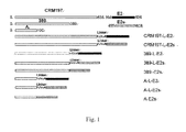
- the clone design of various exemplary fusion proteins constructed is shown in Fig. 1 , wherein the fusion proteins each comprise CRM197 or a fragment thereof and an HEV capsid protein fragment, optionally using a linker.
- the amplification product (i.e. the full-length gene of CRM197) obtained in the Example 1 was used as template.
- the forward primer was CRM197F (SEQ ID NO: 19), at the 5' terminal of which the restriction endonuclease NdeI site CAT ATG was introduced, wherein ATG was the initiation codon in E. coli system.
- the reverse primers were CRM197-linker R (SEQ ID NO: 21), 389-linker R (SEQ ID NO: 22), and A-linker R (SEQ ID NO: 23), respectively, at the 5' terminal of which the restriction endonuclease BamHI site GGA TCC was introduced.
- the PCR reaction was performed in a PCR thermocycler (Biometra T3) under the following conditions.
- the amplification products were DNA fragments of about 1600bp, 1200bp and 600bp in length, respectively.
- pTO-T7-E2 Li, et al. JBC.2005. 28(5): 3400-3406 ) was used as template.
- the forward primers were E2F (SEQ ID NO: 24) and E2sF (SEQ ID NO: 25), respectively, at the 5' terminal of which the restriction endonuclease BamHI site GGA TCC was introduced.
- the reverse primer was Drp59R (SEQ ID NO: 26), at the 5' terminal of which the restriction endonuclease EcoRI site GAA TTC was introduced.
- the PCR reaction was performed in a PCR thermocycler (Biometra T3) under the following conditions. 94°C denaturation 10 min 1 cycle 94°C denaturation 50 sec 58°C annealing 50 sec 20 cycle 72°C elongation 50 sec 72°C elongation 10 min 1 cycle
- the amplification products were DNA fragments of about 600bp and 450bp in length, respectively.
- pMD 18-T vector produced by TAKARA Co.
- pMD 18-T-CRM197-L pMD 18-T-389-L
- pMD 18-T-A-L pMD 18-T-E2 and pMD 18-T-E2s.
- NdeI / BamHI and BamHI / EcoRI enzyme cleavage respectively, the positive clones pMD 18-T-CRM197-L, pMD 18-T-389-L, pMD 18-T-A-L, pMD 18-T-E2 and pMD 18-T-E2s were obtained.
- the plasmids pMD 18-T-CRM197-L, pMD 18-T-389-L and pMD 18-T-A-L were digested by NdeI/BamHI enzyme.
- the fragments obtained by enzyme cleavage were linked into the prokaryotic expression vector pTO-T7 digested by NdeI/BamHI enzyme ( Luo Wenxin et al., Chinese Journal of Biotechnology, 2000, 16:53-57 ), and were transformed into E.
- coli ER2566 purchased from Invitrogen Co.; after extraction of plasmids, as identified by NdeI / BamHI enzyme cleavage, the positive plasmids pTO-T7-CRM197-L, pTO-T7-389-L and pTO-T7-A-L, into which CRM197-L, 389-L and A-L were inserted, respectively, were obtained.
- pTO-T7-CRM197-L, pTO-T7-389-L, pTO-T7-A-L, pMD 18-T-E2 and pMD 18-T-E2s were digested by BamHI / EcoRI enzyme.
- Each of the obtained E2 and E2s fragments was linked into the vectors pTO-T7-CRM197-L, pTO-T7-389-L and pTO-T7-A-L digested by BamHI / EcoRI enzyme, respectively.
- the positive expression vectors pTO-T7-CRM197-L-E2, pTO-T7-CRM197-L-E2s, pTO-T7-389-L-E2, pTO-T7-389-L-E2s, pTO-T7-A-L-E2 and pTO-T7-A-L-E2s into which CRM197-L-E2 (SEQ ID NO:3, 4), CRM197-L-E2s (SEQ ID NO:5, 6), 389-L-E2 (SEQ ID NO:7, 8), 389-L-E2s (SEQ ID NO:9, 10), A-L-E2 (SEQ ID NO:11, 12) or A-L-E2s (SEQ ID NO:13, 14) was inserted, respectively, were obtained.
- the vectors expressing 389-E2s and A-E2s were constructed by three PCR reactions.
- the forward primer was CRM197F, at the 5' terminal of which the restriction endonuclease NdeI site CAT ATG was introduced, wherein ATG was the initiation codon in E. coli system.
- the reverse primers were 389-E2s R (SEQ ID NO: 27) and A-E2s R (SEQ ID NO: 28), respectively.
- the amplification was performed to obtain the N-terminal fragments of the fusion proteins.
- the full-length gene of CRM197 was used as template.
- the forward primer were 389-E2s F (SEQ ID NO: 29) and A-E2s F (SEQ ID NO:30), respectively.
- the reverse primer was DrP59 R, at the 5' terminal of which the restriction endonuclease EcoRI site GAA TTC was introduced.
- the amplification was performed to obtain the C-terminal fragments of the fusion proteins.
- the first and second PCR reactions were performed in a PCR thermocycler (Biometra T3) under the following conditions. 94°C denaturation 10 min 1 cycle 94°C denaturation 50 sec 58°C annealing 50 sec 20 cycle 72°C elongation 50 sec 72°C elongation 10 min 1 cycle
- the amplification products of the first and second PCR reactions were used as templates (for example, the two fragments obtained by using 389-E2sF and 389-E2sR as primers were used as template for amplification of 389-E2s), and CRM197F and DrP59R were used as primers.
- the amplification was performed in a PCR thermocycler (Biometra T3) under the following conditions. 94°C denaturation 10 min 1 cycle 94°C denaturation 50 sec 58°C annealing 50 sec 20 cycle 72°C elongation 50 sec 72°C elongation 10 min 1 cycle
- the amplification products were DNA fragments of about 1600bp and 1000bp in length, respectively.
- the amplification products obtained above were linked into commercially available pMD 18-T vector (produced by TAKARA Co.), respectively.
- pMD 18-T vector produced by TAKARA Co.
- NdeI / EcoRI enzyme cleavage the positive clones pMD 18-T-389-E2s and pMD 18-T-A-E2s were obtained.
- the plasmids pMD 18-T-389-E2s and pMD 18-T-A-E2s were digested by NdeI / EcoRI enzyme. The fragments obtained by enzyme cleavage were then linked into the prokaryotic expression vector pTO-T7 digested by NdeI / EcoRI enzyme ( Luo Wenxin et al., Chinese Journal of Biotechnology, 2000, 16:53-57 ). As identified by NdeI / EcoRI enzyme cleavage, the positive plasmids pTO-T7-389-E2s and pTO-T7-A-E2s, into which 389-E2s and A-E2s were inserted, respectively, were obtained.
- coli ER2566 purchased from Invitrogen prepared by the Calcium chloride method, and then the bacteria were plated on solid LB medium (the components of the LB medium: 10g/L peptone, 5g/L yeast powder, and 10g/L NaCl, the same below) containing kanamycin (at a final concentration of 100 mg/ml, the same below).
- the plates were statically incubated at 37°C for about 10-12h until individual colonies could be observed clearly. Individual colonies from the plates were transferred to a tube containing 4ml liquid LB medium containing kanamycin. The cultures were incubated in a shaking incubator at 180rpm for 10h at 37°C, and then 1 ml bacterial solutions was taken and stored at -70°C.
- Example 3 The expression and purification of the fusion proteins constructed in Example 2
- 5 ⁇ L bacterial solution taken from an ultra low temperature freezer at -70°C, was seeded to 5mL liquid LB medium containing kanamycin, and then was cultured at 37°C, 180rpm under shaking until OD600 reached about 0.5.
- the resultant solution was transferred to 500 ml LB medium containing kanamycin, and then was cultured at 37°C, 180rpm under shaking for 4-5h.
- OD600 reached about 1.5
- IPTG was added to a final concentration of 0.4mM, and the bacteria were induced under shaking at 37°C for 4h.
- the supernatant was kept; and the precipitate was re-suspended in 4M urea in the same volume, under vibration at 37°C for 30min, and centrifuged at 12000rpm, 4°C for 15 min to obtain the supernatant and precipitate.
- the supernatant i.e. the 4M urea-dissolved supernatant
- the supernatant was kept; and the precipitate was further in re-suspended in 8M urea in the same volume, under vibration at 37°C for 30min, and centrifuged, and the supernatant (i.e. the 8M urea-dissolved supernatant) was kept.
- the 4M urea-dissolved supernatants or the 8M urea-dissolved supernatants containing the fusion protein were dialyzed to PBS, respectively, to get the fusion proteins with a purity of about 80% (see Fig. 2D ).
- Example 4 Analysis of properties of the fusion proteins constructed in Example 2
- the dialyzed and renatured samples were transferred to nitrocellulose membrane for blotting after SDS-PAGE separation; 5% skimmed milk was used to block the membrane for 2h, monoclonal antibody 8C11 diluted at a certain ratio was then added (monoclonal antibody was diluted at 1:500, and polyclonal antiserum was diluted at 1:1000), and the reaction was carried out for 1h.
- the membrane was washed with TNT (50 mmol/L Tris•Cl (pH 7.5), 150 mmol/L NaCl, 0.05% Tween 20) for three times, 10min for each time.
- the reactivity of the fusion proteins and the control proteins E2 and HEV-239 with various HEV specific antibodies was determined by indirect ELISA.
- the dialyzed and renatured samples were diluted in 1X PBS (1 ⁇ g/ml), and then were added to 96-well microplate (Beijing Wantai Co.) at 100 ⁇ l/well and incubated at 37°C for 2h.
- the coating solution was discarded, the plate was washed with PBST (PBS + 0.05% Tween-20) once, and then the blocking solution (2% gelatin, 5 ⁇ Casein, 1 ⁇ Proclin300, in PBS) was added at 200 ⁇ l/well and incubated at 37°C for 2h.
- PBST PBS + 0.05% Tween-20
- the blocking solution was discarded when the detection was performed, and the HEV monoclonal antibodies diluted at a certain ratio (when E2s and its fusion protein were detected, they were diluted at 1:10000; when E2 and its fusion protein were detected, they were diluted at 1:100000; when the reactivity of A-L-E2, 239 and E2 proteins was compared, the monoclonal antibodies were subjected to 10-fold serial dilution wherein 1 mg/ml was used as the initial concentration, and the polyclonal antibody at its initial concentration was subjected to dilution in the same manner) was added at 100 ⁇ l/well. The mixture was incubated at 37°C for 1-2h.
- the plate was then washed with PBST for five times, and HRP-labeled Goat anti Mouse (KPL product) (1: 5000) was then added at 100 ⁇ l/well and was incubated at 37°C for 30min; the plate was then washed with PBST for five times, HRP substrate (Beijing Wantai Co.) was then added at 100 ⁇ l/well and was incubated at 37°C for 15min. 2M sulphuric acid was added at 50 ⁇ l/well to stop the reaction, and Microplate reader (Sunrise Type, product from Tecan Co.) was then used to read OD450/620 value. The results of the ELISA using the fusion proteins with the monoclonal antibodies were shown in Fig. 5 .
- the apparatus used in the experiment was US Beckman XL-A analytic supercentrifuge, which was equipped with an optical detection system and An-50Ti and An-60Ti rotators.
- the Sedimentation Velocity (SV) method (c(s) algorithm, see P. Schuck et al., Biophys J 78: 1606-1619(2000 )) was used to analyze the sedimentation coefficient of the fusion protein A-L-E2.
- the analytic result was shown in Fig. 7 .
- the result showed that the fusion protein A-L-E2 was mainly present in a form of dimer, and some dimers might be further polymerized to form a tetramer.
- Example 5 Analysis of immunogenicity of the fusion proteins constructed in Example 2
- mice used in the experiment were female, 6-week old BALB/C mice.
- aluminum adjuvant mice were immunized by intraperitoneal injection of the fusions proteins which were prepared by the methods in the Example 3 and renatured to PBS and the control proteins HEV-239, E2 and E2s, respectively.
- the injection volume was 1ml, and two dose groups (a 5 ⁇ g-dose group or a 0.5 ⁇ g-dose group) were used.
- the primary immunization was performed at week 0, and booster immunization was performed at week 2 and 4.
- HEV-239 was used to coat a plate, and the antibody titers in serum as induced by the fusion proteins and the control proteins, were measured by similar indirect ELISA assay as described above.
- the detection results of the serum antibody titers within 3 months after immunization were shown in Fig. 8 .
- the results showed that seroconversion occurred in mice serum at week 4 in both 5 ⁇ g- and 0.5 ⁇ g-dose groups, and the antibody titers reached the highest value at week 5 or 6.
- the highest antibody titer was obtained when A-L-E2 was used, which reached 10 6 at week 6; and the antibody titers induced by the fusion proteins were higher or comparable to that of HEV-239 protein.
- the antibody titers of the fusion proteins were significantly higher than that of HEV-239, and the antibody titer induced by A-L-E2 protein reached 10 6 at week 5.
- seroconversion did not occur in immune serum when using E2 and E2s, in 5 ⁇ g- and 0.5 ⁇ g-dose groups.
- the immunogenicity of the constructed fusion proteins were significantly higher than the antigen protein (E2 and E2s) alone, indicating that the CRM197 of the invention or a fragment thereof significantly enhanced immunogenicity of the antigen protein fused therewith, and could be used as intramolecular adjuvant.
- mice were used in each group.
- the immunization was carried out by single intraperitoneal injection.
- Peripheral venous blood was taken after 4 weeks following immunization, serum was separated, and serological conversion rate was determined by ELISA assay as described above. When the ELISA value of 100-fold diluted serum was higher than the cutoff value (i.e. three times of the average negative value), the serum was regarded as positive.
- the median effective dose (ED50) was calculated by Reed-Muench method.
- the serological conversion rate of the fusion protein A-L-E2 was shown in Table 2, and the serological conversion rate of HEV-239 vaccine was shown in Table 3.
- Table 2 ED50 of A-L-E2 for inducing seroconversion of HEV antibody in mice Dose ( ⁇ g) Number of mice Number of mice with seroconversion Serological conversion rate ED50( ⁇ g) 1.0000 6 6 100% 0.0064 0.3333 6 6 100% 0.1111 6 6 100% 0.0370 6 6 100% 0.0123 6 6 100% 0.0041 6 1 16.7% 0.0013 6 0 0% 0.0005 6 0 0%
- Table 3 ED50 of HEV-239 vaccine for inducing seroconversion of HEV antibody in mice Dose ( ⁇ g) Number of mice Number of mice with seroconversion Serological conversion rate ED50( ⁇ g) 1.6 6 6 100% 0.071 0.4 6 5 83.3% 0.1 6 4 66.7% 0.025 6 0 0%
- Example 6 Design and clone of fusion proteins comprising CRM197 or a fragment thereof and an influenza virus M2e protein
- fusion proteins were constructed exemplarily.
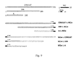
- the clone design of the exemplary fusion proteins constructed is shown in Fig. 9 , wherein the fusion proteins each comprise CRM197 or a fragment thereof and an influenza virus M2e protein, optionally using a linker.
- the amplification product (i.e. the full-length gene of CRM197) obtained in the Example 1 was used as template.
- the forward primer was CRM197F1 (SEQ ID NO: 45), at the 5' terminal of which the restriction endonuclease NdeI site CAT ATG was introduced, wherein ATG was the initiation codon in E. coli system.
- the reverse primers were CRM197-linker R1 (SEQ ID NO: 46), 389-linker R1 (SEQ ID NO: 47) and A-linker R1 (SEQ ID NO: 48), respectively, at the 5' terminal of which the restriction endonuclease BamHI site GGA TCC was introduced.
- the PCR reaction was performed in a PCR thermocycler (Biometra T3) under the following conditions.
- the sequences of the primers used were shown in Table 4.
- 95°C denaturation 10 min 1 cycle 95°C denaturation 1.5 min 58°C annealing 1.5 min 20 cycle 72°C elongation 1.7 min 72°C elongation 10 min 1 cycle
- the amplification products were DNA fragments of about 1600bp, 1200bp and 600bp in length, respectively.
- the plasmid PHW2000 (stored in our lab, comprising the full-length gene of M2) was used as a template.
- the forward primer was M2eF1 (SEQ ID NO: 49), at the 5' terminal of which the restriction endonuclease BamHI GGA TCC was introduced.
- the reverse primer was M2eR (SEQ ID NO: 50), at the 5' terminal of which the restriction endonuclease EcoRI site GAA TTC was introduced.
- the PCR reaction was performed in a PCR thermocycler (Biometra T3) under the following conditions. The sequences of the primers used were shown in Table 4. 95°C denaturation 10 min 1 cycle 95°C denaturation 50 sec 58°C annealing 50 sec 20 cycle 72°C elongation 30 sec 72°C elongation 10 min 1 cycle
- the amplification products were DNA fragments of about 70bp in length, respectively.
- pMD 18-T vector produced by TAKARA Co.
- pMD 18-T-CRM197-L1 pMD 18-T-389-L1
- pMD 18-T-A-L1 pMD 18-T-M2e.
- NdeI / BamHI and BamHI / EcoRI enzyme cleavage respectively, the positive clones pMD 18-T-CRM197-L1, pMD 18-T-389-L1, pMD 18-T-A-L1 and pMD 18-T-M2e were obtained.
- the plasmids pMD 18-T-CRM197-L1, pMD 18-T-389-L1 and pMD 18-T-A-L1 were digested by NdeI/BamHI enzyme.
- the fragments obtained by enzyme cleavage were linked into the prokaryotic expression vector pTO-T7 digested by NdeI/BamHI enzyme ( Luo Wenxin et al., Chinese Journal of Biotechnology, 2000, 16:53-57 ), and were transformed into E.
- coli ER2566 purchased from Invitrogen Co.; after extraction of plasmids, as identified by NdeI / BamHI enzyme cleavage, the positive plasmids pTO-T7-CRM197-L1, pTO-T7-389-L1 and pTO-T7-A-L1, into which the fragments CRM197-L1, 389-L1 and A-L1 were inserted, respectively, were obtained.
- pTO-T7-CRM197-L1, pTO-T7-389-L1, pTO-T7-A-L1 and pMD 18-T-M2e were digested by BamHI / EcoRI enzyme.
- the obtained M2e fragment was linked into the vectors pTO-T7-CRM197-L1, pTO-T7-389-L1 and pTO-T7-A-L1 digested by BamHI / EcoRI enzyme, respectively.
- M2e fused to the N-terminus of CRM197 or a fragment thereof.
- the plasmid PHW2000 (stored in our lab, containing the full-length gene of M2) was used as template.
- the forward primer was M2eF2 (SEQ ID NO: 51), at the 5' terminal of which the restriction endonuclease NdeI CAT ATG was introduced, wherein ATG was the initiation codon in E. coli system.
- the reverse primer was M2e-Linker R (SEQ ID NO: 52), at the 5' terminal of which the restriction endonuclease BamHI GGA TCC was introduced.
- the PCR reaction was performed in a PCR thermocycler (Biometra T3) under the following conditions. 95°C denaturation 10 min 1 cycle 95°C denaturation 50 sec 58°C annealing 50 sec 20 cycle 72°C elongation 30 sec 72°C elongation 10 min 1 cycle
- the amplification products were DNA fragments of about 100bp in length.
- the amplification product (i.e. the full-length gene of CRM197) obtained in the Example 1 was used as template.
- the forward primer was CRM197F2 (SEQ ID NO: 53), at the 5' terminal of which the restriction endonuclease BamHI GGA TCC was introduced.
- the reverse primers were CRM197 R2 (SEQ ID NO: 54), 389 R (SEQ ID NO: 55), and A R (SEQ ID NO: 56), at the 5' terminal of which the restriction endonuclease EcoRI site GAA TTC was introduced.
- the PCR reaction was performed in a PCR thermocycler (Biometra T3) under the following conditions.
- the sequences of the primers used were shown in Table 4. 95°C denaturation 10 min 1 cycle 95°C denaturation 1.5 min 58°C annealing 1.5 min 20 cycle 72°C elongation 1.7 min 72°C elongation 10 min 1 cycle
- the amplification products were DNA fragments of about 1600bp, 1200bp and 600bp in length, respectively.
- the amplification products as obtained above were linked into commercially available pMD 18-T vector (produced by TAKARA Co.), respectively, and designated as pMD 18-T-M2e-L as well as pMD 18-T-CRM197, pMD 18-T-389 and pMD 18-T-A, respectively.
- pMD 18-T-M2e-L As identified by NdeI / BamHI and BamHI / EcoRI enzyme cleavage, respectively, the positive clones pMD 18-T-CRM197, pMD 18-T-389, pMD 18-T-A, and pMD 18-T-M2e-L were obtained.
- the plasmid pMD 18-T-M2e-L was digested by NdeI/BamHI enzyme.
- the fragments obtained by enzyme cleavage were then linked into the prokaryotic expression vector pTO-T7 digested by NdeI/BamHI enzyme ( Luo Wenxin et al., Chinese Journal of Biotechnology, 2000, 16:53-57 ), and was transformed into E. coli ER2566 (purchased from Invitrogen Co.); after extraction of plasmids, as identified by NdeI / BamHI enzyme cleavage, the positive plasmid pTO-T7-M2e-L, into which the fragment M2e-Lwas inserted, was obtained.
- pTO-T7-M2e-L, pMD 18-T-CRM197, pMD 18-T-389 and pMD 18-T-A were digested by BamHI/EcoRI enzyme.
- the obtained fragments CRM197, 389 and A were linked into the vector pTO-T7-M2e-L digested by BamHI/EcoRI enzyme, respectively.
- the positive expression vectors pTO-T7-M2e-L-CRM197, pTO-T7-M2e-L-389, and pTO-T7-M2e-L-A into which M2e-L-CRM197 (SEQ ID NO:39, 40), M2e-L-389 (SEQ ID NO:41, 42), and M2e-L-A (SEQ ID NO:43, 44) were inserted respectively, were obtained.
- coli ER2566 purchased from Invitrogen prepared by the Calcium chloride method, and then the bacteria were plated on solid LB medium (the components of the LB medium: 10g/L peptone, 5g/L yeast powder, and 10g/L NaCl, the same below) containing kanamycin (at a final concentration of 100 mg/ml, the same below).
- the plates were statically incubated at 37°C for about 10-12h until individual colonies could be observed clearly. Individual colonies from the plates were transferred to a tube containing 4ml liquid LB medium containing kanamycin. The cultures were incubated in a shaking incubator at 180rpm for 10h at 37°C, and then 1 ml bacterial solution was taken and stored at -70°C.
- Example 7 The expression, isolation and renaturation of the fusion proteins constructed in Example 6
- 5 ⁇ L bacterial solution taken from an ultra low temperature freezer at -70°C, was seeded to 5mL liquid LB medium containing kanamycin, and then was cultured at 37°C, 180rpm under shaking until OD600 reached about 0.5.
- the resultant solution was transferred to 500 ml LB medium containing kanamycin, and then was cultured at 37°C, 180rpm under shaking for 4-5h.
- OD600 reached about 1.5
- IPTG was added to a final concentration of 0.4mM, and the bacteria were induced under shaking at 37°C for 4h.
- Triton-100 of the same volume was used for washing the precipitate, the result mixture was under vibration for 30min, centrifuged, and the supernatant was discarded.
- the precipitate was re-suspended in Buffer I (20mM Tris-HCl pH8.0, 100mM NaCl, 5mM EDTA), under vibration for 30min, centrifuged, and the supernatant was discarded.
- the precipitate was then re-suspended in 2M urea, under vibration at 37°C for 30min, centrifuged, and the supernatant and the precipitate were obtained.
- the supernatant was kept; and the precipitate was re-suspended in 4M urea in the same volume, under vibration at 37°C for 30min, and centrifuged at 12000rpm, 4°C for 15 min to obtain the supernatant and precipitate.
- the supernatant i.e. the 4M urea-dissolved supernatant
- the supernatant was kept; and the precipitate was further in re-suspended in 8M urea in the same volume, under vibration at 37°C for 30min, and centrifuged, and the supernatant (i.e. the 8M urea-dissolved supernatant) was kept.
- the fractions obtained were analyzed by SDS-PAGE (coomassie brilliant blue staining was used for visualization, the same below, see the methods in The Molecular Cloning Experiment Guide, 2nd editi on).
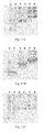
- the results showed that the fusion proteins were expressed in inclusion bodies (see Figs. 10A and 10B ), CRM197-L-M2e, 389-L-M2e, M2e-L-CRM197 and M2e-L-389 were mainly dissolved in 8M urea, and A-L-M2e and M2e-L-A were mainly dissolved in 4M urea.
- Example 8 Analysis of properties of the fusion proteins constructed in Example 6
- the reactivity of the fusion proteins with influenza virus M2e monoclonal antibody 5D1 and CRM197 monoclonal antibody 1E6 were determined by Western blotting.
- the dialyzed and renatured samples were transferred to nitrocellulose membrane for blotting after SDS-PAGE separation; 5% skimmed milk was used to block the membrane for 2h, and then the monoclonal antibody 5D1 diluted at 1:500 was added.
- the reaction was carried out for 1h.
- the membrane was then washed with TNT (50 mmol/L Tris ⁇ Cl (pH 7.5), 150 mmol/L NaCl, 0.05% Tween 20) for three times, 10min for each time.
- TNT 50 mmol/L Tris ⁇ Cl (pH 7.5), 150 mmol/L NaCl, 0.05% Tween 20
- Goat Anti-mouse alkaline phosphatase (KPL product) was then added. The reaction was carried out for 1h, and the membrane was then washed with TNT for three times, 10 min for each time. NBT and BCIP (PROTOS product) were used for visualization. The results, as determined by Western blotting using the fusion proteins and influenza virus M2e monoclonal antibody 5D1 ( Figs. 11A-11D ) or CRM197 monoclonal antibody 1E6 ( Figs. 11E-11H ), were shown in Fig. 11 . The results showed that all the tested fusion proteins had significant reactivity with influenza virus M2e-specific monoclonal antibody 5D1 and CRM197 specific monoclonal antibody 1E6.
- the reactivity of the fusion proteins and the control protein GST-M2e with various M2e specific antibodies and CRM197 specific monoclonal antibody 1E6 was determined by indirect ELISA.
- O19 antibody is a protective antibody against influenza known in the prior art (see, Fu et al., Virology, 2009, 385:218-226 ).
- the dialyzed and renatured samples were diluted in 1X PBS (1 ⁇ g/ml), and then were added to 96-well microplate (Beijing Wantai Co.) at 100 ⁇ l/well and incubated at 37°C for 2h.
- the coating solution was discarded, the plate was washed with PBST (PBS + 0.05% Tween-20) once, and then the blocking solution (2% gelatin, 5 ⁇ Casein, 1 ⁇ Proclin300, in PBS) was added at 180 ⁇ l/well and incubated at 37°C for 2h.
- the blocking solution was discarded when the detection was performed, and the anti-M2e antibody or CRM197 antibody diluted at a certain ratio (0.002 mg/ml was used as the initial concentration for 2-fold gradient dilution) was added at 100 ⁇ l/well. The mixture was incubated at 37°C for 1h.
- the apparatus used in the experiment was US Beckman XL-A analytic supercentrifuge, which was equipped with an optical detection system and An-50Ti and An-60Ti rotators.
- the Sedimentation Velocity (SV) method (c(s) algorithm, see P. Schuck et al., Biophys J 78: 1606-1619(2000 )) was used to analyze the sedimentation coefficient of the fusion proteins.
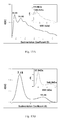
- the analytic results were shown in Figs. 13A-13F .
- A-L-M2e and M2e-L-A were mainly present in a form of monomer and tetramer; and 389-L-M2e was mainly present in a form of dimer and polymer; M2e-L-389 was mainly present in a form of monomer and polymer; CRM197-L-M2e was mainly present in a form of dimer and polymer; and M2e-L-CRM197 was mainly present in a form of monomer and polymer.
- Example 9 Analysis of immunogenicity of the fusion proteins constructed in Example 6
- mice used in the experiment were female, 6-week old BALB/C mice.
- aluminum adjuvant mice were immunized by intraperitoneal injection of the fusions proteins as constructed in Example 6 and renatured to PBS and the control protein GST-M2e, respectively.
- the injection volume was 1ml, and two dose groups (a 5 ⁇ g-dose group or a 0.5 ⁇ g-dose group) were used.
- the primary immunization was performed at week 0, and booster immunization was performed at week 2 and 4.
- GST-M2e was used to coat a plate, and the antibody titers in serum as induced by the fusion proteins and control protein, were measured by similar indirect ELISA assay as described above.
- the detection results of the serum antibody titers within 4 months after immunization were shown in Figs. 14A and 14B .
- the results showed that after the second booster immunization, immunogenicity of the constructed fusion proteins was significantly higher than the antigen protein (GST-M2e) alone, indicating that the CRM197 of the invention or a fragment thereof (no matter being located at the N-terminus or C-terminus of the fusion protein) significantly enhanced immunogenicity of the antigen protein fused therewith, and could be used as intramolecular adjuvant.
Landscapes
- Health & Medical Sciences (AREA)
- Life Sciences & Earth Sciences (AREA)
- Chemical & Material Sciences (AREA)
- Genetics & Genomics (AREA)
- Organic Chemistry (AREA)
- General Health & Medical Sciences (AREA)
- Engineering & Computer Science (AREA)
- Medicinal Chemistry (AREA)
- Bioinformatics & Cheminformatics (AREA)
- Virology (AREA)
- Microbiology (AREA)
- Molecular Biology (AREA)
- Pharmacology & Pharmacy (AREA)
- Public Health (AREA)
- Veterinary Medicine (AREA)
- Animal Behavior & Ethology (AREA)
- Zoology (AREA)
- Wood Science & Technology (AREA)
- Biomedical Technology (AREA)
- Immunology (AREA)
- Biochemistry (AREA)
- Biotechnology (AREA)
- General Engineering & Computer Science (AREA)
- Biophysics (AREA)
- Epidemiology (AREA)
- Mycology (AREA)
- General Chemical & Material Sciences (AREA)
- Chemical Kinetics & Catalysis (AREA)
- Proteomics, Peptides & Aminoacids (AREA)
- Gastroenterology & Hepatology (AREA)
- Nuclear Medicine, Radiotherapy & Molecular Imaging (AREA)
- Plant Pathology (AREA)
- Physics & Mathematics (AREA)
- Communicable Diseases (AREA)
- Oncology (AREA)
- Pulmonology (AREA)
- Peptides Or Proteins (AREA)
- Medicines Containing Antibodies Or Antigens For Use As Internal Diagnostic Agents (AREA)
- Micro-Organisms Or Cultivation Processes Thereof (AREA)
- Medicinal Preparation (AREA)
Abstract
Description
- The invention relates to the field of molecular virology and immunology. In particular, the invention relates to a use of a diphtheria toxin non-toxic mutant CRM197 or a fragment thereof as intramolecular adjuvant in a fusion protein for enhancing immunogenicity of a target protein fused thereto (for example, an HEV capsid protein, an influenza virus M2 protein or an immunogenic fragment thereof). The invention also relates to a method for enhancing immunogenicity of a target protein (for example, an HEV capsid protein, an influenza virus M2 protein or an immunogenic fragment thereof), comprising the fusion expression of CRM197 or a fragment thereof with the target protein to form a fusion protein. The invention also relates to a fusion protein comprising CRM197 or a fragment thereof and a target protein (for example, an HEV capsid protein, an influenza virus M2 protein or an immunogenic fragment thereof), wherein said CRM197 or a fragment thereof enhances immunogenicity of the target protein. The invention also relates to an isolated nucleic acid encoding the fusion protein, a construct and a vector comprising the nucleic acid, and a host cell comprising the nucleic acid. The invention also relates to a use of the fusion protein in the manufacture of a pharmaceutical composition or a vaccine.
- Diphtheria toxin (DT) has been deeply studied. The strudies on structure show that diphtheria toxin consists of three domains: N-terminal Catalytic Domain C (aa 1-190, C domain) (also called Fragment A), middle Transmembrane Domain T (aa 201-384, T domain), and C-terminal Receptor Binding domain R (aa 386-535, R domain) (Choe S, Bennett M, Fujii G, et al., Nature. 1992. 357:216-222). ONTAK (DAB389-IL-2), prepared by fusion of the former two domains of diphtheria toxin with interleukin 2 (IL-2), was approved by FDA on the market in 1999, for the treamtent of adult cutaneous T-cell lymphoma. This demonstrates that the three domains of diphtheria toxin may be used seperately and play their own roles, respectively.
- CRM197 (Cross-Reacting Materials 197) is a diphtheria toxin non-toxic mutant (Uchida, T., A.M, Pappenheimer, Jr., R. Gregory, et al., J. Biol. Chem. 1973. 248:3838-3844), which differs from a wild-type gene encoding DT by a single nucleotide mutation, resulting in the amino acid residue at position 52 changed from Gly to Glu (G. Giannini, R.Rappuoli, G. Ratti et al., Nucleic Acids Research. 1984. 12: 4063-4070).
- Studies show that although CRM197 has a structure similar to that of a wild-type DT (namely, having said three domains), its Fragment A loses the ability of binding to NAD, is unable to bind to EF2 and thereby loses the cytoxicity possessed by natural DT, indicating that the amino acid residue Gly at position 52 plays an important role in the binding of DT to NAD (K.Moyner, G.Christiansen, Acta path microbial immunolscand sect C. 1984, 92:17-23). Although CRM197 loses the cytotoxicity, it retains a stronge immunogenicity comparable to that of a wild-type DT. Therefore, CRM197 is generally used as a protein carrier for crosslinking other haptens so as to prepare conjugate vaccines.
- As early as 1985, Porter et al., crosslinked polysaccharides on Hib surface to CRM197 and DT protein carrier, respectively, and prepared them into vaccines, and studied the difference of them in immunogenicity. The experimental results showed that there was no significant difference between the two crosslinked vaccines in terms of immune effect, both of them could stimulate the generation of a strong immune response and immunologic memory in infants (Porter Anderson, Micheal E. Pichichero and Richard A. J. Clin. Invest. 1985: 52-59). After comparing pneumococcal conjugate vaccines crosslinked to various proteins, it is found that the vaccines wherein CRM197 is used as a protein carrier have a good immune effect in animal experiments and clinical trials, and CRM197 is safe without a side-effect of toxicity (Black, S., H. Shinefield, et al. Pediatr Infect Dis J, 2000, 19(3); 187-195). In current, pneumococcal conjugate vaccines, in which CRM197 is used as a protein carrier, mainly refer to PCV7, PCV9, PCV13, and the like. The results of clinical trials showed that these vaccines had good immunogenicity and safety in children less than two years old (Barricarte, A., J. Castilla, et al. Clin Infect Dis, 2007, 44(11): 1436-1441; Madhi, S., P. Adrian, et al. Vaccine, 2007, 25(13): 2451-2457; Duggan, S. T. Drugs, 2010, 70(15): 1973-1986). Epidemic meningitis conjugate vaccines can be prepared by crosslinking CRM197 to polysaccharides on surface of N. menigitidis. For example, vaccines such as Meningitec (Wyeth Pharmaceuticals), Menjugate (Novartis vaccines), and Menveo (Novartis vaccines), in which CRM197 is used as a protein carrier, have been commercially available.
- Although CRM197 loses enzymatic activity and cytotoxity, it is still able to bind to a specific receptor of DT, i.e. heparin-binding EGF-like growth factor (HB-EGF). Since the expression of the receptor is generally up-regulated in cancerous tissues, like DT, CRM197 also has anti-tumor effect (Buzzi, S., D. Rubboli, et al. Immunotherapy, 2004, 53(11)). The studies also found that CRM197 could pass through Blood-Brain-Barrier (BBB), and therefore could be used as a carrier for delivery of drugs to brain (Gaillard, P. J., and A. G. de Boer. J Control Release, 2006, 116(2): 60-62).
- Although it has been reported that CRM197 has multiple functions, in particular, has a strong immunogenicity and can be used as immunoadjuvant, it is not reported yet that CRM197 may be used as intramolecular adjuvant for enhancing immunogenicity of a target protein fused thereto in a fusion protein. The invention uses Hepatitis E capsid protein as an example, and demonstrates for the first time that CRM197 or a fragment thereof can enhance immunogenicity of a protein fused thereto in a fusion protein, and thereby can be used as intramolecular adjuvant.
- In the invention, unless otherwise specified, the scientific and technical terms used herein have the meanings as generally understood by a person skilled in the art. Moreover, the laboratory operations of cell culture, molecular genetics, nucleic acid chemistry, biological chemistry, and immunology used herein are the routine operations widely used in the corresponding fields. Meanwhile, for the purpose of better understanding of the invention, the definitions and explanations of the relevant terms are provided as follows.
- According to the invention, the term "CRM197" refers to a diphtheria toxin non-toxic mutant, which differs from a wild-type diphtheria toxin by an amino acid residue at position 52 changed from Gly to Glu (G. Giannini, R.Rappuoli, G. Ratti et al., Nucleic Acids Research. 1984. 12: 4063-4070). Diphtheria toxin is well known by a person skilled in the art (see, for example, Choe S, Bennett M, Fujii G, et al., Nature. 1992. 357:216-222), whose amino acid sequence may be found by reference to GenBank accession No. AAV70486.1.
- In the invention, the exemplary amino acid sequence of CRM197 is set forth in SEQ ID NO: 2. Therefore, in the invention, when the sequence of CRM197 is involved, it is described as the sequence set forth in SEQ ID NO:2. For example, in the expression "amino acid residues from
positions 1 to 190 of CRM197", amino acid residues frompositions 1 to 190 refers to amino acid residues frompositions 1 to 190 of SEQ ID NO: 2. However, a person skilled in the art understands that mutations or variations (including, but not limited to, substitution, deletion and/or addition) may naturally occur in or are introduced artificially into SEQ ID NO: 2 without affecting the biological properties of CRM197. Therefore, in the invention, the term "CRM197" intends to comprise all such polypeptides and variants, including the polypeptide set forth in SEQ ID NO: 2 and its natural or artificial variants, wherein the variants retain the biological properties of CRM197, i.e. have a strong immunogenicity and no cytotoxity. In addition, when sequence fragments of CRM197 are described, they include not only the sequence fragments of a polypeptide set forth in SEQ ID NO: 2, but also the corresponding sequence fragments of the natural or artificial variants of the polypeptide. For example, the expression "amino acid residues frompositions 1 to 190 of CRM197" intends to comprise amino acid residues frompositions 1 to 190 of SEQ ID NO: 2 and the corresponding fragments of the variants (natural or artificial) of a polypeptide set forth in SEQ ID NO: 2. - According to the invention, an Hepatitis E virus (HEV) capsid protein refers to a protein encoded by HEV ORF2. The sequence of HEV ORF2 is well known in the art (see, for example, DDBJ accession No. D11092). In the invention, when the sequence of HEV ORF2 is involved, it is described as the sequence set forth in DDBJ accession No. D11092. For example, in the expression "amino acid residues from positions 368 to 606 of a polypeptide encoded by HEV ORF2", amino acid residues from positions 368 to 606 refers to amino acid residues from positions 368 to 606 of a polypeptide encoded by D11092. However, a person skilled in the art understands that mutations or variations (including, but not limited to, substitution, deletion and/or addition) may naturally occur in or are introduced artificially into HEV ORF2 or a polypeptide encoded thereby without affecting the biological properties thereof (such as antigenicity and immunogenicity). Therefore, in the invention, the term "HEV ORF2" intends to comprise all such polypeptides and variants, including the sequence set forth in D11092 and its natural or artificial variants. In addition, when sequence fragments of HEV ORF2 (or a polypeptide encoded thereby) are described, they include not only the sequence fragments of D11092 (or a polypeptide encoded thereby), but also the corresponding sequence fragments of the natural or artificial variants of D11092 (or a polypeptide encoded thereby). For example, the expression "amino acid residues from positions 368 to 606 of a polypeptide encoded by HEV ORF2" intends to comprise amino acid residues from positions 368 to 606 of a polypeptide encoded by D11092 and the corresponding fragments of the variants (natural or artificial) of a polypeptide encoded by D11092. The exemplary amino acid sequence of an HEV capsid protein (a polypeptide encoded by ORF2 of D11092) is described in SEQ ID NO: 31.
- According to the invention, an influenza virus M2 protein refers to a protein encoded by the seventh segment of type A or type B influenza virus genome or a protein encoded by the sixth segment of type C influenza virus genome. The exemplary amino acid sequence of an influenza virus M2 protein is described in SEQ ID NO: 32.
- According to the invention, the expression "corresponding sequence fragments" or "corresponding fragments" refers to fragments that are located in equal positions of sequences when the sequences are subjected to optimal alignment, namely, the sequences are aligned to obtain a highest percentage of identity.
- According to the invention, when used in the background of proteins/polypeptides, the term "variant" refers to a protein, whose amino acid sequence is different from a reference protein/polypeptide (for example, CRM197 of the invention) by one or more (for example, 1-10, or 1-5 or 1-3) amino acids (such as conservative amino acid substitutions), or which has an identity of at least 60%, 80%, 85%, 90%, 95%, 96%, 97%, 98%, or 99% to a reference protein/polypeptide (for example, CRM197 of the invention), and which retains the essential characteristics of the reference protein/polypeptide. In the invention, the essential characteristics of CRM197 may refer to a strong immunogenicity and no cytotoxity, and the essential characteristics of an HEV capsid protein and an influenza virus M2 protein may refer to antigenicity and/or immunogenicity thereof.
- According to the invention, the term "identity" refers to the match degree between two polypeptides or between two nucleic acids. When two sequences for comparison have the same base or amino acid monomer sub-unit at a certain site (e.g., each of two DNA molecules has an adenine at a certain site, or each of two polypeptides has a lysine at a certain site), the two molecules are identical at the site. The percent identity between two sequences is a function of the number of identical sites shared by the two sequences over the total number of sites for comparison x 100. For example, if 6 of 10 sites of two sequences are matched, these two sequences have an identity of 60%. For example, DNA sequences: CTGACT and CAGGTT share an identity of 50% (3 of 6 sites are matched). Generally, the comparison of two sequences is conducted in a manner to produce maximum identity. Such alignment can be conducted by using a computer program such as Align program (DNAstar, Inc.) which is based on the method of Needleman, et al. (J. Mol. Biol. 48:443-453, 1970). The percent identity between two amino acid sequences can be determined using the algorithm of E. Meyers and W. Miller (Comput. Appl. Biosci., 4:11-17 (1988)) which has been incorporated into the ALIGN program (version 2.0), using a PAM120 weight residue table, a gap length penalty of 12 and a gap penalty of 4. In addition, the percent identity between two amino acid sequences can be determined using the algorithm of Needleman and Wunsch (J. Mol. Biol. 48:444-453 (1970)) which has been incorporated into the GAP program in the GCG software package (available at http://www.gcg.com), using either a Blossum 62 matrix or a PAM250 matrix, and a gap weight of 16, 14, 12, 10, 8, 6, or 4 and a length weight of 1, 2, 3, 4, 5, or 6.
- As used in the invention, the term "conservative substitution" refers to amino acid substitutions which would not negatively affect or change the essential characteristics of a protein/polypeptide comprising the amino acid sequence. For example, a conservative substitution may be introduced by standard techniques known in the art such as site-directed mutagenesis and PCR-mediated mutagenesis. Conservative amino acid substitutions include substitutions wherein an amino acid residue is substituted with another amino acid residue having a similar side chain, for example, with a residue similar to the corresponding amino acid residue physically or functionally (such as, having similar size, shape, charges, chemical properties including the capability of forming covalent bond or hydrogen bond, etc.). The families of amino acid residues having similar side chains have been defined in the art. These families include amino acids having alkaline side chains (for example, lysine, arginine and histidine), amino acids having acidic side chains (for example, aspartic acid and glutamic acid), amino acids having uncharged polar side chains (for example, glycine, asparagine, glutamine, serine, threonine, tyrosine, cysteine, tryptophan), amino acids having nonpolar side chains (for example, alanine, valine, leucine, isoleucine, proline, phenylalanine, methionine), amino acids having β-branched side chains (such as threonine, valine, isoleucine) and amino acids having aromatic side chains (for example, tyrosine, phenylalanine, tryptophan, histidine). Therefore, an amino acid residue is preferably substituted with another amino acid residue from the same side-chain family. Methods for identifying amino acid conservative substitutions are well known in the art (see, for example, Brummell et al., Biochem. 32: 1180-1187 (1993); Kobayashi et al., Protein Eng. 12(10): 879-884 (1999); and Burks et al., Proc. Natl Acad. Set USA 94: 412-417 (1997), which are incorporated herein by reference).
- According to the invention, the term "immunogenicity" refers to an ability of stimulating the formation of specific antibodies or sensitized lymphocytes in organisms. It not only refers to the property of an antigen to stimulate a specific immunocyte to activate, proliferate and differentiate the immunocyte so as to finally generate immunologic effector substance such as antibodies and sensitized lymphocytes, but also refers to the specific immune response wherein antibodies or sensitized T lymphocytes can be formed in immune system of an organism after stimulating the organism with an antigen. Immunogenicity is the most important property of an antigen. Whether an antigen can successfully induce the generation of an immune response in a host depends on three factors, properties of an antigen, reactivity of a host, and immunization means.
- According to the invention, the term "immunogenic fragment" refers to such a polypeptide fragment, which at least partially retains the immunogenicity of the protein from which it is derived. For example, immunogenic fragments of an HEV capsid protein refer to fragments of an HEV capsid protein which at least partially retain immunogenicity, for example, HEV-239, E2 or E2s as described in the invention (see, Li et al., J Biol Chem. 280(5): 3400-3406 (2005); Li et al., PLoS Pathogens. 5(8): e1000537 (2009)); immunogenic fragments of an influenza virus M2 protein refer to fragments of M2 protein which at least partially retain immunogenicity, for example, M2e as described in the invention (see, Fiers W et al., Vaccine.27(45) :6280-6283(2009)).
- According to the invention, HEV-239 (or 239 in brief) refers to a polypeptide consisting of amino acid residues from positions 368 to 606 of a polypeptide encoded by HEV ORF2 (i.e. HEV capsid protein); E2 refers to a polypeptide consisting of amino acid residues from positions 394 to 606 of a polypeptide encoded by HEV ORF2; E2s refers to a polypeptide consisting of amino acid residues from positions 455 to 606 of a polypeptide encoded by HEV ORF2.
- According to the invention, the term "M2e" refers to a polypeptide consisting of amino acid residues from
positions 1 to 24 of an influenza virus M2 protein. - In the invention, the term "polypeptide" and "protein" have the same meanings and may be used interchangeably. Moreover, in the invention, amino acids are generally represented by one letter code and three-letter code well known in the art. For example, alanine may be represented by A or Ala.
- According to the invention, the term "E. coli expression system" refers to an expression system consisting of E. coli (strain) and a vector, wherein the E. coli (strain) is available on the market, including but not limited to: GI698, ER2566, BL21 (DE3), B834 (DE3), BLR (DE3), etc.
- According to the invention, the term "vector" refers to a nucleic acid vehicle which can have a polynucleotide inserted therein. When the vector allows for the expression of the protein encoded by the polynucleotide inserted therein, the vector is called an expression vector. The vector can be introduced into the host cell by transformation, transduction, or transfection, and have the carried genetic material elements expressed in a host cell. Vectors are well known by a person skilled in the art, including, but not limited to plasmids, phages, cosmids and the like.
- According to the invention, the term "chromatography" includes, but is not limited to: ion exchange chromatography (e.g. cation-exchange chromatography), hydrophobic interaction chromatography, absorbent chromatography (e.g. hydroxyapatite chromatography), gel filtration chromatography (gel exclusion chromatography), and affinity chromatography.
- According to the invention, the term "pharmaceutically acceptable carriers and/or excipients" refers to carriers and/or excipients that are pharmacologically and/or physiologically compatible with subjects and active ingredients, and are well known in the art (see, for example, Remington's Pharmaceutical Sciences. Edited by Gennaro AR, 19th ed. Pennsylvania: Mack Publishing Company, 1995), including, but not limited to pH adjusting agents, surfactants, adjuvants, and ionic strength enhancers. For example, pH adjusting agents include, but are not limited to, phosphate buffers; surfactants include, but are not limited to: anion surfactants, cation surfactants, or non-ionic surfactants (for example, Tween-80); and ionic strength enhancers include, but are not limited to sodium chloride.
- According to the invention, the term "adjuvant" refers to a non-specific immuno-potentiator, which can enhance immune response to an antigen or change the type of immune response in an organism when it is delivered together with the antigen to the organism or is delivered to the organism in advance. There are a variety of adjuvants, including but limited to, aluminium adjuvants (for example, aluminum hydroxide), Freund's adjuvants (for example, Freund's complete adjuvant and Freund's incomplete adjuvant), corynebacterium parvum, lipopolysaccharide, cytokines, and the like. Freund's adjuvant is the most commonly used adjuvant in animal experiments currently. Aluminum hydroxide adjuvant is more commonly used in clinical trials.
- According to the invention, the term "intramolecular adjuvant" refers to such an adjuvant, which forms a fusion protein with a target protein (i.e. an antigen), is present in the same molecule as the antigen (i.e. a fusion protein comprising it and the antigen), and acts as the adjuvant of the antigen to enhance immunogenicity of the antigen. Namely, an intramolecular adjuvant is an adjuvant capable of enhancing immunogenicity of a target protein (antigen) fused and expressed therewith, which generally refers to a polypeptide fragment. In the invention, an intramolecular adjuvant especially refers to a diphtheria toxin non-toxic mutant CRM197or a fragment thereof.
- The techniques for forming a fusion protein by fusion expression of two or more proteins are well known in the art ( see, for example, Sambrook J et al., Molecular Cloning: A Laboratory Manual (Second Edition), Cold Spring Harbor Laboratory Press, 1989; and F. M. Ausubel et al., Short Protocols in Molecular Biology, 3rd Edition, John Wiley & Sons, Inc., 1995). Generally, DNA fragments encoding two or more proteins are linked together in frame by recombinant DNA techniques, and a fusion protein is obtained by protein expression. Optionally, a linker may be used or not in fusion expression of two or more proteins.
- According to the invention, the term "linker" refers to a short peptide for linking two molecules (for example, proteins). Generally, a fusion protein, such as a target protein 1-linker-a
target protein 2, is obtained by introduction (for example, by PCR amplification or ligase) of a polynucleotide encoding the short peptide between two DNA fragments encoding two target proteins to be linked, respectively, and protein expression thereof. As well known by a person skilled in the art, linkers include, but are not limited to flexible linking peptides, such as Gly-Gly-Gly-Gly, Gly-Gly-Gly-Gly-Ser, Gly-Gly-Ser-Ser and (Gly-Gly-Gly-Gly-Ser)3. - According to the invention, the term "an effective amount" refers to an amount that is sufficient to achieve or at least partially achieve the expected effect. For example, an amount effective for preventing a disease (such as HEV or influenza virus infection) refers to an amount effective for preventing, suppressing, or delaying the occurrence of a disease (such as HEV or influenza virus infection). An effective amount for treating a disease refers to an amount effective for curing or at least partially blocking a disease and its complication in a patient with the disease. The determination of such an effective amount is within the ability of a person skilled in the art. For example, an amount effective for a therapeutic use depends on severity of a disease to be treated, general state of the immune system in a patient, general conditions of a patient, such as age, weight and gender, administration means of drugs, additional therapies used simultaneously, and the like.
- The invention is at least partially based on the inventors' surprising discovery: after fusion expression of CRM197 or a fragment thereof with a target protein (for example, an HEV capsid protein, an influenza virus M2 protein or an immunogenic fragment thereof), CRM197 or a fragment thereof significantly enhances immunogenicity of the target protein. Namely, CRM197 or a fragment thereof may be used as intramolecular adjuvant for enhancing immunogenicity of a target protein by fusion expression with the target protein.
- Therefore, in one aspect, the invention relates to a fusion protein comprising CRM197 or a fragment thereof and a target protein, wherein said CRM197 or a fragment thereof enhances immunogenicity of the target protein.
- In a preferred embodiment, the fragment of CRM197 comprises, for example, Catalytic Domain C (aa 1-190, also called Fragment A in the present application), Transmembrane Domain T (aa 201-384), and/or Receptor Binding domain R (aa 386-535) of CRM197. For example, the fragment of CRM197 may comprise Fragment A, or Fragment A and Transmembrane Domain T.
- In another preferred embodiment, the fragment of CRM197 comprises aa 1-190 of CRM197, for example, comprises aa 1-389 of CRM197. In another preferred embodiment, the fragment of CRM197 consists of aa 1-190 or aa 1-389 of CRM197. In the present application, the exemplary amino acid sequence of CRM197 is set forth in SEQ ID NO: 2, and the corresponding nucleotide sequence is set forth in SEQ ID NO:1.
- In a preferred embodiment, the target protein may be an HEV capsid protein, an influenza virus M2 protein, or an immunogenic fragment thereof. In another preferred embodiment, the immunogenic fragment of an HEV capsid protein may comprise or be, for example, HEV-239 (aa 368-606 of the HEV capsid protein), E2 (aa 394-606 of the HEV capsid protein) or E2s (aa 455-606 of the HEV capsid protein), and the like. In another preferred embodiment, the immunogenic fragment of a M2 protein may comprise or be, for example, M2e (aa 1-24 of the M2 protein).
- In a preferred embodiment, in the fusion protein of the invention, CRM197 or a fragment thereof may be linked to the N-terminus and/or C-terminus of the target protein, optionally via a linker. The linker for linking two peptide fragments are well known in the art, including but not limited to flexible linking peptides, such as Gly-Gly-Gly-Gly, Gly-Gly-Gly-Gly-Ser, Gly-Gly-Ser-Ser and (Gly-Gly-Gly-Gly-Ser)3, etc. Such linkers are well known in the art, and the selection thereof is within the ability of a person skilled in the art.
- In a preferred embodiment, the fusion protein of the invention may comprise CRM197 or a fragment thereof, and a HEV capsid protein or an immunogenic fragment thereof, which are linked together, optionally via a linker. For example, the fusion protein of the invention may be a protein having an amino acid sequence set forth in SEQ ID NO: 4, 6, 8, 10, 12, 14, 16 or 18.
- In a preferred embodiment, the fusion protein of the invention may comprise CRM197 or a fragment thereof, and an influenza virus M2 protein or an immunogenic fragment thereof, which are linked together, optionally via a linker. For example, the fusion protein of the invention may be a protein having an amino acid sequence set forth in SEQ ID NO:34, 36, 38, 40, 42 or 44.
- In the fusion protein of the invention, CRM197 or a fragment thereof superisingly enhances immunogenicity of a target protein (such as HEV capsid protein, an influenza virus M2 protein or or an immunogenic fragment thereof) fused thereto (optionally via a linker) significantly, and thus may be used as intramolecular adjuvant.
- In another aspect, the invention provides a polynucleotide encoding the fusion protein as defined above, and also provides a construct comprising the polynucleotide.
- In another aspect, the invention provides a vector comprising: a polynucleotide encoding the fusion protein as defined above or a construct comprising the polynucleotide. The vector of the invention may be a cloning vector, or an expression vector.
- In a preferred embodiment, the vector of the invention may be, for example, plasmid, cosmid, phage, and the like.
- In another aspect, the invention provides a host cell or organism comprising the polynucleotide, the construct, or the vector of the invention. Said host cell includes, but is not limited to, prokaryotic cell such as E. coli cell, and eukaryotic cell such as yeast cell, insect cell, plant cell and animal cell (such as mammalian cell, for example mice cell, human cell and the like). The cell of the invention may be a cell line, such as 293T cell. In an embodiment, the organism is plant or animal.
- In another aspect, the invention also relates to a pharmaceutical composition or vaccine comprising the fusion protein of the invention, and optionally a pharmaceutically acceptable carrier and/or excipient. Depending on the target protein used in the fusion protein, the pharmaceutical composition or vaccine of the invention may be useful for the prevention and/or treatment of various diseases (i.e. diseases that can be prevented or treated by the target protein). For example, when the target protein used is an HEV capsid protein or an immunogenic fragment thereof, the pharmaceutical composition of the invention may be used to prevent and/or treat HEV infection and diseases associated with HEV infection such as Hepatitis E; when the target protein used is an influenza virus M2 protein or an immunogenic fragment thereof, the pharmaceutical composition of the invention may be used to prevent and/or treat influenza virus infection and diseases associated with influenza virus infection such as influenza.
- In another aspect, the invention also relates to a use of the fusion protein of the invention in the manufacture of a pharmaceutical composition for the prevention and/or treatment of diseases that can be prevented or treated by the target protein. Depending on the target protein used in the fusion protein, the pharmaceutical composition of the invention may be used to prevent and/or treat various diseases. For example, when the target protein used is an HEV capsid protein or an immunogenic fragment thereof, the pharmaceutical composition of the invention may be used to prevent and/or treat HEV infection and diseases associated with HEV infection such as Hepatitis E; when the target protein used is an influenza virus M2 protein or an immunogenic fragment thereof, the pharmaceutical composition of the invention may be used to prevent and/or treat influenza virus infection and diseases associated with influenza virus infection such as influenza.
- In another aspect, the invention also relates to a method for preventing and/or treating HEV infection and/or diseases associated with HEV infection such as Hepatitis E, comprising adminstering an effective amount of the fusion protein of the invention or the pharmaceutical composition comprising the fusion protein, wherein the fusion protein comprises CRM197 or a fragment thereof and an HEV capsid protein or an immunogenic fragment thereof, which are linked together, optionally via a linker.
- In another aspect, the invention also relates to a method for preventing and/or treating influenza virus infection and diseases associated with influenza virus infection such as influenza, comprising adminstering an effective amount of the fusion protein of the invention or the pharmaceutical composition comprising the fusion protein, wherein the fusion protein comprises CRM197 or a fragment thereof and an influenza virus M2 protein or an immunogenic fragment thereof, which are linked together, optionally via a linker.
- In another aspect, the invention provides a method for enhancing immunogenicity of a target protein, comprising obtaining a fusion protein comprising CRM197 or a fragment thereof as defined above and the target protein, so as to enhance immunogenicity of the target protein.
- in a preferred embodiment, the fusion protein may be obtained by fusion expression of CRM197 or a fragment thereof with the target protein, optionally using a linker. In a preferred embodiment, the target protein is the HEV capsid protein, the influenza virus M2 protein or an immunogenic fragment thereof as described above.
- Therefore, in an embodiment, the invention provides a method for enhancing immunogenicity of an HEV capsid protein or an immunogenic fragment thereof, comprising obtaining a fusion protein comprising CRM197 or a fragment thereof and an HEV capsid protein or an immunogenic fragment thereof, so as to enhance immunogenicity of the HEV capsid protein or an immunogenic fragment thereof. In a preferred embodiment, the fusion protein may be obtained by fusion expression of CRM197 or a fragment thereof with an HEV capsid protein or an immunogenic fragment thereof, optionally using a linker.
- In another embodiment, the invention provides a method for enhancing immunogenicity of an influenza virus M2 protein or an immunogenic fragment thereof, comprising obtaining a fusion protein comprising CRM197or a fragment thereof and an influenza virus M2 protein or an immunogenic fragment thereof, so as to enhance immunogenicity of the influenza virus M2 protein or an immunogenic fragment thereof. In a preferred embodiment, the fusion protein may be obtained by fusion expression of CRM197 or a fragment thereof with an influenza virus M2 protein or an immunogenic fragment thereof, optionally using a linker.
- In another aspect, the invention relates to a use of CRM197 or a fragment thereof in the enhancement of immunogenicity of a target protein, characterized by obtaining a fusion protein comprising CRM197 or a fragment thereof and the target protein.
- In a preferred embodiment, the fusion protein may be obtained by fusion expression of CRM197 or a fragment thereof with the target protein, optionally using a linker. In a preferred embodiment, the target protein is an HEV capsid protein, an influenza virus M2 protein, or an immunogenic fragment thereof.
- Therefore, in an embodiment, the invention relates to a use of CRM197 or a fragment thereof in the enhancement of immunogenicity of an HEV capsid protein or an immunogenic fragment thereof, characterized by obtaining a fusion protein comprising CRM197 or a fragment thereof and the HEV capsid protein or an immunogenic fragment thereof. In a preferred embodiment, the fusion protein may be obtained by fusion expression of CRM197 or a fragment thereof with the HEV capsid protein or an immunogenic fragment thereof, optionally using a linker.
- In another embodiment, the invention relates to a use of CRM197 or a fragment thereof in the enhancement of immunogenicity of an influenza virus M2 protein or an immunogenic fragment thereof, characterized by obtaining a fusion protein comprising CRM197 or a fragment thereof and the influenza virus M2 protein or an immunogenic fragment thereof. In a preferred embodiment, the fusion protein may be obtained by fusion expression of CRM197 or a fragment thereof with the influenza virus M2 protein or an immunogenic fragment thereof, optionally using a linker.
- The invention demonstrates for the first time that CRM197 and fragments thereof may be used as intramolecular adjuvant for enhancing immunogenicity of a target protein. Therefore, the invention provides a novel use of CRM197 and fragments thereof, and provides a novel method for enhancing immunogenicity of a target protein.
- In addition, since the fusion protein of the invention exhibits a stronger immunogenicity as compared to a target protein alone, the invention provides a new option for the manufacture of a medicament or vaccine and may achieve more effective treatment and prevention of the corresponding diseases.
- For example, the fusion protein of the invention comprising CRM197 (or a fragment thereof) and an HEV capsid protein (or an immunogenic fragment thereof) exhibits a stronger immunogenicity as compared to a HEV capsid protein (or an immunogenic fragment thereof) alone, and therefore the fusion protein may be useful for the manufacture of a pharmaceutical composition and more effectively prevent and treat HEV infection and diseases associated with HEV infection such as Hepatitis E.
- For example, the fusion protein of the invention comprising CRM197 (or a fragment thereof) and an influenza virus M2 protein (or an immunogenic fragment thereof) exhibits a stronger immunogenicity as compared to a influenza virus M2 protein (or an immunogenic fragment thereof) alone, and therefore the fusion protein may be useful for the manufacture of a pharmaceutical composition and more effectively prevent and treat influenza virus infection and diseases associated with influenza virus infection such as influenza. For example, when M2e protein is fused to the N-terminal of CRM197 (or a fragment thereof), the fusion protein thus formed may form a tetramer or other polymer configuration, and has a good reactivity with a protective monoclonal antibody O19 (see, Fu et al., Virology, 2009, 385:218-226) in vitro (see,
Fig. 12B ), and has a good immunogenicity in vivo (see,Fig. 14 ). Therefore, the fusion protein thus formed is useful for developing general influenza vaccines. - Description of sequence information
- The information of the sequences as involved in the invention is provided in the following table.
SEQ ID NO: Depiction SEQ ID NO: Depiction 1 the nucleotide sequence of CRM197 2 the amino acid sequence of CRM197 3 the nucleotide sequence of CRM197-L-E2 4 the amino acid sequence of CRM197-L-E2 5 the nucleotide sequence of CRM197-L-E2s 6 the amino acid sequence of CRM197-L-E2s 7 the nucleotide sequence of 389-L-E2 8 the amino acid sequence of 389-L-E2 9 the nucleotide sequence of 389-L-E2s 10 the amino acid sequence of 389-L-E2s 11 the nucleotide sequence of A-L-E2 12 the amino acid sequence of A-L-E2 13 the nucleotide sequence of A-L-E2s 14 the amino acid sequence of A-L-E2s 15 the nucleotide sequence of 389-E2s 16 the amino acid sequence of 389-E2s 17 the nucleotide sequence of A-E2s 18 the amino acid sequence of A-E2s 19 primer CRM 197F 20 primer CRM197R 21 primer CRM197-linker R 22 primer 389-linker R 23 primer A-linker R 24 primer E2F 25 primer E2sF 26 primer Drp59R 27 primer 389-E2s R 28 primer A-E2s R 29 primer 389-E2s F 30 primer A-E2s F 31 the amino acid sequence of HEV capsid protein 32 the amino acid sequence of M2 protein 33 the nucleotide sequence of CRM197-L-M2e 34 the amino acid sequence of CRM197-L-M2e 35 the nucleotide sequence of 389-L-M2e 36 the amino acid sequence of 389-L-M2e 37 the nucleotide sequence of A-L-M2e 38 the amino acid sequence of A-L-M2e 39 the nucleotide sequence of M2e-L-CRM197 40 the amino acid sequence of M2e-L-CRM197 41 the nucleotide sequence of M2e-L-389 42 the amino acid sequence of M2e-L-389 43 the nucleotide sequence of M2e-L-A 44 the amino acid sequence of M2e-L-A 45 primer CRM197F1 46 primer CRM197-linker R1 47 primer 389-linker R1 48 primer A-linker R1 49 primer M2eF 50 primer M2eR 51 primer M2eF2 52 primer M2e-Linker R 53 primer CRM197F2 54 primer CRM 197 R2 55 primer 389 R 56 primer A R - The embodiments of the invention are further described in detail by reference to the drawings and examples. However, a person skilled in the art would understand that the following drawings and examples are intended for illustrating the invention only, rather than defining the scope of the invention. According to the detailed depiction of the following drawings and preferred embodiments, various purposes and advantages of the invention would be obvious for a person skilled in the art.
-
-
Fig. 1 shows the clone design of the fusion proteins constructed in Example 2, wherein the linker used (Linker, also referred to L for short in the present application) is a flexible fragment consisting of 15 amino acid residues, whose sequence is GGGGSGGGGSGGGG; the CRM197 used comprised 535 amino acids, whose sequence is set forth in SEQ ID NO: 2; 389 refers to a polypeptide comprising amino acid residues frompositions 1 to 389 (aa 1-389) of CRM197; A refers to a polypeptide comprising amino acid residues frompositions 1 to 190 (aa 1-190) of CRM197; E2 refers to a polypeptide comprising amino acid residues from positions 394 to 606 (aa 394-606) of an HEV capsid protein; E2s refers to a polypeptide comprising amino acid residues from positions 455 to 606 (aa 455-606) of an HEV capsid protein. -
Fig. 2 shows SDS-PAGE analytic results of expression, purification and renaturation of the fusion proteins constructed in Example 2, wherein the sample used inFig. 2A is the precipitate (i.e. inclusion body) obtained by centrifuging the disrupted bacteria after ultrasonication, the sample used inFig. 2B is a 4M urea dissolved supernatant, the sample used inFig. 2C is a 8M urea dissolved supernatant, and the sample used inFig. 2D is a protein renatured into PBS. Lane M: protein molecular weight marker; Lane 1: CRM197-L-E2; Lane 2: CRM197-L-E2s; Lane 3: 389-L-E2; Lane 4: 389-L-E2s; Lane 5: 389-E2s; Lane 6: A-L-E2; Lane 7: A-L-E2s; Lane 8: A-E2s. The results showed that all the constructed fusion proteins could be expressed in inclusion bodies, and A-L-E2 and A-L-E2s were dissolved in 4M and 8M urea, while other fusion proteins were only dissolved in 8M urea. In addition, the results also showed that after dialysis and renaturation, the fusion proteins of a purity of about 80% were obtained. -
Fig. 3 shows the SDS-PAGE result of the fusion protein A-L-E2 purified by chromatography, whereinLane 1 refers to A-L-E2 which is renatured to PBS after purification by chromatography,Lane 2 refers to a A-L-E2 sample ofLane 1 boiled in boiling water for 10 mins. The results showed that after two-step chromatography, A-L-E2 could reach a purity of above 90%. -
Fig. 4 shows the results of Western blotting using the fusion proteins constructed in Example 2 and HEV neutralizing monoclonal antibody 8C11. Lane M: protein molecular weight marker; Lane 1: Control protein HEV-239; Lane 2: Control protein E2, Lane 3: CRM197-L-E2; Lane 4: CRM197-L-E2s; Lane 5: 389-L-E2; Lane 6: 389-L-E2s; Lane 7: 389-E2s; Lane 8: A-L-E2; Lane 9: A-L-E2s; Lane 10: A-E2s. The results showed that all the fusion proteins tested had significant reactivity with the HEV-specific neutralizing monoclonal antibody 8C11. -
Fig. 5 shows the results of indirect ELISA using the fusion proteins constructed in Example 2 and HEV-specific monoclonal antibody. The abscissa refers to HEV-specific monoclonal antibody or CRM197 polyclonal antiserum for ELISA, and the ordinate refers to OD value determined by ELISA at the same antibody dilution.Fig. 5A shows the ELISA result of the fusion proteins comprising E2, andFig. 5B shows the ELISA result of the fusion proteins comprising E2s. The results showed that the reactivity of E2s protein with HEV-specific monoclonal antibody was significantly enhanced, after fusion of E2s protein with CRM197 or a fragment thereof, wherein the reactivity of A-L-E2s and A-E2S was enhanced most significantly; the reactivity of E2 protein with HEV-specific monoclonal antibody was retained or enhanced, after fusion of E2 protein with CRM197 or a fragment thereof. -
Fig. 6 shows the results of indirect ELISA using the proteins A-L-E2, HEV-239 or E2 and HEV-specific monoclonal antibody, wherein the cutoff value is defined as three times of the average negative value. The results showed that the reactivity of A-L-E2 with HEV-specific monoclonal antibody is comparable to that of HEV-239 and E2. -
Fig. 7 shows the analytic result of Sedimentation Velocity (SV) of the fusion protein A-L-E2. The result showed that the fusion protein A-L-E2 was mainly present in a form of dimer, and tetramer is present in a small amount. -
Fig. 8 shows the comparison of immunogenicity between the fusion proteins constructed in Example 2 and HEV-239. The primary immunization was performed atweek 0, and booster immunization was performed atweek Fig. 8A shows the comparison result of the antibody titer of immune serum in 5µg-dose groups, andFig. 8B shows the comparison result of the antibody titer of immune serum in 0.5µg-dose groups. The results showed that seroconversion against HEV occurred in mice serum atweek 4 in 5µg-and 0.5µg-dose groups, and the antibody titer reached the highest value atweek week 6; and the antibody titers induced by the fusion proteins were higher than or comparable to that of HEV-239 protein. In 0.5µg-dose groups, the antibody titers of the fusion proteins were significantly higher than that of HEV-239, and the antibody titer induced by A-L-E2 protein atweek 5 reached 106. In addition, seroconversion did not occur in immune serum when using E2 and E2s in 5µg- and 0.5µg-dose groups. As seen from the results above, the immunogenicity of the fusion proteins constructed in Example 2 were significantly higher than the antigen protein (E2 and E2s) alone, indicating that the CRM197 of the invention or a fragment thereof significantly enhanced immunogenicity of the antigen protein fused therewith, and could be used as intramolecular adjuvant. -
Fig. 9 shows the clone design of the fusion proteins constructed in Example 6, wherein the linker used (Linker, also referred to L for short in the present application) is a flexible fragment consisting of 10 amino acid residues, whose sequence is GGGGSGGGGS; the CRM197 used comprised 535 amino acids, whose sequence is set forth in SEQ ID NO: 2; 389 refers to a polypeptide comprising amino acid residues frompositions 1 to 389 (aa 1-389) of CRM197; A refers to a polypeptide comprising amino acid residues frompositions 1 to 190 (aa 1-190) of CRM197; M2 refers to an influenza virus M2 protein, whose sequence is set forth in SEQ ID NO: 32; M2e refers to a polypeptide comprising amino acid residues frompositions 1 to 24 (aa 1-24) of the influenza virus M2 protein. -
Fig. 10 shows the SDS-PAGE analytic results of expression, purification and renaturation of the fusion proteins constructed in Example 6, wherein Lane M: protein molecular weight marker. -
Fig. 10A used the samples that were the precipitate (i.e. inclusion body) and the supernatant obtained by centrifuging the disrupted bacteria after ultrasonication:- Lane 1: the inclusion body obtained from the bacteria transformed with CRM197-L-M2e;
- Lane 2: the supernatant obtained from the bacteria transformed with CRM197-L-M2e;
- Lane 3: the inclusion body obtained from the bacteria transformed with 389-L-M2e;
- Lane 4: the supernatant obtained from the bacteria transformed with 3 89-L-M2e;
- Lane 5: the inclusion body obtained from the bacteria transformed with A-L-M2e;
- Lane 6: the supernatant obtained from the bacteria transformed with A-L-M2e.
-
Fig. 10B used the samples that were the precipitate (i.e. inclusion body) and the supernatant obtained by centrifuging the disrupted bacteria after ultrasonication:- Lane 1: the inclusion body obtained from the bacteria transformed with M2e-L-A;
- Lane 2: the supernatant obtained from the bacteria transformed with M2e-L-A;
- Lane 3: the inclusion body obtained from the bacteria transformed with M2e-L-389;
- Lane 4: the supernatant obtained from the bacteria transformed with M2e-L-389;
- Lane 5: the inclusion body obtained from the bacteria transformed with M2e-L-CRM197;
- Lane 6: the supernatant obtained from the bacteria transformed with M2e-L-CRM197.
-
Fig. 10C used the samples that were the fusion proteins isolated and renatured into PBS, wherein no β-mercaptoethanol was used during SDS-PAGE analysis, and the protein samples were treated by boiling (for 10 min) or not:- Lane 1: A-L-M2e protein, not treated by boiling;
- Lane 2: A-L-M2e protein, treated by boiling;
- Lane 3: 3 89-L-M2e protein, not treated by boiling;
- Lane 4: 3 89-L-M2e protein, treated by boiling;
- Lane 5: CRM197-L-M2e protein, not treated by boiling;
- Lane 6: CRM197-L-M2e protein, treated by boiling.
-
Fig. 10D used the samples that were the fusion proteins isolated and renatured into PBS, wherein β-mercaptoethanol was used during SDS-PAGE analysis, and the protein samples were treated by boiling (for 10 min) or not:- Lane 1: A-L-M2e protein, not treated by boiling;
- Lane 2: A-L-M2e protein, treated by boiling;
- Lane 3: 3 89-L-M2e protein, not treated by boiling;
- Lane 4: 3 89-L-M2e protein, treated by boiling;
- Lane 5: CRM197-L-M2e protein, not treated by boiling;
- Lane 6: CRM197-L-M2e protein, treated by boiling.
-
Fig. 10E used the samples that were the fusion proteins isolated and renatured into PBS, wherein no β-mercaptoethanol was used during SDS-PAGE analysis, and the protein samples were treated by boiling (for 10 min) or not:- Lane 1: M2e-L-A protein, not treated by boiling;
- Lane 2: M2e-L-A protein, treated by boiling;
- Lane 3: M2e-L-3 89 protein, not treated by boiling;
- Lane 4: M2e-L-3 89 protein, treated by boiling;
- Lane 5: M2e-L-CRM197 protein, not treated by boiling;
- Lane 6: M2e-L-CRM197 protein, treated by boiling.
-
Fig. 10F used the samples that were the fusion proteins isolated and renatured into PBS, wherein β-mercaptoethanol was used during SDS-PAGE analysis, and the protein samples were treated by boiling (for 10 min) or not:- Lane 1: M2e-L-A protein, not treated by boiling;
- Lane 2: M2e-L-A protein, treated by boiling;
- Lane 3: M2e-L-3 89 protein, not treated by boiling;
- Lane 4: M2e-L-3 89 protein, treated by boiling;
- Lane 5: M2e-L-CRM197 protein, not treated by boiling;
- Lane 6: M2e-L-CRM197 protein, treated by boiling.
The results shown inFigs. 10A-10F indicated that all the constructed fusion proteins could be expressed in inclusion bodies, and after purification and renaturation, the fusion proteins with a purity of about 80% could be obtained. -
Fig. 11 shows the results of Western blotting using the fusion proteins constructed in Example 6 and anti-M2e monoclonal antibody 5D1 and CRM197 monoclonal antibody 1E6. The samples represented by Lanes 1-6 inFigs. 11A, 11B, 11C and11D correspond to the samples represented by Lanes 1-6 inFigs. 10C ,10D, 10E and 10F , respectively, wherein anti-M2e specific monoclonal antibody 5D1 was used. The samples represented by Lanes 1-6 inFigs. 11E, 11F ,11G and 11H correspond to the samples represented by Lanes 1-6 inFigs. 10C ,10D, 10E and 10F , respectively, wherein CRM197 specific monoclonal antibody 1E6 was used. The results showed that all the tested fusion proteins had significant reactivity with anti-M2e specific monoclonal antibody 5D1 and CRM197 specific monoclonal antibody 1E6. -
Fig. 12 shows the results of indirect ELISA using the fusion proteins constructed in Example 6 and various anti-M2e specific monoclonal antibodies. The abscissa refers to anti-M2e specific monoclonal antibodies and anti-CRM197 specific monoclonal antibodies for ELISA, and the ordinate refers to OD value determined by ELISA at the same antibody dilution.Fig. 12A shows the ELISA result of the fusion protein in which M2e was fused to the C-terminus of CRM197 or a fragment thereof, andFig. 12B shows the ELISA result of the fusion protein in which M2e was fused to the N-terminus of CRM197 or a fragment thereof. The results showed that the fusion protein comprising M2e protein and CRM197 or a fragment thereof retained or enhanced the reactivity with various anti-M2e specific monoclonal antibodies, as compared to M2e protein alone. -
Fig. 13 shows the analytic results of Sedimentation Velocity (SV) of the fusion proteins constructed in Example 6, whereinFig. 13A : CRM197-L-M2e;Fig. 13B : 389-L-M2e;Fig. 13C : A-L-M2e;Fig. 13D : M2e-L-CRM197;Fig. 13E : M2e-L-389;Fig. 13F : M2e-L-A. The results showed that the fusion proteins A-L-M2e and M2e-L-A were mainly present in a form of monomer and tetramer; and 389-L-M2e was mainly present in a form of dimer and polymer; M2e-L-389 was mainly present in a form of monomer and polymer; CRM197-L-M2e was mainly present in a form of dimer and polymer; and M2e-L-CRM197 was mainly present in a form of monomer and polymer. -
Fig. 14 shows the comparison of immunogenicity between the fusion proteins constructed in Example 6 and GST-M2e. The primary immunization was performed atweek 0, and booster immunization was performed atweek Fig. 14A shows the comparison result of the antibody titer of immune serum in 5µg-dose groups, andFig. 14B shows the comparison result of the antibody titer of immune serum in 0.5µg-dose groups. The results showed that after the second booster immunization, the antibody titers induced by the fusion proteins were significantly higher than GST-M2e alone in 5µg-and 0.5µg-dose groups. As seen from the results above, the immunogenicity of the fusion proteins constructed in Example 6 were significantly higher than the antigen protein (GST-M2e) alone, indicating that the CRM197 of the invention or a fragment thereof (no matter located at N-terminus or C-terminus of the fusion protein) significantly enhanced immunogenicity of the antigen protein fused therewith, and could be used as intramolecular adjuvant. - The present invention is illustrated by reference to the following examples (which are used only for the purpose of illustrating the present invention and are not intended to limit the protection scope of the present invention).
- Unless indicated otherwise, the molecular biological experimental methods and immunological assays used in the present invention are carried out substantially in accordance with the methods as described in Sambrook J et al., Molecular Cloning: A Laboratory Manual (Second Edition), Cold Spring Harbor Laboratory Press, 1989, and F. M. Ausubel et al., Short Protocols in Molecular Biology, 3rd Edition, John Wiley & Sons, Inc., 1995; restriction endonucleases are used under the conditions recommended by manufacturers of the products. The reagents used in the present invention, whose manufacturers are not clearly indicated, are conventional products in the art or commercially available. Those skilled in the art understand that the examples are used for illustrating the present invention, but not intended to limit the protection scope of the present invention.
- Genomic DNA extracted from Diphtheria bacillus C7 (β197) strain obtained from ATCC (NO 53281) was used as template for the PCR reaction, wherein the forward primer was CRM197F (SEQ ID NO: 19), and the reverse primer was CRM197R (SEQ ID NO: 20). The PCR reaction was performed in a PCR apparatus (Biometra T3) under the following conditions, to prepare the full-length gene encoding CRM197.
94°C denaturation 10 min 1 cycle 94°C denaturation 1.5 min 58°C annealing 1.5 min 20 cycles 72°C elongation 1.5 min 72°C elongation 10 min 1 cycle - After PCR amplification, a product of about 1.6kb in length, was obtained. After sequencing, the nucleotide sequence (SEQ ID NO: 1) of the amplification product (i.e. the full-length gene of CRM197) was obtained, and the amino acid sequence encoded thereby was set forth in SEQ ID NO: 2.
- In the Example, vectors expressing the fusion proteins were constructed exemplarily. The clone design of various exemplary fusion proteins constructed is shown in
Fig. 1 , wherein the fusion proteins each comprise CRM197 or a fragment thereof and an HEV capsid protein fragment, optionally using a linker. - The amplification product (i.e. the full-length gene of CRM197) obtained in the Example 1 was used as template. The forward primer was CRM197F (SEQ ID NO: 19), at the 5' terminal of which the restriction endonuclease NdeI site CAT ATG was introduced, wherein ATG was the initiation codon in E. coli system. The reverse primers were CRM197-linker R (SEQ ID NO: 21), 389-linker R (SEQ ID NO: 22), and A-linker R (SEQ ID NO: 23), respectively, at the 5' terminal of which the restriction endonuclease BamHI site GGA TCC was introduced. The PCR reaction was performed in a PCR thermocycler (Biometra T3) under the following conditions. The sequences of the primers used were shown in Table 1.
94°C denaturation 10 min 1 cycle 94°C denaturation 1.5 min 58°C annealing 1.5 min 20 cycle 72°C elongation 1.5 min 72°C elongation 10 min 1 cycle - The amplification products were DNA fragments of about 1600bp, 1200bp and 600bp in length, respectively.
- In addition, pTO-T7-E2 (Li, et al. JBC.2005. 28(5): 3400-3406) was used as template. The forward primers were E2F (SEQ ID NO: 24) and E2sF (SEQ ID NO: 25), respectively, at the 5' terminal of which the restriction endonuclease BamHI site GGA TCC was introduced. The reverse primer was Drp59R (SEQ ID NO: 26), at the 5' terminal of which the restriction endonuclease EcoRI site GAA TTC was introduced. The PCR reaction was performed in a PCR thermocycler (Biometra T3) under the following conditions.
94°C denaturation 10 min 1 cycle 94° C denaturation 50 sec58° C annealing 50sec 20 cycle 72° C elongation 50 sec72°C elongation 10 min 1 cycle - The amplification products were DNA fragments of about 600bp and 450bp in length, respectively.
- The amplification products as obtained above were linked into commercially available pMD 18-T vector (produced by TAKARA Co.), respectively, and designated as pMD 18-T-CRM197-L, pMD 18-T-389-L and pMD 18-T-A-L as well as pMD 18-T-E2 and pMD 18-T-E2s. As identified by NdeI/BamHI and BamHI/EcoRI enzyme cleavage, respectively, the positive clones pMD 18-T-CRM197-L, pMD 18-T-389-L, pMD 18-T-A-L, pMD 18-T-E2 and pMD 18-T-E2s were obtained.
- As confirmed by M13(+) primer, correct nucleotide sequences of interest were inserted into the obtained pMD 18-T-CRM197-L, pMD 18-T-389-L, pMD 18-T-A-L, pMD 18-T-E2 and pMD 18-T-E2s, respectively.
- The plasmids pMD 18-T-CRM197-L, pMD 18-T-389-L and pMD 18-T-A-L were digested by NdeI/BamHI enzyme. The fragments obtained by enzyme cleavage were linked into the prokaryotic expression vector pTO-T7 digested by NdeI/BamHI enzyme (Luo Wenxin et al., Chinese Journal of Biotechnology, 2000, 16:53-57), and were transformed into E. coli ER2566 (purchased from Invitrogen Co.); after extraction of plasmids, as identified by NdeI/BamHI enzyme cleavage, the positive plasmids pTO-T7-CRM197-L, pTO-T7-389-L and pTO-T7-A-L, into which CRM197-L, 389-L and A-L were inserted, respectively, were obtained.
- pTO-T7-CRM197-L, pTO-T7-389-L, pTO-T7-A-L, pMD 18-T-E2 and pMD 18-T-E2s were digested by BamHI/EcoRI enzyme. Each of the obtained E2 and E2s fragments was linked into the vectors pTO-T7-CRM197-L, pTO-T7-389-L and pTO-T7-A-L digested by BamHI/EcoRI enzyme, respectively. As identified by NdeI/EcoRI enzyme cleavage, the positive expression vectors pTO-T7-CRM197-L-E2, pTO-T7-CRM197-L-E2s, pTO-T7-389-L-E2, pTO-T7-389-L-E2s, pTO-T7-A-L-E2 and pTO-T7-A-L-E2s, into which CRM197-L-E2 (SEQ ID NO:3, 4), CRM197-L-E2s (SEQ ID NO:5, 6), 389-L-E2 (SEQ ID NO:7, 8), 389-L-E2s (SEQ ID NO:9, 10), A-L-E2 (SEQ ID NO:11, 12) or A-L-E2s (SEQ ID NO:13, 14) was inserted, respectively, were obtained.
- The vectors expressing 389-E2s and A-E2s were constructed by three PCR reactions. For the first PCR reaction, the full-length gene of CRM197 was used as template. The forward primer was CRM197F, at the 5' terminal of which the restriction endonuclease NdeI site CAT ATG was introduced, wherein ATG was the initiation codon in E. coli system. The reverse primers were 389-E2s R (SEQ ID NO: 27) and A-E2s R (SEQ ID NO: 28), respectively. The amplification was performed to obtain the N-terminal fragments of the fusion proteins. For the second PCR reaction, the full-length gene of CRM197 was used as template. The forward primer were 389-E2s F (SEQ ID NO: 29) and A-E2s F (SEQ ID NO:30), respectively. The reverse primer was DrP59 R, at the 5' terminal of which the restriction endonuclease EcoRI site GAA TTC was introduced. The amplification was performed to obtain the C-terminal fragments of the fusion proteins. The first and second PCR reactions were performed in a PCR thermocycler (Biometra T3) under the following conditions.
94°C denaturation 10 min 1 cycle 94° C denaturation 50 sec58° C annealing 50sec 20 cycle 72° C elongation 50 sec72°C elongation 10 min 1 cycle - For the third PCR reaction, the amplification products of the first and second PCR reactions were used as templates (for example, the two fragments obtained by using 389-E2sF and 389-E2sR as primers were used as template for amplification of 389-E2s), and CRM197F and DrP59R were used as primers. The amplification was performed in a PCR thermocycler (Biometra T3) under the following conditions.
94°C denaturation 10 min 1 cycle 94° C denaturation 50 sec58° C annealing 50sec 20 cycle 72° C elongation 50 sec72°C elongation 10 min 1 cycle - The amplification products were DNA fragments of about 1600bp and 1000bp in length, respectively. The amplification products obtained above were linked into commercially available pMD 18-T vector (produced by TAKARA Co.), respectively. As identified by NdeI/EcoRI enzyme cleavage, the positive clones pMD 18-T-389-E2s and pMD 18-T-A-E2s were obtained.
- As confirmed by M13(+) primer, correct nucleotide sequences of SEQ ID NO:15 and SEQ ID NO:17 (which encoded the amino acid sequences of SEQ ID NO:16 and SEQ ID NO:18, respectively) were inserted into the obtained pMD 18-T-389-E2s and pMD 18-T-A-E2s, respectively.
- The plasmids pMD 18-T-389-E2s and pMD 18-T-A-E2s were digested by NdeI/EcoRI enzyme. The fragments obtained by enzyme cleavage were then linked into the prokaryotic expression vector pTO-T7 digested by NdeI/EcoRI enzyme (Luo Wenxin et al., Chinese Journal of Biotechnology, 2000, 16:53-57). As identified by NdeI/EcoRI enzyme cleavage, the positive plasmids pTO-T7-389-E2s and pTO-T7-A-E2s, into which 389-E2s and A-E2s were inserted, respectively, were obtained.
- The sequences of the primers used in the Example were shown in
-
Table 1: Primer sequences SEQ ID NO: Primer Name Primer sequence (5' - 3') 19 CRM197F CATATGGGCGCTGATGATGTTGTTGATTCTTCT 20 CRM197R GAATTCCCCACTACCTTTCAGCTTTTG 21 CRM197-linker R 
22 389-linker R 
23 A-linker R 
24 E2F GGATCCCAGCTGTTCTACTCTCGTC 25 E2sF GGATCCTCCCCAGCCCCATCGCGC 26 Drp59R GAATTCCTAGCGCGGAGGGGGGGCT 27 389-E2s R GATGGGGCTGGGGAAAATGGTTG 28 A-E2s R GATGGGGCTGGGGAACGATTTCCTGCAC 29 389- E2s F CGCAACCATTTTCCCCAGCCC 30 A-E2s F GAAATCGTTCCCCAGCCCCAT - 1µL of plasmids pTO-T7-CRM197-L-E2, pTO-T7-CRM197-L-E2s, pTO-T7-389-L-E2, pTO-T7-389-L-E2s, pTO-T7-389-E2s, pTO-T7-A-L-E2, pTO-T7-A-L-E2s and pTO-T7-A-E2s (0.15mg/ml) were separately used to transform 40µL competent E. coli ER2566 (purchased from Invitrogen) prepared by the Calcium chloride method, and then the bacteria were plated on solid LB medium (the components of the LB medium: 10g/L peptone, 5g/L yeast powder, and 10g/L NaCl, the same below) containing kanamycin (at a final concentration of 100 mg/ml, the same below). The plates were statically incubated at 37°C for about 10-12h until individual colonies could be observed clearly. Individual colonies from the plates were transferred to a tube containing 4ml liquid LB medium containing kanamycin. The cultures were incubated in a shaking incubator at 180rpm for 10h at 37°C, and then 1 ml bacterial solutions was taken and stored at -70°C.
- 5µL bacterial solution, taken from an ultra low temperature freezer at -70°C, was seeded to 5mL liquid LB medium containing kanamycin, and then was cultured at 37°C, 180rpm under shaking until OD600 reached about 0.5. The resultant solution was transferred to 500 ml LB medium containing kanamycin, and then was cultured at 37°C, 180rpm under shaking for 4-5h. When OD600 reached about 1.5, IPTG was added to a final concentration of 0.4mM, and the bacteria were induced under shaking at 37°C for 4h.
- After induction, centrifugation was performed at 8000g for 5min to collect the bacteria, and then the bacteria were re-suspended in a lysis solution at a ratio of 1g bacteria to 10mL lysis solution (20mM Tris buffer pH7.2, 300mM NaCl), in ice-bath. The bacteria were treated with a sonicator (Sonics VCX750 Type Sonicator) (conditions: operating time 15min,
pulse 2s, intermission 4s, output power 55%). The bacterial lysate was centrifuged at 12000rpm, 4°C for 5min (the same below), the supernatant was discarded and the precipitate (i.e. inclusion body) was kept; 2% Triton-100 of the same volume was used for washing, the result mixture was under vibration for 30min, centrifuged, and the supernatant was discarded. The precipitate was re-suspended in Buffer I (20mM Tris-HCl pH8.0, 100mM NaCl, 5mM EDTA), under vibration for 30min, centrifuged, and the supernatant was discarded. The precipitate was then re-suspended in 2M urea, under vibration at 37°C for 30min, centrifuged, the supernatant and the precipitate were obtained. The supernatant was kept; and the precipitate was re-suspended in 4M urea in the same volume, under vibration at 37°C for 30min, and centrifuged at 12000rpm, 4°C for 15 min to obtain the supernatant and precipitate. The supernatant (i.e. the 4M urea-dissolved supernatant) was kept; and the precipitate was further in re-suspended in 8M urea in the same volume, under vibration at 37°C for 30min, and centrifuged, and the supernatant (i.e. the 8M urea-dissolved supernatant) was kept. - The SDS-PAGE analytic results of the obtained fractions (coomassie brilliant blue staining was used for visualization, the same below, see the methods in The Molecular Cloning Experiment Guide, 2nd edition) was showed in
Fig. 2 . The results showed that the fusion proteins were expressed in inclusion bodies (seeFig. 2A ), and CRM197-L-E2, 389-L-E2, A-L-E2, and A-E2s were mainly dissolved in 4M urea (seeFig. 2B ), CRM197-L-E2s, 389-L-E2s, A-L-E2s, and 389-E2s were mainly dissolved in 8M urea (seeFig. 2C ). The 4M urea-dissolved supernatants or the 8M urea-dissolved supernatants containing the fusion protein, were dialyzed to PBS, respectively, to get the fusion proteins with a purity of about 80% (seeFig. 2D ). -
- Sample: a solution of A-L-E2 protein with a purity of about 80% as obtained above.
- Equipment:
AKTA Explorer 100 preparative liquid chromatography system produced by GE Healthcare (i.e. the original Amershan Pharmacia Co.) - Chromatographic media: Q Sepharose Fast Flow (GE Healthcare Co.)
- Column Volume: 15mm×20cm
- Buffer: 20mM phosphate buffer pH 7.7 + 4M urea
- 20mM phosphate buffer pH 7.7 + 4M urea + 1M NaCl
- Flow Rate: 6mL/min
- Detector Wavelength: 280nm
- Elution protocol: eluting the protein of interest with 150mM NaCl, eluting the undesired protein with 300mM NaCl, and collecting the fraction eluted with 150 mM NaCl.
-
- Equipment:
AKTA Explorer 100 preparative liquid chromatography system produced by GE Healthcare (i.e. the original Amershan Pharmacia Co.) - Chromatographic media: Phenyl Sepharose Fast Flow (GE Healthcare Co.)
- Column Volume: 15mm×20cm
- Buffer: 20mM phosphate buffer pH 7.7 + 4M urea + 0.5 M (NH4)2SO4
- 20mM phosphate buffer pH 7.7 + 4M
- Flow Rate: 5 mL/min
- Detector Wavelength: 280 nm
- Sample: the fraction eluted with 150 mM NaCl as obtained in the previous step was dialyzed to a buffer (20mM phosphate buffer pH 7.7 + 4M urea + 0.5 M (NH4)2SO4), and then was used as sample.
- Elution protocol: eluting the undesired protein with 0.3M (NH4)2SO4, eluting the protein of interest with 0.1M and 0M (NH4)2SO4, and collecting the fraction eluted with 0.1M and 0M (NH4)2SO4.
- The fraction eluted with 0.1M and 0M (NH4)2SO4 was dialyzed and renatured into PBS, and then 10 µl was taken for SDS-PAGE analysis, and electrophoresis bands were visualized by coomassie brilliant blue staining. The results showed that after the above purification steps, the fusion protein A-L-E2 had a purity of above 90% (See
Fig. 3 ). - Example 4. Analysis of properties of the fusion proteins constructed in Example 2
- The reactivity of the fusion proteins with HEV neutralizing monoclonal antibody 8C11 (see, Zhang et al., Vaccine. 23(22): 2881-2892 (2005)) and anti-CRM197 polyclonal antiserum (which was prepared by immunizing mice with CRM197 through methods well known in the art, and the reactivity of the serum was confirmed by commercially available CRM197) were determined by Western blotting. The dialyzed and renatured samples were transferred to nitrocellulose membrane for blotting after SDS-PAGE separation; 5% skimmed milk was used to block the membrane for 2h, monoclonal antibody 8C11 diluted at a certain ratio was then added (monoclonal antibody was diluted at 1:500, and polyclonal antiserum was diluted at 1:1000), and the reaction was carried out for 1h. The membrane was washed with TNT (50 mmol/L Tris•Cl (pH 7.5), 150 mmol/L NaCl, 0.05% Tween 20) for three times, 10min for each time. Goat Anti-mouse alkaline phosphatase (KPL product) was then added, the reaction was carried out for 1h, and the membrane was then washed with TNT for three times, 10 min for each time. NBT and BCIP (PROTOS product) were used for visualization. The results, as determined by Western blotting using the fusion proteins and HEV neutralizing monoclonal antibody 8C11, were shown in
Fig. 4 . The results showed that all the tested fusion proteins had significant reactivity with HEV neutralizing monoclonal antibody 8C11. - The reactivity of the fusion proteins and the control proteins E2 and HEV-239 with various HEV specific antibodies (Gu Ying et al., Chinese Journal of Virology, 19(3): 217-223(2003)) was determined by indirect ELISA. The dialyzed and renatured samples were diluted in 1X PBS (1µg/ml), and then were added to 96-well microplate (Beijing Wantai Co.) at 100µl/well and incubated at 37°C for 2h. The coating solution was discarded, the plate was washed with PBST (PBS + 0.05% Tween-20) once, and then the blocking solution (2% gelatin, 5‰ Casein, 1‰ Proclin300, in PBS) was added at 200µl/well and incubated at 37°C for 2h. The blocking solution was discarded when the detection was performed, and the HEV monoclonal antibodies diluted at a certain ratio (when E2s and its fusion protein were detected, they were diluted at 1:10000; when E2 and its fusion protein were detected, they were diluted at 1:100000; when the reactivity of A-L-E2, 239 and E2 proteins was compared, the monoclonal antibodies were subjected to 10-fold serial dilution wherein 1 mg/ml was used as the initial concentration, and the polyclonal antibody at its initial concentration was subjected to dilution in the same manner) was added at 100µl/well. The mixture was incubated at 37°C for 1-2h. The plate was then washed with PBST for five times, and HRP-labeled Goat anti Mouse (KPL product) (1: 5000) was then added at 100µl/well and was incubated at 37°C for 30min; the plate was then washed with PBST for five times, HRP substrate (Beijing Wantai Co.) was then added at 100µl/well and was incubated at 37°C for 15min. 2M sulphuric acid was added at 50µl/well to stop the reaction, and Microplate reader (Sunrise Type, product from Tecan Co.) was then used to read OD450/620 value. The results of the ELISA using the fusion proteins with the monoclonal antibodies were shown in
Fig. 5 . The results showed that the reactivity of E2s protein with the monoclonal antibody was significantly enhanced, after its fusion with CRM197 or a fragment thereof, wherein the reactivity of A-L-E2s and A-E2s was enhanced most significantly; the reactivity of E2 protein with HEV-specific monoclonal antibody was retained or enhanced, after its fusion with CRM197 or a fragment thereof. - The reactivity of the fusion protein A-L-E2, purified by two-step chromatography, was analyzed by indirect ELISA (see the concrete process in the previous step). The ELISA result was shown in
Fig. 6 . The result showed that the reactivity of A-L-E2 with HEV specific monoclonal antibody was comparable to that of the control proteins HEV-239 and E2. - The apparatus used in the experiment was US Beckman XL-A analytic supercentrifuge, which was equipped with an optical detection system and An-50Ti and An-60Ti rotators. The Sedimentation Velocity (SV) method (c(s) algorithm, see P. Schuck et al., Biophys J 78: 1606-1619(2000)) was used to analyze the sedimentation coefficient of the fusion protein A-L-E2. The analytic result was shown in
Fig. 7 . The result showed that the fusion protein A-L-E2 was mainly present in a form of dimer, and some dimers might be further polymerized to form a tetramer. - The mice used in the experiment were female, 6-week old BALB/C mice. By using aluminum adjuvant, mice were immunized by intraperitoneal injection of the fusions proteins which were prepared by the methods in the Example 3 and renatured to PBS and the control proteins HEV-239, E2 and E2s, respectively. The injection volume was 1ml, and two dose groups (a 5µg-dose group or a 0.5µg-dose group) were used. The primary immunization was performed at
week 0, and booster immunization was performed atweek - HEV-239 was used to coat a plate, and the antibody titers in serum as induced by the fusion proteins and the control proteins, were measured by similar indirect ELISA assay as described above. The detection results of the serum antibody titers within 3 months after immunization were shown in
Fig. 8 . The results showed that seroconversion occurred in mice serum atweek 4 in both 5µg- and 0.5µg-dose groups, and the antibody titers reached the highest value atweek week 6; and the antibody titers induced by the fusion proteins were higher or comparable to that of HEV-239 protein. In 0.5µg-dose groups, the antibody titers of the fusion proteins were significantly higher than that of HEV-239, and the antibody titer induced by A-L-E2 protein reached 106 atweek 5. In addition, seroconversion did not occur in immune serum when using E2 and E2s, in 5µg- and 0.5µg-dose groups. As seen from the above results, the immunogenicity of the constructed fusion proteins were significantly higher than the antigen protein (E2 and E2s) alone, indicating that the CRM197 of the invention or a fragment thereof significantly enhanced immunogenicity of the antigen protein fused therewith, and could be used as intramolecular adjuvant. - In the experiment, immunogenicity of fusion proteins was investigated by determining median effective dose (ED50). The experimental animals used were 3-4 week old female BALB/c mice. A-L-E2 was mixed with aluminum adjuvant, and the initial dose was 1µg/mouse, and was subjected to serial dilution at 1:3, resulting in 8 dose groups in total. In addition, HEV-239 (HEV recombinant vaccine) was used as control, and the initial dose was 1.6µg/mouse, and was subjected to serial dilution at 1:4, resulting in 4 dose groups in total. 6 mice were used in each group. The immunization was carried out by single intraperitoneal injection.
- Peripheral venous blood was taken after 4 weeks following immunization, serum was separated, and serological conversion rate was determined by ELISA assay as described above. When the ELISA value of 100-fold diluted serum was higher than the cutoff value (i.e. three times of the average negative value), the serum was regarded as positive. The median effective dose (ED50) was calculated by Reed-Muench method. The serological conversion rate of the fusion protein A-L-E2 was shown in Table 2, and the serological conversion rate of HEV-239 vaccine was shown in Table 3.
Table 2: ED50 of A-L-E2 for inducing seroconversion of HEV antibody in mice Dose (µg) Number of mice Number of mice with seroconversion Serological conversion rate ED50(µg) 1.0000 6 6 100% 0.0064 0.3333 6 6 100% 0.1111 6 6 100% 0.0370 6 6 100% 0.0123 6 6 100% 0.0041 6 1 16.7% 0.0013 6 0 0% 0.0005 6 0 0% Table 3: ED50 of HEV-239 vaccine for inducing seroconversion of HEV antibody in mice Dose (µg) Number of mice Number of mice with seroconversion Serological conversion rate ED50(µg) 1.6 6 6 100% 0.071 0.4 6 5 83.3% 0.1 6 4 66.7% 0.025 6 0 0% - The results showed that ED50 of HEV-239 was 11 times of that of A-L-E2, indicating that CRM197 of the invention or a fragment thereof significantly enhanced immunogenicity of the antigen protein fused therewith, and could be used as intramolecular adjuvant. Meanwhile, since immunogenicity of the fusion protein A-L-E2 was significantly higher than that of HEV-239 vaccine in the form of virus like particle, the fusion protein might be used for the preparation of a new vaccine which is more effective for Hepatitis E.
- In the Example, vectors expressing the fusion proteins were constructed exemplarily. The clone design of the exemplary fusion proteins constructed is shown in
Fig. 9 , wherein the fusion proteins each comprise CRM197 or a fragment thereof and an influenza virus M2e protein, optionally using a linker. - The amplification product (i.e. the full-length gene of CRM197) obtained in the Example 1 was used as template. The forward primer was CRM197F1 (SEQ ID NO: 45), at the 5' terminal of which the restriction endonuclease NdeI site CAT ATG was introduced, wherein ATG was the initiation codon in E. coli system. The reverse primers were CRM197-linker R1 (SEQ ID NO: 46), 389-linker R1 (SEQ ID NO: 47) and A-linker R1 (SEQ ID NO: 48), respectively, at the 5' terminal of which the restriction endonuclease BamHI site GGA TCC was introduced. The PCR reaction was performed in a PCR thermocycler (Biometra T3) under the following conditions. The sequences of the primers used were shown in Table 4.
95°C denaturation 10 min 1 cycle 95°C denaturation 1.5 min 58°C annealing 1.5 min 20 cycle 72°C elongation 1.7 min 72°C elongation 10 min 1 cycle - The amplification products were DNA fragments of about 1600bp, 1200bp and 600bp in length, respectively.
- In addition, the plasmid PHW2000 (stored in our lab, comprising the full-length gene of M2) was used as a template. The forward primer was M2eF1 (SEQ ID NO: 49), at the 5' terminal of which the restriction endonuclease BamHI GGA TCC was introduced. The reverse primer was M2eR (SEQ ID NO: 50), at the 5' terminal of which the restriction endonuclease EcoRI site GAA TTC was introduced. The PCR reaction was performed in a PCR thermocycler (Biometra T3) under the following conditions. The sequences of the primers used were shown in Table 4.
95°C denaturation 10 min 1 cycle 95° C denaturation 50 sec58° C annealing 50sec 20 cycle 72° C elongation 30 sec72°C elongation 10 min 1 cycle - The amplification products were DNA fragments of about 70bp in length, respectively.
- The amplification products as obtained above were linked into commercially available pMD 18-T vector (produced by TAKARA Co.), respectively, and designated as pMD 18-T-CRM197-L1, pMD 18-T-389-L1 and pMD 18-T-A-L1 as well as pMD 18-T-M2e. As identified by NdeI/BamHI and BamHI/EcoRI enzyme cleavage, respectively, the positive clones pMD 18-T-CRM197-L1, pMD 18-T-389-L1, pMD 18-T-A-L1 and pMD 18-T-M2e were obtained.
- As confirmed by M13(+) primer, correct nucleotide sequences of interest were inserted into the obtained plasmids pMD 18-T-CRM197-L1, pMD 18-T-389-L1, pMD 18-T-A-L1 and pMD 18-T-M2e.
- The plasmids pMD 18-T-CRM197-L1, pMD 18-T-389-L1 and pMD 18-T-A-L1 were digested by NdeI/BamHI enzyme. The fragments obtained by enzyme cleavage were linked into the prokaryotic expression vector pTO-T7 digested by NdeI/BamHI enzyme (Luo Wenxin et al., Chinese Journal of Biotechnology, 2000, 16:53-57), and were transformed into E. coli ER2566 (purchased from Invitrogen Co.); after extraction of plasmids, as identified by NdeI/BamHI enzyme cleavage, the positive plasmids pTO-T7-CRM197-L1, pTO-T7-389-L1 and pTO-T7-A-L1, into which the fragments CRM197-L1, 389-L1 and A-L1 were inserted, respectively, were obtained.
- pTO-T7-CRM197-L1, pTO-T7-389-L1, pTO-T7-A-L1 and pMD 18-T-M2e were digested by BamHI/EcoRI enzyme. The obtained M2e fragment was linked into the vectors pTO-T7-CRM197-L1, pTO-T7-389-L1 and pTO-T7-A-L1 digested by BamHI/EcoRI enzyme, respectively. As identified by NdeI/EcoRI enzyme cleavage, the positive expression vectors pTO-T7-CRM197-L-M2e, pTO-T7-389-L-M2e, and pTO-T7-A-L-M2e, into which CRM197-L-M2e (SEQ ID NO:33, 34), 389-L-M2e (SEQ ID NO:35, 36), or A-L-M2e (SEQ ID NO:37, 38) was inserted respectively, were obtained.
- M2e fused to the N-terminus of CRM197 or a fragment thereof.
- The plasmid PHW2000 (stored in our lab, containing the full-length gene of M2) was used as template. The forward primer was M2eF2 (SEQ ID NO: 51), at the 5' terminal of which the restriction endonuclease NdeI CAT ATG was introduced, wherein ATG was the initiation codon in E. coli system. The reverse primer was M2e-Linker R (SEQ ID NO: 52), at the 5' terminal of which the restriction endonuclease BamHI GGA TCC was introduced. The PCR reaction was performed in a PCR thermocycler (Biometra T3) under the following conditions.
95°C denaturation 10 min 1 cycle 95° C denaturation 50 sec58° C annealing 50sec 20 cycle 72° C elongation 30 sec72°C elongation 10 min 1 cycle - The amplification products were DNA fragments of about 100bp in length.
- In addition, the amplification product (i.e. the full-length gene of CRM197) obtained in the Example 1 was used as template. The forward primer was CRM197F2 (SEQ ID NO: 53), at the 5' terminal of which the restriction endonuclease BamHI GGA TCC was introduced. The reverse primers were CRM197 R2 (SEQ ID NO: 54), 389 R (SEQ ID NO: 55), and A R (SEQ ID NO: 56), at the 5' terminal of which the restriction endonuclease EcoRI site GAA TTC was introduced. The PCR reaction was performed in a PCR thermocycler (Biometra T3) under the following conditions. The sequences of the primers used were shown in Table 4.
95°C denaturation 10 min 1 cycle 95°C denaturation 1.5 min 58°C annealing 1.5 min 20 cycle 72°C elongation 1.7 min 72°C elongation 10 min 1 cycle - The amplification products were DNA fragments of about 1600bp, 1200bp and 600bp in length, respectively.
- The amplification products as obtained above were linked into commercially available pMD 18-T vector (produced by TAKARA Co.), respectively, and designated as pMD 18-T-M2e-L as well as pMD 18-T-CRM197, pMD 18-T-389 and pMD 18-T-A, respectively. As identified by NdeI/BamHI and BamHI/EcoRI enzyme cleavage, respectively, the positive clones pMD 18-T-CRM197, pMD 18-T-389, pMD 18-T-A, and pMD 18-T-M2e-L were obtained.
- As confirmed by M13(+) primer, correct nucleotide sequences of interest were inserted into the obtained plasmids pMD 18-T-CRM197, pMD 18-T-389, pMD 18-T-A, and pMD 18-T-M2e-L, respectively.
- The plasmid pMD 18-T-M2e-L was digested by NdeI/BamHI enzyme. The fragments obtained by enzyme cleavage were then linked into the prokaryotic expression vector pTO-T7 digested by NdeI/BamHI enzyme (Luo Wenxin et al., Chinese Journal of Biotechnology, 2000, 16:53-57), and was transformed into E. coli ER2566 (purchased from Invitrogen Co.); after extraction of plasmids, as identified by NdeI/BamHI enzyme cleavage, the positive plasmid pTO-T7-M2e-L, into which the fragment M2e-Lwas inserted, was obtained.
- pTO-T7-M2e-L, pMD 18-T-CRM197, pMD 18-T-389 and pMD 18-T-A were digested by BamHI/EcoRI enzyme. The obtained fragments CRM197, 389 and A were linked into the vector pTO-T7-M2e-L digested by BamHI/EcoRI enzyme, respectively. As identified by NdeI/EcoRI enzyme cleavage, the positive expression vectors pTO-T7-M2e-L-CRM197, pTO-T7-M2e-L-389, and pTO-T7-M2e-L-A, into which M2e-L-CRM197 (SEQ ID NO:39, 40), M2e-L-389 (SEQ ID NO:41, 42), and M2e-L-A (SEQ ID NO:43, 44) were inserted respectively, were obtained.
- The sequences of the primers used in the Example are listed in Table 4.
Table 4: Primer sequences SEQ ID NO: Primer names Primer sequences (5' - 3') 45 CRM197 F1 
46 CRM197-linker R1 
47 389-linker R1 
48 A-linker R1 
49 M2e F1 GGATCCATGAGTCTTCTAACCGAGGTCGAAACGCCT 50 M2e R GAATTCTTAATCACTTGAACCGTTGCATCTGCACCCCCA 51 M2e F2 CATATGATGAGTCTTCTAACCGAGGTCGAAACGCCT 52 M2e-Linker R 
53 CRM197 F2 
54 CRM197 R2 
55 389 R 
56 AR 
- 1µL of plasmids pTO-T7-CRM197-L-M2e, pTO-T7-389-L-M2e, pTO-T7-A-L-M2e, pTO-T7-M2e-L-CRM197, pTO-T7-M2e-L-389 and pTO-T7-M2e-L-A (0.15mg/ml) were separately used to transform 40µL competent E. coli ER2566 (purchased from Invitrogen) prepared by the Calcium chloride method, and then the bacteria were plated on solid LB medium (the components of the LB medium: 10g/L peptone, 5g/L yeast powder, and 10g/L NaCl, the same below) containing kanamycin (at a final concentration of 100 mg/ml, the same below). The plates were statically incubated at 37°C for about 10-12h until individual colonies could be observed clearly. Individual colonies from the plates were transferred to a tube containing 4ml liquid LB medium containing kanamycin. The cultures were incubated in a shaking incubator at 180rpm for 10h at 37°C, and then 1 ml bacterial solution was taken and stored at -70°C.
- 5µL bacterial solution, taken from an ultra low temperature freezer at -70°C, was seeded to 5mL liquid LB medium containing kanamycin, and then was cultured at 37°C, 180rpm under shaking until OD600 reached about 0.5. The resultant solution was transferred to 500 ml LB medium containing kanamycin, and then was cultured at 37°C, 180rpm under shaking for 4-5h. When OD600 reached about 1.5, IPTG was added to a final concentration of 0.4mM, and the bacteria were induced under shaking at 37°C for 4h.
- After induction, centrifugation was performed at 8000g for 5min to collect the bacteria, and then the bacteria was re-suspended in a lysis solution at a ratio of 1g bacteria to 10mL lysis solution (20mM Tris buffer pH7.2, 300mM NaCl), in ice-bath. The bacteria was treated with a sonicator (Sonics VCX750 Type Sonicator) (conditions: operating time 15min,
pulse 2s, intermission 4s, output power 55%). The bacterial lysate was centrifuged at 12000rpm, 4°C for 5min (the same below), and the supernatant and the precipitate (i.e. inclusion body) after disrupting the bacteria by ultrasonication were collected, respectively. 2% Triton-100 of the same volume was used for washing the precipitate, the result mixture was under vibration for 30min, centrifuged, and the supernatant was discarded. The precipitate was re-suspended in Buffer I (20mM Tris-HCl pH8.0, 100mM NaCl, 5mM EDTA), under vibration for 30min, centrifuged, and the supernatant was discarded. The precipitate was then re-suspended in 2M urea, under vibration at 37°C for 30min, centrifuged, and the supernatant and the precipitate were obtained. The supernatant was kept; and the precipitate was re-suspended in 4M urea in the same volume, under vibration at 37°C for 30min, and centrifuged at 12000rpm, 4°C for 15 min to obtain the supernatant and precipitate. The supernatant (i.e. the 4M urea-dissolved supernatant) was kept; and the precipitate was further in re-suspended in 8M urea in the same volume, under vibration at 37°C for 30min, and centrifuged, and the supernatant (i.e. the 8M urea-dissolved supernatant) was kept. - The fractions obtained were analyzed by SDS-PAGE (coomassie brilliant blue staining was used for visualization, the same below, see the methods in The Molecular Cloning Experiment Guide, 2nd edition). The results showed that the fusion proteins were expressed in inclusion bodies (see
Figs. 10A and 10B ), CRM197-L-M2e, 389-L-M2e, M2e-L-CRM197 and M2e-L-389 were mainly dissolved in 8M urea, and A-L-M2e and M2e-L-A were mainly dissolved in 4M urea. The 4M urea-dissolved supernatants containing A-L-M2e or M2e-L-A or the 8M urea-dissolved supernatants containing CRM197-L-M2e, 389-L-M2e, M2e-L-CRM197 or M2e-L-389, were dialyzed to PBS, respectively, to get the fusion proteins with a purity of about 80% (seeFigs. 10C-10F ). - Example 8. Analysis of properties of the fusion proteins constructed in Example 6
- The reactivity of the fusion proteins with influenza virus M2e monoclonal antibody 5D1 and CRM197 monoclonal antibody 1E6 (prepared in the laboratory) were determined by Western blotting. The dialyzed and renatured samples were transferred to nitrocellulose membrane for blotting after SDS-PAGE separation; 5% skimmed milk was used to block the membrane for 2h, and then the monoclonal antibody 5D1 diluted at 1:500 was added. The reaction was carried out for 1h. The membrane was then washed with TNT (50 mmol/L Tris·Cl (pH 7.5), 150 mmol/L NaCl, 0.05% Tween 20) for three times, 10min for each time. Goat Anti-mouse alkaline phosphatase (KPL product) was then added. The reaction was carried out for 1h, and the membrane was then washed with TNT for three times, 10 min for each time. NBT and BCIP (PROTOS product) were used for visualization. The results, as determined by Western blotting using the fusion proteins and influenza virus M2e monoclonal antibody 5D1 (
Figs. 11A-11D ) or CRM197 monoclonal antibody 1E6 (Figs. 11E-11H ), were shown inFig. 11 . The results showed that all the tested fusion proteins had significant reactivity with influenza virus M2e-specific monoclonal antibody 5D1 and CRM197 specific monoclonal antibody 1E6. - The reactivity of the fusion proteins and the control protein GST-M2e with various M2e specific antibodies and CRM197 specific monoclonal antibody 1E6 (the antibodies used in the experiment were known in the prior art, or commercially available or prepared in the laboratory) was determined by indirect ELISA. For example, O19 antibody is a protective antibody against influenza known in the prior art (see, Fu et al., Virology, 2009, 385:218-226). The dialyzed and renatured samples were diluted in 1X PBS (1µg/ml), and then were added to 96-well microplate (Beijing Wantai Co.) at 100µl/well and incubated at 37°C for 2h. The coating solution was discarded, the plate was washed with PBST (PBS + 0.05% Tween-20) once, and then the blocking solution (2% gelatin, 5‰ Casein, 1‰ Proclin300, in PBS) was added at 180µl/well and incubated at 37°C for 2h. The blocking solution was discarded when the detection was performed, and the anti-M2e antibody or CRM197 antibody diluted at a certain ratio (0.002 mg/ml was used as the initial concentration for 2-fold gradient dilution) was added at 100µl/well. The mixture was incubated at 37°C for 1h. The plate was washed with PBST for five times, HRP-labeled Goat anti Mouse (KPL product) (1: 5000) was then added at 100µl/well and was incubated at 37°C for 30min. The plate was washed with PBST for five times, HRP substrate (Beijing Wantai Co.) was then added at 100µl/well and was incubated at 37°C for 15min. 2M sulphuric acid was added at 50µl/well to stop the reaction, and Microplate reader (Sunrise Type, product from Tecan Co.) was then used to read OD450/620 value. The results of the ELISA using the fusion proteins with the antibodies were shown in
Figs. 12A and 12B . The results showed that as compared to M2e protein alone, the reactivity of M2e protein with various anti-M2e specific monoclonal antibodies was retained or enhanced after its fusion with CRM197 or a fragment thereof. - The apparatus used in the experiment was US Beckman XL-A analytic supercentrifuge, which was equipped with an optical detection system and An-50Ti and An-60Ti rotators. The Sedimentation Velocity (SV) method (c(s) algorithm, see P. Schuck et al., Biophys J 78: 1606-1619(2000)) was used to analyze the sedimentation coefficient of the fusion proteins. The analytic results were shown in
Figs. 13A-13F . The results showed that among the fusion proteins constructed in Example 6, A-L-M2e and M2e-L-A were mainly present in a form of monomer and tetramer; and 389-L-M2e was mainly present in a form of dimer and polymer; M2e-L-389 was mainly present in a form of monomer and polymer; CRM197-L-M2e was mainly present in a form of dimer and polymer; and M2e-L-CRM197 was mainly present in a form of monomer and polymer. - The mice used in the experiment were female, 6-week old BALB/C mice. By using aluminum adjuvant, mice were immunized by intraperitoneal injection of the fusions proteins as constructed in Example 6 and renatured to PBS and the control protein GST-M2e, respectively. The injection volume was 1ml, and two dose groups (a 5µg-dose group or a 0.5µg-dose group) were used. The primary immunization was performed at
week 0, and booster immunization was performed atweek - GST-M2e was used to coat a plate, and the antibody titers in serum as induced by the fusion proteins and control protein, were measured by similar indirect ELISA assay as described above. The detection results of the serum antibody titers within 4 months after immunization were shown in
Figs. 14A and 14B . The results showed that after the second booster immunization, immunogenicity of the constructed fusion proteins was significantly higher than the antigen protein (GST-M2e) alone, indicating that the CRM197 of the invention or a fragment thereof (no matter being located at the N-terminus or C-terminus of the fusion protein) significantly enhanced immunogenicity of the antigen protein fused therewith, and could be used as intramolecular adjuvant. - Although the specific embodiments of the invention have been described in details, those skilled in the art would understand that, according to the teachings disclosed in the specification, various modifications and changes can be made without departing from the sprit or scope of the invention as generally described, and that such modifications and changes are within the scope of the present invention. The scope of the present invention is given by the appended claims and any equivalents thereof.


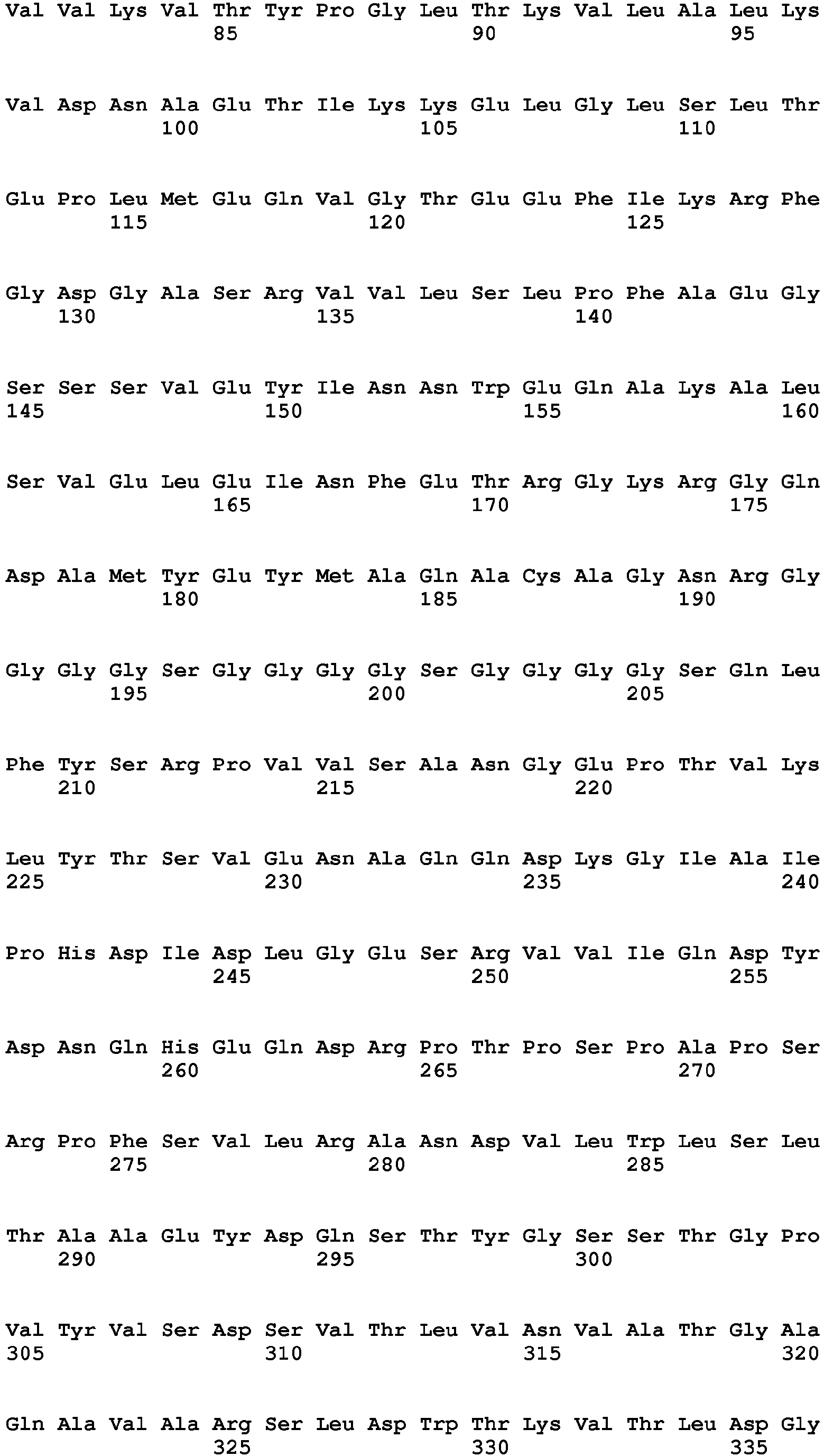

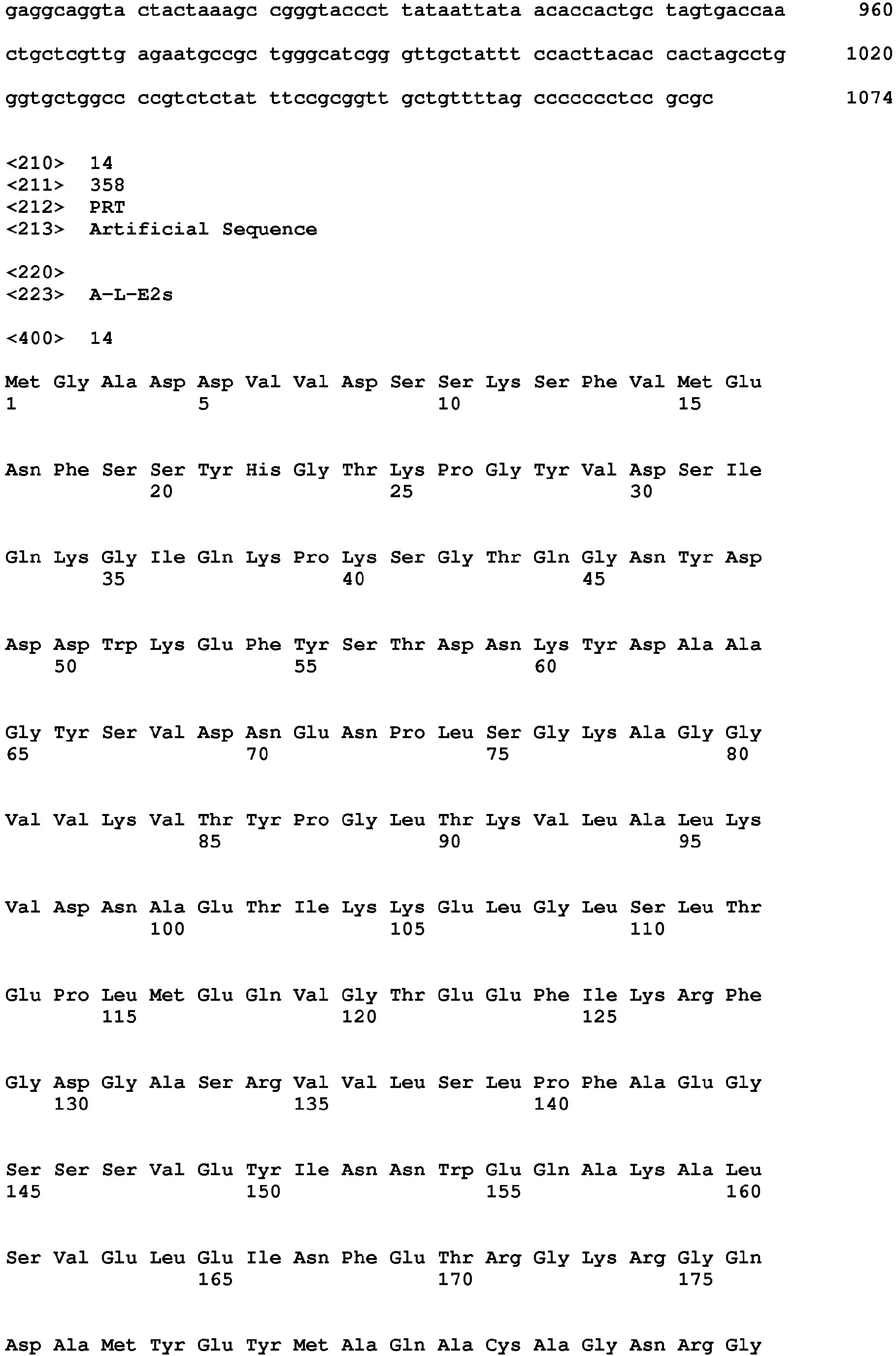


Claims (11)
- A fusion protein comprising CRM197 or a fragment thereof and a target protein, wherein said CRM197 or a fragment thereof enhances immunogenicity of the target protein, wherein
for example, the fragment of CRM197 may comprise Fragment A, Transmembrane Domain T, and/or Receptor Binding domain R of CRM197;
for example, the fragment of CRM197 may comprise Fragment A, or Fragment A and Transmembrane Domain T;
for example, the fragment of CRM197 may comprise aa 1-190 of CRM197, for example, comprise aa 1-389 of CRM197;
for example, the target protein may be an HEV capsid protein or an immunogenic fragment thereof;
for example, the immunogenic fragment of the HEV capsid protein may comprise or be HEV-239 (aa 368-606 of the HEV capsid protein), E2 (aa 394-606 of the HEV capsid protein) or E2s (aa 455-606 of the HEV capsid protein) ;
for example, the target protein may be an influenza virus M2 protein or an immunogenic fragment thereof;
for example, the immunogenic fragment of the M2 protein may comprise or be M2e (aa 1-24 of the M2 protein) ;
for example, in the fusion protein, said CRM197 or a fragment thereof may be linked to the N-terminus and/or C-terminus of the target protein, optionally via a linker;
for example, the fusion protein may comprise CRM197 or a fragment thereof and an HEV capsid protein or an immunogenic fragment thereof, which are linked together, optionally via a linker;
for example, the fusion protein may be a protein having an amino acid sequence as set forth in SEQ ID NO: 4, 6, 8, 10, 12, 14, 16 or 18;
for example, the fusion protein may comprise CRM197 or a fragment thereof, and an influenza virus M2 protein or an immunogenic fragment thereof, which are linked together, optionally via a linker;
for example, the fusion protein may be a protein having an amino acid sequence as set forth in SEQ ID NO: 34, 36, 38, 40, 42 or 44. - A polynucleotide encoding the fusion protein of claim 1.
- A construct comprising the polynucleotide of claim 2.
- A vector comprising the polynucleotide of claim 2 or the construct of claim 3, for example, the vector is an expression vector.
- A host cell or organism comprising the polynucleotide of claim 2, the construct of claim 3, or the vector of claim 4.
- A pharmaceutical composition or vaccine comprising the fusion protein of claim 1, and optionally a pharmaceutically acceptable carrier and/or excipient.
- Use of the fusion protein of claim 1 in the manufacture of a pharmaceutical composition for the prevention and/or treatment of diseases that can be prevented or treated by the target protein,
for example, when the fusion protein comprises CRM197 or a fragment thereof and an HEV capsid protein or an immunogenic fragment thereof (which are linked together, optionally via a linker), the pharmaceutical composition may be used to prevent and/or treat HEV infection and diseases associated with HEV infection such as Hepatitis E;
for example, when the fusion protein comprises CRM197 or a fragment thereof and an influenza virus M2 protein or an immunogenic fragment thereof (which are linked together, optionally via a linker), the pharmaceutical composition may be used to prevent and/or treat influenza virus infection and diseases associated with influenza virus infection such as influenza. - A method for preventing and/or treating HEV infection and/or diseases associated with HEV infection such as Hepatitis E, comprising adminstering an effective amount of the fusion protein of claim 1 or the pharmaceutical composition comprising the fusion protein, wherein the fusion protein comprising CRM197 or a fragment thereof and an HEV capsid protein or an immunogenic fragment thereof, which are linked together, optionally via a linker.
- A method for preventing and/or treating influenza virus infection and diseases associated with influenza virus infection such as influenza, comprising adminstering an effective amount of the fusion protein of claim 1 or the pharmaceutical composition comprising the fusion protein, wherein the fusion protein comprising CRM197 or a fragment thereof and an influenza virus M2 protein or an immunogenic fragment thereof, which are linked together, optionally via a linker.
- A method for enhancing immunogenicity of a target protein, comprising obtaining a fusion protein comprising CRM197 or a fragment thereof as defined in claim 1 and the target protein, so as to enhance immunogenicity of the target protein,
for example, the fusion protein may be obtained by fusion expression of CRM197 or a fragment thereof with the target protein, optionally using a linker;
for example, the target protein is an HEV capsid protein, an influenza virus M2 protein, or an immunogenic fragment thereof. - Use of CRM197 or a fragment thereof as defined in claim 1 in the enhancement of immunogenicity of a target protein, characterized by obtaining a fusion protein comprising CRM197 or a fragment thereof and the target protein,
for example, the fusion protein may be obtained by fusion expression of CRM197 or a fragment thereof with the target protein, optionally using a linker;
for example, the target protein is an HEV capsid protein, an influenza virus M2 protein, or an immunogenic fragment thereof.
Applications Claiming Priority (2)
| Application Number | Priority Date | Filing Date | Title |
|---|---|---|---|
| CN201110145419 | 2011-06-01 | ||
| PCT/CN2012/076378 WO2012163289A1 (en) | 2011-06-01 | 2012-06-01 | Fusion protein comprising diphtheria toxin non-toxic mutant crm197 or fragment thereof |
Publications (3)
| Publication Number | Publication Date |
|---|---|
| EP2716661A1 true EP2716661A1 (en) | 2014-04-09 |
| EP2716661A4 EP2716661A4 (en) | 2015-01-14 |
| EP2716661B1 EP2716661B1 (en) | 2018-03-07 |
Family
ID=47231505
Family Applications (1)
| Application Number | Title | Priority Date | Filing Date |
|---|---|---|---|
| EP12793630.0A Not-in-force EP2716661B1 (en) | 2011-06-01 | 2012-06-01 | Fusion protein comprising diphtheria toxin non-toxic mutant crm197 or fragment thereof |
Country Status (10)
| Country | Link |
|---|---|
| US (3) | US9512185B2 (en) |
| EP (1) | EP2716661B1 (en) |
| JP (1) | JP6048845B2 (en) |
| KR (2) | KR101839496B1 (en) |
| CN (2) | CN105693865B (en) |
| AU (1) | AU2012265320B2 (en) |
| BR (1) | BR112013030963B1 (en) |
| MX (1) | MX346245B (en) |
| WO (1) | WO2012163289A1 (en) |
| ZA (1) | ZA201400047B (en) |
Cited By (6)
| Publication number | Priority date | Publication date | Assignee | Title |
|---|---|---|---|---|
| EP2998322A4 (en) * | 2013-05-14 | 2016-12-14 | Shanghai Hycharm Inc | EPITOPE BASED VACCINE FOR LOW IMMUNOGENICITY PROTEIN AND METHOD FOR PREPARING AND USING SAME |
| WO2018126229A3 (en) * | 2016-12-30 | 2018-08-09 | Sutrovax, Inc. | Polypeptide-antigen conjugates with non-natural amino acids |
| US10988512B2 (en) | 2016-03-10 | 2021-04-27 | The Johns Hopkins University | Methods of producing aggregate-free monomeric diphtheria toxin fusion proteins and therapeutic uses |
| US11203626B2 (en) | 2016-03-10 | 2021-12-21 | The Johns Hopkins University | Methods of producing aggregate-free monomeric diphtheria toxin fusion proteins and therapeutic uses |
| EP3487526B1 (en) * | 2016-07-21 | 2022-07-06 | VAC4all Pte. Ltd. | Biofusion proteins as anti-malaria vaccines |
| US11965009B2 (en) | 2016-03-10 | 2024-04-23 | The Johns Hopkins University | Methods of producing aggregate-free monomeric diphtheria toxin fusion proteins and therapeutic uses |
Families Citing this family (20)
| Publication number | Priority date | Publication date | Assignee | Title |
|---|---|---|---|---|
| MX346245B (en) | 2011-06-01 | 2017-03-13 | Univ Xiamen | Fusion protein comprising diphtheria toxin non-toxic mutant crm197 or fragment thereof. |
| AR095425A1 (en) * | 2013-03-15 | 2015-10-14 | Glaxosmithkline Biologicals Sa | VACCINE, USE AND PROCEDURE TO PREVENT INFECTION WITH PICORNAVIRUS |
| CN104140972B (en) * | 2013-05-07 | 2018-01-23 | 上海惠盾生物技术有限公司 | The preparation method of diphtheria toxin muton CRM 197 |
| CN103540616B (en) * | 2013-10-23 | 2015-08-12 | 中国人民解放军南京军区福州总医院 | Express slow virus carrier system and the preparation and application thereof of Diphtheria toxin A fragment |
| BR102015020632A2 (en) * | 2015-08-26 | 2017-03-01 | Da Silva Bastos Cesar | system and process for obtaining and managing volume and concentration of sanitizer optimized for application in the disinfection procedure of an in ovo substance applicator module having as paradigm the information on the average size of vaccinated and or fed fertile eggs |
| FR3044312B1 (en) * | 2015-11-30 | 2017-12-08 | Biomerieux Sa | MUTUATED HEV POLYPEPTIDES AND THEIR USE FOR THE ASSAY OF ANTI-HEV ANTIBODIES |
| CN106432512B (en) * | 2016-09-30 | 2022-03-01 | 康希诺生物股份公司 | Protein carrier for enhancing polysaccharide antigen immunogenicity and preparation method and application thereof |
| CN106699895B (en) * | 2016-12-05 | 2020-08-04 | 上海科华生物工程股份有限公司 | Novel fusion antigen, detection kit containing same and application |
| US11951165B2 (en) | 2016-12-30 | 2024-04-09 | Vaxcyte, Inc. | Conjugated vaccine carrier proteins |
| CN107488218B (en) * | 2017-09-13 | 2021-06-01 | 华兰生物疫苗股份有限公司 | Polypeptide, immunogenic conjugate and influenza vaccine |
| CN107522777B (en) * | 2017-09-13 | 2021-06-11 | 华兰生物疫苗股份有限公司 | Polypeptide for preventing or treating influenza virus, immunogenic conjugate and application |
| EP3574915A1 (en) * | 2018-05-29 | 2019-12-04 | Neovacs | Immunogenic product comprising il-4 and/or il-13 for treating disorders associated with aberrant il-4 and/or il 13 expression or activity |
| DK3817775T3 (en) * | 2018-07-04 | 2024-09-16 | Vaxcyte Inc | Improvements in immunogenic conjugates |
| CN114835822B (en) * | 2022-04-22 | 2022-11-04 | 浙江洪晟生物科技股份有限公司 | Polymer vaccine of hog cholera virus and its preparing process |
| CN115850524A (en) * | 2022-12-29 | 2023-03-28 | 北京吉诺卫生物科技有限公司 | Method for producing diphtheria toxin non-toxic mutant protein and specific DNA molecule used by same |
| CN118681005A (en) * | 2023-03-23 | 2024-09-24 | 上海惠盾因泰生物科技有限公司 | A bivalent IL-17 therapeutic vaccine and its preparation method and application |
| CN118027189B (en) * | 2024-04-11 | 2024-12-06 | 上海惠盾因泰生物科技有限公司 | A purification preparation method and application of anti-diphtheria toxin mutant antibody and CRM197 |
| CN119792503A (en) * | 2025-03-11 | 2025-04-11 | 宁波第二激素厂 | Animal castration vaccine and its preparation method and application |
| CN119798392A (en) * | 2025-03-11 | 2025-04-11 | 宁波第二激素厂 | Animal castration vaccine antigen and its preparation method and application |
| CN120514843B (en) * | 2025-07-24 | 2026-05-01 | 中国人民解放军军事科学院军事医学研究院 | Use of beta-Hordothionin, active fragments or derivatives thereof as intramolecular adjuvants |
Family Cites Families (26)
| Publication number | Priority date | Publication date | Assignee | Title |
|---|---|---|---|---|
| ES2231770T3 (en) * | 1993-03-05 | 2005-05-16 | Wyeth Holdings Corporation | NEW PLASMIDS FOR THE PRODUCTION OF CRM PROTEIN AND DIFTERIC TOXIN. |
| AU3804995A (en) | 1995-10-10 | 1997-04-30 | Novartis Ag | Melanoma-associated protein |
| TWI239847B (en) | 1997-12-02 | 2005-09-21 | Elan Pharm Inc | N-terminal fragment of Abeta peptide and an adjuvant for preventing and treating amyloidogenic disease |
| UA81216C2 (en) * | 1999-06-01 | 2007-12-25 | Prevention and treatment of amyloid disease | |
| DK1233784T3 (en) | 1999-12-02 | 2008-09-01 | Novartis Vaccines & Diagnostic | Preparations and Methods for Stabilizing Biological Molecules after Lyophilization |
| GB0112687D0 (en) | 2001-05-24 | 2001-07-18 | Microbiological Res Authority | Pharmaceutical use of secreted bacterial effector proteins |
| WO2003040187A1 (en) | 2001-11-08 | 2003-05-15 | Yang Sheng Tang Company Limited | Hepatitis e virus monoclonal antibodies or the binding fragments of it and the use thereof |
| KR20050099517A (en) * | 2003-01-30 | 2005-10-13 | 와이어쓰 홀딩스 코포레이션 | Cross-protective epitopes of moraxella catarrhalis and use thereof |
| US20060251675A1 (en) * | 2003-03-17 | 2006-11-09 | Michael Hagen | Mutant cholera holotoxin as an adjuvant and an antigen carrier protein |
| EP1699810A4 (en) * | 2003-12-17 | 2009-10-21 | Elan Pharm Inc | BETWEEN BETA IMMUNOGENIC PEPTIDE CONJUGATES PRODUCTION TECHNIQUES OF THESE CONJUGATES |
| PT1699822E (en) * | 2003-12-30 | 2008-07-30 | Merck Patent Gmbh | Il-7 fusion proteins with antibody portions, their preparation and their use |
| GB0409750D0 (en) * | 2004-04-30 | 2004-06-09 | Chiron Srl | Integration of meningococcal conjugate vaccination |
| GB0500787D0 (en) | 2005-01-14 | 2005-02-23 | Chiron Srl | Integration of meningococcal conjugate vaccination |
| US9402915B2 (en) | 2004-04-30 | 2016-08-02 | Glaxosmithkline Biologicals Sa | Integration of meningococcal conjugate vaccination |
| EP1645283A1 (en) * | 2004-10-08 | 2006-04-12 | Chiron Behring GmbH & Co. KG | Combination vaccine |
| GB0428394D0 (en) * | 2004-12-24 | 2005-02-02 | Chiron Srl | Saccharide conjugate vaccines |
| CN100999548B (en) * | 2006-01-10 | 2010-09-08 | 海南天源康泽医药科技有限公司 | Diphtheria toxin mutant CRM197 and its preparation method |
| CN100558747C (en) * | 2006-04-06 | 2009-11-11 | 海南天源康泽医药科技有限公司 | Immunogen with diphtheria toxin mutant CRM197 as carrier and its preparation method and application |
| CN100579988C (en) * | 2007-04-02 | 2010-01-13 | 中国疾病预防控制中心病毒病预防控制所 | Fusion protein of diphtheria toxin and GM-CSF mutant and its coding gene and application |
| WO2009014835A2 (en) * | 2007-06-21 | 2009-01-29 | Angelica Therapeutics, Inc. | Modified toxins |
| BRPI0813644B8 (en) | 2007-06-26 | 2021-05-25 | Glaxosmithkline Biologicals Sa | immunogenic composition, vaccine, process to manufacture the same, and use of the immunogenic composition or vaccine |
| US8080244B2 (en) * | 2008-11-21 | 2011-12-20 | Los Alamos National Security, Llc | Anti-influenza M2e antibody |
| CN102481352A (en) * | 2009-06-22 | 2012-05-30 | 惠氏有限责任公司 | Immunogenic compositions of staphylococcus aureus antigens |
| GB0913680D0 (en) * | 2009-08-05 | 2009-09-16 | Glaxosmithkline Biolog Sa | Immunogenic composition |
| WO2011036560A2 (en) * | 2009-09-23 | 2011-03-31 | Novartis Ag | Glycoconjugate compositions and methods for treatment of hiv |
| MX346245B (en) | 2011-06-01 | 2017-03-13 | Univ Xiamen | Fusion protein comprising diphtheria toxin non-toxic mutant crm197 or fragment thereof. |
-
2012
- 2012-06-01 MX MX2013014109A patent/MX346245B/en active IP Right Grant
- 2012-06-01 KR KR1020147000012A patent/KR101839496B1/en not_active Expired - Fee Related
- 2012-06-01 CN CN201610052268.9A patent/CN105693865B/en not_active Expired - Fee Related
- 2012-06-01 WO PCT/CN2012/076378 patent/WO2012163289A1/en not_active Ceased
- 2012-06-01 CN CN201210181392.7A patent/CN102807621B/en not_active Expired - Fee Related
- 2012-06-01 BR BR112013030963-6A patent/BR112013030963B1/en not_active IP Right Cessation
- 2012-06-01 JP JP2014513045A patent/JP6048845B2/en not_active Expired - Fee Related
- 2012-06-01 US US14/123,305 patent/US9512185B2/en active Active
- 2012-06-01 KR KR1020167029198A patent/KR101895768B1/en not_active Expired - Fee Related
- 2012-06-01 EP EP12793630.0A patent/EP2716661B1/en not_active Not-in-force
- 2012-06-01 AU AU2012265320A patent/AU2012265320B2/en not_active Ceased
-
2014
- 2014-01-06 ZA ZA2014/00047A patent/ZA201400047B/en unknown
-
2016
- 2016-08-26 US US15/248,850 patent/US9764028B2/en not_active Expired - Fee Related
- 2016-08-26 US US15/248,779 patent/US9757450B2/en not_active Expired - Fee Related
Cited By (8)
| Publication number | Priority date | Publication date | Assignee | Title |
|---|---|---|---|---|
| EP2998322A4 (en) * | 2013-05-14 | 2016-12-14 | Shanghai Hycharm Inc | EPITOPE BASED VACCINE FOR LOW IMMUNOGENICITY PROTEIN AND METHOD FOR PREPARING AND USING SAME |
| US10988512B2 (en) | 2016-03-10 | 2021-04-27 | The Johns Hopkins University | Methods of producing aggregate-free monomeric diphtheria toxin fusion proteins and therapeutic uses |
| US11203626B2 (en) | 2016-03-10 | 2021-12-21 | The Johns Hopkins University | Methods of producing aggregate-free monomeric diphtheria toxin fusion proteins and therapeutic uses |
| US11965009B2 (en) | 2016-03-10 | 2024-04-23 | The Johns Hopkins University | Methods of producing aggregate-free monomeric diphtheria toxin fusion proteins and therapeutic uses |
| EP3487526B1 (en) * | 2016-07-21 | 2022-07-06 | VAC4all Pte. Ltd. | Biofusion proteins as anti-malaria vaccines |
| WO2018126229A3 (en) * | 2016-12-30 | 2018-08-09 | Sutrovax, Inc. | Polypeptide-antigen conjugates with non-natural amino acids |
| EP3562503B1 (en) * | 2016-12-30 | 2025-04-23 | Vaxcyte, Inc. | Polypeptide-antigen conjugates with non-natural amino acids |
| EP4588521A3 (en) * | 2016-12-30 | 2025-10-22 | Vaxcyte, Inc. | Polypeptide-antigen conjugates with non-natural amino acids |
Also Published As
| Publication number | Publication date |
|---|---|
| JP2014518061A (en) | 2014-07-28 |
| AU2012265320A1 (en) | 2014-01-16 |
| AU2012265320B2 (en) | 2016-11-03 |
| EP2716661A4 (en) | 2015-01-14 |
| US20140134199A1 (en) | 2014-05-15 |
| JP6048845B2 (en) | 2016-12-21 |
| BR112013030963A2 (en) | 2016-09-06 |
| CN105693865B (en) | 2020-07-17 |
| MX346245B (en) | 2017-03-13 |
| KR101895768B1 (en) | 2018-09-07 |
| EP2716661B1 (en) | 2018-03-07 |
| WO2012163289A1 (en) | 2012-12-06 |
| KR20140068843A (en) | 2014-06-09 |
| CN102807621A (en) | 2012-12-05 |
| CN102807621B (en) | 2016-04-13 |
| KR20160135331A (en) | 2016-11-25 |
| US20170015713A1 (en) | 2017-01-19 |
| KR101839496B1 (en) | 2018-03-19 |
| BR112013030963B1 (en) | 2020-12-01 |
| US9757450B2 (en) | 2017-09-12 |
| CN105693865A (en) | 2016-06-22 |
| ZA201400047B (en) | 2015-06-24 |
| MX2013014109A (en) | 2014-06-05 |
| US9512185B2 (en) | 2016-12-06 |
| US9764028B2 (en) | 2017-09-19 |
| US20170014506A1 (en) | 2017-01-19 |
Similar Documents
| Publication | Publication Date | Title |
|---|---|---|
| EP2716661B1 (en) | Fusion protein comprising diphtheria toxin non-toxic mutant crm197 or fragment thereof | |
| US11339194B2 (en) | Truncated rotavirus VP4 protein and application thereof | |
| JP6172582B2 (en) | Vaccine to prevent swine edema disease | |
| JP2026076233A (en) | Chimeric polypeptide | |
| WO2023137519A1 (en) | Chimeric polypeptides | |
| JP2025512770A (en) | Truncated varicella zoster virus envelope glycoprotein gE | |
| WO2023137522A1 (en) | Chimeric polypeptides | |
| CA3046841A1 (en) | Novel recombinant protein uk 114 in stable polymer form for use in the treatment, diagnosis and prevention of malignant solid and systemic tumours | |
| Tang et al. | Conjugation with 8-arm PEG and CRM197 enhances the immunogenicity of SARS-CoV-2 ORF8 protein | |
| CN115197303B (en) | A mutant of the receptor binding domain of S protein and its application | |
| CN111658770A (en) | P14.7 protein and its use as vaccine adjuvant |
Legal Events
| Date | Code | Title | Description |
|---|---|---|---|
| PUAI | Public reference made under article 153(3) epc to a published international application that has entered the european phase |
Free format text: ORIGINAL CODE: 0009012 |
|
| 17P | Request for examination filed |
Effective date: 20131231 |
|
| AK | Designated contracting states |
Kind code of ref document: A1 Designated state(s): AL AT BE BG CH CY CZ DE DK EE ES FI FR GB GR HR HU IE IS IT LI LT LU LV MC MK MT NL NO PL PT RO RS SE SI SK SM TR |
|
| DAX | Request for extension of the european patent (deleted) | ||
| A4 | Supplementary search report drawn up and despatched |
Effective date: 20141217 |
|
| RIC1 | Information provided on ipc code assigned before grant |
Ipc: A61K 47/48 20060101ALI20141211BHEP Ipc: A61P 31/14 20060101ALI20141211BHEP Ipc: A61K 38/16 20060101ALI20141211BHEP Ipc: A61P 1/16 20060101ALI20141211BHEP Ipc: A61P 31/16 20060101ALI20141211BHEP Ipc: C07K 19/00 20060101AFI20141211BHEP Ipc: C12N 15/62 20060101ALI20141211BHEP |
|
| 17Q | First examination report despatched |
Effective date: 20160316 |
|
| GRAP | Despatch of communication of intention to grant a patent |
Free format text: ORIGINAL CODE: EPIDOSNIGR1 |
|
| RIC1 | Information provided on ipc code assigned before grant |
Ipc: C07K 19/00 20060101AFI20170626BHEP Ipc: C12N 15/62 20060101ALI20170626BHEP Ipc: A61P 1/16 20060101ALI20170626BHEP Ipc: A61P 31/14 20060101ALI20170626BHEP Ipc: A61K 38/16 20060101ALI20170626BHEP Ipc: A61P 31/16 20060101ALI20170626BHEP |
|
| INTG | Intention to grant announced |
Effective date: 20170721 |
|
| GRAJ | Information related to disapproval of communication of intention to grant by the applicant or resumption of examination proceedings by the epo deleted |
Free format text: ORIGINAL CODE: EPIDOSDIGR1 |
|
| INTC | Intention to grant announced (deleted) | ||
| GRAP | Despatch of communication of intention to grant a patent |
Free format text: ORIGINAL CODE: EPIDOSNIGR1 |
|
| GRAJ | Information related to disapproval of communication of intention to grant by the applicant or resumption of examination proceedings by the epo deleted |
Free format text: ORIGINAL CODE: EPIDOSDIGR1 |
|
| GRAP | Despatch of communication of intention to grant a patent |
Free format text: ORIGINAL CODE: EPIDOSNIGR1 |
|
| INTG | Intention to grant announced |
Effective date: 20170929 |
|
| INTG | Intention to grant announced |
Effective date: 20171025 |
|
| GRAS | Grant fee paid |
Free format text: ORIGINAL CODE: EPIDOSNIGR3 |
|
| GRAA | (expected) grant |
Free format text: ORIGINAL CODE: 0009210 |
|
| AK | Designated contracting states |
Kind code of ref document: B1 Designated state(s): AL AT BE BG CH CY CZ DE DK EE ES FI FR GB GR HR HU IE IS IT LI LT LU LV MC MK MT NL NO PL PT RO RS SE SI SK SM TR |
|
| REG | Reference to a national code |
Ref country code: GB Ref legal event code: FG4D |
|
| REG | Reference to a national code |
Ref country code: CH Ref legal event code: EP Ref country code: AT Ref legal event code: REF Ref document number: 976466 Country of ref document: AT Kind code of ref document: T Effective date: 20180315 |
|
| REG | Reference to a national code |
Ref country code: IE Ref legal event code: FG4D |
|
| REG | Reference to a national code |
Ref country code: DE Ref legal event code: R096 Ref document number: 602012043790 Country of ref document: DE |
|
| REG | Reference to a national code |
Ref country code: FR Ref legal event code: PLFP Year of fee payment: 7 |
|
| REG | Reference to a national code |
Ref country code: NL Ref legal event code: MP Effective date: 20180307 |
|
| REG | Reference to a national code |
Ref country code: LT Ref legal event code: MG4D |
|
| PG25 | Lapsed in a contracting state [announced via postgrant information from national office to epo] |
Ref country code: ES Free format text: LAPSE BECAUSE OF FAILURE TO SUBMIT A TRANSLATION OF THE DESCRIPTION OR TO PAY THE FEE WITHIN THE PRESCRIBED TIME-LIMIT Effective date: 20180307 Ref country code: CY Free format text: LAPSE BECAUSE OF FAILURE TO SUBMIT A TRANSLATION OF THE DESCRIPTION OR TO PAY THE FEE WITHIN THE PRESCRIBED TIME-LIMIT Effective date: 20180307 Ref country code: LT Free format text: LAPSE BECAUSE OF FAILURE TO SUBMIT A TRANSLATION OF THE DESCRIPTION OR TO PAY THE FEE WITHIN THE PRESCRIBED TIME-LIMIT Effective date: 20180307 Ref country code: FI Free format text: LAPSE BECAUSE OF FAILURE TO SUBMIT A TRANSLATION OF THE DESCRIPTION OR TO PAY THE FEE WITHIN THE PRESCRIBED TIME-LIMIT Effective date: 20180307 Ref country code: NO Free format text: LAPSE BECAUSE OF FAILURE TO SUBMIT A TRANSLATION OF THE DESCRIPTION OR TO PAY THE FEE WITHIN THE PRESCRIBED TIME-LIMIT Effective date: 20180607 Ref country code: HR Free format text: LAPSE BECAUSE OF FAILURE TO SUBMIT A TRANSLATION OF THE DESCRIPTION OR TO PAY THE FEE WITHIN THE PRESCRIBED TIME-LIMIT Effective date: 20180307 |
|
| REG | Reference to a national code |
Ref country code: AT Ref legal event code: MK05 Ref document number: 976466 Country of ref document: AT Kind code of ref document: T Effective date: 20180307 |
|
| PG25 | Lapsed in a contracting state [announced via postgrant information from national office to epo] |
Ref country code: BG Free format text: LAPSE BECAUSE OF FAILURE TO SUBMIT A TRANSLATION OF THE DESCRIPTION OR TO PAY THE FEE WITHIN THE PRESCRIBED TIME-LIMIT Effective date: 20180607 Ref country code: RS Free format text: LAPSE BECAUSE OF FAILURE TO SUBMIT A TRANSLATION OF THE DESCRIPTION OR TO PAY THE FEE WITHIN THE PRESCRIBED TIME-LIMIT Effective date: 20180307 Ref country code: SE Free format text: LAPSE BECAUSE OF FAILURE TO SUBMIT A TRANSLATION OF THE DESCRIPTION OR TO PAY THE FEE WITHIN THE PRESCRIBED TIME-LIMIT Effective date: 20180307 Ref country code: LV Free format text: LAPSE BECAUSE OF FAILURE TO SUBMIT A TRANSLATION OF THE DESCRIPTION OR TO PAY THE FEE WITHIN THE PRESCRIBED TIME-LIMIT Effective date: 20180307 Ref country code: GR Free format text: LAPSE BECAUSE OF FAILURE TO SUBMIT A TRANSLATION OF THE DESCRIPTION OR TO PAY THE FEE WITHIN THE PRESCRIBED TIME-LIMIT Effective date: 20180608 |
|
| PG25 | Lapsed in a contracting state [announced via postgrant information from national office to epo] |
Ref country code: EE Free format text: LAPSE BECAUSE OF FAILURE TO SUBMIT A TRANSLATION OF THE DESCRIPTION OR TO PAY THE FEE WITHIN THE PRESCRIBED TIME-LIMIT Effective date: 20180307 Ref country code: NL Free format text: LAPSE BECAUSE OF FAILURE TO SUBMIT A TRANSLATION OF THE DESCRIPTION OR TO PAY THE FEE WITHIN THE PRESCRIBED TIME-LIMIT Effective date: 20180307 Ref country code: RO Free format text: LAPSE BECAUSE OF FAILURE TO SUBMIT A TRANSLATION OF THE DESCRIPTION OR TO PAY THE FEE WITHIN THE PRESCRIBED TIME-LIMIT Effective date: 20180307 Ref country code: PL Free format text: LAPSE BECAUSE OF FAILURE TO SUBMIT A TRANSLATION OF THE DESCRIPTION OR TO PAY THE FEE WITHIN THE PRESCRIBED TIME-LIMIT Effective date: 20180307 Ref country code: AL Free format text: LAPSE BECAUSE OF FAILURE TO SUBMIT A TRANSLATION OF THE DESCRIPTION OR TO PAY THE FEE WITHIN THE PRESCRIBED TIME-LIMIT Effective date: 20180307 |
|
| PG25 | Lapsed in a contracting state [announced via postgrant information from national office to epo] |
Ref country code: SM Free format text: LAPSE BECAUSE OF FAILURE TO SUBMIT A TRANSLATION OF THE DESCRIPTION OR TO PAY THE FEE WITHIN THE PRESCRIBED TIME-LIMIT Effective date: 20180307 Ref country code: AT Free format text: LAPSE BECAUSE OF FAILURE TO SUBMIT A TRANSLATION OF THE DESCRIPTION OR TO PAY THE FEE WITHIN THE PRESCRIBED TIME-LIMIT Effective date: 20180307 Ref country code: SK Free format text: LAPSE BECAUSE OF FAILURE TO SUBMIT A TRANSLATION OF THE DESCRIPTION OR TO PAY THE FEE WITHIN THE PRESCRIBED TIME-LIMIT Effective date: 20180307 Ref country code: CZ Free format text: LAPSE BECAUSE OF FAILURE TO SUBMIT A TRANSLATION OF THE DESCRIPTION OR TO PAY THE FEE WITHIN THE PRESCRIBED TIME-LIMIT Effective date: 20180307 |
|
| REG | Reference to a national code |
Ref country code: DE Ref legal event code: R097 Ref document number: 602012043790 Country of ref document: DE |
|
| PG25 | Lapsed in a contracting state [announced via postgrant information from national office to epo] |
Ref country code: PT Free format text: LAPSE BECAUSE OF FAILURE TO SUBMIT A TRANSLATION OF THE DESCRIPTION OR TO PAY THE FEE WITHIN THE PRESCRIBED TIME-LIMIT Effective date: 20180709 |
|
| PLBE | No opposition filed within time limit |
Free format text: ORIGINAL CODE: 0009261 |
|
| STAA | Information on the status of an ep patent application or granted ep patent |
Free format text: STATUS: NO OPPOSITION FILED WITHIN TIME LIMIT |
|
| PG25 | Lapsed in a contracting state [announced via postgrant information from national office to epo] |
Ref country code: DK Free format text: LAPSE BECAUSE OF FAILURE TO SUBMIT A TRANSLATION OF THE DESCRIPTION OR TO PAY THE FEE WITHIN THE PRESCRIBED TIME-LIMIT Effective date: 20180307 |
|
| REG | Reference to a national code |
Ref country code: CH Ref legal event code: PL |
|
| 26N | No opposition filed |
Effective date: 20181210 |
|
| PG25 | Lapsed in a contracting state [announced via postgrant information from national office to epo] |
Ref country code: SI Free format text: LAPSE BECAUSE OF FAILURE TO SUBMIT A TRANSLATION OF THE DESCRIPTION OR TO PAY THE FEE WITHIN THE PRESCRIBED TIME-LIMIT Effective date: 20180307 |
|
| REG | Reference to a national code |
Ref country code: BE Ref legal event code: MM Effective date: 20180630 |
|
| REG | Reference to a national code |
Ref country code: IE Ref legal event code: MM4A |
|
| PG25 | Lapsed in a contracting state [announced via postgrant information from national office to epo] |
Ref country code: LU Free format text: LAPSE BECAUSE OF NON-PAYMENT OF DUE FEES Effective date: 20180601 Ref country code: MC Free format text: LAPSE BECAUSE OF FAILURE TO SUBMIT A TRANSLATION OF THE DESCRIPTION OR TO PAY THE FEE WITHIN THE PRESCRIBED TIME-LIMIT Effective date: 20180307 |
|
| PG25 | Lapsed in a contracting state [announced via postgrant information from national office to epo] |
Ref country code: CH Free format text: LAPSE BECAUSE OF NON-PAYMENT OF DUE FEES Effective date: 20180630 Ref country code: LI Free format text: LAPSE BECAUSE OF NON-PAYMENT OF DUE FEES Effective date: 20180630 Ref country code: IE Free format text: LAPSE BECAUSE OF NON-PAYMENT OF DUE FEES Effective date: 20180601 |
|
| PG25 | Lapsed in a contracting state [announced via postgrant information from national office to epo] |
Ref country code: BE Free format text: LAPSE BECAUSE OF NON-PAYMENT OF DUE FEES Effective date: 20180630 |
|
| PG25 | Lapsed in a contracting state [announced via postgrant information from national office to epo] |
Ref country code: MT Free format text: LAPSE BECAUSE OF NON-PAYMENT OF DUE FEES Effective date: 20180601 |
|
| PG25 | Lapsed in a contracting state [announced via postgrant information from national office to epo] |
Ref country code: TR Free format text: LAPSE BECAUSE OF FAILURE TO SUBMIT A TRANSLATION OF THE DESCRIPTION OR TO PAY THE FEE WITHIN THE PRESCRIBED TIME-LIMIT Effective date: 20180307 |
|
| PG25 | Lapsed in a contracting state [announced via postgrant information from national office to epo] |
Ref country code: HU Free format text: LAPSE BECAUSE OF FAILURE TO SUBMIT A TRANSLATION OF THE DESCRIPTION OR TO PAY THE FEE WITHIN THE PRESCRIBED TIME-LIMIT; INVALID AB INITIO Effective date: 20120601 |
|
| PG25 | Lapsed in a contracting state [announced via postgrant information from national office to epo] |
Ref country code: MK Free format text: LAPSE BECAUSE OF NON-PAYMENT OF DUE FEES Effective date: 20180307 |
|
| PG25 | Lapsed in a contracting state [announced via postgrant information from national office to epo] |
Ref country code: IS Free format text: LAPSE BECAUSE OF FAILURE TO SUBMIT A TRANSLATION OF THE DESCRIPTION OR TO PAY THE FEE WITHIN THE PRESCRIBED TIME-LIMIT Effective date: 20180707 |
|
| PGFP | Annual fee paid to national office [announced via postgrant information from national office to epo] |
Ref country code: FR Payment date: 20200625 Year of fee payment: 9 Ref country code: DE Payment date: 20200519 Year of fee payment: 9 |
|
| PGFP | Annual fee paid to national office [announced via postgrant information from national office to epo] |
Ref country code: GB Payment date: 20200520 Year of fee payment: 9 Ref country code: IT Payment date: 20200512 Year of fee payment: 9 |
|
| REG | Reference to a national code |
Ref country code: DE Ref legal event code: R119 Ref document number: 602012043790 Country of ref document: DE |
|
| GBPC | Gb: european patent ceased through non-payment of renewal fee |
Effective date: 20210601 |
|
| PG25 | Lapsed in a contracting state [announced via postgrant information from national office to epo] |
Ref country code: GB Free format text: LAPSE BECAUSE OF NON-PAYMENT OF DUE FEES Effective date: 20210601 Ref country code: DE Free format text: LAPSE BECAUSE OF NON-PAYMENT OF DUE FEES Effective date: 20220101 |
|
| PG25 | Lapsed in a contracting state [announced via postgrant information from national office to epo] |
Ref country code: FR Free format text: LAPSE BECAUSE OF NON-PAYMENT OF DUE FEES Effective date: 20210630 |
|
| PG25 | Lapsed in a contracting state [announced via postgrant information from national office to epo] |
Ref country code: IT Free format text: LAPSE BECAUSE OF NON-PAYMENT OF DUE FEES Effective date: 20210601 |
|
| P01 | Opt-out of the competence of the unified patent court (upc) registered |
Effective date: 20230628 |