EP2716211A1 - Magnetic field viewing film for tracking in-situ surgical applications - Google Patents
Magnetic field viewing film for tracking in-situ surgical applications Download PDFInfo
- Publication number
- EP2716211A1 EP2716211A1 EP13186828.3A EP13186828A EP2716211A1 EP 2716211 A1 EP2716211 A1 EP 2716211A1 EP 13186828 A EP13186828 A EP 13186828A EP 2716211 A1 EP2716211 A1 EP 2716211A1
- Authority
- EP
- European Patent Office
- Prior art keywords
- magnetic field
- magnetic
- medical device
- viewing film
- medical
- Prior art date
- Legal status (The legal status is an assumption and is not a legal conclusion. Google has not performed a legal analysis and makes no representation as to the accuracy of the status listed.)
- Withdrawn
Links
- 230000005291 magnetic effect Effects 0.000 title claims abstract description 171
- 238000011065 in-situ storage Methods 0.000 title 1
- 238000000034 method Methods 0.000 claims abstract description 41
- 238000012800 visualization Methods 0.000 claims abstract description 32
- 239000011859 microparticle Substances 0.000 claims description 20
- 206010019909 Hernia Diseases 0.000 claims description 16
- 239000006249 magnetic particle Substances 0.000 claims description 16
- 238000002679 ablation Methods 0.000 claims description 9
- 238000001574 biopsy Methods 0.000 claims description 9
- 239000000523 sample Substances 0.000 claims description 9
- -1 polyoxymethylene Polymers 0.000 description 9
- 239000000463 material Substances 0.000 description 8
- 239000004005 microsphere Substances 0.000 description 7
- XEEYBQQBJWHFJM-UHFFFAOYSA-N Iron Chemical compound [Fe] XEEYBQQBJWHFJM-UHFFFAOYSA-N 0.000 description 6
- 210000000683 abdominal cavity Anatomy 0.000 description 6
- 239000003921 oil Substances 0.000 description 6
- 239000012530 fluid Substances 0.000 description 5
- 239000000696 magnetic material Substances 0.000 description 5
- 235000019198 oils Nutrition 0.000 description 5
- UQSXHKLRYXJYBZ-UHFFFAOYSA-N Iron oxide Chemical compound [Fe]=O UQSXHKLRYXJYBZ-UHFFFAOYSA-N 0.000 description 4
- PXHVJJICTQNCMI-UHFFFAOYSA-N Nickel Chemical compound [Ni] PXHVJJICTQNCMI-UHFFFAOYSA-N 0.000 description 4
- IAYPIBMASNFSPL-UHFFFAOYSA-N Ethylene oxide Chemical compound C1CO1 IAYPIBMASNFSPL-UHFFFAOYSA-N 0.000 description 3
- 210000004204 blood vessel Anatomy 0.000 description 3
- 238000004945 emulsification Methods 0.000 description 3
- 229910052742 iron Inorganic materials 0.000 description 3
- 238000001356 surgical procedure Methods 0.000 description 3
- XLYOFNOQVPJJNP-UHFFFAOYSA-N water Substances O XLYOFNOQVPJJNP-UHFFFAOYSA-N 0.000 description 3
- 229910000859 α-Fe Inorganic materials 0.000 description 3
- RYGMFSIKBFXOCR-UHFFFAOYSA-N Copper Chemical compound [Cu] RYGMFSIKBFXOCR-UHFFFAOYSA-N 0.000 description 2
- 229910052692 Dysprosium Inorganic materials 0.000 description 2
- 229910052779 Neodymium Inorganic materials 0.000 description 2
- 229910052772 Samarium Inorganic materials 0.000 description 2
- RTAQQCXQSZGOHL-UHFFFAOYSA-N Titanium Chemical compound [Ti] RTAQQCXQSZGOHL-UHFFFAOYSA-N 0.000 description 2
- 210000003815 abdominal wall Anatomy 0.000 description 2
- 229910052782 aluminium Inorganic materials 0.000 description 2
- XAGFODPZIPBFFR-UHFFFAOYSA-N aluminium Chemical compound [Al] XAGFODPZIPBFFR-UHFFFAOYSA-N 0.000 description 2
- 230000008859 change Effects 0.000 description 2
- 239000011248 coating agent Substances 0.000 description 2
- 238000000576 coating method Methods 0.000 description 2
- 229910017052 cobalt Inorganic materials 0.000 description 2
- 239000010941 cobalt Substances 0.000 description 2
- GUTLYIVDDKVIGB-UHFFFAOYSA-N cobalt atom Chemical compound [Co] GUTLYIVDDKVIGB-UHFFFAOYSA-N 0.000 description 2
- 229910052802 copper Inorganic materials 0.000 description 2
- 239000010949 copper Substances 0.000 description 2
- 238000007598 dipping method Methods 0.000 description 2
- KBQHZAAAGSGFKK-UHFFFAOYSA-N dysprosium atom Chemical compound [Dy] KBQHZAAAGSGFKK-UHFFFAOYSA-N 0.000 description 2
- 239000000839 emulsion Substances 0.000 description 2
- 230000005294 ferromagnetic effect Effects 0.000 description 2
- 238000002594 fluoroscopy Methods 0.000 description 2
- 239000007943 implant Substances 0.000 description 2
- 239000000203 mixture Substances 0.000 description 2
- QEFYFXOXNSNQGX-UHFFFAOYSA-N neodymium atom Chemical compound [Nd] QEFYFXOXNSNQGX-UHFFFAOYSA-N 0.000 description 2
- 229910001172 neodymium magnet Inorganic materials 0.000 description 2
- 229910052759 nickel Inorganic materials 0.000 description 2
- 210000000056 organ Anatomy 0.000 description 2
- 239000002245 particle Substances 0.000 description 2
- 229920000642 polymer Polymers 0.000 description 2
- 230000008569 process Effects 0.000 description 2
- KZUNJOHGWZRPMI-UHFFFAOYSA-N samarium atom Chemical compound [Sm] KZUNJOHGWZRPMI-UHFFFAOYSA-N 0.000 description 2
- 238000005507 spraying Methods 0.000 description 2
- 230000001954 sterilising effect Effects 0.000 description 2
- 238000004659 sterilization and disinfection Methods 0.000 description 2
- 229910052712 strontium Inorganic materials 0.000 description 2
- CIOAGBVUUVVLOB-UHFFFAOYSA-N strontium atom Chemical compound [Sr] CIOAGBVUUVVLOB-UHFFFAOYSA-N 0.000 description 2
- 230000008685 targeting Effects 0.000 description 2
- 229920001169 thermoplastic Polymers 0.000 description 2
- 239000004416 thermosoftening plastic Substances 0.000 description 2
- 239000010936 titanium Substances 0.000 description 2
- 229910052719 titanium Inorganic materials 0.000 description 2
- 229920002160 Celluloid Polymers 0.000 description 1
- 239000004709 Chlorinated polyethylene Substances 0.000 description 1
- 229920000089 Cyclic olefin copolymer Polymers 0.000 description 1
- 239000004713 Cyclic olefin copolymer Substances 0.000 description 1
- 229930040373 Paraformaldehyde Natural products 0.000 description 1
- 239000004696 Poly ether ether ketone Substances 0.000 description 1
- 239000004952 Polyamide Substances 0.000 description 1
- 239000004962 Polyamide-imide Substances 0.000 description 1
- 239000005062 Polybutadiene Substances 0.000 description 1
- 239000004695 Polyether sulfone Substances 0.000 description 1
- 239000004697 Polyetherimide Substances 0.000 description 1
- 239000004698 Polyethylene Substances 0.000 description 1
- 239000004642 Polyimide Substances 0.000 description 1
- 239000004721 Polyphenylene oxide Substances 0.000 description 1
- 239000004734 Polyphenylene sulfide Substances 0.000 description 1
- 239000004954 Polyphthalamide Substances 0.000 description 1
- 239000004743 Polypropylene Substances 0.000 description 1
- 239000004793 Polystyrene Substances 0.000 description 1
- 229920001328 Polyvinylidene chloride Polymers 0.000 description 1
- 229920006397 acrylic thermoplastic Polymers 0.000 description 1
- 239000000853 adhesive Substances 0.000 description 1
- 230000001070 adhesive effect Effects 0.000 description 1
- 150000001335 aliphatic alkanes Chemical class 0.000 description 1
- 230000008901 benefit Effects 0.000 description 1
- 230000005540 biological transmission Effects 0.000 description 1
- DQXBYHZEEUGOBF-UHFFFAOYSA-N but-3-enoic acid;ethene Chemical compound C=C.OC(=O)CC=C DQXBYHZEEUGOBF-UHFFFAOYSA-N 0.000 description 1
- 229920002301 cellulose acetate Polymers 0.000 description 1
- 239000000919 ceramic Substances 0.000 description 1
- 238000010276 construction Methods 0.000 description 1
- 235000012343 cottonseed oil Nutrition 0.000 description 1
- 239000002385 cottonseed oil Substances 0.000 description 1
- 239000013078 crystal Substances 0.000 description 1
- 229920001971 elastomer Polymers 0.000 description 1
- 238000005538 encapsulation Methods 0.000 description 1
- 239000005038 ethylene vinyl acetate Substances 0.000 description 1
- 238000000605 extraction Methods 0.000 description 1
- 229920002313 fluoropolymer Polymers 0.000 description 1
- 239000004811 fluoropolymer Substances 0.000 description 1
- 238000000227 grinding Methods 0.000 description 1
- 229920000554 ionomer Polymers 0.000 description 1
- 239000002655 kraft paper Substances 0.000 description 1
- 230000004807 localization Effects 0.000 description 1
- 239000012528 membrane Substances 0.000 description 1
- 229910052751 metal Inorganic materials 0.000 description 1
- 239000002184 metal Substances 0.000 description 1
- 150000002739 metals Chemical class 0.000 description 1
- 239000002480 mineral oil Substances 0.000 description 1
- 238000012986 modification Methods 0.000 description 1
- 230000004048 modification Effects 0.000 description 1
- 238000012544 monitoring process Methods 0.000 description 1
- 238000000465 moulding Methods 0.000 description 1
- 239000003208 petroleum Substances 0.000 description 1
- 229920003023 plastic Polymers 0.000 description 1
- 239000004033 plastic Substances 0.000 description 1
- 229920002493 poly(chlorotrifluoroethylene) Polymers 0.000 description 1
- 229920001652 poly(etherketoneketone) Polymers 0.000 description 1
- 229920001200 poly(ethylene-vinyl acetate) Polymers 0.000 description 1
- 239000005014 poly(hydroxyalkanoate) Substances 0.000 description 1
- 229920000747 poly(lactic acid) Polymers 0.000 description 1
- 229920003229 poly(methyl methacrylate) Polymers 0.000 description 1
- 229920002492 poly(sulfone) Polymers 0.000 description 1
- 229920000058 polyacrylate Polymers 0.000 description 1
- 229920002239 polyacrylonitrile Polymers 0.000 description 1
- 229920002647 polyamide Polymers 0.000 description 1
- 229920002312 polyamide-imide Polymers 0.000 description 1
- 229920006260 polyaryletherketone Polymers 0.000 description 1
- 229920002857 polybutadiene Polymers 0.000 description 1
- 229920001748 polybutylene Polymers 0.000 description 1
- 229920001707 polybutylene terephthalate Polymers 0.000 description 1
- 229920001610 polycaprolactone Polymers 0.000 description 1
- 239000004632 polycaprolactone Substances 0.000 description 1
- 239000004417 polycarbonate Substances 0.000 description 1
- 229920000515 polycarbonate Polymers 0.000 description 1
- 239000005023 polychlorotrifluoroethylene (PCTFE) polymer Substances 0.000 description 1
- 229920001123 polycyclohexylenedimethylene terephthalate Polymers 0.000 description 1
- 229920000728 polyester Polymers 0.000 description 1
- 229920006393 polyether sulfone Polymers 0.000 description 1
- 229920002530 polyetherether ketone Polymers 0.000 description 1
- 229920001601 polyetherimide Polymers 0.000 description 1
- 229920000573 polyethylene Polymers 0.000 description 1
- 229920000139 polyethylene terephthalate Polymers 0.000 description 1
- 239000005020 polyethylene terephthalate Substances 0.000 description 1
- 229920005644 polyethylene terephthalate glycol copolymer Polymers 0.000 description 1
- 229920000903 polyhydroxyalkanoate Polymers 0.000 description 1
- 229920001721 polyimide Polymers 0.000 description 1
- 229920001470 polyketone Polymers 0.000 description 1
- 239000004626 polylactic acid Substances 0.000 description 1
- 229920000306 polymethylpentene Polymers 0.000 description 1
- 239000011116 polymethylpentene Substances 0.000 description 1
- 229920000098 polyolefin Polymers 0.000 description 1
- 229920006324 polyoxymethylene Polymers 0.000 description 1
- 229920006380 polyphenylene oxide Polymers 0.000 description 1
- 229920000069 polyphenylene sulfide Polymers 0.000 description 1
- 229920006375 polyphtalamide Polymers 0.000 description 1
- 229920001155 polypropylene Polymers 0.000 description 1
- 229920001296 polysiloxane Polymers 0.000 description 1
- 229920002223 polystyrene Polymers 0.000 description 1
- 229920002215 polytrimethylene terephthalate Polymers 0.000 description 1
- 239000004814 polyurethane Substances 0.000 description 1
- 229920002635 polyurethane Polymers 0.000 description 1
- 229920002689 polyvinyl acetate Polymers 0.000 description 1
- 239000011118 polyvinyl acetate Substances 0.000 description 1
- 229920000915 polyvinyl chloride Polymers 0.000 description 1
- 239000004800 polyvinyl chloride Substances 0.000 description 1
- 239000005033 polyvinylidene chloride Substances 0.000 description 1
- SCUZVMOVTVSBLE-UHFFFAOYSA-N prop-2-enenitrile;styrene Chemical compound C=CC#N.C=CC1=CC=CC=C1 SCUZVMOVTVSBLE-UHFFFAOYSA-N 0.000 description 1
- 230000005855 radiation Effects 0.000 description 1
- 230000004044 response Effects 0.000 description 1
- 239000005060 rubber Substances 0.000 description 1
- 238000007789 sealing Methods 0.000 description 1
- 239000002904 solvent Substances 0.000 description 1
- 238000000935 solvent evaporation Methods 0.000 description 1
- 238000009718 spray deposition Methods 0.000 description 1
- 238000001694 spray drying Methods 0.000 description 1
- 229920000638 styrene acrylonitrile Polymers 0.000 description 1
- 239000000126 substance Substances 0.000 description 1
- ISXSCDLOGDJUNJ-UHFFFAOYSA-N tert-butyl prop-2-enoate Chemical compound CC(C)(C)OC(=O)C=C ISXSCDLOGDJUNJ-UHFFFAOYSA-N 0.000 description 1
- 210000001519 tissue Anatomy 0.000 description 1
- 238000003466 welding Methods 0.000 description 1
Images


Classifications
-
- A—HUMAN NECESSITIES
- A61—MEDICAL OR VETERINARY SCIENCE; HYGIENE
- A61B—DIAGNOSIS; SURGERY; IDENTIFICATION
- A61B5/00—Measuring for diagnostic purposes; Identification of persons
- A61B5/06—Devices, other than using radiation, for detecting or locating foreign bodies ; Determining position of diagnostic devices within or on the body of the patient
- A61B5/061—Determining position of a probe within the body employing means separate from the probe, e.g. sensing internal probe position employing impedance electrodes on the surface of the body
- A61B5/062—Determining position of a probe within the body employing means separate from the probe, e.g. sensing internal probe position employing impedance electrodes on the surface of the body using magnetic field
-
- A—HUMAN NECESSITIES
- A61—MEDICAL OR VETERINARY SCIENCE; HYGIENE
- A61F—FILTERS IMPLANTABLE INTO BLOOD VESSELS; PROSTHESES; DEVICES PROVIDING PATENCY TO, OR PREVENTING COLLAPSING OF, TUBULAR STRUCTURES OF THE BODY, e.g. STENTS; ORTHOPAEDIC, NURSING OR CONTRACEPTIVE DEVICES; FOMENTATION; TREATMENT OR PROTECTION OF EYES OR EARS; BANDAGES, DRESSINGS OR ABSORBENT PADS; FIRST-AID KITS
- A61F2/00—Filters implantable into blood vessels; Prostheses, i.e. artificial substitutes or replacements for parts of the body; Appliances for connecting them with the body; Devices providing patency to, or preventing collapsing of, tubular structures of the body, e.g. stents
- A61F2/0063—Implantable repair or support meshes, e.g. hernia meshes
-
- A—HUMAN NECESSITIES
- A61—MEDICAL OR VETERINARY SCIENCE; HYGIENE
- A61B—DIAGNOSIS; SURGERY; IDENTIFICATION
- A61B5/00—Measuring for diagnostic purposes; Identification of persons
- A61B5/06—Devices, other than using radiation, for detecting or locating foreign bodies ; Determining position of diagnostic devices within or on the body of the patient
- A61B5/065—Determining position of the probe employing exclusively positioning means located on or in the probe, e.g. using position sensors arranged on the probe
-
- A—HUMAN NECESSITIES
- A61—MEDICAL OR VETERINARY SCIENCE; HYGIENE
- A61F—FILTERS IMPLANTABLE INTO BLOOD VESSELS; PROSTHESES; DEVICES PROVIDING PATENCY TO, OR PREVENTING COLLAPSING OF, TUBULAR STRUCTURES OF THE BODY, e.g. STENTS; ORTHOPAEDIC, NURSING OR CONTRACEPTIVE DEVICES; FOMENTATION; TREATMENT OR PROTECTION OF EYES OR EARS; BANDAGES, DRESSINGS OR ABSORBENT PADS; FIRST-AID KITS
- A61F2/00—Filters implantable into blood vessels; Prostheses, i.e. artificial substitutes or replacements for parts of the body; Appliances for connecting them with the body; Devices providing patency to, or preventing collapsing of, tubular structures of the body, e.g. stents
- A61F2/82—Devices providing patency to, or preventing collapsing of, tubular structures of the body, e.g. stents
-
- A—HUMAN NECESSITIES
- A61—MEDICAL OR VETERINARY SCIENCE; HYGIENE
- A61B—DIAGNOSIS; SURGERY; IDENTIFICATION
- A61B34/00—Computer-aided surgery; Manipulators or robots specially adapted for use in surgery
- A61B34/20—Surgical navigation systems; Devices for tracking or guiding surgical instruments, e.g. for frameless stereotaxis
- A61B2034/2046—Tracking techniques
- A61B2034/2051—Electromagnetic tracking systems
-
- A—HUMAN NECESSITIES
- A61—MEDICAL OR VETERINARY SCIENCE; HYGIENE
- A61B—DIAGNOSIS; SURGERY; IDENTIFICATION
- A61B34/00—Computer-aided surgery; Manipulators or robots specially adapted for use in surgery
- A61B34/20—Surgical navigation systems; Devices for tracking or guiding surgical instruments, e.g. for frameless stereotaxis
- A61B2034/2046—Tracking techniques
- A61B2034/2065—Tracking using image or pattern recognition
-
- A—HUMAN NECESSITIES
- A61—MEDICAL OR VETERINARY SCIENCE; HYGIENE
- A61B—DIAGNOSIS; SURGERY; IDENTIFICATION
- A61B2505/00—Evaluating, monitoring or diagnosing in the context of a particular type of medical care
- A61B2505/05—Surgical care
-
- A—HUMAN NECESSITIES
- A61—MEDICAL OR VETERINARY SCIENCE; HYGIENE
- A61F—FILTERS IMPLANTABLE INTO BLOOD VESSELS; PROSTHESES; DEVICES PROVIDING PATENCY TO, OR PREVENTING COLLAPSING OF, TUBULAR STRUCTURES OF THE BODY, e.g. STENTS; ORTHOPAEDIC, NURSING OR CONTRACEPTIVE DEVICES; FOMENTATION; TREATMENT OR PROTECTION OF EYES OR EARS; BANDAGES, DRESSINGS OR ABSORBENT PADS; FIRST-AID KITS
- A61F2210/00—Particular material properties of prostheses classified in groups A61F2/00 - A61F2/26 or A61F2/82 or A61F9/00 or A61F11/00 or subgroups thereof
- A61F2210/009—Particular material properties of prostheses classified in groups A61F2/00 - A61F2/26 or A61F2/82 or A61F9/00 or A61F11/00 or subgroups thereof magnetic
-
- A—HUMAN NECESSITIES
- A61—MEDICAL OR VETERINARY SCIENCE; HYGIENE
- A61F—FILTERS IMPLANTABLE INTO BLOOD VESSELS; PROSTHESES; DEVICES PROVIDING PATENCY TO, OR PREVENTING COLLAPSING OF, TUBULAR STRUCTURES OF THE BODY, e.g. STENTS; ORTHOPAEDIC, NURSING OR CONTRACEPTIVE DEVICES; FOMENTATION; TREATMENT OR PROTECTION OF EYES OR EARS; BANDAGES, DRESSINGS OR ABSORBENT PADS; FIRST-AID KITS
- A61F2250/00—Special features of prostheses classified in groups A61F2/00 - A61F2/26 or A61F2/82 or A61F9/00 or A61F11/00 or subgroups thereof
- A61F2250/0058—Additional features; Implant or prostheses properties not otherwise provided for
- A61F2250/0096—Markers and sensors for detecting a position or changes of a position of an implant, e.g. RF sensors, ultrasound markers
Definitions
- the present disclosure relates to systems, apparatuses and methods for tracking medical devices. More particularly, the present disclosure relates to systems, apparatuses and methods for tracking invasive medical devices using magnetic field sensitive viewing films.
- the present disclosure provides for a method including: placing a magnetic field viewing film over a region of tissue; inserting at least one of a medical device or a medical instrument including at least one magnetic element into the region of the tissue; and determining a location of at least one of the medical device or the medical instrument based on a visualization by the magnetic field viewing film of a magnetic field produced by the at least one magnetic element.
- the present disclosure also provides for a method according to claim 1.
- the medical device is selected from the group consisting of a mesh, a stent, a sponge, a fastener, a sling, a suture, a clip, and combinations thereof.
- the medical instrument is selected from the group consisting of an endoscope, a catheter, a grasper, a balloon, a sealer, a tissue stapler, a vessel sealer, a clip applier, a biopsy instrument, an ablation probe, and combinations thereof.
- the medical device is at least one of a hernia mesh or a stent including a plurality of magnetic microparticles disposed thereon in a predetermined pattern.
- the magnetic field viewing film includes at least one flexible sheet and a plurality of magnetic particles responsive to the magnetic field produced by the at least one magnetic element.
- the method further includes adjusting the location of at least one of the medical device or the medical instrument based on the visualization by the magnetic field viewing film of the magnetic field produced by the at least one magnetic element.
- the present disclosure provides for a method including: placing a magnetic field viewing film over a region of tissue; guiding a medical device using a medical instrument into the region of the tissue, wherein at least one of the medical device or the medical instrument comprises at least one magnetic element; and determining location of at least one of the medical device or the medical instrument based on a visualization by the magnetic field viewing film of a magnetic field produced by the at least one magnetic element.
- features relevant to targeting the procedure, tissue or the medical device may be sketched on the magnetic field viewing film using a magnetized stylus or "pen".
- the medical device is selected from the group consisting of a mesh, a stent, a sponge, a fastener, a sling, a suture, a clip, and combinations thereof.
- the medical instrument is selected from the group consisting of a endoscope, a catheter, a grasper, a balloon, a sealer, a tissue stapler, a vessel sealer, a clip applier, a biopsy instrument, an ablation probe, and combinations thereof.
- the medical device is selected from the group consisting of a hernia mesh and a stent including a plurality of magnetic microparticles disposed thereon in a predetermined pattern.
- the method further includes adjusting the location of the medical device based on the visualization by the magnetic field viewing film of the magnetic field produced by the at least one magnetic element.
- the magnetic field viewing film includes at least one flexible sheet and a plurality of microspheres encapsulating at least one magnetic particle responsive to the magnetic field produced by the at least one magnetic element.
- kits including: a magnetic field viewing film; and at least one of a medical instrument or a medical device, wherein at least one of the medical instrument or the medical device includes at least one magnetic element configured to produce a magnetic field that is visualized by the magnetic field viewing film.
- the kit further includes instructions for use of the kit, the instructions including: placing the magnetic field viewing film over a region of tissue; inserting at least one the medical device or the medical instrument into the region of the tissue; and determining location of at least one of the medical device or the medical instrument based on a visualization by the magnetic field viewing film of the magnetic field produced by the at least one magnetic element.
- the instructions further include adjusting the location of at least one of the medical device or the medical instrument based on the visualization of the magnetic field viewing film.
- the medical device is selected from the group consisting of a mesh, a stent, a sponge, a fastener, a sling, a suture, a clip, and combinations thereof.
- the medical instrument is selected from the group consisting of an endoscope, a catheter, a grasper, a balloon, a sealer, a tissue stapler, a vessel sealer, a clip applier, a biopsy instrument, an ablation probe, and combinations thereof.
- the medical device is a hernia mesh including a plurality of magnetic microparticles disposed thereon in a predetermined pattern.
- the medical device is a stent including a plurality of magnetic microparticles disposed thereon in a predetermined pattern.
- the magnetic field viewing film includes at least one flexible sheet and a plurality of magnetic particles responsive to the magnetic field produced by the at least one magnetic element.
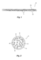
- Fig. 1 is a side, cross-sectional view of a magnetic field visualization film according to the present disclosure
- Fig. 2 is a side, cross-sectional view of a magnetic microsphere of the magnetic field visualization film of Fig. 1 according to the present disclosure
- Fig. 3A is a perspective view of the magnetic field visualization film of Fig. 1 disposed over abdominal cavity according to the present disclosure
- Fig. 3B is a cross-sectional view of the abdominal cavity with the magnetic field visualization film of Fig. 1 disposed outside of the cavity and a hernia mesh with a medical instrument disposed within the cavity according to the present disclosure;
- Fig. 4 is a perspective view of the magnetic field visualization film of Fig. 1 in the presence of a magnetic field produced by the medical instrument according to the present disclosure
- Fig. 5A is a perspective view of the hernia mesh of Fig. 3B having a plurality of magnetic elements according to the present disclosure
- Fig. 5B is a perspective view of the hernia mesh of Fig. 3B having a magnetic pattern according to the present disclosure
- Fig. 5C is a perspective view of the hernia mesh of Fig. 3B having a linear magnetic pattern according to the present disclosure
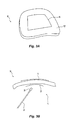
- Fig. 6A is a perspective view of the magnetic field visualization film of Fig. 1 in the presence of a magnetic field produced by the hernia mesh of Fig. 5B according to the present disclosure;
- Fig. 6B is a perspective view of the magnetic field visualization film of Fig. 1 in the presence of a magnetic field produced by the hernia mesh of Fig. 5C according to the present disclosure;
- Fig. 7 is a cross-sectional view of a blood vessel with a magnetic stent and a magnetic catheter disposed therein and the magnetic field visualization film of Fig. 1 disposed over the tissue according to the present disclosure;
- Fig. 8 is a perspective view of the magnetic stent of Fig. 8 according to the present disclosure.
- Fig. 9 is a top view of a surgical kit according to the present disclosure.
- the present disclosure provides for a system and method for visualization of location of internal tissue features and/or medical devices.
- the system includes a magnetic field visualization film disposed on an exterior portion of the patient (e.g., outside the abdominal cavity).
- the film is configured to change visually (e.g., darken) when brought in proximity of a magnetic field.
- Medical devices that are to be visualized are magnetized and/or include magnetic elements disposed thereon such that the magnetic field is visualized by the film.
- Visualization of internal tissues may be accomplished by locating one or more magnetic elements in proximity and/or contact with the internal tissues that are desired to be visualized. Changes in the color density of the film allow the user to locate and/or track location of the medical device.
- the film 10 includes a top sheet 12a and a bottom sheet 12b, which may be formed from any non-magnetic, flexible, transparent or translucent material that allows for transmission of magnetic fields and light to pass therethrough.
- Suitable materials for forming the sheets 12a, 12b include thermoplastics, such as acrylics, celluloid, cellulose acetate, cyclic olefin copolymer, ethylenevinyl acetate, fluoropolymers, ionomers, polyoxymethylene, polyacrylates, polyacrylonitrile, polyamide, polyamide-imide, polyaryletherketon, polybutadiene, polybutylene, polybutylene terephthalate, polycaprolactone, polychlorotrifluoroethylene, polyethylene terephthalate, polycyclohexylene dimethylene terephthalate, polycarbonate, polyhydroxyalkanoates, polyketones, polyester, polyethylene, polyetheretherketone, polyetherketoneketone, polyetherimide, polyethersulfone, chlorinated polyethylene, polyimide, polylactic acid, polymethylpentene, polyphenylene oxide, polyphenylene sulfide, polyphthalamide, polypropylene, polysty
- the film 10 includes a plurality of microspheres 14 disposed between the sheets 12a, 12b.
- Fig. 2 shows an enlarged, cross-sectional view of the microsphere 14 including a membrane 15 enclosing a viscous fluid 16 and a plurality of magnetic particles 18 dispersed and suspended therein.
- the microspheres 14 may be formed by any suitable microencapsulation methods, such as emulsion-based solvent evaporation and/or extraction methods including, but not limited to, single-emulsion methods such as oil-in-water (o/w) and water-in-oil (w/o), double-emulsion methods such as water-in-oil-in-water (w/o/w) and solid-in-oil-in-water (s/o/w), and non-emulsion based methods, such as fluidized-bed, spray-drying, and casting/grinding methods. After encapsulations, the microspheres 14 may be deposited on the sheets 12a, 12b and crosslinked.
- emulsion-based solvent evaporation and/or extraction methods including, but not limited to, single-emulsion methods such as oil-in-water (o/w) and water-in-oil (w/o), double-emulsion methods such as water-in-oil-in-water (w/o/w) and solid
- the fluid 16 may be an oil having an absolute viscosity at room temperature from about 50 centipoise (P) to 1,000 about cP, in embodiments from about 100 cp to about 500 cp.
- Suitable oils include, but are not limited to, petroleum-based oils, such as light, medium or heavy mineral oils (e.g., mixtures of alkanes having from about 40 carbons to about 60 carbons), plant-based oils, such as cottonseed oil, silicone-based oils, and combinations thereof.
- the fluid 15 may be used as a continuous phase fluid during the emulsification processes.
- the magnetic particles 18 may be from about 10 angstroms ( ⁇ ) to about 1000 ⁇ , in embodiments from about 25 ⁇ to about 500 ⁇ .
- the magnetic or magnetized particles 18 formed from any suitable ferromagnetic or magnetizable material including, but not limited to, ferrite, strontium ferrous oxide, neodymium (NdFeB, optionally including dysprosium), samarium, cobalt, aluminum, nickel, copper, iron, titanium, and combinations thereof.
- magnetic particles 18 may have any suitable shape, such as, cylindrical, polygonal, (e.g., square or hexagonal cross-section), and the like.
- the magnetic particles 18 may be reflective (e.g., have one or more reflective surfaces) such that alignment of the particles 18 in the presence of a magnetic field makes the film 10 appear brighter or darker based on the spatial orientation of the film 10 relative to the magnetic field.
- the film 10 is placed outside the patient (e.g., abdominal cavity "C" of Figs. 3A-B ) or over a region of tissue in which a surgical procedure is being performed.
- the film 10 may be used internally over organs and other tissue to track location of medical devices therein. Localization and/or tracking of medical devices is accomplished by monitoring changes in color (e.g., darkness level) of the film 10 as described in further detail below with respect to Figs. 4 , 6 A-B, and 7 ,
- Suitable medical devices include medical implants, such as, meshes, stents, sponges, fasteners (e.g., staplers), slings, clips, sutures, combinations thereof, and the like.
- the magnetic elements may be electromagnets.
- the medical devices may include one or more components (e.g., housing, shaft, etc.) that are magnetized or include a coating of magnetic microparticles.
- Figs. 3A-B show a system (e.g., kit) 30 for repairing a hernia according to an embodiment of the present disclosure.
- the system 30 includes the film 10 that is disposed over the abdominal cavity "C" (e.g., over an abdominal wall "W") and a mesh 32. Since the film 10 is formed from flexible sheets 12a, 12b, the film 10 may be conformed to the abdominal wall "W.”
- the system 30 also includes a medical instrument 34 having one or more magnetic elements 34 disposed thereon.
- the instrument 34 may be any suitable open or endoscopic surgical instrument, such as, endoscopes, catheters, graspers, balloons, sealers, tissue staplers, vessel sealers, clip appliers, biopsy instruments, ablation probes, combinations thereof, and the like.
- the medical instrument 34 includes one or more magnetic elements 36, which may be formed from any suitable ferromagnetic or magnetizable material.
- the magnetic elements 36 are permanently or temporarily (e.g., via adapters) secured to the medical
- Suitable magnetic materials may be temporary magnetic materials or permanent magnetic materials, ceramic, crystalline, or flexible magnetic materials (e.g., a polymeric substance such as thermoplastics or rubber) combined with magnetic ferrite (e.g., heat-treated mixtures of oxides of iron and one or more other metals having complex crystals with magnetic properties).
- Suitable magnetic materials include, but are not limited to, ferrite, strontium ferrous oxide, neodymium (NdFeB, optionally including dysprosium), samarium, cobalt, aluminum, nickel, copper, iron, titanium, and combinations thereof.
- Magnetic elements 36 may have any suitable shape, such as, cylindrical, polygonal, (e.g., square or hexagonal cross-section), and the like.
- the magnetic element 36 may be an electromagnet.
- the surgical instrument 34 may include one or more components (e.g., housing, shaft, etc.) that are magnetized or include a coating of magnetic microparticles (e.g., magnetic particles described above).
- the mesh 32 is inserted into the abdominal cavity "C" and is advanced to the site of the hernia site using any suitable methods and/or instruments.
- the surgical instrument 34 is used to point to different areas of the mesh 32.
- the film 10 reacts to the proximity of the magnetic element 36 of the instrument 34 by changing color, which is indicative of the location of the medical devices.
- the magnetic particles 18 of the film 10 are free to move within the fluid 16 in response to external magnetic fields generated by the magnetic elements 36.
- Fig. 4 illustrates the film 10 with multiple areas 38 indicative of the location of the magnetic element 36.
- the surfaces of the magnetic particles 18 that are reflective are aligned toward the plane defined by the film 10, and appear bright.
- lines of force of the magnetic field are perpendicular to the sheet, the magnetic particles 18 appear darker, since the reflective surfaces thereof are rotated away from the horizontal plane defined by the film 10.
- the user may keep adjusting the position of the mesh 32 and keep pointing the magnetic element 36 to various positions of the mesh 32 (e.g., corners, perimeter, center, etc.) until the mesh 32 is disposed over the desired location.
- the mesh 32 may include one or more magnetic elements 46 disposed thereon.
- the magnetic elements 46 may be disposed in any suitable configuration, such as corners, center, perimeters, and combinations thereof.
- the mesh 32 may include a plurality of magnetic microparticles disposed on and/or within the mesh 32.
- the microparticles may be applied in a flower-shaped pattern 33a to a mesh 32a, a pattern 33b having a plurality of lines to a mesh 32b.
- the microparticles may be applied to the mesh 32 using any suitable method, such as spraying, dipping, incorporating into the polymer solution prior to forming the mesh 32, and combinations thereof.
- the microparticles may be applied to the mesh 32 in any suitable pattern that identifies the shape of the mesh 32.
- Fig. 7A shows a flower-shaped pattern 35a, which is suitable for identifying the center and/or perimeter of the mesh 32a of Fig. 5B as it is brought into proximity with the film 10.
- Fig. 7B shows a pattern 35b having a plurality of lines corresponding to the pattern 33b of the mesh 32b, which is suitable for identifying thickness of the tissue disposed between the mesh 32 and the film 10, with the lines acting like contour lines.
- Fig. 7 shows another embodiment of a surgical instrument (e.g., catheter 40) inserted into a blood vessel "V.”
- the catheter 40 may be any suitable catheter having a flexible longitudinal body 42.
- the catheter 40 includes one or more magnetic elements 56 disposed on the longitudinal body 42.
- the magnetic element 56 may be disposed at a distal end thereof.
- the catheter 40 is inserted into the vessel "V" with the film 10 disposed over the treatment site.
- the magnetic elements 56 cause the film 10 to change in color as described above.
- the catheter 40 may be used to deliver various medical devices into the vessel "V," such as a stent 60.
- the stent 60 includes a plurality of interconnected struts 62.
- the stent 60 may include one or more magnetic elements 66 disposed thereon.
- the magnetic elements 66 may be disposed in any suitable configuration.
- the stent 60 may include a plurality of magnetic microparticles disposed on (e.g., applied as a pattern 68) and/or within the stent 60.
- the microparticles may be applied to the stent 60 using any suitable method, such as spraying, dipping, incorporating into the polymer solution prior to forming the stent 60, and combinations thereof.
- the microparticles may be applied to the stent 60 in any suitable pattern.
- the present disclosure also provides for a surgical kit 100, having one or more magnetic field visualization films 10 and one or more of the following: the mesh 32, the surgical instrument 34, the catheter 40, and the stent 60.
- the surgical kit 100 may include any other medical instruments and/or devices having magnetic elements, magnetized components, or other means suitable for generating magnetic fields (e.g., electromagnets) that are detectable by the film 10.
- the kit 100 may include a tray 102 having a plurality of planar surfaces and a plurality of recesses that may be disposed within the planar surfaces.
- the tray 102 may be formed of any suitable material, for example the tray 102 may be molded from a transparent or translucent substantially rigid plastic material (i.e., PETG).
- PETG transparent or translucent substantially rigid plastic material
- the tray 102 may have side walls defining a depth of the tray 102.
- the planar surfaces may be vertically offset within the tray 102.
- the tray 102 including its features may be formed by molding or any other suitable techniques.
- the plurality of recesses are adapted to hold articles or implements (e.g., films 10, the mesh 32, the surgical instrument 34, the catheter 40, the stent 60, etc.) that are useful in performing the intended surgical procedures described above.
- Each recess may be adapted to hold one or more articles (e.g., a plurality of films 10 or meshes 32 arranged in a stack).
- the recesses may also include detents, protrusions, or the like to frictionally engage the articles and positively retain the articles within the respective recesses.
- the kit 100 may also include a cover (not shown) to enclose the articles therein.
- a corner 104 of the tray 102 may be configured so that the cover is not adhered to the corner 104.
- a user may grasp the cover that is positioned adjacent to the corner 104 to remove the cover from the tray 102.
- the cover may be attached or configured with the tray by any suitable method, including but not limited to adhesives, heat sealing, sonic or thermal welding, solvents, etc.
- ETO ethylene oxide
- a suitable cover material is TYVEKTM a spunbond polyolefin, from DuPont of Wilmington, Del. Any number of other permeable web materials suitable for sterilization (ETO), such as Kraft paper, may be used as the cover.
- the kit 100 may also include a pamphlet (not shown) that includes various information, such as directions for using the kit 100.
Landscapes
- Health & Medical Sciences (AREA)
- Life Sciences & Earth Sciences (AREA)
- Engineering & Computer Science (AREA)
- Biomedical Technology (AREA)
- Veterinary Medicine (AREA)
- Heart & Thoracic Surgery (AREA)
- Animal Behavior & Ethology (AREA)
- General Health & Medical Sciences (AREA)
- Public Health (AREA)
- Transplantation (AREA)
- Vascular Medicine (AREA)
- Oral & Maxillofacial Surgery (AREA)
- Cardiology (AREA)
- Human Computer Interaction (AREA)
- Physics & Mathematics (AREA)
- Biophysics (AREA)
- Pathology (AREA)
- Medical Informatics (AREA)
- Molecular Biology (AREA)
- Surgery (AREA)
- Surgical Instruments (AREA)
- Prostheses (AREA)
- Media Introduction/Drainage Providing Device (AREA)
- Materials For Medical Uses (AREA)
Abstract
Description
- This application claims the benefit of and priority to
U.S. Provisional Patent Application No. 61/708,670, filed October 2, 2012 - The present disclosure relates to systems, apparatuses and methods for tracking medical devices. More particularly, the present disclosure relates to systems, apparatuses and methods for tracking invasive medical devices using magnetic field sensitive viewing films. Background of Related Art
- During invasive surgery it is often desired to track location of medical devices (e.g., surgical instruments, implants, sponges, etc.) and internal features (e.g., tissue, organs, blood vessels, etc.) within the patient. Conventional tracking usually involves magnetic resonance or X-ray fluoroscopy systems. These systems rely on sophisticated and expensive equipment such as computers and displays for visualizing location of the medical devices. Further, in the case of X-ray fluoroscopy the patient and the attending medical staff are exposed to undesirable X-ray radiation.
- There is continual need for medical device tracking systems, apparatuses and methods that provide for safe, accurate, real-time tracking of medical devices that do not require additional computing and visualization equipment.
- The present disclosure provides for a method including: placing a magnetic field viewing film over a region of tissue; inserting at least one of a medical device or a medical instrument including at least one magnetic element into the region of the tissue; and
determining a location of at least one of the medical device or the medical instrument based on a visualization by the magnetic field viewing film of a magnetic field produced by the at least one magnetic element. The present disclosure also provides for a method according to claim 1. - According to one aspect of the present disclosure, the medical device is selected from the group consisting of a mesh, a stent, a sponge, a fastener, a sling, a suture, a clip, and combinations thereof.
- According to one aspect of the present disclosure, the medical instrument is selected from the group consisting of an endoscope, a catheter, a grasper, a balloon, a sealer, a tissue stapler, a vessel sealer, a clip applier, a biopsy instrument, an ablation probe, and combinations thereof.
- According to one aspect of the present disclosure, the medical device is at least one of a hernia mesh or a stent including a plurality of magnetic microparticles disposed thereon in a predetermined pattern.
- According to one aspect of the present disclosure, the magnetic field viewing film includes at least one flexible sheet and a plurality of magnetic particles responsive to the magnetic field produced by the at least one magnetic element.
- According to one aspect of the present disclosure, the method further includes adjusting the location of at least one of the medical device or the medical instrument based on the visualization by the magnetic field viewing film of the magnetic field produced by the at least one magnetic element.
- The present disclosure provides for a method including: placing a magnetic field viewing film over a region of tissue; guiding a medical device using a medical instrument into the region of the tissue, wherein at least one of the medical device or the medical instrument comprises at least one magnetic element; and determining location of at least one of the medical device or the medical instrument based on a visualization by the magnetic field viewing film of a magnetic field produced by the at least one magnetic element.
- According to one aspect of the present disclosure , features relevant to targeting the procedure, tissue or the medical device may be sketched on the magnetic field viewing film using a magnetized stylus or "pen".
- According to one aspect of the present disclosure, the medical device is selected from the group consisting of a mesh, a stent, a sponge, a fastener, a sling, a suture, a clip, and combinations thereof.
- According to one aspect of the present disclosure, the medical instrument is selected from the group consisting of a endoscope, a catheter, a grasper, a balloon, a sealer, a tissue stapler, a vessel sealer, a clip applier, a biopsy instrument, an ablation probe, and combinations thereof.
- According to one aspect of the present disclosure, the medical device is selected from the group consisting of a hernia mesh and a stent including a plurality of magnetic microparticles disposed thereon in a predetermined pattern.
- According to one aspect of the present disclosure, the method further includes adjusting the location of the medical device based on the visualization by the magnetic field viewing film of the magnetic field produced by the at least one magnetic element.
- According to one aspect of the present disclosure, the magnetic field viewing film includes at least one flexible sheet and a plurality of microspheres encapsulating at least one magnetic particle responsive to the magnetic field produced by the at least one magnetic element.
- The present disclosure provides for a kit including: a magnetic field viewing film; and at least one of a medical instrument or a medical device, wherein at least one of the medical instrument or the medical device includes at least one magnetic element configured to produce a magnetic field that is visualized by the magnetic field viewing film.
- According to one aspect of the present disclosure, the kit further includes instructions for use of the kit, the instructions including: placing the magnetic field viewing film over a region of tissue; inserting at least one the medical device or the medical instrument into the region of the tissue; and determining location of at least one of the medical device or the medical instrument based on a visualization by the magnetic field viewing film of the magnetic field produced by the at least one magnetic element.
- According to one aspect of the present disclosure, the instructions further include adjusting the location of at least one of the medical device or the medical instrument based on the visualization of the magnetic field viewing film.
- According to one aspect of the present disclosure, the medical device is selected from the group consisting of a mesh, a stent, a sponge, a fastener, a sling, a suture, a clip, and combinations thereof.
- According to one aspect of the present disclosure, the medical instrument is selected from the group consisting of an endoscope, a catheter, a grasper, a balloon, a sealer, a tissue stapler, a vessel sealer, a clip applier, a biopsy instrument, an ablation probe, and combinations thereof.
- According to one aspect of the present disclosure, the medical device is a hernia mesh including a plurality of magnetic microparticles disposed thereon in a predetermined pattern.
- According to one aspect of the present disclosure, the medical device is a stent including a plurality of magnetic microparticles disposed thereon in a predetermined pattern.
- According to one aspect of the present disclosure, the magnetic field viewing film includes at least one flexible sheet and a plurality of magnetic particles responsive to the magnetic field produced by the at least one magnetic element.
- Various embodiments of the present disclosure are described herein with reference to the drawings wherein:
-
Fig. 1 is a side, cross-sectional view of a magnetic field visualization film according to the present disclosure; -
Fig. 2 is a side, cross-sectional view of a magnetic microsphere of the magnetic field visualization film ofFig. 1 according to the present disclosure; -
Fig. 3A is a perspective view of the magnetic field visualization film ofFig. 1 disposed over abdominal cavity according to the present disclosure; -
Fig. 3B is a cross-sectional view of the abdominal cavity with the magnetic field visualization film ofFig. 1 disposed outside of the cavity and a hernia mesh with a medical instrument disposed within the cavity according to the present disclosure; -
Fig. 4 is a perspective view of the magnetic field visualization film ofFig. 1 in the presence of a magnetic field produced by the medical instrument according to the present disclosure; -
Fig. 5A is a perspective view of the hernia mesh ofFig. 3B having a plurality of magnetic elements according to the present disclosure; -
Fig. 5B is a perspective view of the hernia mesh ofFig. 3B having a magnetic pattern according to the present disclosure; -
Fig. 5C is a perspective view of the hernia mesh ofFig. 3B having a linear magnetic pattern according to the present disclosure; -
Fig. 6A is a perspective view of the magnetic field visualization film ofFig. 1 in the presence of a magnetic field produced by the hernia mesh ofFig. 5B according to the present disclosure; -
Fig. 6B is a perspective view of the magnetic field visualization film ofFig. 1 in the presence of a magnetic field produced by the hernia mesh ofFig. 5C according to the present disclosure; -
Fig. 7 is a cross-sectional view of a blood vessel with a magnetic stent and a magnetic catheter disposed therein and the magnetic field visualization film ofFig. 1 disposed over the tissue according to the present disclosure; -
Fig. 8 is a perspective view of the magnetic stent ofFig. 8 according to the present disclosure; and -
Fig. 9 is a top view of a surgical kit according to the present disclosure. - Particular embodiments of the present disclosure are described hereinbelow with reference to the accompanying drawings. In the following description, well-known functions or constructions are not described in detail to avoid obscuring the present disclosure in unnecessary detail.
- The present disclosure provides for a system and method for visualization of location of internal tissue features and/or medical devices. The system includes a magnetic field visualization film disposed on an exterior portion of the patient (e.g., outside the abdominal cavity). The film is configured to change visually (e.g., darken) when brought in proximity of a magnetic field. Medical devices that are to be visualized are magnetized and/or include magnetic elements disposed thereon such that the magnetic field is visualized by the film. Visualization of internal tissues may be accomplished by locating one or more magnetic elements in proximity and/or contact with the internal tissues that are desired to be visualized. Changes in the color density of the film allow the user to locate and/or track location of the medical device.
- With reference to
Fig. 1 , a magneticfield visualization film 10 is shown. Thefilm 10 includes atop sheet 12a and abottom sheet 12b, which may be formed from any non-magnetic, flexible, transparent or translucent material that allows for transmission of magnetic fields and light to pass therethrough. Suitable materials for forming thesheets - The
film 10 includes a plurality ofmicrospheres 14 disposed between thesheets Fig. 2 shows an enlarged, cross-sectional view of themicrosphere 14 including amembrane 15 enclosing aviscous fluid 16 and a plurality ofmagnetic particles 18 dispersed and suspended therein. Themicrospheres 14 may be formed by any suitable microencapsulation methods, such as emulsion-based solvent evaporation and/or extraction methods including, but not limited to, single-emulsion methods such as oil-in-water (o/w) and water-in-oil (w/o), double-emulsion methods such as water-in-oil-in-water (w/o/w) and solid-in-oil-in-water (s/o/w), and non-emulsion based methods, such as fluidized-bed, spray-drying, and casting/grinding methods. After encapsulations, themicrospheres 14 may be deposited on thesheets - The fluid 16 may be an oil having an absolute viscosity at room temperature from about 50 centipoise (P) to 1,000 about cP, in embodiments from about 100 cp to about 500 cp. Suitable oils include, but are not limited to, petroleum-based oils, such as light, medium or heavy mineral oils (e.g., mixtures of alkanes having from about 40 carbons to about 60 carbons), plant-based oils, such as cottonseed oil, silicone-based oils, and combinations thereof. The fluid 15 may be used as a continuous phase fluid during the emulsification processes.
- The
magnetic particles 18 may be from about 10 angstroms (Å) to about 1000 Å, in embodiments from about 25 Å to about 500 Å. The magnetic ormagnetized particles 18 formed from any suitable ferromagnetic or magnetizable material including, but not limited to, ferrite, strontium ferrous oxide, neodymium (NdFeB, optionally including dysprosium), samarium, cobalt, aluminum, nickel, copper, iron, titanium, and combinations thereof. In embodiments,magnetic particles 18 may have any suitable shape, such as, cylindrical, polygonal, (e.g., square or hexagonal cross-section), and the like. In further embodiments, themagnetic particles 18 may be reflective (e.g., have one or more reflective surfaces) such that alignment of theparticles 18 in the presence of a magnetic field makes thefilm 10 appear brighter or darker based on the spatial orientation of thefilm 10 relative to the magnetic field. - During use, the
film 10 is placed outside the patient (e.g., abdominal cavity "C" ofFigs. 3A-B ) or over a region of tissue in which a surgical procedure is being performed. In embodiments, thefilm 10 may be used internally over organs and other tissue to track location of medical devices therein. Localization and/or tracking of medical devices is accomplished by monitoring changes in color (e.g., darkness level) of thefilm 10 as described in further detail below with respect toFigs. 4 ,6 A-B, and7 , - Suitable medical devices include medical implants, such as, meshes, stents, sponges, fasteners (e.g., staplers), slings, clips, sutures, combinations thereof, and the like. In embodiments, the magnetic elements may be electromagnets. In further embodiments, the medical devices may include one or more components (e.g., housing, shaft, etc.) that are magnetized or include a coating of magnetic microparticles.
-
Figs. 3A-B show a system (e.g., kit) 30 for repairing a hernia according to an embodiment of the present disclosure. Thesystem 30 includes thefilm 10 that is disposed over the abdominal cavity "C" (e.g., over an abdominal wall "W") and amesh 32. Since thefilm 10 is formed fromflexible sheets film 10 may be conformed to the abdominal wall "W." Thesystem 30 also includes amedical instrument 34 having one or moremagnetic elements 34 disposed thereon. Theinstrument 34 may be any suitable open or endoscopic surgical instrument, such as, endoscopes, catheters, graspers, balloons, sealers, tissue staplers, vessel sealers, clip appliers, biopsy instruments, ablation probes, combinations thereof, and the like. Themedical instrument 34 includes one or moremagnetic elements 36, which may be formed from any suitable ferromagnetic or magnetizable material. Themagnetic elements 36 are permanently or temporarily (e.g., via adapters) secured to themedical instrument 34. - Features relevant to targeting the procedure, tissue or the medical device may be sketched on the magnetic field viewing film using a magnetized stylus or "pen".
- Suitable magnetic materials may be temporary magnetic materials or permanent magnetic materials, ceramic, crystalline, or flexible magnetic materials (e.g., a polymeric substance such as thermoplastics or rubber) combined with magnetic ferrite (e.g., heat-treated mixtures of oxides of iron and one or more other metals having complex crystals with magnetic properties). Suitable magnetic materials include, but are not limited to, ferrite, strontium ferrous oxide, neodymium (NdFeB, optionally including dysprosium), samarium, cobalt, aluminum, nickel, copper, iron, titanium, and combinations thereof.
Magnetic elements 36 may have any suitable shape, such as, cylindrical, polygonal, (e.g., square or hexagonal cross-section), and the like. In embodiments, themagnetic element 36 may be an electromagnet. In further embodiments, thesurgical instrument 34 may include one or more components (e.g., housing, shaft, etc.) that are magnetized or include a coating of magnetic microparticles (e.g., magnetic particles described above). - During use, the
mesh 32 is inserted into the abdominal cavity "C" and is advanced to the site of the hernia site using any suitable methods and/or instruments. Once themesh 32 is in position, thesurgical instrument 34 is used to point to different areas of themesh 32. With reference toFig. 4 , as thesurgical instrument 34 is brought into contact with themesh 32, thefilm 10 reacts to the proximity of themagnetic element 36 of theinstrument 34 by changing color, which is indicative of the location of the medical devices. In particular, themagnetic particles 18 of thefilm 10 are free to move within the fluid 16 in response to external magnetic fields generated by themagnetic elements 36. -
Fig. 4 , illustrates thefilm 10 withmultiple areas 38 indicative of the location of themagnetic element 36. When magnetic lines of force of themagnetic element 36 are parallel to the surface of thefilm 10, the surfaces of themagnetic particles 18 that are reflective are aligned toward the plane defined by thefilm 10, and appear bright. When lines of force of the magnetic field are perpendicular to the sheet, themagnetic particles 18 appear darker, since the reflective surfaces thereof are rotated away from the horizontal plane defined by thefilm 10. The user may keep adjusting the position of themesh 32 and keep pointing themagnetic element 36 to various positions of the mesh 32 (e.g., corners, perimeter, center, etc.) until themesh 32 is disposed over the desired location. - With reference to
Fig. 5A , themesh 32 may include one or moremagnetic elements 46 disposed thereon. Themagnetic elements 46 may be disposed in any suitable configuration, such as corners, center, perimeters, and combinations thereof. In further embodiments, themesh 32 may include a plurality of magnetic microparticles disposed on and/or within themesh 32. As shown inFigs. 5B and 5C , the microparticles may be applied in a flower-shapedpattern 33a to amesh 32a, apattern 33b having a plurality of lines to amesh 32b. The microparticles may be applied to themesh 32 using any suitable method, such as spraying, dipping, incorporating into the polymer solution prior to forming themesh 32, and combinations thereof. The microparticles may be applied to themesh 32 in any suitable pattern that identifies the shape of themesh 32. - With reference to
Figs. 6A and 6B , two exemplary embodiments of the patterns 33A and 33B on thefilm 10 are shown.Fig. 7A shows a flower-shapedpattern 35a, which is suitable for identifying the center and/or perimeter of themesh 32a ofFig. 5B as it is brought into proximity with thefilm 10.Fig. 7B shows apattern 35b having a plurality of lines corresponding to thepattern 33b of themesh 32b, which is suitable for identifying thickness of the tissue disposed between themesh 32 and thefilm 10, with the lines acting like contour lines. -
Fig. 7 shows another embodiment of a surgical instrument (e.g., catheter 40) inserted into a blood vessel "V." Thecatheter 40 may be any suitable catheter having a flexiblelongitudinal body 42. Thecatheter 40 includes one or moremagnetic elements 56 disposed on thelongitudinal body 42. In embodiments, themagnetic element 56 may be disposed at a distal end thereof. In use, thecatheter 40 is inserted into the vessel "V" with thefilm 10 disposed over the treatment site. Themagnetic elements 56 cause thefilm 10 to change in color as described above. - With reference to
Figs. 7 and 8 , thecatheter 40 may be used to deliver various medical devices into the vessel "V," such as astent 60. Thestent 60 includes a plurality ofinterconnected struts 62. Thestent 60 may include one or moremagnetic elements 66 disposed thereon. Themagnetic elements 66 may be disposed in any suitable configuration. In further embodiments, thestent 60 may include a plurality of magnetic microparticles disposed on (e.g., applied as a pattern 68) and/or within thestent 60. The microparticles may be applied to thestent 60 using any suitable method, such as spraying, dipping, incorporating into the polymer solution prior to forming thestent 60, and combinations thereof. The microparticles may be applied to thestent 60 in any suitable pattern. - The present disclosure also provides for a
surgical kit 100, having one or more magneticfield visualization films 10 and one or more of the following: themesh 32, thesurgical instrument 34, thecatheter 40, and thestent 60. In embodiments, thesurgical kit 100 may include any other medical instruments and/or devices having magnetic elements, magnetized components, or other means suitable for generating magnetic fields (e.g., electromagnets) that are detectable by thefilm 10. - The
kit 100 may include atray 102 having a plurality of planar surfaces and a plurality of recesses that may be disposed within the planar surfaces. Thetray 102 may be formed of any suitable material, for example thetray 102 may be molded from a transparent or translucent substantially rigid plastic material (i.e., PETG). Thetray 102 may have side walls defining a depth of thetray 102. The planar surfaces may be vertically offset within thetray 102. Thetray 102 including its features (e.g., planar surfaces, recesses, walls, etc.) may be formed by molding or any other suitable techniques. - The plurality of recesses are adapted to hold articles or implements (e.g.,
films 10, themesh 32, thesurgical instrument 34, thecatheter 40, thestent 60, etc.) that are useful in performing the intended surgical procedures described above. Each recess may be adapted to hold one or more articles (e.g., a plurality offilms 10 or meshes 32 arranged in a stack). The recesses may also include detents, protrusions, or the like to frictionally engage the articles and positively retain the articles within the respective recesses. - The
kit 100 may also include a cover (not shown) to enclose the articles therein. Acorner 104 of thetray 102 may be configured so that the cover is not adhered to thecorner 104. In such an embodiment, a user may grasp the cover that is positioned adjacent to thecorner 104 to remove the cover from thetray 102. The cover may be attached or configured with the tray by any suitable method, including but not limited to adhesives, heat sealing, sonic or thermal welding, solvents, etc. After all of the articles have been placed in thetray 102 and the cover sealed to the tray, the kit is subjected to an ETO (ethylene oxide) gas sterilization process. A suitable cover material is TYVEK™ a spunbond polyolefin, from DuPont of Wilmington, Del. Any number of other permeable web materials suitable for sterilization (ETO), such as Kraft paper, may be used as the cover. Thekit 100 may also include a pamphlet (not shown) that includes various information, such as directions for using thekit 100. - While several embodiments of the disclosure have been shown in the drawings and/or described herein, it is not intended that the disclosure be limited thereto, as it is intended that the disclosure be as broad in scope as the art will allow and that the specification be read likewise. Therefore, the above description should not be construed as limiting, but merely as exemplifications of particular embodiments. Those skilled in the art will envision other modifications within the scope and spirit of the claims appended hereto.
- The invention may be described by reference to the following numbered paragraphs:-
- 1. A method comprising:
- placing a magnetic field viewing film over a region of tissue;
- inserting at least one of a medical device or a medical instrument comprising at least one magnetic element into the region of the tissue; and
- determining a location of at least one of the medical device or the medical instrument based on a visualization by the magnetic field viewing film of a magnetic field produced by the at least one magnetic element.
- 2. The method according to paragraph 1, wherein the medical device is selected from the group consisting of a mesh, a stent, a sponge, a fastener, a sling, a suture, a clip, and combinations thereof.
- 3. The method according to paragraph 1, wherein the medical instrument is selected from the group consisting of an endoscope, a catheter, a grasper, a balloon, a sealer, a tissue stapler, a vessel sealer, a clip applier, a biopsy instrument, an ablation probe, and combinations thereof.
- 4. The method according to paragraph 1, wherein the medical device is at least one of a hernia mesh or a stent comprising a plurality of magnetic microparticles disposed thereon in a predetermined pattern.
- 5. The method according to paragraph 1, wherein the magnetic field viewing film comprises at least one flexible sheet and a plurality of magnetic particles responsive to the magnetic field produced by the at least one magnetic element.
- 6. The method according to paragraph 1, further comprising:
- adjusting the location of at least one of the medical device or the medical instrument based on the visualization by the magnetic field viewing film of the magnetic field produced by the at least one magnetic element.
- 7. A method comprising:
- placing a magnetic field viewing film over a region of tissue;
- guiding a medical device using a medical instrument into the region of the tissue, wherein at least one of the medical device or the medical instrument comprises at least one magnetic element; and
- determining location of at least one of the medical device or the medical instrument based on a visualization by the magnetic field viewing film of a magnetic field produced by the at least one magnetic element.
- 8. The method according to paragraph 7, wherein the medical device is selected from the group consisting of a mesh, a stent, a sponge, a fastener, a sling, a suture, a clip, and combinations thereof.
- 9. The method according to paragraph 7, wherein the medical instrument is selected from the group consisting of a endoscope, a catheter, a grasper, a balloon, a sealer, a tissue stapler, a vessel sealer, a clip applier, a biopsy instrument, an ablation probe, and combinations thereof.
- 10. The method according to paragraph 7, wherein the medical device is selected from the group consisting of a hernia mesh and a stent comprising a plurality of magnetic microparticles disposed thereon in a predetermined pattern.
- 11. The method according to paragraph 7, further comprising:
- adjusting the location of the medical device based on the visualization by the magnetic field viewing film of the magnetic field produced by the at least one magnetic element.
- 12. The method according to paragraph 7, wherein the magnetic field viewing film comprises at least one flexible sheet and a plurality of microspheres encapsulating at least one magnetic particle responsive to the magnetic field produced by the at least one magnetic element.
- 13. A kit comprising:
- a magnetic field viewing film; and
- at least one of a medical instrument or a medical device, wherein at least one of the medical instrument or the medical device includes at least one magnetic element configured to produce a magnetic field that is visualized by the magnetic field viewing film.
- 14. The kit according to paragraph 13, further comprising instructions for use of the kit, the instructions comprising:
- placing the magnetic field viewing film over a region of tissue;
- inserting at least one the medical device or the medical instrument into the region of the tissue; and
- determining location of at least one of the medical device or the medical instrument based on a visualization by the magnetic field viewing film of the magnetic field produced by the at least one magnetic element.
- 15. The kit according to
paragraph 14, the instructions further comprising:- adjusting the location of at least one of the medical device or the medical instrument based on the visualization of the magnetic field viewing film.
- 16. The kit according to paragraph 13, wherein the medical device is selected from the group consisting of a mesh, a stent, a sponge, a fastener, a sling, a suture, a clip, and combinations thereof.
- 17. The kit according to paragraph 13, wherein the medical instrument is selected from the group consisting of an endoscope, a catheter, a grasper, a balloon, a sealer, a tissue stapler, a vessel sealer, a clip applier, a biopsy instrument, an ablation probe, and combinations thereof.
- 18. The kit according to paragraph 13, wherein the medical device is a hernia mesh comprising a plurality of magnetic microparticles disposed thereon in a predetermined pattern.
- 19. The kit according to paragraph 13, wherein the medical device is a stent comprising a plurality of magnetic microparticles disposed thereon in a predetermined pattern.
- 20. The kit according to paragraph 13, wherein the magnetic field viewing film comprises at least one flexible sheet and a plurality of magnetic particles responsive to the magnetic field produced by the at least one magnetic element.
Claims (14)
- A method comprising:placing a magnetic field viewing film over a region of tissue containing at least one of a medical device or a medical instrument comprising at least one magnetic element; anddetermining a location of at least one of the medical device or the medical instrument based on a visualization by the magnetic field viewing film of a magnetic field produced by the at least one magnetic element.
- The method according to claim 1, wherein the medical device is selected from the group consisting of a mesh, a stent, a sponge, a fastener, a sling, a suture, a clip, and combinations thereof.
- The method according to claim 1 or claim 2, wherein the medical instrument is selected from the group consisting of an endoscope, a catheter, a grasper, a balloon, a sealer, a tissue stapler, a vessel sealer, a clip applier, a biopsy instrument, an ablation probe, and combinations thereof.
- The method according to any of the preceding claims, wherein the medical device is at least one of a hernia mesh or a stent comprising a plurality of magnetic microparticles disposed thereon in a predetermined pattern.
- The method according to any of the preceding claims, wherein the magnetic field viewing film comprises at least one flexible sheet and a plurality of magnetic particles responsive to the magnetic field produced by the at least one magnetic element.
- The method according to any of the preceding claims, further comprising:adjusting the location of at least one of the medical device or the medical instrument based on the visualization by the magnetic field viewing film of the magnetic field produced by the at least one magnetic element.
- A kit comprising:a magnetic field viewing film; andat least one of a medical instrument or a medical device, wherein at least one of the medical instrument or the medical device includes at least one magnetic element configured to produce a magnetic field that is visualized by the magnetic field viewing film.
- The kit according to claim 7, further comprising instructions for use of the kit, the instructions comprising:placing the magnetic field viewing film over a region of tissue;inserting at least one the medical device or the medical instrument into the region of the tissue; anddetermining location of at least one of the medical device or the medical instrument based on a visualization by the magnetic field viewing film of the magnetic field produced by the at least one magnetic element.
- The kit according to claim 8, the instructions further comprising:adjusting the location of at least one of the medical device or the medical instrument based on the visualization of the magnetic field viewing film.
- The kit according to any of claims 7 to 9, wherein the medical device is selected from the group consisting of a mesh, a stent, a sponge, a fastener, a sling, a suture, a clip, and combinations thereof.
- The kit according to any of claims 7 to 10, wherein the medical instrument is selected from the group consisting of an endoscope, a catheter, a grasper, a balloon, a sealer, a tissue stapler, a vessel sealer, a clip applier, a biopsy instrument, an ablation probe, and combinations thereof.
- The kit according to any of claims 7 to 11, wherein the medical device is a hernia mesh comprising a plurality of magnetic microparticles disposed thereon in a predetermined pattern.
- The kit according to any of claims 7 to 12, wherein the medical device is a stent comprising a plurality of magnetic microparticles disposed thereon in a predetermined pattern.
- The kit according to any of claims 7 to 13, wherein the magnetic field viewing film comprises at least one flexible sheet and a plurality of magnetic particles responsive to the magnetic field produced by the at least one magnetic element.
Applications Claiming Priority (2)
| Application Number | Priority Date | Filing Date | Title |
|---|---|---|---|
| US201261708670P | 2012-10-02 | 2012-10-02 | |
| US13/948,420 US9585600B2 (en) | 2012-10-02 | 2013-07-23 | Magnetic field viewing film for tracking in-situ surgical applications |
Publications (1)
| Publication Number | Publication Date |
|---|---|
| EP2716211A1 true EP2716211A1 (en) | 2014-04-09 |
Family
ID=49378039
Family Applications (1)
| Application Number | Title | Priority Date | Filing Date |
|---|---|---|---|
| EP13186828.3A Withdrawn EP2716211A1 (en) | 2012-10-02 | 2013-10-01 | Magnetic field viewing film for tracking in-situ surgical applications |
Country Status (5)
| Country | Link |
|---|---|
| US (1) | US9585600B2 (en) |
| EP (1) | EP2716211A1 (en) |
| CN (2) | CN103705310B (en) |
| AU (1) | AU2013221937A1 (en) |
| CA (1) | CA2824296A1 (en) |
Families Citing this family (10)
| Publication number | Priority date | Publication date | Assignee | Title |
|---|---|---|---|---|
| US10236710B1 (en) * | 2016-12-20 | 2019-03-19 | Quirklogic, Inc. | Stylus securing apparatus |
| US10994108B2 (en) | 2017-09-19 | 2021-05-04 | Integra LifeSciences Switzerland Sárl | Programmable drainage valve with fixed reference magnet for determining direction of flow operable with analog or digital compass toolsets |
| US10850080B2 (en) | 2017-09-19 | 2020-12-01 | Integra LifeSciences Switzerland Sárl | Electronic toolset to locate, read, adjust, and confirm adjustment in an implantable bodily fluid drainage system without recalibrating following adjustment |
| US10850081B2 (en) | 2017-09-19 | 2020-12-01 | Integra LifeSciences Switzerland Sáarl | Implantable bodily fluid drainage valve with magnetic field resistance engagement confirmation |
| US10888692B2 (en) | 2017-09-19 | 2021-01-12 | Integra Lifesciences Switzerland Sàrl | Electronic toolset for use with multiple generations of implantable programmable valves with or without orientation functionality based on a fixed reference magnet |
| WO2020081651A1 (en) | 2018-10-16 | 2020-04-23 | Activ Surgical, Inc. | Autonomous methods and systems for tying surgical knots |
| CN113993474A (en) | 2018-12-28 | 2022-01-28 | 艾科缇弗外科公司 | System and method for optimizing accessibility, workspace and dexterity in minimally invasive surgery |
| CN113853151A (en) | 2018-12-28 | 2021-12-28 | 艾科缇弗外科公司 | User interface element for orienting a remote camera during surgery |
| WO2020214821A1 (en) | 2019-04-19 | 2020-10-22 | Activ Surgical, Inc. | Systems and methods for trocar kinematics |
| CN112433185A (en) * | 2020-12-08 | 2021-03-02 | 西南大学 | Fruit tree breeding and seedling tracing method and device based on strong magnetic development |
Citations (6)
| Publication number | Priority date | Publication date | Assignee | Title |
|---|---|---|---|---|
| EP1107043A2 (en) * | 1999-12-03 | 2001-06-13 | Marktec Corporation | Coloured magnetic particles for magnetopheric display and method for manufacturing same |
| JP2004074486A (en) * | 2002-08-12 | 2004-03-11 | Takara Co Ltd | Magnetic plate eraser |
| US20040138552A1 (en) * | 2001-04-18 | 2004-07-15 | Alex Harel | Navigating and maneuvering of an in vivo vehicle by extracorporeal devices |
| US20100114126A1 (en) * | 2008-11-02 | 2010-05-06 | Marc Neff | Magnetic positioning of surgical mesh |
| US20100121323A1 (en) * | 2008-11-10 | 2010-05-13 | Ellipse Technologies, Inc. | External adjustment device for distraction device |
| US20110184440A1 (en) * | 2010-01-26 | 2011-07-28 | Saldinger Pierre F | Magnetically manipulable surgical mesh and apparatus for the manipulation thereof |
Family Cites Families (19)
| Publication number | Priority date | Publication date | Assignee | Title |
|---|---|---|---|---|
| DE8128911U1 (en) | 1981-10-02 | 1982-06-16 | Energy-Pak Europe Ltd., 9490 Vaduz | Magnetic plaster |
| JPH05262673A (en) | 1992-03-19 | 1993-10-12 | Nippon Paint Co Ltd | Contrast medium for ultrasonic diagnosis |
| DE19532676C1 (en) * | 1995-09-05 | 1997-05-07 | Inst Physikalische Hochtech Ev | Arrangement for determining the position of a marker in a cavity within the organism of a living being |
| US5944023A (en) * | 1995-12-07 | 1999-08-31 | Sims Deltec, Inc. | Systems and methods for determining the location of an implanted device including a magnet |
| US5948321A (en) * | 1997-04-04 | 1999-09-07 | Hong; Chin-Yih Rex | Magnetic fluid thin film displays, monochromatic light switch and tunable wavelength filter |
| US6344021B1 (en) | 2000-03-03 | 2002-02-05 | Robert W. Juster | Magnetic therapy patch |
| JP3712350B2 (en) | 2000-07-18 | 2005-11-02 | 独立行政法人科学技術振興機構 | Cardiac magnetic field diagnostic apparatus for ventricular delayed potential and method for operating the same |
| US7176344B2 (en) | 2002-09-06 | 2007-02-13 | Sca Hygiene Products Ab | Sensoring absorbing article |
| US20040064030A1 (en) * | 2002-10-01 | 2004-04-01 | Peter Forsell | Detection of implanted injection port |
| US7298140B2 (en) | 2003-07-18 | 2007-11-20 | Aichi Steel Corporation | Three-dimensional magnetic direction sensor, and magneto-impedance sensor element |
| US7409953B2 (en) | 2003-12-16 | 2008-08-12 | Kimberly-Clark Worldwide, Inc. | Surgical drape having an expandable member |
| US7301332B2 (en) | 2005-10-06 | 2007-11-27 | Biosense Webster, Inc. | Magnetic sensor assembly |
| US7702378B2 (en) | 2005-11-17 | 2010-04-20 | Breast-Med, Inc. | Tissue marker for multimodality radiographic imaging |
| US7494686B2 (en) | 2005-12-06 | 2009-02-24 | Jung Suk Suh | Method of producing a patch tape showing stripes of anisotropic magnetic field |
| US20070293878A1 (en) | 2006-06-16 | 2007-12-20 | Butsch John L | Magnetic mesh support for tissue walls |
| US20100087731A1 (en) * | 2008-10-07 | 2010-04-08 | Medtronic Vascular, Inc. | Method for Tracking Degradation of a Biodegradable Stent Having Superparamagnetic Iron Oxide Particles Embedded Therein |
| CA2748504C (en) * | 2008-12-30 | 2014-04-01 | Cook Medical Technologies Llc | Balloon-tipped endoscopic system |
| US8372066B2 (en) * | 2009-04-17 | 2013-02-12 | Domain Surgical, Inc. | Inductively heated multi-mode surgical tool |
| CN102319117B (en) * | 2011-06-16 | 2013-04-03 | 上海交通大学医学院附属瑞金医院 | Large-vessel interventional device implantation system based on magnetic navigation fusion of real-time ultrasound information |
-
2013
- 2013-07-23 US US13/948,420 patent/US9585600B2/en not_active Expired - Fee Related
- 2013-08-22 CA CA2824296A patent/CA2824296A1/en not_active Abandoned
- 2013-08-28 AU AU2013221937A patent/AU2013221937A1/en not_active Abandoned
- 2013-09-30 CN CN201310460955.0A patent/CN103705310B/en not_active Expired - Fee Related
- 2013-09-30 CN CN201710452987.4A patent/CN107157482A/en active Pending
- 2013-10-01 EP EP13186828.3A patent/EP2716211A1/en not_active Withdrawn
Patent Citations (6)
| Publication number | Priority date | Publication date | Assignee | Title |
|---|---|---|---|---|
| EP1107043A2 (en) * | 1999-12-03 | 2001-06-13 | Marktec Corporation | Coloured magnetic particles for magnetopheric display and method for manufacturing same |
| US20040138552A1 (en) * | 2001-04-18 | 2004-07-15 | Alex Harel | Navigating and maneuvering of an in vivo vehicle by extracorporeal devices |
| JP2004074486A (en) * | 2002-08-12 | 2004-03-11 | Takara Co Ltd | Magnetic plate eraser |
| US20100114126A1 (en) * | 2008-11-02 | 2010-05-06 | Marc Neff | Magnetic positioning of surgical mesh |
| US20100121323A1 (en) * | 2008-11-10 | 2010-05-13 | Ellipse Technologies, Inc. | External adjustment device for distraction device |
| US20110184440A1 (en) * | 2010-01-26 | 2011-07-28 | Saldinger Pierre F | Magnetically manipulable surgical mesh and apparatus for the manipulation thereof |
Also Published As
| Publication number | Publication date |
|---|---|
| CN103705310B (en) | 2017-06-23 |
| AU2013221937A1 (en) | 2014-04-17 |
| CA2824296A1 (en) | 2014-04-02 |
| CN103705310A (en) | 2014-04-09 |
| US9585600B2 (en) | 2017-03-07 |
| CN107157482A (en) | 2017-09-15 |
| US20140094685A1 (en) | 2014-04-03 |
Similar Documents
| Publication | Publication Date | Title |
|---|---|---|
| US9585600B2 (en) | Magnetic field viewing film for tracking in-situ surgical applications | |
| US11633181B2 (en) | System and method for increased operating room efficiency | |
| Sliker et al. | Magnetically driven medical devices: a review | |
| US9522001B2 (en) | Surgical appliance kit and system for releasably securing a surgical appliance to a surgical field and method of assembling the surgical applicance kit | |
| WO2009036094A2 (en) | Magnetic prosthetic materials for implantation using natural orifice transluminal endoscopic methods | |
| KR20230152174A (en) | Sterile barrier between surgical instrument and teleoperated actuator | |
| Mortagy et al. | Magnetic anchor guidance for endoscopic submucosal dissection and other endoscopic procedures | |
| WO2015138708A1 (en) | Surgical guidance systems, devices, and methods | |
| CN224056087U (en) | Cover system and connectors | |
| Tajima et al. | An MR-compatible master–slave manipulator with interchangeable surgical tools | |
| US20260013951A1 (en) | Systems and methods for wireless localization integration | |
| ES1324536U (en) | SURGICAL USE ITEM (Machine-translation by Google Translate, not legally binding) | |
| Wang et al. | Intra-luminal injection of ferro-fluid for magnetic bowel retraction in minimal access surgery |
Legal Events
| Date | Code | Title | Description |
|---|---|---|---|
| PUAI | Public reference made under article 153(3) epc to a published international application that has entered the european phase |
Free format text: ORIGINAL CODE: 0009012 |
|
| AK | Designated contracting states |
Kind code of ref document: A1 Designated state(s): AL AT BE BG CH CY CZ DE DK EE ES FI FR GB GR HR HU IE IS IT LI LT LU LV MC MK MT NL NO PL PT RO RS SE SI SK SM TR |
|
| AX | Request for extension of the european patent |
Extension state: BA ME |
|
| 17P | Request for examination filed |
Effective date: 20140922 |
|
| RBV | Designated contracting states (corrected) |
Designated state(s): AL AT BE BG CH CY CZ DE DK EE ES FI FR GB GR HR HU IE IS IT LI LT LU LV MC MK MT NL NO PL PT RO RS SE SI SK SM TR |
|
| 17Q | First examination report despatched |
Effective date: 20180529 |
|
| STAA | Information on the status of an ep patent application or granted ep patent |
Free format text: STATUS: THE APPLICATION IS DEEMED TO BE WITHDRAWN |
|
| 18D | Application deemed to be withdrawn |
Effective date: 20181211 |