EP2685234A1 - Forming an electron microscope sample from high-pressure frozen material - Google Patents
Forming an electron microscope sample from high-pressure frozen material Download PDFInfo
- Publication number
- EP2685234A1 EP2685234A1 EP13176233.8A EP13176233A EP2685234A1 EP 2685234 A1 EP2685234 A1 EP 2685234A1 EP 13176233 A EP13176233 A EP 13176233A EP 2685234 A1 EP2685234 A1 EP 2685234A1
- Authority
- EP
- European Patent Office
- Prior art keywords
- temperature
- capillary
- sample
- extruded
- cooling
- Prior art date
- Legal status (The legal status is an assumption and is not a legal conclusion. Google has not performed a legal analysis and makes no representation as to the accuracy of the status listed.)
- Granted
Links
Images

Classifications
-
- G—PHYSICS
- G01—MEASURING; TESTING
- G01N—INVESTIGATING OR ANALYSING MATERIALS BY DETERMINING THEIR CHEMICAL OR PHYSICAL PROPERTIES
- G01N1/00—Sampling; Preparing specimens for investigation
- G01N1/28—Preparing specimens for investigation including physical details of (bio-)chemical methods covered elsewhere, e.g. G01N33/50, C12Q
- G01N1/42—Low-temperature sample treatment, e.g. cryofixation
-
- B—PERFORMING OPERATIONS; TRANSPORTING
- B23—MACHINE TOOLS; METAL-WORKING NOT OTHERWISE PROVIDED FOR
- B23K—SOLDERING OR UNSOLDERING; WELDING; CLADDING OR PLATING BY SOLDERING OR WELDING; CUTTING BY APPLYING HEAT LOCALLY, e.g. FLAME CUTTING; WORKING BY LASER BEAM
- B23K15/00—Electron-beam welding or cutting
- B23K15/08—Removing material, e.g. by cutting, by hole drilling
-
- G—PHYSICS
- G01—MEASURING; TESTING
- G01N—INVESTIGATING OR ANALYSING MATERIALS BY DETERMINING THEIR CHEMICAL OR PHYSICAL PROPERTIES
- G01N1/00—Sampling; Preparing specimens for investigation
- G01N1/02—Devices for withdrawing samples
- G01N1/04—Devices for withdrawing samples in the solid state, e.g. by cutting
- G01N1/06—Devices for withdrawing samples in the solid state, e.g. by cutting providing a thin slice, e.g. microtome
-
- G—PHYSICS
- G01—MEASURING; TESTING
- G01N—INVESTIGATING OR ANALYSING MATERIALS BY DETERMINING THEIR CHEMICAL OR PHYSICAL PROPERTIES
- G01N1/00—Sampling; Preparing specimens for investigation
- G01N1/28—Preparing specimens for investigation including physical details of (bio-)chemical methods covered elsewhere, e.g. G01N33/50, C12Q
- G01N1/286—Preparing specimens for investigation including physical details of (bio-)chemical methods covered elsewhere, e.g. G01N33/50, C12Q involving mechanical work, e.g. chopping, disintegrating, compacting, homogenising
- G01N2001/2873—Cutting or cleaving
Definitions
- the step of freeing a sample comprises cutting with a focused ion beam.
- this sequence of steps is one of many: optionally the warming/cooling cycles of steps 106/108 can be repeated.
- the temperatures can be chosen different within limits by using for example propane instead of liquid nitrogen, etc.
- staining/labeling steps for electron microscopy for example with electron dense materials as uranyl, osmium tetroxide, quantum dots, heavy metal clusters, etc. can be introduced.
Landscapes
- Physics & Mathematics (AREA)
- Health & Medical Sciences (AREA)
- Life Sciences & Earth Sciences (AREA)
- Chemical & Material Sciences (AREA)
- Analytical Chemistry (AREA)
- Biochemistry (AREA)
- General Health & Medical Sciences (AREA)
- General Physics & Mathematics (AREA)
- Immunology (AREA)
- Pathology (AREA)
- Engineering & Computer Science (AREA)
- Mechanical Engineering (AREA)
- Sampling And Sample Adjustment (AREA)
Abstract
Description
- The invention relates to a method of forming a sample from a capillary with high-pressure frozen sample material, the method comprising:
- A step of providing a high-pressure capillary with vitrified sample material at a temperature T1 below the glass transition temperature Tg, and
- A step of cutting the capillary.
- Such a method is known from "Leica EM PACT application brochure" from Leica Microsystems, available on the internet, further referred to as the Leica brochure [-1-].
- When observing a sample in a TEM, the sample is irradiated with a beam of electrons with an energy of typically between 60 keV and 300 keV, although lower and higher energies are known to be used. The image is formed by observing electrons transmitted through the sample. Therefore the sample must be a thin sample, with a thickness of typically less than 1 µm, more specifically less than 200 nm, and most specifically less than 100 nm. When observing biological samples the samples is typically sliced in thin slices. To that end the samples are first solidified. This is done by e.g. replacing all water with a plastic in so-called resin embedding, but superior results are achieved by cryo-imaging.
- Cryo-imaging involves the freezing of the sample material to a cryogenic temperature without forming ice crystal (as these damage the sample), slice the sample at the cryogenic temperature, and inspect the sample at the cryogenic temperature.
- Transmission Electron Microscopes capable of observing the sample at cryogenic temperature are commercially available.
- Also apparatuses for freezing the sample are commercially available. Part of these apparatuses use the principle of high-pressure freezing, in which the sample, contained in a holder, is first pressurized to a pressure of approximately 2100 bar, and then rapidly cooled to a temperature Tg of approximately 165 K, the glass transition temperature of water. The container is then depressurized and the sample material inside the container can then be freed.
- In one group of such apparatuses capillaries, such as copper capillaries, are used. An example of such a apparatus and method of sample preparation using these capillaries is described in the Leica brochure [-1-], more specifically pages 7 and 8. Sample material is sucked in a capillary of copper with an outer diameter of 650 µm and an inner diameter of 300 µm. this capillary is then enclosed in a holder, exposed to a pressure of 2100 bar and then cooled rapidly (typically > 600 K/s) to a temperature of 165 K or less.
- The prior art method proceeds by immersing the holder with the capillary and sample material in liquid nitrogen (LN2), removing the capillary from the holder, and transferring the capillary to a cryoultramicrotome. In the cryoultramicrotome the material of the capillary is removed and the sample material is sectioned to form samples.
- It is noted that it is also known to section sample material at cryogenic temperatures, followed by a step of freeze-substitution and then a step of immunolabeling at room temperature, followed by inspection at room temperature. See M. Karreman et al., "VIS2FIX: A High-Speed Fixation Method for Immuno-Electron Microscopy", Traffic, Vol 12, issue July 2011, pages 806-814, further referred to as Karreman [-2-].
- It is known that mechanical sectioning of samples introduces artifacts, see for example "An oscillating cryo-knife reduces cutting-induced deformation of vitreous ultrathin sections", A. Al-Amoudi et al., J. Microsc. 2003 (Oct), 212(Pt 1), pages 26-33, further referred to as Al-Amoudi [-3-]. These artifacts result from the mechanical forces on the sample material. Therefore lately Focused Ion Beam (FIB) machining is used for sectioning sample material, in which material is milled (sputtered away) with a focused ion beam, for example a Gallium ion beam with an energy of between 10 and 40 keV. FIB sectioning enables forceless cutting.
- A disadvantage of FIB sectioning of a capillary is that the copper or steel of the capillary has a low milling rate compared to biological material. Also, the wall thickness is, compared to the diameter of the core of sample material, rather large. This implies that a large amount of capillary material must be removed at a low milling rate to form a slice of sample material (a sample, also known as a lamella). The low milling rate and the relatively large amount of material result in long throughput times.
- There is a need for forming samples from capillaries without exerting force on the sample material and with a higher throughput time than prior art methods.
- The invention intends to provide a solution to that.
- To that end the method according to the invention is characterized by a step of warming the capillary to a temperature T2 between T1 and \Tg, a subsequent step of cooling the capillary to a temperature T3 below T2, as a result of which material is extruded from the capillary, and a step of freeing a sample from the extruded sample material at a temperature below Tg.
- Inventors found surprisingly that by raising the temperature of the capillary and then cooling it again resulted in an extrusion of sample material from the open end of the capillary. Inventors are unable to provide a physical explanation for this. It is not solely the result of thermal expansion, as the extrusion is observed at the same temperature where originally the capillary was cut.
- It is noted that extrusion of ice due to a lowering of temperature is known from EP patent application publication
EP2009421A1 , specifically paragraph [0047]. However, this application mentions extrusion due to the lowering of the temperature during the freezing, and does not disclose or indicate that this is due to rising and subsequent lowering of the temperature after freezing. - In an embodiment of the invention the subsequent step of cooling the capillary to a temperature T3 below temperature T2 comprises immersing the capillary in a liquid with a temperature below temperature T2, specifically immersing in liquid ethane, liquid propane or liquid nitrogen.
- Cooling of the capillary is easiest by plunging it in, for example liquid ethane, propane or nitrogen.
- It is noted that before mentioned patent application publication
EP2009421A1 implies that the temperature is increased while introducing the sample in a transmission electron microscope (TEM), but introducing a sample in a TEM implies that no subsequent immersion in a liquid can take place, as the innards of a TEM are a high vacuum. - In a preferred embodiment the capillary is a metal capillary.
- Although capillaries of cellulose are known, the extrusion is only observed with metal capillaries. Such capillaries are preferably made of a metal comprising iron or copper, iron and/or aluminium.
- In another preferred embodiment the difference between temperature T2 and T3 is more than 20 K, preferably more than 50 K.
- The effect is believed to be caused by thermal expansion/contraction. Assuming a constant linear thermal expansion coefficient for capillary and core material, the length of the extruded core material is proportional to the temperature difference. A higher temperature difference thus results in a larger protrusion.
- In an embodiment the step of freeing a sample comprises cutting with a focused ion beam.
- Cutting a slice of material, such as ice with biological material, is a method known per se to the skilled artisan.
- In a further embodiment the freed sample is a lamella.
- A lamella, that is a approximately rectangular slice of sample material with a thickness of between 30 and 300 nm, is a preferred sample in transmission electron microscopy.
- In still a further embodiment the method further comprises a step of inspection in an electron microscope. Forming the sample should lead to a sample to be inspected in a TEM or STEM. The inspection can be performed at cryogenic temperatures, or at room temperature after, for example, freeze substitution and immune-labeling the sample at room temperature. Such a method is known from Karreman [-2-]
- In a preferred embodiment the step of warming the capillary to a temperature T2 between temperature T1 and temperature Tg and the subsequent step of cooling the capillary to a temperature T3 below temperature T2, as a result of which material is extruded from the capillary is followed by a further step of warming the capillary to a temperature T2 between temperature T1 and temperature Tg and a further subsequent step of cooling the capillary to a temperature T3 below temperature T2, as a result of which more material is extruded from the capillary.
- Inventors found surprisingly that in many cases a repetition of the warming/cooling cycle resulted in a further extrusion of sample material, although the process could not be repeated indefinitely.
- The invention is now elucidated using figures, in which identical reference numerals refer to corresponding features.
- To that end:
-
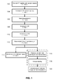
figure 1 schematically shows a flow-chart of the method according to the invention, -
figure 2 shows a photo of a capillary with extruded sample material. -
Figure 1 schematically shows a flowchart of the method according to the invention. - In step 102 a capillary with high pressure frozen sample material is provided in LN2. Such a capillary, for example a copper capillary, can be prepared by a high pressure freezer, such as an EM PACT2™ from Leica Microsystems Gmbh., Wetzlar, Germany.
- In
step 104 the capillary, immersed in LN2, is introduced in a cryo-ultramicrotome to cut the capillary, such as a EM-UC7/FC7™ from Leica Microsystems Gmbh., Wetzlar, Germany. This cutting is routinely done while the capillary is submerged in LN2. - In
step 106 the cut capillary is 'heated' to a temperature of approximately 130 K, some 50 K above the temperature of (boiling) LN2 (77 K). - In
step 108 the cut capillary is cooled to LN2 temperature again by immersing it in LN2. - In
step 110 the truncated capillary is introduced in a FIB or Dual-Beam apparatus at a temperature of 130K. A suited Dual-Beam apparatus, comprising a Focused Ion Beam column for milling and a Scanning Electron Microscope column for imaging, is, for example, the Quanta 3D™ from FEI Company, Hillsboro, USA. Inventors observed that in most cases (approximately 90% of the cases) sample material was extruded from the capillary. - In step 112 a sample is formed from the core of sample material and mounted on a sample carrier.
- The protruding core can now be sectioned with the ion beam at a cryogenic temperature. This is a method known per se to the skilled person. If necessary the sample can not only be sectioned with an ion beam, but also thinned. The completed sample is then deposited on a sample carrier. Such a sample carrier is preferably a TEM grid (typically 3 mm round) or another sample carrier dedicated for use in a TEM, such as the sample carrier described in
US patent No. 7,767,979 . - In
step 114 the sample, mounted on the sample carrier, is introduced and inspected/observed in an electron microscope, such as a Transmission Electron Microscope (TEM) or a Scanning Transmission Electron Microscope (STEM). - It is noted that this inspection may be an inspection at a cryogenic temperature in a cryo-TEM, such as the Titan Krios™ from FEI Company, Hillsboro, USA, or in a room temperature TEM, such as the Titan 80-300™ from FEI Company, Hillsboro, USA. Alternatively, instead of step 114 a
step 116 of freeze substitution and warming of the sample to room , followed by astep 118 of immuno-labeling at room temperature can be performed. In this way a correlative light/electron microscope may be used, observing with fluorescent labels for navigation and making images with electron optical resolutions of less than 1 nm. This method is described in Karreman [-2-] as the VIS2FIXFS method. Another method resulting in a sample at room temperature, the VIS2FIXH method, is also disclosed in Karreman [-2-]. - Using this method, resulting in a sample at room temperature, the imaging can take place at room temperature as well in
step 120. - It will be clear to the person skilled in the art that this sequence of steps is one of many: optionally the warming/cooling cycles of
steps 106/108 can be repeated. The temperatures can be chosen different within limits by using for example propane instead of liquid nitrogen, etc. Also staining/labeling steps for electron microscopy (for example with electron dense materials as uranyl, osmium tetroxide, quantum dots, heavy metal clusters, etc.) can be introduced.
Further it is explicitly noted that the temperatures mentioned are should not be interpreted as absolute limits: what is important is that the sample material does not change from its amorphous or glassy state to a crystalline state (by keeping the temperature below the glass transition temperature Tg), and that a large temperature difference betweensteps -
Figure 2 shows a picture taken with a SEM, showing a capillary with sample material. The capillary 202 is a copper capillary with an external diameter of approximately 650 µm and an internal diameter of 300 µm. The extrudedcore 204 is vitreous ice with biological material in it, the biological material comprising for example cells, bacteria, viruses, etc., that extends over alength 206 of 224 µm - It is mentioned that in electron microscopy "biological material" often refers to material that contains mainly hydro-carbons, and thus includes polymers, plastics, etc. The reason for this is that all these materials show weak contrast in an electron microscope as most of the nuclei are light nuclei, the nuclei showing comparable interaction with the electrons that irradiate the sample. Therefore in this context all material containing mainly hydrocarbons is included in the phrase "biological material".
-
- [-1-] "Leica EM PACT application brochure", http://www.leica-microsystems.co.kr/pdfs.nsf/(ALLIDs)/D5AA3952E2A44A56C1256F100022B5CC/$FI LE/EM PACT Application Brochure 09 04.pdf
- [-2-] "VIS2FIX: A High-Speed Fixation Method for Immuno-Electron Microscopy", M. Karreman et al., Traffic, Vol 12, issue July 2011, pages 806-814.
- [-3-] "An oscillating cryo-knife reduces cutting-induced deformation of vitreous ultrathin sections", A. Al-Amoudi et al., Journal of Microscopy, Vol. 212 Issue 1, Pages 26 - 33.
Claims (9)
- Method of forming a sample from a capillary with high-pressure frozen sample material, the method comprising:• A step (102) of providing a high-pressure capillary with vitrified sample material at a temperature T1 below the glass transition temperature Tg, and• A step (104) of cutting the capillary,
Characterized by• A step (106) of warming the capillary to a temperature T2 between temperature T1 and temperature Tg,• A subsequent step (108) of cooling the capillary to a temperature T3 below temperature T2, as a result of which material is extruded from the capillary, and• A step (112) of freeing a sample from the extruded sample material at a temperature below temperature Tg. - The method of claim 2 in which the subsequent step (108) of cooling the capillary to a temperature T3 below temperature T2 comprises immersing the capillary in a liquid with a temperature below temperature T2, specifically immersing in liquid ethane, liquid propane or liquid nitrogen.
- The method of any of the preceding claims in which the capillary is a metal capillary.
- The method of claim 3 in which the metal comprises copper and/or iron and/or aluminium.
- The method of any of the preceding claims in which the difference between temperature T2 and T3 is more than 20 K, preferably more than 50 K.
- The method of any of the preceding claims in which the step of freeing a sample comprises cutting with a focused ion beam.
- The method of any of the preceding claims in which the freed sample is a lamella.
- The method of claim 7 in which the method further comprises a step of inspection in an electron microscope.
- The method of any of the preceding claims in which the step (106) of warming the capillary to a temperature T2 between temperature T1 and temperature Tg and the subsequent step (108) of cooling the capillary to a temperature T3 below temperature T2, as a result of which material is extruded from the capillary is followed by a further step of warming the capillary to a temperature T2 between temperature T1 and temperature Tg and a further subsequent step (108) of cooling the capillary to a temperature T3 below temperature T2, as a result of which more material is extruded from the capillary.
Priority Applications (1)
| Application Number | Priority Date | Filing Date | Title |
|---|---|---|---|
| EP13176233.8A EP2685234B1 (en) | 2012-07-13 | 2013-07-12 | Forming an electron microscope sample from high-pressure frozen material |
Applications Claiming Priority (2)
| Application Number | Priority Date | Filing Date | Title |
|---|---|---|---|
| EP12176254.6A EP2685233A1 (en) | 2012-07-13 | 2012-07-13 | Forming an electron microscope sample from high-pressure frozen material |
| EP13176233.8A EP2685234B1 (en) | 2012-07-13 | 2013-07-12 | Forming an electron microscope sample from high-pressure frozen material |
Publications (2)
| Publication Number | Publication Date |
|---|---|
| EP2685234A1 true EP2685234A1 (en) | 2014-01-15 |
| EP2685234B1 EP2685234B1 (en) | 2014-12-17 |
Family
ID=48747482
Family Applications (2)
| Application Number | Title | Priority Date | Filing Date |
|---|---|---|---|
| EP12176254.6A Withdrawn EP2685233A1 (en) | 2012-07-13 | 2012-07-13 | Forming an electron microscope sample from high-pressure frozen material |
| EP13176233.8A Active EP2685234B1 (en) | 2012-07-13 | 2013-07-12 | Forming an electron microscope sample from high-pressure frozen material |
Family Applications Before (1)
| Application Number | Title | Priority Date | Filing Date |
|---|---|---|---|
| EP12176254.6A Withdrawn EP2685233A1 (en) | 2012-07-13 | 2012-07-13 | Forming an electron microscope sample from high-pressure frozen material |
Country Status (4)
| Country | Link |
|---|---|
| US (1) | US8674323B2 (en) |
| EP (2) | EP2685233A1 (en) |
| JP (1) | JP6294017B2 (en) |
| CN (1) | CN103543045B (en) |
Families Citing this family (7)
| Publication number | Priority date | Publication date | Assignee | Title |
|---|---|---|---|---|
| EP2813835B1 (en) | 2013-06-14 | 2016-09-07 | Fei Company | Method of welding a frozen aqueous sample to a microprobe |
| JP6454246B2 (en) * | 2015-09-09 | 2019-01-16 | 日本電子株式会社 | Processing method |
| RU2663732C1 (en) * | 2017-05-04 | 2018-08-09 | Игорь Михайлович Рулев | Method of automatic selection and packaging of microbiological objects |
| CN122505950A (en) | 2017-12-28 | 2026-08-04 | Fei 公司 | Methods, apparatus, and systems for treating cryogenic biological samples using focused plasma ion beams |
| CN110823931B (en) * | 2019-10-16 | 2020-10-27 | 中国科学院生物物理研究所 | A kind of cryo-electron microscope sample preparation method |
| CN114674851A (en) * | 2022-03-28 | 2022-06-28 | 厦门大学 | High-pressure freezing and freezing-replacement methods for preparing samples for transmission electron microscopy |
| DE202023100215U1 (en) | 2023-01-17 | 2023-02-06 | Cbg Composites Gmbh | Fiber concrete product based on basalt fibers with a plasticizing effect |
Citations (2)
| Publication number | Priority date | Publication date | Assignee | Title |
|---|---|---|---|---|
| EP2009421A1 (en) | 2007-06-29 | 2008-12-31 | FEI Company | Method for separating a lamella for TEM inspection from a core sample |
| US7767979B2 (en) | 2006-05-29 | 2010-08-03 | Fei Company | Method for coupling and disconnecting a co-operative composite structure of a sample carrier and a sample holder |
Family Cites Families (8)
| Publication number | Priority date | Publication date | Assignee | Title |
|---|---|---|---|---|
| JPS5582944A (en) * | 1978-12-19 | 1980-06-23 | Toukiyouto | Preparation of replica thin-film for specimen of electron microscope |
| EP0853238B1 (en) * | 1997-01-13 | 2002-09-18 | Daniel Dr. Studer | Sample holder for water-containing samples and method for use thereof |
| US6881363B2 (en) * | 2001-12-07 | 2005-04-19 | Symyx Technologies, Inc. | High throughput preparation and analysis of materials |
| DE602008000086D1 (en) * | 2007-06-29 | 2009-09-24 | Fei Co | Method for attaching a sample to a manipulator |
| EP2009420A1 (en) * | 2007-06-29 | 2008-12-31 | FEI Company | Method for attaching a sample to a manipulator |
| CN101648085B (en) * | 2008-08-14 | 2012-03-21 | 浙江工业大学 | Equipment and method for manufacturing capillary reactors |
| EP2402475A1 (en) * | 2010-06-30 | 2012-01-04 | Fei Company | Beam-induced deposition at cryogenic temperatures |
| US8884248B2 (en) * | 2012-02-13 | 2014-11-11 | Fei Company | Forming a vitrified sample for electron microscopy |
-
2012
- 2012-07-13 EP EP12176254.6A patent/EP2685233A1/en not_active Withdrawn
-
2013
- 2013-07-03 US US13/934,925 patent/US8674323B2/en active Active
- 2013-07-09 JP JP2013143193A patent/JP6294017B2/en active Active
- 2013-07-12 EP EP13176233.8A patent/EP2685234B1/en active Active
- 2013-07-12 CN CN201310301331.4A patent/CN103543045B/en active Active
Patent Citations (2)
| Publication number | Priority date | Publication date | Assignee | Title |
|---|---|---|---|---|
| US7767979B2 (en) | 2006-05-29 | 2010-08-03 | Fei Company | Method for coupling and disconnecting a co-operative composite structure of a sample carrier and a sample holder |
| EP2009421A1 (en) | 2007-06-29 | 2008-12-31 | FEI Company | Method for separating a lamella for TEM inspection from a core sample |
Non-Patent Citations (4)
| Title |
|---|
| A. AI-AMOUDI ET AL.: "An oscillating cryo-knife reduces cutting-induced deformation of vitreous ultrathin sections", J. MICROSC., vol. 212, October 2003 (2003-10-01), pages 26 - 33 |
| A. AI-AMOUDI ET AL.: "An oscillating cryo-knife reduces cutting-induced deformation of vitreous ultrathin sections", JOURNAL OF MICROSCOPY, vol. 212, no. 1, pages 26 - 33 |
| HAYLES M F ET AL: "The making of frozen-hydrated, vitreous lamellas from cells for cryo-electron microscopy", JOURNAL OF STRUCTURAL BIOLOGY, ACADEMIC PRESS, UNITED STATES, vol. 172, no. 2, 16 July 2010 (2010-07-16), pages 180 - 190, XP027427363, ISSN: 1047-8477, [retrieved on 20100716], DOI: 10.1016/J.JSB.2010.07.004 * |
| M. KARREMAN ET AL.: "VIS2FIX: A High-Speed Fixation Method for Immuno-Electron Microscopy", TRAFFIC, vol. 12, July 2011 (2011-07-01), pages 806 - 814 |
Also Published As
| Publication number | Publication date |
|---|---|
| CN103543045B (en) | 2017-02-08 |
| JP6294017B2 (en) | 2018-03-14 |
| EP2685234B1 (en) | 2014-12-17 |
| JP2014021109A (en) | 2014-02-03 |
| EP2685233A1 (en) | 2014-01-15 |
| US8674323B2 (en) | 2014-03-18 |
| CN103543045A (en) | 2014-01-29 |
| US20140014834A1 (en) | 2014-01-16 |
Similar Documents
| Publication | Publication Date | Title |
|---|---|---|
| EP2685234B1 (en) | Forming an electron microscope sample from high-pressure frozen material | |
| Mielanczyk et al. | Closer to the native state. Critical evaluation of cryo-techniques for Transmission Electron Microscopy: preparation of biological samples | |
| US9816946B2 (en) | Methods and apparatus for the preparation of microscopy samples by using pulsed light | |
| EP2009421A1 (en) | Method for separating a lamella for TEM inspection from a core sample | |
| JP6434944B2 (en) | Preparation of cryogenic sample for charged particle microscope | |
| EP2009422A1 (en) | Method for attaching a sample to a manipulator | |
| US20060060777A1 (en) | Apparatus and method for evaluating cross section of specimen | |
| EP1394834B1 (en) | Method of obtaining an image of a sample in a particle-optical device | |
| EP2009420A1 (en) | Method for attaching a sample to a manipulator | |
| Barnes et al. | SEM studies of the morphology and chemistry of polar ice | |
| Manhard et al. | Microstructure and defect analysis in the vicinity of blisters in polycrystalline tungsten | |
| EP2706342B1 (en) | Cooperating capillary and cap for use in a high-pressure freezer | |
| Barnes et al. | Observations of polar ice from the Holocene and the glacial period using the scanning electron microscope | |
| JP2022159113A (en) | Method for preparing cryogenic sample having improved cooling characteristics | |
| Yakovlev et al. | Freezing in sealed capillaries for preparation of frozen hydrated sections | |
| CN210006691U (en) | Special sample transmission clamp for scanning electron microscope | |
| JP2009192341A (en) | Method for producing thin sample for transmission electron microscope and sample stage used therefor | |
| EP2147291B1 (en) | Apparatus and method for the preparation of fluid bearing materials for surface analysis in a vacuum | |
| Parmenter et al. | Cryogenic FIB lift-out as a preparation method for damage-free soft matter TEM imaging | |
| JP2002277364A (en) | Thin sample processing method and thin sample preparation method | |
| Salzer et al. | Efficient and precise sample preparation by combination of pulsed laser ablation and FIB milling | |
| Dilly et al. | The Influence of the Cutting Method on the High-Impact Polystyrene Properties Measured by Dynamic Mechanical Thermal Analysis | |
| Hu et al. | Cryo-Electron Tomography Workflow for Thick Tissues Demonstrated in Mouse Hippocampus | |
| JP2025002456A (en) | How to create a phase equilibrium diagram | |
| Yakovlev et al. | Self Pressure Freezing of Caulobacter crescentus for the preparation of frozen hydrated sections |
Legal Events
| Date | Code | Title | Description |
|---|---|---|---|
| PUAI | Public reference made under article 153(3) epc to a published international application that has entered the european phase |
Free format text: ORIGINAL CODE: 0009012 |
|
| AK | Designated contracting states |
Kind code of ref document: A1 Designated state(s): AL AT BE BG CH CY CZ DE DK EE ES FI FR GB GR HR HU IE IS IT LI LT LU LV MC MK MT NL NO PL PT RO RS SE SI SK SM TR |
|
| AX | Request for extension of the european patent |
Extension state: BA ME |
|
| 17P | Request for examination filed |
Effective date: 20140402 |
|
| RBV | Designated contracting states (corrected) |
Designated state(s): AL AT BE BG CH CY CZ DE DK EE ES FI FR GB GR HR HU IE IS IT LI LT LU LV MC MK MT NL NO PL PT RO RS SE SI SK SM TR |
|
| 17Q | First examination report despatched |
Effective date: 20140704 |
|
| GRAP | Despatch of communication of intention to grant a patent |
Free format text: ORIGINAL CODE: EPIDOSNIGR1 |
|
| RIC1 | Information provided on ipc code assigned before grant |
Ipc: G01N 1/42 20060101AFI20140729BHEP Ipc: G01N 1/28 20060101ALI20140729BHEP |
|
| INTG | Intention to grant announced |
Effective date: 20140829 |
|
| GRAS | Grant fee paid |
Free format text: ORIGINAL CODE: EPIDOSNIGR3 |
|
| GRAA | (expected) grant |
Free format text: ORIGINAL CODE: 0009210 |
|
| AK | Designated contracting states |
Kind code of ref document: B1 Designated state(s): AL AT BE BG CH CY CZ DE DK EE ES FI FR GB GR HR HU IE IS IT LI LT LU LV MC MK MT NL NO PL PT RO RS SE SI SK SM TR |
|
| REG | Reference to a national code |
Ref country code: GB Ref legal event code: FG4D |
|
| REG | Reference to a national code |
Ref country code: CH Ref legal event code: EP |
|
| REG | Reference to a national code |
Ref country code: IE Ref legal event code: FG4D |
|
| REG | Reference to a national code |
Ref country code: AT Ref legal event code: REF Ref document number: 702256 Country of ref document: AT Kind code of ref document: T Effective date: 20150115 |
|
| REG | Reference to a national code |
Ref country code: DE Ref legal event code: R096 Ref document number: 602013000643 Country of ref document: DE Effective date: 20150205 |
|
| PG25 | Lapsed in a contracting state [announced via postgrant information from national office to epo] |
Ref country code: LT Free format text: LAPSE BECAUSE OF FAILURE TO SUBMIT A TRANSLATION OF THE DESCRIPTION OR TO PAY THE FEE WITHIN THE PRESCRIBED TIME-LIMIT Effective date: 20141217 Ref country code: FI Free format text: LAPSE BECAUSE OF FAILURE TO SUBMIT A TRANSLATION OF THE DESCRIPTION OR TO PAY THE FEE WITHIN THE PRESCRIBED TIME-LIMIT Effective date: 20141217 Ref country code: NO Free format text: LAPSE BECAUSE OF FAILURE TO SUBMIT A TRANSLATION OF THE DESCRIPTION OR TO PAY THE FEE WITHIN THE PRESCRIBED TIME-LIMIT Effective date: 20150317 |
|
| REG | Reference to a national code |
Ref country code: LT Ref legal event code: MG4D |
|
| PG25 | Lapsed in a contracting state [announced via postgrant information from national office to epo] |
Ref country code: HR Free format text: LAPSE BECAUSE OF FAILURE TO SUBMIT A TRANSLATION OF THE DESCRIPTION OR TO PAY THE FEE WITHIN THE PRESCRIBED TIME-LIMIT Effective date: 20141217 Ref country code: SE Free format text: LAPSE BECAUSE OF FAILURE TO SUBMIT A TRANSLATION OF THE DESCRIPTION OR TO PAY THE FEE WITHIN THE PRESCRIBED TIME-LIMIT Effective date: 20141217 Ref country code: LV Free format text: LAPSE BECAUSE OF FAILURE TO SUBMIT A TRANSLATION OF THE DESCRIPTION OR TO PAY THE FEE WITHIN THE PRESCRIBED TIME-LIMIT Effective date: 20141217 Ref country code: RS Free format text: LAPSE BECAUSE OF FAILURE TO SUBMIT A TRANSLATION OF THE DESCRIPTION OR TO PAY THE FEE WITHIN THE PRESCRIBED TIME-LIMIT Effective date: 20141217 Ref country code: GR Free format text: LAPSE BECAUSE OF FAILURE TO SUBMIT A TRANSLATION OF THE DESCRIPTION OR TO PAY THE FEE WITHIN THE PRESCRIBED TIME-LIMIT Effective date: 20150318 |
|
| REG | Reference to a national code |
Ref country code: AT Ref legal event code: MK05 Ref document number: 702256 Country of ref document: AT Kind code of ref document: T Effective date: 20141217 |
|
| PG25 | Lapsed in a contracting state [announced via postgrant information from national office to epo] |
Ref country code: NL Free format text: LAPSE BECAUSE OF FAILURE TO SUBMIT A TRANSLATION OF THE DESCRIPTION OR TO PAY THE FEE WITHIN THE PRESCRIBED TIME-LIMIT Effective date: 20141217 |
|
| PG25 | Lapsed in a contracting state [announced via postgrant information from national office to epo] |
Ref country code: RO Free format text: LAPSE BECAUSE OF FAILURE TO SUBMIT A TRANSLATION OF THE DESCRIPTION OR TO PAY THE FEE WITHIN THE PRESCRIBED TIME-LIMIT Effective date: 20141217 Ref country code: CZ Free format text: LAPSE BECAUSE OF FAILURE TO SUBMIT A TRANSLATION OF THE DESCRIPTION OR TO PAY THE FEE WITHIN THE PRESCRIBED TIME-LIMIT Effective date: 20141217 Ref country code: EE Free format text: LAPSE BECAUSE OF FAILURE TO SUBMIT A TRANSLATION OF THE DESCRIPTION OR TO PAY THE FEE WITHIN THE PRESCRIBED TIME-LIMIT Effective date: 20141217 Ref country code: ES Free format text: LAPSE BECAUSE OF FAILURE TO SUBMIT A TRANSLATION OF THE DESCRIPTION OR TO PAY THE FEE WITHIN THE PRESCRIBED TIME-LIMIT Effective date: 20141217 Ref country code: SK Free format text: LAPSE BECAUSE OF FAILURE TO SUBMIT A TRANSLATION OF THE DESCRIPTION OR TO PAY THE FEE WITHIN THE PRESCRIBED TIME-LIMIT Effective date: 20141217 |
|
| PG25 | Lapsed in a contracting state [announced via postgrant information from national office to epo] |
Ref country code: AT Free format text: LAPSE BECAUSE OF FAILURE TO SUBMIT A TRANSLATION OF THE DESCRIPTION OR TO PAY THE FEE WITHIN THE PRESCRIBED TIME-LIMIT Effective date: 20141217 Ref country code: IS Free format text: LAPSE BECAUSE OF FAILURE TO SUBMIT A TRANSLATION OF THE DESCRIPTION OR TO PAY THE FEE WITHIN THE PRESCRIBED TIME-LIMIT Effective date: 20150417 Ref country code: PL Free format text: LAPSE BECAUSE OF FAILURE TO SUBMIT A TRANSLATION OF THE DESCRIPTION OR TO PAY THE FEE WITHIN THE PRESCRIBED TIME-LIMIT Effective date: 20141217 |
|
| REG | Reference to a national code |
Ref country code: DE Ref legal event code: R097 Ref document number: 602013000643 Country of ref document: DE |
|
| PLBE | No opposition filed within time limit |
Free format text: ORIGINAL CODE: 0009261 |
|
| STAA | Information on the status of an ep patent application or granted ep patent |
Free format text: STATUS: NO OPPOSITION FILED WITHIN TIME LIMIT |
|
| PG25 | Lapsed in a contracting state [announced via postgrant information from national office to epo] |
Ref country code: DK Free format text: LAPSE BECAUSE OF FAILURE TO SUBMIT A TRANSLATION OF THE DESCRIPTION OR TO PAY THE FEE WITHIN THE PRESCRIBED TIME-LIMIT Effective date: 20141217 |
|
| 26N | No opposition filed |
Effective date: 20150918 |
|
| PG25 | Lapsed in a contracting state [announced via postgrant information from national office to epo] |
Ref country code: IT Free format text: LAPSE BECAUSE OF FAILURE TO SUBMIT A TRANSLATION OF THE DESCRIPTION OR TO PAY THE FEE WITHIN THE PRESCRIBED TIME-LIMIT Effective date: 20141217 |
|
| PG25 | Lapsed in a contracting state [announced via postgrant information from national office to epo] |
Ref country code: SI Free format text: LAPSE BECAUSE OF FAILURE TO SUBMIT A TRANSLATION OF THE DESCRIPTION OR TO PAY THE FEE WITHIN THE PRESCRIBED TIME-LIMIT Effective date: 20141217 Ref country code: MC Free format text: LAPSE BECAUSE OF FAILURE TO SUBMIT A TRANSLATION OF THE DESCRIPTION OR TO PAY THE FEE WITHIN THE PRESCRIBED TIME-LIMIT Effective date: 20141217 |
|
| PG25 | Lapsed in a contracting state [announced via postgrant information from national office to epo] |
Ref country code: LU Free format text: LAPSE BECAUSE OF FAILURE TO SUBMIT A TRANSLATION OF THE DESCRIPTION OR TO PAY THE FEE WITHIN THE PRESCRIBED TIME-LIMIT Effective date: 20150712 |
|
| REG | Reference to a national code |
Ref country code: IE Ref legal event code: MM4A |
|
| PG25 | Lapsed in a contracting state [announced via postgrant information from national office to epo] |
Ref country code: BE Free format text: LAPSE BECAUSE OF FAILURE TO SUBMIT A TRANSLATION OF THE DESCRIPTION OR TO PAY THE FEE WITHIN THE PRESCRIBED TIME-LIMIT Effective date: 20141217 |
|
| REG | Reference to a national code |
Ref country code: FR Ref legal event code: PLFP Year of fee payment: 4 |
|
| PG25 | Lapsed in a contracting state [announced via postgrant information from national office to epo] |
Ref country code: IE Free format text: LAPSE BECAUSE OF NON-PAYMENT OF DUE FEES Effective date: 20150712 |
|
| REG | Reference to a national code |
Ref country code: CH Ref legal event code: PL |
|
| PG25 | Lapsed in a contracting state [announced via postgrant information from national office to epo] |
Ref country code: MT Free format text: LAPSE BECAUSE OF FAILURE TO SUBMIT A TRANSLATION OF THE DESCRIPTION OR TO PAY THE FEE WITHIN THE PRESCRIBED TIME-LIMIT Effective date: 20141217 |
|
| PG25 | Lapsed in a contracting state [announced via postgrant information from national office to epo] |
Ref country code: LI Free format text: LAPSE BECAUSE OF NON-PAYMENT OF DUE FEES Effective date: 20160731 Ref country code: CH Free format text: LAPSE BECAUSE OF NON-PAYMENT OF DUE FEES Effective date: 20160731 |
|
| PG25 | Lapsed in a contracting state [announced via postgrant information from national office to epo] |
Ref country code: HU Free format text: LAPSE BECAUSE OF FAILURE TO SUBMIT A TRANSLATION OF THE DESCRIPTION OR TO PAY THE FEE WITHIN THE PRESCRIBED TIME-LIMIT; INVALID AB INITIO Effective date: 20130712 Ref country code: BG Free format text: LAPSE BECAUSE OF FAILURE TO SUBMIT A TRANSLATION OF THE DESCRIPTION OR TO PAY THE FEE WITHIN THE PRESCRIBED TIME-LIMIT Effective date: 20141217 |
|
| REG | Reference to a national code |
Ref country code: FR Ref legal event code: PLFP Year of fee payment: 5 |
|
| PG25 | Lapsed in a contracting state [announced via postgrant information from national office to epo] |
Ref country code: CY Free format text: LAPSE BECAUSE OF FAILURE TO SUBMIT A TRANSLATION OF THE DESCRIPTION OR TO PAY THE FEE WITHIN THE PRESCRIBED TIME-LIMIT Effective date: 20141217 |
|
| PG25 | Lapsed in a contracting state [announced via postgrant information from national office to epo] |
Ref country code: SM Free format text: LAPSE BECAUSE OF FAILURE TO SUBMIT A TRANSLATION OF THE DESCRIPTION OR TO PAY THE FEE WITHIN THE PRESCRIBED TIME-LIMIT Effective date: 20141217 |
|
| REG | Reference to a national code |
Ref country code: FR Ref legal event code: PLFP Year of fee payment: 6 |
|
| PG25 | Lapsed in a contracting state [announced via postgrant information from national office to epo] |
Ref country code: MK Free format text: LAPSE BECAUSE OF FAILURE TO SUBMIT A TRANSLATION OF THE DESCRIPTION OR TO PAY THE FEE WITHIN THE PRESCRIBED TIME-LIMIT Effective date: 20141217 Ref country code: TR Free format text: LAPSE BECAUSE OF FAILURE TO SUBMIT A TRANSLATION OF THE DESCRIPTION OR TO PAY THE FEE WITHIN THE PRESCRIBED TIME-LIMIT Effective date: 20141217 |
|
| PG25 | Lapsed in a contracting state [announced via postgrant information from national office to epo] |
Ref country code: AL Free format text: LAPSE BECAUSE OF FAILURE TO SUBMIT A TRANSLATION OF THE DESCRIPTION OR TO PAY THE FEE WITHIN THE PRESCRIBED TIME-LIMIT Effective date: 20141217 |
|
| PG25 | Lapsed in a contracting state [announced via postgrant information from national office to epo] |
Ref country code: PT Free format text: LAPSE BECAUSE OF FAILURE TO SUBMIT A TRANSLATION OF THE DESCRIPTION OR TO PAY THE FEE WITHIN THE PRESCRIBED TIME-LIMIT Effective date: 20141217 |
|
| P01 | Opt-out of the competence of the unified patent court (upc) registered |
Effective date: 20230524 |
|
| PGFP | Annual fee paid to national office [announced via postgrant information from national office to epo] |
Ref country code: DE Payment date: 20250616 Year of fee payment: 13 |
|
| PGFP | Annual fee paid to national office [announced via postgrant information from national office to epo] |
Ref country code: GB Payment date: 20250715 Year of fee payment: 13 |
|
| PGFP | Annual fee paid to national office [announced via postgrant information from national office to epo] |
Ref country code: FR Payment date: 20250718 Year of fee payment: 13 |