EP2673006B1 - Method of preparing iron oxide nanoparticles coated with hydrophilic material, and magnetic resonance imaging contrast agent using the same - Google Patents
Method of preparing iron oxide nanoparticles coated with hydrophilic material, and magnetic resonance imaging contrast agent using the same Download PDFInfo
- Publication number
- EP2673006B1 EP2673006B1 EP12744201.0A EP12744201A EP2673006B1 EP 2673006 B1 EP2673006 B1 EP 2673006B1 EP 12744201 A EP12744201 A EP 12744201A EP 2673006 B1 EP2673006 B1 EP 2673006B1
- Authority
- EP
- European Patent Office
- Prior art keywords
- iron oxide
- oxide nanoparticles
- dextran
- nanoparticles
- coated
- Prior art date
- Legal status (The legal status is an assumption and is not a legal conclusion. Google has not performed a legal analysis and makes no representation as to the accuracy of the status listed.)
- Active
Links
Images





Classifications
-
- A—HUMAN NECESSITIES
- A61—MEDICAL OR VETERINARY SCIENCE; HYGIENE
- A61K—PREPARATIONS FOR MEDICAL, DENTAL OR TOILETRY PURPOSES
- A61K49/00—Preparations for testing in vivo
- A61K49/06—Nuclear magnetic resonance [NMR] contrast preparations; Magnetic resonance imaging [MRI] contrast preparations
- A61K49/18—Nuclear magnetic resonance [NMR] contrast preparations; Magnetic resonance imaging [MRI] contrast preparations characterised by a special physical form, e.g. emulsions, microcapsules, liposomes
- A61K49/1818—Nuclear magnetic resonance [NMR] contrast preparations; Magnetic resonance imaging [MRI] contrast preparations characterised by a special physical form, e.g. emulsions, microcapsules, liposomes particles, e.g. uncoated or non-functionalised microparticles or nanoparticles
- A61K49/1821—Nuclear magnetic resonance [NMR] contrast preparations; Magnetic resonance imaging [MRI] contrast preparations characterised by a special physical form, e.g. emulsions, microcapsules, liposomes particles, e.g. uncoated or non-functionalised microparticles or nanoparticles coated or functionalised microparticles or nanoparticles
- A61K49/1824—Nuclear magnetic resonance [NMR] contrast preparations; Magnetic resonance imaging [MRI] contrast preparations characterised by a special physical form, e.g. emulsions, microcapsules, liposomes particles, e.g. uncoated or non-functionalised microparticles or nanoparticles coated or functionalised microparticles or nanoparticles coated or functionalised nanoparticles
- A61K49/1827—Nuclear magnetic resonance [NMR] contrast preparations; Magnetic resonance imaging [MRI] contrast preparations characterised by a special physical form, e.g. emulsions, microcapsules, liposomes particles, e.g. uncoated or non-functionalised microparticles or nanoparticles coated or functionalised microparticles or nanoparticles coated or functionalised nanoparticles having a (super)(para)magnetic core, being a solid MRI-active material, e.g. magnetite, or composed of a plurality of MRI-active, organic agents, e.g. Gd-chelates, or nuclei, e.g. Eu3+, encapsulated or entrapped in the core of the coated or functionalised nanoparticle
- A61K49/1851—Nuclear magnetic resonance [NMR] contrast preparations; Magnetic resonance imaging [MRI] contrast preparations characterised by a special physical form, e.g. emulsions, microcapsules, liposomes particles, e.g. uncoated or non-functionalised microparticles or nanoparticles coated or functionalised microparticles or nanoparticles coated or functionalised nanoparticles having a (super)(para)magnetic core, being a solid MRI-active material, e.g. magnetite, or composed of a plurality of MRI-active, organic agents, e.g. Gd-chelates, or nuclei, e.g. Eu3+, encapsulated or entrapped in the core of the coated or functionalised nanoparticle having a (super)(para)magnetic core coated or functionalised with an organic macromolecular compound, i.e. oligomeric, polymeric, dendrimeric organic molecule
- A61K49/1857—Nuclear magnetic resonance [NMR] contrast preparations; Magnetic resonance imaging [MRI] contrast preparations characterised by a special physical form, e.g. emulsions, microcapsules, liposomes particles, e.g. uncoated or non-functionalised microparticles or nanoparticles coated or functionalised microparticles or nanoparticles coated or functionalised nanoparticles having a (super)(para)magnetic core, being a solid MRI-active material, e.g. magnetite, or composed of a plurality of MRI-active, organic agents, e.g. Gd-chelates, or nuclei, e.g. Eu3+, encapsulated or entrapped in the core of the coated or functionalised nanoparticle having a (super)(para)magnetic core coated or functionalised with an organic macromolecular compound, i.e. oligomeric, polymeric, dendrimeric organic molecule the organic macromolecular compound being obtained otherwise than by reactions only involving carbon-to-carbon unsaturated bonds, e.g. PLGA
- A61K49/186—Nuclear magnetic resonance [NMR] contrast preparations; Magnetic resonance imaging [MRI] contrast preparations characterised by a special physical form, e.g. emulsions, microcapsules, liposomes particles, e.g. uncoated or non-functionalised microparticles or nanoparticles coated or functionalised microparticles or nanoparticles coated or functionalised nanoparticles having a (super)(para)magnetic core, being a solid MRI-active material, e.g. magnetite, or composed of a plurality of MRI-active, organic agents, e.g. Gd-chelates, or nuclei, e.g. Eu3+, encapsulated or entrapped in the core of the coated or functionalised nanoparticle having a (super)(para)magnetic core coated or functionalised with an organic macromolecular compound, i.e. oligomeric, polymeric, dendrimeric organic molecule the organic macromolecular compound being obtained otherwise than by reactions only involving carbon-to-carbon unsaturated bonds, e.g. PLGA the organic macromolecular compound being polyethyleneglycol [PEG]
-
- A—HUMAN NECESSITIES
- A61—MEDICAL OR VETERINARY SCIENCE; HYGIENE
- A61K—PREPARATIONS FOR MEDICAL, DENTAL OR TOILETRY PURPOSES
- A61K49/00—Preparations for testing in vivo
- A61K49/06—Nuclear magnetic resonance [NMR] contrast preparations; Magnetic resonance imaging [MRI] contrast preparations
- A61K49/18—Nuclear magnetic resonance [NMR] contrast preparations; Magnetic resonance imaging [MRI] contrast preparations characterised by a special physical form, e.g. emulsions, microcapsules, liposomes
- A61K49/1818—Nuclear magnetic resonance [NMR] contrast preparations; Magnetic resonance imaging [MRI] contrast preparations characterised by a special physical form, e.g. emulsions, microcapsules, liposomes particles, e.g. uncoated or non-functionalised microparticles or nanoparticles
- A61K49/1821—Nuclear magnetic resonance [NMR] contrast preparations; Magnetic resonance imaging [MRI] contrast preparations characterised by a special physical form, e.g. emulsions, microcapsules, liposomes particles, e.g. uncoated or non-functionalised microparticles or nanoparticles coated or functionalised microparticles or nanoparticles
- A61K49/1824—Nuclear magnetic resonance [NMR] contrast preparations; Magnetic resonance imaging [MRI] contrast preparations characterised by a special physical form, e.g. emulsions, microcapsules, liposomes particles, e.g. uncoated or non-functionalised microparticles or nanoparticles coated or functionalised microparticles or nanoparticles coated or functionalised nanoparticles
-
- A—HUMAN NECESSITIES
- A61—MEDICAL OR VETERINARY SCIENCE; HYGIENE
- A61K—PREPARATIONS FOR MEDICAL, DENTAL OR TOILETRY PURPOSES
- A61K49/00—Preparations for testing in vivo
- A61K49/06—Nuclear magnetic resonance [NMR] contrast preparations; Magnetic resonance imaging [MRI] contrast preparations
- A61K49/18—Nuclear magnetic resonance [NMR] contrast preparations; Magnetic resonance imaging [MRI] contrast preparations characterised by a special physical form, e.g. emulsions, microcapsules, liposomes
- A61K49/1818—Nuclear magnetic resonance [NMR] contrast preparations; Magnetic resonance imaging [MRI] contrast preparations characterised by a special physical form, e.g. emulsions, microcapsules, liposomes particles, e.g. uncoated or non-functionalised microparticles or nanoparticles
- A61K49/1821—Nuclear magnetic resonance [NMR] contrast preparations; Magnetic resonance imaging [MRI] contrast preparations characterised by a special physical form, e.g. emulsions, microcapsules, liposomes particles, e.g. uncoated or non-functionalised microparticles or nanoparticles coated or functionalised microparticles or nanoparticles
- A61K49/1824—Nuclear magnetic resonance [NMR] contrast preparations; Magnetic resonance imaging [MRI] contrast preparations characterised by a special physical form, e.g. emulsions, microcapsules, liposomes particles, e.g. uncoated or non-functionalised microparticles or nanoparticles coated or functionalised microparticles or nanoparticles coated or functionalised nanoparticles
- A61K49/1827—Nuclear magnetic resonance [NMR] contrast preparations; Magnetic resonance imaging [MRI] contrast preparations characterised by a special physical form, e.g. emulsions, microcapsules, liposomes particles, e.g. uncoated or non-functionalised microparticles or nanoparticles coated or functionalised microparticles or nanoparticles coated or functionalised nanoparticles having a (super)(para)magnetic core, being a solid MRI-active material, e.g. magnetite, or composed of a plurality of MRI-active, organic agents, e.g. Gd-chelates, or nuclei, e.g. Eu3+, encapsulated or entrapped in the core of the coated or functionalised nanoparticle
- A61K49/1851—Nuclear magnetic resonance [NMR] contrast preparations; Magnetic resonance imaging [MRI] contrast preparations characterised by a special physical form, e.g. emulsions, microcapsules, liposomes particles, e.g. uncoated or non-functionalised microparticles or nanoparticles coated or functionalised microparticles or nanoparticles coated or functionalised nanoparticles having a (super)(para)magnetic core, being a solid MRI-active material, e.g. magnetite, or composed of a plurality of MRI-active, organic agents, e.g. Gd-chelates, or nuclei, e.g. Eu3+, encapsulated or entrapped in the core of the coated or functionalised nanoparticle having a (super)(para)magnetic core coated or functionalised with an organic macromolecular compound, i.e. oligomeric, polymeric, dendrimeric organic molecule
- A61K49/1863—Nuclear magnetic resonance [NMR] contrast preparations; Magnetic resonance imaging [MRI] contrast preparations characterised by a special physical form, e.g. emulsions, microcapsules, liposomes particles, e.g. uncoated or non-functionalised microparticles or nanoparticles coated or functionalised microparticles or nanoparticles coated or functionalised nanoparticles having a (super)(para)magnetic core, being a solid MRI-active material, e.g. magnetite, or composed of a plurality of MRI-active, organic agents, e.g. Gd-chelates, or nuclei, e.g. Eu3+, encapsulated or entrapped in the core of the coated or functionalised nanoparticle having a (super)(para)magnetic core coated or functionalised with an organic macromolecular compound, i.e. oligomeric, polymeric, dendrimeric organic molecule the organic macromolecular compound being a polysaccharide or derivative thereof, e.g. chitosan, chitin, cellulose, pectin, starch
-
- B—PERFORMING OPERATIONS; TRANSPORTING
- B82—NANOTECHNOLOGY
- B82Y—SPECIFIC USES OR APPLICATIONS OF NANOSTRUCTURES; MEASUREMENT OR ANALYSIS OF NANOSTRUCTURES; MANUFACTURE OR TREATMENT OF NANOSTRUCTURES
- B82Y30/00—Nanotechnology for materials or surface science, e.g. nanocomposites
-
- C—CHEMISTRY; METALLURGY
- C01—INORGANIC CHEMISTRY
- C01G—COMPOUNDS CONTAINING METALS NOT COVERED BY SUBCLASSES C01D OR C01F
- C01G49/00—Compounds of iron
- C01G49/02—Oxides; Hydroxides
-
- C—CHEMISTRY; METALLURGY
- C09—DYES; PAINTS; POLISHES; NATURAL RESINS; ADHESIVES; COMPOSITIONS NOT OTHERWISE PROVIDED FOR; APPLICATIONS OF MATERIALS NOT OTHERWISE PROVIDED FOR
- C09C—TREATMENT OF INORGANIC MATERIALS, OTHER THAN FIBROUS FILLERS, TO ENHANCE THEIR PIGMENTING OR FILLING PROPERTIES ; PREPARATION OF CARBON BLACK ; PREPARATION OF INORGANIC MATERIALS WHICH ARE NO SINGLE CHEMICAL COMPOUNDS AND WHICH ARE MAINLY USED AS PIGMENTS OR FILLERS
- C09C1/00—Treatment of specific inorganic materials other than fibrous fillers; Preparation of carbon black
- C09C1/22—Compounds of iron
- C09C1/24—Oxides of iron
-
- C—CHEMISTRY; METALLURGY
- C09—DYES; PAINTS; POLISHES; NATURAL RESINS; ADHESIVES; COMPOSITIONS NOT OTHERWISE PROVIDED FOR; APPLICATIONS OF MATERIALS NOT OTHERWISE PROVIDED FOR
- C09C—TREATMENT OF INORGANIC MATERIALS, OTHER THAN FIBROUS FILLERS, TO ENHANCE THEIR PIGMENTING OR FILLING PROPERTIES ; PREPARATION OF CARBON BLACK ; PREPARATION OF INORGANIC MATERIALS WHICH ARE NO SINGLE CHEMICAL COMPOUNDS AND WHICH ARE MAINLY USED AS PIGMENTS OR FILLERS
- C09C3/00—Treatment in general of inorganic materials, other than fibrous fillers, to enhance their pigmenting or filling properties
- C09C3/08—Treatment with low-molecular-weight non-polymer organic compounds
-
- C—CHEMISTRY; METALLURGY
- C01—INORGANIC CHEMISTRY
- C01P—INDEXING SCHEME RELATING TO STRUCTURAL AND PHYSICAL ASPECTS OF SOLID INORGANIC COMPOUNDS
- C01P2002/00—Crystal-structural characteristics
- C01P2002/70—Crystal-structural characteristics defined by measured X-ray, neutron or electron diffraction data
- C01P2002/72—Crystal-structural characteristics defined by measured X-ray, neutron or electron diffraction data by d-values or two theta-values, e.g. as X-ray diagram
-
- C—CHEMISTRY; METALLURGY
- C01—INORGANIC CHEMISTRY
- C01P—INDEXING SCHEME RELATING TO STRUCTURAL AND PHYSICAL ASPECTS OF SOLID INORGANIC COMPOUNDS
- C01P2004/00—Particle morphology
- C01P2004/01—Particle morphology depicted by an image
- C01P2004/03—Particle morphology depicted by an image obtained by SEM
-
- C—CHEMISTRY; METALLURGY
- C01—INORGANIC CHEMISTRY
- C01P—INDEXING SCHEME RELATING TO STRUCTURAL AND PHYSICAL ASPECTS OF SOLID INORGANIC COMPOUNDS
- C01P2004/00—Particle morphology
- C01P2004/01—Particle morphology depicted by an image
- C01P2004/04—Particle morphology depicted by an image obtained by TEM, STEM, STM or AFM
-
- C—CHEMISTRY; METALLURGY
- C01—INORGANIC CHEMISTRY
- C01P—INDEXING SCHEME RELATING TO STRUCTURAL AND PHYSICAL ASPECTS OF SOLID INORGANIC COMPOUNDS
- C01P2004/00—Particle morphology
- C01P2004/51—Particles with a specific particle size distribution
-
- C—CHEMISTRY; METALLURGY
- C01—INORGANIC CHEMISTRY
- C01P—INDEXING SCHEME RELATING TO STRUCTURAL AND PHYSICAL ASPECTS OF SOLID INORGANIC COMPOUNDS
- C01P2004/00—Particle morphology
- C01P2004/60—Particles characterised by their size
- C01P2004/64—Nanometer sized, i.e. from 1-100 nanometer
-
- C—CHEMISTRY; METALLURGY
- C01—INORGANIC CHEMISTRY
- C01P—INDEXING SCHEME RELATING TO STRUCTURAL AND PHYSICAL ASPECTS OF SOLID INORGANIC COMPOUNDS
- C01P2006/00—Physical properties of inorganic compounds
- C01P2006/42—Magnetic properties
Definitions
- the present invention relates to a method of preparing iron oxide nanoparticles coated with a hydrophilic material, comprising: a) attaching magnetic iron oxide nanoparticles coated with a hydrophobic organic material onto salt particles, annealing a powder mixture comprising the magnetic iron oxide nanoparticles coated with the hydrophobic organic material and the salt particles under air conditions, removing the hydrophobic organic material from the surface of the magnetic iron oxide nanoparticles by the annealing, and thus obtaining iron oxide nanoparticles having no organic material; and b) coating a surface of the iron oxide nanoparticles having no organic material with a hydrophilic material.
- Magnetic nanoparticles have been widely used in such areas of the biomedical field as cell labeling, magnetic resonance imaging (MRI), drug delivery, and hyperthermia.
- MRI magnetic resonance imaging
- superparamagnetic iron oxide based nanoparticles have been broadly studied as a T2 MRI contrast agent because they have high magnetic susceptibility and superparamagnetic properties.
- T2 MRI contrast agents which are presently commercially available, such as Feridex, Resovist, and Combidex, are manufactured using reduction of iron chloride and co-precipitation in a hydrophilic polymer aqueous solution ( C. W. Jung, et. al. Magn. Reson. Imaging 1995, 13, 661 ).
- iron oxide nanoparticles thus manufactured have some defects. Because they are synthesized in an aqueous solution, it is difficult to perform a high-temperature reaction of 100°C or more, and magnetism is lowered due to low crystallinity. Recently, to overcome such defects as these, thorough research is ongoing into how to improve the magnetism of materials and develop new T2 MRI contrast agents. Methods of synthesizing iron oxide nanoparticles having uniformity and high crystallinity were developed over the past ten years and mass production thereof has become possible ( J. Park, et. al. Nat. Mater. 2004, 3, 891 ). For example, it is reported that manganese ferrite (MnFe2O4) nanoparticles have very high magnetism and thus exhibit superior T2 contrast effects ( J.-H. Lee, et al. Nat. Med. 2007, 13, 95 ).
- MnFe2O4 manganese ferrite
- magnetic nanoparticles synthesized at a high temperature of 100°C or more are not dispersed in an aqueous solution because they are coated with a hydrophobic surfactant.
- hydrophobic magnetic nanoparticles should be coated with a biocompatible and hydrophilic material such as dextran, starch, polyethyleneglycol (PEG) or silica.
- dextran-coated iron oxide nanoparticles are medically approved as a T2 MRI contrast agent and are being used.
- hydrophilic dextran is dispersed only in an aqueous solution, it is difficult to directly coat hydrophobic surfactant-coated nanoparticles.
- annealing treatment has been utilized.
- high-temperature annealing treatment causes the particles to agglomerate thus losing the inherent properties of nanoparticles.
- a wrap-bake-peel method has been devised ( Y. Piao, et al. Nat. Mater. 2008, 7, 242 ), so that the nanoparticles are coated with silica to prevent the nanoparticles from agglomerating during the annealing treatment.
- JARRETT, B. R. ET AL. "Size-controlled synthesis of dextran sulfate coated iron oxide nanoparticles for magnetic resonance imaging", NANOTECHNOLOGY, vol. 18, no. 3, 24 January 2007, page 035603 discloses the generation of nanoparticles for biological applications and that the control over synthetic parameters influencing the particles' physicochemical properties are of great interest due to the strong influence of particle size and surface properties on cellular uptake and biodistribution.
- WO 2007/095871 A2 discloses superparamagnetic nanoparticle probes based on iron, with modified surface, coated with mono-, di- or polysaccharides from the group including D-arabinose, D-glucose, D-galactose, D-mannose, lactose, maltose, dextrans and dextrins, or with amino acids or poly(amino acid)s from the group including alanine, glycine, glutamine, asparagine, histidine, arginine, L-lysine, aspartic and glutamic acid or with synthetic polymers based on (meth)acrylic acid and their derivatives selected from the group containing poly(N,N-dimethylacrylamide), poly(N,N-dimethylmethacrylamide), poly(N,N-diethylacrylamide), poly (N, iV-diethylmethacrylamide), poly(N-isopropylacrylamide), poly(N-isopropylme
- WO 2010/134087 A1 relates to an aqueous dispersible magnetic nanoparticle formulation with a high drug loading capacity used for sustained drug delivery.
- YUANZHE PIAO ET AL. "Wrap-bake-peel process for nanostructural transformation from ⁇ -FeOOH nanorods to biocompatible iron oxide nanocapsules", NATURE MATERIALS, vol. 7, no. 3, 17 February 2008, pages 242 - 247 discloses that thermal treatment of nanostructured materials to improve their properties generally results in undesirable aggregation and sintering. Therein, they report on a novel wrap-bake-peel process, which involves silica coating, heat treatment and finally the removal of the silica layer, to transform the phases and structures of nanostructured materials while preserving their nanostructural characteristics.
- ANASTASIA DELATTRE ET AL. "Stable Colloidal Solutions of High-Temperature-Annealed L10 FePt Nanoparticles", SMALL, vol. 6, no. 8, 23 April 2010, pages 932 - 936 discloses stable colloidal solutions of NaCl-annealed FePt nanoparticles, showing room-temperature ferromagnetism, which are obtained by adding an aqueous solution of cysteine molecules.
- the present invention is intended to provide a novel hydrophilic material coating method via annealing treatment using salt particles in order to solve problematic conventional techniques related to directly coating the hydrophobic surfactant-coated nanoparticles with a hydrophilic material.
- an object of the present invention is to provide a method of preparing iron oxide nanoparticles having increased stability and biocompatibility wherein the magnetism of organic material-coated iron oxide nanoparticles is improved via annealing treatment ( FIG. 1 ) and the annealed iron oxide nanoparticles are coated with a hydrophilic material.
- An object of the present disclosure is to provide iron oxide nanoparticles coated with a hydrophilic material having improved magnetism and biocompatibility.
- a further object of the present disclosure is to provide a magnetic resonance imaging (MRI) contrast agent comprising the nanoparticles having improved magnetism and biocompatibility.
- MRI magnetic resonance imaging
- Iron oxide nanoparticles may be prepared by reacting an iron complex comprising iron as a center atom and a C 4 ⁇ C 25 organic acid group (carboxylate) attached thereto as a ligand; a C 4 ⁇ C 25 fatty acid; and a C 4 ⁇ C 25 aliphatic alcohol or C 4 ⁇ C 25 aliphatic amine at 150 - 350°C.
- an iron complex comprising iron as a center atom and a C 4 ⁇ C 25 organic acid group (carboxylate) attached thereto as a ligand; a C 4 ⁇ C 25 fatty acid; and a C 4 ⁇ C 25 aliphatic alcohol or C 4 ⁇ C 25 aliphatic amine at 150 - 350°C.
- the iron precursor usable upon preparing the iron oxide nanoparticles is preferably provided in the form of a C 10 ⁇ C 22 fatty acid ligand being attached to an iron atom, and more preferable precursor is iron oleate.
- the fatty acid and the aliphatic alcohol may preferably include a C 10 ⁇ C 22 fatty acid and aliphatic alcohol (or aliphatic amine). More preferably the fatty acid and the aliphatic alcohol may include oleic acid and oleyl alcohol respectively, and in the case of the aliphatic amine, oleyl amine is usable.
- Preparing the iron oxide nanoparticles may be achieved by heating the reaction materials, for example, the iron complex, the fatty acid and the aliphatic alcohol (or aliphatic amine) to 200 ⁇ 310°C from room temperature at a heating rate of at least 5°C/min so that they react at 200 ⁇ 310°C for 5 ⁇ 60 min.
- the size of the iron oxide nanoparticles thus prepared may be controlled by adjusting the molar ratio of added reaction materials, namely, C 4 ⁇ C 25 fatty acid and C 4 ⁇ C 25 aliphatic alcohol (or aliphatic amine).
- preparing the iron oxide nanoparticles may include reacting an iron complex comprising an iron center atom and a C 4 ⁇ C 25 organic acid group (carboxylate) ligand attached thereto with a C 4 ⁇ C 25 fatty acid at 290 - 320°C at a heating rate of 3 ⁇ 3.5°C/min.
- an iron complex comprising ,an iron center atom and a C 4 ⁇ C 25 organic acid group (carboxylate) ligand attached thereto and a C 4 ⁇ C 25 fatty acid may be primarily reacted at 265 ⁇ 275°C and then secondarily reacted at 315 ⁇ 325°C thus preparing iron oxide nanoparticles.
- the iron oxide nanoparticles thus prepared may have a diameter of 1 ⁇ 100 nm, and preferably 2 ⁇ 20 nm.
- the kind of hydrophobic organic material attached to the surface of the prepared iron oxide nanoparticles is not limited but may preferably include at least one selected from a C 4 ⁇ C 25 fatty acid, a C 4 ⁇ C 25 aliphatic alcohol and a C 4 ⁇ C 25 aliphatic amine, and the fatty acid which is a hydrophobic organic material is preferably oleic acid.
- the mixture of iron oxide nanoparticles thus prepared and salt particles is annealed under air conditions, and an excess of the salt may be removed there from using distilled water. As such, the temperature may be decreased to room temperature before removing the salt.
- the kind of the salt is not limited but may preferably include sodium sulfate (Na 2 SO 4 ), sodium chloride (NaCl), potassium chloride (KCl), calcium chloride (CaCl 2 ), sodium bicarbonate (NaHCO 3 ), potassium bicarbonate (KHCO 3 ) and calcium bicarbonate (Ca(HCO 3 ) 2 ).
- the salt particles have an average diameter of 1 ⁇ 500 ⁇ m, and preferably 150 ⁇ m or less.
- the weight ratio of iron oxide nanoparticles to the salt may be adjusted considering the diameter of iron oxide nanoparticles and the salt particles but may be preferably in the range of 1:10 ⁇ 1:1000.
- the annealing treatment of the mixture comprising iron oxide nanoparticles and the salt is possible even under the flow of an inert gas such as nitrogen, argon or neon but is the most preferable in air.
- the annealing treatment in an inert gas atmosphere is problematic because the original shape and size of the nanoparticles are not well maintained, and the efficient removal of the hydrophobic organic material in the form of carbon dioxide from the surface of the nanoparticles is possible under air.
- the annealing treatment temperature and the application time of air conditions may be adjusted but are preferably set to 400 ⁇ 600°C and 1 ⁇ 10 hr.
- the process of removing the salt is not limited but preferably includes centrifugation which may be conducted under various conditions, for example, at about 13,000 rpm for about 30 min.
- the iron oxide nanoparticles are dispersed in an aqueous solution which is possible because of the hydroxyl group on the surface thereof. As the pH of the aqueous solution increases, changes in the zeta potential of the iron oxide nanoparticles may be observed.
- a hydrophilic material is used as a surface coating material.
- the hydrophilic material may include a dextran derivative, polyacrylic acid, starch, silica, polyethyleneglycol (PEG) and PEG-phosphate (PO-PEGs).
- coating with a hydrophilic material and removal of salt particles are simultaneously carried out ( FIG. 1 ). The powder mixture of annealed nanoparticles and the salt is added to an aqueous solution of hydrophilic material.
- the nanoparticles While the salt is dissolved in the aqueous solution of hydrophilic material, the nanoparticles are coated with the hydrophilic material.
- the hydrophilic material-coated nanoparticles are collected via centrifugation, and removal of the salt and coating with the hydrophilic material are repeated several times.
- a variety of commercially available dextran derivatives may be used to coat the particles.
- the iron oxide nanoparticles coated with hydrophilic material may be analyzed using transmission electron microscopy (TEM) and dynamic light scattering (DLS).
- the colloidal stability of the iron oxide nanoparticles coated with hydrophilic material may be measured based on the pH, NaCl concentration, and heat resistance.
- the nanoparticles should be stable in the presence of a predetermined amount of NaCl at a predetermined pH.
- the heat resistance is determined by measuring the binding force between the surface of the nanoparticles and the coating material. The stability test may be observed from changes in size with DLS.
- a cell toxicity test of the iron oxide nanoparticles coated with hydrophilic material may be carried out, and a MTT method using MDA-MB-231 as breast cancer cells may be applied.
- the applicability of the iron oxide nanoparticles coated with hydrophilic material as a magnetic resonance imaging (MRI) contrast agent may be confirmed via animal MRI testing.
- MRI magnetic resonance imaging
- high-temperature annealing treatment can increase the crystallinity of nanoparticles to enhance the magnetism of iron oxide nanoparticles.
- the iron oxide nanoparticles are coated with a hydrophilic material so as to ensure high stability and biocompatibility, and among a variety of hydrophilic materials, negatively charged carboxymethyl dextran (CM-dextran) is very effective at coating the nanoparticles to stabilize them. Because of their improved magnetism and coating of one or more nanoparticles with a dextran shell in the coating process, the contrast effects of the CM-dextran-coated iron oxide nanoparticles are superior.
- TEM Transmission electron microscopy
- XRD X-ray diffraction
- VSM vibrating sample magnetometer
- Iron oxide nanoparticles were synthesized from an iron oleate complex prepared in a 100 L pilot plant according to the method disclosed in J. Park et al. Nat. Mater. 2004, 4, 891 .
- This iron oleate complex was obtained by reacting sodium oleate with ferric chloride hexahydrate (FeCl 3 6H 2 O). Specifically, 1.08 kg of ferric chloride hexahydrate and 3.65 kg of sodium oleate were mixed with a solvent mixture comprising 6 L of water, 8 L of ethanol and 14 L of hexane and reacted at about 60°C for 1 hr with vigorous stirring.
- the transparent lower layer was removed from the separated two layers and the remaining brownish organic layer was mixed with water after which the water layer was removed again to remove the remaining aqueous salt. This water-washing process was repeated three times. The hexane solvent was evaporated from the purified complex solution, thus obtaining an iron oleate complex.
- JCPDS Joint Committee on Powder Diffraction Standards
- JCPDS is a file of diffraction data of materials collected and sorted from 1969 in which each of the diffraction patterns is recorded per card, and is thus used as a reference for determining the crystalline structure of a material from the measured XRD pattern.
- the XRD pattern of the nanoparticles after annealing treatment was similar to the XRD pattern of the nanoparticles before annealing treatment. Because small nanoparticles have a tendency to increase the width of the XRD peak, magnetite and maghemite ( ⁇ -Fe 2 O 3 , JCPDS 25-1402) having similar XRD patterns are difficult to clearly distinguish from each other via XRD analysis.
- magnetite when magnetite is annealed under air conditions, it may become maghemite or hematite ( ⁇ -Fe 2 O 3 ) via additional oxidation.
- the XRD pattern of hematite JCPDS 24-0072
- the iron oxide nanoparticles after annealing treatment are brownish, whereas the iron oxide nanoparticles before annealing treatment are black in color ( FIG. 5 ).
- the annealed iron oxide nanoparticles appear to be maghemite via additional oxidation.
- magnetism of the samples was measured using VSM.
- the saturation magnetization was 78 emu/g [Fe] before annealing treatment, and was 103 emu/g [Fe] after annealing treatment ( FIG. 6 ). This is considered to be because crystallinity of the particles is increased via the high-temperature annealing treatment thus affecting the increase in magnetism.
- the annealed iron oxide nanoparticles were dispersed in an aqueous solution to thus increase the pH of the aqueous solution, so that the zeta potential of the iron oxide nanoparticles was observed to change from a positive charge to a negative charge, and accordingly the hydrodynamic diameter was also changed (Table 1).
- the zeta potential of the annealed nanoparticles is small and the particles have a tendency to agglomerate and sink because of the absence of a surfactant for stabilizing the surface thereof.
- the hydrodynamic diameter of the nanoparticles measured at pH 7 was observed to exceed 1 ⁇ m, unlike under acidic or basic conditions (Table 1).
- a mixture comprising iron oxide nanoparticles and salt particles was annealed in the same manner as in Example 1 with the exception that the iron oxide nanoparticles were coated with a hydrophilic material.
- a hydrophilic material Table 2
- 1 g of a hydrophilic material Table 2
- 12.5 g of annealed iron oxide nanoparticles and the salt powder were slowly added at a weight ratio of 1:12.5.
- the iron oxide nanoparticles coated with hydrophilic material was separated via centrifugation at 13,000 rpm for 30 min. Additional washing using the aqueous solution of hydrophilic material and centrifugation were repeated two times more.
- the hydrodynamic diameter of the DS-coated iron oxide nanoparticles was 36.8 nm (Table 3), which was similar to the value (28.7 nm) of the PEG-phospholipid-coated iron oxide nanoparticles (Table 3).
- the hydrodynamic diameter of the CM-dextran-coated iron oxide nanoparticles was 66.4 nm, which was much larger than that of the DS-coated or the PEG-phospholipid-coated iron oxide nanoparticles (Table 3).
- CM-dextran one or more nanoparticles are considered to be coated together.
- dextran having no charge was used for coating the nanoparticles.
- the hydrodynamic diameter of the coated particles exceeded 100 nm (Table 3), and such particles were observed to easily sink over time. From this, the electrostatic action between the nanoparticles and the coating material is regarded as affecting the coating efficiency of particles and the stability.
- negatively charged dextran was efficient at coating, and particularly the DS-coated nanoparticles represented the smallest hydrodynamic diameter.
- the PEG-phosphate (PO-PEGs)-coated iron oxide nanoparticles were well dispersed in an aqueous solution. As shown in the TEM image, the particles did not agglomerate while being coated with PO-PEGs and had a hydrodynamic diameter of 69.2 nm ( FIG. 14 ).
- DS-coated iron oxide nanoparticles were prepared in the same manner as in Example 2 with the exception that the size of salt particles was 13 ⁇ m.
- the hydrodynamic diameter of the DS-coated iron oxide nanoparticles was 31.03 nm (Table 3).
- the DS-coated and the CM-dextran-coated iron oxide nanoparticles were measured using MTT method.
- the iron oxide nanoparticles at different concentrations were cultured in cells for 48 hr, after which the survival rate of cells was measured. Consequently, almost all of the cells were alive up to the concentration of 200 ⁇ g [Fe]/ml ( FIG. 15 ).
- the iron oxide nanoparticles were coated using CM-dextran instead of DS because of superior stability.
- the T2 MRI image of the nanoparticles dispersed in the aqueous solution shows a decrease in signal in proportion to an increase in the concentration of iron ions ( FIG. 16 ).
- r2 relaxivity was calculated, which is a gradient of a reciprocal of T2 relaxation time relative to a concentration of the contrast agent. This is an indicator that shows how much the T2 relaxation time sensitively varies depending on changes in concentration of the contrast agent.
- the relaxivity of the iron oxide nanoparticles coated with PEG-phospholipid without annealing treatment was 124.46 mM -1 s -1 and that of the iron oxide nanoparticles coated with CM-dextran after annealing treatment was 358.9 mM -1 s -1 which is an increase of 2.9 times.
- Such a high relaxivity is based on increased crystallinity during annealing treatment and coating of a plurality of particles with a single dextran shell in the course of coating with CM-dextran. As described in Example 1, the saturation magnetization after annealing treatment was increased because of additional crystallization.
- the saturation magnetization (106 emu/g [Fe]) was similar to that after annealing treatment ( FIG. 17 ).
- the increase in r2 relaxivity was reported to be based on the magnetic relaxation switch ( J. M. Perez, et. al. Chem. Bio. Chem. 2004, 5, 261 ).
- the CM-dextran-coated iron oxide nanoparticles are determined to be useful as a biocompatible MRI contrast agent having high contrast effects.
- the CM-dextran-coated iron oxide nanoparticles were applied to animal MRI testing.
- the nanoparticles (2.5 mg [Fe] per kg of mouse body weight) were injected via the tail vein of the mouse.
- the MRI results before and after the injection of the nanoparticles show that the nanoparticles accumulate in the liver and the spleen thus achieving effective contrast ( FIG. 18 ).
- These nanoparticles were confirmed to be suitable for application in vivo.
Landscapes
- Chemical & Material Sciences (AREA)
- Health & Medical Sciences (AREA)
- Nanotechnology (AREA)
- Engineering & Computer Science (AREA)
- Life Sciences & Earth Sciences (AREA)
- Nuclear Medicine, Radiotherapy & Molecular Imaging (AREA)
- Organic Chemistry (AREA)
- Public Health (AREA)
- Radiology & Medical Imaging (AREA)
- Epidemiology (AREA)
- Animal Behavior & Ethology (AREA)
- General Health & Medical Sciences (AREA)
- Veterinary Medicine (AREA)
- Molecular Biology (AREA)
- Immunology (AREA)
- Composite Materials (AREA)
- Physics & Mathematics (AREA)
- Condensed Matter Physics & Semiconductors (AREA)
- General Physics & Mathematics (AREA)
- Materials Engineering (AREA)
- Crystallography & Structural Chemistry (AREA)
- Inorganic Chemistry (AREA)
- Medicines Containing Antibodies Or Antigens For Use As Internal Diagnostic Agents (AREA)
- Compounds Of Iron (AREA)
- Soft Magnetic Materials (AREA)
- Magnetic Resonance Imaging Apparatus (AREA)
Description
- The present invention relates to a method of preparing iron oxide nanoparticles coated with a hydrophilic material, comprising: a) attaching magnetic iron oxide nanoparticles coated with a hydrophobic organic material onto salt particles, annealing a powder mixture comprising the magnetic iron oxide nanoparticles coated with the hydrophobic organic material and the salt particles under air conditions, removing the hydrophobic organic material from the surface of the magnetic iron oxide nanoparticles by the annealing, and thus obtaining iron oxide nanoparticles having no organic material; and b) coating a surface of the iron oxide nanoparticles having no organic material with a hydrophilic material.
- Magnetic nanoparticles have been widely used in such areas of the biomedical field as cell labeling, magnetic resonance imaging (MRI), drug delivery, and hyperthermia. Among a variety of kinds of magnetic nanoparticles, superparamagnetic iron oxide based nanoparticles have been broadly studied as a T2 MRI contrast agent because they have high magnetic susceptibility and superparamagnetic properties. T2 MRI contrast agents which are presently commercially available, such as Feridex, Resovist, and Combidex, are manufactured using reduction of iron chloride and co-precipitation in a hydrophilic polymer aqueous solution (C. W. Jung, et. al. Magn. Reson. ).
- However, iron oxide nanoparticles thus manufactured have some defects. Because they are synthesized in an aqueous solution, it is difficult to perform a high-temperature reaction of 100°C or more, and magnetism is lowered due to low crystallinity. Recently, to overcome such defects as these, thorough research is ongoing into how to improve the magnetism of materials and develop new T2 MRI contrast agents. Methods of synthesizing iron oxide nanoparticles having uniformity and high crystallinity were developed over the past ten years and mass production thereof has become possible (J. Park, et. al. Nat. Mater. 2004, 3, 891). For example, it is reported that manganese ferrite (MnFe2O4) nanoparticles have very high magnetism and thus exhibit superior T2 contrast effects (J.-H. Lee, et al. Nat. Med. 2007, 13, 95).
- Unlike commercially available T2 MRI contrast agents, however, magnetic nanoparticles synthesized at a high temperature of 100°C or more are not dispersed in an aqueous solution because they are coated with a hydrophobic surfactant. For biomedical applications, such hydrophobic magnetic nanoparticles should be coated with a biocompatible and hydrophilic material such as dextran, starch, polyethyleneglycol (PEG) or silica. Currently, dextran-coated iron oxide nanoparticles are medically approved as a T2 MRI contrast agent and are being used. However, because hydrophilic dextran is dispersed only in an aqueous solution, it is difficult to directly coat hydrophobic surfactant-coated nanoparticles. Thus, many attempts have been made to carry out additional modification so that hydrophilic dextran is dispersed in an organic solvent, or to disperse the nanoparticles in an aqueous solution before coating with dextran, but such methods are complicated and the yield is low.
- In order to modify the structure of the material or improve the properties to solve the aforementioned problems, annealing treatment has been utilized. However, in the case of nanoparticles, high-temperature annealing treatment causes the particles to agglomerate thus losing the inherent properties of nanoparticles. To prevent such side-effects, recently a wrap-bake-peel method has been devised (Y. Piao, et al. Nat. Mater. 2008, 7, 242), so that the nanoparticles are coated with silica to prevent the nanoparticles from agglomerating during the annealing treatment.
- Another method of preventing the particles from agglomerating during the annealing treatment, in which salt particles are used, has been proposed. Platinum-iron (Pt-Fe) nanoparticles having a face centered tetragonal (FCT) structure are mixed with an excess of sodium chloride (NaCl) followed by carrying out high-temperature annealing treatment thus forming Pt-Fe nanoparticles having a face centered cubic (FCC) structure (D. Li, et al. J. Appl. Phys. 2006, 99, 08E911). High-temperature annealing treatment modifies the crystalline structure of particles and thereby magnetism is enhanced.
- In recent methods, NaCl is removed after which nanoparticles are dispersed in an aqueous solution and then coated with cysteine thus obtaining a very stable aqueous solution of nanoparticles (A. Delattre, et al. Small 2010, 6, 932).
- JARRETT, B. R. ET AL., "Size-controlled synthesis of dextran sulfate coated iron oxide nanoparticles for magnetic resonance imaging", NANOTECHNOLOGY, vol. 18, no. 3, 24 January 2007, page 035603 discloses the generation of nanoparticles for biological applications and that the control over synthetic parameters influencing the particles' physicochemical properties are of great interest due to the strong influence of particle size and surface properties on cellular uptake and biodistribution.
- CHEN, S., "Polymer-Coated Iron Oxide Nanoparticles for Medical Imaging", A DISSERTATION OF PH.D. IN MATERIALS SCIENCE AND ENGINEERING, June 2010, MASSACHUSETTS INSTITUTE OF TECHNOLOGY discloses that one of the most versatile and safe materials used in medicine are polymer-coated iron oxide nanoparticles.
-
WO 2007/095871 A2 discloses superparamagnetic nanoparticle probes based on iron, with modified surface, coated with mono-, di- or polysaccharides from the group including D-arabinose, D-glucose, D-galactose, D-mannose, lactose, maltose, dextrans and dextrins, or with amino acids or poly(amino acid)s from the group including alanine, glycine, glutamine, asparagine, histidine, arginine, L-lysine, aspartic and glutamic acid or with synthetic polymers based on (meth)acrylic acid and their derivatives selected from the group containing poly(N,N-dimethylacrylamide), poly(N,N-dimethylmethacrylamide), poly(N,N-diethylacrylamide), poly (N, iV-diethylmethacrylamide), poly(N-isopropylacrylamide), poly(N-isopropylmethacrylamide). -
WO 2010/134087 A1 relates to an aqueous dispersible magnetic nanoparticle formulation with a high drug loading capacity used for sustained drug delivery. - YUANZHE PIAO ET AL., "Wrap-bake-peel process for nanostructural transformation from β-FeOOH nanorods to biocompatible iron oxide nanocapsules", NATURE MATERIALS, vol. 7, no. 3, 17 February 2008, pages 242 - 247 discloses that thermal treatment of nanostructured materials to improve their properties generally results in undesirable aggregation and sintering. Therein, they report on a novel wrap-bake-peel process, which involves silica coating, heat treatment and finally the removal of the silica layer, to transform the phases and structures of nanostructured materials while preserving their nanostructural characteristics.
- YONG IL PARK ET AL., "Transformation of hydrophobic iron oxide nanoparticles to hydrophilic and biocompatible maghemite nanocrystals for use as highly efficient MRI contrast agent", JOURNAL OF MATERIALS CHEMISTRY, vol. 21, no. 31, 16 March 2011, page 11472 discloses a transformation of hydrophobic iron oxide nanoparticles to hydrophilic and biocompatible maghemite nanocrystals by controlled thermal treatment followed by dextran coating.
- ANASTASIA DELATTRE ET AL., "Stable Colloidal Solutions of High-Temperature-Annealed L10 FePt Nanoparticles", SMALL, vol. 6, no. 8, 23 April 2010, pages 932 - 936 discloses stable colloidal solutions of NaCl-annealed FePt nanoparticles, showing room-temperature ferromagnetism, which are obtained by adding an aqueous solution of cysteine molecules.
- The present invention is intended to provide a novel hydrophilic material coating method via annealing treatment using salt particles in order to solve problematic conventional techniques related to directly coating the hydrophobic surfactant-coated nanoparticles with a hydrophilic material.
- Specifically, an object of the present invention is to provide a method of preparing iron oxide nanoparticles having increased stability and biocompatibility wherein the magnetism of organic material-coated iron oxide nanoparticles is improved via annealing treatment (
FIG. 1 ) and the annealed iron oxide nanoparticles are coated with a hydrophilic material. - An object of the present disclosure is to provide iron oxide nanoparticles coated with a hydrophilic material having improved magnetism and biocompatibility.
- A further object of the present disclosure is to provide a magnetic resonance imaging (MRI) contrast agent comprising the nanoparticles having improved magnetism and biocompatibility.
- Iron oxide nanoparticles may be prepared by reacting an iron complex comprising iron as a center atom and a C4∼C25 organic acid group (carboxylate) attached thereto as a ligand; a C4∼C25 fatty acid; and a C4∼C25 aliphatic alcohol or C4∼C25 aliphatic amine at 150 - 350°C.
- The iron precursor usable upon preparing the iron oxide nanoparticles is preferably provided in the form of a C10∼C22 fatty acid ligand being attached to an iron atom, and more preferable precursor is iron oleate.
- Also usable upon preparing the iron oxide nanoparticles, the fatty acid and the aliphatic alcohol (or aliphatic amine) may preferably include a C10∼C22 fatty acid and aliphatic alcohol (or aliphatic amine). More preferably the fatty acid and the aliphatic alcohol may include oleic acid and oleyl alcohol respectively, and in the case of the aliphatic amine, oleyl amine is usable.
- Preparing the iron oxide nanoparticles may be achieved by heating the reaction materials, for example, the iron complex, the fatty acid and the aliphatic alcohol (or aliphatic amine) to 200 ∼ 310°C from room temperature at a heating rate of at least 5°C/min so that they react at 200 ∼ 310°C for 5 ∼ 60 min. The size of the iron oxide nanoparticles thus prepared may be controlled by adjusting the molar ratio of added reaction materials, namely, C4∼C25 fatty acid and C4∼C25 aliphatic alcohol (or aliphatic amine).
- In addition, preparing the iron oxide nanoparticles may include reacting an iron complex comprising an iron center atom and a C4∼C25 organic acid group (carboxylate) ligand attached thereto with a C4∼C25 fatty acid at 290 - 320°C at a heating rate of 3 ∼ 3.5°C/min. Alternatively, an iron complex comprising ,an iron center atom and a C4∼C25 organic acid group (carboxylate) ligand attached thereto and a C4∼C25 fatty acid may be primarily reacted at 265 ∼ 275°C and then secondarily reacted at 315 ∼ 325°C thus preparing iron oxide nanoparticles. The iron oxide nanoparticles thus prepared may have a diameter of 1 ∼ 100 nm, and preferably 2 ∼ 20 nm. The kind of hydrophobic organic material attached to the surface of the prepared iron oxide nanoparticles is not limited but may preferably include at least one selected from a C4∼C25 fatty acid, a C4 ∼C25 aliphatic alcohol and a C4∼C25 aliphatic amine, and the fatty acid which is a hydrophobic organic material is preferably oleic acid.
- The mixture of iron oxide nanoparticles thus prepared and salt particles is annealed under air conditions, and an excess of the salt may be removed there from using distilled water. As such, the temperature may be decreased to room temperature before removing the salt. The kind of the salt is not limited but may preferably include sodium sulfate (Na2SO4), sodium chloride (NaCl), potassium chloride (KCl), calcium chloride (CaCl2), sodium bicarbonate (NaHCO3), potassium bicarbonate (KHCO3) and calcium bicarbonate (Ca(HCO3)2). The salt particles have an average diameter of 1 ∼ 500 µm, and preferably 150 µm or less. The weight ratio of iron oxide nanoparticles to the salt may be adjusted considering the diameter of iron oxide nanoparticles and the salt particles but may be preferably in the range of 1:10 ∼ 1:1000. The annealing treatment of the mixture comprising iron oxide nanoparticles and the salt is possible even under the flow of an inert gas such as nitrogen, argon or neon but is the most preferable in air. The annealing treatment in an inert gas atmosphere is problematic because the original shape and size of the nanoparticles are not well maintained, and the efficient removal of the hydrophobic organic material in the form of carbon dioxide from the surface of the nanoparticles is possible under air. The annealing treatment temperature and the application time of air conditions may be adjusted but are preferably set to 400 ∼ 600°C and 1 ∼ 10 hr. The process of removing the salt is not limited but preferably includes centrifugation which may be conducted under various conditions, for example, at about 13,000 rpm for about 30 min.
- After annealing treatment and removal of the salt, the iron oxide nanoparticles are dispersed in an aqueous solution which is possible because of the hydroxyl group on the surface thereof. As the pH of the aqueous solution increases, changes in the zeta potential of the iron oxide nanoparticles may be observed.
- Also in order to prepare more stable and biocompatible iron oxide nanoparticles in an aqueous solution, a hydrophilic material is used as a surface coating material. Examples of the hydrophilic material may include a dextran derivative, polyacrylic acid, starch, silica, polyethyleneglycol (PEG) and PEG-phosphate (PO-PEGs). In order to prevent the particles from agglomerating during removal of the salt, coating with a hydrophilic material and removal of salt particles are simultaneously carried out (
FIG. 1 ). The powder mixture of annealed nanoparticles and the salt is added to an aqueous solution of hydrophilic material. While the salt is dissolved in the aqueous solution of hydrophilic material, the nanoparticles are coated with the hydrophilic material. The hydrophilic material-coated nanoparticles are collected via centrifugation, and removal of the salt and coating with the hydrophilic material are repeated several times. In order to ascertain the dextran derivative which is suitable for stabilizing the annealed nanoparticles among hydrophilic materials, a variety of commercially available dextran derivatives may be used to coat the particles. The iron oxide nanoparticles coated with hydrophilic material may be analyzed using transmission electron microscopy (TEM) and dynamic light scattering (DLS). - The colloidal stability of the iron oxide nanoparticles coated with hydrophilic material may be measured based on the pH, NaCl concentration, and heat resistance. For biomedical applications, the nanoparticles should be stable in the presence of a predetermined amount of NaCl at a predetermined pH. The heat resistance is determined by measuring the binding force between the surface of the nanoparticles and the coating material. The stability test may be observed from changes in size with DLS.
- Furthermore, a cell toxicity test of the iron oxide nanoparticles coated with hydrophilic material may be carried out, and a MTT method using MDA-MB-231 as breast cancer cells may be applied.
- Moreover, the applicability of the iron oxide nanoparticles coated with hydrophilic material as a magnetic resonance imaging (MRI) contrast agent may be confirmed via animal MRI testing.
- According to the present invention, high-temperature annealing treatment can increase the crystallinity of nanoparticles to enhance the magnetism of iron oxide nanoparticles. Also, the iron oxide nanoparticles are coated with a hydrophilic material so as to ensure high stability and biocompatibility, and among a variety of hydrophilic materials, negatively charged carboxymethyl dextran (CM-dextran) is very effective at coating the nanoparticles to stabilize them. Because of their improved magnetism and coating of one or more nanoparticles with a dextran shell in the coating process, the contrast effects of the CM-dextran-coated iron oxide nanoparticles are superior.
- The above and other objects, features and advantages of the present invention will become apparent from the following description of preferred embodiments given in conjunction with the accompanying drawings, in which:
-
FIG. 1 shows a process comprising annealing iron oxide nanoparticles using salt particles and additionally coating them with a hydrophilic material; -
FIG. 2 shows a TEM image of 10 nm sized iron oxide nanoparticles; -
FIG. 3 shows TEM images of the iron oxide nanoparticles before and after annealing treatment wherein the left image shows the nanoparticles before annealing treatment and the right image shows the nanoparticles after annealing treatment; -
FIG. 4 shows X-ray diffraction (XRD) data of the iron oxide nanoparticles before and after annealing treatment; -
FIG. 5 shows the color of the iron oxide nanoparticles before and after annealing treatment; -
FIG. 6 is a graph showing the magnetic data of the iron oxide nanoparticles before and after annealing treatment; -
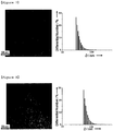
FIG. 7 shows a TEM image of dextran sulfate (DS)-coated iron oxide nanoparticles and a graph showing the hydrodynamic diameter thereof; -
FIG. 8 shows a TEM image of CM-dextran-coated iron oxide nanoparticles and a graph showing the hydrodynamic diameter thereof; -
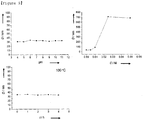
FIG. 9 is of graphs showing the stability test results of the DS-coated iron oxide nanoparticles; -
FIG. 10 is of graphs showing the stability test results of the CM-dextran-coated iron oxide nanoparticles; -
FIG. 11 shows photographs of samples of the CM-dextran-coated iron oxide nanoparticles with respect to changes in pH and a graph showing a zeta potential thereof; -
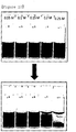
FIG. 12 shows photographs of samples of the CM-dextran-coated iron oxide nanoparticles with respect to changes in NaCl concentration; -
FIG. 13 shows a TEM image of polyacrylic acid-coated iron oxide nanoparticles; -
FIG. 14 shows a TEM image of PEG-phosphate (PO-PEGs)-coated iron oxide nanoparticles and a graph showing the hydrodynamic diameter thereof; -
FIG. 15 is of graphs showing the cell toxicity test results of the DS-coated iron oxide nanoparticles and the CM-dextran-coated iron oxide nanoparticles; -
FIG. 16 shows MRI results using the CM-dextran-coated iron oxide nanoparticles; -
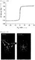
FIG. 17 is a graph showing the magnetic data of the CM-dextran-coated iron oxide nanoparticles; and -
FIG. 18 shows MRI results of animal testing using the CM-dextran-coated iron oxide nanoparticles. - Hereinafter, a detailed description will be given of preferred embodiments of the present invention with reference to the appended drawings.
- Transmission electron microscopy (TEM) was conducted using Jeol EM-2010 microscope, and X-ray diffraction (XRD) was carried out using Rigaku D/Max-3C diffractometer. The hydrodynamic diameter and zeta potential were measured using a particle size analyzer (ELS-Z2, Otsuka), and the M-H curve was determined using a vibrating sample magnetometer (VSM, Quantum Design PPMS).
- Iron oxide nanoparticles were synthesized from an iron oleate complex prepared in a 100 L pilot plant according to the method disclosed in J. Park et al. Nat. Mater. 2004, 4, 891. This iron oleate complex was obtained by reacting sodium oleate with ferric chloride hexahydrate (FeCl36H2O). Specifically, 1.08 kg of ferric chloride hexahydrate and 3.65 kg of sodium oleate were mixed with a solvent mixture comprising 6 L of water, 8 L of ethanol and 14 L of hexane and reacted at about 60°C for 1 hr with vigorous stirring. The transparent lower layer was removed from the separated two layers and the remaining brownish organic layer was mixed with water after which the water layer was removed again to remove the remaining aqueous salt. This water-washing process was repeated three times. The hexane solvent was evaporated from the purified complex solution, thus obtaining an iron oleate complex.
- 3.6 kg (4 mol) of the iron oleate complex thus obtained and 0.57 kg (2 mol) of oleic acid were mixed with 20 kg of 1-octadecene, after which the resulting mixture was heated to 310°C at a heating rate of 10°C/min so that it was allowed to react at 310°C for 60 min, cooled to room temperature so that it was precipitated with a solution mixture having a 1:4 ratio of hexane to acetone, and then centrifuged, yielding iron oxide nanoparticles. Analysis using TEM of the synthesized iron oxide nanoparticles revealed a diameter of 10 nm (
FIG. 2 ). - 50 mg of the iron oxide nanoparticles thus synthesized and 25 g of sodium sulfate (Na2SO4) (average particle size of 150 µm) were stirred in hexane at a weight ratio of 1:500. While the temperature was increased, hexane was slowly evaporated. Thereafter, the powder mixture of nanoparticles and the salt was heated at 500°C for 5 hr under air conditions. After annealing treatment, an excess of the salt was dissolved in distilled water and removed via centrifugation at 13,000 rpm for 30 min. In order to sufficiently remove the salt, washing using distilled water and centrifugation were repeated several times.
- Changes in physical properties of the iron oxide nanoparticles from which the organic material had been removed by annealing treatment using salt particles were analyzed. An increase in particle size due to the agglomeration of the particles during annealing treatment was not observed (
FIG. 3 ). The crystalline structure of the annealed nanoparticles was analyzed via XRD (FIG. 4 ). The XRD pattern of the assynthesized iron oxide nanoparticles was congruous with the XRD pattern of magnetite (Fe3O4, JCPDS 19-0629). As such, JCPDS (Joint Committee on Powder Diffraction Standards) is a file of diffraction data of materials collected and sorted from 1969 in which each of the diffraction patterns is recorded per card, and is thus used as a reference for determining the crystalline structure of a material from the measured XRD pattern. The XRD pattern of the nanoparticles after annealing treatment was similar to the XRD pattern of the nanoparticles before annealing treatment. Because small nanoparticles have a tendency to increase the width of the XRD peak, magnetite and maghemite (γ-Fe2O3, JCPDS 25-1402) having similar XRD patterns are difficult to clearly distinguish from each other via XRD analysis. Typically when magnetite is annealed under air conditions, it may become maghemite or hematite (α-Fe2O3) via additional oxidation. However, the XRD pattern of hematite (JCPDS 24-0072) is apparently different from that of magnetite or maghemite. The iron oxide nanoparticles after annealing treatment are brownish, whereas the iron oxide nanoparticles before annealing treatment are black in color (FIG. 5 ). The annealed iron oxide nanoparticles appear to be maghemite via additional oxidation. In order to analyze changes in magnetism before and after annealing treatment, magnetism of the samples was measured using VSM. The saturation magnetization was 78 emu/g [Fe] before annealing treatment, and was 103 emu/g [Fe] after annealing treatment (FIG. 6 ). This is considered to be because crystallinity of the particles is increased via the high-temperature annealing treatment thus affecting the increase in magnetism. - The annealed iron oxide nanoparticles were dispersed in an aqueous solution to thus increase the pH of the aqueous solution, so that the zeta potential of the iron oxide nanoparticles was observed to change from a positive charge to a negative charge, and accordingly the hydrodynamic diameter was also changed (Table 1). At
pH 7, the zeta potential of the annealed nanoparticles is small and the particles have a tendency to agglomerate and sink because of the absence of a surfactant for stabilizing the surface thereof. The hydrodynamic diameter of the nanoparticles measured atpH 7 was observed to exceed 1 µm, unlike under acidic or basic conditions (Table 1). -
[Table 1] pH Hydrodynamic Diameter (nm) Zeta Potential (mV) 3 39.2 38.6 7 1555.9 -10.73 10 76.5 -34.99 - A mixture comprising iron oxide nanoparticles and salt particles was annealed in the same manner as in Example 1 with the exception that the iron oxide nanoparticles were coated with a hydrophilic material. Specifically, 1 g of a hydrophilic material (Table 2) was dissolved in 250 ml of distilled water. While the aqueous solution of hydrophilic material was stirred, 12.5 g of annealed iron oxide nanoparticles and the salt powder were slowly added at a weight ratio of 1:12.5. The iron oxide nanoparticles coated with hydrophilic material was separated via centrifugation at 13,000 rpm for 30 min. Additional washing using the aqueous solution of hydrophilic material and centrifugation were repeated two times more.
-
[Table 2] Kind Molecular Weight Carboxymethyl dextran sodium salt (CM-dextran) ∼ 12,000 Dextran ∼ 1,500 Diethyl aminoethyl-dextran hydrochloride (DEAE-dextran) ∼ 500,000 Dextran sulfate sodium salt (DS) ∼ 4,000 Polyacrylic acid 1,800 PEG 2,805 PO-PEGs 2,000 (mPEG) - In order to measure the shape and diameter of the iron oxide nanoparticles coated with hydrophilic material, TEM and DLS analysis were conducted. The iron oxide nanoparticles coated with positively charged DEAE-dextran were efficiently dispersed in the aqueous solution, and the hydrodynamic diameter thereof was 84.3 nm (Table 3). However, the coating efficiency of commercially available DEAE-dextran was low because of the large molecular weight. Among the negatively charged dextran derivatives, DS and CM-dextran were used for coating of the nanoparticles, and the coated iron oxide nanoparticles were stable in an aqueous solution for more than one month. The TEM images show no agglomeration of the particles in the course of coating with DS and CM-dextran (
FIGS. 7 and 8 ). The hydrodynamic diameter of the DS-coated iron oxide nanoparticles was 36.8 nm (Table 3), which was similar to the value (28.7 nm) of the PEG-phospholipid-coated iron oxide nanoparticles (Table 3). However, the hydrodynamic diameter of the CM-dextran-coated iron oxide nanoparticles was 66.4 nm, which was much larger than that of the DS-coated or the PEG-phospholipid-coated iron oxide nanoparticles (Table 3). Hence, in the case of CM-dextran, one or more nanoparticles are considered to be coated together. For comparison, dextran having no charge was used for coating the nanoparticles. The hydrodynamic diameter of the coated particles exceeded 100 nm (Table 3), and such particles were observed to easily sink over time. From this, the electrostatic action between the nanoparticles and the coating material is regarded as affecting the coating efficiency of particles and the stability. Among a variety of kinds of dextran derivatives, negatively charged dextran was efficient at coating, and particularly the DS-coated nanoparticles represented the smallest hydrodynamic diameter. - In order to validate the colloidal stability of the iron oxide nanoparticles coated with hydrophilic material, changes in diameter of the iron oxide nanoparticles upon changes in pH and upon heating were observed. The DS-coated iron oxide nanoparticles were stable in the pH range of 4 to 11 (
FIG. 9 ). Also, the DS-coated iron oxide nanoparticles had no size change at 100°C for 4 hr. However, the DS-coated iron oxide nanoparticles were unstable in the NaCl solution and thus observed to sink. The CM-dextran-coated iron oxide nanoparticles were stable in the pH range of 4 to 13, but the nanoparticles became unstable atpH 3 and were observed to rapidly sink (FIGS. 10 and 11 ). Measuring the zeta potential showed that the nanoparticles which had been stable had a value of -23.74 ∼ -36.88 mV but the potential value was 1.81 mV at pH 3 (FIG. 11 ). These results show that aboutpH 3 is regarded as an isoelectric point and the zeta potential is lowered whereby the nanoparticles become unstable. Thus, the electrostatic action (repulsive force) is seen to contribute to the stability of the nanoparticles. The CM-dextran-coated iron oxide nanoparticles also had no size change at 100°C for 4 hr. Such nanoparticles were stable up to the NaCl concentration of 0.15 M (FIGS. 10 and12 ). Thereby, the stability of the CM-dextran-coated nanoparticles can be seen to be suitable for medical applications. The DS-coated nanoparticles are unstable in the NaCl solution and thus are not suitable for medical applications. - Because a polyacrylic acid (Mw 1,800) solution is acidic, the iron oxide nanoparticles coated with polyacrylic acid have low dispersibility in an aqueous solution immediately after removal of the salt. As the pH is increased by the addition of sodium hydroxide (NaOH), dispersibility is improved and the hydrodynamic diameter is decreased (Table 4). This is because the carboxylic acid that forms the polyacrylic acid is negatively charged under basic conditions. The hydrodynamic diameter was 91.9 nm under acidic conditions, 59.4 nm under neutral conditions and 39.9 nm under basic conditions. As shown in the TEM image, the particles did not agglomerate while being coated with polyacrylic acid (
FIG. 13 ). - The PEG-phosphate (PO-PEGs)-coated iron oxide nanoparticles were well dispersed in an aqueous solution. As shown in the TEM image, the particles did not agglomerate while being coated with PO-PEGs and had a hydrodynamic diameter of 69.2 nm (
FIG. 14 ). - DS-coated iron oxide nanoparticles were prepared in the same manner as in Example 2 with the exception that the size of salt particles was 13 µm. The hydrodynamic diameter of the DS-coated iron oxide nanoparticles was 31.03 nm (Table 3).
-
[Table 3] Hydrophilic Material Hydrodynamic Diameter(nm) Ex. 2 DEAE-dextran 84.3 Ex. 2 DS 36.8 Ex. 2 Dextran 157.0 Ex. 2 CM-dextran 66.4 Ex. 2 PEG-phospholipid 28.7 Ex. 3 DS (salt particle size: 13 µm) 31.03 -
[Table 4] pH Hydrodynamic Diameter (nm) 2 91.9 8 59.4 12 39.9 - The DS-coated and the CM-dextran-coated iron oxide nanoparticles were measured using MTT method. The iron oxide nanoparticles at different concentrations were cultured in cells for 48 hr, after which the survival rate of cells was measured. Consequently, almost all of the cells were alive up to the concentration of 200 µg [Fe]/ml (
FIG. 15 ). - The iron oxide nanoparticles were coated using CM-dextran instead of DS because of superior stability. The T2 MRI image of the nanoparticles dispersed in the aqueous solution shows a decrease in signal in proportion to an increase in the concentration of iron ions (
FIG. 16 ). To evaluate the contrast effects, r2 relaxivity was calculated, which is a gradient of a reciprocal of T2 relaxation time relative to a concentration of the contrast agent. This is an indicator that shows how much the T2 relaxation time sensitively varies depending on changes in concentration of the contrast agent. he r2 relaxivity was calculated from 1/T2 = 1/T20 + r2∗C wherein T2 is the T2 relaxation time, T20 is the initial T2 relaxation time, and C is the concentration of the contrast agent, n the graph plot wherein the concentration of the contrast agent including the iron oxide nanoparticles coated with hydrophilic material is set to the x-axis and the 1/T2 measured at the above concentration is set to the γ-axis, r2 is derived from the gradient, wherein r2 designates the r2 relaxivity. The relaxivity of the iron oxide nanoparticles coated with PEG-phospholipid without annealing treatment was 124.46 mM-1s-1 and that of the iron oxide nanoparticles coated with CM-dextran after annealing treatment was 358.9 mM-1s-1 which is an increase of 2.9 times. Such a high relaxivity is based on increased crystallinity during annealing treatment and coating of a plurality of particles with a single dextran shell in the course of coating with CM-dextran. As described in Example 1, the saturation magnetization after annealing treatment was increased because of additional crystallization. Even after coating with CM-dextran, the saturation magnetization (106 emu/g [Fe]) was similar to that after annealing treatment (FIG. 17 ). When the dispersed superparamagnetic nanoparticles agglomerated, the increase in r2 relaxivity was reported to be based on the magnetic relaxation switch (J. M. Perez, et. al. Chem. Bio. Chem. 2004, 5, 261). When a material having high contrast effect is used in this way, the amount of introduced nanoparticles in vivo may be decreased, thus lowering the toxic effects. Hence, the CM-dextran-coated iron oxide nanoparticles are determined to be useful as a biocompatible MRI contrast agent having high contrast effects. - The CM-dextran-coated iron oxide nanoparticles were applied to animal MRI testing. The nanoparticles (2.5 mg [Fe] per kg of mouse body weight) were injected via the tail vein of the mouse. The MRI results before and after the injection of the nanoparticles show that the nanoparticles accumulate in the liver and the spleen thus achieving effective contrast (
FIG. 18 ). These nanoparticles were confirmed to be suitable for application in vivo.
Claims (17)
- A method of preparing iron oxide nanoparticles coated with a hydrophilic material, comprising:a) attaching magnetic iron oxide nanoparticles coated with a hydrophobic organic material onto salt particles, annealing a powder mixture comprising the magnetic iron oxide nanoparticles coated with the hydrophobic organic material and the salt particles under air conditions, removing the hydrophobic organic material from the surface of the magnetic iron oxide nanoparticles by the annealing, and thus obtaining iron oxide nanoparticles having no organic material; andb) coating a surface of the iron oxide nanoparticles having no organic material with a hydrophilic material.
- The method of claim 1, wherein the hydrophobic organic material attached to the iron oxide nanoparticles in a) is at least one selected from the group consisting of a C4∼C25 fatty acid, a C4∼C25 aliphatic alcohol, and a C4∼C25 aliphatic amine.
- The method of claim 2, wherein the fatty acid attached to the iron oxide nanoparticles in a) is oleic acid.
- The method of claim 1, wherein the salt particles in a) is at least one selected from sodium sulfate (Na2SO4), sodium bicarbonate (NaHCO3), potassium bicarbonate (KHCO3), calcium bicarbonate (Ca(HCO3)2), sodium chloride (NaCl), potassium chloride (KC1) and calcium chloride (CaCl2).
- The method of claim 1, wherein the salt particles in a) have a diameter of 1 ∼ 500 µm.
- The method of claim 1, wherein the iron oxide nanoparticles in a) are obtained by reacting an iron complex comprising iron as a center atom and a C4∼C25 organic acid group (carboxylate) attached thereto as a ligand with a C4∼C25 fatty acid.
- The method of claim 6, wherein the iron complex is iron oleate.
- The method of claim 6, wherein the fatty acid is oleic acid.
- The method of claim 1, wherein the iron oxide nanoparticles in a) have a diameter of 1 ∼ 100 nm.
- The method of claim 1, wherein the iron oxide nanoparticles and the salt particles in a) have a weight ratio ranging from 1:10 to 1:1000.
- The method of claim 1, wherein the annealing in a) is performed by carrying out heating at 400 ∼ 600°C for 1 ∼ 10 hr in an air atmosphere.
- The method of claim 1, wherein, after annealing in a), removing the salt particles is additionally performed.
- The method of claim 12, wherein the removing the salt in a) is performed using centrifugation.
- The method of claim 12, wherein the temperature is decreased to room temperature before removing the salt after annealing in a).
- The method of claim 1, wherein the hydrophilic material in b) is a dextran derivative, polyacrylic acid, starch, silica, polyethyleneglycol (PEG) or PEG-phosphate (PO-PEGs).
- The method of claim 15, wherein the dextran derivative which is the hydrophilic material in b) is diethylaminoethyl-dextran (DEAE-dextran), dextran sulfate (DS) or carboxymethyl dextran (CM-doxträn).
- The method of claim 1, wherein in b) coating with the hydrophilic material and removing the salt are simultaneously performed.
Applications Claiming Priority (2)
| Application Number | Priority Date | Filing Date | Title |
|---|---|---|---|
| KR1020110011294A KR101642903B1 (en) | 2011-02-09 | 2011-02-09 | Preparation of hydrophilic material coated iron oxide nanoparticles and magnetic resonance contrast agent using thereof |
| PCT/KR2012/000845 WO2012108648A2 (en) | 2011-02-09 | 2012-02-06 | Method of preparing iron oxide nanoparticles coated with hydrophilic material, and magnetic resonance imaging contrast agent using the same |
Publications (3)
| Publication Number | Publication Date |
|---|---|
| EP2673006A2 EP2673006A2 (en) | 2013-12-18 |
| EP2673006A4 EP2673006A4 (en) | 2016-07-20 |
| EP2673006B1 true EP2673006B1 (en) | 2020-12-30 |
Family
ID=46639034
Family Applications (1)
| Application Number | Title | Priority Date | Filing Date |
|---|---|---|---|
| EP12744201.0A Active EP2673006B1 (en) | 2011-02-09 | 2012-02-06 | Method of preparing iron oxide nanoparticles coated with hydrophilic material, and magnetic resonance imaging contrast agent using the same |
Country Status (6)
| Country | Link |
|---|---|
| US (1) | US9352058B2 (en) |
| EP (1) | EP2673006B1 (en) |
| JP (1) | JP5701408B2 (en) |
| KR (1) | KR101642903B1 (en) |
| CN (1) | CN103347543B (en) |
| WO (1) | WO2012108648A2 (en) |
Families Citing this family (23)
| Publication number | Priority date | Publication date | Assignee | Title |
|---|---|---|---|---|
| US10987436B2 (en) * | 2012-01-27 | 2021-04-27 | Soluciones Nanotecnológicas, S.L. | Superparamagnetic nanoparticles as a contrast agent for magnetic resonance imaging (MRI) of magnetic susceptibility (T2*) |
| KR101384977B1 (en) * | 2013-08-20 | 2014-04-21 | 연세대학교 산학협력단 | Nmps solubilization apparatus having coaxial nozzle |
| KR101508281B1 (en) * | 2013-12-06 | 2015-07-09 | 한화케미칼 주식회사 | Method for preparation of uniform metal oxide nanoparticles with high reproducibility |
| WO2015088254A1 (en) | 2013-12-10 | 2015-06-18 | 김태구 | Simplified method for placing telephone call and simplified method for requesting location-based service |
| IN2014CH00598A (en) * | 2014-02-10 | 2015-08-14 | Psg Institutions | |
| WO2016007194A1 (en) * | 2014-07-10 | 2016-01-14 | Gerald Lee Wolf | Companion nanoparticles for theranosis of macrophage-dependent diseases |
| KR101765335B1 (en) * | 2014-09-26 | 2017-08-22 | 서울대학교산학협력단 | MRI Contrast Agent for Contrasting Cancer Cell |
| BR112017006552A2 (en) | 2014-10-03 | 2017-12-19 | Ntercept Llc | compositions and methods for inhibiting the biological activity of soluble biomolecules |
| US9861710B1 (en) * | 2015-01-16 | 2018-01-09 | Verily Life Sciences Llc | Composite particles, methods, and in vivo diagnostic system |
| JP6538165B2 (en) * | 2015-06-29 | 2019-07-03 | 株式会社Screenホールディングス | Pigment composition and aqueous ink composition for inkjet |
| WO2017176762A1 (en) | 2016-04-06 | 2017-10-12 | Nanotics, Llc | Particles comprising subparticles or nucleic acid scaffolds |
| JP7241355B2 (en) | 2017-11-06 | 2023-03-17 | コニカミノルタ株式会社 | COMPOSITE PARTICLE FOR CONTRAST IMAGING, METHOD FOR MANUFACTURING COMPOSITE PARTICLE, CELL, CELL STRUCTURE AND MIXED DISPERSION |
| JP7041952B2 (en) * | 2018-04-27 | 2022-03-25 | 学校法人 名城大学 | Method for Producing Polysaccharide Chemically Modified Metal Oxide Particles |
| CA3124753A1 (en) * | 2018-12-27 | 2020-07-02 | Astellas Pharma Inc. | Method for producing nanoparticle having metal particle which contains iron oxide to which one or more hydrophilic ligands are coordination bonded |
| JP7551446B2 (en) * | 2019-10-30 | 2024-09-17 | キヤノン株式会社 | Composition and heat transport device |
| WO2021085315A1 (en) * | 2019-10-30 | 2021-05-06 | キヤノン株式会社 | Composition and heat transport device |
| JP7551445B2 (en) * | 2019-10-30 | 2024-09-17 | キヤノン株式会社 | Composition and heat transport device |
| CN110853865B (en) * | 2019-11-20 | 2022-05-17 | 江南大学 | Preparation method and application of ferroferric oxide single-layer nano film |
| KR102610148B1 (en) * | 2019-12-12 | 2023-12-06 | 주식회사 엘지화학 | Magnetic body |
| KR102640683B1 (en) * | 2020-12-29 | 2024-02-27 | 주식회사 지티아이바이오사이언스 | Iron oxide magnetic particles |
| WO2022145970A1 (en) * | 2020-12-29 | 2022-07-07 | 주식회사 지티아이바이오사이언스 | Iron oxide magnetic particles |
| EP4403192A4 (en) * | 2021-09-18 | 2025-07-30 | Nonsuch Biotechnology Inc | Use of iron oxide nanoparticles in the production of a parathyroid and/or lymph node contrast agent |
| KR102681399B1 (en) * | 2021-10-21 | 2024-07-04 | 주식회사 지티아이바이오사이언스 | A method for preparing a composition for treating liver cancer comprising magnetic iron oxide |
Family Cites Families (11)
| Publication number | Priority date | Publication date | Assignee | Title |
|---|---|---|---|---|
| JP2726520B2 (en) * | 1989-10-20 | 1998-03-11 | 名糖産業株式会社 | Organic magnetic composite |
| JPH03141119A (en) | 1989-10-25 | 1991-06-17 | Nippon Telegr & Teleph Corp <Ntt> | Production of fine magnetic particle |
| JP2006016236A (en) * | 2004-06-30 | 2006-01-19 | Sumitomo Osaka Cement Co Ltd | Method for producing metal oxide nanoparticle |
| JP2009114066A (en) * | 2006-02-24 | 2009-05-28 | International Medical Center Of Japan | Organic magnetic nanocomposites with functional molecules |
| KR100819378B1 (en) * | 2006-02-24 | 2008-04-04 | (주)에이티젠 | Magnetic nanocomposite using amphiphilic compound and pharmaceutical composition comprising the same |
| CZ301067B6 (en) * | 2006-02-24 | 2009-10-29 | Ústav makromolekulární chemie AV CR | Iron oxide-based superparamagnetic nanoparticles with modified surface, process of their preparation and use |
| EP2062271A2 (en) * | 2006-09-05 | 2009-05-27 | Columbus Nanoworks, Inc. | Magnetic particles and methods of making and using the same |
| US8852555B2 (en) | 2007-07-26 | 2014-10-07 | Tokyo Institute Of Technology | Process for production of surface-coated inorganic particles |
| AU2009346580B2 (en) * | 2009-05-21 | 2014-01-23 | Institute Of Life Sciences | Water dispersible glyceryl monooleate magnetic nanoparticle formulation |
| GB2472446A (en) * | 2009-08-07 | 2011-02-09 | Ct Fuer Angewandte Nanotechnologie | Metal oxide particles coated with polyethylene glycol and their synthesis |
| CN103025314A (en) * | 2010-05-26 | 2013-04-03 | 通用医疗公司 | Magnetic nanoparticles |
-
2011
- 2011-02-09 KR KR1020110011294A patent/KR101642903B1/en active Active
-
2012
- 2012-02-06 WO PCT/KR2012/000845 patent/WO2012108648A2/en not_active Ceased
- 2012-02-06 JP JP2013553345A patent/JP5701408B2/en active Active
- 2012-02-06 CN CN201280008230.1A patent/CN103347543B/en active Active
- 2012-02-06 US US13/983,367 patent/US9352058B2/en active Active
- 2012-02-06 EP EP12744201.0A patent/EP2673006B1/en active Active
Non-Patent Citations (1)
| Title |
|---|
| None * |
Also Published As
| Publication number | Publication date |
|---|---|
| EP2673006A4 (en) | 2016-07-20 |
| JP2014511324A (en) | 2014-05-15 |
| US20130323182A1 (en) | 2013-12-05 |
| EP2673006A2 (en) | 2013-12-18 |
| WO2012108648A3 (en) | 2012-11-01 |
| WO2012108648A2 (en) | 2012-08-16 |
| US9352058B2 (en) | 2016-05-31 |
| CN103347543B (en) | 2016-11-16 |
| CN103347543A (en) | 2013-10-09 |
| KR20120091513A (en) | 2012-08-20 |
| JP5701408B2 (en) | 2015-04-15 |
| KR101642903B1 (en) | 2016-07-27 |
Similar Documents
| Publication | Publication Date | Title |
|---|---|---|
| US9352058B2 (en) | Method of preparing iron oxide nanoparticles coated with hydrophilic material, and magnetic resonance imaging contrast agent using the same | |
| Khmara et al. | Chitosan-stabilized iron oxide nanoparticles for magnetic resonance imaging | |
| Liu et al. | Highly water‐dispersible biocompatible magnetite particles with low cytotoxicity stabilized by citrate groups | |
| Togashi et al. | Surfactant-assisted one-pot synthesis of superparamagnetic magnetite nanoparticle clusters with tunable cluster size and magnetic field sensitivity | |
| Cho et al. | Gold-coated iron nanoparticles: a novel magnetic resonance agent for T 1 and T 2 weighted imaging | |
| Hu et al. | Facile synthesis of ultrasmall PEGylated iron oxide nanoparticles for dual-contrast T 1-and T 2-weighted magnetic resonance imaging | |
| Shi et al. | Mesoporous superparamagnetic cobalt ferrite nanoclusters: Synthesis, characterization and application in drug delivery | |
| US7960025B2 (en) | Magnetic nanoparticles comprising a core formed from noble metals | |
| Digigow et al. | Preparation and characterization of functional silica hybrid magnetic nanoparticles | |
| CN103153348B (en) | The extremely little and preparation of the iron oxide based paramagnetism of uniform-dimension or pseudo-paramagnetic nanoparticle and use the MRI T1 developing agent of described nano-particle | |
| JP5765520B2 (en) | Method for producing aqueous dispersion containing magnetic particles | |
| Lu et al. | Solid-state synthesis of monocrystalline iron oxide nanoparticle based ferrofluid suitable for magnetic resonance imaging contrast application | |
| Liu et al. | Preparation, characterization and MRI application of carboxymethyl dextran coated magnetic nanoparticles | |
| Park et al. | Transformation of hydrophobic iron oxide nanoparticles to hydrophilic and biocompatible maghemite nanocrystals for use as highly efficient MRI contrast agent | |
| Miola et al. | Reductant-free synthesis of magnetoplasmonic iron oxide-gold nanoparticles | |
| CN116370657B (en) | Chiral iron-based super-particle nano material and preparation method and application thereof | |
| Yue et al. | Ultrasmall europium, gadolinium, and dysprosium oxide nanoparticles: Polyol synthesis, properties, and biomedical imaging applications | |
| EP2942064B1 (en) | Mri contrast agent including t1 contrast material coated on surface of nanoparticle support | |
| Saboktakin et al. | Synthesis and characterization of superparamagnetic nanoparticles coated with carboxymethyl starch (CMS) for magnetic resonance imaging technique | |
| Leonel et al. | Facile polyol synthesis of ultrasmall water-soluble cobalt ferrite nanoparticles | |
| Mansur et al. | Synthesis and characterization of iron oxide superparticles with various polymers | |
| Ene et al. | Single Step Synthesis of Glutamic/tartaric Acidstabilised Fe3O4 Nanoparticles for Targeted Delivery Systems | |
| Popescu et al. | Hydrothermal synthesis of nanostructured hybrids based on iron oxide and branched PEI polymers. Influence of high pressure on structure and morphology | |
| CN114377159A (en) | Low-nephrotoxicity protein-iron oxide composite nano magnetic resonance contrast agent as well as preparation method and application thereof | |
| Xia et al. | Preparation of magnetite–dextran microspheres by ultrasonication |
Legal Events
| Date | Code | Title | Description |
|---|---|---|---|
| PUAI | Public reference made under article 153(3) epc to a published international application that has entered the european phase |
Free format text: ORIGINAL CODE: 0009012 |
|
| 17P | Request for examination filed |
Effective date: 20130830 |
|
| AK | Designated contracting states |
Kind code of ref document: A2 Designated state(s): AL AT BE BG CH CY CZ DE DK EE ES FI FR GB GR HR HU IE IS IT LI LT LU LV MC MK MT NL NO PL PT RO RS SE SI SK SM TR |
|
| DAX | Request for extension of the european patent (deleted) | ||
| A4 | Supplementary search report drawn up and despatched |
Effective date: 20160616 |
|
| RIC1 | Information provided on ipc code assigned before grant |
Ipc: A61K 47/30 20060101ALI20160610BHEP Ipc: A61K 49/04 20060101ALI20160610BHEP Ipc: A61K 9/28 20060101ALI20160610BHEP Ipc: A61K 49/18 20060101AFI20160610BHEP Ipc: A61K 9/16 20060101ALI20160610BHEP |
|
| STAA | Information on the status of an ep patent application or granted ep patent |
Free format text: STATUS: EXAMINATION IS IN PROGRESS |
|
| 17Q | First examination report despatched |
Effective date: 20190313 |
|
| RIC1 | Information provided on ipc code assigned before grant |
Ipc: C09C 1/24 20060101ALI20200610BHEP Ipc: A61K 49/04 20060101ALI20200610BHEP Ipc: C01G 49/02 20060101ALI20200610BHEP Ipc: A61K 9/28 20060101ALI20200610BHEP Ipc: A61K 49/18 20060101AFI20200610BHEP Ipc: A61K 9/16 20060101ALI20200610BHEP Ipc: B82Y 30/00 20110101ALI20200610BHEP Ipc: A61K 47/30 20060101ALI20200610BHEP |
|
| GRAP | Despatch of communication of intention to grant a patent |
Free format text: ORIGINAL CODE: EPIDOSNIGR1 |
|
| STAA | Information on the status of an ep patent application or granted ep patent |
Free format text: STATUS: GRANT OF PATENT IS INTENDED |
|
| RIN1 | Information on inventor provided before grant (corrected) |
Inventor name: HYEON, TAEGHWAN Inventor name: PARK, YONG IL Inventor name: PIAO, YUANZHE |
|
| INTG | Intention to grant announced |
Effective date: 20200721 |
|
| RIN1 | Information on inventor provided before grant (corrected) |
Inventor name: PIAO, YUANZHE Inventor name: PARK, YONG IL Inventor name: HYEON, TAEGHWAN |
|
| GRAS | Grant fee paid |
Free format text: ORIGINAL CODE: EPIDOSNIGR3 |
|
| GRAA | (expected) grant |
Free format text: ORIGINAL CODE: 0009210 |
|
| STAA | Information on the status of an ep patent application or granted ep patent |
Free format text: STATUS: THE PATENT HAS BEEN GRANTED |
|
| AK | Designated contracting states |
Kind code of ref document: B1 Designated state(s): AL AT BE BG CH CY CZ DE DK EE ES FI FR GB GR HR HU IE IS IT LI LT LU LV MC MK MT NL NO PL PT RO RS SE SI SK SM TR |
|
| REG | Reference to a national code |
Ref country code: GB Ref legal event code: FG4D |
|
| REG | Reference to a national code |
Ref country code: DE Ref legal event code: R096 Ref document number: 602012073943 Country of ref document: DE |
|
| REG | Reference to a national code |
Ref country code: AT Ref legal event code: REF Ref document number: 1349282 Country of ref document: AT Kind code of ref document: T Effective date: 20210115 |
|
| REG | Reference to a national code |
Ref country code: IE Ref legal event code: FG4D |
|
| PG25 | Lapsed in a contracting state [announced via postgrant information from national office to epo] |
Ref country code: RS Free format text: LAPSE BECAUSE OF FAILURE TO SUBMIT A TRANSLATION OF THE DESCRIPTION OR TO PAY THE FEE WITHIN THE PRESCRIBED TIME-LIMIT Effective date: 20201230 Ref country code: FI Free format text: LAPSE BECAUSE OF FAILURE TO SUBMIT A TRANSLATION OF THE DESCRIPTION OR TO PAY THE FEE WITHIN THE PRESCRIBED TIME-LIMIT Effective date: 20201230 Ref country code: NO Free format text: LAPSE BECAUSE OF FAILURE TO SUBMIT A TRANSLATION OF THE DESCRIPTION OR TO PAY THE FEE WITHIN THE PRESCRIBED TIME-LIMIT Effective date: 20210330 Ref country code: GR Free format text: LAPSE BECAUSE OF FAILURE TO SUBMIT A TRANSLATION OF THE DESCRIPTION OR TO PAY THE FEE WITHIN THE PRESCRIBED TIME-LIMIT Effective date: 20210331 |
|
| REG | Reference to a national code |
Ref country code: AT Ref legal event code: MK05 Ref document number: 1349282 Country of ref document: AT Kind code of ref document: T Effective date: 20201230 |
|
| PG25 | Lapsed in a contracting state [announced via postgrant information from national office to epo] |
Ref country code: LV Free format text: LAPSE BECAUSE OF FAILURE TO SUBMIT A TRANSLATION OF THE DESCRIPTION OR TO PAY THE FEE WITHIN THE PRESCRIBED TIME-LIMIT Effective date: 20201230 Ref country code: SE Free format text: LAPSE BECAUSE OF FAILURE TO SUBMIT A TRANSLATION OF THE DESCRIPTION OR TO PAY THE FEE WITHIN THE PRESCRIBED TIME-LIMIT Effective date: 20201230 Ref country code: BG Free format text: LAPSE BECAUSE OF FAILURE TO SUBMIT A TRANSLATION OF THE DESCRIPTION OR TO PAY THE FEE WITHIN THE PRESCRIBED TIME-LIMIT Effective date: 20210330 |
|
| REG | Reference to a national code |
Ref country code: NL Ref legal event code: MP Effective date: 20201230 |
|
| PG25 | Lapsed in a contracting state [announced via postgrant information from national office to epo] |
Ref country code: HR Free format text: LAPSE BECAUSE OF FAILURE TO SUBMIT A TRANSLATION OF THE DESCRIPTION OR TO PAY THE FEE WITHIN THE PRESCRIBED TIME-LIMIT Effective date: 20201230 |
|
| REG | Reference to a national code |
Ref country code: LT Ref legal event code: MG9D |
|
| PG25 | Lapsed in a contracting state [announced via postgrant information from national office to epo] |
Ref country code: LT Free format text: LAPSE BECAUSE OF FAILURE TO SUBMIT A TRANSLATION OF THE DESCRIPTION OR TO PAY THE FEE WITHIN THE PRESCRIBED TIME-LIMIT Effective date: 20201230 Ref country code: PT Free format text: LAPSE BECAUSE OF FAILURE TO SUBMIT A TRANSLATION OF THE DESCRIPTION OR TO PAY THE FEE WITHIN THE PRESCRIBED TIME-LIMIT Effective date: 20210430 Ref country code: RO Free format text: LAPSE BECAUSE OF FAILURE TO SUBMIT A TRANSLATION OF THE DESCRIPTION OR TO PAY THE FEE WITHIN THE PRESCRIBED TIME-LIMIT Effective date: 20201230 Ref country code: SK Free format text: LAPSE BECAUSE OF FAILURE TO SUBMIT A TRANSLATION OF THE DESCRIPTION OR TO PAY THE FEE WITHIN THE PRESCRIBED TIME-LIMIT Effective date: 20201230 Ref country code: EE Free format text: LAPSE BECAUSE OF FAILURE TO SUBMIT A TRANSLATION OF THE DESCRIPTION OR TO PAY THE FEE WITHIN THE PRESCRIBED TIME-LIMIT Effective date: 20201230 Ref country code: CZ Free format text: LAPSE BECAUSE OF FAILURE TO SUBMIT A TRANSLATION OF THE DESCRIPTION OR TO PAY THE FEE WITHIN THE PRESCRIBED TIME-LIMIT Effective date: 20201230 Ref country code: NL Free format text: LAPSE BECAUSE OF FAILURE TO SUBMIT A TRANSLATION OF THE DESCRIPTION OR TO PAY THE FEE WITHIN THE PRESCRIBED TIME-LIMIT Effective date: 20201230 |
|
| PG25 | Lapsed in a contracting state [announced via postgrant information from national office to epo] |
Ref country code: PL Free format text: LAPSE BECAUSE OF FAILURE TO SUBMIT A TRANSLATION OF THE DESCRIPTION OR TO PAY THE FEE WITHIN THE PRESCRIBED TIME-LIMIT Effective date: 20201230 Ref country code: AT Free format text: LAPSE BECAUSE OF FAILURE TO SUBMIT A TRANSLATION OF THE DESCRIPTION OR TO PAY THE FEE WITHIN THE PRESCRIBED TIME-LIMIT Effective date: 20201230 |
|
| PG25 | Lapsed in a contracting state [announced via postgrant information from national office to epo] |
Ref country code: MC Free format text: LAPSE BECAUSE OF FAILURE TO SUBMIT A TRANSLATION OF THE DESCRIPTION OR TO PAY THE FEE WITHIN THE PRESCRIBED TIME-LIMIT Effective date: 20201230 Ref country code: IS Free format text: LAPSE BECAUSE OF FAILURE TO SUBMIT A TRANSLATION OF THE DESCRIPTION OR TO PAY THE FEE WITHIN THE PRESCRIBED TIME-LIMIT Effective date: 20210430 |
|
| REG | Reference to a national code |
Ref country code: DE Ref legal event code: R097 Ref document number: 602012073943 Country of ref document: DE |
|
| REG | Reference to a national code |
Ref country code: BE Ref legal event code: MM Effective date: 20210228 |
|
| PG25 | Lapsed in a contracting state [announced via postgrant information from national office to epo] |
Ref country code: LU Free format text: LAPSE BECAUSE OF NON-PAYMENT OF DUE FEES Effective date: 20210206 Ref country code: LI Free format text: LAPSE BECAUSE OF NON-PAYMENT OF DUE FEES Effective date: 20210228 Ref country code: IT Free format text: LAPSE BECAUSE OF FAILURE TO SUBMIT A TRANSLATION OF THE DESCRIPTION OR TO PAY THE FEE WITHIN THE PRESCRIBED TIME-LIMIT Effective date: 20201230 Ref country code: AL Free format text: LAPSE BECAUSE OF FAILURE TO SUBMIT A TRANSLATION OF THE DESCRIPTION OR TO PAY THE FEE WITHIN THE PRESCRIBED TIME-LIMIT Effective date: 20201230 Ref country code: CH Free format text: LAPSE BECAUSE OF NON-PAYMENT OF DUE FEES Effective date: 20210228 |
|
| PLBE | No opposition filed within time limit |
Free format text: ORIGINAL CODE: 0009261 |
|
| STAA | Information on the status of an ep patent application or granted ep patent |
Free format text: STATUS: NO OPPOSITION FILED WITHIN TIME LIMIT |
|
| PG25 | Lapsed in a contracting state [announced via postgrant information from national office to epo] |
Ref country code: DK Free format text: LAPSE BECAUSE OF FAILURE TO SUBMIT A TRANSLATION OF THE DESCRIPTION OR TO PAY THE FEE WITHIN THE PRESCRIBED TIME-LIMIT Effective date: 20201230 Ref country code: ES Free format text: LAPSE BECAUSE OF FAILURE TO SUBMIT A TRANSLATION OF THE DESCRIPTION OR TO PAY THE FEE WITHIN THE PRESCRIBED TIME-LIMIT Effective date: 20201230 |
|
| 26N | No opposition filed |
Effective date: 20211001 |
|
| PG25 | Lapsed in a contracting state [announced via postgrant information from national office to epo] |
Ref country code: IE Free format text: LAPSE BECAUSE OF NON-PAYMENT OF DUE FEES Effective date: 20210206 |
|
| PG25 | Lapsed in a contracting state [announced via postgrant information from national office to epo] |
Ref country code: SI Free format text: LAPSE BECAUSE OF FAILURE TO SUBMIT A TRANSLATION OF THE DESCRIPTION OR TO PAY THE FEE WITHIN THE PRESCRIBED TIME-LIMIT Effective date: 20201230 |
|
| PG25 | Lapsed in a contracting state [announced via postgrant information from national office to epo] |
Ref country code: IS Free format text: LAPSE BECAUSE OF FAILURE TO SUBMIT A TRANSLATION OF THE DESCRIPTION OR TO PAY THE FEE WITHIN THE PRESCRIBED TIME-LIMIT Effective date: 20210430 |
|
| PG25 | Lapsed in a contracting state [announced via postgrant information from national office to epo] |
Ref country code: BE Free format text: LAPSE BECAUSE OF NON-PAYMENT OF DUE FEES Effective date: 20210228 |
|
| PG25 | Lapsed in a contracting state [announced via postgrant information from national office to epo] |
Ref country code: HU Free format text: LAPSE BECAUSE OF FAILURE TO SUBMIT A TRANSLATION OF THE DESCRIPTION OR TO PAY THE FEE WITHIN THE PRESCRIBED TIME-LIMIT; INVALID AB INITIO Effective date: 20120206 Ref country code: CY Free format text: LAPSE BECAUSE OF FAILURE TO SUBMIT A TRANSLATION OF THE DESCRIPTION OR TO PAY THE FEE WITHIN THE PRESCRIBED TIME-LIMIT Effective date: 20201230 |
|
| P01 | Opt-out of the competence of the unified patent court (upc) registered |
Effective date: 20230608 |
|
| PG25 | Lapsed in a contracting state [announced via postgrant information from national office to epo] |
Ref country code: SM Free format text: LAPSE BECAUSE OF FAILURE TO SUBMIT A TRANSLATION OF THE DESCRIPTION OR TO PAY THE FEE WITHIN THE PRESCRIBED TIME-LIMIT Effective date: 20201230 |
|
| PG25 | Lapsed in a contracting state [announced via postgrant information from national office to epo] |
Ref country code: MK Free format text: LAPSE BECAUSE OF FAILURE TO SUBMIT A TRANSLATION OF THE DESCRIPTION OR TO PAY THE FEE WITHIN THE PRESCRIBED TIME-LIMIT Effective date: 20201230 |
|
| PG25 | Lapsed in a contracting state [announced via postgrant information from national office to epo] |
Ref country code: MT Free format text: LAPSE BECAUSE OF FAILURE TO SUBMIT A TRANSLATION OF THE DESCRIPTION OR TO PAY THE FEE WITHIN THE PRESCRIBED TIME-LIMIT Effective date: 20201230 |
|
| PG25 | Lapsed in a contracting state [announced via postgrant information from national office to epo] |
Ref country code: TR Free format text: LAPSE BECAUSE OF FAILURE TO SUBMIT A TRANSLATION OF THE DESCRIPTION OR TO PAY THE FEE WITHIN THE PRESCRIBED TIME-LIMIT Effective date: 20201230 |
|
| PGFP | Annual fee paid to national office [announced via postgrant information from national office to epo] |
Ref country code: GB Payment date: 20251218 Year of fee payment: 15 |
|
| PGFP | Annual fee paid to national office [announced via postgrant information from national office to epo] |
Ref country code: FR Payment date: 20251222 Year of fee payment: 15 |
|
| PGFP | Annual fee paid to national office [announced via postgrant information from national office to epo] |
Ref country code: DE Payment date: 20251216 Year of fee payment: 15 |