EP2639763A1 - Method, Apparatus and System for Localizing a Spine - Google Patents
Method, Apparatus and System for Localizing a Spine Download PDFInfo
- Publication number
- EP2639763A1 EP2639763A1 EP12159593.8A EP12159593A EP2639763A1 EP 2639763 A1 EP2639763 A1 EP 2639763A1 EP 12159593 A EP12159593 A EP 12159593A EP 2639763 A1 EP2639763 A1 EP 2639763A1
- Authority
- EP
- European Patent Office
- Prior art keywords
- slice images
- image
- slice
- spine
- bones
- Prior art date
- Legal status (The legal status is an assumption and is not a legal conclusion. Google has not performed a legal analysis and makes no representation as to the accuracy of the status listed.)
- Granted
Links
Images


Classifications
-
- G—PHYSICS
- G06—COMPUTING OR CALCULATING; COUNTING
- G06T—IMAGE DATA PROCESSING OR GENERATION, IN GENERAL
- G06T7/00—Image analysis
- G06T7/10—Segmentation; Edge detection
- G06T7/11—Region-based segmentation
-
- G—PHYSICS
- G06—COMPUTING OR CALCULATING; COUNTING
- G06T—IMAGE DATA PROCESSING OR GENERATION, IN GENERAL
- G06T7/00—Image analysis
- G06T7/0002—Inspection of images, e.g. flaw detection
- G06T7/0012—Biomedical image inspection
-
- A—HUMAN NECESSITIES
- A61—MEDICAL OR VETERINARY SCIENCE; HYGIENE
- A61B—DIAGNOSIS; SURGERY; IDENTIFICATION
- A61B6/00—Apparatus or devices for radiation diagnosis; Apparatus or devices for radiation diagnosis combined with radiation therapy equipment
- A61B6/50—Apparatus or devices for radiation diagnosis; Apparatus or devices for radiation diagnosis combined with radiation therapy equipment specially adapted for specific body parts; specially adapted for specific clinical applications
- A61B6/505—Apparatus or devices for radiation diagnosis; Apparatus or devices for radiation diagnosis combined with radiation therapy equipment specially adapted for specific body parts; specially adapted for specific clinical applications for diagnosis of bone
-
- G—PHYSICS
- G06—COMPUTING OR CALCULATING; COUNTING
- G06T—IMAGE DATA PROCESSING OR GENERATION, IN GENERAL
- G06T2207/00—Indexing scheme for image analysis or image enhancement
- G06T2207/10—Image acquisition modality
- G06T2207/10072—Tomographic images
- G06T2207/10081—Computed x-ray tomography [CT]
-
- G—PHYSICS
- G06—COMPUTING OR CALCULATING; COUNTING
- G06T—IMAGE DATA PROCESSING OR GENERATION, IN GENERAL
- G06T2207/00—Indexing scheme for image analysis or image enhancement
- G06T2207/20—Special algorithmic details
- G06T2207/20112—Image segmentation details
- G06T2207/20132—Image cropping
-
- G—PHYSICS
- G06—COMPUTING OR CALCULATING; COUNTING
- G06T—IMAGE DATA PROCESSING OR GENERATION, IN GENERAL
- G06T2207/00—Indexing scheme for image analysis or image enhancement
- G06T2207/30—Subject of image; Context of image processing
- G06T2207/30004—Biomedical image processing
- G06T2207/30008—Bone
- G06T2207/30012—Spine; Backbone
-
- G—PHYSICS
- G06—COMPUTING OR CALCULATING; COUNTING
- G06V—IMAGE OR VIDEO RECOGNITION OR UNDERSTANDING
- G06V2201/00—Indexing scheme relating to image or video recognition or understanding
- G06V2201/03—Recognition of patterns in medical or anatomical images
- G06V2201/033—Recognition of patterns in medical or anatomical images of skeletal patterns
Definitions
- the present invention relates to a method and a corresponding apparatus and system for localizing a spine in an image, in particular a computed tomography (CT) image, of a human or animal body according to the independent claims.
- CT computed tomography
- a plurality of axial CT images of a body are acquired and subsequently stored, evaluated and/or displayed in a two- and/or three-dimensional representation. Due to the large amount of acquired image data an automatic localization of a spine in CT images is a computationally intensive and memory demanding task.
- the method according to the invention comprising the following steps: acquiring a plurality of slice images of at least a part of a human or animal body and automatically selecting slice images and/or parts of the slice images from the acquired plurality of slice images by considering at least one parameter characterizing a distribution of bones in said acquired slice images, wherein said selected slice images and/or parts of the slice images comprising image information about the spine.
- a corresponding method according to the invention comprises automatically selecting slice images and/or parts of slice images from a plurality of slice images, said slice images having been acquired of at least a part of a human or animal body, by considering at least one parameter characterizing a distribution of bones in said acquired slice images, wherein said selected slice images and/or parts of the slice images comprising image information about the spine.
- the apparatus comprises an image processing unit for automatically selecting slice images and/or parts of the slice images from a plurality of slice images of at least a part of a human or animal body by considering at least one parameter characterizing a distribution of bones in said acquired slice images, wherein said selected slice images and/or parts of the slice images comprising image information about the spine.
- the system comprises an image acquisition unit, in particular a computed tomography unit, for acquiring a plurality of slice images of at least a part of a human or animal body and an image processing unit for automatically selecting slice images and/or parts of the slice images from the acquired plurality of slice images by considering at least one parameter characterizing a distribution of bones in said acquired slice images, wherein said selected slice images and/or parts of the slice images comprising image information about the spine.
- an image acquisition unit in particular a computed tomography unit, for acquiring a plurality of slice images of at least a part of a human or animal body
- an image processing unit for automatically selecting slice images and/or parts of the slice images from the acquired plurality of slice images by considering at least one parameter characterizing a distribution of bones in said acquired slice images, wherein said selected slice images and/or parts of the slice images comprising image information about the spine.
- the invention is based on the approach to automatically select slice images and/or parts thereof from the acquired plurality of slice images of the body based on a slice-wise, i.e. slice-by-slice, analysis of the acquired slice images with respect to a distribution of bones in the acquired slice images.
- slice images and/or parts thereof containing image information about the spine are selected and/or slice images which do not contain image information about the spine are truncated.
- the slice-wise analysis of the acquired slice images is based on statistics and distribution signatures of bone structures present in the slices.
- the analysis of the bone profiles in the acquired slice images occurs in only one pass, i.e. the acquired slice images are analyzed slice-by-slice, e.g. beginning with slice images relating to the neck and ending with slice images relating to the legs.
- CT image data can be left out, e.g. slice images of the region of the legs of a body which require very often a half of the total data volume.
- portions of the CT image data can be left out from the acquired slices during volume reconstruction of an according spinal image.
- parts too far from the spinal canal can be masked and ignored during the search for disc and vertebra labels.
- the need for computational power and memory for a two- and/or three-dimensional image reconstruction can be reduced significantly without losing relevant image information relating to the spine so that a reliable localization of the spine in the selected images can be ensured.
- the latter particularly applies for a subsequent estimation of what vertebral discs are likely to be found in particular selected slice images.
- selecting slice images in the meaning of the present invention does not only relate to selecting slice images but also to refusing slice images or masking a part thereof. Accordingly, "selected slice images” in the meaning of the present invention are slice images and/or parts thereof which were selected or which were not truncated or masked, respectively.
- a "distribution of bones" in a slice image in the meaning of the present invention relates to a distribution or spread of intensity values or pixels relating to bones in a slice image.
- the intensity values of the slice images are given in and/or transformed to a Hounsfield scale in Hounsfield units (HU), wherein the intensity values are spread over an interval g [350; 1050] HU so that bones can therefore roughly be segmented by an interval threshold.
- a "parameter characterizing a distribution of bones” in the meaning of the present invention relates to one or more properties of the distribution of bone structures within a slice image.
- the parameter can be derived from the bone distribution and/or it can be, e.g., a mean value or a median value of the intensity values or pixels relating to bones, a spread, a width, a variance or a standard deviation of the bone distribution.
- a mean value or a median value of the intensity values or pixels relating to bones a spread, a width, a variance or a standard deviation of the bone distribution.
- the spine has a longitudinal axis and the image plane of respective slice images is substantially perpendicular to the longitudinal axis of the spine. Therefore, slice images in the meaning of the invention are called axial images.
- the at least one parameter relates to a center of gravity of pixels relating to bones in a slice image.
- the center of gravity of pixels relating to bones in a slice image relates to an average location of a distribution of the pixels relating to the bones within the slice image. Assuming that a slice image only contains a cross-sectional image of a more or less symmetric vertebra of the spine, the center of gravity will be located approximately in the center of the cross-sectional image of the vertebra. By this means, a simple and reliable localization of the spine within a slice image and an according selection of the slice image is achieved.
- the term "pixel” denotes a smallest element of an image, in particular a picture element of a slice image.
- each of the pixels exhibits an intensity value of 0 or 1 after the interval threshold has been applied.
- the center of gravity of the intensity values will shift away from the center region of the vertebra.
- the at least one parameter represents a refined center of gravity of pixels relating to bones being located within a refinement window in a slice image, wherein the refinement window is spanned around an original center of gravity of pixels relating to bones in a slice image.
- an original center of gravity of intensity values in a slice image is determined as stated above.
- an asymmetric refinement window is superimposed onto the slice image, wherein the dimensions and position of the refinement window is, preferably automatically, chosen so that the cross-sectional image of the spine or a part thereof is located within the refinement window, whereas additional bones, e.g. pelvis or ribs, are located outside the refinement window.
- the refined center of gravity is derived only by considering pixels relating to bones which are located inside the refinement window. That way, a reliable and fast localization of a spine can also be achieved in slice images containing image information on additional bones.
- slice images are selected by considering a difference between the original center of gravity and the refined center of gravity. Because the refined center of gravity will deviate from the original center of gravity in cases where in addition to the spine additional bones are imaged, the difference between the original center of gravity and the refined center of gravity correlates with the reliability of a seed, which is a point inside or close to a vertebral body, in particular an initialization point for a subsequent segmentation algorithm. As a consequence, the smaller the difference the more reliably the seed aligns with the spine.
- the at least one parameter represents a standard deviation of pixels relating to bones in a slice image.
- the standard deviation of the intensity values characterizes a variation or "dispersion" from an average intensity value in an intensity value distribution of bones within a slice image.
- a low standard deviation indicates that the data points tend to be very close to the mean value
- a high standard deviation indicates that the data points are spread out over a large range of values.
- the standard deviation represents an easily obtainable and reliable parameter for determining whether a slice image mainly contains image information of a spine or it contains also information on additional bones.
- standard deviation of pixels relating to bones refers to a spatial deviation of bone pixels from the centroid of the thresholded slice images.
- slice images are selected in which the standard deviation is within a pre-defined range. It has been found that axial slice images in the lumbar part of a body exhibit a standard deviation which is mainly characterized by the size of the vertebra of typically 25 mm imaged in the slice. Therefore, slice images exhibiting a standard deviation of the pixels relating to the bones in the region of approximately 20 to 40 mm, in particular 25 to 35 mm, in particular 25 to 30 mm, can be assigned to slice images of the lumbar column of the body, whereas slice images exhibiting a standard deviation larger than 40 mm indicate the presence of non-vertebra bones, e.g. ribs or pelvis.
- the at least one parameter relates to at least one histogram of pixels relating to bones in a slice image in a left-to-right direction (LR) and/or an anterior-posterior direction (AP).
- a histogram in the meaning of the invention relates to a representation of a probability distribution of the intensity values which were classified into a pre-defined number of disjoined categories, so-called bins.
- a histogram represents probability values for respective pixels relating to bones in a slice image within a pre-defined bin, i.e. an intensity category.
- the at least one histogram is located, in particular centered, at a refined center of gravity.
- the at least one parameter relates to at least one 4-bin histogram.
- a reliable identification of slice images relating to images containing legs of the body can be achieved.
- leg slice images do not contain information on the spine, they can be truncated subsequently.
- a reliable selection, i.e. truncation, of slice images and a subsequent localization of the spine in the selected, i.e. not truncated, slice images is possible.
- slice images showing a histogram of the pixels relating to bones dominant in the left-to-right direction are truncated.
- slice images showing a histogram of the pixels relating to bones with substantially no contribution in the anterior-posterior direction are truncated.
- the selection step in which slice images are selected also comprises truncating slice images which are assigned to a further part, in particular to the legs, of the body.
- the selection step comprises truncating image information from slice images, in particular from the selected slice images, wherein said truncated image information is assigned to a region beyond a volume around the spine.
- said volume around the spine is given by a rectangular prism.
- the selected slice images can be displayed and/or analyzed for diagnostic purposes.
- the selected slice images are displayed in a volume reconstruction of the slice images.
- the selected slice images are analyzed for diagnostic purposes by searching for at least a part of the spine, in particular for a spinal disc and/or vertebra.
- the acquired slice images, in particular the selected slice images are analyzed by correlating the standard deviation of pixels relating to bones in a slice image with a model of a full-body scan.
- the considered model of a full-body scan depends on parameters relating to the particular body that was scanned, e.g. sex and/or body height and/or body weight. That is, the considered model of a full-body scan contains pre-defined standard deviation values of a bone distribution which is typical for the respective type of body, e.g. male, range of body height 160 to 180 cm, range of body weight 60 to 80 kg.
- other types of models of a full-body scan can be considered.
- At least one correlation parameter is determined for a plurality of the slice images and subsequently analyzed.
- a reliable localization of a spine or parts thereof in a plurality of slice images is possible without intervention by operation personnel in many cases and without special markers in or on the body.
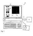
- FIG. 1 shows an example of an apparatus 10 according to the invention.
- a medical data set 11 comprising a plurality of images, in particular axial slice images, of a body is acquired by a medical imaging apparatus 12, in particular a CT apparatus, and is fed to a control unit 13, preferably a computer, which is configured to control and/or to execute steps of the method according to the invention.
- the image data of the plurality of images can be directly fed to the control unit 13.
- image data can also be transferred via a data network 18 to which both the imaging apparatus 12 and the control unit 13 are, at least temporarily, connected.
- the apparatus 10 preferably comprises a display 14, e.g. a TFT screen, for displaying slice views and/or volumetric views of the medical data set.
- a display 14 e.g. a TFT screen
- a coronal CT slice image 15 of a human body and a corresponding axial CT slice image 23 of the lumbar part of the body are displayed.
- the control unit 13 is also configured to generate a volume reconstruction of slice images 15, 23, in particular of automatically selected axial slice images, on the display 14.
- the present invention is based on the approach that before localizing or labeling spines in CT scans a significant amount of the data is cropped so that both time and memory performance of an according method and apparatus for spine localization or labeling can be enhanced significantly.
- a one-pass, slice-wise method to figure out what parts of CT data can be left out from a DICOM-to-volume reconstruction and to estimate what vertebral discs are likely to be found in what slices is described exemplarily.
- the present invention provides a one-pass, slice-wise method based on statistics and distribution signatures of bones structures present in particular slices.
- the invention also provides an according apparatus and system for carrying out the mentioned method.
- the input scan and the accompanying DICOM fields fulfill at least one, preferably all, of the following requirements:
- RAW data relates to original vendor's slices with intensities in CT values. This is the data produced and stored by a CT scanner where the range of pixel intensities are preset by the vendor of the CT scanner and may differ per scanning protocol.
- p z z ⁇ 350 ⁇ g p ⁇ 1050 where p x , p y and p z are world coordinates in millimeters. Therefore, the set B z represents a set of bone pixels in the slices exhibiting intensity values of 1 after thresholding.
- centers of gravity ⁇ z correlate with a spine reliably in lumbar slices where pelvis, ribs, or head do not contribute to the centroid.
- the lumbar part can be characterized by deviation lengths ⁇ z ⁇ related to size of vertebra of approximately 25 mm seen in an axial slice. Therefore, values of ⁇ z ⁇ larger than approximately 40 mm indicate the presence of non-vertebra bones.
- the intensity values of lumbar bones 1 significantly contribute to the calculation of the center of gravity ⁇ z , which is therefore considerably shifted away from a centroid of the vertebra 2.
- the radius of the circle 24 indicated in the slice image 20 corresponds to the length ⁇ z ⁇ of the standard deviation vector ⁇ z of the intensity value distribution in the first slice image 20.
- the difference between the original ⁇ z and the refined v z centroids ⁇ z - v z ⁇ correlates with the reliability of the seed, i.e. the smaller the difference ⁇ z - v z ⁇ , the more reliably the seed aligns with the spine.
- Shape histograms AP versus LR distribution
- leg slice images are identified by means of bone distributions being dominant in the left-to-right (LR) direction and having a zero contribution in the anterior-posterior (AP) direction.
- Fig. 3 shows a negative of an axial CT slice showing legs and a CT table, on which the patient was lying during CT image acquisition, together with right, left, ante and poste histogram bins centered at the refined center of gravity ⁇ z .
- 4-bin histograms located in the refined centers are constructed.
- H z A p ⁇ B z
- H z P p ⁇ B z
- H z R p ⁇ B z
- H z L p ⁇ B z
- H A z and H P z are to be swapped.
- the posterior voxels i.e. volume image pixels, H P z have been excluded from equation (12) in order to ignore an eventual contribution of a CT table.
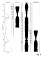
- Fig. 4 shows an example of a coronal projection (left) together with a respective histogram profile ⁇ z (middle) and a standard deviation profile ⁇ 1 (right) derived from axial CT slice images.
- the two-dimensional plot of the parameters ⁇ z and ⁇ z along the z-axis yields what is also referred to as "bone profiles".
- bone profiles what is also referred to as "bone profiles”.
- slice images comprising image information of the legs and/or of a lower part of the pelvis are unnecessary to deal with when labeling the spine.
- the ⁇ z profile is examined automatically by the control unit 13 (see Fig. 1 ) from top to down for a sufficiently long chain of zeros (cf. equation 12 above).
- ischium or a slab between ischium and sacrum is identified by means of a ⁇ z profile having a "zero chain" with a length of at least 20 mm. This is indicated by lines 27 and 28 in Fig. 4 . Therefore, it is preferred to discard slice images below the line 28 and it is even more preferred to discard slice images below the line 27.
- the search space far from the spine is further pruned.
- lumbar slices having a standard deviation ⁇ z ⁇ of pixels relating to bones of approximately 25 ( ⁇ z ⁇ ⁇ 25) yield a good seed to set up a cropping box for the entire volume of the selected slice images.
- This approach leads to an additional significant reduction of the total data volume on the one hand and ensures a reliable identification of all relevant parts of the selected slice images on the other hand, in particular for cases in which no scoliosis or oblique spines are subject to an examination.
- oblique spines and scans without lumbar part it is preferred to consider at least a part, preferably approximately 40 %, of the most reliable centers of gravity for which the original center of gravity ⁇ z and the refined center of gravity ⁇ z overlap at least partially (so that ⁇ z ⁇ ⁇ z ) in order to generate a cropping box around the spine.
- rectangles of a size of approximately 90 x 120 mm are centered at respective refined centers of gravity ⁇ z and spanned around the spine.
- a resulting cropping box is preferably achieved by an x-y hull of this extrusion.
- ⁇ z of a particular scan i.e. a plurality of acquired and/or selected slice images
- a ground-truth labeled model of a full-body scan it is preferred that it is, at least approximately, determined what labels are to be expected in particular slice images.
- Fig. 5 shows an example of a correlation between a plurality of selected axial CT slice images (denoted “Colon_buik”) with a model (denoted “Model”) of a full-body axial CT scan.
- a number of slice positions along the z axis of the model are labeled with respective vertebra labels, e.g. "C2", “T1", “L1” and “S1” and/or respective spinal disc labels, e.g. "C1/C2", “T1/T2", “L1/L2” and “L5/S1".
- the respective correlation factor curve shows two maximum points 31 and 32 indicating a high correlation between the two shown axial slice images "Colon_buik” with the model.
- the selected axial CT slice images fit very well to the respective part of the model.
- classifiers for knees can also be derived from the A profiles. Localization of other organs (e.g., neck, heart, kidney) are also possible. In particular, the following embodiments are preferred.
- a multi-class machine learning framework i.e., training/matching
- the precision of the estimates of ischium as well as the disc-label approximations set forth above will even be enhanced.
Landscapes
- Engineering & Computer Science (AREA)
- Computer Vision & Pattern Recognition (AREA)
- Physics & Mathematics (AREA)
- General Physics & Mathematics (AREA)
- Theoretical Computer Science (AREA)
- Health & Medical Sciences (AREA)
- General Health & Medical Sciences (AREA)
- Medical Informatics (AREA)
- Nuclear Medicine, Radiotherapy & Molecular Imaging (AREA)
- Radiology & Medical Imaging (AREA)
- Quality & Reliability (AREA)
- Apparatus For Radiation Diagnosis (AREA)
Abstract
Description
- The present invention relates to a method and a corresponding apparatus and system for localizing a spine in an image, in particular a computed tomography (CT) image, of a human or animal body according to the independent claims.
- Typically, a plurality of axial CT images of a body are acquired and subsequently stored, evaluated and/or displayed in a two- and/or three-dimensional representation. Due to the large amount of acquired image data an automatic localization of a spine in CT images is a computationally intensive and memory demanding task.
- It is an object of the present invention to provide a method and a corresponding apparatus and system for localizing a spine in an image, in particular a CT image, of a human or animal body allowing for a reduced need for computational power and/or memory on the one hand and assuring a reliable localization of the spine on the other hand.
- This object is achieved by the method and the corresponding apparatus and system according to the independent claims.
- The method according to the invention comprising the following steps: acquiring a plurality of slice images of at least a part of a human or animal body and automatically selecting slice images and/or parts of the slice images from the acquired plurality of slice images by considering at least one parameter characterizing a distribution of bones in said acquired slice images, wherein said selected slice images and/or parts of the slice images comprising image information about the spine.
- A corresponding method according to the invention comprises automatically selecting slice images and/or parts of slice images from a plurality of slice images, said slice images having been acquired of at least a part of a human or animal body, by considering at least one parameter characterizing a distribution of bones in said acquired slice images, wherein said selected slice images and/or parts of the slice images comprising image information about the spine.
- The apparatus according to the invention comprises an image processing unit for automatically selecting slice images and/or parts of the slice images from a plurality of slice images of at least a part of a human or animal body by considering at least one parameter characterizing a distribution of bones in said acquired slice images, wherein said selected slice images and/or parts of the slice images comprising image information about the spine.
- The system according to the invention comprises an image acquisition unit, in particular a computed tomography unit, for acquiring a plurality of slice images of at least a part of a human or animal body and an image processing unit for automatically selecting slice images and/or parts of the slice images from the acquired plurality of slice images by considering at least one parameter characterizing a distribution of bones in said acquired slice images, wherein said selected slice images and/or parts of the slice images comprising image information about the spine.
- The invention is based on the approach to automatically select slice images and/or parts thereof from the acquired plurality of slice images of the body based on a slice-wise, i.e. slice-by-slice, analysis of the acquired slice images with respect to a distribution of bones in the acquired slice images. Dependent on the result of the analysis, slice images and/or parts thereof containing image information about the spine are selected and/or slice images which do not contain image information about the spine are truncated. The slice-wise analysis of the acquired slice images is based on statistics and distribution signatures of bone structures present in the slices. Preferably, the analysis of the bone profiles in the acquired slice images occurs in only one pass, i.e. the acquired slice images are analyzed slice-by-slice, e.g. beginning with slice images relating to the neck and ending with slice images relating to the legs.
- By means of this approach, in many cases of CT images a significant portion of CT image data can be left out, e.g. slice images of the region of the legs of a body which require very often a half of the total data volume. In particular, such portions of the CT image data can be left out from the acquired slices during volume reconstruction of an according spinal image. Moreover, parts too far from the spinal canal can be masked and ignored during the search for disc and vertebra labels. As a consequence, the need for computational power and memory for a two- and/or three-dimensional image reconstruction can be reduced significantly without losing relevant image information relating to the spine so that a reliable localization of the spine in the selected images can be ensured. The latter particularly applies for a subsequent estimation of what vertebral discs are likely to be found in particular selected slice images.
- The term "selecting slice images" in the meaning of the present invention does not only relate to selecting slice images but also to refusing slice images or masking a part thereof. Accordingly, "selected slice images" in the meaning of the present invention are slice images and/or parts thereof which were selected or which were not truncated or masked, respectively.
- A "distribution of bones" in a slice image in the meaning of the present invention relates to a distribution or spread of intensity values or pixels relating to bones in a slice image. Preferably, the intensity values of the slice images are given in and/or transformed to a Hounsfield scale in Hounsfield units (HU), wherein the intensity values are spread over an interval g [350; 1050] HU so that bones can therefore roughly be segmented by an interval threshold.
- A "parameter characterizing a distribution of bones" in the meaning of the present invention relates to one or more properties of the distribution of bone structures within a slice image. Preferably, the parameter can be derived from the bone distribution and/or it can be, e.g., a mean value or a median value of the intensity values or pixels relating to bones, a spread, a width, a variance or a standard deviation of the bone distribution. As will be set forth below in detail, there are a number of further preferred parameters which characterize the distribution of bones.
- Preferably, the spine has a longitudinal axis and the image plane of respective slice images is substantially perpendicular to the longitudinal axis of the spine. Therefore, slice images in the meaning of the invention are called axial images.
- In a preferred embodiment of the invention the at least one parameter relates to a center of gravity of pixels relating to bones in a slice image. The center of gravity of pixels relating to bones in a slice image relates to an average location of a distribution of the pixels relating to the bones within the slice image. Assuming that a slice image only contains a cross-sectional image of a more or less symmetric vertebra of the spine, the center of gravity will be located approximately in the center of the cross-sectional image of the vertebra. By this means, a simple and reliable localization of the spine within a slice image and an according selection of the slice image is achieved.
- In the meaning of the present invention, the term "pixel" denotes a smallest element of an image, in particular a picture element of a slice image. Preferably, each of the pixels exhibits an intensity value of 0 or 1 after the interval threshold has been applied.
- In the case that additional bones, e.g. pelvis or ribs, contribute to the bone distribution in a slice image of a spine, the center of gravity of the intensity values will shift away from the center region of the vertebra. In order to account for this, it is preferred that the at least one parameter represents a refined center of gravity of pixels relating to bones being located within a refinement window in a slice image, wherein the refinement window is spanned around an original center of gravity of pixels relating to bones in a slice image. In this embodiment, an original center of gravity of intensity values in a slice image is determined as stated above. Next, an asymmetric refinement window is superimposed onto the slice image, wherein the dimensions and position of the refinement window is, preferably automatically, chosen so that the cross-sectional image of the spine or a part thereof is located within the refinement window, whereas additional bones, e.g. pelvis or ribs, are located outside the refinement window. In a further step, the refined center of gravity is derived only by considering pixels relating to bones which are located inside the refinement window. That way, a reliable and fast localization of a spine can also be achieved in slice images containing image information on additional bones.
- In a particularly preferred embodiment, slice images are selected by considering a difference between the original center of gravity and the refined center of gravity. Because the refined center of gravity will deviate from the original center of gravity in cases where in addition to the spine additional bones are imaged, the difference between the original center of gravity and the refined center of gravity correlates with the reliability of a seed, which is a point inside or close to a vertebral body, in particular an initialization point for a subsequent segmentation algorithm. As a consequence, the smaller the difference the more reliably the seed aligns with the spine.
- In another preferred embodiment of the invention the at least one parameter represents a standard deviation of pixels relating to bones in a slice image. The standard deviation of the intensity values characterizes a variation or "dispersion" from an average intensity value in an intensity value distribution of bones within a slice image. E.g., a low standard deviation indicates that the data points tend to be very close to the mean value, whereas a high standard deviation indicates that the data points are spread out over a large range of values. Thus, the standard deviation represents an easily obtainable and reliable parameter for determining whether a slice image mainly contains image information of a spine or it contains also information on additional bones.
- In the meaning of the present invention, the term "standard deviation of pixels relating to bones" refers to a spatial deviation of bone pixels from the centroid of the thresholded slice images.
- It is particularly preferred, that slice images are selected in which the standard deviation is within a pre-defined range. It has been found that axial slice images in the lumbar part of a body exhibit a standard deviation which is mainly characterized by the size of the vertebra of typically 25 mm imaged in the slice. Therefore, slice images exhibiting a standard deviation of the pixels relating to the bones in the region of approximately 20 to 40 mm, in particular 25 to 35 mm, in particular 25 to 30 mm, can be assigned to slice images of the lumbar column of the body, whereas slice images exhibiting a standard deviation larger than 40 mm indicate the presence of non-vertebra bones, e.g. ribs or pelvis.
- In a further preferred embodiment the at least one parameter relates to at least one histogram of pixels relating to bones in a slice image in a left-to-right direction (LR) and/or an anterior-posterior direction (AP). A histogram in the meaning of the invention relates to a representation of a probability distribution of the intensity values which were classified into a pre-defined number of disjoined categories, so-called bins. Thus, a histogram represents probability values for respective pixels relating to bones in a slice image within a pre-defined bin, i.e. an intensity category. Preferably, the at least one histogram is located, in particular centered, at a refined center of gravity. Alternatively or additionally it is also preferred that the at least one parameter relates to at least one 4-bin histogram. By considering at least one histogram in LR and/or AP direction, a reliable identification of slice images relating to images containing legs of the body can be achieved. As leg slice images do not contain information on the spine, they can be truncated subsequently. By this means, a reliable selection, i.e. truncation, of slice images and a subsequent localization of the spine in the selected, i.e. not truncated, slice images is possible.
- Moreover, it is preferred that slice images showing a histogram of the pixels relating to bones dominant in the left-to-right direction are truncated. Alternatively or additionally it is also preferred that slice images showing a histogram of the pixels relating to bones with substantially no contribution in the anterior-posterior direction are truncated. By these provisions a very reliable identification of slice images containing images from the legs of a body and their subsequent truncation can be achieved.
- As already exemplarily explained above, it is one preferred aspect of the invention that the selection step in which slice images are selected also comprises truncating slice images which are assigned to a further part, in particular to the legs, of the body. Alternatively or additionally, it is also preferred that the selection step comprises truncating image information from slice images, in particular from the selected slice images, wherein said truncated image information is assigned to a region beyond a volume around the spine. Preferably, said volume around the spine is given by a rectangular prism. By this means, the total data volume is reduced considerably so that computational power and memory requirements for displaying and/or analyzing the selected slice images can be reduced significantly without adversely effecting the localization of the spine in the selected slice images.
- As already mentioned above, the selected slice images can be displayed and/or analyzed for diagnostic purposes. Preferably, the selected slice images are displayed in a volume reconstruction of the slice images. Alternatively or additionally, the selected slice images are analyzed for diagnostic purposes by searching for at least a part of the spine, in particular for a spinal disc and/or vertebra.
- In a preferred embodiment of the invention the acquired slice images, in particular the selected slice images, are analyzed by correlating the standard deviation of pixels relating to bones in a slice image with a model of a full-body scan. Preferably, the considered model of a full-body scan depends on parameters relating to the particular body that was scanned, e.g. sex and/or body height and/or body weight. That is, the considered model of a full-body scan contains pre-defined standard deviation values of a bone distribution which is typical for the respective type of body, e.g. male, range of body height 160 to 180 cm, range of body weight 60 to 80 kg. Of course, also other types of models of a full-body scan can be considered. Preferably, when correlating the standard deviation of pixels relating to bones in a slice image with a model of a full-body scan, at least one correlation parameter is determined for a plurality of the slice images and subsequently analyzed. In this way, a reliable localization of a spine or parts thereof in a plurality of slice images is possible without intervention by operation personnel in many cases and without special markers in or on the body.
- Further advantages, features and examples of the present invention will be apparent from the following description of following figures:
- Fig. 1
- shows an example of an apparatus according to the invention;
- Fig. 2
- shows three examples of slice images in which a center of gravity, a refinement window and a refined center of gravity is indicated respectively;
- Fig. 3
- shows an example of a 4-bin histogram together with a slice image;
- Fig. 4
- shows an example of a histogram and a standard deviation profile derived from axial CT slice images together with a respective coronal projection;
- Fig. 5
- shows an example of a correlation between a plurality of selected axial CT slice images with a model of a full-body axial CT scan.
-
Fig. 1 shows an example of anapparatus 10 according to the invention. Amedical data set 11 comprising a plurality of images, in particular axial slice images, of a body is acquired by amedical imaging apparatus 12, in particular a CT apparatus, and is fed to acontrol unit 13, preferably a computer, which is configured to control and/or to execute steps of the method according to the invention. The image data of the plurality of images can be directly fed to thecontrol unit 13. Alternatively or additionally, image data can also be transferred via adata network 18 to which both theimaging apparatus 12 and thecontrol unit 13 are, at least temporarily, connected. - The
apparatus 10 preferably comprises adisplay 14, e.g. a TFT screen, for displaying slice views and/or volumetric views of the medical data set. In the example given inFig. 1 , a coronalCT slice image 15 of a human body and a corresponding axialCT slice image 23 of the lumbar part of the body are displayed. Preferably, thecontrol unit 13 is also configured to generate a volume reconstruction ofslice images display 14. - As already mentioned above, the present invention is based on the approach that before localizing or labeling spines in CT scans a significant amount of the data is cropped so that both time and memory performance of an according method and apparatus for spine localization or labeling can be enhanced significantly. In the following, a one-pass, slice-wise method to figure out what parts of CT data can be left out from a DICOM-to-volume reconstruction and to estimate what vertebral discs are likely to be found in what slices is described exemplarily.
- According to the invention, in many cases a significant portion of the CT data can be truncated. For example, leg slices (often a half of the data) can be left out already during volume reconstruction from the DICOM slices. Moreover, parts too far from the spinal canal can be masked and ignored during the search for disc and vertebra labels. To cope with the above, the present invention provides a one-pass, slice-wise method based on statistics and distribution signatures of bones structures present in particular slices. The invention also provides an according apparatus and system for carrying out the mentioned method.
- Preferably, the input scan and the accompanying DICOM fields fulfill at least one, preferably all, of the following requirements:
- CT with Hounsfield scale, wherein it is assumed that there is a RAW data to Hounsfield intensity transformation,
- axial CT slices, so that a slice-wise iteration can be performed,
- head-first face-up (supine) orientation of the patient, or a transformation to yield such an orientation, and
- pixel/voxel size given in millimeters, or a corresponding scaling information from DICOM.
- The term "RAW data" relates to original vendor's slices with intensities in CT values. This is the data produced and stored by a CT scanner where the range of pixel intensities are preset by the vendor of the CT scanner and may differ per scanning protocol.
- It is further preferred that the intensity values are spread over an interval g [350; 1050] in Hounsfield units (HU) so that bones can therefore roughly be segmented by an interval threshold. Accordingly, for a set B z of 3D positions p within a slice z where a bone is detected it applies:

where px , py and pz are world coordinates in millimeters. Therefore, the set Bz represents a set of bone pixels in the slices exhibiting intensity values of 1 after thresholding. - The simplest features are based on a centroid, i.e. a center of gravity µ z , of Bz

and the length ∥σ z ∥ of a standard deviation vector σ z

- It has been found that centers of gravity µ z correlate with a spine reliably in lumbar slices where pelvis, ribs, or head do not contribute to the centroid. The lumbar part can be characterized by deviation lengths ∥σ z ∥ related to size of vertebra of approximately 25 mm seen in an axial slice. Therefore, values of ∥σ z ∥ larger than approximately 40 mm indicate the presence of non-vertebra bones.
- While reliable in the lumbar area of the body, the centroids may drift remarkably from spine if the pelvis or ribs contribute by its pixels. This is illustrated by means of three examples of
slice images Fig. 2 . - In the
first slice image 20 the intensity values oflumbar bones 1 significantly contribute to the calculation of the center of gravity µz, which is therefore considerably shifted away from a centroid of thevertebra 2. The radius of thecircle 24 indicated in theslice image 20 corresponds to the length ∥σz∥ of the standard deviation vector σz of the intensity value distribution in thefirst slice image 20. - As obvious from the second and
third slice image 21 and 22 a shift of the calculated center of gravity µz away from the centroid of therespective spine 2 is still present, albeit considerably smaller than in thefirst slice image 20 due to a smaller contribution of the intensity values of surroundingbones 3 or 4, to the total intensity value distribution in theslice image circle 24 corresponding to the length ∥σz∥ of the standard deviation vector σz in thesecond slice image 21 indicates that there areless bones 3 distributed far around thespine 2 than in the first or thethird slice image - In order to avoid the above-mentioned shift of the center of gravity µz, the centroids within a rectangular window 17 (see
Fig. 2 ) which is asymmetrically spanned around an original center of gravity µz are refined as follows:
or
- where the correct variant of Wz is determined by the orientation of the patient inside the CT scanner: face-up (according to equation (5) above) and face-down (according to equation (6) above).
- The original center of gravity µz is refined to a refined center of gravity vz of bone pixels in this window 17:

- The difference between the original µz and the refined vz centroids ∥µz - vz∥ correlates with the reliability of the seed, i.e. the smaller the difference ∥µz - vz∥, the more reliably the seed aligns with the spine.
- In each of the
slice images Fig. 2 a 80 x 40mm refinement window 17 and a refined center of gravity vz is shown. Accordingly, the refined center of gravity νz is now in the region of the central axis of the respective part, e.g. a vertebra, of thespine 2. - According to a preferred embodiment of the method according to the invention leg slice images are identified by means of bone distributions being dominant in the left-to-right (LR) direction and having a zero contribution in the anterior-posterior (AP) direction.
- This is illustrated by means of
Fig. 3 which shows a negative of an axial CT slice showing legs and a CT table, on which the patient was lying during CT image acquisition, together with right, left, ante and poste histogram bins centered at the refined center of gravity νz. In cases where the deviation vector σz fails to discriminate leg slices, 4-bin histograms located in the refined centers are constructed. Putting δ = p - νz the following four quantities are defined:


- If the patient was positioned face-down (instead of face-up), HA z and HP z are to be swapped.
- By means of the AP/LR histograms the leg detection is reformulated as a search for slices, where ante-poste bone contributions vanish:

- In this preferred reformulation, the posterior voxels, i.e. volume image pixels, HP z have been excluded from equation (12) in order to ignore an eventual contribution of a CT table.
- According to the steps outlined above, it is preferred that for each of the slice images two scalars σ z and Λ z , i.e. the standard deviation σ z and a value Λ z representative of contributions of bones in ante-poste direction, are derived. With these two features the slices can be easily identified in a context. Preferably, in order to identify and/or classify the slice images even more reliably, a longer feature vector containing more than two scalars can be established.
-
Fig. 4 shows an example of a coronal projection (left) together with a respective histogram profile Λ z (middle) and a standard deviation profile σ1 (right) derived from axial CT slice images. The two-dimensional plot of the parameters σ z and Λ z along the z-axis yields what is also referred to as "bone profiles". In the following it will be shown how the above-mentioned parameters contribute to localize the spine according to the invention. - Usually, slice images comprising image information of the legs and/or of a lower part of the pelvis are unnecessary to deal with when labeling the spine. In order to identify and crop respective slice images from the CT scan, the Λ z profile is examined automatically by the control unit 13 (see
Fig. 1 ) from top to down for a sufficiently long chain of zeros (cf.equation 12 above). Preferably, ischium or a slab between ischium and sacrum is identified by means of a Λ z profile having a "zero chain" with a length of at least 20 mm. This is indicated bylines Fig. 4 . Therefore, it is preferred to discard slice images below theline 28 and it is even more preferred to discard slice images below theline 27. - After the legs have been cropped the search space far from the spine is further pruned. For upright spines, lumbar slices having a standard deviation ∥σz∥ of pixels relating to bones of approximately 25 (∥σz∥ ≈ 25) yield a good seed to set up a cropping box for the entire volume of the selected slice images. This approach leads to an additional significant reduction of the total data volume on the one hand and ensures a reliable identification of all relevant parts of the selected slice images on the other hand, in particular for cases in which no scoliosis or oblique spines are subject to an examination.
- In cases of scolioses, oblique spines and scans without lumbar part it is preferred to consider at least a part, preferably approximately 40 %, of the most reliable centers of gravity for which the original center of gravity µz and the refined center of gravity νz overlap at least partially (so that µz ≈ νz) in order to generate a cropping box around the spine. Preferably, rectangles of a size of approximately 90 x 120 mm are centered at respective refined centers of gravity νz and spanned around the spine. A resulting cropping box is preferably achieved by an x-y hull of this extrusion. By these means, an undesired cropping of data in the y-direction related to the neck of the body can be reliably avoided in cases of scolioses, oblique spines and scans without lumbar part.
- In order to obtain an estimate of what portion of the human body was scanned, it is preferred to correlate the deviation profile σz of a particular scan, i.e. a plurality of acquired and/or selected slice images, with a ground-truth labeled model of a full-body scan. Moreover, it is preferred that it is, at least approximately, determined what labels are to be expected in particular slice images.
-
Fig. 5 shows an example of a correlation between a plurality of selected axial CT slice images (denoted "Colon_buik") with a model (denoted "Model") of a full-body axial CT scan. A number of slice positions along the z axis of the model are labeled with respective vertebra labels, e.g. "C2", "T1", "L1" and "S1" and/or respective spinal disc labels, e.g. "C1/C2", "T1/T2", "L1/L2" and "L5/S1". - As obvious from
Fig. 5 , the respective correlation factor curve (left part ofFig. 5 ) shows twomaximum points Fig. 5 , in the correlation corresponding to themaximum point 31 the selected axial CT slice images fit very well to the respective part of the model. By this means, a reliable localization of vertebrae and/or spinal discs in an axial CT scan can be achieved. - Similar to leg slices, classifiers for knees can also be derived from the A profiles. Localization of other organs (e.g., neck, heart, kidney) are also possible. In particular, the following embodiments are preferred.
- First, involving more bins, e.g. at least 5 bins, in the shape histogram are preferred to yield an even more detailed look on the bone distribution. Also taking z-slabs instead of single slices may be advantageous.
- Second, a multi-class machine learning framework (i.e., training/matching) would be preferable to classify the slices for a desired list of organs. By this means, the precision of the estimates of ischium as well as the disc-label approximations set forth above will even be enhanced.
- Third, it is also possible to consider other features from a bone distribution (e.g., circularity, inertia) and/or non-intensity based features when localizing the spine.
Claims (15)
- Method for localizing a spine in an image, in particular a computed tomography (CT) image, of a human or animal body, comprising the following steps:a) acquiring a plurality of slice images (20 - 23) of at least a part of a human or animal body andb) automatically selecting slice images and/or parts of slice images from the acquired plurality of slice images (20 - 23) by considering at least one parameter (µz, νz, σz, Λz) characterizing a distribution of bones (1 - 4) in said acquired slice images (20 - 23), wherein said selected slice images and/or parts of the slice images comprising image information about the spine.
- Method according to claim 1, wherein the at least one parameter relates to a center of gravity (µz) of pixels relating to bones (1 - 4) in a slice image (20 - 23).
- Method according to claim 1, wherein the at least one parameter represents a refined center of gravity (νz) of pixels relating to bones (1 - 4) within a refinement window (17) in a slice image (20 - 23), wherein the refinement window (17) is spanned around an original center of gravity (µz) of pixels relating to bones (1 - 4) in a slice image (20 - 23).
- Method according to claim 3, wherein slice images are selected by considering the difference (µz - νz) between the original center of gravity (µz) and the refined center of gravity (νz).
- Method according to anyone of the preceding claims, wherein the at least one parameter represents a standard deviation (σz) of pixels relating to bones (1 - 4) in a slice image (20 - 23).
- Method according to claim 5, wherein slice images are selected in which the standard deviation (σz) is within a pre-defined range.
- Method according to anyone of the preceding claims, wherein the at least one parameter (Λz) relates to at least one histogram (HL z, HR z, HA z, HP z ) of pixels relating to bones (1 - 4) in a slice image (20 - 23) in a left-to-right direction (LR) and/or an anterior-posterior direction (AP).
- Method according to claim 7, wherein slice images (20 - 23) showing a histogram (HL z, HR z, HA z, HP z ) of the pixels relating to bones (1 - 4) dominant in the left-to-right direction (LR) and/or with substantially no contribution in the anterior-posterior direction (AP) are truncated.
- Method according to anyone of the preceding claims, wherein step b) comprises truncating image information from slice images (20 - 23), in particular from the selected slice images, wherein said truncated image information is assigned to a region beyond a volume around the spine.
- Method according to claim 9, wherein said volume around the spine is given by a rectangular prism.
- Method according to anyone of the preceding claims, wherein said selected slice images are displayed, in particular by a volume reconstruction.
- Method according to anyone of the preceding claims, wherein said selected slice images are analyzed for diagnostic purposes by searching for at least a part of the spine, in particular for a spinal disc and/or vertebra.
- Method according to claim 5, wherein said slice images are analyzed by correlating the standard deviation (σz) of pixels relating to bones (1 - 4) in slice images (20 - 23; Colon_buik) with a model of a full-body scan (Model).
- Apparatus for localizing a spine in an image, in particular a computed tomography (CT) image, said image comprising a plurality of slice images (20 - 23) of at least a part of a human or animal body, said apparatus comprising
an image processing unit (13) for automatically selecting slice images and/or parts of the slice images from the acquired plurality of slice images (20 - 23) by considering at least one parameter (µz, νz, σz, Λz) characterizing a distribution of bones (1 - 4) in said acquired slice images (20 - 23), wherein said selected slice images and/or parts of the slice images comprising image information about the spine. - System for localizing a spine in an image, in particular a computed tomography (CT) image, of a human or animal body, comprising:an image acquisition unit (12), in particular a computed tomography unit, for acquiring a plurality of slice images (20 - 23) of at least a part of a human or animal body andan image processing unit (13) for automatically selecting slice images and/or parts of the slice images from the acquired plurality of slice images (20 - 23) by considering at least one parameter (µz, νz, σz, Λz) characterizing a distribution of bones (1 - 4) in said acquired slice images (20 - 23), wherein said selected slice images and/or parts of the slice images comprising image information about the spine.
Priority Applications (5)
| Application Number | Priority Date | Filing Date | Title |
|---|---|---|---|
| EP12159593.8A EP2639763B1 (en) | 2012-03-15 | 2012-03-15 | Method, Apparatus and System for Localizing a Spine |
| PCT/EP2013/055233 WO2013135812A1 (en) | 2012-03-15 | 2013-03-14 | Method, apparatus and system for localizing a spine |
| US14/384,734 US9406122B2 (en) | 2012-03-15 | 2013-03-14 | Method, apparatus and system for localizing a spine |
| CN201380014219.0A CN104395930B (en) | 2012-03-15 | 2013-03-14 | For positioning the method, apparatus and system of spinal column |
| BR112014022793-4A BR112014022793B1 (en) | 2012-03-15 | 2013-03-14 | METHOD, APPARATUS AND SYSTEM FOR LOCATING A VERTEBRAL COLUMN |
Applications Claiming Priority (1)
| Application Number | Priority Date | Filing Date | Title |
|---|---|---|---|
| EP12159593.8A EP2639763B1 (en) | 2012-03-15 | 2012-03-15 | Method, Apparatus and System for Localizing a Spine |
Publications (2)
| Publication Number | Publication Date |
|---|---|
| EP2639763A1 true EP2639763A1 (en) | 2013-09-18 |
| EP2639763B1 EP2639763B1 (en) | 2015-10-21 |
Family
ID=45926413
Family Applications (1)
| Application Number | Title | Priority Date | Filing Date |
|---|---|---|---|
| EP12159593.8A Active EP2639763B1 (en) | 2012-03-15 | 2012-03-15 | Method, Apparatus and System for Localizing a Spine |
Country Status (5)
| Country | Link |
|---|---|
| US (1) | US9406122B2 (en) |
| EP (1) | EP2639763B1 (en) |
| CN (1) | CN104395930B (en) |
| BR (1) | BR112014022793B1 (en) |
| WO (1) | WO2013135812A1 (en) |
Cited By (2)
| Publication number | Priority date | Publication date | Assignee | Title |
|---|---|---|---|---|
| JPWO2014065317A1 (en) * | 2012-10-23 | 2016-09-08 | 株式会社日立製作所 | Image processing apparatus and spinal canal evaluation method |
| US11841923B2 (en) | 2020-07-06 | 2023-12-12 | Alibaba Group Holding Limited | Processing method, model training method, means, and storage medium for spinal images |
Families Citing this family (11)
| Publication number | Priority date | Publication date | Assignee | Title |
|---|---|---|---|---|
| US9538920B2 (en) * | 2014-05-09 | 2017-01-10 | London Health Sciences Centre Research Inc. | Standalone annotations of axial-view spine images |
| CN106256326A (en) * | 2015-06-19 | 2016-12-28 | 通用电气公司 | The generation system and method for computed tomography sectioning image |
| JP6280676B2 (en) * | 2016-02-15 | 2018-02-14 | 学校法人慶應義塾 | Spine arrangement estimation device, spine arrangement estimation method, and spine arrangement estimation program |
| JP7170631B2 (en) | 2016-10-05 | 2022-11-14 | ニューヴェイジヴ,インコーポレイテッド | Surgical navigation system and related methods |
| CN106845068B (en) * | 2016-12-13 | 2020-05-22 | 海纳医信(北京)软件科技有限责任公司 | Picture labeling method and device |
| US10366491B2 (en) * | 2017-03-08 | 2019-07-30 | Siemens Healthcare Gmbh | Deep image-to-image recurrent network with shape basis for automatic vertebra labeling in large-scale 3D CT volumes |
| CN108451656B (en) * | 2018-04-04 | 2020-03-10 | 北京医院 | Spine three-dimensional positioning method |
| CN109360213B (en) * | 2018-12-14 | 2020-10-30 | 浙江工业大学 | Automatic vertebral body identification method based on spine ultrasonic coronal plane image |
| CN110916695A (en) * | 2019-11-05 | 2020-03-27 | 东软医疗系统股份有限公司 | Method and device for determining spinal scanning visual field and image processing equipment |
| CN113902654B (en) | 2020-07-06 | 2025-10-03 | 阿里巴巴集团控股有限公司 | Image processing method, device, electronic device and storage medium |
| US11881301B2 (en) * | 2021-02-26 | 2024-01-23 | GE Precision Healthcare LLC | Methods and systems for utilizing histogram views for improved visualization of three-dimensional (3D) medical images |
Citations (2)
| Publication number | Priority date | Publication date | Assignee | Title |
|---|---|---|---|---|
| US20050276455A1 (en) * | 2004-06-01 | 2005-12-15 | Marta Fidrich | Systems and methods for segmenting an organ in a plurality of images |
| JP2008132019A (en) * | 2006-11-27 | 2008-06-12 | Rigaku Corp | Volume data processing apparatus and volume data processing program |
Family Cites Families (12)
| Publication number | Priority date | Publication date | Assignee | Title |
|---|---|---|---|---|
| US5431161A (en) * | 1993-04-15 | 1995-07-11 | Adac Laboratories | Method and apparatus for information acquistion, processing, and display within a medical camera system |
| US7013032B1 (en) * | 1999-11-24 | 2006-03-14 | The General Electric Company | Method and apparatus for secondary capture of 3D based images on a picture archival and communications (PACS) system |
| JP3809105B2 (en) * | 2002-01-18 | 2006-08-16 | 株式会社東芝 | Magnetic resonance imaging system |
| JP4091318B2 (en) * | 2002-03-04 | 2008-05-28 | ジーイー・メディカル・システムズ・グローバル・テクノロジー・カンパニー・エルエルシー | X-ray CT system |
| US8600124B2 (en) * | 2004-09-16 | 2013-12-03 | Imatx, Inc. | System and method of predicting future fractures |
| US7123760B2 (en) * | 2002-11-21 | 2006-10-17 | General Electric Company | Method and apparatus for removing obstructing structures in CT imaging |
| US8073210B2 (en) * | 2005-02-14 | 2011-12-06 | University Of Lowa Research Foundation | Methods of smoothing segmented regions and related devices |
| US20070121778A1 (en) * | 2005-10-17 | 2007-05-31 | Siemens Corporate Research Inc | Method and System For Spinal Cord Detection In Computed Tomography Volume Data |
| US8244015B2 (en) * | 2006-11-22 | 2012-08-14 | General Electric Company | Methods and apparatus for detecting aneurysm in vasculatures |
| IL179581A0 (en) * | 2006-11-26 | 2007-05-15 | Algotec Systems Ltd | Spine labeling |
| US8311306B2 (en) * | 2008-04-30 | 2012-11-13 | Otismed Corporation | System and method for image segmentation in generating computer models of a joint to undergo arthroplasty |
| JP5138431B2 (en) * | 2008-03-17 | 2013-02-06 | 富士フイルム株式会社 | Image analysis apparatus and method, and program |
-
2012
- 2012-03-15 EP EP12159593.8A patent/EP2639763B1/en active Active
-
2013
- 2013-03-14 CN CN201380014219.0A patent/CN104395930B/en not_active Expired - Fee Related
- 2013-03-14 WO PCT/EP2013/055233 patent/WO2013135812A1/en not_active Ceased
- 2013-03-14 BR BR112014022793-4A patent/BR112014022793B1/en active IP Right Grant
- 2013-03-14 US US14/384,734 patent/US9406122B2/en active Active
Patent Citations (2)
| Publication number | Priority date | Publication date | Assignee | Title |
|---|---|---|---|---|
| US20050276455A1 (en) * | 2004-06-01 | 2005-12-15 | Marta Fidrich | Systems and methods for segmenting an organ in a plurality of images |
| JP2008132019A (en) * | 2006-11-27 | 2008-06-12 | Rigaku Corp | Volume data processing apparatus and volume data processing program |
Non-Patent Citations (1)
| Title |
|---|
| ZHIGANG PENG ET AL: "Automated Vertebra Detection and Segmentation from the Whole Spine MR Images", ENGINEERING IN MEDICINE AND BIOLOGY SOCIETY, 2005. IEEE-EMBS 2005. 27T H ANNUAL INTERNATIONAL CONFERENCE OF THE SHANGHAI, CHINA 01-04 SEPT. 2005, PISCATAWAY, NJ, USA,IEEE, 1 January 2005 (2005-01-01), pages 2527 - 2530, XP031001189, ISBN: 978-0-7803-8741-6, DOI: 10.1109/IEMBS.2005.1616983 * |
Cited By (2)
| Publication number | Priority date | Publication date | Assignee | Title |
|---|---|---|---|---|
| JPWO2014065317A1 (en) * | 2012-10-23 | 2016-09-08 | 株式会社日立製作所 | Image processing apparatus and spinal canal evaluation method |
| US11841923B2 (en) | 2020-07-06 | 2023-12-12 | Alibaba Group Holding Limited | Processing method, model training method, means, and storage medium for spinal images |
Also Published As
| Publication number | Publication date |
|---|---|
| US20150036909A1 (en) | 2015-02-05 |
| BR112014022793B1 (en) | 2021-11-03 |
| CN104395930B (en) | 2017-04-05 |
| WO2013135812A1 (en) | 2013-09-19 |
| BR112014022793A2 (en) | 2017-06-20 |
| US9406122B2 (en) | 2016-08-02 |
| CN104395930A (en) | 2015-03-04 |
| EP2639763B1 (en) | 2015-10-21 |
Similar Documents
| Publication | Publication Date | Title |
|---|---|---|
| EP2639763B1 (en) | Method, Apparatus and System for Localizing a Spine | |
| US9408584B2 (en) | Method, apparatus and system for automated spine labeling | |
| CN110599508B (en) | Artificial intelligence-based spine image processing method and related equipment | |
| US12154271B2 (en) | Automatically segmenting vertebral bones in 3D medical images | |
| EP2948062B1 (en) | Method for identifying a specific part of a spine in an image | |
| US7831079B2 (en) | Segmentation of anatomic structures using navigation table | |
| JP5486197B2 (en) | Vertebral center detecting device, method and program | |
| CN115429326B (en) | An ultrasound imaging method and ultrasound imaging device | |
| Lim et al. | Introducing Willmore flow into level set segmentation of spinal vertebrae | |
| CN102525534A (en) | Medical image processing apparatus and medical image processing method | |
| JP2017067489A (en) | Diagnosis support apparatus, method, and computer program | |
| KR102163701B1 (en) | Apparatus and method for classifying spine | |
| US8644608B2 (en) | Bone imagery segmentation method and apparatus | |
| US9672600B2 (en) | Clavicle suppression in radiographic images | |
| US10307124B2 (en) | Image display device, method, and program for determining common regions in images | |
| CN104146766B (en) | Scanning means, scan method and medical image equipment | |
| EP3224802B1 (en) | Image processing apparatus and method | |
| Yao et al. | Sclerotic rib metastases detection on routine CT images | |
| Mehmood et al. | Vertebra localization and centroid detection from cervical radiographs | |
| Al-Helo et al. | Segmentation of lumbar vertebrae from clinical CT using active shape models and GVF-snake |
Legal Events
| Date | Code | Title | Description |
|---|---|---|---|
| PUAI | Public reference made under article 153(3) epc to a published international application that has entered the european phase |
Free format text: ORIGINAL CODE: 0009012 |
|
| AK | Designated contracting states |
Kind code of ref document: A1 Designated state(s): AL AT BE BG CH CY CZ DE DK EE ES FI FR GB GR HR HU IE IS IT LI LT LU LV MC MK MT NL NO PL PT RO RS SE SI SK SM TR |
|
| AX | Request for extension of the european patent |
Extension state: BA ME |
|
| 17P | Request for examination filed |
Effective date: 20140318 |
|
| RBV | Designated contracting states (corrected) |
Designated state(s): AL AT BE BG CH CY CZ DE DK EE ES FI FR GB GR HR HU IE IS IT LI LT LU LV MC MK MT NL NO PL PT RO RS SE SI SK SM TR |
|
| 17Q | First examination report despatched |
Effective date: 20140417 |
|
| GRAP | Despatch of communication of intention to grant a patent |
Free format text: ORIGINAL CODE: EPIDOSNIGR1 |
|
| INTG | Intention to grant announced |
Effective date: 20150421 |
|
| RAP1 | Party data changed (applicant data changed or rights of an application transferred) |
Owner name: AGFA HEALTHCARE Owner name: AVL LIST GMBH Owner name: VRVIS ZENTRUM FUER VIRTUAL REALITY UND VISUALISIER Owner name: IMP FORSCHUNGSINSTITUT FUER MOLEKULARE PATHOLOGIE |
|
| GRAS | Grant fee paid |
Free format text: ORIGINAL CODE: EPIDOSNIGR3 |
|
| GRAA | (expected) grant |
Free format text: ORIGINAL CODE: 0009210 |
|
| RAP1 | Party data changed (applicant data changed or rights of an application transferred) |
Owner name: AGFA HEALTHCARE Owner name: VRVIS ZENTRUM FUER VIRTUAL REALITY UND VISUALISIER Owner name: IMP FORSCHUNGSINSTITUT FUER MOLEKULARE PATHOLOGIE |
|
| AK | Designated contracting states |
Kind code of ref document: B1 Designated state(s): AL AT BE BG CH CY CZ DE DK EE ES FI FR GB GR HR HU IE IS IT LI LT LU LV MC MK MT NL NO PL PT RO RS SE SI SK SM TR |
|
| REG | Reference to a national code |
Ref country code: GB Ref legal event code: FG4D Ref country code: NL Ref legal event code: MP Effective date: 20151021 |
|
| REG | Reference to a national code |
Ref country code: CH Ref legal event code: EP |
|
| REG | Reference to a national code |
Ref country code: AT Ref legal event code: REF Ref document number: 757026 Country of ref document: AT Kind code of ref document: T Effective date: 20151115 |
|
| REG | Reference to a national code |
Ref country code: IE Ref legal event code: FG4D |
|
| REG | Reference to a national code |
Ref country code: DE Ref legal event code: R096 Ref document number: 602012011710 Country of ref document: DE |
|
| REG | Reference to a national code |
Ref country code: FR Ref legal event code: PLFP Year of fee payment: 5 |
|
| REG | Reference to a national code |
Ref country code: LT Ref legal event code: MG4D |
|
| REG | Reference to a national code |
Ref country code: AT Ref legal event code: MK05 Ref document number: 757026 Country of ref document: AT Kind code of ref document: T Effective date: 20151021 |
|
| PG25 | Lapsed in a contracting state [announced via postgrant information from national office to epo] |
Ref country code: IT Free format text: LAPSE BECAUSE OF FAILURE TO SUBMIT A TRANSLATION OF THE DESCRIPTION OR TO PAY THE FEE WITHIN THE PRESCRIBED TIME-LIMIT Effective date: 20151021 Ref country code: ES Free format text: LAPSE BECAUSE OF FAILURE TO SUBMIT A TRANSLATION OF THE DESCRIPTION OR TO PAY THE FEE WITHIN THE PRESCRIBED TIME-LIMIT Effective date: 20151021 Ref country code: LT Free format text: LAPSE BECAUSE OF FAILURE TO SUBMIT A TRANSLATION OF THE DESCRIPTION OR TO PAY THE FEE WITHIN THE PRESCRIBED TIME-LIMIT Effective date: 20151021 Ref country code: NL Free format text: LAPSE BECAUSE OF FAILURE TO SUBMIT A TRANSLATION OF THE DESCRIPTION OR TO PAY THE FEE WITHIN THE PRESCRIBED TIME-LIMIT Effective date: 20151021 Ref country code: HR Free format text: LAPSE BECAUSE OF FAILURE TO SUBMIT A TRANSLATION OF THE DESCRIPTION OR TO PAY THE FEE WITHIN THE PRESCRIBED TIME-LIMIT Effective date: 20151021 Ref country code: NO Free format text: LAPSE BECAUSE OF FAILURE TO SUBMIT A TRANSLATION OF THE DESCRIPTION OR TO PAY THE FEE WITHIN THE PRESCRIBED TIME-LIMIT Effective date: 20160121 Ref country code: IS Free format text: LAPSE BECAUSE OF FAILURE TO SUBMIT A TRANSLATION OF THE DESCRIPTION OR TO PAY THE FEE WITHIN THE PRESCRIBED TIME-LIMIT Effective date: 20160221 |
|
| PG25 | Lapsed in a contracting state [announced via postgrant information from national office to epo] |
Ref country code: AT Free format text: LAPSE BECAUSE OF FAILURE TO SUBMIT A TRANSLATION OF THE DESCRIPTION OR TO PAY THE FEE WITHIN THE PRESCRIBED TIME-LIMIT Effective date: 20151021 Ref country code: PT Free format text: LAPSE BECAUSE OF FAILURE TO SUBMIT A TRANSLATION OF THE DESCRIPTION OR TO PAY THE FEE WITHIN THE PRESCRIBED TIME-LIMIT Effective date: 20160222 Ref country code: GR Free format text: LAPSE BECAUSE OF FAILURE TO SUBMIT A TRANSLATION OF THE DESCRIPTION OR TO PAY THE FEE WITHIN THE PRESCRIBED TIME-LIMIT Effective date: 20160122 Ref country code: PL Free format text: LAPSE BECAUSE OF FAILURE TO SUBMIT A TRANSLATION OF THE DESCRIPTION OR TO PAY THE FEE WITHIN THE PRESCRIBED TIME-LIMIT Effective date: 20151021 Ref country code: SE Free format text: LAPSE BECAUSE OF FAILURE TO SUBMIT A TRANSLATION OF THE DESCRIPTION OR TO PAY THE FEE WITHIN THE PRESCRIBED TIME-LIMIT Effective date: 20151021 Ref country code: FI Free format text: LAPSE BECAUSE OF FAILURE TO SUBMIT A TRANSLATION OF THE DESCRIPTION OR TO PAY THE FEE WITHIN THE PRESCRIBED TIME-LIMIT Effective date: 20151021 Ref country code: RS Free format text: LAPSE BECAUSE OF FAILURE TO SUBMIT A TRANSLATION OF THE DESCRIPTION OR TO PAY THE FEE WITHIN THE PRESCRIBED TIME-LIMIT Effective date: 20151021 Ref country code: LV Free format text: LAPSE BECAUSE OF FAILURE TO SUBMIT A TRANSLATION OF THE DESCRIPTION OR TO PAY THE FEE WITHIN THE PRESCRIBED TIME-LIMIT Effective date: 20151021 |
|
| REG | Reference to a national code |
Ref country code: DE Ref legal event code: R097 Ref document number: 602012011710 Country of ref document: DE |
|
| PG25 | Lapsed in a contracting state [announced via postgrant information from national office to epo] |
Ref country code: CZ Free format text: LAPSE BECAUSE OF FAILURE TO SUBMIT A TRANSLATION OF THE DESCRIPTION OR TO PAY THE FEE WITHIN THE PRESCRIBED TIME-LIMIT Effective date: 20151021 |
|
| PLBE | No opposition filed within time limit |
Free format text: ORIGINAL CODE: 0009261 |
|
| STAA | Information on the status of an ep patent application or granted ep patent |
Free format text: STATUS: NO OPPOSITION FILED WITHIN TIME LIMIT |
|
| PG25 | Lapsed in a contracting state [announced via postgrant information from national office to epo] |
Ref country code: SM Free format text: LAPSE BECAUSE OF FAILURE TO SUBMIT A TRANSLATION OF THE DESCRIPTION OR TO PAY THE FEE WITHIN THE PRESCRIBED TIME-LIMIT Effective date: 20151021 Ref country code: EE Free format text: LAPSE BECAUSE OF FAILURE TO SUBMIT A TRANSLATION OF THE DESCRIPTION OR TO PAY THE FEE WITHIN THE PRESCRIBED TIME-LIMIT Effective date: 20151021 Ref country code: BE Free format text: LAPSE BECAUSE OF NON-PAYMENT OF DUE FEES Effective date: 20160331 Ref country code: DK Free format text: LAPSE BECAUSE OF FAILURE TO SUBMIT A TRANSLATION OF THE DESCRIPTION OR TO PAY THE FEE WITHIN THE PRESCRIBED TIME-LIMIT Effective date: 20151021 Ref country code: SK Free format text: LAPSE BECAUSE OF FAILURE TO SUBMIT A TRANSLATION OF THE DESCRIPTION OR TO PAY THE FEE WITHIN THE PRESCRIBED TIME-LIMIT Effective date: 20151021 Ref country code: RO Free format text: LAPSE BECAUSE OF FAILURE TO SUBMIT A TRANSLATION OF THE DESCRIPTION OR TO PAY THE FEE WITHIN THE PRESCRIBED TIME-LIMIT Effective date: 20151021 |
|
| 26N | No opposition filed |
Effective date: 20160722 |
|
| PG25 | Lapsed in a contracting state [announced via postgrant information from national office to epo] |
Ref country code: LU Free format text: LAPSE BECAUSE OF FAILURE TO SUBMIT A TRANSLATION OF THE DESCRIPTION OR TO PAY THE FEE WITHIN THE PRESCRIBED TIME-LIMIT Effective date: 20160315 Ref country code: MC Free format text: LAPSE BECAUSE OF FAILURE TO SUBMIT A TRANSLATION OF THE DESCRIPTION OR TO PAY THE FEE WITHIN THE PRESCRIBED TIME-LIMIT Effective date: 20151021 |
|
| REG | Reference to a national code |
Ref country code: CH Ref legal event code: PL |
|
| PG25 | Lapsed in a contracting state [announced via postgrant information from national office to epo] |
Ref country code: SI Free format text: LAPSE BECAUSE OF FAILURE TO SUBMIT A TRANSLATION OF THE DESCRIPTION OR TO PAY THE FEE WITHIN THE PRESCRIBED TIME-LIMIT Effective date: 20151021 |
|
| REG | Reference to a national code |
Ref country code: IE Ref legal event code: MM4A |
|
| PG25 | Lapsed in a contracting state [announced via postgrant information from national office to epo] |
Ref country code: BE Free format text: LAPSE BECAUSE OF FAILURE TO SUBMIT A TRANSLATION OF THE DESCRIPTION OR TO PAY THE FEE WITHIN THE PRESCRIBED TIME-LIMIT Effective date: 20151021 |
|
| PG25 | Lapsed in a contracting state [announced via postgrant information from national office to epo] |
Ref country code: IE Free format text: LAPSE BECAUSE OF NON-PAYMENT OF DUE FEES Effective date: 20160315 Ref country code: CH Free format text: LAPSE BECAUSE OF NON-PAYMENT OF DUE FEES Effective date: 20160331 Ref country code: LI Free format text: LAPSE BECAUSE OF NON-PAYMENT OF DUE FEES Effective date: 20160331 |
|
| REG | Reference to a national code |
Ref country code: FR Ref legal event code: PLFP Year of fee payment: 6 |
|
| PG25 | Lapsed in a contracting state [announced via postgrant information from national office to epo] |
Ref country code: MT Free format text: LAPSE BECAUSE OF FAILURE TO SUBMIT A TRANSLATION OF THE DESCRIPTION OR TO PAY THE FEE WITHIN THE PRESCRIBED TIME-LIMIT Effective date: 20151021 |
|
| REG | Reference to a national code |
Ref country code: FR Ref legal event code: PLFP Year of fee payment: 7 |
|
| PG25 | Lapsed in a contracting state [announced via postgrant information from national office to epo] |
Ref country code: HU Free format text: LAPSE BECAUSE OF FAILURE TO SUBMIT A TRANSLATION OF THE DESCRIPTION OR TO PAY THE FEE WITHIN THE PRESCRIBED TIME-LIMIT; INVALID AB INITIO Effective date: 20120315 Ref country code: CY Free format text: LAPSE BECAUSE OF FAILURE TO SUBMIT A TRANSLATION OF THE DESCRIPTION OR TO PAY THE FEE WITHIN THE PRESCRIBED TIME-LIMIT Effective date: 20151021 |
|
| PG25 | Lapsed in a contracting state [announced via postgrant information from national office to epo] |
Ref country code: MK Free format text: LAPSE BECAUSE OF FAILURE TO SUBMIT A TRANSLATION OF THE DESCRIPTION OR TO PAY THE FEE WITHIN THE PRESCRIBED TIME-LIMIT Effective date: 20151021 Ref country code: TR Free format text: LAPSE BECAUSE OF FAILURE TO SUBMIT A TRANSLATION OF THE DESCRIPTION OR TO PAY THE FEE WITHIN THE PRESCRIBED TIME-LIMIT Effective date: 20151021 Ref country code: MT Free format text: LAPSE BECAUSE OF FAILURE TO SUBMIT A TRANSLATION OF THE DESCRIPTION OR TO PAY THE FEE WITHIN THE PRESCRIBED TIME-LIMIT Effective date: 20160331 |
|
| PG25 | Lapsed in a contracting state [announced via postgrant information from national office to epo] |
Ref country code: BG Free format text: LAPSE BECAUSE OF FAILURE TO SUBMIT A TRANSLATION OF THE DESCRIPTION OR TO PAY THE FEE WITHIN THE PRESCRIBED TIME-LIMIT Effective date: 20151021 |
|
| PG25 | Lapsed in a contracting state [announced via postgrant information from national office to epo] |
Ref country code: AL Free format text: LAPSE BECAUSE OF FAILURE TO SUBMIT A TRANSLATION OF THE DESCRIPTION OR TO PAY THE FEE WITHIN THE PRESCRIBED TIME-LIMIT Effective date: 20151021 |
|
| PGFP | Annual fee paid to national office [announced via postgrant information from national office to epo] |
Ref country code: GB Payment date: 20260217 Year of fee payment: 15 |
|
| PGFP | Annual fee paid to national office [announced via postgrant information from national office to epo] |
Ref country code: DE Payment date: 20260217 Year of fee payment: 15 |
|
| PGFP | Annual fee paid to national office [announced via postgrant information from national office to epo] |
Ref country code: FR Payment date: 20260213 Year of fee payment: 15 |