EP2623111A2 - Peptides from the venom of the rhopalurus junceus scorpion and pharmaceutical composition - Google Patents
Peptides from the venom of the rhopalurus junceus scorpion and pharmaceutical composition Download PDFInfo
- Publication number
- EP2623111A2 EP2623111A2 EP11815845.0A EP11815845A EP2623111A2 EP 2623111 A2 EP2623111 A2 EP 2623111A2 EP 11815845 A EP11815845 A EP 11815845A EP 2623111 A2 EP2623111 A2 EP 2623111A2
- Authority
- EP
- European Patent Office
- Prior art keywords
- venom
- rjlb
- junceus
- scorpion venom
- scorpion
- Prior art date
- Legal status (The legal status is an assumption and is not a legal conclusion. Google has not performed a legal analysis and makes no representation as to the accuracy of the status listed.)
- Withdrawn
Links
- 108090000765 processed proteins & peptides Proteins 0.000 title claims abstract description 33
- 102000004196 processed proteins & peptides Human genes 0.000 title claims abstract description 18
- 241001050861 Rhopalurus junceus Species 0.000 title claims description 58
- 239000008194 pharmaceutical composition Substances 0.000 title claims description 8
- 239000002435 venom Substances 0.000 title description 74
- 231100000611 venom Toxicity 0.000 title description 74
- 210000001048 venom Anatomy 0.000 title description 74
- 239000002795 scorpion venom Substances 0.000 claims abstract description 46
- 239000000203 mixture Substances 0.000 claims abstract description 30
- 238000009472 formulation Methods 0.000 claims abstract description 17
- 210000004881 tumor cell Anatomy 0.000 claims description 22
- 230000000259 anti-tumor effect Effects 0.000 claims description 9
- XLYOFNOQVPJJNP-UHFFFAOYSA-N water Chemical compound O XLYOFNOQVPJJNP-UHFFFAOYSA-N 0.000 claims description 9
- 239000012153 distilled water Substances 0.000 claims description 7
- 230000001472 cytotoxic effect Effects 0.000 claims description 6
- 239000000546 pharmaceutical excipient Substances 0.000 claims description 4
- 230000000699 topical effect Effects 0.000 claims description 2
- 125000003275 alpha amino acid group Chemical group 0.000 claims 8
- GOJUJUVQIVIZAV-UHFFFAOYSA-N 2-amino-4,6-dichloropyrimidine-5-carbaldehyde Chemical group NC1=NC(Cl)=C(C=O)C(Cl)=N1 GOJUJUVQIVIZAV-UHFFFAOYSA-N 0.000 claims 1
- 239000000443 aerosol Substances 0.000 claims 1
- 206010028980 Neoplasm Diseases 0.000 abstract description 34
- 201000011510 cancer Diseases 0.000 abstract description 18
- 102000004169 proteins and genes Human genes 0.000 abstract description 17
- 108090000623 proteins and genes Proteins 0.000 abstract description 17
- 230000000202 analgesic effect Effects 0.000 abstract description 11
- 230000003110 anti-inflammatory effect Effects 0.000 abstract description 8
- 229940079593 drug Drugs 0.000 abstract description 5
- 239000003814 drug Substances 0.000 abstract description 5
- 150000001413 amino acids Chemical class 0.000 abstract description 4
- 230000003217 anti-cancerogenic effect Effects 0.000 abstract description 3
- 150000001720 carbohydrates Chemical class 0.000 abstract description 3
- 235000014633 carbohydrates Nutrition 0.000 abstract description 3
- 150000002632 lipids Chemical class 0.000 abstract description 3
- 150000003839 salts Chemical class 0.000 abstract description 2
- 150000002500 ions Chemical class 0.000 abstract 1
- 210000004027 cell Anatomy 0.000 description 36
- 230000000694 effects Effects 0.000 description 29
- 241000699670 Mus sp. Species 0.000 description 27
- 241001465754 Metazoa Species 0.000 description 23
- 230000005764 inhibitory process Effects 0.000 description 22
- 238000003556 assay Methods 0.000 description 20
- 230000004614 tumor growth Effects 0.000 description 19
- QTBSBXVTEAMEQO-UHFFFAOYSA-N Acetic acid Chemical compound CC(O)=O QTBSBXVTEAMEQO-UHFFFAOYSA-N 0.000 description 18
- 230000004083 survival effect Effects 0.000 description 15
- 241000239226 Scorpiones Species 0.000 description 14
- 238000011156 evaluation Methods 0.000 description 14
- 238000000034 method Methods 0.000 description 14
- 239000000243 solution Substances 0.000 description 14
- 239000003053 toxin Substances 0.000 description 14
- 231100000765 toxin Toxicity 0.000 description 14
- 108700012359 toxins Proteins 0.000 description 14
- 239000002356 single layer Substances 0.000 description 13
- 206010018691 Granuloma Diseases 0.000 description 12
- 241001529936 Murinae Species 0.000 description 12
- 238000007912 intraperitoneal administration Methods 0.000 description 12
- 238000011282 treatment Methods 0.000 description 12
- 206010061218 Inflammation Diseases 0.000 description 11
- 239000000047 product Substances 0.000 description 11
- 239000000126 substance Substances 0.000 description 11
- 230000004054 inflammatory process Effects 0.000 description 10
- 210000004072 lung Anatomy 0.000 description 9
- 239000013642 negative control Substances 0.000 description 9
- BSYNRYMUTXBXSQ-UHFFFAOYSA-N Aspirin Chemical compound CC(=O)OC1=CC=CC=C1C(O)=O BSYNRYMUTXBXSQ-UHFFFAOYSA-N 0.000 description 8
- 206010018338 Glioma Diseases 0.000 description 8
- 206010030113 Oedema Diseases 0.000 description 8
- 238000010171 animal model Methods 0.000 description 8
- OROGSEYTTFOCAN-DNJOTXNNSA-N codeine Chemical compound C([C@H]1[C@H](N(CC[C@@]112)C)C3)=C[C@H](O)[C@@H]1OC1=C2C3=CC=C1OC OROGSEYTTFOCAN-DNJOTXNNSA-N 0.000 description 8
- 229940117173 croton oil Drugs 0.000 description 8
- UREBDLICKHMUKA-CXSFZGCWSA-N dexamethasone Chemical compound C1CC2=CC(=O)C=C[C@]2(C)[C@]2(F)[C@@H]1[C@@H]1C[C@@H](C)[C@@](C(=O)CO)(O)[C@@]1(C)C[C@@H]2O UREBDLICKHMUKA-CXSFZGCWSA-N 0.000 description 8
- 229960003957 dexamethasone Drugs 0.000 description 8
- 238000000338 in vitro Methods 0.000 description 8
- 239000013641 positive control Substances 0.000 description 8
- 206010027476 Metastases Diseases 0.000 description 7
- 208000002193 Pain Diseases 0.000 description 7
- 230000009401 metastasis Effects 0.000 description 7
- 238000000746 purification Methods 0.000 description 7
- 230000002829 reductive effect Effects 0.000 description 7
- 101710164760 Chlorotoxin Proteins 0.000 description 6
- 230000003187 abdominal effect Effects 0.000 description 6
- 201000008274 breast adenocarcinoma Diseases 0.000 description 6
- QPAKKWCQMHUHNI-GQIQPHNSSA-N chlorotoxin Chemical compound C([C@H]1C(=O)NCC(=O)N2CCC[C@H]2C(=O)N[C@@H](CCC(N)=O)C(=O)N[C@H]2CSSC[C@H]3C(=O)N[C@@H](CC(O)=O)C(=O)N[C@@H](CC(O)=O)C(=O)N[C@H]4CSSC[C@@H](C(N[C@@H](CCSC)C(=O)N5CCC[C@H]5C(=O)N[C@@H](CSSC[C@@H](C(=O)N1)NC(=O)[C@H](CCCCN)NC(=O)CNC(=O)[C@H](CCCNC(N)=N)NC(=O)CNC(=O)[C@H](CCCCN)NC(=O)CNC(=O)CNC(=O)[C@H](CSSC[C@H](NC(=O)[C@H](CC(C)C)NC2=O)C(=O)N[C@@H](CCCNC(N)=N)C(N)=O)NC4=O)C(=O)N[C@@H](CC=1C=CC=CC=1)C(=O)N[C@@H]([C@@H](C)O)C(=O)N[C@@H]([C@@H](C)O)C(=O)N[C@@H](CC(O)=O)C(=O)N[C@@H](CC=1N=CNC=1)C(=O)N[C@@H](CCC(N)=O)C(=O)N[C@@H](CCSC)C(=O)N[C@@H](C)C(=O)N[C@@H](CCCNC(N)=N)C(=O)N[C@@H](CCCCN)C(=O)N3)=O)NC(=O)[C@@H](N)CCSC)C1=CC=C(O)C=C1 QPAKKWCQMHUHNI-GQIQPHNSSA-N 0.000 description 6
- 229960005534 chlorotoxin Drugs 0.000 description 6
- 238000002513 implantation Methods 0.000 description 6
- 238000004519 manufacturing process Methods 0.000 description 6
- 230000036407 pain Effects 0.000 description 6
- 230000000144 pharmacologic effect Effects 0.000 description 6
- 230000009467 reduction Effects 0.000 description 6
- 206010058467 Lung neoplasm malignant Diseases 0.000 description 5
- 229960001138 acetylsalicylic acid Drugs 0.000 description 5
- 230000006907 apoptotic process Effects 0.000 description 5
- 231100000673 dose–response relationship Toxicity 0.000 description 5
- 230000006872 improvement Effects 0.000 description 5
- 238000003819 low-pressure liquid chromatography Methods 0.000 description 5
- 239000002609 medium Substances 0.000 description 5
- 239000008188 pellet Substances 0.000 description 5
- 230000008569 process Effects 0.000 description 5
- 230000004044 response Effects 0.000 description 5
- 239000000725 suspension Substances 0.000 description 5
- 210000001519 tissue Anatomy 0.000 description 5
- 238000011200 topical administration Methods 0.000 description 5
- 230000001988 toxicity Effects 0.000 description 5
- 231100000419 toxicity Toxicity 0.000 description 5
- CIWBSHSKHKDKBQ-JLAZNSOCSA-N Ascorbic acid Chemical compound OC[C@H](O)[C@H]1OC(=O)C(O)=C1O CIWBSHSKHKDKBQ-JLAZNSOCSA-N 0.000 description 4
- 206010006187 Breast cancer Diseases 0.000 description 4
- 206010008342 Cervix carcinoma Diseases 0.000 description 4
- IAZDPXIOMUYVGZ-UHFFFAOYSA-N Dimethylsulphoxide Chemical compound CS(C)=O IAZDPXIOMUYVGZ-UHFFFAOYSA-N 0.000 description 4
- 102000004190 Enzymes Human genes 0.000 description 4
- 108090000790 Enzymes Proteins 0.000 description 4
- 208000032612 Glial tumor Diseases 0.000 description 4
- 241001481692 Mesobuthus martensii Species 0.000 description 4
- 241000700159 Rattus Species 0.000 description 4
- FAPWRFPIFSIZLT-UHFFFAOYSA-M Sodium chloride Chemical compound [Na+].[Cl-] FAPWRFPIFSIZLT-UHFFFAOYSA-M 0.000 description 4
- 238000002835 absorbance Methods 0.000 description 4
- 230000033115 angiogenesis Effects 0.000 description 4
- YZXBAPSDXZZRGB-DOFZRALJSA-N arachidonic acid Chemical compound CCCCC\C=C/C\C=C/C\C=C/C\C=C/CCCC(O)=O YZXBAPSDXZZRGB-DOFZRALJSA-N 0.000 description 4
- 239000012298 atmosphere Substances 0.000 description 4
- 210000004556 brain Anatomy 0.000 description 4
- 235000012730 carminic acid Nutrition 0.000 description 4
- 210000003169 central nervous system Anatomy 0.000 description 4
- 208000019065 cervical carcinoma Diseases 0.000 description 4
- 229960004126 codeine Drugs 0.000 description 4
- 231100000433 cytotoxic Toxicity 0.000 description 4
- 230000003013 cytotoxicity Effects 0.000 description 4
- 231100000135 cytotoxicity Toxicity 0.000 description 4
- 210000005069 ears Anatomy 0.000 description 4
- 229940088598 enzyme Drugs 0.000 description 4
- OROGSEYTTFOCAN-UHFFFAOYSA-N hydrocodone Natural products C1C(N(CCC234)C)C2C=CC(O)C3OC2=C4C1=CC=C2OC OROGSEYTTFOCAN-UHFFFAOYSA-N 0.000 description 4
- 210000003292 kidney cell Anatomy 0.000 description 4
- 230000003040 nociceptive effect Effects 0.000 description 4
- 238000003359 percent control normalization Methods 0.000 description 4
- 238000012360 testing method Methods 0.000 description 4
- WEVYAHXRMPXWCK-UHFFFAOYSA-N Acetonitrile Chemical compound CC#N WEVYAHXRMPXWCK-UHFFFAOYSA-N 0.000 description 3
- 206010067484 Adverse reaction Diseases 0.000 description 3
- PHEDXBVPIONUQT-UHFFFAOYSA-N Cocarcinogen A1 Natural products CCCCCCCCCCCCCC(=O)OC1C(C)C2(O)C3C=C(C)C(=O)C3(O)CC(CO)=CC2C2C1(OC(C)=O)C2(C)C PHEDXBVPIONUQT-UHFFFAOYSA-N 0.000 description 3
- 229920000742 Cotton Polymers 0.000 description 3
- 206010061309 Neoplasm progression Diseases 0.000 description 3
- 206010039491 Sarcoma Diseases 0.000 description 3
- 230000006838 adverse reaction Effects 0.000 description 3
- 229940035676 analgesics Drugs 0.000 description 3
- 239000000730 antalgic agent Substances 0.000 description 3
- 230000003502 anti-nociceptive effect Effects 0.000 description 3
- 230000003078 antioxidant effect Effects 0.000 description 3
- 230000004071 biological effect Effects 0.000 description 3
- 230000008033 biological extinction Effects 0.000 description 3
- 230000037396 body weight Effects 0.000 description 3
- 210000000481 breast Anatomy 0.000 description 3
- 238000004113 cell culture Methods 0.000 description 3
- 230000010261 cell growth Effects 0.000 description 3
- 230000004663 cell proliferation Effects 0.000 description 3
- 210000001072 colon Anatomy 0.000 description 3
- 238000005516 engineering process Methods 0.000 description 3
- 230000007717 exclusion Effects 0.000 description 3
- 238000002270 exclusion chromatography Methods 0.000 description 3
- 210000002950 fibroblast Anatomy 0.000 description 3
- 230000012010 growth Effects 0.000 description 3
- 230000002489 hematologic effect Effects 0.000 description 3
- 238000011141 high resolution liquid chromatography Methods 0.000 description 3
- 230000002757 inflammatory effect Effects 0.000 description 3
- 230000003902 lesion Effects 0.000 description 3
- 201000005296 lung carcinoma Diseases 0.000 description 3
- 210000004698 lymphocyte Anatomy 0.000 description 3
- 239000003550 marker Substances 0.000 description 3
- 210000003097 mucus Anatomy 0.000 description 3
- FRZJZRVZZNTMAW-UHFFFAOYSA-N n,n-diethyl-3-(hydroxymethyl)benzamide Chemical compound CCN(CC)C(=O)C1=CC=CC(CO)=C1 FRZJZRVZZNTMAW-UHFFFAOYSA-N 0.000 description 3
- 210000000496 pancreas Anatomy 0.000 description 3
- PHEDXBVPIONUQT-RGYGYFBISA-N phorbol 13-acetate 12-myristate Chemical group C([C@]1(O)C(=O)C(C)=C[C@H]1[C@@]1(O)[C@H](C)[C@H]2OC(=O)CCCCCCCCCCCCC)C(CO)=C[C@H]1[C@H]1[C@]2(OC(C)=O)C1(C)C PHEDXBVPIONUQT-RGYGYFBISA-N 0.000 description 3
- 230000003389 potentiating effect Effects 0.000 description 3
- 230000003244 pro-oxidative effect Effects 0.000 description 3
- 210000002307 prostate Anatomy 0.000 description 3
- 238000012216 screening Methods 0.000 description 3
- 230000035945 sensitivity Effects 0.000 description 3
- 238000000926 separation method Methods 0.000 description 3
- 241000894007 species Species 0.000 description 3
- 230000000638 stimulation Effects 0.000 description 3
- 230000008961 swelling Effects 0.000 description 3
- 230000005751 tumor progression Effects 0.000 description 3
- 238000005303 weighing Methods 0.000 description 3
- 241000238421 Arthropoda Species 0.000 description 2
- 241000283690 Bos taurus Species 0.000 description 2
- 108010062745 Chloride Channels Proteins 0.000 description 2
- 102000011045 Chloride Channels Human genes 0.000 description 2
- 241000282552 Chlorocebus aethiops Species 0.000 description 2
- 206010011224 Cough Diseases 0.000 description 2
- 230000005778 DNA damage Effects 0.000 description 2
- 231100000277 DNA damage Toxicity 0.000 description 2
- 240000001624 Espostoa lanata Species 0.000 description 2
- 235000009161 Espostoa lanata Nutrition 0.000 description 2
- 201000008808 Fibrosarcoma Diseases 0.000 description 2
- 206010023856 Laryngeal squamous cell carcinoma Diseases 0.000 description 2
- 241000239268 Leiurus quinquestriatus Species 0.000 description 2
- 206010027458 Metastases to lung Diseases 0.000 description 2
- DTQVDTLACAAQTR-UHFFFAOYSA-N Trifluoroacetic acid Chemical compound OC(=O)C(F)(F)F DTQVDTLACAAQTR-UHFFFAOYSA-N 0.000 description 2
- 101710099833 Venom protein Proteins 0.000 description 2
- 230000009471 action Effects 0.000 description 2
- 230000004913 activation Effects 0.000 description 2
- 239000008186 active pharmaceutical agent Substances 0.000 description 2
- 230000001154 acute effect Effects 0.000 description 2
- 230000036592 analgesia Effects 0.000 description 2
- 238000004458 analytical method Methods 0.000 description 2
- 239000002260 anti-inflammatory agent Substances 0.000 description 2
- 230000001028 anti-proliverative effect Effects 0.000 description 2
- 229940114079 arachidonic acid Drugs 0.000 description 2
- 235000021342 arachidonic acid Nutrition 0.000 description 2
- 235000010323 ascorbic acid Nutrition 0.000 description 2
- 229960005070 ascorbic acid Drugs 0.000 description 2
- 239000011668 ascorbic acid Substances 0.000 description 2
- 210000001185 bone marrow Anatomy 0.000 description 2
- 238000005119 centrifugation Methods 0.000 description 2
- BPLKXBNWXRMHRE-UHFFFAOYSA-N copper;1,10-phenanthroline Chemical compound [Cu].C1=CN=C2C3=NC=CC=C3C=CC2=C1 BPLKXBNWXRMHRE-UHFFFAOYSA-N 0.000 description 2
- 230000006378 damage Effects 0.000 description 2
- 238000011161 development Methods 0.000 description 2
- 230000018109 developmental process Effects 0.000 description 2
- 238000003745 diagnosis Methods 0.000 description 2
- 230000008034 disappearance Effects 0.000 description 2
- 230000001605 fetal effect Effects 0.000 description 2
- 239000012530 fluid Substances 0.000 description 2
- 238000013467 fragmentation Methods 0.000 description 2
- 238000006062 fragmentation reaction Methods 0.000 description 2
- 239000000499 gel Substances 0.000 description 2
- XEEYBQQBJWHFJM-UHFFFAOYSA-N iron Substances [Fe] XEEYBQQBJWHFJM-UHFFFAOYSA-N 0.000 description 2
- 229910052742 iron Inorganic materials 0.000 description 2
- 230000009191 jumping Effects 0.000 description 2
- 231100000053 low toxicity Toxicity 0.000 description 2
- 201000005202 lung cancer Diseases 0.000 description 2
- 201000003711 lung mucoepidermoid carcinoma Diseases 0.000 description 2
- 208000020816 lung neoplasm Diseases 0.000 description 2
- 238000004949 mass spectrometry Methods 0.000 description 2
- 239000000463 material Substances 0.000 description 2
- 210000004498 neuroglial cell Anatomy 0.000 description 2
- 230000003287 optical effect Effects 0.000 description 2
- 210000003024 peritoneal macrophage Anatomy 0.000 description 2
- 238000002264 polyacrylamide gel electrophoresis Methods 0.000 description 2
- 230000002685 pulmonary effect Effects 0.000 description 2
- 208000010568 pulmonary mucoepidermoid carcinoma Diseases 0.000 description 2
- 238000011160 research Methods 0.000 description 2
- 210000002966 serum Anatomy 0.000 description 2
- 238000002415 sodium dodecyl sulfate polyacrylamide gel electrophoresis Methods 0.000 description 2
- 238000001228 spectrum Methods 0.000 description 2
- 210000000952 spleen Anatomy 0.000 description 2
- 230000002269 spontaneous effect Effects 0.000 description 2
- 230000006641 stabilisation Effects 0.000 description 2
- 238000011105 stabilization Methods 0.000 description 2
- 208000024891 symptom Diseases 0.000 description 2
- 125000003831 tetrazolyl group Chemical group 0.000 description 2
- 230000002588 toxic effect Effects 0.000 description 2
- 231100000027 toxicology Toxicity 0.000 description 2
- OVJBOPBBHWOWJI-FYNXUGHNSA-N (2S)-2-[[(2S)-1-[(2S)-2-[[(aS,1R,3aS,4S,10S,16S,19R,22S,25S,28S,34S,37S,40R,45R,48S,51S,57S,60S,63S,69S,72S,75S,78S,85R,88S,91R,94S)-40-[[(2S)-6-amino-2-[[(2S)-2-[[(2S)-4-amino-2-[[(2S,3S)-2-[[(2S,3S)-2-[[(2S,3R)-2-amino-3-hydroxybutanoyl]amino]-3-methylpentanoyl]amino]-3-methylpentanoyl]amino]-4-oxobutanoyl]amino]-3-methylbutanoyl]amino]hexanoyl]amino]-25,48,78,88,94-pentakis(4-aminobutyl)-a-(2-amino-2-oxoethyl)-22,63,72-tris(3-amino-3-oxopropyl)-69-benzyl-37-[(1R)-1-hydroxyethyl]-34,60-bis(hydroxymethyl)-51,57,75-trimethyl-16-(2-methylpropyl)-3a-(2-methylsulfanylethyl)-2a,3,5a,9,15,18,21,24,27,33,36,39,47,50,53,56,59,62,65,68,71,74,77,80,87,90,93,96,99-nonacosaoxo-7a,8a,42,43,82,83-hexathia-1a,2,4a,8,14,17,20,23,26,32,35,38,46,49,52,55,58,61,64,67,70,73,76,79,86,89,92,95,98-nonacosazahexacyclo[43.35.25.419,91.04,8.010,14.028,32]nonahectane-85-carbonyl]amino]-3-(4-hydroxyphenyl)propanoyl]pyrrolidine-2-carbonyl]amino]-3-(1H-imidazol-5-yl)propanoic acid Chemical compound CC[C@H](C)[C@H](NC(=O)[C@@H](N)[C@@H](C)O)C(=O)N[C@@H]([C@@H](C)CC)C(=O)N[C@@H](CC(N)=O)C(=O)N[C@@H](C(C)C)C(=O)N[C@@H](CCCCN)C(=O)N[C@H]1CSSC[C@@H]2NC(=O)[C@H](CCCCN)NC(=O)[C@H](C)NC(=O)CNC(=O)[C@H](C)NC(=O)[C@H](CO)NC(=O)[C@H](CCC(N)=O)NC(=O)CNC(=O)[C@H](Cc3ccccc3)NC(=O)[C@H](CCC(N)=O)NC(=O)[C@H](C)NC(=O)[C@H](CCCCN)NC(=O)[C@@H]3CSSC[C@H](NC(=O)[C@H](CCCCN)NC(=O)[C@H](CSSC[C@H](NC(=O)[C@H](CCC(N)=O)NC(=O)[C@H](CCCCN)NC(=O)[C@@H]4CCCN4C(=O)[C@H](CO)NC(=O)[C@@H](NC1=O)[C@@H](C)O)C(=O)N[C@@H](CC(C)C)C(=O)N1CCC[C@H]1C(=O)N1CCC[C@H]1C(=O)N3)NC(=O)[C@H](CCCCN)NC(=O)CNC(=O)[C@H](CC(N)=O)NC(=O)[C@H](CCSC)NC2=O)C(=O)N[C@@H](Cc1ccc(O)cc1)C(=O)N1CCC[C@H]1C(=O)N[C@@H](Cc1cnc[nH]1)C(O)=O OVJBOPBBHWOWJI-FYNXUGHNSA-N 0.000 description 1
- ZIIUUSVHCHPIQD-UHFFFAOYSA-N 2,4,6-trimethyl-N-[3-(trifluoromethyl)phenyl]benzenesulfonamide Chemical compound CC1=CC(C)=CC(C)=C1S(=O)(=O)NC1=CC=CC(C(F)(F)F)=C1 ZIIUUSVHCHPIQD-UHFFFAOYSA-N 0.000 description 1
- XMTQQYYKAHVGBJ-UHFFFAOYSA-N 3-(3,4-DICHLOROPHENYL)-1,1-DIMETHYLUREA Chemical compound CN(C)C(=O)NC1=CC=C(Cl)C(Cl)=C1 XMTQQYYKAHVGBJ-UHFFFAOYSA-N 0.000 description 1
- 108010088751 Albumins Proteins 0.000 description 1
- 102000009027 Albumins Human genes 0.000 description 1
- USFZMSVCRYTOJT-UHFFFAOYSA-N Ammonium acetate Chemical compound N.CC(O)=O USFZMSVCRYTOJT-UHFFFAOYSA-N 0.000 description 1
- 239000005695 Ammonium acetate Substances 0.000 description 1
- 241000239223 Arachnida Species 0.000 description 1
- 208000003174 Brain Neoplasms Diseases 0.000 description 1
- 201000011057 Breast sarcoma Diseases 0.000 description 1
- 241000239225 Buthidae Species 0.000 description 1
- 201000009030 Carcinoma Diseases 0.000 description 1
- 101000997261 Centruroides margaritatus Potassium channel toxin alpha-KTx 2.2 Proteins 0.000 description 1
- 241000699802 Cricetulus griseus Species 0.000 description 1
- 201000004624 Dermatitis Diseases 0.000 description 1
- 101000761020 Dinoponera quadriceps Poneritoxin Proteins 0.000 description 1
- 208000000059 Dyspnea Diseases 0.000 description 1
- 206010013975 Dyspnoeas Diseases 0.000 description 1
- 238000002965 ELISA Methods 0.000 description 1
- 241000196324 Embryophyta Species 0.000 description 1
- 241000588724 Escherichia coli Species 0.000 description 1
- LFQSCWFLJHTTHZ-UHFFFAOYSA-N Ethanol Chemical compound CCO LFQSCWFLJHTTHZ-UHFFFAOYSA-N 0.000 description 1
- 206010015548 Euthanasia Diseases 0.000 description 1
- 208000009331 Experimental Sarcoma Diseases 0.000 description 1
- 206010015866 Extravasation Diseases 0.000 description 1
- 206010053759 Growth retardation Diseases 0.000 description 1
- 241000238631 Hexapoda Species 0.000 description 1
- 206010022998 Irritability Diseases 0.000 description 1
- 208000031671 Large B-Cell Diffuse Lymphoma Diseases 0.000 description 1
- 206010025323 Lymphomas Diseases 0.000 description 1
- 231100000002 MTT assay Toxicity 0.000 description 1
- 238000000134 MTT assay Methods 0.000 description 1
- 102000000424 Matrix Metalloproteinase 2 Human genes 0.000 description 1
- 108010016165 Matrix Metalloproteinase 2 Proteins 0.000 description 1
- 101000716700 Mesobuthus martensii Toxin BmKT Proteins 0.000 description 1
- 238000011785 NMRI mouse Methods 0.000 description 1
- 206010029260 Neuroblastoma Diseases 0.000 description 1
- 108091002531 OF-1 protein Proteins 0.000 description 1
- 108020002230 Pancreatic Ribonuclease Proteins 0.000 description 1
- 102000005891 Pancreatic ribonuclease Human genes 0.000 description 1
- 102000015439 Phospholipases Human genes 0.000 description 1
- 108010064785 Phospholipases Proteins 0.000 description 1
- 239000004793 Polystyrene Substances 0.000 description 1
- 102000004257 Potassium Channel Human genes 0.000 description 1
- 101800004937 Protein C Proteins 0.000 description 1
- 239000012980 RPMI-1640 medium Substances 0.000 description 1
- 241000273008 Rhopalurus Species 0.000 description 1
- 240000004808 Saccharomyces cerevisiae Species 0.000 description 1
- 101800001700 Saposin-D Proteins 0.000 description 1
- 102400000827 Saposin-D Human genes 0.000 description 1
- 210000001744 T-lymphocyte Anatomy 0.000 description 1
- CSCPPACGZOOCGX-UHFFFAOYSA-N acetone Substances CC(C)=O CSCPPACGZOOCGX-UHFFFAOYSA-N 0.000 description 1
- 239000012190 activator Substances 0.000 description 1
- 239000004480 active ingredient Substances 0.000 description 1
- 208000038016 acute inflammation Diseases 0.000 description 1
- 230000006022 acute inflammation Effects 0.000 description 1
- 208000005298 acute pain Diseases 0.000 description 1
- 230000007059 acute toxicity Effects 0.000 description 1
- 231100000403 acute toxicity Toxicity 0.000 description 1
- 239000002671 adjuvant Substances 0.000 description 1
- 238000000246 agarose gel electrophoresis Methods 0.000 description 1
- 150000001412 amines Chemical class 0.000 description 1
- 235000019257 ammonium acetate Nutrition 0.000 description 1
- 229940043376 ammonium acetate Drugs 0.000 description 1
- 210000003484 anatomy Anatomy 0.000 description 1
- 230000002491 angiogenic effect Effects 0.000 description 1
- 229940121363 anti-inflammatory agent Drugs 0.000 description 1
- 229940124599 anti-inflammatory drug Drugs 0.000 description 1
- 230000009286 beneficial effect Effects 0.000 description 1
- 230000015572 biosynthetic process Effects 0.000 description 1
- 210000004369 blood Anatomy 0.000 description 1
- 239000008280 blood Substances 0.000 description 1
- 210000004204 blood vessel Anatomy 0.000 description 1
- 238000011088 calibration curve Methods 0.000 description 1
- 239000002775 capsule Substances 0.000 description 1
- 230000030833 cell death Effects 0.000 description 1
- 230000004709 cell invasion Effects 0.000 description 1
- 230000012292 cell migration Effects 0.000 description 1
- 230000003833 cell viability Effects 0.000 description 1
- 238000012512 characterization method Methods 0.000 description 1
- 239000013043 chemical agent Substances 0.000 description 1
- 238000006243 chemical reaction Methods 0.000 description 1
- 239000003795 chemical substances by application Substances 0.000 description 1
- 238000004587 chromatography analysis Methods 0.000 description 1
- 239000013611 chromosomal DNA Substances 0.000 description 1
- 208000037976 chronic inflammation Diseases 0.000 description 1
- 230000006020 chronic inflammation Effects 0.000 description 1
- 150000001875 compounds Chemical class 0.000 description 1
- 238000012790 confirmation Methods 0.000 description 1
- 230000008602 contraction Effects 0.000 description 1
- 230000001419 dependent effect Effects 0.000 description 1
- 230000000994 depressogenic effect Effects 0.000 description 1
- 238000001514 detection method Methods 0.000 description 1
- 238000002405 diagnostic procedure Methods 0.000 description 1
- 238000010790 dilution Methods 0.000 description 1
- 239000012895 dilution Substances 0.000 description 1
- 208000037265 diseases, disorders, signs and symptoms Diseases 0.000 description 1
- 230000002500 effect on skin Effects 0.000 description 1
- 238000001962 electrophoresis Methods 0.000 description 1
- 210000002615 epidermis Anatomy 0.000 description 1
- 239000003797 essential amino acid Substances 0.000 description 1
- 235000020776 essential amino acid Nutrition 0.000 description 1
- 238000002474 experimental method Methods 0.000 description 1
- 230000036251 extravasation Effects 0.000 description 1
- 238000001914 filtration Methods 0.000 description 1
- 230000037406 food intake Effects 0.000 description 1
- 235000012631 food intake Nutrition 0.000 description 1
- 238000001502 gel electrophoresis Methods 0.000 description 1
- ZDXPYRJPNDTMRX-UHFFFAOYSA-N glutamine Natural products OC(=O)C(N)CCC(N)=O ZDXPYRJPNDTMRX-UHFFFAOYSA-N 0.000 description 1
- 230000033687 granuloma formation Effects 0.000 description 1
- 230000009036 growth inhibition Effects 0.000 description 1
- 231100000001 growth retardation Toxicity 0.000 description 1
- 230000036541 health Effects 0.000 description 1
- 229910001385 heavy metal Inorganic materials 0.000 description 1
- 230000001632 homeopathic effect Effects 0.000 description 1
- 210000005260 human cell Anatomy 0.000 description 1
- 238000001727 in vivo Methods 0.000 description 1
- 230000005917 in vivo anti-tumor Effects 0.000 description 1
- 230000006698 induction Effects 0.000 description 1
- 230000008595 infiltration Effects 0.000 description 1
- 238000001764 infiltration Methods 0.000 description 1
- 230000004968 inflammatory condition Effects 0.000 description 1
- 239000003112 inhibitor Substances 0.000 description 1
- 230000002401 inhibitory effect Effects 0.000 description 1
- 239000002919 insect venom Substances 0.000 description 1
- 230000010354 integration Effects 0.000 description 1
- 230000009545 invasion Effects 0.000 description 1
- FBAFATDZDUQKNH-UHFFFAOYSA-M iron chloride Chemical compound [Cl-].[Fe] FBAFATDZDUQKNH-UHFFFAOYSA-M 0.000 description 1
- 230000002262 irrigation Effects 0.000 description 1
- 238000003973 irrigation Methods 0.000 description 1
- 239000003446 ligand Substances 0.000 description 1
- 201000007270 liver cancer Diseases 0.000 description 1
- 210000003141 lower extremity Anatomy 0.000 description 1
- 210000002540 macrophage Anatomy 0.000 description 1
- 230000007246 mechanism Effects 0.000 description 1
- 230000002503 metabolic effect Effects 0.000 description 1
- 239000002207 metabolite Substances 0.000 description 1
- 229910021645 metal ion Inorganic materials 0.000 description 1
- 230000002438 mitochondrial effect Effects 0.000 description 1
- 238000002156 mixing Methods 0.000 description 1
- 230000009456 molecular mechanism Effects 0.000 description 1
- VMGAPWLDMVPYIA-HIDZBRGKSA-N n'-amino-n-iminomethanimidamide Chemical compound N\N=C\N=N VMGAPWLDMVPYIA-HIDZBRGKSA-N 0.000 description 1
- 229930014626 natural product Natural products 0.000 description 1
- 230000017074 necrotic cell death Effects 0.000 description 1
- 230000004770 neurodegeneration Effects 0.000 description 1
- 208000015122 neurodegenerative disease Diseases 0.000 description 1
- 210000002569 neuron Anatomy 0.000 description 1
- 210000000440 neutrophil Anatomy 0.000 description 1
- FEMOMIGRRWSMCU-UHFFFAOYSA-N ninhydrin Chemical compound C1=CC=C2C(=O)C(O)(O)C(=O)C2=C1 FEMOMIGRRWSMCU-UHFFFAOYSA-N 0.000 description 1
- 229940021182 non-steroidal anti-inflammatory drug Drugs 0.000 description 1
- 210000004882 non-tumor cell Anatomy 0.000 description 1
- 231100000252 nontoxic Toxicity 0.000 description 1
- 230000003000 nontoxic effect Effects 0.000 description 1
- 238000011275 oncology therapy Methods 0.000 description 1
- 210000000056 organ Anatomy 0.000 description 1
- 230000001590 oxidative effect Effects 0.000 description 1
- 230000008533 pain sensitivity Effects 0.000 description 1
- 230000001575 pathological effect Effects 0.000 description 1
- 230000007170 pathology Effects 0.000 description 1
- 230000002093 peripheral effect Effects 0.000 description 1
- 210000004303 peritoneum Anatomy 0.000 description 1
- 238000005502 peroxidation Methods 0.000 description 1
- 239000002504 physiological saline solution Substances 0.000 description 1
- 231100000614 poison Toxicity 0.000 description 1
- 230000007096 poisonous effect Effects 0.000 description 1
- 229920002401 polyacrylamide Polymers 0.000 description 1
- 229920002223 polystyrene Polymers 0.000 description 1
- 108020001213 potassium channel Proteins 0.000 description 1
- 230000002265 prevention Effects 0.000 description 1
- 230000000770 proinflammatory effect Effects 0.000 description 1
- 230000035752 proliferative phase Effects 0.000 description 1
- 230000001681 protective effect Effects 0.000 description 1
- 229960000856 protein c Drugs 0.000 description 1
- 230000035484 reaction time Effects 0.000 description 1
- 230000009257 reactivity Effects 0.000 description 1
- 210000000664 rectum Anatomy 0.000 description 1
- BOLDJAUMGUJJKM-LSDHHAIUSA-N renifolin D Natural products CC(=C)[C@@H]1Cc2c(O)c(O)ccc2[C@H]1CC(=O)c3ccc(O)cc3O BOLDJAUMGUJJKM-LSDHHAIUSA-N 0.000 description 1
- 230000008439 repair process Effects 0.000 description 1
- 230000008458 response to injury Effects 0.000 description 1
- 201000006845 reticulosarcoma Diseases 0.000 description 1
- 208000029922 reticulum cell sarcoma Diseases 0.000 description 1
- 230000009834 selective interaction Effects 0.000 description 1
- 239000002904 solvent Substances 0.000 description 1
- 238000013222 sprague-dawley male rat Methods 0.000 description 1
- 238000012453 sprague-dawley rat model Methods 0.000 description 1
- 230000007480 spreading Effects 0.000 description 1
- 238000003892 spreading Methods 0.000 description 1
- 239000008174 sterile solution Substances 0.000 description 1
- 230000003637 steroidlike Effects 0.000 description 1
- 150000003431 steroids Chemical class 0.000 description 1
- 229960005322 streptomycin Drugs 0.000 description 1
- 231100001257 subchronic oral toxicity Toxicity 0.000 description 1
- 230000007666 subchronic toxicity Effects 0.000 description 1
- 231100000195 subchronic toxicity Toxicity 0.000 description 1
- 239000006228 supernatant Substances 0.000 description 1
- 238000003786 synthesis reaction Methods 0.000 description 1
- WROMPOXWARCANT-UHFFFAOYSA-N tfa trifluoroacetic acid Chemical compound OC(=O)C(F)(F)F.OC(=O)C(F)(F)F WROMPOXWARCANT-UHFFFAOYSA-N 0.000 description 1
- 238000002560 therapeutic procedure Methods 0.000 description 1
- 231100000331 toxic Toxicity 0.000 description 1
- 238000002723 toxicity assay Methods 0.000 description 1
- 231100000041 toxicology testing Toxicity 0.000 description 1
- 230000004565 tumor cell growth Effects 0.000 description 1
- 238000002211 ultraviolet spectrum Methods 0.000 description 1
- 230000002792 vascular Effects 0.000 description 1
- 230000008728 vascular permeability Effects 0.000 description 1
- 230000007998 vessel formation Effects 0.000 description 1
- 230000004584 weight gain Effects 0.000 description 1
- 235000019786 weight gain Nutrition 0.000 description 1
- 230000004580 weight loss Effects 0.000 description 1
Images

Classifications
-
- C—CHEMISTRY; METALLURGY
- C07—ORGANIC CHEMISTRY
- C07K—PEPTIDES
- C07K14/00—Peptides having more than 20 amino acids; Gastrins; Somatostatins; Melanotropins; Derivatives thereof
- C07K14/435—Peptides having more than 20 amino acids; Gastrins; Somatostatins; Melanotropins; Derivatives thereof from animals; from humans
- C07K14/43504—Peptides having more than 20 amino acids; Gastrins; Somatostatins; Melanotropins; Derivatives thereof from animals; from humans from invertebrates
- C07K14/43513—Peptides having more than 20 amino acids; Gastrins; Somatostatins; Melanotropins; Derivatives thereof from animals; from humans from invertebrates from arachnidae
- C07K14/43522—Peptides having more than 20 amino acids; Gastrins; Somatostatins; Melanotropins; Derivatives thereof from animals; from humans from invertebrates from arachnidae from scorpions
-
- A—HUMAN NECESSITIES
- A61—MEDICAL OR VETERINARY SCIENCE; HYGIENE
- A61K—PREPARATIONS FOR MEDICAL, DENTAL OR TOILETRY PURPOSES
- A61K38/00—Medicinal preparations containing peptides
- A61K38/04—Peptides having up to 20 amino acids in a fully defined sequence; Derivatives thereof
- A61K38/08—Peptides having 5 to 11 amino acids
-
- A—HUMAN NECESSITIES
- A61—MEDICAL OR VETERINARY SCIENCE; HYGIENE
- A61K—PREPARATIONS FOR MEDICAL, DENTAL OR TOILETRY PURPOSES
- A61K35/00—Medicinal preparations containing materials or reaction products thereof with undetermined constitution
- A61K35/56—Materials from animals other than mammals
- A61K35/63—Arthropods
- A61K35/646—Arachnids, e.g. spiders, scorpions, ticks or mites
-
- A—HUMAN NECESSITIES
- A61—MEDICAL OR VETERINARY SCIENCE; HYGIENE
- A61K—PREPARATIONS FOR MEDICAL, DENTAL OR TOILETRY PURPOSES
- A61K38/00—Medicinal preparations containing peptides
- A61K38/16—Peptides having more than 20 amino acids; Gastrins; Somatostatins; Melanotropins; Derivatives thereof
- A61K38/17—Peptides having more than 20 amino acids; Gastrins; Somatostatins; Melanotropins; Derivatives thereof from animals; from humans
- A61K38/1767—Peptides having more than 20 amino acids; Gastrins; Somatostatins; Melanotropins; Derivatives thereof from animals; from humans from invertebrates
-
- A—HUMAN NECESSITIES
- A61—MEDICAL OR VETERINARY SCIENCE; HYGIENE
- A61K—PREPARATIONS FOR MEDICAL, DENTAL OR TOILETRY PURPOSES
- A61K9/00—Medicinal preparations characterised by special physical form
- A61K9/0012—Galenical forms characterised by the site of application
-
- A—HUMAN NECESSITIES
- A61—MEDICAL OR VETERINARY SCIENCE; HYGIENE
- A61P—SPECIFIC THERAPEUTIC ACTIVITY OF CHEMICAL COMPOUNDS OR MEDICINAL PREPARATIONS
- A61P29/00—Non-central analgesic, antipyretic or antiinflammatory agents, e.g. antirheumatic agents; Non-steroidal antiinflammatory drugs [NSAID]
-
- A—HUMAN NECESSITIES
- A61—MEDICAL OR VETERINARY SCIENCE; HYGIENE
- A61P—SPECIFIC THERAPEUTIC ACTIVITY OF CHEMICAL COMPOUNDS OR MEDICINAL PREPARATIONS
- A61P35/00—Antineoplastic agents
Definitions
- the present invention is linked essentially to the pharmaceutical industry and particularly to the identification of peptides obtained from the Rhopalurus junceus scorpion, Buthidae family, Rhopalurus genus, R. junceus species, common name "red” scorpion, venom which contains a mix of peptides, proteins, amino acids and free amines.
- Scorpions are land arthropods and, as poisonous animals, they are the oldest group as they were the first ones to develop on Earth. There are approximately over 1 500 scorpion species whose taxonomic classification is Phylum Arthropoda, Arachnida Class scorpions. In their natural habitat, the venom of scorpions is an opalescent, milky fluid with pH 7.12 and it contains mucus, lipids, carbohydrates, amino acids, inorganic salts, low-molecular weight organic molecules and a wide variety of proteins with molecular weights ranging 3 kDa-90 kDa which are the main component.
- Scorpions are one of the components of formulations in the form of tablets for treating primary liver cancer ( CN 1265901, 2000 and CN 1279088, 2001 ) and of capsules which inhibit tumor cell growth and can cure cancer in patients ( CN 1391941, 2003 .)
- Other patents describe various formulations for cancer treatment ( CN 1252321; 2000 CN 1316249, 2001 ; CN 1399979, 2003 .)
- chlorotoxin not only inhibits "in vitro" glioma growth but has a capacity to prevent the invasion and spreading of this kind of tumors into brain regions which have not been damaged due to its specific and selective interaction with methalloproteinases, which are enzymes having to do with the high invasive rate of this kind of cancer ( Deshane J, Garner CC and Sontheimer H: Chlorotoxin inhibits glioma cell invasion via matrix metalloproteinase-2.
- composition and pharmacological properties of Rhopalurus junceus scorpion venom and/or its derivatives make it a highly valuable natural drug and its anti-inflammatory, analgesic and anti-carcinogenic effects differentiate it substantially from other similar products in the market. It is a totally natural drug combining high-efficacy components to fight tumor cells (peptides) and others that provide it with analgesic and anti-inflammatory activity. Additionally, it has beneficial effects through the induction of tumor cell apoptosis and through its analgesic and anti-inflammatory properties. These features make it very valuable for improving the quality of life of patients suffering from cancer diseases and their related inflammatory conditions. It has been found that the composition of the natural product resulting from this invention includes a series of low molecular weight proteins with in vitro anti-carcinogenic activity whose action confirms potential in vivo antitumor activity.
- One of the purposes of this invention is determining the composition of and describing the peptides which are the active principles with antitumor activity found in venom.
- the summaries on qualitative composition, chemical screening, chemical-physical properties and pharmacological activity of scorpion venom are shown in Tables I, II, III, IV, V, VI and VII, respectively.
- Table I Chemical screening of Rhopalurus junceus whole venom used in formulations of this invention.
- Assay Results Dragendorff Assay Negative Iron Chloride Assay Negative Shinoda Assay Negative Lieberman-Burchard Assay Negative Ninhydrin Assay Positive Carbohydrates Positive Lipids Positive
- Table II Determination of metal ions in Rhopalurus junceus whole venom.
- the product from this invention is non-toxic.
- IP Intraperitoneal
- Assay substance and positive control were administered first. After 30 minutes, a 10-15 ⁇ L croton oil solution was administered to each surface inside and outside the right ear of animals and the left ear was used as control. After three hours, animals were sacrificed following the usual standards for the species (according to FELASA), both ears were severed and round 8-mm diameter samples were taken with a punch and weighed on an analytical scale.
- the 25-30 ⁇ L croton oil solution was applied on each of the inner and outer surfaces of the animal's right ear and the left ear was used as control. Then the assay substance was given immediately. After three hours, animals were sacrificed following the usual standards for the species (according to FELASA), both ears were severed and round 8-mm diameter samples were taken with a punch and weighed on an analytical scale.
- the 25-30 ⁇ L croton oil solution was administered on each of the inner and outer surfaces of the animal's right ear and the left ear was used as control. Then the assay substance was administered by oral route immediately. After three hours, animals were sacrificed following the usual standards for the species (according to FELASA), both ears were severed and round 8-mm diameter samples were taken with a punch and weighed on an analytical scale.
- Topical administration of croton oil provides a proper skin inflammation model for evaluating anti-inflammatory agents.
- the croton oil active principle is Phorbol-12-Myristate 13-Acetate (PMA), which is a potent pro-inflammatory agent whose epicutaneous application results in histological and biochemical changes including higher vascular permeability and vascular rupturing, leukocytary infiltration, protein C activation and a greater release of arachidonic acid and its metabolites.
- PMA is also known to be a powerful neutrophil activator and there is wide and powerful blood irrigation in the ear.
- IP administration of the 1-5 mg/kg doses per body weight inhibited swelling induced by croton oil in mice ears (Table VIII), and the 3 mg/kg dose was more effective than the IP administration of 15 mg/kg dexamethasone in experimental animals as it had a higher inflammation inhibiting effect (97% vs 60.86%.)
- the 5 mg/kg dose did not differ statistically (p > 0.05) from the positive control (dexamethasone.)
- R. junceus venom results in significant ear weight loss compared to the negative control group (Table IX), but it doesn't compare statistically to swelling inhibition induced by dexamethasone, which is a good steroidal anti-inflammatory drug that inhibits phospholipase A 2 (the enzyme that is the route for arachidonic acid.)
- Table IX Effect of oral administration of R. junceus venom in the ear edema model in mice. Group Extinction of edema (m/g) Inflammation inhibition, % Control 11,28 ⁇ 1,29a - R.
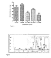
- Table XI shows the results from the study that was done. Oral administration of R. junceus venom brought a slight granulomatose tissue inhibition that took place compared to the negative control group, which suggests activity in the proliferative phase of inflammation. The two studied product doses had a similar effect. Table XI. Effect of R. junceus venom on cotton pellet-induced granulomas in rats. Group Granuloma weight (mg) Inhibition, % Control 103,33 ⁇ 5,08 a - 3 mg/kg dexamethasone 55,40 ⁇ 14,84 b 44,09 10 mg/kg R. Junceus venom 80,60 ⁇ 4,74 c 20,95 20 mg/kg R. Junceus venom 83,10 ⁇ 6,52 c 18,49 Values are the mean ⁇ SD. Groups having at least one common letter do not differ statistically. (p > 0.05.)
- Table XII shows granuloma weight and carmine content values for the different experimental groups.
- the toxin lowered the granuloma weight 1.32 and 2.05 times compared to the control group (p ⁇ 0.05) with its 3- and 5-mg/kg doses, respectively.
- Angiogenesis is a key process for tumor growth and metastasis, so having products which can inhibit this complex process is an alternative for treating tumor diseases.
- Table XIII shows the results of the analgesic effect of R. junceus venom inoculated intraperitonially to the 3% acetic acid-induced contorsions model in mice.
- the animals were placed on a (UGO-Basile) hot plate at a 55°C constant temperature both before and after administering the assay substance.
- the latency of the nociceptive response in the form of hind leg licking or jumping was measured. Only those animals which showed a nociceptive response within 20 seconds were chosen for the assay. Selection ended at 40 seconds.
- mice nociceptive reactivity to a thermal stimulus in mice was measured using the hot plate assay, which is an acute pain sensitivity test to detect opiod analgesia as well as some kinds of significant hyperanalgesic dorsal spine reactions.
- a substance is considered to have significant analgesic properties when an animal's normal reaction time doubles with its administration.
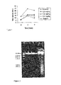
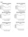
- R. junceus venom The analgesic effect of R. junceus venom is shown in Figure 1 and Table XV. Results indicate that IP administration of three doses of the product significantly reduced hot plate thermal stimulation. Figure 1 shows that the three R. junceus venom doses significantly expand nociceptive response latency spans during the hot plate assay with mice.
- Pain inhibition in the various experimental groups is shown in Table XV.
- a powerful heat-induced inhibition of algesia is observed, with the highest inhibition taking place 2 hours after administration. There is a potent effect after three hours, and although 2.5- and 7.5-mg/kg doses have a similar effect on the positive control it is the higher dose which has a longer effect which is greater than that of codeine.
- Venom exposure provoked significant growth inhibition (p ⁇ 0.05) in human and murine tumor cell lines in single-layer growing conditions.
- the U937 and S-180 tumor cell lines both of which grew in suspension) showed less sensitivity to the administration of the venom; the only effect was a slightly lesser growth (Table XVIII.)
- the effect of scorpion venom on an mammary adenocarcinoma model implanted in Balb c mice was evaluated.
- Four doses (6 mg/kg, 12.5 mg/kg, 25 mg/kg and 50 mg/kg) for the treatment groups were used.
- Controls were given a saline solution and all administrations were through oral route for 35 days. Tumor growth was monitored for 35 days.
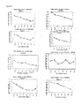
- the experimental groups administered with scorpion venom showed significant tumor growth inhibition (p ⁇ 0.05) compared to the control group ( Figure 4 .)
- the experimental groups showed a dose-response ratio during the 35 days of evaluation as to tumor progression retardation.
- Table XX Mean survival time of experimental groups implanted with 10 6 S-180 mice cells intraperitoneally and treated with R. junceus scorpion venom given orally.
- Table XXI Survival time among experimental groups implanted with 10 6 S-180 mice cells intraperitoneally and treated with R. junceus scorpion venom given orally.
- the 12.5-mg/kg dose gave a longer mean survival time (24 days), followed by the 25-mg/kg dose (22.5 days) and the 6-mg/kg dose (20.5 days), while the mean survival time of controls was 20 days (Table XXI.)
- the 12.5-mg/kg dose evidenced the highest survival percents by the time at which 100% of mice in the control group had died (Table XXI), while the 6-mg/kg and 25-mg/kg doses showed 26% and 22%, respectively.
- the longer survival time and the survival percents among the three experimental groups (6 mg/kg, 12.5 mg/kg and 25 mg/kg) were not statistically significant.
- the 50-mg/kg dose showed similar values to those of controls in all cases.
- the effect of protein fractions on cell growth was determined in a similar way as that in Example 6.
- the study used 2 tumor cell lines: HeLa (human cervix carcinoma) and A549 (human lung carcinoma) and the normal MRC-5 cell line (human lung fibroblasts.) Fractions were at a final 9 ⁇ g/mL-600 ⁇ g/mL concentration in wells.
- Table XXII shows the CC 50 for each of the purification fractions.
- Table XXII Relative molecular weights and mean cytotoxic concentration of fractions from molecular exclusion chromatography in tumor and normal cell lines. R.
- junceus venoms fractions Relative molecular weights * Mean cytotoxic concentration (CC 50 ) ( ⁇ g/mL) Hela A549 MRC-5 LB-01 30 kDa-72 kDa 835,3 420 245,1 LB-02 14 kDa- 30 kDa 208,7 250 276,9 LB-03 4 kDa - 8 kDa 140,1 116,6 439,2 LB-04 ⁇ 4 kDa 423,6 283 613,9 *Relative molecular weights determined from the log PM vs V o V e . calibration curve. MW: Molecular weights of pattern proteins. V o : Dead volume of column. V e : Elusion volume for each protein peak.
- the LB-03 and LB-04 fractions have a low molecular weight protein composition and, additionally, they were the fractions which showed higher cytotoxicity on tumor cells (Hela and A549) and low toxicity on normal cells (MRC-5.)
- Table XXIII Proportion of active principles in venom composition and range of in vitro biological activity on tumor cells. Active principle Proportion of venom, % In vitro biological activity range (%) RjLB-01 1,5-20 0.945-1,89 RjLB-03 8-9 4,5-9 RjLB-04 0,5-1,0 0,445-0,89 RjLB-05 0,5-0,7 0,33-0,71 RjLB-07 0,5-0,8 0,5-1,0 RjLB-08 0,5-1,0 1,5-3,0 RjLB-09 0,3-0,6 3,65-4,1 RjLB-14 0,4-0,8 3,5-6,0 (pag. 24)
- the molecular weights obtained for the RjLB-01, RjLB-03 and RjLB-04 peptides were 908 Da, 1964 Da and 4748,14 Da, respectively.
- the obtained sequences were compared to scorpion venom peptide data bases and no homology with previously described peptides was found, which is additional evidence of the novelty of results.
- Another result from this invention is the formulation obtained as follows: Fifty to one hundred Rhopalurus junceus species scorpions were taken and their venom extracted by way of electric stimulation and diluted in 10-20mL distilled water. It was then clarified through 10 000 rpm centrifugation for 15 minutes to get rid of components such as mucus and cell debris. At the same time, their proteins were determined using the Lowry method and the result was a concentration of 5-15 mg/mL. Then the scorpion venom was diluted conveniently with distilled water as the only excipient to obtain a formulation whose concentration range was 0.05-0.1 mg/mL.
- Example 1 Acute inflammation model of croton oil-induced auricular edema in mice.
- the cotton pellet granulose model in rats is a frequently used assay to study the effect on chronic inflammation conditions (Elieter, 1999.)
- Three inflammation phases after pellet implantation have been demonstrated.
- the last phase is that of cell proliferation occurring between the third and sixth days. This phase may be inhibited by anti-inflammatory steroids such as dexamethasone and by non-steroidal anti-inflammatory drugs (Sw, 1972.)
- the animals were sacrificed 7 days after the implantation of cotton plugs through the established euthanasia method and the pellets were extracted and studied.
- the pellet's end- and initial-weight difference was considered to be the granulomatose tissue produced.
- Inflammatory angiogenesis is a complex process involving a series of different but basically similar molecular mechanisms which develop in an angiogenic cascade.
- the granulomas were smaller at touch and a macroscopic observation of them showed less-defined ends and they were not so attached to the epidermis.
- the in vitro anti-oxidative activity by Rhopalurus junceus venom was evaluated through the DNA protective effect in Copper-Phenanthroline and Bleomycin-Iron systems oxidative processes.
- the venom showed anti-oxidative activity as it protected DNA from peroxidation processes.
- Example 6 In vitro cytotoxicity in a panel of tumor and normal cells
- HeLa human cervix carcinoma
- HEp-2 human laryngeal epidermoid carcinoma
- NCI-H292 human lung mucoepidermoid carcinoma
- A549 human lung carcinoma
- U937 human histyocitic lymphoma
- L929 murine fibrosarcoma
- S-180 murine sarcoma
- F311 murine breast sarcoma.
- 3 normal cell lines were used: MRC-5 (human lung fibroblasts), Vero (African green monkey normal kidney cells), and MDCK (normal dog kidney cells.)
- Peritoneal macrophages extracted from Balb/c mice peritonea
- lymphocytes extracted from Balc/c mice spleens
- Cells were grown within culture flasks in a minimal essential medium (MEM) or in a RPMI-1640 medium, depending on cell culture characteristics, and added 2 mM glutamine, non-essential amino acids, 10% bovine fetal serum (BFS), and 100 IU-100 ⁇ g/mL penicillin-streptomycin. Each one was incubated in a humid atmosphere at 37°C and 5-percent CO 2 till a single-layer was formed. Each cell line was separated by way of a 0.25% trypsin-EDTA solution and prepared at a 2 x 10 5 cell/mL concentration after being counted in a Neubauer chamber.
- MEM minimal essential medium
- BFS bovine fetal serum
- the assay was done in 96-well, flat-bottom cell culture polystyrene dishes (Coming Inc. costar R . ) 50 ⁇ L of each cell line was poured into each well and incubated at a 5-percent CO 2 and 37°C atmosphere for 24 hours. After such time, 50 ⁇ L medium containing previously dissolved venom was added. Final venom concentrations were 0.1 mg/mL, 0.25 Mg/mL, 0.5 mg/mL, 0.75 mg/mL and 1 mg/mL in wells. All final cell line concentrations were 10 4 cells/well. The bovine fetal serum (BFS) was used in the medium at 10%.
- BFS bovine fetal serum
- Dishes were incubated again at a 5-percent CO 2 and 37°C atmosphere for three days. After such time, a 10 ⁇ L MTT sterile solution (5 mg/mL tetrazolium salts in sterile PBS) was added to each well and incubated under the same conditions for 4 hours. Finally, the medium was poured off and a 200 ⁇ L/well dimethyl sulfoxide (DMSO) solution was added and incubated at 37°C for 30 minutes in a humid atmosphere. The optical density (OD) was read on a Revelation Dynex Technologies ELISA MRX microdish reader at 560 nm with 630 nm as reference. Each fractional concentration was done three times and the assay was performed four times.
- MTT sterile solution 5 mg/mL tetrazolium salts in sterile PBS
- CC 50 is the venom concentration that provokes a 50-percent decrease in the number of viable cells (MTT absorbance) compared to untreated controls and appears in Table XV for each of the evaluated cells.
- Example 8 Antitumor activity in solid tumors F311 breast adenocarcinoma experimental model . Intraperitoneal administration route
- the effect of scorpion venom on a mammary adenocarcinoma model implanted in Balc c. mice was evaluated. Three doses (0.2 mg/kg, 0.8 mg/kg and 3.2 mg/kg) were used for the treatment groups. Controls were administered a saline solution intraperitoneally. Tumor growth was monitored for 35 days. Fifty days after the implantation of the tumor the animals were sacrificed, their lungs were extracted and pulmonary metastasis was searched for.
- the experimental groups treated with scorpion venom showed significant tumor growth inhibition (p ⁇ 0.05) compared to the control group ( Figure 3 .)
- the experimental groups showed a dose-response ratio during the 35 days of evaluation as to tumor progression retardation.
- the significant reduction of tumor growth in the treated groups indicates that R. junceus scorpion venom has an antitumor effect as it influences tumor growth, at least during the evaluation period.
- Example 9 F311 breast adenocarcinoma. Experimental model. Oral administration route
- the effect of scorpion venom in a breast adenocarcinoma model that was injected in Balb c mice was evaluated.
- Four doses (6mg/kg, 12,5 mg/kg, 25 mg/kg and 50 mg/kg) were used for the treatment groups, the control group was administered a saline solution orally. Tumor growth was monitored for 35 days.
- the experimental groups treated with scorpion venom (12, 5 mg/kg, 25 mg/kg and 50 mg/kg) showed significant tumor growth inhibition (p ⁇ 0.05) when compared to the control group ( Figure 4 .)
- the significant decrease in tumor progression that was found in the treated groups demonstrates the antitumor effect of the R. junceus scorpion venom, as it affected tumor growth at least during the evaluation period.
- Example 11 Purification and identification of venom proteins as active principles.
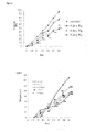
- the whole venom was dissolved in 0,1 M ammonium acetate (NH 4 Ac) and centrifuged at 10 000 rpm for 15 minutes. The supernatant was put in an AKTA FPLC (Amersham Pharmacia Biotech) low-pressure liquid chromatography equipment with a 12 HR 10/30 Superose filtration gel column measuring 10 x 300 mm. The column was balanced with 0.1M NH 4 Ac and the material mixed with same solvent at a flow rate of 0.5 mL/min. Absorbance was monitored at an optical density (OD) of 280 nm for 72 minutes.
- OD optical density
- the 75 HR 10/20 Superose molecular exclusion column was calibrated with a pattern protein kit including ribonuclease A (13.7 Kda), chemotripsinogen (25 Kda), ovoalbumin (43 Kda), albumin (67 Kda) and blue dextrane 2000.
- a pattern curve was drawn to determine the relative molecular weights of the various protein fractions obtained during chromatography. The results from chromatographic runs appear in Figure 6 .
- the purpose of the study was to assess life quality in cancer patients.
- the subjects included in the study were patients of both sexes with histologically confirmed cancer at any stage.
- a document was written which stated the informed consent of patients and a summary of the clinical record issued by the treating oncologist.
- the document included an evaluation of the clinical condition of patients as followed by their physicians in the hospital during their clinical evolution.
- follow-up periodicity was no less than two months.
- follow-up took place for a year.
- the formulation was prepared in 40-mL flasks with 0.05-0.1 mg/mL concentrations. Flask content was diluted in distilled water used as excipient to complete 1 litter.
- the product was administered daily by oral route and the recommended doses depended on patient condition and histological diagnosis.
- the administration of the product did not cause any adverse reaction at any moment during treatment, which coincided with observations during pre-clinical studies. All cases improved their quality of life as evidenced by an improvement in their main clinical variables such as less or no dysnea and coughing in all lung cancer patients. Many patients showed better radiological test results in the form of lesion stabilization in some cases and disappearance in others. Additionally, hematological variables were stabilized and pain and swelling reduced. Over 50% of the patients treated with the invented formulation for a year had a longer survival time than the one estimated for some of the studied conditions.
- the objective of the study was the evaluation of life quality in cancer patients. Patients of both sexes with histological confirmation of cancer in any stage were used to be included as study individuals.
- a document was prepared which contained the patient's consent act and the summary of the clinical record issued by the primary health care oncologist. The evaluation of the clinical behavior of patients was included in the document and the evaluation was followed up through the clinical progress and conducted by the specialists from the patients' origin hospitals. The periodicity of the follow-up was of at least two months. The follow-up was carried out during 1 year.
- the formulation was prepared in bottles with a volume of 40 mL with concentrations between 0.05-0.1 mg/mL.
- the flask was diluted in distilled water used as excipient until completing 1 liter.
- the product was administered on a daily basis through the oral route and the recommended dose depended on the patient's stage and histological diagnosis.
- the administration of the product caused adverse reactions during the treatment which coincided with what was observed in the preclinical studies.
- an improvement in the x-ray tests results as their lesions became stable in some case and in others lesions disappeared.
- the stabilization of the hematological variables was achieved and there was a reduction of pain and inflammation.
- the survival period estimated for some of the pathologies studied was exceeded.
- VIDATOX ® 30 CH is a homeopathic biotherapeutical drug developed at the Homeopathy Laboratory of LABIOFAM. Its active ingredient is Rhopalurus junceus scorpion venom in a 30 centesimal dilution. It comes in the form of drops in a 33% alcohol vehicle.
Landscapes
- Health & Medical Sciences (AREA)
- Life Sciences & Earth Sciences (AREA)
- Chemical & Material Sciences (AREA)
- Medicinal Chemistry (AREA)
- General Health & Medical Sciences (AREA)
- Pharmacology & Pharmacy (AREA)
- Animal Behavior & Ethology (AREA)
- Public Health (AREA)
- Veterinary Medicine (AREA)
- Organic Chemistry (AREA)
- Proteomics, Peptides & Aminoacids (AREA)
- Epidemiology (AREA)
- Gastroenterology & Hepatology (AREA)
- Insects & Arthropods (AREA)
- Immunology (AREA)
- Bioinformatics & Cheminformatics (AREA)
- Engineering & Computer Science (AREA)
- General Chemical & Material Sciences (AREA)
- Zoology (AREA)
- Chemical Kinetics & Catalysis (AREA)
- Tropical Medicine & Parasitology (AREA)
- Nuclear Medicine, Radiotherapy & Molecular Imaging (AREA)
- Molecular Biology (AREA)
- Biochemistry (AREA)
- Biophysics (AREA)
- Genetics & Genomics (AREA)
- Toxicology (AREA)
- Pain & Pain Management (AREA)
- Rheumatology (AREA)
- Medicines That Contain Protein Lipid Enzymes And Other Medicines (AREA)
- Peptides Or Proteins (AREA)
- Medicines Containing Material From Animals Or Micro-Organisms (AREA)
- Acyclic And Carbocyclic Compounds In Medicinal Compositions (AREA)
- Medicinal Preparation (AREA)
Abstract
This invention refers to new peptides obtained from Rhpalurus junceus scorpion venom, which has a high content of proteins, lipids, carbohydrates, amino acids, inorganic salts and other ions, including peptides as active principles. The invention also includes a formulation used as a drug due to its anticarcinogenic, analgesic and anti-inflammatory properties which improve the quality of life of cancer patients.
Description
- The present invention is linked essentially to the pharmaceutical industry and particularly to the identification of peptides obtained from the Rhopalurus junceus scorpion, Buthidae family, Rhopalurus genus, R. junceus species, common name "red" scorpion, venom which contains a mix of peptides, proteins, amino acids and free amines.
- Scorpions are land arthropods and, as poisonous animals, they are the oldest group as they were the first ones to develop on Earth. There are approximately over 1 500 scorpion species whose taxonomic classification is Phylum Arthropoda, Arachnida Class scorpions. In their natural habitat, the venom of scorpions is an opalescent, milky fluid with pH 7.12 and it contains mucus, lipids, carbohydrates, amino acids, inorganic salts, low-molecular weight organic molecules and a wide variety of proteins with molecular weights ranging 3 kDa-90 kDa which are the main component. Recently, research on scorpion venoms has taken momentum due to their high content of peptides that have shown a wide spectrum of pharmacological activity, for which reason they are invaluable tools for biomedical research (Martin-Eauclaire M-F, Segoard M, Ramos C, Cestele S, Bougis PE, and Svenson B, Production of active insect-specific scorpion neurotoxin in yeast. Eur.J. Biochem. 1994; 223; 637-45; Bednarek MA, Bugianesi RM, Leonard RJ, Felix JP. Chemical synthesis and structure-function studies of margatoxin, a potent inhibitor of voltage dependent potassium channel in human T lymphocytes. Biochem Biophys Res Commun. 1994 Jan 28; 198(2):619-25.) In this sense, recent years have seen a greater number of patent and non-patent publications about the activity of these toxins and their derivatives like anti-inflammatory components (Rajendra W, Armugan A and Jeyaseeian K. Toxins in anti-nociception and anti-inflammation. Toxicon 2004 July; 44(1):1-17); analgesics (Guan RJ, Wang CG, Wang M and Wang DC. A depressant insect toxin with a novel analgesic effect from scorpion Buthus martensii Karsch. Biochem. Biophys. File 2001; 1549(1):9-18), in cancer treatment (Liu YF, Ma RL, Wang SL, Duan ZY, Zhang JH, Wu LJ and Wu CF. Expression of an antitumor-analgesic peptide from the venom of Chinese scorpion Buthus martensi Karsch in Escherichia coli. Protein Expr. Purif. 2003; 27(2):253-8; Wang WX, Ji YH. Scorpion venom induces glioma cell apoptosis in vivo and inhibits glioma tumor growth in vitro. J Neurooncol. 2005 May; 73(1):1-7) and neurodegenerative diseases (Rajendra W, Armugam A and Jeyaseelan K. Toxins in anti-nociception and anti-inflammation. Toxicon 2004 July; 44(1):1-17.) Inventions whose composition include scorpion and/or its venom comprise a wide range of formulations intended for cancer therapy. The authors of the
1993 CN 1073480 andCN 1076858 patent papers refer they have obtained a wine allowing for cancer treatment and prevention, in both cases by mixing scorpion and other plant and animal materials. Scorpions are one of the components of formulations in the form of tablets for treating primary liver cancer (CN 1265901, 2000 andCN 1279088, 2001 ) and of capsules which inhibit tumor cell growth and can cure cancer in patients (CN 1391941, 2003 .) Other patents describe various formulations for cancer treatment (CN 1252321; 2000 CN 1316249, 2001 ;CN 1399979, 2003 .) - There are also patents on the exclusive use of toxins obtained from scorpion venom which act on specific tumor cell types. Early works on scorpion venom toxins referred that a peptide was obtained from the Leiurus quinquestriatus Hebrew scorpion. That toxin, which is a 4 KDa peptide named chlorotoxin, has a capacity to bind with the chloride channels expressed in gliomas (primary brain tumors from the glia (De Bin JA, Maggio JE, Strichartz GR. Purification and characterization of chlrotoxin, a chloride channel ligand from venom of the scorpion. Am J Physiol 1993; 264:361-369.) Based on this breakthroughs, in 1999 Ullrich et al. described in
US a diagnostic method for treating gliomas. Other works taking the specific action of chlorotoxin on certain cell receptors in gliomas as a basis showed that this toxin is a highly specific marker for other kinds of tumors whose cells have a common embryonic origin with the Central Nervous System (CNS) cells (Lyons SA, O'Neal J, Sontheimer H: Chlorotoxin, a scorpion derived peptide, specifically binds to gliomas and tumors of neuroectodermal origin. GLIA 2002; 39:162-73.) Additionally, recent works have shown that chlorotoxin not only inhibits "in vitro" glioma growth but has a capacity to prevent the invasion and spreading of this kind of tumors into brain regions which have not been damaged due to its specific and selective interaction with methalloproteinases, which are enzymes having to do with the high invasive rate of this kind of cancer (Deshane J, Garner CC and Sontheimer H: Chlorotoxin inhibits glioma cell invasion via matrix metalloproteinase-2. J Biol Chem 2003; 278:4135-44.) In 2004, another patent on the antitumor activity of an isolated toxin from the Buthus martensis karsh scorpion on animal-implanted tumors (patent 5 905 027EP20020774251 - In some cases, there is a wide variety of formulations with the ability to treat or cure cancer under the described patents. Yet, these cases do not prove this quality is unique of some of the types of scorpions and toxins involved in their manufacturing. On the other hand, the described toxins obtained from Leiurus quinquestriatus scorpions do not show antitumor activity per se, as in the case of chlorotoxin and Buthus martensis karsh, which can be used only to treat certain types of tumors as isolated toxins and not as a mix of toxins in whole venom.
- In Cuba, there is a Rhopalurus junceus scorpion venom diluted solution whose antitumor activity has been proved empirically in studies. During this work, the solution was administered to domestic animals with spontaneous tumors and tumor reduction and obliteration and good survival were observed (
CU 22413 A1 - The composition and pharmacological properties of Rhopalurus junceus scorpion venom and/or its derivatives make it a highly valuable natural drug and its anti-inflammatory, analgesic and anti-carcinogenic effects differentiate it substantially from other similar products in the market. It is a totally natural drug combining high-efficacy components to fight tumor cells (peptides) and others that provide it with analgesic and anti-inflammatory activity. Additionally, it has beneficial effects through the induction of tumor cell apoptosis and through its analgesic and anti-inflammatory properties. These features make it very valuable for improving the quality of life of patients suffering from cancer diseases and their related inflammatory conditions. It has been found that the composition of the natural product resulting from this invention includes a series of low molecular weight proteins with in vitro anti-carcinogenic activity whose action confirms potential in vivo antitumor activity.
- One of the purposes of this invention is determining the composition of and describing the peptides which are the active principles with antitumor activity found in venom. The summaries on qualitative composition, chemical screening, chemical-physical properties and pharmacological activity of scorpion venom are shown in Tables I, II, III, IV, V, VI and VII, respectively.
Table I. Chemical screening of Rhopalurus junceus whole venom used in formulations of this invention. Assay Results Dragendorff Assay Negative Iron Chloride Assay Negative Shinoda Assay Negative Lieberman-Burchard Assay Negative Ninhydrin Assay Positive Carbohydrates Positive Lipids Positive Table II. Determination of metal ions in Rhopalurus junceus whole venom. Sample Na (mg/L) Mg (mg/L) K (mg/L) Cu (mg/L) Zn (mg/L) Cd (mg/L) Pb (mg/L) Scorpion venom 114±21 4.12±0.28 4.66±0.35 0.069±0.006 11.18±0.7 <0.004* <0.01* *Heavy metals with levels below the lower limit Table III. Chemical-physical properties of Rhopalurus junceus scorpion venom used in formulations for this invention. Property Acceptance index pH 5-7 Protein content 5-15 mg/mL Appearance Opalescent Color Whitisth Table IV. Table of relative molecular weights (RMW) of proteins in scorpion venom ≥obtained through molecular exclusion using low-pressure liquid chromatography and polyacrylamide gel electrophoresis (SDS-PAGE.) Relative molecular weights ≥ 72 kDa 60 kDa 45 kDa 30 kDa 14 kDa 8 kDa ≤ 4 kDa Table V. Active principles with toxic activity on tumor cells identified from Rhopalurus junceus whole venom. Peptide Molecular weight Amount of amino acids RjLB-01 544.42 Da 10 RjLB-03 1964.0 Da 17 RjLB-04 4748.14 Da 43 RjLB-05 908.0 Da 8 RjLB-07 707.03 Da 7 RjLB-08 712.42 Da 17 RjLB-09 1203.44 Da 12 RjLB-14 5930.45 Da 50 Table VI. Summary of toxicological studies General toxicology R. junceus venom (mg/kg) Effect Acute toxicology Oral route 2000 N.E. IP route 5-20 DL50=16,41 mg/kg (M y H) Irritability Dermal - N.E. Ophthalmic - N.E. Mouth mucose - N.E. Repeat dose toxicity Repeat oral dose toxicity (28 days) 100 N.E. Subchronic oral toxicity (90 days) 0,1-100 N.E. Water and food intake N.E. Weight gain N.E. Hematological and biochemical parameters Relative weight of organs N.E. Pathological anatomy N.E. N.E. Especial toxicity R. junceus venom (mg/kg) Effect Mice bone marrow micronuclei Oral (acute) 2000 N.E. Oral (repeat doses, 28 days) 100 N.E. ip 4,10-13,13 N.E. Legend: N.E.: No effect Table VII. Summary of pharmacological studies Pharmacology Effect Antiproliferative Cytotoxicity in epithelial tumor cells Antitumoral Murine solid tumor growth retardation Antimetastasic Murine lung metastasis reduction Analgesic Reduced CNS and peripheral pain Anti-inflammatory Reduced inflammation after topical, oral and intraperitoneal administration - The following procedures were carried out in order to determine the composition of the mix in this invention.
- Determination of whole proteins through the Lowry method, modified.
- Chemical screening
- Separation of peptides as per molecular weight groups through low-pressure liquid chromatography separation using a 12 HR 10&30 Superose column.
- Polyacrylamide gel protein electrophoresis (SDS-PAGE.)
- Separation of whole venom and of peptides through high-resolution liquid chromatography using a reverse-phase C18 column.
- Determination of 220nm- and 280nm-wavelength UV spectra.
- Cell viability in normal and tumor cells.
- Analysis of each peptide of interest through mass spectrometry.
- According to the results from acute and subchronic toxicity assays done in mice, the product from this invention is non-toxic.
- The studies which are described in detail below as examples of the invention product do not limit the scope of this application whatsoever.
- In order to carry out the experiments given as examples of work, several scorpions were chosen and their venom was extracted through electrical stimulation and diluted in adequately distilled water. It was later clarified by centrifugation at 10 000 rpm for 15 minutes to eliminate components such as mucus and cell debris. Venom protein content determination was performed through the Lowry method, which showed a 5-15 mg/mL concentration.
- Assay substance and positive control were administered first. After 30 minutes, a 10-15µL croton oil solution was administered to each surface inside and outside the right ear of animals and the left ear was used as control. After three hours, animals were sacrificed following the usual standards for the species (according to FELASA), both ears were severed and round 8-mm diameter samples were taken with a punch and weighed on an analytical scale.
- First, the 25-30µL croton oil solution was applied on each of the inner and outer surfaces of the animal's right ear and the left ear was used as control. Then the assay substance was given immediately. After three hours, animals were sacrificed following the usual standards for the species (according to FELASA), both ears were severed and round 8-mm diameter samples were taken with a punch and weighed on an analytical scale.
- The 25-30µL croton oil solution was administered on each of the inner and outer surfaces of the animal's right ear and the left ear was used as control. Then the assay substance was administered by oral route immediately. After three hours, animals were sacrificed following the usual standards for the species (according to FELASA), both ears were severed and round 8-mm diameter samples were taken with a punch and weighed on an analytical scale.
- Topical administration of croton oil provides a proper skin inflammation model for evaluating anti-inflammatory agents. The croton oil active principle is Phorbol-12-Myristate 13-Acetate (PMA), which is a potent pro-inflammatory agent whose epicutaneous application results in histological and biochemical changes including higher vascular permeability and vascular rupturing, leukocytary infiltration, protein C activation and a greater release of arachidonic acid and its metabolites. PMA is also known to be a powerful neutrophil activator and there is wide and powerful blood irrigation in the ear.
- IP administration of the 1-5 mg/kg doses per body weight inhibited swelling induced by croton oil in mice ears (Table VIII), and the 3 mg/kg dose was more effective than the IP administration of 15 mg/kg dexamethasone in experimental animals as it had a higher inflammation inhibiting effect (97% vs 60.86%.) The 5 mg/kg dose did not differ statistically (p > 0.05) from the positive control (dexamethasone.)
Table VIII. Effect of intraperitoneal administration of R. junceus venom in the mice ear edema model Group Extinction of edema (mg) Inflammation inhibition, % Control 7,043 ± 0,27 - R. Junceus venom 1 mg/kg 5,050 ± 1,47 b 27,12 3 mg/kg 1,483 ± 0,93 c 97,46 5 mg/kg 4,020 ± 1,41 db 44,09 15 mg/kg dexamethasone 3,117 ± 1,09 d 60,86 Values are the mean ±SD. Groups having at least one common letter do not differ statistically. (p > 0.05.) - Administration of 5-, 10- and 20-mg R. junceus venom results in significant ear weight loss compared to the negative control group (Table IX), but it doesn't compare statistically to swelling inhibition induced by dexamethasone, which is a good steroidal anti-inflammatory drug that inhibits phospholipase A2 (the enzyme that is the route for arachidonic acid.)
Table IX. Effect of oral administration of R. junceus venom in the ear edema model in mice. Group Extinction of edema (m/g) Inflammation inhibition, % Control 11,28 ± 1,29a - R. junceus venom 5 mg/kg 7,68 ± 0,50 c 35,72 10 mg/ kg 8,05 ± 0,57 c 37,39 20 mg/kg 7,76 ± 1,01 c 38,02 15 mg/ kg dexamethasone 4,87 ± 0,95 b 53,70 Values are the mean ±SD. Groups having at least one common letter do not differ statistically. (p > 0.05.) - Topical administration of R. junceus venom resulted in auricular edema reduction compared to the control group, as shown in Table X.
Table X. Effect of topical administration of R. junceus venom in the ear edema model in mice. Group Extinction of edema (mg) Inflammation inhibition, % Control 12,00 ± 0,48 a - R. junceus venom 10 mg/ kg 8,87 ± 0,66 c 28,23 20 mg/ kg 8,25 ± 0,83 c 21,70 Bencydamine 4,87 ± 1,40 b 67,60 Values are the mean ±SD. Groups having at least one common letter do not differ statistically. (p > 0.05.) - Table XI shows the results from the study that was done. Oral administration of R. junceus venom brought a slight granulomatose tissue inhibition that took place compared to the negative control group, which suggests activity in the proliferative phase of inflammation. The two studied product doses had a similar effect.
Table XI. Effect of R. junceus venom on cotton pellet-induced granulomas in rats. Group Granuloma weight (mg) Inhibition, % Control 103,33 ± 5,08 a - 3 mg/ kg dexamethasone 55,40 ± 14,84 b 44,09 10 mg/kg R. Junceus venom 80,60 ± 4,74 c 20,95 20 mg/kg R. Junceus venom 83,10 ± 6,52 c 18,49 Values are the mean ±SD. Groups having at least one common letter do not differ statistically. (p > 0.05.) - Table XII shows granuloma weight and carmine content values for the different experimental groups. The toxin lowered the granuloma weight 1.32 and 2.05 times compared to the control group (p < 0.05) with its 3- and 5-mg/kg doses, respectively.
- Angiogenesis is a key process for tumor growth and metastasis, so having products which can inhibit this complex process is an alternative for treating tumor diseases.
- Carmine content is an indicator of the formation of new blood vessels in the air-pouch granuloma. Toxin administration reduced new blood vessel formation in the granulomatose tissue by 39.22% and 69.24% with the 3- 5-mg/kg (IP) doses, respectively, with a significant statistical difference between all experimental groups (p < 0.001.)
Table XII. Effect of R. Junceus venom administered intraperitonially on granuloma weight and carmine content in adjuvant-induced inflammatory angiogenesis in rats (n=10.) Group Granuloma weight (g±SD) Carmine content (A±SD) Control 5,26 ± 1,11a 0,413 ± 0,014a R. junceus venom 3 mg/ kg 3,59 ± 0,33b 0,251 ± 0,015b 5 mg/ kg 2,56 ± 0,74b 0,127 ± 0,020c Groups having at least one common letter do not differ statistically (p > 0.05.) - Table XIII shows the results of the analgesic effect of R. junceus venom inoculated intraperitonially to the 3% acetic acid-induced contorsions model in mice.
- All groups treated with the different venom doses showed much lower (p < 0.05) abdominal constrictions compared to the negative control group. The 2.5- and 7.5-mg/kg venom doses had a similar effect than that in the group given aspirin, a known analgesic drug. On the other hand, the 5.0 mg/kg R. Junceus venom dose showed a higher inhibition (87.74%) than that obtained from the administration of 100 mg/kg aspirin (49.28%.)
Table XIII. Effect of the intraperitonial administration of R. junceus venom in the acetic acid-induced abdominal contorsions model. Experimental groups Doses (mg/kg) No. contorsions X±DS % inhibition of No. of contorsions CN - 36,22 ± 5,11 a ------- ASA 150 18,37 ± 4,63 b 49,28 Venom of R. junceus 1 22,25 ± 4,53 b 38,57 2,5 17,87 ± 5,59 b 50,66 5 4,44 ± 4,33 c 87,74 7,5 17,86 ± 5,30 b 50,69 Experimental groups Dose (mg/kg) No. of contorsions X ± DS Inhibition in the No. of contorsions, % NC - 36,22 ± 5,11a ---------- ASA 150 18,37 ± 4,63b 49,28 R. junceus venom 1 22,25 ± 4,53b 38,57 2,5 17,87 ± 5,59b 50,66 5 4,44 ± 4,33 c 87,74 7,5 17,86 ± 5,30b 50,69 NC: Negative control; No: Number
ASA: Aspirin
Groups having at least one common letter do not differ statistically (p > 0.05.) - As evidenced in Table XIV, all groups which were given various venom doses showed a markedly lower number (p < 0.05) of abdominal constrictions compared to the negative control group. A comparison of the venom doses used with the positive control group (aspirin) showed a marked analgesic effect. Doses of 10-, 15- and 20-mg/kg R. junceus venom inhibited abdominal contractions in a way similar to that of aspirin. Whereas the 50-mg/kg dose did not bring about statistically significant differences from the positive control, it attained a 54.78% inhibition of abdominal contorsions in the experimental animals.
Table XIV. Inhibition of acetic acid-induced abdominal contorsions after the oral administration of R. junceus venom. Experimental groups Dose (mg/kg) Inhibition in the No. of contorsions, % NC - ------ ASA 150 47.01 1 18,84 5 32,84 10 36,23 15 39,85 20 43,19 50 54,78 NC: Negative control
ASA: Aspirin - The method described in Drug Discover was used.
- OF1 male mice weighing an average 20-25g from the National Center for Laboratory Animal Production (CENPALAB) were used.
(pag. 16) - The animals were placed on a (UGO-Basile) hot plate at a 55°C constant temperature both before and after administering the assay substance. The latency of the nociceptive response in the form of hind leg licking or jumping was measured. Only those animals which showed a nociceptive response within 20 seconds were chosen for the assay. Selection ended at 40 seconds.
- After administering a physiological saline solution to the negative control, 4% codeine to the positive control, and the various R. junceus venom doses, response latency time in each animal were measured after 2-3 hours.
- During this work, nociceptive reactivity to a thermal stimulus in mice was measured using the hot plate assay, which is an acute pain sensitivity test to detect opiod analgesia as well as some kinds of significant hyperanalgesic dorsal spine reactions. A substance is considered to have significant analgesic properties when an animal's normal reaction time doubles with its administration.
- The analgesic effect of R. junceus venom is shown in
Figure 1 and Table XV. Results indicate that IP administration of three doses of the product significantly reduced hot plate thermal stimulation.Figure 1 shows that the three R. junceus venom doses significantly expand nociceptive response latency spans during the hot plate assay with mice. - Pain inhibition in the various experimental groups is shown in Table XV. A powerful heat-induced inhibition of algesia is observed, with the highest
inhibition taking place 2 hours after administration. There is a potent effect after three hours, and although 2.5- and 7.5-mg/kg doses have a similar effect on the positive control it is the higher dose which has a longer effect which is greater than that of codeine.Table XV. Effect of R. jenceus venom administration in the hot plate model. Group Dose (mg/kg) Inhibition, % 2 h 3 h Control - 26,52 a 16,76 a R. junceus venom 2,5 173,96 b 92,53 b 5,0 109,19 c 36,31 a 7,5 145,55 bc 165,54 c Codeine 40 120,35 c 94,17 b Groups having at least one common letter do not differ statistically (p > 0.05.) - The results of the hot plate test evidence that the R. Junceus venom has an anti-cociceptive effect on the central nervous system that is higher than the one on the bone marrow as a greater and more complex neuron integration is needed for the licking and jumping response.
Table XVI. Pro-oxidative potential of R. junceus scorpion venom in the model for Fenanthrolin/Cu2+-induced DNA damage. Assay substance Concentration (**/mL) Extent of damage to DNA (*****at 532 nm; Median ±SD) Negative control - 0,069 ± 0,004 R. junceus scorpion venom 7,50 0,023 ± 0,009* 18,70 0,36 ± 0,002* 37,50 0,047 ± 0,003* 56,20 0,069 ± 0,012 75,0 0,075 ± 0,011 Ascorbic acid 200 µM 0,130 ± 0.006* Different letters mean a statistical difference (p < 0.05) compared to control. Table XVII. Pro-oxidative potential of R. junceus scorpion venom in the model for Fenanthrolin/Cu2+ -induced DNA damage. Assay substance Concentration (µg/mL) Extent of damage to DNA (OD at 532 nm: Median ± SD) Negative control - 0.40 ± 0,005 R. junceus scorpion venom 4,50 0,024 ± 0,006 9,01 0,026 ± 0,010 18,02 0,037 ± 0.008 36,04 0,024 ± 0,001 90,11 0,039 ± 0,001 Ascorbic acid 200 µM 0,647 ± 0,016* Different letters mean a statistical difference (p < 0.05) compared to control. Table XVIII. Cells used in studies to evaluate the in vitro biological activity of R. junceus scorpion venom. Cell lines Growth form CC50(mg/mL) HeLa (human cervix carcinoma) single- layer 1 HEp-2 (human larynx epidermoid carcinoma) single- layer 0,78 NCI-H292 (human lung mucoepidermoid carcinoma) single- layer 0,69 A549 (human lung carcinoma) single- layer 0,62 U937 (human histiocytic lymphoma) suspension ND L929 (murine fibrosarcoma) single- layer 1,3 S-180 (murine sarcoma) suspension ND F311 (murine mammary adenocarcinoma) single- layer 1,2 MRC-5 (human lung fibroblasts) Single- layer 2,2 Vero (African green monkey normal kidney cells) Single-layer ND MDCK (normal dog kidney cells) single-layer ND N2A (murine neuroblastoma cells) single- layer 1,4 Macrophages (extracted from Balc/c mice peritonea) - ND Lymphocytes (extracted from Balb/c mice spleens) suspension ND ND non-determined CC50: median cytotoxic concentration - Single-layer (carcinoma) growing cell cultures showed significant cell growth inhibition (p < 0.05) and higher sensitivity than normal cells and tumor cells growing in a suspension.
- A comparison between the CC50 of a normal MRC-5 human cell line with human tumor cells showed that R. junceus scorpion venom has a differential and significant cytotoxicity (p < 0,05) in tumor cells compared to normal cells (Table XVIII.)
- Venom exposure provoked significant growth inhibition (p < 0.05) in human and murine tumor cell lines in single-layer growing conditions. The U937 and S-180 tumor cell lines (both of which grew in suspension) showed less sensitivity to the administration of the venom; the only effect was a slightly lesser growth (Table XVIII.)
- Normal cells, lymphocytes and peritoneal macrophages showed less sensitivity than tumor cells. No significant toxic effects were observed (p > 0.05) along the full spectrum of used concentrations (Table XVIII.)
- An evaluation of pulmonary metastasis incidence after 50 days showed that the administration of scorpion venom significantly reduced (0.8 mg/kg; 3.2 mg/kg) the onset of spontaneous lung metastases (Table XIX.)
Table XIX. Incidence of lung metastasis onset in Balb/c mice implanted with F311 murine mammary tumor. *Statistically significant differences from control (p < 0.05.) Group Number of mice Metastasis incidence No. of metastasis nodules (range) Control 6 4/6 66% 0-7 0,2 mg/kg 7 5/7 71% 0-7 0,8 mg/ kg 5 2/5 42% 0-2* 3,2 mg/ kg 5 2/5 42% 0-3* - The effect of scorpion venom on an mammary adenocarcinoma model implanted in Balb c mice was evaluated. Four doses (6 mg/kg, 12.5 mg/kg, 25 mg/kg and 50 mg/kg) for the treatment groups were used. Controls were given a saline solution and all administrations were through oral route for 35 days. Tumor growth was monitored for 35 days.
- The experimental groups administered with scorpion venom showed significant tumor growth inhibition (p < 0.05) compared to the control group (
Figure 4 .) The experimental groups showed a dose-response ratio during the 35 days of evaluation as to tumor progression retardation.Table XX. Mean survival time of experimental groups implanted with 106 S-180 mice cells intraperitoneally and treated with R. junceus scorpion venom given orally. Dose Control 6 mg/ kg 12,5 mg/ kg 25 mg/ kg 50 mg/kg Survival time (days) 20 20,5 24 22,5 20 Table XXI. Survival time among experimental groups implanted with 106 S-180 mice cells intraperitoneally and treated with R. junceus scorpion venom given orally. Dose Control 6 mg/ kg 12,5 mg/ kg 25 mg/ kg 50 mg/kg Survival, % 0 26 37 22 0 - The 12.5-mg/kg dose gave a longer mean survival time (24 days), followed by the 25-mg/kg dose (22.5 days) and the 6-mg/kg dose (20.5 days), while the mean survival time of controls was 20 days (Table XXI.) Similarly, the 12.5-mg/kg dose evidenced the highest survival percents by the time at which 100% of mice in the control group had died (Table XXI), while the 6-mg/kg and 25-mg/kg doses showed 26% and 22%, respectively. Notwithstanding these results, the longer survival time and the survival percents among the three experimental groups (6 mg/kg, 12.5 mg/kg and 25 mg/kg) were not statistically significant. The 50-mg/kg dose showed similar values to those of controls in all cases.
- The effect of protein fractions on cell growth was determined in a similar way as that in Example 6. The study used 2 tumor cell lines: HeLa (human cervix carcinoma) and A549 (human lung carcinoma) and the normal MRC-5 cell line (human lung fibroblasts.) Fractions were at a final 9 µg/mL-600 µg/mL concentration in wells.
- The cytotoxity evaluation of cell line fractions evidenced inhibited growth of tumor cells and little toxicity in normal cells for the LB-03 and LB-04 fractions. Table XXII shows the CC50 for each of the purification fractions.
Table XXII. Relative molecular weights and mean cytotoxic concentration of fractions from molecular exclusion chromatography in tumor and normal cell lines. R. junceus venoms fractions Relative molecular weights* Mean cytotoxic concentration (CC50) (µg/mL) Hela A549 MRC-5 LB-01 30 kDa-72 kDa 835,3 420 245,1 LB-02 14 kDa- 30 kDa 208,7 250 276,9 LB-03 4 kDa - 8 kDa 140,1 116,6 439,2 LB-04 ≤4 kDa 423,6 283 613,9 *Relative molecular weights determined from the log PM vs VoVe. calibration curve.
MW: Molecular weights of pattern proteins.
Vo: Dead volume of column.
Ve: Elusion volume for each protein peak. - Fractions obtained from FPLC molecular exclusion chromatography were run in polyacrilamide gel electrophoresis with a 4-20% gradient to verify their protein composition, which appears in
Fig. 7 . - The LB-03 and LB-04 fractions have a low molecular weight protein composition and, additionally, they were the fractions which showed higher cytotoxicity on tumor cells (Hela and A549) and low toxicity on normal cells (MRC-5.)
- These fractions were re-purified through high-resolution liquid chromatography using a C18 reverse-phase analytical curve. Two solutions were used as mobile phase: Solution A (0.12% trifluoroacetic acid (TFA) in water) and Solution B (0.10% TFA in acetonitrile.) Fraction component elusion was done with gradual gradients from 0 to 70% of Solution B for 70 minutes and at a 0.5 ml/min flow and the fraction detection wavelength was 220 nm. The obtained pure peptides in tumor (Hela) and normal (CHO) cells were evaluated in a way similar to that described in Example 6 and their molecular weight and protein sequence were determined through mass spectrometry built in a high-resolution liquid chromatography device.
- Eight peptides (RjLB-01, RjLB-03, RjLB-04, RjLB-05, RjLB-07, RjLB-08, RjLB-09 and RjLB-14) with high cytotoxic activity on tumor cells and low toxicity on normal cells were obtained. These results corroborate what was observed with whole venom and confirm the preferential toxicity of venom components on tumor cells.
- Based on the purification of active components, their proportional presence in venom was determined (Table XXIII.)
Table XXIII. Proportion of active principles in venom composition and range of in vitro biological activity on tumor cells. Active principle Proportion of venom, % In vitro biological activity range (%) RjLB-01 1,5-20 0.945-1,89 RjLB-03 8-9 4,5-9 RjLB-04 0,5-1,0 0,445-0,89 RjLB-05 0,5-0,7 0,33-0,71 RjLB-07 0,5-0,8 0,5-1,0 RjLB-08 0,5-1,0 1,5-3,0 RjLB-09 0,3-0,6 3,65-4,1 RjLB-14 0,4-0,8 3,5-6,0 (pag. 24) - The molecular weights obtained for the RjLB-01, RjLB-03 and RjLB-04 peptides were 908 Da, 1964 Da and 4748,14 Da, respectively. The obtained sequences were compared to scorpion venom peptide data bases and no homology with previously described peptides was found, which is additional evidence of the novelty of results.
- Another result from this invention is the formulation obtained as follows: Fifty to one hundred Rhopalurus junceus species scorpions were taken and their venom extracted by way of electric stimulation and diluted in 10-20mL distilled water. It was then clarified through 10 000 rpm centrifugation for 15 minutes to get rid of components such as mucus and cell debris. At the same time, their proteins were determined using the Lowry method and the result was a concentration of 5-15 mg/mL. Then the scorpion venom was diluted conveniently with distilled water as the only excipient to obtain a formulation whose concentration range was 0.05-0.1 mg/mL.
-
-
Figure 1 shows the behavior of codeine (positive control), a central analgesic drug whose mean life fluctuates within 2-3 hours, which coincides with our results. -
Figure 2 shows 1.5% agarose gel electrophoresis of DNA extracted from the A549 and Hela tumor cell lines. Venom was given at an end concentration of 0.5 mg/mL per well. DNA from each cell line was extracted after 24 and 48 hours.
Track M: Lambda DNA/Hind III molecular weight marker.
Track 1-3: A549 cell line; 1: DNA control without venom; 2: DNA extracted after 24 hours; 3: DNA extracted after 48 hours.
Track 4-6: Hela cell line; 4: DNA control without venom; 5: DNA extracted after 24 hours; 6: DNA extracted after 48 hours. -
Figure 3 shows the kinetics of tumor growth in the experimental groups. Tumor growth was monitored for 35 days. Tumor growth inhibition depended on the venom doses that were given. The statistical significance as per control was *p < 0.05; **p < 0.01, ***p < 0.001. -
Figure 4 shows the kinetics of tumor growth in the experimental groups. Tumor growth was monitored for 35 days. Tumor growth inhibition depended on the venom doses that were given. The statistical significance as per control was *p < 0.05; **p < 0.01, ***p < 0.001. -
Figure 5 shows the effect t of oral administration of scorpion venom on the incidence of experimental lung metastases. Statistically significant difference: **p < 0.01, ***p < 0.001. -
Figure 6 shows the R. junceus scorpion venom chromatographic profile in low-pressure liquid chromatography using a 12 HR10/30 Superose molecular exclusion column. -
Figure 7 shows the polyacrylamide gel electrophoresis in reductive conditions. Fractions obtained from purification were applied using low-pressure liquid chromatography. Track 1: Molecular Weight Marker; Track 2: Whole Venom; Track 3: LB-01 Fraction; Track 4: LB-02 Fraction; Track 5: LB-03 Fraction; Track 6: LB-04 Fraction. -
Figure 8 shows the concentration-response curve for pure peptides which were obtained from the re-purification of evaluated fractions. The evaluation was performed on Hela (human cervix carcinoma) tumor cells. The concentrations used were 6.25 µg/mL-400 µg/mL. -
Figure 9 shows the concentration-response curve for pure peptides which were obtained from the re-purification of evaluated fractions. The evaluation was performed on CHO (Chinese Hamster Ovarium) non-tumor cells. The concentrations used were 6.25 µg/mL-800 µg/mL. - OF-1 male mice weighing an average 18-22 g from the National Center for Laboratory Animal Production (CENPALAB) were used. The method described by CYTED (Ibero-American Programme for Science and Technology Development, Lima, November of 1996) was used. A 0.5%-acetone croton oil solution was used. Dexamethasone was dissolved in 0.5% CMC.
- Sprague-Dawley rats of 180g body weight from the National Center for Laboratory Animal Production (CENPALAB) were used.
The method for cotton ball implantation from CYTED (Ibero-American Programme for Science and Technology Development, Lima, November of 1996) was carried out. Inflammation is the tissue response to damage. This includes enzyme involvement and activation, mediator release, fluid extravasation, cell migration and tissue rupture and repair (Vane, 1995.) It is well known that the anti-inflammatory effect may result from a series of chemical agents and that there is a slight correlation between their pharmacological activity and chemical structure (Sertie, 1990.) This, combined with the complexity of the inflammatory process, requires the use of various experimental models for carrying out a pharmacological assay. The cotton pellet granulose model in rats is a frequently used assay to study the effect on chronic inflammation conditions (Elieter, 1999.) Three inflammation phases after pellet implantation have been demonstrated. The last phase is that of cell proliferation occurring between the third and sixth days. This phase may be inhibited by anti-inflammatory steroids such as dexamethasone and by non-steroidal anti-inflammatory drugs (Sw, 1972.) - The animals were sacrificed 7 days after the implantation of cotton plugs through the established euthanasia method and the pellets were extracted and studied. The pellet's end- and initial-weight difference was considered to be the granulomatose tissue produced.
- Sprague-Dawley male rats from CENPALAB with 180-240 g body weight were used. Inflammatory angiogenesis is a complex process involving a series of different but basically similar molecular mechanisms which develop in an angiogenic cascade. Six days after granuloma formation was induced, the granuloma was well defined and palpable in the control group, which coincides with the results reported by Primelles in 2001. In the case of animals given venom doses, the granulomas were smaller at touch and a macroscopic observation of them showed less-defined ends and they were not so attached to the epidermis.
- The method described in CYTED was followed.
- OF1 male mice weighing an average 20-25g from the National Center for Laboratory Animal Production (CENPALAB) were used.
- Doses of 1-, 2.5- and 7.5-mg/kg R. junceus venom were inoculated intraperitoneally. Thirty minutes later, the experimental animals were administered 0.1 mL of a 3-percent acetic acid solution intraperitoneally and the number of contorsions/animal was recorded for 10 minutes beginning with the first contorsion observed.
- Doses of 1-, 5-, 10-, 15-, 20- and 50-mg/kg R. junceus venom were administered orally. One hour later, the experimental animals were administered 0.1 mL of a 3-percent acetic acid solution intraperitoneally and the number of contorsions/animal was recorded for 10 minutes beginning with the first contorsion observed.
- The in vitro anti-oxidative activity by Rhopalurus junceus venom was evaluated through the DNA protective effect in Copper-Phenanthroline and Bleomycin-Iron systems oxidative processes. The venom showed anti-oxidative activity as it protected DNA from peroxidation processes.
- The Copper-Phenanthroline and Biomycin-Iron systems showed that the Rhopalurus junceus derivative in the above-described formulations has no pro-oxidative effect on DNA. The absorbance values obtained with various Rhopalurus junceus derivative concentrations are lower than those obtained with the positive control. Tables XVI and XVII show the results from such evaluation.
- The effect of venom on cell growth was seen in the MTT assay, which measures cell proliferation resulting from the metabolic reduction of this (yellow) tetrazolium salt by mitochondrial dehydrogenated enzymes. The resulting compound (blue formazan) can be made soluble and quantified spectrophotometrically
- Nine tumor cell lines were used in the study: HeLa (human cervix carcinoma), HEp-2 (human laryngeal epidermoid carcinoma), NCI-H292 (human lung mucoepidermoid carcinoma), A549 (human lung carcinoma), U937 (human histyocitic lymphoma), L929 (murine fibrosarcoma), S-180 (murine sarcoma), and F311 (murine breast sarcoma.) Equally, 3 normal cell lines were used: MRC-5 (human lung fibroblasts), Vero (African green monkey normal kidney cells), and MDCK (normal dog kidney cells.) Peritoneal macrophages (extracted from Balb/c mice peritonea) and lymphocytes (extracted from Balc/c mice spleens) were also evaluated. Cells were grown within culture flasks in a minimal essential medium (MEM) or in a RPMI-1640 medium, depending on cell culture characteristics, and added 2 mM glutamine, non-essential amino acids, 10% bovine fetal serum (BFS), and 100 IU-100 µg/mL penicillin-streptomycin. Each one was incubated in a humid atmosphere at 37°C and 5-percent CO2 till a single-layer was formed. Each cell line was separated by way of a 0.25% trypsin-EDTA solution and prepared at a 2 x 10⁵ cell/mL concentration after being counted in a Neubauer chamber.
- The assay was done in 96-well, flat-bottom cell culture polystyrene dishes (Coming Inc. costarR.) 50 µL of each cell line was poured into each well and incubated at a 5-percent CO2 and 37°C atmosphere for 24 hours. After such time, 50 µL medium containing previously dissolved venom was added. Final venom concentrations were 0.1 mg/mL, 0.25 Mg/mL, 0.5 mg/mL, 0.75 mg/mL and 1 mg/mL in wells. All final cell line concentrations were 104 cells/well. The bovine fetal serum (BFS) was used in the medium at 10%.
- Dishes were incubated again at a 5-percent CO2 and 37°C atmosphere for three days. After such time, a 10 µL MTT sterile solution (5 mg/mL tetrazolium salts in sterile PBS) was added to each well and incubated under the same conditions for 4 hours. Finally, the medium was poured off and a 200 µL/well dimethyl sulfoxide (DMSO) solution was added and incubated at 37°C for 30 minutes in a humid atmosphere. The optical density (OD) was read on a Revelation Dynex Technologies ELISA MRX microdish reader at 560 nm with 630 nm as reference. Each fractional concentration was done three times and the assay was performed four times.
- For the concentration-response curves analysis, the cell proliferation percent was graphically obtained from the formula (1-OD (sample)/OD (control)) X100 against the different venom concentrations. Mean cytotoxic concentration was expressed as CC50, which is the venom concentration that provokes a 50-percent decrease in the number of viable cells (MTT absorbance) compared to untreated controls and appears in Table XV for each of the evaluated cells.
- The study for determining apoptosis was done through DNA fragmentation. The Hela and A549 cell lines were used. Results are shown in
Figure 2 . The characteristic DNA fragmentation of apoptosis was observed in the corresponding tracks of Hela cells treated with venom, while a single intact chromosomal DNA band in control wells and in the A549 cell line was observed. These results show that scorpion venom may induce cell death through both mechanisms -apoptosis and necrosis. - The effect of scorpion venom on a mammary adenocarcinoma model implanted in Balc c. mice was evaluated. Three doses (0.2 mg/kg, 0.8 mg/kg and 3.2 mg/kg) were used for the treatment groups. Controls were administered a saline solution intraperitoneally. Tumor growth was monitored for 35 days. Fifty days after the implantation of the tumor the animals were sacrificed, their lungs were extracted and pulmonary metastasis was searched for.
- The experimental groups treated with scorpion venom showed significant tumor growth inhibition (p < 0.05) compared to the control group (
Figure 3 .) The experimental groups showed a dose-response ratio during the 35 days of evaluation as to tumor progression retardation. The significant reduction of tumor growth in the treated groups indicates that R. junceus scorpion venom has an antitumor effect as it influences tumor growth, at least during the evaluation period. - The effect of scorpion venom in a breast adenocarcinoma model that was injected in Balb c mice was evaluated. Four doses (6mg/kg, 12,5 mg/kg, 25 mg/kg and 50 mg/kg) were used for the treatment groups, the control group was administered a saline solution orally. Tumor growth was monitored for 35 days.
The experimental groups treated with scorpion venom (12, 5 mg/kg, 25 mg/kg and 50 mg/kg) showed significant tumor growth inhibition (p<0.05) when compared to the control group (Figure 4 .) The significant decrease in tumor progression that was found in the treated groups demonstrates the antitumor effect of the R. junceus scorpion venom, as it affected tumor growth at least during the evaluation period. - The effect of scorpion venom in a murine sarcoma model implanted in the peritoneum of NMRI mice was evaluated. Four doses (6 mg/kg, 12.5 mg/kg, 25 mg/kg and 50 mg/kg) for treatment groups were used. The control group was given a saline solution. The venom was administered daily 24 hours after the implantation of the tumor until all experimental groups died. Table XX shows the survival time results with the treated animals and controls. Table XXI shows the survival percents among experimental groups.
- In order to separate whole protein content, the whole venom was dissolved in 0,1 M ammonium acetate (NH4Ac) and centrifuged at 10 000 rpm for 15 minutes. The supernatant was put in an AKTA FPLC (Amersham Pharmacia Biotech) low-pressure liquid chromatography equipment with a 12
HR 10/30 Superose filtration gel column measuring 10 x 300 mm. The column was balanced with 0.1M NH4Ac and the material mixed with same solvent at a flow rate of 0.5 mL/min. Absorbance was monitored at an optical density (OD) of 280 nm for 72 minutes. The 75HR 10/20 Superose molecular exclusion column was calibrated with a pattern protein kit including ribonuclease A (13.7 Kda), chemotripsinogen (25 Kda), ovoalbumin (43 Kda), albumin (67 Kda) and blue dextrane 2000. A pattern curve was drawn to determine the relative molecular weights of the various protein fractions obtained during chromatography. The results from chromatographic runs appear inFigure 6 . - The purpose of the study was to assess life quality in cancer patients. The subjects included in the study were patients of both sexes with histologically confirmed cancer at any stage. A document was written which stated the informed consent of patients and a summary of the clinical record issued by the treating oncologist. The document included an evaluation of the clinical condition of patients as followed by their physicians in the hospital during their clinical evolution. Follow-up periodicity was no less than two months. Follow-up took place for a year. The formulation was prepared in 40-mL flasks with 0.05-0.1 mg/mL concentrations. Flask content was diluted in distilled water used as excipient to complete 1 litter. The product was administered daily by oral route and the recommended doses depended on patient condition and histological diagnosis.
A total of 100 patients were included in the study and the main evaluated cancer locations were breast, prostate, colon, lung, brain and pancreas. The administration of the product did not cause any adverse reaction at any moment during treatment, which coincided with observations during pre-clinical studies. All cases improved their quality of life as evidenced by an improvement in their main clinical variables such as less or no dysnea and coughing in all lung cancer patients. Many patients showed better radiological test results in the form of lesion stabilization in some cases and disappearance in others. Additionally, hematological variables were stabilized and pain and swelling reduced. Over 50% of the patients treated with the invented formulation for a year had a longer survival time than the one estimated for some of the studied conditions. - The objective of the study was the evaluation of life quality in cancer patients. Patients of both sexes with histological confirmation of cancer in any stage were used to be included as study individuals. A document was prepared which contained the patient's consent act and the summary of the clinical record issued by the primary health care oncologist.
The evaluation of the clinical behavior of patients was included in the document and the evaluation was followed up through the clinical progress and conducted by the specialists from the patients' origin hospitals. The periodicity of the follow-up was of at least two months. The follow-up was carried out during 1 year. The formulation was prepared in bottles with a volume of 40 mL with concentrations between 0.05-0.1 mg/mL. The flask was diluted in distilled water used as excipient until completing 1 liter. The product was administered on a daily basis through the oral route and the recommended dose depended on the patient's stage and histological diagnosis. - A total of 100 patients were included in the study and the main locations for cancer were, breast, prostate, colon, lung, brain and pancreas. In no case the administration of the product caused adverse reactions during the treatment which coincided with what was observed in the preclinical studies. In all cases it was observed an increase in life quality including an improvement of the main clinical variables such as decrease or disappearance of dyspnea and coughing in all cases of lung cancer patients. In many patients it was verified an improvement in the x-ray tests results as their lesions became stable in some case and in others lesions disappeared. In addition to this, the stabilization of the hematological variables was achieved and there was a reduction of pain and inflammation. In over 50 % of the patients treated for 1 year with the formulation object of the invention, the survival period estimated for some of the pathologies studied was exceeded.
-
VIDATOX ® 30 CH is a homeopathic biotherapeutical drug developed at the Homeopathy Laboratory of LABIOFAM. Its active ingredient is Rhopalurus junceus scorpion venom in a 30 centesimal dilution. It comes in the form of drops in a 33% alcohol vehicle.
The studies carried out in 174 patients, of both sexes, having specific cancer treatments demonstrated the adjuvanting effect ofVIDATOX ® 30 CH for treating cancer in different body location, (lung, prostate, breast, colon, pancreas, t. lymphoid, brain, rectum etc.) Its results show that 96 % of patients administered with the product had a survival of over 12 months, that 90% of the patients reported improvement in their clinical symptoms seen by physicians and that among these 63 patients, pain (as the predominant symptom) for 62 % of them evolved to a moderate form, which did not require relief treatment, that the 27 % of them reported no pain and none of them reported adverse reactions to the treatment.

Claims (14)
- Peptide called RjLB-01, obtained from scorpion venom and characterized by having 544.42 Da molecular weight and SEQ. DI No. 1 amino acid sequence.
- Peptide called RjLB-03, obtained from scorpion venom and characterized by having 1964.0 Da molecular weight and SEQ. DI No. 2 amino acid sequence.
- Peptide called RjLB-04, obtained from scorpion venom and characterized by having 4748.14 Da molecular weight SEQ. DI No. 3 amino acid sequence.
- Peptide called RjLB-05, obtained from scorpion venom and characterized having 908.0 molecular weight of Da and SEQ. DI No. 4 amino acid sequence.
- Peptide called RjLB-07, obtained from scorpion venom and characterized by having 707.03 Da molecular weight and SEQ. DI No. 5 amino acid sequence.
- Peptide called RjLB-08, obtained from scorpion venom and characterized by having 712.42 Da molecular weight and SEQ. DI No. 6 amino acid sequence.
- Peptide named RjLB-09, obtained from scorpion venom and characterized by having 1203.44 Da molecular weight and SEQ. ID No. 7 amino acid sequence.
- Peptide called RjLB-014, obtained from scorpion venom and characterized by having 5930. 45 Da molecular weight and SEQ. DI No. 8 amino acid sequence.
- Pharmaceutical compositions that are obtained from Rhopalurus junceus scorpion venom, characterized by having peptides as active principles with high cytotoxic activity on tumor cells, mixed with distilled water as excipient
- Pharmaceutical compositions as per claim 9 which are processed-homeopathically.
- Pharmaceutical compositions as per claim 9, containing at least one peptide according to claims 1-8, which has antitumor activity, and such selected peptide having any of the described amino acid sequence.
- Pharmaceutical compositions as per claim 11 characterized by having a mix of peptides contained in Rhopalurus junceus scorpion venom with the following concentrations: RJL-01, at a 1,5-2,0% range; RJLB-03, at a 8-9% range; RJLB-07, in a 0,5-0,8% range; RJLB-08 at a 0,5-1,0% range; RJLB-09, at a 0,3-0,60% range; RJLB-14, at a 0, 4-0-8% range, diluted in 10-20 mL distilled water.
- Pharmaceutical compositions, as per claim 12, processed homeopathically.
- Pharmaceutical compositions, as per claims 9, 10,11, 12 and 13, used in formulations to be administered by oral, topical, parenteral, rectal, vaginal and aerosol routes.
Applications Claiming Priority (2)
| Application Number | Priority Date | Filing Date | Title |
|---|---|---|---|
| CU2010000186A CU24055B1 (en) | 2010-09-27 | 2010-09-27 | PHARMACEUTICAL COMPOSITIONS OF RHOPALURUS JUNCEUS SCORPION POISON |
| PCT/CU2011/000006 WO2012041261A2 (en) | 2010-09-27 | 2011-09-27 | Peptides from the venom of the rhopalurus junceus scorpion and pharmaceutical composition |
Publications (1)
| Publication Number | Publication Date |
|---|---|
| EP2623111A2 true EP2623111A2 (en) | 2013-08-07 |
Family
ID=45566782
Family Applications (1)
| Application Number | Title | Priority Date | Filing Date |
|---|---|---|---|
| EP11815845.0A Withdrawn EP2623111A2 (en) | 2010-09-27 | 2011-09-27 | Peptides from the venom of the rhopalurus junceus scorpion and pharmaceutical composition |
Country Status (17)
| Country | Link |
|---|---|
| EP (1) | EP2623111A2 (en) |
| JP (1) | JP2013542196A (en) |
| KR (1) | KR20140041381A (en) |
| CN (1) | CN103200951A (en) |
| AP (1) | AP2013006811A0 (en) |
| AR (1) | AR083117A1 (en) |
| AU (1) | AU2011307691A1 (en) |
| BR (1) | BR112013007198A2 (en) |
| CA (1) | CA2812841A1 (en) |
| CO (1) | CO6781464A2 (en) |
| CU (1) | CU24055B1 (en) |
| EC (1) | ECSP13012595A (en) |
| IL (1) | IL225577A0 (en) |
| MX (1) | MX360726B (en) |
| RU (1) | RU2563348C2 (en) |
| SG (1) | SG191708A1 (en) |
| WO (1) | WO2012041261A2 (en) |
Families Citing this family (5)
| Publication number | Priority date | Publication date | Assignee | Title |
|---|---|---|---|---|
| CN103074304B (en) * | 2013-01-28 | 2016-01-13 | 江苏健安生物科技有限公司 | High level human diploid cell cultivates varicella-zoster vaccine virus method |
| MX2019000570A (en) * | 2018-12-19 | 2020-08-13 | Centro De Investig Biologicas Del Noroeste S C | Homeopathic composition based on venom of the scorpion rhopalurus junceus and use in aquaculture. |
| WO2022133540A1 (en) * | 2020-12-22 | 2022-06-30 | PreveCeutical Medical Inc. | Cyclic peptides and uses thereof |
| CN113384682B (en) * | 2021-05-31 | 2023-07-04 | 南方医科大学 | Application of scorpion venom polypeptide Smp43 in preparation of antitumor drugs |
| CN117069800B (en) * | 2023-10-16 | 2023-12-12 | 山东省食品药品检验研究院 | Characteristic polypeptide for identifying Buthus martensii Karsch and application thereof |
Family Cites Families (15)
| Publication number | Priority date | Publication date | Assignee | Title |
|---|---|---|---|---|
| CN1030229C (en) | 1991-12-16 | 1995-11-08 | 山东省蒙阴县酒厂 | Method for making scorpion wine |
| CN1076858A (en) | 1993-03-24 | 1993-10-06 | 王强华 | Fel Ursi scorpion liquor and preparation technology thereof |
| CU22413A1 (en) | 1994-01-11 | 1996-01-31 | Antitumorigenic composition | |
| US5905027A (en) | 1995-12-27 | 1999-05-18 | Uab Research Foundation | Method of diagnosing and treating gliomas |
| FR2745004B1 (en) * | 1996-02-16 | 1998-03-27 | Rhone Poulenc Agrochimie | ANTIBACTERIAL AND ANTIFUNGAL PEPTIDE |
| US6168651B1 (en) | 1998-10-08 | 2001-01-02 | Donaldson Company, Inc. | Filter assembly with shaped adsorbent article; and devices and methods of use |
| CN1279088A (en) | 1999-06-25 | 2001-01-10 | 谷令旗 | Process for preparing medicine to cure liver cancer |
| CN1265901A (en) | 2000-01-26 | 2000-09-13 | 徐忠廷 | Traditional Chinese medicinal pill for anti-cancer |
| CN1316249A (en) | 2001-04-04 | 2001-10-10 | 李滋星 | Nutritive active liquid of scorpion |
| CN1157215C (en) | 2001-07-30 | 2004-07-14 | 张官锁 | Chinese medicine composition for curing cancers |
| CN1325514C (en) * | 2001-09-30 | 2007-07-11 | 沈阳药科大学 | Scorpion pain-stopping anti-tumor Val-Arg-Gly peptide and its preparation method |
| CN100465272C (en) * | 2002-01-18 | 2009-03-04 | 中国科学院上海有机化学研究所 | Genetic engineering of rBmKaIT1 recombinant East Asian scorpion venom rBmKaIT1 |
| CA2487425A1 (en) * | 2002-05-31 | 2003-12-11 | Transmolecular, Inc. | Combination chemotherapy with chlorotoxin |
| CN1256971C (en) | 2002-08-12 | 2006-05-24 | 徐继康 | Capsule for curing tumor and its preparing method |
| US8097284B2 (en) * | 2007-11-13 | 2012-01-17 | Arthur Mikaelian | Polarized scorpion venom solution and a method for making polarized scorpion venom solution |
-
2010
- 2010-09-27 CU CU2010000186A patent/CU24055B1/en not_active IP Right Cessation
-
2011
- 2011-09-27 WO PCT/CU2011/000006 patent/WO2012041261A2/en active Application Filing
- 2011-09-27 AU AU2011307691A patent/AU2011307691A1/en not_active Abandoned
- 2011-09-27 KR KR1020137010868A patent/KR20140041381A/en not_active Ceased
- 2011-09-27 RU RU2013119431/10A patent/RU2563348C2/en active IP Right Revival
- 2011-09-27 CN CN2011800541685A patent/CN103200951A/en active Pending
- 2011-09-27 JP JP2013530559A patent/JP2013542196A/en active Pending
- 2011-09-27 BR BR112013007198A patent/BR112013007198A2/en not_active IP Right Cessation
- 2011-09-27 EP EP11815845.0A patent/EP2623111A2/en not_active Withdrawn
- 2011-09-27 CA CA2812841A patent/CA2812841A1/en not_active Abandoned
- 2011-09-27 MX MX2013003545A patent/MX360726B/en active IP Right Grant
- 2011-09-27 AR ARP110103520A patent/AR083117A1/en not_active Application Discontinuation
- 2011-09-27 SG SG2013022850A patent/SG191708A1/en unknown
- 2011-09-27 AP AP2013006811A patent/AP2013006811A0/en unknown
-
2013
- 2013-04-04 IL IL225577A patent/IL225577A0/en unknown
- 2013-04-25 CO CO13105491A patent/CO6781464A2/en not_active Application Discontinuation
- 2013-04-29 EC ECSP13012595 patent/ECSP13012595A/en unknown
Non-Patent Citations (1)
| Title |
|---|
| See references of WO2012041261A2 * |
Also Published As
| Publication number | Publication date |
|---|---|
| CU20100186A7 (en) | 2012-06-21 |
| CO6781464A2 (en) | 2013-10-31 |
| SG191708A1 (en) | 2013-08-30 |
| JP2013542196A (en) | 2013-11-21 |
| KR20140041381A (en) | 2014-04-04 |
| RU2563348C2 (en) | 2015-09-20 |
| ECSP13012595A (en) | 2014-11-28 |
| MX2013003545A (en) | 2013-11-04 |
| CA2812841A1 (en) | 2012-04-05 |
| AR083117A1 (en) | 2013-01-30 |
| AP2013006811A0 (en) | 2013-04-30 |
| RU2013119431A (en) | 2014-11-10 |
| AU2011307691A1 (en) | 2013-05-02 |
| CN103200951A (en) | 2013-07-10 |
| WO2012041261A2 (en) | 2012-04-05 |
| IL225577A0 (en) | 2013-06-27 |
| MX360726B (en) | 2018-11-13 |
| BR112013007198A2 (en) | 2016-06-14 |
| WO2012041261A3 (en) | 2012-11-22 |
| CU24055B1 (en) | 2014-12-26 |
Similar Documents
| Publication | Publication Date | Title |
|---|---|---|
| Pezzani et al. | Anticancer properties of bromelain: State-of-the-art and recent trends | |
| EP2623111A2 (en) | Peptides from the venom of the rhopalurus junceus scorpion and pharmaceutical composition | |
| KR20190101420A (en) | Composition of mannuron diacid | |
| CN101485655A (en) | Application of dihydromyricetin in preparing medicament for preventing and treating adverse reaction of tumor chemoradiotherapy | |
| Nongalleima et al. | Citrus macroptera Montruz extracts | |
| Ibrahim et al. | Leaves extract of Muntingia calabura protects against gastric ulcer induced by ethanol in Sprague-dawley rats | |
| CN104116734A (en) | Pharmaceutical composition and pharmaceutical preparation for treating oral and throat diseases, and applications and preparation method of the pharmaceutical composition and pharmaceutical preparation | |
| CN110464727A (en) | The application of 3,4- dihydro -2H- benzo-[1,4] oxazines class drug or its salt in preparation inhibition iron death drug | |
| KR20040099300A (en) | Isolation, purification and structural identification of a bioactive component of a water soluble extract of a botanical species for enhancing immune, anti-inflammatory, anti-tumor and dna repair processes of warm blooded animals | |
| CN112409439B (en) | Glycyrrhizic acid derivative, preparation method and application | |
| Balamurugan et al. | Anti epileptic activity of poly herbal extract from Indian medicinal plants | |
| Chandrappa et al. | In vitro anti-inflammatory activity of Carmona retusa (Vahl.) | |
| Moran et al. | Insect venoms and their bioactive components: A novel therapeutic approach in chronic diseases and cancer | |
| CN106220643A (en) | Ganolactone D and pharmaceutical composition thereof and its application in pharmacy and food | |
| JP5231244B2 (en) | Momordica extract and momordica saponin I isolated therefrom useful for preventing and treating gastritis or gastric ulcer | |
| EP2606883A1 (en) | Uses of n-Butylidenephthalide in Treating a Liver Injury and Improving Liver Function | |
| Yang et al. | A new method for purifying Brazilin from lignum sappan–Cytotoxic and anti-proliferative effects in cell lines and improved survival in mice bearing urinary bladder carcinoma | |
| CN114456218A (en) | A blueberry anthocyanin and its preparation method and its application in the preparation of functional food against acrylamide neurotoxicity | |
| KR101782962B1 (en) | Composition for treatment of leukemia and method of manufacturing extract from myrrh | |
| Indrawati et al. | The Effect of an Annona muricata Leaves Extract on Circulating Soluble Adhesion Molecules in Colon Carcinogenesis Model | |
| CN101951932B (en) | Use of extract of piper betel leaves for the treatment of human malignancies sensitive to oxidative stress | |
| CN107468684B (en) | Application of ellagic acid and its synergy with tripterine in the preparation of anti-lung cancer drugs | |
| Verma et al. | Anti-oxidant, Anti-inflammatory and Anti-Microbial Activity of Pterocarpus santalinus L. f | |
| Sarwar et al. | Phytochemical and biological studies of Panicum antidotale aerial parts ethanol extract supported by molecular docking study | |
| Agnihotri | Anti-inflammatory profiling of seed and rind extracts of Amomum subulatum |
Legal Events
| Date | Code | Title | Description |
|---|---|---|---|
| PUAI | Public reference made under article 153(3) epc to a published international application that has entered the european phase |
Free format text: ORIGINAL CODE: 0009012 |
|
| 17P | Request for examination filed |
Effective date: 20130425 |
|
| AK | Designated contracting states |
Kind code of ref document: A2 Designated state(s): AL AT BE BG CH CY CZ DE DK EE ES FI FR GB GR HR HU IE IS IT LI LT LU LV MC MK MT NL NO PL PT RO RS SE SI SK SM TR |
|
| DAX | Request for extension of the european patent (deleted) | ||
| 17Q | First examination report despatched |
Effective date: 20141103 |
|
| STAA | Information on the status of an ep patent application or granted ep patent |
Free format text: STATUS: THE APPLICATION IS DEEMED TO BE WITHDRAWN |
|
| 18D | Application deemed to be withdrawn |
Effective date: 20160401 |