EP2623106A1 - Composition for regenerating follicle which contains cd36-expressing connective tissue sheath cells - Google Patents
Composition for regenerating follicle which contains cd36-expressing connective tissue sheath cells Download PDFInfo
- Publication number
- EP2623106A1 EP2623106A1 EP10857831.1A EP10857831A EP2623106A1 EP 2623106 A1 EP2623106 A1 EP 2623106A1 EP 10857831 A EP10857831 A EP 10857831A EP 2623106 A1 EP2623106 A1 EP 2623106A1
- Authority
- EP
- European Patent Office
- Prior art keywords
- cells
- dsc
- animal
- hair follicles
- dpc
- Prior art date
- Legal status (The legal status is an assumption and is not a legal conclusion. Google has not performed a legal analysis and makes no representation as to the accuracy of the status listed.)
- Granted
Links
Images


Classifications
-
- A—HUMAN NECESSITIES
- A01—AGRICULTURE; FORESTRY; ANIMAL HUSBANDRY; HUNTING; TRAPPING; FISHING
- A01K—ANIMAL HUSBANDRY; AVICULTURE; APICULTURE; PISCICULTURE; FISHING; REARING OR BREEDING ANIMALS, NOT OTHERWISE PROVIDED FOR; NEW BREEDS OF ANIMALS
- A01K67/00—Rearing or breeding animals, not otherwise provided for; New or modified breeds of animals
- A01K67/027—New or modified breeds of vertebrates
-
- A—HUMAN NECESSITIES
- A01—AGRICULTURE; FORESTRY; ANIMAL HUSBANDRY; HUNTING; TRAPPING; FISHING
- A01K—ANIMAL HUSBANDRY; AVICULTURE; APICULTURE; PISCICULTURE; FISHING; REARING OR BREEDING ANIMALS, NOT OTHERWISE PROVIDED FOR; NEW BREEDS OF ANIMALS
- A01K67/00—Rearing or breeding animals, not otherwise provided for; New or modified breeds of animals
- A01K67/027—New or modified breeds of vertebrates
- A01K67/0271—Chimeric vertebrates, e.g. comprising exogenous cells
-
- A—HUMAN NECESSITIES
- A61—MEDICAL OR VETERINARY SCIENCE; HYGIENE
- A61K—PREPARATIONS FOR MEDICAL, DENTAL OR TOILETRY PURPOSES
- A61K35/00—Medicinal preparations containing materials or reaction products thereof with undetermined constitution
- A61K35/12—Materials from mammals; Compositions comprising non-specified tissues or cells; Compositions comprising non-embryonic stem cells; Genetically modified cells
- A61K35/36—Skin; Hair; Nails; Sebaceous glands; Cerumen; Epidermis; Epithelial cells; Keratinocytes; Langerhans cells; Ectodermal cells
-
- A—HUMAN NECESSITIES
- A61—MEDICAL OR VETERINARY SCIENCE; HYGIENE
- A61K—PREPARATIONS FOR MEDICAL, DENTAL OR TOILETRY PURPOSES
- A61K8/00—Cosmetics or similar toiletry preparations
- A61K8/18—Cosmetics or similar toiletry preparations characterised by the composition
- A61K8/96—Cosmetics or similar toiletry preparations characterised by the composition containing materials, or derivatives thereof of undetermined constitution
- A61K8/98—Cosmetics or similar toiletry preparations characterised by the composition containing materials, or derivatives thereof of undetermined constitution of animal origin
- A61K8/981—Cosmetics or similar toiletry preparations characterised by the composition containing materials, or derivatives thereof of undetermined constitution of animal origin of mammals or bird
- A61K8/985—Skin or skin outgrowth, e.g. hair, nails
-
- A—HUMAN NECESSITIES
- A61—MEDICAL OR VETERINARY SCIENCE; HYGIENE
- A61L—METHODS OR APPARATUS FOR STERILISING MATERIALS OR OBJECTS IN GENERAL; DISINFECTION, STERILISATION OR DEODORISATION OF AIR; CHEMICAL ASPECTS OF BANDAGES, DRESSINGS, ABSORBENT PADS OR SURGICAL ARTICLES; MATERIALS FOR BANDAGES, DRESSINGS, ABSORBENT PADS OR SURGICAL ARTICLES
- A61L27/00—Materials for grafts or prostheses or for coating grafts or prostheses
- A61L27/36—Materials for grafts or prostheses or for coating grafts or prostheses containing ingredients of undetermined constitution or reaction products thereof, e.g. transplant tissue, natural bone, extracellular matrix
- A61L27/38—Materials for grafts or prostheses or for coating grafts or prostheses containing ingredients of undetermined constitution or reaction products thereof, e.g. transplant tissue, natural bone, extracellular matrix containing added animal cells
- A61L27/3804—Materials for grafts or prostheses or for coating grafts or prostheses containing ingredients of undetermined constitution or reaction products thereof, e.g. transplant tissue, natural bone, extracellular matrix containing added animal cells characterised by specific cells or progenitors thereof, e.g. fibroblasts, connective tissue cells, kidney cells
- A61L27/3813—Epithelial cells, e.g. keratinocytes, urothelial cells
-
- A—HUMAN NECESSITIES
- A61—MEDICAL OR VETERINARY SCIENCE; HYGIENE
- A61P—SPECIFIC THERAPEUTIC ACTIVITY OF CHEMICAL COMPOUNDS OR MEDICINAL PREPARATIONS
- A61P17/00—Drugs for dermatological disorders
- A61P17/14—Drugs for dermatological disorders for baldness or alopecia
-
- A—HUMAN NECESSITIES
- A61—MEDICAL OR VETERINARY SCIENCE; HYGIENE
- A61P—SPECIFIC THERAPEUTIC ACTIVITY OF CHEMICAL COMPOUNDS OR MEDICINAL PREPARATIONS
- A61P43/00—Drugs for specific purposes, not provided for in groups A61P1/00-A61P41/00
-
- A—HUMAN NECESSITIES
- A61—MEDICAL OR VETERINARY SCIENCE; HYGIENE
- A61Q—SPECIFIC USE OF COSMETICS OR SIMILAR TOILETRY PREPARATIONS
- A61Q7/00—Preparations for affecting hair growth
-
- C—CHEMISTRY; METALLURGY
- C12—BIOCHEMISTRY; BEER; SPIRITS; WINE; VINEGAR; MICROBIOLOGY; ENZYMOLOGY; MUTATION OR GENETIC ENGINEERING
- C12N—MICROORGANISMS OR ENZYMES; COMPOSITIONS THEREOF; PROPAGATING, PRESERVING, OR MAINTAINING MICROORGANISMS; MUTATION OR GENETIC ENGINEERING; CULTURE MEDIA
- C12N5/00—Undifferentiated human, animal or plant cells, e.g. cell lines; Tissues; Cultivation or maintenance thereof; Culture media therefor
- C12N5/06—Animal cells or tissues; Human cells or tissues
- C12N5/0602—Vertebrate cells
- C12N5/0625—Epidermal cells, skin cells; Cells of the oral mucosa
- C12N5/0627—Hair cells
-
- C—CHEMISTRY; METALLURGY
- C12—BIOCHEMISTRY; BEER; SPIRITS; WINE; VINEGAR; MICROBIOLOGY; ENZYMOLOGY; MUTATION OR GENETIC ENGINEERING
- C12N—MICROORGANISMS OR ENZYMES; COMPOSITIONS THEREOF; PROPAGATING, PRESERVING, OR MAINTAINING MICROORGANISMS; MUTATION OR GENETIC ENGINEERING; CULTURE MEDIA
- C12N5/00—Undifferentiated human, animal or plant cells, e.g. cell lines; Tissues; Cultivation or maintenance thereof; Culture media therefor
- C12N5/06—Animal cells or tissues; Human cells or tissues
- C12N5/0602—Vertebrate cells
- C12N5/0625—Epidermal cells, skin cells; Cells of the oral mucosa
- C12N5/0627—Hair cells
- C12N5/0628—Hair stem cells; Hair progenitors
-
- A—HUMAN NECESSITIES
- A01—AGRICULTURE; FORESTRY; ANIMAL HUSBANDRY; HUNTING; TRAPPING; FISHING
- A01K—ANIMAL HUSBANDRY; AVICULTURE; APICULTURE; PISCICULTURE; FISHING; REARING OR BREEDING ANIMALS, NOT OTHERWISE PROVIDED FOR; NEW BREEDS OF ANIMALS
- A01K2207/00—Modified animals
- A01K2207/12—Animals modified by administration of exogenous cells
-
- A—HUMAN NECESSITIES
- A01—AGRICULTURE; FORESTRY; ANIMAL HUSBANDRY; HUNTING; TRAPPING; FISHING
- A01K—ANIMAL HUSBANDRY; AVICULTURE; APICULTURE; PISCICULTURE; FISHING; REARING OR BREEDING ANIMALS, NOT OTHERWISE PROVIDED FOR; NEW BREEDS OF ANIMALS
- A01K2227/00—Animals characterised by species
- A01K2227/10—Mammal
- A01K2227/105—Murine
-
- A—HUMAN NECESSITIES
- A01—AGRICULTURE; FORESTRY; ANIMAL HUSBANDRY; HUNTING; TRAPPING; FISHING
- A01K—ANIMAL HUSBANDRY; AVICULTURE; APICULTURE; PISCICULTURE; FISHING; REARING OR BREEDING ANIMALS, NOT OTHERWISE PROVIDED FOR; NEW BREEDS OF ANIMALS
- A01K2267/00—Animals characterised by purpose
- A01K2267/03—Animal model, e.g. for test or diseases
-
- A—HUMAN NECESSITIES
- A61—MEDICAL OR VETERINARY SCIENCE; HYGIENE
- A61K—PREPARATIONS FOR MEDICAL, DENTAL OR TOILETRY PURPOSES
- A61K2800/00—Properties of cosmetic compositions or active ingredients thereof or formulation aids used therein and process related aspects
- A61K2800/70—Biological properties of the composition as a whole
-
- A—HUMAN NECESSITIES
- A61—MEDICAL OR VETERINARY SCIENCE; HYGIENE
- A61L—METHODS OR APPARATUS FOR STERILISING MATERIALS OR OBJECTS IN GENERAL; DISINFECTION, STERILISATION OR DEODORISATION OF AIR; CHEMICAL ASPECTS OF BANDAGES, DRESSINGS, ABSORBENT PADS OR SURGICAL ARTICLES; MATERIALS FOR BANDAGES, DRESSINGS, ABSORBENT PADS OR SURGICAL ARTICLES
- A61L2430/00—Materials or treatment for tissue regeneration
- A61L2430/18—Materials or treatment for tissue regeneration for hair reconstruction
-
- C—CHEMISTRY; METALLURGY
- C12—BIOCHEMISTRY; BEER; SPIRITS; WINE; VINEGAR; MICROBIOLOGY; ENZYMOLOGY; MUTATION OR GENETIC ENGINEERING
- C12N—MICROORGANISMS OR ENZYMES; COMPOSITIONS THEREOF; PROPAGATING, PRESERVING, OR MAINTAINING MICROORGANISMS; MUTATION OR GENETIC ENGINEERING; CULTURE MEDIA
- C12N5/00—Undifferentiated human, animal or plant cells, e.g. cell lines; Tissues; Cultivation or maintenance thereof; Culture media therefor
- C12N5/06—Animal cells or tissues; Human cells or tissues
- C12N5/0602—Vertebrate cells
- C12N5/0652—Cells of skeletal and connective tissues; Mesenchyme
- C12N5/0662—Stem cells
- C12N5/0666—Mesenchymal stem cells from hair follicles
Definitions
- the present invention relates to a composition
- a composition comprising dermal sheath (DS) cells expressing CD36 antigen (thrombospondin receptor) (to be referred to as "CD36-expressing DSc”) and arbitrarily, dermal papilla (DP) cells (to be referred to as "DPc”), a method for regenerating hair follicles using such a composition, and an animal or three-dimensional skin model having hair follicles regenerated by such a method.
- DS dermal sheath
- DP dermal papilla
- Hair follicles are exceptional organs that continue to repeatedly self-regenerate throughout nearly the entire life of a mature living organism. Elucidation of the mechanisms of this self-regeneration are expected to lead to clinical applications for which there are considerable demand, such as hair loss treatment using tissue or cell transplants, and the construction of natural and functionally superior skin sheets containing hair follicles and sebaceous glands.
- follicular epithelial stem cells epithelial stem cells
- dermal papilla cells which are follicle-specific mesenchymal cells
- Dermal papilla cells fulfill the role of a so-called control tower that induces activation signals to follicular epithelial stem cells for self-regeneration of hair follicles, and have been determined, along with follicular epithelial stem cells, to be essential cells in follicle reconstruction evaluation systems ( Kishimoto, et al., Proc. Natl. Acad. Sci. USA (1999), Vol. 96, pp. 7336-7341 : Non-Patent Document 1).
- Dermal papilla (DP) and dermal sheath (DS) surrounding the periphery of hair follicles both differ from epithelial cells composing the majority of hair follicles in that they are composed of mesenchymal-derived cell populations.
- DS dermal sheath
- numerous findings have been reported in recent years suggesting its importance with respect to hair follicle formation.
- DS has been reported to be regenerated from DP in an experiment involving transplantation of hair bulbs-removed hair follicles of rat whiskers (1), and follicle regeneration has been reported to be induced in mice by transplanting DS of hair follicles from which the lower half has been severed (2).
- Non-Patent Document 2 (Development, 1992, Apr:114(4), pp. 887-897 : Non-Patent Document 2) reported that follicle regeneration can be induced by transplanting DS to humans ( Horne, K.A. and Jahoda, C.A., Development, 1992, Nov:116(3), pp. 563-571 : Non-Patent Document 3). Moreover, the Tobin, Paus et al. group reported that cells migration occur between DS and DP, and that proliferation of dermal sheath cells (DSc) begins prior to DPc that begin to proliferate during the hair growth phase ( Tobin, D.J. et al., J. Invest. Dermatol., 120, pp. 895-904, 2003 : Non-Patent Document 4).
- An object of the present invention is to provide a novel follicle regeneration system.
- the hair follicle regeneration composition of the present invention can be used in transplant surgery for regenerating hair follicles and in research and development on hair follicle reconstruction.
- the present invention provides a composition for regenerating hair follicles comprising DSc and arbitrarily comprising DPc, a method for regenerating hair follicles using such a composition, and an animal or three-dimensional skin model having hair follicles regenerated by such a method.
- CD36 antigen is also referred to as thrombospondin receptor.
- CD36 is an integral membrane protein found on the surfaces of numerous cell types of vertebrate animals, and is also known as FAT, SCARB3, GP88, glycoprotein IV (gpIV) and glycoprotein IIIb (gpIIIb).
- CD36 is a member of the class B scavenger receptor family of cell surface proteins.
- CD36 also binds with numerous other ligands such as collagen, erythrocytes parasitized by falciparum malaria parasites, oxidized low density lipoproteins, naturally-occurring lipoproteins, oxidized phospholipids and long-chain fatty acids.
- CD36 was able to be confirmed to fulfill a definite role in fatty acid and sugar metabolism, heart disease, sense of taste and the transport of vegetable fats and oils in the intestinal tract.
- CD36 can also be involved in impaired glucose tolerance, atherosclerosis, arterial hypertension, diabetes, cardiomyopathy and Alzheimer's disease. Furthermore, the relationship between CD36 antigen and hair growth is completely unknown.
- DSc are cells that compose the sheath portion that surrounds the periphery of DP in hair follicles, and are mesenchymal cells in the same manner as DPc.
- DP are considered to be derived from DS, and since DS proliferates prior to the proliferation of DP during the hair growth phase, DS is thought to supply DPc ( Tobin, D.J. et al., J. Invest. Dermatol., 120, pp. 895-904, 2003 : Non-Patent Document 4).
- DSc that express CD36 can be sorted from DPc and other cells by, for example, commonly used cell sorting techniques using antibodies, and preferably monoclonal antibodies, to CD36.
- the DSc of the present invention can be derived from the skin of all mammals, such as humans, chimpanzees, other primates, domestic animals such as dogs, cats, rabbits, horses, sheep, goats, cows or pigs, and experimental animals such as rats, mice or guinea pigs, and preferably nude mice, SCID mice or nude rats.
- the epidermal site may be a site where there is hair growth such as the scalp, or a site where there is no hair growth such as the foreskin.
- DPc skin papilla cells
- dermal papilla cell preparations containing only activated dermal papilla cells can be prepared according to, for example, the method described in Kishimoto, et al., Proc. Natl. Acad. Sci. USA (1999), Vol. 96, pp. 7336-7341 using transgenic mice.
- these preparations are preferably prepared by, for example, preparing a cell suspension by treating a dermal tissue fraction, obtained by removing epidermal tissue from skin tissue, with collagenase, followed by destroying the follicular epithelial cells by subjecting the cell suspension to cryopreservation.
- cryopreservation method can be specifically carried out, for example, in the manner indicated below.
- the freezing method consists of storing at a temperature of -20°C or lower, preferably -50°C or lower and more preferably -80°C or lower in an ultra-deep freezer, or in liquid nitrogen.
- the duration of cryopreservation it is a period of, for example, 1 day or more, preferably 3 days or more and more preferably 1 week or more to ensure that the epithelial cells are killed.
- dermal papilla cells have been confirmed to remain viable even after storing for 4 months in liquid nitrogen.
- An ordinary cryopreservation solution used to store cells such as CellBanker 2 cryopreservation solution (Catalog No. BLC-2, Nippon Zenyaku Kogyo Co., Ltd.), can be used for the cryopreservation solution.
- Measurement of the number of cells can be carried out by a method commonly known among persons with ordinary skill in the art.
- the number of cells can be measured by placing a cell suspension obtained by diluting the cells with an equal volume of 0.4% Trypan blue stain (No. 15250-061, Invitrogen Corp.) on a hemocytometer (Eosinophil Counter, SLGC Co., Ltd.), and calculating the number of cells in accordance with the method described in the instruction manual provided with the hemocytometer.
- the DPc of the present invention can be derived from the skin of all mammals, such as humans, chimpanzees, other primates, domestic animals such as dogs, cats, rabbits, horses, sheep, goats, cows or pigs, and experimental animals such as rats, mice or guinea pigs, and preferably nude mice, SCID mice or nude rats.
- the composition for regenerating hair follicles of the present invention further comprises epithelial cells.
- Epithelial cells are cells that compose the majority of the dermis or epidermis of skin, and arise from a single layer of basal cells in contact with the dermis.
- epithelial cells derived from newborn mice (or fetuses) can be preferably used as epithelial cells, they may also be cells derived from mature skin, such as the epithelium of resting phase hair or epithelium of growth stage hair, or cultured cells in the form of keratinocytes. These cells can be prepared from the skin of a desired donor animal according to methods commonly known among persons with ordinary skill in the art.
- the epithelial cells can be prepared in the manner described below.
- the epithelial cells of the present invention can be derived from the skin of all mammals, such as humans, chimpanzees, other primates, domestic animals such as dogs, cats, rabbits, horses, sheep, goats, cows or pigs, and experimental animals such as rats, mice or guinea pigs, and preferably nude mice, SCID mice or nude rats.
- the epidermal site may be a site where there is hair growth such as the scalp, or a site where there is no hair growth such as the foreskin.
- DSc and DPc are contained at a ratio of preferably 1:10 to 10:1, and more-preferably 1:3 to 3:1.
- epithelial cells are contained at a ratio to the total number of DSc and DPc of 1:10 to 10:1, preferably 1:1 to 10:1, more preferably 1:1 to 3:1 and most preferably 1:1.
- the combination of DSc, DPc and arbitrarily epithelial cells may be from the same species or different species.
- the composition for regenerating hair follicles of the present invention may be, for example, a combination in which all of the DSc, DPc and epithelial cells are derived from a human, a combination in which all of the DSc, DPc and epithelial cells are derived from the same species of mammal other than human (the above combinations are combinations of the same species), a combination in which the DSc and DPc are derived from a human while the epithelial cells are derived a mammal other than human, a combination in which one of either the DSc or DPc are derived from a human, and the other and the epithelial cells are derived from the same species or different species of mammal other than a human, and a combination in which one of either the DSc or DPc are derived a mammal other than a human and the other
- the method used to transplant the composition for regenerating hair follicles of the present invention into a recipient animal can be in accordance with a known transplant method.
- a known transplant method Reference can be made to, for example, Weinberg, et al., J. Invest. Dermatol., Vol. 100 (1993), pp. 229-236 .
- the cells that have been acquired are mixed just before or within 1 hour before transplant, the culture broth is removed by centrifugation (9000xg, 10 min.), and after forming a cell aggregate of about 50 ⁇ L to 100 ⁇ L, the cell aggregate is promptly poured into a silicone dome-shaped chamber embedded in the skin on the backs of the nude mice.
- transplantation may be a homotransplantation, namely an autotransplantation, isotransplantation or allotransplantation, or may be a heterotransplantation.
- the dermal papilla cell preparation and the epithelial cells are from the same species as the recipient.
- the dermal papilla cell preparation or the epithelial cells are from a different species than the recipient while the other may be from the same species as the recipient, or both may be from a different species than the recipient.
- recipient animals include all mammals, such as humans, chimpanzees, other primates, domestic animals such as dogs, cats, rabbits, horses, sheep, goats, cows or pigs, and experimental animals such as rats, mice or guinea pigs, and preferably nude mice, SCID mice or nude rats.
- a chimeric animal having regenerated hair follicles can be provided by transplanting the aforementioned composition according to the present invention into a suitable recipient animal.
- the resulting chimeric animal can serve as a useful animal model for, for example, researching and elucidating the mechanism of hair follicle regeneration, or screening drugs or herbs effective for regenerating hair follicles, growing hair or preventing hair loss.
- the recipient animal is preferably an immunosuppressed animal regardless of the source of each of the cells contained in system transplanted into the recipient animal.
- an animal species capable of being used as an experimental animal can be used for the animal species, and although any animal species may be used provided it coincides with the object of the present invention, preferable examples thereof include mice and rats.
- examples of immunosuppressed animals when using the example of mice include those in having the trait of a missing thymus in the manner of nude mice.
- particularly preferable examples of recipient animals include commercially available nude mice (such as Balb-c nu/nu strain), SCID mice (such as Balb/c-SCID), and nude rats (such as F344/N Jcl-run).
- a three-dimensional skin model can be provided that has regenerated hair follicles.
- dermal papilla cells serving as a control tower for hair growth are essential.
- a three-dimensional skin model can be produced in the manner described below, for example, according to a method commonly known among persons with ordinary skill in the art ( Amano, S. et al., Exp. Cell Res. (2001), Vol. 271, pp. 249-362 ).
- the three-dimensional skin model respectively contains DSc and DPc at 1 ⁇ 10 6 cells/cm 2 to 1 ⁇ 10 8 cells/cm 2 , preferably at 1.0 ⁇ 10 7 cells/cm 2 to 1.5 ⁇ 10 7 cells/m 2 , and more preferably at about 1.27 ⁇ 10 7 cells/cm 2 .
- a suitable number of human fibroblasts are dispersed in DMEM containing 0.1% collagen and 10% FBS followed by dispensing into a Petri dish and immediately allowing to stand undisturbed in a CO 2 incubator at 37°C. After the medium has gelled, the gel is scraped from the side walls and bottom of the Petri dish so as to suspend the gel in the Petri dish. The cells are then cultured while shaking the collagen gel to contract the gel to about one-fifth its original size and obtain a dermal model.
- the dermal model is then placed on a stainless steel grid, a glass ring is placed thereon, and 0.4 ml of cultured human epithelial cells (1.0 ⁇ 10 6 cells/ml) dispersed in KGM (keratinocyte growth medium) are injected into the ring and cultured. At this time, DSc and DPc are simultaneously mixed and injected.
- Mouse neonatal epithelial cells can also be used instead of cultured human epithelial cells.
- a three-dimensional skin model having regenerated hair follicles can be used for researching and elucidating the mechanism of hair follicle regeneration, or screening drugs or herbs effective for growing hair or preventing hair loss.
- the isolated DP were static-cultured in a 35 mm collagen-coated tissue dish containing Medium-1 (Nissui low-serum fibroblast medium containing 10% fetal bovine serum, 10 ng/ml EGF, 20 ng/ml bFGF, 0.00075% ⁇ -mercaptoethanol, 100 units/ml (titer) penicillin, 0.1 mg/ml (titer) streptomycin and 0.25 ⁇ g/ml (titer) amphotericin B), while the isolated DS were treated with collagenase for 40 minutes at 37°C followed by similarly static- culturing in a 35 mm collagen-coated tissue dish.
- Medium-1 Non-sui low-serum fibroblast medium containing 10% fetal bovine serum, 10 ng/ml EGF, 20 ng/ml bFGF, 0.00075% ⁇ -mercaptoethanol, 100 units/ml (titer) penicillin, 0.1 mg/ml (tit
- DPc and DSc were used as experimental samples.
- Commercially available cells (Toyobo Co., Ltd.) were used for the fibroblasts (FBc).
- the DSc, DPc and FBc were static-cultured for 7 to 10 days using Medium-1. Subsequently, the cells were subcultured using trypsin. Culturing conditions consisted of culturing in a collagen-coated flask (T-75, Iwaki Glass Co., Ltd.) at 37°C and 5% CO 2 . In addition, each of the cells used in the experiment had been subcultured one to three times.
- HMVEC human adult dermal microvascular endothelial cells
- Humedia-MvG Human adult dermal microvascular endothelial cells
- RNA containing mRNA was collected from the DSc, DPc and FBc using the RNeasy Micro Kit (Qiagen Corp.). The collected RNA was subjected to doublestranded cDNA synthesis using Agilent's protocol followed by synthesis of cRNA labeled with cyanine 3.5. The labeled cRNA was hybridized for 17 hours at 65°C on a microarray chip slide (Agilent, Whole Human Genome (4x44K), G4110) using a two-color assay.
- Comparisons of the gene expression levels of two types of cells each consisting of DSc and DPc, DPc and FBc and FBc and DSc were carried out on each chip slide using two types each of RNA derived from the DS of two individuals (total of 4 types), two types each of RNA derived from the DP of two individuals (total of 4 types), and two types each of RNA derived from the FB of one individual.
- images of fluorescent signals (cyanine 3.5) of the cDNA on the chip were obtained with a dual-laser microarray scanner (Agilent Technologies Inc.).
- the image data was quantified using Feature Extraction Software Ver. 9.1, and at that time, taggings were made (tagged) to indicate abnormal values and low values at about the same level as background noise followed by analyzing the data.
- Each expression level was compared by comparing two sets of quantitative values of the acquired signals.
- Gene Spring GX 7.3.1 software (Agilent Technologies Inc.) was used to analyze each gene expression level in greater detail using bioinformatics techniques. Abnormal values and low values at about the same level as background noise were tagged in a procedure using Feature Extraction Software Ver. 9.1, and analyses were conducted using those genes that were not tagged. The genes for which there was a difference in expression level between two types of cells were extracted and subjected to functional categorization by using GeneOntology (http://www.geneontology.org/). At that time, the degree of statistical significance was tested using Fisher's exact test.
- Cell staining using CD36 antibody consisted of seeding DSc in a four well-chamber slide (Nalgene, Nalge Nunc International Corp.) subjected to collagen surface treatment using an acidic collagen solution (Koken Co., Ltd.) and using the cells after culturing for 1 to 2 days. After washing with PBS, the cells were fixed for 30 minutes with 4% PFA, washed with PBS and treated for 10 minutes with PBS solution containing 0.1% Triton X-100. After blocking for 30 minutes with PBS containing 3% BSA, the cells were allowed to react for 1 hour with a primary antibody solution obtained by diluting CD36 antibody (ab17044, Abcam Inc.) 50-fold with PBS containing 1% BSA.
- the cells After washing four times with PBS, the cells were allowed to react for 1 hour with a secondary antibody solution obtained by diluting Alexa 594-labeled anti-mouse IgG antibody (Invitrogen Corp.) 200-fold with PBS containing 1% BSA. After reacting with DAPI solution in order to carry out nuclear staining, the cells were washed four times with PBS and sealed with an anti-fade reagent (Prolong Gold Antifade Reagent, Invitrogen Corp.) and a cover glass. The cells were observed using a fluorescence microscope (Olympus Corp.).
- Human scalp tissue was embedded in a frozen tissue embedding agent (OTC Compound, Sakura Finetek Japan Co., Ltd.), and frozen section slides were prepared with a frozen section production system (Cryostat, Leica Camera AG). After fixing for 15 minutes with 4% PFA, the tissue was washed with PBS and allowed to react for 1 hour using a blocking solution obtained by adding 5% skim milk, 1% donkey serum and 0.1% Triton X-100 to PBS.
- OTC Compound Sakura Finetek Japan Co., Ltd.
- the tissue was allowed to react for 1 hour at room temperature or overnight at 4°C using a primary antibody solution obtained by diluting CD36 antibody solution (ab17044, Abcam Inc.) or CD31 antibody solution (AF806, R&D Co., Ltd.) 50-fold and 100-fold, respectively, with the blocking solution. Furthermore, CD31 antibody was used to label CD31 used as a vascular endothelial cell marker.
- the tissue was allowed to react for 1 hour at room temperature using a secondary antibody solution obtained by diluting Alexa 594-labeled anti-mouse IgG antibody (Invitrogen Corp.) or Alexa 488-labeled anti-rabbit IgG antibody (Invitrogen Corp.) 200-fold each with blocking solution.
- a secondary antibody solution obtained by diluting Alexa 594-labeled anti-mouse IgG antibody (Invitrogen Corp.) or Alexa 488-labeled anti-rabbit IgG antibody (Invitrogen Corp.) 200-fold each with blocking solution.
- DAPI solution After reacting with DAPI solution, the tissue was washed 3 times with PBS and sealed with an anti-fade reagent (Prolong Gold Antifade Reagent) and a cover glass. The tissue was observed using a fluorescence microscope (Olympus Corp.).
- Hair follicles isolated from human tissue were fixed while shake-culturing for 2 hours at 4°C with 4% PFA.
- the follicles were sequentially subjected to dehydration treatment consisting of treating for 5 minutes each using 0.1% Tween PBS containing 25%, 50% or 75% ethanol (to be referred to as PBST) and treating for 5 minutes each using 100% ethanol.
- the treated samples were stored in ethanol at -20°C.
- the follicles were treated for 10 minutes with PBS containing 5% Triton X-100.
- the follicles were sequentially reacted with the blocking solution used for tissue staining, a primary antibody solution containing CD36 antibody (ab17044, Abcam Inc.) and CD31 antibody (AF806, R&D Co., Ltd.), a secondary antibody solution containing Alexa 594-labeled anti-mouse IgG antibody and Alexa 488-labeled anti-rabbit IgG antibody, and DAPI solution. Furthermore, the follicles were washed 8 times each using PBS containing 0.1% Triton X-100 both between the antibody reaction procedures and after staining.
- Reaction conditions consisted of reacting overnight at 4°C in the case of the primary antibody solution, and reacting for 2 to 3 hours at 4°C in the case of the secondary antibody solution.
- the follicles were observed with a fluorescence microscope (Olympus Corp.) after sealing with an anti-fade reagent (Prolong Gold Antifade Reagent) and a cover glass.
- Quantitative RT-PCR was then carried out using the synthesized cDNA as template and using LightCycler® FastStart DNA MasterPLUS SYBR Green (Roche Diagnostics GmbH) for the reaction reagent and LightCycler (Roche Diagnostics GmBH) for the reaction device.
- Composition conditions were in accordance with the Roche protocol. PCR conditions consisted of initial denaturation for 10 minutes at 95°C, denaturation for 10 seconds at 95°C, annealing for 10 seconds at 60°C, and elongation for 10 seconds at 72°C.
- the primer data used is as indicated below.
- the cells were sorted using the Cell Separation Magnet (BD Biosciences Inc.). Operating conditions were in accordance with the protocol provided by BD Biosciences Inc. After separating the cells using trypsin solution, the cell suspension was passed through a cell strainer having a pore size of 70 ⁇ m (Falcon Inc.) followed by counting the number of cells. 5 million to 10 million of cells were suspended in 500 ml of PBS solution containing 3% fetal bovine serum, followed by the addition of CD36 antibody (ab17044, Abcam Inc.) so as to be diluted 50-fold and allowing to react for about 15 minutes on ice.
- BD Biosciences Inc. Cell Separation Magnet
- the cells After recovering the cells by washing and centrifuging using 1 ⁇ Imag Buffer (BD Biosciences Inc.), the cells were re-suspended in 30 ⁇ l of anti-mouse IgG1 magnetic particles (BD Biosciences Inc.) and allowed to stand undisturbed for 30 minutes on ice. 500 ⁇ l of 1 ⁇ Imag Buffer (BD Biosciences Inc.) were added followed by placing in the Cell Separation Magnet (BD Biosciences Inc.) and allowing to stand undisturbed for 8 minutes. The supernatant was recovered while being careful so as not to dislodge cells adhered to the lateral surfaces by the magnet, and the resulting supernatant was used as CD36-negative DSc.
- 1 ⁇ Imag Buffer BD Biosciences Inc.
- CD36-positive DSc The recovered CD36-positive and CD36-negative DSc were suspended in Medium-1 followed by culturing for 2 to 4 days at 37°C and 5% CO 2 using a collagen-coated flask (T-25, Iwaki Glass Co., Ltd.) for the culture vessel, followed by use in the experiment.
- the medium was replaced with medium obtained by adding 100 units/ml (titer) of penicillin, 0.1 mg/ml (titer) of streptomycin, 0.25 ⁇ g/ml (titer) of amphotericin B and 0.1% BSA (Sigma Corp.) to vascular endothelial cell basal medium (Humedia-EB2, Kurabo Industries Ltd.). After further co-culturing for 1 day, the cells were separated with trypsin solution followed by proceeding with analysis using FACS.
- the cells were then passed through a 70 ⁇ m cell strainer (Falcon Inc.), suspended in PBS solution containing 3% fetal bovine serum, and allowed to react for 20 minutes on ice using a primary antibody solution, i.e., CD31 antibody solution (AF806, R&D Co., Ltd.). After washing the cells with PBS solution containing 3% fetal bovine serum, the cells were allowed to react for 20 minutes on ice with secondary antibody solution, i.e., Alexa 488-labeled anti-rabbit IgG antibody (Invitrogen Corp.) and re-suspended in 0.5 ml of PBS solution followed by proceeding with analysis using Cell Lab Quanta SC (Beckman Coulter Inc.).
- secondary antibody solution i.e., Alexa 488-labeled anti-rabbit IgG antibody (Invitrogen Corp.) and re-suspended in 0.5 ml of PBS solution followed by proceeding with analysis using Cell Lab Quanta SC (Beckman Coulter
- Preparations including laser accuracy management, were made using the protocol and working reagent designated by Beckman Coulter.
- the number of CD31-positive cells was measured using the FL1 channel (525 nm). Furthermore, correction was made to eliminate autofluorescence by using endothelial cells that did not react with CD31 antibody. Following measurement, the number of CD31-positive cells was calculated based on total number of cells obtained and the ratio of CD31-positive cells.
- Table 1 shows the expression level of some vascular-related factors. Although vascular-related factors were determined to be highly expressed in DSs, CD36 and HGF were determined to be specifically highly expressed in DSs. FIG. 2 indicates the expression level of various types of vascular-related factors in DSc, DPc, ORS cells and VEC (vascular endothelial cells). CD36 and HGF were determined to be expressed extremely specifically in DS. On the basis of cell staining results as well, CD36-positive cells were observed to be present only in isolated DS cultured cells, while CD36-positive cells were determined to be absent in DPc or FBc (data not shown).
- CD36-positive DSc was suggested to be intimately involved with blood vessels.
- CD36-positive DSc cells are nearly always co-localized with blood vessels, CD36-positive DSc are absent in the vicinity of some vessels ( FIG. 4 ).
- CD36-positive DSc cells were suggested to promote vascularization by, for example, promoting the proliferation of vascular endothelial cells.
- CD36-positive DSc isolated by cell sorting were co-cultured with vascular endothelial cells
- the CD36-positive DSc were indicated to significantly promote the proliferation of vascular endothelial cells in comparison with CD36-negative DSc ( FIG. 5 ).
- isolated CD36-positive DSc were also indicated to highly express HGF in comparison with CD36-negative DSc cells ( FIG. 6 ).
- HGF is commonly known as a factor that promotes the growth of new hair and hair growth (Non-Patent Document 5).
- transplantation of CD36-positive DSc into hair follicles is clearly effective for the growth of new hair and hair growth.
Landscapes
- Health & Medical Sciences (AREA)
- Life Sciences & Earth Sciences (AREA)
- Engineering & Computer Science (AREA)
- Biomedical Technology (AREA)
- Zoology (AREA)
- Chemical & Material Sciences (AREA)
- Cell Biology (AREA)
- General Health & Medical Sciences (AREA)
- Dermatology (AREA)
- Animal Behavior & Ethology (AREA)
- Biotechnology (AREA)
- Organic Chemistry (AREA)
- Bioinformatics & Cheminformatics (AREA)
- Public Health (AREA)
- Veterinary Medicine (AREA)
- Wood Science & Technology (AREA)
- Genetics & Genomics (AREA)
- Epidemiology (AREA)
- Environmental Sciences (AREA)
- Medicinal Chemistry (AREA)
- Developmental Biology & Embryology (AREA)
- Biochemistry (AREA)
- General Engineering & Computer Science (AREA)
- Microbiology (AREA)
- Pharmacology & Pharmacy (AREA)
- Chemical Kinetics & Catalysis (AREA)
- Animal Husbandry (AREA)
- Biodiversity & Conservation Biology (AREA)
- Virology (AREA)
- Immunology (AREA)
- Urology & Nephrology (AREA)
- Transplantation (AREA)
- Oral & Maxillofacial Surgery (AREA)
- Botany (AREA)
- Birds (AREA)
- General Chemical & Material Sciences (AREA)
- Nuclear Medicine, Radiotherapy & Molecular Imaging (AREA)
- Medicines Containing Material From Animals Or Micro-Organisms (AREA)
- Micro-Organisms Or Cultivation Processes Thereof (AREA)
Abstract
Description
- The present invention relates to a composition comprising dermal sheath (DS) cells expressing CD36 antigen (thrombospondin receptor) (to be referred to as "CD36-expressing DSc") and arbitrarily, dermal papilla (DP) cells (to be referred to as "DPc"), a method for regenerating hair follicles using such a composition, and an animal or three-dimensional skin model having hair follicles regenerated by such a method.
- Hair is viewed to be extremely important in terms of aesthetic appearance. Thus, hair loss caused by congenital or acquired factors is a serious problem for many people. In today's society having a large elderly population and containing numerous sources of stress in particular, there are a growing number of opportunities for being at risk to the loss of scalp hair due to various acquired factors. In order to deal with this situation, various attempts have been made to provide superior hair tonics that more effectively demonstrate hair growth effects including the promotion of hair growth and thicker hair.
- Hair follicles are exceptional organs that continue to repeatedly self-regenerate throughout nearly the entire life of a mature living organism. Elucidation of the mechanisms of this self-regeneration are expected to lead to clinical applications for which there are considerable demand, such as hair loss treatment using tissue or cell transplants, and the construction of natural and functionally superior skin sheets containing hair follicles and sebaceous glands. In recent years, research has progressed rapidly on follicular epithelial stem cells (epithelial cells) accompanying the growing interest in stem cell research, and the properties of dermal papilla cells, which are follicle-specific mesenchymal cells, have also been gradually determined. Dermal papilla cells fulfill the role of a so-called control tower that induces activation signals to follicular epithelial stem cells for self-regeneration of hair follicles, and have been determined, along with follicular epithelial stem cells, to be essential cells in follicle reconstruction evaluation systems (Kishimoto, et al., Proc. Natl. Acad. Sci. USA (1999), Vol. 96, pp. 7336-7341: Non-Patent Document 1).
- Dermal papilla (DP) and dermal sheath (DS) surrounding the periphery of hair follicles both differ from epithelial cells composing the majority of hair follicles in that they are composed of mesenchymal-derived cell populations. With respect to DS, numerous findings have been reported in recent years suggesting its importance with respect to hair follicle formation. DS has been reported to be regenerated from DP in an experiment involving transplantation of hair bulbs-removed hair follicles of rat whiskers (1), and follicle regeneration has been reported to be induced in mice by transplanting DS of hair follicles from which the lower half has been severed (2). In addition, Jahoda, et al. (Development, 1992, Apr:114(4), pp. 887-897: Non-Patent Document 2) reported that follicle regeneration can be induced by transplanting DS to humans (Horne, K.A. and Jahoda, C.A., Development, 1992, Nov:116(3), pp. 563-571: Non-Patent Document 3). Moreover, the Tobin, Paus et al. group reported that cells migration occur between DS and DP, and that proliferation of dermal sheath cells (DSc) begins prior to DPc that begin to proliferate during the hair growth phase (Tobin, D.J. et al., J. Invest. Dermatol., 120, pp. 895-904, 2003: Non-Patent Document 4).
- In this manner, although DS has a high possibility of fulfilling an important role in the formation of hair follicles, the mechanisms of action have not been well known, and even the properties of DSc are not understood. Therefore, we investigated the gene expression profile that characterizes DSc, and analyzed the properties for the purpose of clarifying the mechanism of action in the follicle formation.
-
- Non-Patent Document 1: Kishimoto, et al., Proc. Natl. Acad. Sci. USA (1999), Vol. 96, pp. 7336-7341
- Non-Patent Document 2: Jahoda, et al., Development, 1992, Apr:114(4), pp. 887-897
- Non-Patent Document 3: Horne, K.A. and Jahoda, C.A., Development, 1992, Nov:116(3), pp. 563-571
- Non-Patent Document 4: Tobin, D.J. et al., J. Invest. Dermatol., 120, pp. 895-904, 2003
- Non-Patent Document 5: Linder, J. et al., Federation of American Societies for Experimental Biology, 14(2), 319 (2000)
- An object of the present invention is to provide a novel follicle regeneration system.
- As a result of investigating the gene expression profile of DSc using a microarray, 304 genes were identified as genes with two or more higher expression rate in DSc in comparison with that in DPc and fibroblasts (FBc). As a result of categorizing these genes by function using GeneSpring's GeneOntology, many of the genes belonged to the category of vascular-related factors, thereby suggesting involvement between DS and blood vessels. Among these, DSc were found to highly express CD36, and this expression of CD36 by DSc was determined to be correlated with expression of HGF (hepatocyte growth factor) that demonstrates hair growth promotional effects (J. Linder, et al., Federation of American Societies for Experimental Biology, 14(2), 319 (2000): Non-Patent Document 5).
- Thus, the present application includes the inventions indicated below.
- [1] A composition for regenerating hair follicles, comprising CD36-expressing dermal sheath (DS) cells.
- [2] The composition for regenerating hair follicles of [1], further comprising dermal papilla (DP) cells.
- [3] The composition of [2], wherein the ratio of the number of CD36-expressing DSc to the number of DPc is about 10:1 to 1:10.
- [4] The composition of [2] or [3], wherein the CD36-expressing DSc and the DPc are both derived from mice or both derived from rats.
- [5] The composition of [2] or [3], wherein the CD36-expressing DSc and the DPc are heterologous cells, and each of them are derived from mice, rats or humans.
- [6] A method for regenerating hair follicles, comprising transplanting the composition of any of [1] to [5] to a human.
- [7] A method for regenerating hair follicles by transplanting the composition of any of [1] to [5] to a recipient animal.
- [8] The method of [7], wherein the recipient animal is an immunosuppressed animal.
- [9] The method of [7] or [8], wherein the recipient animal is an immunosuppressed animal selected from the group consisting of a nude mouse, SCID mouse and nude rat.
- [10] A method for regenerating hair follicles, comprising producing a three-dimensional skin model containing the composition of any of [2] to [5].
- [11] A chimeric animal imparted with regenerated hair follicles by transplanting the composition of any of [1] to [5] to a recipient animal.
- [12] The chimeric animal of [11], wherein the recipient animal is an immunosuppressed animal.
- [13] The chimeric animal of [11] or [12], wherein the recipient animal is an immunosuppressed animal selected from the group consisting of a nude mouse, SCID mouse and nude rat.
- [14] A three-dimensional skin model imparted with regenerated hair follicles by producing a three-dimensional skin model containing the composition of any of [2] to [5].
- The hair follicle regeneration composition of the present invention can be used in transplant surgery for regenerating hair follicles and in research and development on hair follicle reconstruction.
-
-
FIG. 1 is a schematic diagram showing the structure of hair follicle tissue. -
FIG. 2 indicates expression level of various types of vascular-related factors in various cells. -
FIG. 3 is an image obtained by CD36 and CD31 immunohistochemical staining. -
FIG. 4 is an image of whole mount stained hair follicles obtained by CD36 and CD31 immunostaining. -
FIG. 5 indicates the results of co-culturing CD36-positive DSc and vascular endothelial cells. -
FIG. 6 indicates the results of HGF expression level in CD36-positive DSc. - The present invention provides a composition for regenerating hair follicles comprising DSc and arbitrarily comprising DPc, a method for regenerating hair follicles using such a composition, and an animal or three-dimensional skin model having hair follicles regenerated by such a method.
- CD36 antigen is also referred to as thrombospondin receptor. CD36 is an integral membrane protein found on the surfaces of numerous cell types of vertebrate animals, and is also known as FAT, SCARB3, GP88, glycoprotein IV (gpIV) and glycoprotein IIIb (gpIIIb). CD36 is a member of the class B scavenger receptor family of cell surface proteins. In addition to thrombospondin, CD36 also binds with numerous other ligands such as collagen, erythrocytes parasitized by falciparum malaria parasites, oxidized low density lipoproteins, naturally-occurring lipoproteins, oxidized phospholipids and long-chain fatty acids. In recent research using genetically modified rodents, CD36 was able to be confirmed to fulfill a definite role in fatty acid and sugar metabolism, heart disease, sense of taste and the transport of vegetable fats and oils in the intestinal tract. CD36 can also be involved in impaired glucose tolerance, atherosclerosis, arterial hypertension, diabetes, cardiomyopathy and Alzheimer's disease.
Furthermore, the relationship between CD36 antigen and hair growth is completely unknown. - DSc are cells that compose the sheath portion that surrounds the periphery of DP in hair follicles, and are mesenchymal cells in the same manner as DPc. DP are considered to be derived from DS, and since DS proliferates prior to the proliferation of DP during the hair growth phase, DS is thought to supply DPc (Tobin, D.J. et al., J. Invest. Dermatol., 120, pp. 895-904, 2003: Non-Patent Document 4).
- Although there are no particular limitations thereon, DSc that express CD36 can be sorted from DPc and other cells by, for example, commonly used cell sorting techniques using antibodies, and preferably monoclonal antibodies, to CD36.
- The DSc of the present invention can be derived from the skin of all mammals, such as humans, chimpanzees, other primates, domestic animals such as dogs, cats, rabbits, horses, sheep, goats, cows or pigs, and experimental animals such as rats, mice or guinea pigs, and preferably nude mice, SCID mice or nude rats. In addition, the epidermal site may be a site where there is hair growth such as the scalp, or a site where there is no hair growth such as the foreskin.
- DPc (dermal papilla cells) refer to cells that are mesenchymal cells located in the lowest portion of hair follicles, and fulfill the role of a so-called control tower by transducing activation signals to follicular epithelial stem cells for self-regeneration of hair follicles. Dermal papilla cell preparations containing only activated dermal papilla cells can be prepared according to, for example, the method described in Kishimoto, et al., Proc. Natl. Acad. Sci. USA (1999), Vol. 96, pp. 7336-7341 using transgenic mice. However, in consideration of yield and the like, these preparations are preferably prepared by, for example, preparing a cell suspension by treating a dermal tissue fraction, obtained by removing epidermal tissue from skin tissue, with collagenase, followed by destroying the follicular epithelial cells by subjecting the cell suspension to cryopreservation.
- The above-mentioned cryopreservation method can be specifically carried out, for example, in the manner indicated below.
- 1. Mammalian skin is acquired.
- 2. The skin is then allowed to stand undisturbed for a suitable amount of time, such as overnight, in a protease solution such as a trypsin solution as necessary, the epidermal portion is subsequently removed with a forceps and the like, and the remaining dermis is treated with a collagenase to prepare a cell suspension.
- 3. The cell suspension is then filtered with a cell strainer as necessary, and the sediment is removed by allowing to stand undisturbed.
- 4. After measuring the number of cells, the cells are re-suspended in a cryopreservation solution at a suitable cell density such as 1 × 10⁵/ml to 1 × 10⁸/ml, and the suspension is dispensed into small aliquots as necessary and then placed in cryopreservation in accordance with ordinary cell storage methods.
- 5. The cells are stored for a suitable amount of time and used after thawing.
- Although there are no particular limitations thereon, the freezing method consists of storing at a temperature of -20°C or lower, preferably -50°C or lower and more preferably -80°C or lower in an ultra-deep freezer, or in liquid nitrogen. Although there are no particular limitations on the duration of cryopreservation, it is a period of, for example, 1 day or more, preferably 3 days or more and more preferably 1 week or more to ensure that the epithelial cells are killed. Furthermore, dermal papilla cells have been confirmed to remain viable even after storing for 4 months in liquid nitrogen. An ordinary cryopreservation solution used to store cells, such as
CellBanker 2 cryopreservation solution (Catalog No. BLC-2, Nippon Zenyaku Kogyo Co., Ltd.), can be used for the cryopreservation solution. - Measurement of the number of cells can be carried out by a method commonly known among persons with ordinary skill in the art. For example, the number of cells can be measured by placing a cell suspension obtained by diluting the cells with an equal volume of 0.4% Trypan blue stain (No. 15250-061, Invitrogen Corp.) on a hemocytometer (Eosinophil Counter, SLGC Co., Ltd.), and calculating the number of cells in accordance with the method described in the instruction manual provided with the hemocytometer.
- Similar to DSc, the DPc of the present invention can be derived from the skin of all mammals, such as humans, chimpanzees, other primates, domestic animals such as dogs, cats, rabbits, horses, sheep, goats, cows or pigs, and experimental animals such as rats, mice or guinea pigs, and preferably nude mice, SCID mice or nude rats.
- Preferably, the composition for regenerating hair follicles of the present invention further comprises epithelial cells. Epithelial cells are cells that compose the majority of the dermis or epidermis of skin, and arise from a single layer of basal cells in contact with the dermis. In using the example of mice, although epithelial cells derived from newborn mice (or fetuses) can be preferably used as epithelial cells, they may also be cells derived from mature skin, such as the epithelium of resting phase hair or epithelium of growth stage hair, or cultured cells in the form of keratinocytes. These cells can be prepared from the skin of a desired donor animal according to methods commonly known among persons with ordinary skill in the art.
- In a preferable aspect thereof, the epithelial cells can be prepared in the manner described below.
- 1. Mammalian skin is acquired.
- 2. The epidermis of this skin is treated with trypsin by allowing to stand undisturbed overnight at 4°C in PBS containing 0.25% trypsin as necessary.
- 3. After peeling off only the epidermal portion with a forceps and the like and slicing into thin sections, the epidermal tissue is treated by suspending for about 1 hour at 4°C in a suitable culture broth (such as keratinocyte culture broth).
- 4. The suspension is passed through a cell strainer having a suitable pore size, followed by subjecting to centrifugal separation and recovering the epithelial cells.
- 5. The cell preparation is suspended in KGM or SFM medium at a desired cell density, and then allowed to stand undisturbed on ice until just before the time of use.
- Similar to DSc and DPc, the epithelial cells of the present invention can be derived from the skin of all mammals, such as humans, chimpanzees, other primates, domestic animals such as dogs, cats, rabbits, horses, sheep, goats, cows or pigs, and experimental animals such as rats, mice or guinea pigs, and preferably nude mice, SCID mice or nude rats. In addition, the epidermal site may be a site where there is hair growth such as the scalp, or a site where there is no hair growth such as the foreskin.
- Although there are no particular limitations on the ratio of DSc to DPc used, in the composition of the present invention, DSc and DPc are contained at a ratio of preferably 1:10 to 10:1, and more-preferably 1:3 to 3:1. Moreover, epithelial cells are contained at a ratio to the total number of DSc and DPc of 1:10 to 10:1, preferably 1:1 to 10:1, more preferably 1:1 to 3:1 and most preferably 1:1.
- The combination of DSc, DPc and arbitrarily epithelial cells may be from the same species or different species. Thus, the composition for regenerating hair follicles of the present invention may be, for example, a combination in which all of the DSc, DPc and epithelial cells are derived from a human, a combination in which all of the DSc, DPc and epithelial cells are derived from the same species of mammal other than human (the above combinations are combinations of the same species), a combination in which the DSc and DPc are derived from a human while the epithelial cells are derived a mammal other than human, a combination in which one of either the DSc or DPc are derived from a human, and the other and the epithelial cells are derived from the same species or different species of mammal other than a human, and a combination in which one of either the DSc or DPc are derived a mammal other than a human and the other and the epithelial cells are derived from a human (the above combinations are combinations of different species).
- The method used to transplant the composition for regenerating hair follicles of the present invention into a recipient animal can be in accordance with a known transplant method. Reference can be made to, for example, Weinberg, et al., J. Invest. Dermatol., Vol. 100 (1993), pp. 229-236. For example, in the case of transplanting into nude mice, the cells that have been acquired are mixed just before or within 1 hour before transplant, the culture broth is removed by centrifugation (9000xg, 10 min.), and after forming a cell aggregate of about 50 µL to 100 µL, the cell aggregate is promptly poured into a silicone dome-shaped chamber embedded in the skin on the backs of the nude mice. Two weeks later, the chamber is carefully removed and starting an additional 3 weeks later, the presence of hair formation at the transplant site can be observed macroscopically. Transplantation for the purpose of growing hair in animals including humans can be carried out by a similar method, and an appropriate method may be suitably determined by a physician or veterinarian.
- In the case of transplanting the aforementioned composition into a recipient animal, that transplantation may be a homotransplantation, namely an autotransplantation, isotransplantation or allotransplantation, or may be a heterotransplantation. In the case of a homotransplantation, the dermal papilla cell preparation and the epithelial cells are from the same species as the recipient. In the case of a heterotransplantation, either the dermal papilla cell preparation or the epithelial cells are from a different species than the recipient while the other may be from the same species as the recipient, or both may be from a different species than the recipient. Examples of recipient animals include all mammals, such as humans, chimpanzees, other primates, domestic animals such as dogs, cats, rabbits, horses, sheep, goats, cows or pigs, and experimental animals such as rats, mice or guinea pigs, and preferably nude mice, SCID mice or nude rats.
- In addition, a chimeric animal having regenerated hair follicles can be provided by transplanting the aforementioned composition according to the present invention into a suitable recipient animal. The resulting chimeric animal can serve as a useful animal model for, for example, researching and elucidating the mechanism of hair follicle regeneration, or screening drugs or herbs effective for regenerating hair follicles, growing hair or preventing hair loss. The recipient animal is preferably an immunosuppressed animal regardless of the source of each of the cells contained in system transplanted into the recipient animal. In addition, an animal species capable of being used as an experimental animal can be used for the animal species, and although any animal species may be used provided it coincides with the object of the present invention, preferable examples thereof include mice and rats. Among these animals, examples of immunosuppressed animals when using the example of mice include those in having the trait of a missing thymus in the manner of nude mice. Furthermore, in consideration of the object of the present invention, particularly preferable examples of recipient animals include commercially available nude mice (such as Balb-c nu/nu strain), SCID mice (such as Balb/c-SCID), and nude rats (such as F344/N Jcl-run).
- Moreover, by incorporating the composition according to the present invention in a three-dimensional skin model, a three-dimensional skin model can be provided that has regenerated hair follicles. In this case, however, dermal papilla cells serving as a control tower for hair growth are essential. A three-dimensional skin model can be produced in the manner described below, for example, according to a method commonly known among persons with ordinary skill in the art (Amano, S. et al., Exp. Cell Res. (2001), Vol. 271, pp. 249-362). The three-dimensional skin model respectively contains DSc and DPc at 1 × 10⁶ cells/cm2 to 1 × 10⁸ cells/cm2, preferably at 1.0 × 10⁷ cells/cm2 to 1.5 × 10⁷ cells/m2, and more preferably at about 1.27 × 10⁷ cells/cm2.
- A suitable number of human fibroblasts are dispersed in DMEM containing 0.1% collagen and 10% FBS followed by dispensing into a Petri dish and immediately allowing to stand undisturbed in a CO2 incubator at 37°C. After the medium has gelled, the gel is scraped from the side walls and bottom of the Petri dish so as to suspend the gel in the Petri dish. The cells are then cultured while shaking the collagen gel to contract the gel to about one-fifth its original size and obtain a dermal model. The dermal model is then placed on a stainless steel grid, a glass ring is placed thereon, and 0.4 ml of cultured human epithelial cells (1.0 × 10⁶ cells/ml) dispersed in KGM (keratinocyte growth medium) are injected into the ring and cultured. At this time, DSc and DPc are simultaneously mixed and injected. Mouse neonatal epithelial cells can also be used instead of cultured human epithelial cells.
Medium consisting of DMEM, KGM, 5% FBS and Ca2+ is placed in the Petri dish to a degree that the upper portion of the dermal model is exposed to air followed by culturing, and after about one week, the skin model is observed and assessed for the presence or absence of rudimentary hair follicle formation and regeneration. - Similar to the aforementioned chimeric animal having regenerated hair follicles, a three-dimensional skin model having regenerated hair follicles can be used for researching and elucidating the mechanism of hair follicle regeneration, or screening drugs or herbs effective for growing hair or preventing hair loss.
- The following provides a more detailed explanation of the present invention by indicating examples thereof.
- Removal of the dermal portion of human scalp tissue was carried out with a scalpel in DMEM containing 10% fetal bovine serum (Gibco/ Invitrogen Corp.), and hair follicles were extracted from the severed surface. Hair shafts containing outer root sheath cells (ORS, follicular epithelial cells) were removed from the hair follicles using micro-forceps so as to extract DP and DS. The isolated DP were static-cultured in a 35 mm collagen-coated tissue dish containing Medium-1 (Nissui low-serum fibroblast medium containing 10% fetal bovine serum, 10 ng/ml EGF, 20 ng/ml bFGF, 0.00075% β-mercaptoethanol, 100 units/ml (titer) penicillin, 0.1 mg/ml (titer) streptomycin and 0.25 µg/ml (titer) amphotericin B), while the isolated DS were treated with collagenase for 40 minutes at 37°C followed by similarly static- culturing in a 35 mm collagen-coated tissue dish. After confirming the growth of both DP and DS one week later, the resulting DPc and DSc were used as experimental samples. Commercially available cells (Toyobo Co., Ltd.) were used for the fibroblasts (FBc). The DSc, DPc and FBc were static-cultured for 7 to 10 days using Medium-1. Subsequently, the cells were subcultured using trypsin. Culturing conditions consisted of culturing in a collagen-coated flask (T-75, Iwaki Glass Co., Ltd.) at 37°C and 5% CO2. In addition, each of the cells used in the experiment had been subcultured one to three times.
Normal human adult dermal microvascular endothelial cells (HMVEC, Kurabo Industries Ltd.) were used as vascular endothelial cells, and the cells were cultured in low-serum growth medium (Humedia-MvG, Kurabo Industries Ltd.) and then subcultured five times prior to use in the experiment. - Total RNA containing mRNA was collected from the DSc, DPc and FBc using the RNeasy Micro Kit (Qiagen Corp.). The collected RNA was subjected to doublestranded cDNA synthesis using Agilent's protocol followed by synthesis of cRNA labeled with cyanine 3.5. The labeled cRNA was hybridized for 17 hours at 65°C on a microarray chip slide (Agilent, Whole Human Genome (4x44K), G4110) using a two-color assay. Comparisons of the gene expression levels of two types of cells each consisting of DSc and DPc, DPc and FBc and FBc and DSc were carried out on each chip slide using two types each of RNA derived from the DS of two individuals (total of 4 types), two types each of RNA derived from the DP of two individuals (total of 4 types), and two types each of RNA derived from the FB of one individual. After washing the slides, images of fluorescent signals (cyanine 3.5) of the cDNA on the chip were obtained with a dual-laser microarray scanner (Agilent Technologies Inc.). The image data was quantified using Feature Extraction Software Ver. 9.1, and at that time, taggings were made (tagged) to indicate abnormal values and low values at about the same level as background noise followed by analyzing the data. Each expression level was compared by comparing two sets of quantitative values of the acquired signals.
- Gene Spring GX 7.3.1 software (Agilent Technologies Inc.) was used to analyze each gene expression level in greater detail using bioinformatics techniques. Abnormal values and low values at about the same level as background noise were tagged in a procedure using Feature Extraction Software Ver. 9.1, and analyses were conducted using those genes that were not tagged. The genes for which there was a difference in expression level between two types of cells were extracted and subjected to functional categorization by using GeneOntology (http://www.geneontology.org/). At that time, the degree of statistical significance was tested using Fisher's exact test.
- Cell staining using CD36 antibody consisted of seeding DSc in a four well-chamber slide (Nalgene, Nalge Nunc International Corp.) subjected to collagen surface treatment using an acidic collagen solution (Koken Co., Ltd.) and using the cells after culturing for 1 to 2 days. After washing with PBS, the cells were fixed for 30 minutes with 4% PFA, washed with PBS and treated for 10 minutes with PBS solution containing 0.1% Triton X-100. After blocking for 30 minutes with PBS containing 3% BSA, the cells were allowed to react for 1 hour with a primary antibody solution obtained by diluting CD36 antibody (ab17044, Abcam Inc.) 50-fold with PBS containing 1% BSA. After washing four times with PBS, the cells were allowed to react for 1 hour with a secondary antibody solution obtained by diluting Alexa 594-labeled anti-mouse IgG antibody (Invitrogen Corp.) 200-fold with PBS containing 1% BSA. After reacting with DAPI solution in order to carry out nuclear staining, the cells were washed four times with PBS and sealed with an anti-fade reagent (Prolong Gold Antifade Reagent, Invitrogen Corp.) and a cover glass. The cells were observed using a fluorescence microscope (Olympus Corp.).
- Human scalp tissue was embedded in a frozen tissue embedding agent (OTC Compound, Sakura Finetek Japan Co., Ltd.), and frozen section slides were prepared with a frozen section production system (Cryostat, Leica Camera AG). After fixing for 15 minutes with 4% PFA, the tissue was washed with PBS and allowed to react for 1 hour using a blocking solution obtained by adding 5% skim milk, 1% donkey serum and 0.1% Triton X-100 to PBS. Next, the tissue was allowed to react for 1 hour at room temperature or overnight at 4°C using a primary antibody solution obtained by diluting CD36 antibody solution (ab17044, Abcam Inc.) or CD31 antibody solution (AF806, R&D Co., Ltd.) 50-fold and 100-fold, respectively, with the blocking solution. Furthermore, CD31 antibody was used to label CD31 used as a vascular endothelial cell marker. After washing 3 times with PBS, the tissue was allowed to react for 1 hour at room temperature using a secondary antibody solution obtained by diluting Alexa 594-labeled anti-mouse IgG antibody (Invitrogen Corp.) or Alexa 488-labeled anti-rabbit IgG antibody (Invitrogen Corp.) 200-fold each with blocking solution. After reacting with DAPI solution, the tissue was washed 3 times with PBS and sealed with an anti-fade reagent (Prolong Gold Antifade Reagent) and a cover glass. The tissue was observed using a fluorescence microscope (Olympus Corp.).
- Hair follicles isolated from human tissue were fixed while shake-culturing for 2 hours at 4°C with 4% PFA. The follicles were sequentially subjected to dehydration treatment consisting of treating for 5 minutes each using 0.1% Tween PBS containing 25%, 50% or 75% ethanol (to be referred to as PBST) and treating for 5 minutes each using 100% ethanol. The treated samples were stored in ethanol at -20°C. At the time of use, after rehydrating with the same ethanol series PBST, the follicles were treated for 10 minutes with PBS containing 5% Triton X-100. Subsequently, the follicles were sequentially reacted with the blocking solution used for tissue staining, a primary antibody solution containing CD36 antibody (ab17044, Abcam Inc.) and CD31 antibody (AF806, R&D Co., Ltd.), a secondary antibody solution containing Alexa 594-labeled anti-mouse IgG antibody and Alexa 488-labeled anti-rabbit IgG antibody, and DAPI solution. Furthermore, the follicles were washed 8 times each using PBS containing 0.1% Triton X-100 both between the antibody reaction procedures and after staining. Reaction conditions consisted of reacting overnight at 4°C in the case of the primary antibody solution, and reacting for 2 to 3 hours at 4°C in the case of the secondary antibody solution. The follicles were observed with a fluorescence microscope (Olympus Corp.) after sealing with an anti-fade reagent (Prolong Gold Antifade Reagent) and a cover glass.
- RNA was collected from the cells using TRIzol (Invitrogen Corp.) in accordance with the protocol provided. The concentration of the collected RNA was measured with a nucleic acid quantification system (Nanodrop, Thermo Scientific Inc.). After making the concentrations of RNA targeted for comparison to a same level, cDNA was synthesized using oligo(dT) primers from the RNA using reverse transcriptase (Superscript III, Invitrogen Corp.) in accordance with the Invitrogen protocol. Quantitative RT-PCR was then carried out using the synthesized cDNA as template and using LightCycler® FastStart DNA MasterPLUS SYBR Green (Roche Diagnostics GmbH) for the reaction reagent and LightCycler (Roche Diagnostics GmBH) for the reaction device. Composition conditions were in accordance with the Roche protocol. PCR conditions consisted of initial denaturation for 10 minutes at 95°C, denaturation for 10 seconds at 95°C, annealing for 10 seconds at 60°C, and elongation for 10 seconds at 72°C. The primer data used is as indicated below.
- G3PDH:
Forward primer: 5'-GCACCGTCAAGGCTGAGAAC-3' (SEQ ID NO: 1)
Reverse primer: 5'-ATGGTGGTGAAGACGCCAGT-3' (SEQ ID NO: 2) - CD36:
Forward primer: 5'-GAGGAACTATATTGTGCCTATTCTTTGGC-3' (SEQ ID NO: 3)
Reverse primer: 5'-CATAAAAGCAACAAACATCACCACACCAAC-3' (SEQ ID NO: 4) - CD31:
Forward primer: 5'-ATGCCGTGGAAAGCAGATACTCTAG-3' (SEQ ID NO: 5)
Reverse primer: 5'-AATTGCTGTGTTCTGTGGGAGCAG-3' (SEQ ID NO: 6) - HGF:
Forward primer: 5'-GAGGGAAGGTGACTCTGAATGAG-3' (SEQ ID NO: 7)
Reverse primer: 5'-AATACCAGGACGATTTGGAATGGCAC-3' (SEQ ID NO: 8) - The cells were sorted using the Cell Separation Magnet (BD Biosciences Inc.). Operating conditions were in accordance with the protocol provided by BD Biosciences Inc. After separating the cells using trypsin solution, the cell suspension was passed through a cell strainer having a pore size of 70 µm (Falcon Inc.) followed by counting the number of cells. 5 million to 10 million of cells were suspended in 500 ml of PBS solution containing 3% fetal bovine serum, followed by the addition of CD36 antibody (ab17044, Abcam Inc.) so as to be diluted 50-fold and allowing to react for about 15 minutes on ice. After recovering the cells by washing and centrifuging using 1×Imag Buffer (BD Biosciences Inc.), the cells were re-suspended in 30 µl of anti-mouse IgG1 magnetic particles (BD Biosciences Inc.) and allowed to stand undisturbed for 30 minutes on ice. 500 µl of 1×Imag Buffer (BD Biosciences Inc.) were added followed by placing in the Cell Separation Magnet (BD Biosciences Inc.) and allowing to stand undisturbed for 8 minutes. The supernatant was recovered while being careful so as not to dislodge cells adhered to the lateral surfaces by the magnet, and the resulting supernatant was used as CD36-negative DSc. Continuing, after again adding 500 µl of 1×Imag Buffer (BD Biosciences Inc.) and suspending cells adhered to the lateral surfaces, the suspension was placed in the Cell Separation Magnet and allowed to stand undisturbed for 4 minutes followed by removal of the supernatant. This procedure was further repeated once and the cells adhered to the lateral surfaces were used as CD36-positive DSc. The recovered CD36-positive and CD36-negative DSc were suspended in Medium-1 followed by culturing for 2 to 4 days at 37°C and 5% CO2 using a collagen-coated flask (T-25, Iwaki Glass Co., Ltd.) for the culture vessel, followed by use in the experiment.
- An experiment was conducted using the CD36-positive and CD-36 negative DSc derived from each specimen for N=3 and 4 times, respectively. 300,000 of the sorted CD36-positive and CD36-negative DSc were respectively seeded to a collagen-coated flask (T-75). Subsequently, after culturing for 2 days in Medium-1, 400,000 HMVEC were added and co-cultured for 1 day in Humedica-MvG (Kurabo Industries Ltd.). Subsequently, the medium was replaced with medium obtained by adding 100 units/ml (titer) of penicillin, 0.1 mg/ml (titer) of streptomycin, 0.25 µg/ml (titer) of amphotericin B and 0.1% BSA (Sigma Corp.) to vascular endothelial cell basal medium (Humedia-EB2, Kurabo Industries Ltd.). After further co-culturing for 1 day, the cells were separated with trypsin solution followed by proceeding with analysis using FACS. The cells were then passed through a 70 µm cell strainer (Falcon Inc.), suspended in PBS solution containing 3% fetal bovine serum, and allowed to react for 20 minutes on ice using a primary antibody solution, i.e., CD31 antibody solution (AF806, R&D Co., Ltd.). After washing the cells with PBS solution containing 3% fetal bovine serum, the cells were allowed to react for 20 minutes on ice with secondary antibody solution, i.e., Alexa 488-labeled anti-rabbit IgG antibody (Invitrogen Corp.) and re-suspended in 0.5 ml of PBS solution followed by proceeding with analysis using Cell Lab Quanta SC (Beckman Coulter Inc.). Preparations, including laser accuracy management, were made using the protocol and working reagent designated by Beckman Coulter. The number of CD31-positive cells was measured using the FL1 channel (525 nm). Furthermore, correction was made to eliminate autofluorescence by using endothelial cells that did not react with CD31 antibody. Following measurement, the number of CD31-positive cells was calculated based on total number of cells obtained and the ratio of CD31-positive cells.
- Table 1 shows the expression level of some vascular-related factors. Although vascular-related factors were determined to be highly expressed in DSs, CD36 and HGF were determined to be specifically highly expressed in DSs.
FIG. 2 indicates the expression level of various types of vascular-related factors in DSc, DPc, ORS cells and VEC (vascular endothelial cells). CD36 and HGF were determined to be expressed extremely specifically in DS. On the basis of cell staining results as well, CD36-positive cells were observed to be present only in isolated DS cultured cells, while CD36-positive cells were determined to be absent in DPc or FBc (data not shown).[Table 1] Vascular-Related Factors Highly Expressed in DSc DS/DP DS/FB FB/DP CD36 40.24 10.97 3.93 HGF 20.19 3.28 10.34 14.52 4.33 4.45 17.85 4.14 4.47 TBX2 (T-box2 (transcription factor)) 20.67 4.68 6.07 VEGFA (vascular endothelial cell growth factor) 3.41 5.12 0.50 4.34 7.40 0.48 2.22 4.16 0.52 2.68 4.55 0.60 PDGFA (platelet-derived growth factor) 5.82 3.67 1.80 COX-1 (prostaglandin synthase) 11.73 2.24 4.88 9.64 1.60 5.83 - Specific staining of CD36 was also observed in the dermal sheaths of hair follicles, namely DS as a result of immunohistochemical staining of CD36 and CD31 on the sections of hair follicle (
FIG. 3 ). Moreover, the results of hair follicle whole-mount staining indicated dense areas of blood vessels in a portion of the DS, and CD36-positive DSc were determined to be localized in those dense areas. Thus, CD36-positive DSc was suggested to be intimately involved with blood vessels. In addition, although CD36-positive DSc cells are nearly always co-localized with blood vessels, CD36-positive DSc are absent in the vicinity of some vessels (FIG. 4 ).
Thus, CD36-positive DSc cells were suggested to promote vascularization by, for example, promoting the proliferation of vascular endothelial cells. - In an experiment in which CD36-positive DSc isolated by cell sorting were co-cultured with vascular endothelial cells, the CD36-positive DSc were indicated to significantly promote the proliferation of vascular endothelial cells in comparison with CD36-negative DSc (
FIG. 5 ). Moreover, isolated CD36-positive DSc were also indicated to highly express HGF in comparison with CD36-negative DSc cells (FIG. 6 ). As mentioned above, HGF is commonly known as a factor that promotes the growth of new hair and hair growth (Non-Patent Document 5). Thus, transplantation of CD36-positive DSc into hair follicles is clearly effective for the growth of new hair and hair growth.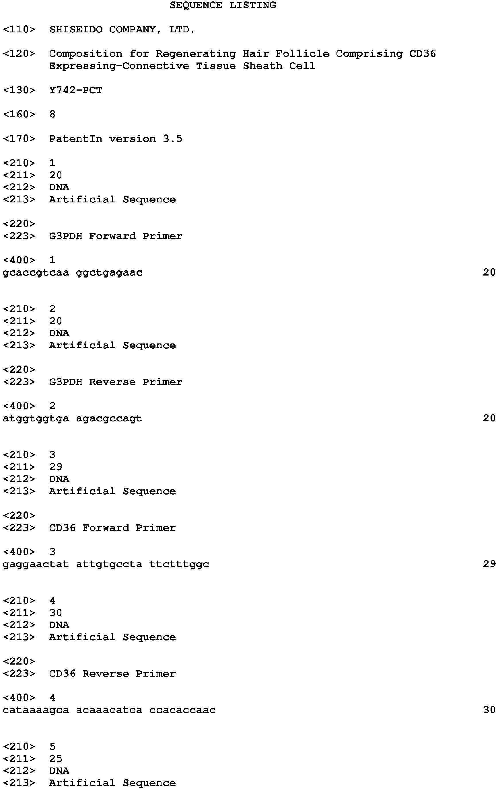


Claims (14)
- A composition for regenerating hair follicles, comprising CD36-expressing dermal sheath cells (DSc).
- The composition for regenerating hair follicles according to claim 1, further comprising dermal papilla cells (DPc).
- The composition according to claim 2, wherein the ratio of the number of CD36-expressing DSc to the number of DPc is about 10:1 to 1:10.
- The composition according to claim 2 or 3, wherein the CD36-expressing DSc and the DPc are both derived from mice or both derived from rats.
- The composition according to claim 2 or 3, wherein the CD36-expressing DSc and the DPc are heterologous cells, and each of them are derived from mice, rats or humans.
- A method for regenerating hair follicles, comprising transplanting the composition according to any of claims 1 to 5 to a human.
- A method for regenerating hair follicles by transplanting the composition according to any of claims 1 to 5 to a recipient animal.
- The method according to claim 7, wherein the recipient animal is an immunosuppressed animal.
- The method according to claim 7 or 8, wherein the recipient animal is an immunosuppressed animal selected from the group consisting of a nude mouse, SCID mouse and nude rat.
- A method for regenerating hair follicles, comprising producing a three-dimensional skin model containing the composition according to any of claims 2 to 5.
- A chimeric animal imparted with regenerated hair follicles by transplanting the composition according to any of claims 1 to 5 to a recipient animal.
- The chimeric animal according to claim 11, wherein the recipient animal is an immunosuppressed animal.
- The chimeric animal according to claim 11 or 12, wherein the recipient animal is an immunosuppressed animal selected from the group consisting of a nude mouse, SCID mouse and nude rat.
- A three-dimensional skin model imparted with regenerated hair follicles by producing a three-dimensional skin model containing the composition according to any of claims 2 to 5.
Applications Claiming Priority (1)
| Application Number | Priority Date | Filing Date | Title |
|---|---|---|---|
| PCT/JP2010/066999 WO2012042618A1 (en) | 2010-09-29 | 2010-09-29 | Composition for regenerating follicle which contains cd36-expressing connective tissue sheath cells |
Publications (3)
| Publication Number | Publication Date |
|---|---|
| EP2623106A1 true EP2623106A1 (en) | 2013-08-07 |
| EP2623106A4 EP2623106A4 (en) | 2014-03-26 |
| EP2623106B1 EP2623106B1 (en) | 2018-05-23 |
Family
ID=45892122
Family Applications (1)
| Application Number | Title | Priority Date | Filing Date |
|---|---|---|---|
| EP10857831.1A Active EP2623106B1 (en) | 2010-09-29 | 2010-09-29 | Composition for regenerating follicle which contains cd36-expressing connective tissue sheath cells |
Country Status (7)
| Country | Link |
|---|---|
| US (2) | US20130212724A1 (en) |
| EP (1) | EP2623106B1 (en) |
| KR (1) | KR101730216B1 (en) |
| CN (1) | CN103237554B (en) |
| IN (1) | IN2013MN00607A (en) |
| SG (1) | SG189167A1 (en) |
| WO (1) | WO2012042618A1 (en) |
Families Citing this family (6)
| Publication number | Priority date | Publication date | Assignee | Title |
|---|---|---|---|---|
| CN107849532A (en) * | 2015-05-14 | 2018-03-27 | 株式会社再生科技工程 | Preparation method of frozen mesenchymal cells and preparation method of therapeutic material for transplantation |
| KR101848904B1 (en) * | 2015-11-12 | 2018-04-13 | 김동구 | Antibody for specific binding and functional activator to hair Dermal Papilla cell and use thereof |
| DE102015119880B4 (en) * | 2015-11-17 | 2018-05-24 | Technische Universität Berlin | Process for the production of hair follicles and de novo papillae and their use for in vitro tests and in vivo implants |
| US20200208106A1 (en) * | 2017-08-22 | 2020-07-02 | National University Corporation Yokohama National University | Production Method of Multiple Regenerated Hair Follicle Primordia, Production Method of Hair Follicle Tissue-Containing Sheet, Hair Regeneration Kit and Method for Screening Hair Growth promoter or Hair Growth Inhibitor |
| JP7385842B2 (en) * | 2019-09-17 | 2023-11-24 | 学校法人東京理科大学 | Observation methods, measurement methods, analysis methods, quantitative methods, and kits |
| TW202241459A (en) * | 2020-12-23 | 2022-11-01 | 南韓商生物歐賽加有限責任公司 | Use of mirna-485 inhibitors for inducing hair growth |
Family Cites Families (10)
| Publication number | Priority date | Publication date | Assignee | Title |
|---|---|---|---|---|
| GB9925964D0 (en) * | 1999-11-03 | 1999-12-29 | Jahoda Colin A B | Hair transplantation |
| EP1437042A4 (en) * | 2001-09-06 | 2006-05-03 | Shiseido Co Ltd | Hair follicle-reconstitution system and animal carrying the same |
| JP4689175B2 (en) * | 2003-10-06 | 2011-05-25 | 株式会社資生堂 | Composition, method for regenerating hair follicle and animal carrying regenerated hair follicle |
| KR101156797B1 (en) * | 2003-10-06 | 2012-06-18 | 가부시키가이샤 시세이도 | Animal having regenerated hair follicle |
| CN100438926C (en) * | 2003-12-05 | 2008-12-03 | 英泰格伦斯生物株式会社 | hair growth composition and application thereof |
| CA2554732C (en) * | 2004-01-27 | 2014-07-15 | The Hospital For Sick Children | Methods of making and using skin-derived stem cells |
| EP2347793A1 (en) * | 2004-06-14 | 2011-07-27 | Shiseido Company, Ltd. | Method for regenerating hair follicles by suppressing a gene with hair follicle formation-inhibiting ability or by activating a gene with hair follicle formation - inducing ability |
| EP1950284B1 (en) * | 2005-09-30 | 2012-04-25 | Phoenixbio Co., Ltd. | Method for cultivation of hair follicular dermal sheath cell |
| US9109204B2 (en) * | 2006-02-28 | 2015-08-18 | The Trustees Of Columbia University In The City Of New York | Methods for compact aggregation of dermal cells |
| JP5097387B2 (en) | 2006-11-16 | 2012-12-12 | ライオン株式会社 | Artificial skin |
-
2010
- 2010-09-29 US US13/876,608 patent/US20130212724A1/en not_active Abandoned
- 2010-09-29 KR KR1020137007049A patent/KR101730216B1/en not_active Expired - Fee Related
- 2010-09-29 SG SG2013023619A patent/SG189167A1/en unknown
- 2010-09-29 EP EP10857831.1A patent/EP2623106B1/en active Active
- 2010-09-29 IN IN607MUN2013 patent/IN2013MN00607A/en unknown
- 2010-09-29 WO PCT/JP2010/066999 patent/WO2012042618A1/en not_active Ceased
- 2010-09-29 CN CN201080069392.7A patent/CN103237554B/en active Active
-
2016
- 2016-03-22 US US15/076,836 patent/US10172332B2/en active Active
Also Published As
| Publication number | Publication date |
|---|---|
| KR101730216B1 (en) | 2017-04-25 |
| KR20130112872A (en) | 2013-10-14 |
| HK1183790A1 (en) | 2014-01-10 |
| US20160193140A1 (en) | 2016-07-07 |
| US20130212724A1 (en) | 2013-08-15 |
| IN2013MN00607A (en) | 2015-09-11 |
| CN103237554B (en) | 2015-08-12 |
| SG189167A1 (en) | 2013-05-31 |
| WO2012042618A1 (en) | 2012-04-05 |
| US10172332B2 (en) | 2019-01-08 |
| EP2623106B1 (en) | 2018-05-23 |
| EP2623106A4 (en) | 2014-03-26 |
| CN103237554A (en) | 2013-08-07 |
Similar Documents
| Publication | Publication Date | Title |
|---|---|---|
| US10172332B2 (en) | Method for regenerating hair follicles using CD36-expressing dermal sheath cells | |
| KR101077760B1 (en) | Progenitor cells from wharton's jelly of human umbilical cord | |
| KR101472594B1 (en) | A cell mass that can form a primitive organs, including a plurality of cell types derived from somatic origin | |
| WO2008004819A1 (en) | Method for isolation of a hair follicle stem cell and a composition for hair reproduction | |
| JP2015097524A (en) | Compositions and methods for making reconstituted skin | |
| WO2014200025A1 (en) | Quality control method for hair-follicle forming composition | |
| JP2008029331A (en) | Cell cluster comprising multiple kinds of cells derived from soma with ability to form primitive organ-like structure | |
| US8334136B2 (en) | Method for promoting hair growth or hair regeneration by maintaining or increasing expression of cell-adhesion factor | |
| JP6839003B2 (en) | Reconstructed scalp model and screening method for active molecules | |
| JP5686957B2 (en) | Composition for regenerating hair follicles containing CD36-expressing connective tissue sheath cells | |
| US8835169B2 (en) | Compositions, methods and systems for preparation of a stem cell-enriched cell population | |
| CN1968713B (en) | Method of regenerating follicles by inhibiting gene capable of inhibiting follicle formation or activating gene capable of inducing follicle formation | |
| Dong et al. | Enrichment of epidermal stem cells by rapid adherence and analysis of the reciprocal interaction of epidermal stem cells with neighboring cells using an organotypic system | |
| HK1183790B (en) | Composition for regenerating follicle which contains cd36-expressing connective tissue sheath cells | |
| JP2003189849A (en) | Hair follicle reconstitution system and its carrying animal | |
| JP2010284110A (en) | Method for screening hair-developing or hair-growing promoter using expression of cd36 of connective tissue sheath cell as index | |
| Sun | Integration and interaction of cardiac progenitor cells with human failing myocardium | |
| JP6101066B2 (en) | Preparation method of melanocyte population | |
| Gharzi | Dermal and epidermal cell functions in the growth and regeneration of hair follicles and other skin appendages | |
| WO2020066818A1 (en) | In vitro growth method for hair follicular epithelial stem cells | |
| Higgins | Molecular and cellular basis of Exogen and Anagen induction |
Legal Events
| Date | Code | Title | Description |
|---|---|---|---|
| PUAI | Public reference made under article 153(3) epc to a published international application that has entered the european phase |
Free format text: ORIGINAL CODE: 0009012 |
|
| 17P | Request for examination filed |
Effective date: 20130417 |
|
| AK | Designated contracting states |
Kind code of ref document: A1 Designated state(s): AL AT BE BG CH CY CZ DE DK EE ES FI FR GB GR HR HU IE IS IT LI LT LU LV MC MK MT NL NO PL PT RO SE SI SK SM TR |
|
| DAX | Request for extension of the european patent (deleted) | ||
| REG | Reference to a national code |
Ref country code: DE Ref legal event code: R079 Ref document number: 602010050838 Country of ref document: DE Free format text: PREVIOUS MAIN CLASS: A61K0035360000 Ipc: C12N0005071000 |
|
| A4 | Supplementary search report drawn up and despatched |
Effective date: 20140225 |
|
| RIC1 | Information provided on ipc code assigned before grant |
Ipc: C12N 5/071 20100101AFI20140217BHEP Ipc: A61K 35/36 20060101ALI20140217BHEP Ipc: A61P 17/14 20060101ALI20140217BHEP |
|
| 17Q | First examination report despatched |
Effective date: 20160422 |
|
| GRAP | Despatch of communication of intention to grant a patent |
Free format text: ORIGINAL CODE: EPIDOSNIGR1 |
|
| INTG | Intention to grant announced |
Effective date: 20171208 |
|
| GRAS | Grant fee paid |
Free format text: ORIGINAL CODE: EPIDOSNIGR3 |
|
| GRAA | (expected) grant |
Free format text: ORIGINAL CODE: 0009210 |
|
| AK | Designated contracting states |
Kind code of ref document: B1 Designated state(s): AL AT BE BG CH CY CZ DE DK EE ES FI FR GB GR HR HU IE IS IT LI LT LU LV MC MK MT NL NO PL PT RO SE SI SK SM TR |
|
| REG | Reference to a national code |
Ref country code: GB Ref legal event code: FG4D |
|
| REG | Reference to a national code |
Ref country code: CH Ref legal event code: EP Ref country code: CH Ref legal event code: NV Representative=s name: BUGNION S.A., CH |
|
| REG | Reference to a national code |
Ref country code: IE Ref legal event code: FG4D |
|
| REG | Reference to a national code |
Ref country code: AT Ref legal event code: REF Ref document number: 1001538 Country of ref document: AT Kind code of ref document: T Effective date: 20180615 |
|
| REG | Reference to a national code |
Ref country code: DE Ref legal event code: R096 Ref document number: 602010050838 Country of ref document: DE |
|
| REG | Reference to a national code |
Ref country code: FR Ref legal event code: PLFP Year of fee payment: 9 |
|
| REG | Reference to a national code |
Ref country code: NL Ref legal event code: MP Effective date: 20180523 |
|
| REG | Reference to a national code |
Ref country code: LT Ref legal event code: MG4D |
|
| PG25 | Lapsed in a contracting state [announced via postgrant information from national office to epo] |
Ref country code: ES Free format text: LAPSE BECAUSE OF FAILURE TO SUBMIT A TRANSLATION OF THE DESCRIPTION OR TO PAY THE FEE WITHIN THE PRESCRIBED TIME-LIMIT Effective date: 20180523 Ref country code: LT Free format text: LAPSE BECAUSE OF FAILURE TO SUBMIT A TRANSLATION OF THE DESCRIPTION OR TO PAY THE FEE WITHIN THE PRESCRIBED TIME-LIMIT Effective date: 20180523 Ref country code: FI Free format text: LAPSE BECAUSE OF FAILURE TO SUBMIT A TRANSLATION OF THE DESCRIPTION OR TO PAY THE FEE WITHIN THE PRESCRIBED TIME-LIMIT Effective date: 20180523 Ref country code: NO Free format text: LAPSE BECAUSE OF FAILURE TO SUBMIT A TRANSLATION OF THE DESCRIPTION OR TO PAY THE FEE WITHIN THE PRESCRIBED TIME-LIMIT Effective date: 20180823 Ref country code: BG Free format text: LAPSE BECAUSE OF FAILURE TO SUBMIT A TRANSLATION OF THE DESCRIPTION OR TO PAY THE FEE WITHIN THE PRESCRIBED TIME-LIMIT Effective date: 20180823 Ref country code: SE Free format text: LAPSE BECAUSE OF FAILURE TO SUBMIT A TRANSLATION OF THE DESCRIPTION OR TO PAY THE FEE WITHIN THE PRESCRIBED TIME-LIMIT Effective date: 20180523 |
|
| PG25 | Lapsed in a contracting state [announced via postgrant information from national office to epo] |
Ref country code: GR Free format text: LAPSE BECAUSE OF FAILURE TO SUBMIT A TRANSLATION OF THE DESCRIPTION OR TO PAY THE FEE WITHIN THE PRESCRIBED TIME-LIMIT Effective date: 20180824 Ref country code: LV Free format text: LAPSE BECAUSE OF FAILURE TO SUBMIT A TRANSLATION OF THE DESCRIPTION OR TO PAY THE FEE WITHIN THE PRESCRIBED TIME-LIMIT Effective date: 20180523 Ref country code: HR Free format text: LAPSE BECAUSE OF FAILURE TO SUBMIT A TRANSLATION OF THE DESCRIPTION OR TO PAY THE FEE WITHIN THE PRESCRIBED TIME-LIMIT Effective date: 20180523 Ref country code: NL Free format text: LAPSE BECAUSE OF FAILURE TO SUBMIT A TRANSLATION OF THE DESCRIPTION OR TO PAY THE FEE WITHIN THE PRESCRIBED TIME-LIMIT Effective date: 20180523 |
|
| PG25 | Lapsed in a contracting state [announced via postgrant information from national office to epo] |
Ref country code: CZ Free format text: LAPSE BECAUSE OF FAILURE TO SUBMIT A TRANSLATION OF THE DESCRIPTION OR TO PAY THE FEE WITHIN THE PRESCRIBED TIME-LIMIT Effective date: 20180523 Ref country code: SK Free format text: LAPSE BECAUSE OF FAILURE TO SUBMIT A TRANSLATION OF THE DESCRIPTION OR TO PAY THE FEE WITHIN THE PRESCRIBED TIME-LIMIT Effective date: 20180523 Ref country code: PL Free format text: LAPSE BECAUSE OF FAILURE TO SUBMIT A TRANSLATION OF THE DESCRIPTION OR TO PAY THE FEE WITHIN THE PRESCRIBED TIME-LIMIT Effective date: 20180523 Ref country code: DK Free format text: LAPSE BECAUSE OF FAILURE TO SUBMIT A TRANSLATION OF THE DESCRIPTION OR TO PAY THE FEE WITHIN THE PRESCRIBED TIME-LIMIT Effective date: 20180523 Ref country code: RO Free format text: LAPSE BECAUSE OF FAILURE TO SUBMIT A TRANSLATION OF THE DESCRIPTION OR TO PAY THE FEE WITHIN THE PRESCRIBED TIME-LIMIT Effective date: 20180523 Ref country code: EE Free format text: LAPSE BECAUSE OF FAILURE TO SUBMIT A TRANSLATION OF THE DESCRIPTION OR TO PAY THE FEE WITHIN THE PRESCRIBED TIME-LIMIT Effective date: 20180523 |
|
| REG | Reference to a national code |
Ref country code: DE Ref legal event code: R097 Ref document number: 602010050838 Country of ref document: DE |
|
| PG25 | Lapsed in a contracting state [announced via postgrant information from national office to epo] |
Ref country code: SM Free format text: LAPSE BECAUSE OF FAILURE TO SUBMIT A TRANSLATION OF THE DESCRIPTION OR TO PAY THE FEE WITHIN THE PRESCRIBED TIME-LIMIT Effective date: 20180523 Ref country code: IT Free format text: LAPSE BECAUSE OF FAILURE TO SUBMIT A TRANSLATION OF THE DESCRIPTION OR TO PAY THE FEE WITHIN THE PRESCRIBED TIME-LIMIT Effective date: 20180523 |
|
| PLBE | No opposition filed within time limit |
Free format text: ORIGINAL CODE: 0009261 |
|
| STAA | Information on the status of an ep patent application or granted ep patent |
Free format text: STATUS: NO OPPOSITION FILED WITHIN TIME LIMIT |
|
| PG25 | Lapsed in a contracting state [announced via postgrant information from national office to epo] |
Ref country code: MC Free format text: LAPSE BECAUSE OF FAILURE TO SUBMIT A TRANSLATION OF THE DESCRIPTION OR TO PAY THE FEE WITHIN THE PRESCRIBED TIME-LIMIT Effective date: 20180523 |
|
| 26N | No opposition filed |
Effective date: 20190226 |
|
| PG25 | Lapsed in a contracting state [announced via postgrant information from national office to epo] |
Ref country code: SI Free format text: LAPSE BECAUSE OF FAILURE TO SUBMIT A TRANSLATION OF THE DESCRIPTION OR TO PAY THE FEE WITHIN THE PRESCRIBED TIME-LIMIT Effective date: 20180523 |
|
| REG | Reference to a national code |
Ref country code: BE Ref legal event code: MM Effective date: 20180930 |
|
| REG | Reference to a national code |
Ref country code: IE Ref legal event code: MM4A |
|
| PG25 | Lapsed in a contracting state [announced via postgrant information from national office to epo] |
Ref country code: LU Free format text: LAPSE BECAUSE OF NON-PAYMENT OF DUE FEES Effective date: 20180929 |
|
| PG25 | Lapsed in a contracting state [announced via postgrant information from national office to epo] |
Ref country code: IE Free format text: LAPSE BECAUSE OF NON-PAYMENT OF DUE FEES Effective date: 20180929 |
|
| PG25 | Lapsed in a contracting state [announced via postgrant information from national office to epo] |
Ref country code: BE Free format text: LAPSE BECAUSE OF NON-PAYMENT OF DUE FEES Effective date: 20180930 |
|
| PG25 | Lapsed in a contracting state [announced via postgrant information from national office to epo] |
Ref country code: AL Free format text: LAPSE BECAUSE OF FAILURE TO SUBMIT A TRANSLATION OF THE DESCRIPTION OR TO PAY THE FEE WITHIN THE PRESCRIBED TIME-LIMIT Effective date: 20180523 |
|
| PG25 | Lapsed in a contracting state [announced via postgrant information from national office to epo] |
Ref country code: MT Free format text: LAPSE BECAUSE OF NON-PAYMENT OF DUE FEES Effective date: 20180929 |
|
| PG25 | Lapsed in a contracting state [announced via postgrant information from national office to epo] |
Ref country code: TR Free format text: LAPSE BECAUSE OF FAILURE TO SUBMIT A TRANSLATION OF THE DESCRIPTION OR TO PAY THE FEE WITHIN THE PRESCRIBED TIME-LIMIT Effective date: 20180523 |
|
| PG25 | Lapsed in a contracting state [announced via postgrant information from national office to epo] |
Ref country code: PT Free format text: LAPSE BECAUSE OF FAILURE TO SUBMIT A TRANSLATION OF THE DESCRIPTION OR TO PAY THE FEE WITHIN THE PRESCRIBED TIME-LIMIT Effective date: 20180523 Ref country code: HU Free format text: LAPSE BECAUSE OF FAILURE TO SUBMIT A TRANSLATION OF THE DESCRIPTION OR TO PAY THE FEE WITHIN THE PRESCRIBED TIME-LIMIT; INVALID AB INITIO Effective date: 20100929 |
|
| PG25 | Lapsed in a contracting state [announced via postgrant information from national office to epo] |
Ref country code: CY Free format text: LAPSE BECAUSE OF FAILURE TO SUBMIT A TRANSLATION OF THE DESCRIPTION OR TO PAY THE FEE WITHIN THE PRESCRIBED TIME-LIMIT Effective date: 20180523 Ref country code: MK Free format text: LAPSE BECAUSE OF NON-PAYMENT OF DUE FEES Effective date: 20180523 |
|
| PG25 | Lapsed in a contracting state [announced via postgrant information from national office to epo] |
Ref country code: IS Free format text: LAPSE BECAUSE OF FAILURE TO SUBMIT A TRANSLATION OF THE DESCRIPTION OR TO PAY THE FEE WITHIN THE PRESCRIBED TIME-LIMIT Effective date: 20180923 |
|
| REG | Reference to a national code |
Ref country code: AT Ref legal event code: UEP Ref document number: 1001538 Country of ref document: AT Kind code of ref document: T Effective date: 20180523 |
|
| P01 | Opt-out of the competence of the unified patent court (upc) registered |
Effective date: 20230518 |
|
| PGFP | Annual fee paid to national office [announced via postgrant information from national office to epo] |
Ref country code: DE Payment date: 20240820 Year of fee payment: 15 |
|
| PGFP | Annual fee paid to national office [announced via postgrant information from national office to epo] |
Ref country code: GB Payment date: 20240820 Year of fee payment: 15 |
|
| PGFP | Annual fee paid to national office [announced via postgrant information from national office to epo] |
Ref country code: FR Payment date: 20240820 Year of fee payment: 15 |
|
| PGFP | Annual fee paid to national office [announced via postgrant information from national office to epo] |
Ref country code: AT Payment date: 20240821 Year of fee payment: 15 |
|
| PGFP | Annual fee paid to national office [announced via postgrant information from national office to epo] |
Ref country code: CH Payment date: 20241001 Year of fee payment: 15 |
|
| REG | Reference to a national code |
Ref country code: DE Ref legal event code: R119 Ref document number: 602010050838 Country of ref document: DE |
|
| REG | Reference to a national code |
Ref country code: CH Ref legal event code: H13 Free format text: ST27 STATUS EVENT CODE: U-0-0-H10-H13 (AS PROVIDED BY THE NATIONAL OFFICE) Effective date: 20260504 |
|
| REG | Reference to a national code |
Ref country code: AT Ref legal event code: MM01 Ref document number: 1001538 Country of ref document: AT Kind code of ref document: T Effective date: 20250929 |
|
| GBPC | Gb: european patent ceased through non-payment of renewal fee |
Effective date: 20250929 |
|
| PG25 | Lapsed in a contracting state [announced via postgrant information from national office to epo] |
Ref country code: GB Free format text: LAPSE BECAUSE OF NON-PAYMENT OF DUE FEES Effective date: 20250929 |
|
| PG25 | Lapsed in a contracting state [announced via postgrant information from national office to epo] |
Ref country code: DE Free format text: LAPSE BECAUSE OF NON-PAYMENT OF DUE FEES Effective date: 20260401 |
|
| PG25 | Lapsed in a contracting state [announced via postgrant information from national office to epo] |
Ref country code: AT Free format text: LAPSE BECAUSE OF NON-PAYMENT OF DUE FEES Effective date: 20250929 |
|
| PG25 | Lapsed in a contracting state [announced via postgrant information from national office to epo] |
Ref country code: CH Free format text: LAPSE BECAUSE OF NON-PAYMENT OF DUE FEES Effective date: 20250930 |
|
| PG25 | Lapsed in a contracting state [announced via postgrant information from national office to epo] |
Ref country code: FR Free format text: LAPSE BECAUSE OF NON-PAYMENT OF DUE FEES Effective date: 20250930 |