EP2620909A1 - Method, system and computer readable medium for automatic segmentation of a medical image - Google Patents
Method, system and computer readable medium for automatic segmentation of a medical image Download PDFInfo
- Publication number
- EP2620909A1 EP2620909A1 EP12462003.0A EP12462003A EP2620909A1 EP 2620909 A1 EP2620909 A1 EP 2620909A1 EP 12462003 A EP12462003 A EP 12462003A EP 2620909 A1 EP2620909 A1 EP 2620909A1
- Authority
- EP
- European Patent Office
- Prior art keywords
- medical image
- probability
- thresholding
- volume
- area
- Prior art date
- Legal status (The legal status is an assumption and is not a legal conclusion. Google has not performed a legal analysis and makes no representation as to the accuracy of the status listed.)
- Granted
Links
- 230000011218 segmentation Effects 0.000 title claims abstract description 62
- 238000000034 method Methods 0.000 title claims abstract description 61
- 230000000877 morphologic effect Effects 0.000 claims abstract description 15
- 230000009466 transformation Effects 0.000 claims description 18
- 210000000988 bone and bone Anatomy 0.000 claims description 5
- 230000001131 transforming effect Effects 0.000 claims description 4
- 210000000056 organ Anatomy 0.000 description 31
- 230000006870 function Effects 0.000 description 11
- 210000004185 liver Anatomy 0.000 description 9
- 210000003484 anatomy Anatomy 0.000 description 8
- 230000008569 process Effects 0.000 description 8
- 238000010200 validation analysis Methods 0.000 description 8
- 230000003187 abdominal effect Effects 0.000 description 7
- 238000004590 computer program Methods 0.000 description 7
- 210000003734 kidney Anatomy 0.000 description 6
- 238000013459 approach Methods 0.000 description 5
- 238000003384 imaging method Methods 0.000 description 4
- 230000003993 interaction Effects 0.000 description 4
- 238000013473 artificial intelligence Methods 0.000 description 3
- 238000002059 diagnostic imaging Methods 0.000 description 3
- 238000003709 image segmentation Methods 0.000 description 3
- 238000002595 magnetic resonance imaging Methods 0.000 description 3
- 238000012545 processing Methods 0.000 description 3
- 230000008901 benefit Effects 0.000 description 2
- 230000008859 change Effects 0.000 description 2
- 238000002591 computed tomography Methods 0.000 description 2
- 238000002372 labelling Methods 0.000 description 2
- 238000001959 radiotherapy Methods 0.000 description 2
- 238000002603 single-photon emission computed tomography Methods 0.000 description 2
- 210000000952 spleen Anatomy 0.000 description 2
- 238000012546 transfer Methods 0.000 description 2
- 206010028980 Neoplasm Diseases 0.000 description 1
- 210000001015 abdomen Anatomy 0.000 description 1
- 238000004458 analytical method Methods 0.000 description 1
- 230000005540 biological transmission Effects 0.000 description 1
- 210000004556 brain Anatomy 0.000 description 1
- 238000010276 construction Methods 0.000 description 1
- 238000011161 development Methods 0.000 description 1
- 230000018109 developmental process Effects 0.000 description 1
- 238000009429 electrical wiring Methods 0.000 description 1
- 230000005670 electromagnetic radiation Effects 0.000 description 1
- 230000003628 erosive effect Effects 0.000 description 1
- 239000000835 fiber Substances 0.000 description 1
- 238000001914 filtration Methods 0.000 description 1
- 230000006872 improvement Effects 0.000 description 1
- 230000004807 localization Effects 0.000 description 1
- 210000003205 muscle Anatomy 0.000 description 1
- 238000010606 normalization Methods 0.000 description 1
- 238000009206 nuclear medicine Methods 0.000 description 1
- 230000003287 optical effect Effects 0.000 description 1
- 238000012856 packing Methods 0.000 description 1
- 238000002600 positron emission tomography Methods 0.000 description 1
- 238000007781 pre-processing Methods 0.000 description 1
- 238000002601 radiography Methods 0.000 description 1
- 239000004065 semiconductor Substances 0.000 description 1
- 238000004513 sizing Methods 0.000 description 1
- 210000004872 soft tissue Anatomy 0.000 description 1
- 238000001228 spectrum Methods 0.000 description 1
- 238000011477 surgical intervention Methods 0.000 description 1
- 210000001519 tissue Anatomy 0.000 description 1
- 238000000844 transformation Methods 0.000 description 1
- 238000013519 translation Methods 0.000 description 1
- 230000014616 translation Effects 0.000 description 1
Images


Classifications
-
- G—PHYSICS
- G06—COMPUTING; CALCULATING OR COUNTING
- G06T—IMAGE DATA PROCESSING OR GENERATION, IN GENERAL
- G06T7/00—Image analysis
- G06T7/0002—Inspection of images, e.g. flaw detection
- G06T7/0012—Biomedical image inspection
- G06T7/0014—Biomedical image inspection using an image reference approach
-
- G—PHYSICS
- G06—COMPUTING; CALCULATING OR COUNTING
- G06T—IMAGE DATA PROCESSING OR GENERATION, IN GENERAL
- G06T7/00—Image analysis
- G06T7/10—Segmentation; Edge detection
- G06T7/11—Region-based segmentation
-
- G—PHYSICS
- G06—COMPUTING; CALCULATING OR COUNTING
- G06T—IMAGE DATA PROCESSING OR GENERATION, IN GENERAL
- G06T7/00—Image analysis
- G06T7/10—Segmentation; Edge detection
- G06T7/187—Segmentation; Edge detection involving region growing; involving region merging; involving connected component labelling
-
- G—PHYSICS
- G06—COMPUTING; CALCULATING OR COUNTING
- G06T—IMAGE DATA PROCESSING OR GENERATION, IN GENERAL
- G06T2207/00—Indexing scheme for image analysis or image enhancement
- G06T2207/20—Special algorithmic details
- G06T2207/20076—Probabilistic image processing
-
- G—PHYSICS
- G06—COMPUTING; CALCULATING OR COUNTING
- G06T—IMAGE DATA PROCESSING OR GENERATION, IN GENERAL
- G06T2207/00—Indexing scheme for image analysis or image enhancement
- G06T2207/20—Special algorithmic details
- G06T2207/20112—Image segmentation details
- G06T2207/20128—Atlas-based segmentation
-
- G—PHYSICS
- G06—COMPUTING; CALCULATING OR COUNTING
- G06T—IMAGE DATA PROCESSING OR GENERATION, IN GENERAL
- G06T2207/00—Indexing scheme for image analysis or image enhancement
- G06T2207/20—Special algorithmic details
- G06T2207/20112—Image segmentation details
- G06T2207/20156—Automatic seed setting
-
- G—PHYSICS
- G06—COMPUTING; CALCULATING OR COUNTING
- G06T—IMAGE DATA PROCESSING OR GENERATION, IN GENERAL
- G06T2207/00—Indexing scheme for image analysis or image enhancement
- G06T2207/30—Subject of image; Context of image processing
- G06T2207/30004—Biomedical image processing
Definitions
- the subject matter disclosed herein relates to a method, a system and a computer readable medium for automatic segmentation of a medical image. More particularly, the disclosed subject matter relates to image processing, and to systems and methods for medical imaging with image recognition and image registration capabilities.
- Segmentation of anatomical structures in medical images is a fundamental task in a number of clinical processes in the field of oncology, radiology and in planning surgical interventions.
- Exemplary techniques for imaging include conventional X-ray plane film radiography, computed tomography (“CT”) imaging, magnetic resonance imaging (“MRI”), and nuclear medicine imaging techniques, such as positron emission tomography (“PET”) and single photon emission computed tomography (“SPECT”).
- CT computed tomography
- MRI magnetic resonance imaging
- PET nuclear medicine imaging techniques, such as positron emission tomography
- SPECT single photon emission computed tomography
- transforming a medical image which is a CT image or a series of CT images (often called as a CT study, CT volume or CT exam) to a common reference frame is a useful step before commencing segmentation.
- Transforming a medical image to a common reference frame is generally called registering the medical image. It is assumed that anatomic regions of different medical images are found in approximately a same voxel region.
- a probability map also called as probability atlas or statistical atlas
- a new medical image may be registered to a reference image to determine transformation function or transfer parameters.
- the reference image is usually selected from medical images of the average patients.
- the transformation function may then be applied to transform the probability map data to a coordinate system of the medical image to be segmented to help initialize a segmentation algorithm.
- Segmentation is the process of assigning labels to individual voxels in the data set. Automatic segmentation thereby means automated recognition and labeling of human anatomical structures in 2D or 3D digital scans of the human body.
- Region growing techniques have also been widely used in image processing and analysis and include a process of recursively packing connected pixels according to some criteria. The process starts from one (or more) pixel called a seed, and checks all pixels in its immediate neighborhood. Those pixels that satisfy some criteria, for example, their intensity value is larger than a pre-defined threshold, are collected or grouped together. The process then continues recursively from each collected pixel until no more pixels can be collected.
- region growing method other similar techniques are also known for this purpose, e.g. the so called active contour method expanding an initial seed volume surrounded by an initial contour to the segmented volume of the organ.
- US 7,259,762 discloses a method for detecting and segmenting organs and structures in a medical image. Structural connections are analyzed, registration and region growing is used in this known organ segmentation.
- US 2007/0160277 A1 discloses a method for segmenting various brain structures on MRI images using an anatomical template and graph cut algorithms in an iterative approach.
- US 2010/0054525 A1 discloses a system and a method for automatic recognition and labeling of anatomical structures and vessels in medical imaging scans.
- a method for automatic segmentation of a medical image comprising
- a computer readable medium comprising a computer readable program for automatic segmentation of a medical image, wherein the computer readable program when executed on a computer causes the computer to perform the steps of the above method.
- a system for automatic segmentation of a medical image, the medical image comprising an object image to be segmented, the system comprising
- An automatic segmentation method can decrease the processing time and makes possible to establish an alternative clinical workflow, wherein the algorithms can be executed before the user starts to work with the images, e.g. the algorithm can run during the night or during the image transfer from a CT console to a workstation.
- the algorithm can run during the night or during the image transfer from a CT console to a workstation.
- suggested contours are already available, which saves a considerable amount of time. These contours may need supervising and editing, but these take significantly less time than fully manual contouring.
- the automatic segmentation disclosed herein is proved to be accurate and robust not requiring any post editing, and successfully eliminating the problems related to inter- and intra-operator variability.
- An embodiment may be embodied in the form of computer-implemented processes and apparatuses for practicing those processes.
- An embodiment may also be embodied in the form of a computer program code containing instructions embodied in tangible media, such as floppy diskettes, CD-ROMs, hard drives, or any other computer readable storage medium, wherein, when the computer program code is loaded into and executed by a computer, the computer becomes an apparatus for carrying out the method.
- An embodiment may also be embodied in the form of computer program code, for example, whether stored in a storage medium, loaded into and/or executed by a computer, or transmitted over some transmission medium, such as over electrical wiring or cabling, through fiber optics, or via electromagnetic radiation, wherein when the computer program code is loaded into and executed by a computer, the computer becomes an apparatus for carrying out the method.
- the computer program code segments configure the microprocessor to create specific logic circuits.
- the invention can take the form of a computer program product accessible from a computer-usable or computer-readable medium providing program code for use by or in connection with a computer or any instruction execution system.
- a computer-usable or computer readable medium can be any apparatus that may include, store, communicate, propagate, or transport the program for use by or in connection with the instruction execution system, apparatus, or device.
- the medium can be an electronic, magnetic, optical, electromagnetic, infrared, or semiconductor system (or apparatus or device) or a propagation medium.
- Embodiments can take the form of an entirely hardware embodiment, an entirely software embodiment or an embodiment including both hardware and software elements.
- the present invention is implemented in software, which includes but is not limited to firmware, resident software, microcode, etc.
- the subject matter disclosed herein describes a contouring workflow of a radiotherapy system, which is extended with a user supervised fully automatic abdominal organ segmentation tool for CT studies. Besides the automatic nature of segmentations, the workflow is also extended with an artificial intelligence based validation system to detect failures of the automatic segmentations, when the algorithms cannot handle the special anatomical attributes of the patient and unwanted over- or under-segmentations may occur.
- exemplary target organs are the liver, the spleen and the left and right kidneys.
- the subject matter disclosed herein is an extension to the current clinical abdominal organ segmentation workflow.
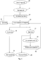
- the current workflow contains the following steps as depicted in Fig. 1 .
- Ellipses in the workflow represent steps involving user interaction/supervising, rectangles represent automated functions.
- a user selects in step 10 a medical image, which is usually an abdominal CT image series.
- the user selects an organ for contouring.
- Step 12 represents an automatic organ segmentation, which runs in the background.
- the contour of the selected organ is visualized on the CT image series in step 13.
- the user can decide to accept or reject the visualized contour.
- Step 15 represents that the user accepts the contour. If the user accepts the generated contours, there is an option to edit or refine contour borders manually in step 16.
- the user can decide to draw the whole contour manually in step 20 or run a semi-automatic method in steps 18 and 19 for organ segmentation. If the generated contour is rejected and the semi-automatic method is chosen, the user has to provide an input for it in step 18; semi-automatic segmentation methods represented by step 19 require various user interactions.
- the user is preferably also able to save automatic segmentation preferences to a personalized protocol in step 14. In this way, next time the selected organs on a different CT series are automatically segmented in the loading time of the protocol without any interaction.
- Fig. 2 is a detailed description of step 12 in Fig. 1 .
- An abdominal CT image series called as a medical image is input in step 30.
- an organ is selected for contouring in step 31.
- a reference image associated with the object is loaded into the memory.
- a reference image associated with the object in this context means a reference image, which comprises an image part containing an image of the object to be segmented.
- a probability map associated with the object, i.e. an atlas of the target organ is also loaded into the memory in step 33. The probability map is preferably created on the basis of a large number of medical images, also containing the reference image preferably belonging to a typical member of the population.
- the reference image is registered to the medical image, i.e. to the input CT exam resulting in a transformation function comprising transformation parameters.
- the transformation function may comprise overall translations, rotations and re-sizing, but it can be complex as well and may also comprise e.g. local or non-linear transformations.
- the transformation function is applied on the probability map associated with the object, i.e. on the atlas of the target organ. This provides an approximate location for the object in the medical image.
- the CT values are combined with the probability values of the transformed probability map - the detailed description of which can be found below in connection with Fig. 3 - to generate a seed region inside the target organ in step 36.
- the seed is used for an automatic segmentation of the medical image in step 37.
- the segmenting step 37 preferably comprises a growing of a region from the seed by means of a suitable growing method.
- the transformed (or in other words: registered) probability map is also used for the automatic segmentation to force the segmentation to acquire a shape corresponding to the expected probabilities.
- the automatic segmentation may still contain errors due to errors in the registration and seed generation steps 34 and 36.
- a validation step 38 removes suspicious results by using an artificial intelligence method trained on various metrics of acceptable results. This validation turned out to be very useful because physicians do not want to see bad/useless results/contours. A post validation helps to prevent losing reliability.
- the output is the contour of the target organ in step 39 if it passes the validation step, otherwise the output is an error message.
- Fig. 3 illustrates a flowchart of an automatic seed generation of step 36 in accordance with the subject matter described herein.
- the input medical image is loaded into the memory in step 40.
- a preprocessing/filtering step 41 follows, which eliminates the noise from the CT medical image.
- the transformed (registered) probability map is loaded into the memory in step 42.
- a probability thresholding step 43 is carried out on the transformed probability map by selecting a first area of the medical image in which the probability of the object is within a probability range.
- the probability range may mean a range from 100% to a lower limit, in which case only an area reaching a predetermined probability level is selected as the first area.
- the probability ranges or limits of given objects, e.g. organs are preferably different from each other, and are adjusted in a heuristic way.
- the first area may comprise both joined and separated sub areas of the medical image.
- the transformation function is applied to at least one further probability map associated with a further object, a probability thresholding on the transformed further probability map is carried out, and areas in which the probability of the further object is within a further probability range are deselected from the first area.
- a probability thresholding on the transformed further probability map is carried out, and areas in which the probability of the further object is within a further probability range are deselected from the first area.
- an intensity thresholding on the medical image is carried out in step 45 by selecting a second area of the medical image in which the intensity is within an intensity range.
- Certain organs have their certain intensity ranges, so this step deselects areas in which the intensities are too low (e.g. fat tissues, cavities) or too high (e.g. bones).
- the second area may comprise joined and separated sub areas as well.
- the intensity thresholding step 45 is carried out after the probability thresholding step 43, and is carried out on the selected first area only. In this way considerable computing time can be saved. More preferably, a morphological dilatation step (not depicted) is applied on bone image parts before the intensity thresholding step. In this way the gaps between the parts of the skeleton structure can be filled and muscles adhering to the bones can be excluded from erroneous recognition of organs.
- Morphological opening means a morphological erosion followed by a morphological dilatation, by means of which small noise parts are eliminated and the large regions are separated into separate sub-areas.
- the largest sub-area is selected in step 47 as a seed, and the automatic segmentation of the medical image is carried out on the basis of the selected seed.
- the results of the automatic segmentation is preferably validated in step 38 of Fig. 1 .
- the validating step 38 is carried out on the basis of at least one thresholding criterion selected from the following group: volume, enclosing block volume, ratio of volume / enclosing block volume, enclosing sphere volume, ratio of volume / enclosing sphere volume, enclosing sphere volumes of slices, axial distortion.
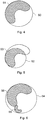
- Figs. 4 to 6 schematically show a correctly segmented, an under-segmented and an over-segmented liver in axial views, respectively.
- Fig. 4 depicts a segmentation 50 of a segmented area 51 of a liver.
- a segmentation 52 defines a segmented area 53 which does not contain the entire liver.
- Segmentation 54 depicted in Fig. 6 contains a segmented area 55 of a liver, and in addition a segmented area 56 of a kidney.
- Figs. 7 and 8 schematically show a kidney in coronal view validated as correctly segmented and over-segmented, respectively, on the basis of the detected axial distortion.
- a segmented area 57 in Fig. 7 has a detected axis 58 having a distortion within a predetermined credibility range, however, segmented area 59 in Fig. 8 has a detected axis 60 with a distortion outside the predetermined credibility range.
- this particular thresholding criteria the erroneous segmentation according to Fig. 8 can be easily detected.
- An exemplary embodiment is a computer readable medium comprising a computer readable program for automatic segmentation of a medical image, wherein the computer readable program when executed on a computer causes the computer to perform the steps of the above disclosed method.
- Another exemplary embodiment is a system for automatic segmentation of a medical image, the system comprising units configured to perform the steps of the above disclosed method. These units can take the form of an entirely hardware embodiment, an entirely software embodiment or an embodiment including both hardware and software elements.
- a technical advantage of the subject matter disclosed herein is that it enables to automatically segment abdominal organs on non-contrast enhanced CT scans.
- this feature is not offered, only semi-automatic methods are available.
- the method can be adopted to all medical applications, and parts of the workflow can facilitate solving other abdomen localization and segmentation problems.
Abstract
Description
- The subject matter disclosed herein relates to a method, a system and a computer readable medium for automatic segmentation of a medical image. More particularly, the disclosed subject matter relates to image processing, and to systems and methods for medical imaging with image recognition and image registration capabilities.
- Segmentation of anatomical structures in medical images is a fundamental task in a number of clinical processes in the field of oncology, radiology and in planning surgical interventions. Exemplary techniques for imaging include conventional X-ray plane film radiography, computed tomography ("CT") imaging, magnetic resonance imaging ("MRI"), and nuclear medicine imaging techniques, such as positron emission tomography ("PET") and single photon emission computed tomography ("SPECT"). Segmentation is used to measure the size and shape of anatomical structures, to guide spatial normalization of anatomy between individuals and to plan medical interventions. The spectrum of available segmentation approaches is broad, ranging from manual outlining of structures in 2D cross-sections to more developed methods that use a so called 'registration' to find optimal correspondences between 3D images and a labeled probability map or atlas. There are also known semiautomatic approaches that combine the efficiency and repeatability of automatic segmentation with the human judgment that can only come from skilled expertise.
- Despite the fact that a large number of fully automatic and semiautomatic segmentation methods have been disclosed, still manual delineation is generally used as the technique of choice for image segmentation. Reluctance to use the fully automatic approach is due to the concerns about its insufficient reliability in cases where the target anatomy may difference from the norm, as well as due to high computational demands of the approach based on image registration.
- Manually tracing the outlines on a contiguous set of 2D slices and then combining them can be time consuming and labor intensive. Time and labor increase significantly both as the number of image slices increase, and as a number and size of an organ, tumor, etc. in an anatomical area of interest increases. Quality of the outlining and quality of a produced 3D object depend on a resolution and contrast of the 2D slices, and on knowledge and judgment of the clinician performing the reconstruction.
- Using reliable automatic image segmentation could save time and labor, and could increase precision by eliminating subjectivity of the clinician.
- Automated image segmentation of organs faces certain challenges. Some organs are located in a soft tissue environment wherein resolution against surrounding structures has poor contrast since neighboring organs have similar density values. Furthermore, shape and position of organs may change periodically. Characteristics of abdominal organs also change from patient to patient including for example, shape, size and location of the organ. Imaging parameters of CT machines vary as well.
- Significant efforts have also been directed toward the development of templates for segmentation of the human organs. In model-based segmentation, transforming a medical image, which is a CT image or a series of CT images (often called as a CT study, CT volume or CT exam) to a common reference frame is a useful step before commencing segmentation. Transforming a medical image to a common reference frame is generally called registering the medical image. It is assumed that anatomic regions of different medical images are found in approximately a same voxel region. Thus, a probability map (also called as probability atlas or statistical atlas) may be generated on the basis of a large number of medical images, that represents a probability that a given voxel is a part of a particular organ. Given the probability map, a new medical image may be registered to a reference image to determine transformation function or transfer parameters. The reference image is usually selected from medical images of the average patients. The transformation function may then be applied to transform the probability map data to a coordinate system of the medical image to be segmented to help initialize a segmentation algorithm.
- Segmentation is the process of assigning labels to individual voxels in the data set. Automatic segmentation thereby means automated recognition and labeling of human anatomical structures in 2D or 3D digital scans of the human body.
- Region growing techniques have also been widely used in image processing and analysis and include a process of recursively packing connected pixels according to some criteria. The process starts from one (or more) pixel called a seed, and checks all pixels in its immediate neighborhood. Those pixels that satisfy some criteria, for example, their intensity value is larger than a pre-defined threshold, are collected or grouped together. The process then continues recursively from each collected pixel until no more pixels can be collected. Apart from the above region growing method, other similar techniques are also known for this purpose, e.g. the so called active contour method expanding an initial seed volume surrounded by an initial contour to the segmented volume of the organ. Throughout the present description and claims, all feasible methods suitable for growing or expanding a segmented area from a seed is called 'growing method'.
- A major limitation with existing growing methods is the need for human interaction to manually select the seed. There were some efforts to achieve automatic seed selection, but no practically applicable solution has been found so far. It is also a problem that no automated validation has been provided so far to achieve a safe final check of the automated segmentation.
-
US 7,259,762 discloses a method for detecting and segmenting organs and structures in a medical image. Structural connections are analyzed, registration and region growing is used in this known organ segmentation.US 2007/0160277 A1 discloses a method for segmenting various brain structures on MRI images using an anatomical template and graph cut algorithms in an iterative approach.US 2010/0054525 A1 discloses a system and a method for automatic recognition and labeling of anatomical structures and vessels in medical imaging scans. - Automatic segmentations (either atlas based or not) is a long researched topic, numerous articles are published in this topic. Examples are the following: 3D segmentation of liver, kidneys and spleen from CT images, Gyorgy Bekes et al. (in Computer Assisted Radiation Therapy, Int J CARS (2007) 2 (Suppl 1):S45-47); Construction of an abdominal probabilistic atlas and its application in segmentation, Hyunjin Park et al. (in IEEE Transactions on Medical Imaging, Vol. 22, No. 4, April 2003, pp. 483-492); and User-guided 3D active contour segmentation of anatomical structures: Significantly improved efficiency and reliability, Paul A. Yushkevich et al. (in NeuroImage 31 (2006) 1116-1128).
- These known methods do not solve the above problems of automatic segmentation.
- Thus, there is a need for a solution allowing an improvement over existing methods and systems. There is a need for automatic segmentation method, computer program and system eliminating as much as possible the shortcomings of known techniques. There is a particular need for an automatic segmentation method allowing automated seed selection and final validation of the segmentation results. There is also a need for an automatic segmentation method that eliminates the concerns about insufficient reliability of automated segmentation.
- In an exemplary embodiment, a method is provided for automatic segmentation of a medical image, the medical image comprising an object image to be segmented, the method comprising
- registering a reference image associated with the object to the medical image, and determining a transformation function on the basis of the registration, and
- applying the transformation function to a probability map associated with the object, characterized by further comprising the steps of
- carrying out a probability thresholding on the transformed probability map by selecting a first area of the medical image in which the probability of the object is within a probability range,
- carrying out an intensity thresholding on the medical image by selecting a second area of the medical image in which the intensity is within an intensity range,
- selecting a common part of the first and second areas and carrying out on the common part a morphological opening resulting in separate sub-areas of the common part,
- selecting the largest sub-area as a seed, and
- segmenting the medical image on the basis of the selected seed.
- In another exemplary embodiment, a computer readable medium comprising a computer readable program is provided for automatic segmentation of a medical image, wherein the computer readable program when executed on a computer causes the computer to perform the steps of the above method.
- In a further exemplary embodiment, a system is provided for automatic segmentation of a medical image, the medical image comprising an object image to be segmented, the system comprising
- a registering unit configured to register a reference image associated with the object to the medical image, and to determine a transformation function on the basis of the registration, and
- a transforming unit configured to apply the transformation function to a probability map associated with the object,
characterized by further comprising - a probability thresholding unit configured to carry out probability thresholding on the transformed probability map by selecting a first area of the medical image in which the probability of the object is within a probability range,
- an intensity thresholding unit configured to carry out an intensity thresholding on the medical image by selecting a second area of the medical image in which the intensity is within an intensity range,
- a morphological opening unit configured to carry out a morphological opening on a common part of the first and second areas, resulting in separate sub-areas of the common part,
- a selecting unit configured to select the largest sub-area as a seed, and
- a segmenting unit configured to segment the medical image on the basis of the selected seed.
- An automatic segmentation method according to the subject matter disclosed herein can decrease the processing time and makes possible to establish an alternative clinical workflow, wherein the algorithms can be executed before the user starts to work with the images, e.g. the algorithm can run during the night or during the image transfer from a CT console to a workstation. When the user starts working on the images, suggested contours are already available, which saves a considerable amount of time. These contours may need supervising and editing, but these take significantly less time than fully manual contouring. The automatic segmentation disclosed herein is proved to be accurate and robust not requiring any post editing, and successfully eliminating the problems related to inter- and intra-operator variability.
- Characteristics, objectives and advantages of embodiments of the subject matter will become apparent from the following description, which is given solely by way of illustration and is non-limiting, and is to be read with reference to the appended drawings in which
-
Fig. 1 illustrates a flowchart of a typical clinical workflow comprising automated segmentation; -
Fig. 2 shows a flowchart of an automatic segmentation in accordance with the subject matter described herein; -
Fig. 3 illustrates a flowchart of an automatic seed generation in accordance with the subject matter described herein; -
Fig. 4 schematically shows a correctly segmented liver in axial view; -
Fig. 5 schematically shows an under-segmented liver in axial view; -
Fig. 6 schematically shows an over-segmented liver in axial view; -
Fig. 7 schematically illustrates a kidney in coronal view validated as correctly segmented on the basis of the detected axial distortion; and -
Fig. 8 schematically illustrates a kidney in coronal view validated as over-segmented on the basis of the detected axial distortion. - An embodiment may be embodied in the form of computer-implemented processes and apparatuses for practicing those processes. An embodiment may also be embodied in the form of a computer program code containing instructions embodied in tangible media, such as floppy diskettes, CD-ROMs, hard drives, or any other computer readable storage medium, wherein, when the computer program code is loaded into and executed by a computer, the computer becomes an apparatus for carrying out the method. An embodiment may also be embodied in the form of computer program code, for example, whether stored in a storage medium, loaded into and/or executed by a computer, or transmitted over some transmission medium, such as over electrical wiring or cabling, through fiber optics, or via electromagnetic radiation, wherein when the computer program code is loaded into and executed by a computer, the computer becomes an apparatus for carrying out the method. When implemented on a general-purpose microprocessor, the computer program code segments configure the microprocessor to create specific logic circuits.
- Furthermore, the invention can take the form of a computer program product accessible from a computer-usable or computer-readable medium providing program code for use by or in connection with a computer or any instruction execution system. For the purposes of this description, a computer-usable or computer readable medium can be any apparatus that may include, store, communicate, propagate, or transport the program for use by or in connection with the instruction execution system, apparatus, or device. The medium can be an electronic, magnetic, optical, electromagnetic, infrared, or semiconductor system (or apparatus or device) or a propagation medium.
- Embodiments can take the form of an entirely hardware embodiment, an entirely software embodiment or an embodiment including both hardware and software elements. In a preferred embodiment, the present invention is implemented in software, which includes but is not limited to firmware, resident software, microcode, etc.
- The subject matter disclosed herein describes a contouring workflow of a radiotherapy system, which is extended with a user supervised fully automatic abdominal organ segmentation tool for CT studies. Besides the automatic nature of segmentations, the workflow is also extended with an artificial intelligence based validation system to detect failures of the automatic segmentations, when the algorithms cannot handle the special anatomical attributes of the patient and unwanted over- or under-segmentations may occur. Exemplary target organs are the liver, the spleen and the left and right kidneys.
- The subject matter disclosed herein is an extension to the current clinical abdominal organ segmentation workflow. The current workflow contains the following steps as depicted in
Fig. 1 . Ellipses in the workflow represent steps involving user interaction/supervising, rectangles represent automated functions. First, a user selects in step 10 a medical image, which is usually an abdominal CT image series. Instep 11, the user selects an organ for contouring.Step 12 represents an automatic organ segmentation, which runs in the background. The contour of the selected organ is visualized on the CT image series instep 13. The user can decide to accept or reject the visualized contour.Step 15 represents that the user accepts the contour. If the user accepts the generated contours, there is an option to edit or refine contour borders manually instep 16. If the user is not satisfied with the accuracy and rejects the contour instep 17, the user can decide to draw the whole contour manually instep 20 or run a semi-automatic method insteps step 18; semi-automatic segmentation methods represented bystep 19 require various user interactions. The user is preferably also able to save automatic segmentation preferences to a personalized protocol instep 14. In this way, next time the selected organs on a different CT series are automatically segmented in the loading time of the protocol without any interaction. - The subject matter disclosed herein is the automation of the organ segmentation process, preferably extended with artificial intelligence guided contour validation. A preferred method for automatic segmentation of a medical image comprising an object image to be segmented is illustrated in
Fig. 2 , which is a detailed description ofstep 12 inFig. 1 . - An abdominal CT image series, called as a medical image is input in
step 30. Then an organ is selected for contouring instep 31. Instep 32, a reference image associated with the object is loaded into the memory. A reference image associated with the object in this context means a reference image, which comprises an image part containing an image of the object to be segmented. A probability map associated with the object, i.e. an atlas of the target organ is also loaded into the memory instep 33. The probability map is preferably created on the basis of a large number of medical images, also containing the reference image preferably belonging to a typical member of the population. In registeringstep 34, the reference image is registered to the medical image, i.e. to the input CT exam resulting in a transformation function comprising transformation parameters. The transformation function may comprise overall translations, rotations and re-sizing, but it can be complex as well and may also comprise e.g. local or non-linear transformations. Next instep 35, the transformation function is applied on the probability map associated with the object, i.e. on the atlas of the target organ. This provides an approximate location for the object in the medical image. - Next, the CT values are combined with the probability values of the transformed probability map - the detailed description of which can be found below in connection with
Fig. 3 - to generate a seed region inside the target organ instep 36. The seed is used for an automatic segmentation of the medical image instep 37. The segmentingstep 37 preferably comprises a growing of a region from the seed by means of a suitable growing method. Alternatively, the transformed (or in other words: registered) probability map is also used for the automatic segmentation to force the segmentation to acquire a shape corresponding to the expected probabilities. - The automatic segmentation may still contain errors due to errors in the registration and seed generation steps 34 and 36. As it is preferable to reduce the number of the wrong contours, a
validation step 38 removes suspicious results by using an artificial intelligence method trained on various metrics of acceptable results. This validation turned out to be very useful because physicians do not want to see bad/useless results/contours. A post validation helps to prevent losing reliability. The output is the contour of the target organ instep 39 if it passes the validation step, otherwise the output is an error message. -
Fig. 3 illustrates a flowchart of an automatic seed generation ofstep 36 in accordance with the subject matter described herein. The input medical image is loaded into the memory instep 40. Alternatively, a preprocessing/filtering step 41 follows, which eliminates the noise from the CT medical image. The transformed (registered) probability map is loaded into the memory instep 42. Next, aprobability thresholding step 43 is carried out on the transformed probability map by selecting a first area of the medical image in which the probability of the object is within a probability range. The probability range may mean a range from 100% to a lower limit, in which case only an area reaching a predetermined probability level is selected as the first area. The probability ranges or limits of given objects, e.g. organs are preferably different from each other, and are adjusted in a heuristic way. The first area may comprise both joined and separated sub areas of the medical image. - Optionally, in
step 44, the transformation function is applied to at least one further probability map associated with a further object, a probability thresholding on the transformed further probability map is carried out, and areas in which the probability of the further object is within a further probability range are deselected from the first area. In this way the selection of the first area - corresponding to the most probable location of the target organ - can be effected more reliably. - Next, an intensity thresholding on the medical image is carried out in
step 45 by selecting a second area of the medical image in which the intensity is within an intensity range. Certain organs have their certain intensity ranges, so this step deselects areas in which the intensities are too low (e.g. fat tissues, cavities) or too high (e.g. bones). Again, the second area may comprise joined and separated sub areas as well. - Preferably, the
intensity thresholding step 45 is carried out after theprobability thresholding step 43, and is carried out on the selected first area only. In this way considerable computing time can be saved. More preferably, a morphological dilatation step (not depicted) is applied on bone image parts before the intensity thresholding step. In this way the gaps between the parts of the skeleton structure can be filled and muscles adhering to the bones can be excluded from erroneous recognition of organs. - Next, a common part of the first and second areas are selected and a
morphological opening step 46 is carried out on the common part resulting in separate sub-areas of the common part. Morphological opening means a morphological erosion followed by a morphological dilatation, by means of which small noise parts are eliminated and the large regions are separated into separate sub-areas. The largest sub-area is selected instep 47 as a seed, and the automatic segmentation of the medical image is carried out on the basis of the selected seed. - The results of the automatic segmentation is preferably validated in
step 38 ofFig. 1 . The validatingstep 38 is carried out on the basis of at least one thresholding criterion selected from the following group: volume, enclosing block volume, ratio of volume / enclosing block volume, enclosing sphere volume, ratio of volume / enclosing sphere volume, enclosing sphere volumes of slices, axial distortion. -
Figs. 4 to 6 schematically show a correctly segmented, an under-segmented and an over-segmented liver in axial views, respectively.Fig. 4 depicts asegmentation 50 of asegmented area 51 of a liver. InFig. 5 asegmentation 52 defines a segmentedarea 53 which does not contain the entire liver.Segmentation 54 depicted inFig. 6 contains a segmentedarea 55 of a liver, and in addition asegmented area 56 of a kidney. By means of the thresholding criteria above, the erroneous segmentations according toFigs. 5 and 6 can be detected in an easy way. -
Figs. 7 and 8 schematically show a kidney in coronal view validated as correctly segmented and over-segmented, respectively, on the basis of the detected axial distortion. Asegmented area 57 inFig. 7 has a detectedaxis 58 having a distortion within a predetermined credibility range, however, segmentedarea 59 inFig. 8 has a detectedaxis 60 with a distortion outside the predetermined credibility range. By means of this particular thresholding criteria, the erroneous segmentation according toFig. 8 can be easily detected. - An exemplary embodiment is a computer readable medium comprising a computer readable program for automatic segmentation of a medical image, wherein the computer readable program when executed on a computer causes the computer to perform the steps of the above disclosed method.
- Another exemplary embodiment is a system for automatic segmentation of a medical image, the system comprising units configured to perform the steps of the above disclosed method. These units can take the form of an entirely hardware embodiment, an entirely software embodiment or an embodiment including both hardware and software elements.
- A technical advantage of the subject matter disclosed herein is that it enables to automatically segment abdominal organs on non-contrast enhanced CT scans. Currently, this feature is not offered, only semi-automatic methods are available. The method can be adopted to all medical applications, and parts of the workflow can facilitate solving other abdomen localization and segmentation problems.
- This written description uses examples to disclose the invention, including the best mode, and also to enable any person skilled in the art to practice the invention, including making and using any devices or systems and performing any incorporated methods. The patentable scope of the invention is defined by the claims, and may include other examples that occur to those skilled in the art. Such other examples are intended to be within the scope of the claims if they have structural elements that do not differ from the literal language of the claims, or if they include equivalent structural elements with insubstantial differences from the literal languages of the claims.
Claims (13)
- A method for automatic segmentation of a medical image, the medical image comprising an object image to be segmented, the method comprising- registering (34) a reference image associated with the object to the medical image, and determining a transformation function on the basis of the registration, and- applying (35) the transformation function to a probability map associated with the object,
characterized by further comprising the steps of- carrying out a probability thresholding (43) on the transformed probability map by selecting a first area of the medical image in which the probability of the object is within a probability range,- carrying out an intensity thresholding (45) on the medical image by selecting a second area of the medical image in which the intensity is within an intensity range,- selecting a common part of the first and second areas and carrying out on the common part a morphological opening (46) resulting in separate sub-areas of the common part,- selecting (47) the largest sub-area as a seed, and- segmenting (37) the medical image on the basis of the selected seed. - The method according to claim 1, characterized by carrying out the intensity thresholding (45) on the selected first area only.
- The method according to claim 1 or claim 2, characterized by applying (44) the transformation function to at least one further probability map associated with a further object, carrying out a probability thresholding on the transformed further probability map, and deselecting areas from the first area in which the probability of the further object is within a further probability range.
- The method according to any of claims 1 to 3, characterized by applying a morphological dilatation on bone image parts before the intensity thresholding step.
- The method according to any of claims 1 to 4, characterized by a further step of validating (38) the segmentation on the basis of at least one thresholding criterion selected from the following group: volume, enclosing block volume, ratio of volume / enclosing block volume, enclosing sphere volume, ratio of volume / enclosing sphere volume, enclosing sphere volumes of slices, axial distortion.
- The method according to any of claims 1 to 5, characterized in that the segmenting step (37) comprises the growing of a region from the seed by means of a growing method.
- A computer readable medium comprising a computer readable program for automatic segmentation of a medical image, wherein the computer readable program when executed on a computer causes the computer to perform the steps of the method according to any of claims 1 to 6.
- A system for automatic segmentation of a medical image, the medical image comprising an object image to be segmented, the system comprising- a registering unit configured to register (34) a reference image associated with the object to the medical image, and to determine a transformation function on the basis of the registration, and- a transforming unit configured to apply (35) the transformation function to a probability map associated with the object,
characterized by further comprising- a probability thresholding unit configured to carry out probability thresholding (43) on the transformed probability map by selecting a first area of the medical image in which the probability of the object is within a probability range,- an intensity thresholding unit configured to carry out an intensity thresholding (45) on the medical image by selecting a second area of the medical image in which the intensity is within an intensity range,- a morphological opening unit configured to carry out a morphological opening (46) on a common part of the first and second areas, resulting in separate sub-areas of the common part,- a selecting unit configured to select (47) the largest sub-area as a seed, and- a segmenting unit configured to segment (37) the medical image on the basis of the selected seed. - The system according to claim 8, characterized in that the intensity thresholding unit is configured to carry the intensity thresholding (45) on the selected first area only.
- The system according to claim 8 or claim 9, characterized by comprising a further registering unit configured to apply (44) the transformation function to at least one further probability map associated with a further object, to carry out a probability thresholding on the further probability map, and to deselect areas from the first area in which the probability of the further object is within a further probability range.
- The system according to any of claims 8 to 10, characterized by comprising a morphological dilatation unit configured to apply a morphological dilatation on bone image parts before the intensity thresholding step.
- The system according to any of claims 8 to 11, characterized by comprising a validating unit configured to validate (38) the segmentation on the basis of at least one thresholding criterion selected from the following group: volume, enclosing block volume, ratio of volume / enclosing block volume, enclosing sphere volume, ratio of volume / enclosing sphere volume, enclosing sphere volumes of slices, axial distortion.
- The system according to any of claims 8 to 14, characterized in that the segmenting unit is configured to grow a region from the seed by means of a growing method.
Priority Applications (2)
| Application Number | Priority Date | Filing Date | Title |
|---|---|---|---|
| EP12462003.0A EP2620909B1 (en) | 2012-01-24 | 2012-01-24 | Method, system and computer readable medium for automatic segmentation of a medical image |
| US13/736,575 US9098912B2 (en) | 2012-01-24 | 2013-01-08 | Method, system and computer readable medium for automatic segmentation of a medical image |
Applications Claiming Priority (1)
| Application Number | Priority Date | Filing Date | Title |
|---|---|---|---|
| EP12462003.0A EP2620909B1 (en) | 2012-01-24 | 2012-01-24 | Method, system and computer readable medium for automatic segmentation of a medical image |
Publications (2)
| Publication Number | Publication Date |
|---|---|
| EP2620909A1 true EP2620909A1 (en) | 2013-07-31 |
| EP2620909B1 EP2620909B1 (en) | 2014-12-24 |
Family
ID=45607693
Family Applications (1)
| Application Number | Title | Priority Date | Filing Date |
|---|---|---|---|
| EP12462003.0A Active EP2620909B1 (en) | 2012-01-24 | 2012-01-24 | Method, system and computer readable medium for automatic segmentation of a medical image |
Country Status (2)
| Country | Link |
|---|---|
| US (1) | US9098912B2 (en) |
| EP (1) | EP2620909B1 (en) |
Families Citing this family (11)
| Publication number | Priority date | Publication date | Assignee | Title |
|---|---|---|---|---|
| BR112014018598A8 (en) * | 2012-02-01 | 2017-07-11 | Koninklijke Philips Nv | MARKING APPARATUS FOR MARKING STRUCTURES OF AN OBJECT DISPLAYED IN AN PICTURE OF THE OBJECT, MARKING METHOD FOR MARKING STRUCTURES OF AN OBJECT DISPLAYED IN AN PICTURE OF THE OBJECT, AND MARKING COMPUTER PROGRAM FOR MARKING THE STRUCTURES OF AN OBJECT DISPLAYED IN AN PICTURE OF THE OBJECT OBJECT |
| EP2806398A1 (en) * | 2013-05-23 | 2014-11-26 | Agfa Healthcare | Method of defining a region of interest |
| US9155513B2 (en) * | 2013-07-17 | 2015-10-13 | Hepatiq Llc | Systems and methods for determining hepatic function from liver scans |
| EP2881972B1 (en) * | 2013-08-09 | 2017-03-22 | Carl Zeiss Microscopy Ltd. | Method and data analysis system for semi-automated particle analysis using a charged particle beam |
| CN104318568B (en) * | 2014-10-24 | 2017-07-28 | 武汉华目信息技术有限责任公司 | A kind of method and system of image registration |
| CN109952070B (en) | 2016-10-05 | 2022-02-01 | 纽文思公司 | Surgical navigation system and related methods |
| CN106650802B (en) * | 2016-12-09 | 2019-07-02 | 北京农业智能装备技术研究中心 | A kind of seed classification recognition methods and system based on X-ray digital image |
| JP2018190622A (en) | 2017-05-09 | 2018-11-29 | 日本電子株式会社 | Electron microscope and control method |
| US11335006B2 (en) * | 2018-04-25 | 2022-05-17 | Mim Software, Inc. | Image segmentation with active contour |
| US10964012B2 (en) * | 2018-06-14 | 2021-03-30 | Sony Corporation | Automatic liver segmentation in CT |
| US10937172B2 (en) | 2018-07-10 | 2021-03-02 | International Business Machines Corporation | Template based anatomical segmentation of medical images |
Citations (3)
| Publication number | Priority date | Publication date | Assignee | Title |
|---|---|---|---|---|
| US20070160277A1 (en) | 2006-01-12 | 2007-07-12 | Siemens Corporate Research, Inc. | System and Method For Segmentation of Anatomical Structures In MRI Volumes Using Graph Cuts |
| US7259762B2 (en) | 2005-06-29 | 2007-08-21 | General Electric Company | Method and system for automatically transforming CT studies to a common reference frame |
| US20100054525A1 (en) | 2008-08-27 | 2010-03-04 | Leiguang Gong | System and method for automatic recognition and labeling of anatomical structures and vessels in medical imaging scans |
Family Cites Families (2)
| Publication number | Priority date | Publication date | Assignee | Title |
|---|---|---|---|---|
| US8175351B2 (en) * | 2008-09-16 | 2012-05-08 | Icad, Inc. | Computer-aided detection and classification of suspicious masses in breast imagery |
| US8355553B2 (en) * | 2008-11-22 | 2013-01-15 | General Electric Company | Systems, apparatus and processes for automated medical image segmentation using a statistical model |
-
2012
- 2012-01-24 EP EP12462003.0A patent/EP2620909B1/en active Active
-
2013
- 2013-01-08 US US13/736,575 patent/US9098912B2/en active Active
Patent Citations (3)
| Publication number | Priority date | Publication date | Assignee | Title |
|---|---|---|---|---|
| US7259762B2 (en) | 2005-06-29 | 2007-08-21 | General Electric Company | Method and system for automatically transforming CT studies to a common reference frame |
| US20070160277A1 (en) | 2006-01-12 | 2007-07-12 | Siemens Corporate Research, Inc. | System and Method For Segmentation of Anatomical Structures In MRI Volumes Using Graph Cuts |
| US20100054525A1 (en) | 2008-08-27 | 2010-03-04 | Leiguang Gong | System and method for automatic recognition and labeling of anatomical structures and vessels in medical imaging scans |
Non-Patent Citations (5)
| Title |
|---|
| GYORGY BEKES ET AL.: "Computer Assisted Radiation Therapy", INT J CARS, vol. 2, no. 1, 2007, pages 545 - 47 |
| HYUNJIN PARK ET AL., IEEE TRANSACTIONS ON MEDICAL IMAGING, vol. 22, no. 4, April 2003 (2003-04-01), pages 483 - 492 |
| JAN REXILIUS ET AL: "Multispectral brain tumor segmentation based on histogram model adaptation", PROCEEDINGS OF SPIE, vol. 6514, 1 January 2007 (2007-01-01), pages 65140V - 65140V-10, XP055033654, ISSN: 0277-786X, DOI: 10.1117/12.709410 * |
| LAURA IGUAL ET AL: "A fully-automatic caudate nucleus segmentation of brain MRI: Application in volumetric analysis of pediatric attention-deficit/hyperactivity disorder", BIOMEDICAL ENGINEERING ONLINE, vol. 10, no. 1, 1 January 2011 (2011-01-01), pages 105, XP055033666, ISSN: 1475-925X, DOI: 10.1186/1475-925X-10-105 * |
| PAUL A. YUSHKEVICH ET AL., NEUROIMAGE, vol. 31, 2006, pages 1116 - 1128 |
Also Published As
| Publication number | Publication date |
|---|---|
| EP2620909B1 (en) | 2014-12-24 |
| US9098912B2 (en) | 2015-08-04 |
| US20130188846A1 (en) | 2013-07-25 |
Similar Documents
| Publication | Publication Date | Title |
|---|---|---|
| US9098912B2 (en) | Method, system and computer readable medium for automatic segmentation of a medical image | |
| US11062449B2 (en) | Method and system for extracting vasculature | |
| US10878573B2 (en) | System and method for segmentation of lung | |
| CN112001925B (en) | Image segmentation method, radiation therapy system, computer device and storage medium | |
| US9218661B2 (en) | Image analysis for specific objects | |
| JP6877868B2 (en) | Image processing equipment, image processing method and image processing program | |
| CN111008984B (en) | Automatic contour line drawing method for normal organ in medical image | |
| US7676257B2 (en) | Method and apparatus for segmenting structure in CT angiography | |
| US8588498B2 (en) | System and method for segmenting bones on MR images | |
| US9129391B2 (en) | Semi-automated preoperative resection planning | |
| EP3722996A2 (en) | Systems and methods for processing 3d anatomical volumes based on localization of 2d slices thereof | |
| CN112700451A (en) | Method, system and computer readable medium for automatic segmentation of 3D medical images | |
| EP2689344B1 (en) | Knowledge-based automatic image segmentation | |
| US20080123800A1 (en) | Vasculature Partitioning Methods and Apparatus | |
| US8938107B2 (en) | System and method for automatic segmentation of organs on MR images using a combined organ and bone atlas | |
| EP2750102B1 (en) | Method, system and computer readable medium for liver analysis | |
| EP3889888A1 (en) | Method, system and computer readable medium for automatic segmentation of a 3d medical image | |
| CN111445550A (en) | Iterative reconstruction method and device of PET image and computer readable storage medium | |
| Tran et al. | Liver segmentation and 3d modeling from abdominal ct images | |
| Venkata | A Cardiac Disease Prediction and Clinical Decision Making Framework Based on Automated Segmentation of Left and Right Ventricles in Cardiac MRI Images |
Legal Events
| Date | Code | Title | Description |
|---|---|---|---|
| PUAI | Public reference made under article 153(3) epc to a published international application that has entered the european phase |
Free format text: ORIGINAL CODE: 0009012 |
|
| AK | Designated contracting states |
Kind code of ref document: A1 Designated state(s): AL AT BE BG CH CY CZ DE DK EE ES FI FR GB GR HR HU IE IS IT LI LT LU LV MC MK MT NL NO PL PT RO RS SE SI SK SM TR |
|
| AX | Request for extension of the european patent |
Extension state: BA ME |
|
| 17P | Request for examination filed |
Effective date: 20140107 |
|
| RBV | Designated contracting states (corrected) |
Designated state(s): AL AT BE BG CH CY CZ DE DK EE ES FI FR GB GR HR HU IE IS IT LI LT LU LV MC MK MT NL NO PL PT RO RS SE SI SK SM TR |
|
| GRAP | Despatch of communication of intention to grant a patent |
Free format text: ORIGINAL CODE: EPIDOSNIGR1 |
|
| INTG | Intention to grant announced |
Effective date: 20140311 |
|
| GRAP | Despatch of communication of intention to grant a patent |
Free format text: ORIGINAL CODE: EPIDOSNIGR1 |
|
| INTG | Intention to grant announced |
Effective date: 20141008 |
|
| GRAS | Grant fee paid |
Free format text: ORIGINAL CODE: EPIDOSNIGR3 |
|
| GRAA | (expected) grant |
Free format text: ORIGINAL CODE: 0009210 |
|
| AK | Designated contracting states |
Kind code of ref document: B1 Designated state(s): AL AT BE BG CH CY CZ DE DK EE ES FI FR GB GR HR HU IE IS IT LI LT LU LV MC MK MT NL NO PL PT RO RS SE SI SK SM TR |
|
| REG | Reference to a national code |
Ref country code: GB Ref legal event code: FG4D |
|
| REG | Reference to a national code |
Ref country code: CH Ref legal event code: EP |
|
| REG | Reference to a national code |
Ref country code: IE Ref legal event code: FG4D |
|
| REG | Reference to a national code |
Ref country code: AT Ref legal event code: REF Ref document number: 703474 Country of ref document: AT Kind code of ref document: T Effective date: 20150115 |
|
| REG | Reference to a national code |
Ref country code: DE Ref legal event code: R096 Ref document number: 602012004504 Country of ref document: DE Effective date: 20150219 |
|
| REG | Reference to a national code |
Ref country code: NL Ref legal event code: VDEP Effective date: 20141224 |
|
| PG25 | Lapsed in a contracting state [announced via postgrant information from national office to epo] |
Ref country code: NO Free format text: LAPSE BECAUSE OF FAILURE TO SUBMIT A TRANSLATION OF THE DESCRIPTION OR TO PAY THE FEE WITHIN THE PRESCRIBED TIME-LIMIT Effective date: 20150324 Ref country code: LT Free format text: LAPSE BECAUSE OF FAILURE TO SUBMIT A TRANSLATION OF THE DESCRIPTION OR TO PAY THE FEE WITHIN THE PRESCRIBED TIME-LIMIT Effective date: 20141224 Ref country code: FI Free format text: LAPSE BECAUSE OF FAILURE TO SUBMIT A TRANSLATION OF THE DESCRIPTION OR TO PAY THE FEE WITHIN THE PRESCRIBED TIME-LIMIT Effective date: 20141224 |
|
| REG | Reference to a national code |
Ref country code: LT Ref legal event code: MG4D |
|
| PG25 | Lapsed in a contracting state [announced via postgrant information from national office to epo] |
Ref country code: LV Free format text: LAPSE BECAUSE OF FAILURE TO SUBMIT A TRANSLATION OF THE DESCRIPTION OR TO PAY THE FEE WITHIN THE PRESCRIBED TIME-LIMIT Effective date: 20141224 Ref country code: GR Free format text: LAPSE BECAUSE OF FAILURE TO SUBMIT A TRANSLATION OF THE DESCRIPTION OR TO PAY THE FEE WITHIN THE PRESCRIBED TIME-LIMIT Effective date: 20150325 Ref country code: RS Free format text: LAPSE BECAUSE OF FAILURE TO SUBMIT A TRANSLATION OF THE DESCRIPTION OR TO PAY THE FEE WITHIN THE PRESCRIBED TIME-LIMIT Effective date: 20141224 Ref country code: HR Free format text: LAPSE BECAUSE OF FAILURE TO SUBMIT A TRANSLATION OF THE DESCRIPTION OR TO PAY THE FEE WITHIN THE PRESCRIBED TIME-LIMIT Effective date: 20141224 Ref country code: SE Free format text: LAPSE BECAUSE OF FAILURE TO SUBMIT A TRANSLATION OF THE DESCRIPTION OR TO PAY THE FEE WITHIN THE PRESCRIBED TIME-LIMIT Effective date: 20141224 |
|
| REG | Reference to a national code |
Ref country code: AT Ref legal event code: MK05 Ref document number: 703474 Country of ref document: AT Kind code of ref document: T Effective date: 20141224 |
|
| PG25 | Lapsed in a contracting state [announced via postgrant information from national office to epo] |
Ref country code: NL Free format text: LAPSE BECAUSE OF FAILURE TO SUBMIT A TRANSLATION OF THE DESCRIPTION OR TO PAY THE FEE WITHIN THE PRESCRIBED TIME-LIMIT Effective date: 20141224 |
|
| PG25 | Lapsed in a contracting state [announced via postgrant information from national office to epo] |
Ref country code: RO Free format text: LAPSE BECAUSE OF FAILURE TO SUBMIT A TRANSLATION OF THE DESCRIPTION OR TO PAY THE FEE WITHIN THE PRESCRIBED TIME-LIMIT Effective date: 20141224 Ref country code: EE Free format text: LAPSE BECAUSE OF FAILURE TO SUBMIT A TRANSLATION OF THE DESCRIPTION OR TO PAY THE FEE WITHIN THE PRESCRIBED TIME-LIMIT Effective date: 20141224 Ref country code: SK Free format text: LAPSE BECAUSE OF FAILURE TO SUBMIT A TRANSLATION OF THE DESCRIPTION OR TO PAY THE FEE WITHIN THE PRESCRIBED TIME-LIMIT Effective date: 20141224 Ref country code: CZ Free format text: LAPSE BECAUSE OF FAILURE TO SUBMIT A TRANSLATION OF THE DESCRIPTION OR TO PAY THE FEE WITHIN THE PRESCRIBED TIME-LIMIT Effective date: 20141224 Ref country code: ES Free format text: LAPSE BECAUSE OF FAILURE TO SUBMIT A TRANSLATION OF THE DESCRIPTION OR TO PAY THE FEE WITHIN THE PRESCRIBED TIME-LIMIT Effective date: 20141224 |
|
| REG | Reference to a national code |
Ref country code: CH Ref legal event code: PL |
|
| PG25 | Lapsed in a contracting state [announced via postgrant information from national office to epo] |
Ref country code: IS Free format text: LAPSE BECAUSE OF FAILURE TO SUBMIT A TRANSLATION OF THE DESCRIPTION OR TO PAY THE FEE WITHIN THE PRESCRIBED TIME-LIMIT Effective date: 20150424 Ref country code: LU Free format text: LAPSE BECAUSE OF FAILURE TO SUBMIT A TRANSLATION OF THE DESCRIPTION OR TO PAY THE FEE WITHIN THE PRESCRIBED TIME-LIMIT Effective date: 20150124 Ref country code: AT Free format text: LAPSE BECAUSE OF FAILURE TO SUBMIT A TRANSLATION OF THE DESCRIPTION OR TO PAY THE FEE WITHIN THE PRESCRIBED TIME-LIMIT Effective date: 20141224 Ref country code: PL Free format text: LAPSE BECAUSE OF FAILURE TO SUBMIT A TRANSLATION OF THE DESCRIPTION OR TO PAY THE FEE WITHIN THE PRESCRIBED TIME-LIMIT Effective date: 20141224 |
|
| REG | Reference to a national code |
Ref country code: DE Ref legal event code: R097 Ref document number: 602012004504 Country of ref document: DE |
|
| REG | Reference to a national code |
Ref country code: HU Ref legal event code: AG4A Ref document number: E023641 Country of ref document: HU |
|
| PG25 | Lapsed in a contracting state [announced via postgrant information from national office to epo] |
Ref country code: MC Free format text: LAPSE BECAUSE OF FAILURE TO SUBMIT A TRANSLATION OF THE DESCRIPTION OR TO PAY THE FEE WITHIN THE PRESCRIBED TIME-LIMIT Effective date: 20141224 |
|
| PG25 | Lapsed in a contracting state [announced via postgrant information from national office to epo] |
Ref country code: LI Free format text: LAPSE BECAUSE OF NON-PAYMENT OF DUE FEES Effective date: 20150131 Ref country code: DK Free format text: LAPSE BECAUSE OF FAILURE TO SUBMIT A TRANSLATION OF THE DESCRIPTION OR TO PAY THE FEE WITHIN THE PRESCRIBED TIME-LIMIT Effective date: 20141224 Ref country code: CH Free format text: LAPSE BECAUSE OF NON-PAYMENT OF DUE FEES Effective date: 20150131 |
|
| PLBE | No opposition filed within time limit |
Free format text: ORIGINAL CODE: 0009261 |
|
| REG | Reference to a national code |
Ref country code: IE Ref legal event code: MM4A |
|
| STAA | Information on the status of an ep patent application or granted ep patent |
Free format text: STATUS: NO OPPOSITION FILED WITHIN TIME LIMIT |
|
| 26N | No opposition filed |
Effective date: 20150925 |
|
| PG25 | Lapsed in a contracting state [announced via postgrant information from national office to epo] |
Ref country code: IT Free format text: LAPSE BECAUSE OF FAILURE TO SUBMIT A TRANSLATION OF THE DESCRIPTION OR TO PAY THE FEE WITHIN THE PRESCRIBED TIME-LIMIT Effective date: 20141224 |
|
| REG | Reference to a national code |
Ref country code: FR Ref legal event code: PLFP Year of fee payment: 5 |
|
| PG25 | Lapsed in a contracting state [announced via postgrant information from national office to epo] |
Ref country code: IE Free format text: LAPSE BECAUSE OF NON-PAYMENT OF DUE FEES Effective date: 20150124 |
|
| PG25 | Lapsed in a contracting state [announced via postgrant information from national office to epo] |
Ref country code: SI Free format text: LAPSE BECAUSE OF FAILURE TO SUBMIT A TRANSLATION OF THE DESCRIPTION OR TO PAY THE FEE WITHIN THE PRESCRIBED TIME-LIMIT Effective date: 20141224 |
|
| PG25 | Lapsed in a contracting state [announced via postgrant information from national office to epo] |
Ref country code: BE Free format text: LAPSE BECAUSE OF FAILURE TO SUBMIT A TRANSLATION OF THE DESCRIPTION OR TO PAY THE FEE WITHIN THE PRESCRIBED TIME-LIMIT Effective date: 20141224 |
|
| PG25 | Lapsed in a contracting state [announced via postgrant information from national office to epo] |
Ref country code: MT Free format text: LAPSE BECAUSE OF FAILURE TO SUBMIT A TRANSLATION OF THE DESCRIPTION OR TO PAY THE FEE WITHIN THE PRESCRIBED TIME-LIMIT Effective date: 20141224 |
|
| REG | Reference to a national code |
Ref country code: FR Ref legal event code: PLFP Year of fee payment: 6 |
|
| PG25 | Lapsed in a contracting state [announced via postgrant information from national office to epo] |
Ref country code: SM Free format text: LAPSE BECAUSE OF FAILURE TO SUBMIT A TRANSLATION OF THE DESCRIPTION OR TO PAY THE FEE WITHIN THE PRESCRIBED TIME-LIMIT Effective date: 20141224 Ref country code: BG Free format text: LAPSE BECAUSE OF FAILURE TO SUBMIT A TRANSLATION OF THE DESCRIPTION OR TO PAY THE FEE WITHIN THE PRESCRIBED TIME-LIMIT Effective date: 20141224 |
|
| PG25 | Lapsed in a contracting state [announced via postgrant information from national office to epo] |
Ref country code: CY Free format text: LAPSE BECAUSE OF FAILURE TO SUBMIT A TRANSLATION OF THE DESCRIPTION OR TO PAY THE FEE WITHIN THE PRESCRIBED TIME-LIMIT Effective date: 20141224 |
|
| PG25 | Lapsed in a contracting state [announced via postgrant information from national office to epo] |
Ref country code: PT Free format text: LAPSE BECAUSE OF FAILURE TO SUBMIT A TRANSLATION OF THE DESCRIPTION OR TO PAY THE FEE WITHIN THE PRESCRIBED TIME-LIMIT Effective date: 20150424 |
|
| PG25 | Lapsed in a contracting state [announced via postgrant information from national office to epo] |
Ref country code: TR Free format text: LAPSE BECAUSE OF FAILURE TO SUBMIT A TRANSLATION OF THE DESCRIPTION OR TO PAY THE FEE WITHIN THE PRESCRIBED TIME-LIMIT Effective date: 20141224 |
|
| REG | Reference to a national code |
Ref country code: FR Ref legal event code: PLFP Year of fee payment: 7 |
|
| PG25 | Lapsed in a contracting state [announced via postgrant information from national office to epo] |
Ref country code: MK Free format text: LAPSE BECAUSE OF FAILURE TO SUBMIT A TRANSLATION OF THE DESCRIPTION OR TO PAY THE FEE WITHIN THE PRESCRIBED TIME-LIMIT Effective date: 20141224 |
|
| PG25 | Lapsed in a contracting state [announced via postgrant information from national office to epo] |
Ref country code: AL Free format text: LAPSE BECAUSE OF FAILURE TO SUBMIT A TRANSLATION OF THE DESCRIPTION OR TO PAY THE FEE WITHIN THE PRESCRIBED TIME-LIMIT Effective date: 20141224 |
|
| PGFP | Annual fee paid to national office [announced via postgrant information from national office to epo] |
Ref country code: HU Payment date: 20221227 Year of fee payment: 12 Ref country code: DE Payment date: 20221220 Year of fee payment: 12 |
|
| P01 | Opt-out of the competence of the unified patent court (upc) registered |
Effective date: 20230528 |
|
| PGFP | Annual fee paid to national office [announced via postgrant information from national office to epo] |
Ref country code: GB Payment date: 20231219 Year of fee payment: 13 |
|
| PGFP | Annual fee paid to national office [announced via postgrant information from national office to epo] |
Ref country code: FR Payment date: 20231219 Year of fee payment: 13 |