EP2618147A1 - Réactif pour tester des tumeurs et composition pharmaceutique pour la prévention tumorale - Google Patents
Réactif pour tester des tumeurs et composition pharmaceutique pour la prévention tumorale Download PDFInfo
- Publication number
- EP2618147A1 EP2618147A1 EP11818262.5A EP11818262A EP2618147A1 EP 2618147 A1 EP2618147 A1 EP 2618147A1 EP 11818262 A EP11818262 A EP 11818262A EP 2618147 A1 EP2618147 A1 EP 2618147A1
- Authority
- EP
- European Patent Office
- Prior art keywords
- feat
- cells
- protein
- gene
- tumor
- Prior art date
- Legal status (The legal status is an assumption and is not a legal conclusion. Google has not performed a legal analysis and makes no representation as to the accuracy of the status listed.)
- Withdrawn
Links
- 206010028980 Neoplasm Diseases 0.000 title claims abstract description 163
- 239000003153 chemical reaction reagent Substances 0.000 title claims abstract description 38
- 238000012360 testing method Methods 0.000 title claims abstract description 37
- 239000008194 pharmaceutical composition Substances 0.000 title claims abstract description 33
- 230000002265 prevention Effects 0.000 title claims abstract description 15
- 101001049375 Homo sapiens eEF1A N-terminal methyltransferase Proteins 0.000 claims abstract description 312
- 102100023730 eEF1A N-terminal methyltransferase Human genes 0.000 claims abstract description 245
- 239000000523 sample Substances 0.000 claims abstract description 34
- 238000001514 detection method Methods 0.000 claims abstract description 30
- 239000012634 fragment Substances 0.000 claims abstract description 25
- 230000003321 amplification Effects 0.000 claims abstract description 14
- 238000003199 nucleic acid amplification method Methods 0.000 claims abstract description 14
- 210000004027 cell Anatomy 0.000 claims description 185
- 230000014509 gene expression Effects 0.000 claims description 76
- 238000000034 method Methods 0.000 claims description 51
- 239000000126 substance Substances 0.000 claims description 43
- 108020004459 Small interfering RNA Proteins 0.000 claims description 21
- 239000002246 antineoplastic agent Substances 0.000 claims description 21
- 210000002865 immune cell Anatomy 0.000 claims description 20
- 230000009261 transgenic effect Effects 0.000 claims description 16
- 230000002401 inhibitory effect Effects 0.000 claims description 13
- 238000010171 animal model Methods 0.000 claims description 12
- 230000004952 protein activity Effects 0.000 claims description 7
- 210000001744 T-lymphocyte Anatomy 0.000 claims description 5
- 210000003719 b-lymphocyte Anatomy 0.000 claims description 5
- 210000004369 blood Anatomy 0.000 claims description 4
- 239000008280 blood Substances 0.000 claims description 4
- 210000004443 dendritic cell Anatomy 0.000 claims description 4
- 210000001124 body fluid Anatomy 0.000 claims description 2
- 239000010839 body fluid Substances 0.000 claims description 2
- 210000004881 tumor cell Anatomy 0.000 abstract description 23
- 239000000439 tumor marker Substances 0.000 abstract description 3
- 108090000623 proteins and genes Proteins 0.000 description 112
- 241000699670 Mus sp. Species 0.000 description 67
- 102000004169 proteins and genes Human genes 0.000 description 59
- 235000018102 proteins Nutrition 0.000 description 54
- 210000001519 tissue Anatomy 0.000 description 53
- 206010073071 hepatocellular carcinoma Diseases 0.000 description 49
- 231100000844 hepatocellular carcinoma Toxicity 0.000 description 49
- 108020004414 DNA Proteins 0.000 description 44
- 150000001413 amino acids Chemical group 0.000 description 44
- 150000007523 nucleic acids Chemical class 0.000 description 40
- 102000039446 nucleic acids Human genes 0.000 description 38
- 108020004707 nucleic acids Proteins 0.000 description 38
- 201000011510 cancer Diseases 0.000 description 33
- 241000699666 Mus <mouse, genus> Species 0.000 description 31
- 230000006907 apoptotic process Effects 0.000 description 28
- 125000000539 amino acid group Chemical group 0.000 description 26
- PEDCQBHIVMGVHV-UHFFFAOYSA-N Glycerine Chemical compound OCC(O)CO PEDCQBHIVMGVHV-UHFFFAOYSA-N 0.000 description 24
- 230000006870 function Effects 0.000 description 24
- 239000013612 plasmid Substances 0.000 description 24
- 108091032973 (ribonucleotides)n+m Proteins 0.000 description 22
- 208000005623 Carcinogenesis Diseases 0.000 description 22
- 206010025323 Lymphomas Diseases 0.000 description 22
- 241001465754 Metazoa Species 0.000 description 22
- 230000036952 cancer formation Effects 0.000 description 22
- 231100000504 carcinogenesis Toxicity 0.000 description 22
- 108090000765 processed proteins & peptides Proteins 0.000 description 21
- 238000012228 RNA interference-mediated gene silencing Methods 0.000 description 20
- 230000009368 gene silencing by RNA Effects 0.000 description 20
- 239000002773 nucleotide Substances 0.000 description 20
- 125000003729 nucleotide group Chemical group 0.000 description 20
- FWMNVWWHGCHHJJ-SKKKGAJSSA-N 4-amino-1-[(2r)-6-amino-2-[[(2r)-2-[[(2r)-2-[[(2r)-2-amino-3-phenylpropanoyl]amino]-3-phenylpropanoyl]amino]-4-methylpentanoyl]amino]hexanoyl]piperidine-4-carboxylic acid Chemical compound C([C@H](C(=O)N[C@H](CC(C)C)C(=O)N[C@H](CCCCN)C(=O)N1CCC(N)(CC1)C(O)=O)NC(=O)[C@H](N)CC=1C=CC=CC=1)C1=CC=CC=C1 FWMNVWWHGCHHJJ-SKKKGAJSSA-N 0.000 description 19
- 239000002299 complementary DNA Substances 0.000 description 18
- 230000000694 effects Effects 0.000 description 18
- 102000040430 polynucleotide Human genes 0.000 description 17
- 108091033319 polynucleotide Proteins 0.000 description 17
- 239000002157 polynucleotide Substances 0.000 description 17
- 108090000397 Caspase 3 Proteins 0.000 description 16
- 102000003952 Caspase 3 Human genes 0.000 description 16
- 238000004458 analytical method Methods 0.000 description 16
- 239000000872 buffer Substances 0.000 description 16
- 239000002953 phosphate buffered saline Substances 0.000 description 16
- 239000013598 vector Substances 0.000 description 16
- 108091027967 Small hairpin RNA Proteins 0.000 description 15
- 238000003776 cleavage reaction Methods 0.000 description 15
- LOKCTEFSRHRXRJ-UHFFFAOYSA-I dipotassium trisodium dihydrogen phosphate hydrogen phosphate dichloride Chemical compound P(=O)(O)(O)[O-].[K+].P(=O)(O)([O-])[O-].[Na+].[Na+].[Cl-].[K+].[Cl-].[Na+] LOKCTEFSRHRXRJ-UHFFFAOYSA-I 0.000 description 15
- 102000050502 human EEF1AKNMT Human genes 0.000 description 15
- 230000007017 scission Effects 0.000 description 15
- FAPWRFPIFSIZLT-UHFFFAOYSA-M Sodium chloride Chemical compound [Na+].[Cl-] FAPWRFPIFSIZLT-UHFFFAOYSA-M 0.000 description 14
- 108020004999 messenger RNA Proteins 0.000 description 14
- 238000002493 microarray Methods 0.000 description 14
- 238000001262 western blot Methods 0.000 description 14
- 102000004190 Enzymes Human genes 0.000 description 13
- 108090000790 Enzymes Proteins 0.000 description 13
- 230000004075 alteration Effects 0.000 description 13
- 229940088598 enzyme Drugs 0.000 description 13
- 239000013604 expression vector Substances 0.000 description 13
- 102000004196 processed proteins & peptides Human genes 0.000 description 13
- LFQSCWFLJHTTHZ-UHFFFAOYSA-N Ethanol Chemical compound CCO LFQSCWFLJHTTHZ-UHFFFAOYSA-N 0.000 description 12
- 238000012217 deletion Methods 0.000 description 12
- 230000037430 deletion Effects 0.000 description 12
- 210000004185 liver Anatomy 0.000 description 12
- YBYRMVIVWMBXKQ-UHFFFAOYSA-N phenylmethanesulfonyl fluoride Chemical compound FS(=O)(=O)CC1=CC=CC=C1 YBYRMVIVWMBXKQ-UHFFFAOYSA-N 0.000 description 12
- 238000002360 preparation method Methods 0.000 description 12
- 239000000243 solution Substances 0.000 description 12
- QKNYBSVHEMOAJP-UHFFFAOYSA-N 2-amino-2-(hydroxymethyl)propane-1,3-diol;hydron;chloride Chemical compound Cl.OCC(N)(CO)CO QKNYBSVHEMOAJP-UHFFFAOYSA-N 0.000 description 11
- MEFKEPWMEQBLKI-AIRLBKTGSA-N S-adenosyl-L-methioninate Chemical compound O[C@@H]1[C@H](O)[C@@H](C[S+](CC[C@H](N)C([O-])=O)C)O[C@H]1N1C2=NC=NC(N)=C2N=C1 MEFKEPWMEQBLKI-AIRLBKTGSA-N 0.000 description 11
- 229960001570 ademetionine Drugs 0.000 description 11
- 201000007270 liver cancer Diseases 0.000 description 11
- 235000001014 amino acid Nutrition 0.000 description 10
- 229940024606 amino acid Drugs 0.000 description 10
- 238000009396 hybridization Methods 0.000 description 10
- 208000014018 liver neoplasm Diseases 0.000 description 10
- 229920001184 polypeptide Polymers 0.000 description 10
- 238000000746 purification Methods 0.000 description 10
- 239000004055 small Interfering RNA Substances 0.000 description 10
- PFNFFQXMRSDOHW-UHFFFAOYSA-N spermine Chemical compound NCCCNCCCCNCCCN PFNFFQXMRSDOHW-UHFFFAOYSA-N 0.000 description 10
- 206010006187 Breast cancer Diseases 0.000 description 9
- 208000026310 Breast neoplasm Diseases 0.000 description 9
- 102000011727 Caspases Human genes 0.000 description 9
- 108010076667 Caspases Proteins 0.000 description 9
- 108700026244 Open Reading Frames Proteins 0.000 description 9
- 238000006243 chemical reaction Methods 0.000 description 9
- 239000000284 extract Substances 0.000 description 9
- RAXXELZNTBOGNW-UHFFFAOYSA-N imidazole Natural products C1=CNC=N1 RAXXELZNTBOGNW-UHFFFAOYSA-N 0.000 description 9
- 239000003550 marker Substances 0.000 description 9
- 210000000440 neutrophil Anatomy 0.000 description 9
- 206010009944 Colon cancer Diseases 0.000 description 8
- 102000048850 Neoplasm Genes Human genes 0.000 description 8
- 108700019961 Neoplasm Genes Proteins 0.000 description 8
- 241000283973 Oryctolagus cuniculus Species 0.000 description 8
- 241000700159 Rattus Species 0.000 description 8
- 238000007792 addition Methods 0.000 description 8
- 239000000427 antigen Substances 0.000 description 8
- 108091007433 antigens Proteins 0.000 description 8
- 102000036639 antigens Human genes 0.000 description 8
- HKSZLNNOFSGOKW-UHFFFAOYSA-N ent-staurosporine Natural products C12=C3N4C5=CC=CC=C5C3=C3CNC(=O)C3=C2C2=CC=CC=C2N1C1CC(NC)C(OC)C4(C)O1 HKSZLNNOFSGOKW-UHFFFAOYSA-N 0.000 description 8
- 239000012528 membrane Substances 0.000 description 8
- 210000000056 organ Anatomy 0.000 description 8
- 238000002415 sodium dodecyl sulfate polyacrylamide gel electrophoresis Methods 0.000 description 8
- 238000010186 staining Methods 0.000 description 8
- HKSZLNNOFSGOKW-FYTWVXJKSA-N staurosporine Chemical compound C12=C3N4C5=CC=CC=C5C3=C3CNC(=O)C3=C2C2=CC=CC=C2N1[C@H]1C[C@@H](NC)[C@@H](OC)[C@]4(C)O1 HKSZLNNOFSGOKW-FYTWVXJKSA-N 0.000 description 8
- CGPUWJWCVCFERF-UHFFFAOYSA-N staurosporine Natural products C12=C3N4C5=CC=CC=C5C3=C3CNC(=O)C3=C2C2=CC=CC=C2N1C1CC(NC)C(OC)C4(OC)O1 CGPUWJWCVCFERF-UHFFFAOYSA-N 0.000 description 8
- -1 t-butyloxycarbonyl Chemical group 0.000 description 8
- 208000001333 Colorectal Neoplasms Diseases 0.000 description 7
- KCXVZYZYPLLWCC-UHFFFAOYSA-N EDTA Chemical compound OC(=O)CN(CC(O)=O)CCN(CC(O)=O)CC(O)=O KCXVZYZYPLLWCC-UHFFFAOYSA-N 0.000 description 7
- 102000016397 Methyltransferase Human genes 0.000 description 7
- 108060004795 Methyltransferase Proteins 0.000 description 7
- 240000004808 Saccharomyces cerevisiae Species 0.000 description 7
- 235000014680 Saccharomyces cerevisiae Nutrition 0.000 description 7
- 230000000692 anti-sense effect Effects 0.000 description 7
- 238000003556 assay Methods 0.000 description 7
- 238000002869 basic local alignment search tool Methods 0.000 description 7
- 230000015572 biosynthetic process Effects 0.000 description 7
- 238000002474 experimental method Methods 0.000 description 7
- 201000001441 melanoma Diseases 0.000 description 7
- 238000010369 molecular cloning Methods 0.000 description 7
- 230000035772 mutation Effects 0.000 description 7
- 239000011780 sodium chloride Substances 0.000 description 7
- 238000006467 substitution reaction Methods 0.000 description 7
- 238000003786 synthesis reaction Methods 0.000 description 7
- 102000040650 (ribonucleotides)n+m Human genes 0.000 description 6
- 208000024893 Acute lymphoblastic leukemia Diseases 0.000 description 6
- 208000014697 Acute lymphocytic leukaemia Diseases 0.000 description 6
- 108091023037 Aptamer Proteins 0.000 description 6
- 102100025064 Cellular tumor antigen p53 Human genes 0.000 description 6
- WSFSSNUMVMOOMR-UHFFFAOYSA-N Formaldehyde Chemical compound O=C WSFSSNUMVMOOMR-UHFFFAOYSA-N 0.000 description 6
- ZHNUHDYFZUAESO-UHFFFAOYSA-N Formamide Chemical compound NC=O ZHNUHDYFZUAESO-UHFFFAOYSA-N 0.000 description 6
- 241000282412 Homo Species 0.000 description 6
- 101000721661 Homo sapiens Cellular tumor antigen p53 Proteins 0.000 description 6
- 206010058467 Lung neoplasm malignant Diseases 0.000 description 6
- 108700011259 MicroRNAs Proteins 0.000 description 6
- 108091028043 Nucleic acid sequence Proteins 0.000 description 6
- 206010033128 Ovarian cancer Diseases 0.000 description 6
- 206010061535 Ovarian neoplasm Diseases 0.000 description 6
- 206010061902 Pancreatic neoplasm Diseases 0.000 description 6
- 206010035226 Plasma cell myeloma Diseases 0.000 description 6
- 208000006664 Precursor Cell Lymphoblastic Leukemia-Lymphoma Diseases 0.000 description 6
- 206010060862 Prostate cancer Diseases 0.000 description 6
- 208000000236 Prostatic Neoplasms Diseases 0.000 description 6
- 208000024770 Thyroid neoplasm Diseases 0.000 description 6
- 208000006105 Uterine Cervical Neoplasms Diseases 0.000 description 6
- 230000000295 complement effect Effects 0.000 description 6
- 201000005202 lung cancer Diseases 0.000 description 6
- 208000020816 lung neoplasm Diseases 0.000 description 6
- 208000015486 malignant pancreatic neoplasm Diseases 0.000 description 6
- 239000002679 microRNA Substances 0.000 description 6
- 239000000203 mixture Substances 0.000 description 6
- 201000002528 pancreatic cancer Diseases 0.000 description 6
- 208000008443 pancreatic carcinoma Diseases 0.000 description 6
- 230000008569 process Effects 0.000 description 6
- 238000005215 recombination Methods 0.000 description 6
- ATHGHQPFGPMSJY-UHFFFAOYSA-N spermidine Chemical compound NCCCCNCCCN ATHGHQPFGPMSJY-UHFFFAOYSA-N 0.000 description 6
- 230000004083 survival effect Effects 0.000 description 6
- 201000002510 thyroid cancer Diseases 0.000 description 6
- 206010046766 uterine cancer Diseases 0.000 description 6
- 238000011740 C57BL/6 mouse Methods 0.000 description 5
- 208000031671 Large B-Cell Diffuse Lymphoma Diseases 0.000 description 5
- 108091034117 Oligonucleotide Proteins 0.000 description 5
- 208000015634 Rectal Neoplasms Diseases 0.000 description 5
- 229920002684 Sepharose Polymers 0.000 description 5
- 208000005718 Stomach Neoplasms Diseases 0.000 description 5
- 208000024313 Testicular Neoplasms Diseases 0.000 description 5
- 206010057644 Testis cancer Diseases 0.000 description 5
- 108020004440 Thymidine kinase Proteins 0.000 description 5
- 208000002495 Uterine Neoplasms Diseases 0.000 description 5
- BFNBIHQBYMNNAN-UHFFFAOYSA-N ammonium sulfate Chemical compound N.N.OS(O)(=O)=O BFNBIHQBYMNNAN-UHFFFAOYSA-N 0.000 description 5
- 229910052921 ammonium sulfate Inorganic materials 0.000 description 5
- 235000011130 ammonium sulphate Nutrition 0.000 description 5
- 210000000628 antibody-producing cell Anatomy 0.000 description 5
- 238000011161 development Methods 0.000 description 5
- 230000018109 developmental process Effects 0.000 description 5
- 206010012818 diffuse large B-cell lymphoma Diseases 0.000 description 5
- 235000013601 eggs Nutrition 0.000 description 5
- 238000005516 engineering process Methods 0.000 description 5
- DEFVIWRASFVYLL-UHFFFAOYSA-N ethylene glycol bis(2-aminoethyl)tetraacetic acid Chemical compound OC(=O)CN(CC(O)=O)CCOCCOCCN(CC(O)=O)CC(O)=O DEFVIWRASFVYLL-UHFFFAOYSA-N 0.000 description 5
- 206010017758 gastric cancer Diseases 0.000 description 5
- 238000010199 gene set enrichment analysis Methods 0.000 description 5
- 210000004408 hybridoma Anatomy 0.000 description 5
- 230000003053 immunization Effects 0.000 description 5
- 238000000338 in vitro Methods 0.000 description 5
- 239000002609 medium Substances 0.000 description 5
- 230000002018 overexpression Effects 0.000 description 5
- 238000007427 paired t-test Methods 0.000 description 5
- 239000000047 product Substances 0.000 description 5
- 230000006798 recombination Effects 0.000 description 5
- 206010038038 rectal cancer Diseases 0.000 description 5
- 201000001275 rectum cancer Diseases 0.000 description 5
- 230000002441 reversible effect Effects 0.000 description 5
- 238000012163 sequencing technique Methods 0.000 description 5
- 241000894007 species Species 0.000 description 5
- 229940063675 spermine Drugs 0.000 description 5
- 201000011549 stomach cancer Diseases 0.000 description 5
- 239000006228 supernatant Substances 0.000 description 5
- 230000008685 targeting Effects 0.000 description 5
- 201000003120 testicular cancer Diseases 0.000 description 5
- FWBHETKCLVMNFS-UHFFFAOYSA-N 4',6-Diamino-2-phenylindol Chemical compound C1=CC(C(=N)N)=CC=C1C1=CC2=CC=C(C(N)=N)C=C2N1 FWBHETKCLVMNFS-UHFFFAOYSA-N 0.000 description 4
- 108010085238 Actins Proteins 0.000 description 4
- 208000031261 Acute myeloid leukaemia Diseases 0.000 description 4
- 108010039627 Aprotinin Proteins 0.000 description 4
- IJGRMHOSHXDMSA-UHFFFAOYSA-N Atomic nitrogen Chemical compound N#N IJGRMHOSHXDMSA-UHFFFAOYSA-N 0.000 description 4
- 208000003950 B-cell lymphoma Diseases 0.000 description 4
- 206010005003 Bladder cancer Diseases 0.000 description 4
- 108091003079 Bovine Serum Albumin Proteins 0.000 description 4
- 102000004018 Caspase 6 Human genes 0.000 description 4
- 108090000425 Caspase 6 Proteins 0.000 description 4
- 241000700198 Cavia Species 0.000 description 4
- ACTIUHUUMQJHFO-UHFFFAOYSA-N Coenzym Q10 Natural products COC1=C(OC)C(=O)C(CC=C(C)CCC=C(C)CCC=C(C)CCC=C(C)CCC=C(C)CCC=C(C)CCC=C(C)CCC=C(C)CCC=C(C)CCC=C(C)C)=C(C)C1=O ACTIUHUUMQJHFO-UHFFFAOYSA-N 0.000 description 4
- 241001198387 Escherichia coli BL21(DE3) Species 0.000 description 4
- WZUVPPKBWHMQCE-UHFFFAOYSA-N Haematoxylin Chemical compound C12=CC(O)=C(O)C=C2CC2(O)C1C1=CC=C(O)C(O)=C1OC2 WZUVPPKBWHMQCE-UHFFFAOYSA-N 0.000 description 4
- QNAYBMKLOCPYGJ-REOHCLBHSA-N L-alanine Chemical compound C[C@H](N)C(O)=O QNAYBMKLOCPYGJ-REOHCLBHSA-N 0.000 description 4
- 206010023825 Laryngeal cancer Diseases 0.000 description 4
- 101710164279 METTL13 Proteins 0.000 description 4
- TWRXJAOTZQYOKJ-UHFFFAOYSA-L Magnesium chloride Chemical compound [Mg+2].[Cl-].[Cl-] TWRXJAOTZQYOKJ-UHFFFAOYSA-L 0.000 description 4
- 102100038895 Myc proto-oncogene protein Human genes 0.000 description 4
- 101710135898 Myc proto-oncogene protein Proteins 0.000 description 4
- 208000033776 Myeloid Acute Leukemia Diseases 0.000 description 4
- 201000000582 Retinoblastoma Diseases 0.000 description 4
- 206010039491 Sarcoma Diseases 0.000 description 4
- 229930006000 Sucrose Natural products 0.000 description 4
- CZMRCDWAGMRECN-UGDNZRGBSA-N Sucrose Chemical compound O[C@H]1[C@H](O)[C@@H](CO)O[C@@]1(CO)O[C@@H]1[C@H](O)[C@@H](O)[C@H](O)[C@@H](CO)O1 CZMRCDWAGMRECN-UGDNZRGBSA-N 0.000 description 4
- 102000006601 Thymidine Kinase Human genes 0.000 description 4
- 108091023040 Transcription factor Proteins 0.000 description 4
- 102000040945 Transcription factor Human genes 0.000 description 4
- 101710150448 Transcriptional regulator Myc Proteins 0.000 description 4
- 208000007097 Urinary Bladder Neoplasms Diseases 0.000 description 4
- QTBSBXVTEAMEQO-UHFFFAOYSA-N acetic acid Substances CC(O)=O QTBSBXVTEAMEQO-UHFFFAOYSA-N 0.000 description 4
- 235000004279 alanine Nutrition 0.000 description 4
- 230000001640 apoptogenic effect Effects 0.000 description 4
- 229960004405 aprotinin Drugs 0.000 description 4
- 238000003491 array Methods 0.000 description 4
- 235000003704 aspartic acid Nutrition 0.000 description 4
- CKLJMWTZIZZHCS-REOHCLBHSA-N aspartic acid group Chemical group N[C@@H](CC(=O)O)C(=O)O CKLJMWTZIZZHCS-REOHCLBHSA-N 0.000 description 4
- 239000011324 bead Substances 0.000 description 4
- 230000000903 blocking effect Effects 0.000 description 4
- 238000004422 calculation algorithm Methods 0.000 description 4
- 230000007910 cell fusion Effects 0.000 description 4
- 239000013592 cell lysate Substances 0.000 description 4
- 230000002759 chromosomal effect Effects 0.000 description 4
- 235000017471 coenzyme Q10 Nutrition 0.000 description 4
- 230000000052 comparative effect Effects 0.000 description 4
- 101150066777 coq5 gene Proteins 0.000 description 4
- 230000000875 corresponding effect Effects 0.000 description 4
- 238000006731 degradation reaction Methods 0.000 description 4
- VHJLVAABSRFDPM-QWWZWVQMSA-N dithiothreitol Chemical compound SC[C@@H](O)[C@H](O)CS VHJLVAABSRFDPM-QWWZWVQMSA-N 0.000 description 4
- 238000000684 flow cytometry Methods 0.000 description 4
- 238000010353 genetic engineering Methods 0.000 description 4
- ZPNFWUPYTFPOJU-LPYSRVMUSA-N iniprol Chemical compound C([C@H]1C(=O)NCC(=O)NCC(=O)N[C@H]2CSSC[C@H]3C(=O)N[C@@H](CCCCN)C(=O)N[C@@H](C)C(=O)N[C@@H](CCCNC(N)=N)C(=O)N[C@H](C(N[C@H](C(=O)N[C@@H](CCCNC(N)=N)C(=O)N[C@@H](CC=4C=CC(O)=CC=4)C(=O)N[C@@H](CC=4C=CC=CC=4)C(=O)N[C@@H](CC=4C=CC(O)=CC=4)C(=O)N[C@@H](CC(N)=O)C(=O)N[C@@H](C)C(=O)N[C@@H](CCCCN)C(=O)N[C@@H](C)C(=O)NCC(=O)N[C@@H](CC(C)C)C(=O)N[C@@H](CSSC[C@H](NC(=O)[C@H](CC(O)=O)NC(=O)[C@H](CCC(O)=O)NC(=O)[C@H](C)NC(=O)[C@H](CO)NC(=O)[C@H](CCCCN)NC(=O)[C@H](CC=4C=CC=CC=4)NC(=O)[C@H](CC(N)=O)NC(=O)[C@H](CC(N)=O)NC(=O)[C@H](CCCNC(N)=N)NC(=O)[C@H](CCCCN)NC(=O)[C@H](C)NC(=O)[C@H](CCCNC(N)=N)NC2=O)C(=O)N[C@@H](CCSC)C(=O)N[C@@H](CCCNC(N)=N)C(=O)N[C@@H]([C@@H](C)O)C(=O)N[C@@H](CSSC[C@H](NC(=O)[C@H](CC=2C=CC=CC=2)NC(=O)[C@H](CC(O)=O)NC(=O)[C@H]2N(CCC2)C(=O)[C@@H](N)CCCNC(N)=N)C(=O)N[C@@H](CC(C)C)C(=O)N[C@@H](CCC(O)=O)C(=O)N2[C@@H](CCC2)C(=O)N2[C@@H](CCC2)C(=O)N[C@@H](CC=2C=CC(O)=CC=2)C(=O)N[C@@H]([C@@H](C)O)C(=O)NCC(=O)N2[C@@H](CCC2)C(=O)N3)C(=O)NCC(=O)NCC(=O)N[C@@H](C)C(O)=O)C(=O)N[C@@H](CCC(N)=O)C(=O)N[C@H](C(=O)N[C@@H](CC=2C=CC=CC=2)C(=O)N[C@H](C(=O)N1)C(C)C)[C@@H](C)O)[C@@H](C)CC)=O)[C@@H](C)CC)C1=CC=C(O)C=C1 ZPNFWUPYTFPOJU-LPYSRVMUSA-N 0.000 description 4
- 230000003834 intracellular effect Effects 0.000 description 4
- BPHPUYQFMNQIOC-NXRLNHOXSA-N isopropyl beta-D-thiogalactopyranoside Chemical compound CC(C)S[C@@H]1O[C@H](CO)[C@H](O)[C@H](O)[C@H]1O BPHPUYQFMNQIOC-NXRLNHOXSA-N 0.000 description 4
- 206010023841 laryngeal neoplasm Diseases 0.000 description 4
- 239000003068 molecular probe Substances 0.000 description 4
- 229940126619 mouse monoclonal antibody Drugs 0.000 description 4
- 201000000050 myeloid neoplasm Diseases 0.000 description 4
- 210000005259 peripheral blood Anatomy 0.000 description 4
- 239000011886 peripheral blood Substances 0.000 description 4
- 210000003800 pharynx Anatomy 0.000 description 4
- 229940002612 prodrug Drugs 0.000 description 4
- 239000000651 prodrug Substances 0.000 description 4
- KIDHWZJUCRJVML-UHFFFAOYSA-N putrescine Chemical compound NCCCCN KIDHWZJUCRJVML-UHFFFAOYSA-N 0.000 description 4
- NPCOQXAVBJJZBQ-UHFFFAOYSA-N reduced coenzyme Q9 Natural products COC1=C(O)C(C)=C(CC=C(C)CCC=C(C)CCC=C(C)CCC=C(C)CCC=C(C)CCC=C(C)CCC=C(C)CCC=C(C)CCC=C(C)C)C(O)=C1OC NPCOQXAVBJJZBQ-UHFFFAOYSA-N 0.000 description 4
- 238000011160 research Methods 0.000 description 4
- 238000012216 screening Methods 0.000 description 4
- 210000002966 serum Anatomy 0.000 description 4
- 230000019491 signal transduction Effects 0.000 description 4
- 239000005720 sucrose Substances 0.000 description 4
- 238000001890 transfection Methods 0.000 description 4
- 238000012546 transfer Methods 0.000 description 4
- 229940035936 ubiquinone Drugs 0.000 description 4
- 201000005112 urinary bladder cancer Diseases 0.000 description 4
- 206010073478 Anaplastic large-cell lymphoma Diseases 0.000 description 3
- 208000003174 Brain Neoplasms Diseases 0.000 description 3
- 208000011691 Burkitt lymphomas Diseases 0.000 description 3
- CURLTUGMZLYLDI-UHFFFAOYSA-N Carbon dioxide Chemical compound O=C=O CURLTUGMZLYLDI-UHFFFAOYSA-N 0.000 description 3
- 108010047041 Complementarity Determining Regions Proteins 0.000 description 3
- 208000032004 Large-Cell Anaplastic Lymphoma Diseases 0.000 description 3
- 241000124008 Mammalia Species 0.000 description 3
- OKKJLVBELUTLKV-UHFFFAOYSA-N Methanol Chemical compound OC OKKJLVBELUTLKV-UHFFFAOYSA-N 0.000 description 3
- 229930193140 Neomycin Natural products 0.000 description 3
- 239000002202 Polyethylene glycol Substances 0.000 description 3
- 102000000574 RNA-Induced Silencing Complex Human genes 0.000 description 3
- 108010016790 RNA-Induced Silencing Complex Proteins 0.000 description 3
- 239000012980 RPMI-1640 medium Substances 0.000 description 3
- 241000235343 Saccharomycetales Species 0.000 description 3
- 208000000453 Skin Neoplasms Diseases 0.000 description 3
- 108010051753 Spermidine Synthase Proteins 0.000 description 3
- 108700019146 Transgenes Proteins 0.000 description 3
- 102100031988 Tumor necrosis factor ligand superfamily member 6 Human genes 0.000 description 3
- 108050002568 Tumor necrosis factor ligand superfamily member 6 Proteins 0.000 description 3
- JLCPHMBAVCMARE-UHFFFAOYSA-N [3-[[3-[[3-[[3-[[3-[[3-[[3-[[3-[[3-[[3-[[3-[[5-(2-amino-6-oxo-1H-purin-9-yl)-3-[[3-[[3-[[3-[[3-[[3-[[5-(2-amino-6-oxo-1H-purin-9-yl)-3-[[5-(2-amino-6-oxo-1H-purin-9-yl)-3-hydroxyoxolan-2-yl]methoxy-hydroxyphosphoryl]oxyoxolan-2-yl]methoxy-hydroxyphosphoryl]oxy-5-(5-methyl-2,4-dioxopyrimidin-1-yl)oxolan-2-yl]methoxy-hydroxyphosphoryl]oxy-5-(6-aminopurin-9-yl)oxolan-2-yl]methoxy-hydroxyphosphoryl]oxy-5-(6-aminopurin-9-yl)oxolan-2-yl]methoxy-hydroxyphosphoryl]oxy-5-(6-aminopurin-9-yl)oxolan-2-yl]methoxy-hydroxyphosphoryl]oxy-5-(6-aminopurin-9-yl)oxolan-2-yl]methoxy-hydroxyphosphoryl]oxyoxolan-2-yl]methoxy-hydroxyphosphoryl]oxy-5-(5-methyl-2,4-dioxopyrimidin-1-yl)oxolan-2-yl]methoxy-hydroxyphosphoryl]oxy-5-(4-amino-2-oxopyrimidin-1-yl)oxolan-2-yl]methoxy-hydroxyphosphoryl]oxy-5-(5-methyl-2,4-dioxopyrimidin-1-yl)oxolan-2-yl]methoxy-hydroxyphosphoryl]oxy-5-(5-methyl-2,4-dioxopyrimidin-1-yl)oxolan-2-yl]methoxy-hydroxyphosphoryl]oxy-5-(6-aminopurin-9-yl)oxolan-2-yl]methoxy-hydroxyphosphoryl]oxy-5-(6-aminopurin-9-yl)oxolan-2-yl]methoxy-hydroxyphosphoryl]oxy-5-(4-amino-2-oxopyrimidin-1-yl)oxolan-2-yl]methoxy-hydroxyphosphoryl]oxy-5-(4-amino-2-oxopyrimidin-1-yl)oxolan-2-yl]methoxy-hydroxyphosphoryl]oxy-5-(4-amino-2-oxopyrimidin-1-yl)oxolan-2-yl]methoxy-hydroxyphosphoryl]oxy-5-(6-aminopurin-9-yl)oxolan-2-yl]methoxy-hydroxyphosphoryl]oxy-5-(4-amino-2-oxopyrimidin-1-yl)oxolan-2-yl]methyl [5-(6-aminopurin-9-yl)-2-(hydroxymethyl)oxolan-3-yl] hydrogen phosphate Polymers Cc1cn(C2CC(OP(O)(=O)OCC3OC(CC3OP(O)(=O)OCC3OC(CC3O)n3cnc4c3nc(N)[nH]c4=O)n3cnc4c3nc(N)[nH]c4=O)C(COP(O)(=O)OC3CC(OC3COP(O)(=O)OC3CC(OC3COP(O)(=O)OC3CC(OC3COP(O)(=O)OC3CC(OC3COP(O)(=O)OC3CC(OC3COP(O)(=O)OC3CC(OC3COP(O)(=O)OC3CC(OC3COP(O)(=O)OC3CC(OC3COP(O)(=O)OC3CC(OC3COP(O)(=O)OC3CC(OC3COP(O)(=O)OC3CC(OC3COP(O)(=O)OC3CC(OC3COP(O)(=O)OC3CC(OC3COP(O)(=O)OC3CC(OC3COP(O)(=O)OC3CC(OC3COP(O)(=O)OC3CC(OC3COP(O)(=O)OC3CC(OC3CO)n3cnc4c(N)ncnc34)n3ccc(N)nc3=O)n3cnc4c(N)ncnc34)n3ccc(N)nc3=O)n3ccc(N)nc3=O)n3ccc(N)nc3=O)n3cnc4c(N)ncnc34)n3cnc4c(N)ncnc34)n3cc(C)c(=O)[nH]c3=O)n3cc(C)c(=O)[nH]c3=O)n3ccc(N)nc3=O)n3cc(C)c(=O)[nH]c3=O)n3cnc4c3nc(N)[nH]c4=O)n3cnc4c(N)ncnc34)n3cnc4c(N)ncnc34)n3cnc4c(N)ncnc34)n3cnc4c(N)ncnc34)O2)c(=O)[nH]c1=O JLCPHMBAVCMARE-UHFFFAOYSA-N 0.000 description 3
- 239000002253 acid Substances 0.000 description 3
- 239000004480 active ingredient Substances 0.000 description 3
- 208000009956 adenocarcinoma Diseases 0.000 description 3
- 238000001042 affinity chromatography Methods 0.000 description 3
- 230000002424 anti-apoptotic effect Effects 0.000 description 3
- 230000001093 anti-cancer Effects 0.000 description 3
- OQFSQFPPLPISGP-UHFFFAOYSA-N beta-carboxyaspartic acid Natural products OC(=O)C(N)C(C(O)=O)C(O)=O OQFSQFPPLPISGP-UHFFFAOYSA-N 0.000 description 3
- 239000002981 blocking agent Substances 0.000 description 3
- 210000004556 brain Anatomy 0.000 description 3
- 229910052799 carbon Inorganic materials 0.000 description 3
- 230000015556 catabolic process Effects 0.000 description 3
- 238000005277 cation exchange chromatography Methods 0.000 description 3
- 230000030833 cell death Effects 0.000 description 3
- 210000000349 chromosome Anatomy 0.000 description 3
- 208000019425 cirrhosis of liver Diseases 0.000 description 3
- 238000010367 cloning Methods 0.000 description 3
- 239000002552 dosage form Substances 0.000 description 3
- 229940079593 drug Drugs 0.000 description 3
- 239000003814 drug Substances 0.000 description 3
- 108010048367 enhanced green fluorescent protein Proteins 0.000 description 3
- 239000013613 expression plasmid Substances 0.000 description 3
- 238000005194 fractionation Methods 0.000 description 3
- 230000004927 fusion Effects 0.000 description 3
- 230000012010 growth Effects 0.000 description 3
- 210000003917 human chromosome Anatomy 0.000 description 3
- 238000002649 immunization Methods 0.000 description 3
- 238000001114 immunoprecipitation Methods 0.000 description 3
- 238000010253 intravenous injection Methods 0.000 description 3
- 230000003902 lesion Effects 0.000 description 3
- 239000007788 liquid Substances 0.000 description 3
- 210000004072 lung Anatomy 0.000 description 3
- 239000012139 lysis buffer Substances 0.000 description 3
- 230000001404 mediated effect Effects 0.000 description 3
- 238000000520 microinjection Methods 0.000 description 3
- 229960004927 neomycin Drugs 0.000 description 3
- 210000004940 nucleus Anatomy 0.000 description 3
- 230000006712 oncogenic signaling pathway Effects 0.000 description 3
- 229920001223 polyethylene glycol Polymers 0.000 description 3
- 229920002981 polyvinylidene fluoride Polymers 0.000 description 3
- 108091008598 receptor tyrosine kinases Proteins 0.000 description 3
- 102000027426 receptor tyrosine kinases Human genes 0.000 description 3
- 230000002829 reductive effect Effects 0.000 description 3
- 230000001105 regulatory effect Effects 0.000 description 3
- 238000002741 site-directed mutagenesis Methods 0.000 description 3
- 235000020183 skimmed milk Nutrition 0.000 description 3
- 201000000849 skin cancer Diseases 0.000 description 3
- 230000008410 smoothened signaling pathway Effects 0.000 description 3
- 229940063673 spermidine Drugs 0.000 description 3
- 239000007929 subcutaneous injection Substances 0.000 description 3
- 239000000758 substrate Substances 0.000 description 3
- 230000001629 suppression Effects 0.000 description 3
- 239000000725 suspension Substances 0.000 description 3
- 210000001550 testis Anatomy 0.000 description 3
- 238000013518 transcription Methods 0.000 description 3
- 230000035897 transcription Effects 0.000 description 3
- 230000005748 tumor development Effects 0.000 description 3
- 238000012800 visualization Methods 0.000 description 3
- 238000005406 washing Methods 0.000 description 3
- IHPYMWDTONKSCO-UHFFFAOYSA-N 2,2'-piperazine-1,4-diylbisethanesulfonic acid Chemical compound OS(=O)(=O)CCN1CCN(CCS(O)(=O)=O)CC1 IHPYMWDTONKSCO-UHFFFAOYSA-N 0.000 description 2
- OGNSCSPNOLGXSM-UHFFFAOYSA-N 2,4-diaminobutyric acid Chemical compound NCCC(N)C(O)=O OGNSCSPNOLGXSM-UHFFFAOYSA-N 0.000 description 2
- OYIFNHCXNCRBQI-UHFFFAOYSA-N 2-aminoadipic acid Chemical compound OC(=O)C(N)CCCC(O)=O OYIFNHCXNCRBQI-UHFFFAOYSA-N 0.000 description 2
- 102100037295 2-methoxy-6-polyprenyl-1,4-benzoquinol methylase, mitochondrial Human genes 0.000 description 2
- PECYZEOJVXMISF-UHFFFAOYSA-N 3-aminoalanine Chemical compound [NH3+]CC(N)C([O-])=O PECYZEOJVXMISF-UHFFFAOYSA-N 0.000 description 2
- 102100022900 Actin, cytoplasmic 1 Human genes 0.000 description 2
- 102000007469 Actins Human genes 0.000 description 2
- 208000010507 Adenocarcinoma of Lung Diseases 0.000 description 2
- 108090000672 Annexin A5 Proteins 0.000 description 2
- 102000004121 Annexin A5 Human genes 0.000 description 2
- 208000010839 B-cell chronic lymphocytic leukemia Diseases 0.000 description 2
- 208000032791 BCR-ABL1 positive chronic myelogenous leukemia Diseases 0.000 description 2
- 241000283690 Bos taurus Species 0.000 description 2
- 241000282472 Canis lupus familiaris Species 0.000 description 2
- OKTJSMMVPCPJKN-UHFFFAOYSA-N Carbon Chemical compound [C] OKTJSMMVPCPJKN-UHFFFAOYSA-N 0.000 description 2
- 201000009030 Carcinoma Diseases 0.000 description 2
- 241000282693 Cercopithecidae Species 0.000 description 2
- 108091026890 Coding region Proteins 0.000 description 2
- 108010060313 Core Binding Factor beta Subunit Proteins 0.000 description 2
- 102000008147 Core Binding Factor beta Subunit Human genes 0.000 description 2
- 108010025464 Cyclin-Dependent Kinase 4 Proteins 0.000 description 2
- 108010076010 Cystathionine beta-lyase Proteins 0.000 description 2
- 102000004127 Cytokines Human genes 0.000 description 2
- 108090000695 Cytokines Proteins 0.000 description 2
- 102000000311 Cytosine Deaminase Human genes 0.000 description 2
- 108010080611 Cytosine Deaminase Proteins 0.000 description 2
- 102100031866 DNA excision repair protein ERCC-5 Human genes 0.000 description 2
- 108010035476 DNA excision repair protein ERCC-5 Proteins 0.000 description 2
- 102100029094 DNA repair endonuclease XPF Human genes 0.000 description 2
- 108010086291 Deubiquitinating Enzyme CYLD Proteins 0.000 description 2
- 102100035813 E3 ubiquitin-protein ligase CBL Human genes 0.000 description 2
- 102000001301 EGF receptor Human genes 0.000 description 2
- 108060006698 EGF receptor Proteins 0.000 description 2
- 238000002965 ELISA Methods 0.000 description 2
- 241000283086 Equidae Species 0.000 description 2
- 241000588724 Escherichia coli Species 0.000 description 2
- 208000000461 Esophageal Neoplasms Diseases 0.000 description 2
- 108091060211 Expressed sequence tag Proteins 0.000 description 2
- 102100034553 Fanconi anemia group J protein Human genes 0.000 description 2
- GHASVSINZRGABV-UHFFFAOYSA-N Fluorouracil Chemical compound FC1=CNC(=O)NC1=O GHASVSINZRGABV-UHFFFAOYSA-N 0.000 description 2
- 208000022072 Gallbladder Neoplasms Diseases 0.000 description 2
- 206010064571 Gene mutation Diseases 0.000 description 2
- 208000034951 Genetic Translocation Diseases 0.000 description 2
- 208000031448 Genomic Instability Diseases 0.000 description 2
- 208000032612 Glial tumor Diseases 0.000 description 2
- 206010018338 Glioma Diseases 0.000 description 2
- WQZGKKKJIJFFOK-GASJEMHNSA-N Glucose Natural products OC[C@H]1OC(O)[C@H](O)[C@@H](O)[C@@H]1O WQZGKKKJIJFFOK-GASJEMHNSA-N 0.000 description 2
- DHMQDGOQFOQNFH-UHFFFAOYSA-N Glycine Chemical compound NCC(O)=O DHMQDGOQFOQNFH-UHFFFAOYSA-N 0.000 description 2
- 102100039121 Histone-lysine N-methyltransferase MECOM Human genes 0.000 description 2
- 102100029235 Histone-lysine N-methyltransferase NSD3 Human genes 0.000 description 2
- 208000017604 Hodgkin disease Diseases 0.000 description 2
- 208000021519 Hodgkin lymphoma Diseases 0.000 description 2
- 208000010747 Hodgkins lymphoma Diseases 0.000 description 2
- 101000952940 Homo sapiens 2-methoxy-6-polyprenyl-1,4-benzoquinol methylase, mitochondrial Proteins 0.000 description 2
- 101000848171 Homo sapiens Fanconi anemia group J protein Proteins 0.000 description 2
- 101001033728 Homo sapiens Histone-lysine N-methyltransferase MECOM Proteins 0.000 description 2
- 101000634046 Homo sapiens Histone-lysine N-methyltransferase NSD3 Proteins 0.000 description 2
- 101001056560 Homo sapiens Juxtaposed with another zinc finger protein 1 Proteins 0.000 description 2
- 101001039236 Homo sapiens Leucine-rich repeat and fibronectin type-III domain-containing protein 2 Proteins 0.000 description 2
- 101000954986 Homo sapiens Merlin Proteins 0.000 description 2
- 101001120056 Homo sapiens Phosphatidylinositol 3-kinase regulatory subunit alpha Proteins 0.000 description 2
- 101001126417 Homo sapiens Platelet-derived growth factor receptor alpha Proteins 0.000 description 2
- 101000959489 Homo sapiens Protein AF-9 Proteins 0.000 description 2
- 101000925651 Homo sapiens Protein ENL Proteins 0.000 description 2
- 101000866633 Homo sapiens Protein Hook homolog 3 Proteins 0.000 description 2
- 101000628562 Homo sapiens Serine/threonine-protein kinase STK11 Proteins 0.000 description 2
- 101000638154 Homo sapiens Transmembrane protease serine 2 Proteins 0.000 description 2
- 108010021625 Immunoglobulin Fragments Proteins 0.000 description 2
- 102000008394 Immunoglobulin Fragments Human genes 0.000 description 2
- 208000037396 Intraductal Noninfiltrating Carcinoma Diseases 0.000 description 2
- 102100025727 Juxtaposed with another zinc finger protein 1 Human genes 0.000 description 2
- 208000008839 Kidney Neoplasms Diseases 0.000 description 2
- 239000002147 L01XE04 - Sunitinib Substances 0.000 description 2
- 102100040698 Leucine-rich repeat and fibronectin type-III domain-containing protein 2 Human genes 0.000 description 2
- 102000003960 Ligases Human genes 0.000 description 2
- 108090000364 Ligases Proteins 0.000 description 2
- 102100037106 Merlin Human genes 0.000 description 2
- 208000034578 Multiple myelomas Diseases 0.000 description 2
- 108010021466 Mutant Proteins Proteins 0.000 description 2
- 102000008300 Mutant Proteins Human genes 0.000 description 2
- 206010029260 Neuroblastoma Diseases 0.000 description 2
- 208000015914 Non-Hodgkin lymphomas Diseases 0.000 description 2
- 206010030155 Oesophageal carcinoma Diseases 0.000 description 2
- 108091008606 PDGF receptors Proteins 0.000 description 2
- 239000007990 PIPES buffer Substances 0.000 description 2
- 108010011536 PTEN Phosphohydrolase Proteins 0.000 description 2
- 102000014160 PTEN Phosphohydrolase Human genes 0.000 description 2
- 108091093018 PVT1 Proteins 0.000 description 2
- 229930040373 Paraformaldehyde Natural products 0.000 description 2
- 241001494479 Pecora Species 0.000 description 2
- 102000010292 Peptide Elongation Factor 1 Human genes 0.000 description 2
- 108010077524 Peptide Elongation Factor 1 Proteins 0.000 description 2
- KPKZJLCSROULON-QKGLWVMZSA-N Phalloidin Chemical compound N1C(=O)[C@@H]([C@@H](O)C)NC(=O)[C@H](C)NC(=O)[C@H](C[C@@](C)(O)CO)NC(=O)[C@H](C2)NC(=O)[C@H](C)NC(=O)[C@@H]3C[C@H](O)CN3C(=O)[C@@H]1CSC1=C2C2=CC=CC=C2N1 KPKZJLCSROULON-QKGLWVMZSA-N 0.000 description 2
- 208000009565 Pharyngeal Neoplasms Diseases 0.000 description 2
- 206010034811 Pharyngeal cancer Diseases 0.000 description 2
- ISWSIDIOOBJBQZ-UHFFFAOYSA-N Phenol Chemical compound OC1=CC=CC=C1 ISWSIDIOOBJBQZ-UHFFFAOYSA-N 0.000 description 2
- 102100026169 Phosphatidylinositol 3-kinase regulatory subunit alpha Human genes 0.000 description 2
- 102000011653 Platelet-Derived Growth Factor Receptors Human genes 0.000 description 2
- 102100030485 Platelet-derived growth factor receptor alpha Human genes 0.000 description 2
- 229920001213 Polysorbate 20 Polymers 0.000 description 2
- 102100039686 Protein AF-9 Human genes 0.000 description 2
- 102100033813 Protein ENL Human genes 0.000 description 2
- 102100031717 Protein Hook homolog 3 Human genes 0.000 description 2
- 239000005700 Putrescine Substances 0.000 description 2
- 238000011530 RNeasy Mini Kit Methods 0.000 description 2
- 206010038389 Renal cancer Diseases 0.000 description 2
- 239000006146 Roswell Park Memorial Institute medium Substances 0.000 description 2
- 108010055623 S-Phase Kinase-Associated Proteins Proteins 0.000 description 2
- 102100034374 S-phase kinase-associated protein 2 Human genes 0.000 description 2
- 102100026715 Serine/threonine-protein kinase STK11 Human genes 0.000 description 2
- 208000021712 Soft tissue sarcoma Diseases 0.000 description 2
- 238000002105 Southern blotting Methods 0.000 description 2
- 102100030413 Spermidine synthase Human genes 0.000 description 2
- 102100037616 Spermine synthase Human genes 0.000 description 2
- 108010071698 Spermine synthase Proteins 0.000 description 2
- 241000282887 Suidae Species 0.000 description 2
- 239000012505 Superdex™ Substances 0.000 description 2
- 102100037357 Thymidylate kinase Human genes 0.000 description 2
- 102100026260 Titin Human genes 0.000 description 2
- 206010062129 Tongue neoplasm Diseases 0.000 description 2
- 102100031989 Transmembrane protease serine 2 Human genes 0.000 description 2
- 239000013504 Triton X-100 Substances 0.000 description 2
- 229920004890 Triton X-100 Polymers 0.000 description 2
- 102100024250 Ubiquitin carboxyl-terminal hydrolase CYLD Human genes 0.000 description 2
- 108091008605 VEGF receptors Proteins 0.000 description 2
- 102000009484 Vascular Endothelial Growth Factor Receptors Human genes 0.000 description 2
- 208000008383 Wilms tumor Diseases 0.000 description 2
- 150000007513 acids Chemical class 0.000 description 2
- 230000009471 action Effects 0.000 description 2
- 239000002671 adjuvant Substances 0.000 description 2
- QWCKQJZIFLGMSD-UHFFFAOYSA-N alpha-aminobutyric acid Chemical compound CCC(N)C(O)=O QWCKQJZIFLGMSD-UHFFFAOYSA-N 0.000 description 2
- 238000005571 anion exchange chromatography Methods 0.000 description 2
- LFYJSSARVMHQJB-QIXNEVBVSA-N bakuchiol Chemical compound CC(C)=CCC[C@@](C)(C=C)\C=C\C1=CC=C(O)C=C1 LFYJSSARVMHQJB-QIXNEVBVSA-N 0.000 description 2
- 230000037396 body weight Effects 0.000 description 2
- 229940098773 bovine serum albumin Drugs 0.000 description 2
- 210000004899 c-terminal region Anatomy 0.000 description 2
- 229910002092 carbon dioxide Inorganic materials 0.000 description 2
- 230000032823 cell division Effects 0.000 description 2
- 239000002771 cell marker Substances 0.000 description 2
- 239000006285 cell suspension Substances 0.000 description 2
- ACTIUHUUMQJHFO-UPTCCGCDSA-N coenzyme Q10 Chemical compound COC1=C(OC)C(=O)C(C\C=C(/C)CC\C=C(/C)CC\C=C(/C)CC\C=C(/C)CC\C=C(/C)CC\C=C(/C)CC\C=C(/C)CC\C=C(/C)CC\C=C(/C)CCC=C(C)C)=C(C)C1=O ACTIUHUUMQJHFO-UPTCCGCDSA-N 0.000 description 2
- 201000010897 colon adenocarcinoma Diseases 0.000 description 2
- 208000029742 colonic neoplasm Diseases 0.000 description 2
- 239000000306 component Substances 0.000 description 2
- 150000001875 compounds Chemical class 0.000 description 2
- 239000000470 constituent Substances 0.000 description 2
- 230000002596 correlated effect Effects 0.000 description 2
- 210000004748 cultured cell Anatomy 0.000 description 2
- 208000035250 cutaneous malignant susceptibility to 1 melanoma Diseases 0.000 description 2
- 210000000805 cytoplasm Anatomy 0.000 description 2
- 210000001151 cytotoxic T lymphocyte Anatomy 0.000 description 2
- 108010000742 dTMP kinase Proteins 0.000 description 2
- UREBDLICKHMUKA-CXSFZGCWSA-N dexamethasone Chemical compound C1CC2=CC(=O)C=C[C@]2(C)[C@]2(F)[C@@H]1[C@@H]1C[C@@H](C)[C@@](C(=O)CO)(O)[C@@]1(C)C[C@@H]2O UREBDLICKHMUKA-CXSFZGCWSA-N 0.000 description 2
- 229960003957 dexamethasone Drugs 0.000 description 2
- 238000009826 distribution Methods 0.000 description 2
- PMMYEEVYMWASQN-UHFFFAOYSA-N dl-hydroxyproline Natural products OC1C[NH2+]C(C([O-])=O)C1 PMMYEEVYMWASQN-UHFFFAOYSA-N 0.000 description 2
- 208000028715 ductal breast carcinoma in situ Diseases 0.000 description 2
- 238000001962 electrophoresis Methods 0.000 description 2
- 238000004520 electroporation Methods 0.000 description 2
- 201000004101 esophageal cancer Diseases 0.000 description 2
- 210000003608 fece Anatomy 0.000 description 2
- 239000012091 fetal bovine serum Substances 0.000 description 2
- 108091006047 fluorescent proteins Proteins 0.000 description 2
- 102000034287 fluorescent proteins Human genes 0.000 description 2
- 229960002949 fluorouracil Drugs 0.000 description 2
- 108020001507 fusion proteins Proteins 0.000 description 2
- 102000037865 fusion proteins Human genes 0.000 description 2
- 201000010175 gallbladder cancer Diseases 0.000 description 2
- 239000000499 gel Substances 0.000 description 2
- 238000005227 gel permeation chromatography Methods 0.000 description 2
- 238000002523 gelfiltration Methods 0.000 description 2
- 230000002068 genetic effect Effects 0.000 description 2
- 239000011521 glass Substances 0.000 description 2
- 239000008103 glucose Substances 0.000 description 2
- 229940088597 hormone Drugs 0.000 description 2
- 239000005556 hormone Substances 0.000 description 2
- 238000001727 in vivo Methods 0.000 description 2
- 239000004615 ingredient Substances 0.000 description 2
- 239000007924 injection Substances 0.000 description 2
- 238000002347 injection Methods 0.000 description 2
- 239000007928 intraperitoneal injection Substances 0.000 description 2
- 238000004255 ion exchange chromatography Methods 0.000 description 2
- 201000010982 kidney cancer Diseases 0.000 description 2
- 238000002372 labelling Methods 0.000 description 2
- 208000032839 leukemia Diseases 0.000 description 2
- 239000008297 liquid dosage form Substances 0.000 description 2
- 238000003670 luciferase enzyme activity assay Methods 0.000 description 2
- 201000005249 lung adenocarcinoma Diseases 0.000 description 2
- 210000001165 lymph node Anatomy 0.000 description 2
- 229910001629 magnesium chloride Inorganic materials 0.000 description 2
- 238000002595 magnetic resonance imaging Methods 0.000 description 2
- 238000012423 maintenance Methods 0.000 description 2
- 230000003211 malignant effect Effects 0.000 description 2
- 210000001161 mammalian embryo Anatomy 0.000 description 2
- 229910052751 metal Inorganic materials 0.000 description 2
- 239000002184 metal Substances 0.000 description 2
- 238000010208 microarray analysis Methods 0.000 description 2
- 210000003470 mitochondria Anatomy 0.000 description 2
- 238000002156 mixing Methods 0.000 description 2
- 238000010172 mouse model Methods 0.000 description 2
- 201000008026 nephroblastoma Diseases 0.000 description 2
- 229910052757 nitrogen Inorganic materials 0.000 description 2
- 108091027963 non-coding RNA Proteins 0.000 description 2
- 102000042567 non-coding RNA Human genes 0.000 description 2
- 238000001668 nucleic acid synthesis Methods 0.000 description 2
- 201000008968 osteosarcoma Diseases 0.000 description 2
- 210000001672 ovary Anatomy 0.000 description 2
- 201000002094 pancreatic adenocarcinoma Diseases 0.000 description 2
- 229920002866 paraformaldehyde Polymers 0.000 description 2
- 230000036961 partial effect Effects 0.000 description 2
- 230000001575 pathological effect Effects 0.000 description 2
- 239000008188 pellet Substances 0.000 description 2
- 239000000137 peptide hydrolase inhibitor Substances 0.000 description 2
- 239000000256 polyoxyethylene sorbitan monolaurate Substances 0.000 description 2
- 235000010486 polyoxyethylene sorbitan monolaurate Nutrition 0.000 description 2
- 239000013641 positive control Substances 0.000 description 2
- 238000002600 positron emission tomography Methods 0.000 description 2
- 230000003389 potentiating effect Effects 0.000 description 2
- 238000012545 processing Methods 0.000 description 2
- 230000008707 rearrangement Effects 0.000 description 2
- 230000009467 reduction Effects 0.000 description 2
- 201000009410 rhabdomyosarcoma Diseases 0.000 description 2
- 238000005185 salting out Methods 0.000 description 2
- 239000012723 sample buffer Substances 0.000 description 2
- 239000006152 selective media Substances 0.000 description 2
- 125000006850 spacer group Chemical group 0.000 description 2
- 210000000952 spleen Anatomy 0.000 description 2
- 210000004989 spleen cell Anatomy 0.000 description 2
- 230000002269 spontaneous effect Effects 0.000 description 2
- 238000007619 statistical method Methods 0.000 description 2
- UCSJYZPVAKXKNQ-HZYVHMACSA-N streptomycin Chemical compound CN[C@H]1[C@H](O)[C@@H](O)[C@H](CO)O[C@H]1O[C@@H]1[C@](C=O)(O)[C@H](C)O[C@H]1O[C@@H]1[C@@H](NC(N)=N)[C@H](O)[C@@H](NC(N)=N)[C@H](O)[C@H]1O UCSJYZPVAKXKNQ-HZYVHMACSA-N 0.000 description 2
- 238000007920 subcutaneous administration Methods 0.000 description 2
- 238000010254 subcutaneous injection Methods 0.000 description 2
- 229960001796 sunitinib Drugs 0.000 description 2
- WINHZLLDWRZWRT-ATVHPVEESA-N sunitinib Chemical compound CCN(CC)CCNC(=O)C1=C(C)NC(\C=C/2C3=CC(F)=CC=C3NC\2=O)=C1C WINHZLLDWRZWRT-ATVHPVEESA-N 0.000 description 2
- 230000001225 therapeutic effect Effects 0.000 description 2
- 238000002560 therapeutic procedure Methods 0.000 description 2
- 210000001541 thymus gland Anatomy 0.000 description 2
- 208000030901 thyroid gland follicular carcinoma Diseases 0.000 description 2
- 201000006134 tongue cancer Diseases 0.000 description 2
- 230000002103 transcriptional effect Effects 0.000 description 2
- 238000011830 transgenic mouse model Methods 0.000 description 2
- 238000013519 translation Methods 0.000 description 2
- YNJBWRMUSHSURL-UHFFFAOYSA-N trichloroacetic acid Chemical compound OC(=O)C(Cl)(Cl)Cl YNJBWRMUSHSURL-UHFFFAOYSA-N 0.000 description 2
- 238000002525 ultrasonication Methods 0.000 description 2
- 229940035893 uracil Drugs 0.000 description 2
- 210000002700 urine Anatomy 0.000 description 2
- 229940124676 vascular endothelial growth factor receptor Drugs 0.000 description 2
- 239000011534 wash buffer Substances 0.000 description 2
- 108010073629 xeroderma pigmentosum group F protein Proteins 0.000 description 2
- DGVVWUTYPXICAM-UHFFFAOYSA-N β‐Mercaptoethanol Chemical compound OCCS DGVVWUTYPXICAM-UHFFFAOYSA-N 0.000 description 2
- BJBUEDPLEOHJGE-UHFFFAOYSA-N (2R,3S)-3-Hydroxy-2-pyrolidinecarboxylic acid Natural products OC1CCNC1C(O)=O BJBUEDPLEOHJGE-UHFFFAOYSA-N 0.000 description 1
- MTCFGRXMJLQNBG-REOHCLBHSA-N (2S)-2-Amino-3-hydroxypropansäure Chemical compound OC[C@H](N)C(O)=O MTCFGRXMJLQNBG-REOHCLBHSA-N 0.000 description 1
- YOFPFYYTUIARDI-ZCFIWIBFSA-N (2r)-2-aminooctanedioic acid Chemical compound OC(=O)[C@H](N)CCCCCC(O)=O YOFPFYYTUIARDI-ZCFIWIBFSA-N 0.000 description 1
- CXNPLSGKWMLZPZ-GIFSMMMISA-N (2r,3r,6s)-3-[[(3s)-3-amino-5-[carbamimidoyl(methyl)amino]pentanoyl]amino]-6-(4-amino-2-oxopyrimidin-1-yl)-3,6-dihydro-2h-pyran-2-carboxylic acid Chemical compound O1[C@@H](C(O)=O)[C@H](NC(=O)C[C@@H](N)CCN(C)C(N)=N)C=C[C@H]1N1C(=O)N=C(N)C=C1 CXNPLSGKWMLZPZ-GIFSMMMISA-N 0.000 description 1
- BVAUMRCGVHUWOZ-ZETCQYMHSA-N (2s)-2-(cyclohexylazaniumyl)propanoate Chemical compound OC(=O)[C@H](C)NC1CCCCC1 BVAUMRCGVHUWOZ-ZETCQYMHSA-N 0.000 description 1
- MRTPISKDZDHEQI-YFKPBYRVSA-N (2s)-2-(tert-butylamino)propanoic acid Chemical compound OC(=O)[C@H](C)NC(C)(C)C MRTPISKDZDHEQI-YFKPBYRVSA-N 0.000 description 1
- NPDBDJFLKKQMCM-SCSAIBSYSA-N (2s)-2-amino-3,3-dimethylbutanoic acid Chemical compound CC(C)(C)[C@H](N)C(O)=O NPDBDJFLKKQMCM-SCSAIBSYSA-N 0.000 description 1
- 125000003088 (fluoren-9-ylmethoxy)carbonyl group Chemical group 0.000 description 1
- UKAUYVFTDYCKQA-UHFFFAOYSA-N -2-Amino-4-hydroxybutanoic acid Natural products OC(=O)C(N)CCO UKAUYVFTDYCKQA-UHFFFAOYSA-N 0.000 description 1
- 101150084750 1 gene Proteins 0.000 description 1
- 102100025573 1-alkyl-2-acetylglycerophosphocholine esterase Human genes 0.000 description 1
- GOJUJUVQIVIZAV-UHFFFAOYSA-N 2-amino-4,6-dichloropyrimidine-5-carbaldehyde Chemical group NC1=NC(Cl)=C(C=O)C(Cl)=N1 GOJUJUVQIVIZAV-UHFFFAOYSA-N 0.000 description 1
- 102100024428 28S ribosomal protein S33, mitochondrial Human genes 0.000 description 1
- 102100022528 5'-AMP-activated protein kinase catalytic subunit alpha-1 Human genes 0.000 description 1
- 101710132383 66 kDa protein Proteins 0.000 description 1
- 102100027399 A disintegrin and metalloproteinase with thrombospondin motifs 2 Human genes 0.000 description 1
- 108091005662 ADAMTS2 Proteins 0.000 description 1
- 102100022984 AP-2 complex subunit alpha-1 Human genes 0.000 description 1
- 102100030840 AT-rich interactive domain-containing protein 4B Human genes 0.000 description 1
- 102100027757 ATP synthase subunit d, mitochondrial Human genes 0.000 description 1
- 102100033092 ATP-binding cassette sub-family G member 8 Human genes 0.000 description 1
- 102100040634 Actin filament-associated protein 1-like 2 Human genes 0.000 description 1
- 206010052747 Adenocarcinoma pancreas Diseases 0.000 description 1
- 102100039677 Adenylate cyclase type 1 Human genes 0.000 description 1
- 102100024394 Adipocyte enhancer-binding protein 1 Human genes 0.000 description 1
- 229920001817 Agar Polymers 0.000 description 1
- 229920000936 Agarose Polymers 0.000 description 1
- 239000012103 Alexa Fluor 488 Substances 0.000 description 1
- 239000012099 Alexa Fluor family Substances 0.000 description 1
- 102100023635 Alpha-fetoprotein Human genes 0.000 description 1
- 102100032047 Alsin Human genes 0.000 description 1
- 101710187109 Alsin Proteins 0.000 description 1
- 102100033897 Ankyrin repeat and SOCS box protein 1 Human genes 0.000 description 1
- 102100034280 Ankyrin repeat domain-containing protein 26 Human genes 0.000 description 1
- 102100039376 Ankyrin repeat domain-containing protein 7 Human genes 0.000 description 1
- 108050008874 Annexin Proteins 0.000 description 1
- 102000000412 Annexin Human genes 0.000 description 1
- 108010087765 Antipain Proteins 0.000 description 1
- 239000004475 Arginine Substances 0.000 description 1
- 208000002109 Argyria Diseases 0.000 description 1
- 108010024976 Asparaginase Proteins 0.000 description 1
- DCXYFEDJOCDNAF-UHFFFAOYSA-N Asparagine Natural products OC(=O)C(N)CC(N)=O DCXYFEDJOCDNAF-UHFFFAOYSA-N 0.000 description 1
- 208000004736 B-Cell Leukemia Diseases 0.000 description 1
- MLDQJTXFUGDVEO-UHFFFAOYSA-N BAY-43-9006 Chemical compound C1=NC(C(=O)NC)=CC(OC=2C=CC(NC(=O)NC=3C=C(C(Cl)=CC=3)C(F)(F)F)=CC=2)=C1 MLDQJTXFUGDVEO-UHFFFAOYSA-N 0.000 description 1
- 102100032427 BCLAF1 and THRAP3 family member 3 Human genes 0.000 description 1
- 206010004446 Benign prostatic hyperplasia Diseases 0.000 description 1
- 108060000903 Beta-catenin Proteins 0.000 description 1
- 102000015735 Beta-catenin Human genes 0.000 description 1
- 102100021277 Beta-secretase 2 Human genes 0.000 description 1
- 102100024776 Break repair meiotic recombinase recruitment factor 1 Human genes 0.000 description 1
- 102100024124 CDK5 and ABL1 enzyme substrate 2 Human genes 0.000 description 1
- MEFKEPWMEQBLKI-PQGMCJBSSA-N C[S+](CC[C@@H](C([O-])=O)N)C[C@H]([C@H]([C@H]1O)O)O[14C@H]1N1C(N=CN=C2N)=C2N=C1 Chemical compound C[S+](CC[C@@H](C([O-])=O)N)C[C@H]([C@H]([C@H]1O)O)O[14C@H]1N1C(N=CN=C2N)=C2N=C1 MEFKEPWMEQBLKI-PQGMCJBSSA-N 0.000 description 1
- 102100022527 Cadherin-18 Human genes 0.000 description 1
- 102100029761 Cadherin-5 Human genes 0.000 description 1
- 102100037885 Calcium-independent phospholipase A2-gamma Human genes 0.000 description 1
- 102100033561 Calmodulin-binding transcription activator 1 Human genes 0.000 description 1
- 102100030006 Calpain-5 Human genes 0.000 description 1
- 241000283707 Capra Species 0.000 description 1
- 229940123169 Caspase inhibitor Drugs 0.000 description 1
- 206010007747 Cataract congenital Diseases 0.000 description 1
- 102000011068 Cdc42 Human genes 0.000 description 1
- 102100023441 Centromere protein J Human genes 0.000 description 1
- 102100037328 Chitotriosidase-1 Human genes 0.000 description 1
- 102100021809 Chorionic somatomammotropin hormone 1 Human genes 0.000 description 1
- 108090000317 Chymotrypsin Proteins 0.000 description 1
- 102100033361 Cilium assembly protein DZIP1 Human genes 0.000 description 1
- KRKNYBCHXYNGOX-UHFFFAOYSA-K Citrate Chemical compound [O-]C(=O)CC(O)(CC([O-])=O)C([O-])=O KRKNYBCHXYNGOX-UHFFFAOYSA-K 0.000 description 1
- 102100040552 Claudin-23 Human genes 0.000 description 1
- 208000030808 Clear cell renal carcinoma Diseases 0.000 description 1
- 102100038385 Coiled-coil domain-containing protein R3HCC1L Human genes 0.000 description 1
- 102100023699 Collagen and calcium-binding EGF domain-containing protein 1 Human genes 0.000 description 1
- 102100040453 Connector enhancer of kinase suppressor of ras 2 Human genes 0.000 description 1
- 108091035707 Consensus sequence Proteins 0.000 description 1
- 102000015775 Core Binding Factor Alpha 1 Subunit Human genes 0.000 description 1
- 108010024682 Core Binding Factor Alpha 1 Subunit Proteins 0.000 description 1
- 235000005956 Cosmos caudatus Nutrition 0.000 description 1
- 241000699800 Cricetinae Species 0.000 description 1
- 102100025522 Cullin-7 Human genes 0.000 description 1
- 102000013701 Cyclin-Dependent Kinase 4 Human genes 0.000 description 1
- 102100036252 Cyclin-dependent kinase 4 Human genes 0.000 description 1
- 108090000266 Cyclin-dependent kinases Proteins 0.000 description 1
- 102100037147 Cytoplasmic dynein 2 heavy chain 1 Human genes 0.000 description 1
- 102100026233 DAN domain family member 5 Human genes 0.000 description 1
- 102100033212 DAZ-associated protein 2 Human genes 0.000 description 1
- 230000004544 DNA amplification Effects 0.000 description 1
- 238000000018 DNA microarray Methods 0.000 description 1
- 230000004543 DNA replication Effects 0.000 description 1
- 230000007018 DNA scission Effects 0.000 description 1
- 102100022204 DNA-dependent protein kinase catalytic subunit Human genes 0.000 description 1
- 108010014303 DNA-directed DNA polymerase Proteins 0.000 description 1
- 102000016928 DNA-directed DNA polymerase Human genes 0.000 description 1
- ZBNZXTGUTAYRHI-UHFFFAOYSA-N Dasatinib Chemical compound C=1C(N2CCN(CCO)CC2)=NC(C)=NC=1NC(S1)=NC=C1C(=O)NC1=C(C)C=CC=C1Cl ZBNZXTGUTAYRHI-UHFFFAOYSA-N 0.000 description 1
- 102100030171 Deuterosome assembly protein 1 Human genes 0.000 description 1
- 102100024426 Dihydropyrimidinase-related protein 2 Human genes 0.000 description 1
- 102000016607 Diphtheria Toxin Human genes 0.000 description 1
- 108010053187 Diphtheria Toxin Proteins 0.000 description 1
- SNRUBQQJIBEYMU-UHFFFAOYSA-N Dodecane Natural products CCCCCCCCCCCC SNRUBQQJIBEYMU-UHFFFAOYSA-N 0.000 description 1
- 102100021211 Double homeobox protein A Human genes 0.000 description 1
- 206010059866 Drug resistance Diseases 0.000 description 1
- 102100023272 Dual specificity mitogen-activated protein kinase kinase 5 Human genes 0.000 description 1
- 208000037162 Ductal Breast Carcinoma Diseases 0.000 description 1
- 102100035834 Dynactin subunit 6 Human genes 0.000 description 1
- 102100032049 E3 ubiquitin-protein ligase LRSAM1 Human genes 0.000 description 1
- 108050002772 E3 ubiquitin-protein ligase Mdm2 Proteins 0.000 description 1
- 102000012199 E3 ubiquitin-protein ligase Mdm2 Human genes 0.000 description 1
- 102100040278 E3 ubiquitin-protein ligase RNF19A Human genes 0.000 description 1
- 101710157279 E3 ubiquitin-protein ligase RNF19A Proteins 0.000 description 1
- 102100029944 E3 ubiquitin-protein ligase SHPRH Human genes 0.000 description 1
- 102100040341 E3 ubiquitin-protein ligase UBR5 Human genes 0.000 description 1
- 102100023456 ELMO domain-containing protein 1 Human genes 0.000 description 1
- 101150029707 ERBB2 gene Proteins 0.000 description 1
- 101150099213 ERN2 gene Proteins 0.000 description 1
- 241001340534 Eido Species 0.000 description 1
- 102100037642 Elongation factor G, mitochondrial Human genes 0.000 description 1
- 241000196324 Embryophyta Species 0.000 description 1
- 208000005431 Endometrioid Carcinoma Diseases 0.000 description 1
- 102100031780 Endonuclease Human genes 0.000 description 1
- 108010067770 Endopeptidase K Proteins 0.000 description 1
- 102100030011 Endoribonuclease Human genes 0.000 description 1
- 102100033482 Enolase-phosphatase E1 Human genes 0.000 description 1
- 102100021601 Ephrin type-A receptor 8 Human genes 0.000 description 1
- 102100023721 Ephrin-B2 Human genes 0.000 description 1
- 102100036823 Erlin-2 Human genes 0.000 description 1
- 101001091269 Escherichia coli Hygromycin-B 4-O-kinase Proteins 0.000 description 1
- 108700024394 Exon Proteins 0.000 description 1
- 102100022114 F-box only protein 25 Human genes 0.000 description 1
- 102100028408 FERM and PDZ domain-containing protein 1 Human genes 0.000 description 1
- 241000282326 Felis catus Species 0.000 description 1
- 102100037362 Fibronectin Human genes 0.000 description 1
- 102100026545 Fibronectin type III domain-containing protein 3B Human genes 0.000 description 1
- 206010016935 Follicular thyroid cancer Diseases 0.000 description 1
- 102100041016 G-protein coupled receptor 157 Human genes 0.000 description 1
- 102100033432 Gamma-tubulin complex component 5 Human genes 0.000 description 1
- 102100034299 Germ cell-specific gene 1-like protein Human genes 0.000 description 1
- 102100030668 Glutamate receptor 4 Human genes 0.000 description 1
- WHUUTDBJXJRKMK-UHFFFAOYSA-N Glutamic acid Natural products OC(=O)C(N)CCC(O)=O WHUUTDBJXJRKMK-UHFFFAOYSA-N 0.000 description 1
- 239000004471 Glycine Substances 0.000 description 1
- 102100038395 Granzyme K Human genes 0.000 description 1
- 102100021519 Hemoglobin subunit beta Human genes 0.000 description 1
- 108091005904 Hemoglobin subunit beta Proteins 0.000 description 1
- 102100031188 Hephaestin Human genes 0.000 description 1
- 102100028902 Hermansky-Pudlak syndrome 1 protein Human genes 0.000 description 1
- 102100020759 Homeobox protein Hox-C4 Human genes 0.000 description 1
- 101000689828 Homo sapiens 28S ribosomal protein S33, mitochondrial Proteins 0.000 description 1
- 101000677993 Homo sapiens 5'-AMP-activated protein kinase catalytic subunit alpha-1 Proteins 0.000 description 1
- 101000757299 Homo sapiens AP-2 complex subunit alpha-1 Proteins 0.000 description 1
- 101000792935 Homo sapiens AT-rich interactive domain-containing protein 4B Proteins 0.000 description 1
- 101000936976 Homo sapiens ATP synthase subunit d, mitochondrial Proteins 0.000 description 1
- 101000892366 Homo sapiens Actin filament-associated protein 1-like 2 Proteins 0.000 description 1
- 101000959343 Homo sapiens Adenylate cyclase type 1 Proteins 0.000 description 1
- 101000833122 Homo sapiens Adipocyte enhancer-binding protein 1 Proteins 0.000 description 1
- 101000925496 Homo sapiens Ankyrin repeat and SOCS box protein 1 Proteins 0.000 description 1
- 101000780116 Homo sapiens Ankyrin repeat domain-containing protein 26 Proteins 0.000 description 1
- 101000961312 Homo sapiens Ankyrin repeat domain-containing protein 7 Proteins 0.000 description 1
- 101000798486 Homo sapiens BCLAF1 and THRAP3 family member 3 Proteins 0.000 description 1
- 101000894883 Homo sapiens Beta-secretase 2 Proteins 0.000 description 1
- 101000761844 Homo sapiens Break repair meiotic recombinase recruitment factor 1 Proteins 0.000 description 1
- 101000978381 Homo sapiens C-C motif chemokine 14 Proteins 0.000 description 1
- 101000910457 Homo sapiens CDK5 and ABL1 enzyme substrate 2 Proteins 0.000 description 1
- 101000899405 Homo sapiens Cadherin-18 Proteins 0.000 description 1
- 101000794587 Homo sapiens Cadherin-5 Proteins 0.000 description 1
- 101001095970 Homo sapiens Calcium-independent phospholipase A2-gamma Proteins 0.000 description 1
- 101000945309 Homo sapiens Calmodulin-binding transcription activator 1 Proteins 0.000 description 1
- 101000793666 Homo sapiens Calpain-5 Proteins 0.000 description 1
- 101000969553 Homo sapiens Cell surface glycoprotein CD200 receptor 1 Proteins 0.000 description 1
- 101000907924 Homo sapiens Centromere protein J Proteins 0.000 description 1
- 101000879661 Homo sapiens Chitotriosidase-1 Proteins 0.000 description 1
- 101000895818 Homo sapiens Chorionic somatomammotropin hormone 1 Proteins 0.000 description 1
- 101000926718 Homo sapiens Cilium assembly protein DZIP1 Proteins 0.000 description 1
- 101000749344 Homo sapiens Claudin-23 Proteins 0.000 description 1
- 101000743767 Homo sapiens Coiled-coil domain-containing protein R3HCC1L Proteins 0.000 description 1
- 101000978341 Homo sapiens Collagen and calcium-binding EGF domain-containing protein 1 Proteins 0.000 description 1
- 101000749824 Homo sapiens Connector enhancer of kinase suppressor of ras 2 Proteins 0.000 description 1
- 101000856425 Homo sapiens Cullin-7 Proteins 0.000 description 1
- 101000881344 Homo sapiens Cytoplasmic dynein 2 heavy chain 1 Proteins 0.000 description 1
- 101000912351 Homo sapiens DAN domain family member 5 Proteins 0.000 description 1
- 101000871240 Homo sapiens DAZ-associated protein 2 Proteins 0.000 description 1
- 101000619536 Homo sapiens DNA-dependent protein kinase catalytic subunit Proteins 0.000 description 1
- 101000864492 Homo sapiens Deuterosome assembly protein 1 Proteins 0.000 description 1
- 101001053503 Homo sapiens Dihydropyrimidinase-related protein 2 Proteins 0.000 description 1
- 101000968523 Homo sapiens Double homeobox protein A Proteins 0.000 description 1
- 101000873769 Homo sapiens Dynactin subunit 6 Proteins 0.000 description 1
- 101001065747 Homo sapiens E3 ubiquitin-protein ligase LRSAM1 Proteins 0.000 description 1
- 101000863869 Homo sapiens E3 ubiquitin-protein ligase SHPRH Proteins 0.000 description 1
- 101000671838 Homo sapiens E3 ubiquitin-protein ligase UBR5 Proteins 0.000 description 1
- 101001048694 Homo sapiens ELMO domain-containing protein 1 Proteins 0.000 description 1
- 101000880344 Homo sapiens Elongation factor G, mitochondrial Proteins 0.000 description 1
- 101000850450 Homo sapiens Enolase-phosphatase E1 Proteins 0.000 description 1
- 101000898676 Homo sapiens Ephrin type-A receptor 8 Proteins 0.000 description 1
- 101001049392 Homo sapiens Ephrin-B2 Proteins 0.000 description 1
- 101000851719 Homo sapiens Erlin-2 Proteins 0.000 description 1
- 101000824144 Homo sapiens F-box only protein 25 Proteins 0.000 description 1
- 101001060980 Homo sapiens FERM and PDZ domain-containing protein 1 Proteins 0.000 description 1
- 101001027128 Homo sapiens Fibronectin Proteins 0.000 description 1
- 101000913642 Homo sapiens Fibronectin type III domain-containing protein 3B Proteins 0.000 description 1
- 101001039303 Homo sapiens G-protein coupled receptor 157 Proteins 0.000 description 1
- 101000926904 Homo sapiens Gamma-tubulin complex component 5 Proteins 0.000 description 1
- 101001069989 Homo sapiens Germ cell-specific gene 1-like protein Proteins 0.000 description 1
- 101001010438 Homo sapiens Glutamate receptor 4 Proteins 0.000 description 1
- 101001033007 Homo sapiens Granzyme K Proteins 0.000 description 1
- 101000993183 Homo sapiens Hephaestin Proteins 0.000 description 1
- 101000838926 Homo sapiens Hermansky-Pudlak syndrome 1 protein Proteins 0.000 description 1
- 101001002994 Homo sapiens Homeobox protein Hox-C4 Proteins 0.000 description 1
- 101001074380 Homo sapiens Inactive phospholipase D5 Proteins 0.000 description 1
- 101000606465 Homo sapiens Inactive tyrosine-protein kinase 7 Proteins 0.000 description 1
- 101000734676 Homo sapiens Inactive tyrosine-protein kinase PEAK1 Proteins 0.000 description 1
- 101000962413 Homo sapiens Inositol polyphosphate-5-phosphatase A Proteins 0.000 description 1
- 101001077604 Homo sapiens Insulin receptor substrate 1 Proteins 0.000 description 1
- 101000840577 Homo sapiens Insulin-like growth factor-binding protein 7 Proteins 0.000 description 1
- 101001043809 Homo sapiens Interleukin-7 receptor subunit alpha Proteins 0.000 description 1
- 101000605520 Homo sapiens Kallikrein-14 Proteins 0.000 description 1
- 101001047085 Homo sapiens Kielin/chordin-like protein Proteins 0.000 description 1
- 101000971530 Homo sapiens Killer cell lectin-like receptor subfamily G member 2 Proteins 0.000 description 1
- 101000893526 Homo sapiens Leucine-rich repeat transmembrane protein FLRT2 Proteins 0.000 description 1
- 101000619621 Homo sapiens Leucine-rich repeat-containing protein 4C Proteins 0.000 description 1
- 101000579807 Homo sapiens Leucine-rich repeat-containing protein 63 Proteins 0.000 description 1
- 101000577881 Homo sapiens Macrophage metalloelastase Proteins 0.000 description 1
- 101000956314 Homo sapiens Maleylacetoacetate isomerase Proteins 0.000 description 1
- 101000581507 Homo sapiens Methyl-CpG-binding domain protein 1 Proteins 0.000 description 1
- 101000969327 Homo sapiens Methylthioribose-1-phosphate isomerase Proteins 0.000 description 1
- 101000628925 Homo sapiens Mitochondrial intermediate peptidase Proteins 0.000 description 1
- 101001052490 Homo sapiens Mitogen-activated protein kinase 3 Proteins 0.000 description 1
- 101001005605 Homo sapiens Mitogen-activated protein kinase kinase kinase 12 Proteins 0.000 description 1
- 101000634535 Homo sapiens NIPA-like protein 2 Proteins 0.000 description 1
- 101001024704 Homo sapiens Nck-associated protein 1-like Proteins 0.000 description 1
- 101000672316 Homo sapiens Netrin receptor UNC5B Proteins 0.000 description 1
- 101001023729 Homo sapiens Neuropilin and tolloid-like protein 2 Proteins 0.000 description 1
- 101001024120 Homo sapiens Nipped-B-like protein Proteins 0.000 description 1
- 101001098357 Homo sapiens Orexin receptor type 2 Proteins 0.000 description 1
- 101000786370 Homo sapiens Palmitoyltransferase ZDHHC21 Proteins 0.000 description 1
- 101000738243 Homo sapiens Patched domain-containing protein 4 Proteins 0.000 description 1
- 101001134861 Homo sapiens Pericentriolar material 1 protein Proteins 0.000 description 1
- 101000595868 Homo sapiens Phosphatidylinositol transfer protein beta isoform Proteins 0.000 description 1
- 101000605432 Homo sapiens Phospholipid phosphatase 1 Proteins 0.000 description 1
- 101000602212 Homo sapiens Plasmanylethanolamine desaturase Proteins 0.000 description 1
- 101000595326 Homo sapiens Podocan-like protein 1 Proteins 0.000 description 1
- 101001066701 Homo sapiens Pogo transposable element with ZNF domain Proteins 0.000 description 1
- 101001049829 Homo sapiens Potassium channel subfamily K member 5 Proteins 0.000 description 1
- 101000974710 Homo sapiens Potassium voltage-gated channel subfamily E member 4 Proteins 0.000 description 1
- 101001009074 Homo sapiens Potassium/sodium hyperpolarization-activated cyclic nucleotide-gated channel 1 Proteins 0.000 description 1
- 101000907912 Homo sapiens Pre-mRNA-splicing factor ATP-dependent RNA helicase DHX16 Proteins 0.000 description 1
- 101001033206 Homo sapiens Probable methyltransferase-like protein 25 Proteins 0.000 description 1
- 101000976218 Homo sapiens Probable ribonuclease ZC3H12B Proteins 0.000 description 1
- 101000595907 Homo sapiens Procollagen-lysine,2-oxoglutarate 5-dioxygenase 2 Proteins 0.000 description 1
- 101001048815 Homo sapiens Protein FAM161B Proteins 0.000 description 1
- 101001046894 Homo sapiens Protein HID1 Proteins 0.000 description 1
- 101000986265 Homo sapiens Protein MTSS 1 Proteins 0.000 description 1
- 101000864298 Homo sapiens Protein SLX4IP Proteins 0.000 description 1
- 101000825428 Homo sapiens Protein Shroom4 Proteins 0.000 description 1
- 101000863978 Homo sapiens Protein downstream neighbor of Son Proteins 0.000 description 1
- 101000650334 Homo sapiens RING finger protein 207 Proteins 0.000 description 1
- 101000687323 Homo sapiens Rabenosyn-5 Proteins 0.000 description 1
- 101000686153 Homo sapiens Ras-related GTP-binding protein A Proteins 0.000 description 1
- 101000686233 Homo sapiens Ras-related GTP-binding protein B Proteins 0.000 description 1
- 101000620576 Homo sapiens Ras-related protein Rab-14 Proteins 0.000 description 1
- 101000831949 Homo sapiens Receptor for retinol uptake STRA6 Proteins 0.000 description 1
- 101000606537 Homo sapiens Receptor-type tyrosine-protein phosphatase delta Proteins 0.000 description 1
- 101000973859 Homo sapiens Ribosyldihydronicotinamide dehydrogenase [quinone] Proteins 0.000 description 1
- 101000650697 Homo sapiens Roundabout homolog 2 Proteins 0.000 description 1
- 101000650588 Homo sapiens Roundabout homolog 3 Proteins 0.000 description 1
- 101000654257 Homo sapiens SCY1-like protein 2 Proteins 0.000 description 1
- 101000640924 Homo sapiens SEC23-interacting protein Proteins 0.000 description 1
- 101000654560 Homo sapiens SH3-containing GRB2-like protein 3-interacting protein 1 Proteins 0.000 description 1
- 101000687634 Homo sapiens SWI/SNF-related matrix-associated actin-dependent regulator of chromatin subfamily D member 3 Proteins 0.000 description 1
- 101000740400 Homo sapiens Secretory carrier-associated membrane protein 1 Proteins 0.000 description 1
- 101000697610 Homo sapiens Serine/threonine-protein kinase 32C Proteins 0.000 description 1
- 101000740529 Homo sapiens Serologically defined colon cancer antigen 8 Proteins 0.000 description 1
- 101000651890 Homo sapiens Slit homolog 2 protein Proteins 0.000 description 1
- 101000651893 Homo sapiens Slit homolog 3 protein Proteins 0.000 description 1
- 101000702102 Homo sapiens Sperm flagellar protein 2 Proteins 0.000 description 1
- 101000702010 Homo sapiens Spermatogenesis-associated protein 16 Proteins 0.000 description 1
- 101000703512 Homo sapiens Sphingosine-1-phosphate phosphatase 1 Proteins 0.000 description 1
- 101000852716 Homo sapiens T-cell immunomodulatory protein Proteins 0.000 description 1
- 101100206128 Homo sapiens TBCK gene Proteins 0.000 description 1
- 101000759889 Homo sapiens Tetraspanin-14 Proteins 0.000 description 1
- 101000712600 Homo sapiens Thyroid hormone receptor beta Proteins 0.000 description 1
- 101000645320 Homo sapiens Titin Proteins 0.000 description 1
- 101000638427 Homo sapiens Tonsoku-like protein Proteins 0.000 description 1
- 101000649101 Homo sapiens TraB domain-containing protein Proteins 0.000 description 1
- 101000596093 Homo sapiens Transcription initiation factor TFIID subunit 1 Proteins 0.000 description 1
- 101000836148 Homo sapiens Transforming acidic coiled-coil-containing protein 2 Proteins 0.000 description 1
- 101000852847 Homo sapiens Transmembrane protein 104 Proteins 0.000 description 1
- 101000596317 Homo sapiens Transmembrane protein 161A Proteins 0.000 description 1
- 101000798524 Homo sapiens Transmembrane protein 169 Proteins 0.000 description 1
- 101000797334 Homo sapiens Trem-like transcript 4 protein Proteins 0.000 description 1
- 101000634975 Homo sapiens Tripartite motif-containing protein 29 Proteins 0.000 description 1
- 101000830565 Homo sapiens Tumor necrosis factor ligand superfamily member 10 Proteins 0.000 description 1
- 101000777102 Homo sapiens UBX domain-containing protein 8 Proteins 0.000 description 1
- 101000768621 Homo sapiens UHRF1-binding protein 1-like Proteins 0.000 description 1
- 101000614277 Homo sapiens Ubiquitin thioesterase OTUB1 Proteins 0.000 description 1
- 101000720059 Homo sapiens Uncharacterized aarF domain-containing protein kinase 2 Proteins 0.000 description 1
- 101000958729 Homo sapiens Unconventional myosin-IXa Proteins 0.000 description 1
- 101000982057 Homo sapiens Unconventional myosin-XVI Proteins 0.000 description 1
- 101000760337 Homo sapiens Urokinase plasminogen activator surface receptor Proteins 0.000 description 1
- 101000803718 Homo sapiens V-set and transmembrane domain-containing protein 2B Proteins 0.000 description 1
- 101000667295 Homo sapiens WASH complex subunit 3 Proteins 0.000 description 1
- 101000666502 Homo sapiens Xaa-Pro aminopeptidase 1 Proteins 0.000 description 1
- 101000723645 Homo sapiens Zinc finger protein 697 Proteins 0.000 description 1
- 108010001336 Horseradish Peroxidase Proteins 0.000 description 1
- 241001135569 Human adenovirus 5 Species 0.000 description 1
- 101000982323 Human herpesvirus 8 type P (isolate GK18) Complement control protein Proteins 0.000 description 1
- PMMYEEVYMWASQN-DMTCNVIQSA-N Hydroxyproline Chemical compound O[C@H]1CN[C@H](C(O)=O)C1 PMMYEEVYMWASQN-DMTCNVIQSA-N 0.000 description 1
- 108060003951 Immunoglobulin Proteins 0.000 description 1
- 102000009786 Immunoglobulin Constant Regions Human genes 0.000 description 1
- 108010009817 Immunoglobulin Constant Regions Proteins 0.000 description 1
- 108700005091 Immunoglobulin Genes Proteins 0.000 description 1
- 108010067060 Immunoglobulin Variable Region Proteins 0.000 description 1
- 102000017727 Immunoglobulin Variable Region Human genes 0.000 description 1
- 102100036182 Inactive phospholipase D5 Human genes 0.000 description 1
- 102100039813 Inactive tyrosine-protein kinase 7 Human genes 0.000 description 1
- 102100034687 Inactive tyrosine-protein kinase PEAK1 Human genes 0.000 description 1
- 102100039253 Inositol polyphosphate-5-phosphatase A Human genes 0.000 description 1
- 102100025087 Insulin receptor substrate 1 Human genes 0.000 description 1
- 102100029228 Insulin-like growth factor-binding protein 7 Human genes 0.000 description 1
- 102100021593 Interleukin-7 receptor subunit alpha Human genes 0.000 description 1
- 101710042703 KIAA2026 Proteins 0.000 description 1
- 108700042464 KRIT1 Proteins 0.000 description 1
- 102000056028 KRIT1 Human genes 0.000 description 1
- 101150090242 KRIT1 gene Proteins 0.000 description 1
- 102100038298 Kallikrein-14 Human genes 0.000 description 1
- 102100022808 Kielin/chordin-like protein Human genes 0.000 description 1
- 102100021465 Killer cell lectin-like receptor subfamily G member 2 Human genes 0.000 description 1
- 238000012313 Kruskal-Wallis test Methods 0.000 description 1
- SNDPXSYFESPGGJ-BYPYZUCNSA-N L-2-aminopentanoic acid Chemical compound CCC[C@H](N)C(O)=O SNDPXSYFESPGGJ-BYPYZUCNSA-N 0.000 description 1
- AHLPHDHHMVZTML-BYPYZUCNSA-N L-Ornithine Chemical compound NCCC[C@H](N)C(O)=O AHLPHDHHMVZTML-BYPYZUCNSA-N 0.000 description 1
- ONIBWKKTOPOVIA-BYPYZUCNSA-N L-Proline Chemical compound OC(=O)[C@@H]1CCCN1 ONIBWKKTOPOVIA-BYPYZUCNSA-N 0.000 description 1
- ODKSFYDXXFIFQN-BYPYZUCNSA-P L-argininium(2+) Chemical compound NC(=[NH2+])NCCC[C@H]([NH3+])C(O)=O ODKSFYDXXFIFQN-BYPYZUCNSA-P 0.000 description 1
- DCXYFEDJOCDNAF-REOHCLBHSA-N L-asparagine Chemical compound OC(=O)[C@@H](N)CC(N)=O DCXYFEDJOCDNAF-REOHCLBHSA-N 0.000 description 1
- WHUUTDBJXJRKMK-VKHMYHEASA-N L-glutamic acid Chemical compound OC(=O)[C@@H](N)CCC(O)=O WHUUTDBJXJRKMK-VKHMYHEASA-N 0.000 description 1
- ZDXPYRJPNDTMRX-VKHMYHEASA-N L-glutamine Chemical compound OC(=O)[C@@H](N)CCC(N)=O ZDXPYRJPNDTMRX-VKHMYHEASA-N 0.000 description 1
- UKAUYVFTDYCKQA-VKHMYHEASA-N L-homoserine Chemical compound OC(=O)[C@@H](N)CCO UKAUYVFTDYCKQA-VKHMYHEASA-N 0.000 description 1
- AGPKZVBTJJNPAG-WHFBIAKZSA-N L-isoleucine Chemical compound CC[C@H](C)[C@H](N)C(O)=O AGPKZVBTJJNPAG-WHFBIAKZSA-N 0.000 description 1
- ROHFNLRQFUQHCH-YFKPBYRVSA-N L-leucine Chemical compound CC(C)C[C@H](N)C(O)=O ROHFNLRQFUQHCH-YFKPBYRVSA-N 0.000 description 1
- KDXKERNSBIXSRK-YFKPBYRVSA-N L-lysine Chemical compound NCCCC[C@H](N)C(O)=O KDXKERNSBIXSRK-YFKPBYRVSA-N 0.000 description 1
- FFEARJCKVFRZRR-BYPYZUCNSA-N L-methionine Chemical compound CSCC[C@H](N)C(O)=O FFEARJCKVFRZRR-BYPYZUCNSA-N 0.000 description 1
- SNDPXSYFESPGGJ-UHFFFAOYSA-N L-norVal-OH Natural products CCCC(N)C(O)=O SNDPXSYFESPGGJ-UHFFFAOYSA-N 0.000 description 1
- LRQKBLKVPFOOQJ-YFKPBYRVSA-N L-norleucine Chemical compound CCCC[C@H]([NH3+])C([O-])=O LRQKBLKVPFOOQJ-YFKPBYRVSA-N 0.000 description 1
- COLNVLDHVKWLRT-QMMMGPOBSA-N L-phenylalanine Chemical compound OC(=O)[C@@H](N)CC1=CC=CC=C1 COLNVLDHVKWLRT-QMMMGPOBSA-N 0.000 description 1
- AYFVYJQAPQTCCC-GBXIJSLDSA-N L-threonine Chemical compound C[C@@H](O)[C@H](N)C(O)=O AYFVYJQAPQTCCC-GBXIJSLDSA-N 0.000 description 1
- OUYCCCASQSFEME-QMMMGPOBSA-N L-tyrosine Chemical compound OC(=O)[C@@H](N)CC1=CC=C(O)C=C1 OUYCCCASQSFEME-QMMMGPOBSA-N 0.000 description 1
- KZSNJWFQEVHDMF-BYPYZUCNSA-N L-valine Chemical compound CC(C)[C@H](N)C(O)=O KZSNJWFQEVHDMF-BYPYZUCNSA-N 0.000 description 1
- 239000005551 L01XE03 - Erlotinib Substances 0.000 description 1
- 239000005511 L01XE05 - Sorafenib Substances 0.000 description 1
- 239000002067 L01XE06 - Dasatinib Substances 0.000 description 1
- 239000002136 L01XE07 - Lapatinib Substances 0.000 description 1
- 239000005536 L01XE08 - Nilotinib Substances 0.000 description 1
- ROHFNLRQFUQHCH-UHFFFAOYSA-N Leucine Natural products CC(C)CC(N)C(O)=O ROHFNLRQFUQHCH-UHFFFAOYSA-N 0.000 description 1
- 102100040899 Leucine-rich repeat transmembrane protein FLRT2 Human genes 0.000 description 1
- 102100022187 Leucine-rich repeat-containing protein 4C Human genes 0.000 description 1
- 102100028213 Leucine-rich repeat-containing protein 63 Human genes 0.000 description 1
- GDBQQVLCIARPGH-UHFFFAOYSA-N Leupeptin Natural products CC(C)CC(NC(C)=O)C(=O)NC(CC(C)C)C(=O)NC(C=O)CCCN=C(N)N GDBQQVLCIARPGH-UHFFFAOYSA-N 0.000 description 1
- 108060001084 Luciferase Proteins 0.000 description 1
- KDXKERNSBIXSRK-UHFFFAOYSA-N Lysine Natural products NCCCCC(N)C(O)=O KDXKERNSBIXSRK-UHFFFAOYSA-N 0.000 description 1
- 239000004472 Lysine Substances 0.000 description 1
- 108010068305 MAP Kinase Kinase 5 Proteins 0.000 description 1
- 102000043129 MHC class I family Human genes 0.000 description 1
- 108091054437 MHC class I family Proteins 0.000 description 1
- 102100027998 Macrophage metalloelastase Human genes 0.000 description 1
- 102100038560 Maleylacetoacetate isomerase Human genes 0.000 description 1
- 108010090822 Member 8 Subfamily G ATP Binding Cassette Transporter Proteins 0.000 description 1
- 206010027476 Metastases Diseases 0.000 description 1
- 102100027383 Methyl-CpG-binding domain protein 1 Human genes 0.000 description 1
- 102100021415 Methylthioribose-1-phosphate isomerase Human genes 0.000 description 1
- 102100026934 Mitochondrial intermediate peptidase Human genes 0.000 description 1
- 102100024192 Mitogen-activated protein kinase 3 Human genes 0.000 description 1
- 102100025180 Mitogen-activated protein kinase kinase kinase 12 Human genes 0.000 description 1
- 102100025744 Mothers against decapentaplegic homolog 1 Human genes 0.000 description 1
- 241000699660 Mus musculus Species 0.000 description 1
- 101000916174 Mus musculus Catenin beta-1 Proteins 0.000 description 1
- 101100462520 Mus musculus Tp53 gene Proteins 0.000 description 1
- 102100029046 NIPA-like protein 2 Human genes 0.000 description 1
- 102100036942 Nck-associated protein 1-like Human genes 0.000 description 1
- 102100040289 Netrin receptor UNC5B Human genes 0.000 description 1
- 102100035485 Neuropilin and tolloid-like protein 2 Human genes 0.000 description 1
- 241000208125 Nicotiana Species 0.000 description 1
- 102100035377 Nipped-B-like protein Human genes 0.000 description 1
- 108091092724 Noncoding DNA Proteins 0.000 description 1
- 238000000636 Northern blotting Methods 0.000 description 1
- 101710163270 Nuclease Proteins 0.000 description 1
- 229940122426 Nuclease inhibitor Drugs 0.000 description 1
- 108091005461 Nucleic proteins Proteins 0.000 description 1
- CTQNGGLPUBDAKN-UHFFFAOYSA-N O-Xylene Chemical compound CC1=CC=CC=C1C CTQNGGLPUBDAKN-UHFFFAOYSA-N 0.000 description 1
- KNTFCRCCPLEUQZ-VKHMYHEASA-N O-methylserine Chemical compound COC[C@H](N)C(O)=O KNTFCRCCPLEUQZ-VKHMYHEASA-N 0.000 description 1
- 108010038807 Oligopeptides Proteins 0.000 description 1
- 102000015636 Oligopeptides Human genes 0.000 description 1
- 108700020796 Oncogene Proteins 0.000 description 1
- 108010058765 Oncogene Protein pp60(v-src) Proteins 0.000 description 1
- 102100037588 Orexin receptor type 2 Human genes 0.000 description 1
- AHLPHDHHMVZTML-UHFFFAOYSA-N Orn-delta-NH2 Natural products NCCCC(N)C(O)=O AHLPHDHHMVZTML-UHFFFAOYSA-N 0.000 description 1
- UTJLXEIPEHZYQJ-UHFFFAOYSA-N Ornithine Natural products OC(=O)C(C)CCCN UTJLXEIPEHZYQJ-UHFFFAOYSA-N 0.000 description 1
- 238000012408 PCR amplification Methods 0.000 description 1
- 108091007960 PI3Ks Proteins 0.000 description 1
- 102100025790 Palmitoyltransferase ZDHHC21 Human genes 0.000 description 1
- 206010033701 Papillary thyroid cancer Diseases 0.000 description 1
- 102100037887 Patched domain-containing protein 4 Human genes 0.000 description 1
- 229930182555 Penicillin Natural products 0.000 description 1
- JGSARLDLIJGVTE-MBNYWOFBSA-N Penicillin G Chemical compound N([C@H]1[C@H]2SC([C@@H](N2C1=O)C(O)=O)(C)C)C(=O)CC1=CC=CC=C1 JGSARLDLIJGVTE-MBNYWOFBSA-N 0.000 description 1
- 102000035195 Peptidases Human genes 0.000 description 1
- 108091005804 Peptidases Proteins 0.000 description 1
- 108010033276 Peptide Fragments Proteins 0.000 description 1
- 102000007079 Peptide Fragments Human genes 0.000 description 1
- 108010009711 Phalloidine Proteins 0.000 description 1
- 108090000430 Phosphatidylinositol 3-kinases Proteins 0.000 description 1
- 102000003993 Phosphatidylinositol 3-kinases Human genes 0.000 description 1
- 102100036063 Phosphatidylinositol transfer protein beta isoform Human genes 0.000 description 1
- 102100038121 Phospholipid phosphatase 1 Human genes 0.000 description 1
- 108090000608 Phosphoric Monoester Hydrolases Proteins 0.000 description 1
- 102000004160 Phosphoric Monoester Hydrolases Human genes 0.000 description 1
- 108091000080 Phosphotransferase Proteins 0.000 description 1
- 101710086519 Plasma membrane ATPase 1 Proteins 0.000 description 1
- 102100037592 Plasmanylethanolamine desaturase Human genes 0.000 description 1
- 102100036038 Podocan-like protein 1 Human genes 0.000 description 1
- 102100034345 Pogo transposable element with ZNF domain Human genes 0.000 description 1
- 101150107725 Pomk gene Proteins 0.000 description 1
- 102100023202 Potassium channel subfamily K member 5 Human genes 0.000 description 1
- 102100022751 Potassium voltage-gated channel subfamily E member 4 Human genes 0.000 description 1
- 102100027376 Potassium/sodium hyperpolarization-activated cyclic nucleotide-gated channel 1 Human genes 0.000 description 1
- 102100023390 Pre-mRNA-splicing factor ATP-dependent RNA helicase DHX16 Human genes 0.000 description 1
- 102100038292 Probable methyltransferase-like protein 25 Human genes 0.000 description 1
- 102100023883 Probable ribonuclease ZC3H12B Human genes 0.000 description 1
- 102100035198 Procollagen-lysine,2-oxoglutarate 5-dioxygenase 2 Human genes 0.000 description 1
- 206010036790 Productive cough Diseases 0.000 description 1
- ONIBWKKTOPOVIA-UHFFFAOYSA-N Proline Natural products OC(=O)C1CCCN1 ONIBWKKTOPOVIA-UHFFFAOYSA-N 0.000 description 1
- XBDQKXXYIPTUBI-UHFFFAOYSA-N Propionic acid Chemical compound CCC(O)=O XBDQKXXYIPTUBI-UHFFFAOYSA-N 0.000 description 1
- 208000004403 Prostatic Hyperplasia Diseases 0.000 description 1
- 239000004365 Protease Substances 0.000 description 1
- 229940124158 Protease/peptidase inhibitor Drugs 0.000 description 1
- 102000004245 Proteasome Endopeptidase Complex Human genes 0.000 description 1
- 108090000708 Proteasome Endopeptidase Complex Proteins 0.000 description 1
- 101800004937 Protein C Proteins 0.000 description 1
- 102100023769 Protein FAM161B Human genes 0.000 description 1
- 102100022877 Protein HID1 Human genes 0.000 description 1
- 102100028951 Protein MTSS 1 Human genes 0.000 description 1
- 108700040121 Protein Methyltransferases Proteins 0.000 description 1
- 102000055027 Protein Methyltransferases Human genes 0.000 description 1
- 102100028655 Protein O-mannose kinase Human genes 0.000 description 1
- 102100029920 Protein SLX4IP Human genes 0.000 description 1
- 102100029926 Protein downstream neighbor of Son Human genes 0.000 description 1
- LCTONWCANYUPML-UHFFFAOYSA-N Pyruvic acid Chemical compound CC(=O)C(O)=O LCTONWCANYUPML-UHFFFAOYSA-N 0.000 description 1
- 101150040459 RAS gene Proteins 0.000 description 1
- 102100027428 RING finger protein 207 Human genes 0.000 description 1
- 239000012083 RIPA buffer Substances 0.000 description 1
- 108091034057 RNA (poly(A)) Proteins 0.000 description 1
- 108010092799 RNA-directed DNA polymerase Proteins 0.000 description 1
- 238000010240 RT-PCR analysis Methods 0.000 description 1
- 102100024910 Rabenosyn-5 Human genes 0.000 description 1
- 102100025001 Ras-related GTP-binding protein A Human genes 0.000 description 1
- 102100025006 Ras-related GTP-binding protein B Human genes 0.000 description 1
- 102100022288 Ras-related protein Rab-14 Human genes 0.000 description 1
- 102100024235 Receptor for retinol uptake STRA6 Human genes 0.000 description 1
- 102100039666 Receptor-type tyrosine-protein phosphatase delta Human genes 0.000 description 1
- 206010038019 Rectal adenocarcinoma Diseases 0.000 description 1
- 206010039101 Rhinorrhoea Diseases 0.000 description 1
- 108010083644 Ribonucleases Proteins 0.000 description 1
- 102000006382 Ribonucleases Human genes 0.000 description 1
- 102100022353 Ribosyldihydronicotinamide dehydrogenase [quinone] Human genes 0.000 description 1
- 241000283984 Rodentia Species 0.000 description 1
- 102100027739 Roundabout homolog 2 Human genes 0.000 description 1
- 102100027488 Roundabout homolog 3 Human genes 0.000 description 1
- ZUNBITIXDCPNSD-LSRJEVITSA-N S-adenosylmethioninamine Chemical compound O[C@@H]1[C@H](O)[C@@H](C[S+](CCCN)C)O[C@H]1N1C2=NC=NC(N)=C2N=C1 ZUNBITIXDCPNSD-LSRJEVITSA-N 0.000 description 1
- 102100034187 S-methyl-5'-thioadenosine phosphorylase Human genes 0.000 description 1
- 101710136206 S-methyl-5'-thioadenosine phosphorylase Proteins 0.000 description 1
- 102100031698 SCY1-like protein 2 Human genes 0.000 description 1
- 102100034247 SEC23-interacting protein Human genes 0.000 description 1
- 102100032641 SH3-containing GRB2-like protein 3-interacting protein 1 Human genes 0.000 description 1
- 108091006575 SLC34A3 Proteins 0.000 description 1
- 108091006656 SLC9A7 Proteins 0.000 description 1
- 108091006732 SLCO4C1 Proteins 0.000 description 1
- 101700032040 SMAD1 Proteins 0.000 description 1
- 102100024837 SWI/SNF-related matrix-associated actin-dependent regulator of chromatin subfamily D member 3 Human genes 0.000 description 1
- 101100177159 Saccharomyces cerevisiae (strain ATCC 204508 / S288c) HAC1 gene Proteins 0.000 description 1
- 102100036546 Salivary acidic proline-rich phosphoprotein 1/2 Human genes 0.000 description 1
- 101800001700 Saposin-D Proteins 0.000 description 1
- 102100037230 Secretory carrier-associated membrane protein 1 Human genes 0.000 description 1
- 238000012300 Sequence Analysis Methods 0.000 description 1
- MTCFGRXMJLQNBG-UHFFFAOYSA-N Serine Natural products OCC(N)C(O)=O MTCFGRXMJLQNBG-UHFFFAOYSA-N 0.000 description 1
- 102100027903 Serine/threonine-protein kinase 32C Human genes 0.000 description 1
- 102100035348 Serine/threonine-protein phosphatase 2B catalytic subunit alpha isoform Human genes 0.000 description 1
- 102100037221 Serologically defined colon cancer antigen 8 Human genes 0.000 description 1
- 108010042291 Serum Response Factor Proteins 0.000 description 1
- 102100022056 Serum response factor Human genes 0.000 description 1
- 241000700584 Simplexvirus Species 0.000 description 1
- 102100027340 Slit homolog 2 protein Human genes 0.000 description 1
- 102100038440 Sodium-dependent phosphate transport protein 2C Human genes 0.000 description 1
- 102100029971 Sodium/hydrogen exchanger 7 Human genes 0.000 description 1
- 102100022001 Solute carrier organic anion transporter family member 4C1 Human genes 0.000 description 1
- 102100030317 Sperm flagellar protein 2 Human genes 0.000 description 1
- 102100030311 Spermatogenesis-associated protein 16 Human genes 0.000 description 1
- 102000006806 Spermidine/spermine synthase Human genes 0.000 description 1
- 102100030684 Sphingosine-1-phosphate phosphatase 1 Human genes 0.000 description 1
- 208000034254 Squamous cell carcinoma of the cervix uteri Diseases 0.000 description 1
- 101001091268 Streptomyces hygroscopicus Hygromycin-B 7''-O-kinase Proteins 0.000 description 1
- 102100021701 Switch-associated protein 70 Human genes 0.000 description 1
- 101710149524 Switch-associated protein 70 Proteins 0.000 description 1
- 102100036378 T-cell immunomodulatory protein Human genes 0.000 description 1
- 208000000389 T-cell leukemia Diseases 0.000 description 1
- 208000028530 T-cell lymphoblastic leukemia/lymphoma Diseases 0.000 description 1
- 206010042971 T-cell lymphoma Diseases 0.000 description 1
- 208000027585 T-cell non-Hodgkin lymphoma Diseases 0.000 description 1
- 102100024750 TBC domain-containing protein kinase-like protein Human genes 0.000 description 1
- 102100024995 Tetraspanin-14 Human genes 0.000 description 1
- AYFVYJQAPQTCCC-UHFFFAOYSA-N Threonine Natural products CC(O)C(N)C(O)=O AYFVYJQAPQTCCC-UHFFFAOYSA-N 0.000 description 1
- 239000004473 Threonine Substances 0.000 description 1
- 102100033451 Thyroid hormone receptor beta Human genes 0.000 description 1
- 102100031224 Tonsoku-like protein Human genes 0.000 description 1
- 101710120037 Toxin CcdB Proteins 0.000 description 1
- 102100027874 TraB domain-containing protein Human genes 0.000 description 1
- 102100035222 Transcription initiation factor TFIID subunit 1 Human genes 0.000 description 1
- 102100027044 Transforming acidic coiled-coil-containing protein 2 Human genes 0.000 description 1
- 102100036722 Transmembrane protein 104 Human genes 0.000 description 1
- 102100035059 Transmembrane protein 161A Human genes 0.000 description 1
- 102100032477 Transmembrane protein 169 Human genes 0.000 description 1
- 102100032998 Trem-like transcript 4 protein Human genes 0.000 description 1
- 102100029519 Tripartite motif-containing protein 29 Human genes 0.000 description 1
- 239000007984 Tris EDTA buffer Substances 0.000 description 1
- 239000007983 Tris buffer Substances 0.000 description 1
- 108090000631 Trypsin Proteins 0.000 description 1
- 102000004142 Trypsin Human genes 0.000 description 1
- 102100024598 Tumor necrosis factor ligand superfamily member 10 Human genes 0.000 description 1
- 102100031303 UBX domain-containing protein 8 Human genes 0.000 description 1
- 102100027977 UHRF1-binding protein 1-like Human genes 0.000 description 1
- GBOGMAARMMDZGR-UHFFFAOYSA-N UNPD149280 Natural products N1C(=O)C23OC(=O)C=CC(O)CCCC(C)CC=CC3C(O)C(=C)C(C)C2C1CC1=CC=CC=C1 GBOGMAARMMDZGR-UHFFFAOYSA-N 0.000 description 1
- 102100040461 Ubiquitin thioesterase OTUB1 Human genes 0.000 description 1
- 102100025993 Uncharacterized aarF domain-containing protein kinase 2 Human genes 0.000 description 1
- 102100022853 Uncharacterized protein KIAA2026 Human genes 0.000 description 1
- 102100038307 Unconventional myosin-IXa Human genes 0.000 description 1
- 102100026677 Unconventional myosin-XVI Human genes 0.000 description 1
- DRTQHJPVMGBUCF-XVFCMESISA-N Uridine Chemical group O[C@@H]1[C@H](O)[C@@H](CO)O[C@H]1N1C(=O)NC(=O)C=C1 DRTQHJPVMGBUCF-XVFCMESISA-N 0.000 description 1
- 102100024689 Urokinase plasminogen activator surface receptor Human genes 0.000 description 1
- 102100035157 V-set and transmembrane domain-containing protein 2B Human genes 0.000 description 1
- KZSNJWFQEVHDMF-UHFFFAOYSA-N Valine Natural products CC(C)C(N)C(O)=O KZSNJWFQEVHDMF-UHFFFAOYSA-N 0.000 description 1
- 241000700605 Viruses Species 0.000 description 1
- 102100039746 WASH complex subunit 3 Human genes 0.000 description 1
- 235000010724 Wisteria floribunda Nutrition 0.000 description 1
- 102100038365 Xaa-Pro aminopeptidase 1 Human genes 0.000 description 1
- MIFGOLAMNLSLGH-QOKNQOGYSA-N Z-Val-Ala-Asp(OMe)-CH2F Chemical compound COC(=O)C[C@@H](C(=O)CF)NC(=O)[C@H](C)NC(=O)[C@H](C(C)C)NC(=O)OCC1=CC=CC=C1 MIFGOLAMNLSLGH-QOKNQOGYSA-N 0.000 description 1
- HCHKCACWOHOZIP-UHFFFAOYSA-N Zinc Chemical compound [Zn] HCHKCACWOHOZIP-UHFFFAOYSA-N 0.000 description 1
- 102100028373 Zinc finger protein 697 Human genes 0.000 description 1
- 230000002159 abnormal effect Effects 0.000 description 1
- 230000005856 abnormality Effects 0.000 description 1
- 238000002835 absorbance Methods 0.000 description 1
- 230000003213 activating effect Effects 0.000 description 1
- 230000010933 acylation Effects 0.000 description 1
- 238000005917 acylation reaction Methods 0.000 description 1
- 239000008272 agar Substances 0.000 description 1
- 239000011543 agarose gel Substances 0.000 description 1
- 230000001270 agonistic effect Effects 0.000 description 1
- 230000029936 alkylation Effects 0.000 description 1
- 238000005804 alkylation reaction Methods 0.000 description 1
- 210000002588 alveolar type II cell Anatomy 0.000 description 1
- 229950001537 amatuximab Drugs 0.000 description 1
- 238000003277 amino acid sequence analysis Methods 0.000 description 1
- 210000004102 animal cell Anatomy 0.000 description 1
- 239000005557 antagonist Substances 0.000 description 1
- 239000003242 anti bacterial agent Substances 0.000 description 1
- 230000000259 anti-tumor effect Effects 0.000 description 1
- 229940088710 antibiotic agent Drugs 0.000 description 1
- 239000003963 antioxidant agent Substances 0.000 description 1
- 235000006708 antioxidants Nutrition 0.000 description 1
- SDNYTAYICBFYFH-TUFLPTIASA-N antipain Chemical compound NC(N)=NCCC[C@@H](C=O)NC(=O)[C@H](C(C)C)NC(=O)[C@H](CCCN=C(N)N)NC(=O)N[C@H](C(O)=O)CC1=CC=CC=C1 SDNYTAYICBFYFH-TUFLPTIASA-N 0.000 description 1
- 239000012122 aqueous mounting media Substances 0.000 description 1
- ODKSFYDXXFIFQN-UHFFFAOYSA-N arginine Natural products OC(=O)C(N)CCCNC(N)=N ODKSFYDXXFIFQN-UHFFFAOYSA-N 0.000 description 1
- 235000009582 asparagine Nutrition 0.000 description 1
- 229960001230 asparagine Drugs 0.000 description 1
- 150000001510 aspartic acids Chemical class 0.000 description 1
- 230000002238 attenuated effect Effects 0.000 description 1
- 238000000376 autoradiography Methods 0.000 description 1
- 229960003005 axitinib Drugs 0.000 description 1
- RITAVMQDGBJQJZ-FMIVXFBMSA-N axitinib Chemical compound CNC(=O)C1=CC=CC=C1SC1=CC=C(C(\C=C\C=2N=CC=CC=2)=NN2)C2=C1 RITAVMQDGBJQJZ-FMIVXFBMSA-N 0.000 description 1
- 108700042656 bcl-1 Genes Proteins 0.000 description 1
- 229960000397 bevacizumab Drugs 0.000 description 1
- 210000000941 bile Anatomy 0.000 description 1
- 239000011230 binding agent Substances 0.000 description 1
- 230000007321 biological mechanism Effects 0.000 description 1
- 230000031018 biological processes and functions Effects 0.000 description 1
- 239000000090 biomarker Substances 0.000 description 1
- 238000001574 biopsy Methods 0.000 description 1
- CXNPLSGKWMLZPZ-UHFFFAOYSA-N blasticidin-S Natural products O1C(C(O)=O)C(NC(=O)CC(N)CCN(C)C(N)=N)C=CC1N1C(=O)N=C(N)C=C1 CXNPLSGKWMLZPZ-UHFFFAOYSA-N 0.000 description 1
- 230000017531 blood circulation Effects 0.000 description 1
- 239000012503 blood component Substances 0.000 description 1
- 210000000481 breast Anatomy 0.000 description 1
- 208000014581 breast ductal adenocarcinoma Diseases 0.000 description 1
- 201000010983 breast ductal carcinoma Diseases 0.000 description 1
- XXWCODXIQWIHQN-UHFFFAOYSA-N butane-1,4-diamine;hydron;dichloride Chemical compound Cl.Cl.NCCCCN XXWCODXIQWIHQN-UHFFFAOYSA-N 0.000 description 1
- 108091008816 c-sis Proteins 0.000 description 1
- 239000001506 calcium phosphate Substances 0.000 description 1
- 229910000389 calcium phosphate Inorganic materials 0.000 description 1
- 235000011010 calcium phosphates Nutrition 0.000 description 1
- 239000002775 capsule Substances 0.000 description 1
- 150000001720 carbohydrates Chemical class 0.000 description 1
- 239000001569 carbon dioxide Substances 0.000 description 1
- 125000003178 carboxy group Chemical group [H]OC(*)=O 0.000 description 1
- 230000003197 catalytic effect Effects 0.000 description 1
- 101150102092 ccdB gene Proteins 0.000 description 1
- 108010051348 cdc42 GTP-Binding Protein Proteins 0.000 description 1
- 238000004113 cell culture Methods 0.000 description 1
- 230000022131 cell cycle Effects 0.000 description 1
- 230000010261 cell growth Effects 0.000 description 1
- 210000000170 cell membrane Anatomy 0.000 description 1
- 210000004671 cell-free system Anatomy 0.000 description 1
- 238000005119 centrifugation Methods 0.000 description 1
- 201000006612 cervical squamous cell carcinoma Diseases 0.000 description 1
- 229960005395 cetuximab Drugs 0.000 description 1
- 230000008859 change Effects 0.000 description 1
- 239000013522 chelant Substances 0.000 description 1
- 239000002738 chelating agent Substances 0.000 description 1
- 238000007385 chemical modification Methods 0.000 description 1
- 239000003795 chemical substances by application Substances 0.000 description 1
- BKHZIBWEHPHYAI-UHFFFAOYSA-N chloroform;3-methylbutan-1-ol Chemical compound ClC(Cl)Cl.CC(C)CCO BKHZIBWEHPHYAI-UHFFFAOYSA-N 0.000 description 1
- 238000004587 chromatography analysis Methods 0.000 description 1
- 229960002376 chymotrypsin Drugs 0.000 description 1
- 239000013599 cloning vector Substances 0.000 description 1
- 230000004186 co-expression Effects 0.000 description 1
- 239000013068 control sample Substances 0.000 description 1
- 239000006071 cream Substances 0.000 description 1
- 230000009260 cross reactivity Effects 0.000 description 1
- 238000005138 cryopreservation Methods 0.000 description 1
- 239000012228 culture supernatant Substances 0.000 description 1
- YPHMISFOHDHNIV-FSZOTQKASA-N cycloheximide Chemical compound C1[C@@H](C)C[C@H](C)C(=O)[C@@H]1[C@H](O)CC1CC(=O)NC(=O)C1 YPHMISFOHDHNIV-FSZOTQKASA-N 0.000 description 1
- 208000002445 cystadenocarcinoma Diseases 0.000 description 1
- GBOGMAARMMDZGR-JREHFAHYSA-N cytochalasin B Natural products C[C@H]1CCC[C@@H](O)C=CC(=O)O[C@@]23[C@H](C=CC1)[C@H](O)C(=C)[C@@H](C)[C@@H]2[C@H](Cc4ccccc4)NC3=O GBOGMAARMMDZGR-JREHFAHYSA-N 0.000 description 1
- GBOGMAARMMDZGR-TYHYBEHESA-N cytochalasin B Chemical compound C([C@H]1[C@@H]2[C@@H](C([C@@H](O)[C@@H]3/C=C/C[C@H](C)CCC[C@@H](O)/C=C/C(=O)O[C@@]23C(=O)N1)=C)C)C1=CC=CC=C1 GBOGMAARMMDZGR-TYHYBEHESA-N 0.000 description 1
- 230000001086 cytosolic effect Effects 0.000 description 1
- 229960002448 dasatinib Drugs 0.000 description 1
- 230000007423 decrease Effects 0.000 description 1
- 230000003247 decreasing effect Effects 0.000 description 1
- 230000002950 deficient Effects 0.000 description 1
- 229960001251 denosumab Drugs 0.000 description 1
- 229940009976 deoxycholate Drugs 0.000 description 1
- KXGVEGMKQFWNSR-LLQZFEROSA-N deoxycholic acid Chemical compound C([C@H]1CC2)[C@H](O)CC[C@]1(C)[C@@H]1[C@@H]2[C@@H]2CC[C@H]([C@@H](CCC(O)=O)C)[C@@]2(C)[C@@H](O)C1 KXGVEGMKQFWNSR-LLQZFEROSA-N 0.000 description 1
- 201000002893 dermoid cyst of ovary Diseases 0.000 description 1
- 238000011033 desalting Methods 0.000 description 1
- 238000013461 design Methods 0.000 description 1
- 238000003745 diagnosis Methods 0.000 description 1
- 238000000502 dialysis Methods 0.000 description 1
- 230000001079 digestive effect Effects 0.000 description 1
- 229940042399 direct acting antivirals protease inhibitors Drugs 0.000 description 1
- 201000010099 disease Diseases 0.000 description 1
- 208000037265 diseases, disorders, signs and symptoms Diseases 0.000 description 1
- 239000007884 disintegrant Substances 0.000 description 1
- 230000009977 dual effect Effects 0.000 description 1
- 239000012149 elution buffer Substances 0.000 description 1
- 201000011025 embryonal testis carcinoma Diseases 0.000 description 1
- 239000000839 emulsion Substances 0.000 description 1
- 201000003914 endometrial carcinoma Diseases 0.000 description 1
- 208000028730 endometrioid adenocarcinoma Diseases 0.000 description 1
- 230000007613 environmental effect Effects 0.000 description 1
- YQGOJNYOYNNSMM-UHFFFAOYSA-N eosin Chemical compound [Na+].OC(=O)C1=CC=CC=C1C1=C2C=C(Br)C(=O)C(Br)=C2OC2=C(Br)C(O)=C(Br)C=C21 YQGOJNYOYNNSMM-UHFFFAOYSA-N 0.000 description 1
- 210000000918 epididymis Anatomy 0.000 description 1
- 201000010063 epididymitis Diseases 0.000 description 1
- 229960001433 erlotinib Drugs 0.000 description 1
- AAKJLRGGTJKAMG-UHFFFAOYSA-N erlotinib Chemical compound C=12C=C(OCCOC)C(OCCOC)=CC2=NC=NC=1NC1=CC=CC(C#C)=C1 AAKJLRGGTJKAMG-UHFFFAOYSA-N 0.000 description 1
- ZMMJGEGLRURXTF-UHFFFAOYSA-N ethidium bromide Chemical compound [Br-].C12=CC(N)=CC=C2C2=CC=C(N)C=C2[N+](CC)=C1C1=CC=CC=C1 ZMMJGEGLRURXTF-UHFFFAOYSA-N 0.000 description 1
- 229960005542 ethidium bromide Drugs 0.000 description 1
- 238000010195 expression analysis Methods 0.000 description 1
- 238000000605 extraction Methods 0.000 description 1
- 230000004129 fatty acid metabolism Effects 0.000 description 1
- 230000001605 fetal effect Effects 0.000 description 1
- 239000000834 fixative Substances 0.000 description 1
- 239000012530 fluid Substances 0.000 description 1
- 125000005519 fluorenylmethyloxycarbonyl group Chemical group 0.000 description 1
- MHMNJMPURVTYEJ-UHFFFAOYSA-N fluorescein-5-isothiocyanate Chemical compound O1C(=O)C2=CC(N=C=S)=CC=C2C21C1=CC=C(O)C=C1OC1=CC(O)=CC=C21 MHMNJMPURVTYEJ-UHFFFAOYSA-N 0.000 description 1
- 235000003599 food sweetener Nutrition 0.000 description 1
- 238000013467 fragmentation Methods 0.000 description 1
- 238000006062 fragmentation reaction Methods 0.000 description 1
- 238000007710 freezing Methods 0.000 description 1
- 230000008014 freezing Effects 0.000 description 1
- 235000011389 fruit/vegetable juice Nutrition 0.000 description 1
- IRSCQMHQWWYFCW-UHFFFAOYSA-N ganciclovir Chemical compound O=C1NC(N)=NC2=C1N=CN2COC(CO)CO IRSCQMHQWWYFCW-UHFFFAOYSA-N 0.000 description 1
- 229960002963 ganciclovir Drugs 0.000 description 1
- 201000006585 gastric adenocarcinoma Diseases 0.000 description 1
- 210000004051 gastric juice Anatomy 0.000 description 1
- 238000011223 gene expression profiling Methods 0.000 description 1
- 208000005017 glioblastoma Diseases 0.000 description 1
- 239000003862 glucocorticoid Substances 0.000 description 1
- 235000013922 glutamic acid Nutrition 0.000 description 1
- 239000004220 glutamic acid Substances 0.000 description 1
- ZDXPYRJPNDTMRX-UHFFFAOYSA-N glutamine Natural products OC(=O)C(N)CCC(N)=O ZDXPYRJPNDTMRX-UHFFFAOYSA-N 0.000 description 1
- 230000034659 glycolysis Effects 0.000 description 1
- PCHJSUWPFVWCPO-UHFFFAOYSA-N gold Chemical compound [Au] PCHJSUWPFVWCPO-UHFFFAOYSA-N 0.000 description 1
- 239000010931 gold Substances 0.000 description 1
- 229910052737 gold Inorganic materials 0.000 description 1
- 201000007574 granular cell carcinoma Diseases 0.000 description 1
- 230000009643 growth defect Effects 0.000 description 1
- 239000001963 growth medium Substances 0.000 description 1
- 230000036541 health Effects 0.000 description 1
- 210000002443 helper t lymphocyte Anatomy 0.000 description 1
- 238000007490 hematoxylin and eosin (H&E) staining Methods 0.000 description 1
- 230000004730 hepatocarcinogenesis Effects 0.000 description 1
- 210000003494 hepatocyte Anatomy 0.000 description 1
- 238000000265 homogenisation Methods 0.000 description 1
- 210000005260 human cell Anatomy 0.000 description 1
- 238000004191 hydrophobic interaction chromatography Methods 0.000 description 1
- 229960002591 hydroxyproline Drugs 0.000 description 1
- 238000003384 imaging method Methods 0.000 description 1
- 238000003018 immunoassay Methods 0.000 description 1
- 238000010166 immunofluorescence Methods 0.000 description 1
- 230000002163 immunogen Effects 0.000 description 1
- 102000018358 immunoglobulin Human genes 0.000 description 1
- 230000002055 immunohistochemical effect Effects 0.000 description 1
- 238000011532 immunohistochemical staining Methods 0.000 description 1
- 238000003364 immunohistochemistry Methods 0.000 description 1
- 239000012133 immunoprecipitate Substances 0.000 description 1
- 238000012744 immunostaining Methods 0.000 description 1
- 238000011065 in-situ storage Methods 0.000 description 1
- 210000003000 inclusion body Anatomy 0.000 description 1
- 230000006698 induction Effects 0.000 description 1
- 230000006882 induction of apoptosis Effects 0.000 description 1
- 230000028709 inflammatory response Effects 0.000 description 1
- 238000001802 infusion Methods 0.000 description 1
- 239000003112 inhibitor Substances 0.000 description 1
- 229950004101 inotuzumab ozogamicin Drugs 0.000 description 1
- 238000003780 insertion Methods 0.000 description 1
- 230000037431 insertion Effects 0.000 description 1
- 238000010255 intramuscular injection Methods 0.000 description 1
- 239000007927 intramuscular injection Substances 0.000 description 1
- 238000001990 intravenous administration Methods 0.000 description 1
- 230000009545 invasion Effects 0.000 description 1
- 206010073095 invasive ductal breast carcinoma Diseases 0.000 description 1
- 206010073096 invasive lobular breast carcinoma Diseases 0.000 description 1
- BBJIPMIXTXKYLZ-UHFFFAOYSA-N isoglutamic acid Chemical compound OC(=O)CC(N)CC(O)=O BBJIPMIXTXKYLZ-UHFFFAOYSA-N 0.000 description 1
- 238000002955 isolation Methods 0.000 description 1
- 229960000310 isoleucine Drugs 0.000 description 1
- AGPKZVBTJJNPAG-UHFFFAOYSA-N isoleucine Natural products CCC(C)C(N)C(O)=O AGPKZVBTJJNPAG-UHFFFAOYSA-N 0.000 description 1
- 210000003292 kidney cell Anatomy 0.000 description 1
- 230000002147 killing effect Effects 0.000 description 1
- 229960004891 lapatinib Drugs 0.000 description 1
- BCFGMOOMADDAQU-UHFFFAOYSA-N lapatinib Chemical compound O1C(CNCCS(=O)(=O)C)=CC=C1C1=CC=C(N=CN=C2NC=3C=C(Cl)C(OCC=4C=C(F)C=CC=4)=CC=3)C2=C1 BCFGMOOMADDAQU-UHFFFAOYSA-N 0.000 description 1
- 208000003849 large cell carcinoma Diseases 0.000 description 1
- GDBQQVLCIARPGH-ULQDDVLXSA-N leupeptin Chemical compound CC(C)C[C@H](NC(C)=O)C(=O)N[C@@H](CC(C)C)C(=O)N[C@H](C=O)CCCN=C(N)N GDBQQVLCIARPGH-ULQDDVLXSA-N 0.000 description 1
- 108010052968 leupeptin Proteins 0.000 description 1
- 238000001638 lipofection Methods 0.000 description 1
- 230000004807 localization Effects 0.000 description 1
- 238000001325 log-rank test Methods 0.000 description 1
- 239000000314 lubricant Substances 0.000 description 1
- 201000005243 lung squamous cell carcinoma Diseases 0.000 description 1
- 210000004698 lymphocyte Anatomy 0.000 description 1
- 239000006166 lysate Substances 0.000 description 1
- 230000002934 lysing effect Effects 0.000 description 1
- 229920002521 macromolecule Polymers 0.000 description 1
- 210000002540 macrophage Anatomy 0.000 description 1
- 210000004962 mammalian cell Anatomy 0.000 description 1
- 238000004949 mass spectrometry Methods 0.000 description 1
- 238000005259 measurement Methods 0.000 description 1
- 230000003340 mental effect Effects 0.000 description 1
- 230000004060 metabolic process Effects 0.000 description 1
- 150000002739 metals Chemical class 0.000 description 1
- 230000009401 metastasis Effects 0.000 description 1
- 230000001394 metastastic effect Effects 0.000 description 1
- 206010061289 metastatic neoplasm Diseases 0.000 description 1
- 229930182817 methionine Natural products 0.000 description 1
- 108091005592 methylated proteins Proteins 0.000 description 1
- 230000000813 microbial effect Effects 0.000 description 1
- 238000007431 microscopic evaluation Methods 0.000 description 1
- 235000013336 milk Nutrition 0.000 description 1
- 239000008267 milk Substances 0.000 description 1
- 210000004080 milk Anatomy 0.000 description 1
- IKEOZQLIVHGQLJ-UHFFFAOYSA-M mitoTracker Red Chemical compound [Cl-].C1=CC(CCl)=CC=C1C(C1=CC=2CCCN3CCCC(C=23)=C1O1)=C2C1=C(CCC1)C3=[N+]1CCCC3=C2 IKEOZQLIVHGQLJ-UHFFFAOYSA-M 0.000 description 1
- 230000004048 modification Effects 0.000 description 1
- 238000012986 modification Methods 0.000 description 1
- 238000012544 monitoring process Methods 0.000 description 1
- 210000001616 monocyte Anatomy 0.000 description 1
- 210000005087 mononuclear cell Anatomy 0.000 description 1
- 229950003968 motesanib Drugs 0.000 description 1
- RAHBGWKEPAQNFF-UHFFFAOYSA-N motesanib Chemical compound C=1C=C2C(C)(C)CNC2=CC=1NC(=O)C1=CC=CN=C1NCC1=CC=NC=C1 RAHBGWKEPAQNFF-UHFFFAOYSA-N 0.000 description 1
- 230000004899 motility Effects 0.000 description 1
- 210000004877 mucosa Anatomy 0.000 description 1
- 208000010753 nasal discharge Diseases 0.000 description 1
- 210000000822 natural killer cell Anatomy 0.000 description 1
- 230000001613 neoplastic effect Effects 0.000 description 1
- 238000007857 nested PCR Methods 0.000 description 1
- 229960001346 nilotinib Drugs 0.000 description 1
- HHZIURLSWUIHRB-UHFFFAOYSA-N nilotinib Chemical compound C1=NC(C)=CN1C1=CC(NC(=O)C=2C=C(NC=3N=C(C=CN=3)C=3C=NC=CC=3)C(C)=CC=2)=CC(C(F)(F)F)=C1 HHZIURLSWUIHRB-UHFFFAOYSA-N 0.000 description 1
- 229950010203 nimotuzumab Drugs 0.000 description 1
- 229960003301 nivolumab Drugs 0.000 description 1
- 208000002154 non-small cell lung carcinoma Diseases 0.000 description 1
- 108091008104 nucleic acid aptamers Proteins 0.000 description 1
- 238000001821 nucleic acid purification Methods 0.000 description 1
- 229960002450 ofatumumab Drugs 0.000 description 1
- 239000002674 ointment Substances 0.000 description 1
- 230000004650 oncogenic pathway Effects 0.000 description 1
- 229960003104 ornithine Drugs 0.000 description 1
- 208000004971 ovarian teratoma Diseases 0.000 description 1
- 210000000496 pancreas Anatomy 0.000 description 1
- 210000001819 pancreatic juice Anatomy 0.000 description 1
- 229960001972 panitumumab Drugs 0.000 description 1
- 201000010210 papillary cystadenocarcinoma Diseases 0.000 description 1
- 239000012188 paraffin wax Substances 0.000 description 1
- 239000006072 paste Substances 0.000 description 1
- 244000052769 pathogen Species 0.000 description 1
- 230000001717 pathogenic effect Effects 0.000 description 1
- 229940049954 penicillin Drugs 0.000 description 1
- 108010091212 pepstatin Proteins 0.000 description 1
- 229950000964 pepstatin Drugs 0.000 description 1
- FAXGPCHRFPCXOO-LXTPJMTPSA-N pepstatin A Chemical compound OC(=O)C[C@H](O)[C@H](CC(C)C)NC(=O)[C@H](C)NC(=O)C[C@H](O)[C@H](CC(C)C)NC(=O)[C@H](C(C)C)NC(=O)[C@H](C(C)C)NC(=O)CC(C)C FAXGPCHRFPCXOO-LXTPJMTPSA-N 0.000 description 1
- 229940023041 peptide vaccine Drugs 0.000 description 1
- 210000004976 peripheral blood cell Anatomy 0.000 description 1
- 210000003819 peripheral blood mononuclear cell Anatomy 0.000 description 1
- 102000013415 peroxidase activity proteins Human genes 0.000 description 1
- 108040007629 peroxidase activity proteins Proteins 0.000 description 1
- 239000000546 pharmaceutical excipient Substances 0.000 description 1
- 150000002989 phenols Chemical class 0.000 description 1
- 125000001997 phenyl group Chemical group [H]C1=C([H])C([H])=C(*)C([H])=C1[H] 0.000 description 1
- COLNVLDHVKWLRT-UHFFFAOYSA-N phenylalanine Natural products OC(=O)C(N)CC1=CC=CC=C1 COLNVLDHVKWLRT-UHFFFAOYSA-N 0.000 description 1
- 239000008363 phosphate buffer Substances 0.000 description 1
- 102000020233 phosphotransferase Human genes 0.000 description 1
- 239000002504 physiological saline solution Substances 0.000 description 1
- 210000002381 plasma Anatomy 0.000 description 1
- 229920002401 polyacrylamide Polymers 0.000 description 1
- 230000008488 polyadenylation Effects 0.000 description 1
- 238000010837 poor prognosis Methods 0.000 description 1
- 239000000843 powder Substances 0.000 description 1
- 239000002244 precipitate Substances 0.000 description 1
- 239000003755 preservative agent Substances 0.000 description 1
- 230000000750 progressive effect Effects 0.000 description 1
- 230000001737 promoting effect Effects 0.000 description 1
- XJMOSONTPMZWPB-UHFFFAOYSA-M propidium iodide Chemical compound [I-].[I-].C12=CC(N)=CC=C2C2=CC=C(N)C=C2[N+](CCC[N+](C)(CC)CC)=C1C1=CC=CC=C1 XJMOSONTPMZWPB-UHFFFAOYSA-M 0.000 description 1
- 201000005825 prostate adenocarcinoma Diseases 0.000 description 1
- 230000013587 protein N-linked glycosylation Effects 0.000 description 1
- 238000002731 protein assay Methods 0.000 description 1
- 229960000856 protein c Drugs 0.000 description 1
- 238000001742 protein purification Methods 0.000 description 1
- 238000010379 pull-down assay Methods 0.000 description 1
- 108010077182 raf Kinases Proteins 0.000 description 1
- 239000011535 reaction buffer Substances 0.000 description 1
- 230000035484 reaction time Effects 0.000 description 1
- 238000011084 recovery Methods 0.000 description 1
- 201000001281 rectum adenocarcinoma Diseases 0.000 description 1
- 230000008929 regeneration Effects 0.000 description 1
- 238000011069 regeneration method Methods 0.000 description 1
- 238000002271 resection Methods 0.000 description 1
- 230000008268 response to external stimulus Effects 0.000 description 1
- 230000008399 response to wounding Effects 0.000 description 1
- 210000001995 reticulocyte Anatomy 0.000 description 1
- 238000012340 reverse transcriptase PCR Methods 0.000 description 1
- 238000010839 reverse transcription Methods 0.000 description 1
- 229960004641 rituximab Drugs 0.000 description 1
- 102200082946 rs33948578 Human genes 0.000 description 1
- 235000002020 sage Nutrition 0.000 description 1
- 210000003296 saliva Anatomy 0.000 description 1
- 150000003839 salts Chemical class 0.000 description 1
- 238000013515 script Methods 0.000 description 1
- 238000000926 separation method Methods 0.000 description 1
- 238000013207 serial dilution Methods 0.000 description 1
- 238000005549 size reduction Methods 0.000 description 1
- 150000003384 small molecules Chemical class 0.000 description 1
- 229910052708 sodium Inorganic materials 0.000 description 1
- 239000011734 sodium Substances 0.000 description 1
- 239000007909 solid dosage form Substances 0.000 description 1
- 239000002904 solvent Substances 0.000 description 1
- 210000001082 somatic cell Anatomy 0.000 description 1
- 229960003787 sorafenib Drugs 0.000 description 1
- 230000009303 sperm storage Effects 0.000 description 1
- 108020005217 spermidine/spermine synthase Proteins 0.000 description 1
- 210000003802 sputum Anatomy 0.000 description 1
- 208000024794 sputum Diseases 0.000 description 1
- 206010041823 squamous cell carcinoma Diseases 0.000 description 1
- 239000012128 staining reagent Substances 0.000 description 1
- 238000012289 standard assay Methods 0.000 description 1
- 150000003431 steroids Chemical class 0.000 description 1
- 229960005322 streptomycin Drugs 0.000 description 1
- 210000002536 stromal cell Anatomy 0.000 description 1
- 208000028210 stromal sarcoma Diseases 0.000 description 1
- 210000004895 subcellular structure Anatomy 0.000 description 1
- 210000004243 sweat Anatomy 0.000 description 1
- 239000003765 sweetening agent Substances 0.000 description 1
- 239000003826 tablet Substances 0.000 description 1
- 239000013076 target substance Substances 0.000 description 1
- 238000010998 test method Methods 0.000 description 1
- 230000002381 testicular Effects 0.000 description 1
- 206010062123 testicular embryonal carcinoma Diseases 0.000 description 1
- 238000010257 thawing Methods 0.000 description 1
- 208000030045 thyroid gland papillary carcinoma Diseases 0.000 description 1
- 229960003989 tocilizumab Drugs 0.000 description 1
- 238000011200 topical administration Methods 0.000 description 1
- 230000000699 topical effect Effects 0.000 description 1
- BJBUEDPLEOHJGE-IMJSIDKUSA-N trans-3-hydroxy-L-proline Chemical compound O[C@H]1CC[NH2+][C@@H]1C([O-])=O BJBUEDPLEOHJGE-IMJSIDKUSA-N 0.000 description 1
- 230000005030 transcription termination Effects 0.000 description 1
- 238000010361 transduction Methods 0.000 description 1
- 230000026683 transduction Effects 0.000 description 1
- 239000012096 transfection reagent Substances 0.000 description 1
- 230000005945 translocation Effects 0.000 description 1
- 229960000575 trastuzumab Drugs 0.000 description 1
- QORWJWZARLRLPR-UHFFFAOYSA-H tricalcium bis(phosphate) Chemical compound [Ca+2].[Ca+2].[Ca+2].[O-]P([O-])([O-])=O.[O-]P([O-])([O-])=O QORWJWZARLRLPR-UHFFFAOYSA-H 0.000 description 1
- 230000004102 tricarboxylic acid cycle Effects 0.000 description 1
- LENZDBCJOHFCAS-UHFFFAOYSA-N tris Chemical compound OCC(N)(CO)CO LENZDBCJOHFCAS-UHFFFAOYSA-N 0.000 description 1
- 239000012588 trypsin Substances 0.000 description 1
- 230000004614 tumor growth Effects 0.000 description 1
- 208000029729 tumor suppressor gene on chromosome 11 Diseases 0.000 description 1
- 108700010449 tumor-promoting protein Proteins 0.000 description 1
- OUYCCCASQSFEME-UHFFFAOYSA-N tyrosine Natural products OC(=O)C(N)CC1=CC=C(O)C=C1 OUYCCCASQSFEME-UHFFFAOYSA-N 0.000 description 1
- 238000009827 uniform distribution Methods 0.000 description 1
- 230000003827 upregulation Effects 0.000 description 1
- 238000011144 upstream manufacturing Methods 0.000 description 1
- 210000004291 uterus Anatomy 0.000 description 1
- 239000004474 valine Substances 0.000 description 1
- 230000009385 viral infection Effects 0.000 description 1
- 238000012070 whole genome sequencing analysis Methods 0.000 description 1
- 239000008096 xylene Substances 0.000 description 1
- 229960002555 zidovudine Drugs 0.000 description 1
- HBOMLICNUCNMMY-XLPZGREQSA-N zidovudine Chemical compound O=C1NC(=O)C(C)=CN1[C@@H]1O[C@H](CO)[C@@H](N=[N+]=[N-])C1 HBOMLICNUCNMMY-XLPZGREQSA-N 0.000 description 1
- 229910052725 zinc Inorganic materials 0.000 description 1
- 239000011701 zinc Substances 0.000 description 1
Images


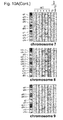


Classifications
-
- C—CHEMISTRY; METALLURGY
- C07—ORGANIC CHEMISTRY
- C07K—PEPTIDES
- C07K16/00—Immunoglobulins [IGs], e.g. monoclonal or polyclonal antibodies
- C07K16/18—Immunoglobulins [IGs], e.g. monoclonal or polyclonal antibodies against material from animals or humans
- C07K16/28—Immunoglobulins [IGs], e.g. monoclonal or polyclonal antibodies against material from animals or humans against receptors, cell surface antigens or cell surface determinants
- C07K16/2878—Immunoglobulins [IGs], e.g. monoclonal or polyclonal antibodies against material from animals or humans against receptors, cell surface antigens or cell surface determinants against the NGF-receptor/TNF-receptor superfamily, e.g. CD27, CD30, CD40, CD95
-
- G—PHYSICS
- G01—MEASURING; TESTING
- G01N—INVESTIGATING OR ANALYSING MATERIALS BY DETERMINING THEIR CHEMICAL OR PHYSICAL PROPERTIES
- G01N33/00—Investigating or analysing materials by specific methods not covered by groups G01N1/00 - G01N31/00
- G01N33/48—Biological material, e.g. blood, urine; Haemocytometers
- G01N33/50—Chemical analysis of biological material, e.g. blood, urine; Testing involving biospecific ligand binding methods; Immunological testing
- G01N33/68—Chemical analysis of biological material, e.g. blood, urine; Testing involving biospecific ligand binding methods; Immunological testing involving proteins, peptides or amino acids
- G01N33/6893—Chemical analysis of biological material, e.g. blood, urine; Testing involving biospecific ligand binding methods; Immunological testing involving proteins, peptides or amino acids related to diseases not provided for elsewhere
-
- A—HUMAN NECESSITIES
- A01—AGRICULTURE; FORESTRY; ANIMAL HUSBANDRY; HUNTING; TRAPPING; FISHING
- A01K—ANIMAL HUSBANDRY; AVICULTURE; APICULTURE; PISCICULTURE; FISHING; REARING OR BREEDING ANIMALS, NOT OTHERWISE PROVIDED FOR; NEW BREEDS OF ANIMALS
- A01K67/00—Rearing or breeding animals, not otherwise provided for; New or modified breeds of animals
- A01K67/027—New or modified breeds of vertebrates
- A01K67/0275—Genetically modified vertebrates, e.g. transgenic
-
- A—HUMAN NECESSITIES
- A61—MEDICAL OR VETERINARY SCIENCE; HYGIENE
- A61K—PREPARATIONS FOR MEDICAL, DENTAL OR TOILETRY PURPOSES
- A61K35/00—Medicinal preparations containing materials or reaction products thereof with undetermined constitution
- A61K35/12—Materials from mammals; Compositions comprising non-specified tissues or cells; Compositions comprising non-embryonic stem cells; Genetically modified cells
- A61K35/14—Blood; Artificial blood
- A61K35/17—Lymphocytes; B-cells; T-cells; Natural killer cells; Interferon-activated or cytokine-activated lymphocytes
-
- A—HUMAN NECESSITIES
- A61—MEDICAL OR VETERINARY SCIENCE; HYGIENE
- A61K—PREPARATIONS FOR MEDICAL, DENTAL OR TOILETRY PURPOSES
- A61K39/00—Medicinal preparations containing antigens or antibodies
- A61K39/46—Cellular immunotherapy
- A61K39/461—Cellular immunotherapy characterised by the cell type used
- A61K39/4611—T-cells, e.g. tumor infiltrating lymphocytes [TIL], lymphokine-activated killer cells [LAK] or regulatory T cells [Treg]
-
- A—HUMAN NECESSITIES
- A61—MEDICAL OR VETERINARY SCIENCE; HYGIENE
- A61K—PREPARATIONS FOR MEDICAL, DENTAL OR TOILETRY PURPOSES
- A61K39/00—Medicinal preparations containing antigens or antibodies
- A61K39/46—Cellular immunotherapy
- A61K39/461—Cellular immunotherapy characterised by the cell type used
- A61K39/4612—B-cells
-
- A—HUMAN NECESSITIES
- A61—MEDICAL OR VETERINARY SCIENCE; HYGIENE
- A61K—PREPARATIONS FOR MEDICAL, DENTAL OR TOILETRY PURPOSES
- A61K39/00—Medicinal preparations containing antigens or antibodies
- A61K39/46—Cellular immunotherapy
- A61K39/461—Cellular immunotherapy characterised by the cell type used
- A61K39/4615—Dendritic cells
-
- A—HUMAN NECESSITIES
- A61—MEDICAL OR VETERINARY SCIENCE; HYGIENE
- A61K—PREPARATIONS FOR MEDICAL, DENTAL OR TOILETRY PURPOSES
- A61K39/00—Medicinal preparations containing antigens or antibodies
- A61K39/46—Cellular immunotherapy
- A61K39/462—Cellular immunotherapy characterized by the effect or the function of the cells
- A61K39/4622—Antigen presenting cells
-
- A—HUMAN NECESSITIES
- A61—MEDICAL OR VETERINARY SCIENCE; HYGIENE
- A61K—PREPARATIONS FOR MEDICAL, DENTAL OR TOILETRY PURPOSES
- A61K39/00—Medicinal preparations containing antigens or antibodies
- A61K39/46—Cellular immunotherapy
- A61K39/464—Cellular immunotherapy characterised by the antigen targeted or presented
- A61K39/4643—Vertebrate antigens
- A61K39/4644—Cancer antigens
-
- A—HUMAN NECESSITIES
- A61—MEDICAL OR VETERINARY SCIENCE; HYGIENE
- A61P—SPECIFIC THERAPEUTIC ACTIVITY OF CHEMICAL COMPOUNDS OR MEDICINAL PREPARATIONS
- A61P35/00—Antineoplastic agents
-
- A—HUMAN NECESSITIES
- A61—MEDICAL OR VETERINARY SCIENCE; HYGIENE
- A61P—SPECIFIC THERAPEUTIC ACTIVITY OF CHEMICAL COMPOUNDS OR MEDICINAL PREPARATIONS
- A61P35/00—Antineoplastic agents
- A61P35/02—Antineoplastic agents specific for leukemia
-
- C—CHEMISTRY; METALLURGY
- C07—ORGANIC CHEMISTRY
- C07K—PEPTIDES
- C07K16/00—Immunoglobulins [IGs], e.g. monoclonal or polyclonal antibodies
- C07K16/18—Immunoglobulins [IGs], e.g. monoclonal or polyclonal antibodies against material from animals or humans
- C07K16/28—Immunoglobulins [IGs], e.g. monoclonal or polyclonal antibodies against material from animals or humans against receptors, cell surface antigens or cell surface determinants
- C07K16/30—Immunoglobulins [IGs], e.g. monoclonal or polyclonal antibodies against material from animals or humans against receptors, cell surface antigens or cell surface determinants from tumour cells
-
- C—CHEMISTRY; METALLURGY
- C12—BIOCHEMISTRY; BEER; SPIRITS; WINE; VINEGAR; MICROBIOLOGY; ENZYMOLOGY; MUTATION OR GENETIC ENGINEERING
- C12N—MICROORGANISMS OR ENZYMES; COMPOSITIONS THEREOF; PROPAGATING, PRESERVING, OR MAINTAINING MICROORGANISMS; MUTATION OR GENETIC ENGINEERING; CULTURE MEDIA
- C12N15/00—Mutation or genetic engineering; DNA or RNA concerning genetic engineering, vectors, e.g. plasmids, or their isolation, preparation or purification; Use of hosts therefor
- C12N15/09—Recombinant DNA-technology
- C12N15/11—DNA or RNA fragments; Modified forms thereof; Non-coding nucleic acids having a biological activity
- C12N15/113—Non-coding nucleic acids modulating the expression of genes, e.g. antisense oligonucleotides; Antisense DNA or RNA; Triplex- forming oligonucleotides; Catalytic nucleic acids, e.g. ribozymes; Nucleic acids used in co-suppression or gene silencing
- C12N15/1135—Non-coding nucleic acids modulating the expression of genes, e.g. antisense oligonucleotides; Antisense DNA or RNA; Triplex- forming oligonucleotides; Catalytic nucleic acids, e.g. ribozymes; Nucleic acids used in co-suppression or gene silencing against oncogenes or tumor suppressor genes
-
- C—CHEMISTRY; METALLURGY
- C12—BIOCHEMISTRY; BEER; SPIRITS; WINE; VINEGAR; MICROBIOLOGY; ENZYMOLOGY; MUTATION OR GENETIC ENGINEERING
- C12Q—MEASURING OR TESTING PROCESSES INVOLVING ENZYMES, NUCLEIC ACIDS OR MICROORGANISMS; COMPOSITIONS OR TEST PAPERS THEREFOR; PROCESSES OF PREPARING SUCH COMPOSITIONS; CONDITION-RESPONSIVE CONTROL IN MICROBIOLOGICAL OR ENZYMOLOGICAL PROCESSES
- C12Q1/00—Measuring or testing processes involving enzymes, nucleic acids or microorganisms; Compositions therefor; Processes of preparing such compositions
- C12Q1/68—Measuring or testing processes involving enzymes, nucleic acids or microorganisms; Compositions therefor; Processes of preparing such compositions involving nucleic acids
- C12Q1/6876—Nucleic acid products used in the analysis of nucleic acids, e.g. primers or probes
- C12Q1/6883—Nucleic acid products used in the analysis of nucleic acids, e.g. primers or probes for diseases caused by alterations of genetic material
- C12Q1/6886—Nucleic acid products used in the analysis of nucleic acids, e.g. primers or probes for diseases caused by alterations of genetic material for cancer
-
- G—PHYSICS
- G01—MEASURING; TESTING
- G01N—INVESTIGATING OR ANALYSING MATERIALS BY DETERMINING THEIR CHEMICAL OR PHYSICAL PROPERTIES
- G01N33/00—Investigating or analysing materials by specific methods not covered by groups G01N1/00 - G01N31/00
- G01N33/48—Biological material, e.g. blood, urine; Haemocytometers
- G01N33/50—Chemical analysis of biological material, e.g. blood, urine; Testing involving biospecific ligand binding methods; Immunological testing
- G01N33/53—Immunoassay; Biospecific binding assay; Materials therefor
- G01N33/574—Immunoassay; Biospecific binding assay; Materials therefor for cancer
- G01N33/57407—Specifically defined cancers
- G01N33/57438—Specifically defined cancers of liver, pancreas or kidney
-
- A—HUMAN NECESSITIES
- A01—AGRICULTURE; FORESTRY; ANIMAL HUSBANDRY; HUNTING; TRAPPING; FISHING
- A01K—ANIMAL HUSBANDRY; AVICULTURE; APICULTURE; PISCICULTURE; FISHING; REARING OR BREEDING ANIMALS, NOT OTHERWISE PROVIDED FOR; NEW BREEDS OF ANIMALS
- A01K2217/00—Genetically modified animals
- A01K2217/05—Animals comprising random inserted nucleic acids (transgenic)
- A01K2217/052—Animals comprising random inserted nucleic acids (transgenic) inducing gain of function
-
- A—HUMAN NECESSITIES
- A01—AGRICULTURE; FORESTRY; ANIMAL HUSBANDRY; HUNTING; TRAPPING; FISHING
- A01K—ANIMAL HUSBANDRY; AVICULTURE; APICULTURE; PISCICULTURE; FISHING; REARING OR BREEDING ANIMALS, NOT OTHERWISE PROVIDED FOR; NEW BREEDS OF ANIMALS
- A01K2227/00—Animals characterised by species
- A01K2227/10—Mammal
- A01K2227/105—Murine
-
- A—HUMAN NECESSITIES
- A01—AGRICULTURE; FORESTRY; ANIMAL HUSBANDRY; HUNTING; TRAPPING; FISHING
- A01K—ANIMAL HUSBANDRY; AVICULTURE; APICULTURE; PISCICULTURE; FISHING; REARING OR BREEDING ANIMALS, NOT OTHERWISE PROVIDED FOR; NEW BREEDS OF ANIMALS
- A01K2267/00—Animals characterised by purpose
- A01K2267/03—Animal model, e.g. for test or diseases
- A01K2267/0331—Animal model for proliferative diseases
-
- C—CHEMISTRY; METALLURGY
- C12—BIOCHEMISTRY; BEER; SPIRITS; WINE; VINEGAR; MICROBIOLOGY; ENZYMOLOGY; MUTATION OR GENETIC ENGINEERING
- C12N—MICROORGANISMS OR ENZYMES; COMPOSITIONS THEREOF; PROPAGATING, PRESERVING, OR MAINTAINING MICROORGANISMS; MUTATION OR GENETIC ENGINEERING; CULTURE MEDIA
- C12N2310/00—Structure or type of the nucleic acid
- C12N2310/10—Type of nucleic acid
- C12N2310/14—Type of nucleic acid interfering N.A.
-
- C—CHEMISTRY; METALLURGY
- C12—BIOCHEMISTRY; BEER; SPIRITS; WINE; VINEGAR; MICROBIOLOGY; ENZYMOLOGY; MUTATION OR GENETIC ENGINEERING
- C12Q—MEASURING OR TESTING PROCESSES INVOLVING ENZYMES, NUCLEIC ACIDS OR MICROORGANISMS; COMPOSITIONS OR TEST PAPERS THEREFOR; PROCESSES OF PREPARING SUCH COMPOSITIONS; CONDITION-RESPONSIVE CONTROL IN MICROBIOLOGICAL OR ENZYMOLOGICAL PROCESSES
- C12Q2563/00—Nucleic acid detection characterized by the use of physical, structural and functional properties
- C12Q2563/131—Nucleic acid detection characterized by the use of physical, structural and functional properties the label being a member of a cognate binding pair, i.e. extends to antibodies, haptens, avidin
-
- C—CHEMISTRY; METALLURGY
- C12—BIOCHEMISTRY; BEER; SPIRITS; WINE; VINEGAR; MICROBIOLOGY; ENZYMOLOGY; MUTATION OR GENETIC ENGINEERING
- C12Q—MEASURING OR TESTING PROCESSES INVOLVING ENZYMES, NUCLEIC ACIDS OR MICROORGANISMS; COMPOSITIONS OR TEST PAPERS THEREFOR; PROCESSES OF PREPARING SUCH COMPOSITIONS; CONDITION-RESPONSIVE CONTROL IN MICROBIOLOGICAL OR ENZYMOLOGICAL PROCESSES
- C12Q2600/00—Oligonucleotides characterized by their use
- C12Q2600/158—Expression markers
-
- G—PHYSICS
- G01—MEASURING; TESTING
- G01N—INVESTIGATING OR ANALYSING MATERIALS BY DETERMINING THEIR CHEMICAL OR PHYSICAL PROPERTIES
- G01N2500/00—Screening for compounds of potential therapeutic value
- G01N2500/04—Screening involving studying the effect of compounds C directly on molecule A (e.g. C are potential ligands for a receptor A, or potential substrates for an enzyme A)
Definitions
- the present invention relates to a reagent for tumor detection and a pharmaceutical composition for tumor prevention, each of which comprises a probe for the FEAT gene or amplification primers for the FEAT gene, or an antibody against the FEAT protein or a fragment of the antibody.
- Non-patent Document 1 discloses a method for molecular-targeted cancer screening and prevention.
- Non-patent Documents 2 to 4 comprehensive sequencing of protein-coding exon regions
- Non-patent Document 5 whole genome sequencing
- Non-patent Document 6 paired-end sequencing for identification of gene rearrangements in somatic cells
- Non-patent Documents 7 to 9 there is no consistency in the combinations of altered cancer genes (Non-patent Documents 7 to 9), and it has been reported that alterations in signaling pathways rather than individual genes would facilitate tumorigenesis (Non-patent Document 10). Moreover, even in the case of common signaling pathways that are altered equally in cancers, different components are altered in different tumor tissues (Non-patent Documents 2 to 4).
- test reagents for tumor detection are designed to use a marker specific for a particular type of tumor as an indicator for tumor detection
- many kinds of test reagents should be used at the same time in order to comprehensively detect tumors which may develop in the patient's body.
- the use of many kinds of test reagents not only leads to a significant reduction in the cost-effectiveness ratio because of requiring high costs, but also raises a problem of an increased work burden on medical practitioners and an increased mental and physical burden on patients.
- the present invention aims to provide a test reagent and a therapeutic pharmaceutical composition, each targeting a marker which is expressed equally in various types of tumors or cancers. Moreover, the present invention also aims to provide a detection reagent allowing tumor detection at an early stage of tumorigenesis.
- the inventors of the present invention have found that the FEAT protein (faint expression in normal tissues, aberrant overexpression in tumors) is overexpressed in common from an early stage in diverse human cancer tissues.
- the inventors of the present invention have conducted expression microarray analyses and have found that FEAT induces oncogenic signaling pathways.
- the inventors of the present invention have prepared transgenic (Tg) mice for overexpression of FEAT and have found that FEAT also promotes tumorigenesis in vivo in the experiments using these Tg mice.
- the present invention therefore provides the following.
- the reagent of the present invention enables the detection of various types of cancers or precancerous lesions. Moreover, the reagent of the present invention also allows tumor detection at an early stage of tumorigenesis. Further, the pharmaceutical composition of the present invention allows effective treatment or prevention of tumors.
- the inventors of the present invention have found that the FEAT protein is overexpressed in diverse tumor tissues and contributes to tumorigenesis.
- the FEAT gene and protein are not expressed in almost all tissues and are weakly expressed only in the testis, brain and liver.
- the FEAT gene is expressed equally in diverse tumor tissues including ovarian cancer, lung cancer, liver cancer, rectal cancer, uterine cervical cancer, skin cancer, testicular cancer, thyroid cancer, uterine cancer, gastric cancer, pharyngeal cancer, breast cancer, prostate cancer, and pancreatic cancer ( Figure 5B ).
- the inventors of the present invention have found that the FEAT protein is produced prior to tumor-induced phenotypic changes during carcinogenesis in humans ( Figure 13 ). Moreover, the inventors of the present invention have also found that the FEAT protein, when overexpressed in cultured cells, activates the receptor tyrosine kinase signaling pathway and the Hedgehog signaling pathway among oncogenic pathways ( Figure 5D ), and have further confirmed that tumor tissues are formed in various organs of transgenic mice engineered to overexpress the FEAT protein ( Figures 7A to 7C ).
- those expressing the FEAT protein are highly likely to turn into tumors in the future, i.e., can be regarded as precancerous cells.
- FEAT which is used as a tumor marker in the present invention.
- the FEAT protein is a protein encoded by a gene called “comparative gene identification-01 (CGI-01),” “KIAA0859” or “methyltransferase like 13 (METTL13).”
- the FEAT protein is cleaved by caspase-3, but not by caspase-6 ( Figure 2B ).
- the FEAT protein has the function of suppressing apoptosis in cells ( Figure 4C ).
- the nucleotide sequence of the human FEAT gene and the amino acid sequence of the human FEAT protein are as shown in SEQ ID NO: 1 (Accession Number: NM_015935) and SEQ ID NO: 2, respectively.
- SEQ ID NO: 2 accesion Number: NM_015935
- mutants thereof such as those carrying either or both a D274A mutation in which alanine substitutes for aspartic acid at position 274 (SEQ ID NO: 2) in the caspase-3 cleavage sites and a D288A mutation in which alanine substitutes for aspartic acid at position 288 (SEQ ID NO: 2) in the caspase-3 cleavage sites, are resistant to caspase-3 cleavage and hence have a stronger apoptosis-suppressing function ( Figure 3B ).
- FEAT protein SEQ ID NO: 2
- fragments thereof particularly fragments comprising a sequence composed of amino acids at positions 289 to 699 in the amino acid sequence of the FEAT protein, have an apoptosis-suppressing function.
- the FEAT protein contains two S-adenosylmethionine-binding motifs (SAM-binding motifs).
- SAM-binding motif is a motif characteristic of methyltransferase and related enzymes thereof ( Figure 1B ). However, it has remained unknown whether or not the FEAT protein has methyltransferase activity.
- consensus sequences in SAM-binding motifs the following sequences can be listed, by way of example.
- the above FEAT protein and FEAT gene can be obtained by the procedures described later in the Example section, known genetic engineering procedures, known synthesis procedures, etc.
- the present invention provides a reagent for tumor detection, which comprises a probe for the FEAT gene or amplification primers for the FEAT gene, or an antibody against the FEAT protein or a fragment of the antibody.
- the FEAT gene or the FEAT protein are expressed widely and specifically in tumor cells or precancerous cells, but are not expressed in most normal cells. Thus, if the FEAT gene or the FEAT protein is detected in test cells, such test cells can be diagnosed as tumor cells or precancerous cells.
- FEAT gene is intended to include the above human wild-type FEAT gene having the nucleotide sequence shown in SEQ ID NO: 1, as well as FEAT homolog genes derived from non-human species and mutant genes thereof.
- examples of the FEAT gene include any one polynucleotide selected from the group consisting of (a) to (e) shown below:
- the FEAT gene may be either DNA or RNA (messenger RNA).
- polynucleotide which is hybridizable under stringent conditions is intended to mean, for example, a polynucleotide that can be obtained by means of colony hybridization, plaque hybridization, Southern hybridization or other hybridization techniques using, as a probe, the whole or a part of a polynucleotide consisting of a nucleotide sequence complementary to the nucleotide sequence shown in SEQ ID NO: 1 or a polynucleotide consisting of a nucleotide sequence encoding the amino acid sequence shown in SEQ ID NO: 2.
- stringent conditions may be any of low stringent conditions, moderately stringent conditions and high stringent conditions.
- Low stringent conditions refer to, for example, conditions of 5 ⁇ SSC, 5 ⁇ Denhardt's solution, 0.5% SDS, 50% formamide and 32°C.
- moderately stringent conditions refer to, for example, conditions of 5 ⁇ SSC, 5 ⁇ Denhardt's solution, 0.5% SDS, 50% formamide and 42°C.
- “High stringent conditions” refer to, for example, conditions of 5 ⁇ SSC, 5 ⁇ Denhardt's solution, 0.5% SDS, 50% formamide and 50°C. Under these conditions, it can be expected that DNA having a higher identity is efficiently obtained at a higher temperature.
- the stringency of hybridization would be affected by a plurality of factors, including temperature, probe concentration, probe length, ionic strength, reaction time, salt concentration and so on. Those skilled in the art would be able to achieve the same stringency by selecting these factors as appropriate.
- hybridization may be accomplished in accordance with the protocol attached to the kit, i.e., a membrane may be incubated overnight with a labeled probe and then washed with a primary washing buffer containing 0.1% (w/v) SDS under conditions of 55°C to detect the hybridized DNA.
- hybridizable polynucleotides include polynucleotides sharing an identity of 50% or more, 60% or more, 70% or more, 80% or more, 85% or more, 90% or more, 91% or more, 92% or more, 93% or more, 94% or more, 95% or more, 96% or more, 97% or more, 98% or more, 99% or more, 99.5% or more, or 99.9% or more with a polynucleotide consisting of the nucleotide sequence shown in SEQ ID NO: 1 or with a polynucleotide consisting of a nucleotide sequence encoding the amino acid sequence shown in SEQ ID NO: 2, as calculated by homology search software such as FASTA or BLAST using default parameters.
- homology search software such as FASTA or BLAST using default parameters.
- polynucleotides of the present invention described above can be obtained by known genetic engineering procedures or known synthesis procedures.
- examples of a probe for the FEAT gene include oligonucleotides which are hybridizable under stringent conditions with the FEAT gene.
- Stringent conditions are as described above.
- oligonucleotide is intended to mean a nucleotide chain having a length of preferably 5 to 500 bp, more preferably 10 to 200 bp, and even more preferably 10 to 100 bp.
- oligonucleotides may be readily synthesized by using various automatic synthesizers (e.g., AKTA oligopilot plus 10/100 (GE Healthcare)), or alternatively, their synthesis may be entrusted to a third party (e.g., Promega or Takara), etc.
- automatic synthesizers e.g., AKTA oligopilot plus 10/100 (GE Healthcare)
- a third party e.g., Promega or Takara
- assay methods for tumor detection include Northern blotting ( Sambrook, Fritsch and Maniatis, "Molecular Cloning: A Laboratory Manual” 2nd Edition (1989), Cold Spring Harbor Laboratory Press ), microarrays (Affymetrix; see United States Patent Nos. 6,045,996 , 5,925,525 and 5,858,659 ) or TaqMan PCR ( Sambrook, Fritsch and Maniatis, "Molecular Cloning: A Laboratory Manual” 2nd Edition (1989), Cold Spring Harbor Laboratory Press ) for the FEAT gene expressed in the tumor.
- the reagent of the present invention comprises primers for the FEAT gene
- methods for tumor detection include PCR amplification or sequencing ( Sambrook, Fritsch and Maniatis, "Molecular Cloning: A Laboratory Manual” 2nd Edition (1989), Cold Spring Harbor Laboratory Press ) for the FEAT gene expressed in the tumor.
- the reagent of the present invention may comprise both the probe and the primers.
- FEAT protein is intended to include not only the human wild-type FEAT protein consisting of the amino acid sequence shown in SEQ ID NO: 2, but also FEAT homolog proteins derived from non-human species and mutant proteins thereof. In addition, among fragments generated by cleavage of the FEAT protein, those having a tumor-promoting function also fall within the FEAT protein.
- FEAT protein examples include polypeptides shown in (f), (g) and (h) below or fragments thereof:
- the above polypeptide (g) or (h) is typically a FEAT homolog protein derived from non-human species or a mutant protein of the wild-type FEAT protein, but it also includes other polypeptides based on a polypeptide consisting of the amino acid sequence shown in SEQ ID NO: 2, which may be artificially obtained by site-directed mutagenesis as described in " Sambrook & Russell, Molecular Cloning: A Laboratory Manual Vol. 3, Cold Spring Harbor Laboratory Press 2001 ,” “ Ausubel, Current Protocols in Molecular Biology, John Wiley & Sons 1987-1997 ,” “ Nuc. Acids. Res., 10, 6487(1982 ),” “ Proc. Natl. Acad. Sci.
- polypeptide which comprises an amino acid sequence with deletion, substitution or addition of one or several amino acids in the amino acid sequence shown in SEQ ID NO: 2 and which has a tumor-promoting function is intended to include polypeptides which comprise an amino acid sequence with deletion, substitution or addition of, e.g., 1 to 100 amino acid residues, 1 to 90 amino acid residues, 1 to 80 amino acid residues, 1 to 70 amino acid residues, 1 to 60 amino acid residues, 1 to 50 amino acid residues, 1 to 40 amino acid residues, 1 to 30 amino acid residues, 1 to 20 amino acid residues, 1 to 10 amino acid residues, 1 to 9 amino acid residues (one or several amino acid residues), 1 to 8 amino acid residues, 1 to 7 amino acid residues, 1 to 6 amino acid residues, 1 to 5 amino acid residues, 1 to 4 amino acid residues, 1 to 3 amino acid residues, 1 or 2 amino acid residues, or a single amino acid residue in the amino acid sequence shown in SEQ ID NO: 2 and which
- examples of such polypeptides include those which have an amino acid sequence sharing an identity of 60% or more, 65% or more, 70% or more, 75% or more, 80% or more, 85% or more, 90% or more, 91% or more, 92% or more, 93% or more, 94% or more, 95% or more, 96% or more, 97% or more, 98% or more, 99% or more, 99.1% or more, 99.5% or more, or 99.9% or more with the amino acid sequence shown in SEQ ID NO: 2 and which have a tumor-promoting function. In general, a larger value is more preferred for the above identity.
- test protein has a tumor-promoting function can be confirmed by introducing the test protein or a gene thereof into appropriate normal host cells (e.g., human neutrophils) which do not express FEAT, incubating the host cells under appropriate culture conditions, and then observing tumorigenesis in the host cells.
- appropriate normal host cells e.g., human neutrophils
- the tumor-promoting function may be an apoptosis-suppressing function. Whether or not a test protein has an apoptosis-suppressing function can be confirmed by introducing the test protein or a gene thereof into appropriate host cells (e.g., human neutrophils) which do not express FEAT, incubating the host cells under appropriate culture conditions, and then analyzing the amount of apoptotic cells with fragmented DNA among the host cells by flow cytometry to determine apoptotic cell counts.
- appropriate host cells e.g., human neutrophils
- deletion, substitution or addition of one or several amino acid residues may occur at any one or more positions in the same sequence, or alternatively, two or more of deletion, substitution and addition may occur at the same time.
- the amino acid residue(s) may be added by being inserted into the amino acid sequence or may be added to the N-terminal or C-terminal end of the amino acid sequence.
- FEAT proteins may also be prepared by chemical synthesis methods such as Fmoc method (fluorenylmethyloxycarbonyl method) and tBoc method (t-butyloxycarbonyl method). Alternatively, they may be chemically synthesized by using peptide synthesizers commercially available from Advanced Automation Peptide Protein Technologies, Perkin Elmer, Protein Technologies, PerSeptive, Applied Biosystems, SHIMADZU, etc.
- the FEAT protein is expressed in tumor cells or precancerous cells
- fragments of the FEAT protein may also be contained in tumor cells or precancerous cells because the FEAT protein will be cleaved by intracellular caspase-3 once apoptotic cell death has occurred.
- the reagent of the present invention is configured to detect the full-length FEAT protein or a fragment thereof.
- Such a fragment is not limited in any way, but is preferably a fragment comprising a sequence consisting of amino acids at positions 289 to 699 in the amino acid sequence shown in SEQ ID NO: 2. This fragment is preferred in terms of having a tumor-promoting function, albeit weaker than that of the full-length protein.
- an antibody against the FEAT protein (or a fragment thereof) (hereinafter referred to as “the antibody of the present invention”) may be either polyclonal or monoclonal.
- the FEAT protein is first prepared for use as an immunogen (antigen).
- the FEAT protein serving as an antigen may be prepared in a genetic engineering manner on the basis of the amino acid sequence of the FEAT protein.
- the resulting purified FEAT is then used to immunize animals. Immunization may be accomplished by administering a solution containing the FEAT protein to mammals (e.g., mice, rats, rabbits, guinea pigs, goats or the like). Administration may be accomplished primarily by intravenous, subcutaneous or intraperitoneal injection. If necessary, an adjuvant may also be added as appropriate. Subsequently, sera (antisera) obtained as a result of immunization are collected. Serum collection is not limited in any way and sera may be collected in a routine manner form the blood of the immunized animals at 1 to 28 days after the final administration. The collected antisera are screened by known immunoassay techniques to select a desired antiserum.
- Antibodies contained in the desired antiserum obtained by the above screening are polyclonal antibodies. If these antibodies are required to be purified, known purification techniques such as salting out with ammonium sulfate, ion exchange chromatography, affinity chromatography, gel chromatography and so on may be used either alone or in combination for this purpose.
- Preparation of an antigen and a solution thereof, as well as immunization therewith may be accomplished in the same manner as described above for preparation of polyclonal antibodies.
- antibody-producing cells are collected.
- antibody-producing cells preferred are spleen cells, lymph node cells, peripheral blood cells and the like, by way of example, and more preferred are spleen cells.
- Cell fusion is induced between the collected antibody-producing cells and myeloma cells to thereby obtain fused cells (hybridomas).
- Myeloma cells used for this purpose include, for example, PAI, P3X63-Ag.8.U1(P3U1), NS-I and so on.
- the above cell fusion may be accomplished, for example, by mixing the antibody-producing cells with the myeloma cells in a medium for animal cell culture (e.g., RPMI-1640 medium) to cause fusion reaction.
- fusion reaction is preferably performed in the presence of a cell fusion promoter such as polyethylene glycol.
- a commercially available electroporation-based cell fusion apparatus may also be used to fuse the antibody-producing cells with the myeloma cells.
- the cells obtained after fusion reaction are cultured, e.g., in HAT selective medium. After being cultured as above, cells grown in the HAT selective medium are fused cells (hybridomas).
- the hybridomas obtained by being cultured as above are screened to select desired hybridomas, followed by known purification techniques such as salting out with ammonium sulfate, ion exchange chromatography, affinity chromatography, gel chromatography and so on, which may be used either alone or in combination as appropriate, to thereby purify monoclonal antibodies from the above hybridomas.
- purification techniques such as salting out with ammonium sulfate, ion exchange chromatography, affinity chromatography, gel chromatography and so on, which may be used either alone or in combination as appropriate, to thereby purify monoclonal antibodies from the above hybridomas.
- monoclonal antibodies reference may be made to United States Patent No. 4,816,567 .
- a humanized antibody a human antibody, or a fragment thereof may also be used.
- a humanized antibody refers to a monoclonal antibody prepared in a genetic engineering manner, and more specifically refers to an antibody designed such that the complementarity determining regions (CDRs) in its hypervariable regions are composed partially or completely of complementarity determining regions derived from mouse monoclonal antibody hypervariable regions, the framework regions (FRs) in its variable regions are composed of FRs derived from human immunoglobulin variable regions, and its constant regions are composed of human immunoglobulin constant regions.
- CDRs complementarity determining regions
- FRs framework regions
- FRs framework regions
- constant regions are composed of human immunoglobulin constant regions.
- a human antibody refers to an antibody whose immunoglobulin-constituting regions are all derived from genes encoding human immunoglobulin sequences.
- An antibody fragment is intended to mean a partial fragment comprising an antigen-binding region of the above antibody, and specific examples include F(ab') 2 , Fab', Fab, Fv (variable fragment of antibody), scFv (single chain Fv), dsFv (disulphide stabilized Fv) and so on.
- assay methods for tumor detection include immunohistological staining ( Lorette C. Javois ed. "Immunocytochemical Methods and Protocols (Methods in Molecular Biology)” Humana Press; 2nd edition (February 15, 1999 )), immunoprecipitation, affinity chromatography, pull-down assay, enzyme linked immunosorbent assay (ELISA) and Western blotting ( Gary C. Howard and Matthew R. Kaser ed. "Making and Using Antibodies: A Practical Handbook” CRC Press; 1st edition (December 13, 2006 )), antibody microarrays ( Chaga GS (2008). "Antibody arrays for determination of relative protein abundances.” Methods Mol. Biol.
- the inventors of the present invention have found that the FEAT protein is expressed from a very early stage (prior to tumor-induced phenotypic changes) during the tumorigenesis process, and have further found that the expression of the FEAT protein promotes tumorigenesis.
- the inventors of the present invention have also found that the FEAT protein per se has an apoptosis-suppressing function and that the expression of the FEAT protein activates oncogenic signaling pathways.
- tumorigenesis can also be prevented when functions of the FEAT protein or a gene thereof are inhibited to induce apoptotic cell death in tumor cells or precancerous cells.
- the present invention provides a pharmaceutical composition for tumor prevention, which comprises a substance shown in (i) or (ii) below or cells shown in (iii) below:
- the "substance capable of inhibiting FEAT gene expression” shown in (i) above include substances capable of reducing the expression level of messenger RNA (mRNA) for the FEAT gene, as exemplified by low molecular compounds, hormones, proteins and nucleic acids.
- mRNA messenger RNA
- such a substance may be a nucleic acid capable of suppressing the functions or expression of the FEAT gene.
- examples of such a nucleic acid include hairpin-shaped shRNAs (short hairpin RNAs) or double-stranded RNAs (dsRNAs) which produce siRNAs (small interfering RNAs) for RNA interference (RNAi), as well as antisense nucleic acids, decoy nucleic acids, or aptamers, etc.
- inhibitory nucleic acids are able to suppress the expression of the above gene.
- the nucleotide sequence of the FEAT gene to be inhibited is as described above, and sequence information can be obtained for each case.
- the nucleotide sequence of the FEAT gene is as shown in SEQ ID NO: 1, although not only a coding region of FEAT, but also a non-coding region may be used as a target.
- the inventors of the present invention have succeeded in knocking down FEAT expression in host cells by RNAi using shRNAs ( Figure 3C ) and thereby promoting apoptosis in the host cells ( Figure 3D ).
- RNAi is a multi-step process proceeding through a number of stages.
- dsRNA or shRNA expressed from an RNAi expression vector is recognized by Dicer and cleaved into siRNAs of 21 to 23 nucleotides. These siRNAs are then integrated into an RNAi targeting complex, which is called the RNA-induced silencing complex (RISC), and the complexes between RISC and siRNAs bind to target mRNA containing sequences complementary to the siRNA sequences and thereby cleave the mRNA.
- RISC RNA-induced silencing complex
- the target mRNA is cleaved in the center of its region complementary to the siRNA, finally leading to rapid degradation of the target mRNA and reduced protein expression levels.
- siRNA duplexes are known to be sequences of 21 nucleotides in length, each comprising a 19 bp duplex with an overhang of two uridine residues at the 3'-terminal end ( Elbashir S.M. et al., Genes and Dev, 15, 188-200 (2001 )).
- a target sequence on mRNA may be selected from the cDNA sequence corresponding to the mRNA.
- the present invention is not limited to this region.
- siRNA molecules may be designed on the basis of the criteria well known in the art. For example, as a target segment in target mRNA, it is possible to select a segment covering 15 to 30 contiguous bases, preferably 19 to 25 contiguous bases, preferably starting with AA, TA, GA or CA. siRNA molecules have a GC ratio of 30% to 70%, preferably 35% to 55%. Alternatively, a target sequence for RNAi may be selected as appropriate as described in Ui-Tei K. et al. ((2004) Nucleic Acids Res. 32, 936-948 ).
- RNAi against the human FEAT gene the following nucleotide sequences of 19 base pairs starting from positions 183, 259, 1385, 1550, 1634 and 2056 in the open reading frame region of FEAT cDNA (SEQ ID NOs: 3, 4, 5, 6, 7 and 8, respectively) can be used as targets for RNAi to thereby suppress FEAT gene expression.
- siRNA For introduction of siRNA into cells, it is possible to use, e.g., procedures in which synthesized siRNA is ligated to plasmid DNA and then introduced into cells, or procedures in which double-stranded RNA is annealed.
- shRNA may also be used for providing RNAi effect.
- shRNA is an RNA molecule called short hairpin RNA, which has a stem-loop structure because some single-stranded regions form complementary strands with other regions.
- shRNA may be designed to form a stem-loop structure as a part thereof. For example, assuming that a sequence covering a certain region is designated as sequence A, and a strand complementary to the sequence A is designated as sequence B, shRNA is design to comprise the sequence A, a spacer and the sequence B linked in this order on a single RNA strand and to have an overall length of 45 to 60 bases. The spacer may also have any length.
- sequence A is a sequence covering a partial region of the target FEAT gene, there is no particular limitation on the target region and any region may be selected as a candidate for the target region.
- sequence A has a length of 19 to 25 bases, preferably 19 to 21 bases.
- microRNA may be used to inhibit FEAT gene expression.
- microRNA is an intracellular single-stranded RNA molecule having a length of about 20 to 25 bases and is a kind of ncRNA (non-coding RNA) which is considered to have the function of regulating the expression of other genes. miRNA is generated through processing upon transcription into RNA and is present as a nucleic acid capable of forming a hairpin structure which suppresses the expression of a target sequence.
- miRNA is also an inhibitory nucleic acid based on RNAi
- miRNA may also be designed and synthesized in the same manner as in the case of shRNA or siRNA.
- Expression vectors for RNAi may be readily prepared with a commercially available DNA/RNA synthesizer (e.g., Applied Biosystems model 394) on the basis of pMuniH1 plasmid, pSINsi vector (Takara Bio Inc., Japan), pSIF1-H1 (System Biosciences, Inc.), etc.
- expression vectors for RNAi may be prepared by entrusting their preparation to third parties such as Cosmo Bio Co., Ltd. (Japan), Takara Bio Inc. (Japan), Invitrogen, Promega, etc.
- an antisense nucleic acid may be used to inhibit FEAT gene expression.
- An antisense nucleic acid is a single-stranded nucleic acid sequence (either RNA or DNA) capable of binding to the mRNA or DNA sequence of the FEAT gene.
- Such an antisense nucleic acid sequence has a length of at least 14 nucleotides, preferably 14 to 100 nucleotides. The antisense nucleic acid binds to the above gene sequence to form a duplex and thereby suppresses the transcription or translation of the FEAT gene.
- the antisense nucleic acid may be prepared by chemical or biochemical synthesis procedures known in the art. For example, it is possible to use nucleic acid synthesis procedures using a commonly used DNA synthesizer.
- a nucleic acid serving as a decoy which is called a decoy nucleic acid, may be used to inhibit FEAT gene expression.
- a decoy nucleic acid is a nucleic acid that suppresses FEAT gene expression through binding to a transcription factor in the FEAT gene or in the FEAT receptor gene to suppress promoter activity, and it is intended to mean a short nucleic acid serving as a decoy that comprises a binding site for the transcription factor.
- a transcription factor will bind to this nucleic acid.
- the transcription factor will be competitively inhibited from binding to its genomic binding site and thus prevented from being expressed.
- Examples of a decoy nucleic acid preferred in the present invention include a nucleic acid binding to a promoter for the FEAT gene, or a nucleic acid binding to a promoter for mRNA of FEAT or for a factor located upstream of FEAT.
- a decoy nucleic acid may be designed as a single or double strand on the basis of the promoter sequence for the FEAT gene.
- the decoy nucleic acid may have any length, its length is 15 to 60 bases, preferably 20 to 30 bases.
- the decoy nucleic acid may be either DNA or RNA, and may also include modified nucleic acids.
- the decoy nucleic acid to be used in the present invention may be prepared by chemical or biochemical synthesis procedures known in the art. For example, it is possible to use nucleic acid synthesis procedures using a DNA synthesizer commonly used in gene recombination technology. Alternatively, a nucleotide sequence serving as a template may be isolated or synthesized, followed by PCR or gene amplification using a cloning vector. Further, to obtain a decoy nucleic acid which is more stable within cells, bases and other constituents may be modified by chemical modifications such as alkylation, acylation, etc.
- an aptamer may be used to inhibit FEAT gene expression.
- An aptamer is a synthetic DNA or RNA molecule or a peptidic molecule, which has the ability to bind specifically to a target substance, and can be synthesized chemically in a test tube within a short time.
- an aptamer may be obtained on the basis of the nucleotide sequence shown in SEQ ID NO: 1, or alternatively, may be obtained by evolutionary engineering procedures known as in vitro selection or SELEX techniques.
- a nucleic acid aptamer is rapidly degraded and removed from the blood flow by the action of nucleases, and hence it is preferred that the aptamer has optionally been subjected to molecular modification, e.g., with a polyethylene glycol (PEG) chain to thereby extend its half-life.
- PEG polyethylene glycol
- the "substance capable of inhibiting FEAT protein activity” shown in (i) above include substances capable of breaking down the FEAT protein (e.g., various proteases), as well as antagonists of the FEAT protein (e.g., low molecular compounds, hormones, proteins, antibodies and nucleic acids, etc.).
- a substance may be an antibody against the FEAT protein.
- Such an antibody against the FEAT protein is as described above for the test reagent.
- immune cells capable of reacting with the FEAT protein include immune cells that recognize the FEAT protein as an antigen.
- immune cells include, but are not limited to, helper T cells, cytotoxic T cells (CTLs), natural killer cells, B cells, dendritic cells, macrophages and so on.
- Such immune cells may be those transformed by gene recombination to express a gene for an anticancer agent or a gene for a prodrug-converting enzyme that converts a prodrug of the anticancer agent into an active drug.
- the immune cells capable of reacting with the FEAT protein are cells expressing molecules with antitumor activity (hereinafter collectively referred to as "anticancer agents")
- anticancer agents molecules with antitumor activity
- such immune cells will contact with tumor cells via the FEAT protein because the FEAT protein is expressed exclusively in tumor cells, and the tumor cells will be killed by the action of the anticancer agents expressed within the immune cells.
- anticancer agents also includes prodrugs of the anticancer agents.
- anticancer agents include, but are not limited to, parasporin, POGZ, panitumumab, rituximab, cetuximab, trastuzumab, ImmunoMax, bevacizumab, L-asparaginase, dasatinib, nilotinib, erlotinib, sunitinib, sorafenib, lapatinib, axitinib, sunitinib, motesanib, denosumab, nimotuzumab, ibritumomab, tocilizumab, ofatumumab, CS-1008, MORAb-009, MORAb-003, ONO-4538, KW-0761, AMG655, R7159(GA101), R1507, CMC-544, GC33, KRN330m, U3-1287 and so on.
- the nucleotide sequences of genes encoding these anticancer agents are known
- anticancer agents may also be siRNAs against cancer genes, or shRNAs or dsRNAs from which the siRNAs are produced.
- cancer genes include the myelocytomatosis oncogene (c-Myc) gene, the v-Src gene, the RAS gene, the B-cell leukemia/lymphoma 2 (BCL2) gene, the phosphatidyl inositol 3-kinase catalytic subunit (PIK3CA) gene, the Cyclin D1 gene, the Liver Kinase B1 (LKB1) gene, the c-Abl gene, the c-Sis gene, the epidermal growth factor receptor (EGFR) gene, the platelet-derived growth factor receptor (PDGFR) gene, the vascular endothelial growth factor receptor (VEGFR) gene, the HER2/neu gene, the Raf kinase gene, the cyclin-dependent kinase genes (e.g., Cyclin dependent kinase 4
- the immune cells of the present invention express a prodrug-converting enzyme
- the above cells will recognize tumor cells or precancerous cells (hereinafter collectively referred to as "tumor cells"), which are expressing the FEAT protein, and will be localized around the tumor cells and will produce the prodrug-converting enzyme exclusively therein.
- tumor cells precancerous cells
- the administered prodrug will be converted into the original anticancer agent exclusively around the tumor cells, so that the anticancer agent can exert its efficacy exclusively on the tumor cells while minimizing any effects on normal cells.
- prodrug-converting enzyme examples include thymidine kinase, a fusion protein between thymidine kinase and thymidylate kinase, as well as cytosine deaminase and so on.
- Thymidine kinase is a prodrug-converting enzyme for ganciclovir
- a fusion protein between thymidine kinase and thymidylate kinase is a prodrug-converting enzyme for azidothymidine
- cytosine deaminase is a prodrug-converting enzyme for 5-fluorouracil (5-FU).
- 5-FU 5-fluorouracil
- anticancer genes may each be inserted as an appropriate expression cassette into an expression vector, and the vector may be used to transform host cells (e.g., immune cells).
- an appropriate expression cassette comprises at least the following as constituent elements:
- promoters that can be transcribed in these host cells include, but are not limited to, CMV, CAG, LTR, EF-1 ⁇ and SV40 promoters, etc.
- Expression vectors for siRNAs against cancer genes are as described above for RNA interference.
- Such an expression vector may comprise not only the above expression cassette, but also a selection marker expression cassette to select the transformed host cells.
- a selection marker include, but are not limited to, positive selection markers (e.g., neomycin resistance gene, hygromycin B phosphotransferase gene), expression reporters (e.g., LacZ, GFP (green fluorescence protein) and luciferase genes), negative selection markers (e.g., herpes simplex virus thymidine kinase gene (HSV-TK), diphtheria toxin A fragment (DTA)), etc.
- positive selection markers e.g., neomycin resistance gene, hygromycin B phosphotransferase gene
- expression reporters e.g., LacZ, GFP (green fluorescence protein) and luciferase genes
- negative selection markers e.g., herpes simplex virus thymidine kinase gene (HSV-TK), diphtheria tox
- the transformed host cells can be easily selected by the above markers.
- these cells may be cultured in a G418-supplemented medium to conduct primary selection.
- a targeting vector comprises a gene for a fluorescent protein (e.g., GFP) as a marker
- drug resistance-based selection may be combined with cell sorting in a FACS (fluorescence activated cell sorter) or the like to isolate fluorescent protein-expressing cells.
- Expression vectors which can be used for introduction of anti-cancer genes into host cells are commercially available, and examples include pEGFP-C1TM (Clontech), pCMV-HATM (Clontech), pMSCVpuroTM (Clontech), pEF-DEST51TM (Invitrogen), pCEP4TM (Invitrogen), ViraPower II Lentiviral Gateway SystemTM (Invitrogen) and so on.
- These expression vectors can be introduced into host cells by known gene transfer techniques such as electroporation, microinjection, calcium phosphate transfection, lipofection, virus infection, etc.
- gene transfer techniques reference may be made to, e.g., " Sambrook & Russell, Molecular Cloning: A Laboratory Manual Vol. 3, Cold Spring Harbor, Laboratory Press 2001 .”
- Immune cells capable of reacting with the FEAT protein can be prepared in the following manner.
- the FEAT amino acid sequence is used to predict the amino acid sequence of an immunizing peptide which matches with the histocompatibility antigen (HLA) of each subject, and a peptide having such an amino acid sequence is synthesized.
- Amino acid sequence prediction for the immunizing peptide which matches with HLA may be accomplished by using the software of BIMAS (http://www-bimas.cit.nih.gov/molbio/hla-bind/) or SYFPEITHI (http://www.syfpeithi.de/Scripts/MHCServer.dll/EpitopePrediction.htm).
- the above peptide is mixed with Freund's incomplete adjuvant and inoculated as a peptide vaccine into a human subject by the subcutaneous route to produce immune cells in the human body, or alternatively, (2) cells separated from human peripheral blood are cultured together with the peptide and various cytokines to produce immune cells.
- the "substance capable of binding to the FEAT protein, which substance carries an antitumor agent, the substance shown in (i) above or the cells shown in (ii) above” is intended to mean a substance capable of delivering an antitumor agent, the substance shown in (i) above or the cells shown in (ii) above to FEAT-expressing cells.
- a substance capable of delivering an antitumor agent, the substance shown in (i) above or the cells shown in (ii) above to FEAT-expressing cells include, but are not limited to, peptides, saccharides, small molecules, macromolecules, nucleic acids, metals, antibodies and so on, which are capable of binding to the FEAT protein.
- such a substance may be an anti-FEAT antibody conjugated with an antitumor agent.
- the pharmaceutical composition of the present invention may be administered by any route commonly used for administration of the candidate drug.
- routes commonly used for administration of the candidate drug include oral, sublingual, transnasal, transpulmonary, transgastrointestinal and percutaneous routes, eye dropping, intravenous injection, subcutaneous injection, intramuscular injection, intraperitoneal injection, topical injection, and surgical grafting, with intravenous injection being preferred.
- the pharmaceutical composition of the present invention may be in a solid dosage form (e.g., capsules, tablets, powders) or may be in a liquid dosage form (e.g., solutions, suspensions or emulsions) or in a semi-liquid dosage form (e.g., ointments, creams or pastes).
- a solid dosage form e.g., capsules, tablets, powders
- a liquid dosage form e.g., solutions, suspensions or emulsions
- a semi-liquid dosage form e.g., ointments, creams or pastes.
- a suitable dosage form may be exemplified by a cell suspension.
- a cell suspension may comprise a culture medium suitable for survival of the cells, cytokines, serum (preferably autoserum collected from a patient to be administered), nonessential amino acids, antibiotics and so on.
- a suitable dosage form may be exemplified by a suspension or a solution.
- a suspension or dosage form may comprise a buffer suitable for maintaining the structure of the above substance (e.g., siRNA, antibody or anticancer agent), physiological saline, protease inhibitors, chelating agents (e.g., EDTA) and so on.
- the above active ingredient may be used alone or may be formulated together with other pharmacologically acceptable ingredients.
- examples of other pharmacologically acceptable ingredients include, but are not limited to, excipients, binders, disintegrants, antioxidants, preservatives, auxiliaries, lubricants, sweeteners, flavorings and so on.
- the pharmaceutical composition of the present invention may comprise the FEAT inhibitor serving as an active ingredient in a therapeutically effective amount.
- therapeutically effective amount is intended to mean an amount at which the active ingredient can reduce the growth rate of tumor cells or precancerous cells when administered to a subject.
- the therapeutically effective amount is 0.001% to 10% by weight, preferably 0.01 % to 5% by weight, and more preferably 0.1 % to 2% by weight.
- the pharmaceutical composition of the present invention comprises the cells
- the therapeutically effective amount is 1 ⁇ 10 3 to 10 10 cells/ml, preferably 1 ⁇ 10 4 to 10 9 cells/ml, and more preferably 1 ⁇ 10 5 to 10 8 cells/ml.
- the dose and administration frequency of the pharmaceutical composition of the present invention will vary depending on various factors, such as the species, body weight, sex and age of a subject, the severity of tumor disease, the route of administration, etc. However, those skilled in the art including physicians, veterinarians, dentists or pharmacists would be able to determine the dose in consideration of the respective factors.
- the pharmaceutical composition of the present invention may be administered daily once to four times, preferably once or twice, and the dose per administration is 0.1 to 150 mg, preferably 1 to 50 mg.
- the dose per administration is 1 ⁇ 10 3 to 10 10 cells, preferably 1 ⁇ 10 4 to 10 9 cells, and more preferably 1 ⁇ 10 5 to 10 8 cells.
- a patient may be administered with activated lymphocytes by drip infusion at a dose of 5 ⁇ 10 7 cells or 2 ⁇ 10 8 cells per administration or may be administered with dendritic cells by subcutaneous injection at a dose of 1 ⁇ 10 7 cells, although the dose is not limited thereto.
- Tumors to be targeted by the pharmaceutical composition of the present invention may be of any type, either benign or malignant.
- tumors include (1) sarcomas such as osteosarcoma and soft tissue sarcoma, (2) cancers such as breast cancer, lung cancer, bladder cancer, kidney cancer, thyroid cancer, prostate cancer, pancreatic cancer, esophageal cancer, gastric cancer, liver cancer, gallbladder cancer, uterine cancer, colorectal cancer, rectal cancer, testicular cancer, uterine cervical cancer, ovarian cancer, skin cancer, tongue cancer and pharynx laryngeal cancer, (3) Hodgkin's and non-Hodgkin's lymphomas, (4) neuroblastoma, (5) melanoma, (6) multiple myeloma, (7) Wilms tumor, (8) leukemias such as acute myelogenous leukemia (AML), chronic myelogenous leukemia (CML), acute lymphocytic leukemia (ALL) and chronic lymph
- Preferred examples include, but are not limited to, tumors which have already been confirmed to express the FEAT protein in humans by Western blotting, immunohistological analysis and/or microarray analysis, as exemplified by ovarian cancer, lung cancer, liver cancer, rectal cancer, uterine cervical cancer, melanoma, testicular cancer, thyroid cancer, uterine cancer, gastric cancer, pharynx laryngeal cancer, breast cancer, prostate cancer, pancreatic cancer, lymphoma, colorectal cancer, bladder cancer, retinoblastoma, etc.
- the pharmaceutical composition of the present invention can be used not only for tumor prevention, but also as a pharmaceutical composition for tumor treatment. Size reduction of tumor tissues in the patient's body when administered with the pharmaceutical composition of the present invention may be easily traced using a diagnosis instrument such as X-ray, CT, MRI (magnetic resonance imaging), PET (positron emission tomography) or echo.
- a diagnosis instrument such as X-ray, CT, MRI (magnetic resonance imaging), PET (positron emission tomography) or echo.
- the present invention provides a method for tumor detection, which comprises reacting a test sample taken from a living body with the reagent of the present invention.
- the FEAT gene or the FEAT protein is expressed exclusively in tumor cells or precancerous cells, and is not expressed in most normal cells. Thus, if the FEAT gene or the FEAT protein is detected in test cells, such test cells can be diagnosed as tumor cells or precancerous cells.
- the living body is not limited in any way and may be of any species of organism. Preferred are mammals including, but not limited to, humans, monkeys, horses, cows, pigs, sheep, dogs, cats, mice, rats, guinea pigs, rabbits, hamsters and so on.
- the test sample may be a sample derived from a living body, as exemplified by tissues, cells, mucosa, blood components (serum, plasma, blood cells), lacrimal fluid, nasal discharge, sputum, digestive juice (e.g., saliva, gastric juice, bile, pancreatic juice), urine, feces, sweat, body hair, milk and so on, or alternatively, may be a sample derived from cultured tissues or cells, as exemplified by cultured tissues or cells, extracts of cultured cells, culture supernatants and so on.
- a sample derived from a living body as exemplified by tissues, cells, mucosa, blood components (serum, plasma, blood cells), lacrimal fluid, nasal discharge, sputum, digestive juice (e.g., saliva, gastric juice, bile, pancreatic juice), urine, feces, sweat, body hair, milk and so on.
- digestive juice e.g., saliva, gastric juice,
- a cell extract is prepared from the tumor tissue thus obtained, it may be prepared directly from cells contained in the collected tumor (primary tumor cells), or alternatively, may be prepared from cells which are established from the primary tumor cells through one or more cycles of cell division after the cells contained in the collected tumor are cultured under appropriate conditions.
- the primary tumor cells or the cells established form the primary tumor cells through cell division may be collected and subjected to ultrasonication, treatment with a cell lysing agent, homogenization or the like to obtain a cell extract.
- the cell extract may be supplemented with a nuclease inhibitor or a protease inhibitor to avoid degradation of nucleic acids or proteins.
- the cell extract may be frozen and stored (preferably at a temperature of -80°C or below) before use in the method of the present invention.
- the test sample may be a purified protein or nucleic acid obtained from the cell extract prepared as above through a further purification step.
- these sample and reagent may be mixed together.
- the mixture is preferably shaken (e.g., with a shaker or a rotor) under gentle conditions to ensure a uniform distribution of components in the mixture.
- the method of the present invention is performed at atmospheric temperature (around 25°C) or below, preferably at 15°C, 10°C, 8°C, 5°C or below, and most preferably at 4 ⁇ 1°C, in terms of avoiding degradation of the FEAT protein or the FEAT gene contained in the sample and the nucleic acid or antibody contained in the reagent of the present invention.
- test sample and the reagent of the present invention may be blocked by being mixed with a blocking agent prior to the reaction between the test sample and the reagent of the present invention.
- a blocking agent include, but are not limited to, serum-containing buffers or skimmed milk-containing buffers.
- the inventors of the present invention have prepared a transgenic (Tg) animal overexpressing the FEAT protein by introduction of the FEAT gene, and have found that FEAT protein overexpression causes the Tg animal to form tumors in a plurality of organs. Tumors formed in the Tg animal overlap with tumors in human patients in their pathological features and genetic changes. Thus, the Tg animal is useful as a tumor model reflecting human tumors in detail.
- Tg transgenic
- the present invention provides a tumor animal model, which consists of a transgenic non-human animal transformed with the FEAT gene.
- the "FEAT gene” is as described above for the test reagent.
- transgenic non-human animal intended in the present invention is not limited in any way as long as it is a non-human animal.
- mammals e.g., horses, cows, pigs, dogs, monkeys, mice, rats, guinea pigs, rabbits
- rodents such as mice, rats, guinea pigs, rabbits and the like in terms of ease of obtaining next generation animals by gene recombination and crossing.
- An FEAT gene expression vector is prepared and introduced into fertilized eggs.
- the type of expression vector, how to introduce it into host cells, and how to select a transformant are as described above for the pharmaceutical composition.
- Microinjection is a technique for direct injection of an FEAT gene expression vector into fertilized eggs.
- fertilized eggs collected from animals are directly injected with an FEAT gene expression vector using a micromanipulator or the like under a microscope.
- the fertilized eggs thus prepared may be implanted into and allowed to develop in the uteri of foster mothers (pseudopregnant animals) to thereby obtain a desired transgenic animal.
- the above transgenic animal may further be crossed with a wild-type animal of the same line or with another transgenic animal for maintenance of the transgenic animal line.
- the genotype of each animal can be determined by genotype analysis based on Southern blotting or PCR as described above.
- Progeny animals of the FEAT gene transgenic animal prepared as above also fall within the animal model of the present invention as long as the FEAT gene is overexpressed.
- tumors include (1) sarcomas such as osteosarcoma and soft tissue sarcoma, (2) cancers such as breast cancer, lung cancer, bladder cancer, kidney cancer, thyroid cancer, prostate cancer, pancreatic cancer, esophageal cancer, gastric cancer, liver cancer, gallbladder cancer, uterine cancer, colorectal cancer, rectal cancer, testicular cancer, uterine cervical cancer, ovarian cancer, skin cancer, tongue cancer and pharynx laryngeal cancer, (3) Hodgkin's and non-Hodgkin's lymphomas, (4) neuroblastoma, (5) melanoma, (6) multiple myeloma, (7) Wilms tumor, (8) leukemias such as acute myelogenous leukemia (AML), chronic myelogenous leukemia (CML), acute lymphocytic leukemia
- Preferred examples include, but are not limited to, tumors which have already been confirmed to express the FEAT protein in humans by Western blotting, immunohistological analysis and/or microarray analysis, as exemplified by ovarian cancer, lung cancer, liver cancer, rectal cancer, uterine cervical cancer, melanoma, testicular cancer, thyroid cancer, uterine cancer, gastric cancer, pharynx laryngeal cancer, breast cancer, prostate cancer, pancreatic cancer, lymphoma, colorectal cancer, bladder cancer, retinoblastoma, etc.
- the animal model of the present invention is preferably a transgenic mouse.
- the animal model of the present invention is particularly useful as an animal model for hepatocellular carcinoma (HCC) and/or malignant lymphoma.
- HCC hepatocellular carcinoma
- Agonistic anti-Fas IgM monoclonal antibody (CH-11) was purchased from MBL (Nagoya, Japan).
- Benzyloxycarbonyl-Val-Ala-Asp(OMe)-fluoromethylketone (zVAD-fmk) for use as a caspase inhibitor was purchased from Enzyme Systems (Dublin, California, USA).
- DFF 40 and DFF 45 were kindly provided by Dr. Xiaodong Wang (Howard Hughes Medical Institute and Department of Biochemistry, University of Texas Southwestern Medical Center, Dallas, Texas). Plasmid pcDL-SR ⁇ -procaspase-3 was kindly provided by Dr. Fumiko Toyoshima-Morimoto (Institute for Virus Research, Kyoto University, Kyoto, Japan).
- His-tagged recombinant caspase-3 and caspase-6 encoded by pET-16b plasmids were purified from E. coli BL21(DE3)pLys cells (Novagen, Merck KGaA) in the following manner.
- the cell lysate was centrifuged, and the supernatant was supplemented with 10 mM ⁇ -mercaptoethanol and 5 mM imidazole.
- This mixture was applied to a Ni 2+ -NTA agarose column (Qiagen, Hilden, Germany) which had been equilibrated with the lysis buffer. The column was then washed well with a washing buffer (50 mM Tris-HCl (pH 8.0), 0.5 mM sucrose, 5% glycerol, 10 mM ⁇ -mercaptoethanol, 60 mM imidazole, 400 mM NaCl, 0.04% (v/v) Nonidet P-40 (NP-40)).
- a washing buffer 50 mM Tris-HCl (pH 8.0), 0.5 mM sucrose, 5% glycerol, 10 mM ⁇ -mercaptoethanol, 60 mM imidazole, 400 mM NaCl, 0.04% (v/v
- His-tagged caspases were eluted with an elution buffer (50 mM Tris-HCl (pH 8.0), 0.5 mM sucrose, 5% glycerol, 500 mM imidazole, 1 mM PMSF, 1% (v/v) aprotinin, 1 ⁇ g/ml CLAP) and then dialyzed against KPM buffer (50 mM KCl, 50 mM PIPES, pH 7.0, 10 mM EGTA, 1.92 mM MgCl 2 , 1 mM dithiothreitol (DTT), 20 ⁇ M cytochalasin B, 100 ⁇ M PMSF, 1 ⁇ g/ml CLAP).
- KPM buffer 50 mM Tris-HCl (pH 8.0), 0.5 mM sucrose, 5% glycerol, 500 mM imidazole, 1 mM PMSF, 1% (v/v) aprotinin, 1
- Rat liver cytoplasmic extracts (4 g each of the supernatants centrifuged at 100,000 ⁇ g from six Wister rats) were purified sequentially by cation exchange chromatography (SP-Sepharose FF; GE Healthcare Bio-Sciences), ammonium sulfate fractionation, hydrophobic interaction chromatography (Phenyl Sepharose HP; GE Healthcare Bio-Sciences), ammonium sulfate fractionation, anion exchange chromatography (DE52; Whatman, GE Healthcare Bio-Sciences), metal chelate chromatography (zinc) (HiTrap chelating, FPLC; GE Healthcare Bio-Sciences) and gel filtration (Superdex 200, FPLC; GE Healthcare Bio-Sciences).
- the fractions obtained in the above respective purification steps were each applied to a desalting column (Econo-Pac 10DG column or Bio-Spin chromatographic column; Bio-Rad Laboratories, Inc., Hercules, CA) to replace the solvent with KPM buffer. Each fraction was incubated at 37°C for 2 hours together with His-tagged caspase-3 or caspase-6, isolated nuclei ( Lazebnik, Y. A. et al (1993) J. Cell Biol. 123, 7-22 ) and an ATP regeneration system ( Takahashi, A. et al (1997) Exp. Cell Res. 231, 123-131 ). The nuclei were stained with 1 ⁇ g/ml 4',6-diamidino-2-phenylindole (DAPI) and observed for their morphology under a fluorescence microscope (Olympus, Tokyo, Japan).
- DAPI 1 ⁇ g/ml 4',6-diamidino-2-phenylindo
- proteins contained in each fraction were separated by sodium dodecyl sulfate-polyacrylamide electrophoresis (SDS-PAGE) (BIO CRAFT, Tokyo, Japan) and stained with a 2D silver staining kit II (Daiichi Pure Chemicals, Tokyo, Japan) ( Figure 1A , upper panel).
- SDS-PAGE sodium dodecyl sulfate-polyacrylamide electrophoresis
- the purified proteins were separated by SDS-PAGE and transferred onto a polyvinylidene difluoride (PVDF) membrane (ProBlott; Applied Biosystems, Lincoln Centre Drive Foster City, California), followed by staining with 0.2% Ponceau S and 1% acetic acid.
- PVDF polyvinylidene difluoride
- the membrane was excised and digested with trypsin, followed by mass spectrometry.
- the proteins were not able to be collected in an amount sufficient for microsequencing.
- the above purification steps were modified as follows:
- RNAs were collected from Jurkat T cells, KB epithelial cancer cells and HeLa cells.
- Reverse transcriptase PCR reaction (RT-PCR) was performed with primers prepared based on the sequence of an expressed sequence tag (EST) for human FEAT.
- EST expressed sequence tag
- a 2.5 kb cDNA fragment detected in all of the cell lines was subcloned and sequenced, and then used as a [ 32 P]-labeled probe in screening against the human skin ⁇ ZAP II cDNA library (Stratagene, La Jolla, CA) ( Takahashi, K. et al (1994) J. Cell Biol. 127, 505-520 ) in accordance with the manufacturer's instructions.
- a His-tagged FEAT amino-terminal (N-terminal) protein (amino acid (aa) sequence at positions 1-274; His-FEAT ⁇ C) and a His-tagged carboxyl-terminal (C-terminal) protein (amino acid sequence at positions 288-699; His-FEAT ⁇ N) encoded by pET-16b plasmids (Novagen, Merck KGaA) were allowed to be expressed in E. coli BL21(DE3)pLys cells (Novagen, Merck KGaA).
- the expressed proteins were purified using Ni 2+ -NTA columns (Qiagen) in accordance with the manufacturer's instructions under denaturing conditions.
- the purified proteins were diluted with PBS and injected into rabbits.
- the rabbit antisera were affinity-purified with the His-FEAT ⁇ C protein and the His-FEAT ⁇ N protein (Operon Biotechnologies, Tokyo, Japan).
- An expression plasmid for mammalian cells was transfected into COS-7, 293, HeLa or MCF-7 cells using FuGENE 6 (Roche, Indianapolis, Indiana). Likewise, the same plasmid was also transfected into Karpas 299 cells using DMRIE-C (Roche). Transfection was performed in accordance with the instructions of the transfection reagent manufacturer.
- Plasmids pcDL-SR ⁇ -myc and pcDL-SR ⁇ -HA were used to overexpress Myc-tagged or HA-tagged human FEAT, FEAT ⁇ C and FEAT ⁇ N in COS-7 cells, followed by immunoprecipitation with anti-human c-Myc monoclonal antibody (9E10; sc-40: Santa Cruz Biotechnology, Inc., Santa Cruz, CA) and anti-HA monoclonal antibody (Y-11; sc-805: Santa Cruz Biotechnology, Inc., Santa Cruz, CA).
- the immunoprecipitates were equilibrated with a methyltransferase buffer (25 mM Tris-HCl, pH 7.5, 150 mM NaCl, 1 mM EDTA, 1 mM EGTA).
- E. coli BL21(DE3)pLys cells carrying pET-16b plasmids encoding His-tagged FEAT and FEAT mutants were incubated in the presence of 1 mM IPTG at 16°C for 16 hours to induce expression, and the expressed proteins were purified using Ni 2+ -NTA columns (Qiagen) in accordance with the manufacturer's instructions under denaturing conditions. The purified proteins were then dialyzed against a dialysis buffer (50 mM phosphate buffer (pH 7.4), 50 mM NaCl, 10% glycerol, 5 mM DTT).
- a dialysis buffer 50 mM phosphate buffer (pH 7.4), 50 mM NaCl, 10% glyce
- Recombinant FEAT 80 ng/ ⁇ l, immunoprecipitated FEAT or mutants thereof were each mixed with the cell lysate (containing 4 ⁇ g protein per 1 ⁇ l) and 5 nCi/ ⁇ l [ 14 C]-S-adenosylmethionine in the methyltransferase buffer and incubated at 37°C for 60 minutes.
- the radiolabeled methylated proteins were separated by 12.5% SDS-PAGE and visualized with an image analyzer (Fuji Photo Film Co., Ltd., Tokyo, Japan).
- E. coli BL21(DE3)pLys cells were treated with 1 mM IPTG at 37°C for 4 hours or 16 hours to induce expression of His-tagged FEAT or mutants thereof encoded by pET-16b plasmid.
- recombinant FEAT (30 ⁇ g/ml), immunoprecipitated FEAT or mutants thereof were each mixed with a reaction buffer (100 mM Tris-HCl (pH 7.5), 5 mM DTT, 1 mM EDTA, 1 mM EGTA) supplemented with 1 mM putrescine dihydrochloride (Wako Pure Chemical Industries, Osaka, Japan) and 0.2 mM decarboxylated S-adenosylmethionine (provided by Dr.
- a reaction buffer 100 mM Tris-HCl (pH 7.5), 5 mM DTT, 1 mM EDTA, 1 mM EGTA
- Nicotiana sylyvestris -derived spermidine synthetase (whose cDNA was provided by Dr. Takashi Hashimoto, Division of Molecular Biology, Nara Institute of Science and Technology, Nara, Japan) was used.
- pDR196GW-FEAT(full-length), pDR196GW-FEAT(ORF) and pDR196GW-FEATAC plasmids encode FEAT or FEAT ⁇ C under the control of the plasma membrane ATPase 1 promoter which allows strong constitutive gene expression in yeast.
- These plasmids were each introduced into budding yeast ( Saccharomyces cerevisiae ) strain W303-1A- ⁇ coq5 (provided by Dr. C. F. Clarke and Dr. P. Gin, University of California, Los Angeles, California). In this strain, the COQ5 gene is disrupted ( Barkovich, R.J. et al (1997) J. Biol. Chem. 272, 9182-9188 ).
- the yeast strains carrying the above FEAT expression vectors were each cultured in SD (-uracil) liquid medium until they reached the mid-log phase (OD 600 about 2.0). Then, the cultured products (5 ⁇ l each) were spotted onto SD (-uracil) agar plates containing minimal medium supplemented with either glucose (control) or glycerol as a sole carbon source. Since ubiquinone is required to use nonfermentable carbon sources, the budding yeast strain ⁇ COQ5 which lacks C-methyltransferase activity shows growth defect on a glycerol plate. These plates were incubated at 30°C for 24 hours (glucose) or 48 hours (glycerol), and recovery of the growth ability on the glycerol plates was used as an indicator to evaluate complementation for the COQ5 gene.
- yeast expressing the COQ5 gene was prepared in the following manner. Using an RNeasy Plant Mini kit (Qiagen), total RNA was extracted from the wild-type budding yeast (W303-1A strain). The total RNA (2.0 ⁇ g) and Superscript III RNase H - (Invitrogen) were used to conduct reverse transcription reaction. The resulting cDNA, KOD-Plus-DNA polymerase (TOYOBO Co., Ltd., Osaka, Japan) and the primers shown below were used in PCR to amplify the coding region of the COQ5 gene.
- the proteins separated by SDS-PAGE were transferred onto an Immobilon-P PVDF membrane (Millipore, Billerica, Massachusetts) using an NA-1512 semidry electric transfer apparatuses (Nihon Eido, Tokyo, Japan) and a transfer buffer (175 mM Tris, 1.344 M glycine, and 5% methanol) at 180 mA over 45 minutes.
- the membrane was blocked for 15 minutes with a blocking buffer (PBS, 0.1% Tween-20 (Bio-Rad), 5% skimmed milk (Difco Skim Milk; Difco Laboratories, BD Biosciences, San Jose, California)) and then incubated for 45 minutes with a blocking buffer containing a primary antibody for staining.
- the membrane was probed by being incubated for 15 minutes with a horseradish peroxidase-conjugated secondary antibody (sheep-derived anti-rabbit IgG (sc-2004: Santa Cruz Biotechnology, Inc., Santa Cruz, California) or anti-mouse IgG (sc-2005: Santa Cruz Biotechnology)) which had been diluted 1:10000 with the blocking buffer.
- a horseradish peroxidase-conjugated secondary antibody horseradish peroxidase-conjugated secondary antibody (sheep-derived anti-rabbit IgG (sc-2004: Santa Cruz Biotechnology, Inc., Santa Cruz, California) or anti-mouse IgG (sc-2005: Santa Cruz Biotechnology)
- the membrane was washed four times with PBS-Tween for 5 minutes and then visualized with an ECL detection system (GE Healthcare Bio-Sciences).
- the primary antibodies used are as follows: anti-actin antibody (A5060; Sigma) and anti-human c-Myc antibody (Anti-Myc Tag, 06-549; Upstate, Millipore).
- An INSTA-Blot membrane (IMB-105) (IMGENEX, San Diego, California) contains cell lysates of the following human cell lines (10 ⁇ g protein per lane): HeLa (uterine cervical cancer), Jurkat (T-cell leukemia), Daudi (Burkitt's lymphoma), 293 (fetal kidney cells transformed with adenovirus type 5), Rh 30 (rhabdomyosarcoma), A375 (malignant melanoma), T98G (glioblastoma), HCT-116 (colorectal cancer) and Hep-2 (pharyngeal cancer).
- Multiple Tissue Blot human
- WB46 Human Tumor Tissue Blot
- WB51 Human Tumor Tissue Blot
- WB46 contains proteins derived from various normal tissues (75 ⁇ g protein per lane).
- WB51 contains proteins derived from the following cancer cells (50 ⁇ g protein per lane): ovary stromal sarcoma, lung adenocarcinoma, hepatocellular carcinoma, rectal adenocarcinoma, cervical squamous cell carcinoma, skin malignant melanoma, testicular embryonal carcinoma, follicular thyroid cancer, uterine adenocarcinoma, gastric adenocarcinoma, laryngopharyngeal squamous cell carcinoma, breast ductal carcinoma, benign prostatic hyperplasia, and pancreatic adenocarcinoma.
- Caspase is generally known to cleave the carboxyl side of aspartic acid residues in its substrate ( Earnshaw, W.C. et al (1999) Annu. Rev. Biochem. 68, 383-424 ).
- Candidate aspartic acids as cleavage sites encoded by FEAT cDNA inserted into pBluescript SK(-) plasmid (Stratagene) were each mutated to alanine using a GeneEditor in vitro Site-Directed Mutagenesis System (Promega, Madison, Wisconsin).
- the following cell lines have been established from malignant lymphoma patients (Akasaka et al., 1996): Satoh (endemic Burkitt's lymphoma cell line), KS-Bu3 (sporadic Burkitt's lymphoma cell line), DL-4 (DLBL cell line with t(8;14) translocation), KIS-1 (DLBL cell line with t(9;14) chromosomal translocation), Yoshikawa (testicular DLBL cell line), Muranaka (T-cell lymphoma cell line), Karpas (ALCL cell line with t(2;5) chromosomal translocation), DLBL (diffuse large B-cell lymphoma), and ALCL (anaplastic large cell lymphoma).
- Satoh endemic Burkitt's lymphoma cell line
- KS-Bu3 sporadic Burkitt's lymphoma cell line
- DL-4 DLBL cell line with t(8;14) translocation
- TRIzol Invitrogen was used to isolate total RNA.
- Superscript II reverse transcriptase Invitrogen
- an oligo dT primer were used to synthesize the first strand of cDNA.
- KOD Dash DNA polymerase TOYOBO
- primers were used to perform PCR reaction:
- the cells were fixed with a mild fixing solution (2% paraformaldehyde, 100 mM NaCl, 300 mM sucrose, 3 mM MgCl 2 , 1 mM EGTA, 10 mM PIPES (pH 7.0)) at room temperature for 30 minutes and then fixed with ethanol, followed by staining with propidium iodide (PI: Calbiochem, Merck KGaA) and flow cytometry analysis.
- a mild fixing solution 2% paraformaldehyde, 100 mM NaCl, 300 mM sucrose, 3 mM MgCl 2 , 1 mM EGTA, 10 mM PIPES (pH 7.0)
- PI propidium iodide
- the neutrophils were suspended in RPMI-1640 (Sigma) medium supplemented with 10% fetal bovine serum (FBS) (GIBCO, Invitrogen), 50 units/ml penicillin and 50 ⁇ g/ml streptomycin (GIBCO, Invitrogen) (FBS-RPMI), and then treated with 1 ⁇ M purified protein at 37°C for 15 minutes. Then, the cells were diluted with FBS-RPMI and incubated at 37°C for 24 hours.
- FBS fetal bovine serum
- FBS-RPMI fetal bovine serum
- FBS-RPMI fetal bovine serum
- FBS-RPMI 50 units/ml penicillin and 50 ⁇ g/ml streptomycin
- the cells were stained with FITC-conjugated annexin V and PI (ApoAlert annexin V Apoptosis Detection kit, Clontech Laboratories, Takara Bio), and the amount of cells with fragmented DNA was analyzed by flow cytometry to determine the apoptosis ratio of the neutrophils.
- FITC-conjugated annexin V and PI ApoAlert annexin V Apoptosis Detection kit, Clontech Laboratories, Takara Bio
- RNA interference RNA interference
- pMuniH1 plasmid was kindly provided by Dr. Arasambattu K. Munirajan (University of Madras, Teramani, India).
- the pMuniH1 plasmid encodes short hairpin RNA (shRNA), whose expression is driven by the H1 RNA promoter, and a neomycin selection marker.
- shRNA short hairpin RNA
- sites appropriate for RNAi were selected from FEAT cDNA.
- oligonucleotides targeting 21 base pairs starting from positions 183, 259, 1385, 1550, 1634 and 2056 in the ORF were each synthesized and ligated to pMuniH1.
- the shRNA plasmids were co-transfected with a plasmid encoding Myc-tagged FEAT into HeLa cells, and the ability of each plasmid to suppress myc-FEAT expression was evaluated by Western blotting with anti-human c-Myc polyclonal antibody (anti-Myc tag). Plasmids with RNAi activity were selected and transfected into HeLa cells. After selection for 10 days in the presence of 400 ⁇ g/ml G418 (Sigma), colonies were picked up and screened for clones in which endogenous FEAT protein expression was suppressed.
- the cells were washed with PBS and incubated for 1 hour with Alexa Fluor 488- or 588-conjugated anti-rabbit antibody (Molecular Probes, Invitrogen) and 1 ⁇ g/ml DAPI. After washing with PBS, a cover slip was placed on each glass bottom dish and mounted with a Prolong Gold Antifade Reagent (Molecular Probes, Invitrogen). The stained cells were observed under a DM IRE2 fluorescence microscope (Leica Microsystems, Wetzlar, Germany) and imaged using a DC 350F camera (Leica Microsystems) controllable by Leica Q Fluoro software (Leica Microsystems Imaging Solutions, Cambridge, UK).
- Alexa Fluor 488-conjugated phalloidin (Molecular Probes, Invitrogen) was added together with the secondary antibody.
- mitochondria the living cells were incubated with Mitotracker Red (Molecular Probes, Invitrogen) at 37°C for 30 minutes before being fixed.
- Mitotracker Red Molecular Probes, Invitrogen
- endoplasmic reticula ERs
- the cells were fixed with 4% paraformaldehyde/PBS for 15 minutes, permeabilized with 0.1% Triton X-100/PBS for 10 minutes, and then incubated with an ER staining reagent (Subcellular Structure Localization Kit; Chemicon International, Millipore) at 37°C for 30 minutes. Subsequently, blocking was performed and FEAT staining was further performed.
- the cDNA of human FEAT ORF was subcloned into entry vector pENTR3C in a Gateway cloning system (Invitrogen). LR recombination was conducted with pEF-DEST51 vector (Invitrogen) to prepare pEF-DEST51-FEAT which drives FEAT expression under the control of the elongation factor 1 ⁇ promoter. Using HilyMax (Dojindo Laboratories, Kumamoto, Japan), pEF-DEST51-FEAT was transfected into NIH3T3 cells.
- FEAT FEAT-3, -15 and -20.
- Gene expression profiles were analyzed with a Sentrix Mouse WG-6 v2 BeadChip Array (Illumina Inc., San Diego, CA) at the Research Support Center, graduate School of Medical Sciences, Kyushu University. First, BeadStudio 3.0 software was used to normalize the raw data of gene expression. Average signals with a detection p-value >0.01 (based on Illumina replicate gene probes) were excluded from the analysis.
- GSEA Gene set enrichment analysis
- Tg mice and littermate transgene-negative (non-transgenic: non-Tg) mice were maintained in an SPF (specific pathogen free) animal facility at the Chiba Cancer Center Research Institute. Few progeny mice were born from female Tg mice, and hence attempts to prepare homozygous Tg mice ended in failure. For this reason, male Tg mice were crossed with normal female C57BL/6 mice (Charles River Laboratories, Inc., Wilmington, Massachusetts) to maintain Tg mouse lines. The mice were tested at fixed intervals for signs of tumor and other diseases. Mice which developed tumors or were in a moribund state were euthanized and necropsied. Sperm were collected from the epididymis of each Tg mouse and their cryopreservation was entrusted to Kyudo Co., Ltd. (Tosu, Japan).
- Tg mice and non-Tg littermates at 6 months of age were euthanized with carbon dioxide and then dissected. Each organ was frozen in liquid nitrogen and fragmented with a hammer. The fragmented tissues were suspended in RIPA buffer (50 mM Tris-HCl (pH 7.4), 150 mM NaCl, 1% (v/v) NP-40, 1% deoxycholate (Sigma), 0.05% SDS) and completely lysed by repeating three rounds of freezing in liquid nitrogen and thawing on ice. The cell lysates were centrifuged at 15000 rpm at 0°C for 10 minutes to collect the supernatants.
- RIPA buffer 50 mM Tris-HCl (pH 7.4), 150 mM NaCl, 1% (v/v) NP-40, 1% deoxycholate (Sigma), 0.05% SDS
- the protein concentration in the extracts was determined using a Bio-Rad Protein Assay kit (Bio-Rad) and the samples were loaded on an SDS-PAGE minigel in a total protein amount of 30 ⁇ g per lane, followed by Western blotting with an anti-FEAT ⁇ N antibody.
- the thymus was excised and homogenized at 37°C using a 1 ml sterilized syringe together with 10% FBS-supplemented RPMI-1640 medium (Sigma) to isolate thymocytes.
- FBS-supplemented RPMI-1640 medium 10% FBS-supplemented RPMI-1640 medium (Sigma) to isolate thymocytes.
- Fas ligand rhsSuperFasLigand: Alexis, Lausen, Switzerland
- dexamethasone Nacalai Tesque, Kyoto, Japan
- the cells were stained with annexin V-CFS (R&D Systems, Inc., Minneapolis, Minnesota) and PI, followed by flow cytometry analysis to determine cell death.
- the mouse organs were each excised, fixed with 3.7% formaldehyde/PBS, sliced into 2 mm sections, placed into tissue cassettes (Tissue-Tek Uni-Cassette; Sakura Finetek Japan Co., Ltd., Tokyo, Japan) and then immersed in fresh fixative. Further processing and histopathological studies were performed in Narabyouri Research Co., Ltd. (Nara, Japan).
- the fixed tissues were dehydrated with serial dilutions of ethanol (20% to 100%) and a xylene solution.
- the tissues were each embedded in paraffin using an automatic tissue processor, sliced with a microtome, deparaffinized and rehydrated, followed by staining with hematoxylin and eosin (H&E).
- H&E hematoxylin and eosin
- DAKO Autostainer
- Paraffin-embedded sections were deparaffinized with a Clear-Advantage (Polysciences, Inc., Warrington, Pennsylvania), rehydrated, treated with a Citrate-based Antigen Unmasking Solution (Vector Laboratories Inc., Burlingame, California), and stained using an ImmPRESS kit (Vector Laboratories) and an ImmPACT Chromogen (Vector Laboratories) in accordance with the manufacturer's instructions.
- the slides were counterstained with Mayer's Hematoxylin (Merck KGaA), and cover slips were mounted with a Gel/Mount aqueous mounting medium (Biomeda, Corp., Foster City, California).
- anti-albumin rabbit polyclonal antibody A0001; DakoCytomation Denmark A/S, Glostrup, Denmark
- anti-prosurfactant protein C proSP-C: marker for type 2 alveolar epithelial cells
- rabbit polyclonal antibody AB3786; Chemicon International, Millipore
- anti- ⁇ -fetoprotein AFP
- mouse monoclonal antibody AB3786; Vector Laboratories
- anti- ⁇ -catenin mouse monoclonal antibody 610153; BD Transduction Laboratories, BD Biosciences.
- MDM2 was stained using an anti-MDM2 mouse monoclonal antibody (sc-965; Santa Cruz Biotechnology), an M.O.M immunodetection kit (Vector Laboratories) and a DAB peroxidase substrate solution (Vector Laboratories).
- Anti-FEAT ⁇ N and anti-FEAT ⁇ C antibodies are those recognizing different sites on the FEAT protein, and their epitopes do not overlap with each other. As a result of staining with these antibodies, the same areas were stained in the mouse brain and testis, suggesting the specificity of FEAT staining.
- genomic DNA was prepared from mouse tissues. Namely, in 2 ml microtubes, the minced tissues were suspended in 1 ml of ice-cold TEN buffer (50 mM Tris-HCl (pH 8.0), 1 mM EDTA, pH 8.0, 100 mM NaCl) and shaken gently with 50 ⁇ l of 10% SDS. 25 ⁇ l of 20 mg/ml proteinase K was added and the tissues were incubated overnight at 50°C.
- ice-cold TEN buffer 50 mM Tris-HCl (pH 8.0), 1 mM EDTA, pH 8.0, 100 mM NaCl
- TE saturated phenol (NIPPON GENE Co., Ltd., Tokyo, Japan) was added, and each sample was rotated at room temperature for 1 hour and then centrifuged at 8000 rpm at 15°C for 15 minutes to collect the aqueous layer. After repeating the above phenol extraction, the aqueous layer was collected and mixed with 1 ml of chloroform isoamyl alcohol. The mixture was rotated at room temperature for 1 hour and centrifuged at 3000 rpm at 15°C for 10 minutes to collect the aqueous layer. 2 ml of ethanol which had been cooled to -20° was added and shaken gently to precipitate genomic DNA.
- This precipitate was transferred with a Pasteur pipette to a 1.5 ml microcentrifuge tube containing 1 ml of 70% ethanol and centrifuged at 15000 rpm at 4°C for 5 minutes.
- the pellet was suspended again in 200 ⁇ l of TE buffer (10 mM Tris-HCl (pH 8.0), 1 mM EDTA) and dissolved by being rotated overnight at room temperature.
- the absorbance at 260 nm was measured with a DU 800 spectrophotometer (Beckman Coulter, Inc., Fullerton, California) to determine the DNA concentration.
- the purity of DNA was evaluated by electrophoresis of 200 ng DNA on a 0.8% agarose gel containing 0.5 ⁇ g/ml ethidium bromide. Only DNA samples without smearing were used for the subsequent steps.
- DNA labeling and hybridization were performed on 244k slide format 60-mer oligonucleotide microarrays for array-CGH (Agilent Mouse Genome CGH Microarray Kit 244A) using the following DNAs: mouse hepatocellular carcinoma-derived genomic DNA (experimental sample) and normal C57BL/6 mouse liver-derived genomic DNA (control sample).
- array-CGH Agilent Technologies (Santa Clara, California)
- the microarrays were scanned with a microarray scanner and data were extracted using Feature Extraction software and analyzed using CGH analytics 3.4 software.
- Tissue arrays on which formalin-fixed and paraffin-embedded various normal and tumor tissue fragments were spotted were purchased from US Biomax, Inc. (Ijamsville, Maryland).
- the following microarrays were used: MC5001 (high-density multiple organ cancer and normal tissue microarray); BR721 (breast cancer tissue microarray with self-matching cancer adjacent normal tissues and normal tissue controls); BC05021 (colon adenocarcinoma (combination of malignant and normal samples); and LV803 (liver cancer (combination of cancer tissue, cancer adjacent tissue and normal tissue).
- the amplification products were subjected to direct sequencing.
- the mouse p53 gene was further sequenced using genomic DNA and the following primer set:
- KOD-Plus-DNA polymerase hepatocellular carcinoma genomic DNA prepared for array-CGH and the following primer set were used in PCR to amplify the region covering exons 1 to 3 of the mouse ⁇ -catenin gene. Exon 2 included in this region is a hot spot where mutations frequently occur ( de La Coste, A. et al. (1998) Proc. Natl. Acad. Sci. U.S.A. 95, 8847-8851 ).
- Nested PCR was further performed using KOD-Plus-DNA polymerase and the following primers:
- the resulting amplification products were subjected to direct sequencing.
- FEAT contains two S-adenosylmethionine-binding motifs (SAM-binding motifs).
- SAM-binding motif is a motif characteristic of methyltransferase and related enzymes thereof ( Kagan, R.M., and Clarke, S. (1994) Arch. Biochem. Biophys. 310, 417-427 ).
- the structure of FEAT is well conserved across species ( Figure 1B ).
- the bars I, postI, II and III indicated above the alignments shown in Figure 1B represent SAM-binding motifs.
- the arrows indicated above the alignments represent the cleavage sites for caspase-3. The alignments were prepared using ClustalW and Boxshade software.
- the human FEAT gene is located at chromosome 1q24.3, but this site is frequently amplified in malignant lymphoma and associated with poor prognosis.
- the expression level of FEAT mRNA was correlated with suppression of STS-induced apoptosis ( Figure 3A ). It is known that caspase-mediated cleavage of anti-apoptotic kinases and phosphatases induces apoptosis through blockage of survival signals and generation of apoptosis-inducible peptide fragments ( Kurokawa, M., and Kornbluth, S. (2009) Cell 138, 838-854 ).
- D112A mutation Upon introduction of a D112A mutation into the D274A/D288A mutant, the D274A/D288A mutant lost its ability to suppress apoptosis (D112A/D274A/D288A). This result suggested that D112 was essential for anti-apoptotic functions.
- FEAT ⁇ C (aa 1-274) showed no suppressive effect against apoptosis, but was found to interfere with the anti-apoptotic effect of FEAT ⁇ N (FEAT ⁇ N+C in Figure 4C ).
- Figure 3A shows the results of RT-PCR on lymphoma cell lines, which indicate that the expression level of FEAT is inversely correlated with the incidence of apoptosis.
- FEAT was found to be localized diffusely in the cytoplasm and nuclei and partially in ERs, as analyzed by immunostaining ( Figure 3E ). This result was consistent with the prediction obtained from PSORT II analysis to the effect that FEAT was localized in ERs with a probability of 34.8%. FEAT was not co-localized with actin and mitochondria ( Figure 3E : magnification ⁇ 400).
- FEAT was found to correspond to a TGACCTCCAG tag ( Velculescu, V.E. et al. (1999) Nat. Genet. 23, 387-388 ), which is used in SAGE transcriptome experiments for serial analysis of human gene expression.
- the TGACCTCCAG tag shows a higher expression level in human colorectal cancer, brain tumor, breast cancer, lung cancer and melanoma than in normal tissues.
- human normal cells were analyzed by Western blotting, weak expression of FEAT was observed only in the testis, brain and liver ( Figure 5A ). This result obtained by Western blotting analysis is consistent with the result of messenger RNA expression ( Lai, C.H. et al., (2000) Genome Res. 10, 703-713 ).
- Figure 5B In contrast to normal cells, most types of human cancer tissues showed moderate to high expression of the FEAT protein ( Figure 5B ), indicating that FEAT is a ubiquitous protein involved in tumor development.
- FIG. 5C shows the results of Western blotting with anti-FEAT antibody performed on NIH3T3 cells transfected with control plasmids ( ⁇ ccdB-1, ⁇ ccdB-2 and ⁇ ccdB-3) as well as human FEAT cDNA expression plasmids (FEAT-3, FEAT-15 and FEAT-20).
- GSEA Gene set enrichment analysis
- FEAT overexpression enhanced the signals of the receptor tyrosine kinase (RTK) and Hedgehog signaling pathways ( Figure 5D (left panel: enrichment plots; right panel: heat maps) and Table 1). These signaling pathways are known to play important roles in the development and maintenance of tumors ( Vogelstein, B., and Kinzler, K.W. (2004) Nat. Med. 10, 789-799 ; Rubin, L.L., and de Sauvage, F.J. (2006) Nat. Rev. Drug Discov. 5, 1026-1033 ). These results suggest that FEAT has a potential as a tumor-promoting protein.
- Figure 5D shows the results of GSEA analysis for genome-wide expression profiling of NIH3T3 cells transfected with human FEAT cDNA.
- the left and right panels of Figure 5D show enrichment plots and heat maps, respectively, which list gene sets with an enrichment score (ES) >0.5 and a false discovery rate (FDR) q-value ⁇ 0.25.
- ES enrichment score
- FDR false discovery rate
- transgenic mice expressing human FEAT were prepared ( Hanahan, D. et al (2007) Genes Dev. 21, 2258-2270 ). Human FEAT was expressed under the control of a promoter which is active in various human tissues. The Tg mice were born at the ratio expected according to the Mendel's laws of heredity. Except for the fact that congenital cataract was observed in about 15% of newborn Tg mice, all other organs developed normally without any histological abnormalities until 9 months of age. Among six primary Tg mice, one female (line 10) and one male (line 14) were determined to be sterile as a result of crossing with a plurality of normal C57BL/6 mice. The first-generation progeny of one primary Tg male (line 23) and the second-generation progeny of one primary Tg male (line 18) were found to be sterile.
- Tg mice Malem of the second-generation Tg mice (lines 1, 16 and 18) were observed under a microscope, indicating that the motility of their sperm was reduced in comparison with their littermate non-transgenic (non-Tg) mice. In Western blotting analysis, the Tg mice showed human FEAT expression in their thymuses, spleens, livers and lungs ( Figure 6A : left panel, non-Tg mice; right panel, Tg mice).
- Thymocytes derived from the Tg mice showed significant decreases in Fas ligand-induced cell death and glucocorticoid-induced cell death ( Figure 6B )
- Fas ligand left panel
- Tumors developed in the primary, first-generation and second-generation FEAT Tg mice are shown in Figure 6C (HCC: hepatocellular carcinoma).
- HCC hepatocellular carcinoma
- Figure 7A arrows
- FIG 7C arrows
- lymphoma was observed only at very low incidence (18%, 3 of 17 mice).
- Hepatocellular carcinoma and lymphoma in Tg mice were observed in the progeny (line) of four distinct primary Tg mice ( Figure 6C ). This denies the possibility that tumors were formed as a result of activating endogenous cancer genes upon transgene insertion. Human hepatocellular carcinoma more often develops in males ( Naugler, W.E.
- mice As in the case of human hepatocellular carcinoma, most cases of mouse hepatocellular carcinoma were found to be well-differentiated tumors (upper panels of Figure 8B ). Moreover, in mouse hepatocellular carcinoma, variants were also observed, such as those of clear cell type (lower left panel of Figure 8B ) and those with cytoplasm inclusion bodies (lower right panel of Figure 8B ). Lung metastasis was also observed in two cases of mouse hepatocellular carcinoma ( Figure 8C ).
- Figures 7A and 7C show the gross appearance of the primary, first-generation and second-generation FEAT Tg mice with hepatocellular carcinoma and malignant lymphoma, respectively.
- the arrows in Figure 7A represent tumors developed in the liver, while the arrows in Figure 7C represent mesenteric, mediastinal or axillary lymph nodes as well as livers and spleens, which were enlarged due to malignant lymphoma.
- the photograph shown in Figure 7B is a photograph showing a male Tg mouse at 9 months of age which was found by chance to have early hepatocellular carcinoma when dissected for sperm storage (lower left panel of Figure 8B ). This mouse has a small tumor (arrow) in the liver.
- FIG. 7D shows photomicrographs of lymphoma (upper panel) and rhabdomyosarcoma (lower panel) developed simultaneously in the same Tg mouse (H&E staining: ⁇ 400 magnification).
- Figure 7E shows Kaplan-Meier plots for tumor-free survival of Tg mice with hepatocellular carcinoma (red line) and Tg mice with lymphoma (blue line).
- Figure 10 is graphical aberration showing amplifications (red) and deletions (green) in mouse chromosomes.
- the graphical aberration shows the results analyzed for DNAs derived from hepatocellular carcinoma (HCC) in six FEAT Tg mice, normal liver of one Tg mouse and normal liver of one non-Tg mouse by CGH Analytics 3.4 software (z-score threshold: 2.5) (Agilent Technologies).
- FEAT Tg mice as a model for human hepatocellular carcinoma
- mouse hepatocellular carcinoma has chromosomal changes similar to those of human patients. amplifications (gain) and deletions (loss) (copy number alterations (CNAs)) in genomic regions were analyzed by microarray-based comparative genomic hybridization (array comparative genomic hybridization (array-CGH)). Macroscopically, mouse hepatocellular carcinoma had more amplifications than deletions due to genomic instability ( Figure 10 ). This result was similar to that of hepatocellular carcinoma in humans ( Zender, L. et al. (2006) Cell 125, 1253-1267 ). In contrast, the livers from Tg mice without hepatocellular carcinoma showed no significant copy number alteration.
- Copy number alterations in these small regions involved the following 18 types of cancer-related genes (http://www.sanger.ac.uk/genetics/CGP/Census/): MDS1, PDGFRA, PIK3R1, JAZF1, WHSC1L1, HOOK3, PCM1, MLLT3, PTEN, CBL, ERCC5, ERCC4, CYLD, CBFB, BRIP1, MLLT1, TMPRSS2 and NF2 (Table 2).
- TTN, SKP2, EED1 and PVT1 which are not registered as cancer-related genes, would also contribute to tumorigenesis.
- Table 2 Genes showing copy number alterations in hepatocellular carcinoma of FEAT Tg mice Syntenic human chromosome gain loss 1p CDC42, EPHA8 ZNF697, SGIP1, GPR157, CAMTA1, RNF207 1q CHIT1, ARID4B, PLD5, SDCCAG8 2p ABCG8 2q TTN, ALSIN, TMEM169, FN1, KCNE4, IRS1, ASB1 3p ROBO2, SFMBHT1, THRB, ZFYVE20 3q FNDC3B, MDS1 SPATA16, TNFSF10 PLOD2, GFM1 4p SLIT2 4q PDGFRA, IGFBP7, ENOPH1, SMAD1 TBCKL 5p NIPBL, SKP2, IL7R, KPL2, CDH18 HCN1, PRKAA1 5q PPAP2A, PIK3R1, SCAMP1, SLCO4C1, ADAMTS2 GZMK, CKMTS 6p TREML4,
- the syntenic regions shown in Figure 8D cover almost all of the mutated chromosomal regions which have been suggested to be involved in human hepatocellular carcinoma ( Farazi, P.A., and DePinho, R.A. (2006) Nat. Rev.
- FEAT expression levels in human normal tissues various cancers and their subtypes were examined using tissue arrays. FEAT expression was found to be significantly enhanced in colorectal cancer, pancreatic cancer, prostate cancer, breast cancer, ovarian cancer, thyroid cancer and non-small cell lung cancer, in comparison with normal tissues ( Figure 11 , Figure 12 and Table 3). FEAT expression was enhanced in cancer cells, but not in non-neoplastic stromal cells or normal cells adjacent to cancer tissues. Thus, enhanced FEAT expression is a feature common to a wide range of human cancers, suggesting that this feature is an imperative process in the development of most cancers.
- FEAT is not expressed in normal cells and its expression is limited to tumor cells or precancerous cells. In addition, FEAT is expressed from an early stage of tumorigenesis. Thus, the test reagent of the present invention allows tumor detection at an early stage through detection of FEAT-expressing cells.
- the pharmaceutical composition of the present invention allows not only prevention of further development of tumors, but also treatment of tumors which have already developed, through killing of FEAT-expressing cells.
- the FEAT-expressing tumor animal model of the present invention is an animal model that faithfully mimics pathological features of human hepatocellular carcinoma or malignant lymphoma, and is useful for studies of these cancer and tumor.
Landscapes
- Health & Medical Sciences (AREA)
- Life Sciences & Earth Sciences (AREA)
- Chemical & Material Sciences (AREA)
- Immunology (AREA)
- Engineering & Computer Science (AREA)
- General Health & Medical Sciences (AREA)
- Molecular Biology (AREA)
- Organic Chemistry (AREA)
- Biomedical Technology (AREA)
- Cell Biology (AREA)
- Medicinal Chemistry (AREA)
- Genetics & Genomics (AREA)
- Biotechnology (AREA)
- Microbiology (AREA)
- Biochemistry (AREA)
- Hematology (AREA)
- Urology & Nephrology (AREA)
- Proteomics, Peptides & Aminoacids (AREA)
- Zoology (AREA)
- Veterinary Medicine (AREA)
- Oncology (AREA)
- Animal Behavior & Ethology (AREA)
- Analytical Chemistry (AREA)
- Pathology (AREA)
- Physics & Mathematics (AREA)
- Biophysics (AREA)
- Wood Science & Technology (AREA)
- Public Health (AREA)
- Pharmacology & Pharmacy (AREA)
- Bioinformatics & Cheminformatics (AREA)
- Hospice & Palliative Care (AREA)
- Epidemiology (AREA)
- General Engineering & Computer Science (AREA)
- General Physics & Mathematics (AREA)
- Food Science & Technology (AREA)
- Mycology (AREA)
- Environmental Sciences (AREA)
- Animal Husbandry (AREA)
- Biodiversity & Conservation Biology (AREA)
- Plant Pathology (AREA)
Applications Claiming Priority (2)
| Application Number | Priority Date | Filing Date | Title |
|---|---|---|---|
| JP2010181835 | 2010-08-16 | ||
| PCT/JP2011/068814 WO2012023622A1 (fr) | 2010-08-16 | 2011-08-15 | Réactif pour tester des tumeurs et composition pharmaceutique pour la prévention tumorale |
Publications (2)
| Publication Number | Publication Date |
|---|---|
| EP2618147A1 true EP2618147A1 (fr) | 2013-07-24 |
| EP2618147A4 EP2618147A4 (fr) | 2014-11-26 |
Family
ID=45605274
Family Applications (1)
| Application Number | Title | Priority Date | Filing Date |
|---|---|---|---|
| EP11818262.5A Withdrawn EP2618147A4 (fr) | 2010-08-16 | 2011-08-15 | Réactif pour tester des tumeurs et composition pharmaceutique pour la prévention tumorale |
Country Status (4)
| Country | Link |
|---|---|
| US (1) | US20130212721A1 (fr) |
| EP (1) | EP2618147A4 (fr) |
| JP (1) | JP6021151B2 (fr) |
| WO (1) | WO2012023622A1 (fr) |
Citations (2)
| Publication number | Priority date | Publication date | Assignee | Title |
|---|---|---|---|---|
| US20030099974A1 (en) * | 2001-07-18 | 2003-05-29 | Millennium Pharmaceuticals, Inc. | Novel genes, compositions, kits and methods for identification, assessment, prevention, and therapy of breast cancer |
| EP2057465A2 (fr) * | 2006-08-09 | 2009-05-13 | Homestead Clinical Corporation | Protéines spécifiques d'organes et procédés d'utilisation |
Family Cites Families (11)
| Publication number | Priority date | Publication date | Assignee | Title |
|---|---|---|---|---|
| US4816567A (en) | 1983-04-08 | 1989-03-28 | Genentech, Inc. | Recombinant immunoglobin preparations |
| GB8607679D0 (en) | 1986-03-27 | 1986-04-30 | Winter G P | Recombinant dna product |
| US5925525A (en) | 1989-06-07 | 1999-07-20 | Affymetrix, Inc. | Method of identifying nucleotide differences |
| US5858659A (en) | 1995-11-29 | 1999-01-12 | Affymetrix, Inc. | Polymorphism detection |
| US6045996A (en) | 1993-10-26 | 2000-04-04 | Affymetrix, Inc. | Hybridization assays on oligonucleotide arrays |
| JP4590107B2 (ja) * | 1999-04-09 | 2010-12-01 | 中外製薬株式会社 | 新規胎児性遺伝子 |
| US20040029114A1 (en) * | 2001-01-24 | 2004-02-12 | Eos Technology, Inc. | Methods of diagnosis of breast cancer, compositions and methods of screening for modulators of breast cancer |
| JP2003174885A (ja) * | 2001-12-12 | 2003-06-24 | National Cancer Center-Japan | 新規タンパク質、それをコードする遺伝子及び新規モノクローナル抗体 |
| JP2004217634A (ja) * | 2002-12-24 | 2004-08-05 | Takeda Chem Ind Ltd | がんの予防・治療剤 |
| US20070099209A1 (en) * | 2005-06-13 | 2007-05-03 | The Regents Of The University Of Michigan | Compositions and methods for treating and diagnosing cancer |
| JP2010181835A (ja) | 2009-02-09 | 2010-08-19 | Sharp Corp | 表示パネル |
-
2011
- 2011-08-15 US US13/817,005 patent/US20130212721A1/en not_active Abandoned
- 2011-08-15 WO PCT/JP2011/068814 patent/WO2012023622A1/fr active Application Filing
- 2011-08-15 JP JP2012529635A patent/JP6021151B2/ja not_active Expired - Fee Related
- 2011-08-15 EP EP11818262.5A patent/EP2618147A4/fr not_active Withdrawn
Patent Citations (2)
| Publication number | Priority date | Publication date | Assignee | Title |
|---|---|---|---|---|
| US20030099974A1 (en) * | 2001-07-18 | 2003-05-29 | Millennium Pharmaceuticals, Inc. | Novel genes, compositions, kits and methods for identification, assessment, prevention, and therapy of breast cancer |
| EP2057465A2 (fr) * | 2006-08-09 | 2009-05-13 | Homestead Clinical Corporation | Protéines spécifiques d'organes et procédés d'utilisation |
Non-Patent Citations (1)
| Title |
|---|
| See also references of WO2012023622A1 * |
Also Published As
| Publication number | Publication date |
|---|---|
| WO2012023622A1 (fr) | 2012-02-23 |
| JPWO2012023622A1 (ja) | 2013-10-28 |
| US20130212721A1 (en) | 2013-08-15 |
| JP6021151B2 (ja) | 2016-11-09 |
| EP2618147A4 (fr) | 2014-11-26 |
Similar Documents
| Publication | Publication Date | Title |
|---|---|---|
| Zheng et al. | Tyrosine 23 phosphorylation-dependent cell-surface localization of annexin A2 is required for invasion and metastases of pancreatic cancer | |
| JP4772782B2 (ja) | 腫瘍抑制因子として作用するaim3の新規用途 | |
| KR102340685B1 (ko) | 암의 진단 및 치료를 위한 방법 및 조성물 | |
| US8486903B2 (en) | TAZ/WWTR1 for diagnosis and treatment of cancer | |
| US20090081237A1 (en) | Prognostic, diagnostic, and cancer therapeutic uses of FANCI and FANCI modulating agents | |
| CA2944903A1 (fr) | Suppresseur de tumeur et biomarqueurs oncogenes predictifs de reponse a un inhibiteur de point de controle anti-immunitaire | |
| AU2003253046A1 (en) | TFC/beta-catenin regulated genes for treating cancer | |
| KR20090039748A (ko) | 암의 치료, 진단 또는 검출을 위한 amigo-2 억제제 | |
| TW200920406A (en) | EBI3, DLX5, NPTX1 and CDKN3 for target genes of lung cancer therapy and diagnosis | |
| JP2012504426A (ja) | Golph3を使用する癌の診断、予後およびモニタリングのための組成物、キットおよび方法 | |
| JP5756014B2 (ja) | がんの診断および治療のためのvhz | |
| EP2838998A1 (fr) | Egfr et ros1 dans les cancers | |
| Derer et al. | Oncogenic KRAS impairs EGFR antibodies' efficiency by C/EBPβ-dependent suppression of EGFR expression | |
| US20210087247A1 (en) | Mps peptides and use thereof | |
| WO2006093337A1 (fr) | Agent préventif/thérapeutique pour le cancer | |
| Liu et al. | FUT8-mediated aberrant N-glycosylation of SEMA7A promotes head and neck squamous cell carcinoma progression | |
| JP2019525903A (ja) | 転移性癌の診断及び治療の方法 | |
| JPWO2007018316A1 (ja) | 癌の予防・治療剤 | |
| US7745585B2 (en) | Antibodies to interleukin-like epithelial-mesenchymal transition inducer (ILEI) | |
| CN114045336B (zh) | Cga基因作为靶点在制备用于诊断和治疗耐药实体瘤药物的应用 | |
| EP2618147A1 (fr) | Réactif pour tester des tumeurs et composition pharmaceutique pour la prévention tumorale | |
| CN112143805A (zh) | Rit1在肝细胞癌的诊断和治疗中的应用 | |
| WO2011029193A1 (fr) | Procédé de traitement du cancer par inhibition de l'expression ou l'activité de trim59 | |
| WO2007037538A1 (fr) | Application therapeutique ou diagnostique du gene spo11 | |
| WO2014034798A1 (fr) | Méthode de détection d'un cancer, médicament à usage diagnostique et nécessaire de diagnostic d'un cancer, et composition pharmaceutique utilisable en vue du traitement d'un cancer |
Legal Events
| Date | Code | Title | Description |
|---|---|---|---|
| PUAI | Public reference made under article 153(3) epc to a published international application that has entered the european phase |
Free format text: ORIGINAL CODE: 0009012 |
|
| 17P | Request for examination filed |
Effective date: 20130315 |
|
| AK | Designated contracting states |
Kind code of ref document: A1 Designated state(s): AL AT BE BG CH CY CZ DE DK EE ES FI FR GB GR HR HU IE IS IT LI LT LU LV MC MK MT NL NO PL PT RO RS SE SI SK SM TR |
|
| DAX | Request for extension of the european patent (deleted) | ||
| A4 | Supplementary search report drawn up and despatched |
Effective date: 20141024 |
|
| RIC1 | Information provided on ipc code assigned before grant |
Ipc: G01N 33/53 20060101AFI20141020BHEP Ipc: A61K 45/00 20060101ALI20141020BHEP Ipc: A61K 35/28 20060101ALI20141020BHEP Ipc: C12N 15/113 20100101ALI20141020BHEP Ipc: A01K 67/027 20060101ALI20141020BHEP Ipc: A61K 39/395 20060101ALI20141020BHEP Ipc: A61K 48/00 20060101ALI20141020BHEP Ipc: A61K 35/00 20060101ALI20141020BHEP Ipc: C12N 15/09 20060101ALI20141020BHEP Ipc: C12Q 1/68 20060101ALI20141020BHEP Ipc: G01N 33/574 20060101ALI20141020BHEP |
|
| 17Q | First examination report despatched |
Effective date: 20151201 |
|
| STAA | Information on the status of an ep patent application or granted ep patent |
Free format text: STATUS: THE APPLICATION IS DEEMED TO BE WITHDRAWN |
|
| 18D | Application deemed to be withdrawn |
Effective date: 20160412 |