EP2617709B1 - Guanidine derivatives as antiviral agents - Google Patents
Guanidine derivatives as antiviral agents Download PDFInfo
- Publication number
- EP2617709B1 EP2617709B1 EP13164504.6A EP13164504A EP2617709B1 EP 2617709 B1 EP2617709 B1 EP 2617709B1 EP 13164504 A EP13164504 A EP 13164504A EP 2617709 B1 EP2617709 B1 EP 2617709B1
- Authority
- EP
- European Patent Office
- Prior art keywords
- guanidine
- coronavirus
- virus
- strain
- cinnamoylguanidine
- Prior art date
- Legal status (The legal status is an assumption and is not a legal conclusion. Google has not performed a legal analysis and makes no representation as to the accuracy of the status listed.)
- Expired - Lifetime
Links
Images


Classifications
-
- C—CHEMISTRY; METALLURGY
- C07—ORGANIC CHEMISTRY
- C07D—HETEROCYCLIC COMPOUNDS
- C07D277/00—Heterocyclic compounds containing 1,3-thiazole or hydrogenated 1,3-thiazole rings
- C07D277/02—Heterocyclic compounds containing 1,3-thiazole or hydrogenated 1,3-thiazole rings not condensed with other rings
- C07D277/20—Heterocyclic compounds containing 1,3-thiazole or hydrogenated 1,3-thiazole rings not condensed with other rings having two or three double bonds between ring members or between ring members and non-ring members
- C07D277/32—Heterocyclic compounds containing 1,3-thiazole or hydrogenated 1,3-thiazole rings not condensed with other rings having two or three double bonds between ring members or between ring members and non-ring members with hetero atoms or with carbon atoms having three bonds to hetero atoms with at the most one bond to halogen, e.g. ester or nitrile radicals, directly attached to ring carbon atoms
- C07D277/38—Nitrogen atoms
- C07D277/44—Acylated amino or imino radicals
- C07D277/46—Acylated amino or imino radicals by carboxylic acids, or sulfur or nitrogen analogues thereof
-
- C—CHEMISTRY; METALLURGY
- C07—ORGANIC CHEMISTRY
- C07D—HETEROCYCLIC COMPOUNDS
- C07D241/00—Heterocyclic compounds containing 1,4-diazine or hydrogenated 1,4-diazine rings
- C07D241/02—Heterocyclic compounds containing 1,4-diazine or hydrogenated 1,4-diazine rings not condensed with other rings
- C07D241/10—Heterocyclic compounds containing 1,4-diazine or hydrogenated 1,4-diazine rings not condensed with other rings having three double bonds between ring members or between ring members and non-ring members
- C07D241/14—Heterocyclic compounds containing 1,4-diazine or hydrogenated 1,4-diazine rings not condensed with other rings having three double bonds between ring members or between ring members and non-ring members with hetero atoms or with carbon atoms having three bonds to hetero atoms with at the most one bond to halogen, e.g. ester or nitrile radicals, directly attached to ring carbon atoms
- C07D241/24—Carbon atoms having three bonds to hetero atoms with at the most one bond to halogen, e.g. ester or nitrile radicals
- C07D241/26—Carbon atoms having three bonds to hetero atoms with at the most one bond to halogen, e.g. ester or nitrile radicals with nitrogen atoms directly attached to ring carbon atoms
- C07D241/28—Carbon atoms having three bonds to hetero atoms with at the most one bond to halogen, e.g. ester or nitrile radicals with nitrogen atoms directly attached to ring carbon atoms in which said hetero-bound carbon atoms have double bonds to oxygen, sulfur or nitrogen atoms
- C07D241/34—(Amino-pyrazine carbonamido) guanidines
-
- A—HUMAN NECESSITIES
- A61—MEDICAL OR VETERINARY SCIENCE; HYGIENE
- A61K—PREPARATIONS FOR MEDICAL, DENTAL OR TOILETRY PURPOSES
- A61K31/00—Medicinal preparations containing organic active ingredients
- A61K31/13—Amines
- A61K31/15—Oximes (>C=N—O—); Hydrazines (>N—N<); Hydrazones (>N—N=) ; Imines (C—N=C)
-
- A—HUMAN NECESSITIES
- A61—MEDICAL OR VETERINARY SCIENCE; HYGIENE
- A61K—PREPARATIONS FOR MEDICAL, DENTAL OR TOILETRY PURPOSES
- A61K31/00—Medicinal preparations containing organic active ingredients
- A61K31/16—Amides, e.g. hydroxamic acids
- A61K31/165—Amides, e.g. hydroxamic acids having aromatic rings, e.g. colchicine, atenolol, progabide
-
- A—HUMAN NECESSITIES
- A61—MEDICAL OR VETERINARY SCIENCE; HYGIENE
- A61K—PREPARATIONS FOR MEDICAL, DENTAL OR TOILETRY PURPOSES
- A61K31/00—Medicinal preparations containing organic active ingredients
- A61K31/33—Heterocyclic compounds
- A61K31/38—Heterocyclic compounds having sulfur as a ring hetero atom
- A61K31/381—Heterocyclic compounds having sulfur as a ring hetero atom having five-membered rings
-
- A—HUMAN NECESSITIES
- A61—MEDICAL OR VETERINARY SCIENCE; HYGIENE
- A61K—PREPARATIONS FOR MEDICAL, DENTAL OR TOILETRY PURPOSES
- A61K31/00—Medicinal preparations containing organic active ingredients
- A61K31/33—Heterocyclic compounds
- A61K31/395—Heterocyclic compounds having nitrogen as a ring hetero atom, e.g. guanethidine or rifamycins
- A61K31/435—Heterocyclic compounds having nitrogen as a ring hetero atom, e.g. guanethidine or rifamycins having six-membered rings with one nitrogen as the only ring hetero atom
- A61K31/44—Non condensed pyridines; Hydrogenated derivatives thereof
- A61K31/4406—Non condensed pyridines; Hydrogenated derivatives thereof only substituted in position 3, e.g. zimeldine
-
- A—HUMAN NECESSITIES
- A61—MEDICAL OR VETERINARY SCIENCE; HYGIENE
- A61K—PREPARATIONS FOR MEDICAL, DENTAL OR TOILETRY PURPOSES
- A61K31/00—Medicinal preparations containing organic active ingredients
- A61K31/33—Heterocyclic compounds
- A61K31/395—Heterocyclic compounds having nitrogen as a ring hetero atom, e.g. guanethidine or rifamycins
- A61K31/435—Heterocyclic compounds having nitrogen as a ring hetero atom, e.g. guanethidine or rifamycins having six-membered rings with one nitrogen as the only ring hetero atom
- A61K31/47—Quinolines; Isoquinolines
-
- A—HUMAN NECESSITIES
- A61—MEDICAL OR VETERINARY SCIENCE; HYGIENE
- A61K—PREPARATIONS FOR MEDICAL, DENTAL OR TOILETRY PURPOSES
- A61K31/00—Medicinal preparations containing organic active ingredients
- A61K31/33—Heterocyclic compounds
- A61K31/395—Heterocyclic compounds having nitrogen as a ring hetero atom, e.g. guanethidine or rifamycins
- A61K31/495—Heterocyclic compounds having nitrogen as a ring hetero atom, e.g. guanethidine or rifamycins having six-membered rings with two or more nitrogen atoms as the only ring heteroatoms, e.g. piperazine or tetrazines
- A61K31/4965—Non-condensed pyrazines
-
- A—HUMAN NECESSITIES
- A61—MEDICAL OR VETERINARY SCIENCE; HYGIENE
- A61K—PREPARATIONS FOR MEDICAL, DENTAL OR TOILETRY PURPOSES
- A61K31/00—Medicinal preparations containing organic active ingredients
- A61K31/33—Heterocyclic compounds
- A61K31/395—Heterocyclic compounds having nitrogen as a ring hetero atom, e.g. guanethidine or rifamycins
- A61K31/55—Heterocyclic compounds having nitrogen as a ring hetero atom, e.g. guanethidine or rifamycins having seven-membered rings, e.g. azelastine, pentylenetetrazole
-
- A—HUMAN NECESSITIES
- A61—MEDICAL OR VETERINARY SCIENCE; HYGIENE
- A61K—PREPARATIONS FOR MEDICAL, DENTAL OR TOILETRY PURPOSES
- A61K45/00—Medicinal preparations containing active ingredients not provided for in groups A61K31/00 - A61K41/00
- A61K45/06—Mixtures of active ingredients without chemical characterisation, e.g. antiphlogistics and cardiaca
-
- A—HUMAN NECESSITIES
- A61—MEDICAL OR VETERINARY SCIENCE; HYGIENE
- A61P—SPECIFIC THERAPEUTIC ACTIVITY OF CHEMICAL COMPOUNDS OR MEDICINAL PREPARATIONS
- A61P1/00—Drugs for disorders of the alimentary tract or the digestive system
- A61P1/16—Drugs for disorders of the alimentary tract or the digestive system for liver or gallbladder disorders, e.g. hepatoprotective agents, cholagogues, litholytics
-
- A—HUMAN NECESSITIES
- A61—MEDICAL OR VETERINARY SCIENCE; HYGIENE
- A61P—SPECIFIC THERAPEUTIC ACTIVITY OF CHEMICAL COMPOUNDS OR MEDICINAL PREPARATIONS
- A61P11/00—Drugs for disorders of the respiratory system
-
- A—HUMAN NECESSITIES
- A61—MEDICAL OR VETERINARY SCIENCE; HYGIENE
- A61P—SPECIFIC THERAPEUTIC ACTIVITY OF CHEMICAL COMPOUNDS OR MEDICINAL PREPARATIONS
- A61P31/00—Antiinfectives, i.e. antibiotics, antiseptics, chemotherapeutics
-
- A—HUMAN NECESSITIES
- A61—MEDICAL OR VETERINARY SCIENCE; HYGIENE
- A61P—SPECIFIC THERAPEUTIC ACTIVITY OF CHEMICAL COMPOUNDS OR MEDICINAL PREPARATIONS
- A61P31/00—Antiinfectives, i.e. antibiotics, antiseptics, chemotherapeutics
- A61P31/12—Antivirals
-
- A—HUMAN NECESSITIES
- A61—MEDICAL OR VETERINARY SCIENCE; HYGIENE
- A61P—SPECIFIC THERAPEUTIC ACTIVITY OF CHEMICAL COMPOUNDS OR MEDICINAL PREPARATIONS
- A61P31/00—Antiinfectives, i.e. antibiotics, antiseptics, chemotherapeutics
- A61P31/12—Antivirals
- A61P31/14—Antivirals for RNA viruses
-
- A—HUMAN NECESSITIES
- A61—MEDICAL OR VETERINARY SCIENCE; HYGIENE
- A61P—SPECIFIC THERAPEUTIC ACTIVITY OF CHEMICAL COMPOUNDS OR MEDICINAL PREPARATIONS
- A61P31/00—Antiinfectives, i.e. antibiotics, antiseptics, chemotherapeutics
- A61P31/12—Antivirals
- A61P31/14—Antivirals for RNA viruses
- A61P31/16—Antivirals for RNA viruses for influenza or rhinoviruses
-
- A—HUMAN NECESSITIES
- A61—MEDICAL OR VETERINARY SCIENCE; HYGIENE
- A61P—SPECIFIC THERAPEUTIC ACTIVITY OF CHEMICAL COMPOUNDS OR MEDICINAL PREPARATIONS
- A61P31/00—Antiinfectives, i.e. antibiotics, antiseptics, chemotherapeutics
- A61P31/12—Antivirals
- A61P31/14—Antivirals for RNA viruses
- A61P31/18—Antivirals for RNA viruses for HIV
-
- C—CHEMISTRY; METALLURGY
- C07—ORGANIC CHEMISTRY
- C07C—ACYCLIC OR CARBOCYCLIC COMPOUNDS
- C07C279/00—Derivatives of guanidine, i.e. compounds containing the group, the singly-bound nitrogen atoms not being part of nitro or nitroso groups
- C07C279/20—Derivatives of guanidine, i.e. compounds containing the group, the singly-bound nitrogen atoms not being part of nitro or nitroso groups containing any of the groups, X being a hetero atom, Y being any atom, e.g. acylguanidines
- C07C279/22—Y being a hydrogen or a carbon atom, e.g. benzoylguanidines
-
- Y—GENERAL TAGGING OF NEW TECHNOLOGICAL DEVELOPMENTS; GENERAL TAGGING OF CROSS-SECTIONAL TECHNOLOGIES SPANNING OVER SEVERAL SECTIONS OF THE IPC; TECHNICAL SUBJECTS COVERED BY FORMER USPC CROSS-REFERENCE ART COLLECTIONS [XRACs] AND DIGESTS
- Y02—TECHNOLOGIES OR APPLICATIONS FOR MITIGATION OR ADAPTATION AGAINST CLIMATE CHANGE
- Y02A—TECHNOLOGIES FOR ADAPTATION TO CLIMATE CHANGE
- Y02A50/00—TECHNOLOGIES FOR ADAPTATION TO CLIMATE CHANGE in human health protection, e.g. against extreme weather
- Y02A50/30—Against vector-borne diseases, e.g. mosquito-borne, fly-borne, tick-borne or waterborne diseases whose impact is exacerbated by climate change
Definitions
- the present invention relates to compounds of Formula I as defined in the claims for use in the therapeutic or prophylactic treatment of a viral infection in a mammal infected with or exposed to a virus selected from the group consisting of Lentivirus, Coronavirus, Hepatitis C virus and Equine Arteritis virus; a compound selected from cinnamoylguanidine, (2-chlorocinnamoyl)guanidine and trans-3-(1-naphthyl)acryloylguanidine for use in the therapeutic or prophylactic treatment of an infection with Dengue virus; and an in vitro or ex vivo method as defined in claim 20.
- a virus selected from the group consisting of Lentivirus, Coronavirus, Hepatitis C virus and Equine Arteritis virus
- a compound selected from cinnamoylguanidine, (2-chlorocinnamoyl)guanidine and trans-3-(1-naphthyl)acryloylguanidine for
- acylguanidines relating to the present disclosure have previously been described and have been used in pharmaceutical formulations treating cardiovascular diseases and tuberculosis ( US 6025349 , EP 0755919 A2 , FR1435379A , IN177137 A1 , JP 10316647 A , Bream et al. 1975, Malyuga et al 1971).
- EP 0755919 discloses the compound for use as a cardiovascular medicament.
- WO 97/46226 discloses the compound for use as a medicament for normalizing serum lipids.
- EP 0810205 discloses the compound for use as a cardioprotective medicament with antiarrhythmic properties.
- EP 0744397 discloses the compounds and for use as antiarrhythmic medicaments.
- EP 0682017 discloses the compounds and for use as cardiovascular medicaments.
- HCV Hepatitis C virus
- Coronaviruses A further well-known family of pathogenic viruses are the Coronaviruses.
- Coronaviruses (Order Nidovirales, family Coronaviridae, Genus Coronavirus ) are enveloped positive-stranded RNA viruses that bud from the endoplasmic reticulum-Golgi intermediate compartment or the cis -Golgi network (Fischer, Stegen et al. 1998; Maeda, Maeda et al. 1999; Corse and Machamer 2000; Maeda, Repass et al. 2001; Kuo and Masters 2003)
- Several animal coronavirus are significant veterinary pathogens (Rota, Oberste et al. 2003).
- Severe acute respiratory syndrome is caused by a newly identified virus.
- SARS is a respiratory illness that has recently been reported in Asia, North America, and Europe (Peiris, Lai et al. 2003).
- the causative agent of SARS was identified as a coronavirus. (Drosten, Gunther et al. 2003; Ksiazek, Erdman et al. 2003; Peiris, Lai et al. 2003).
- the World Health Organization reports that the cumulative number of reported probable cases of SARS from 1 November 2002 to the 11 th July 2003 is 8,437 with 813 deaths, nearly a 10% death rate. It is believed that SARS will not be eradicated, but will cause seasonal epidemics like the cold or influenza viruses (Vogel 2003).
- the inventors have surprisingly found that certain compounds that fall under the classification of substituted acylguanidines have antiviral activity against viruses from a range of different virus families selected from the group consisting of Lentivirus, Coronavirus, Hepatitis C virus and Equine Arteritis virus. Without intending to be bound by any particular theory or mechanism of action, and despite current dogma, it appears possible that viral replication can be retarded by inhibiting or otherwise down-regulating the activity of ion channels expressed in the host cell. Thus, the negative impact of the compounds of the present invention on viral replication may be mediated by the inhibition or otherwise down-regulation of a membrane ion channel relied upon by the virus for replication.
- This membrane ion channel may be a viral membrane ion channel (exogenous to the host cell) or a host cell ion channel induced as a result of viral infection (endogenous to the host cell).
- the compounds of the present invention may inhibit Vpu or p7 function and thereby inhibit the continuation of the respective HIV or HCV life cycle.
- the SARS virus encodes an E protein which is shown for the first time, by the present inventors, to act as an ion channel.
- E proteins are present in other coronaviruses, the compounds, compositions and methods of the present invention would have utility in the inhibition and/or treatment of infections by other coronaviruses.
- the invention relates to a compound of Formula I
- the compound reduces, retards or otherwise inhibits growth and/or replication of the virus in a cell infected with the virus or exposed to the virus, wherein the cell is in the mammal.
- the compound prevents infection of a cell exposed to the virus, wherein the cell is in the mammal.
- the compound down regulates a membrane ion channel functional activity in a cell infected with the virus, wherein the cell is in the mammal.
- the membrane ion channel may be selected from the group consisting of HIV Vpu membrane ion channel in the treatment of HIV, Coronavirus E protein in the treatment of Coronavirus and Hepatitis C virus p7 membrane ion channel in the treatment of Hepatitis C.
- the compound reduces, retards or otherwise inhibits growth and/or replication of the virus, wherein the virus has infected a cell in the mammal.
- the Lentivirus may be Human Immunodeficiency Virus (HIV).
- the compound is selected from:
- HIV may be HIV-1.
- the Coronavirus is selected from the group consisting of Severe Acute Respiratory Syndrome virus (SARS), human Coronavirus 229E, human Coronavirus OC43, porcine respiratory Coronavirus (PRCV), bovine Coronvirus (BCV) and the coronavirus isolates listed in Table 1.
- SARS Severe Acute Respiratory Syndrome virus
- human Coronavirus 229E human Coronavirus 229E
- human Coronavirus OC43 porcine respiratory Coronavirus
- PRCV porcine respiratory Coronavirus
- BCV bovine Coronvirus isolates listed in Table 1.
- the coronavirus is SARS
- the compound is selected from:
- the coronavirus is Coronavirus 229E
- the compound is selected from:
- the coronavirus is human Coronavirus OC43
- the compound is selected from:
- the coronavirus is PRCV
- the compound is selected from:
- the coronavirus is BCV
- the compound is selected from:
- the coronavirus is a coronavirus isolate listed in Table 1, the compound is selected from:
- the compound is selected from:
- the compound is selected from:
- the invention relates to a compound for use in the therapeutic or prophylactic treatment of a viral infection in a mammal infected with or exposed to a virus, wherein the virus is Dengue virus and the compound is selected from the group consisting of:
- the invention relates to an ex vivo or in vitro method of:
- Coronaviruses which can be inhibited or their infections treated by the compounds of the invention are those listed in Table 1.
- One or more known antiviral compounds or molecules may be used in combination with the uses and methods as defined in the claims.
- the subject of the viral inhibition is generally a mammal such as but not limited to human, primate, livestock animal (e.g. sheep, cow, horse, donkey, pig), companion animal (e.g. dog, cat), laboratory test animal (e.g. mouse, rabbit, rat, guinea pig, hamster), captive wild animal (e.g. fox, deer).
- livestock animal e.g. sheep, cow, horse, donkey, pig
- companion animal e.g. dog, cat
- laboratory test animal e.g. mouse, rabbit, rat, guinea pig, hamster
- captive wild animal e.g. fox, deer.
- the subject is a primate, or horse.
- the subject is a human.
- the present invention is based, in part, on the surprising determination that certain compounds that fall under the classification of substituted acylguanidines have antiviral activity against viruses selected from the group consisting of Lentivirus, Coronavirus, Hepatitis C virus and Equine Arteritis virus. Certain other acylguanidines have activity against Dengue virus.
- the negative impact of the compounds of the present invention on viral replication may be mediated by the inhibition or otherwise down-regulation of a membrane ion channel relied upon by the virus for replication.
- This membrane ion channel may be a viral membrane ion channel (exogenous to the host cell) or a host cell ion channel induced as a result of viral infection (endogenous to the host cell).
- the compounds of the present invention may inhibit Vpu or p7 function and thereby inhibit the continuation of the respective HIV or HCV life cycle.
- the SARS virus encodes an E protein which is shown for the first time, by the present inventors, to act as an ion channel.
- E proteins are present in other coronaviruses, the compounds, compositions and methods of the present invention would have utility in the inhibition and/or treatment of infections by other coronaviruses.
- the compounds of the invention may be administered in the form of a composition or formulation comprising pharmaceutically acceptable carriers and excipients.
- the pharmaceutical compositions of the invention may further comprise one or more known antiviral compounds or molecules.
- the known antiviral compounds are selected from the group consisting of Vidarabine, Acyclovir, Ganciclovir, Valganciclovir, Valacyclovir, Cidofovir, Famciclovir, Ribavirin, Amantadine, Rimantadine, Interferon, Oseltamivir, Palivizumab, Rimantadine, Zanamivir, nucleoside-analog reverse transcriptase inhibitors (NRTI) such as Zidovudine, Didanosine, Zalcitabine, Stavudine, Lamivudine and Abacavir, non-nucleoside reverse transcriptase inhibitors (NNRTI) such as Nevirapine, Delavirdine and Efavirenz, protease inhibitors such as Saquinavir, Ritonavir, Indinavir, Nelfinavir, Amprenavir,
- Known antiviral compounds or molecules may in some cases act synergistically with the antiviral compounds of the invention.
- Table 1 Known coronavirus isolates Group 1 species Canine coronavirus Canine enteric coronavirus (strain INSAVC-1) Canine enteric coronavirus (strain K378) Feline coronavirus Feline enteric coronavirus (strain 79-1683) Feline infectious peritonitis virus (FIPV) Human coronavirus 229E Porcine epidemic diarrhea virus Porcine epidemic diarrhea virus (strain Br1/87) Porcine epidemic diarrhea virus (strain CV777) Transmissible gastroenteritis virus Porcine respiratory coronavirus Porcine transmissible gastroenteritis coronavirus (STRAIN FS772/70) Porcine transmissible gastroenteritis coronavirus (strain Miller) Porcine transmissible gastroenteritis coronavirus (strain Neb72-RT) Porcine transmissible gastroenteritis coronavirus (STRAIN PURDUE) Group 2 species Bovine
- acylguanidines as anti-viral agents for the therapy and prophylaxis of viral conditions caused by different viruses selected from the group consisting of Lentivirus, Coronavirus, Hepatitis C virus and Equine Arteritis virus.
- Certain other acylguanidines ca be used as anti-viral agents for the therapy and prophylaxis of conditions caused by Dengue virus.
- the methods and compositions of the present invention may be particularly effective against viruses which rely on ion channel formation for their replication, however it will be understood that this is not the only mechanism relied on by viruses for replication and that the compounds and methods of the present invention are not limited to agents which exert their action by retarding or inhibiting the function of ion channels.
- membrane ion channel should be understood as a reference to a structure which transports ions across a membrane.
- the present invention extends to ion channels which may function by means such as passive, osmotic, active or exchange transport.
- the ion channel may be formed by intracellular or extracellular means.
- the ion channel may be an ion channel which is naturally formed by a cell to facilitate its normal functioning.
- the ion channel may be formed by extracellular means. Extracellular means would include, for example, the formation of ion channels due to introduced chemicals, drugs or other agents such as ionophores or due to the functional activity of viral proteins encoded by a virus which has entered a cell.
- membrane which are the subject of certain embodiments of the present invention facilitate the transport of ions across membranes.
- Said membrane may be any membrane and is not limited to the outer cell wall plasma membrane.
- membrane encompasses the membrane surrounding any cellular organelle, such as the Golgi apparatus and endoplasmic reticulum, the outer cell membrane, the membrane surrounding any foreign antigen which is located within the cell (for example, a viral envelope) or the membrane of a foreign organism which is located extracellularly.
- the membrane is typically, but not necessarily, composed of a fluid lipid bilayer.
- the subject ion channel may be of any structure.
- Vpu ion channel is formed by Vpu which is an integral membrane protein encoded by HIV-1 which associates with, for example, the Golgi and endoplasmic reticulum membranes of infected cells.
- Vpu ion channels is a reference to all related ion channels for example P7 HCV and M2 of influenza and the like.
- HIV HIV
- SARS Session Virus
- Coronavirus Coronavirus
- HCV HCV
- references to the "functional activity" of an ion channel should be understood as a reference to any one or more of the functions which an ion channel performs or is involved in.
- the Vpu protein encoded ion channel in addition to facilitating the transportation of Na + , K + , Cl - and PO 4 3- , also plays a role in the degradation of the CD4 molecule in the endoplasmic reticulum.
- the Vpu protein encoded ion channel is also thought to play a role in mediating the HIV life cycle.
- the present invention is not limited to treating HIV infection via the mechanism of inhibiting the HIV life cycle and, in particular, HIV replication.
- the present invention should be understood to encompass any mechanism by which the compounds of the present invention exert their anti-viral activity and may include inhibition of HIV viability or functional activity. This also applies to HCV, Coronaviruses, and to other viruses selected from the group consisting of Lentivirus, Coronavirus, Hepatitis C virus, Equine Arteritis virus, and Dengue virus.
- references to the "functional activity" of a virus should be understood as a reference to any one or more of the functions which a virus performs or is involved in.
- Ion channel mediation of viral replication may be by direct or indirect means. Said ion channel mediation is by direct means if the ion channel interacts directly with the virion at any one or more of its life cycle stages. Said ion channel mediation is indirect if it interacts with a molecule other than those of the virion, which other molecule either directly or indirectly modulates any one or more aspects or stages of the viral life cycle. Accordingly, the compounds for the use and the methods of the present invention encompasses the mediation of viral replication via the induction of a cascade of steps which lead to the mediation of any one or more aspects or stages of the viral life cycle.
- references to "down-regulating' ion channel functional activity should be understood as a reference to the partial or complete inhibition of any one or more aspects of said activity by both direct and indirect mechanisms.
- a suitable agent may interact directly with an ion channel to prevent replication of a virus or, alternatively, may act indirectly to prevent said replication by, for example, interacting with a molecule other than an ion channel.
- a further alternative is that said other molecule interacts with and inhibits the activity of the ion channel.
- references to a "cell” infected with a virus should be understood as a reference to any cell, prokaryotic or eukaryotic, which has been infected with a virus. This includes, for example, immortal or primary cell lines, bacterial cultures and cells in situ. In a suitable screening system for antiviral compounds, the preferred infected cells would be macrophages/monocytes or hepatocytes/lymphoid cells infected with either HIV or HCV respectively.
- the compounds of the present invention are thought to inhibit viral replication or virion release from cells by causing ion channels, namely VPU of HIV, the E protein of SARS and other Coronaviruses, or P7 of HCV to become blocked.
- the present invention encompasses antiviral compounds that are substituted acylguanidines as defined in the claims.
- the subject of the viral inhibition is generally a mammal such as but not limited to human, primate, livestock animal (e.g. sheep, cow, horse, donkey, pig), companion animal (e.g. dog, cat), laboratory test animal (e.g. mouse, rabbit, rat, guinea pig, hamster), captive wild animal (e.g. fox, deer).
- livestock animal e.g. sheep, cow, horse, donkey, pig
- companion animal e.g. dog, cat
- laboratory test animal e.g. mouse, rabbit, rat, guinea pig, hamster
- captive wild animal e.g. fox, deer
- the method and uses of the present invention is useful in the treatment and prophylaxis of viral infection such as, for example, but not limited to HIV infection, HCV infection and other viral infections wherein the virus is selected from the group consisting of Lentivirus, Coronavirus, Hepatitis C virus, Equine Arteritis virus, and Dengue virus.
- the antiviral activity may be effected in subjects known to be infected with HIV in order to prevent replication of HIV thereby preventing the onset of AIDS.
- the method and uses of the present invention may be used to reduce serum viral load or to alleviate viral infection symptoms.
- antiviral treatment may be effected in subjects known to be infected with, for example, HCV, in order to prevent replication of HCV, thereby preventing the further hepatocyte involvement and the ultimate degeneration of liver tissue.
- the method and uses of the present invention may be particularly useful either in the early stages of viral infection to prevent the establishment of a viral reservoir in affected cells or as a prophylactic treatment to be applied immediately prior to or for a period after exposure to a possible source of virus selected from the group consisting of Lentivirus, Coronavirus, Hepatitis C virus, Equine Arteritis virus, and Dengue virus.
- therapy and prophylaxis include amelioration of the symptoms of a particular condition or preventing or otherwise reducing the risk of developing a particular condition.
- prophylaxis may be considered as reducing the severity of onset of a particular condition. Therapy may also reduce the severity of an existing condition or the frequency of acute attacks.
- more than one compound or composition may be co-administered with one or more other compounds, such as known anti-viral compounds or molecules.
- co-administered is meant simultaneous administration in the same formulation or in two different formulations via the same or different routes or sequential administration by the same or different routes.
- sequential administration is meant a time difference of from seconds, minutes, hours or days between the administration of the two or more separate compounds.
- the subject antiviral compounds may be administered in any order.
- Routes of administration include but are not limited to intravenously, intraperitionealy, subcutaneously, intracranialy, intradermally, intramuscularly, intraocularly, intrathecaly, intracerebrally, intranasally, transmucosally, by infusion, orally, rectally, via iv drip, patch and implant. Intravenous routes are particularly preferred.
- compositions suitable for injectable use include sterile aqueous solutions (where water soluble) and sterile powders for the extemporaneous preparation of sterile injectable solutions.
- the carrier can be a solvent or dispersion medium containing, for example, water, ethanol, polyol (for example, glycerol, propylene glycol and liquid polyethylene glycol, and the like), suitable mixtures thereof and vegetable oils.
- the prevention of the action of microorganisms can be brought about by various antibacterial and antifungal agents, for example, parabens, chlorobutanol, phenol, sorbic acid, thirmerosal and the like. In many cases, it will be preferable to include isotonic agents, for example, sugars or sodium chloride. Prolonged absorption of the injectable compositions can be brought about by the use in the compositions of agents delaying absorption, for example, aluminum monostearate and gelatin.
- Sterile injectable solutions are prepared by incorporating the active compounds in the required amount in the appropriate solvent with various of the other ingredients enumerated above, as required, followed by, for example, filter sterilization or sterilization by other appropriate means.
- Dispersions are also contemplated and these may be prepared by incorporating the various sterilized active ingredients into a sterile vehicle which contains the basic dispersion medium and the required other ingredients from those enumerated above.
- a preferred method of preparation includes vacuum drying and the freeze-drying technique which yield a powder of the active ingredient plus any additional desired ingredient from a previously sterile-filtered solution.
- the active ingredients When the active ingredients are suitably protected, they may be orally administered, for example, with an inert diluent or with an assimilable edible carrier, or it may be enclosed in hard or soft shell gelatin capsule, or it may be compressed into tablets.
- the active compound may be incorporated with excipients and used in the form of ingestible tablets, buccal tablets, troches, capsules, elixirs, suspensions, syrups, wafers, and the like.
- Such compositions and preparations should contain at least 0.01 % by weight, more preferably 0.1% by weight, even more preferably 1% by weight of active compound.
- compositions and preparations may, of course, be varied and may conveniently be between about 1 to about 99%, more preferably about 2 to about 90 %, even more preferably about 5 to about 80% of the weight of the unit.
- the amount of active compound in such therapeutically useful compositions in such that a suitable dosage will be obtained.
- Preferred compositions or preparations according to the present invention are prepared so that an oral dosage unit form contains between about 0.1 ng and 2000 mg of active compound.
- the tablets, troches, pills, capsules and the like may also contain the components as listed hereafter: A binder such as gum, acacia, corn starch or gelatin; excipients such as dicalcium phosphate; a disintegrating agent such as corn starch, potato starch, alginic acid and the like; a lubricant such as magnesium stearate; and a sweetening agent such as sucrose, lactose or saccharin may be added or a flavouring agent such as peppermint, oil of wintergreen, or cherry flavouring.
- a binder such as gum, acacia, corn starch or gelatin
- excipients such as dicalcium phosphate
- a disintegrating agent such as corn starch, potato starch, alginic acid and the like
- a lubricant such as magnesium stearate
- a sweetening agent such as sucrose, lactose or saccharin
- a flavouring agent such as peppermint, oil of wintergreen, or cherry flavour
- tablets, pills, or capsules may be coated with shellac, sugar or both.
- a syrup or elixir may contain the active compound, sucrose as a sweetening agent, methyl and propylparabens as preservatives, a dye and flavouring such as cherry or orange flavour. Any material used in preparing any dosage unit form should be pharmaceutically pure and substantially non-toxic in the amounts employed.
- the active compound(s) may be incorporated into sustained-release preparations and formulations.
- the present invention also extends to forms suitable for topical application such as creams, lotions and gels.
- the anti-clotting peptides may need to be modified to permit penetration of the surface barrier.
- Procedures for the preparation of dosage unit forms and topical preparations are readily available to those skilled in the art from texts such as Pharmaceutical Handbook. A Martindale Companion Volume Ed. Ainley Wade Nineteenth Edition The Pharmaceutical Press Lond on, CRC Handbook of Chemistry and Physics Ed. Robert C. Weast Ph D. CRC Press Inc. ; Goodman and Gilman's; The Pharmacological basis of Therapeutics. Ninth Ed. McGraw Hill ; Remington; and The Science and Practice of Pharmacy. Nineteenth Ed. Ed. Alfonso R. Gennaro Mack Publishing Co. Easton Pennsylvania .
- Pharmaceutically acceptable carriers and/or diluents include any and all solvents, dispersion media, coatings, antibacterial and antifungal agents, isotonic and absorption delaying agents and the like.
- the use of such media and agents for pharmaceutically active substances is well known in the art. Except insofar as any conventional media or agent is incompatible with the active ingredient, use thereof in the therapeutic compositions is contemplated. Supplementary active ingredients can also be incorporated into the compositions.
- Dosage unit form refers to physically discrete units suited as unitary dosages for the mammalian subjects to be treated; each unit containing a predetermined quantity of active material calculated to produce the desired therapeutic effect in association with the required pharmaceutical carrier.
- the specification for the novel dosage unit forms of the invention are dictated by and directly dependent on (a) the unique characteristics of the active material and the particular therapeutic effect to be achieved and (b) the limitations inherent in the art of compounding.
- Effective amounts contemplated by the present invention will vary depending on the severity of the pain and the health and age of the recipient. In general terms, effective amounts may vary from 0.01 ng/kg body weight to about 100 mg/kg body weight.
- Alternative amounts include for about 0. 1 ng/kg body weight about 100 mg/kg body weight or from 1.0 ng/kg body weight to about 80 mg/kg body weight.
- the subject of the viral inhibition is generally a mammal such as but not limited to human, primate, livestock animal (e.g. sheep, cow, horse, donkey, pig), companion animal (e.g. dog, cat), laboratory test animal (e.g. mouse, rabbit, rat, guinea pig, hamster), captive wild animal (e.g. fox, deer).
- livestock animal e.g. sheep, cow, horse, donkey, pig
- companion animal e.g. dog, cat
- laboratory test animal e.g. mouse, rabbit, rat, guinea pig, hamster
- captive wild animal e.g. fox, deer
- the compounds for the use and the methods of the present invention are useful in the treatment and prophylaxis of viral infection as defined in the claims such as, for example, HIV infection, HCV infection and other viral infections selected from the group consisting of Lentivirus, Coronavirus, Hepatitis C virus, Equine Arteritis virus, and Dengue virus.
- the antiviral activity may be effected in subjects known to be infected with HIV in order to prevent replication of HIV thereby preventing the onset of AIDS.
- the compounds for the use and the methods of the present invention may reduce serum viral load or to alleviate viral infection symptoms.
- antiviral treatment may be effected in subjects known to be infected with, for example, HCV, in order to prevent replication of HCV, thereby preventing the further hepatocyte involvement and the ultimate degeneration of liver tissue.
- the compounds for the use and the methods of the present invention may be particularly useful either in the early stages of viral infection to prevent the establishment of a viral reservoir in affected cells or as a prophylactic treatment to be applied immediately prior to or for a period after exposure to a possible source of virus.
- the compounds of the present invention may be made from the corresponding acid chlorides or methyl esters as shown in Scheme 1. Both of these methods are well described in the literature.
- trans -cinnamic acid (1.50 g, 10.12 mmol) in dry benzene (30mL) containing a drop of N,N -dimethylformamide was added oxalyl chloride (5.14 g, 40.5 mmol) causing the solution to effervesce. After refluxing for 2 h, the solution was evaporated to dryness under reduced pressure. The resulting solid was dissolved in dry tetrahydrofuran (20mL) and added slowly to a solution of guanidine hydrochloride in 2M aqueous sodium hydroxide (25mL). The reaction was stirred at room temperature for 1h then extracted with ethyl acetate (3x50mL).
- cDNA fragments for the various viral proteins listed in Table 2 were obtained either by PCR amplification from a parental virus genome clone, or by direct chemical synthesis of the polynucleotide sequence.
- Vpu open reading frame encoding Vpu ( Fig 1a ) was amplified by PCR from a cDNA clone of an Nde I fragment of the HIV-1 genome (isolate HXB2, McFarlane Burnet Centre, Melbourne, Australia) as follows: Native Pfu DNA polymerase (Stratagene; 0.035 U//Il) was chosen to catalyse the PCR reaction to minimise possible PCR introduced errors by virtue of the enzyme's proofreading activity.
- the 5', sense, primer was chosen to catalyse the PCR reaction to minimise possible PCR introduced errors by virtue of the enzyme's proofreading activity.
- AGTA GGATCC ATGCAACCTATACC ( ⁇ 400 > 2) introduces a BamHl site (underlined) for cloning in-frame with the 3' end of the GST gene in p2GEX (41).
- This primer also repairs the start codon (bold T replaces a Q of the vpu gene which is a threonine codon in the HXB2 isolate.
- the 3', antisense, primer TCT GGAATT LTACAGATCAT CAAC ( ⁇ 400 > 3) introduces an EcoRl site (underlined) to the other end of the PCR product to facilitate cloning.
- the 268bp fragment was purified, digested with BamHl and EcoRl and ligated to p2GEX prepared by digestion with the same two enzymes.
- the resultant recombinant plasmid is illustrated in Fig lb.
- the entireVpu open reading frame and the BamHl and EcoRl ligation sites were sequenced by cycle sequencing, using the Applied Biosystems dye-terminator kit, to confirm the DNA sequence.
- Other cDNAs were synthesised for us using state of the art methods by GenScript Corporation (New Jersey, USA).
- Codon sequences were optimised for expression in bacterial, insect or mammalian cells, as appropriate. Restriction endonuclease enzyme recognition sites were incorportated at the 5' and 3' ends of the synthetic cDNAs to facilitate cloning into plasmid expression vectors, pcDNA3.1, pFastBac and pPL451 for expression of the encoded virus proteins in mammalian, insect or bacterial cells, respectively.
- Vpu open reading frame for insertion into the pPL451 expression plasmid
- p2GEXVpu was first digested with BamHI and the 5' base overhang was filled in the Klenow DNA polymerase in the presence of dNTPs.
- the Vpu-encoding fragment was then liberated by digestion with EcoRl, purified from an agarose gel and ligated into pPL451 which had been digested with Hpal and EcoRl.
- E. coli strain XLI-blue cells containing p2GEXVpu were grown at 30°C with vigorous aeration in LB medium supplemented with glucose (6g/L) and ampicillin (50mg/L) to a density of approximately 250 Klett units, at which time IPTG was added to a final concentration of 0.01mM and growth was continued for a further 4hr.
- the final culture density was approximately 280 Klett units.
- the osmotically sensitised cells were pelleted at 12,000g and resuspended to the original volume in water to burst the cells.
- the suspension was then made up to lxMTPBS/DTT using a l0x buffer stock and the ghosts were isolated by centrifugation and resuspended in MTPBS/DTT to which was then sequentially added glycerol (to 20 % wt/vol) and CHAPS (to 2 % wt/vol) to give a final volume of one quarter the original volume. This mixture was stirred on ice for 1 hr and then centrifuged at 400,000g for lhr to remove insoluble material.
- the GST-Vpu fusion protein was purified from the detergent extract by affinity chromatography on a glutathione agarose resin (Sigma). The resin was thoroughly washed in 50mM Tris pH 7.5 containing glycerol (5 %), DTT (ImM), and CHAPS (0.5 %) (Buffer A) and then the Vpu portion of the fusion protein was liberated and eluted from the resin-bound GST by treatment of a 50% (v/v) suspension of the beads with human thrombin (100U/ml; 37°C for lhr). PMSF (0.5mM) was added to the eluant to eliminate any remaining thrombin activity. This Vpu fraction was further purified on a column of MA7Q anion exchange resin attached to a BioRad HPLC and eluted with a linear NaCl gradient (0-2M) in buffer A.
- the Vpu was purified to homogeneity - as determined on silver stained gels - on an immunoaffinity column as follows: HPLC fractions containing Vpu were desalted on a NAP 25 column (Pharmacia) into buffer A and then mixed with the antibody-agarose beads for lhr at room temperature. The beads were washed thoroughly and Vpu was eluted by increasing the salt concentration to 2M. Protein was quantitated using the BioRad dye binding assay.
- the plasmid p2GEXVpu ( Fig. 1 ) was constructed to create an in-frame gene fusion between the GST and Vpu open-reading frames. This system enabled IPTG-inducible expression of the Vpu polypeptide fused to the C-terminus of GST and allowed purification of the fusion protein by affinity chromatography on glutathione agarose.
- Optimal levels of GST-Vpu expression were obtained by growing the cultures at 30°C to a cell density of approximately 250-300 Klett units and inducing with low levels of IPTG (0.01mM).
- a combined cellular fraction containing the cell debris and plasma membrane was prepared by lysozyme treatment of the induced cells followed by a low-speed centrifugation. Approximately 50% of the GST-Vpu protein could be solubilised from this fraction using the zwitterionic detergent CHAPS.
- Affinity chromatography using glutathione-agarose beads was used to enrich the fusion protein and thrombin was used to cleave the fusion protein at the high affinity thrombin site between the fusion partners, liberating Vpu ( Fig.
- Vpu was the major protein visible on silver stained gels ( Fig. 2B , lane 1). Finally, Vpu was purified to apparent homogeneity on an immunoaffinity column ( Fig. 2B , lane 2).
- the N-terminal amino acid sequence of the protein band (excised from SDS-PAGE gels) corresponding to the immunodetected protein confirmed its identity as Vpu.
- Proteoliposomes containing Vpu were prepared by the detergent dilution method (New, 1990). A mixture of lipids (PE:PC:PS; 5:3:2; lmg total lipid) dissolved in chloroform was dried under a stream of nitrogen gas and resuspended in 0.1 ml of potassium phosphate buffer (50mM pH 7.4) containing DTT (ImM). A 25 ⁇ l aliquot containing purified Vpu was added, followed by octylglucoside to a final concentration of 1. 25 % (wt/vol).
- This mixture was subject to three rounds of freezing in liquid nitrogen, thawing and sonication in a bath type sonicator (20-30 sec) and was then rapidly diluted into 200 volumes of the potassium phosphate buffer.
- Proteoliposomes were collected by centrifugation at 400,000g for lhr and resuspended in approximately 150 ⁇ l of phosphate buffer.
- Vpu was tested for its ability to induce channel activity in planar lipid bilayers using standard techniques as described elsewhere (Miller, 1986; and Piller et al, 1996).
- the solutions in the CIS and TRANS chambers were separated by a Delrin TM plastic wall containing a small circular hole of approximately 100 ⁇ m diameter across which a lipid bilayer was painted so as to form a high resistance electrical seal.
- Bilayers were painted from a mixture (8:2) of palmitoyl-oleolyphosphatidyl-ethanolamine and pahnitoyl-oleolyphosphatidyl-choline (Avanti Polar Lipids, Alabaster, Alabama) in n-decane.
- the solutions in the two chambers contained MES buffer (10mM, pH 6.0) to which various NaCl or KCl concentrations were added. Currents were recorded with an Axopatch TM 200 amplifier. The electrical potential between the two chambers could be manipulated between +/-200mV (TRANS relative to grounded CIS). Aliquots containing Vpu were added to the CIS chamber either as a detergent solution or after incorporation of the protein into phospholipid vesicles. The chamber was stirred until currents were observed.
- Example 10 Vpu Forms Ion Channels in Lipid Bilayers.
- Channel activity was observed in over 40 individual experiments with Vpu samples prepared from five independent purifications. In different experiments, the amplitude of the currents varied over a large range and, again, seemed to approximately correlate with the amount of protein added. The smallest and largest channels measured had conductances of 14 pS and 280 pS, respectively. The channels were consistently smaller when lipid vesicles containing Vpu were prepared and fused to the bilayer rather than when purified protein in detergent solution was added. This may be because the former method included treatment with high concentrations of detergent and a dilution step that may have favoured the breakdown of large aggregates into monomers.
- This bio-assay is based on the observation that expression of Vpu in E. coli results in an active Vpu channel located in the plasmalemma that dissipates the transmembrane sodium gradient.
- a sodium dependent co-transporter for example proline or adenine
- metabolites whose accumulation within the cells is mediated by a sodium dependent co-transporter (for example proline or adenine) leak out of the cell faster than they can be synthesised so that the metabolites' intracellular levels become limiting for growth of the cell.
- a sodium dependent co-transporter for example proline or adenine
- the vpu open-reading frame was cloned into the plasmid pPL451 to create the recombinant plasmid pPL-Vpu (Fig. lb).
- the strong P L and P R lambda promoters are used to drive expression of Vpu under control of the temperature sensitive c1857 represser, such that when grown at 30°C expression is tightly repressed and can be induced by raising the temperature to between 37°C and 42°C.
- the temperature sensitive c1857 represser such that when grown at 30°C expression is tightly repressed and can be induced by raising the temperature to between 37°C and 42°C.
- cells containing pPL-Vpu grew when incubated at 30°C and 37°C but not at 42°C, while control strains grew well at 42°C.
- the plasma membrane fraction was prepared and western blotting, using an antibody that specifically binds to the C-terminus of Vpu, detected a single band at approximately 16kDa, indicating that Vpu was expressed and associated with the membranes ( Fig. 2A , lane 5).
- E. coli Uptake of proline by E. coli is well characterised and active transport of the amino acid into the cells is known to use the sodium gradient as the energy source (Yamato et al, 1994).
- the following cross-feeing assay was used: A lawn of an E. coli strain auxotrophic for proline and methionine (Met - Pro - ), was seeded and poured as a soft agar overlay on minimal drop-out media plates lacking proline but containing methionine. Sterile porous filter discs were inoculated with a Met + Pro + strain (XL-1 blue) containing either the pPL451 control plasmid or pPL-Vpu and placed onto the soft agar.
- Met + Pro + strain XL-1 blue
- the E. coli methionine permease is known to belong to the ABC transporter family (Rosen, 1987) and hence be energised by ATP. Identical crossfeeding experiments to those described above were set us except that the Met - Pro - strain was spread on minimal drop-out plates lacking methionine but containing proline. No growth of this strain was evident around any of the discs ( Fig. 4C ), indicating that methionine was not leaking out of the XL-1 blue cells even when Vpu was being expressed.
- Example 14 E.Coli Cells Expressing Vpu Require Adenine in the External Medium for Growth.
- Vpu N-terminal peptide residues 1-32
- trifluoroethanol trifluoroethanol
- the solutions was stirred until ion currents were observed, indicating incorporation of one or more Vpu ion channels into the bilayer.
- drugs were added to the solutions in the CIS and TRANS chambers - with stirring - to a final concentration of 100 ⁇ M.
- Channel activity was then recorded for at least a further three minutes and the effect of drug addition on ion current was determined by comparing the channel activity before and after drug addition.
- Table 3 Summary of Compounds Inhibiting the Vpu Ion Channel in Bilayers Compound No. of Expts. Results Phenamil (for reference) 3 3x Strong block MIA (for reference) 2 1x Strong block; 1x weak Benzamil (for reference) 10 3x partial block; 7x no effect EIPA (for reference) 3 3x weak block; HMA (for reference) 1 1x Strong block; (5-Phenyl-penta-2,4-dienoyl)guanidine 6 6x strong block 6-methoxy-2-naphthoylguanidine 5 5x strong block (2-Chlorocinnamoyl)guanidine 6 4x strong; 2x partial blocks 3-(trifluoromethyl)cinnamoylguanidine N- ⁇ 5-[3-(5-Guanidino-pentyloxymethyl)- 5 4x strong blocks; 1x no effect benzyloxy]-pentyl ⁇ -guanidine 4-phenylbenzoylguanidine 3
- Example 16 Compound Screening using the Bacterial Bio-Assay for the Vpu protein.
- halos of growth around the site of application of particular drugs - as described in example 14- were given a score between zero and six reflecting the size and density of the zone of bacterial cell growth. Scores greater than 3 represent strong inhibition of the Vpu protein; scores between 1.5 and 3 represent moderate inhibition and scores between 0.01 and 1.5 represent fair inhibition.
- Table 4 lists the scores for inhibition of Vpu protein in the bacterial bio-assay.
- Table 4 Compound Vpu Inhibition (score / # of times tested) (3-Chlorocinnamoyl)guanidine 4.38/4 (3-Bromocinnamoyl)guanidine 4.3/24 (2-Chlorocinnamoyl)guanidine 4.0/4 (2-Bromocinnamoyl)guanidine 3.7/2 3-(trifluoromethyl)cinnamoylguanidine 3.7/2 5-bromo-2-fluorocinnamoylguanidine 3.5/2 3-methylcinnamoylguanidine 3.4/2 2-methylcinnamoylguanidine 3.1/2 2,3-dimethylcinnamoylguanidine 3.1/2 cinnamoylguanidine 2.96/12 6-methoxy-2-naphthoylguanidine 2.9/4 trans-3 -(1-naphthyl)acryloylguanidine
- Human monocytes were isolated from peripheral blood and cultured either for 24hr (one day old monocytes) or for 7 days to allow differentiation into monocyte derived macrophages (MDM). These cells were then exposed to cell-free preparations of HIV isolates and allowed to absorb for 2hr before complete aspiration of the medium, washing once with virus-free medium and resuspension in fresh medium. The cells were exposed to various concentration of compound either 24 hr prior to infection or after infection. Subsequent HIV replication, at various times after infection, was compared in cells exposed to drugs and in cells not exposed to drugs (controls). The progression and extent of viral replication was assayed using either an HIV DNA PCR method (Fear et al, 1998) or an ELISA method to quantitate p24 in culture supernatants (Kelly et al, 1998).
- Table 5 provides examples of results obtained using this assay and test antiviral compounds.
- Table 5 Compound Drug Cone. ⁇ M Percent of Positive Control None - positive control 100% 4-phenylbenzoylguanidine (for reference) 10 26 5 4 2.5 9 1. 216 0.625 10 None - positive control 100% (3-Bromocinnamoyl)guanidine 10 3 5 1 2.5 9 1.25 59 0.625 116 None - positive control 100% 3-(trifluoromethyl)cinnamoylguanidine 10 11 5 8 2.5 25 1.25 27 0.625 38 None - positive control 100% 5-(N,N-hexamethylene)amiloride (for reference) 10 6 5 21 2.5 50 1.25 19 0.625 30
- Example 18 SARS Coronavirus.
- SARS E protein forms an ion channel
- a peptide corresponding to the full-length SARS-CoV (isolate Tor2 and Urbani) E protein (MYSFVSEETGTLIVNSVLLFLAFVVFLLVTLAILTALRLCA YCCNIVNVSLVKPTVYVYSRVKNLNSSEGVPDLLV) and a second peptide comprising the first 40 amino acids of the full length E protein which correspond to the transmembrane domain (MYSFVSEETGTLIVNSVLLFLAFVVF LLVTLAILTALRLC) were synthesized manually using FMOC chemistry and solid phase peptide synthesis The synthesis was done at the Biomolecular Resource Facility (John Curtin School of Medical Research, ANU, Australia) using a Symphony R Peptide Synthesiser from Protein Technologies Inc.(Tucson, AZ, USA) according to the manufacturers instructions.
- the insoluble material containing the E protein was dried using Speedvac an the weight of the final product was used to calculate the yield.
- the purified peptide was analysed by Bruker Omniflex MALDI-TOF mass spectrometry in HABA matrix at 2.5mg/ml in methanol at a 1:1 ratio and spectra were obtained in the positive linear mode. A clear peat at m/z ratio of 8,360.1 was seen as expected for the calculated molecular weight of full-length E protein and 4422.3 for the N-terminal E protein.
- the SARS virus E protein was resuspended at 1mg/ml in 2,2,2-trifluoroethanol.
- the SARS virus E protein's ability to form ion channels was tested on a Warner (Warner instruments, Inc. 1125 Dixwell Avenue, Hamden, CT 06514) bilayer rig as follows; A lipid mix of 3:1:1, 1-Palmitoyl-2-oleolyl phosphatidyl Ethanolamine: 1-Palmitoyl-2-oleolyl phosphatidyl Serine: 1-Palmitoyl-2-oleolyl phosphatidyl choline in CHCl 3 was dried under N 2 gas and resuspended to 50mg/ml in n-decane.
- Bilayers were painted across a circular hole of approximately 100 ⁇ m diameter in a Delrin TM cup separating aqueous solution in the CIS and TRANS chambers.
- the CIS chamber contained a solution of 500mM NaCl or KCl, in a 5mM HEPES buffer pH 7.2
- the TRANS chamber contained a solution of 50mM NaCl or KCl, in a 5mM HEPES buffer pH 7.2.
- Silver electrodes coated in chloride with 2% agarose bridges are placed in the CIS and TRANS chamber solutions.
- the SARS E protein full-length or N-terminal peptides (3-10ug) were added to the CIS chamber, which was stirred until channel activity was detected.
- the CIS chamber was earthed and the TRANS chamber was held at various holding potentials ranging between +100 to -100mV. Currents were recorded using a Warner model BD-525D amplifier, filtered at 1kHz, sampling at 5 kHz and digitally recorded on the hard disk of a PC using software developed in house.
- Drugs to be tested for their ability to inhibit SARS E protein ion channel activity were made up at 50mM in a solution of 50% DMSO: 50% methanol.
- 100 ⁇ M to 400 ⁇ M of compound was added to the CIS chamber while stirring for 30 seconds. Bilayer currents were recorded before channel activity, during channel activity and after the addition of the drug.
- cinnamoylguanidine (Bit036), a compound which was shown in earlier experiments to be antiviral and to inhibit ion channel proteins from other viruses.
- E protein was dissolved to 1 mg/ml, 5 mg/ml and 10 mg/ml in, 6 M Urea, 10% Glycerol, 5% SDS, 500 mM DTT, 0.002% Bromophenol Blue, 62.5 mM Tris HCl (pH 8.3). Peptides in solutions were heated at 100°C for 20 minutes before 30 ⁇ L samples were run on stacking gel 4-20% (Gradipore). SeeBlue ® pre-stained standard (Invitrogen) was used for molecular weight markers.
- the purified synthetic peptide was reconstituted into planar lipid bilayers (21). Typically, 3 ⁇ g of SARS full-length E protein was added to the CIS chamber, while stirring. This CIS chamber contained 500 mM NaCl and the TRANS chamber contained 50 mM NaCl. In 60 experiments, ion currents due to SARS E protein ion channel activity were observed after about 5-15 minutes of stirring. Activity was detected more rapidly and reliably with a holding potential of approximately -100mV across the bilayer. Currents recorded at -100mV, (A) and at -60mV (B) in one of these experiments are shown in Figure 6 .
- Figure 7a shows typical current traces recorded over a range of potentials in NaCl solutions.
- the IV curve shows that at the lower voltages the average current flow across the bilayer is small but at higher potentials there is an increase in average current across the bilayer, resulting in a non-linear IV relationship.
- the average reversal potential was +48.3 ⁇ 2.3 mV (mean ⁇ 1SEM), indicating that the channels were about 37 times more permeable to Na+ ion than to Cl - ions.
- the reversal potential is close to the Na+ equilibrium potential (+53mV), therefore the channel is selective for Na+ ions.
- the channel conductance varied between 95-164 pS; the average conductance was 130 ⁇ 13 pS.
- SARS E protein ion channel is slightly less selectivity for K + ions than Na + ions.
- Figure 8b shows recording of currents in KCl solutions at a range of potentials. In this experiment the currents reversed at +31 mV. In seven similar experiments E protein ion channel average reversal potential was +34.5 ⁇ 2.5 mV. Therefore the SARS E protein ion channel is about 7.2 times more permeable to K + ions than Cl - ions. In seven experiments, the channel conductance varied ranging between 24-166 pS, the average conductance was 83.4 ⁇ 26 pS.
- SARS E protein N-terminal peptide also formed ion channels in KCl solution that were similarly selective for K+ ions compared to the full-length E protein.
- the average channel reversal potential was +39.5 ⁇ 3.6 mV, therefore the channel is about 11 times more permeable to K + ions than Cl - ions.
- SDS-PAGE of the purified full-length E protein peptide showed bands corresponding to the full-length E protein (Data not shown). Larger bands of varying size up to about 20 kDa were detected, suggesting that SARS E protein may form homo-oligomers.
- Example 21 SARS E protein ion channel is blocked by cinnamoylguanidine and other compounds
- the average current across the bilayer was reduced to baseline by 100 ⁇ M cinnamoylguanidine.
- 100 to 200 ⁇ M cinnamoulguanidine reduced the average current across the bilayer about 4 fold.
- 100 to 200 ⁇ M cinnamoylguanidine blocked channels formed by full-length E protein in KCl solutions.
- SARS E protein can form ion channels in lipid bilayer membranes.
- the first 40 amino acids of the N-terminal which contains the hydrophobic domain of the SARS virus E protein is sufficient for the formation of ion channels on planar lipid bilayers.
- the N-terminal E protein ion channel has the same selectivity and conductance as the full-length E protein ion channel.
- the SARS virus full length E protein ion channel activity and N-terminal domain E protein ion channel activity on planar lipid bilayers in NaCl and KCl solutions was inhibited by addition of between 100 ⁇ M to 200 ⁇ M cinnamoylguanidine to the CIS chamber. Inhibition or partial inhibition of the E protein ion channel activity by cinnamoylguanidine has been observed in seven independent experiments in NaCl solution and four independent experiments in KCl solution.
- coronaviruses encode an E protein with a hydrophobic N-terminus transmembrane domain therefore all coronaviruses E proteins could form ion channels on planar lipid bilayers. This indicates that the E protein could be a suitable target for antiviral drugs and potentially stop the spread of coronavirus from infected host cells. Drugs that block the E protein ion channel could be effective antiviral therapy for the treatment of several significant human and veterinary coronavirus diseases including SARS and the common cold.
- Example 22 Bacterial Bio-Assay for Screening Potential SARS-CoV E protein Ion Channel-Blocking Drugs.
- SARS-CoV E protein Ion Channel inhibits Bacterial Cell growth.
- a bio-assay of SARS-CoV E protein function in bacterial cells was developed.
- a synthetic cDNA fragment encoding SARS-CoV E protein was cloned into the expression plasmid pPL451, creating a vector in which E protein expression is temperature inducible, as described in Example 4.
- Inhibition of the growth of E.coli cells expressing E protein at 37°C was observed as an indicator of p7 ion channel function dissipating the normal Na+ gradient maintained by the bacterial cells.
- Example 23 Compound Screening using the Bacterial Bio-Assay for SARS coronavirus E protein.
- Table 7 lists the scores for inhibition of SARS-CoV E protein in the bacterial bio-assay.
- Table 7 Compound SARS E protein Inhibition (score / # of times tested) 2,3-difluorocinnamoylguanidine 4.50/ 1 3,4-dichlorocinnamoylguanidine 4.15/2 4-t-butylcinnamoylguanidine 4.00/ 1 3-(2-naphthyl)acryloylguanidine 3.88/ 1 (3-Chlorocinnamoyl)guanidine 3.87 /3 3-(cyclohex-1-en-1-yl)cinnamoylguanidine 3.75/ 1 2,5-dimethylcinnamoylguanidine 3.63/1 trans-3-(1-naphthyl)acryloylguanidine 3.38/2 4-isopropylcinnamoylguanidine 3.16/2 (3-Bromocinnamoyl)guanidine 3.15 / 27
- Example 24 SARS Antiviral Assay for testing compounds against replication of SARS coronavirus (SARS-CoV).
- SARS-CoV SARS coronavirus
- Monolayers of Vero cells grown in 25cm 2 flasks were infected at a multiplicity of 1:50 and treated immediately post infection with compounds at two concentrations, 10uM and 2uM. A control infected monolayer remained untreated.
- Samples of culture media were taken at 48 hours post infection. Two aliquots from each of the samples (titrations 1 and 2) were serially log diluted and 12 replicates of log dilutions -4 to -7 added to cells in microtitre plates. Four days later, wells in the microtitre plates were scored for cytopathic effect (CPE) and the titration values calculated based on the number of CPE positive wells at the 4 dilutions. Control titres were 4.8 and 5.9 TCID 50 ⁇ 10 6 (average 5.35 ⁇ 10 6 )
- trans-3-(1-naphthyl)acryloylguanidine and cinnamoylguanidine a decrease in virus titre of approximately 80% was observed at a concentration of 10uM and a reduction of approximately 50% was seen to persist at 2 ⁇ M trans-3-(1-naphthyl)acryloylguanidine.
- Table 8 provides Virus titration data presented as % of a control (SARS CoV grown for 48 hours in the absence of compounds).
- Table 8 Compound Average TCIDso (x 10 6 ) Titre (% control) Name Concentration (uM) cinnamoylguanidine 10 1.3 24 2 4.4 82 trans-3-(1-naphthyl)acryloylguanidine 10 1.15 22 2 2.45 46 6-methoxy-2-naphthoylguanidine 10 5.95 111 2 6.35 118 Control 0 5.35 100
- a peptide corresponding to the full-length 229E-E protein (sequence: MFLKLVDDHALVVNVLLWCVVLIVILLVCITIIKLIKLCFTCHMFCNRTVYGPI KNVYHIYQSYMHIDPFPKRVIDF; accession number NP_073554) was synthesized manually using FMOC chemistry and solid phase peptide synthesis.
- the synthesis was done at the Biomolecular Resource Facility (John Curtin School of Medical Research, ANU, Australia) using a Symphony R Peptide Synthesiser from Protein Technologies Inc.(Woburn, MS, USA) according to the manufacturers instructions to give C-terminal amides, the coupling was done with HBTU and hydroxybenzotriazole in N-methylpyrrolidone. Each of the synthesis cycles used double coupling and a 4-fold excess of the amino acids. Temporary ⁇ -N Fmoc-protecting groups were removed using 20% piperidine in DMF.
- the crude synthetic peptide was purified using the ProteoPlus TM kit (Qbiogene inc. CA), following manufactures instructions. Briefly, the peptides were diluted in loading buffer (60mM Tris-HCl pH 8.3, 6M urea, 5% SDS, 10% glycerol, 0.2% Bromophenol blue, ⁇ 100 mM ⁇ -mercaptoethanol) and run on 4-20% gradient polyacrylamide gels (Gradipore, NSW, Australia) in tris-glycine electrophoresis buffer (25 mM Tris, 250 mM glycine, 0.1% SDS). The peptides were stained with gel code blue (Promega, NSW) and the bands corresponding to the full-length peptide were excised out of the gel.
- loading buffer 60mM Tris-HCl pH 8.3, 6M urea, 5% SDS, 10% glycerol, 0.2% Bromophenol blue, ⁇ 100 mM ⁇ -mercaptoethanol
- the gel slice was transferred to the ProteoPLUS TM tube and filled with tris-glycine electrophoresis buffer.
- the tubes were emerged in tris-glycine electrophoresis buffer and subjected to 100 volts for approximately 1 hour. The polarity of the electric current was reversed for 1 minute to increase the amount of protein recovered.
- the peptides were harvested and centrifuged at 13, 000 rpm for 1 minute. The purified peptides were dried in a Speedvac and the weight of the final product was used to calculate the yield.
- Example 27 229E-E protein forms ion channels in planar lipid bilayers.
- Lipid bilayer studies were performed as described elsewhere (Sunstrom, 1996; Miller, 1986).
- a lipid mixture of palmitoyl-oleoyl-phosphatidylethanolamine, palmitoyl-oleoyl-phosphatidylserine and palmitoyl-oleoyl-phosphatidylcholine (5:3:2) (Avanti Polar Lipids, Alabaster, Alabama) was used.
- the lipid mixture was painted onto an aperture of 150-200 ⁇ m in the wall of a 1 ml delrin cup.
- the aperture separates two chambers, cis and trans, both containing salt solutions at different concentrations.
- the cis chamber was connected to ground and the trans chamber to the input of an Axopatch 200 amplifier.
- the cis chamber contained either 500 mM NaCl or 500mM KCl and the trans 50 mM NaCl or 50mM KCl.
- the bilayer formation was monitored electrically by the amplitude of the current pulse generated by a current ramp. The potentials were measured in the trans chamber with respect to the cis.
- the synthetic peptide was added to the cis chamber and stirred until channel activity was seen. The currents were filtered at 1000 Hz, digitized at 5000 Hz and stored on magnetic disk.
- the 229E E synthetic peptide was dissolved in 2,2,2-trifluorethanol (TFE) at 0.05mg/ml to 1 mg/ml. 10 ⁇ l of this was added to the cis chamber (1ml aqueous volume) of the bilayer apparatus, which was stirred via a magnetic "flea". Ionic currents, indicating channel activity in the bilayer, were typically detected within 15-30 min. After channels were detected the holding potential across the bilayer was varied between -100mV and +100mV to characterise the size and polarity of current flow and enable the reversal potential to be determined.
- TFE 2,2,2-trifluorethanol
- Figure 9 shows examples of raw current data for the 229E E ion channel at various holding potentials (cis relative to trans) in asymmetrical KCl solutions (500/50 mM).
- the graph is a representative plot of average bilayer current (pA; y-axis) versus holding potential (mV; x-axis).
- Example 28 Chemical compounds inhibit the ion channel activity of the 229E E protein synthetic peptide.
- Compound stock solutions were typically prepared at 500 mM in DMSO. This solution was further diluted to 50 mM, or lower concentration in 50% DMSO/50% methanol and 2 ⁇ l of the appropriately diluted compound was added to the cis and/or trans chambers to yield the desired final concentration.
- Example. 29 Bacterial Bio-Assay for Screening Potential 229E-CoV E protein Ion Channel-Blocking Drugs.
- 229E-CoV E-protein Ion Channel inhibits Bacterial Cell growth.
- a bio-assay of 229E-CoV E-protein function in bacterial cells was developed.
- a synthetic cDNA fragment encoding 229E-CoV E-protein was cloned into the expression plasmid pPL451, creating a vector in which E protein expression is temperature inducible, as described in Example 4.
- Inhibition of the growth of E.coli cells expressing E protein at 37°C was observed as an indicator of p7 ion channel function dissipating the normal Na+ gradient maintained by the bacterial cells.
- Example 30 Compound Screening using the Bacterial Bio-Assay for 229E-CoV E-protein.
- Table 9 list the scores for inhibition of 229E-CoV E-protein in the bacterial bio-assay.
- Table 9 Compound 229E E protein Inhibition (score) 4-isopropylcinnamoylguanidine 4.9 3,4-dichlorocinnamoylguanidine 4.4 3-(trifluoromethoxy)cinnamoylguanidine 4.1
- 4-t-butylcinnamoylguanidine 4.0 3-isopropylcinnamoylguanidine hydrochloride 4.0 3 -t-butylcinnamoylguanidine 3.9
- 2-t-butylcinnamoylguanidine 3.9 trans-3 -(1-naphthyl)acryloylguanidine 3.7 5-bromo-2-methoxycinnamoylguanidine 3.6 2,3 -difluorocinnamoylguanidine 3.3 3-(2-naphthyl)acrylo
- Example 31 Antiviral Assay for testing compounds against replication of human coronavirus 229E (229E).
- the infective inoculum was removed and replaced with fresh medium (DMEM supplemented with 10% fetal calf serum) containing various test concentrations of compounds or the appropriate level of solvent used for the compounds (control). Plates were subsequently incubated at 35°C (in 5% CO 2 ) for 3 -5 days post infection, after which time culture supernatant was removed and the cells were stained with 0.1% crystal violet solution in 20% ethanol for 10 minutes. Plaques were counted in all wells and the percentage reduction in plaque number compared to solvent control was calculated. Measurements were performed in duplicate to quadruplicate wells.
- an ELISA assay was developed measuring the release of the viral N-protein into culture supernatants from monolayers of OC43-infected MRC-5 cells (human lung fibroblasts ;ATCC CCL-171): First, a virus working stock was prepared by amplification in MRC-5 cells. This was then used to infect confluent monolayers of MRC-5 cells grown in 6-well tissue culture plates by exposure to the virus at an MOI of approx. 0.01 pfu/cell for 1 hour at 35°C in 5%CO 2 .
- the infective inoculum was removed and replaced with fresh medium (DMEM supplemented with 10% fetal calf serum) containing various test concentrations of compounds or the appropriate level of solvent used for the compounds (control). Plates were subsequently incubated at 35°C (in 5% CO 2 ) for 5 days post infection, after which time culture supernatant was harvested and cellular debris removed by centrifugation at 5000 x g for 10 minutes.
- DMEM fetal calf serum
- Example 34 Mouse Hepatitis Virus (MHV).
- a peptide corresponding to the full-length MHV-A59 E protein was synthesized manually using FMOC chemistry and solid phase peptide synthesis The synthesis was done at the Biomolecular Resource Facility (John Curtin School of Medical Research, ANU, Australia) using a Symphony R Peptide Synthesiser from Protein Technologies Inc.(Woburn, MS, USA) according to the manufacturers instructions to give C-terminal amides, the coupling was done with HBTU and hydroxybenzotriazole in N-methylpyrrolidone. Each of the synthesis cycles used double coupling and a 4-fold excess of the amino acids. Temporary ⁇ -N Fmoc-protecting groups were removed using 20% piperidine in DMF.
- the crude synthetic peptide was purified using the ProteoPlus TM kit (Qbiogene inc. CA), following manufactures instructions. Briefly, the peptides were diluted in loading buffer (60mM Tris-HCl pH 8.3, 6M urea, 5% SDS, 10% glycerol, 0.2% Bromophenol blue, ⁇ 100 mM ⁇ -mercaptoethanol) and run on 4-20% gradient polyacrylamide gels (Gradipore, NSW, Australia) in tris-glycine electrophoresis buffer (25 mM Tris, 250 mM glycine, 0.1% SDS). The peptides were stained with gel code blue (Promega, NSW) and the bands corresponding to the full-length peptide were excised out of the gel.
- loading buffer 60mM Tris-HCl pH 8.3, 6M urea, 5% SDS, 10% glycerol, 0.2% Bromophenol blue, ⁇ 100 mM ⁇ -mercaptoethanol
- the gel slice was transferred to the ProteoPLUS TM tube and filled with tris-glycine electrophoresis buffer.
- the tubes were emerged in tris-glycine electrophoresis buffer and subjected to 100 volts for approximately 1 hour. The polarity of the electric current was reversed for 1 minute to increase the amount of protein recovered.
- the peptides were harvested and centrifuged at 13, 000 rpm for 1 minute. The purified peptides were dried in a Speedvac and the weight of the final product was used to calculate the yield.
- Example 35 MHV-E protein forms ion channels in planar lipid bilavers.
- Lipid bilayer studies were performed as described elsewhere (Sunstrom, 1996; Miller, 1986).
- a lipid mixture of palmitoyl-oleoyl-phosphatidylethanolamine, palmitoyl-oleoyl-phosphatidylserine and palmitoyl-oleoyl-phosphatidylcholine (5:3:2) (Avanti Polar Lipids, Alabaster, Alabama) was used.
- the lipid mixture was painted onto an aperture of 150-200 ⁇ m in the wall of a 1 ml delrin cup.
- the aperture separates two chambers, cis and trans, both containing salt solutions at different concentrations.
- the cis chamber was connected to ground and the trans chamber to the input of an Axopatch 200 amplifier.
- the cis chamber contained either 500 mM NaCl or 500mM KCl and the trans 50 mM NaCl or 50mM KCl.
- the bilayer formation was monitored electrically by the amplitude of the current pulse generated by a current ramp. The potentials were measured in the trans chamber with respect to the cis.
- the synthetic peptide was added to the cis chamber and stirred until channel activity was seen. The currents were filtered at 1000 Hz, digitized at 5000 Hz and stored on magnetic disk.
- the MHV E synthetic peptide was dissolved in 2,2,2-trifluorethanol (TFE) at 0.05mg/ml to 1 mg/ml. 10 ⁇ l of this was added to the cis chamber (1ml aqueous volume) of the bilayer apparatus, which was stirred via a magnetic "flea". Ionic currents, indicating channel activity in the bilayer, were typically detected within 15-30 min. After channels were detected the holding potential across the bilayer was varied between -100mV and +100mV to characterise the size and polarity of current flow and enable the reversal potential to be determined.
- TFE 2,2,2-trifluorethanol
- Figure 11 shows examples of raw current data for the MHV E ion channel at various holding potentials (cis relative to trans) in asymmetrical NaCl solutions (500/50 mM).
- the graph is a representative plot of average bilayer current (pA; y-axis) versus holding potential (mV; x-axis).
- Example 36 Chemical compounds inhibit the ion channel activity of the MHV E protein synthetic peptide.
- Compound stock solutions were typically prepared at 500 mM in DMSO. This solution was further diluted to 50 mM, or lower concentration in 50% DMSO/50% methanol and 2 ⁇ l of the appropriately diluted compound was added to the cis and/or trans chambers to yield the desired final concentration.
- MHV E-protein Ion Channel inhibits Bacterial Cell growth.
- a bio-assay of MHV E-protein function in bacterial cells was developed.
- a synthetic cDNA fragment encoding MHV E-protein was cloned into the expression plasmid pPL451, creating a vector in which E protein expression is temperature inducible, as described in Example 4.
- Inhibition of the growth of E.coli cells expressing E protein at 37°C was observed as an indicator of p7 ion channel function dissipating the normal Na+ gradient maintained by the bacterial cells.
- Example 38 Compound Screening using the Bacterial Bio-Assay for MHV E protein.
- Table 12 lists the scores for inhibition of MHV E protein in the bacterial bio-assay.
- Compound MHV E protein Inhibition 4-isopropylcinnamoylguanidine 4.5 3-isopropylcinnamoylguanidine hydrochloride 4.2
- 4-t-butylcinnamoylguanidine 4.1
- 3-(trifluoromethoxy)cinnamoylguanidine 3-t-butylcinnamoylguanidine 4.0
- 2-t-butylcinnamoylguanidine 3-phenylcinnamoylguanidine 3.7
- 2-phenylcinnamoylguanidine 3.4 5-bromo-2-methoxycinnamoylguanidine 3.3 2-(
- Example 39 MHV Antiviral Assay for testing compounds against replication of mouse hepatitis virus (MHV).
- the infective inoculum was removed and replaced with fresh medium (DMEM supplemented with 10% horse serum) containing various test concentrations of compounds or the appropriate level of solvent used for the compounds (control). Plates were subsequently incubated at 37°C (in 5% CO 2 ) for 16 - 24 hours post infection, after which time culture supernatant was removed and the cells were stained with 0.1% crystal violet solution in 20% ethanol for 10 minutes. Plaques were counted in all wells and the percentage reduction in plaque number compared to solvent control was calculated. Measurements were performed in duplicate to quadruplicate wells.
- Table 13 provides the results obtained from this study.
- Table 13 Percent reduction in Plaque number / # experiments Compound 20uM 10uM 1uM 5-(3'-bromophenyl)penta-2,4-dienoylguanidine ( ⁇ ) N/D 99/2 66/1 5-bromo-2-methoxycinnamoylguanidine N/D 100/1 66/1 3-phenylcinnamoylguanidine Toxic 86/2 64/3 2,3-difluorocinnamoylguanidine Toxic 92/3 64/2 3-ethoxycinnamoylguanidine 100/1 89/2 58/1 5-(2'-bromophenyl)penta-2,4-dienoylguanidine Toxic 100/1 57/1 cinnamoylguanidine hydrochloride 85/1 72/2 56/1 (2-Chlorocinnamoyl)guanidine 95/2 88/3 53/3 cinnamoylguan
- Example 41 Porcine Respiratory Coronavirus (PRCV)
- the viral supernatant was removed and 2ml/well of overlay containing 1% Seaplaque agarose in 1x MEM, 5% FCS was added to each well.
- Compounds to be tested were added to the overlay mixture by diluting the compounds from frozen stock to a concentration so that the same volume of compound/solvent would be added to the overlay for each concentration of compound. The volume of compound/solvent never exceeded 0.07% of the volume of the overlay.
- the solvent used to dissolve compounds was DMSO and methanol mixed in equal proportions.
- Compounds were tested for anti-plaque forming activity at four concentrations, 0.1uM, 1uM, 10uM and 20uM. Either duplicates or quadruplicates were performed at each concentration.
- Table 14 provides EC50 values for some tested compounds.
- Table 14 Compound EC50 (uM) 5-(N,N-hexamethylene)amiloride (for reference) 0.06 6-methoxy-2-naphthoylguanidine 0.04 cinnamoylguanidine 0.08 N-(3-phenylpropanoyl)-N'-phenylguanidine 19 3 -methylcinnamoylguanidine 1.43 (3-Bromocinnamoyl)guanidine 11.2 (trans-2-Phenylcyclopropanecarbonyl)guanidine 17.2 trans-3 -(1-naphthyl)acryloylguanidine 19.1 2-(2-naphthyl)acetoylguanidine 119.6
- the viral supernatant was removed and 2ml/well of overlay containing 1% Seaplaque agarose in 1x MEM, 5% FCS was added to each well.
- Compounds to be tested were added to the overlay mixture by diluting the compounds from a 0.5M frozen stock to a concentration so that the same volume of compound/solvent would be added to the overlay for each concentration of compound. The volume of compound/solvent never exceeded 0.07% of the volume of the overlay.
- the solvent used to dissolve compounds was DMSO and methanol mixed in equal proportions.
- Compounds were tested for anti-plaque forming activity at four concentrations, 0.1uM, 1uM, 10uM and 20uM. Quadruplicates were performed at each concentration.
- Controls were performed where the same volume of solvent was added to the overlay.
- the overlay was allowed to set at room temp for 20 mins.
- the plates were then incubated at 37°C for 7 days.
- the monolayers were then fixed and stained by adding 1ml/well of 0.5% methylene blue, 4% formaldehyde.
- HCV hepatitis C virus
- Lipid bilayer studies were performed as described elsewhere (Miller, 1986).
- a lipid mixture of palmitoyl-oleoyl-phosphatidylethanolamine, palmitoyl-oleoyl-phosphatidylserine and palmitoyl-oleoyl-phosphatidylcholine (5:3:2) (Avanti Polar Lipids, Alabaster, Alabama) was used.
- the lipid mixture was painted onto an aperture of 150-200 um in the wall of a 1 ml delrin cup.
- the aperture separates two chambers, cis and trans, both containing salt solutions at different concentrations.
- the cis chamber was connected to ground and the trans chamber to the input of an Axopatch 200 amplifier.
- the cis chamber contained 500 mM KCl and the trans 50 mM KCl.
- the bilayer formation was monitored electrically by the amplitude of the current pulse generated by a current ramp. The potentials were measured in the trans chamber with respect to the cis.
- the protein was added to the cis chamber and stirred until channel activity was seen. The currents were filtered at 1000 Hz, digitized at 2000 Hz and stored on magnetic disk.
- the P7 peptide was dissolved in 2,2,2-trifluorethanol (TFE) at 10mg/ml. 10 ul of this was added to the cis chamber of the bilayer which was stirred. Channel activity was seen within 15-20 min.
- TFE 2,2,2-trifluorethanol
- the channels formed by the P7 peptide were blocked by 5-(N,N-hexamethylene) amiloride (HMA).
- Example 46 Recombinant Expression of HCV p7 protein.
- cDp7.coli Two cDNA fragments, each encoding the same polypeptide corresponding to the amino acid sequence of the HCV-1a p7 protein, were synthesised commercially by GeneScript.
- the two cDNAs differed in nucleotide sequence such that in one cDNA (“cDp7.coli") the codons were optimised for expression of the p7 protein in E.coli while in the other cDNA (“cDp7.mam)" codons were biased for expression in mammalian cell lines.
- cDp7.coli was cloned into the plasmid pPL451 as a BamHI/EcoRI fragment for expression in E.coli.
- cDp7.mam was cloned into vectors (for example, pcDNA3.1 vaccinia virus, pfastBac-1) for expression of p7 in mammalian cell lines.
- Example 47 Role of p7 in enhancement of Gag VLP Budding.
- VLP virus-like particles
- Example 48 Assay of the ability of compounds to inhibit p7 ion channel functional activity.
- Example 33 The two methods of detecting p7 ion channel functional activity, described in Examples 33-35, were employed to assay the ability of compounds to inhibit the p7 channel.
- compounds were tested for their ability to inhibit p7 channel activity in planar lipid bilayers.
- compounds were tested for their ability to reduce the number of VLPs released from cells expressing both p7 and HIV-1 Gag.
- HCV p7 Ion Channel inhibits Bacterial Cell growth.
- a bio-assay of p7 function in bacterial cells was developed.
- the p7-encoding synthetic cDNA fragment cDp7.coli was cloned into the expression plasmid pPL451, creating the vector pPLp7, in which p7 expression is temperature inducible, as described in Example 4.
- Inhibition of the growth of E.coli cells expressing p7 at 37°C was observed as an indicator of p7 ion channel function dissipating the normal Na+ gradient maintained by the bacterial cells.
- Example 50 Compound Screening using the Bacterial Bio-Assay for HCV p7 protein.
- Table 16 lists the scores for inhibition of HCV p7 protein in the bacterial bio-assay.
- Table 16 (( ⁇ ) compound is listed for reference)
- Compound HCV p7 protein Inhibition (score / # of times tested) 2,3-dimethylcinnamoylguanidine 3.88/2 2,4,6-trimethylcinnamoylguanidine 3.75/1 5-bromo-2-fluorocinnamoylguanidine 3.73/6 (4-Bromocinnamoyl)guanidine 3.47/4 2,5-dimethylcinnamoylguanidine 3.43/4 3-(trifluoromethyl)cinnamoylguanidine 3.34/3 4-(trifluoromethyl)cinnamoylguanidine 3.33 /5 6-methoxy-2-naphthoylguanidine 3.33/3 (2-Chlorocinnamoyl)guanidine 3.31 /6 (4-Chlorocinnamoyl)guanidine 3.16/4 (2
- EAV equine arteritis virus
- the infective inoculum was removed and nd the cells were overlayed with a 1% sea plaque overlay (Cambrex Bio Science) in MEM containing 10% FCS containing and 10, 5 or 1 ⁇ M of compounds to be tested or the appropriate level of solvent used for the compounds (control). Plates were subsequently incubated at 37°C (in 5% CO 2 ) for 3 days post infection, after which time culture supernatant was removed and the cells were stained with 0.1% crystal violet solution in 20% ethanol for 10 minutes. Plaques were counted in all wells and the percentage reduction in plaque number compared to solvent control was calculated. Measurements were performed in duplicate to quadruplicate wells.
- the C- terminal 40 amino acids of the M protein of the Dengue virus type 1 strain Singapore S275/90 (Fu et al 1992) (ALRHPGFTVIALFLAHAIGTSITQKGIIFILLMLVTPSMA) was synthesised using the Fmoc method. The synthesis was done on a Symphony Peptide Synthesiser form Protein Technologies Inc (Tucson, Arizona) as used to give C-terminal amides, the coupling was done with HBTU and hydroxybenzotriazole in N-methylpyrrolidone. Each of the synthesis cycle used double coupling and a 4-fold excess of the amino acids. Temporary ⁇ -N Fmoc-protecting groups were removed using 20% piperidine in DMF.
- Example 54 Incorporation of Dengue M virus protein into lipid bilavers.
- Lipid bilayer studies were performed as described elsewhere (Sunstrom, 1996; Miller, 1986).
- a lipid mixture of palmitoyl-oleoyl-phosphatidylethanolamine, palmitoyl-oleoyl-phosphatidylserine and palmitoyl-oleoyl-phosphatidylcholine (5:3:2) (Avanti Polar Lipids, Alabaster, Alabama) was used.
- the lipid mixture was painted onto an aperture of 150-200 ⁇ m in the wall of a 1 ml delrin cup.
- the aperture separates two chambers, cis and trans, both containing salt solutions at different concentrations.
- the cis chamber was connected to ground and the trans chamber to the input of an Axopatch 200 amplifier.
- the cis chamber contained 500 mM KCl and the trans 50 mM KCl.
- the bilayer formation was monitored electrically by the amplitude of the current pulse generated by a current ramp. The potentials were measured in the trans chamber with respect to the cis.
- the protein was added to the cis chamber and stirred until channel activity was seen. The currents were filtered at 1000 Hz, digitized at 5000 Hz and stored on magnetic disk.
- DMVC dengue virus M protein C-terminal peptide
- TFE 2,2,2-trifluorethanol
- Example 55 Hexamethylene amiloride (HMA) to inhibits ion channel activity of the dengue virus M protein C-terminal peptide. (Comparative example)
- Solutions of 50 mM HMA were prepared by first making a 500 mM solution in DMSO. This solution was further diluted to 50 mM HMA using 0.1 M HCl. 2 ⁇ l of the 50 mM HMA was added to the cis chamber after channel activity was seen. The cis chamber contained 1 ml of solution making the final concentration of HMA 100 ⁇ M.
- the cultures were allowed to grow for 7 days and then Alamar Blue, a fluorescent dye that measures the metabolism of the cultures (red/ox), was added to each culture and the fluorescence value for each culture was measured.
- the negative control without experimental compound or virus was fixed at 100%.
- the positive controls and the cultures with compound were scored by calculating their average fluorescence as a percentage of the negative control. At least six replicate wells were measured for each experimental condition.
- Example 58 Positive correlation between Bacterial Assay and Anti-viral Assays
- the p24-antigen data for twelve compounds representing various substituted acyl-guanidines was compared with the activity scores obtained for those compounds in the Vpu bacterial assay.
- the data from each assay was initially rank ordered for effectiveness.
- the rank order for the Vpu bacterial assay was determined from all activity scores, the highest score indicating the greatest effectiveness.
- the rank order for the anti-HIV-1 assay was determined based on the overall average value of p24 antigen measured in culture supernatants at all of the drug concentrations tested, with the lowest score indicating the greatest effectiveness.
- the two rank orders generated were then compared statistically by generating the Spearman's Rank correlation coefficient.
- bacterial assays may therefore be a useful tool in screening for compounds that exhibit anti-viral activity.
- Table 19a Comparison of Rank order of efficacy of 12 substituted acyl-guanidines in the Vpu bacterial assay and anti-HIV assay.
- MHV plaque reduction activity data for 96 compounds screened were sorted from greatest to least percent plaque reduction and rank orders were assigned to the list of compounds. This was performed for the data generated by exposure to both 10 ⁇ M and 1 ⁇ M concentrations of the compounds, giving rise to two rank order lists.
- a rank order list was generated for the MHVE bacterial bioassay scores for the same 96 compounds. Where one or more compounds had the same score, the rank values for that group were averaged.
- the rank order comparison of 96 compounds assayed in the bacterial bio-assay and the antiviral assay show that MHVE bacterial assay rank order for the compounds tested is significantly positively correlated with the rank orders generated by the MHV plaque reduction assay.
- the significant correlation between the assays is highly indicative that either assay may be utilised to identify compounds that may be useful.
- the bacterial assay may thereby be a useful tool in screening for compounds that exhibit anti-viral activity.
- 229E plaque reduction activity data for 97 compounds screened against 2.5 ⁇ M compound concentration were sorted from greatest to least percent plaque reduction and rank orders were assigned to the list of compounds.
- a rank order list was generated for the 229E E bacterial bioassay scores for the same 97 compounds. Where one or more compounds had the same score, the rank values for that group were averaged.
Landscapes
- Health & Medical Sciences (AREA)
- Chemical & Material Sciences (AREA)
- Life Sciences & Earth Sciences (AREA)
- Organic Chemistry (AREA)
- General Health & Medical Sciences (AREA)
- Pharmacology & Pharmacy (AREA)
- Public Health (AREA)
- Animal Behavior & Ethology (AREA)
- Veterinary Medicine (AREA)
- Medicinal Chemistry (AREA)
- Epidemiology (AREA)
- Virology (AREA)
- General Chemical & Material Sciences (AREA)
- Chemical Kinetics & Catalysis (AREA)
- Nuclear Medicine, Radiotherapy & Molecular Imaging (AREA)
- Oncology (AREA)
- Communicable Diseases (AREA)
- Molecular Biology (AREA)
- Pulmonology (AREA)
- Engineering & Computer Science (AREA)
- Bioinformatics & Cheminformatics (AREA)
- AIDS & HIV (AREA)
- Gastroenterology & Hepatology (AREA)
- Tropical Medicine & Parasitology (AREA)
- Pharmaceuticals Containing Other Organic And Inorganic Compounds (AREA)
- Acyclic And Carbocyclic Compounds In Medicinal Compositions (AREA)
- Compositions Of Macromolecular Compounds (AREA)
- Medicines That Contain Protein Lipid Enzymes And Other Medicines (AREA)
- Micro-Organisms Or Cultivation Processes Thereof (AREA)
- Quinoline Compounds (AREA)
- Furan Compounds (AREA)
- Organic Low-Molecular-Weight Compounds And Preparation Thereof (AREA)
- Agricultural Chemicals And Associated Chemicals (AREA)
- Heterocyclic Compounds That Contain Two Or More Ring Oxygen Atoms (AREA)
- Pyridine Compounds (AREA)
Description
- The present invention relates to compounds of Formula I as defined in the claims for use in the therapeutic or prophylactic treatment of a viral infection in a mammal infected with or exposed to a virus selected from the group consisting of Lentivirus, Coronavirus, Hepatitis C virus and Equine Arteritis virus; a compound selected from cinnamoylguanidine, (2-chlorocinnamoyl)guanidine and trans-3-(1-naphthyl)acryloylguanidine for use in the therapeutic or prophylactic treatment of an infection with Dengue virus; and an in vitro or ex vivo method as defined in
claim 20. - The invention is defined in the claims. Any subject-matter not encompassed by the claims does not form part of the invention.
- Currently, there is a great need for the development of new treatments that are effective against viral infections, particularly against viral infections which are associated with high morbidity and mortality, and which impact on sizable populations. Treatments currently available are inadequate or ineffective in large proportions of infected patients.
- For example, in ameliorating AIDS symptoms and prolonging life expectancy, a measure of success has been achieved with drugs targeting the viral reverse transcriptase and protease enzymes (Miller and Sarver, 1997; Mitsuya, 1992; Moore, 1997; and Thomas and Brady, 1997). However, no single treatment method is completely effective against HIV infection. (Barry et al, 1998; Deeks, 1998; Miles, 1997; Miles, 1998; Moyle et al, 1998; Rachlis and Zarowny, 1998; Veil et al, 1997; Volberding and Deeks, 1998; and Volberdin, 1998).
- The published patent application
WO 00/21538 - Substituted acylguanidines relating to the present disclosure have previously been described and have been used in pharmaceutical formulations treating cardiovascular diseases and tuberculosis (
US 6025349 ,EP 0755919 A2 ,FR1435379A IN177137 A1 JP 10316647 A -
EP 0755919 discloses the compound
-
WO 97/46226 
-
EP 0810205 discloses the compound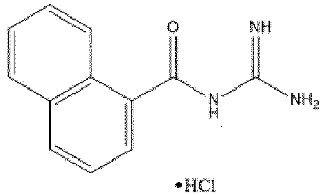
-
-
- Another virus considered to be a significant human pathogen is the Hepatitis C virus (HCV). This is a significant human pathogen in terms of both cost to human health and associated economic costs. HCV causes chronic hepatitis and cirrhosis and is the leading indicator for liver replacement surgery. In 2002 the Centre for Disease Control and Prevention estimated that more than 4 million people were infected in the USA alone and that approximately 8,000 to 10, 000 die as a result of chronic HCV infection yearly. There is no known cure or vaccine. More effective pharmacological agents are urgently required.
- A further well-known family of pathogenic viruses are the Coronaviruses. Coronaviruses (Order Nidovirales, family Coronaviridae, Genus Coronavirus) are enveloped positive-stranded RNA viruses that bud from the endoplasmic reticulum-Golgi intermediate compartment or the cis-Golgi network (Fischer, Stegen et al. 1998; Maeda, Maeda et al. 1999; Corse and Machamer 2000; Maeda, Repass et al. 2001; Kuo and Masters 2003)
- Coronaviruses infect humans and animals and it is thought that there could be a coronavirus that infects every animal. The two human coronaviruses, 229E and OC43, are known to be the major causes of the common cold and can occasionally cause pneumonia in older adults, neonates, or immunocompromised patients (Peiris, Lai et al. 2003). Animal coronaviruses can cause respiratory, gastrointestinal, neurological, or hepatic diseases in their host (Peiris, Lai et al. 2003). Several animal coronavirus are significant veterinary pathogens (Rota, Oberste et al. 2003).
- Severe acute respiratory syndrome (SARS) is caused by a newly identified virus. SARS is a respiratory illness that has recently been reported in Asia, North America, and Europe (Peiris, Lai et al. 2003). The causative agent of SARS was identified as a coronavirus. (Drosten, Gunther et al. 2003; Ksiazek, Erdman et al. 2003; Peiris, Lai et al. 2003). The World Health Organization reports that the cumulative number of reported probable cases of SARS from 1 November 2002 to the 11th July 2003 is 8,437 with 813 deaths, nearly a 10% death rate. It is believed that SARS will not be eradicated, but will cause seasonal epidemics like the cold or influenza viruses (Vogel 2003).
- To improve the prospect of treating and preventing viral infections, there is an on-going need to identify molecules capable of inhibiting various aspects of the viral life cycle.
- It is an object of the present invention to overcome or ameliorate at least one of the disadvantages of the prior art, or to provide a useful alternative.
- Any discussion of the prior art throughout the specification should in no way be considered as an admission that such prior art is widely known or forms part of common general knowledge in the field.
- The inventors have surprisingly found that certain compounds that fall under the classification of substituted acylguanidines have antiviral activity against viruses from a range of different virus families selected from the group consisting of Lentivirus, Coronavirus, Hepatitis C virus and Equine Arteritis virus. Without intending to be bound by any particular theory or mechanism of action, and despite current dogma, it appears possible that viral replication can be retarded by inhibiting or otherwise down-regulating the activity of ion channels expressed in the host cell. Thus, the negative impact of the compounds of the present invention on viral replication may be mediated by the inhibition or otherwise down-regulation of a membrane ion channel relied upon by the virus for replication. This membrane ion channel may be a viral membrane ion channel (exogenous to the host cell) or a host cell ion channel induced as a result of viral infection (endogenous to the host cell).
- As an example, the compounds of the present invention may inhibit Vpu or p7 function and thereby inhibit the continuation of the respective HIV or HCV life cycle.
- The SARS virus encodes an E protein which is shown for the first time, by the present inventors, to act as an ion channel. As similar E proteins are present in other coronaviruses, the compounds, compositions and methods of the present invention would have utility in the inhibition and/or treatment of infections by other coronaviruses.
In one aspect the invention relates to a compound of Formula I
- or a pharmaceutically acceptable salt thereof,
- wherein:
- R1 =


- R2 , R3 and R4 are independently hydrogen,


- and wherein
- X = hydrogen, hydroxy, nitro, halo, C1-6alkyl, C1-6alkyloxy, C3-6cycloalkyl, halo-substituted C1-6alkyl, halo-substituted C1-6alkyloxy, phenyl, C2-6alkenyl, C3-6cycloalkenyl or C2-6alkeneoxy;
- Rd , Re , Rf , Rh , Rk , RL , Rm , Rn , Ro , Rp independently = hydrogen, amino, halo, C1-5alkyl, C1-5alkyloxy, hydroxy, aryl, substituted aryl, substituted amino, mono or dialkyl-substituted amino, cycloalkyl-substituted amino, aryl-substituted amino,

- Rg , Ri independently = hydrogen, hydroxy, halo, or C1-5 alkyl;
- Rj = hydrogen, amino, halo, C1-5 alkyl, C1-5alkyloxy, hydroxy, aryl, substituted aryl, substituted amino, alkyl-substituted amino, cycloalkyl-substituted amino, aryl-substituted amino, propyl thio,

- for use in the therapeutic or prophylactic treatment of a viral infection in a mammal infected with or exposed to a virus selected from the group consisting of Lentivirus, Coronavirus, Hepatitis C virus and Equine Arteritis virus.
- R1 =
- In one embodiment, the compound reduces, retards or otherwise inhibits growth and/or replication of the virus in a cell infected with the virus or exposed to the virus, wherein the cell is in the mammal.
- In one embodiment, the compound prevents infection of a cell exposed to the virus, wherein the cell is in the mammal.
- In one embodiment, the compound down regulates a membrane ion channel functional activity in a cell infected with the virus, wherein the cell is in the mammal. The membrane ion channel may be selected from the group consisting of HIV Vpu membrane ion channel in the treatment of HIV, Coronavirus E protein in the treatment of Coronavirus and Hepatitis C virus p7 membrane ion channel in the treatment of Hepatitis C.
- In one embodiment, the compound reduces, retards or otherwise inhibits growth and/or replication of the virus, wherein the virus has infected a cell in the mammal. The Lentivirus may be Human Immunodeficiency Virus (HIV).
- In one embodiment related to use for treatment of HIV, the compound is selected from:
- (3-Chlorocinnamoyl)guanidine,
- (3-Bromocinnamoyl)guanidine,
- (2-Chlorocinnamoyl)guanidine,
- (2-Bromocinnamoyl)guanidine,
- 3-(trifluoromethyl)cinnamoylguanidine,
- 5-bromo-2-fluorocinnamoylguanidine,
- 3-methylcinnamoylguanidine,
- 2-methylcinnamoylguanidine,
- 2,3-dimethylcinnamoylguanidine,
- Cinnamoylguanidine,
- 6-methoxy-2-naphthoylguanidine,
- Trans-3-(1-naphthyl)acryloylguanidine,
- 3,4-dichlorocinnamoylguanidine,
- 2,6-dichlorocinnamoylguanidine,
- 2-ethylcinnamoylguanidine,
- (4-Chlorocinnamoyl)guanidine,
- 2-naphthoylguanidine,
- 2,5-dimethylcinnamoylguanidine,
- 3-isopropylcinnamoylguanidine hydrochloride,
- (5-Phenyl-penta-2,4-dienoyl)guanidine,
- 3-phenylcinnamoylguanidine,
- (4-Bromocinnamoyl)guanidine,
- 3 -(cyclohex-1-en-1-yl)cinnamoylguanidine,
- 3 -(trifluoromethoxy)cinnamoylguanidine,
- 2-(trifluoromethyl)cinnamoylguanidine,
- N,N'-bis(3phenylpropanoyl)-N" -phenylguanidine,
- 2-ethoxycinnamoylguanidine,
- N-(3-phenylpropanoyl)-N'-phenylguanidine,
- 4-(trifluoromethyl)cinnamoylguanidine,
- (4-Methoxycinnamoyl)guanidine,
- 2-t-butylcinnamoylguanidine,
- 4-methylcinnamoylguanidine,
- 2-fluorocinnamoylguanidine,
- 2-phenylcinnamoylguanidine,
- N-(6-Hydroxy-2-naphthoyl)-N'-phenylguanidine,
- 3 -t-butylcinnamoylguanidine,
- 3,4-difluorocinnamoylguanidine,
- 3 -fluorocinnamoylguanidine,
- 5 -bromo-2-methoxycinnamoylguanidine,
- 3 -ethoxycinnamoylguanidine,
- 3,4-(methylenedioxy)cinnamoylguanidine,
- (2-Methoxycinnamoyl)guanidine,
- 2,3,5,6, -tetramethylcinnamoylguanidine,
- 3-(2-naphthyl)acryloylguanidine,
- 2-(1-naphthyl)acetoylguanidine,
- 2,3 -difluorocinnamoylguanidine,
- (3 -Methoxycinnamoyl)guanidine,
- 4-isopropylcinnamoylguanidine,
- 2,4,6-trimethylcinnamoylguanidine,
- N-(cinnamoyl)-N'phenylguanidine,
- 2-(cyclohex-1-en-1yl)cinnamoylguanidine,
- 2-(2-naphthyl)acetoylguanidine,
- (4-Hydroxycinnamoyl)guanidine,
- 4-phenylcinnamoylguanidine,
- 4-fluorocinnamoylguanidine,
- N,N'-bis-(cinnamoy1)-N"-phenylguanidine,
- (2-Furanacryloyl)guanidine,
- (3 -Nitrocinnamoyl)guanidine,
- Benzyoylguanidine,
- 3-(trans-hept-1-en-1-yl)cinnamoylguanidine,
- 2-cyclohexylcinnamoylguanidine,
- 4-ethoxycinnamoylguanidine,
- 2,4-dichlorocinnamolyguanidine,
- (a-Methylcinnamoyl)guanidine,
- cinnamoylguanidine hydrochloride,
- [(4-Chlorophenoxy-acetyl]guanidine,
- (trans-2-Phenylcyclopropanecarbonyl)guanidine,
- (2-Nitrocinnamoyl)guanidine,
- trans-3-Furanacryoylguanidine,
- 1 -naphthoylguanidine,
- (3-phenylpropanoyl)guanidine,
- 4-t-butylcinnamoylguanidine,
- N,N'-Bis(3-phenylpropanoyl)guanidine,
- N-Benzoyl-N'-cinnamoylguanidine and
- 1 -bromo-2-naphthoylguanidine.
- HIV may be HIV-1.
- In one embodiment, the Coronavirus is selected from the group consisting of Severe Acute Respiratory Syndrome virus (SARS), human Coronavirus 229E, human Coronavirus OC43, porcine respiratory Coronavirus (PRCV), bovine Coronvirus (BCV) and the coronavirus isolates listed in Table 1.
- In one embodiment wherein the coronavirus is SARS, the compound is selected from:
- 2,3 -difluorocinnamoylguanidine,
- 3,4-dichlorocinnamoylguanidine,
- 4-t-butylcinnamoylguanidine,
- 3-(2-naphthyl)acryloylguanidine,
- (3 -Chlorocinnamoyl)guanidine,
- 3 -(cyclohex-1-en-1-yl)cinnamoylguanidine,
- 2,5-dimethylcinnamoylguanidine,
- trans-3 -(1-naphthyl)acryloylguanidine,
- 4-isopropylcinnamoylguanidine,
- (3 -Bromocinnamoyl)guanidine,
- 6-methoxy-2-naphthoylguanidine,
- 3-phenylcinnamoylguanidine,
- (2-Chlorocinnamoyl)guanidine,
- 4-phenylcinnamoylguanidine,
- 4-(trifluoromethyl)cinnamoylguanidine,
- 3 -(trifluoromethoxy)cinnamoylguanidine,
- 3-(trifluoromethyl)cinnamoylguanidine,
- 2-ethoxycinnamoylguanidine,
- cinnamoylguanidine hydrochloride,
- 4-ethoxycinnamoylguanidine,
- (2-Bromocinnamoyl)guanidine,
- 2,6-dichlorocinnamoylguanidine,
- 3,4,5 -trimethoxycinnamoylguanidine,
- 3 -t-butylcinnamoylguanidine,
- 5 -bromo-2-fluorocinnamoylguanidine,
- (4-Chlorocinnamoyl)guanidine,
- 2-t-butylcinnamoylguanidine,
- 2-cyclohexylcinnamoylguanidine,
- 3-(trans-hept-1-en-1-yl)cinnamoylguanidine,
- (4-Bromocinnamoyl)guanidine,
- (4-Hydroxycinnamoyl)guanidine,
- N-(3-phenylpropanoyl)-N'-phenylguanidine,
- (3 -Nitrocinnamoyl)guanidine,
- 3 -fluorocinnamoylguanidine,
- 2-(1-naphthyl)acetoylguanidine,
- 2-ethylcinnamoylguanidine,
- 2-naphthoylguanidine,
- 2-(trifluoromethyl)cinnamoylguanidine,
- N-(6-Hydroxy-2-naphthoyl)-N'-phenylguanidine,
- (trans-2-Phenylcyclopropanecarbonyl)guanidine,
- N,N'-bis(3phenylpropanoyl)-N" -phenylguanidine,
- 1-naphthoylguanidine,
- 4-methylcinnamoylguanidine,
- 4-fluorocinnamoylguanidine,
- 3,4-(methylenedioxy)cinnamoylguanidine,
- N-(cinnamoyl)-N'phenylguanidine,
- 3 -methylcinnamoylguanidine,
- 2-methylcinnamoylguanidine,
- 2,3,5,6, -tetramethylcinnamoylguanidine,
- trans-3 -Furanacryoylguanidine,
- (4-Methoxycinnamoyl)guanidine,
- (2-Furanacryloyl)guanidine,
- (3-phenylpropanoyl)guanidine,
- 2-(2-naphthyl)acetoylguanidine,
- Cinnamoylguanidine,
- (2-Methoxycinnamoyl)guanidine,
- [3-(3-Pyridyl)acryloyl]guanidine,
- 2,4-dichlorocinnamolyguanidine,
- (3 -Methoxycinnamoyl)guanidine,
- 2-fluorocinnamoylguanidine,
- (a-Methylcinnamoyl)guanidine,
- (5-Phenyl-penta-2,4-dienoyl)guanidine,
- (Quinoline-2-carbonyl)guanidine,
- (Phenylacetyl)guanidine,
- N,N'-Bis(amidino)naphthalene-2,6-dicarboxamide,
- 6-bromo-2-naphthoylguanidine,
- 1-bromo-2-naphthoylguanidine,
- 2-chloro-6-fluorocinnamoylguanidine,
- [(4-Chlorophenoxy-acetyl]guanidine,
- N-Benzoyl-N'-cinnamoylguanidine and
- N-(2-naphthoyl)-N'-phenylguanidine.
- In one embodiment wherein the coronavirus is Coronavirus 229E, the compound is selected from:
- 4-isopropylcinnamoylguanidine,
- 3,4-dichlorocinnamoylguanidine,
- 3 -(trifluoromethoxy)cinnamoylguanidine,
- 4-t-butylcinnamoylguanidine,
- 3-isopropylcinnamoylguanidine hydrochloride,
- 3 -t-butylcinnamoylguanidine,
- 2-t-butylcinnamoylguanidine,
- trans-3 -(1-naphthyl)acryloylguanidine,
- 5-bromo-2-methoxycinnamoylguanidine,
- 2,3 -difluorocinnamoylguanidine,
- 3-(2-naphthyl)acryloylguanidine,
- 2-phenylcinnamoylguanidine,
- 3-phenylcinnamoylguanidine,
- 3 -(cyclohex-1-en-1-yl)cinnamoylguanidine,
- 3-(trifluoromethyl)cinnamoylguanidine,
- 4-(trifluoromethyl)cinnamoylguanidine,
- 2-(cyclohex-1-en-1yl)cinnamoylguanidine,
- (4-Bromocinnamoyl)guanidine,
- 1 -naphthoylguanidine,
- (5-Phenyl-penta-2,4-dienoyl)guanidine,
- (3-Bromocinnamoyl)guanidine,
- 2,5-dimethylcinnamoylguanidine,
- 2-(trifluoromethyl)cinnamoylguanidine,
- 6-methoxy-2-naphthoylguanidine,
- (4-Chlorocinnamoyl)guanidine,
- (3-Methoxycinnamoyl)guanidine,
- 5-bromo-2-fluorocinnamoylguanidine,
- Cinnamoylguanidine,
- (2-Methoxycinnamoyl)guanidine,
- (a-Methylcinnamoyl)guanidine,
- 4-phenylcinnamoylguanidine,
- 2,6-dichlorocinnamoylguanidine,
- (2-Bromocinnamoyl)guanidine,
- 2,4,6-trimethylcinnamoylguanidine,
- (trans-2-Phenylcyclopropanecarbonyl)guanidine,
- (3-Chlorocinnamoyl)guanidine,
- 2-(1-naphthyl)acetoylguanidine,
- 2-ethylcinnamoylguanidine,
- 2-cyclohexylcinnamoylguanidine,
- (4-Hydroxycinnamoyl)guanidine,
- 2-ethoxycinnamoylguanidine,
- 3 -methylcinnamoylguanidine,
- 2-methylcinnamoylguanidine,
- 3 -fluorocinnamoylguanidine,
- cinnamoylguanidine hydrochloride,
- 2,3-dimethylcinnamoylguanidine,
- 2-]fluorocinnamoylguanidine,
- 4-fluorocinnamoylguanidine,
- 3,4-difluorocinnamoylguanidine,
- 2-naphthoylguanidine,
- N,N'-Bis(amidino)naphthalene-2,6-dicarboxamide,
- N,N'-Bis(3-phenylpropanoyl)guanidine,
- 4-methylcinnamoylguanidine,
- 2,3,5,6, -tetramethylcinnamoylguanidine,
- 3-ethoxycinnamoylguanidine,
- N,N'-bis(3phenylpropanoyl)-N" -phenylguanidine,
- (4-Methoxycinnamoyl)guanidine,
- (2-Chlorocinnamoyl)guanidine,
- (3 -Nitrocinnamoyl)guanidine,
- 4-ethoxycinnamoylguanidine,
- 3,4,5-trimethoxycinnamoylguanidine,
- 2-(2-naphthyl)acetoylguanidine,
- N-(3-phenylpropanoyl)-N'-phenylguanidine,
- 5-(2'-bromophenyl)penta-2,4-dienoylguanidine,
- (4-Bromocinnamoyl)guanidine,
- (2-Nitrocinnamoyl)guanidine,
- (3-Chlorocinnamoyl)guanidine,
- (4-Methoxycinnamoyl)guanidine,
- 4-(trifluoromethyl)cinnamoylguanidine,
- [(E)-3-(4-Dimethylaminophenyl)-2-methylacryloyl]guanidine,
- N-Benzoyl-N'-cinnamoylguanidine,
- trans-3 -Furanacryoylguanidine,
- N-(cinnamoyl)-N'phenylguanidine,
- Cinnamoylguanidine,
- (3-phenylpropanoyl)guanidine,
- Benzyoylguanidine,
- (Quinoline-2-carbonyl)guanidine,
- [3-(3-Pyridyl)acryloyl]guanidine,
- N-Cinnamoyl-N',N'-dimethylguanidine,
- N-(2-naphthoyl)-N'-phenylguanidine and
- (Phenylacetyl)guanidine.
- In one embodiment wherein the coronavirus is human Coronavirus OC43, the compound is selected from:
- 3 -methylcinnamoylguanidine,
- trans-3 -(1-naphthyl)acryloylguanidine,
- (3-Bromocinnamoyl)guanidine,
- (2-Chlorocinnamoyl)guanidine,
- 3,4-dichlorocinnamoylguanidine,
- 3-(trifluoromethyl)cinnamoylguanidine,
- (trans-2-Phenylcyclopropanecarbonyl)guanidine,
- 4-isopropylcinnamoylguanidine,
- Cinnamoylguanidine,
- 6-methoxy-2-naphthoylguanidine,
- 2,4-dichlorocinnamolyguanidine,
- (4-Chlorocinnamoyl)guanidine,
- (4-Bromocinnamoyl)guanidine,
- 2,6-dichlorocinnamoylguanidine,
- 5-bromo-2-methoxycinnamoylguanidine,
- (5-Phenyl-penta-2,4-dienoyl)guanidine,
- 3-(trifluoromethoxy)cinnamoylguanidine,
- 2-t-butylcinnamoylguanidine.
- In one embodiment wherein the coronavirus is PRCV, the compound is selected from:
- 6-methoxy-2-naphthoylguanidine,
- Cinnamoylguanidine,
- N-(3-phenylpropanoyl)-N'-phenylguanidine,
- 3-methylcinnamoylguanidine,
- (3-Bromocinnamoyl)guanidine,
- (trans-2-Phenylcyclopropanecarbonyl)guanidine,
- trans-3-(1-naphthyl)acryloylguanidine and
- 2-(2-naphthyl)acetoylguanidine.
- In one embodiment wherein the coronavirus is BCV, the compound is selected from:
- (3-Bromocinnamoyl)guanidine,
- 3-(trifluoromethyl)cinnamoylguanidine,
- 6-methoxy-2-naphthoylguanidine,
- trans-3 -(1-naphthyl)acryloylguanidine,
- Cinnamoylguanidine,
- (5-Phenyl-penta-2,4-dienoyl)guanidine,
- 2-(2-naphthyl)acetoylguanidine,
- (trans-2-Phenylcyclopropanecarbonyl)guanidine,
- and N-(3-phenylpropanoyl)-N'-phenylguanidine.
- In one embodiment wherein the coronavirus is a coronavirus isolate listed in Table 1, the compound is selected from:
- 4-isopropylcinnamoylguanidine,
- 3,4-dichlorocinnamoylguanidine,
- 3-(trifluoromethoxy)cinnamoylguanidine,
- 4-t-butylcinnamoylguanidine, and
- 3-isopropylcinnamoylguanidine hydrochloride.
- In one embodiment wherein the virus is Hepatitis C virus, the compound is selected from:
- 2,3-dimethylcinnamoylguanidine,
- 2,4,6-trimethylcinnamoylguanidine,
- 5-bromo-2-fluorocinnamoylguanidine,
- (4-Bromocinnamoyl)guanidine,
- 2,5-dimethylcinnamoylguanidine,
- 3-(trifluoromethyl)cinnamoylguanidine,
- 4-(trifluoromethyl)cinnamoylguanidine,
- 6-methoxy-2-naphthoylguanidine,
- (2-Chlorocinnamoyl)guanidine,
- (4-Chlorocinnamoyl)guanidine,
- (2-Bromocinnamoyl)guanidine,
- 2, 6-dichlorocinnamoylguanidine,
- (3-Bromocinnamoyl)guanidine,
- (3-Chlorocinnamoyl)guanidine,
- 2-(trifluoromethyl)cinnamoylguanidine,
- 3,4-dichlorocinnamoylguanidine,
- 4-isopropylcinnamoylguanidine,
- trans-3 -(1-naphthyl)acryloylguanidine,
- 4-t-butylcinnamoylguanidine,
- 2-t-butylcinnamoylguanidine,
- 2-ethylcinnamoylguanidine,
- 4-methylcinnamoylguanidine,
- 5-bromo-2-methoxycinnamoylguanidine,
- 3 -(trifluoromethoxy)cinnamoylguanidine,
- 2-cyclohexylcinnamoylguanidine,
- 1-naphthoylguanidine,
- 3-t-butylcinnamoylguanidine,
- (5-Phenyl-penta-2,4-dienoyl)guanidine,
- N-(cinnamoyl)-N'phenylguanidine,
- 3-isopropylcinnamoylguanidine hydrochloride,
- N-(3-phenylpropanoyl)-N'-phenylguanidine,
- N,N'-bis(3phenylpropanoyl)-N" -phenylguanidine,
- 3-(2-naphthyl)acryloylguanidine,
- (4-Methoxycinnamoyl)guanidine,
- 4-fluorocinnamoylguanidine,
- (3-Nitrocinnamoyl)guanidine,
- 4-ethoxycinnamoylguanidine,
- (4-Hydroxycinnamoyl)guanidine,
- (trans-2-Phenylcyclopropanecarbonyl)guanidine,
- 3-ethoxycinnamoylguanidine,
- 2,3,5,6,-tetramethylcinnamoylguanidine,
- 4-phenylcinnamoylguanidine,
- trans-3 -Furanacryoylguanidine,
- N-(6-Hydroxy-2-naphthoyl)-N'-phenylguanidine,
- (2-Furanacryloyl)guanidine,
- 3-(cyclohex-1-en-1-yl)cinnamoylguanidine,
- cinnamoylguanidine hydrochloride,
- 2,3 -difluorocinnamoylguanidine,
- 2-(1-naphthyl)acetoylguanidine,
- (a-Methylcinnamoyl)guanidine,
- (2-Nitrocinnamoyl)guanidine,
- 3,4-(methylenedioxy)cinnamoylguanidine,
- 2-ethoxycinnamoylguanidine,
- Cinnamoylguanidine,
- 2-phenylcinnamoylguanidine,
- 2-(cyclohex-1-en-1yl)cinnamoylguanidine,
- 2-naphthoylguanidine,
- 3-phenylcinnamoylguanidine,
- (3-Methoxycinnamoyl)guanidine,
- 2-fluorocinnamoylguanidine,
- [(4-Chlorophenoxy-acetyl]guanidine,
- (3-phenylpropanoyl)guanidine,
- 2-chloro-6-fluorocinnamoylguanidine,
- 3-fluorocinnamoylguanidine,
- 2-methylcinnamoylguanidine,
- (2-Methoxycinnamoyl)guanidine,
- 1-bromo-2-naphthoylguanidine,
- 3,4,5-trimethoxycinnamoy lguanidine,
- 3 -methylcinnamoylguanidine,
- 3-(trans-hept-1-en-1-yl)cinnamoylguanidine,
- 2,4-dichlorocinnamolyguanidine,
- (4-Nitrocinnamoyl)guanidine,
- 3,4-difluorocinnamoylguanidine and
- [(E)-3-(4-Dimethylaminophenyl)-2-methylacryloyl]guanidine.
- In one embodiment wherein the virus is Equine Arteritis virus, the compound is selected from:
- (3-Bromocinnamoyl)guanidine,
- trans-3 -(1-naphthyl)acryloylguanidine,
- 2-t-butylcinnamoylguanidine and
- 2-(cyclohex-1-en-lyl)cinnamoylguanidine.
- In another aspect, the invention relates to a compound for use in the therapeutic or prophylactic treatment of a viral infection in a mammal infected with or exposed to a virus, wherein the virus is Dengue virus and the compound is selected from the group consisting of:
- cinnamoylguanidine,
- (2-chlorocinnamoyl)guanidine and
- trans-3-(1-naphthyl)acryloylguanidine.
- In one aspect, the invention relates to an ex vivo or in vitro method of:
- reducing, retarding or otherwise inhibiting growth and/or replication of a virus in a cell infected with the virus or exposed to the virus;
- preventing the infection of a cell exposed to a virus; or
- down regulating a membrane ion channel functional activity in a cell infected with a virus;
- the method comprising contacting the cell with a compound as defined in in the first aspect, wherein the virus is selected from the group consisting of Lentivirus, Coronavirus, Hepatitis C virus and Equine Arteritis virus.
- Other Coronaviruses which can be inhibited or their infections treated by the compounds of the invention are those listed in Table 1.
- One or more known antiviral compounds or molecules may be used in combination with the uses and methods as defined in the claims.
- The subject of the viral inhibition is generally a mammal such as but not limited to human, primate, livestock animal (e.g. sheep, cow, horse, donkey, pig), companion animal (e.g. dog, cat), laboratory test animal (e.g. mouse, rabbit, rat, guinea pig, hamster), captive wild animal (e.g. fox, deer). Preferably, the subject is a primate, or horse. Most preferably, the subject is a human.
- Unless the context clearly requires otherwise, throughout the description and the claims, the words 'comprise', 'comprising', and the like are to be construed in an inclusive sense as opposed to an exclusive or exhaustive sense; that is to say, in the sense of "including, but not limited to".
-
-
Figure 1 is a schematic representation of plasmids used for expression of Vpu in E. coli. A. The amino acid sequence ( < 400 > 1) encoded by the vpu open reading frame (ORF) generated by PCR from an HIV- 1 strain HXB2 cDNA clone. The vpu ORF was cloned in-frame at the 3' end of the GST gene in p2GEX to generate p2GEXVpu (B). It was subsequently cloned into pPL451 to produce the plasmid pPL + Vpu (C). -
Figure 2 is a photographic representation of the expression and purification of Vpu in E. coli. A. Western blotting after SDS-PAGE was used to detect expressed Vpu in E. coli extracts. Lanes 1-4 contain samples, at various stages of purity, of Vpu expressed from p2GEXVpu:lane 1, GST-Vpu fusion protein isolated by glutathione-agarose affinity chromatography;lane 2, Vpu liberated from the fusion protein by treatment with thrombin;lane 3, Vpu purified by HPLC anion exchange chromatography;lane 4, Vpu after passage through the immunoaffinity column.Lanes lane 1, Vpu purified by HPLC anion exchange chromatography;lane 2, Vpu after passage through the immunoaffinity column. -
Figure 3 is a graphical representation of ion channel activity observed after exposure of lipid bilayers to aliquots containing purified Vpu. In A and B, the CIS chamber contained 500mM NaCl and the TRANS chamber contained 5OmM NaCl; both solutions were buffered at pH 6.0 with 10 mM MES. B shows a current versus voltage curve generated from data similar to that shown in A. -
Figure 4 is a photographic representation of bacterial cross-feeding assays. For all plates, the Met-, Pro- auxotrophic strain was used to seed a soft agar overlay. Plates A and B contain minimal drop-out medium minus proline; in plate C the medium was minus methionine. To control for viability of the cells in the background lawn, the discs labelled P and M contained added proline or methionine, respectively. The discs labelled C and V were inoculated with Met+, Pro+ E. coli cells containing the plasmids pPL451 or pPL+Vpu, respectively. Plates were incubated at 37°C (A and C) or 30°C (B) for two days and photographed above a black background with peripheral illumination from a fluorescent light located below the plate. The images were recorded on a Novaline video gel documentation system. Light halos around the discs labelled P or M on all plates and around the disc labelled V on plate A indicate growth of the background lawn strain. -
Figure 5 is a graphical representation of the screening of drugs for potential Vpu channel blockers. The photograph shows a section of a minimal medium-lacking adenine - agarose plate onto which a lawn of XL-I-blue E. coli cells containing the Vpu expression plasmid pPLVpu has been seeded. Numbers 6-11 are located at the sites of application of various drugs being tested, which were applied in 3µl drops and allowed to soak into the agarose. The plate was then incubated at 37°C for 48hr prior to being photographed. The background grey shade corresponds to areas of no bacterial growth. The bright circular area around " 10 " represents bacterial cell growth as a result of application of adenine at that location (positive control). The smaller halo of bacterial growth around "9" is due to the application of 5-(N,N-hexamethylene)-amiloride at that location. -
Figure 6 . SARS E protein ion channel activity observed in NaCl solutions after exposure of lipid bilayer to 3-10µg of E protein. A. The closed state is shown as solid line, openings are derivations from the line. Scale bar is 300ms and 5pA. The CIS chamber contained 50mM NaCl in 5mM HEPES buffer pH 7.2, the TRANS chamber contained 500mM NaCl in 5mM HEPES buffer pH 7.2. The CIS chamber was earthed and the TRANS chamber was held at various potentials between -100 to +100mV. B. Largest single opening events of a single channel. -
Figure 7 . SARS E protein ion channel activity observed in NaCl solutions after exposure of lipid bilayer to 3-10µg of E protein. A. The closed state is shown as solid line, openings are derivations from the line. Scale bar is 300ms and 5pA. The CIS chamber contained 50mM NaCl in 5mM HEPES buffer pH 7.2, the TRANS chamber contained 500mM NaCl in 5mM HEPES buffer pH 7.2. The CIS chamber was earthed and the TRANS chamber was held at various potentials between -100 to +100mV. B. Largest single opening events of a single channel. -
Figure 8 . Cinnamoylguanidine (Bit036) inhibits SARS E protein ion channel activity in NaCl solution. A. Representative currents at holding potential of -40mV. Scale bar is 300mS and 5pA. E protein ion channel activity and E protein channel activity after the addition of 100µM Bit036. B. All points histogram at holding potential of-40mV. E protein ion channel activity before and after the addition of 100µM Bit036. C. Average current (pA), before formation of E protein ion channel, E protein ion channel activity and after addition of 100µM Bit036. -
Figure 9 229E E protein Ion channel activity in lipid bilayers in KCl solutions. -
Figure 10 : Part A shows raw currents generated by the 229E-E protein ion channel in a planar lipid bilayer. The top trace shows current activity prior to drug addition and the lower trace shows the effect of addition of 100µM cinnamoylguanidine on channel activity. Part B is a graphical representation of the average current flowing across the bilayer (in arbitrary units), before and after addition of cinnamoylguanidine. -
Figure 11 : MHV E protein Ion channel activity in lipid bilayers NaCl solutions. -
Figure 12 : Part A shows raw currents generated by the MHV-E protein ion channel in a planar lipid bilayer. The top trace shows current activity prior to drug addition and the lower trace shows the effect of addition of 100µM cinnamoylguanidine on channel activity. Part B is a graphical representation of the average current flowing across the bilayer (in arbitrary units), before and after addition of cinnamoylguanidine. - The present invention is based, in part, on the surprising determination that certain compounds that fall under the classification of substituted acylguanidines have antiviral activity against viruses selected from the group consisting of Lentivirus, Coronavirus, Hepatitis C virus and Equine Arteritis virus. Certain other acylguanidines have activity against Dengue virus. Without intending to be bound by any particular theory or mechanism of action, the negative impact of the compounds of the present invention on viral replication may be mediated by the inhibition or otherwise down-regulation of a membrane ion channel relied upon by the virus for replication. This membrane ion channel may be a viral membrane ion channel (exogenous to the host cell) or a host cell ion channel induced as a result of viral infection (endogenous to the host cell).
- As an example, the compounds of the present invention may inhibit Vpu or p7 function and thereby inhibit the continuation of the respective HIV or HCV life cycle.
- The SARS virus encodes an E protein which is shown for the first time, by the present inventors, to act as an ion channel. As similar E proteins are present in other coronaviruses, the compounds, compositions and methods of the present invention would have utility in the inhibition and/or treatment of infections by other coronaviruses.
- It will be understood by those skilled in the art that the compounds of the invention may be administered in the form of a composition or formulation comprising pharmaceutically acceptable carriers and excipients.
- The pharmaceutical compositions of the invention may further comprise one or more known antiviral compounds or molecules. Preferably, the known antiviral compounds are selected from the group consisting of Vidarabine, Acyclovir, Ganciclovir, Valganciclovir, Valacyclovir, Cidofovir, Famciclovir, Ribavirin, Amantadine, Rimantadine, Interferon, Oseltamivir, Palivizumab, Rimantadine, Zanamivir, nucleoside-analog reverse transcriptase inhibitors (NRTI) such as Zidovudine, Didanosine, Zalcitabine, Stavudine, Lamivudine and Abacavir, non-nucleoside reverse transcriptase inhibitors (NNRTI) such as Nevirapine, Delavirdine and Efavirenz, protease inhibitors such as Saquinavir, Ritonavir, Indinavir, Nelfinavir, Amprenavir, and other known antiviral compounds and preparations. Known antiviral compounds or molecules may in some cases act synergistically with the antiviral compounds of the invention.
Table 1 Known coronavirus isolates Group 1 species Canine coronavirus Canine enteric coronavirus (strain INSAVC-1) Canine enteric coronavirus (strain K378) Feline coronavirus Feline enteric coronavirus (strain 79-1683) Feline infectious peritonitis virus (FIPV) Human coronavirus 229E Porcine epidemic diarrhea virus Porcine epidemic diarrhea virus (strain Br1/87) Porcine epidemic diarrhea virus (strain CV777) Transmissible gastroenteritis virus Porcine respiratory coronavirus Porcine transmissible gastroenteritis coronavirus (STRAIN FS772/70) Porcine transmissible gastroenteritis coronavirus (strain Miller) Porcine transmissible gastroenteritis coronavirus (strain Neb72-RT) Porcine transmissible gastroenteritis coronavirus (STRAIN PURDUE) Group 2 species Bovine coronavirus Bovine coronavirus (STRAIN F15) Bovine coronavirus (strain G95) Bovine coronavirus (STRAIN L9) Bovine coronavirus (strain LSU-94LSS-051) Bovine coronavirus (STRAIN LY-138) Bovine coronavirus (STRAIN MEBUS) Bovine coronavirus (strain OK-0514-3) Bovine coronavirus (strain Ontario) Bovine coronavirus (STRAIN QUEBEC) Bovine coronavirus (STRAIN VACCINE) Bovine enteric coronavirus (strain 98TXSF-110-ENT) Canine respiratory coronavirus Chicken enteric coronavirus Human coronavirus OC43 Murine hepatitis virus Murine coronavirus (strain DVIM) Murine hepatitis virus (strain A59) Murine hepatitis virus (strain JHM) Murine hepatitis virus (strain S) Murine hepatitis virus strain 1 Murine hepatitis virus strain 2 Murine hepatitis virus strain 3 Murine hepatitis virus strain 4 Murine hepatitis virus strain ML-11 Porcine hemagglutinating encephalomyelitis virus Porcine hemagglutinating encephalomyelitis virus (strain 67N) Porcine hemagglutinating encephalomyelitis virus (strain IAF- 404) Puffinosis virus Rat coronavirus Rat coronavirus (strain 681) Rat coronavirus (strain NJ) Rat sialodacryoadenitis coronavirus Group 3 species Turkey coronavirus Turkey coronavirus (strain Indiana) Turkey coronavirus (strain Minnesota) Turkey coronavirus (strain NC95) Avian infectious bronchitis virus Avian infectious bronchitis virus (STRAIN 6/82) Avian infectious bronchitis virus (strain Arkansas 99) Avian infectious bronchitis virus (strain Beaudette CK) Avian infectious bronchitis virus (strain Beaudette M42) Avian infectious bronchitis virus (strain Beaudette US) Avian infectious bronchitis virus (strain Beaudette) Avian infectious bronchitis virus (strain D1466) Avian infectious bronchitis virus (strain D274) Avian infectious bronchitis virus (strain D3896) Avian infectious bronchitis virus (strain D41) Avian infectious bronchitis virus (strain DE072) Avian infectious bronchitis virus (strain GRAY) Avian infectious bronchitis virus (strain H120) Avian infectious bronchitis virus (strain H52) Avian infectious bronchitis virus (strain KB8523) Avian infectious bronchitis virus (strain M41) Avian infectious bronchitis virus (strain PORTUGAL/322/82) Avian infectious bronchitis virus (strain SAIB20) Avian infectious bronchitis virus (strain UK/123/82) Avian infectious bronchitis virus (strain UK/142/86) Avian infectious bronchitis virus (strain UK/167/84) Avian infectious bronchitis virus (strain UK/183/66) Avian infectious bronchitis virus (strain UK/68/84) Avian infectious bronchitis virus (strain V18/91) Avian infectious bronchitis virus (strain Vic S) Avian infectious laryngotracheitis virus Preliminary Group 4 species SARS coronavirus SARS coronavirus Beijing ZY-2003 SARS coronavirus BJ01 SARS coronavirus BJ02 SARS coronavirus BJ03 SARS coronavirus BJ04 SARS coronavirus CUHK-Su10 SARS coronavirus CUHK-W1 SARS coronavirus Frankfurt 1 SARS coronavirus GZ01 SARS coronavirus HKU-39849 SARS coronavirus Hong Kong ZY-2003 SARS coronavirus Hong Kong/03/2003 SARS coronavirus HSR 1 SARS coronavirus Sin2500 SARS coronavirus Sin2677 SARS coronavirus Sin2679 SARS coronavirus Sin2748 SARS coronavirus Sin2774 SARS coronavirus Taiwan SARS coronavirus Taiwan JC-2003 SARS coronavirus Taiwan TC1 SARS coronavirus Taiwan TC2 SARS coronavirus Tor2 SARS coronavirus TW1 SARS coronavirus TWC SARS coronavirus Urbani SARS coronavirus Vietnam SARS coronavirus ZJ-HZ01 SARS coronavirus ZJ01 unclassified coronaviruses Bovine respiratory coronavirus (strain 98TXSF-110-LUN) Human enteric coronavirus 4408 Enteric coronavirus Equine coronavirus Equine coronavirus NC99 - The present observations and findings now permit the use of agents such as certain substituted acylguanidines, as anti-viral agents for the therapy and prophylaxis of viral conditions caused by different viruses selected from the group consisting of Lentivirus, Coronavirus, Hepatitis C virus and Equine Arteritis virus. Certain other acylguanidines ca be used as anti-viral agents for the therapy and prophylaxis of conditions caused by Dengue virus The methods and compositions of the present invention may be particularly effective against viruses which rely on ion channel formation for their replication, however it will be understood that this is not the only mechanism relied on by viruses for replication and that the compounds and methods of the present invention are not limited to agents which exert their action by retarding or inhibiting the function of ion channels.
- Reference to "membrane ion channel" should be understood as a reference to a structure which transports ions across a membrane. The present invention extends to ion channels which may function by means such as passive, osmotic, active or exchange transport. The ion channel may be formed by intracellular or extracellular means. For example, the ion channel may be an ion channel which is naturally formed by a cell to facilitate its normal functioning. Alternatively, the ion channel may be formed by extracellular means. Extracellular means would include, for example, the formation of ion channels due to introduced chemicals, drugs or other agents such as ionophores or due to the functional activity of viral proteins encoded by a virus which has entered a cell.
- The ion channels which are the subject of certain embodiments of the present invention facilitate the transport of ions across membranes. Said membrane may be any membrane and is not limited to the outer cell wall plasma membrane. Accordingly, "membrane" as used herein encompasses the membrane surrounding any cellular organelle, such as the Golgi apparatus and endoplasmic reticulum, the outer cell membrane, the membrane surrounding any foreign antigen which is located within the cell (for example, a viral envelope) or the membrane of a foreign organism which is located extracellularly. The membrane is typically, but not necessarily, composed of a fluid lipid bilayer. The subject ion channel may be of any structure. For example, the Vpu ion channel is formed by Vpu which is an integral membrane protein encoded by HIV-1 which associates with, for example, the Golgi and endoplasmic reticulum membranes of infected cells. Reference hereinafter to "Vpu ion channels" is a reference to all related ion channels for example P7 HCV and M2 of influenza and the like.
- Reference to "HIV", "SARS", "Coronavirus" or "HCV" should be understood as a reference to any HIV, SARS, Coronavirus or HCV virus strain and including homologues and mutants.
- Reference to the "functional activity" of an ion channel should be understood as a reference to any one or more of the functions which an ion channel performs or is involved in. For example, the Vpu protein encoded ion channel, in addition to facilitating the transportation of Na+, K+, Cl- and PO4 3-, also plays a role in the degradation of the CD4 molecule in the endoplasmic reticulum. Without wishing to be bound by a particular theory, the Vpu protein encoded ion channel is also thought to play a role in mediating the HIV life cycle. The present invention is not limited to treating HIV infection via the mechanism of inhibiting the HIV life cycle and, in particular, HIV replication. Rather, the present invention should be understood to encompass any mechanism by which the compounds of the present invention exert their anti-viral activity and may include inhibition of HIV viability or functional activity. This also applies to HCV, Coronaviruses, and to other viruses selected from the group consisting of Lentivirus, Coronavirus, Hepatitis C virus, Equine Arteritis virus, and Dengue virus.
- Reference to the "functional activity" of a virus should be understood as a reference to any one or more of the functions which a virus performs or is involved in.
- Reference to the " viral replication" should be understood to include any one or more stages or aspects of the viral life cycle, such as inhibiting the assembly or release of virions. Ion channel mediation of viral replication may be by direct or indirect means. Said ion channel mediation is by direct means if the ion channel interacts directly with the virion at any one or more of its life cycle stages. Said ion channel mediation is indirect if it interacts with a molecule other than those of the virion, which other molecule either directly or indirectly modulates any one or more aspects or stages of the viral life cycle. Accordingly, the compounds for the use and the methods of the present invention encompasses the mediation of viral replication via the induction of a cascade of steps which lead to the mediation of any one or more aspects or stages of the viral life cycle.
- Reference to "down-regulating' ion channel functional activity, should be understood as a reference to the partial or complete inhibition of any one or more aspects of said activity by both direct and indirect mechanisms. For example, a suitable agent may interact directly with an ion channel to prevent replication of a virus or, alternatively, may act indirectly to prevent said replication by, for example, interacting with a molecule other than an ion channel. A further alternative is that said other molecule interacts with and inhibits the activity of the ion channel.
- Screening for molecules that have antiviral activity can be achieved by the range of methodologies described herein.
- Reference to a "cell" infected with a virus should be understood as a reference to any cell, prokaryotic or eukaryotic, which has been infected with a virus. This includes, for example, immortal or primary cell lines, bacterial cultures and cells in situ. In a suitable screening system for antiviral compounds, the preferred infected cells would be macrophages/monocytes or hepatocytes/lymphoid cells infected with either HIV or HCV respectively.
- Without limiting the present invention which is defined by the claims to any one theory or mode of action. the compounds of the present invention are thought to inhibit viral replication or virion release from cells by causing ion channels, namely VPU of HIV, the E protein of SARS and other Coronaviruses, or P7 of HCV to become blocked. The present invention encompasses antiviral compounds that are substituted acylguanidines as defined in the claims.
- The subject of the viral inhibition is generally a mammal such as but not limited to human, primate, livestock animal (e.g. sheep, cow, horse, donkey, pig), companion animal (e.g. dog, cat), laboratory test animal (e.g. mouse, rabbit, rat, guinea pig, hamster), captive wild animal (e.g. fox, deer). Preferably, the subject is a human or primate. Most preferably, the subject is a human.
- The method and uses of the present invention is useful in the treatment and prophylaxis of viral infection such as, for example, but not limited to HIV infection, HCV infection and other viral infections wherein the virus is selected from the group consisting of Lentivirus, Coronavirus, Hepatitis C virus, Equine Arteritis virus, and Dengue virus. For example, the antiviral activity may be effected in subjects known to be infected with HIV in order to prevent replication of HIV thereby preventing the onset of AIDS. Alternatively, the method and uses of the present invention may be used to reduce serum viral load or to alleviate viral infection symptoms. Similarly, antiviral treatment may be effected in subjects known to be infected with, for example, HCV, in order to prevent replication of HCV, thereby preventing the further hepatocyte involvement and the ultimate degeneration of liver tissue.
- The method and uses of the present invention may be particularly useful either in the early stages of viral infection to prevent the establishment of a viral reservoir in affected cells or as a prophylactic treatment to be applied immediately prior to or for a period after exposure to a possible source of virus selected from the group consisting of Lentivirus, Coronavirus, Hepatitis C virus, Equine Arteritis virus, and Dengue virus.
- Reference herein to "therapeutic" and "prophylactic" is to be considered in their broadest contexts. The term "therapeutic" does not necessarily imply that a mammal is treated until total recovery. Similarly, "prophylactic" does not necessarily mean that the subject will not eventually contract a disease condition. Accordingly, therapy and prophylaxis include amelioration of the symptoms of a particular condition or preventing or otherwise reducing the risk of developing a particular condition. The term "prophylaxis" may be considered as reducing the severity of onset of a particular condition. Therapy may also reduce the severity of an existing condition or the frequency of acute attacks.
- In accordance with the methods and uses of the present invention, more than one compound or composition may be co-administered with one or more other compounds, such as known anti-viral compounds or molecules. By "co-administered" is meant simultaneous administration in the same formulation or in two different formulations via the same or different routes or sequential administration by the same or different routes. By "sequential" administration is meant a time difference of from seconds, minutes, hours or days between the administration of the two or more separate compounds. The subject antiviral compounds may be administered in any order.
- Routes of administration include but are not limited to intravenously, intraperitionealy, subcutaneously, intracranialy, intradermally, intramuscularly, intraocularly, intrathecaly, intracerebrally, intranasally, transmucosally, by infusion, orally, rectally, via iv drip, patch and implant. Intravenous routes are particularly preferred.
- Compositions suitable for injectable use include sterile aqueous solutions (where water soluble) and sterile powders for the extemporaneous preparation of sterile injectable solutions. The carrier can be a solvent or dispersion medium containing, for example, water, ethanol, polyol (for example, glycerol, propylene glycol and liquid polyethylene glycol, and the like), suitable mixtures thereof and vegetable oils. The prevention of the action of microorganisms can be brought about by various antibacterial and antifungal agents, for example, parabens, chlorobutanol, phenol, sorbic acid, thirmerosal and the like. In many cases, it will be preferable to include isotonic agents, for example, sugars or sodium chloride. Prolonged absorption of the injectable compositions can be brought about by the use in the compositions of agents delaying absorption, for example, aluminum monostearate and gelatin.
- Sterile injectable solutions are prepared by incorporating the active compounds in the required amount in the appropriate solvent with various of the other ingredients enumerated above, as required, followed by, for example, filter sterilization or sterilization by other appropriate means. Dispersions are also contemplated and these may be prepared by incorporating the various sterilized active ingredients into a sterile vehicle which contains the basic dispersion medium and the required other ingredients from those enumerated above. In the case of sterile powders for the preparation of sterile injectable solutions, a preferred method of preparation includes vacuum drying and the freeze-drying technique which yield a powder of the active ingredient plus any additional desired ingredient from a previously sterile-filtered solution.
- When the active ingredients are suitably protected, they may be orally administered, for example, with an inert diluent or with an assimilable edible carrier, or it may be enclosed in hard or soft shell gelatin capsule, or it may be compressed into tablets. For oral therapeutic administration, the active compound may be incorporated with excipients and used in the form of ingestible tablets, buccal tablets, troches, capsules, elixirs, suspensions, syrups, wafers, and the like. Such compositions and preparations should contain at least 0.01 % by weight, more preferably 0.1% by weight, even more preferably 1% by weight of active compound. The percentage of the compositions and preparations may, of course, be varied and may conveniently be between about 1 to about 99%, more preferably about 2 to about 90 %, even more preferably about 5 to about 80% of the weight of the unit. The amount of active compound in such therapeutically useful compositions in such that a suitable dosage will be obtained. Preferred compositions or preparations according to the present invention are prepared so that an oral dosage unit form contains between about 0.1 ng and 2000 mg of active compound.
- The tablets, troches, pills, capsules and the like may also contain the components as listed hereafter: A binder such as gum, acacia, corn starch or gelatin; excipients such as dicalcium phosphate; a disintegrating agent such as corn starch, potato starch, alginic acid and the like; a lubricant such as magnesium stearate; and a sweetening agent such as sucrose, lactose or saccharin may be added or a flavouring agent such as peppermint, oil of wintergreen, or cherry flavouring. When the dosage unit form is a capsule, it may contain, in addition to materials of the above type, a liquid carrier. Various other materials may be present as coatings or to otherwise modify the physical form of the dosage unit. For instance, tablets, pills, or capsules may be coated with shellac, sugar or both. A syrup or elixir may contain the active compound, sucrose as a sweetening agent, methyl and propylparabens as preservatives, a dye and flavouring such as cherry or orange flavour. Any material used in preparing any dosage unit form should be pharmaceutically pure and substantially non-toxic in the amounts employed. In addition, the active compound(s) may be incorporated into sustained-release preparations and formulations.
- The present invention also extends to forms suitable for topical application such as creams, lotions and gels. In such forms, the anti-clotting peptides may need to be modified to permit penetration of the surface barrier. Procedures for the preparation of dosage unit forms and topical preparations are readily available to those skilled in the art from texts such as Pharmaceutical Handbook. A Martindale Companion Volume Ed. Ainley Wade Nineteenth Edition The Pharmaceutical Press Lond on, CRC Handbook of Chemistry and Physics Ed. Robert C. Weast Ph D. CRC Press Inc. ; Goodman and Gilman's; The Pharmacological basis of Therapeutics. Ninth Ed. McGraw Hill ; Remington; and The Science and Practice of Pharmacy. Nineteenth Ed. Ed. Alfonso R. Gennaro Mack Publishing Co. Easton Pennsylvania .
- Pharmaceutically acceptable carriers and/or diluents include any and all solvents, dispersion media, coatings, antibacterial and antifungal agents, isotonic and absorption delaying agents and the like. The use of such media and agents for pharmaceutically active substances is well known in the art. Except insofar as any conventional media or agent is incompatible with the active ingredient, use thereof in the therapeutic compositions is contemplated. Supplementary active ingredients can also be incorporated into the compositions.
- It is especially advantageous to formulate parenteral compositions in dosage unit form for ease of administration and uniformity of dosage. Dosage unit form as used herein refers to physically discrete units suited as unitary dosages for the mammalian subjects to be treated; each unit containing a predetermined quantity of active material calculated to produce the desired therapeutic effect in association with the required pharmaceutical carrier. The specification for the novel dosage unit forms of the invention are dictated by and directly dependent on (a) the unique characteristics of the active material and the particular therapeutic effect to be achieved and (b) the limitations inherent in the art of compounding.
- Effective amounts contemplated by the present invention will vary depending on the severity of the pain and the health and age of the recipient. In general terms, effective amounts may vary from 0.01 ng/kg body weight to about 100 mg/kg body weight.
- Alternative amounts include for about 0. 1 ng/kg body weight about 100 mg/kg body weight or from 1.0 ng/kg body weight to about 80 mg/kg body weight.
- The subject of the viral inhibition is generally a mammal such as but not limited to human, primate, livestock animal (e.g. sheep, cow, horse, donkey, pig), companion animal (e.g. dog, cat), laboratory test animal (e.g. mouse, rabbit, rat, guinea pig, hamster), captive wild animal (e.g. fox, deer). Preferably, the subject is a human or primate. Most preferably, the subject is a human.
- The compounds for the use and the methods of the present invention are useful in the treatment and prophylaxis of viral infection as defined in the claims such as, for example, HIV infection, HCV infection and other viral infections selected from the group consisting of Lentivirus, Coronavirus, Hepatitis C virus, Equine Arteritis virus, and Dengue virus. For example, the antiviral activity may be effected in subjects known to be infected with HIV in order to prevent replication of HIV thereby preventing the onset of AIDS. Alternatively, the compounds for the use and the methods of the present invention may reduce serum viral load or to alleviate viral infection symptoms. Similarly, antiviral treatment may be effected in subjects known to be infected with, for example, HCV, in order to prevent replication of HCV, thereby preventing the further hepatocyte involvement and the ultimate degeneration of liver tissue.
- The compounds for the use and the methods of the present invention may be particularly useful either in the early stages of viral infection to prevent the establishment of a viral reservoir in affected cells or as a prophylactic treatment to be applied immediately prior to or for a period after exposure to a possible source of virus.
- The present invention will now be described in more detail with reference to specific examples describing studies of viral membrane ion channels and screening for antiviral activity. Some examples involve the use of the SARS virus. It will be clear from the description herein that other lentiviruses, and coronaviruses and other compounds may be used effectively in the context of the present invention. Examples not falling under the scope of the appended claims do not form part of the invention.
- The compounds of the present invention may be made from the corresponding acid chlorides or methyl esters as shown in
Scheme 1. Both of these methods are well described in the literature.
- The following examples show synthetic schemes for some compounds of the invention.
-

- To a solution of trans-cinnamic acid (1.50 g, 10.12 mmol) in dry benzene (30mL) containing a drop of N,N-dimethylformamide was added oxalyl chloride (5.14 g, 40.5 mmol) causing the solution to effervesce. After refluxing for 2 h, the solution was evaporated to dryness under reduced pressure. The resulting solid was dissolved in dry tetrahydrofuran (20mL) and added slowly to a solution of guanidine hydrochloride in 2M aqueous sodium hydroxide (25mL). The reaction was stirred at room temperature for 1h then extracted with ethyl acetate (3x50mL). The combined extracts were dried over magnesium sulfate and evaporated to give an orange oil. The crude product was purified by column chromatography. Elution with 10% to 20% methanol in dichloromethane gave Cinnamoylguanidine as a cream solid (0.829 g, 43%).
-

- To a solution of methyl 3-amino-5,6-dichloro-2-pyrazinecarboxylate (0.444 g, 2.0 mmol) in tetrahydrofuran (5 mL) / water (10 mL) / toluene (20 mL) was added phenyl boronic acid (0.536 g, 4.4 mmol), sodium carbonate (0.699 g, 6.6 mmol) and tetrakis(triphenylphosphine)- palladium(0) (0.116 g, 0.10 mmol). The reaction was evacuated and purged with nitrogen several times before being refluxed for 6 h. The organic layer was separated and the aqueous layer extracted with toluene (3 x 20 mL). The combined organic extracts were dried over magnesium sulfate, filtered and evaporated under reduced pressure to give methyl 3-amino-6-chloro-5-phenyl-2-pyrazinecarboxylate as a yellow solid (0.43 g, 82%).
-

- To a solution of sodium (0.040 g, 1.74 mmol) dissolved in methanol (5 mL) was added guanidine hydrochloride (0.258 g, 2.70 mmol) and the mixture refluxed for 30 min after which it was filtered. To the filtrate was added methyl 3-amino-6-chloro-5-phenyl-2-pyrazinecarboxylate (0.264 g, 1.0 mmol) in N,N-dimethylformamide (5 mL) and the solution heated at 75oC for 12 h. The solvent was removed under reduced pressure and the residue chromatographed on silica gel eluting with 1% triethylamine / 5% methanol / dichloromethane. The resulting solid was suspended in chloroform, filtered and dried under high vacuum to give N-Amidino-3-amino-5-phenyl-6-chloro-2-pyrazinecarboxamide as a yellow solid (0.04 g, 14%).
-

- To a solution of methyl 3-amino-5,6-dichloro-2-pyrazinecarboxylate (1.11 g, 5.0 mmol) in tetrahydrofuran (50 mL) was added hexamethyleneimine (1.49 g, 15.0 mmol) and the reaction was refluxed for 1 h. The reaction was allowed to cool and the solid hexamethyleneimine hydrochloride removed by filtration. The filtrate was evaporated and the residue chromatographed over silica gel. Elution with dichloromethane gave methyl 3-amino-6-chloro-5-hexamethyleneimino-2-pyrazinecarboxylate as an off-white solid (1.20 g, 85%).

- To a solution of methyl 3-amino-6-chloro-5-hexamethyleneimino-2-pyrazinecarboxylate (0.350g, 1.23 mmol) in dimethylsulfoxide (5 mL) was added phenyl boronic acid (0.166 g, 1.35 mmol), potassium carbonate (0.511 g, 3.70 mmol) and [1,1'-bis(diphenylphosphino)ferrocene]dichloropalladium(II)-dichloromethane complex (0.041 g, 0.05 mmol). The reaction was heated at 90oC for 16 h before being poured into water (50mL) and extracted with ethyl acetate (3 × 50mL). The combined extracts were dried over magnesium sulfate, filtered and evaporated to give a brown oil which was purified by chromatography on silica gel. Elution with dichloromethane followed by 10% ethyl acetate/dichloromethane gave methyl 3-amino-5-hexamethyleneimino-6-phenyl-2-pyrazinecarboxylate as a yellow solid (0.309 g, 77%).
-

- To a solution of sodium (0.090 g, 6.17 mmol) dissolved in methanol (8 mL) was added guanidine hydrochloride (0.598 g, 6.26 mmol) and the mixture was refluxed for 30 min after which it was filtered. To the filtrate was added methyl 3-amino-5-hexamethyleneimino-6-phenyl-2-pyrazinecarboxylate (0.310 g, 0.95 mmol) in tetrahydrofuran (10 mL) and the solution refluxed for 72 h. The solvent was removed under reduced pressure and the residue chromatographed on silica gel. Elution with 5% methanol/dichloromethane gave N-amidino-3-amino-5-hexamethyleneimino-6-phenyl-2-pyrazinecarboxamide as a yellow solid (0.116 g, 35%).
- Complimentary DNA (cDNA) fragments for the various viral proteins listed in Table 2 were obtained either by PCR amplification from a parental virus genome clone, or by direct chemical synthesis of the polynucleotide sequence. For example, the open reading frame encoding Vpu (
Fig 1a ) was amplified by PCR from a cDNA clone of an Nde I fragment of the HIV-1 genome (isolate HXB2, McFarlane Burnet Centre, Melbourne, Australia) as follows: Native Pfu DNA polymerase (Stratagene; 0.035 U//Il) was chosen to catalyse the PCR reaction to minimise possible PCR introduced errors by virtue of the enzyme's proofreading activity. The 5', sense, primer - AGTAGGATCCATGCAACCTATACC (< 400 > 2) introduces a BamHl site (underlined) for cloning in-frame with the 3' end of the GST gene in p2GEX (41). This primer also repairs the start codon (bold T replaces a Q of the vpu gene which is a threonine codon in the HXB2 isolate. The 3', antisense, primer TCTGGAATTLTACAGATCAT CAAC (< 400 > 3) introduces an EcoRl site (underlined) to the other end of the PCR product to facilitate cloning. After 30 cycles of 94°C for 45 sec, 55°C for 1 min and 72°C for 1 min in 0.5 ml thin-walled eppendorf tubes in a Perkin-Elmer thermocycler, the 268bp fragment was purified, digested with BamHl and EcoRl and ligated to p2GEX prepared by digestion with the same two enzymes. The resultant recombinant plasmid is illustrated in Fig lb. The entireVpu open reading frame and the BamHl and EcoRl ligation sites were sequenced by cycle sequencing, using the Applied Biosystems dye-terminator kit, to confirm the DNA sequence. Other cDNAs were synthesised for us using state of the art methods by GenScript Corporation (New Jersey, USA). Codon sequences were optimised for expression in bacterial, insect or mammalian cells, as appropriate. Restriction endonuclease enzyme recognition sites were incorportated at the 5' and 3' ends of the synthetic cDNAs to facilitate cloning into plasmid expression vectors, pcDNA3.1, pFastBac and pPL451 for expression of the encoded virus proteins in mammalian, insect or bacterial cells, respectively.
- Standard techniques of molecular biology were used in cloning experiments. For example, to prepare the Vpu open reading frame for insertion into the pPL451 expression plasmid, p2GEXVpu was first digested with BamHI and the 5' base overhang was filled in the Klenow DNA polymerase in the presence of dNTPs. The Vpu-encoding fragment was then liberated by digestion with EcoRl, purified from an agarose gel and ligated into pPL451 which had been digested with Hpal and EcoRl. Western blots subsequently confirmed that the pPLVpu construct (
Fig 1c ) expressed Vpu after induction of cultures at 42°C to inactivate the cI857 repressor of the PR and PL promoters.Table 2 Source of viral cDNA or peptide sequences. Target Protein Source organism Strain or Sequence Accession number Vpu HIV-1 strain HXB2 SARS-CoV E protein SARS coronavirus P59637 HCV p7 Hepatitus C virus H77 1a NP 751922 MHV-E protein Murine hepatitis virus NP_068673 229E E protein Human coronavirus 229E NP 073554 Dengue M protein Dengue virus type 1Strain Singapore S275/90 - Cultures of E. coli strain XLI-blue cells containing p2GEXVpu were grown at 30°C with vigorous aeration in LB medium supplemented with glucose (6g/L) and ampicillin (50mg/L) to a density of approximately 250 Klett units, at which time IPTG was added to a final concentration of 0.01mM and growth was continued for a further 4hr. The final culture density was approximately 280 Klett units. Since early experiments revealed that the majority of expressed GST-Vpu fusion protein was associated with both the cell debris and 30 membrane fractions, the method of Varadhachary and Maloney (Varadhachary and Maloney, 1990) was adopted to isolate osmotically disrupted cell ghosts (combining both cell debris and membrane fractions) for the initial purification steps. Cells were harvested, washed, weighed and resuspended to 10ml/g wet weight in MTPBS containing DTT (ImM) and MgCl2 (10mM). Lysozyme (0.3 mg/ml; chicken egg white; Sigma) was added and incubated on ice for 30 min with gentle agitation followed by 5 min at 37°C. The osmotically sensitised cells were pelleted at 12,000g and resuspended to the original volume in water to burst the cells. The suspension was then made up to lxMTPBS/DTT using a l0x buffer stock and the ghosts were isolated by centrifugation and resuspended in MTPBS/DTT to which was then sequentially added glycerol (to 20 % wt/vol) and CHAPS (to 2 % wt/vol) to give a final volume of one quarter the original volume. This mixture was stirred on ice for 1 hr and then centrifuged at 400,000g for lhr to remove insoluble material. The GST-Vpu fusion protein was purified from the detergent extract by affinity chromatography on a glutathione agarose resin (Sigma). The resin was thoroughly washed in 50mM Tris pH 7.5 containing glycerol (5 %), DTT (ImM), and CHAPS (0.5 %) (Buffer A) and then the Vpu portion of the fusion protein was liberated and eluted from the resin-bound GST by treatment of a 50% (v/v) suspension of the beads with human thrombin (100U/ml; 37°C for lhr). PMSF (0.5mM) was added to the eluant to eliminate any remaining thrombin activity. This Vpu fraction was further purified on a column of MA7Q anion exchange resin attached to a BioRad HPLC and eluted with a linear NaCl gradient (0-2M) in buffer A.
- The Vpu was purified to homogeneity - as determined on silver stained gels - on an immunoaffinity column as follows: HPLC fractions containing Vpu were desalted on a NAP 25 column (Pharmacia) into buffer A and then mixed with the antibody-agarose beads for lhr at room temperature. The beads were washed thoroughly and Vpu was eluted by increasing the salt concentration to 2M. Protein was quantitated using the BioRad dye binding assay.
- The plasmid p2GEXVpu (
Fig. 1 ) was constructed to create an in-frame gene fusion between the GST and Vpu open-reading frames. This system enabled IPTG-inducible expression of the Vpu polypeptide fused to the C-terminus of GST and allowed purification of the fusion protein by affinity chromatography on glutathione agarose. - Optimal levels of GST-Vpu expression were obtained by growing the cultures at 30°C to a cell density of approximately 250-300 Klett units and inducing with low levels of IPTG (0.01mM). To purify the GST-Vpu, a combined cellular fraction containing the cell debris and plasma membrane was prepared by lysozyme treatment of the induced cells followed by a low-speed centrifugation. Approximately 50% of the GST-Vpu protein could be solubilised from this fraction using the zwitterionic detergent CHAPS. Affinity chromatography using glutathione-agarose beads was used to enrich the fusion protein and thrombin was used to cleave the fusion protein at the high affinity thrombin site between the fusion partners, liberating Vpu (
Fig. 2A ). In fractions eluted from the anion exchange column Vpu was the major protein visible on silver stained gels (Fig. 2B , lane 1). Finally, Vpu was purified to apparent homogeneity on an immunoaffinity column (Fig. 2B , lane 2). The N-terminal amino acid sequence of the protein band (excised from SDS-PAGE gels) corresponding to the immunodetected protein confirmed its identity as Vpu. - Proteoliposomes containing Vpu were prepared by the detergent dilution method (New, 1990). A mixture of lipids (PE:PC:PS; 5:3:2; lmg total lipid) dissolved in chloroform was dried under a stream of nitrogen gas and resuspended in 0.1 ml of potassium phosphate buffer (50mM pH 7.4) containing DTT (ImM). A 25µl aliquot containing purified Vpu was added, followed by octylglucoside to a final concentration of 1. 25 % (wt/vol). This mixture was subject to three rounds of freezing in liquid nitrogen, thawing and sonication in a bath type sonicator (20-30 sec) and was then rapidly diluted into 200 volumes of the potassium phosphate buffer. Proteoliposomes were collected by centrifugation at 400,000g for lhr and resuspended in approximately 150µl of phosphate buffer.
- Purified Vpu was tested for its ability to induce channel activity in planar lipid bilayers using standard techniques as described elsewhere (Miller, 1986; and Piller et al, 1996). The solutions in the CIS and TRANS chambers were separated by a Delrin™ plastic wall containing a small circular hole of approximately 100µm diameter across which a lipid bilayer was painted so as to form a high resistance electrical seal. Bilayers were painted from a mixture (8:2) of palmitoyl-oleolyphosphatidyl-ethanolamine and pahnitoyl-oleolyphosphatidyl-choline (Avanti Polar Lipids, Alabaster, Alabama) in n-decane. The solutions in the two chambers contained MES buffer (10mM, pH 6.0) to which various NaCl or KCl concentrations were added. Currents were recorded with an
Axopatch ™ 200 amplifier. The electrical potential between the two chambers could be manipulated between +/-200mV (TRANS relative to grounded CIS). Aliquots containing Vpu were added to the CIS chamber either as a detergent solution or after incorporation of the protein into phospholipid vesicles. The chamber was stirred until currents were observed. - To assay for ion-channel formation by Vpu, reconstitution into planar lipid bilayers was performed. When samples (containing between 7 and 70ng of protein) of purified recombinant Vpu were added to the 1ml of buffer in the CIS chamber of the bilayer apparatus, current fluctuations were detected after periods of stirring that varied from 2 to 30 min (
Fig. 3 ). This time taken to observe channel activity approximately correlated with the amount of protein added to the chamber. No channels were detected when control buffer aliquots or control lipid vesicles were added to the CIS chamber. In those control experiments the chambers could be stirred for more than an hour without appearance of channel activity. - Channel activity was observed in over 40 individual experiments with Vpu samples prepared from five independent purifications. In different experiments, the amplitude of the currents varied over a large range and, again, seemed to approximately correlate with the amount of protein added. The smallest and largest channels measured had conductances of 14 pS and 280 pS, respectively. The channels were consistently smaller when lipid vesicles containing Vpu were prepared and fused to the bilayer rather than when purified protein in detergent solution was added. This may be because the former method included treatment with high concentrations of detergent and a dilution step that may have favoured the breakdown of large aggregates into monomers.
- The relationship between current amplitude and voltage was linear and the reversal potential in solutions containing a ten-fold gradient of NaCl (500mM CIS; 50mM TRANS) was +3OmV (
Fig. 3B ). A similar reversal potential was obtained when solutions contained KCI instead of NaCl. In 5 experiments with either NaCl or KCI in the solutions on either side of the membrane, the average reversal potential was 31.0 +/-1.2mV (+/-SEM). This is more negative than expected for a channel selectively permeable for the cations alone. Using ion activities in the Goldman-Hodgkin-Katz equation gives a PNa/Pcl ratio of about 5.5 indicating that the channels are also permeable to chloride ions. An attempt was made to reduce the anion current by substituting phosphate for chloride ions. When a Na-phosphate gradient (150mM Na+ & 100mM phosphate CIS; 15mM Na+ & 10mM phosphate TRANS, pH 6.8) was used instead of the NaCl gradient, the reversal potential was 37.1 +/- 0.2 (+/-SEM, n=2) again indicating a cation/anion permeability ratio of about 5. (For calculations involving the phosphate solutions, the summed activities of the mono and bivalent anions were used and it was assumed that the two species were equally permeable). The current-voltage curve now exhibited rectification that was not seen in the NaCl solutions. It can be concluded that the channels formed by Vpu are equally permeably to Na+ and K+ and are also permeable, though to a lesser extent, to chloride as well as phosphate ions. - This bio-assay is based on the observation that expression of Vpu in E. coli results in an active Vpu channel located in the plasmalemma that dissipates the transmembrane sodium gradient. As a consequence of this Vpu channel activity, metabolites whose accumulation within the cells is mediated by a sodium dependent co-transporter (for example proline or adenine) leak out of the cell faster than they can be synthesised so that the metabolites' intracellular levels become limiting for growth of the cell. Thereby, an E. coli cell expressing Vpu is unable to grow in minimal drop-out media lacking adenine or proline. However, in the presence of a drug that blocks the Vpu channel, the cell is once again able to re-establish its transmembrane sodium gradient - due to the action of other ion pumps in the membrane - and the leakage of metabolites is prevented enabling growth. Experiments to demonstrate that Vpu can form sodium channels in the plasma membrane of E. coli were performed as follows.
- To express unfused Vpu in E. coli, the vpu open-reading frame was cloned into the plasmid pPL451 to create the recombinant plasmid pPL-Vpu (Fig. lb). In this vector the strong PL and PR lambda promoters are used to drive expression of Vpu under control of the temperature sensitive c1857 represser, such that when grown at 30°C expression is tightly repressed and can be induced by raising the temperature to between 37°C and 42°C. On agar plates, cells containing pPL-Vpu grew when incubated at 30°C and 37°C but not at 42°C, while control strains grew well at 42°C. Liquid cultures of cells containing pPL-Vpu were grown at 30°C to OD600,=0.84 then moved to grow at 42°C for two hours (the final cell density was OD600,=0.75). The plasma membrane fraction was prepared and western blotting, using an antibody that specifically binds to the C-terminus of Vpu, detected a single band at approximately 16kDa, indicating that Vpu was expressed and associated with the membranes (
Fig. 2A , lane 5). - Uptake of proline by E. coli is well characterised and active transport of the amino acid into the cells is known to use the sodium gradient as the energy source (Yamato et al, 1994). To detect whether proline leakage occurs, the following cross-feeing assay was used: A lawn of an E. coli strain auxotrophic for proline and methionine (Met- Pro-), was seeded and poured as a soft agar overlay on minimal drop-out media plates lacking proline but containing methionine. Sterile porous filter discs were inoculated with a Met+ Pro+ strain (XL-1 blue) containing either the pPL451 control plasmid or pPL-Vpu and placed onto the soft agar. The plates were then incubated at 37°C or 30°C for two days. After than time a halo growth of the Met- Pro- strain was clearly visible surrounding the disc inoculated with the cells containing pPL-Vpu incubated at 37°C (
Fig. 4A ). This growth can only be due to the leakage of proline from the Vpu-expressing cells on the disc. No such leakage was apparent from the control strain at 37°C nor around either strain on plates grown at 30°C (Fig. 4B ). - In contrast to proline transport, the E. coli methionine permease is known to belong to the ABC transporter family (Rosen, 1987) and hence be energised by ATP. Identical crossfeeding experiments to those described above were set us except that the Met- Pro- strain was spread on minimal drop-out plates lacking methionine but containing proline. No growth of this strain was evident around any of the discs (
Fig. 4C ), indicating that methionine was not leaking out of the XL-1 blue cells even when Vpu was being expressed. - It was observed that, due to an uncharacterised mutation in the adenine synthesis pathway, growth of E. coli cells of the XLl-blue strain expressing Vpu at 37°C was dependant on the presence of adenine in the medium. This allowed the development of an even simpler bioassay for Vpu ion-channel activity than the proline cross-feeding assay described above: A lawn of XL1-blue cells containing the pPL-Vpu plasmid is seeded onto an agarose plate lacking adenine in the medium, small aliquots of drugs to be tested for inhibition of the Vpu channel are spotted onto the agarose in discrete locations and the plates are incubated at 37°C for a suitable period of time (12-36 hours). Halos of growth around a particular drug application site indicate that the drug has inhibited expression of the Vpu ion channel activity that prevents growth in the absence of the drug. (
Figure 5 ). - Comopunds were characterized for their ability to block Vpu ion channel activity reconstituted into planar lipid bilayers. Vpu N-terminal peptide (residues 1-32) dissolved in trifluoroethanol was added to the CIS chamber of the bilayer apparatus and the solutions was stirred until ion currents were observed, indicating incorporation of one or more Vpu ion channels into the bilayer. After recording the channel activity for a few minutes, drugs were added to the solutions in the CIS and TRANS chambers - with stirring - to a final concentration of 100µM. Channel activity was then recorded for at least a further three minutes and the effect of drug addition on ion current was determined by comparing the channel activity before and after drug addition. For each experiment, drug effect was classified into four categories: "Stong block", if current was inhibited approximately 90-100%; "weak block", approx. 50-90% inhibition; "partial block", <50%; and "no effect". Experiments were disregarded if currents larger than ±50pA were generated after addition of Vpu N-peptide because in such cases it is possible that non-native peptide aggregates contribute to bilayer breakdown. Such aggregates, by virtue of their disorganized structure may not be specifically blocked by the drugs at the concentrations tested.
- Table 3 summarises the results of the bilayer experiments. A novel outcome of these experiments was the strong blocking of Vpu channels observed with Phenamil. Phenamil has a phenyl group derivative at the guanidine group of amiloride. Amiloride itself is not a blocker of Vpu, whereas addition of the hexamethylene group at the 5- position of the pyrazine ring created a structure (HMA) that blocks the channel at concentrations as low as 25µM. These new results with Phenamil, however, now show that a bulky hydrophobic derivative at the opposite end of the molecule can also turn amiloride into an effective Vpu channel blocker. Interestingly, benzamil, with a very similar structure was much less effective at blocking the Vpu channel.
Table 3: Summary of Compounds Inhibiting the Vpu Ion Channel in Bilayers Compound No. of Expts. Results Phenamil (for reference) 3 3x Strong block MIA (for reference) 2 1x Strong block; 1x weak Benzamil (for reference) 10 3x partial block; 7x no effect EIPA (for reference) 3 3x weak block; HMA (for reference) 1 1x Strong block; (5-Phenyl-penta-2,4-dienoyl)guanidine 6 6x strong block 6-methoxy-2-naphthoylguanidine 5 5x strong block (2-Chlorocinnamoyl)guanidine 6 4x strong; 2x partial blocks 3-(trifluoromethyl)cinnamoylguanidine N-{5-[3-(5-Guanidino-pentyloxymethyl)- 5 4x strong blocks; 1x no effect benzyloxy]-pentyl}-guanidine 4-phenylbenzoylguanidine 3 3x strong block 4 3x strong block; 1x no effect (for reference) 3-methylcinnamoylguanidine 4 2x strong block; 2x partial (3-Chlorocinnamoyl)guanidine N-(3 -phenyl propanoyl)- N'- 4 2x strong block; 2x partial phenylguanidine 1 1x strong blocks (3-Bromocinnamoyl)guanidine 5-tert-butylamino-amiloride (for 3 3x partial-strong block reference) N-amidino-3-amino-5-phenyl-6-chloro-2- 3 3x partial block pyrazinecarboxamide (for reference) 3 3x partial block 3-methoxy -HMA (for reference) 3 3x partial block 5-(N-Methyl-N-isobutyl)amiloride (for reference) 1 1x partial block 5-(N-Ethyl-N-isopropyl)amiloride (for reference) 1 1x partial block 2-naphthoylguanidine 7 7x weak block N,N'-bis(3phenylpropanoyl)-N" - phenylguanidine 7 7x weak block cinnamoylguanidine 3 3x weak block - The halos of growth around the site of application of particular drugs - as described in example 14- were given a score between zero and six reflecting the size and density of the zone of bacterial cell growth. Scores greater than 3 represent strong inhibition of the Vpu protein; scores between 1.5 and 3 represent moderate inhibition and scores between 0.01 and 1.5 represent fair inhibition.
- Table 4 lists the scores for inhibition of Vpu protein in the bacterial bio-assay.
Table 4 Compound Vpu Inhibition (score / # of times tested) (3-Chlorocinnamoyl)guanidine 4.38/4 (3-Bromocinnamoyl)guanidine 4.3/24 (2-Chlorocinnamoyl)guanidine 4.0/4 (2-Bromocinnamoyl)guanidine 3.7/2 3-(trifluoromethyl)cinnamoylguanidine 3.7/2 5-bromo-2-fluorocinnamoylguanidine 3.5/2 3-methylcinnamoylguanidine 3.4/2 2-methylcinnamoylguanidine 3.1/2 2,3-dimethylcinnamoylguanidine 3.1/2 cinnamoylguanidine 2.96/12 6-methoxy-2-naphthoylguanidine 2.9/4 trans-3 -(1-naphthyl)acryloylguanidine 2.9/3 3,4-dichlorocinnamoylguanidine 2.9/3 2,6-dichlorocinnamoylguanidine 2.88/2 4-phenylbenzoylguanidine (∗) 2.75/5 2-ethylcinnamoylguanidine 2.75/2 (4-Chlorocinnamoyl)guanidine 2.7/5 2-naphthoylguanidine 2.7/11 2,5-dimethylcinnamoylguanidine 2.69/2 3-isopropylcinnamoylguanidine hydrochloride 2.6/2 (5-Phenyl-penta-2,4-dienoyl)guanidine 2.56/2 3-phenylcinnamoylguanidine 2.54/3 (4-Bromocinnamoyl)guanidine 2.5/4 5-(3'-bromophenyl)penta-2,4-dienoylguanidine (*) 2.5/2 3 -(cyclohex- 1-en-1-yl)cinnamoylguanidine 2.5/2 3-(trifluoromethoxy)cinnamoylguanidine 2.44/2 2-(trifluoromethyl)cinnamoylguanidine 2.4/2 N,N'-bis(3phenylpropanoyl)-N" -phenylguanidine 2.25/3 2-ethoxycinnamoylguanidine 2.25/2 N-(3-phenylpropanoyl)-N'-phenylguanidine 2.21/3 4-(trifluoromethyl)cinnamoylguanidine 2.2/2 (4-Methoxycinnamoyl)guanidine 2.13/3 2-t-butylcinnamoylguanidine 2.13/2 4-methylcinnamoylguanidine 2.1/2 2-fluorocinnamoylguanidine 2.1/2 2-phenylcinnamoylguanidine 2.1/2 N-(6-Hydroxy-2-naphthoyl)-N'-phenylguanidine 2.06/2 3-t-butylcinnamoylguanidine 2.06/2 3,4-difluorocinnamoylguanidine 2.06/2 5-(N,N-hexamethylene)amiloride (for reference) 1.9/31 3-fluorocinnamoylguanidine 1.9/2 5-bromo-2-methoxycinnamoylguanidine 1.9/2 3-ethoxycinnamoylguanidine 1.9/2 3,4-(methylenedioxy)cinnamoylguanidine 1.88/2 (2-Methoxycinnamoyl)guanidine 1.7/4 2'4 DichloroBenzamil HCl (for reference) 1.7/2 2,3,5,6,-tetramethylcinnamoylguanidine 1.6/2 3-(2-naphthyl)acryloylguanidine 1.56/2 2-(1-naphthyl)acetoylguanidine 1.56/2 2,3-difluorocinnamoylguanidine 1.56/2 (3-Methoxycinnamoyl)guanidine 1.52/6 4-isopropylcinnamoylguanidine 1.4/2 2,4,6-trimethylcinnamoylguanidine 1.4/2 N-(cinnamoyl)-N'phenylguanidine 1.25/3 2-(cyclohex-1-en-1yl)cinnamoylguanidine 1.2/2 2-(2-naphthyl)acetoylguanidine 1.19/2 (4-Hydroxycinnamoyl)guanidine 1.1/2 4-phenylcinnamoylguanidine 1.1/2 4-fluorocinnamoylguanidine 1.1/2 N,N'-bis-(cinnamoyl)-N"-phenylguanidine 0.94/2 (2-Furanacryloyl)guanidine 0.94/2 Phenamil methanesulfonate salt (for reference) 0.9/5 Benzamil hydrochloride (for reference) 0.9/3 (3 -Nitrocinnamoyl)guanidine 0.9/1 Benzyoylguanidine 0.88/2 (4-Phenoxybenzoyl)guanidine (*) 0.81/2 3-(trans-hept-1-en-1-yl)cinnamoylguanidine 0.81/2 5-(N-Methyl-N-isobutyl)amiloride (for reference) 0.8/2 2-cyclohexylcinnamoylguanidine 0.8/2 4-ethoxycinnamoylguanidine 0.69/2 2,4-dichlorocinnamolyguanidine 0.63/2 5-(N-Ethyl-N-isopropyl)amiloride (for reference) N-amidino-3-amino-5-hexamethyleneimino-6-phenyl- 0.6/3 2-pyrazinecarboxamide (for reference) 0.6/2 (a-Methylcinnamoyl)guanidine 0.6/2 cinnamoylguanidine hydrochloride 0.6/2 [(4-Chlorophenoxy-acetyl]guanidine N-amidino-3-amino-5-phenyl-6-chloro-2- 0.56/2 pyrazinecarboxamide (for reference) 0.5/11 5-(4-fluorophenyl)amiloride (for reference) 0.4/6 (trans-2-Phenylcyclopropanecarbonyl)guanidine 0.4/2 (2-Nitrocinnamoyl)guanidine 0.4/2 trans-3-Furanacryoylguanidine 0.38/2 1-naphthoylguanidine 0.3/2 5-tert-butylamino-amiloride (for reference) 0.2/7 3-methoxy -HMA (for reference) 0.2/4 (3-phenylpropanoyl)guanidine 0.2/4 4-t-butylcinnamoylguanidine 0.19/2 5-(N,N-Dimethyl)amiloride hydrochloride (for reference) 0.1/2 N,N'-Bis(3-phenylpropanoyl)guanidine 0.1/2 N-Benzoyl-N'-cinnamoylguanidine 0.06/2 1-bromo-2-naphthoylguanidine 0.06/2 (∗) = compound is listed for reference - Human monocytes were isolated from peripheral blood and cultured either for 24hr (one day old monocytes) or for 7 days to allow differentiation into monocyte derived macrophages (MDM). These cells were then exposed to cell-free preparations of HIV isolates and allowed to absorb for 2hr before complete aspiration of the medium, washing once with virus-free medium and resuspension in fresh medium. The cells were exposed to various concentration of compound either 24 hr prior to infection or after infection. Subsequent HIV replication, at various times after infection, was compared in cells exposed to drugs and in cells not exposed to drugs (controls). The progression and extent of viral replication was assayed using either an HIV DNA PCR method (Fear et al, 1998) or an ELISA method to quantitate p24 in culture supernatants (Kelly et al, 1998).
- Table 5 provides examples of results obtained using this assay and test antiviral compounds.
Table 5 Compound Drug Cone. µM Percent of Positive Control None - positive control 100% 4-phenylbenzoylguanidine (for reference) 10 26 5 4 2.5 9 1. 216 0.625 10 None - positive control 100% (3-Bromocinnamoyl) guanidine 10 3 5 1 2.5 9 1.25 59 0.625 116 None - positive control 100% 3-(trifluoromethyl)cinnamoylguanidine 10 11 5 8 2.5 25 1.25 27 0.625 38 None - positive control 100% 5-(N,N-hexamethylene)amiloride (for reference) 10 6 5 21 2.5 50 1.25 19 0.625 30 - A peptide corresponding to the full-length SARS-CoV (isolate Tor2 and Urbani) E protein (MYSFVSEETGTLIVNSVLLFLAFVVFLLVTLAILTALRLCA YCCNIVNVSLVKPTVYVYSRVKNLNSSEGVPDLLV) and a second peptide comprising the first 40 amino acids of the full length E protein which correspond to the transmembrane domain (MYSFVSEETGTLIVNSVLLFLAFVVF LLVTLAILTALRLC) were synthesized manually using FMOC chemistry and solid phase peptide synthesis The synthesis was done at the Biomolecular Resource Facility (John Curtin School of Medical Research, ANU, Australia) using a SymphonyR Peptide Synthesiser from Protein Technologies Inc.(Tucson, AZ, USA) according to the manufacturers instructions.
- Mass spectral analysis of the synthetic peptide revealed that the preparation contained significant amounts of material with lower m/z ratio than expected for the full-length product. The majority of these are presumably truncated peptides generated during the peptide synthesis process. To enrich the full-length E protein, the following procedure was used, which relies on differential solubility of the smaller molecules and full-length peptide. The crude preparation was suspended at 12 mg/ml in 70% CH3CN, 0.1%TFA and vortexed for 10 minutes. This suspension was centrifuged at 10,000g for 10 minutes at 20°C. The supernatant was discarded and the insoluble fractions was extracted with 70% CH3CN, 0.1% TFA, as above, two more times. The insoluble material containing the E protein was dried using Speedvac an the weight of the final product was used to calculate the yield. The purified peptide was analysed by Bruker Omniflex MALDI-TOF mass spectrometry in HABA matrix at 2.5mg/ml in methanol at a 1:1 ratio and spectra were obtained in the positive linear mode. A clear peat at m/z ratio of 8,360.1 was seen as expected for the calculated molecular weight of full-length E protein and 4422.3 for the N-terminal E protein.
- The SARS virus E protein was resuspended at 1mg/ml in 2,2,2-trifluoroethanol. The SARS virus E protein's ability to form ion channels was tested on a Warner (Warner instruments, Inc. 1125 Dixwell Avenue, Hamden, CT 06514) bilayer rig as follows; A lipid mix of 3:1:1, 1-Palmitoyl-2-oleolyl phosphatidyl Ethanolamine: 1-Palmitoyl-2-oleolyl phosphatidyl Serine: 1-Palmitoyl-2-oleolyl phosphatidyl choline in CHCl3 was dried under N2 gas and resuspended to 50mg/ml in n-decane. Bilayers were painted across a circular hole of approximately 100µm diameter in a Delrin™ cup separating aqueous solution in the CIS and TRANS chambers. The CIS chamber contained a solution of 500mM NaCl or KCl, in a 5mM HEPES buffer pH 7.2, the TRANS chamber contained a solution of 50mM NaCl or KCl, in a 5mM HEPES buffer pH 7.2. Silver electrodes coated in chloride with 2% agarose bridges are placed in the CIS and TRANS chamber solutions. The SARS E protein full-length or N-terminal peptides (3-10ug) were added to the CIS chamber, which was stirred until channel activity was detected. The CIS chamber was earthed and the TRANS chamber was held at various holding potentials ranging between +100 to -100mV. Currents were recorded using a Warner model BD-525D amplifier, filtered at 1kHz, sampling at 5 kHz and digitally recorded on the hard disk of a PC using software developed in house.
- Drugs to be tested for their ability to inhibit SARS E protein ion channel activity were made up at 50mM in a solution of 50% DMSO: 50% methanol. For experiments testing the ability of compounds to inhibit E protein ion channel activity, 100 µM to 400 µM of compound was added to the CIS chamber while stirring for 30 seconds. Bilayer currents were recorded before channel activity, during channel activity and after the addition of the drug.
- Among the compounds tested was cinnamoylguanidine (Bit036), a compound which was shown in earlier experiments to be antiviral and to inhibit ion channel proteins from other viruses.
- Purified E protein was dissolved to 1 mg/ml, 5 mg/ml and 10 mg/ml in, 6 M Urea, 10% Glycerol, 5% SDS, 500 mM DTT, 0.002% Bromophenol Blue, 62.5 mM Tris HCl (pH 8.3). Peptides in solutions were heated at 100°C for 20 minutes before 30 µL samples were run on stacking gel 4-20% (Gradipore). SeeBlue® pre-stained standard (Invitrogen) was used for molecular weight markers.
- To test if the SARS E protein forms ion channels the purified synthetic peptide was reconstituted into planar lipid bilayers (21). Typically, 3µg of SARS full-length E protein was added to the CIS chamber, while stirring. This CIS chamber contained 500 mM NaCl and the TRANS chamber contained 50 mM NaCl. In 60 experiments, ion currents due to SARS E protein ion channel activity were observed after about 5-15 minutes of stirring. Activity was detected more rapidly and reliably with a holding potential of approximately -100mV across the bilayer. Currents recorded at -100mV, (A) and at -60mV (B) in one of these experiments are shown in
Figure 6 . In that experiment the reversal potential was about +48mV and the channel conductances were calculated to be 52pS and 26pS, respectively. This indicates that the current-voltage (IV) relationship is not linear. In ten other experiments, where no protein was added to the CIS chamber, no ion channel activity was detected, even after recording for over 1 hour. -
Figure 7a shows typical current traces recorded over a range of potentials in NaCl solutions. In that experiment the direction of current flow reversed at +48mV (Fig 7b ). The IV curve shows that at the lower voltages the average current flow across the bilayer is small but at higher potentials there is an increase in average current across the bilayer, resulting in a non-linear IV relationship. In seven independent experiments, the average reversal potential was +48.3 ± 2.3 mV (mean ± 1SEM), indicating that the channels were about 37 times more permeable to Na+ ion than to Cl- ions. The reversal potential is close to the Na+ equilibrium potential (+53mV), therefore the channel is selective for Na+ ions. For these 7 experiments the channel conductance varied between 95-164 pS; the average conductance was 130 ± 13 pS. - SARS E protein ion channel is slightly less selectivity for K+ ions than Na+ ions.
Figure 8b shows recording of currents in KCl solutions at a range of potentials. In this experiment the currents reversed at +31 mV. In seven similar experiments E protein ion channel average reversal potential was +34.5 ± 2.5 mV. Therefore the SARS E protein ion channel is about 7.2 times more permeable to K+ ions than Cl-ions. In seven experiments, the channel conductance varied ranging between 24-166 pS, the average conductance was 83.4 ± 26 pS. - Similar results were obtained with a second synthetic peptide, which corresponded to the first forty N-terminal amino acids of the SARS E protein "N-terminal peptide" (21). The average reversal potential in NaCl solution in four experiments was +46.3 ± 2.5 mV, indicating that the ion channel formed by N-terminal peptide is about 25 times more permeable to Na+ ion than to Cl- ions. The SARS E protein N-terminal peptide was sufficient for the formation of ion channels with properties like those of the full length SARS E protein. Therefore, the selectivity filter for the SARS E protein is most likely contained within the first forty amino acids of the N-terminal.
- SARS E protein N-terminal peptide also formed ion channels in KCl solution that were similarly selective for K+ ions compared to the full-length E protein. In five independent experiments the average channel reversal potential was +39.5 ± 3.6 mV, therefore the channel is about 11 times more permeable to K+ ions than Cl- ions. SDS-PAGE of the purified full-length E protein peptide showed bands corresponding to the full-length E protein (Data not shown). Larger bands of varying size up to about 20 kDa were detected, suggesting that SARS E protein may form homo-oligomers.
- E protein ion channel activity in NaCl solutions was significantly reduced (p≥ 0.01, n=6 experiments) by addition of 100 to 200 µM cinnamoylguanidine to the CIS chamber. The average current across the bilayer was reduced to baseline by 100µM cinnamoylguanidine. In experiments when E protein ion channels had higher conductance, 100 to 200 µM cinnamoulguanidine reduced the average current across the bilayer about 4 fold. Similarly, in four other experiments, 100 to 200 µM cinnamoylguanidine blocked channels formed by full-length E protein in KCl solutions. In two additional experiments, the SARS E protein N-terminal peptide was blocked by 100 to 200 µM cinnamoylguanidine, demonstrating that the cinnamoylguanidine drug-binding site is located within the first forty amino acids of the E protein N-terminal domain. Other compounds tested in bilayers for their effect on the SARS E protein are shown in below in Table 6.

- We have shown that SARS E protein can form ion channels in lipid bilayer membranes. The ion currents reversed at positive potentials, which demonstrates that E protein ion channels are selective for monovalent cations over monovalent anions. E protein Ion channels were about 37 times more selective for Na+ ions over Cl-ions and about 7.2 times more selective for K+ ions over Cl- ions. In over 60 experiments the Na+ conductance of the E protein ion channel varied from as low as 26 pS to as high as 164 pS. SDS-PAGE showed that the E protein forms homo-oligomers, and we surmised that the larger conductances were probably due to aggregation of the E protein peptide leading to larger ion channels or the synchronous opening of many ion channels. Single channel currents were observed in several experiments and from these the channel conductance was calculated to be voltage dependent.
- The first 40 amino acids of the N-terminal which contains the hydrophobic domain of the SARS virus E protein is sufficient for the formation of ion channels on planar lipid bilayers. The N-terminal E protein ion channel has the same selectivity and conductance as the full-length E protein ion channel.
- The SARS virus full length E protein ion channel activity and N-terminal domain E protein ion channel activity on planar lipid bilayers in NaCl and KCl solutions was inhibited by addition of between 100µM to 200µM cinnamoylguanidine to the CIS chamber. Inhibition or partial inhibition of the E protein ion channel activity by cinnamoylguanidine has been observed in seven independent experiments in NaCl solution and four independent experiments in KCl solution.
- All known coronaviruses encode an E protein with a hydrophobic N-terminus transmembrane domain therefore all coronaviruses E proteins could form ion channels on planar lipid bilayers. This indicates that the E protein could be a suitable target for antiviral drugs and potentially stop the spread of coronavirus from infected host cells. Drugs that block the E protein ion channel could be effective antiviral therapy for the treatment of several significant human and veterinary coronavirus diseases including SARS and the common cold.
- A bio-assay of SARS-CoV E protein function in bacterial cells was developed. A synthetic cDNA fragment encoding SARS-CoV E protein was cloned into the expression plasmid pPL451, creating a vector in which E protein expression is temperature inducible, as described in Example 4. Inhibition of the growth of E.coli cells expressing E protein at 37°C was observed as an indicator of p7 ion channel function dissipating the normal Na+ gradient maintained by the bacterial cells.
- The halos of growth around the site of application of particular drugs - as described in example 14 - were scored as decribed in example 15.
- Table 7 lists the scores for inhibition of SARS-CoV E protein in the bacterial bio-assay.
Table 7 Compound SARS E protein Inhibition (score / # of times tested) 2,3-difluorocinnamoylguanidine 4.50/ 1 3,4-dichlorocinnamoylguanidine 4.15/2 4-t-butylcinnamoylguanidine 4.00/ 1 3-(2-naphthyl)acryloylguanidine 3.88/ 1 (3-Chlorocinnamoyl)guanidine 3.87 /3 3-(cyclohex-1-en-1-yl)cinnamoylguanidine 3.75/ 1 2,5-dimethylcinnamoylguanidine 3.63/1 trans-3-(1-naphthyl)acryloylguanidine 3.38/2 4-isopropylcinnamoylguanidine 3.16/2 (3-Bromocinnamoyl)guanidine 3.15 / 27 6-methoxy-2-naphthoylguanidine 3.13 /3 5-(N-Methyl-N-isobutyl)amiloride (for reference) 3.13 /2 3-phenylcinnamoylguanidine 3.13/1 (2-Chlorocinnamoyl)guanidine 3.1/3 2'4 DichloroBenzamil HCl (for reference) 3.00/2 4-phenylcinnamoylguanidine 2.75/2 4-(trifluoromethyl)cinnamoylguanidine 2.75/1 3-(trifluoromethoxy)cinnamoylguanidine 2.71/1 3-(trifluoromethyl)cinnamoylguanidine 2.67 / 1 2-ethoxycinnamoylguanidine 2.57/ 1 cinnamoylguanidine hydrochloride 2.50/ 1 4-ethoxycinnamoylguanidine 2.48 / 2 (2-Bromocinnamoyl)guanidine 2.47 / 3 2,6-dichlorocinnamoylguanidine 2.25 / 1 3,4,5-trimethoxycinnamoylguanidine 2.25 / 1 5-tert-butylamino-amiloride (for reference) 2.01 /2 3-t-butylcinnamoylguanidine 2.00 / 1 5-bromo-2-fluorocinnamoylguanidine 2.00 / 1 (4-Chlorocinnamoyl)guanidine 1.94/2 2-t-butylcinnamoylguanidine 1.86/ 1 2-cyclohexylcinnamoylguanidine 1.83/1 6-Iodoamiloride (for reference) 1.75 /2 3-(trans-hept-1-en-1-yl)cinnamoylguanidine 1.71 / 1 (4-Bromocinnamoyl)guanidine 1.69/2 (4-Hydroxycinnamoyl)guanidine 1.63/2 N-(3-phenylpropanoyl)-N'-phenylguanidine 1.57 /2 (3-Nitrocinnamoyl)guanidine 1.51/2 3-fluorocinnamoylguanidine 1.50/1 2-(1-naphthyl)acetoylguanidine 1.50/ 1 2-ethylcinnamoylguanidine 1.50/1 5-(N,N-Dimethyl)amiloride hydrochloride (for reference) 1.38 /2 2-naphthoylguanidine 1.38/2 5-(4-fluorophenyl)amiloride (for reference) 1.38/1 2-(trifluoromethyl)cinnamoylguanidine 1.38/ 1 N-(6-Hydroxy-2-naphthoyl)-N'-phenylguanidine 1.35/3 (trans-2-Phenylcyclopropanecarbonyl)guanidine 1.34/3 N,N'-bis(3phenylpropanoyl)-N"-phenylguanidine 1.33 /3 1-naphthoylguanidine 1.32/3 Benzamil hydrochloride (for reference) 1.32/2 3-methoxy -HMA (for reference) 1.25 /1 4-methylcinnamoylguanidine 1.25/1 4-fluorocinnamoylguanidine 1.25/1 3,4-(methylenedioxy)cinnamoylguanidine 1.25/1 5-(N,N-hexamethylene)amiloride (for reference) 1.2/3 N-(cinnamoyl)-N'phenylguanidine 1.19/2 5-(N-Ethyl-N-isopropyl)amiloride (for reference) 1.07 /2 3-methylcinnamoylguanidine 1.00/ 1 2-methylcinnamoylguanidine 1.00/ 1 2,3,5,6,-tetramethylcinnamoylguanidine 1.00/ 1 trans-3-Furanacryoylguanidine 0.88/2 (4-Methoxycinnamoyl)guanidine 0.88/2 (2-Furanacryloyl)guanidine 0.82/2 (3-phenylpropanoyl)guanidine 0.73 / 5 2-(2-naphthyl)acetoylguanidine 0.71 / 1 cinnamoylguanidine 0.69/3 (2-Methoxycinnamoyl)guanidine 0.69 / 2 [3-(3-Pyridyl)acryloyl]guanidine 0.67 / 3 4-phenylbenzoylguanidine (*) 0.63 /2 2,4-dichlorocinnamolyguanidine 0.63 / 2 (3-Methoxycinnamoyl)guanidine 0.63 / 2 2-fluorocinnamoylguanidine 0.63/1 (4-Phenoxybenzoyl)guanidine (*) 0.57 /2 (a-Methylcinnamoyl)guanidine 0.50/1 5-(3'-bromophenyl)penta-2,4-dienoylguanidine (∗) 0.5 / 1 (5-Phenyl-penta-2,4-dienoyl)guanidine 0.44 /2 (Quinoline-2-carbonyl)guanidine 0.41 / 1 (Phenylacetyl)guanidine 0.32 / 3 N,N'-Bis(amidino)naphthalene-2,6-dicarboxamide 0.25 /2 6-bromo-2-naphthoylguanidine 0.25 / 1 1-bromo-2-naphthoylguanidine 0.25 / 1 2-chloro-6-fluorocinnamoylguanidine 0.25 / 1 [(4-Chlorophenoxy-acetyl]guanidine 0.19 /2 Phenamil methanesulfonate salt (for reference) 0.13/2 N-Benzoyl-N'-cinnamoylguanidine 0.13 / 2 N-(2-naphthoyl)-N'-phenylguanidine 0.07 /2 (∗) = compound is listed for reference - Compounds were tested against SARS-CoV (Hong Kong strain) using virus plaque purified three times in Vero cells. Stock virus was generated by infecting Vero cells at MOI = 1x TCID50 per 100 cells.
- Monolayers of Vero cells grown in 25cm2 flasks were infected at a multiplicity of 1:50 and treated immediately post infection with compounds at two concentrations, 10uM and 2uM. A control infected monolayer remained untreated. Samples of culture media were taken at 48 hours post infection. Two aliquots from each of the samples (
titrations 1 and 2) were serially log diluted and 12 replicates of log dilutions -4 to -7 added to cells in microtitre plates. Four days later, wells in the microtitre plates were scored for cytopathic effect (CPE) and the titration values calculated based on the number of CPE positive wells at the 4 dilutions. Control titres were 4.8 and 5.9 TCID50 × 106 (average 5.35 × 10⁶) - Three selected compounds were tested for activity against SARS-CoV according to the method described in example 21. For trans-3-(1-naphthyl)acryloylguanidine and cinnamoylguanidine a decrease in virus titre of approximately 80% was observed at a concentration of 10uM and a reduction of approximately 50% was seen to persist at 2µM trans-3-(1-naphthyl)acryloylguanidine.
- Table 8 provides Virus titration data presented as % of a control (SARS CoV grown for 48 hours in the absence of compounds).
Table 8 Compound Average TCIDso (x 106) Titre (% control) Name Concentration (uM) cinnamoylguanidine 10 1.3 24 2 4.4 82 trans-3-(1-naphthyl)acryloylguanidine 10 1.15 22 2 2.45 46 6-methoxy-2- naphthoylguanidine 10 5.95 111 2 6.35 118 Control 0 5.35 100 - A peptide corresponding to the full-length 229E-E protein (sequence: MFLKLVDDHALVVNVLLWCVVLIVILLVCITIIKLIKLCFTCHMFCNRTVYGPI KNVYHIYQSYMHIDPFPKRVIDF; accession number NP_073554) was synthesized manually using FMOC chemistry and solid phase peptide synthesis. The synthesis was done at the Biomolecular Resource Facility (John Curtin School of Medical Research, ANU, Australia) using a SymphonyR Peptide Synthesiser from Protein Technologies Inc.(Woburn, MS, USA) according to the manufacturers instructions to give C-terminal amides, the coupling was done with HBTU and hydroxybenzotriazole in N-methylpyrrolidone. Each of the synthesis cycles used double coupling and a 4-fold excess of the amino acids. Temporary α-N Fmoc-protecting groups were removed using 20% piperidine in DMF.
- The crude synthetic peptide was purified using the ProteoPlus™ kit (Qbiogene inc. CA), following manufactures instructions. Briefly, the peptides were diluted in loading buffer (60mM Tris-HCl pH 8.3, 6M urea, 5% SDS, 10% glycerol, 0.2% Bromophenol blue, ± 100 mM β-mercaptoethanol) and run on 4-20% gradient polyacrylamide gels (Gradipore, NSW, Australia) in tris-glycine electrophoresis buffer (25 mM Tris, 250 mM glycine, 0.1% SDS). The peptides were stained with gel code blue (Promega, NSW) and the bands corresponding to the full-length peptide were excised out of the gel.
- The gel slice was transferred to the ProteoPLUS™ tube and filled with tris-glycine electrophoresis buffer. The tubes were emerged in tris-glycine electrophoresis buffer and subjected to 100 volts for approximately 1 hour. The polarity of the electric current was reversed for 1 minute to increase the amount of protein recovered. The peptides were harvested and centrifuged at 13, 000 rpm for 1 minute. The purified peptides were dried in a Speedvac and the weight of the final product was used to calculate the yield.
- Lipid bilayer studies were performed as described elsewhere (Sunstrom, 1996; Miller, 1986). A lipid mixture of palmitoyl-oleoyl-phosphatidylethanolamine, palmitoyl-oleoyl-phosphatidylserine and palmitoyl-oleoyl-phosphatidylcholine (5:3:2) (Avanti Polar Lipids, Alabaster, Alabama) was used. The lipid mixture was painted onto an aperture of 150-200 µm in the wall of a 1 ml delrin cup. The aperture separates two chambers, cis and trans, both containing salt solutions at different concentrations. The cis chamber was connected to ground and the trans chamber to the input of an
Axopatch 200 amplifier. Normally the cis chamber contained either 500 mM NaCl or 500mM KCl and thetrans 50 mM NaCl or 50mM KCl. The bilayer formation was monitored electrically by the amplitude of the current pulse generated by a current ramp. The potentials were measured in the trans chamber with respect to the cis. The synthetic peptide was added to the cis chamber and stirred until channel activity was seen. The currents were filtered at 1000 Hz, digitized at 5000 Hz and stored on magnetic disk. - The 229E E synthetic peptide was dissolved in 2,2,2-trifluorethanol (TFE) at 0.05mg/ml to 1 mg/ml. 10 µl of this was added to the cis chamber (1ml aqueous volume) of the bilayer apparatus, which was stirred via a magnetic "flea". Ionic currents, indicating channel activity in the bilayer, were typically detected within 15-30 min. After channels were detected the holding potential across the bilayer was varied between -100mV and +100mV to characterise the size and polarity of current flow and enable the reversal potential to be determined.
- In 15 experiments where the cis chamber contained 500mM NaCl solution and the trans chamber contained 50 mM NaCl solution, the average reversal potential of the channel activity was calculated to be 22 ±7 (SEM) mV. In 13 experiments where the cis chamber contained 500mM KCl solution and the trans chamber contained 50 mM KCl solution, the average reversal potential of the channel activity was calculated to be 38 ±4 (SEM) mV. These results indicate that the 229E E protein forms cation selective ion channels that are slightly more selective for K+ than for Na+ ions.
-
Figure 9 shows examples of raw current data for the 229E E ion channel at various holding potentials (cis relative to trans) in asymmetrical KCl solutions (500/50 mM). The graph is a representative plot of average bilayer current (pA; y-axis) versus holding potential (mV; x-axis). - To test compounds for their ability to block or otherwise inhibit the ion channel formed by 229E E protein, small aliquots of solutions containing the compounds were added to the aqueous solutions bathing planar lipids in which the peptide channel activity had been reconstituted and the effect of the compound addition on the ionic currents was recorded and measured.
- Compound stock solutions were typically prepared at 500 mM in DMSO. This solution was further diluted to 50 mM, or lower concentration in 50% DMSO/50% methanol and 2 µl of the appropriately diluted compound was added to the cis and/or trans chambers to yield the desired final concentration.
- In the example shown in
Figure 10 , addition of 100µM cinnamoylguanidine to the cis chamber greatly reduced current flow through the 229E E ion channel. - A bio-assay of 229E-CoV E-protein function in bacterial cells was developed. A synthetic cDNA fragment encoding 229E-CoV E-protein was cloned into the expression plasmid pPL451, creating a vector in which E protein expression is temperature inducible, as described in Example 4. Inhibition of the growth of E.coli cells expressing E protein at 37°C was observed as an indicator of p7 ion channel function dissipating the normal Na+ gradient maintained by the bacterial cells.
- The halos of growth around the site of application of particular drugs - as described in example 14 - were scored as decribed in example 15.
- Table 9 list the scores for inhibition of 229E-CoV E-protein in the bacterial bio-assay.
Table 9 Compound 229E E protein Inhibition (score) 4-isopropylcinnamoylguanidine 4.9 3,4-dichlorocinnamoylguanidine 4.4 3-(trifluoromethoxy)cinnamoylguanidine 4.1 4-t-butylcinnamoylguanidine 4.0 3-isopropylcinnamoylguanidine hydrochloride 4.0 3 -t-butylcinnamoylguanidine 3.9 2-t-butylcinnamoylguanidine 3.9 trans-3 -(1-naphthyl)acryloylguanidine 3.7 5-bromo-2-methoxycinnamoylguanidine 3.6 2,3 -difluorocinnamoylguanidine 3.3 3-(2-naphthyl)acryloylguanidine 3.0 2-phenylcinnamoylguanidine 3.0 3-phenylcinnamoylguanidine 2.9 3 -(cyclohex- 1-en-1-yl)cinnamoylguanidine 2.4 4-phenylbenzoylguanidine (*) 2.3 3-(trifluoromethyl)cinnamoylguanidine 2.3 (4-Phenoxybenzoyl)guanidine (*) 2.3 4-(trifluoromethyl)cinnamoylguanidine 2.3 2-(cyclohex-1-en-1yl)cinnamoylguanidine 2.3 (4-Bromocinnamoyl)guanidine 2.0 5-(N,N-hexamethylene)amiloride (for reference) 1.9 1-naphthoylguanidine 1.9 5-(4-fluorophenyl)amiloride (for reference) 1.8 (5-Phenyl-penta-2,4-dienoyl)guanidine 1.8 (3-Bromocinnamoyl)guanidine 1.7 2,5-dimethylcinnamoylguanidine 1.6 2-(trifluoromethyl)cinnamoylguanidine 1.5 6-methoxy-2-naphthoylguanidine 1.4 (4-Chlorocinnamoyl)guanidine 1.4 (3-Methoxycinnamoyl)guanidine 1.4 5-bromo-2-fluorocinnamoylguanidine 1.4 5-(N,N-Dimethyl)amiloride hydrochloride (for reference) 1.3 cinnamoylguanidine 1.3 (2-Methoxycinnamoyl)guanidine 1.1 (a-Methylcinnamoyl)guanidine 1.0 4-phenylcinnamoylguanidine 1.0 2,6-dichlorocinnamoylguanidine 1.0 (2-Bromocinnamoyl)guanidine 0.9 2,4,6-trimethylcinnamoylguanidine 0.9 (trans-2-Phenylcyclopropanecarbonyl)guanidine 0.8 (3-Chlorocinnamoyl)guanidine 0.8 2-(1-naphthyl)acetoylguanidine 0.8 2-ethylcinnamoylguanidine 0.8 2-cyclohexylcinnamoylguanidine 0.8 (4-Hydroxycinnamoyl)guanidine 0.6 2-ethoxycinnamoylguanidine 0.6 3-methylcinnamoylguanidine 0.5 2-methylcinnamoylguanidine 0.5 3-fluorocinnamoylguanidine 0.5 cinnamoylguanidine hydrochloride 0.5 2,3-dimethylcinnamoylguanidine 0.5 2-fluorocinnamoylguanidine 0.4 4-fluorocinnamoylguanidine 0.4 3,4-difluorocinnamoylguanidine 0.4 5-tert-butylamino-amiloride (for reference) 0.3 2-naphthoylguanidine 0.3 N,N'-Bis(amidino)naphthalene-2,6-dicarboxamide (for reference) 0.3 N,N'-Bis(3 -phenylpropanoyl)guanidine 0.3 4-methylcinnamoylguanidine 0.3 5-(3'-bromophenyl)penta-2,4-dienoylguanidine (∗) 0.3 2,3,5,6,-tetramethylcinnamoylguanidine 0.3 3-ethoxycinnamoylguanidine 0.3 N,N'-bis(3phenylpropanoyl)-N"-phenylguanidine 0.1 (4-Methoxycinnamoyl)guanidine 0.1 (2-Chlorocinnamoyl)guanidine 0.1 (3-Nitrocinnamoyl)guanidine 0.1 4-ethoxycinnamoylguanidine 0.1 3,4,5-trimethoxycinnamoylguanidine 0.1 2-(2-naphthyl)acetoylguanidine 0.1 N-(3-phenylpropanoyl)-N'-phenylguanidine 0.1 (∗) = compound is listed for reference - To determine the antiviral activity of compounds against human coronavirus 229E replication (ATCC VR-740), an assay measuring reduction in the number of plaques formed in monolayers of 229E infected MRC-5 cells (human lung fibroblasts ;ATCC CCL-171) was developed: First, a virus working stock was prepared by amplification in MRC-5 cells. This was then used to infect confluent monolayers of MRC-5 cells grown in 6-well tissue culture plates by exposure to the virus at an MOI of approx. 0.01 pfu/cell for 1 hour at 35°C in 5%CO2. The infective inoculum was removed and replaced with fresh medium (DMEM supplemented with 10% fetal calf serum) containing various test concentrations of compounds or the appropriate level of solvent used for the compounds (control). Plates were subsequently incubated at 35°C (in 5% CO2) for 3 -5 days post infection, after which time culture supernatant was removed and the cells were stained with 0.1% crystal violet solution in 20% ethanol for 10 minutes. Plaques were counted in all wells and the percentage reduction in plaque number compared to solvent control was calculated. Measurements were performed in duplicate to quadruplicate wells.
Table 10 Plaque Reduction (%control / # experiments) Compound 5uM 2.5uM 1uM 2-t-butylcinnamoylguanidine 100/1 100/3 050/3 4-isopropylcinnamoylguanidine 100/1 100/2 057/2 3,4-dichlorocinnamoylguanidine 3- 100/3 099 / 4 086/3 (trifluoromethoxy)cinnamoylguanidine 100/2 098/4 077/3 2,6-dichlorocinnamoylguanidine 2-(cyclohex-1-en- 100/1 097/3 066/2 lyl)cinnamoylguanidine 100/1 097/3 021/1 2-cyclohexylcinnamoylguanidine 5-bromo-2- 070/1 097/2 089/2 methoxycinnamoylguanidine 100/2 096/4 088/3 2-phenylcinnamoylguanidine 5-(2'-bromophenyl)penta-2,4- 100/1 096/3 100/1 dienoylguanidine 100/2 095/3 079/2 4-t-butylcinnamoylguanidine 3-phenylcinnamoylguanidine 100/1 095/3 094 / 3 084/3 077/2 (3-Bromocinnamoyl)guanidine 100/2 093 / 3 072/2 (4-Bromocinnamoyl)guanidine 5-(N,N-hexamethylene)amiloride (for 094/1 091/3 073/2 reference) 089/ 091/2 033/ 1 trans-3-(1-naphthyl)acryloylguanidine 100/1 091 /2 064/2 3-(2-naphthyl)acryloylguanidine 100/1 091 /2 062/2 2,4-dichlorocinnamolyguanidine 100/2 090 / 4 064/3 (2-Nitrocinnamoyl)guanidine 3- 085 /2 090/2 046/2 (trifluoromethyl)cinnamoylguanidine 097/2 089 / 4 064/3 5-bromo-2-fluorocinnamoylguanidine 100/1 088/3 063/2 4-methylcinnamoylguanidine 091/2 087/4 063/2 (3-Chlorocinnamoyl)guanidine 100/1 086/3 009/1 (4-Methoxycinnamoyl)guanidine 100/1 085/4 057/3 (4-Chlorocinnamoyl)guanidine 100/2 084/2 051/2 3-fluorocinnamoylguanidine 3-(cyclohex-1-en-1- 095/1 083/3 051/2 yl)cinnamoylguanidine 100/1 082/3 063/2 (a-Methylcinnamoyl)guanidine 023/1 082/1 036/2 2,3,5,6,- tetramethylcinnamoylguanidine 098 / 2 079/4 064/3 2-fluorocinnamoylguanidine 090 / 1 079/3 045/2 4- (trifluoromethyl)cinnamoylguanidine 100/1 079/1 052/1 (3-Nitrocinnamoyl)guanidine 100 / 1 079 / 1 045/1 2,5-dimethylcinnamoylguanidine 092/2 078 / 1 078/1 3-t-butylcinnamoylguanidine 100/1 077/4 030/3 (3-Methoxycinnamoyl)guanidine 089/1 075/2 030/1 3-methylcinnamoylguanidine 095 / 1 074/3 044/1 3-isopropylcinnamoylguanidine hydrochloride 089/1 074/3 014/1 (2-Bromocinnamoyl)guanidine 095/2 072/2 043/2 3-ethoxycinnamoylguanidine 100 / 1 072/3 057/1 (5-Phenyl-penta-2,4-dienoyl)guanidine 100/ 1 072/2 069/1 (2-Chlorocinnamoyl)guanidine 095 /2 072/2 040/2 4-ethoxycinnamoylguanidine 073 /1 069/2 057/1 4-fluorocinnamoylguanidine 100/1 067/3 034/2 3,4-difluorocinnamoylguanidine 085 /1 065/3 042/2 N-(3-phenyl propanoyl)- N'- phenylguanidine 051 / 1 064/1 000/1 2,4,6-trimethylcinnamoylguanidine 075/2 063/3 062/2 2-methylcinnamoylguanidine (trans-2-Phenyl cyclopropanecarbonyl)- 074/2 063/3 053/3 guanidine [(E)-3-(4-Dimethylaminophenyl)-2- 063/2 022/1 methylacryloyl]guanidine 059/1 N-Benzoyl-N'-cinnamoylguanidine 056/1 4-phenylbenzoylguanidine (*) 076 / 1 055 /2 071/1 trans-3-Furanacryoylguanidine 055 /2 018/1 (4-Phenoxybenzoyl)guanidine (*) 069 /1 054/3 040/2 (2-Methoxycinnamoyl)guanidine N-amidino-3-amino-5-phenyl-6-chloro-2- 051 / 1 053/2 024/1 pyrazinecarboxamide (for reference) 074/2 052/2 038/1 N-(cinnamoyl)-N'phenylguanidine 084 / 1 048/2 035/ 1 cinnamoylguanidine 3,4- 095/2 047/2 059/1 (methylenedioxy)cinnamoylguanidine N,N'-Bis(amidino)naphthalene-2,6- 084/1 046/1 019/1 dicarboxamide 045/1 2,3-dimethylcinnamoylguanidine 073 / 1 044/2 024/1 5-(3'-bromophenyl)penta-2,4- dienoylguanidine (*) 044/1 N,N'-Bis(3- phenylpropanoyl)guanidine 041/1 3-methoxy-amiloride (for reference) 029 / 2 039/3 022/2 2,3-difluorocinnamoylguanidine 036/1 1-naphthoylguanidine 036/1 (3-phenylpropanoyl)guanidine 036/1 6-methoxy-2-naphthoylguanidine 5-(N,N-Dimethyl)amiloride 49/3 030/4 hydrochloride (for reference) 027 / 1 2-ethoxycinnamoylguanidine 027 / 1 2-naphthoylguanidine 027 / 1 3,4,5-trimethoxycinnamoylguanidine 027 / 1 3-methoxy -HMA (for reference) 027/1 Benzyoylguanidine 026/1 2- (trifluoromethyl)cinnamoylguanidine N-amidino-3,5-diamino-6-phynyl-2- 022/ 1 pyrazinecarboxamide (for reference) 022/1 cinnamoylguanidine hydrochloride 020 / 1 (Quinoline-2-carbonyl)guanidine 015/3 019/3 006/2 (4-Hydroxycinnamoyl)guanidine 019/1 5-(4-fluorophenyl)amiloride (for reference) 018/1 2-(1-naphthyl)acetoylguanidine 018/1 (2-Furanacryloyl)guanidine 018/1 [3-(3-Pyridyl)acryloyl]guanidine N-Cinnamoyl-N',N'- 018/1 dimethylguanidine 015/1 N-(2-naphthoyl)-N'-phenylguanidine 011/1 2-(2-naphthyl)acetoylguanidine N,N'-bis(3phenylpropanoyl)-N" - 009/ 1 phenylguanidine 009 / 1 (Phenylacetyl)guanidine 009 / 1 (∗) = compound is listed for reference - To determine the antiviral activity of compounds against human coronavirus OC43 replication (ATCC VR-759), an ELISA assay was developed measuring the release of the viral N-protein into culture supernatants from monolayers of OC43-infected MRC-5 cells (human lung fibroblasts ;ATCC CCL-171): First, a virus working stock was prepared by amplification in MRC-5 cells. This was then used to infect confluent monolayers of MRC-5 cells grown in 6-well tissue culture plates by exposure to the virus at an MOI of approx. 0.01 pfu/cell for 1 hour at 35°C in 5%CO2. The infective inoculum was removed and replaced with fresh medium (DMEM supplemented with 10% fetal calf serum) containing various test concentrations of compounds or the appropriate level of solvent used for the compounds (control). Plates were subsequently incubated at 35°C (in 5% CO2) for 5 days post infection, after which time culture supernatant was harvested and cellular debris removed by centrifugation at 5000 x g for 10 minutes. For N-antigen detection, 100µl samples of clarified culture supernatant were added to duplicate wells of a 96-well Maxi-Sorb plate; 100µl of RIPA buffer was added per well with mixing and the plate was covered and incubated at 4°C overnight to enable protein binding to the plastic wells. The next day, the coating solution was discarded, wells were washed thoroughly with PBST, and blocking of unoccupied protein binding sites was performed by incubation in 1% BSA in PBS for 1.5 hours. The antibody recognising OC43 N-protein was used at 1/800 dilution in PBS (1hr at 37°C) and the secondary antibody (goat-antimouse alkaline phosphatase) was used for the colour development reaction. Optical density of the wells was read at 405 nm and the effect of compounds determined by comparison of the level of signal in presence of compound to level of signal from the solvent control.
- Compounds were screened for activity against OC43 replication according to the method described in example 22. Results are shown in Table 11.
Table 11 Compound Virus inhibition at 2.5uM 3- methylcinnamoylguanidine 100 trans-3 -(1-naphthyl)acryloylguanidine 100 (3-Bromocinnamoyl)guanidine 100 (2-Chlorocinnamoyl)guanidine 96 3,4- dichlorocinnamoylguanidine 90 3-(trifluoromethyl)cinnamoylguanidine 84 (trans-2-Phenyl cycl opropanecarbony l)guani dine 71 4-isopropylcinnamoylguanidine 68 cinnamoylguanidine 57 6-methoxy-2-naphthoylguanidine 47 2,4-dichlorocinnamolyguanidine 36 (4-Chlorocinnamoyl) guanidine 36 5-(N,N-hexamethylene)amiloride (for reference) 30 (4-Bromocinnamoyl)guanidine 29 2,6-dichlorocinnamoylguanidine 27 5-bromo-2-methoxycinnamoylguanidine 24 (5-Phenyl-penta-2,4-dienoyl)guanidine 9 3 -(trifluoromethoxy) cinnamoylguanidine 4 2-t- butylcinnamoylguanidine 4 - A peptide corresponding to the full-length MHV-A59 E protein (sequence: MFNLFLTDTVWYVGQIIFIFAVCLMVTIIVVAFLASIKLCIQLCGLCNTL VLSPSIYLYDRSKQLYKYYNEEMRLPLLEVDDI; accession number NP - 068673) was synthesized manually using FMOC chemistry and solid phase peptide synthesis The synthesis was done at the Biomolecular Resource Facility (John Curtin School of Medical Research, ANU, Australia) using a SymphonyR Peptide Synthesiser from Protein Technologies Inc.(Woburn, MS, USA) according to the manufacturers instructions to give C-terminal amides, the coupling was done with HBTU and hydroxybenzotriazole in N-methylpyrrolidone. Each of the synthesis cycles used double coupling and a 4-fold excess of the amino acids. Temporary α-N Fmoc-protecting groups were removed using 20% piperidine in DMF.
- The crude synthetic peptide was purified using the ProteoPlus™ kit (Qbiogene inc. CA), following manufactures instructions. Briefly, the peptides were diluted in loading buffer (60mM Tris-HCl pH 8.3, 6M urea, 5% SDS, 10% glycerol, 0.2% Bromophenol blue, ± 100 mM β-mercaptoethanol) and run on 4-20% gradient polyacrylamide gels (Gradipore, NSW, Australia) in tris-glycine electrophoresis buffer (25 mM Tris, 250 mM glycine, 0.1% SDS). The peptides were stained with gel code blue (Promega, NSW) and the bands corresponding to the full-length peptide were excised out of the gel.
- The gel slice was transferred to the ProteoPLUS™ tube and filled with tris-glycine electrophoresis buffer. The tubes were emerged in tris-glycine electrophoresis buffer and subjected to 100 volts for approximately 1 hour. The polarity of the electric current was reversed for 1 minute to increase the amount of protein recovered. The peptides were harvested and centrifuged at 13, 000 rpm for 1 minute. The purified peptides were dried in a Speedvac and the weight of the final product was used to calculate the yield.
- Lipid bilayer studies were performed as described elsewhere (Sunstrom, 1996; Miller, 1986). A lipid mixture of palmitoyl-oleoyl-phosphatidylethanolamine, palmitoyl-oleoyl-phosphatidylserine and palmitoyl-oleoyl-phosphatidylcholine (5:3:2) (Avanti Polar Lipids, Alabaster, Alabama) was used. The lipid mixture was painted onto an aperture of 150-200 µm in the wall of a 1 ml delrin cup. The aperture separates two chambers, cis and trans, both containing salt solutions at different concentrations. The cis chamber was connected to ground and the trans chamber to the input of an
Axopatch 200 amplifier. Normally the cis chamber contained either 500 mM NaCl or 500mM KCl and thetrans 50 mM NaCl or 50mM KCl. The bilayer formation was monitored electrically by the amplitude of the current pulse generated by a current ramp. The potentials were measured in the trans chamber with respect to the cis. The synthetic peptide was added to the cis chamber and stirred until channel activity was seen. The currents were filtered at 1000 Hz, digitized at 5000 Hz and stored on magnetic disk. - The MHV E synthetic peptide was dissolved in 2,2,2-trifluorethanol (TFE) at 0.05mg/ml to 1 mg/ml. 10 µl of this was added to the cis chamber (1ml aqueous volume) of the bilayer apparatus, which was stirred via a magnetic "flea". Ionic currents, indicating channel activity in the bilayer, were typically detected within 15-30 min. After channels were detected the holding potential across the bilayer was varied between -100mV and +100mV to characterise the size and polarity of current flow and enable the reversal potential to be determined.
- In 14 experiments where the cis chamber contained 500mM NaCl solution and the trans chamber contained 50 mM NaCl solution, the average reversal potential of the channel activity was calculated to be 49 ±1 (SEM) mV. In 11 experiments where the cis chamber contained 500mM KCl solution and the trans chamber contained 50 mM KCl solution, the average reversal potential of the channel activity was calculated to be 13 ±6 (SEM) mV. These results indicate that the MHV E protein forms cation selective ion channels that are more selective for Na+ than for K+ ions.
-
Figure 11 shows examples of raw current data for the MHV E ion channel at various holding potentials (cis relative to trans) in asymmetrical NaCl solutions (500/50 mM). The graph is a representative plot of average bilayer current (pA; y-axis) versus holding potential (mV; x-axis). - To test compounds for their ability to block or otherwise inhibit the ion channel formed by MHV E protein, small aliquots of solutions containing the compounds were added to the aqueous solutions bathing planar lipids in which the peptide channel activity had been reconstituted and the effect of the compound addition on the ionic currents was recorded and measured.
- Compound stock solutions were typically prepared at 500 mM in DMSO. This solution was further diluted to 50 mM, or lower concentration in 50% DMSO/50% methanol and 2 µl of the appropriately diluted compound was added to the cis and/or trans chambers to yield the desired final concentration.
- In the example shown in
Figure 12 below, addition of 100µM cinnamoylguanidine to the cis chamber greatly reduced current flow through the MHV E ion channel. - A bio-assay of MHV E-protein function in bacterial cells was developed. A synthetic cDNA fragment encoding MHV E-protein was cloned into the expression plasmid pPL451, creating a vector in which E protein expression is temperature inducible, as described in Example 4. Inhibition of the growth of E.coli cells expressing E protein at 37°C was observed as an indicator of p7 ion channel function dissipating the normal Na+ gradient maintained by the bacterial cells.
- The halos of growth around the site of application of particular drugs - as described in example 14 - were scored as decribed in example 15.
- Table 12 lists the scores for inhibition of MHV E protein in the bacterial bio-assay.
Table 12 ((∗) = compound is listed for reference) Compound MHV E protein Inhibition (score) 4-isopropylcinnamoylguanidine 4.5 3-isopropylcinnamoylguanidine hydrochloride 4.2 4-t-butylcinnamoylguanidine 4.1 3-(trifluoromethoxy)cinnamoylguanidine 4.1 3-t-butylcinnamoylguanidine 4.0 3,4-dichlorocinnamoylguanidine 3.8 2,3-difluorocinnamoylguanidine 3.8 2-t-butylcinnamoylguanidine 3.8 3-phenylcinnamoylguanidine 3.7 2-phenylcinnamoylguanidine 3.4 5-bromo-2-methoxycinnamoylguanidine 3.3 2-(cyclohex-1-en-1yl)cinnamoylguanidine 3.3 3-(trifluoromethyl)cinnamoylguanidine 2.9 3 -(cyclohex- 1-en-1-yl)cinnamoylguanidine 2.9 trans-3 -(1-naphthyl)acryloylguanidine 2.8 4-(trifluoromethyl)cinnamoylguanidine 2.8 3-(2-naphthyl)acryloylguanidine 2.8 2-(trifluoromethyl)cinnamoylguanidine 2.7 (4-Phenoxybenzoyl)guanidine (*) 2.4 (3-Bromocinnamoyl)guanidine 2.4 2,5-dimethylcinnamoylguanidine 2.3 5-bromo-2-fluorocinnamoylguanidine 2.1 6-methoxy-2-naphthoylguanidine 1.8 4-phenylbenzoylguanidine (∗) 1.8 (4-Bromocinnamoyl)guanidine 1.8 1-naphthoylguanidine 1.7 (5-Phenyl-penta-2,4-dienoyl)guanidine 1.4 (2-Bromocinnamoyl)guanidine 1.4 (4-Chlorocinnamoyl)guanidine 1.3 2-methylcinnamoylguanidine 1.2 2,6-dichlorocinnamoylguanidine 1.2 2,4,6-trimethylcinnamoylguanidine 1.2 5-(N,N-hexamethylene)amiloride (for reference) 1.1 cinnamoylguanidine 1.1 cinnamoylguanidine hydrochloride 1.1 (a-Methylcinnamoyl)guanidine 1.0 2,3-dimethylcinnamoylguanidine 1.0 2-cyclohexylcinnamoylguanidine 0.9 N-(3-phenylpropanoyl)-N'-phenylguanidine 0.8 N,N'-bis(3phenylpropanoyl)-N" -phenylguanidine 0.8 (3-Methoxycinnamoyl)guanidine 0.8 (2-Methoxycinnamoyl)guanidine 0.8 3-fluorocinnamoylguanidine 0.8 2-fluorocinnamoylguanidine 0.8 2,4-dichlorocinnamolyguanidine 0.8 2-ethylcinnamoylguanidine 0.8 (2-Chlorocinnamoyl)guanidine 0.7 (4-Hydroxycinnamoyl)guanidine 0.7 2-ethoxycinnamoylguanidine 0.7 2-naphthoylguanidine 0.6 (trans-2-Phenylcyclopropanecarbonyl)guanidine 0.6 5-(N,N-Dimethyl)amiloride hydrochloride (for reference) 0.5 5-(4-fluorophenyl)amiloride (for reference) 0.5 3-methylcinnamoylguanidine 0.5 (3-Chlorocinnamoyl)guanidine 0.4 4-methylcinnamoylguanidine 0.4 4-ethoxycinnamoylguanidine 0.4 2-(1-naphthyl)acetoylguanidine 0.4 3,4-difluorocinnamoylguanidine 0.4 2-(2-naphthyl)acetoylguanidine 0.4 2,3,5,6,-tetramethylcinnamoylguanidine 0.4 (4-Methoxycinnamoyl)guanidine 0.3 3,4-(methylenedioxy)cinnamoylguanidine 0.3 3-ethoxycinnamoylguanidine 0.3 4-fluorocinnamoylguanidine 0.2 1-bromo-2-naphthoylguanidine 0.2 5-tert-butylamino-amiloride (for reference) 0.1 (3-Nitrocinnamoyl)guanidine 0.1 3,4,5-trimethoxycinnamoylguanidine 0.1 5-(3'-bromophenyl)penta-2,4-dienoylguanidine (∗) 0.1 - To determine the antiviral activity of compounds against MHV replication (strain MHV-A59: ATCC VR-764), an assay measuring reduction in the number of plaques formed in monolayers of MHV infected L929 cells (ATCC CCL-a) was developed: First, a virus working stock was prepared by amplification in NCTC clone 1469 cells (ATCC CCL-9.1). This was then used to infect confluent monolayers of L929 cells grown in 6-well tissue culture plates by exposure to the virus at an MOI of 0.01 pfu/cell or 1 pfu/cell for 30 minutes at 37°C in 5%CO2. The infective inoculum was removed and replaced with fresh medium (DMEM supplemented with 10% horse serum) containing various test concentrations of compounds or the appropriate level of solvent used for the compounds (control). Plates were subsequently incubated at 37°C (in 5% CO2) for 16 - 24 hours post infection, after which time culture supernatant was removed and the cells were stained with 0.1% crystal violet solution in 20% ethanol for 10 minutes. Plaques were counted in all wells and the percentage reduction in plaque number compared to solvent control was calculated. Measurements were performed in duplicate to quadruplicate wells.
- Table 13 provides the results obtained from this study.
Table 13 Percent reduction in Plaque number / # experiments Compound 20uM 10uM 1uM 5-(3'-bromophenyl)penta-2,4-dienoylguanidine (∗) N/D 99/2 66/1 5-bromo-2-methoxycinnamoylguanidine N/D 100/1 66/1 3-phenylcinnamoylguanidine Toxic 86/2 64/3 2,3-difluorocinnamoylguanidine Toxic 92/3 64/2 3-ethoxycinnamoylguanidine 100/1 89/2 58/1 5-(2'-bromophenyl)penta-2,4-dienoylguanidine Toxic 100/1 57/1 cinnamoylguanidine hydrochloride 85/1 72/2 56/1 (2-Chlorocinnamoyl)guanidine 95/2 88/3 53/3 cinnamoylguanidine 97/8 88/8 52/7 (4-Bromocinnamoyl)guanidine Toxic / 2 98/3 52/3 (2- Bromocinnamoy l)guani dine 91/2 89/3 52/3 (4-Methoxycinnamoyl)guanidine 98/4 96/4 51/3 (a-Methylcinnamoyl)guanidine 81/2 75/3 51/2 3,4-dichlorocinnamoy lguanidine 91/2 96/1 50/2 2-(cyclohex-1-en-1yl)cinnamoylguanidine N/D 97/1 50/1 3,4-difluorocinnamoy lguanidine Toxic 91/2 50/1 3 -t-butylcinnamoylguanidine Toxic 94/3 49/2 2-ethoxycinnamoylguanidine 93/2 85/3 48/2 trans-3-Furanacryoylguanidine 70/1 65/1 48/1 N-amidino-3-amino-5-hexamethyleneimino-6-phenyl-2-pyrazinecarboxamide (for reference) 84/1 52/2 48/1 (2-Nitrocinnamoyl)guanidine 97/1 77/2 47/1 4-(trifluoromethyl)cinnamoylguanidine 97/3 95/3 46/3 3,4-(methylenedioxy)cinnamoylguanidine 93/3 82/3 45/3 5-(N-Methyl-N-isobutyl)amiloride (for reference) 92/1 85/1 44/2 (4-Chlorocinnamoyl)guanidine 97/2 88/2 43/3 2,4-dichlorocinnamol yguanidine 76/1 73/1 43/1 N-(3-phenylpropanoyl)-N'-phenylguanidine 80/1 65/1 43/1 (3-Nitrocinnamoyl)guanidine 95/2 77/3 42/3 2-phenylcinnamoylguanidine N/D 100/1 42/1 4-isopropylcinnamoylguanidine 95/3 93/3 41/3 3-(trifluoromethoxy)cinnamoylguanidine 100/1 90/3 41/2 3-(trifluoromethyl)cinnamoylguanidine 98/1 83/1 40/1 (4-Nitrocinnamoyl)guanidine 97/1 75/3 40/3 3-(2-naphthyl)acryloylguanidine 93 / 1 93/1 40/1 4-ethoxycinnamoylguanidine 96/1 92/1 40/1 2,6-dichlorocinnamoylguanidine 91/1 70/1 40/1 2,5-dimethylcinnamoylguanidine 95/3 91/3 39/3 (3-Bromocinnamoyl)guanidine 95/2 90/3 39/3 (3-Chlorocinnamoyl)guanidine 94/1 86/2 39/2 3-methylcinnamoylguanidine 90/1 88/ 1 39/1 (3-Methoxycinnamoyl)guanidine 92/2 87/2 37/3 2-t-butylcinnamoylguanidine N/D 98/2 37/1 [(E)-3-(4-Dimethylaminophenyl)-2-methylacryloyl]guanidine 56/1 45/1 37/1 N,N'-bis(1-naphthoyl)guanidine 58/1 52/2 35/1 3-methoxy -HMA (for reference) 15/1 31/1 35/1 5-tert-butylamino-amiloride (for reference) 89/4 84/4 34/4 trans-3 -(1-naphthyl)acryloylguanidine 95/2 86/3 34/3 6-methoxy-2-naphthoylguanidine 88/3 56/3 34/3 2-naphthoylguanidine 67/2 36/2 34/2 2-ethylcinnamoy lguanidine 96/ 1 81/2 34/1 2,3-dimethylcinnamoylguanidine 95/1 79/2 34/1 N"-Cinnamoyl-N,N'-diphenylguanidine 97/1 72/2 34/1 3-isopropylcinnamoylguanidine hydrochloride N/D 99/2 32/1 (4-Phenoxybenzoyl)guanidine (∗) 73/1 65/1 32/1 (trans-2-Phenylcyclopropanecarbonyl)guanidine 77/2 64/2 31/2 3 -fluorocinnamoylguanidine 100/1 93/2 31/1 5-bromo- 2-fluorocinnamoy lguanidine Toxic 81/2 31/1 N,N'-bis-(cinnamoyl)-N"-phenylguanidine 16/1 38/ 2 31/1 3 -quinolinoylguanidine 27/1 36/2 30/1 2,4,6-trimethylcinnamoylguanidine 91/2 61/3 27/2 1-bromo-2-naphthoylguanidine 31/1 27/2 27/1 N-amidino-3,5-diamino-6-phynyl-2-pyrazinecarboxamide (for reference) 53/1 39/2 25/1 N-Cinnamoyl- N',N'-dimethylguanidine 92/2 65/3 24/2 (2-Methoxycinnamoyl)guanidine 90/2 85/2 23/2 2-(2-naphthyl)acetoylguanidine 52/1 20/2 23/1 4-phenylcinnamoylguanidine 53/1 36/1 21/3 [3-(3-Pyridyl)acryloyl]guanidine 81/2 73/2 21/2 3,4,5-trimethoxycinnamoylguanidine 84/1 84/1 21/1 4-methylcinnamoylguanidine 93/1 89/1 20/1 4-fluorocinnamoy lguanidine 86/1 83/1 20/1 2-methylcinnamoylguanidine 91/1 82/1 20/1 6-bromo-2-naphthoylguani dine 65/1 37/2 19/1 5-(N,N-Dimethyl)amiloride hydrochloride (for reference) 42/4 7/4 17/4 (5-Phenyl-penta-2,4-dienoyl)guanidine 27/1 24/1 17/1 2-cyclohexylcinnamoylguanidine 100/1 74/2 16/1 5-(4-fluorophenyl)amiloride (for reference) 4/1 25/1 16/1 Benzyoylguanidine 22/1 39/2 14/1 N-Benzoyl-N'-cinnamoylguanidine 0/1 0/1 14/1 5-(N,N-hexamethylene)amiloride (for reference) 84/2 89/1 13/2 N-(cinnamoyl)- N'phenylguanidine 83/1 88/1 13/1 (4-Hydroxycinnamoyl)guanidine 19/1 15/1 13/1 2-(trifluoromethyl)cinnamoylguanidine 19/1 15/1 13/1 (Quinoline-2-carbonyl)guanidine 86/1 84/1 12/1 2-(1-naphthyl)acetoylguanidine -19/1 02/1 11/1 2-chloro-6-fluorocinnamoy lguanidine 100/1 84/2 9/1 N-amidino-3-amino-5-phenyl-6-chloro-2-pyrazinecarboxamide (for reference) 20/1 20/2 9/1 4-phenylbenzoylguanidine (*) 32/1 24/1 5/1 N,N'-bis(2-naphthoyl)guanidine 5/1 3/ 2 3/1 (Phenylacetyl)guanidine 35/1 22/1 3/1 1-naphthoylguanidine 71 /3 62/3 2/3 N,N'-bis(3phenylpropanoyl)-N" -phenylguanidine 67/3 40/4 1/3 3-hydroxy-5-hexamethyleneimino-amiloride (for reference) 16/1 22/2 1/1 2'4 DichloroBenzamil HCl (for reference) 12/2 0/3 0/3 2,3,5,6, -tetramethylcinnamoylguanidine N/D 68/2 0/1 Benzamil hydrochloride (for reference) 0/1 26/1 0/1 6-Iodoamiloride (for reference) 28/1 21/1 0/1 N,N'-Bis(amidino)naphthalene-2,6-dicarboxamide 19/1 16/1 0/1 [(4-Chlorophenoxy-acetyl]guanidine 19/1 16/1 0/1 (3-phenylpropanoyl)guanidine 51/1 03/1 0/1 2-fluorocinnamoy lguanidine 76/1 73/1 6-bromo-2-naphthoylguani dine 43/1 (2-Furanacryloyl)guanidine 67/2 63/2 -3/2 N-(6-Hydroxy-2-naphthoyl)-N'-phenylguanidine 43/1 39/1 -5/1 Amiloride.HCl (for reference) 21/1 18/1 -5/1 3-(trans-hept-1-en-1-yl)cinnamoylguanidine T/1 23(T)/1 -6/1 3-methoxy-amiloride (for reference) 60/2 47/3 -7/2 N,N'-Bis(3-phenylpropanoyl)guanidine 41 /3 30/4 -8/3 3 -(cyclohex- 1 -en-1 -yl)cinnamoylguanidine T/ 1 2/2 -19/1 ((∗) = compound is listed for reference) - To determine the antiviral activity of compounds against porcine respiratory coronavirus replication (ATCC VR-2384), an assay measuring reduction in the number of plaques formed in monolayers of PRCV infected ST cells (procine fetal testis cell line, ATCC CRL-1746) was developed: Confluent ST cells in 6 well plates were infected with a quaternary passage of porcine respiratory virus (PRCV) strain AR310 at three
dilutions - Compounds were screened for activity against PRCV replication according to the method described in example 29. Table 14 provides EC50 values for some tested compounds.
Table 14 Compound EC50 (uM) 5-(N,N-hexamethylene)amiloride (for reference) 0.06 6-methoxy-2-naphthoylguanidine 0.04 cinnamoylguanidine 0.08 N-(3-phenylpropanoyl)-N'- phenylguanidine 19 3 -methylcinnamoylguanidine 1.43 (3-Bromocinnamoyl)guanidine 11.2 (trans-2-Phenylcyclopropanecarbonyl)guanidine 17.2 trans-3 -(1-naphthyl)acryloylguanidine 19.1 2-(2-naphthyl)acetoylguanidine 119.6 - To determine the antiviral activity of compounds against bovine coronavirus replication (ATCC VR-874), an assay measuring reduction in the number of plaques formed in monolayers of BCV infected MDBK cells (bovine kidney cell line ;ATCC CCL-22) was developed: Confluent MDBK cells in 6 well plates were infected with a secondary passage of BCV with serially diluted virus diluted to 10-3, 5-5 and 10-4 in PBS to provide a range of plaques numbers to count. 100ul of diluted virus was added per well in a volume of 1ml of media. Plates were incubated for one hr to allow virus to adsorb to cells. The viral supernatant was removed and 2ml/well of overlay containing 1% Seaplaque agarose in 1x MEM, 5% FCS was added to each well. Compounds to be tested were added to the overlay mixture by diluting the compounds from a 0.5M frozen stock to a concentration so that the same volume of compound/solvent would be added to the overlay for each concentration of compound. The volume of compound/solvent never exceeded 0.07% of the volume of the overlay. The solvent used to dissolve compounds was DMSO and methanol mixed in equal proportions. Compounds were tested for anti-plaque forming activity at four concentrations, 0.1uM, 1uM, 10uM and 20uM. Quadruplicates were performed at each concentration. Controls were performed where the same volume of solvent was added to the overlay. The overlay was allowed to set at room temp for 20 mins. The plates were then incubated at 37°C for 7 days. The monolayers were then fixed and stained by adding 1ml/well of 0.5% methylene blue, 4% formaldehyde.
- Compounds were screened for activity against BCV replication according to the method described in example 31. Table 15 provides EC50 values for some tested compounds.
Table 15 ((∗) = compound is listed for reference) Compound EC50 uM (3-Bromocinnamoyl) guanidine 3 3-(trifluoromethyl) cinnamoylguanidine 3 6-methoxy-2-naphthoylguanidine 9 5-(N,N-hexamethylene)amiloride (for reference) 9 trans-3 -(1-naphthyl)acryloylguanidine 13 cinnamoylguanidine 42 (5-Phenyl-penta-2,4-dienoyl)guanidine 95 2-(2-naphthyl)acetoylguanidine 99 (trans-2-Phenyl cycl opropanecarbony l)guani dine 109 N-(3-phenylpropanoyl)-N'-phenylguanidine 156 4-phenylbenzoylguanidine (∗) 190 - A peptide mimicking the protein P7 encoded by the hepatitis C virus (HCV) was synthesised having the following amino acid sequence:

- Lipid bilayer studies were performed as described elsewhere (Miller, 1986). A lipid mixture of palmitoyl-oleoyl-phosphatidylethanolamine, palmitoyl-oleoyl-phosphatidylserine and palmitoyl-oleoyl-phosphatidylcholine (5:3:2) (Avanti Polar Lipids, Alabaster, Alabama) was used. The lipid mixture was painted onto an aperture of 150-200 um in the wall of a 1 ml delrin cup. The aperture separates two chambers, cis and trans, both containing salt solutions at different concentrations. The cis chamber was connected to ground and the trans chamber to the input of an
Axopatch 200 amplifier. Normally the cis chamber contained 500 mM KCl and thetrans 50 mM KCl. The bilayer formation was monitored electrically by the amplitude of the current pulse generated by a current ramp. The potentials were measured in the trans chamber with respect to the cis. The protein was added to the cis chamber and stirred until channel activity was seen. The currents were filtered at 1000 Hz, digitized at 2000 Hz and stored on magnetic disk. The P7 peptide was dissolved in 2,2,2-trifluorethanol (TFE) at 10mg/ml. 10 ul of this was added to the cis chamber of the bilayer which was stirred. Channel activity was seen within 15-20 min. - When the P7 peptide was added to the cis chamber and stirred, channel activity was recorded. The potential in the trans chamber was -80 mV and the currents are downwards. The currents reversed at +50 mV close to the potassium equilibrium potential in these solutions indicating that the channels were cation-selective. The amplitude of the open-channel peak is 1.7 pA corresponding to a channel conductance of about 14 pS. In most experiments, "single channels" had a much larger size, presumably because of aggregation of the P7 peptide. The currents reversed at about +40 mV in this experiment. In some experiments the solution in the cis chamber was 150 mM KCl and 15 mM KCl in the trans chamber. The P7 peptide again produced currents that reversed.
- Similar results were obtained when both chambers contained NaCl. Currents recorded in an experiment when the cis chamber contained 500 mM NaCl and the
trans chamber 50 mM NaCl.. Again the currents reversed between +40 and +60 mV, close to the Na+ equilibrium potential indicating that channels were much more permeable to Na+ than to K+. - The channels formed by the P7 peptide were blocked by 5-(N,N-hexamethylene) amiloride (HMA).
- Addition of the P7 peptide produced channel activity. Addition of 2 µl of 50 µM HMA to the cis chamber followed by stirring resulted in disappearance of the channel activity. Block of channel activity produced by the P7 peptide with 100 µM HMA was recorded in 4 experiments. In 2 experiments, sodium channels (500/50) were blocked by 500 µM HMA
- When 10 mM CaCl2 was added to the cis chamber (K solutions) the reversal potential of the currents produced by P7 peptide shifted to more negative potentials indicating a decrease in the PK/PCl ratio.
- When the cis chamber contained 500 mM CaCl2 and the
trans chamber 50 mM CaCl2, both positive and negative currents were seen at potentials around +20 mV and it was not possible to determine a reversal potential. - Two cDNA fragments, each encoding the same polypeptide corresponding to the amino acid sequence of the HCV-1a p7 protein, were synthesised commercially by GeneScript. The two cDNAs differed in nucleotide sequence such that in one cDNA ("cDp7.coli") the codons were optimised for expression of the p7 protein in E.coli while in the other cDNA ("cDp7.mam)" codons were biased for expression in mammalian cell lines. cDp7.coli was cloned into the plasmid pPL451 as a BamHI/EcoRI fragment for expression in E.coli. cDp7.mam was cloned into vectors (for example, pcDNA3.1 vaccinia virus, pfastBac-1) for expression of p7 in mammalian cell lines.
- The budding of virus-like particles (VLP) from cultured HeLa cells results from the expression of retroviral Gag proteins in the cells and co-expression of small viral ion channels, such as M2, Vpu and 6K, with the Gag protein enhances budding. Interestingly, the viral ion channels can enhance budding of heterologous virus particles. Therefore, to assess budding enhancement by p7 it was co-expressed with the HIV-1 Gag protein in HeLa cells, and VLP release into the culture medium was measured by Gag ELISA. To achieve this, the plasmids pcDNAp7 (pc DNA3.1 = pcDp7.mam as described in Example 20, p7 expressed under control of the T7 promoter) and pcDNAGag (HIV-1 Gag protein expressed under control of the T7 promoter) were cotransfected into HeLa cells infected with the vaccinia virus strain vTF7.3 (expresses T7 RNA polymerase) and culture supernatants were collected for ELISA assay after 16 hours incubation.
- The two methods of detecting p7 ion channel functional activity, described in Examples 33-35, were employed to assay the ability of compounds to inhibit the p7 channel. In the case of Example 33, compounds were tested for their ability to inhibit p7 channel activity in planar lipid bilayers. In the case of Example 35 compounds were tested for their ability to reduce the number of VLPs released from cells expressing both p7 and HIV-1 Gag.
- A bio-assay of p7 function in bacterial cells was developed. The p7-encoding synthetic cDNA fragment cDp7.coli was cloned into the expression plasmid pPL451, creating the vector pPLp7, in which p7 expression is temperature inducible, as described in Example 4. Inhibition of the growth of E.coli cells expressing p7 at 37°C was observed as an indicator of p7 ion channel function dissipating the normal Na+ gradient maintained by the bacterial cells.
- The halos of growth around the site of application of particular drugs - as described in example 14 - were scored as decribed in example 15.
- Table 16 lists the scores for inhibition of HCV p7 protein in the bacterial bio-assay.
Table 16 ((∗) = compound is listed for reference) Compound HCV p7 protein Inhibition (score / # of times tested) 2,3-dimethylcinnamoylguanidine 3.88/2 2,4,6-trimethylcinnamoylguanidine 3.75/1 5-bromo-2-fluorocinnamoylguanidine 3.73/6 (4-Bromocinnamoyl)guanidine 3.47/4 2,5-dimethylcinnamoylguanidine 3.43/4 3-(trifluoromethyl)cinnamoylguanidine 3.34/3 4-(trifluoromethyl)cinnamoylguanidine 3.33 /5 6-methoxy-2-naphthoylguanidine 3.33/3 (2-Chlorocinnamoyl)guanidine 3.31 /6 (4-Chlorocinnamoyl)guanidine 3.16/4 (2-Bromocinnamoyl)guanidine 3.00 /3 2,6-dichlorocinnamoylguanidine 3.00 /3 (3-Bromocinnamoyl)guanidine 2.92/3 (3-Chlorocinnamoyl)guanidine 2.75/3 2-(trifluoromethyl)cinnamoylguanidine 2.63 /3 (4-Phenoxybenzoyl)guanidine (∗) 2.63 /1 3,4-dichlorocinnamoylguanidine 2.59/3 4-isopropylcinnamoylguanidine 2.51/2 trans-3-(1-naphthyl)acryloylguanidine 2.44 /2 4-t-butylcinnamoylguanidine 2.42 /2 2-t-butylcinnamoylguanidine 2.36/2 2-ethylcinnamoylguanidine 2.36/2 4-methylcinnamoylguanidine 2.32/2 5-bromo-2-methoxycinnamoylguanidine 2.32/2 3-(trifluoromethoxy)cinnamoylguanidine 2.26 /2 2-cyclohexylcinnamoylguanidine 2.26 /2 1-naphthoylguanidine 2.25/1 3-t-butylcinnamoylguanidine 2.23 /2 4-phenylbenzoylguanidine (*) 2.19/2 (5-Phenyl-penta-2,4-dienoyl)guanidine 2.13/ 1 N-(cinnamoyl)-N'phenylguanidine 2.13 /1 3-isopropylcinnamoylguanidine hydrochloride 2.00/ 1 Benzamil hydrochloride (for reference) 2.0/1 N-(3-phenylpropanoyl)-N'-phenylguanidine 2.0/1 N,N'-bis(3phenylpropanoyl)-N"-phenylguanidine 2.0/1 3-(2-naphthyl)acryloylguanidine 1.93 /2 5-(N-Methyl-N-isobutyl)amiloride (for reference) 1.88/1 2'4 DichloroBenzamil HCl (for reference) 1.88/1 5-tert-butylamino-amiloride (for reference) 1.88/1 5-(N-Ethyl-N-isopropyl)amiloride (for reference) 1.88/1 (4-Methoxycinnamoyl)guanidine 1.88/1 4-fluorocinnamoylguanidine 1.86/1 (3-Nitrocinnamoyl)guanidine 1.71 /1 4-ethoxycinnamoylguanidine 1.63/1 (4-Hydroxycinnamoyl)guanidine 1.63/1 (trans-2-Phenylcyclopropanecarbonyl)guanidine 1.63/1 3-ethoxycinnamoylguanidine 1.63/ 1 2,3,5,6,-tetramethylcinnamoylguanidine 1.51/2 4-phenylcinnamoylguanidine 1.5/1 trans-3-Furanacryoylguanidine 1.38/1 N-(6-Hydroxy-2-naphthoyl)-N'-phenylguanidine 1.38/1 (2-Furanacryloyl)guanidine 1.38/1 3-(cyclohex-1-en-1-yl)cinnamoylguanidine 1.32/2 cinnamoylguanidine hydrochloride 1.32/2 5-(N,N-hexamethylene)amiloride (for reference) 1.28/4 2,3 -difluorocinnamoylguanidine 1.24/1 2-(1-naphthyl)acetoylguanidine 1.14/ 1 (a-Methylcinnamoyl)guanidine 1.14/1 (2-Nitrocinnamoyl)guanidine 1.14/1 6-Iodoamiloride (for reference) 1.13 /1 3,4-(methylenedioxy)cinnamoylguanidine 1.13 /1 2-ethoxycinnamoylguanidine 1.00/1 cinnamoylguanidine 1.00/1 2-phenylcinnamoylguanidine 1.00/1 2-(cyclohex-1-en-1yl)cinnamoylguanidine 1.00/1 2-naphthoylguanidine 1.0/3 3-phenylcinnamoylguanidine 1.0/1 5-(N,N-Dimethyl)amiloride hydrochloride (for reference) 1.0/1 5-(4-fluorophenyl)amiloride (for reference) 1.0/1 (3-Methoxycinnamoyl)guanidine 1.0/1 2-fluorocinnamoylguanidine 1.0/1 5-(3'-bromophenyl)penta-2,4-dienoylguanidine (∗) 1.0/1 [(4-Chlorophenoxy-acetyl]guanidine 1.0/1 (3-phenylpropanoyl)guanidine 1.0/1 2-chloro-6-fluorocinnamoylguanidine 0.88/1 3-fluorocinnamoylguanidine 0.86/1 2-methylcinnamoylguanidine 0.75/1 (2-Methoxycinnamoyl)guanidine 0.75/1 1-bromo-2-naphthoylguanidine 0.75/1 3,4,5-trimethoxycinnamoylguanidine 0.71 /1 3-methylcinnamoylguanidine 0.63 /1 3-(trans-hept-1-en-1-yl)cinnamoylguanidine 0.50/1 Amiloride.HCl (for reference) 0.5/2 Phenamil methanesulfonate salt (for reference) 0.5/1 2,4-dichlorocinnamolyguanidine 0.38/1 (4-Nitrocinnamoyl)guanidine 0.25 /1 3,4-difluorocinnamoylguanidine 0.13 /1 [(E)-3-(4-Dimethylaminophenyl)-2-methylacryloyl]guanidine 0.03 /4 - To determine the antiviral activity of compounds against EAV replication (strain Bucyrus; ATCC VR-796), an assay measuring reduction in the number of plaques formed in monolayers of EAV infected BHK-21 cells (ATCC CCL-10) was developed: A virus stock was amplified in RK-13 cells (ATCC CCL-37) and this was then used to infect confluent monolayers of BHK-21 cells grown in 6-well tissue culture plates by exposure to the virus at an MOI of 5×10⁻³ pfu/cell for 1 hour at 37°
C 5% CO2. The infective inoculum was removed and nd the cells were overlayed with a 1% sea plaque overlay (Cambrex Bio Science) in MEM containing 10% FCS containing and 10, 5 or 1 µM of compounds to be tested or the appropriate level of solvent used for the compounds (control). Plates were subsequently incubated at 37°C (in 5% CO2) for 3 days post infection, after which time culture supernatant was removed and the cells were stained with 0.1% crystal violet solution in 20% ethanol for 10 minutes. Plaques were counted in all wells and the percentage reduction in plaque number compared to solvent control was calculated. Measurements were performed in duplicate to quadruplicate wells. - Compounds were screened for activity against EAV replication according to the method described in example 35. Results expressed as IC50 values are shown in Table 17.
Table 17 Compound IC50 5-(N,N-hexamethylene)amiloride (for reference) 7.5 µM (3-Bromocinnamoyl) guanidine 10 µM trans-3 -(1-naphthyl)acryloylguanidine 7.5 µM 2-t- butylcinnamoylguanidine 1 µM 2-(cyclohex-1-en-1yl)cinnamoylguanidine 10 µM - The C-
terminal 40 amino acids of the M protein of theDengue virus type 1 strain Singapore S275/90 (Fu et al 1992) (ALRHPGFTVIALFLAHAIGTSITQKGIIFILLMLVTPSMA) was synthesised using the Fmoc method. The synthesis was done on a Symphony Peptide Synthesiser form Protein Technologies Inc (Tucson, Arizona) as used to give C-terminal amides, the coupling was done with HBTU and hydroxybenzotriazole in N-methylpyrrolidone. Each of the synthesis cycle used double coupling and a 4-fold excess of the amino acids. Temporary α-N Fmoc-protecting groups were removed using 20% piperidine in DMF. - Lipid bilayer studies were performed as described elsewhere (Sunstrom, 1996; Miller, 1986). A lipid mixture of palmitoyl-oleoyl-phosphatidylethanolamine, palmitoyl-oleoyl-phosphatidylserine and palmitoyl-oleoyl-phosphatidylcholine (5:3:2) (Avanti Polar Lipids, Alabaster, Alabama) was used. The lipid mixture was painted onto an aperture of 150-200 µm in the wall of a 1 ml delrin cup. The aperture separates two chambers, cis and trans, both containing salt solutions at different concentrations. The cis chamber was connected to ground and the trans chamber to the input of an
Axopatch 200 amplifier. Normally the cis chamber contained 500 mM KCl and thetrans 50 mM KCl. The bilayer formation was monitored electrically by the amplitude of the current pulse generated by a current ramp. The potentials were measured in the trans chamber with respect to the cis. The protein was added to the cis chamber and stirred until channel activity was seen. The currents were filtered at 1000 Hz, digitized at 5000 Hz and stored on magnetic disk. - The dengue virus M protein C-terminal peptide (DMVC) was dissolved in 2,2,2-trifluorethanol (TFE) at 0.05mg/ml to 1 mg/ml. 10 µl of this was added to the cis chamber of the bilayer which was stirred. Channel activity was seen within 15-30 min.
- Solutions of 50 mM HMA were prepared by first making a 500 mM solution in DMSO. This solution was further diluted to 50 mM HMA using 0.1 M HCl. 2 µl of the 50 mM HMA was added to the cis chamber after channel activity was seen. The cis chamber contained 1 ml of solution making the final concentration of
HMA 100 µM. - Compounds were tested at 10, 5, 2.5, 1.25 and 0.625 µM for activity against
Dengue 1 strain Hawaii using a cytoproliferation assay which measures the effect of dengue virus infection on proliferation of LLC-MK2, rhesus macaque monkey kidney cells. The LLC-MK2 cells were infected with a predetermined amount of virus, titrated such that cell proliferation in infected cultures would be significantly reduced compared to uninfected controls. The infected cells were then plated at 1.5×10³ cells per well in a 96 well plate. Negative controls (no virus, no experimental compound), positive controls (virus, no experimental compound), and cytotoxicity controls (experimental compound, no virus) were run with each drug assay. The cultures were allowed to grow for 7 days and then Alamar Blue, a fluorescent dye that measures the metabolism of the cultures (red/ox), was added to each culture and the fluorescence value for each culture was measured. The negative control without experimental compound or virus was fixed at 100%. The positive controls and the cultures with compound were scored by calculating their average fluorescence as a percentage of the negative control. At least six replicate wells were measured for each experimental condition. -
Table 18 Drug Drug Conc. µM Antiviral Percent of Negative Control cinnamovlguanidine Negative control 0 NA Positive control 0 76.5% 10 17.1% 5 38.6% 2.5. 58.3% 1.25 72.6% 0.625 76.6% (2-chlorocinnamoyl) guanidine Negative control 0 NA Positive control 0 80.3% 10 8.4% 5 7.7% 2.5. 22.7% 1.25 52.5% 0.625 64.3% Trans-3 -(1-naphthyl)acryloylguanidine Negative control 0 NA Positive control 0 80.4% 10 6.8% 5 12.4% 2.5. 38.7% 1.25 73.7% 0.625 77.7% N.A. - not applicable - A correlative study was performed to measure correlation between the activity scores assigned to compounds tested in the Vpu bacterial assay and the ability of these compounds to inhibit HIV-1 in the anti-viral assay.
- The p24-antigen data for twelve compounds representing various substituted acyl-guanidines was compared with the activity scores obtained for those compounds in the Vpu bacterial assay. The data from each assay was initially rank ordered for effectiveness. The rank order for the Vpu bacterial assay was determined from all activity scores, the highest score indicating the greatest effectiveness. The rank order for the anti-HIV-1 assay was determined based on the overall average value of p24 antigen measured in culture supernatants at all of the drug concentrations tested, with the lowest score indicating the greatest effectiveness. The two rank orders generated were then compared statistically by generating the Spearman's Rank correlation coefficient.
- The Spearman's correlation coefficient was 0.785 which, by comparison with a statistical table of critical values (for n=12), indicates that the two rank orders are significantly positively correlated (P<0.01) (Table 19a).
- In addition, a different comparison of the Vpu Bacterial assay rank order with a yes/no score for whether the anti-viral data indicated a p24 reduction of at least one order of magnitude, aligned the 'yes' group of compounds with the top 6 compounds by the bacterial assay (Table 19b).
- These results are indicative that a positive correlation exists between bacterial assays and the antiviral assays as performed according to the present invention. The bacterial assay may therefore be a useful tool in screening for compounds that exhibit anti-viral activity.
Table 19a. Comparison of Rank order of efficacy of 12 substituted acyl-guanidines in the Vpu bacterial assay and anti-HIV assay. ((∗) = compound is listed for reference) Compound Bacterial assay rank order p24 rank order di^2 (3-bromocinnamoyl) guanidine 1 1 0 3-(trifluoromethyl) cinnamoylguanidine 2 2 0 3- methylcinnamoylguanidine 3 3 0 cinnamoylguanidine 4 4 0 trans -3 -(1-naphthyl)acryloylguanidine 5.5 7 2.25 6-methoxy-2-naphthoylguanidine 5.5 5 0.25 4-phenylbenzoylguanidine (*) 7 11 16 (5-phenyl-penta-2,4-dienoyl) guanidine 8 9 1 N-(3-phenylpropanoyl)-N'-phenylguanidine 9 12 9 Hexamethylene amiloride (for reference) 10 6 16 2-(2-naphthyl)acetoylguanidine 11 10 1 (trans-2-phenyl cycl opropanecarbony l) guanidine 12 8 16 Sum di^2 61.5 Spearman correlation coefficient 0.785 P value >0.01 Table 19b. ((∗) = compound is listed for reference) Compounds Ordered by p24 rank order Vpu Bacterial score Bacterial assay rank order At least 1x log reduction seen in p24 assay? (3-bromocinnamoyl)guanidine 4.3 1 yes 3-(trifluoromethyl)cinnamoylguanidine 3.7 2 yes 3 -methylcinnamoylguanidine 3.4 3 yes cinnamoylguanidine 3.0 4 yes trans -3 -(1-naphthyl)acryloylguanidine 2.9 5.5 yes 6-methoxy-2-naphthoylguanidine 2.9 5.5 yes 4-phenylbenzoylguanidine (∗) 2.8 7 no (5-phenyl-penta-2,4-dienoyl)guanidine 2.6 8 no N-(3-phenylpropanoyl)-N'-phenylguanidine 2.2 9 no Hexamethylene amiloride (for reference) 1.9 10 no 2-(2-naphthyl)acetoylguanidine 1.2 11 no (trans-2-phenyl cycl opropanecarbony l)guanidine 0.4 12 no - A positive correlation was seen between the activity scores assigned to compounds when tested in the Mouse Hepatitis Virus E-protein bacterial bio-assay and the percent inhibition exhibited by these compounds in the Mouse Hepatitis Virus plaque assay.
- MHV plaque reduction activity data for 96 compounds screened were sorted from greatest to least percent plaque reduction and rank orders were assigned to the list of compounds. This was performed for the data generated by exposure to both 10µM and 1µM concentrations of the compounds, giving rise to two rank order lists.
- Similarly, a rank order list was generated for the MHVE bacterial bioassay scores for the same 96 compounds. Where one or more compounds had the same score, the rank values for that group were averaged.
- Spearman's statistical test for [as described in "Mathematical Statistics with Applications" (2nd edn): Mendenhall, W., Scheaffer, RL.,& Wackerly, DD. Duxbury Press, Boston Massachusetts - 1981] was used to compare rank orders. Briefly, this involved calculating the Sum of squares (SS) of the differences between rank values for each compound, and then generating the Spearman's Rank Correlation coefficient (Rs) according to the formula: Rs = 1-(6.SS/n(n2-1)), where n is the number of compounds ranked (96 in this case). Rs is then compared to a Table of critical values to determine statistical significance (P values).
- This table summarises the Rs and P values generated as a result of the indicated pairwise comparisons between rank orders.
Table 20 Comparison Rs P +ive correlator Plaque at 10µM Plaque at 1µM 0.689 >99.5 Yes Bacterial Plaque at 10µM 0.444 >99 Yes Plaque at 1µM 0.406 >98.5 Yes Randomised order -0.382 n/s No - The rank order comparison of 96 compounds assayed in the bacterial bio-assay and the antiviral assay show that MHVE bacterial assay rank order for the compounds tested is significantly positively correlated with the rank orders generated by the MHV plaque reduction assay. The significant correlation between the assays is highly indicative that either assay may be utilised to identify compounds that may be useful. The bacterial assay may thereby be a useful tool in screening for compounds that exhibit anti-viral activity.
- A positive correlation was seen between the activity scores assigned to compounds when tested in the Human Coronavirus 229E E-protein bacterial bio-assay and the percent inhibition exhibited by these compounds in the Human Coronavirus 229E plaque assay.
- 229E plaque reduction activity data for 97 compounds screened against 2.5 µM compound concentration were sorted from greatest to least percent plaque reduction and rank orders were assigned to the list of compounds. Similarly, a rank order list was generated for the 229E E bacterial bioassay scores for the same 97 compounds. Where one or more compounds had the same score, the rank values for that group were averaged.
- Spearman's statistical test for [as described in "Mathematical Statistics with Applications" (2nd edn): Mendenhall, W., Scheaffer, RL.,& Wackerly, DD. Duxbury Press, Boston Massachusetts - 1981] was used to compare rank orders. Briefly, this involves calculating the Sum of squares (SS) of the differences between rank values for each compound, and then generating the Spearman's Rank Correlation coefficient (Rs) according to the formula: Rs = 1-(6.SS/n(n2-1)), where n is the number of compounds ranked (97 in this case). Rs is then compared to a Table of critical values to determine statistical significance (P values).
- This table summarises the Rs and P values generated as a result of the indicated pairwise comparisons between rank orders.
Table 21 Comparison Rs P +ive correlatior Plaque at 2.5µM Bacterial 0.584 >99.5 Yes randomised -0.382 n/s No - The results above indicate that the 229E bacterial assay rank order for the compounds tested is significantly positively correlated with the rank orders generated by the 229E plaque reduction assay. This result combined with that shown in Examples 49.1 and 49.4, provides strong evidence that either assay may be utilised to identify compounds that may be useful. The bacterial assay may thereby be a useful tool in screening for compounds that exhibit anti-viral activity.
-
- Barry , M., Mulcahy, F. and Back, D - J., Br J Clin Pharmacol, 45: 221 (1998)
- Deeks, S.G., West J Med, 168:133 (1998)
- Miles, S.A., J Acquir Immune Defic Syndr Hum Retrovirol, 16 Suppl 1: S36 (1997)
- Miles, S.A., J. Acquir Immune Defic Syndr Hum Retrovirol, 16 Suppl 1: S I (1998)
- Moyle, G.J., Gazzard, B.G., Cooper, D.A. and Gatell, J., Drugs, 55:383 (1998)
- Rachlis, A.R. and Zarowny, D.P., Cmaj, 158:496 (1998)
- Vella, S., Fragola, V. and Palmisano, L., J Biol Regul Homeost Agents, 11:60 (1997)
- Volberding, P.A. and Deeks, S.G., Jama, 279:1343 (1998)
- Volberding, P.A., Hosp Pract (Off Ed), 33:81-4, 87-90, 95-6 passim. (1998)
- Miller, R.H. and Sarver, N., Nat Med, 3:389 (1997)
- Mitsuya, H., Enzyme Inhibition, 6:1 (1992)
- Moore, J.P., Science, 276:51 (1997)
- Thomas, M. and Brady, L., Trends Biotechnol., 15:167 (1997)
- Balliet, J.W., Kolson, D.L., Eiger, G., Kim, F.M., McGann, K.A., Srinivasan, A. and Collman, R., Virology, 200:623 (1994)
- Westervelt, P., Henkel, T., Trowbridge, D.B., Orenstein, J., Heuser, J., Gendelman, H.E.and Ratner, L., J Virol, 66:3925 (1992)
- Ewart, G.D., Sutherland, T., Gage, P.W. and Cox, G.B., J Virol, 70:7108 (1996)
- Schubert, U., Henklein, P., Boldyreff, B., Wingender, E., Strebel, K. and Porstmann, T. J Mol Biol, 236:16 (1994)
- Friborg, J., Ladha, A., Gottlinger, H., Haseltine, W.A. and Cohen, E.A., Journal of Acquired Immune Deficiency Syndromes & Human Retrovirology, 8:10 (1995)
- Fu,J., Tan,B.H., Yap,E.H., Chan,Y.C. and Tan,Y.H. (1992) Full-length cDNA sequence of
- Love, C.A., Lilley, P.E. and Dixon, N.E., Escherichia coli. Gene, in press. (1996)
- Yamato, I., Kotani, M., Oka, Y. and Anraku, Y., Escherichia coli. Journal of Biological Chemistry, 269:5729 (1994)
- Rosen, B.R., ATP-coupled solute transport systems. Escherichia coli and Salmonella typhimurium: Cellular and molecular biology 1: (1987) Editor: Neidhardt, F.C. American Society for Microbiology.
- Piller, S.C., Ewart, G.D., Premkumar, A., Cox, G.B. and Gage, P.W., Proceedings of the National Academy of Sciences of the United States of America, 93:111 (1996)
- Lu, Y.A., Clavijo, P., Galantino, M., Shen, Z.Y., Liu, W. and Tam, J.P., Molecular Immunology, 28:623 (1991)
- Harlow, E. and Lane, D., (1988) Antibodies: A laboratory manual. (ed). Cold Spring Harbor Laboratory, Cold Spring Harbor, N.Y., 'Vol:'.
- Varadhachary, A. and Maloney. P -C., Molecular Microbiology, 4:1407 (1990) - 44 New, R.C.C., (1990). Liposomes: A practical approach. The Practical Approach Series.
- Kuhn RJ, Zhang W, Rossmann MG, Pletnev SV, Corver J, Lenches E, Jones CT, Mukhopadhyay S, Chipman PR, Strauss EG, Baker TS, Strauss JH. (2002). Structure of dengue virus: implications for flavivirus organization, maturation, and fusion. Cell. 2002 .
- Rickwood, D., Harries, B. D. (eds). IRL Press, Oxford.
- Miller, C., (1986) Ion channel reconstitution. (ed). Plenum Press, New York and London.
- Fear, W.R., Kesson, A.M., Naif, H., Lynch, G.W. and Cunningham, A.L., J. Virol, 72:1334 (1998)
- Kelly, M.D., Naif, H., Adams, S.L., Cunningham, A.L. and Lloyd, A.R., J. Immunol, 160:3091 (1998)
- New, R.C.C. (ed.), Liposomes: a practical approach. IRL Press, Oxford (1990) Grice, A.L., Kerr, I.D. and Sansom, M.S., FEBSLett, 405(3):299-304 (1997) Moore, P.B., Zhong, Q., Husslein, T. and Klein, M.L., FEBS Lett, 431(2):143-148 (1998)
- Schubert, U., Bour, S., Ferrermontiel, A.V., Montal, M., Maldarelli, F. and Strebel, K.,Joumal of Virology, 70(2):809-819 (1996a)
- Sunstrom NA, Premkumar LS, Premkumar A, Ewart G, Cox GB, Gage PW (1996), Ion channels formed by NB, an influenza B virus protein. J Membr Biol. 1996 Mar;150(2):127-32
- Willbold, D., Hofftnann, S. and Rosch, P., Eur J Biochem, 245(3):581-8 (1997) Wray, V., Kinder, R., Federau, T., Henklein, P.. Bechinger, B. and Schubert, U., Biochemistry, 38(16):5272-82 (1999)
- Bream JB, Lauener H, Picard CW, Scholtysik G, White TG. ARZNEIMITTEL FORSCHUNG. DRUG RESEARCH 25:10 (1975)
- O. A. MalyugaR. Ya. GalanovaG. M. PetrenkoN. A. MukhinaG. V. Semukhina Pharmaceutical Chemistry Journal Vol 5:3, (1971)


Claims (20)
- A compound of Formula I
 or a pharmaceutically acceptable salt thereof,wherein:R1 =


 R2 , R3 and R4 are independently hydrogen,
 and whereinX = hydrogen, hydroxy, nitro, halo, C1-6alkyl, C1-6alkyloxy, C3-6cycloalkyl, halo-substituted C1-6alkyl, halo-substituted C1-6alkyloxy, phenyl, C2-6alkenyl, C3-6cycloalkenyl or C2-6alkeneoxy;Rd, Re, Rf, Rh, Rk, RL, Rm, Rn, Ro, Rp independently = hydrogen, amino, halo, C1-5alkyl, C1-5alkyloxy, hydroxy, aryl, substituted aryl, substituted amino, mono or dialkyl-substituted amino, cycloalkyl-substituted amino, aryl-substituted amino,
 Rg, Ri independently = hydrogen, hydroxy, halo, or C1-5 alkyl;Rj = hydrogen, amino, halo, C1-5alkyl, C1-5alkyloxy, hydroxy, aryl, substituted aryl, substituted amino, alkyl-substituted amino, cycloalkyl-substituted amino, aryl-substituted amino, propyl thio,
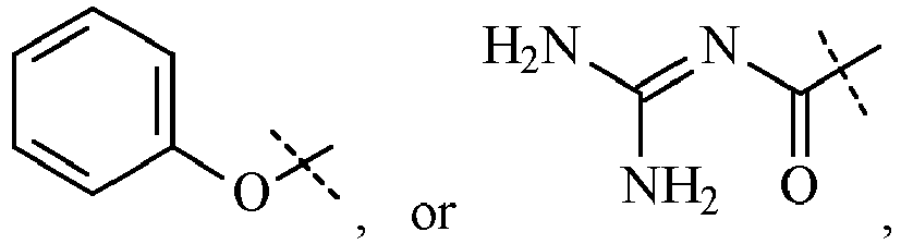 for use in the therapeutic or prophylactic treatment of a viral infection in a mammal infected with or exposed to a virus selected from the group consisting of Lentivirus, Coronavirus, Hepatitis C virus and Equine Arteritis virus.
 - The compound for use according to claim 1, wherein the compound reduces, retards or otherwise inhibits growth and/or replication of the virus in a cell infected with the virus or exposed to the virus, wherein the cell is in the mammal.
- The compound for use according to claim 1, wherein the compound prevents infection of a cell exposed to the virus, wherein the cell is in the mammal.
- The compound for use according to claim 1, wherein the compound down regulates a membrane ion channel functional activity in a cell infected with the virus, wherein the cell is in the mammal.
- The compound for use according to claim 4, wherein the compound reduces, retards or otherwise inhibits growth and/or replication of the virus, wherein the virus has infected a cell in the mammal.
- The compound for use according to claim 1, wherein the Lentivirus is Human Immunodeficiency Virus (HIV).
- The compound for use according to claim 6, wherein the compound is selected from:(3-Chlorocinnamoyl)guanidine,(3 -Bromocinnamoyl)guanidine,(2-Chlorocinnamoyl)guanidine,(2-Bromocinnamoyl)guanidine,3-(trifluoromethyl)cinnamoylguanidine,5-bromo-2-fluorocinnamoylguanidine,3 -methylcinnamoylguanidine,2-methylcinnamoylguanidine,2,3 -dimethylcinnamoylguanidine,Cinnamoylguanidine,6-methoxy-2-naphthoylguanidine,Trans-3-(1-naphthyl)acryloylguanidine,3,4-dichlorocinnamoylguanidine,2,6-dichlorocinnamoylguanidine,2-ethylcinnamoylguanidine,(4-Chlorocinnamoyl)guanidine,2-naphthoylguanidine,2,5 -dimethylcinnamoylguanidine,3-isopropylcinnamoylguanidine hydrochloride,(5-Phenyl-penta-2,4-dienoyl)guanidine,3-phenylcinnamoylguanidine,(4-Bromocinnamoyl)guanidine,5-(3'-bromophenyl)penta-2,4-dienoylguanidine,3 -(cyclohex-1-en-1-yl)cinnamoylguanidine,3 -(trifluoromethoxy)cinnamoylguanidine,2-(trifluoromethyl)cinnamoylguanidine,N,N'-bis(3phenylpropanoyl)-N" -phenylguanidine,2-ethoxycinnamoylguanidine,N-(3-phenylpropanoyl)-N'-phenylguanidine,4-(trifluoromethyl)cinnamoylguanidine,(4-Methoxycinnamoyl)guanidine,2-t-butylcinnamoylguanidine,4-methylcinnamoylguanidine,2-fluorocinnamoylguanidine,2-phenylcinnamoylguanidine,N-(6-Hydroxy-2-naphthoyl)-N'-phenylguanidine,3 -t-butylcinnamoylguanidine,3,4-difluorocinnamoylguanidine,3 -fluorocinnamoylguanidine,5-bromo-2-methoxycinnamoylguanidine,3-ethoxycinnamoylguanidine,3,4-(methylenedioxy)cinnamoylguanidine,(2-Methoxycinnamoyl)guanidine,2,3,5,6, -tetramethylcinnamoylguanidine,3-(2-naphthyl)acryloylguanidine,2-(1-naphthyl)acetoylguanidine,2,3 -difluorocinnamoylguanidine,(3 -Methoxycinnamoyl)guanidine,4-isopropylcinnamoylguanidine,2,4,6-trimethylcinnamoylguanidine,N-(cinnamoyl)-N'phenylguanidine,2-(cyclohex-1-en-1yl)cinnamoylguanidine,2-(2-naphthyl)acetoylguanidine,(4-Hydroxycinnamoyl)guanidine,4-phenylcinnamoylguanidine,4-fluorocinnamoylguanidine,N,N'-bis-(cinnamoyl)-N"-phenylguanidine,(2-Furanacryloyl)guanidine,(3 -Nitrocinnamoyl)guanidine,Benzyoylguanidine,(4-Phenoxybenzoyl)guanidine,3-(trans-hept-1-en-1-yl)cinnamoylguanidine,2-cyclohexylcinnamoylguanidine,4-ethoxycinnamoylguanidine,2,4-dichlorocinnamolyguanidine,(a-Methylcinnamoyl)guanidine,cinnamoylguanidine hydrochloride,[(4-Chlorophenoxy-acetyl]guanidine,(trans-2-Phenylcyclopropanecarbonyl)guanidine,(2-Nitrocinnamoyl)guanidine,trans-3-Furanacryoylguanidine,1-naphthoylguanidine,(3 -phenylpropanoyl)guanidine,4-t-butylcinnamoylguanidine,N,N'-Bis(3-phenylpropanoyl)guanidine,N-Benzoyl-N'-cinnamoylguanidine and1-bromo-2-naphthoylguanidine.
- The compound for use according to claim 6, wherein the HIV is HIV-1.
- The compound for use according to claim 1, wherein the Coronavirus is selected from the group consisting of Severe Acute Respiratory Syndrome virus (SARS), human Coronavirus 229E, human Coronavirus OC43, porcine respiratory Coronavirus (PRCV), bovine Coronvirus (BCV) and the coronavirus isolates listed in Table 1:
Table 1. Group 1 species Canine coronavirus Canine enteric coronavirus (strain INSAVC-1) Canine enteric coronavirus (strain K378) Feline coronavirus Feline enteric coronavirus (strain 79-1683) Feline infectious peritonitis virus (FIPV) Human coronavirus 229E Porcine epidemic diarrhea virus Porcine epidemic diarrhea virus (strain Br1/87) Porcine epidemic diarrhea virus (strain CV777) Transmissible gastroenteritis virus Porcine respiratory coronavirus Porcine transmissible gastroenteritis coronavirus (STRAIN FS772/70) Porcine transmissible gastroenteritis coronavirus (strain Miller) Porcine transmissible gastroenteritis coronavirus (strain Neb72-RT) Porcine transmissible gastroenteritis coronavirus (STRAIN PURDUE) Group 2 species Bovine coronavirus Bovine coronavirus (STRAIN F15) Bovine coronavirus (strain G95) Bovine coronavirus (STRAIN L9) Bovine coronavirus (strain LSU-94LSS-051) Bovine coronavirus (STRAIN LY-138) Bovine coronavirus (STRAIN MEBUS) Bovine coronavirus (strain OK-0514-3) Bovine coronavirus (strain Ontario) Bovine coronavirus (STRAIN QUEBEC) Bovine coronavirus (STRAIN VACCINE) Bovine enteric coronavirus (strain 98TXSF-110-ENT) Canine respiratory coronavirus Chicken enteric coronavirus Human coronavirus OC43 Murine hepatitis virus Murine coronavirus (strain DVIM) Murine hepatitis virus (strain A59) Murine hepatitis virus (strain JHM) Murine hepatitis virus (strain S) Murine hepatitis virus strain 1 Murine hepatitis virus strain 2 Murine hepatitis virus strain 3 Murine hepatitis virus strain 4 Murine hepatitis virus strain ML-11 Porcine hemagglutinating encephalomyelitis virus Porcine hemagglutinating encephalomyelitis virus (strain 67N) Porcine hemagglutinating encephalomyelitis virus (strain IAF-404) Puffinosis virus Rat coronavirus Rat coronavirus (strain 681) Rat coronavirus (strain NJ) Rat sialodacryoadenitis coronavirus Group 3 species Turkey coronavirus Turkey coronavirus (strain Indiana) Turkey coronavirus (strain Minnesota) Turkey coronavirus (strain NC95) Avian infectious bronchitis virus Avian infectious bronchitis virus (STRAIN 6/82) Avian infectious bronchitis virus (strain Arkansas 99) Avian infectious bronchitis virus (strain Beaudette CK) Avian infectious bronchitis virus (strain Beaudette M42) Avian infectious bronchitis virus (strain Beaudette US) Avian infectious bronchitis virus (strain Beaudette) Avian infectious bronchitis virus (strain D1466) Avian infectious bronchitis virus (strain D274) Avian infectious bronchitis virus (strain D3896) Avian infectious bronchitis virus (strain D41) Avian infectious bronchitis virus (strain DE072) Avian infectious bronchitis virus (strain GRAY) Avian infectious bronchitis virus (strain H120) Avian infectious bronchitis virus (strain H52) Avian infectious bronchitis virus (strain KB8523) Avian infectious bronchitis virus (strain M41) Avian infectious bronchitis virus (strain PORTUGAL/322/82) Avian infectious bronchitis virus (strain SAIB20) Avian infectious bronchitis virus (strain UK/123/82) Avian infectious bronchitis virus (strain UK/142/86) Avian infectious bronchitis virus (strain UK/167/84) Avian infectious bronchitis virus (strain UK/183/66) Avian infectious bronchitis virus (strain UK/68/84) Avian infectious bronchitis virus (strain V18/91) Avian infectious bronchitis virus (strain Vic S) Avian infectious laryngotracheitis virus Preliminary Group 4 species SARS coronavirus SARS coronavirus Beijing ZY-2003 SARS coronavirus BJ01 SARS coronavirus BJ02 SARS coronavirus BJ03 SARS coronavirus BJ04 SARS coronavirus CUHK-Su10 SARS coronavirus CUHK-W1 SARS coronavirus Frankfurt 1 SARS coronavirus GZ01 SARS coronavirus HKU-39849 SARS coronavirus Hong Kong ZY-2003 SARS coronavirus Hong Kong/03/2003 SARS coronavirus HSR 1 SARS coronavirus Sin2500 SARS coronavirus Sin2677 SARS coronavirus Sin2679 SARS coronavirus Sin2748 SARS coronavirus Sin2774 SARS coronavirus Taiwan SARS coronavirus Taiwan JC-2003 SARS coronavirus Taiwan TC 1 SARS coronavirus Taiwan TC2 SARS coronavirus Tor2 SARS coronavirus TW1 SARS coronavirus TWC SARS coronavirus Urbani SARS coronavirus Vietnam SARS coronavirus ZJ-HZ01 SARS coronavirus ZJ01 unclassified coronaviruses Bovine respiratory coronavirus (strain 98TXSF-110-LUN) Human enteric coronavirus 4408 Enteric coronavirus Equine coronavirus Equine coronavirus NC99 - The compound for use according to claim 9, wherein the coronavirus is SARS and the compound is selected from:2,3 -difluorocinnamoylguanidine,3,4-dichlorocinnamoylguanidine,4-t-butylcinnamoylguanidine,3-(2-naphthyl)acryloylguanidine,(3-Chlorocinnamoyl)guanidine,3 -(cyclohex-1-en-1-yl)cinnamoylguanidine,2,5-dimethylcinnamoylguanidine,trans-3 -(1-naphthyl)acryloylguanidine,4-isopropylcinnamoylguanidine,(3 -Bromocinnamoyl)guanidine,6-methoxy-2-naphthoylguanidine,3-phenylcinnamoylguanidine,(2-Chlorocinnamoyl)guanidine,4-phenylcinnamoylguanidine,4-(trifluoromethyl)cinnamoylguanidine,3 -(trifluoromethoxy)cinnamoylguanidine,3-(trifluoromethyl)cinnamoylguanidine,2-ethoxycinnamoylguanidine,cinnamoylguanidine hydrochloride,4-ethoxycinnamoylguanidine,(2-Bromocinnamoyl)guanidine,2,6-dichlorocinnamoylguanidine,3,4,5-trimethoxycinnamoylguanidine,3 -t-butylcinnamoylguanidine,5-bromo-2-fluorocinnamoylguanidine,(4-Chlorocinnamoyl)guanidine,2-t-butylcinnamoylguanidine,2-cyclohexylcinnamoylguanidine,3-(trans-hept-1-en-1-yl)cinnamoylguanidine,(4-Bromocinnamoyl)guanidine,(4-Hydroxycinnamoyl)guanidine,N-(3-phenylpropanoyl)-N'-phenylguanidine,(3 -Nitrocinnamoyl)guanidine,3 -fluorocinnamoylguanidine,2-(1-naphthyl)acetoylguanidine,2-ethylcinnamoylguanidine,2-naphthoylguanidine,2-(trifluoromethyl)cinnamoylguanidine,N-(6-Hydroxy-2-naphthoyl)-N'-phenylguanidine,(trans-2-Phenylcyclopropanecarbonyl)guanidine,N,N'-bis(3phenylpropanoyl)-N" -phenylguanidine,1-naphthoylguanidine,4-methylcinnamoylguanidine,4-fluorocinnamoylguanidine,3,4-(methylenedioxy)cinnamoylguanidine,N-(cinnamoyl)-N'phenylguanidine,3 -methylcinnamoylguanidine,2-methylcinnamoylguanidine,2,3,5,6, -tetramethylcinnamoylguanidine,trans-3 -Furanacryoylguanidine,(4-Methoxycinnamoyl)guanidine,(2-Furanacryloyl)guanidine,(3 -phenylpropanoyl)guanidine,2-(2-naphthyl)acetoylguanidine,Cinnamoylguanidine,(2-Methoxycinnamoyl)guanidine,[3-(3-Pyridyl)acryloyl]guanidine,2,4-dichlorocinnamolyguanidine,(3 -Methoxycinnamoyl)guanidine,2-fluorocinnamoylguanidine,(a-Methylcinnamoyl)guanidine,(5-Phenyl-penta-2,4-dienoyl)guanidine,(Quinoline-2-carbonyl)guanidine,(Phenylacetyl)guanidine,N,N'-Bis(amidino)naphthalene-2,6-dicarboxamide,6-bromo-2-naphthoylguanidine,1-bromo-2-naphthoylguanidine,2-chloro-6-fluorocinnamoylguanidine,[(4-Chlorophenoxy-acetyl]guanidine,N-Benzoyl-N'-cinnamoylguanidine andN-(2-naphthoyl)-N'-phenylguanidine.
- The compound for use according to claim 9, wherein the coronavirus is Coronavirus 229E, and the compound is selected from:4-isopropylcinnamoylguanidine,3,4-dichlorocinnamoylguanidine,3 -(trifluoromethoxy)cinnamoylguanidine,4-t-butylcinnamoylguanidine,3-isopropylcinnamoylguanidine hydrochloride,3 -t-butylcinnamoylguanidine,2-t-butylcinnamoylguanidine,trans-3 -(1-naphthyl)acryloylguanidine,5-bromo-2-methoxycinnamoylguanidine,2,3 -difluorocinnamoylguanidine,3-(2-naphthyl)acryloylguanidine,2-phenylcinnamoylguanidine,3-phenylcinnamoylguanidine,3 -(cyclohex-1 -en-1 -yl)cinnamoylguanidine,3-(trifluoromethyl)cinnamoylguanidine,4-(trifluoromethyl)cinnamoylguanidine,2-(cyclohex-1-en-1yl)cinnamoylguanidine,(4-Bromocinnamoyl)guanidine,1-naphthoylguanidine,(5-Phenyl-penta-2,4-dienoyl)guanidine,(3 -Bromocinnamoyl)guanidine,2,5-dimethylcinnamoylguanidine,2-(trifluoromethyl)cinnamoylguanidine,6-methoxy-2-naphthoylguanidine,(4-Chlorocinnamoyl)guanidine,(3 -Methoxycinnamoyl)guanidine,5-bromo-2-fluorocinnamoylguanidine,Cinnamoylguanidine,(2-Methoxycinnamoyl)guanidine,(a-Methylcinnamoyl)guanidine,4-phenylcinnamoylguanidine,2,6-dichlorocinnamoylguanidine,(2-Bromocinnamoyl)guanidine,2,4,6-trimethylcinnamoylguanidine,(trans-2-Phenylcyclopropanecarbonyl)guanidine,(3-Chlorocinnamoyl)guanidine,2-(1-naphthyl)acetoylguanidine,2-ethylcinnamoylguanidine,2-cyclohexylcinnamoylguanidine,(4-Hydroxycinnamoyl)guanidine,2-ethoxycinnamoylguanidine,3 -methylcinnamoylguanidine,2-methylcinnamoylguanidine,3 -fluorocinnamoylguanidine,cinnamoylguanidine hydrochloride,2,3 -dimethyl cinnamoylguanidine,2-fluorocinnamoylguanidine,4-fluorocinnamoylguanidine,3,4-difluorocinnamoylguanidine,2-naphthoylguanidine,N,N'-Bis(amidino)naphthalene-2,6-dicarboxamide,N,N'-Bis(3-phenylpropanoyl)guanidine,4-methylcinnamoylguanidine,2,3,5,6, -tetramethylcinnamoylguanidine,3-ethoxycinnamoylguanidine,N,N'-bis(3phenylpropanoyl)-N" -phenylguanidine,(4-Methoxycinnamoyl)guanidine,(2-Chlorocinnamoyl)guanidine,(3 -Nitrocinnamoyl)guanidine,4-ethoxycinnamoylguanidine,3,4,5-trimethoxycinnamoylguanidine,2-(2-naphthyl)acetoylguanidine,N-(3-phenylpropanoyl)-N'-phenylguanidine,5-(2'-bromophenyl)penta-2,4-dienoylguanidine,(4-Bromocinnamoyl)guanidine,(2-Nitrocinnamoyl)guanidine,(3-Chlorocinnamoyl)guanidine,(4-Methoxycinnamoyl)guanidine,4-(trifluoromethyl)cinnamoylguanidine,[(E)-3-(4-Dimethylaminophenyl)-2-methylacryloyl]guanidine,N-Benzoyl-N'-cinnamoylguanidine,trans-3 -Furanacryoylguanidine,N-(cinnamoyl)-N'phenylguanidine,Cinnamoylguanidine,(3 -phenylpropanoyl)guanidine,Benzyoylguanidine,(Quinoline-2-carbonyl)guanidine,[3-(3-Pyridyl)acryloyl]guanidine,N-Cinnamoyl-N',N'-dimethylguanidine,N-(2-naphthoyl)-N'-phenylguanidine and(Phenylacetyl)guanidine.
- The compound for use according to claim 9, wherein the coronavirus is human Coronavirus OC43 and the compound is selected from:3 -methylcinnamoylguanidine,trans-3 -(1-naphthyl)acryloylguanidine,(3-Bromocinnamoyl)guanidine,(2-Chlorocinnamoyl)guanidine,3,4-dichlorocinnamoylguanidine,3-(trifluoromethyl)cinnamoylguanidine,(trans-2-Phenylcyclopropanecarbonyl)guanidine,4-isopropylcinnamoylguanidine,Cinnamoylguanidine,6-methoxy-2-naphthoylguanidine,2,4-dichlorocinnamolyguanidine,(4-Chlorocinnamoyl)guanidine,(4-Bromocinnamoyl)guanidine,2,6-dichlorocinnamoylguanidine,5-bromo-2-methoxycinnamoylguanidine,(5-Phenyl-penta-2,4-dienoyl)guanidine,3-(trifluoromethoxy)cinnamoylguanidine,2-t-butylcinnamoylguanidine.
- The compound for use according to claim 9, wherein the coronavirus is PRCV and the compound is selected from:6-methoxy-2-naphthoylguanidine,Cinnamoylguanidine,N-(3-phenylpropanoyl)-N'-phenylguanidine,3 -methylcinnamoylguanidine,(3-Bromocinnamoyl)guanidine,(trans-2-Phenylcyclopropanecarbonyl)guanidine,trans-3-(1-naphthyl)acryloylguanidine and2-(2-naphthyl)acetoylguanidine.
- The compound for use according to claim 9, wherein the coronavirus is BCV and the compound is selected from:(3-Bromocinnamoyl)guanidine,3-(trifluoromethyl)cinnamoylguanidine,6-methoxy-2-naphthoylguanidine,trans-3 -(1-naphthyl)acryloylguanidine,Cinnamoylguanidine,(5-Phenyl-penta-2,4-dienoyl)guanidine,2-(2-naphthyl)acetoylguanidine,(trans-2-Phenylcyclopropanecarbonyl)guanidine,and N-(3-phenylpropanoyl)-N'-phenylguanidine.
- The compound for use according to claim 9, wherein the coronavirus is a coronavirus isolate listed in Table 1 and the compound is selected from:4-isopropylcinnamoylguanidine,3,4-dichlorocinnamoylguanidine,3 -(trifluoromethoxy)cinnamoylguanidine,4-t-butylcinnamoylguanidine, and3-isopropylcinnamoylguanidine hydrochloride.
- The compound for use according to claim 1, wherein the virus is Hepatitis C virus and the compound is selected from:2,3 -dimethylcinnamoylguanidine,2,4,6-trimethylcinnamoylguanidine,5-bromo-2-fluorocinnamoylguanidine,(4-Bromocinnamoyl)guanidine,2,5-dimethylcinnamoylguanidine,3-(trifluoromethyl)cinnamoylguanidine,4-(trifluoromethyl)cinnamoylguanidine,6-methoxy-2-naphthoylguanidine,(2-Chlorocinnamoyl)guanidine,(4-Chlorocinnamoyl)guanidine,(2-Bromocinnamoyl)guanidine,2,6-dichlorocinnamoylguanidine,(3 -Bromocinnamoyl)guanidine,(3 -Chlorocinnamoyl)guanidine,2-(trifluoromethyl)cinnamoylguanidine,3,4-dichlorocinnamoylguanidine,4-isopropylcinnamoylguanidine,trans-3 -(1-naphthyl)acryloylguanidine,4-t-butylcinnamoylguanidine,2-t-butylcinnamoylguanidine,2-ethylcinnamoylguanidine,4-methylcinnamoylguanidine,5-bromo-2-methoxycinnamoylguanidine,3 -(trifluoromethoxy)cinnamoylguanidine,2-cyclohexylcinnamoylguanidine,1-naphthoylguanidine,3 -t-butylcinnamoylguanidine,(5-Phenyl-penta-2,4-dienoyl)guanidine,N-(cinnamoyl)-N'phenylguanidine,3-isopropylcinnamoylguanidine hydrochloride,N-(3-phenylpropanoyl)-N'-phenylguanidine,N,N'-bis(3phenylpropanoyl)-N" -phenylguanidine,3-(2-naphthyl)acryloylguanidine,(4-Methoxycinnamoyl)guanidine,4-fluorocinnamoylguanidine,(3 -Nitrocinnamoyl)guanidine,4-ethoxycinnamoylguanidine,(4-Hydroxycinnamoyl)guanidine,(trans-2-Phenylcyclopropanecarbonyl)guanidine,3-ethoxycinnamoylguanidine,2,3,5,6, -tetramethylcinnamoylguanidine,4-phenylcinnamoylguanidine,trans-3 -Furanacryoylguanidine,N-(6-Hydroxy-2-naphthoyl)-N'-phenylguanidine,(2-Furanacryloyl)guanidine,3-(cyclohex-1-en-1-yl)cinnamoylguanidine,cinnamoylguanidine hydrochloride,2,3 -difluorocinnamoylguanidine,2-(1-naphthyl)acetoylguanidine,(a-Methylcinnamoyl)guanidine,(2-Nitrocinnamoyl)guanidine,3,4-(methylenedioxy)cinnamoylguanidine,2-ethoxycinnamoylguanidine,Cinnamoylguanidine,2-phenylcinnamoylguanidine,2-(cyclohex-1-en-1yl)cinnamoylguanidine,2-naphthoylguanidine,3-phenylcinnamoylguanidine,(3 -Methoxycinnamoyl)guanidine,2-fluorocinnamoylguanidine,[(4-Chlorophenoxy-acetyl]guanidine,(3 -phenylpropanoyl)guanidine,2-chloro-6-fluorocinnamoylguanidine,3-fluorocinnamoylguanidine,2-methylcinnamoylguanidine,(2-Methoxycinnamoyl)guanidine,1-bromo-2-naphthoylguanidine,3,4,5-trimethoxycinnamoylguanidine,3 -methylcinnamoylguanidine,3-(trans-hept-1-en-1-yl)cinnamoylguanidine,2,4-dichlorocinnamolyguanidine,(4-Nitrocinnamoyl)guanidine,3,4-difluorocinnamoylguanidine and[(E)-3-(4-Dimethylaminophenyl)-2-methylacryloyl]guanidine.
- The compound for use according to claim 1, wherein the virus is Equine Arteritis virus and the compound is selected from:(3-Bromocinnamoyl)guanidine,trans-3 -(1-naphthyl)acryloylguanidine,2-t-butylcinnamoylguanidine and2-(cyclohex-1-en-1yl)cinnamoylguanidine.
- A compound for use in the therapeutic or prophylactic treatment of a viral infection in a mammal infected with or exposed to a virus, wherein the virus is Dengue virus and the compound is selected from the group consisting of:cinnamoylguanidine,(2-chlorocinnamoyl)guanidine andtrans-3-(1-naphthyl)acryloylguanidine.
- The compound for use according to claim 4, wherein the membrane ion channel is selected from the group consisting of HIV Vpu membrane ion channel in the treatment of HIV, Coronavirus E protein in the treatment of Coronavirus and Hepatitis C virus p7 membrane ion channel in the treatment of Hepatitis C.
- An in vitro or ex vivo method of:reducing, retarding or otherwise inhibiting growth and/or replication of a virus in a cell infected with the virus or exposed to the virus;preventing the infection of a cell exposed to a virus; ordown regulating a membrane ion channel functional activity in a cell infected with a virus;the method comprising contacting the cell with a compound as defined in claim 1, wherein the virus is selected from the group consisting of Lentivirus, Coronavirus, Hepatitis C virus and Equine Arteritis virus.
Applications Claiming Priority (6)
| Application Number | Priority Date | Filing Date | Title |
|---|---|---|---|
| AU2003903251A AU2003903251A0 (en) | 2003-06-26 | 2003-06-26 | Antiviral compounds and methods |
| AU2003903850A AU2003903850A0 (en) | 2003-07-25 | 2003-07-25 | Anti-coronavirus compounds and methods |
| AU2003904692A AU2003904692A0 (en) | 2003-08-29 | Anti-flavivirus compounds and methods | |
| AU2004902902A AU2004902902A0 (en) | 2004-05-31 | Anti-flavivirus compounds and methods | |
| EP04737487A EP1646371A4 (en) | 2003-06-26 | 2004-06-26 | Antiviral compounds and methods |
| PCT/AU2004/000866 WO2004112687A2 (en) | 2003-06-26 | 2004-06-26 | Antiviral acylguanidine compounds and methods |
Related Parent Applications (2)
| Application Number | Title | Priority Date | Filing Date |
|---|---|---|---|
| EP04737487A Division EP1646371A4 (en) | 2003-06-26 | 2004-06-26 | Antiviral compounds and methods |
| EP04737487.1 Division | 2004-06-26 |
Publications (3)
| Publication Number | Publication Date |
|---|---|
| EP2617709A1 EP2617709A1 (en) | 2013-07-24 |
| EP2617709B1 true EP2617709B1 (en) | 2022-11-16 |
| EP2617709B8 EP2617709B8 (en) | 2022-12-21 |
Family
ID=33545379
Family Applications (2)
| Application Number | Title | Priority Date | Filing Date |
|---|---|---|---|
| EP04737487A Withdrawn EP1646371A4 (en) | 2003-06-26 | 2004-06-26 | Antiviral compounds and methods |
| EP13164504.6A Expired - Lifetime EP2617709B8 (en) | 2003-06-26 | 2004-06-26 | Guanidine derivatives as antiviral agents |
Family Applications Before (1)
| Application Number | Title | Priority Date | Filing Date |
|---|---|---|---|
| EP04737487A Withdrawn EP1646371A4 (en) | 2003-06-26 | 2004-06-26 | Antiviral compounds and methods |
Country Status (8)
| Country | Link |
|---|---|
| US (4) | US20130035328A1 (en) |
| EP (2) | EP1646371A4 (en) |
| JP (1) | JP5030587B2 (en) |
| KR (1) | KR101153254B1 (en) |
| BR (1) | BRPI0411900B8 (en) |
| CA (1) | CA2529949C (en) |
| NZ (1) | NZ544671A (en) |
| WO (1) | WO2004112687A2 (en) |
Families Citing this family (46)
| Publication number | Priority date | Publication date | Assignee | Title |
|---|---|---|---|---|
| BRPI0411900B8 (en) | 2003-06-26 | 2021-05-25 | Biotron Ltd | compounds and pharmaceutical compositions comprising them |
| US7745442B2 (en) * | 2003-08-20 | 2010-06-29 | Parion Sciences, Inc. | Methods of reducing risk of infection from pathogens |
| PT2826770T (en) | 2005-06-24 | 2018-12-18 | Biotron Ltd | Acylguanidine compounds with antiviral activity |
| AU2013205388B2 (en) * | 2005-06-24 | 2016-05-05 | Biotron Limited | Antiviral compounds and methods |
| GB0526240D0 (en) * | 2005-12-22 | 2006-02-01 | Novartis Ag | Organic compounds |
| GB0526244D0 (en) | 2005-12-22 | 2006-02-01 | Novartis Ag | Organic compounds |
| AU2013219202B2 (en) * | 2007-08-03 | 2016-12-08 | Biotron Limited | Hepatitis C antiviral compositions and methods |
| WO2009018609A1 (en) * | 2007-08-03 | 2009-02-12 | Biotrom Limited | Hepatitis c antiviral compositions and methods |
| US20110217265A1 (en) * | 2008-09-23 | 2011-09-08 | Glenn Jeffrey S | Screening for Inhibitors of HCV Amphipathic Helix (AH) Function |
| ES2524356T3 (en) | 2010-07-22 | 2014-12-05 | Gilead Sciences, Inc. | Methods and compounds to treat infections caused by Paramyxoviridae virus |
| TWI740546B (en) | 2014-10-29 | 2021-09-21 | 美商基利科學股份有限公司 | Methods for the preparation of ribosides |
| HRP20220355T1 (en) | 2015-09-16 | 2022-05-13 | Gilead Sciences, Inc. | Methods for treating coronaviridae infections |
| WO2018081863A1 (en) * | 2016-11-04 | 2018-05-11 | University Of Wollongong | 6-SUBSTITUTED DERIVATIVES OF HEXAMETHYLENE AMILORIDE AS INHIBITORS OF uPA AND USES THEREOF |
| CN116036112A (en) | 2017-03-14 | 2023-05-02 | 吉利德科学公司 | Methods of Treating Feline Coronavirus Infection |
| WO2018204198A1 (en) | 2017-05-01 | 2018-11-08 | Gilead Sciences, Inc. | Crystalline forms of (s) 2 ethylbutyl 2 (((s) (((2r,3s,4r,5r) 5 (4 aminopyrrolo[2,1-f] [1,2,4]triazin-7-yl)-5-cyano-3,4-dihydroxytetrahydrofuran-2 yl)methoxy)(phenoxy) phosphoryl)amino)propanoate |
| EP4512475A3 (en) | 2017-07-11 | 2025-05-14 | Gilead Sciences, Inc. | Compositions comprising an rna polymerase inhibitor and cyclodextrin for treating viral infections |
| AU2021214911A1 (en) | 2020-01-27 | 2022-07-21 | Gilead Sciences, Inc. | Methods for treating SARS CoV-2 infections |
| JP2021138619A (en) * | 2020-03-02 | 2021-09-16 | フマキラー株式会社 | Anti-coronavirus agent |
| CN118994170A (en) | 2020-03-12 | 2024-11-22 | 吉利德科学公司 | Process for preparing 1' -cyanonucleosides |
| WO2021202535A1 (en) * | 2020-03-30 | 2021-10-07 | Thomas Jefferson University | Methods for treating, ameliorating, or preventing viral infections |
| EP4132651B1 (en) | 2020-04-06 | 2026-03-18 | Gilead Sciences, Inc. | Inhalation formulations of 1'-cyano substituted carbanucleoside analogs |
| ES3041698T3 (en) | 2020-05-29 | 2025-11-13 | Gilead Sciences Inc | Remdesivir for the treatment of viral infections |
| CA3187821A1 (en) | 2020-06-24 | 2021-12-30 | Gilead Sciences, Inc. | 1'-cyano nucleoside analogs and uses thereof |
| WO2022040321A1 (en) * | 2020-08-19 | 2022-02-24 | Natreon, Inc. | Protection against coronavirus infection by extracts and extract components |
| MX2023002233A (en) | 2020-08-24 | 2023-03-15 | Gilead Sciences Inc | Phospholipid compounds and uses thereof. |
| EP3960165A1 (en) * | 2020-08-25 | 2022-03-02 | Fondation EspeRare | Nhe-1 inhibitors for the treatment of coronavirus infections |
| WO2022047065A2 (en) | 2020-08-27 | 2022-03-03 | Gilead Sciences, Inc. | Compounds and methods for treatment of viral infections |
| TWI811812B (en) | 2020-10-16 | 2023-08-11 | 美商基利科學股份有限公司 | Phospholipid compounds and uses thereof |
| WO2022251318A1 (en) | 2021-05-26 | 2022-12-01 | Gilead Sciences, Inc. | Phospholipid formulations of 1'-cyano substituted carba-nucleoside analogs |
| WO2023012329A1 (en) | 2021-08-06 | 2023-02-09 | Intervet International B.V. | Method of treating veterinary viral diseases |
| JP7719954B2 (en) | 2021-08-18 | 2025-08-06 | ギリアード サイエンシーズ, インコーポレイテッド | Phospholipid compounds and methods for producing and using same |
| TW202525810A (en) | 2022-03-02 | 2025-07-01 | 美商基利科學股份有限公司 | Compounds and methods for treatment of viral infections |
| EP4486746A1 (en) | 2022-03-02 | 2025-01-08 | Gilead Sciences, Inc. | Compounds and methods for treatment of viral infections |
| CR20240367A (en) | 2022-03-03 | 2024-09-30 | Gilead Sciences Inc | ANTIVIRAL COMPOUNDS AND METHODS OF THEIR PREPARATION AND USE. |
| WO2023167055A1 (en) * | 2022-03-03 | 2023-09-07 | 日本曹達株式会社 | Antiviral agent |
| WO2023168254A1 (en) | 2022-03-03 | 2023-09-07 | Gilead Sciences, Inc. | Antiviral compounds and methods of making and using the same |
| CN119604286A (en) | 2022-06-06 | 2025-03-11 | 吉利德科学公司 | Methods for treating viral infections including SARS-CoV-2 |
| WO2024006376A1 (en) | 2022-06-29 | 2024-01-04 | Gilead Sciences, Inc. | Solid forms of a nucleoside analogue and uses thereof |
| WO2024006461A1 (en) | 2022-06-30 | 2024-01-04 | Gilead Sciences, Inc. | Solid forms of a nucleoside analogue and uses thereof |
| US12357577B1 (en) | 2024-02-02 | 2025-07-15 | Gilead Sciences, Inc. | Pharmaceutical formulations and uses thereof |
| WO2024112712A1 (en) * | 2022-11-21 | 2024-05-30 | Henderson Theodore | Treatment using an antiviral compound and spironolactone |
| EP4665737A1 (en) | 2023-02-16 | 2025-12-24 | Gilead Sciences, Inc. | Phospholipid compounds and methods of making and using the same |
| US20250090537A1 (en) | 2023-08-31 | 2025-03-20 | Gilead Sciences, Inc. | Antiviral compounds and methods of making and using the same |
| CN121693506A (en) | 2023-08-31 | 2026-03-17 | 吉利德科学公司 | Antiviral compounds and methods of making and using the same |
| WO2025054278A1 (en) | 2023-09-06 | 2025-03-13 | Gilead Sciences, Inc. | Solid forms of a nucleoside analogue and uses thereof |
| US20250109157A1 (en) | 2023-09-28 | 2025-04-03 | Gilead Sciences, Inc. | Compounds and methods for treatment of viral infections |
Citations (5)
| Publication number | Priority date | Publication date | Assignee | Title |
|---|---|---|---|---|
| EP0682017A1 (en) * | 1994-05-05 | 1995-11-15 | Hoechst Aktiengesellschaft | Substituted bicycle heteroaroylguanidines, their preparation and their application in medicaments and diagnostic agents |
| EP0744397A2 (en) * | 1995-05-22 | 1996-11-27 | Hoechst Aktiengesellschaft | Fluorophenyl substituted alkenyl carboxylic acid guanidines as Na+/H+ exchange inhibitors, process for their preparation, their use as medicinal or diagnostic agent as well as a medicament containing them |
| EP0810205A1 (en) * | 1996-05-29 | 1997-12-03 | Hoechst Aktiengesellschaft | Substituted 1-naphtoylguanidines, process for their preparation, their use as medicament or diagnostic agent and medicaments containing them |
| WO1997046226A2 (en) * | 1996-06-03 | 1997-12-11 | Hoechst Aktiengesellschaft | USE OF INHIBITORS OF THE CELLULAR Na+/H+ EXCHANGER (NHE) FOR PREPARING A MEDICAMENT FOR NORMALIZING SERUM LIPIDS |
| WO2000021538A1 (en) * | 1998-10-12 | 2000-04-20 | The Australian National University | A method of modulating ion channel functional activity |
Family Cites Families (34)
| Publication number | Priority date | Publication date | Assignee | Title |
|---|---|---|---|---|
| US2734904A (en) * | 1956-02-14 | Xcxnhxc-nh | ||
| BE639386A (en) | 1962-10-30 | |||
| FR1435379A (en) * | 1964-07-30 | 1966-04-15 | American Cyanamid Co | Process for stabilizing rigid polyvinyl chloride against the effects of light |
| US3527758A (en) * | 1967-04-13 | 1970-09-08 | Merck & Co Inc | Process for the preparation of pyrazinoylguanidines from a pyrazinoic azide and a guanidine |
| DE2006186A1 (en) | 1970-02-11 | 1971-08-19 | Huebner Vamag | Method for inserting a seat ring for the shut-off element of a valve |
| DE2531343A1 (en) * | 1975-07-14 | 1977-02-10 | Henkel & Cie Gmbh | Antimicrobial compsns. contg. substd. benzoyl phenyl guanidines - prepd. from opt. substd. benzoyl cyanamides and substd. anilines |
| DD200618A1 (en) * | 1981-10-05 | 1983-05-25 | Manfred Augustin | PROCESS FOR THE PREPARATION OF NEW N-AROYL AND HETAROYLIMIDES |
| US4496573A (en) * | 1982-08-24 | 1985-01-29 | William H. Rorer, Inc. | 1-Pyridylmethyl-3-acyl guanidines |
| US4894376A (en) * | 1988-02-26 | 1990-01-16 | Trustees Of The University Of Pennsylvania | Methods of treating diseases characterized by hyperexcitability of neurons |
| JPH0789859A (en) * | 1990-04-05 | 1995-04-04 | Celtrix Pharmaceut Inc | Pharmaceutical compositions containing pyrazine derivatives and therapeutic methods for inhibiting ocular neovascularization |
| IN177137B (en) * | 1992-12-11 | 1996-11-16 | Hoechst India | |
| DE4301739A1 (en) * | 1993-01-22 | 1994-07-28 | Sauer Professor Dr Gerhard | Treatment of primary tumours and viral diseases |
| JPH0725768A (en) * | 1993-07-09 | 1995-01-27 | Mitsubishi Chem Corp | Suppressing agent for hyperplasia of vascular endosporium |
| DE4325822A1 (en) * | 1993-07-31 | 1995-02-02 | Hoechst Ag | Substituted benzoylguanidines, process for their preparation, their use as medicament or diagnostic agent, and medicament containing them |
| TW415937B (en) * | 1994-01-25 | 2000-12-21 | Hoechst Ag | Phenyl-substituted alkylcarboxylic acid guanidides bearing perfluoroalkyl groups, process for their preparation, their use as a medicament or diagnostic, and medicament containing them |
| DE4421536A1 (en) * | 1994-06-20 | 1995-12-21 | Hoechst Ag | Phenyl-substituted alkenylcarboxylic acid guanidines bearing perfluoroalkyl groups, processes for their preparation, their use as medicaments or diagnostic agents, and medicaments containing them |
| JPH08225513A (en) * | 1994-12-21 | 1996-09-03 | Kanebo Ltd | Naphthoylguanidine derivative |
| AT408330B (en) | 1995-01-27 | 2001-10-25 | Colop Stempelerzeugung Skopek | SELF-COLORING STAMP |
| DK0738712T3 (en) * | 1995-04-18 | 2000-03-06 | Hoechst Ag | Substituted indenoylguanidines with antiarrhythmic cardioprotective effect |
| DE19527305A1 (en) * | 1995-07-26 | 1997-01-30 | Hoechst Ag | Substituted cinnamic acid guanidides, process for their preparation, their use as a medicament or diagnostic agent and medicament containing them |
| US5628984A (en) * | 1995-07-31 | 1997-05-13 | University Of North Carolina At Chapel Hill | Method of detecting lung disease |
| JPH0967332A (en) * | 1995-08-29 | 1997-03-11 | Yamanouchi Pharmaceut Co Ltd | N-(2-propenoyl)guanidine derivative |
| AUPO545297A0 (en) * | 1997-03-04 | 1997-03-27 | Fujisawa Pharmaceutical Co., Ltd. | Guanidine derivatives |
| IL126276A0 (en) * | 1997-09-24 | 1999-05-09 | Hoechst Marion Roussel De Gmbh | The use of an inhibitor of the na+/H+ exchanger for the production of a medicament for the treatment or prophylaxis of disorders of the central nervous system |
| US7041702B1 (en) * | 1997-10-21 | 2006-05-09 | Scion Pharmaceuticals, Inc. | Pharmaceutically active compounds and methods of use |
| JP2001520215A (en) * | 1997-10-21 | 2001-10-30 | ケンブリッジ・ニューロサイエンス・インコーポレイティッド | Pharmaceutically active compounds and uses |
| CN1285001A (en) * | 1997-12-18 | 2001-02-21 | 塞普拉科有限公司 | Methods for simultaneous identification of novel biological targets and lead structures for drug development |
| US6011059A (en) * | 1997-12-24 | 2000-01-04 | Bristol-Myers Squibb Company | Acyl guanidine sodium/proton exchange inhibitors and method |
| DE19849722A1 (en) * | 1998-10-28 | 2000-05-04 | Aventis Pharma Gmbh | Substituted phenyl-alkenoylguanidines, processes for their preparation, their use as medicaments or diagnostic agents and medicaments containing them |
| DE19859727A1 (en) * | 1998-12-23 | 2000-06-29 | Aventis Pharma Gmbh | The use of inhibitors of the sodium-hydrogen exchanger for the manufacture of a medicament for the prevention of age-related organ dysfunctions, age-related illnesses for the prolongation of life |
| AUPQ223999A0 (en) * | 1999-08-16 | 1999-09-09 | University Of Sydney, The | Intracellular feedback controls in the diagnosis and treatment of human disease |
| AUPS022802A0 (en) * | 2002-01-31 | 2002-02-21 | Macfarlane Burnet Institute For Medical Research And Public Health Limited, The | Anti-viral compounds |
| BRPI0411900B8 (en) | 2003-06-26 | 2021-05-25 | Biotron Ltd | compounds and pharmaceutical compositions comprising them |
| PT2826770T (en) * | 2005-06-24 | 2018-12-18 | Biotron Ltd | Acylguanidine compounds with antiviral activity |
-
2004
- 2004-06-26 BR BRPI0411900A patent/BRPI0411900B8/en not_active IP Right Cessation
- 2004-06-26 KR KR1020057024860A patent/KR101153254B1/en not_active Expired - Fee Related
- 2004-06-26 JP JP2006515560A patent/JP5030587B2/en not_active Expired - Lifetime
- 2004-06-26 EP EP04737487A patent/EP1646371A4/en not_active Withdrawn
- 2004-06-26 NZ NZ544671A patent/NZ544671A/en not_active IP Right Cessation
- 2004-06-26 CA CA2529949A patent/CA2529949C/en not_active Expired - Lifetime
- 2004-06-26 WO PCT/AU2004/000866 patent/WO2004112687A2/en not_active Ceased
- 2004-06-26 EP EP13164504.6A patent/EP2617709B8/en not_active Expired - Lifetime
-
2012
- 2012-07-19 US US13/553,239 patent/US20130035328A1/en not_active Abandoned
-
2015
- 2015-02-06 US US14/615,616 patent/US20150313909A1/en not_active Abandoned
-
2017
- 2017-05-23 US US15/602,526 patent/US10472332B2/en not_active Expired - Lifetime
-
2019
- 2019-08-29 US US16/554,990 patent/US11192863B2/en not_active Expired - Fee Related
Patent Citations (5)
| Publication number | Priority date | Publication date | Assignee | Title |
|---|---|---|---|---|
| EP0682017A1 (en) * | 1994-05-05 | 1995-11-15 | Hoechst Aktiengesellschaft | Substituted bicycle heteroaroylguanidines, their preparation and their application in medicaments and diagnostic agents |
| EP0744397A2 (en) * | 1995-05-22 | 1996-11-27 | Hoechst Aktiengesellschaft | Fluorophenyl substituted alkenyl carboxylic acid guanidines as Na+/H+ exchange inhibitors, process for their preparation, their use as medicinal or diagnostic agent as well as a medicament containing them |
| EP0810205A1 (en) * | 1996-05-29 | 1997-12-03 | Hoechst Aktiengesellschaft | Substituted 1-naphtoylguanidines, process for their preparation, their use as medicament or diagnostic agent and medicaments containing them |
| WO1997046226A2 (en) * | 1996-06-03 | 1997-12-11 | Hoechst Aktiengesellschaft | USE OF INHIBITORS OF THE CELLULAR Na+/H+ EXCHANGER (NHE) FOR PREPARING A MEDICAMENT FOR NORMALIZING SERUM LIPIDS |
| WO2000021538A1 (en) * | 1998-10-12 | 2000-04-20 | The Australian National University | A method of modulating ion channel functional activity |
Also Published As
| Publication number | Publication date |
|---|---|
| JP2007508234A (en) | 2007-04-05 |
| CA2529949C (en) | 2013-08-06 |
| US20170260147A1 (en) | 2017-09-14 |
| US10472332B2 (en) | 2019-11-12 |
| KR20060103086A (en) | 2006-09-28 |
| JP5030587B2 (en) | 2012-09-19 |
| US11192863B2 (en) | 2021-12-07 |
| US20150313909A1 (en) | 2015-11-05 |
| BRPI0411900A (en) | 2006-09-19 |
| EP1646371A4 (en) | 2010-09-22 |
| US20130035328A1 (en) | 2013-02-07 |
| NZ544671A (en) | 2009-02-28 |
| KR101153254B1 (en) | 2012-07-02 |
| EP2617709B8 (en) | 2022-12-21 |
| US20190389816A1 (en) | 2019-12-26 |
| BRPI0411900B1 (en) | 2018-11-21 |
| EP2617709A1 (en) | 2013-07-24 |
| BRPI0411900B8 (en) | 2021-05-25 |
| CA2529949A1 (en) | 2004-12-29 |
| WO2004112687A3 (en) | 2007-07-12 |
| EP1646371A2 (en) | 2006-04-19 |
| WO2004112687A2 (en) | 2004-12-29 |
Similar Documents
| Publication | Publication Date | Title |
|---|---|---|
| US11192863B2 (en) | Antiviral compounds and methods | |
| CA2695390C (en) | Hepatitis c antiviral compositions and methods | |
| US20070099968A1 (en) | Antiviral compounds and methods | |
| AU2004248859B2 (en) | Antiviral acylguanidine compounds and methods | |
| US11376306B2 (en) | Peptides and uses therefor as antiviral agents | |
| JP2009242247A (en) | Human immunodeficiency virus infection inhibitor and aids-treating or preventing drug | |
| HK1187600A (en) | Guanidine derivatives as antiviral agents | |
| CN101111475A (en) | Antiviral compounds and methods | |
| KR20050092103A (en) | Use of iminosugar derivatives to inhibit ion channel activity | |
| EP1711522A2 (en) | Antiviral compositions which inhibit paramyxovirus infection | |
| EP0684818B1 (en) | Method of reversing resistance of hiv-1 strains to zidovudine | |
| EP4238560B1 (en) | Therapeutic agent for covid-19 | |
| US11331371B2 (en) | Labyrinthopeptins as anti-viral agents | |
| HK40025030A (en) | Peptides and uses therefor as antiviral agents | |
| WO2018189566A1 (en) | Use of a chemical compound as a therapeutic agent | |
| MXPA99007077A (en) | Quinoxaline in triple combination with protease inhibitors and reverse transcriptase inhibitors as medicines for treating aids |
Legal Events
| Date | Code | Title | Description |
|---|---|---|---|
| PUAI | Public reference made under article 153(3) epc to a published international application that has entered the european phase |
Free format text: ORIGINAL CODE: 0009012 |
|
| AC | Divisional application: reference to earlier application |
Ref document number: 1646371 Country of ref document: EP Kind code of ref document: P |
|
| AK | Designated contracting states |
Kind code of ref document: A1 Designated state(s): AT BE BG CH CY CZ DE DK EE ES FI FR GB GR HU IE IT LI LU MC NL PL PT RO SE SI SK TR |
|
| 17P | Request for examination filed |
Effective date: 20140124 |
|
| RBV | Designated contracting states (corrected) |
Designated state(s): AT BE BG CH CY CZ DE DK EE ES FI FR GB GR HU IE IT LI LU MC NL PL PT RO SE SI SK TR |
|
| REG | Reference to a national code |
Ref country code: HK Ref legal event code: DE Ref document number: 1187600 Country of ref document: HK |
|
| 17Q | First examination report despatched |
Effective date: 20150901 |
|
| STAA | Information on the status of an ep patent application or granted ep patent |
Free format text: STATUS: EXAMINATION IS IN PROGRESS |
|
| REG | Reference to a national code |
Ref country code: DE Ref legal event code: R079 Ref document number: 602004055117 Country of ref document: DE Free format text: PREVIOUS MAIN CLASS: C07C0279220000 Ipc: A61K0031150000 |
|
| GRAP | Despatch of communication of intention to grant a patent |
Free format text: ORIGINAL CODE: EPIDOSNIGR1 |
|
| STAA | Information on the status of an ep patent application or granted ep patent |
Free format text: STATUS: GRANT OF PATENT IS INTENDED |
|
| RIC1 | Information provided on ipc code assigned before grant |
Ipc: A61P 31/18 20060101ALI20220520BHEP Ipc: A61K 31/15 20060101AFI20220520BHEP Ipc: A61P 31/00 20060101ALI20220520BHEP Ipc: A61P 11/00 20060101ALI20220520BHEP Ipc: A61P 1/16 20060101ALI20220520BHEP Ipc: C07D 241/34 20060101ALI20220520BHEP Ipc: A61K 31/165 20060101ALI20220520BHEP Ipc: C07C 279/22 20060101ALI20220520BHEP Ipc: A61K 31/381 20060101ALI20220520BHEP Ipc: A61K 31/4406 20060101ALI20220520BHEP Ipc: A61K 31/47 20060101ALI20220520BHEP Ipc: A61K 31/4965 20060101ALI20220520BHEP Ipc: A61K 31/55 20060101ALI20220520BHEP Ipc: A61P 31/12 20060101ALI20220520BHEP Ipc: A61P 31/14 20060101ALI20220520BHEP Ipc: A61P 31/16 20060101ALI20220520BHEP |
|
| INTG | Intention to grant announced |
Effective date: 20220613 |
|
| GRAS | Grant fee paid |
Free format text: ORIGINAL CODE: EPIDOSNIGR3 |
|
| GRAA | (expected) grant |
Free format text: ORIGINAL CODE: 0009210 |
|
| STAA | Information on the status of an ep patent application or granted ep patent |
Free format text: STATUS: THE PATENT HAS BEEN GRANTED |
|
| AC | Divisional application: reference to earlier application |
Ref document number: 1646371 Country of ref document: EP Kind code of ref document: P |
|
| AK | Designated contracting states |
Kind code of ref document: B1 Designated state(s): AT BE BG CH CY CZ DE DK EE ES FI FR GB GR HU IE IT LI LU MC NL PL PT RO SE SI SK TR |
|
| REG | Reference to a national code |
Ref country code: GB Ref legal event code: FG4D |
|
| REG | Reference to a national code |
Ref country code: DE Ref legal event code: R081 Ref document number: 602004055117 Country of ref document: DE Owner name: BIOTRON LIMITED, AU Free format text: FORMER OWNER: BIOTRON LIMITED, CANBERRA, AU |
|
| REG | Reference to a national code |
Ref country code: CH Ref legal event code: PK Free format text: BERICHTIGUNG B8 Ref country code: CH Ref legal event code: EP |
|
| REG | Reference to a national code |
Ref country code: DE Ref legal event code: R096 Ref document number: 602004055117 Country of ref document: DE |
|
| REG | Reference to a national code |
Ref country code: IE Ref legal event code: FG4D |
|
| RAP4 | Party data changed (patent owner data changed or rights of a patent transferred) |
Owner name: BIOTRON LIMITED |
|
| REG | Reference to a national code |
Ref country code: AT Ref legal event code: REF Ref document number: 1531339 Country of ref document: AT Kind code of ref document: T Effective date: 20221215 |
|
| REG | Reference to a national code |
Ref country code: NL Ref legal event code: MP Effective date: 20221116 |
|
| REG | Reference to a national code |
Ref country code: AT Ref legal event code: MK05 Ref document number: 1531339 Country of ref document: AT Kind code of ref document: T Effective date: 20221116 |
|
| PG25 | Lapsed in a contracting state [announced via postgrant information from national office to epo] |
Ref country code: SE Free format text: LAPSE BECAUSE OF FAILURE TO SUBMIT A TRANSLATION OF THE DESCRIPTION OR TO PAY THE FEE WITHIN THE PRESCRIBED TIME-LIMIT Effective date: 20221116 Ref country code: PT Free format text: LAPSE BECAUSE OF FAILURE TO SUBMIT A TRANSLATION OF THE DESCRIPTION OR TO PAY THE FEE WITHIN THE PRESCRIBED TIME-LIMIT Effective date: 20230316 Ref country code: FI Free format text: LAPSE BECAUSE OF FAILURE TO SUBMIT A TRANSLATION OF THE DESCRIPTION OR TO PAY THE FEE WITHIN THE PRESCRIBED TIME-LIMIT Effective date: 20221116 Ref country code: ES Free format text: LAPSE BECAUSE OF FAILURE TO SUBMIT A TRANSLATION OF THE DESCRIPTION OR TO PAY THE FEE WITHIN THE PRESCRIBED TIME-LIMIT Effective date: 20221116 Ref country code: AT Free format text: LAPSE BECAUSE OF FAILURE TO SUBMIT A TRANSLATION OF THE DESCRIPTION OR TO PAY THE FEE WITHIN THE PRESCRIBED TIME-LIMIT Effective date: 20221116 |
|
| PG25 | Lapsed in a contracting state [announced via postgrant information from national office to epo] |
Ref country code: PL Free format text: LAPSE BECAUSE OF FAILURE TO SUBMIT A TRANSLATION OF THE DESCRIPTION OR TO PAY THE FEE WITHIN THE PRESCRIBED TIME-LIMIT Effective date: 20221116 Ref country code: GR Free format text: LAPSE BECAUSE OF FAILURE TO SUBMIT A TRANSLATION OF THE DESCRIPTION OR TO PAY THE FEE WITHIN THE PRESCRIBED TIME-LIMIT Effective date: 20230217 |
|
| P01 | Opt-out of the competence of the unified patent court (upc) registered |
Effective date: 20230523 |
|
| PG25 | Lapsed in a contracting state [announced via postgrant information from national office to epo] |
Ref country code: NL Free format text: LAPSE BECAUSE OF FAILURE TO SUBMIT A TRANSLATION OF THE DESCRIPTION OR TO PAY THE FEE WITHIN THE PRESCRIBED TIME-LIMIT Effective date: 20221116 |
|
| PG25 | Lapsed in a contracting state [announced via postgrant information from national office to epo] |
Ref country code: RO Free format text: LAPSE BECAUSE OF FAILURE TO SUBMIT A TRANSLATION OF THE DESCRIPTION OR TO PAY THE FEE WITHIN THE PRESCRIBED TIME-LIMIT Effective date: 20221116 Ref country code: EE Free format text: LAPSE BECAUSE OF FAILURE TO SUBMIT A TRANSLATION OF THE DESCRIPTION OR TO PAY THE FEE WITHIN THE PRESCRIBED TIME-LIMIT Effective date: 20221116 Ref country code: DK Free format text: LAPSE BECAUSE OF FAILURE TO SUBMIT A TRANSLATION OF THE DESCRIPTION OR TO PAY THE FEE WITHIN THE PRESCRIBED TIME-LIMIT Effective date: 20221116 Ref country code: CZ Free format text: LAPSE BECAUSE OF FAILURE TO SUBMIT A TRANSLATION OF THE DESCRIPTION OR TO PAY THE FEE WITHIN THE PRESCRIBED TIME-LIMIT Effective date: 20221116 |
|
| PGFP | Annual fee paid to national office [announced via postgrant information from national office to epo] |
Ref country code: FR Payment date: 20230627 Year of fee payment: 20 Ref country code: DE Payment date: 20230620 Year of fee payment: 20 |
|
| REG | Reference to a national code |
Ref country code: DE Ref legal event code: R097 Ref document number: 602004055117 Country of ref document: DE |
|
| PG25 | Lapsed in a contracting state [announced via postgrant information from national office to epo] |
Ref country code: SK Free format text: LAPSE BECAUSE OF FAILURE TO SUBMIT A TRANSLATION OF THE DESCRIPTION OR TO PAY THE FEE WITHIN THE PRESCRIBED TIME-LIMIT Effective date: 20221116 |
|
| PLBE | No opposition filed within time limit |
Free format text: ORIGINAL CODE: 0009261 |
|
| STAA | Information on the status of an ep patent application or granted ep patent |
Free format text: STATUS: NO OPPOSITION FILED WITHIN TIME LIMIT |
|
| 26N | No opposition filed |
Effective date: 20230817 |
|
| PGFP | Annual fee paid to national office [announced via postgrant information from national office to epo] |
Ref country code: GB Payment date: 20230622 Year of fee payment: 20 |
|
| PG25 | Lapsed in a contracting state [announced via postgrant information from national office to epo] |
Ref country code: SI Free format text: LAPSE BECAUSE OF FAILURE TO SUBMIT A TRANSLATION OF THE DESCRIPTION OR TO PAY THE FEE WITHIN THE PRESCRIBED TIME-LIMIT Effective date: 20221116 |
|
| PG25 | Lapsed in a contracting state [announced via postgrant information from national office to epo] |
Ref country code: MC Free format text: LAPSE BECAUSE OF FAILURE TO SUBMIT A TRANSLATION OF THE DESCRIPTION OR TO PAY THE FEE WITHIN THE PRESCRIBED TIME-LIMIT Effective date: 20221116 |
|
| PG25 | Lapsed in a contracting state [announced via postgrant information from national office to epo] |
Ref country code: MC Free format text: LAPSE BECAUSE OF FAILURE TO SUBMIT A TRANSLATION OF THE DESCRIPTION OR TO PAY THE FEE WITHIN THE PRESCRIBED TIME-LIMIT Effective date: 20221116 |
|
| REG | Reference to a national code |
Ref country code: CH Ref legal event code: PL |
|
| REG | Reference to a national code |
Ref country code: BE Ref legal event code: MM Effective date: 20230630 |
|
| PG25 | Lapsed in a contracting state [announced via postgrant information from national office to epo] |
Ref country code: LU Free format text: LAPSE BECAUSE OF NON-PAYMENT OF DUE FEES Effective date: 20230626 |
|
| REG | Reference to a national code |
Ref country code: IE Ref legal event code: MM4A |
|
| PG25 | Lapsed in a contracting state [announced via postgrant information from national office to epo] |
Ref country code: LU Free format text: LAPSE BECAUSE OF NON-PAYMENT OF DUE FEES Effective date: 20230626 |
|
| PG25 | Lapsed in a contracting state [announced via postgrant information from national office to epo] |
Ref country code: IE Free format text: LAPSE BECAUSE OF NON-PAYMENT OF DUE FEES Effective date: 20230626 |
|
| PG25 | Lapsed in a contracting state [announced via postgrant information from national office to epo] |
Ref country code: IE Free format text: LAPSE BECAUSE OF NON-PAYMENT OF DUE FEES Effective date: 20230626 Ref country code: CH Free format text: LAPSE BECAUSE OF NON-PAYMENT OF DUE FEES Effective date: 20230630 |
|
| PG25 | Lapsed in a contracting state [announced via postgrant information from national office to epo] |
Ref country code: IT Free format text: LAPSE BECAUSE OF FAILURE TO SUBMIT A TRANSLATION OF THE DESCRIPTION OR TO PAY THE FEE WITHIN THE PRESCRIBED TIME-LIMIT Effective date: 20221116 Ref country code: BE Free format text: LAPSE BECAUSE OF NON-PAYMENT OF DUE FEES Effective date: 20230630 |
|
| REG | Reference to a national code |
Ref country code: DE Ref legal event code: R071 Ref document number: 602004055117 Country of ref document: DE |
|
| PG25 | Lapsed in a contracting state [announced via postgrant information from national office to epo] |
Ref country code: GB Free format text: LAPSE BECAUSE OF EXPIRATION OF PROTECTION Effective date: 20240625 |
|
| REG | Reference to a national code |
Ref country code: GB Ref legal event code: PE20 Expiry date: 20240625 |
|
| PG25 | Lapsed in a contracting state [announced via postgrant information from national office to epo] |
Ref country code: GB Free format text: LAPSE BECAUSE OF EXPIRATION OF PROTECTION Effective date: 20240625 |
|
| PG25 | Lapsed in a contracting state [announced via postgrant information from national office to epo] |
Ref country code: BG Free format text: LAPSE BECAUSE OF FAILURE TO SUBMIT A TRANSLATION OF THE DESCRIPTION OR TO PAY THE FEE WITHIN THE PRESCRIBED TIME-LIMIT Effective date: 20221116 |
|
| PG25 | Lapsed in a contracting state [announced via postgrant information from national office to epo] |
Ref country code: BG Free format text: LAPSE BECAUSE OF FAILURE TO SUBMIT A TRANSLATION OF THE DESCRIPTION OR TO PAY THE FEE WITHIN THE PRESCRIBED TIME-LIMIT Effective date: 20221116 |
|
| REG | Reference to a national code |
Ref country code: HK Ref legal event code: WD Ref document number: 1187600 Country of ref document: HK |
|
| PG25 | Lapsed in a contracting state [announced via postgrant information from national office to epo] |
Ref country code: CY Free format text: LAPSE BECAUSE OF FAILURE TO SUBMIT A TRANSLATION OF THE DESCRIPTION OR TO PAY THE FEE WITHIN THE PRESCRIBED TIME-LIMIT; INVALID AB INITIO Effective date: 20040626 |
|
| PG25 | Lapsed in a contracting state [announced via postgrant information from national office to epo] |
Ref country code: HU Free format text: LAPSE BECAUSE OF FAILURE TO SUBMIT A TRANSLATION OF THE DESCRIPTION OR TO PAY THE FEE WITHIN THE PRESCRIBED TIME-LIMIT; INVALID AB INITIO Effective date: 20040626 |
|
| PG25 | Lapsed in a contracting state [announced via postgrant information from national office to epo] |
Ref country code: TR Free format text: LAPSE BECAUSE OF FAILURE TO SUBMIT A TRANSLATION OF THE DESCRIPTION OR TO PAY THE FEE WITHIN THE PRESCRIBED TIME-LIMIT Effective date: 20221116 |