EP2604702A1 - Methods and compositions for direct detection of DNA damage - Google Patents
Methods and compositions for direct detection of DNA damage Download PDFInfo
- Publication number
- EP2604702A1 EP2604702A1 EP12189330.9A EP12189330A EP2604702A1 EP 2604702 A1 EP2604702 A1 EP 2604702A1 EP 12189330 A EP12189330 A EP 12189330A EP 2604702 A1 EP2604702 A1 EP 2604702A1
- Authority
- EP
- European Patent Office
- Prior art keywords
- dna
- fluorescent
- fodu
- pro
- cells
- Prior art date
- Legal status (The legal status is an assumption and is not a legal conclusion. Google has not performed a legal analysis and makes no representation as to the accuracy of the status listed.)
- Granted
Links
- 238000000034 method Methods 0.000 title claims abstract description 48
- 230000005778 DNA damage Effects 0.000 title claims abstract description 24
- 231100000277 DNA damage Toxicity 0.000 title claims abstract description 24
- 238000001514 detection method Methods 0.000 title description 39
- 239000000203 mixture Substances 0.000 title description 7
- OAKJQQAXSVQMHS-UHFFFAOYSA-N hydrazine Substances NN OAKJQQAXSVQMHS-UHFFFAOYSA-N 0.000 claims abstract description 57
- -1 hydrazine compound Chemical class 0.000 claims abstract description 51
- 230000006378 damage Effects 0.000 claims abstract description 34
- 230000015572 biosynthetic process Effects 0.000 claims abstract description 18
- 125000002485 formyl group Chemical group [H]C(*)=O 0.000 claims abstract description 6
- 238000012544 monitoring process Methods 0.000 claims abstract description 6
- 230000005865 ionizing radiation Effects 0.000 claims description 20
- 239000000126 substance Substances 0.000 claims description 18
- XHNXJRVXHHTIKS-UHFFFAOYSA-N 6-hydrazinylpyridine-3-carboxamide Chemical compound NNC1=CC=C(C(N)=O)C=N1 XHNXJRVXHHTIKS-UHFFFAOYSA-N 0.000 claims description 16
- QMVCLSHKMIGEFN-UHFFFAOYSA-N quinolin-2-ylhydrazine Chemical compound C1=CC=CC2=NC(NN)=CC=C21 QMVCLSHKMIGEFN-UHFFFAOYSA-N 0.000 claims description 13
- 230000001590 oxidative effect Effects 0.000 claims description 9
- LSMMRJUHLKJNLR-UHFFFAOYSA-N 3-methyl-1,3-benzothiazol-2-one Chemical compound C1=CC=C2SC(=O)N(C)C2=C1 LSMMRJUHLKJNLR-UHFFFAOYSA-N 0.000 claims description 4
- MVORBLZUGBSUNB-XLPZGREQSA-N 1-[(2r,4s,5r)-4-hydroxy-5-(hydroxymethyl)oxolan-2-yl]-2,4-dioxopyrimidine-5-carbaldehyde Chemical compound C1[C@H](O)[C@@H](CO)O[C@H]1N1C(=O)NC(=O)C(C=O)=C1 MVORBLZUGBSUNB-XLPZGREQSA-N 0.000 description 121
- 108020004414 DNA Proteins 0.000 description 110
- 210000004027 cell Anatomy 0.000 description 107
- 239000000523 sample Substances 0.000 description 33
- KYHUBFIOWQTECH-LAEOZQHASA-N 5-[(3as,4s,6ar)-2-oxo-1,3,3a,4,6,6a-hexahydrothieno[3,4-d]imidazol-4-yl]-n'-(2-aminooxyacetyl)pentanehydrazide Chemical compound N1C(=O)N[C@@H]2[C@H](CCCCC(=O)NNC(=O)CON)SC[C@@H]21 KYHUBFIOWQTECH-LAEOZQHASA-N 0.000 description 29
- 238000003556 assay Methods 0.000 description 29
- 210000000440 neutrophil Anatomy 0.000 description 29
- 239000003153 chemical reaction reagent Substances 0.000 description 27
- 238000000684 flow cytometry Methods 0.000 description 25
- 210000001519 tissue Anatomy 0.000 description 25
- PAYRUJLWNCNPSJ-UHFFFAOYSA-N Aniline Chemical compound NC1=CC=CC=C1 PAYRUJLWNCNPSJ-UHFFFAOYSA-N 0.000 description 24
- 101100001708 Mus musculus Angptl4 gene Proteins 0.000 description 23
- 239000002585 base Substances 0.000 description 22
- 238000004458 analytical method Methods 0.000 description 21
- 238000006243 chemical reaction Methods 0.000 description 21
- 230000005855 radiation Effects 0.000 description 20
- 150000002429 hydrazines Chemical class 0.000 description 18
- 230000035945 sensitivity Effects 0.000 description 18
- 108091034117 Oligonucleotide Proteins 0.000 description 17
- 150000001299 aldehydes Chemical group 0.000 description 17
- 125000003118 aryl group Chemical group 0.000 description 17
- 238000010186 staining Methods 0.000 description 17
- 230000004792 oxidative damage Effects 0.000 description 14
- 230000009257 reactivity Effects 0.000 description 14
- JLCPHMBAVCMARE-UHFFFAOYSA-N [3-[[3-[[3-[[3-[[3-[[3-[[3-[[3-[[3-[[3-[[3-[[5-(2-amino-6-oxo-1H-purin-9-yl)-3-[[3-[[3-[[3-[[3-[[3-[[5-(2-amino-6-oxo-1H-purin-9-yl)-3-[[5-(2-amino-6-oxo-1H-purin-9-yl)-3-hydroxyoxolan-2-yl]methoxy-hydroxyphosphoryl]oxyoxolan-2-yl]methoxy-hydroxyphosphoryl]oxy-5-(5-methyl-2,4-dioxopyrimidin-1-yl)oxolan-2-yl]methoxy-hydroxyphosphoryl]oxy-5-(6-aminopurin-9-yl)oxolan-2-yl]methoxy-hydroxyphosphoryl]oxy-5-(6-aminopurin-9-yl)oxolan-2-yl]methoxy-hydroxyphosphoryl]oxy-5-(6-aminopurin-9-yl)oxolan-2-yl]methoxy-hydroxyphosphoryl]oxy-5-(6-aminopurin-9-yl)oxolan-2-yl]methoxy-hydroxyphosphoryl]oxyoxolan-2-yl]methoxy-hydroxyphosphoryl]oxy-5-(5-methyl-2,4-dioxopyrimidin-1-yl)oxolan-2-yl]methoxy-hydroxyphosphoryl]oxy-5-(4-amino-2-oxopyrimidin-1-yl)oxolan-2-yl]methoxy-hydroxyphosphoryl]oxy-5-(5-methyl-2,4-dioxopyrimidin-1-yl)oxolan-2-yl]methoxy-hydroxyphosphoryl]oxy-5-(5-methyl-2,4-dioxopyrimidin-1-yl)oxolan-2-yl]methoxy-hydroxyphosphoryl]oxy-5-(6-aminopurin-9-yl)oxolan-2-yl]methoxy-hydroxyphosphoryl]oxy-5-(6-aminopurin-9-yl)oxolan-2-yl]methoxy-hydroxyphosphoryl]oxy-5-(4-amino-2-oxopyrimidin-1-yl)oxolan-2-yl]methoxy-hydroxyphosphoryl]oxy-5-(4-amino-2-oxopyrimidin-1-yl)oxolan-2-yl]methoxy-hydroxyphosphoryl]oxy-5-(4-amino-2-oxopyrimidin-1-yl)oxolan-2-yl]methoxy-hydroxyphosphoryl]oxy-5-(6-aminopurin-9-yl)oxolan-2-yl]methoxy-hydroxyphosphoryl]oxy-5-(4-amino-2-oxopyrimidin-1-yl)oxolan-2-yl]methyl [5-(6-aminopurin-9-yl)-2-(hydroxymethyl)oxolan-3-yl] hydrogen phosphate Polymers Cc1cn(C2CC(OP(O)(=O)OCC3OC(CC3OP(O)(=O)OCC3OC(CC3O)n3cnc4c3nc(N)[nH]c4=O)n3cnc4c3nc(N)[nH]c4=O)C(COP(O)(=O)OC3CC(OC3COP(O)(=O)OC3CC(OC3COP(O)(=O)OC3CC(OC3COP(O)(=O)OC3CC(OC3COP(O)(=O)OC3CC(OC3COP(O)(=O)OC3CC(OC3COP(O)(=O)OC3CC(OC3COP(O)(=O)OC3CC(OC3COP(O)(=O)OC3CC(OC3COP(O)(=O)OC3CC(OC3COP(O)(=O)OC3CC(OC3COP(O)(=O)OC3CC(OC3COP(O)(=O)OC3CC(OC3COP(O)(=O)OC3CC(OC3COP(O)(=O)OC3CC(OC3COP(O)(=O)OC3CC(OC3COP(O)(=O)OC3CC(OC3CO)n3cnc4c(N)ncnc34)n3ccc(N)nc3=O)n3cnc4c(N)ncnc34)n3ccc(N)nc3=O)n3ccc(N)nc3=O)n3ccc(N)nc3=O)n3cnc4c(N)ncnc34)n3cnc4c(N)ncnc34)n3cc(C)c(=O)[nH]c3=O)n3cc(C)c(=O)[nH]c3=O)n3ccc(N)nc3=O)n3cc(C)c(=O)[nH]c3=O)n3cnc4c3nc(N)[nH]c4=O)n3cnc4c(N)ncnc34)n3cnc4c(N)ncnc34)n3cnc4c(N)ncnc34)n3cnc4c(N)ncnc34)O2)c(=O)[nH]c1=O JLCPHMBAVCMARE-UHFFFAOYSA-N 0.000 description 13
- 238000002290 gas chromatography-mass spectrometry Methods 0.000 description 12
- 150000007857 hydrazones Chemical class 0.000 description 12
- PHOLIFLKGONSGY-CSKARUKUSA-N (e)-(3-methyl-1,3-benzothiazol-2-ylidene)hydrazine Chemical compound C1=CC=C2S\C(=N\N)N(C)C2=C1 PHOLIFLKGONSGY-CSKARUKUSA-N 0.000 description 10
- IQFYYKKMVGJFEH-XLPZGREQSA-N Thymidine Chemical compound O=C1NC(=O)C(C)=CN1[C@@H]1O[C@H](CO)[C@@H](O)C1 IQFYYKKMVGJFEH-XLPZGREQSA-N 0.000 description 10
- 238000003384 imaging method Methods 0.000 description 10
- XEEYBQQBJWHFJM-UHFFFAOYSA-N Iron Chemical compound [Fe] XEEYBQQBJWHFJM-UHFFFAOYSA-N 0.000 description 9
- 206010028980 Neoplasm Diseases 0.000 description 9
- 230000004069 differentiation Effects 0.000 description 9
- 238000002474 experimental method Methods 0.000 description 9
- 238000010348 incorporation Methods 0.000 description 9
- 230000003902 lesion Effects 0.000 description 9
- YBJHBAHKTGYVGT-ZKWXMUAHSA-N (+)-Biotin Chemical group N1C(=O)N[C@@H]2[C@H](CCCCC(=O)O)SC[C@@H]21 YBJHBAHKTGYVGT-ZKWXMUAHSA-N 0.000 description 8
- 206010073306 Exposure to radiation Diseases 0.000 description 8
- 150000001875 compounds Chemical class 0.000 description 8
- 108020004707 nucleic acids Proteins 0.000 description 8
- 102000039446 nucleic acids Human genes 0.000 description 8
- 150000007523 nucleic acids Chemical class 0.000 description 8
- 239000003642 reactive oxygen metabolite Substances 0.000 description 8
- RWQNBRDOKXIBIV-UHFFFAOYSA-N thymine Chemical compound CC1=CNC(=O)NC1=O RWQNBRDOKXIBIV-UHFFFAOYSA-N 0.000 description 8
- 208000035657 Abasia Diseases 0.000 description 7
- 229930040373 Paraformaldehyde Natural products 0.000 description 7
- 239000011324 bead Substances 0.000 description 7
- 239000013611 chromosomal DNA Substances 0.000 description 7
- 238000000799 fluorescence microscopy Methods 0.000 description 7
- 229920002866 paraformaldehyde Polymers 0.000 description 7
- 238000012360 testing method Methods 0.000 description 7
- 230000001464 adherent effect Effects 0.000 description 6
- SHGAZHPCJJPHSC-YCNIQYBTSA-N all-trans-retinoic acid Chemical compound OC(=O)\C=C(/C)\C=C\C=C(/C)\C=C\C1=C(C)CCCC1(C)C SHGAZHPCJJPHSC-YCNIQYBTSA-N 0.000 description 6
- 210000004369 blood Anatomy 0.000 description 6
- 239000008280 blood Substances 0.000 description 6
- 230000000694 effects Effects 0.000 description 6
- 230000003647 oxidation Effects 0.000 description 6
- 238000007254 oxidation reaction Methods 0.000 description 6
- 150000003254 radicals Chemical class 0.000 description 6
- 239000000243 solution Substances 0.000 description 6
- 230000004083 survival effect Effects 0.000 description 6
- 238000005406 washing Methods 0.000 description 6
- CLGFIVUFZRGQRP-UHFFFAOYSA-N 7,8-dihydro-8-oxoguanine Chemical compound O=C1NC(N)=NC2=C1NC(=O)N2 CLGFIVUFZRGQRP-UHFFFAOYSA-N 0.000 description 5
- HCAJQHYUCKICQH-VPENINKCSA-N 8-Oxo-7,8-dihydro-2'-deoxyguanosine Chemical compound C1=2NC(N)=NC(=O)C=2NC(=O)N1[C@H]1C[C@H](O)[C@@H](CO)O1 HCAJQHYUCKICQH-VPENINKCSA-N 0.000 description 5
- 102000053602 DNA Human genes 0.000 description 5
- 150000003934 aromatic aldehydes Chemical class 0.000 description 5
- 235000020958 biotin Nutrition 0.000 description 5
- 230000034994 death Effects 0.000 description 5
- 239000011521 glass Substances 0.000 description 5
- 238000000589 high-performance liquid chromatography-mass spectrometry Methods 0.000 description 5
- 230000003993 interaction Effects 0.000 description 5
- BAUYGSIQEAFULO-UHFFFAOYSA-L iron(2+) sulfate (anhydrous) Chemical compound [Fe+2].[O-]S([O-])(=O)=O BAUYGSIQEAFULO-UHFFFAOYSA-L 0.000 description 5
- 229910000359 iron(II) sulfate Inorganic materials 0.000 description 5
- 238000004949 mass spectrometry Methods 0.000 description 5
- 230000004048 modification Effects 0.000 description 5
- 238000012986 modification Methods 0.000 description 5
- 238000000746 purification Methods 0.000 description 5
- 229940104230 thymidine Drugs 0.000 description 5
- DWRXFEITVBNRMK-UHFFFAOYSA-N Beta-D-1-Arabinofuranosylthymine Natural products O=C1NC(=O)C(C)=CN1C1C(O)C(O)C(CO)O1 DWRXFEITVBNRMK-UHFFFAOYSA-N 0.000 description 4
- NYHBQMYGNKIUIF-UHFFFAOYSA-N D-guanosine Natural products C1=2NC(N)=NC(=O)C=2N=CN1C1OC(CO)C(O)C1O NYHBQMYGNKIUIF-UHFFFAOYSA-N 0.000 description 4
- 238000002965 ELISA Methods 0.000 description 4
- 238000011529 RT qPCR Methods 0.000 description 4
- VYPSYNLAJGMNEJ-UHFFFAOYSA-N Silicium dioxide Chemical compound O=[Si]=O VYPSYNLAJGMNEJ-UHFFFAOYSA-N 0.000 description 4
- YCOXPTBWJXOXAV-XLPZGREQSA-N [[(2r,3s,5r)-5-(5-formyl-2,4-dioxopyrimidin-1-yl)-3-hydroxyoxolan-2-yl]methoxy-hydroxyphosphoryl] phosphono hydrogen phosphate Chemical compound O1[C@H](COP(O)(=O)OP(O)(=O)OP(O)(O)=O)[C@@H](O)C[C@@H]1N1C(=O)NC(=O)C(C=O)=C1 YCOXPTBWJXOXAV-XLPZGREQSA-N 0.000 description 4
- 238000013459 approach Methods 0.000 description 4
- QVGXLLKOCUKJST-UHFFFAOYSA-N atomic oxygen Chemical compound [O] QVGXLLKOCUKJST-UHFFFAOYSA-N 0.000 description 4
- IQFYYKKMVGJFEH-UHFFFAOYSA-N beta-L-thymidine Natural products O=C1NC(=O)C(C)=CN1C1OC(CO)C(O)C1 IQFYYKKMVGJFEH-UHFFFAOYSA-N 0.000 description 4
- 229960002685 biotin Drugs 0.000 description 4
- 239000011616 biotin Substances 0.000 description 4
- 210000001185 bone marrow Anatomy 0.000 description 4
- 231100001018 bone marrow damage Toxicity 0.000 description 4
- 239000000872 buffer Substances 0.000 description 4
- 201000011510 cancer Diseases 0.000 description 4
- 239000013592 cell lysate Substances 0.000 description 4
- 230000001413 cellular effect Effects 0.000 description 4
- 230000002596 correlated effect Effects 0.000 description 4
- 230000007547 defect Effects 0.000 description 4
- 231100000673 dose–response relationship Toxicity 0.000 description 4
- 230000005782 double-strand break Effects 0.000 description 4
- 238000011534 incubation Methods 0.000 description 4
- 208000014674 injury Diseases 0.000 description 4
- 238000002372 labelling Methods 0.000 description 4
- 230000004807 localization Effects 0.000 description 4
- 238000000386 microscopy Methods 0.000 description 4
- 231100000299 mutagenicity Toxicity 0.000 description 4
- 230000007886 mutagenicity Effects 0.000 description 4
- 230000035772 mutation Effects 0.000 description 4
- 239000002773 nucleotide Substances 0.000 description 4
- 125000003729 nucleotide group Chemical group 0.000 description 4
- 229910052760 oxygen Inorganic materials 0.000 description 4
- 239000001301 oxygen Substances 0.000 description 4
- 235000018102 proteins Nutrition 0.000 description 4
- 102000004169 proteins and genes Human genes 0.000 description 4
- 108090000623 proteins and genes Proteins 0.000 description 4
- 230000008439 repair process Effects 0.000 description 4
- 208000011580 syndromic disease Diseases 0.000 description 4
- 229940113082 thymine Drugs 0.000 description 4
- XLYOFNOQVPJJNP-UHFFFAOYSA-N water Substances O XLYOFNOQVPJJNP-UHFFFAOYSA-N 0.000 description 4
- OISVCGZHLKNMSJ-UHFFFAOYSA-N 2,6-dimethylpyridine Chemical compound CC1=CC=CC(C)=N1 OISVCGZHLKNMSJ-UHFFFAOYSA-N 0.000 description 3
- QTBSBXVTEAMEQO-UHFFFAOYSA-N Acetic acid Chemical compound CC(O)=O QTBSBXVTEAMEQO-UHFFFAOYSA-N 0.000 description 3
- WEVYAHXRMPXWCK-UHFFFAOYSA-N Acetonitrile Chemical compound CC#N WEVYAHXRMPXWCK-UHFFFAOYSA-N 0.000 description 3
- 208000036762 Acute promyelocytic leukaemia Diseases 0.000 description 3
- 241000972773 Aulopiformes Species 0.000 description 3
- 108090001008 Avidin Proteins 0.000 description 3
- 238000007400 DNA extraction Methods 0.000 description 3
- LFQSCWFLJHTTHZ-UHFFFAOYSA-N Ethanol Chemical compound CCO LFQSCWFLJHTTHZ-UHFFFAOYSA-N 0.000 description 3
- 101001046686 Homo sapiens Integrin alpha-M Proteins 0.000 description 3
- 101000573199 Homo sapiens Protein PML Proteins 0.000 description 3
- AVXURJPOCDRRFD-UHFFFAOYSA-N Hydroxylamine Chemical compound ON AVXURJPOCDRRFD-UHFFFAOYSA-N 0.000 description 3
- 102100022338 Integrin alpha-M Human genes 0.000 description 3
- 108010090804 Streptavidin Proteins 0.000 description 3
- 208000027418 Wounds and injury Diseases 0.000 description 3
- 238000002835 absorbance Methods 0.000 description 3
- 238000009825 accumulation Methods 0.000 description 3
- 230000001154 acute effect Effects 0.000 description 3
- 239000000090 biomarker Substances 0.000 description 3
- 238000012303 cytoplasmic staining Methods 0.000 description 3
- 238000005474 detonation Methods 0.000 description 3
- 238000005516 engineering process Methods 0.000 description 3
- 238000001917 fluorescence detection Methods 0.000 description 3
- 238000007478 fluorogenic assay Methods 0.000 description 3
- 230000003394 haemopoietic effect Effects 0.000 description 3
- 238000012203 high throughput assay Methods 0.000 description 3
- TUJKJAMUKRIRHC-UHFFFAOYSA-N hydroxyl Chemical compound [OH] TUJKJAMUKRIRHC-UHFFFAOYSA-N 0.000 description 3
- 208000015181 infectious disease Diseases 0.000 description 3
- 238000011835 investigation Methods 0.000 description 3
- 229910052742 iron Inorganic materials 0.000 description 3
- 230000007774 longterm Effects 0.000 description 3
- 210000004072 lung Anatomy 0.000 description 3
- 231100000219 mutagenic Toxicity 0.000 description 3
- 230000003505 mutagenic effect Effects 0.000 description 3
- 239000002245 particle Substances 0.000 description 3
- 235000019515 salmon Nutrition 0.000 description 3
- 229910000077 silane Inorganic materials 0.000 description 3
- 238000003786 synthesis reaction Methods 0.000 description 3
- 231100000419 toxicity Toxicity 0.000 description 3
- 230000001988 toxicity Effects 0.000 description 3
- 210000004881 tumor cell Anatomy 0.000 description 3
- 230000035899 viability Effects 0.000 description 3
- 238000005160 1H NMR spectroscopy Methods 0.000 description 2
- ZAYHVCMSTBRABG-UHFFFAOYSA-N 5-Methylcytidine Natural products O=C1N=C(N)C(C)=CN1C1C(O)C(O)C(CO)O1 ZAYHVCMSTBRABG-UHFFFAOYSA-N 0.000 description 2
- ZAYHVCMSTBRABG-JXOAFFINSA-N 5-methylcytidine Chemical compound O=C1N=C(N)C(C)=CN1[C@H]1[C@H](O)[C@H](O)[C@@H](CO)O1 ZAYHVCMSTBRABG-JXOAFFINSA-N 0.000 description 2
- 239000004255 Butylated hydroxyanisole Substances 0.000 description 2
- 239000004322 Butylated hydroxytoluene Substances 0.000 description 2
- NLZUEZXRPGMBCV-UHFFFAOYSA-N Butylhydroxytoluene Chemical compound CC1=CC(C(C)(C)C)=C(O)C(C(C)(C)C)=C1 NLZUEZXRPGMBCV-UHFFFAOYSA-N 0.000 description 2
- 230000033616 DNA repair Effects 0.000 description 2
- 230000004543 DNA replication Effects 0.000 description 2
- AOJJSUZBOXZQNB-TZSSRYMLSA-N Doxorubicin Chemical compound O([C@H]1C[C@@](O)(CC=2C(O)=C3C(=O)C=4C=CC=C(C=4C(=O)C3=C(O)C=21)OC)C(=O)CO)[C@H]1C[C@H](N)[C@H](O)[C@H](C)O1 AOJJSUZBOXZQNB-TZSSRYMLSA-N 0.000 description 2
- 108010017080 Granulocyte Colony-Stimulating Factor Proteins 0.000 description 2
- 102000004269 Granulocyte Colony-Stimulating Factor Human genes 0.000 description 2
- CSNNHWWHGAXBCP-UHFFFAOYSA-L Magnesium sulfate Chemical compound [Mg+2].[O-][S+2]([O-])([O-])[O-] CSNNHWWHGAXBCP-UHFFFAOYSA-L 0.000 description 2
- 241000124008 Mammalia Species 0.000 description 2
- 241001465754 Metazoa Species 0.000 description 2
- 206010033661 Pancytopenia Diseases 0.000 description 2
- 206010040047 Sepsis Diseases 0.000 description 2
- BLRPTPMANUNPDV-UHFFFAOYSA-N Silane Chemical compound [SiH4] BLRPTPMANUNPDV-UHFFFAOYSA-N 0.000 description 2
- FAPWRFPIFSIZLT-UHFFFAOYSA-M Sodium chloride Chemical compound [Na+].[Cl-] FAPWRFPIFSIZLT-UHFFFAOYSA-M 0.000 description 2
- 229920004890 Triton X-100 Polymers 0.000 description 2
- 108060008682 Tumor Necrosis Factor Proteins 0.000 description 2
- 102000000852 Tumor Necrosis Factor-alpha Human genes 0.000 description 2
- DRTQHJPVMGBUCF-XVFCMESISA-N Uridine Chemical compound O[C@@H]1[C@H](O)[C@@H](CO)O[C@H]1N1C(=O)NC(=O)C=C1 DRTQHJPVMGBUCF-XVFCMESISA-N 0.000 description 2
- 230000002411 adverse Effects 0.000 description 2
- 125000003172 aldehyde group Chemical group 0.000 description 2
- 125000002344 aminooxy group Chemical group [H]N([H])O[*] 0.000 description 2
- 239000012491 analyte Substances 0.000 description 2
- 230000003078 antioxidant effect Effects 0.000 description 2
- 230000001640 apoptogenic effect Effects 0.000 description 2
- 230000033590 base-excision repair Effects 0.000 description 2
- 239000012472 biological sample Substances 0.000 description 2
- 210000000481 breast Anatomy 0.000 description 2
- 244000309464 bull Species 0.000 description 2
- 235000019282 butylated hydroxyanisole Nutrition 0.000 description 2
- 235000010354 butylated hydroxytoluene Nutrition 0.000 description 2
- 125000002915 carbonyl group Chemical group [*:2]C([*:1])=O 0.000 description 2
- 230000007423 decrease Effects 0.000 description 2
- 238000011161 development Methods 0.000 description 2
- 230000018109 developmental process Effects 0.000 description 2
- 238000004980 dosimetry Methods 0.000 description 2
- 229940079593 drug Drugs 0.000 description 2
- 239000003814 drug Substances 0.000 description 2
- 230000007613 environmental effect Effects 0.000 description 2
- 239000012091 fetal bovine serum Substances 0.000 description 2
- MHMNJMPURVTYEJ-UHFFFAOYSA-N fluorescein-5-isothiocyanate Chemical compound O1C(=O)C2=CC(N=C=S)=CC=C2C21C1=CC=C(O)C=C1OC1=CC(O)=CC=C21 MHMNJMPURVTYEJ-UHFFFAOYSA-N 0.000 description 2
- 238000002866 fluorescence resonance energy transfer Methods 0.000 description 2
- 239000000499 gel Substances 0.000 description 2
- RWSXRVCMGQZWBV-WDSKDSINSA-N glutathione Chemical compound OC(=O)[C@@H](N)CCC(=O)N[C@@H](CS)C(=O)NCC(O)=O RWSXRVCMGQZWBV-WDSKDSINSA-N 0.000 description 2
- 210000003714 granulocyte Anatomy 0.000 description 2
- UYTPUPDQBNUYGX-UHFFFAOYSA-N guanine Chemical compound O=C1NC(N)=NC2=C1N=CN2 UYTPUPDQBNUYGX-UHFFFAOYSA-N 0.000 description 2
- 238000004128 high performance liquid chromatography Methods 0.000 description 2
- 102000054896 human PML Human genes 0.000 description 2
- 238000000338 in vitro Methods 0.000 description 2
- 238000003780 insertion Methods 0.000 description 2
- 230000037431 insertion Effects 0.000 description 2
- 230000000968 intestinal effect Effects 0.000 description 2
- 239000006166 lysate Substances 0.000 description 2
- 238000004519 manufacturing process Methods 0.000 description 2
- 239000003550 marker Substances 0.000 description 2
- 239000000463 material Substances 0.000 description 2
- 239000011159 matrix material Substances 0.000 description 2
- 230000002503 metabolic effect Effects 0.000 description 2
- 239000003068 molecular probe Substances 0.000 description 2
- 210000003887 myelocyte Anatomy 0.000 description 2
- 208000004235 neutropenia Diseases 0.000 description 2
- 239000002777 nucleoside Substances 0.000 description 2
- 210000000056 organ Anatomy 0.000 description 2
- 239000007800 oxidant agent Substances 0.000 description 2
- 230000008789 oxidative DNA damage Effects 0.000 description 2
- 150000002978 peroxides Chemical class 0.000 description 2
- HKOOXMFOFWEVGF-UHFFFAOYSA-N phenylhydrazine Chemical compound NNC1=CC=CC=C1 HKOOXMFOFWEVGF-UHFFFAOYSA-N 0.000 description 2
- 229940067157 phenylhydrazine Drugs 0.000 description 2
- 238000002360 preparation method Methods 0.000 description 2
- 238000010791 quenching Methods 0.000 description 2
- 230000000171 quenching effect Effects 0.000 description 2
- 230000010076 replication Effects 0.000 description 2
- 108091008146 restriction endonucleases Proteins 0.000 description 2
- 229930002330 retinoic acid Natural products 0.000 description 2
- 239000000377 silicon dioxide Substances 0.000 description 2
- UCSJYZPVAKXKNQ-HZYVHMACSA-N streptomycin Chemical compound CN[C@H]1[C@H](O)[C@@H](O)[C@H](CO)O[C@H]1O[C@@H]1[C@](C=O)(O)[C@H](C)O[C@H]1O[C@@H]1[C@@H](NC(N)=N)[C@H](O)[C@@H](NC(N)=N)[C@H](O)[C@H]1O UCSJYZPVAKXKNQ-HZYVHMACSA-N 0.000 description 2
- 238000004114 suspension culture Methods 0.000 description 2
- 238000012549 training Methods 0.000 description 2
- 230000007704 transition Effects 0.000 description 2
- 239000001226 triphosphate Substances 0.000 description 2
- 235000011178 triphosphate Nutrition 0.000 description 2
- 210000002700 urine Anatomy 0.000 description 2
- QYSXJUFSXHHAJI-YRZJJWOYSA-N vitamin D3 Chemical compound C1(/[C@@H]2CC[C@@H]([C@]2(CCC1)C)[C@H](C)CCCC(C)C)=C\C=C1\C[C@@H](O)CCC1=C QYSXJUFSXHHAJI-YRZJJWOYSA-N 0.000 description 2
- 239000011647 vitamin D3 Substances 0.000 description 2
- 239000012130 whole-cell lysate Substances 0.000 description 2
- MZOFCQQQCNRIBI-VMXHOPILSA-N (3s)-4-[[(2s)-1-[[(2s)-1-[[(1s)-1-carboxy-2-hydroxyethyl]amino]-4-methyl-1-oxopentan-2-yl]amino]-5-(diaminomethylideneamino)-1-oxopentan-2-yl]amino]-3-[[2-[[(2s)-2,6-diaminohexanoyl]amino]acetyl]amino]-4-oxobutanoic acid Chemical compound OC[C@@H](C(O)=O)NC(=O)[C@H](CC(C)C)NC(=O)[C@H](CCCN=C(N)N)NC(=O)[C@H](CC(O)=O)NC(=O)CNC(=O)[C@@H](N)CCCCN MZOFCQQQCNRIBI-VMXHOPILSA-N 0.000 description 1
- GMRQFYUYWCNGIN-UHFFFAOYSA-N 1,25-Dihydroxy-vitamin D3' Natural products C1CCC2(C)C(C(CCCC(C)(C)O)C)CCC2C1=CC=C1CC(O)CC(O)C1=C GMRQFYUYWCNGIN-UHFFFAOYSA-N 0.000 description 1
- LTFMZDNNPPEQNG-KVQBGUIXSA-N 2'-deoxyguanosine 5'-monophosphate Chemical compound C1=2NC(N)=NC(=O)C=2N=CN1[C@H]1C[C@H](O)[C@@H](COP(O)(O)=O)O1 LTFMZDNNPPEQNG-KVQBGUIXSA-N 0.000 description 1
- JKMHFZQWWAIEOD-UHFFFAOYSA-N 2-[4-(2-hydroxyethyl)piperazin-1-yl]ethanesulfonic acid Chemical compound OCC[NH+]1CCN(CCS([O-])(=O)=O)CC1 JKMHFZQWWAIEOD-UHFFFAOYSA-N 0.000 description 1
- XWKFPIODWVPXLX-UHFFFAOYSA-N 2-methyl-5-methylpyridine Natural products CC1=CC=C(C)N=C1 XWKFPIODWVPXLX-UHFFFAOYSA-N 0.000 description 1
- HGSRJCLBVNHEAX-UHFFFAOYSA-N 2H-azastibol-4-ylhydrazine Chemical compound NNC1=C[SbH]N=C1 HGSRJCLBVNHEAX-UHFFFAOYSA-N 0.000 description 1
- FWBHETKCLVMNFS-UHFFFAOYSA-N 4',6-Diamino-2-phenylindol Chemical compound C1=CC(C(=N)N)=CC=C1C1=CC2=CC=C(C(N)=N)C=C2N1 FWBHETKCLVMNFS-UHFFFAOYSA-N 0.000 description 1
- QWDCXCRLPNMJIH-UHFFFAOYSA-N 4-formylbenzamide Chemical compound NC(=O)C1=CC=C(C=O)C=C1 QWDCXCRLPNMJIH-UHFFFAOYSA-N 0.000 description 1
- OHAMXGZMZZWRCA-UHFFFAOYSA-N 5-formyluracil Chemical compound OC1=NC=C(C=O)C(O)=N1 OHAMXGZMZZWRCA-UHFFFAOYSA-N 0.000 description 1
- 208000004998 Abdominal Pain Diseases 0.000 description 1
- 241000251468 Actinopterygii Species 0.000 description 1
- 208000009304 Acute Kidney Injury Diseases 0.000 description 1
- 208000001395 Acute radiation syndrome Diseases 0.000 description 1
- 239000012103 Alexa Fluor 488 Substances 0.000 description 1
- 239000012099 Alexa Fluor family Substances 0.000 description 1
- 108091093088 Amplicon Proteins 0.000 description 1
- 241000894006 Bacteria Species 0.000 description 1
- ROFVEXUMMXZLPA-UHFFFAOYSA-N Bipyridyl Chemical group N1=CC=CC=C1C1=CC=CC=N1 ROFVEXUMMXZLPA-UHFFFAOYSA-N 0.000 description 1
- 208000013883 Blast injury Diseases 0.000 description 1
- 108091003079 Bovine Serum Albumin Proteins 0.000 description 1
- OYPRJOBELJOOCE-UHFFFAOYSA-N Calcium Chemical compound [Ca] OYPRJOBELJOOCE-UHFFFAOYSA-N 0.000 description 1
- UXVMQQNJUSDDNG-UHFFFAOYSA-L Calcium chloride Chemical compound [Cl-].[Cl-].[Ca+2] UXVMQQNJUSDDNG-UHFFFAOYSA-L 0.000 description 1
- 241000579894 Chloroceryle americana Species 0.000 description 1
- 108010071942 Colony-Stimulating Factors Proteins 0.000 description 1
- 102000007644 Colony-Stimulating Factors Human genes 0.000 description 1
- 206010011224 Cough Diseases 0.000 description 1
- 102000011724 DNA Repair Enzymes Human genes 0.000 description 1
- 108010076525 DNA Repair Enzymes Proteins 0.000 description 1
- 230000008836 DNA modification Effects 0.000 description 1
- 230000006820 DNA synthesis Effects 0.000 description 1
- 102000010719 DNA-(Apurinic or Apyrimidinic Site) Lyase Human genes 0.000 description 1
- 108010063362 DNA-(Apurinic or Apyrimidinic Site) Lyase Proteins 0.000 description 1
- 102000016928 DNA-directed DNA polymerase Human genes 0.000 description 1
- 108010014303 DNA-directed DNA polymerase Proteins 0.000 description 1
- 208000005156 Dehydration Diseases 0.000 description 1
- 206010012735 Diarrhoea Diseases 0.000 description 1
- 208000000059 Dyspnea Diseases 0.000 description 1
- 206010013975 Dyspnoeas Diseases 0.000 description 1
- 206010014418 Electrolyte imbalance Diseases 0.000 description 1
- 102100021710 Endonuclease III-like protein 1 Human genes 0.000 description 1
- 102000004190 Enzymes Human genes 0.000 description 1
- 108090000790 Enzymes Proteins 0.000 description 1
- 241000588724 Escherichia coli Species 0.000 description 1
- 229920001917 Ficoll Polymers 0.000 description 1
- 208000012671 Gastrointestinal haemorrhages Diseases 0.000 description 1
- 206010017964 Gastrointestinal infection Diseases 0.000 description 1
- WQZGKKKJIJFFOK-GASJEMHNSA-N Glucose Natural products OC[C@H]1OC(O)[C@H](O)[C@@H](O)[C@@H]1O WQZGKKKJIJFFOK-GASJEMHNSA-N 0.000 description 1
- 108010024636 Glutathione Proteins 0.000 description 1
- 102100039619 Granulocyte colony-stimulating factor Human genes 0.000 description 1
- 239000007995 HEPES buffer Substances 0.000 description 1
- 101710088172 HTH-type transcriptional regulator RipA Proteins 0.000 description 1
- 208000032843 Hemorrhage Diseases 0.000 description 1
- 101000970385 Homo sapiens Endonuclease III-like protein 1 Proteins 0.000 description 1
- 101000746367 Homo sapiens Granulocyte colony-stimulating factor Proteins 0.000 description 1
- 206010021143 Hypoxia Diseases 0.000 description 1
- 206010061218 Inflammation Diseases 0.000 description 1
- 229910002567 K2S2O8 Inorganic materials 0.000 description 1
- 239000007836 KH2PO4 Substances 0.000 description 1
- PWKSKIMOESPYIA-BYPYZUCNSA-N L-N-acetyl-Cysteine Chemical compound CC(=O)N[C@@H](CS)C(O)=O PWKSKIMOESPYIA-BYPYZUCNSA-N 0.000 description 1
- ZDXPYRJPNDTMRX-VKHMYHEASA-N L-glutamine Chemical compound OC(=O)[C@@H](N)CCC(N)=O ZDXPYRJPNDTMRX-VKHMYHEASA-N 0.000 description 1
- 229930182816 L-glutamine Natural products 0.000 description 1
- 238000012773 Laboratory assay Methods 0.000 description 1
- 241000270322 Lepidosauria Species 0.000 description 1
- 108090000856 Lyases Proteins 0.000 description 1
- 102000004317 Lyases Human genes 0.000 description 1
- 206010025476 Malabsorption Diseases 0.000 description 1
- 208000004155 Malabsorption Syndromes Diseases 0.000 description 1
- 208000002720 Malnutrition Diseases 0.000 description 1
- 208000034486 Multi-organ failure Diseases 0.000 description 1
- 241000699666 Mus <mouse, genus> Species 0.000 description 1
- VLCDUOXHFNUCKK-UHFFFAOYSA-N N,N'-Dimethylthiourea Chemical compound CNC(=S)NC VLCDUOXHFNUCKK-UHFFFAOYSA-N 0.000 description 1
- 238000005481 NMR spectroscopy Methods 0.000 description 1
- 239000004677 Nylon Substances 0.000 description 1
- 229930182555 Penicillin Natural products 0.000 description 1
- JGSARLDLIJGVTE-MBNYWOFBSA-N Penicillin G Chemical compound N([C@H]1[C@H]2SC([C@@H](N2C1=O)C(O)=O)(C)C)C(=O)CC1=CC=CC=C1 JGSARLDLIJGVTE-MBNYWOFBSA-N 0.000 description 1
- 108091005804 Peptidases Proteins 0.000 description 1
- 206010035664 Pneumonia Diseases 0.000 description 1
- 206010035742 Pneumonitis Diseases 0.000 description 1
- 240000009305 Pometia pinnata Species 0.000 description 1
- 235000017284 Pometia pinnata Nutrition 0.000 description 1
- 206010036790 Productive cough Diseases 0.000 description 1
- 239000004365 Protease Substances 0.000 description 1
- 206010037660 Pyrexia Diseases 0.000 description 1
- 239000012980 RPMI-1640 medium Substances 0.000 description 1
- 208000019155 Radiation injury Diseases 0.000 description 1
- 206010037765 Radiation pneumonitis Diseases 0.000 description 1
- 206010068142 Radiation sickness syndrome Diseases 0.000 description 1
- 208000033626 Renal failure acute Diseases 0.000 description 1
- 102100037486 Reverse transcriptase/ribonuclease H Human genes 0.000 description 1
- 239000006146 Roswell Park Memorial Institute medium Substances 0.000 description 1
- 241000235343 Saccharomycetales Species 0.000 description 1
- 206010053615 Thermal burn Diseases 0.000 description 1
- 108010022394 Threonine synthase Proteins 0.000 description 1
- 102000006601 Thymidine Kinase Human genes 0.000 description 1
- 108020004440 Thymidine kinase Proteins 0.000 description 1
- 102000005497 Thymidylate Synthase Human genes 0.000 description 1
- YTGJWQPHMWSCST-UHFFFAOYSA-N Tiopronin Chemical compound CC(S)C(=O)NCC(O)=O YTGJWQPHMWSCST-UHFFFAOYSA-N 0.000 description 1
- 239000013504 Triton X-100 Substances 0.000 description 1
- 102000004142 Trypsin Human genes 0.000 description 1
- 108090000631 Trypsin Proteins 0.000 description 1
- QYSXJUFSXHHAJI-XFEUOLMDSA-N Vitamin D3 Natural products C1(/[C@@H]2CC[C@@H]([C@]2(CCC1)C)[C@H](C)CCCC(C)C)=C/C=C1\C[C@@H](O)CCC1=C QYSXJUFSXHHAJI-XFEUOLMDSA-N 0.000 description 1
- 206010047700 Vomiting Diseases 0.000 description 1
- PTFCDOFLOPIGGS-UHFFFAOYSA-N Zinc dication Chemical compound [Zn+2] PTFCDOFLOPIGGS-UHFFFAOYSA-N 0.000 description 1
- RWCIRYJIAIANQL-QJPTWQEYSA-N [(2r,3s,5r)-3-acetyloxy-5-(5-formyl-2,4-dioxopyrimidin-1-yl)oxolan-2-yl]methyl acetate Chemical compound C1[C@H](OC(C)=O)[C@@H](COC(=O)C)O[C@H]1N1C(=O)NC(=O)C(C=O)=C1 RWCIRYJIAIANQL-QJPTWQEYSA-N 0.000 description 1
- 231100000987 absorbed dose Toxicity 0.000 description 1
- 125000002777 acetyl group Chemical group [H]C([H])([H])C(*)=O 0.000 description 1
- 238000006640 acetylation reaction Methods 0.000 description 1
- 229960004308 acetylcysteine Drugs 0.000 description 1
- 239000002253 acid Substances 0.000 description 1
- 201000011040 acute kidney failure Diseases 0.000 description 1
- 208000012998 acute renal failure Diseases 0.000 description 1
- 239000011543 agarose gel Substances 0.000 description 1
- 238000000246 agarose gel electrophoresis Methods 0.000 description 1
- 230000032683 aging Effects 0.000 description 1
- LBDSXVIYZYSRII-IGMARMGPSA-N alpha-particle Chemical compound [4He+2] LBDSXVIYZYSRII-IGMARMGPSA-N 0.000 description 1
- 230000003321 amplification Effects 0.000 description 1
- 239000004599 antimicrobial Substances 0.000 description 1
- 239000002246 antineoplastic agent Substances 0.000 description 1
- 239000003963 antioxidant agent Substances 0.000 description 1
- 230000006907 apoptotic process Effects 0.000 description 1
- 239000012736 aqueous medium Substances 0.000 description 1
- 239000007864 aqueous solution Substances 0.000 description 1
- 238000002820 assay format Methods 0.000 description 1
- 238000011717 athymic nude mouse Methods 0.000 description 1
- 230000008901 benefit Effects 0.000 description 1
- DRTQHJPVMGBUCF-PSQAKQOGSA-N beta-L-uridine Natural products O[C@H]1[C@@H](O)[C@H](CO)O[C@@H]1N1C(=O)NC(=O)C=C1 DRTQHJPVMGBUCF-PSQAKQOGSA-N 0.000 description 1
- 238000004166 bioassay Methods 0.000 description 1
- 230000008033 biological extinction Effects 0.000 description 1
- 230000003592 biomimetic effect Effects 0.000 description 1
- 150000001615 biotins Chemical group 0.000 description 1
- 208000034158 bleeding Diseases 0.000 description 1
- 230000000740 bleeding effect Effects 0.000 description 1
- 238000004820 blood count Methods 0.000 description 1
- 210000001124 body fluid Anatomy 0.000 description 1
- 210000004556 brain Anatomy 0.000 description 1
- 229940043253 butylated hydroxyanisole Drugs 0.000 description 1
- CZBZUDVBLSSABA-UHFFFAOYSA-N butylated hydroxyanisole Chemical compound COC1=CC=C(O)C(C(C)(C)C)=C1.COC1=CC=C(O)C=C1C(C)(C)C CZBZUDVBLSSABA-UHFFFAOYSA-N 0.000 description 1
- 229940095259 butylated hydroxytoluene Drugs 0.000 description 1
- 229960005084 calcitriol Drugs 0.000 description 1
- GMRQFYUYWCNGIN-NKMMMXOESA-N calcitriol Chemical compound C1(/[C@@H]2CC[C@@H]([C@]2(CCC1)C)[C@@H](CCCC(C)(C)O)C)=C\C=C1\C[C@@H](O)C[C@H](O)C1=C GMRQFYUYWCNGIN-NKMMMXOESA-N 0.000 description 1
- 229910052791 calcium Inorganic materials 0.000 description 1
- 239000011575 calcium Substances 0.000 description 1
- 239000001110 calcium chloride Substances 0.000 description 1
- 229910001628 calcium chloride Inorganic materials 0.000 description 1
- 230000000711 cancerogenic effect Effects 0.000 description 1
- 125000004432 carbon atom Chemical group C* 0.000 description 1
- 230000015556 catabolic process Effects 0.000 description 1
- 230000030833 cell death Effects 0.000 description 1
- 230000024245 cell differentiation Effects 0.000 description 1
- 230000010261 cell growth Effects 0.000 description 1
- 108091092356 cellular DNA Proteins 0.000 description 1
- 238000005119 centrifugation Methods 0.000 description 1
- 230000008859 change Effects 0.000 description 1
- 239000002738 chelating agent Substances 0.000 description 1
- 238000004182 chemical digestion Methods 0.000 description 1
- 239000013626 chemical specie Substances 0.000 description 1
- 239000003593 chromogenic compound Substances 0.000 description 1
- 230000002759 chromosomal effect Effects 0.000 description 1
- 230000001149 cognitive effect Effects 0.000 description 1
- 210000001072 colon Anatomy 0.000 description 1
- 229940047120 colony stimulating factors Drugs 0.000 description 1
- 238000003927 comet assay Methods 0.000 description 1
- 231100000170 comet assay Toxicity 0.000 description 1
- 230000000295 complement effect Effects 0.000 description 1
- 238000003271 compound fluorescence assay Methods 0.000 description 1
- 239000012141 concentrate Substances 0.000 description 1
- 239000005289 controlled pore glass Substances 0.000 description 1
- 238000007796 conventional method Methods 0.000 description 1
- ARUVKPQLZAKDPS-UHFFFAOYSA-L copper(II) sulfate Chemical compound [Cu+2].[O-][S+2]([O-])([O-])[O-] ARUVKPQLZAKDPS-UHFFFAOYSA-L 0.000 description 1
- 229910000366 copper(II) sulfate Inorganic materials 0.000 description 1
- 210000000805 cytoplasm Anatomy 0.000 description 1
- 230000001086 cytosolic effect Effects 0.000 description 1
- 229940127089 cytotoxic agent Drugs 0.000 description 1
- SUYVUBYJARFZHO-RRKCRQDMSA-N dATP Chemical compound C1=NC=2C(N)=NC=NC=2N1[C@H]1C[C@H](O)[C@@H](COP(O)(=O)OP(O)(=O)OP(O)(O)=O)O1 SUYVUBYJARFZHO-RRKCRQDMSA-N 0.000 description 1
- SUYVUBYJARFZHO-UHFFFAOYSA-N dATP Natural products C1=NC=2C(N)=NC=NC=2N1C1CC(O)C(COP(O)(=O)OP(O)(=O)OP(O)(O)=O)O1 SUYVUBYJARFZHO-UHFFFAOYSA-N 0.000 description 1
- GYOZYWVXFNDGLU-XLPZGREQSA-N dTMP Chemical compound O=C1NC(=O)C(C)=CN1[C@@H]1O[C@H](COP(O)(O)=O)[C@@H](O)C1 GYOZYWVXFNDGLU-XLPZGREQSA-N 0.000 description 1
- 230000007123 defense Effects 0.000 description 1
- 230000018044 dehydration Effects 0.000 description 1
- 238000006297 dehydration reaction Methods 0.000 description 1
- 238000000432 density-gradient centrifugation Methods 0.000 description 1
- KHWCHTKSEGGWEX-UHFFFAOYSA-N deoxyadenylic acid Natural products C1=NC=2C(N)=NC=NC=2N1C1CC(O)C(COP(O)(O)=O)O1 KHWCHTKSEGGWEX-UHFFFAOYSA-N 0.000 description 1
- 229940009976 deoxycholate Drugs 0.000 description 1
- KXGVEGMKQFWNSR-LLQZFEROSA-N deoxycholic acid Chemical compound C([C@H]1CC2)[C@H](O)CC[C@]1(C)[C@@H]1[C@@H]2[C@@H]2CC[C@H]([C@@H](CCC(O)=O)C)[C@@]2(C)[C@@H](O)C1 KXGVEGMKQFWNSR-LLQZFEROSA-N 0.000 description 1
- LTFMZDNNPPEQNG-UHFFFAOYSA-N deoxyguanylic acid Natural products C1=2NC(N)=NC(=O)C=2N=CN1C1CC(O)C(COP(O)(O)=O)O1 LTFMZDNNPPEQNG-UHFFFAOYSA-N 0.000 description 1
- 230000001419 dependent effect Effects 0.000 description 1
- 230000027832 depurination Effects 0.000 description 1
- 238000001212 derivatisation Methods 0.000 description 1
- 238000013461 design Methods 0.000 description 1
- 239000003599 detergent Substances 0.000 description 1
- 238000003745 diagnosis Methods 0.000 description 1
- 230000029087 digestion Effects 0.000 description 1
- 208000019836 digestive system infectious disease Diseases 0.000 description 1
- 201000010099 disease Diseases 0.000 description 1
- 208000037265 diseases, disorders, signs and symptoms Diseases 0.000 description 1
- 238000009826 distribution Methods 0.000 description 1
- 229960004679 doxorubicin Drugs 0.000 description 1
- 230000009977 dual effect Effects 0.000 description 1
- 238000000835 electrochemical detection Methods 0.000 description 1
- 230000005670 electromagnetic radiation Effects 0.000 description 1
- 238000010828 elution Methods 0.000 description 1
- ZMMJGEGLRURXTF-UHFFFAOYSA-N ethidium bromide Chemical compound [Br-].C12=CC(N)=CC=C2C2=CC=C(N)C=C2[N+](CC)=C1C1=CC=CC=C1 ZMMJGEGLRURXTF-UHFFFAOYSA-N 0.000 description 1
- 229960005542 ethidium bromide Drugs 0.000 description 1
- 238000011156 evaluation Methods 0.000 description 1
- 230000005284 excitation Effects 0.000 description 1
- 230000007717 exclusion Effects 0.000 description 1
- 238000004880 explosion Methods 0.000 description 1
- 238000000605 extraction Methods 0.000 description 1
- 230000002349 favourable effect Effects 0.000 description 1
- 239000000835 fiber Substances 0.000 description 1
- 235000019688 fish Nutrition 0.000 description 1
- 238000012632 fluorescent imaging Methods 0.000 description 1
- 239000012634 fragment Substances 0.000 description 1
- 238000004817 gas chromatography Methods 0.000 description 1
- 230000002496 gastric effect Effects 0.000 description 1
- 208000030304 gastrointestinal bleeding Diseases 0.000 description 1
- 238000001502 gel electrophoresis Methods 0.000 description 1
- 239000011491 glass wool Substances 0.000 description 1
- 239000008103 glucose Substances 0.000 description 1
- 229960003180 glutathione Drugs 0.000 description 1
- 235000003969 glutathione Nutrition 0.000 description 1
- 230000012010 growth Effects 0.000 description 1
- 210000004209 hair Anatomy 0.000 description 1
- 230000036541 health Effects 0.000 description 1
- 230000008821 health effect Effects 0.000 description 1
- 238000005534 hematocrit Methods 0.000 description 1
- 230000002489 hematologic effect Effects 0.000 description 1
- 239000012456 homogeneous solution Substances 0.000 description 1
- 210000005260 human cell Anatomy 0.000 description 1
- 125000004435 hydrogen atom Chemical group [H]* 0.000 description 1
- 230000007062 hydrolysis Effects 0.000 description 1
- 238000006460 hydrolysis reaction Methods 0.000 description 1
- 230000007954 hypoxia Effects 0.000 description 1
- 238000005286 illumination Methods 0.000 description 1
- 238000003364 immunohistochemistry Methods 0.000 description 1
- 238000010874 in vitro model Methods 0.000 description 1
- 238000001727 in vivo Methods 0.000 description 1
- 238000011065 in-situ storage Methods 0.000 description 1
- 239000000411 inducer Substances 0.000 description 1
- 230000004054 inflammatory process Effects 0.000 description 1
- 239000003112 inhibitor Substances 0.000 description 1
- 230000015788 innate immune response Effects 0.000 description 1
- 238000007689 inspection Methods 0.000 description 1
- 239000000543 intermediate Substances 0.000 description 1
- 210000005027 intestinal barrier Anatomy 0.000 description 1
- 230000004673 intestinal mucosal barrier function Effects 0.000 description 1
- 208000003243 intestinal obstruction Diseases 0.000 description 1
- 230000003834 intracellular effect Effects 0.000 description 1
- NTHXOOBQLCIOLC-UHFFFAOYSA-N iohexol Chemical compound OCC(O)CN(C(=O)C)C1=C(I)C(C(=O)NCC(O)CO)=C(I)C(C(=O)NCC(O)CO)=C1I NTHXOOBQLCIOLC-UHFFFAOYSA-N 0.000 description 1
- 230000002427 irreversible effect Effects 0.000 description 1
- 150000002540 isothiocyanates Chemical class 0.000 description 1
- 210000003734 kidney Anatomy 0.000 description 1
- 238000009533 lab test Methods 0.000 description 1
- 238000012125 lateral flow test Methods 0.000 description 1
- 231100000518 lethal Toxicity 0.000 description 1
- 230000001665 lethal effect Effects 0.000 description 1
- 231100000225 lethality Toxicity 0.000 description 1
- 208000032839 leukemia Diseases 0.000 description 1
- 210000000265 leukocyte Anatomy 0.000 description 1
- 210000004698 lymphocyte Anatomy 0.000 description 1
- 239000012139 lysis buffer Substances 0.000 description 1
- 229910052943 magnesium sulfate Inorganic materials 0.000 description 1
- 230000001071 malnutrition Effects 0.000 description 1
- 235000000824 malnutrition Nutrition 0.000 description 1
- 210000004962 mammalian cell Anatomy 0.000 description 1
- 231100000682 maximum tolerated dose Toxicity 0.000 description 1
- 238000005259 measurement Methods 0.000 description 1
- 230000007246 mechanism Effects 0.000 description 1
- 230000001404 mediated effect Effects 0.000 description 1
- MIKKOBKEXMRYFQ-WZTVWXICSA-N meglumine amidotrizoate Chemical compound C[NH2+]C[C@H](O)[C@@H](O)[C@H](O)[C@H](O)CO.CC(=O)NC1=C(I)C(NC(C)=O)=C(I)C(C([O-])=O)=C1I MIKKOBKEXMRYFQ-WZTVWXICSA-N 0.000 description 1
- 238000002844 melting Methods 0.000 description 1
- 230000008018 melting Effects 0.000 description 1
- 239000012528 membrane Substances 0.000 description 1
- QSHDDOUJBYECFT-UHFFFAOYSA-N mercury Chemical compound [Hg] QSHDDOUJBYECFT-UHFFFAOYSA-N 0.000 description 1
- 229910052753 mercury Inorganic materials 0.000 description 1
- 125000002496 methyl group Chemical group [H]C([H])([H])* 0.000 description 1
- 238000002156 mixing Methods 0.000 description 1
- 229910000402 monopotassium phosphate Inorganic materials 0.000 description 1
- 230000004660 morphological change Effects 0.000 description 1
- 208000029744 multiple organ dysfunction syndrome Diseases 0.000 description 1
- 231100000350 mutagenesis Toxicity 0.000 description 1
- 210000000066 myeloid cell Anatomy 0.000 description 1
- 208000025113 myeloid leukemia Diseases 0.000 description 1
- QJGQUHMNIGDVPM-UHFFFAOYSA-N nitrogen group Chemical group [N] QJGQUHMNIGDVPM-UHFFFAOYSA-N 0.000 description 1
- 238000003199 nucleic acid amplification method Methods 0.000 description 1
- 150000003833 nucleoside derivatives Chemical class 0.000 description 1
- 125000003835 nucleoside group Chemical group 0.000 description 1
- 208000015380 nutritional deficiency disease Diseases 0.000 description 1
- 229920001778 nylon Polymers 0.000 description 1
- 230000003287 optical effect Effects 0.000 description 1
- 230000036542 oxidative stress Effects 0.000 description 1
- 150000002923 oximes Chemical class 0.000 description 1
- 239000012188 paraffin wax Substances 0.000 description 1
- FIKAKWIAUPDISJ-UHFFFAOYSA-L paraquat dichloride Chemical compound [Cl-].[Cl-].C1=C[N+](C)=CC=C1C1=CC=[N+](C)C=C1 FIKAKWIAUPDISJ-UHFFFAOYSA-L 0.000 description 1
- 229940049954 penicillin Drugs 0.000 description 1
- KHIWWQKSHDUIBK-UHFFFAOYSA-N periodic acid Chemical compound OI(=O)(=O)=O KHIWWQKSHDUIBK-UHFFFAOYSA-N 0.000 description 1
- 238000003752 polymerase chain reaction Methods 0.000 description 1
- GNSKLFRGEWLPPA-UHFFFAOYSA-M potassium dihydrogen phosphate Chemical compound [K+].OP(O)([O-])=O GNSKLFRGEWLPPA-UHFFFAOYSA-M 0.000 description 1
- 239000002243 precursor Substances 0.000 description 1
- 230000000750 progressive effect Effects 0.000 description 1
- 230000000069 prophylactic effect Effects 0.000 description 1
- XJMOSONTPMZWPB-UHFFFAOYSA-M propidium iodide Chemical compound [I-].[I-].C12=CC(N)=CC=C2C2=CC=C(N)C=C2[N+](CCC[N+](C)(CC)CC)=C1C1=CC=CC=C1 XJMOSONTPMZWPB-UHFFFAOYSA-M 0.000 description 1
- 238000012342 propidium iodide staining Methods 0.000 description 1
- 210000002307 prostate Anatomy 0.000 description 1
- 230000017854 proteolysis Effects 0.000 description 1
- 208000005069 pulmonary fibrosis Diseases 0.000 description 1
- 150000003212 purines Chemical class 0.000 description 1
- 150000003230 pyrimidines Chemical class 0.000 description 1
- 238000011002 quantification Methods 0.000 description 1
- 238000006862 quantum yield reaction Methods 0.000 description 1
- 239000001397 quillaja saponaria molina bark Substances 0.000 description 1
- 239000002516 radical scavenger Substances 0.000 description 1
- 238000010814 radioimmunoprecipitation assay Methods 0.000 description 1
- 238000003753 real-time PCR Methods 0.000 description 1
- 238000011897 real-time detection Methods 0.000 description 1
- 238000011084 recovery Methods 0.000 description 1
- 230000009467 reduction Effects 0.000 description 1
- 238000005057 refrigeration Methods 0.000 description 1
- 230000003362 replicative effect Effects 0.000 description 1
- 230000005801 respiratory difficulty Effects 0.000 description 1
- 230000004044 response Effects 0.000 description 1
- 230000003938 response to stress Effects 0.000 description 1
- 229920002477 rna polymer Polymers 0.000 description 1
- 210000003296 saliva Anatomy 0.000 description 1
- 229930182490 saponin Natural products 0.000 description 1
- 150000007949 saponins Chemical class 0.000 description 1
- 238000012216 screening Methods 0.000 description 1
- 210000000582 semen Anatomy 0.000 description 1
- 210000002966 serum Anatomy 0.000 description 1
- 230000035939 shock Effects 0.000 description 1
- 210000003491 skin Anatomy 0.000 description 1
- 239000011780 sodium chloride Substances 0.000 description 1
- 238000000527 sonication Methods 0.000 description 1
- 230000003595 spectral effect Effects 0.000 description 1
- 208000024794 sputum Diseases 0.000 description 1
- 210000003802 sputum Anatomy 0.000 description 1
- 238000011301 standard therapy Methods 0.000 description 1
- 229960005322 streptomycin Drugs 0.000 description 1
- 239000000758 substrate Substances 0.000 description 1
- 125000000446 sulfanediyl group Chemical group *S* 0.000 description 1
- 239000004094 surface-active agent Substances 0.000 description 1
- 239000000725 suspension Substances 0.000 description 1
- 208000024891 symptom Diseases 0.000 description 1
- 238000010189 synthetic method Methods 0.000 description 1
- 230000009885 systemic effect Effects 0.000 description 1
- 238000002560 therapeutic procedure Methods 0.000 description 1
- 206010043554 thrombocytopenia Diseases 0.000 description 1
- 239000003104 tissue culture media Substances 0.000 description 1
- 231100000331 toxic Toxicity 0.000 description 1
- 230000002588 toxic effect Effects 0.000 description 1
- 238000012546 transfer Methods 0.000 description 1
- 230000008733 trauma Effects 0.000 description 1
- UNXRWKVEANCORM-UHFFFAOYSA-N triphosphoric acid Chemical compound OP(O)(=O)OP(O)(=O)OP(O)(O)=O UNXRWKVEANCORM-UHFFFAOYSA-N 0.000 description 1
- 239000012588 trypsin Substances 0.000 description 1
- 238000011144 upstream manufacturing Methods 0.000 description 1
- DRTQHJPVMGBUCF-UHFFFAOYSA-N uracil arabinoside Natural products OC1C(O)C(CO)OC1N1C(=O)NC(=O)C=C1 DRTQHJPVMGBUCF-UHFFFAOYSA-N 0.000 description 1
- 229940045145 uridine Drugs 0.000 description 1
- 210000003932 urinary bladder Anatomy 0.000 description 1
- 239000013598 vector Substances 0.000 description 1
- 230000000007 visual effect Effects 0.000 description 1
- 235000005282 vitamin D3 Nutrition 0.000 description 1
- 229940021056 vitamin d3 Drugs 0.000 description 1
- 230000003442 weekly effect Effects 0.000 description 1
- 238000010626 work up procedure Methods 0.000 description 1
- 230000029663 wound healing Effects 0.000 description 1
- YKCRYRUJSCXWIP-UHFFFAOYSA-N xanthen-9-ylidenehydrazine Chemical compound C1=CC=C2C(=NN)C3=CC=CC=C3OC2=C1 YKCRYRUJSCXWIP-UHFFFAOYSA-N 0.000 description 1
Images






Classifications
-
- G—PHYSICS
- G01—MEASURING; TESTING
- G01N—INVESTIGATING OR ANALYSING MATERIALS BY DETERMINING THEIR CHEMICAL OR PHYSICAL PROPERTIES
- G01N33/00—Investigating or analysing materials by specific methods not covered by groups G01N1/00 - G01N31/00
- G01N33/48—Biological material, e.g. blood, urine; Haemocytometers
- G01N33/50—Chemical analysis of biological material, e.g. blood, urine; Testing involving biospecific ligand binding methods; Immunological testing
- G01N33/58—Chemical analysis of biological material, e.g. blood, urine; Testing involving biospecific ligand binding methods; Immunological testing involving labelled substances
- G01N33/582—Chemical analysis of biological material, e.g. blood, urine; Testing involving biospecific ligand binding methods; Immunological testing involving labelled substances with fluorescent label
-
- G—PHYSICS
- G01—MEASURING; TESTING
- G01N—INVESTIGATING OR ANALYSING MATERIALS BY DETERMINING THEIR CHEMICAL OR PHYSICAL PROPERTIES
- G01N33/00—Investigating or analysing materials by specific methods not covered by groups G01N1/00 - G01N31/00
- G01N33/48—Biological material, e.g. blood, urine; Haemocytometers
- G01N33/50—Chemical analysis of biological material, e.g. blood, urine; Testing involving biospecific ligand binding methods; Immunological testing
- G01N33/53—Immunoassay; Biospecific binding assay; Materials therefor
- G01N33/5308—Immunoassay; Biospecific binding assay; Materials therefor for analytes not provided for elsewhere, e.g. nucleic acids, uric acid, worms, mites
-
- G—PHYSICS
- G01—MEASURING; TESTING
- G01N—INVESTIGATING OR ANALYSING MATERIALS BY DETERMINING THEIR CHEMICAL OR PHYSICAL PROPERTIES
- G01N2800/00—Detection or diagnosis of diseases
- G01N2800/40—Disorders due to exposure to physical agents, e.g. heat disorders, motion sickness, radiation injuries, altitude sickness, decompression illness
-
- Y—GENERAL TAGGING OF NEW TECHNOLOGICAL DEVELOPMENTS; GENERAL TAGGING OF CROSS-SECTIONAL TECHNOLOGIES SPANNING OVER SEVERAL SECTIONS OF THE IPC; TECHNICAL SUBJECTS COVERED BY FORMER USPC CROSS-REFERENCE ART COLLECTIONS [XRACs] AND DIGESTS
- Y10—TECHNICAL SUBJECTS COVERED BY FORMER USPC
- Y10T—TECHNICAL SUBJECTS COVERED BY FORMER US CLASSIFICATION
- Y10T436/00—Chemistry: analytical and immunological testing
- Y10T436/14—Heterocyclic carbon compound [i.e., O, S, N, Se, Te, as only ring hetero atom]
- Y10T436/142222—Hetero-O [e.g., ascorbic acid, etc.]
- Y10T436/143333—Saccharide [e.g., DNA, etc.]
Definitions
- the present invention relates to compounds and methods used to label biomolecules for detection and diagnosis.
- it relates to fluorescent and pro-fluorescent, chromogenic and pro-chromogenic, hydrazine compounds that when bound to deoxyribonucleic acid ("DNA”), detect damage caused by ionizing radiation, oxidizing chemicals and cellular defects.
- DNA deoxyribonucleic acid
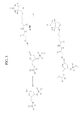
- Reactive oxygen species (ROS) induce at least twenty common adducts to purines and pyrimidines with 8-oxoguanine (8-oxodG) and 5-formyldeoxyuridine (5-FodU) ( Fig.1 ) among the most prominent products.
- Altered base pairing by 8-oxodG, 5-FodU and other lesions can induce mutations and promote cancers including those of breast, prostate, bone marrow, brain, lung, skin, colon, kidney, bladder, and others.
- Gray unit "Gy” of absorbed radiation induces many thousands of base lesions along with double strand breaks in each cell, suggesting that dosimetry by direct measurement of base damage might be practical were a rapid, robust and accurate assay available.
- a promising approach is direct chemical detection of oxidative lesions.
- the reactivity of the aldehyde moiety of 5-FodU makes it a particularly attractive target because it is the only aromatic aldehyde to be identified in vivo and therefore is ideal as a biomarker.
- Indirect methods include detection of glycosylase-sensitive sites by alkaline elution or comet assay. Both are compatible with analysis of cells rather than purified DNA, but neither offer sufficient sensitivity nor quantitation.
- Recent approaches such as detection anti-8-oxoguanine monoclonals ( Bruskov et al., Biochemistery 64:803, 1999 ; Park et al., PNAS 89:3375, 1992 ), Fabs ( Bespalov et al., Biochemistry 35:2067, 1996 ; Soultanakis et al., Free Radic. Biol. Med. 28:987, 2000 ) or binding of avidin ( Struthers et al., Anal. Biochem.
- ARP Aldehyde Reactive Probe
- reaction condenses a biotinylated aminoxy reagent to covalently link the biotin moiety to the oxidized DNA, and is amenable to quantitation ( Kurisu et al., Nucleic Acid Res. Suppl. 45, 2001 ) and ELISA format ( Kow and Dare, Methods 22:164, 2000 ).
- ARP also efficiently reacts with 5-FodU in DNA (Ide et al., 1993 supra ) and it is possible that a significant fraction of the reactivity for ARP may derive from 5-FodU rather than abasic sites.
- ARP is commercially available from Dojindo (Rockville, MD) and Invitrogen/Molecular Probes (San Diego, CA) and detection is performed by addition of either a streptavidin-HRP conjugate or a streptavidin-fluorophore conjugate.
- This method can be highly sensitive and has been used to detect individual lesions on single DNA molecules ( Hirose et al., Photochem. Photobiol. 76:123, 2002 ; Kim et al., FEBS Lett. 555:611, 2003 ). However, it is a multi-step method and subject to background from other aldehydes such as carbonyl-modified proteins ( Chavez et al., Anal. Chem. 78:6847, 2006 ).
- the binding between biotin and streptavidin has high sensitivity and specificity, but streptavidin is bulky and tetravalent, so that binding of a single streptavidin can mask several proximal biotins which can be expected at sites of cluster damage.
- Direct detection via Fluorescent Aldehyde Reactive Probe (“FARP", 5-(((2-(carbohydrazino)-methyl)thio)acetyl)aminofluorescein, aminooxyacetyl hydrazide Chakrabarti et al., Int. J. Radiat. Biol. 75: 1055, 1999 ; Makrigiorgos et al., Int. J. Radiat. Biol.
- FARP probes are available from Invitrogen/Molecular Probes (San Diego, CA).
- FRET fluorescent resonance energy transfer
- Methods for detecting DNA damage in a subject suspected of having DNA damage wherein the damage results in the formation of aldehyde moieties in DNA comprising the steps of, obtaining a DNA sample from the subject suspected of having DNA damage, combining the DNA sample with a fluorescent or pro-fluorescent, chromogenic or pro-chromogenic hydrazine compound to form a fluorescent DNA, detecting the fluorescent DNA or colored DNA by monitoring a fluorescent emission or saturation of color and quantitating the fluorescent emission or color saturation thereby determining the DNA damage in the subject.
- the DNA damage in the subject is the result of the subject being exposed to ionizing radiation, oxidizing chemicals or any radiation, substance or condition, whether environmental or otherwise, that causes or results in, the formation of aldehyde groups in DNA.
- the aldehyde moiety may be the result of conversion of a nucleotide base into an aldehyde containing compound.
- thymidine can be converted by ionizing radiation into 5-formyldeoxyuridine (5-FodU) which contains a free aldehyde that can form a hydrazone bond with a hydrazine compound.
- the hydrazine compound in another embodiment can be fluorescent or pro-fluorescent, chromogenic or pro-chromogenic. If the compound is pro-fluorogenic it may be 2-hydrazinoquinoline, 3-methyl-2-benzothiazolinone, 6-hydrazinonicotinamide or 4-hydrazinostilbazole.
- the DNA damage may be detected in a variety of samples including but not limited to isolated DNA, a living or dead cell or a tissue sample.
- Figure 1 Two-step mechanism for the conversion of thymidine to 5-formyl-deoxyuridine by oxygen free radicals.
- Figure 2 Expected reaction of the aromatic hydrazine 4-hydrazinostilbazole with 5-FodU to form a fluorescent bis-arylhydrazone.
- FIG. 3 Scheme for labeling a basic site using the Aldehyde Reactive Probe (ARP).
- ARP Aldehyde Reactive Probe
- Figure 4 Derivatization of 5-FodU with phenylhydrazine to form a bis-arylhydrazone.
- FIG. 5 Schematic representation of the chromogenic/fluorogenic assay wherein 5-FodU-modified DNA (B) is immobilized on aromatic hydrazine beads (A) to concentrate the 5-FodU DNA by immobilization in Step 1 (C) followed by addition of pro-chromogenic/pro-fluorescent aromatic hydrazine in Step 2 to produce a chromogenic or fluorescent signal (D).
- Figure 7 Scheme for the synthesis of 5-FodU phosphoramidites as described by Sugiyama ( Zhang et a/. Nucleic Acids Res. 25:3969, 1997 ).
- Figure 10 Diagrammatic representation of pro-fluorophore binding to 5-FodU and fluorescent emission.
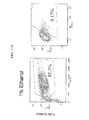
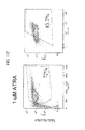
- Figure 11 In vitro differentiation of the HL60 promyelocytic leukemia cell line to neutrophils with all transretinoic acid, Vitamin D3 and G-CSF. Flow cytometry for forward scatter (upper row, viability) and CD 11b (lower row, neutrophil differentiation marker).
- Figure 12 Scheme for the reaction of FARP, probe, Alexa Fluor® 488 hydroxylamine and 5-FodU DNA.
- the present invention describes methods and compositions for the detection and quantitation of damage to DNA caused by ionizing radiation, oxidizing chemicals or cellular defects comprising obtaining a biological sample from a subject suspected of being subjected to ionizing radiation, oxidizing chemicals or event resulting in cellular defects, mixing the biological sample with a chromogenic/pro-chromogenic or fluorogenic/pro-fluorogenic hydrazine compound, measuring the chromogenic of fluorogenic signal thereby produced and determining the extent of DNA damage.
- aldehyde refers to a compound of the formula R-CHO wherein R comprises DNA.
- DNA damage refers to damage which results in the formation of one or more aldehyde moieties in DNA.
- ionizing radiation causes conversion of thymidine in DNA to 5-fonnyldeoxyuridine which contains a free aldehyde group.
- hydrazine refers to a compound having the formula R-NH-NH 2 wherein R is an aromatic moiety.
- the aromatic moiety may be fluorescent or chromogenic.
- the aromatic moiety may be pro-fluorescent or pro-chromogenic.
- pro-fluorescent refers to a compound that may be fluorescent but will have a fluorescent signal less than the fluorescent signal of the hydrazone formed when the pro-fluorophore binds to the aldehyde of the damaged DNA.
- the pro-fluorophore signal may shift to a higher or lower emission frequency when forming the hydrazone bond with the damaged DNA.
- pro-chromogenic refers to a compound that emits in the visible range and may have little or no color before interacting with the hydrazine compound but will have an enhanced or increased saturation of color upon forming the hydrazone bond with the damaged DNA.
- the pro-chromogenic compound may shift in color when forming the hydrazone bond compared to the non-bound state.
- ionizing radiation refers to any radiation having sufficient energy to cause the formation of an aldehyde moiety in DNA. This includes high energy radiation such as ⁇ , neutron or ⁇ radiation but can include lower energy radiation such as X-ray or ultraviolet radiation as well.
- oxidizing chemical refers to any compound that is capable of forming aldehyde moieties within DNA.
- sample refers to any bodily fluid or tissue sample that may contain DNA. These include living or dead cells, tissue samples, as well as blood or serum, saliva, semen, and the like.
- subject refers to any animal species such as for example a mammal, a bird, a reptile, an amphibian or a fish.
- animal is a mammal and most preferably a human.
- oxidizing chemicals and other events that result in cellular defects often modify DNA and in some instances form reactive groups that are able to interact with a pro-fluorescent/pro-chromogenic aromatic hydrazine to yield a florescent/colored product.
- Monitoring and quantitating this product can provide information on the extent of DNA modification and correspondingly damage.
- the weakly blue fluorescent aromatic hydrazine, 4-hydrazino-stilbazole reacts with 5-FodU to form a fluorescent bis-arylhydrazone product that absorbs in the orange frequency range ( Fig.2 ).
- this hydrazine has a little or no reactivity with undamaged DNA.
- DNA oxidation products caused by ionizing radiation Reactive oxygen species (ROS) and covalent modification of DNA mediate most of the effects of ionizing radiation on biological targets and are responsible for a wide range of health effects including aging and cancer ( Cooke et al., FASEB J. 17:1195, 2003 ; Evans et al., Mutat. Res. 567:1, 2004 ).
- ROS reactive oxygen species
- the interaction of energetic electromagnetic radiation, or particles, with water yields abundant hydroxyl radicals along with other H atoms and hydrated electrons. Higher energy radiation sheds a correspondingly higher density of radicals along the particle track.
- Hydroxyl radicals react rapidly with DNA at double bonds, methyl groups and sugar ring carbon atoms yielding radicals that further react with water, oxygen or other targets to cause a wide range of lesions, including strand breaks, abasic sites, modified bases and DNA-DNA or DNA-protein crosslinks.
- ⁇ particle, neutron or ⁇ radiation as might be expected from a radiation accident or nuclear attack, the density of hydroxyl radicals is sufficient to produce numerous double strand breaks and highly clustered damage, with multiple base lesions spread over a few contiguous residues on either strand. Repair of clustered damage can yield additional double strand breaks. While X-rays, ⁇ particles, ultraviolet light and other less energetic radiation produce fewer double strand breaks, the same free-radical mediated reactions occur but leaving behind widely distributed strand breaks and base adducts.
- 5-FodU Exposure of thymine in the context of DNA, or as a free nitrogenous base, nucleoside or nucleotide to hydroxyl radicals in the presence of oxygen, can yield 5-FodU as a prominent product ( Fig. 1 , ( Bjelland et al., Mutat. Res. 486:147, 2001 ; Bjelland et al., Biochemistry, 34:14758, 1995 ; Liu et al., DNA Repair 2:199, 2003 )). 5-FodU is produced with similar abundance to 8-oxodG after 60 Co irradiation, as first described by Kasai et al. (Mutat. Res. 243:249) in 1990 .
- 5-FodU is a common adduct in chromosomal DNA after radiation exposure, but given the size of nucleotide pools, a significant source of 5-FodU in both DNA and RNA is likely via oxidation of free thymine. Ionizing radiation may result in accumulation of 5-formyl deoxyuridine triphosphate ("5-FodUTP") and incorporation during chromosomal replication and/or repair synthesis ( Klungland et al., Toxicol. Lett. 119:71, 2001 ).
- 5-FodUTP 5-formyl deoxyuridine triphosphate
- 5-FodU is weakly mutagenic, and presumably mediates some of the carcinogenic effects of ionizing radiation. Indeed, 5-FodUTP induces both transition and transversion mutations in cells, presumably via misincorporation and/or mispairing during replication.
- the Ntg1 and Ntg2 serve as the 5-formyluracil deglycosylases and AP lyases, leaving a nick adjacent to the position of the oxidized base ( Zhang et al., Int. J. Radiat. Biol. 79:341, 2003 ).
- human cells express hNTH1 and hNEIL1 deglycosylaselAP lyases that remove 5-FodU from artificial substrates and nick the DNA ( Matsubara et al., Biochemistry 42:4993, 2003 ; Miyabe et al., Nucleic Acids Res. 30:3443, 2002 ; Zhang et al., DNA Repair 4:71, 2005 ).
- badge dosimeters Currently, employees working in proximity to nuclear reactors or instruments and/or protocols that use radioactivity such as X-ray machines wear one of two types of badge dosimeters. They are X-ray film-based dosimeters or thermo-luminescent-based dosimeters. Unfortunately, this does not provide immediate real time detection of DNA damage. Such badges cannot be quickly scanned to identify the extent of potential DNA damage.
- the present invention provides a real time biomimetic dosimeter comprised of a packet, or other enclosure, in a badge-type format that contains purified DNA, or preferably poly-T DNA, combined in a matrix with a pro-fluorescent aromatic hydrazine that on exposure to ionizing radiation converts the thymidine bases within the DNA to 5-formyl-deoxyuridine (5-FodU). Since 5-FodU reacts with the aromatic hydrazines forming a fluorescent bis-arylhydrazone as described above, the fluorescent product can be detected using a fluorescence reader ( Fig. 10 ). As the quantity of 5-FodU produced is proportional to the amount of exposure to ionizing radiation the fluorescence level similarly quantifies the exposure.
- This badge unlike the X-ray film based dosimeter can be re-used following reading. For example, by recording the amount of radiation damage from an initial exposure, that recorded exposure can be utilized as the baseline for the next determination of damage in a subsequent exposure.
- ARS Acute radiation syndrome
- ARS is a multi-organ stress response that occurs after whole-body or significant partial-body irradiation of greater than 1 Gy delivered at a relatively high dose rate
- ARS oxidative stress protein
- GI gastrointestinal
- cerebrovascular syndromes a combination of hematopoietic, gastrointestinal, and cerebrovascular syndromes. Symptoms of acute radiation are dependent on the absorbed dose and may appear within hours to days, but follow a characteristic sequence. Patients with a total body exposure of> 1 Gy will develop dose dependent, increasingly severe aspects of the hematopoietic syndrome. Because bone marrow progenitors are unable to divide after exposure of 2 to 3 Gy, a hematologic crisis may ensue, characterized by pancytopenia, infection, bleeding, and poor wound healing.
- Lymphocyte depression is maximal at two weeks, and may take several months to recover. Time to nadir is inversely proportional to dose, and depth and duration are proportional to dose. Platelet production decreases and slowly recovers, leading to a period of thrombocytopenia.
- Neutrophils serve a major role in innate immunity and typically comprise 70% of circulating leukocytes, totaling 2,500-7,500 cells/ ⁇ l and neutropenia below 1000 cells/ ⁇ l strongly predisposes to infection. Neutropenia develops over the first week and full recovery may take several months. To head off sepsis, treatment with granulocyte colony stimulating factors to restore neutrophil numbers along with broad-spectrum prophylactic antimicrobial agents is considered standard therapy during the neutropenic period.
- the increasing damage to other organs gains greater significance.
- the breakdown of the intestinal mucosal barrier results in abdominal pain, diarrhea, nausea and vomiting, and predisposes patients to bowel obstruction, gastrointestinal bleeding, and infection.
- Systemic effects may include malabsorption resulting in malnutrition, dehydration, electrolyte imbalance, and acute renal failure.
- Loss of intestinal epithelial integrity enhances the hematopoietic syndrome as inflammation depletes neutrophils and leakage of bacteria promotes local infection and significantly enhances the likelihood of sepsis.
- the lungs are another survival limiting organ target.
- Acute radiation pneumonitis develops two to six months after exposure and is manifested by cough, dyspnea, and respiratory difficulties. Progressive and irreversible pulmonary fibrosis may develop even if the patient initially was asymptomatic. Survival becomes less likely with increasing dose and victims exposed to 10 Gy suffer rapid, multi-organ failure, a situation incompatible with long-term survival.
- the pro-chromogenic/pro-fluorescent technology to detect 5-FodU damage provides the following advantages: (i) the reaction proceeds in biological media, i.e. PBS yielding a stable chromogenic/fluorogenic product wherein 5-FodU is a component of the product; (ii) favorable kinetics of reactivity between 5-FodU and the pro-fluorescent aromatic hydrazine catalyzed by aniline; (iii) limited steric encumbrance following covalent linking of the pro-fluorescent aromatic hydrazine to 5-FodU; (iv) a basis for a homogeneous assay i.e.
- an assay in which a washing step is not required (v) a basis for a assay high throughput assay that can be incorporated into existing instrumentation; (vi) low limits of detection and an extended linear range; (vii) a basis for use in multiple platforms such as immunohistochemistry, flow cytometry and ELISA and (viii) a field test assay such as a "dipstick" test requiring minimal technical training.
- FIG. 5 is a schematic depiction of the assay wherein 5-FodU-modified DNA is concentrated by immobilization on a pro-fluorescent aromatic hydrazine bead, a zone on a lateral flow strip or the tip of a fiber optic cable. Subsequently, solution phase pro-fluorescent aromatic hydrazine is added to react with the remaining 5-FodU moieties thereby increasing the signal.
- a library of aromatic hydrazines can be prepared and their absorbance and fluorescent properties determine to select one or more with the optimal properties for the detection of 5-FodU in the desired assay.
- the chromogenic assay is adaptable to a rapid self-assessment assay to determine radiation exposure over 2 Gy. Since this assay does not employ any biological reagents and the signal is visual, it satisfies all the requirements for functionality in adverse conditions, convenience of use, shelf-life and ease of manufacturing.
- the fluorogenic assay in association with a portable fluorescent reader is adaptable to determining levels of radiation and usable under adverse conditions. Furthermore, the fluorogenic assay is readily adaptable to high-throughput assays in a laboratory environment based on fluorescent multi-plate robotic instruments or flow cytometry.
- qPCR-Based Assay a quantitative polymerase chain reaction (“qPCR") based assay using the following multi-step protocol can be performed robotically, (e.g. Kingfisher (ThermoFisher) multiwell magnetic bead robot Pittsburgh, PA). Such an assay would include the following steps:
- the high sensitivity of a qPCR-based assay will allow inspection of samples from non-invasive sources such as hair, sputum and urine. It is known that DNA fragments are present in urine (transrenal DNA) and have been used as biomarkers of diseases which can be detected by PCR.
- 5-FodU is synthesized as described in the literature.
- 5-Di-O-acetyl-thymidine is oxidized to di-O-acetyl-5-formyl-deoxyuridine by CuSO 4 /K 2 S 2 O 8 /2,6-lutidine/aqueous acetonitrile/65° C/2 hours in ⁇ 52% yield as described by Ono et al. , 1994, supra.
- De-acetylation using aqueous acetic acid yields 5-FodU which is then condensed with a variety of structurally diverse commercially available hydrazines as shown in Fig. 6 .
- Reaction kinetics are examined with and without 10-100 mM aniline and with respect to pH, temperature, etc.
- the hydrazones are then isolated and their structures confirmed by 1 H nuclear magnetic resonance (" 1 H NMR") and mass spectroscopy.
- the fluorescent properties of this panel of 5-FodU hydrazones are evaluated with respect to their absorbance and emission wavelength, quantum yields and photostability.
- Oligonucleotides are prepared bearing 5-FodU moieties.
- Sugiyama Zhang et al., Nucleic Acids Res. 25:3969, 1997
- Sugiyama Zhang et al., Nucleic Acids Res. 25:3969, 1997
- All intermediates synthesized are characterized by 1 H NMR and mass spectroscopy.
- 5-FodU substituted oligonucleotides are ultimately isolated by periodate oxidation of the 5-(1,2-dihydroxy)-deoxyuracil-containing oligonucleotides.
- PAGE gel and mass spectral data are obtained for all oligonucleotides synthesized.
- Oligonucleotides are synthesized with single and multiple 5-FodU moieties ( Fig. 8 ), as a model for dispersed and clustered damage.
- Complementary oligonucleotides with A or G opposite the 5-FodU sites are synthesized to determine how the 5-FodU base-pairing alters reactivity.
- the single stranded and double stranded oligonucleotides are treated with aromatic hydrazines including 2-hydrazinoquinoline, 3-Methyl-2-benzothiazolinone hydrazone, 6-hydrazinonicotinamide, 4-hydrazinostilbazole and the adducts are evaluated to examine the effects on extinction coefficient and fluorescent characteristics of the hydrazone.
- aromatic hydrazines including 2-hydrazinoquinoline, 3-Methyl-2-benzothiazolinone hydrazone, 6-hydrazinonicotinamide, 4-hydrazinostilbazole and the adducts are evaluated to examine the effects on extinction coefficient and fluorescent characteristics of the hydrazone.
- the products are analyzed by gel electrophoresis, mass spectrometry and fluorescence.
- Comparison of A, B, and C reveal the overall impact on Tm of the unmodified oligonucleotide vs. a modification near the end vs. a modification in the middle. It will also allow comparison of fluorescent signal when the fluorophore is located at the end vs . the middle of the oligonucleotide.
- Comparison of C, D, E, and F Fig. 8 ) reveals the extent of fluorophore-fluorophore interactions (positive or negative) for in-phase versus out-of-phase positions, and distance-dependence (10bp vs. 20bp apart) of in-phase fluorophore-fluorophore interactions. Comparison of B and F vs .
- G Fig. 8
- C and G vs . H Fig. 8
- Tm and background fluorescent signal determinations can be observed from evaluation of control oligonucleotide I.
- 5-FodU triphosphate is synthesized by chemical means ( Armstrong et al., Biochemistry 15:2086, 1976 ).
- 5-FodUTP is doped into polymerase chain reaction (“PCR") amplification reactions at increasing percentages and the level of incorporation determined.
- PCR polymerase chain reaction
- Several methods to detect 5-FodU in PCR products are utilized including gas chromatography/mass spectroscopy (“GC/MS”) ( Douki et al., Chem. Res. Toxicol. 9:1145, 1996 ), using the synthesized 5-FodU as a standard.
- GC/MS gas chromatography/mass spectroscopy
- PCR products are reacted with Aldehyde Reactive Probe (ARP, N-(aminooxyacetyl)-N'-(D-biotinoyl) hydrazine) before and after treatment with deglycosylating enzymes or incubation at 100° C to form abasic sites, to determine reactivity of 5-FodU vs. abasic sites to this probe.
- ARP Aldehyde Reactive Probe
- Biotin incorporation from ARP is examined by HABA assay and/or FluoReporter Biotin Quantitation Assay (Invitrogen, San Diego) to determine sensitivity and specificity as a reference.
- Fluorescent hydroxylamine (“FARP") reagents such as Alexa Fluor 488 hydroxylamine (Invitrogen, San Diego), are also examined for sensitivity and specificity. Then, the 5-FodU labeled amplicons is reacted with pro-fluorophore hydrazines including 2-hydrazinoquinoline, 3-methyl-2-benzothiazolinone hydrazone, 6-hydrazinonicotinamide, and 4-bydrazinostilbazole and the fluorescence of the products is quantified and the results correlated with the digestion results. The efficiency and kinetics of fluorescent labels are examined with respect to such variables as pH and temperature. In addition, the minimum detectable level of 5-FodU that can be detected by the pro-fluorophore technology is determined.
- FARP Fluorescent hydroxylamine
- Detection of formation of 5-FodU is determined by established methods using GC-MS or HPLC-MS/MS (e.g. ( Cadet et al., Free Radic. Biol. Med. 33:441, 2002 ; Douki et al., 1996, supra; Hong and Wang, Anal. Chem. 79:322, 2007 )). Chemically synthesized 5-FodU, 5-FodUTP and 5-FodU containing oligonucleotides are utilized to develop methods and provide calibration standards. Then, the irradiated DNA is reacted ARP as described above, to examine sensitivity and specificity of detection.
- the irradiated DNA is treated with pro-fluorophore hydrazines including 2-hydrazinoquinoline, 3-methyl-2-benzothiazolinone hydrazone, 6-hydrazinonicotinamide, and 4-hydrazinostilbazole.
- pro-fluorophore hydrazines including 2-hydrazinoquinoline, 3-methyl-2-benzothiazolinone hydrazone, 6-hydrazinonicotinamide, and 4-hydrazinostilbazole.
- the fluorescence enhancement, Stokes shift and reaction kinetics is quantified and the results correlated with the ARP probe and the GC-MS results.
- Controls are screened for background reactivity for the pro-fluorophore hydrazines including 2-hydrazinoquinoline, 3-methyl-2-benzothiazolinone hydrazone, 6-hydrazinonicotinamide, and 4-hydrazinostilbazole, with and without 100 mM aniline, and compare them to the commercially available reagents including ARP and FARP (Invitrogen, San Diego, CA), anti-8-OxodG antibody and the OxyDNA and/or OxyFLOW kits (Biotrin, Dublin, Ireland, HemoGenix, Colorado Springs, CO).
- pro-fluorophore hydrazines including 2-hydrazinoquinoline, 3-methyl-2-benzothiazolinone hydrazone, 6-hydrazinonicotinamide, and 4-hydrazinostilbazole, with and without 100 mM aniline, and compare them to the commercially available reagents including ARP and FARP (Invitrogen, San Diego, CA), anti-8-O
- Adherent cells A549 (lung), MCF-7 (breast), SCC6I (head and neck) and non-adherent HL60 (PML) and K562 (CML) tumor cell lines, are wash, permeabilized and fixed with paraformaldehyde, and then wash in PBS. The adherent lines are used for imaging and the non-adherent lines are used for flow cytometry and multi-well fluorimetry. Reagents are added; the cells are incubated, washed with PBS and screened by fluorescence imaging, flow cytometry and/or 96 well plate fluorimetry.
- the cells are treated with a mixture of 100 ⁇ M FeSO 4 and 100 ⁇ M H 2 O 2 for 0, 10, 30 and 60 minutes and then repeat the analysis to detect enhanced reaction with pro-fluorophore hydrazines and the commercial alternatives ARP, FARP, anti-8-OxodG antibody and the OxyDNA and/or OxyFLOW kits.
- the adherent cells are stained with a DNA counterstain (e.g. Syber Green) and examined by microscopy to determine whether the fluorescent product demonstrates localization, primarily or exclusively to the nucleus.
- a DNA counterstain e.g. Syber Green
- the highest intensity of fluorescence is in the nucleus following treatment with hydroxyl radicals.
- the aromatic hydrazines are specific to aromatic aldehydes. Thus fluorescence is low and nuclear localized in the controls and restricted to nucleic acids in oxidized cells. Cytoplasmic staining is due primarily to oxidized ribonucleic acid ("RNA"). Consequently cells were treated with RNAse prior to staining.
- the cells are washed, permeabilized and fixed with paraformaldehyde, and then wash in PBS to assess oxidative damage to chromosomal DNA.
- cells are irradiated in the 60 Co irradiator at doses of 0 to 50 Gy. Aliquots of ⁇ 10 6 cells are removed from each sample for DNA extraction and conventional analysis of oxidative damage by GC/MS. The 5-FodU content and kinetics of removal from DNA are then determined. For staining, cells are fixed with paraformaldehyde.
- Sections are prepared by placing them onto slides for conventional H & E analysis. Background binding is determined by treating the sections with pro-fluorophore hydrazines with and without aniline. The reagents are added, and the slides are incubated, washed and assayed by fluorescence microscopy to detecting damage in situ. Sections of fixed and embedded tissue are examined using FARP, anti-8-OxodG antibodies, or the OxyDNA kit and compared to staining with pro-fluorophore reagents. The sections are then subjected to fluorescence microscopy, counterstaining with a DNA stain to detect nuclei, and examined to identify localization and sensitivity of staining.
- Tumor cells are examined and their staining compared to staining of the surrounding stroma and normal tissue.
- Pro-fluorescence reagents displayed a particularly significant fluorescent signals in these applications. Background binding of the reagents to the embedding material, tissue matrix did not result in significant background, unlike other reagents that are fluorescent and require extensive washing.
- sections are incubated with a mixture of 100 ⁇ M FeSO 4 and 100 ⁇ M H 2 O 2 for 0, 10, 30 and 60 minutes and then the analysis repeated to detect enhanced reaction with pro-fluorophore hydrazines and the commercial alternatives ARP, FARP, anti-8-OxodG antibody and the OxyDNA kit. Positive results with the pro-fluorophore hydrazines are then evaluated to assess their use without additional washing steps as a direct stain for oxidative damage. Finally, sections from tumors excised under conditions that may have induced elevated levels of ROS are examined and stained to determine if this will be reflected in higher staining of pro-fluorophore hydrazine probes or the commercial alternatives.
- the human promyelocytic leukemia cell line HL60 is a granulocyte progenitor that can be induced to differentiate into functional neutrophils by treatment with all-trans retinoic acid ("ATRA") ( Breitman et al. Chem Res. Toxicol. 9:115, 1980 ) .
- HL60 cells are used as a neutrophil cell model to develop and optimize our DNA damage detection and quantitation methods.
- HL60 cells were grown to confluence (1 x 10 6 cells/ml) in Roswell Park Memorial Institute media ("RPMI") 1640 media supplemented with 10% fetal bovine serum, 1% penicillin/streptomycin and 2 mM L-glutamine at 37 °C in 5% CO 2 and treated with ethanol carrier, 1 ⁇ M ATRA or 1 ⁇ M ATRA, 6 nM Vitamin D 3 and 30 ng/ml G-CSF for five days. Differentiation was confirmed by expression of the neutrophil-specific membrane receptor CD11b.
- RPMI Roswell Park Memorial Institute media
- CD11b mean fluorescien isothiocyanate
- SSC mean SSC
- the data shown are gated to remove a significant fraction of apparently apoptotic cells in the differentiated cell populations.
- Gross morphological changes suggestive of neutrophil differentiation are apparent in the differentiated HL60 cells as revealed by Wright's-Giemsa stain, including an increase in the cytoplasm to nucleus ratio, a reduction in cell size and the characteristic increased lobularity of the nucleus (not shown).
- 5-FodU labeled DNA Chromosomal DNA is labeled with 5-FodU by several methods, including chemical oxidation, irradiation and incorporation in living cells. Salmon sperm DNA is dissolved in water at > 10 mg/ml. Chemical oxidants such as peroxide or iron based Fenton reagents readily oxidize the 5-methyl group of thymine in solution or in DNA to an aromatic aldehyde, forming 5-FodU. Methods developed by Kasai et al.
- 5-FodU was added to tissue culture media for cancer cell lines, including HL60 promyelocytic leukemia cells, K562 myeloid leukemia cells and one or more adherent cell lines such as HeLa and MCF-7 and maximum tolerated doses were determined, where cell growth rate, as judged by time to confluence, is increased by less than two fold and cell death, as judged by ethidium bromide exclusion, is less than doubled.
- Cells are split and then placed into media at this 5-FodU concentration and allowed to reach confluence, and then DNA is prepared from these cells using Qiagen DNAEasy Blood & Tissue genomic DNA purification kits (Hilden, Germany). The concentration of the DNA is determined by OD 260nm .
- the DNA is stored at -80° C to reduce the rate of depurination, adduct formation or other events.
- DNA is treated with restriction enzymes or sonication and separated by TBE agarose gel electrophoresis to determine the extent of labeling of intact DNA.
- DNA is transferred to nylon membranes and probed with fluorescent avidin.
- the gel is imaged in a fluorescence flat bed scanner.
- Specificity of detection by ARP and FARP is examined by adding competing aldehydes such as simple aliphatic aldehyde, protein carbonyls and lipid aldehydes to the labeled DNA.
- ARP and FARP results are compared to the GC-MS and/or HPLC-MS/MS data to determine relative sensitivity, specificity and linearity of these methods.
- Controls were screened for background reactivity for the pro-fluorophore hydrazines including 2-hydrazinoquinoline, 3-methyl-2-benzothiazolinone hydrazone, Xanthen-ylidene hydrazine, and 4-hydrazinostilbazole, with and without 100 mM aniline, and compared to commercially available reagents including ARP and FARP (Invitrogen, San Diego, CA), anti-8-OxodG antibody and the OxyDNA and/or OxyFLOW kits. The reagents were added, incubated and washed with PBS and screened by fluorescence imaging, flow cytometry and/or 96 well plate fluorimetry.
- One or more aromatic hydrazines will display higher signal to background and more rapid kinetics and greater consistency when evaluated as a ratio between a total DNA stain and the 5-FodU reactivity, when evaluated by flow cytometry.
- the DNA stain will have to be at a relatively low stoichiometry to avoid cross-talk and probe-probe interactions, such as fluorescence energy transfer and quenching.
- the sensitive and specificity for 5-FodU is evaluated using the advanced flow cytometers at the University of Chicago.
- a point-of care flow cytometer based assay was evaluated on an Accuri benchtop flow cytometer (Accuri Cytometers, Ann Arbor, MI) equipped with standard laser and filters. The optics is adapted to optimize detection and enhance signal to noise ratio.
- Non-adherent HL60 and K562 myeloid cell lines are washed, permeabilized and fixed with paraformaldehyde, and then washed in PBS. Free radical damage is induced in the fixed tissue culture cells, by treatment with a mixture of 100 ⁇ M FeSO 4 and 100 ⁇ M H 2 O 2 for 0, 10, 30 and 60 minutes. The analysis is then repeated to detect enhanced reaction with pro-fluorophore hydrazines and the commercial alternatives ARP, FARP, anti-8-OxodG antibody and the OxyDNA and/or OxyFLOW kits. The suspension culture cells are stained with a DNA counterstain (e.g.
- Flow cytometry is then performed, plus and minus RNAse, using a DNA counterstain such as Syber Green to provide a ratio of staining to DNA content, and evaluate homogeneity of staining and compare the intensity distributions of populations of control and oxidized cells, to compare the hydrazines and commercial reagents.
- a DNA counterstain such as Syber Green
- Living cells are irradiated in the 60 Co irradiator ( ⁇ 1.5 Gy/min) at doses of 0 to 50 Gy. Aliquots of ⁇ 10 6 cells are removed from each sample for DNA extraction and conventional analysis of oxidative damage by GC/MS. 5-FodU content and kinetics of removal from DNA are determined. For staining, cells are fixed with paraformaldehyde. An aliquot is stained with FARP, anti-8-OxodG antibody and the OxyDNA and/or OxyFLOW kits using manufacturer's protocols, and analyzed by quantitative imaging and flow cytometry. The aromatic hydrazine probes to detect 5-FodU are examined each, with and without aniline, to accelerate kinetics.
- 5-FodU formation by ionizing radiation is detected by established methods for detection, GC-MS or HPLC-MS/MS (e.g. Hong and Wang Anal. Chem., 2007 supra, Douki et al., 1996, supra and Cadet et al., 2002, supra).
- aromatic hydrazines including 2-hydrazinoquinoline, 3-methyl-2-benzothiazolinone hydrazone, 6-hydrazinonicotinamide, and 4-hydrazinostilbazole.
- the fluorescence enhancement, Stokes shift and reaction kinetics is quantified and the results correlated with the ARP probe and the GC-MS results.
- Suspension culture cells are stained with a DNA counterstain (e.g. Syber Green) and examined by microscopy to determine whether the fluorescent product demonstrates localization, primarily or exclusively to the nucleus.
- a DNA counterstain e.g. Syber Green
- Some cytoplasmic staining may be due to oxidized RNA. Consequently, the cells are treated with RNAse prior to staining. Under these conditions staining of cells using hydrazines is substantially better than commercial reagents.
- HL60 cell differentiation and purification Neutrophils are both a readily accessible cell type for biodosimetry and a significant target tissue in ARS. Consequently, HL60 cells are a neutrophil model for biodosimetry. While this promyelocytic leukemia cell line may not be an optimal model, the ability to reproducibly grow large numbers of cells, and differentiate them into functional neutrophils makes this a powerful system. Because of unexpected results that might occur when differentiating HL60 cells while simultaneously labeling them metabolically with 5-FodU only differentiated, HL60 cells were used in this investigation to validate pro-fluorophore detection of 5-FodU in irradiated cells.
- Human promyelocytic leukemia HL60 cells are cultured in RPMI 1640 media supplemented with 10% FBS, 1% Pen/Strep and 2 mM GT at 37° C with 5% CO 2 at a culture density maintained at 10 5 to 10 6 cells/ml.
- Confluent cultures at 1 x 10 6 cells/ml are subjected to a standard protocol to induce differentiation into functional neutrophils by incubation with 1 ⁇ M ATRA, 6 pM 1,25-dihydroxy Vitamin D3 and 30 ng/ml GCSF (Bunce et al. Leukemia 9:410, 1995). Differentiation is evaluated and the assay optimized balancing extent of differentiation against loss of cells to apoptosis.
- Density gradient centrifugation is used to isolate the differentiated neutrophils from apoptotic cells, cell debris and any undifferentiated cells.
- Several methods have been described using Percoll, Ficoll, Hypaque and/or Nycodenz gradients (e.g. Ford et al. Blut 54:337, 1987 , McFaul J. Immunol. Methods 130:15, 1990 , Kalmer et al. J. Immunol. Methods 110:275, 1988 , Kjeldsen et al. J. Immunol. Methods 232:131, 1999 , Lichtenberger et al. J. Immunol. Methods 227:75, 1999 and Kouoh et al. Biol. Pham. Bull.
- Neutrophils from anticoagulated human blood Anticoagulated venous blood obtained from patients at the University of Chicago Adult Primary Care service and to be discarded after hematocrit and differential are obtained as needed.
- High yields of unactivated neutrophils are obtained by one of several validated protocols for purification of neutrophils using gradient centrifugation as described above.
- the one-step gradient method can be optimized to obtain a >50% yield with >95% purity. Neutrophils are then examined for viability and function prior to analysis.
- each of differentiated HL60 cells, or human neutrophils are subjected to 60 Co irradiation at doses from 0 to 10 Gy.
- Aliquots of each sample are subjected to DNA extraction and the content of 5-FodU determined by conventional analysis of oxidative damage by GC/MS.
- aliquots are removed and fixed with paraformaldehyde.
- An aliquot is stained to detect 8-OxodG with anti-8-OxodG antibody and the OxyDNA and/or OxyFLOW kits using the manufacturer's protocols, and analyzed by quantitative imaging and flow cytometry.
- the pro-fluorophore probes are then examined to detect 5-FodU, using standard conditions developed above.
- the reagents are added and fluorimetry, quantitative imaging, and/or flow cytometry are used to develop plots of fluorescence enhancement vs. radiation dose, to determine sensitivity and linearity and to compare performance.
- Conditions are optimized for flow cytometric detection as well as the requirement for proteolysis, RNAse treatment, and/or other extractions to enhance detection and quantitation, for adapting the assay to the Accuri benchtop flow cytometer.
- Detection of 5-FodU in living tissue culture cells and neutrophils are labeled with 5-FodU by metabolic incorporation using aromatic hydrazines in the media or buffer, with and without aniline.
- the cells are screening to determine those hydrazines judged to be most promising as candidates for use in living cells. More specifically, the cells are screened for low background and high fluorescence enhancement at the excitation and emission maxima for the predicted hydrazone product using fluorimetry and fluorescence imaging. Changes in cell integrity and viability are examined during staining.
- 5-FodU formation in K562 and undifferentiated HL60 cells as well as differentiated HL60 and human neutrophils subjected to 60 Co irradiation at doses from 0 to 50 Gy is detected.
- the living cells are then assayed on the Accuri benchtop flow cytometer to detect 5-FodU and the optics further optimized to enhance detection of formation of the expected hydrazones.
- a condition of the successful development of a lateral flow test is the capture of the analyte in a zone on a test strip as the analyte flows through.
- Silica surfaces such as glass slides, coverslips, glass wool, controlled pore glass and/or silica beads are modified with HyNic-silane ( Fig. 9 ).
- HyNic-modified glass surfaces are exposed to synthetic 5-FodU in aqueous solution, with and without 10-100 mM aniline. Fluorimetry or fluorescent imaging is used to detect the formation of a fluorescent product on the surface.
- Figure 10 presents fluorescence properties of the hydrazone formed from the reaction of 4-formylbenzamide and 6-hydrazinonicotinamide.
- the bis-arylhydrazone formed from HyNic and 5-FodU has similar fluorescent characteristics and is detected using the 365 nm mercury line and standard "DAPI" fluorescence filters and optics in an epifluoreseence microscope.
- the surface based assay format is used to detect 5-FodU formed from irradiation of chromosomal DNA. Samples subjected to the full range of radiation doses and assay intact DNA, denatured DNA and DNA hydrolyzed with DNAse I are used. 5-FodU-based hydrazones are detected by fluorimetry and/or fluorescence imaging.
- RNA and DNA whole cell lysates of neutrophil-differentiated HL60 cells are prepared to capture the nucleotide pools, RNA and DNA, all of which may contain 5-FodU after irradiation.
- the cells are suspended in RIPA (1% Triton X100, 0.1% SDS, 1% deoxycholate) or another detergent lysis buffer. The cells are then disrupted with a needle and syringe, incubated at 4°C for 30 minutes and sonicated. Protease, DNAse and/or RNAse is added to fully solubilize the nucleic acids. Lysates from unirradiated cells and cells treated with doses from 0 to 10 Gy are prepared as stated above.
- Fluorimetry is utilized to examine the reactivity for pro-fluorophore hydrazines including 2-hydrazinoquinoline, 3-methyl-2-benzothiazolinone hydrazone, 6-hydrazinonicotinamide, and 4-hydrazinostilbazole, with and without 10-100 mM aniline.
- Background fluorescence is evaluated to examine sensitivity by adding synthetic 5-FodU and determining the limits of detection, linearity and kinetics.
- Cell lysates may contain a competitor or inhibitor that could increase fluorescent background, so that the solution phase homogeneous assay can be optimized to enhance the observed signal above background.
- Surface-based assay might be optimal with a whole cell lysate.
- cell lysates are applied to HyNic silane coated glass (as slides, coverslips, or beads) and the accumulation of fluorescent product on the glass surface over time is observed. Background, sensitivity for added synthetic 5-FodU and detection of irradiation is observed as described above. The effect of conditions such as aniline or buffers on kinetics is examined. The need to wash away the lysate prior to measuring fluorescence is determined for the assay.
Landscapes
- Health & Medical Sciences (AREA)
- Life Sciences & Earth Sciences (AREA)
- Engineering & Computer Science (AREA)
- Molecular Biology (AREA)
- Immunology (AREA)
- Biomedical Technology (AREA)
- Chemical & Material Sciences (AREA)
- Hematology (AREA)
- Urology & Nephrology (AREA)
- Cell Biology (AREA)
- Microbiology (AREA)
- Biotechnology (AREA)
- Food Science & Technology (AREA)
- Medicinal Chemistry (AREA)
- Physics & Mathematics (AREA)
- Analytical Chemistry (AREA)
- Biochemistry (AREA)
- General Health & Medical Sciences (AREA)
- General Physics & Mathematics (AREA)
- Pathology (AREA)
- Tropical Medicine & Parasitology (AREA)
- Measuring Or Testing Involving Enzymes Or Micro-Organisms (AREA)
Abstract
Description
- This application claims priority to
U.S. Provisional Application Serial No. 61/094,521, filed September 5, 2008 - The present invention relates to compounds and methods used to label biomolecules for detection and diagnosis. In particular, it relates to fluorescent and pro-fluorescent, chromogenic and pro-chromogenic, hydrazine compounds that when bound to deoxyribonucleic acid ("DNA"), detect damage caused by ionizing radiation, oxidizing chemicals and cellular defects.
- Oxygen free radicals mediate most ionizing radiation ("IR") damage by attacking DNA bases and the sugar-phosphate backbone. This damage may result as adducts, other covalent modifications and strand breaks that scale directly with IR dose and intensity. Reactive oxygen species (ROS) induce at least twenty common adducts to purines and pyrimidines with 8-oxoguanine (8-oxodG) and 5-formyldeoxyuridine (5-FodU) (
Fig.1 ) among the most prominent products. Altered base pairing by 8-oxodG, 5-FodU and other lesions can induce mutations and promote cancers including those of breast, prostate, bone marrow, brain, lung, skin, colon, kidney, bladder, and others. One Gray unit "Gy "of absorbed radiation induces many thousands of base lesions along with double strand breaks in each cell, suggesting that dosimetry by direct measurement of base damage might be practical were a rapid, robust and accurate assay available. A promising approach is direct chemical detection of oxidative lesions. The reactivity of the aldehyde moiety of 5-FodU makes it a particularly attractive target because it is the only aromatic aldehyde to be identified in vivo and therefore is ideal as a biomarker. - Extensive literature is available examining the strengths and limitations of different methods of detecting oxidative DNA damage, as reviewed in Cadet et al., Mutat. Res. 531:5 2003; Dizdaroglu et al., Free Radic. Biol. Med. 32:1102, 2002. For this approach to be used in the development of a rapid field capable biodosimetry test, challenges include sufficient sensitivity, specificity, speed and reproducibility to reliably detect as few as 1 lesion in 105 bases from a readily available source of cells. In turn, the lability of some lesions as well as the potential for oxidative damage during work-up for the assay provides additional challenges. Given the wide range of different chemical species described, comprehensive analysis requires complete chemical digestion of purified DNA and analysis by mass spectrometry. For analysis of the adduct 8-oxoguanine (also 8-hydroxyguanine), it is common to use high pressure liquid chromatography ("HPLC")-electrochemical detection or GC-MS to obtain quantitative results. Notably, the two methods can yield very different results from the same samples, which have been ascribed to the chemical derivitization required for the latter method. In turn, the requirement for purification of DNA, complete hydrolysis and complex analytic instrumentation suggest that these methods are not amenable to clinical use, let alone rapid dosimetry of mass casualties. Indirect methods include detection of glycosylase-sensitive sites by alkaline elution or comet assay. Both are compatible with analysis of cells rather than purified DNA, but neither offer sufficient sensitivity nor quantitation. Recent approaches such as detection anti-8-oxoguanine monoclonals (Bruskov et al., Biochemistery 64:803, 1999; Park et al., PNAS 89:3375, 1992), Fabs (Bespalov et al., Biochemistry 35:2067, 1996; Soultanakis et al., Free Radic. Biol. Med. 28:987, 2000) or binding of avidin (Struthers et al., Anal. Biochem. 255:20, 1998) similarly do not require DNA purification and are amenable to detection in cells and tissues (Persinger et al., Exp. Gerontol. 36:1483, 2001), but are subject to a range of artifacts and are relatively less sensitive to clustered damage. Commercial kits for 8-oxoguanine based on antibody, or avidin, detection by enzyme-linked immunosorbent assay ("ELISA"), imaging (Biotrin OxyDNA, Biotrin, Dublin, Ireland) and flow cytometry (HemoGenix OxyFLOW HemoGenix, Colorado Springs, CO) are available. However, these formats utilized with these assays, do not yield results over 2 Gy with accuracy and precision having a statistical certainty for rapid triage nor satisfactory for high throughput assays that provide accurate and precise results with a stated statistical certainty over the range of 0.5-10 Gy.
- An attractive and comprehensive alternative is to use the chemical reactivity of some oxidized bases to detect their presence in purified DNA and/or in permeabilized cells. The Aldehyde Reactive Probe ("ARP", N-(aminooxyacetyl)-N'-(D-biotinoyl) hydrazine (Ide et al., Biochemistry 32:8276, 1993; Kubo et al., Biochemistry 31:3703, 1992)) represents the most comprehensively studied chemical-based method to detect lesions on DNA caused by oxidative stress. ARP detects abasic sites on DNA by forming an oxime between a substituted aminoxy reagent and the aliphatic aldehyde produced on de-purination (
Fig. 3 ). The reaction condenses a biotinylated aminoxy reagent to covalently link the biotin moiety to the oxidized DNA, and is amenable to quantitation (Kurisu et al., Nucleic Acid Res. Suppl. 45, 2001) and ELISA format (Kow and Dare, Methods 22:164, 2000). In addition to abasic sites, ARP also efficiently reacts with 5-FodU in DNA (Ide et al., 1993 supra) and it is possible that a significant fraction of the reactivity for ARP may derive from 5-FodU rather than abasic sites. ARP is commercially available from Dojindo (Rockville, MD) and Invitrogen/Molecular Probes (San Diego, CA) and detection is performed by addition of either a streptavidin-HRP conjugate or a streptavidin-fluorophore conjugate. This method can be highly sensitive and has been used to detect individual lesions on single DNA molecules (Hirose et al., Photochem. Photobiol. 76:123, 2002; Kim et al., FEBS Lett. 555:611, 2003). However, it is a multi-step method and subject to background from other aldehydes such as carbonyl-modified proteins (Chavez et al., Anal. Chem. 78:6847, 2006). The binding between biotin and streptavidin has high sensitivity and specificity, but streptavidin is bulky and tetravalent, so that binding of a single streptavidin can mask several proximal biotins which can be expected at sites of cluster damage. Direct detection via Fluorescent Aldehyde Reactive Probe ("FARP", 5-(((2-(carbohydrazino)-methyl)thio)acetyl)aminofluorescein, aminooxyacetyl hydrazide (Chakrabarti et al., Int. J. Radiat. Biol. 75: 1055, 1999; Makrigiorgos et al., Int. J. Radiat. Biol. 74:99, 1998)) has been described, which removes the need for a binding step but still requires a washing step, ruling out homogeneous assays, and is subject to lower sensitivity and higher background. FARP probes are available from Invitrogen/Molecular Probes (San Diego, CA). A highly creative approach using a fluorescent resonance energy transfer ("FRET") pair of FARP probes was shown to identify clustered damage (Chakrabarti et al., 1999, supra) and might be satisfactory to detect damage without extensive washing. Nonetheless, as yet, no methods let alone commercially available kits for using ARP or FARP probes with intact cells for cell imaging or flow cytometry are available. - Consequently, there is a need for a single sensitive, robust technology that meets the following criteria: (i) detection of a biomarker directly related to radiation exposure, (ii) low background levels from competing markers and reagents employed, (iii) able to detect exposure of at least 2 Gy in <30 minutes, (iv) ability to be adapted to multiple platforms such as lateral flow bioassay, flow cytometry and high throughput clinical analyzers (v) able to be adapted for field use in all environments without the need for refrigeration and minimal user training and (vi) acceptable cost.
- Methods are provided for detecting DNA damage in a subject suspected of having DNA damage wherein the damage results in the formation of aldehyde moieties in DNA comprising the steps of, obtaining a DNA sample from the subject suspected of having DNA damage, combining the DNA sample with a fluorescent or pro-fluorescent, chromogenic or pro-chromogenic hydrazine compound to form a fluorescent DNA, detecting the fluorescent DNA or colored DNA by monitoring a fluorescent emission or saturation of color and quantitating the fluorescent emission or color saturation thereby determining the DNA damage in the subject.
- In one embodiment of the present invention the DNA damage in the subject is the result of the subject being exposed to ionizing radiation, oxidizing chemicals or any radiation, substance or condition, whether environmental or otherwise, that causes or results in, the formation of aldehyde groups in DNA. The aldehyde moiety may be the result of conversion of a nucleotide base into an aldehyde containing compound. For example, thymidine can be converted by ionizing radiation into 5-formyldeoxyuridine (5-FodU) which contains a free aldehyde that can form a hydrazone bond with a hydrazine compound.
- In another embodiment the hydrazine compound can be fluorescent or pro-fluorescent, chromogenic or pro-chromogenic. If the compound is pro-fluorogenic it may be 2-hydrazinoquinoline, 3-methyl-2-benzothiazolinone, 6-hydrazinonicotinamide or 4-hydrazinostilbazole.
- In still another embodiment the DNA damage may be detected in a variety of samples including but not limited to isolated DNA, a living or dead cell or a tissue sample.
- These and other features of the invention are described in further detail in the following description and will be better understood with reference to the accompanying drawings, which are briefly described below.
-
Figure 1 : Two-step mechanism for the conversion of thymidine to 5-formyl-deoxyuridine by oxygen free radicals. -
Figure 2 : Expected reaction of the aromatic hydrazine 4-hydrazinostilbazole with 5-FodU to form a fluorescent bis-arylhydrazone. -
Figure 3 : Scheme for labeling a basic site using the Aldehyde Reactive Probe (ARP). -
Figure 4 : Derivatization of 5-FodU with phenylhydrazine to form a bis-arylhydrazone. -
Figure 5 : Schematic representation of the chromogenic/fluorogenic assay wherein 5-FodU-modified DNA (B) is immobilized on aromatic hydrazine beads (A) to concentrate the 5-FodU DNA by immobilization in Step 1 (C) followed by addition of pro-chromogenic/pro-fluorescent aromatic hydrazine inStep 2 to produce a chromogenic or fluorescent signal (D). -
Figure 6 : Initial group of reactions with 5-FodU (1) to form fluorescent hydrazones (5, 7, 9 and 11) to be prepared from commercially available aromatic hydrazines, 2-hydrazinquinoquinoline (4), 3-methy-2-benzothiazolinone hydrazone ("MBTH") (6), 6-hydrazinonicotinamide (8) and 4-hydrazinostibazole (10). -
Figure 7 : Scheme for the synthesis of 5-FodU phosphoramidites as described by Sugiyama (Zhang et a/. Nucleic Acids Res. 25:3969, 1997). -
Figure 8 : Oligonucleotides designed to examine pro-fluorescent detection of 5-FodU in ssDNA and dsDNA, where 5-FodU = U. -
Figure 9 : HyNic silane (left) and HyNic-silanized glass surface. -
Figure 10 : Diagrammatic representation of pro-fluorophore binding to 5-FodU and fluorescent emission. -
Figure 11 : In vitro differentiation of the HL60 promyelocytic leukemia cell line to neutrophils with all transretinoic acid, Vitamin D3 and G-CSF. Flow cytometry for forward scatter (upper row, viability) and CD 11b (lower row, neutrophil differentiation marker). -
Figure 12 : Scheme for the reaction of FARP, probe,Alexa Fluor® 488 hydroxylamine and 5-FodU DNA. - The present invention describes methods and compositions for the detection and quantitation of damage to DNA caused by ionizing radiation, oxidizing chemicals or cellular defects comprising obtaining a biological sample from a subject suspected of being subjected to ionizing radiation, oxidizing chemicals or event resulting in cellular defects, mixing the biological sample with a chromogenic/pro-chromogenic or fluorogenic/pro-fluorogenic hydrazine compound, measuring the chromogenic of fluorogenic signal thereby produced and determining the extent of DNA damage.
- To facilitate understanding of the invention set forth in the disclosure that follows, a number of terms are defined below.
- Unless defined otherwise, all terms used herein have the same meaning as are commonly understood by one of skill in the art to which this invention belongs. In the event that there is a plurality of definitions for a term herein, those in this section prevail. All patents, patent applications and publications referred to throughout the disclosure herein are incorporated by reference in their entirety.
- The term "aldehyde" as used herein refers to a compound of the formula R-CHO wherein R comprises DNA.
- The term "DNA damage" as used herein refers to damage which results in the formation of one or more aldehyde moieties in DNA. For example, ionizing radiation causes conversion of thymidine in DNA to 5-fonnyldeoxyuridine which contains a free aldehyde group.
- The term "hydrazine" as used herein refers to a compound having the formula R-NH-NH2 wherein R is an aromatic moiety. The aromatic moiety may be fluorescent or chromogenic. Alternatively, the aromatic moiety may be pro-fluorescent or pro-chromogenic. The term "pro-fluorescent" as used herein refers to a compound that may be fluorescent but will have a fluorescent signal less than the fluorescent signal of the hydrazone formed when the pro-fluorophore binds to the aldehyde of the damaged DNA. Correspondingly, the pro-fluorophore signal may shift to a higher or lower emission frequency when forming the hydrazone bond with the damaged DNA. The term "pro-chromogenic" as used herein refers to a compound that emits in the visible range and may have little or no color before interacting with the hydrazine compound but will have an enhanced or increased saturation of color upon forming the hydrazone bond with the damaged DNA. Correspondingly, the pro-chromogenic compound may shift in color when forming the hydrazone bond compared to the non-bound state.
- The term "ionizing radiation" as used herein refers to any radiation having sufficient energy to cause the formation of an aldehyde moiety in DNA. This includes high energy radiation such as α, neutron or γ radiation but can include lower energy radiation such as X-ray or ultraviolet radiation as well.
- The term "oxidizing chemical" as used herein refers to any compound that is capable of forming aldehyde moieties within DNA.
- The term "sample" as used herein refers to any bodily fluid or tissue sample that may contain DNA. These include living or dead cells, tissue samples, as well as blood or serum, saliva, semen, and the like.
- The term "subject" as used herein refers to any animal species such as for example a mammal, a bird, a reptile, an amphibian or a fish. Preferably the animal is a mammal and most preferably a human.
- Radiation, oxidizing chemicals and other events that result in cellular defects often modify DNA and in some instances form reactive groups that are able to interact with a pro-fluorescent/pro-chromogenic aromatic hydrazine to yield a florescent/colored product. Monitoring and quantitating this product can provide information on the extent of DNA modification and correspondingly damage. For example, the weakly blue fluorescent aromatic hydrazine, 4-hydrazino-stilbazole reacts with 5-FodU to form a fluorescent bis-arylhydrazone product that absorbs in the orange frequency range (
Fig.2 ). In addition, this hydrazine has a little or no reactivity with undamaged DNA. This was observed when 0.5 mg/ml undamaged salmon sperm DNA was incubated with 20 µM 4-hydrazino which gave no change in optical properties. However, DNA treated with 9 Gy γ-radiation yielded a readily visible orange fluorescence with long-wave UV illumination. - DNA oxidation products caused by ionizing radiation: Reactive oxygen species (ROS) and covalent modification of DNA mediate most of the effects of ionizing radiation on biological targets and are responsible for a wide range of health effects including aging and cancer (Cooke et al., FASEB J. 17:1195, 2003; Evans et al., Mutat. Res. 567:1, 2004). The interaction of energetic electromagnetic radiation, or particles, with water yields abundant hydroxyl radicals along with other H atoms and hydrated electrons. Higher energy radiation sheds a correspondingly higher density of radicals along the particle track. Hydroxyl radicals react rapidly with DNA at double bonds, methyl groups and sugar ring carbon atoms yielding radicals that further react with water, oxygen or other targets to cause a wide range of lesions, including strand breaks, abasic sites, modified bases and DNA-DNA or DNA-protein crosslinks. For α particle, neutron or γ radiation as might be expected from a radiation accident or nuclear attack, the density of hydroxyl radicals is sufficient to produce numerous double strand breaks and highly clustered damage, with multiple base lesions spread over a few contiguous residues on either strand. Repair of clustered damage can yield additional double strand breaks. While X-rays, β particles, ultraviolet light and other less energetic radiation produce fewer double strand breaks, the same free-radical mediated reactions occur but leaving behind widely distributed strand breaks and base adducts.
- Hydroxyl radicals and other reactive oxygen species induce patterns of modified bases ((Bjelland and Seeberg, Mutat. Res. 531:37, 2003; Cadet et al., J. Environ. Pathol. Toxicol. Oncol. 23:33, 2004; Cadet et al., Mutat. Res. 424:9, 1999; Cadet et al., 2003, supra). These might be considered potential signatures of radiation exposure. Among the best characterized oxidized bases is the mutagenic base 8-oxoguanine (8-oxodG (David et al., Nature 447:941, 2007)), the abundance of which has been used as an indirect measure of oxidative damage. Exposure of thymine in the context of DNA, or as a free nitrogenous base, nucleoside or nucleotide to hydroxyl radicals in the presence of oxygen, can yield 5-FodU as a prominent product (
Fig. 1 , (Bjelland et al., Mutat. Res. 486:147, 2001; Bjelland et al., Biochemistry, 34:14758, 1995; Liu et al., DNA Repair 2:199, 2003)). 5-FodU is produced with similar abundance to 8-oxodG after 60Co irradiation, as first described by Kasai et al. (Mutat. Res. 243:249) in 1990. Chemical oxidants such as peroxide or iron based Fenton reagents can also readily oxidize the 5-methyl group of thymine to 5-FodU. 5-FodU is a common adduct in chromosomal DNA after radiation exposure, but given the size of nucleotide pools, a significant source of 5-FodU in both DNA and RNA is likely via oxidation of free thymine. Ionizing radiation may result in accumulation of 5-formyl deoxyuridine triphosphate ("5-FodUTP") and incorporation during chromosomal replication and/or repair synthesis (Klungland et al., Toxicol. Lett. 119:71, 2001). In the context of DNA, 5-FodU is weakly mutagenic, and presumably mediates some of the carcinogenic effects of ionizing radiation. Indeed, 5-FodUTP induces both transition and transversion mutations in cells, presumably via misincorporation and/or mispairing during replication. - Toxicity, mutagenicity and repair of 5-formyldeoxyuridine: Like other common forms of oxidative damage, the accumulation of 5-FodU in DNA after exposure to ionizing radiation can mediate toxic effects by several means. Extensive literature exists describing the contribution by 5-FodU to the mutagenicity of ROS. The results conclude that the formyl moiety significantly lowers the pKa of uridine from 9.4 to 6.8 due to the electron withdrawing properties of the formyl moiety (Privat and Sowers, Mutat. Res. 354:151, 1996). It is this lowered pKa that results in enhanced mispairing (
Fig. 2 ) of the ionized form with guanine during DNA replication and demonstrates the mutagenicity of 5-FodU. Kamiya (Kamiya et al., Mutat. Res. 513:213, 2002) reported T → G and T → A transversions by 5-FodU in mammalian cells by incorporating 5-FodU in predetermined sites in double-stranded vectors. - The molecular basis of the mutagenicity of 5-FodU oligonucleotides containing 5-FodU was observed by Ono et al., Chem. Pharm. Bull. 42:2231, 1994. Synthetic methods of preparing the protected 5-FodU phosphoramidites and their incorporation into oligonucleotides are well described, (e.g. Berthed et al., Nucleosides & Nucleotides15-1287, 1996). Studies of the melting behavior of oligonucleotides incorporating 5-FodU moieties demonstrated that 5-FodU-A base pair are less stable than the T-A base pair. DNA replication studies on a template-primer system showed that dATP was incorporated into the DNA strand at the site opposite 5-FodU by Klenow DNA polymerase, but at a reduced rate (Ono et al., 1994, supra). In a subsequent investigation Karino et al., (Nucleic Acid Res.29:256, 2001) synthesized 5-FodC oligonucleotides and showed (i) that 5-FodC:A, 5-FodC:C or 5-FodC T base pairs significantly reduce the Tm's and (ii) insertion of dGMP opposite 5-FodC appears to be less efficient relative to insertion opposite 5-methyl-cytidine and (iii) dAMP and dTMP are misincorporated more frequently opposite 5-FodC than 5-methyl-cytidine. It was suggested that 5-FodC may induce the transition mutation C:G→T:A and the transverse mutation C:G→A:T during DNA synthesis.
- The major defense against these effects is by base excision repair, whereby 5-FodU is recognized by DNA repair enzymes that serve to restore the base to deoxythymidine. E. coli repair 5-FodU with the Nth, Nei and MutM base excision repair proteins by removal of the base by N-glycosylase to create an abasic (AP) site followed by nicking of the damaged DNA strand upstream, thus creating a 3'-OH terminus (Zhang et al., J. Biol. Chem. 275:35471, 2000). In budding yeast, the Ntg1 and Ntg2 serve as the 5-formyluracil deglycosylases and AP lyases, leaving a nick adjacent to the position of the oxidized base (Zhang et al., Int. J. Radiat. Biol. 79:341, 2003). Similarly, human cells express hNTH1 and hNEIL1 deglycosylaselAP lyases that remove 5-FodU from artificial substrates and nick the DNA (Matsubara et al., Biochemistry 42:4993, 2003; Miyabe et al., Nucleic Acids Res. 30:3443, 2002; Zhang et al., DNA Repair 4:71, 2005).
- Radiation dose and injury: Although nearly all radiation injuries since the bombing of Hiroshima and Nagasaki have been due to accidents, recent interest has focused on the potential for intentional detonation of nuclear or radiologic devices. While the much discussed radiologic dispersal devices (dirty bombs) would probably cause a limited number of casualties, detonation of improvised nuclear devices or nuclear weapons would result in mass casualties. Individuals in the immediate vicinity of a nuclear explosion would likely suffer lethal thermal burn or blast injuries in combination with radiation exposure, but many other victims are expected to suffer radiation exposures alone or along with otherwise treatable burn and trauma injuries (the combined injury syndrome). In the event of a nuclear detonation, it will be imperative to readily detect the level of radiation exposure to soldiers and/or civilians. For military personnel, it will be important to determine whether they must be removed from further exposure or can be sent back into harm's way and for civilians, it will be important to triage victims into treatable and untreatable groups.
- Currently, employees working in proximity to nuclear reactors or instruments and/or protocols that use radioactivity such as X-ray machines wear one of two types of badge dosimeters. They are X-ray film-based dosimeters or thermo-luminescent-based dosimeters. Unfortunately, this does not provide immediate real time detection of DNA damage. Such badges cannot be quickly scanned to identify the extent of potential DNA damage.
- The present invention provides a real time biomimetic dosimeter comprised of a packet, or other enclosure, in a badge-type format that contains purified DNA, or preferably poly-T DNA, combined in a matrix with a pro-fluorescent aromatic hydrazine that on exposure to ionizing radiation converts the thymidine bases within the DNA to 5-formyl-deoxyuridine (5-FodU). Since 5-FodU reacts with the aromatic hydrazines forming a fluorescent bis-arylhydrazone as described above, the fluorescent product can be detected using a fluorescence reader (
Fig. 10 ). As the quantity of 5-FodU produced is proportional to the amount of exposure to ionizing radiation the fluorescence level similarly quantifies the exposure. This badge unlike the X-ray film based dosimeter can be re-used following reading. For example, by recording the amount of radiation damage from an initial exposure, that recorded exposure can be utilized as the baseline for the next determination of damage in a subsequent exposure. - Responses to whole body irradiation: Acute radiation syndrome ("ARS") is a multi-organ stress response that occurs after whole-body or significant partial-body irradiation of greater than 1 Gy delivered at a relatively high dose rate (Dainiak et al., Hematology Am. Soc. Hematol. Educ. Program 473, 2003; Goans and Waselenko, Health Phys. 89:505, 2005; Jackson et al., BJR Suppl. 27:161, 2005; Waselenko et al., Ann. Intern. Med. 140:1037, 2004; Weisdorf et al., Biol. Blood Marrow Transplant 12:672, 2006, see Table 1, Dose Response in ARS). The most replicative cells are the most sensitive to the acute effects of radiation, including the bone marrow and intestinal epithelial precursors, but other tissues display significant sensitivities as well. The clinical manifestation of ARS is dose-dependent, reflecting a combination of hematopoietic, gastrointestinal, and cerebrovascular syndromes. Symptoms of acute radiation are dependent on the absorbed dose and may appear within hours to days, but follow a characteristic sequence. Patients with a total body exposure of> 1 Gy will develop dose dependent, increasingly severe aspects of the hematopoietic syndrome. Because bone marrow progenitors are unable to divide after exposure of 2 to 3 Gy, a hematologic crisis may ensue, characterized by pancytopenia, infection, bleeding, and poor wound healing. Lymphocyte depression is maximal at two weeks, and may take several months to recover. Time to nadir is inversely proportional to dose, and depth and duration are proportional to dose. Platelet production decreases and slowly recovers, leading to a period of thrombocytopenia. Neutrophils serve a major role in innate immunity and typically comprise 70% of circulating leukocytes, totaling 2,500-7,500 cells/µl and neutropenia below 1000 cells/µl strongly predisposes to infection. Neutropenia develops over the first week and full recovery may take several months. To head off sepsis, treatment with granulocyte colony stimulating factors to restore neutrophil numbers along with broad-spectrum prophylactic antimicrobial agents is considered standard therapy during the neutropenic period.
- At doses of 3.5 Gy or higher, the increasing damage to other organs gains greater significance. The breakdown of the intestinal mucosal barrier results in abdominal pain, diarrhea, nausea and vomiting, and predisposes patients to bowel obstruction, gastrointestinal bleeding, and infection. Systemic effects may include malabsorption resulting in malnutrition, dehydration, electrolyte imbalance, and acute renal failure. Loss of intestinal epithelial integrity enhances the hematopoietic syndrome as inflammation depletes neutrophils and leakage of bacteria promotes local infection and significantly enhances the likelihood of sepsis. The lungs are another survival limiting organ target. Acute radiation pneumonitis develops two to six months after exposure and is manifested by cough, dyspnea, and respiratory difficulties. Progressive and irreversible pulmonary fibrosis may develop even if the patient initially was asymptomatic. Survival becomes less likely with increasing dose and victims exposed to 10 Gy suffer rapid, multi-organ failure, a situation incompatible with long-term survival.
-
Table 1 Dose, Gy Prodrome Manifestation of illness Proqnosis without therapy 0.5-1.0 Mild Slight decrease in blood cell counts Survival with cancer risk 1.0-2.0 Mild to moderate Early signs of bone marrow damage Survival likely (~90%) with cancer risk 2.0-3.5 Moderate Moderate to severe bone marrow damage Survival expected if bone marrow recovers 3.5-5.5 Severe Severe bone marrow damage; slight GI damage Death expected within 3.5-6 wk (>50% lethality) 5.5-7.5 Severe Pancytopenia and moderate GI damage Death probable within 2-3 wk, few long-term survivors 7.5-10.0 Severe Marked GI and bone marrow damage Death probable within 1-2 wk, no long-term survivors 10.0-20.0 Severe Severe GI damage, pneumonitis, cognitive loss Death certain within 5-12 d 20.0-30.0 Severe Cerebrovascular collapse, fever, shock Death certain within 2-5 d - There are several other time points and locations where it is critical to be able to determine the level of exposure to radiation including (i) immediately following exposure for triage at medical centers, (ii) high throughput quantitative laboratory tests that could perform up to 200,000 tests/week, (iii) rapid dose quantification in the field for daily, weekly and monthly monitoring following a nuclear event and (iv) a test with high levels of accuracy over a range of 0.01-20 Gy for post event follow-up of patients.
- Hydrazines (R-NHNH2) form stable hydrazones with aldehydes (R-CHO) in organic media but in aqueous media the hydrazones are in equilibrium unless both the hydrazine and the aldehyde components are aromatic. Kaneko (Kaneko et al. Bioconj. Chem. 2:133, 1991) in a thorough study showed the aqueous stabilities of a variety of hydrazones in their search for an acid labile hydrazone for the endosomal release of doxorubicin conjugated to antibodies. Ono (Ono et al., 1994, supra) demonstrated that a decameric oligonucleotide with a single 5-FodU that was reacted with phenylhydrazine quickly and quantitatively formed a stable bis-arylhydrazone, yielding a characteristic absorbance at 420 nm (
Fig. 4 ). - Design of a 5-FodU-based chemical detection method for oxidative DNA damage: The pro-chromogenic/pro-fluorescent technology to detect 5-FodU damage provides the following advantages: (i) the reaction proceeds in biological media, i.e. PBS yielding a stable chromogenic/fluorogenic product wherein 5-FodU is a component of the product; (ii) favorable kinetics of reactivity between 5-FodU and the pro-fluorescent aromatic hydrazine catalyzed by aniline; (iii) limited steric encumbrance following covalent linking of the pro-fluorescent aromatic hydrazine to 5-FodU; (iv) a basis for a homogeneous assay i.e. an assay in which a washing step is not required; (v) a basis for a assay high throughput assay that can be incorporated into existing instrumentation; (vi) low limits of detection and an extended linear range; (vii) a basis for use in multiple platforms such as immunohistochemistry, flow cytometry and ELISA and (viii) a field test assay such as a "dipstick" test requiring minimal technical training.
- Chromogenic/fluorogenic sssay:
Figure 5 is a schematic depiction of the assay wherein 5-FodU-modified DNA is concentrated by immobilization on a pro-fluorescent aromatic hydrazine bead, a zone on a lateral flow strip or the tip of a fiber optic cable. Subsequently, solution phase pro-fluorescent aromatic hydrazine is added to react with the remaining 5-FodU moieties thereby increasing the signal. A library of aromatic hydrazines can be prepared and their absorbance and fluorescent properties determine to select one or more with the optimal properties for the detection of 5-FodU in the desired assay. - The chromogenic assay is adaptable to a rapid self-assessment assay to determine radiation exposure over 2 Gy. Since this assay does not employ any biological reagents and the signal is visual, it satisfies all the requirements for functionality in adverse conditions, convenience of use, shelf-life and ease of manufacturing. The fluorogenic assay in association with a portable fluorescent reader is adaptable to determining levels of radiation and usable under adverse conditions. Furthermore, the fluorogenic assay is readily adaptable to high-throughput assays in a laboratory environment based on fluorescent multi-plate robotic instruments or flow cytometry.
- qPCR-Based Assay a quantitative polymerase chain reaction ("qPCR") based assay using the following multi-step protocol can be performed robotically, (e.g. Kingfisher (ThermoFisher) multiwell magnetic bead robot Pittsburgh, PA). Such an assay would include the following steps:
- 1.) obtain a blood sample or tissue sample of interest is treated to lyse cells to expose 5-FodU-containing DNA;
- 2.) add aromatic-hydrazine-coated magnetic beads and aniline-containing buffers to the sample to capture the 5-FodU-modified DNA;
- 3.) wash the beads;
- 4.) add all the components required to perform qPCR analyses which may additionally include random primers or a mixture of primers to human sequences that are known to have multiple copies in the human genome; and
- 5.) perform qPCR and tabulate the results for analysis.
- The high sensitivity of a qPCR-based assay will allow inspection of samples from non-invasive sources such as hair, sputum and urine. It is known that DNA fragments are present in urine (transrenal DNA) and have been used as biomarkers of diseases which can be detected by PCR.
- 5-FodU is synthesized as described in the literature. 5-Di-O-acetyl-thymidine is oxidized to di-O-acetyl-5-formyl-deoxyuridine by CuSO4/K2S2O8/2,6-lutidine/aqueous acetonitrile/65° C/2 hours in ~52% yield as described by Ono et al., 1994, supra. De-acetylation using aqueous acetic acid yields 5-FodU which is then condensed with a variety of structurally diverse commercially available hydrazines as shown in
Fig. 6 . Reaction kinetics are examined with and without 10-100 mM aniline and with respect to pH, temperature, etc. The hydrazones are then isolated and their structures confirmed by 1H nuclear magnetic resonance ("1H NMR") and mass spectroscopy. The fluorescent properties of this panel of 5-FodU hydrazones are evaluated with respect to their absorbance and emission wavelength, quantum yields and photostability. - Oligonucleotides are prepared bearing 5-FodU moieties. Sugiyama (Zhang et al., Nucleic Acids Res. 25:3969, 1997) described the synthesis of a protected 5- (1-2)-di-O-acetoxyethyl)-2-deoxyuracil phosphoramidite (
Fig. 7 ). All intermediates synthesized are characterized by 1H NMR and mass spectroscopy. 5-FodU substituted oligonucleotides are ultimately isolated by periodate oxidation of the 5-(1,2-dihydroxy)-deoxyuracil-containing oligonucleotides. PAGE gel and mass spectral data are obtained for all oligonucleotides synthesized. Oligonucleotides are synthesized with single and multiple 5-FodU moieties (Fig. 8 ), as a model for dispersed and clustered damage. Complementary oligonucleotides with A or G opposite the 5-FodU sites are synthesized to determine how the 5-FodU base-pairing alters reactivity. The single stranded and double stranded oligonucleotides are treated with aromatic hydrazines including 2-hydrazinoquinoline, 3-Methyl-2-benzothiazolinone hydrazone, 6-hydrazinonicotinamide, 4-hydrazinostilbazole and the adducts are evaluated to examine the effects on extinction coefficient and fluorescent characteristics of the hydrazone. The products are analyzed by gel electrophoresis, mass spectrometry and fluorescence. - Comparison of A, B, and C (
Fig. 8 ) reveal the overall impact on Tm of the unmodified oligonucleotide vs. a modification near the end vs. a modification in the middle. It will also allow comparison of fluorescent signal when the fluorophore is located at the end vs. the middle of the oligonucleotide. Comparison of C, D, E, and F (Fig. 8 ) reveals the extent of fluorophore-fluorophore interactions (positive or negative) for in-phase versus out-of-phase positions, and distance-dependence (10bp vs. 20bp apart) of in-phase fluorophore-fluorophore interactions. Comparison of B and F vs. G (Fig. 8 ) reveals if the center fluorophore can bridge between the two end fluorophores to yield a greater signal or greater quenching. Comparison of C and G vs. H (Fig. 8 ) reveals extent of the interaction of multiple, maximally-separated fluorophores to closely-clumped fluorophores, relative to the single center fluorophore. Tm and background fluorescent signal determinations can be observed from evaluation of control oligonucleotide I. - 5-FodU triphosphate is synthesized by chemical means (Armstrong et al., Biochemistry 15:2086, 1976). 5-FodUTP is doped into polymerase chain reaction ("PCR") amplification reactions at increasing percentages and the level of incorporation determined. Several methods to detect 5-FodU in PCR products are utilized including gas chromatography/mass spectroscopy ("GC/MS") (Douki et al., Chem. Res. Toxicol. 9:1145, 1996), using the synthesized 5-FodU as a standard. In addition, the PCR products are reacted with Aldehyde Reactive Probe (ARP, N-(aminooxyacetyl)-N'-(D-biotinoyl) hydrazine) before and after treatment with deglycosylating enzymes or incubation at 100° C to form abasic sites, to determine reactivity of 5-FodU vs. abasic sites to this probe. Biotin incorporation from ARP is examined by HABA assay and/or FluoReporter Biotin Quantitation Assay (Invitrogen, San Diego) to determine sensitivity and specificity as a reference. Fluorescent hydroxylamine ("FARP") reagents, such as
Alexa Fluor 488 hydroxylamine (Invitrogen, San Diego), are also examined for sensitivity and specificity. Then, the 5-FodU labeled amplicons is reacted with pro-fluorophore hydrazines including 2-hydrazinoquinoline, 3-methyl-2-benzothiazolinone hydrazone, 6-hydrazinonicotinamide, and 4-bydrazinostilbazole and the fluorescence of the products is quantified and the results correlated with the digestion results. The efficiency and kinetics of fluorescent labels are examined with respect to such variables as pH and temperature. In addition, the minimum detectable level of 5-FodU that can be detected by the pro-fluorophore technology is determined. - 1. Preparation and irradiation of purified chromosomal DNA and detection of 5-FodU: Salmon sperm DNA is dissolved in water at > 10 mg/ml. 10.0 mg aliquots are diluted to 0.5 mg/ml and subjected to irradiation in a 60Co source (∼1.5 Gy/min) to obtain doses of e.g. 0, 0.1, 0.2, 0.4. 0.8 ... 3.2, 6.4 and 12.8 Gy, to cover the full range of physiologically relevant doses.
- 2. Detection of formation of 5-FodU: Detection of 5-FodU by ionizing radiation, is determined by established methods using GC-MS or HPLC-MS/MS (e.g. (Cadet et al., Free Radic. Biol. Med. 33:441, 2002; Douki et al., 1996, supra; Hong and Wang, Anal. Chem. 79:322, 2007)). Chemically synthesized 5-FodU, 5-FodUTP and 5-FodU containing oligonucleotides are utilized to develop methods and provide calibration standards. Then, the irradiated DNA is reacted ARP as described above, to examine sensitivity and specificity of detection. Using the optimal conditions identified above, the irradiated DNA is treated with pro-fluorophore hydrazines including 2-hydrazinoquinoline, 3-methyl-2-benzothiazolinone hydrazone, 6-hydrazinonicotinamide, and 4-hydrazinostilbazole. The fluorescence enhancement, Stokes shift and reaction kinetics is quantified and the results correlated with the ARP probe and the GC-MS results.
- 1. Analysis of DNA oxidation in tissue culture cells: Controls are screened for background reactivity for the pro-fluorophore hydrazines including 2-hydrazinoquinoline, 3-methyl-2-benzothiazolinone hydrazone, 6-hydrazinonicotinamide, and 4-hydrazinostilbazole, with and without 100 mM aniline, and compare them to the commercially available reagents including ARP and FARP (Invitrogen, San Diego, CA), anti-8-OxodG antibody and the OxyDNA and/or OxyFLOW kits (Biotrin, Dublin, Ireland, HemoGenix, Colorado Springs, CO). Adherent cells, A549 (lung), MCF-7 (breast), SCC6I (head and neck) and non-adherent HL60 (PML) and K562 (CML) tumor cell lines, are wash, permeabilized and fixed with paraformaldehyde, and then wash in PBS. The adherent lines are used for imaging and the non-adherent lines are used for flow cytometry and multi-well fluorimetry. Reagents are added; the cells are incubated, washed with PBS and screened by fluorescence imaging, flow cytometry and/or 96 well plate fluorimetry. To induce free radical damage in the fixed tissue culture cells, the cells are treated with a mixture of 100 µM FeSO4 and 100 µM H2O2 for 0, 10, 30 and 60 minutes and then repeat the analysis to detect enhanced reaction with pro-fluorophore hydrazines and the commercial alternatives ARP, FARP, anti-8-OxodG antibody and the OxyDNA and/or OxyFLOW kits.
- The adherent cells are stained with a DNA counterstain (e.g. Syber Green) and examined by microscopy to determine whether the fluorescent product demonstrates localization, primarily or exclusively to the nucleus. The highest intensity of fluorescence is in the nucleus following treatment with hydroxyl radicals. The aromatic hydrazines are specific to aromatic aldehydes. Thus fluorescence is low and nuclear localized in the controls and restricted to nucleic acids in oxidized cells. Cytoplasmic staining is due primarily to oxidized ribonucleic acid ("RNA"). Consequently cells were treated with RNAse prior to staining.
- 2. Analysis of oxidative damage in living cells: To induce intracellular hydroxyl radicals in tissue culture cells, the cells are treated with 100 µM FeSO4 for 1 hour, to allow iron uptake, and then with 100 H2O2 to induce the Fenton reaction and produce hydroxyl radicals (Ishii et al., Cell Calcium 39:487, 2006). Control cells are pre-incubated with hydroxyl radical scavengers, 5 mM N-(2-mercaptopropionyl)-glycine ("MPG") and 20
mM 1,3-dimethyl-2-thiourea ("DMTU"), or Fe2+ chelator, 100µM 2,2'-dipyridyl ("DP"), to block hydroxyl radical damage. At time points of 0, 10, 30, 60 and 120 minutes, the cells are washed, permeabilized and fixed with paraformaldehyde, and then wash in PBS to assess oxidative damage to chromosomal DNA. As an alternative source of hydroxyl radical, cells are irradiated in the 60Co irradiator at doses of 0 to 50 Gy. Aliquots of ∼106 cells are removed from each sample for DNA extraction and conventional analysis of oxidative damage by GC/MS. The 5-FodU content and kinetics of removal from DNA are then determined. For staining, cells are fixed with paraformaldehyde. An aliquot is stained with FARP, anti-8-OxodG antibody and the OxyDNA and/or OxyFLOW kits using manufacturer's protocols, and analyzed by quantitative imaging and flow cytometry. The 5-FodU is then detected in the pro-fluorophore probes by examining each with and without aniline to accelerate kinetics. In this experiment the reagents are added, incubated, washed and signal detected using fluorimetry, quantitative imaging, and/or flow cytometry to develop plots of fluorescence enhancement vs. incubation time. Data is obtained to determine sensitivity and linearity and to compare performance to FARP, anti-8-OxodG and the OxyDNA/OxyFLOW kits. From these experiments, the relative sensitivity and specificity are evaluated for the of aromatic hydrazine pro-fluorescence detection compared to commercial reagents. - The ability to detect physiological, environmental and drug-induced levels of oxidative damage, the effects of hypoxia, TNF-alpha, Zn2+, paraquat, and other well known inducers of mitochondrially derived ROS and hydroxyl radicals, on level of staining by pro-fluorophores and commercial reagents is determined. Microscopy, multi-well scanning and flow cytometry are used to develop dose response curves and document the effects of well-described antioxidant drugs such as n-acetyl cysteine, glutathione, butylated hydroxyanisole ("BHA"), butylated hydroxytoluene ("BHT"), etc. on oxidative damage. The evidence collected for the replacement of conventional laboratory assays of oxidative damage and antioxidant effects with pro-fluorescence assays is positive. Using the pro-fluorophore aromatic hydrazines as direct stains without a washing step may also be used.
- 3. Analysis of fixed and embedded tissue: Paraffin blocks in which human tumor cell lines are grown as xenograft tumors in athymic nude mice have been embedded are utilized. These imbedded tissues are obtained from, Dr. Ralph Weichselbaum (University of Chicago Medical Center, Chicago, IL) who has an extensive range of relevant tumors in blocks, including examples treated with radiation and/or chemotherapeutic agents and others treated with TNFalpha, which might be expected to display elevated levels of oxidative damage over the tumors alone.
- Sections are prepared by placing them onto slides for conventional H & E analysis. Background binding is determined by treating the sections with pro-fluorophore hydrazines with and without aniline. The reagents are added, and the slides are incubated, washed and assayed by fluorescence microscopy to detecting damage in situ. Sections of fixed and embedded tissue are examined using FARP, anti-8-OxodG antibodies, or the OxyDNA kit and compared to staining with pro-fluorophore reagents. The sections are then subjected to fluorescence microscopy, counterstaining with a DNA stain to detect nuclei, and examined to identify localization and sensitivity of staining. Tumor cells are examined and their staining compared to staining of the surrounding stroma and normal tissue. Pro-fluorescence reagents displayed a particularly significant fluorescent signals in these applications. Background binding of the reagents to the embedding material, tissue matrix did not result in significant background, unlike other reagents that are fluorescent and require extensive washing.
- To induce hydroxyl radicals, sections are incubated with a mixture of 100 µM FeSO4 and 100 µM H2O2 for 0, 10, 30 and 60 minutes and then the analysis repeated to detect enhanced reaction with pro-fluorophore hydrazines and the commercial alternatives ARP, FARP, anti-8-OxodG antibody and the OxyDNA kit. Positive results with the pro-fluorophore hydrazines are then evaluated to assess their use without additional washing steps as a direct stain for oxidative damage. Finally, sections from tumors excised under conditions that may have induced elevated levels of ROS are examined and stained to determine if this will be reflected in higher staining of pro-fluorophore hydrazine probes or the commercial alternatives.
- An ideal in vitro model for circulating blood neutrophils is unavailable insofar as neutrophils are terminally differentiated and a culturable untransformed granulocyte precursor cell line is not available. The human promyelocytic leukemia cell line HL60 is a granulocyte progenitor that can be induced to differentiate into functional neutrophils by treatment with all-trans retinoic acid ("ATRA") (Breitman et al. Chem Res. Toxicol. 9:115, 1980) . HL60 cells are used as a neutrophil cell model to develop and optimize our DNA damage detection and quantitation methods.
- We have obtained the HL60 cell line and demonstrated in vitro differentiation to neutrophils. HL60 cells were grown to confluence (1 x 10⁶ cells/ml) in Roswell Park Memorial Institute media ("RPMI") 1640 media supplemented with 10% fetal bovine serum, 1% penicillin/streptomycin and 2 mM L-glutamine at 37 °C in 5% CO2 and treated with ethanol carrier, 1 µM ATRA or 1 µM ATRA, 6 nM Vitamin D3 and 30 ng/ml G-CSF for five days. Differentiation was confirmed by expression of the neutrophil-specific membrane receptor CD11b. Dot plot display of flow cytometry analysis using FITC-labeled mouse anti-human CD11b, a granulocyte marker, demonstrates increased expression of CD11b (mean fluorescien isothiocyanate, "FITC") correlated with increased granularity (mean SSC) in the differentiated cells (
Fig. 11 ). Note that the data shown are gated to remove a significant fraction of apparently apoptotic cells in the differentiated cell populations. Gross morphological changes suggestive of neutrophil differentiation are apparent in the differentiated HL60 cells as revealed by Wright's-Giemsa stain, including an increase in the cytoplasm to nucleus ratio, a reduction in cell size and the characteristic increased lobularity of the nucleus (not shown). - 1. 5-FodU labeled DNA Chromosomal DNA is labeled with 5-FodU by several methods, including chemical oxidation, irradiation and incorporation in living cells. Salmon sperm DNA is dissolved in water at > 10 mg/ml. Chemical oxidants such as peroxide or iron based Fenton reagents readily oxidize the 5-methyl group of thymine in solution or in DNA to an aromatic aldehyde, forming 5-FodU. Methods developed by Kasai et al. 1990, supra who treated duplex DNA with Fe(II)-EDTA, Fe(II)-nitriloacetic acid (NTA), Fe (III)-EDTA-H2O2-catecholo or ascorbic acid- H2O2 are utilized. Interestingly, Kasai (Mutat. Res., 243:249, 1990) found that 5-FodU was generated more efficiently than 8-OxodG by this treatment. In this experiment, 10 mg aliquots of the DNA are diluted in solution to 0.5 mg/ml, treated with 100 µM FeSO4 and 100 µM H2O2 at 37° C. Time points for analysis of base oxidation are recorded. For irradiation, 10 mg aliquots are diluted to 0.5 mg/ml and subjected to irradiation in a 60Co source (∼1.5 Gy/min) at doses of 0 to 100 Gy.
- 2. DNA labeled with 5-FodU by metabolic incorporation The methods of Klungland et al. (Toxicol. Lett., 119:71, 2001) who fed 5-FodU to tissue culture cells is utilized. They observed significant toxicity, with an IC50 of ∼ 1 µM in several cell lines. Toxicity was lost in cells lacking thymidine kinase or thymidylate synthetase and 5-FodU was found to be highly mutagenic, each suggesting incorporation into DNA. In this experiment, 5-FodU was added to tissue culture media for cancer cell lines, including HL60 promyelocytic leukemia cells, K562 myeloid leukemia cells and one or more adherent cell lines such as HeLa and MCF-7 and maximum tolerated doses were determined, where cell growth rate, as judged by time to confluence, is increased by less than two fold and cell death, as judged by ethidium bromide exclusion, is less than doubled. Cells are split and then placed into media at this 5-FodU concentration and allowed to reach confluence, and then DNA is prepared from these cells using Qiagen DNAEasy Blood & Tissue genomic DNA purification kits (Hilden, Germany). The concentration of the DNA is determined by OD260nm. The DNA is stored at -80° C to reduce the rate of depurination, adduct formation or other events.
- 3. Quantitation of 5-FodU by conventional methods To detect formation of 5-FodU in chromosomal DNA, established methods for detection by GC-MS or HPLC-MS/MS (e.g. Hong and Wang Anal. Chem., 79:322, 2007, Douki et al. Chem. Res. Toxicol. 9:1145, 1996 and Cadet et al. Free Radic. Biol. Med. 33:441, 2002) are utilized. Chemically synthesized 5-FodU will be utilized to optimize methods and provide calibration standards. The labeled DNA is reacted with ARP and/or FARP probes (
Fig. 12 ), to examine sensitivity of detection. In this experiment, DNA is treated with restriction enzymes or sonication and separated by TBE agarose gel electrophoresis to determine the extent of labeling of intact DNA. To determine ARP binding, DNA is transferred to nylon membranes and probed with fluorescent avidin. For FARP, the gel is imaged in a fluorescence flat bed scanner. Specificity of detection by ARP and FARP is examined by adding competing aldehydes such as simple aliphatic aldehyde, protein carbonyls and lipid aldehydes to the labeled DNA. ARP and FARP results are compared to the GC-MS and/or HPLC-MS/MS data to determine relative sensitivity, specificity and linearity of these methods. - 4. Reaction of aromatic hydrazines with 5-FodU-labeled DNA. Using the optimal conditions defined above for labeling 5-FodU in solution, labeled DNA is treated with aromatic hydrazines including 2-hydrazinoquinoline, 3-methyl-2-benzothiazolinone hydrazone, 6-hydrazinonicotinamide, 4-hydrazinostilbazole and xantheny-9-ylidene hydrazine. The fluorescence enhancement, Stokes shift and reaction kinetics are quantified. Labeled DNA is digested with restriction enzymes or sonicated and separated on agarose gels to demonstrate that fluorescence is associated with the intact DNA. Specificity of detection by aromatic hydrazines is examined by adding competing aldehydes such as simple aliphatic aldehydes, protein carbonyls and lipid aldehydes to the labeled DNA. Results are then compared with the GC-MS and/or HPLC-MS/MS data and the ARP/FARP probe results.
- 1. Fluorescence detection of incorporated 5-FodU in intact cells Detection of 5-FodU in cells is performed using known methods for analysis of DNA content by propidium iodide staining and detection of 8-Oxo-dG by anti-8-Oxo-dG antibody and manufacturer's protocols for the OxyDNA and OxyFLOW kits. Briefly, aliquots of ∼106 cells, starting with K562 or HL60 suspension cells, are obtained by growth to confluence in the presence of a range of tolerated concentrations of 5-FodU and lightly fixed with parafomaldehyde and permeabilized with saponin, Triton X-100 or other surfactant (e.g. Becton Dickenson CytoFix/Cytoperm reagent Franklin Lakes, NJ), washed in PBS and then treated with RNAse and trypsin. DNA content was determined by staining cells with Hoechst 33242, propidium iodide, SYTOX Blue, Green or Orange or other DNA stain and evaluated by flow cytometry using the University of Chicago Flow Cytometry Facility FACSCanto or FACSCalibur analyzers (Chicago, IL). Controls were screened for background reactivity for the pro-fluorophore hydrazines including 2-hydrazinoquinoline, 3-methyl-2-benzothiazolinone hydrazone, Xanthen-ylidene hydrazine, and 4-hydrazinostilbazole, with and without 100 mM aniline, and compared to commercially available reagents including ARP and FARP (Invitrogen, San Diego, CA), anti-8-OxodG antibody and the OxyDNA and/or OxyFLOW kits. The reagents were added, incubated and washed with PBS and screened by fluorescence imaging, flow cytometry and/or 96 well plate fluorimetry. A small positive signal should result in all cells but any hydrazine probes that display high fluorescent enhancement will be rejected. The reactivity of aromatic hydrazine probes is examined to detect incorporated 5-FodU using the optimal conditions developed above. In this experiment, the reagents were added, incubated, washed and conventional fluorimetry, quantitative imaging, and/or flow cytometry were used to examine the background signal in the absence of 5-FodU and determine the fluorescence enhancement with increasing 5-FodU incorporation.
- One or more aromatic hydrazines will display higher signal to background and more rapid kinetics and greater consistency when evaluated as a ratio between a total DNA stain and the 5-FodU reactivity, when evaluated by flow cytometry. When performing dual staining, the DNA stain will have to be at a relatively low stoichiometry to avoid cross-talk and probe-probe interactions, such as fluorescence energy transfer and quenching. In particular, the sensitive and specificity for 5-FodU is evaluated using the advanced flow cytometers at the University of Chicago. A point-of care flow cytometer based assay was evaluated on an Accuri benchtop flow cytometer (Accuri Cytometers, Ann Arbor, MI) equipped with standard laser and filters. The optics is adapted to optimize detection and enhance signal to noise ratio.
- 2. Analysis of 5-FodU formation by hydroxyl radicals and ionizing radiation in tissue culture cells Non-adherent HL60 and K562 myeloid cell lines, are washed, permeabilized and fixed with paraformaldehyde, and then washed in PBS. Free radical damage is induced in the fixed tissue culture cells, by treatment with a mixture of 100 µM FeSO4 and 100 µM H2O2 for 0, 10, 30 and 60 minutes. The analysis is then repeated to detect enhanced reaction with pro-fluorophore hydrazines and the commercial alternatives ARP, FARP, anti-8-OxodG antibody and the OxyDNA and/or OxyFLOW kits. The suspension culture cells are stained with a DNA counterstain (e.g. Syber Green) and examined by microscopy to determine whether the fluorescent product demonstrates localization, primarily or exclusively to the nucleus. ARP, FARP, anti-8-OxodG antibody, and OxyDNA/FLOW led to moderate background in control cells and all showed enhanced binding, with highest intensity in the nucleus after treatment with hydroxyl radicals. For the aromatic hydrazines, their reactivity will be specific to aromatic aldehydes. Thus fluorescence is low, but nuclear localized in the controls and restricted to nucleic acids in oxidized cells. Cytoplasmic staining can also be detected due to oxidized RNA. To test if cytoplasmic background is indeed due to RNA, the cells are treated with RNAse prior to staining. Flow cytometry is then performed, plus and minus RNAse, using a DNA counterstain such as Syber Green to provide a ratio of staining to DNA content, and evaluate homogeneity of staining and compare the intensity distributions of populations of control and oxidized cells, to compare the hydrazines and commercial reagents.
- Living cells are irradiated in the 60Co irradiator (∼1.5 Gy/min) at doses of 0 to 50 Gy. Aliquots of ∼106 cells are removed from each sample for DNA extraction and conventional analysis of oxidative damage by GC/MS. 5-FodU content and kinetics of removal from DNA are determined. For staining, cells are fixed with paraformaldehyde. An aliquot is stained with FARP, anti-8-OxodG antibody and the OxyDNA and/or OxyFLOW kits using manufacturer's protocols, and analyzed by quantitative imaging and flow cytometry. The aromatic hydrazine probes to detect 5-FodU are examined each, with and without aniline, to accelerate kinetics. In this investigation, the reagents are added, the cells incubated, washed and analyzed by fluorimetry, quantitative imaging, and/or flow cytometry to develop plots of fluorescence enhancement vs. incubation time. Data is obtained to determine sensitivity and linearity and to compare performance to FARP, anti-8-OxodG and the OxyDNA/OxyFLOW kits. FARP, anti-8-OxodG antibody, and OxyDNA/FLOW led to moderate background in control cells and showed enhanced binding, with highest intensity in the nucleus after irradiation. Aromatic hydrazine pro-fluorescence detection was substantially better than commercial reagents.
- 5-FodU formation by ionizing radiation is detected by established methods for detection, GC-MS or HPLC-MS/MS (e.g. Hong and Wang Anal. Chem., 2007 supra, Douki et al., 1996, supra and Cadet et al., 2002, supra). Using the optimal conditions defined above, the irradiated DNA is treated with aromatic hydrazines including 2-hydrazinoquinoline, 3-methyl-2-benzothiazolinone hydrazone, 6-hydrazinonicotinamide, and 4-hydrazinostilbazole. The fluorescence enhancement, Stokes shift and reaction kinetics is quantified and the results correlated with the ARP probe and the GC-MS results. Suspension culture cells are stained with a DNA counterstain (e.g. Syber Green) and examined by microscopy to determine whether the fluorescent product demonstrates localization, primarily or exclusively to the nucleus. Some cytoplasmic staining may be due to oxidized RNA. Consequently, the cells are treated with RNAse prior to staining. Under these conditions staining of cells using hydrazines is substantially better than commercial reagents.
- 3. HL60 cell differentiation and purification Neutrophils are both a readily accessible cell type for biodosimetry and a significant target tissue in ARS. Consequently, HL60 cells are a neutrophil model for biodosimetry. While this promyelocytic leukemia cell line may not be an optimal model, the ability to reproducibly grow large numbers of cells, and differentiate them into functional neutrophils makes this a powerful system. Because of unexpected results that might occur when differentiating HL60 cells while simultaneously labeling them metabolically with 5-FodU only differentiated, HL60 cells were used in this investigation to validate pro-fluorophore detection of 5-FodU in irradiated cells.
- Human promyelocytic leukemia HL60 cells are cultured in RPMI 1640 media supplemented with 10% FBS, 1% Pen/Strep and 2 mM GT at 37° C with 5% CO2 at a culture density maintained at 105 to 106 cells/ml. Confluent cultures at 1 x 10⁶ cells/ml are subjected to a standard protocol to induce differentiation into functional neutrophils by incubation with 1 µM ATRA, 6
pM 1,25-dihydroxy Vitamin D3 and 30 ng/ml GCSF (Bunce et al. Leukemia 9:410, 1995). Differentiation is evaluated and the assay optimized balancing extent of differentiation against loss of cells to apoptosis. Density gradient centrifugation is used to isolate the differentiated neutrophils from apoptotic cells, cell debris and any undifferentiated cells. Several methods have been described using Percoll, Ficoll, Hypaque and/or Nycodenz gradients (e.g. Ford et al. Blut 54:337, 1987, McFaul J. Immunol. Methods 130:15, 1990, Kalmer et al. J. Immunol. Methods 110:275, 1988, Kjeldsen et al. J. Immunol. Methods 232:131, 1999, Lichtenberger et al. J. Immunol. Methods 227:75, 1999 and Kouoh et al. Biol. Pham. Bull. 23:1382, 2000) to isolate functional, unactivated neutrophils. Typically, the purity of these preparations is 95-98%. Cells are washed free of the gradient material and resuspended in cold neutrophil buffer (e.g. 20 mM HEPES, 132 mM NaCl, 6 mM KCl, 1mM MgSO 4 1 mM KH2PO4, 5 mM glucose, 1.0 mM CaCl2, 0.05% BSA, pH 7.4) before irradiation and analysis. - 4. Neutrophils from anticoagulated human blood Anticoagulated venous blood obtained from patients at the University of Chicago Adult Primary Care service and to be discarded after hematocrit and differential are obtained as needed. High yields of unactivated neutrophils are obtained by one of several validated protocols for purification of neutrophils using gradient centrifugation as described above. The one-step gradient method can be optimized to obtain a >50% yield with >95% purity. Neutrophils are then examined for viability and function prior to analysis.
- 5. Flow cytometric detection of 5-FodU in neutrophils after irradiation Aliquots of both differentiated HL60 and human neutrophils are fixed with paraformaldehyde, permeabilized, washed in PBS and then screened for background reactivity for the pro-fluorophore hydrazines including 2-hydrazinoquinoline, 3-methyl-2-benzothiazolinone hydrazone, 6-hydrazinonicotinamide, and 4-hydrazinostilbazole. In this experiment, the reagents are added, incubated, then washed with PBS and screened by fluorescence imaging and flow cytometry. A small positive signal in all cells is observed, but probes that display high fluorescent enhancement are rejected. Then, >106 each of differentiated HL60 cells, or human neutrophils, are subjected to 60Co irradiation at doses from 0 to 10 Gy. Aliquots of each sample are subjected to DNA extraction and the content of 5-FodU determined by conventional analysis of oxidative damage by GC/MS. Similarly, aliquots are removed and fixed with paraformaldehyde. An aliquot is stained to detect 8-OxodG with anti-8-OxodG antibody and the OxyDNA and/or OxyFLOW kits using the manufacturer's protocols, and analyzed by quantitative imaging and flow cytometry. The pro-fluorophore probes are then examined to detect 5-FodU, using standard conditions developed above. In this experiment, the reagents are added and fluorimetry, quantitative imaging, and/or flow cytometry are used to develop plots of fluorescence enhancement vs. radiation dose, to determine sensitivity and linearity and to compare performance. Conditions are optimized for flow cytometric detection as well as the requirement for proteolysis, RNAse treatment, and/or other extractions to enhance detection and quantitation, for adapting the assay to the Accuri benchtop flow cytometer.
- 6. Detection of 5-FodU in living tissue culture cells and neutrophils Living K562 and undifferentiated HL60 cells are labeled with 5-FodU by metabolic incorporation using aromatic hydrazines in the media or buffer, with and without aniline. The cells are screening to determine those hydrazines judged to be most promising as candidates for use in living cells. More specifically, the cells are screened for low background and high fluorescence enhancement at the excitation and emission maxima for the predicted hydrazone product using fluorimetry and fluorescence imaging. Changes in cell integrity and viability are examined during staining. 5-FodU formation in K562 and undifferentiated HL60 cells as well as differentiated HL60 and human neutrophils subjected to 60Co irradiation at doses from 0 to 50 Gy is detected. The living cells are then assayed on the Accuri benchtop flow cytometer to detect 5-FodU and the optics further optimized to enhance detection of formation of the expected hydrazones.
- These experiments demonstrate that measuring and detecting oxidative damage in tissues can be accomplished using aromatic hydrazine probes to detect the aromatic aldehydes 5-FodU and 5-FodC via covalent reaction to form a fluorescent bis-arylhydrazone product by flow cytometry.
- A condition of the successful development of a lateral flow test is the capture of the analyte in a zone on a test strip as the analyte flows through. Silica surfaces such as glass slides, coverslips, glass wool, controlled pore glass and/or silica beads are modified with HyNic-silane (
Fig. 9 ). HyNic-modified glass surfaces are exposed to synthetic 5-FodU in aqueous solution, with and without 10-100 mM aniline. Fluorimetry or fluorescent imaging is used to detect the formation of a fluorescent product on the surface.Figure 10 , presents fluorescence properties of the hydrazone formed from the reaction of 4-formylbenzamide and 6-hydrazinonicotinamide. The bis-arylhydrazone formed from HyNic and 5-FodU has similar fluorescent characteristics and is detected using the 365 nm mercury line and standard "DAPI" fluorescence filters and optics in an epifluoreseence microscope. - The surface based assay format is used to detect 5-FodU formed from irradiation of chromosomal DNA. Samples subjected to the full range of radiation doses and assay intact DNA, denatured DNA and DNA hydrolyzed with DNAse I are used. 5-FodU-based hydrazones are detected by fluorimetry and/or fluorescence imaging.
- Whole cell lysates of neutrophil-differentiated HL60 cells are prepared to capture the nucleotide pools, RNA and DNA, all of which may contain 5-FodU after irradiation. To prepare cell lysates, the cells are suspended in RIPA (1% Triton X100, 0.1% SDS, 1% deoxycholate) or another detergent lysis buffer. The cells are then disrupted with a needle and syringe, incubated at 4°C for 30 minutes and sonicated. Protease, DNAse and/or RNAse is added to fully solubilize the nucleic acids. Lysates from unirradiated cells and cells treated with doses from 0 to 10 Gy are prepared as stated above. Fluorimetry is utilized to examine the reactivity for pro-fluorophore hydrazines including 2-hydrazinoquinoline, 3-methyl-2-benzothiazolinone hydrazone, 6-hydrazinonicotinamide, and 4-hydrazinostilbazole, with and without 10-100 mM aniline. Background fluorescence is evaluated to examine sensitivity by adding synthetic 5-FodU and determining the limits of detection, linearity and kinetics. Cell lysates may contain a competitor or inhibitor that could increase fluorescent background, so that the solution phase homogeneous assay can be optimized to enhance the observed signal above background.
- Surface-based assay might be optimal with a whole cell lysate. To implement the surface based assay, cell lysates are applied to HyNic silane coated glass (as slides, coverslips, or beads) and the accumulation of fluorescent product on the glass surface over time is observed. Background, sensitivity for added synthetic 5-FodU and detection of irradiation is observed as described above. The effect of conditions such as aniline or buffers on kinetics is examined. The need to wash away the lysate prior to measuring fluorescence is determined for the assay.
- The examples set forth above are provided to give those of ordinary skill in the art with a complete disclosure and description of how to make and use the preferred embodiments of the compositions, and are not intended to limit the scope of what the inventor regards as their invention. Modifications of the above-described modes (for carrying out the invention that are obvious to persons of skill in the art) are intended to be within the scope of the following claims. All publications, patents and patent applications cited in this specification are incorporated herein by reference.
The following numbered paragraphs contain statements of broad combinations of the inventive technical features herein disclosed:- - 1. The method for detecting DNA damage in a subject suspected of having DNA damage wherein the damage results in the formation of aldehyde moieties in DNA, comprising the steps of:
- a) obtaining a DNA sample from the subject suspected of having DNA damage;
- b) combining the DNA sample with a fluorescent or pro-fluorescent hydrazine compound to form a fluorescent DNA;
- c) detecting the fluorescent DNA by monitoring a fluorescent emission; and
- d) quantitating the fluorescent emission to determine the DNA damage.
- 2. The method according to
paragraph 1, wherein the DNA damage in the subject is the result of the subject being exposed to ionizing radiation or oxidizing chemicals. - 3. The method according to
paragraph 1, wherein the pro-fluorescent hydrazine compound is 2-hydrazinoquinoline, 3-methyl-2-benzothiazolinone, 6-hydrazinonicotinamide or 4-hydrazinostilbazole. - 4. The method according to
paragraph 1, wherein the presence of the fluorescent DNA is detected in an isolated DNA sample, a living cell or a tissue sample. - 5. The method according to
paragraph 1, wherein the aldehyde moiety in damaged DNA is 5-FodU or 5-FodC.

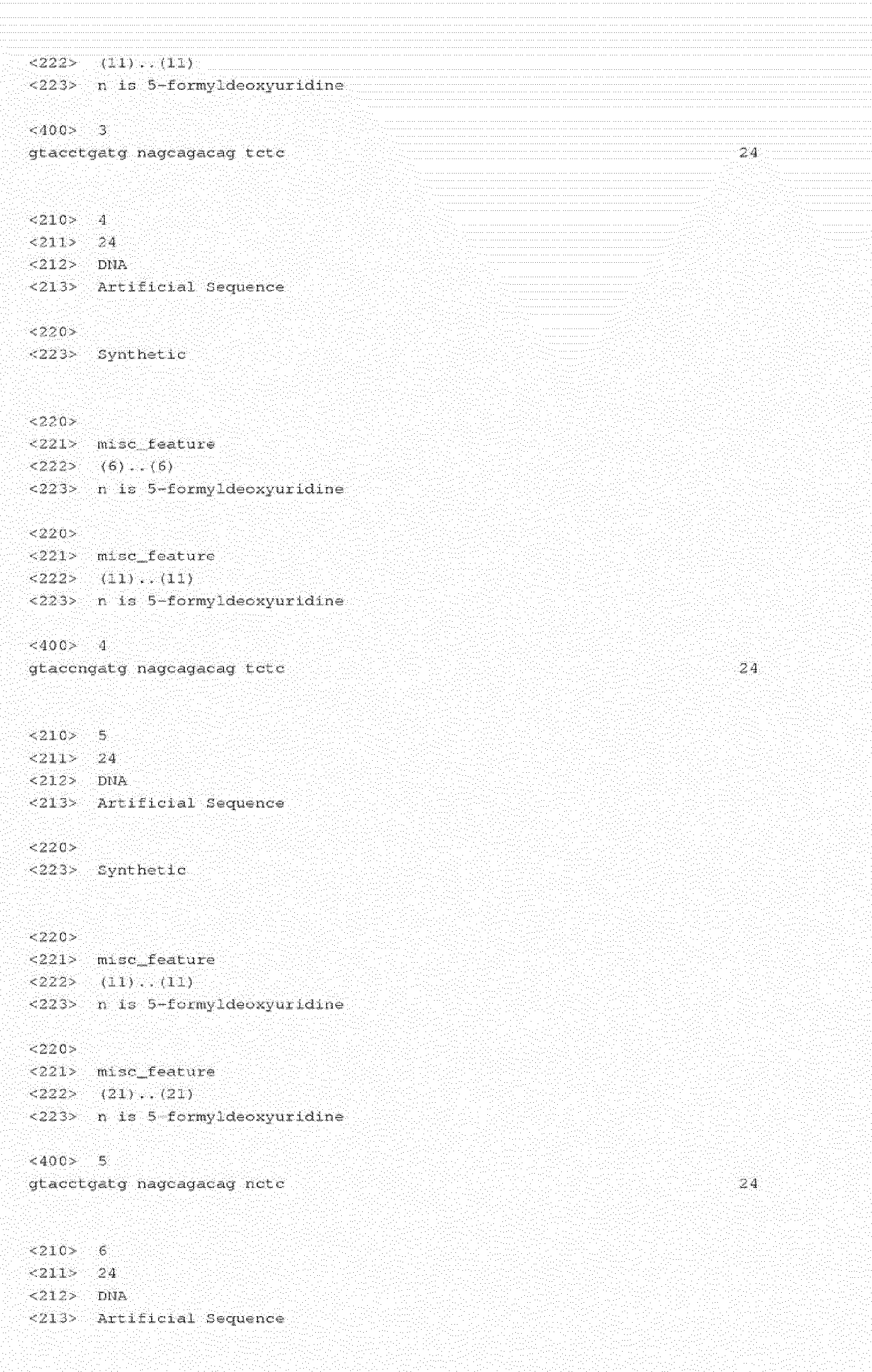
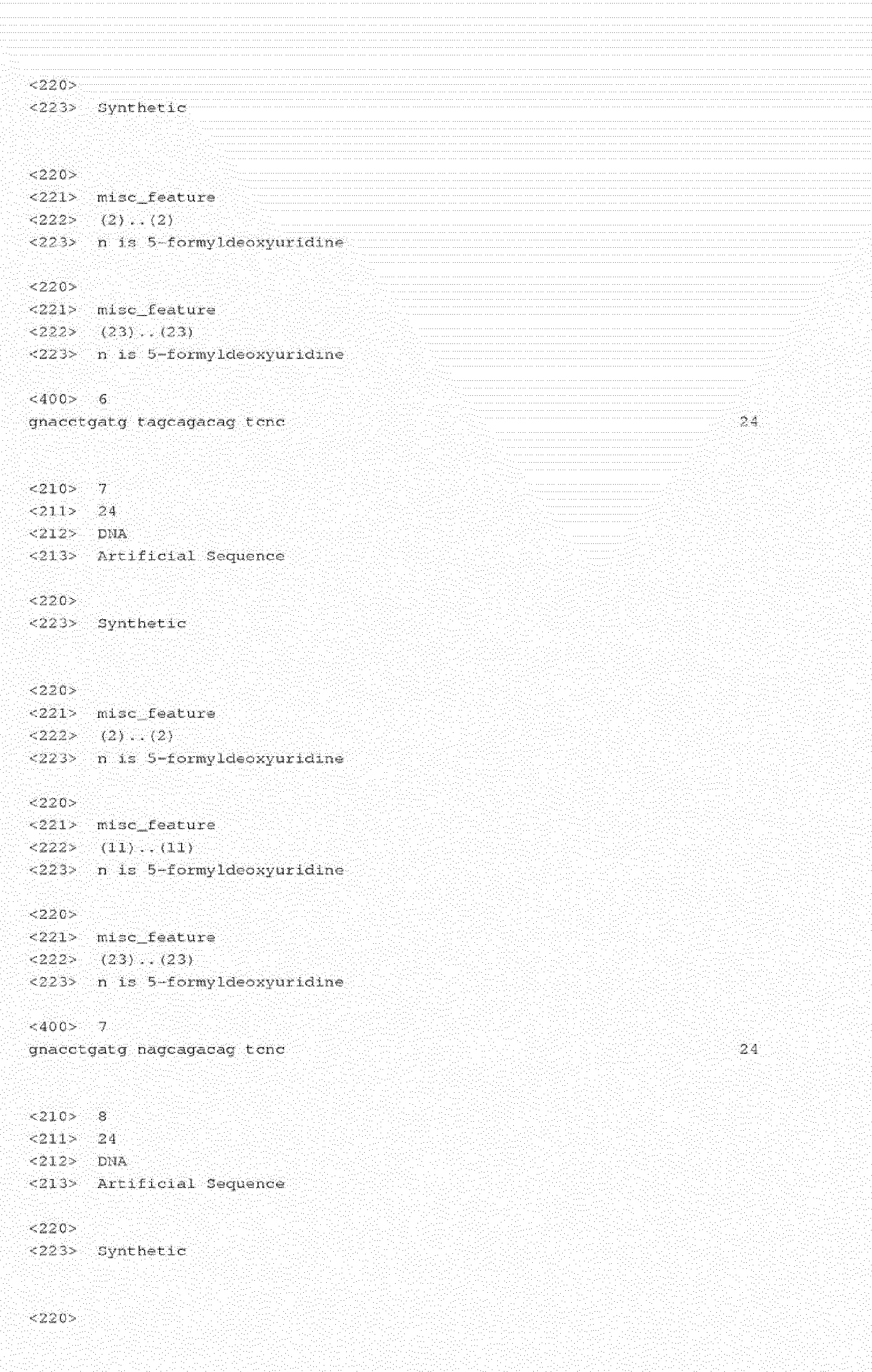

Claims (10)
- A method for detecting DNA damage in a subject suspected of having DNA damage wherein the damage results in the formation of aldehyde moieties in DNA, comprising the steps of:a) combining a sample comprising DNA from the subject with a pro-fluorescent hydrazine compound, wherein the pro-fluorescent hydrazine compound reacts with an aldehyde moiety formed as a result of damage to the DNA to form a fluorescent aryl hydrazone covalently linked to the DNA, wherein the fluorescent aryl hydrazone covalently linked to the DNA has an emission frequency different than that of the pro-fluorescent hydrazine compound;b) detecting the fluorescent DNA by monitoring a fluorescent emission; andc) quantitating the fluorescent emission to determine the DNA damage.
- A method according to claim 1, wherein the DNA damage in the subject is the result of the subject being exposed to ionizing radiation or oxidizing chemicals
- A method according to claim 1, wherein the sample is combined with a pro-fluorescent hydrazine compound, and wherein the pro-fluorescent hydrazine compound is 2-hydrazinoquinoline, 3-methyl-2-benzothiazolinone, 6-hydrazinonicotinamide or 4-hydrazinostilbazole.
- A method according to claim 1, wherein the presence of the fluorescent DNA is detected in an isolated DNA sample, a cell or a tissue sample.
- A method according to claim 1, wherein the aldehyde moiety in damaged DNA is 5-FodU or 5-FodC.
- A method according to claim 3, wherein the pro-fluorescent hydrazine compound is 2-hydrazinoquinoline.
- A method according to claim 3, wherein the pro-fluorescent hydrazine compound is 3-methyl-2-benzothiazolinone.
- A method according to claim 3, wherein the pro-fluorescent hydrazine compound is 6-hydrazinonicotinamide.
- A method according to claim 3, wherein the pro-fluorescent hydrazine compound is 4-hydrazinostilbazole.
- A method according to claim 4, wherein the presence of fluorescent DNA is detected in a cell.
Applications Claiming Priority (2)
| Application Number | Priority Date | Filing Date | Title |
|---|---|---|---|
| US9452108P | 2008-09-05 | 2008-09-05 | |
| EP09812394A EP2334814A4 (en) | 2008-09-05 | 2009-09-08 | Methods and compositions for direct detection of dna damage |
Related Parent Applications (2)
| Application Number | Title | Priority Date | Filing Date |
|---|---|---|---|
| EP09812394.6 Division | 2009-09-08 | ||
| EP09812394A Division EP2334814A4 (en) | 2008-09-05 | 2009-09-08 | Methods and compositions for direct detection of dna damage |
Publications (2)
| Publication Number | Publication Date |
|---|---|
| EP2604702A1 true EP2604702A1 (en) | 2013-06-19 |
| EP2604702B1 EP2604702B1 (en) | 2015-07-01 |
Family
ID=41797561
Family Applications (2)
| Application Number | Title | Priority Date | Filing Date |
|---|---|---|---|
| EP09812394A Withdrawn EP2334814A4 (en) | 2008-09-05 | 2009-09-08 | Methods and compositions for direct detection of dna damage |
| EP12189330.9A Not-in-force EP2604702B1 (en) | 2008-09-05 | 2009-09-08 | Methods for direct detection of DNA damage |
Family Applications Before (1)
| Application Number | Title | Priority Date | Filing Date |
|---|---|---|---|
| EP09812394A Withdrawn EP2334814A4 (en) | 2008-09-05 | 2009-09-08 | Methods and compositions for direct detection of dna damage |
Country Status (3)
| Country | Link |
|---|---|
| US (2) | US8580516B2 (en) |
| EP (2) | EP2334814A4 (en) |
| WO (1) | WO2010028388A1 (en) |
Families Citing this family (10)
| Publication number | Priority date | Publication date | Assignee | Title |
|---|---|---|---|---|
| WO2013058767A1 (en) * | 2011-10-21 | 2013-04-25 | Empire Technology Development Llc | Materials and methods to detect pyrimidine-pyrimidine dimer formation |
| MX365321B (en) | 2012-06-07 | 2019-05-29 | Los Angeles Childrens Hospital | METHODS FOR THE TREATMENT OF NEUTROPENIA USING RETINOID AGONISTS. |
| AU2015219038B2 (en) * | 2014-02-18 | 2018-10-18 | Children's Hospital Los Angeles | Compositions and methods for treating neutropenia |
| US10370703B2 (en) * | 2014-03-14 | 2019-08-06 | National Institute Of Advanced Industrial Science And Technology | Method for detecting guanine-abasic site in DNA |
| US9546930B2 (en) | 2014-06-27 | 2017-01-17 | Pulse Heath Llc | Analysis cartridge |
| WO2016082003A1 (en) * | 2014-11-26 | 2016-06-02 | The University Of Melbourne | Method and device for determination of aldehydes in biological samples |
| WO2017019751A1 (en) | 2015-07-27 | 2017-02-02 | The Johns Hopkins University | Method to use viral and host methylation markers for cervical cancer screening and triage in liquid prep, serum/plasma, and urine: pcr and sequencing based process methods |
| KR102392736B1 (en) | 2020-03-18 | 2022-05-02 | 재단법인 아산사회복지재단 | Method, program and system for training ai-based comet assay analysis model and method, program and system for analyzing comet assay using the analysis model |
| CN115803451A (en) * | 2020-09-11 | 2023-03-14 | 伊鲁米纳公司 | Compositions and methods for detecting abasic sites of nucleic acids |
| CA3214282A1 (en) | 2021-03-29 | 2022-10-06 | Illumina, Inc. | Compositions and methods for assessing dna damage in a library and normalizing amplicon size bias |
Citations (1)
| Publication number | Priority date | Publication date | Assignee | Title |
|---|---|---|---|---|
| WO1992006378A1 (en) * | 1990-10-04 | 1992-04-16 | The Scripps Research Institute | Self-assembling fluorescent diagnostic agents |
Family Cites Families (7)
| Publication number | Priority date | Publication date | Assignee | Title |
|---|---|---|---|---|
| US4268270A (en) * | 1979-04-30 | 1981-05-19 | Children's Hospital Medical Center | Glycosylated hemoglobin measurement |
| GB9510954D0 (en) * | 1995-05-31 | 1995-07-26 | Univ Leicester | Detection of DNA damage |
| US20010031739A1 (en) * | 1999-12-21 | 2001-10-18 | Dare Akintade Oyedele | Method and kit for quantitating genomic DNA damage and repair capicity |
| US6835573B2 (en) * | 2001-08-29 | 2004-12-28 | Husky Injection Molding Systems Ltd. | Method and device for testing aldehyde in polyester polymer |
| JP2004049182A (en) * | 2002-07-24 | 2004-02-19 | Bio Oriented Technol Res Advancement Inst | Method for detecting desalinated site of dna |
| GB0227738D0 (en) * | 2002-11-28 | 2003-01-08 | Univ Liverpool | Nanoparticle conjugates and method of production thereof |
| WO2007087250A2 (en) * | 2006-01-23 | 2007-08-02 | Amira Pharmaceuticals, Inc. | Tricyclic inhibitors of 5-lipoxygenase |
-
2009
- 2009-09-08 EP EP09812394A patent/EP2334814A4/en not_active Withdrawn
- 2009-09-08 US US12/555,668 patent/US8580516B2/en active Active
- 2009-09-08 EP EP12189330.9A patent/EP2604702B1/en not_active Not-in-force
- 2009-09-08 WO PCT/US2009/056262 patent/WO2010028388A1/en not_active Ceased
-
2013
- 2013-11-11 US US14/076,342 patent/US20140220696A1/en not_active Abandoned
Patent Citations (1)
| Publication number | Priority date | Publication date | Assignee | Title |
|---|---|---|---|---|
| WO1992006378A1 (en) * | 1990-10-04 | 1992-04-16 | The Scripps Research Institute | Self-assembling fluorescent diagnostic agents |
Non-Patent Citations (60)
| Title |
|---|
| ARMSTRONG, BIOCHEMISTRY, vol. 15, 1976, pages 2086 |
| BERTHED ET AL., NUCLEOSIDES & NUCLEOTIDES, vol. L5, 1996, pages 1287 |
| BESPALOV ET AL., BIOCHEMISTRY, vol. 35, 1996, pages 2067 |
| BJELLAND ET AL., BIOCHEMISTRY, vol. 34, 1995, pages 14758 |
| BJELLAND ET AL., MUTAT. RES., vol. 486, 2001, pages 147 |
| BJELLAND; SEEBERG, MUTAT. RES., vol. 531, 2003, pages 37 |
| BMSKOV ET AL., BIOCHEMISTERY, vol. 64, 1999, pages 803 |
| BREITMAN ET AL., CHEM RES. TOXICOL., vol. 9, 1980, pages 115 |
| CADET ET AL., FREE RADIC. BIOL. MED., vol. 33, 2002, pages 441 |
| CADET ET AL., J. ENVIRON. PATHOL. TOXICOL. ONCOL., vol. 23, 2004, pages 33 |
| CADET ET AL., MUTAT. RES., vol. 424, 1999, pages 9 |
| CADET ET AL., MUTAT. RES., vol. 531, 2003, pages 5 |
| CHAKRABARTI ET AL., INT. J. RADIAT. BIOL., vol. 75, 1999, pages 1055 |
| CHAVEZ ET AL., ANAL. CHEM., vol. 78, 2006, pages 6847 |
| COOKE, FASEB J., vol. 17, 2003, pages 1195 |
| DAINIAK ET AL., HEMATOLOGY AM. SOC. HEMATOL. EDUC. PROGRAM, 2003, pages 473 |
| DAVID, NATURE, vol. 447, 2007, pages 941 |
| DIZDAROGLU ET AL., FREE RADIC. BIOL. MED., vol. 32, 2002, pages 1102 |
| DOUKI ET AL., CHEM. RES. TOXICOL., vol. 9, 1996, pages 1145 |
| EVANS ET AL., MUTAT. RES., vol. 567, 2004, pages 1 |
| FORD ET AL., BLUT, vol. 54, 1987, pages 337 |
| GOANS; WASELENKO, HEALTH PHYS., vol. 89, 2005, pages 505 |
| HIROSE ET AL., PHOTOCHEM. PHOTOBIOL., vol. 76, 2002, pages 123 |
| HONG; WANG, ANAL. CHEM., 2007 |
| HONG; WANG, ANAL. CHEM., vol. 79, 2007, pages 322 |
| IDE ET AL., BIOCHEMISTRY, vol. 32, 1993, pages 8276 |
| ISHII ET AL., CELL CALCIUM, vol. 39, 2006, pages 487 |
| JACKSON ET AL., BJR, vol. 27, 2005, pages 161 |
| KALMER ET AL., J. IMMUNOL. METHODS, vol. 110, 1988, pages 275 |
| KAMIYA ET AL., MUTAT. RES., vol. 513, 2002, pages 213 |
| KARINO ET AL., NUCLEIC ACID RES., vol. 29, 2001, pages 256 |
| KASAI, MUTAT. RES., vol. 243, 1990, pages 249 |
| KIM ET AL., FEBS LETT., vol. 555, 2003, pages 611 |
| KJELDSEN ET AL., J. IMMUNOL. METHODS, vol. 232, 1999, pages 131 |
| KLUNGLAND ET AL., TOXICOL. LETT., vol. 119, 2001, pages 71 |
| KLUNGLAND, TOXICOL. LETT., vol. 119, 2001, pages 71 |
| KOUOH ET AL., BIOL. PHAM. BULL., vol. 23, 2000, pages 1382 |
| KOW; DARE, METHODS, vol. 22, 2000, pages 164 |
| KUBO ET AL., BIOCHEMISTRY, vol. 31, 1992, pages 3703 |
| KURISU ET AL., NUCLEIC ACID RES., 2001, pages 45 |
| LICHTENBERGER ET AL., J. IMMUNOL. METHODS, vol. 227, 1999, pages 75 |
| LIU ET AL., DNA REPAIR, vol. 2, 2003, pages 199 |
| MAKRIGIORGOS G M ET AL: "FLUORESCENT LABELLING OF ABASIC SITES: A NOVEL METHODOLOGY TO DETECT CLOSELY-SPACED DAMAGE SITES IN DNA", INTERNATIONAL JOURNAL OF RADIATION BIOLOGY, TAYLOR & FRANCIS, GB, vol. 74, no. 1, 1 January 1998 (1998-01-01), pages 99 - 109, XP002925462, ISSN: 0955-3002, DOI: 10.1080/095530098141762 * |
| MAKRIGIORGOS, INT. J. RADIAT. BIOL., vol. 74, 1998, pages 99 |
| MATSUBARA ET AL., BIOCHEMISTRY, vol. 42, 2003, pages 4993 |
| MCFAUL, J. IMMUNOL. METHODS, vol. 130, 1990, pages 15 |
| MIYABE ET AL., NUCLEIC ACIDS RES., vol. 30, 2002, pages 3443 |
| ONO ET AL., CHEM. PHARM. BULL., vol. 42, 1994, pages 2231 |
| PARK ET AL., PNAS, vol. 89, 1992, pages 3375 |
| PERSINGER ET AL., EXP. GERONTOL., vol. 36, 2001, pages 1483 |
| PRIVAT; SOWERS, MUTAT. RES., vol. 354, 1996, pages 151 |
| SOULTANAKIS ET AL., FREE RADIC. BIOL. MED., vol. 28, 2000, pages 987 |
| STRUTHERS ET AL., ANAL. BIOCHEM., vol. 255, 1998, pages 20 |
| WASELENKO ET AL., ANN. INTERN. MED., vol. 140, 2004, pages 1037 |
| WEISDORF ET AL., BIOL. BLOOD MARROW TRANSPLANT, vol. 12, 2006, pages 672 |
| ZHANG ET AL., DNA REPAIR, vol. 4, 2005, pages 71 |
| ZHANG ET AL., J. BIOL. CHEM., vol. 275, 2000, pages 35471 |
| ZHANG ET AL., NUCLEIC ACIDS RES., vol. 25, 1997, pages 3969 |
| ZHANG, INT. J. RADIAT. BIOL., vol. 79, 2003, pages 341 |
| ZHANG, NUCLEIC ACIDS RES., vol. 25, 1997, pages 3969 |
Also Published As
| Publication number | Publication date |
|---|---|
| EP2334814A4 (en) | 2012-04-25 |
| US20140220696A1 (en) | 2014-08-07 |
| EP2334814A1 (en) | 2011-06-22 |
| US20100216132A1 (en) | 2010-08-26 |
| WO2010028388A1 (en) | 2010-03-11 |
| US8580516B2 (en) | 2013-11-12 |
| EP2604702B1 (en) | 2015-07-01 |
Similar Documents
| Publication | Publication Date | Title |
|---|---|---|
| EP2604702B1 (en) | Methods for direct detection of DNA damage | |
| US9790541B2 (en) | Methods and compositions for labeling nucleic acids | |
| CN109142710B (en) | Method for rapidly and sensitively detecting tetrodotoxin TTX | |
| JP2009537816A (en) | Sample control for correction of sample matrix effects in analytical detection methods | |
| WO2017180911A1 (en) | Methods and systems for detecting or monitoring aggregation related to amyotrophic lateral sclerosis | |
| Vinothkanna et al. | Diagnostic applications of phycobiliproteins | |
| EP3480320A1 (en) | Double-stranded nucleic acid signal probe and method for detecting target molecule using same | |
| SI24638A (en) | Fluorometric immunoassay for the determination of antibodies against double-stranded DNA | |
| US20160258938A1 (en) | Method of detecting an analyte in a sample | |
| EP1204737B1 (en) | Internal quality control | |
| RU2486247C2 (en) | Method of identification agent based on high throughput screening | |
| CN109142751B (en) | Method for sensitively detecting tetrodotoxin TTX based on nucleic acid cleavage enzyme I immune marker | |
| JP2006238845A (en) | Cell separation method, cell identification method and cell examination method | |
| Diermeier‐Daucher et al. | Dynamic proliferation assessment in flow cytometry | |
| Burns | Fluorescent probes in the study of nucleic acids and chromatin in living cells | |
| US7098042B2 (en) | Internal quality control | |
| US20230349891A1 (en) | Releasable reagents for cell separation | |
| Stolley et al. | RNA In situ Hybridization with Sequential Protein Immunofluorescence in Tandem Assay | |
| AU761549B2 (en) | Internal quality control | |
| CN119738558A (en) | Composition for cell lysis and its application and kit |
Legal Events
| Date | Code | Title | Description |
|---|---|---|---|
| PUAI | Public reference made under article 153(3) epc to a published international application that has entered the european phase |
Free format text: ORIGINAL CODE: 0009012 |
|
| 17P | Request for examination filed |
Effective date: 20121105 |
|
| AC | Divisional application: reference to earlier application |
Ref document number: 2334814 Country of ref document: EP Kind code of ref document: P |
|
| AK | Designated contracting states |
Kind code of ref document: A1 Designated state(s): AT BE BG CH CY CZ DE DK EE ES FI FR GB GR HR HU IE IS IT LI LT LU LV MC MK MT NL NO PL PT RO SE SI SK SM TR |
|
| 17Q | First examination report despatched |
Effective date: 20140108 |
|
| GRAP | Despatch of communication of intention to grant a patent |
Free format text: ORIGINAL CODE: EPIDOSNIGR1 |
|
| INTG | Intention to grant announced |
Effective date: 20150115 |
|
| GRAS | Grant fee paid |
Free format text: ORIGINAL CODE: EPIDOSNIGR3 |
|
| GRAA | (expected) grant |
Free format text: ORIGINAL CODE: 0009210 |
|
| AC | Divisional application: reference to earlier application |
Ref document number: 2334814 Country of ref document: EP Kind code of ref document: P |
|
| AK | Designated contracting states |
Kind code of ref document: B1 Designated state(s): AT BE BG CH CY CZ DE DK EE ES FI FR GB GR HR HU IE IS IT LI LT LU LV MC MK MT NL NO PL PT RO SE SI SK SM TR |
|
| REG | Reference to a national code |
Ref country code: GB Ref legal event code: FG4D |
|
| REG | Reference to a national code |
Ref country code: AT Ref legal event code: REF Ref document number: 734015 Country of ref document: AT Kind code of ref document: T Effective date: 20150715 Ref country code: CH Ref legal event code: EP |
|
| REG | Reference to a national code |
Ref country code: IE Ref legal event code: FG4D |
|
| REG | Reference to a national code |
Ref country code: DE Ref legal event code: R096 Ref document number: 602009032018 Country of ref document: DE |
|
| REG | Reference to a national code |
Ref country code: AT Ref legal event code: MK05 Ref document number: 734015 Country of ref document: AT Kind code of ref document: T Effective date: 20150701 |
|
| REG | Reference to a national code |
Ref country code: NL Ref legal event code: MP Effective date: 20150701 |
|
| REG | Reference to a national code |
Ref country code: FR Ref legal event code: PLFP Year of fee payment: 7 |
|
| REG | Reference to a national code |
Ref country code: LT Ref legal event code: MG4D |
|
| PG25 | Lapsed in a contracting state [announced via postgrant information from national office to epo] |
Ref country code: GR Free format text: LAPSE BECAUSE OF FAILURE TO SUBMIT A TRANSLATION OF THE DESCRIPTION OR TO PAY THE FEE WITHIN THE PRESCRIBED TIME-LIMIT Effective date: 20151002 Ref country code: FI Free format text: LAPSE BECAUSE OF FAILURE TO SUBMIT A TRANSLATION OF THE DESCRIPTION OR TO PAY THE FEE WITHIN THE PRESCRIBED TIME-LIMIT Effective date: 20150701 Ref country code: NO Free format text: LAPSE BECAUSE OF FAILURE TO SUBMIT A TRANSLATION OF THE DESCRIPTION OR TO PAY THE FEE WITHIN THE PRESCRIBED TIME-LIMIT Effective date: 20151001 Ref country code: LV Free format text: LAPSE BECAUSE OF FAILURE TO SUBMIT A TRANSLATION OF THE DESCRIPTION OR TO PAY THE FEE WITHIN THE PRESCRIBED TIME-LIMIT Effective date: 20150701 Ref country code: LT Free format text: LAPSE BECAUSE OF FAILURE TO SUBMIT A TRANSLATION OF THE DESCRIPTION OR TO PAY THE FEE WITHIN THE PRESCRIBED TIME-LIMIT Effective date: 20150701 |
|
| PGFP | Annual fee paid to national office [announced via postgrant information from national office to epo] |
Ref country code: CH Payment date: 20151224 Year of fee payment: 7 Ref country code: GB Payment date: 20151223 Year of fee payment: 7 |
|
| PG25 | Lapsed in a contracting state [announced via postgrant information from national office to epo] |
Ref country code: PL Free format text: LAPSE BECAUSE OF FAILURE TO SUBMIT A TRANSLATION OF THE DESCRIPTION OR TO PAY THE FEE WITHIN THE PRESCRIBED TIME-LIMIT Effective date: 20150701 Ref country code: ES Free format text: LAPSE BECAUSE OF FAILURE TO SUBMIT A TRANSLATION OF THE DESCRIPTION OR TO PAY THE FEE WITHIN THE PRESCRIBED TIME-LIMIT Effective date: 20150701 Ref country code: IS Free format text: LAPSE BECAUSE OF FAILURE TO SUBMIT A TRANSLATION OF THE DESCRIPTION OR TO PAY THE FEE WITHIN THE PRESCRIBED TIME-LIMIT Effective date: 20151101 Ref country code: AT Free format text: LAPSE BECAUSE OF FAILURE TO SUBMIT A TRANSLATION OF THE DESCRIPTION OR TO PAY THE FEE WITHIN THE PRESCRIBED TIME-LIMIT Effective date: 20150701 Ref country code: HR Free format text: LAPSE BECAUSE OF FAILURE TO SUBMIT A TRANSLATION OF THE DESCRIPTION OR TO PAY THE FEE WITHIN THE PRESCRIBED TIME-LIMIT Effective date: 20150701 Ref country code: SE Free format text: LAPSE BECAUSE OF FAILURE TO SUBMIT A TRANSLATION OF THE DESCRIPTION OR TO PAY THE FEE WITHIN THE PRESCRIBED TIME-LIMIT Effective date: 20150701 Ref country code: PT Free format text: LAPSE BECAUSE OF FAILURE TO SUBMIT A TRANSLATION OF THE DESCRIPTION OR TO PAY THE FEE WITHIN THE PRESCRIBED TIME-LIMIT Effective date: 20151102 |
|
| PGFP | Annual fee paid to national office [announced via postgrant information from national office to epo] |
Ref country code: FR Payment date: 20151223 Year of fee payment: 7 |
|
| REG | Reference to a national code |
Ref country code: DE Ref legal event code: R097 Ref document number: 602009032018 Country of ref document: DE |
|
| PG25 | Lapsed in a contracting state [announced via postgrant information from national office to epo] |
Ref country code: CZ Free format text: LAPSE BECAUSE OF FAILURE TO SUBMIT A TRANSLATION OF THE DESCRIPTION OR TO PAY THE FEE WITHIN THE PRESCRIBED TIME-LIMIT Effective date: 20150701 Ref country code: EE Free format text: LAPSE BECAUSE OF FAILURE TO SUBMIT A TRANSLATION OF THE DESCRIPTION OR TO PAY THE FEE WITHIN THE PRESCRIBED TIME-LIMIT Effective date: 20150701 Ref country code: IT Free format text: LAPSE BECAUSE OF FAILURE TO SUBMIT A TRANSLATION OF THE DESCRIPTION OR TO PAY THE FEE WITHIN THE PRESCRIBED TIME-LIMIT Effective date: 20150701 Ref country code: DK Free format text: LAPSE BECAUSE OF FAILURE TO SUBMIT A TRANSLATION OF THE DESCRIPTION OR TO PAY THE FEE WITHIN THE PRESCRIBED TIME-LIMIT Effective date: 20150701 Ref country code: SK Free format text: LAPSE BECAUSE OF FAILURE TO SUBMIT A TRANSLATION OF THE DESCRIPTION OR TO PAY THE FEE WITHIN THE PRESCRIBED TIME-LIMIT Effective date: 20150701 Ref country code: MC Free format text: LAPSE BECAUSE OF FAILURE TO SUBMIT A TRANSLATION OF THE DESCRIPTION OR TO PAY THE FEE WITHIN THE PRESCRIBED TIME-LIMIT Effective date: 20150701 Ref country code: LU Free format text: LAPSE BECAUSE OF FAILURE TO SUBMIT A TRANSLATION OF THE DESCRIPTION OR TO PAY THE FEE WITHIN THE PRESCRIBED TIME-LIMIT Effective date: 20150908 |
|
| PGFP | Annual fee paid to national office [announced via postgrant information from national office to epo] |
Ref country code: DE Payment date: 20151229 Year of fee payment: 7 |
|
| PLBE | No opposition filed within time limit |
Free format text: ORIGINAL CODE: 0009261 |
|
| STAA | Information on the status of an ep patent application or granted ep patent |
Free format text: STATUS: NO OPPOSITION FILED WITHIN TIME LIMIT |
|
| PG25 | Lapsed in a contracting state [announced via postgrant information from national office to epo] |
Ref country code: RO Free format text: LAPSE BECAUSE OF FAILURE TO SUBMIT A TRANSLATION OF THE DESCRIPTION OR TO PAY THE FEE WITHIN THE PRESCRIBED TIME-LIMIT Effective date: 20150701 |
|
| 26N | No opposition filed |
Effective date: 20160404 |
|
| REG | Reference to a national code |
Ref country code: IE Ref legal event code: MM4A |
|
| PG25 | Lapsed in a contracting state [announced via postgrant information from national office to epo] |
Ref country code: IE Free format text: LAPSE BECAUSE OF NON-PAYMENT OF DUE FEES Effective date: 20150908 |
|
| PG25 | Lapsed in a contracting state [announced via postgrant information from national office to epo] |
Ref country code: SI Free format text: LAPSE BECAUSE OF FAILURE TO SUBMIT A TRANSLATION OF THE DESCRIPTION OR TO PAY THE FEE WITHIN THE PRESCRIBED TIME-LIMIT Effective date: 20150701 |
|
| PG25 | Lapsed in a contracting state [announced via postgrant information from national office to epo] |
Ref country code: BE Free format text: LAPSE BECAUSE OF FAILURE TO SUBMIT A TRANSLATION OF THE DESCRIPTION OR TO PAY THE FEE WITHIN THE PRESCRIBED TIME-LIMIT Effective date: 20150701 |
|
| PG25 | Lapsed in a contracting state [announced via postgrant information from national office to epo] |
Ref country code: MT Free format text: LAPSE BECAUSE OF FAILURE TO SUBMIT A TRANSLATION OF THE DESCRIPTION OR TO PAY THE FEE WITHIN THE PRESCRIBED TIME-LIMIT Effective date: 20150701 |
|
| REG | Reference to a national code |
Ref country code: DE Ref legal event code: R119 Ref document number: 602009032018 Country of ref document: DE |
|
| REG | Reference to a national code |
Ref country code: CH Ref legal event code: PL |
|
| GBPC | Gb: european patent ceased through non-payment of renewal fee |
Effective date: 20160908 |
|
| PG25 | Lapsed in a contracting state [announced via postgrant information from national office to epo] |
Ref country code: SM Free format text: LAPSE BECAUSE OF FAILURE TO SUBMIT A TRANSLATION OF THE DESCRIPTION OR TO PAY THE FEE WITHIN THE PRESCRIBED TIME-LIMIT Effective date: 20150701 Ref country code: BG Free format text: LAPSE BECAUSE OF FAILURE TO SUBMIT A TRANSLATION OF THE DESCRIPTION OR TO PAY THE FEE WITHIN THE PRESCRIBED TIME-LIMIT Effective date: 20150701 Ref country code: HU Free format text: LAPSE BECAUSE OF FAILURE TO SUBMIT A TRANSLATION OF THE DESCRIPTION OR TO PAY THE FEE WITHIN THE PRESCRIBED TIME-LIMIT; INVALID AB INITIO Effective date: 20090908 |
|
| PG25 | Lapsed in a contracting state [announced via postgrant information from national office to epo] |
Ref country code: CY Free format text: LAPSE BECAUSE OF FAILURE TO SUBMIT A TRANSLATION OF THE DESCRIPTION OR TO PAY THE FEE WITHIN THE PRESCRIBED TIME-LIMIT Effective date: 20150701 Ref country code: NL Free format text: LAPSE BECAUSE OF FAILURE TO SUBMIT A TRANSLATION OF THE DESCRIPTION OR TO PAY THE FEE WITHIN THE PRESCRIBED TIME-LIMIT Effective date: 20150701 |
|
| REG | Reference to a national code |
Ref country code: FR Ref legal event code: ST Effective date: 20170531 |
|
| PG25 | Lapsed in a contracting state [announced via postgrant information from national office to epo] |
Ref country code: DE Free format text: LAPSE BECAUSE OF NON-PAYMENT OF DUE FEES Effective date: 20170401 Ref country code: FR Free format text: LAPSE BECAUSE OF NON-PAYMENT OF DUE FEES Effective date: 20160930 Ref country code: CH Free format text: LAPSE BECAUSE OF NON-PAYMENT OF DUE FEES Effective date: 20160930 Ref country code: GB Free format text: LAPSE BECAUSE OF NON-PAYMENT OF DUE FEES Effective date: 20160908 Ref country code: LI Free format text: LAPSE BECAUSE OF NON-PAYMENT OF DUE FEES Effective date: 20160930 |
|
| PG25 | Lapsed in a contracting state [announced via postgrant information from national office to epo] |
Ref country code: MK Free format text: LAPSE BECAUSE OF FAILURE TO SUBMIT A TRANSLATION OF THE DESCRIPTION OR TO PAY THE FEE WITHIN THE PRESCRIBED TIME-LIMIT Effective date: 20150701 Ref country code: TR Free format text: LAPSE BECAUSE OF FAILURE TO SUBMIT A TRANSLATION OF THE DESCRIPTION OR TO PAY THE FEE WITHIN THE PRESCRIBED TIME-LIMIT Effective date: 20150701 |