EP2592155B2 - EGFR mutations - Google Patents
EGFR mutations Download PDFInfo
- Publication number
- EP2592155B2 EP2592155B2 EP12192359.3A EP12192359A EP2592155B2 EP 2592155 B2 EP2592155 B2 EP 2592155B2 EP 12192359 A EP12192359 A EP 12192359A EP 2592155 B2 EP2592155 B2 EP 2592155B2
- Authority
- EP
- European Patent Office
- Prior art keywords
- egfr
- protein mutation
- mutation
- gene
- egfr protein
- Prior art date
- Legal status (The legal status is an assumption and is not a legal conclusion. Google has not performed a legal analysis and makes no representation as to the accuracy of the status listed.)
- Active
Links
Images


Classifications
-
- A—HUMAN NECESSITIES
- A61—MEDICAL OR VETERINARY SCIENCE; HYGIENE
- A61K—PREPARATIONS FOR MEDICAL, DENTAL OR TOILETRY PURPOSES
- A61K39/00—Medicinal preparations containing antigens or antibodies
- A61K39/395—Antibodies; Immunoglobulins; Immune serum, e.g. antilymphocytic serum
-
- A—HUMAN NECESSITIES
- A61—MEDICAL OR VETERINARY SCIENCE; HYGIENE
- A61K—PREPARATIONS FOR MEDICAL, DENTAL OR TOILETRY PURPOSES
- A61K31/00—Medicinal preparations containing organic active ingredients
- A61K31/185—Acids; Anhydrides, halides or salts thereof, e.g. sulfur acids, imidic, hydrazonic or hydroximic acids
- A61K31/19—Carboxylic acids, e.g. valproic acid
-
- A—HUMAN NECESSITIES
- A61—MEDICAL OR VETERINARY SCIENCE; HYGIENE
- A61K—PREPARATIONS FOR MEDICAL, DENTAL OR TOILETRY PURPOSES
- A61K31/00—Medicinal preparations containing organic active ingredients
- A61K31/33—Heterocyclic compounds
- A61K31/395—Heterocyclic compounds having nitrogen as a ring hetero atom, e.g. guanethidine or rifamycins
- A61K31/495—Heterocyclic compounds having nitrogen as a ring hetero atom, e.g. guanethidine or rifamycins having six-membered rings with two or more nitrogen atoms as the only ring heteroatoms, e.g. piperazine or tetrazines
- A61K31/498—Pyrazines or piperazines ortho- and peri-condensed with carbocyclic ring systems, e.g. quinoxaline, phenazine
-
- A—HUMAN NECESSITIES
- A61—MEDICAL OR VETERINARY SCIENCE; HYGIENE
- A61K—PREPARATIONS FOR MEDICAL, DENTAL OR TOILETRY PURPOSES
- A61K31/00—Medicinal preparations containing organic active ingredients
- A61K31/33—Heterocyclic compounds
- A61K31/395—Heterocyclic compounds having nitrogen as a ring hetero atom, e.g. guanethidine or rifamycins
- A61K31/495—Heterocyclic compounds having nitrogen as a ring hetero atom, e.g. guanethidine or rifamycins having six-membered rings with two or more nitrogen atoms as the only ring heteroatoms, e.g. piperazine or tetrazines
- A61K31/505—Pyrimidines; Hydrogenated pyrimidines, e.g. trimethoprim
- A61K31/517—Pyrimidines; Hydrogenated pyrimidines, e.g. trimethoprim ortho- or peri-condensed with carbocyclic ring systems, e.g. quinazoline, perimidine
-
- A—HUMAN NECESSITIES
- A61—MEDICAL OR VETERINARY SCIENCE; HYGIENE
- A61K—PREPARATIONS FOR MEDICAL, DENTAL OR TOILETRY PURPOSES
- A61K31/00—Medicinal preparations containing organic active ingredients
- A61K31/70—Carbohydrates; Sugars; Derivatives thereof
- A61K31/7028—Compounds having saccharide radicals attached to non-saccharide compounds by glycosidic linkages
- A61K31/7034—Compounds having saccharide radicals attached to non-saccharide compounds by glycosidic linkages attached to a carbocyclic compound, e.g. phloridzin
- A61K31/704—Compounds having saccharide radicals attached to non-saccharide compounds by glycosidic linkages attached to a carbocyclic compound, e.g. phloridzin attached to a condensed carbocyclic ring system, e.g. sennosides, thiocolchicosides, escin, daunorubicin
-
- A—HUMAN NECESSITIES
- A61—MEDICAL OR VETERINARY SCIENCE; HYGIENE
- A61K—PREPARATIONS FOR MEDICAL, DENTAL OR TOILETRY PURPOSES
- A61K31/00—Medicinal preparations containing organic active ingredients
- A61K31/70—Carbohydrates; Sugars; Derivatives thereof
- A61K31/7042—Compounds having saccharide radicals and heterocyclic rings
- A61K31/7052—Compounds having saccharide radicals and heterocyclic rings having nitrogen as a ring hetero atom, e.g. nucleosides, nucleotides
- A61K31/706—Compounds having saccharide radicals and heterocyclic rings having nitrogen as a ring hetero atom, e.g. nucleosides, nucleotides containing six-membered rings with nitrogen as a ring hetero atom
- A61K31/7064—Compounds having saccharide radicals and heterocyclic rings having nitrogen as a ring hetero atom, e.g. nucleosides, nucleotides containing six-membered rings with nitrogen as a ring hetero atom containing condensed or non-condensed pyrimidines
- A61K31/7068—Compounds having saccharide radicals and heterocyclic rings having nitrogen as a ring hetero atom, e.g. nucleosides, nucleotides containing six-membered rings with nitrogen as a ring hetero atom containing condensed or non-condensed pyrimidines having oxo groups directly attached to the pyrimidine ring, e.g. cytidine, cytidylic acid
- A61K31/7072—Compounds having saccharide radicals and heterocyclic rings having nitrogen as a ring hetero atom, e.g. nucleosides, nucleotides containing six-membered rings with nitrogen as a ring hetero atom containing condensed or non-condensed pyrimidines having oxo groups directly attached to the pyrimidine ring, e.g. cytidine, cytidylic acid having two oxo groups directly attached to the pyrimidine ring, e.g. uridine, uridylic acid, thymidine, zidovudine
-
- A—HUMAN NECESSITIES
- A61—MEDICAL OR VETERINARY SCIENCE; HYGIENE
- A61K—PREPARATIONS FOR MEDICAL, DENTAL OR TOILETRY PURPOSES
- A61K45/00—Medicinal preparations containing active ingredients not provided for in groups A61K31/00 - A61K41/00
- A61K45/06—Mixtures of active ingredients without chemical characterisation, e.g. antiphlogistics and cardiaca
-
- A—HUMAN NECESSITIES
- A61—MEDICAL OR VETERINARY SCIENCE; HYGIENE
- A61P—SPECIFIC THERAPEUTIC ACTIVITY OF CHEMICAL COMPOUNDS OR MEDICINAL PREPARATIONS
- A61P35/00—Antineoplastic agents
-
- C—CHEMISTRY; METALLURGY
- C12—BIOCHEMISTRY; BEER; SPIRITS; WINE; VINEGAR; MICROBIOLOGY; ENZYMOLOGY; MUTATION OR GENETIC ENGINEERING
- C12Q—MEASURING OR TESTING PROCESSES INVOLVING ENZYMES, NUCLEIC ACIDS OR MICROORGANISMS; COMPOSITIONS OR TEST PAPERS THEREFOR; PROCESSES OF PREPARING SUCH COMPOSITIONS; CONDITION-RESPONSIVE CONTROL IN MICROBIOLOGICAL OR ENZYMOLOGICAL PROCESSES
- C12Q1/00—Measuring or testing processes involving enzymes, nucleic acids or microorganisms; Compositions therefor; Processes of preparing such compositions
- C12Q1/48—Measuring or testing processes involving enzymes, nucleic acids or microorganisms; Compositions therefor; Processes of preparing such compositions involving transferase
- C12Q1/485—Measuring or testing processes involving enzymes, nucleic acids or microorganisms; Compositions therefor; Processes of preparing such compositions involving transferase involving kinase
-
- C—CHEMISTRY; METALLURGY
- C12—BIOCHEMISTRY; BEER; SPIRITS; WINE; VINEGAR; MICROBIOLOGY; ENZYMOLOGY; MUTATION OR GENETIC ENGINEERING
- C12Q—MEASURING OR TESTING PROCESSES INVOLVING ENZYMES, NUCLEIC ACIDS OR MICROORGANISMS; COMPOSITIONS OR TEST PAPERS THEREFOR; PROCESSES OF PREPARING SUCH COMPOSITIONS; CONDITION-RESPONSIVE CONTROL IN MICROBIOLOGICAL OR ENZYMOLOGICAL PROCESSES
- C12Q1/00—Measuring or testing processes involving enzymes, nucleic acids or microorganisms; Compositions therefor; Processes of preparing such compositions
- C12Q1/68—Measuring or testing processes involving enzymes, nucleic acids or microorganisms; Compositions therefor; Processes of preparing such compositions involving nucleic acids
- C12Q1/6876—Nucleic acid products used in the analysis of nucleic acids, e.g. primers or probes
- C12Q1/6883—Nucleic acid products used in the analysis of nucleic acids, e.g. primers or probes for diseases caused by alterations of genetic material
- C12Q1/6886—Nucleic acid products used in the analysis of nucleic acids, e.g. primers or probes for diseases caused by alterations of genetic material for cancer
-
- G—PHYSICS
- G01—MEASURING; TESTING
- G01N—INVESTIGATING OR ANALYSING MATERIALS BY DETERMINING THEIR CHEMICAL OR PHYSICAL PROPERTIES
- G01N33/00—Investigating or analysing materials by specific methods not covered by groups G01N1/00 - G01N31/00
- G01N33/48—Biological material, e.g. blood, urine; Haemocytometers
- G01N33/50—Chemical analysis of biological material, e.g. blood, urine; Testing involving biospecific ligand binding methods; Immunological testing
- G01N33/53—Immunoassay; Biospecific binding assay; Materials therefor
- G01N33/574—Immunoassay; Biospecific binding assay; Materials therefor for cancer
- G01N33/57484—Immunoassay; Biospecific binding assay; Materials therefor for cancer involving compounds serving as markers for tumor, cancer, neoplasia, e.g. cellular determinants, receptors, heat shock/stress proteins, A-protein, oligosaccharides, metabolites
- G01N33/57492—Immunoassay; Biospecific binding assay; Materials therefor for cancer involving compounds serving as markers for tumor, cancer, neoplasia, e.g. cellular determinants, receptors, heat shock/stress proteins, A-protein, oligosaccharides, metabolites involving compounds localized on the membrane of tumor or cancer cells
-
- C—CHEMISTRY; METALLURGY
- C12—BIOCHEMISTRY; BEER; SPIRITS; WINE; VINEGAR; MICROBIOLOGY; ENZYMOLOGY; MUTATION OR GENETIC ENGINEERING
- C12Q—MEASURING OR TESTING PROCESSES INVOLVING ENZYMES, NUCLEIC ACIDS OR MICROORGANISMS; COMPOSITIONS OR TEST PAPERS THEREFOR; PROCESSES OF PREPARING SUCH COMPOSITIONS; CONDITION-RESPONSIVE CONTROL IN MICROBIOLOGICAL OR ENZYMOLOGICAL PROCESSES
- C12Q2600/00—Oligonucleotides characterized by their use
- C12Q2600/106—Pharmacogenomics, i.e. genetic variability in individual responses to drugs and drug metabolism
-
- C—CHEMISTRY; METALLURGY
- C12—BIOCHEMISTRY; BEER; SPIRITS; WINE; VINEGAR; MICROBIOLOGY; ENZYMOLOGY; MUTATION OR GENETIC ENGINEERING
- C12Q—MEASURING OR TESTING PROCESSES INVOLVING ENZYMES, NUCLEIC ACIDS OR MICROORGANISMS; COMPOSITIONS OR TEST PAPERS THEREFOR; PROCESSES OF PREPARING SUCH COMPOSITIONS; CONDITION-RESPONSIVE CONTROL IN MICROBIOLOGICAL OR ENZYMOLOGICAL PROCESSES
- C12Q2600/00—Oligonucleotides characterized by their use
- C12Q2600/118—Prognosis of disease development
-
- C—CHEMISTRY; METALLURGY
- C12—BIOCHEMISTRY; BEER; SPIRITS; WINE; VINEGAR; MICROBIOLOGY; ENZYMOLOGY; MUTATION OR GENETIC ENGINEERING
- C12Q—MEASURING OR TESTING PROCESSES INVOLVING ENZYMES, NUCLEIC ACIDS OR MICROORGANISMS; COMPOSITIONS OR TEST PAPERS THEREFOR; PROCESSES OF PREPARING SUCH COMPOSITIONS; CONDITION-RESPONSIVE CONTROL IN MICROBIOLOGICAL OR ENZYMOLOGICAL PROCESSES
- C12Q2600/00—Oligonucleotides characterized by their use
- C12Q2600/136—Screening for pharmacological compounds
-
- C—CHEMISTRY; METALLURGY
- C12—BIOCHEMISTRY; BEER; SPIRITS; WINE; VINEGAR; MICROBIOLOGY; ENZYMOLOGY; MUTATION OR GENETIC ENGINEERING
- C12Q—MEASURING OR TESTING PROCESSES INVOLVING ENZYMES, NUCLEIC ACIDS OR MICROORGANISMS; COMPOSITIONS OR TEST PAPERS THEREFOR; PROCESSES OF PREPARING SUCH COMPOSITIONS; CONDITION-RESPONSIVE CONTROL IN MICROBIOLOGICAL OR ENZYMOLOGICAL PROCESSES
- C12Q2600/00—Oligonucleotides characterized by their use
- C12Q2600/156—Polymorphic or mutational markers
-
- C—CHEMISTRY; METALLURGY
- C12—BIOCHEMISTRY; BEER; SPIRITS; WINE; VINEGAR; MICROBIOLOGY; ENZYMOLOGY; MUTATION OR GENETIC ENGINEERING
- C12Q—MEASURING OR TESTING PROCESSES INVOLVING ENZYMES, NUCLEIC ACIDS OR MICROORGANISMS; COMPOSITIONS OR TEST PAPERS THEREFOR; PROCESSES OF PREPARING SUCH COMPOSITIONS; CONDITION-RESPONSIVE CONTROL IN MICROBIOLOGICAL OR ENZYMOLOGICAL PROCESSES
- C12Q2600/00—Oligonucleotides characterized by their use
- C12Q2600/16—Primer sets for multiplex assays
-
- G—PHYSICS
- G01—MEASURING; TESTING
- G01N—INVESTIGATING OR ANALYSING MATERIALS BY DETERMINING THEIR CHEMICAL OR PHYSICAL PROPERTIES
- G01N2333/00—Assays involving biological materials from specific organisms or of a specific nature
- G01N2333/435—Assays involving biological materials from specific organisms or of a specific nature from animals; from humans
- G01N2333/705—Assays involving receptors, cell surface antigens or cell surface determinants
- G01N2333/71—Assays involving receptors, cell surface antigens or cell surface determinants for growth factors; for growth regulators
-
- G—PHYSICS
- G01—MEASURING; TESTING
- G01N—INVESTIGATING OR ANALYSING MATERIALS BY DETERMINING THEIR CHEMICAL OR PHYSICAL PROPERTIES
- G01N2500/00—Screening for compounds of potential therapeutic value
Definitions
- the present invention relates to cancer diagnostics and therapies and in particular to the detection of mutations that are diagnostic and/or prognostic.
- EGFR Epidermal Growth Factor Receptor
- Activation of these receptors typically occurs via specific ligand binding, resulting in hetero- or homodimerization between receptor family members, with subsequent autophosphorylation of the tyrosine kinase domain. This activation triggers a cascade of intracellular signaling pathways involved in both cellular proliferation (the ras/raf/MAP kinase pathway) and survival (the Pl3 kinase/Akt pathway).
- ras/raf/MAP kinase pathway the ras/raf/MAP kinase pathway
- the Pl3 kinase/Akt pathway the Pl3 kinase/Akt pathway
- EGFR transforming growth factor ⁇
- transforming growth factor ⁇ Gullick, Br Med Bull 1991, 47:87-98 ; Modijtahedi and Dean, Int J Oncol 1994, 4:277-96 ; Salomon et al., Crit Rev Oncol Hematol 1995;19:183-232 ).
- EGFR overexpression has been associated with an adverse prognosis in a number of human cancers, including NSCLC.
- TarcevaTM also known as erlotinib; OSI-774
- erlotinib a quinazoline
- Erlotinib inhibits human EGFR tyrosine kinase with an IC 50 of 2 nM (0.786 mg/mL) in an in vitro enzyme assay. This inhibition is selective for EGFR tyrosine kinase, results in cell cycle arrest at G 1 , and is reversible.
- erlotinib Oral administration of erlotinib in mice has demonstrated a >70% reduction in EGFR autophosphorylation in human xenografts and marked growth inhibition of HN5 and A431 xenografts in nude mice has been demonstrated.
- erlotinib In addition to single-agent activity in in vivo assay systems, erlotinib has been evaluated in combination with a number of chemotherapy agents to determine possible interactions. There was an additive interaction between erlotinib and paclitaxel, cisplatin, gemcitabine, and doxorubicin.
- Lung cancer represents the leading cause of cancer-related mortality for both men and women in the United States. In 2000, it was estimated that 164,000 new cases would be diagnosed and 157,000 patients would die from this disease ( Greenlee et al., CA Cancer J Clin 2001, 51:15-36 ). Approximately 75% of these patients would have had non-small cell histologies, with the majority presenting with inoperable Stage IIIB or Stage IV disease. For those patients with more limited disease at presentation (Stages I-IIIA), relapse following standard surgical therapy, with or without adjuvant or neoadjuvant chemo- and/or radiotherapy, is common. These findings result in an overall 5-year survival in non-small cell lung cancer (NSCLC) of ⁇ 12% and serve to emphasize the unmet medical need in this disease.
- NSCLC non-small cell lung cancer
- the platinum compound cisplatin was the first chemotherapy agent to show clinical benefit in the management of locally advanced or metastatic NSCLC. Randomized clinical trials demonstrated improved response rates, quality of life, and survival compared with the best supportive care (Rapp et al. 1988). However, the magnitude of this improvement was modest-measured in weeks. Subsequently, a number of newer chemotherapy agents have been evaluated as single agents and in combination with the platinum salts in the first-line setting. The conclusion from these studies is that modem "doublet" chemotherapy appears to achieve response rates of 15%-20%, median time to disease progression of 3-4 months, and median survival of 7-8 months.
- the present invention provides methods, as defined in the claims, for determining the prognosis of a patient having a non-small cell lung cancer tumor.
- the present invention also provides methods, as defined in the claims, of identifying a non-small cell lung cancer tumor that is susceptible to treatment with an EGFR inhibitor.
- the present invention also provides an EGFR inhibitor selected from the group consisting of cetuximab, panitumumab, erlotinib or gefitinib, for use a method of treatment of non-small cell lung cancer tumor in a human patient, as defined in the claims.
- the present disclosure provides a method for identifying a tumor in a human subject that is susceptible to treatment comprising determining the presence of a mutated EGFR gene or mutated EGFR protein in a sample of said tumor wherein said mutation is located in exons 18-21 of EGFR whereby the presence of a mutated EGFR gene or mutated EGFR protein indicates the tumor is susceptible to treatment.
- Also disclosed is a method of treating a tumor in a mammal comprising identifying the presence of an EGFR mutation in said tumor and treating said mammal with an anticancer agent.
- Also disclosed is a method of identifying an EGFR mutation in a sample comprising contacting nucleic acid from said sample with a probe that is capable of specifically hybridizing to nucleic acid encoding a mutated EGFR protein, or fragment thereof incorporating a mutation, and detecting the hybridization.
- nucleic acid probes capable of specifically hybridizing to nucleic acid encoding a mutated EGFR protein or fragment thereof incorporating a mutation.
- Also disclosed is a method of detecting a mutated EGFR gene in a sample comprising amplifying from said sample nucleic acid corresponding to the kinase domain of said EGFR gene, or a fragment thereof suspected of containing a mutation, and comparing the electrophoretic mobility of the amplified nucleic acid to the electrophoretic mobility of corresponding wild-type EGFR gene or fragment thereof.
- Also disclosed is a method for identifying a tumor in a human subject that is susceptible to treatment with an EGFR inhibitor comprising (i) determining the presence of a wild-type KRAS protein or gene in a sample of said tumor whereby the presence of a wild-type KRAS protein or gene indicates that the tumor is susceptible to treatment with an EGFR inhibitor or (ii) determining the presence of a mutated KRAS protein or gene in a sample of said tumor whereby the absence of a mutated KRAS protein or gene indicates that the tumor is susceptible to treatment with an EGFR inhibitor.
- EGFR Epidermal Growth Factor Receptor
- KDR EGFR kinase domain region
- paclitaxel 200 mg/m 2 3 hour i.v. infusion
- AUC 6 mg/ml x minute infused over 15-30 minutes using Calvert formula
- erlotinib 100 mg/day p.o. escalated to 150
- Tumor samples formalin-fixed paraffin-embedded blocks or unstained slides, from approximately 250 patients collected from the Tribute trial were enriched for tumor cells by laser capture mircrodissection followed by DNA extraction. Exons 18-21 were amplified by nested PCR and bi-directional sequences were obtained from each PCR product using fluorescent dye-terminator chemistry.
- Table 1 protein mutation nucleic acid mutation exon G719A 2402G>C 18 G719C 2401G>T 18 G719S 2401G>A 18 E746-R748 del 2482-2490 del GGAATTAAGA (SEQ ID NO: 32) 19 E746-A750 del 2481-2495 del GGAATTAAGAGAAGC (SEQ ID NO: 33) 19 E746-R748 del 2482-2490 del GAATTAAGA E749Q 2491G>C A750P 2494G>C 19 L747-E749 del 2485-2493 del TTAAGAGAA A750P 494G>C 19 L747S R748-P753 del 2486-2503 del TAAGAGAAGCAACATCTC (SEQ ID NO: 34) 19 L747-S752 del E746V 2485-2502 del TTAAGAGAAGCAACATCT 2483A>T (SEQ ID NO:
- Clinical outcome of patients having tumors with EGFR mutations and wild-type EGFR were analyzed according to response (complete + partial) benefit (response + stable disease) and progressive disease. Lesions were evaluated using Response Evaluation Criteria in Solid Tumors (RECIST) criteria whereby "complete response” (CR) is defined as the disappearance of all target lesions; "partial response” (PR) is defined as at least a 30% decrease in the sum of the longest diameter of target lesions, taking as reference the baseline sum longest diameter; "progressive disease” (PD) is defined as at least a 20% increase in the sum of the longest diameter of target lesions, taking as reference the smallest sum longest diameter recorded since the treatment started or the appearance of one or more new lesions; and “stable disease” (SD) is defined as neither sufficient shrinkage to qualify for partial response nor sufficient increase to qualify for progressive disease, taking as reference the smallest sum longest diameter since the treatment started.
- RECIST Response Evaluation Criteria in Solid Tumors
- the present invention provides a method as defined in the claims for determining the prognosis of a patient having a tumor comprising determining in a sample of said tumor the presence or absence of one or more EGFR mutations in exons 18-21 (or the amino acid sequence corresponding to exons 18-21) whereby the presence of said one or more EGFR mutation indicates better prognosis compared to the absence of said one or more EGFR mutation.
- prognosis is meant response and/or benefit and/or survival.
- EGFR mutations means an amino acid or nucleic acid sequence that differs from wild-type EGFR protein or nucleic acid respectively found on one allele (heterozygous) or both alleles (homozygous) and may be somatic or germ line.
- the mutation is E746K or A755V.
- the present disclosure further provides a method for determining the prognosis of a patient having a tumor comprising determining in a sample of said tumor the presence or absence of the T790M EGFR mutation whereby the presence of said T790M EGFR mutation indicates poorer prognosis compared to the absence of said T790M EGFR mutation.
- a method of identifying patients having a tumor that is less responsive to therapy of an EGFR inhibitor such as erlotinib or gefitinib, whether in combination with chemotherapy or not comprising determining the presence or absence of a T790M EGFR mutation in the patient's tumor whereby the presence of said mutation indicates the patient will respond less to said therapy compared to a patient having a tumor that does not have said T790M EGFR mutation.
- an EGFR inhibitor such as erlotinib or gefitinib
- a method of identifying a tumor that is resistant to treatment with an EGFR inhibitor such as a kinase domain binding inhibitor (for example erlotinib or gefitinib), whether in combination with chemotherapy or not, comprising determining the presence or absence of a T790M EGFR mutation in a sample of the tumor whereby the presence of said mutation indicates the tumor is resistant to said treatment.
- determination of the mutation is at the protein level or nucleic acid level (genomic DNA or mRNA) and are accomplished using techniques such as those described herein.
- said EGFR inhibitor competes with ATP at the EGFR kinase domain.
- the EGFR inhibitor is erlotinib.
- a method of treating a patient having a tumor incorporating a T790M mutant EGFR protein or gene comprising co-administering to said patient (or contacting said tumor with) a first compound that binds to and/or inhibits signaling of said T790M mutant EGFR in combination with a second compound that binds to and/or inhibits signaling of wild-type EGFR or EGFR incorporating an activating mutation.
- said activating mutation is one or more of those described in Table 1 (other than T790M).
- said first and second compounds are administered sequentially or concommitantly.
- said second compound is erlotinib.
- a method of screening for compounds that inhibit signaling of a mutant EGFR protein that incorporates a T790M mutation comprising contacting said mutant EGFR with a test compound in the presence of a phosphorylation substrate and ATP and detecting a change in the amount of phosphorylation of said substrate whereby a reduction of phosphorylation of said substrate compared to a control, or compared to phosphorylation of the substrate in the absence of the test compound, indicates said test compound is an inhibitor of mutant EGFR signaling.
- said method is performed in vitro in the presence of a ligand for said mutant EGFR such as EGF or TGF-alpha.
- the inhibitory activity of a test compound can be determined in vitro by the amount of inhibition of the phosphorylation of an exogenous substrate (e.g. Lys 3 -Gastrin or polyGluTyr (4:1) random copolymer ( I. Posner et. al., J. Biol. Chem. 267 (29), 20638-47 (1992 )) on tyrosine by epidermal growth factor receptor kinase by a test compound relative to a control.
- an exogenous substrate e.g. Lys 3 -Gastrin or polyGluTyr (4:1) random copolymer ( I. Posner et. al., J. Biol. Chem. 267 (29), 20638-47 (1992 )
- Purified, soluble human T790M mutant EGFR (96 ng) is preincubated in a microfuge tube with EGF (2 ⁇ g/ml) in phosphorylation buffer+vanadate (PBV: 50 mM HEPES, pH 7.4; 125 mM NaCl; 24 mM MgCl 2 ; 100 ⁇ M sodium orthovanadate), in a total volume of 10 ⁇ l, for 20-30 minutes at room temperature.
- the test compound, dissolved in dimethylsulfoxide (DMSO) is diluted in PBV, and 10 ⁇ l is mixed with the mutant EGFR/EGF mix, and incubated for 10-30 minutes at 30° C.
- DMSO dimethylsulfoxide
- the phosphorylation reaction is initiated by addition of 20 ⁇ l 33 P-ATP/substrate mix (120 ⁇ M Lys 3 -Gastrin (sequence in single letter code for amino acids, KKKGPWLEEEEEAYGWLDF - SEQ ID NO: 38), 50 mM Hepes pH 7.4, 40 ⁇ M ATP, 2 ⁇ Ci ⁇ -[ 33 P]-ATP) to the mutant EGFR/EGF mix and incubated for 20 minutes at room temperature.
- the reaction is stopped by addition of 10 ⁇ l stop solution (0.5M EDTA, pH 8; 2mM ATP) and 6 ⁇ l 2N HCl.
- the tubes are centrifuged at 14,000 RPM, 4°C., for 10 minutes.
- a method as defined in the claims for identifying a tumor in a human subject that is susceptible to treatment comprising determining the presence of a mutated EGFR gene or mutated EGFR protein in a sample of said tumor wherein said mutation is located in exons 18-21 of EGFR whereby the presence of a mutated EGFR gene or mutated EGFR protein indicates that the tumor is susceptible to treatment with an EGFR inhibitor.
- the tumor is a non-small cell lung cancer tumor.
- the EGFR inhibitor is an antibody such as ErbitutuxTM (cetuximab, Imclone Systems Inc.) and ABX-EGF (panitumumab, Abgenix, Inc.).
- the EGFR inhibitor is a small molecule that competes with ATP such as TarcevaTM (erlotinib, OSI Pharmaceuticals), IressaTM (gefitinib, Astra-Zeneca), tyrphostins described by Dvir, et al., J Cell Biol., 113:857-865 (1991 ); tricyclic pyrimidine compounds disclosed in U.S.
- Patent 5,679,683 compound 6- (2,6-dichlorophenyl)-2-(4-(2-diethylaininoethoxy)phenylamino)-8-methyl-8H-pyrido(2,3-d)pyrimidin-7-one (known as PD166285) disclosed in Panek, et al., Journal of Pharmacology and Experimental Therapeutics 283, 1433-1444 (1997 ).
- a method of identifying an EGFR mutation in a sample comprising contacting nucleic acid from said sample with a nucleic acid probe that is capable of specifically hybridizing to nucleic acid encoding a mutated EGFR protein, or fragment thereof incorporating a mutation, and detecting said hybridization.
- said probe is detectably labeled such as with a radioisotope ( 3 H, 32 P, 33 P etc), a fluorescent agent (rhodamine, fluorescene etc.) or a chromogenic agent.
- the probe is an antisense oligomer, for example PNA, morpholino-phosphoramidates, LNA or 2'-alkoxyalkoxy.
- the probe may be from about 8 nucleotides to about 100 nucleotides, or about 10 to about 75, or about 15 to about 50, or about 20 to about 30.
- said probes of the disclosure are provided in a kit for identifying EGFR mutations in a sample, said kit comprising an oligonucleotide that specifically hybridizes to or adjacent to a site of mutation in the EGFR gene.
- the kit may further comprise instructions for treating patients having tumors that contain EGFR mutations with an EGFR inhibitor based on the result of a hybridization test using the kit.
- a method of detecting a mutated EGFR gene in a sample comprising amplifying from said sample nucleic acid corresponding to the kinase domain of said EGFR gene, or exons 18-21, or a fragment thereof suspected of containing a mutation, and comparing the electrophoretic mobility of the amplified nucleic acid to the electrophoretic mobility of corresponding wild-type EGFR gene or fragment thereof. A difference in the mobility indicates the presence of a mutation in the amplified nucleic acid sequence. Electrophoretic mobility may be determined on polyacrylamide gel.
- amplified EGFR gene or fragment nucleic acid may be analyzed for detection of mutations using Enzymatic Mutation Detection (EMD) ( Del Tito et al, Clinical Chemistry 44:731-739, 1998 ).
- EMD uses the bacteriophage resolvase T 4 endonuclease VII, which scans along double-stranded DNA until it detects and cleaves structural distortions caused by base pair mismatches resulting from point mutations, insertions and deletions. Detection of two short fragments formed by resolvase cleavage, for example by gel eletrophoresis, indicates the presence of a mutation.
- Benefits of the EMD method are a single protocol to identify point mutations, deletions, and insertions assayed directly from PCR reactions eliminating the need for sample purification, shortening the hybridization time, and increasing the signal-to-noise ratio.
- Mixed samples containing up to a 20-fold excess of normal DNA and fragments up to 4 kb in size can been assayed.
- EMD scanning does not identify particular base changes that occur in mutation positive samples requiring additional sequencing procedures to identiity of the mutation if necessary.
- CEL I enzyme can be used similarly to resolvase T 4 endonuclease VII as demonstrated in US5869245 .
- kits for detecting the EGFR mutations of the disclosure is a reverse hybridization test strip similar to Haemochromatosis StripAssayTM (Viennalabs http://www.bamburghmarrsh.com/pdf/4220.pdf) for detection of multiple mutations in HFE, TFR2 and FPN1 genes causing Haemochromatosis.
- StripAssayTM Haemochromatosis StripAssayTM (Viennalabs http://www.bamburghmarrsh.com/pdf/4220.pdf) for detection of multiple mutations in HFE, TFR2 and FPN1 genes causing Haemochromatosis.
- Such an assay is based on sequence specific hybridisation following amplification by PCR.
- a microplate-based detection system may be applied, whereas for multi-mutation assays, teststrips may be used as "macro-arrays".
- Kits may include ready-to use reagents for sample prep
- Multiplex amplification protocols provide convenience and allow testing of samples with very limited volumes. Using the straightforward StripAssay format, testing for twenty and more mutations may be completed in less than five hours without costly equipment.
- DNA is isolated from a sample and the EGFR gene (or exons 18-21 or KDR or segments thereof) is amplified in vitro (e.g. PCR) and biotin-labelled, preferably in a single (“multiplex") amplification reaction.
- the PCR products are the selectively hybridized to oligonucleotide probes (wild-type and mutant specific) immobilized on a solid support such as a test strip in which the probes are immobilized as parallel lines or bands.
- Bound biotinylated amplicons are detected using streptavidin-alkaline phosphatase and color substrates. Such an assay can detect all or any subset of the mutations in table 1. With respect to a particular mutant probe band one of three signalling patterns are possible: (i) a band only for wild-type probe which indicates normal EGFR (ii) bands for both wild-type and a mutant probe which indicates heterozygous genotype and (iii) band only for the mutant probe which indicates homozygous mutant EGFR genotype. Accordingly there is further provides a method of detecting EGFR mutations of the disclosure comprising isolating nucleic acid from a sample, amplifying the EGFR gene, or fragment thereof (e.g.
- the amplified nucleic acid comprises a ligand
- a probe which comprises a detectable binding partner to the ligand and the probe is capable of specifically hydribizing to an EGFR mutation, and then detecting the hybridization of said probe to said amplified EGFR gene or fragment.
- the ligand is biotin and the binding partner is comprises avidin or streptavidin.
- the binding partner is steptavidin-alkaline which is detectable with color substrates.
- the probes are immobilized for example on a test strip wherein probes complementary to different mutations are separated from one another.
- the amplified nucleic acid is labelled with a radioisotope in which case the probe need not comprise a ligand.
- the tumor samples were also analyzed for mutations in KRAS (as referred to as p21a).
- Particular mutations detected in exon 1 are: G12C; G12A; G12D; G12R; G12S; G12V; G13C; G13D which correlated with poor prognosis to chemotherapy as well as chemotherapy with erlotinib therapy.
- the disclosure further provides a method of identifying patients not responsive to therapy of an EGFR inhibitor such as erlotinib or erlotinib in combination with chemotherapy comprising determining the presence or absence of a KRAS mutation whereby the presence of said mutation indicates a patient will not respond to said therapy.
- a method for identifying a tumor in a human subject that is susceptible to treatment with an EGFR inhibitor comprising (i) determining the presence of a wild-type KRAS protein or gene in a sample of said tumor whereby the presence of a wild-type KRAS protein or gene indicates that the tumor is susceptible to treatment with an EGFR inhibitor or (ii) determining the presence of a mutated KRAS protein or gene in a sample of said tumor whereby the absence of a mutated KRAS protein or gene indicates that the tumor is susceptible to treatment with an EGFR inhibitor.
- the mutation is in exon 1 of K-Ras.
- the K-Ras mutation is at least one of G12C; G12A; G12D; G12R; G12S; G12V; G13C; G13D.
- individuals who have tumors which harbor mutant K-Ras may be treated with EGFR inhibitors when in concomitantly with a K-Ras inhibitor.
- Methods for determining the presence of K-Ras mutations are analogous to those used to identify EGFR mutations described in detail herein.
- alteration of the wild-type EGFR gene is detected as defined in the claims.
- Somatic mutations are those which occur only in certain tissues, e.g., in the tumor tissue, and are not inherited in the germ line.
- Germ line mutations can be found in any of a body's tissues. If only a single allele is somatically mutated, an early neoplastic state is indicated. However, if both alleles are mutated then a late neoplastic state is indicated.
- the finding of EGFR mutations is therefore a diagnostic and prognostic indicator as described herein.
- the EGFR mutations found in tumor tissues may result in increased signaling activity relative to wild-type EGFR leading to a cancerous state.
- a sample or biopsy of the tumor is obtained by methods well known in the art and appropriate for the particular type and location of the tumor.
- samples of lung cancer lesions may be obtained by resection, bronchoscopy, fine needle aspiration, bronchial brushings, or from sputum, pleural fluid or blood.
- Means for enriching a tissue preparation for tumor cells are known in the art.
- the tissue may be isolated from paraffin or cryostat sections. Cancer cells may also be separated from normal cells by flow cytometry or laser capture microdissection. These as well as other techniques for separating tumor from normal cells are well known in the art. If the tumor tissue is highly contaminated with normal cells, detection of mutations is more difficult.
- Detection of point mutations may be accomplished by molecular cloning of the EGFR allele (or alleles) and sequencing that allele(s) using techniques well known in the art.
- the polymerase chain reaction PCR
- PCR polymerase chain reaction
- the polymerase chain reaction is well known in the art and described in Saiki et al., Science 239:487, 1988 ; U.S. 4,683,203 ; and U.S. 4,683,195 .
- Specific primer pairs which can be used for PCR amplification of EGFR exons 18-21 include:
- Specific primer pairs which can be used for PCR amplification of K-Ras exon 1 include:
- the ligase chain reaction which is known in the art, can also be used to amplify EGFR sequences. See Wu et al., Genomics, Vol. 4, pp. 560-569 (1989 ).
- a technique known as allele specific PCR can be used. (See Ruano and Kidd, Nucleic Acids Research, Vol. 17, p. 8392, 1989 .)
- primers are used which hybridize at their 3'ends to a particular EGFR mutation. If the particular EGFR mutation is not present, an amplification product is not observed.
- Amplification Refractory Mutation System (ARMS) can also be used as disclosed in European Patent Application Publication No.
- Insertions and deletions of genes can also be detected by cloning, sequencing and amplification.
- restriction fragment length polymorphism, (RFLP) probes for the gene or surrounding marker genes can be used to score alteration of an allele or an insertion in a polymorphic fragment.
- RFLP restriction fragment length polymorphism
- Single stranded conformation polymorphism (SSCP) analysis can also be used to detect base change variants of an allele.
- Alteration of wild-type genes can also be detected on the basis of the alteration of a wild-type expression product of the gene.
- Such expression products include both the EGFR mRNA as well as the EGFR protein product.
- Point mutations may be detected by amplifying and sequencing the mRNA or via molecular cloning of cDNA made from the mRNA.
- the sequence of the cloned cDNA can be determined using DNA sequencing techniques which are well known in the art.
- the cDNA can also be sequenced via the polymerase chain reaction (PCR).
- Mismatches are hybridized nucleic acid duplexes which are not 100% complementary.
- the lack of total complementarity may be due to deletions, insertions, inversions, substitutions or frameshift mutations.
- Mismatch detection can be used to detect point mutations in the gene or its mRNA product. While these techniques are less sensitive than sequencing, they are simpler to perform on a large number of tumor samples.
- An example of a mismatch cleavage technique is the RNase protection method, which is described in detail in Winter et al., Proc. Natl. Acad. Sci. USA, Vol. 82, p. 7575, 1985 and Meyers et al., Science, Vol. 230, p. 1242, 1985 .
- the method involves the use of a labeled riboprobe which is complementary to the human wild-type EGFR gene coding sequence (or exons 18-21 or KDR thereof).
- the riboprobe and either mRNA or DNA isolated from the tumor tissue are annealed (hybridized) together and subsequently digested with the enzyme RNase A which is able to detect some mismatches in a duplex RNA structure. If a mismatch is detected by RNase A, it cleaves at the site of the mismatch.
- RNA product when the annealed RNA preparation is separated on an electrophoretic gel matrix, if a mismatch has been detected and cleaved by RNase A, an RNA product will be seen which is smaller than the full-length duplex RNA for the riboprobe and the mRNA or DNA.
- the riboprobe need not be the full length of the EGFR mRNA or gene but can be exons 18 through 21 or the EGFR KDR or segments thereof. If the riboprobe comprises only a segment of the EGFR mRNA or gene it will be desirable to use a number of these probes to screen the whole mRNA sequence for mismatches.
- DNA probes can be used to detect mismatches, through enzymatic or chemical cleavage. See, e.g., Cotton et al., Proc. Natl. Acad. Sci. USA, Vol. 85, 4397, 1988 ; and Shenk et al., Proc. Natl. Acad. Sci. USA, Vol. 72, p. 989, 1975 .
- mismatches can be detected by shifts in the electrophoretic mobility of mismatched duplexes relative to matched duplexes. See, e.g., Cariello, Human Genetics, Vol. 42, p. 726, 1988 .
- the cellular mRNA or DNA which might contain a mutation can be amplified using PCR before hybridization. Changes in DNA of the EGFR gene can also be detected using Southern hybridization, especially if the changes are gross rearrangements, such as deletions and insertions.
- DNA sequences of the EGFR gene which have been amplified by use of polymerase chain reaction may also be screened using allele-specific probes.
- These probes are nucleic acid oligomers, each of which contains a region of the EGFR gene sequence harboring a known mutation. For example, one oligomer may be about 30 nucleotides in length, corresponding to a portion of the EGFR gene sequence.
- PCR amplification products can be screened to identify the presence of a previously identified mutation in the EGFR gene.
- Hybridization of allele-specific probes with amplified EGFR sequences can be performed, for example, on a nylon filter. Hybridization to a particular probe under stringent hybridization conditions indicates the presence of the same mutation in the tumor tissue as in the allele-specific probe.
- Alteration of wild-type EGFR genes can also be detected by screening for alteration of wild-type EGFR protein.
- monoclonal antibodies immunoreactive with EGFR can be used to screen a tissue. Lack of cognate antigen would indicate an EGFR mutation.
- Antibodies specific for products of mutant alleles could also be used to detect mutant EGFR gene product.
- Antibodies may be identified from phage display libraries.
- Such immunological assays can be done in any convenient format known in the art. These include Western blots, immunohistochemical assays and ELISA assays. Any means for detecting an altered EGFR protein can be used to detect alteration of wild-type EGFR genes.
- Mutant EGFR genes or gene products can be detected from tumor or from other body samples such as urine, sputum or serum.
- body samples such as urine, sputum or serum.
- the same techniques discussed above for detection of mutant EGFR genes or gene products in tumor samples can be applied to other body samples. Cancer cells are sloughed off from tumors and appear in such body samples. By screening such body samples, a simple early diagnosis can be achieved for many types of cancers. In addition, the progress of chemotherapy or radiotherapy can be monitored more easily by testing such body samples for mutant EGFR genes or gene products.
- the diagnostic method of the present invention is useful for clinicians so that they can decide upon an appropriate course of treatment. For example, a tumor displaying alteration of both EGFR alleles might suggest a more aggressive therapeutic regimen than a tumor displaying alteration of only one EGFR allele.
- the primer pairs of the present disclosure are useful for determination of the nucleotide sequence of a particular EGFR allele using the polymerase chain reaction.
- the pairs of single stranded DNA primers can be annealed to sequences within or surrounding the EGFR gene on in order to prime amplifying DNA synthesis of the EGFR gene itself.
- a set of these primers allows synthesis of all of the nucleotides of the EGFR exons 18 through 21. Allele specific primers can also be used. Such primers anneal only to particular EGFR mutant alleles, and thus will only amplify a product in the presence of the mutant allele as a template.
- primers may have restriction enzyme site sequences appended to their ends.
- all nucleotides of the primers are derived from EGFR exons 18-21 or sequences adjacent thereto except the few nucleotides necessary to form a restriction enzyme site.
- Such enzymes and sites are well known in the art.
- the primers themselves can be synthesized using techniques which are well known in the art. Generally, the primers can be made using oligonucleotide synthesizing machines which are commercially available. Design of particular primers is well within the skill of the art.
- the nucleic acid probes provided by the present disclosure are useful for a number of purposes. They can be used in Southern hybridization to genomic DNA and in the RNase protection method for detecting point mutations already discussed above.
- the probes can be used to detect PCR amplification products. They may also be used to detect mismatches with the EGFR gene or mRNA using other techniques. Mismatches can be detected using either enzymes (e.g., S1 nuclease), chemicals (e.g., hydroxylamine or osmium tetroxide and piperidine), or changes in electrophoretic mobility of mismatched hybrids as compared to totally matched hybrids. These techniques are known in the art. See Novack et al., Proc. Natl. Acad. Sci.
- the probes are complementary to EGFR exon 18-21 sequences, although generally probes to the kinase domain and segments thereof are also contemplated.
- An entire battery of nucleic acid probes may be used to compose a kit for detecting alteration of wild-type EGFR genes.
- the kit allows for hybridization to the entire exon 18-21 sequence of the EGFR gene.
- the probes may overlap with each other or be contiguous.
- a riboprobe is used to detect mismatches with mRNA, it is complementary to the mRNA of the EGFR gene.
- the riboprobe thus is an antisense probe in that it does not code for the EGFR protein because it is complementary to the sense strand.
- the riboprobe generally will be labeled with a radioactive, colorimetric, or fluorometric material, which can be accomplished by any means known in the art. If the riboprobe is used to detect mismatches with DNA it can be of either polarity, sense or anti-sense. Similarly, DNA probes also may be used to detect mismatches.
- Predisposition to cancers can be ascertained by testing any tissue of a human for mutations of the EGFR gene. For example, a person who has inherited a germ line EGFR mutation would be prone to develop cancers. This can be determined by testing DNA from any tissue of the body. For example, blood can be drawn and DNA extracted from the cells of the blood. In addition, prenatal diagnosis can be accomplished by testing fetal cells, placental cells, or amniotic fluid for mutations of the EGFR gene. Alteration of a wild- type EGFR allele, whether for example, by point mutation or by deletion, can be detected by any of the means discussed above.
- Primer pairs were designed for each exon to be sequenced ( EGFR exons 18, 19, 20 and 21). Primer sequences used were as follows:
- Nested amplification of the primary PCR product was performed using intron-specific primer pairs located within the primary PCR product. These nested primers pairs were tagged with M13f and M13rev sequences.
- PCR reaction products were run on E-Gel 2% agarose gels (Invitrogen, cat# G6018-02) for quality control. PCR products were purified directly using the Qiaquick 96 PCR purification kit (Qiagen, cat#28181) or gel purified as was necessary. For gel purification, the PCR product was excised from the E-gel and the DNA purified using Qiaquick 96 PCR purification kit with a gel extraction protocol.(Qiagen, cat#28181).
- Nested sequencing primers or standard M13f and M13rev sequencing primers for tagged PCR products were used to sequence the purified PCR products. Sequences were as follows:
- Reaction products were electrophoresed on ABI3700 or ABI3730 sequencing instruments. Electropherograms were analyzed for mutations using commercially available analysis programs, such as Sequencher (Gene Codes, Corp), and with custom tools.
- EGFR Human epidermal growth factor receptor
- L858R or pRK5.gD.EGFR.del(E746-S752)) using LipofectAMINE 2000 following manufacturer's recommended protocol (Invitrogen). Twenty-four hours post-transfection, cells were serum starved for six hours in serum free DMEM. One hour prior to stimulation, transfected cells were preincubated with the indicated concentrations of erlotinib. Transfected cells were stimulated with 1 nM TGF ⁇ for 10 minutes. Cells were lysed directly in the wells using reducing Laemmli buffer.
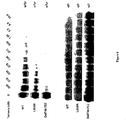
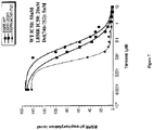
- Receptor autophosphorylation an index of EGFR receptor activation by growth factor stimulation, was detected by Western blotting using an HRP-conjugated anti-phosphotyrosine antibody (Oncogene Sciences, AB-4). Transfection efficiency was evaluated using an antibody specific for the gD epitope tag (5B6). Level of receptor activation was evaluated from the autoradiograms using NIH Image software. These data were then used to generate a graph from which an IC50 was calculated using a 4 parameter fit function. As illustrated by the results below, erlotinib has a greater affinity to EGFR containing mutations compared to wild-type EGFR.
- EGFR construct inhibition (IC50) WT EGFR-gD 50 nM L858R EGFR-gD 20 nM del(746-752) EGFR-gD 5 nM
Description
- The present invention relates to cancer diagnostics and therapies and in particular to the detection of mutations that are diagnostic and/or prognostic.
- Epidermal Growth Factor Receptor (EGFR) is a member of the
type 1 tyrosine kinase family of growth factor receptors, which play critical roles in cellular growth, differentiation, and survival. Activation of these receptors typically occurs via specific ligand binding, resulting in hetero- or homodimerization between receptor family members, with subsequent autophosphorylation of the tyrosine kinase domain. This activation triggers a cascade of intracellular signaling pathways involved in both cellular proliferation (the ras/raf/MAP kinase pathway) and survival (the Pl3 kinase/Akt pathway). Members of this family, including EGFR and HER2, have been directly implicated in cellular transformation. - A number of human malignancies are associated with aberrant or overexpression of EGFR and/or overexpression of its specific ligands e.g. transforming growth factor α (Gullick, Br Med Bull 1991, 47:87-98; Modijtahedi and Dean, Int J Oncol 1994, 4:277-96; Salomon et al., Crit Rev Oncol Hematol 1995;19:183-232). EGFR overexpression has been associated with an adverse prognosis in a number of human cancers, including NSCLC. In some instances, overexpression of tumor EGFR has been correlated with both chemoresistance and a poor prognosis (Lei et al., Anticancer Res 1999; 19:221-8; Veale et al., Br J Cancer 1993;68:162-5). These observations suggest that agents that effectively inhibit EGFR receptor activation and subsequent downstream signaling may have clinical activity in a variety of human cancers, including NSCLC.
- Tarceva™ (also known as erlotinib; OSI-774), a quinazoline, is an orally active, potent, selective inhibitor of EGFR tyrosine kinase. Erlotinib inhibits human EGFR tyrosine kinase with an IC50 of 2 nM (0.786 mg/mL) in an in vitro enzyme assay. This inhibition is selective for EGFR tyrosine kinase, results in cell cycle arrest at G1, and is reversible. Oral administration of erlotinib in mice has demonstrated a >70% reduction in EGFR autophosphorylation in human xenografts and marked growth inhibition of HN5 and A431 xenografts in nude mice has been demonstrated. In addition to single-agent activity in in vivo assay systems, erlotinib has been evaluated in combination with a number of chemotherapy agents to determine possible interactions. There was an additive interaction between erlotinib and paclitaxel, cisplatin, gemcitabine, and doxorubicin.
- Lung cancer represents the leading cause of cancer-related mortality for both men and women in the United States. In 2000, it was estimated that 164,000 new cases would be diagnosed and 157,000 patients would die from this disease (Greenlee et al., CA Cancer J Clin 2001, 51:15-36). Approximately 75% of these patients would have had non-small cell histologies, with the majority presenting with inoperable Stage IIIB or Stage IV disease. For those patients with more limited disease at presentation (Stages I-IIIA), relapse following standard surgical therapy, with or without adjuvant or neoadjuvant chemo- and/or radiotherapy, is common. These findings result in an overall 5-year survival in non-small cell lung cancer (NSCLC) of ∼12% and serve to emphasize the unmet medical need in this disease.
- The platinum compound cisplatin was the first chemotherapy agent to show clinical benefit in the management of locally advanced or metastatic NSCLC. Randomized clinical trials demonstrated improved response rates, quality of life, and survival compared with the best supportive care (Rapp et al. 1988). However, the magnitude of this improvement was modest-measured in weeks. Subsequently, a number of newer chemotherapy agents have been evaluated as single agents and in combination with the platinum salts in the first-line setting. The conclusion from these studies is that modem "doublet" chemotherapy appears to achieve response rates of 15%-20%, median time to disease progression of 3-4 months, and median survival of 7-8 months. The modest improvements in efficacy with combination therapies over the results obtained with cisplatin have established these therapies as a standard of care for patients with advanced NSCLC and an acceptable performance status (Non-Small Cell Lung Cancer Cooperative Group, Br Med J 1995, 311:899-909; American Society of Clinical Oncology, J Clin Oncol 1997, 15:2996-3018; Breathnach et al., J Clin Oncol 2001;19:1734-42).
- The present invention provides methods, as defined in the claims, for determining the prognosis of a patient having a non-small cell lung cancer tumor.
- The present invention also provides methods, as defined in the claims, of identifying a non-small cell lung cancer tumor that is susceptible to treatment with an EGFR inhibitor.
- The present invention also provides an EGFR inhibitor selected from the group consisting of cetuximab, panitumumab, erlotinib or gefitinib, for use a method of treatment of non-small cell lung cancer tumor in a human patient, as defined in the claims.
- The present disclosure provides a method for identifying a tumor in a human subject that is susceptible to treatment comprising determining the presence of a mutated EGFR gene or mutated EGFR protein in a sample of said tumor wherein said mutation is located in exons 18-21 of EGFR whereby the presence of a mutated EGFR gene or mutated EGFR protein indicates the tumor is susceptible to treatment.
- Also disclosed is a method of treating a tumor in a mammal comprising identifying the presence of an EGFR mutation in said tumor and treating said mammal with an anticancer agent.
- Also disclosed is a method of identifying an EGFR mutation in a sample comprising contacting nucleic acid from said sample with a probe that is capable of specifically hybridizing to nucleic acid encoding a mutated EGFR protein, or fragment thereof incorporating a mutation, and detecting the hybridization.
- Also disclosed are nucleic acid probes capable of specifically hybridizing to nucleic acid encoding a mutated EGFR protein or fragment thereof incorporating a mutation.
- Also disclosed is a method of detecting a mutated EGFR gene in a sample comprising amplifying from said sample nucleic acid corresponding to the kinase domain of said EGFR gene, or a fragment thereof suspected of containing a mutation, and comparing the electrophoretic mobility of the amplified nucleic acid to the electrophoretic mobility of corresponding wild-type EGFR gene or fragment thereof.
- Also disclosed is a method for identifying a tumor in a human subject that is susceptible to treatment with an EGFR inhibitor comprising (i) determining the presence of a wild-type KRAS protein or gene in a sample of said tumor whereby the presence of a wild-type KRAS protein or gene indicates that the tumor is susceptible to treatment with an EGFR inhibitor or (ii) determining the presence of a mutated KRAS protein or gene in a sample of said tumor whereby the absence of a mutated KRAS protein or gene indicates that the tumor is susceptible to treatment with an EGFR inhibitor.
-
-
Figure 1 illustrates the amino acid sequence of wild-type EGFR1 (SEQ ID NO: 1) in which the signal sequence is residues 1-24, the extracellular domain includes residues 24-645, the transmembrane domain includes residues 646-668, and the cytoplasmic domain includes residues 669-1210. The tyrosine kinase domain region is residues 718-964, and the threonine phosphorylation site is residue 678. -
Figure 2a through 2d is the cDNA sequence (SEQ ID NO: 2) of wild-type EGFR in whichexon 18 corresponds to nucleotides 2308-2430;exon 19 corresponds to nucleotides 2431-2529;exon 20 corresponds to nucleotides 2530-2715 andexon 21 corresponds to 2716-2871. -
Figure 3 is a graphical representation of extracellular (top) and intracellular (bottom) regions of EGFR. -
Figure 4 is a Kaplan-Meier curve showing time to progression of patients having NSCLC tumors expressing wild-type EGFR (solid line) and mutant EGFR (dashed line). -
Figure 5 is a Kaplan-Meier curve showing survival of patients having NSCLC tumors expressing wild-type EGFR (solid line) and mutant EGFR (dashed line). -
Figure 6 is an autoradiograph illustrating inhibition of autophosphorylation of wild-type EGFR, and mutant EGFR (L858R and del746-752) with varying concentrations of erlotinib in transiently transfected COS7 cells. -
Figure 7 is a graph showing inhibition of autophosphorylation of wild-type EGFR and mutant EGFR (L858R and del746-752) with varying concentrations of erlotinib in transiently transfected COS7 cells. -
Figure 8 illustrates mutations inexons -
Figure 9 illustrates mutations inexons - It is a discovery of the present invention that mutational events associated with tumorigenesis occur in Epidermal Growth Factor Receptor (EGFR). Although it was previously known that aberrant EGFR activity was associated with various cancers, it was unknown that mutations in the EGFR kinase domain region (KDR) existed that caused aberrant signaling activity associated with some cancers. Surprisingly patients suffering from tumors having EGFR KDR mutations have a better prognosis than those with wild-type EGFR. The KDR mutations of the EGFR gene can involve rearrangements such as insertions and deletions as well as point mutations.
- Samples from approximately 250 patients who participated a randomized, double-blinded phase III clinical trial referred to as Tribute were sequenced for mutations occurring in exons 18-21 of EGFR. Tribute studied 1,079 patients at approximately 150 centers in the United States having histological confirmed NSCLC who had not received prior chemotherapy comparing erlotinib + chemotherapy (carboplatin/paclitaxel) with chemotherapy alone. Patients received paclitaxel (200 mg/
m 2 3 hour i.v. infusion) followed by carboplatin (AUC = 6 mg/ml x minute infused over 15-30 minutes using Calvert formula) with or without erlotinib (100 mg/day p.o. escalated to 150mg/day for tolerant patients). Tumor samples, formalin-fixed paraffin-embedded blocks or unstained slides, from approximately 250 patients collected from the Tribute trial were enriched for tumor cells by laser capture mircrodissection followed by DNA extraction. Exons 18-21 were amplified by nested PCR and bi-directional sequences were obtained from each PCR product using fluorescent dye-terminator chemistry. Mutations discovered from the sequencing are shown in table 1:Table 1 protein mutation nucleic acid mutation exon G719A 2402G>C 18 G719C 2401G>T 18 G719S 2401G>A 18 E746-R748 del 2482-2490 del GGAATTAAGA (SEQ ID NO: 32) 19 E746-A750 del 2481-2495 del GGAATTAAGAGAAGC (SEQ ID NO: 33) 19 E746-R748 del 2482-2490 del GAATTAAGA E749Q 2491G>C A750P 2494G>C 19 L747-E749 del 2485-2493 del TTAAGAGAA A750P 494G>C 19 L747S R748-P753 del 2486-2503 del TAAGAGAAGCAACATCTC (SEQ ID NO: 34) 19 L747-S752 del E746V 2485-2502 del TTAAGAGAAGCAACATCT 2483A>T (SEQ ID NO: 35) 19 L747-T751 del ins S 2486-2494del TAAGAGAAGCAA (SEQ ID NO: 36) 19 2499-2522 del ATCTCCGAAAGCCAACAAGGAAAT 19 S752-I759 del (SEQ ID NO: 37) M766-A767 Al ins 2544-2545 ins GCCATA 20 S768-V769 SVA ins 2554-2555 ins CCAGCGTGG (2556C>T silent) 20 L858R 2819T>G 21 G719C 2401G>T 18 S768I 2549G>T {2607G>A SNP silent} 20 G719C 2401G> T 18 V765M 2539G>A 20 S768I 2549G> T 20 A755V 2510C> T 19 L747S 2486T> C 19 E746K 2482G>A 19 S752-I759 del (SEQ ID NO: 37) P772-H773 V ins 2561-2562 ins GGT 20 L858P 2819T> C 21 L861Q 2576T>A 21 P772-H773 NS ins 2562-2563 ins AACTCC H773Y 2563C> T 20 T790M 2615C> T 20 L858R 2819T> G 21 S784F 21 L858R 21 ins = insertion del = deletion figures 2a-2d . - Clinical outcome of patients having tumors with EGFR mutations and wild-type EGFR were analyzed according to response (complete + partial) benefit (response + stable disease) and progressive disease. Lesions were evaluated using Response Evaluation Criteria in Solid Tumors (RECIST) criteria whereby "complete response" (CR) is defined as the disappearance of all target lesions; "partial response" (PR) is defined as at least a 30% decrease in the sum of the longest diameter of target lesions, taking as reference the baseline sum longest diameter; "progressive disease" (PD) is defined as at least a 20% increase in the sum of the longest diameter of target lesions, taking as reference the smallest sum longest diameter recorded since the treatment started or the appearance of one or more new lesions; and "stable disease" (SD) is defined as neither sufficient shrinkage to qualify for partial response nor sufficient increase to qualify for progressive disease, taking as reference the smallest sum longest diameter since the treatment started.
- Results of the analysis are summarized in table 2.
Table 2 Mutant EGFR n=24 Wild-Type EGFR n=181 Response / Benefit Rate response (CR + PR) 11 46% 46 25% benefit (CR + PR + SD) 18 75% 105 58 % SD 7 29% 59 33 % PD 6 25% 76 42% Survival (days) median 435 309 range 133-687 9-643 CR=complete response; PR=partial response; SD=stable disease; PD=progressing disease - Analysis of clinical outcome revealed that patients with tumors expressing a mutation in exons 18-21 of EGFR have better prognosis than those with tumors expressing wild-type EGFR. Mutant EGFR patients exhibited greater response rate, benefit rate and survival when treated with chemotherapy or chemotherapy plus erlotinib. These results are useful for predicting outcome such that patients who's tumors have EGFR mutations in any or all of
exons 18 through 21 have more favorable prognosis than patients who's tumors do not have such mutations. - Accordingly, the present invention provides a method as defined in the claims for determining the prognosis of a patient having a tumor comprising determining in a sample of said tumor the presence or absence of one or more EGFR mutations in exons 18-21 (or the amino acid sequence corresponding to exons 18-21) whereby the presence of said one or more EGFR mutation indicates better prognosis compared to the absence of said one or more EGFR mutation. By "prognosis" is meant response and/or benefit and/or survival. By "EGFR mutations" means an amino acid or nucleic acid sequence that differs from wild-type EGFR protein or nucleic acid respectively found on one allele (heterozygous) or both alleles (homozygous) and may be somatic or germ line. The mutation is E746K or A755V.
- EGFR exons 18-21 from an H1975 tumor cell line that exhibited resistance to treatment with erlotinib was sequenced and found to incorporate a mutation T790M in combination with an L858R mutation. Accordingly the present disclosure further provides a method for determining the prognosis of a patient having a tumor comprising determining in a sample of said tumor the presence or absence of the T790M EGFR mutation whereby the presence of said T790M EGFR mutation indicates poorer prognosis compared to the absence of said T790M EGFR mutation. Further, there is provided a method of identifying patients having a tumor that is less responsive to therapy of an EGFR inhibitor such as erlotinib or gefitinib, whether in combination with chemotherapy or not, comprising determining the presence or absence of a T790M EGFR mutation in the patient's tumor whereby the presence of said mutation indicates the patient will respond less to said therapy compared to a patient having a tumor that does not have said T790M EGFR mutation. Further, there is provided a method of identifying a tumor that is resistant to treatment with an EGFR inhibitor, such as a kinase domain binding inhibitor (for example erlotinib or gefitinib), whether in combination with chemotherapy or not, comprising determining the presence or absence of a T790M EGFR mutation in a sample of the tumor whereby the presence of said mutation indicates the tumor is resistant to said treatment. It is understood that determination of the mutation is at the protein level or nucleic acid level (genomic DNA or mRNA) and are accomplished using techniques such as those described herein. In a particular embodiment, said EGFR inhibitor competes with ATP at the EGFR kinase domain. In a particular embodiment the EGFR inhibitor is erlotinib.
- In another aspect, there is provided a method of treating a patient having a tumor incorporating a T790M mutant EGFR protein or gene (or treating a tumor incorporating a T790M mutant EGFR protein or gene) comprising co-administering to said patient (or contacting said tumor with) a first compound that binds to and/or inhibits signaling of said T790M mutant EGFR in combination with a second compound that binds to and/or inhibits signaling of wild-type EGFR or EGFR incorporating an activating mutation. In a particular embodiment said activating mutation is one or more of those described in Table 1 (other than T790M). In a particular embodiment said first and second compounds are administered sequentially or concommitantly. In a particular embodiment said second compound is erlotinib.
- In another aspect of the disclosure, there is provided a method of screening for compounds that inhibit signaling of a mutant EGFR protein that incorporates a T790M mutation, comprising contacting said mutant EGFR with a test compound in the presence of a phosphorylation substrate and ATP and detecting a change in the amount of phosphorylation of said substrate whereby a reduction of phosphorylation of said substrate compared to a control, or compared to phosphorylation of the substrate in the absence of the test compound, indicates said test compound is an inhibitor of mutant EGFR signaling. In an embodiment, said method is performed in vitro in the presence of a ligand for said mutant EGFR such as EGF or TGF-alpha.
- In a particular embodiment the inhibitory activity of a test compound can be determined in vitro by the amount of inhibition of the phosphorylation of an exogenous substrate (e.g. Lys3 -Gastrin or polyGluTyr (4:1) random copolymer (I. Posner et. al., J. Biol. Chem. 267 (29), 20638-47 (1992)) on tyrosine by epidermal growth factor receptor kinase by a test compound relative to a control. Purified, soluble human T790M mutant EGFR (96 ng) is preincubated in a microfuge tube with EGF (2 µg/ml) in phosphorylation buffer+vanadate (PBV: 50 mM HEPES, pH 7.4; 125 mM NaCl; 24 mM MgCl2; 100 µM sodium orthovanadate), in a total volume of 10 µl, for 20-30 minutes at room temperature. The test compound, dissolved in dimethylsulfoxide (DMSO), is diluted in PBV, and 10 µl is mixed with the mutant EGFR/EGF mix, and incubated for 10-30 minutes at 30° C. The phosphorylation reaction is initiated by addition of 20 µl 33P-ATP/substrate mix (120 µM Lys3 -Gastrin (sequence in single letter code for amino acids, KKKGPWLEEEEEAYGWLDF - SEQ ID NO: 38), 50 mM Hepes pH 7.4, 40 µM ATP, 2µCi γ-[33P]-ATP) to the mutant EGFR/EGF mix and incubated for 20 minutes at room temperature. The reaction is stopped by addition of 10 µl stop solution (0.5M EDTA,
pH 8; 2mM ATP) and 6 µl 2N HCl. The tubes are centrifuged at 14,000 RPM, 4°C., for 10 minutes. 35 µl of supernatant from each tube is pipetted onto a 2.5 cm circle of Whatman P81 paper, bulk washed four times in 5% acetic acid, 1 liter per wash, and then air dried. This results in the binding of substrate to the paper with loss of free ATP on washing. The [33P] incorporated is measured by liquid scintillation counting. Incorporation in the absence of substrate (e.g., lys3 -gastrin) is subtracted from all values as a background and percent inhibition is calculated relative to controls without test compound present. Such assays, carried out with a range of doses of test compounds, allow the determination of an approximate IC50 value for the in vitro inhibition of T790M mutant EGFR kinase activity. - In another aspect of the invention there is provided a method as defined in the claims for identifying a tumor in a human subject that is susceptible to treatment comprising determining the presence of a mutated EGFR gene or mutated EGFR protein in a sample of said tumor wherein said mutation is located in exons 18-21 of EGFR whereby the presence of a mutated EGFR gene or mutated EGFR protein indicates that the tumor is susceptible to treatment with an EGFR inhibitor. The tumor is a non-small cell lung cancer tumor. In an embodiment the EGFR inhibitor is an antibody such as Erbitutux™ (cetuximab, Imclone Systems Inc.) and ABX-EGF (panitumumab, Abgenix, Inc.). In another embodiment the EGFR inhibitor is a small molecule that competes with ATP such as Tarceva™ (erlotinib, OSI Pharmaceuticals), Iressa™ (gefitinib, Astra-Zeneca), tyrphostins described by Dvir, et al., J Cell Biol., 113:857-865 (1991); tricyclic pyrimidine compounds disclosed in
U.S. Patent 5,679,683 ; compound 6- (2,6-dichlorophenyl)-2-(4-(2-diethylaininoethoxy)phenylamino)-8-methyl-8H-pyrido(2,3-d)pyrimidin-7-one (known as PD166285) disclosed in Panek, et al., Journal of Pharmacology and Experimental Therapeutics 283, 1433-1444 (1997). - In another aspect of the disclosure there is provided a method of identifying an EGFR mutation in a sample comprising contacting nucleic acid from said sample with a nucleic acid probe that is capable of specifically hybridizing to nucleic acid encoding a mutated EGFR protein, or fragment thereof incorporating a mutation, and detecting said hybridization. In a particular embodiment said probe is detectably labeled such as with a radioisotope (3H, 32P, 33P etc), a fluorescent agent (rhodamine, fluorescene etc.) or a chromogenic agent. In a particular embodiment the probe is an antisense oligomer, for example PNA, morpholino-phosphoramidates, LNA or 2'-alkoxyalkoxy. The probe may be from about 8 nucleotides to about 100 nucleotides, or about 10 to about 75, or about 15 to about 50, or about 20 to about 30. In another aspect said probes of the disclosure are provided in a kit for identifying EGFR mutations in a sample, said kit comprising an oligonucleotide that specifically hybridizes to or adjacent to a site of mutation in the EGFR gene. The kit may further comprise instructions for treating patients having tumors that contain EGFR mutations with an EGFR inhibitor based on the result of a hybridization test using the kit.
- In another aspect of the disclosure there is provided a method of detecting a mutated EGFR gene in a sample comprising amplifying from said sample nucleic acid corresponding to the kinase domain of said EGFR gene, or exons 18-21, or a fragment thereof suspected of containing a mutation, and comparing the electrophoretic mobility of the amplified nucleic acid to the electrophoretic mobility of corresponding wild-type EGFR gene or fragment thereof. A difference in the mobility indicates the presence of a mutation in the amplified nucleic acid sequence. Electrophoretic mobility may be determined on polyacrylamide gel.
- Alternatively , amplified EGFR gene or fragment nucleic acid may be analyzed for detection of mutations using Enzymatic Mutation Detection (EMD) (Del Tito et al, Clinical Chemistry 44:731-739, 1998). EMD uses the bacteriophage resolvase T4 endonuclease VII, which scans along double-stranded DNA until it detects and cleaves structural distortions caused by base pair mismatches resulting from point mutations, insertions and deletions. Detection of two short fragments formed by resolvase cleavage, for example by gel eletrophoresis, indicates the presence of a mutation. Benefits of the EMD method are a single protocol to identify point mutations, deletions, and insertions assayed directly from PCR reactions eliminating the need for sample purification, shortening the hybridization time, and increasing the signal-to-noise ratio. Mixed samples containing up to a 20-fold excess of normal DNA and fragments up to 4 kb in size can been assayed. However, EMD scanning does not identify particular base changes that occur in mutation positive samples requiring additional sequencing procedures to identiity of the mutation if necessary. CEL I enzyme can be used similarly to resolvase T4 endonuclease VII as demonstrated in
US5869245 . - Another simple kit for detecting the EGFR mutations of the disclosure is a reverse hybridization test strip similar to Haemochromatosis StripAssay™ (Viennalabs http://www.bamburghmarrsh.com/pdf/4220.pdf) for detection of multiple mutations in HFE, TFR2 and FPN1 genes causing Haemochromatosis. Such an assay is based on sequence specific hybridisation following amplification by PCR. For single mutation assays, a microplate-based detection system may be applied, whereas for multi-mutation assays, teststrips may be used as "macro-arrays". Kits may include ready-to use reagents for sample prep, amplification and mutation detection. Multiplex amplification protocols provide convenience and allow testing of samples with very limited volumes. Using the straightforward StripAssay format, testing for twenty and more mutations may be completed in less than five hours without costly equipment. DNA is isolated from a sample and the EGFR gene (or exons 18-21 or KDR or segments thereof) is amplified in vitro (e.g. PCR) and biotin-labelled, preferably in a single ("multiplex") amplification reaction. The PCR products are the selectively hybridized to oligonucleotide probes (wild-type and mutant specific) immobilized on a solid support such as a test strip in which the probes are immobilized as parallel lines or bands. Bound biotinylated amplicons are detected using streptavidin-alkaline phosphatase and color substrates. Such an assay can detect all or any subset of the mutations in table 1. With respect to a particular mutant probe band one of three signalling patterns are possible: (i) a band only for wild-type probe which indicates normal EGFR (ii) bands for both wild-type and a mutant probe which indicates heterozygous genotype and (iii) band only for the mutant probe which indicates homozygous mutant EGFR genotype. Accordingly there is further provides a method of detecting EGFR mutations of the disclosure comprising isolating nucleic acid from a sample, amplifying the EGFR gene, or fragment thereof (e.g. the KDR or exons 18-21 or smaller) such that the amplified nucleic acid comprises a ligand, contacting the amplified EGFR gene or fragment with a probe which comprises a detectable binding partner to the ligand and the probe is capable of specifically hydribizing to an EGFR mutation, and then detecting the hybridization of said probe to said amplified EGFR gene or fragment. In a particular embodiment the ligand is biotin and the binding partner is comprises avidin or streptavidin. In a particular embodiment the binding partner is steptavidin-alkaline which is detectable with color substrates. In a particular embodiment the probes are immobilized for example on a test strip wherein probes complementary to different mutations are separated from one another. Alternatively, the amplified nucleic acid is labelled with a radioisotope in which case the probe need not comprise a ligand.
- The tumor samples were also analyzed for mutations in KRAS (as referred to as p21a). Particular mutations detected in
exon 1 are: G12C; G12A; G12D; G12R; G12S; G12V; G13C; G13D which correlated with poor prognosis to chemotherapy as well as chemotherapy with erlotinib therapy. Accordingly, the disclosure further provides a method of identifying patients not responsive to therapy of an EGFR inhibitor such as erlotinib or erlotinib in combination with chemotherapy comprising determining the presence or absence of a KRAS mutation whereby the presence of said mutation indicates a patient will not respond to said therapy. Alternatively, there is provided a method for identifying a tumor in a human subject that is susceptible to treatment with an EGFR inhibitor comprising (i) determining the presence of a wild-type KRAS protein or gene in a sample of said tumor whereby the presence of a wild-type KRAS protein or gene indicates that the tumor is susceptible to treatment with an EGFR inhibitor or (ii) determining the presence of a mutated KRAS protein or gene in a sample of said tumor whereby the absence of a mutated KRAS protein or gene indicates that the tumor is susceptible to treatment with an EGFR inhibitor. In a particular embodiment the mutation is inexon 1 of K-Ras. In another embodiment the K-Ras mutation is at least one of G12C; G12A; G12D; G12R; G12S; G12V; G13C; G13D. Alternatively, individuals who have tumors which harbor mutant K-Ras may be treated with EGFR inhibitors when in concomitantly with a K-Ras inhibitor. Methods for determining the presence of K-Ras mutations are analogous to those used to identify EGFR mutations described in detail herein. - According to the diagnostic and prognostic method of the present invention, alteration of the wild-type EGFR gene is detected as defined in the claims. Somatic mutations are those which occur only in certain tissues, e.g., in the tumor tissue, and are not inherited in the germ line. Germ line mutations can be found in any of a body's tissues. If only a single allele is somatically mutated, an early neoplastic state is indicated. However, if both alleles are mutated then a late neoplastic state is indicated. The finding of EGFR mutations is therefore a diagnostic and prognostic indicator as described herein.
- The EGFR mutations found in tumor tissues may result in increased signaling activity relative to wild-type EGFR leading to a cancerous state. In order to detect the alteration of the wild-type EGFR gene a sample or biopsy of the tumor is obtained by methods well known in the art and appropriate for the particular type and location of the tumor. For instance, samples of lung cancer lesions may be obtained by resection, bronchoscopy, fine needle aspiration, bronchial brushings, or from sputum, pleural fluid or blood. Means for enriching a tissue preparation for tumor cells are known in the art. For example, the tissue may be isolated from paraffin or cryostat sections. Cancer cells may also be separated from normal cells by flow cytometry or laser capture microdissection. These as well as other techniques for separating tumor from normal cells are well known in the art. If the tumor tissue is highly contaminated with normal cells, detection of mutations is more difficult.
- Detection of point mutations may be accomplished by molecular cloning of the EGFR allele (or alleles) and sequencing that allele(s) using techniques well known in the art. Alternatively, the polymerase chain reaction (PCR) can be used to amplify gene sequences directly from a genomic DNA preparation from the tumor tissue. The DNA sequence of the amplified sequences can then be determined and mutations identified therefrom. The polymerase chain reaction is well known in the art and described in Saiki et al., Science 239:487, 1988;
U.S. 4,683,203 ; andU.S. 4,683,195 . - Specific primer pairs which can be used for PCR amplification of EGFR exons 18-21 include:
- <5pEGFR.ex18.out> CAAATGAGCTGGCAAGTGCCGTGTC (SEQ ID NO: 39)
- <3pEGFR.ex18.out> GAGTTTCCCAAACACTCAGTGAAAC (SEQ ID NO: 40)
- <5pEGFR.ex19.out> GCAATATCAGCCTTAGGTGCGGCTC (SEQ ID NO: 41)
- <3pEGFR.ex19.out> CATAGAAAGTGAACATTTAGGATGTG (SEQ ID NO: 42)
- <5pEGFR.ex20.out> CCATGAGTACGTATTTTGAAACTC (SEQ ID NO: 43)
- <3pEGFR.ex20.out> CATATCCCCATGGCAAACTCTTGC (SEQ ID NO: 44)
- <5pEGFR.ex21.out> CTAACGTTCGCCAGCCATAAGTCC (SEQ ID NO: 45)
- <3pEGFR.ex21.out> GCTGCGAGCTCACCCAGAATGTCTGG (SEQ ID NO: 46)
- <5pEGFR.ex18.in.m13f> TGTAAAACGACGGCCAGTCAAGTGCCGTGTCCTGGCACCCAAGC (SEQ ID NO: 47)
- <3pEGFR.ex18.in.m13r> CAGGAAACAGCTATGACCCCAAACACTCAGTGAAACAAAGAG (SEQ ID NO: 48)
- <5pEGFR.ex19.in.m13f> TGTAAAACGACGGCCAGTCCTTAGGTGCGGCTCCACAGC (SEQ ID NO: 49)
- <3pEGFR.ex19.in.m13r> CAGGAAACAGCTATGACCCATTTAGGATGTGGAGATGAGC (SEQ ID NO: 50)
- <5pEGFR.ex20.in.m13f> TGTAAAACGACGGCCAGTGAAACTCAAGATCGCATTCATGC (SEQ ID NO: 51)
- <3pEGFR.ex20.in.m13r> CAGGAAACAGCTATGACCGCAAACTCTTGCTATCCCAGGAG (SEQ ID NO: 52)
- <5pEGFR.ex21.in.m13f> TGTAAAACGACGGCCAGTCAGCCATAAGTCCTCGACGTGG (SEQ ID NO: 53)
- <3pEGFR.ex21.in.m13r> CAGGAAACAGCTATGACCCATCCTCCCCTGCATGTGTTAAAC (SEQ ID NO: 54)
- Specific primer pairs which can be used for PCR amplification of K-
Ras exon 1 include: - <5pKRAS-out> TACTGGTGGAGTATTTGATAGTG (SEQ ID NO: 55)
- <3pKRAS-out> CTGTATCAAAGAATGGTCCTG (SEQ ID NO: 56)
- <5pKRA8-in.m13f> TGTAAAACGACGGCCAGTTAGTGTATTAACCTTATGTG (SEQ ID NO: 57)
- <3pKRAS-in.m13r> CAGGAAACAGCTATGACCACCTCTATTGTTGGATCATATTCG (SEQ ID NO: 58)
- The ligase chain reaction, which is known in the art, can also be used to amplify EGFR sequences. See Wu et al., Genomics, Vol. 4, pp. 560-569 (1989). In addition, a technique known as allele specific PCR can be used. (See Ruano and Kidd, Nucleic Acids Research, Vol. 17, p. 8392, 1989.) According to this technique, primers are used which hybridize at their 3'ends to a particular EGFR mutation. If the particular EGFR mutation is not present, an amplification product is not observed. Amplification Refractory Mutation System (ARMS) can also be used as disclosed in European Patent Application Publication No.
0332435 and in Newton et al., Nucleic Acids Research, Vol. 17, p.7, 1989. Insertions and deletions of genes can also be detected by cloning, sequencing and amplification. In addition, restriction fragment length polymorphism, (RFLP) probes for the gene or surrounding marker genes can be used to score alteration of an allele or an insertion in a polymorphic fragment. Single stranded conformation polymorphism (SSCP) analysis can also be used to detect base change variants of an allele. (Orita et al., Proc. Natl. Acad. Sci. USA Vol. 86, pp. 2766-2770, 1989, and Genomics, Vol. 5, pp. 874-879, 1989.) Other techniques for detecting insertions and deletions as are known in the art can be used. - Alteration of wild-type genes can also be detected on the basis of the alteration of a wild-type expression product of the gene. Such expression products include both the EGFR mRNA as well as the EGFR protein product. Point mutations may be detected by amplifying and sequencing the mRNA or via molecular cloning of cDNA made from the mRNA. The sequence of the cloned cDNA can be determined using DNA sequencing techniques which are well known in the art. The cDNA can also be sequenced via the polymerase chain reaction (PCR).
- Mismatches, according to the present invention are hybridized nucleic acid duplexes which are not 100% complementary. The lack of total complementarity may be due to deletions, insertions, inversions, substitutions or frameshift mutations. Mismatch detection can be used to detect point mutations in the gene or its mRNA product. While these techniques are less sensitive than sequencing, they are simpler to perform on a large number of tumor samples. An example of a mismatch cleavage technique is the RNase protection method, which is described in detail in Winter et al., Proc. Natl. Acad. Sci. USA, Vol. 82, p. 7575, 1985 and Meyers et al., Science, Vol. 230, p. 1242, 1985. In the practice a the present invention the method involves the use of a labeled riboprobe which is complementary to the human wild-type EGFR gene coding sequence (or exons 18-21 or KDR thereof). The riboprobe and either mRNA or DNA isolated from the tumor tissue are annealed (hybridized) together and subsequently digested with the enzyme RNase A which is able to detect some mismatches in a duplex RNA structure. If a mismatch is detected by RNase A, it cleaves at the site of the mismatch. Thus, when the annealed RNA preparation is separated on an electrophoretic gel matrix, if a mismatch has been detected and cleaved by RNase A, an RNA product will be seen which is smaller than the full-length duplex RNA for the riboprobe and the mRNA or DNA. The riboprobe need not be the full length of the EGFR mRNA or gene but can be
exons 18 through 21 or the EGFR KDR or segments thereof. If the riboprobe comprises only a segment of the EGFR mRNA or gene it will be desirable to use a number of these probes to screen the whole mRNA sequence for mismatches. - In a similar manner, DNA probes can be used to detect mismatches, through enzymatic or chemical cleavage. See, e.g., Cotton et al., Proc. Natl. Acad. Sci. USA, Vol. 85, 4397, 1988; and Shenk et al., Proc. Natl. Acad. Sci. USA, Vol. 72, p. 989, 1975. Alternatively, mismatches can be detected by shifts in the electrophoretic mobility of mismatched duplexes relative to matched duplexes. See, e.g., Cariello, Human Genetics, Vol. 42, p. 726, 1988. With either riboprobes or DNA probes, the cellular mRNA or DNA which might contain a mutation can be amplified using PCR before hybridization. Changes in DNA of the EGFR gene can also be detected using Southern hybridization, especially if the changes are gross rearrangements, such as deletions and insertions.
- DNA sequences of the EGFR gene which have been amplified by use of polymerase chain reaction may also be screened using allele-specific probes. These probes are nucleic acid oligomers, each of which contains a region of the EGFR gene sequence harboring a known mutation. For example, one oligomer may be about 30 nucleotides in length, corresponding to a portion of the EGFR gene sequence. By use of a battery of such allele-specific probes, PCR amplification products can be screened to identify the presence of a previously identified mutation in the EGFR gene. Hybridization of allele-specific probes with amplified EGFR sequences can be performed, for example, on a nylon filter. Hybridization to a particular probe under stringent hybridization conditions indicates the presence of the same mutation in the tumor tissue as in the allele-specific probe.
- Alteration of wild-type EGFR genes can also be detected by screening for alteration of wild-type EGFR protein. For example, monoclonal antibodies immunoreactive with EGFR can be used to screen a tissue. Lack of cognate antigen would indicate an EGFR mutation. Antibodies specific for products of mutant alleles could also be used to detect mutant EGFR gene product. Antibodies may be identified from phage display libraries. Such immunological assays can be done in any convenient format known in the art. These include Western blots, immunohistochemical assays and ELISA assays. Any means for detecting an altered EGFR protein can be used to detect alteration of wild-type EGFR genes.
- Mutant EGFR genes or gene products can be detected from tumor or from other body samples such as urine, sputum or serum. The same techniques discussed above for detection of mutant EGFR genes or gene products in tumor samples can be applied to other body samples. Cancer cells are sloughed off from tumors and appear in such body samples. By screening such body samples, a simple early diagnosis can be achieved for many types of cancers. In addition, the progress of chemotherapy or radiotherapy can be monitored more easily by testing such body samples for mutant EGFR genes or gene products.
- The diagnostic method of the present invention is useful for clinicians so that they can decide upon an appropriate course of treatment. For example, a tumor displaying alteration of both EGFR alleles might suggest a more aggressive therapeutic regimen than a tumor displaying alteration of only one EGFR allele.
- The primer pairs of the present disclosure are useful for determination of the nucleotide sequence of a particular EGFR allele using the polymerase chain reaction. The pairs of single stranded DNA primers can be annealed to sequences within or surrounding the EGFR gene on in order to prime amplifying DNA synthesis of the EGFR gene itself. A set of these primers allows synthesis of all of the nucleotides of the
EGFR exons 18 through 21. Allele specific primers can also be used. Such primers anneal only to particular EGFR mutant alleles, and thus will only amplify a product in the presence of the mutant allele as a template. In order to facilitate subsequent cloning of amplified sequences, primers may have restriction enzyme site sequences appended to their ends. Thus, all nucleotides of the primers are derived from EGFR exons 18-21 or sequences adjacent thereto except the few nucleotides necessary to form a restriction enzyme site. Such enzymes and sites are well known in the art. The primers themselves can be synthesized using techniques which are well known in the art. Generally, the primers can be made using oligonucleotide synthesizing machines which are commercially available. Design of particular primers is well within the skill of the art. - The nucleic acid probes provided by the present disclosure are useful for a number of purposes. They can be used in Southern hybridization to genomic DNA and in the RNase protection method for detecting point mutations already discussed above. The probes can be used to detect PCR amplification products. They may also be used to detect mismatches with the EGFR gene or mRNA using other techniques. Mismatches can be detected using either enzymes (e.g., S1 nuclease), chemicals (e.g., hydroxylamine or osmium tetroxide and piperidine), or changes in electrophoretic mobility of mismatched hybrids as compared to totally matched hybrids. These techniques are known in the art. See Novack et al., Proc. Natl. Acad. Sci. USA, Vol. 83, p. 586, 1986. Generally, the probes are complementary to EGFR exon 18-21 sequences, although generally probes to the kinase domain and segments thereof are also contemplated. An entire battery of nucleic acid probes may be used to compose a kit for detecting alteration of wild-type EGFR genes. The kit allows for hybridization to the entire exon 18-21 sequence of the EGFR gene. The probes may overlap with each other or be contiguous.
- If a riboprobe is used to detect mismatches with mRNA, it is complementary to the mRNA of the EGFR gene. The riboprobe thus is an antisense probe in that it does not code for the EGFR protein because it is complementary to the sense strand. The riboprobe generally will be labeled with a radioactive, colorimetric, or fluorometric material, which can be accomplished by any means known in the art. If the riboprobe is used to detect mismatches with DNA it can be of either polarity, sense or anti-sense. Similarly, DNA probes also may be used to detect mismatches.
- Predisposition to cancers can be ascertained by testing any tissue of a human for mutations of the EGFR gene. For example, a person who has inherited a germ line EGFR mutation would be prone to develop cancers. This can be determined by testing DNA from any tissue of the body. For example, blood can be drawn and DNA extracted from the cells of the blood. In addition, prenatal diagnosis can be accomplished by testing fetal cells, placental cells, or amniotic fluid for mutations of the EGFR gene. Alteration of a wild- type EGFR allele, whether for example, by point mutation or by deletion, can be detected by any of the means discussed above.
- Submersed sections in the following solutions:
- 1. Fresh xylenes (to depariffinize the sections) - 5 min
- 2. Fresh xylenes - 5 min
- 3. 100% ethanol - 15 sec
- 4. 95% ethanol - 15 sec
- 5. 70% ethanol - 15 sec
- 6. Deionized water - 15 sec
- 7. Mayer's Hematoxylin - 30 sec
- 8. Deionized water - rinse (x 2) - 15 sec
- 9. 70% ethanol - 15 sec
- 10. Eosin Y - 5 sec
- 11. 95% ethanol - 15 sec
- 12. 95% ethanol - 15 sec
- 13. 100% ethanol - 15 sec
- 14. 100% ethanol - 15 sec
- 15. Xylenes (to ensure dehydration of the section) - 60 sec
- 16. Air-dried for approximately 2 minutes or gently used air gun to completely remove xylenes.
- 17. The tissue was then ready for LCM.
-
- PixCell II LCM System
- CapSure HS or CapSure Macro LCM caps
- ExtractSure device (HS only)
- Razor blades (factory sterile)
- 0.5 ml tubes
- 0.2 ml tubes
- PicoPure DNA extraction Kit
- 65°C incubator
-
- 1. Placed CapSure cap over area of tissue to be collected
- 2. Lased over desired area
- 2. Lifted cap off tissue.
- 3. Dispensed 20 ul of PicoPure digest buffer with Proteinase K into 0.5ml tube.
- 4. Placed cap with dissected material into tube to form a tight seal.
- 5. Inverted tube such that digest buffer covered cap.
- 6. Incubated at 65°C for 24 hours.
- 7. Spun tube with cap to collect digested material in the bottom of the tube.
- 8. Transferred digest to 0.2 ml strip tube.
- 9. Inactivated Proteinase K at 95°C for 10 minutes in a thermocycler with a heated lid.
- 10. Used 1-2 ul of sample in a 50 ul PCR reaction. No clean-up was necessary.
- Primer pairs were designed for each exon to be sequenced (
EGFR exons - <5pEGFR.ex18.out> CAAATGAGCTGGCAAGTGCCGTGTC (SEQ ID NO: 39)
- <3pEGFR.ex18.out> GAGTTTCCCAAACACTCAGTGAAAC (SEQ ID NO: 40)
- <5pEGFR.ex19.out> GCAATATCAGCCTTAGGTGCGGCTC (SEQ ID NO: 41)
- <3pEGFR.ex19.out> CATAGAAAGTGAACATTTAGGATGTG (SEQ ID NO: 42)
- <5pEGFR.ex20.out> CCATGAGTACGTATTTTGAAACTC (SEQ ID NO: 43)
- <3pEGFR.ex20.out> CATATCCCCATGGCAAACTCTTGC (SEQ ID NO: 44)
- <5pEGFR.ex21.out> CTAACGTTCGCCAGCCATAAGTCC (SEQ ID NO: 45)
- <3pEGFR.ex21.out> GCTGCGAGCTCACCCAGAATGTCTGG (SEQ ID NO: 46)
- <5pEGFR.ex18.in.m13f> TGTAAAACGACGGCCAGTCAAGTGCCGTGTCCTGGCACCCAAGC (SEQ ID NO: 47)
- <3pEGFR.ex18.in.m13r> CAGGAAACAGCTATGACCCCAAACACTCAGTGAAACAAAGAG (SEQ ID NO: 48)
- <5pEGFR.ex19.in.m13f> TGTAAAACGACGGCCAGTCCTTAGGTGCGGCTCCACAGC (SEQ ID NO: 49)
- <3pEGFR.ex19.in.m13r> CAGGAAACAGCTATGACCCATTTAGGATGTGGAGATGAGC (SEQ ID NO: 50)
- <5pEGFR.ex20.in.m13f> TGTAAAACGACGGCCAGTGAAACTCAAGATCGCATTCATGC (SEQ ID NO: 51)
- <3pEGFR.ex20.in.m13r> CAGGAAACAGCTATGACCGCAAACTCTTGCTATCCCAGGAG (SEQ ID NO: 52)
- <5pEGFR.ex21.in.m13f> TGTAAAACGACGGCCAGTCAGCCATAAGTCCTCGACGTGG (SEQ ID NO: 53)
- <3pEGFR.ex21.in.m13r> CAGGAAACAGCTATGACCCATCCTCCCCTGCATGTGTTAAAC (SEQ ID NO: 54)
-
- <5pKRAS-out> TACTGGTGGAGTATTTGATAGTG (SEQ ID NO: 55)
- <3pKRAS-out> CTGTATCAAAGAATGGTCCTG (SEQ ID NO: 56)
- <5pKRAS-in.m13f> TGTAAAACGACGGCCAGTTAGTGTATTAACCTTATGTG (SEQ ID NO: 57)
- <3pKRAS-in>.m13r CAGGAAACAGCTATGACCACCTCTATTGTTGGATCATATTCG (SEQ ID NO: 58)
- Nested amplification of the primary PCR product was performed using intron-specific primer pairs located within the primary PCR product. These nested primers pairs were tagged with M13f and M13rev sequences.
-
PCR Reaction: DNA 0.5 to 30ng Primers 250nM/ each outer primers dNTPs 0.2mM each (Roche cat#1581295) MgCl2 1.5mM (15mM 10 X buffer) Enzyme 1.5 U/RX Expand High fidelity Tag (Roche cat#1759078) 50ul reaction volume -
- 95°C - 3minutes
- 94°C - 30seconds repeat 35 times
- 58°C - 30seconds
- 72°C - 1minute
- 72°C - 8minutes
- 4°C - forever
-
PCR Reaction: DNA 1ul from first round PCR reaction Primers 250nM/ each inner primers dNTPs 0.2mM each (Roche cat#1581295) MgCl2 1.5mM (15mM 10 X buffer) Enzyme 1.5 U/RX Expand High fidelity Tag (Roche cat#1759078) 50ul reaction volume -
- 95°C - 3minutes
- 94°C - 30seconds repeat 30 times
- 58°C - 30seconds
- 72°C - 1minute
- 72°C - 8 minutes
- 4°C - forever
- PCR reaction products were run on E-Gel 2% agarose gels (Invitrogen, cat# G6018-02) for quality control. PCR products were purified directly using the Qiaquick 96 PCR purification kit (Qiagen, cat#28181) or gel purified as was necessary. For gel purification, the PCR product was excised from the E-gel and the DNA purified using Qiaquick 96 PCR purification kit with a gel extraction protocol.(Qiagen, cat#28181).
- Nested sequencing primers or standard M13f and M13rev sequencing primers for tagged PCR products were used to sequence the purified PCR products. Sequences were as follows:
- <m13f> TGTAAAACGACGGCCAGT (SEQ ID NO: 59)
- <m13r> CAGGAAACAGCTATGACC (SEQ ID NO: 60)
- Purified PCR products were diltuted and cycle-sequenced using the BigDye Terminator Kit (ABI, Foster City, CA) according to manufacturer's instructions.
-
- 5 ul DNA (25-100ng PCR product)
- 6 ul water
- 1 ul primer diluted to .25 OD/100ul with water (m13for m13r or sequence specific primer)
- 2 ul BigDye v3.1
- 6 ul Dilution Buffer (equivalent of ABI 5x Dilution Buffer)
-
- 96°C - 2.5 minutes - initial denaturation
- 96°C - 10 seconds
- 50°C - 5 seconds
- 60°C - 4 minutes
- repeated for 25 to 50 total cycles
- Removed unincorporated nucleotides using:
- 8% sephadex
- 500 ul in Edge BioSystem 96-well block
- spin @ 750g for 2 minutes
- Reaction products were electrophoresed on ABI3700 or ABI3730 sequencing instruments.
Electropherograms were analyzed for mutations using commercially available analysis programs, such as Sequencher (Gene Codes, Corp), and with custom tools. - Human epidermal growth factor receptor (EGFR) wild-type and mutant constructs used in this study were epitope-tagged at the N-terminus with the herpes simplex virus signal sequence of gD, replacing the endogenous EGFR signal sequence (Schaefer et al. 1999 J. Biol. Chem. 274, 859-866). Cos7 cells were seeded in 12 well dishes in
normal growth medium 24 hours prior to transfection. Cells were transfected with 0.25ug per well with expression plasmid DNAs (pRK5.gD.EGFR wild-type, pRK5.gD.EGFR. L858R, or pRK5.gD.EGFR.del(E746-S752)) using LipofectAMINE 2000 following manufacturer's recommended protocol (Invitrogen). Twenty-four hours post-transfection, cells were serum starved for six hours in serum free DMEM. One hour prior to stimulation, transfected cells were preincubated with the indicated concentrations of erlotinib. Transfected cells were stimulated with 1 nM TGF α for 10 minutes. Cells were lysed directly in the wells using reducing Laemmli buffer. Receptor autophosphorylation, an index of EGFR receptor activation by growth factor stimulation, was detected by Western blotting using an HRP-conjugated anti-phosphotyrosine antibody (Oncogene Sciences, AB-4). Transfection efficiency was evaluated using an antibody specific for the gD epitope tag (5B6). Level of receptor activation was evaluated from the autoradiograms using NIH Image software. These data were then used to generate a graph from which an IC50 was calculated using a 4 parameter fit function. As illustrated by the results below, erlotinib has a greater affinity to EGFR containing mutations compared to wild-type EGFR.EGFR construct inhibition (IC50) WT EGFR-gD 50 nM L858R EGFR- gD 20 nM del(746-752) EGFR-gD 5 nM - The following numbered paragraphs (paras.) are disclosed.
- 1. A method for identifying a tumor in a human subject that is susceptible to treatment comprising determining the presence of a mutated EGFR gene or mutated EGFR protein in a sample of said tumor wherein said mutation is located in exons 18-21 of EGFR whereby the presence of a mutated EGFR gene or mutated EGFR protein other than T790M indicates the tumor is susceptible to treatment.
- 2. The method of
para 1 wherein said treatment is a chemotherapeutic agent. - 3. The method of para 2 wherein said chemotherapeutic agent is one or more of cytarabine, fludarabine, 5-fluoro-2'-deoxyuiridine, gemcitabine, hydroxyurea, methotrexate; bleomycin, chlorambucil, cisplatin, cyclophosphamide, doxorubicin, mitoxantrone; camptothecin, topotecan; teniposide; colcemid, colchicine, paclitaxel, vinblastine vincristine or tamoxifen.
- 4. The method of para 2 wherein said chemotherapeutic agent is carboplatin and/or paclitaxel.
- 5. The method of
para 1 wherein said treatment is an EGFR inhibitor. - 6. The method of para 5 wherein said EGFR inhibitor is one or more of cetuximab, panitumumab, erlotinib or gefitinib.
- 7. The method of para 5 wherein said EGFR inhibitor is erlotinib.
- 8. The method of
para 1 wherein said mutation is located in the kinase domain of EGFR. - 9. The method of
para 8 wherein said mutation is at least one of G719A, G719C, G719S, E746K, L747S, E749Q, A750P, A755V, V765M, H773Y, S768I, S784F, L858R, L858P, L861Q, E746-A748 del, E746-A750 del, E746-R748 del, L747-E749 del, R748-P753 del, L747-S752 del, E746V, L747-T751 del, L747-P753 del, S752-I759 del, M766-A767 Al ins, S768-V769 SVA ins, P772-H773 NS ins and P772-H773 V ins. - 10. The method of
para 8 wherein said mutation is at least one of G719A, E746K, L747S, E749Q, A750P, A755V, V765M, S768I, L858P, E746-R748 del, R748-P753 del, M766-A767 Al ins, S768-V769 SVA ins and P772-H773 NS ins. - 11. The method of
para 8 wherein said mutation is at least one of 2402G>C; 2401G>T; 2401G>A; 2482G>A; 2486T>C; 2491G>C; 2494G>C; 2510C>T; 2539G>A; 2556C>T; 2563C>T; 2576T>A; 2819T>G; 2819T>C; 2482-2490 del; 2481-2495 del; 2482-2490 del; 2485-2493 del; 2494G>C; 2486-2503 del; 2485-2502 del; 2483A>T; 2486-2494del; 2499-2522 del; 2544-2545 ins GCCATA; 2554-2555 ins CCAGCGTGG; 2561-2562 ins GGT; and 2562-2563 ins AACTCC. - 12. The method of
para 8 wherein said mutation is at least one of 2402G>C; 2482G>A; 2486T>C; 2491G>C; 2494G>C; 2510C>T; 2539G>A; 2549G>T; 2563C>T; 2819T>C; 2482-2490 del; 2486-2503 del; 2544-2545 ins GCCATA; 2554-2555 ins CCAGCGTGG; and 2562-2563 ins AACTCC. - 13. A method of identifying an EGFR mutation in a sample comprising contacting nucleic acid from said sample with a nucleic acid probe that is capable of specifically hybridizing to nucleic acid encoding a mutated EGFR protein, or fragment thereof incorporating a mutation, and detecting said hybridization.
- 14. The method of para 13 wherein said mutation is in the kinase domain of EGFR.
- 15. The method of para 13 wherein said EGFR mutation is at least one of 2402G>C; 2401G>T; 2401G>A; 2482G>A; 2486T>C; 2491G>C; 2494G>C; 2510C>T; 2539G>A; 2556C>T; 2563C>T; 2576T>A; 2615C>T; 2819T>G; 2819T>C; 2482-2490 del ; 2481-2495 del; 2482-2490 del; 2485-2493 del; 2494G>C; 2486-2503 del; 2485-2502 del; 2483A>T; 2486-2494 del; 2499-2522 del; 2544-2545 ins GCCATA; 2554-2555 ins CCAGCGTGG; 2561-2562 ins GGT; and 2562-2563 ins AACTCC.
- 16. The method of para 13 wherein said EGFR mutation is at least one of 2402G>C; 2482G>A; 2486T>C; 2491G>C; 2494G>C; 2510C>T; 2539G>A; 2549G>T; 2563C>T; 2615C>T2819T>C; 2482-2490 del; 2486-2503 del; 2544-2545 ins GCCATA; 2554-2555 ins CCAGCGTGG; and 2562-2563 ins AACTCC, .
- 17. The method of para 13 wherein said probe is detectably labeled.
- 18. The method of para 13 wherein said probe is an antisense oligomer.
- 19. The method of para 13 wherein the EGFR gene or a fragment thereof in said nucleic acid said sample is amplified and contacted with said probe.
- 20. The use of an anticancer agent in the preparation of a medicament for the treatment of a tumor having an EGFR mutation, wherein said EGFR mutation in said tumor is at least one of G719A, G719C, G719S, E746K, L747S, E749Q, A750P, A755V, V765M, H773Y, S768I, S784F, L858R, L858P, L861Q, E746-A748 del, E746-A750 del, E746-R748 del, L747-E749 del, R748-P753 del, L747-S752 del, E746V, L747-T751 del, L747-P753 del, S752-I759 del, M766-A767 Al ins, S768-V769 SVA ins, P772-H773 NS ins and P772-H773 V ins.
- 21. The use of
para 20 wherein said anticancer agent is a chemotherapeutic agent. - 22. The use of
para 20 wherein said anticancer agent is an EGFR inhibitor. - 23. The use of
para 21 wherein said chemotherapeutic agent is one or more of cytarabine, fludarabine, 5-fluoro-2'-deoxyuiridine, gemcitabine, hydroxyurea, methotrexate, bleomycin, chlorambucil, cisplatin, cyclophosphamide, doxorubicin, mitoxantrone, camptothecin, topotecan; teniposide; colcemid, colchicine, paclitaxel, vinblastine, vincristine, or tamoxifen. - 24. The use of
para 21 wherein said chemotherapeutic agent is one or more of methotrexate; bleomycin, cisplatin, cyclophosphamide, doxorubicin, mitoxantrone, paclitaxel, vinblastine, vincristine or tamoxifen. - 25. The use of
para 22 wherein said chemotherapeutic agent is one or more of cetuximab, panitumumab, erlotinib or gefitinib. - 26. The use of
para 20 wherein said EGFR mutation is at least one of G719A, E746K, L747S, E749Q, A750P, A755V, V765M, S768I, L858P, E746-R748 del, R748-P753 del, M766-A767 Al ins, S768-V769 SVA ins and P772-H773 NS ins. - 27. A nucleic acid probe capable of specifically hybridizing to nucleic acid encoding a mutated EGFR protein or fragment thereof incorporating a mutation.
- 28. The probe of
para 27 wherein said probe is complementary to said nucleic acid encoding the mutated EGFR or said fragment thereof. - 29. The probe of
para 27 having a length of about 10 to about 50 nucleotides. - 30. The probe of
para 27 further comprising a detectable label. - 31. The probe of
para 27 wherein said mutation is at least one of G719A, G719C, G719S, E746K, L747S, E749Q, A750P, A755V, S768I, L858R, L858P,L861Q, E746-A748 del, E746-A750 del, E746-R748 del, L747-E749 del, R748-P753 del, L747-S752 del, E746V, L747-T751 del, L747-P753 del, S752-I759 del, M766-A767 Al ins, S768-V769 SVA ins, and P772-H773 V ins. - 32. The probe of
para 27 wherein said mutation is at least one of G719A, E746K, L747S, E749Q, A750P, A755V, V765M, S768I, H773Y, S784F, T790M, L858P, E746-R748 del, R748-P753 del, M766-A767 Al ins, P772-H773 NS ins and S768-V769 SVA ins. - 33. A method of detecting a mutated EGFR gene in a sample comprising amplifying from said sample nucleic acid corresponding to the kinase domain of said EGFR gene, or a fragment thereof suspected of containing a mutation, and comparing the electrophoretic mobility of the amplified nucleic acid to the electrophoretic mobility of corresponding wild-type EGFR gene or fragment thereof.
- 34. The method of para 33 wherein the electrophoretic mobility is determined on polyacrylamide gel.
- 35. A method for identifying a tumor in a human subject that is susceptible to treatment with an EGFR inhibitor comprising (i) determining the presence of a wild-type RAS protein or gene in a sample of said tumor whereby the presence of a wild-type RAS protein or gene indicates that the tumor is susceptible to treatment with an EGFR inhibitor or (ii) determining the presence of a mutated RAS protein or gene in a sample of said tumor whereby the absence of a mutated RAS protein or gene indicates that the tumor is susceptible to treatment with an EGFR inhibitor.
- 36. The method of para 36 wherein said RAS mutation is at least one of G12C; G12A; G12D; G12R; G12S; G12V; G13C; G13D.
- 37. A method for determining the prognosis of a patient having a tumor comprising determining in a sample of said tumor the presence or absence of the T790M EGFR mutation whereby the presence of said T790M EGFR mutation indicates poorer prognosis compared to the absence of said T790M EGFR mutation.
- 38. A method of identifying a tumor that is resistant to treatment with an EGFR inhibitor, comprising determining the presence or absence of a T790M EGFR mutation in a sample of the tumor whereby the presence of said mutation indicates the tumor is resistant to treatment with an EGFR inhibitor.
- 39. A method of screening for compounds that inhibit signaling of a mutant EGFR protein that incorporates a T790M mutation comprising contacting said mutant EGFR with a test compound in the presence of a phosphorylation substrate and ATP and detecting a change in the amount of phosphorylation of said substrate whereby a reduction of phosphorylation of said substrate compared to a control, or compared to phosphorylation of the substrate in the absence of the test compound, indicates said test compound is an inhibitor of mutant EGFR signaling.
-
- <110> GENENTECH, INC.
- <120> EGFR Mutations
- <130> CMD/FP6747802
- <140> EP divisional application based on
EP 05757355.2
<141> 2005-06-02 - <150>
EP 05757355.2
<151> 2005-06-02 - <150>
PCT/US2005/019653
<151> 2005-06-02 - <150>
US 60/577,425
<151> 2004-06-04 - <150>
US 60/635,344
<151> 2004-12-10 - <150>
US 60/666,068
<151> 2005-03-28 - <160> 60
- <210> 1
<211> 1210
<212> PRT
<213> Homo sapiens - <400> 1


- <210> 2
<211> 5616
<212> DNA
<213> Homo sapiens - <400> 2


- <210> 3
<211> 6
<212> PRT
<213> Homo sapiens - <400> 3

- <210> 4
<211> 18
<212> DNA
<213> Homo sapiens - <400> 4
aaagtgctgg gctccggt 18 - <210> 5
<211> 18
<212> DNA
<213> Homo sapiens - <400> 5
aaagtgctgt gctccggt 18 - <210> 6
<211> 6
<212> PRT
<213> Homo sapiens - <400> 6

- <210> 7
<211> 18
<212> DNA
<213> Homo sapiens - <400> 7
aaagtgctgg cctccggt 18 - <210> 8
<211> 6
<212> PRT
<213> Homo sapiens - <400> 8

- <210> 9
<211> 19
<212> PRT
<213> Homo sapiens - <400> 9

- <210> 10
<211> 57
<212> DNA
<213> Homo sapiens - <400> 10

- <210> 11
<211> 42
<212> DNA
<213> Homo sapiens - <400> 11
gctatcaaaa catctccgaa agccaacaag gaaatcctcg at 42 - <210> 12
<211> 48
<212> DNA
<213> Homo sapiens - <400> 12
gctatcaagc aaccaacatc tccgaaagcc aacaaggaaa tcctcgat 48 - <210> 13
<211> 39
<212> DNA
<213> Homo sapiens - <400> 13
gctatcaagg aatcgaaagc caacaaggaa atcctcgat 39 - <210> 14
<211> 33
<212> DNA
<213> Homo sapiens - <400> 14
gctatcaagg aattaagaga agcaaccctc gat 33 - <210> 15
<211> 57
<212> DNA
<213> Homo sapiens - <400> 15

- <210> 16
<211> 57
<212> DNA
<213> Homo sapiens - <400> 16

- <210> 17
<211> 57
<212> DNA
<213> Homo sapiens - <400> 17

- <210> 18
<211> 7
<212> PRT
<213> Homo sapiens - <400> 18

- <210> 19
<211> 21
<212> DNA
<213> Homo sapiens - <400> 19
atggccagcg tggacaacccc 21 - <210> 20
<211> 27
<212> DNA
<213> Homo sapiens - <400> 20
atggccatag ccagcgtgga caacccc 27 - <210> 21
<211> 9
<212> PRT
<213> Homo sapiens - <400> 21

- <210> 22
<211> 30
<212> DNA
<213> Homo sapiens - <400> 22
atggccagcg tggccagcgt ggataacccc 30 - <210> 23
<211> 10
<212> PRT
<213> Homo sapiens - <400> 23

- <210> 24
<211> 21
<212> DNA
<213> Homo sapiens - <400> 24
atggccatcg tggacaacccc 21 - <210> 25
<211> 7
<212> PRT
<213> Homo sapiens - <400> 25

- <210> 26
<211> 6
<212> PRT
<213> Homo sapiens - <400> 26

- <210> 27
<211> 18
<212> DNA
<213> Homo sapiens - <400> 27
tttgggctgg ccaaactg 18 - <210> 28
<211> 18
<212> DNA
<213> Homo sapiens - <400> 28
tttgggcggg ccaaactg 18 - <210> 29
<211> 6
<212> PRT
<213> Homo sapiens - <400> 29

- <210> 30
<211> 18
<212> DNA
<213> Homo sapiens - <400> 30
tttgggccgg ccaaactg 18 - <210> 31
<211> 6
<212> PRT
<213> Homo sapiens - <400> 31

- <210> 32
<211> 10
<212> DNA
<213> Homo sapiens - <400> 32
ggaattaaga 10 - <210> 33
<211> 15
<212> DNA
<213> Homo sapiens - <400> 33
ggaattaaga gaagc 15 - <210> 34
<211> 18
<212> DNA
<213> Homo sapiens - <400> 34
taagagaagc aacatctc 18 - <210> 35
<211> 18
<212> DNA
<213> Homo sapiens - <400> 35
ttaagagaag caacatct 18 - <210> 36
<211> 12
<212> DNA
<213> Homo sapiens - <400> 36
taagagaagc aa 12 - <210> 37
<211> 24
<212> DNA
<213> Homo sapiens - <400> 37
atctccgaaa gccaacaagg aaat 24 - <210> 38
<211> 19
<212> PRT
<213> Homo sapiens - <400> 38

- <210> 39
<211> 25
<212> DNA
<213> Homo sapiens - <400> 39
caaatgagct ggcaagtgcc gtgtc 25 - <210> 40
<211> 25
<212> DNA
<213> Homo sapiens - <400> 40
gagtttccca aacactcagt gaaac 25 - <210> 41
<211> 25
<212> DNA
<213> Homo sapiens - <400> 41
gcaatatcag ccttaggtgc ggctc 25 - <210> 42
<211> 26
<212> DNA
<213> Homo sapiens - <400> 42
catagaaagt gaacatttag gatgtg 26 - <210> 43
<211> 24
<212> DNA
<213> Homo sapiens - <400> 43
ccatgagtac gtattttgaa actc 24 - <210> 44
<211> 24
<212> DNA
<213> Homo sapiens - <400> 44
catatcccca tggcaaactc ttgc 24 - <210> 45
<211> 24
<212> DNA
<213> Homo sapiens - <400> 45
ctaacgttcg ccagccataa gtcc 24 - <210> 46
<211> 26
<212> DNA
<213> Homo sapiens - <400> 46
gctgcgagct cacccagaat gtctgg 26 - <210> 47
<211> 44
<212> DNA
<213> Homo sapiens - <400> 47
tgtaaaacga cggccagtca agtgccgtgt cctggcaccc aagc 44 - <210> 48
<211> 42
<212> DNA
<213> Homo sapiens - <400> 48
caggaaacag ctatgacccc aaacactcag tgaaacaaag ag 42 - <210> 49
<211> 39
<212> DNA
<213> Homo sapiens - <400> 49
tgtaaaacga cggccagtcc ttaggtgcgg ctccacagc 39 - <210> 50
<211> 40
<212> DNA
<213> Homo sapiens - <400> 50
caggaaacag ctatgaccca tttaggatgt ggagatgagc 40 - <210> 51
<211> 41
<212> DNA
<213> Homo sapiens - <400> 51
tgtaaaacga cggccagtga aactcaagat cgcattcatg c 41 - <210> 52
<211> 41
<212> DNA
<213> Homo sapiens - <400> 52
caggaaacag ctatgaccgc aaactcttgc tatcccagga g 41 - <210> 53
<211> 40
<212> DNA
<213> Homo sapiens - <400> 53
tgtaaaacga cggccagtca gccataagtc ctcgacgtgg 40 - <210> 54
<211> 42
<212> DNA
<213> Homo sapiens - <400> 54
caggaaacag ctatgaccca tcctcccctg catgtgttaa ac 42 - <210> 55
<211> 23
<212> DNA
<213> Homo sapiens - <400> 55
tactggtgga gtatttgata gtg 23 - <210> 56
<211> 21
<212> DNA
<213> Homo sapiens - <400> 56
ctgtatcaaa gaatggtcctg 21 - <210> 57
<211> 38
<212> DNA
<213> Homo sapiens - <400> 57
tgtaaaacga cggccagtta gtgtattaac cttatgtg 38 - <210> 58
<211> 42
<212> DNA
<213> Homo sapiens - <400> 58
caggaaacag ctatgaccac ctctattgtt ggatcatatt cg 42 - <210> 59
<211> 18
<212> DNA
<213> Homo sapiens - <400> 59
tgtaaaacga cggccagt 18 - <210> 60
<211> 18
<212> DNA
<213> Homo sapiens - <400> 60
caggaaacag ctatgacc 18
Claims (17)
- A method(i) for determining the prognosis of a patient having a non-small cell lung cancer tumor, comprising determining in a sample of said tumor the presence or absence of a E746K or A755V epidermal growth factor receptor (EGFR) protein mutation or a gene encoding a E746K or A755V EGFR protein mutation, whereby the presence of said EGFR protein mutation or said gene encoding said EGFR protein mutation indicates better prognosis compared to the absence of said EGFR protein mutation or said gene encoding said EGFR protein mutation; or(ii) of identifying a non-small cell lung cancer tumor that is susceptible to treatment with an EGFR inhibitor, comprising determining the presence or absence of a E746K or A755V EGFR protein mutation or a gene encoding a E746K or A755V EGFR protein mutation in a sample of the tumor, whereby the presence of said EGFR protein mutation or said gene encoding said EGFR protein mutation indicates the tumor is susceptible to treatment with an EGFR inhibitor.
- The method of claim 1, wherein the presence or absence of a gene encoding an EGFR protein mutation is detected by amplifying the nucleic acid region corresponding to said EGFR protein mutation in an EGFR gene and comparing the electrophoretic mobility of the amplified nucleic acid to the electrophoretic mobility of the corresponding region in a wild-type EGFR gene.
- The method of claim 1, wherein the presence or absence of a gene encoding an EGFR protein mutation is detected by amplifying the nucleic acid region corresponding to said EGFR protein mutation in an EGFR gene and sequencing said amplified nucleic acid.
- The method of claim 1, wherein the presence or absence of a gene encoding an EGFR protein mutation is detected by allele-specific polymerase chain reaction.
- The method of claim 1, wherein the presence or absence of a gene encoding an EGFR protein mutation is detected by amplifying exons 18-21 of the EGFR gene by PCR and bi-directionally sequencing the PCR product.
- The method of claim 1, comprising determining the presence or absence of a E746K EGFR protein mutation or a gene encoding a E746K EGFR protein mutation.
- The method of any preceding claim, wherein the presence of said EGFR protein mutation or said gene encoding said EGFR protein mutation indicates better prognosis compared to the expression of wild-type EGFR protein.
- The method of any preceding claim, wherein the EGFR inhibitor is cetuximab, panitumumab, erlotinib or gefitinib.
- The method of claim 8, wherein the EGFR inhibitor is erlotinib.
- An EGFR inhibitor selected from the group consisting of cetuximab, panitumumab, erlotinib or gefitinib, for use in a method of treatment of non-small cell lung cancer tumor in a human patient, the method comprising:(i) identifying a non-small cell lung cancer tumor that is susceptible to treatment with an EGFR inhibitor according to the method of claim 1(ii); and(ii) treating a patient whose non-small cell lung cancer tumor has been identified as susceptible to treatment with an EGFR inhibitor in step (i) with said EGFR inihibitor selected from the group consisting of cetuximab, panitumumab, erlotinib or gefitinib.
- The EGFR inhibitor for use of claim 10, wherein the presence or absence of a gene encoding an EGFR protein mutation is detected by amplifying the nucleic acid region corresponding to said EGFR protein mutation in an EGFR gene and comparing the electrophoretic mobility of the amplified nucleic acid to the electrophoretic mobility of the corresponding region in a wild-type EGFR gene.
- The EGFR inhibitor for use of claim 10, wherein the presence or absence of a gene encoding an EGFR protein mutation is detected by amplifying the nucleic acid region corresponding to said EGFR protein mutation in an EGFR gene and sequencing said amplified nucleic acid.
- The EGFR inhibitor for use of claim 10, wherein the presence or absence of a gene encoding an EGFR protein mutation is detected by allele-specific polymerase chain reaction.
- The EGFR inhibitor for use of claim 10, wherein the presence or absence of a gene encoding an EGFR protein mutation is detected by amplifying exons 18-21 of the EGFR gene by PCR and bi-directionally sequencing the PCR product.
- The EGFR inhibitor for use of claim 10, wherein the identifying step comprises determining the presence or absence of a E746K EGFR protein mutation or a gene encoding a E746K EGFR protein mutation.
- The EGFR inhibitor for use of any one of claims 10-15, wherein the presence of said EGFR protein mutation or said gene encoding said EGFR protein mutation indicates better prognosis compared to the expression of wild-type EGFR protein.
- The EGFR inhibitor for use according to any one of claims 10-16, which is erlotinib.
Priority Applications (1)
| Application Number | Priority Date | Filing Date | Title |
|---|---|---|---|
| EP15196523.3A EP3042964A1 (en) | 2004-06-04 | 2005-06-02 | Egfr mutations |
Applications Claiming Priority (5)
| Application Number | Priority Date | Filing Date | Title |
|---|---|---|---|
| US57742504P | 2004-06-04 | 2004-06-04 | |
| US63534404P | 2004-12-10 | 2004-12-10 | |
| US66606805P | 2005-03-28 | 2005-03-28 | |
| PCT/US2005/019653 WO2005118876A2 (en) | 2004-06-04 | 2005-06-02 | Egfr mutations |
| EP05757355A EP1766068A4 (en) | 2004-06-04 | 2005-06-02 | Egfr mutations |
Related Parent Applications (2)
| Application Number | Title | Priority Date | Filing Date |
|---|---|---|---|
| EP05757355.2 Division | 2005-06-02 | ||
| EP05757355A Division EP1766068A4 (en) | 2004-06-04 | 2005-06-02 | Egfr mutations |
Related Child Applications (2)
| Application Number | Title | Priority Date | Filing Date |
|---|---|---|---|
| EP15196523.3A Division EP3042964A1 (en) | 2004-06-04 | 2005-06-02 | Egfr mutations |
| EP15196523.3A Division-Into EP3042964A1 (en) | 2004-06-04 | 2005-06-02 | Egfr mutations |
Publications (4)
| Publication Number | Publication Date |
|---|---|
| EP2592155A2 EP2592155A2 (en) | 2013-05-15 |
| EP2592155A3 EP2592155A3 (en) | 2013-08-21 |
| EP2592155B1 EP2592155B1 (en) | 2015-12-30 |
| EP2592155B2 true EP2592155B2 (en) | 2019-09-11 |
Family
ID=35463475
Family Applications (3)
| Application Number | Title | Priority Date | Filing Date |
|---|---|---|---|
| EP15196523.3A Withdrawn EP3042964A1 (en) | 2004-06-04 | 2005-06-02 | Egfr mutations |
| EP05757355A Withdrawn EP1766068A4 (en) | 2004-06-04 | 2005-06-02 | Egfr mutations |
| EP12192359.3A Active EP2592155B2 (en) | 2004-06-04 | 2005-06-02 | EGFR mutations |
Family Applications Before (2)
| Application Number | Title | Priority Date | Filing Date |
|---|---|---|---|
| EP15196523.3A Withdrawn EP3042964A1 (en) | 2004-06-04 | 2005-06-02 | Egfr mutations |
| EP05757355A Withdrawn EP1766068A4 (en) | 2004-06-04 | 2005-06-02 | Egfr mutations |
Country Status (8)
| Country | Link |
|---|---|
| US (8) | US7932026B2 (en) |
| EP (3) | EP3042964A1 (en) |
| JP (3) | JP2008504809A (en) |
| AU (1) | AU2005250484B2 (en) |
| CA (1) | CA2569520C (en) |
| ES (1) | ES2564127T5 (en) |
| HK (1) | HK1185106A1 (en) |
| WO (1) | WO2005118876A2 (en) |
Families Citing this family (115)
| Publication number | Priority date | Publication date | Assignee | Title |
|---|---|---|---|---|
| US20100056762A1 (en) | 2001-05-11 | 2010-03-04 | Old Lloyd J | Specific binding proteins and uses thereof |
| WO2002092771A2 (en) | 2001-05-11 | 2002-11-21 | Ludwig Institute For Cancer Research | Specific binding proteins and uses thereof |
| CA2497792C (en) | 2002-09-06 | 2014-08-05 | Insert Therapeutics, Inc. | Cyclodextrin-based polymers for therapeutics delivery |
| US7767792B2 (en) * | 2004-02-20 | 2010-08-03 | Ludwig Institute For Cancer Research Ltd. | Antibodies to EGF receptor epitope peptides |
| EP3611273A1 (en) * | 2004-03-31 | 2020-02-19 | The General Hospital Corporation | Method to determine responsiveness of cancer to epidermal growth factor receptor targeting treatments |
| JP2008504809A (en) | 2004-06-04 | 2008-02-21 | ジェネンテック・インコーポレーテッド | EGFR mutation |
| CN113952338A (en) * | 2005-02-03 | 2022-01-21 | 综合医院公司 | Methods of treating gefitinib resistant cancers |
| CN101193905B (en) | 2005-02-11 | 2014-06-25 | 纪念斯隆-凯特林癌症中心 | Methods and compositions for detecting a drug resistant EGFR mutant |
| WO2006091899A2 (en) * | 2005-02-24 | 2006-08-31 | Amgen Inc. | Epidermal growth factor receptor mutations |
| GB0506670D0 (en) * | 2005-04-01 | 2005-05-11 | Astrazeneca Ab | Method |
| GB2424886A (en) * | 2005-04-04 | 2006-10-11 | Dxs Ltd | Polynucleotide primers against epidermal growth factor receptor and method of detecting gene mutations |
| DK1913157T4 (en) * | 2005-06-28 | 2017-01-23 | Genentech Inc | EGFR and KRAS mutations for predicting patient response to EGFR inhibitor therapy. |
| EP1917528B1 (en) * | 2005-08-24 | 2011-08-17 | Bristol-Myers Squibb Company | Biomarkers and methods for determining sensitivity to epidermal growth factor receptor modulators |
| EP1931798A1 (en) * | 2005-10-05 | 2008-06-18 | AstraZeneca UK Limited | Method to predict or monitor the response of a patient to an erbb receptor drug |
| CA2626326C (en) | 2005-11-04 | 2021-02-16 | Wyeth | Antineoplastic combinations with mtor inhibitor, herceptin, and/or hki-272 |
| JP5143026B2 (en) * | 2006-02-16 | 2013-02-13 | ベンタナ・メデイカル・システムズ・インコーポレーテツド | Reagents and methods for cancer prognosis and pathological staging. |
| EP1881079A1 (en) * | 2006-07-20 | 2008-01-23 | Pangaea Biotech, S.A. | Method for the detection of EGFR mutations in blood samples |
| DK2061482T3 (en) * | 2006-08-28 | 2015-04-20 | Univ Western Australia | PROCEDURE FOR MODULATING EXPRESSION OF EPIDERMAL GROWTH FACTOR RECEPTOR (EGFR) CONCERNING MIRNA |
| WO2008089135A2 (en) * | 2007-01-12 | 2008-07-24 | University Of South Florida | Identification of biomarkers predictive of dasatinib effects in cancer cells |
| US20080176958A1 (en) | 2007-01-24 | 2008-07-24 | Insert Therapeutics, Inc. | Cyclodextrin-based polymers for therapeutics delivery |
| EP2126127B1 (en) * | 2007-01-25 | 2016-09-28 | Dana-Farber Cancer Institute, Inc. | Use of anti-egfr antibodies in treatment of egfr mutant mediated disease |
| US9340601B2 (en) * | 2007-03-01 | 2016-05-17 | The Board Of Trustees Of The Leland Stanford Junior University | Splice variants of the EGF receptor |
| SI2412828T1 (en) * | 2007-03-13 | 2013-10-30 | Amgen Inc. | K-ras and B-raf mutations and anti-EGFr antibody therapy |
| HUE033695T2 (en) | 2007-03-13 | 2017-12-28 | Amgen Inc | K-ras mutations and anti-egfr antibody therapy |
| CA2683559C (en) | 2007-04-13 | 2019-09-24 | Dana Farber Cancer Institute, Inc. | Methods for treating cancer resistant to erbb therapeutics |
| EP2188311B1 (en) | 2007-08-14 | 2016-10-05 | Ludwig Institute for Cancer Research Ltd. | Monoclonal antibody 175 targeting the egf receptor and derivatives and uses thereof |
| US8022216B2 (en) | 2007-10-17 | 2011-09-20 | Wyeth Llc | Maleate salts of (E)-N-{4-[3-chloro-4-(2-pyridinylmethoxy)anilino]-3-cyano-7-ethoxy-6-quinolinyl}-4-(dimethylamino)-2-butenamide and crystalline forms thereof |
| US20110081362A1 (en) * | 2008-01-31 | 2011-04-07 | The Brigham And Women's Hospital, Inc. | Treatment of cancer |
| EP2257293A2 (en) * | 2008-03-06 | 2010-12-08 | Genentech, Inc. | Combination therapy with c-met and egfr antagonists |
| AU2009234389B2 (en) * | 2008-04-10 | 2014-08-21 | Cell Signaling Technology, Inc. | Compositions and methods for detecting EGFR mutations in cancer |
| ES2835349T3 (en) | 2008-06-17 | 2021-06-22 | Wyeth Llc | Antineoplastic combinations containing HKI-272 and vinorelbine |
| KR20110025224A (en) | 2008-06-27 | 2011-03-09 | 아빌라 테라퓨틱스, 인크. | Heteroaryl compounds and uses thereof |
| US8338439B2 (en) | 2008-06-27 | 2012-12-25 | Celgene Avilomics Research, Inc. | 2,4-disubstituted pyrimidines useful as kinase inhibitors |
| US11351168B1 (en) | 2008-06-27 | 2022-06-07 | Celgene Car Llc | 2,4-disubstituted pyrimidines useful as kinase inhibitors |
| EP2318552B1 (en) | 2008-09-05 | 2016-11-23 | TOMA Biosciences, Inc. | Methods for stratifying and annotating cancer drug treatment options |
| EP2440559B1 (en) | 2009-05-05 | 2018-01-10 | Dana-Farber Cancer Institute, Inc. | Egfr inhibitors and methods of treating disorders |
| IN2012DN01403A (en) * | 2009-08-24 | 2015-06-05 | Genentech Inc | |
| US20120114658A1 (en) * | 2009-09-15 | 2012-05-10 | John Ryan | Treatment of cancer |
| AU2010295646B2 (en) * | 2009-09-15 | 2016-02-11 | Ellipses Pharma Limited | Treatment of cancer |
| CN102021232A (en) * | 2009-09-22 | 2011-04-20 | 北京雅康博生物科技有限公司 | Kit for quantitatively detecting EGFR mutation |
| WO2011087928A1 (en) | 2010-01-12 | 2011-07-21 | Siemens Healthcare Diagnostics Inc. | Oligonucleotides and methods for detecting kras and pik3ca mutations |
| EP2560640A1 (en) | 2010-04-19 | 2013-02-27 | Synta Pharmaceuticals Corp. | Cancer therapy using a combination of a hsp90 inhibitory compounds and a egfr inhibitor |
| WO2012021444A1 (en) | 2010-08-10 | 2012-02-16 | Avila Therapeutics, Inc. | Besylate salt of a btk inhibitor |
| JP2013537229A (en) * | 2010-09-13 | 2013-09-30 | シンタ ファーマシューティカルズ コーポレーション | HSP90 inhibitors for treating non-small cell lung cancer in patients with wild type EGFR and / or KRAS |
| CA2810931C (en) | 2010-09-24 | 2018-04-17 | The Board Of Trustees Of The Leland Stanford Junior University | Direct capture, amplification and sequencing of target dna using immobilized primers |
| CN103080314B (en) * | 2010-09-30 | 2016-04-13 | Lsip基金运营联合公司 | Dominant mutant genes expression inhibitor |
| JP5956999B2 (en) | 2010-11-01 | 2016-07-27 | セルジーン アヴィロミクス リサーチ, インコーポレイテッド | Heteroaryl compounds and uses thereof |
| NZ609957A (en) | 2010-11-01 | 2015-08-28 | Celgene Avilomics Res Inc | Heterocyclic compounds and uses thereof |
| EP2637502B1 (en) | 2010-11-10 | 2018-01-10 | Celgene CAR LLC | Mutant-selective egfr inhibitors and uses thereof |
| WO2012065705A1 (en) | 2010-11-19 | 2012-05-24 | Roche Diagnostics Gmbh | Novel complex mutation in the epidermal growth factor receptor kinase domain |
| US20120164641A1 (en) | 2010-12-22 | 2012-06-28 | Roche Molecular Systems, Inc. | Methods and Compositions for Detecting Mutation in the Human Epidermal Growth Factor Receptor Gene |
| WO2012095639A2 (en) * | 2011-01-14 | 2012-07-19 | Genefirst Limited | Methods, compositions, and kits for determing the presence/absence of a varian nucleic acid sequence |
| JP5930825B2 (en) * | 2011-05-06 | 2016-06-08 | アークレイ株式会社 | Reagent kit for EGFR exon 19 polymorphism detection test and use thereof |
| WO2012162613A2 (en) * | 2011-05-26 | 2012-11-29 | Brandeis University | Detection of sequence variants in the human epidermal growth factor receptor (egfr) gene |
| US9364476B2 (en) | 2011-10-28 | 2016-06-14 | Celgene Avilomics Research, Inc. | Methods of treating a Bruton's Tyrosine Kinase disease or disorder |
| US20140286902A1 (en) | 2011-11-02 | 2014-09-25 | Synta Pharmaceuticals Corp. | Combination therapy of hsp90 inhibitors with platinum-containing agents |
| JP2014532712A (en) | 2011-11-02 | 2014-12-08 | シンタ ファーマシューティカルズ コーポレーション | Cancer therapy using a combination of a topoisomerase I inhibitor and an HSP90 inhibitor |
| US9738935B2 (en) * | 2011-11-10 | 2017-08-22 | Roche Molecular Systems, Inc. | Complex mutations in the epidermal growth factor receptor kinase domain |
| JP2014533299A (en) | 2011-11-14 | 2014-12-11 | シンタ ファーマシューティカルズ コーポレーション | Combination therapy of BRAF inhibitor and HSP90 inhibitor |
| US9260714B2 (en) | 2011-12-02 | 2016-02-16 | Roche Molecular Systems, Inc. | Suppression of non-specific amplification with high-homology oligonucleotides |
| LT2825042T (en) | 2012-03-15 | 2018-12-10 | Celgene Car Llc | Salts of an epidermal growth factor receptor kinase inhibitor |
| KR102081042B1 (en) | 2012-03-15 | 2020-02-26 | 셀젠 카르 엘엘씨 | Solid forms of an epidermal growth factor receptor kinase inhibitor |
| DK2838998T3 (en) * | 2012-04-18 | 2018-01-15 | Cell Signaling Technology Inc | EGFR AND ROS1 IN CANCER |
| EP2653558B1 (en) | 2012-04-18 | 2015-10-07 | Roche Diagniostics GmbH | A method of detecting nucleic acid targets using a statistical classifier |
| WO2013190089A1 (en) | 2012-06-21 | 2013-12-27 | Pangaea Biotech, S.L. | Molecular biomarkers for predicting outcome in lung cancer |
| US20140094432A1 (en) | 2012-10-02 | 2014-04-03 | Cerulean Pharma Inc. | Methods and systems for polymer precipitation and generation of particles |
| SI2922872T1 (en) | 2012-11-21 | 2019-01-31 | Janssen Biotech, Inc., | Bispecific egfr/c-met antibodies |
| US9695228B2 (en) | 2012-11-21 | 2017-07-04 | Janssen Biotech, Inc. | EGFR and c-Met fibronectin type III domain binding molecules |
| US20140341884A1 (en) * | 2012-12-04 | 2014-11-20 | Roche Molecular Systems, Inc. | Novel Complex Mutations in the Epidermal Growth Factor Receptor Kinase Domain |
| WO2014100748A1 (en) | 2012-12-21 | 2014-06-26 | Celgene Avilomics Research, Inc. | Heteroaryl compounds and uses thereof |
| WO2014110577A1 (en) * | 2013-01-14 | 2014-07-17 | Dcb-Usa Llc | Dnazyme for silencing the expression of egfr |
| MX2015009952A (en) | 2013-02-08 | 2015-10-05 | Celgene Avilomics Res Inc | Erk inhibitors and uses thereof. |
| ES2662598T3 (en) | 2013-03-08 | 2018-04-09 | F. Hoffmann-La Roche Ag | Blood tests for the detection of EGFR mutations |
| US20140287417A1 (en) | 2013-03-08 | 2014-09-25 | Roche Molecular Systems, Inc. | EGFR Blood Monitoring |
| WO2014159638A1 (en) * | 2013-03-14 | 2014-10-02 | Genentech, Inc. | Predicting response to egfr inhibitors |
| US9492471B2 (en) | 2013-08-27 | 2016-11-15 | Celgene Avilomics Research, Inc. | Methods of treating a disease or disorder associated with Bruton'S Tyrosine Kinase |
| AU2014334627B2 (en) | 2013-10-14 | 2019-07-25 | Janssen Biotech, Inc. | Cysteine engineered fibronectin type III domain binding molecules |
| US9415049B2 (en) | 2013-12-20 | 2016-08-16 | Celgene Avilomics Research, Inc. | Heteroaryl compounds and uses thereof |
| BR112016021383A2 (en) | 2014-03-24 | 2017-10-03 | Genentech Inc | METHOD TO IDENTIFY A PATIENT WITH CANCER WHO IS LIKE OR LESS LIKELY TO RESPOND TO TREATMENT WITH A CMET ANTAGONIST, METHOD TO IDENTIFY A PATIENT WITH PREVIOUSLY TREATED CANCER, METHOD TO DETERMINE THE EXPRESSION OF THE HGF BIOMARKER, ANTI-C-MET ANTAGONIST AND ITS USE, DIAGNOSTIC KIT AND ITS PREPARATION METHOD |
| WO2015154005A1 (en) | 2014-04-04 | 2015-10-08 | Amgen Inc. | Biomarkers and use of met inhibitor for treatment of cancer |
| TW201613892A (en) | 2014-08-13 | 2016-04-16 | Celgene Avilomics Res Inc | Forms and compositions of an ERK inhibitor |
| JP6694429B2 (en) | 2014-10-09 | 2020-05-13 | エフ.ホフマン−ラ ロシュ アーゲーF. Hoffmann−La Roche Aktiengesellschaft | Mutations in the epidermal growth factor receptor kinase domain |
| RU2739942C2 (en) * | 2014-12-24 | 2020-12-30 | Дженентек, Инк. | Therapeutic, diagnostic and prognostic methods for bladder cancer |
| KR101805977B1 (en) * | 2015-03-24 | 2017-12-08 | 연세대학교 산학협력단 | Predicting kit for survival of lung cancer patients and the method of providing the information for predicting survival of lung cancer patients |
| CA2985135A1 (en) * | 2015-05-06 | 2016-11-10 | Seracare Life Sciences, Inc. | Liposomal preparations for non-invasive-prenatal or cancer screening |
| US20170327868A1 (en) * | 2015-10-21 | 2017-11-16 | Admera Health LLC | Blocker based enrichment system and uses thereof |
| CN108140940B (en) * | 2015-10-22 | 2021-05-25 | 株式会社村田制作所 | Antenna device |
| CN105510603A (en) * | 2016-02-01 | 2016-04-20 | 宁夏医科大学总医院 | Method for detecting drug-resistant mechanism of gefitinib |
| US10662235B2 (en) | 2016-06-21 | 2020-05-26 | Janssen Biotech, Inc. | Cysteine engineered fibronectin type III domain binding molecules |
| WO2018111973A1 (en) | 2016-12-14 | 2018-06-21 | Janssen Biotech, Inc. | Cd8a-binding fibronectin type iii domains |
| US10597438B2 (en) | 2016-12-14 | 2020-03-24 | Janssen Biotech, Inc. | PD-L1 binding fibronectin type III domains |
| WO2018111978A1 (en) | 2016-12-14 | 2018-06-21 | Janssen Biotech, Inc. | Cd137 binding fibronectin type iii domains |
| US11395821B2 (en) | 2017-01-30 | 2022-07-26 | G1 Therapeutics, Inc. | Treatment of EGFR-driven cancer with fewer side effects |
| US9980967B1 (en) | 2017-03-16 | 2018-05-29 | National Chiao Tung University | Method for overcoming drug resistance of EGFR mutation and cancerous stemness of human non-small cell lung carcinoma |
| CN107389621B (en) * | 2017-06-28 | 2019-10-29 | 中南民族大学 | A kind of hydrogen peroxide and peroxide enzyme assay method based on modified nano gold |
| AU2019225249A1 (en) | 2018-02-26 | 2020-09-17 | Genentech, Inc. | Dosing for treatment with anti-tigit and anti-PD-L1 antagonist antibodies |
| IT201800004082A1 (en) * | 2018-03-29 | 2019-09-29 | Berlin Chemie Ag | ANTI-CANCER PHARMACEUTICAL COMPOSITIONS FOR COMBINED THERAPY |
| CN108642154A (en) * | 2018-06-01 | 2018-10-12 | 领航基因科技(杭州)有限公司 | The primer combination of probe and kit in a kind of detection EGFR mutational sites and its application |
| CN108998526A (en) * | 2018-08-08 | 2018-12-14 | 中山大学达安基因股份有限公司 | Detect the kit and method of the mutation of 21 exon genes of EGFR gene |
| EP3846836A1 (en) * | 2018-09-05 | 2021-07-14 | Inivata Limited | Method of treating a cancer patient without the need for a tissue biopsy |
| SG11202108311RA (en) | 2019-02-26 | 2021-09-29 | Janssen Biotech Inc | Combination therapies and patient stratification with bispecific anti-egfr/c-met antibodies |
| WO2020198469A1 (en) * | 2019-03-27 | 2020-10-01 | Ideaya Biosciences Inc. | Method for treating epidermal growth factor receptor-driven cancers with protein kinase c inhibitors in combination with an egfr-tyrosine kinase inhibitor |
| US11879013B2 (en) | 2019-05-14 | 2024-01-23 | Janssen Biotech, Inc. | Combination therapies with bispecific anti-EGFR/c-Met antibodies and third generation EGFR tyrosine kinase inhibitors |
| US11850248B2 (en) | 2019-05-14 | 2023-12-26 | Yuhan Corporation | Therapies with 3rd generation EGFR tyrosine kinase inhibitors |
| CR20220127A (en) | 2019-09-27 | 2022-05-27 | Genentech Inc | Dosing for treatment with anti-tigit and anti-pd-l1 antagonist antibodies |
| CN114786682A (en) | 2019-10-14 | 2022-07-22 | Aro生物疗法公司 | Fibronectin type III domain of CD71 |
| WO2021076574A2 (en) | 2019-10-14 | 2021-04-22 | Aro Biotherapeutics Company | Fn3 domain-sirna conjugates and uses thereof |
| WO2022050954A1 (en) | 2020-09-04 | 2022-03-10 | Genentech, Inc. | Dosing for treatment with anti-tigit and anti-pd-l1 antagonist antibodies |
| WO2021194481A1 (en) | 2020-03-24 | 2021-09-30 | Genentech, Inc. | Dosing for treatment with anti-tigit and anti-pd-l1 antagonist antibodies |
| IL294800A (en) | 2020-01-27 | 2022-09-01 | Genentech Inc | Methods for treatment of cancer with an anti-tigit antagonist antibody |
| WO2022053697A1 (en) | 2020-09-14 | 2022-03-17 | Janssen Pharmaceutica Nv | Fgfr inhibitor combination therapies |
| EP4304644A1 (en) | 2021-03-09 | 2024-01-17 | Janssen Biotech, Inc. | Treatment of cancers lacking egfr-activating mutations |
| TW202309094A (en) | 2021-05-18 | 2023-03-01 | 美商健生生物科技公司 | Methods for identifying cancer patients for combination treatment |
| DE102021126650A1 (en) | 2021-10-14 | 2023-04-20 | Rheinische Friedrich-Wilhelms-Universität Bonn, Körperschaft des öffentlichen Rechts | METHOD OF DETERMINING THE PROBABILITY OF RESPONSE OF A MALIGNANT DISEASE TO TREATMENT WITH A PHARMACEUTICAL INHIBITING AGENT |
| TW202342057A (en) | 2022-02-07 | 2023-11-01 | 美商健生生物科技公司 | Methods for reducing infusion-related reactions in patients treated with egfr/met bispecific antibodies |
Family Cites Families (12)
| Publication number | Priority date | Publication date | Assignee | Title |
|---|---|---|---|---|
| US4683203A (en) | 1984-04-14 | 1987-07-28 | Redco N.V. | Immobilized enzymes, processes for preparing same, and use thereof |
| US4683195A (en) | 1986-01-30 | 1987-07-28 | Cetus Corporation | Process for amplifying, detecting, and/or-cloning nucleic acid sequences |
| IE61148B1 (en) | 1988-03-10 | 1994-10-05 | Ici Plc | Method of detecting nucleotide sequences |
| US5981725A (en) * | 1989-09-08 | 1999-11-09 | The Johns Hopkins Univiersity | Structural alterations of the EGF receptor gene in human tumors |
| US5679683A (en) | 1994-01-25 | 1997-10-21 | Warner-Lambert Company | Tricyclic compounds capable of inhibiting tyrosine kinases of the epidermal growth factor receptor family |
| US5869245A (en) | 1996-06-05 | 1999-02-09 | Fox Chase Cancer Center | Mismatch endonuclease and its use in identifying mutations in targeted polynucleotide strands |
| EP1170011A1 (en) * | 2000-07-06 | 2002-01-09 | Boehringer Ingelheim International GmbH | Novel use of inhibitors of the epidermal growth factor receptor |
| CA2527321A1 (en) | 2003-05-30 | 2004-12-23 | Genomic Health, Inc. | Gene expression markers for response to egfr inhibitor drugs |
| EP3611273A1 (en) | 2004-03-31 | 2020-02-19 | The General Hospital Corporation | Method to determine responsiveness of cancer to epidermal growth factor receptor targeting treatments |
| JP2008504809A (en) | 2004-06-04 | 2008-02-21 | ジェネンテック・インコーポレーテッド | EGFR mutation |
| DK1913157T4 (en) | 2005-06-28 | 2017-01-23 | Genentech Inc | EGFR and KRAS mutations for predicting patient response to EGFR inhibitor therapy. |
| US11145566B2 (en) | 2020-02-10 | 2021-10-12 | Xilinx, Inc. | Stacked silicon package assembly having thermal management |
-
2005
- 2005-06-02 JP JP2007515638A patent/JP2008504809A/en active Pending
- 2005-06-02 CA CA2569520A patent/CA2569520C/en active Active
- 2005-06-02 EP EP15196523.3A patent/EP3042964A1/en not_active Withdrawn
- 2005-06-02 WO PCT/US2005/019653 patent/WO2005118876A2/en active Application Filing
- 2005-06-02 US US11/145,566 patent/US7932026B2/en active Active
- 2005-06-02 AU AU2005250484A patent/AU2005250484B2/en active Active
- 2005-06-02 ES ES12192359T patent/ES2564127T5/en active Active
- 2005-06-02 EP EP05757355A patent/EP1766068A4/en not_active Withdrawn
- 2005-06-02 EP EP12192359.3A patent/EP2592155B2/en active Active
-
2008
- 2008-11-17 US US12/272,321 patent/US7960118B2/en active Active
-
2011
- 2011-04-27 US US13/095,342 patent/US8232053B2/en active Active
- 2011-04-27 US US13/095,362 patent/US8232062B2/en active Active
- 2011-06-17 JP JP2011135152A patent/JP2011244823A/en not_active Withdrawn
-
2012
- 2012-06-29 US US13/537,739 patent/US8535670B2/en active Active
-
2013
- 2013-08-21 US US13/972,527 patent/US8987266B2/en active Active
- 2013-10-04 JP JP2013208684A patent/JP2014057587A/en active Pending
- 2013-11-04 HK HK13112353.1A patent/HK1185106A1/en active IP Right Maintenance
-
2015
- 2015-02-18 US US14/625,138 patent/US20150290196A1/en not_active Abandoned
-
2018
- 2018-04-16 US US15/954,541 patent/US20180235968A1/en not_active Abandoned
Also Published As
| Publication number | Publication date |
|---|---|
| HK1185106A1 (en) | 2014-02-07 |
| ES2564127T5 (en) | 2020-03-26 |
| EP1766068A2 (en) | 2007-03-28 |
| US7932026B2 (en) | 2011-04-26 |
| US20180235968A1 (en) | 2018-08-23 |
| EP1766068A4 (en) | 2010-03-03 |
| US8987266B2 (en) | 2015-03-24 |
| US8232053B2 (en) | 2012-07-31 |
| JP2011244823A (en) | 2011-12-08 |
| US20050272083A1 (en) | 2005-12-08 |
| US20140011821A1 (en) | 2014-01-09 |
| EP2592155B1 (en) | 2015-12-30 |
| AU2005250484B2 (en) | 2011-08-11 |
| AU2005250484A1 (en) | 2005-12-15 |
| US7960118B2 (en) | 2011-06-14 |
| CA2569520A1 (en) | 2005-12-15 |
| US8232062B2 (en) | 2012-07-31 |
| EP3042964A1 (en) | 2016-07-13 |
| US8535670B2 (en) | 2013-09-17 |
| EP2592155A2 (en) | 2013-05-15 |
| US20110229890A1 (en) | 2011-09-22 |
| US20130071382A1 (en) | 2013-03-21 |
| JP2008504809A (en) | 2008-02-21 |
| EP2592155A3 (en) | 2013-08-21 |
| US20150290196A1 (en) | 2015-10-15 |
| ES2564127T3 (en) | 2016-03-18 |
| US20110275072A1 (en) | 2011-11-10 |
| WO2005118876A2 (en) | 2005-12-15 |
| US20100297615A1 (en) | 2010-11-25 |
| WO2005118876A3 (en) | 2009-04-09 |
| CA2569520C (en) | 2023-03-14 |
| JP2014057587A (en) | 2014-04-03 |
Similar Documents
| Publication | Publication Date | Title |
|---|---|---|
| EP2592155B2 (en) | EGFR mutations | |
| EP1913157B2 (en) | Egfr and kras mutations for prediction of patient response to egfr inhibitor treatment | |
| AU2013200810B2 (en) | EGFR Mutations | |
| AU2015203691B2 (en) | EGFR Mutations |
Legal Events
| Date | Code | Title | Description |
|---|---|---|---|
| PUAI | Public reference made under article 153(3) epc to a published international application that has entered the european phase |
Free format text: ORIGINAL CODE: 0009012 |
|
| AC | Divisional application: reference to earlier application |
Ref document number: 1766068 Country of ref document: EP Kind code of ref document: P |
|
| AK | Designated contracting states |
Kind code of ref document: A2 Designated state(s): AT BE BG CH CY CZ DE DK EE ES FI FR GB GR HU IE IS IT LI LT LU MC NL PL PT RO SE SI SK TR |
|
| PUAL | Search report despatched |
Free format text: ORIGINAL CODE: 0009013 |
|
| AK | Designated contracting states |
Kind code of ref document: A3 Designated state(s): AT BE BG CH CY CZ DE DK EE ES FI FR GB GR HU IE IS IT LI LT LU MC NL PL PT RO SE SI SK TR |
|
| RIC1 | Information provided on ipc code assigned before grant |
Ipc: C07H 21/04 20060101ALI20130717BHEP Ipc: C12Q 1/68 20060101AFI20130717BHEP Ipc: G01N 33/53 20060101ALI20130717BHEP |
|
| REG | Reference to a national code |
Ref country code: HK Ref legal event code: DE Ref document number: 1185106 Country of ref document: HK |
|
| 17P | Request for examination filed |
Effective date: 20140221 |
|
| RBV | Designated contracting states (corrected) |
Designated state(s): AT BE BG CH CY CZ DE DK EE ES FI FR GB GR HU IE IS IT LI LT LU MC NL PL PT RO SE SI SK TR |
|
| 17Q | First examination report despatched |
Effective date: 20140414 |
|
| GRAP | Despatch of communication of intention to grant a patent |
Free format text: ORIGINAL CODE: EPIDOSNIGR1 |
|
| INTG | Intention to grant announced |
Effective date: 20150617 |
|
| GRAS | Grant fee paid |
Free format text: ORIGINAL CODE: EPIDOSNIGR3 |
|
| GRAA | (expected) grant |
Free format text: ORIGINAL CODE: 0009210 |
|
| AC | Divisional application: reference to earlier application |
Ref document number: 1766068 Country of ref document: EP Kind code of ref document: P |
|
| AK | Designated contracting states |
Kind code of ref document: B1 Designated state(s): AT BE BG CH CY CZ DE DK EE ES FI FR GB GR HU IE IS IT LI LT LU MC NL PL PT RO SE SI SK TR |
|
| REG | Reference to a national code |
Ref country code: GB Ref legal event code: FG4D |
|
| REG | Reference to a national code |
Ref country code: CH Ref legal event code: EP |
|
| REG | Reference to a national code |
Ref country code: AT Ref legal event code: REF Ref document number: 767523 Country of ref document: AT Kind code of ref document: T Effective date: 20160115 |
|
| REG | Reference to a national code |
Ref country code: IE Ref legal event code: FG4D |
|
| REG | Reference to a national code |
Ref country code: DE Ref legal event code: R096 Ref document number: 602005048222 Country of ref document: DE |
|
| REG | Reference to a national code |
Ref country code: ES Ref legal event code: FG2A Ref document number: 2564127 Country of ref document: ES Kind code of ref document: T3 Effective date: 20160318 |
|
| REG | Reference to a national code |
Ref country code: LT Ref legal event code: MG4D |
|
| PG25 | Lapsed in a contracting state [announced via postgrant information from national office to epo] |
Ref country code: LT Free format text: LAPSE BECAUSE OF FAILURE TO SUBMIT A TRANSLATION OF THE DESCRIPTION OR TO PAY THE FEE WITHIN THE PRESCRIBED TIME-LIMIT Effective date: 20151230 |
|
| REG | Reference to a national code |
Ref country code: CH Ref legal event code: NV Representative=s name: F. HOFFMANN-LA ROCHE AG, CH |
|
| REG | Reference to a national code |
Ref country code: NL Ref legal event code: MP Effective date: 20151230 |
|
| REG | Reference to a national code |
Ref country code: AT Ref legal event code: MK05 Ref document number: 767523 Country of ref document: AT Kind code of ref document: T Effective date: 20151230 |
|
| PG25 | Lapsed in a contracting state [announced via postgrant information from national office to epo] |
Ref country code: GR Free format text: LAPSE BECAUSE OF FAILURE TO SUBMIT A TRANSLATION OF THE DESCRIPTION OR TO PAY THE FEE WITHIN THE PRESCRIBED TIME-LIMIT Effective date: 20160331 Ref country code: FI Free format text: LAPSE BECAUSE OF FAILURE TO SUBMIT A TRANSLATION OF THE DESCRIPTION OR TO PAY THE FEE WITHIN THE PRESCRIBED TIME-LIMIT Effective date: 20151230 Ref country code: SE Free format text: LAPSE BECAUSE OF FAILURE TO SUBMIT A TRANSLATION OF THE DESCRIPTION OR TO PAY THE FEE WITHIN THE PRESCRIBED TIME-LIMIT Effective date: 20151230 |
|
| REG | Reference to a national code |
Ref country code: FR Ref legal event code: PLFP Year of fee payment: 12 |
|
| PG25 | Lapsed in a contracting state [announced via postgrant information from national office to epo] |
Ref country code: NL Free format text: LAPSE BECAUSE OF FAILURE TO SUBMIT A TRANSLATION OF THE DESCRIPTION OR TO PAY THE FEE WITHIN THE PRESCRIBED TIME-LIMIT Effective date: 20151230 |
|
| PG25 | Lapsed in a contracting state [announced via postgrant information from national office to epo] |
Ref country code: CZ Free format text: LAPSE BECAUSE OF FAILURE TO SUBMIT A TRANSLATION OF THE DESCRIPTION OR TO PAY THE FEE WITHIN THE PRESCRIBED TIME-LIMIT Effective date: 20151230 |
|
| PG25 | Lapsed in a contracting state [announced via postgrant information from national office to epo] |
Ref country code: PT Free format text: LAPSE BECAUSE OF FAILURE TO SUBMIT A TRANSLATION OF THE DESCRIPTION OR TO PAY THE FEE WITHIN THE PRESCRIBED TIME-LIMIT Effective date: 20160502 Ref country code: SK Free format text: LAPSE BECAUSE OF FAILURE TO SUBMIT A TRANSLATION OF THE DESCRIPTION OR TO PAY THE FEE WITHIN THE PRESCRIBED TIME-LIMIT Effective date: 20151230 Ref country code: IS Free format text: LAPSE BECAUSE OF FAILURE TO SUBMIT A TRANSLATION OF THE DESCRIPTION OR TO PAY THE FEE WITHIN THE PRESCRIBED TIME-LIMIT Effective date: 20160430 Ref country code: RO Free format text: LAPSE BECAUSE OF FAILURE TO SUBMIT A TRANSLATION OF THE DESCRIPTION OR TO PAY THE FEE WITHIN THE PRESCRIBED TIME-LIMIT Effective date: 20151230 Ref country code: AT Free format text: LAPSE BECAUSE OF FAILURE TO SUBMIT A TRANSLATION OF THE DESCRIPTION OR TO PAY THE FEE WITHIN THE PRESCRIBED TIME-LIMIT Effective date: 20151230 Ref country code: EE Free format text: LAPSE BECAUSE OF FAILURE TO SUBMIT A TRANSLATION OF THE DESCRIPTION OR TO PAY THE FEE WITHIN THE PRESCRIBED TIME-LIMIT Effective date: 20151230 Ref country code: PL Free format text: LAPSE BECAUSE OF FAILURE TO SUBMIT A TRANSLATION OF THE DESCRIPTION OR TO PAY THE FEE WITHIN THE PRESCRIBED TIME-LIMIT Effective date: 20151230 |
|
| REG | Reference to a national code |
Ref country code: DE Ref legal event code: R026 Ref document number: 602005048222 Country of ref document: DE |
|
| PLBI | Opposition filed |
Free format text: ORIGINAL CODE: 0009260 |
|
| PG25 | Lapsed in a contracting state [announced via postgrant information from national office to epo] |
Ref country code: DK Free format text: LAPSE BECAUSE OF FAILURE TO SUBMIT A TRANSLATION OF THE DESCRIPTION OR TO PAY THE FEE WITHIN THE PRESCRIBED TIME-LIMIT Effective date: 20151230 |
|
| PLAX | Notice of opposition and request to file observation + time limit sent |
Free format text: ORIGINAL CODE: EPIDOSNOBS2 |
|
| 26 | Opposition filed |
Opponent name: JAMES POOLE LIMITED Effective date: 20160929 Opponent name: SAGITTARIUS INTELLECTUAL PROPERTY LLP Effective date: 20160929 |
|
| REG | Reference to a national code |
Ref country code: HK Ref legal event code: GR Ref document number: 1185106 Country of ref document: HK |
|
| PG25 | Lapsed in a contracting state [announced via postgrant information from national office to epo] |
Ref country code: BE Free format text: LAPSE BECAUSE OF FAILURE TO SUBMIT A TRANSLATION OF THE DESCRIPTION OR TO PAY THE FEE WITHIN THE PRESCRIBED TIME-LIMIT Effective date: 20151230 |
|
| PG25 | Lapsed in a contracting state [announced via postgrant information from national office to epo] |
Ref country code: MC Free format text: LAPSE BECAUSE OF FAILURE TO SUBMIT A TRANSLATION OF THE DESCRIPTION OR TO PAY THE FEE WITHIN THE PRESCRIBED TIME-LIMIT Effective date: 20151230 |
|
| PG25 | Lapsed in a contracting state [announced via postgrant information from national office to epo] |
Ref country code: SI Free format text: LAPSE BECAUSE OF FAILURE TO SUBMIT A TRANSLATION OF THE DESCRIPTION OR TO PAY THE FEE WITHIN THE PRESCRIBED TIME-LIMIT Effective date: 20151230 |
|
| PLAF | Information modified related to communication of a notice of opposition and request to file observations + time limit |
Free format text: ORIGINAL CODE: EPIDOSCOBS2 |
|
| REG | Reference to a national code |
Ref country code: IE Ref legal event code: MM4A |
|
| PLBB | Reply of patent proprietor to notice(s) of opposition received |
Free format text: ORIGINAL CODE: EPIDOSNOBS3 |
|
| REG | Reference to a national code |
Ref country code: FR Ref legal event code: PLFP Year of fee payment: 13 |
|
| PG25 | Lapsed in a contracting state [announced via postgrant information from national office to epo] |
Ref country code: IE Free format text: LAPSE BECAUSE OF NON-PAYMENT OF DUE FEES Effective date: 20160602 |
|
| REG | Reference to a national code |
Ref country code: FR Ref legal event code: PLFP Year of fee payment: 14 |
|
| PG25 | Lapsed in a contracting state [announced via postgrant information from national office to epo] |
Ref country code: CY Free format text: LAPSE BECAUSE OF FAILURE TO SUBMIT A TRANSLATION OF THE DESCRIPTION OR TO PAY THE FEE WITHIN THE PRESCRIBED TIME-LIMIT Effective date: 20151230 Ref country code: HU Free format text: LAPSE BECAUSE OF FAILURE TO SUBMIT A TRANSLATION OF THE DESCRIPTION OR TO PAY THE FEE WITHIN THE PRESCRIBED TIME-LIMIT; INVALID AB INITIO Effective date: 20050602 |
|
| PG25 | Lapsed in a contracting state [announced via postgrant information from national office to epo] |
Ref country code: TR Free format text: LAPSE BECAUSE OF FAILURE TO SUBMIT A TRANSLATION OF THE DESCRIPTION OR TO PAY THE FEE WITHIN THE PRESCRIBED TIME-LIMIT Effective date: 20151230 Ref country code: LU Free format text: LAPSE BECAUSE OF NON-PAYMENT OF DUE FEES Effective date: 20160602 |
|
| PG25 | Lapsed in a contracting state [announced via postgrant information from national office to epo] |
Ref country code: BG Free format text: LAPSE BECAUSE OF FAILURE TO SUBMIT A TRANSLATION OF THE DESCRIPTION OR TO PAY THE FEE WITHIN THE PRESCRIBED TIME-LIMIT Effective date: 20151230 |
|
| APAH | Appeal reference modified |
Free format text: ORIGINAL CODE: EPIDOSCREFNO |
|
| APBM | Appeal reference recorded |
Free format text: ORIGINAL CODE: EPIDOSNREFNO |
|
| APBP | Date of receipt of notice of appeal recorded |
Free format text: ORIGINAL CODE: EPIDOSNNOA2O |
|
| APBU | Appeal procedure closed |
Free format text: ORIGINAL CODE: EPIDOSNNOA9O |
|
| PUAH | Patent maintained in amended form |
Free format text: ORIGINAL CODE: 0009272 |
|
| STAA | Information on the status of an ep patent application or granted ep patent |
Free format text: STATUS: PATENT MAINTAINED AS AMENDED |
|
| REG | Reference to a national code |
Ref country code: CH Ref legal event code: AELC |
|
| 27A | Patent maintained in amended form |
Effective date: 20190911 |
|
| AK | Designated contracting states |
Kind code of ref document: B2 Designated state(s): AT BE BG CH CY CZ DE DK EE ES FI FR GB GR HU IE IS IT LI LT LU MC NL PL PT RO SE SI SK TR |
|
| REG | Reference to a national code |
Ref country code: DE Ref legal event code: R102 Ref document number: 602005048222 Country of ref document: DE |
|
| REG | Reference to a national code |
Ref country code: ES Ref legal event code: DC2A Ref document number: 2564127 Country of ref document: ES Kind code of ref document: T5 Effective date: 20200326 |
|
| PGFP | Annual fee paid to national office [announced via postgrant information from national office to epo] |
Ref country code: IT Payment date: 20230612 Year of fee payment: 19 Ref country code: FR Payment date: 20230509 Year of fee payment: 19 Ref country code: DE Payment date: 20230509 Year of fee payment: 19 |
|
| PGFP | Annual fee paid to national office [announced via postgrant information from national office to epo] |
Ref country code: GB Payment date: 20230510 Year of fee payment: 19 Ref country code: ES Payment date: 20230703 Year of fee payment: 19 Ref country code: CH Payment date: 20230701 Year of fee payment: 19 |