EP2587263A1 - Self coupling recombinant antibody fusion proteins - Google Patents
Self coupling recombinant antibody fusion proteins Download PDFInfo
- Publication number
- EP2587263A1 EP2587263A1 EP13152898.6A EP13152898A EP2587263A1 EP 2587263 A1 EP2587263 A1 EP 2587263A1 EP 13152898 A EP13152898 A EP 13152898A EP 2587263 A1 EP2587263 A1 EP 2587263A1
- Authority
- EP
- European Patent Office
- Prior art keywords
- component
- compound
- mmol
- protein
- snap
- Prior art date
- Legal status (The legal status is an assumption and is not a legal conclusion. Google has not performed a legal analysis and makes no representation as to the accuracy of the status listed.)
- Withdrawn
Links
- 0 C*C(CSC(CC(N1)=O)C1=O)C(C)C(C)C(C)C(C)C(C)C Chemical compound C*C(CSC(CC(N1)=O)C1=O)C(C)C(C)C(C)C(C)C(C)C 0.000 description 5
- LUQHABJIMDEBEU-UHFFFAOYSA-N CCNc1nc(N)nc(OOCCOCCOCCOCCN)c1N Chemical compound CCNc1nc(N)nc(OOCCOCCOCCOCCN)c1N LUQHABJIMDEBEU-UHFFFAOYSA-N 0.000 description 1
- MKKLEVTYXBZBQB-OBEQGSJMSA-N CNc(nc1OCc2ccc(COCCOCCO/C=C/OCCNC(CCOCC(COCCC(NCCOCCOCCOCCOCC3C=CC(COc4c5nc[nH]c5nc(N)n4)=CC3)=O)(COCCC(NCCOCCOCCOCCOCc3ccc(COc4c5nc[nH]c5nc(N)n4)cc3)=O)NC(CCCCCN(C(C=C3)=O)C3=O)=O)=O)cc2)nc2c1nc[nH]2 Chemical compound CNc(nc1OCc2ccc(COCCOCCO/C=C/OCCNC(CCOCC(COCCC(NCCOCCOCCOCCOCC3C=CC(COc4c5nc[nH]c5nc(N)n4)=CC3)=O)(COCCC(NCCOCCOCCOCCOCc3ccc(COc4c5nc[nH]c5nc(N)n4)cc3)=O)NC(CCCCCN(C(C=C3)=O)C3=O)=O)=O)cc2)nc2c1nc[nH]2 MKKLEVTYXBZBQB-OBEQGSJMSA-N 0.000 description 1
- BXJHWYVXLGLDMZ-UHFFFAOYSA-N COc1nc(N)nc2c1nc[nH]2 Chemical compound COc1nc(N)nc2c1nc[nH]2 BXJHWYVXLGLDMZ-UHFFFAOYSA-N 0.000 description 1
- URRIOWUAPQDJFX-UHFFFAOYSA-N NCCCCC(C(O)=O)NC(c(cc1)cc(C(c(ccc(O)c2)c2Oc2c3)(c2ccc3O)O2)c1C2=O)=O Chemical compound NCCCCC(C(O)=O)NC(c(cc1)cc(C(c(ccc(O)c2)c2Oc2c3)(c2ccc3O)O2)c1C2=O)=O URRIOWUAPQDJFX-UHFFFAOYSA-N 0.000 description 1
- ZUHFQTVQCNASKB-UHFFFAOYSA-N NCCCCC(C(O)=O)NC(c1cc(C(OC2(c(c(Oc3c4)c5)ccc5O)c3ccc4O)=O)c2cc1)=O Chemical compound NCCCCC(C(O)=O)NC(c1cc(C(OC2(c(c(Oc3c4)c5)ccc5O)c3ccc4O)=O)c2cc1)=O ZUHFQTVQCNASKB-UHFFFAOYSA-N 0.000 description 1
- QWGCYIXIBXBUOL-UHFFFAOYSA-N NCCOCCOCCOCCNC(NCCCC(NCCOCCOCCOCCOOc1nc(N)nc2c1nc[nH]2)=O)=O Chemical compound NCCOCCOCCOCCNC(NCCCC(NCCOCCOCCOCCOOc1nc(N)nc2c1nc[nH]2)=O)=O QWGCYIXIBXBUOL-UHFFFAOYSA-N 0.000 description 1
- ANOJXMUSDYSKET-UHFFFAOYSA-N NCCOCCOCCOCCO Chemical compound NCCOCCOCCOCCO ANOJXMUSDYSKET-UHFFFAOYSA-N 0.000 description 1
- ZTUUZZDEOHHDLJ-UHFFFAOYSA-N Nc1nc(OCc2ccc(CNC(NCCOCCOCCOCCNC(C(CC(NCCOCCOCCOCCOCc3ccc(COc4nc(N)nc5c4nc[nH]5)cc3)=O)NC(c(cc3C(c(c(Oc4c5)c6)ccc6O)(c4ccc5O)O4)ccc3C4=O)=O)=O)=O)cc2)ncc1 Chemical compound Nc1nc(OCc2ccc(CNC(NCCOCCOCCOCCNC(C(CC(NCCOCCOCCOCCOCc3ccc(COc4nc(N)nc5c4nc[nH]5)cc3)=O)NC(c(cc3C(c(c(Oc4c5)c6)ccc6O)(c4ccc5O)O4)ccc3C4=O)=O)=O)=O)cc2)ncc1 ZTUUZZDEOHHDLJ-UHFFFAOYSA-N 0.000 description 1
- MQCXTGXATYFISY-UHFFFAOYSA-N Nc1nc(OCc2ccc(COCCOCCOCCOCCNC(CCOCC(COCCC(NCCOCCOCCOCCOCc3ccc(COc4c5nc[nH]c5nc(N)n4)cc3)=O)(COCCC(NCCOCCOCCOCCOCc3ccc(COc4nc(N)nc5c4N=CCCC5)cc3)=O)NCC(CCc3ccc(C(CCCl)CCCl)cc3)C=O)=O)cc2)c2nc[nH]c2n1 Chemical compound Nc1nc(OCc2ccc(COCCOCCOCCOCCNC(CCOCC(COCCC(NCCOCCOCCOCCOCc3ccc(COc4c5nc[nH]c5nc(N)n4)cc3)=O)(COCCC(NCCOCCOCCOCCOCc3ccc(COc4nc(N)nc5c4N=CCCC5)cc3)=O)NCC(CCc3ccc(C(CCCl)CCCl)cc3)C=O)=O)cc2)c2nc[nH]c2n1 MQCXTGXATYFISY-UHFFFAOYSA-N 0.000 description 1
- URPYOTMEUWQMKQ-UHFFFAOYSA-N OC(C(CCCCNC(CCCc1ccc(C(CCCl)CCCl)cc1)=O)NC(c(cc1)cc(C(c(ccc(O)c2)c2Oc2c3)(c2ccc3O)O2)c1C2=O)=O)=O Chemical compound OC(C(CCCCNC(CCCc1ccc(C(CCCl)CCCl)cc1)=O)NC(c(cc1)cc(C(c(ccc(O)c2)c2Oc2c3)(c2ccc3O)O2)c1C2=O)=O)=O URPYOTMEUWQMKQ-UHFFFAOYSA-N 0.000 description 1
- WFYWPEUVFKLRPX-UHFFFAOYSA-N OC(C(CCCCNC(CCCc1ccc(C(CCCl)CCCl)cc1)=O)NC(c1cc(C(OC2(c(c(Oc3c4)c5)ccc5O)c3ccc4O)=O)c2cc1)=O)=O Chemical compound OC(C(CCCCNC(CCCc1ccc(C(CCCl)CCCl)cc1)=O)NC(c1cc(C(OC2(c(c(Oc3c4)c5)ccc5O)c3ccc4O)=O)c2cc1)=O)=O WFYWPEUVFKLRPX-UHFFFAOYSA-N 0.000 description 1
Images








































Classifications
-
- C—CHEMISTRY; METALLURGY
- C12—BIOCHEMISTRY; BEER; SPIRITS; WINE; VINEGAR; MICROBIOLOGY; ENZYMOLOGY; MUTATION OR GENETIC ENGINEERING
- C12N—MICROORGANISMS OR ENZYMES; COMPOSITIONS THEREOF; PROPAGATING, PRESERVING, OR MAINTAINING MICROORGANISMS; MUTATION OR GENETIC ENGINEERING; CULTURE MEDIA
- C12N9/00—Enzymes; Proenzymes; Compositions thereof; Processes for preparing, activating, inhibiting, separating or purifying enzymes
- C12N9/10—Transferases (2.)
- C12N9/1085—Transferases (2.) transferring alkyl or aryl groups other than methyl groups (2.5)
-
- A—HUMAN NECESSITIES
- A61—MEDICAL OR VETERINARY SCIENCE; HYGIENE
- A61K—PREPARATIONS FOR MEDICAL, DENTAL OR TOILETRY PURPOSES
- A61K47/00—Medicinal preparations characterised by the non-active ingredients used, e.g. carriers or inert additives; Targeting or modifying agents chemically bound to the active ingredient
- A61K47/50—Medicinal preparations characterised by the non-active ingredients used, e.g. carriers or inert additives; Targeting or modifying agents chemically bound to the active ingredient the non-active ingredient being chemically bound to the active ingredient, e.g. polymer-drug conjugates
- A61K47/51—Medicinal preparations characterised by the non-active ingredients used, e.g. carriers or inert additives; Targeting or modifying agents chemically bound to the active ingredient the non-active ingredient being chemically bound to the active ingredient, e.g. polymer-drug conjugates the non-active ingredient being a modifying agent
- A61K47/54—Medicinal preparations characterised by the non-active ingredients used, e.g. carriers or inert additives; Targeting or modifying agents chemically bound to the active ingredient the non-active ingredient being chemically bound to the active ingredient, e.g. polymer-drug conjugates the non-active ingredient being a modifying agent the modifying agent being an organic compound
- A61K47/545—Heterocyclic compounds
-
- A—HUMAN NECESSITIES
- A61—MEDICAL OR VETERINARY SCIENCE; HYGIENE
- A61K—PREPARATIONS FOR MEDICAL, DENTAL OR TOILETRY PURPOSES
- A61K47/00—Medicinal preparations characterised by the non-active ingredients used, e.g. carriers or inert additives; Targeting or modifying agents chemically bound to the active ingredient
- A61K47/50—Medicinal preparations characterised by the non-active ingredients used, e.g. carriers or inert additives; Targeting or modifying agents chemically bound to the active ingredient the non-active ingredient being chemically bound to the active ingredient, e.g. polymer-drug conjugates
- A61K47/51—Medicinal preparations characterised by the non-active ingredients used, e.g. carriers or inert additives; Targeting or modifying agents chemically bound to the active ingredient the non-active ingredient being chemically bound to the active ingredient, e.g. polymer-drug conjugates the non-active ingredient being a modifying agent
- A61K47/54—Medicinal preparations characterised by the non-active ingredients used, e.g. carriers or inert additives; Targeting or modifying agents chemically bound to the active ingredient the non-active ingredient being chemically bound to the active ingredient, e.g. polymer-drug conjugates the non-active ingredient being a modifying agent the modifying agent being an organic compound
- A61K47/549—Sugars, nucleosides, nucleotides or nucleic acids
-
- A—HUMAN NECESSITIES
- A61—MEDICAL OR VETERINARY SCIENCE; HYGIENE
- A61K—PREPARATIONS FOR MEDICAL, DENTAL OR TOILETRY PURPOSES
- A61K49/00—Preparations for testing in vivo
- A61K49/001—Preparation for luminescence or biological staining
- A61K49/0013—Luminescence
- A61K49/0017—Fluorescence in vivo
-
- A—HUMAN NECESSITIES
- A61—MEDICAL OR VETERINARY SCIENCE; HYGIENE
- A61K—PREPARATIONS FOR MEDICAL, DENTAL OR TOILETRY PURPOSES
- A61K49/00—Preparations for testing in vivo
- A61K49/001—Preparation for luminescence or biological staining
- A61K49/0013—Luminescence
- A61K49/0017—Fluorescence in vivo
- A61K49/0019—Fluorescence in vivo characterised by the fluorescent group, e.g. oligomeric, polymeric or dendritic molecules
- A61K49/0021—Fluorescence in vivo characterised by the fluorescent group, e.g. oligomeric, polymeric or dendritic molecules the fluorescent group being a small organic molecule
- A61K49/0026—Acridine dyes
-
- A—HUMAN NECESSITIES
- A61—MEDICAL OR VETERINARY SCIENCE; HYGIENE
- A61K—PREPARATIONS FOR MEDICAL, DENTAL OR TOILETRY PURPOSES
- A61K49/00—Preparations for testing in vivo
- A61K49/001—Preparation for luminescence or biological staining
- A61K49/0013—Luminescence
- A61K49/0017—Fluorescence in vivo
- A61K49/0019—Fluorescence in vivo characterised by the fluorescent group, e.g. oligomeric, polymeric or dendritic molecules
- A61K49/0021—Fluorescence in vivo characterised by the fluorescent group, e.g. oligomeric, polymeric or dendritic molecules the fluorescent group being a small organic molecule
- A61K49/0039—Coumarin dyes
-
- A—HUMAN NECESSITIES
- A61—MEDICAL OR VETERINARY SCIENCE; HYGIENE
- A61K—PREPARATIONS FOR MEDICAL, DENTAL OR TOILETRY PURPOSES
- A61K49/00—Preparations for testing in vivo
- A61K49/001—Preparation for luminescence or biological staining
- A61K49/0013—Luminescence
- A61K49/0017—Fluorescence in vivo
- A61K49/0019—Fluorescence in vivo characterised by the fluorescent group, e.g. oligomeric, polymeric or dendritic molecules
- A61K49/0021—Fluorescence in vivo characterised by the fluorescent group, e.g. oligomeric, polymeric or dendritic molecules the fluorescent group being a small organic molecule
- A61K49/0041—Xanthene dyes, used in vivo, e.g. administered to a mice, e.g. rhodamines, rose Bengal
- A61K49/0043—Fluorescein, used in vivo
-
- A—HUMAN NECESSITIES
- A61—MEDICAL OR VETERINARY SCIENCE; HYGIENE
- A61K—PREPARATIONS FOR MEDICAL, DENTAL OR TOILETRY PURPOSES
- A61K49/00—Preparations for testing in vivo
- A61K49/001—Preparation for luminescence or biological staining
- A61K49/0013—Luminescence
- A61K49/0017—Fluorescence in vivo
- A61K49/005—Fluorescence in vivo characterised by the carrier molecule carrying the fluorescent agent
- A61K49/0052—Small organic molecules
-
- A—HUMAN NECESSITIES
- A61—MEDICAL OR VETERINARY SCIENCE; HYGIENE
- A61K—PREPARATIONS FOR MEDICAL, DENTAL OR TOILETRY PURPOSES
- A61K49/00—Preparations for testing in vivo
- A61K49/001—Preparation for luminescence or biological staining
- A61K49/0013—Luminescence
- A61K49/0017—Fluorescence in vivo
- A61K49/005—Fluorescence in vivo characterised by the carrier molecule carrying the fluorescent agent
- A61K49/0054—Macromolecular compounds, i.e. oligomers, polymers, dendrimers
-
- C—CHEMISTRY; METALLURGY
- C07—ORGANIC CHEMISTRY
- C07K—PEPTIDES
- C07K14/00—Peptides having more than 20 amino acids; Gastrins; Somatostatins; Melanotropins; Derivatives thereof
- C07K14/435—Peptides having more than 20 amino acids; Gastrins; Somatostatins; Melanotropins; Derivatives thereof from animals; from humans
- C07K14/475—Growth factors; Growth regulators
- C07K14/485—Epidermal growth factor [EGF], i.e. urogastrone
-
- C—CHEMISTRY; METALLURGY
- C07—ORGANIC CHEMISTRY
- C07K—PEPTIDES
- C07K14/00—Peptides having more than 20 amino acids; Gastrins; Somatostatins; Melanotropins; Derivatives thereof
- C07K14/435—Peptides having more than 20 amino acids; Gastrins; Somatostatins; Melanotropins; Derivatives thereof from animals; from humans
- C07K14/705—Receptors; Cell surface antigens; Cell surface determinants
- C07K14/70578—NGF-receptor/TNF-receptor superfamily, e.g. CD27, CD30, CD40, CD95
-
- C—CHEMISTRY; METALLURGY
- C07—ORGANIC CHEMISTRY
- C07K—PEPTIDES
- C07K14/00—Peptides having more than 20 amino acids; Gastrins; Somatostatins; Melanotropins; Derivatives thereof
- C07K14/435—Peptides having more than 20 amino acids; Gastrins; Somatostatins; Melanotropins; Derivatives thereof from animals; from humans
- C07K14/705—Receptors; Cell surface antigens; Cell surface determinants
- C07K14/71—Receptors; Cell surface antigens; Cell surface determinants for growth factors; for growth regulators
-
- C—CHEMISTRY; METALLURGY
- C07—ORGANIC CHEMISTRY
- C07K—PEPTIDES
- C07K16/00—Immunoglobulins [IG], e.g. monoclonal or polyclonal antibodies
- C07K16/18—Immunoglobulins [IG], e.g. monoclonal or polyclonal antibodies against material from animals or humans
- C07K16/28—Immunoglobulins [IG], e.g. monoclonal or polyclonal antibodies against material from animals or humans against receptors, cell surface antigens or cell surface determinants
- C07K16/2803—Immunoglobulins [IG], e.g. monoclonal or polyclonal antibodies against material from animals or humans against receptors, cell surface antigens or cell surface determinants against the immunoglobulin superfamily
- C07K16/283—Immunoglobulins [IG], e.g. monoclonal or polyclonal antibodies against material from animals or humans against receptors, cell surface antigens or cell surface determinants against the immunoglobulin superfamily against Fc-receptors, e.g. CD16, CD32, CD64
-
- C—CHEMISTRY; METALLURGY
- C07—ORGANIC CHEMISTRY
- C07K—PEPTIDES
- C07K16/00—Immunoglobulins [IG], e.g. monoclonal or polyclonal antibodies
- C07K16/18—Immunoglobulins [IG], e.g. monoclonal or polyclonal antibodies against material from animals or humans
- C07K16/28—Immunoglobulins [IG], e.g. monoclonal or polyclonal antibodies against material from animals or humans against receptors, cell surface antigens or cell surface determinants
- C07K16/2878—Immunoglobulins [IG], e.g. monoclonal or polyclonal antibodies against material from animals or humans against receptors, cell surface antigens or cell surface determinants against the NGF-receptor/TNF-receptor superfamily, e.g. CD27, CD30, CD40, CD95
-
- G—PHYSICS
- G01—MEASURING; TESTING
- G01N—INVESTIGATING OR ANALYSING MATERIALS BY DETERMINING THEIR CHEMICAL OR PHYSICAL PROPERTIES
- G01N21/00—Investigating or analysing materials by the use of optical means, i.e. using sub-millimetre waves, infrared, visible or ultraviolet light
- G01N21/62—Systems in which the material investigated is excited whereby it emits light or causes a change in wavelength of the incident light
- G01N21/63—Systems in which the material investigated is excited whereby it emits light or causes a change in wavelength of the incident light optically excited
- G01N21/64—Fluorescence; Phosphorescence
- G01N21/6486—Measuring fluorescence of biological material, e.g. DNA, RNA, cells
-
- G—PHYSICS
- G01—MEASURING; TESTING
- G01N—INVESTIGATING OR ANALYSING MATERIALS BY DETERMINING THEIR CHEMICAL OR PHYSICAL PROPERTIES
- G01N33/00—Investigating or analysing materials by specific methods not covered by groups G01N1/00 - G01N31/00
- G01N33/48—Biological material, e.g. blood, urine; Haemocytometers
- G01N33/50—Chemical analysis of biological material, e.g. blood, urine; Testing involving biospecific ligand binding methods; Immunological testing
- G01N33/53—Immunoassay; Biospecific binding assay; Materials therefor
- G01N33/531—Production of immunochemical test materials
- G01N33/532—Production of labelled immunochemicals
Definitions
- the present invention relates to a complex formed from at least one component A and at least one component B.
- the present invention also relates to nucleic acids and/or vectors coding for such a complex.
- an integral part of the complex comprises a component C which consists of an orthogonale substrate for component B which is chemically linked to a chemical or solid matter.
- Component C is added in a covalent coupling reaction to B through a substrate specific manner thereby transferring its inherent physico-chemical properties to the complex ABC.
- An object of the invention is to avoid as far as possible the loss of binding activity and specificity due to chemical modification of full length antibodies in e.g. fusion proteins used for cell targeting.
- Yet another object of the invention is to provide a missing link between classical therapeutic approaches and more specific new technologies
- the invention relates to novel compounds, in particular fusion proteins, comprising at least one antigen specific binding moiety and at least one enzyme type protein which reacts covalently with a specific substrate.
- a compound comprising three components A, B, and C, which components are covalently bound forming the compound having the structure A-B-C wherein component A has a specific binding affinity for antigens, component B is covalently linked to component A component C is a compound having an alkylated purin or pyrimidin moiety such as guanin, cytosin or a Coenzyme A moiety and linked thereto a moiety having a physiological effect with the proviso that component B has an catalytical or acceptor activity to couple component C with covalently coupled components A-B.
- Antigen binding moiety (A) - enzyme type protein (B) - C
- the compound of the invention is in particular a heterologous complex comprising at least one recombinant fusion protein comprising at least one specific binding component A in particular cell-specific binding component and one enzyme type protein B and at least one additional component C that is covalently coupled to B.
- the compound in another embodiment of the invention is a heterologous complex comprising at least one recombinant fusion protein comprising at least one component A binding to a soluble antigen and one enzyme type protein B and at least one additional component C that is covalently coupled to B.
- the compound of the invention has a covalent modification of component A with component C through component B.
- component A of the compound of the invention is a chemical moiety having a polypeptidic antigen binding structure and component B is an enzyme type protein linked to component A.
- component B of the compound of the invention is capable of reacting with component C in a substrate specific manner, thereby connecting covalently the complex AB with component C.
- component A of the compound of the invention belongs to the group of antigen binding polypeptides/proteins targeting celltype specific markers, in particular component A is directed against disease specific structures of pathogenic substances or pathogenic matter.
- Some representatives of component A comprise moieties which are affinity moieties from affinity substances or affinity substances in their entirety selected from the group consisting of antibodies, receptor/receptor ligands, including protein A/IgG, avidin/biotin and the like, enzyme substrates, lectins, interleukins, cytokines, chemokines, lymphokines, allergens, peptidic allergens, recombinant allergens, allergen-idiotypical antibodies, autoimmune-provoking structures, tissue-rejection-inducing structures, immunoglobulin constant regions and their derivatives, mutants or combinations thereof.
- component A can bind to soluble markers of disease/environment/food and feed safety or biodefense (e.g. toxins).
- Component A may have a specific binding affinity also to antigens, which are immunologically relevant only when coupled to immunogenic molecules. These compounds typically addressed as haptenes are for instance low molecular substances, which - as individuals - are not provoking an immune response, but when inked to an immunogenic compound such as a protein.
- component B of the compound of the invention is a polypeptide that reacts covalently with a specific substrate.
- component B may be a derivative of the human DNA repair protein O 6 -alkylguanine-DNA alkyltransferase (AGT).
- AGT human DNA repair protein
- the component B can be derived from the Acyl Carrier Protein (ACP).
- ACP Acyl Carrier Protein
- the substrate for component B consists of 06-benzylguanine, 02-benzylcytosine or a coenzyme A (CoA).
- components A-B are covalently coupled polypeptides.
- Component C holds physico-chemical or physiological properties to be transferred to the complex AB.
- component C of the compound of the invention is a drug, a detectable label or other components mediating biological activity in a targeted cell or organism.
- component C of the compound of the invention is a solid phase or a support.
- component C of the compound of the invention comprises a moiety which serves as a substrate for component B.
- component C can have the structure (X) n1 -(Y) -(Z) n2 with X being a for component B specific substrate and n1 being one or more preferentially 1-3 and Z being a drug, a detectable label or other components mediating biological activity in a targeted cell or organism and n2 being 1 or more.
- the structural element Y of component C may fullfill the following fuctions: a spacer mediating the desired flexibility between X and Z (and this way between B and C) ensuring the functionality of each component within the assembled complex.
- linker structural element may contain structures enabling the controlled release of Z under certain environmental conditions during interactions like chemical reactions (e.g. pH sensitive or reducable structures for release in endosomes or the cytosol).
- the linker structural element Y may also have linear, branched, tree like or polymeric structure.
- Subject matter of the invention is also a nucleic acid molecule coding for polypeptides of the invention.
- expression cassettes comprising a polynucleotide encoding the polypeptide, in particular a chimeric polypeptide, comprising components A and B in the order AB or BA.
- AAB, ABB, AAAB, AAAAB, BAA; BBA, BAAA, BAAAA, BAB and ABA are provided.
- the nucleic acid molecule of the invention and expression cassette of the invention may further be a part of a vector or vector system suitable for expression of the complexes AB (BA) in a host cell. Therefor also the vector is subject matter of the invention.
- the present invention provides for a method using a host cell comprising an afore mentioned expression vector of the invention and culturing the host cell under conditions suitable for the expression of the invention related complexes.
- the host cell is further defined as a procaryotic host cell or a eucaryotic host cell like mammalian, plant or yeast cells.
- the invention relates to methods of reacting a complex AB (BA) with a compound C comprising one ore more enzyme substrates for which B is specific and further carrying one or more copies of a drug, a detectable label or other components mediating biological activity in a targeted cell or organism.
- BA complex AB
- C comprising one ore more enzyme substrates for which B is specific and further carrying one or more copies of a drug, a detectable label or other components mediating biological activity in a targeted cell or organism.
- the invention relates to methods of preparing and administering the invention related complexes to cells in vitro and in vivo, with C carrying one or more copies of a drug, a detectable label or other components mediating biological activity in a targeted cell or organism.
- Applications of the invention related complex include in vitro and in vivo diagnostic approaches in the field of human and animal disorders as well as analytic approaches in the field of environmental monitoring, ecotoxicology and biosensor applications, with C being or containing a detectable label.
- the complex will be used in therapy for human and animal disorders, with C being or containing a drug or components mediating biological activity in a targeted cell or organism.
- component A is directly immobilized via component C to a given surface (planar, bead) allowing to enrich the marker at a distinct location.
- component A is used to detect an enriched marker (pref. soluble, e.g. soluble CD30/CEA/ PSA/ sIL-2R/ sFAS/ sCD23/sCD26/ sCD40L/ sCD40/ CRP/ sVCAM-1/ MCP1/ thrombomodulin/ plasma C4bBP/Protein C/ activated proteinC/ proteinS/ von willebrand factor/ TNFR / p55 / p75 / Fas (CD95) /Nerve growth factor R / CD27 / CD30 /Growth hormone R/ GM-CSF/ Erythropoietin-R/ Thrombopoietin /G-CSF/ IL-IRI / IL-IRII/ IL-2Ra (Tac, CD25) IL-4R / IL-5Ra/IL-7R/ IL/ CNTFR/ LIFR /Leptin R/ IL-11R
- radiolabel including gamma ray emitting nuclides like Technetium-99m, Thallium-201, Gallium-67, Fluorine-18, Indium-111, ultrasound including resp. bubbles, electrochemical including enzymes like alkaline phosphatase, oligonucleotides like hybridization probes for PCR.
- component A is binding to a cell surface marker being internalized (EGFR, CD30R, BCR, and the like); component C is an agent selected from the group of small molecules having cytotoxic/cytostatic activities.
- a cell surface marker being internalized EGFR, CD30R, BCR, and the like
- component C is an agent selected from the group of small molecules having cytotoxic/cytostatic activities.
- component A is binding to a cell surface marker (MUC1, Syndecanl), not being internalized;
- component C is an agent delivering CpG motives, beta ray emitting nuclides like Iodine-131, Yttrium-90, Lutetium-177, or enzymes activating cytotoxic agents (directed enzyme prodrug therapy: DEPT using e.g. carboxypeptidase as enzyme).
- component A contains or is composed of D-amino acids in an artificial process copying the above mentioned proteins/peptides which naturally are synthesized with L- amino acids.
- components A and B may be modified with or contain chemically modified azido and alkynyl monosaccharide precursors for labeling glycans, unnatural amino acids bearing azides and alkynes for residue-specific protein labeling or azido lipid substrates for probing lipidated proteins.
- bioorthogonal labeling enables a site-specific modification of components A or B via click chemistry like described by Baskin, J. and Bertozzi C.(2007).
- the invention also relates to diagnostic, therapeutic or analytical compositions of the heterologous complex, methods of producing such complexes and methods of using the same in vitro and in vivo.
- component A of the complex represents the actively binding structure of the complex of present invention.
- the component A is selected from the group of actively binding structures consisting of antibodies or their derivatives or fragments thereof, synthetic peptides such as scFv, mimotopes, etc.
- ligands such as carbohydrates, lipids, nucleic acids, peptides, vitamins, etc., and/or small molecules with up to 100 atoms with receptor-binding activity like ligands, in particular single atoms, peptidic molecules, non-peptidic molecules, etc., and/or cell surface carbohydrate binding proteins and their ligands such as lectins, in particular calnexins, c-type lectins, I-type lectins, m-type lectins, p-type lectins, r-type lectins, galectins and their derivatives, and/or receptor binding molecules such as natural ligands to the cluster of differentiation (CD) antigens, like CD30, CD40, etc., cytokines such as chemokines, colony stimulating factors, type-1 cytokines, type-2 cytokines, interferons, interleukins, lymphokines, monokines, etc., and/or adhesion molecules including their derivatives
- the component A may also be selected from the group of passively binding structures consisting of allergens, peptidic allergens, recombinant allergens, allergen-idiotypical antibodies, autoimmune-provoking structures, tissue-rejection-inducing structures, immunoglobulin constant regions and their derivatives, mutants or combinations thereof. Combining at least two identical or different binding structures selected from the above-mentioned groups may generate a component A with higher valency.
- component A is binding to a cell surface marker of a healthy or diseased cell belonging to the cluster of differentiation antigens (CD-antigens, Table 1).
- the component A is a chemokine or a specifically binding fragment thereof like those provided in table 2 binding to its specific cellular receptors.
- component A is an interleukin or a specifically binding fragment thereof like those provided in table 3 binding to its specific cellular receptor.
- component A is the extracellular or intracellular part of a cluster of differentiation antigens as listed in table 1 specifically binding to soluble factors and being used to detect a soluble antigen or a family of soluble antigens.
- the component A is an angiogenic factor modulating growth, chemotactic behavior and/or functional activities of vascular endothelial cells or a specifically-binding fragment thereof including AcSDKP, aFGF, ANF, Angiogenin, angiomodulin, Angiotropin, AtT20-ECGF, B61, bFGF, bFGF inducing activity, CAM-RF, ChDI, CLAF, ECGF, ECI, EDMF, EGF, EMAP-2, Neurothelin (see: EMMPRIN), Endostatin, Endothelial cell growth inhibitor, Endothelial cell-viability maintaining factor, Epo, FGF-5, IGF-2 (see: Growth-promoting activity for vascular endothelial cells), HBNF, HGF, HUAF, IFN-gamma, IL1, K-FGF, LIF, MD-ECI, MECIF, NPY, Oncostatin M, PD-ECGF, PDGF, a specifically-bind
- protein factors detected initially due to some other biological activities and later shown to promote angiogenesis are protein factors detected initially due to some other biological activities and later shown to promote angiogenesis.
- the list of protein factors angiogenically active in vivo includes fibroblast growth factors (see: FGF), Angiogenin, Angiopoietin-1, EGF, HGF, NPY, VEGF, TNF-alpha, TGF-beta, PD-ECGF, PDGF, IGF, IL8, Growth hormone.
- Fibrin fragment E has been shown also to have angiogenic activity.
- factors such as Angiopoietin-1 which do not behave as classical growth factors for endothelial cells but play a prominent role in vasculogenic and angiogenic processes.
- the component A is a virulence factor or the corresponding part of it binding to a subset of human cells such as 121R, 14.7 kDa orf virus protein, 145R, 16 kDa orf virus protein, 2C, 38K gene of Cowpox virus, 3a, 5EL, 5-HL, 7a, A224L, A238L, A39R, A41L, AcMNPV ORF32, Actinobacillus actinomycetem comitans Cytolethal distending toxin, Actinobacillus actinomycetem comitans leukotoxin, Adenovirus Death Protein, Adenovirus E1B 19kDa protein, Adenovirus E3 10.4K/14.5kDa protein, Adenovirus E3 14.7kDa protein, Adenovirus E3 19kDa protein, Aerolysin, AgMNPV IAP3, AHV-Sema, AIP56, Alpha-Hemolysin,
- antibody refers to polyclonal antibodies, monoclonal antibodies, humanized antibodies, single-chain antibodies, and fragments thereof such as Fab, F(ab')2, Fv, and other fragments which retain the antigen binding function and specificity of the parent antibody.
- the term "monoclonal antibody” refers to an antibody composition having a homogeneous antibody population.
- the term is not limited regarding the species or source of the antibody, nor is it intended to be limited by the manner in which it is made.
- the term encompasses whole immunoglobulins as well as fragments such as Fab, F(ab')2, Fv, and others which retain the antigen binding function and specificity of the antibody.
- Monoclonal antibodies of any mammalian species can be used in this invention. In practice, however, the antibodies will typically be of rat or murine origin because of the availability of rat or murine cell lines for use in making the required hybrid cell lines or hybridomas to produce monoclonal antibodies.
- human antibodies means that the framework regions of an immunoglobulin are derived from human immunoglobulin sequences.
- single chain antibody fragments refers to antibodies prepared by determining the binding domains (both heavy and light chains) of a binding antibody, and supplying a linking moiety, which permits preservation of the binding function. This forms, in essence, a radically abbreviated antibody, having only that part of the variable domain necessary for binding to the antigen. Determination and construction of single chain antibodies are described in US-A-4,946,778 to Ladner et al.
- the component B is an enzyme like protein derived from the Alklguanine-DNA-alkyltransferase (AGT), which has a substrate specificity for O 6 -benzylguanine or O 6 heteroarylmethylguanine.
- AGT Alklguanine-DNA-alkyltransferase
- the enzyme like protein is able to transfer a certain label from the substrate in a reaction previously described in WO/2005/085470 .
- the enzyme like protein has been modified to recognize 2-amino-4-benzyloxypyrimidines as described in WO/2006/114409 .
- the component B may also be an enzyme like protein derived from the protein Alkylcytosine transferase (ACT), which has the substrate specificity for O 2 -benzylcytosine derivatives and realted O 2 heteroarylmethyl-cytosine derivatives described previously in WO/2008/012296 .
- ACT protein Alkylcytosine transferase
- B consists of an Acyl carrier protein or fragments thereof.
- Coenzym A derivatives are able to transfer their label to the ACP or part of the ACP in the presence of the modifying enzyme holo-acyl carrier protein (ACPS) or modification or mutants thereof as previously described in WO/2004/104588 .
- ACPS holo-acyl carrier protein
- DNA sequences of the invention may be engineered in order to alter a chimeric coding sequence for a variety of modifications, including but not limited to alterations, which modify processing, and expression of the gene product.
- mutations my be introduced by techniques which are well known in the art, for example site directed mutagenesis or SOE-PCR to insert or remove restriction sites, to alter glycosylation or phosphorylation pattern or to alter the substrate specificity of the active center.
- component C of the complex represents a specific additional function added to the complex AB through covalent coupling.
- Component C is a drug, a detectable label or other components mediating biological activity in a targeted cell or organism.
- C can also be a solid phase.
- C further contains a moiety which serves as a substrate for component B.
- Component C can have the structure (X) n1 -(Y) -(Z) n2 with X being a component B specific substrate and n1 being one or more preferentially 1-3 and Z being a drug, a detectable label or other components mediating biological activity in a targeted cell or organism and n2 being 1 or more.
- Y is a linker structure designed to functionally connect X and Z. Y may fullfill the following functions: a spacer mediating the desired flexibility between X and Z (and this way between B and C) ensuring the functionality of each component within the assembled complex.
- linker may contain structures enabeling the controlled release of Z under certain environmental conditions (e.g. pH sensitive or reducable structures for release in endosomes or the cytosol or enzyme degradable linkers).
- Such linker structures may be e.g. cis-Aconityl linkages, linkers containing an ester bond, acid sensitive hydrazone linkers, lysosomally degradable peptide linkers, self eliminating spacers, sulphhydryl linkers, light sensitive linkers (reviewed in Dyba et al.)
- linker may contain chelating agents such as DOTA (1,4,7,10-tetraazacyclododecane-1,4,7,10-tetraacetic acid) or DTAP (Diethylene triamine pentaacetic acid) that can be used for complexing e.g. radioisotopes.
- DOTA 1,4,7,10-tetraazacyclododecane-1,4,7,10-tetraacetic acid
- DTAP Diethylene triamine pentaacetic acid
- Y may have linear, branched, tree like or polymeric structure.
- Drugs considered as component C include all kinds of substances that can display their mode of action on the targeted cell and that are likely to be more effective when transported to a particular site within the body. Preferentially these are compounds with proven efficacy e.g. as chemotherapeutical agents. They may be selected from the group of alkylating agents (e.g.
- cyclophosphamide chlorambucil
- anthracyclins maytansinoids (maytansinoid DM1), anti-metabolites, plant alkaloids and terpenoids as the Vinca alkaloids (vinblastine, vincristine vinorebline, vindesin)
- Podophyllotoxin and derivatives hereof and taxanes paclitaxel, docetaxel, taxotere
- topoisomerase inhibitors camptothecins
- synthetic toxins as ellipticine analogs or synthetic analogs of tumor antibiotics as duocarmycin or CC1065, other tubulin binding agents as halichondrin B, hemiasterlins and dolastatins or analogs as monomethyl-auristatin E; component C may also be selected from the group of small molecules having cytotoxic/cytostatic activities like alkylating agents (like Cyclophosphamide, Mechlorethamine, Chlorambuci
- component C is a polymer or dendrimer carrying several cytostatic/cytotoxic agents as exemplified above like e.g. paclitaxel or methothrexat molecules carrying a Benzylguanine (BG)/Benzylcytosine(BC)-group and is modified to improve biocompatibility e.g. by pegylation.
- cytostatic/cytotoxic agents as exemplified above like e.g. paclitaxel or methothrexat molecules carrying a Benzylguanine (BG)/Benzylcytosine(BC)-group and is modified to improve biocompatibility e.g. by pegylation.
- the drug can be a radioisotope selected from the group of beta emiting isotopes that can be used for radiotherapy (e.g. iodine-131, lutetium-177, yttrium 90).
- a radioisotope selected from the group of beta emiting isotopes that can be used for radiotherapy (e.g. iodine-131, lutetium-177, yttrium 90).
- the drug can be a nucleic acid or a nucleic acid analog, which can exert biological activity in the targeted cell.
- the nucleic acid molecule can be designed to allow the expression of an encoded protein in the targeted cell (in the sense of a gene therapy) or to mediate RNA interference (RNAi) including small interfering RNAs (siRNAs) or a short hairpin RNA (shRNA), an antisense DNA or RNA, a double stranded RNA (dsRNA) or a micro RNA (miRNA).
- RNAi RNA interference
- small interfering RNAs small interfering RNAs
- shRNA short hairpin RNA
- dsRNA double stranded RNA
- miRNA micro RNA
- component C is a siRNA or a linker structure as defined hereinbefore with one or more functionally attached siRNAs of a single specificity or several different specificities.
- the RNAi mediating compound may be directed against any desired cellular mRNA.
- the RNA interference (RNAi) mediating compound may be designed to directly or indirectly downregulate the expression of factors that are essential for the survival of the targeted cell (e.g siRNA mediated knock down of elongation factor II (eEFII or a variety of anti-apoptotic factors as BCL2, BCL-xL or other oncogenes) or may be designed to alter the gene expression profile in a targeted cell in a way that has a therapeutic effect.
- the complex AB-C comprises an EGFR specific single chain antibody or human EGF fused to the SnapTag, to which a siRNA directed against the human elongation factor II, laminA/C, or GFP modified with BG is coupled.
- Component C may further be a prodrug that is activated e.g. by cellular proteases upon entry into the target cell.
- the drug may further be a peptide or polypeptide that has toxic activity in the targeted cell.
- Examples are the ADP ribosylating enzymes pseudomonas exotoxin A, diphteria-, cholera-, pertussis- and botulinotoxin.
- the ribosome inactivating proteins diathin, saporin, bryodin, gelonin, ricin, abrin or restrictocin, ribonucleases (Phosphodiesterases) RNAse H, angiogenin, eosinophil-derived neurotoxin (EDN), eosinophilic cationic protein, onconase and bullfrog lektin.
- Additional proteins that can be represented by C include prodrug activating enzymes as caliceamicin, glucoseoxidase, carboxypeptidase, alkaline phosphatase, cytosindeaminase, beta-glycosidase, beta-glucoronidase, beta-lactamase, nitroreductase, thymidinkinase or purin-nucleosid phosphorylase. Further cathepsines, granzymes and combinations and possible variations of the afore mentioned protein families.
- ricin A alpha sarcin (family of lectins), diphteriatoxin and pseudomonas exotoxin A. They have been subject of several clinical studies and their efficasy is well documented.
- Component C may also represent toxic peptides as denfensines, anti-fungal peptides or e.g. several peptides isolated from lumpfish or sponges.
- Detectable labels are fluorescent dyes such as fluorescein, rhodamine, courmarine, and cyanine and derivatives hereof.
- Prefered fluorophores emitt in the near infra red (NIR) range between 680 and 950 nm. This wavelength results in very low background fluorescence and excellent tissue penetration and is therefore ideally suited for fluorescence detection in vivo.
- NIR near infra red
- a tumour specific antibody or other ligand in fusion with the Snap-tag is labeled with a BG derivative of a NIR dye.
- the labeled antibody or ligand serves as an imaging tool that can be used to visualize tumor growth and/or treatment in vivo.
- a BG derivative of an NIR dye emitting at 782nm was coupled to a single chain antibody fragment SNAP-tag fusion protein targeting EGFR.
- the resulting in vivo imaging probe was used to detect EGFR expression in a pancreatic carcinoma xenograft model.
- several fluorophore coupled complexes AB were used for flow cytometry and confocal microscopy applications.
- the detectable label can be gamma emitting radioisotopes as e.g. iodine-131, lutetium-177, yttrium 90 or any other diagnostically relevant isotope usually combined with a complexing agent as DOTA or DTAP.
- gamma emitting radioisotopes as e.g. iodine-131, lutetium-177, yttrium 90 or any other diagnostically relevant isotope usually combined with a complexing agent as DOTA or DTAP.
- the detectable label can be a quantum dot composed of heavy metals like CdSe or InGaP. Quantum dots are favourable optical imaging agents due to their high quantum yield and photostability.
- a fluorescent label represented by component C may be noble metal nanoclusters composed of a few (8-12) gold or silver atoms, or synthetic fluorophores captured in nanoparticles made from silicon dixode.
- detectable labels are superparamagnetic iron oxid particles for MRI based molecular imaging.
- Fluorescent proteins like GFP or dsRED or derivatives hereof can serve as detectable label coupled to the complexes AB. Fluorescent proteins today cover a wide range of the visible spectrum as well as the near infrared.
- detectable labels can be enzymes like alkaline phosphatase, peroxidases and galactosidases that are commonly applied in a variety of immunoassays.
- Component C can also be a solid phase in the sense of a bead, a biochip surface or an ELISA-plate.
- the term "antigen" is describing any target structure being bound by any component A.
- complex is a chemical entity which may be constructed from different chemical structures forming a chemical compound, the different chemical structures Inked to each other by covalent and/or ionic bonds, as well as hydrophobic and/or hydrophilic interactions.
- terapéutica represents any use of the complex ABC that leads to at least stabilization of diseases.
- diagnosis represents any use of the complex ABC which leads to the identification of the nature of problem in medicine, science, engineering, environment, food & feed, business, trade.
- target cell and or “target tissue” refers to cells or tissues carrying an extracellular surface structure to which the component A of the complex actively or passively binds.
- Target cells and target tissues are thus cells and tissues to which the component A of the complex can bind.
- recombinant refers to the preparation of molecules, in particular the covalent joining of molecules from different sources, by any one of the known methods of molecular biology.
- the term “recombinant” refers in particular to the fusion of the antibody or ligand part A to the enzyme like protein part B by any one of the known methods of molecular biology, such as through production of single chain antibodies.
- the recombinant DNA molecule encoding the recombinant fusion protein comprising the antibody/ligand part and the enzyme type protein part are recombinantly expressed.
- Recombinant invention related complexes produced in this way may be isolated by any technique known in the field of recombinant DNA expression technology suitable for this purpose.
- derivative refers to a mutated or modified protein, which has retained its characterizing activity, i.e. binding activity or enzymatic activity. Particular preferred are constitutively active derivatives.
- derivative comprises proteins, which carry at least one amino acid substitution, deletion, addition, a swapping of a single domain or at least one modification of at least one amino acid.
- derivatives having as many modification as possible but not destroying the function of the compound of the invention are within the scope of the present invention more particularly those proteins which carry about 20 such changes or those with about 10 such changes or those with 1 to 5 such changes.
- a further meaning of "derivative” is a chemical modification of a protein in its side chain, e.g. by glycosylation, phosphorylation, modification of carboxyl groups, such as amidation, esterification, modification of thiol or hydroxyl groups, e.g. by alkylation or oxidation or disulfide linking, modification of amino groups which may act as nucleophilic moiety, such as acylation, alkylation or other electrophilic attacks.
- derivative refers to chemical structures analogous to a parent structure, which is extended or modified by another more or less complex group, e.g. a fluorophore being the parent structure extended by one or more reactive groups, e.g. a maleimido group.
- vector comprises DNA and RNA forms of a plasmid, a cosmid, a phage, phagemid, derivatives of them, or a virus.
- a vector comprises control sequences and coding sequences.
- expression of the recombinant genes encoding the recombinant complex refers to the transformation and/or transfection of a host cell with a nucleic acid or vector encoding such a complex, and culturing said host cells selected from the group of bacteria, such as E. coli, and / or in yeast, such as in S.
- the term "expression of the recombinant genes encoding the recombinant complex”, comprises the following steps:
- Transformation of an appropriate cellular host with a recombinant vector in which a nucleotide sequence coding for the fusion protein had been inserted under the control of the appropriate regulatory elements, particularly a promoter recognized by the polymerases of the cellular host.
- an appropriate ribosome-binding site also precedes the nucleotide sequence coding for the fusion protein, enabling the translation in said cellular host.
- RBS ribosome-binding site
- any artificial signal sequence or pre/pro sequence may be provided, or the natural signal sequence may be employed.
- the transformed cellular host is cultured under conditions enabling the expression of said insert.
- cells or in vitro translation systems which synthesize complete complexes according to the invention or individual components thereof, after transformation and/or transfection with, or addition of the nucleic acid molecules or vectors according to the invention.
- One further embodiment of the present invention is a cellular compartment or an organism except a human being which compartment or organism being transformed or transfected with the nucleic acid according to the invention.
- the cellular compartment may be of prokaryotic origin in particular from E. coli, B. subtilis, S. carnosus S. coelicolor, and/or Marinococcus sp., or a lower eukaryote, such as Saccharomyces sp., Aspergillus sp., Hansenula polymorpha, Arxula adeninivorans, Spodoptera sp. and / or P.
- the cell is a primary or cultivated mammalian cell, such as a freshly isolated human cell or a eukaryotic cell line such as CHO, NS0, COS, BHK, 293T and MDCK.
- Cells or organisms according to the invention are either of prokaryotic origin, especially from E. coli, B. subtilis, S. carnosus, S. coelicolor, Marinococcus sp., or eukaryotic origin, especially from Saccharomyces sp., Aspergillus sp., Spodoptera sp., P. pastoris, primary or cultivated mammalian cells, eukaryotic cell lines (e.g., CHO, Cos or 293), plants (e.g. N. tabacum), or yeasts (e.g. S . cerevisiae, H. polymorpha, A. adenivorans).
- prokaryotic origin especially from E. coli, B. subtilis, S. carnosus, S. coelicolor, Marinococcus sp.
- eukaryotic origin especially from Saccharomyces sp., Aspergillus sp., Spodoptera sp., P
- the invention also relates to medicaments and analytical / diagnostic tools comprising the complex of the present invention and/or the nucleic acid or vectors encoding the complex of present invention.
- the complexes according to the invention are administered in physiologically acceptable dosage forms. These include, for example, Tris, NaCl, phosphate buffers and all approved buffer systems, especially including buffer systems, which are characterized by the addition of approved protein stabilizers.
- the administration is effected, in particular, by parenteral, intravenous, subcutaneous, intramuscular, intratumoral, transnasal administrations, and by transmucosal application.
- the dosage of the complexes according to the invention to be administered must be established for each application in each disease to be newly treated by clinical phase I studies (dose-escalation studies).
- nucleic acid molecules coding therefore and/or cells or in vitro translation systems can be used for the preparation of a medicament for treating tumor diseases, allergies, autoimmune diseases, and chronic/acute inflammation reactions or for the preparation of a diagnostic tool for the same. Furthermore malignant diseases and tissue/graft rejection reactions can be treated.
- BG 06-Benzylguanine (derivative) CT 02-Benzylcytosine (derivative) LB Luria broth TB terrific brith IMAC Immbilized metal affinity chromatography p Mikro M Milli M Molar mAk monoclonal antibody Min Minute mRNA ("messenger") ribonucleic acid (RNA) siRNA short interfering ribonucleic acid DNA desoxyribonucleic acid Mw molecular weight N Nano N-term Amino terminal (for proteins/oligo peptides) C-term carboxy-terminal (for proteins/oligo peptides) ORF open reading frame PAA Polyacrylamid PAGE Polyacrylamide gelelectrophoresis pAk polyclonal antibody PBS phosphate buffered saline PBS-T PBS + 0,05% (v/v) Tween-20 PCR polymerase chain reaction PEG Polyethylenglycol pelB bacterial leader-peptide for periplasmatic targeting in E.
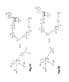
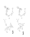
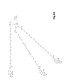
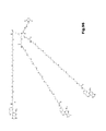
- Tris(hydroxymethyl)methylamine (Tris, 2.42 g, 20.0 mmol) in 4.0 mL of a newly opened bottle of DMSO is cooled to 15.0°C. Then, 0.4 mL of 5.0 M NaOH is injected while stirring, followed by tert-butyl acrylate (10.0 mL, 68 mmol), which is injected dropwise. A solvent mixture of 5-10% water in DMSO is optimal for this reaction. The reaction mixture is allowed to reach room temperature and left stirring for 24 h. Then the crude mixture is poured onto water and extracted with ethyl acetate, the organic phase is dried over MgSO 4 , and evaporated under reduced pressure to afford ( Fig. 1 ). The compound is directly used for next step without further purification.
- FAB-MS m/z 506 [M+H] + .
- N-Tris ⁇ [2-(tert-butoxycarbonyl)ethoxy]methyl ⁇ methyl trifluoroacetamide ( Fig. 2 ) (4.81 g, 8 mmol) is stirred in 80 mL of 96% formic acid for 18 h. Then, the formic acid is removed at reduced pressure at 50°C to produce a colorless oil in quantitative yield.
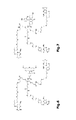
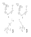
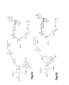
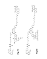
- 5'-Thiol modified oligonucleotide (43 nmol) is reduced by incubation for 1 h at room temperature with 200 mM DTT in 200 ⁇ L Tris-buffer pH 8.5. The DTT is removed by gel filtration and the oligonucleotide eluted in PBS (pH 7.4). The most concentrated fractions are combined giving a total of 800 ⁇ L. 300 ⁇ L of a solution of compound ( Fig. 9 ) (2.5 mM in DMF) is added and the reaction mixture incubated at room temperature for 1 h. The reaction mixture is diluted with water to a total volume of 2 mL and excess maleimide removed by gel filtration.
- the tris(BG-PEG4-NH-carbonylethyloxymethyl)methylamide-maleimide-oligonucleotide conjugate ( Fig. 10 ) is then purified by HPLC (solvent A: 0.1 M tetraethylammonium acetate pH 6.9 in water; solvent B: acetonitrile).
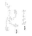
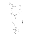
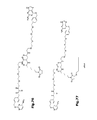
- the 5'-thiol modified oligonucleotide (43 nmol) is reduced by incubation for 1 h at room temperature with 200 mM DTT in 200 ⁇ L Tris-buffer pH 8.5. The DTT is removed by gel filtration and the oligonucleotide eluted in PBS (pH 7.4). The most concentrated fractions are combined giving a total of 800 ⁇ L. 300 ⁇ L of a solution of compound ( Fig. 18 ) (2.5 mM in DMF) is added and the reaction mixture incubated at room temperature for 1 h. The reaction mixture is diluted with water to a total volume of 2 mL and excess maleimide removed by gel filtration. The siRNA conjugate ( Fig. 19 ) is then purified by HPLC (solvent A: 0.1 M tetraethylammonium acetate pH 6.9 in water; solvent B: acetonitrile).
- N-(2-aminoethyl)maleimide trifluoroacetate (343 mg, 1.35 mmol) and azido-PEG12-propionic NHS ester (1 g, 1.35 mmol) are dissolved in 5 mL DMF with Et 3 N (188 ⁇ L, 1.35 mmol) and heated overnight at 31°C. The solvent is evaporated under vacuum and the product is isolated by reversed phase HPLC on a C18 column using a linear gradient of water:acetonitrile (from 95:5 to 20:80 in 20 min, 0.08% TFA). ESI-MS: m/z 766.40 [M+H] + .
- the 5'-thiol modified oligonucleotide (43 nmol) is reduced by incubation for 1 h at room temperature with 200 mM DTT in 200 ⁇ L Tris-buffer pH 8.5. The DTT is removed by gel filtration and the oligonucleotide eluted in PBS (pH 7.4). The most concentrated fractions are combined giving a total of 800 ⁇ L. 300 ⁇ L of a solution of compound ( Fig. 28 ) (2.5 mM in DMF) is added and the reaction mixture incubated at room temperature for 1 h. The reaction mixture is diluted with water to a total volume of 2 mL and excess maleimide removed by gel filtration. The conjugate ( Fig. 29 ) is then purified by HPLC (solvent A: 0.1 M tetraethylammonium acetate pH 6.9 in water; solvent B: acetonitrile).
- the 5'-thiol modified oligonucleotide (43 nmol) is reduced by incubation for 1 h at room temperature with 200 mM DTT in 200 ⁇ L Tris-buffer pH 8.5. The DTT is removed by gel filtration and the oligonucleotide eluted in PBS (pH 7.4). The most concentrated fractions are combined giving a total of 800 ⁇ L. 300 ⁇ L of a solution of compound ( Fig. 35 ) (2.5 mM in DMF) is added and the reaction mixture incubated at room temperature for 1 h. The reaction mixture is diluted with water to a total volume of 2 mL, and excess maleimide removed by gel filtration. The conjugate ( Fig. 36 ) is then purified by HPLC (solvent A: 0.1 M tetraethylammonium acetate pH 6.9 in water; solvent B: acetonitrile).
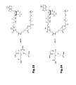
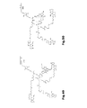
- aqueous phase is washed with ethyl acetate. Upon acidification of the aqueous phase with acetic acid, a yellowish precipitate is formed. The solid is collected via filtration to afford the desired compound as a mixture of isomers ( Fig. 45) and (Fig. 46 ).
- ESI-MS m/z 699.23 [M+H] + .
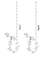
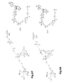
- the 5'-thiol modified oligonucleotide (43 nmol) is reduced by incubation for 1 h at room temperature with 200 mM DTT in 200 ⁇ L Tris-buffer pH 8.5. The DTT is removed by gel filtration and the oligonucleotide eluted in PBS (pH 7.4). The most concentrated fractions are combined giving a total of 800 ⁇ L. 300 ⁇ L solution of a mixture of isomers ( Fig. 47) and (Fig. 48 ) (2.5 mM in DMF) is added and the reaction mixture incubated at room temperature for 1 h. The reaction mixture is diluted with water to a total volume of 2 mL and excess maleimide removed by gel filtration. The mixture of conjugates ( Fig. 49) and (Fig. 50 ) is then purified by HPLC (solvent A: 0.1 M tetraethylammonium acetate pH 6.9 in water; solvent B: acetonitrile).
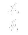
- the 5'-thiol modified oligonucleotide (43 nmol) is reduced by incubation for 1 h at room temperature with 200 mM DTT in 200 ⁇ L Tris-buffer pH 8.5. The DTT is removed by gel filtration and the oligonucleotide eluted in PBS (pH 7.4). The most concentrated fractions are combined giving a total of 800 ⁇ L. 300 ⁇ L solution of a mixture of isomers ( Fig. 53) and (Fig. 54 ) (2.5 mM in DMF) is added and the reaction mixture incubated at room temperature for 1 h. The reaction mixture is diluted with water to a total volume of 2 mL, and excess maleimide removed by gel filtration. The mixture of conjugates ( Fig. 55) and (Fig. 56 ) is then purified by HPLC (solvent A: 0.1 M tetraethylammonium acetate pH 6.9 in water; solvent B: acetonitrile).
- the 5'-thiol modified oligonucleotide (43 nmol) is reduced by incubation for 1 h at room temperature with 200 mM DTT in 200 ⁇ L Tris-buffer pH 8.5.
- the DTT is removed by gel filtration and the oligonucleotide eluted in PBS (pH 7.4).
- the most concentrated fractions are combined giving a total of 800 ⁇ L.
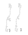
- 300 ⁇ L of a solution of mixture of isomers ( Fig. 59) and (Fig. 60 ) (2.5 mM in DMF) is added and the reaction mixture incubated at room temperature for 1 h.
- the reaction mixture is diluted with water to a total volume of 2 mL, and excess maleimide removed by gel filtration.
- the mixture of conjugates ( Fig. 61) and (Fig. 62 ) is then purified by HPLC (solvent A: 0.1 M tetraethylammonium acetate pH 6.9 in water; solvent B: acetonitrile).
- the 5'-thiol modified oligonucleotide (43 nmol) is reduced by incubation for 1 h at room temperature with 200 mM DTT in 200 ⁇ L Tris-buffer pH 8.5.
- the DTT is removed by gel filtration and the oligonucleotide eluted in PBS (pH 7.4).
- the most concentrated fractions are combined giving a total of 800 ⁇ L.
- 300 ⁇ L solution of a mixture of isomers ( Fig. 65) and (Fig. 66 ) (2.5 mM in DMF) is added and the reaction mixture incubated at room temperature for 1 h.
- the reaction mixture is diluted with water to a total volume of 2 mL and excess maleimide removed by gel filtration.
- the mixture of conjugates ( Fig. 67) and (Fig. 68 ) is then purified by HPLC (solvent A: 0.1 M tetraethylammonium acetate pH 6.9 in water; solvent B: acetonitrile).
- the 5'-thiol modified oligonucleotide (43 nmol) is reduced by incubation for 1 h at room temperature with 200 mM DTT in 200 ⁇ L Tris-buffer pH 8.5. The DTT is removed by gel filtration and the oligonucleotide eluted in PBS (pH 7.4). The most concentrated fractions are combined giving a total of 800 ⁇ L. 300 ⁇ L solution of compound ( Fig. 76 ) (2.5 mM in DMF) is added and the reaction mixture incubated at room temperature for 1 h. The reaction mixture is diluted with water to a total volume of 2 mL and excess maleimide removed by gel filtration. The conjugate ( Fig. 77 ) is then purified by HPLC (solvent A: 0.1 M tetraethylammonium acetate pH 6.9 in water; solvent B: acetonitrile).
- Tris ⁇ [2-tert-butoxycarbonyl)ethoxy]methyl ⁇ methylamine ( Fig. 1 )(4.3g, 8 mmol) is stirred in 80 mL of 96% formic acid for 18 h. Then the formic acid is removed at reduced pressure at 50°C to produce a colorless oil in quantitative yield.
- 1 H NMR ((CD 3 ) 2 SO, 400 MHz): 8.2 (m, 2H), 7.45 (m, 3H), 3.6 (m, 6H), 3.4 (m, 6H), 2.45 (m, 6H).
- nile red-oxyacetic acid (9-diethylamino-5-oxo-benzo[a]phenoxazin-2-oxyacetic acid, 100 mg, 0.255 mmol, 1 eq) in DMF (50 mL) are successively added DCC (160 mg, 0.765 mmol, 3 eq) and NHS (90 mg, 0.765 mmol, 3 eq). The resulting mixture is stirred overnight. Then DCU salts are removed by centrifugation. Compound ( Fig. 1 ) (130 mg, 0.255 mmol, 1 eq) and DIPEA (42 ⁇ L, 0.255 mmol, 1 eq) are added to the solution at rt.
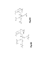
- N-Tris[(2-carboxyethoxy)methyl]methyl 7-(diethylamino)-coumarin-3-carboxamide ( Fig. 82 ) (10 mg, 0.018 mmol) and BG-PEG12-NH 2 ( Fig. 80 ) (54 mg, 0.062 mmol, 3.6 eq) in DMF (1 mL) are successively added DIPEA (8 ⁇ L, 0.062 mmol, 3.6 eq), HOBT (1 M in NMP, 18 ⁇ L, 0.018 mmol, 1 eq) and EDC (12 mg, 0.062 mmol, 3.6 eq) at rt. The resulting mixture is stirred overnight.
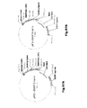
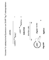
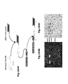
- a modified pSecTac based mammalian expression vector (pMS, Stöcker et al., 2003) was provided with the SNAP 26m gene by PCR cloning from the storage vector pSS26m (COVALYS AG). Two versions are available allowing to link component A to the N-terminus of component B or to the C-terminus of component B which are depicted in Figures (97A +B).
- the expression cassette of the vector comprises of the following key features: the human cytomegali virus promoter sequence (CMV), a bovine growth hormone polyadenylation signal (BGH pA) and an internal IVS ribosome entry site (IRES).
- CMV human cytomegali virus promoter sequence
- BGH pA bovine growth hormone polyadenylation signal
- IVS ribosome entry site IVS ribosome entry site
- a plant expression vector system designed for transient and stable expression of SNAP-tag fusion proteins in plants is shown in figure (97C ).
- the vector comprises the following features:
- KDEL plant ER retention signal
- LPH codon optimized murine signal peptide
- Bla ampicillin resistance ( E. coli ), cabenicillin resistance (A. tumefaciens ); nptII: Kanamycin resistance plant
- SAR scaffold attachment region of tobacco RB7 gene
- P35SS transcription start
- CHS 5'UTR from chalcon synthase
- pA35S polyadenylation signal from CaMV
- RK3 ori ori for A. tumefaciens
- ColE1 ori ori for E. coli
- LB/RB elft/right border
- pAnos nopaline synthase polyadenylation signal
- Pnos nopaline synthase gene promoter.
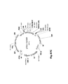
- Procaryotic expression plasmids exemplified here are based on the pET26bTM system (Novagen) and designed for periplasmatic expression of C/N-terminal SNAP-tag fusion proteins in E . coli ( Fig. 97D ).
- the SNAP-tag version (26b) is codon optimized for and was PCR amplified from the storage vector pSET7-26b from Covalys.
- the expression is regulated through the T7 promoter; together with a host-encoded T7 polymerase the regulation of expression is extremely tight.
- the kanamycin resistance gene allows selection of transformed bacteria.
- the pelB leader is directing the recombinant protein into the periplasmic space.
- Yeast expression plasmid based on the CoMedTM system provided by Pharmedartis (Aachen, Germany) ( Fig. 97E ).
- the vector backbone is a derivative of a standard E . coli vector combining a ColE1 ori and an ampicillin resistance (bla) sequence.
- a variant I contains an f1(-)origin, a variant II is without that sequence.
- a multiple cloning site (MCS) has been engineered for the uptake of various modules.
- ARS/CEN modules For insertion ARS/CEN modules (module 1) are flanked by SacII/BcuI restriction sites, rDNA segments (module 2) by BcuI/Eco47III sites, selection marker modules (module 3) by Eco47III/SalI sites and expression cassettes (module 4) by SalI/ApaI sites. In variant II additional SphI and BsiWI cloning sites are present.
- the Plasmid is designed to work with a variety of yeast strains:
- Species auxotrophies Arxula adeninivorans (LS3) wild type; leu2 Arxula adeninivorans (CBS7350) wild type; leu2 Arxula adeninivorans (CBS1738) wild type; leu2 Hansenula polymorpha (CBS4732) wild type; ura3; leu2 ura3; arg1 leu2 ura; ade1 leu2 ura3 Kluyveromyces lactis met- ura3 Pichia pastoris wild type; ura3; ura3 his3 Saccharomyces cerevisiae wild type; ura3; leu2 ura3 trp1 lys2 Yarrowia lipolytica E150 wild type; ura3; leu2; ura3 leu2 leu2 leu2 leu2
- modules of a concrete vector construct may contain one or more of the following features:
- ORF open reading frame
- Transfection efficiency was between 75 and 95% determined by counting green fluorescent cells. 3 days after initial transfection, cell culture supernatants were analyzed for recombinant protein. Subsequently, transfected cells were transferred into medium-sized cell culture flasks (Nunc; 85m 2 ) and grown in RPMI complex medium supplemented with 100 ⁇ g/ml Zeocin. One to two weeks productively transfected clones were green fluorescing and hence could be detected by fluorescence microscopy. Transfected cell populations were established by subcultivation of these clones.
- Agrobacterium tumefaciens (A. tumefaciens) mediated transformation method is chosen.
- A. tumefaciens e.g. strain GV3101 : : pMP90RK
- tumefaciens is made electrocompetent (Shen & Forde, 1989) and 100 ⁇ l of the competent cells are mixed with 50-200 ng of a binary vector (pTRAkc based) containing the expression cassette for the SNAP-Tag fusion protein in a 0.1 cm electrogap cuvette (BioRad).
- the cells are transformed by a electric pulse using a GenePulser (BioRad) set at 1.8 kV, 25 mF and 200 V.
- Electroporated cells are incubated in 1 ml Luria-Bertani (LB) broth for 2 h prior to plating on LB medium containing 50 mg carbenicillin ml -1 , 50 mg rifampicin ml -1 and 30 mg kanamycin ml -1 .
- LB Luria-Bertani
- A. tumefaciens mediated transient expression of SNAP-tag fusion proteins in tobacco plants A. tumefaciens cultures containing pTRAkc vector bearing the SNAP-tag fusion protein expression cassette clones are supplemented with 50 mg carbenicillin ml -1 and 50 mg rifampicin ml -1 . Cultures are grown with shaking at 27 °C to exponential phase (OD600 approx. 0.8) in LB broth containing the appropriate antibiotics.
- Cells are collected by centrifugation at 4000 g, resuspended in induction medium (LB broth at pH 5.6 containing 10 mM MES, 20 mM acetosyringone and 2 mM MgSO 4 ) with the appropriate antibiotics, and grown as above.
- the cells are collected by centrifugation at 4000 g and resuspended in infiltration medium (10 mM MgCl2, 10 mM MES, 2% sucrose and 150 mg acetosyringone ml -1 , pH 5.6).
- the Agrobacterium suspensions are diluted in infiltration medium to an OD600 of 1.0 and are stored at 22 °C for 2-3 h.
- Agrobacterium suspensions are diluted and combined in infiltration medium, both to a final OD600 of 0.25.
- Agrobacterium pTRAkc
- Leaves from 2-4-week-old Nicotiana benthamiana plants are infiltrated by injecting the bacterial suspension into the abaxial air spaces from the underside of the leaf. E.g. six leaves are agroinfiltrated with each bacterial mixture (three plants, two leaves per plant). The plants are grown for 5-6 days under conditions of 16 h light, 8 h dark, 22 °C.
- Agrobacterium cultures are grown overnight in induction medium. The cells from are resuspended in 1-8 I infiltration medium to a final OD600 of 0.25 per culture.
- Whole Nicotiana tabacum L. 'Petite Havana' SR1 plants with roots removed are submerged into the bacterial suspension and subjected to a vacuum of 290 kPa for 5-10 min, with occasional agitation to release trapped air bubbles. The vacuum is released rapidly (approx. 10 kPa s -1 ). The plant stalks are placed in water-saturated floral foam. The plants are grown for 3 days under conditions of 16 h light, 8 h dark, 22 °C.
- N tabacum leaf discs (cut by using the cap of a microfuge tube) are harvested from agroinfiltrated leaves and ground in 250 ml high-salt phosphate buffer (0.5 M NaCl) per disc. The extract is centrifuged at 13 000 r.p.m. for 5 min, supernatant is collected and the centrifugation is repeated.
- the SNAP-tag fusion protein is alternatively detected in cell extracts by adding appropriate amounts of the BG stain SNAP-vista green.
- the manufacturers protocoll is followed regarding the staining conditions and reaction conditions.
- the recombinant SNAP-tag fusion proteins stained by SNAP-vista green can be visualized in a standard UV transilluminator used for gel documentation.
- a scientist skilled in the art may recognize that different A. tumefaciens strains together with other binary A. tumefaciens plasmid vectors than pTRAkc may also lead to successful transformation of tobacco plants and therefore functional expression of SNAP-Tag fusion proteins.
- strains are grown either under non-selective conditions in complex medium (YEPD) or under selective conditions in a yeast minimal medium (YMM) supplemented with 2% of a selected carbon source (Steinborn,G. et al., 2006). Cultivation is performed at 30°C.
- A.adeninivorans LS3, A.adeninivorans 135, A. adeninivorans G1211, D.hansenii H158, D.polymorphus H120, H. polymorpha MedHp1, P. pastoris GS115 and S. cerevisiae C13ABYS86 are transformed according to R Harborl H. et al., 1998; and Dohmen RJ et al.,1991.
- Stable transformants are obtained after a sequence of passages on selective and non-selective media.
- hygromycin B-resistant colonies are selected on YEPD agar plates supplemented with 150 - 400 mg I-1 hygromycin B (200 mg I -1 for A.adeninivorans LS3 and 135, 250 mg I -1 for D.hansenii H158 and D.polymorphus H120, 400 mg I -1 for H. polymorpha MedHp1, 150 mg I -1 for P. pastoris GS115 and S. cerevisiae C13ABYS86.
- Single colonies are isolated and grown on YEPD medium and hygromycin B at 30°C for 2 days. This step is repeated three times before the cells are plated on non-selective YEPD agar and grown for 3-5 days at 30°C. A single colony from each transformant is then isolated and defined as a strain.
- Intracellular and extracellular expression levels of SNAP-tag fusion proteins are analyzed by Western blot experiments with anti-His-Tag antibodies for the newly generated expression yeast cell lines.
- SNAP-tag fusion protein is alternatively detected in cell extracts by adding appropriate amounts of the BG stain SNAP-vista green. The manufacturers protocoll is followed regarding the staining conditions and reaction conditions.
- the recombinant SNAP-tag fusion proteins stained by Vista green can be visualized in a standard UV transilluminator used for gel documentation.
- SNAP-tag fusion proteins For bacterial expression of SNAP-tag fusion proteins the desired fusion partners are cloned into the pET26b+ derived bacterial periplasmic expression vectors described in "construction of expression vectors".
- Heat shock competent bacteria of the appropriate E. coli strain e.g. ROSETTA, EMD Biosciences, Darmstadt, Germany
- Seleced clones growing on agar plates with Kanamycin pET encoded Kan R provides bacteria with resistance gene
- the expression of the plasmid encoded SNAP-Tag fusion proteins is done using the osmotic stress expression protocol described in Barth et al., 2000.
- Recombinant RFT5-SNAP-tag fusion proteins are expressed under the control of the IPTG inducible T7 lac promoter in E. coli ROSETTA (DE3). Bacteria are grown overnight at 26°C in Terrific Broth (TB) (Sambrook&Maniatis, 1989) containing 50 mg of kanamycin/ml and 0.5 mM ZnCl2, since it has been shown earlier that periplasmic proteolysis can be dramatically reduced upon addition of this salt (Baneyx, F., and G. Georgiou. 1992. ). The shaking culture is diluted 30-fold in 200 ml of the same medium.
- cells are harvested by centrifugation at 3,700 3 g for 10 minat 4°C. For all the following steps, tubes are chilled on ice. The bacterial pellet is centrifuged, and its wet weight is determined. Cells are frozen at -80°C until further processing.
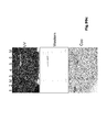
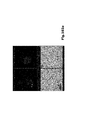
- Figure (101 ) shows a 12 % SDS-PAGE gel (A: UV light, B: Coomassie stained) which was loaded with 5 ⁇ g of the different mammalian expressed and IMAC (Immobilized Metal Affinity Chromatography) purified.
- the Gel contains: 1:Ki4-SNAP; 2: SNAP-EGF; 3: Hai-SNAP; 4: H22-SNAP; 5: SNAP-CD30L; M: prestained portein marker (NEB).
- the SNAP-tag fusion protein (Ki4-SNAP) is Ni-NTA purified as decribed in section "IMAC purification of SNAP-Tag fusion proteins from mammalian expression". While still bound on the resin via His-Tag-Nickel interaction the Ki4-SNAP protein can be labeled with one of the SNAP-tag specific BG substrates like e.g BG505 as seen in figure (99c ).
- CLIP-tag fusion proteins are Ni-NTA purified as decribed for SNAP-tag constructs. While still bound on the resin via His-Tag-Nickel interaction the CLIP-Tag fusion proteins can be labeled with one of the CLIP-tag specific CT substrates like CT-360,CT-430, CT-FL/CT-PF, CT-488, CT-505, CT547, CT-TMR, CT-647 CT-Biotin, CLIP-vista Green.
- ACP-Tag-Eotaxin /MCP-Tag-CXCL9 expressing HEK293 cells three times with tissue culture medium with serum.
- One vial of ACP-tag substrate is dissolved in 25 ⁇ L of DMSO to give a labeling stock solution of 1 mM in DMSO. After 10 minutes of mixing all the ACP-tag substrate is dissolved.
- the 1 mM ACP-tag substrate stock solution is diluted 1:200 in medium to give a labeling medium of 5 ⁇ M. Afterwards MgCl2 to a final concentration of 10 mM is supplemented. Finally, the ACP-Synthase is added to a final concentration of 1 ⁇ M.
- the culture medium on the cells expressing an ACP-tag fusion protein located in or on the cell membrane with the ACP-tag facing the outside of the cell is exchanged with the labeling medium and incubated for 30 minutes.
- the labeling medium is removed and exchanged by fresh cell culture medium and incubated for another 20 minutes to remove unreacted ACP-tag substrate.
- the medium is exchanged again and the cells are ready for microscopy, flow cytometric analysis or FACS sorting.
- the same procedure can be operated with cells expressing a MCP-Tag fusion protein. Therefore the labeling substrate is the same, a CoA derivative but instead of the ACP-Synthase the SFP-Synthase is taken for catalyzing the labeling reaction.
- ACP/MCP-tag fusion proteins are Ni-NTA purified as decribed for SNAP-Tag constructs. While still bound on the resin via His-Tag-Nickel interaction the ACP/MCP-Tag fusion proteins can be labeled with one of the ACP/MCP-tag specific CoA based substrates like CoA-488, CoA-547, CoA-647and CoA-Biotin.
- ACP-tag substrate is dissolved in 25 ⁇ L of DMSO to give a labeling stock solution of 1 mM in DMSO. After 10 minutes of mixing all the ACP-tag substrate is dissolved.
- the same procedure can be performed with cells expressing a MCP-Tag fusion protein. Therefore the labeling substrate is the same, a CoA derivative but instead of the ACP-Synthase the SFP-Synthse is taken for catalyzing the labeling reaction.
- the moldular structure of the invention related complex allows the combination of two SNAP-tag constructs with (antibody) fusion partners of different/same binding specificity via a linker structure containing two or more BG residues, resulting in a bispecific molecule.
- heterobivalent (bispecific) constructs the whole process consists of two steps to maximize the amount of built heterodimers.
- a recombinant SNAP-tag fusion protein with specificity 1 was bound on the resin via His-Tag-Nickel interaction.
- the recombinant SNAP-tag fusion protein with specificity 2 was added in the same molar ratio than the prelabeled protein 1.
- the crosslinking reaction was then performed at 4°C over night in solution.
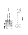
- Fig. 99a The success of the crosslinking reaction was documented by SDS-PAGE and Coomassie staining ( Fig. 99a ) .
- the gel shows the Ki4-SNAP (lane 1x ) and its crosslinked version (lane 2x) together with a molecular weight marker (lane M).
- the molecular sizes determined by densitometric analysis are given as 53 kDa for the single Ki4-SNAP and 122kDa for the crosslinked version.
- Crosslinking was realized with a homobifunctional crosslinker, SV 305, containing a PEG 12 spacer ( Fig. 99b ).
- the reaction can also be done IMAC matrix assisted like for bispecifics but also without in a single step reaction.
- the IMAC purified SNAP-tag fusion protein is mixed with the crosslinker in the following ratio: for a crosslinker with with a given number of BG residues BG n the mixture formula is:
- the reaction mix is incubated for at least 12h at 4°C in the dark.
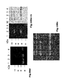
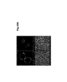
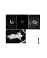
- Figure (100a -d) shows:
- the HaiSNAP was mixed with the crosslinkers in a molar ratio of 3:1.
- the fluorophors can be well detected using implemented conventional emission filter sets.
- Figure (100d ) shows a confocal microscopy done with the SV305 crosslinked version of Hai-SNAP.
- the crosslinked protein was separated from non crosslinked version by 100kDA MWCO spin columns (Pall Nanosep).
- 25 ⁇ g of the crosslinked sample were added to the column and cenrifuged at 10.000 g for 10 minutes. Afterwards 500 ⁇ l 1xPBS were added and the sample centrifuged again until the volume was reduced to 50 ⁇ l. This step was repeated and the residue in the column was taken for microscopic analysis.
- RNA Antibody-Nucleic acid conjugates
- Optimized siRNAs were synthesised by Dharmacon with an amino-group and C(6) spacer on the 3' or 5' end of the sense strand.
- the siRNA duplexes are solubilised in PBS and reacted with a 50fold molar excess of BG-GLA-NHS (Covalys) solubilized in water free DMF for 4h at RT.
- the siRNA is ethanol precipitated and residual BG-GLA-NHS is removed by passing through a gel filtration column (Centri-spin 10, Princeton separations).
- Analogously thiol-modified RNA can be used together with a BG-maleimide after reduction of the thiol with DTT.
- Figure (104A ) shows te schematic procedure for si-RNA coupling to SNAP-tag fusion proteins.
- the DNA is modified with Benzylguanine either by Mull modifications of oligonukleotides with a terminal benzylguanine (BG) or benzylcytosine (BC).
- BG benzylguanine
- BC benzylcytosine
- the DNA fragment is amplified by PCR using a BG or BC modified oligonucleotide as one of the two primers.
- the PCR product is purified from unreacted BG or BC modified oligonucleotides via a commercial plasmid preparation kit (EndoFree Plasmid Maxi Kit QIAGEN, Hilden Germany).
- the purified PCR product is then incubated with the SNAP-tag fusion protein in a molar ratio of (RNA:SNAP-tag fusion protein) 2:1 over night at 4°C.
- the success of the coupling reaction is monitored via agarose gel electrophoresis followed by ethidiumbromide staining where only the DNA labeled SNAP-tag fusion proteins are stained whereas the SNAP-tag fusion protein alone is not stained.
- a discrimination between DNA labeled and non labeled fusion proteins is also possible via the electromobility shift of labeled protein.
- the successful DNA labeled Ki4-SNAP-tag fusion protein is able to target cancer cells via their overexpressed cell surface marker CD30.
- AMAXA nucleofector
- the SNAP-tag fusion protein-DNA complexes are at first coupled via specific DNA-DNA interaction on a DNA loaded surface. After coupling the complexes are able to fix CD30 overexpressing cells on certain spots, where the Protein-DNA complex was immobilized beforehand.
- silica nanobeads with a size distribution between 20 and 80nm and encapsulated rhodamine fluorophor were taken.
- the beads were concentrated at 9.5 mg/ml with 5.3x10 15 amino (NH 2 ) groups (8.76 x 10 -3 ⁇ mol/ml).
- Beads were washed two times with 800 ⁇ l PBS and resuspended in 100 ⁇ l PBS prior to use.
- Figure (102A ) shows a confocal microscopy of Ki4-SNAP functionalized Nanobeads binding CD30-positive L540 cells. Rhodamine based emission of beads in red (A) and Draq5 emission in blue pseudocolour (B), overlay (D) with grayscale picture (C).
- Figure (102B ) shows the flow cytometric analysis of cD30 overexpressing L540cy cells incubated with different amounts (0,5 and 5 ⁇ l) of Ki4-SNAP coupled rhodamine doted nanobeads. As control 5 ⁇ l uncoupled beads were applied to L540cy cells.
- Figure (102C ) shows the flow cytometric analysis of the CD30 negative U937 cells incubated with different amounts (0,5 and 5 ⁇ l) of Ki4-SNAP coupled rhodamine doted nanobeads. As control 5 ⁇ l uncoupled beads were applied to the U937 cells.
- a 96 well ELISA plate is coated with the analyte by pipetting 25 ⁇ l of coating buffer (100 mM Sodiumcarbonate, pH 9,6) in each well and then mixing with 25 ⁇ l analyte solution per well.
- coating buffer 100 mM Sodiumcarbonate, pH 9,6
- the plate After 2 hours of coating at room temperature the plate is washed twice with 1xPBS and then 50 ⁇ l of the detection antibody solution (SNAP-tag fusion protein, 50-100ng) is added.
- the SNAP-tag fusion protein was labeled beforehand with the SNAP-vista green fluorophor (Covalys) as described in "Labeling of SNP-tag fusion proteins with BG derivatives of organic fluorophores".
- the detection antibody solution is incubated for 1 hour at room temperature and the plate is washed twice with 1xPBS. Afterwards the plate is analyzed in a fluorescence ELISA reader using s filter set suited for the SNAP-tag coupled fluorophor.
- ELISA plate surfaces can be modified with BG-PEG-NH2 so that they will covalently immobilize SNAP-tag fusion proteins. Surface activation is done using standard amino-coupling procedures (such as exposure to NHS and EDC). This surface is then modified as follows: 1.4 mg (0.0031 mmol) BG-PEG-NH2 is dissolved in 10 mL HBSbuffer and centrifuged (20,000 x g, room temperature,20 min). 100 ⁇ L of this solution is pipetted into each well of a 96 well ELISA plate with carboxylated surfaces. After 30 minutes of incubation excess reactive groups are quenched by adding ethanolamin (10mmol) and further 10 minutes incubation.
- the ELISA plate surface is ready to use for the direct immobilization of SNAP-tag fusion protein from samples. Therefore 100 ng of purified Ki2 mab (in 50 ⁇ l PBS) are pipetted into each well and incubated for 2 hours at room temperature or alternatively over night at 4°C.
- the sample (50 ⁇ l) containing the analyte (secreted CD30) is pipetted into the wells and incubated for 2hours at room temperature.
- the Plate is then washed twice with 1x PBS before 50 ⁇ l of the detection antibody solution (scFv Ki3, 200ng/well) is applied.
- the detection antibody consists either of a scFv-GFP fusion or a fluorophore labeled scFv-SNAP-tag fusion protein for fluorescent readout.
- This protocol is basically a modified version of the quantitative immuno-PCR (qIPCR) method described by Niemeyer et al.,2007.
- the basic principle of the assay relies upon a sandwich immunoassay followed by qPCR 1:captured of antigen by a non labeled antibody (Ki2 mab) coated on the 96 well PCR/ELISA plate and 2: sCD30 antigen capture from blood serum and 3: the detection of this captured sCD30 by a fusion protein of Ki3 scFv and SNAP-tag which was beforehand covalently labeled with a dsDNA PCR template and finaly 4: a qPCR step for signal amplification and readout.
- qPCR 1 captured of antigen by a non labeled antibody (Ki2 mab) coated on the 96 well PCR/ELISA plate
- 2 sCD30 antigen capture from blood serum
- 3 the detection of this captured sCD30 by a fusion protein of Ki3 scFv and SNAP-tag which was beforehand covalently labeled with a dsDNA PCR template
- finaly 4 a qPCR
- Part 1 -3 represent a typical sandwich ELISA protocol as previously described.
- a schematic overview is given in figure (103 ).
- the assay has to be performed in thin-walled polycarbonate plates suited for immunoassay as well as for thermocycling based applications (e.g. Nunc TopYield starter kit).
- the protocol is performed as follows:
- the initial working step is mostly performed as described in the "Sandwich ELISA" protocol.
- the only difference is the use of thin walled polycarbonate PCR grade 96 well plates or strips instead of conventional ELISA plates.
- the plates are rigorously washed five times with PBS+Tween (0.01%), soaking wells for 3 min with wash buffer during each cycle, followed by two washes with ultrapure, 0.2 mm filtered water. After addition of PCR reagents, the plates are subjected to 30 cycles of PCR amplification, using a 96-well real-time PCR cycler, for example the ABIprism 7000 (Applied Biosystems) system.
- the TaqMan Universal PCR Mastermix is prepared according to the manufacturer's instructions using the described concentrations of primer-1, primer-2 and probe. 30 ⁇ l of the PCR Mastermix are pipetted in each well. The modules are sealed with an adhesive foil. The plate or PCR stripes are placed into the precleaned real time PCR machine and a typical PCR program is run: initial denaturation: 5 min 95 °C followed by 30 cycles consisting of denaturation step: 30 s, 50 °C , synthesis step: 30 s, 72 °C and denaturation step: 12 s, 95 °C. After the run the acquired data are evaluated by software.
- the cell-binding activity of the SNAP-tag fusion proteins containing a targeting component A was evaluated using a FACS Calibur flow cytometer and CellQuest software (Becton Dickinson, Heidelberg, Germany) or the free software WinMDI 2.8.
- the SNAP-tag fusion protein was labeled ahead of application as described in "Labeling of SNAP-tag fusion proteins with BG derivatives of organic fluorophores". Cells were labeled with the fluorophor labeled SNAP-tag complex by incubation for 30 minutes on ice. Cells were then washed twice with 500 ⁇ l cold PBS in an automated cell washer (Dade Serocent; Baxter).
- the binding of the SNAP-tag fusion protein was detected via the polyhistidine tag by using a Penta-His Alexa Fluor 488 antibody (Qiagen, Hilden, Germany).
- antibody SNAP-tag fusion constructs targeting the human cell surface molecules CD30 Hodgkin lymphoma
- CD64 (Fc gamma RI ) on activated macrophages)
- EGF receptor on pancreatic-, breast-, lung-and non small cell lung cancer
- the scFv Ki4 a monomeric recombinant version of the parental CD30 specific monoclonal antibody (Barth e al. 2000) and its counterpart the CD30 ligand were cloned as fusion proteins to SNAP-tag.
- the choice of positions (N- or C-terminal) was done in respect of maintaining full functionality of both fusion partners.
- Figure (105A ) shows an evaluation of flow cytometric analysis of Ki-SNAP labeled with SNAP-vista Green (Covalys) binding on the CD30 overexpressing cell line L540cy.
- Figure (105B ) shows an evaluation of flow cytometric analysis of SNAP-CD30L labeled with SNAP-vista Green (Covalys) binding on the CD30 overexpressing cell line L540cy.
- the scFv 425 (further named Hai), a monomeric recombinant version of the parental EGFR specific monoclonal antibody (Haisma et. al., 2000) and the natural EGFR ligand EGF were cloned as fusion proteins to SNAP-tag.
- Hai a monomeric recombinant version of the parental EGFR specific monoclonal antibody
- EGF EGF
- Figure (105C ) shows an evaluation of flow cytometric analysis of Hai-SNAP labeled with SNAP-vista Green (Covalys) binding on the EGFR overexpressing cell line A431.
- Figure (105D ) shows an evaluation of flow cytometric analysis of SNAP-EGF labeled with SNAP-vista Green (Covalys) binding on the EGFR overexpressing cell line A431.
- the H22 a monomeric recombinant version of the parental anti CD64 specific monoclonal antibody (Tur et. al., 2003) was cloned as fusion proteins N-terminal to SNAP-tag.
- Figure (105E ) shows an evaluation of flow cytometric analysis of H22-SNAP labeled with SNAP-vista Green (Covalys) binding on the CD64 overexpressing cell line U937 and not binding on the CD64 negative cell line L540.
- pancreatitis derived cellular material To destinguish between inflammatory pancreatitis and pancratic cancer a immunized murine scFv Phage Display library was depleted on pancreatitis derived cellular material followed by three rounds of Phage Display selection.
- one pancreatic cancer specific scFv clone (clone 14.1) was selected as specific binder for pancreatic cancer derived cell lines L3.6pl and A431 and being negative on pancreatitis cell membranes.
- the target cells were prepared as described in "Flow cytometric applications of SNAP-tag fusion proteins" but were fixed with formaldehyde after the last washing step. Therefore 300 ⁇ l of an icecold 0,4 % formaldehyde-PBS solution were added to the cells on ice and incubated for 30 minutes. The cells were washed once again with PBS in an automated cell washer. For counterstaining of nuclei, the cells were mixed with 2 ⁇ l of a 1/100 diluton of Draq5 (BioStatus, Leicestershire, UK). After 5 minutes incubation 10 ⁇ l of the cell suspension were mounted on a glass slide covered with glass coverslips and investigated with a Leica fluorescence (DMR) and Confocal microscope (TSC SP).
- DMR Leica fluorescence
- TSC SP Confocal microscope
- Figure (106 ) shows confocal pictures of L540cy cells stained with BG505 (Covalys) labeled Ki4-SNAP.
- L3.6pl pancreatic carcinoma 5x10 5 cells were injected intravenously in a female 6 week old SCID mouse and visualized after 1 week growth by retrobulbic injection of 70 ⁇ g Hai-SNAP labeled with BG-782 NIR dye.
- the labeling reaction was done beforehand as described in section "Labeling of SNP-tag fusion proteins with BG derivatives of organic fluorophores".
- the imaging was done with anesthetized mice after 5 minutes, 12, 24 and 72 hours after injection of the Hai-SNAP-BG782 imaging agent.
- Figure (107 ) shows infrared pictures from the whole mouse that were taken and analyzed by spectral unmixing the signal from background with the Intas Cri-Maestro In vivo imager.
- a large tumor in the abdomen of the mouse could be well visualized by accumulation of Hai-SNAP.
- Figure (108 ) shows infrared pictures from the whole mouse that were taken and analyzed by spectral unmixing the signal from background with the Intas Cri-Maestro In vivo imager.
- stably EGFP expressing L3.6pl pancreatic carcinoma 5x10 5 cells were injected under the skin at the right and left femoral region of a female 6 week old SCID mouse and visualized after 1 week growth by retrobulbic injection of 70 ⁇ g Hai-SNAP labled with BG-782 NIR dye.
- the labeling reaction was done beforehand as described in section "Labeling of SNP-tag fusion proteins with BG derivatives of organic fluorophores".
- the imaging was done with anesthetized mouse after 24 past injection of the Hai-SNAP-BG782 imaging agent.
- the green fluorescence and the infrared fluorescence signal clearly overlap when overlaying the corresponding pictures taken by the Intas Cri-Maestro In vivo imager.
- the EGFR-positive target cells were prepared as described in "Confocal microscopy applications of SNAP-tag fusion proteins" but were fixed with formaldehyde after different time points of SNAP-tag fusion protein application. Therefore 300 ⁇ l of an icecold 0,4 % formaldehyde-PBS solution were added after 15, 30 and 60 minutes of incubation at 4°C/37° to the cells on ice and incubated for 30 minutes. The cells were washed once again with PBS in an automated cell washer. For counterstaining of nuclei, the cells were mixed with 2 ⁇ l of a 1/100 diluton of Draq5 (BioStatus, Sheffieldshire, UK). After 5 minutes incubation 10 ⁇ l of the cell suspension were mounted on a glass slide covered with glass coverslips and investigated with a Leica fluorescence (DMR) and Confocal microscope (TSC SP).
- DMR Leica fluorescence
- TSC SP Confocal microscope
- Figure (109 ) shows confocal pictures of L3.6pl cells stained with BG505 (Covalys) labeled Hai-SNAP. There is a clear higher internalization rate of bound Hai-SNAP into the cells when incubated at 37° in comparison to the 4°C sample. EGFR negative cell lines like L540 and U937 were not stained under the same conditions.
- Figure (110 ) shows the colocalization of Hai-SNAP BG505 labeled and transferrin ALEXA 594 labeled after internalization (see black arrows in (Fig. 109E)).
- Figure (109A) shows internalized HaiSNAP-BG505 in green;
- FIG. 109B clathrin-mediated internalization of transferrin-ALEXA594 in blue
- Fig. 109C an overlay of A and B and D is an overlay of C with transmission light picture;
- Fig. 109E magnification of
- Fig. 109D arrows depict vesicles harboring both labeled transferrin and HaiSNAP-BG505.
- Fig. 109D magnification of
- arrows depict vesicles harboring both labeled transferrin and HaiSNAP-BG505.
- Transferrin is known to be internalized via clathrin supported internalization. This is also reported for EGFR internalization. The colocalization of Hai-SNAP and transferrin therefore indicates that the original internalization route of EGFR is not affected by bound Hai-SNAP.
- Cell permeable SNAP-tag staining substrates like BG-430, BG-505, BG-DAF and TMR-Star can be used to specifically label SNAP-tag fusion proteins in living mammalian cells.
- BG-430, BG-505, BG-DAF and TMR-Star can be used to specifically label SNAP-tag fusion proteins in living mammalian cells.
- TMR-Star TMR-Star
- TMS-Star substrate was dissolved in DMSO according to the manufacturers (Covalys) instructions. 5 ⁇ M, TMR-Star was diluted to a final working concentration of 1 ⁇ M. Cells were labeled in the dark for 30 min at 37°C, then washed twice in medium and incubated for a further 30 min prior to imaging to allow diffusion of non-reacted substrate out of the cell. All steps were performed under a laminar flow and with sterile filtrated solutions.
- TMR stained cells were counterstained with a 1:2000 dilution of DRAQ5TM solution for 2 min followed by a washing step with PBS. Thhe staining solution concentration and washing conditions affter TMR staining were carefully determined by microscopy until the TMR background in non or mock transfected cell lines was low enough to get a good signal to background ratio in the cells expressing SNAP-tag fusion protein.
- the rest of the properly stained cells were sorted according to the strength of the TMR signal. Ten percent of the cells with strongest TMR signal were sorted and afterwards transfered into a new culture flask.
- Figure (111 ) shows the TMR staining of HEK293 cells expressing the Hai-SNAP fuison protein together with the EGFP reporter protein which is aencoded 3' on the biscistronic mRNA.
- Figure (111A) shows the signal of the EGFP reporter,
- Fig. 111B the TMR signal belonging to the SNAP-Tag fusion protein,
- Fig. 111C the Draq5 nuclear counterstain
- Fig. 111D the transmission light picture of the same cells.
- RNA Antibody-Nucleic acid conjugates
- the complex was applied to target cells in concentrations ranging from 10ng/well (1x10 5 target cells) to 100 ng/well (EGFR overexpressing cell line L3.6pl) and the cells are tested for specific knockdown of target genes 62 hours after application of the siRNA complex by westen blot and quantitatve PCR.
- RNA molecules is realized over a homotrifunctional or heterotrifunctional and homobifunctional/heterobifunctional crosslinkers containing a maleimide function, by which a RNA molecule (preferably having RNA interference properties like siRNA) containing a terminal primary SH-group is coupled.
- a RNA molecule preferably having RNA interference properties like siRNA
- the preformed crosslinker-RNA complex is then added in a molar ratio of 2:1 to the pre- purified SNAP-/CLIP-tag fusion proteins and reaced as decribed above.
- RNA bearing crosslinkers one BG and on BC residue for crosslinking one SNAP-tag and one CLIP-tag fusion protein
- figure (77 ) An example for heterobifunctional RNA bearing crosslinkers (one BG and on BC residue for crosslinking one SNAP-tag and one CLIP-tag fusion protein) is given in figure (77 ).
- RNA bearing crosslinkers three BG residues for crosslinking of three SNAP-tag fusion proteins
- This crosslinker ( Fig. 50 ) additionally contains a fluorecein residue which enables tracing of the complex by e.g. microscopy.
- the targeting complex AB is consisting of a EGFR targeting antibody (Hai) or natural lingand (EGF) whereas the SNAP-tag is coupled to Benzylguanine modified cytotoxic agents like Paclitaxel.
- This Paclitaxel is representing the invention related component C and is delivering in complex with AB a cytotoxic payload to targeted cells (EGFR overexpressing).
- the toxic moiety is loaded on dendrimeric structures like described for Paclitaxel in Jongdoo Lim et al.,2007. or methothrexate in Gong Wu et al.,2006.
- the purified (dialysis) dendrimer-Hai-SNAP complex is then applied to target cells and tested for specific cytotoxicity in a XTT based cell viability assay.
- a toxic molecule like Chlorambucil is coupled to a homotrifunctional crosslinker.
- the preformed crosslinker-Chlorambucil complex is then added in a molar ratio of 3:1 to the pre- purified SNAP-/CLIP-tag fusion proteins and reaced as decribed above.
- the general structure of the protein complex is SNAP-/CLIP-tag-Protease cleavage site-Target protein (+His-Tag) -ACP-tag.
- the protein is primarily covalently bound via SNAP-/CLIP-tag to SNAP-/CLIP-Capture purification resin. After this step, the resin is intensively washed until no protein signal can be detected in the wash fraction.
- the desired protease the target protein is then cleaved off and released from the resin.
- the eluted protein is then directly bound to a IMAC (Ni-NTA) column to re-bind the target protein via His-Tag and remove the protease by simple washing steps.
- the Protein can then be labeled at the ACP-tag site with fluorophors etc. on the column. Afterwards unreacted ACP substrate is washed away and a labeled highly pure target protein can be eluted from the Ni-NTA column.
- CD molecule Alternate Names Entrez Gene CD1a R4; HTA1 909 CD1b R1 910 CD1c M241; R7 911 CD1d R3 912 CD1e R2 913 CD2 CD2R; E-rosette receptor; T11; LFA-2 914 CD3delta CD3d 915 CD3epsilon CD3e 916 CD3gamma CD3g 917 CD4 L3T4; W3/25 920 CD5 Leu-1; Ly-1; T1; Tp67 921 CD6 T12 923 CD7 gp40 924 CD8alpha Leu2; Lyt2; T cell co-receptor; T8 925 CD8beta Leu2; CD8; Lyt3 926 CD9 DRAP-27; MRP-1; p24 928 CD10 EC 3.4.24.11; neprilysin; CALLA; enkephalinase; gp100; NEP 4311 CD11a AlphaL integrin
- CCR3, CCR5 c (CCL9/10) Unknown MRP-2/CCF18/MIP-1 ⁇ CCR1 CCL11 17q11.2 Eotaxin Eotaxin CCR3 (CCL12) Unknown MCP-5 CCR2 CCL13 17q11.2 MCP-4 Unknown CCR2, CCR3 CCL14 17q12 HCC-1 Unknown CCR1, CCR5 CCL15 17q12 HCC-2/Lkn-1/MIP-1 Unknown CCR1, CCR3 CCL16 17q12 HCC-4/LEC/LCC-1 Unknown CCR1, CCR2 CCL17 16q13 TARC TARC/ABCD-2 CCR4 CCL18 17q12 DC-CK1/PARC/AMAC-1 Unknown Unknown CCL19 9p13.3 MIP-3 ⁇ /ELC/exodus-3 MIP-3 ⁇ /ELC/exodus-3 CCR7 d CCL20 2q36.3 MIP-3 ⁇ /LARC/exodus-1 MIP-3 ⁇ /LARC/exodus-1 CCR6
- Target cells Function IL-1 macrophages, B cells, monocytes, dendritic cells CD121a/IL1R1, CD121b/IL1R2 T helper cells co-stimulation B cells Maturation & proliferation Nk cells activation macrophages, endothelium, other inflammation, small amounts induce acute phase reaction, large amounts induce fever IL-2 TH1-cells CD25/IL2RA, CD122/IL2RB, CD132/IL2RG activated T cells and B cells, NK cells, macrophages, oligodendrocytes stimulates growth and differentiation of T cell response. Can be used in immunotherapy to treat cancer or suppressed for transplant patients.
- IL-3 activated T helper cells[3], mast cells, NK cells, endothelium, eosinophils CD123/IL3RA, CD131/IL3RB hematopoietic stem cells growth and differentiation to e.g. erythrocytes, granulocytes mast cells growth and histamine release IL-4 TH2-cells, just activated naive CD4+ cell, memory CD4+ cells, mast cells, macrophages CD124/IL4R, CD132/IL2RG activated B cells proliferation and differentiation, IgG1 and IgE synthesis.
- B cells activated B cells controls the growth and proliferation of B cells, inhibits Ig secretion IL-15 mononuclear phagocytes (and some other cells), especially macrophages following infection by virus(es) IL15RA T cells, activated B cells Induces production of Natural Killer Cells IL-16 lymphocytes, epithelial cells, eosinophils, CD8+ T cells CD4 CD4+ T cells CD4+ chemoattractant IL-17 subsets of T cells CDw217/IL17RA, IL17RB epithelium, endothelium, other osteoclastogenesis, angiogenesis, ⁇ inflammatory cytokines IL-18 macrophages CDw218a/IL18R1 Th1 cells, NK cells Induces production of IFN ⁇ , ⁇ NK cell activity IL-19 - IL20R - IL-20 - IL
- IL-25 - LY6E Induces the production IL-4, IL-5 and IL-13, which stimulate eosinophil expansion IL-26 - IL20R1 Enhances secretion of IL-10 and IL-8 and cell surface expression of CD54 on epithelial cells
- IL-27 - IL27RA Regulates the activity of B lymphocyte and T lymphocytes
- IL-28 - IL28R Plays a role in immune defense against viruses IL-29 - Plays a role in host defenses against microbes IL-30 -
- one chain of IL-27 IL-31 - IL31RA May play a role in inflammation of the skin IL-32 - Induces monocytes and macrophages to secrete TNF- ⁇ , IL-8 and CXCL2 IL-33 - Induces helper T cells to produce type 2 cytokine IL-35 regulatory T cells Suppression of T helper cell activation
Landscapes
- Health & Medical Sciences (AREA)
- Life Sciences & Earth Sciences (AREA)
- Chemical & Material Sciences (AREA)
- Engineering & Computer Science (AREA)
- General Health & Medical Sciences (AREA)
- Biomedical Technology (AREA)
- Immunology (AREA)
- Veterinary Medicine (AREA)
- Public Health (AREA)
- Animal Behavior & Ethology (AREA)
- Epidemiology (AREA)
- Organic Chemistry (AREA)
- Molecular Biology (AREA)
- Medicinal Chemistry (AREA)
- Biochemistry (AREA)
- Genetics & Genomics (AREA)
- Bioinformatics & Cheminformatics (AREA)
- Zoology (AREA)
- Proteomics, Peptides & Aminoacids (AREA)
- Biophysics (AREA)
- Gastroenterology & Hepatology (AREA)
- Wood Science & Technology (AREA)
- Cell Biology (AREA)
- Biotechnology (AREA)
- Microbiology (AREA)
- Urology & Nephrology (AREA)
- Hematology (AREA)
- Pharmacology & Pharmacy (AREA)
- Toxicology (AREA)
- Physics & Mathematics (AREA)
- Pathology (AREA)
- Analytical Chemistry (AREA)
- General Physics & Mathematics (AREA)
- General Engineering & Computer Science (AREA)
- Food Science & Technology (AREA)
- Nuclear Medicine, Radiotherapy & Molecular Imaging (AREA)
- Medicines That Contain Protein Lipid Enzymes And Other Medicines (AREA)
- Peptides Or Proteins (AREA)
- Medicinal Preparation (AREA)
Abstract
component A has a specific binding affinity for antigens,
component B is covalently linked to component A
component C is a compound having an alkylated purin or pyrimidin moiety such as guanin, cytosin or a Coenzyme A moiety and linked thereto a moiety having a physiological effect with the proviso that
component B has an catalytical or acceptor activity to couple component C with covalently coupled components A-B.
Description
- The present invention relates to a complex formed from at least one component A and at least one component B. The present invention also relates to nucleic acids and/or vectors coding for such a complex. Furthermore an integral part of the complex comprises a component C which consists of an orthogonale substrate for component B which is chemically linked to a chemical or solid matter. Component C is added in a covalent coupling reaction to B through a substrate specific manner thereby transferring its inherent physico-chemical properties to the complex ABC.
- Today there is a series of approved methods available for diagnosis and/or therapy of malignant disorders in man like cancer, chronic inflammatory diseases and allergy. The classical therapeutic approaches have, because of their relatively unselective nature (for example radio- and chemotherapy in cancer treatment), a lot of severe side effects.
- In the field of diagnosis there exist a lot of new high resolution imaging technologies that render possible a very exact topographic localization of e.g. solid tumors (X-Ray/magnetic resonance imaging (MRI)). Despite the correct localization the tumor biology and physiology is of great importance for the kind of therapy being optimal.
- Modern molecular biology approaches like antibody technology open new opportunities and way of diagnosis and therapy. As a selective component and fusion partner to therapeutics/diagnostics can target almost every desired cell /tissue specific marker. They also drastically improve the specificity of therapy and limit the incidence of false, false-positive/false-negative diagnosis.
- Before their application as immunodiagnostic tool or as immunotherapeutic (e.g. immunotoxin) full length antibodies have to be modified with detectable agents or effector molecules. A gold standard is still the chemical methodology. However, the chemical modification of antibodies very often leads to complications like loss of binding activity or specificity. Chemical properties of the generic proteins like solubility are also affected negatively in some cases. Due to more and more sophisticated applications there is a strong demand for functional modificated antibodies.
- A further development in this field is the genetic fusion of recombinant antibodies to effector molecules. Unfortunately each of the resulting fusion proteins is limited to one or a few applications. For each new application field the antibody has to be coupled to another suited effector molecule resulting in loborious proces optimizations for each case. Additionally this approach is limited to peptidic effector molecules.
- An object of the invention is to avoid as far as possible the loss of binding activity and specificity due to chemical modification of full length antibodies in e.g. fusion proteins used for cell targeting.
- A further object of the present invention is providing a compound which enables the skilled person to use a similar or same tool both for diagnosis and therapy of diseases. Still another object of the invention is providing a compound avoiding immunogenicity/strong side effects of of immunotherapeutics.
- Yet another object of the invention is to provide a missing link between classical therapeutic approaches and more specific new technologies
- The invention relates to novel compounds, in particular fusion proteins, comprising at least one antigen specific binding moiety and at least one enzyme type protein which reacts covalently with a specific substrate.
- The objects of the invention are solved by a compound comprising three components A, B, and C, which components are covalently bound forming the compound having the structure A-B-C wherein
component A has a specific binding affinity for antigens,
component B is covalently linked to component A
component C is a compound having an alkylated purin or pyrimidin moiety such as guanin, cytosin or a Coenzyme A moiety and linked thereto a moiety having a physiological effect with the proviso that
component B has an catalytical or acceptor activity to couple component C with covalently coupled components A-B. - In one particular embodiment of the invention the compound can be rearded as a complex with the generic structure:
- The compound of the invention is in particular a heterologous complex comprising at least one recombinant fusion protein comprising at least one specific binding component A in particular cell-specific binding component and one enzyme type protein B and at least one additional component C that is covalently coupled to B.
- In another embodiment of the invention the compound is a heterologous complex comprising at least one recombinant fusion protein comprising at least one component A binding to a soluble antigen and one enzyme type protein B and at least one additional component C that is covalently coupled to B.
- In a specific embodiment, the compound of the invention has a covalent modification of component A with component C through component B.
- In a further embodiment of the invention component A of the compound of the invention is a chemical moiety having a polypeptidic antigen binding structure and component B is an enzyme type protein linked to component A.
- In yet another embodiment, component B of the compound of the invention is capable of reacting with component C in a substrate specific manner, thereby connecting covalently the complex AB with component C.
- In particular, component A of the compound of the invention belongs to the group of antigen binding polypeptides/proteins targeting celltype specific markers, in particular component A is directed against disease specific structures of pathogenic substances or pathogenic matter. Some representatives of component A comprise moieties which are affinity moieties from affinity substances or affinity substances in their entirety selected from the group consisting of antibodies, receptor/receptor ligands, including protein A/IgG, avidin/biotin and the like, enzyme substrates, lectins, interleukins, cytokines, chemokines, lymphokines, allergens, peptidic allergens, recombinant allergens, allergen-idiotypical antibodies, autoimmune-provoking structures, tissue-rejection-inducing structures, immunoglobulin constant regions and their derivatives, mutants or combinations thereof. Furthermore component A can bind to soluble markers of disease/environment/food and feed safety or biodefense (e.g. toxins).
- Component A may have a specific binding affinity also to antigens, which are immunologically relevant only when coupled to immunogenic molecules. These compounds typically addressed as haptenes are for instance low molecular substances, which - as individuals - are not provoking an immune response, but when inked to an immunogenic compound such as a protein.
- In an embodiment of the invention the component B of the compound of the invention is a polypeptide that reacts covalently with a specific substrate. In particular, component B may be a derivative of the human DNA repair protein O6-alkylguanine-DNA alkyltransferase (AGT). The component B can be derived from the Acyl Carrier Protein (ACP). A person skilled in the art recognizes that there may be multiple alterations and modifications on the DNA or the amino acid level which lead to components with functional equivalence.
- In a specific embodiment of the invention the substrate for component B consists of 06-benzylguanine, 02-benzylcytosine or a coenzyme A (CoA).
- Advantageously in the compound of the invention components A-B are covalently coupled polypeptides.
- Component C holds physico-chemical or physiological properties to be transferred to the complex AB.
- In an embodiment of the invention component C of the compound of the invention is a drug, a detectable label or other components mediating biological activity in a targeted cell or organism.
- In another embodiment of the invention component C of the compound of the invention is a solid phase or a support.
- In a further embodiment of the invention component C of the compound of the invention comprises a moiety which serves as a substrate for component B.
- In particular, component C can have the structure
(X)n1-(Y) -(Z)n2
with X being a for component B specific substrate and n1 being one or more preferentially 1-3 and Z being a drug, a detectable label or other components mediating biological activity in a targeted cell or organism and n2 being 1 or more. - The structural element Y of component C may fullfill the following fuctions: a spacer mediating the desired flexibility between X and Z (and this way between B and C) ensuring the functionality of each component within the assembled complex.
- Further the linker structural element may contain structures enabling the controlled release of Z under certain environmental conditions during interactions like chemical reactions (e.g. pH sensitive or reducable structures for release in endosomes or the cytosol).
- The linker structural element Y may also have linear, branched, tree like or polymeric structure.
- Subject matter of the invention is also a nucleic acid molecule coding for polypeptides of the invention.
- In an additional embodiment of the present invention there are provided expression cassettes comprising a polynucleotide encoding the polypeptide, in particular a chimeric polypeptide, comprising components A and B in the order AB or BA. Further different versions are possible: AAB, ABB, AAAB, AAAAB, BAA; BBA, BAAA, BAAAA, BAB and ABA.
- The nucleic acid molecule of the invention and expression cassette of the invention may further be a part of a vector or vector system suitable for expression of the complexes AB (BA) in a host cell. Therefor also the vector is subject matter of the invention.
- In a further embodiment there are provided methods for the expression of the recombinant genes encoding the recombinant compounds of the invention.
- In a further embodiment the present invention provides for a method using a host cell comprising an afore mentioned expression vector of the invention and culturing the host cell under conditions suitable for the expression of the invention related complexes.
- The host cell is further defined as a procaryotic host cell or a eucaryotic host cell like mammalian, plant or yeast cells.
- Moreover the invention relates to methods of reacting a complex AB (BA) with a compound C comprising one ore more enzyme substrates for which B is specific and further carrying one or more copies of a drug, a detectable label or other components mediating biological activity in a targeted cell or organism.
- Furthermore the invention relates to methods of preparing and administering the invention related complexes to cells in vitro and in vivo, with C carrying one or more copies of a drug, a detectable label or other components mediating biological activity in a targeted cell or organism.
- Applications of the invention related complex include in vitro and in vivo diagnostic approaches in the field of human and animal disorders as well as analytic approaches in the field of environmental monitoring, ecotoxicology and biosensor applications, with C being or containing a detectable label.
- In a certain embodiment of the afore mentioned applications the complex will be used in therapy for human and animal disorders, with C being or containing a drug or components mediating biological activity in a targeted cell or organism.
- In a specific embodiment component A is directly immobilized via component C to a given surface (planar, bead) allowing to enrich the marker at a distinct location.
- In another specific embodiment, component A is used to detect an enriched marker (pref. soluble, e.g. soluble CD30/CEA/ PSA/ sIL-2R/ sFAS/ sCD23/sCD26/ sCD40L/ sCD40/ CRP/ sVCAM-1/ MCP1/ thrombomodulin/ plasma C4bBP/Protein C/ activated proteinC/ proteinS/ von willebrand factor/ TNFR / p55 / p75 / Fas (CD95) /Nerve growth factor R / CD27 / CD30 /Growth hormone R/ GM-CSF/ Erythropoietin-R/ Thrombopoietin /G-CSF/ IL-IRI / IL-IRII/ IL-2Ra (Tac, CD25) IL-4R / IL-5Ra/IL-7R/ IL/ CNTFR/ LIFR /Leptin R/ IL-11R / IL-12/ Stem cell factor R (c-kit)/ Interferon R/ Lipopolysaccharide R (CD14)/ Complement receptor Type I/ Hyaluronate R (CD44)/ CD58/ IgER (FceRII, CD23)/IgGR (FcgRII)/ ICAM-1 (CD54)/ ICAM-3 (CD50)/ Transforming growth factor bRIII/Epidermal growth factor R (c-erb B)/ Vascular endothelial growth factor R/Platelet derived growth factor R/ Fibroblast growth factor / Colony stimulating factor-1R (MCFR, c-fms)/ ARK/ Tie/ Insulin R / Insulin-like growth factor-IIR/mannose 6-phosphate R) at a distinct location (spot, bead) via component C having the following characteristics: optical including fluorescence, magnetic including resp. beads (e.g. FeOH-based), radiolabel including gamma ray emitting nuclides like Technetium-99m, Thallium-201, Gallium-67, Fluorine-18, Indium-111, ultrasound including resp. bubbles, electrochemical including enzymes like alkaline phosphatase, oligonucleotides like hybridization probes for PCR.
- In vitro/in vivo detection of the distinct cells is via component C having the above-mentioned characteristics.
- In an additional embodiment, the component A is binding to a cell surface marker being internalized (EGFR, CD30R, BCR, and the like); component C is an agent selected from the group of small molecules having cytotoxic/cytostatic activities.
- In another specific embodiment, the component A is binding to a cell surface marker (MUC1, Syndecanl), not being internalized; component C is an agent delivering CpG motives, beta ray emitting nuclides like Iodine-131, Yttrium-90, Lutetium-177, or enzymes activating cytotoxic agents (directed enzyme prodrug therapy: DEPT using e.g. carboxypeptidase as enzyme).
- In another embodiment component A contains or is composed of D-amino acids in an artificial process copying the above mentioned proteins/peptides which naturally are synthesized with L- amino acids.
- In specific embodiments components A and B may be modified with or contain chemically modified azido and alkynyl monosaccharide precursors for labeling glycans, unnatural amino acids bearing azides and alkynes for residue-specific protein labeling or azido lipid substrates for probing lipidated proteins.
- The process, called bioorthogonal labeling enables a site-specific modification of components A or B via click chemistry like described by Baskin, J. and Bertozzi C.(2007).
- Other objects, features and advantages of the present invention will become apparent from the following detailed description. It should be understood, however, that the detailed description and the specific examples, while indicating preferred embodiments of the invention, are given by way of illustration only, since various changes and modifications within the spirit and scope of the invention will become apparent to those skilled in the art from this detailed description.
- The invention also relates to diagnostic, therapeutic or analytical compositions of the heterologous complex, methods of producing such complexes and methods of using the same in vitro and in vivo.
- As used herein, the specification, "a" or "an" may mean one ore more. As used herein in the claim(s), when used in conjunction with the word "comprising", the words "a" and "an" may mean one or more than one. As used herein "another" may mean at least a second or more.
- As used herein, the term "component A" of the complex represents the actively binding structure of the complex of present invention. The component A is selected from the group of actively binding structures consisting of antibodies or their derivatives or fragments thereof, synthetic peptides such as scFv, mimotopes, etc. or chemical molecules such as carbohydrates, lipids, nucleic acids, peptides, vitamins, etc., and/or small molecules with up to 100 atoms with receptor-binding activity like ligands, in particular single atoms, peptidic molecules, non-peptidic molecules, etc., and/or cell surface carbohydrate binding proteins and their ligands such as lectins, in particular calnexins, c-type lectins, I-type lectins, m-type lectins, p-type lectins, r-type lectins, galectins and their derivatives, and/or receptor binding molecules such as natural ligands to the cluster of differentiation (CD) antigens, like CD30, CD40, etc., cytokines such as chemokines, colony stimulating factors, type-1 cytokines, type-2 cytokines, interferons, interleukins, lymphokines, monokines, etc., and/or adhesion molecules including their derivatives and mutants, and/or derivatives or combinations of any of the above listed of actively binding structures, which bind to CD antigens, cytokine receptors, hormone receptors, growth factor receptors, ion pumps, channel-forming proteins. The component A may also be selected from the group of passively binding structures consisting of allergens, peptidic allergens, recombinant allergens, allergen-idiotypical antibodies, autoimmune-provoking structures, tissue-rejection-inducing structures, immunoglobulin constant regions and their derivatives, mutants or combinations thereof. Combining at least two identical or different binding structures selected from the above-mentioned groups may generate a component A with higher valency.
- In an additional object of the present invention, component A is binding to a cell surface marker of a healthy or diseased cell belonging to the cluster of differentiation antigens (CD-antigens, Table 1).
- In another specific embodiment, the component A is a chemokine or a specifically binding fragment thereof like those provided in table 2 binding to its specific cellular receptors.
- In another embodiment, component A is an interleukin or a specifically binding fragment thereof like those provided in table 3 binding to its specific cellular receptor.
- In another embodiment, component A is the extracellular or intracellular part of a cluster of differentiation antigens as listed in table 1 specifically binding to soluble factors and being used to detect a soluble antigen or a family of soluble antigens.
- In another specific embodiment, the component A is an angiogenic factor modulating growth, chemotactic behavior and/or functional activities of vascular endothelial cells or a specifically-binding fragment thereof including AcSDKP, aFGF, ANF, Angiogenin, angiomodulin, Angiotropin, AtT20-ECGF, B61, bFGF, bFGF inducing activity, CAM-RF, ChDI, CLAF, ECGF, ECI, EDMF, EGF, EMAP-2, Neurothelin (see: EMMPRIN), Endostatin, Endothelial cell growth inhibitor, Endothelial cell-viability maintaining factor, Epo, FGF-5, IGF-2 (see: Growth-promoting activity for vascular endothelial cells), HBNF, HGF, HUAF, IFN-gamma, IL1, K-FGF, LIF, MD-ECI, MECIF, NPY, Oncostatin M, PD-ECGF, PDGF, PF4, PIGF, Prolactin, TNF-alpha, TNF-beta, Transferrin, VEGF. Some of these factors are protein factors detected initially due to some other biological activities and later shown to promote angiogenesis. The list of protein factors angiogenically active in vivo includes fibroblast growth factors (see: FGF), Angiogenin, Angiopoietin-1, EGF, HGF, NPY, VEGF, TNF-alpha, TGF-beta, PD-ECGF, PDGF, IGF, IL8, Growth hormone. Fibrin fragment E has been shown also to have angiogenic activity. In addition there are factors such as Angiopoietin-1, which do not behave as classical growth factors for endothelial cells but play a prominent role in vasculogenic and angiogenic processes.
- In another embodiment, the component A is a virulence factor or the corresponding part of it binding to a subset of human cells such as 121R, 14.7 kDa orf virus protein, 145R, 16 kDa orf virus protein, 2C, 38K gene of Cowpox virus, 3a, 5EL, 5-HL, 7a, A224L, A238L, A39R, A41L, AcMNPV ORF32, Actinobacillus actinomycetem comitans Cytolethal distending toxin, Actinobacillus actinomycetem comitans leukotoxin, Adenovirus Death Protein, Adenovirus E1B 19kDa protein, Adenovirus E3 10.4K/14.5kDa protein, Adenovirus E3 14.7kDa protein, Adenovirus E3 19kDa protein, Aerolysin, AgMNPV IAP3, AHV-Sema, AIP56, Alpha-Hemolysin, alpha-HL, Alpha-toxin, Anti-cytokines, Apoptin, Apoptosis, B13R, B15R, B18R, B8R, Bacillus anthracis toxin, Bacteriokine, baculovirus p35 protein, baculovirus P49 protein, BAD1, BALF-1, BARF1, BCK, BCL2, BCRF-1, Beta-Hemolysin, Beta-toxin, BHRF-1, Bm-MIF, BmNPV FGF, Bordetella dermonecrotic toxin, BORFE2, BPV-1 E6, BZLF1, C12L, C21L, CADD, Campylobacter Cytolethal distending toxin, caspase-7-like protein, Caspases, CDT, Ce-MIF, Chemokines, Circovirus type 2 ORF3, CLAP, Clostridium perfringens alpha-toxin, Clostridium perfringens beta-toxin, CMV IL10, CMV RR1, CNF1, CNF2, COPE version 15.8, COPE version 8.7, COPE, crmA, crmB, crmC, crmD, crmE, Cytokine assays, Cytokine Inter-species Reactivities, Cytokines, D7L, Delta-hemolysin, Delta-toxin, E1.1, E1B-55K, E2, E3-6.7, E3L, E3L-like protein, E4orf4, E5, E6, E7, E8, Early response gene, EBNA-LP, ECRF-3, ectromelia poxvirus p13, Ectromelia virus p28 protein, EHV-2 E1 ORF, EHV-2 IL10, EP153R, EP402R, Erns, Escherichia coli Cytolethal distending toxin, F1L, FLIP, FPV016 protein, Fractalkine, Fumonisins, Fusobacterium necrophorum leukotoxin, G4R, G5R, GAM-1, Gamma-hemolysin, GIF, glycoprotein G, gp120, GPCMV-MIP, H3L, H4R, H83, Haemophilus ducreyi Cytolethal distending toxin, HBx, Helicobacter Cytolethal distending toxin, hemolysin BL, Herpesvirus saimiri BCL2, HJ1, HP1118, HP-NAP, HSGF-2, HVP IL10, HVS13, IAP, ICP0, ICP10PK, ICP22, ICP27, ICP34.5, IE1, IE2, IE2579aa, IMP, Influenza A virus NS1 protein, IpaB, ITA, K13, K2, K2R, K3R, K4.1, K6, KSHV ORF4, KSHV, L*, LANA-2, Leishmania mexicana cysteine protease CPB2.8, LMP2A, M11L, m131/129, M3, M33, M78, MALP-404, Mannheimia haemolytica leukotoxin, MC148R, MC159, MC53L, MC54L, MDM, MDV003, MDV078, MEQ, MGF, Microcystin-LR, Microkine, Modulins, M-T1, M-T7, MyD, N1R, Nipah virus P protein, Nipah virus V protein, Nipah virus W protein, Npro, NS1, NS2, NS5A, orf virus IL10, orf virus, ORF, ORF13, ORF152, ORF16, ORF390, ORF45, ORF50, ORF74, ORFK2, ORFK4.1, ORFK4, ORFK5, ORFK6, ORFK7, ORFK9, ORFV2-VEGF, p13, Panton-Valentine leukocidin, Pasteurella multocida toxin, PB1-F2, Poxvirus growth factor, PRGF, Pseudomonas aeruginosa exotoxin A, RK-BARFO, RRV ORF74, RSV Glycoprotein G, RTA, SARS coronavirus E protein, SARS coronavirus N protein, SARS coronavirus non-structural protein-1, SCMV IL10, SERP1, SERP2, SERP3, SFGF, Shigella Cytolethal distending toxin, sigmaC, SipB, sis, Sliap, Slp49, SPI-2, SPV146, Staphylococcus aureus alpha-toxin, Staphylococcus aureus delta-toxin, Staphylococcus aureus gamma-toxin, STI, Streptolysin O, SV40 large T antigen, SV5 V protein, swinepox virus SPV003/148 protein, T2, TAIP, Tanapoxvirus 2L protein, Tanapoxvirus 38 kDa protein, Thogoto virus ML protein, Trypanokine, U12, U51, U83, U83A, UL111.5A, UL111a, UL119-UL118, UL141, UL144, UL146, UL147, UL18, UL3 protein, UL36, UL37, UL69 protein, UL82, US27, US28, US3, Us5, V protein, VacA, Vaccinia 19 kDa protein, Vaccinia growth factor, Vaccinia virus growth factor, Vaccinia virus protein phosphatase VH1, vBCK, vBCL2, vC4bBP, vCCI, vCCL1, vCKBP, vCKBP-1, vCKBP-2, vCKBP-3, vCKBP-4, VCP, vCSF1BP, VEGF-E, vFGF, VG71, vGPCR, vICA, vIL17, vIL18BP, vIL6, vIL8, viral BCK, viral BCL2, viral C4b binding protein, viral CCL1, viral CD30, viral chemokine binding protein, viral chemokine binding protein-1, viral chemokine binding protein-2, viral chemokine binding protein-3, viral chemokine binding protein-4, viral chemokine inhibitor, viral CSF1 binding protein, viral cytokine receptors, viral cytokines, viral EGF, viral Fc-gamma R2, viral Fc-gamma R3, Viral FLICE-inhibitory proteins, viral G-protein-coupled receptor, viral IFN-gamma/IL2/IL5 binding protein, viral IL10, viral IL17, viral IL18 binding protein, viral IL6, viral IL8, viral inhibitor of apoptosis protein, viral inhibitor of caspase activation, viral interferon regulatory factor, viral interferon regulatory factor-1, viral interferon regulatory factor-2, viral interferon regulatory factor-3, viral M-CSF binding protein, viral MIP-1, viral MIP-1-alpha, viral MIP-1-beta, viral MIP-2, viral NGF-beta, viral OX2, viral semaphorin, viral TGF-beta, viral VEGF, vIRF, vIRF1, vIRF2, vIRF3, Viroceptor, Virokine, vMCC-1, vM-CSFBP, vMIA, vMIP-1, vMIP-1-alpha, vMIP-1-beta, vMIP-2, vMIP-3, vNGF-beta, vOX2, VP35 protein, VPS, vTGF-beta, vTNFR, VVGF, Y134R, Yaba monkey tumor virus 2L protein, YLDV IL10, YopJ, ZmpB, Zta.
- As used herein, the term "antibody" refers to polyclonal antibodies, monoclonal antibodies, humanized antibodies, single-chain antibodies, and fragments thereof such as Fab, F(ab')2, Fv, and other fragments which retain the antigen binding function and specificity of the parent antibody.
- As used herein, the term "monoclonal antibody" refers to an antibody composition having a homogeneous antibody population. The term is not limited regarding the species or source of the antibody, nor is it intended to be limited by the manner in which it is made. The term encompasses whole immunoglobulins as well as fragments such as Fab, F(ab')2, Fv, and others which retain the antigen binding function and specificity of the antibody. Monoclonal antibodies of any mammalian species can be used in this invention. In practice, however, the antibodies will typically be of rat or murine origin because of the availability of rat or murine cell lines for use in making the required hybrid cell lines or hybridomas to produce monoclonal antibodies.
- As used herein, the term "human antibodies" means that the framework regions of an immunoglobulin are derived from human immunoglobulin sequences.
- As used herein, the term "single chain antibody fragments" (scFv) refers to antibodies prepared by determining the binding domains (both heavy and light chains) of a binding antibody, and supplying a linking moiety, which permits preservation of the binding function. This forms, in essence, a radically abbreviated antibody, having only that part of the variable domain necessary for binding to the antigen. Determination and construction of single chain antibodies are described in
US-A-4,946,778 to Ladner et al. - The component B is an enzyme like protein derived from the Alklguanine-DNA-alkyltransferase (AGT), which has a substrate specificity for O6 -benzylguanine or O6 heteroarylmethylguanine. The enzyme like protein is able to transfer a certain label from the substrate in a reaction previously described in
WO/2005/085470 . - In a specific embodiment the enzyme like protein has been modified to recognize 2-amino-4-benzyloxypyrimidines as described in
WO/2006/114409 . - The component B may also be an enzyme like protein derived from the protein Alkylcytosine transferase (ACT), which has the substrate specificity for O2-benzylcytosine derivatives and realted O2 heteroarylmethyl-cytosine derivatives described previously in
WO/2008/012296 . - In an alternate embodiment of the invention B consists of an Acyl carrier protein or fragments thereof. Coenzym A derivatives are able to transfer their label to the ACP or part of the ACP in the presence of the modifying enzyme holo-acyl carrier protein (ACPS) or modification or mutants thereof as previously described in
WO/2004/104588 . - The DNA sequences of the invention may be engineered in order to alter a chimeric coding sequence for a variety of modifications, including but not limited to alterations, which modify processing, and expression of the gene product. For example, mutations my be introduced by techniques which are well known in the art, for example site directed mutagenesis or SOE-PCR to insert or remove restriction sites, to alter glycosylation or phosphorylation pattern or to alter the substrate specificity of the active center.
- As used herein, the term "component C" of the complex represents a specific additional function added to the complex AB through covalent coupling. Component C is a drug, a detectable label or other components mediating biological activity in a targeted cell or organism. C can also be a solid phase.
- C further contains a moiety which serves as a substrate for component B.
- Component C can have the structure (X)n1-(Y) -(Z)n2 with X being a component B specific substrate and n1 being one or more preferentially 1-3 and Z being a drug, a detectable label or other components mediating biological activity in a targeted cell or organism and n2 being 1 or more.
- Y is a linker structure designed to functionally connect X and Z. Y may fullfill the following functions: a spacer mediating the desired flexibility between X and Z (and this way between B and C) ensuring the functionality of each component within the assembled complex.
- Further the linker may contain structures enabeling the controlled release of Z under certain environmental conditions (e.g. pH sensitive or reducable structures for release in endosomes or the cytosol or enzyme degradable linkers). Such linker structures may be e.g. cis-Aconityl linkages, linkers containing an ester bond, acid sensitive hydrazone linkers, lysosomally degradable peptide linkers, self eliminating spacers, sulphhydryl linkers, light sensitive linkers (reviewed in Dyba et al.)
- Further the linker may contain chelating agents such as DOTA (1,4,7,10-tetraazacyclododecane-1,4,7,10-tetraacetic acid) or DTAP (Diethylene triamine pentaacetic acid) that can be used for complexing e.g. radioisotopes.
- Y may have linear, branched, tree like or polymeric structure.
- Drugs considered as component C include all kinds of substances that can display their mode of action on the targeted cell and that are likely to be more effective when transported to a particular site within the body. Preferentially these are compounds with proven efficacy e.g. as chemotherapeutical agents. They may be selected from the group of alkylating agents (e.g. cyclophosphamide, chlorambucil), anthracyclins (doxorubicin, daunomycin), maytansinoids (maytansinoid DM1), anti-metabolites, plant alkaloids and terpenoids as the Vinca alkaloids (vinblastine, vincristine vinorebline, vindesin) Podophyllotoxin and derivatives hereof and taxanes (paclitaxel, docetaxel, taxotere) or topoisomerase inhibitors (camptothecins), synthetic toxins as ellipticine analogs or synthetic analogs of tumor antibiotics as duocarmycin or CC1065, other tubulin binding agents as halichondrin B, hemiasterlins and dolastatins or analogs as monomethyl-auristatin E; component C may also be selected from the group of small molecules having cytotoxic/cytostatic activities like alkylating agents (like Cyclophosphamide, Mechlorethamine, Chlorambucil, Melphalan) or anthracyclines (like Danorubicin, Doxorubicin, Epirubicin, Idarubicin, Mitoxantrone, Valrubicin) or cytoskeletal disruptors (like Paclitaxel, Docetaxel) or Epothilones (like) or Inhibitors of topoisomerase II (like Etoposide, Teniposide, Tafluposide) or nucleotide analogs and precursor analogs (like azacididine, azathioprine, capecitabine, cytarabine, doxofluridine, fluorouracil, gemcitabine, mercaptopurine, methotrexate, tioguanine) or peptide antibiotics (like bleomycin) or platinum-based agents (like carboplatin, cisplatin, oxaliplatin) or retinoids (like all-trans retinoic acid) or vinca alkaloids and derivatives (like vinblastine, vincristine, vindestine, vinorelbine), beta ray emiting nuclides like Iodine-131, Yttrium-90, Lutetium-177, from the group of Aromatase Inhibitors (like Aminoglutethimide, Anastrozole, Letrozole, Vorozole, Exemestane, 4-androstene-3,6,17-trione, 1,4,6-androstatrien-3,17-dione, Formestane, Testolactone), Carbonic Anhydrase Inhibitors (like Acetazolamide, Methazolamide, Dorzolamide, Topiramate), Cholinesterase Inhibitors (Organophosphates like Metrifonate, Carbamates like Physostigmine, Neostigmine, Pyridostigmine, Ambenonium, Demarcarium, Rivastigmine, Phananthrine like Galantamine, Piperidine like Donepezil, Tacrine, Edophonium, or Phenothiazines), Cyclooxygenase Inhibitors (like Celecoxib, Rofecoxib, Etoricoxib, Acetaminophen, Diclofenac, Ibuprofen), Folic Acid Antagonists (like Methotrexate), Hydroxymethylglutaryl-CoA Reductase Inhibitors (like Atorvastatin, Cerivastatin, Fluvastatin, Lovastatin, Mevastatin, Pitavastatin, Pravastatin, Rosuvastatin, Simvastatin, Vytorin, Advicor, Caduet), Integrase Inhibitors (like Raltegravir, Elvitegravir), Lipoxygenase Inhibitors (like Zileutron), Monoamine Oxidase Inhibitors (like Isocarboxazid, Moclobemide, Phenelzine, Tranylcypromine, Selegiline, Rasagiline, Nialamide, Iproniazid, Iproclozide, Toloxatone, Linezolid, Tryptamines, Dienolide, Detxtroamphetamine), Nucleic Acid Synthesis Inhibitors, Phosphodiesterase Inhibitors (like Caffeine, Theopyline, 3-isobutyl-1-methylxanthine, Vinpocetine, EHNA, Enoximone, Lirinone, PDE3, Mesembrine, Rolipram, Ibudilast, Sildenafil, Tadalafil, Vardenafil, Udenafil, Avanafil), Protease Inhibitors (like Saquinavir, Ritonavir, Idinavir, Nelfinavir, Amprenavir, Lopinavir, Atazanavir, Fosamprenavir, Tipranavir, Darunavir), Protein Kinase Inhibitors (like Imatinib, Geftinib, Pegaptanib, Sorafenib, Dasatinib, Sunitinib, Erlotinib, Nilotinib, Lapatinib), Protein Synthesis Inhibitors (like Anisomycin, Cycloheximide, Chloramphenicol, Tetracycline, Streptomycin, Erythromycin, Puromycin, etc.), Proton Pump Inhibitors (like Omeprazole, Lansoprazole, Esomeprazole, Pantoprazole, Rabeprazole), from the group of oligonucleotides nucleic acids like small interfering RNAs (siRNAs) or a short hairpin RNA (shRNA), an antisense DNA or RNA, a double stranded RNA (dsRNA) or a micro RNA (miRNA) might be used to down-regulate specific key elements of regulative pathways within a cell.
- In a specific embodiment component C is a polymer or dendrimer carrying several cytostatic/cytotoxic agents as exemplified above like e.g. paclitaxel or methothrexat molecules carrying a Benzylguanine (BG)/Benzylcytosine(BC)-group and is modified to improve biocompatibility e.g. by pegylation.
- Further the drug can be a radioisotope selected from the group of beta emiting isotopes that can be used for radiotherapy (e.g. iodine-131, lutetium-177, yttrium 90).
- In another example the drug can be a nucleic acid or a nucleic acid analog, which can exert biological activity in the targeted cell. More specifically the nucleic acid molecule can be designed to allow the expression of an encoded protein in the targeted cell (in the sense of a gene therapy) or to mediate RNA interference (RNAi) including small interfering RNAs (siRNAs) or a short hairpin RNA (shRNA), an antisense DNA or RNA, a double stranded RNA (dsRNA) or a micro RNA (miRNA).
- In a specific embodiment component C is a siRNA or a linker structure as defined hereinbefore with one or more functionally attached siRNAs of a single specificity or several different specificities. The RNAi mediating compound may be directed against any desired cellular mRNA. The RNA interference (RNAi) mediating compound may be designed to directly or indirectly downregulate the expression of factors that are essential for the survival of the targeted cell (e.g siRNA mediated knock down of elongation factor II (eEFII or a variety of anti-apoptotic factors as BCL2, BCL-xL or other oncogenes) or may be designed to alter the gene expression profile in a targeted cell in a way that has a therapeutic effect.
- In a concrete example the complex AB-C comprises an EGFR specific single chain antibody or human EGF fused to the SnapTag, to which a siRNA directed against the human elongation factor II, laminA/C, or GFP modified with BG is coupled.
- Component C may further be a prodrug that is activated e.g. by cellular proteases upon entry into the target cell.
- The drug may further be a peptide or polypeptide that has toxic activity in the targeted cell.
- Examples are the ADP ribosylating enzymes pseudomonas exotoxin A, diphteria-, cholera-, pertussis- and botulinotoxin. The ribosome inactivating proteins diathin, saporin, bryodin, gelonin, ricin, abrin or restrictocin, ribonucleases (Phosphodiesterases) RNAse H, angiogenin, eosinophil-derived neurotoxin (EDN), eosinophilic cationic protein, onconase and bullfrog lektin. Additional proteins that can be represented by C include prodrug activating enzymes as caliceamicin, glucoseoxidase, carboxypeptidase, alkaline phosphatase, cytosindeaminase, beta-glycosidase, beta-glucoronidase, beta-lactamase, nitroreductase, thymidinkinase or purin-nucleosid phosphorylase. Further cathepsines, granzymes and combinations and possible variations of the afore mentioned protein families.
- Prefered are validated toxins as ricin A, alpha sarcin (family of lectins), diphteriatoxin and pseudomonas exotoxin A. They have been subject of several clinical studies and their efficasy is well documented.
- Component C may also represent toxic peptides as denfensines, anti-fungal peptides or e.g. several peptides isolated from lumpfish or sponges.
- Detectable labels are fluorescent dyes such as fluorescein, rhodamine, courmarine, and cyanine and derivatives hereof. Prefered fluorophores emitt in the near infra red (NIR) range between 680 and 950 nm. This wavelength results in very low background fluorescence and excellent tissue penetration and is therefore ideally suited for fluorescence detection in vivo. In a specific embodiment a tumour specific antibody or other ligand in fusion with the Snap-tag is labeled with a BG derivative of a NIR dye. The labeled antibody or ligand serves as an imaging tool that can be used to visualize tumor growth and/or treatment in vivo.
- In a concrete example a BG derivative of an NIR dye emitting at 782nm was coupled to a single chain antibody fragment SNAP-tag fusion protein targeting EGFR. The resulting in vivo imaging probe was used to detect EGFR expression in a pancreatic carcinoma xenograft model. In other concrete examples several fluorophore coupled complexes AB were used for flow cytometry and confocal microscopy applications.
- Further the detectable label can be gamma emitting radioisotopes as e.g. iodine-131, lutetium-177, yttrium 90 or any other diagnostically relevant isotope usually combined with a complexing agent as DOTA or DTAP.
- Further the detectable label can be a quantum dot composed of heavy metals like CdSe or InGaP. Quantum dots are favourable optical imaging agents due to their high quantum yield and photostability. Another possibility for a fluorescent label represented by component C may be noble metal nanoclusters composed of a few (8-12) gold or silver atoms, or synthetic fluorophores captured in nanoparticles made from silicon dixode.
- Further detectable labels are superparamagnetic iron oxid particles for MRI based molecular imaging.
- Fluorescent proteins like GFP or dsRED or derivatives hereof can serve as detectable label coupled to the complexes AB. Fluorescent proteins today cover a wide range of the visible spectrum as well as the near infrared.
- Further detectable labels can be enzymes like alkaline phosphatase, peroxidases and galactosidases that are commonly applied in a variety of immunoassays.
- Component C can also be a solid phase in the sense of a bead, a biochip surface or an ELISA-plate.
- As used herein the term "antigen" is describing any target structure being bound by any component A.
- As used herein the term "complex" is a chemical entity which may be constructed from different chemical structures forming a chemical compound, the different chemical structures Inked to each other by covalent and/or ionic bonds, as well as hydrophobic and/or hydrophilic interactions.
- As used herein the term "therapeutic" represents any use of the complex ABC that leads to at least stabilization of diseases.
- As used herein the term "diagnostic" represents any use of the complex ABC which leads to the identification of the nature of problem in medicine, science, engineering, environment, food & feed, business, trade.
- The term "target cell" and or "target tissue" refers to cells or tissues carrying an extracellular surface structure to which the component A of the complex actively or passively binds. Target cells and target tissues are thus cells and tissues to which the component A of the complex can bind.
- The term "recombinant" refers to the preparation of molecules, in particular the covalent joining of molecules from different sources, by any one of the known methods of molecular biology. As used in the present invention, the term "recombinant" refers in particular to the fusion of the antibody or ligand part A to the enzyme like protein part B by any one of the known methods of molecular biology, such as through production of single chain antibodies. The recombinant DNA molecule encoding the recombinant fusion protein comprising the antibody/ligand part and the enzyme type protein part are recombinantly expressed. Recombinant invention related complexes produced in this way may be isolated by any technique known in the field of recombinant DNA expression technology suitable for this purpose.
- The term "derivative" refers to a mutated or modified protein, which has retained its characterizing activity, i.e. binding activity or enzymatic activity. Particular preferred are constitutively active derivatives. The term derivative comprises proteins, which carry at least one amino acid substitution, deletion, addition, a swapping of a single domain or at least one modification of at least one amino acid. In particular derivatives having as many modification as possible but not destroying the function of the compound of the invention are within the scope of the present invention more particularly those proteins which carry about 20 such changes or those with about 10 such changes or those with 1 to 5 such changes.
- A further meaning of "derivative" is a chemical modification of a protein in its side chain, e.g. by glycosylation, phosphorylation, modification of carboxyl groups, such as amidation, esterification, modification of thiol or hydroxyl groups, e.g. by alkylation or oxidation or disulfide linking, modification of amino groups which may act as nucleophilic moiety, such as acylation, alkylation or other electrophilic attacks.
- Further the term "derivative" refers to chemical structures analogous to a parent structure, which is extended or modified by another more or less complex group, e.g. a fluorophore being the parent structure extended by one or more reactive groups, e.g. a maleimido group.
- As used herein, the term "As used herein, the term "vector" comprises DNA and RNA forms of a plasmid, a cosmid, a phage, phagemid, derivatives of them, or a virus. A vector comprises control sequences and coding sequences.
- The term "expression of the recombinant genes encoding the recombinant complex", wherein the recombinant complex is a single chain antibody/ligand-enzyme type protein fusion polypeptide, refers to the transformation and/or transfection of a host cell with a nucleic acid or vector encoding such a complex, and culturing said host cells selected from the group of bacteria, such as E. coli, and/or in yeast, such as in S. cerevisiae, and/or in established mammalian or insect cell lines, such as CHO, NS0, COS, BHK, 293T and MDCK cells, and/or in primary cells, such as human cells, non-human vertebrate cells, and/or in invertebrate cells such as insect cells, and the synthesis and translation of the corresponding mRNA, finally giving rise to the recombinant protein, the recombinant complex. In more detail, the term "expression of the recombinant genes encoding the recombinant complex", comprises the following steps:
- Transformation of an appropriate cellular host with a recombinant vector, in which a nucleotide sequence coding for the fusion protein had been inserted under the control of the appropriate regulatory elements, particularly a promoter recognized by the polymerases of the cellular host. In the case of a prokaryotic host, an appropriate ribosome-binding site (RBS) also precedes the nucleotide sequence coding for the fusion protein, enabling the translation in said cellular host. In the case of a eukaryotic host any artificial signal sequence or pre/pro sequence may be provided, or the natural signal sequence may be employed. The transformed cellular host is cultured under conditions enabling the expression of said insert.
- Also claimed are cells or in vitro translation systems, which synthesize complete complexes according to the invention or individual components thereof, after transformation and/or transfection with, or addition of the nucleic acid molecules or vectors according to the invention.
- One further embodiment of the present invention is a cellular compartment or an organism except a human being which compartment or organism being transformed or transfected with the nucleic acid according to the invention. The cellular compartment may be of prokaryotic origin in particular from E. coli, B. subtilis, S. carnosus S. coelicolor, and/or Marinococcus sp., or a lower eukaryote, such as Saccharomyces sp., Aspergillus sp., Hansenula polymorpha, Arxula adeninivorans, Spodoptera sp. and/or P. pastoris, a higher non-human eukaryote such as a plant and/or an animal, and the cell is a primary or cultivated mammalian cell, such as a freshly isolated human cell or a eukaryotic cell line such as CHO, NS0, COS, BHK, 293T and MDCK.
- Cells or organisms according to the invention are either of prokaryotic origin, especially from E. coli, B. subtilis, S. carnosus, S. coelicolor, Marinococcus sp., or eukaryotic origin, especially from Saccharomyces sp., Aspergillus sp., Spodoptera sp., P. pastoris, primary or cultivated mammalian cells, eukaryotic cell lines (e.g., CHO, Cos or 293), plants (e.g. N. tabacum), or yeasts (e.g. S. cerevisiae, H. polymorpha, A. adenivorans).
- The invention also relates to medicaments and analytical / diagnostic tools comprising the complex of the present invention and/or the nucleic acid or vectors encoding the complex of present invention. Typically, the complexes according to the invention are administered in physiologically acceptable dosage forms. These include, for example, Tris, NaCl, phosphate buffers and all approved buffer systems, especially including buffer systems, which are characterized by the addition of approved protein stabilizers. The administration is effected, in particular, by parenteral, intravenous, subcutaneous, intramuscular, intratumoral, transnasal administrations, and by transmucosal application.
- The dosage of the complexes according to the invention to be administered must be established for each application in each disease to be newly treated by clinical phase I studies (dose-escalation studies).
- The complex according to the invention, nucleic acid molecules coding therefore and/or cells or in vitro translation systems can be used for the preparation of a medicament for treating tumor diseases, allergies, autoimmune diseases, and chronic/acute inflammation reactions or for the preparation of a diagnostic tool for the same. Furthermore malignant diseases and tissue/graft rejection reactions can be treated.
- Further details of recombinant protein engineering are either well known to the skilled person or become evident from Rosenblum in (
US 2006/0280749 A1 ) incorporated herein by reference. - The following is an illustration of preferred embodiments for practicing the present invention. However, they are not limiting examples. Other examples and methods are possible in practicing the present invention.
-
- BC-NH2 = 2-(4-aminomethylbenzyloxy)-4-aminopyrimidine (aminomethylbenzylcytosine)
- BG-PEG4-NH2= 6-(4-((2-(2-(2-(2-aminoethoxy)ethoxy)ethoxy)ethoxy)methyl)benzyloxy)-9H-purin-2-amine (pegylated O6-benzylguanine)
- CDI = N,N'-carbonyl diimidazole
- CoA-SH = coenzyme A
- DCC = dicyclohexylcarbodiimide
- DCU = dicyclohexylurea
- DIPEA = diisopropylethylamine
- DMF = dimethylformamide
- DMSO = dimethyl sulfoxide
- DTT = dithiothreitol
- EDC = 1-(3-(dimethylamino)propyl)-3-ethylcarbodiimide
- eq = equivalent
- ESI-MS = electrospray ionization mass spectrometry
- Et3N = triethylamine
- EtOAc = ethyl acetate
- EtOH = ethanol
- FAB-MS = fast atom bombardment mass spectrometry
- HOBT = 1-hydroxybenzotriazole
- HPLC = high pressure liquid chromatography
- Lys = lysine
- MeNH2 = methylamine
- MeOH = methanol
- NHS = N-hydroxy succinimide
- NMP = N-methylpyrrolidine
- PEG12 = -(CH2CH2O)12-
- PMe3 = trimethylphosphine
- PYBOP = (benzotriazol-1-yloxy)-tripyrrolidino-phosphonium hexafluorophosphate
- TFA = trifluoroacetic acid
- Tris = tris(hydroxymethyl)methylamine
-
BG 06-Benzylguanine (derivative) CT 02-Benzylcytosine (derivative) LB Luria broth TB terrific brith IMAC Immbilized metal affinity chromatography p Mikro M Milli M Molar mAk monoclonal antibody Min Minute mRNA ("messenger") ribonucleic acid (RNA) siRNA short interfering ribonucleic acid DNA desoxyribonucleic acid Mw molecular weight N Nano N-term Amino terminal (for proteins/oligo peptides) C-term carboxy-terminal (for proteins/oligo peptides) ORF open reading frame PAA Polyacrylamid PAGE Polyacrylamide gelelectrophoresis pAk polyclonal antibody PBS phosphate buffered saline PBS-T PBS + 0,05% (v/v) Tween-20 PCR polymerase chain reaction PEG Polyethylenglycol pelB bacterial leader-peptide for periplasmatic targeting in E. coli RT reverse transkriptase RT-PCR reverse transkriptase PCR s Second scFv single-chain variable fracment SDS Natriumdodecylsulfat Taq Thermus aquaticus Tris Tris(hydroxymethyl) aminomethan Tween 20 Polyoxyethylensorbitanmonolaurate U Unit o.n. over night RPM rounds per minute UV Ultra-violett V Volt v/v volume per volume VH/VL variable region of heavy (H) or light (L) immunglobuline Vol. 1. Volume W Watt w/v weight per volume scFv H22 Humanized scFv against human CD64 scFv 40 Murine antibody against apple scrap spores CD40L natural ligand for CD40 CD30L natural ligand for CD30 scFv Ki4 murine scFv against human CD30 scFvKi3 murine scFv against human CD30 scFvKi2 murine scFv against human CD30 scFv 425 (Hai) murine scFv against human EGF receptor (EGFR) hEGF Human epidermal growth factor binding to human EGF receptor Adapter3 Adapter3 consists of an endosomal cleavable+membrane transfer peptide scFv 14.1 murine scFv against pancreatic cancer cells MOG Myelin Oligodendrocyte Glycoprotein scFv35 human scFv against fetal acteylcholine receptor Trans-Activator of Transcription taken from HIV genome TAT scFvM12 human scFv against CEA (carcinoembryogenic antigen) PIGF Phosphatidylinositol glycan, class F protein VEGF Vascular endothelial growth factor mSNAP SfiI restriction endonuclease recognition site depleted version of SNAP-Tag (SNAP 26m) SNAP SNAP-Tag (SNAP26m / SNAP26b gene) IL1-IL31 interleukin 1-interleukin 31 CXCL9 (MIG Chemokine CXC motif ligand 9 CXCL10 (IP10) Interferon-gamma-inducible protein 10 CXCL11 Chemokine CXC motif ligand 11 CXCL13 Chemokine CXC motif ligand 13 CXCL16 Chemokine CXC motif ligand 16 CCL11 (Eotaxin-1) Chemokine CC motif ligand 11 CCL14 Chemokine CC motif ligand 14 CCL16 Chemokine CC motif ligand 16 CCL18 Chemokine CC motif ligand 18 CCL27 Chemokine CC motif ligand 27 CCL28 Chemokine CC motif ligand 28 XCL1 (Lymphotatcin) Chemokine C motif ligand 1 CX3CL1(Neurotactin) Chemokine CX3C motif ligand 1 TGFbeta TGF beta receptor, type I G-CSF Granulocyte-Colony Stimulating Factor NGF Nerve growth factor HGF Hepatocyte growth factor/scatter factor sCD64 soluble CD64 (FC gamma receptor I) - Tris(hydroxymethyl)methylamine (Tris, 2.42 g, 20.0 mmol) in 4.0 mL of a newly opened bottle of DMSO is cooled to 15.0°C. Then, 0.4 mL of 5.0 M NaOH is injected while stirring, followed by tert-butyl acrylate (10.0 mL, 68 mmol), which is injected dropwise. A solvent mixture of 5-10% water in DMSO is optimal for this reaction. The reaction mixture is allowed to reach room temperature and left stirring for 24 h. Then the crude mixture is poured onto water and extracted with ethyl acetate, the organic phase is dried over MgSO4, and evaporated under reduced pressure to afford (
Fig. 1 ). The compound is directly used for next step without further purification. FAB-MS: m/z 506 [M+H]+. - To a solution of tris{[2-(tert-butoxycarbonyl)ethoxy]methyl}methylamine (
Fig. 1 ) (10 mmol, 5.05 g) in MeOH (30 mL) is added triethylamine (1 eq, 10 mmol, 1.39 mL) at rt. Then, ethyl trifluoroacetate (1.3 eq, 13 mmol, 1.55 mL) is slowly added over 20 min at rt. The reaction mixture is stirred overnight at rt. Then, the solvent is evaporated, the residue is diluted with EtOAc (100 mL) and washed with a saturated solution of NaCl. The organic layer is dried over MgSO4 and concentrated under reduce pressure. Flash chromatography (cyclohexane /EtOAc, 2/1→1/1) gives the desired compound (Fig. 2 ). - ESI-MS: m/z 602.31 [M+H]+.
- N-Tris{[2-(tert-butoxycarbonyl)ethoxy]methyl}methyl trifluoroacetamide (
Fig. 2 ) (4.81 g, 8 mmol) is stirred in 80 mL of 96% formic acid for 18 h. Then, the formic acid is removed at reduced pressure at 50°C to produce a colorless oil in quantitative yield. - ESI-MS: m/z 434.12 [M+H]+.
- To a solution of (
Fig. 3 ) (433 mg, 1 mmol, 1 eq) and BG-PEG4-NH2 (1.34 g, 3 mmol, 3 eq) in DMF (10 mL) are successively added DIPEA (495 µL, 3 mmol, 3 eq), HOBT (1 M in NMP, 3 mL, 3 mmol, 3 eq) and DCC (620 mg, 3 mmol, 3 eq) at rt. The resulting mixture is stirred overnight. Then the solvent is removed under reduced pressure and the mixture is diluted with 250 mL of EtOAc. The organic layer is washed with water, dried over MgSO4 and evaporated under reduced pressure. Flash chromatography (CH2Cl2 / MeOH, 10/1→5/1) gives the desired compound (Fig. 4 ). ESI-MS: m/z 1718.77 [M+H]+. - To a solution of compound (
Fig. 4 ) (1.03 g, 0.6 mmol) in EtOH (15 mL) is added a solution of MeNH2 (30% in EtOH, 30 mL). The corresponding solution is stirred overnight at rt. A cloudy mixture is obtained. The solid is removed by filtration and evaporation of the resulting clean solution affords the desired compound (Fig. 5 ). No further purification is required. ESI-MS: m/z 1621.79 [M+H]+. - Compound (
Fig. 5 ) (29 mg, 0.018 mmol) and 5(6)-carboxyfluorescein succinimidyl ester (8.5 mg, 0.018 mmol) are dissolved in 1 mL of DMF with Et3N (2.7 µL, 0.018 mmol) and heated overnight at 31°C. The solvent is evaporated under vacuum and the compounds (Fig. 6) and (Fig. 7 ) isolated by reversed phase HPLC on a C18 column using a linear gradient of water:acetonitrile 95:5 to 20:80 in 20 min, 0.08% TFA). ESI-MS: m/z 1980.84 [M+H]+. - To a solution of chlorambucil (22 mg, 0.072 mmol) in DMF (2 mL) is added PYBOP (38 mg, 0.072 mmol) at rt. The solution is stirred at room temperature for 20 min. Then, compound (
Fig. 5 ) (116 mg, 0.072 mmol) and DIPEA (12 µL, 0.072 mmol) are added and the solution is heated at 50°C for 5 min. The solution is stirred at room temperature overnight. Then the solvent is removed under reduced pressure and the mixture is diluted with 150 mL of EtOAc. The organic layer is washed with water, dried over MgSO4 and evaporated under reduced pressure. Flash chromatography (CH2Cl2/MeOH, 10/1→5/1) gives the desired compound (Fig. 8 ). - ESI-MS: m/z 1906.86 [M+H]+.
- To a solution of 6-maleimido-hexanoic acid (8 mg, 0.036 mmol) in DMF (2 mL) is added PYBOP (19 mg, 0.036 mmol) at rt. The solution is stirred at room temperature for 20 min. Then compound (
Fig. 5 ) (58 mg, 0.036 mmol) and DIPEA (6 µL, 0.036 mmol) are added and the solution is heated at 50°C for 5 min. The solution is stirred at room temperature overnight. Then the solvent is removed under reduced pressure and the mixture is diluted with 150 mL of EtOAc. The organic layer is washed with water, dried over MgSO4 and evaporated under reduced pressure. Flash chromatography (CH2Cl2/MeOH, 10/1→5/1) gives the desired compound (Fig. 9 ). - ESI-MS: m/z 1815.86 [M+H]+.
- 5'-Thiol modified oligonucleotide (43 nmol) is reduced by incubation for 1 h at room temperature with 200 mM DTT in 200 µL Tris-buffer pH 8.5. The DTT is removed by gel filtration and the oligonucleotide eluted in PBS (pH 7.4). The most concentrated fractions are combined giving a total of 800 µL. 300 µL of a solution of compound (
Fig. 9 ) (2.5 mM in DMF) is added and the reaction mixture incubated at room temperature for 1 h. The reaction mixture is diluted with water to a total volume of 2 mL and excess maleimide removed by gel filtration. The tris(BG-PEG4-NH-carbonylethyloxymethyl)methylamide-maleimide-oligonucleotide conjugate (Fig. 10 ) is then purified by HPLC (solvent A: 0.1 M tetraethylammonium acetate pH 6.9 in water; solvent B: acetonitrile). - To a solution of 11-azido-3,6,9-trioxaundecan-1-amine (1.55 g, 1 eq, 7.1 mmol) in DMF (2 mL) is added tris[2-(tert-butoxycarbonyl)ethyl]methyl_isocyanate (3.1 g, 1 eq, 7.1 mmol) and Et3N (988 µL, 1 eq, 7.1 mmol). The solution is stirred overnight at 31°C. Then the crude mixture is poured onto water and extracted with ethyl acetate, the organic phase is dried over MgSO4, and evaporated under reduced pressure to afford (
Fig. 11 ). No further purification is required. - FAB-MS: m/z 660.41 [M+H]+.
- Compound (
Fig. 11 ) (3.3 g, 5 mmol) is stirred in 50 mL of 96% formic acid for 18 h. Then, the formic acid is removed at reduced pressure at 50°C to produce a colorless oil, compound (Fig. 12 ). ESI-MS: m/z 492.22 [M+H]+. - To a solution of compound (
Fig. 12 ) (491 mg, 1 mmol, 1 eq) and BG-PEG4-NH2 (1.34 g, 3 mmol, 3 eq) in DMF (50 mL) are successively added DIPEA (495 µL, 3 mmol, 3 eq), HOBT (1 M in NMP, 3 mL, 3 mmol, 3 eq) and DCC (620 mg, 3 mmol, 3 eq) at rt. The resulting mixture is stirred overnight. Then the solvent is removed under reduced pressure and the mixture is diluted with 250 mL of EtOAc. The organic layer is washed with water, dried over MgSO4 and evaporated under reduced pressure. Flash chromatography (CH2Cl2/MeOH, 10/1→5/1) gives the desired compound (Fig. 13 ). ESI-MS: m/z 1776.87 [M+H]+. - To a solution of compound (
Fig. 13 ) (708 mg, 0.4 mmol) in dioxane (10 mL) is added water (1 mL). Then PMe3 (2.40 mL 1 M in THF solution, 6 eq) is added and the solution is stirred at room temperature for 2 h. The solvent is removed under reduced pressure, and the compound (Fig. 14 ) is obtained by purification with preparative HPLC. ESI-MS: m/z 1750.88 [M+H]+. - Compound (
Fig. 14 ) (18 mg, 0.01 mmol) and 5(6)-carboxyfluorescein succinimidyl ester (5 mg, 0.01 mmol) are dissolved in 800 µL DMF with Et3N (1.6 µL, 0.01 mmol) and heated overnight at 31°C. The solvent is evaporated under vacuum and the compounds (Fig. 15) and (Fig. 16 ) isolated by reversed phase HPLC on a C18 column using a linear gradient of water:acetonitrile (from 95:5 to 20:80 in 20 min, 0.08% TFA). ESI-MS: m/z 2108.93 [M+H]+. - To a solution of chlorambucil (18 mg, 0.06 mmol) in DMF (3 mL) is added PYBOP (31 mg, 0.06 mmol) at rt. The solution is stirred at room temperature for 20 min. Then compound (
Fig. 14 ) (103 mg, 0.06 mmol) and DIPEA (10 µL, 0.06 mmol) are added and the solution is heated at 50°C for 5min. The solution is stirred at room temperature overnight. Then the solvent is removed under reduced pressure and the mixture is diluted with 150 mL of EtOAc. The organic layer is washed with water, dried over MgSO4 and evaporated under reduced pressure. Flash chromatography (CH2Cl2/MeOH, 10/1→5/1) gives the desired compound (Fig. 17 ). ESI-MS: m/z 2050.99 [M+H]+. - To a solution of 6-maleimidohexanoic acid (10 mg, 0.046 mmol) in DMF (2 mL) is added PYBOP (24 mg, 0.046 mmol) at rt. The solution is stirred at room temperature for 20 min. Then compound (
Fig. 14 ) (80 mg, 0.046 mmol) and DIPEA (7.7 µL, 0.046 mmol) are added and the solution is heated at 50°C for 5min. The solution is stirred at room temperature overnight. Then the solvent is removed under reduced pressure and the mixture is diluted with 150 mL of EtOAc. The organic layer is washed with water, dried over MgSO4 and evaporated under reduced pressure. Flash chromatography (CH2Cl2/MeOH, 10/1→5/1) gives the desired compound (Fig. 18 ). - ESI-MS: m/z 1959.99 [M+H]+.
- The 5'-thiol modified oligonucleotide (43 nmol) is reduced by incubation for 1 h at room temperature with 200 mM DTT in 200 µL Tris-buffer pH 8.5. The DTT is removed by gel filtration and the oligonucleotide eluted in PBS (pH 7.4). The most concentrated fractions are combined giving a total of 800 µL. 300 µL of a solution of compound (
Fig. 18 ) (2.5 mM in DMF) is added and the reaction mixture incubated at room temperature for 1 h. The reaction mixture is diluted with water to a total volume of 2 mL and excess maleimide removed by gel filtration. The siRNA conjugate (Fig. 19 ) is then purified by HPLC (solvent A: 0.1 M tetraethylammonium acetate pH 6.9 in water; solvent B: acetonitrile). - N-(2-aminoethyl)maleimide trifluoroacetate (343 mg, 1.35 mmol) and azido-PEG12-propionic NHS ester (1 g, 1.35 mmol) are dissolved in 5 mL DMF with Et3N (188 µL, 1.35 mmol) and heated overnight at 31°C. The solvent is evaporated under vacuum and the product is isolated by reversed phase HPLC on a C18 column using a linear gradient of water:acetonitrile (from 95:5 to 20:80 in 20 min, 0.08% TFA). ESI-MS: m/z 766.40 [M+H]+.
- A solution of maleimide derivative (
Fig. 20 ) (192 mg, 1 eq, 252 µmol) in DMF (2 mL) is added to a solution of CoA-SH (248 mg, 1.2 eq, 304 µmol) in Tris-buffer (pH 7.5, 200 µL). The reaction mixture is shaken overnight at 31°C. Then the solvent is removed under vacuum and the crude mixture is purified via preparative HPLC. ESI-MS: m/z 1554.48 [M-Na]-. - To a solution of compound (
Fig. 21 ) (204 mg, 0.13 mmol) in dioxane (3 mL) is added water (450 µL). Then PMe3 (800 µL 1 M in THF solution, 6 eq) is added and the solution is stirred at room temperature for 2 h. The solvent is removed under reduced pressure the compound is obtained by purification with preparative HPLC. - ESI-MS: m/z 1527.48 [M-Na]-.
- To a solution of (
Fig. 3 ) (21 mg, 0.05 mmol, 1 eq) and (Fig. 22 ) (232 mg, 0.15 mmol, 3 eq) in DMF (1 mL) are successively added DIPEA (25 µL, 0.15 mmol, 3 eq), HOBT (1 M in NMP, 150 µL, 0.3 mmol, 3 eq) and DCC (31 mg, 0.15 mmol, 3 eq) at rt. The resulting mixture is stirred overnight. The solvent is removed under reduced pressure, and the compound (Fig. 23 ) is obtained by purification with preparative HPLC. ESI-MS: m/z 5010.4 [M-Na]-. - To a solution of compound (
Fig. 21 ) (100 mg, 0.02 mmol) in EtOH (1.5 mL) is added a solution of MeNH2 (3 mL, 30% in EtOH). The corresponding solution is stirred overnight at rt. Evaporation of the solvent affords the desired compound (Fig. 24 ). No further purification is required. ESI-MS: m/z 4914.4 [M-Na]-. - Compound (
Fig. 24 ) (19 mg, 0.004 mmol) and 5(6)-carboxyfluorescein NHS ester (2 mg, 0.004 mmol) are dissolved in 600 µL DMF with Et3N (0.6 µL, 0.004 mmol) and heated overnight at 31°C. The solvent is evaporated under vacuum and the compounds (Fig. 25) and (Fig. 26 ) isolated by reversed phase HPLC on a C18 column using a linear gradient of water:acetonitrile (from 95:5 to 20:80 in 20 min, 0.08% TFA). ESI-MS: m/z 5272.7 [M-Na]-. - To a solution of chlorambucil (1.8 mg, 0.006 mmol) in DMF (1 mL) is added PYBOP (3 mg, 0.006 mmol) at rt. The solution is stirred at room temperature for 20 min. Then compound (
Fig. 24 ) (29 mg, 0.006 mmol) and DIPEA (0.9 µL, 0.006 mmol) are added and the solution is heated at 50°C for 5 min. Then the solution is stirred at room temperature overnight. The solvent is removed under reduced pressure. Compound (Fig. 27 ) is isolated by reversed phase HPLC on a C18 column using a linear gradient of water:acetonitrile (from 95:5 to 20:80 in 20 min, 0.08% TFA). - ESI-MS: m/z 5200.6 [M-Na]-.
- To a solution of 6-maleimido-hexanoic acid (0.844 mg, 0.004 mmol) in DMF (1 mL) is added PYBOP (2.08 mg, 0.004 mmol) at rt. The solution is stirred at room temperature for 20 min. Then compound (
Fig. 24 ) (19 mg, 0.004 mmol) and DIPEA (0.6 6µL, 0.004 mmol) are added and the solution is heated at 50°C for 5 min. The solution is stirred at room temperature overnight. Then the solvent is removed under reduced pressure. The compound is isolated by reversed phase HPLC on a C18 column using a linear gradient of water:acetonitrile (from 95:5 to 20:80 in 20 min, 0.08% TFA). ESI-MS: m/z 5107.7 [M-Na]-. - The 5'-thiol modified oligonucleotide (43 nmol) is reduced by incubation for 1 h at room temperature with 200 mM DTT in 200 µL Tris-buffer pH 8.5. The DTT is removed by gel filtration and the oligonucleotide eluted in PBS (pH 7.4). The most concentrated fractions are combined giving a total of 800 µL. 300 µL of a solution of compound (
Fig. 28 ) (2.5 mM in DMF) is added and the reaction mixture incubated at room temperature for 1 h. The reaction mixture is diluted with water to a total volume of 2 mL and excess maleimide removed by gel filtration. The conjugate (Fig. 29 ) is then purified by HPLC (solvent A: 0.1 M tetraethylammonium acetate pH 6.9 in water; solvent B: acetonitrile). - To a solution of compound (
Fig. 12 ) (49 mg, 0.1 mmol, 1 eq) and compound (Fig. 22 ) (134 mg, 0.3 mmol, 3 eq) in DMF (5 mL) are successively added DIPEA (49 µL, 0.3 mmol, 3 eq), HOBT (1 M in NMP, 0.3 mL, 0.3 mmol, 3 eq) and DCC (62 mg, 0.3 mmol, 3 eq) at rt. The resulting mixture is stirred overnight. The solvent is removed under reduced pressure. The compound (Fig. 30 ) is isolated by reversed phase HPLC on a C18 column using a linear gradient of water:acetonitrile (from 95:5 to 20:80 in 20 min, 0.08% TFA). ESI-MS: m/z 5068.6 [M-Na]-. - To a solution of compound (
Fig. 30 ) (127 mg, 0.025 mmol) in dioxane (3 mL) is added water (450 µL). Then PMe3 (154 µL of 1 M THF solution, 6 eq) is added and the solution is stirred at room temperature for 2 h. The solvent is removed under reduced pressure, and the compound (Fig. 31 ) is obtained by purification with preparative HPLC on a C18 column using a linear gradient of water:acetonitrile (from 95:5 to 20:80 in 20 min, 0.08% TFA). ESI-MS: m/z 5042.5 [M-Na]-. - Compound (
Fig. 31 ) (20 mg, 0.004 mmol) and 5(6)-carboxyfluorescein NHS ester (2 mg, 0.004 mmol) are dissolved in 600 µL DMF with Et3N (0.6 µL, 0.004 mmol) and heated overnight at 31°C. The solvent is evaporated under vacuum and the compounds (Fig. 32) and (Fig. 33 ) isolated by reversed phase HPLC on a C18 column using a linear gradient of water:acetonitrile (from 95:5 to 20:80 in 20 min, 0.08% TFA). ESI-MS: m/z 5400.9 [M-Na]-. - To a solution of chlorambucil (2.1 mg, 0.007 mmol) in DMF (1 mL) is added PYBOP (3.5 mg, 0.007 mmol) at rt. The solution is stirred at room temperature for 20 min. Then compound (
Fig. 31 ) (35 mg, 0.007 mmol) and DIPEA (1.1 µL, 0.007 mmol) are added and the solution is heated at 50°C for 5min. The solution is stirred at room temperature overnight. Then the solvent is removed under reduced pressure. Compound (Fig. 34 ) is isolated by reversed phase HPLC on a C18 column using a linear gradient of water:acetonitrile (from 95:5 to 20:80 in 20 min, 0.08% TFA). - ESI-MS: m/z 5327.8 [M-Na]-.
- To a solution of 6-maleimido-hexanoic acid (1 mg, 0.005 mmol) in DMF (1 mL) is added PYBOP (2.5 mg, 0.005 mmol) at rt. The solution is stirred at room temperature for 20 min. Then compound (
Fig. 31 ) (24 mg, 0.005 mmol) and DIPEA (0.8 µL, 0.005 mmol) are added and the solution is heated at 50°C for 5min. The solution is stirred at room temperature overnight. Then the solvent is removed under reduced pressure. Compound (Fig. 35 ) is isolated by reversed phase HPLC on a C18 column using a linear gradient of water:acetonitrile (from 95:5 to 20:80 in 20 min, 0.08% TFA). ESI-MS: m/z 5235.7 [M-Na]-. - The 5'-thiol modified oligonucleotide (43 nmol) is reduced by incubation for 1 h at room temperature with 200 mM DTT in 200 µL Tris-buffer pH 8.5. The DTT is removed by gel filtration and the oligonucleotide eluted in PBS (pH 7.4). The most concentrated fractions are combined giving a total of 800 µL. 300 µL of a solution of compound (
Fig. 35 ) (2.5 mM in DMF) is added and the reaction mixture incubated at room temperature for 1 h. The reaction mixture is diluted with water to a total volume of 2 mL, and excess maleimide removed by gel filtration. The conjugate (Fig. 36 ) is then purified by HPLC (solvent A: 0.1 M tetraethylammonium acetate pH 6.9 in water; solvent B: acetonitrile). - Fmoc-Lys-OH (184 mg, 0.5 mmol) and 5(6)-carboxyfluorescein NHS ester (237 mg, 0.5 mmol) are dissolved in 5 mL of DMF with Et3N (70 µL, 0.5 mmol) and heated overnight at 31°C. Then the crude mixture is poured onto water (100 mL). The aqueous is basified (pH = 9) with NaOH (1 M). The aqueous phase is washed with ethyl acetate. Upon acidification of the aqueous phase with acetic acid, a yellowish precipitate is formed. The solid is collected via filtration to afford the desired compound as a mixture of isomers (
Fig. 37) and (Fig. 38 ). - ESI-MS: m/z 727.7 [M+H]+.
- To a solution of mixture of compounds (
Fig. 37) and (Fig. 38 ) (300 mg, 0.4 mmol) in DMF (3 mL) is added diethylamine (600 µL) at rt. The solution is stirred at room temperature for 3 h. The solvent is removed under reduced pressure and the desired mixture of compounds (Fig. 39) and (Fig. 40 ) is directly used for next step. - ESI-MS: m/z 505.15 [M+H]+.
- To a solution of chlorambucil (106 mg, 0.35 mmol) in DMF (3 mL) is added PYBOP (182 mg, 0.35 mmol) at rt. The solution is stirred at room temperature for 20 min. Then the mixture of isomers (
Fig. 39) and (Fig. 40 ) (176 mg, 0.35 mmol) and DIPEA (58 µL, 0.35 mmol) are added and the solution is heated at 50°C for 5 min. The solution is stirred at room temperature overnight. Then the crude mixture is poured onto water (60 mL). The aqueous solution is basified (pH = 9) with NaOH (1 M). The aqueous phase is washed with ethyl acetate. Upon acidification of the aqueous phase with acetic acid, a yellowish precipitate is formed. The solid is collected via filtration to afford the desired compound as a mixture of isomers (Fig. 41) and (Fig. 42 ). - ESI-MS: m/z 789.23 [M+H]+.
- To a solution of a mixture of isomers (
Fig. 41) and (Fig. 42 ) (15 mg, 0.02 mmol) in DMF (2 mL) is added PYBOP (10 mg, 0.02 mmol) at rt. The solution is stirred at room temperature for 20 min. Then compound (Fig. 5 ) (32 mg, 0.02 mmol) and DIPEA (3.3 µL, 0.02 mmol) are added and the solution is heated at 50°C for 5 min. The solution is stirred at room temperature overnight. The solution is poured onto water, the precipitate is filtered and washed with water. The desired compounds (Fig. 43) and (Fig. 44 ) are obtained as a solid. ESI-MS: m/z 2393.01 [M+H]+. - To a solution of 6-maleimido-hexanoic acid (66 mg, 0.31 mmol) in DMF (3 mL) is added PYBOP (161 mg, 0.31 mmol) at rt. The solution is stirred at room temperature for 20 min. Then the mixture of compounds (
Fig. 39) and (Fig. 40 ) (156 mg, 0.31 mmol) and DIPEA (51 µL, 0.31 mmol) is added and the solution is heated at 50°C for 5 min. The solution is stirred at room temperature overnight. Then the crude mixture is poured onto water (60 mL). The aqueous is basified (pH = 9) with NaOH (1 M). The aqueous phase is washed with ethyl acetate. Upon acidification of the aqueous phase with acetic acid, a yellowish precipitate is formed. The solid is collected via filtration to afford the desired compound as a mixture of isomers (Fig. 45) and (Fig. 46 ). ESI-MS: m/z 699.23 [M+H]+. - To a solution of mixture of isomers (
Fig. 45) and (Fig. 46 ) (9 mg, 0.013 mmol) in DMF (2 mL) is added PYBOP (6.5 mg, 0.013 mmol) at rt. The solution is stirred at room temperature for 20 min. Then compound (Fig. 5 ) (21 mg, 0.013 mmol) and DIPEA (2.1 µL, 0.013 mmol) are added and the solution is heated at 50°C for 5 min. The solution is stirred at room temperature overnight. The solution is poured onto water, the precipitate is filtered and washed with water. The desired compound is obtained as a mixture of isomers (Fig. 47) and (Fig. 48 ) as a solid. - ESI-MS: m/z 2302.01 [M+H]+.
- The 5'-thiol modified oligonucleotide (43 nmol) is reduced by incubation for 1 h at room temperature with 200 mM DTT in 200 µL Tris-buffer pH 8.5. The DTT is removed by gel filtration and the oligonucleotide eluted in PBS (pH 7.4). The most concentrated fractions are combined giving a total of 800 µL. 300 µL solution of a mixture of isomers (
Fig. 47) and (Fig. 48 ) (2.5 mM in DMF) is added and the reaction mixture incubated at room temperature for 1 h. The reaction mixture is diluted with water to a total volume of 2 mL and excess maleimide removed by gel filtration. The mixture of conjugates (Fig. 49) and (Fig. 50 ) is then purified by HPLC (solvent A: 0.1 M tetraethylammonium acetate pH 6.9 in water; solvent B: acetonitrile). - To a solution of mixture of isomers (
Fig. 41) and (Fig. 42 ) (19 mg, 0.024 mmol) in DMF (3 mL) is added PYBOP (13 mg, 0.024 mmol) at rt. The solution is stirred at room temperature for 20 min. Then compound (Fig. 14 ) (42 mg, 0.024 mmol) and DIPEA (4 µL, 0.024 mmol) are added and the solution is heated at 50°C for 5 min. The solution is stirred at room temperature overnight. The solution is poured onto water, the precipitate is filtered and washed with water. The desired compound is obtained as a mixture of isomers (Fig. 51) and (Fig. 52 ). - ESI-MS: m/z 2521.10 [M+H]+.
- To a solution of a mixture of isomers (
Fig. 45) and (Fig. 46 ) (21 mg, 0.03 mmol) in DMF (3 mL) is added PYBOP (16 mg, 0.03 mmol) at rt. The solution is stirred at room temperature for 20 min. Then compound (Fig. 14 ) (53 mg, 0.03 mmol) and DIPEA (5 µL, 0.03 mmol) are added and the solution is heated at 50°C for 5 min. The solution is stirred at room temperature overnight. The solution is poured onto water, the precipitate is filtered and washed with water. The desired compound is obtained as a mixture of isomers (Fig. 53) and (Fig. 54 ). - ESI-MS: m/z 2430.10 [M+H]+.
- The 5'-thiol modified oligonucleotide (43 nmol) is reduced by incubation for 1 h at room temperature with 200 mM DTT in 200 µL Tris-buffer pH 8.5. The DTT is removed by gel filtration and the oligonucleotide eluted in PBS (pH 7.4). The most concentrated fractions are combined giving a total of 800 µL. 300 µL solution of a mixture of isomers (
Fig. 53) and (Fig. 54 ) (2.5 mM in DMF) is added and the reaction mixture incubated at room temperature for 1 h. The reaction mixture is diluted with water to a total volume of 2 mL, and excess maleimide removed by gel filtration. The mixture of conjugates (Fig. 55) and (Fig. 56 ) is then purified by HPLC (solvent A: 0.1 M tetraethylammonium acetate pH 6.9 in water; solvent B: acetonitrile). - To a solution of mixture of isomers (
Fig. 41) and (Fig. 42 ) (12 mg, 0.015 mmol) in DMF (2 mL) is added PYBOP (8 mg, 0.015 mmol) at rt. The solution is stirred at room temperature for 20 min. Then compound (Fig. 24 ) (73 mg, 0.015 mmol) and DIPEA (2.5 µL, 0.015 mmol) are added and the solution is heated at 50°C for 5 min. The solution is stirred at room temperature overnight. The solvent is removed under reduced pressure, and the compound is obtained as a mixture of isomers (Fig. 57) and (Fig. 58 ) by purification with preparative HPLC. - ESI-MS: m/z 5686.1 [M-Na]-.
- To a solution of mixture of isomers (
Fig. 45) and (Fig. 46 ) (7 mg, 0.01 mmol) in DMF (2 mL) is added PYBOP (5 mg, 0.01 mmol) at rt. The solution is stirred at room temperature for 20 min. Then compound (Fig. 24 ) (50 mg, 0.01 mmol) and DIPEA (1.65 µL, 0.01 mmol) are added and the solution is heated at 50°C for 5 min. The solution is stirred at room temperature overnight. The solvent is removed under reduced pressure, and the compound is obtained as a mixture of isomers (Fig. 59) and (Fig. 60 ) by purification with preparative HPLC. - ESI-MS: m/z 5594.1 [M-Na]-.
- The 5'-thiol modified oligonucleotide (43 nmol) is reduced by incubation for 1 h at room temperature with 200 mM DTT in 200 µL Tris-buffer pH 8.5. The DTT is removed by gel filtration and the oligonucleotide eluted in PBS (pH 7.4). The most concentrated fractions are combined giving a total of 800 µL. 300 µL of a solution of mixture of isomers (
Fig. 59) and (Fig. 60 ) (2.5 mM in DMF) is added and the reaction mixture incubated at room temperature for 1 h. The reaction mixture is diluted with water to a total volume of 2 mL, and excess maleimide removed by gel filtration. The mixture of conjugates (Fig. 61) and (Fig. 62 ) is then purified by HPLC (solvent A: 0.1 M tetraethylammonium acetate pH 6.9 in water; solvent B: acetonitrile). - To a solution of mixture of isomers (
Fig. 41) and (Fig. 42 ) (5 mg, 0.006 mmol) in DMF (1 mL) is added PYBOP (3 mg, 0.006 mmol) at rt. The solution is stirred at room temperature for 20 min. Then compound (Fig. 31 ) (30 mg, 0.006 mmol) and DIPEA (1 µL, 0.006 mmol) are added and the solution is heated at 50°C for 5 min. The solution is stirred at room temperature overnight. The solvent is removed under reduced pressure, and the compound is obtained as a mixture of isomers (Fig. 63) and (Fig. 64 ) by purification with preparative HPLC. - ESI-MS: m/z 5814.9 [M-Na]-.
- To a solution of mixture of isomers (
Fig. 45) and (Fig. 46 ) (14 mg, 0.02 mmol) in DMF (2 mL) is added PYBOP (10 mg, 0.02 mmol) at rt. The solution is stirred at room temperature for 20 min. Then compound (Fig. 31 ) (100 mg, 0.02 mmol) and DIPEA (3.3 µL, 0.02 mmol) are added and the solution is heated at 50°C for 5 min. The solution is stirred at room temperature overnight. The solvent is removed under reduced pressure, and the compound is obtained as a mixture of isomers (Fig. 65) and (Fig. 66 ) by purification with preparative HPLC. - ESI-MS: m/z 5722.2 [M-Na]-.
- The 5'-thiol modified oligonucleotide (43 nmol) is reduced by incubation for 1 h at room temperature with 200 mM DTT in 200 µL Tris-buffer pH 8.5. The DTT is removed by gel filtration and the oligonucleotide eluted in PBS (pH 7.4). The most concentrated fractions are combined giving a total of 800 µL. 300 µL solution of a mixture of isomers (
Fig. 65) and (Fig. 66 ) (2.5 mM in DMF) is added and the reaction mixture incubated at room temperature for 1 h. The reaction mixture is diluted with water to a total volume of 2 mL and excess maleimide removed by gel filtration. The mixture of conjugates (Fig. 67) and (Fig. 68 ) is then purified by HPLC (solvent A: 0.1 M tetraethylammonium acetate pH 6.9 in water; solvent B: acetonitrile). - To a solution of BG-PEG4-NH2 (620 mg, 1.3 mmol, 1 eq) in DMF (15 mL) is added 2-phthalimido-succinic anhydride (340 mg, 1.39 mmol, 1 eq) at rt. The reaction mixture is stirred at room temperature for 4 h, then the crude mixture is poured into water (225 mL). The pH of the water phase is adjusted to 8 with NaOH (1 M), and the precipitate disappears. The aqueous layer is washed with EtOAc (2
times 100 mL). Then the pH is adjusted to 4 and the precipitate is collected. ESI-MS: m/z 692.69 [M+H]+. - To a solution of 11-azido-3,6,9-trioxaundecan-1-amine (73 µL, 1 eq, 0.37 mmol) in DMF (3 mL) is added CDI (60 mg, 1 eq, 0.37 mmol). The solution is stirred overnight at rt. To the solution is added BC-NH2 (85 mg, 1 eq, 0.37 mmol) and the mixture is heated at 65°C for 3 h. Then the crude mixture is poured onto water and extracted with ethyl acetate, the organic phase is dried over MgSO4, and evaporated under reduced pressure to afford the desired compound. No further purification is required.
- TLC (CH2Cl2/MeOH 10:1). ESI-MS: m/z 475.51 [M+H]+.
- To a solution of compound (
Fig. 70 ) (54 mg, 0.12 mmol) in dioxane (3 mL) is added water (360 µL). Then PMe3 (720 µL 1 M in THF solution, 6 eq) is added and the solution is stirred at room temperature for 2 h. The solvent is removed under reduced pressure, and the compound (Fig. 71 ) is obtained by purification with preparative HPLC. ESI-MS: m/z 449.52 [M+H]+. - To a solution of compound (
Fig. 71 ) (45 mg, 0.1 mmol, 1 eq) and compound (Fig. 69 ) (69 mg, 0.1 mmol, 1 eq) in DMF (2 mL) are successively added DIPEA (17 µL, 0.1 mmol, 1 eq), HOBT (1 M in NMP, 0.1 mL, 0.1 mmol, 1 eq) and DCC (21 mg, 1 mmol, 1 eq) at rt. The resulting mixture is stirred overnight. Then the solvent is removed under reduced pressure and the mixture is diluted with 50 mL EtOAc. The organic layer is washed with water, dried over MgSO4 and evaporated under reduced pressure. Flash chromatography (CH2Cl2/MeOH, 10/1→5/1) gives the desired compound (Fig. 72 ). - ESI-MS: m/z 1123.19 [M+H]+.
- To a solution of compound (
Fig. 72 ) (45 mg, 0.04 mmol) in EtOH (3 mL) is added methylamine (300 µl), and the solution is stirred at room temperature for 12 h. The solvent is removed under reduced pressure and the compound (Fig. 73 ) is obtained by purification with preparative HPLC. ESI-MS: m/z 993.09 [M+H]+. - Compound (
Fig. 73 ) (9 mg, 0.009 mmol) and 5(6)-carboxyfluorescein NHS ester (4 mg, 0.009 mmol) are dissolved in 800 µL DMF with Et3N (1.35 µL, 0.009 mmol) and heated overnight at 31°C. The solvent is evaporated under vacuum and the compounds (Fig. 74) and (Fig. 75 ) isolated by reversed phase HPLC on a C18 column using a linear gradient of water:acetonitrile (from 95:5 to 20:80 in 20 min, 0.08% TFA). - ESI-MS: m/z 1351.39 [M+H]+.
- To a solution of 6-maleimido-hexanoic acid (4.4 mg, 0.02 mmol) in DMF (1 mL) is added PYBOP (10 mg, 0.02 mmol) at rt. The solution is stirred at room temperature for 20 min. Then compound (
Fig. 73 ) (20 mg, 0.02 mmol) and DIPEA (3.3 µL, 0.02 mmol) are added and the solution is heated at 50°C for 5 min. The solution is stirred at room temperature overnight. Then the solvent is removed under reduced pressure and the mixture is diluted with 150 mL EtOAc. The organic layer is washed with water, dried over MgSO4 and evaporated under reduced pressure. Flash chromatography (CH2Cl2/MeOH, 10/1→5/1) gives the desired compound (Fig. 76 ). ESI-MS: m/z 1186.29 [M+H]+. - The 5'-thiol modified oligonucleotide (43 nmol) is reduced by incubation for 1 h at room temperature with 200 mM DTT in 200 µL Tris-buffer pH 8.5. The DTT is removed by gel filtration and the oligonucleotide eluted in PBS (pH 7.4). The most concentrated fractions are combined giving a total of 800 µL. 300 µL solution of compound (
Fig. 76 ) (2.5 mM in DMF) is added and the reaction mixture incubated at room temperature for 1 h. The reaction mixture is diluted with water to a total volume of 2 mL and excess maleimide removed by gel filtration. The conjugate (Fig. 77 ) is then purified by HPLC (solvent A: 0.1 M tetraethylammonium acetate pH 6.9 in water; solvent B: acetonitrile). - To a solution of chlorambucil (6 mg, 0.02 mmol) in DMF (1 mL) is added PYBOP (10 mg, 0.02 mmol) at rt. The solution is stirred at room temperature for 20 min. Then compound (
Fig. 73 ) (20 mg, 0.02 mmol) and DIPEA (3.3 µL, 0.02 mmol) are added and the solution is heated at 50°C for 5 min. The solution is stirred at room temperature overnight. Then the solvent is removed under reduced pressure and the mixture is diluted with 150 mL of EtOAc. The organic layer is washed with water, dried over MgSO4 and evaporated under reduced pressure. Flash chromatography (CH2Cl2/MeOH, 10/1→5/1) gives the desired compound (Fig. 78 ). ESI-MS: m/z 1276.56 [M+H]+. - To a solution of Fmoc-amido-PEG12-acid (1 g, 1.19 mmol) in DMF (2 mL) is added PYBOP (619 mg, 1.19 mmol) at rt. The solution is stirred at room temperature for 20 min. Then O6-aminomethylbenzyl guanine (320 mg, 1.19 mmol) and DIPEA (196 µL, 1.19 mmol) are added and the solution is heated at 50°C for 5 min. Then the solution is stirred at room temperature overnight. The crude mixture is poured onto diethyl ether. The precipitate is collected and washed with diethyl ether. The obtained solid is dissolved in MeOH and the solvent is concentrated until dryness. No further purification is required. MS (ESI) m/z 1093 [M+H]+.
- To a solution of compound (
Fig. 79 ) (1.5 g, 1.72 mmol) in dioxane (10 mL) is added diethylamine (2.5 mL) at rt. The solution is stirred at room temperature for 3 h. Then the solvent is removed under reduced pressure. The crude mixture is dissolved in DMF (1.5 mL) and poured into diethyl ether (10 mL). The resulting precipitate is collected. No further purification is required. MS (ESI) m/z 871 [M+H]+. - Tris{[2-tert-butoxycarbonyl)ethoxy]methyl}methylamine (
Fig. 1 )(4.3g, 8 mmol) is stirred in 80 mL of 96% formic acid for 18 h. Then the formic acid is removed at reduced pressure at 50°C to produce a colorless oil in quantitative yield. 1H NMR ((CD3)2SO, 400 MHz): 8.2 (m, 2H), 7.45 (m, 3H), 3.6 (m, 6H), 3.4 (m, 6H), 2.45 (m, 6H). - Compound (
Fig. 81 ) (66 mg, 0.195 mmol) and 7-(diethylamino)coumarin-3-carboxylic acid N-succinimidyl ester (70 mg, 0.195 mmol) are dissolved in 2 mL of DMF with Et3N (28 µL, 0.195 mmol) and heated overnight at 40°C. The solvent is evaporated under vacuum and the compound (Fig. 82 ) isolated by reversed phase HPLC on a C18 column using a linear gradient of water:acetonitrile (from 95:5 to 20:80 in 20 min, 0.08% TFA). MS (ESI) m/z 581 [M+H]+. - Tris{[2-(tert-butoxycarbonyl)ethoxy]methyl}methylamine (
Fig. 1 ) (4.8 mg, 9.45 µmol) and ATTO-495 N-succinimidyl ester (5.2 mg, 9.45 µmol) are dissolved in 2 mL DMF with Et3N (1.3 µL, 9.45 µmol) and heated overnight at 40°C. The solvent is evaporated under vacuum and the compound (Fig. 83 ) isolated by reversed phase HPLC on a C18 column using a linear gradient of water:acetonitrile (from 95:5 to 20:80 in 20 min, 0.08% TFA). MS (ESI) m/z 841 [M+H]+. - Compound (
Fig. 83 ) (210 mg, 0.25 mmol) is stirred in 250 µL of 96% formic acid for 18 h. Then the formic acid is removed at reduced pressure at 5 °C to produce a colorless oil in quantitative yield. MS (ESI) m/z 672 [M+H]+. - To a solution of nile red-oxyacetic acid (9-diethylamino-5-oxo-benzo[a]phenoxazin-2-oxyacetic acid, 100 mg, 0.255 mmol, 1 eq) in DMF (50 mL) are successively added DCC (160 mg, 0.765 mmol, 3 eq) and NHS (90 mg, 0.765 mmol, 3 eq). The resulting mixture is stirred overnight. Then DCU salts are removed by centrifugation. Compound (
Fig. 1 ) (130 mg, 0.255 mmol, 1 eq) and DIPEA (42 µL, 0.255 mmol, 1 eq) are added to the solution at rt. The resulting mixture is stirred overnight. Then the solvent is removed under reduce pressure. Flash chromatography (CH2Cl2/MeOH, 10/1→5/1) gives the desired compound (Fig. 85 ). MS (ESI) m/z 881 [M+H]+. - Compound (
Fig. 85 ) (70 mg, 0.08 mmol) is stirred in 250 µL of 96% formic acid for 18 h. Then the formic acid is removed under reduced pressure at 50°C to produce a colorless oil in quantitative yield. MS (ESI) m/z 712 [M+H]+. - To a solution of 6-maleimido-hexanoic acid (106 mg, 0.5 mmol) in DMF (5 mL) is added PYBOP (260 mg, 0.5 mmol) at rt. The solution is stirred at room temperature for 20 min. Then compound (
Fig. 1 ) (253 mg, 0.5 mmol) and DIPEA (83 µL, 0.5 mmol) are added and the solution is heated at 50°C for 5 min. The solution is stirred at room temperature overnight. Then the solvent is removed under reduced pressure. Flash chromatography (cyclohexane/ethyl acetate, 1/1) gives the desired compound (Fig. 87 ). 1H NMR ((CD3)2SO, 400 MHz): 6.7 (s, 2H), 3.7 (s, 6H), 3.65 (m, 6H), 3.5 (m, 2H), 2.45 (m, 6H), 2.1 (m, 2H), 1.6 (m, 4H), 1.45 (m, 27H), 1.35 (m, 2H). - Compound (
Fig. 87 ) (214 mg, 0.305 mmol) is stirred in 3 mL of 96% formic acid for 18 h. Then the formic acid is removed at reduced pressure at 50°C to produce a colorless oil in quantitative yield. The compound is directly used for next step. - To a solution of N-Tris[(2-carboxyethoxy)methyl]methyl 7-(diethylamino)-coumarin-3-carboxamide (
Fig. 82 ) (10 mg, 0.018 mmol) and BG-PEG12-NH2 (Fig. 80 ) (54 mg, 0.062 mmol, 3.6 eq) in DMF (1 mL) are successively added DIPEA (8 µL, 0.062 mmol, 3.6 eq), HOBT (1 M in NMP, 18 µL, 0.018 mmol, 1 eq) and EDC (12 mg, 0.062 mmol, 3.6 eq) at rt. The resulting mixture is stirred overnight. The solvent is evaporated under vacuum and the compound (Fig. 89 ) isolated by reversed phase HPLC on a C18 column using a linear gradient of water:acetonitrile (from 95:5 to 20:80 in 20 min, 0.08% TFA). The structural ability of compound (Fig. 89 ) to trigger the formation of a protein trimer is confirmed by in vitro experiments using the fusion protein SNAP-FKBP according to Example 76. The formation of the protein trimer is visualized by SDS-PAGE followed by coomassie staining of the proteins. - To a solution of N-Tris[(2-carboxyethoxy)methyl]methyl ATTO-495-carboxamide (
Fig. 84 ) (4 mg, 0.005 mmol) and BG-PEG12-NH2 (Fig. 80 ) (15 mg, 0.0175 mmol, 3.6 eq) in DMF (1 mL) are successively added DIPEA (3 µL, 0.0175 mmol, 3.6 eq), HOBT (1 M in NMP, 5 µL, 0.005 mmol, 1 eq) and EDC (4 mg, 0.0175 mmol, 3.6 eq) at rt. The resulting mixture is stirred overnight. The solvent is evaporated under vacuum and the compound (Fig. 90 ) isolated by reversed phase HPLC on a C18 column using a linear gradient of water:acetonitrile (from 95:5 to 20:80 in 20 min, 0.08% TFA). The structural ability of compound (Fig. 90 ) to trigger the formation of a protein trimer is confirmed by in vitro experiments using the fusion protein SNAP-FKBP according to Example 76. - To a solution of N-Tris[(2-carboxyethoxy)methyl]methyl nile red-oxyacetamide (
Fig. 86 ) (8 mg, 0.011 mmol) and BG-PEG12-NH2 (Fig. 80 ) (34 mg, 0.039 mmol, 3.6 eq) in DMF (1 mL) are successively added DIPEA (7 µL, 0.039 mmol, 3.6 eq), HOBT (1 M in NMP, 11 µL, 0.01 mmol, 1 eq) and EDC (8 mg, 0.039 mmol, 3.6 eq) at rt. The resulting mixture is stirred overnight. The solvent is evaporated under vacuum and the compound (Fig. 91 ) isolated by reversed phase HPLC on a C18 column using a linear gradient of water:acetonitrile (from 95:5 to 20:80 in 20 min, 0.08% TFA). The structural ability of compound (Fig. 91 ) to trigger the formation of a protein trimer is confirmed by in vitro experiments using the fusion protein SNAP-FKBP according to Example 76. - To a solution of N-Tris[(2-carboxyethoxy)methyl]methyl 5-maleimidopentanecarboxamide (
Fig. 88 ) (8 mg, 0.016 mmol) and BG-PEG12-NH2 (Fig. 80 ) (50 mg, 0.057 mmol, 3.6 eq) in DMF (1 mL) are successively added DIPEA (10 µL, 0.057 mmol, 3.6 eq), HOBT (1 M in NMP, 16 µL, 0.016 mmol, 1 eq) and EDC (2 mg, 0.057 mmol, 3.6 eq) at rt. The resulting mixture is stirred overnight. The solvent is evaporated under vacuum and the compound (Fig. 92 ) isolated by reversed phase HPLC on a C18 column using a linear gradient of water:acetonitrile (from 95:5 to 20:80 in 20 min, 0.08% TFA). The structural ability of compound (Fig. 92 ) to trigger the formation of a protein trimer is confirmed by in vitro experiments using the fusion protein SNAP-FKBP according to Example 76. - A solution of 3-[2-(2-aminoethyl)disulfanyl]propanoic acid (250 mg, 1.38 mmol) and maleic anhydride (272 mg, 2.76 mmol) in a mixture of acetic acid/toluene (3/1, 3 mL) is heated overnight at 120°C. Then the crude mixture is cooled down to rt, and further cooled in an ice bath to 0°C. Pentane (50 mL) is added, and a precipitate is formed. Diethyl ether is added to this precipitate, and the white solid formed is removed. The ether solution is concentrated under vacuum to yield the product (
Fig. 93 ). No further purification is required. 1H NMR ((CD3)2SO, 400 MHz): 7.4 (s, 1H), 6.7 (s, 2H), 3.7 (m, 2H), 2.9 (m, 4H), 2.6 (m, 2H). - To a solution of 3-[2-(2-maleimidoethyl)disulfanyl]propanoic acid (
Fig. 93 ) (188 mg, 0.72 mmol) in DMF (2 mL) is added PYBOP (376 mg, 0.72 mmol) at rt. The solution is stirred at room temperature for 20 min. Then tris{[2-tert-butoxycarbonyl)ethoxy]methyl}methylamine (Fig. 1 ) (364 mg, 0.72 mmol) and DIPEA (119 µL, 0.72 mmol) are added and the solution is heated at 50°C for 5 min. The solution is stirred at room temperature overnight. Then the solvent is removed under reduced pressure. Flash chromatography (cyclohexane/ethyl acetate, 2/1) gives the desired compound (Fig. 94 ). 1H NMR ((CD3)2SO, 400 MHz): 6.6 (s, 2H), 3.8 (m, 2H), 3.6 (m, 6H), 3.55 (m, 6H), 2.8 (m, 4H), 2.5 (m, 2H), 2.35 (m, 6H), 1.4 (m, 27H). - Compound (
Fig. 94 ) (112 mg, 0.15 mmol) is stirred in 1.5 mL of 96% formic acid for 18 h. Then formic acid is removed at reduced pressure at 50°C to produce a colorless oil in quantitative yield. The compound is directly used for the next step. 1H NMR ((CD3)2SO, 400 MHz): 7.0 (s, 2H), 3.7 (m, 2H), 3.55 (m, 12H), 2.75 (m, 4H), 2.45 (m, 6H). - To a solution of compound (
Fig. 95 ) (10 mg, 0.017 mmol) and BG-PEG12-NH2 (Fig. 80 ) (120 mg, 0.138 mmol, 8 eq) in DMF (1 mL) are successively added DIPEA (17 µL, 0.069 mmol, 4 eq), HOBT (1 M in NMP, 17 µL, 0.017 mmol, 1 eq) and EDC (14 mg, 0.069 mmol, 4 eq) at rt. The resulting mixture is stirred overnight. The solvent is evaporated under vacuum and the compound (Fig. 96 ) isolated by reversed phase HPLC on a C18 column using a linear gradient of water:acetonitrile (from 95:5 to 20:80 in 20 min, 0.08% TFA). The structural ability of compound (Fig. 96 ) to trigger the formation of a protein trimer is confirmed by in vitro experiments using the fusion protein SNAP-FKBP according to Example 76. - 1 µL of a 591 µM solution of FKBP protein fused to a variant of AGT available from Covalys as SNAP26™ and 1 µL of a 100 µM solution of compound (
Fig. 89 ), (Fig. 90 ), (Fig. 91 ), (Fig. 92 ) or (Fig. 96 )_are added to 8 µL of a solution of 50 mM Tris-HCl pH 7.5; 100 mM NaCl; 0.1% Tween20™; 1 mM DTT. Following a 4 h incubation at rt, 15 µL of a solution of 100 mM Tris-HCl pH 6.8; 2% SDS; 35% glycerol; 10 mM EDTA; 20 mM DTT is added. Then the mixture is boiled for 5 min at 95°C. After cooling to rt, 25 µL of this solution is loaded on a 4-20% linear gradient SDS-PAGE gel. After electrophoresis, the proteins are coomassie stained in gel to visualize protein trimer. - For the construction of a vector encoding a recombinant complex AB, a modified pSecTac based mammalian expression vector (pMS, Stöcker et al., 2003) was provided with the SNAP 26m gene by PCR cloning from the storage vector pSS26m (COVALYS AG). Two versions are available allowing to link component A to the N-terminus of component B or to the C-terminus of component B which are depicted in
Figures (97A +B). In a further version of these vectors the internal SfiI endonuclease restriction site of the SNAP26m gene (Covalys) was removed to allow rapid exchange of scFv fusion partners by common SfiI/NotI cloning (Fig. 97F +G). The SfiI depleted version of the SNAP-Tag is further on named as mSNAP. - The expression cassette of the vector comprises of the following key features: the human cytomegali virus promoter sequence (CMV), a bovine growth hormone polyadenylation signal (BGH pA) and an internal IVS ribosome entry site (IRES). The SNAP-tag fusion protein is secreted through a Igkappa leader peptide whereas the reporter EGFP gene in 3'of the IRES site is lacking a secretion signal, therefore accumulating in cytoplasm.
- A plant expression vector system designed for transient and stable expression of SNAP-tag fusion proteins in plants is shown in
figure (97C ). The vector comprises the following features: - KDEL: plant ER retention signal; LPH: codon optimized murine signal peptide; Bla: ampicillin resistance (E. coli), cabenicillin resistance (A. tumefaciens); nptII: Kanamycin resistance plant; SAR: scaffold attachment region of tobacco RB7 gene; P35SS: transcription start; CHS: 5'UTR from chalcon synthase; pA35S: polyadenylation signal from CaMV; RK3 ori: ori for A. tumefaciens; ColE1 ori: ori for E. coli; LB/RB: elft/right border; pAnos: nopaline synthase polyadenylation signal; Pnos: nopaline synthase gene promoter.
- Procaryotic expression plasmids exemplified here are based on the pET26b™ system (Novagen) and designed for periplasmatic expression of C/N-terminal SNAP-tag fusion proteins in E. coli (
Fig. 97D ). The SNAP-tag version (26b) is codon optimized for and was PCR amplified from the storage vector pSET7-26b from Covalys. The expression is regulated through the T7 promoter; together with a host-encoded T7 polymerase the regulation of expression is extremely tight. The kanamycin resistance gene allows selection of transformed bacteria. The pelB leader is directing the recombinant protein into the periplasmic space. - Yeast expression plasmid based on the CoMedTM system provided by Pharmedartis (Aachen, Germany) (
Fig. 97E ). The vector backbone is a derivative of a standard E. coli vector combining a ColE1 ori and an ampicillin resistance (bla) sequence. A variant I contains an f1(-)origin, a variant II is without that sequence. A multiple cloning site (MCS) has been engineered for the uptake of various modules. For insertion ARS/CEN modules (module 1) are flanked by SacII/BcuI restriction sites, rDNA segments (module 2) by BcuI/Eco47III sites, selection marker modules (module 3) by Eco47III/SalI sites and expression cassettes (module 4) by SalI/ApaI sites. In variant II additional SphI and BsiWI cloning sites are present. - The Plasmid is designed to work with a variety of yeast strains:
-
Species auxotrophies Arxula adeninivorans (LS3) wild type; leu2 Arxula adeninivorans (CBS7350) wild type; leu2 Arxula adeninivorans (CBS1738) wild type; leu2 Hansenula polymorpha (CBS4732) wild type; ura3; leu2 ura3; arg1 leu2 ura; ade1 leu2 ura3 Kluyveromyces lactis met- ura3 Pichia pastoris wild type; ura3; ura3 his3 Saccharomyces cerevisiae wild type; ura3; leu2 ura3 trp1 lys2 Yarrowia lipolytica E150 wild type; ura3; leu2; ura3 leu2 - The key features are subsegmented into four modules, whereas the modules of a concrete vector construct may contain one or more of the following features:
- Module 1consists of ARS/CEN sequences for replication in yeasts:
- HARS1 (H. polymorpha-derived autonomously replication sequence)
- ARS (S. cerevisiae)
- CEN (S. cerevisiae)
-
Module 2 consists of rDNA targeting sequences for yeast genomic integration
NTS2-ETS-18SrDNA-ITS1 (H. polymorpha, Arxula adeninivorans) -
Module 3 consists of selection markers for transformant selection- 1. dominant selection markers:
- TEF promoter (A. gossypii; A. adeninivorans) - hph (E.coli) - TEF terminator (hygromycin resistance)
- TEF promoter (A. gossypii; A. adeninivorans) - kanMX (E. coli) - TEF terminator (gentamycin resistance)
- 2. complementation selection markers:
- URA3 (S. cerevisiae)
- LEU2 (S. cerevisiae, A. adeninivorans)
- dLEU2 (A. adeninivorans) (deficient promoter)
- TRP1 (S. cerevisiae)
- 1. dominant selection markers:
-
Module 4 comprises the SNAP-tag fusion protein expression cassette consisting of promoter - cloning site - terminator whereas the terminator sequence is mostly from MOX but also from TEF and PHO5. - Different components A like antibody fragments and natural ligands for receptors including soluble ligands, receptors, chemokines, growth factors or interleukins or fragments therof were cloned in an open reading frame (ORF) together with the SNAP-tag. The exemplified ORFs are listed by their sequences (expression was exemplified after cloning into the mammalian pMS vector constructs) (
Fig. 98 and112 ). See list of sequences ID 1-71 inFig. 112 . - After TransFast-mediated (Promega, Mannhein, Germany) transformation into 293T-cells, the recombinant SNAP-tag fusion proteins were expressed as described by Stöcker M. et al., 2003. Briefly, one µg plasmid-DNA (like Ki4-SNAP (anti-CD30 scFv); SNAP-EGF (EGFR ligand); Hai-SNAP (anti-EGFR scFv 425); H22-SNAP (anti-CD64 scFv) or SNAP-CD30L (CD30 ligand) and 3µl TransFast have been used according to the manufactures protocol for 12 well cell culture plates. Transfection efficiency was between 75 and 95% determined by counting green fluorescent cells. 3 days after initial transfection, cell culture supernatants were analyzed for recombinant protein. Subsequently, transfected cells were transferred into medium-sized cell culture flasks (Nunc; 85m2) and grown in RPMI complex medium supplemented with 100 µg/ml Zeocin. One to two weeks productively transfected clones were green fluorescing and hence could be detected by fluorescence microscopy. Transfected cell populations were established by subcultivation of these clones.
- For transient expression of SNAP-tag fusion proteins in plant (e.g. tobacco-Nicotiana benthamiana), an Agrobacterium tumefaciens (A. tumefaciens) mediated transformation method is chosen.
- Therefore A. tumefaciens (e.g. strain GV3101 : : pMP90RK) is made electrocompetent (Shen & Forde, 1989) and 100 µl of the competent cells are mixed with 50-200 ng of a binary vector (pTRAkc based) containing the expression cassette for the SNAP-Tag fusion protein in a 0.1 cm electrogap cuvette (BioRad). The cells are transformed by a electric pulse using a GenePulser (BioRad) set at 1.8 kV, 25 mF and 200 V. Electroporated cells are incubated in 1 ml Luria-Bertani (LB) broth for 2 h prior to plating on LB medium containing 50 mg carbenicillin ml-1, 50 mg rifampicin ml-1 and 30 mg kanamycin ml-1 .
- For A. tumefaciens mediated transient expression of SNAP-tag fusion proteins in tobacco plants A. tumefaciens cultures containing pTRAkc vector bearing the SNAP-tag fusion protein expression cassette clones are supplemented with 50 mg carbenicillin ml-1 and 50 mg rifampicin ml-1. Cultures are grown with shaking at 27 °C to exponential phase (OD600 approx. 0.8) in LB broth containing the appropriate antibiotics. Cells are collected by centrifugation at 4000 g, resuspended in induction medium (LB broth at pH 5.6 containing 10 mM MES, 20 mM acetosyringone and 2 mM MgSO4) with the appropriate antibiotics, and grown as above. The cells are collected by centrifugation at 4000 g and resuspended in infiltration medium (10 mM MgCl2, 10 mM MES, 2% sucrose and 150 mg acetosyringone ml-1, pH 5.6). The Agrobacterium suspensions are diluted in infiltration medium to an OD600 of 1.0 and are stored at 22 °C for 2-3 h.
- There are two infiltration methods: direct injection and vacuum infiltration. For injection, the Agrobacterium suspensions are diluted and combined in infiltration medium, both to a final OD600 of 0.25. When Agrobacterium (pTRAkc) was co-infiltrated with the above suspension, it was used at a final OD600 of 0.0125. Leaves from 2-4-week-old Nicotiana benthamiana plants are infiltrated by injecting the bacterial suspension into the abaxial air spaces from the underside of the leaf. E.g. six leaves are agroinfiltrated with each bacterial mixture (three plants, two leaves per plant). The plants are grown for 5-6 days under conditions of 16 h light, 8 h dark, 22 °C.
- For vacuum infiltration, Agrobacterium cultures are grown overnight in induction medium. The cells from are resuspended in 1-8 I infiltration medium to a final OD600 of 0.25 per culture. Whole Nicotiana tabacum L. 'Petite Havana' SR1 plants with roots removed are submerged into the bacterial suspension and subjected to a vacuum of 290 kPa for 5-10 min, with occasional agitation to release trapped air bubbles. The vacuum is released rapidly (approx. 10 kPa s-1). The plant stalks are placed in water-saturated floral foam. The plants are grown for 3 days under conditions of 16 h light, 8 h dark, 22 °C.
- For recombinant protein extraction N. tabacum leaf discs (cut by using the cap of a microfuge tube) are harvested from agroinfiltrated leaves and ground in 250 ml high-salt phosphate buffer (0.5 M NaCl) per disc. The extract is centrifuged at 13 000 r.p.m. for 5 min, supernatant is collected and the centrifugation is repeated.
- For Western blot analysis, plant extracts were incubated at 95 °C for 2 min in loading buffer (Sambrook et al., 1989), separated by SDS-PAGE (10% gel) and then transferred onto a nitrocellulose membrane by semi-dry electroblotting. Recombinant SNAP-tag fusion protein protein is detected with anti His-tag mAb horsereadiish peroxdase coupled (1 : 5000). The detection reaction is done with DAB ragent (SIGMAFAST, Sigma).
- The SNAP-tag fusion protein is alternatively detected in cell extracts by adding appropriate amounts of the BG stain SNAP-vista green. The manufacturers protocoll is followed regarding the staining conditions and reaction conditions. The recombinant SNAP-tag fusion proteins stained by SNAP-vista green can be visualized in a standard UV transilluminator used for gel documentation.
- A scientist skilled in the art may recognize that different A. tumefaciens strains together with other binary A. tumefaciens plasmid vectors than pTRAkc may also lead to successful transformation of tobacco plants and therefore functional expression of SNAP-Tag fusion proteins.
- A skilled artisan may further recognize the possibility of transformation of a variety of different hosts plants with the here described A. tumefaciens based method.
- Yeast strains like A. adeninivorans LS3, A. adeninivorans 135, A. adeninivorans G1211 ([aleu2-), D.hansenii H158, D. polymorphus H120, P. pastoris GS115 (his4 -) and the H. polymorpha MedHp1 (odc1 -), as well as S. cerevisiae C13ABYS86 (MATα leu2 ura3 his pra1 prb1 prc1 cps -) are used as possible hosts (Steinborn,G. et al., 2006). All strains are grown either under non-selective conditions in complex medium (YEPD) or under selective conditions in a yeast minimal medium (YMM) supplemented with 2% of a selected carbon source (Steinborn,G. et al., 2006). Cultivation is performed at 30°C. A.adeninivorans LS3, A.adeninivorans 135, A. adeninivorans G1211, D.hansenii H158, D.polymorphus H120, H. polymorpha MedHp1, P. pastoris GS115 and S. cerevisiae C13ABYS86 are transformed according to Rösel H. et al., 1998; and Dohmen RJ et al.,1991. Stable transformants are obtained after a sequence of passages on selective and non-selective media. After transformation of plasmids with the hph selection marker, hygromycin B-resistant colonies are selected on YEPD agar plates supplemented with 150 - 400 mg I-1 hygromycin B (200 mg I-1 for A.adeninivorans LS3 and 135, 250 mg I-1 for D.hansenii H158 and D.polymorphus H120, 400 mg I-1 for H. polymorpha MedHp1, 150 mg I-1 for P. pastoris GS115 and S. cerevisiae C13ABYS86. Single colonies are isolated and grown on YEPD medium and hygromycin B at 30°C for 2 days. This step is repeated three times before the cells are plated on non-selective YEPD agar and grown for 3-5 days at 30°C. A single colony from each transformant is then isolated and defined as a strain.
- In case of auxothrophy complementation the transformants are selected on YMM agar plates lacking the respective amino acid.
- Intracellular and extracellular expression levels of SNAP-tag fusion proteins are analyzed by Western blot experiments with anti-His-Tag antibodies for the newly generated expression yeast cell lines.
- For this purpose, five transformants per yeast species are cultured in YMM12% glucose at 30°C for 72 h. The SNAP-tag fusion protein is alternatively detected in cell extracts by adding appropriate amounts of the BG stain SNAP-vista green. The manufacturers protocoll is followed regarding the staining conditions and reaction conditions. The recombinant SNAP-tag fusion proteins stained by Vista green can be visualized in a standard UV transilluminator used for gel documentation.
- For bacterial expression of SNAP-tag fusion proteins the desired fusion partners are cloned into the pET26b+ derived bacterial periplasmic expression vectors described in "construction of expression vectors".
- Heat shock competent bacteria of the appropriate E. coli strain (e.g. ROSETTA, EMD Biosciences, Darmstadt, Germany) are transformed by e.g. heat shock transformation. Seleced clones growing on agar plates with Kanamycin (pET encoded KanR provides bacteria with resistance gene) are taken for expression.
- The expression of the plasmid encoded SNAP-Tag fusion proteins is done using the osmotic stress expression protocol described in Barth et al., 2000.
- Recombinant RFT5-SNAP-tag fusion proteins are expressed under the control of the IPTG inducible T7 lac promoter in E. coli ROSETTA (DE3). Bacteria are grown overnight at 26°C in Terrific Broth (TB) (Sambrook&Maniatis, 1989) containing 50 mg of kanamycin/ml and 0.5 mM ZnCl2, since it has been shown earlier that periplasmic proteolysis can be dramatically reduced upon addition of this salt (Baneyx, F., and G. Georgiou. 1992. ). The shaking culture is diluted 30-fold in 200 ml of the same medium. At an optical density at 600 nm (OD600) of 2, it is supplemented with 0.5 M sorbitol, 4% NaCl, and 10 mM glycine betaine and is then incubated at 26°C for additional 30 to 60 min. Thereafter, SNAP-tag fusion protein production is induced by the addition of 2 mM IPTG at 26°C.
- Fifteen hours later, cells are harvested by centrifugation at 3,700 3 g for 10
minat 4°C. For all the following steps, tubes are chilled on ice. The bacterial pellet is centrifuged, and its wet weight is determined. Cells are frozen at -80°C until further processing. - The expression and purification of RFT5-SNAP by IMAC is performed as described in section "IMAC purification of SNAP-Tag fusion proteins fromm mammalian expression".
- Purifications of the His-tagged proteins were accomplished by the Ni-NTA metal-affinity method (Hochuli, V., 1989, Porath, J. et al., 1975). The protein purification followed a modified protocol for the purification of native protein from Qiagen (The Expressionist 07/97). For protein mini-preparation, 900 µl centrifugation-cleared cell culture supernatant was supplemented with 300 µl of 4x incubation buffer (200 mM NaH2PO4, pH 8.0; 1.2 M NaCl; 40 mM Imidazol) and 30
µl 50% Ni-NTA. Following 1 h incubation, the Ni-NTA resin was pelleted by centrifugation. After washing the sediment twice in 175µl 1x incubation buffer, bound protein was eluted with 30 µl of elution buffer (50 mM NaH2PO4, pH 8.0; 1.2 M NaCl; and 40 mM imidazol) and 30µl 50% Ni-NTA. Following an 1 h incubation, the Ni-NTA resin was pelleted by centrifugation. After washing the sediment twice in 175µl 1x incubation buffer, bound protein was eluted with 30 µl of elution buffer (50 mM NaH2PO4, pH 8.0; 300 mM NaCl; 250 mM Imidazol) for 20 min at RT. Larger scale purification of eukaryotically-expressed proteins up to 500 ml cell culture supernatant was performed on a AEKTA FPLC system (Amersham-Pharmacia, USA). Cell culture supernatants were loaded onto a Ni-NTA column and following elution of the His-tagged proteins were made under the conditions described above. -
Figure (101 ) shows a 12 % SDS-PAGE gel (A: UV light, B: Coomassie stained) which was loaded with 5 µg of the different mammalian expressed and IMAC (Immobilized Metal Affinity Chromatography) purified. The Gel contains: 1:Ki4-SNAP; 2: SNAP-EGF; 3: Hai-SNAP; 4: H22-SNAP; 5: SNAP-CD30L; M: prestained portein marker (NEB). - In a first step the SNAP-tag fusion protein (Ki4-SNAP) is Ni-NTA purified as decribed in section "IMAC purification of SNAP-Tag fusion proteins from mammalian expression". While still bound on the resin via His-Tag-Nickel interaction the Ki4-SNAP protein can be labeled with one of the SNAP-tag specific BG substrates like e.g BG505 as seen in
figure (99c ). - A labeling solution of BG-505 2 µM is prepared in 1x Ni-NTA wash buffer (300 mM NaCl, 50 mM sodium phosphate, pH=7,5). As much solution as the estimated void volume of the Ni-NTA resin is prepared and added to the column. The incubation is done at room temperature for 30 minutes in the dark. The resin is washed twice with 5 bed volumes of Ni-NTA wash buffer. The elution of CT-fluorophor labeled His-tagged protein is done with a Ni-NTA elution buffer (300 mM NaCl, 50 mM sodium phosphate, 500 mM immidazole, pH=7,5).
- The success of the labeling reaction is documented by SDS-PAGE followed by analysis under a UV transilluminator (BioRad Gel Doc XR gel documentation) (
Fig. 99c ). - Furthermore the labeling success is documented with a Intas CRI-Maestro In vivo imager.
- CLIP-tag fusion proteins are Ni-NTA purified as decribed for SNAP-tag constructs. While still bound on the resin via His-Tag-Nickel interaction the CLIP-Tag fusion proteins can be labeled with one of the CLIP-tag specific CT substrates like CT-360,CT-430, CT-FL/CT-PF, CT-488, CT-505, CT547, CT-TMR, CT-647 CT-Biotin, CLIP-vista Green.
- A labeling solution of CT-505 2 µM is prepared in 1x Ni-NTA wash buffer (300 mM NaCl, 50 mM sodium phosphate, pH=7,5). As much solution as the estimated void volume of the Ni-NTA resin is prepared and added to the column. The incubation is done at room temperature for 30 minutes in the dark. The resin is washed twice with 5 bed volumes of Ni-NTA wash buffer. The elution of CT-fluorophor labeled His-tagged protein is done with a Ni-NTA elution buffer (300 mM NaCl, 50 mM sodium phosphate, 500 mM immidazole, pH=7,5).
- The success of the labeling reaction is documented by SDS-PAGE followed by analysis under a UV transilluminator (BioRad Gel Doc XR gel documentation).
- Furthermore the labeling success is documented with a Intas CRI-Maestro In vivo imager.
- Wash the ACP-Tag-Eotaxin /MCP-Tag-CXCL9 expressing HEK293 cells three times with tissue culture medium with serum. One vial of ACP-tag substrate is dissolved in 25 µL of DMSO to give a labeling stock solution of 1 mM in DMSO. After 10 minutes of mixing all the ACP-tag substrate is dissolved.
- The 1 mM ACP-tag substrate stock solution is diluted 1:200 in medium to give a labeling medium of 5 µM. Afterwards MgCl2 to a final concentration of 10 mM is supplemented. Finally, the ACP-Synthase is added to a final concentration of 1 µM.
- The culture medium on the cells expressing an ACP-tag fusion protein located in or on the cell membrane with the ACP-tag facing the outside of the cell is exchanged with the labeling medium and incubated for 30 minutes.
- Afterwards the labeling medium is removed and exchanged by fresh cell culture medium and incubated for another 20 minutes to remove unreacted ACP-tag substrate. The medium is exchanged again and the cells are ready for microscopy, flow cytometric analysis or FACS sorting.
- The same procedure can be operated with cells expressing a MCP-Tag fusion protein. Therefore the labeling substrate is the same, a CoA derivative but instead of the ACP-Synthase the SFP-Synthase is taken for catalyzing the labeling reaction.
- ACP/MCP-tag fusion proteins are Ni-NTA purified as decribed for SNAP-Tag constructs. While still bound on the resin via His-Tag-Nickel interaction the ACP/MCP-Tag fusion proteins can be labeled with one of the ACP/MCP-tag specific CoA based substrates like CoA-488, CoA-547, CoA-647and CoA-Biotin.
- One vial of ACP-tag substrate is dissolved in 25 µL of DMSO to give a labeling stock solution of 1 mM in DMSO. After 10 minutes of mixing all the ACP-tag substrate is dissolved.
- As much solution as the estimated void volume of the Ni-NTA resin is prepared and added to the column. The incubation is done at room temperature for 30 minutes in the dark. The resin is washed twice with 5 bed volumes of Ni-NTA wash buffer. The elution of BG-fluorophor labeled His-tagged protein is done with a Ni-NTA elution buffer (300 mM NaCl, 50 mM sodium phosphate, 500 mM immidazole, pH=7,5).
- The same procedure can be performed with cells expressing a MCP-Tag fusion protein. Therefore the labeling substrate is the same, a CoA derivative but instead of the ACP-Synthase the SFP-Synthse is taken for catalyzing the labeling reaction.
- The success of the labeling reaction is documented by SDS-PAGE followed by analysis under a UV transilluminator (BioRad Gel Doc XR gel documentation).
- Furthermore the labeling success is documented with a Intas CRI-Maestro In vivo imager.
- The moldular structure of the invention related complex allows the combination of two SNAP-tag constructs with (antibody) fusion partners of different/same binding specificity via a linker structure containing two or more BG residues, resulting in a bispecific molecule.
- For the construction of heterobivalent (bispecific) constructs the whole process consists of two steps to maximize the amount of built heterodimers.
- In a first step a recombinant SNAP-tag fusion protein with
specificity 1 was bound on the resin via His-Tag-Nickel interaction. - A solution of 2µM of the desired homobifunctional BG-crosslinker (
Fig. 3b ) was prepared in 1x Ni-NTA wash buffer (300mM NaCl, 50mM sodium phosphate, pH= 7,5). As much solution as the estimated void volume of the Ni-NTA resin was prepared and added to the column. The incubation was done at room temperature for 30 minutes in the dark. The resin was washed twice with 5 bed volumes of Ni-NTA wash bufferto remove unreacted crosslinker. - The elution of BG-crosslinker labeled His-tagged protein was done with a Ni-NTA elution buffer (300 mM NaCl, 50 mM sodium phosphate, 500 mM immidazole, pH=7,5).
- In a second step the recombinant SNAP-tag fusion protein with
specificity 2 was added in the same molar ratio than theprelabeled protein 1. The crosslinking reaction was then performed at 4°C over night in solution. - The success of the crosslinking reaction was documented by SDS-PAGE and Coomassie staining (
Fig. 99a ).The gel shows the Ki4-SNAP (lane 1x) and its crosslinked version (lane 2x) together with a molecular weight marker (lane M). The molecular sizes determined by densitometric analysis are given as 53 kDa for the single Ki4-SNAP and 122kDa for the crosslinked version. Crosslinking was realized with a homobifunctional crosslinker, SV 305, containing a PEG 12 spacer (Fig. 99b ).Crosslinkers like inFig. (99b ) comprising a flourophor were additionaly documented with a CRi-Maestro In vivo Imager (INTAS, Göttingen, Germany) which is able to excite and detect all kinds of fluorophors from 430 nm up to 800 nm. It is able to assess emissions wavelengths from 500 up to 900 nm. - Successfully coupled SNAP-tag fusion proteins were detctable down to amounts of 50 ng depending on the quantum yield of the fluorophor.
- Analogous to the in "bispecific antibody-SNAP-tag conjugates" described procedure di- or multimeric complexes with one binding specificity can be produced by crosslinkers having two or more BG moieties.
- The reaction can also be done IMAC matrix assisted like for bispecifics but also without in a single step reaction.
- For a single step reaction the IMAC purified SNAP-tag fusion protein is mixed with the crosslinker in the following ratio: for a crosslinker with with a given number of BG residues BGn the mixture formula is:
- The reaction mix is incubated for at least 12h at 4°C in the dark.
-
Figure (100a -d) shows: (Fig. 100a ): composite picture of Hai-SNAP fusion protein labelled with three different fluorophor labeled homotrimeric crosslinkers and visualized by Cri- MAESTRO In vivo Imager. (Fig. 100b ): the same gel coomassie stained and (Fig. 100c ): the same coomassie stained gel analyzed with a densitometric analysis software. The HaiSNAP was mixed with the crosslinkers in a molar ratio of 3:1. The fluorophors can be well detected using implemented conventional emission filter sets. Samples 1: C1776-4 labelled with BG430 ( Ex 421nm, Em 444nm and 484nm), 2: C1884-4 labelled with Atto 495 (Ex: 495, Em:527); 3: C1883-4 labelled with nile red (ex.: 554nm; ex: 638). For chemical structures seefigures15,16 and17 . The chemical structure formulas are depicted infigures (89-91 ). -
Figure (100d ) shows a confocal microscopy done with the SV305 crosslinked version of Hai-SNAP. The crosslinked protein was separated from non crosslinked version by 100kDA MWCO spin columns (Pall Nanosep). In brief 25µg of the crosslinked sample were added to the column and cenrifuged at 10.000 g for 10 minutes. Afterwards 500µl 1xPBS were added and the sample centrifuged again until the volume was reduced to 50µl. This step was repeated and the residue in the column was taken for microscopic analysis. - The staining of 5x10⁵ L3.6pl cells was done as described in section "Confocal microscopy applications of SNAP-tag fusion proteins".
- Optimized siRNAs were synthesised by Dharmacon with an amino-group and C(6) spacer on the 3' or 5' end of the sense strand. The siRNA duplexes are solubilised in PBS and reacted with a 50fold molar excess of BG-GLA-NHS (Covalys) solubilized in water free DMF for 4h at RT. In the next step the siRNA is ethanol precipitated and residual BG-GLA-NHS is removed by passing through a gel filtration column (Centri-
spin 10, Princeton separations). Analogously thiol-modified RNA can be used together with a BG-maleimide after reduction of the thiol with DTT.Figure (104A ) shows te schematic procedure for si-RNA coupling to SNAP-tag fusion proteins. - The results of a coupling reactions of anti eEFII siRNA to H22-SNAP and to Hai-SNAP were separated on a 10% SDS-PAGE. The gel shows the following samples: 1: H22-SNAP + a eEFII-BG; 2: H22-SNAP; 3: Hai-SNAP + a eEFII-BG and 4: Hai-SNAP.
Figure (104B ) is an ethidiumbromide stained gel analyzed under a standard UV transilluminator (BioRad).Figure (104C) is the same gel subsequently coomassie stained. The siRNA coupled SNAP-tag fusion proteins show a clear electromobility shift in comparison to their uncoupled versions. The siRNA coupled complexes run as exprected around 15 kDa. higher in size (65 kDa instead of 50 kDa). - For targeted delivery of DNA molecules the DNA is modified with Benzylguanine either by direkt modifications of oligonukleotides with a terminal benzylguanine (BG) or benzylcytosine (BC). For longer DNA stretches or whole plasmids the DNA fragment is amplified by PCR using a BG or BC modified oligonucleotide as one of the two primers.
- The PCR product is purified from unreacted BG or BC modified oligonucleotides via a commercial plasmid preparation kit (EndoFree Plasmid Maxi Kit QIAGEN, Hilden Germany).
- The purified PCR product is then incubated with the SNAP-tag fusion protein in a molar ratio of (RNA:SNAP-tag fusion protein) 2:1 over night at 4°C. The success of the coupling reaction is monitored via agarose gel electrophoresis followed by ethidiumbromide staining where only the DNA labeled SNAP-tag fusion proteins are stained whereas the SNAP-tag fusion protein alone is not stained. A discrimination between DNA labeled and non labeled fusion proteins is also possible via the electromobility shift of labeled protein.
- The successful DNA labeled Ki4-SNAP-tag fusion protein is able to target cancer cells via their overexpressed cell surface marker CD30.
- After binding of the protein-DNA complex it is internalized via receptor mediated endocytosis processes. An alternative route of internalization is the electroporation of cells after binding of the complex with a nucleofector (AMAXA).
- In another embodiment the SNAP-tag fusion protein-DNA complexes are at first coupled via specific DNA-DNA interaction on a DNA loaded surface. After coupling the complexes are able to fix CD30 overexpressing cells on certain spots, where the Protein-DNA complex was immobilized beforehand.
- In this certain embodiment silica nanobeads with a size distribution between 20 and 80nm and encapsulated rhodamine fluorophor were taken. The beads were concentrated at 9.5 mg/ml with 5.3x10¹⁵ amino (NH2) groups (8.76 x 10⁻³ µmol/ml).
- 500 µl beads (containing 4.38 nmol NH2 groups) were pelleted with 1500 g for 2 min, washed 2x with dry DMF and resuspended in 80 µl DMF.
- Beads in DMF were added to an excess of BG-GLA-NHS (415 nmol ⇒ 95fold molar excess) and incubated 1 h at 25°C with shaking.
- Beads were washed twice with 800 µl PBS, resuspended with 200 ml Ki4-SNAP (100 µg, 2 nmol) in PBS/1 mM DTE and incubated for one hour at room temperature.
- Beads were washed two times with 800 µl PBS and resuspended in 100 µl PBS prior to use.
-
Figure (102A ) shows a confocal microscopy of Ki4-SNAP functionalized Nanobeads binding CD30-positive L540 cells. Rhodamine based emission of beads in red (A) and Draq5 emission in blue pseudocolour (B), overlay (D) with grayscale picture (C). -
Figure (102B ) shows the flow cytometric analysis of cD30 overexpressing L540cy cells incubated with different amounts (0,5 and 5µl) of Ki4-SNAP coupled rhodamine doted nanobeads. As control 5µl uncoupled beads were applied to L540cy cells. -
Figure (102C ) shows the flow cytometric analysis of the CD30 negative U937 cells incubated with different amounts (0,5 and 5µl) of Ki4-SNAP coupled rhodamine doted nanobeads. As control 5µl uncoupled beads were applied to the U937 cells. - A 96 well ELISA plate is coated with the analyte by pipetting 25 µl of coating buffer (100 mM Sodiumcarbonate,
pH 9,6) in each well and then mixing with 25µl analyte solution per well. - After 2 hours of coating at room temperature the plate is washed twice with 1xPBS and then 50 µl of the detection antibody solution (SNAP-tag fusion protein, 50-100ng) is added. The SNAP-tag fusion protein was labeled beforehand with the SNAP-vista green fluorophor (Covalys) as described in "Labeling of SNP-tag fusion proteins with BG derivatives of organic fluorophores". The detection antibody solution is incubated for 1 hour at room temperature and the plate is washed twice with 1xPBS. Afterwards the plate is analyzed in a fluorescence ELISA reader using s filter set suited for the SNAP-tag coupled fluorophor.
- ELISA plate surfaces can be modified with BG-PEG-NH2 so that they will covalently immobilize SNAP-tag fusion proteins. Surface activation is done using standard amino-coupling procedures (such as exposure to NHS and EDC). This surface is then modified as follows: 1.4 mg (0.0031 mmol) BG-PEG-NH2 is dissolved in 10 mL HBSbuffer and centrifuged (20,000 x g, room temperature,20 min). 100 µL of this solution is pipetted into each well of a 96 well ELISA plate with carboxylated surfaces. After 30 minutes of incubation excess reactive groups are quenched by adding ethanolamin (10mmol) and further 10 minutes incubation. After three times of washing with 1x PBS the ELISA plate surface is ready to use for the direct immobilization of SNAP-tag fusion protein from samples. Therefore 100 ng of purified Ki2 mab (in 50µl PBS) are pipetted into each well and incubated for 2 hours at room temperature or alternatively over night at 4°C.
- After washing two times with 1x PBS the sample (50µl) containing the analyte (secreted CD30) is pipetted into the wells and incubated for 2hours at room temperature. The Plate is then washed twice with 1x PBS before 50µl of the detection antibody solution (scFv Ki3, 200ng/well) is applied. The detection antibody consists either of a scFv-GFP fusion or a fluorophore labeled scFv-SNAP-tag fusion protein for fluorescent readout.
- This protocol is basically a modified version of the quantitative immuno-PCR (qIPCR) method described by Niemeyer et al.,2007.
- The basic principle of the assay relies upon a sandwich immunoassay followed by qPCR 1:captured of antigen by a non labeled antibody (Ki2 mab) coated on the 96 well PCR/ELISA plate and 2: sCD30 antigen capture from blood serum and 3: the detection of this captured sCD30 by a fusion protein of Ki3 scFv and SNAP-tag which was beforehand covalently labeled with a dsDNA PCR template and finaly 4: a qPCR step for signal amplification and readout.
- Part 1 -3 represent a typical sandwich ELISA protocol as previously described. A schematic overview is given in
figure (103 ). - The assay has to be performed in thin-walled polycarbonate plates suited for immunoassay as well as for thermocycling based applications (e.g. Nunc TopYield starter kit).
- To avoid contamination by PCR product, handling of immuno-PCR product DNA is strictly separated from earlier immuno-PCR set-up steps, by performing both in different, well-separated laboratories.
- The protocol is performed as follows:
- The initial working step is mostly performed as described in the "Sandwich ELISA" protocol. The only difference is the use of thin walled polycarbonate PCR grade 96 well plates or strips instead of conventional ELISA plates.
- Instead of fluorophor labeled detection antibody (SNAP-tag fusion protein) a DNA template coupled detection antibody complex is used.
- To remove as much unbound target DNA the plates are rigorously washed five times with PBS+Tween (0.01%), soaking wells for 3 min with wash buffer during each cycle, followed by two washes with ultrapure, 0.2 mm filtered water. After addition of PCR reagents, the plates are subjected to 30 cycles of PCR amplification, using a 96-well real-time PCR cycler, for example the ABIprism 7000 (Applied Biosystems) system.
- The TaqMan Universal PCR Mastermix is prepared according to the manufacturer's instructions using the described concentrations of primer-1, primer-2 and probe. 30µl of the PCR Mastermix are pipetted in each well. The modules are sealed with an adhesive foil. The plate or PCR stripes are placed into the precleaned real time PCR machine and a typical PCR program is run: initial denaturation: 5 min 95 °C followed by 30 cycles consisting of denaturation step: 30 s, 50 °C , synthesis step: 30 s, 72 °C and denaturation step: 12 s, 95 °C. After the run the acquired data are evaluated by software.
- The cell-binding activity of the SNAP-tag fusion proteins containing a targeting component A was evaluated using a FACS Calibur flow cytometer and CellQuest software (Becton Dickinson, Heidelberg, Germany) or the free software WinMDI 2.8. The SNAP-tag fusion protein was labeled ahead of application as described in "Labeling of SNAP-tag fusion proteins with BG derivatives of organic fluorophores". Cells were labeled with the fluorophor labeled SNAP-tag complex by incubation for 30 minutes on ice. Cells were then washed twice with 500 µl cold PBS in an automated cell washer (Dade Serocent; Baxter). As alternative to the direct staining of the complex, the binding of the SNAP-tag fusion protein (only AB) was detected via the polyhistidine tag by using a Penta-His Alexa Fluor 488 antibody (Qiagen, Hilden, Germany).
- In certain embodiments of this procedure antibody SNAP-tag fusion constructs targeting the human cell surface molecules CD30 (Hodgkin lymphoma), the CD64 ((Fc gamma RI ) on activated macrophages) and the EGF receptor (on pancreatic-, breast-, lung-and non small cell lung cancer) were employed.
- The scFv Ki4, a monomeric recombinant version of the parental CD30 specific monoclonal antibody (Barth e al. 2000) and its counterpart the CD30 ligand were cloned as fusion proteins to SNAP-tag. The choice of positions (N- or C-terminal) was done in respect of maintaining full functionality of both fusion partners.
-
Figure (105A ) shows an evaluation of flow cytometric analysis of Ki-SNAP labeled with SNAP-vista Green (Covalys) binding on the CD30 overexpressing cell line L540cy. - Briefly 5x10⁵ L540cy cells were mixed with different amounts of SNAP-vista Green labeled Ki4-SNAP (5, 50 and 500 ng) in 500µl PBS. The binding reaction was allowed to proceed for 20 minutes on ice in the dark. Afterwards the cells were washed twice with PBS and analyzed in a FACS Calibur (Becton&Dickinson) flow cytometer.
-
Figure (105B ) shows an evaluation of flow cytometric analysis of SNAP-CD30L labeled with SNAP-vista Green (Covalys) binding on the CD30 overexpressing cell line L540cy. - Briefly 5x10⁵ L540cy cells were mixed with 500 ng of Vista Green labeled SNAP-CD30L in 500 µl PBS. The binding reaction was allowed to proceed for 20 minutes on ice in the dark. Afterwards the cells were washed twice with PBS and analyzed in a FACS Calibur (beton&Dickinson) flow cytometer. As negative control the CD30 negative cell line L3.6pl was stained and analyzed in the same manner.
- The scFv 425 (further named Hai), a monomeric recombinant version of the parental EGFR specific monoclonal antibody (Haisma et. al., 2000) and the natural EGFR ligand EGF were cloned as fusion proteins to SNAP-tag. The choice of positions (N- or C-terminal) was done in respect of maintaining full functionality of both fusion partners.
-
Figure (105C ) shows an evaluation of flow cytometric analysis of Hai-SNAP labeled with SNAP-vista Green (Covalys) binding on the EGFR overexpressing cell line A431. - Briefly 5x10⁵ A431 cells were mixed with 500 ng of SNAP-vista Green labeled HAi-SNAP in 500 µl PBS. The binding reaction was allowed to proceed for 20 minutes on ice in the dark. Afterwards the cells were washed twice with PBS and analyzed in a FACS Calibur (Becton&Dickinson) flow cytometer. As negative control the same staining was performed with the EGFR negative cell line Monomac.
-
Figure (105D ) shows an evaluation of flow cytometric analysis of SNAP-EGF labeled with SNAP-vista Green (Covalys) binding on the EGFR overexpressing cell line A431. - Briefly 5x10⁵ A431 cells were mixed with 500 ng of Vista Green labeled SNAP-EGF in 500 µl PBS. The binding reaction was allowed to proceed for 20 minutes on ice in the dark. Afterwards the cells were washed twice with PBS and analyzed in a FACS Calibur (Becton&Dickinson) flow cytometer. As negative control the same staining was performed with the EGFR negative cell line CHO K1.
- The H22, a monomeric recombinant version of the parental anti CD64 specific monoclonal antibody (Tur et. al., 2003) was cloned as fusion proteins N-terminal to SNAP-tag.
- Briefly 5x10⁵ U937 cells (CD64 positive) were mixed with 500 ng of SNAP-vista Green labeled H22-SNAP in 500 µl PBS. The binding reaction was allowed to proceed for 20 minutes on ice in the dark. Afterwards the cells were washed twice with PBS and analyzed in a FACS Calibur (beton&Dickinson) flow cytometer. As negative control the same staining was performed with the CD64 negative cell line L540.
-
Figure (105E ) shows an evaluation of flow cytometric analysis of H22-SNAP labeled with SNAP-vista Green (Covalys) binding on the CD64 overexpressing cell line U937 and not binding on the CD64 negative cell line L540. - To destinguish between inflammatory pancreatitis and pancratic cancer a immunized murine scFv Phage Display library was depleted on pancreatitis derived cellular material followed by three rounds of Phage Display selection. one pancreatic cancer specific scFv clone (clone 14.1) was selected as specific binder for pancreatic cancer derived cell lines L3.6pl and A431 and being negative on pancreatitis cell membranes.
- In brief 5x10⁵ A431/L3.6pl cells were mixed with SNAP-vista Green labeled 14.1-SNAP in 500 µl PBS. The binding reaction was allowed to proceed for 20 minutes on ice in the dark. Afterwards the cells were washed twice with PBS and analyzed in a FACS Calibur (Becton&Dickinson) flow cytometer. As negative control the same staining was performed with the EGFR negative cell line L540 see
figure (105F ). - The target cells were prepared as described in "Flow cytometric applications of SNAP-tag fusion proteins" but were fixed with formaldehyde after the last washing step. Therefore 300 µl of an
icecold minutes incubation 10 µl of the cell suspension were mounted on a glass slide covered with glass coverslips and investigated with a Leica fluorescence (DMR) and Confocal microscope (TSC SP). -
Figure (106 ) shows confocal pictures of L540cy cells stained with BG505 (Covalys) labeled Ki4-SNAP. - In vivo imaging of L3.6pl (EGFR+) tumor xenograft was done with the Intas Cri Maestro In vivo imager.
- L3.6pl pancreatic carcinoma 5x10⁵ cells were injected intravenously in a female 6 week old SCID mouse and visualized after 1 week growth by retrobulbic injection of 70 µg Hai-SNAP labeled with BG-782 NIR dye. The labeling reaction was done beforehand as described in section "Labeling of SNP-tag fusion proteins with BG derivatives of organic fluorophores". The imaging was done with anesthetized mice after 5 minutes, 12, 24 and 72 hours after injection of the Hai-SNAP-BG782 imaging agent.
-
Figure (107 ) shows infrared pictures from the whole mouse that were taken and analyzed by spectral unmixing the signal from background with the Intas Cri-Maestro In vivo imager. A large tumor in the abdomen of the mouse could be well visualized by accumulation of Hai-SNAP. There is a clear movement of the injected tumor imaging substrate from the place of injection towards the tumor detectable within a time range of 24 hours. -
Figure (108 ) shows infrared pictures from the whole mouse that were taken and analyzed by spectral unmixing the signal from background with the Intas Cri-Maestro In vivo imager. In contrast to picture 10 stably EGFP expressing L3.6pl pancreatic carcinoma 5x10⁵ cells were injected under the skin at the right and left femoral region of a female 6 week old SCID mouse and visualized after 1 week growth by retrobulbic injection of 70 µg Hai-SNAP labled with BG-782 NIR dye. The labeling reaction was done beforehand as described in section "Labeling of SNP-tag fusion proteins with BG derivatives of organic fluorophores". The imaging was done with anesthetized mouse after 24 past injection of the Hai-SNAP-BG782 imaging agent. - The green fluorescence and the infrared fluorescence signal clearly overlap when overlaying the corresponding pictures taken by the Intas Cri-Maestro In vivo imager.
- The EGFR-positive target cells were prepared as described in "Confocal microscopy applications of SNAP-tag fusion proteins" but were fixed with formaldehyde after different time points of SNAP-tag fusion protein application. Therefore 300 µl of an
icecold -
Figure (109 ) shows confocal pictures of L3.6pl cells stained with BG505 (Covalys) labeled Hai-SNAP. There is a clear higher internalization rate of bound Hai-SNAP into the cells when incubated at 37° in comparison to the 4°C sample. EGFR negative cell lines like L540 and U937 were not stained under the same conditions. -
Figure (110 ) shows the colocalization of Hai-SNAP BG505 labeled and transferrin ALEXA 594 labeled after internalization (see black arrows in(Fig. 109E)). Figure (109A) shows internalized HaiSNAP-BG505 in green;(Fig. 109B) clathrin-mediated internalization of transferrin-ALEXA594 in blue,(Fig. 109C) an overlay of A and B and D is an overlay of C with transmission light picture;(Fig. 109E): magnification of(Fig. 109D): arrows depict vesicles harboring both labeled transferrin and HaiSNAP-BG505. There is a high degree of overlapping localization of transferrin and HaiSNAP-BG505. - Transferrin is known to be internalized via clathrin supported internalization. This is also reported for EGFR internalization. The colocalization of Hai-SNAP and transferrin therefore indicates that the original internalization route of EGFR is not affected by bound Hai-SNAP.
- Cell permeable SNAP-tag staining substrates like BG-430, BG-505, BG-DAF and TMR-Star (Covalys) can be used to specifically label SNAP-tag fusion proteins in living mammalian cells. In order to detect the expression rate of transiently transfected HEK 293 or CHO cells (with pMS based vectors, see
figures 1A+B and 5 ) the cells were incubated with cell permeable TMRstar. - The TMS-Star substrate was dissolved in DMSO according to the manufacturers (Covalys) instructions. 5 µM, TMR-Star was diluted to a final working concentration of 1 µM. Cells were labeled in the dark for 30 min at 37°C, then washed twice in medium and incubated for a further 30 min prior to imaging to allow diffusion of non-reacted substrate out of the cell. All steps were performed under a laminar flow and with sterile filtrated solutions.
- For microscopic preevaluation 1x10⁵ TMR stained cells were counterstained with a 1:2000 dilution of DRAQ5™ solution for 2 min followed by a washing step with PBS. Thhe staining solution concentration and washing conditions affter TMR staining were carefully determined by microscopy until the TMR background in non or mock transfected cell lines was low enough to get a good signal to background ratio in the cells expressing SNAP-tag fusion protein.
- The rest of the properly stained cells were sorted according to the strength of the TMR signal. Ten percent of the cells with strongest TMR signal were sorted and afterwards transfered into a new culture flask.
- This procedure was repeated on demand to get a homogenous high producing cell population for scale up of the mammalian expression.
-
Figure (111 ) shows the TMR staining of HEK293 cells expressing the Hai-SNAP fuison protein together with the EGFP reporter protein which is aencoded 3' on the biscistronic mRNA.Figure (111A) shows the signal of the EGFP reporter,(Fig. 111B) the TMR signal belonging to the SNAP-Tag fusion protein,(Fig. 111C) the Draq5 nuclear counterstain and(Fig. 111D) the transmission light picture of the same cells. - The coupling of anti eEFII siRNA and the Hai-SNAP fusion protein is basically done like described in section "Antibody-Nucleic acid conjugates (RNA)".
- The complex was applied to target cells in concentrations ranging from 10ng/well (1x10⁵ target cells) to 100 ng/well (EGFR overexpressing cell line L3.6pl) and the cells are tested for specific knockdown of
target genes 62 hours after application of the siRNA complex by westen blot and quantitatve PCR. - This approach can readily be adapted to other ligands and ribonucleic acids based molecules e.g. miRNAs/shRNA of different specificity.
- In a further application the coupling of RNA molecules is realized over a homotrifunctional or heterotrifunctional and homobifunctional/heterobifunctional crosslinkers containing a maleimide function, by which a RNA molecule (preferably having RNA interference properties like siRNA) containing a terminal primary SH-group is coupled.
- The preformed crosslinker-RNA complex is then added in a molar ratio of 2:1 to the pre- purified SNAP-/CLIP-tag fusion proteins and reaced as decribed above.
- An example for heterobifunctional RNA bearing crosslinkers (one BG and on BC residue for crosslinking one SNAP-tag and one CLIP-tag fusion protein) is given in
figure (77 ). - Examples for homotrifunctional RNA bearing crosslinkers (three BG residues for crosslinking of three SNAP-tag fusion proteins) is given in
figure (10 ,19 ,50 ). This crosslinker (Fig. 50 ) additionally contains a fluorecein residue which enables tracing of the complex by e.g. microscopy. - In a specific embodiment of the invention the targeting complex AB is consisting of a EGFR targeting antibody (Hai) or natural lingand (EGF) whereas the SNAP-tag is coupled to Benzylguanine modified cytotoxic agents like Paclitaxel. This Paclitaxel is representing the invention related component C and is delivering in complex with AB a cytotoxic payload to targeted cells (EGFR overexpressing).
- To increase the toxic payload per bound complex AB the toxic moiety is loaded on dendrimeric structures like described for Paclitaxel in Jongdoo Lim et al.,2007. or methothrexate in Gong Wu et al.,2006.
- In brief the benzylguanine-modified dendrimers carrying a high number of cytotoxic moieties are coupled to the Hai-SNAP like described in section "Labeling of SNAP-tag fusion proteins with BG derivatives of organic fluorophores".
- The purified (dialysis) dendrimer-Hai-SNAP complex is then applied to target cells and tested for specific cytotoxicity in a XTT based cell viability assay.
- In a further imbodyment of the envention a toxic molecule like Chlorambucil is coupled to a homotrifunctional crosslinker. The preformed crosslinker-Chlorambucil complex is then added in a molar ratio of 3:1 to the pre- purified SNAP-/CLIP-tag fusion proteins and reaced as decribed above.
- An example for heterotrifunctional Chlorambucil bearing crosslinker (three BG residues for crosslinking three SNAP-tag proteins) is given in
figures (17 ,43 ,51 ). - The general structure of the protein complex is SNAP-/CLIP-tag-Protease cleavage site-Target protein (+His-Tag) -ACP-tag.
- All steps are performed in a FPLC system to better monitor the protein concentrations of every protocol step. In brief, the protein is primarily covalently bound via SNAP-/CLIP-tag to SNAP-/CLIP-Capture purification resin. After this step, the resin is intensively washed until no protein signal can be detected in the wash fraction. By adding the desired protease the target protein is then cleaved off and released from the resin. The eluted protein is then directly bound to a IMAC (Ni-NTA) column to re-bind the target protein via His-Tag and remove the protease by simple washing steps. The Protein can then be labeled at the ACP-tag site with fluorophors etc. on the column. Afterwards unreacted ACP substrate is washed away and a labeled highly pure target protein can be eluted from the Ni-NTA column.
- All of the methods and compositions disclosed and claimed herein can be made and executed without undue experimentations in light of the present disclosure. While the compositions and methods of this invention have been described in terms of preferred embodiments, it will be apparent to those of skill in the art that variations may be applied to the methods and the steps or in the sequence of the steps of the method described herein without departing from the concept, spirit and scope of the invention. More specifically, it will be apparent that certain agents that are both chemically and physiologically related may be substituted for the agents described herein while the same or similar results would be achieved. All such similar substitutes and modifications apparent to those skilled in the art are deemed to be within the spirit, scope and concept of the invention as defined by the appended claims.
Table 1 CD molecule Alternate Names Entrez Gene CD1a R4; HTA1 909 CD1b R1 910 CD1c M241; R7 911 CD1d R3 912 CD1e R2 913 CD2 CD2R; E-rosette receptor; T11; LFA-2 914 CD3delta CD3d 915 CD3epsilon CD3e 916 CD3gamma CD3g 917 CD4 L3T4; W3/25 920 CD5 Leu-1; Ly-1; T1; Tp67 921 CD6 T12 923 CD7 gp40 924 CD8alpha Leu2; Lyt2; T cell co-receptor; T8 925 CD8beta Leu2; CD8; Lyt3 926 CD9 DRAP-27; MRP-1; p24 928 CD10 EC 3.4.24.11; neprilysin; CALLA; enkephalinase; gp100; NEP 4311 CD11a AlphaL integrin chain; LFA-1alpha 3683 CD11b AlphaM integrin chain; AlphaM-beta2; C3biR; CR3; Mac-1; Mo1 3684 CD11c AlphaX integrin chain; Axb2; CR4; leukocyte surface antigen p150,95 3687 CDw12 p90-120 23444 CD13 APN; EC 3.4.11.2; gp150 290 CD14 LPS-R 929 CD15u Sulphated CD15 CD16a FCRIIIA 2214 CD16b FCRIIIB 2215 CDw17 LacCer CD18 CD11a beta subunit; CD11b beta subunit; CD11c beta subunit; beta-2 integrin chain 3689 CD19 B4 930 CD20 B1; Bp35 931 CD21 C3d receptor; CR2; EBV-R 1380 CD22 BL-CAM; Lyb8 933 CD23 B6; BLAST-2; FceRII; Leu-20; Low affinity IgE receptor 2208 CD24 BA-1; HSA 934 CD25 IL-2R alpha chain; IL-2R; Tac antigen 3559 CD26 EC 3.4.14.5; ADA-binding protein; DPP IV ectoenzyme 1803 CD27 S152; T14 939 CD28 T44; Tp44 940 CD29 Platelet GPIIa; VLA-beta chain; beta-1 integrin chain 3688 CD30 Ber-H2 antigen; Ki-1 antigen 943 CD31 GPiia'; endocam; PECAM-1 5175 CD32 FCR II; Fc gamma RII 2212 CD33 gp67; p67 945 CD34 gp105-120 947 CD35 C3bR; C4bR; CR1; Immune Adherence Receptor 1378 CD36 GPIIIb; GPIV; OKM5-antigen; PASIV 948 CD37 gp52-40 951 CD38 T10; cyclic ADP-ribose hydrolase 952 CD39 953 CD40 Bp50 958 CD41 GPIIb; alpha IIb integrin chain 3674 CD42a GPIX 2815 CD42b GPIbalpha; Glycocalicin 2811 CD42c GPIb-beta 2812 CD42d GPV 2814 CD43 gpL115; leukocyte sialoglycoprotein; leukosialin; sialophorin 6693 CD44 ECMR III; H-CAM; HUTCH-1; Hermes; Lu, In-related; Pgp-1; gp85 960 CD44R CD44v; CD44v9 960 CD45 B220; CD45R; CD45RA; CD45RB; CD45RC; CD45RO; EC 3.1.3.4; LCA; T200; Ly5 5788 CD46 MCP 4179 CD47R Rh-associated protein; gp42; IAP; neurophilin; OA3; MEM-133; formerly CDwl49 961 CD48 BCM1; Blast-1; Hu Lym3; OX-45 962 CD49a Alpha-1 integrin chain; VLA-1 alpha chain 3672 CD49b Alpha-2 integrin chain; GPIa; VLA-2 alpha chain 3673 CD49c Alpha-3 integrin chain; VLA-3 alpha chain 3675 CD49d Alpha-4 integrin chain; VLA-4 alpha chain 3676 CD49e Alpha-5 integrin chain; FNR alpha chain; VLA-5 alpha chain 3678 CD49f Alpha-6 integrin chain; Platelet gpI; VLA-6 alpha chain 3655 CD50 ICAM-3 3385 CD51 VNR-alpha chain; alpha V integrin chain; vitronectin receptor 3685 CD52 1043 CD53 963 CD54 ICAM-1 3383 CD55 DAF 1604 CD56 Leu-19; NKH1; NCAM 4684 CD57 HNK1; Leu-7 964 CD58 LFA-3 965 CD59 1F-5Ag; H19; HRF20; MACIF; MIRL; P-18; Protectin 966 CD60a GD3 CD60b 9-O-acetyl-GD3 CD60c 7-O-acetyl-GD3 CD61 CD61A; GPIIb/IIIa; beta 3 integrin chain 3690 CD62E E-selectin; ELAM-1; LECAM-2 6401 CD62L L-selectin; LAM-1; LECAM-1; Leu-8; MEL-14; TQ-1 6402 CD62P P-selectin; GMP-140; PADGEM 6403 CD63 LIMP; MLA1; PTLGP40; gp55; granulophysin; LAMP-3; ME491; NGA 967 CD64 FC gammaRI; FCR I 2209 CD65 Ceramide-dodecasaccharide; VIM-2 CD65s Sialylated-CD65; VIM2 CD66a NCA-160; BGP 634 CD66b CD67; CGM6; NCA-95 1088 CD66c NCA; NCA-50/90 4680 CD66d CGM1 1084 CD66e CEA 1048 CD66f Pregnancy specific b1 glycoprotein; SP-1; PSG 5669 CD68 gp110; macrosialin 968 CD69 AIM; EA 1; MLR3; gp34/28; VEA 969 CD70 CD27-ligand; Ki-24 antigen 970 CD71 T9; transferrin receptor 7037 CD72 Ly-19.2; Ly-32.2; Lyb-2 971 CD73 Ecto-5'-nucleotidase 4907 CD74 Class II-specific chaperone; Ii; Invariant chain 972 CD75 Lactosamines CD75s Alpha-2,6-sialylated lactosamines (formerly CDw75 and CDw76) CD77 Pk blood group antigen; BLA; CTH; Gb3 CD79a Ig alpha; MB1 973 CD79b B29; Ig beta 974 CD80 B7; BB1 941 CD81 TAPA-1 975 CD82 4F9;C33;IA4;KAI1; R2 3732 CD83 HB15 9308 CD84 8832 CD85 ILT/LIR family 10859 CD86 B7-2;B70 942 CD87 uPAR 5329 CD88 C5aR 728 CD89 Fcalpha-R; IgA Fc receptor;IgA receptor 2204 CD90 Thy-1 7070 CD91 ALPHA2M-R; LRP 4035 CD92 CTL1; formerly CDw92 23446 CDw93 23447 CD94 Kp43 3824 CD95 APO-1; Fas; TNFRSF6; APT1 355 CD96 TACTILE 10225 CD97 976 CD98 4F2; FRP-1; RL-388 4198 CD99 CD99R; E2; MIC2 gene product 4267 CD100 SEMA4D 10507 CD101 IGSF2; P126; V7 9398 CD102 ICAM-2 3384 CD103 ITGAE; HML-1; integrin alphaE chain 3682 CD104 beta 4 integrin chain; TSP-1180; beta 4 3691 CD105 endoglin 2022 CD106 INCAM-110; VCAM-1 7412 CD107a LAMP-1 3916 CD107b LAMP-2 3920 CD108 SEMA7A; JMH human blood group antigen; formerly CDw108 8482 CD109 8A3; E123; 7D1 CD110 MPL; TPO-R; C-MPL 4352 CD111 PVRL1; PRR1; HevC; nectin-1; HIgR 5818 CD112 HVEB; PRR2; PVRL2; nectin 2 5819 CDw113 PVRL3, Nectin3; poliovirus receptor-related 3; nectin-3 25945 CD114 CSF3R; HG-CSFR; G-CSFR 1441 CD115 c-fms; CSF-1R; M-CSFR 1436 CD116 GM-CSF receptor alpha chain 1438 CD117 c-KIT; SCFR 3815 CD118 LIFR; leukemia inhibitory factor receptor 3977 CDw119 IFNgR; IFNgRa 3459 CD120a TNFRI; p55 7132 CD120b TNFRII; p75; TNFR p80 7133 CD121a IL-1R; type 1 IL-1R 3554 CDw121b IL-1R, type 2 7850 CD122 IL-2Rbeta 3560 CD123 IL-3Ralpha 3563 CD124 IL-4R 3566 CDw125 IL-5Ralpha 3568 CD126 IL-6R 3570 CD127 IL-7R; IL-7R alpha; p90 117 R 3575 CDw128a CXCR1; IL-8RA 3577 CDw128b CXCR2; IL-8RB 3579 CD129 Reserved CD130 gp130 3572 CD131 common beta subunit 1439 CD132 IL2RG; common cytokine receptor gamma chain; common gamma chain 3561 CD133 PROML1; AC133; hematopoietic stem cell antigen; prominin-like 1 8842 CD134 OX40 7293 CD135 flt3; Flk-2; STK-1 2322 CDw136 msp receptor; ron; p158-ron 4486 CDw137 4-1BB; ILA 3604 CD138 heparan sulfate proteoglycan; syndecan-1 6382 CD139 23448 CD140a PDGF-R; PDGFRa 5156 CD140b PDGFRb 5159 CD141 fetomodulin; TM 7056 CD142 F3; coagulation Factor III; thromboplastin; TF 2152 CD143 EC 3.4.15.1; ACE; kininase II; peptidyl dipeptidase A 1636 CD144 cadherin-5; VE-Cadherin 1003 CDw145 CD146 MCAM; A32; MUC18; Mel-CAM; S-endo 4162 CD147 5A11; Basigin; CE9; HT7; M6; Neurothelin; OX-47; EMMPRIN; gp42 682 CD148 HPTP-eta; DEP-1; p260 5795 CDw149 new designation is CD47R CD150 SLAM; IPO-3; fomerly CDw150 6504 CD151 PETA-3; SFA-1 977 CD152 CTLA-4 1493 CD153 CD30L 944 CD154 CD40L; T-BAM; TRAP; gp39 959 CD155 PVR 5817 CD156a ADAM8; MS2 human; fomerly CD156 101 CD156b ADAM17; TACE; cSVP 6868 CDw156C ADAM10; a disintegrin and metalloproteinase domain 10 102 CD157 BP-3/IF-7; BST-1; Mo5 683 CD158 KIR family CD159a NKG2A 3821 CD159c NKG2C; killer cell lectin-like receptor subfamily C, member 2 3822 CD160 BY55 antigen; NK1; NK28 11126 CD161 KLRB1; NKR-P1A; killer cell lectin-like receptor subfamily B, member 1 3820 CD162 PSGL-1, PSGL 6404 CD162R PEN5 (a post-translational modification of PSGL-1) 6404 CD163 GHI/61; M130; RM3/1 9332 CD164 MUC-24; MGC-24v 8763 CD165 AD2; gp37 23449 CD166 BEN; DM-GRASP; KG-CAM; Neurolin; SC-1; ALCAM 214 CD167a trkE; trk6; cak; eddr1; DDR1; MCK10; RTK6; NTRK4 780 CD168 HMMR; IHABP; RHAMM 3161 CD169 sialoadhesin; siglec-1 6614 CD170 Siglec-5 8778 CD171 L1; L1CAM; N-CAM L1 3897 CD172a SIRP alpha 8194 CD172b SIRPbeta; signal-regulatory protein beta 1 10326 CD172g SIRPgamma; signal-regulatory protein beta 2 55423 CD173 Blood group H type 2 CD174 Lewis y 2525 CD175 Tn CD175s Sialyl-Tn CD176 TF CD177 NB1 CD178 fas-L; TNFSF6; APT1LG1; CD95-L 356 CD179a VpreB; VPREB1; IGVPB 7441 CD179b IGLL1; lambda5; immunoglobulin omega polypeptide; IGVPB; 14.1 chain 3543 CD180 LY64; RP105 4064 CD181 CXCR1; (was CDw128A), IL8Ralpha 3577 CD182 CXCR2; (was CDw128B), IL8Rbeta 12765 CD183 CXCR3; GPR9; CKR-L2; IP10-R; Mig-R 2833 CD184 CXCR4; fusin; LESTR; NPY3R; HM89; FB22 7852 CD185 CXCR5; Chemokine (C-X-C motif) Receptor 5, Burkitt lymphoma receptor 1 643 CDw186 CXCR6; Chemokine (C-X-C motif) Receptor 6 10663 CD191 CCR1; Chemokine (C-C motif) Receptor 1, RANTES Receptor 1230 CD192 CCR2; Chemokine (C-C motif) Receptor 2, MCP-1 receptor 1231 CD193 CCR3; Chemokine (C-C motif) Receptor 3, eosinophil eotaxin receptor 1232 CD195 CCR5 1234 CD196 CCR6; Chemokine (C-C motif) Receptor 6 1235 CD197 CCR7; (was CDw197) Chemokine (C-C motif) Receptor 7 1236 CDw198 CCR8; Chemokine (C-C motif) Receptor 8 1237 CDw199 CCR9; Chemokine (C-C motif) Receptor 9 10803 CDw197 CCR7 1236 CD200 OX2 4345 CD201 EPC R 10544 CD202b tie2; tek 7010 CD203c NPP3; PDNP3; PD-Ibeta; B10; gp130RB13-6; ENPP3; bovine intestinal phosphodiesterase 5169 CD204 macrophage scavenger R 4481 CD205 DEC205 4065 CD206 MRC1; MMR 4360 CD207 Langerin 50489 CD208 DC-LAMP 27074 CD209 DC-SIGN 30385 CDw210 IL-10 R 3587; 3588 CD212 IL-12 R 3594 CD213a1 IL-13 R alpha 1 3597 CD213a2 IL-13 R alpha 2 3598 CDw217 IL-17 R 23765 CDw218a IL18Ralpha; IL18Ralpha CDw218b IL18Rbeta; IL18Rbeta CD220 Insulin R 3643 CD221 IGF1 R 3480 CD222 Mannose-6-phosphate/IGF2 R 3482 CD223 LAG-3 3902 CD224 GGT; EC2.3.2.2 2678 CD225 Leu13 8519 CD226 DNAM-1; PTA1; TLiSA1 10666 CD227 MUC1; episialin; PUM; PEM; EMA; DF3 antigen; H23 antigen 4582 CD228 melanotransferrin 4241 CD229 Ly9 4063 CD230 Prion protein 5621 CD231 TM4SF2; A15; TALLA-1; MXS1; CCG-B7; TALLA 7102 CD232 VESP R 10154 CD233 band 3; erythrocyte membrane protein band 3;AE1; SLC4A1; Diego blood group; EPB3 6521 CD234 Fy-glycoprotein; Duffy antigen 2532 CD235a Glycophorin A 2993 CD235b Glycophorin B 2994 CD235ab Glycophorin A/B crossreactive mabs CD236 Glycophorin C/D CD236R Glycophorin C 2995 CD238 Kell 3792 CD239 B-CAM 4059 CD240CE Rh30CE 6006 CD240D Rh30D 6007 CD240DCE Rh30D/CE crossreactive mabs CD241 RhAg 6005 CD242 ICAM-4 3386 CD243 MDR-1 5243 CD244 2B4; NAIL; p38 51744 CD245 p220/240 CD246 Anaplastic lymphoma kinase 238 CD247 Zeta chain 919 CD248 TEM1, Endosialin; CD164 sialomucin-like 1, tumor endothelial marker 1 57124 CD249 Aminopeptidase A; APA, gp160 2028 CD252 OX40L; TNF (ligand) superfamily member 4, CD134 ligand 7292 CD253 TRAIL; TNF (ligand) superfamily member 10, APO2L 8743 CD254 TRANCE; TNF (ligand) superfamily member 11, RANKL 8600 CD256 APRIL; TNF (ligand) superfamily member 13, TALL2 8741 CD257 BLYS; TNF (ligand) superfamily, member 13b, TALL1, BAFF 10673 CD258 LIGHT; TNF (ligand) superfamily, member 14 8740 CD261 TRAIL-R1; TNFR superfamily, member 10a, DR4, APO2 8797 CD262 TRAIL-R2; TNFR superfamily, member 10b, DR5 8795 CD263 TRAIL-R3; TNFR superfamily, member 10c, DCR1 8794 CD264 TRAIL-R4; TNFR superfamily, member 10d, DCR2 8793 CD265 TRANCE-R; TNFR superfamily, member 11a, RANK 8792 CD266 TWEAK-R; TNFR superfamily, member 12A, type I transmembrane protein Fn14 51330 CD267 TACI; TNFR superfamily, member 13B, transmembrane activator and CAML interactor 23495 CD268 BAFFR; TNFR superfamily, member 13C, B cell-activating factor receptor 115650 CD269 BCMA; TNFR superfamily, member 17, B-cell maturation factor 608 CD271 NGFR (p75); nerve growth factor receptor (TNFR superfamily, member 16) 4804 CD272 BTLA; B and T lymphocyte attenuator 151888 CD273 B7DC, PDL2; programmed cell death 1 ligand 2 80380 CD274 B7H1, PDL1; programmed cell death 1 ligand 1 29126 CD275 B7H2, ICOSL; inducible T-cell co-stimulator ligand (ICOSL) 23308 CD276 B7H3; B7 homolog 3 80381 CD277 BT3.1; B7 family: butyrophilin, subfamily 3, member A1 11119 CD278 ICOS; inducible T-cell co-stimulator 29851 CD279 PD1; programmed cell death 1 5133 CD280 ENDO180; uPARAP, mannose receptor, C type 2, TEM22 9902 CD281 TLR1; TOLL-like receptor 1 7096 CD282 TLR2; TOLL-like receptor 2 7097 CD283 TLR3; TOLL-like receptor 3 7098 CD284 TLR4; TOLL-like receptor 4 7099 CD289 TLR9; TOLL-like receptor 9 54106 CD292 BMPR1A; Bone Morphogenetic Protein Receptor, type IA 657 CDw293 BMPR1B; Bone Morphogenetic Protein Receptor, type IB 658 CD294 CRTH2; PGRD2; G protein-coupled receptor 44, 11251 CD295 LEPR; Leptin Receptor 3953 CD296 ART1; ADP-ribosyltransferase 1 417 CD297 ART4; ADP-ribosyltransferase 4; Dombrock blood group glycoprotein 420 CD298 ATP1B3; Na+/K+ -ATPase beta 3 subunit 483 CD299 DCSIGN-related; CD209 antigen-like, DC-SIGN2, L-SIGN 10332 CD300a CMRF35 FAMILY; CMRF-35H 11314 CD300c CMRF35 FAMILY; CMRF-35A 10871 CD300e CMRF35 FAMILY; CMRF-35L1 CD301 MGL; CLECSF14, macrophage galactose-type C-type lectin 10462 CD302 DCL1; Type I transmembrane C-type lectin receptor DCL-1 9936 CD303 BDCA2; C-type lectin, superfamily member 11 170482 CD304 BDCA4; Neuropilin 1 8829 CD305 LAIR1; Leukocyte-Associated Ig-like Receptor 1 3903 CD306 LAIR2; Leukocyte-Associated Ig-like Receptor 2 3904 CD307 IRTA2; Immunoglobulin superfamily Receptor Translocation Associated 2 83416 CD309 VEGFR2; KDR (a type III receptor tyrosine kinase) 3791 CD312 EMR2 ; EGF-like module containing, mucin-like, hormone receptor-like 2 30817 CD314 NKG2D; Killer cell lectin-like receptor subfamily K, member 1 22914 CD315 CD9P1; Prostaglandin F2 receptor negative regulator 5738 CD316 EWI2; Immunoglobulin superfamily, member 8 93185 CD317 BST2; Bone Marrow Stromal cell antigen 2 684 CD318 CDCP1; CUB domain-containing protein 1 64866 CD319 CRACC; SLAM family member 7 57823 CD320 8D6; 8D6 Antigen; FDC 51293 CD321 JAM1; F11 receptor 50848 CD322 JAM2; Junctional Adhesion Molecule 2 58494 CD324 E-Cadherin; cadherin 1, type 1, E-cadherin (epithelial) 999 CDw325 N-Cadherin; cadherin 2, type 1, N-cadherin (neuronal) 1000 CD326 Ep-CAM; tumor-associated calcium signal transducer 1 4072 CDw327 siglec6; sialic acid binding Ig-like lectin 6 946 CDw328 siglec7; sialic acid binding Ig-like lectin 7 27036 CDw329 siglec9; sialic acid binding Ig-like lectin 9 27180 CD331 FGFR1; Fibroblast Growth Factor Receptor 1 2260 CD332 FGFR2; Fibroblast Growth Factor Receptor 2 (keratinocyte growth factor receptor) 2263 CD333 FGFR3; Fibroblast Growth Factor Receptor 3 (achondroplasia, thanatophoric dwarfism) 2261 CD334 FGFR4; Fibroblast Growth Factor Receptor 4 2264 CD335 NKp46; NCR1, (Ly94); natural cytotoxicity triggering receptor 1 9437 CD336 NKp44; NCR2, (Ly95); natural cytotoxicity triggering receptor 2 9436 CD337 NKp30; NCR3 259197 CDw338 ABCG2; ATP-binding cassette, sub-family G (WHITE), member 2 9429 CD339 Jagged-1; Jagged 1 (Alagille syndrome) 182 Table 2 Extracted from R. Thorpe et al., Cytokine 21 (2003) 48-49 Systematic name Human chromosome Human ligand Mouse ligand Chemokine receptor(s) CXC chemokine/receptor family CXCL1 4q21.1 GROα/MGSA-α GRO/MIP-2/KC? CXCR2 > CXCR1 CXCL2 4q21.1 GROβ/MGSA-β GRO/MIP-2/KC? CXCR2 CXCL3 4q21.1 GROγ/MGSA-γ GRO/MIP-2/KC? CXCR2 CXCL4 4q21.1 PF4 PF4 Unknown CXCL5 4q21.1 ENA-78 GCP2/LIX? CXCR2 CXCL6 4q21.1 GCP-2 GCP-2/LIX? CXCR1, CXCR2 CXCL7 4q21.1 NAP-2 Unknown CXCR2 CXCL8 4q21.1 IL-8 Unknown CXCR1, CXCR2 CXCL9 4q21.1 Mig Mig CXCR3a CXCL10 4q21.1 IP-10 IP-10/CRG-2 CXCR3a CXCL11 4q21.1 I-TAC I-TAC CXCR3a CXCL12 10q11.21 SDF-1 α/β SDF-1/PBSF CXCR4b CXCL13 4q21.1 BCA-1 BLC CXCR5 CXCL14 5q31.1 BRAK/bolekine BRAK Unknown (CXCL15) Unknown Lungkine/WECHE Unknown CXCL16 17p13 CXCR6 C chemokine/receptor family XCL1 1q24.2 Lymphotactin/SCM-1α/ ATAC Lymphotactin XCR1 XCL2 1q24.2 SCM-1β Unknown XCR1 CX3 C chemokine/receptor family CX3CL1 16q13 Fractalkine Neurotactin/ABCD-3 CX3CR1 CC chemokine/receptor family CCL1 17q11.2 I-309 TCA-3/P500 CCR8 CCL2 17q11.2 MCP-1/MCAF/TDCF JE? CCR2 CCL3 17q12 MIP-1α/LD78α MIP-1α CCR1, CCR5 CCL3L1 17q12 LD78β Unknown CCR1, CCR5 CCL4 17q12 MIP-1β MIP-1β CCR53 CCL5 17q12 RANTES RANTES CCR1, CCR3, CCR5c (CCL6) Unknown C10/MRP-1 Unknown CCL7 17q11.2 MCP-3 MARC? CCR1, CCR2, CCR3 CCL8 17q11.2 MCP-2 MCP-2? CCR3, CCR5c (CCL9/10) Unknown MRP-2/CCF18/MIP-1γ CCR1 CCL11 17q11.2 Eotaxin Eotaxin CCR3 (CCL12) Unknown MCP-5 CCR2 CCL13 17q11.2 MCP-4 Unknown CCR2, CCR3 CCL14 17q12 HCC-1 Unknown CCR1, CCR5 CCL15 17q12 HCC-2/Lkn-1/MIP-1 Unknown CCR1, CCR3 CCL16 17q12 HCC-4/LEC/LCC-1 Unknown CCR1, CCR2 CCL17 16q13 TARC TARC/ABCD-2 CCR4 CCL18 17q12 DC-CK1/PARC/AMAC-1 Unknown Unknown CCL19 9p13.3 MIP-3β/ELC/exodus-3 MIP-3β/ELC/exodus-3 CCR7d CCL20 2q36.3 MIP-3α/LARC/exodus-1 MIP-3α/LARC/exodus-1 CCR6 CCL21 9p13.3 6Ckine/SLC/exodus-2 6Ckine/SLC/exodus-2/ CCR7d TCA-4 CCL22 16q13 MDC/STCP-1 ABCD-1 CCR4 CCL23 17q12 MPIF-1/CKβ8/CKβ8-1 Unknown CCR1 CCL24 7q11.23 Eotaxin-2/MPIF-2 MPIF-2 CCR3 CCL25 19p13.3 TECK TECK CCR9 CCL26 7q11.23 Eotaxin-3 Unknown CCR3 CCL27 9p13.3 CTACK/ILC ALP/CTACK/ILC/ESkine CCR10 CCL28 5p12 MEC CCR3/CCR10 a CD183.
b CD184.
c CD195.
d CDw 197.Table 3 Name Source Target receptors Target cells Function IL-1 macrophages, B cells, monocytes, dendritic cells CD121a/IL1R1, CD121b/IL1R2 T helper cells co-stimulation B cells Maturation & proliferation Nk cells activation macrophages, endothelium, other inflammation, small amounts induce acute phase reaction, large amounts induce fever IL-2 TH1-cells CD25/IL2RA, CD122/IL2RB, CD132/IL2RG activated T cells and B cells, NK cells, macrophages, oligodendrocytes stimulates growth and differentiation of T cell response. Can be used in immunotherapy to treat cancer or suppressed for transplant patients. IL-3 activated T helper cells[3], mast cells, NK cells, endothelium, eosinophils CD123/IL3RA, CD131/IL3RB hematopoietic stem cells growth and differentiation to e.g. erythrocytes, granulocytes mast cells growth and histamine release IL-4 TH2-cells, just activated naive CD4+ cell, memory CD4+ cells, mast cells, macrophages CD124/IL4R, CD132/IL2RG activated B cells proliferation and differentiation, IgG1 and IgE synthesis. Important role in allergic response (IgE) T cells proliferation IL-5 TH2-cells, mast cells, eosinophils CD125/IL5RA, CD131/IL3RB eosinophils production B cells differentiation, IgA production IL-6 macrophages, TH2-cells, B cells, astrocytes, endothelium CD126/IL6RA, CD130/IR6RB activated B cells differentiation into plasma cells plasma cells antibody secretion hematopoietic stem cells differentiation T cells, others induces acute phase reaction, hematopoiesis, differentiation, inflammation IL-7 bone marrow stromal cells and thymus stromal cells CD127/IL7RA, CD132/IL2RG pre/pro-B cell, pre/pro-T cell, NK cells involved in B, T, and NK cell survival, development, and homeostasis, ↑ proinflammatory cytokines IL-8 macrophages, lymphocytes, epithelial cells, endothelial cells CXCR1/IL8RA, CXCR2/IL8RB/CD128 neutrophils, basophils, lymphocytes Neutrophil chemotaxis IL-9 Th2-cells, specifically by CD4+ helper cells CD129/IL9R T cells, B cells Potentiates IgM, IgG, IgE, stimulates mast cells IL-10 monocytes, TH2-cells, CD8+ T cells, mast cells, macrophages, B cell subset CD210/IL10RA, CDW210B/IL10RB macrophages cytokine production B cells activation Th1 cells inhibits Th1 cytokine production (IFN-γ, TNF-β, IL-2) Th2 cells Stimulation IL-11 bone marrow stroma IL11RA bone marrow stroma acute phase protein production, osteoclast formation IL-12 dendritic cells, B cells, T cells, macrophages CD212/IL12RB1, IR12RB2 activated [3] T cells, differentiation into Cytotoxic T cells with IL-2[3], ↑ IFN-γ, TNF-α, ↓ IL-10 NK cells ↑ IFN-γ, TNF-α IL-13 activated TH2-cells, mast cells, NK cells IL13R TH2-cells, B cells, macrophages Stimulates growth and differentiation of B-Cells (IgE), inhibits TH1-cells and the production of macrophage inflammatory cytokines (e.g. IL-1, IL-6), ↓ IL-8, IL-10, IL-12 IL-14 T cells and certain malignant B cells activated B cells controls the growth and proliferation of B cells, inhibits Ig secretion IL-15 mononuclear phagocytes (and some other cells), especially macrophages following infection by virus(es) IL15RA T cells, activated B cells Induces production of Natural Killer Cells IL-16 lymphocytes, epithelial cells, eosinophils, CD8+ T cells CD4 CD4+ T cells CD4+ chemoattractant IL-17 subsets of T cells CDw217/IL17RA, IL17RB epithelium, endothelium, other osteoclastogenesis, angiogenesis, ↑ inflammatory cytokines IL-18 macrophages CDw218a/IL18R1 Th1 cells, NK cells Induces production of IFNγ, ↑ NK cell activity IL-19 - IL20R - IL-20 - IL20R regulates proliferation and differentiation of keratinocytes IL-21 - IL21R IL-22 - IL22R Activates STAT1 and STAT3 and increases production of acute phase proteins such as serum amyloid A, Alpha 1-antichymotrypsin and haptoglobin in hepatoma cell lines IL-23 - IL23R Increases angiogenesis but reduces CD8 T-cell infiltration IL-24 - IL20R Plays important roles in tumor suppression, wound healing and psoriasis by influencing cell survival. IL-25 - LY6E Induces the production IL-4, IL-5 and IL-13, which stimulate eosinophil expansion IL-26 - IL20R1 Enhances secretion of IL-10 and IL-8 and cell surface expression of CD54 on epithelial cells IL-27 - IL27RA Regulates the activity of B lymphocyte and T lymphocytes IL-28 - IL28R Plays a role in immune defense against viruses IL-29 - Plays a role in host defenses against microbes IL-30 - Forms one chain of IL-27 IL-31 - IL31RA May play a role in inflammation of the skin IL-32 - Induces monocytes and macrophages to secrete TNF-α, IL-8 and CXCL2 IL-33 - Induces helper T cells to produce type 2 cytokineIL-35 regulatory T cells Suppression of T helper cell activation -
- 1. Baneyx F, Georgiou G: Degradation of secreted proteins in Escherichia coli. Ann N Y Acad Sci 1992, 665:301-308.
- 2. Baskin JM, Prescher JA, Laughlin ST, Agard NJ, Chang PV, Miller IA, Lo A, Codelli JA, Bertozzi CR: Copper-free click chemistry for dynamic in vivo imaging. Proc Natl Acad Sci U S A 2007, 104:16793-16797.
- 3. Dyba M, Tarasova NI, Michejda CJ: Small molecule toxins targeting tumor receptors. Curr Pharm Des 2004, 10:2311-2334.
- 4.
GAUTIER A, JOHNSSON, K., KINDERMANN, M., JUILLERAT, A., BEAUFILS, F. PCT/EP2007/057597 - 5. Hochuli E: Large-scale chromatography of recombinant proteins. J Chromatogr 1988, 444:293-302.
- 6.
JACCARD H, JOHNSSON, K., KINDERMANN, M., SIELAFF, I. C. PCT/EP2005/050900 - 7.
JOHNSSON K, GEORGE, N. PCT/IB2004/001733 - 8.
KINDERMANN M, SCHWAB, M. PCT/EP2006/061798 - 9. Porath J, Carlsson J, Olsson I, Belfrage G: Metal chelate affinity chromatography, a new approach to protein fractionation. Nature 1975, 258:598-599.
- 10. Steinborn G, Boer E, Scholz A, Tag K, Kunze G, Gellissen G: Application of a wide-range yeast vector (CoMed) system to recombinant protein production in dimorphic Arxula adeninivorans, methylotrophic Hansenula polymorpha and other yeasts. Microb Cell Fact 2006, 5:33.
- 11. Stocker M, Tur MK, Sasse S, Krussmann A, Barth S, Engert A: Secretion of functional anti-CD30-angiogenin immunotoxins into the supernatant of transfected 293T-cells. Protein Expr Purif 2003, 28:211-219.
- 12. Thorpe, R., et al., Cytokine 21 (2003) 48-49




Claims (16)
- A compound comprising three components A, B, and C, which components are covalently bound forming the compound having the structure A-B-C wherein
component A has a specific binding affinity for antigens,
component B is covalently linked to component A
component C is a compound having an alkylated purin or pyrimidin moiety such as guanin, cytosin or a Coenzyme A moiety and linked thereto a moiety having a physiological effect with the proviso that component B has an catalytical or acceptor activity to couple component C with covalently coupled components A-B. - The compound according to claim 1 wherein the compound is a heterologous complex comprising at least one recombinant fusion protein comprising at least one binding component A, in particular cell-specific binding component and one enzyme type protein B and at least one additional component C that is covalently coupled to B or
wherein the compound is a heterologous complex comprising at least one recombinant fusion protein comprising at least one component A binding to a soluble antigen and one enzyme type protein B and at least one additional component C that is covalently coupled to B. - The compound of claims 1 or 2 having a covalent modification of component A with component C through component B and/or
wherein component A is a chemical moiety having an antigen binding structure of polypeptidic structure, and component B is an enzyme type protein linked to component A and/or
wherein component B is capable of reacting with component C in a substrate specific manner, thereby connecting covalently the complex AB with component C and/or
wherein component A belongs to the group of antigen binding polypeptides/proteins targeting celltype specific markers, in particular component A is directed against disease specific structures of pathogenic substances or pathogenic matter or component A is binding to soluble markers of disease/environment/food and feed safety or biodefense (e.g. toxins) and/or
wherein component A comprises moieties which are affinity moieties from affinity substances or affinity substances in their entirety selected from the group consisting of antibodies, receptor ligands, enzyme substrates, lectins, cytokines, lymphokines, interleukins, angiogenic or virulence factors, allergens, peptidic allergens, recombinant allergens, allergen-idiotypical antibodies, autoimmune-provoking structures, tissue-rejection-inducing structures, immunoglobulin constant regions and their derivatives, mutants or combinations thereof. - The compound of any one of the claims 1 to 3 wherein● component B is a polypeptide that reacts covalently with a specific substrate and/or● component B is a derivative of the human DNA repair protein O6-alkylguanine-DNA alkyltransferase (AGT) and/or● component B is a derivative of the Acyl Carrier Protein (ACP) and/or● the substrate for component B consists of 06-benzylguanine, 02-benzylcytosine or a coenzyme A (CoA).
- The compound of any one of the claims 1 to 4 wherein the covalently coupled components A-B are polypeptides.
- The compound of any one of the claims 1 to 5 wherein component C is a drug, a detectable label or other components mediating biological activity in a targeted cell or organism and/or
wherein component C is a solid phase or a support and/or
wherein component C comprises a moiety which serves as a substrate for component B and/or
wherein component C can have the structure (X)n1-(Y) -(Z)n2 with X being a component B specific substrate and n1 being one or more, in particular 1-3, Z being a drug, a detectable label or other components mediating biological activity in a targeted cell or organism and n2 being 1 or more and Y is a linker structural element designed to functionally connect X and Z preferably● wherein Y is a spacer between X and Z for ensuring the functionality of each component within the assembled compound and/or● wherein the linker structural element comprises structural elements enabeling a controlled release of Z under reaction conditions such as pH sensitive or reducable structures for release in endosomes or the cytosol and/or● wherein the structural element Y may also have linear, branched, tree like or polymeric structure. - A nucleic acid molecule coding for polypeptides of claim 5.
- A vector comprising the nucleic acid of claim 7.
- A method for the expression of the recombinant genes encoding the recombinant compounds of any one of the claims 1 to 6, wherein a host cell comprising a vector of claim 8 and culturing the host cell under conditions suitable for the expression of the compounds of any one of the claims 1 to 6, preferably wherein the host cell is a procaryotic or a eucaryotic cell.
- A cellular compartment or an organism except a human being which compartment or organism being transformed or transfected with the nucleic acid of claim 7, preferably wherein the cellular compartment is of prokaryotic origin in particular from E. coli, B. subtilis, S. carnosus S. coelicolor, and/or Marinococcus sp., or a lower eukaryote, such as Saccharomyces sp., Aspergillus sp., Hansenula polymorpha, Arxula adeninivorans, Spodoptera sp. and/or P. pastoris, a higher non-human eukaryote such as a plant and/or an animal, and the cell is a primary or cultivated mammalian cell, such as a freshly isolated human cell or a eukaryotic cell line such as CHO, NS0, COS, BHK, 293T and MDCK.
- A method of manufacturing a compound of any one of the claims 1 to 6 by reacting a component AB or BA with a component C comprising one ore more enzyme substrates for which B is specific and further carrying one or more copies of a drug, a detectable label or other components mediating biological activity in a targeted cell or organism.
- A method of preparing and administering the compounds of any one of the claims 1 to 6 to cells in vitro and in vivo, with component C carrying one or more copies of a drug, a detectable label or other components mediating biological activity in a targeted cell or organism.
- A method of using the compounds of any one of the claims 1 to 6 for in vitro and in vivo diagnostic applications in the field of human and animal disorders as well as analytic applications in the field of environmental monitoring, ecotoxicology and biosensor applications, with component C as a detectable label and/or in therapy for human and animal disorders, with component C as a drug or elements mediating biological activity in a targeted cell or organism.
- A method for immobilizing component A via component C directly to a surface of a support such as a planar surface or a bead, allowing to enrich the marker at a distinct location.
- A medicament comprising the compound according to any one of the claims 1 to 6, the nucleic acid of claim 7, the vector of claim 8 and/or the cellular compartment or organism of claim 10.
- Use of the compound of any one of the claims 1 to 6, the nucleic acid of claim 7, the vector of claim 8 and/or the cellular compartment or organism of claim 10 for manufacturing of a medicament for the treatment of malignant diseases, allergic diseases, auto immune reactions, chronic inflammatory reactions or tissue/graft rejection reactions.
Applications Claiming Priority (2)
| Application Number | Priority Date | Filing Date | Title |
|---|---|---|---|
| DE102007035160 | 2007-07-25 | ||
| EP08775358A EP2171456A2 (en) | 2007-07-25 | 2008-07-25 | Self coupling recombinant antibody fusion proteins |
Related Parent Applications (1)
| Application Number | Title | Priority Date | Filing Date |
|---|---|---|---|
| EP08775358.8 Division | 2008-07-25 |
Publications (1)
| Publication Number | Publication Date |
|---|---|
| EP2587263A1 true EP2587263A1 (en) | 2013-05-01 |
Family
ID=40120282
Family Applications (2)
| Application Number | Title | Priority Date | Filing Date |
|---|---|---|---|
| EP08775358A Withdrawn EP2171456A2 (en) | 2007-07-25 | 2008-07-25 | Self coupling recombinant antibody fusion proteins |
| EP13152898.6A Withdrawn EP2587263A1 (en) | 2007-07-25 | 2008-07-25 | Self coupling recombinant antibody fusion proteins |
Family Applications Before (1)
| Application Number | Title | Priority Date | Filing Date |
|---|---|---|---|
| EP08775358A Withdrawn EP2171456A2 (en) | 2007-07-25 | 2008-07-25 | Self coupling recombinant antibody fusion proteins |
Country Status (5)
| Country | Link |
|---|---|
| US (2) | US20100183516A1 (en) |
| EP (2) | EP2171456A2 (en) |
| CN (1) | CN101828113B (en) |
| CA (1) | CA2693155A1 (en) |
| WO (1) | WO2009013359A2 (en) |
Families Citing this family (27)
| Publication number | Priority date | Publication date | Assignee | Title |
|---|---|---|---|---|
| CN105821078A (en) * | 2010-12-09 | 2016-08-03 | 巴斯德研究所 | MGMT-Based method for obtaining high yield of recombinant protein expression |
| CA2866933C (en) * | 2012-03-21 | 2020-08-18 | Rheinisch-Westfalische Technische Hochschule Aachen | Novel photoimmunoconjugates for use in photodynamic therapy |
| CN102690337B (en) * | 2012-06-13 | 2015-07-01 | 陈立志 | Fusobacterium necrophorum leukotoxin truncated segment recombinant protein and preparation method thereof |
| WO2014030147A2 (en) * | 2012-08-23 | 2014-02-27 | Fundación Fraunhofer Chile Research | Synthetic proteins based on dendrimers |
| ES2759503T3 (en) | 2013-05-02 | 2020-05-11 | Glykos Finland Oy | Conjugates of a glycoprotein or a glycan with a toxic payload |
| JP2016534051A (en) | 2013-07-04 | 2016-11-04 | フラウンホーファ−ゲゼルシャフト ツァー フォルデルング デア アンゲバンデン フォルシュンク エー. ファオ.Fraunhofer−Gesellschaft Zur Forderung Der Angewandten Forschung E. V. | A novel fusion protein for targeting and treating acute myeloid leukemia cells |
| CA2920483A1 (en) | 2013-08-27 | 2015-03-05 | Fraunhofer-Gesellschaft Zur Forderung Der Angewandten Forschung E.V. | Antibodies for diagnosis of acute myeloid leukemia |
| AU2015243849B2 (en) | 2014-04-10 | 2020-12-17 | Seattle Children's Hospital (dba Seattle Children's Research Institute) | Transgene genetic tags and methods of use |
| WO2016025202A1 (en) * | 2014-08-14 | 2016-02-18 | The Regents Of The University Of Colorado | Antibody-sirna conjugates and uses therefor |
| US9616114B1 (en) | 2014-09-18 | 2017-04-11 | David Gordon Bermudes | Modified bacteria having improved pharmacokinetics and tumor colonization enhancing antitumor activity |
| US10676723B2 (en) | 2015-05-11 | 2020-06-09 | David Gordon Bermudes | Chimeric protein toxins for expression by therapeutic bacteria |
| JP7162530B2 (en) | 2015-08-07 | 2022-10-28 | シアトル チルドレンズ ホスピタル (ディービーエイ シアトル チルドレンズ リサーチ インスティテュート) | Bispecific CAR T cells targeting solid tumors |
| KR101751917B1 (en) | 2016-02-02 | 2017-06-28 | 기초과학연구원 | Antibody analogue capable of being activated reversibly and uses thereof |
| WO2017214151A1 (en) * | 2016-06-06 | 2017-12-14 | Northwestern University | Fusion protein construct |
| WO2018067789A1 (en) * | 2016-10-07 | 2018-04-12 | President And Fellows Of Harvard College | Hydrogel display |
| US11180535B1 (en) | 2016-12-07 | 2021-11-23 | David Gordon Bermudes | Saccharide binding, tumor penetration, and cytotoxic antitumor chimeric peptides from therapeutic bacteria |
| US11129906B1 (en) | 2016-12-07 | 2021-09-28 | David Gordon Bermudes | Chimeric protein toxins for expression by therapeutic bacteria |
| BR112019020909A2 (en) * | 2017-04-05 | 2020-05-12 | Korea Research Institute Of Bioscience And Biotechnology | NK CELL ACTIVATING FUSION PROTEIN, NK CELL AND PHARMACEUTICAL COMPOSITION INCLUDING THE SAME |
| US11512114B2 (en) * | 2017-05-09 | 2022-11-29 | Cyano Biotech Gmbh | Modified microcystins and nodularins |
| GB201720358D0 (en) | 2017-12-06 | 2018-01-17 | Univ Cape Town | Fusion proteins for the detection of apoptosis |
| CN110201418B (en) * | 2019-05-09 | 2020-10-27 | 西安交通大学 | A kind of cell membrane chromatography column based on immobilized SNAP-tag fusion protein and preparation method thereof |
| JP2023500377A (en) * | 2019-11-08 | 2023-01-05 | ザ・ユニバーシティ・オブ・クイーンズランド | Radiolabeled targeting ligand |
| BR112022011414A2 (en) * | 2019-12-12 | 2022-08-30 | Iltoo Pharma | CHIMERIC CONSTRUCTION, HOMODIMERIC PROTEIN, HETERODIMERIC PROTEIN, NUCLEIC ACID, VECTOR, HOST CELL, METHODS FOR PRODUCING A DIMERIC PROTEIN AND FOR PRODUCING A HETERODIMERIC PROTEIN |
| CN113533273B (en) * | 2021-06-26 | 2024-06-04 | 浙江工商大学 | Marking method for improving time-resolved fluorescence signal intensity and application thereof |
| WO2024233942A1 (en) * | 2023-05-10 | 2024-11-14 | Alamar Biosciences, Inc. | A fully automatic instrument system for biochemical assays |
| CN116870186A (en) * | 2023-06-06 | 2023-10-13 | 天津医科大学 | siRNA drug delivery system and preparation method and application thereof |
| CN118165125B (en) * | 2024-05-14 | 2024-07-26 | 北京伟杰信生物科技有限公司 | Fusion protein of recombinant porcine interferon lambda 1, porcine interferon gamma and porcine SA and application thereof |
Citations (7)
| Publication number | Priority date | Publication date | Assignee | Title |
|---|---|---|---|---|
| US4946778A (en) | 1987-09-21 | 1990-08-07 | Genex Corporation | Single polypeptide chain binding molecules |
| WO2004031404A1 (en) * | 2002-10-03 | 2004-04-15 | Ecole Polytechnique Federale De Lausanne (Epfl) | Protein labelling with o6-alkylguanine-dna alkyltransferase |
| WO2004104588A1 (en) | 2003-05-23 | 2004-12-02 | Epfl - Ecole Polytechnique Federale De Lausanne | Methods for protein labeling based on acyl carrier protein |
| WO2005085470A1 (en) | 2004-03-02 | 2005-09-15 | Epfl Ecole Polytechnique Federale De Lausanne | Specific substrates for o6- alkylguanine-dna alkyltransferase |
| WO2006114409A1 (en) | 2005-04-27 | 2006-11-02 | Covalys Biosciences Ag | Pyrimidines reacting with o6-alkylguanine-dna alkyltransferase |
| US20060280749A1 (en) | 2001-07-17 | 2006-12-14 | Rosenblum Michael G | Therapeutic agents comprising pro-apoptotic proteins |
| WO2008012296A1 (en) | 2006-07-25 | 2008-01-31 | Epfl - Ecole Polytechnique Federale De Lausanne | Labelling of fusion proteins with synthetic probes |
Family Cites Families (21)
| Publication number | Priority date | Publication date | Assignee | Title |
|---|---|---|---|---|
| KR100208957B1 (en) * | 1990-04-25 | 1999-07-15 | 로렌스 티. 마이젠헬더 | New CC-1065 Analogues |
| US5912170A (en) * | 1991-03-07 | 1999-06-15 | The General Hospital Corporation | Redirection of cellular immunity by protein-tyrosine kinase chimeras |
| GB9200417D0 (en) * | 1992-01-09 | 1992-02-26 | Bagshawe Kenneth D | Cytotoxic drug therapy |
| US5439819A (en) * | 1993-08-27 | 1995-08-08 | The Regents Of The University Of California | Chimeric protein tyrosine kinases |
| US5583034A (en) * | 1994-02-22 | 1996-12-10 | La Jolla Institute For Allergy And Immunology | Enhancement of adoptosis using antisense oligonucleotides |
| US5935835A (en) * | 1996-10-11 | 1999-08-10 | Smithkline Beecham Corporation | Polynucleotide encoding human Myt-1 kinase clone |
| US6346406B1 (en) * | 1997-08-20 | 2002-02-12 | University Of Medicine & Dentistry Of New Jersey | Elongation factor-2 kinase (EF-2 kinase), and methods of use therefor |
| US7138103B2 (en) * | 1998-06-22 | 2006-11-21 | Immunomedics, Inc. | Use of bi-specific antibodies for pre-targeting diagnosis and therapy |
| AU6058500A (en) * | 1999-06-30 | 2001-01-31 | Center For Blood Research, The | Fusion protein and uses thereof |
| US20040110928A1 (en) * | 2000-04-12 | 2004-06-10 | Andrea Crisanti | Peptide conjugates for drug delivery |
| ATE372130T1 (en) * | 2000-04-22 | 2007-09-15 | Pharmedartis Gmbh | APOPTOTICS |
| US20050159381A1 (en) * | 2001-05-18 | 2005-07-21 | Sirna Therapeutics, Inc. | RNA interference mediated inhibition of chromosome translocation gene expression using short interfering nucleic acid (siNA) |
| US20050176024A1 (en) * | 2001-05-18 | 2005-08-11 | Sirna Therapeutics, Inc. | RNA interference mediated inhibition of epidermal growth factor receptor (EGFR) gene expression using short interfering nucleic acid (siNA) |
| US20050153915A1 (en) * | 2001-05-18 | 2005-07-14 | Sirna Therapeutics, Inc. | RNA interference mediated inhibition of early growth response gene expression using short interfering nucleic acid (siNA) |
| US7109165B2 (en) * | 2001-05-18 | 2006-09-19 | Sirna Therapeutics, Inc. | Conjugates and compositions for cellular delivery |
| US20050176025A1 (en) * | 2001-05-18 | 2005-08-11 | Sirna Therapeutics, Inc. | RNA interference mediated inhibition of B-cell CLL/Lymphoma-2 (BCL-2) gene expression using short interfering nucleic acid (siNA) |
| CA2476468A1 (en) * | 2002-02-13 | 2003-08-21 | Medbridge, Inc. | Protein carrier system for therapeutic oligonucleotides |
| ATE465255T1 (en) * | 2002-11-26 | 2010-05-15 | Univ Massachusetts | ADMINISTRATION OF SIRNAS |
| EP1737879B1 (en) * | 2004-04-19 | 2012-10-10 | Archemix LLC | Aptamer-mediated intracellular delivery of therapeutic oligonucleotides |
| WO2006021553A1 (en) * | 2004-08-23 | 2006-03-02 | Covalys Biosciences Ag | Method for protein purification and labeling based on a chemoselective reaction |
| US20110014196A1 (en) * | 2007-10-03 | 2011-01-20 | Covalys Biosciences Ag | Drug Transfer into Living Cells |
-
2008
- 2008-07-25 WO PCT/EP2008/059831 patent/WO2009013359A2/en not_active Ceased
- 2008-07-25 EP EP08775358A patent/EP2171456A2/en not_active Withdrawn
- 2008-07-25 US US12/670,565 patent/US20100183516A1/en not_active Abandoned
- 2008-07-25 EP EP13152898.6A patent/EP2587263A1/en not_active Withdrawn
- 2008-07-25 CA CA2693155A patent/CA2693155A1/en not_active Abandoned
- 2008-07-25 CN CN200880100453.4A patent/CN101828113B/en not_active Expired - Fee Related
-
2013
- 2013-09-30 US US14/041,966 patent/US20140023592A1/en not_active Abandoned
Patent Citations (7)
| Publication number | Priority date | Publication date | Assignee | Title |
|---|---|---|---|---|
| US4946778A (en) | 1987-09-21 | 1990-08-07 | Genex Corporation | Single polypeptide chain binding molecules |
| US20060280749A1 (en) | 2001-07-17 | 2006-12-14 | Rosenblum Michael G | Therapeutic agents comprising pro-apoptotic proteins |
| WO2004031404A1 (en) * | 2002-10-03 | 2004-04-15 | Ecole Polytechnique Federale De Lausanne (Epfl) | Protein labelling with o6-alkylguanine-dna alkyltransferase |
| WO2004104588A1 (en) | 2003-05-23 | 2004-12-02 | Epfl - Ecole Polytechnique Federale De Lausanne | Methods for protein labeling based on acyl carrier protein |
| WO2005085470A1 (en) | 2004-03-02 | 2005-09-15 | Epfl Ecole Polytechnique Federale De Lausanne | Specific substrates for o6- alkylguanine-dna alkyltransferase |
| WO2006114409A1 (en) | 2005-04-27 | 2006-11-02 | Covalys Biosciences Ag | Pyrimidines reacting with o6-alkylguanine-dna alkyltransferase |
| WO2008012296A1 (en) | 2006-07-25 | 2008-01-31 | Epfl - Ecole Polytechnique Federale De Lausanne | Labelling of fusion proteins with synthetic probes |
Non-Patent Citations (11)
| Title |
|---|
| BANEYX F; GEORGIOU G: "Degradation of secreted proteins in Escherichia coli.", ANN N YACAD SCI, vol. 665, 1992, pages 301 - 308 |
| BASKIN JM; PRESCHER JA; LAUGHLIN ST; AGARD NJ; CHANG PV; MILLER IA; LO A; CODELLI JA; BERTOZZI CR: "Copper-free click chemistry for dynamic in vivo imaging", PROC NATL ACAD SCI USA, vol. 104, 2007, pages 16793 - 16797 |
| DYBA M; TARASOVA NI; MICHEJDA CJ: "Small molecule toxins targeting tumor receptors", CURR PHARM DES, vol. 10, 2004, pages 2311 - 2334 |
| HOCHULI E: "Large-scale chromatography of recombinant proteins", J CHROMATOGR, vol. 444, 1988, pages 293 - 302, XP026550824, DOI: doi:10.1016/S0021-9673(01)94032-4 |
| KEPPLER A ET AL: "Labeling of fusion proteins of O<6>-alkylguanine-DNA alkyltransferase with small molecules in vivo and in vitro", METHODS : A COMPANION TO METHODS IN ENZYMOLOGY, ACADEMIC PRESS INC., NEW YORK, NY, US, vol. 32, no. 4, 1 April 2004 (2004-04-01), pages 437 - 444, XP004493214, ISSN: 1046-2023 * |
| MEYER BRUNO H ET AL: "FRET imaging reveals that functional neurokinin-1 receptors are monomeric and reside in membrane microdomains of live cells.", PROCEEDINGS OF THE NATIONAL ACADEMY OF SCIENCES OF THE UNITED STATES OF AMERICA 14 FEB 2006, vol. 103, no. 7, 14 February 2006 (2006-02-14), pages 2138 - 2143, XP002509772, ISSN: 0027-8424 * |
| PORATH J; CARLSSON J; OLSSON I; BELFRAGE G: "Metal chelate affinity chromatography, a new approach to protein fractionation", NATURE, vol. 258, 1975, pages 598 - 599, XP002059246, DOI: doi:10.1038/258598a0 |
| STEINBORN G; BOER E; SCHOLZ A; TAG K; KUNZE G; GELLISSEN G: "Application of a wide-range yeast vector (CoMed) system to recombinant protein production in dimorphic Arxula adeninivorans, methylotrophic Hansenula polymorpha and other yeasts", MICROB CELL FACT, vol. 5, 2006, pages 33, XP021023929, DOI: doi:10.1186/1475-2859-5-33 |
| STOCKER M; TUR MK; SASSE S; KRUSSMANN A; BARTH S; ENGERT A: "Secretion of functional anti-CD30-angiogenin immunotoxins into the supernatant of transfected 293T-cells", PROTEIN EXPR PURIF, vol. 28, 2003, pages 211 - 219, XP002302002, DOI: doi:10.1016/S1046-5928(02)00709-X |
| THORPE, R. ET AL., CYTOKINE, vol. 21, 2003, pages 48 - 49 |
| WU ANNA M ET AL: "Arming antibodies: prospects and challenges for immunoconjugates.", NATURE BIOTECHNOLOGY SEP 2005, vol. 23, no. 9, September 2005 (2005-09-01), pages 1137 - 1146, XP002509773, ISSN: 1087-0156 * |
Also Published As
| Publication number | Publication date |
|---|---|
| CA2693155A1 (en) | 2009-01-29 |
| EP2171456A2 (en) | 2010-04-07 |
| CN101828113A (en) | 2010-09-08 |
| US20140023592A1 (en) | 2014-01-23 |
| US20100183516A1 (en) | 2010-07-22 |
| CN101828113B (en) | 2014-04-16 |
| WO2009013359A2 (en) | 2009-01-29 |
| WO2009013359A3 (en) | 2009-03-19 |
Similar Documents
| Publication | Publication Date | Title |
|---|---|---|
| US20140023592A1 (en) | Self coupling recombinant antibody fusion proteins | |
| US9695239B2 (en) | Microtubule-modifying compound | |
| JP2024542073A5 (en) | ||
| JP6935195B2 (en) | Protein containing binding region, effector region of Shiga toxin A subunit, and carboxy-terminal endoplasmic reticulum localization signal motif | |
| WO2014207245A1 (en) | Antibodies conjugated to at least one nucleic acid molecule and their use in multiplex immuno-detection assays | |
| JP6701217B2 (en) | 18F-radiolabeling methods and compositions for biologics | |
| CN111544585B (en) | An adjuvant that boosts immune cell proliferation in vivo | |
| CA3180590A1 (en) | Drug conjugate having enhanced drug delivery and internalization efficiency | |
| WO2021142835A1 (en) | Strengthened receptor for improving immune cell function | |
| CN115103633A (en) | Imaging method using multiple imaging agents | |
| JP6648842B2 (en) | Method for detecting constituents of antibody-drug conjugate | |
| AU2021219676A1 (en) | Tandem repeat cancer-targeting peptides for molecular conjugation or engineering and uses thereof in cancer theranostics | |
| CN118302445A (en) | Anti-CD155 antibodies and antigen-binding fragments and methods of use thereof | |
| JP2026065117A (en) | Modified conjugation polypeptides for optimized drug conjugation | |
| WO2021204204A1 (en) | Antigen gene transfection cell vaccine and related immune cell | |
| US12235261B2 (en) | Method for measuring cellular uptake of molecules | |
| CN1738645A (en) | Anti-lymphoma targeting agent with effector and affinity functions linked by an agent with three reactive sites | |
| US20250353931A1 (en) | Multi-specific reagent for targeted delivery of lipid nanoparticles | |
| KR102876545B1 (en) | Fusion protein comprising glutathione-S-tranferase and antibody binding domain and uses thereof | |
| WO2023143565A1 (en) | Anti-btla antibodies and uses thereof in treating cancer | |
| JP2026023561A (en) | Cancer therapeutic agent containing substance that specifically binds to VSIG2 | |
| WO2025255266A1 (en) | Engineered polypeptides for immune cell activation | |
| WO2024152962A1 (en) | Anti-tt3 humanized antibody and use thereof | |
| Wardiana | Development of novel bioconjugation strategies for creating cancer-targeting nanomaterials | |
| TW202535941A (en) | Antibody-drug conjugates having a tailor-made drug-to-antibody ratio |
Legal Events
| Date | Code | Title | Description |
|---|---|---|---|
| PUAI | Public reference made under article 153(3) epc to a published international application that has entered the european phase |
Free format text: ORIGINAL CODE: 0009012 |
|
| AC | Divisional application: reference to earlier application |
Ref document number: 2171456 Country of ref document: EP Kind code of ref document: P |
|
| AK | Designated contracting states |
Kind code of ref document: A1 Designated state(s): AT BE BG CH CY CZ DE DK EE ES FI FR GB GR HR HU IE IS IT LI LT LU LV MC MT NL NO PL PT RO SE SI SK TR |
|
| RIN1 | Information on inventor provided before grant (corrected) |
Inventor name: RIBBERT, MARKUS Inventor name: BARTH, STEFAN Inventor name: KAMPMEIER, FLORIAN |
|
| RIN1 | Information on inventor provided before grant (corrected) |
Inventor name: RIBBERT, MARKUS Inventor name: KAMPMEIER, FLORIAN Inventor name: BARTH, STEFAN |
|
| 17P | Request for examination filed |
Effective date: 20131029 |
|
| RBV | Designated contracting states (corrected) |
Designated state(s): AT BE BG CH CY CZ DE DK EE ES FI FR GB GR HR HU IE IS IT LI LT LU LV MC MT NL NO PL PT RO SE SI SK TR |
|
| 17Q | First examination report despatched |
Effective date: 20131216 |
|
| 17Q | First examination report despatched |
Effective date: 20131220 |
|
| 17Q | First examination report despatched |
Effective date: 20140110 |
|
| STAA | Information on the status of an ep patent application or granted ep patent |
Free format text: STATUS: THE APPLICATION IS DEEMED TO BE WITHDRAWN |
|
| 18D | Application deemed to be withdrawn |
Effective date: 20150612 |