EP2581449B1 - Novel genes related to glutaminyl cyclase - Google Patents
Novel genes related to glutaminyl cyclase Download PDFInfo
- Publication number
- EP2581449B1 EP2581449B1 EP13150579.4A EP13150579A EP2581449B1 EP 2581449 B1 EP2581449 B1 EP 2581449B1 EP 13150579 A EP13150579 A EP 13150579A EP 2581449 B1 EP2581449 B1 EP 2581449B1
- Authority
- EP
- European Patent Office
- Prior art keywords
- seq
- human
- isoqc
- expression
- qpctl
- Prior art date
- Legal status (The legal status is an assumption and is not a legal conclusion. Google has not performed a legal analysis and makes no representation as to the accuracy of the status listed.)
- Active
Links
- 108090000623 proteins and genes Proteins 0.000 title description 123
- 102000003642 glutaminyl-peptide cyclotransferase Human genes 0.000 title description 41
- 108010081484 glutaminyl-peptide cyclotransferase Proteins 0.000 title description 41
- 101000585397 Homo sapiens Glutaminyl-peptide cyclotransferase-like protein Proteins 0.000 claims description 212
- 230000014509 gene expression Effects 0.000 claims description 141
- 108090000765 processed proteins & peptides Proteins 0.000 claims description 109
- 102000004196 processed proteins & peptides Human genes 0.000 claims description 91
- 229920001184 polypeptide Polymers 0.000 claims description 76
- 150000007523 nucleic acids Chemical class 0.000 claims description 57
- 102000039446 nucleic acids Human genes 0.000 claims description 49
- 108020004707 nucleic acids Proteins 0.000 claims description 49
- 150000001413 amino acids Chemical group 0.000 claims description 25
- 241000235058 Komagataella pastoris Species 0.000 claims description 23
- 108020004705 Codon Proteins 0.000 claims description 17
- 230000035772 mutation Effects 0.000 claims description 12
- 101100086105 Homo sapiens QPCTL gene Proteins 0.000 claims description 9
- XUJNEKJLAYXESH-UHFFFAOYSA-N cysteine Chemical group SCC(N)C(O)=O XUJNEKJLAYXESH-UHFFFAOYSA-N 0.000 claims description 7
- 229920002704 polyhistidine Polymers 0.000 claims description 7
- DCXYFEDJOCDNAF-UHFFFAOYSA-N Asparagine Natural products OC(=O)C(N)CC(N)=O DCXYFEDJOCDNAF-UHFFFAOYSA-N 0.000 claims description 6
- QNAYBMKLOCPYGJ-REOHCLBHSA-N L-alanine Chemical compound C[C@H](N)C(O)=O QNAYBMKLOCPYGJ-REOHCLBHSA-N 0.000 claims description 6
- DCXYFEDJOCDNAF-REOHCLBHSA-N L-asparagine Chemical group OC(=O)[C@@H](N)CC(N)=O DCXYFEDJOCDNAF-REOHCLBHSA-N 0.000 claims description 6
- 235000004279 alanine Nutrition 0.000 claims description 6
- 235000009582 asparagine Nutrition 0.000 claims description 6
- 229960001230 asparagine Drugs 0.000 claims description 6
- 235000018417 cysteine Nutrition 0.000 claims description 6
- 229960000310 isoleucine Drugs 0.000 claims description 6
- AGPKZVBTJJNPAG-UHFFFAOYSA-N isoleucine Natural products CCC(C)C(N)C(O)=O AGPKZVBTJJNPAG-UHFFFAOYSA-N 0.000 claims description 6
- 125000000151 cysteine group Chemical group N[C@@H](CS)C(=O)* 0.000 claims description 5
- 125000000741 isoleucyl group Chemical group [H]N([H])C(C(C([H])([H])[H])C([H])([H])C([H])([H])[H])C(=O)O* 0.000 claims description 5
- 101000585315 Homo sapiens Glutaminyl-peptide cyclotransferase Proteins 0.000 description 145
- 210000004027 cell Anatomy 0.000 description 138
- 108020004414 DNA Proteins 0.000 description 108
- 102100029865 Glutaminyl-peptide cyclotransferase-like protein Human genes 0.000 description 105
- 102000004169 proteins and genes Human genes 0.000 description 104
- 235000018102 proteins Nutrition 0.000 description 102
- 241000282414 Homo sapiens Species 0.000 description 83
- 101710090339 Glutaminyl-peptide cyclotransferase-like protein Proteins 0.000 description 78
- 238000003752 polymerase chain reaction Methods 0.000 description 70
- 102100029846 Glutaminyl-peptide cyclotransferase Human genes 0.000 description 69
- 230000000694 effects Effects 0.000 description 66
- 102000004190 Enzymes Human genes 0.000 description 64
- 108090000790 Enzymes Proteins 0.000 description 64
- 229940088598 enzyme Drugs 0.000 description 64
- 239000013598 vector Substances 0.000 description 64
- 239000003112 inhibitor Substances 0.000 description 61
- 239000000758 substrate Substances 0.000 description 48
- 102000040430 polynucleotide Human genes 0.000 description 47
- 108091033319 polynucleotide Proteins 0.000 description 47
- 239000002157 polynucleotide Substances 0.000 description 47
- 229930182817 methionine Natural products 0.000 description 44
- FFEARJCKVFRZRR-BYPYZUCNSA-N L-methionine Chemical compound CSCC[C@H](N)C(O)=O FFEARJCKVFRZRR-BYPYZUCNSA-N 0.000 description 43
- 108010048367 enhanced green fluorescent protein Proteins 0.000 description 42
- 238000010367 cloning Methods 0.000 description 38
- 238000000034 method Methods 0.000 description 37
- 238000004458 analytical method Methods 0.000 description 35
- 238000006243 chemical reaction Methods 0.000 description 32
- ODHCTXKNWHHXJC-VKHMYHEASA-N 5-oxo-L-proline Chemical compound OC(=O)[C@@H]1CCC(=O)N1 ODHCTXKNWHHXJC-VKHMYHEASA-N 0.000 description 31
- 108020001507 fusion proteins Proteins 0.000 description 31
- 102000037865 fusion proteins Human genes 0.000 description 31
- 239000002299 complementary DNA Substances 0.000 description 30
- 241001529936 Murinae Species 0.000 description 29
- 210000001519 tissue Anatomy 0.000 description 28
- 230000002441 reversible effect Effects 0.000 description 26
- 230000005764 inhibitory process Effects 0.000 description 25
- 108020004999 messenger RNA Proteins 0.000 description 24
- 239000000047 product Substances 0.000 description 24
- 238000003199 nucleic acid amplification method Methods 0.000 description 23
- 230000003321 amplification Effects 0.000 description 22
- 238000003556 assay Methods 0.000 description 22
- 230000027455 binding Effects 0.000 description 22
- 239000012634 fragment Substances 0.000 description 22
- 238000000746 purification Methods 0.000 description 21
- 206010028980 Neoplasm Diseases 0.000 description 20
- 239000002609 medium Substances 0.000 description 20
- 241000588724 Escherichia coli Species 0.000 description 19
- 102100021943 C-C motif chemokine 2 Human genes 0.000 description 18
- 108091026890 Coding region Proteins 0.000 description 18
- ZDXPYRJPNDTMRX-VKHMYHEASA-N L-glutamine Chemical compound OC(=O)[C@@H](N)CCC(N)=O ZDXPYRJPNDTMRX-VKHMYHEASA-N 0.000 description 17
- 239000002532 enzyme inhibitor Substances 0.000 description 17
- 201000001441 melanoma Diseases 0.000 description 17
- FERIUCNNQQJTOY-UHFFFAOYSA-N Butyric acid Chemical compound CCCC(O)=O FERIUCNNQQJTOY-UHFFFAOYSA-N 0.000 description 16
- 101000897480 Homo sapiens C-C motif chemokine 2 Proteins 0.000 description 16
- 239000002773 nucleotide Substances 0.000 description 16
- 125000003729 nucleotide group Chemical group 0.000 description 16
- 102400000921 Gastrin Human genes 0.000 description 15
- 240000004808 Saccharomyces cerevisiae Species 0.000 description 15
- 230000015572 biosynthetic process Effects 0.000 description 15
- 239000002158 endotoxin Substances 0.000 description 15
- 239000013604 expression vector Substances 0.000 description 15
- 229920006008 lipopolysaccharide Polymers 0.000 description 15
- 210000003470 mitochondria Anatomy 0.000 description 15
- 239000013612 plasmid Substances 0.000 description 15
- 238000003753 real-time PCR Methods 0.000 description 15
- 108010052343 Gastrins Proteins 0.000 description 14
- 108091028043 Nucleic acid sequence Proteins 0.000 description 14
- 208000024770 Thyroid neoplasm Diseases 0.000 description 14
- 230000000692 anti-sense effect Effects 0.000 description 14
- 208000037265 diseases, disorders, signs and symptoms Diseases 0.000 description 14
- HNDVDQJCIGZPNO-UHFFFAOYSA-N histidine Natural products OC(=O)C(N)CC1=CN=CN1 HNDVDQJCIGZPNO-UHFFFAOYSA-N 0.000 description 14
- 238000012216 screening Methods 0.000 description 14
- 238000010186 staining Methods 0.000 description 14
- 230000000638 stimulation Effects 0.000 description 14
- WHUUTDBJXJRKMK-UHFFFAOYSA-N Glutamic acid Natural products OC(=O)C(N)CCC(O)=O WHUUTDBJXJRKMK-UHFFFAOYSA-N 0.000 description 13
- 102400001103 Neurotensin Human genes 0.000 description 13
- 101800001814 Neurotensin Proteins 0.000 description 13
- 229940024606 amino acid Drugs 0.000 description 13
- 235000001014 amino acid Nutrition 0.000 description 13
- AOXOCDRNSPFDPE-UKEONUMOSA-N chembl413654 Chemical compound C([C@H](C(=O)NCC(=O)N[C@H](CC=1C2=CC=CC=C2NC=1)C(=O)N[C@H](CCSC)C(=O)N[C@H](CC(O)=O)C(=O)N[C@H](CC=1C=CC=CC=1)C(N)=O)NC(=O)[C@@H](C)NC(=O)[C@@H](CCC(O)=O)NC(=O)[C@@H](CCC(O)=O)NC(=O)[C@@H](CCC(O)=O)NC(=O)[C@H](CCC(O)=O)NC(=O)[C@H](CCC(O)=O)NC(=O)[C@H](CC(C)C)NC(=O)[C@H](CC=1C2=CC=CC=C2NC=1)NC(=O)[C@H]1N(CCC1)C(=O)CNC(=O)[C@@H](N)CCC(O)=O)C1=CC=C(O)C=C1 AOXOCDRNSPFDPE-UKEONUMOSA-N 0.000 description 13
- 239000012528 membrane Substances 0.000 description 13
- 210000004379 membrane Anatomy 0.000 description 13
- PCJGZPGTCUMMOT-ISULXFBGSA-N neurotensin Chemical compound C([C@@H](C(=O)N[C@@H]([C@@H](C)CC)C(=O)N[C@@H](CC(C)C)C(O)=O)NC(=O)[C@H]1N(CCC1)C(=O)[C@H](CCCN=C(N)N)NC(=O)[C@H](CCCN=C(N)N)NC(=O)[C@H]1N(CCC1)C(=O)[C@H](CCCCN)NC(=O)[C@H](CC(N)=O)NC(=O)[C@H](CCC(O)=O)NC(=O)[C@H](CC=1C=CC(O)=CC=1)NC(=O)[C@H](CC(C)C)NC(=O)[C@H]1NC(=O)CC1)C1=CC=C(O)C=C1 PCJGZPGTCUMMOT-ISULXFBGSA-N 0.000 description 13
- 108091032973 (ribonucleotides)n+m Proteins 0.000 description 12
- ZHNUHDYFZUAESO-UHFFFAOYSA-N Formamide Chemical compound NC=O ZHNUHDYFZUAESO-UHFFFAOYSA-N 0.000 description 12
- 230000033228 biological regulation Effects 0.000 description 12
- 239000003795 chemical substances by application Substances 0.000 description 12
- 108010083819 mannosyl-oligosaccharide 1,3 - 1,6-alpha-mannosidase Proteins 0.000 description 12
- 210000001616 monocyte Anatomy 0.000 description 12
- 108700021111 pyroglutamyl-glutamyl-proline amide Proteins 0.000 description 12
- 230000004960 subcellular localization Effects 0.000 description 12
- 208000033781 Thyroid carcinoma Diseases 0.000 description 11
- 108060008682 Tumor Necrosis Factor Proteins 0.000 description 11
- 102000000852 Tumor Necrosis Factor-alpha Human genes 0.000 description 11
- 239000000427 antigen Substances 0.000 description 11
- 108091007433 antigens Proteins 0.000 description 11
- 102000036639 antigens Human genes 0.000 description 11
- 230000001580 bacterial effect Effects 0.000 description 11
- 150000001875 compounds Chemical class 0.000 description 11
- 201000010099 disease Diseases 0.000 description 11
- 206010017758 gastric cancer Diseases 0.000 description 11
- 230000001965 increasing effect Effects 0.000 description 11
- 239000002953 phosphate buffered saline Substances 0.000 description 11
- 201000002510 thyroid cancer Diseases 0.000 description 11
- 208000013077 thyroid gland carcinoma Diseases 0.000 description 11
- 101100456896 Drosophila melanogaster metl gene Proteins 0.000 description 10
- WHUUTDBJXJRKMK-VKHMYHEASA-N L-glutamic acid Chemical group OC(=O)[C@@H](N)CCC(O)=O WHUUTDBJXJRKMK-VKHMYHEASA-N 0.000 description 10
- 206010033701 Papillary thyroid cancer Diseases 0.000 description 10
- HYZBGWLLSXSYLX-GUBZILKMSA-N Pglu-glu-pro-amide Chemical compound NC(=O)[C@@H]1CCCN1C(=O)[C@H](CCC(O)=O)NC(=O)[C@H]1NC(=O)CC1 HYZBGWLLSXSYLX-GUBZILKMSA-N 0.000 description 10
- FAPWRFPIFSIZLT-UHFFFAOYSA-M Sodium chloride Chemical compound [Na+].[Cl-] FAPWRFPIFSIZLT-UHFFFAOYSA-M 0.000 description 10
- 102000003425 Tyrosinase Human genes 0.000 description 10
- 108060008724 Tyrosinase Proteins 0.000 description 10
- 230000003197 catalytic effect Effects 0.000 description 10
- 238000006555 catalytic reaction Methods 0.000 description 10
- 230000002860 competitive effect Effects 0.000 description 10
- 230000006870 function Effects 0.000 description 10
- 208000010749 gastric carcinoma Diseases 0.000 description 10
- 235000013922 glutamic acid Nutrition 0.000 description 10
- 239000004220 glutamic acid Substances 0.000 description 10
- 125000000404 glutamine group Chemical group N[C@@H](CCC(N)=O)C(=O)* 0.000 description 10
- 238000009396 hybridization Methods 0.000 description 10
- 238000003780 insertion Methods 0.000 description 10
- 230000037431 insertion Effects 0.000 description 10
- 230000004044 response Effects 0.000 description 10
- 239000000523 sample Substances 0.000 description 10
- 201000000498 stomach carcinoma Diseases 0.000 description 10
- 208000030045 thyroid gland papillary carcinoma Diseases 0.000 description 10
- 102100032366 C-C motif chemokine 7 Human genes 0.000 description 9
- 102100034871 C-C motif chemokine 8 Human genes 0.000 description 9
- 201000009030 Carcinoma Diseases 0.000 description 9
- 241001465754 Metazoa Species 0.000 description 9
- OKKJLVBELUTLKV-UHFFFAOYSA-N Methanol Chemical compound OC OKKJLVBELUTLKV-UHFFFAOYSA-N 0.000 description 9
- 102000004887 Transforming Growth Factor beta Human genes 0.000 description 9
- 108090001012 Transforming Growth Factor beta Proteins 0.000 description 9
- 239000000556 agonist Substances 0.000 description 9
- 125000000539 amino acid group Chemical group 0.000 description 9
- 210000004899 c-terminal region Anatomy 0.000 description 9
- 238000004113 cell culture Methods 0.000 description 9
- 238000002474 experimental method Methods 0.000 description 9
- 210000002288 golgi apparatus Anatomy 0.000 description 9
- RAXXELZNTBOGNW-UHFFFAOYSA-N imidazole Natural products C1=CNC=N1 RAXXELZNTBOGNW-UHFFFAOYSA-N 0.000 description 9
- 230000002438 mitochondrial effect Effects 0.000 description 9
- 210000000056 organ Anatomy 0.000 description 9
- 229940079889 pyrrolidonecarboxylic acid Drugs 0.000 description 9
- 230000001105 regulatory effect Effects 0.000 description 9
- 238000002741 site-directed mutagenesis Methods 0.000 description 9
- MZOFCQQQCNRIBI-VMXHOPILSA-N (3s)-4-[[(2s)-1-[[(2s)-1-[[(1s)-1-carboxy-2-hydroxyethyl]amino]-4-methyl-1-oxopentan-2-yl]amino]-5-(diaminomethylideneamino)-1-oxopentan-2-yl]amino]-3-[[2-[[(2s)-2,6-diaminohexanoyl]amino]acetyl]amino]-4-oxobutanoic acid Chemical compound OC[C@@H](C(O)=O)NC(=O)[C@H](CC(C)C)NC(=O)[C@H](CCCN=C(N)N)NC(=O)[C@H](CC(O)=O)NC(=O)CNC(=O)[C@@H](N)CCCCN MZOFCQQQCNRIBI-VMXHOPILSA-N 0.000 description 8
- 241000196324 Embryophyta Species 0.000 description 8
- NTYJJOPFIAHURM-UHFFFAOYSA-N Histamine Chemical compound NCCC1=CN=CN1 NTYJJOPFIAHURM-UHFFFAOYSA-N 0.000 description 8
- OIRDTQYFTABQOQ-KQYNXXCUSA-N adenosine Chemical compound C1=NC=2C(N)=NC=NC=2N1[C@@H]1O[C@H](CO)[C@@H](O)[C@H]1O OIRDTQYFTABQOQ-KQYNXXCUSA-N 0.000 description 8
- 239000000872 buffer Substances 0.000 description 8
- 238000010494 dissociation reaction Methods 0.000 description 8
- 230000005593 dissociations Effects 0.000 description 8
- 239000003550 marker Substances 0.000 description 8
- 239000000203 mixture Substances 0.000 description 8
- 230000028327 secretion Effects 0.000 description 8
- 230000007704 transition Effects 0.000 description 8
- 241000282472 Canis lupus familiaris Species 0.000 description 7
- 108090000100 Hepatocyte Growth Factor Proteins 0.000 description 7
- 102000003745 Hepatocyte Growth Factor Human genes 0.000 description 7
- 101000797758 Homo sapiens C-C motif chemokine 7 Proteins 0.000 description 7
- 101000946794 Homo sapiens C-C motif chemokine 8 Proteins 0.000 description 7
- 206010021143 Hypoxia Diseases 0.000 description 7
- 238000007792 addition Methods 0.000 description 7
- 239000003814 drug Substances 0.000 description 7
- 230000002255 enzymatic effect Effects 0.000 description 7
- 125000000291 glutamic acid group Chemical group N[C@@H](CCC(O)=O)C(=O)* 0.000 description 7
- 230000007954 hypoxia Effects 0.000 description 7
- 230000004807 localization Effects 0.000 description 7
- 210000004962 mammalian cell Anatomy 0.000 description 7
- 238000004519 manufacturing process Methods 0.000 description 7
- XNSAINXGIQZQOO-SRVKXCTJSA-N protirelin Chemical compound NC(=O)[C@@H]1CCCN1C(=O)[C@@H](NC(=O)[C@H]1NC(=O)CC1)CC1=CN=CN1 XNSAINXGIQZQOO-SRVKXCTJSA-N 0.000 description 7
- 239000000243 solution Substances 0.000 description 7
- 229940034199 thyrotropin-releasing hormone Drugs 0.000 description 7
- 238000001890 transfection Methods 0.000 description 7
- 102000004400 Aminopeptidases Human genes 0.000 description 6
- 108090000915 Aminopeptidases Proteins 0.000 description 6
- QGZKDVFQNNGYKY-UHFFFAOYSA-N Ammonia Chemical compound N QGZKDVFQNNGYKY-UHFFFAOYSA-N 0.000 description 6
- 102100023702 C-C motif chemokine 13 Human genes 0.000 description 6
- LFQSCWFLJHTTHZ-UHFFFAOYSA-N Ethanol Chemical compound CCO LFQSCWFLJHTTHZ-UHFFFAOYSA-N 0.000 description 6
- 208000004463 Follicular Adenocarcinoma Diseases 0.000 description 6
- 206010018498 Goitre Diseases 0.000 description 6
- VEXZGXHMUGYJMC-UHFFFAOYSA-N Hydrochloric acid Chemical compound Cl VEXZGXHMUGYJMC-UHFFFAOYSA-N 0.000 description 6
- ODHCTXKNWHHXJC-GSVOUGTGSA-N Pyroglutamic acid Natural products OC(=O)[C@H]1CCC(=O)N1 ODHCTXKNWHHXJC-GSVOUGTGSA-N 0.000 description 6
- ODHCTXKNWHHXJC-UHFFFAOYSA-N acide pyroglutamique Natural products OC(=O)C1CCC(=O)N1 ODHCTXKNWHHXJC-UHFFFAOYSA-N 0.000 description 6
- 102000030621 adenylate cyclase Human genes 0.000 description 6
- 108060000200 adenylate cyclase Proteins 0.000 description 6
- 239000005557 antagonist Substances 0.000 description 6
- 230000008827 biological function Effects 0.000 description 6
- 210000004556 brain Anatomy 0.000 description 6
- 230000001419 dependent effect Effects 0.000 description 6
- 239000003623 enhancer Substances 0.000 description 6
- 229940125532 enzyme inhibitor Drugs 0.000 description 6
- 230000002068 genetic effect Effects 0.000 description 6
- 235000004554 glutamine Nutrition 0.000 description 6
- 201000003872 goiter Diseases 0.000 description 6
- 238000004191 hydrophobic interaction chromatography Methods 0.000 description 6
- 238000001597 immobilized metal affinity chromatography Methods 0.000 description 6
- 230000006698 induction Effects 0.000 description 6
- 230000002401 inhibitory effect Effects 0.000 description 6
- 238000002955 isolation Methods 0.000 description 6
- 230000014759 maintenance of location Effects 0.000 description 6
- 230000010076 replication Effects 0.000 description 6
- 238000002864 sequence alignment Methods 0.000 description 6
- AJPJDKMHJJGVTQ-UHFFFAOYSA-M sodium dihydrogen phosphate Chemical compound [Na+].OP(O)([O-])=O AJPJDKMHJJGVTQ-UHFFFAOYSA-M 0.000 description 6
- 229910000162 sodium phosphate Inorganic materials 0.000 description 6
- 239000006228 supernatant Substances 0.000 description 6
- 238000003786 synthesis reaction Methods 0.000 description 6
- 238000012360 testing method Methods 0.000 description 6
- ZRKFYGHZFMAOKI-QMGMOQQFSA-N tgfbeta Chemical compound C([C@H](NC(=O)[C@H](C(C)C)NC(=O)CNC(=O)[C@H](CCC(O)=O)NC(=O)[C@H](CCCNC(N)=N)NC(=O)[C@H](CC(N)=O)NC(=O)[C@H](CC(C)C)NC(=O)[C@H]([C@@H](C)O)NC(=O)[C@H](CCC(O)=O)NC(=O)[C@H]([C@@H](C)O)NC(=O)[C@H](CC(C)C)NC(=O)CNC(=O)[C@H](C)NC(=O)[C@H](CO)NC(=O)[C@H](CCC(N)=O)NC(=O)[C@@H](NC(=O)[C@H](C)NC(=O)[C@H](C)NC(=O)[C@@H](NC(=O)[C@H](CC(C)C)NC(=O)[C@@H](N)CCSC)C(C)C)[C@@H](C)CC)C(=O)N[C@@H]([C@@H](C)O)C(=O)N[C@@H](C(C)C)C(=O)N[C@@H](CC=1C=CC=CC=1)C(=O)N[C@@H](C)C(=O)N1[C@@H](CCC1)C(=O)N[C@@H]([C@@H](C)O)C(=O)N[C@@H](CC(N)=O)C(=O)N[C@@H](CCC(O)=O)C(=O)N[C@@H](C)C(=O)N[C@@H](CC=1C=CC=CC=1)C(=O)N[C@@H](CCCNC(N)=N)C(=O)N[C@@H](C)C(=O)N[C@@H](CC(C)C)C(=O)N1[C@@H](CCC1)C(=O)N1[C@@H](CCC1)C(=O)N[C@@H](CCCNC(N)=N)C(=O)N[C@@H](CCC(O)=O)C(=O)N[C@@H](CCCNC(N)=N)C(=O)N[C@@H](CO)C(=O)N[C@@H](CCCNC(N)=N)C(=O)N[C@@H](CC(C)C)C(=O)N[C@@H](CC(C)C)C(O)=O)C1=CC=C(O)C=C1 ZRKFYGHZFMAOKI-QMGMOQQFSA-N 0.000 description 6
- 208000030901 thyroid gland follicular carcinoma Diseases 0.000 description 6
- XLYOFNOQVPJJNP-UHFFFAOYSA-N water Substances O XLYOFNOQVPJJNP-UHFFFAOYSA-N 0.000 description 6
- 241000283690 Bos taurus Species 0.000 description 5
- KCXVZYZYPLLWCC-UHFFFAOYSA-N EDTA Chemical compound OC(=O)CN(CC(O)=O)CCN(CC(O)=O)CC(O)=O KCXVZYZYPLLWCC-UHFFFAOYSA-N 0.000 description 5
- 241000672609 Escherichia coli BL21 Species 0.000 description 5
- 108700023372 Glycosyltransferases Proteins 0.000 description 5
- 101000978379 Homo sapiens C-C motif chemokine 13 Proteins 0.000 description 5
- 101001005728 Homo sapiens Melanoma-associated antigen 1 Proteins 0.000 description 5
- 101001036686 Homo sapiens Melanoma-associated antigen B2 Proteins 0.000 description 5
- 108010044467 Isoenzymes Proteins 0.000 description 5
- 241000282560 Macaca mulatta Species 0.000 description 5
- 102100025050 Melanoma-associated antigen 1 Human genes 0.000 description 5
- 102100039479 Melanoma-associated antigen B2 Human genes 0.000 description 5
- 102000002512 Orexin Human genes 0.000 description 5
- 239000007983 Tris buffer Substances 0.000 description 5
- 230000004071 biological effect Effects 0.000 description 5
- 238000006664 bond formation reaction Methods 0.000 description 5
- 210000001175 cerebrospinal fluid Anatomy 0.000 description 5
- ZDXPYRJPNDTMRX-UHFFFAOYSA-N glutamine Natural products OC(=O)C(N)CCC(N)=O ZDXPYRJPNDTMRX-UHFFFAOYSA-N 0.000 description 5
- 230000013595 glycosylation Effects 0.000 description 5
- 238000006206 glycosylation reaction Methods 0.000 description 5
- 108700014210 glycosyltransferase activity proteins Proteins 0.000 description 5
- 102000045442 glycosyltransferase activity proteins Human genes 0.000 description 5
- 125000000487 histidyl group Chemical group [H]N([H])C(C(=O)O*)C([H])([H])C1=C([H])N([H])C([H])=N1 0.000 description 5
- 230000002757 inflammatory effect Effects 0.000 description 5
- 238000011835 investigation Methods 0.000 description 5
- 239000003446 ligand Substances 0.000 description 5
- 238000002887 multiple sequence alignment Methods 0.000 description 5
- 210000000440 neutrophil Anatomy 0.000 description 5
- 108060005714 orexin Proteins 0.000 description 5
- 238000002360 preparation method Methods 0.000 description 5
- 230000008569 process Effects 0.000 description 5
- 238000011002 quantification Methods 0.000 description 5
- 230000002829 reductive effect Effects 0.000 description 5
- 238000011160 research Methods 0.000 description 5
- 238000007363 ring formation reaction Methods 0.000 description 5
- 230000003248 secreting effect Effects 0.000 description 5
- 150000003384 small molecules Chemical class 0.000 description 5
- 239000011780 sodium chloride Substances 0.000 description 5
- 239000007787 solid Substances 0.000 description 5
- 230000001225 therapeutic effect Effects 0.000 description 5
- 230000009466 transformation Effects 0.000 description 5
- LENZDBCJOHFCAS-UHFFFAOYSA-N tris Chemical compound OCC(N)(CO)CO LENZDBCJOHFCAS-UHFFFAOYSA-N 0.000 description 5
- 208000023697 ABri amyloidosis Diseases 0.000 description 4
- 208000017227 ADan amyloidosis Diseases 0.000 description 4
- 108010090849 Amyloid beta-Peptides Proteins 0.000 description 4
- 102000013455 Amyloid beta-Peptides Human genes 0.000 description 4
- 102100032306 Aurora kinase B Human genes 0.000 description 4
- 102100023700 C-C motif chemokine 16 Human genes 0.000 description 4
- 239000002126 C01EB10 - Adenosine Substances 0.000 description 4
- 108010035563 Chloramphenicol O-acetyltransferase Proteins 0.000 description 4
- 108700024394 Exon Proteins 0.000 description 4
- 241000238631 Hexapoda Species 0.000 description 4
- 101000859448 Homo sapiens Beta/gamma crystallin domain-containing protein 1 Proteins 0.000 description 4
- 101000978375 Homo sapiens C-C motif chemokine 16 Proteins 0.000 description 4
- 101001056180 Homo sapiens Induced myeloid leukemia cell differentiation protein Mcl-1 Proteins 0.000 description 4
- 201000000162 ITM2B-related cerebral amyloid angiopathy 1 Diseases 0.000 description 4
- 201000000194 ITM2B-related cerebral amyloid angiopathy 2 Diseases 0.000 description 4
- 102100026539 Induced myeloid leukemia cell differentiation protein Mcl-1 Human genes 0.000 description 4
- 108091092195 Intron Proteins 0.000 description 4
- 241000124008 Mammalia Species 0.000 description 4
- 241000699666 Mus <mouse, genus> Species 0.000 description 4
- 241000699660 Mus musculus Species 0.000 description 4
- 108010029485 Protein Isoforms Proteins 0.000 description 4
- 102000001708 Protein Isoforms Human genes 0.000 description 4
- 101000702488 Rattus norvegicus High affinity cationic amino acid transporter 1 Proteins 0.000 description 4
- 206010039491 Sarcoma Diseases 0.000 description 4
- MTCFGRXMJLQNBG-UHFFFAOYSA-N Serine Natural products OCC(N)C(O)=O MTCFGRXMJLQNBG-UHFFFAOYSA-N 0.000 description 4
- 208000021712 Soft tissue sarcoma Diseases 0.000 description 4
- 101000857870 Squalus acanthias Gonadoliberin Proteins 0.000 description 4
- PTFCDOFLOPIGGS-UHFFFAOYSA-N Zinc dication Chemical compound [Zn+2] PTFCDOFLOPIGGS-UHFFFAOYSA-N 0.000 description 4
- 229960005305 adenosine Drugs 0.000 description 4
- 239000003153 chemical reaction reagent Substances 0.000 description 4
- 230000003399 chemotactic effect Effects 0.000 description 4
- 239000005482 chemotactic factor Substances 0.000 description 4
- 238000003776 cleavage reaction Methods 0.000 description 4
- 230000000875 corresponding effect Effects 0.000 description 4
- 210000001151 cytotoxic T lymphocyte Anatomy 0.000 description 4
- 238000011033 desalting Methods 0.000 description 4
- 230000004069 differentiation Effects 0.000 description 4
- 238000010790 dilution Methods 0.000 description 4
- 239000012895 dilution Substances 0.000 description 4
- WJJMNDUMQPNECX-UHFFFAOYSA-N dipicolinic acid Chemical compound OC(=O)C1=CC=CC(C(O)=O)=N1 WJJMNDUMQPNECX-UHFFFAOYSA-N 0.000 description 4
- 210000004188 enterochromaffin-like cell Anatomy 0.000 description 4
- 210000003527 eukaryotic cell Anatomy 0.000 description 4
- 229960001340 histamine Drugs 0.000 description 4
- 230000001744 histochemical effect Effects 0.000 description 4
- 230000003284 homeostatic effect Effects 0.000 description 4
- 229940088597 hormone Drugs 0.000 description 4
- 239000005556 hormone Substances 0.000 description 4
- 238000000338 in vitro Methods 0.000 description 4
- 238000001727 in vivo Methods 0.000 description 4
- 230000003993 interaction Effects 0.000 description 4
- 239000000543 intermediate Substances 0.000 description 4
- 230000002427 irreversible effect Effects 0.000 description 4
- BPHPUYQFMNQIOC-NXRLNHOXSA-N isopropyl beta-D-thiogalactopyranoside Chemical compound CC(C)S[C@@H]1O[C@H](CO)[C@H](O)[C@H](O)[C@H]1O BPHPUYQFMNQIOC-NXRLNHOXSA-N 0.000 description 4
- 230000000670 limiting effect Effects 0.000 description 4
- 210000004698 lymphocyte Anatomy 0.000 description 4
- 230000007246 mechanism Effects 0.000 description 4
- 230000010534 mechanism of action Effects 0.000 description 4
- 230000005012 migration Effects 0.000 description 4
- 238000013508 migration Methods 0.000 description 4
- 238000010369 molecular cloning Methods 0.000 description 4
- 210000004877 mucosa Anatomy 0.000 description 4
- 201000006417 multiple sclerosis Diseases 0.000 description 4
- OFNHNCAUVYOTPM-IIIOAANCSA-N orexin-a Chemical compound C([C@@H](C(=O)N[C@@H](C)C(=O)N[C@@H](C)C(=O)NCC(=O)N[C@@H]([C@@H](C)CC)C(=O)N[C@@H](CC(C)C)C(=O)N[C@@H]([C@@H](C)O)C(=O)N[C@@H](CC(C)C)C(N)=O)NC(=O)[C@H](CC(N)=O)NC(=O)CNC(=O)[C@H](C)NC(=O)CNC(=O)[C@H](CC=1N=CNC=1)NC(=O)[C@H](CC(C)C)NC(=O)[C@H](CC(C)C)NC(=O)[C@H](CCC(O)=O)NC(=O)[C@H](CC=1C=CC(O)=CC=1)NC(=O)[C@H](CC(C)C)NC(=O)[C@H](CCCN=C(N)N)NC(=O)[C@H]1NC(=O)[C@H](CO)NC(=O)[C@@H]2CSSC[C@@H](C(=O)N[C@H](C(N[C@@H](CCCN=C(N)N)C(=O)N[C@@H](CCC(N)=O)C(=O)N[C@@H](CCCCN)C(=O)N[C@H](C(=O)N2)[C@@H](C)O)=O)CSSC1)NC(=O)[C@H](CC(O)=O)NC(=O)[C@H]1N(CCC1)C(=O)[C@H](CC(C)C)NC(=O)[C@H]1N(CCC1)C(=O)[C@H]1NC(=O)CC1)C1=CNC=N1 OFNHNCAUVYOTPM-IIIOAANCSA-N 0.000 description 4
- 210000001711 oxyntic cell Anatomy 0.000 description 4
- 239000008188 pellet Substances 0.000 description 4
- 239000013600 plasmid vector Substances 0.000 description 4
- 230000003389 potentiating effect Effects 0.000 description 4
- 238000010188 recombinant method Methods 0.000 description 4
- 238000011084 recovery Methods 0.000 description 4
- 238000003757 reverse transcription PCR Methods 0.000 description 4
- 150000003839 salts Chemical class 0.000 description 4
- 230000007017 scission Effects 0.000 description 4
- 238000012163 sequencing technique Methods 0.000 description 4
- 210000002784 stomach Anatomy 0.000 description 4
- 239000000126 substance Substances 0.000 description 4
- 208000019179 thyroid gland undifferentiated (anaplastic) carcinoma Diseases 0.000 description 4
- 238000013518 transcription Methods 0.000 description 4
- 230000035897 transcription Effects 0.000 description 4
- VEPDWBJBPXVCEN-NSHDSACASA-N (2s)-2-amino-n-(4-methyl-2-oxochromen-7-yl)pentanediamide Chemical compound C1=C(NC(=O)[C@@H](N)CCC(N)=O)C=CC2=C1OC(=O)C=C2C VEPDWBJBPXVCEN-NSHDSACASA-N 0.000 description 3
- QTBSBXVTEAMEQO-UHFFFAOYSA-N Acetic acid Chemical compound CC(O)=O QTBSBXVTEAMEQO-UHFFFAOYSA-N 0.000 description 3
- 208000024827 Alzheimer disease Diseases 0.000 description 3
- 201000001320 Atherosclerosis Diseases 0.000 description 3
- 101800000285 Big gastrin Proteins 0.000 description 3
- 102100023701 C-C motif chemokine 18 Human genes 0.000 description 3
- 240000006432 Carica papaya Species 0.000 description 3
- 108010078239 Chemokine CX3CL1 Proteins 0.000 description 3
- 102000019034 Chemokines Human genes 0.000 description 3
- 108010012236 Chemokines Proteins 0.000 description 3
- IAZDPXIOMUYVGZ-UHFFFAOYSA-N Dimethylsulphoxide Chemical compound CS(C)=O IAZDPXIOMUYVGZ-UHFFFAOYSA-N 0.000 description 3
- 239000006144 Dulbecco’s modified Eagle's medium Substances 0.000 description 3
- NMJREATYWWNIKX-UHFFFAOYSA-N GnRH Chemical compound C1CCC(C(=O)NCC(N)=O)N1C(=O)C(CC(C)C)NC(=O)C(CC=1C2=CC=CC=C2NC=1)NC(=O)CNC(=O)C(NC(=O)C(CO)NC(=O)C(CC=1C2=CC=CC=C2NC=1)NC(=O)C(CC=1NC=NC=1)NC(=O)C1NC(=O)CC1)CC1=CC=C(O)C=C1 NMJREATYWWNIKX-UHFFFAOYSA-N 0.000 description 3
- 230000025545 Golgi localization Effects 0.000 description 3
- 208000035895 Guillain-Barré syndrome Diseases 0.000 description 3
- 108010010995 MART-1 Antigen Proteins 0.000 description 3
- 241000282567 Macaca fascicularis Species 0.000 description 3
- 102000008300 Mutant Proteins Human genes 0.000 description 3
- 108010021466 Mutant Proteins Proteins 0.000 description 3
- 102000003797 Neuropeptides Human genes 0.000 description 3
- 108090000189 Neuropeptides Proteins 0.000 description 3
- 108700026244 Open Reading Frames Proteins 0.000 description 3
- 108010076504 Protein Sorting Signals Proteins 0.000 description 3
- 238000011529 RT qPCR Methods 0.000 description 3
- 241000700159 Rattus Species 0.000 description 3
- 241000700157 Rattus norvegicus Species 0.000 description 3
- 229920002684 Sepharose Polymers 0.000 description 3
- HEMHJVSKTPXQMS-UHFFFAOYSA-M Sodium hydroxide Chemical compound [OH-].[Na+] HEMHJVSKTPXQMS-UHFFFAOYSA-M 0.000 description 3
- 241000187392 Streptomyces griseus Species 0.000 description 3
- 101000733590 Streptomyces griseus subsp. griseus (strain JCM 4626 / NBRC 13350) Aminopeptidase S Proteins 0.000 description 3
- 210000001744 T-lymphocyte Anatomy 0.000 description 3
- 241000607269 Vibrio proteolyticus Species 0.000 description 3
- DGEZNRSVGBDHLK-UHFFFAOYSA-N [1,10]phenanthroline Chemical compound C1=CN=C2C3=NC=CC=C3C=CC2=C1 DGEZNRSVGBDHLK-UHFFFAOYSA-N 0.000 description 3
- 150000001408 amides Chemical class 0.000 description 3
- 229910021529 ammonia Inorganic materials 0.000 description 3
- BFNBIHQBYMNNAN-UHFFFAOYSA-N ammonium sulfate Chemical compound N.N.OS(O)(=O)=O BFNBIHQBYMNNAN-UHFFFAOYSA-N 0.000 description 3
- 229910052921 ammonium sulfate Inorganic materials 0.000 description 3
- 238000010171 animal model Methods 0.000 description 3
- 239000000164 antipsychotic agent Substances 0.000 description 3
- 230000003542 behavioural effect Effects 0.000 description 3
- 210000001124 body fluid Anatomy 0.000 description 3
- 239000010839 body fluid Substances 0.000 description 3
- 238000005119 centrifugation Methods 0.000 description 3
- 239000002738 chelating agent Substances 0.000 description 3
- 230000006957 competitive inhibition Effects 0.000 description 3
- 230000000295 complement effect Effects 0.000 description 3
- 238000001514 detection method Methods 0.000 description 3
- 229960001760 dimethyl sulfoxide Drugs 0.000 description 3
- 238000009826 distribution Methods 0.000 description 3
- 229940079593 drug Drugs 0.000 description 3
- 238000007877 drug screening Methods 0.000 description 3
- 210000003979 eosinophil Anatomy 0.000 description 3
- 238000011156 evaluation Methods 0.000 description 3
- 230000035558 fertility Effects 0.000 description 3
- 238000007421 fluorometric assay Methods 0.000 description 3
- 238000005194 fractionation Methods 0.000 description 3
- 229930195712 glutamate Chemical group 0.000 description 3
- WHUUTDBJXJRKMK-VKHMYHEASA-L glutamate group Chemical group N[C@@H](CCC(=O)[O-])C(=O)[O-] WHUUTDBJXJRKMK-VKHMYHEASA-L 0.000 description 3
- RWSXRVCMGQZWBV-WDSKDSINSA-N glutathione Chemical compound OC(=O)[C@@H](N)CCC(=O)N[C@@H](CS)C(=O)NCC(O)=O RWSXRVCMGQZWBV-WDSKDSINSA-N 0.000 description 3
- 229960003180 glutathione Drugs 0.000 description 3
- 235000003969 glutathione Nutrition 0.000 description 3
- 230000012010 growth Effects 0.000 description 3
- 230000036541 health Effects 0.000 description 3
- 125000000623 heterocyclic group Chemical group 0.000 description 3
- 238000004128 high performance liquid chromatography Methods 0.000 description 3
- 210000005260 human cell Anatomy 0.000 description 3
- 230000002779 inactivation Effects 0.000 description 3
- 238000003402 intramolecular cyclocondensation reaction Methods 0.000 description 3
- 210000003734 kidney Anatomy 0.000 description 3
- 210000004185 liver Anatomy 0.000 description 3
- 238000001840 matrix-assisted laser desorption--ionisation time-of-flight mass spectrometry Methods 0.000 description 3
- 229910052751 metal Inorganic materials 0.000 description 3
- 239000002184 metal Substances 0.000 description 3
- 238000001823 molecular biology technique Methods 0.000 description 3
- 230000001537 neural effect Effects 0.000 description 3
- 239000002858 neurotransmitter agent Substances 0.000 description 3
- 229930027945 nicotinamide-adenine dinucleotide Natural products 0.000 description 3
- 230000007310 pathophysiology Effects 0.000 description 3
- 230000002093 peripheral effect Effects 0.000 description 3
- 239000008194 pharmaceutical composition Substances 0.000 description 3
- 230000001817 pituitary effect Effects 0.000 description 3
- 239000002243 precursor Substances 0.000 description 3
- 230000035755 proliferation Effects 0.000 description 3
- 229940043131 pyroglutamate Drugs 0.000 description 3
- 102000005962 receptors Human genes 0.000 description 3
- 108020003175 receptors Proteins 0.000 description 3
- 108091008146 restriction endonucleases Proteins 0.000 description 3
- 210000000582 semen Anatomy 0.000 description 3
- 239000004017 serum-free culture medium Substances 0.000 description 3
- 239000011734 sodium Substances 0.000 description 3
- 238000002415 sodium dodecyl sulfate polyacrylamide gel electrophoresis Methods 0.000 description 3
- 239000002904 solvent Substances 0.000 description 3
- 239000011550 stock solution Substances 0.000 description 3
- 238000006467 substitution reaction Methods 0.000 description 3
- 239000000725 suspension Substances 0.000 description 3
- 229940124597 therapeutic agent Drugs 0.000 description 3
- 230000014621 translational initiation Effects 0.000 description 3
- 241000701161 unidentified adenovirus Species 0.000 description 3
- 238000001262 western blot Methods 0.000 description 3
- 239000011701 zinc Substances 0.000 description 3
- QDZOEBFLNHCSSF-PFFBOGFISA-N (2S)-2-[[(2R)-2-[[(2S)-1-[(2S)-6-amino-2-[[(2S)-1-[(2R)-2-amino-5-carbamimidamidopentanoyl]pyrrolidine-2-carbonyl]amino]hexanoyl]pyrrolidine-2-carbonyl]amino]-3-(1H-indol-3-yl)propanoyl]amino]-N-[(2R)-1-[[(2S)-1-[[(2R)-1-[[(2S)-1-[[(2S)-1-amino-4-methyl-1-oxopentan-2-yl]amino]-4-methyl-1-oxopentan-2-yl]amino]-3-(1H-indol-3-yl)-1-oxopropan-2-yl]amino]-1-oxo-3-phenylpropan-2-yl]amino]-3-(1H-indol-3-yl)-1-oxopropan-2-yl]pentanediamide Chemical compound C([C@@H](C(=O)N[C@H](CC=1C2=CC=CC=C2NC=1)C(=O)N[C@@H](CC(C)C)C(=O)N[C@@H](CC(C)C)C(N)=O)NC(=O)[C@@H](CC=1C2=CC=CC=C2NC=1)NC(=O)[C@H](CCC(N)=O)NC(=O)[C@@H](CC=1C2=CC=CC=C2NC=1)NC(=O)[C@H]1N(CCC1)C(=O)[C@H](CCCCN)NC(=O)[C@H]1N(CCC1)C(=O)[C@H](N)CCCNC(N)=N)C1=CC=CC=C1 QDZOEBFLNHCSSF-PFFBOGFISA-N 0.000 description 2
- FZQXMGLQANXZRP-UHFFFAOYSA-N 1-(3,4-dimethoxyphenyl)-3-(3-imidazol-1-ylpropyl)thiourea Chemical compound C1=C(OC)C(OC)=CC=C1NC(=S)NCCCN1C=NC=C1 FZQXMGLQANXZRP-UHFFFAOYSA-N 0.000 description 2
- KPGXRSRHYNQIFN-UHFFFAOYSA-N 2-oxoglutaric acid Chemical compound OC(=O)CCC(=O)C(O)=O KPGXRSRHYNQIFN-UHFFFAOYSA-N 0.000 description 2
- 108010083651 3-galactosyl-N-acetylglucosaminide 4-alpha-L-fucosyltransferase Proteins 0.000 description 2
- 102100040842 3-galactosyl-N-acetylglucosaminide 4-alpha-L-fucosyltransferase FUT3 Human genes 0.000 description 2
- 102100030310 5,6-dihydroxyindole-2-carboxylic acid oxidase Human genes 0.000 description 2
- 101710122462 65 kDa protein Proteins 0.000 description 2
- KDCGOANMDULRCW-UHFFFAOYSA-N 7H-purine Chemical compound N1=CNC2=NC=NC2=C1 KDCGOANMDULRCW-UHFFFAOYSA-N 0.000 description 2
- 102400000345 Angiotensin-2 Human genes 0.000 description 2
- 101800000733 Angiotensin-2 Proteins 0.000 description 2
- IJGRMHOSHXDMSA-UHFFFAOYSA-N Atomic nitrogen Chemical compound N#N IJGRMHOSHXDMSA-UHFFFAOYSA-N 0.000 description 2
- 241000894006 Bacteria Species 0.000 description 2
- 102100026349 Beta-1,4-galactosyltransferase 1 Human genes 0.000 description 2
- 101710120061 Beta-1,4-galactosyltransferase 1 Proteins 0.000 description 2
- 101100086104 Bos taurus QPCTL gene Proteins 0.000 description 2
- 102100031151 C-C chemokine receptor type 2 Human genes 0.000 description 2
- 101710149815 C-C chemokine receptor type 2 Proteins 0.000 description 2
- 101710155857 C-C motif chemokine 2 Proteins 0.000 description 2
- 102000055006 Calcitonin Human genes 0.000 description 2
- 108060001064 Calcitonin Proteins 0.000 description 2
- 241000283707 Capra Species 0.000 description 2
- 108020004635 Complementary DNA Proteins 0.000 description 2
- 101710095468 Cyclase Proteins 0.000 description 2
- 108010014303 DNA-directed DNA polymerase Proteins 0.000 description 2
- 102000016928 DNA-directed DNA polymerase Human genes 0.000 description 2
- 101710088194 Dehydrogenase Proteins 0.000 description 2
- 102100031780 Endonuclease Human genes 0.000 description 2
- 241000206602 Eukaryota Species 0.000 description 2
- 241000282326 Felis catus Species 0.000 description 2
- 102000013818 Fractalkine Human genes 0.000 description 2
- 101150103804 GAL3 gene Proteins 0.000 description 2
- 102100039558 Galectin-3 Human genes 0.000 description 2
- 108700039691 Genetic Promoter Regions Proteins 0.000 description 2
- WQZGKKKJIJFFOK-GASJEMHNSA-N Glucose Natural products OC[C@H]1OC(O)[C@H](O)[C@@H](O)[C@@H]1O WQZGKKKJIJFFOK-GASJEMHNSA-N 0.000 description 2
- HVLSXIKZNLPZJJ-TXZCQADKSA-N HA peptide Chemical compound C([C@@H](C(=O)N[C@@H](CC(O)=O)C(=O)N[C@@H](C(C)C)C(=O)N1[C@@H](CCC1)C(=O)N[C@@H](CC(O)=O)C(=O)N[C@@H](CC=1C=CC(O)=CC=1)C(=O)N[C@@H](C)C(O)=O)NC(=O)[C@H]1N(CCC1)C(=O)[C@@H](N)CC=1C=CC(O)=CC=1)C1=CC=C(O)C=C1 HVLSXIKZNLPZJJ-TXZCQADKSA-N 0.000 description 2
- HTTJABKRGRZYRN-UHFFFAOYSA-N Heparin Chemical compound OC1C(NC(=O)C)C(O)OC(COS(O)(=O)=O)C1OC1C(OS(O)(=O)=O)C(O)C(OC2C(C(OS(O)(=O)=O)C(OC3C(C(O)C(O)C(O3)C(O)=O)OS(O)(=O)=O)C(CO)O2)NS(O)(=O)=O)C(C(O)=O)O1 HTTJABKRGRZYRN-UHFFFAOYSA-N 0.000 description 2
- 102100035833 Histo-blood group ABO system transferase Human genes 0.000 description 2
- 101000773083 Homo sapiens 5,6-dihydroxyindole-2-carboxylic acid oxidase Proteins 0.000 description 2
- 101000978371 Homo sapiens C-C motif chemokine 18 Proteins 0.000 description 2
- 101001005718 Homo sapiens Melanoma-associated antigen 2 Proteins 0.000 description 2
- 101001129465 Homo sapiens Pyroglutamyl-peptidase 1 Proteins 0.000 description 2
- CZGUSIXMZVURDU-JZXHSEFVSA-N Ile(5)-angiotensin II Chemical compound C([C@@H](C(=O)N[C@@H]([C@@H](C)CC)C(=O)N[C@@H](CC=1NC=NC=1)C(=O)N1[C@@H](CCC1)C(=O)N[C@@H](CC=1C=CC=CC=1)C([O-])=O)NC(=O)[C@@H](NC(=O)[C@H](CCCNC(N)=[NH2+])NC(=O)[C@@H]([NH3+])CC([O-])=O)C(C)C)C1=CC=C(O)C=C1 CZGUSIXMZVURDU-JZXHSEFVSA-N 0.000 description 2
- DGAQECJNVWCQMB-PUAWFVPOSA-M Ilexoside XXIX Chemical compound C[C@@H]1CC[C@@]2(CC[C@@]3(C(=CC[C@H]4[C@]3(CC[C@@H]5[C@@]4(CC[C@@H](C5(C)C)OS(=O)(=O)[O-])C)C)[C@@H]2[C@]1(C)O)C)C(=O)O[C@H]6[C@@H]([C@H]([C@@H]([C@H](O6)CO)O)O)O.[Na+] DGAQECJNVWCQMB-PUAWFVPOSA-M 0.000 description 2
- CKLJMWTZIZZHCS-REOHCLBHSA-N L-aspartic acid Chemical compound OC(=O)[C@@H](N)CC(O)=O CKLJMWTZIZZHCS-REOHCLBHSA-N 0.000 description 2
- AGPKZVBTJJNPAG-WHFBIAKZSA-N L-isoleucine Chemical compound CC[C@H](C)[C@H](N)C(O)=O AGPKZVBTJJNPAG-WHFBIAKZSA-N 0.000 description 2
- 101100086106 Macaca fascicularis QPCTL gene Proteins 0.000 description 2
- TWRXJAOTZQYOKJ-UHFFFAOYSA-L Magnesium chloride Chemical compound [Mg+2].[Cl-].[Cl-] TWRXJAOTZQYOKJ-UHFFFAOYSA-L 0.000 description 2
- 102100028389 Melanoma antigen recognized by T-cells 1 Human genes 0.000 description 2
- 102100025081 Melanoma-associated antigen 2 Human genes 0.000 description 2
- 102000005741 Metalloproteases Human genes 0.000 description 2
- 108010006035 Metalloproteases Proteins 0.000 description 2
- 206010049567 Miller Fisher syndrome Diseases 0.000 description 2
- 241000699670 Mus sp. Species 0.000 description 2
- 101710135898 Myc proto-oncogene protein Proteins 0.000 description 2
- 102100038895 Myc proto-oncogene protein Human genes 0.000 description 2
- 206010029260 Neuroblastoma Diseases 0.000 description 2
- 102000015636 Oligopeptides Human genes 0.000 description 2
- 108010038807 Oligopeptides Proteins 0.000 description 2
- 241000283973 Oryctolagus cuniculus Species 0.000 description 2
- 241000235648 Pichia Species 0.000 description 2
- 206010062519 Poor quality sleep Diseases 0.000 description 2
- 201000004681 Psoriasis Diseases 0.000 description 2
- 102100031108 Pyroglutamyl-peptidase 1 Human genes 0.000 description 2
- WKBAPRRMZYHPNB-BXBUPLCLSA-N QYNAD Chemical compound OC(=O)C[C@@H](C(O)=O)NC(=O)[C@H](C)NC(=O)[C@H](CC(N)=O)NC(=O)[C@@H](NC(=O)[C@@H](N)CCC(N)=O)CC1=CC=C(O)C=C1 WKBAPRRMZYHPNB-BXBUPLCLSA-N 0.000 description 2
- 108010092799 RNA-directed DNA polymerase Proteins 0.000 description 2
- 108020004511 Recombinant DNA Proteins 0.000 description 2
- 108010008281 Recombinant Fusion Proteins Proteins 0.000 description 2
- 102000007056 Recombinant Fusion Proteins Human genes 0.000 description 2
- 108020005091 Replication Origin Proteins 0.000 description 2
- 241000293869 Salmonella enterica subsp. enterica serovar Typhimurium Species 0.000 description 2
- 229920005654 Sephadex Polymers 0.000 description 2
- 239000012507 Sephadex™ Substances 0.000 description 2
- 241000187747 Streptomyces Species 0.000 description 2
- 238000000692 Student's t-test Methods 0.000 description 2
- 102400000096 Substance P Human genes 0.000 description 2
- 101800003906 Substance P Proteins 0.000 description 2
- 230000024932 T cell mediated immunity Effects 0.000 description 2
- 102000003141 Tachykinin Human genes 0.000 description 2
- 101710150448 Transcriptional regulator Myc Proteins 0.000 description 2
- 108091005956 Type II transmembrane proteins Proteins 0.000 description 2
- 241000700605 Viruses Species 0.000 description 2
- IXKSXJFAGXLQOQ-XISFHERQSA-N WHWLQLKPGQPMY Chemical compound C([C@@H](C(=O)N[C@@H](CC=1C2=CC=CC=C2NC=1)C(=O)N[C@@H](CC(C)C)C(=O)N[C@@H](CCC(N)=O)C(=O)N[C@@H](CC(C)C)C(=O)N1CCC[C@H]1C(=O)NCC(=O)N[C@@H](CCC(N)=O)C(=O)N[C@@H](CC(O)=O)C(=O)N1CCC[C@H]1C(=O)N[C@@H](CCSC)C(=O)N[C@@H](CC=1C=CC(O)=CC=1)C(O)=O)NC(=O)[C@@H](N)CC=1C2=CC=CC=C2NC=1)C1=CNC=N1 IXKSXJFAGXLQOQ-XISFHERQSA-N 0.000 description 2
- 108010084455 Zeocin Proteins 0.000 description 2
- 239000002253 acid Substances 0.000 description 2
- 230000009858 acid secretion Effects 0.000 description 2
- 230000002378 acidificating effect Effects 0.000 description 2
- 239000004480 active ingredient Substances 0.000 description 2
- 230000009435 amidation Effects 0.000 description 2
- 238000007112 amidation reaction Methods 0.000 description 2
- 125000003277 amino group Chemical group 0.000 description 2
- 229950006323 angiotensin ii Drugs 0.000 description 2
- 230000000561 anti-psychotic effect Effects 0.000 description 2
- 230000000259 anti-tumor effect Effects 0.000 description 2
- 239000012298 atmosphere Substances 0.000 description 2
- 230000003190 augmentative effect Effects 0.000 description 2
- 230000037424 autonomic function Effects 0.000 description 2
- 210000003050 axon Anatomy 0.000 description 2
- 210000003719 b-lymphocyte Anatomy 0.000 description 2
- 210000003651 basophil Anatomy 0.000 description 2
- 230000006399 behavior Effects 0.000 description 2
- 230000000975 bioactive effect Effects 0.000 description 2
- 238000012742 biochemical analysis Methods 0.000 description 2
- OWMVSZAMULFTJU-UHFFFAOYSA-N bis-tris Chemical compound OCCN(CCO)C(CO)(CO)CO OWMVSZAMULFTJU-UHFFFAOYSA-N 0.000 description 2
- BBBFJLBPOGFECG-VJVYQDLKSA-N calcitonin Chemical compound N([C@H](C(=O)N[C@@H](CC(C)C)C(=O)NCC(=O)N[C@@H](CCCCN)C(=O)N[C@@H](CC(C)C)C(=O)N[C@@H](CO)C(=O)N[C@@H](CCC(N)=O)C(=O)N[C@@H](CCC(O)=O)C(=O)N[C@@H](CC(C)C)C(=O)N[C@@H](CC=1NC=NC=1)C(=O)N[C@@H](CCCCN)C(=O)N[C@@H](CC(C)C)C(=O)N[C@@H](CCC(N)=O)C(=O)N[C@@H]([C@@H](C)O)C(=O)N[C@@H](CC=1C=CC(O)=CC=1)C(=O)N1[C@@H](CCC1)C(=O)N[C@@H](CCCNC(N)=N)C(=O)N[C@@H]([C@@H](C)O)C(=O)N[C@@H](CC(N)=O)C(=O)N[C@@H]([C@@H](C)O)C(=O)NCC(=O)N[C@@H](CO)C(=O)NCC(=O)N[C@@H]([C@@H](C)O)C(=O)N1[C@@H](CCC1)C(N)=O)C(C)C)C(=O)[C@@H]1CSSC[C@H](N)C(=O)N[C@@H](CO)C(=O)N[C@@H](CC(N)=O)C(=O)N[C@@H](CC(C)C)C(=O)N[C@@H](CO)C(=O)N[C@@H]([C@@H](C)O)C(=O)N1 BBBFJLBPOGFECG-VJVYQDLKSA-N 0.000 description 2
- 229960004015 calcitonin Drugs 0.000 description 2
- 201000011510 cancer Diseases 0.000 description 2
- 238000012512 characterization method Methods 0.000 description 2
- 239000000460 chlorine Substances 0.000 description 2
- 238000004587 chromatography analysis Methods 0.000 description 2
- 230000002759 chromosomal effect Effects 0.000 description 2
- 210000000349 chromosome Anatomy 0.000 description 2
- 230000001684 chronic effect Effects 0.000 description 2
- 239000013599 cloning vector Substances 0.000 description 2
- 230000008045 co-localization Effects 0.000 description 2
- GLNDAGDHSLMOKX-UHFFFAOYSA-N coumarin 120 Chemical compound C1=C(N)C=CC2=C1OC(=O)C=C2C GLNDAGDHSLMOKX-UHFFFAOYSA-N 0.000 description 2
- 125000004122 cyclic group Chemical group 0.000 description 2
- 230000007423 decrease Effects 0.000 description 2
- 230000003247 decreasing effect Effects 0.000 description 2
- 238000012217 deletion Methods 0.000 description 2
- 230000037430 deletion Effects 0.000 description 2
- 238000004925 denaturation Methods 0.000 description 2
- 230000036425 denaturation Effects 0.000 description 2
- 230000029087 digestion Effects 0.000 description 2
- BNIILDVGGAEEIG-UHFFFAOYSA-L disodium hydrogen phosphate Chemical compound [Na+].[Na+].OP([O-])([O-])=O BNIILDVGGAEEIG-UHFFFAOYSA-L 0.000 description 2
- 229910000397 disodium phosphate Inorganic materials 0.000 description 2
- 208000035475 disorder Diseases 0.000 description 2
- VYFYYTLLBUKUHU-UHFFFAOYSA-N dopamine Chemical compound NCCC1=CC=C(O)C(O)=C1 VYFYYTLLBUKUHU-UHFFFAOYSA-N 0.000 description 2
- 238000004520 electroporation Methods 0.000 description 2
- 239000012149 elution buffer Substances 0.000 description 2
- 210000003038 endothelium Anatomy 0.000 description 2
- 230000037149 energy metabolism Effects 0.000 description 2
- 239000006167 equilibration buffer Substances 0.000 description 2
- 230000005284 excitation Effects 0.000 description 2
- 239000000284 extract Substances 0.000 description 2
- 239000007850 fluorescent dye Substances 0.000 description 2
- 230000037406 food intake Effects 0.000 description 2
- 235000012631 food intake Nutrition 0.000 description 2
- 230000004927 fusion Effects 0.000 description 2
- 108010066264 gastrin 17 Proteins 0.000 description 2
- GKDWRERMBNGKCZ-RNXBIMIWSA-N gastrin-17 Chemical compound C([C@@H](C(=O)NCC(=O)N[C@@H](CC=1C2=CC=CC=C2NC=1)C(=O)N[C@@H](CCSC)C(=O)N[C@@H](CC(O)=O)C(=O)N[C@@H](CC=1C=CC=CC=1)C(N)=O)NC(=O)[C@H](C)NC(=O)[C@H](CCC(O)=O)NC(=O)[C@H](CCC(O)=O)NC(=O)[C@H](CCC(O)=O)NC(=O)[C@H](CCC(O)=O)NC(=O)[C@H](CCC(O)=O)NC(=O)[C@H](CC(C)C)NC(=O)[C@H](CC=1C2=CC=CC=C2NC=1)NC(=O)[C@H]1N(CCC1)C(=O)CNC(=O)[C@H]1NC(=O)CC1)C1=CC=C(O)C=C1 GKDWRERMBNGKCZ-RNXBIMIWSA-N 0.000 description 2
- 238000001502 gel electrophoresis Methods 0.000 description 2
- 239000008103 glucose Substances 0.000 description 2
- 239000003102 growth factor Substances 0.000 description 2
- 239000001963 growth medium Substances 0.000 description 2
- 229960002897 heparin Drugs 0.000 description 2
- 229920000669 heparin Polymers 0.000 description 2
- 206010073071 hepatocellular carcinoma Diseases 0.000 description 2
- 231100000844 hepatocellular carcinoma Toxicity 0.000 description 2
- 230000003054 hormonal effect Effects 0.000 description 2
- 230000004727 humoral immunity Effects 0.000 description 2
- 230000002209 hydrophobic effect Effects 0.000 description 2
- 150000002460 imidazoles Chemical class 0.000 description 2
- 229940079865 intestinal antiinfectives imidazole derivative Drugs 0.000 description 2
- 210000000936 intestine Anatomy 0.000 description 2
- 230000003834 intracellular effect Effects 0.000 description 2
- 210000003292 kidney cell Anatomy 0.000 description 2
- 230000023404 leukocyte cell-cell adhesion Effects 0.000 description 2
- 210000004072 lung Anatomy 0.000 description 2
- 210000001165 lymph node Anatomy 0.000 description 2
- 239000000463 material Substances 0.000 description 2
- 239000011159 matrix material Substances 0.000 description 2
- 210000003643 myeloid progenitor cell Anatomy 0.000 description 2
- UPSFMJHZUCSEHU-JYGUBCOQSA-N n-[(2s,3r,4r,5s,6r)-2-[(2r,3s,4r,5r,6s)-5-acetamido-4-hydroxy-2-(hydroxymethyl)-6-(4-methyl-2-oxochromen-7-yl)oxyoxan-3-yl]oxy-4,5-dihydroxy-6-(hydroxymethyl)oxan-3-yl]acetamide Chemical compound CC(=O)N[C@@H]1[C@@H](O)[C@H](O)[C@@H](CO)O[C@H]1O[C@H]1[C@H](O)[C@@H](NC(C)=O)[C@H](OC=2C=C3OC(=O)C=C(C)C3=CC=2)O[C@@H]1CO UPSFMJHZUCSEHU-JYGUBCOQSA-N 0.000 description 2
- 210000004898 n-terminal fragment Anatomy 0.000 description 2
- 239000013642 negative control Substances 0.000 description 2
- 230000009826 neoplastic cell growth Effects 0.000 description 2
- 230000003472 neutralizing effect Effects 0.000 description 2
- BOPGDPNILDQYTO-NNYOXOHSSA-N nicotinamide-adenine dinucleotide Chemical compound C1=CCC(C(=O)N)=CN1[C@H]1[C@H](O)[C@H](O)[C@@H](COP(O)(=O)OP(O)(=O)OC[C@@H]2[C@H]([C@@H](O)[C@@H](O2)N2C3=NC=NC(N)=C3N=C2)O)O1 BOPGDPNILDQYTO-NNYOXOHSSA-N 0.000 description 2
- 230000036963 noncompetitive effect Effects 0.000 description 2
- 238000005192 partition Methods 0.000 description 2
- 230000008506 pathogenesis Effects 0.000 description 2
- 230000007170 pathology Effects 0.000 description 2
- 239000000813 peptide hormone Substances 0.000 description 2
- CWCMIVBLVUHDHK-ZSNHEYEWSA-N phleomycin D1 Chemical compound N([C@H](C(=O)N[C@H](C)[C@@H](O)[C@H](C)C(=O)N[C@@H]([C@H](O)C)C(=O)NCCC=1SC[C@@H](N=1)C=1SC=C(N=1)C(=O)NCCCCNC(N)=N)[C@@H](O[C@H]1[C@H]([C@@H](O)[C@H](O)[C@H](CO)O1)O[C@@H]1[C@H]([C@@H](OC(N)=O)[C@H](O)[C@@H](CO)O1)O)C=1N=CNC=1)C(=O)C1=NC([C@H](CC(N)=O)NC[C@H](N)C(N)=O)=NC(N)=C1C CWCMIVBLVUHDHK-ZSNHEYEWSA-N 0.000 description 2
- 230000035790 physiological processes and functions Effects 0.000 description 2
- 230000008488 polyadenylation Effects 0.000 description 2
- 108010040003 polyglutamine Proteins 0.000 description 2
- 229920000155 polyglutamine Polymers 0.000 description 2
- 208000000813 polyradiculoneuropathy Diseases 0.000 description 2
- 229940124606 potential therapeutic agent Drugs 0.000 description 2
- RZIMNEGTIDYAGZ-HNSJZBNRSA-N pro-gastrin Chemical compound N([C@@H](CC(C)C)C(=O)NCC(=O)NCC(=O)N[C@@H](CCC(N)=O)C(=O)NCC(=O)N1CCC[C@H]1C(=O)NCC(=O)NCC(=O)NCC(=O)NCC(=O)N[C@@H](C)C(=O)N[C@@H](CC(O)=O)C(=O)NCC(=O)NCC(=O)N[C@@H](CCCCN)C(=O)N[C@@H](CCCCN)C(=O)N[C@@H](CCC(N)=O)C(=O)NCC(=O)N1CCC[C@H]1C(=O)NCC(=O)NCC(=O)N[C@@H](CCC(O)=O)C(=O)N[C@@H](CCC(O)=O)C(=O)N[C@@H](CCC(O)=O)C(=O)N[C@@H](CCC(O)=O)C(=O)NCC(=O)N[C@@H](C)C(=O)NCC(=O)NCC(=O)N[C@@H](CC=1C2=CC=CC=C2NC=1)C(=O)N[C@@H](CCSC)C(=O)N[C@@H](CC(O)=O)C(=O)N[C@@H](CC=1C=CC=CC=1)C(N)=O)C(=O)[C@@H]1CCC(=O)N1 RZIMNEGTIDYAGZ-HNSJZBNRSA-N 0.000 description 2
- 210000001236 prokaryotic cell Anatomy 0.000 description 2
- 210000002307 prostate Anatomy 0.000 description 2
- 238000004445 quantitative analysis Methods 0.000 description 2
- 206010039073 rheumatoid arthritis Diseases 0.000 description 2
- 230000035945 sensitivity Effects 0.000 description 2
- 230000019491 signal transduction Effects 0.000 description 2
- 210000002460 smooth muscle Anatomy 0.000 description 2
- 229910052708 sodium Inorganic materials 0.000 description 2
- 210000004872 soft tissue Anatomy 0.000 description 2
- 238000002798 spectrophotometry method Methods 0.000 description 2
- 210000000952 spleen Anatomy 0.000 description 2
- 230000004936 stimulating effect Effects 0.000 description 2
- 208000024891 symptom Diseases 0.000 description 2
- 230000005062 synaptic transmission Effects 0.000 description 2
- 108060008037 tachykinin Proteins 0.000 description 2
- 238000002560 therapeutic procedure Methods 0.000 description 2
- 230000036964 tight binding Effects 0.000 description 2
- 230000036962 time dependent Effects 0.000 description 2
- 230000032258 transport Effects 0.000 description 2
- 230000001228 trophic effect Effects 0.000 description 2
- 230000036967 uncompetitive effect Effects 0.000 description 2
- 210000005253 yeast cell Anatomy 0.000 description 2
- NEHKZPHIKKEMAZ-ZFVKSOIMSA-N (2s)-2-[[(2s,3r)-2-[[(2s)-2-[[(2s,3s)-2-[[2-[[(2s,3s)-2-[[2-[[(2s)-2-[[(2s)-2-azaniumylpropanoyl]amino]propanoyl]amino]acetyl]amino]-3-methylpentanoyl]amino]acetyl]amino]-3-methylpentanoyl]amino]-4-methylpentanoyl]amino]-3-hydroxybutanoyl]amino]-3-methylb Chemical compound C[C@H](N)C(=O)N[C@@H](C)C(=O)NCC(=O)N[C@@H]([C@@H](C)CC)C(=O)NCC(=O)N[C@@H]([C@@H](C)CC)C(=O)N[C@@H](CC(C)C)C(=O)N[C@@H]([C@@H](C)O)C(=O)N[C@@H](C(C)C)C(O)=O NEHKZPHIKKEMAZ-ZFVKSOIMSA-N 0.000 description 1
- YZJSUQQZGCHHNQ-BYPYZUCNSA-N (2s)-6-amino-2-azaniumyl-6-oxohexanoate Chemical group OC(=O)[C@@H](N)CCCC(N)=O YZJSUQQZGCHHNQ-BYPYZUCNSA-N 0.000 description 1
- IDNSGZOFDGAHTI-BYPYZUCNSA-N (3s)-3,6-diamino-6-oxohexanoic acid Chemical group OC(=O)C[C@@H](N)CCC(N)=O IDNSGZOFDGAHTI-BYPYZUCNSA-N 0.000 description 1
- 102000040650 (ribonucleotides)n+m Human genes 0.000 description 1
- KKKDZZRICRFGSD-UHFFFAOYSA-N 1-benzylimidazole Chemical compound C1=CN=CN1CC1=CC=CC=C1 KKKDZZRICRFGSD-UHFFFAOYSA-N 0.000 description 1
- MCTWTZJPVLRJOU-UHFFFAOYSA-N 1-methyl-1H-imidazole Chemical compound CN1C=CN=C1 MCTWTZJPVLRJOU-UHFFFAOYSA-N 0.000 description 1
- GZCWLCBFPRFLKL-UHFFFAOYSA-N 1-prop-2-ynoxypropan-2-ol Chemical compound CC(O)COCC#C GZCWLCBFPRFLKL-UHFFFAOYSA-N 0.000 description 1
- HYZJCKYKOHLVJF-UHFFFAOYSA-N 1H-benzimidazole Chemical compound C1=CC=C2NC=NC2=C1 HYZJCKYKOHLVJF-UHFFFAOYSA-N 0.000 description 1
- JBIJLHTVPXGSAM-UHFFFAOYSA-N 2-naphthylamine Chemical compound C1=CC=CC2=CC(N)=CC=C21 JBIJLHTVPXGSAM-UHFFFAOYSA-N 0.000 description 1
- UYWWLYCGNNCLKE-UHFFFAOYSA-N 2-pyridin-4-yl-1h-benzimidazole Chemical compound N=1C2=CC=CC=C2NC=1C1=CC=NC=C1 UYWWLYCGNNCLKE-UHFFFAOYSA-N 0.000 description 1
- OSJPPGNTCRNQQC-UWTATZPHSA-N 3-phospho-D-glyceric acid Chemical compound OC(=O)[C@H](O)COP(O)(O)=O OSJPPGNTCRNQQC-UWTATZPHSA-N 0.000 description 1
- 102000013563 Acid Phosphatase Human genes 0.000 description 1
- 108010051457 Acid Phosphatase Proteins 0.000 description 1
- 206010000871 Acute monocytic leukaemia Diseases 0.000 description 1
- 102000009346 Adenosine receptors Human genes 0.000 description 1
- 108050000203 Adenosine receptors Proteins 0.000 description 1
- 102100031969 Alpha-N-acetylgalactosaminide alpha-2,6-sialyltransferase 1 Human genes 0.000 description 1
- 101710183133 Alpha-N-acetylgalactosaminide alpha-2,6-sialyltransferase 1 Proteins 0.000 description 1
- 101800001718 Amyloid-beta protein Proteins 0.000 description 1
- 206010002023 Amyloidoses Diseases 0.000 description 1
- 101100238293 Arabidopsis thaliana MOR1 gene Proteins 0.000 description 1
- QCLHLXDWRKOHRR-GUBZILKMSA-N Asp-Glu-His Chemical compound C1=C(NC=N1)C[C@@H](C(=O)O)NC(=O)[C@H](CCC(=O)O)NC(=O)[C@H](CC(=O)O)N QCLHLXDWRKOHRR-GUBZILKMSA-N 0.000 description 1
- 206010003571 Astrocytoma Diseases 0.000 description 1
- 241000972773 Aulopiformes Species 0.000 description 1
- 244000063299 Bacillus subtilis Species 0.000 description 1
- 235000014469 Bacillus subtilis Nutrition 0.000 description 1
- 102400000948 Big gastrin Human genes 0.000 description 1
- 101000585418 Bos taurus Glutaminyl-peptide cyclotransferase Proteins 0.000 description 1
- 108091003079 Bovine Serum Albumin Proteins 0.000 description 1
- 206010068597 Bulbospinal muscular atrophy congenital Diseases 0.000 description 1
- 239000012617 Butyl Sepharose™ 4 Fast Flow Substances 0.000 description 1
- 102100031172 C-C chemokine receptor type 1 Human genes 0.000 description 1
- 101710149814 C-C chemokine receptor type 1 Proteins 0.000 description 1
- 102100024167 C-C chemokine receptor type 3 Human genes 0.000 description 1
- 101710149862 C-C chemokine receptor type 3 Proteins 0.000 description 1
- 101710149863 C-C chemokine receptor type 4 Proteins 0.000 description 1
- 101710112613 C-C motif chemokine 13 Proteins 0.000 description 1
- 101710112526 C-C motif chemokine 18 Proteins 0.000 description 1
- 102100032367 C-C motif chemokine 5 Human genes 0.000 description 1
- 101710155834 C-C motif chemokine 7 Proteins 0.000 description 1
- 101710155833 C-C motif chemokine 8 Proteins 0.000 description 1
- 102100032976 CCR4-NOT transcription complex subunit 6 Human genes 0.000 description 1
- 210000001266 CD8-positive T-lymphocyte Anatomy 0.000 description 1
- 101001082567 Caenorhabditis elegans Hypoxia-inducible factor 1 Proteins 0.000 description 1
- 241000282693 Cercopithecidae Species 0.000 description 1
- 108010055166 Chemokine CCL5 Proteins 0.000 description 1
- 108010055124 Chemokine CCL7 Proteins 0.000 description 1
- 108010055204 Chemokine CCL8 Proteins 0.000 description 1
- 102000014464 Chemokine CX3CL1 Human genes 0.000 description 1
- ZAMOUSCENKQFHK-UHFFFAOYSA-N Chlorine atom Chemical compound [Cl] ZAMOUSCENKQFHK-UHFFFAOYSA-N 0.000 description 1
- 241000282552 Chlorocebus aethiops Species 0.000 description 1
- 108010089448 Cholecystokinin B Receptor Proteins 0.000 description 1
- 206010057645 Chronic Inflammatory Demyelinating Polyradiculoneuropathy Diseases 0.000 description 1
- 208000030939 Chronic inflammatory demyelinating polyneuropathy Diseases 0.000 description 1
- 108091062157 Cis-regulatory element Proteins 0.000 description 1
- 101100007328 Cocos nucifera COS-1 gene Proteins 0.000 description 1
- 108091033380 Coding strand Proteins 0.000 description 1
- 102000012422 Collagen Type I Human genes 0.000 description 1
- 108010022452 Collagen Type I Proteins 0.000 description 1
- 206010009944 Colon cancer Diseases 0.000 description 1
- 208000001333 Colorectal Neoplasms Diseases 0.000 description 1
- 206010010144 Completed suicide Diseases 0.000 description 1
- 229940123320 Cyclase inhibitor Drugs 0.000 description 1
- 229920002307 Dextran Polymers 0.000 description 1
- 238000009007 Diagnostic Kit Methods 0.000 description 1
- 108010016626 Dipeptides Proteins 0.000 description 1
- 201000010374 Down Syndrome Diseases 0.000 description 1
- 241000255581 Drosophila <fruit fly, genus> Species 0.000 description 1
- 206010061825 Duodenal neoplasm Diseases 0.000 description 1
- 102100029727 Enteropeptidase Human genes 0.000 description 1
- 108010013369 Enteropeptidase Proteins 0.000 description 1
- 101001091269 Escherichia coli Hygromycin-B 4-O-kinase Proteins 0.000 description 1
- 241000701959 Escherichia virus Lambda Species 0.000 description 1
- 108091006010 FLAG-tagged proteins Proteins 0.000 description 1
- 241000700662 Fowlpox virus Species 0.000 description 1
- 210000000712 G cell Anatomy 0.000 description 1
- 108091006027 G proteins Proteins 0.000 description 1
- 102000030782 GTP binding Human genes 0.000 description 1
- 108091000058 GTP-Binding Proteins 0.000 description 1
- 108700028146 Genetic Enhancer Elements Proteins 0.000 description 1
- 101000930822 Giardia intestinalis Dipeptidyl-peptidase 4 Proteins 0.000 description 1
- FAQVCWVVIYYWRR-WHFBIAKZSA-N Gln-Ala Chemical compound OC(=O)[C@H](C)NC(=O)[C@@H](N)CCC(N)=O FAQVCWVVIYYWRR-WHFBIAKZSA-N 0.000 description 1
- OWOFCNWTMWOOJJ-WDSKDSINSA-N Gln-Glu Chemical compound NC(=O)CC[C@H](N)C(=O)N[C@@H](CCC(O)=O)C(O)=O OWOFCNWTMWOOJJ-WDSKDSINSA-N 0.000 description 1
- 108030004638 Glycoprotein-fucosylgalactoside alpha-N-acetylgalactosaminyltransferases Proteins 0.000 description 1
- 108050000048 Gonadoliberin Proteins 0.000 description 1
- 102000009165 Gonadoliberin Human genes 0.000 description 1
- 108010086677 Gonadotropins Proteins 0.000 description 1
- 102000006771 Gonadotropins Human genes 0.000 description 1
- 102000002812 Heat-Shock Proteins Human genes 0.000 description 1
- 108010004889 Heat-Shock Proteins Proteins 0.000 description 1
- 206010019375 Helicobacter infections Diseases 0.000 description 1
- 241000590002 Helicobacter pylori Species 0.000 description 1
- 101710154606 Hemagglutinin Proteins 0.000 description 1
- 101710142776 Histo-blood group ABO system transferase Proteins 0.000 description 1
- 241000282412 Homo Species 0.000 description 1
- 101000969553 Homo sapiens Cell surface glycoprotein CD200 receptor 1 Proteins 0.000 description 1
- 101001005725 Homo sapiens Melanoma-associated antigen 10 Proteins 0.000 description 1
- 101001030211 Homo sapiens Myc proto-oncogene protein Proteins 0.000 description 1
- 241000701044 Human gammaherpesvirus 4 Species 0.000 description 1
- 241000725303 Human immunodeficiency virus Species 0.000 description 1
- 239000007836 KH2PO4 Substances 0.000 description 1
- 108010025815 Kanamycin Kinase Proteins 0.000 description 1
- 208000027747 Kennedy disease Diseases 0.000 description 1
- ONIBWKKTOPOVIA-BYPYZUCNSA-N L-Proline Chemical compound OC(=O)[C@@H]1CCCN1 ONIBWKKTOPOVIA-BYPYZUCNSA-N 0.000 description 1
- 229930182816 L-glutamine Natural products 0.000 description 1
- OUYCCCASQSFEME-QMMMGPOBSA-N L-tyrosine Chemical compound OC(=O)[C@@H](N)CC1=CC=C(O)C=C1 OUYCCCASQSFEME-QMMMGPOBSA-N 0.000 description 1
- 101710090149 Lactose operon repressor Proteins 0.000 description 1
- 102000004856 Lectins Human genes 0.000 description 1
- 108090001090 Lectins Proteins 0.000 description 1
- 102000002704 Leucyl aminopeptidase Human genes 0.000 description 1
- 108010004098 Leucyl aminopeptidase Proteins 0.000 description 1
- 239000006142 Luria-Bertani Agar Substances 0.000 description 1
- 102000016200 MART-1 Antigen Human genes 0.000 description 1
- 108700018351 Major Histocompatibility Complex Proteins 0.000 description 1
- 208000007466 Male Infertility Diseases 0.000 description 1
- 102000001776 Matrix metalloproteinase-9 Human genes 0.000 description 1
- 108010015302 Matrix metalloproteinase-9 Proteins 0.000 description 1
- 102100025049 Melanoma-associated antigen 10 Human genes 0.000 description 1
- 102000006404 Mitochondrial Proteins Human genes 0.000 description 1
- 108010058682 Mitochondrial Proteins Proteins 0.000 description 1
- 208000035489 Monocytic Acute Leukemia Diseases 0.000 description 1
- 101000585320 Mus musculus Glutaminyl-peptide cyclotransferase Proteins 0.000 description 1
- 101000969137 Mus musculus Metallothionein-1 Proteins 0.000 description 1
- 101800000021 N-terminal protease Proteins 0.000 description 1
- BAWFJGJZGIEFAR-NNYOXOHSSA-N NAD zwitterion Chemical compound NC(=O)C1=CC=C[N+]([C@H]2[C@@H]([C@H](O)[C@@H](COP([O-])(=O)OP(O)(=O)OC[C@@H]3[C@H]([C@@H](O)[C@@H](O3)N3C4=NC=NC(N)=C4N=C3)O)O2)O)=C1 BAWFJGJZGIEFAR-NNYOXOHSSA-N 0.000 description 1
- 206010029350 Neurotoxicity Diseases 0.000 description 1
- 206010029719 Nonspecific reaction Diseases 0.000 description 1
- 238000000636 Northern blotting Methods 0.000 description 1
- 108020004711 Nucleic Acid Probes Proteins 0.000 description 1
- 108091034117 Oligonucleotide Proteins 0.000 description 1
- 108020005187 Oligonucleotide Probes Proteins 0.000 description 1
- 102100037588 Orexin receptor type 2 Human genes 0.000 description 1
- 102100028141 Orexin/Hypocretin receptor type 1 Human genes 0.000 description 1
- 101710093908 Outer capsid protein VP4 Proteins 0.000 description 1
- 101710135467 Outer capsid protein sigma-1 Proteins 0.000 description 1
- 229910019142 PO4 Inorganic materials 0.000 description 1
- 208000018737 Parkinson disease Diseases 0.000 description 1
- 102000035195 Peptidases Human genes 0.000 description 1
- 108091005804 Peptidases Proteins 0.000 description 1
- 102000015731 Peptide Hormones Human genes 0.000 description 1
- 108010038988 Peptide Hormones Proteins 0.000 description 1
- OAICVXFJPJFONN-UHFFFAOYSA-N Phosphorus Chemical compound [P] OAICVXFJPJFONN-UHFFFAOYSA-N 0.000 description 1
- 108091000080 Phosphotransferase Proteins 0.000 description 1
- ZLMJMSJWJFRBEC-UHFFFAOYSA-N Potassium Chemical compound [K] ZLMJMSJWJFRBEC-UHFFFAOYSA-N 0.000 description 1
- ONIBWKKTOPOVIA-UHFFFAOYSA-N Proline Natural products OC(=O)C1CCCN1 ONIBWKKTOPOVIA-UHFFFAOYSA-N 0.000 description 1
- 101710176177 Protein A56 Proteins 0.000 description 1
- 101710188313 Protein U Proteins 0.000 description 1
- 241000589516 Pseudomonas Species 0.000 description 1
- CZPWVGJYEJSRLH-UHFFFAOYSA-N Pyrimidine Chemical compound C1=CN=CN=C1 CZPWVGJYEJSRLH-UHFFFAOYSA-N 0.000 description 1
- 101150055615 Qpctl gene Proteins 0.000 description 1
- 238000010802 RNA extraction kit Methods 0.000 description 1
- 238000011530 RNeasy Mini Kit Methods 0.000 description 1
- 239000012980 RPMI-1640 medium Substances 0.000 description 1
- 241000283984 Rodentia Species 0.000 description 1
- 238000010818 SYBR green PCR Master Mix Methods 0.000 description 1
- 229940124639 Selective inhibitor Drugs 0.000 description 1
- 102000003838 Sialyltransferases Human genes 0.000 description 1
- 108090000141 Sialyltransferases Proteins 0.000 description 1
- 206010054184 Small intestine carcinoma Diseases 0.000 description 1
- 108010052164 Sodium Channels Proteins 0.000 description 1
- 102000018674 Sodium Channels Human genes 0.000 description 1
- 241000256248 Spodoptera Species 0.000 description 1
- 241000191940 Staphylococcus Species 0.000 description 1
- 208000005718 Stomach Neoplasms Diseases 0.000 description 1
- 101001091268 Streptomyces hygroscopicus Hygromycin-B 7''-O-kinase Proteins 0.000 description 1
- 208000037065 Subacute sclerosing leukoencephalitis Diseases 0.000 description 1
- 206010042297 Subacute sclerosing panencephalitis Diseases 0.000 description 1
- 241000282887 Suidae Species 0.000 description 1
- QAOWNCQODCNURD-UHFFFAOYSA-L Sulfate Chemical compound [O-]S([O-])(=O)=O QAOWNCQODCNURD-UHFFFAOYSA-L 0.000 description 1
- 101150006914 TRP1 gene Proteins 0.000 description 1
- 108010022394 Threonine synthase Proteins 0.000 description 1
- 102000006601 Thymidine Kinase Human genes 0.000 description 1
- 108020004440 Thymidine kinase Proteins 0.000 description 1
- 206010044221 Toxic encephalopathy Diseases 0.000 description 1
- 208000011608 Undifferentiated carcinoma of stomach Diseases 0.000 description 1
- 206010046865 Vaccinia virus infection Diseases 0.000 description 1
- 241000607598 Vibrio Species 0.000 description 1
- 108020005202 Viral DNA Proteins 0.000 description 1
- 102000016913 Voltage-Gated Sodium Channels Human genes 0.000 description 1
- 108010053752 Voltage-Gated Sodium Channels Proteins 0.000 description 1
- 208000006269 X-Linked Bulbo-Spinal Atrophy Diseases 0.000 description 1
- HCHKCACWOHOZIP-UHFFFAOYSA-N Zinc Chemical group [Zn] HCHKCACWOHOZIP-UHFFFAOYSA-N 0.000 description 1
- JLCPHMBAVCMARE-UHFFFAOYSA-N [3-[[3-[[3-[[3-[[3-[[3-[[3-[[3-[[3-[[3-[[3-[[5-(2-amino-6-oxo-1H-purin-9-yl)-3-[[3-[[3-[[3-[[3-[[3-[[5-(2-amino-6-oxo-1H-purin-9-yl)-3-[[5-(2-amino-6-oxo-1H-purin-9-yl)-3-hydroxyoxolan-2-yl]methoxy-hydroxyphosphoryl]oxyoxolan-2-yl]methoxy-hydroxyphosphoryl]oxy-5-(5-methyl-2,4-dioxopyrimidin-1-yl)oxolan-2-yl]methoxy-hydroxyphosphoryl]oxy-5-(6-aminopurin-9-yl)oxolan-2-yl]methoxy-hydroxyphosphoryl]oxy-5-(6-aminopurin-9-yl)oxolan-2-yl]methoxy-hydroxyphosphoryl]oxy-5-(6-aminopurin-9-yl)oxolan-2-yl]methoxy-hydroxyphosphoryl]oxy-5-(6-aminopurin-9-yl)oxolan-2-yl]methoxy-hydroxyphosphoryl]oxyoxolan-2-yl]methoxy-hydroxyphosphoryl]oxy-5-(5-methyl-2,4-dioxopyrimidin-1-yl)oxolan-2-yl]methoxy-hydroxyphosphoryl]oxy-5-(4-amino-2-oxopyrimidin-1-yl)oxolan-2-yl]methoxy-hydroxyphosphoryl]oxy-5-(5-methyl-2,4-dioxopyrimidin-1-yl)oxolan-2-yl]methoxy-hydroxyphosphoryl]oxy-5-(5-methyl-2,4-dioxopyrimidin-1-yl)oxolan-2-yl]methoxy-hydroxyphosphoryl]oxy-5-(6-aminopurin-9-yl)oxolan-2-yl]methoxy-hydroxyphosphoryl]oxy-5-(6-aminopurin-9-yl)oxolan-2-yl]methoxy-hydroxyphosphoryl]oxy-5-(4-amino-2-oxopyrimidin-1-yl)oxolan-2-yl]methoxy-hydroxyphosphoryl]oxy-5-(4-amino-2-oxopyrimidin-1-yl)oxolan-2-yl]methoxy-hydroxyphosphoryl]oxy-5-(4-amino-2-oxopyrimidin-1-yl)oxolan-2-yl]methoxy-hydroxyphosphoryl]oxy-5-(6-aminopurin-9-yl)oxolan-2-yl]methoxy-hydroxyphosphoryl]oxy-5-(4-amino-2-oxopyrimidin-1-yl)oxolan-2-yl]methyl [5-(6-aminopurin-9-yl)-2-(hydroxymethyl)oxolan-3-yl] hydrogen phosphate Polymers Cc1cn(C2CC(OP(O)(=O)OCC3OC(CC3OP(O)(=O)OCC3OC(CC3O)n3cnc4c3nc(N)[nH]c4=O)n3cnc4c3nc(N)[nH]c4=O)C(COP(O)(=O)OC3CC(OC3COP(O)(=O)OC3CC(OC3COP(O)(=O)OC3CC(OC3COP(O)(=O)OC3CC(OC3COP(O)(=O)OC3CC(OC3COP(O)(=O)OC3CC(OC3COP(O)(=O)OC3CC(OC3COP(O)(=O)OC3CC(OC3COP(O)(=O)OC3CC(OC3COP(O)(=O)OC3CC(OC3COP(O)(=O)OC3CC(OC3COP(O)(=O)OC3CC(OC3COP(O)(=O)OC3CC(OC3COP(O)(=O)OC3CC(OC3COP(O)(=O)OC3CC(OC3COP(O)(=O)OC3CC(OC3COP(O)(=O)OC3CC(OC3CO)n3cnc4c(N)ncnc34)n3ccc(N)nc3=O)n3cnc4c(N)ncnc34)n3ccc(N)nc3=O)n3ccc(N)nc3=O)n3ccc(N)nc3=O)n3cnc4c(N)ncnc34)n3cnc4c(N)ncnc34)n3cc(C)c(=O)[nH]c3=O)n3cc(C)c(=O)[nH]c3=O)n3ccc(N)nc3=O)n3cc(C)c(=O)[nH]c3=O)n3cnc4c3nc(N)[nH]c4=O)n3cnc4c(N)ncnc34)n3cnc4c(N)ncnc34)n3cnc4c(N)ncnc34)n3cnc4c(N)ncnc34)O2)c(=O)[nH]c1=O JLCPHMBAVCMARE-UHFFFAOYSA-N 0.000 description 1
- 238000002835 absorbance Methods 0.000 description 1
- 238000010521 absorption reaction Methods 0.000 description 1
- 230000030120 acrosome reaction Effects 0.000 description 1
- 230000009471 action Effects 0.000 description 1
- 230000036982 action potential Effects 0.000 description 1
- 230000003213 activating effect Effects 0.000 description 1
- 230000004913 activation Effects 0.000 description 1
- 239000013543 active substance Substances 0.000 description 1
- 238000004115 adherent culture Methods 0.000 description 1
- 238000001042 affinity chromatography Methods 0.000 description 1
- 239000011543 agarose gel Substances 0.000 description 1
- 230000008856 allosteric binding Effects 0.000 description 1
- HWXBTNAVRSUOJR-UHFFFAOYSA-N alpha-hydroxyglutaric acid Natural products OC(=O)C(O)CCC(O)=O HWXBTNAVRSUOJR-UHFFFAOYSA-N 0.000 description 1
- 229940009533 alpha-ketoglutaric acid Drugs 0.000 description 1
- 238000012870 ammonium sulfate precipitation Methods 0.000 description 1
- 235000011130 ammonium sulphate Nutrition 0.000 description 1
- 229960000723 ampicillin Drugs 0.000 description 1
- AVKUERGKIZMTKX-NJBDSQKTSA-N ampicillin Chemical compound C1([C@@H](N)C(=O)N[C@H]2[C@H]3SC([C@@H](N3C2=O)C(O)=O)(C)C)=CC=CC=C1 AVKUERGKIZMTKX-NJBDSQKTSA-N 0.000 description 1
- 108010064397 amyloid beta-protein (1-40) Proteins 0.000 description 1
- 108010064539 amyloid beta-protein (1-42) Proteins 0.000 description 1
- 108010087587 amyloid beta-protein (11-40) Proteins 0.000 description 1
- 108010087299 amyloid beta-protein (11-42) Proteins 0.000 description 1
- FEWOUVRMGWFWIH-ILZZQXMPSA-N amyloid-beta polypeptide 40 Chemical compound C([C@@H](C(=O)N[C@@H](C)C(=O)N[C@@H](CCC(O)=O)C(=O)N[C@@H](CC(O)=O)C(=O)N[C@H](C(=O)NCC(=O)N[C@@H](CO)C(=O)N[C@@H](CC(N)=O)C(=O)N[C@@H](CCCCN)C(=O)NCC(=O)N[C@@H](C)C(=O)N[C@H](C(=O)N[C@@H]([C@@H](C)CC)C(=O)NCC(=O)N[C@@H](CC(C)C)C(=O)N[C@@H](CCSC)C(=O)N[C@@H](C(C)C)C(=O)NCC(=O)NCC(=O)N[C@@H](C(C)C)C(=O)N[C@@H](C(C)C)C(O)=O)[C@@H](C)CC)C(C)C)NC(=O)[C@H](CC=1C=CC=CC=1)NC(=O)[C@@H](NC(=O)[C@H](CC(C)C)NC(=O)[C@H](CCCCN)NC(=O)[C@H](CCC(N)=O)NC(=O)[C@H](CC=1N=CNC=1)NC(=O)[C@H](CC=1N=CNC=1)NC(=O)[C@@H](NC(=O)[C@H](CCC(O)=O)NC(=O)[C@H](CC=1C=CC(O)=CC=1)NC(=O)CNC(=O)[C@H](CO)NC(=O)[C@H](CC(O)=O)NC(=O)[C@H](CC=1N=CNC=1)NC(=O)[C@H](CCCNC(N)=N)NC(=O)[C@H](CC=1C=CC=CC=1)NC(=O)[C@H](CCC(O)=O)NC(=O)[C@H](C)NC(=O)[C@@H](N)CC(O)=O)C(C)C)C(C)C)C1=CC=CC=C1 FEWOUVRMGWFWIH-ILZZQXMPSA-N 0.000 description 1
- DZHSAHHDTRWUTF-SIQRNXPUSA-N amyloid-beta polypeptide 42 Chemical compound C([C@@H](C(=O)N[C@@H](C)C(=O)N[C@@H](CCC(O)=O)C(=O)N[C@@H](CC(O)=O)C(=O)N[C@H](C(=O)NCC(=O)N[C@@H](CO)C(=O)N[C@@H](CC(N)=O)C(=O)N[C@@H](CCCCN)C(=O)NCC(=O)N[C@@H](C)C(=O)N[C@H](C(=O)N[C@@H]([C@@H](C)CC)C(=O)NCC(=O)N[C@@H](CC(C)C)C(=O)N[C@@H](CCSC)C(=O)N[C@@H](C(C)C)C(=O)NCC(=O)NCC(=O)N[C@@H](C(C)C)C(=O)N[C@@H](C(C)C)C(=O)N[C@@H]([C@@H](C)CC)C(=O)N[C@@H](C)C(O)=O)[C@@H](C)CC)C(C)C)NC(=O)[C@H](CC=1C=CC=CC=1)NC(=O)[C@@H](NC(=O)[C@H](CC(C)C)NC(=O)[C@H](CCCCN)NC(=O)[C@H](CCC(N)=O)NC(=O)[C@H](CC=1N=CNC=1)NC(=O)[C@H](CC=1N=CNC=1)NC(=O)[C@@H](NC(=O)[C@H](CCC(O)=O)NC(=O)[C@H](CC=1C=CC(O)=CC=1)NC(=O)CNC(=O)[C@H](CO)NC(=O)[C@H](CC(O)=O)NC(=O)[C@H](CC=1N=CNC=1)NC(=O)[C@H](CCCNC(N)=N)NC(=O)[C@H](CC=1C=CC=CC=1)NC(=O)[C@H](CCC(O)=O)NC(=O)[C@H](C)NC(=O)[C@@H](N)CC(O)=O)C(C)C)C(C)C)C1=CC=CC=C1 DZHSAHHDTRWUTF-SIQRNXPUSA-N 0.000 description 1
- 206010002022 amyloidosis Diseases 0.000 description 1
- 210000004102 animal cell Anatomy 0.000 description 1
- 238000005571 anion exchange chromatography Methods 0.000 description 1
- 150000001450 anions Chemical class 0.000 description 1
- 238000000137 annealing Methods 0.000 description 1
- 210000004198 anterior pituitary gland Anatomy 0.000 description 1
- 239000003242 anti bacterial agent Substances 0.000 description 1
- 229940088710 antibiotic agent Drugs 0.000 description 1
- 230000000890 antigenic effect Effects 0.000 description 1
- 229940009098 aspartate Drugs 0.000 description 1
- QVGXLLKOCUKJST-UHFFFAOYSA-N atomic oxygen Chemical compound [O] QVGXLLKOCUKJST-UHFFFAOYSA-N 0.000 description 1
- 239000003693 atypical antipsychotic agent Substances 0.000 description 1
- 229940127236 atypical antipsychotics Drugs 0.000 description 1
- 230000009286 beneficial effect Effects 0.000 description 1
- 239000013060 biological fluid Substances 0.000 description 1
- 230000001851 biosynthetic effect Effects 0.000 description 1
- 230000000903 blocking effect Effects 0.000 description 1
- 230000017531 blood circulation Effects 0.000 description 1
- 229940098773 bovine serum albumin Drugs 0.000 description 1
- 239000006172 buffering agent Substances 0.000 description 1
- 230000003491 cAMP production Effects 0.000 description 1
- 239000001506 calcium phosphate Substances 0.000 description 1
- 229910000389 calcium phosphate Inorganic materials 0.000 description 1
- 235000011010 calcium phosphates Nutrition 0.000 description 1
- 230000003185 calcium uptake Effects 0.000 description 1
- 238000004422 calculation algorithm Methods 0.000 description 1
- 150000001720 carbohydrates Chemical class 0.000 description 1
- 235000014633 carbohydrates Nutrition 0.000 description 1
- 230000001925 catabolic effect Effects 0.000 description 1
- 238000005277 cation exchange chromatography Methods 0.000 description 1
- 239000006143 cell culture medium Substances 0.000 description 1
- 230000003915 cell function Effects 0.000 description 1
- 239000013592 cell lysate Substances 0.000 description 1
- 230000012292 cell migration Effects 0.000 description 1
- 230000030570 cellular localization Effects 0.000 description 1
- 230000005754 cellular signaling Effects 0.000 description 1
- 210000003169 central nervous system Anatomy 0.000 description 1
- 230000008859 change Effects 0.000 description 1
- 230000003196 chaotropic effect Effects 0.000 description 1
- 239000013043 chemical agent Substances 0.000 description 1
- 239000007795 chemical reaction product Substances 0.000 description 1
- 229910052801 chlorine Inorganic materials 0.000 description 1
- 230000000112 colonic effect Effects 0.000 description 1
- 238000010668 complexation reaction Methods 0.000 description 1
- 238000003271 compound fluorescence assay Methods 0.000 description 1
- 239000003636 conditioned culture medium Substances 0.000 description 1
- 238000001218 confocal laser scanning microscopy Methods 0.000 description 1
- 238000011109 contamination Methods 0.000 description 1
- 238000011437 continuous method Methods 0.000 description 1
- 230000008602 contraction Effects 0.000 description 1
- 230000001276 controlling effect Effects 0.000 description 1
- 230000002596 correlated effect Effects 0.000 description 1
- 239000000287 crude extract Substances 0.000 description 1
- 238000012258 culturing Methods 0.000 description 1
- 230000001351 cycling effect Effects 0.000 description 1
- 230000001086 cytosolic effect Effects 0.000 description 1
- 230000007547 defect Effects 0.000 description 1
- 230000007123 defense Effects 0.000 description 1
- 210000004443 dendritic cell Anatomy 0.000 description 1
- 238000003795 desorption Methods 0.000 description 1
- 230000014155 detection of activity Effects 0.000 description 1
- 239000003599 detergent Substances 0.000 description 1
- 238000011161 development Methods 0.000 description 1
- 238000003745 diagnosis Methods 0.000 description 1
- 235000005911 diet Nutrition 0.000 description 1
- 230000037213 diet Effects 0.000 description 1
- 208000015799 differentiated thyroid carcinoma Diseases 0.000 description 1
- 102000038379 digestive enzymes Human genes 0.000 description 1
- 108091007734 digestive enzymes Proteins 0.000 description 1
- 102000004419 dihydrofolate reductase Human genes 0.000 description 1
- LOKCTEFSRHRXRJ-UHFFFAOYSA-I dipotassium trisodium dihydrogen phosphate hydrogen phosphate dichloride Chemical compound P(=O)(O)(O)[O-].[K+].P(=O)(O)([O-])[O-].[Na+].[Na+].[Cl-].[K+].[Cl-].[Na+] LOKCTEFSRHRXRJ-UHFFFAOYSA-I 0.000 description 1
- CETRZFQIITUQQL-UHFFFAOYSA-N dmso dimethylsulfoxide Chemical compound CS(C)=O.CS(C)=O CETRZFQIITUQQL-UHFFFAOYSA-N 0.000 description 1
- 229960003638 dopamine Drugs 0.000 description 1
- 239000002552 dosage form Substances 0.000 description 1
- 239000000890 drug combination Substances 0.000 description 1
- 238000009509 drug development Methods 0.000 description 1
- 238000007878 drug screening assay Methods 0.000 description 1
- 208000000718 duodenal ulcer Diseases 0.000 description 1
- 201000000312 duodenum cancer Diseases 0.000 description 1
- 238000010828 elution Methods 0.000 description 1
- 230000002124 endocrine Effects 0.000 description 1
- 230000002367 endolytic effect Effects 0.000 description 1
- 210000002889 endothelial cell Anatomy 0.000 description 1
- 230000009144 enzymatic modification Effects 0.000 description 1
- 238000006911 enzymatic reaction Methods 0.000 description 1
- 210000002919 epithelial cell Anatomy 0.000 description 1
- 230000008556 epithelial cell proliferation Effects 0.000 description 1
- 238000012869 ethanol precipitation Methods 0.000 description 1
- 230000007717 exclusion Effects 0.000 description 1
- 208000021045 exocrine pancreatic carcinoma Diseases 0.000 description 1
- 238000000605 extraction Methods 0.000 description 1
- 238000000855 fermentation Methods 0.000 description 1
- 230000004151 fermentation Effects 0.000 description 1
- 210000002950 fibroblast Anatomy 0.000 description 1
- 239000012847 fine chemical Substances 0.000 description 1
- 239000012530 fluid Substances 0.000 description 1
- 235000013305 food Nutrition 0.000 description 1
- 238000009472 formulation Methods 0.000 description 1
- 230000008014 freezing Effects 0.000 description 1
- 238000007710 freezing Methods 0.000 description 1
- 125000000524 functional group Chemical group 0.000 description 1
- 230000002538 fungal effect Effects 0.000 description 1
- 230000027119 gastric acid secretion Effects 0.000 description 1
- 230000002496 gastric effect Effects 0.000 description 1
- FMIHGWZLPSIAFY-WGFKALLTSA-N gastrin-34 Chemical compound C([C@@H](C(=O)NCC(=O)N[C@@H](CC=1C2=CC=CC=C2NC=1)C(=O)N[C@@H](CCSC)C(=O)N[C@@H](CC(O)=O)C(=O)N[C@@H](CC=1C=CC=CC=1)C(O)=O)NC(=O)[C@H](C)NC(=O)[C@H](CCC(O)=O)NC(=O)[C@H](CCC(O)=O)NC(=O)[C@H](CCC(O)=O)NC(=O)[C@H](CCC(O)=O)NC(=O)[C@H](CCC(O)=O)NC(=O)[C@H](CC(C)C)NC(=O)[C@H](CC=1C2=CC=CC=C2NC=1)NC(=O)[C@H]1N(CCC1)C(=O)CNC(=O)[C@H](CCC(N)=O)NC(=O)[C@H](CCCCN)NC(=O)[C@H](CCCCN)NC(=O)[C@H](CO)NC(=O)[C@H]1N(CCC1)C(=O)[C@H](CC(O)=O)NC(=O)[C@H](C)NC(=O)[C@@H](NC(=O)[C@H](CC(C)C)NC(=O)[C@H](CC=1N=CNC=1)NC(=O)[C@H]1N(CCC1)C(=O)[C@H]1N(CCC1)C(=O)CNC(=O)[C@H](CCC(N)=O)NC(=O)[C@H]1N(CCC1)C(=O)CNC(=O)[C@H](CC(C)C)NC(=O)[C@@H](N)CCC(N)=O)C(C)C)C1=CC=C(O)C=C1 FMIHGWZLPSIAFY-WGFKALLTSA-N 0.000 description 1
- OKGNKPYIPKMGLR-ZPCKCTIPSA-N gastrins Chemical class C([C@@H](C(=O)N[C@@H](CC(C)C)C(=O)N[C@H](C(=O)N[C@@H](C)C(=O)N[C@@H](CC(O)=O)C(=O)N[C@@H](CC(C)C)C(=O)N[C@@H](C)C(=O)N[C@@H](CCCCN)C(=O)N[C@@H](CCCCN)C(=O)N[C@@H](CCC(N)=O)C(=O)NCC(=O)N1CCC[C@H]1C(=O)N[C@@H](CC=1C2=CC=CC=C2NC=1)C(=O)N[C@@H](CCSC)C(=O)N[C@@H](CCC(O)=O)C(=O)N[C@@H](CCC(O)=O)C(=O)N[C@@H](CCC(O)=O)C(=O)N[C@@H](CCC(O)=O)C(=O)N[C@@H](CCC(O)=O)C(=O)N[C@@H](C)C(=O)N[C@@H](CC=1C=CC(O)=CC=1)C(=O)NCC(=O)N[C@@H](CC=1C2=CC=CC=C2NC=1)C(=O)N[C@@H](CCSC)C(=O)N[C@@H](CC(O)=O)C(=O)N[C@@H](CC=1C=CC=CC=1)C(N)=O)C(C)C)NC(=O)[C@H]1N(CCC1)C(=O)[C@H]1N(CCC1)C(=O)CNC(=O)[C@H](CCC(N)=O)NC(=O)[C@H](CC(C)C)NC(=O)CNC(=O)[C@H](CC(C)C)NC(=O)[C@H]1N(CCC1)C(=O)[C@H]1NC(=O)CC1)C1=CN=CN1 OKGNKPYIPKMGLR-ZPCKCTIPSA-N 0.000 description 1
- 210000001035 gastrointestinal tract Anatomy 0.000 description 1
- 238000002523 gelfiltration Methods 0.000 description 1
- 101150056310 gem1 gene Proteins 0.000 description 1
- 150000002309 glutamines Chemical class 0.000 description 1
- 230000002414 glycolytic effect Effects 0.000 description 1
- 239000002622 gonadotropin Substances 0.000 description 1
- 229940094892 gonadotropins Drugs 0.000 description 1
- 210000003714 granulocyte Anatomy 0.000 description 1
- 210000002216 heart Anatomy 0.000 description 1
- 229940037467 helicobacter pylori Drugs 0.000 description 1
- 208000002672 hepatitis B Diseases 0.000 description 1
- 238000013537 high throughput screening Methods 0.000 description 1
- 210000001320 hippocampus Anatomy 0.000 description 1
- 102000053563 human MYC Human genes 0.000 description 1
- 239000001257 hydrogen Substances 0.000 description 1
- 229910052739 hydrogen Inorganic materials 0.000 description 1
- 125000004435 hydrogen atom Chemical class [H]* 0.000 description 1
- 238000012872 hydroxylapatite chromatography Methods 0.000 description 1
- 125000002883 imidazolyl group Chemical group 0.000 description 1
- 210000000987 immune system Anatomy 0.000 description 1
- 230000002163 immunogen Effects 0.000 description 1
- 238000011534 incubation Methods 0.000 description 1
- 208000027866 inflammatory disease Diseases 0.000 description 1
- 206010022000 influenza Diseases 0.000 description 1
- 238000004255 ion exchange chromatography Methods 0.000 description 1
- 239000013038 irreversible inhibitor Substances 0.000 description 1
- 210000002510 keratinocyte Anatomy 0.000 description 1
- 108010045069 keyhole-limpet hemocyanin Proteins 0.000 description 1
- 238000002372 labelling Methods 0.000 description 1
- 101150066555 lacZ gene Proteins 0.000 description 1
- 239000004816 latex Substances 0.000 description 1
- 229920000126 latex Polymers 0.000 description 1
- 239000002523 lectin Substances 0.000 description 1
- 231100000518 lethal Toxicity 0.000 description 1
- 231100000636 lethal dose Toxicity 0.000 description 1
- 230000001665 lethal effect Effects 0.000 description 1
- 208000032839 leukemia Diseases 0.000 description 1
- 210000000265 leukocyte Anatomy 0.000 description 1
- 230000031700 light absorption Effects 0.000 description 1
- 238000001638 lipofection Methods 0.000 description 1
- 210000002540 macrophage Anatomy 0.000 description 1
- 229910001629 magnesium chloride Inorganic materials 0.000 description 1
- 238000012423 maintenance Methods 0.000 description 1
- 210000005171 mammalian brain Anatomy 0.000 description 1
- 238000005259 measurement Methods 0.000 description 1
- 230000001404 mediated effect Effects 0.000 description 1
- 229940115256 melanoma vaccine Drugs 0.000 description 1
- 208000030159 metabolic disease Diseases 0.000 description 1
- 230000004060 metabolic process Effects 0.000 description 1
- -1 methyl coumarine Chemical compound 0.000 description 1
- 230000000813 microbial effect Effects 0.000 description 1
- 244000000010 microbial pathogen Species 0.000 description 1
- 230000003278 mimic effect Effects 0.000 description 1
- 238000005065 mining Methods 0.000 description 1
- 230000004048 modification Effects 0.000 description 1
- 238000012986 modification Methods 0.000 description 1
- 238000012544 monitoring process Methods 0.000 description 1
- 229910000402 monopotassium phosphate Inorganic materials 0.000 description 1
- 230000004660 morphological change Effects 0.000 description 1
- 238000002703 mutagenesis Methods 0.000 description 1
- 231100000350 mutagenesis Toxicity 0.000 description 1
- 210000000066 myeloid cell Anatomy 0.000 description 1
- 230000003039 myelosuppressive effect Effects 0.000 description 1
- RHLMXWCISNJNDH-UHFFFAOYSA-N n-[2-[3-[[5-[3-(dimethylcarbamoyl)phenyl]-2-methoxyphenyl]sulfonylamino]anilino]ethyl]-3-methylbenzamide Chemical compound COC1=CC=C(C=2C=C(C=CC=2)C(=O)N(C)C)C=C1S(=O)(=O)NC(C=1)=CC=CC=1NCCNC(=O)C1=CC=CC(C)=C1 RHLMXWCISNJNDH-UHFFFAOYSA-N 0.000 description 1
- 229950006238 nadide Drugs 0.000 description 1
- 210000004296 naive t lymphocyte Anatomy 0.000 description 1
- 229930014626 natural product Natural products 0.000 description 1
- 210000005170 neoplastic cell Anatomy 0.000 description 1
- 208000015122 neurodegenerative disease Diseases 0.000 description 1
- 230000000626 neurodegenerative effect Effects 0.000 description 1
- 210000002569 neuron Anatomy 0.000 description 1
- 210000000607 neurosecretory system Anatomy 0.000 description 1
- 231100000189 neurotoxic Toxicity 0.000 description 1
- 230000002887 neurotoxic effect Effects 0.000 description 1
- 231100000228 neurotoxicity Toxicity 0.000 description 1
- 230000007135 neurotoxicity Effects 0.000 description 1
- 229910052757 nitrogen Inorganic materials 0.000 description 1
- 231100000956 nontoxicity Toxicity 0.000 description 1
- 239000002853 nucleic acid probe Substances 0.000 description 1
- 239000012038 nucleophile Substances 0.000 description 1
- 235000015097 nutrients Nutrition 0.000 description 1
- 239000002751 oligonucleotide probe Substances 0.000 description 1
- 230000003287 optical effect Effects 0.000 description 1
- 239000001301 oxygen Substances 0.000 description 1
- 229910052760 oxygen Inorganic materials 0.000 description 1
- 210000000496 pancreas Anatomy 0.000 description 1
- 208000008443 pancreatic carcinoma Diseases 0.000 description 1
- 230000003076 paracrine Effects 0.000 description 1
- 230000001936 parietal effect Effects 0.000 description 1
- 239000002245 particle Substances 0.000 description 1
- 230000001717 pathogenic effect Effects 0.000 description 1
- 230000004963 pathophysiological condition Effects 0.000 description 1
- 230000037361 pathway Effects 0.000 description 1
- 238000010647 peptide synthesis reaction Methods 0.000 description 1
- 229940023041 peptide vaccine Drugs 0.000 description 1
- 210000001428 peripheral nervous system Anatomy 0.000 description 1
- 210000001322 periplasm Anatomy 0.000 description 1
- 239000000575 pesticide Substances 0.000 description 1
- 239000008177 pharmaceutical agent Substances 0.000 description 1
- 230000000144 pharmacologic effect Effects 0.000 description 1
- NBIIXXVUZAFLBC-UHFFFAOYSA-K phosphate Chemical compound [O-]P([O-])([O-])=O NBIIXXVUZAFLBC-UHFFFAOYSA-K 0.000 description 1
- 239000010452 phosphate Substances 0.000 description 1
- 229940080469 phosphocellulose Drugs 0.000 description 1
- 230000026731 phosphorylation Effects 0.000 description 1
- 238000006366 phosphorylation reaction Methods 0.000 description 1
- 102000020233 phosphotransferase Human genes 0.000 description 1
- 239000004033 plastic Substances 0.000 description 1
- 229920003023 plastic Polymers 0.000 description 1
- 229920002401 polyacrylamide Polymers 0.000 description 1
- 239000013641 positive control Substances 0.000 description 1
- 239000011591 potassium Substances 0.000 description 1
- 229910052700 potassium Inorganic materials 0.000 description 1
- GNSKLFRGEWLPPA-UHFFFAOYSA-M potassium dihydrogen phosphate Chemical compound [K+].OP(O)([O-])=O GNSKLFRGEWLPPA-UHFFFAOYSA-M 0.000 description 1
- 239000000843 powder Substances 0.000 description 1
- 238000011533 pre-incubation Methods 0.000 description 1
- 239000003755 preservative agent Substances 0.000 description 1
- 125000002924 primary amino group Chemical group [H]N([H])* 0.000 description 1
- 238000004393 prognosis Methods 0.000 description 1
- 230000002250 progressing effect Effects 0.000 description 1
- 230000001737 promoting effect Effects 0.000 description 1
- 230000001681 protective effect Effects 0.000 description 1
- 230000030788 protein refolding Effects 0.000 description 1
- 229940024999 proteolytic enzymes for treatment of wounds and ulcers Drugs 0.000 description 1
- 208000009305 pseudorabies Diseases 0.000 description 1
- 239000011535 reaction buffer Substances 0.000 description 1
- 239000011541 reaction mixture Substances 0.000 description 1
- 238000003259 recombinant expression Methods 0.000 description 1
- 230000007115 recruitment Effects 0.000 description 1
- 230000009467 reduction Effects 0.000 description 1
- 230000037425 regulation of transcription Effects 0.000 description 1
- 210000005084 renal tissue Anatomy 0.000 description 1
- 230000003362 replicative effect Effects 0.000 description 1
- 230000001850 reproductive effect Effects 0.000 description 1
- 239000013557 residual solvent Substances 0.000 description 1
- 230000000284 resting effect Effects 0.000 description 1
- 230000000717 retained effect Effects 0.000 description 1
- 238000010839 reverse transcription Methods 0.000 description 1
- 229920002477 rna polymer Polymers 0.000 description 1
- 235000002020 sage Nutrition 0.000 description 1
- 235000019515 salmon Nutrition 0.000 description 1
- 201000000980 schizophrenia Diseases 0.000 description 1
- 230000000698 schizophrenic effect Effects 0.000 description 1
- 230000000580 secretagogue effect Effects 0.000 description 1
- 238000003196 serial analysis of gene expression Methods 0.000 description 1
- 238000007086 side reaction Methods 0.000 description 1
- 210000003491 skin Anatomy 0.000 description 1
- 230000016160 smooth muscle contraction Effects 0.000 description 1
- 238000000527 sonication Methods 0.000 description 1
- 241000894007 species Species 0.000 description 1
- 230000009870 specific binding Effects 0.000 description 1
- 230000008010 sperm capacitation Effects 0.000 description 1
- 210000000278 spinal cord Anatomy 0.000 description 1
- 230000002269 spontaneous effect Effects 0.000 description 1
- 230000006641 stabilisation Effects 0.000 description 1
- 238000011105 stabilization Methods 0.000 description 1
- 238000010561 standard procedure Methods 0.000 description 1
- 210000000130 stem cell Anatomy 0.000 description 1
- 201000011549 stomach cancer Diseases 0.000 description 1
- 238000003860 storage Methods 0.000 description 1
- 125000001424 substituent group Chemical group 0.000 description 1
- 230000008093 supporting effect Effects 0.000 description 1
- 230000001629 suppression Effects 0.000 description 1
- 230000020382 suppression by virus of host antigen processing and presentation of peptide antigen via MHC class I Effects 0.000 description 1
- 208000011580 syndromic disease Diseases 0.000 description 1
- 210000001103 thalamus Anatomy 0.000 description 1
- 238000010257 thawing Methods 0.000 description 1
- 231100001274 therapeutic index Toxicity 0.000 description 1
- 208000013076 thyroid tumor Diseases 0.000 description 1
- 231100000331 toxic Toxicity 0.000 description 1
- 230000002588 toxic effect Effects 0.000 description 1
- 231100000419 toxicity Toxicity 0.000 description 1
- 230000001988 toxicity Effects 0.000 description 1
- 230000002103 transcriptional effect Effects 0.000 description 1
- 230000002463 transducing effect Effects 0.000 description 1
- 238000012546 transfer Methods 0.000 description 1
- 230000009261 transgenic effect Effects 0.000 description 1
- 238000013519 translation Methods 0.000 description 1
- 230000005945 translocation Effects 0.000 description 1
- QORWJWZARLRLPR-UHFFFAOYSA-H tricalcium bis(phosphate) Chemical compound [Ca+2].[Ca+2].[Ca+2].[O-]P([O-])([O-])=O.[O-]P([O-])([O-])=O QORWJWZARLRLPR-UHFFFAOYSA-H 0.000 description 1
- OUYCCCASQSFEME-UHFFFAOYSA-N tyrosine Natural products OC(=O)C(N)CC1=CC=C(O)C=C1 OUYCCCASQSFEME-UHFFFAOYSA-N 0.000 description 1
- 241000701447 unidentified baculovirus Species 0.000 description 1
- 241001430294 unidentified retrovirus Species 0.000 description 1
- VBEQCZHXXJYVRD-GACYYNSASA-N uroanthelone Chemical compound C([C@@H](C(=O)N[C@H](C(=O)N[C@@H](CS)C(=O)N[C@@H](CC(N)=O)C(=O)N[C@@H](CS)C(=O)N[C@H](C(=O)N[C@@H]([C@@H](C)CC)C(=O)NCC(=O)N[C@@H](CC=1C=CC(O)=CC=1)C(=O)N[C@@H](CO)C(=O)NCC(=O)N[C@@H](CC(O)=O)C(=O)N[C@@H](CCCNC(N)=N)C(=O)N[C@@H](CS)C(=O)N[C@@H](CCC(N)=O)C(=O)N[C@@H]([C@@H](C)O)C(=O)N[C@@H](CCCNC(N)=N)C(=O)N[C@@H](CC(O)=O)C(=O)N[C@@H](CC(C)C)C(=O)N[C@@H](CCCNC(N)=N)C(=O)N[C@@H](CC=1C2=CC=CC=C2NC=1)C(=O)N[C@@H](CC=1C2=CC=CC=C2NC=1)C(=O)N[C@@H](CCC(O)=O)C(=O)N[C@@H](CC(C)C)C(=O)N[C@@H](CCCNC(N)=N)C(O)=O)C(C)C)[C@@H](C)O)NC(=O)[C@H](CO)NC(=O)[C@H](CC(O)=O)NC(=O)[C@H](CC(C)C)NC(=O)[C@H](CO)NC(=O)[C@H](CCC(O)=O)NC(=O)[C@@H](NC(=O)[C@H](CC=1NC=NC=1)NC(=O)[C@H](CCSC)NC(=O)[C@H](CS)NC(=O)[C@@H](NC(=O)CNC(=O)CNC(=O)[C@H](CC(N)=O)NC(=O)[C@H](CC(C)C)NC(=O)[C@H](CS)NC(=O)[C@H](CC=1C=CC(O)=CC=1)NC(=O)CNC(=O)[C@H](CC(O)=O)NC(=O)[C@H](CC=1C=CC(O)=CC=1)NC(=O)[C@H](CO)NC(=O)[C@H](CO)NC(=O)[C@H]1N(CCC1)C(=O)[C@H](CS)NC(=O)CNC(=O)[C@H]1N(CCC1)C(=O)[C@H](CC=1C=CC(O)=CC=1)NC(=O)[C@H](CO)NC(=O)[C@@H](N)CC(N)=O)C(C)C)[C@@H](C)CC)C1=CC=C(O)C=C1 VBEQCZHXXJYVRD-GACYYNSASA-N 0.000 description 1
- 208000007089 vaccinia Diseases 0.000 description 1
- 229940124549 vasodilator Drugs 0.000 description 1
- 239000003071 vasodilator agent Substances 0.000 description 1
- 239000013603 viral vector Substances 0.000 description 1
- 230000003612 virological effect Effects 0.000 description 1
- 238000012800 visualization Methods 0.000 description 1
- 238000005406 washing Methods 0.000 description 1
Images

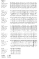
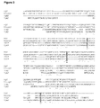


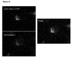
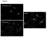


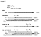


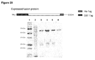


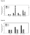
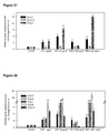

Classifications
-
- C—CHEMISTRY; METALLURGY
- C12—BIOCHEMISTRY; BEER; SPIRITS; WINE; VINEGAR; MICROBIOLOGY; ENZYMOLOGY; MUTATION OR GENETIC ENGINEERING
- C12N—MICROORGANISMS OR ENZYMES; COMPOSITIONS THEREOF; PROPAGATING, PRESERVING, OR MAINTAINING MICROORGANISMS; MUTATION OR GENETIC ENGINEERING; CULTURE MEDIA
- C12N9/00—Enzymes; Proenzymes; Compositions thereof; Processes for preparing, activating, inhibiting, separating or purifying enzymes
- C12N9/10—Transferases (2.)
- C12N9/1025—Acyltransferases (2.3)
- C12N9/104—Aminoacyltransferases (2.3.2)
-
- C—CHEMISTRY; METALLURGY
- C12—BIOCHEMISTRY; BEER; SPIRITS; WINE; VINEGAR; MICROBIOLOGY; ENZYMOLOGY; MUTATION OR GENETIC ENGINEERING
- C12N—MICROORGANISMS OR ENZYMES; COMPOSITIONS THEREOF; PROPAGATING, PRESERVING, OR MAINTAINING MICROORGANISMS; MUTATION OR GENETIC ENGINEERING; CULTURE MEDIA
- C12N15/00—Mutation or genetic engineering; DNA or RNA concerning genetic engineering, vectors, e.g. plasmids, or their isolation, preparation or purification; Use of hosts therefor
- C12N15/09—Recombinant DNA-technology
- C12N15/11—DNA or RNA fragments; Modified forms thereof; Non-coding nucleic acids having a biological activity
- C12N15/52—Genes encoding for enzymes or proenzymes
-
- A—HUMAN NECESSITIES
- A61—MEDICAL OR VETERINARY SCIENCE; HYGIENE
- A61K—PREPARATIONS FOR MEDICAL, DENTAL OR TOILETRY PURPOSES
- A61K38/00—Medicinal preparations containing peptides
- A61K38/16—Peptides having more than 20 amino acids; Gastrins; Somatostatins; Melanotropins; Derivatives thereof
- A61K38/43—Enzymes; Proenzymes; Derivatives thereof
- A61K38/45—Transferases (2)
-
- A—HUMAN NECESSITIES
- A61—MEDICAL OR VETERINARY SCIENCE; HYGIENE
- A61P—SPECIFIC THERAPEUTIC ACTIVITY OF CHEMICAL COMPOUNDS OR MEDICINAL PREPARATIONS
- A61P1/00—Drugs for disorders of the alimentary tract or the digestive system
- A61P1/04—Drugs for disorders of the alimentary tract or the digestive system for ulcers, gastritis or reflux esophagitis, e.g. antacids, inhibitors of acid secretion, mucosal protectants
-
- A—HUMAN NECESSITIES
- A61—MEDICAL OR VETERINARY SCIENCE; HYGIENE
- A61P—SPECIFIC THERAPEUTIC ACTIVITY OF CHEMICAL COMPOUNDS OR MEDICINAL PREPARATIONS
- A61P15/00—Drugs for genital or sexual disorders; Contraceptives
- A61P15/08—Drugs for genital or sexual disorders; Contraceptives for gonadal disorders or for enhancing fertility, e.g. inducers of ovulation or of spermatogenesis
-
- A—HUMAN NECESSITIES
- A61—MEDICAL OR VETERINARY SCIENCE; HYGIENE
- A61P—SPECIFIC THERAPEUTIC ACTIVITY OF CHEMICAL COMPOUNDS OR MEDICINAL PREPARATIONS
- A61P17/00—Drugs for dermatological disorders
-
- A—HUMAN NECESSITIES
- A61—MEDICAL OR VETERINARY SCIENCE; HYGIENE
- A61P—SPECIFIC THERAPEUTIC ACTIVITY OF CHEMICAL COMPOUNDS OR MEDICINAL PREPARATIONS
- A61P17/00—Drugs for dermatological disorders
- A61P17/04—Antipruritics
-
- A—HUMAN NECESSITIES
- A61—MEDICAL OR VETERINARY SCIENCE; HYGIENE
- A61P—SPECIFIC THERAPEUTIC ACTIVITY OF CHEMICAL COMPOUNDS OR MEDICINAL PREPARATIONS
- A61P25/00—Drugs for disorders of the nervous system
-
- A—HUMAN NECESSITIES
- A61—MEDICAL OR VETERINARY SCIENCE; HYGIENE
- A61P—SPECIFIC THERAPEUTIC ACTIVITY OF CHEMICAL COMPOUNDS OR MEDICINAL PREPARATIONS
- A61P25/00—Drugs for disorders of the nervous system
- A61P25/14—Drugs for disorders of the nervous system for treating abnormal movements, e.g. chorea, dyskinesia
-
- A—HUMAN NECESSITIES
- A61—MEDICAL OR VETERINARY SCIENCE; HYGIENE
- A61P—SPECIFIC THERAPEUTIC ACTIVITY OF CHEMICAL COMPOUNDS OR MEDICINAL PREPARATIONS
- A61P25/00—Drugs for disorders of the nervous system
- A61P25/18—Antipsychotics, i.e. neuroleptics; Drugs for mania or schizophrenia
-
- A—HUMAN NECESSITIES
- A61—MEDICAL OR VETERINARY SCIENCE; HYGIENE
- A61P—SPECIFIC THERAPEUTIC ACTIVITY OF CHEMICAL COMPOUNDS OR MEDICINAL PREPARATIONS
- A61P25/00—Drugs for disorders of the nervous system
- A61P25/20—Hypnotics; Sedatives
-
- A—HUMAN NECESSITIES
- A61—MEDICAL OR VETERINARY SCIENCE; HYGIENE
- A61P—SPECIFIC THERAPEUTIC ACTIVITY OF CHEMICAL COMPOUNDS OR MEDICINAL PREPARATIONS
- A61P25/00—Drugs for disorders of the nervous system
- A61P25/28—Drugs for disorders of the nervous system for treating neurodegenerative disorders of the central nervous system, e.g. nootropic agents, cognition enhancers, drugs for treating Alzheimer's disease or other forms of dementia
-
- A—HUMAN NECESSITIES
- A61—MEDICAL OR VETERINARY SCIENCE; HYGIENE
- A61P—SPECIFIC THERAPEUTIC ACTIVITY OF CHEMICAL COMPOUNDS OR MEDICINAL PREPARATIONS
- A61P29/00—Non-central analgesic, antipyretic or antiinflammatory agents, e.g. antirheumatic agents; Non-steroidal antiinflammatory drugs [NSAID]
-
- A—HUMAN NECESSITIES
- A61—MEDICAL OR VETERINARY SCIENCE; HYGIENE
- A61P—SPECIFIC THERAPEUTIC ACTIVITY OF CHEMICAL COMPOUNDS OR MEDICINAL PREPARATIONS
- A61P3/00—Drugs for disorders of the metabolism
-
- A—HUMAN NECESSITIES
- A61—MEDICAL OR VETERINARY SCIENCE; HYGIENE
- A61P—SPECIFIC THERAPEUTIC ACTIVITY OF CHEMICAL COMPOUNDS OR MEDICINAL PREPARATIONS
- A61P31/00—Antiinfectives, i.e. antibiotics, antiseptics, chemotherapeutics
- A61P31/04—Antibacterial agents
-
- A—HUMAN NECESSITIES
- A61—MEDICAL OR VETERINARY SCIENCE; HYGIENE
- A61P—SPECIFIC THERAPEUTIC ACTIVITY OF CHEMICAL COMPOUNDS OR MEDICINAL PREPARATIONS
- A61P35/00—Antineoplastic agents
-
- A—HUMAN NECESSITIES
- A61—MEDICAL OR VETERINARY SCIENCE; HYGIENE
- A61P—SPECIFIC THERAPEUTIC ACTIVITY OF CHEMICAL COMPOUNDS OR MEDICINAL PREPARATIONS
- A61P35/00—Antineoplastic agents
- A61P35/04—Antineoplastic agents specific for metastasis
-
- A—HUMAN NECESSITIES
- A61—MEDICAL OR VETERINARY SCIENCE; HYGIENE
- A61P—SPECIFIC THERAPEUTIC ACTIVITY OF CHEMICAL COMPOUNDS OR MEDICINAL PREPARATIONS
- A61P37/00—Drugs for immunological or allergic disorders
- A61P37/02—Immunomodulators
- A61P37/06—Immunosuppressants, e.g. drugs for graft rejection
-
- A—HUMAN NECESSITIES
- A61—MEDICAL OR VETERINARY SCIENCE; HYGIENE
- A61P—SPECIFIC THERAPEUTIC ACTIVITY OF CHEMICAL COMPOUNDS OR MEDICINAL PREPARATIONS
- A61P9/00—Drugs for disorders of the cardiovascular system
- A61P9/10—Drugs for disorders of the cardiovascular system for treating ischaemic or atherosclerotic diseases, e.g. antianginal drugs, coronary vasodilators, drugs for myocardial infarction, retinopathy, cerebrovascula insufficiency, renal arteriosclerosis
-
- C—CHEMISTRY; METALLURGY
- C07—ORGANIC CHEMISTRY
- C07D—HETEROCYCLIC COMPOUNDS
- C07D233/00—Heterocyclic compounds containing 1,3-diazole or hydrogenated 1,3-diazole rings, not condensed with other rings
- C07D233/54—Heterocyclic compounds containing 1,3-diazole or hydrogenated 1,3-diazole rings, not condensed with other rings having two double bonds between ring members or between ring members and non-ring members
- C07D233/56—Heterocyclic compounds containing 1,3-diazole or hydrogenated 1,3-diazole rings, not condensed with other rings having two double bonds between ring members or between ring members and non-ring members with only hydrogen atoms or radicals containing only hydrogen and carbon atoms, attached to ring carbon atoms
- C07D233/61—Heterocyclic compounds containing 1,3-diazole or hydrogenated 1,3-diazole rings, not condensed with other rings having two double bonds between ring members or between ring members and non-ring members with only hydrogen atoms or radicals containing only hydrogen and carbon atoms, attached to ring carbon atoms with hydrocarbon radicals, substituted by nitrogen atoms not forming part of a nitro radical, attached to ring nitrogen atoms
-
- C—CHEMISTRY; METALLURGY
- C07—ORGANIC CHEMISTRY
- C07D—HETEROCYCLIC COMPOUNDS
- C07D235/00—Heterocyclic compounds containing 1,3-diazole or hydrogenated 1,3-diazole rings, condensed with other rings
- C07D235/02—Heterocyclic compounds containing 1,3-diazole or hydrogenated 1,3-diazole rings, condensed with other rings condensed with carbocyclic rings or ring systems
- C07D235/04—Benzimidazoles; Hydrogenated benzimidazoles
- C07D235/06—Benzimidazoles; Hydrogenated benzimidazoles with only hydrogen atoms, hydrocarbon or substituted hydrocarbon radicals, directly attached in position 2
-
- C—CHEMISTRY; METALLURGY
- C07—ORGANIC CHEMISTRY
- C07D—HETEROCYCLIC COMPOUNDS
- C07D401/00—Heterocyclic compounds containing two or more hetero rings, having nitrogen atoms as the only ring hetero atoms, at least one ring being a six-membered ring with only one nitrogen atom
- C07D401/02—Heterocyclic compounds containing two or more hetero rings, having nitrogen atoms as the only ring hetero atoms, at least one ring being a six-membered ring with only one nitrogen atom containing two hetero rings
- C07D401/12—Heterocyclic compounds containing two or more hetero rings, having nitrogen atoms as the only ring hetero atoms, at least one ring being a six-membered ring with only one nitrogen atom containing two hetero rings linked by a chain containing hetero atoms as chain links
-
- C—CHEMISTRY; METALLURGY
- C07—ORGANIC CHEMISTRY
- C07D—HETEROCYCLIC COMPOUNDS
- C07D403/00—Heterocyclic compounds containing two or more hetero rings, having nitrogen atoms as the only ring hetero atoms, not provided for by group C07D401/00
- C07D403/02—Heterocyclic compounds containing two or more hetero rings, having nitrogen atoms as the only ring hetero atoms, not provided for by group C07D401/00 containing two hetero rings
- C07D403/06—Heterocyclic compounds containing two or more hetero rings, having nitrogen atoms as the only ring hetero atoms, not provided for by group C07D401/00 containing two hetero rings linked by a carbon chain containing only aliphatic carbon atoms
-
- C—CHEMISTRY; METALLURGY
- C07—ORGANIC CHEMISTRY
- C07D—HETEROCYCLIC COMPOUNDS
- C07D403/00—Heterocyclic compounds containing two or more hetero rings, having nitrogen atoms as the only ring hetero atoms, not provided for by group C07D401/00
- C07D403/02—Heterocyclic compounds containing two or more hetero rings, having nitrogen atoms as the only ring hetero atoms, not provided for by group C07D401/00 containing two hetero rings
- C07D403/12—Heterocyclic compounds containing two or more hetero rings, having nitrogen atoms as the only ring hetero atoms, not provided for by group C07D401/00 containing two hetero rings linked by a chain containing hetero atoms as chain links
-
- C—CHEMISTRY; METALLURGY
- C07—ORGANIC CHEMISTRY
- C07D—HETEROCYCLIC COMPOUNDS
- C07D405/00—Heterocyclic compounds containing both one or more hetero rings having oxygen atoms as the only ring hetero atoms, and one or more rings having nitrogen as the only ring hetero atom
- C07D405/02—Heterocyclic compounds containing both one or more hetero rings having oxygen atoms as the only ring hetero atoms, and one or more rings having nitrogen as the only ring hetero atom containing two hetero rings
- C07D405/12—Heterocyclic compounds containing both one or more hetero rings having oxygen atoms as the only ring hetero atoms, and one or more rings having nitrogen as the only ring hetero atom containing two hetero rings linked by a chain containing hetero atoms as chain links
-
- C—CHEMISTRY; METALLURGY
- C07—ORGANIC CHEMISTRY
- C07D—HETEROCYCLIC COMPOUNDS
- C07D409/00—Heterocyclic compounds containing two or more hetero rings, at least one ring having sulfur atoms as the only ring hetero atoms
- C07D409/02—Heterocyclic compounds containing two or more hetero rings, at least one ring having sulfur atoms as the only ring hetero atoms containing two hetero rings
- C07D409/12—Heterocyclic compounds containing two or more hetero rings, at least one ring having sulfur atoms as the only ring hetero atoms containing two hetero rings linked by a chain containing hetero atoms as chain links
-
- C—CHEMISTRY; METALLURGY
- C07—ORGANIC CHEMISTRY
- C07D—HETEROCYCLIC COMPOUNDS
- C07D413/00—Heterocyclic compounds containing two or more hetero rings, at least one ring having nitrogen and oxygen atoms as the only ring hetero atoms
- C07D413/02—Heterocyclic compounds containing two or more hetero rings, at least one ring having nitrogen and oxygen atoms as the only ring hetero atoms containing two hetero rings
- C07D413/12—Heterocyclic compounds containing two or more hetero rings, at least one ring having nitrogen and oxygen atoms as the only ring hetero atoms containing two hetero rings linked by a chain containing hetero atoms as chain links
-
- C—CHEMISTRY; METALLURGY
- C07—ORGANIC CHEMISTRY
- C07D—HETEROCYCLIC COMPOUNDS
- C07D417/00—Heterocyclic compounds containing two or more hetero rings, at least one ring having nitrogen and sulfur atoms as the only ring hetero atoms, not provided for by group C07D415/00
- C07D417/02—Heterocyclic compounds containing two or more hetero rings, at least one ring having nitrogen and sulfur atoms as the only ring hetero atoms, not provided for by group C07D415/00 containing two hetero rings
- C07D417/12—Heterocyclic compounds containing two or more hetero rings, at least one ring having nitrogen and sulfur atoms as the only ring hetero atoms, not provided for by group C07D415/00 containing two hetero rings linked by a chain containing hetero atoms as chain links
-
- C—CHEMISTRY; METALLURGY
- C12—BIOCHEMISTRY; BEER; SPIRITS; WINE; VINEGAR; MICROBIOLOGY; ENZYMOLOGY; MUTATION OR GENETIC ENGINEERING
- C12N—MICROORGANISMS OR ENZYMES; COMPOSITIONS THEREOF; PROPAGATING, PRESERVING, OR MAINTAINING MICROORGANISMS; MUTATION OR GENETIC ENGINEERING; CULTURE MEDIA
- C12N9/00—Enzymes; Proenzymes; Compositions thereof; Processes for preparing, activating, inhibiting, separating or purifying enzymes
- C12N9/10—Transferases (2.)
-
- G—PHYSICS
- G01—MEASURING; TESTING
- G01N—INVESTIGATING OR ANALYSING MATERIALS BY DETERMINING THEIR CHEMICAL OR PHYSICAL PROPERTIES
- G01N33/00—Investigating or analysing materials by specific methods not covered by groups G01N1/00 - G01N31/00
- G01N33/48—Biological material, e.g. blood, urine; Haemocytometers
- G01N33/50—Chemical analysis of biological material, e.g. blood, urine; Testing involving biospecific ligand binding methods; Immunological testing
- G01N33/53—Immunoassay; Biospecific binding assay; Materials therefor
- G01N33/573—Immunoassay; Biospecific binding assay; Materials therefor for enzymes or isoenzymes
Definitions
- the present invention relates to novel glutaminyl-peptide cyclotransferase-like proteins (QPCTLs), which are isoenzymes of glutaminyl cyclase (QC, EC 2.3.2.5), and to isolated nucleic acids coding for these isoenzymes, all of which are useful for the discovery of new therapeutic agents, for measuring cyclase activity, and for determining the inhibitory activity of compounds against these glutaminyl cyclase isoenzymes.
- QPCTLs novel glutaminyl-peptide cyclotransferase-like proteins
- Glutaminyl cyclase catalyzes the intramolecular cyclization of N-terminal glutamine residues into pyroglutamic acid (pGlu*) liberating ammonia.
- pGlu* pyroglutamic acid
- EP 02 011 349.4 discloses polynucleotides encoding insect glutaminyl cyclase, as well as polypeptides encoded thereby.
- This application further provides host cells comprising expression vectors comprising polynucleotides of the invention. Isolated polypeptides and host cells comprising insect QC are useful in methods of screening for agents that reduce glutaminyl cyclase activity. Such agents are useful as pesticides.
- Inhibitors of QC which also could be useful as inhibitors of QC isoenzymes, are described in WO 2004/098625 , WO 2004/098591 , WO 2005/039548 and WO 2005/075436 .
- Reversible enzyme inhibitors comprise competitive inhibitors, non-competitive reversible inhibitors, slow-binding or tight-binding inhibitors, transition state analogs and multisubstrate analogs.
- a competitive inhibitor or transition state analog can be designed which contains structural characteristics resembling two or more of the substrates.
- Irreversible enzyme inhibitors drive the equilibrium between the unbound enzyme and inhibitor and enzyme inhibitor complex (E + I ⁇ ---> E-I) all the way to the right with a covalent bond ( ⁇ 100 kcal/mole), making the inhibition irreversible.
- the reaction scheme below shows the mechanism of action of a mechanism based enzyme inactivator, where K D is the dissociation complex, k 2 is the rate of activation of the inhibitor once bound to the enzyme, k 3 is the rate of dissociation of the activated inhibitor, P, from the enzyme (product can still be reactive) from the enzyme and k 4 is the rate of covalent bond formation between the activated inhibitor and the enzyme.
- Inactivation (covalent bond formation, k 4 ) must occur prior to dissociation (k 3 ) otherwise the now reactive inhibitor is released into the environment.
- Uncompetitive enzyme inhibitors From the definition of uncompetitive inhibitor (an inhibitor which binds only to ES complexes) the following equilibria can be written:
- the ES complex dissociates the substrate with a dissociation constant equal to Ks, whereas the ESI complex does not dissociate it (i.e has a Ks value equal to zero).
- the K m 's of Michaelis-Menten type enzymes are expected to be reduced.
- Increasing substrate concentration leads to increasing ESI concentration (a complex incapable of progressing to reaction products), therefore the inhibition can not be removed.
- Preferred according to the present invention are competitive enzyme inhibitors. Most preferred are competitive reversible enzyme inhibitors.
- k i or "K l” and “K D” are binding constants, which describe the binding of an inhibitor to and the subsequent release from an enzyme. Another measure is the “IC 50 " value, which reflects the inhibitor concentration, which at a given substrate concentration results in 50 % enzyme activity.
- QC comprises glutaminyl cyclase (QC), which is synonymous to glutaminyl-peptide cyclotransferase (QPCT); and QC-like enzymes, which are synonymous to glutaminyl-peptide cyclotransferase-like proteins (QPCTLs).
- QC and QC-like enzymes have identical or similar enzymatic activity, further defined as QC activity.
- QC-like enzymes can fundamentally differ in their molecular structure from QC.
- QC-activity is defined as the catalytic activity of glutaminyl cyclase (QC, QPCT) and QC-like enzymes (QPCTLs). These enzymes are found in various tissues of the body of a mammal including kidney, liver, intestine, brain and body fluids such as CSF, where they cyclize glutamine or glutamate at the N-terminus of biologically active peptides with a high specificity.
- QC activity is defined as intramolecular cyclization of N-terminal glutamine residues into pyroglutamic acid (pGlu*) or of N-terminal L-homoglutamine or L- ⁇ -homoglutamine to a cyclic pyro-homoglutamine derivative under liberation of ammonia. See therefore schemes 1 and 2.
- EC as used herein comprises the side activity of glutaminyl cyclase (QC, QPCT) and QC-like enzymes (QPCTLs) as glutamate cyclase (EC), further defined as EC activity.
- EC activity is defined as intramolecular cyclization of N-terminal glutamate residues into pyroglutamic acid (pGlu*) by glutaminyl cyclase (QC, QPCT) and QC-like enzymes (QPCTLs). See therefore scheme 3.
- QC-inhibitor or "glutaminyl cyclase inhibitor” is generally known to a person skilled in the art and means enzyme inhibitors, which inhibit the catalytic activity of glutaminyl cyclase (QC, QPCT) or QC-like enzymes (QPCTLs) or their glutamyl cyclase (EC) activity, preferably by direct interaction of the inhibitor with the enzyme.
- enzyme inhibitors which inhibit the catalytic activity of glutaminyl cyclase (QC, QPCT) or QC-like enzymes (QPCTLs) or their glutamyl cyclase (EC) activity, preferably by direct interaction of the inhibitor with the enzyme.
- selective QC-inhibitor as defined herein means enzyme inhibitors, which inhibit the catalytic activity of glutaminyl cyclase (QC, QPCT) but do not or with a lower potency inhibit at least one QC-like enzymes (QPCTLs).
- selective QC-inhibitors which inhibit glutaminyl cyclase (QC, QPCT) with an ki-value, which is one order of magnitude lower than its ki-value for the inhibition of at least one QC-like enzyme (QPCTL).
- the ki-value of said selective QC-inhibitor for the inhibition of glutaminyl cyclase is two orders of magnitude lower than its ki-value for the inhibition of at least one QC-like enzyme (QPCTL).
- selective QC-inhibitors wherein their ki-value for the inhibition of glutaminyl cyclase (QC, QPCT) is three orders of magnitude lower than their ki-value for the inhibition of at least one QC-like enzyme (QPCTL).
- selective QPCTL-inhibitor as defined herein means enzyme inhibitors, which inhibit the catalytic activity of at least one QC-like enzyme (QPCTL), but do not or with a lower potency inhibit the activity of glutaminyl cyclase (QC, QPCT).
- QPCTL QC-like enzyme
- the ki-value of said selective QPCTL-inhibitor for the inhibition of at least one QC-like enzyme is two orders of magnitude lower than its ki-value for the inhibition of of of glutaminyl cyclase (QC, QPCT).
- selective QPCTL-inhibitors wherein their ki-value for the inhibition of at least one QC-like enzyme (QPCTL) is three orders of magnitude lower than their ki-value for the inhibition of glutaminyl cyclase (QC, QPCT).
- the subject method and medical use utilize an agent with a K i for QC inhibition of 10 ⁇ M or less, more preferably of 1 ⁇ M or less, even more preferably of 0.1 ⁇ M or less or 0.01 ⁇ M or less, or most preferably 0.01 ⁇ M or less.
- K i for QC inhibition
- 10 ⁇ M or less more preferably of 1 ⁇ M or less, even more preferably of 0.1 ⁇ M or less or 0.01 ⁇ M or less, or most preferably 0.01 ⁇ M or less.
- the QC inhibitors of the subject method or medical use will be small molecules, e.g., with molecular weights of 1000 g/mole or less, 500 g/mole or less, preferably of 400 g/mole or less, and even more preferably of 350 g/mole or less and even of 300 g/mole or less.
- subject refers to an animal, preferably a mammal, most preferably a human, who has been the object of treatment, observation or experiment.
- terapéuticaally effective amount means that amount of active compound or pharmaceutical agent that elicits the biological or medicinal response in a tissue system, animal or human being sought by a researcher, veterinarian, medical doctor or other clinician, which includes alleviation of the symptoms of the disease or disorder being treated.
- the term "pharmaceutically acceptable” embraces both human and veterinary use: for example the term “pharmaceutically acceptable” embraces a veterinarily acceptable compound or a compound acceptable in human medicine and health care.
- the present invention provides mutant proteins with glutaminyl cyclase activities that constitute novel members of a family of proteins related to glutaminyl cyclase, including nucleotide sequences encoding the same.
- the above proteins may be used in methods of screening for substrates, interacting proteins, agonists, antagonists or inhibitors.
- novel proteins having significant sequence similarity to glutaminyl cyclase are mutant proteins (QPCTLs) from human (further named as human isoQC) (GenBank accession no. NM_017659) of SEQ ID NO: 11.
- the protein sequence is given in SEQ. ID NO: 12. Further disclosed are nucleic acid sequences coding for these proteins (SEQ. ID NO: 3). Table 1 illustrates the similarity between human isoQC and the known glutaminyl cyclase. Table 2 illustrates the identity between the human isoQC and the known glutaminyl cyclase.
- Table 1 Similarity of the protein sequences of the novel glutaminyl-peptide cyclotransferase-like proteins with glutaminyl cyclase QPCTL source human isoQC ( SEQ ID NO 11) human QC ( SEQ ID NO 10) human isoQC (SEQ ID NO 11) - 71.98%
- Table 2 Identity of the protein sequences of the novel glutaminyl-peptide cyclotransferase-like proteins with glutaminyl cyclase QPCTL source human isoQC ( SEQ ID NO 11) human QC (SEQ ID NO 10) human isoQC (SEQ ID NO 11) - 45.24%
- the expression pattern of the QPCTLs in brain, prostate and lung tissue is consistent with a role in the diseases described below.
- the enzymatic activity as glutaminyl cyclase demonstrates that QPCTLs-activating or inhibiting molecules will have numerous therapeutic applications as described below.
- QPCTL activities described herein and their expression patterns are compatible with their functional roles as physiological regulators of the immune and neuroendocrine systems through the enzymatic modification of biochemical mediators like hormones, peptides and chemokines.
- biochemical mediators like hormones, peptides and chemokines.
- the numerous functions previously described for QC based upon the use of inhibitors may be due in part to its action and that of similar proteins, like the QPCTLs. Therefore, the discovery of selective and potent inhibitors of QC, of the QPCTLs and of other related enzymes is considered central to achieving effective and safe pharmaceutical use of these and any newly identified glutaminyl-peptide cyclotransferases, as well as other active compounds that modify the function(s) of such proteins.
- the invention thus provides novel mutant proteins or polypeptides, the nucleic acids coding therefore, cells which have been modified with the nucleic acid so as to express these proteins,
- the novel proteins and the nucleic acids coding therefore can be used to discover new therapeutic agents for the treatment of certain diseases, such as for example, neurodegenerative, reproductive, inflammatory and metabolic disorders and also in the preparation of antibodies with therapeutic or diagnostic value.
- novel, mutant biologically active proteins preferably of human origin.
- Such proteins may be isolated in small quantities from suitable animal (including human) tissue or biological fluids by standard techniques; however, larger quantities are more conveniently prepared in cultures of cells genetically modified so as to express the protein.
- nucleic acid molecules encoding polypeptides of the present invention including mRNAs, DNAs, cDNAs, genomic DNAs thereof.
- nucleic acid probes are also provided comprising nucleic acid molecules of sufficient length to specifically hybridize to a nucleic acid sequence of the present invention.
- processes utilizing recombinant techniques are provided for producing such polypeptides useful for in vitro scientific research, for example, synthesis of DNA and manufacture of DNA vectors.
- Processes for producing such polypeptides include culturing recombinant prokaryotic and/or eukaryotic host cells that have been transfected with DNA vectors containing a nucleic acid sequence encoding such a polypeptide and/or the mature protein under conditions promoting expression of such protein and subsequent recovery of such protein or a fragment of the expressed product.
- the invention provides an isolated nucleic acid which encodes a QPCTL polypeptide of SEQ ID NO: 12.
- the invention provides a polypeptide which is glycosylated, and which comprises the amino acid sequence of a mature protein set forth in SEQ ID NOS: 12.
- Figure 1 shows the sequence alignment of human QC (hQC), human isoQC (hisoQC), murine QC (mQC) and murine isoQC (misoQC). Multiple sequence alignment was performed using ClustalW at PBIL (Pdeemed Bioposition Lyonnais) (http://npsa-pbil.ibcp.fr) with default settings.
- PBIL Porous Bioposition Lyonnais
- human QC hQC; GenBank X71125, SEQ ID NO: 10
- human isoQC hisoQC, GenBank NM_017659, SEQ ID NO: 11
- murine QC mQC, GenBank NM_027455, SEQ ID NO: 79
- murine isoQC misoQC, GenBank BC058181, SEQ ID NO: 17
- Figure 2 shows the sequence alignment of isoQC from Homo sapiens (hisoQC, GenBank NM_017659, SEQ ID NO: 11), Macaca fascicularis (M_fascicularis, GenBank AB168255, SEQ ID NO: 13), Macaca mulatta (M_mulatta, GenBank XM_001110995, SEQ ID NO: 14), Canis familiaris (C_familiaris, GenBank M_541552, SEQ ID NO: 15), Rattus norvegicus (R_norvegicus, GenBank M_001066591, SEQ ID NO: 16), Mus musculus (M_musculus, GenBank BC058181, SEQ ID NO: 17) and Bos taurus (B_taurus, GenBank BT026254, SEQ ID NO: 18).
- Figure 4 shows the sequence alignment of human QC (hQC, SEQ ID NO: 10) and human isoQC (hisoQC, SEQ ID NO: 11), showing two putative tranlational starts (methionine I - bold, underlined; methionine II - bold). Multiple sequence alignment was performed using ClustalW at PBIL (Pdeemed Biocie Lyonnais) http://npsa-pbil.ibcp.fr with default settings. The transmembrane domain, present in human isoQC, is indicated by the black bar.
- Figure 5 shows the sequence alignment of human QC (hQC, SEQ ID NO: 10) and human isoQC (hisoQC, SEQ ID NO: 12), starting with methionine II (bold).
- FIG. 6 shows the analysis of isoQC expression by RT-PCR. Detection in SH-SY5Y, LN405, HaCaT and Hep-G2. Lanes: bp, DNA standard; 1, amplified PCR product of human isoQC from SH-SY5Y; 2, amplified PCR product of human isoQC from LN405; 3, amplified PCR product of human isoQC from HaCaT; 4, amplified PCR product of human isoQC from Hep-G2.
- Figure 7 shows the analysis of isoQC (Met I, SEQ ID NO: 11) subcellular localization by immunhistochemistry.
- Human isoQC starting at methionine I was expressed as a fusion protein with EGFP (isoQC (Metl) EGFP) in LN 405.
- Mannosidase II counterstaining was performed using AB3712 (Chemicon). Merge represents the overlay of isoQC (Metl)-EGFP and Mannosidase II staining.
- Figure 8 shows the analysis of isoQC (Met I, SEQ ID NO: 11) subcellular localization by immunhistochemistry.
- Human isoQC starting at methionine I was expressed as a fusion protein with EGFP (isoQC (Metl) EGFP) in LN 405. Mitochondrial counterstaining was performed using MAB1273 (Chemicon). Merge represents the overlay of isoQC (Metl)-EGFP and mitochondrial staining.
- Figure 9 shows the analysis of isoQC (Met II, SEQ ID NO: 12) subcellular localization by immunhistochemistry. Human isoQC starting at methionine II was expressed as a fusion protein with EGFP (isoQC (MetII) EGFP) in LN 405. Mannosidase II counterstaining was performed using AB3712 (Chemicon).
- Merge represents the overlay of isoQC (Metll)-EGFP and Mannosidase II staining.
- Figure 10 shows the analysis of isoQC (Met II, SEQ ID NO: 12) subcellular localization by immunhistochemistry. Human isoQC starting at methionine II was expressed as a fusion protein with EGFP (isoQC (Metll) EGFP) in LN 405. Mitochondrial counterstaining was performed using MAB1273 (Chemicon).
- Merge represents the overlay of isoQC (Metll)-EGFP and mitochondrial staining.
- Figure 11 shows the analysis of the subcellular localization of isoQC (Met I, SEQ ID NO: 11) by immunhistochemistry.
- Human isoQC starting at methionine I was expressed as a fusion protein with EGFP (isoQC (Metl) EGFP) in COS-7. Mannosidase II counterstaining was performed using AB3712 (Chemicon). Merge represents the overlay of isoQC (Metl)-EGFP and Mannosidase II staining.
- Figure 12 shows the analysis of isoQC (Met I, SEQ ID NO: 11) subcellular localization by immunhistochemistry. Human isoQC starting at methionine I was expressed as a fusion protein with EGFP (isoQC (Metl) EGFP) in COS-7. Mitochondrial counterstaining was performed using MAB1273 (Chemicon).
- Merge represents the overlay of isoQC (Metl)-EGFP and mitochondrial staining.
- Figure 13 shows the analysis of isoQC (Met II, SEQ ID NO: 12) subcellular localization by immunhistochemistry. Human isoQC starting at methionine II was expressed as a fusion protein with EGFP (isoQC (Metll) EGFP) in COS-7. Mannosidase II counterstaining was performed using AB3712 (Chemicon).
- Merge represents the overlay of isoQC (Metll)-EGFP and Mannosidase II staining.
- Figure 14 shows the analysis of isoQC (Met II, SEQ ID NO: 12) subcellular localization by immunhistochemistry.
- Inhibitor concentrations were as follows: 0 ⁇ M 0.3125 ⁇ M 0.625 ⁇ M 1.25 ⁇ M 2.5 ⁇ M 5 ⁇ M The determined K i -value was 240 ⁇ 8 nM.
- Figure 16 shows the human isoQC-catalyzed conversion of H-Gln-Ala-OH into pGlu-Ala-OH determined using a spectrophotometric assay. The data were evaluated according to Michaelis-Menten kinetics. The kinetic parameters were 324 ⁇ 28 ⁇ M and 7.4 ⁇ 0.2 nM/min for the K M and V max -value, respectively.
- Figure 17 provides a schematic representation of the human isoQC protein constructs that were expressed hetereologously in the yeast P.
- FIG. 18 shows the QC activity, which was determined in the medium of expressing yeast cells. Due to the transmembrane domain, the native constructs were not secreted into the medium (not implemented). Caused by glycosylation (I55N), proteins are most efficiently secreted.
- the mutation C351 A resulted also in higher QC activity detected in the medium.
- Figure 19 shows the purification of the human isoQC, based on construct YSShisoQCI55NC351A C-His, from the medium of a transgenic P.pastoris strain.
- the QC was purified by a combination of IMAC (immobilized metal affinity chromatography, lane 3), HIC (hydrophobic interaction chromatography, lane 4) and desalting (lane 5).
- the glycosylation of the enzyme was evidence by enzymatic deglycosalytion, which results in a shift in migration of the protein (lane 6).
- Lane 1 protein standard: Lane 2, medium prior to purification.
- Figure 20 shows the purification of the human isoQC, based on construct GST-hisoQC C-His, from the cell homogenate of transformed E. coli.
- the isoQC was purified by a combination of IMAC (immobilized metal affinity chromatography, lane 3), GST-affinity (lane 4), desalting (lane 5) and ion exchange chromatography (lane 6).
- Lane 1 protein standard: Lane 2, cell homogenate prior to purification.
- the difference in the molecular mass between the hisoQC which was expressed in yeast and E. coli is caused by the N-terminal GST-tag fusion.
- the expressed construct is provided schematically in the upper part of the figure.
- Figure 21 shows the specificity constants for conversion of dipeptide-surrogates, dipeptides and oligopeptides by human isoQC (YSShisoQCI55NC351 A C-His; compare Figure 17 ), GST-hisoQC and human QC.
- the specificity of GST-hisoQC was the lowest, followed by YSShisoQCI55NC351 A C-His.
- the highest specificity displayed human QC indicating a higher overall enzymatic activity.
- Figure 22 shows the pH-dependency of catalysis, investigated with human isoQC (hisoQC), which was expressed in yeast, and human QC (hQC).
- Both proteins display a pH-optimum between pH 7 and 8.
- the fitted curve is based on three dissociating groups that influence catalysis, one at acidic pH, two at basic pH.
- Figure 23 shows the analysis of conversion of glutamic acid, which is present at the N-terminus of the amyloid- ⁇ related peptide A ⁇ (3-11). The analysis was performed using Maldi-Tof mass spectrometry, the substrate and product differ in their molecular mass/charge ratio of the single chared molecule by about 18Da, which is the mass of the released water. In both cases, the same protein concentration was present in the samples, clearly suggesting that human isoQC also converts N-terminal glutamic acid, but slower than the human QC.
- Figure 24 shows the tissue distribution of murine QC (mQC, SEQ ID NO: 79) and its isoenzyme misoQC (SEQ ID NO: 17), analyzed using real-time PCR. Bothe enzymes are expressed in the tested organs. However, the expression level of mQC was higher in the brain compared with the peripheral organs. In contrats, misoQC was expressed in all tested organs and tisssues at a more similar level, indicating a ubiquitous, "housekeeping" protein.
- Figure 25 shows the time-dependent inhibition of human isoQC (hisoQC) by metal-chelating compounds 1,10-phenanthroline (circles) and EDTA (squares).
- FIG. 26 shows the biochemical analysis of the subcellular localization of QC activity after expression of pcDNA and the native enzymes hisoQC (Met I, SEQ ID NO: 11), hisoQC (Met II, SEQ ID NO: 12) and hQC (SEQ ID NO: 10) in HEK293 cells.
- HisoQC Metal I, SEQ ID NO: 11
- hisoQC Metal II, SEQ ID NO: 12
- hQC hQC
- C Expression of h-isoQC (Met I, SEQ ID NO: 11), h-isoQC (Met II, SEQ ID NO: 12) and hQC (SEQ ID NO: 10) possessing a C-terminal FLAG-tag in HEK293 in comparison to vector-transfected control (pcDNA), followed by Western Blot analysis applying specific antibodies detecting either the FLAG-epitope (anti-DYKDDDDK-antibody, Cell Signaling), a 65 kDa protein of human mitochondria (anti-human mitochondria, Chemicon) or human Sialyltransferase ST1 GAL3 (Abnova).
- Figure 27 shows the subcellular localization of human isoQC (hisoQC) signal sequences (27A) methionine I - serine 53 and (27B) methionine II - serine 53, fused to EGFP (1, 4).
- Golgi complex was stained using an anti-mannosidase II antibody (2) and mitochondria were stained using an antibody detecting a 65 kDA protein of human mitochondria (5).
- Co-localization is shown by superimposition of EGFP fluorescence and Red X fluorescence (3, 6).
- Figure 28 shows the domain structure of human isoQC (hisoQC) and murine isoQC (misoQC) in comparison to published sequences of human glycosyltransferases: alpha-N-acetylgalactosaminide alpha-2,6-sialyl transferase 1 (ST6GaINAC1 ; E.C. 2.4.99.3); beta-1,4-galactosyltransferase 1 (b4Gal-T1, E.C. 2.4.1.-); Galactoside 3(4)-L-fucosyltransferase (FucT-III; E.C.
- FIG. 29 shows the quantification of human isoQC (QPCTL) mRNA in different carcinoma cell lines.
- the QPCTL expression was normalized to 50 ng total-RNA.
- the black bar within the boxes represents the respective median.
- Figure 30 shows the quantification of human isoQC (QPCTL) mRNA expression in different melanoma cell lines.
- FIG. 31 shows the quantification of human isoQC (QPCTL) mRNA expression in samples from soft tissue carcinoma, gastric carcinoma and thyroid carcinoma from different patients. The QPCTL expression was normalized to 50 ng total-RNA. The black bar within the boxes represents the respective median.
- Figure 32 shows the human isoQC (QPCTL) mRNA expression in different gastric carcinomas against their stage of differentiation. QPCTL expression was normalized to 50 ng total-RNA. The black bar within the boxes represents the respective median.
- Figure 33 shows a comparison of human QC (QPCT) mRNA expression in different thyroid carcinomas. QPCT expression was normalized to 50 ng total-RNA. The black bar within the boxes represents the respective median.
- FIG. 34 shows a comparison of human isoQC (QPCTL) mRNA expression in different thyroid carcinomas. QPCTL expression was normalized to 50 ng total-RNA. The black bar within the boxes represents the respective median.
- FTC folicular thyroid carcinoma; PTC: papillary thyroid carcinoma; UTC: undifferentiated thyroid carcinoma.
- Figure 35 shows the influence of different stimuli on mRNA expression of human QC (QPCT), human isoQC (QPCTL) and CCL2 in HEK293 cells. The amount of transcripts is depicted relating to basal expression without stimulus. The used concentration of stimulus is stated on the x-axis drawing.
- Figure 36 shows the influence of different stimuli on mRNA expression of human QC (QPCT), human isoQC (QPCTL) and CCL2 in FTC-133 cells.
- the amount of transcripts is depicted relating to basal expression without stimulus.
- the used concentration of stimulus is stated on the x-axis drawing.
- Figure 37 shows the influence of different stimuli on mRNA expression of human QC (QPCT), human isoQC (QPCTL) and CCL2 in THP-1 cells.
- the amount of transcripts is depicted relating to basal expression without stimulus.
- the used concentration of stimulus is stated on the x-axis drawing.
- Figure 38 shows the influence of different stimuli on mRNA expression of human QC (QPCT), CCL2, CCL7, CCL8 and CCL13 in THP-1 cells.
- the amount of transcripts is depicted relating to basal expression without stimulus. The used concentration of stimulus is stated on the x-axis drawing.
- Figure 39 shows the influence of hypoxia on the mRNA level of human QC (QPCT), human isoQC (QPCTL) and HIF1 ⁇ in HEK293 (A), FTC-133 (b) and THP-1 (C).
- Preferred according to the present invention are isolated nucleic acid sequences (polynucleotides) of SEQ ID NOS: 2 and 3, 19 and 20, which encode the mature polypeptides having the deduced amino acid sequences of the QPCTLs from human (SEQ ID NOS: 11 and 12, 21 and 22).
- nucleic acid sequence of SEQ ID NO: 3, which encodes the mature polypeptide having the deduced amino acid sequence of the human QPCTL of SEQ ID NO: 12, wherein said nucleic acid comprises mutations in codons 55 and 351 and wherein isoleucine at position 55 of said polypeptide is replaced by asparagine and cysteine in position 351 of said polypeptide is replaced by alanine.
- the invention further provides a purified human QPCTL (glutaminyl-peptide cyclotransferase-like protein) of SEQ ID NO: 12, wherein isoleucine at position 55 of said polypeptide is replaced by asparagine and cysteine in position 351 of said polypeptide is replaced by alanine and which is obtainable following the expression of a construct comprising the nucleic acid of SEQ ID NO: 3 in Pichia pastoris, wherein said nucleic acid comprises mutations in codons 55 and 351 and wherein said construct may further comprise a nucleic acid, which codes for a polyhistidine tag at the N-terminus or at the C-terminus.
- QPCTL glutaminyl-peptide cyclotransferase-like protein
- the invention further provides a Pichia pastoris host cell comprising the nucleic acid of SEQ ID NO: 3, wherein said nucleic acid codes for the polypeptide, which has the amino acid sequence of SEQ ID NO: 12, wherein said nucleic acid comprises mutations in codons 55 and 351 and wherein isoleucine at position 55 of said polypeptide is replaced by asparagine and cysteine in position 351 of said polypeptide is replaced by alanine.
- the Pichia pastoris host cell of the invention comprises a construct which comprises the nucleic acid of SEQ ID NO: 3, wherein said nucleic acid comprises mutations in codons 55 and 351 and wherein said construct may further comprise a nucleic acid, which codes for a polyhistidine tag at the N-terminus or at the C-terminus.
- a Pichia pastoris host cell comprising a construct which comprises the nucleic acid of SEQ ID NO: 3, wherein said nucleic acid comprises mutations in codons 55 and 351 and wherein said construct further comprises a nucleic acid, which codes for a polyhistidine tag at the C-terminus.
- the polynucleotides of this invention were discovered by similarity search using Nucleotide BLAST at NCBI (http://www.ncbi.nlm.nih.gov/BLAST/) applying human QC as template.
- the search resulted in discovery of a putative QPCTL on chromosome 19, which is encoded in region 19q13.32.
- primers for a cell line screening of human isoQC were designed (Table 4).
- the isolated cDNA for human QPCTL contains an open reading frame encoding a protein of 382 amino acids in length, which is related to human QC displaying 45.24% sequence identity, and 71.98% similarity.
- Applying different bioinformatic algorithms (www.expasy.ch) for prediction of the subcellular localization did not result in a reliable result.
- the prognosis depending on the prediction program, was transfer to golgi-apparatus or mitochondria.
- the human isoQC gene contains at least 8 exons.
- the sequence coding for the human isoQC protein is located on exons 1 to 7.
- Human isoQC maps to chromosome 19 at position 19q13.32.
- a cell line screening for human isoQC revealed transcripts in cells origin from liver (Hep-G2, hepatocellular carcinoma), skin (HaCaT, keratinocyte) and neuronal tissues (LN405, astrocytoma; SH-SY5Y, neuroblastoma) ( Figure 6 ).
- the isolated QPCTL-cDNA was tested on functional expression in several expression hosts.
- Expression in P. pastoris which was successfully applied for human QC, did not result in an enzymatically active protein.
- Expression in mammalian cells resulted in detection of activity, however, expression levels were very low.
- Enzymatically active protein was isolated only following expression of a GST-QPCTL fusion protein in E. coli, applying very unusual expression conditions: Expression for 4 h at 37°C in presence of 1% Glucose, induction of expression using 20 ⁇ M IPTG. The expression conditions result in a low-level expression in E. coli, which is necessary for functional folding of the peptide chain.
- the polynucleotides of the present invention may be in the form of RNA or in the form of DNA; DNA should be understood to include cDNA, genomic DNA, and synthetic DNA.
- the DNA may be double-stranded or single-stranded and, if single stranded, may be the coding strand or non-coding (antisense) strand.
- the coding sequence, which encodes the mature polypeptide may be identical to the coding sequence shown in SEQ ID NO 3, or it may be a different coding sequence encoding the same mature polypeptide, as a result of the redundancy or degeneracy of the genetic code or a single nucleotide polymorphism. For example, it may also be an RNA transcript which includes the entire length of SEQ ID NO 12.
- the polynucleotides which encode the mature protein of SEQ ID NO 3 may include but are not limited to the coding sequence for the mature protein alone; the coding sequence for the mature polypeptide plus additional coding sequence, such as a leader or secretory sequence or a proprotein sequence; and the coding sequence for the mature protein (and optionally additional coding sequence) plus non-coding sequence, such as introns or a non-coding sequence 5' and/or 3' of the coding sequence for the mature protein.
- polynucleotide encoding a polypeptide or the term “nucleic acid encoding a polypeptide” should be understood to encompass a polynucleotide or nucleic acid which includes only coding sequence for the mature protein as well as one which includes additional coding and/or non-coding sequence.
- polynucleotides and nucleic acid are used interchangeably.
- the present invention also includes polynucleotides where the coding sequence for the mature protein may be fused in the same reading frame to a polynucleotide sequence which aids in expression and secretion of a polypeptide from a host cell; for example, a leader sequence which functions as a secretory sequence for controlling transport of a polypeptide from the cell may be so fused.
- the polypeptide having such a leader sequence is termed a preprotein or a preproprotein and may have the leader sequence cleaved, by the host cell to form the mature form of the protein.
- These polynucleotides may have a 5' extended region so that it encodes a proprotein, which is the mature protein plus additional amino acid residues at the N-terminus.
- polynucleotides of the present invention may encode mature proteins, or proteins having a prosequence, or proteins having both a prosequence and a presequence (leader sequence).
- the polynucleotides of the present invention may also have the coding sequence fused in frame to a marker sequence which allows for purification of the polypeptides of the present invention.
- the marker sequence may be a polyhistidine tag, a hemagglutinin (HA) tag, a c-myc tag or a V5 tag when a mammalian host, e. g. COS-1 cells, is used.
- the HA tag would correspond to an epitope derived from the influenza hemagglutinin protein ( Wilson, I., etal., Cell, 37: 767 (1984 )), and the c-myc tag may be an epitope from human Myc protein ( Evans, G. I. et al., Mol. Cell. Biol.5 : 3610-3616(1985 )).
- gene means the segment of DNA involved in producing a polypeptide chain; it includes regions preceding and following the coding region (leader and trailer) as well as intervening sequences (introns) between individual coding segments (exons).
- significant sequence homology is intended to denote that at least 25%, preferably at least 40%, of the amino acid residues are conserved, and that, of the nonconserved residues, at least 40% are conservative substitutions.
- Fragments of the full-length genes of the present invention may be used as a hybridization probe for a cDNA library to isolate full-length cDNA as well as to isolate other cDNAs, which have significant sequence homology to the gene and will encode proteins or polypeptides having similar biological activity or function.
- similar biological activity or function for purposes of this application, is meant the ability to form pyroglutamate from a N-terminal glutamine or glutamic acid of peptides, proteins, hormones or other substrates, defined as QC- and EC-activity, respectively.
- Such a probe of this type has at least 14 bases (at least 14 contiguous nucleotides from one of SEQ ID NOS: 2 to 9), preferably at least 30 bases, and such may contain, for example, 50 or more bases. Preferred are the probes of SEQ ID NOS 53 to 61. Such probe may also be used to identify a cDNA clone corresponding to a full-length transcript and/or a genomic clone or clones that contains the complete gene, including regulatory and promoter regions, exons, and introns.
- Labelled oligonucleotides having a sequence complementary to that of the gene of the present invention are useful to screen a library of human cDNA, genomic DNA or mRNA or similar libraries from other sources or animals to locate members of the library to which the probe hybridizes.
- a known DNA sequence may be used to synthesize an oligonucleotide probe, which is then used in screening a library to isolate the coding region of a gene of interest.
- the present invention is considered to further provide polynucleotides which hybridize to the hereinabove-described sequences wherein there is at least 70%, preferably at least 90%, and more preferably at least 95% identity or similarity between the sequences, and thus encode proteins having similar biological activity.
- identity between two polypeptides when the amino acid sequences contain the same or conserved amino acid substitutes for each individual residue in the sequence. Identity and similarity may be measured using sequence analysis software (e. g., ClustalW at PBIL (Pole Bioposition Lyonnais) http://npsa-pbil.ibcp.fr).
- the present invention particularly provides such polynucleotides, which hybridize under stringent conditions to the hereinabove-described polynucleotides.
- stringent conditions means conditions which permit hybridization between polynucleotides sequences and the polynucleotide sequence of SEQ ID NO: 3 where there is at least about 70% identity.
- stringent conditions can be defined by, e. g., the concentrations of salt or formamide in the prehybridization and hybridization solutions, or by the hybridization temperature, and are well known in the art.
- stringency can be increased by reducing the concentration of salt, by increasing the concentration of formamide, and/or by raising the hybridization temperature.
- hybridization under high stringency conditions may employ about 50% formamide at about 37°C to 42°C
- hybridization under reduced stringency conditions might employ about 35% to 25% formamide at about 30°C to 35°C.
- One particular set of conditions for hybridization under high stringency conditions employs 42°C, 50% formamide, 5x. SSPE, 0.3% SDS, and 200 ⁇ g/ml sheared and denatured salmon sperm DNA.
- similar conditions as described above may be used in 35% formamide at a reduced temperature of 35°C.
- the temperature range corresponding to a particular level of stringency can be further narrowed by calculating the purine to pyrimidine ratio of the nucleic acid of interest and adjusting the temperature accordingly.
- hybridization should occur only if there is at least 95%, and more preferably at least 97%, identity between the sequences.
- the polynucleotides which hybridize to the hereinabove described polynucleotides in a preferred embodiment encode polypeptides which exhibit substantially the same biological function or activity as the mature protein encoded by one of the cDNA of SEQ ID NO : 3.
- a suitable polynucleotide probe may have at least 14 bases, preferably 30 bases, and more preferably at least 50 bases, and will hybridize to a polynucleotide of the present invention, which has an identity thereto, as hereinabove described, and which may or may not retain activity.
- such polynucleotides may be employed as a probe for hybridizing to the polynucleotide of SEQ ID NO: 3 , for example, for recovery of such a polynucleotide, or as a diagnostic probe, or as a PCR primer.
- the present invention includes polynucleotides having at least a 70% identity, preferably at least a 90% identity, and more preferably at least a 95% identity to a polynucleotide which encodes the polypeptide of SEQ ID NO: 12, as well as fragments thereof, which fragments preferably have at least 30 bases and more preferably at least 50 bases, and to polypeptides encoded by such polynucleotides.
- the genetic code is redundant in that certain amino acids are coded for by more than one nucleotide triplet (codon), and the invention includes those polynucleotide sequences which encode the same amino acids using a different codon from that specifically exemplified in the sequences herein. Such a polynucleotide sequence is referred to herein as an "equivalent" polynucleotide sequence.
- the present invention further includes variants of the hereinabove described polynucleotides which encode for fragments, such as part or all of the mature protein, analogs and derivatives of one of the polypeptides having the deduced amino acid sequence of SEQ ID NO: 12.
- the variant forms of the polynucleotides may be a naturally occurring allelic variant of the polynucleotides or a non-naturally occurring variant of the polynucleotides.
- the variant in the nucleic acid may simply be a difference in codon sequence for the amino acid resulting from the degeneracy of the genetic code, or there may be deletion variants, substitution variants and addition or insertion variants.
- an allelic variant is an alternative form of a polynucleotide sequence, which may have a substitution, deletion or addition of one or more nucleotides that does not substantially alter the biological function of the encoded polypeptide.
- the present invention further includes polypeptides, which have the deduced amino acid sequence of SEQ ID NO: 12, as well as fragments, analogs and derivatives of such polypeptides.
- fragment when referring to the polypeptide of SEQ ID NO: 12, means polypeptides that retain essentially the same biological function or activity as such polypeptides.
- An analog might, for example, include a proprotein, which can be activated by cleavage of the proprotein portion to produce an active mature protein.
- polypeptides of the present invention may be recombinant polypeptides, natural polypeptides or synthetic polypeptide; however, they are preferably recombinant polypeptides, glycosylated or unglycosylated, preferably glycosylated.
- the fragment, derivative or analog of a polypeptide of SEQ ID NO 12 is particularly (i) one in which one or more of the amino acid residues is substituted with a conserved or non-conserved amino acid residue (preferably a conserved amino acid residue) and such substituted amino acid residue may or may not be one encoded by the genetic code, or (ii) one in which one or more of the amino acid residues includes a substituent group, or (iii) one in which additional amino acids are fused to the mature protein, such as a leader or secretory sequence or a sequence which is employed for purification of the mature polypeptide or a proprotein sequence.
- Such fragments, derivatives and analogs are deemed to be within the scope of those skilled in the art to provide upon the basis of the teachings herein.
- polypeptides and polynucleotides of the present invention should be in an isolated form, and preferably they are purified to substantial homogeneity or purity.
- substantial homogeneity is meant a purity of at least about 85%.
- isolated is used to mean that the material has been removed from its original environment (e. g., the natural environment if it is naturally occurring).
- a naturally occurring polynucleotide or polypeptide present in a living animal is not considered to be isolated, but the same polynucleotide or polypeptide, when separated from substantially all of the coexisting materials in the natural system, is considered isolated.
- DNA includes, for example, a recombinant DNA which is incorporated into a vector, into an autonomously replicating plasmid or virus, or into the genomic DNA of a prokaryote or eukaryote; or which exists as a separate molecule (e.
- a cDNA or a genomic or cDNA fragment produced by polymerase chain reaction (PCR) or restriction endonuclease digestion) independent of other sequences. It also includes a recombinant DNA, which is part of a hybrid gene encoding additional polypeptide sequence, e.g., a fusion protein.
- polypeptides of the present invention include those which comprise the polypeptide of SEQ ID NO: 12(in particular the mature proteins), as well as polypeptides which have at least 95% similarity (e. g. preferably at least 90% identity) to the polypeptide of SEQ ID NO: 12.
- Fragments or portions of the polypeptides of the present invention may be employed as intermediates for producing the corresponding full-length polypeptides by peptide synthesis. Fragments or portions of the polynucleotides of the present invention may also be used to synthesize full-length polynucleotides of the present invention.
- the present invention also includes vectors, which include such polynucleotides, host cells which are genetically engineered with such vectors and the production of polypeptides by recombinant techniques using the foregoing.
- Host cells are genetically engineered (transduced or transformed or transfected) with such vectors, which may be, for example, a cloning vector or an expression vector.
- the vector may be, for example, in the form of a plasmid, a viral particle, a phage, etc.
- the engineered host cells can be cultured in conventional nutrient media modified as appropriate for activating promoters, selecting transformants or amplifying the genes of the present invention.
- the culture conditions such as temperature, pH and the like, are those commonly used with the host cell selected for expression, as well known to the ordinarily skilled artisan.
- the polynucleotides of the present invention may be employed for producing polypeptides by recombinant techniques.
- the polynucleotides may be included in any one of a variety of expression vectors for expressing polypeptides.
- Such vectors include chromosomal, nonchromosomal and synthetic DNA sequences, e. g., derivatives of SV40; bacterial plasmids; phage DNA; baculovirus; yeast plasmids; vectors derived from combinations of plasmids and phage DNA, viral DNA such as vaccinia, adenovirus, fowl pox virus, and pseudorabies.
- any other vector may be used as long as it is replicable and viable in the host.
- the appropriate DNA sequence may be inserted into the vector by any of a variety of procedures.
- the DNA sequence is inserted into an appropriate restriction endonuclease site (s) by procedures well known in the art, which procedures are deemed to be within the scope of those skilled in this art.
- the DNA sequence in the expression vector is operatively linked to an appropriate expression control sequence (s) (promoter) to direct mRNA synthesis.
- s expression control sequence
- promoters there may be mentioned: LTR or SV40 promoter, the E. coli lac or trp, the phage lambda P.sub.L promoter and other promoters known to control expression of genes in prokaryotic or eukaryotic cells or their viruses.
- the expression vector should also contain a ribosome binding site for translation initiation and a transcription terminator.
- the vector may also include appropriate sequences for amplifying expression.
- the expression vectors preferably contain one or more selectable marker genes to provide a phenotypic trait for selection of transformed host cells, such as dihydrofolate reductase or neomycin-resistance for eukaryotic cell culture, or such as tetracycline-or ampicillin-resistance in E. coli.
- the vector containing the appropriate DNA sequence as hereinabove described, as well as an appropriate promoter or control sequence, may be employed to transform an appropriate host to permit the host to express the protein.
- appropriate hosts there may be mentioned: bacterial cells, such as E. coli, Streptomyces, Salmonella typhimurium ; fungal cells, such as yeast; insect cells, such as Drosophila S2 and Spodoptera Sf9; animal cells, such as CHO, COS or Bowes melanoma; adenoviruses; plant cells, etc.
- bacterial cells such as E. coli, Streptomyces, Salmonella typhimurium
- fungal cells such as yeast
- insect cells such as Drosophila S2 and Spodoptera Sf9
- animal cells such as CHO, COS or Bowes melanoma
- adenoviruses adenoviruses
- plant cells etc.
- the selection of an appropriate host is deemed to be within the scope of those skilled in
- the present invention also includes expression vectors useful for the production of the proteins of the present invention.
- the present invention further includes recombinant constructs comprising one or more of the sequences as broadly described above.
- the constructs may comprise a vector, such as a plasmid or viral vector, into which a sequence of the invention has been inserted, in a forward or reverse orientation.
- the construct further comprises regulatory sequences, including, for example, a promoter, operably linked to the sequence.
- vectors and promoters are known to those of skill in the art, and are commercially available.
- the following vectors are provided byway of example: Bacterial: pQE70, pQE60, pQE-9 (Qiagen), pBS, pD10, phagescript, psiX174, pbluescript SK, pbsks, pNH8A, pNH16a, pNHI8A, pNH46A (Stratagene), ptrc99a, pKK223-3, pKK233-3, pDR540 and pRIT5 (Pharmacia); and Eukaryotic: pWLNEO, pSV2CAT, pOG44, pXTI, pSG (Stratagene), pSVK3, pBPV, pMSG, and pSVL (Pharmacia).
- any other suitable plasmid or vector may be used as long as it is replicable and viable in the following
- Promoter regions can be selected from any desired gene using CAT (chloramphenicol acetyl transferase) vectors or other vectors with selectable markers.
- CAT chloramphenicol acetyl transferase
- Two appropriate vectors are pKK232-8 and pCM7.
- Particular named bacterial promoters include lacl, lacZ, T3, T7, gpt, lambda P.sub.R, P.sub.L and trp.
- Eukaryotic promoters include CMV immediate early, HSV thymidine kinase, early and late SV40, LTRs from retrovirus, and mouse metallothionein-I. Selection of the appropriate vector and promoter is well within the level of ordinary skill in the art.
- Components of the expression vector may generally include: 1) a neomycin phosphotransferase (G418), or hygromycin B phosphotransferase (hyg) gene as a selection marker, 2) an E. coli origin of replication, 3) a T7 and SP6 phage promoter sequence, 4) lac operator sequences, 5) the lactose operon repressor gene (IacIq) and 6) a multiple cloning site linker region.
- G418 neomycin phosphotransferase
- hyg hygromycin B phosphotransferase
- hyg hygromycin B phosphotransferase
- lac operator sequences lactose operon repressor gene
- IacIq lactose operon repressor gene
- a multiple cloning site linker region Such an origin of replication (oriC) may be derived from pUC19 (LTI, Gaithersburg
- a nucleotide sequence encoding the polypeptide of SEQ ID NO: 12 having the appropriate restriction sites is generated, for example, according to the PCR protocol described in Examples 1 and 2 hereinafter, using PCR primers having restriction sites for EcoR I (as the 5' primer) and Sal I (as the 3'primer) for cloning of isoQC Met I and Met II into vector EGFP-N3, or sites for Spe I (as the 5' primer) and EcoR I (as the 3' primer) for cloning of isoQC into vector pET41 a.
- the PCR inserts are gel-purified and digested with compatible restriction enzymes.
- the insert and vector are ligated according to standard protocols.
- the present invention provides host cells containing the above-described constructs.
- the host cell can be a higher eukaryotic cell, such as a mammalian cell, or a lower eukaryotic cell, such as a yeast cell, or the host cell can be a prokaryotic cell, such as a bacterial cell.
- Introduction of the construct into the host cell can be effected by calcium phosphate transfection, DEAE-Dextran mediated transfection, lipofection or electroporation ( Davis, L., Dibner, M., Battey, I., Basic Methods in Molecular Biology, (1986 )).
- polypeptides of the invention can be synthetically produced by conventional peptide synthesizers or by chemical ligation of suitable fragments thus prepared.
- Mature proteins can be expressed in mammalian cells, yeast, bacteria, or other cells under the control of appropriate promoters. Cell-free translation systems can also be employed to produce such proteins using RNAs derived from the DNA constructs of the present invention. Appropriate cloning and expression vectors for use with prokaryotic and eukaryotic hosts are described by Sambrook, et al., Molecular Cloning: A Laboratory Manual, Second Edition, Cold Spring Harbor, N. Y., (1989 ).
- Enhancers include cis-acting elements of DNA, usually about from 10 to 300 bp, that act on a promoter to increase its transcription. Examples include the SV40 enhancer on the late side of the replication origin bp 100 to 270, acytomegalovirus early promoter enhancer, the polyoma enhancer on the late side of the replication origin, and adenovirus enhancers.
- recombinant expression vectors will include origins of replication and selectable markers permitting transformation of the host cell, e. g., the ampicillin resistance gene of E. coli and S. cerevisiae TRP1 gene, and a promoter derived from a highly expressed gene to direct transcription of a downstream structural sequence.
- promoters can be derived from operons encoding glycolytic enzymes, such as 3-phosphoglycerate kinase (PGK), alpha-factor, acid phosphatase, or heat shock proteins, among others.
- the heterologous structural sequence is assembled in appropriate phase with translation initiation and termination sequences, and preferably, a leader sequence capable of directing secretion of translated protein into the periplasmic space or extracellular medium.
- the heterologous sequence can encode a fusion protein including an N-terminal identification peptide imparting desired characteristics, e.g., stabilization or simplified purification of expressed recombinant product.
- Useful expression vectors for bacterial use are constructed by inserting a structural DNA sequence encoding a desired protein together with suitable translation initiation and termination signals in operable reading phase with a functional promoter.
- the vector will comprise one or more phenotypic selectable markers and an origin of replication to ensure maintenance of the vector and to, if desired, provide amplification within the host.
- Suitable prokaryotic hosts for transformation include E. coli, Bacillus subtilis, Salmonella typhimurium and various species within the genera Pseudomonas, Streptomyces and Staphylococcus, although others may also be employed as a matter of choice.
- useful expression vectors for bacterial use can comprise a selectable marker and bacterial origin of replication derived from commercially available plasmids comprising genetic elements of the well known cloning vector pBR322 (ATCC 37017).
- cloning vector pBR322 ATCC 37017
- Such commercial vectors include, for example, pKK223-3 (Pharmacia Fine Chemicals, Uppsala, Sweden) and GEM1 (Promega Biotec, Madison, Wis., U. S. A.). These pBR322 "backbone" sections are combined with an appropriate promoter and the structural sequence to be expressed.
- the selected promoter is induced by appropriate means (e. g., temperature shift or chemical induction), and cells are cultured for an additional period.
- Cells are typically harvested by centrifugation and then disrupted by physical or chemical means, with the resulting crude extract being retained for further purification.
- Microbial cells employed in expression of proteins can be disrupted by any convenient method, including freeze-thaw cycling, sonication, mechanical disruption and use of cell-lysing agents; such methods are well known to those skilled in the art.
- mammalian cell culture systems can also be employed to express a recombinant protein.
- mammalian expression systems include the COS-7 lines of monkey kidney fibroblasts, described by Gluzman, Cell, 23: 175 (1981 ).
- Other cell lines capable of expressing a compatible vector include, for example, the C127, 3T3, CHO, HeLa and BHK cell lines.
- Mammalian expression vectors will generally comprise an origin of replication, a suitable promoter and enhancer, and also any necessary ribosome binding sites, polyadenylation site, splice donor and acceptor sites, transcriptional termination sequences, and 5' flanking nontranscribed sequences. DNA sequences derived from the SV40 splice, and polyadenylation sites may be used to provide required nontranscribed genetic elements.
- the polypeptides can be recovered and purified from recombinant cell cultures by methods including ammonium sulfate or ethanol precipitation, acid extraction, anion or cation exchange chromatography, phosphocellulose chromatography, hydrophobic interaction chromatography, affinity chromatography, hydroxylapatite chromatography and lectin chromatography. Recovery can be facilitated if the polypeptide is expressed at the surface of the cells, but such is not a prerequisite. Recovery may also be desirable of cleavage products that are cleaved following expression of a longer form of the polypeptide. Protein refolding steps as known in this art can be used, as necessary, to complete configuration of the mature protein. High performance liquid chromatography (HPLC) can be employed for final purification steps.
- HPLC high performance liquid chromatography
- polypeptides of the present invention may be purified natural products, or produced by recombinant techniques from a prokaryotic or eukaryotic host (for example, by bacterial, yeast, higher plant, insect or mammalian cells in culture). Depending upon the host employed in a recombinant production procedure, the polypeptides of the present invention may be glycosylated or may be non-glycosylated. Polypeptides of the invention may also include an initial methionine amino acid residue.
- the proteins of the invention are isolated and purified so as to be substantially free of contamination from other proteins.
- the proteins of the invention should constitute at least 80% by weight of the total protein present in a sample, more preferably at least 90%, even more preferably at least 95%, and most preferably at least 98% by weight of the total protein.
- These proteins may be in the form of a solution in water, another suitable solvent, such as dimethyl sulphoxide (DMSO) or ethanol, or a mixture of suitable solvents.
- DMSO dimethyl sulphoxide
- ethanol a mixture of suitable solvents.
- mixtures of solvents include 10% (by weight) ethanol in water and 2% (by weight) DMSO in water.
- a solution may further comprise salts, buffering agents, chaotropic agents, detergents, preservatives and the like.
- the proteins may be in the form of a solid, such as a lyophilised powder or a crystalline solid, which may also comprise a residual solvent, a salt or the like.
- the QPCTLs can be used in treatment of the Diseases.
- compositions suitable for use in this aspect of the invention include compositions wherein the active ingredients are contained in an effective amount to achieve the intended purpose relating to one of the Diseases.
- the determination of a therapeutically effective dose is well within the capability of those skilled in the art and can be estimated initially either in cell culture assays, e. g. of neoplastic cells, or in animal models, usually mice, rats, rabbits, dogs, or pigs.
- An animal model may also be used to determine the appropriate concentration range and route of administration, which information is then commonly used to determine useful doses and routes for administration in humans.
- a therapeutically effective dose refers to that amount of active ingredient, e.g. a QPCTL or fragment thereof, antibodies of QPCTL or an agonist, antagonist or inhibitor of QPCTL, which ameliorates particular symptoms or conditions of the disease.
- the amount to be administered may be effective to cyclise N-terminal Glu or Gln of a desired target substrate upon contact therewith.
- Therapeutic efficacy and toxicity may likewise be determined by standard pharmaceutical procedures in cell cultures or with experimental animals, such as by calculating the ED50 (the dose therapeutically effective in 50% of the population) or LD50 (the dose lethal to 50% of the population) statistics.
- the dose ratio of toxic to therapeutic effects is the therapeutic index, and it can be expressed as the LD50/ED50 ratio.
- compositions which exhibit large therapeutic indices, are preferred.
- the data obtained from cell culture assays and animal studies is used in formulating a range of dosage for human use.
- the dosage contained in such compositions is preferably within a range of circulating concentrations that include the ED50 with little or no toxicity.
- the dosage varies within this range depending upon the dosage form employed, the sensitivity of the patient, and the route of administration.
- An exact dosage will normally be determined by the medical practitioner in light of factors related to the subject requiring treatment, with dosage and administration being adjusted to provide a sufficient level of the active moiety or to maintain a desired effect. Factors to be taken into account include the severity of the disease state, the general health of the subject, the age, weight, and gender of the subject, diet, time and frequency of administration, drug combination (s), reaction sensitivities, and tolerance/response to therapy. Long-acting pharmaceutical compositions may be administered every 3 to 4 days, every week, or even once every two weeks, depending on the half-life and clearance rate of the particular formulation.
- the expression of the QPCTL gene of SEQ ID NO: 3 in brain, prostate, lung, heart, liver, spleen and kidney tissue provides evidence for a potential role in the pathophysiology of the diseases described below.
- QPCTLs are for the screening of potential antagonists for use as therapeutic agents, for example, for inhibiting binding to QPCTL, as well as for screening for agonists.
- QPCTL, its immunogenic fragments, or oligopeptides thereof can be used for screening libraries of compoundswhich are prospective agonists or antagonists in any of a variety of drug screening techniques.
- the fragment employed in such screening may be free in solution, affixed to a solid support, borne on a cell surface, or located intracellularly. The formation of binding complexes between QPCTL and the agent being tested is then measured.
- Other assays to discover antagonists that will inhibit QPCTL are apparent from the disclosures of Patents Nos.
- WO 2004/098625 WO 2004/098591 and WO 2005/075436 , which describe inhibitors of QC.
- Another worthwhile use of these QPCTLs is the screening of inhibitors of QC to show that they will not have undesired side effects by also inhibiting one or more of the QPCTLs.
- agonist refers to a molecule which, when bound to QPCTL, increases or prolongs the duration of the effect of QPCTL.
- Agonists may include proteins, nucleic acids, carbohydrates, or any other molecules that bind to and modulate the effect of QPCTL.
- a method for identifying such a small molecule, which binds QPCTL as an agonist comprises delivering a chromogenic form of a small molecule that binds QPCTL into cells transformed with a vector expressing QPCTL and assaying for fluorescence or UV light absorption changes by spectrophotometry. An increased amount of UV absorption or fluorescence would establish that the small molecule is an agonist that increases QPCTL activity.
- Another technique for drug screening which may be used provides for high throughput screening of compounds having suitable binding affinity to the protein of interest as described in published PCT application WO 84/03564 .
- large numbers of different small test compounds are synthesized on a solid substrate, such as plastic pins or some other surface.
- the test compounds are reacted with QPCTL, or with fragments thereof, and then washed. Bound QPCTL is then detected by methods well known in the art.
- Purified QPCTL can also be coated directly onto plates for use in the aforementioned drug screening techniques.
- non-neutralizing antibodies can be used to capture the peptide and immobilize it on a solid support.
- ligands may be designed that, for example, have more interactions with QPCTL than do its natural ligands. Such antagonist ligands will bind to QPCTL with higher affinity and so function as competitive ligands.
- synthetic or recombinant proteins homologous or analogous to the ligand binding site of native QPCTL may be designed, as may other molecules having high affinity for QPCTL. Such molecules should also be capable of displacing QPCTL and provide a protective effect.
- QPCTL knowledge of the structures of QPCTL enables synthetic binding site homologues and analogues to be designed. Such molecules will facilitate greatly the use of the binding properties to target potential therapeutic agents, and they may also be used to screen potential therapeutic agents. Furthermore, they may be used as immunogens in the production of monoclonal antibodies, which antibodies may themselves be used in diagnosis and/or therapy as described hereinbefore.
- amyloid peptides e.g. Abeta 1-42 (SEQ ID NO 23) and Abeta 1-40 (SEQ ID NO 24) become N-terminally truncated by proteolytic enzymes such as for example aminopeptidases or dipeptidyl aminopeptidases, resulting in the Abeta-peptides 3-42 (SEQ ID NO 25), 3-40 (SEQ ID NO 26), 11-42 (SEQ ID NO 27) and 11-40 (SEQ ID NO 28).
- proteolytic enzymes such as for example aminopeptidases or dipeptidyl aminopeptidases
- the resulting pGlu-Abeta peptides of SED ID NOS 29-32 are much more hydrophobic than the non-pyroglutamted peptides, are much more prone to form A-beta peptide aggregates, such as oligomers and fibrills, and were shown to by highly neurotoxic. Finally, the Abeta-peptides of SEQ ID NOS 29-32 play a crucial role in the development of Alzheimer's disease and Down Syndrome.
- QPTCLs Other potential physiological substrates of QPTCLs in mammals are selected from the group consisting of Glu 1 -ABri (SEQ ID NO 33), Glu 1 -ADan (SEQ ID NO 34), and Gln 1 -Gastrins (17 and 34) (SEQ ID NOS 35 and 36).
- Their pyroglutamated forms SEQ ID NOS 37-40) cause pathologies such as those selected from the group consisting of duodenal cancer with or w/o Helicobacter pylori infections, colorectal cancer, Zolliger-Ellison syndrome, Familial British Dementia (FBD) and Familial Danish Dementia (FDD). Accordingly, inhibitors of QPCTLs can be used to treat these pathologies.
- Table 3 Amino acid sequences of physiological active peptides with an N-terminal glutamine residue Peptide Amino acid sequence Function Gastrin 17 (SEQ ID NO 35) Swiss-Prot: P01350 QGPWL EEEEEAYGWM DF (amide) Gastrin stimulates the stomach mucosa to produce and secrete hydrochloric acid and the pancreas to secrete its digestive enzymes. It also stimulates smooth muscle contraction and increases blood circulation and water secretion in the stomach and intestine.
- Neurotensin (SEQ ID NO 41) Swiss-Prot: P30990 QLYENKPRRP YIL Neurotensin plays an endocrine or paracrine role in the regulation of fat metabolism.
- FPP QEP amide A tripeptide related to thyrotrophin releasing hormone (TRH), is found in seminal plasma. Recent evidence obtained in vitro and in vivo showed that FPP plays an important role in regulating sperm fertility.
- TRH Swiss-Prot: P20396 QHP amide TRH functions as a regulator of the biosynthesis of TSH in the anterior pituitary gland and as a neurotransmitter/ neuromodulator in the central and peripheral nervous systems.
- GnRH (SEQ ID NO 42) Swiss-Prot: P01148 QHWSYGL RP(G) amide Stimulates the secretion of gonadotropins; it stimulates the secretion of both luteinizing and follicle-stimulating hormones.
- CCL16 small inducible cytokine A16
- SEQ ID NO 43 Swiss-Prot: 015467 Shows chemotactic activity for lymphocytes and monocytes but not neutrophils. Also shows potent myelosuppressive activity, suppresses proliferation of myeloid progenitor cells.
- Recombinant SCYA16 shows chemotactic activity for monocytes and THP-1 monocytes, but not for resting lymphocytes and neutrophils.
- CCL8 small inducible cytokine A8
- SEQ ID NO 44 Swiss-Prot: P80075
- Chemotactic factor that attracts monocytes, lymphocytes, basophils and eosinophils. May play a role in neoplasia and inflammatory host responses. This protein can bind heparin.
- CCL2 small inducible cytokine A2
- SEQ ID NO 45 Swiss-Prot: P13500 Chemotactic factor that attracts monocytes and basophils but not neutrophils or eosinophils. Augments monocyte anti-tumor activity.
- naive T lymphocytes toward dendritic cells and activated macrophages in lymph nodes has chemotactic activity for naive T cells, CD4+ and CD8+ T cells and thus may play a role in both humoral and cell-mediated immunity responses.
- Fractalkine (neurotactin) (SEQ ID NO 47) Swiss-Prot: P78423 The soluble form is chemotactic for T cells and monocytes, but not for neutrophils.
- the membrane-bound form promotes adhesion of those leukocytes to endothelial cells. May play a role in regulating leukocyte adhesion and migration processes at the endothelium. Binds to cx3cr1.
- CCL7 small inducible cytokine A7
- SEQ ID NO 48 Swiss-Prot: P80098 Chemotactic factor that attracts monocytes and eosinophils, but not neutrophils. Augments monocyte anti-tumor activity. Also induces the release of gelatinase B. This protein can bind heparin. Binds to CCR1, CCR2 and CCR3. Orexin A (Hypocretin-1) (SEQ ID NO 49) Swiss-Prot O43612 Neuropeptide that plays a significant role in the regulation of food intake and sleep-wakefulness, possibly by coordinating the complex behavioral and physiologic responses of these complementary homeostatic functions.
- Orexin-A binds to both OX1R and OX2R with a high 7affinity.
- Substance P SEQ ID NO 50
- RPK PQQFFGLM Belongs to the tachykinins.
- Tachykinins are active peptides which excite neurons, evoke behavioral responses, are potent vasodilators and secretagogues, and contract (directly or indirectly) many smooth muscles.
- the peptides Gln 1 -Gastrin (17 and 34 amino acids in length), Gln 1 -Neurotensin and Gln 1 -FPP were identified as new physiological substrates of QPCTLs.
- Gastrin, Neurotensin and FPP comprise a pGlu residue in their N-terminal position.
- This N-terminal pGlu residue may be formed from N-terminal glutamine by QPCTL catalysis for all peptides. As a result, these peptides are activated in terms of their biological function upon conversion of the N-terminal glutamine residue to pGlu.
- Transepithelial transducing cells particularly the gastrin (G) cell, co-ordinate gastric acid secretion with the arrival of food in the stomach.
- G gastrin
- Biosynthetic precursors and intermediates are putative growth factors; their products, the amidated gastrins, regulate epithelial cell proliferation, the differentiation of acid-producing parietal cells and histamine-secreting enterochromaffin-like (ECL) cells, and the expression of genes associated with histamine synthesis and storage in ECL cells, as well as acutely stimulating acid secretion.
- Gastrin also stimulates the production of members of the epidermal growth factor (EGF) family, which in turn inhibit parietal cell function but stimulate the growth of surface epithelial cells.
- EGF epidermal growth factor
- Plasma gastrin concentrations are elevated in subjects with Helicobacter pylori, who are known to have increased risk of duodenal ulcer disease and gastric cancer ( Dockray, G.J. 1999 J Physiol 15 315-324 ).
- the peptide hormone gastrin released from antral G cells, is known to stimulate the synthesis and release of histamine from ECL cells in the oxyntic mucosa via CCK-2 receptors.
- the mobilized histamine induces acid secretion by binding to the H(2) receptors located on parietal cells.
- gastrin in both its fully amidated and less processed forms (progastrin and glycine-extended gastrin), is also a growth factor for the gastrointestinal tract. It has been established that the major trophic effect of amidated gastrin is for the oxyntic mucosa of stomach, where it causes increased proliferation of gastric stem cells and ECL cells, resulting in increased parietal and ECL cell mass.
- the major trophic target of the less processed gastrin e.g. glycine-extended gastrin
- the colonic mucosa Koh, T.J. and Chen, D. 2000 Regul Pept 9337-44 .
- Neurotensin (SEQ ID NO 41) is a neuropeptide implicated in the pathophysiology of schizophrenia that specifically modulates neurotransmitter systems previously demonstrated to be misregulated in this disorder.
- Clinical studies in which cerebrospinal fluid (CSF) NT concentrations have been measured revealed a subset of schizophrenic patients with decreased CSF NT concentrations that are restored by effective antipsychotic drug treatment.
- CSF cerebrospinal fluid
- NT cerebrospinal fluid
- the behavioral and biochemical effects of centrally administered NT remarkably resemble those of systemically administered antipsychotic drugs, and antipsychotic drugs increase NT neurotransmission. This concatenation of findings led to the hypothesis that NT functions as an endogenous antipsychotic.
- FPP Fertilization promoting peptide
- TRH thyrotrophin releasing hormone
- FPP and adenosine have been shown to stimulate cAMP production in uncapacitated cells but inhibit it in capacitated cells, with FPP receptors somehow interacting with adenosine receptors and G proteins to achieve regulation of AC. These events affect the tyrosine phosphorylation state of various proteins, some being important in the initial "switching on,” others possibly being involved in the acrosome reaction itself.
- Calcitonin and angiotensin II also found in seminal plasma, have similar effects in vitro on uncapacitated spermatozoa and can augment responses to FPP. These molecules have similar effects in vivo, affecting fertility by stimulating and then maintaining fertilizing potential.
- further physiological substrates of QPCTLs were identified within the present invention. These are Gln 1 -CCL2 (SEQ ID NO 45), Gln 1 -CCL7 (SEQ ID NO 48), Gln 1 -CCL8 (SEQ ID NO 44), Gln 1 -CCL16 (SEQ ID NO 43), Gln 1 -CCL18 (SEQ ID NO 46) and Gln 1 -fractalkine (SEQ ID NO 47). For details see Table 3.
- polypeptides play an important role in pathophysiological conditions, such as suppression of proliferation of myeloid progenitor cells, neoplasia, inflammatory host responses, cancer, psoriasis, rheumatoid arthritis, atherosclerosis, humoral and cell-mediated immunity responses, leukocyte adhesion and migration processes at the endothelium.
- cytotoxic T lymphocyte peptide-based vaccines against hepatitis B, human immunodeficiency virus and melanoma were recently studied in clinical trials.
- This peptide is a Melan-A/MART-1 antigen immunodominant peptide analog, with an N-terminal glutamic acid. It has been reported that the amino group and gamma-carboxylic group of glutamic acids, as well as the amino group and gamma-carboxamide group of glutamines, condense easily to form pyroglutamic derivatives.
- Gln 1 -orexin A (SEQ ID NO 49) was identified as a physiological substrate of QPCTLs within the present invention.
- Orexin A is a neuropeptide that plays a significant role in the regulation of food intake and sleep-wakefulness, possibly by coordinating the complex behavioral and physiologic responses of these complementary homeostatic functions. It plays also a role in the homeostatic regulation of energy metabolism, autonomic function, hormonal balance and the regulation of body fluids.
- the biochemical properties of polyglutamine repeats suggest one possible explanation: endolytic cleavage at a glutaminyl-glutaminyl bond followed by pyroglutamate formation may contribute to the pathogenesis through augmenting the catabolic stability, hydrophobicity, amyloidogenicity, and neurotoxicity of the polyglutaminyl proteins ( Saido, T; Med Hypotheses (2000) Mar;54(3):427-9 ).
- a further substrate of QPTCLs is the peptide QYNAD (SEQ ID NO 51). Its pyroglutamated form pGlu-Tyr-Asn-Ala-Asp (pEYNAD) (SEQ ID NO 52) is the effective agent with blocking activity of voltage-gated sodium channels. Sodium channels are expressed at high density in myelinated axons and play an obligatory role in conducting action potentials along axons within the mammalian brain and spinal cord. Therefore, it is speculated that they are involved in several aspects of the pathophysiology of multiple sclerosis (MS), the Guillain-Barré syndrome and chronic inflammatory demyelinizing polyradiculoneuropathy.
- MS multiple sclerosis
- Guillain-Barré syndrome chronic inflammatory demyelinizing polyradiculoneuropathy
- the present invention further pertains to a diagnostic kit for carrying out the dignosing method comprising as detection means the aforementioned diagnostic assay and a determination means.
- African green monkey kidney cell line COS-7, human neuroblastoma cell line SH-SY5Y, human asatrocytoma cell line LN405, human keratinocytoma cell line HaCaT and human hepatocellular carcinoma cell line Hep-G2 were cultured in appropriate cell culture media (DMEM, 10 % FBS for Cos-7, SH-SY5Y, LN405, HaCaT), (RPMI1640, 10% FBS for Hep-G2), in a humidified atmosphere of 5% CO 2 (HaCaT, Hep-G2, COS-7) or 10% CO 2 (SH-SY5Y, LN405) at 37 °C.
- DMEM 10 % FBS for Cos-7
- SH-SY5Y, LN405, HaCaT RPMI1640, 10% FBS for Hep-G2
- cDNA of human isoQC was isolated from Hep-G2 cells using RT-PCR. Briefly, total RNA of Hep-G2 cells was reversely transcribed by SuperScript II (Invitrogen). Subsequently, human isoQC was amplified on a 1:12,5 dilution of generated cDNA product in a 25 ⁇ l reaction with Herculase Enhanced DNA-Polymerase (Stratagene) using primers isoQChu-1 (sense, SEQ ID NO: 55) and isoQChu-2 (antisense, SEQ ID NO: 56). The resulting PCR-product was subcloned into vector pPCRScript CAM SK (+) (Stratagene) and confirmed by sequencing.
- the primers isoQC EGFP-1 Met I (SEQ ID NO: 57) and isoQC EGFP-3 (SEQ ID NO: 59) were used for amplification of human isoQC starting with methionine I and primers isoQC EGFP-2 Met II (SEQ ID NO: 58) and isoQC EGFP-3 (SEQ ID NO: 59) were used for amplification of human isoQC starting with methionine II.
- the fragments were inserted into vector pEGFP-N3 (Invitrogen) employing the restriction sites of EcoR I and Sal I and the correct insertion was confirmed by sequencing. Subsequently, the vectors were isolated for cell culture purposes using the EndoFree Maxi Kit (Qiagen).
- vector pEGFP-N3 Invitrogen
- vector pcDNA 3.1 Invitrogen
- EGFP-1 sense
- EGFP-2 antisense
- the N-terminal sequences of hisoQC beginning with methionine I and II each ending at serine 53 were fused C-terminally with EGFP in vector pcDNA 3.1 using isoQC EGFP-1 Met I (sense, SEQ ID NO: 57) and hisoQC SS EGFP pcDNA as (antisense) (SEQ ID NO: 87) for the N-terminal fragment of hisoQC beginning with methionine I and isoQC EGFP-2 Met II (sense, SEQ ID NO: 58) and hisoQC SS EGFP pcDNA as (antisense) (SEQ ID NO: 87) for the N-terminal fragment of hisoQC beginning with methionine II. Fragments were inserted into Eco RI and Not I restrictione sites of vector pcDNA 3.1. Subsequently, the vectors were isolated for cell culture purposes using the EndoFree Maxi Kit (Qiagen).
- Native hQC was inserted into Hind III and Not I restriction sites and native hisoQC was inserted into Eco RI and Not I restriction sites of vector pcDNA 3.1 (+) (Invitrogen) after amplification utilizing primers hQC-1 (sense) (SEQ ID NO: 82) and hQC-2 (antisense) (SEQ ID NO: 83) for hQC, isoQC EGFP-1 Met I (sense) (SEQ ID NO: 57) and hisoQC pcDNA as (antisense) (SEQ ID NO: 84) for hisoQC starting with methionine I and isoQC EGFP-2 Met II (sense) (SEQ ID NO: 58) and hisoQC pcDNA as (antisense) (SEQ ID NO: 84) for hisoQC starting with methionine II.
- Human QC was cloned with a C-terminal FLAG-tag after amplification applying primers hQC-1 (sense) (SEQ ID NO: 82) and hQC C-FLAG pcDNA as (antisense) (SEQ ID NO: 88) into HindIII and NotI restriction sits of vector pcDNA 3.1.
- Human isoQC was inserted with a C-terminal FLAG-tag into pcDNA 3.1 after amplification using primers isoQC EGFP-1 Met I (sense) (SEQ ID NO: 57) and hisoQC C-FLAG pcDNA as (antisense) (SEQ ID NO: 89) for hisoQC starting with methionine 1 and primers isoQC EGFP-2 Met II (sense) (SEQ ID NO: 58) and hisoQC C-FLAG pcDNA as (antisense) (SEQ ID NO: 89) for hisoQC starting with methionine 2.
- COS-7 and LN405 were cultured in 6-well dishes containing a cover slip. Cells were grown until 80% confluency, transfected using Lipofectamin2000 (Invitrogen) according to manufacturer's manual and incubated in the transfection solution for 5 hours.
- the cells were washed 3 times with D-PBS for 10 min.
- Cells stained for golgi-zone were incubated with goat anti-rabbit IgG secondary antibody conjugated with Rhodamin-RedX (Dianova) for 45 min at room temperature in the dark.
- Cells stained for mitochondria were incubated with goat anti-mouse IgG secondary antibody conjugated with Rhodamin-RedX (Dianova) for 45 min at room temperature in the dark.
- cells were washed 3 times with D-PBS for 5 min at room temperature and at least, the cover slips were mounted on a microscope slide with citiflour. Cells were observed under a fluorescence microscope (Carl-Zeiss).
- human isoQC-EGFP fusion protein starting with methionine I and methionine II in cell line LN405 leads to a compartmentalization of the resulting protein.
- Counterstaining of the golgi-zone of LN405 using mannosidase II antibody (red fluorescence) and subsequent superimposition of human isoQC-EGFP with mannosidase II suggests a localization of human isoQC-EGFP fusion protein within the golgi-compartment (yellow coloration of the merged images) ( Figure 7 , 9 ).
- human isoQC starting at methionine II is sufficient to generate a golgi-localization of the human isoQC fusion protein.
- Example 4 Expression and purification of human isoQC in E. coli
- Escherichia coli strain DH5 ⁇ was used for propagation of plasmids and E . coli strain BL21 was used for the expression of human isoQC. E. coli strains were grown, transformed and analyzed according to the manufacturer's instructions (Qiagen(DH5 ⁇ ) Stratagene (BL21)). The media required for E. coli, i.e. Luria-Bertani (LB) medium, was prepared according to the manufacturer's recommendations.
- LB Luria-Bertani
- the construct encoding the human isoQC was transformed into BL21 cells (Stratagene) and grown on selective LB agar plates at 37°C. Protein expression was carried out in LB medium containing 1% glucose at 37°C. After reaching an OD 600 of approximately 0.8, isoQC expression was induced with 20 ⁇ M IPTG for 4 h at 37°C. Cells were separated from the medium by centrigugation (4000xg, 20 min), resuspended in PBS (140mM NaCl, 2,7mM KCI, 10mM Na 2 HPO 4 , 1,8mM KH 2 PO 4 , pH 7,3) and lysed by one cycle of freezing and thawing followed by one cycle of French Press.
- PBS 140mM NaCl, 2,7mM KCI, 10mM Na 2 HPO 4 , 1,8mM KH 2 PO 4 , pH 7,3
- the cell lysate was diluted to a final volume of 1.5 I using phosphate-containing buffer (50mM Na 2 HPO 4 , 500mM NaCl. pH 7,3) and centrifuged at 13.400 x g at 4°C for 1 h. After centrifugation, the protein concentration of the resulting supernatant was determined using the mothod of Bradford. If necessary, the solution was diluted again to obtain a final total protein concentration of 0.6 mg/ml.
- the GST-isoQC fusion protein was purified utilizing a 4-step protocol (Table 5). The purfication is illustrated by SDS-PAGE analysis in Figure 20 .
- QC was activity was determined using H-Gln-AMC as substrate. Reactions were carried out at 30°C utilizing the NOVOStar reader for microplates (BMG Labtechnologies). The samples consisted of varying concentrations of the fluorogenic substrate, 0.1 U pyroglutamyl aminopeptidase (Qiagen) in 0.05 M Tris/HCl, pH 8.0 and an appropriately diluted aliquot of QC in a final volume of 250 ⁇ l. Excitation/emission wavelengths were 380/460 nm. The assay reactions were initiated by addition of glutaminyl cyclase. QC activity was determined from a standard curve of 7-amino-4-methylcoumarin under assay conditions. The kinetic data were evaluated using GraFit sofware.
- This assay was used to determine the kinetic parameters for most of the QC substrates.
- QC activity was analyzed spectrophotometrically using a continuous method ( Schilling, S. et al., 2003 Biol Chem 384, 1583-1592 ) utilizing glutamic dehydrogenase as auxiliary enzyme.
- Samples consisted of the respective QC substrate, 0.3 mM NADH, 14 mM ⁇ -Ketoglutaric acid and 30 U/ml glutamic dehydrogenase in a final volume of 250 ⁇ l. Reactions were started by addition of QC and pursued by monitoring of the decrease in absorbance at 340 nm for 8-15 min. The initial velocities were evaluated and the enzymatic activity was determined from a standard curve of ammonia under assay conditions. All samples were measured at 30 °C, using the Sunrise reader for microplates. Kinetic data were evaluated using GraFit software.
- the sample composition was the same as described above, except of the putative inhibitory compound added.
- samples contained 4 mM of the respective inhibitor and a substrate concentration at 1 K M .
- influence of the inhibitor on the auxiliary enzymes was investigated first. In every case, there was no influence on either enzyme detected, thus enabling the reliable determination of the QC inhibition.
- the inhibitory constant was evaluated by fitting the set of progress curves to the general equation for competitive inhibition using GraFit software.
- Table 3 Kinetic evaluation of peptide substrates of human QC and human isoQC. Human isoQC was expressed in E. coli BL21 (hisoQCdt) or P. pastoris (YSShisoQC). The substrates are displayed in the one-letter code of amino acids.
- Escherichia coli strain DH5 ⁇ was used for propagation of plasmids and P. pastoris strain X-33 was used for the expression of human isoQC in yeast.
- E. coli and P. pastoris strains were grown, transformed and analyzed according to the manufacturer's instructions (Qiagen (DH5 ⁇ ), invitrogen (X-33)).
- the media required for E. coli, i.e. Luria-Bertani (LB) medium was prepared according to the manufacturer's recommendations.
- the media required for Pichia pastoris, i.e. BMMY, BMGY, YPD, YPDS and the concentration of the antibiotics, i.e. Zeocin were prepared as described in the Pichia manual (invitrogen, catalog. No. K1740-01). The manual also includes all relevant descriptions for the handling of yeast.
- the fluorometric assay using H-Gln- ⁇ NA was applied to investigate the pH-dependence of the catalytic specificity.
- the reactions were carried out at substarte concentrations of 7 ⁇ M, i.e. at [S] ⁇ K M . Therefore, the the observed specificity constants could be directly deduced from the initial velocity of the progress curves of substrate conversion.
- the reaction buffer consisted of 0.075 M acetic acid, 0.075 M Mes and 0.15 M Tris, adjusted to the desired pH using HCl or NaOH. The buffer assures a constant ionic strength over a very broad pH-range.
- k cat / K M pH k cat / K M limit * 1 / 1 + H + / K HS + K E 1 / H + + K E 1 / H + * K E 2 / H + , in which k cat /K M (pH) denotes the pH-dependent (observed) kinetic parameter. k cat /K M (limit) denotes the pH-independent ("limiting") value. K HS , K E1 and K E2 denote the dissociation constants of an dissociating group in the acidic pH-range, and two dissociating groups of the enzyme, respectively. Evaluation of all kinetic data was performed using GraFit software (version 5.0.4. for windows, ERITHACUS SOFTWARE Ltd., Horley, UK).
- the hisoQC displays a pH-optimum of specificity at pH 7-8.
- the pH-optimum of catalysis is very similar to human QC. Fitting of the data according to a model which is based on three dissociating groups resulted in a well interpretation of the pH-dependence of hisoQC and hQC ( Figure 22 ).
- the catalysis of both enzymatic reactions is influenced by similar dissociating groups, suggesting a similar catalytic mechanism in general.
- the determined pKa-values are displayed in Table 8. It is obvious, that only one pKa differs between hisoQC and hQC significantly. In hQC, the pKa corresponds to the pKa of the dissociation constant of the substrate. Possibly, the subtle difference between hQC and hisoQC is caused by structural changes occurring in isoQC catalysis (induced fit), influencing the pH-dependence.
- human QC and human isoQC catalyze the conversion of A ⁇ (3-11) into pGlu-A ⁇ (3-11).
- hisoQC catalyzes the conversion of A ⁇ (3-11) into pGlu-A ⁇ (3-11).
- the conversion of N-terminal glutamic acid by hisoQC is much slower compared with hQC.
- the lower specificity constants for conversion of glutaminyl substrates is also observed with glutamyl substrates. No cyclization was observed under these conditions with inactivated enzyme ( Schilling, S. et al., 2004 FEBS Lett. 563, 191-196 ).
- the tissue distribution of murine QC and murine isoQC was investigated using quantitative real time PCR techniques. Prior to analysis of cDNA from several different organs and tissues, the murine isoQC open reading frame was isolated applying specific primers (isoQCm MetI s (SEQ ID NO: 68), isoQCm MetI as (SEQ ID NO: 69) (table 4), which were deduced from the chromosomal coding region of murine isoQC.
- the open reading frame was cloned into vector pPCR-Script CAM SK (+) ( PCR-Script CAM Cloning Kit, Stratagene) and used as a positive control in the real-time PCR determinations and for preparation of a standard curve under assay conditions.
- RNA-isolation kit II Macherey and Nagel. The RNA concentration and purity was assessed by gelelectrophoresis (agarose gel) and spectrophotometry. For synthesis of cDNA, 1 ⁇ g of RNA was used. The reaction was done applying the reverse Transcriptase Superscript II RT (Invitrogen) according to the recommendations of the supplier, the cDNA was stored at -80 °C.
- the quantitative analysis of the transcript concentration in different tissues was analysed using the "Light Cycler” (Corbett research), applying the "QuantiTect SYBR Green PCR” (Qiagen).
- the DNA standard contained 4 concentrations in the range of 10 7 -10 1 Molecules / ⁇ l , and an limiting concentration (10°).
- the reaction protocoll is displayed in Table 8. The results are displayed in Figure 24 .
- murine QC and murine isoQC are expressed in all organs tested.
- the variances in expression of murine isoQC between different organs are smaller, indicating a lower stringency of regulation of transcription.
- the data for expression of mQC correspond to previous analyses of bovine QC, which was analyzed using Northern-Blot ( Pohl, T. et al. 1991 Proc Natl Acad Sci U S A 88, 10059-10063 ). Highest expression of QC was observed in Thalamus, Hippocampus and Cortex. Thus, QC-expression is primarily detected in neuronal tissue. Little QC-expression is detected in peripheral organs as spleen and kidney. Also misoQC is expressed in neuronal tissue, but at lower levels compared with mQC. In contrast, expresssion levels in peripheral organs is very similar between isoQC and QC.
- the combined activity should be highest in brain.
- highest QC-protein levels are present in organs that are afflicted by amyloidoses like Alzheimers Disease, familial british dementia and familial danish dementia.
- Example 10 Inhibition of human isoQC by heterocyclic chelators
- Example 11 Subcellular localization of hisoQC investigated using cell fractionation
- HEK293 cells were washed with D-PBS and collected by centrifugation at 500 x g for 5 min at 4 °C. Subsequently, D-PBS was discarded and the cells were resuspended in 1 ml of disruption buffer (50 mM Tris, 50 mM KCI, 5 mM EDTA, 2 mM MgCl 2 , pH 7.6 adjusted with HCl) and cracked by 30 crushes in a Potter cell homogenisator. The suspension was centrifuged at 700 x g for 10 min at 4 °C. The obtained pellet was resuspended in 300 ⁇ l disruption buffer and designated as debris fraction (D).
- disruption buffer 50 mM Tris, 50 mM KCI, 5 mM EDTA, 2 mM MgCl 2 , pH 7.6 adjusted with HCl
- the resulting supernatant was further centrifuged at 20.000 x g for 30 min at 4 °C.
- the pellet illustrated the heavy membrane fraction (HM) and was resuspended in 200 ⁇ l disruption buffer.
- the resulting supernatant was centrifuged at 100.000 x g for 1 h at 4 °C using an ultracentrifuge (Beckmann).
- the obtained pellet was resuspended in 200 ⁇ l disruption buffer and was termed as light membrane fraction (LM).
- the supernatant was designated as soluble fraction (S).
- Debris, heavy membrane and light membrane fractions were sonicated for 10 sec and. the protein content of all fractions was determined using the method of Bradford. Subsequently, fractions were analyzed for QC activity and stained for marker proteins using Western Blot.
- biochemical analysis of QC activity distribution derived from hisoQC and hQC expression were performed.
- the native hisoQC beginning with methionine I and II and hQC were expressed in HEK293 cells, respectively.
- the QC activity in the each fraction was determined using the fluorescence assay applying H-Gln- ⁇ NA as substrate. In cells, transfected with the empty vector (pcDNA), specific QC activity is hardly measurable.
- QC activity When expressing native hisoQC (MetI) and hisoQC (MetII), QC activity was readily detectable with the highest specific activity in the heavy membrane fraction (MetI: 40 ⁇ 2 ⁇ mole/min/g; MetII: 36 ⁇ 1.5 ⁇ mole/min/g) and the medium (MetI: 30 ⁇ 2 ⁇ mole/min/g; MetII: 54 ⁇ 3 ⁇ mole/min/g).
- hQC shows the highest specific QC activity within the medium (1339 ⁇ 76 ⁇ mole/min/g) followed by the heavy membrane fraction (251 ⁇ 21 ⁇ mole/min/g) ( Figure 26A ).
- QC activity deduced by hQC expression shows high activity within the medium (1138 ⁇ 65 nM/min) and within intracellular compartements (debris: 1089 ⁇ 14 nM/min; heavy membrane fraction: 583 ⁇ 38 nM/min) supporting an Golgi localization of hisoQC as shown by histochemical analysis ( Figure 26B ).
- the signal peptides starting at MetI and MetII, including the transmembrane helix were cloned in frame with EGFP.
- the resulting vectors hisoQC (MetI) SS EGFP and hisoQC (MetII) SS EGFP were expressed in LN405 cells and examined in analogy to the full-length hisoQC EGFP fusion proteins using confocal laserscanning microscopy.
- hisoQC (MetI) SS EGFP led to the same Golgi complex localization observed for the full-length hisoQC (MetI) EGFP fusion protein. Again, a transport of hisoQC (MetI) SS EGFP to the mitochondria was not observed ( Figure 27A ). In addition, the expression of the N-terminal truncated peptide hisoQC (MetII) SS EGFP also led to a enrichment of the protein within the Golgi complex.
- Glycosyltransferases are type II transmembrane proteins, possessing a short cytoplasmatic sequence, followed by the transmembrane helix and a large luminal catalytic domain. Clearly, this is essentially the same domain structure as found for misoQC and hisoQC ( Figure 28 ). For a number of glycosyltransferases, the Golgi retention signal was identified to reside within the transmembrane domain. Furthermore, for some of these enzymes truncation of the cytoplasmatic sequence was found to have no influence on the activity or the localization of the protein. In summary, evidence was provided, that hisoQC is a type II transmembrane protein showing a retention within the Golgi complex similar to glycosyltransferases.
- the quantitative analysis of the transcript concentration in different tissues was analysed using the "Light Cycler” (Corbett research), applying the "QuantiTect SYBR Green PCR” (Qiagen).
- the DNA standard (cloned cDNA isoQC human) was used for quantification.
- the DNA standard contained 4 concentrations in the range of 10 7 -10 1 Molecules / ⁇ l , and an limiting concentration (10°).
- human melanoma cells show the highest expression of QPCTL transcripts (approx. 7000 copies / 50 ng total-RNA), whereas the human soft tissue sarcoma cell lines show the lowest expression of QPCTL (365 copies / 50 ng total-RNA).
- Pancreas carcinoma shows 2100 copies, thyroid carcinoma 3500 copies and gastric carcinoma possesses 4100 copies in the median ( Figure 29 ).
- melanoma-specific tumor-associated antigens were selected by data base mining and published results.
- AIM1 and AIM2 abent in melanoma
- MAGEA1, -A2, -A10 and MAGEB2 melanoma antigen familly A and B
- MART1 melanoma antigen recognized by T-cells
- TYR tyrosinase
- TYRP1 and TYRP2 tyrosinase related protein
- MCL-1 myeloid cell leukemia
- FTC follicular thyroid carcinoma
- PTC papillary thyroid carcinoma
- UTC undifferentiated thyroid carcinoma
- UTC possesses 5400 copies / 50 ng total-RNA and is 2.5 times higher than observed in goiter.
- the QPCTL mRNA level in thyroid carcinoma is homogeneous.
- the samples from FTC (2600 copies / 50 ng total-RNA) and UTC (2500 copies / 50 ng total-RNA) are similar to goiter (2500 copies / 50 ng total-RNA).
- the expression of QPCTL in PTC is slightly decreased to 1900 copies / 50 ng total-RNA ( Figure 34 ).
- QPCT and QPCTL are equally expressed in goiter. However, in tumor tissues the expression of QPCT increases, whereas the expression of QPCTL remains stable.
- the stimulation experiments were performed using the human embryonal kidney cell line HEK293, human acute monocytic leukemia cell line THP-1 and the follicular thyroid carcinoma cell line FTC-133.
- Cells were grown in appropriate culture media (DMEM, 10 % FBS for HEK293, RPM11640, 10% FBS for THP-1 and DMEM/F12, 10% FBS for FTC-133) in a humidified atmosphere at 37 °C and 5 % CO 2 .
- HEK293 and FTC-133 cells were cultivated as adherent cultures and THP-1 cells were grown in suspension.
- 2x10 5 cells of FTC-133 and HEK293 cells were transferred to 24well plates. In case of HEK293, plates were coated with collagen I for ensuring proper adherence.
- 2x10 6 cells of THP-1 were grown in 24well suspension plates. All stimulation experiments were applied under serum-free conditions. FTC-133 was grown over night. Afterwards, cells were adapted to serum-free media for another 24 h and the stimulation was started by replacing the conditioned media by fresh serum-free media.
- HEK293 cells were grown over night and afterwards the stimulation using respective agents was started without an adaption to serum-free conditions due to morphological changes in case of cultivation of HEK293 under serum-free conditions for more than 24 h.
- THP-1 cells were plated in serum-free media together with respective agent. The applied stimuli and final concentrations are listed in Table 12.
- Table 12 Stimuli for investigations on the regulation of hQC and hisoQC in human cell lines Name Final concentration butyric acid (BA) 2 mM hepatocyte growth factor (HGF) 10 ng/ml lipopolysaccharide (LPS) 1, 10 ⁇ g/ml transforming growth factor ⁇ (TGF ⁇ ) 10, 100 ng/ml tumor necrosis factor ⁇ (TNF ⁇ ) 10, 100 ng/ml
- BA butyric acid
- HGF hepatocyte growth factor
- LPS lipopolysaccharide
- TGF ⁇ transforming growth factor ⁇
- TNF ⁇ tumor necrosis factor ⁇
- THP-1, HEK293 and FTC-133 cells were plated into two 25 cm 2 tissue culture flasks, respectively. Thereby, one flask of each cell line served as negative control, cultivated under normal growth conditions for 24 h. The other flasks were placed in a anaerobic bag together with an anaerobic reagent (Anaerocult ® P, Merck) and an indicator. The bag was sealed to ensure air tight conditions. Cells were also grown for 24 h and subsequently, total-RNA was isolated using the Nucleo-Spin ® RNA II Kit (Macherey-Nagel) and stored until qPCR assay.
- the basal expression in the used cell lines HEK293, FTC-133 and THP-1 was evaluated in preparation for the following stimulation experiments.
- the copy number of QPCT and QPCTL transcripts is summarized in Table 13.
- Table 13 Basal expression of QPCT and QPCTL in different cell lines cell line Absolute mRNA copy numbers per 50 ng total RNA QPCT QPCTL HEK-293 (8 samples) 37196 ⁇ 18928 3206 ⁇ 855 FTC-133 (8 samples) 24790 ⁇ 7605 10262 ⁇ 1899 THP-1 (8 samples) 3588 ⁇ 853 6725 ⁇ 1763
- FTC-133 was stimulated using LPS and TGF- ⁇ and the regulation of QPCT, QPCTL and CCL2 was monitored.
- LPS and TGF- ⁇ stimulated the expression of QPCT mRNA, but failed to induce QPCTL and CCL2 expression ( Figure 36 ).
- TGF- ⁇ was less effective as stimulus and induced the expression of QPCT, CCL2, CCL7 and CCL8 maximum 2fold.
- CCL13 was repressed by TGF ⁇ stimulation ( Figure 38 ).
- QPCTL expression could not be regulated by chemical agents, bioactive pepitides or LPS. Therefore, we tested, whether QPCTL expression is regulated by hypoxia. As summarized in Figure 39 . Hypoxia selectively induced the expression of QPCTL but not of QPCT. In comparison, hypoxia induced factor 1a (HIF1a) was repressed by 15 % ( Figure 39A ) and 45 % ( Figure 39C ). The data suggest a connection of QPCTL to hypoxia.
- hypoxia induced factor 1a HIF1a
Description
- The present invention relates to novel glutaminyl-peptide cyclotransferase-like proteins (QPCTLs), which are isoenzymes of glutaminyl cyclase (QC, EC 2.3.2.5), and to isolated nucleic acids coding for these isoenzymes, all of which are useful for the discovery of new therapeutic agents, for measuring cyclase activity, and for determining the inhibitory activity of compounds against these glutaminyl cyclase isoenzymes.
- Glutaminyl cyclase (QC, EC 2.3.2.5) catalyzes the intramolecular cyclization of N-terminal glutamine residues into pyroglutamic acid (pGlu*) liberating ammonia. A QC was first isolated by Messer from the latex of the tropical plant Carica papaya in 1963 (Messer, M. 1963 Nature 4874, 1299). 24 years later, a corresponding enzymatic activity was discovered in animal pituitary (Busby, W. H. J. et al. 1987 J Biol Chem 262, 8532-8536; Fischer, W. H. and Spiess, J. 1987 Proc Natl Acad Sci U S A 84, 3628-3632). For the mammalian QC, the conversion of Gln into pGlu by QC could be shown for the precursors of TRH and GnRH (Busby, W. H. J. et al. 1987 J Biol Chem 262, 8532-8536; Fischer, W. H. and Spiess, J. 1987 Proc Natl Acad Sci USA 84, 3628-3632). In addition, initial localization experiments of QC revealed a co-localization with its putative products of catalysis in bovine pituitary, further improving the suggested function in peptide hormone synthesis (Bockers, T. M. et al. 1995 J Neuroendocrinol 7, 445-453). In contrast, the physiological function of the plant QC is less clear. In the case of the enzyme from C. papaya, a role in the plant defense against pathogenic microorganisms was suggested (El Moussaoui, A. et al.2001 Cell Mol Life Sci 58, 556-570). Putative QCs from other plants were identified by sequence comparisons recently (Dahl, S. W. et al.2000 Protein Expr Purif 20, 27-36). The physiological function of these enzymes, however, is still ambiguous.
- The QCs known from plants and animals show a strict specificity for L-Glutamine in the N-terminal position of the substrates and their kinetic behavior was found to obey the Michaelis-Menten equation (Pohl, T. et al. 1991 Proc Natl Acad Sci U S A 88, 10059-10063; Consalvo, A. P. et al. 1988 Anal Biochem 175, 131-138; Gololobov, M. Y. et al. 1996 Biol Chem Hoppe Seyler 377, 395-398). A comparison of the primary structures of the QCs from C. papaya and that of the highly conserved QC from mammals, however, did not reveal any sequence homology (Dahl, S. W. et al. 2000 Protein Expr Purif 20, 27-36). Whereas the plant QCs appear to belong to a new enzyme family (Dahl, S. W. et al. 2000 Protein Expr Purif 20, 27-36), the mammalian QCs were found to have a pronounced sequence homology to bacterial aminopeptidases (Bateman, R. C. et al. 2001 Biochemistry 40, 11246-11250), leading to the conclusion that the QCs from plants and animals have different evolutionary origins.
- Recently, it was shown that recombinant human QC as well as QC-activity from brain extracts catalyze both, the N-terminal glutaminyl as well as glutamate cyclization. Most striking is the finding, that cyclase-catalyzed Glu1-conversion is favored around pH 6.0 while Gln1-conversion to pGlu-derivatives occurs with a pH-optimum of around 8.0. Since the formation of pGlu-A -related peptides can be suppressed by inhibition of recombinant human QC and QC-activity from pig pituitary extracts, the enzyme QC is a target in drug development for treatment of Alzheimer's disease.
-
EP 02 011 349.4 - Inhibitors of QC, which also could be useful as inhibitors of QC isoenzymes, are described in
WO 2004/098625 ,WO 2004/098591 ,WO 2005/039548 andWO 2005/075436 . - Reversible enzyme inhibitors: comprise competitive inhibitors, non-competitive reversible inhibitors, slow-binding or tight-binding inhibitors, transition state analogs and multisubstrate analogs.
-
- i) non-covalent interactions with the enzyme,
- ii) compete with substrate for the enzyme active site,
- The principal mechanism of action of a reversible enzyme inhibitor and the definition of the dissociation constant can be visualized as follows:


- The formation of the enzyme-inhibitor [E-I] complex prevents binding of substrates, therefore the reaction cannot proceed to the normal physiological product, P. A larger inhibitor concentration [I] leads to larger [E-I], leaving less free enzyme to which the substrate can bind.
-
- i) bind at a site other than active site (allosteric binding site)
- ii) cause a conformational change in the enzyme which decreases or stops catalytic activity.
-
- i) are competitive inhibitors where the equilibrium between inhibitor and enzyme is reached slowly,
- ii) (kon is slow), possibly due to conformational changes that must occur in the enzyme or inhibitor
- a) are often transition state analogs
- b) are effective at concentrations similar to the enzyme conc. (subnanomolar KD values)
- c) due to koff values being so low these types of inhibitors are "almost" irreversible
- are competitive inhibitors which mimic the transition state of an enzyme catalyzed reaction. Enzyme catalysis occurs due to a lowering of the energy of the transition state, therefore, transition state binding is favored over substrate binding.
- For a reaction involving two or more substrates, a competitive inhibitor or transition state analog can be designed which contains structural characteristics resembling two or more of the substrates.
- Irreversible enzyme inhibitors: drive the equilibrium between the unbound enzyme and inhibitor and enzyme inhibitor complex (E + I <---> E-I) all the way to the right with a covalent bond (~100 kcal/mole), making the inhibition irreversible.
-
- Active-site directed irreversible inhibitors (competitive irreversible inhibitor) are recognized by the enzyme (reversible, specific binding) followed by covalent bond formation, and
- i) are structurally similar to substrate, transition state or product allowing for specific interaction between drug and target enzyme,
- ii) contain reactive functional group (e.g. a nucleophile, -COCH2Br) allowing for covalent bond formation
The reaction scheme below describes an active-site directed reagent with its target enzyme where KD is the dissociation constant and kinactivation is the rate of covalent bond formation.
- Mechanism-based enzyme inactivators (also called suicide inhibitors) are active-site directed reagents (unreactive) which binds to the enzyme active site where it is transformed to a reactive form (activated) by the enzyme's catalytic capabilities. Once activated, a covalent bond between the inhibitor and the enzyme is formed.
- The reaction scheme below shows the mechanism of action of a mechanism based enzyme inactivator, where KD is the dissociation complex, k2 is the rate of activation of the inhibitor once bound to the enzyme, k3 is the rate of dissociation of the activated inhibitor, P, from the enzyme (product can still be reactive) from the enzyme and k4 is the rate of covalent bond formation between the activated inhibitor and the enzyme.

- Inactivation (covalent bond formation, k4) must occur prior to dissociation (k3) otherwise the now reactive inhibitor is released into the environment. Partition ratio, k3/k4: ratio of released product to inactivation should be minimized for efficient inactivation of the system and minimal undesirable side reactions.
- A large partition ratio (favors dissocation) leads to nonspecific reactions.
- Uncompetitive enzyme inhibitors: From the definition of uncompetitive inhibitor (an inhibitor which binds only to ES complexes) the following equilibria can be written:

- The ES complex dissociates the substrate with a dissociation constant equal to Ks, whereas the ESI complex does not dissociate it (i.e has a Ks value equal to zero). The Km's of Michaelis-Menten type enzymes are expected to be reduced. Increasing substrate concentration leads to increasing ESI concentration (a complex incapable of progressing to reaction products), therefore the inhibition can not be removed.
- Preferred according to the present invention are competitive enzyme inhibitors.
Most preferred are competitive reversible enzyme inhibitors. - The terms "ki" or "Kl" and "KD" are binding constants, which describe the binding of an inhibitor to and the subsequent release from an enzyme. Another measure is the "IC50" value, which reflects the inhibitor concentration, which at a given substrate concentration results in 50 % enzyme activity.
- The term "QC" as used herein comprises glutaminyl cyclase (QC), which is synonymous to glutaminyl-peptide cyclotransferase (QPCT); and QC-like enzymes, which are synonymous to glutaminyl-peptide cyclotransferase-like proteins (QPCTLs). QC and QC-like enzymes have identical or similar enzymatic activity, further defined as QC activity. In this regard, QC-like enzymes can fundamentally differ in their molecular structure from QC.
- "QC-activity" is defined as the catalytic activity of glutaminyl cyclase (QC, QPCT) and QC-like enzymes (QPCTLs). These enzymes are found in various tissues of the body of a mammal including kidney, liver, intestine, brain and body fluids such as CSF, where they cyclize glutamine or glutamate at the N-terminus of biologically active peptides with a high specificity.
- In particular, the term "QC activity" as used herein is defined as intramolecular cyclization of N-terminal glutamine residues into pyroglutamic acid (pGlu*) or of N-terminal L-homoglutamine or L-β-homoglutamine to a cyclic pyro-homoglutamine derivative under liberation of ammonia. See therefore
schemes 

- The term "EC" as used herein comprises the side activity of glutaminyl cyclase (QC, QPCT) and QC-like enzymes (QPCTLs) as glutamate cyclase (EC), further defined as EC activity.
- The term "EC activity" as used herein is defined as intramolecular cyclization of N-terminal glutamate residues into pyroglutamic acid (pGlu*) by glutaminyl cyclase (QC, QPCT) and QC-like enzymes (QPCTLs). See therefore
scheme 3.
- The term "QC-inhibitor" or "glutaminyl cyclase inhibitor" is generally known to a person skilled in the art and means enzyme inhibitors, which inhibit the catalytic activity of glutaminyl cyclase (QC, QPCT) or QC-like enzymes (QPCTLs) or their glutamyl cyclase (EC) activity, preferably by direct interaction of the inhibitor with the enzyme.
- Ther term "selective QC-inhibitor" as defined herein means enzyme inhibitors, which inhibit the catalytic activity of glutaminyl cyclase (QC, QPCT) but do not or with a lower potency inhibit at least one QC-like enzymes (QPCTLs). Preferred are selective QC-inhibitors, which inhibit glutaminyl cyclase (QC, QPCT) with an ki-value, which is one order of magnitude lower than its ki-value for the inhibition of at least one QC-like enzyme (QPCTL). More preferably, the ki-value of said selective QC-inhibitor for the inhibition of glutaminyl cyclase (QC, QPCT) is two orders of magnitude lower than its ki-value for the inhibition of at least one QC-like enzyme (QPCTL). Even more preferred are selective QC-inhibitors, wherein their ki-value for the inhibition of glutaminyl cyclase (QC, QPCT) is three orders of magnitude lower than their ki-value for the inhibition of at least one QC-like enzyme (QPCTL). Most preferred are selective QC-inhibitors, which do not inhibit QC-like enzymes (QPCTLs).
- Ther term "selective QPCTL-inhibitor" as defined herein means enzyme inhibitors, which inhibit the catalytic activity of at least one QC-like enzyme (QPCTL), but do not or with a lower potency inhibit the activity of glutaminyl cyclase (QC, QPCT). Preferred are selective QPCTL-inhibitors, which inhibit at least one QC-like enzyme (QPCTL) with an ki-value, which is one order of magnitude lower than its ki-value for the inhibition of glutaminyl cyclase (QC, QPCT). More preferably, the ki-value of said selective QPCTL-inhibitor for the inhibition of at least one QC-like enzyme (QPCTL) is two orders of magnitude lower than its ki-value for the inhibition of of glutaminyl cyclase (QC, QPCT). Even more preferred are selective QPCTL-inhibitors, wherein their ki-value for the inhibition of at least one QC-like enzyme (QPCTL) is three orders of magnitude lower than their ki-value for the inhibition of glutaminyl cyclase (QC, QPCT). Most preferred are selective QPCTL-inhibitors, which do not inhibit the activity of glutaminyl cyclase (QC, QPCT).
- In light of the correlation with QC inhibition, in preferred embodiments, the subject method and medical use utilize an agent with a Ki for QC inhibition of 10 µM or less, more preferably of 1 µM or less, even more preferably of 0.1 µM or less or 0.01 µM or less, or most preferably 0.01 µM or less. Indeed, inhibitors with Ki values in the lower micromolar, preferably the nanomolar and even more preferably the picomolar range are contemplated. Thus, while the active agents are described herein, for convience, as "QC inhibitors", it will be understood that such nomenclature is not intending to limit the subject of the invention to a particular mechanism of action.
- In general, the QC inhibitors of the subject method or medical use will be small molecules, e.g., with molecular weights of 1000 g/mole or less, 500 g/mole or less, preferably of 400 g/mole or less, and even more preferably of 350 g/mole or less and even of 300 g/mole or less.
- The term "subject" as used herein, refers to an animal, preferably a mammal, most preferably a human, who has been the object of treatment, observation or experiment.
- The term "therapeutically effective amount" as used herein, means that amount of active compound or pharmaceutical agent that elicits the biological or medicinal response in a tissue system, animal or human being sought by a researcher, veterinarian, medical doctor or other clinician, which includes alleviation of the symptoms of the disease or disorder being treated.
- As used herein, the term "pharmaceutically acceptable" embraces both human and veterinary use: for example the term "pharmaceutically acceptable" embraces a veterinarily acceptable compound or a compound acceptable in human medicine and health care.
- The present invention provides mutant proteins with glutaminyl cyclase activities that constitute novel members of a family of proteins related to glutaminyl cyclase, including nucleotide sequences encoding the same. The above proteins may be used in methods of screening for substrates, interacting proteins, agonists, antagonists or inhibitors.
- These novel proteins having significant sequence similarity to glutaminyl cyclase (nucleic acid sequence of
SEQ ID NO 1, protein sequence of SEQ ID NO 10) are mutant proteins (QPCTLs) from human (further named as human isoQC) (GenBank accession no. NM_017659) of SEQ ID NO: 11. - The protein sequence is given in SEQ. ID NO: 12. Further disclosed are nucleic acid sequences coding for these proteins (SEQ. ID NO: 3). Table 1 illustrates the similarity between human isoQC and the known glutaminyl cyclase. Table 2 illustrates the identity between the human isoQC and the known glutaminyl cyclase.
Table 1: Similarity of the protein sequences of the novel glutaminyl-peptide cyclotransferase-like proteins with glutaminyl cyclase QPCTL source human isoQC (SEQ ID NO 11) human QC (SEQ ID NO 10) human isoQC (SEQ ID NO 11) - 71.98% Table 2: Identity of the protein sequences of the novel glutaminyl-peptide cyclotransferase-like proteins with glutaminyl cyclase QPCTL source human isoQC (SEQ ID NO 11) human QC (SEQ ID NO 10) human isoQC (SEQ ID NO 11) - 45.24% - There is a high similarity of 95 to 99 % and a high identity of 84 to 98 % between the QPCTLs from different sources (see
figure 2 ). On the basis of sequence similarity with human and murine glutaminyl cyclase (seeFigure 1 ), one might predict that these QPCTLs would have functions that include, but are not limited to, roles as enzymes. Cloning, expression, biochemical and molecular characterization have confirmed this hypothesis. - The expression pattern of the QPCTLs in brain, prostate and lung tissue is consistent with a role in the diseases described below. The enzymatic activity as glutaminyl cyclase demonstrates that QPCTLs-activating or inhibiting molecules will have numerous therapeutic applications as described below.
- QPCTL activities described herein and their expression patterns are compatible with their functional roles as physiological regulators of the immune and neuroendocrine systems through the enzymatic modification of biochemical mediators like hormones, peptides and chemokines. The numerous functions previously described for QC based upon the use of inhibitors may be due in part to its action and that of similar proteins, like the QPCTLs. Therefore, the discovery of selective and potent inhibitors of QC, of the QPCTLs and of other related enzymes is considered central to achieving effective and safe pharmaceutical use of these and any newly identified glutaminyl-peptide cyclotransferases, as well as other active compounds that modify the function(s) of such proteins.
- The invention thus provides novel mutant proteins or polypeptides, the nucleic acids coding therefore, cells which have been modified with the nucleic acid so as to express these proteins, The novel proteins and the nucleic acids coding therefore can be used to discover new therapeutic agents for the treatment of certain diseases, such as for example, neurodegenerative, reproductive, inflammatory and metabolic disorders and also in the preparation of antibodies with therapeutic or diagnostic value.
- In accordance with one aspect of the present invention, there are provided novel, mutant biologically active proteins, preferably of human origin. Such proteins may be isolated in small quantities from suitable animal (including human) tissue or biological fluids by standard techniques; however, larger quantities are more conveniently prepared in cultures of cells genetically modified so as to express the protein.
- In accordance with another aspect of the present invention, there are provided isolated nucleic acid molecules encoding polypeptides of the present invention including mRNAs, DNAs, cDNAs, genomic DNAs thereof.
- In accordance with a further aspect of the present invention, nucleic acid probes are also provided comprising nucleic acid molecules of sufficient length to specifically hybridize to a nucleic acid sequence of the present invention.
- In accordance with a still further aspect of the present invention, processes utilizing recombinant techniques are provided for producing such polypeptides useful for in vitro scientific research, for example, synthesis of DNA and manufacture of DNA vectors. Processes for producing such polypeptides include culturing recombinant prokaryotic and/or eukaryotic host cells that have been transfected with DNA vectors containing a nucleic acid sequence encoding such a polypeptide and/or the mature protein under conditions promoting expression of such protein and subsequent recovery of such protein or a fragment of the expressed product.
- In accordance with a more specific aspect, the invention provides an isolated nucleic acid which encodes a QPCTL polypeptide of SEQ ID NO: 12.
- In accordance with another specific aspect, the invention provides a polypeptide which is glycosylated, and which comprises the amino acid sequence of a mature protein set forth in SEQ ID NOS: 12.
- These and other aspects of the present invention should be apparent to those skilled in the art from the detailed description, which follows.
-
Figure 1 shows the sequence alignment of human QC (hQC), human isoQC (hisoQC), murine QC (mQC) and murine isoQC (misoQC). Multiple sequence alignment was performed using ClustalW at PBIL (Pôle Bioinformatique Lyonnais) (http://npsa-pbil.ibcp.fr) with default settings. The conservation of the zinc-ion ligating residues is shown for human QC (hQC; GenBank X71125, SEQ ID NO: 10), human isoQC (hisoQC, GenBank NM_017659, SEQ ID NO: 11), murine QC (mQC, GenBank NM_027455, SEQ ID NO: 79) and murine isoQC (misoQC, GenBank BC058181, SEQ ID NO: 17) in bold and underlined.
Figure 2 shows the sequence alignment of isoQC from Homo sapiens (hisoQC, GenBank NM_017659, SEQ ID NO: 11), Macaca fascicularis (M_fascicularis, GenBank AB168255, SEQ ID NO: 13), Macaca mulatta (M_mulatta, GenBank XM_001110995, SEQ ID NO: 14), Canis familiaris (C_familiaris, GenBank M_541552, SEQ ID NO: 15), Rattus norvegicus (R_norvegicus, GenBank M_001066591, SEQ ID NO: 16), Mus musculus (M_musculus, GenBank BC058181, SEQ ID NO: 17) and Bos taurus (B_taurus, GenBank BT026254, SEQ ID NO: 18). Multiple sequence alignment was performed using ClustalW at PBIL (Pôle Bioinformatique Lyonnais) (http://npsa-pbil.ibcp.fr) with default settings. The amino acids of the conserved zinc-ion ligating residues are underlined and typed in bold.
Figure 3 shows the sequence alignment of human QC (hQC, SEQ ID NO: 10) and human isoQC (hisoQC, SEQ ID NO: 12) and other M28 family members of the metallopeptidase Clan MH. Multiple sequence alignment was performed using ClustalW at ch.EMBnet.org with default settings. The conservation of the amino acid residues ligating the single zinc-ion within the human QC (hQC; Swiss-Prot Q16769, SEQ ID NO: 10), is shown for the human isoQC (isoQC; Swiss-Prot Q53HE4, SEQ ID NO: 12) (residues 19-382), the Zn-dependent aminopeptidase from Streptomyces griseus (SGAP; Swiss-Prot P80561, SEQ ID NO: 80) and the mature Zn-dependent leucyl-aminopeptidase from Vibrio proteolyticus (VpAP; Swiss-Prot Q01693, SEQ ID NO: 81). The respective amino acid residues are underlined and typed in bold.
Figure 4 shows the sequence alignment of human QC (hQC, SEQ ID NO: 10) and human isoQC (hisoQC, SEQ ID NO: 11), showing two putative tranlational starts (methionine I - bold, underlined; methionine II - bold). Multiple sequence alignment was performed using ClustalW at PBIL (Pôle Bioinformatique Lyonnais) http://npsa-pbil.ibcp.fr with default settings. The transmembrane domain, present in human isoQC, is indicated by the black bar.
Figure 5 shows the sequence alignment of human QC (hQC, SEQ ID NO: 10) and human isoQC (hisoQC, SEQ ID NO: 12), starting with methionine II (bold). Multiple sequence alignment was performed using ClustalW at ch.EMBnet.org with default settings. The amino acids involved in metal binding are underlined and typed in bold. The transmembrane domain, present in human isoQC, is indicated by the black bar.
Figure 6 shows the analysis of isoQC expression by RT-PCR. Detection in SH-SY5Y, LN405, HaCaT and Hep-G2. Lanes: bp, DNA standard; 1, amplified PCR product of human isoQC from SH-SY5Y; 2, amplified PCR product of human isoQC from LN405; 3, amplified PCR product of human isoQC from HaCaT; 4, amplified PCR product of human isoQC from Hep-G2.
Figure 7 shows the analysis of isoQC (Met I, SEQ ID NO: 11) subcellular localization by immunhistochemistry. Human isoQC starting at methionine I (seeFigure 5 ) was expressed as a fusion protein with EGFP (isoQC (Metl) EGFP) in LN 405. Mannosidase II counterstaining was performed using AB3712 (Chemicon). Merge represents the overlay of isoQC (Metl)-EGFP and Mannosidase II staining.
Figure 8 shows the analysis of isoQC (Met I, SEQ ID NO: 11) subcellular localization by immunhistochemistry. Human isoQC starting at methionine I was expressed as a fusion protein with EGFP (isoQC (Metl) EGFP) in LN 405. Mitochondrial counterstaining was performed using MAB1273 (Chemicon). Merge represents the overlay of isoQC (Metl)-EGFP and mitochondrial staining.
Figure 9 shows the analysis of isoQC (Met II, SEQ ID NO: 12) subcellular localization by immunhistochemistry. Human isoQC starting at methionine II was expressed as a fusion protein with EGFP (isoQC (MetII) EGFP) in LN 405. Mannosidase II counterstaining was performed using AB3712 (Chemicon). Merge represents the overlay of isoQC (Metll)-EGFP and Mannosidase II staining.
Figure 10 shows the analysis of isoQC (Met II, SEQ ID NO: 12) subcellular localization by immunhistochemistry. Human isoQC starting at methionine II was expressed as a fusion protein with EGFP (isoQC (Metll) EGFP) in LN 405. Mitochondrial counterstaining was performed using MAB1273 (Chemicon). Merge represents the overlay of isoQC (Metll)-EGFP and mitochondrial staining.
Figure 11 shows the analysis of the subcellular localization of isoQC (Met I, SEQ ID NO: 11) by immunhistochemistry. Human isoQC starting at methionine I was expressed as a fusion protein with EGFP (isoQC (Metl) EGFP) in COS-7. Mannosidase II counterstaining was performed using AB3712 (Chemicon). Merge represents the overlay of isoQC (Metl)-EGFP and Mannosidase II staining.
Figure 12 shows the analysis of isoQC (Met I, SEQ ID NO: 11) subcellular localization by immunhistochemistry. Human isoQC starting at methionine I was expressed as a fusion protein with EGFP (isoQC (Metl) EGFP) in COS-7. Mitochondrial counterstaining was performed using MAB1273 (Chemicon). Merge represents the overlay of isoQC (Metl)-EGFP and mitochondrial staining.
Figure 13 shows the analysis of isoQC (Met II, SEQ ID NO: 12) subcellular localization by immunhistochemistry. Human isoQC starting at methionine II was expressed as a fusion protein with EGFP (isoQC (Metll) EGFP) in COS-7. Mannosidase II counterstaining was performed using AB3712 (Chemicon). Merge represents the overlay of isoQC (Metll)-EGFP and Mannosidase II staining.
Figure 14 shows the analysis of isoQC (Met II, SEQ ID NO: 12) subcellular localization by immunhistochemistry. Expression of human isoQC starting at methionine II as a fusion protein with EGFP (isoQC (Metll) EGFP) in COS-7. Mitochondrial counterstaining was performed using MAB1273 (Chemicon). Merge represents the overlay of isoQC (MetII)-EGFP and mitochondrial staining.
Figure 15 shows the inhibition of human isoQC-catalyzed conversion of H-Gln-AMC into pGlu-AMC by the inhibitor P150/03. The data were evaluated according to the Michaelis-Menten kinetic model considering linear competitive inhibition. Inhibitor concentrations were as follows:
0 µM 
0.3125 µM 
0.625 µM 
1.25 µM 
2.5 µM 
5 µM
The determined Ki-value was 240 ± 8 nM.
Figure 16 shows the human isoQC-catalyzed conversion of H-Gln-Ala-OH into pGlu-Ala-OH determined using a spectrophotometric assay. The data were evaluated according to Michaelis-Menten kinetics. The kinetic parameters were 324 ± 28 µM and 7.4 ± 0.2 nM/min for the KM and Vmax-value, respectively.
Figure 17 provides a schematic representation of the human isoQC protein constructs that were expressed hetereologously in the yeast P. pastoris. Two mutations were introduced in some proteins, leading to a glycosylation site at position 55 (I55N) and a mutated cystein residue at position 351 (C351A). For expression, the N-terminus including the transmembrane domain was replaced by a secretion signal of yeast (YSS). The constructs containing the N-terminal secretion signal should be efficiently secreted into the medium.
Figure 18 shows the QC activity, which was determined in the medium of expressing yeast cells. Due to the transmembrane domain, the native constructs were not secreted into the medium (not implemented). Caused by glycosylation (I55N), proteins are most efficiently secreted. The mutation C351 A resulted also in higher QC activity detected in the medium. The constructs are described inFigure 17 .
Figure 19 shows the purification of the human isoQC, based on construct YSShisoQCI55NC351A C-His, from the medium of a transgenic P.pastoris strain. The QC was purified by a combination of IMAC (immobilized metal affinity chromatography, lane 3), HIC (hydrophobic interaction chromatography, lane 4) and desalting (lane 5). The glycosylation of the enzyme was evidence by enzymatic deglycosalytion, which results in a shift in migration of the protein (lane 6).Lane 1, protein standard:Lane 2, medium prior to purification.
Figure 20 shows the purification of the human isoQC, based on construct GST-hisoQC C-His, from the cell homogenate of transformed E. coli. The isoQC was purified by a combination of IMAC (immobilized metal affinity chromatography, lane 3), GST-affinity (lane 4), desalting (lane 5) and ion exchange chromatography (lane 6).Lane 1, protein standard:Lane 2, cell homogenate prior to purification. The difference in the molecular mass between the hisoQC which was expressed in yeast and E. coli is caused by the N-terminal GST-tag fusion. The expressed construct is provided schematically in the upper part of the figure.
Figure 21 shows the specificity constants for conversion of dipeptide-surrogates, dipeptides and oligopeptides by human isoQC (YSShisoQCI55NC351 A C-His; compareFigure 17 ), GST-hisoQC and human QC. The specificity of GST-hisoQC was the lowest, followed by YSShisoQCI55NC351 A C-His. The highest specificity displayed human QC, indicating a higher overall enzymatic activity.
Figure 22 shows the pH-dependency of catalysis, investigated with human isoQC (hisoQC), which was expressed in yeast, and human QC (hQC). Both proteins display a pH-optimum between pH 7 and 8.The fitted curve is based on three dissociating groups that influence catalysis, one at acidic pH, two at basic pH.
Figure 23 shows the analysis of conversion of glutamic acid, which is present at the N-terminus of the amyloid-β related peptide Aβ(3-11). The analysis was performed using Maldi-Tof mass spectrometry, the substrate and product differ in their molecular mass/charge ratio of the single chared molecule by about 18Da, which is the mass of the released water. In both cases, the same protein concentration was present in the samples, clearly suggesting that human isoQC also converts N-terminal glutamic acid, but slower than the human QC.
Figure 24 shows the tissue distribution of murine QC (mQC, SEQ ID NO: 79) and its isoenzyme misoQC (SEQ ID NO: 17), analyzed using real-time PCR. Bothe enzymes are expressed in the tested organs. However, the expression level of mQC was higher in the brain compared with the peripheral organs. In contrats, misoQC was expressed in all tested organs and tisssues at a more similar level, indicating a ubiquitous, "housekeeping" protein.
Figure 25 shows the time-dependent inhibition of human isoQC (hisoQC) by metal-chelating compounds 1,10-phenanthroline (circles) and EDTA (squares). Residual hisoQC activity was determined directly after addition (closed symbols) or preincubation of hisoQC with respective reagent for 15 min at 30 °C (open symbols).
Figure 26 shows the biochemical analysis of the subcellular localization of QC activity after expression of pcDNA and the native enzymes hisoQC (Met I, SEQ ID NO: 11), hisoQC (Met II, SEQ ID NO: 12) and hQC (SEQ ID NO: 10) in HEK293 cells. (A) specific activity within the cell fractions in µmole / min / g. (B) absolute activity in nM /min. (C) Expression of h-isoQC (Met I, SEQ ID NO: 11), h-isoQC (Met II, SEQ ID NO: 12) and hQC (SEQ ID NO: 10) possessing a C-terminal FLAG-tag in HEK293 in comparison to vector-transfected control (pcDNA), followed by Western Blot analysis applying specific antibodies detecting either the FLAG-epitope (anti-DYKDDDDK-antibody, Cell Signaling), a 65 kDa protein of human mitochondria (anti-human mitochondria, Chemicon) or human Sialyltransferase ST1 GAL3 (Abnova).
Figure 27 shows the subcellular localization of human isoQC (hisoQC) signal sequences (27A) methionine I - serine 53 and (27B) methionine II - serine 53, fused to EGFP (1, 4). Golgi complex was stained using an anti-mannosidase II antibody (2) and mitochondria were stained using an antibody detecting a 65 kDA protein of human mitochondria (5). Co-localization is shown by superimposition of EGFP fluorescence and Red X fluorescence (3, 6).
Figure 28 shows the domain structure of human isoQC (hisoQC) and murine isoQC (misoQC) in comparison to published sequences of human glycosyltransferases: alpha-N-acetylgalactosaminide alpha-2,6-sialyl transferase 1 (ST6GaINAC1 ; E.C. 2.4.99.3); beta-1,4-galactosyltransferase 1 (b4Gal-T1, E.C. 2.4.1.-); Galactoside 3(4)-L-fucosyltransferase (FucT-III; E.C. 2.4.1.65) and Glycoprotein-fucosylgalactoside alpha-N-acetylgalactosaminyl transferase (NAGAT, E.C.2.4.1.40). The number of amino acids as listed below the cloumns. The cytosolic part is shaded, the transmembrane helix is black and luminal part is illustrated in white.
Figure 29 shows the quantification of human isoQC (QPCTL) mRNA in different carcinoma cell lines. The QPCTL expression was normalized to 50 ng total-RNA. The black bar within the boxes represents the respective median.
Figure 30 shows the quantification of human isoQC (QPCTL) mRNA expression in different melanoma cell lines. The QPCTL expression was normalized to 50 ng total-RNA.
Figure 31 shows the quantification of human isoQC (QPCTL) mRNA expression in samples from soft tissue carcinoma, gastric carcinoma and thyroid carcinoma from different patients. The QPCTL expression was normalized to 50 ng total-RNA. The black bar within the boxes represents the respective median.
Figure 32 shows the human isoQC (QPCTL) mRNA expression in different gastric carcinomas against their stage of differentiation. QPCTL expression was normalized to 50 ng total-RNA. The black bar within the boxes represents the respective median.
Figure 33 shows a comparison of human QC (QPCT) mRNA expression in different thyroid carcinomas. QPCT expression was normalized to 50 ng total-RNA. The black bar within the boxes represents the respective median. (FTC: folicular thyroid carcinoma; PTC: papillary thyroid carcinoma; UTC: undifferentiated thyroid carcinoma).
Figure 34 shows a comparison of human isoQC (QPCTL) mRNA expression in different thyroid carcinomas. QPCTL expression was normalized to 50 ng total-RNA. The black bar within the boxes represents the respective median. (FTC: folicular thyroid carcinoma; PTC: papillary thyroid carcinoma; UTC: undifferentiated thyroid carcinoma).
Figure 35 shows the influence of different stimuli on mRNA expression of human QC (QPCT), human isoQC (QPCTL) and CCL2 in HEK293 cells. The amount of transcripts is depicted relating to basal expression without stimulus. The used concentration of stimulus is stated on the x-axis drawing.
Figure 36 shows the influence of different stimuli on mRNA expression of human QC (QPCT), human isoQC (QPCTL) and CCL2 in FTC-133 cells. The amount of transcripts is depicted relating to basal expression without stimulus. The used concentration of stimulus is stated on the x-axis drawing.
Figure 37 shows the influence of different stimuli on mRNA expression of human QC (QPCT), human isoQC (QPCTL) and CCL2 in THP-1 cells. The amount of transcripts is depicted relating to basal expression without stimulus. The used concentration of stimulus is stated on the x-axis drawing.
Figure 38 shows the influence of different stimuli on mRNA expression of human QC (QPCT), CCL2, CCL7, CCL8 and CCL13 in THP-1 cells. The amount of transcripts is depicted relating to basal expression without stimulus. The used concentration of stimulus is stated on the x-axis drawing.
Figure 39 shows the influence of hypoxia on the mRNA level of human QC (QPCT), human isoQC (QPCTL) and HIF1α in HEK293 (A), FTC-133 (b) and THP-1 (C).List of Sequences SEQ ID NO Description 1 human QC, nucleic acid 2 human isoQC Met I, nucleic acid 3 human isoQC Met II, nucleic acid 4 Macaca fascicularis QPCTL, nucleic acid 5 Macaca mulatta QPCTL, nucleic acid 6 Canis familiaris QPCTL, nucleic acid 7 rat QPCTL, nucleic acid 8 mouse QPCTL, nucleic acid 9 bovine QPCTL, nucleic acid 10 human QC, protein 11 human isoQC Met I, protein 12 human isoQC Met II, protein 13 Macaca fascicularis QPCTL, protein 14 Macaca mulatta QPCTL, protein 15 Canis familiaris QPCTL, protein 16 rat QPCTL, protein 17 mouse QPCTL, protein 18 bovine QPCTL, protein 19 human isoQC splice form 1, nucleic acid 20 human isoQC splice form 2, nucleic acid 21 human isoQC splice form 1, protein 22 human isoQC splice form 2, protein 23 Amyloid beta peptide (Abeta) (1-42) 24 Abeta (1-40) 25 Abeta (3-42) 26 Abeta (3-40) 27 Abeta (11-42) 28 Abeta (11-40) 29 pGlu3-Abeta (3-42) 30 pGlu3-Abeta (3-40) 31 pGlu3-Abeta (11-42) 32 pGlu3-Abeta (11-40) 33 ABri 34 ADan 35 Gastrin 17 36 Gastrin 34 37 pGlu-Abri 38 pGlu-ADan 39 pGlu-Gastrin 17 40 pGlu-Gastrin 34 41 Neurotensin 42 GnRH 43 CCL16 44 CCL8 45 CCL2 46 CCL18 47 Fractalkine 48 CCL7 49 Orexin A 50 Substance P 51 QYNAD 52 pGlu-YNAD 53 human isoQC forward primer used for cell line screening 54 human isoQC reverse primer used for cell line screening 55 forward primer used for isolation of human isoQC 56 reverse primer used for isolation of human isoQC 57 forward primer used for cloning of human isoQC (isoform Met I) into vector pEGFP-N3 58 forward primer used for cloning of human isoQC (isoform Met II) into vector pEGFP-N3 59 reverse primer used for cloning of human isoQC (isoforms Met I and Met II) into vector pEGFP-N3 60 forward primer used for cloning of human isoQC into vector pET41a 61 reverse primer used for cloning of human isoQC into vector pET41a 62 forward primer for cloning human isoQC into vector pPICZαA with a C-terminal histidine tag 63 forward primer for cloning human isoQC into vector pPICZαA with a N-terminal histidine tag 64 reverse primer for cloning human isoQC into vector pPICZαA with a N-terminal histidine tag 65 forward primer for real-time PCR analysis of isoQC 66 reverse primer for cloning human isoQC into vector pPICZαA with a C-terminal histidine tag 67 reverse primer for real-time PCR analysis of isoQC 68 Forward primer for cloning of murine isoQC cDNA 69 Reverse primer for cloning of murine isoQC cDNA 70 Forward primer for cloning of murine isoQC cDNA 71 forward primer for real-time PCR analysis of murine QC 72 reverse primer for real-time PCR analysis of murine QC 73 forward primer for real-time PCR analysis of murine QC 74 reverse primer for real-time PCR analysis of murine QC 75 forward primer for site-directed mutagenesis hisoQC 155N 76 reverse primer for site-directed mutagenesis hisoQC 155N 77 forward primer for site-directed mutagenesis hisoQC C351 A 78 reverse primer for site-directed mutagenesis hisoQC C351 A 79 Mouse glutaminyl cyclase protein 80 Streptomyces griseus SGAP 81 Vibrio proteolyticus VpAP 82 forward primer for insertion of native hQC into pcDNA 3.1 83 reverse primer for insertion of native hQC into pcDNA 3.1 84 reverse primer for amplification of hisoQC including the stop codon for insertion into pcDNA 3.1 85 forward primer for amplification EGFP 86 reverse primer for amplification EGFP 87 Reverse primer for amplification of hisoQC N-terminal sequence for fusion with EGFP 88 Reverse primer for amplification hQC C-FLAG for insertion into pcDNA 3.1 89 Reverse primer for amplification hisoQC C-FLAG for insertion into pcDNA 3.1 - In accordance with an aspect of the present invention, there is provided an isolated nucleic acid sequence (polynucleotides) of SEQ ID NO: 3, which encode the mature polypeptide having the deduced amino acid sequence of the QPCTL of SEQ ID NO: 12.
- Preferred according to the present invention are isolated nucleic acid sequences (polynucleotides) of SEQ ID NOS: 2 and 3, 19 and 20, which encode the mature polypeptides having the deduced amino acid sequences of the QPCTLs from human (SEQ ID NOS: 11 and 12, 21 and 22).
- More preferred according to the present invention is the isolated nucleic acid sequence (polynucleotide) of SEQ ID NO: 3, which encodes the mature polypeptide having the deduced amino acid sequence of the human QPCTL of SEQ ID NO: 12, wherein said nucleic acid comprises mutations in codons 55 and 351 and wherein isoleucine at position 55 of said polypeptide is replaced by asparagine and cysteine in position 351 of said polypeptide is replaced by alanine.
- The invention further provides a purified human QPCTL (glutaminyl-peptide cyclotransferase-like protein) of SEQ ID NO: 12, wherein isoleucine at position 55 of said polypeptide is replaced by asparagine and cysteine in position 351 of said polypeptide is replaced by alanine and which is obtainable following the expression of a construct comprising the nucleic acid of SEQ ID NO: 3 in Pichia pastoris, wherein said nucleic acid comprises mutations in codons 55 and 351 and wherein said construct may further comprise a nucleic acid, which codes for a polyhistidine tag at the N-terminus or at the C-terminus.
- The invention further provides a Pichia pastoris host cell comprising the nucleic acid of SEQ ID NO: 3, wherein said nucleic acid codes for the polypeptide, which has the amino acid sequence of SEQ ID NO: 12, wherein said nucleic acid comprises mutations in codons 55 and 351 and wherein isoleucine at position 55 of said polypeptide is replaced by asparagine and cysteine in position 351 of said polypeptide is replaced by alanine.
- More preferably, the Pichia pastoris host cell of the invention comprises a construct which comprises the nucleic acid of SEQ ID NO: 3, wherein said nucleic acid comprises mutations in codons 55 and 351 and wherein said construct may further comprise a nucleic acid, which codes for a polyhistidine tag at the N-terminus or at the C-terminus.
- Most preferably, a Pichia pastoris host cell is provided, comprising a construct which comprises the nucleic acid of SEQ ID NO: 3, wherein said nucleic acid comprises mutations in codons 55 and 351 and wherein said construct further comprises a nucleic acid, which codes for a polyhistidine tag at the C-terminus.
- The polynucleotides of this invention were discovered by similarity search using Nucleotide BLAST at NCBI (http://www.ncbi.nlm.nih.gov/BLAST/) applying human QC as template. The search resulted in discovery of a putative QPCTL on chromosome 19, which is encoded in region 19q13.32. On basis of the search, primers for a cell line screening of human isoQC were designed (Table 4). The isolated cDNA for human QPCTL contains an open reading frame encoding a protein of 382 amino acids in length, which is related to human QC displaying 45.24% sequence identity, and 71.98% similarity. Applying different bioinformatic algorithms (www.expasy.ch) for prediction of the subcellular localization did not result in a reliable result. The prognosis, depending on the prediction program, was transfer to golgi-apparatus or mitochondria.
- Amino acid sequence alignments of human QPCTL with other members of the M28 family members of the metallopeptidase Clan MH shows that human QPCTL protein has overall sequence and structural homology to human and murine QC (
Figure 1 ) and bacterial aminopeptidases (Figure 3 ). A database search for additional human QPCTL-related genes revealed the presence of rodent, simian, cattle and dog QPCTLs. Alignment of these sequences with the novel human QPCTL shows that they display considerable homology with its human counterpart. The zinc-complexing residues of human QC (Asp-Glu-His) are conserved within QPCTLs from the different orgins (Figure 2 ). - The human isoQC gene contains at least 8 exons. The sequence coding for the human isoQC protein is located on
exons 1 to 7. Human isoQC maps to chromosome 19 at position 19q13.32. A cell line screening for human isoQC revealed transcripts in cells origin from liver (Hep-G2, hepatocellular carcinoma), skin (HaCaT, keratinocyte) and neuronal tissues (LN405, astrocytoma; SH-SY5Y, neuroblastoma) (Figure 6 ). - The isolated QPCTL-cDNA was tested on functional expression in several expression hosts. Expression in P. pastoris, which was successfully applied for human QC, did not result in an enzymatically active protein. Expression in mammalian cells resulted in detection of activity, however, expression levels were very low. Thus, the isolation of an enzymatically active protein was not possible with the knowledge of the skilled artisan. Enzymatically active protein was isolated only following expression of a GST-QPCTL fusion protein in E. coli, applying very unusual expression conditions: Expression for 4 h at 37°C in presence of 1% Glucose, induction of expression using 20 µM IPTG.
The expression conditions result in a low-level expression in E. coli, which is necessary for functional folding of the peptide chain. - The polynucleotides of the present invention may be in the form of RNA or in the form of DNA; DNA should be understood to include cDNA, genomic DNA, and synthetic DNA. The DNA may be double-stranded or single-stranded and, if single stranded, may be the coding strand or non-coding (antisense) strand. The coding sequence, which encodes the mature polypeptide may be identical to the coding sequence shown in
SEQ ID NO 3, or it may be a different coding sequence encoding the same mature polypeptide, as a result of the redundancy or degeneracy of the genetic code or a single nucleotide polymorphism. For example, it may also be an RNA transcript which includes the entire length ofSEQ ID NO 12. - The polynucleotides which encode the mature protein of
SEQ ID NO 3 may include but are not limited to the coding sequence for the mature protein alone; the coding sequence for the mature polypeptide plus additional coding sequence, such as a leader or secretory sequence or a proprotein sequence; and the coding sequence for the mature protein (and optionally additional coding sequence) plus non-coding sequence, such as introns or a non-coding sequence 5' and/or 3' of the coding sequence for the mature protein. - Thus, the term "polynucleotide encoding a polypeptide" or the term "nucleic acid encoding a polypeptide" should be understood to encompass a polynucleotide or nucleic acid which includes only coding sequence for the mature protein as well as one which includes additional coding and/or non-coding sequence. The terms polynucleotides and nucleic acid are used interchangeably.
- The present invention also includes polynucleotides where the coding sequence for the mature protein may be fused in the same reading frame to a polynucleotide sequence which aids in expression and secretion of a polypeptide from a host cell; for example, a leader sequence which functions as a secretory sequence for controlling transport of a polypeptide from the cell may be so fused. The polypeptide having such a leader sequence is termed a preprotein or a preproprotein and may have the leader sequence cleaved, by the host cell to form the mature form of the protein. These polynucleotides may have a 5' extended region so that it encodes a proprotein, which is the mature protein plus additional amino acid residues at the N-terminus. The expression product having such a prosequence is termed a proprotein, which is an inactive form of the mature protein; however, once the prosequence is cleaved an active mature protein remains. Thus, for example, the polynucleotides of the present invention may encode mature proteins, or proteins having a prosequence, or proteins having both a prosequence and a presequence (leader sequence).
- The polynucleotides of the present invention may also have the coding sequence fused in frame to a marker sequence which allows for purification of the polypeptides of the present invention. The marker sequence may be a polyhistidine tag, a hemagglutinin (HA) tag, a c-myc tag or a V5 tag when a mammalian host, e. g. COS-1 cells, is used.
- The HA tag would correspond to an epitope derived from the influenza hemagglutinin protein (Wilson, I., etal., Cell, 37: 767 (1984)), and the c-myc tag may be an epitope from human Myc protein (Evans, G. I. et al., Mol. Cell. Biol.5 : 3610-3616(1985)).
- The term "gene" means the segment of DNA involved in producing a polypeptide chain; it includes regions preceding and following the coding region (leader and trailer) as well as intervening sequences (introns) between individual coding segments (exons).
- The term "significant sequence homology" is intended to denote that at least 25%, preferably at least 40%, of the amino acid residues are conserved, and that, of the nonconserved residues, at least 40% are conservative substitutions.
- Fragments of the full-length genes of the present invention may be used as a hybridization probe for a cDNA library to isolate full-length cDNA as well as to isolate other cDNAs, which have significant sequence homology to the gene and will encode proteins or polypeptides having similar biological activity or function. By similar biological activity or function, for purposes of this application, is meant the ability to form pyroglutamate from a N-terminal glutamine or glutamic acid of peptides, proteins, hormones or other substrates, defined as QC- and EC-activity, respectively. Such a probe of this type has at least 14 bases (at least 14 contiguous nucleotides from one of SEQ ID NOS: 2 to 9), preferably at least 30 bases, and such may contain, for example, 50 or more bases. Preferred are the probes of SEQ ID NOS 53 to 61. Such probe may also be used to identify a cDNA clone corresponding to a full-length transcript and/or a genomic clone or clones that contains the complete gene, including regulatory and promoter regions, exons, and introns. Labelled oligonucleotides having a sequence complementary to that of the gene of the present invention are useful to screen a library of human cDNA, genomic DNA or mRNA or similar libraries from other sources or animals to locate members of the library to which the probe hybridizes. As an example, a known DNA sequence may be used to synthesize an oligonucleotide probe, which is then used in screening a library to isolate the coding region of a gene of interest.
- The present invention is considered to further provide polynucleotides which hybridize to the hereinabove-described sequences wherein there is at least 70%, preferably at least 90%, and more preferably at least 95% identity or similarity between the sequences, and thus encode proteins having similar biological activity. Moreover, as known in the art, there is "similarity" between two polypeptides when the amino acid sequences contain the same or conserved amino acid substitutes for each individual residue in the sequence. Identity and similarity may be measured using sequence analysis software (e. g., ClustalW at PBIL (Pole Bioinformatique Lyonnais) http://npsa-pbil.ibcp.fr). The present invention particularly provides such polynucleotides, which hybridize under stringent conditions to the hereinabove-described polynucleotides. As herein used, the term "stringent conditions" means conditions which permit hybridization between polynucleotides sequences and the polynucleotide sequence of SEQ ID NO: 3 where there is at least about 70% identity.
- Suitably stringent conditions can be defined by, e. g., the concentrations of salt or formamide in the prehybridization and hybridization solutions, or by the hybridization temperature, and are well known in the art. In particular, stringency can be increased by reducing the concentration of salt, by increasing the concentration of formamide, and/or by raising the hybridization temperature.
- For example, hybridization under high stringency conditions may employ about 50% formamide at about 37°C to 42°C, whereas hybridization under reduced stringency conditions might employ about 35% to 25% formamide at about 30°C to 35°C. One particular set of conditions for hybridization under high stringency conditions employs 42°C, 50% formamide, 5x. SSPE, 0.3% SDS, and 200 µg/ml sheared and denatured salmon sperm DNA. For hybridization under reduced stringency, similar conditions as described above may be used in 35% formamide at a reduced temperature of 35°C. The temperature range corresponding to a particular level of stringency can be further narrowed by calculating the purine to pyrimidine ratio of the nucleic acid of interest and adjusting the temperature accordingly. Variations on the above ranges and conditions are well known in the art. Preferably, hybridization should occur only if there is at least 95%, and more preferably at least 97%, identity between the sequences. The polynucleotides which hybridize to the hereinabove described polynucleotides in a preferred embodiment encode polypeptides which exhibit substantially the same biological function or activity as the mature protein encoded by one of the cDNA of SEQ ID NO : 3.
- As mentioned, a suitable polynucleotide probe may have at least 14 bases, preferably 30 bases, and more preferably at least 50 bases, and will hybridize to a polynucleotide of the present invention, which has an identity thereto, as hereinabove described, and which may or may not retain activity. For example, such polynucleotides may be employed as a probe for hybridizing to the polynucleotide of SEQ ID NO: 3 , for example, for recovery of such a polynucleotide, or as a diagnostic probe, or as a PCR primer. Thus, the present invention includes polynucleotides having at least a 70% identity, preferably at least a 90% identity, and more preferably at least a 95% identity to a polynucleotide which encodes the polypeptide of SEQ ID NO: 12, as well as fragments thereof, which fragments preferably have at least 30 bases and more preferably at least 50 bases, and to polypeptides encoded by such polynucleotides.
- As is well known in the art, the genetic code is redundant in that certain amino acids are coded for by more than one nucleotide triplet (codon), and the invention includes those polynucleotide sequences which encode the same amino acids using a different codon from that specifically exemplified in the sequences herein. Such a polynucleotide sequence is referred to herein as an "equivalent" polynucleotide sequence. The present invention further includes variants of the hereinabove described polynucleotides which encode for fragments, such as part or all of the mature protein, analogs and derivatives of one of the polypeptides having the deduced amino acid sequence of SEQ ID NO: 12. The variant forms of the polynucleotides may be a naturally occurring allelic variant of the polynucleotides or a non-naturally occurring variant of the polynucleotides. For example, the variant in the nucleic acid may simply be a difference in codon sequence for the amino acid resulting from the degeneracy of the genetic code, or there may be deletion variants, substitution variants and addition or insertion variants. As known in the art, an allelic variant is an alternative form of a polynucleotide sequence, which may have a substitution, deletion or addition of one or more nucleotides that does not substantially alter the biological function of the encoded polypeptide.
- The present invention further includes polypeptides, which have the deduced amino acid sequence of SEQ ID NO: 12, as well as fragments, analogs and derivatives of such polypeptides. The terms "fragment", "derivative" and "analog", when referring to the polypeptide of SEQ ID NO: 12, means polypeptides that retain essentially the same biological function or activity as such polypeptides. An analog might, for example, include a proprotein, which can be activated by cleavage of the proprotein portion to produce an active mature protein. The polypeptides of the present invention may be recombinant polypeptides, natural polypeptides or synthetic polypeptide; however, they are preferably recombinant polypeptides, glycosylated or unglycosylated, preferably glycosylated.
- The fragment, derivative or analog of a polypeptide of
SEQ ID NO 12, is particularly (i) one in which one or more of the amino acid residues is substituted with a conserved or non-conserved amino acid residue (preferably a conserved amino acid residue) and such substituted amino acid residue may or may not be one encoded by the genetic code, or (ii) one in which one or more of the amino acid residues includes a substituent group, or (iii) one in which additional amino acids are fused to the mature protein, such as a leader or secretory sequence or a sequence which is employed for purification of the mature polypeptide or a proprotein sequence. Such fragments, derivatives and analogs are deemed to be within the scope of those skilled in the art to provide upon the basis of the teachings herein. - The polypeptides and polynucleotides of the present invention should be in an isolated form, and preferably they are purified to substantial homogeneity or purity. By substantial homogeneity is meant a purity of at least about 85%.
- The term "isolated" is used to mean that the material has been removed from its original environment (e. g., the natural environment if it is naturally occurring). For example, a naturally occurring polynucleotide or polypeptide present in a living animal is not considered to be isolated, but the same polynucleotide or polypeptide, when separated from substantially all of the coexisting materials in the natural system, is considered isolated. For DNA, the term includes, for example, a recombinant DNA which is incorporated into a vector, into an autonomously replicating plasmid or virus, or into the genomic DNA of a prokaryote or eukaryote; or which exists as a separate molecule (e. g., a cDNA or a genomic or cDNA fragment produced by polymerase chain reaction (PCR) or restriction endonuclease digestion) independent of other sequences. It also includes a recombinant DNA, which is part of a hybrid gene encoding additional polypeptide sequence, e.g., a fusion protein.
- The polypeptides of the present invention include those which comprise the polypeptide of SEQ ID NO: 12(in particular the mature proteins), as well as polypeptides which have at least 95% similarity (e. g. preferably at least 90% identity) to the polypeptide of SEQ ID NO: 12.
- Fragments or portions of the polypeptides of the present invention may be employed as intermediates for producing the corresponding full-length polypeptides by peptide synthesis. Fragments or portions of the polynucleotides of the present invention may also be used to synthesize full-length polynucleotides of the present invention.
- The present invention also includes vectors, which include such polynucleotides, host cells which are genetically engineered with such vectors and the production of polypeptides by recombinant techniques using the foregoing. Host cells are genetically engineered (transduced or transformed or transfected) with such vectors, which may be, for example, a cloning vector or an expression vector. The vector may be, for example, in the form of a plasmid, a viral particle, a phage, etc. The engineered host cells can be cultured in conventional nutrient media modified as appropriate for activating promoters, selecting transformants or amplifying the genes of the present invention. The culture conditions, such as temperature, pH and the like, are those commonly used with the host cell selected for expression, as well known to the ordinarily skilled artisan.
- The polynucleotides of the present invention may be employed for producing polypeptides by recombinant techniques. Thus, for example, the polynucleotides may be included in any one of a variety of expression vectors for expressing polypeptides. Such vectors include chromosomal, nonchromosomal and synthetic DNA sequences, e. g., derivatives of SV40; bacterial plasmids; phage DNA; baculovirus; yeast plasmids; vectors derived from combinations of plasmids and phage DNA, viral DNA such as vaccinia, adenovirus, fowl pox virus, and pseudorabies. However, any other vector may be used as long as it is replicable and viable in the host.
- The appropriate DNA sequence may be inserted into the vector by any of a variety of procedures. In general, the DNA sequence is inserted into an appropriate restriction endonuclease site (s) by procedures well known in the art, which procedures are deemed to be within the scope of those skilled in this art.
- The DNA sequence in the expression vector is operatively linked to an appropriate expression control sequence (s) (promoter) to direct mRNA synthesis. As representative examples of such promoters, there may be mentioned: LTR or SV40 promoter, the E. coli lac or trp, the phage lambda P.sub.L promoter and other promoters known to control expression of genes in prokaryotic or eukaryotic cells or their viruses.
- The expression vector should also contain a ribosome binding site for translation initiation and a transcription terminator. The vector may also include appropriate sequences for amplifying expression. In addition, the expression vectors preferably contain one or more selectable marker genes to provide a phenotypic trait for selection of transformed host cells, such as dihydrofolate reductase or neomycin-resistance for eukaryotic cell culture, or such as tetracycline-or ampicillin-resistance in E. coli.
- The vector containing the appropriate DNA sequence as hereinabove described, as well as an appropriate promoter or control sequence, may be employed to transform an appropriate host to permit the host to express the protein. As representative examples of appropriate hosts, there may be mentioned: bacterial cells, such as E. coli, Streptomyces, Salmonella typhimurium; fungal cells, such as yeast; insect cells, such as Drosophila S2 and Spodoptera Sf9; animal cells, such as CHO, COS or Bowes melanoma; adenoviruses; plant cells, etc. The selection of an appropriate host is deemed to be within the scope of those skilled in the art from the teachings herein.
- Synthetic production of nucleic acid sequences is well known in the art as is apparent from CLONTECH 95/96 Catalogue, pages 215-216, CLONTECH, 1020 East Meadow Circle, Palo Alto, Calif. 94303. Thus, the present invention also includes expression vectors useful for the production of the proteins of the present invention. The present invention further includes recombinant constructs comprising one or more of the sequences as broadly described above. The constructs may comprise a vector, such as a plasmid or viral vector, into which a sequence of the invention has been inserted, in a forward or reverse orientation. In a preferred aspect of this embodiment, the construct further comprises regulatory sequences, including, for example, a promoter, operably linked to the sequence. Large numbers of suitable vectors and promoters are known to those of skill in the art, and are commercially available. The following vectors are provided byway of example: Bacterial: pQE70, pQE60, pQE-9 (Qiagen), pBS, pD10, phagescript, psiX174, pbluescript SK, pbsks, pNH8A, pNH16a, pNHI8A, pNH46A (Stratagene), ptrc99a, pKK223-3, pKK233-3, pDR540 and pRIT5 (Pharmacia); and Eukaryotic: pWLNEO, pSV2CAT, pOG44, pXTI, pSG (Stratagene), pSVK3, pBPV, pMSG, and pSVL (Pharmacia). However, any other suitable plasmid or vector may be used as long as it is replicable and viable in the host.
- Promoter regions can be selected from any desired gene using CAT (chloramphenicol acetyl transferase) vectors or other vectors with selectable markers.
- Two appropriate vectors are pKK232-8 and pCM7. Particular named bacterial promoters include lacl, lacZ, T3, T7, gpt, lambda P.sub.R, P.sub.L and trp. Eukaryotic promoters include CMV immediate early, HSV thymidine kinase, early and late SV40, LTRs from retrovirus, and mouse metallothionein-I. Selection of the appropriate vector and promoter is well within the level of ordinary skill in the art.
- Components of the expression vector may generally include: 1) a neomycin phosphotransferase (G418), or hygromycin B phosphotransferase (hyg) gene as a selection marker, 2) an E. coli origin of replication, 3) a T7 and SP6 phage promoter sequence, 4) lac operator sequences, 5) the lactose operon repressor gene (IacIq) and 6) a multiple cloning site linker region. Such an origin of replication (oriC) may be derived from pUC19 (LTI, Gaithersburg, Md.).
- A nucleotide sequence encoding the polypeptide of SEQ ID NO: 12 having the appropriate restriction sites is generated, for example, according to the PCR protocol described in Examples 1 and 2 hereinafter, using PCR primers having restriction sites for EcoR I (as the 5' primer) and Sal I (as the 3'primer) for cloning of isoQC Met I and Met II into vector EGFP-N3, or sites for Spe I (as the 5' primer) and EcoR I (as the 3' primer) for cloning of isoQC into vector pET41 a. The PCR inserts are gel-purified and digested with compatible restriction enzymes. The insert and vector are ligated according to standard protocols.
- In a further embodiment, the present invention provides host cells containing the above-described constructs. The host cell can be a higher eukaryotic cell, such as a mammalian cell, or a lower eukaryotic cell, such as a yeast cell, or the host cell can be a prokaryotic cell, such as a bacterial cell. Introduction of the construct into the host cell can be effected by calcium phosphate transfection, DEAE-Dextran mediated transfection, lipofection or electroporation (Davis, L., Dibner, M., Battey, I., Basic Methods in Molecular Biology, (1986)).
- Such constructs in host cells are preferably used in a conventional manner to produce the gene product encoded by the recombinant sequence. Alternatively, the polypeptides of the invention can be synthetically produced by conventional peptide synthesizers or by chemical ligation of suitable fragments thus prepared.
- Mature proteins can be expressed in mammalian cells, yeast, bacteria, or other cells under the control of appropriate promoters. Cell-free translation systems can also be employed to produce such proteins using RNAs derived from the DNA constructs of the present invention. Appropriate cloning and expression vectors for use with prokaryotic and eukaryotic hosts are described by Sambrook, et al., Molecular Cloning: A Laboratory Manual, Second Edition, Cold Spring Harbor, N. Y., (1989).
- Transcription of the DNA encoding the polypeptides of the present invention by higher eukaryotes is increased by inserting an enhancer sequence into the vector.
Enhancers include cis-acting elements of DNA, usually about from 10 to 300 bp, that act on a promoter to increase its transcription. Examples include the SV40 enhancer on the late side of thereplication origin bp 100 to 270, acytomegalovirus early promoter enhancer, the polyoma enhancer on the late side of the replication origin, and adenovirus enhancers. - Generally, recombinant expression vectors will include origins of replication and selectable markers permitting transformation of the host cell, e. g., the ampicillin resistance gene of E. coli and S. cerevisiae TRP1 gene, and a promoter derived from a highly expressed gene to direct transcription of a downstream structural sequence. Such promoters can be derived from operons encoding glycolytic enzymes, such as 3-phosphoglycerate kinase (PGK), alpha-factor, acid phosphatase, or heat shock proteins, among others. The heterologous structural sequence is assembled in appropriate phase with translation initiation and termination sequences, and preferably, a leader sequence capable of directing secretion of translated protein into the periplasmic space or extracellular medium. Optionally, the heterologous sequence can encode a fusion protein including an N-terminal identification peptide imparting desired characteristics, e.g., stabilization or simplified purification of expressed recombinant product.
- Useful expression vectors for bacterial use are constructed by inserting a structural DNA sequence encoding a desired protein together with suitable translation initiation and termination signals in operable reading phase with a functional promoter.
- The vector will comprise one or more phenotypic selectable markers and an origin of replication to ensure maintenance of the vector and to, if desired, provide amplification within the host. Suitable prokaryotic hosts for transformation include E. coli, Bacillus subtilis, Salmonella typhimurium and various species within the genera Pseudomonas, Streptomyces and Staphylococcus, although others may also be employed as a matter of choice.
- As a representative but non-limiting example, useful expression vectors for bacterial use can comprise a selectable marker and bacterial origin of replication derived from commercially available plasmids comprising genetic elements of the well known cloning vector pBR322 (ATCC 37017). Such commercial vectors include, for example, pKK223-3 (Pharmacia Fine Chemicals, Uppsala, Sweden) and GEM1 (Promega Biotec, Madison, Wis., U. S. A.). These pBR322 "backbone" sections are combined with an appropriate promoter and the structural sequence to be expressed.
- Following transformation of a suitable host strain and growth of the host strain to an appropriate cell density, the selected promoter is induced by appropriate means (e. g., temperature shift or chemical induction), and cells are cultured for an additional period.
- Cells are typically harvested by centrifugation and then disrupted by physical or chemical means, with the resulting crude extract being retained for further purification.
- Microbial cells employed in expression of proteins can be disrupted by any convenient method, including freeze-thaw cycling, sonication, mechanical disruption and use of cell-lysing agents; such methods are well known to those skilled in the art.
- Various mammalian cell culture systems can also be employed to express a recombinant protein. Examples of mammalian expression systems include the COS-7 lines of monkey kidney fibroblasts, described by Gluzman, Cell, 23: 175 (1981). Other cell lines capable of expressing a compatible vector include, for example, the C127, 3T3, CHO, HeLa and BHK cell lines. Mammalian expression vectors will generally comprise an origin of replication, a suitable promoter and enhancer, and also any necessary ribosome binding sites, polyadenylation site, splice donor and acceptor sites, transcriptional termination sequences, and 5' flanking nontranscribed sequences. DNA sequences derived from the SV40 splice, and polyadenylation sites may be used to provide required nontranscribed genetic elements.
- The polypeptides can be recovered and purified from recombinant cell cultures by methods including ammonium sulfate or ethanol precipitation, acid extraction, anion or cation exchange chromatography, phosphocellulose chromatography, hydrophobic interaction chromatography, affinity chromatography, hydroxylapatite chromatography and lectin chromatography. Recovery can be facilitated if the polypeptide is expressed at the surface of the cells, but such is not a prerequisite. Recovery may also be desirable of cleavage products that are cleaved following expression of a longer form of the polypeptide. Protein refolding steps as known in this art can be used, as necessary, to complete configuration of the mature protein. High performance liquid chromatography (HPLC) can be employed for final purification steps.
- The polypeptides of the present invention may be purified natural products, or produced by recombinant techniques from a prokaryotic or eukaryotic host (for example, by bacterial, yeast, higher plant, insect or mammalian cells in culture). Depending upon the host employed in a recombinant production procedure, the polypeptides of the present invention may be glycosylated or may be non-glycosylated. Polypeptides of the invention may also include an initial methionine amino acid residue.
- In a preferred embodiment, the proteins of the invention are isolated and purified so as to be substantially free of contamination from other proteins. For example, the proteins of the invention should constitute at least 80% by weight of the total protein present in a sample, more preferably at least 90%, even more preferably at least 95%, and most preferably at least 98% by weight of the total protein.
- These proteins may be in the form of a solution in water, another suitable solvent, such as dimethyl sulphoxide (DMSO) or ethanol, or a mixture of suitable solvents.
- Examples of mixtures of solvents include 10% (by weight) ethanol in water and 2% (by weight) DMSO in water. A solution may further comprise salts, buffering agents, chaotropic agents, detergents, preservatives and the like. Alternatively, the proteins may be in the form of a solid, such as a lyophilised powder or a crystalline solid, which may also comprise a residual solvent, a salt or the like.
- As earlier mentioned, the QPCTLs can be used in treatment of the Diseases.
- Pharmaceutical compositions suitable for use in this aspect of the invention include compositions wherein the active ingredients are contained in an effective amount to achieve the intended purpose relating to one of the Diseases. The determination of a therapeutically effective dose is well within the capability of those skilled in the art and can be estimated initially either in cell culture assays, e. g. of neoplastic cells, or in animal models, usually mice, rats, rabbits, dogs, or pigs. An animal model may also be used to determine the appropriate concentration range and route of administration, which information is then commonly used to determine useful doses and routes for administration in humans.
- A therapeutically effective dose refers to that amount of active ingredient, e.g. a QPCTL or fragment thereof, antibodies of QPCTL or an agonist, antagonist or inhibitor of QPCTL, which ameliorates particular symptoms or conditions of the disease. For example, the amount to be administered may be effective to cyclise N-terminal Glu or Gln of a desired target substrate upon contact therewith. Therapeutic efficacy and toxicity may likewise be determined by standard pharmaceutical procedures in cell cultures or with experimental animals, such as by calculating the ED50 (the dose therapeutically effective in 50% of the population) or LD50 (the dose lethal to 50% of the population) statistics. The dose ratio of toxic to therapeutic effects is the therapeutic index, and it can be expressed as the LD50/ED50 ratio. Pharmaceutical compositions, which exhibit large therapeutic indices, are preferred. The data obtained from cell culture assays and animal studies is used in formulating a range of dosage for human use. The dosage contained in such compositions is preferably within a range of circulating concentrations that include the ED50 with little or no toxicity. The dosage varies within this range depending upon the dosage form employed, the sensitivity of the patient, and the route of administration.
- An exact dosage will normally be determined by the medical practitioner in light of factors related to the subject requiring treatment, with dosage and administration being adjusted to provide a sufficient level of the active moiety or to maintain a desired effect. Factors to be taken into account include the severity of the disease state, the general health of the subject, the age, weight, and gender of the subject, diet, time and frequency of administration, drug combination (s), reaction sensitivities, and tolerance/response to therapy. Long-acting pharmaceutical compositions may be administered every 3 to 4 days, every week, or even once every two weeks, depending on the half-life and clearance rate of the particular formulation.
- In particular, the expression of the QPCTL gene of SEQ ID NO: 3 in brain, prostate, lung, heart, liver, spleen and kidney tissue provides evidence for a potential role in the pathophysiology of the diseases described below.
- A further use of QPCTLs is for the screening of potential antagonists for use as therapeutic agents, for example, for inhibiting binding to QPCTL, as well as for screening for agonists. QPCTL, its immunogenic fragments, or oligopeptides thereof can be used for screening libraries of compoundswhich are prospective agonists or antagonists in any of a variety of drug screening techniques. The fragment employed in such screening may be free in solution, affixed to a solid support, borne on a cell surface, or located intracellularly. The formation of binding complexes between QPCTL and the agent being tested is then measured. Other assays to discover antagonists that will inhibit QPCTL are apparent from the disclosures of Patents Nos.
WO 2004/098625 ,WO 2004/098591 andWO 2005/075436 , which describe inhibitors of QC. Another worthwhile use of these QPCTLs is the screening of inhibitors of QC to show that they will not have undesired side effects by also inhibiting one or more of the QPCTLs. - The term "agonist", as used herein, refers to a molecule which, when bound to QPCTL, increases or prolongs the duration of the effect of QPCTL. Agonists may include proteins, nucleic acids, carbohydrates, or any other molecules that bind to and modulate the effect of QPCTL. Although it is less likely that small molecules will prove to be effective QPCTL agonists, a method for identifying such a small molecule, which binds QPCTL as an agonist, comprises delivering a chromogenic form of a small molecule that binds QPCTL into cells transformed with a vector expressing QPCTL and assaying for fluorescence or UV light absorption changes by spectrophotometry. An increased amount of UV absorption or fluorescence would establish that the small molecule is an agonist that increases QPCTL activity.
- Another technique for drug screening which may be used provides for high throughput screening of compounds having suitable binding affinity to the protein of interest as described in published
PCT application WO 84/03564 - In another embodiment, one may use competitive drug screening assays in which neutralizing antibodies capable of binding QPCTL specifically compete with a test compound for binding QPCTL. In this manner, antibodies can be used to detect the presence of any peptide that shares one or more antigenic determinants with QPCTL.
- As indicated above, by investigating the binding sites, ligands may be designed that, for example, have more interactions with QPCTL than do its natural ligands. Such antagonist ligands will bind to QPCTL with higher affinity and so function as competitive ligands. Alternatively, synthetic or recombinant proteins homologous or analogous to the ligand binding site of native QPCTL may be designed, as may other molecules having high affinity for QPCTL. Such molecules should also be capable of displacing QPCTL and provide a protective effect.
- As indicated above, the knowledge of the structures of QPCTL enables synthetic binding site homologues and analogues to be designed. Such molecules will facilitate greatly the use of the binding properties to target potential therapeutic agents, and they may also be used to screen potential therapeutic agents. Furthermore, they may be used as immunogens in the production of monoclonal antibodies, which antibodies may themselves be used in diagnosis and/or therapy as described hereinbefore.
- It is known in the art that amyloid peptides, e.g. Abeta 1-42 (SEQ ID NO 23) and Abeta 1-40 (SEQ ID NO 24) become N-terminally truncated by proteolytic enzymes such as for example aminopeptidases or dipeptidyl aminopeptidases, resulting in the Abeta-peptides 3-42 (SEQ ID NO 25), 3-40 (SEQ ID NO 26), 11-42 (SEQ ID NO 27) and 11-40 (SEQ ID NO 28). These truncated Abeta peptides start with a glutamate residue at the N-terminus and are thus substrates for QC (see also
WO 2004/09862 - Other potential physiological substrates of QPTCLs in mammals are selected from the group consisting of Glu1-ABri (SEQ ID NO 33), Glu1-ADan (SEQ ID NO 34), and Gln1-Gastrins (17 and 34) (SEQ ID NOS 35 and 36). Their pyroglutamated forms (SEQ ID NOS 37-40) cause pathologies such as those selected from the group consisting of duodenal cancer with or w/o Helicobacter pylori infections, colorectal cancer, Zolliger-Ellison syndrome, Familial British Dementia (FBD) and Familial Danish Dementia (FDD). Accordingly, inhibitors of QPCTLs can be used to treat these pathologies.
- Further potential physiological substrates of QPCTLs are shown in table 3.
Table 3: Amino acid sequences of physiological active peptides with an N-terminal glutamine residue Peptide Amino acid sequence Function Gastrin 17 (SEQ ID NO 35) Swiss-Prot: P01350 QGPWL EEEEEAYGWM DF (amide) Gastrin stimulates the stomach mucosa to produce and secrete hydrochloric acid and the pancreas to secrete its digestive enzymes. It also stimulates smooth muscle contraction and increases blood circulation and water secretion in the stomach and intestine. Neurotensin (SEQ ID NO 41) Swiss-Prot: P30990 QLYENKPRRP YIL Neurotensin plays an endocrine or paracrine role in the regulation of fat metabolism. It causes contraction of smooth muscle. FPP QEP amide A tripeptide related to thyrotrophin releasing hormone (TRH), is found in seminal plasma. Recent evidence obtained in vitro and in vivo showed that FPP plays an important role in regulating sperm fertility. TRH Swiss-Prot: P20396 QHP amide TRH functions as a regulator of the biosynthesis of TSH in the anterior pituitary gland and as a neurotransmitter/ neuromodulator in the central and peripheral nervous systems. GnRH (SEQ ID NO 42) Swiss-Prot: P01148 QHWSYGL RP(G) amide Stimulates the secretion of gonadotropins; it stimulates the secretion of both luteinizing and follicle-stimulating hormones. CCL16 (small inducible cytokine A16) (SEQ ID NO 43) Swiss-Prot: 015467 
Shows chemotactic activity for lymphocytes and monocytes but not neutrophils. Also shows potent myelosuppressive activity, suppresses proliferation of myeloid progenitor cells. Recombinant SCYA16 shows chemotactic activity for monocytes and THP-1 monocytes, but not for resting lymphocytes and neutrophils. Induces a calcium flux in THP-1 cells that were desensitized by prior expression to RANTES. CCL8 (small inducible cytokine A8) (SEQ ID NO 44) Swiss-Prot: P80075 
Chemotactic factor that attracts monocytes, lymphocytes, basophils and eosinophils. May play a role in neoplasia and inflammatory host responses. This protein can bind heparin. CCL2 (small inducible cytokine A2) (SEQ ID NO 45) Swiss-Prot: P13500 
Chemotactic factor that attracts monocytes and basophils but not neutrophils or eosinophils. Augments monocyte anti-tumor activity. Has been implicated in the pathogenesis of diseases characterized by monocytic infiltrates, like psoriasis, rheumatoid arthritis or atherosclerosis. May be involved in the recruitment of monocytes into the arterial wall during the disease process of atherosclerosis. Binds to CCR2 and CCR4. CCL18 (small inducible cytokine A18) ID NO 26) (SEQ ID NO 46) Swiss-Prot: P55774 
Chemotactic factor that attracts lymphocytes but not monocytes or granulocytes. May be involved in B cell migration into B cell follicles in lymph nodes. Attracts naive T lymphocytes toward dendritic cells and activated macrophages in lymph nodes, has chemotactic activity for naive T cells, CD4+ and CD8+ T cells and thus may play a role in both humoral and cell-mediated immunity responses. Fractalkine (neurotactin) (SEQ ID NO 47) Swiss-Prot: P78423 
The soluble form is chemotactic for T cells and monocytes, but not for neutrophils. The membrane-bound form promotes adhesion of those leukocytes to endothelial cells. May play 
a role in regulating leukocyte adhesion and migration processes at the endothelium. Binds to cx3cr1. CCL7 (small inducible cytokine A7) (SEQ ID NO 48) Swiss-Prot: P80098 
Chemotactic factor that attracts monocytes and eosinophils, but not neutrophils. Augments monocyte anti-tumor activity. Also induces the release of gelatinase B. This protein can bind heparin. Binds to CCR1, CCR2 and CCR3. Orexin A (Hypocretin-1) (SEQ ID NO 49) Swiss-Prot O43612 
Neuropeptide that plays a significant role in the regulation of food intake and sleep-wakefulness, possibly by coordinating the complex behavioral and physiologic responses of these complementary homeostatic functions. It plays also a broader role in the homeostatic regulation of energy metabolism, autonomic function, hormonal balance and the regulation of body fluids. Orexin-A binds to both OX1R and OX2R with a high 7affinity. Substance P (SEQ ID NO 50) RPK PQQFFGLM Belongs to the tachykinins. Tachykinins are active peptides which excite neurons, evoke behavioral responses, are potent vasodilators and secretagogues, and contract (directly or indirectly) many smooth muscles. - The peptides Gln1-Gastrin (17 and 34 amino acids in length), Gln1-Neurotensin and Gln1-FPP were identified as new physiological substrates of QPCTLs. Gastrin, Neurotensin and FPP comprise a pGlu residue in their N-terminal position. This N-terminal pGlu residue may be formed from N-terminal glutamine by QPCTL catalysis for all peptides. As a result, these peptides are activated in terms of their biological function upon conversion of the N-terminal glutamine residue to pGlu.
- Transepithelial transducing cells, particularly the gastrin (G) cell, co-ordinate gastric acid secretion with the arrival of food in the stomach. Recent work showed that multiple active products are generated from the gastrin precursor, and that there are multiple control points in gastrin biosynthesis. Biosynthetic precursors and intermediates (progastrin and Gly-gastrins) are putative growth factors; their products, the amidated gastrins, regulate epithelial cell proliferation, the differentiation of acid-producing parietal cells and histamine-secreting enterochromaffin-like (ECL) cells, and the expression of genes associated with histamine synthesis and storage in ECL cells, as well as acutely stimulating acid secretion. Gastrin also stimulates the production of members of the epidermal growth factor (EGF) family, which in turn inhibit parietal cell function but stimulate the growth of surface epithelial cells. Plasma gastrin concentrations are elevated in subjects with Helicobacter pylori, who are known to have increased risk of duodenal ulcer disease and gastric cancer (Dockray, G.J. 1999 ).
- The peptide hormone gastrin, released from antral G cells, is known to stimulate the synthesis and release of histamine from ECL cells in the oxyntic mucosa via CCK-2 receptors. The mobilized histamine induces acid secretion by binding to the H(2) receptors located on parietal cells. Recent studies suggest that gastrin, in both its fully amidated and less processed forms (progastrin and glycine-extended gastrin), is also a growth factor for the gastrointestinal tract. It has been established that the major trophic effect of amidated gastrin is for the oxyntic mucosa of stomach, where it causes increased proliferation of gastric stem cells and ECL cells, resulting in increased parietal and ECL cell mass. On the other hand, the major trophic target of the less processed gastrin (e.g. glycine-extended gastrin) appears to be the colonic mucosa (Koh, T.J. and Chen, D. 2000 Regul Pept 9337-44 ).
- Neurotensin (NT) (SEQ ID NO 41) is a neuropeptide implicated in the pathophysiology of schizophrenia that specifically modulates neurotransmitter systems previously demonstrated to be misregulated in this disorder. Clinical studies in which cerebrospinal fluid (CSF) NT concentrations have been measured revealed a subset of schizophrenic patients with decreased CSF NT concentrations that are restored by effective antipsychotic drug treatment. Considerable evidence also exists concordant with the involvement of NT systems in the mechanism of action of antipsychotic drugs. The behavioral and biochemical effects of centrally administered NT remarkably resemble those of systemically administered antipsychotic drugs, and antipsychotic drugs increase NT neurotransmission. This concatenation of findings led to the hypothesis that NT functions as an endogenous antipsychotic. Moreover, typical and atypical antipsychotic drugs differentially alter NT neurotransmission in nigrostriatal and mesolimbic dopamine terminal regions, and these effects are predictive of side effect liability and efficacy, respectively (Binder, E. B. et al. 2001 ).
- Fertilization promoting peptide (FPP), a tripeptide related to thyrotrophin releasing hormone (TRH), is found in seminal plasma. Recent evidence obtained in vitro and in vivo showed that FPP plays an important role in regulating sperm fertility. Specifically, FPP initially stimulates nonfertilizing (uncapacitated) spermatozoa to "switch on" and become fertile more quickly, but then arrests capacitation so that spermatozoa do not undergo spontaneous acrosome loss and therefore do not lose fertilizing potential. These responses are mimicked, and indeed augmented, by adenosine, known to regulate the adenylyl cyclase (AC)/cAMP signal transduction pathway. Both FPP and adenosine have been shown to stimulate cAMP production in uncapacitated cells but inhibit it in capacitated cells, with FPP receptors somehow interacting with adenosine receptors and G proteins to achieve regulation of AC. These events affect the tyrosine phosphorylation state of various proteins, some being important in the initial "switching on," others possibly being involved in the acrosome reaction itself. Calcitonin and angiotensin II, also found in seminal plasma, have similar effects in vitro on uncapacitated spermatozoa and can augment responses to FPP. These molecules have similar effects in vivo, affecting fertility by stimulating and then maintaining fertilizing potential. Either reductions in the availability of FPP, adenosine, calcitonin, and angiotensin II or defects in their receptors contribute to male infertility (Fraser, L.R. and Adeoya-Osiguwa, S. A. 2001 Vitam Horm 63, 1-28).
- In a further embodiment, further physiological substrates of QPCTLs were identified within the present invention. These are Gln1-CCL2 (SEQ ID NO 45), Gln1-CCL7 (SEQ ID NO 48), Gln1-CCL8 (SEQ ID NO 44), Gln1-CCL16 (SEQ ID NO 43), Gln1-CCL18 (SEQ ID NO 46) and Gln1-fractalkine (SEQ ID NO 47). For details see Table 3. These polypeptides play an important role in pathophysiological conditions, such as suppression of proliferation of myeloid progenitor cells, neoplasia, inflammatory host responses, cancer, psoriasis, rheumatoid arthritis, atherosclerosis, humoral and cell-mediated immunity responses, leukocyte adhesion and migration processes at the endothelium.
- Several cytotoxic T lymphocyte peptide-based vaccines against hepatitis B, human immunodeficiency virus and melanoma were recently studied in clinical trials. One interesting melanoma vaccine candidate alone or in combination with other tumor antigens, is the decapeptide ELA. This peptide is a Melan-A/MART-1 antigen immunodominant peptide analog, with an N-terminal glutamic acid. It has been reported that the amino group and gamma-carboxylic group of glutamic acids, as well as the amino group and gamma-carboxamide group of glutamines, condense easily to form pyroglutamic derivatives. To overcome this stability problem, several peptides of pharmaceutical interest have been developed with a pyroglutamic acid instead of N-terminal glutamine or glutamic acid, without loss of pharmacological properties. Unfortunately compared with ELA, the pyroglutamic acid derivative (PyrELA) and also the N-terminal acetyl-capped derivative (AcELA) failed to elicit cytotoxic T lymphocyte (CTL) activity. Despite the apparent minor modifications introduced in PyrELA and AcELA, these two derivatives probably have lower affinity than ELA for the specific class I major histocompatibility complex. Consequently, in order to conserve full activity of ELA, the formation of PyrELA must be avoided (Beck A. et al. 2001, J Pept Res 57(6):528-38.). Recently, it was found that also the enzyme glutaminyl cyclase (QC) is overexpressed in melanomas (Ross D. T et al., 2000, Nat Genet 24:227-35.).
- Furthermore, Gln1-orexin A (SEQ ID NO 49) was identified as a physiological substrate of QPCTLs within the present invention. Orexin A is a neuropeptide that plays a significant role in the regulation of food intake and sleep-wakefulness, possibly by coordinating the complex behavioral and physiologic responses of these complementary homeostatic functions. It plays also a role in the homeostatic regulation of energy metabolism, autonomic function, hormonal balance and the regulation of body fluids.
- Polyglutamine expansions in several proteins lead to neurodegenerative disorders, such as Parkinson disease and Kennedy's disease. The mechanism therefore remains largely unknown. The biochemical properties of polyglutamine repeats suggest one possible explanation: endolytic cleavage at a glutaminyl-glutaminyl bond followed by pyroglutamate formation may contribute to the pathogenesis through augmenting the catabolic stability, hydrophobicity, amyloidogenicity, and neurotoxicity of the polyglutaminyl proteins (Saido, T; Med Hypotheses (2000) Mar;54(3):427-9).
- A further substrate of QPTCLs is the peptide QYNAD (SEQ ID NO 51). Its pyroglutamated form pGlu-Tyr-Asn-Ala-Asp (pEYNAD) (SEQ ID NO 52) is the effective agent with blocking activity of voltage-gated sodium channels. Sodium channels are expressed at high density in myelinated axons and play an obligatory role in conducting action potentials along axons within the mammalian brain and spinal cord. Therefore, it is speculated that they are involved in several aspects of the pathophysiology of multiple sclerosis (MS), the Guillain-Barré syndrome and chronic inflammatory demyelinizing polyradiculoneuropathy.
- The present invention further pertains to a diagnostic kit for carrying out the dignosing method comprising as detection means the aforementioned diagnostic assay and a determination means.
- African green monkey kidney cell line COS-7, human neuroblastoma cell line SH-SY5Y, human asatrocytoma cell line LN405, human keratinocytoma cell line HaCaT and human hepatocellular carcinoma cell line Hep-G2 were cultured in appropriate cell culture media (DMEM, 10 % FBS for Cos-7, SH-SY5Y, LN405, HaCaT), (RPMI1640, 10% FBS for Hep-G2), in a humidified atmosphere of 5% CO2 (HaCaT, Hep-G2, COS-7) or 10% CO2 (SH-SY5Y, LN405) at 37 °C.
- Total RNA was isolated from SH-SY5Y, LN405, HaCaT and Hep-G2 cells using the RNeasy Mini Kit (Qiagen) and reversely transcribed by SuperScript II (Invitrogen). Subsequently, human isoQC was amplified on a 1:12,5 dilution of generated cDNA product in a 25 µl reaction with Herculase Enhanced DNA-Polymerase (Stratagene) using primers isoQCh-1 (sense, SEQ ID NO: 53) and isoQCh-2 (antisense, SEQ ID NO: 54). The PCR product of Hep-G2 was purified utilizing the Strataprep PCR Purification Kit (Stratagene) and confirmed by sequencing.
- Transcripts of human isoQC were found to be present in cell lines SH-SY5Y (
Figure 6 , lane 1), LN405 (Figure 6 , lane 2), HaCaT (Figure 6 , lane 3) and Hep-G2 (Figure 6 , lane 4). The PCR product of Hep-G2 was confirmed by sequencing. - Full-length cDNA of human isoQC was isolated from Hep-G2 cells using RT-PCR. Briefly, total RNA of Hep-G2 cells was reversely transcribed by SuperScript II (Invitrogen). Subsequently, human isoQC was amplified on a 1:12,5 dilution of generated cDNA product in a 25 µl reaction with Herculase Enhanced DNA-Polymerase (Stratagene) using primers isoQChu-1 (sense, SEQ ID NO: 55) and isoQChu-2 (antisense, SEQ ID NO: 56). The resulting PCR-product was subcloned into vector pPCRScript CAM SK (+) (Stratagene) and confirmed by sequencing.
- All cloning procedures were done applying standard molecular biology techniques. For expression of human isoQC-EGFP fusion protein in human cells, the vector pEGFP-N3 (Invitrogen) was used. The cDNA of the native human isoQC starting either at methionine I or at methionine II was fused N-terminally in frame with the plasmid encoded enhanced green fluorescent protein (EGFP). The primers isoQC EGFP-1 Met I (SEQ ID NO: 57) and isoQC EGFP-3 (SEQ ID NO: 59) were used for amplification of human isoQC starting with methionine I and primers isoQC EGFP-2 Met II (SEQ ID NO: 58) and isoQC EGFP-3 (SEQ ID NO: 59) were used for amplification of human isoQC starting with methionine II. The fragments were inserted into vector pEGFP-N3 (Invitrogen) employing the restriction sites of EcoRI and SalI and the correct insertion was confirmed by sequencing. Subsequently, the vectors were isolated for cell culture purposes using the EndoFree Maxi Kit (Qiagen).
- In addition, the EGFP sequence of vector pEGFP-N3 (Invitrogen) was introduced into vector pcDNA 3.1 (Invitrogen) using EGFP-1 (sense) (SEQ ID NO: 85) and EGFP-2 (antisense) (SEQ ID NO: 86) for amplification. The fragment was introduced into XhoI site of pcDNA 3.1. The N-terminal sequences of hisoQC beginning with methionine I and II each ending at serine 53 were fused C-terminally with EGFP in vector pcDNA 3.1 using isoQC EGFP-1 Met I (sense, SEQ ID NO: 57) and hisoQC SS EGFP pcDNA as (antisense) (SEQ ID NO: 87) for the N-terminal fragment of hisoQC beginning with methionine I and isoQC EGFP-2 Met II (sense, SEQ ID NO: 58) and hisoQC SS EGFP pcDNA as (antisense) (SEQ ID NO: 87) for the N-terminal fragment of hisoQC beginning with methionine II. Fragments were inserted into EcoRI and NotI restrictione sites of vector pcDNA 3.1. Subsequently, the vectors were isolated for cell culture purposes using the EndoFree Maxi Kit (Qiagen).
- Native hQC was inserted into HindIII and NotI restriction sites and native hisoQC was inserted into EcoRI and NotI restriction sites of vector pcDNA 3.1 (+) (Invitrogen) after amplification utilizing primers hQC-1 (sense) (SEQ ID NO: 82) and hQC-2 (antisense) (SEQ ID NO: 83) for hQC, isoQC EGFP-1 Met I (sense) (SEQ ID NO: 57) and hisoQC pcDNA as (antisense) (SEQ ID NO: 84) for hisoQC starting with methionine I and isoQC EGFP-2 Met II (sense) (SEQ ID NO: 58) and hisoQC pcDNA as (antisense) (SEQ ID NO: 84) for hisoQC starting with methionine II.
- Human QC was cloned with a C-terminal FLAG-tag after amplification applying primers hQC-1 (sense) (SEQ ID NO: 82) and hQC C-FLAG pcDNA as (antisense) (SEQ ID NO: 88) into HindIII and NotI restriction sits of vector pcDNA 3.1. Human isoQC was inserted with a C-terminal FLAG-tag into pcDNA 3.1 after amplification using primers isoQC EGFP-1 Met I (sense) (SEQ ID NO: 57) and hisoQC C-FLAG pcDNA as (antisense) (SEQ ID NO: 89) for hisoQC starting with
methionine 1 and primers isoQC EGFP-2 Met II (sense) (SEQ ID NO: 58) and hisoQC C-FLAG pcDNA as (antisense) (SEQ ID NO: 89) for hisoQC starting withmethionine 2. - For expression of human isoQC-EGFP fusion proteins starting either with methionine I or methionine II, COS-7 and LN405 were cultured in 6-well dishes containing a cover slip. Cells were grown until 80% confluency, transfected using Lipofectamin2000 (Invitrogen) according to manufacturer's manual and incubated in the transfection solution for 5 hours.
- Afterwards, the solution was replaced by appropriate growth media and cells were grown over night.
- The next day, cells were washed twice with D-PBS (Invitrogen) and fixed using ice-cold methanol for 10 min at -20°C, followed by 3 washing steps using D-PBS for 10 min at room temperature. For staining of the golgi-zone, COS-7 and LN405 were incubated with rabbit anti-mannosidase II polyclonal antibody (Chemicon) in a 1:50 dilution of antibody in D-PBS for 3 h. For staining of mitochondria in COS-7 and LN405, cells were incubated with mouse anti-human mitochondria monoclonal antibody (Chemicon) in a 1:100 dilution of antibody in D-PBS for 3 h at room temperature. Subsequently, the cells were washed 3 times with D-PBS for 10 min. Cells stained for golgi-zone were incubated with goat anti-rabbit IgG secondary antibody conjugated with Rhodamin-RedX (Dianova) for 45 min at room temperature in the dark. Cells stained for mitochondria were incubated with goat anti-mouse IgG secondary antibody conjugated with Rhodamin-RedX (Dianova) for 45 min at room temperature in the dark. Afterwards, cells were washed 3 times with D-PBS for 5 min at room temperature and at least, the cover slips were mounted on a microscope slide with citiflour. Cells were observed under a fluorescence microscope (Carl-Zeiss).
- The expression of human isoQC-EGFP fusion protein starting with methionine I and methionine II in cell line LN405 (green fluorescence) leads to a compartmentalization of the resulting protein. Counterstaining of the golgi-zone of LN405 using mannosidase II antibody (red fluorescence) and subsequent superimposition of human isoQC-EGFP with mannosidase II suggests a localization of human isoQC-EGFP fusion protein within the golgi-compartment (yellow coloration of the merged images) (
Figure 7 ,9 ). Thereby, it is evident that human isoQC starting at methionine II is sufficient to generate a golgi-localization of the human isoQC fusion protein. - The expression of human isoQC-EGFP fusion protein starting with methionine I and II (green fluorescence) and counterstaining for mitochondria (red fluorescence) did not reveal a localization of human isoQC-EGFP fusion protein starting with methionine I or II within the mitochondria due to the absence of a yellow coloration of the merged images after superimposition (
Figure 8 ,10 ). - In analogy to the expression of human isoQC-EGFP fusion protein starting with methionine I and methionine II in cell line LN405, leads the expression of human isoQC-EGFP fusion protein starting with methionine I and methionine II in COS-7 to a compartmentalization of the resulting protein (green fluorescence). Counterstaining of the golgi-zone of COS-7 cells using mannosidase II antibody (red fluorescence) and subsequent superimposition of human isoQC-EGFP with mannosidase II suggests a localization of human isoQC-EGFP fusion protein within the golgi-compartment of COS-7 (yellow coloration of the merged images) (
Figure 11 ,13 ). Again, in COS-7 cells the expression of human isoQC-EGFP fusion protein starting at methionine II is sufficient to cause a golgi-localization. - As expected, the expression of human isoQC-EGFP fusion protein starting with methionine I and II in COS-7 (green fluorescence) and counterstaining for mitochondria (red fluorescence) did not result in a localization of human isoQC-EGFP fusion protein starting with methionine I or II within the mitochondria due to the absence of a yellow coloration of the merged images after superimposition (
Figure 12 ,14 ). - Escherichia coli strain DH5α was used for propagation of plasmids and E. coli strain BL21 was used for the expression of human isoQC. E. coli strains were grown, transformed and analyzed according to the manufacturer's instructions (Qiagen(DH5α) Stratagene (BL21)). The media required for E. coli, i.e. Luria-Bertani (LB) medium, was prepared according to the manufacturer's recommendations.
- All cloning procedures were done applying standard molecular biology techniques. For expression in E. coli BL21, the vector pET41 a (Novagen) was used. The cDNA of the mature human isoQC starting with codon 30 (counting from methionine II) was fused in frame with the plasmid encoded GST-tag. After amplification utilizing the primers hisoQC pET41a-1 (SEQ ID NO: 60) and hisoQC pET41a-2 (SEQ ID NO: 61) (Table 4) a N-terminal protease cleavage site for Enterokinase and a C-terminal (His)6-tag was introduced. After subcloning, the fragment was inserted into the expression vector employing the restriction sites of Spe I and EcoR I.
- The construct encoding the human isoQC was transformed into BL21 cells (Stratagene) and grown on selective LB agar plates at 37°C. Protein expression was carried out in LB medium containing 1% glucose at 37°C. After reaching an OD 600 of approximately 0.8, isoQC expression was induced with 20 µM IPTG for 4 h at 37°C. Cells were separated from the medium by centrigugation (4000xg, 20 min), resuspended in PBS (140mM NaCl, 2,7mM KCI, 10mM Na2HPO4, 1,8mM KH2PO4, pH 7,3) and lysed by one cycle of freezing and thawing followed by one cycle of French Press. The cell lysate was diluted to a final volume of 1.5 I using phosphate-containing buffer (50mM Na2HPO4, 500mM NaCl. pH 7,3) and centrifuged at 13.400 x g at 4°C for 1 h. After centrifugation, the protein concentration of the resulting supernatant was determined using the mothod of Bradford. If necessary, the solution was diluted again to obtain a final total protein concentration of 0.6 mg/ml. The GST-isoQC fusion protein was purified utilizing a 4-step protocol (Table 5). The purfication is illustrated by SDS-PAGE analysis in
Figure 20 . - All measurements were performed with a NovoStar reader for microplates (BMG Labtechnologies) at 30 °C. QC activity was evaluated fluorometrically using H-Gln-βNA. The samples consisted of 0.2 mM fluorogenic substrate, 0.25 U pyroglutamyl aminopeptidase (Qiagen, Hilden, Germany) in 0.05 M Tris/HCl, pH 8.0 and an appropriately diluted aliquot of QC in a final volume of 250 µl. Excitation/emission wavelengths were 320/410 nm. The assay reactions were initiated by addition of glutaminyl cyclase. QC activity was determined from a standard curve of β-naphthylamine under assay conditions. One unit is defined as the amount of QC catalyzing the formation of 1 µmol pGlu-βNA from H-Gln-βNA per minute under the described conditions.
- In a second fluorometric assay, QC was activity was determined using H-Gln-AMC as substrate. Reactions were carried out at 30°C utilizing the NOVOStar reader for microplates (BMG Labtechnologies). The samples consisted of varying concentrations of the fluorogenic substrate, 0.1 U pyroglutamyl aminopeptidase (Qiagen) in 0.05 M Tris/HCl, pH 8.0 and an appropriately diluted aliquot of QC in a final volume of 250 µl. Excitation/emission wavelengths were 380/460 nm. The assay reactions were initiated by addition of glutaminyl cyclase. QC activity was determined from a standard curve of 7-amino-4-methylcoumarin under assay conditions. The kinetic data were evaluated using GraFit sofware.
- This assay was used to determine the kinetic parameters for most of the QC substrates. QC activity was analyzed spectrophotometrically using a continuous method (Schilling, S. et al., 2003 Biol Chem 384, 1583-1592) utilizing glutamic dehydrogenase as auxiliary enzyme. Samples consisted of the respective QC substrate, 0.3 mM NADH, 14 mM α-Ketoglutaric acid and 30 U/ml glutamic dehydrogenase in a final volume of 250 µl. Reactions were started by addition of QC and pursued by monitoring of the decrease in absorbance at 340 nm for 8-15 min. The initial velocities were evaluated and the enzymatic activity was determined from a standard curve of ammonia under assay conditions. All samples were measured at 30 °C, using the Sunrise reader for microplates. Kinetic data were evaluated using GraFit software.
- For inhibitor testing, the sample composition was the same as described above, except of the putative inhibitory compound added. For a rapid test of QC-inhibition, samples contained 4 mM of the respective inhibitor and a substrate concentration at 1 KM. For detailed investigations of the inhibition and determination of Ki-values, influence of the inhibitor on the auxiliary enzymes was investigated first. In every case, there was no influence on either enzyme detected, thus enabling the reliable determination of the QC inhibition. The inhibitory constant was evaluated by fitting the set of progress curves to the general equation for competitive inhibition using GraFit software.
- A variety of different substrates was evaluated on conversion by human isoQC (Table 3). All analyzed substrates were converted by isoQC, indicating a relatively relaxed overall specificity similar to human QC (Schilling, S. et al., 2003 Biol Chem 384, 1583-1592). As observed previously for human QC (Schilling, S. et al., 2003 Biol Chem 384, 1583-1592), highest specificity constants (kcat/KM) were observed for substrates carrying large hydrophobic amino acids adjacent to the N-terminal glutaminyl residue, e.g. Gln-AMC. In contrast, negatively charged residues in that very position led to a drastic drop in specificity, as observed for Gln-Glu, indicating a negatively charged active site of isoQC. Compared to human QC, both recombinant iosQCs exerted a lower enzymatic activity (
Figure 21 ). The difference was up to one order of magnitude. According to the specificity of isoQC, it reasonable to assume that the enzyme is responsible for conversion of different substrates in vivo, i.e. isoQC is involved in the generation of many different physiological substrates. - Human isoQC activity was competitively inhibited by imidazole derivatives (table 6,
figure 15 ). The inhibition constants Ki for imidazole and benzimidazole was very similar to the value which was obtained for human QC previously. A 10-fold drop in Ki, however, was observed for the potent QC inhibitor P150/03. Thus, the binding mode of the chelating part, i.e. the imidazole ring, appears to be very similar. Presumably, this results from complexation of the active site zinc ion of QC and isoQC by the imidazole basic nitrogen. The differences in the Ki-values for P150/03 clearly demonstrates that the active sites of both enzymes display subtle differences. Therefore, it is possible to generate inhibitors that exert selectivity for one enzymic isoform. Selective inhibitors are beneficial for the treatment of the diseases.Table 3: Kinetic evaluation of peptide substrates of human QC and human isoQC. Human isoQC was expressed in E. coli BL21 (hisoQCdt) or P. pastoris (YSShisoQC). The substrates are displayed in the one-letter code of amino acids. Substrate KM (mM) hisoQCdt KM (mM) YSShisoQC kcat (s-1) hisoQCdt kcat (s-1) YSShisoQC kcat/KM (mM-1*s-1) hisoQCdt kcat/KM (mM-1*s-1) YSShisoQC Q-βNA 0,03±0,002 0,035±0,0005 3,37±0,12 8,16±0,87 93,26±6,68 228,70±22,22 QAMC 0,01±0,0009 0,03±0,0064 1,07±0,03 3,72±0,44 62,57±5,68 102,87±29,22 QQ 0,11±0,027 0,11±0,007 2,72±0,25 6,08±0,17 24,50±4,009 54,32±4,61 QE 0,7±0,13 0,61±0,064 2,64±0,21 5,33±0,43 3,85±0,56 8,75±0,87 QG 0,42±0,04 0,36±0,047 1,65±0,04 3,24±0,18 3,93±0,31 9,01±1,75 QGP 0,21±0,016 0,23±0,02 4,01±0,14 8,98±0,07 18,82±1,26 38,42±3,55 QYA 0,22±0,01 0,08±0,022 7,7±0,4 16,47±0,72 66,48±13,07 206,9±57,54 QFA 0,11±0,016 0,104±0,025 7,49±0,28 11,68±2,39 33,03±2,38 116,99±34,37 QEYF 0,03±0,004 0,04±0,004 3,34±0,15 5,64±0,39 109,57±21,03 122,56±5,6 QEDL 0,63±0,052 0,16±0,01 6,41±0,15 9,24±0,65 10,2±0,84 55,04±5,14 Table 4: Utilized primers Primer Sequence 5' → 3' Application IsoQCh-1 (SEQ ID NO : 53) GGTCTACACCATTTGGAGCGGCTGGC Cell Line Screening IsoQCh-2 (SEQ ID NO : 54) GGGTTGGAAGTACATCACTTCCTGGGG Cell Line Screening IsoQChu-1 (SEQ ID NO : 55) ACCATGCGTTCCGGGGGCCGCGGG Isolation of hisoQC IsoQChu-2 (SEQ ID NO : 56) ACGCTAGAGCCCCAGGTATTCAGCCAG Isolation of hisoQC IsoQC EGFP-1 Met I (SEQ ID NO : 57) ATATATGAATTCATGCGTTCCGGGGGCCGC Cloning human isoQC (Met I) into vector pEGFP-N3 IsoQC EGFP-2 Met II (SEQ ID NO : 58) ATATATGAATTCATGGAGCCACTCTTGCCGCCG Cloning human isoQC (Met II) into vector pEGFP-N3 IsoQC EGFP-3 (SEQ ID NO : 59) ATATATGTCGACGAGCCCCAGGTATTCAGCCAG Cloning human isoQC (Met I and Met II) into vector pEGFP-N3 HisoQC pET41a-1 (SEQ ID NO : 60) ATATACTAGTGATGACGAC Cloning human isoQC into vector pET41 a GACAAGTTCTACACCATTTGGAGCG HisoQC pET41a-2 (SEQ ID NO : 61) TATAGAATTCCTAGTGATGGT Cloning human isoQC into vector pET41 a GATGGTGATGGAGCCCCAGGTATTCAGC hisoQC HIS C-Term pPICZAA-1 (SEQ ID NO : 62) ATA TGA ATT CTT CTA CAC CAT TTG GAG C Cloning human isoQC into vector PPICZαA hisoQC HIS N-Term pPICZAA-1 (SEQ ID NO : 63) 
Cloning human isoQC into vector PPICZαA hisoQC HIS N-Term pPICZAA-2 (SEQ ID NO : 64) 
Cloning human isoQC into vector PPICZαA isoQCm RT s (SEQ ID NO : 65) CCA GGA TCC AGG CTA TTG AG Real-time PCR analysis of isoQC hisoQC HIS C-Term pPICZAA-2 (SEQ ID NO : 66) 
Cloning human isoQC into vector PPICZαA isoQCm RT as (SEQ ID NO : 67) TTC CAC AGG GCC GGG GGG C Real-time PCR analysis of isoQC isoQCm Metl s (SEQ ID NO : 68) ATG AGT CCC GGG AGC CGC Cloning of murine isoQC cDNA isoQCm MetI as (SEQ ID NO : 69) CTA GAG TCC CAG GTA CTC Cloning of murine isoQC cDNA isoQCm kurz s (SEQ ID NO : 70) AGT TCC TGC CCC TGC TGC TG Cloning of murine isoQC cDNA mQC RT s (SEQ ID NO : 71) ATC AAG AGG CAC CAA CCA AC Real-time PCR analysis of mQC mQC RT as (SEQ ID NO : 72) CTG GAT AAT ATT TCC ATA G Real-time PCR analysis of mQC mQC RT N-terminal s (SEQ ID NO : 73) ACA GCT GGG AAT CTG AGT C Real-time PCR analysis of mQC mQC RT N-terminal as (SEQ ID NO : 74) GAG CAG AAT AGC TTC CGG GCG Real-time PCR analysis of mQC Iso-I55Ns (SEQ ID NO : 75) CTG CGG GTC CCA TTG AAC GGA AGC CTC CCC GAA Site-directed mutagenesis hisoQC I55N Iso-I55Nas (SEQ ID NO : 76) TTC GGG GAG GCT TCC GTT CAA TGG GAC CCG CAG Site-directed mutagenesis hisoQC I55N Iso-C351As (SEQ ID NO : 77) ACG GTA CAC AAC TTG GCC CGC ATT CTC GCT GTG Site-directed mutagenesis hisoQC C351 A Iso-C351Aas (SEQ ID NO : 78) CAC AGC GAG AAT GCG GGC CAA GTT GTG TAC CGT Site-directed mutagenesis hisoQC C351 A hQC-1 (SEQ ID NO : 82) ATATATAAGCTTATGGCAGGCGGAAGACAC Insertion of native hQC into pcDNA 3.1 hQC-2 (SEQ ID NO : 83) ATATGCGGCCGCTTACAAATGAAGATATTCC Insertion of native hQC into pcDNA 3.1 hisoQC pcDNA as (SEQ ID NO : 84) ATATATGCGGCCGCCTAGAGCCCCAGGTATTCAGC Amplification hisoQC including the stop codon for insertion into pcDNA 3.1 EGFP-1 (SEQ ID NO : 85) ATATCTCGAGTCCATCGCCACCATGGTGAGC Amplification EGFP EGFP-2 (SEQ ID NO : 86) ATATCTCGAGTTACTTGTACA GCTCGTCCAT Amplification EGFP hisoQC SS EGFP pcDNA as (SEQ ID NO : 87) ATATGCGGCCGCATGTCGACGCTCCAAATGGTGTAGAACGC Amplification hisoQC N-terminal sequence hQC C-FLAG pcDNA as (SEQ ID NO : 88) 
Amplification hQC C-FLAG hisoQC C-FLAG pcDNA as (SEQ ID NO : 89) 
Amplification h-isoQC C-Flag Hs_QPCT_1_SG QuantiTect Primer Assay (200), Qiagen, Hilden qPCR hQC Hs_QPCTL_1_SG QuantiTect Primer Assay (200), Qiagen, Hilden qPCR h-isoQC CCL2-F (SEQ ID NO : 90) GCCTCCAGCATGAAAGTCTC qPCR CCL2 CCL2-R (SEQ ID NO: 91) CAGATCTCCTTGGCCACAAT CCL7-F (SEQ ID NO : 92) ATGAAAGCCTCTGCAGCACT qPCR CCL7 CCL7-R (SEQ ID NO : 93) TGGCTACTGGTGGTCCTTCT CCL8-F (SEQ ID NO : 94) TCACCTGCTGCTTTAACGTG qPCR CCL8 CCL8-R (SEQ ID NO : 95) ATCCCTGACCCATCTCTCCT CCL13-F (SEQ ID NO : 96) ATCTCCTTGCAGAGGCTGAA qPCR CCL13 CCL13-R (SEQ ID NO : 97) AGAAGAGGAGGCCAGAGGAG HIF1α-F (SEQ ID NO : 98) CACAGAAATGGCCTTGTGAA qPCR HIF1α HIF1α-R (SEQ ID NO : 99) CCAAGCAGGTCATAGGTGGT AIM1-F (SEQ ID NO : 100) TCCTTTCATCCTGGAACCTG qPCR AIM1 AIM1-R (SEQ ID NO : 101) CGCCTCTTCTGTTTCACCTC AIM2-F (SEQ ID NO : 102) AAGCGCTGTTTGCCAGTTAT qPCR AIM2 AIM2-R (SEQ ID NO : 103) CACACGTGAGGCGCTATTTA MAGEA1-F (SEQ ID NO : 104) GTCAACAGATCCTCCCCAGA qPCR MAGEA1 MAGEA1-R (SEQ ID NO : 105) CAGCATTTCTGCCTTTGTGA MAGEA2-F (SEQ ID NO : 106) AGGTGGAGAGCCTGAGGAAT qPCR MAGEA2 MAGEA2-R (SEQ ID NO : 107) CTCGGGTCCTACTTGTCAGC MAGEA10-F (SEQ ID NO : 108) AAGCGAGGTTCTCGTTCTGA qPCR MAGEA10 MAGEA10-R (SEQ ID NO : 109) TGACCTCTTGCTCTCCCTGT MAGEB2-F (SEQ ID NO : 110) CTTCAAGCTCTCCTGCTGCT qPCR MAGEB2 MAGEB2-R (SEQ ID NO : 111) CGACCCTGACTTCCTGGTTA MART1-F (SEQ ID NO : 112) GCTCATCGGCTGTTGGTATT qPCR MART1 MART1-R (SEQ ID NO : 113) ATAAGCAGGTGGAGCATTGG MCL1-F (SEQ ID NO : 114) ATGCTTCGGAAACTGGACAT qPCR MCL1 MCL1-R (SEQ ID NO : 115) ATGGTTCGATGCAGCTTTCT TYR-F (SEQ ID NO : 116) TACGGCGTAATCCTGGAAAC qPCR TYR TYR-R (SEQ ID NO : 117) ATTGTGCATGCTGCTTTGAG TYRP1-F (SEQ ID NO : 118) CCGAAACACAGTGGAAGGTT qPCR TYRP1 TYRP1-R (SEQ ID NO : 119) TCTGTGAAGGTGTGCAGGAG TYRP2-F (SEQ ID NO : 120) GGTTCCTTTCTTCCCTCCAG qPCR TYRP2 TYRP2-R (SEQ ID NO : 121) AACCAAAGCCACCAGTGTTC Table 5: Purification of GST-isoQC fusion protein following Expression in E. coli. The purified fusion protein was used for determination of QC activity. Purification Step 1 2 3 4 Method Ni2+-IMAC (EBA) GST-TAG AC GF (Desalting) IEX (UNO S) Column type (Amersham Biosciences AB, Sweden) Chelating Sepharose Fast Flow Glutathion Sepharose 4 Fast Flow Sephadex G-25 Fine "continuous bed" matrix BIO-Rad Column size d=2,5cm d=1,6cm d=2,6cm d=1,2cm I=42cm I=10cm I=10cm I=5,3cm CV=206cm3 CV=20cm3 CV=53cm3 CV=6cm3 Equilibration Buffer PBS PBS 25mM Mes 25mM Mes pH 7,3 7,3 6,0 6,0 Volume 10CV 10CV 10CV 10CV Intermediate - (Wash) Buffer PBS PBS 25mM Mes 0, 5mM Histidin pH 7,3 7,3 6,0 Volume 10CV 10CV 10CV Elution Buffer PBS 50mM Tris 25mM Mes 25mM Mes Gradient elution NaCl 100mM Histidin 10mM Glutathion (reduced) pH 7,3 8,0 6,0 6,0 Volume 1,5 CV (reverse flow) 1 CV CV Table 6: KI-values for competitive inhibition of human QC and human isoQC by imidazole derivatives. Human isoQC was expressed in E. coli BL21 (hisoQCdt) or P. pastoris (YSShisoQC). Inhibitor Ki (µM) hisoQCdt Ki (µM) YSShisoQC Ki (µM) hQC Imidazole 220 ± 1 235± 13 103 ± 2 Benzimidazole 200 ± 8 250 ± 5 138 ± 4 1- Benzylimidazole 7,3 ± 0,5 6,2 ± 0,2 7,1 ± 0,1 1- Methylimidazole 80 ± 5 82 ±3 39,7 ± 0,2 PBD150 1-(3,4-Dimethoxyphenyl)-3-(3-imidazole-1-yl-propyl)- thiourea 0,48 ± 0,03 0,519 ± 0,001 0,0584 ± 0,0002 - Escherichia coli strain DH5α was used for propagation of plasmids and P. pastoris strain X-33 was used for the expression of human isoQC in yeast. E. coli and P. pastoris strains were grown, transformed and analyzed according to the manufacturer's instructions (Qiagen (DH5α), invitrogen (X-33)). The media required for E. coli, i.e. Luria-Bertani (LB) medium, was prepared according to the manufacturer's recommendations. The media required for Pichia pastoris, i.e. BMMY, BMGY, YPD, YPDS and the concentration of the antibiotics, i.e. Zeocin, were prepared as described in the Pichia manual (invitrogen, catalog. No. K1740-01). The manual also includes all relevant descriptions for the handling of yeast.
- All cloning procedures were done applying standard molecular biology techniques. For expression in Pichia pastoris X-33, the pPiCZαA (invitrogen) was used. The cDNA of the mature human isoQC starting with codon 30 (counting from methionine II) was fused in frame with the plasmid encoded α-factor, directing the protein into the secretory pathway. After amplification utilizing the primers hisoQC HIS C-Term pPICZAA-1 (SEQ ID NO: 62) or hisoQC HIS N-Term pPICZAA-1 (SEQ ID NO: 63) as sense-Primers and hisoQC HIS N-Term pPICZAA-2 (SEQ ID NO: 64) and hisoQC HIS C-Term pPICZAA-2 (SEQ ID NO: 66) (Table 4) as antisense Primers, the fragment was inserted into the expression vector employing the restriction sites of NotI and EcoR I. Depending on the construct, Mutations were introduced in codons 55 (Ile) and 351 (Cys). The mutagenesis was performed according to standard PCR techniques followed by digestion of the parent DNA using DpnI (quik-change II site-directed mutagenesis kit, Stratagene, Catalog No. 200524). The generated constructs are illustrated schematically in
Figure 17 . - 1-2 µg of plasmid DNA were applied for transformation of competent P. pastoris cells by electroporation according to the manufacturer's instructions (BioRad). Selection was done on plates containing 100 µg/ml Zeocin. In order to test the recombinant yeast clones upon isQC expression, recombinants were grown for 24 h in 10 ml conical tubes containing 2 ml BMGY. Afterwards, the yeast was centrifuged and resuspended in 2 ml BMMY containing 0.5 % methanol. This concentration was maintained by addition of methanol every 24 h for about 72 h. Subsequently, QC activity in the supernatant was determined. Clones that displayed the highest activity were chosen for further experiments and fermentation. Depending on the expressed construct, the isoQC-activity in the medium differed (
Figure 18 ). - For large scale-Expression of isoQC in Pichia pasoris, the condition were kept as described in the mini-scale expression, however, the total volume was 8L. The expression was performed in shake-flasks. After expression, cells were separated from the medium by centrigugation (1500xg, 20 min), and the pellet discarded. The pH-value of the supernatant was adjusted to neutrality, centrifuged again and applied for the first purification step. The isoQC protein was purified utilizing a 3-step protocol (Table 7). The purfication is illustrated by SDS-PAGE analysis in
Figure 19 .Table 7: Purification of hisoQC (YSShisoQCN55IC351 A C-His) following Expression in P. pastoris. The purified fusion protein was used for determination of QC activity and pH-dependence. Purification Step 1 2 3 Method Ni2+-IMAC HIC GF (Desalting) Column type (Amersham Biosciences AB, Sweden) Chelating Sepharose Fast Flow Butyl Sepharose 4Fast Flow Sephadex G-25 Fine Column size d=2,5cm d=1,6cm d=2,6cm I=42cm I=15,5cm I=10cm CV=206cm3 CV=23cm3 CV=53cm3 Equilibration Buffer 50mM NaH2PO4 30mM NaH2PO4 50mM Bis-Tris 1M (NH4)2SO4 100mM NaCl pH 7,0 7,0 6,8 Volume 10CV 10CV 10CV Intermediate (Wash) Buffer 50mM NaH2PO4 30mM NaH2PO4 0,5mM Histidin 1M (NH4)2SO4 - pH 7,0 7,0 Volume 10CV 6CV Elution Buffer 50mM NaH2PO4 100mM Histidin 30mM NaH2PO4 50mM Bis-Tris 100mM NaCl pH 7,0 7,0 6,8 Volume 1,5 CV 5 CV 1CV - Human isoQC was expressed in the methylotrophic yeast P. pastoris successfully. Several diffrent constructs were generated, in order to select the best expression conditions in yeast (
Figure 17 ). As illustrated infigure 18 , the QC activity that is expressed and present in the medium of the expressing cells, varies depending on the expressed construct. Introduction of a glycosylation site resulted in proper secretion, as can be observed from constructs YSShisoQCN55IC351A C-His and YSShisoQCN55I C-His. Due to the highest activity in the medium, construct YSShisoQCN55IC351 A C-His was expressed in large-scale and purified. The purification was carried out as described in Table 7, the yield of purification was 59%. The apparent homogeneous protein was glycosylated, as evidenced by a shift in migration to lower molecular mass (Figure 19 ). Glycosylation did not influence the catalytic activity of the enzyme. - The fluorometric assay using H-Gln-βNA (described in example 5) was applied to investigate the pH-dependence of the catalytic specificity. The reactions were carried out at substarte concentrations of 7 µM, i.e. at [S]<<KM. Therefore, the the observed specificity constants could be directly deduced from the initial velocity of the progress curves of substrate conversion. In these studies the reaction buffer consisted of 0.075 M acetic acid, 0.075 M Mes and 0.15 M Tris, adjusted to the desired pH using HCl or NaOH. The buffer assures a constant ionic strength over a very broad pH-range. Evaluation of the acquired enzyme kinetic data was performed using the following equation:

- The hisoQC displays a pH-optimum of specificity at pH 7-8. Thus, the pH-optimum of catalysis is very similar to human QC. Fitting of the data according to a model which is based on three dissociating groups resulted in a well interpretation of the pH-dependence of hisoQC and hQC (
Figure 22 ). Thus, the catalysis of both enzymatic reactions is influenced by similar dissociating groups, suggesting a similar catalytic mechanism in general. - The determined pKa-values are displayed in Table 8. It is obvious, that only one pKa differs between hisoQC and hQC significantly. In hQC, the pKa corresponds to the pKa of the dissociation constant of the substrate. Possibly, the subtle difference between hQC and hisoQC is caused by structural changes occurring in isoQC catalysis (induced fit), influencing the pH-dependence.
- It has been described for human QC, that the enzyme catalyses the cyclization of N-terminal glutamic acid into pyroglutamic acid. Therefore, QC is involved inteo the generation of pGlu-modified amyloid peptides.
- In order to investigate the cyclization of glutamic acid, human QC and human isoQC were purified and the formation of pGlu-modified amyloid β(3-11) [pGlu-Aβ(3-11)] from Aβ(3-11) was monitored. Reactions consisted of 20µl substrate (Aβ(3-11), 2,5mM stock solution in 50mM Mes buffer,
pH 6,5) and 80µl enzyme (0,62 mg/ml hQC stock solution; 0,61 mg/ml hisoQC stock solution in50mM Mes pH 6,5). Samples (15 µl) were removed after 0h, 6h, 24h, 48h und 72h and boiled for 5min in order to terminate the reaction. The analysis of substrate conversion was monitored by Maldi-Tof mass spectrometry. Substrate and product differ in their molecular mass by 18 Da, the mass of water, which is released during cyclization. - As shown in
figure 23 , human QC and human isoQC (YSShisoQCI55NC351A C-His) catalyze the conversion of Aβ(3-11) into pGlu-Aβ(3-11). However, based on equal protein concentrations in both samples, one can conclude that the conversion of N-terminal glutamic acid by hisoQC is much slower compared with hQC. Thus, the lower specificity constants for conversion of glutaminyl substrates is also observed with glutamyl substrates. No cyclization was observed under these conditions with inactivated enzyme (Schilling, S. et al., 2004 FEBS Lett. 563, 191-196). - The tissue distribution of murine QC and murine isoQC was investigated using quantitative real time PCR techniques. Prior to analysis of cDNA from several different organs and tissues, the murine isoQC open reading frame was isolated applying specific primers (isoQCm MetI s (SEQ ID NO: 68), isoQCm MetI as (SEQ ID NO: 69) (table 4), which were deduced from the chromosomal coding region of murine isoQC.
- The open reading frame was cloned into vector pPCR-Script CAM SK (+) (PCR-Script CAM Cloning Kit, Stratagene) and used as a positive control in the real-time PCR determinations and for preparation of a standard curve under assay conditions.
- The characterization of the tissue specificity of misoQC expression was achieved applying cDNA from 3-6 month old mice. Total RNA was isolated from 30 mg tissue, using the RNA-isolation kit II (Macherey and Nagel). The RNA concentration and purity was assessed by gelelectrophoresis (agarose gel) and spectrophotometry. For synthesis of cDNA, 1 µg of RNA was used. The reaction was done applying the reverse Transcriptase Superscript II RT (Invitrogen) according to the recommendations of the supplier, the cDNA was stored at -80 °C.
- The quantitative analysis of the transcript concentration in different tissues was analysed using the "Light Cycler" (Corbett research), applying the "QuantiTect SYBR Green PCR" (Qiagen). The DNA standard (cloned cDNA isoQC mouse) was used for quantification. The copy number was calculated according to the following equation: (Xg/µl DNA) / (Plasmid length in bp*660)*6.022*1023 = Y Molecules/µl. The DNA standard contained 4 concentrations in the range of 107-101 Molecules/µl, and an limiting concentration (10°). The reaction protocoll is displayed in Table 8. The results are displayed in
Figure 24 . - For amplification of murine QC, the same protocol was used, applying the primers mQC RT N-terminal s (SEQ ID NO: 73) and mQC RT N-terminal as (SEQ ID NO: 74).
Table 8: Reaction protocol of the quantitative real-time-PCR using the Roto-Gene RG 3000 (Corbett Research) PCR-Cycles step T in °C t in sec. 0 Denaturation 95 900 1 Denaturation 95 15 2 Primer Annealing 55 20 3 Elongation 72 20 Cycles 45 - As shown in
Figure 24 , murine QC and murine isoQC are expressed in all organs tested. In contrast to murine QC, the variances in expression of murine isoQC between different organs are smaller, indicating a lower stringency of regulation of transcription. The data for expression of mQC correspond to previous analyses of bovine QC, which was analyzed using Northern-Blot (Pohl, T. et al. 1991 Proc Natl Acad Sci U S A 88, 10059-10063). Highest expression of QC was observed in Thalamus, Hippocampus and Cortex. Thus, QC-expression is primarily detected in neuronal tissue. Little QC-expression is detected in peripheral organs as spleen and kidney. Also misoQC is expressed in neuronal tissue, but at lower levels compared with mQC. In contrast, expresssion levels in peripheral organs is very similar between isoQC and QC. - Concluding, based on the results of transcript concentration, the combined activity (isoQC and QC) should be highest in brain. Thus, highest QC-protein levels are present in organs that are afflicted by amyloidoses like Alzheimers Disease, familial british dementia and familial danish dementia.
- The time-dependent inhibition of QCs from different sources using heterocyclic chelators, such as 1,10-phenanthroline and dipicolinic acid has been investigated previously (6, 9). In analogy, h-isoQC is also time-dependently inactivated by the
heterocyclic chelators 1,10-phenanthroline (Figure 25 ) and dipicolinic acid (not shown), clearly pointing to a metal-dependent activity. Furthermore, EDTA also inhibited h-isoQC (Figure 25 ). This is in sharp contrast to QCs, since neither human QC, porcine QC nor murine QC has shown discernible inhibition by EDTA. However, inhibition of hisoQC by EDTA even stronger suggests a metal-dependent catalysis. - The day following transfection, expressing HEK293 cells were washed with D-PBS and collected by centrifugation at 500 x g for 5 min at 4 °C. Subsequently, D-PBS was discarded and the cells were resuspended in 1 ml of disruption buffer (50 mM Tris, 50 mM KCI, 5 mM EDTA, 2 mM MgCl2, pH 7.6 adjusted with HCl) and cracked by 30 crushes in a Potter cell homogenisator. The suspension was centrifuged at 700 x g for 10 min at 4 °C. The obtained pellet was resuspended in 300 µl disruption buffer and designated as debris fraction (D). The resulting supernatant was further centrifuged at 20.000 x g for 30 min at 4 °C. The pellet illustrated the heavy membrane fraction (HM) and was resuspended in 200 µl disruption buffer. The resulting supernatant was centrifuged at 100.000 x g for 1 h at 4 °C using an ultracentrifuge (Beckmann). The obtained pellet was resuspended in 200 µl disruption buffer and was termed as light membrane fraction (LM).
- The supernatant was designated as soluble fraction (S). Debris, heavy membrane and light membrane fractions were sonicated for 10 sec and. the protein content of all fractions was determined using the method of Bradford. Subsequently, fractions were analyzed for QC activity and stained for marker proteins using Western Blot.
- For further corroboration, biochemical analysis of QC activity distribution, derived from hisoQC and hQC expression were performed. The native hisoQC beginning with methionine I and II and hQC were expressed in HEK293 cells, respectively. After cell fractionation the QC activity in the each fraction was determined using the fluorescence assay applying H-Gln-βNA as substrate. In cells, transfected with the empty vector (pcDNA), specific QC activity is hardly measurable. When expressing native hisoQC (MetI) and hisoQC (MetII), QC activity was readily detectable with the highest specific activity in the heavy membrane fraction (MetI: 40 ± 2 µmole/min/g; MetII: 36 ± 1.5 µmole/min/g) and the medium (MetI: 30 ± 2 µmole/min/g; MetII: 54 ± 3 µmole/min/g). In contrast, hQC shows the highest specific QC activity within the medium (1339 ± 76 µmole/min/g) followed by the heavy membrane fraction (251 ± 21 µmole/min/g) (
Figure 26A ). - In addition the absolute activities were calculated, illustrating that the expression of hisoQC (MetI) and hisoQC (MetII) led mainly to an increase in the intracellular QC activity, namely within the debris (MetI: 1032 ± 9 nM/min; MetII: 1110 ± 10 nM/min) and heavy membrane fraction (MetI: 374 ±20 nM/min; MetII: 281 ± 12 nM/min). Only little QC activity was found within the medium (MetI: 27 ± 2 nM/min; MetII: 53 ± 3 nM/min). In contrast, QC activity deduced by hQC expression shows high activity within the medium (1138 ± 65 nM/min) and within intracellular compartements (debris: 1089 ± 14 nM/min; heavy membrane fraction: 583 ± 38 nM/min) supporting an Golgi localization of hisoQC as shown by histochemical analysis (
Figure 26B ). - The data obtained by the expression of the native enzymes was further supported by expression of hisoQC (MetI and MetII) and hQC possessing a C-terminal FLAG-tag (
Figure 26C ). Western Blot analysis of the resulting FLAG-tagged proteins in comparison to marker proteins of the Golgi complex and mitochondria revealed a mainly intracellular localization of hisoQC(MetI) and hisoQC (MetII) within the debris and heavy membrane fraction, whereas hQC is enriched within the medium but also found within the debris and heavy membrane fraction. Visualization of marker proteins of the Golgi complex (ST1 GAL3) and mitochondria revealed the presence of these compartments within the debris and heavy membrane fraction. In addition the 65 kDa mitochondrial protein was also found to a smaller portion within the soluble fraction. - In order to clarify, whether the predicted N-terminal transmembrane helix is responsible for the retention of hisoQC within the Golgi complex, the signal peptides starting at MetI and MetII, including the transmembrane helix, were cloned in frame with EGFP. The resulting vectors hisoQC (MetI) SS EGFP and hisoQC (MetII) SS EGFP were expressed in LN405 cells and examined in analogy to the full-length hisoQC EGFP fusion proteins using confocal laserscanning microscopy. The expression of hisoQC (MetI) SS EGFP led to the same Golgi complex localization observed for the full-length hisoQC (MetI) EGFP fusion protein. Again, a transport of hisoQC (MetI) SS EGFP to the mitochondria was not observed (
Figure 27A ). In addition, the expression of the N-terminal truncated peptide hisoQC (MetII) SS EGFP also led to a enrichment of the protein within the Golgi complex. - In analogy to hisoQC (MetI) SS EGFP, no mitochondrial EGFP fluorescence could be recorded (
Figure 27B ). Consequently, the N-terminal sequence of hisoQC leads to the co-translational translocation of the protein to the ER membrane and to the retention within the Golgi complex. Furthermore, due to the expression of hisoQC (MetII) SS EGFP, the Golgi retention signal was grossly mapped to reside between methionine 19 and serine 53 (counting of amino acids beginning at Metl). - Additional topology analysis revealed the possibility for a functional homology of the hisoQC N-terminus to glycosyltransferases. Glycosyltransferases are type II transmembrane proteins, possessing a short cytoplasmatic sequence, followed by the transmembrane helix and a large luminal catalytic domain. Clearly, this is essentially the same domain structure as found for misoQC and hisoQC (
Figure 28 ). For a number of glycosyltransferases, the Golgi retention signal was identified to reside within the transmembrane domain. Furthermore, for some of these enzymes truncation of the cytoplasmatic sequence was found to have no influence on the activity or the localization of the protein. In summary, evidence was provided, that hisoQC is a type II transmembrane protein showing a retention within the Golgi complex similar to glycosyltransferases. - Analysis of human QPCTL expression in human carcinoma cell lines were performed using the quatitative real time PCR (qPCR) technique, essentially as described in example 9. For determining QPCTL mRNA, primers of the QuantiTect® primer assay were applied covering an exon/exon region for exclusion of co-amplification of genomic DNA. QPCR was performed following the manufacturers recommendations. The reaction mixture is depicted in Table 9 and the PCR program is illustrated in Table 8.
Table 9: Composition of the qPCR mixture component Volume in µl 2x QuantiTect SYBR Green PCR Master Mix (2,5 mM MqCl2) 7,5 10x QuantiTect Primer Assay 1,5 cDNA (≤100 ng/Reaktion) 1 Aqua bidest. 5 - The quantitative analysis of the transcript concentration in different tissues was analysed using the "Light Cycler" (Corbett research), applying the "QuantiTect SYBR Green PCR" (Qiagen). The DNA standard (cloned cDNA isoQC human) was used for quantification. The copy number was calculated according to the following equation: (Xg/µl DNA) / (Plasmid length in bp*660)*6.022*1023 = Y Molecules/µl. The DNA standard contained 4 concentrations in the range of 107-101 Molecules/µl , and an limiting concentration (10°).
- The results of qPCR were evaluated using the rotor-gene operating software (Corbett research).
- Among the tested cancer cell lines, human melanoma cells show the highest expression of QPCTL transcripts (approx. 7000 copies / 50 ng total-RNA), whereas the human soft tissue sarcoma cell lines show the lowest expression of QPCTL (365 copies / 50 ng total-RNA). Pancreas carcinoma shows 2100 copies, thyroid carcinoma 3500 copies and gastric carcinoma possesses 4100 copies in the median (
Figure 29 ). - Recently it has been shown, that melanoma cells possess comparable high QPCT expression (Gillis, J.S., J. Transl. Med. 4 (2006), 4:27). Therefore, QPCTL expression in different melanoma cell lines was analyzed. As depicted in
Figure 30 , QPCTL expression was detected in all melanoma cell lines, tested. The variation among the cell lines varied from 2025 copies / 50 ng total-RNA in line Mel_ZL_11 to 18043 copies / 50 ng total-RNA in line Mel_ZL12.Table 10: Correlation of QPCT and QPCTL to tumor-associates antigens (taa) and correlation of taa among each other correlation significance correlation significance QPCT - MAGEB2 0,0436 AIM1 - MCL1 0,0163 QPCT - MART1 0,0020 MAGEA1 - MAGEA2 0,00002 QPCT- TYR 0,0023 MAGEA1 - MAGEB2 0,0058 QPCT - MAGEA1 0,0591 TYRP2 - MART1 0,0042 QPCTL - MART1 0,0008 TYR - MART1 0,0335 TYR - TYRP2 0,0408 AIM1 - AIM2 0,0082 TYR - MCL-1 0,0151 - Furthermore, QPCT and QPCTL expression was correlated to the expression of tumor-associated antigens (taa). The melanoma-specific tumor-associated antigens were selected by data base mining and published results. Among others, AIM1 and AIM2 (absent in melanoma), MAGEA1, -A2, -A10 and MAGEB2 (melanoma antigen familly A and B), MART1 (melanoma antigen recognized by T-cells), TYR (tyrosinase), TYRP1 and TYRP2 (tyrosinase related protein) and MCL-1 (myeloid cell leukemia) are tumor-associated antigens in melanoma. Data were compared using SPSS statistic software.
- Correlation between QPCT and MAGEB2 was significant (p = 0.0436). Furthermore, correlation between QPCT and MART1 (p = 0.002), QPCTL and MART1 (p = 0.008) and QPCT and TYR (p = 0.0023) was also statistically highly significant. The correlations show a direct dependence, which implies: the higher QPCT/QPCTL expression, the higher the expression of tumor-associated antigens. The only exception is the correlation between TYR and MCL1, which shows an indirect dependence.
- The expression of QPCT and QPCTL was evaluated in tumor tissues of soft tissue sarcoma, gastric carcinoma and thyroid carcinoma. Highest expression of QPCT has been found in thyroid carcinoma followed by gastric carcinoma and soft tissue carcinoma (Table 11). The same order was observed for QPCTL expression, however, the copy number of QPCTL transcripts was always lower, than observed for QPCT transcripts as revealed by Student's t-test (psoft tissues carcinoma = 0.001 ; pgastric carcinoma = 4.8E-7; pthyroid carcinoma = 0.04) (Table 11;
Figure 31 ).Table 11: Comparison of QPCT and QPCTL expression in different tumor tissues soft tissue sarcoma (119 samples) gastric carcinoma (47 samples) thyroid carcinoma (29 samples) QPCT 1293 2985 8303 QPCTL 170 469 2540 - Further investigations on the expression level of QPCT and QPCTL revealed a two-sided significant correlation by Pearson in soft tissue sarcoma (p = 2E-31) and gastric carcinoma (p = 0.015). No correlation has been observed for QPCT and QPCTL expression level in thyroid carcinoma (p = 0.46).
- For gastric carcinomas, QPCTL expression in samples representing different stages of tumor differentiation were investigated. As control served tumor-surrounding normal tissue. The comparison of normal with tumor tissue revealed a significantly higher QPCTL expression (p = 0.04) in tumor tissues. Undifferentiated gastric carcinomas show lower QPCTL expression, than normal tissue. Poorly and well to moderate differentiated gastric carcinomas show no differences in the median compared to normal tissue (
Figure 32 ). - Different stages of thyroid carcinoma were inverstigated concerning QPCT and QPCTL expression. The stages were classified according to nomenclature of the world health organisation (WHO) as follicular thyroid carcinoma (FTC), papillary thyroid carcinoma (PTC) and undifferentiated thyroid carcinoma (UTC). Samples from patients possessing goiter served as control.
- The QPCT mRNA level (median) in differentiated thyroid carcinomas FTC (6700 copies / 50 ng total-RNA) and PTC (16000 copies / 50 ng total-RNA) were higher than in non-tumor tissue (goiter: 2100 copies / 50 ng total-RNA). UTC possesses 5400 copies / 50 ng total-RNA and is 2.5 times higher than observed in goiter. The mRNA copy number of QPCT is in all thyroid tumors significantly higher than in goiter (p = 0.04, Student's t-test) (
Figure 33 ). - The QPCTL mRNA level in thyroid carcinoma is homogeneous. The samples from FTC (2600 copies / 50 ng total-RNA) and UTC (2500 copies / 50 ng total-RNA) are similar to goiter (2500 copies / 50 ng total-RNA). The expression of QPCTL in PTC is slightly decreased to 1900 copies / 50 ng total-RNA (
Figure 34 ). - In conclusion, QPCT and QPCTL are equally expressed in goiter. However, in tumor tissues the expression of QPCT increases, whereas the expression of QPCTL remains stable.
- The stimulation experiments were performed using the human embryonal kidney cell line HEK293, human acute monocytic leukemia cell line THP-1 and the follicular thyroid carcinoma cell line FTC-133. Cells were grown in appropriate culture media (DMEM, 10 % FBS for HEK293, RPM11640, 10% FBS for THP-1 and DMEM/F12, 10% FBS for FTC-133) in a humidified atmosphere at 37 °C and 5 % CO2.
- HEK293 and FTC-133 cells were cultivated as adherent cultures and THP-1 cells were grown in suspension. For stimulation assay 2x10⁵ cells of FTC-133 and HEK293 cells were transferred to 24well plates. In case of HEK293, plates were coated with collagen I for ensuring proper adherence. In addition, 2x10⁶ cells of THP-1 were grown in 24well suspension plates. All stimulation experiments were applied under serum-free conditions. FTC-133 was grown over night. Afterwards, cells were adapted to serum-free media for another 24 h and the stimulation was started by replacing the conditioned media by fresh serum-free media. HEK293 cells were grown over night and afterwards the stimulation using respective agents was started without an adaption to serum-free conditions due to morphological changes in case of cultivation of HEK293 under serum-free conditions for more than 24 h. THP-1 cells were plated in serum-free media together with respective agent. The applied stimuli and final concentrations are listed in Table 12.
Table 12: Stimuli for investigations on the regulation of hQC and hisoQC in human cell lines Name Final concentration butyric acid (BA) 2 mM hepatocyte growth factor (HGF) 10 ng/ml lipopolysaccharide (LPS) 1, 10 µg/ml transforming growth factor β (TGFβ) 10, 100 ng/ml tumor necrosis factor α (TNFα) 10, 100 ng/ml - Cells were incubated with the respective stimulus for 24 h. Afterwards, total-RNA from the cells was isolated using the Nucleo-Spin® RNA II Kit (Macherey-Nagel) and stored until qPCR assay.
- THP-1, HEK293 and FTC-133 cells were plated into two 25 cm2 tissue culture flasks, respectively. Thereby, one flask of each cell line served as negative control, cultivated under normal growth conditions for 24 h. The other flasks were placed in a anaerobic bag together with an anaerobic reagent (Anaerocult® P, Merck) and an indicator. The bag was sealed to ensure air tight conditions. Cells were also grown for 24 h and subsequently, total-RNA was isolated using the Nucleo-Spin® RNA II Kit (Macherey-Nagel) and stored until qPCR assay.
- The basal expression in the used cell lines HEK293, FTC-133 and THP-1 was evaluated in preparation for the following stimulation experiments. The copy number of QPCT and QPCTL transcripts is summarized in Table 13.
Table 13: Basal expression of QPCT and QPCTL in different cell lines cell line Absolute mRNA copy numbers per 50 ng total RNA QPCT QPCTL HEK-293 (8 samples) 37196 ± 18928 3206 ± 855 FTC-133 (8 samples) 24790 ± 7605 10262 ± 1899 THP-1 (8 samples) 3588 ± 853 6725 ± 1763 - Regulational binding sites of the promotors of QPCT and QPCTL and signal transduction pathways leading to their regulation are not decribed so far. Therefore, stimulation experiments using different cell lines and stimuli were conducted. QPCT mRNA levels in HEK293 cells were increased by stimulation using TNF-α, HGF and butyric acid. In addition the regulation of CCL2 as QPCT/QPCTL substrate has been inverstigated. TNF-α and butyric acid increased the amount of CCL2 transcripts in HEK293. HGF had no influence in CCL2 expression. In contrast QPCTL was not regulated by TNF-α, HGF and butyric acid (
Figure 35 ). - In addition FTC-133 was stimulated using LPS and TGF-β and the regulation of QPCT, QPCTL and CCL2 was monitored. In FTC-133, LPS and TGF-β stimulated the expression of QPCT mRNA, but failed to induce QPCTL and CCL2 expression (
Figure 36 ). - This experiments were further coroborated by stimulation of THP-1 cells using LPS (1 µg/ml), LPS (10 µg/ml), TGF-β and TNF-α. As observed for FTC-133 and HEK293, QPCT expression could be induced using different stimuli. In addition CCL2 expression was induced using LPS and TNF-α. Again, no induction or repression of QPCTL mRNA could be observed (
Figure 37 ). - In conclusion, the experiments revealed, that QPCT can be regulated by a set of stimuli in different cell lines (LPS, TNF-α, HGF, butyric acid and others). In contrast, QPCTL could neither stimulated nor repressed by the tested stimuli suggesting a house-keeping function of QPCTL.
- Since QPCT expression was induced by a number of stimuli, the question was raised, whether QPCT induction takes place in combination with an induction of the QPCT substrates CCL2, CCL7, CCL8 and CCL13. Therefore, the stimulation using LPS (1 µg/ml), LPS (10 µg/ml), TGF-β (100ng/ml) and TNF-α (100 ng/ml), respectively, was performed using THP-1 monocytes. THP-1 expresses all chemokines at a basal level, important for comparison of stimulated cells with the negative control.
- LPS and TNF-α led to the reliable induction of all tested chemokines and QPCT in THP-1 cells. TGF-β was less effective as stimulus and induced the expression of QPCT, CCL2, CCL7 and CCL8 maximum 2fold. CCL13 was repressed by TGFβ stimulation (
Figure 38 ). - QPCTL expression could not be regulated by chemical agents, bioactive pepitides or LPS. Therefore, we tested, whether QPCTL expression is regulated by hypoxia. As summarized in
Figure 39 . Hypoxia selectively induced the expression of QPCTL but not of QPCT. In comparison, hypoxia induced factor 1a (HIF1a) was repressed by 15 % (Figure 39A ) and 45 % (Figure 39C ). The data suggest a connection of QPCTL to hypoxia. -
- °C
- degree Celsius
- A, Ala
- alanine
- Aβ
- amyloid-β peptide
- ABri
- amyloid peptide in familial british dementia
- AC
- adenylyl cyclase
- ADan
- amyloid peptide in familial danish dementia
- AIM
- absent in melanoma
- AMC
- aminio methyl coumarine
- as
- antisense
- Asp
- aspartate
- βNA
- beta-naphtylamine
- BA
- butyric acid
- bp
- basepair
- BSA
- bovine serum albumin
- C
- cysteine
- CAT
- chloramphenicol acetyl transferase
- cAMP
- cyclic adenosine monophsphate
- CCL2
- MCP-1,
monocyte chemoattractant protein 1 - CCL7
- MCP-3,
monocyte chemoattractant protein 3 - CCL8
- MCP-2,
monocyte chemoattractant protein 2 - CCL13
- MCP-4,
monocyte chemoattractant protein 4 - cDNA
- copy-DNA
- C-His
- C-terminal histidine tag
- CIDP
- Chronic inflammatory demyelinizing polyradiculoneuropathy
- Cl
- chlorine
- CSF
- cerebor-spinal fluid (liquor cerebrospinalis)
- C-terminus
- carboxy-terminus
- CTL
- cytotoxic T-lymphocyte
- CV
- column volume
- d
- diameter
- Da
- Dalton
- DMSO
- dimethyl sulphoxide
- DNA
- desoxyribonucleic acid
- E
- enzyme
- EBV
- Epstein Barr virus
- ECL
- enterochromaffin-like
- E. coli
- Escherichia coli
- EC
- glutamyl cyclase
- ED
- effective dose
- EGFP
- enhanced green fluorescent protein
- ES
- enzyme-substrate complex
- FPP
- fertilization promoting peptide
- FTC
- follicular thyroid carcinoma
- g
- relative centrifugal force
- GBS
- Guillain-Barré syndrome
- GF
- gel filtration
- Gln
- glutamine
- Glu
- glutamic acid
- GnRH
- gonadotropin-releasing hormone (gonadoliberin)
- GST
- glutathion S-transferase
- H
- hydrogen
- h
- human, hour
- HGF
- hepatocyte growth factor
- HIC
- hydrophobic interaction chromatography
- HIF1a
- hypoxia induced factor 1 a
- His
- histidine
- HPLC
- high performance liquid chromatography
- I
- inhibitor, isoleucine
- ID
- identification
- IMAC
- immobilized metal affinity chromatography
- IPTG
- Isopropyl-β-D-thiogalactopyranosid
- K
- potassium
- k
- constant
- kDA
- kilo-dalton
- ki
- inhibitor constant
- KLH
- Keyhole limpet hemocyanin
- I
- length
- LB
- Luria-Bertani
- LD
- lethal dose
- LPS
- lipopolysaccharide
- M
- molar
- µl
- micro-liter
- µM
- micro-molar
- MAGEA
- melanoma antigen family A
- MAGEB
- melanoma antigen family B
- Maldi-tof
- matrix assisted laser desorption/ionization time-of-flight
- MART1
- melanoma antigen recognized by T-
cells 1 - max
- maximum
- MCL-1
-
myeloid cell leukemia 1 - Met
- methionine
- min
- minutes
- mM
- milli-molar
- MS
- Multiple Sclerosis
- mRNA
- messenger-RNA
- N
- asparagine
- Na
- sodium
- NADH
- nicotinamide adenine dinucleotide
- nm
- nanometer
- NO
- number
- NT
- Neurotensin
- N-terminus
- amino terminus
- O
- oxygen
- OD
- optical density
- P
- product, phosphor
- PBS
- phosphate-buffered saline
- PCR
- polymerase chain reaction
- pGlu
- pyroglutamic acid
- pH
- pondus hydrogenii
- Pro
- proline
- PTC
- papillary thyroid carcinoma
- Pyr
- pyroglutamate
- QC
- glutaminyl cyclase (glutaminyl-peptide cyclotransferase)
- qPCR
- quantitative real-time polymerase chain reaction
- QPCTL
- glutaminyl-peptide cyclotransferase - like
- RNA
- ribonucleic acid
- RT
- reverse transcription; reverse transcriptase
- S
- substrate
- s
- sense
- SAGE
- serial analysis of gene expression
- SDS
- sodium dodecly sulfate
- SDS-PAGE
- SDS-polyacrylamid gelelectrophoresis
- SGAP
- Streptomyces griseus amino peptidase
- SEQ
- sequence
- SNP
- single nucleotide polymorphism
- taa
- tumor-associated antigen
- TGF-β
- transforming growth factor beta
- TNF-α
- tumor necrosis factor alpha
- TRH
- thyreotropin-realeasing hormone (thyreoliberin)
- TSH
- thyroidea-stimulating-hormone
- TYR
- tyrosinase
- TYRP
- tyrosinase related protein
- U
- unit
- UTC
- undifferentiated thyroid carcinoma
- UV
- ultraviolet
- V
- velocity
- VpAP
- Vibrio proteolytica amino peptidase
- YSS
- yeast signal sequence
- Zn
- zinc
-
- <110> Probiodrug AG
- <120> Novel Genes Related to Glutaminyl Cyclase
- <130> PBD 060-2EP
- <150>
US 60/846,244
<151> 2006-09-21 - <150>
US 60/947,780
<151> 2007-07-03 - <160> 121
- <170> PatentIn version 3.1
- <210> 1
<211> 1086
<212> DNA
<213> human - <400> 1

- <210> 2
<211> 1149
<212> DNA
<213> human - <400> 2

- <210> 3
<211> 1145
<212> DNA
<213> human - <400> 3


- <210> 4
<211> 1149
<212> DNA
<213> Macaca fascicularis - <400> 4


- <210> 5
<211> 1149
<212> DNA
<213> Macaca mulatta - <400> 5

- <210> 6
<211> 1152
<212> DNA
<213> Canis familiaris - <400> 6


- <210> 7
<211> 1152
<212> DNA
<213> Rattus norvegicus - <400> 7


- <210> 8
<211> 1152
<212> DNA
<213> Mus musculus - <400> 8

- <210> 9
<211> 1152
<212> DNA
<213> Bos taurus - <400> 9

- <210> 10
<211> 361
<212> PRT
<213> human - <400> 10


- <210> 11
<211> 382
<212> PRT
<213> human - <400> 11


- <210> 12
<211> 364
<212> PRT
<213> human - <400> 12


- <210> 13
<211> 382
<212> PRT
<213> Macaca fascicularis - <400> 13


- <210> 14
<211> 382
<212> PRT
<213> Macaca mulatta - <400> 14


- <210> 15
<211> 383
<212> PRT
<213> Canis familiaris - <400> 15


- <210> 16
<211> 383
<212> PRT
<213> Rattus norvegicus - <400> 16


- <210> 17
<211> 383
<212> PRT
<213> Mus musculus - <400> 17


- <210> 18
<211> 383
<212> PRT
<213> Bos taurus - <400> 18


- <210> 19
<211> 1457
<212> DNA
<213> human - <400> 19


- <210> 20
<211> 1088
<212> DNA
<213> human - <400> 20


- <210> 21
<211> 481
<212> PRT
<213> human - <400> 21


- <210> 22
<211> 359
<212> PRT
<213> human - <400> 22


- <210> 23
<211> 42
<212> PRT
<213> Homo sapiens - <400> 23

- <210> 24
<211> 40
<212> PRT
<213> Homo sapiens - <400> 24

- <210> 25
<211> 40
<212> PRT
<213> Homo sapiens - <400> 25

- <210> 26
<211> 38
<212> PRT
<213> Homo sapiens - <400> 26


- <210> 27
<211> 32
<212> PRT
<213> artificial sequence - <220>
<223> synthetic peptide - <400> 27

- <210> 28
<211> 30
<212> PRT
<213> artificial sequence - <220>
<223> synthetic peptide - <400> 28

- <210> 29
<211> 40
<212> PRT
<213> human - <220>
<221> MOD_RES
<222> (1)..(1)
<223> PYRROLIDONE CARBOXYLIC ACID - <400> 29


- <210> 30
<211> 38
<212> PRT
<213> human - <220>
<221> MOD_RES
<222> (1)..(1)
<223> PYRROLIDONE CARBOXYLIC ACID - <400> 30

- <210> 31
<211> 32
<212> PRT
<213> human - <220>
<221> MOD_RES
<222> (1)..(1)
<223> PYRROLIDONE CARBOXYLIC ACID - <400> 31

- <210> 32
<211> 30
<212> PRT
<213> human - <220>
<221> MOD_RES
<222> (1)..(1)
<223> PYRROLIDONE CARBOXYLIC ACID - <400> 32

- <210> 33
<211> 34
<212> PRT
<213> Homo sapiens - <400> 33

- <210> 34
<211> 34
<212> PRT
<213> Homo sapiens - <400> 34

- <210> 35
<211> 17
<212> PRT
<213> Homo sapiens - <220>
<221> MOD_RES
<222> (17)..(17)
<223> AMIDATION - <400> 35

- <210> 36
<211> 34
<212> PRT
<213> human - <400> 36

- <210> 37
<211> 34
<212> PRT
<213> Homo sapiens - <220>
<221> MOD_RES
<222> (1)..(1)
<223> PYRROLIDONE CARBOXYLIC ACID - <400> 37

- <210> 38
<211> 34
<212> PRT
<213> Homo sapiens - <220>
<221> MOD_RES
<222> (1)..(1)
<223> PYRROLIDONE CARBOXYLIC ACID - <400> 38


- <210> 39
<211> 17
<212> PRT
<213> human - <220>
<221> MOD_RES
<222> (1)..(1)
<223> PYRROLIDONE CARBOXYLIC ACID - <400> 39

- <210> 40
<211> 34
<212> PRT
<213> human - <220>
<221> MOD_RES
<222> (1)..(1)
<223> PYRROLIDONE CARBOXYLIC ACID - <400> 40

- <210> 41
<211> 13
<212> PRT
<213> Homo sapiens - <400> 41

- <210> 42
<211> 10
<212> PRT
<213> Homo sapiens - <220>
<221> MOD_RES
<222> (10)..(10)
<223> AMIDATION - <400> 42

- <210> 43
<211> 97
<212> PRT
<213> Homo sapiens - <400> 43

- <210> 44
<211> 76
<212> PRT
<213> Homo sapiens - <400> 44


- <210> 45
<211> 76
<212> PRT
<213> Homo sapiens - <400> 45

- <210> 46
<211> 68
<212> PRT
<213> Homo sapiens - <400> 46

- <210> 47
<211> 373
<212> PRT
<213> Homo sapiens - <400> 47


- <210> 48
<211> 76
<212> PRT
<213> Homo sapiens - <400> 48

- <210> 49
<211> 33
<212> PRT
<213> Homo sapiens - <400> 49

- <210> 50
<211> 11
<212> PRT
<213> Homo sapiens - <400> 50

- <210> 51
<211> 5
<212> PRT
<213> artificial sequence - <220>
<223> ynthetic peptide - <400> 51

- <210> 52
<211> 5
<212> PRT
<213> artificial sequence - <220>
<223> synthetic peptide - <220>
<221> MOD_RES
<222> (1)..(1)
<223> PYRROLIDONE CARBOXYLIC ACID - <400> 52

- <210> 53
<211> 26
<212> DNA
<213> artificial sequence - <220>
<223> synthetic nucleotide - <400> 53
ggtctacacc atttggagcg gctggc 26 - <210> 54
<211> 27
<212> DNA
<213> artificial sequence - <220>
<223> synthtic nucleotide - <400> 54
gggttggaag tacatcactt cctgggg 27 - <210> 55
<211> 24
<212> DNA
<213> artificial sequence - <220>
<223> synthetic nucleotide - <400> 55
accatgcgtt ccgggggccg cggg 24 - <210> 56
<211> 27
<212> DNA
<213> artificial sequence - <220>
<223> synthetic nucleotide - <400> 56
acgctagagc cccaggtatt cagccag 27 - <210> 57
<211> 30
<212> DNA
<213> artificial sequence - <220>
<223> synthetic nucleotide - <400> 57
atatatgaat tcatgcgttc cgggggccgc 30 - <210> 58
<211> 33
<212> DNA
<213> artificial sequence - <220>
<223> synthetic nucleotide - <400> 58
atatatgaat tcatggagcc actcttgccg ccg 33 - <210> 59
<211> 33
<212> DNA
<213> artificial sequence - <220>
<223> synthetic nucleotide - <400> 59
atatatgtcg acgagcccca ggtattcagc cag 33 - <210> 60
<211> 44
<212> DNA
<213> artificial sequence - <220>
<223> synthetic nucleotide - <400> 60
atatactagt gatgacgacg acaagttcta caccatttgg agcg 44 - <210> 61
<211> 49
<212> DNA
<213> artificial sequence - <220>
<223> synthetic nucleotide - <400> 61
tatagaattc ctagtgatgg tgatggtgat ggagccccag gtattcagc 49 - <210> 62
<211> 28
<212> DNA
<213> artificial sequence - <220>
<223> PCR primer - <400> 62
atatgaattc ttctacacca tttggagc 28 - <210> 63
<211> 49
<212> DNA
<213> artificial sequence - <220>
<223> PCR primer - <400> 63
atatgaattc catcaccatc accatcactt ctacaccatt tggagcggc 49 - <210> 64
<211> 35
<212> DNA
<213> artificial sequence - <220>
<223> PCR primer - <400> 64
atatatgcgg ccgcctagag ccccaggtat tcagc 35 - <210> 65
<211> 20
<212> DNA
<213> artificial sequence - <220>
<223> PCR primer - <400> 65
ccaggatcca ggctattgag 20 - <210> 66
<211> 56
<212> DNA
<213> artificial sequence - <220>
<223> PCR primer - <400> 66
atatatgcgg ccgcctagtg atggtgatgg tgatggagcc ccaggtattc agccag 56 - <210> 67
<211> 19
<212> DNA
<213> artificial sequence - <220>
<223> PCR primer - <400> 67
ttccacaggg ccggggggc 19 - <210> 68
<211> 18
<212> DNA
<213> artificial sequence - <220>
<223> PCR primer - <400> 68
atgagtcccg ggagccgc 18 - <210> 69
<211> 18
<212> DNA
<213> artificial sequence - <220>
<223> PCR primer - <400> 69
ctagagtccc aggtactc 18 - <210> 70
<211> 20
<212> DNA
<213> artificial sequence - <220>
<223> PCR primer - <400> 70
agttcctgcc cctgctgctg 20 - <210> 71
<211> 20
<212> DNA
<213> artificial sequence - <220>
<223> PCR primer - <400> 71
atcaagaggc accaaccaac 20 - <210> 72
<211> 19
<212> DNA
<213> artificial sequence - <220>
<223> PCR primer - <400> 72
ctggataata tttccatag 19 - <210> 73
<211> 19
<212> DNA
<213> artificial sequence - <220>
<223> PCR primer - <400> 73
acagctggga atctgagtc 19 - <210> 74
<211> 21
<212> DNA
<213> artificial sequence - <220>
<223> PCR primer - <400> 74
gagcagaata gcttccgggcg 21 - <210> 75
<211> 33
<212> DNA
<213> artificial sequence - <220>
<223> PCR primer - <400> 75
ctgcgggtcc cattgaacgg aagcctcccc gaa 33 - <210> 76
<211> 33
<212> DNA
<213> artificial sequence - <220>
<223> PCR primer - <400> 76
ttcggggagg cttccgttca atgggacccg cag 33 - <210> 77
<211> 33
<212> DNA
<213> artificial sequence - <220>
<223> PCR primer - <400> 77
acggtacaca acttggcccg cattctcgct gtg 33 - <210> 78
<211> 33
<212> DNA
<213> artificial sequence - <220>
<223> PCR primer - <400> 78
cacagcgaga atgcgggcca agttgtgtac cgt 33 - <210> 79
<211> 362
<212> PRT
<213> Mus musculus - <400> 79


- <210> 80
<211> 284
<212> PRT
<213> Strepromyces griseus - <400> 80


- <210> 81
<211> 299
<212> PRT
<213> Vibrio proteolyticus - <400> 81


- <210> 82
<211> 30
<212> DNA
<213> Artificial sequence - <220>
<223> Cloning primer - <400> 82
atatataagc ttatggcagg cggaagacac 30 - <210> 83
<211> 31
<212> DNA
<213> Artificial sequence - <220>
<223> Cloning primer - <400> 83
atatgcggcc gcttacaaat gaagatattc c 31 - <210> 84
<211> 35
<212> DNA
<213> Artificial sequence - <220>
<223> PCR primer - <400> 84
atatatgcgg ccgcctagag ccccaggtat tcagc 35 - <210> 85
<211> 31
<212> DNA
<213> Artificial sequence - <220>
<223> PCR primer - <400> 85
atatctcgag tccatcgcca ccatggtgag c 31 - <210> 86
<211> 31
<212> DNA
<213> Artificial sequence - <220>
<223> PCR primer - <400> 86
atatctcgag ttacttgtac agctcgtcca 31 - <210> 87
<211> 41
<212> DNA
<213> artificial sequence - <220>
<223> PCR primer - <400> 87
atatgcggcc gcatgtcgac gctccaaatg gtgtagaacg c 41 - <210> 88
<211> 57
<212> DNA
<213> Artificial sequence - <220>
<223> PCR primer - <400> 88
atatgcggcc gcttacttgt catcgtcatc cttgtaatcc aaatgaagat attccaa 57 - <210> 89
<211> 57
<212> DNA
<213> Artificial sequence - <220>
<223> PCR primer - <400> 89
atatgcggcc gcctacttgt catcgtcatc cttgtaatcg agccccaggt attcagc 57 - <210> 90
<211> 20
<212> DNA
<213> Artificial sequence - <220>
<223> PCR primer - <400> 90
gcctccagca tgaaagtctc 20 - <210> 91
<211> 20
<212> DNA
<213> Artificial sequence - <220>
<223> CAGATCTCCTTGGCCACAAT - <400> 91
cagatctcct tggccacaat 20 - <210> 92
<211> 20
<212> DNA
<213> Artificial sequence - <220>
<223> PCR primer - <400> 92
atgaaagcct ctgcagcact 20 - <210> 93
<211> 20
<212> DNA
<213> artificial sequence - <220>
<223> PCR primer - <400> 93
tggctactgg tggtccttct 20 - <210> 94
<211> 20
<212> DNA
<213> artificial sequence - <220>
<223> PCR primer - <400> 94
tcacctgctg ctttaacgtg 20 - <210> 95
<211> 20
<212> DNA
<213> Artificial sequence - <220>
<223> PCR primer - <400> 95
atccctgacc catctctcct 20 - <210> 96
<211> 20
<212> DNA
<213> artificial sequence - <220>
<223> PCR primer - <400> 96
atctccttgc agaggctgaa 20 - <210> 97
<211> 20
<212> DNA
<213> artificial sequence - <220>
<223> PCR primer - <400> 97
agaagaggag gccagaggag 20 - <210> 98
<211> 20
<212> DNA
<213> artificial sequence - <220>
<223> PCR primer - <400> 98
cacagaaatg gccttgtgaa 20 - <210> 99
<211> 20
<212> DNA
<213> artificial sequence - <220>
<223> PCR primer - <400> 99
ccaagcaggt cataggtggt 20 - <210> 100
<211> 20
<212> DNA
<213> artificial sequence - <220>
<223> PCR primer - <400> 100
tcctttcatc ctggaacctg 20 - <210> 101
<211> 20
<212> DNA
<213> artificial sequence - <220>
<223> PCR primer - <400> 101
cgcctcttct gtttcaccc 20 - <210> 102
<211> 20
<212> DNA
<213> artificial sequence - <220>
<223> PCR primer - <400> 102
aagcgctgtt tgccagttat 20 - <210> 103
<211> 20
<212> DNA
<213> artificial sequence - <220>
<223> PCR primer - <400> 103
cacacgtgag gcgctattta 20 - <210> 104
<211> 20
<212> DNA
<213> artificial sequence - <220>
<223> PCR primer - <400> 104
gtcaacagat cctccccaga 20 - <210> 105
<211> 20
<212> DNA
<213> artificial sequence - <220>
<223> PCR primer - <400> 105
cagcatttct gcctttgtga 20 - <210> 106
<211> 20
<212> DNA
<213> artificial sequence - <220>
<223> PCR primer - <400> 106
aggtggagag cctgaggaat 20 - <210> 107
<211> 20
<212> DNA
<213> artificial sequence - <220>
<223> PCR primer - <400> 107
ctcgggtcct acttgtcagc 20 - <210> 108
<211> 20
<212> DNA
<213> artificial sequence - <220>
<223> PCR primer - <400> 108
aagcgaggtt ctcgttctga 20 - <210> 109
<211> 20
<212> DNA
<213> artificial sequence - <220>
<223> PCR primer - <400> 109
tgacctcttg ctctccctgt 20 - <210> 110
<211> 20
<212> DNA
<213> artificial sequence - <220>
<223> PCR primer - <400> 110
cttcaagctc tcctgctgct 20 - <210> 111
<211> 20
<212> DNA
<213> artificial sequence - <220>
<223> PCR primer - <400> 111
cgaccctgac ttcctggtta 20 - <210> 112
<211> 20
<212> DNA
<213> artificial sequence - <220>
<223> PCR primer - <400> 112
gctcatcggc tgttggtatt 20 - <210> 113
<211> 20
<212> DNA
<213> artificial sequence - <220>
<223> PCR primer - <400> 113
ataagcaggt ggagcattgg 20 - <210> 114
<211> 20
<212> DNA
<213> artificial sequence - <220>
<223> PCR primer - <400> 114
atgcttcgga aactggacat 20 - <210> 115
<211> 20
<212> DNA
<213> artificial sequence - <220>
<223> PCR primer - <400> 115
atggttcgat gcagctttct 20 - <210> 116
<211> 20
<212> DNA
<213> artificial sequence - <220>
<223> PCR primer - <400> 116
tacggcgtaa tcctggaaac 20 - <210> 117
<211> 20
<212> DNA
<213> artificial sequence - <220>
<223> PCR primer - <400> 117
attgtgcatg ctgctttgag 20 - <210> 118
<211> 20
<212> DNA
<213> artificial sequence - <220>
<223> PCR primer - <400> 118
ccgaaacaca gtggaaggtt 20 - <210> 119
<211> 20
<212> DNA
<213> artificial sequence - <220>
<223> PCR primer - <400> 119
tctgtgaagg tgtgcaggag 20 - <210> 120
<211> 20
<212> DNA
<213> artificial sequence - <220>
<223> PCR primer - <400> 120
ggttcctttc ttccctccag 20 - <210> 121
<211> 20
<212> DNA
<213> artificial sequence - <220>
<223> PCR primer - <400> 121
aaccaaagcc accagtgttc 20
Claims (4)
- A Pichia pastoris host cell comprising the nucleic acid of SEQ ID NO: 3, wherein said nucleic acid codes for the polypeptide, which has the amino acid sequence of SEQ ID NO: 12,
wherein said nucleic acid comprises mutations in codons 55 and 351 and wherein isoleucine at position 55 of said polypeptide is replaced by asparagine and cysteine in position 351 of said polypeptide is replaced by alanine. - The Pichia pastoris host cell of claim 1 comprising a construct which comprises the nucleic acid of SEQ ID NO: 3, wherein said nucleic acid comprises mutations in codons 55 and 351 and wherein said construct may further comprise a nucleic acid, which codes for a polyhistidine tag at the N-terminus or at the C-terminus.
- A purified human glutaminyl-peptide cyclotransferase-like protein according to SEQ ID NO: 12, wherein isoleucine at position 55 of said polypeptide is replaced by asparagine and cysteine in position 351 of said polypeptide is replaced by alanine, which is obtainable following the expression of a construct comprising the nucleic acid of SEQ ID NO: 3 in Pichia pastoris, wherein said nucleic acid comprises mutations in codons 55 and 351 and wherein said construct may further comprise a nucleic acid, which codes for a polyhistidine tag at the N-terminus or at the C-terminus.
- The Pichia pastoris host cell of claim 2 or the purified human QPCTL of claim 3, wherein the construct comprises the nucleic acid of SEQ ID NO: 3, wherein said nucleic acid comprises mutations in codons 55 and 351 and wherein said construct further comprises a nucleic acid, which codes for a polyhistidine tag at the C-terminus.
Applications Claiming Priority (3)
| Application Number | Priority Date | Filing Date | Title |
|---|---|---|---|
| US84624406P | 2006-09-21 | 2006-09-21 | |
| US94778007P | 2007-07-03 | 2007-07-03 | |
| EP07820442.7A EP2082041B1 (en) | 2006-09-21 | 2007-09-21 | Novel genes related to glutaminyl cyclase |
Related Parent Applications (3)
| Application Number | Title | Priority Date | Filing Date |
|---|---|---|---|
| EP07820442.7 Division | 2007-09-21 | ||
| EP07820442.7A Division EP2082041B1 (en) | 2006-09-21 | 2007-09-21 | Novel genes related to glutaminyl cyclase |
| EP07820442.7A Division-Into EP2082041B1 (en) | 2006-09-21 | 2007-09-21 | Novel genes related to glutaminyl cyclase |
Publications (3)
| Publication Number | Publication Date |
|---|---|
| EP2581449A2 EP2581449A2 (en) | 2013-04-17 |
| EP2581449A3 EP2581449A3 (en) | 2013-07-31 |
| EP2581449B1 true EP2581449B1 (en) | 2016-09-14 |
Family
ID=38779576
Family Applications (2)
| Application Number | Title | Priority Date | Filing Date |
|---|---|---|---|
| EP13150579.4A Active EP2581449B1 (en) | 2006-09-21 | 2007-09-21 | Novel genes related to glutaminyl cyclase |
| EP07820442.7A Active EP2082041B1 (en) | 2006-09-21 | 2007-09-21 | Novel genes related to glutaminyl cyclase |
Family Applications After (1)
| Application Number | Title | Priority Date | Filing Date |
|---|---|---|---|
| EP07820442.7A Active EP2082041B1 (en) | 2006-09-21 | 2007-09-21 | Novel genes related to glutaminyl cyclase |
Country Status (13)
| Country | Link |
|---|---|
| US (3) | US20080249083A1 (en) |
| EP (2) | EP2581449B1 (en) |
| JP (1) | JP2010504088A (en) |
| KR (1) | KR101432848B1 (en) |
| AU (1) | AU2007298929B2 (en) |
| BR (1) | BRPI0718507A2 (en) |
| CA (1) | CA2663635A1 (en) |
| EA (1) | EA016584B1 (en) |
| HK (1) | HK1136603A1 (en) |
| IL (2) | IL197090A (en) |
| MX (1) | MX2009003090A (en) |
| NZ (2) | NZ575727A (en) |
| WO (1) | WO2008034891A2 (en) |
Families Citing this family (32)
| Publication number | Priority date | Publication date | Assignee | Title |
|---|---|---|---|---|
| CN101837127A (en) * | 2003-05-05 | 2010-09-22 | 前体生物药物股份公司 | The application of glutaminyl and glutamate cyclase effector |
| US9277737B2 (en) * | 2006-09-21 | 2016-03-08 | Probiodrug Ag | Mouse models carrying a knock-out mutation of the QPCTL-gene |
| US20080249083A1 (en) * | 2006-09-21 | 2008-10-09 | Probiodrug Ag | Novel genes related to glutaminyl cyclase |
| US8889709B2 (en) | 2006-09-21 | 2014-11-18 | Probiodrug Ag | Use of isoQC inhibitors in the treatment and prevention of inflammatory diseases or conditions |
| US8420684B2 (en) * | 2006-11-09 | 2013-04-16 | Probiodrug Ag | Inhibitors of glutaminyl cyclase |
| DK2086960T3 (en) * | 2006-11-09 | 2014-06-10 | Probiodrug Ag | NEW INHIBITORS OF GLUTAMINYLYCYCLASE. |
| JP5379692B2 (en) * | 2006-11-09 | 2013-12-25 | プロビオドルグ エージー | 3-Hydroxy-1,5-dihydro-pyrrol-2-one derivatives as inhibitors of glutaminyl cyclase for the treatment of ulcers, cancer and other diseases |
| US9126987B2 (en) * | 2006-11-30 | 2015-09-08 | Probiodrug Ag | Inhibitors of glutaminyl cyclase |
| JP2010516235A (en) * | 2007-01-19 | 2010-05-20 | プロビオドルグ エージー | In vivo screening model for the treatment of Alzheimer's disease and other QPCT related disorders |
| EP2118101B1 (en) * | 2007-03-09 | 2012-09-26 | Probiodrug AG | Imidazo [1,5-a] pyridine derivatives as inhibitors of glutaminyl cyclase |
| DK2142513T3 (en) * | 2007-04-18 | 2014-06-10 | Probiodrug Ag | Nitrovinyl diamine derivatives as glutaminyl cyclase inhibitors |
| EP2160380B1 (en) * | 2007-04-18 | 2014-04-02 | Probiodrug AG | Cyano-guanidine derivatives as glutaminyl cyclase inhibitors |
| DK2142514T3 (en) * | 2007-04-18 | 2015-03-23 | Probiodrug Ag | Thiourea derivatives as glutaminyl cyclase inhibitors |
| JP5675343B2 (en) * | 2007-04-18 | 2015-02-25 | プロビオドルグ エージー | Urea derivatives as glutaminyl cyclase inhibitors |
| JP5675344B2 (en) * | 2007-04-18 | 2015-02-25 | プロビオドルグ エージー | Nitrovinyldiamine derivatives as glutaminyl cyclase inhibitors |
| ES2478673T3 (en) * | 2007-04-18 | 2014-07-22 | Probiodrug Ag | Thioxoquinazolinone derivatives as glutaminyl cyclase inhibitors |
| JP5676249B2 (en) * | 2007-04-20 | 2015-02-25 | プロビオドルグ エージー | Aminopyridine derivatives as glutaminyl cyclase inhibitors |
| CN101802000A (en) * | 2007-09-12 | 2010-08-11 | 前体生物药物股份公司 | transgenic mice |
| CA2710494A1 (en) * | 2008-01-14 | 2009-07-23 | Probiodrug Ag | Mouse models carrying a knock-out mutation of the glutaminyl cyclase gene |
| BRPI0916601B1 (en) * | 2008-07-31 | 2021-05-25 | Probiodrug Ag | METHOD OF DIAGNOSING ALZHEIMER'S DISEASE (DA), NEURODEGENERATION IN DOWN SYNDROME (NSD) OR MILD COGNITIVE IMPAIRMENT (MCI) IN AN INDIVIDUAL |
| JP5788320B2 (en) * | 2008-09-04 | 2015-09-30 | プロビオドルグ エージー | Use of isoQC inhibitors |
| NZ591531A (en) | 2008-09-04 | 2012-03-30 | Probiodrug Ag | Novel inhibitors |
| AU2010251176A1 (en) * | 2009-05-19 | 2011-12-08 | Probiodrug Ag | Mouse models carrying a knock-out mutation of the QPCTL-gene |
| EP2537029A1 (en) * | 2010-02-18 | 2012-12-26 | Probiodrug AG | Methods of diagnosing inflammatory diseases by determining pyroglutamate-modified mcp-1 and screening methods for inhibitors of glutaminyl cyclase |
| DK2606129T3 (en) | 2010-08-19 | 2015-06-29 | Probiodrug Ag | Glutaminyl cyclase crystal structure |
| WO2012028706A1 (en) | 2010-09-02 | 2012-03-08 | Probiodrug Ag | IN VIVO SCREENING MODELS FOR TREATMENT OF isoQC-RELATED DISORDERS |
| US8741614B2 (en) | 2010-11-02 | 2014-06-03 | Probiodrug Ag | Crystal structure of isoglutaminyl cyclase |
| WO2014015157A2 (en) | 2012-07-19 | 2014-01-23 | Philadelphia Health & Education Corporation | Novel sigma receptor ligands and methods of modulating cellular protein homeostasis using same |
| US20180318260A1 (en) * | 2015-11-04 | 2018-11-08 | The Scripps Research Institute | Compositions and methods for treating cystic fibrosis |
| EP3658141B1 (en) * | 2017-07-24 | 2022-11-16 | Stichting Het Nederlands Kanker Instituut- Antoni van Leeuwenhoek Ziekenhuis | Treating pathological conditions by direct and indirect targeting of sirpalpha - cd47 interaction |
| WO2019089902A1 (en) | 2017-11-01 | 2019-05-09 | Drexel University | Compounds, compositions, and methods for treating diseases |
| GB201918108D0 (en) * | 2019-12-10 | 2020-01-22 | Vib Vzw | Improved aminopeptiadases for single molecule peptide sequencing |
Family Cites Families (34)
| Publication number | Priority date | Publication date | Assignee | Title |
|---|---|---|---|---|
| NZ207394A (en) | 1983-03-08 | 1987-03-06 | Commw Serum Lab Commission | Detecting or determining sequence of amino acids |
| IT1183575B (en) | 1985-05-08 | 1987-10-22 | Eurand Spa | FORMULATION DEODORING WITH MODULATING EFFECT ON TRANSPIRATION |
| DD293584A5 (en) | 1990-03-23 | 1991-09-05 | Adw,Zi Fuer Organische Chemie,De | PROCESS FOR PREPARING NEW 3,5,5-TRISUBSTITUTED 1- (BENZIMIDAZOL-2-YL) -IMIDAZOLIDIN-2,4-DIONES BZW. -4-ON-2-thiones |
| US20020168761A1 (en) | 2000-01-24 | 2002-11-14 | Gour Barbara J. | Peptidomimetic modulators of cell adhesion |
| US20040006011A1 (en) * | 1996-07-12 | 2004-01-08 | Gour Barbara J. | Peptidomimetic modulators of cell adhesion |
| US6247293B1 (en) * | 1998-11-03 | 2001-06-19 | Klockner Bartelt, Inc. | Modular packaging machine with web tension control |
| FR2796945B1 (en) | 1999-07-30 | 2002-06-28 | Sod Conseils Rech Applic | NOVEL DERIVATIVES OF HYDANTOINS, THIOHYDANTOINS, PYRIMIDINEDIONES AND THIOXOPYRIMIDINONES, PROCESSES FOR THEIR PREPARATION AND THEIR APPLICATION AS MEDICAMENTS |
| FR2808711B1 (en) * | 2000-05-10 | 2002-08-09 | Poudres & Explosifs Ste Nale | PROCESS FOR THE MANUFACTURE OF LOW THICKNESS TIN-TUNGSTEN COMPOSITE ELEMENTS |
| WO2002093436A1 (en) * | 2001-05-11 | 2002-11-21 | Swisscom Mobile Ag | Method for transmitting an anonymous request from a consumer to a content or service provider through a telecommunication network |
| US6948038B2 (en) * | 2001-07-24 | 2005-09-20 | Microsoft Corporation | System and method for backing up and restoring data |
| KR100474689B1 (en) * | 2001-08-30 | 2005-03-08 | 삼성전자주식회사 | Method for power control during soft handoff in mobile communication system |
| AU2002359474A1 (en) * | 2001-11-26 | 2003-06-10 | Exelixis, Inc. | MBMs AS MODIFIERS OF BRANCHING MORPHOGENESIS AND METHODS OF USE |
| US7677859B2 (en) | 2002-07-22 | 2010-03-16 | Brooks Automation, Inc. | Substrate loading and uploading station with buffer |
| GB0220501D0 (en) * | 2002-09-04 | 2002-10-09 | Hall Geoffrey W | Push 22 |
| US20040098591A1 (en) * | 2002-11-15 | 2004-05-20 | Fahrny James W. | Secure hardware device authentication method |
| US6851324B2 (en) * | 2002-12-16 | 2005-02-08 | Delphi Technologies, Inc. | Non-contacting compliant torque sensor |
| PL1620082T3 (en) * | 2003-05-05 | 2010-10-29 | Probiodrug Ag | Medical use of inhibitors of glutaminyl and glutamate cyclases for treating alzheimer's disease and down syndrome |
| CN101837127A (en) * | 2003-05-05 | 2010-09-22 | 前体生物药物股份公司 | The application of glutaminyl and glutamate cyclase effector |
| ATE462432T1 (en) * | 2003-05-05 | 2010-04-15 | Probiodrug Ag | GLUTAMINYL CYCLASE INHIBITORS |
| US7732162B2 (en) * | 2003-05-05 | 2010-06-08 | Probiodrug Ag | Inhibitors of glutaminyl cyclase for treating neurodegenerative diseases |
| US8338120B2 (en) * | 2003-05-05 | 2012-12-25 | Probiodrug Ag | Method of treating inflammation with glutaminyl cyclase inhibitors |
| US7192995B2 (en) * | 2003-10-03 | 2007-03-20 | Virginia Commonwealth University | Homogenous compositions of polymers and crystalline solids or cross-linking agents and methods of making the same |
| JP5707014B2 (en) * | 2003-10-15 | 2015-04-22 | プロビオドルグ エージー | Use of glutaminyl and glutamate cyclase effectors |
| JP2007509898A (en) * | 2003-11-03 | 2007-04-19 | プロビオドルグ エージー | Useful combinations for the treatment of neurological disorders |
| US20050171112A1 (en) * | 2003-11-03 | 2005-08-04 | Probiodrug Ag | Combinations useful for the treatment of neuronal disorders |
| US7667044B2 (en) * | 2003-11-03 | 2010-02-23 | Probiodrug Ag | Compounds for the treatment of neurological disorders |
| MXPA06005742A (en) * | 2003-11-19 | 2006-12-14 | Acrux Dds Pty Ltd | Method and composition for treatment of cutaneous lesions. |
| US7304086B2 (en) * | 2004-02-05 | 2007-12-04 | Probiodrug Ag | Inhibitors of glutaminyl cyclase |
| US20080299042A1 (en) * | 2004-04-30 | 2008-12-04 | Biogen Idec Ma Inc. | Membrane Associated Molecules |
| CA2574857C (en) * | 2004-07-01 | 2013-05-14 | Micro Motion, Inc. | Split balance weights for eliminating density effect on flow |
| US7663268B2 (en) * | 2006-08-30 | 2010-02-16 | The Regents of the University of Cailfornia | Converters for high power applications |
| US20080249083A1 (en) * | 2006-09-21 | 2008-10-09 | Probiodrug Ag | Novel genes related to glutaminyl cyclase |
| EP2091945B1 (en) | 2006-11-09 | 2014-01-15 | Probiodrug AG | Novel inhibitors of glutaminyl cyclase |
| JP5676249B2 (en) * | 2007-04-20 | 2015-02-25 | プロビオドルグ エージー | Aminopyridine derivatives as glutaminyl cyclase inhibitors |
-
2007
- 2007-09-21 US US11/859,217 patent/US20080249083A1/en not_active Abandoned
- 2007-09-21 AU AU2007298929A patent/AU2007298929B2/en not_active Ceased
- 2007-09-21 JP JP2009528732A patent/JP2010504088A/en active Pending
- 2007-09-21 BR BRPI0718507-3A patent/BRPI0718507A2/en not_active IP Right Cessation
- 2007-09-21 NZ NZ575727A patent/NZ575727A/en not_active IP Right Cessation
- 2007-09-21 NZ NZ590631A patent/NZ590631A/en not_active IP Right Cessation
- 2007-09-21 EA EA200900404A patent/EA016584B1/en not_active IP Right Cessation
- 2007-09-21 CA CA002663635A patent/CA2663635A1/en not_active Abandoned
- 2007-09-21 MX MX2009003090A patent/MX2009003090A/en active IP Right Grant
- 2007-09-21 EP EP13150579.4A patent/EP2581449B1/en active Active
- 2007-09-21 WO PCT/EP2007/060013 patent/WO2008034891A2/en active Application Filing
- 2007-09-21 KR KR1020097008086A patent/KR101432848B1/en active IP Right Grant
- 2007-09-21 EP EP07820442.7A patent/EP2082041B1/en active Active
-
2009
- 2009-02-17 IL IL197090A patent/IL197090A/en active IP Right Grant
- 2009-07-02 US US12/497,082 patent/US8129160B2/en active Active
-
2010
- 2010-04-28 HK HK10104168.6A patent/HK1136603A1/en unknown
-
2011
- 2011-08-25 IL IL214820A patent/IL214820A/en active IP Right Grant
- 2011-12-13 US US13/325,015 patent/US8647834B2/en active Active
Also Published As
| Publication number | Publication date |
|---|---|
| HK1136603A1 (en) | 2010-07-02 |
| EP2581449A2 (en) | 2013-04-17 |
| WO2008034891A3 (en) | 2008-05-08 |
| BRPI0718507A2 (en) | 2013-11-12 |
| NZ590631A (en) | 2011-12-22 |
| EP2581449A3 (en) | 2013-07-31 |
| EA016584B1 (en) | 2012-06-29 |
| IL214820A (en) | 2016-06-30 |
| IL197090A0 (en) | 2011-08-01 |
| KR20090059163A (en) | 2009-06-10 |
| US8129160B2 (en) | 2012-03-06 |
| US8647834B2 (en) | 2014-02-11 |
| US20120183974A1 (en) | 2012-07-19 |
| IL197090A (en) | 2016-03-31 |
| KR101432848B1 (en) | 2014-08-27 |
| AU2007298929B2 (en) | 2012-09-27 |
| US20080249083A1 (en) | 2008-10-09 |
| EP2082041A2 (en) | 2009-07-29 |
| AU2007298929A1 (en) | 2008-03-27 |
| MX2009003090A (en) | 2009-04-02 |
| JP2010504088A (en) | 2010-02-12 |
| EA200900404A1 (en) | 2009-10-30 |
| WO2008034891A2 (en) | 2008-03-27 |
| IL214820A0 (en) | 2011-09-27 |
| US20100009337A1 (en) | 2010-01-14 |
| EP2082041B1 (en) | 2018-03-14 |
| NZ575727A (en) | 2011-12-22 |
| CA2663635A1 (en) | 2008-03-27 |
Similar Documents
| Publication | Publication Date | Title |
|---|---|---|
| EP2581449B1 (en) | Novel genes related to glutaminyl cyclase | |
| US7157241B2 (en) | Serine protease genes related to DPPIV | |
| US20070037203A1 (en) | Novel 21910, 56634, 55053, 2504, 15977, 14760, 25501, 17903, 3700, 21529, 26176, 26343, 56638, 18610, 33217, 21967, H1983, M1983, 38555 or 593 molecules and uses therefor | |
| US7160693B2 (en) | Human hydrolase family members and uses thereof | |
| JP2002502242A (en) | Ubiquitin binding protein | |
| JP2001514657A (en) | Growth factor receptor binding protein | |
| US20080177034A1 (en) | Taspase1 and Methods of Use | |
| Diefenbach‐Jagger et al. | Arg21 is the preferred kexin cleavage site in parathyroid‐hormone‐related protein | |
| CN101573450B (en) | The new gene relevant to glutaminyl cyclase | |
| US20020193303A1 (en) | 58860, a human cholesteryl ester hydrolase and uses therefor | |
| US20020038014A1 (en) | 26443 and 46837, novel human asparaginase family members and uses therefor | |
| AU2012241108B2 (en) | Novel genes related to glutaminyl cyclase | |
| Sun et al. | Molecular cloning and characterization of a novel cystatin-like molecule, CLM, from human bone marrow stromal cells | |
| CA2461543A1 (en) | Sperm factor sequences | |
| US20020155535A1 (en) | Novel human cysteine protease | |
| US6730491B2 (en) | 2504, 15977, and 14760, novel protein kinase family members and uses therefor | |
| JP2002520048A (en) | Human aspartic protease | |
| US8889709B2 (en) | Use of isoQC inhibitors in the treatment and prevention of inflammatory diseases or conditions | |
| US20020197703A1 (en) | 65552, a human matrix metalloproteinase and uses therefor | |
| CA2340617A1 (en) | Genes associated with neurotransmitter processing | |
| JP2001517066A (en) | Homolog of human mouse RAB18 gene | |
| US20030073098A1 (en) | 65577, a human matrix metalloproteinase and uses therefor | |
| US20070048784A1 (en) | Novel human hydrolase family members and uses thereof |
Legal Events
| Date | Code | Title | Description |
|---|---|---|---|
| PUAI | Public reference made under article 153(3) epc to a published international application that has entered the european phase |
Free format text: ORIGINAL CODE: 0009012 |
|
| AC | Divisional application: reference to earlier application |
Ref document number: 2082041 Country of ref document: EP Kind code of ref document: P |
|
| AK | Designated contracting states |
Kind code of ref document: A2 Designated state(s): AT BE BG CH CY CZ DE DK EE ES FI FR GB GR HU IE IS IT LI LT LU LV MC MT NL PL PT RO SE SI SK TR |
|
| PUAL | Search report despatched |
Free format text: ORIGINAL CODE: 0009013 |
|
| AK | Designated contracting states |
Kind code of ref document: A3 Designated state(s): AT BE BG CH CY CZ DE DK EE ES FI FR GB GR HU IE IS IT LI LT LU LV MC MT NL PL PT RO SE SI SK TR |
|
| RIC1 | Information provided on ipc code assigned before grant |
Ipc: C12N 9/10 20060101ALI20130625BHEP Ipc: C12N 15/54 20060101AFI20130625BHEP Ipc: A61K 31/4164 20060101ALI20130625BHEP Ipc: C07D 233/54 20060101ALI20130625BHEP Ipc: C07D 403/12 20060101ALI20130625BHEP Ipc: C07D 405/12 20060101ALI20130625BHEP Ipc: C07D 235/06 20060101ALI20130625BHEP Ipc: C07D 417/12 20060101ALI20130625BHEP |
|
| 17P | Request for examination filed |
Effective date: 20140130 |
|
| RBV | Designated contracting states (corrected) |
Designated state(s): AT BE BG CH CY CZ DE DK EE ES FI FR GB GR HU IE IS IT LI LT LU LV MC MT NL PL PT RO SE SI SK TR |
|
| 17Q | First examination report despatched |
Effective date: 20140407 |
|
| GRAP | Despatch of communication of intention to grant a patent |
Free format text: ORIGINAL CODE: EPIDOSNIGR1 |
|
| INTG | Intention to grant announced |
Effective date: 20160422 |
|
| GRAS | Grant fee paid |
Free format text: ORIGINAL CODE: EPIDOSNIGR3 |
|
| GRAA | (expected) grant |
Free format text: ORIGINAL CODE: 0009210 |
|
| AC | Divisional application: reference to earlier application |
Ref document number: 2082041 Country of ref document: EP Kind code of ref document: P |
|
| AK | Designated contracting states |
Kind code of ref document: B1 Designated state(s): AT BE BG CH CY CZ DE DK EE ES FI FR GB GR HU IE IS IT LI LT LU LV MC MT NL PL PT RO SE SI SK TR |
|
| REG | Reference to a national code |
Ref country code: GB Ref legal event code: FG4D |
|
| REG | Reference to a national code |
Ref country code: CH Ref legal event code: EP |
|
| REG | Reference to a national code |
Ref country code: IE Ref legal event code: FG4D |
|
| REG | Reference to a national code |
Ref country code: AT Ref legal event code: REF Ref document number: 829021 Country of ref document: AT Kind code of ref document: T Effective date: 20161015 |
|
| REG | Reference to a national code |
Ref country code: DE Ref legal event code: R096 Ref document number: 602007047977 Country of ref document: DE Ref country code: FR Ref legal event code: PLFP Year of fee payment: 10 |
|
| REG | Reference to a national code |
Ref country code: CH Ref legal event code: NV Representative=s name: BRAUNPAT BRAUN EDER AG, CH |
|
| REG | Reference to a national code |
Ref country code: LT Ref legal event code: MG4D |
|
| REG | Reference to a national code |
Ref country code: NL Ref legal event code: MP Effective date: 20160914 |
|
| PG25 | Lapsed in a contracting state [announced via postgrant information from national office to epo] |
Ref country code: FI Free format text: LAPSE BECAUSE OF FAILURE TO SUBMIT A TRANSLATION OF THE DESCRIPTION OR TO PAY THE FEE WITHIN THE PRESCRIBED TIME-LIMIT Effective date: 20160914 Ref country code: LT Free format text: LAPSE BECAUSE OF FAILURE TO SUBMIT A TRANSLATION OF THE DESCRIPTION OR TO PAY THE FEE WITHIN THE PRESCRIBED TIME-LIMIT Effective date: 20160914 |
|
| REG | Reference to a national code |
Ref country code: AT Ref legal event code: MK05 Ref document number: 829021 Country of ref document: AT Kind code of ref document: T Effective date: 20160914 |
|
| PG25 | Lapsed in a contracting state [announced via postgrant information from national office to epo] |
Ref country code: NL Free format text: LAPSE BECAUSE OF FAILURE TO SUBMIT A TRANSLATION OF THE DESCRIPTION OR TO PAY THE FEE WITHIN THE PRESCRIBED TIME-LIMIT Effective date: 20160914 Ref country code: SE Free format text: LAPSE BECAUSE OF FAILURE TO SUBMIT A TRANSLATION OF THE DESCRIPTION OR TO PAY THE FEE WITHIN THE PRESCRIBED TIME-LIMIT Effective date: 20160914 Ref country code: BE Free format text: LAPSE BECAUSE OF NON-PAYMENT OF DUE FEES Effective date: 20160930 Ref country code: LV Free format text: LAPSE BECAUSE OF FAILURE TO SUBMIT A TRANSLATION OF THE DESCRIPTION OR TO PAY THE FEE WITHIN THE PRESCRIBED TIME-LIMIT Effective date: 20160914 Ref country code: GR Free format text: LAPSE BECAUSE OF FAILURE TO SUBMIT A TRANSLATION OF THE DESCRIPTION OR TO PAY THE FEE WITHIN THE PRESCRIBED TIME-LIMIT Effective date: 20161215 |
|
| PG25 | Lapsed in a contracting state [announced via postgrant information from national office to epo] |
Ref country code: RO Free format text: LAPSE BECAUSE OF FAILURE TO SUBMIT A TRANSLATION OF THE DESCRIPTION OR TO PAY THE FEE WITHIN THE PRESCRIBED TIME-LIMIT Effective date: 20160914 Ref country code: EE Free format text: LAPSE BECAUSE OF FAILURE TO SUBMIT A TRANSLATION OF THE DESCRIPTION OR TO PAY THE FEE WITHIN THE PRESCRIBED TIME-LIMIT Effective date: 20160914 |
|
| PG25 | Lapsed in a contracting state [announced via postgrant information from national office to epo] |
Ref country code: IS Free format text: LAPSE BECAUSE OF FAILURE TO SUBMIT A TRANSLATION OF THE DESCRIPTION OR TO PAY THE FEE WITHIN THE PRESCRIBED TIME-LIMIT Effective date: 20170114 Ref country code: BG Free format text: LAPSE BECAUSE OF FAILURE TO SUBMIT A TRANSLATION OF THE DESCRIPTION OR TO PAY THE FEE WITHIN THE PRESCRIBED TIME-LIMIT Effective date: 20161214 Ref country code: SK Free format text: LAPSE BECAUSE OF FAILURE TO SUBMIT A TRANSLATION OF THE DESCRIPTION OR TO PAY THE FEE WITHIN THE PRESCRIBED TIME-LIMIT Effective date: 20160914 Ref country code: PL Free format text: LAPSE BECAUSE OF FAILURE TO SUBMIT A TRANSLATION OF THE DESCRIPTION OR TO PAY THE FEE WITHIN THE PRESCRIBED TIME-LIMIT Effective date: 20160914 Ref country code: CZ Free format text: LAPSE BECAUSE OF FAILURE TO SUBMIT A TRANSLATION OF THE DESCRIPTION OR TO PAY THE FEE WITHIN THE PRESCRIBED TIME-LIMIT Effective date: 20160914 Ref country code: ES Free format text: LAPSE BECAUSE OF FAILURE TO SUBMIT A TRANSLATION OF THE DESCRIPTION OR TO PAY THE FEE WITHIN THE PRESCRIBED TIME-LIMIT Effective date: 20160914 Ref country code: BE Free format text: LAPSE BECAUSE OF FAILURE TO SUBMIT A TRANSLATION OF THE DESCRIPTION OR TO PAY THE FEE WITHIN THE PRESCRIBED TIME-LIMIT Effective date: 20160914 Ref country code: AT Free format text: LAPSE BECAUSE OF FAILURE TO SUBMIT A TRANSLATION OF THE DESCRIPTION OR TO PAY THE FEE WITHIN THE PRESCRIBED TIME-LIMIT Effective date: 20160914 Ref country code: PT Free format text: LAPSE BECAUSE OF FAILURE TO SUBMIT A TRANSLATION OF THE DESCRIPTION OR TO PAY THE FEE WITHIN THE PRESCRIBED TIME-LIMIT Effective date: 20170116 |
|
| REG | Reference to a national code |
Ref country code: DE Ref legal event code: R097 Ref document number: 602007047977 Country of ref document: DE |
|
| REG | Reference to a national code |
Ref country code: IE Ref legal event code: MM4A |
|
| PG25 | Lapsed in a contracting state [announced via postgrant information from national office to epo] |
Ref country code: IT Free format text: LAPSE BECAUSE OF FAILURE TO SUBMIT A TRANSLATION OF THE DESCRIPTION OR TO PAY THE FEE WITHIN THE PRESCRIBED TIME-LIMIT Effective date: 20160914 |
|
| PLBE | No opposition filed within time limit |
Free format text: ORIGINAL CODE: 0009261 |
|
| STAA | Information on the status of an ep patent application or granted ep patent |
Free format text: STATUS: NO OPPOSITION FILED WITHIN TIME LIMIT |
|
| PG25 | Lapsed in a contracting state [announced via postgrant information from national office to epo] |
Ref country code: IE Free format text: LAPSE BECAUSE OF NON-PAYMENT OF DUE FEES Effective date: 20160921 Ref country code: DK Free format text: LAPSE BECAUSE OF FAILURE TO SUBMIT A TRANSLATION OF THE DESCRIPTION OR TO PAY THE FEE WITHIN THE PRESCRIBED TIME-LIMIT Effective date: 20160914 |
|
| 26N | No opposition filed |
Effective date: 20170615 |
|
| PG25 | Lapsed in a contracting state [announced via postgrant information from national office to epo] |
Ref country code: LU Free format text: LAPSE BECAUSE OF NON-PAYMENT OF DUE FEES Effective date: 20160921 |
|
| REG | Reference to a national code |
Ref country code: FR Ref legal event code: PLFP Year of fee payment: 11 |
|
| PG25 | Lapsed in a contracting state [announced via postgrant information from national office to epo] |
Ref country code: SI Free format text: LAPSE BECAUSE OF FAILURE TO SUBMIT A TRANSLATION OF THE DESCRIPTION OR TO PAY THE FEE WITHIN THE PRESCRIBED TIME-LIMIT Effective date: 20160914 |
|
| REG | Reference to a national code |
Ref country code: CH Ref legal event code: PCAR Free format text: NEW ADDRESS: HOLEESTRASSE 87, 4054 BASEL (CH) |
|
| PG25 | Lapsed in a contracting state [announced via postgrant information from national office to epo] |
Ref country code: HU Free format text: LAPSE BECAUSE OF FAILURE TO SUBMIT A TRANSLATION OF THE DESCRIPTION OR TO PAY THE FEE WITHIN THE PRESCRIBED TIME-LIMIT; INVALID AB INITIO Effective date: 20070921 Ref country code: CY Free format text: LAPSE BECAUSE OF FAILURE TO SUBMIT A TRANSLATION OF THE DESCRIPTION OR TO PAY THE FEE WITHIN THE PRESCRIBED TIME-LIMIT Effective date: 20160914 |
|
| PG25 | Lapsed in a contracting state [announced via postgrant information from national office to epo] |
Ref country code: MC Free format text: LAPSE BECAUSE OF FAILURE TO SUBMIT A TRANSLATION OF THE DESCRIPTION OR TO PAY THE FEE WITHIN THE PRESCRIBED TIME-LIMIT Effective date: 20160914 Ref country code: TR Free format text: LAPSE BECAUSE OF FAILURE TO SUBMIT A TRANSLATION OF THE DESCRIPTION OR TO PAY THE FEE WITHIN THE PRESCRIBED TIME-LIMIT Effective date: 20160914 Ref country code: MT Free format text: LAPSE BECAUSE OF NON-PAYMENT OF DUE FEES Effective date: 20160930 |
|
| REG | Reference to a national code |
Ref country code: FR Ref legal event code: PLFP Year of fee payment: 12 |
|
| P01 | Opt-out of the competence of the unified patent court (upc) registered |
Effective date: 20230517 |
|
| PGFP | Annual fee paid to national office [announced via postgrant information from national office to epo] |
Ref country code: GB Payment date: 20230921 Year of fee payment: 17 |
|
| PGFP | Annual fee paid to national office [announced via postgrant information from national office to epo] |
Ref country code: DE Payment date: 20230919 Year of fee payment: 17 Ref country code: FR Payment date: 20230918 Year of fee payment: 17 |
|
| PGFP | Annual fee paid to national office [announced via postgrant information from national office to epo] |
Ref country code: CH Payment date: 20231001 Year of fee payment: 17 |