EP2578207A2 - Masking apertures enabling automation and solution exchange in sessile droplet lipid bilayers - Google Patents
Masking apertures enabling automation and solution exchange in sessile droplet lipid bilayers Download PDFInfo
- Publication number
- EP2578207A2 EP2578207A2 EP12006905.9A EP12006905A EP2578207A2 EP 2578207 A2 EP2578207 A2 EP 2578207A2 EP 12006905 A EP12006905 A EP 12006905A EP 2578207 A2 EP2578207 A2 EP 2578207A2
- Authority
- EP
- European Patent Office
- Prior art keywords
- solution
- bilayer
- aperture
- amphiphilic molecule
- chamber
- Prior art date
- Legal status (The legal status is an assumption and is not a legal conclusion. Google has not performed a legal analysis and makes no representation as to the accuracy of the status listed.)
- Withdrawn
Links
Images


Classifications
-
- A—HUMAN NECESSITIES
- A61—MEDICAL OR VETERINARY SCIENCE; HYGIENE
- A61K—PREPARATIONS FOR MEDICAL, DENTAL OR TOILETRY PURPOSES
- A61K9/00—Medicinal preparations characterised by special physical form
- A61K9/10—Dispersions; Emulsions
- A61K9/127—Liposomes
-
- C—CHEMISTRY; METALLURGY
- C07—ORGANIC CHEMISTRY
- C07F—ACYCLIC, CARBOCYCLIC OR HETEROCYCLIC COMPOUNDS CONTAINING ELEMENTS OTHER THAN CARBON, HYDROGEN, HALOGEN, OXYGEN, NITROGEN, SULFUR, SELENIUM OR TELLURIUM
- C07F9/00—Compounds containing elements of Groups 5 or 15 of the Periodic System
- C07F9/02—Phosphorus compounds
- C07F9/06—Phosphorus compounds without P—C bonds
- C07F9/22—Amides of acids of phosphorus
-
- G—PHYSICS
- G01—MEASURING; TESTING
- G01N—INVESTIGATING OR ANALYSING MATERIALS BY DETERMINING THEIR CHEMICAL OR PHYSICAL PROPERTIES
- G01N27/00—Investigating or analysing materials by the use of electric, electrochemical, or magnetic means
-
- G—PHYSICS
- G01—MEASURING; TESTING
- G01N—INVESTIGATING OR ANALYSING MATERIALS BY DETERMINING THEIR CHEMICAL OR PHYSICAL PROPERTIES
- G01N33/00—Investigating or analysing materials by specific methods not covered by groups G01N1/00 - G01N31/00
- G01N33/48—Biological material, e.g. blood, urine; Haemocytometers
- G01N33/483—Physical analysis of biological material
- G01N33/487—Physical analysis of biological material of liquid biological material
- G01N33/48707—Physical analysis of biological material of liquid biological material by electrical means
- G01N33/48728—Investigating individual cells, e.g. by patch clamp, voltage clamp
Definitions
- Ion channels are present in every cell, playing key roles in a range of physiological processes, including cardiac and neural activity. Ion channel disorders have been implicated in epilepsy, cystic fibrosis, malaria, and a number of other diseases ( Nilius, B. Biochimica Et Biophysica Acta-Molecular Basis of Disease 1772, 805-812 (2007 ). Because of their central importance, ion channels are intensely studied scientifically and are critical targets for drugs ( Molokanova, M. et al. Drug Discovery Today 13, 14-22 (2008 )) as well as the subject of safety concerns for off-target drug interactions ( e.g., hERG cardiac K + channels) ( Keating, M.T. et al. Cell 104, 569-580 (2001 )).
- Electrophysiological measurements of ion channels are complicated by the requirement that they must be incorporated into a lipid bilayer membrane to pass ionic current. These currents can be measured using the techniques of Patch Clamp (primarily used to measure ion channels in cells) or artificial lipid bilayers.
- Manual patch clamp is regarded as the gold standard for in vitro measurement of ion channels ( Dunlop, J., et al., Nature Reviews Drug Discovery 7, 358-368 (2008 ); Hertzberg, R.P. et al. Current Opinion in Chemical Biology 4, 445-451 (2000 ), but despite its high quality data, patch clamp's low throughput and high equipment and skill requirements have limited its use to specialists.
- Ion channel drug screening in industry uses automated patch clamp (APC), an arrayed and automated version of manual patch clamp, which has increased measurement throughput, but is characterized by limited cell compatibility and very high instrumentation and consumable costs, also strongly limiting its use ( Comley, J., Drug Discovery World, 47-57 (2003 ).
- API automated patch clamp
- Electrophysiological activity of ion channels can be measured directly using cell-based patch clamp and cell-free artificial lipid bilayers.
- these labor intensive platforms also require considerable technical expertise, severely limiting the potential user population as well as the scope and type of measurements that can be conducted.
- Studies of ion channels and transmembrane proteins in planar lipid bilayer membranes allow for functionality testing in highly controlled environments. Applications ranging from drug interaction testing to mutational studies have been demonstrated. Fully automatable formation and measurement of functional planar lipid bilayers have been shown using the contacting monolayer technique; automated formation of such 'droplet' lipid bilayers having consistent and repeatable sizes, however, has not been demonstrated. Further, the ability to perfuse such bilayers during measurement has not been shown.
- the invention in one aspect, relates to devices and methods related to bilayer formation, such an amphiphilic molecule bilayer formation.
- a device comprising: a) a first chamber; b) at least one second chamber; and c) a substrate defining at least one aperture having a diameter size, wherein the substrate is positioned between the at least one second chamber and the first chamber, and wherein the first chamber is in fluid communication with the at least one second chamber through the at least one aperture.
- Also disclosed herein is a method of making a lipid bilayer comprising: a) providing a device comprising a substrate defining at least one aperture; b) providing an first solution on one side of the aperture; c) providing a second solution on the opposite side of the aperture from the first solution, wherein the first solution is immiscible in the second solution; d) providing a first amphiphilic molecule in the first solution, or second solution, or in both the first solution and second solution; e) contacting the first solution and the second solution through the aperture, thereby forming a first amphiphilic molecule monolayer; f) providing an fourth solution; g) providing a second amphiphilic molecule in the second solution, or fourth solution, or in both the second solution and fourth solution; h) submerging at least a portion of the fourth solution in the second solution, wherein the fourth solution is immiscible in the second solution, thereby forming a second amphiphilic molecule monolayer; and i) contacting the second amphi
- Also disclosed herein is a method of performing electrical measurements on an amphiphilic molecule bilayer comprising: a) providing an amphiphilic molecule bilayer formed at an aperture; and b) performing a measurement.
- providing an amphiphilic molecule bilayer comprises the methods for forming an amphiphilic molecule bilayer as described herein.
- providing a lipid bilayer formed at an aperture can comprise: a) providing a device comprising a substrate defining at least one aperture; b) providing an first solution on one side of the aperture; c) providing a second solution on the opposite side of the aperture from the first solution, wherein the first solution is immiscible in the second solution; d) providing a first amphiphilic molecule in the first solution, or second solution, or in both the first solution and second solution; e) contacting the first solution and the second solution through the aperture, thereby forming a first amphiphilic molecule monolayer; f) providing an fourth solution; g) providing a second amphiphilic molecule in the second solution, or fourth solution, or in both the second solution and fourth solution; h) submerging at least a portion of the fourth solution in the second solution, wherein the fourth solution is immiscible in the second solution, thereby forming a second amphiphilic molecule monolayer; and i) contacting the second amphiphilic molecule mono
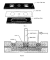
- Figure 1A and 1B show diagrams of a bilayer measurement chamber.
- Top Exploded view of top acrylic piece, center Delrin film containing cutouts for access to the lower fluidic channel and bilayer masking aperture and bottom acrylic piece connecting the side and center wells.
- Bottom Cross-section of assembled chamber. Aqueous fluid is first loaded into one of the two side wells followed by loading of n-decane into the central well. The aperture size in the Delrin film masks the contact area between these two solutions. Lipids in the decane or aqueous solutions form a monolayer at this contact area.
- a Ag/AgCl pin electrode with sessile aqueous droplet in the central well causes self-assembly of another lipid monolayer; lowering the pin contacts the two monolayers to form a bilayer defined by the Delrin aperture. Electrical measurements of this bilayer are made by the pin electrode and Ag/AgCl counterelectrode inserted into the side well.
- Figure 2 shows the bilayer capacitance as a function of vertical pin position with masking apertures of diameter (50-200 ⁇ m) and with no aperture.
- the apertures allow control of bilayer area and strongly reduce its dependence on pin vertical position.
- Figure 3 shows the bilayer capacitance as a function of lateral pin position with a 150 ⁇ m diameter masking aperture (squares) and with no aperture (diamonds). For each measurement, the pin is raised vertically 500 ⁇ m from its starting position and moved laterally before being lowered.
- FIG. 4 show that masking apertures increase tolerance of bilayers to solution flow.
- Bilayer capacitance is measured as a function of flow rate of the buffer in the lower aqueous chamber. Data shown from two separate trials with a 200 ⁇ m diameter aperture. Without the masking aperture, bilayer failure was immediate even for flow rates ⁇ 1 ml/hr.
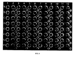
- Figure 5 show fabricated array plate of wells depicted in Figure 1 of manuscript.
- Well spacing is SBS standard and compatible with multichannel fluid handling and motion control standard apparatus.
- Figure 6A-6D show a droplet bilayer apparatus use.
- the lower channel was filled with an aqueous solution containing TRPM8 proteoliposomes (1 mg mL21 in 20 mM HEPES (pH 7.2), 150 mM KCl, 0.2 mM MgCl2).
- the central well was then filled with n-decane, resulting in lipid monolayer formation at the aqueous-decane interface, restricted by the aperture in the Delrin film.
- 2 mL of proteoliposome solution was then deposited on the bottom of a Ag/AgCl pin electrode.
- the electrode was lowered, via a micromanipulator, into the central well to allow a lipid monolayer to form at the droplet interface. After approximately five-minutes dwell time, the pin was lowered to contact the monolayers on the aqueous interfaces in order to form a bilayer.
- Drug dosing was achieved through the addition and withdrawal of solution from the lower channel via the fluid inlet and fluid outlet lines. Electrical measurements of the bilayer were made by the pin electrode and Ag/AgCl counter electrode (inserted into the outlet well).
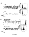
- Figure 7A and 7B show a dose-dependent activation of TRPM8 by menthol and inhibition of menthol-evoked TRPM8 currents by 2-APB.
- proteoliposomes at a protein : lipid ratio of 1 : 1000 diluted to 1 mg mL21 in reconstitution buffer with 2.5 mM PI(4,5)P2 added.
- (a) Excerpts of one experiment in which the channel currents were continuously measured as menthol concentration was increased: 0 mM, 20 mM, 40 mM, 60 mM, 80 mM, 100 mM, 120 mM, 140 mM and 500 mM. Step-wise, dose-dependent increases in current were observed.
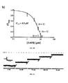
- Figure 8A and 8B show IC50 and EC50 results for TRPM8 following menthol and 2-APB addition.
- Currents recorded e.g. Fig. 3
- I/Imax fractional unblocked current
- proteoliposomes at a protein : lipid ratio of 1 : 1000 diluted to 1 mg mL21 in reconstitution buffer with 2.5 mM PI(4,5)P2 added.
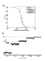
- Figure 9A - 9D show: (a) Dose-dependence effect of astemizole on hERG currents. (Left) hERG currents measured following sequential application of solutions with increasing astemizole concentration shows dose-dependent inhibition, with a small amount of measured unblockable current remaining at high (50 ⁇ M) astemizole concentration (-100 mV applied). (Right) This unblockable current was subtracted from each measured current and the difference was normalized to the current measured before application of astemizole to result in the ratio I/I max . This ratio was plotted versus astemizole concentration and fit to the Hill equation (see Example 3) to find the concentration at 50% conductance, IC 50 .
- Figure 10 shows the ensemble recordings of TRPM8 currents while varying PI(4,5)P 2 concentration in the presence of 250 ⁇ M menthol and a clamping potential of +100 mV. Data were filtered at 200 Hz. Measurements began in the absence of PI(4,5)P 2 . After ten minutes at each concentration, the concentration of PI(4,5)P 2 was then increased by 0.8 ⁇ M with the addition of 2.4 ⁇ L PI(4,5)P 2 stock solution (100 ⁇ M) to the 200 ⁇ L lower aqueous solution. With no PI(4,5)P 2 in the measurement solution, there was no observed channel activity. As the concentration of PI(4,5)P 2 increased, frequency of channel opening increased. At 2.4 ⁇ M PI(4,5)P 2 , the maximum current did not increase further. Throughout experiments, temperature was kept constant at 20 °C.
- Figure 11 shows the cold and menthol activation of TRPM8.
- Figure 12 shows the ensemble recordings of TRPM8 currents while varying temperature in the presence of 250 ⁇ M menthol and a clamping potential of +100 mV. Data were filtered at 200 Hz. Measurements made in the presence of 2.5 ⁇ M PI(4,5)P 2 and at a starting temperature of 20°C. After recording channel activity at 20°C for five minutes, alcohol lamp was then ignited and chamber temperature was slowly increased to 35°C. In representative trace, maximal current (275 pA) was achieved at 20°C and, upon warming, remained constant until 23°C. For temperatures greater than 23°C, the measured current progressively decreased and was finally extinguished at 28°C.
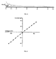
- Figure 13 shows a plot of current versus applied voltage obtained on a lipid bilayer produced with hERG membrane preparations.
- Ranges can be expressed herein as from “about” one particular value, and/or to "about” another particular value. When such a range is expressed, a further aspect includes from the one particular value and/or to the other particular value. Similarly, when values are expressed as approximations, by use of the antecedent "about,” it will be understood that the particular value forms a further aspect. It will be further understood that the endpoints of each of the ranges are significant both in relation to the other endpoint, and independently of the other endpoint. It is also understood that there are a number of values disclosed herein, and that each value is also herein disclosed as "about” that particular value in addition to the value itself. For example, if the value “10” is disclosed, then “about 10" is also disclosed. It is also understood that each unit between two particular units are also disclosed. For example, if 10 and 15 are disclosed, then 11, 12, 13, and 14 are also disclosed.
- references in the specification and concluding claims to parts by weight of a particular element or component in a composition denotes the weight relationship between the element or component and any other elements or components in the composition or article for which a part by weight is expressed.
- X and Y are present at a weight ratio of 2:5, and are present in such ratio regardless of whether additional components are contained in the compound.
- a weight percent (wt. %) of a component is based on the total weight of the formulation or composition in which the component is included.
- the terms "optional” or “optionally” means that the subsequently described event or circumstance can or can not occur, and that the description includes instances where said event or circumstance occurs and instances where it does not.
- the terms "optional” or “optionally” means that the subsequently described event or circumstance can or cannot occur, and that the description includes instances where said event or circumstance occurs and instances where it does not.
- a chemical agent refers to any natural or synthetic composition, molecule, or atom.
- a chemical agent can be a drug that has been approved by the FDA.
- a chemical agent can be a molecule that is in clinical trial.
- Suitable chemical agents include, but are not limited to, substances, molecules, or atoms that targets membrane proteins such as ion channels and/or GPCRs or the membrane itself.
- the terms “effective amount” and “amount effective” refer to an amount that is sufficient to achieve the desired result or to have an effect on an undesired condition.
- an effective amount of a chemical agent can achieve desired results in a formed amphiphilic bilayer.
- the term "derivative" refers to a compound having a structure derived from the structure of a parent compound (e.g., a compound disclosed herein, such as a lipid) and whose structure is sufficiently similar to those disclosed herein and based upon that similarity, would be expected by one skilled in the art to exhibit the same or similar activities and utilities as the claimed compounds, or to induce, as a precursor, the same or similar activities and utilities as the claimed compounds.
- exemplary derivatives include salts, esters, amides, salts of esters or amides, and N-oxides of a parent compound.
- compositions disclosed herein have certain functions. Disclosed herein are certain structural requirements for performing the disclosed functions, and it is understood that there are a variety of structures that can perform the same function that are related to the disclosed structures, and that these structures will typically achieve the same result.
- amphiphilic molecule as used herein is a molecule composed of hydrophilic and hydrophobic groups. Amphiphilic molecules can form an amphiphilic bilayer under suitable conditions. An amphiphilic molecule can for an amphiphilic monolayer at the interface of an aqueous and non-aqueous solution. An amphiphilic molecule can be any amphiphilic molecule that forms a bilayer found in a mammalian cell. In one aspect, the amphiphilic molecule can be a lipid.
- a lipid can comprise mixtures of two or more lipids.
- Suitable lipids can be generally classified as ionic (anionic/cationic/dipolar) and nonionic. More specifically, polymeric surfactants, silicon surfactants, fluorinated surfactants, oligomeric surfactants, dimeric surfactants, natural lipids, and the like, are suitable lipids for the devices and methods disclosed herein.
- the lipds disclosed herein can comprise an anionic lipid.
- Any anionic lipid can be used. Suitable anionic lipids are commonly used in detergents, shampoos, soaps, etc., and can be obtained commercially or prepared by methods known in the art. They include, but are not limited to, alkylbenzene sulfonates (detergent), fatty acid based surfactants, lauryl sulfate ( e.g., a foaming agent), di-alkyl sulfosuccinate ( e.g ., a wetting agent), lignosulfonates ( e.g ., a dispersant), and the like, including mixtures thereof.
- alkylbenzene sulfonates detergent
- fatty acid based surfactants e.g., a foaming agent
- di-alkyl sulfosuccinate e.g ., a wetting agent
- lignosulfonates e.
- linear alkylbenzene sulphonic acid sodium lauryl ether sulphate, alpha olefin sulphonates, phosphate esters, sodium sulphosuccinates, hydrotropes, and the like, including mixtures thereof, can be used.
- the lipids disclosed herein can comprise a cationic lipid.
- Any cationic lipid can be used.
- Such cationic lipids can be obtained commercially or can be prepared by methods known in the art.
- the lipids disclosed herein can comprise a nonionic lipid. Any nonionic lipid can be used. Suitable nonionic lipids do not ionize in aqueous solution, because their hydrophilic group is of a non-dissociable type, such as alcohol, phenol, ether, ester, or amide.
- ethers e.g ., polyhydric alcohols such as glycerin, solbitole, sucrose, etc.
- fatty acid esters e.g ., glycerin fatty acid ester, sobitan fatty acid ester, sucrose fatty acid ester, etc.
- esters e.g ., compounds made by applying, for example, ethylene oxide to a material having hydroxyl radicals such as high alcohol, alkyl-phenol, and the like
- ether/esters e.g ., compounds made by applying, for example, the ethylene oxide to the fatty acid or polyhydric alcohol fatty acid ester, having both ester bond and ether bond in the molecule
- other types e.g ., the fatty acid alkanol-amide type or the alkylpolyglyceride type.
- nonionic lipids can include, but are not limited to, alcohol ethoxylates and alkyl phenol ethyoxylates, fatty amine oxides, alkanolamides, ethylene oxide/propylene oxide block copolymers, alkyl amine ethoxylates, tigercol lubricants, etc.
- the lipids disclosed herein can comprise dipolar lipids. Any dipolar lipid can be used. Suitable dipolar lipids (called amphoteric or zwitterionic) exhibit both anionic and cationic dissociation. Suitable examples of dipolar lipids include, but are not limited to, products like betaines or sulfobetaines and natural substances such as amino acids and phospholipids. In one aspect, the betaines disclosed in U.S. Patent Nos. 6,852,816 ; 6,846,795 ; 6,846,352 ; and 6,849,426 , which are incorporated by reference in their entireties, can be used herein.
- Suitable lipids include natural surfactants, which can have their source from plant or animal organs.
- a bolaform lipids can be used.
- a bolaform lipid is a lipid that has two hydrophilic head groups at opposite ends of a hydrophobic tail.
- Mixtures of these lipids can also be used in the compositions and methods disclosed herein.
- the disclosed lipids comprises diphytanoylphosphatidylcholine and/or 1,2-diphytanoyl-sn-glycero-3-phosphatidylcholine.
- a device comprising: a) a first chamber; b) at least one second chamber; and c) a substrate defining at least one aperture having a diameter size, wherein the substrate is positioned between the at least one second chamber and the first chamber, and wherein the first chamber is in fluid communication with the at least one second chamber through the at least one aperture.
- the substrate is a hydrophobic substrate.
- the hydrophobic substrate comprises polyoxymethylene, Teflon, polyethylene, acrylic resin, or a mixture thereof.
- the hydrophobic substrate can be Delrin.
- the substrate can be a film. In one aspect, the substrate can have a thickness that is less than 1 cm, 0.5 cm, or 0.1 cm. In another aspect the can have a thickness that is less than 500 ⁇ m, 200 ⁇ m, 100 ⁇ m, 50 ⁇ m, 25 ⁇ m, or 10 ⁇ m.
- the first and at least one second chamber can be in any spatial orientation relative to one another.
- the first and at least one second chamber can be side-by-side.
- the first chamber can be a first side chamber and the at least one second chamber can be at least one side second chamber.
- the first and at least one second chamber can above and below on another.
- the first chamber is a lower chamber and wherein the second chamber is at least one upper chamber.
- the first chamber defines a first plate defining a solution chamber; and the second chamber defines a second plate defining at least one loading chamber having a diameter size.
- the lower chamber defines a first plate defining a solution chamber; and the at least one upper chamber defines a second plate defining at least one loading chamber having a diameter size.
- the first plate and the second plate are made of a non-absorbing material.
- the first plate and the second plate are made of the same material.
- the first plate and the second plate can be acrylic based plates.
- the substrate can be in contact with the first plate. In another aspect, the substrate can be in contact with the second plate. In another aspect, the substrate can be in contact with the first plate and the second plate.
- the substrate can define the upper limit of the solution chamber. In another aspect, the substrate can define the lower limit of the at least one loading chamber.
- the diameter of the at least one loading chamber is larger than the aperture of the substrate. In another aspect, the diameter of the at least one loading chamber is smaller than the solution chamber.
- the first plate further defines at least one solution aperture and the substrate further defines at least one solution aperture, wherein the at least one solution aperture of the first plate is in fluid communication with the solution chamber through the at least one solution aperture of the substrate.
- the first plate further defines at least one solution aperture and the hydrophobic substrate further defines at least one solution aperture, wherein the at least one solution aperture of the first plate is in fluid communication with the solution chamber through the at least one solution aperture of the hydrophobic substrate.
- the second plate further defines at least one solution aperture. The at least one solution aperture of the second plate can be in fluid communication with the at least one solution aperture of the first plate through the at least one solution aperture of the substrate.
- the device comprises at least two solution apertures.
- the device comprises at least four solution apertures.
- the device comprises at least six solution apertures.
- the at least one solution aperture defines a solution inlet. In another aspect, the at least one solution aperture defines a solution outlet. In yet another aspect, the at least one solution aperture defines a solution inlet and a solution outlet. Means can be provided to produce a flow rate of a solution via the solution inlet and solution outlet.
- the solution can, for example, be a third solution as defined herein.
- the diameter size of the at least one aperture is about 500 nm to about 10,000 ⁇ m. In another aspect, the diameter size of the at least one aperture is about 500 nm to about 5,000 ⁇ m. In yet another aspect, the diameter size of the at least one aperture is about 500 nm to about 1,000 ⁇ m. In yet another aspect, the diameter size of the at least one aperture is about 500 nm to about 750 ⁇ m. In yet another aspect, the diameter size of the at least one aperture is about 500 nm to about 500 ⁇ m. In yet another aspect, the diameter size of the at least one aperture is about 500 nm to about 200 ⁇ m. In yet another aspect, the diameter size of the at least one aperture is about 500 nm to about 100 ⁇ m.
- the diameter size of the at least one aperture is about 500 nm to about 50 ⁇ m. In yet another aspect, the diameter size of the at least one aperture is about 500 nm to about 20 ⁇ m. In yet another aspect, the diameter size of the at least one aperture is about 500 nm to about 10 ⁇ m. In yet another aspect, the diameter size of the at least one aperture is about 500 nm to about 5 ⁇ m. In yet another aspect, the diameter size of the at least one aperture is about 500 nm to about 1 ⁇ m. For example, the diameter size of the at least one aperture can be about 20 ⁇ m to about 500 ⁇ m. In another example, the diameter size of the at least one aperture is about 50 ⁇ m to about 200 ⁇ m.
- the diameter size of the at least one aperture is smaller than the diameter size of the at least one loading chamber.
- the thickness of the substrate and the diameter size of the aperture are such that an amphiphilic molecule bilayer, such as a lipid bilayer, can be formed.
- an amphiphilic molecule bilayer such as a lipid bilayer
- the device further comprises means for performing a measurement.
- the measurement can be any measurement that produces information about the formation or disruption of an amphiphilic monolayer or amphiphilic bilayer. Such measurements are well known in the art. Suitable measurements include, but are not limited to, an electrical measurement, an optical measurement, a chemical measurement, an acoustic measurement, or combination thereof. The above measurements can include, fluorescence microscopy, dual polarization interferometery, x-ray diffraction, and electron microscopy. In one example, the measurement can be an electrical measurement. In one aspect, the means for performing electrical measurements comprises an electrode.
- the first chamber further comprises a first fluid.
- the first fluid can be an aqueous fluid.
- An aqueous fluid can be water, such as deionized or milipure water.
- the first fluid can be a non-aqueous solution.
- the first fluid, such as an aqueous fluid can comprise at least one amphiphilic molecule, such as a lipid.
- the first fluid can further comprise a chemical agent.
- the first fluid can further comprise a chemical agent and at least one amphiphilic molecule.
- the at least one second chamber comprises a second fluid.
- the second fluid is immiscible in the first fluid.
- the second fluid can be a non-aqueous solution.
- Suitable non-aqueous solutions are saturated hydrocarbon fluids such as n-hexane, n-heptane, n-octane, n-nonane, n-decane, and n-dodecane.
- Other suitable non-aqueous solutions include oils, such mineral oils.
- the second fluid can be an aqueous solution.
- the second fluid can be a non-aqueous solution and the first fluid can be an aqueous solution.
- viscosity of the second fluid and first fluids are substantially the same.
- the second fluid comprises at least one amphiphilic molecule, such as at least one lipid.
- the first and second fluid comprises at least one amphiphilic molecule, such as at least one lipid.
- the second fluid comprises at least one amphiphilic molecule, such as at least one lipid and the first fluid does not comprise at least one amphiphilic molecule.
- the first fluid comprises at least one amphiphilic molecule, such as at least one lipid and the second fluid does not comprise at least one amphiphilic molecule.
- the second fluid comprises a chemical agent.
- the second fluid comprises a chemical agent and at least on amphiphilic molecule, such a lipid.
- the at least one second chamber is an array of loading chambers.
- the array can comprise at least 2, 3, 4, 5, 6, 7, 8, 9, 10, 15, 20, 30, 50, 100, 500 or 1000 loading chambers.
- the at least one aperture is an array of apertures.
- the array can comprise at least 2, 3, 4, 5, 6, 7, 8, 9, 10, 15, 20, 30, 50, 100, 500 or 1000 apertures.
- the at least one solution aperture can be an array of solution apertures.
- the array can comprise at least 2, 3, 4, 5, 6, 7, 8, 9, 10, 15, 20, 30, 50, 100, 500, 1000 or 2000 solution apertures.
- the solution chamber comprises a third fluid.
- the third fluid and first fluid are identical.
- the third solution is immiscible in the second fluid.
- the third fluid comprises a chemical agent.
- the third fluid and first fluid are identical but for that the third fluid comprises a chemical agent which the first fluid does not.
- the third fluid is an aqueous solution.
- the third fluid is a non-aqueous solution.
- the device comprises means for flowing a third fluid through the solution chamber.
- the means for flowing a third fluid through the solution chamber comprises a fluid inlet and a fluid outlet.
- the chemical agent comprises a chemical agent that interacts with membrane proteins, receptors, or ion channels. In another aspect, the chemical agent comprises a chemical agent that modifies membrane proteins, receptors, or ion channels. In another aspect, the chemical agent comprises a chemical agent that disrupts membrane proteins, receptors, or ion channels. In another aspect, the chemical agent comprises a chemical agent that does not interact with membrane proteins, receptors, or ion channels.
- the first chamber or at least one second chamber comprises a fourth solution.
- the at least one second chamber can comprise the fourth solution.
- the first chamber can comprise the fourth solution.
- the fourth solution when the at least one second chamber comprises a second solution the fourth solution is immiscible in the second solution.
- the further solution is immiscible in a fluid present in the first chamber or the at least one second chamber.
- the fourth solution when the first chamber comprises a first solution the fourth solution is immiscible in the first solution.
- the fourth solution is an aqueous solution.
- the fourth solution is a non-aqueous solution.
- the fourth solution comprises at least one amphiphilic molecule, such as a lipid.
- the fourth solution comprises a chemical agent.
- the fourth solution is a droplet, such as an aqueous droplet.
- the loading chamber comprises an aqueous droplet.
- least a portion of the fourth solution is in contact with means for performing measurements, such as electrical measurements.
- the loading chamber can comprise an aqueous droplet, and wherein at least a portion of the aqueous droplet is in contact with the means for performing electrical measurements.
- Also disclosed herein is a method of making a lipid bilayer comprising: a) providing a device comprising a substrate defining at least one aperture; b) providing an first solution on one side of the aperture; c) providing a second solution on the opposite side of the aperture from the first solution, wherein the first solution is immiscible in the second solution; d) providing a first amphiphilic molecule in the first solution, or second solution, or in both the first solution and second solution; e) contacting the first solution and the second solution through the aperture, thereby forming a first amphiphilic molecule monolayer; f) providing an fourth solution; g) providing a second amphiphilic molecule in the second solution, or fourth solution, or in both the second solution and fourth solution; h) submerging at least a portion of the fourth solution in the second solution, wherein the fourth solution is immiscible in the second solution, thereby forming a second amphiphilic molecule monolayer; and i) contacting the second amphi
- the device is a device disclosed herein.
- the fourth solution is a droplet.
- the fourth solution comprises an amphiphilic molecule.
- the fourth solution can comprise a lipid.
- the amphiphilic molecule bilayer is a lipid bilayer. In one aspect, the amphiphilic molecule bilayer comprises at least one ion channel. In another aspect, the amphiphilic molecule bilayer comprises at least one receptor. In another aspect, the amphiphilic molecule bilayer comprises at least one membrane proteins. In another aspect, the amphiphilic molecule bilayer comprises at least one ion channel and at least one receptor. In another aspect, the amphiphilic molecule bilayer comprises at least one ion channel, at least one receptor, and at least one membrane protein.
- amphiphilic molecule bilayer is stable for at least 3 days, 5 days, 1 week, 2 weeks, 1 month, 3 months or 6 months.
- the method further comprises providing means for performing measurements.
- Suitable means for performing measurements are known in the art and include, but are not limited to means for performing electrical measurements, means for performing optical measurements, means for performing chemical measurements, means for performing acoustic measurements, or a combination thereof.
- the method can further comprise providing means for performing electrical measurements.
- the method further comprises performing measurements. Suitable measurements are known in the art and include, but are not limited to an electrical measurement, an optical measurement, a chemical measurement, an acoustic measurement, or combination thereof.
- the method can further comprise performing an electrical measurement.
- At least a portion of the fourth solution is in contact with the means for performing measurements, such as electrical measurements.
- the means for performing measurements such as electrical measurements.
- at least a portion of the droplet is in contact with the means for performing measurements, such as electrical measurements.
- the method further comprises providing a third solution on the same side of the aperture as the first solution.
- the third solution is identical to the first solution.
- the third solution is an aqueous solution.
- the third solution is a non-aqueous solution.
- the chemical agent in the third solution can be present before or after the formation of the amphiphilic molecule bilayer.
- the chemical agent in the third solution can be present before the formation of the amphiphilic molecule bilayer.
- the chemical agent in the third solution can be present after the formation of the amphiphilic molecule bilayer.
- the third solution does not comprise a chemical agent before the formation of the amphiphilic molecule bilayer.
- the third solution has a flow rate.
- the flow rate can be at least 1 ml/hr, 3 ml/hr, 5, ml/hr, 10 ml/hr, 15 ml/hr, 20 ml/hr, 25 ml/hr, 30 ml/hr or 50 ml/hr.
- the flow rate of the third solution does not influence the stability of the amphiphilic molecule bilayer.
- the third solution further comprises a chemical agent.
- the first solution is an aqueous solution. In another aspect, the first solution is a non-aqueous solution. In one aspect, the first solution further comprises a chemical agent.
- the chemical agent in the first solution can be present before the formation of the amphiphilic molecule bilayer. In another example, the chemical agent in the first solution can be present after the formation of the amphiphilic molecule bilayer. In another example, the first solution does not comprise a chemical agent before the formation of the amphiphilic molecule bilayer.
- the second solution is a non-aqueous solution. In another aspect, the second solution is an aqueous solution. In one aspect, the second solution further comprises a chemical agent.
- the chemical agent in the second solution can be present before the formation of the amphiphilic molecule bilayer. In another example, the chemical agent in the second solution can be present after the formation of the amphiphilic molecule bilayer. In another example, the second solution does not comprise a chemical agent before the formation of the amphiphilic molecule bilayer.
- the chemical agent comprises a chemical agent that interacts with membrane proteins, receptors, or ion channels. In another aspect, the chemical agent comprises a chemical agent that modifies membrane proteins, receptors, or ion channels. In another aspect, the chemical agent comprises a chemical agent that disrupts membrane proteins, receptors, or ion channels. In another aspect, the chemical agent comprises a chemical agent that does not interact with membrane proteins, receptors, or ion channels. The method of paragraph 53, wherein the chemical agent is known to interact with amphiphilic molecule bilayer.
- the defining at least one aperture is an array of apertures.
- the array can comprise at least 2, 3, 4, 5, 6, 7, 8, 9, 10, 15, 20, 30, 50, 100, 500 or 1000 apertures.
- a chemical agent can be added to the first solution, second solution, third solution or fourth solution before or after the formation of the amphiphilic molecule bilayer.
- a chemical agent can be added to the first solution before or after the formation of the amphiphilic molecule bilayer.
- a chemical agent can be added to the second solution before or after the formation of the amphiphilic molecule bilayer.
- a chemical agent can be added to the third solution before or after the formation of the amphiphilic molecule bilayer.
- a chemical agent can be added to the fourth solution before or after the formation of the amphiphilic molecule bilayer.
- Also disclosed herein is a method of performing electrical measurements on an amphiphilic molecule bilayer comprising: a) providing an amphiphilic molecule bilayer formed at an aperture; and b) performing a measurement.
- providing an amphiphilic molecule bilayer comprises the methods for forming an amphiphilic molecule bilayer as described herein.
- providing a lipid bilayer formed at an aperture can comprise: a) providing a device comprising a substrate defining at least one aperture; b) providing an first solution on one side of the aperture; c) providing a second solution on the opposite side of the aperture from the first solution, wherein the first solution is immiscible in the second solution; d) providing a first amphiphilic molecule in the first solution, or second solution, or in both the first solution and second solution; e) contacting the first solution and the second solution through the aperture, thereby forming a first amphiphilic molecule monolayer; f) providing an fourth solution; g) providing a second amphiphilic molecule in the second solution, or fourth solution, or in both the second solution and fourth solution; h) submerging at least a portion of the fourth solution in the second solution, wherein the fourth solution is immiscible
- the aperture can be an aperture as disclosed herein. In another aspect, the aperture can be present in a device disclosed herein. In one aspect, the aperture can be an array of apertures. The array can comprise at least 2, 3, 4, 5, 6, 7, 8, 9, 10, 15, 20, 30, 50, 100, 500 or 1000 apertures.
- the device can be a device disclosed herein.
- the amphiphilic molecule bilayer is a lipid bilayer. In one aspect, the amphiphilic molecule bilayer comprises at least one ion channel. In another aspect, the amphiphilic molecule bilayer comprises at least one receptor. In another aspect, the amphiphilic molecule bilayer comprises at least one membrane proteins. In another aspect, the amphiphilic molecule bilayer comprises at least one ion channel and at least one receptor. In another aspect, the amphiphilic molecule bilayer comprises at least one ion channel, at least one receptor, and at least one membrane protein.
- the fourth solution is a droplet.
- the method further comprises performing measurements prior to providing an amphiphilic molecule bilayer. In one aspect, the method further comprises performing measurements during the formation of an amphiphilic molecule bilayer.
- the measurement can be an electrical measurement, an optical measurement, a chemical measurement, an acoustic measurement, or combination thereof.
- the measurement can be an electrical measurement or an optical measurement.
- the measurement can be an electrical measurement.
- performing a measurement comprises providing means for performing measurements.
- Suitable means for performing measurements are known in the art and include, but are not limited to means for performing electrical measurements, means for performing optical measurements, means for performing chemical measurements, means for performing acoustic measurements, or a combination thereof.
- the method can further comprise providing means for performing electrical measurements.
- performing electrical measurements comprises an electrode.
- the fourth solution is in contact with at least a portion of the means for performing electrical measurements.
- the fourth solution can be a droplet.
- at least portion of the droplet can be in contact with at least a portion of the electrode.
- the method can further comprise detecting a change in the measurements.
- the method can further comprise detecting a change in the electrical measurements or optical measurements.
- the method can further comprise detecting a change in the electrical measurements.
- the method further comprises providing a third solution on the same side of the aperture as the first solution.
- the method further comprises providing a third solution on the same side of the aperture as the first solution.
- the third solution is identical to the first solution.
- the third solution is an aqueous solution.
- the third solution is a non-aqueous solution.
- the chemical agent in the third solution can be present before or after the formation of the amphiphilic molecule bilayer.
- the chemical agent in the third solution can be present before the formation of the amphiphilic molecule bilayer.
- the chemical agent in the third solution can be present after the formation of the amphiphilic molecule bilayer.
- the third solution does not comprise a chemical agent before the formation of the amphiphilic molecule bilayer.
- the third solution has a flow rate.
- the flow rate can be at least 1 ml/hr, 3 ml/hr, 5, ml/hr, 10 ml/hr, 15 ml/hr, 20 ml/hr, 25 ml/hr, 30 ml/hr or 50 ml/hr.
- the flow rate of the third solution does not influence the stability of the amphiphilic molecule bilayer.
- the third solution further comprises a chemical agent.
- the first solution is an aqueous solution. In another aspect, the first solution is a non-aqueous solution. In one aspect, the first solution further comprises a chemical agent.
- the chemical agent in the first solution can be present before the formation of the amphiphilic molecule bilayer. In another example, the chemical agent in the first solution can be present after the formation of the amphiphilic molecule bilayer. In another example, the first solution does not comprise a chemical agent before the formation of the amphiphilic molecule bilayer.
- the second solution is a non-aqueous solution. In another aspect, the second solution is an aqueous solution. In one aspect, the second solution further comprises a chemical agent.
- the chemical agent in the second solution can be present before the formation of the amphiphilic molecule bilayer. In another example, the chemical agent in the second solution can be present after the formation of the amphiphilic molecule bilayer. In another example, the second solution does not comprise a chemical agent before the formation of the amphiphilic molecule bilayer.
- the chemical agent comprises a chemical agent that interacts with membrane proteins, receptors, or ion channels. In another aspect, the chemical agent comprises a chemical agent that modifies membrane proteins, receptors, or ion channels. In another aspect, the chemical agent comprises a chemical agent that disrupts membrane proteins, receptors, or ion channels. In another aspect, the chemical agent comprises a chemical agent that does not interact with membrane proteins, receptors, or ion channels. The method of paragraph 53, wherein the chemical agent is known to interact with amphiphilic molecule bilayer.
- a chemical agent can be added to the first solution, second solution, third solution or fourth solution before or after the formation of the amphiphilic molecule bilayer.
- a chemical agent can be added to the first solution before or after the formation of the amphiphilic molecule bilayer.
- a chemical agent can be added to the second solution before or after the formation of the amphiphilic molecule bilayer.
- a chemical agent can be added to the third solution before or after the formation of the amphiphilic molecule bilayer.
- a chemical agent can be added to the fourth solution before or after the formation of the amphiphilic molecule bilayer.
- the method provides information regarding the potency of the chemical agent.
- the method provides information regarding the potency of the chemical agent towards an amphiphilic molecule bilayer.
- the at least one second amphiphilic molecule is the at least one first amphiphilic molecule.
- an apparatus was designed for artificial lipid bilayer formation and measurement plate which constrains the contact area of the two aqueous phases, also constraining the bilayer area.
- the apparatus consists of a lower aqueous solution chamber plate, a hydrophobic film in which a small aperture is cut, and a top chamber plate that allows for top loading and electrical access of all solutions.
- Lipid bilayers are formed by contacting monolayers through the small aperture in the hydrophobic film, which constrains the bilayer size.
- the measurements in the examples demonstrate a reduced sensitivity of bilayer area to the relative position of the aqueous phases, reducing the precision needed by fluid handling and motion control hardware in automation.
- the apparatus is also easily arrayed and compatible with SBS standard instrumentation.
- the examples demonstrate fully automated fluid exchange of the lower aqueous solution with intact droplet bilayers allowing for stable solution perfusion. While masking of lipid bilayers formed by contacting monolayers has been studied previously (Zagnoni M 2009), here were present a platform that is easily automatable and scalable, presenting a way forward toward high-throughput study.
- Chambers were made from 0.125" thick acrylic (McMaster-Carr) and 0.003" thick Delrin film (McMaster-Carr). Two acrylic pieces were milled to form fluidic wells and channels by stacking them vertically and sandwiching the Delrin. Apertures in the Delrin film were cut using a CO 2 laser (Universal Laser Systems) to connect the wells formed by the upper and lower acrylic pieces. Two wells were connected through a channel in the lower acrylic piece. Aperture sizes were measured microscopically. The center measurement well was connected to the channel through a pore between 50 - 200 ⁇ m in diameter laser cut into the Delrin film on which the bilayer is formed.
- CO 2 laser Universal Laser Systems
- Measurement buffer (MB) containing liposomes were made as previously described(Poulos J L 2010). Briefly, a 1 ml solution of 1M KCL, 10mM Tris-HCL (Sigma), pH 8.0, and additionally containing 33 mg of 1,2-Diphytanoyl-sn-Glycero-3-Phosphocholine (DPhPC) lipid (Avanti Polar Lipids), was extruded through a 200nm filter (Avanti). 150 ⁇ l MB was first loaded into the aqueous inlet to completely fill the portion of the chamber below the Delrin film.
- DPhPC 1,2-Diphytanoyl-sn-Glycero-3-Phosphocholine
- n-decane MP Biomedicals
- Silver pins were fabricated using 16 gauge silver wire (0.999 purity, C.C. Silver & Gold). The pins were cut to approximately 1 inch and electrical discharge machining was used to create a blunt end and cut slots 0.05" deep and 0.015" wide into the ends of the pins.
- Counter electrodes were made from 200 ⁇ m diameter silver wire (Ted Pella). The silver wires were chloridized by immersing them in bleach for approximately 1 min, followed by a deionized (DI) water rinse.
- DI deionized
- the chloridized pin was lowered into liposome-containing MB to a depth of approximately 10 mm for 1 s and removed ( Figure 1(b) ), resulting in a small ⁇ 1.1 ⁇ l droplet hanging from the end of the pin.
- This pin with hanging droplet was then lowered into the decane solution.
- the pin was lowered further using a micromanipulator (Newport) until the droplet contacted the lower aqueous phase within the Delrin pore, forming a lipid bilayer membrane.
- Newport micromanipulator
- the apparatus was placed inside a Faraday cage and the Ag/AgCl pin and counter-electrode were connected to an Axopatch 200B amplifier (Axon Instruments).
- the signals were digitized with a Digidata 1332A (Axon Instruments) at 5 kHz, filtered in hardware with a 1 kHz Bessel filter and subsequently filtered further with a 30 Hz Bessel filter and analyzed with Clampfit software (Axon Instruments).
- PTFE tubing Zeus
- KD Scientific syringe pump
- 10mL glass syringe Hamilton
- the sensitivity of bilayer area was also investigated to the lateral position of the pin.
- a pin with 2 ⁇ l sessile droplet was axially aligned with the center of a 150 ⁇ m aperture and lowered to form a bilayer. After each capacitance measurement, the pin was raised and moved in 200 ⁇ m steps laterally before being lowered again, after which the capacitance was measured.
- These experiments were compared to measurements taken with no separating Delrin film. With the Delrin film, the bilayer capacitance was still within 90% of its original value when the droplet was positioned over 500 ⁇ m from the aperture center. In contrast, without the masking aperture, the bilayer capacitance decreased to approximately 15% of its original value ( Figure 3 ).
- the relative position of the aqueous fluid interfaces must be precise within 3.6 ⁇ m. This relative position is determined by the position of the pin holding the sessile droplet, the volume of sessile droplet, and the position of the lower aqueous interface, which itself depends on the volume of the lower fluids and the dimensions of the channels and wells. Estimating the shape of the 2.5 ⁇ l sessile droplet on the pin as a spherical cap, 3.6 ⁇ m precision in height of this droplet corresponds to 15 nl required precision in droplet volume. Further, for development of parallel arrays, this vertical precision also requires a high degree of machining tolerance, uniformity, and alignment of the component parts.
- Solution exchange enables increased experimental throughput by allowing for a variety of different experimental conditions to be tested in a short time, as well as measuring the activity of a large but fixed number of ion channels in the presence of varying concentrations of pharmaceutically active compounds for IC 50 /EC 50 determination.
- This type of apparatus enables automated, repeatable high yield formation and measurement of artificial lipid bilayers and ion channels incorporated into them. This goal is achieved by constraining the bilayer area using a masking aperture, which were superior in performance compared to their absence.
- the devices using these apertures are easily arrayable to result in multi-well plates for parallel bilayer and ion channel measurements. This parallelization, in combination with improved compatibility of bilayer formation with automation and the ability to support solution exchange without disturbing the bilayer, can result in significantly increased throughput ion channel studies using artificial bilayers.
- the potency of pharmaceutical compounds acting on ion channels can be determined through measurements of ion channel conductance as a function of compound concentration. Described herein is artificial lipid bilayer apparatus for simple, fast, and high yield measurement of ion channel conductance with simultaneous solution perfusion.
- this chip the application of this chip to the measurement of the mammalian cold and menthol receptor TRPM8.
- Ensemble measurements of TRPM8 as a function of concentration of menthol and 2-aminoethoxydiphenyl borate (2-APB) enabled efficient determination of menthol's EC 50 (111.8 ⁇ M ⁇ 2.4 ⁇ M) and 2-APB's IC 50 (4.9 ⁇ M ⁇ 0.2 ⁇ M) in agreement with published values.
- This validation coupled with the compatibility of this platform with automation and parallelization, indicates significant potential for large scale pharmaceutical ion channel screening.
- Electrophysiological measurements of ion channels are important scientifically and pharmacologically.
- the potency of pharmaceutical compounds on ion channels can be shown through measured changes in ion channel conductance as a function of compound concentration and expressed in the form of IC 50 and EC 50 concentrations. These are commonly measured with cells using the patch clamp technique ( B. Sakmann et al., Ann. Rev. Physiol., 1984, 46, 455-472 ; J.A. Fernández, et al., J. Gen. Physiol., 2011, 137, 173-195 ).
- TRPM8 a member of the Transient Receptor Potential Melastatin (TRPM) family ( N. Kedei, et al., J. Biol. Chem., 2001, 276, 28613-28619 ; T. Rosenbaum, et al., BMC Neurosci. 2002, 3, 4-13 ), was the first temperature-activated channel found to sense cold ( A.M. Peier, et al., Cell, 2002, 108, 705-715 ; D.D. McKemy, et al., Nature, 2002, 416, 52-58 ), and has become a primary target in studies of thermosensation and cold stimulating compounds ( J.A. Fernández, et al., J. Gen.
- TRPM8 has been identified with multiple cancer types ( L.
- TRPM8 is an important drug discovery target.
- TRPM8 single channel conductance, temperature response, and phospholipid sensitivity agreed well with previously published studies. Measurements of TRPM8 ensembles during perfusion of solutions of menthol and 2-aminoethoxydiphenyl borate at varying concentrations enabled us to efficiently determine the EC 50 and IC 50 values of these compounds with TRPM8, also in agreement with the literature. These results, combined with the simplicity and high yield of this platform and its compatibility with automation and parallelism, show great promise for lipid bilayers to play a role in ion channel drug discovery and safety screening.
- Example 1 The general apparatus for these experiments is described in Example 1.
- the apparatus was prepared for use by filling the outer wells and lower channel with 200 ⁇ L of an aqueous solution containing liposomes (described below) ( Fig. 1B ).
- the 200 ⁇ m aperture in the hydrophobic Delrin film is sufficiently small to prevent flow of aqueous solution through it.
- 80 ⁇ L of n-decane was then added to the center well, contacting the lower aqueous solution through the Delrin aperture ( Fig. 1B ).
- a Ag/AgCl pin electrode with 2 ⁇ L sessile droplet of aqueous liposome solution was introduced to the decane in the center well ( Fig. 1B ) ( J.L.
- TRPM8 construct with 6x histidine tag at the amino terminus was transformed into E. coli, expressed, and purified. Following purification, TRPM8 was reconstituted into liposomes, adapted from Long et al. ( S.B. Long, et al, Nature, 2007, 450, 376-382 ) 200 nm diameter unilamellar liposomes were prepared, composed of 1-palmitoyl-2-oleoyl- sn -glycero-3-phosphocholine (POPC) and 1-palmitoyl-2-oleoyl- sn -glycero-3-phosphoethanolamine (POPE) (Avanti Polar Lipids) at a ratio of 3:1 (w:w).
- POPC 1-palmitoyl-2-oleoyl- sn -glycero-3-phosphocholine
- POPE 1-palmitoyl-2-oleoyl- sn -glycero-3-phosphoethanolamine
- TRPM8 proteoliposomes were diluted to a final concentration of 1 mg/mL in reconstitution buffer (RB, 20 mM HEPES (pH 7.2), 150 mM KCl, 0.2 mM MgCl 2 ). 2 ⁇ L of this solution was used for the sessile droplet and 200 ⁇ L of this solution (or a similarly prepared liposome solution not containing TRPM8) was added to the lower aqueous channel.
- reconstitution buffer RB, 20 mM HEPES (pH 7.2), 150 mM KCl, 0.2 mM MgCl 2 .
- a Ag/AgCl counter electrode placed in the one of the outer wells and the Ag/AgCl pin electrode were connected to an Axopatch 200B amplifier (Axon Instruments), which was used to apply a transmembrane potential and measure the resultant ionic current ( Figure 1B ).
- the signals were digitized with a Digidata 1332A (Axon Instruments) at 10 kHz, filtered in hardware with a 1 kHz Bessel filter, filtered post-acquisition with a 200 Hz Bessel filter and analyzed with Clampfit10 software (Axon Instruments). Bilayer capacitance was measured throughout to ensure that changes in measured current were due to changes in channel conductance and not changes in bilayer size or stability.
- Temperature was measured using a 10 k ⁇ n-type thermistor (Newark) placed in the solution of one of the outer wells.
- the thermistor was connected in series with a 10 k ⁇ resistor (Newark) to form a voltage divider; resistor voltage was recorded using a BNC-2110 connector block and PCI-6036E DAQ Card (National Instruments) and used to determine resistance and temperature with LabVIEW 9.2.1 (National Instruments).
- Experiments were performed at room temperature, ⁇ 20°C, unless otherwise mentioned.
- An alcohol lamp was placed near the bilayer chamber to gently heat the experimental environment for temperature experiments.
- Short-chain phosphatidylinositol-4,5-bisphosphate (PI(4,5)P 2 ) (Avanti Polar Lipids)
- menthol and 2-APB were dissolved in RB to a final concentration of 100 ⁇ M, 2 mM, and 300 ⁇ M, respectively.
- solutions were added to the fluid inlet well and withdrawn from the fluid outlet well ( Fig. 6D ).
- Ensembles of TRPM8 were measured using a 1:1000 protein:lipid ratio. The magnitude of measured currents varied widely between experiments. The average observed current over 20 experiments was 693.5 ⁇ 502.7 pA, corresponding to 108 ⁇ 79 channels based on the observed single channel conductance determined above. As described below, this variability did not affect drug potency measurements because, once reconstituted, the magnitude of the current and number of channels remained constant, enabling the relative change in channel conductance between experiments to be compared.
- TRPM8 channels reconstituted at a 1:1000 protein:lipid ratio, were measured with a +100 mV applied potential to measure menthol and 2-APB potency.
- concentration of menthol, a TRPM8 activator, in the lower aqueous solution was increased from 20-140 ⁇ M in 20 ⁇ M increments and a final measurement at 500 ⁇ M.
- the resultant current at each concentration was measured over 10 minutes before the concentration was increased ( Fig. 7 ).
- TRPM8 currents were similarly measured with 2-APB, a TRPM8 inhibitor, at concentrations 1 ⁇ M, 3.3 ⁇ M, 6.6 ⁇ M, 9.9 ⁇ M, and 13.2 ⁇ M in solutions containing 500 ⁇ M menthol to activate TRPM8 ( Fig. 7a ). These experiments were repeated three times for each compound. In one experiment with 2-APB, we also measured concentrations of 5 ⁇ M, 9 ⁇ M and 11 ⁇ M.
- the average current at each concentration was determined and normalized by the maximum value measured.
- Droplet bilayers have been the subject of much recent activity in part because bilayer formation results from a mechanical step, which is simple and highly amenable to automation and parallelization. Coupled with solution perfusion ( S.A. Portonovo , Biomed. Microdevices, 2011 , DOI: 10.1007/s10544-011-9596-5), they have considerable potential for pharmaceutical screening of ion channels, as well as greatly reducing the time and expertise required for ion channel measurement in bilayers. Toward this goal, we aimed to validate this platform through ensemble measurements of TRPM8 in the presence of agonist and antagonist compounds, menthol and 2-APB, respectively, and the determination of their EC 50 and IC 50 values enabled by solution perfusion.
- Membrane composition is critical to the function of many ion channels, including those in the TRP family.
- the membrane composition in our system was specifiable through the prepared liposomes and amount of PI(4,5)P 2 added, and could be easily changed for studies with other ion channels or different membrane compositions.
- Leptihn et al. recently showed ion channel measurement from native cells in a similar droplet bilayer platform ( J. Am. Chem. Soc., 2011, 133, 9370-9375 ).
- TRPM8 measurements at the single channel and ensemble level matched well with previously published results as did the determined IC 50 and EC 50 values of 2-APB and menthol.
- the bilayer chip allowed electrical and fluidic access from the chamber top, allowing quick set up and convenient exchange of chambers between experiments. This chip design is also easily arrayed and compatible with parallel-automated fluid handling and motion control hardware.
- concentration-dependent modulation of channel conductance by pharmaceutical candidates may be measured rapidly and repeatedly in parallel, giving it considerable potential for high throughput electrophysiological screening.
- Ion channel conductance measurements are used to determine drug potency and also detect off-target drug interactions, most commonly for the cardiac potassium ion channel K v 11.1 (hERG).(Hancox, McPate et al. 2008). In these measurements, inhibition (or enhancement) of the conductance is measured as a function of drug concentration, from which the IC 50 (or EC 50 ) is determined, defined as the concentration for which the measured channel conductance is 50% of the maximum. Ion channel drug inhibition can also be determined optically through radioligand binding,(Chiu, Marcoe et al. 2004; Diaz, Daniell et al. 2004) measurements of ion flux,(Cheng, Alderman et al. 2002; Titus, Beacham et al.
- Electrophysiological measurements of ion channel conductance are predominantly made from ion channel ensembles in whole cells using patch clamp.
- Ion channel ensembles have also been measured in lipid bilayers,(Schindler and Rosenbusch 1978; Schindler and Quast 1980; Tao and MacKinnon 2008; Leptihn, Thompson et al. 2011; Brohawn, del Marmol et al. 2012; El-Arabi, Salazar et al. 2012) which offer simplified apparatus, reduced training, and the ability to easily control membrane and solution composition.
- DIBs Droplet interface bilayers
- DIBs for ion channel drug potency measurements were performed. Concentration dependent drug activation and inhibition was previously measured of the rat cold and menthol sensitive ion channel TRPM8, which was expressed in E. coli, purified, and reconstituted into proteoliposomes for measurement in DIBs (El-Arabi, Salazar et al. 2012). However, this result is not readily generalizable because very few physiologically relevant ion channels have been successfully expressed in bacterial expression systems.
- the apparatus described in Example 1 was used during ion channel measurement and used it to measure the dose-dependent attenuation of hERG conductance from increasing concentrations of astemizole and E-4031.
- Membrane preparations were diluted from 1:100,000 to 1:1,000,000 in measurement buffer (MB: 350 mM KCl, 10 mM HEPES, pH 7.5).
- Diphytanoyl-phosphatidylcholine (DPhPC, Avanti Polar Lipids) was dissolved at 1% (wt/vol) in hexadecane (Sigma).
- Bilayer measurement chambers and droplet bilayer formation were similar to previous work.(Zagnoni, Sandison et al. 2009; El-Arabi, Salazar et al. 2012; Portonovo and Schmidt 2012)
- Each chamber consisted of a lower compartment and an upper compartment, connected by a 200 ⁇ m circular aperture in a 75 ⁇ m thick Delrin sheet.
- the droplet was then lowered into contact with the monolayer formed at the lower aqueous/organic interface, bounded by the Delrin masking aperture.
- An Ag/AgCl counter-electrode made from 22 gauge silver wire (Ted Pella) was inserted into a side well accessing the lower aqueous solution, which served as the ground electrode. Both electrodes were chloridized for at least 20 min in Clorox bleach. Transmembrane voltages and electronic measurement of ion channel currents were measured using an Axopatch 200B amplifier (Molecular Devices) and digitized with a Digidata 1332A (Molecular Devices) at a sampling rate of 20 kHz and unfiltered. Over the course of the experiment any non-zero offset currents observed with 0 V applied potential were eliminated using hardware adjustment.
- Astemizole and E-4031 was added to the lower aqueous solution during the experiment. Following bilayer formation and measurement of steady currents, solutions of MB containing either astemizole or E-4031 were added to the lower compartment of the measurement chambers in step-wise increasing concentrations.
- Astemizole is an anti-histamine drug no longer commercially available in many countries due to its role in producing cardiac arrhythmic side effects including Long QT syndrome. It is a known antagonist to voltage-gated potassium ion channels, including hERG.(Wulff, Castle et al. 2009)
- Astemizole (Sigma) was first dissolved in DMSO to yield a 10 mM stock solution, which was diluted to 5.0 ⁇ M in MB. 2 ⁇ L of this diluted astemizole solution was pipetted into one of the side wells of the bilayer chamber, followed by gentle agitation, resulting in a 50 nM solution of astemizole in the 200 ⁇ L lower chamber. Sequential additions of 1 ⁇ L of the diluted astemizole solution into the lower chamber increased the astemizole concentration to 75 nM, 100 nM, 150 nM, 200 nM, and 500 nM. Finally, 1 ⁇ L of the undiluted stock solution was added to achieve a 50 ⁇ M final concentration.
- E-4031 Experiments using the hERG-specific blocker E-4031 (Sigma) were conducted similarly to those conducted with astemizole. E-4031 doses were sequentially added to the lower aqueous compartments of active bilayer chambers to produce resultant concentrations of 10 nM, 20 nM, 30 nM, 40 nM, 50 nM, 80 nM, and 10 ⁇ M. Currents were observed to decrease with each added dose of drug ( Figure 9c ). As with astemizole, a small residual current was measured for the 10 ⁇ M concentration of E-4031. Control experiments applying E-4031 to DPhPC lipid bilayers without hERG did not produce ion currents. Each set of experiments was repeated at least three times for astemizole and E-4031.
- any unblockable current measured at the maximum concentration was subtracted from the current recorded for each concentration to obtain the magnitude of drug-responsive current.
- the ratio of this drug-responsive current to the maximum blockable current was plotted as a function of concentration to obtain dose-response plots for astemizole and E-4031 ( Figures 9b and 9c ).
- the menthol receptor TRPM8 is the principal detector of environmental cold. Nature, 2007, 448, 204-208 .
- the [3H]dofetilide binding assay is a predictive screening tool for hERG blockade and proarrhythmia: Comparison of intact cell and membrane preparations and effects of altering [K+]. Journal of Pharmacological and Toxicological Methods 50(3): 187-199 (2004 )
- Hromada L.P., Nablo, B.J., Kasianowicz, J.J., Gaitan, M.A. & DeVoe, D.L. Single molecule measurements within individual membrane-bound ion channels using a polymer-based bilayer lipid membrane chip. Lab on a Chip 8, 602-608 (2008 ).
- TRPM8 Transient Receptor Potential Melastatin 8
Abstract
Description
- This Application claims the benefit of
U.S. Provisional Application No. 61/543,771, filed on October 5, 2011 - This invention was made with Government support of Grant No. 0644442, awarded by the National Science Foundation (NSF). The US Government has certain rights in this invention.
- Ion channels are present in every cell, playing key roles in a range of physiological processes, including cardiac and neural activity. Ion channel disorders have been implicated in epilepsy, cystic fibrosis, malaria, and a number of other diseases (Nilius, B. Biochimica Et Biophysica Acta-Molecular Basis of Disease 1772, 805-812 (2007). Because of their central importance, ion channels are intensely studied scientifically and are critical targets for drugs (Molokanova, M. et al. Drug Discovery Today 13, 14-22 (2008)) as well as the subject of safety concerns for off-target drug interactions (e.g., hERG cardiac K+ channels) (Keating, M.T. et al. Cell 104, 569-580 (2001)).
- Electrophysiological measurements of ion channels are complicated by the requirement that they must be incorporated into a lipid bilayer membrane to pass ionic current. These currents can be measured using the techniques of Patch Clamp (primarily used to measure ion channels in cells) or artificial lipid bilayers. Manual patch clamp is regarded as the gold standard for in vitro measurement of ion channels (Dunlop, J., et al., Nature Reviews Drug Discovery 7, 358-368 (2008); Hertzberg, R.P. et al. Current Opinion in Chemical Biology 4, 445-451 (2000), but despite its high quality data, patch clamp's low throughput and high equipment and skill requirements have limited its use to specialists. Ion channel drug screening in industry uses automated patch clamp (APC), an arrayed and automated version of manual patch clamp, which has increased measurement throughput, but is characterized by limited cell compatibility and very high instrumentation and consumable costs, also strongly limiting its use (Comley, J., Drug Discovery World, 47-57 (2003).
- In vitro measurement of ion channels in artificial lipid bilayers is well-established for their isolation and study at the single molecule level (Wong, D., Nanotechnology 17, 3710-3717 (2006)) and uses electrical apparatus highly similar to patch clamp (Miller, C. Ion channel reconstitution. (Plenum Press, New York; 1986); Sakmann, B. & Neher, E. (eds.) Single-channel recording. (Plenum Press, New York; 1995)). Artificial bilayers are formed from constituent lipids and will reconstitute ion channels following addition of soluble channels or channel-containing vesicles to the surrounding membrane solution (Miller, C. Ion channel reconstitution. (Plenum Press, New York; 1986)). Ion channel measurement with cell-free artificial bilayers has a number of advantages over patch clamp including reduced equipment and training required and the ability to easily control the membrane composition and surrounding solution. Unfortunately, like patch clamp, it is a manual, low throughput measurement platform suited for specialists.
- Electrophysiological activity of ion channels can be measured directly using cell-based patch clamp and cell-free artificial lipid bilayers. However, it is well recognized that these labor intensive platforms also require considerable technical expertise, severely limiting the potential user population as well as the scope and type of measurements that can be conducted. Studies of ion channels and transmembrane proteins in planar lipid bilayer membranes allow for functionality testing in highly controlled environments. Applications ranging from drug interaction testing to mutational studies have been demonstrated. Fully automatable formation and measurement of functional planar lipid bilayers have been shown using the contacting monolayer technique; automated formation of such 'droplet' lipid bilayers having consistent and repeatable sizes, however, has not been demonstrated. Further, the ability to perfuse such bilayers during measurement has not been shown.
- Reconstitution of ion channels into artificial lipid bilayer membranes enables the isolation and study of individual channels as well as a high degree of control over the membrane composition and surrounding solution. Formation of artificial lipid bilayers from the contact of lipid monolayers self-assembled on oil/aqueous interfaces (Tsofina L M 1966) has been implemented in microfluidic devices (Funakoshi K 2006; Malmstadt 2006)and discrete droplet systems, (Holden M A 2007; Bayley H 2008; Poulos J L 2009) and has been the subject of much recent activity due to its compatibility with automated and parallel implementations (Poulos J L 2009; Poulos J L 2010; Thapliyal 2010) and capability to measure ion channels incorporated directly from primary cells or organelles (Leptihn S 2011).
- It was previously shown that bilayer areas are highly sensitive to variations in positioning of the two aqueous phases (Heron A J 2007; Poulos J L 2010), which can in turn affect number of incorporated channels (Leptihn S 2011) and measurement noise (Wonderlin W F 1990; Mayer M 2003).
- Thus, there exists a need for devices and methods for producing and measuring artificial bilayers. Such devices and methods are described herein.
- In accordance with the purpose(s) of the invention, as embodied and broadly described herein, the invention, in one aspect, relates to devices and methods related to bilayer formation, such an amphiphilic molecule bilayer formation.
- Disclosed herein is a device comprising: a) a first chamber; b) at least one second chamber; and c) a substrate defining at least one aperture having a diameter size, wherein the substrate is positioned between the at least one second chamber and the first chamber, and wherein the first chamber is in fluid communication with the at least one second chamber through the at least one aperture.
- Also disclosed herein is a method of making a lipid bilayer comprising: a) providing a device comprising a substrate defining at least one aperture; b) providing an first solution on one side of the aperture; c) providing a second solution on the opposite side of the aperture from the first solution, wherein the first solution is immiscible in the second solution; d) providing a first amphiphilic molecule in the first solution, or second solution, or in both the first solution and second solution; e) contacting the first solution and the second solution through the aperture, thereby forming a first amphiphilic molecule monolayer; f) providing an fourth solution; g) providing a second amphiphilic molecule in the second solution, or fourth solution, or in both the second solution and fourth solution; h) submerging at least a portion of the fourth solution in the second solution, wherein the fourth solution is immiscible in the second solution, thereby forming a second amphiphilic molecule monolayer; and i) contacting the second amphiphilic molecule monolayer and the first amphiphilic molecule monolayer, thereby forming an amphiphilic molecule bilayer.
- Also disclosed herein is a method of performing electrical measurements on an amphiphilic molecule bilayer comprising: a) providing an amphiphilic molecule bilayer formed at an aperture; and b) performing a measurement. In one aspect, providing an amphiphilic molecule bilayer comprises the methods for forming an amphiphilic molecule bilayer as described herein. For example, providing a lipid bilayer formed at an aperture can comprise: a) providing a device comprising a substrate defining at least one aperture; b) providing an first solution on one side of the aperture; c) providing a second solution on the opposite side of the aperture from the first solution, wherein the first solution is immiscible in the second solution; d) providing a first amphiphilic molecule in the first solution, or second solution, or in both the first solution and second solution; e) contacting the first solution and the second solution through the aperture, thereby forming a first amphiphilic molecule monolayer; f) providing an fourth solution; g) providing a second amphiphilic molecule in the second solution, or fourth solution, or in both the second solution and fourth solution; h) submerging at least a portion of the fourth solution in the second solution, wherein the fourth solution is immiscible in the second solution, thereby forming a second amphiphilic molecule monolayer; and i) contacting the second amphiphilic molecule monolayer and the first amphiphilic molecule monolayer, thereby forming an amphiphilic molecule bilayer.
- While aspects of the present invention can be described and claimed in a particular statutory class, such as the system statutory class, this is for convenience only and one of skill in the art will understand that each aspect of the present invention can be described and claimed in any statutory class. Unless otherwise expressly stated, it is in no way intended that any method or aspect set forth herein be construed as requiring that its steps be performed in a specific order. Accordingly, where a method claim does not specifically state in the claims or descriptions that the steps are to be limited to a specific order, it is no way intended that an order be inferred, in any respect. This holds for any possible non-express basis for interpretation, including matters of logic with respect to arrangement of steps or operational flow, plain meaning derived from grammatical organization or punctuation, or the number or type of aspects described in the specification.
- The accompanying figures, which are incorporated in and constitute a part of this specification, illustrate several aspects and together with the description serve to explain the principles of the invention.
-
Figure 1A and 1B show diagrams of a bilayer measurement chamber. Top: Exploded view of top acrylic piece, center Delrin film containing cutouts for access to the lower fluidic channel and bilayer masking aperture and bottom acrylic piece connecting the side and center wells. Bottom: Cross-section of assembled chamber. Aqueous fluid is first loaded into one of the two side wells followed by loading of n-decane into the central well. The aperture size in the Delrin film masks the contact area between these two solutions. Lipids in the decane or aqueous solutions form a monolayer at this contact area. A Ag/AgCl pin electrode with sessile aqueous droplet in the central well causes self-assembly of another lipid monolayer; lowering the pin contacts the two monolayers to form a bilayer defined by the Delrin aperture. Electrical measurements of this bilayer are made by the pin electrode and Ag/AgCl counterelectrode inserted into the side well. -
Figure 2 shows the bilayer capacitance as a function of vertical pin position with masking apertures of diameter (50-200 µm) and with no aperture. The apertures allow control of bilayer area and strongly reduce its dependence on pin vertical position. -
Figure 3 shows the bilayer capacitance as a function of lateral pin position with a 150 µm diameter masking aperture (squares) and with no aperture (diamonds). For each measurement, the pin is raised vertically 500 µm from its starting position and moved laterally before being lowered. -
Figure 4 show that masking apertures increase tolerance of bilayers to solution flow. Bilayer capacitance is measured as a function of flow rate of the buffer in the lower aqueous chamber. Data shown from two separate trials with a 200 µm diameter aperture. Without the masking aperture, bilayer failure was immediate even for flow rates < 1 ml/hr. -
Figure 5 show fabricated array plate of wells depicted inFigure 1 of manuscript. Well spacing is SBS standard and compatible with multichannel fluid handling and motion control standard apparatus. -
Figure 6A-6D show a droplet bilayer apparatus use. (a) The lower channel was filled with an aqueous solution containing TRPM8 proteoliposomes (1 mg mL21 in 20 mM HEPES (pH 7.2), 150 mM KCl, 0.2 mM MgCl2). (b) The central well was then filled with n-decane, resulting in lipid monolayer formation at the aqueous-decane interface, restricted by the aperture in the Delrin film. (c) 2 mL of proteoliposome solution was then deposited on the bottom of a Ag/AgCl pin electrode. The electrode was lowered, via a micromanipulator, into the central well to allow a lipid monolayer to form at the droplet interface. After approximately five-minutes dwell time, the pin was lowered to contact the monolayers on the aqueous interfaces in order to form a bilayer. (d) Drug dosing was achieved through the addition and withdrawal of solution from the lower channel via the fluid inlet and fluid outlet lines. Electrical measurements of the bilayer were made by the pin electrode and Ag/AgCl counter electrode (inserted into the outlet well). -
Figure 7A and7B show a dose-dependent activation of TRPM8 by menthol and inhibition of menthol-evoked TRPM8 currents by 2-APB. Experiments were conducted using proteoliposomes at a protein : lipid ratio of 1 : 1000 diluted to 1 mg mL21 in reconstitution buffer with 2.5 mM PI(4,5)P2 added. (a) Excerpts of one experiment in which the channel currents were continuously measured as menthol concentration was increased: 0 mM, 20 mM, 40 mM, 60 mM, 80 mM, 100 mM, 120 mM, 140 mM and 500 mM. Step-wise, dose-dependent increases in current were observed. (b) Excerpts of one experiment in which the channel currents (in the presence of 500 mM menthol) were continuously measured as 2-APB concentration was increased: 0 mM, 1mM, 3.3 mM, 6.6 mM, 9.9 mM, and 13.2 mM. Dose-dependent decreases in current were observed following 2-APB addition. -
Figure 8A and8B show IC50 and EC50 results for TRPM8 following menthol and 2-APB addition. Currents recorded (e.g.Fig. 3 ) were averaged over 10 min and divided by the maximum current to obtain the fractional unblocked current, I/Imax. I/Imax (mean ¡ SEM; n = 3 (unless otherwise noted)) is plotted as a function of compound concentration and fitted as described in the text (solid lines). Experiments were conducted using proteoliposomes at a protein : lipid ratio of 1 : 1000 diluted to 1 mg mL21 in reconstitution buffer with 2.5 mM PI(4,5)P2 added. (a) The EC50 value of menthol was determined from the fit to be 111.8 ¡ 2.4 mM, which compares well with previous work.33,36,45 (b) TRPM8 currents were initially evoked with the addition of 500 mM menthol, followed by the addition of varying concentrations of 2-APB. The IC50 value for 2-APB was determined from the fit to be 4.9 ¡ 0.2 mM, also comparing well with previous studies (H.-Z. Hu, et al., J. Biol. Chem., 2004, 279, 35741-35748.26,46,47; R. Eccles, J. Pharm. Pharmacol., 1994, 46, 618-630; A. Zholos, Br. J. Pharmacol., 2010, 159, 1559-1571). -
Figure 9A - 9D show: (a) Dose-dependence effect of astemizole on hERG currents. (Left) hERG currents measured following sequential application of solutions with increasing astemizole concentration shows dose-dependent inhibition, with a small amount of measured unblockable current remaining at high (50 µM) astemizole concentration (-100 mV applied). (Right) This unblockable current was subtracted from each measured current and the difference was normalized to the current measured before application of astemizole to result in the ratio I/Imax. This ratio was plotted versus astemizole concentration and fit to the Hill equation (see Example 3) to find the concentration at 50% conductance, IC50. (b) (Left) hERG currents measured in increasing E-4031 concentration, with a small unblockable current at high (10 µM) E-4031 concentration (-100 mV applied). (Right) As with astemizole, the normalized blockable current ratio, I/Imax, was plotted versus E-4031 concentration and fit to the Hill equation to the IC50 for E-4031. (c) Single channel hERG recording at -100 mV applied. -
Figure 10 shows the ensemble recordings of TRPM8 currents while varying PI(4,5)P2 concentration in the presence of 250 µM menthol and a clamping potential of +100 mV. Data were filtered at 200 Hz. Measurements began in the absence of PI(4,5)P2. After ten minutes at each concentration, the concentration of PI(4,5)P2 was then increased by 0.8 µM with the addition of 2.4 µL PI(4,5)P2 stock solution (100 µM) to the 200 µL lower aqueous solution. With no PI(4,5)P2 in the measurement solution, there was no observed channel activity. As the concentration of PI(4,5)P2 increased, frequency of channel opening increased. At 2.4 µM PI(4,5)P2, the maximum current did not increase further. Throughout experiments, temperature was kept constant at 20 °C. -
Figure 11 shows the cold and menthol activation of TRPM8. Representative single-channel recordings (left panels) and corresponding all points' histograms (right panels) shown with applied potential of +100 mV. Data were filtered at 200 Hz. All single-channel measurements were done using proteoliposomes of a protein:lipid ratio of 1:10,000 (w:w). 11A. Temperature-dependent activation of TRPM8. Measurements made in the presence of 2.5 µM PI(4,5)P2 and in the absence of menthol. Single-channel temperature experiments began at 20°C and the temperature then increased to 30°C using an alcohol lamp (see Materials and Methods). Popen increased 12-fold from 0.035 ± 0.012 (n = 7) to 0.410 ± 0.035 (n = 7) with a corresponding decrease in temperature from 30°C (bottom panel) to 20°C (top panel). 11B. Menthol-dependent activation of TRPM8. Measurements made in the presence of 2.5 µM PI(4,5)P2 while the menthol concentration was varied. Temperature was kept constant at 20°C. Increasing the menthol concentration from 50 µM (bottom panel) to 500 µM (top panel), increased Popen from 0.639 ± 0.029 (n = 9) to 0.967 ± 0.013 (n = 9). -
Figure 12 shows the ensemble recordings of TRPM8 currents while varying temperature in the presence of 250 µM menthol and a clamping potential of +100 mV. Data were filtered at 200 Hz. Measurements made in the presence of 2.5 µM PI(4,5)P2 and at a starting temperature of 20°C. After recording channel activity at 20°C for five minutes, alcohol lamp was then ignited and chamber temperature was slowly increased to 35°C. In representative trace, maximal current (275 pA) was achieved at 20°C and, upon warming, remained constant until 23°C. For temperatures greater than 23°C, the measured current progressively decreased and was finally extinguished at 28°C. -
Figure 13 shows a plot of current versus applied voltage obtained on a lipid bilayer produced with hERG membrane preparations. - Additional advantages of the invention will be set forth in part in the description which follows, and in part will be obvious from the description, or can be learned by practice of the invention. The advantages of the invention will be realized and attained by means of the elements and combinations particularly pointed out in the appended claims. It is to be understood that both the foregoing general description and the following detailed description are exemplary and explanatory only and are not restrictive of the invention, as claimed.
- The present invention can be understood more readily by reference to the following detailed description of the invention and the Examples included therein.
- Before the present compositions and methods are disclosed and described, it is to be understood that they are not limited to specific synthetic methods unless otherwise specified, or to particular reagents unless otherwise specified, as such may, of course, vary. It is also to be understood that the terminology used herein is for the purpose of describing particular aspects only and is not intended to be limiting. Although any methods and materials similar or equivalent to those described herein can be used in the practice or testing of the present invention, example methods and materials are now described.
- Moreover, it is to be understood that unless otherwise expressly stated, it is in no way intended that any method set forth herein be construed as requiring that its steps be performed in a specific order. Accordingly, where a method claim does not actually recite an order to be followed by its steps or it is not otherwise specifically stated in the claims or descriptions that the steps are to be limited to a specific order, it is no way intended that an order be inferred, in any respect. This holds for any possible non-express basis for interpretation, including: matters of logic with respect to arrangement of steps or operational flow; plain meaning derived from grammatical organization or punctuation; and the number or type of embodiments described in the specification.
- All publications mentioned herein are incorporated herein by reference to disclose and describe the methods and/or materials in connection with which the publications are cited. The publications discussed herein are provided solely for their disclosure prior to the filing date of the present application. Nothing herein is to be construed as an admission that the present invention is not entitled to antedate such publication by virtue of prior invention. Further, the dates of publication provided herein can be different from the actual publication dates, which can require independent confirmation.
- Ranges can be expressed herein as from "about" one particular value, and/or to "about" another particular value. When such a range is expressed, a further aspect includes from the one particular value and/or to the other particular value. Similarly, when values are expressed as approximations, by use of the antecedent "about," it will be understood that the particular value forms a further aspect. It will be further understood that the endpoints of each of the ranges are significant both in relation to the other endpoint, and independently of the other endpoint. It is also understood that there are a number of values disclosed herein, and that each value is also herein disclosed as "about" that particular value in addition to the value itself. For example, if the value "10" is disclosed, then "about 10" is also disclosed. It is also understood that each unit between two particular units are also disclosed. For example, if 10 and 15 are disclosed, then 11, 12, 13, and 14 are also disclosed.
- References in the specification and concluding claims to parts by weight of a particular element or component in a composition denotes the weight relationship between the element or component and any other elements or components in the composition or article for which a part by weight is expressed. Thus, in a compound containing 2 parts by weight of component X and 5 parts by weight component Y, X and Y are present at a weight ratio of 2:5, and are present in such ratio regardless of whether additional components are contained in the compound.
- A weight percent (wt. %) of a component, unless specifically stated to the contrary, is based on the total weight of the formulation or composition in which the component is included.
- As used herein, the terms "optional" or "optionally" means that the subsequently described event or circumstance can or can not occur, and that the description includes instances where said event or circumstance occurs and instances where it does not.
- Disclosed are the components to be used within the methods disclosed herein. These and other materials are disclosed herein, and it is understood that when combinations, subsets, interactions, groups, etc. of these materials are disclosed that while specific reference of each various individual and collective combinations and permutation of these compounds can not be explicitly disclosed, each is specifically contemplated and described herein. For example, if a particular compound is disclosed and discussed and a number of modifications that can be made to a number of molecules including the compounds are discussed, specifically contemplated is each and every combination and permutation of the compound and the modifications that are possible unless specifically indicated to the contrary. Thus, if a class of molecules A, B, and C are disclosed as well as a class of molecules D, E, and F and an example of a combination molecule, A-D is disclosed, then even if each is not individually recited each is individually and collectively contemplated meaning combinations, A-E, A-F, B-D, B-E, B-F, C-D, C-E, and C-F are considered disclosed. Likewise, any subset or combination of these is also disclosed. Thus, for example, the sub-group of A-E, B-F, and C-E would be considered disclosed. This concept applies to all aspects of this application including, but not limited to, steps in methods of making and using the compositions of the invention. Thus, if there are a variety of additional steps that can be performed it is understood that each of these additional steps can be performed with any specific embodiment or combination of embodiments of the methods of the invention.
- As used herein, the terms "optional" or "optionally" means that the subsequently described event or circumstance can or cannot occur, and that the description includes instances where said event or circumstance occurs and instances where it does not.
- A used herein, the term "chemical agent" and the like terms refer to any natural or synthetic composition, molecule, or atom. For example, a chemical agent can be a drug that has been approved by the FDA. In another example, a chemical agent can be a molecule that is in clinical trial. Suitable chemical agents include, but are not limited to, substances, molecules, or atoms that targets membrane proteins such as ion channels and/or GPCRs or the membrane itself.
- As used herein, the terms "effective amount" and "amount effective" refer to an amount that is sufficient to achieve the desired result or to have an effect on an undesired condition. For example, an effective amount of a chemical agent can achieve desired results in a formed amphiphilic bilayer.
- As used herein, the term "derivative" refers to a compound having a structure derived from the structure of a parent compound (e.g., a compound disclosed herein, such as a lipid) and whose structure is sufficiently similar to those disclosed herein and based upon that similarity, would be expected by one skilled in the art to exhibit the same or similar activities and utilities as the claimed compounds, or to induce, as a precursor, the same or similar activities and utilities as the claimed compounds. Exemplary derivatives include salts, esters, amides, salts of esters or amides, and N-oxides of a parent compound.
- It is understood that the compositions disclosed herein have certain functions. Disclosed herein are certain structural requirements for performing the disclosed functions, and it is understood that there are a variety of structures that can perform the same function that are related to the disclosed structures, and that these structures will typically achieve the same result.
- An "amphiphilic molecule" as used herein is a molecule composed of hydrophilic and hydrophobic groups. Amphiphilic molecules can form an amphiphilic bilayer under suitable conditions. An amphiphilic molecule can for an amphiphilic monolayer at the interface of an aqueous and non-aqueous solution. An amphiphilic molecule can be any amphiphilic molecule that forms a bilayer found in a mammalian cell. In one aspect, the amphiphilic molecule can be a lipid.
- In one example, a lipid can comprise mixtures of two or more lipids. Suitable lipids can be generally classified as ionic (anionic/cationic/dipolar) and nonionic. More specifically, polymeric surfactants, silicon surfactants, fluorinated surfactants, oligomeric surfactants, dimeric surfactants, natural lipids, and the like, are suitable lipids for the devices and methods disclosed herein.
- In one aspect, the lipds disclosed herein can comprise an anionic lipid. Any anionic lipid can be used. Suitable anionic lipids are commonly used in detergents, shampoos, soaps, etc., and can be obtained commercially or prepared by methods known in the art. They include, but are not limited to, alkylbenzene sulfonates (detergent), fatty acid based surfactants, lauryl sulfate (e.g., a foaming agent), di-alkyl sulfosuccinate (e.g., a wetting agent), lignosulfonates (e.g., a dispersant), and the like, including mixtures thereof. In other examples, linear alkylbenzene sulphonic acid, sodium lauryl ether sulphate, alpha olefin sulphonates, phosphate esters, sodium sulphosuccinates, hydrotropes, and the like, including mixtures thereof, can be used.
- In another aspect, the lipids disclosed herein can comprise a cationic lipid. Any cationic lipid can be used. Suitable cationic lipids included, but are not limited to, quaternary ammonium compounds (e.g., tetraalkyl ammonium salts, pyridinium salts, imidazolinium salts, and the like). Such cationic lipids can be obtained commercially or can be prepared by methods known in the art.
- In still another aspect, the lipids disclosed herein can comprise a nonionic lipid. Any nonionic lipid can be used. Suitable nonionic lipids do not ionize in aqueous solution, because their hydrophilic group is of a non-dissociable type, such as alcohol, phenol, ether, ester, or amide. They can be classified as ethers (e.g., polyhydric alcohols such as glycerin, solbitole, sucrose, etc.), fatty acid esters (e.g., glycerin fatty acid ester, sobitan fatty acid ester, sucrose fatty acid ester, etc.), esters (e.g., compounds made by applying, for example, ethylene oxide to a material having hydroxyl radicals such as high alcohol, alkyl-phenol, and the like), ether/esters (e.g., compounds made by applying, for example, the ethylene oxide to the fatty acid or polyhydric alcohol fatty acid ester, having both ester bond and ether bond in the molecule), and other types (e.g., the fatty acid alkanol-amide type or the alkylpolyglyceride type). Other suitable examples of nonionic lipids can include, but are not limited to, alcohol ethoxylates and alkyl phenol ethyoxylates, fatty amine oxides, alkanolamides, ethylene oxide/propylene oxide block copolymers, alkyl amine ethoxylates, tigercol lubricants, etc.
- In yet another aspect, the lipids disclosed herein can comprise dipolar lipids. Any dipolar lipid can be used. Suitable dipolar lipids (called amphoteric or zwitterionic) exhibit both anionic and cationic dissociation. Suitable examples of dipolar lipids include, but are not limited to, products like betaines or sulfobetaines and natural substances such as amino acids and phospholipids. In one aspect, the betaines disclosed in
U.S. Patent Nos. 6,852,816 ;6,846,795 ;6,846,352 ; and6,849,426 , which are incorporated by reference in their entireties, can be used herein. - Other examples of suitable lipids include natural surfactants, which can have their source from plant or animal organs. In another example, a bolaform lipids can be used. A bolaform lipid is a lipid that has two hydrophilic head groups at opposite ends of a hydrophobic tail.
- Mixtures of these lipids can also be used in the compositions and methods disclosed herein.
- In one specific example, the disclosed lipids comprises diphytanoylphosphatidylcholine and/or 1,2-diphytanoyl-sn-glycero-3-phosphatidylcholine.
- Disclosed herein is a device comprising: a) a first chamber; b) at least one second chamber; and c) a substrate defining at least one aperture having a diameter size, wherein the substrate is positioned between the at least one second chamber and the first chamber, and wherein the first chamber is in fluid communication with the at least one second chamber through the at least one aperture.
- In one aspect, the substrate is a hydrophobic substrate. In one aspect, the hydrophobic substrate comprises polyoxymethylene, Teflon, polyethylene, acrylic resin, or a mixture thereof. For example, the hydrophobic substrate can be Delrin.
- In one aspect, the substrate can be a film. In one aspect, the substrate can have a thickness that is less than 1 cm, 0.5 cm, or 0.1 cm. In another aspect the can have a thickness that is less than 500 µm, 200 µm, 100 µm, 50 µm, 25 µm, or 10 µm.
- In one aspect, the first and at least one second chamber can be in any spatial orientation relative to one another. For example, the first and at least one second chamber can be side-by-side. In such example, the first chamber can be a first side chamber and the at least one second chamber can be at least one side second chamber. In another example, the first and at least one second chamber can above and below on another. In such example, the first chamber is a lower chamber and wherein the second chamber is at least one upper chamber.
- In one aspect, the first chamber defines a first plate defining a solution chamber; and the second chamber defines a second plate defining at least one loading chamber having a diameter size. For example, the lower chamber defines a first plate defining a solution chamber; and the at least one upper chamber defines a second plate defining at least one loading chamber having a diameter size. In one aspect, the first plate and the second plate are made of a non-absorbing material. In one aspect, the first plate and the second plate are made of the same material. For expel, the first plate and the second plate can be acrylic based plates.
- In one aspect, the substrate can be in contact with the first plate. In another aspect, the substrate can be in contact with the second plate. In another aspect, the substrate can be in contact with the first plate and the second plate.
- In one aspect, the substrate can define the upper limit of the solution chamber. In another aspect, the substrate can define the lower limit of the at least one loading chamber.
- In one aspect, the diameter of the at least one loading chamber is larger than the aperture of the substrate. In another aspect, the diameter of the at least one loading chamber is smaller than the solution chamber.
- In one aspect, the first plate further defines at least one solution aperture and the substrate further defines at least one solution aperture, wherein the at least one solution aperture of the first plate is in fluid communication with the solution chamber through the at least one solution aperture of the substrate. For example, the first plate further defines at least one solution aperture and the hydrophobic substrate further defines at least one solution aperture, wherein the at least one solution aperture of the first plate is in fluid communication with the solution chamber through the at least one solution aperture of the hydrophobic substrate. In yet another aspect, the second plate further defines at least one solution aperture. The at least one solution aperture of the second plate can be in fluid communication with the at least one solution aperture of the first plate through the at least one solution aperture of the substrate. In one aspect, the device comprises at least two solution apertures. In another aspect, the device comprises at least four solution apertures. In another aspect, the device comprises at least six solution apertures.
- In one aspect, the at least one solution aperture defines a solution inlet. In another aspect, the at least one solution aperture defines a solution outlet. In yet another aspect, the at least one solution aperture defines a solution inlet and a solution outlet. Means can be provided to produce a flow rate of a solution via the solution inlet and solution outlet. The solution can, for example, be a third solution as defined herein.
- In one aspect, the diameter size of the at least one aperture is about 500 nm to about 10,000 µm. In another aspect, the diameter size of the at least one aperture is about 500 nm to about 5,000 µm. In yet another aspect, the diameter size of the at least one aperture is about 500 nm to about 1,000 µm. In yet another aspect, the diameter size of the at least one aperture is about 500 nm to about 750 µm. In yet another aspect, the diameter size of the at least one aperture is about 500 nm to about 500 µm. In yet another aspect, the diameter size of the at least one aperture is about 500 nm to about 200 µm. In yet another aspect, the diameter size of the at least one aperture is about 500 nm to about 100 µm. In yet another aspect, the diameter size of the at least one aperture is about 500 nm to about 50 µm. In yet another aspect, the diameter size of the at least one aperture is about 500 nm to about 20 µm. In yet another aspect, the diameter size of the at least one aperture is about 500 nm to about 10 µm. In yet another aspect, the diameter size of the at least one aperture is about 500 nm to about 5 µm. In yet another aspect, the diameter size of the at least one aperture is about 500 nm to about 1 µm. For example, the diameter size of the at least one aperture can be about 20 µm to about 500 µm. In another example, the diameter size of the at least one aperture is about 50 µm to about 200 µm.
- In one aspect, the diameter size of the at least one aperture is smaller than the diameter size of the at least one loading chamber.
- In one aspect, the thickness of the substrate and the diameter size of the aperture are such that an amphiphilic molecule bilayer, such as a lipid bilayer, can be formed. Generally, the thicker the substrate the larger the diameter size of the aperture has to be for the amphiphilic molecule bilayer, such as a lipid bilayer, to be formed.
- In one aspect, the device further comprises means for performing a measurement. The measurement can be any measurement that produces information about the formation or disruption of an amphiphilic monolayer or amphiphilic bilayer. Such measurements are well known in the art. Suitable measurements include, but are not limited to, an electrical measurement, an optical measurement, a chemical measurement, an acoustic measurement, or combination thereof. The above measurements can include, fluorescence microscopy, dual polarization interferometery, x-ray diffraction, and electron microscopy. In one example, the measurement can be an electrical measurement. In one aspect, the means for performing electrical measurements comprises an electrode.
- In one aspect, the first chamber further comprises a first fluid. In one aspect, the first fluid can be an aqueous fluid. An aqueous fluid can be water, such as deionized or milipure water. In another aspect, the first fluid can be a non-aqueous solution. In another aspect, the first fluid, such as an aqueous fluid, can comprise at least one amphiphilic molecule, such as a lipid. In one aspect, the first fluid can further comprise a chemical agent. In another aspect, the first fluid can further comprise a chemical agent and at least one amphiphilic molecule.
- In one aspect, the at least one second chamber comprises a second fluid. In one aspect, the second fluid is immiscible in the first fluid. For example, the second fluid can be a non-aqueous solution. Suitable non-aqueous solutions are saturated hydrocarbon fluids such as n-hexane, n-heptane, n-octane, n-nonane, n-decane, and n-dodecane. Other suitable non-aqueous solutions include oils, such mineral oils. In another example, the second fluid can be an aqueous solution. In another example, the second fluid can be a non-aqueous solution and the first fluid can be an aqueous solution. In one aspect, viscosity of the second fluid and first fluids are substantially the same.
- In one aspect, the second fluid comprises at least one amphiphilic molecule, such as at least one lipid. In one aspect, the first and second fluid comprises at least one amphiphilic molecule, such as at least one lipid. In another aspect, the second fluid comprises at least one amphiphilic molecule, such as at least one lipid and the first fluid does not comprise at least one amphiphilic molecule. In another aspect, the first fluid comprises at least one amphiphilic molecule, such as at least one lipid and the second fluid does not comprise at least one amphiphilic molecule.
- In one aspect, the second fluid comprises a chemical agent. In another aspect, the second fluid comprises a chemical agent and at least on amphiphilic molecule, such a lipid.
- In one aspect, the at least one second chamber is an array of loading chambers. The array can comprise at least 2, 3, 4, 5, 6, 7, 8, 9, 10, 15, 20, 30, 50, 100, 500 or 1000 loading chambers.
- In one aspect, the at least one aperture is an array of apertures. The array can comprise at least 2, 3, 4, 5, 6, 7, 8, 9, 10, 15, 20, 30, 50, 100, 500 or 1000 apertures.
- In one aspect, the at least one solution aperture can be an array of solution apertures. The array can comprise at least 2, 3, 4, 5, 6, 7, 8, 9, 10, 15, 20, 30, 50, 100, 500, 1000 or 2000 solution apertures.
- In one aspect, the solution chamber comprises a third fluid. In one aspect, the third fluid and first fluid are identical. In another aspect, the third solution is immiscible in the second fluid. In another aspect, the third fluid comprises a chemical agent. In one aspect, the third fluid and first fluid are identical but for that the third fluid comprises a chemical agent which the first fluid does not. In one aspect, the third fluid is an aqueous solution. In another aspect, the third fluid is a non-aqueous solution. In one aspect, the device comprises means for flowing a third fluid through the solution chamber. In one aspect, the means for flowing a third fluid through the solution chamber comprises a fluid inlet and a fluid outlet.
- In one aspect, the chemical agent comprises a chemical agent that interacts with membrane proteins, receptors, or ion channels. In another aspect, the chemical agent comprises a chemical agent that modifies membrane proteins, receptors, or ion channels. In another aspect, the chemical agent comprises a chemical agent that disrupts membrane proteins, receptors, or ion channels. In another aspect, the chemical agent comprises a chemical agent that does not interact with membrane proteins, receptors, or ion channels.
- In one aspect, the first chamber or at least one second chamber comprises a fourth solution. For example, the at least one second chamber can comprise the fourth solution. In another example, the first chamber can comprise the fourth solution. In one aspect, when the at least one second chamber comprises a second solution the fourth solution is immiscible in the second solution. In one aspect, the further solution is immiscible in a fluid present in the first chamber or the at least one second chamber. In another aspect, when the first chamber comprises a first solution the fourth solution is immiscible in the first solution. In one aspect, the fourth solution is an aqueous solution. In another aspect, the fourth solution is a non-aqueous solution. In one aspect, the fourth solution comprises at least one amphiphilic molecule, such as a lipid. In another aspect, the fourth solution comprises a chemical agent. In one aspect, the fourth solution is a droplet, such as an aqueous droplet. For example, the loading chamber comprises an aqueous droplet. In one aspect, least a portion of the fourth solution is in contact with means for performing measurements, such as electrical measurements. For example, the loading chamber can comprise an aqueous droplet, and wherein at least a portion of the aqueous droplet is in contact with the means for performing electrical measurements.
- Also disclosed herein is a method of making a lipid bilayer comprising: a) providing a device comprising a substrate defining at least one aperture; b) providing an first solution on one side of the aperture; c) providing a second solution on the opposite side of the aperture from the first solution, wherein the first solution is immiscible in the second solution; d) providing a first amphiphilic molecule in the first solution, or second solution, or in both the first solution and second solution; e) contacting the first solution and the second solution through the aperture, thereby forming a first amphiphilic molecule monolayer; f) providing an fourth solution; g) providing a second amphiphilic molecule in the second solution, or fourth solution, or in both the second solution and fourth solution; h) submerging at least a portion of the fourth solution in the second solution, wherein the fourth solution is immiscible in the second solution, thereby forming a second amphiphilic molecule monolayer; and i) contacting the second amphiphilic molecule monolayer and the first amphiphilic molecule monolayer, thereby forming an amphiphilic molecule bilayer.
- In one aspect, the device is a device disclosed herein.
- In one aspect, the fourth solution is a droplet.
- In another aspect, the fourth solution comprises an amphiphilic molecule. For example, the fourth solution can comprise a lipid.
- In one aspect, the amphiphilic molecule bilayer is a lipid bilayer. In one aspect, the amphiphilic molecule bilayer comprises at least one ion channel. In another aspect, the amphiphilic molecule bilayer comprises at least one receptor. In another aspect, the amphiphilic molecule bilayer comprises at least one membrane proteins. In another aspect, the amphiphilic molecule bilayer comprises at least one ion channel and at least one receptor. In another aspect, the amphiphilic molecule bilayer comprises at least one ion channel, at least one receptor, and at least one membrane protein.
- In one aspect, the amphiphilic molecule bilayer is stable for at least 3 days, 5 days, 1 week, 2 weeks, 1 month, 3 months or 6 months.
- In one aspect, the method further comprises providing means for performing measurements. Suitable means for performing measurements are known in the art and include, but are not limited to means for performing electrical measurements, means for performing optical measurements, means for performing chemical measurements, means for performing acoustic measurements, or a combination thereof. For example, the method can further comprise providing means for performing electrical measurements.
- In one aspect, the method further comprises performing measurements. Suitable measurements are known in the art and include, but are not limited to an electrical measurement, an optical measurement, a chemical measurement, an acoustic measurement, or combination thereof. For example, the method can further comprise performing an electrical measurement.
- In one aspect, at least a portion of the fourth solution is in contact with the means for performing measurements, such as electrical measurements. For example, at least a portion of the droplet is in contact with the means for performing measurements, such as electrical measurements.
- In one aspect, the method further comprises providing a third solution on the same side of the aperture as the first solution. In one aspect, the third solution is identical to the first solution. In another aspect, the third solution is an aqueous solution. In another aspect, the third solution is a non-aqueous solution. In one aspect, the chemical agent in the third solution can be present before or after the formation of the amphiphilic molecule bilayer. For example, the chemical agent in the third solution can be present before the formation of the amphiphilic molecule bilayer. In another example, the chemical agent in the third solution can be present after the formation of the amphiphilic molecule bilayer. In another example, the third solution does not comprise a chemical agent before the formation of the amphiphilic molecule bilayer.
- In one aspect, the third solution has a flow rate. The flow rate can be at least 1 ml/hr, 3 ml/hr, 5, ml/hr, 10 ml/hr, 15 ml/hr, 20 ml/hr, 25 ml/hr, 30 ml/hr or 50 ml/hr. In one aspect, the flow rate of the third solution does not influence the stability of the amphiphilic molecule bilayer.
- In one aspect, the third solution further comprises a chemical agent.
- In one aspect, the first solution is an aqueous solution. In another aspect, the first solution is a non-aqueous solution. In one aspect, the first solution further comprises a chemical agent. For example, the chemical agent in the first solution can be present before the formation of the amphiphilic molecule bilayer. In another example, the chemical agent in the first solution can be present after the formation of the amphiphilic molecule bilayer. In another example, the first solution does not comprise a chemical agent before the formation of the amphiphilic molecule bilayer.
- In one aspect, the second solution is a non-aqueous solution. In another aspect, the second solution is an aqueous solution. In one aspect, the second solution further comprises a chemical agent. For example, the chemical agent in the second solution can be present before the formation of the amphiphilic molecule bilayer. In another example, the chemical agent in the second solution can be present after the formation of the amphiphilic molecule bilayer. In another example, the second solution does not comprise a chemical agent before the formation of the amphiphilic molecule bilayer.
- In one aspect, the chemical agent comprises a chemical agent that interacts with membrane proteins, receptors, or ion channels. In another aspect, the chemical agent comprises a chemical agent that modifies membrane proteins, receptors, or ion channels. In another aspect, the chemical agent comprises a chemical agent that disrupts membrane proteins, receptors, or ion channels. In another aspect, the chemical agent comprises a chemical agent that does not interact with membrane proteins, receptors, or ion channels. The method of paragraph 53, wherein the chemical agent is known to interact with amphiphilic molecule bilayer.
- In one aspect, the defining at least one aperture is an array of apertures. The array can comprise at least 2, 3, 4, 5, 6, 7, 8, 9, 10, 15, 20, 30, 50, 100, 500 or 1000 apertures.
- In one aspect, a chemical agent can be added to the first solution, second solution, third solution or fourth solution before or after the formation of the amphiphilic molecule bilayer. For example, a chemical agent can be added to the first solution before or after the formation of the amphiphilic molecule bilayer. In another example, a chemical agent can be added to the second solution before or after the formation of the amphiphilic molecule bilayer. In another example, a chemical agent can be added to the third solution before or after the formation of the amphiphilic molecule bilayer. In another example, a chemical agent can be added to the fourth solution before or after the formation of the amphiphilic molecule bilayer.
- Also disclosed herein is a method of performing electrical measurements on an amphiphilic molecule bilayer comprising: a) providing an amphiphilic molecule bilayer formed at an aperture; and b) performing a measurement.
- In one aspect, providing an amphiphilic molecule bilayer comprises the methods for forming an amphiphilic molecule bilayer as described herein. For example, providing a lipid bilayer formed at an aperture can comprise: a) providing a device comprising a substrate defining at least one aperture; b) providing an first solution on one side of the aperture; c) providing a second solution on the opposite side of the aperture from the first solution, wherein the first solution is immiscible in the second solution; d) providing a first amphiphilic molecule in the first solution, or second solution, or in both the first solution and second solution; e) contacting the first solution and the second solution through the aperture, thereby forming a first amphiphilic molecule monolayer; f) providing an fourth solution; g) providing a second amphiphilic molecule in the second solution, or fourth solution, or in both the second solution and fourth solution; h) submerging at least a portion of the fourth solution in the second solution, wherein the fourth solution is immiscible in the second solution, thereby forming a second amphiphilic molecule monolayer; and i) contacting the second amphiphilic molecule monolayer and the first amphiphilic molecule monolayer, thereby forming an amphiphilic molecule bilayer.
- In one aspect, the aperture can be an aperture as disclosed herein. In another aspect, the aperture can be present in a device disclosed herein. In one aspect, the aperture can be an array of apertures. The array can comprise at least 2, 3, 4, 5, 6, 7, 8, 9, 10, 15, 20, 30, 50, 100, 500 or 1000 apertures.
- In one aspect, the device can be a device disclosed herein.
- In one aspect, the amphiphilic molecule bilayer is a lipid bilayer. In one aspect, the amphiphilic molecule bilayer comprises at least one ion channel. In another aspect, the amphiphilic molecule bilayer comprises at least one receptor. In another aspect, the amphiphilic molecule bilayer comprises at least one membrane proteins. In another aspect, the amphiphilic molecule bilayer comprises at least one ion channel and at least one receptor. In another aspect, the amphiphilic molecule bilayer comprises at least one ion channel, at least one receptor, and at least one membrane protein.
- In one aspect, the fourth solution is a droplet.
- In one aspect, the method further comprises performing measurements prior to providing an amphiphilic molecule bilayer. In one aspect, the method further comprises performing measurements during the formation of an amphiphilic molecule bilayer.
- In one aspect, the measurement can be an electrical measurement, an optical measurement, a chemical measurement, an acoustic measurement, or combination thereof. For example, the measurement can be an electrical measurement or an optical measurement. In another example, the measurement can be an electrical measurement.
- In one aspect, performing a measurement comprises providing means for performing measurements. Suitable means for performing measurements are known in the art and include, but are not limited to means for performing electrical measurements, means for performing optical measurements, means for performing chemical measurements, means for performing acoustic measurements, or a combination thereof. For example, the method can further comprise providing means for performing electrical measurements.
- In one aspect, performing electrical measurements comprises an electrode.
- In one aspect, at least a portion of the fourth solution is in contact with at least a portion of the means for performing electrical measurements. In one aspect, the fourth solution can be a droplet. For example, at least portion of the droplet can be in contact with at least a portion of the electrode.
- In one aspect, the method can further comprise detecting a change in the measurements. For example, the method can further comprise detecting a change in the electrical measurements or optical measurements. In another example, the method can further comprise detecting a change in the electrical measurements.
- In one aspect, the method further comprises providing a third solution on the same side of the aperture as the first solution.
- In one aspect, the method further comprises providing a third solution on the same side of the aperture as the first solution. In one aspect, the third solution is identical to the first solution. In another aspect, the third solution is an aqueous solution. In another aspect, the third solution is a non-aqueous solution. In one aspect, the chemical agent in the third solution can be present before or after the formation of the amphiphilic molecule bilayer. For example, the chemical agent in the third solution can be present before the formation of the amphiphilic molecule bilayer. In another example, the chemical agent in the third solution can be present after the formation of the amphiphilic molecule bilayer. In another example, the third solution does not comprise a chemical agent before the formation of the amphiphilic molecule bilayer.
- In one aspect, the third solution has a flow rate. The flow rate can be at least 1 ml/hr, 3 ml/hr, 5, ml/hr, 10 ml/hr, 15 ml/hr, 20 ml/hr, 25 ml/hr, 30 ml/hr or 50 ml/hr. In one aspect, the flow rate of the third solution does not influence the stability of the amphiphilic molecule bilayer.
- In one aspect, the third solution further comprises a chemical agent.
- In one aspect, the first solution is an aqueous solution. In another aspect, the first solution is a non-aqueous solution. In one aspect, the first solution further comprises a chemical agent. For example, the chemical agent in the first solution can be present before the formation of the amphiphilic molecule bilayer. In another example, the chemical agent in the first solution can be present after the formation of the amphiphilic molecule bilayer. In another example, the first solution does not comprise a chemical agent before the formation of the amphiphilic molecule bilayer.
- In one aspect, the second solution is a non-aqueous solution. In another aspect, the second solution is an aqueous solution. In one aspect, the second solution further comprises a chemical agent. For example, the chemical agent in the second solution can be present before the formation of the amphiphilic molecule bilayer. In another example, the chemical agent in the second solution can be present after the formation of the amphiphilic molecule bilayer. In another example, the second solution does not comprise a chemical agent before the formation of the amphiphilic molecule bilayer.
- In one aspect, the chemical agent comprises a chemical agent that interacts with membrane proteins, receptors, or ion channels. In another aspect, the chemical agent comprises a chemical agent that modifies membrane proteins, receptors, or ion channels. In another aspect, the chemical agent comprises a chemical agent that disrupts membrane proteins, receptors, or ion channels. In another aspect, the chemical agent comprises a chemical agent that does not interact with membrane proteins, receptors, or ion channels. The method of paragraph 53, wherein the chemical agent is known to interact with amphiphilic molecule bilayer.
- In one aspect, a chemical agent can be added to the first solution, second solution, third solution or fourth solution before or after the formation of the amphiphilic molecule bilayer. For example, a chemical agent can be added to the first solution before or after the formation of the amphiphilic molecule bilayer. In another example, a chemical agent can be added to the second solution before or after the formation of the amphiphilic molecule bilayer. In another example, a chemical agent can be added to the third solution before or after the formation of the amphiphilic molecule bilayer. In another example, a chemical agent can be added to the fourth solution before or after the formation of the amphiphilic molecule bilayer.
- In one aspect, the method provides information regarding the potency of the chemical agent. For example, the method provides information regarding the potency of the chemical agent towards an amphiphilic molecule bilayer.
- In one aspect, the at least one second amphiphilic molecule is the at least one first amphiphilic molecule.
- To address drawbacks in the prior art, an apparatus was designed for artificial lipid bilayer formation and measurement plate which constrains the contact area of the two aqueous phases, also constraining the bilayer area. The apparatus consists of a lower aqueous solution chamber plate, a hydrophobic film in which a small aperture is cut, and a top chamber plate that allows for top loading and electrical access of all solutions. Lipid bilayers are formed by contacting monolayers through the small aperture in the hydrophobic film, which constrains the bilayer size. The measurements in the examples demonstrate a reduced sensitivity of bilayer area to the relative position of the aqueous phases, reducing the precision needed by fluid handling and motion control hardware in automation. The apparatus is also easily arrayed and compatible with SBS standard instrumentation. The examples also demonstrate fully automated fluid exchange of the lower aqueous solution with intact droplet bilayers allowing for stable solution perfusion. While masking of lipid bilayers formed by contacting monolayers has been studied previously (Zagnoni M 2009), here were present a platform that is easily automatable and scalable, presenting a way forward toward high-throughput study.
- Chambers were made from 0.125" thick acrylic (McMaster-Carr) and 0.003" thick Delrin film (McMaster-Carr). Two acrylic pieces were milled to form fluidic wells and channels by stacking them vertically and sandwiching the Delrin. Apertures in the Delrin film were cut using a CO2 laser (Universal Laser Systems) to connect the wells formed by the upper and lower acrylic pieces. Two wells were connected through a channel in the lower acrylic piece. Aperture sizes were measured microscopically. The center measurement well was connected to the channel through a pore between 50 - 200 µm in diameter laser cut into the Delrin film on which the bilayer is formed. A 0.0625" thick layer of PDMS elastomer (Sylgard, Dow Coming) cut with holes matching those in the acrylic pieces was placed on top of the Delrin film to form a sealing gasket. Once assembled, all pieces were clamped together (
Figure 1 ). - Measurement buffer (MB) containing liposomes were made as previously described(Poulos J L 2010). Briefly, a 1 ml solution of 1M KCL, 10mM Tris-HCL (Sigma), pH 8.0, and additionally containing 33 mg of 1,2-Diphytanoyl-sn-Glycero-3-Phosphocholine (DPhPC) lipid (Avanti Polar Lipids), was extruded through a 200nm filter (Avanti). 150 µl MB was first loaded into the aqueous inlet to completely fill the portion of the chamber below the Delrin film. Next, 40 µl of n-decane (MP Biomedicals) was loaded into the measurement well to fill the portion of the well directly above the pore in the Delrin film. This allows the liposome-containing aqueous solution below the film to come into contact with the n-decane within the Delrin pore, where a lipid monolayer self-assembles.
- Silver pins were fabricated using 16 gauge silver wire (0.999 purity, C.C. Silver & Gold). The pins were cut to approximately 1 inch and electrical discharge machining was used to create a blunt end and cut slots 0.05" deep and 0.015" wide into the ends of the pins. Counter electrodes were made from 200 µm diameter silver wire (Ted Pella). The silver wires were chloridized by immersing them in bleach for approximately 1 min, followed by a deionized (DI) water rinse.
- As described previously(Poulos J L 2010), the chloridized pin was lowered into liposome-containing MB to a depth of approximately 10 mm for 1 s and removed (
Figure 1(b) ), resulting in a small ≈1.1 µl droplet hanging from the end of the pin. This pin with hanging droplet was then lowered into the decane solution. After waiting approximately 1 min. for lipid monolayer formation, the pin was lowered further using a micromanipulator (Newport) until the droplet contacted the lower aqueous phase within the Delrin pore, forming a lipid bilayer membrane. - For electrical measurement, the apparatus was placed inside a Faraday cage and the Ag/AgCl pin and counter-electrode were connected to an Axopatch 200B amplifier (Axon Instruments). The signals were digitized with a Digidata 1332A (Axon Instruments) at 5 kHz, filtered in hardware with a 1 kHz Bessel filter and subsequently filtered further with a 30 Hz Bessel filter and analyzed with Clampfit software (Axon Instruments).
- In fluid exchange experiments, PTFE tubing (Zeus) was inserted into one of the inlet ports of the lower channel of the measurement chamber and connected to a syringe pump (KD Scientific) actuating a 10mL glass syringe (Hamilton) filled with MB. After the lower aqueous chamber was filled, the measurement well was loaded with n-decane and bilayers formed after pin insertion as described above.
- Best results for the laser cut apertures were obtained when the Delrin films were thinned by raster etching before cutting, which involves a laser burning away sections of material to reduce film thickness. For apertures 50µm - 200µm in diameter, bilayer formation, measured capacitively, was observed to occur upon contact of the sessile droplet to the Delrin film, enduring stably at least several hours, as previously described (Poulos J L 2009; Poulos J L 2010). Measurements of ion channels in lipid bilayers formed using the masking apertures were indistinguishable from those made without the apertures (Data not shown.). Bilayer formation was highly repeatable, and droplets could easily be removed and replaced to subsequently form bilayers-in a repeatability experiment, 51 bilayers were formed in 51 attempts.
- Bilayer areas without these masking apertures are highly sensitive to the relative positions of the droplets (Heron A J 2007; Poulos J L 2010). The influence of the apertures was explored on this effect. In one set of experiments, the pin was lowered to contact the droplet to the Delrin apertures until an increase in capacitance was measured, signifying bilayer formation. The pin was then further lowered in steps of 50 µm and the capacitance measured again. From these values the 30 pF background capacitance, measured with the pin approximately 1 mm away from the aperture, was subtracted and the resulting bilayer capacitance was plotted, shown in
Figure 2 . The variation of measured capacitance with vertical pin position was markedly reduced with the Delrin apertures as compared to our previous measurements. - The sensitivity of bilayer area was also investigated to the lateral position of the pin. A pin with 2 µl sessile droplet was axially aligned with the center of a 150 µm aperture and lowered to form a bilayer. After each capacitance measurement, the pin was raised and moved in 200 µm steps laterally before being lowered again, after which the capacitance was measured. These experiments were compared to measurements taken with no separating Delrin film. With the Delrin film, the bilayer capacitance was still within 90% of its original value when the droplet was positioned over 500 µm from the aperture center. In contrast, without the masking aperture, the bilayer capacitance decreased to approximately 15% of its original value (
Figure 3 ). - The effect of active solution exchange through the lower chamber was determined by measuring bilayer capacitance while fluid was pumped at different flow rates using a syringe pump. Lipid bilayers were formed using a 200 µm diameter aperture and measured in flow rates of 0-20 ml/hr. Data are shown in
Figure 4 from two experiments. Similar experiments without the masking aperture resulted in immediate bilayer failure as the lower aqueous solution would rise up through the measurement well, even at very low flow rates. - It was previously demonstrated that an apparatus for lipid bilayer formation and measurement using sessile droplets suspended from silver pins compatible with automated motion control and fluid handling technologies.(Poulos J L 2010) We and others ((Heron A J 2007; Poulos J L 2010))) have shown that the bilayer capacitance (proportional to area) is strongly dependent on the contact area of the aqueous phases, which can be affected by their positioning and size. Specifically, it was shown that the bilayer capacitance (proportional to area) varied approximately linearly with the upper droplet's vertical position (
Figure 2 ). As shown, the measured capacitance increases from 102 pF to 186 pF as the droplet is lowered 30 µm. - From this it was estimated that, to obtain 10% repeatability in bilayer capacitance and area, the relative position of the aqueous fluid interfaces must be precise within 3.6 µm. This relative position is determined by the position of the pin holding the sessile droplet, the volume of sessile droplet, and the position of the lower aqueous interface, which itself depends on the volume of the lower fluids and the dimensions of the channels and wells. Estimating the shape of the 2.5 µl sessile droplet on the pin as a spherical cap, 3.6 µm precision in height of this droplet corresponds to 15 nl required precision in droplet volume. Further, for development of parallel arrays, this vertical precision also requires a high degree of machining tolerance, uniformity, and alignment of the component parts.
- With masking apertures in Delrin films between 50 and 200 µm in diameter, formation and measurement of stable bilayers were highly repeatable, with the bilayer area controlled by the pore size (
Figure 2 ). The dependence of bilayer capacitance on vertical and lateral relative positions of the aqueous interfaces was significantly diminished when using the aperture mask (Figures 2 and3 ). A change in the capacitance of a masked bilayer of 10% only resulted after vertical displacements of >100 µm for the 50, 100, 150 µm apertures and < 50 µm for the 200 µm masks. Similarly, the dependence of measured capacitance on lateral droplet alignment was markedly reduced with 150 µm aperture as compared to no aperture. With the aperture, consistent bilayer sizes resulted even with the pin positioned over 400 µm from the well center. - Easy control and repeatability of bilayer size allows the number of incorporated ion channels to be specified as can be the level of measurement noise associated with the bilayer capacitance (Wonderlin W F 1990). This was observed to decrease as aperture size and bilayer capacitance decreased and was accomplished solely through reduction in the aperture size, holding constant the apparatus and volumes and positions of the fluids.
- In addition, of key technological importance is the ability to support solution exchange. Without the masking aperture, pumping of the lower solution significantly displaced the fluidic interfacial boundary, rupturing the bilayer even at low flow rates (< 1 ml/hr). However, with the mask, it was obtainable to flow solution through the lower chamber, stop the flow, and resume measurement, simulating an experiment in which analyte concentration in the lower solution is changed. Measuring of bilayers was also possible during exchange at flow rates at rates of up to 20 ml/hr (
Figure 4 ). Data from two bilayers are shown, demonstrating that the measured bilayer capacitance remains within 10% of its original value. - Solution exchange enables increased experimental throughput by allowing for a variety of different experimental conditions to be tested in a short time, as well as measuring the activity of a large but fixed number of ion channels in the presence of varying concentrations of pharmaceutically active compounds for IC50/EC50 determination.
- This type of apparatus enables automated, repeatable high yield formation and measurement of artificial lipid bilayers and ion channels incorporated into them. This goal is achieved by constraining the bilayer area using a masking aperture, which were superior in performance compared to their absence. The devices using these apertures are easily arrayable to result in multi-well plates for parallel bilayer and ion channel measurements. This parallelization, in combination with improved compatibility of bilayer formation with automation and the ability to support solution exchange without disturbing the bilayer, can result in significantly increased throughput ion channel studies using artificial bilayers.
- The potency of pharmaceutical compounds acting on ion channels can be determined through measurements of ion channel conductance as a function of compound concentration. Described herein is artificial lipid bilayer apparatus for simple, fast, and high yield measurement of ion channel conductance with simultaneous solution perfusion. In this example the application of this chip to the measurement of the mammalian cold and menthol receptor TRPM8. Ensemble measurements of TRPM8 as a function of concentration of menthol and 2-aminoethoxydiphenyl borate (2-APB), enabled efficient determination of menthol's EC50 (111.8 µM ± 2.4 µM) and 2-APB's IC50 (4.9 µM ± 0.2 µM) in agreement with published values. This validation, coupled with the compatibility of this platform with automation and parallelization, indicates significant potential for large scale pharmaceutical ion channel screening.
- Electrophysiological measurements of ion channels are important scientifically and pharmacologically. The potency of pharmaceutical compounds on ion channels can be shown through measured changes in ion channel conductance as a function of compound concentration and expressed in the form of IC50 and EC50 concentrations. These are commonly measured with cells using the patch clamp technique (B. Sakmann et al., Ann. Rev. Physiol., 1984, 46, 455-472; J.A. Fernández, et al., J. Gen. Physiol., 2011, 137, 173-195).
- Artificial lipid bilayers are well established for reconstitution and study of ion channels at the single-channel level and have been used to contain pore proteins for sensing applications (E. Zakharian, et al., J. Neurosci., 2010, 30, 12526-12534; J.J. Kasianowicz, et al.. Natl. Acad. Sci. U.S.A., 1996, 93, 13770-13773). Their particular advantages include simplified apparatus and the high degree of control over the membrane composition and surrounding solution. There have been a number of recent developments in artificial bilayer platforms with improved robustness and longevity, microfluidic integration, and parallelization, which indicate the potential of this technology to widen its range of applicability (M. Hirano, et al., J. Surf Sci. Nanotechnol., 2008, 6, 130-133; T.-J. Jeon, et al., J. Am. Chem. Soc., 2006, 128, 42-43; H. Suzuki, et al., Langmuir, 2006, 22, 1937-1942; T. Ide, et al., Anal. Chem., 2008, 80, 7792-7795; G. Baaken, et al., Lab Chip, 2008, 8, 938-944; G. Baaken, et al., ACS Nano, 2011, 5, 8080-8088). In particular, artificial bilayers formed through mechanical contact of lipid monolayers at aqueous/oil interfaces or droplets (L. Tsofina, et al., Nature, 1966, 212, 681-683; K. Funakoshi, et al., Anal. Chem., 2006, 78, 8169-8174; M.A. Holden, et al., J. Am. Chem. Soc., 2007, 129, 8650-8655; S.H. White, The physical nature of planar bilayer membranes in Ion Channel Reconstitution, Plenum Press, New York, 1986) have enabled implementation in microfluidic devices (M. Zagnoni, et al., Biosens. Bioelectron., 2009, 24, 1235-1240; N. Malmstadt, et al., Nano Lett., 2006, 6, 1961-1965), automation (J.L. Poulos, et al., J. Phys.-Condens. Mat., 2010, 22, 454105; J.L. Poulos, et al., Biotechnol. J., 2010, 5, 511-514; T. Thapliyal et al., Biosens. Bioelectron., 2011, 26, 2651-2654), and arrays (S. Leptihn, et al., J. Am. Chem. Soc., 2011, 133, 9370-9375; J.L. Poulos, et al., Biosens. Bioelectron., 2009, 24, 1806-1810; et al., Adv. Mater., 2007, 19, 4466-4470). These developments in artificial bilayer platforms have the potential to make practical their use in pharmaceutical ion channel screening, alongside automated and parallelized patch clamp platforms (J. Dunlop, et al., Nat. Rev. Drug Discov., 2008, 7, 358-368). Syeda et al. screened a single viral potassium channel Kcv by serially contacting droplets containing Kcv to drug-containing droplets (R. Syeda, et al., J. Am. Chem. Soc., 2008, 130, 15543-15548). Recently, Leptihn et al. showed measurements of mammalian and human ion channels in droplet bilayers obtained from cells and organelles (S. Leptihn, et al., J. Am. Chem. Soc., 2011, 133, 9370-9375).
- Conventional electrophysiological ion channel screening predominantly involves measurements of ensemble ion channel currents in the presence of pharmaceutical compounds at varying concentration (H. Chuang, et al., Neuron, 2004, 43, 859-869; H.-Z. Hu, et al., J. Biol. Chem., 2004, 279, 35741-35748). Recently, we described the stabilization of interface bilayers using a mask that allows solution perfusion and compound exchange adjacent to the bilayer (S.A. Portonovo et al., Biomed. Microdevices, 2011, DOI: 10.1007/s10544-011-9596-5). Here we used a chip containing such a mask to rapidly and efficiently measure the cold and menthol sensing ion channel TRPM8 at the single-channel and ensemble level.
- TRPM8, a member of the Transient Receptor Potential Melastatin (TRPM) family (N. Kedei, et al., J. Biol. Chem., 2001, 276, 28613-28619; T. Rosenbaum, et al., BMC Neurosci. 2002, 3, 4-13), was the first temperature-activated channel found to sense cold (A.M. Peier, et al., Cell, 2002, 108, 705-715; D.D. McKemy, et al., Nature, 2002, 416, 52-58), and has become a primary target in studies of thermosensation and cold stimulating compounds (J.A. Fernández, et al., J. Gen. Physiol., 2011, 137, 173-195; E. Zakharian, et al., J. Neurosci., 2010, 30, 12526-12534; E. Zakharian, et al., PLoS One, 2009, 4, e5404; D. Andersson, et al., J. Neurosci., 2004, 24, 5364-5369; S. Brauchi, et al., P. Natl. Acad. Sci. U.S.A., 2004, 101, 15494-154999; H.-J. Behrendt, et al., Br. J. Pharmacol., 2004, 141, 737-745). TRPM8 has been identified with multiple cancer types (L. Zhang et al., Endocr. Relat. Cancer, 2006, 13, 27-38; L. Tsavaler, et al., Cancer Res., 2001, 61, 3760-3769), and a TRPM8 antagonist reduces symptoms of painful bladder syndrome in rats (E.S.R. Lashinger, et al., Am. J. Physiol. Renal Physiol., 2008, 295, 803-810). With diverse roles in multiple tissues and relevance for cancer and pain therapy, TRPM8 is an important drug discovery target.
- The measurements of TRPM8's single channel conductance, temperature response, and phospholipid sensitivity agreed well with previously published studies. Measurements of TRPM8 ensembles during perfusion of solutions of menthol and 2-aminoethoxydiphenyl borate at varying concentrations enabled us to efficiently determine the EC50 and IC50 values of these compounds with TRPM8, also in agreement with the literature. These results, combined with the simplicity and high yield of this platform and its compatibility with automation and parallelism, show great promise for lipid bilayers to play a role in ion channel drug discovery and safety screening.
- Unless otherwise noted, all reagents and chemicals were purchased from Sigma Aldrich.
- The general apparatus for these experiments is described in Example 1. The apparatus was prepared for use by filling the outer wells and lower channel with 200 µL of an aqueous solution containing liposomes (described below) (
Fig. 1B ). The 200 µm aperture in the hydrophobic Delrin film is sufficiently small to prevent flow of aqueous solution through it. 80 µL of n-decane was then added to the center well, contacting the lower aqueous solution through the Delrin aperture (Fig. 1B ). A Ag/AgCl pin electrode with 2 µL sessile droplet of aqueous liposome solution was introduced to the decane in the center well (Fig. 1B ) (J.L. Poulos, et al., J. Phys.-Condens. Mat., 2010, 22, 454105). Using a micromanipulator, the pin and droplet were lowered to the Delrin aperture to obtain lipid bilayer formation through the contact of the lipid monolayers formed on both aqueous/decane interfaces, as previously described (S.A. Portonovo and J.J. Schmidt, Biomed. Microdevices, 2011, DOI: 10.1007/s10544-011-9596-5). The lower aqueous solution could be exchanged by adding and withdrawing fluids through the outer wells (Fig. 1B ). - A TRPM8 construct with 6x histidine tag at the amino terminus was transformed into E. coli, expressed, and purified. Following purification, TRPM8 was reconstituted into liposomes, adapted from Long et al. (S.B. Long, et al, Nature, 2007, 450, 376-382) 200 nm diameter unilamellar liposomes were prepared, composed of 1-palmitoyl-2-oleoyl-sn-glycero-3-phosphocholine (POPC) and 1-palmitoyl-2-oleoyl-sn-glycero-3-phosphoethanolamine (POPE) (Avanti Polar Lipids) at a ratio of 3:1 (w:w).39,40 Purified TRPM8 was reconstituted into these liposomes at protein:lipid ratios(w:w) of 1:100, 1:1,000, and 1:10,000. The resulting proteoliposomes were aliquoted and stored at -80°C. Detailed procedures and protocols are contained in the Supplementary Information.
- TRPM8 proteoliposomes were diluted to a final concentration of 1 mg/mL in reconstitution buffer (RB, 20 mM HEPES (pH 7.2), 150 mM KCl, 0.2 mM MgCl2). 2 µL of this solution was used for the sessile droplet and 200 µL of this solution (or a similarly prepared liposome solution not containing TRPM8) was added to the lower aqueous channel.
- A Ag/AgCl counter electrode placed in the one of the outer wells and the Ag/AgCl pin electrode were connected to an Axopatch 200B amplifier (Axon Instruments), which was used to apply a transmembrane potential and measure the resultant ionic current (
Figure 1B ). The signals were digitized with a Digidata 1332A (Axon Instruments) at 10 kHz, filtered in hardware with a 1 kHz Bessel filter, filtered post-acquisition with a 200 Hz Bessel filter and analyzed with Clampfit10 software (Axon Instruments). Bilayer capacitance was measured throughout to ensure that changes in measured current were due to changes in channel conductance and not changes in bilayer size or stability. - Temperature was measured using a 10 kΩ n-type thermistor (Newark) placed in the solution of one of the outer wells. The thermistor was connected in series with a 10 kΩ resistor (Newark) to form a voltage divider; resistor voltage was recorded using a BNC-2110 connector block and PCI-6036E DAQ Card (National Instruments) and used to determine resistance and temperature with LabVIEW 9.2.1 (National Instruments). Experiments were performed at room temperature, ∼20°C, unless otherwise mentioned. An alcohol lamp was placed near the bilayer chamber to gently heat the experimental environment for temperature experiments.
- Short-chain phosphatidylinositol-4,5-bisphosphate (PI(4,5)P2) (Avanti Polar Lipids), menthol and 2-APB were dissolved in RB to a final concentration of 100 µM, 2 mM, and 300 µM, respectively. During measurement, solutions were added to the fluid inlet well and withdrawn from the fluid outlet well (
Fig. 6D ). - Bilayers were formed with this chip at very high yield and with highly consistent diameters (163 ± 14 µm (n = 55), measured capacitively).17 Following bilayer formation, a potential of 0 mV was applied followed by voltage steps from -100 mV to +100 mV, in 20 mV increments for 10 seconds each, and the resulting current measured. We measured no TRPM8 currents in the absence of the signaling phospholipid PI(4,5)P2, in agreement with previous work (B. Liu and F. Qin, J. Neurosci., 2005, 25, 1674-1681; T. Rohács, C.M.B. Lopes, I. Michailidis and D.E. Logothetis, Nat. Neurosci., 2005, 8, 626-634). Hence, in all experiments PI(4,5)P2 was present in the measurement buffer at 2.5 µM. With PI(4,5)P2, constant TRPM8 currents were almost always observed immediately upon bilayer formation. Use of proteoliposomes with protein:lipid of 1:10,000 resulted in TRPM8 measurement at approximately the single-channel level, whereas ratios of 1:100 and 1:1,000 resulted in measurement of hundreds of channels.
- Measurements of single channel conductance (64 ± 6 pS (n=21) and open probability as a function of temperature (30°C: Popen = 0.035 ± 0.012 (n = 7); 20°C: Popen = 0.410 ± 0.035 (n = 7)) (
Fig. 11 ), matched previously published values (J.A. Fernández, et al., J. Gen. Physiol., 2011, 137, 173-195; E. Zakharian, et al., J. Neurosci., 2010, 30, 12526-12534). Similar to the effect of decreasing temperature, at 20 °C menthol also activated TRPM8 in a concentration dependent manner increasing Popen from 0.410 ± 0.035 (n = 7) at 0 µM (Fig. 10 ) to 0.639 ± 0.029 (n = 9) at 50 µM and 0.967 ± 0.013 (n = 9) at 500 µM (Fig. 11 ). Ensembles of TRPM8 were measured using a 1:1000 protein:lipid ratio. The magnitude of measured currents varied widely between experiments. The average observed current over 20 experiments was 693.5 ± 502.7 pA, corresponding to 108 ± 79 channels based on the observed single channel conductance determined above. As described below, this variability did not affect drug potency measurements because, once reconstituted, the magnitude of the current and number of channels remained constant, enabling the relative change in channel conductance between experiments to be compared. - Similar to the single channel measurements, the channel ensembles were also responsive to temperature changes (
Fig. 12 ), corresponding to previous studies which have shown that TRPM8 responds to temperature without the aid of secondary membrane components (D.M. Bautista, et al., Nature, 2007, 448, 204-208; R.W. Colburn, et al., Neuron, 2007, 54, 379-386). As a control, bilayers formed from liposomes without reconstituted TRPM8 were measured in identical conditions and the current did not exceed 10 pA while capacitance remained constant through temperatures greater than 38°C. - TRPM8 channels, reconstituted at a 1:1000 protein:lipid ratio, were measured with a +100 mV applied potential to measure menthol and 2-APB potency. The concentration of menthol, a TRPM8 activator, in the lower aqueous solution was increased from 20-140 µM in 20 µM increments and a final measurement at 500 µM. The resultant current at each concentration was measured over 10 minutes before the concentration was increased (
Fig. 7 ). Likewise, TRPM8 currents were similarly measured with 2-APB, a TRPM8 inhibitor, atconcentrations 1 µM, 3.3 µM, 6.6 µM, 9.9 µM, and 13.2 µM in solutions containing 500 µM menthol to activate TRPM8 (Fig. 7a ). These experiments were repeated three times for each compound. In one experiment with 2-APB, we also measured concentrations of 5 µM, 9 µM and 11 µM. - The average current at each concentration was determined and normalized by the maximum value measured. The normalized average currents were plotted as a function of menthol and 2-APB concentration (
Fig. 8 ). These normalized currents were fitted to the following equation using GraphPad Prism software (GraphPad Ltd.):
where A is the EC50 or IC50 value and X is the concentration of menthol or 2-APB, respectively. From this fit, an EC50 value for menthol was determined to be 111.8 ± 2.4 µM, comparing well to literature values of 80 µM to 101 µM found using patch clamp (D. Andersson, H.W.N. Chase and S. Bevan, J. Neurosci., 2004, 24, 5364-5369; L. Zhang and G.J. Barritt, Endocr. Relat. Cancer, 2006, 13, 27-38; I. Dragoni, E. Guida and P. McIntyre, J. Biol. Chem., 2006, 281, 37353-37360). The fit of the 2-APB experiments gave an IC50 value of 4.9 ± 0.2 µM, also comparing well to literature values of 7.7 µM to 12 µM (H.-Z. Hu, et al., J. Biol. Chem., 2004, 279, 35741-35748; R. Eccles, J. Pharm. Pharmacol., 1994, 46, 618-630; A. Zholos, Brit. J. Pharmacol., 2010, 159, 1559-1571). - Droplet bilayers have been the subject of much recent activity in part because bilayer formation results from a mechanical step, which is simple and highly amenable to automation and parallelization. Coupled with solution perfusion (S.A. Portonovo , Biomed. Microdevices, 2011, DOI: 10.1007/s10544-011-9596-5), they have considerable potential for pharmaceutical screening of ion channels, as well as greatly reducing the time and expertise required for ion channel measurement in bilayers. Toward this goal, we aimed to validate this platform through ensemble measurements of TRPM8 in the presence of agonist and antagonist compounds, menthol and 2-APB, respectively, and the determination of their EC50 and IC50 values enabled by solution perfusion.
- Formation of interface bilayers was simple and fast, requiring only solution loading and mechanical translation of the center electrode, with less than 10 minutes elapsing from an empty chamber to bilayer measurement. The chamber design enabled electrical and fluidic access to both sides of the bilayer from the chamber top, allowing quick set up and convenient exchange of chambers between experiments. The experimental yield was excellent, with greater than 90% bilayer yield and successful TRPM8 measurements with most bilayers occurring immediately upon bilayer formation.
- Control of membrane composition was important, as the presence of PI(4,5)P2 was required for TRPM8 activity. Membrane composition is critical to the function of many ion channels, including those in the TRP family. The membrane composition in our system was specifiable through the prepared liposomes and amount of PI(4,5)P2 added, and could be easily changed for studies with other ion channels or different membrane compositions. In situations in which a purely native membrane is desired or purified channels are unavailable, Leptihn et al. recently showed ion channel measurement from native cells in a similar droplet bilayer platform (J. Am. Chem. Soc., 2011, 133, 9370-9375).
- By varying the protein to lipid ratio or by diluting the proteoliposomes with protein-free liposomes of identical lipid composition, we could control the number of incorporated channels from one to thousands. The measured single channel conductance and dependence of open probability on temperature and menthol agreed well with previously published studies.
- The variability in number of incorporated channels did not adversely affect drug-response measurements as a result of the ability of our platform to support solution perfusion during bilayer measurement, since the relative change in conductance was measured as a function of varying compound concentration over the same experiment. The determination of IC50 and EC50 values for drug potency in this way is standard in patch clamp measurements. The measured IC50 and EC50 values of 2-APB and menthol are in agreement with the literature (H.-Z. Hu, et al., J. Biol. Chem., 2004, 279, 35741-35748; D. Andersson, et al., J. Neurosci., 2004, 24, 5364-5369; L. Zhang et al., Endocr. Relat. Cancer, 2006, 13, 27-38; I. Dragoni, et al., J. Biol. Chem., 2006, 281, 37353-37360; R. Eccles, J. Pharm. Pharmacol., 1994, 46, 618-630; A. Zholos, Brit. J. Pharmacol., 2010, 159, 1559-1571).
- TRPM8 measurements at the single channel and ensemble level matched well with previously published results as did the determined IC50 and EC50 values of 2-APB and menthol. The bilayer chip allowed electrical and fluidic access from the chamber top, allowing quick set up and convenient exchange of chambers between experiments. This chip design is also easily arrayed and compatible with parallel-automated fluid handling and motion control hardware.
- With solution perfusion, concentration-dependent modulation of channel conductance by pharmaceutical candidates may be measured rapidly and repeatedly in parallel, giving it considerable potential for high throughput electrophysiological screening.
- Described herein are measurements of the human cardiac potassium ion channel Kv11.1 (hERG) in droplet bilayers incorporated directly from commercial membrane preparations of HEK293 cells. Ensemble currents showed inhibition dependent on astemizole and E-4031 concentration with IC50 values in good agreement with prior art measurements. The availability of engineered HEK cells expressing a variety of ion channels, combined with the simplicity of the inhibition measurement, suggest that droplet bilayers may have considerable technological potential for determination of ion channel drug potency.
- Ion channel conductance measurements are used to determine drug potency and also detect off-target drug interactions, most commonly for the cardiac potassium ion channel Kv11.1 (hERG).(Hancox, McPate et al. 2008). In these measurements, inhibition (or enhancement) of the conductance is measured as a function of drug concentration, from which the IC50 (or EC50) is determined, defined as the concentration for which the measured channel conductance is 50% of the maximum. Ion channel drug inhibition can also be determined optically through radioligand binding,(Chiu, Marcoe et al. 2004; Diaz, Daniell et al. 2004) measurements of ion flux,(Cheng, Alderman et al. 2002; Titus, Beacham et al. 2009; Schmalhofer, Swensen et al. 2010) or membrane potential.(Falconer, Smith et al. 2002). Although these methods are not as information-rich as electrophysiological measurements, they allow IC50 determination for lower cost and higher throughput.
- Electrophysiological measurements of ion channel conductance are predominantly made from ion channel ensembles in whole cells using patch clamp. Ion channel ensembles have also been measured in lipid bilayers,(Schindler and Rosenbusch 1978; Schindler and Quast 1980; Tao and MacKinnon 2008; Leptihn, Thompson et al. 2011; Brohawn, del Marmol et al. 2012; El-Arabi, Salazar et al. 2012) which offer simplified apparatus, reduced training, and the ability to easily control membrane and solution composition. Droplet interface bilayers (DIBs)(Funakoshi, Suzuki et al. 2006; Holden, Needham et al. 2007) have shown promise technologically, supporting automation and parallelism,(Ide, Kobayashi et al. 2008; Poulos, Portonovo et al. 2010) which may indicate significant potential for this platform for ion channel screening. Reconstitution of ion channels in lipid bilayers directly from cellular membrane preparations simplifies protein expression, eliminates most purification steps, and is well established for measurement at the single channel(Schein, Colombini et al. 1976; Golowasch, Kirkwood et al. 1986; Yuan, O'Connell et al. 2004) and ensemble levels.(Schindler and Quast 1980) Schindler and Quast prepared vesicles from Torpedo marmorata containing acetylcholine receptor (AChR) and used them to form lipid monolayers at the air-water interface in a variant of Montal-Mueller bilayer formation (Schindler and Quast 1980). They observed ion channel currents, at single channel to ensemble levels depending on the amount of dilution of the prepared vesicles, which were activatable and inhibitable by AChR-active compounds. Recently Leptihn and co-workers showed a similar process using droplet bilayers, in which ion channel-containing membrane preparations were used to form lipid monolayers at oil-water interfaces before bilayer formation, measuring a variety of ion channels, including hERG (Leptihn, Thompson et al. 2011).
- The suitability of DIBs for ion channel drug potency measurements were performed. Concentration dependent drug activation and inhibition was previously measured of the rat cold and menthol sensitive ion channel TRPM8, which was expressed in E. coli, purified, and reconstituted into proteoliposomes for measurement in DIBs (El-Arabi, Salazar et al. 2012). However, this result is not readily generalizable because very few physiologically relevant ion channels have been successfully expressed in bacterial expression systems. The apparatus described in Example 1 was used during ion channel measurement and used it to measure the dose-dependent attenuation of hERG conductance from increasing concentrations of astemizole and E-4031. Analysis of the conductance as a function of drug concentration enabled determination of IC50 values for these drugs comparable to published values (Zhou, Gong et al. 1998; Chachin, Katayama et al. 1999). These results, combined with the large variety of ion channels expressed in engineered HEK cells and the technological potential of droplet bilayers, indicate considerable promise for this platform in ion channel drug potency measurements.
- Membrane preparations of hERG-expressing HEK293 cells (Millipore) were used without further purification and analysed with Western blots (Supplementary Material). Schindler and Quast showed that activity of AChR membrane preparations varied widely between different preparations.(Schindler and Quast 1980) Therefore we did not quantify the amount of hERG present in the source material; rather, like Schindler and Quast(Schindler and Quast 1980) and Leptihn and coworkers,(Leptihn, Thompson et al. 2011) we empirically determined the appropriate dilution for ensemble and single channel measurement. Membrane preparations were diluted from 1:100,000 to 1:1,000,000 in measurement buffer (MB: 350 mM KCl, 10 mM HEPES, pH 7.5). Diphytanoyl-phosphatidylcholine (DPhPC, Avanti Polar Lipids) was dissolved at 1% (wt/vol) in hexadecane (Sigma). Bilayer measurement chambers and droplet bilayer formation were similar to previous work.(Zagnoni, Sandison et al. 2009; El-Arabi, Salazar et al. 2012; Portonovo and Schmidt 2012) Each chamber consisted of a lower compartment and an upper compartment, connected by a 200 µm circular aperture in a 75 µm thick Delrin sheet. 200 µL of the diluted hERG preparation was added to the lower compartment, followed by the addition of 50 µL of DPhPC/hexadecane to the upper compartment (
Figure 1B ). A 2 µL sessile droplet ofhERG solution was deposited on an Ag/AgCl pin electrode made from 16 gauge silver wire (C.C. Silver and Gold). The droplet was lowered into the DPhPC/hexadecane solution for 15 minutes to allow lipid monolayer formation(Poulos, Portonovo et al. 2010) at the aqueous/organic interface (Figure 1b ). The droplet was then lowered into contact with the monolayer formed at the lower aqueous/organic interface, bounded by the Delrin masking aperture. An Ag/AgCl counter-electrode made from 22 gauge silver wire (Ted Pella) was inserted into a side well accessing the lower aqueous solution, which served as the ground electrode. Both electrodes were chloridized for at least 20 min in Clorox bleach. Transmembrane voltages and electronic measurement of ion channel currents were measured using an Axopatch 200B amplifier (Molecular Devices) and digitized with a Digidata 1332A (Molecular Devices) at a sampling rate of 20 kHz and unfiltered. Over the course of the experiment any non-zero offset currents observed with 0 V applied potential were eliminated using hardware adjustment. - Bilayer formation was observed capacitively after lowering the sessile droplet into contact with the lower aqueous interface.(Portonovo and Schmidt 2012) Since the same buffer was used on both sides of the bilayer, for ion channel measurement we applied similar transmembrane potentials as Kiehn and coworkers, who also measured hERG with the same solutions present on both sides of excised Xenopus oocyte membrane patches.(Kiehn, Lacerda et al. 1996) Specifically, they held the transmembrane potential at 0 V and stepped to voltages between -120 mV and +60 mV; the transmembrane potential was here held at 0 V for 2 seconds and stepped to voltages ranging from -120 mV to +120 mV for 10 seconds. At 1:100,000 dilution of the membrane preparations, currents was observed from tens to hundreds of pA. However, the ensemble kinetic activity and rectified conductance reported by Kiehn was not observed,(Kiehn, Lacerda et al. 1996) although the buffer conditions and applied voltages were similar. Instead, an ionic conductance was observed that was constant with respect to time and applied potential (
Figure 13 ). If the ion channels were reconstituted into the bilayer with symmetric orientation, it is expected that the magnitude of the measured current would be independent of the sign of the applied potential since the solutions on each side of the bilayer were the same. However, voltage activation and kinetics characteristic to hERG should still be seen. Therefore, in the absence of such observations, it is questionable whether these currents are attributable to the presence of hERG. - To explore this, astemizole and E-4031 was added to the lower aqueous solution during the experiment. Following bilayer formation and measurement of steady currents, solutions of MB containing either astemizole or E-4031 were added to the lower compartment of the measurement chambers in step-wise increasing concentrations. Astemizole is an anti-histamine drug no longer commercially available in many countries due to its role in producing cardiac arrhythmic side effects including Long QT syndrome. It is a known antagonist to voltage-gated potassium ion channels, including hERG.(Wulff, Castle et al. 2009)
- Astemizole (Sigma) was first dissolved in DMSO to yield a 10 mM stock solution, which was diluted to 5.0 µM in MB. 2 µL of this diluted astemizole solution was pipetted into one of the side wells of the bilayer chamber, followed by gentle agitation, resulting in a 50 nM solution of astemizole in the 200 µL lower chamber. Sequential additions of 1 µL of the diluted astemizole solution into the lower chamber increased the astemizole concentration to 75 nM, 100 nM, 150 nM, 200 nM, and 500 nM. Finally, 1 µL of the undiluted stock solution was added to achieve a 50 µM final concentration. After each dose of astemizole was added, ion currents were observed to decrease and eventually stabilize (
Figure 9 ). Additional solutions were added 5-10 minutes later. To confirm that the measured change in current did not result from changes in bilayer area or number of channels, the bilayer capacitance was measured throughout the experiment. In some experiments, small residual currents remained following administration of the final 50 µM solution concentration. The total fraction of DMSO in the measurement solution did not exceed 0.5%. In control experiments, identical amounts of astemizole and DMSO were added to DPhPC droplet bilayers made without hERG membrane preparations, with no change in membrane conductance observed. - Experiments using the hERG-specific blocker E-4031 (Sigma) were conducted similarly to those conducted with astemizole. E-4031 doses were sequentially added to the lower aqueous compartments of active bilayer chambers to produce resultant concentrations of 10 nM, 20 nM, 30 nM, 40 nM, 50 nM, 80 nM, and 10 µM. Currents were observed to decrease with each added dose of drug (
Figure 9c ). As with astemizole, a small residual current was measured for the 10 µM concentration of E-4031. Control experiments applying E-4031 to DPhPC lipid bilayers without hERG did not produce ion currents. Each set of experiments was repeated at least three times for astemizole and E-4031. - To analyze the measured currents from each experiment, any unblockable current measured at the maximum concentration was subtracted from the current recorded for each concentration to obtain the magnitude of drug-responsive current. The ratio of this drug-responsive current to the maximum blockable current (obtained before administration of any drug) was plotted as a function of concentration to obtain dose-response plots for astemizole and E-4031 (
Figures 9b and 9c ). Curves were fit to these plots following the Hill equation:
using GraphPad Prism (GraphPad Ltd.) to obtain IC50 values of 91 nM for astemizole and 12.4 nM for E-4031 and Hill coefficients of -1.98 and -2.35 for astemizole and E-4031, respectively. These values are similar to those reported in literature from whole-cell patch clamp experiments, which have been reported in ranges from 1-70 nM for astemizole and 8-48 nM for E-4031.(Zhou, Gong et al. 1998; Chachin, Katayama et al. 1999; Diaz, Daniell et al. 2004). - The reduction in current following the addition of astemizole and E-4031 with matching IC50 values suggests that hERG channels are measured. Using a 1:1,000,000 dilution of the membrane preparations, we measured current spikes of magnitude 13.4 ± 2.5 pS (Supplementary Material), comparable to previously reported hERG single channel currents at high ionic strength.(Zou, Curran et al. 1997) The absence of observed voltage and temporal conductance kinetics and voltage rectification echo the observations made by Schindler and Quast for membrane preparations of AChR reconstituted in lipid bilayers made from monolayers formed at air-water interfaces, which also did not display characteristic voltage dependent conductance or kinetics.(Schindler and Quast 1980) These similarities indicates that the hERG and AChR channels are affected similarly by their exposure to hexadecane and air, respectively, potentially limiting the scope of studies using droplet bilayers for measurement of ion channel ensembles. The measurements of constant hERG conductance also resemble those of Chen and coworkers, who generated a F656A/G657F hERG mutant which was constitutively open.(Chen, Seebohm et al. 2002)
- It is shown that measurements of ionic currents in droplet lipid bilayers can be made from commercial membrane preparations of hERG-expressing HEK293 cells. Although these currents did not display the voltage activation and kinetics characteristic of published studies of hERG using patch clamp, the currents were inhibited by astemizole and the hERG-specific blocker E-4031, with IC50 values measured in our system consistent with reported values. Such results can be closer to those provided by technologies indirectly measuring ion channel drug potency such as membrane potential and flux assays than they are to traditional patch clamp-based measurement.
- Andersson, D., Chase, H.W.N. and Bevan, S. TRPM8 Activation by Menthol, Icilin, and Cold Is Differentially Modulated by Intracellular pH. J. Neurosci., 2004, 24, 5364-5369.
- Baaken, G., Ankri, N., Schuler, A.K., Rühe, J. and Behrends, J.C. Nanopore-Based Single-Molecule Mass Spectrometry on a Lipid Membrane Microarray. ACS Nano, 2011, 5, 8080-8088.
- Baaken, G., Sondermann, M., Schlemmer, C., Rühe, J. and Behrends, J.C. Planar microelectrode-cavity array for high-resolution and parallel electrical recording of membrane ionic currents. Lab Chip, 2008, 8, 938-944.
- Bautista, D.M., Siemens, J., Glazer, J.M., Tsuruda, P.R., Basbaum, A.I., Stucky, C.L., Jordt, S.E. and Julius, D. The menthol receptor TRPM8 is the principal detector of environmental cold. Nature, 2007, 448, 204-208.
- Bayley H., Cronin, B., Heron A., Holden M.A., Hwang W., Syeda R., Thompson J., and Wallace M. Droplet interface bilayers. Mol. Biosyst, 2008, 4, 1191-1208.
- Behrendt, H.J., Germann, T., Gillen, C., Hatt, H. and Jostock, R. Characterization of the mouse cold-menthol receptor TRPM8 and vanilloid receptor type-1 VR1 using a fluorometric imaging plate reader (FLIPR) assay. Br. J. Pharmacol., 2004, 141, 737-745.
- Blake, S., Mayer, T., Mayer, M. & Yang, J. Monitoring chemical reactions by using ion-channel-forming peptides. Chembiochem 7, 433-435 (2006).
- Blaustein, R.O., Koehler, T.M., Collier, R.J. & Finkelstein, A. Anthrax Toxin - Channel-Forming Activity of Protective Antigen in Planar Phospholipid-Bilayers. Proceedings of the National Academy of Sciences of the United States of America 86, 2209-2213 (1989).
- Braha, O., Walker, B., Cheley, S., Kasianowicz, J.J., Song, L., Gouaux, J.E. and Bayley H. Designed protein pores as components for biosensors. Chemistry and ).
- Braha, O., Gu, L.Q., Zhou, L., Xiaofeng, L., Cheley, S. and Bayley, H. Simultaneous stochastic sensing of divalent metal ions. ).
- Brauchi, S., Orio, P. and Latorre, R. Clues to understanding cold sensation: Thermodynamics and electrophysiological analysis of the cold receptor TRPM8. Proc. Natl. Acad. Sci. U.S.A., 2004, 101, 15494-154999.
- Brohawn, S. G., del Mármol J. and MacKinnon R. Crystal structure of the human K2P TRAAK, a lipid- and mechano-sensitive K+ ion channel. Science; 335(6067): 436-41 (2012)
- Capone, R., Blake, S., Restrepo, M.R., Yang, J. & Mayer, M. Designing Nanosensors Based on Charged Derivatives of Gramicidin A. J Am Chem Soc 129, 9737-9745 (2007).
- Chachin, M., Y. Katayama, Yamada, M., Horio, Y., Ohmura, T., Kitagawa, H., Uchida, S. and Kurachi, Y. Epinastine, a nonsedating histamine H"1 receptor antagonist, has a negligible effect on HERG channel. European Journal of Pharmacology 374(3): 457-460 (1999)
- Cheley, S., Gu, L.Q. & Bayley, H. Stochastic sensing of ).
- Chen, J., Seebohm, G. and Sanguinetti, M.C. Position of aromatic residues in the S6 domain, not inactivation, dictates cisapride sensitivity of HERG and eag potassium channels. Proceedings of the National Academy of Sciences 99(19): 12461-12466 (2002)
- Cheng, C. S., Alderman, D., Kwash, J., Dessaint, J., Patel, R., Lescoe, M.K., Kinrade, M.B. and Weifeng, Y. A high-throughput HERG potassium channel function assay: An old assay with a new look. Drug Development and Industrial Pharmacy 28(2): 177-191 (2002)
- Chiu, P. J. S., Marcoe, K.F., Bounds, S.E., Lin, C.H., Feng, J.J., Atsui, L., Cheng, F.C., Crumb, W.J. and Mitchell, R. Validation of a [3H]astemizole binding assay in HEK293 cells expressing HERG K+ channels. Journal of Pharmacological Sciences 95(3): 311-319 (2004)
- Chuang, H., Neuhausser, W.M. and Julius, D. The super-cooling agent icilin reveals a mechanism of coincidence detection by a temperature-sensitive TRP channel. Neuron, 2004, 43, 859-869.
- Cohen, F.S. Fusion of phospholipid vesicles with planar phospholipid bilayer membranes. II. Incorporation of a vesicular membrane marker into the planar membrane. The Journal of ).
- Colburn, R.W., Lubin, M.L., Stone, D.J., Wang, Y., Lawrence, D., D'Andrea, M.R., Brandt, M.R., Liu, Y.M., Flores, C.M. and Qin, N. Attenuated cold sensitivity in TRPM8 null mice. Neuron, 2007, 54, 379-386.
- Comley, J. Patchers verses screener: divergent opinion on high throughput electrophysiology. Drug Discovery World, 47-57 (2003).
- Diaz, G. J., Daniell, K., Leitza, S.T., Martin, R.L., Su, Z., McDermott, J.S., Cox, B.F. and Gintant, G.A. The [3H]dofetilide binding assay is a predictive screening tool for hERG blockade and proarrhythmia: Comparison of intact cell and membrane preparations and effects of altering [K+]. Journal of Pharmacological and Toxicological Methods 50(3): 187-199 (2004)
- Dragoni, I., Guida, E. and McIntyre, P. The Cold and Menthol Receptor TRPM8 Contains a Functionally Important Double Cysteine Motif. J. Biol. Chem., 2006, 281, 37353-37360.
- Dunlop, J., Bowlby, M., Peri, R., Vasilyev, D. & Arias, R. High-throughput electrophysiology: an emerging paradigm for ion-channel screening and physiology. Nature Reviews Drug Discovery 7, 358-368 (2008).
- Eccles, R. Methanol and related cooling compounds. J. Pharm. Pharmacol., 1994, 46, 618-630.
- El-Arabi, A. M., Salazar, C.S. and Schmidt, J.J. Ion channel drug potency assay with an artificial bilayer chip. Lab on a Chip 12(13): 2409-2413 (2012)
- Falconer, M., Smith, F., Surah-Narwal S, Congrave G, Liu Z, Hayter P, Ciaramella G, Keighley W, Haddock P, Waldron G and Sewing A. High-Throughput Screening for Ion Channel Modulators. Journal of Biomolecular Screening 7(5): 460-465 (2002)
- Fernández, J.A., Skryma, R., Bidaux, G., Magleby, K.L., Scholfield, C.N., McGeown, J.G., Prevarskaya, N. and Zholos, A.V. Voltage- and cold-dependent gating of single TRPM8 ion channels. J. Gen. Physiol., 2011, 137, 173-195.
- Funakoshi, K., Suzuki, H. & Takeuchi, S. Lipid bilayer formation by contacting monolayers in a microfluidic device for membrane protein analysis. Anal Chem 78, 8169-8174 (2006).
- Golowasch, J., Kirkwood, A. and Moller, C. Allosteric effects of Mg2+ on the gating of Ca2+-activated K+ channels from mammalian skeletal muscle. Journal of experimental biology 124(1): 5-13 (1986)
- Gu, L.Q., Braha, O., Conlan, S., Cheley, S. & Bayley, H. Stochastic sensing of organic analytes by a pore-forming protein containing a molecular adapter. Nature 398, 686-690 (1999).
- Guan, X.Y., Gu, L.Q., Cheley, S., Braha, O. & Bayley, H. Stochastic sensing of TNT with a genetically engineered pore. ).
- Han, X., Studer, A., Sehr, H., Geissbühler, I., DiBerardino, M., Winkler, F.K. and Tiefenauer, L.X. Nanopore Arrays for Stable and Functional Free-Standing Lipid Bilayers. Adv. Mater., 2007, 19, 4466-4470.
- Hancox, J. C., McPate, M.J., El Harchi, A. and Zhang, Y.H. The hERG potassium channel and hERG screening for drug-induced torsades de pointes. Pharmacology & therapeutics 119(2): 118-132 (2008)
- Hanke, W. & Schlue, W.R. Planar Lipid Bilayers: Methods and Applications. Academic Press, London ; New York (1993).
- Heron, A.J., Thompson, J.R., Mason, A.E. & Wallace, M.I. Direct detection of membrane channels from gels using water-in-oil droplet bilayers. JAm Chem Soc 129, 16042-16047 (2007).
- Hertzberg, R.P. & Pope, A.J. High-throughput screening: new technology for the 2 1 st century. Current Opinion in ).
- Hirano, M., Kobayashi, T. and Ide, T. Lipid Bilayers at Gel/Gel Interface for Ion Channel Recordings. J. Surf. Sci. Nanotechnol., 2008, 6, 130-133.
- Holden, M.A., Needham, D. & Bayley, H. Functional bionetworks from nanoliter water droplets. J Am Chem Soc 129, 8650-8655 (2007).
- Hromada, L.P., Nablo, B.J., Kasianowicz, J.J., Gaitan, M.A. & DeVoe, D.L. Single molecule measurements within individual membrane-bound ion channels using a polymer-based bilayer lipid membrane chip. Lab on a Chip 8, 602-608 (2008).
- Hu, H.Z., Gu, Q., Wang, C., Colton, C.K., Tang, J., Kinoshita-Kawada, M., Lee, L.Y., Wood, J.D. and Zhu, M.X. 2-Aminoethoxydiphenyl borate is a common activator of TRPV1, TRPV2, and TRPV3. J. Biol. Chem., 2004, 279, 35741-35748.
- Hwang, W.L., Chen, M., Cronin, B., Holden, M.A. & Bayley, H. Asymmetric droplet interface bilayers. J Am Chem Soc 130, 5878-+ (2008).
- Ide, T., Kobayashi, T. and Hirano, M. Lipid Bilayers at the Gel Interface for Single Ion Channel Recordings. Anal. Chem., 2008, 80, 7792-7795.
- Jeon, T.J., Malmstadt, N. & Schmidt, J.J. Hydrogel-encapsulated lipid membranes. J Am Chem Soc 128, 42-43 (2006).
- Jeon, T.J., Poulos, J.L. & Schmidt, J.J. Black lipid membranes stabilized through substrate conjugation to a hydrogel Biointerphases 3, 96-100 (2008).
- Kang, X.F., Cheley, S., Rice-Ficht, A.C. & Bayley, H. A storable encapsulated bilayer chip containing a single protein nanopore. J Am Chem Soc 129, 4701-4705 (2007).
- Kasianowicz, J., Brandin, E., Branton, D. & Deamer, D.W. Characterization of individual polynucleotide molecules using a membrane channel. Proc. Natl. Acad. Sci. U.S.A. 93, 13770-13773 (1996).
- Keating, M.T. & Sanguinetti, M.C. Molecular and cellular mechanisms of cardiac arrhythmias. Cell 104, 569-580 (2001).
- Kedei, N., Szabo, T., Lile, J.D., Treanor, J.J., Olah, Z.Z., Iadarola, M.J. and Blumberg, P.M. Analysis of the native quaternary structure of .
- Kiehn, J., Lacerda, A.E., Wible, B. and Brown, A. Cellular and Molecular Cardiology: Molecular Physiology and Pharmacology of HERG: Single-Channel Currents and Block by Dofetilide. Circulation 94(10): 2572-2579 (1996)
- Lashinger, E.S.R., Steiginga, M.S., Hieble, J.P., Leon, L.A., Gardner, S.D., Nagilla, R., Davenport, E.A., Hoffman, B.E., Laping N.J. and Su, X. AMTB, a TRPM8 channel blocker: evidence in rats for activity in overactive bladder and painful bladder syndrome Am. J Physiol. Renal Physiol., 2008, 295, 803-810.
- Le Pioufle, B., Suzuki, H., Tabata, K.V., Noji, H. & Takeuchi, S. Lipid bilayer microarray for parallel recording of transmembrane ion currents. ).
- Leptihn S, T.J.R., Ellory J C, Tucker S J, Wallace M I In Vitro Reconstitution of Eukaryotic Ion Channels using Droplet Interface Bilayers. J. Am. Chem. Soc. 133 9370-9375 (2011).
- Liu B. and Qin F. Functional control of cold- and menthol-sensitive TRPM8 ion channels by .
- Long, S.B., Tao, X., Campbell, E.B. and MacKinnon, R. Atomic structure of a voltage-dependent K+ channel in a lipid membrane-like environment. Nature, 2007, 450, 376-382.
- Malmstadt, N., Jeon, T.J. & Schmidt, J.J. Long-lived Planar Lipid Bilayer Membranes Anchored to an In Situ Polymerized Hydrogel. ).
- Malmstadt, N., Nash, M.A., Purnell, R.F. & Schmidt, J.J. Automated formation of lipid-bilayer membranes in a microfluidic device. ).
- Mayer, M., Kriebel, J.K., Tosteson, M.T. & Whitesides, G.M. Microfabricated teflon membranes for low-noise recordings of ion channels in planar lipid bilayers. Biophys J 85, 2684-2695 (2003).
- Mayer, M., Semetey, V., Gitlin, I., Yang, J. & Whitesides, G.M. Using ion channel-forming peptides to quantify protein-ligand interactions. J Am Chem Soc 130, 1453-1465 (2008).
- McKemy, D.D., Neuhausser W.M. and Julius, D. Identification of a cold receptor reveals a general role for TRP channels in thermosensation. Nature, 2002, 416, 52-58.
- Miller, C. Ion channel reconstitution. (Plenum Press, New York; 1986).
- Molokanova, M. & Savchenko, A. Bright future of optical assays for ion channel drug discovery. Drug Discovery Today 13, 14-22 (2008).
- Montal, M. & Mueller, P. Formation of bimolecular membranes from lipid monolayers and a study of their electrical properties. Proceedings of the National Academy of Sciences of the United States of America 69, 3561-3566 (1972).
- Mueller, P., Rudin, D.O., Ti Tien, H. & Wescott, W.C. Reconstiution of Cell Membrane Structure in vitro and its Transformation into an Excitable System. Nature 194, 979-980 (1962).
- Nilius, B. TRP channels in disease. Biochimica Et Biophysica Acta-Molecular Basis of Disease 1772, 805-812 (2007).
- Ottova, A.L. & Tien, H.T. Self-assembled bilayer lipid membranes: from mimicking biomembranes to practical applications. Bioelectrochemistry and Bioenergetics 42, 141-152 (1997).
- Peier, A.M., Moqrich, A., Hergarden, A.C., Reeve, A.J., Andersson, D.A., Story, G.M., Earley, T.J., Dragoni, I., McIntyre, P., Bevan S. and Patapoutian A. A TRP Channel that Senses Cold Stimuli and Menthol. Cell, 2002, 108, 705-715.
- Perez, G., Lagrutta, A., Adelman, J.P. & Toro, L. Reconstitution of Expressed K-Ca Channels from Xenopus-Oocytes to Lipid Bilayers. Biophys J 66, 1022-1027 (1994).
- Portonovo, S.A. and Schmidt, J.J. Masking apertures enabling automation and solution exchange in sessile droplet lipid bilayers. Biomed. Microdevices, 2011, DOI: 10.1007/s10544-011-9596-5.
- Poulos, J.L. Jeon T.J., Damoiseaux R., Gillespie E.J., Bradley K.A. and Schmidt J.J. Ion channel and toxin measurement using a high throughput lipid membrane platform. Biosensors & Bioelectronics 24, 1806-1810 (2009).
- Poulos, J.L., Bang, H., Jeon, T.-J. & Schmidt, J., J., Vol. 7035. (eds. R. Manijeh & M. Hooman) 703509 (SPIE, 2008).
- Poulos, J.L., Jeon, T.J. and Schmidt, J.J. Automatable bilayer chips. Biotechnol. J., 2010, 5, 511-514.
- Poulos, J.L., Nelson, W.C., Jeon, T.-J., Kim, C.-J. & Schmidt, J. Electrowetting on dielectric-based microfluidics for integrated lipid bilayer formation and measurement. Applied Physics Letters 95, 013706 (2009).
- Poulos, J.L., Portonovo, S.A., Bang, H. & Schmidt, J.J. Automatable lipid bilayer formation and ion channel measurement using sessile droplets. J. Phys.: Condens. Matter 22, 454105 (2010).
- Purnell, R.F., Mehta, K.H. & Schmidt, J.J. Nucleotide identification and orientation discrimination of DNA homopolymers immobilized in a protein nanopores. NanoLetters , DOI: 10.1021/nl802312f (2008).
- Rohács, T., Lopes, C.M.B., Michailidis, I. and Logothetis, D.E. PI(4,5)P2 regulates the activation and desensitization of TRPM8 channels through the TRP domain. Nat. Neurosci., 2005, 8, 626-634.
- Rosenbaum, T., Awaya, M. and Gordon, S.E. Subunit modification and association in VR1 ion channels. BMC Neurosci. 2002, 3, 4-13.
- Sakmann B. and Neher E. Patch clamp techniques for studying ionic channels in excitable membranes. Ann. Rev. Physiol., 1984, 46, 455-472.
- Sakmann, B. & Neher, E. (eds.) Single-channel recording. (Plenum Press, New York; 1995).
- Sandison, M.E. & Morgan, H. Rapid fabrication of polymer microfluidic systems for the production of artificial lipid bilayers. Journal of Micromechanics and Microengineering 15, S 139-S 144 (2005).
- Sandison, M.E., Zagnoni, M. & Morgan, H. Air-exposure technique for the formation of artificial lipid bilayers in microsystems. Langmuir 23, 8277-8284 (2007).
- Sandison, M.E., Zagnoni, M., Abu-Hantash, M. & Morgan, H. Micromachined glass apertures for artificial lipid bilayer formation in a microfluidic system. Journal of Micromechanics and Microengineering 17, S189-S196 (2007).
- Schein, S. J., Colombini, M. and Finkelstein, A. Reconstitution in planar lipid bilayers of a voltage-dependent anion-selective channel obtained from paramecium mitochondria. Journal of Membrane Biology 30(1): 99-120 (1976)
- Schindler, H. and J. P. Rosenbusch Matrix Protein from Escherichia coli Outer Membranes Forms Voltage-Controlled Channels in Lipid Bilayers. Proceedings of the National Academy of Sciences 75(8): 3751-3755 (1978)
- Schindler, H. and U. Quast Functional acetylcholine receptor from Torpedo marmorata in planar membranes. Proceedings of the National Academy of Sciences 77(5): 3052 (1980)
- Schindler, H. Formation of Planar Bilayers from Artificial or Native Membrane-Vesicles. Febs Letters 122, 77-79 (1980).
- Schmalhofer, Swensen, A.M., Thomas, B.S., Felix, J.P., Haedo, R.J., Solly, K., Kiss, L., Kaczorowski, G.J. and Garcia M.L. A Pharmacologically Validated, High-Capacity, Functional Thallium Flux Assay for the Human Ether-à-go-go Related Gene Potassium Channel. Assay and drug development technologies 8(6): 714-726 (2010)
- Shim, J.W. & Gu, L.Q. Stochastic sensing on a modular chip containing a single-ion channel. Anal Chem 79, 2207-2213 (2007).
- Suarezisla, B.A., Wan, K., Lindstrom, J. & Montal, M. Single-Channel Recordings from Purified Acetylcholine-Receptors Reconstituted in Bilayers Formed at the Tip of Patch Pipets. Biochemistry 22, 2319-2323 (1983).
- Suzuki, H., Tabata, K., Kato-Yamada, Y., Noji, H. & Takeuchi, S. Planar lipid bilayer reconstitution with a microfluidic system. Lab on a Chip 4(Advance Article) (2004).
- Suzuki, H., Tabata, K.V., Noji, H. & Takeuchi, S. Electrophysiological recordings of single ion channels in planar lipid bilayers using a polymethyl methacrylate microfluidic chip. Biosensors & Bioelectronics 22, 1111-1115 (2007).
- Suzuki, H., Tabata, K.V., Noji, H. & Takeuchi, S. Highly reproducible method of planar lipid bilayer reconstitution in polymethyl methacrylate microfluidic chip. Langmuir 22, 1937-1942 (2006).
- Syeda, R., Holden, M.A., Hwang, W.L. & Bayley, H. Screening Blockers Against a Potassium Channel with a Droplet Interface Bilayer Array. J Am Chem Soc 130, 15543-15548 (2008).
- Takagi, M., Azuma, K. & Kishimoto, U. A new method for the formation of bilayer membranes in aqueous solutions. Annu. Rep. Biol. Fac. Sci. Osaka 13, 107-110 (1965).
- Tao, X. and MacKinnon, R. Functional analysis of Kvl.2 and paddle chimera Kv channels in planar lipid bilayers. J. Mol. Biol., 2008, 382, 24-33.
- Thapliyal, T., Poulos, J.L. and Schmidt, J.J. Automated lipid bilayer and ion channel measurement platform. Biosens. Bioelectron., 2011, 26, 2651-2654.
- Titus, S. A., Beacham D., Shahane, S.A., Southall, N., Xia, M., Huang, R., Hooten, E., Zhao, Y., Shou, L., Austin, C.P. and Zheng, W. A new homogeneous high-throughput screening assay for profiling compound activity on the human ether-a-go-go-related gene channel. Analytical Biochemistry 394(1): 30-38 (2009)
- Tsavaler, L., Shapero, M.H., Morkowski, S. and Laus, R. Trp-p8, a novel prostate-specific gene, is up-regulated in prostate cancer and other malignancies and shares high homology with transient receptor potential calcium channel proteins. Cancer Res., 2001, 61, 3760-3769.
- Tsofina, L.M., Liberman, E.A. & Babakov, A.V. Production of Bimolecular Protein-Lipid Membranes in Aqueous Solution. Nature 212, 681-683 (1966).
- Vodyanoy, I., Hall, J.E. & Balasubramanian, T.M. Alamethicin-Induced Current-Voltage Curve Asymmetry in Lipid Bilayers. Biophys J 42, 71-82 (1983).
- White, S.H. The physical nature of planar bilayer membranes in Ion Channel Reconstitution, Plenum Press, New York, 1986.
- Wonderlin, W.F., Finkel, A. & French, R.J. Optimizing Planar Lipid Bilayer Single-Channel Recordings for High-Resolution with Rapid Voltage Steps. Biophys J 58, 289-297 (1990).
- Wong, D., Jeon, T.J. & Schmidt, J. Single molecule measurements of channel proteins incorporated into biomimetic polymer membranes. Nanotechnology 17, 3710-3717 (2006).
- Wulff, H., Castle, N.A. and Pardo, L.A. Voltage-gated potassium channels as therapeutic targets. Nature Reviews Drug Discovery 8(12): 982-1001 (2009)
- Yuan, C., O'Connell, R.J., Feinberg-Zadek, P.L., Johnston, L.J. and Treistman, S.N. Bilayer Thickness Modulates the Conductance of the BK Channel in Model Membranes. Biophysical journal 86(6): 3620-3633 (2004)
- Zagnoni M., Sandison, M.E., Marius, P. and Morgan, H., 2009. Bilayer lipid membranes from falling droplets. Anal. Bioanal. Chem. 393, 1601-1605.
- Zagnoni, M., Sandison, M.E. and Morgan, H. Microfluidic array platform for simultaneous lipid bilayer membrane formation. Biosens. Bioelectron., 2009, 24, 1235-1240.
- Zakharian, E., Cao, C. and Rohacs, T. Gating of Transient Receptor Potential Melastatin 8 (TRPM8) Channels Activated by Cold and Chemical Agonists in Planar Lipid Bilayers. J. Neurosci., 2010, 30, 12526-12534.
- Zakharian, E., Thyagarajan, B., French, R.J., Pavlov, E. and Rohacs, T. Inorganic polyphosphate modulates TRPM8 channels. PLoS One, 2009, 4, e5404.
- Zhang, L. and Barritt, G.J. TRPM8 in prostate cancer cells: a potential diagnostic and prognostic marker with a secretory function? Endocr. Relat. Cancer, 2006, 13, 27-38.
- Zholos, A. Pharmacology of transient receptor potential melastatin channels in the vasculature. Brit. J. Pharmacol., 2010, 159, 1559-1571.
- Zhou, Z., Gong, Q., Ye, B., Fan, Z., Makielski, J.C., Robertson, G.A. and January, C.T. Properties of HERG channels stably expressed in HEK 293 cells studied at physiological temperature. Biophysical journal 74(1): 230-241 (1998)
- Zou, A., Curran, M.E., Keating, M.T. and Sanguinetti, M.C. Single HERG delayed rectifier K+ channels expressed in Xenopus oocytes. American Journal of Physiology-Heart and Circulatory Physiology 272(3): H1309-H1314 (1997)
-
- 1. A device comprising:
- a) a first chamber;
- b) at least one second chamber; and
- c) a substrate defining at least one aperture having a diameter size,
- 2. The device of
paragraph 1, wherein the first chamber is a lower chamber and wherein the second chamber is at least one upper chamber. - 3. The device of
paragraph 1, wherein substrate is a hydrophobic substrate. - 4. The device of
paragraph 2, wherein:- a) the lower chamber defines a first plate defining a solution chamber; and
- b) the at least one upper chamber defines a second plate defining at least one loading chamber having a diameter size.
- 5. The device of
paragraph 1, wherein the diameter size of the at least one aperture is about 500 nm to about 10,000 µm. - 6. The device of
paragraph 1, wherein the diameter size of the at least one aperture is about 20 µm to about 500 µm. - 7. The device of
paragraph 1, wherein the diameter size of the at least one aperture is about 50 µm to about 200 µm. - 8. The device of
paragraph 4, wherein the diameter size of the at least one aperture is smaller than the diameter size of the at least one loading chamber. - 9. The device of
paragraph 4, wherein the first plate further defines at least one solution aperture and the hydrophobic substrate further defines at least one solution aperture, wherein the at least one solution aperture of the first plate is in fluid communication with the solution chamber through the at least one solution aperture of the hydrophobic substrate. - 10. The device of
paragraph 1, wherein the device further comprises means for performing a measurement. - 11. The device of
paragraph 1, wherein the measurement is an electrical measurement, an optical measurement, a chemical measurement, an acoustic measurement, or combination thereof. - 12. The device of
paragraph 1, wherein the measurement is an electrical measurement. - 13. The device of
paragraph 12, wherein the means for performing electrical measurements comprises an electrode. - 14. The device of
paragraph 4, wherein the first plate and the second plate are acrylic based plates. - 15. The device of paragraph 3, wherein the hydrophobic substrate comprises polyoxymethylene, Teflon, polyethylene, acrylic resin, or a mixture thereof.
- 16. The device of
paragraph 1, wherein the first chamber comprises a first fluid. - 17. The device of
paragraph 16, wherein the first fluid is an aqueous solution comprising at least one amphiphilic molecule. - 18. The device of paragraph 17, wherein the at least one amphiphilic molecule comprises at least one lipid.
- 19. The device of
paragraph 16, wherein the first fluid comprises a chemical agent. - 20. The device of
paragraph 1, wherein the at least one second chamber comprises a second fluid. - 21. The device of
paragraph 20, wherein the second fluid is immiscible in the first fluid. - 22. The device of
paragraph 20, wherein the second fluid is a non-aqueous solution. - 23. The device of
paragraph 20, wherein the second fluid comprises at least one amphiphilic molecule. - 24. The device of paragraph 23, wherein the at least one amphiphilic molecule is at least on lipid.
- 25. The device of
paragraph 20, wherein the second fluid comprises a chemical agent. - 26. The device of
paragraph 1, wherein the at least one second chamber is an array of loading chambers. - 27. The device of
paragraph 1, wherein the at least one aperture is an array of apertures. - 28. The device of
paragraph 4, wherein the device comprises means for flowing a third fluid through the solution chamber. - 29. The device of paragraph 28, wherein the means for flowing a third fluid through the solution chamber comprises a fluid inlet and a fluid outlet.
- 30. The device of
paragraph 4, wherein the solution chamber comprises a third fluid. - 31. The device of
paragraph 30, wherein the third fluid comprises a chemical agent. - 32. The device of paragraph 31, wherein the chemical agent comprises a chemical agent that interacts with membrane proteins, receptors, or ion channels.
- 33. The device of
paragraph 1, wherein the first chamber or at least one second chamber comprises a droplet. - 34. The device of
paragraph 1, wherein the droplet is immiscible in a fluid present in the first chamber or the at least one second chamber - 35. The device of
paragraph 1, wherein the first chamber or at least one second chamber comprises an aqueous droplet. - 36. The device of
paragraph 2, wherein the upper chamber comprises a droplet. - 37. The device of
paragraph 2, wherein the lower chamber comprises a droplet. - 38. The device of
paragraph 10, wherein the loading chamber comprises an aqueous droplet, and wherein at least a portion of the aqueous droplet is in contact with the means for performing electrical measurements. - 39. The device of
paragraph 1, wherein the substrate is a film. - 40. A method of making a lipid bilayer comprising:
- a) providing a device comprising a substrate defining at least one aperture;
- b) providing an first solution on one side of the aperture;
- c) providing a second solution on the opposite side of the aperture from the first solution, wherein the first solution is immiscible in the second solution;
- d) providing a first amphiphilic molecule in the first solution, or second solution, or in both the first solution and second solution;
- e) contacting the first solution and the second solution through the aperture, thereby forming a first amphiphilic molecule monolayer;
- f) providing an fourth solution;
- g) providing a second amphiphilic molecule in the second solution, or fourth solution, or in both the second solution and fourth solution;
- h) submerging at least a portion of the fourth solution in the second solution, wherein the fourth solution is immiscible in the second solution, thereby forming a second amphiphilic molecule monolayer; and
- i) contacting the second amphiphilic molecule monolayer and the first amphiphilic molecule monolayer, thereby forming an amphiphilic molecule bilayer.
- 41. The method of
paragraph 40, wherein the device is the device of paragraphs 1-39. - 42. The method of
paragraph 40, wherein the droplet comprises an amphiphilic molecule. - 43. The method of
paragraph 40, wherein the amphiphilic molecule bilayer is a lipid bilayer. - 44. The method of
paragraph 40, wherein the amphiphilic molecule bilayer is stable for at least one week. - 45. The method of
paragraph 40, wherein the method further comprises performing a measurement. - 46. The method of paragraph 45, wherein the measurement is an electrical measurement, an optical measurement, a chemical measurement, an acoustic measurement, or combination thereof.
- 47. The method of paragraph 45, wherein the method further comprises performing an electrical measurement.
- 48. The method of
paragraph 40, wherein least a portion of the fourth solution is in contact with means for performing electrical measurements. - 49. The method of
paragraph 40, wherein the method further comprises providing a third solution on the same side of the aperture as the first solution. - 50. The method of
paragraph 40, wherein the first solution is an aqueous solution. - 51. The method of
paragraph 40, wherein the second solution is a non-aqueous solution. - 52. The method of paragraph 49, wherein the third solution has a flow rate.
- 53. The method of paragraph 49, wherein the third solution comprises a chemical agent.
- 54. The method of
paragraph 40, wherein the first solution comprises a chemical agent. - 55. The method of paragraph 54, wherein the chemical agent is added before or after the formation of the amphiphilic molecule bilayer.
- 56. The method of
paragraph 40, wherein the second solution comprises a chemical agent. - 57. The method of paragraph 56, wherein the chemical agent is added before or after the formation of the amphiphilic molecule bilayer.
- 58. The method of paragraph 53, wherein the chemical agent is known to modify the amphiphilic molecule bilayer.
- 59. The method of paragraph 53, wherein the chemical agent is known to interact with amphiphilic molecule bilayer.
- 60. The method of paragraph 52, wherein the flow rate of the third solution does not influence the stability of amphiphilic molecule bilayer.
- 61. The method of
paragraph 40, wherein the defining at least one aperture is an array of apertures. - 62. The method of
paragraph 40, wherein amphiphilic molecule bilayer comprises one or more ion channels, one or more receptors, or one or more membranes proteins. - 63. The method of
paragraph 40, wherein amphiphilic molecule bilayer comprises one or more ion channels. - 64. The method of paragraph 62, wherein a chemical agent is added before or after the formation of the amphiphilic molecule bilayer.
- 65. The method of paragraph 64, wherein a chemical agent is known to modify the one or more ion channels, the one or more receptors, or the one or more membranes proteins.
- 66. The method of paragraph 64, wherein a chemical agent is known to modify the one or more ion channels.
- 67. The method of paragraph 64, wherein a chemical agent is known to interact with the one or more ion channels, the one or more receptors, or the one or more membranes proteins.
- 68. The method of paragraph 64, wherein a chemical agent is known to interact with the one or more ion channels.
- 69. A method of performing electrical measurements on an amphiphilic molecule bilayer comprising:
- a) Providing an amphiphilic molecule bilayer formed at an aperture; and
- b) performing a measurement.
- 70. The method of paragraph 69, wherein the method comprises performing measurements prior to providing an amphiphilic molecule bilayer.
- 71. The method of paragraph 69, wherein the measurement is an electrical measurement, an optical measurement, a chemical measurement, an acoustic measurement, or combination thereof.
- 72. The method of paragraph 69, wherein the measurement is an electrical measurement, an optical measurement.
- 73. The method of paragraph 69, wherein providing an amphiphilic molecule bilayer formed at an aperture comprises
- a) providing a device comprising a substrate defining at least one aperture;
- b) providing an first solution on one side of the aperture;
- c) providing a second solution on the opposite side of the aperture from the first solution, wherein the first solution is immiscible in the second solution;
- d) providing a first amphiphilic molecule in the first solution, or second solution, or in both the first solution and second solution;
- e) contacting the first solution and the second solution through the aperture, thereby forming a first amphiphilic molecule monolayer;
- f) providing an fourth solution;
- g) providing a second amphiphilic molecule in the second solution, or droplet, or in both the second solution and droplet;
- h) submerging at least a portion of the fourth solution in the second solution, wherein the fourth solution is immiscible in the second solution, thereby forming a second amphiphilic molecule monolayer; and
- i) contacting the second amphiphilic molecule monolayer and the first amphiphilic molecule monolayer, thereby forming an amphiphilic molecule bilayer.
- 74. The method of paragraph 73, wherein the device is the device of paragraph 1-39.
- 75. The method of paragraph 73, wherein at least a portion of the means for performing electrical measurements is in contact with the fourth solution.
- 76. The method of paragraph 73, wherein the fourth solution is a droplet.
- 77. The method of paragraph 73, wherein the means for performing electrical measurements comprises an electrode.
- 78. The method of paragraph 77, wherein at least a portion of the electrode is in contact with at least a portion of the droplet.
- 79. The method of paragraph 77, wherein the method further comprises providing a third solution on the same side of the aperture as the first solution.
- 80. The method of paragraph 79, wherein the third solution has a flow rate.
- 81. The method of paragraph 79, wherein the third solution comprises a chemical agent.
- 82. The method of paragraph 79, wherein the first solution comprises a chemical agent.
- 83. The method of paragraph 79, wherein the second solution comprises a chemical agent.
- 84. The method of paragraph 79, wherein the method further comprises detecting a change in the electrical measurement.
- 85. The method of paragraph 79, wherein the chemical agent is known to modify the amphiphilic molecule bilayer.
- 86. The method of paragraph 73, wherein the amphiphilic molecule bilayer is a lipid bilayer.
- 87. The method of paragraph 79, wherein electrical measurements are taken prior providing the third solution.
- 88. The method of paragraph 79, wherein electrical measurements are taken after providing the third solution.
- 89. The method of paragraph 79, wherein electrical measurements are taken prior and after providing the third solution.
- 90. The method of paragraph 73, wherein the at least one second amphiphilic molecule is the at least one first amphiphilic molecule.
- 91. The method of paragraph 79, wherein the method provides information regarding the potency of the chemical agent.
- 92. The method of paragraph 79, wherein the amphiphilic molecule bilayer is modified by the chemical agent.
- 93. The method of paragraph 73, wherein the amphiphilic molecule bilayer comprises one or more ion channels, one or more receptors, or one or more membrane proteins, or a mixture thereof.
- 94. The method of paragraph 73, wherein the amphiphilic molecule bilayer comprises one or more ion channels.
- 95. The method of paragraph 94, wherein the one or more ion channels, the one or more receptors, or the one or more membrane proteins are modified by a chemical agent.
Claims (15)
- A device comprising:a) a first chamber;b) at least one second chamber; andc) a substrate defining at least one aperture having a diameter size,wherein the substrate is positioned between the at least one second chamber and the first chamber, and wherein the first chamber is in fluid communication with the at least one second chamber through the at least one aperture.
- The device of claim 1, wherein the first chamber is a lower chamber and wherein the second chamber is at least one upper chamber.
- The device of claim 1, wherein substrate is a hydrophobic substrate.
- The device of claim 2, wherein:a) the lower chamber defines a first plate defining a solution chamber; andb) the at least one upper chamber defines a second plate defining at least one loading chamber having a diameter size.
- The device of claim 1, wherein the diameter size of the at least one aperture is about 500 nm to about 10,000 µm, wherein optionally the diameter size of the at least one aperture is about 20 µm to about 500 µm, or wherein optionally the diameter size of the at least one aperture is about 50 µm to about 200 µm.
- The device of claim 1, wherein the device further comprises means for performing a measurement.
- A method of making a lipid bilayer comprising:a) providing a device comprising a substrate defining at least one aperture;b) providing a first solution on one side of the aperture;c) providing a second solution on the opposite side of the aperture from the first solution, wherein the first solution is immiscible in the second solution;d) providing a first amphiphilic molecule in the first solution, or second solution, or in both the first solution and second solution;e) contacting the first solution and the second solution through the aperture, thereby forming a first amphiphilic molecule mono layer;f) providing a fourth solution;g) providing a second amphiphilic molecule in the second solution, or fourth solution, or in both the second solution and fourth solution;h) submerging at least a portion of the fourth solution in the second solution, wherein the fourth solution is immiscible in the second solution, thereby forming a second amphiphilic molecule monolayer; andi) contacting the second amphiphilic molecule monolayer and the first amphiphilic molecule monolayer, thereby forming an amphiphilic molecule bilayer.
- The method of claim 7, wherein the device is the device of any one of claims 1-6.
- The method of claim 7, wherein the amphiphilic molecule bilayer is a lipid bilayer.
- The method of claim 7, wherein the first solution is an aqueous solution.
- The method of claim 7, wherein the second solution is a non-aqueous solution.
- A method of performing electrical measurements on an amphiphilic molecule bilayer comprising:a) providing an amphiphilic molecule bilayer formed at an aperture; andb) performing a measurement.
- The method of claim 12, wherein the measurement is an electrical measurement, an optical measurement, a chemical measurement, an acoustic measurement, or combination thereof.
- The method of claim 12, wherein providing an amphiphilic molecule bilayer formed at an aperture comprisesa) providing a device comprising a substrate defining at least one aperture;b) providing a first solution on one side of the aperture;c) providing a second solution on the opposite side of the aperture from the first solution, wherein the first solution is immiscible in the second solution;d) providing a first amphiphilic molecule in the first solution, or second solution, or in both the first solution and second solution;e) contacting the first solution and the second solution through the aperture, thereby forming a first amphiphilic molecule mono layer;f) providing a fourth solution;g) providing a second amphiphilic molecule in the second solution, or droplet, or in both the second solution and droplet;h) submerging at least a portion of the fourth solution in the second solution, wherein the fourth solution is immiscible in the second solution, thereby forming a second amphiphilic molecule monolayer; andi) contacting the second amphiphilic molecule monolayer and the first amphiphilic molecule monolayer, thereby forming an amphiphilic molecule bilayer.
- The method of claim 14, wherein the device is the device of any one of claims 1-6.
Applications Claiming Priority (1)
| Application Number | Priority Date | Filing Date | Title |
|---|---|---|---|
| US201161543771P | 2011-10-05 | 2011-10-05 |
Publications (2)
| Publication Number | Publication Date |
|---|---|
| EP2578207A2 true EP2578207A2 (en) | 2013-04-10 |
| EP2578207A3 EP2578207A3 (en) | 2015-10-07 |
Family
ID=47172199
Family Applications (1)
| Application Number | Title | Priority Date | Filing Date |
|---|---|---|---|
| EP12006905.9A Withdrawn EP2578207A3 (en) | 2011-10-05 | 2012-10-05 | Masking apertures enabling automation and solution exchange in sessile droplet lipid bilayers |
Country Status (2)
| Country | Link |
|---|---|
| US (1) | US9150598B2 (en) |
| EP (1) | EP2578207A3 (en) |
Cited By (4)
| Publication number | Priority date | Publication date | Assignee | Title |
|---|---|---|---|---|
| US8772041B2 (en) | 2008-05-22 | 2014-07-08 | The Regents Of The University Of California | Membrane precursors and membranes formed therefrom |
| US9068280B2 (en) | 2010-01-05 | 2015-06-30 | The Regents Of The University Of California | Droplet bilayer formation using throughput liquid handling techniques |
| US9150598B2 (en) | 2011-10-05 | 2015-10-06 | The Regents Of The University Of California | Masking apertures enabling automation and solution exchange in sessile bilayers |
| US9233846B2 (en) | 2005-10-14 | 2016-01-12 | The Regents Of The University Of California | Formation and encapsulation of molecular bilayer and monolayer membranes |
Families Citing this family (5)
| Publication number | Priority date | Publication date | Assignee | Title |
|---|---|---|---|---|
| CN108780096B (en) * | 2015-11-17 | 2021-03-23 | 巴黎科学与文学联大-拉丁区 | Method for analyzing the activity of ion channels |
| US10324080B2 (en) * | 2015-11-17 | 2019-06-18 | Purdue Research Foundation | Systems and methods for automated image-guided patch-clamp electrophysiology in vitro |
| CA3018069C (en) * | 2016-03-21 | 2019-09-03 | Two Pore Guys, Inc. | Wafer-scale assembly of insulator-membrane-insulator devices for nanopore sensing |
| EP3548888B1 (en) * | 2016-12-02 | 2023-04-05 | Sophion Bioscience A/S | Seal enhancer |
| JP7353597B2 (en) * | 2019-10-04 | 2023-10-02 | 地方独立行政法人神奈川県立産業技術総合研究所 | Measuring instrument and method for measuring target substances using it |
Citations (4)
| Publication number | Priority date | Publication date | Assignee | Title |
|---|---|---|---|---|
| US6846795B2 (en) | 2000-10-31 | 2005-01-25 | The Procter & Gamble Company | Detergent compositions |
| US6846352B2 (en) | 1999-12-28 | 2005-01-25 | Seiko Epson Corporation | Ink composition for ink-jet recording and ink set comprising the same |
| US6849426B2 (en) | 2001-06-28 | 2005-02-01 | Cambrex Bio Science Walkersville, Inc. | Methods and reagents for detecting endotoxin |
| US6852816B2 (en) | 2000-10-06 | 2005-02-08 | Biocompatibles Uk Limited | Zwitterionic polymers |
Family Cites Families (37)
| Publication number | Priority date | Publication date | Assignee | Title |
|---|---|---|---|---|
| US3846795A (en) | 1972-11-03 | 1974-11-05 | Transfer Systems | Early warning material failure indicator |
| WO1996012540A1 (en) | 1994-10-22 | 1996-05-02 | Central Research Laboratories Limited | Method and apparatus for diffusive transfer between immiscible fluids |
| US6699719B2 (en) | 1996-11-29 | 2004-03-02 | Proteomic Systems, Inc. | Biosensor arrays and methods |
| GB9717905D0 (en) | 1997-08-22 | 1997-10-29 | Univ Aston | Lipid-containing compositions and uses thereof |
| US6583251B1 (en) | 1997-09-08 | 2003-06-24 | Emory University | Modular cytomimetic biomaterials, transport studies, preparation and utilization thereof |
| US20020144905A1 (en) | 1997-12-17 | 2002-10-10 | Christian Schmidt | Sample positioning and analysis system |
| CA2312102C (en) | 1997-12-24 | 2007-09-04 | Cepheid | Integrated fluid manipulation cartridge |
| AU2460399A (en) | 1998-01-20 | 1999-08-02 | Packard Bioscience Company | Gel pad arrays and methods and systems for making them |
| WO2001020330A1 (en) | 1999-09-17 | 2001-03-22 | The Texas A & M University System | Spatially addressed lipid bilayer arrays and lipid bilayers with addressable confined aqueous compartments |
| WO2002039083A2 (en) | 2000-11-08 | 2002-05-16 | Science & Technology Corporation @ Unm | Fluorescence and fret based assays for biomolecules on beads |
| DE60213948T2 (en) | 2001-01-18 | 2007-03-08 | Newcastle University Ventures Ltd. | BIOSENSOR WITH Covalently Bonded Membrane Transmitting Proteins |
| US6913697B2 (en) | 2001-02-14 | 2005-07-05 | Science & Technology Corporation @ Unm | Nanostructured separation and analysis devices for biological membranes |
| US20060029955A1 (en) * | 2001-03-24 | 2006-02-09 | Antonio Guia | High-density ion transport measurement biochip devices and methods |
| US20020158016A1 (en) | 2001-04-26 | 2002-10-31 | Jan Norberg | Separation unit, a method for separation, and a device for mounting a separation unit in a separation apparatus |
| JP2002361002A (en) | 2001-06-04 | 2002-12-17 | Minolta Co Ltd | Extraction method and extraction apparatus, and separation method and separation apparatus |
| US6863833B1 (en) * | 2001-06-29 | 2005-03-08 | The Board Of Trustees Of The Leland Stanford Junior University | Microfabricated apertures for supporting bilayer lipid membranes |
| US7618565B2 (en) | 2001-08-16 | 2009-11-17 | Polytechnic Institute Of New York University | Lipobeads and their production |
| US20030175824A1 (en) | 2002-01-22 | 2003-09-18 | The Penn State Research Foundation | Drug candidate screening systems based on micropatterned hydrogels and microfluidic systems |
| US7125592B2 (en) | 2002-04-10 | 2006-10-24 | Wisconsin Alumni Research Foundation | Detecting interactions at biomimetic interfaces with liquid crystals |
| GB0312309D0 (en) | 2003-05-29 | 2003-07-02 | Gaslini Children S Hospital G | Targeted liposome |
| WO2005015792A2 (en) | 2003-08-06 | 2005-02-17 | Purdue Research Foundation | Fabrication of nanoparticle arrays |
| CA2536242A1 (en) | 2003-11-20 | 2005-06-09 | Angiotech International Ag | Implantable sensors and implantable pumps and anti-scarring agents |
| US9198645B2 (en) | 2003-11-26 | 2015-12-01 | The United States of America, as represented by the Secretary of Commerce of The National Institute of Standards and Technology | Controlled vesicle self-assembly in continuous two phase flow microfluidic channels |
| US8039247B2 (en) * | 2004-01-21 | 2011-10-18 | Japan Science And Technology Agency | Method of forming planar lipid double membrane for membrane protein analysis and apparatus therefor |
| US7655470B2 (en) | 2004-10-29 | 2010-02-02 | University Of Chicago | Method for manipulating a plurality of plugs and performing reactions therein in microfluidic systems |
| EP1801572A4 (en) * | 2004-09-17 | 2010-09-01 | Japan Science & Tech Agency | Lipid substitution method in artificial lipid double membrane, artificial lipid double membrane obtained by that method, equipment for producing artificial lipid double membrane, and ion permeation measuring equipment |
| AU2006206426B2 (en) | 2005-01-20 | 2012-03-15 | The Regents Of The University Of California | Cellular microarrays for screening differentiation factors |
| GB0505971D0 (en) * | 2005-03-23 | 2005-04-27 | Isis Innovation | Delivery of molecules to a lipid bilayer |
| US8005526B2 (en) | 2005-08-31 | 2011-08-23 | The Regents Of The University Of Michigan | Biologically integrated electrode devices |
| US8038885B2 (en) * | 2005-10-14 | 2011-10-18 | The Regents Of The University Of California | Formation and encapsulation of molecular bilayer and monolayer membranes |
| US9233846B2 (en) | 2005-10-14 | 2016-01-12 | The Regents Of The University Of California | Formation and encapsulation of molecular bilayer and monolayer membranes |
| GB2446823A (en) * | 2007-02-20 | 2008-08-27 | Oxford Nanolabs Ltd | Formulation of lipid bilayers |
| US20100196203A1 (en) * | 2007-02-20 | 2010-08-05 | Gurdial Singh Sanghera | Formation of Lipid Bilayers |
| GB0724736D0 (en) * | 2007-12-19 | 2008-01-30 | Oxford Nanolabs Ltd | Formation of layers of amphiphilic molecules |
| US8772041B2 (en) | 2008-05-22 | 2014-07-08 | The Regents Of The University Of California | Membrane precursors and membranes formed therefrom |
| EP2521650A4 (en) | 2010-01-05 | 2013-05-15 | Univ California | Droplet bilayer formation using throughput liquid handling techniques |
| EP2578207A3 (en) | 2011-10-05 | 2015-10-07 | Jacob J. Schmidt | Masking apertures enabling automation and solution exchange in sessile droplet lipid bilayers |
-
2012
- 2012-10-05 EP EP12006905.9A patent/EP2578207A3/en not_active Withdrawn
- 2012-10-05 US US13/646,305 patent/US9150598B2/en not_active Expired - Fee Related
Patent Citations (4)
| Publication number | Priority date | Publication date | Assignee | Title |
|---|---|---|---|---|
| US6846352B2 (en) | 1999-12-28 | 2005-01-25 | Seiko Epson Corporation | Ink composition for ink-jet recording and ink set comprising the same |
| US6852816B2 (en) | 2000-10-06 | 2005-02-08 | Biocompatibles Uk Limited | Zwitterionic polymers |
| US6846795B2 (en) | 2000-10-31 | 2005-01-25 | The Procter & Gamble Company | Detergent compositions |
| US6849426B2 (en) | 2001-06-28 | 2005-02-01 | Cambrex Bio Science Walkersville, Inc. | Methods and reagents for detecting endotoxin |
Non-Patent Citations (178)
| Title |
|---|
| "Single-channel recording", 1995, PLENUM PRESS |
| A. ZHOLOS, BR. J. PHARMACOL., vol. 159, 2010, pages 1559 - 1571 |
| A. ZHOLOS, BRIT. J PHARMACOL., vol. 159, 2010, pages 1559 - 1571 |
| A.M. PEIER ET AL., CELL, vol. 108, 2002, pages 705 - 715 |
| ADV. MATER., vol. 19, 2007, pages 4466 - 4470 |
| ANDERSSON, D.; CHASE, H.W.N.; BEVAN, S., TRPM8 ACTIVATION BY MENTHOL, ICILIN, AND COLD IS DIFFERENTIALLY MODULATED BY INTRACELLULAR PH. J NEUROSCI., vol. 24, 2004, pages 5364 - 5369 |
| B. LIU AND F. QIN, J NEUROSCI., vol. 25, 2005, pages 1674 - 1681 |
| B. SAKMANN ET AL., ANN. REV. PHYSIOL., vol. 46, 1984, pages 455 - 472 |
| BAAKEN, G.; ANKRI, N.; SCHULER, A.K.; RUHE, J.; BEHRENDS, J.C.: "Nanopore-Based Single-Molecule Mass Spectrometry on a Lipid Membrane Microarray", ACS NANO, vol. 5, 2011, pages 8080 - 8088, XP055023858, DOI: doi:10.1021/nn202670z |
| BAAKEN, G.; SONDERMANN, M.; SCHLEMMER, C.; RUHE, J.; BEHRENDS, J.C.: "Planar microelectrode-cavity array for high-resolution and parallel electrical recording of membrane ionic currents", LAB CHIP, vol. 8, 2008, pages 938 - 944, XP007919564, DOI: doi:10.1039/b800431e |
| BAUTISTA, D.M.; SIEMENS, J.; GLAZER, J.M.; TSURUDA, P.R.; BASBAUM, A.I.; STUCKY, C.L.; JORDT, S.E.; JULIUS, D., THE MENTHOL RECEPTOR TRPM8 IS THE PRINCIPAL DETECTOR OF ENVIRONMENTAL COLD. NATURE, vol. 448, 2007, pages 204 - 208 |
| BAYLEY H.; CRONIN, B.; HERON A.; HOLDEN M.A.; HWANG W.; SYEDA R.; THOMPSON J.; WALLACE M.: "Droplet interface bilayers", MOL. BIOSYST, vol. 4, 2008, pages 1191 - 1208 |
| BEHRENDT, H.J.; GERMANN, T.; GILLEN, C.; HATT, H.; JOSTOCK, R.: "Characterization of the mouse cold-menthol receptor TRPM8 and vanilloid receptor type-1 VR1 using a fluorometric imaging plate reader (FLIPR) assay", BR. J PHARMACOL., vol. 141, 2004, pages 737 - 745, XP002338125, DOI: doi:10.1038/sj.bjp.0705652 |
| BLAKE, S.; MAYER, T.; MAYER, M.; YANG: "J. Monitoring chemical reactions by using ion-channel-forming peptides", CHEMBIOCHEM, vol. 7, 2006, pages 433 - 435 |
| BLAUSTEIN, R.O.; KOEHLER, T.M.; COLLIER, R.J.; FINKELSTEIN: "A. Anthrax Toxin - Channel-Forming Activity of Protective Antigen in Planar Phospholipid-Bilayers", PROCEEDINGS OF THE NATIONAL ACADEMY OF SCIENCES OF THE UNITED STATES OF AMERICA, vol. 86, 1989, pages 2209 - 2213 |
| BRAHA, O.; GU, L.Q.; ZHOU, L.; XIAOFENG, L.; CHELEY, S.; BAYLEY, H.: "Simultaneous stochastic sensing of divalent metal ions", NATURE BIOTECHNOLOGY, vol. 18, 2000, pages 1005 - 1007 |
| BRAHA, O.; WALKER, B.; CHELEY, S.; KASIANOWICZ, J.J.; SONG, L.; GOUAUX, J.E.; BAYLEY H.: "Designed protein pores as components for biosensors", CHEMISTRY AND BIOLOGY, vol. 4, 1997, pages 497 - 505, XP004556332, DOI: doi:10.1016/S1074-5521(97)90321-5 |
| BRAUCHI, S.; ORIO, P.; LATORRE, R.: "Clues to understanding cold sensation: Thermodynamics and electrophysiological analysis of the cold receptor TRPM8", PROC. NATL. ACAD. SCI. U.S.A., vol. 101, 2004, pages 15494 - 154999 |
| BROHAWN, S. G.; MARMOL J.; MACKINNON R.: "Crystal structure of the human K2P TRAAK, a lipid- and mechano-sensitive K+ ion channel", SCIENCE, vol. 335, no. 6067, 2012, pages 436 - 41 |
| CAPONE, R.; BLAKE, S.; RESTREPO, M.R.; YANG, J.; MAYER, M.: "Designing Nanosensors Based on Charged Derivatives of Gramicidin", A. JAM CHEM SOC, vol. 129, 2007, pages 9737 - 9745 |
| CHACHIN, M.; Y. KATAYAMA; YAMADA, M., HORIO; Y., OHMURA, T.; KITAGAWA, H.; UCHIDA, S.; KURACHI, Y.: "Epinastine, a nonsedating histamine H'' receptor antagonist, has a negligible effect on HERG channel", EUROPEAN JOURNAL OFPHARMACOLOGY, vol. 374, no. 3, 1999, pages 457 - 460 |
| CHELEY, S.; GU, L.Q.; BAYLEY, H.: "Stochastic sensing of nanomolar inositol 1,4,5- trisphosphate with an engineered pore", CHEMISTRY & BIOLOGY, vol. 9, 2002, pages 829 - 838, XP026919871, DOI: doi:10.1016/S1074-5521(02)00172-2 |
| CHEN, J.; SEEBOHM, G.; SANGUINETTI, M.C.: "Position of aromatic residues in the S6 domain, not inactivation, dictates cisapride sensitivity of HERG and eag potassium channels", PROCEEDINGS OF THE NATIONAL ACADEMY OF SCIENCES, vol. 99, no. 19, 2002, pages 12461 - 12466, XP002494676, DOI: doi:10.1073/pnas.192367299 |
| CHENG, C. S.; ALDERMAN, D.; KWASH, J.; DESSAINT, J.; PATEL, R.; LESCOE, M.K.; KINRADE, M.B.; WEIFENG, Y. A: "high-throughput HERG potassium channel function assay: An old assay with a new look", DRUG DEVELOPMENT AND INDUSTRIAL PHARMACY, vol. 28, no. 2, 2002, pages 177 - 191 |
| CHIU, P. J. S.; MARCOE, K.F.; BOUNDS, S.E.; LIN, C.H.; FENG, J.J.; ATSUI, L.; CHENG, F.C.; CRUMB, W.J.; MITCHELL, R.: "Validation of a [3H]astemizole binding assay in HEK293 cells expressing HERG K+ channels", JOURNAL OF PHARMACOLOGICAL SCIENCES, vol. 95, no. 3, 2004, pages 311 - 319 |
| CHUANG, H.; NEUHAUSSER, W.M.; JULIUS, D.: "The super-cooling agent icilin reveals a mechanism of coincidence detection by a temperature-sensitive TRP channel", NEURON, vol. 43, 2004, pages 859 - 869 |
| COHEN, F.S.: "Fusion of phospholipid vesicles with planar phospholipid bilayer membranes. II. Incorporation of a vesicular membrane marker into the planar membrane", THE JOURNAL OF GENERAL PHYSIOLOGY, vol. 75, 1980, pages 251 - 270 |
| COLBURN, R.W.; LUBIN, M.L.; STONE, D.J.; WANG, Y.; LAWRENCE, D.; D'ANDREA, M.R.; BRANDT, M.R.; LIU, Y.M.; FLORES, C.M.; QIN, N.: "Attenuated cold sensitivity in TRPM8 null mice", NEURON, vol. 54, 2007, pages 379 - 386 |
| COMLEY, J., DRUG DISCOVERY WORLD, 2003, pages 47 - 57 |
| COMLEY, J.: "Patchers verses screener: divergent opinion on high throughput electrophysiology", DRUG DISCOVERY WORLD, vol. 47, 2003, pages 57 |
| D. ANDERSSON ET AL., J NEUROSCI., vol. 24, 2004, pages 5364 - 5369 |
| D. ANDERSSON; H.W.N. CHASE; S. BEVAN, J NEUROSCI., vol. 24, 2004, pages 5364 - 5369 |
| D.D. MCKEMY ET AL., NATURE, vol. 416, 2002, pages 52 - 58 |
| D.M. BAUTISTA ET AL., NATURE, vol. 448, 2007, pages 204 - 208 |
| DIAZ, G. J.; DANIELL, K.; LEITZA, S.T.; MARTIN, R.L.; SU, Z.; MCDERMOTT, J.S.; COX, B.F.; GINTANT, G.A.: "The [3H]dofetilide binding assay is a predictive screening tool for hERG blockade and proarrhythmia: Comparison of intact cell and membrane preparations and effects of altering [K+", JOURNAL OF PHARMACOLOGICAL AND TOXICOLOGICAL METHODS, vol. 50, no. 3, 2004, pages 187 - 199, XP004618447, DOI: doi:10.1016/j.vascn.2004.04.001 |
| DRAGONI ET AL., J BIOL. CHEM., vol. 281, 2006, pages 37353 - 37360 |
| DRAGONI, E. GUIDA; P. MCINTYRE, J BIOL. CHEM., vol. 281, 2006, pages 37353 - 37360 |
| DRAGONI, I.; GUIDA, E.; MCINTYRE, P.: "The Cold and Menthol Receptor TRPM8 Contains a Functionally Important Double Cysteine Motif", J BIOL. CHEM., vol. 281, 2006, pages 37353 - 37360 |
| DUNLOP, J. ET AL., NATURE REVIEWS DRUG DISCOVERY, vol. 7, 2008, pages 358 - 368 |
| DUNLOP, J.; BOWLBY, M.; PERI, R.; VASILYEV, D.; ARIAS, R.: "High-throughput electrophysiology: an emerging paradigm for ion-channel screening and physiology", NATURE REVIEWS DRUG DISCOVERY, vol. 7, 2008, pages 358 - 368, XP055011674, DOI: doi:10.1038/nrd2552 |
| E. ZAKHARIAN ET AL., J NEUROSCI., vol. 30, 2010, pages 12526 - 12534 |
| E. ZAKHARIAN ET AL., PLOS ONE, vol. 4, 2009, pages E5404 |
| E.S.R. LASHINGER ET AL., AM. J PHYSIOL. RENAL PHYSIOL., vol. 295, 2008, pages 803 - 810 |
| ECCLES, R.: "Methanol and related cooling compounds", J PHARM. PHARMACOL., vol. 46, 1994, pages 618 - 630, XP009076858 |
| EL-ARABI, A. M.; SALAZAR, C.S.; SCHMIDT, J.J.: "Ion channel drug potency assay with an artificial bilayer chip", LAB ON A CHIP, vol. 12, no. 13, 2012, pages 2409 - 2413 |
| FALCONER, M.; SMITH, F.; SURAH-NARWAL S; CONGRAVE G; LIU Z; HAYTER P; CIARAMELLA G; KEIGHLEY W; HADDOCK P; WALDRON G: "High-Throughput Screening for Ion Channel Modulators", JOURNAL OF BIOMOLECULAR SCREENING, vol. 7, no. 5, 2002, pages 460 - 465, XP009055746, DOI: doi:10.1177/108705702237678 |
| FERNÁNDEZ, J.A.; SKRYMA, R.; BIDAUX, G.; MAGLEBY, K.L.; SCHOLFIELD, C.N.; MCGEOWN, J.G.; PREVARSKAYA, N.; ZHOLOS, A.V.: "Voltage- and cold-dependent gating of single TRPM8 ion channels", J GEN. PHYSIOL., vol. 137, 2011, pages 173 - 195 |
| FUNAKOSHI, K.; SUZUKI, H.; TAKEUCHI, S.: "Lipid bilayer formation by contacting monolayers in a microfluidic device for membrane protein analysis", ANAL CHEM, vol. 78, 2006, pages 8169 - 8174, XP002456397, DOI: doi:10.1021/ac0613479 |
| G. BAAKEN ET AL., ACS NANO, vol. 5, 2011, pages 8080 - 8088 |
| G. BAAKEN ET AL., LAB CHIP, vol. 8, 2008, pages 938 - 944 |
| GOLOWASCH, J.; KIRKWOOD, A.; MOLLER, C.: "Allosteric effects of Mg2+ on the gating of Ca2+-activated K+ channels from mammalian skeletal muscle", JOURNAL OF EXPERIMENTAL BIOLOGY, vol. 124, no. 1, 1986, pages 5 - 13 |
| GU, L.Q.; BRAHA, O.; CONLAN, S.; CHELEY, S.; BAYLEY, H.: "Stochastic sensing of organic analytes by a pore-forming protein containing a molecular adapter", NATURE, vol. 398, 1999, pages 686 - 690, XP002252375, DOI: doi:10.1038/19491 |
| GUAN, X.Y.; GU, L.Q.; CHELEY, S.; BRAHA, O.; BAYLEY, H.: "Stochastic sensing of TNT with a genetically engineered pore", CHEMBIOCHEM, vol. 6, 2005, pages 1875 - 1881 |
| H. CHUANG ET AL., NEURON, vol. 43, 2004, pages 859 - 869 |
| H. SUZUKI ET AL., LANGMUIR, vol. 22, 2006, pages 1937 - 1942 |
| H.-J. BEHRENDT ET AL., BR. J PHARMACOL., vol. 141, 2004, pages 737 - 745 |
| H.-Z. HU ET AL., J BIOL. CHEM., vol. 279, 2004, pages 35741 - 35748 |
| H.-Z. HU ET AL., J. BIOL. CHEM., vol. 279, 2004, pages 35741 - 35748 |
| HAN, X.; STUDER, A.; SEHR, H.; GEISSBIIHLER, I.; DIBERARDINO, M.; WINKLER, F.K.; TIEFENAUER, L.X.: "Nanopore Arrays for Stable and Functional Free-Standing Lipid Bilayers", ADV. MATER., vol. 19, 2007, pages 4466 - 4470 |
| HANCOX, J. C.; MCPATE, M.J.; EL HARCHI, A.; ZHANG, Y.H.: "The hERG potassium channel and hERG screening for drug-induced torsades de pointes", PHARMACOLOGY & THERAPEUTICS, vol. 119, no. 2, 2008, pages 118 - 132, XP024339862, DOI: doi:10.1016/j.pharmthera.2008.05.009 |
| HANKE, W.; SCHLUE, W.R.: "Planar Lipid Bilayers: Methods and Applications", 1993, ACADEMIC PRESS |
| HERON, A.J.; THOMPSON, J.R.; MASON, A.E.; WALLACE, M.I.: "Direct detection of membrane channels from gels using water-in-oil droplet bilayers", JAM CHEM SOC, vol. 129, 2007, pages 16042 - 16047, XP002502214, DOI: doi:10.1021/JA075715H |
| HERTZBERG, R.P. ET AL., CURRENT OPINION IN CHEMICAL BIOLOGY, vol. 4, 2000, pages 445 - 451 |
| HERTZBERG, R.P.; POPE, A.J.: "High-throughput screening: new technology for the 21st century", CURRENT OPINION IN CHEMICAL BIOLOGY, vol. 4, 2000, pages 445 - 451, XP002256444, DOI: doi:10.1016/S1367-5931(00)00110-1 |
| HIRANO, M.; KOBAYASHI, T.; IDE, T.: "Lipid Bilayers at Gel/Gel Interface for Ion Channel Recordings", J SURF SCI. NANOTECHNOL., vol. 6, 2008, pages 130 - 133 |
| HOLDEN, M.A.; NEEDHAM, D.; BAYLEY, H.: "Functional bionetworks from nanoliter water droplets", JAM CHEM SOC, vol. 129, 2007, pages 8650 - 8655, XP002456396, DOI: doi:10.1021/ja072292a |
| HROMADA, L.P.; NABLO, B.J.; KASIANOWICZ, J.J.; GAITAN, M.A.; DEVOE, D.L.: "Single molecule measurements within individual membrane-bound ion channels using a polymer-based bilayer lipid membrane chip", LAB ON A CHIP, vol. 8, 2008, pages 602 - 608 |
| HU, H.Z.; GU, Q.; WANG, C.; COLTON, C.K.; TANG, J.; KINOSHITA-KAWADA, M.; LEE, L.Y.; WOOD, J.D.; ZHU, M.X.: "2-Aminoethoxydiphenyl borate is a common activator of TRPV1, TRPV2, and TRPV3", J BIOL. CHEM., vol. 279, 2004, pages 35741 - 35748 |
| HWANG, W.L.; CHEN, M.; CRONIN, B.; HOLDEN, M.A.; BAYLEY, H.: "Asymmetric droplet interface bilayers", JAM CHEM SOC, vol. 130, 2008, pages 5878, XP055058912, DOI: doi:10.1021/ja802089s |
| IDE, T.; KOBAYASHI, T.; HIRANO, M.: "Lipid Bilayers at the Gel Interface for Single Ion Channel Recordings", ANAL. CHEM., vol. 80, 2008, pages 7792 - 7795, XP055058894, DOI: doi:10.1021/ac801224a |
| J AM. CHEM. SOC., vol. 133, 2011, pages 9370 - 9375 |
| J. DUNLOP ET AL., NAT. REV. DRUG DISCOV., vol. 7, 2008, pages 358 - 368 |
| J.A. FERNÁNDEZ ET AL., J GEN. PHYSIOL., vol. 137, 2011, pages 173 - 195 |
| J.A. FERNÁNDEZ ET AL., J. GEN. PHYSIOL., vol. 137, 2011, pages 173 - 195 |
| J.J. KASIANOWICZ ET AL., NATL. ACAD. SCI. U.S.A., vol. 93, 1996, pages 13770 - 13773 |
| J.J. SCHMIDT, BIOMED. MICRODEVICES, 2011 |
| J.L. POULOS ET AL., BIOSENS. BIOELECTRON., vol. 24, 2009, pages 1806 - 1810 |
| J.L. POULOS ET AL., BIOTECHNOL. J, vol. 5, 2010, pages 511 - 514 |
| J.L. POULOS ET AL., J PHYS.-CONDENS. MAT., vol. 22, 2010, pages 454105 |
| JEON, T.J.; MALMSTADT, N.; SCHMIDT, J.J.: "Hydrogel-encapsulated lipid membranes", JAM CHEM SOC, vol. 128, 2006, pages 42 - 43, XP055059552, DOI: doi:10.1021/ja056901v |
| JEON, T.J.; POULOS, J.L.; SCHMIDT, J.J.: "Black lipid membranes stabilized through substrate conjugation to a hydrogel", BIOINTERPHASES, vol. 3, 2008, pages 96 - 100 |
| K. FUNAKOSHI ET AL., ANAL. CHEM., vol. 78, 2006, pages 8169 - 8174 |
| KANG, X.F.; CHELEY, S.; RICE-FICHT, A.C.; BAYLEY, H. A: "storable encapsulated bilayer chip containing a single protein nanopore", JAM CHEM SOC, vol. 129, 2007, pages 4701 - 4705, XP002603421, DOI: doi:10.1021/JA068654G |
| KASIANOWICZ, J.; BRANDIN, E.; BRANTON, D.; DEAMER, D.W.: "Characterization of individual polynucleotide molecules using a membrane channel", PROC. NATL. ACAD. SCI. U.S.A., vol. 93, 1996, pages 13770 - 13773, XP002139846, DOI: doi:10.1073/pnas.93.24.13770 |
| KEATING, M.T. ET AL., CELL, vol. 104, 2001, pages 569 - 580 |
| KEATING, M.T.; SANGUINETTI, M.C.: "Molecular and cellular mechanisms of cardiac arrhythmias", CELL, vol. 104, 2001, pages 569 - 580 |
| KEDEI, N.; SZABO, T.; LILE, J.D.; TREANOR, J.J.; OLAH, Z.Z.; IADAROLA, M.J.; BLUMBERG, P.M.: "Analysis of the native quaternary structure of vanilloid receptor 1", J BIOL. CHEM., vol. 276, 2001, pages 28613 - 28619 |
| KIEHN, J.; LACERDA, A.E.; WIBLE, B.; BROWN, A.: "Cellular and Molecular Cardiology: Molecular Physiology and Pharmacology of HERG: Single-Channel Currents and Block by Dofetilide", CIRCULATION, vol. 94, no. 10, 1996, pages 2572 - 2579 |
| L. TSAVALER ET AL., CANCER RES., vol. 61, 2001, pages 3760 - 3769 |
| L. TSOFINA ET AL., NATURE, vol. 212, 1966, pages 681 - 683 |
| L. ZHANG ET AL., ENDOCR. RELAT. CANCER, vol. 13, 2006, pages 27 - 38 |
| L. ZHANG; G.J. BARRITT, ENDOCR. RELAT. CANCER, vol. 13, 2006, pages 27 - 38 |
| LASHINGER, E.S.R.; STEIGINGA, M.S.; HIEBLE, J.P.; LEON, L.A.; GARDNER, S.D.; NAGILLA, R.; DAVENPORT, E.A.; HOFFMAN, B.E.; LAPING N: "a TRPM8 channel blocker: evidence in rats for activity in overactive bladder and painful bladder syndrome", AM. J PHYSIOL. RENAL PHYSIOL., vol. 295, 2008, pages 803 - 810 |
| LE PIOUFLE, B.; SUZUKI, H.; TABATA, K.V.; NOJI, H.; TAKEUCHI, S.: "Lipid bilayer microarray for parallel recording of transmembrane ion currents", ANAL CHEM, vol. 80, 2008, pages 328 - 332, XP002607039, DOI: doi:10.1021/AC7016635 |
| LEPTIHN S, T.J.R.; ELLORY J C; TUCKER S J; WALLACE M I: "In Vitro Reconstitution of Eukaryotic Ion Channels using Droplet Interface Bilayers", JAM. CHEM. SOC., vol. 133, 2011, pages 9370 - 9375 |
| LIU B.; QIN F.: "Functional control of cold- and menthol-sensitive TRPM8 ion channels by phosphatidylinositol 4,5-bisphosphate", J NEUROSCI., vol. 25, 2005, pages 1674 - 1681 |
| LONG, S.B.; TAO, X.; CAMPBELL, E.B.; MACKINNON, R.: "Atomic structure of a voltage-dependent K+ channel in a lipid membrane-like environment", NATURE, vol. 450, 2007, pages 376 - 382 |
| M. HIRANO ET AL., J SURF SCI. NANOTECHNOL., vol. 6, 2008, pages 130 - 133 |
| M. ZAGNONI ET AL., BIOSENS. BIOELECTRON., vol. 24, 2009, pages 1235 - 1240 |
| M.A. HOLDEN ET AL., J AM CHEM. SOC., vol. 129, 2007, pages 8650 - 8655 |
| MALMSTADT, N.; JEON, T.J.; SCHMIDT, J.J.: "Long-lived Planar Lipid Bilayer Membranes Anchored to an In Situ Polymerized Hydrogel", ADVANCED MATERIALS, vol. 20, 2008, pages 84 - 89, XP055059553, DOI: doi:10.1002/adma.200700810 |
| MALMSTADT, N.; NASH, M.A.; PURNELL, R.F.; SCHMIDT, J.J.: "Automated formation of lipid- bilayer membranes in a microfluidic device", NANO LETTERS, vol. 6, 2006, pages 1961 - 1965, XP002607037, DOI: doi:10.1021/NL0611034 |
| MAYER, M.; KRIEBEL, J.K.; TOSTESON, M.T.; WHITESIDES, G.M.: "Microfabricated teflon membranes for low-noise recordings of ion channels in planar lipid bilayers", BIOPHYS J, vol. 85, 2003, pages 2684 - 2695, XP002333351 |
| MAYER, M.; SEMETEY, V.; GITLIN, I.; YANG, J.; WHITESIDES, G.M.: "Using ion channel- forming peptides to quantify protein-ligand interactions", JAM CHEM SOC, vol. 130, 2008, pages 1453 - 1465 |
| MCKEMY, D.D.; NEUHAUSSER W.M.; JULIUS, D.: "Identification of a cold receptor reveals a general role for TRP channels in thermosensation", NATURE, vol. 416, 2002, pages 52 - 58, XP002250383, DOI: doi:10.1038/nature719 |
| MILLER, C.: "Ion channel reconstitution", 1986, PLENUM PRESS |
| MOLOKANOVA, M. ET AL., DRUG DISCOVERY TODAY, vol. 13, 2008, pages 14 - 22 |
| MOLOKANOVA, M.; SAVCHENKO, A.: "Bright future of optical assays for ion channel drug discovery", DRUG DISCOVERY TODAY, vol. 13, 2008, pages 14 - 22, XP022413776, DOI: doi:10.1016/j.drudis.2007.11.009 |
| MONTAL, M.; MUELLER, P.: "Formation of bimolecular membranes from lipid monolayers and a study of their electrical properties", PROCEEDINGS OF THE NATIONAL ACADEMY OF SCIENCES OF THE UNITED STATES OFAMERICA, vol. 69, 1972, pages 3561 - 3566, XP009006007, DOI: doi:10.1073/pnas.69.12.3561 |
| MUELLER, P.; RUDIN, D.O.; TI TIEN, H.; WESCOTT, W.C.: "Reconstiution of Cell Membrane Structure in vitro and its Transformation into an Excitable System", NATURE, vol. 194, 1962, pages 979 - 980 |
| N. KEDEI ET AL., J BIOL. CHEM., vol. 276, 2001, pages 28613 - 28619 |
| N. MALMSTADT ET AL., NANO LETT., vol. 6, 2006, pages 1961 - 1965 |
| NILIUS, B., BIOCHIMICA ET BIOPHYSICA ACTA-MOLECULAR BASIS OF DISEASE, vol. 1772, 2007, pages 805 - 812 |
| NILIUS, B.: "TRP channels in disease", BIOCHIMICA ET BIOPHYSICA ACTA-MOLECULAR BASIS OF DISEASE, vol. 1772, 2007, pages 805 - 812, XP022183760, DOI: doi:10.1016/j.bbadis.2007.02.002 |
| OTTOVA, A.L.; TIEN, H.T.: "Self-assembled bilayer lipid membranes: from mimicking biomembranes to practical applications", BIOELECTROCHEMISTRY AND BIOENERGETICS, vol. 42, 1997, pages 141 - 152 |
| PEIER, A.M.; MOQRICH, A.; HERGARDEN, A.C.; REEVE, A.J.; ANDERSSON, D.A.; STORY, G.M.; EARLEY, T.J.; DRAGONI, L; MCINTYRE, P.; BEVA: "A TRP Channel that Senses Cold Stimuli and Menthol", CELL, vol. 108, 2002, pages 705 - 715, XP002246274, DOI: doi:10.1016/S0092-8674(02)00652-9 |
| PEREZ, G.; LAGRUTTA, A.; ADELMAN, J.P.; TORO, L.: "Reconstitution of Expressed K-Ca Channels from Xenopus-Oocytes to Lipid Bilayers", BIOPHYS J, vol. 66, 1994, pages 1022 - 1027 |
| PORTONOVO, S.A.; SCHMIDT, J.J.: "Masking apertures enabling automation and solution exchange in sessile droplet lipid bilayers", BIOMED. MICRODEVICES, 2011 |
| POULOS, J.L.; BANG, H.; JEON, T.-J.; SCHMIDT, J.: "J.", vol. 7035, 2008, SPIE, pages: 703509 |
| POULOS, J.L.; JEON T.J.; DAMOISEAUX R.; GILLESPIE E.J.; BRADLEY K.A.; SCHMIDT J.J.: "Ion channel and toxin measurement using a high throughput lipid membrane platform", BIOSENSORS & BIOELECTRONICS, vol. 24, 2009, pages 1806 - 1810, XP025922763, DOI: doi:10.1016/j.bios.2008.08.041 |
| POULOS, J.L.; JEON, T.J.; SCHMIDT, J.J.: "Automatable bilayer chips", BIOTECHNOL. J, vol. 5, 2010, pages 511 - 514, XP055058885, DOI: doi:10.1002/biot.200900297 |
| POULOS, J.L.; NELSON, W.C.; JEON, T.-J.; KIM, C.-J.; SCHMIDT, J.: "Electrowetting on dielectric-based microfluidics for integrated lipid bilayer formation and measurement", APPLIED PHYSICS LETTERS, vol. 95, 2009, pages 013706 |
| POULOS, J.L.; PORTONOVO, S.A.; BANG, H.; SCHMIDT, J.J.: "Automatable lipid bilayer formation and ion channel measurement using sessile droplets", J PHYS.: CONDENS. MATTER, vol. 22, 2010, pages 454105, XP020199813, DOI: doi:10.1088/0953-8984/22/45/454105 |
| PURNELL, R.F.; MEHTA, K.H.; SCHMIDT, J.J.: "Nucleotide identification and orientation discrimination of DNA homopolymers immobilized in a protein nanopores", NANOLETTERS, 2008 |
| R. ECCLES, J PHARM. PHARMACOL., vol. 46, 1994, pages 618 - 630 |
| R. ECCLES, J. PHARM. PHARMACOL., vol. 46, 1994, pages 618 - 630 |
| R. SYEDA ET AL., J. AM CHEM. SOC., vol. 130, 2008, pages 15543 - 15548 |
| R.W. COLBURN ET AL., NEURON, vol. 54, 2007, pages 379 - 386 |
| ROHÁCS, T.; LOPES, C.M.B.: "Michailidis, 1. and Logothetis, D.E. PI(4,5)P2 regulates the activation and desensitization of TRPM8 channels through the TRP domain", NAT. NEUROSCI., vol. 8, 2005, pages 626 - 634 |
| ROSENBAUM, T.; AWAYA, M.; GORDON, S.E.: "Subunit modification and association in VR1 ion channels", BMC NEUROSCI, vol. 3, 2002, pages 4 - 13 |
| S. BRAUCHI ET AL., P. NATL. ACAD. SCI. U.S.A., vol. 101, 2004, pages 15494 - 154999 |
| S. LEPTIHN ET AL., J AM CHEM. SOC., vol. 133, 2011, pages 9370 - 9375 |
| S. LEPTIHN ET AL., JAM. CHEM. SOC., vol. 133, 2011, pages 9370 - 9375 |
| S.A. PORTONOVO ET AL., BIOMED. MICRODEVICES, 2011 |
| S.A. PORTONOVO, BIOMED. MICRODEVICES, 2011 |
| S.B. LONG ET AL., NATURE, vol. 450, 2007, pages 376 - 382 |
| S.H. WHITE: "The physical nature of planar bilayer membranes in Ion Channel Reconstitution", 1986, PLENUM PRESS |
| SAKMANN B.; NEHER E.: "Patch clamp techniques for studying ionic channels in excitable membranes", ANN. REV. PHYSIOL., vol. 46, 1984, pages 455 - 472, XP008066364, DOI: doi:10.1146/annurev.ph.46.030184.002323 |
| SANDISON, M.E.; MORGAN, H.: "Rapid fabrication of polymer microfluidic systems for the production of artificial lipid bilayers", JOURNAL OF MICROMECHANICS AND MICROENGINEERING, vol. 15, 2005, pages S 139 - S 144 |
| SANDISON, M.E.; ZAGNONI, M.; ABU-HANTASH, M.; MORGAN, H.: "Micromachined glass apertures for artificial lipid bilayer formation in a microfluidic system", JOURNAL OF MICROMECHANICS AND MICROENGINEERING, vol. 17, 2007, pages S 189 - S 196 |
| SANDISON, M.E.; ZAGNONI, M.; MORGAN, H.: "Air-exposure technique for the formation of artificial lipid bilayers in microsystems", LANGMUIR, vol. 23, 2007, pages 8277 - 8284, XP002526094, DOI: doi:10.1021/LA7007528 |
| SCHEIN, S. J.; COLOMBINI, M.; FINKELSTEIN, A.: "Reconstitution in planar lipid bilayers of a voltage-dependent anion-selective channel obtained from paramecium mitochondria", JOURNAL OFMEMBRANE BIOLOGY, vol. 30, no. 1, 1976, pages 99 - 120 |
| SCHINDLER, H.: "Formation of Planar Bilayers from Artificial or Native Membrane-Vesicles", FEBS LETTERS, vol. 122, 1980, pages 77 - 79, XP025598678, DOI: doi:10.1016/0014-5793(80)80405-4 |
| SCHINDLER, H.; J. P. ROSENBUSCH: "Matrix Protein from Escherichia coli Outer Membranes Forms Voltage-Controlled Channels in Lipid Bilayers", PROCEEDINGS OF THE NATIONAL ACADEMY OF SCIENCES, vol. 75, no. 8, 1978, pages 3751 - 3755 |
| SCHINDLER, H.; U. QUAST: "Functional acetylcholine receptor from Torpedo marmorata in planar membranes", PROCEEDINGS OF THE NATIONAL ACADEMY OF SCIENCES, vol. 77, no. 5, 1980, pages 3052 |
| SCHMALHOFER; SWENSEN, A.M.; THOMAS, B.S.; FELIX, J.P.; HAEDO, R.J.; SOLLY, K.; KISS, L.; KACZOROWSKI, G.J.; GARCIA M.L.: "A Pharmacologically Validated, High-Capacity, Functional Thallium Flux Assay for the Human Ether-a-go-go Related Gene Potassium Channel", ASSAY AND DRUG DEVELOPMENT TECHNOLOGIES, vol. 8, no. 6, 2010, pages 714 - 726 |
| SHIM, J.W.; GU, L.Q.: "Stochastic sensing on a modular chip containing a single-ion channel", ANAL CHEM, vol. 79, 2007, pages 2207 - 2213, XP002482461, DOI: doi:10.1021/ac0614285 |
| SUAREZISLA, B.A.; WAN, K.; LINDSTROM, J.; MONTAL, M.: "Single-Channel Recordings from Purified Acetylcholine-Receptors Reconstituted in Bilayers Formed at the Tip of Patch Pipets", BIOCHEMISTRY, vol. 22, 1983, pages 2319 - 2323 |
| SUZUKI, H.; TABATA, K.; KATO-YAMADA, Y.; NOJI, H.; TAKEUCHI, S.: "Planar lipid bilayer reconstitution with a microfluidic system", LAB ON A CHIP, vol. 4, 2004 |
| SUZUKI, H.; TABATA, K.V.; NOJI, H.; TAKEUCHI, S.: "Electrophysiological recordings of single ion channels in planar lipid bilayers using a polymethyl methacrylate microfluidic chip", BIOSENSORS & BIOELECTRONICS, vol. 22, 2007, pages 1111 - 1115, XP005730916, DOI: doi:10.1016/j.bios.2006.04.013 |
| SUZUKI, H.; TABATA, K.V.; NOJI, H.; TAKEUCHI, S.: "Highly reproducible method of planar lipid bilayer reconstitution in polymethyl methacrylate microfluidic chip", LANGMUIR, vol. 22, 2006, pages 1937 - 1942, XP002481436, DOI: doi:10.1021/la052534p |
| SYEDA, R.; HOLDEN, M.A.; HWANG, W.L.; BAYLEY, H.: "Screening Blockers Against a Potassium Channel with a Droplet Interface Bilayer Array", JAM CHEM SOC, vol. 130, 2008, pages 15543 - 15548, XP055106754, DOI: doi:10.1021/ja804968g |
| T. IDE ET AL., ANAL. CHEM., vol. 80, 2008, pages 7792 - 7795 |
| T. ROHÁCS; C.M.B. LOPES; MICHAILIDIS; D.E. LOGOTHETIS, NAT. NEUROSCI., vol. 8, 2005, pages 626 - 634 |
| T. ROSENBAUM ET AL., BMC NEUROSCI., vol. 3, 2002, pages 4 - 13 |
| T. THAPLIYAL ET AL., BIOSENS. BIOELECTRON., vol. 26, 2011, pages 2651 - 2654 |
| T.-J. JEON ET AL., J AM. CHEM. SOC., vol. 128, 2006, pages 42 - 43 |
| TAKAGI, M.; AZUMA, K.; KISHIMOTO, U.: "A new method for the formation of bilayer membranes in aqueous solutions", ANNU. REP. BIOL. FAC. SCI. OSAKA, vol. 13, 1965, pages 107 - 110 |
| TAO, X.; MACKINNON, R.: "Functional analysis of Kvl.2 and paddle chimera Kv channels in planar lipid bilayers", J MOL. BIOL., vol. 382, 2008, pages 24 - 33, XP023906554, DOI: doi:10.1016/j.jmb.2008.06.085 |
| THAPLIYAL, T.; POULOS, J.L.; SCHMIDT, J.J.: "Automated lipid bilayer and ion channel measurement platform", BIOSENS. BIOELECTRON., vol. 26, 2011, pages 2651 - 2654, XP027580213 |
| TITUS, S. A.; BEACHAM D.; SHAHANE, S.A.; SOUTHALL, N.; XIA, M.; HUANG, R.; HOOTEN, E.; ZHAO, Y.; SHOU, L.; AUSTIN, C.P.: "A new homogeneous high-throughput screening assay for profiling compound activity on the human ether-a-go-go-related gene channel", ANALYTICAL BIOCHEMISTRY, vol. 394, no. 1, 2009, pages 30 - 38, XP026524829, DOI: doi:10.1016/j.ab.2009.07.003 |
| TSAVALER, L.; SHAPERO, M.H.; MORKOWSKI, S.; LAUS, R.: "Trp-p8, a novel prostate- specific gene, is up-regulated in prostate cancer and other malignancies and shares high homology with transient receptor potential calcium channel proteins", CANCER RES., vol. 61, 2001, pages 3760 - 3769, XP002246273 |
| TSOFINA, L.M.; LIBERMAN, E.A.; BABAKOV, A.V.: "Production of Bimolecular Protein-Lipid Membranes in Aqueous Solution", NATURE, vol. 212, 1966, pages 681 - 683, XP055106792 |
| VODYANOY, I.; HALL, J.E.; BALASUBRAMANIAN, T.M.: "Alamethicin-Induced Current-Voltage Curve Asymmetry in Lipid Bilayers", BIOPHYS J, vol. 42, 1983, pages 71 - 82 |
| WHITE, S.H.: "Ion Channel Reconstitution", 1986, PLENUM PRESS, article "The physical nature of planar bilayer membranes" |
| WONDERLIN, W.F.; FINKEL, A.; FRENCH, R.J.: "Optimizing Planar Lipid Bilayer Single-Channel Recordings for High-Resolution with Rapid Voltage Steps", BIOPHYS J, vol. 58, 1990, pages 289 - 297, XP000974335 |
| WONG, D., NANOTECHNOLOGY, vol. 17, 2006, pages 3710 - 3717 |
| WONG, D.; JEON, T.J.; SCHMIDT, J.: "Single molecule measurements of channel proteins incorporated into biomimetic polymer membranes", NANOTECHNOLOGY, vol. 17, 2006, pages 3710 - 3717, XP020103932, DOI: doi:10.1088/0957-4484/17/15/016 |
| WULFF, H.; CASTLE, N.A.; PARDO, L.A.: "Voltage-gated potassium channels as therapeutic targets", NATURE REVIEWS DRUG DISCOVERY, vol. 8, no. 12, 2009, pages 982 - 1001, XP055101638, DOI: doi:10.1038/nrd2983 |
| YUAN, C.; O'CONNELL, R.J.; FEINBERG-ZADEK, P.L.; JOHNSTON, L.J.; TREISTMAN, S.N.: "Bilayer Thickness Modulates the Conductance of the BK Channel in Model Membranes", BIOPHYSICAL JOURNAL, vol. 86, no. 6, 2004, pages 3620 - 3633 |
| ZAGNONI M.; SANDISON, M.E.; MARIUS, P.; MORGAN, H.: "Bilayer lipid membranes from falling droplets", ANAL. BIOANAL. CHEM., vol. 393, 2009, pages 1601 - 1605, XP019702612 |
| ZAGNONI, M.; SANDISON, M.E.; MORGAN, H.: "Microfluidic array platform for simultaneous lipid bilayer membrane formation", BIOSENS. BIOELECTRON., vol. 24, 2009, pages 1235 - 1240, XP025868510, DOI: doi:10.1016/j.bios.2008.07.022 |
| ZAKHARIAN, E.; CAO, C.; ROHACS, T.: "Gating of Transient Receptor Potential Melastatin 8 (TRPM8) Channels Activated by Cold and Chemical Agonists in Planar Lipid Bilayers", J NEUROSCI., vol. 30, 2010, pages 12526 - 12534 |
| ZAKHARIAN, E.; THYAGARAJAN, B.; FRENCH, R.J.; PAVLOV, E.; ROHACS, T.: "Inorganic polyphosphate modulates TRPM8 channels", PLOS ONE, vol. 4, 2009, pages E5404 |
| ZHANG, L.; BARRITT, G.J.: "TRPM8 in prostate cancer cells: a potential diagnostic and prognostic marker with a secretory function", ENDOCR. RELAT. CANCER, vol. 13, 2006, pages 27 - 38 |
| ZHOLOS, A.: "Pharmacology of transient receptor potential melastatin channels in the vasculature", BRIT. J PHARMACOL., vol. 159, 2010, pages 1559 - 1571 |
| ZHOU, Z.; GONG, Q.; YE, B.; FAN, Z.; MAKIELSKI, J.C.; ROBERTSON, G.A.; JANUARY, C.T.: "Properties of HERG channels stably expressed in HEK 293 cells studied at physiological temperature", BIOPHYSICAL JOURNAL, vol. 74, no. 1, 1998, pages 230 - 241, XP002281087 |
| ZOU, A.; CURRAN, M.E.; KEATING, M.T.; SANGUINETTI, M.C.: "Single HERG delayed rectifier K+ channels expressed in Xenopus oocytes", AMERICAN JOURNAL OF PHYSIOLOGY-HEART AND CIRCULATORY PHYSIOLOGY, vol. 272, no. 3, 1997, pages H1309 - H1314 |
Cited By (4)
| Publication number | Priority date | Publication date | Assignee | Title |
|---|---|---|---|---|
| US9233846B2 (en) | 2005-10-14 | 2016-01-12 | The Regents Of The University Of California | Formation and encapsulation of molecular bilayer and monolayer membranes |
| US8772041B2 (en) | 2008-05-22 | 2014-07-08 | The Regents Of The University Of California | Membrane precursors and membranes formed therefrom |
| US9068280B2 (en) | 2010-01-05 | 2015-06-30 | The Regents Of The University Of California | Droplet bilayer formation using throughput liquid handling techniques |
| US9150598B2 (en) | 2011-10-05 | 2015-10-06 | The Regents Of The University Of California | Masking apertures enabling automation and solution exchange in sessile bilayers |
Also Published As
| Publication number | Publication date |
|---|---|
| US20130147461A1 (en) | 2013-06-13 |
| US9150598B2 (en) | 2015-10-06 |
| EP2578207A3 (en) | 2015-10-07 |
Similar Documents
| Publication | Publication Date | Title |
|---|---|---|
| US9150598B2 (en) | Masking apertures enabling automation and solution exchange in sessile bilayers | |
| Leptihn et al. | In vitro reconstitution of eukaryotic ion channels using droplet interface bilayers | |
| Richards et al. | Membrane protein mobility and orientation preserved in supported bilayers created directly from cell plasma membrane blebs | |
| Simons et al. | Model systems, lipid rafts, and cell membranes | |
| Martinac | The ion channels to cytoskeleton connection as potential mechanism of mechanosensitivity | |
| Yue et al. | Dynamic protonation dramatically affects the membrane permeability of drug-like molecules | |
| Hwang et al. | Asymmetric droplet interface bilayers | |
| Bartels et al. | Raftlike mixtures of sphingomyelin and cholesterol investigated by solid-state 2H NMR spectroscopy | |
| CN102687021B (en) | Bilayers | |
| Lin et al. | Domain stability in biomimetic membranes driven by lipid polyunsaturation | |
| Mattei et al. | Solubilization of binary lipid mixtures by the detergent Triton X-100: the role of cholesterol | |
| Khadka et al. | Macroscopic and nanoscopic heterogeneous structures in a three-component lipid bilayer mixtures determined by atomic force microscopy | |
| Owen et al. | Dynamic organization of lymphocyte plasma membrane: lessons from advanced imaging methods | |
| Beltramo et al. | Toward realistic large-area cell membrane mimics: excluding oil, controlling composition, and including ion channels | |
| Stubbs et al. | Fluorescence techniques for probing water penetration into lipid bilayers | |
| Watanabe et al. | Lipid-surrounding water molecules probed by time-resolved emission spectra of laurdan | |
| Bachler et al. | Permeation studies across symmetric and asymmetric membranes in microdroplet arrays | |
| Errico et al. | Quantitative measurement of the affinity of toxic and nontoxic misfolded protein oligomers for lipid bilayers and of its modulation by lipid composition and trodusquemine | |
| Bilginer et al. | Biomimetic model membranes as drug screening platform | |
| Huckabay et al. | Hydration effects on membrane structure probed by single molecule orientations | |
| Ackerman et al. | Effects of transmembrane α-helix length and concentration on phase behavior in four-component lipid mixtures: a molecular dynamics study | |
| JP3813602B2 (en) | Lipid replacement method in artificial lipid bilayer membrane, apparatus for producing the artificial lipid bilayer membrane, ion permeation measurement method, and ion permeation measurement apparatus | |
| Ali et al. | Advanced DNA zipper probes for detecting cell membrane lipid domains | |
| US7638092B2 (en) | Artificial lipid bilayer membrane lipid substitution method, artificial lipid bilayer membrane obtained by using lipid substitution method, artificial lipid bilayer membrane formation device and ion permeation measuring device | |
| Skrobecki et al. | Sulpiride, amisulpride, thioridazine, and olanzapine: interaction with model membranes. Thermodynamic and structural aspects |
Legal Events
| Date | Code | Title | Description |
|---|---|---|---|
| PUAI | Public reference made under article 153(3) epc to a published international application that has entered the european phase |
Free format text: ORIGINAL CODE: 0009012 |
|
| AK | Designated contracting states |
Kind code of ref document: A2 Designated state(s): AL AT BE BG CH CY CZ DE DK EE ES FI FR GB GR HR HU IE IS IT LI LT LU LV MC MK MT NL NO PL PT RO RS SE SI SK SM TR |
|
| AX | Request for extension of the european patent |
Extension state: BA ME |
|
| PUAL | Search report despatched |
Free format text: ORIGINAL CODE: 0009013 |
|
| AK | Designated contracting states |
Kind code of ref document: A3 Designated state(s): AL AT BE BG CH CY CZ DE DK EE ES FI FR GB GR HR HU IE IS IT LI LT LU LV MC MK MT NL NO PL PT RO RS SE SI SK SM TR |
|
| AX | Request for extension of the european patent |
Extension state: BA ME |
|
| RIC1 | Information provided on ipc code assigned before grant |
Ipc: C07F 9/22 20060101ALI20150903BHEP Ipc: G01N 33/487 20060101ALI20150903BHEP Ipc: A61C 8/00 20060101ALI20150903BHEP Ipc: A61K 9/127 20060101AFI20150903BHEP |
|
| STAA | Information on the status of an ep patent application or granted ep patent |
Free format text: STATUS: THE APPLICATION IS DEEMED TO BE WITHDRAWN |
|
| 18D | Application deemed to be withdrawn |
Effective date: 20160408 |