EP2573169A1 - Cartilage-specific hif-2 transgenic mouse as an animal model for arthritis, and use thereof - Google Patents
Cartilage-specific hif-2 transgenic mouse as an animal model for arthritis, and use thereof Download PDFInfo
- Publication number
- EP2573169A1 EP2573169A1 EP10851841A EP10851841A EP2573169A1 EP 2573169 A1 EP2573169 A1 EP 2573169A1 EP 10851841 A EP10851841 A EP 10851841A EP 10851841 A EP10851841 A EP 10851841A EP 2573169 A1 EP2573169 A1 EP 2573169A1
- Authority
- EP
- European Patent Office
- Prior art keywords
- arthritis
- hif
- transgenic
- promoter
- present
- Prior art date
- Legal status (The legal status is an assumption and is not a legal conclusion. Google has not performed a legal analysis and makes no representation as to the accuracy of the status listed.)
- Granted
Links
Images



Classifications
-
- A—HUMAN NECESSITIES
- A01—AGRICULTURE; FORESTRY; ANIMAL HUSBANDRY; HUNTING; TRAPPING; FISHING
- A01K—ANIMAL HUSBANDRY; AVICULTURE; APICULTURE; PISCICULTURE; FISHING; REARING OR BREEDING ANIMALS, NOT OTHERWISE PROVIDED FOR; NEW BREEDS OF ANIMALS
- A01K67/00—Rearing or breeding animals, not otherwise provided for; New or modified breeds of animals
- A01K67/027—New or modified breeds of vertebrates
- A01K67/0275—Genetically modified vertebrates, e.g. transgenic
-
- A—HUMAN NECESSITIES
- A01—AGRICULTURE; FORESTRY; ANIMAL HUSBANDRY; HUNTING; TRAPPING; FISHING
- A01K—ANIMAL HUSBANDRY; AVICULTURE; APICULTURE; PISCICULTURE; FISHING; REARING OR BREEDING ANIMALS, NOT OTHERWISE PROVIDED FOR; NEW BREEDS OF ANIMALS
- A01K2227/00—Animals characterised by species
- A01K2227/10—Mammal
- A01K2227/105—Murine
-
- A—HUMAN NECESSITIES
- A01—AGRICULTURE; FORESTRY; ANIMAL HUSBANDRY; HUNTING; TRAPPING; FISHING
- A01K—ANIMAL HUSBANDRY; AVICULTURE; APICULTURE; PISCICULTURE; FISHING; REARING OR BREEDING ANIMALS, NOT OTHERWISE PROVIDED FOR; NEW BREEDS OF ANIMALS
- A01K2267/00—Animals characterised by purpose
- A01K2267/03—Animal model, e.g. for test or diseases
- A01K2267/035—Animal model for multifactorial diseases
- A01K2267/0368—Animal model for inflammation
-
- C—CHEMISTRY; METALLURGY
- C07—ORGANIC CHEMISTRY
- C07K—PEPTIDES
- C07K14/00—Peptides having more than 20 amino acids; Gastrins; Somatostatins; Melanotropins; Derivatives thereof
- C07K14/435—Peptides having more than 20 amino acids; Gastrins; Somatostatins; Melanotropins; Derivatives thereof from animals; from humans
- C07K14/46—Peptides having more than 20 amino acids; Gastrins; Somatostatins; Melanotropins; Derivatives thereof from animals; from humans from vertebrates
- C07K14/47—Peptides having more than 20 amino acids; Gastrins; Somatostatins; Melanotropins; Derivatives thereof from animals; from humans from vertebrates from mammals
- C07K14/4701—Peptides having more than 20 amino acids; Gastrins; Somatostatins; Melanotropins; Derivatives thereof from animals; from humans from vertebrates from mammals not used
- C07K14/4702—Regulators; Modulating activity
- C07K14/4705—Regulators; Modulating activity stimulating, promoting or activating activity
Definitions
- the present invention relates to a cartilage-specific HIF-2 ⁇ transgenic mouse as an animal model for arthritis, and use thereof.
- Degenerative arthritis is a representative degenerative disease that is primarily characterized by gradual and irreversible articular cartilage destruction and it leads to degrade the quality of life. Peoples aged more than 70 years old represent a degenerative arthritis and approximately 1/4 of them require clinical treatment. The market size of arthritis continues to increase is approximately 9.5 billion dollars in the world, growing by an annual average 14% and expected to approximately 20 billion dollars in 2005 and approximately 350 billion dollars in 2010. Currently, it now has an estimated 400 billion won in domestic market. However, no effective medical therapy to prevent OA cartilage destruction is currently available, owing to the limited understanding of the underlying molecular pathogenic mechanisms. A variety of potential OA-causing mechanisms have been proposed.
- HIF-2 ⁇ also known as endothelial PAS domain protein-1 or EPAS1
- EPAS1 endothelial PAS domain protein-1
- HIF-2 ⁇ is gene regulated via hypoxea as transcription factor and it induces expression of target genes to allow adopting hypoxea.
- function of HIF-2 ⁇ in cartilage degeneration is not yet clearly understood, except for a report that HIF-2 ⁇ is expressed in hypertrophic chondrocyte.
- the present inventors have made intensive studies to develop novel animal model system for a more efficient research to arthritis. As a result, they have introduced recombinant expression vector overexpressed hypoxia-inducible factor 2 ⁇ (HIF-2 ⁇ ) to a fertilized egg of mouse (Deposit accession number: KTCT11701BP), constructed a transgenic mouse, and found out that the transgenic mouse may be very useful as an animal model system for preventing or treating arthritis.
- HIF-2 ⁇ hypoxia-inducible factor 2 ⁇
- a non-human transgenic fertilized egg comprising an expression construct comprising (a) a nucleotide sequence encoding HIF-2 ⁇ (hypoxia-inducible factor 2 ⁇ ); and (b) a transcription-regulating sequence operatively linked to the nucleotide sequence (Deposit accession number: KTCT11701BP).
- transgenic animal model for arthritis obtained from the fertilized egg mentiond-above.
- the present inventors have made intensive studies to develop novel animal model system for a more efficient research to arthritis. As a result, they have introduced recombinant expression vector overexpressed hypoxia-inducible factor 2 ⁇ (HIF-2 ⁇ ) to a fertilized egg of mouse (Deposit accession number: KTCT11701BP), constructed a transgenic mouse, and found out that the transgenic mouse may be very useful as an animal model system for preventing or treating arthritis.
- HIF-2 ⁇ hypoxia-inducible factor 2 ⁇
- Arthritis means diseases of joint disorder that involves inflammation caused by various factors.
- osteoarthritis OA
- OA osteoarthritis
- Cartilage is one area of the joint that it serves as cushion at tail end of bones to support moving joint easily. Cartilage destruction induces an abrasion between the adjacent bones and lead to stiffness and suffering caused by difficulty of the movement of the joints.
- the present invention provides a transgenic animal, preferably a mouse, and a animal model system for arthritis using recombinant expression vector (expression construct) comprising HIF-2 ⁇ as cartilage degeneration-inducing factor and its controllable target factor.
- the recombinant expression vector of the present invention comprises the nucleotide sequence as set forth in SEQ ID NO:1. More detailed, the recombinant expression vector comprises (a) a construct comprising an expression substance of interest-encoding nucleotide sequence; (b) a promoter which is operatively linked to the construct and act in animal cells to form RNA molecule, more preferably (a) a construct comprising a nucleotide sequence encoding hypoxia-inducible factor 2 ⁇ (HIF-2 ⁇ ) as set forth in SEQ ID N0:1; (b) a transcription-regulating sequence which is operatively linked to the construct, still more preferably (a) a construct comprising a nucleotide sequence encoding hypoxia-inducible factor 2 ⁇ (HIF-2 ⁇ ) as set forth in SEQ ID NO:1; (b) a transcription-regulating sequence which is operatively linked to the construct; and (c) a poly A signal causing polyadenylation at the 3'-end of
- the mRNA level or the protein level of the expression of the nucleotide sequence as set forth in SEQ ID NO:1 which is included the recombinant vector of the present invention is increased in arthritis induced cells (e.g., chondrocytes) or tissues (e.g. , the transgenic mouse cartilage tissue in the present invention) so that researches associated with arthritis have usability in animals (preferably mouse).
- arthritis induced cells e.g., chondrocytes
- tissues e.g. , the transgenic mouse cartilage tissue in the present invention
- variants of nucleotide sequence encoding HIF-2 ⁇ belong to the present invention.
- variants fused with marker for detecting target protein belong to the present invention.
- the protein which may be fused to target protein for detecting target protein includes, but not limited to, GFP (green fluorescent protein), RFP (red fluorescent protein), CFP (cyan fluorescent protein) and YFP (yellow fluorescent protein), BFP (bluefluorescent protein), luciferase or its variants ( e,g ., EGFP, ECFP, EYFP, ERFP, EBFP).
- GFP green fluorescent protein
- RFP red fluorescent protein
- CFP cyan fluorescent protein
- YFP yellow fluorescent protein
- BFP bluefluorescent protein
- luciferase or its variants e,g ., EGFP, ECFP, EYFP, ERFP, EBFP.
- the HIF-2 ⁇ in transgenic animal model for arthritis of the present invention increases the expression of cartilage degeneration-inducing factor.
- the cartilage degeneration-inducing factor of the present invention includes, but not limited to, matrix metalloprotenase (MMP) -1, MMP-3, MMP-9, MMP-12, MMP-13, ADAMTS (a disintegrin and metalloproteinase with thrombospondin motifs)-4, ADAMTS-5, PTGS-2 (Prostaglandin-endoperoxide synthase-2) or NOS2 (nitric oxide synthase 2).
- transcription-regulating sequence means a DNA sequence that regulates the expression of a coding sequence or a functional RNA.
- operatively linked refers to functional linkage between a nucleic acid expression control sequence (such as a promoter, signal sequence, or array of transcription factor binding sites) and a second nucleic acid sequence, wherein the expression control sequence affects transcription and/or translation of the nucleic acid corresponding to the second sequence.
- the transcription-regulating sequence comprises a promoter and an enhancer.
- TERT promoter TERT promoter
- PSA promoter PSMA promoter
- CEA promoter CEA promoter
- E2F promoter E2F promoter
- AFP promoter tissue specific promoter
- tissue specific promoter e.g ., albumin promoter
- the promoter is CMV promoter.
- the promoter of present invention is a chondrocyte-specific promoter and more preferably, the present invention further comprise the enhancer, most preferably the chondrocyte-specific enhancer of type II collagenase.
- the expression construct of the present invention further comprises an intron sequence between the promoter and the nucleotide sequence encoding HIF-2 ⁇ .
- the expression constructs used in the present invention comprises polyadenylated sequence (e.g ., bovine growth hormone terminator and polyadenylated sequence derived from SV40).
- polyadenylated sequence e.g ., bovine growth hormone terminator and polyadenylated sequence derived from SV40.
- the construct comprising HIF-2 ⁇ -encoding nucleotide sequence used in the present invention has the structure of "transcription-regulating sequence- HIF-2 ⁇ -encoding nucleotide sequence-polyadenylated sequence".
- the HIF-2 ⁇ -encoding nucleotide sequence of the present invention comprises any of gene delivery system used in gene therapy by those skilled in the art, preferably, plasmid, adenovirus ( Lockett LJ, et al., Clin. Cancer Res., 3:2075-2080(1997 )), adeno-associated virus ( AAV, Lashford LS., et al., Gene Therapy Technologies, Applications and Regulations Ed. A. Meager, 1999 ), retrovirus ( Gunzburg WH, et al., Retroviral vectors. Gene Therapy Technologies, Applications and Regulations Ed. A. Meager, 1999 ), lentivirus ( Wang G. et al., J. Clin. Invest.
- adenovirus Lockett LJ, et al., Clin. Cancer Res., 3:2075-2080(1997 )
- adeno-associated virus AAV, Lashford LS., et al., Gene Therapy Technologies, Applications and Regulations Ed.
- the gene delivery system of this invention is constructed by incorporating the HIF-2 ⁇ -encoding nucleotide sequence to adenovirus.
- Adenoviruses have been usually employed as a gene delivery system because of its mid-sized genome, ease of manipulation, high titer, wide target-cell range, and high infectivity. Both ends of the viral genome contains 100-200 bp ITRs (inverted terminal repeats), which are cis elements necessary for viral DNA replication and packaging.
- the E1 region (E1A and E1B) encodes proteins responsible for the regulation of transcription of the viral genome and a few cellular genes.
- the expression of the E2 region results in the synthesis of the proteins for viral DNA replication.
- adenoviral vectors developed so far, the replication incompetent adenovirus having the deleted E1 region is usually used.
- the deleted E3 region in adenoviral vectors may provide an insertion site for transgenes ( Thimmappaya, B. et al., Cell, 31:543-551(1982 ); and Riordan, J. R. et al., Science, 245:1066-1073(1989 )).
- the nucleotide sequence encoding the CCN5 protein or CCN2 ⁇ CT protein of this invention is inserted into either the deleted E1 region (E1A region and/or E1B region, preferably, E1B region) or the deleted E3 region, more preferably, the deleted E3 region.
- the nucleotide sequence of interest to be delivered is preferably inserted into either the deleted E1 region (E1A region and/or E1B region, preferably, E1B region) or the deleted E3 region, more preferably, the deleted E1 region.
- the inserted sequences may be incorporated into the deleted E4 region.
- the term used herein "deletion" with reference to viral genome encompasses whole deletion and partial deletion as well.
- the adenovirus gene transduction system of the present invention comprises "promoter-the HIF-2 ⁇ gene-poly A sequence".
- the "promoter-the HIF-2 ⁇ gene-poly A sequence” is inserted into the deleted E1 region (E1A region and/or E1B region, preferably, E1B region) or the E3 region, preferably, the deleted E3 region.
- adenovirus can package approximately 105% of the wild-type genome, providing capacity for about 2 extra kb of DNA ( Ghosh-Choudhury et al., EMBO J., 6:1733-1739(1987 )).
- the foreign sequences described above inserted into adenovirus may be further inserted into adenoviral wild-type genome.
- the adenovirus may be of any of the 42 different known serotypes or subgroups A-F.
- Adenovirus type 5 of subgroup C is the most preferred starting material for constructing the adenoviral gene delivery system of this invention. A great deal of biochemical and genetic information about adenovirus type 5 is known.
- the foreign genes delivered by the present adenoviral gene delivery system are episomal, and therefore, have low genotoxicity to host cells. Therefore, gene therapy using the adenoviral gene delivery system of this invention may be considerably safe.
- Retroviruses capable of carrying relatively large exogenous genes have been used as viral gene delivery vectors in the senses that they integrate their genome into a host genome and have broad host spectrum.
- the HIF-2 ⁇ gene is inserted into the viral genome in the place of certain viral sequences to produce a replication-defective virus.
- a packaging cell line containing the gag, pol and env genes but without the LTR (long terminal repeat) and ⁇ components is constructed ( Mann et al., Cell, 33:153-159(1983 )).
- the ⁇ sequence allows the RNA transcript of the recombinant plasmid to be packaged into viral particles, which are then secreted into the culture media ( Nicolas and Rubinstein "Retroviral vectors," In: Vectors: A survey of molecular cloning vectors and their uses, Rodriguez and Denhardt (eds.), Stoneham: Butterworth, 494-513(1988 )).
- Nicolas and Rubinstein “Retroviral vectors," In: Vectors: A survey of molecular cloning vectors and their uses, Rodriguez and Denhardt (eds.), Stoneham: Butterworth, 494-513(1988 )
- the media containing the recombinant retroviruses is then collected, optionally concentrated and used for gene delivery.
- Adeno-associated viruses are capable of infecting non-dividing cells and various types of cells, making them useful in constructing the gene delivery system of this invention.
- the detailed descriptions for use and preparation of AAV vector are found in U.S. Pat. No. 5,139,941 and 4,797,368 .
- a recombinant AAV virus is made by cotransfecting a plasmid containing the gene of interest (i.e., relaxin gene and nucleotide sequence of interest to be delivered) flanked by the two AAV terminal repeats (McLaughlin et al., 1988; Samulski et al., 1989) and an expression plasmid containing the wild type AAV coding sequences without the terminal repeats ( McCarty et al., J. Virol., 65:2936-2945(1991 )).
- the gene of interest i.e., relaxin gene and nucleotide sequence of interest to be delivered
- virus vectors may be used for the gene transduction system in the present invention.
- Vectors derived from viruses such as vaccinia virus ( Puhlmann M. et al., Human Gene Therapy 10:649-657(1999 ); Ridgeway, "Mammalian expression vectors," In: Vectors: A survey of molecular cloning vectors and their uses. Rodriguez and Denhardt, eds. Stoneham: Butterworth, 467-492(1988 ); Baichwal and Sugden, "Vectors for gene transfer derived from animal DNA viruses: Transient and stable expression of transferred genes," In: Kucherlapati R, ed. Gene transfer.
- lentivirus Wang G. et al., J. Clin. Invest. 104(11):R55-62(1999 )
- herpes simplex virus Chamber R., et al., Proc. Natl. Acad. Sci USA 92:1411-1415(1995 )
- Liposomes are formed spontaneously when phospholipids are suspended in an excess of aqueous medium. Liposome-mediated nucleic acid delivery has been very successful as described in Nicolau and Sene, Biochim. Biophys. Acta, 721:185-190(1982 ) and Nicolau et al., Methods Enzymol., 149:157-176(1987 ).
- Example of commercially accessible reagents for transfecting animal cells using liposomes includes Lipofectamine (Gibco BRL). Liposomes entrapping the HIF-2 ⁇ gene interact with cells by mechanism such as endocytosis, adsorption and fusion and then transfer the sequences into cells.
- the methods Introduced gene delivery system of the present invention into cells the above-mentioned may be carried out through a variety of methods known in the art.
- the present gene delivery system is constructed on the basis of viral vector construction, the contacting is performed as conventional infection methods known in the art.
- the infection of hosts using viral vectors is well described in the above-cited publications.
- the present gene delivery system is a naked recombinant DNA molecule or plasmid, the nucleotide sequence to be delivered are introduced into cells by microinjection ( Capecchi, M.R., Cell, 22:479(1980 ) and Harland and Weintraub, J. Cell Biol. 101:1094-1099(1985 )), calcium phosphate co-precipitation ( Graham, F.L.
- the recombinant expression vector of the present invention is microinjected into the fertilized egg to obtain the transgenic fertilized egg.
- the fertilized egg with the gene inserted was implanted in the womb of a surrogate (for example, mouse) to obtain mice. Then, 10 transgenic mice were selected through PCR genotyping.
- PCR is one of the most predominant processes for nucleic acid amplification and a number of its variations and applications have been developed. For example, for improving PCR specificity or sensitivity, touchdown PCR, hot start PCR, nested PCR and booster PCR have been developed with modifying traditional PCR procedures. In addition, real-time PCR, differential display PCR (DD-PCR), rapid amplification of cDNA ends (RACE), multiplex PCR, inverse polymerase chain reaction (IPCR), vectorette PCR and thermal asymmetric interlaced PCR (TAIL-PCR) have been suggested for certain applications. The details of PCR can be found in McPherson, M.J., and Moller, S.G. PCR. BIOS Scientific Publishers, Springer-Verlag New York Berlin Heidelberg, N.Y. (2000 ), the teachings of which are incorporated herein by reference in its entity.
- amplification refers to reactions for amplifying nucleic acid molecules.
- a multitude of amplification reactions have been suggested in the art, including polymerase chain reaction (hereinafter referred to as PCR) ( U.S. Patent Nos. 4,683,195 , 4,683,202 , and 4,800,159 ), reverse transcription-polymerase chain reaction (hereinafter referred to as RT-PCR) ( Sambrook, J. et al., Molecular Cloning. A Laboratory Manual, 3rd ed. Cold Spring Harbor Press (2001 )), the methods of Miller, H. I. (WO 89/06700 ) and Davey, C. et al.
- nucleic acid sequence based amplification NASBA; U.S. Patent Nos. 5,130,238 , 5,409,818 , 5,554,517 and 6,063,603
- LAMP strand displacement amplification and loop-mediated isothermal amplification
- Other amplification methods that may be used are described in U.S. Patent Nos. 5,242,794 , 5.494,810 , 4,988,617 and in U.S. Ser. No. 09/854,317 .
- the present invention may be generally carried out by nucleic acid amplifications using mRNA molecules in samples as templates and primers to be annealed to mRNA or cDNA.
- RNA is isolated from samples.
- the isolation of total RNA may be performed by various methods ( Sambrook, J. et al., Molecular Cloning. A Laboratory Manual, 3rd ed. Cold Spring Harbor Press (2001 ); Tesniere, C. et al., Plant Mol. Biol. Rep., 9: 242 (1991 ); Ausubel, F.M. et al., Current Protocols in Molecular Biology, John Willey & Sons (1987 ); and Chomczynski, P. et al., Anal. Biochem. 162: 156 (1987 )).
- Trizol Trizol.
- cDNA molecules are synthesized using mRNA molecules isolated and then amplified. Since total RNA molecules used in the present invention are isolated from human samples, mRNA molecules have poly-A tails and converted to cDNA by use of dT primer and reverse transcriptase ( PNAS USA, 85: 8998 (1988 ); Libert F, et al., Science, 244: 569 (1989 ); and Sambrook, J. et al., Molecular Cloning. A Laboratory Manual, 3rd ed. Cold Spring Harbor Press (2001 )). cDNA molecules synthesized are then amplified by amplification reactions.
- the primers used for the present invention is hybridized or annealed to a region on template so that double-stranded structure is formed.
- Conditions of nucleic acid hybridization suitable for forming such double stranded structures are described by Joseph Sambrook, et al., Molecular Cloning, A Laboratory Manual, Cold Spring Harbor Laboratory Press, Cold Spring Harbor, N.Y. (2001 ) and Haymes, B. D., et al., Nucleic Acid Hybridization, A Practical Approach, IRL Press, Washington, D.C. (1985 ).
- a variety of DNA polymerases can be used in the amplification step of the present methods, which includes "Klenow" fragment of E. coli DNA polymerase I, a thermostable DNA polymerase and bacteriophage T7 DNA polymerase.
- the polymerase is a thermostable DNA polymerase obtained from a variety of bacterial species, including Thermus aquaticus (Taq), Thermus thermophilus (Tth), Thermus filiformis, Thermis flavus, Thermococcus literalis, and Pyrococcus furiosus (Pfu).
- components of the amplification reaction refers to an amount of each component such that the ability to achieve the desired amplification is not substantially limited by the concentration of that component. It is desirable to provide to the reaction mixture an amount of required cofactors such as Mg 2+ , and dATP, dCTP, dGTP and dTTP in sufficient quantity to support the degree of amplification desired. All of the enzymes used in this amplification reaction may be active under the same reaction conditions. Indeed, buffers exist in which all enzymes are near their optimal reaction conditions. Therefore, the amplification process of the present invention can be done in a single reaction volume without any change of conditions such as addition of reactants.
- Annealing or hybridization in the present invention is performed under stringent conditions that allow for specific binding between the target nucleotide sequence and the primer.
- stringent conditions for annealing will be sequence-dependent and varied depending on environmental parameters.
- composition of the present invention may optionally include other reagents along with components described above.
- the present kit may optionally include the reagents required for performing PCR reactions such as buffers, DNA polymerase (thermostable DNA polymerase obtained from Thermus aquaticus (Taq), Thermus thermophilus (Tth), Thermus filiformis, Thermis flavus, Thermococcus literalis, and Pyrococcus furiosus (Pfu)), DNA polymerase cofactors, and dNTPs.
- DNA polymerase thermoostable DNA polymerase obtained from Thermus aquaticus (Taq)
- Thermus filiformis Thermis flavus
- Thermococcus literalis Thermococcus literalis
- Pyrococcus furiosus Pyrococcus furiosus
- Primers used in the present invention have a sequence complementary to a nucleotide sequence encoding HIF-2 ⁇ .
- the term “complementary” with reference to sequence used herein refers to a sequence having complementarity to the extent that the sequence hybridizes or anneals specifically with the nucleotide sequence described above under certain hybridization or annealing conditions. In this regard, the term “complementary” used herein has different meaning from the term “perfectly complementary”.
- the primer of this invention may include one or more mismatch base sequences where it is able to specifically hybridize with the above-described nucleotide sequences.
- primer used herein means a single-stranded oligonucleotide which is capable of acting as a point of initiation of template-directed DNA synthesis when placed under proper conditions (i.e ., in the presence of four different nucleoside triphosphates and a thermostable enzyme) in an appropriate buffer and at a suitable temperature.
- the suitable length of primers will depend on many factors, including temperature, application and source of primer, generally, 15-30 nucleotides in length. In general, shorter primers need lower temperature to form stable hybridization duplexes to templates.
- the sequences of primers are not required to have perfectly complementary sequence to templates.
- the sequences of primers may comprise some mismatches, so long as they can be hybridized with templates and serve as primers. Therefore, the primers of this invention are not required to have perfectly complementary sequence to the nucleotide sequence as described above; it is sufficient that they have complementarity to the extent that they anneals specifically to the nucleotide sequence of the gene for acting as a point of initiation of synthesis.
- the primer design may be conveniently performed with referring to the above-described nucleotide sequences. For instance, the primer design may be carried out using computer programs for primer design (e.g ., PRIMER 3 program).
- the nucleotide sequence of the present biomarker may be found in the GenBank Accession No. NM_010137.3 of HIF-2 ⁇ (SEQ ID NO:1) and the GenBank Accession No. NM_008084.2 of GAPDH, and primers may be designed by reference with the nucleotide sequence afore-mentioned.
- a method for screening a therapeutic agent for treating arthritis which comprises the steps of:
- test substance or expression substance of interest-encoding nucleotide sequence or protein may be labeled by detectable label.
- the detectable label is chemical label (e.g ., biotin), enzyme (horseradish peroxidase, alkaline phosphatase, peroxidase, luciferase, ⁇ -galactosidase, ⁇ -glucosidase), radioactive label (e.g ., C 14 , I 125 , P 32 and S 35 ), fluorescent label [ e.g ., coumarin, fluorescein, FITC (fluoresein Isothiocyanate), rhodamine 6G, rhodamine B, TAMRA (6-carboxy-tetramethyl-rhodamine), Cy-3, Cy-5, Texas Red, Alexa Fluor, DAPI (4,6-diamidino-2-phenylindole), HEX, TET
- a transgenic animal is prepared first using the recombinant expression vector of the present invention.
- the transgenic animal preferably mouse is contacted to a test substance to be analyzed.
- the transgenic animal of the present invention is transgenic which is introduced a recombinant expression vector overexpressing HIF-2 ⁇ .
- test substance used herein in conjunction with the present screening method refers to a material tested in the present method for analyzing the influence on the expression level of HIF-2 ⁇ .
- the test substance includes siRNA (small interference RNA), shRNA (short hairpin RNA), miRNA (microRNA), ribozyme, DNAzyme, PNA (peptide nucleic acids), antisense oligonucleotides, antibodies, aptamers, extracts of natural sources and chemical substances, but not limited to.
- the expression of the HIF-2 ⁇ coding nucleotide sequence or the binding affinity of the transcription-regulating sequence to the HIF-2 ⁇ protein in cells treated with the test substance is measured.
- the measurement of the expression levels may be carried out according to methods as described above, and the test substance may be determined as the therapeutic agent for treating arthritis where the test substance inhibits the expression of the HIF-2 ⁇ protein or inhibits the binding of the transcription-regulating sequence to the HIF-2 ⁇ .
- the measurement of change in the expression level of the nucleotide sequence afore-mentioned may be carried out through a variety of methods known in the art, for example, RT-PCR ( Sambrook et al, Molecular Cloning. A Laboratory Manual, 3rd ed. Cold Spring Harbor Press(2001 )), Northern blot ( Peter B. Kaufma et al., Molecular and Cellular Methods in Biology and Medicine, 102-108, CRC press ), hybridization using cDNA microarray ( Sambrook et al, Molecular Cloning. A Laboratory Manual, 3rd ed. Cold Spring Harbor Press(2001 )) or in situ hybridization ( Sambrook et al, Molecular Cloning. A Laboratory Manual, 3rd ed. Cold Spring Harbor Press (2001 )).
- the present invention may be performed to the manner of immunoassay, i.e ., antigen-antibody reaction manner. At this time, it is performed using an antibody or an aptamer specific binding to arthritis marker (e.g ., HIF-2 ⁇ ) of the present invention mentioned-above.
- arthritis marker e.g ., HIF-2 ⁇
- the antibody used in the present invention is polyclonal or monoclonal antibody, preferably monoclonal antibody.
- Antibody production may be prepared by a method widely known in the art, such as a hybridoma method ( Kohler and Milstein, European Journal of Immunology, 6:511-519(1976 )), a recombinant DNA methods ( U.S. Patent No. 4,816,56 ) or a phage antibody library technique ( Clackson et al, Nature, 352:624-628(1991 ) and Marks et al, J. Mol. Biol., 222:58, 1-597(1991 )).
- a hybridoma method Kohler and Milstein, European Journal of Immunology, 6:511-519(1976 )
- U.S. Patent No. 4,816,56 U.S. Patent No. 4,816,56
- a phage antibody library technique Clackson et al, Nature, 352:624-628(1991 ) and Marks
- the present disclosure may be used for diagnosing arthritis by performing conventional immunoassay methods.
- the immunoassay may be performed by a variety of quantitative or qualitative immunoassay protocols.
- the immunoassay format includes, but is not limited to, radioimmunoassay analysis, radioactive immunoprecipitation, immunoprecipitation, immunohistochemical staining, ELISA (enzyme-linked immunosorbant assay), Capture-ELISA, sandwich assay, flow cytometry, immunofluorescence and immune affinity purified.
- the immunoassay or the immuno staining method is described in Enzyme Immunoassay, E. T. Maggio, ed., CRC Press, Boca Raton, Florida, 1980 ; Gaastra, W., Enzyme-linked immunosorbent assay (ELISA), in Methods in Molecular Biology, Vol. 1, Walker, J.M. ed., Humana Press, NJ, 1984 ; and Ed Harlow and David Lane, Using Antibodies:A Laboratory Manual, Cold Spring Harbor Laboratory Press, 1999 and the literatures are inserted as reference in herein.
- the antibody labled with radioactive isotopes e.g ., C 14 , I 125 , P 32 and S 35
- the antibody labled with radioactive isotopes may be used for detecting a marker of the present invention.
- certain examples of the present invention comprise the steps of: (i) coating unknown cell cytolysate samples of interest for analysis on the surface of a solid substrate; ( ii ) contacting the cell cytolysate with antibody for the marker as the primary antibody; (iii) contacting the resultant of step ( ii ) with the secondary antibody conjugated enzyme; and (iv) detecting the enzyme activity.
- the appropriate solid substrate is hydrocarbon polymers (e.g ., polystyrene and polypropylene), glass, metal, or gel, and most preferably a micro-titer plate.
- the appropriate secondary antibody conjugated enzyme includes, but is not limited to, color-developing reaction, fluorescent reaction, luminescent reaction or infrared reaction, for example, alkaline phosphatase, ⁇ -galactosidase, horseradish peroxidase, luciferase and cytochrome P450.
- alkaline phosphatase is used for the enzyme binding to the secondary antibody
- bromochloroindolylphosphate (BCIP), nitro blue tetrazolium (NBT), naphthol-AS-B1-phosphate and ECF (enhanced chemifluorescence) may be used as a substrate for color-developing reactions.
- certain examples of the present invention comprise the steps of: (i) coating the antibody for the marker as capturing antibody on the surface of a solid substrate; ( ii ) contacting the sample with the capturing antibody; (iii) contacting the resultant of step ( ii ) with the detecting antibody which is combined with label generating signal and react specifically to the HIF-2 ⁇ protein; and (iv) detecting the signal from the label.
- the detecting antibody has the label generating detectable a signal.
- the label includes, but is not limited to, chemical (e.g ., biotin), enzyme (alkaline phosphatase, ⁇ -galactosidase, horseradish peroxidase and cytochrome P450), radioactive material (e.g., C 14 , I 125 , P 32 and S 35 ), fluorescent material (e.g ., fluorescein), luminescent material, chemiluminescent material and FRET (fluorescence resonance energy transfer).
- chemical e.g ., biotin
- enzyme alkaline phosphatase, ⁇ -galactosidase, horseradish peroxidase and cytochrome P450
- radioactive material e.g., C 14 , I 125 , P 32 and S 35
- fluorescent material e.g ., fluorescein
- luminescent material e.g ., chemiluminescent material
- FRET
- the final measurement of enzyme activity or measurement of the signal may be carried out in accordance with a variety of methods known in the art.
- the detection of this signal permits to qualitative or quantitative analysis of the marker of the present invention.
- biotin as label the signal is easily detected using streptavidin.
- luciferase as label the signal is easily detected using luciferin.
- the present invention may use aptamers specific to markers instead of antibodies.
- Aptamers include oligo nucleic acid and peptide molecules, of which details can be found in Bock LC et al., Nature 355 (6360):5646 (1992 ); Hoppe-Seyler F, Butz K "Peptide aptamers: powerful new tools for molecular medicine”. J Mol Med. 78(8):42630(2000 ); Cohen BA, Colas P, Brent R. "An artificial cell-cycle inhibitor isolated from a combinatorial library”. Proc Natl Acad Sci USA. 95(24):142727 (1998 ).
- Example 1 Vector preparation for manufacture of chondrocyte-specific HIF-2 ⁇ transgenic mouse and production of transgenic mouse
- a gene for transplantation was prepared first ( Fig. 1 ).
- a fragment (2.625 kb) of a mouse HIF-2 ⁇ gene cleaved by NotI and NotI restriction enzymes was cloned into the NotI site of 4.35-kb pNass-beta vector (Clontech Laboratories, USA).
- enhancer, promoter and 0.7 kb intron parts of 1.0 kb type II collagen (Col2a1) inducing chondrocyte-specific expression were cloned into the NotI site of the vector.
- the vector was cleaved with NotI to obtain a purified, 9.675-kb gene for transplantation.
- the gene is called a CII-pNass- ⁇ -HIF-2 ⁇ construct ( Fig.
- the gene for transplantation was microinjected into the pronucleus of a fertilized egg of a C57BL/6 mouse (Charles River, Japan).
- the fertilized egg obtained as a resultant was deposited to Korea Research Institute of Bioscience and Biotechnology at May 20, 2010 and received a deposit accession number KTCT11701BP.
- the fertilized egg with the gene inserted was implanted in the womb of a surrogate mouse to obtain at least 200 mice. Then, 10 transgenic mice were selected through PCR genotyping.
- the original mouse obtained in the Example 1 was mated with wild-type mouse.
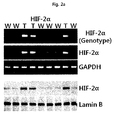
- the resultant mouse genomic DNA was extracted from tail of the mouse and subject to PCR gene analysis using primers in which may specifically amplify only cDNA of HIF-2 ⁇ transgenic mouse ( Fig. 2a ).
- HIF-2 ⁇ is overexpressed in HIF-2 ⁇ transgenic mouse tissues (cartilage, brain, heart, kidney, liver, lung, stomach, muscle) and chondrocytes using western blot and PCR. As a result, it was demonstrated that HIF-2 ⁇ was overexpressed specifically in only cartilage tissue ( Fig. 2a and 2b ).
- mouse knee joint chondrocytes were isolated from the knee joint of 8-day-old mouse and cultured in DMEM (Gibco, USA) containing 10% (v/v) fetal bovine serum (Gibco, USA), 50 ⁇ g/mL streptomycin (Sigma-Aldrich, USA) and 50 unit/mL penicillin (Sigma-Aldrich, USA).
- DMEM Gibco, USA
- streptomycin Sigma-Aldrich, USA
- penicillin Sigma-Aldrich, USA
- a monoclonal mouse anti-HIF-2 ⁇ antibody (Abcam., Inc. USA) was used.
- RNAs were extracted from chondrocytes and tissues (cartilage, brain, heart, kidney, liver, lung, stomach, muscle) in wild-type mouse and HIF-2 ⁇ transgenic mouse, and subjected to PCR to verify mRNA expression levels.
- Total RNA used in RT-PCR was isolated from respective tissues and cells using RNA STAT-60 (Tel-Test, Woodlands, TX) and reverse transcription was performed using ImProm-IITM reverse transcriptase (Promega, Madison, WI).
- the produced cDNA was amplified by PCR using Taq polymerase (TaKaRa Bio, Shiga, Japan). Primers used in PCR are described in Table 2.
- HIF-2 ⁇ was overexpressed strongly in HIF-2 ⁇ transgenic mouse chondrocytes, in contrast, the expression of HIF-2 ⁇ was expressed weakly in wild-type mouse.
- mRNA expression of HIF-2 ⁇ was significantly increased in only HIF-2 ⁇ transgenic mouse cartilage, on the other hand, mRNA expression of HIF-2 ⁇ was expressed weakly in wild-type mouse.
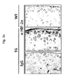
- HIF-2 ⁇ protein expression levels in transgenic mouse cartilage tissue were verified using immunohistochemistry.
- the cartilage of 45-week-old transgenic mouse or wild-type mouse was fixed in 4% paraformaldehyde (PFA, Sigma-Aldrich, USA) solution for 24 hours, incubated in 10% EDTA (Sigma-Aldrich, USA) solution for 2 weeks and prepared into a paraffin (McCormick, USA) block by increasing alcohol concentration.
- PFA paraformaldehyde
- EDTA Sigma-Aldrich, USA
- paraffin block was sliced to 6- ⁇ m thickness, deparaffinised in xylene (Sigma-Aldrich, USA) and treated with 10%(v/v) H 2 O 2 (Sigma-Aldrich, USA) in PBS (Sigma-Aldrich, USA) to remove activity of peroxidase in tissue.
- the slides were blocked with 1% BSA (Sigma-Aldrich, USA) and incubated at 4°C with a monoclonal mouse anti-HIF-2 ⁇ antibody. Normal mouse IgG was used as negative control ( Fig. 3a ).
- HIF-2 ⁇ protein was overexpressed strongly in HIF-2 ⁇ transgenic mouse cartilage tissue, in contrast, there was no HIF-2 ⁇ expression in wild-type mouse cartilage tissue.
- the cartilage of 12-week-old and 45-week-old transgenic mouse or wild-type mouse was fixed in 4% paraformaldehyde (PFA, Sigma-Aldrich, USA) solution for 24 hours, incubated in 10% EDTA solution for 2 weeks and prepared into a paraffin block by increasing alcohol concentration.
- the paraffin block was sliced to 6- ⁇ m thickness and the slides were stained with safranin-O (Sigma-Aldrich, USA) and hematoxylin (Sigma-Aldrich, USA) ( Fig. 3b ).
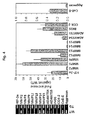
- RNAs were extracted from cartilages in 45-week-old transgenic mouse or wild-type mouse and the expression of representative cartilage degeneration-inducing factors such as MMP, ADAMTS, COX-2 and iNOS, were verified using RT-PCR ( Fig. 4 ).
- HIF-2 ⁇ was overexpressed in HIF-2 ⁇ transgenic mouse cartilage tissue than in the wild-type mouse, and mRNA expressions of MMP-3, MMP-9, MMP-12, MMP-13, ADAMTS-4, ADAMTS-5, COX-2 and iNOS were verified using RT-PCR and qRT-PCR ( Fig. 4 ).
Landscapes
- Life Sciences & Earth Sciences (AREA)
- Environmental Sciences (AREA)
- Animal Behavior & Ethology (AREA)
- Biotechnology (AREA)
- General Health & Medical Sciences (AREA)
- Veterinary Medicine (AREA)
- Health & Medical Sciences (AREA)
- Zoology (AREA)
- Animal Husbandry (AREA)
- Biodiversity & Conservation Biology (AREA)
- Engineering & Computer Science (AREA)
- Micro-Organisms Or Cultivation Processes Thereof (AREA)
- Investigating Or Analysing Biological Materials (AREA)
Abstract
Description
- The present invention relates to a cartilage-specific HIF-2α transgenic mouse as an animal model for arthritis, and use thereof.
- Degenerative arthritis (osteoarthritis, OA) is a representative degenerative disease that is primarily characterized by gradual and irreversible articular cartilage destruction and it leads to degrade the quality of life. Peoples aged more than 70 years old represent a degenerative arthritis and approximately 1/4 of them require clinical treatment. The market size of arthritis continues to increase is approximately 9.5 billion dollars in the world, growing by an annual average 14% and expected to approximately 20 billion dollars in 2005 and approximately 350 billion dollars in 2010. Currently, it now has an estimated 400 billion won in domestic market. However, no effective medical therapy to prevent OA cartilage destruction is currently available, owing to the limited understanding of the underlying molecular pathogenic mechanisms. A variety of potential OA-causing mechanisms have been proposed.
- Pathological causes of degenerative arthritis have been known to be induced by biochemical changes in the articular cartilage which is caused by the mechanical damage of the joints, genetic factors and obesity. This in turn leads to activation of biochemical pathways in chondrocytes, a unique resident cell type that synthesizes cartilagespecific extracellular matrix (ECM) components as well as various catabolic and anabolic factors. Activation of biochemical pathways involves the production of proinflammatory cytokines, inflammation, degradation of the ECM by matrix metalloproteinases (MMPs) and ADAMTS, and cessation of ECM synthesis via dedifferentiation and apoptosis of chondrocytes. However, the molecular pathogenic mechanisms of osteoarthritis need to be elucidated in more detail.
- Meanwhile, the HIF-2α (also known as endothelial PAS domain protein-1 or EPAS1) is gene regulated via hypoxea as transcription factor and it induces expression of target genes to allow adopting hypoxea. However, function of HIF-2α in cartilage degeneration is not yet clearly understood, except for a report that HIF-2α is expressed in hypertrophic chondrocyte.
- Throughout this application, several patents and publications are referenced and citations are provided in parentheses. The disclosure of these patents and publications is incorporated into this application in order to more fully describe this invention and the state of the art to which this invention pertains.
- The present inventors have made intensive studies to develop novel animal model system for a more efficient research to arthritis. As a result, they have introduced recombinant expression vector overexpressed hypoxia-inducible factor 2α (HIF-2 α) to a fertilized egg of mouse (Deposit accession number: KTCT11701BP), constructed a transgenic mouse, and found out that the transgenic mouse may be very useful as an animal model system for preventing or treating arthritis.
- Accordingly, it is an object of this invention to provide a transgenic fertilized egg.
- It is another object of this invention to provide a transgenic animal model for arthritis obtained from the fertilized egg mentioned-above.
- It is still another object of this invention to provide a method for screening a therapeutic agent for treating arthritis.
- Other objects and advantages of the present invention will become apparent from the detailed description to follow taken in conjugation with the appended claims and drawings.
- In one aspect of the present invention, there is provided a non-human transgenic fertilized egg comprising an expression construct comprising (a) a nucleotide sequence encoding HIF-2α (hypoxia-inducible factor 2α); and (b) a transcription-regulating sequence operatively linked to the nucleotide sequence (Deposit accession number: KTCT11701BP).
- In another aspect of the present invention, there is provided a transgenic animal model for arthritis obtained from the fertilized egg mentiond-above.
- The present inventors have made intensive studies to develop novel animal model system for a more efficient research to arthritis. As a result, they have introduced recombinant expression vector overexpressed hypoxia-inducible factor 2α (HIF-2 α) to a fertilized egg of mouse (Deposit accession number: KTCT11701BP), constructed a transgenic mouse, and found out that the transgenic mouse may be very useful as an animal model system for preventing or treating arthritis.
- Arthritis means diseases of joint disorder that involves inflammation caused by various factors. Specially, osteoarthritis (OA) is one of the oldest and the most common disease. It means a chronic state characterized by destruction of joint's cartilage and is known as degenerative joint disease, ostoarthrosis, hypertrophic arthritis or degenerative arthritis. Therefore, The term used herein "osteoarthritis" and other names above-mentioned will be used interchangeably. Cartilage is one area of the joint that it serves as cushion at tail end of bones to support moving joint easily. Cartilage destruction induces an abrasion between the adjacent bones and lead to stiffness and suffering caused by difficulty of the movement of the joints.
- Arthritis is very high incidence disease which is estimated approximately 200 million people in Korea and approximately 2,700 million in the United States (Helmick, C., et al of the Prevalence of Arthritis and OtherRheumatic conditions. Arthritis & Rheumatism, 58 (1): 15-25 (2008)). Unfortunately, a clear understanding for cause of the disease is scarce yet so that there is no therapeutic method. Actually, therapeutic method can be a variety of ways depending on the situation, because arthritis is caused by various factors (for example, age, obesity, injury, excessive use of joint and genetic cause).
- The present invention provides a transgenic animal, preferably a mouse, and a animal model system for arthritis using recombinant expression vector (expression construct) comprising HIF-2α as cartilage degeneration-inducing factor and its controllable target factor.
- The recombinant expression vector of the present invention comprises the nucleotide sequence as set forth in SEQ ID NO:1. More detailed, the recombinant expression vector comprises (a) a construct comprising an expression substance of interest-encoding nucleotide sequence; (b) a promoter which is operatively linked to the construct and act in animal cells to form RNA molecule, more preferably (a) a construct comprising a nucleotide sequence encoding hypoxia-inducible factor 2α (HIF-2 α) as set forth in SEQ ID N0:1; (b) a transcription-regulating sequence which is operatively linked to the construct, still more preferably (a) a construct comprising a nucleotide sequence encoding hypoxia-inducible factor 2α (HIF-2 α) as set forth in SEQ ID NO:1; (b) a transcription-regulating sequence which is operatively linked to the construct; and (c) a poly A signal causing polyadenylation at the 3'-end of a RNA molecule which act in animal cells, most preferably (a) a construct comprising a nucleotide sequence encoding CII-pNass-β-HIF-2α; (b) a transcription-regulating sequence which is operatively linked to the construct; and (c) a poly A signal causing polyadenylation at the 3'-end of a RNA molecule which act in animal cells.
- The mRNA level or the protein level of the expression of the nucleotide sequence as set forth in SEQ ID NO:1 which is included the recombinant vector of the present invention is increased in arthritis induced cells (e.g., chondrocytes) or tissues (e.g., the transgenic mouse cartilage tissue in the present invention) so that researches associated with arthritis have usability in animals (preferably mouse). In addition, it may become apparent to those skilled in this art that variants of nucleotide sequence encoding HIF-2α belong to the present invention. In other words, it may become apparent to those skilled in this art that variants fused with marker for detecting target protein belong to the present invention. For example, the protein which may be fused to target protein for detecting target protein includes, but not limited to, GFP (green fluorescent protein), RFP (red fluorescent protein), CFP (cyan fluorescent protein) and YFP (yellow fluorescent protein), BFP (bluefluorescent protein), luciferase or its variants (e,g., EGFP, ECFP, EYFP, ERFP, EBFP).
- According to a preferable embodiment, the HIF-2α in transgenic animal model for arthritis of the present invention increases the expression of cartilage degeneration-inducing factor. According to a preferred embodiment, the cartilage degeneration-inducing factor of the present invention includes, but not limited to, matrix metalloprotenase (MMP) -1, MMP-3, MMP-9, MMP-12, MMP-13, ADAMTS (a disintegrin and metalloproteinase with thrombospondin motifs)-4, ADAMTS-5, PTGS-2 (Prostaglandin-endoperoxide synthase-2) or NOS2 (nitric oxide synthase 2).
- The term used herein "transcription-regulating sequence" means a DNA sequence that regulates the expression of a coding sequence or a functional RNA. The term "operatively linked" refers to functional linkage between a nucleic acid expression control sequence (such as a promoter, signal sequence, or array of transcription factor binding sites) and a second nucleic acid sequence, wherein the expression control sequence affects transcription and/or translation of the nucleic acid corresponding to the second sequence.
- According to a preferable embodiment, the transcription-regulating sequence comprises a promoter and an enhancer.
- In the present invention, the transcription-regulating sequence linked to the HIF-2α is operable in, preferably animal cells, more preferably mammalian cells, still more preferably human or mouse cells, most preferably chondrocytes to control transcription of the HIF-2α, including the promoters derived from the genome of mammalian cells or from mammalian viruses, for example, CMV (cytomegalovirus) promoter, CII promoter, the adenovirus late promoter, the vaccinia virus 7.5K promoter, SV40 promoter, HSV tk promoter, RSV promoter, EF1 alpha promoter, metallothionein promoter, beta-actin promoter, human IL-2 gene promoter, human IFN gene promoter, human IL-4 gene promoter, human lymphotoxin gene promoter, human GM-CSF gene promoter, inducible promoter, tumor cell specific promoter (e.g., TERT promoter, PSA promoter, PSMA promoter, CEA promoter, E2F promoter and AFP promoter) and tissue specific promoter (e.g., albumin promoter). Most preferably, the promoter is CMV promoter.
- According to a preferable embodiment, the promoter of present invention is a chondrocyte-specific promoter and more preferably, the present invention further comprise the enhancer, most preferably the chondrocyte-specific enhancer of type II collagenase.
- According to a preferable embodiment, the expression construct of the present invention further comprises an intron sequence between the promoter and the nucleotide sequence encoding HIF-2α.
- Preferably, the expression constructs used in the present invention comprises polyadenylated sequence (e.g., bovine growth hormone terminator and polyadenylated sequence derived from SV40).
- According to a preferred embodiment, the construct comprising HIF-2α-encoding nucleotide sequence used in the present invention has the structure of "transcription-regulating sequence- HIF-2α-encoding nucleotide sequence-polyadenylated sequence".
- The HIF-2α-encoding nucleotide sequence of the present invention comprises any of gene delivery system used in gene therapy by those skilled in the art, preferably, plasmid, adenovirus (Lockett LJ, et al., Clin. Cancer Res., 3:2075-2080(1997)), adeno-associated virus (AAV, Lashford LS., et al., Gene Therapy Technologies, Applications and Regulations Ed. A. Meager, 1999), retrovirus (Gunzburg WH, et al., Retroviral vectors. Gene Therapy Technologies, Applications and Regulations Ed. A. Meager, 1999), lentivirus (Wang G. et al., J. Clin. Invest. 104(11):R55-62(1999)), herpes simplex virus (Chamber R., et al., Proc. Natl. Acad. Sci USA, 92:1411-1415(1995)), vaccinia virus (Puhlmann M. et al., Human Gene Therapy, 10:649-657(1999)) liposome ((Methods in Molecular Biology, Vol 199, S.C. Basu and M. Basu (Eds.), Human Press 2002)) or niosome. Most preferably, the gene delivery system of this invention is constructed by incorporating the HIF-2α-encoding nucleotide sequence to adenovirus.
- Adenoviruses have been usually employed as a gene delivery system because of its mid-sized genome, ease of manipulation, high titer, wide target-cell range, and high infectivity. Both ends of the viral genome contains 100-200 bp ITRs (inverted terminal repeats), which are cis elements necessary for viral DNA replication and packaging. The E1 region (E1A and E1B) encodes proteins responsible for the regulation of transcription of the viral genome and a few cellular genes. The expression of the E2 region (E2A and E2B) results in the synthesis of the proteins for viral DNA replication.
- Of adenoviral vectors developed so far, the replication incompetent adenovirus having the deleted E1 region is usually used. The deleted E3 region in adenoviral vectors may provide an insertion site for transgenes (Thimmappaya, B. et al., Cell, 31:543-551(1982); and Riordan, J. R. et al., Science, 245:1066-1073(1989)). Therefore, it is preferred that the nucleotide sequence encoding the CCN5 protein or CCN2ΔCT protein of this invention is inserted into either the deleted E1 region (E1A region and/or E1B region, preferably, E1B region) or the deleted E3 region, more preferably, the deleted E3 region. The nucleotide sequence of interest to be delivered is preferably inserted into either the deleted E1 region (E1A region and/or E1B region, preferably, E1B region) or the deleted E3 region, more preferably, the deleted E1 region. Furthermore, the inserted sequences may be incorporated into the deleted E4 region. The term used herein "deletion" with reference to viral genome encompasses whole deletion and partial deletion as well.
- According to the most preferred embodiment of the present invention, the adenovirus gene transduction system of the present invention comprises "promoter-the HIF-2α gene-poly A sequence". The "promoter-the HIF-2α gene-poly A sequence"is inserted into the deleted E1 region (E1A region and/or E1B region, preferably, E1B region) or the E3 region, preferably, the deleted E3 region.
- In nature, adenovirus can package approximately 105% of the wild-type genome, providing capacity for about 2 extra kb of DNA (Ghosh-Choudhury et al., EMBO J., 6:1733-1739(1987)). In this regard, the foreign sequences described above inserted into adenovirus may be further inserted into adenoviral wild-type genome.
- The adenovirus may be of any of the 42 different known serotypes or subgroups A-F. Adenovirus type 5 of subgroup C is the most preferred starting material for constructing the adenoviral gene delivery system of this invention. A great deal of biochemical and genetic information about adenovirus type 5 is known.
- The foreign genes delivered by the present adenoviral gene delivery system are episomal, and therefore, have low genotoxicity to host cells. Therefore, gene therapy using the adenoviral gene delivery system of this invention may be considerably safe.
- Retroviruses capable of carrying relatively large exogenous genes have been used as viral gene delivery vectors in the senses that they integrate their genome into a host genome and have broad host spectrum.
- In order to construct a retroviral vector, the HIF-2α gene is inserted into the viral genome in the place of certain viral sequences to produce a replication-defective virus. To produce virions, a packaging cell line containing the gag, pol and env genes but without the LTR (long terminal repeat) and Ψ components is constructed (Mann et al., Cell, 33:153-159(1983)). When a recombinant plasmid containing the HIF-2α gene, LTR and Ψ is introduced into this cell line, the Ψ sequence allows the RNA transcript of the recombinant plasmid to be packaged into viral particles, which are then secreted into the culture media (Nicolas and Rubinstein "Retroviral vectors," In: Vectors: A survey of molecular cloning vectors and their uses, Rodriguez and Denhardt (eds.), Stoneham: Butterworth, 494-513(1988)). The media containing the recombinant retroviruses is then collected, optionally concentrated and used for gene delivery.
- A successful gene transfer using the second-generation retroviral vector has been reported. Kasahara et al. (Science, 266:1373-1376(1994)) prepared variants of moloney murine leukemia virus in which the EPO (erythropoietin) sequence is inserted in the place of the envelope region, consequently, producing chimeric proteins having novel binding properties. Likely, the present gene delivery system can be constructed in accordance with the construction strategies for the second-generation retroviral vector.
- Adeno-associated viruses are capable of infecting non-dividing cells and various types of cells, making them useful in constructing the gene delivery system of this invention. The detailed descriptions for use and preparation of AAV vector are found in
U.S. Pat. No. 5,139,941 and4,797,368 . - Research results for AAV as gene delivery systems are disclosed in LaFace et al, Viology, 162:483486(1988), Zhou et al., Exp. Hematol. (NY), 21:928-933(1993), Walsh et al, J. Clin. Invert., 94:1440-1448(1994) and Flotte et al., Gene Therapy, 2:29-37(1995). Recently, an AAV vector has been approved for Phase I human trials for the treatment of cystic fibrosis.
- Typically, a recombinant AAV virus is made by cotransfecting a plasmid containing the gene of interest (i.e., relaxin gene and nucleotide sequence of interest to be delivered) flanked by the two AAV terminal repeats (McLaughlin et al., 1988; Samulski et al., 1989) and an expression plasmid containing the wild type AAV coding sequences without the terminal repeats (McCarty et al., J. Virol., 65:2936-2945(1991)).
- Other virus vectors may be used for the gene transduction system in the present invention. Vectors derived from viruses such as vaccinia virus (Puhlmann M. et al., Human Gene Therapy 10:649-657(1999); Ridgeway, "Mammalian expression vectors," In: Vectors: A survey of molecular cloning vectors and their uses. Rodriguez and Denhardt, eds. Stoneham: Butterworth, 467-492(1988); Baichwal and Sugden, "Vectors for gene transfer derived from animal DNA viruses: Transient and stable expression of transferred genes," In: Kucherlapati R, ed. Gene transfer. New York: Plenum Press, 117-148(1986) and Coupar et al., Gene, 68:1-10(1988)), lentivirus (Wang G. et al., J. Clin. Invest. 104(11):R55-62(1999)) and herpes simplex virus (Chamber R., et al., Proc. Natl. Acad. Sci USA 92:1411-1415(1995)) may be used in the present delivery systems for transferring the HIF-2α gene into cells.
- Liposomes are formed spontaneously when phospholipids are suspended in an excess of aqueous medium. Liposome-mediated nucleic acid delivery has been very successful as described in Nicolau and Sene, Biochim. Biophys. Acta, 721:185-190(1982) and Nicolau et al., Methods Enzymol., 149:157-176(1987). Example of commercially accessible reagents for transfecting animal cells using liposomes includes Lipofectamine (Gibco BRL). Liposomes entrapping the HIF-2α gene interact with cells by mechanism such as endocytosis, adsorption and fusion and then transfer the sequences into cells.
- The methods Introduced gene delivery system of the present invention into cells the above-mentioned may be carried out through a variety of methods known in the art. Where the present gene delivery system is constructed on the basis of viral vector construction, the contacting is performed as conventional infection methods known in the art. The infection of hosts using viral vectors is well described in the above-cited publications. Where the present gene delivery system is a naked recombinant DNA molecule or plasmid, the nucleotide sequence to be delivered are introduced into cells by microinjection (Capecchi, M.R., Cell, 22:479(1980) and Harland and Weintraub, J. Cell Biol. 101:1094-1099(1985)), calcium phosphate co-precipitation (Graham, F.L. et al., Virology, 52:456(1973) and Chen and Okayama, Mol. Cell. Biol. 7:2745-2752(1987)), electroporation (Neumann, E. et al., EMBO J., 1:841(1982) and Tur-Kaspa et al., Mol. Cell Biol., 6:716-718(1986)), liposome-mediated transfection (Wong, T.K. et al., Gene, 10:87(1980) and Nicolau and Sene, Biochim. Biophys. Acta, 721:185-190(1982); and Nicolau et al., Methods Enzymol., 149:157-176(1987)), DEAE-dextran treatment (Gopal, Mol. Cell Biol., 5:1188-1190(1985)) and particle bombardment (Yang et al., Proc. Natl. Acad. Sci., 87:9568-9572(1990)), and most preferably microinjection.
- According to the present invention, the recombinant expression vector of the present invention is microinjected into the fertilized egg to obtain the transgenic fertilized egg. The fertilized egg with the gene inserted was implanted in the womb of a surrogate (for example, mouse) to obtain mice. Then, 10 transgenic mice were selected through PCR genotyping.
- PCR is one of the most predominant processes for nucleic acid amplification and a number of its variations and applications have been developed. For example, for improving PCR specificity or sensitivity, touchdown PCR, hot start PCR, nested PCR and booster PCR have been developed with modifying traditional PCR procedures. In addition, real-time PCR, differential display PCR (DD-PCR), rapid amplification of cDNA ends (RACE), multiplex PCR, inverse polymerase chain reaction (IPCR), vectorette PCR and thermal asymmetric interlaced PCR (TAIL-PCR) have been suggested for certain applications. The details of PCR can be found in McPherson, M.J., and Moller, S.G. PCR. BIOS Scientific Publishers, Springer-Verlag New York Berlin Heidelberg, N.Y. (2000), the teachings of which are incorporated herein by reference in its entity.
- The term used herein "amplification" refers to reactions for amplifying nucleic acid molecules. A multitude of amplification reactions have been suggested in the art, including polymerase chain reaction (hereinafter referred to as PCR) (
U.S. Patent Nos. 4,683,195 ,4,683,202 , and4,800,159 ), reverse transcription-polymerase chain reaction (hereinafter referred to as RT-PCR) (Sambrook, J. et al., Molecular Cloning. A Laboratory Manual, 3rd ed. Cold Spring Harbor Press (2001)), the methods ofMiller, H. I. (WO 89/06700 Davey, C. et al. (EP 329,822 WO 90/01069 EP 439,182 WO 88/10315 WO 90/06995 U.S. Patent No. 6,410,276 ), consensus sequence primed polymerase chain reaction (CP-PCR;U.S. Patent No. 4,437,975 ), arbitrarily primed polymerase chain reaction (AP-PCR;U.S. Patent Nos. 5,413,909 and5,861,245 ), nucleic acid sequence based amplification (NASBA;U.S. Patent Nos. 5,130,238 ,5,409,818 ,5,554,517 and6,063,603 ), strand displacement amplification and loop-mediated isothermal amplification (LAMP), but not limited to. Other amplification methods that may be used are described inU.S. Patent Nos. 5,242,794 ,5.494,810 ,4,988,617 and inU.S. Ser. No. 09/854,317 - Therefore, the present invention may be generally carried out by nucleic acid amplifications using mRNA molecules in samples as templates and primers to be annealed to mRNA or cDNA.
- For obtaining mRNA molecules, total RNA is isolated from samples. The isolation of total RNA may be performed by various methods (Sambrook, J. et al., Molecular Cloning. A Laboratory Manual, 3rd ed. Cold Spring Harbor Press (2001); Tesniere, C. et al., Plant Mol. Biol. Rep., 9: 242 (1991); Ausubel, F.M. et al., Current Protocols in Molecular Biology, John Willey & Sons (1987); and Chomczynski, P. et al., Anal. Biochem. 162: 156 (1987)). For example, total RNA in cells may be isolated using Trizol. Afterwards, cDNA molecules are synthesized using mRNA molecules isolated and then amplified. Since total RNA molecules used in the present invention are isolated from human samples, mRNA molecules have poly-A tails and converted to cDNA by use of dT primer and reverse transcriptase ( PNAS USA, 85: 8998 (1988); Libert F, et al., Science, 244: 569 (1989); and Sambrook, J. et al., Molecular Cloning. A Laboratory Manual, 3rd ed. Cold Spring Harbor Press (2001)). cDNA molecules synthesized are then amplified by amplification reactions.
- The primers used for the present invention is hybridized or annealed to a region on template so that double-stranded structure is formed. Conditions of nucleic acid hybridization suitable for forming such double stranded structures are described by Joseph Sambrook, et al., Molecular Cloning, A Laboratory Manual, Cold Spring Harbor Laboratory Press, Cold Spring Harbor, N.Y. (2001) and Haymes, B. D., et al., Nucleic Acid Hybridization, A Practical Approach, IRL Press, Washington, D.C. (1985).
- A variety of DNA polymerases can be used in the amplification step of the present methods, which includes "Klenow" fragment of E. coli DNA polymerase I, a thermostable DNA polymerase and bacteriophage T7 DNA polymerase. Preferably, the polymerase is a thermostable DNA polymerase obtained from a variety of bacterial species, including Thermus aquaticus (Taq), Thermus thermophilus (Tth), Thermus filiformis, Thermis flavus, Thermococcus literalis, and Pyrococcus furiosus (Pfu).
- When a polymerization reaction is being conducted, it is preferable to provide the components required for such reaction in excess in the reaction vessel. Excess in reference to components of the amplification reaction refers to an amount of each component such that the ability to achieve the desired amplification is not substantially limited by the concentration of that component. It is desirable to provide to the reaction mixture an amount of required cofactors such as Mg2+, and dATP, dCTP, dGTP and dTTP in sufficient quantity to support the degree of amplification desired. All of the enzymes used in this amplification reaction may be active under the same reaction conditions. Indeed, buffers exist in which all enzymes are near their optimal reaction conditions. Therefore, the amplification process of the present invention can be done in a single reaction volume without any change of conditions such as addition of reactants.
- Annealing or hybridization in the present invention is performed under stringent conditions that allow for specific binding between the target nucleotide sequence and the primer. Such stringent conditions for annealing will be sequence-dependent and varied depending on environmental parameters.
- The composition of the present invention may optionally include other reagents along with components described above. For instance, where the present kit may be used for nucleic acid amplification, it may optionally include the reagents required for performing PCR reactions such as buffers, DNA polymerase (thermostable DNA polymerase obtained from Thermus aquaticus (Taq), Thermus thermophilus (Tth), Thermus filiformis, Thermis flavus, Thermococcus literalis, and Pyrococcus furiosus (Pfu)), DNA polymerase cofactors, and dNTPs.
- Primers used in the the present invention have a sequence complementary to a nucleotide sequence encoding HIF-2α. The term "complementary" with reference to sequence used herein refers to a sequence having complementarity to the extent that the sequence hybridizes or anneals specifically with the nucleotide sequence described above under certain hybridization or annealing conditions. In this regard, the term "complementary" used herein has different meaning from the term "perfectly complementary". The primer of this invention may include one or more mismatch base sequences where it is able to specifically hybridize with the above-described nucleotide sequences.
- The term "primer" used herein means a single-stranded oligonucleotide which is capable of acting as a point of initiation of template-directed DNA synthesis when placed under proper conditions (i.e., in the presence of four different nucleoside triphosphates and a thermostable enzyme) in an appropriate buffer and at a suitable temperature. The suitable length of primers will depend on many factors, including temperature, application and source of primer, generally, 15-30 nucleotides in length. In general, shorter primers need lower temperature to form stable hybridization duplexes to templates.
- The sequences of primers are not required to have perfectly complementary sequence to templates. The sequences of primers may comprise some mismatches, so long as they can be hybridized with templates and serve as primers. Therefore, the primers of this invention are not required to have perfectly complementary sequence to the nucleotide sequence as described above; it is sufficient that they have complementarity to the extent that they anneals specifically to the nucleotide sequence of the gene for acting as a point of initiation of synthesis. The primer design may be conveniently performed with referring to the above-described nucleotide sequences. For instance, the primer design may be carried out using computer programs for primer design (e.g., PRIMER 3 program).
- To prepare primers, the nucleotide sequence of the present biomarker may be found in the GenBank Accession No. NM_010137.3 of HIF-2α (SEQ ID NO:1) and the GenBank Accession No. NM_008084.2 of GAPDH, and primers may be designed by reference with the nucleotide sequence afore-mentioned.
- In another aspect of the present invention, there is provided a method for screening a therapeutic agent for treating arthritis, which comprises the steps of:
- (a) contacting a test substance of interest for analysis to the transgenic animal; and (b) analyzing the cartilage tissue destruction level of the transgenic animal,
- Since the method of the present invention follows the process of analyzing the expression levels of HIF-2α of the present method described above, the common descriptions between them are omitted in order to avoid undue redundancy leading to the complexity of this specification.
- In the method for screening of the present invention, test substance or expression substance of interest-encoding nucleotide sequence or protein may be labeled by detectable label. For example, the detectable label is chemical label (e.g., biotin), enzyme (horseradish peroxidase, alkaline phosphatase, peroxidase, luciferase, β-galactosidase, β-glucosidase), radioactive label (e.g., C14, I125, P32 and S35), fluorescent label [e.g., coumarin, fluorescein, FITC (fluoresein Isothiocyanate), rhodamine 6G, rhodamine B, TAMRA (6-carboxy-tetramethyl-rhodamine), Cy-3, Cy-5, Texas Red, Alexa Fluor, DAPI (4,6-diamidino-2-phenylindole), HEX, TET, Dabsyl and FAM], luminescent label, chemiluminescent label, FRET (fluorescence resonance energy transfer) label or metal label (e.g., gold and silver)
- According to the method of the present invention, a transgenic animal is prepared first using the recombinant expression vector of the present invention. The transgenic animal, preferably mouse is contacted to a test substance to be analyzed. The transgenic animal of the present invention is transgenic which is introduced a recombinant expression vector overexpressing HIF-2α. The term "test substance" used herein in conjunction with the present screening method refers to a material tested in the present method for analyzing the influence on the expression level of HIF-2α. The test substance includes siRNA (small interference RNA), shRNA (short hairpin RNA), miRNA (microRNA), ribozyme, DNAzyme, PNA (peptide nucleic acids), antisense oligonucleotides, antibodies, aptamers, extracts of natural sources and chemical substances, but not limited to.
- Afterwards, the expression of the HIF-2α coding nucleotide sequence or the binding affinity of the transcription-regulating sequence to the HIF-2α protein in cells treated with the test substance is measured. The measurement of the expression levels may be carried out according to methods as described above, and the test substance may be determined as the therapeutic agent for treating arthritis where the test substance inhibits the expression of the HIF-2α protein or inhibits the binding of the transcription-regulating sequence to the HIF-2α.
- The measurement of change in the expression level of the nucleotide sequence afore-mentioned may be carried out through a variety of methods known in the art, for example, RT-PCR (Sambrook et al, Molecular Cloning. A Laboratory Manual, 3rd ed. Cold Spring Harbor Press(2001)), Northern blot (Peter B. Kaufma et al., Molecular and Cellular Methods in Biology and Medicine, 102-108, CRC press), hybridization using cDNA microarray (Sambrook et al, Molecular Cloning. A Laboratory Manual, 3rd ed. Cold Spring Harbor Press(2001)) or in situ hybridization (Sambrook et al, Molecular Cloning. A Laboratory Manual, 3rd ed. Cold Spring Harbor Press (2001)).
- According to a preferred embodiment, the present invention may be performed to the manner of immunoassay, i.e., antigen-antibody reaction manner. At this time, it is performed using an antibody or an aptamer specific binding to arthritis marker (e.g., HIF-2α) of the present invention mentioned-above.
- The antibody used in the present invention is polyclonal or monoclonal antibody, preferably monoclonal antibody. Antibody production may be prepared by a method widely known in the art, such as a hybridoma method (Kohler and Milstein, European Journal of Immunology, 6:511-519(1976)), a recombinant DNA methods (
U.S. Patent No. 4,816,56 ) or a phage antibody library technique (Clackson et al, Nature, 352:624-628(1991) and Marks et al, J. Mol. Biol., 222:58, 1-597(1991)). General process for antibody production is described in Harlow, E. and Lane, D., Using Antibodies: A Laboratory Manual, Cold Spring Harbor Press, New York, 1999; Zola, H., Monoclonal Antibodies: A Manual of Techniques, CRC Press, Inc., Boca Raton, Florida, 1984; Coligan, CURRENT PROTOCOLS IN IMMUNOLOGY, Wiley / Greene, NY, 1991, and the literatures are inserted as reference in the present invention. For example, the preparation of the hybridoma cells producing monoclonal antibody is accomplished by fusing immortalized cell line with antibody-producing lymphocytes. The technology required for this process is widely known in the art and may be easily carried out using techniques. Polyclonal antibodies may be produced by injecting the protein antigen into an appropriate animal and collecting blood samples from the animal to obtain sera containing antibodies using affinity technology known in the art. - When the method of the present disclosure is performed using an antibody or an aptamer, the present disclosure may be used for diagnosing arthritis by performing conventional immunoassay methods.
- The immunoassay may be performed by a variety of quantitative or qualitative immunoassay protocols. The immunoassay format includes, but is not limited to, radioimmunoassay analysis, radioactive immunoprecipitation, immunoprecipitation, immunohistochemical staining, ELISA (enzyme-linked immunosorbant assay), Capture-ELISA, sandwich assay, flow cytometry, immunofluorescence and immune affinity purified. The immunoassay or the immuno staining method is described in Enzyme Immunoassay, E. T. Maggio, ed., CRC Press, Boca Raton, Florida, 1980; Gaastra, W., Enzyme-linked immunosorbent assay (ELISA), in Methods in Molecular Biology, Vol. 1, Walker, J.M. ed., Humana Press, NJ, 1984; and Ed Harlow and David Lane, Using Antibodies:A Laboratory Manual, Cold Spring Harbor Laboratory Press, 1999 and the literatures are inserted as reference in herein.
- For example, when the method of the present disclosure is performed using radioimmunoassay analysis, the antibody labled with radioactive isotopes (e.g., C14, I125, P32 and S35) may be used for detecting a marker of the present invention.
- When the method of the present disclosure is performed using the ELISA, certain examples of the present invention comprise the steps of: (i) coating unknown cell cytolysate samples of interest for analysis on the surface of a solid substrate; ( ii ) contacting the cell cytolysate with antibody for the marker as the primary antibody; (iii) contacting the resultant of step ( ii ) with the secondary antibody conjugated enzyme; and (iv) detecting the enzyme activity.
- The appropriate solid substrate is hydrocarbon polymers (e.g., polystyrene and polypropylene), glass, metal, or gel, and most preferably a micro-titer plate.
- The appropriate secondary antibody conjugated enzyme includes, but is not limited to, color-developing reaction, fluorescent reaction, luminescent reaction or infrared reaction, for example, alkaline phosphatase, β-galactosidase, horseradish peroxidase, luciferase and cytochrome P450. Where alkaline phosphatase is used for the enzyme binding to the secondary antibody, bromochloroindolylphosphate (BCIP), nitro blue tetrazolium (NBT), naphthol-AS-B1-phosphate and ECF (enhanced chemifluorescence) may be used as a substrate for color-developing reactions. In the case of using horseradish peroxidase, chloronaphtol, aminoethylcarbazol, diaminobenzidine, D-luciferin, lucigenin (bis-N-methylacridinium nitrate), resorufin benzyl ether, luminol, Amplex Red reagent (10-acetyl-3,7-dihydroxyphenoxazine), HYR (p-phenylenediamine-HCl and pyrocatechol), TMB (3,3,5,5-tetramethylbenzidine), ABTS (2,2-Azine-di[3-ethylbenzthiazoline sulfonate]), o-phenylenediamine (OPD) and naphtol/pyronine may be used as a substrate; and in the case of using glucose oxidase, t-NBT (nitroblue tetrazolium) or m-PMS (phenzaine methosulfate) may be used as a substrate.
- When the method of the present disclosure is performed using the Capture-ELISA, certain examples of the present invention comprise the steps of: (i) coating the antibody for the marker as capturing antibody on the surface of a solid substrate; ( ii ) contacting the sample with the capturing antibody; (iii) contacting the resultant of step ( ii ) with the detecting antibody which is combined with label generating signal and react specifically to the HIF-2α protein; and (iv) detecting the signal from the label.
- The detecting antibody has the label generating detectable a signal. The label includes, but is not limited to, chemical (e.g., biotin), enzyme (alkaline phosphatase, β-galactosidase, horseradish peroxidase and cytochrome P450), radioactive material (e.g., C14, I125, P32 and S35), fluorescent material (e.g., fluorescein), luminescent material, chemiluminescent material and FRET (fluorescence resonance energy transfer). A variety of labels and labeling methods are described in Ed. Harlow and David Lane, Using Antibodies: A Laboratory Manual, Cold Spring Harbor Laboratory Press, 1999.
- The final measurement of enzyme activity or measurement of the signal may be carried out in accordance with a variety of methods known in the art. The detection of this signal permits to qualitative or quantitative analysis of the marker of the present invention. In the case of using biotin as label, the signal is easily detected using streptavidin. In the case of using luciferase as label, the signal is easily detected using luciferin.
- According to another embodiment of this invention, the present invention may use aptamers specific to markers instead of antibodies. Aptamers include oligo nucleic acid and peptide molecules, of which details can be found in Bock LC et al., Nature 355 (6360):5646 (1992); Hoppe-Seyler F, Butz K "Peptide aptamers: powerful new tools for molecular medicine". J Mol Med. 78(8):42630(2000); Cohen BA, Colas P, Brent R. "An artificial cell-cycle inhibitor isolated from a combinatorial library". Proc Natl Acad Sci USA. 95(24):142727 (1998).
- The features and advantages of the present invention will be summarized as follows:
- (a) The present invention relates to a transgenic fertilized egg which is transformed with a recombinant expression vector overexpressing HIF-2 α (hypoxia-inducible factor 2α), and a transgenic animal model for arthritis obtained therefrom.
- (b) A transgenic mouse of the present invention may be very useful as a therapeutic model for arthritis, which may be used in studies on the cartilage degeneration process.
- (c) In addition, the transgenic mouse of the present invention may be applied to a method for screening arthritis therapeutics.
-
-
Fig. 1 schematically represents the vector map required for preparing HIF-2α genetically engineered mouse. -
Fig. 2 represents the result that vector DNA is inserted on chromosome of transgenic mouse and it is verified using PCR (polymerase chain reaction) and western blot. -
Fig. 3 represents the immunohistochemical analysis of the cartilage destruction level in HIF-2α transgenic mouse and wild-type mouse -
Fig. 4 represents the result of RT-PCR (reverse transcription-polymerase chain reaction) and western blot analysis for cartilage degeneration inducing factors in cartilage of HIF-2α transgenic mouse and wild-type mouse. - The present invention will now be described in further detail by examples. It would be obvious to those skilled in the art that these examples are intended to be more concretely illustrative and the scope of the present invention as set forth in the appended claims is not limited to or by the examples.
- To prepare the transgenic mouse overexpressing chondrocyte-specific HIF-2α, a gene for transplantation was prepared first (
Fig. 1 ). - A fragment (2.625 kb) of a mouse HIF-2α gene cleaved by NotI and NotI restriction enzymes was cloned into the NotI site of 4.35-kb pNass-beta vector (Clontech Laboratories, USA). In addition, enhancer, promoter and 0.7 kb intron parts of 1.0 kb type II collagen (Col2a1) inducing chondrocyte-specific expression were cloned into the NotI site of the vector. The vector was cleaved with NotI to obtain a purified, 9.675-kb gene for transplantation. The gene is called a CII-pNass-β-HIF-2α construct (
Fig. 1 ; gene 2.625 kb + vector 4.35 kb + enhancer 1.0 kb + promoter 1.0 kb + intron 0.7 kb = 9.675 kb). The gene for transplantation was microinjected into the pronucleus of a fertilized egg of a C57BL/6 mouse (Charles River, Japan). The fertilized egg obtained as a resultant was deposited to Korea Research Institute of Bioscience and Biotechnology at May 20, 2010 and received a deposit accession number KTCT11701BP. The fertilized egg with the gene inserted was implanted in the womb of a surrogate mouse to obtain at least 200 mice. Then, 10 transgenic mice were selected through PCR genotyping. - The original mouse obtained in the Example 1 was mated with wild-type mouse. The resultant mouse genomic DNA was extracted from tail of the mouse and subject to PCR gene analysis using primers in which may specifically amplify only cDNA of HIF-2α transgenic mouse (
Fig. 2a ). - The Primers used in PCR are described in Table 1 and PCR reaction condition is as follows: the reaction mixture was denatured for 5 min at 95 °C, subjected to 30 cycles of 10 sec at 95 °C, 20 sec at 58 °C and 30 sec at 72 °C and extended for 10 min at 72 °C.
[Table 1] Primer sequence Gene Primer sequence SEQ ID NO Predicted size Use HIF-2α gene test sense 5'-GCCTCATGTCTCCATGTTCA-3' 2 628 bp HIF-2α TG gene test antisense 5'-TGTATCTTATCATGTCTGCAT-3' 3 - We examined whether HIF-2α is overexpressed in HIF-2α transgenic mouse tissues (cartilage, brain, heart, kidney, liver, lung, stomach, muscle) and chondrocytes using western blot and PCR. As a result, it was demonstrated that HIF-2α was overexpressed specifically in only cartilage tissue (
Fig. 2a and2b ). - To perform western blot extracting protein in cells, the mouse knee joint chondrocytes were isolated from the knee joint of 8-day-old mouse and cultured in DMEM (Gibco, USA) containing 10% (v/v) fetal bovine serum (Gibco, USA), 50 µg/mL streptomycin (Sigma-Aldrich, USA) and 50 unit/mL penicillin (Sigma-Aldrich, USA). A monoclonal mouse anti-HIF-2α antibody (Abcam., Inc. USA) was used.
- In addition, RNAs were extracted from chondrocytes and tissues (cartilage, brain, heart, kidney, liver, lung, stomach, muscle) in wild-type mouse and HIF-2α transgenic mouse, and subjected to PCR to verify mRNA expression levels. Total RNA used in RT-PCR was isolated from respective tissues and cells using RNA STAT-60 (Tel-Test, Woodlands, TX) and reverse transcription was performed using ImProm-IITM reverse transcriptase (Promega, Madison, WI). The produced cDNA was amplified by PCR using Taq polymerase (TaKaRa Bio, Shiga, Japan). Primers used in PCR are described in Table 2.
[Table 2] Primer sequence Gene Primer sequence SEQ ID NO Predicted size Mouse HIF-2α sense 5'-CGAGAAGAACGACGTGGTGTTC-3' 4 293 bp antisense 5'-GTGAAGGCGGGCAGGCTCC-3 5 Mouse GAPDH sense 5'-TCACTGCCACCCAGAAGAC-3' 6 450 bp antisense 5'-TGTAGGCCATGAGGTCCAC-3 7 - As a result, it would be demonstrated that the protein and mRNA of HIF-2α were overexpressed strongly in HIF-2α transgenic mouse chondrocytes, in contrast, the expression of HIF-2α was expressed weakly in wild-type mouse. In the results of mRNA expression levels of HIF-2α for each of tissues, mRNA expression of HIF-2α was significantly increased in only HIF-2α transgenic mouse cartilage, on the other hand, mRNA expression of HIF-2α was expressed weakly in wild-type mouse. Taken together, it would be understood that HIF-2α was overexpressed in only transgenic mouse chondrocytes which is prepared by the method of the present invention.
- HIF-2α protein expression levels in transgenic mouse cartilage tissue were verified using immunohistochemistry. The cartilage of 45-week-old transgenic mouse or wild-type mouse was fixed in 4% paraformaldehyde (PFA, Sigma-Aldrich, USA) solution for 24 hours, incubated in 10% EDTA (Sigma-Aldrich, USA) solution for 2 weeks and prepared into a paraffin (McCormick, USA) block by increasing alcohol concentration. The paraffin block was sliced to 6-µm thickness, deparaffinised in xylene (Sigma-Aldrich, USA) and treated with 10%(v/v) H2O2 (Sigma-Aldrich, USA) in PBS (Sigma-Aldrich, USA) to remove activity of peroxidase in tissue. The slides were blocked with 1% BSA (Sigma-Aldrich, USA) and incubated at 4°C with a monoclonal mouse anti-HIF-2α antibody. Normal mouse IgG was used as negative control (
Fig. 3a ). - As a result, it would be demonstrated that the protein of HIF-2α was overexpressed strongly in HIF-2α transgenic mouse cartilage tissue, in contrast, there was no HIF-2α expression in wild-type mouse cartilage tissue. These facts mean that HIF-2α protein was expressed in only transgenic mouse because transgenic mouse prepared to allow high-expressing HIF-2α in cartilage tissue which is abundant type II collagen (
FIG. 3a ). - In order to examine cartilage destruction level in the transgenic mice, the cartilage of 12-week-old and 45-week-old transgenic mouse or wild-type mouse was fixed in 4% paraformaldehyde (PFA, Sigma-Aldrich, USA) solution for 24 hours, incubated in 10% EDTA solution for 2 weeks and prepared into a paraffin block by increasing alcohol concentration. The paraffin block was sliced to 6-µm thickness and the slides were stained with safranin-O (Sigma-Aldrich, USA) and hematoxylin (Sigma-Aldrich, USA) (
Fig. 3b ). - As a result, when compared with the 12-week-old wild-type mouse, slight irregularity was found in the HIF-2α transgenic mouse. In addition, when compared with the 45-week-old wild-type mouse, the cartilage of the HIF-2α transgenic mouse showed thinner and damaged cartilage tissue. The Mankin score indicative of cartilage damage was remarkably higher in the HIF-2α transgenic mouse than in the wild-type mouse, indicating that degenerative arthritis was induced (
Fig. 3b ). - In order to examine the expression level of cartilage degeneration-inducing factor, RNAs were extracted from cartilages in 45-week-old transgenic mouse or wild-type mouse and the expression of representative cartilage degeneration-inducing factors such as MMP, ADAMTS, COX-2 and iNOS, were verified using RT-PCR (
Fig. 4 ). - As a result, HIF-2α was overexpressed in HIF-2α transgenic mouse cartilage tissue than in the wild-type mouse, and mRNA expressions of MMP-3, MMP-9, MMP-12, MMP-13, ADAMTS-4, ADAMTS-5, COX-2 and iNOS were verified using RT-PCR and qRT-PCR (
Fig. 4 ). - Having described a preferred embodiment of the present invention, it is to be understood that variants and modifications thereof falling within the spirit of the invention may become apparent to those skilled in this art, and the scope of this invention is to be determined by appended claims and their equivalents.
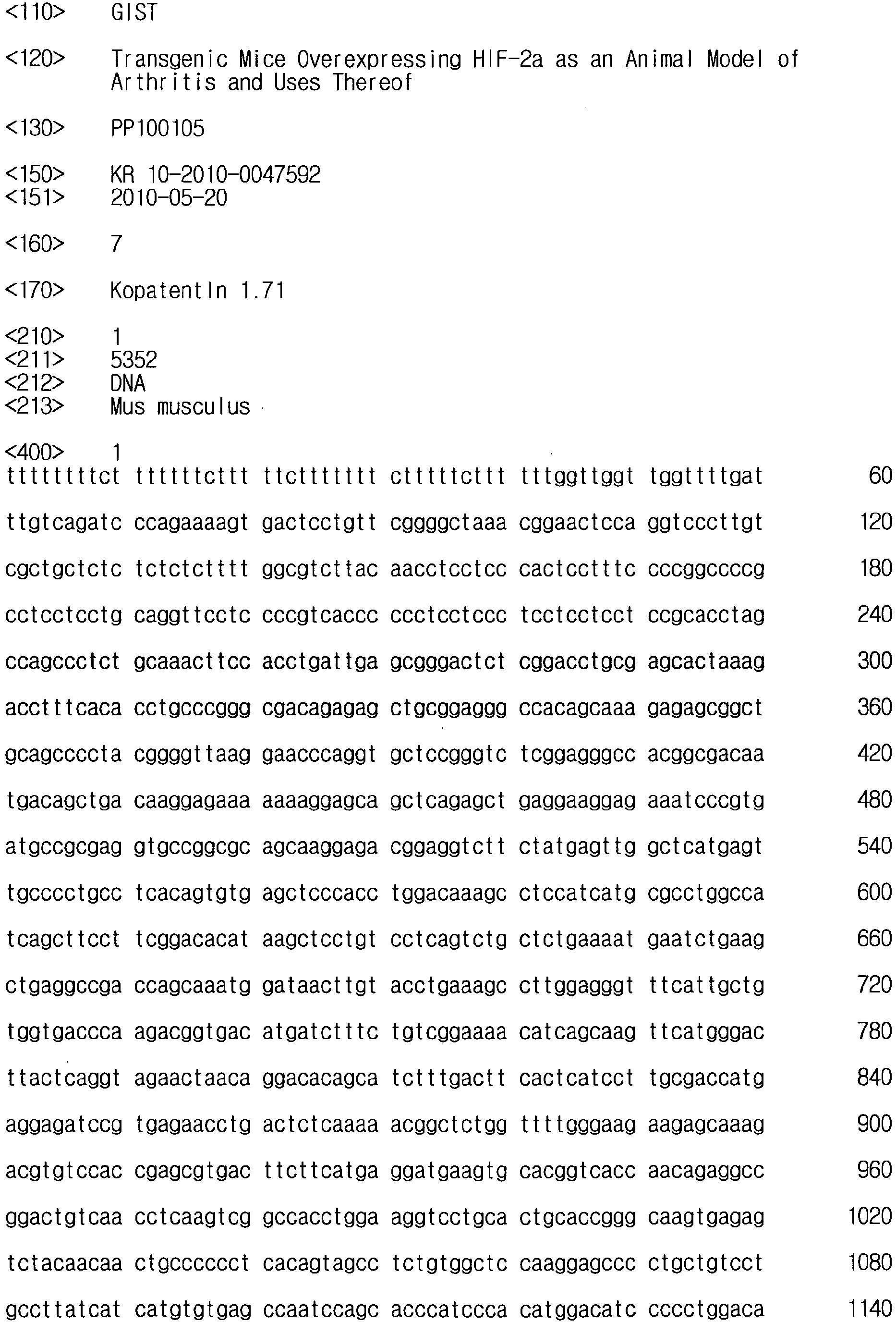


Claims (12)
- A non-human transgenic fertilized egg comprising an expression construct comprising (a) a nucleotide sequence encoding HIF-2α (hypoxia-inducible factor 2α); and (b) a transcription-regulating sequence operatively linked to the nucleotide sequence (Deposit accession number: KTCT11701BP).
- The transgenic fertilized egg according to claim 1, wherein the expression constructs further comprise a poly A signal causing polyadenylation at the 3'-end of a RNA molecule.
- The transgenic fertilized egg according to claim 1, wherein the transcription-regulating sequence comprises a promoter and an enhancer.
- The transgenic fertilized egg according to claim 3, wherein the promoter is a promoter of type II collagenase.
- The transgenic fertilized egg according to claim 3, wherein the enhancer is an enhancer of type II collagenase.
- The transgenic fertilized egg according to claim 1, wherein the expression construct further comprises an intron sequence between the promoter and the nucleotide sequence encoding HIF-2α.
- A transgenic animal model for arthritis obtained from the fertilized egg according to any of claims 1 to 6.
- The transgenic animal model for arthritis according to claim 7, wherein the HIF-2α in the transgenic animal for arthritis increases the expression of cartilage degeneration-inducing factor.
- The transgenic animal model for arthritis according to claim 8, wherein the cartilage degeneration-inducing factor is matrix metalloprotenase (MMP)-3, MMP-9, MMP-12, MMP-13, ADAMTS (a disintegrin and metalloproteinase with thrombospondin motifs)-4, ADAMTS-5, COX-2 (Cyclooxygenase-2) or iNOS (inducible nitric oxide synthase).
- The transgenic animal model for arthritis according to claim 7, wherein the animal is a mouse.
- A method for screening a therapeutic agent for treating arthritis, which comprises the steps of:(a) contacting a test substance of interest for analysis to the transgenic animal of claim 10 or 11; and(b) analyzing the cartilage tissue destruction level of the transgenic animal,wherein where the test substance inhibits the expression of HIF-2α, or alleviates or improves the cartilage tissue destruction, it is determined as therapeutic agent for treating arthritis.
- The method according to claim 11, wherein the arthritis is osteoarthritis, degenerative arthritis, desquamative osteochondritis, meniscal injury, articular malalignment, avascular necrosis, rheumation arthritis, juvenile idiopathic arthritis, injury, inflammatory arthritis or septic arthritis caused by infection.
Applications Claiming Priority (2)
| Application Number | Priority Date | Filing Date | Title |
|---|---|---|---|
| KR1020100047592A KR101176139B1 (en) | 2010-05-20 | 2010-05-20 | Transgenic Mice Overexpressing HIF-2α as an Animal Model of Arthritis and Uses Thereof |
| PCT/KR2010/008736 WO2011145795A1 (en) | 2010-05-20 | 2010-12-08 | Cartilage-specific hif-2α transgenic mouse as an animal model for arthritis, and use thereof |
Publications (3)
| Publication Number | Publication Date |
|---|---|
| EP2573169A1 true EP2573169A1 (en) | 2013-03-27 |
| EP2573169A4 EP2573169A4 (en) | 2013-12-25 |
| EP2573169B1 EP2573169B1 (en) | 2016-10-26 |
Family
ID=44991866
Family Applications (1)
| Application Number | Title | Priority Date | Filing Date |
|---|---|---|---|
| EP10851841.6A Not-in-force EP2573169B1 (en) | 2010-05-20 | 2010-12-08 | Cartilage-specific hif-2 transgenic mouse as an animal model for arthritis, and use thereof |
Country Status (4)
| Country | Link |
|---|---|
| US (1) | US20130102028A1 (en) |
| EP (1) | EP2573169B1 (en) |
| KR (1) | KR101176139B1 (en) |
| WO (1) | WO2011145795A1 (en) |
Families Citing this family (4)
| Publication number | Priority date | Publication date | Assignee | Title |
|---|---|---|---|---|
| CN102399822B (en) * | 2011-12-25 | 2013-12-11 | 浙江大学 | Construction and application of transgenic mouse model for expressing farnesyl pyrophosphate synthetase |
| CN104582477B (en) * | 2012-06-21 | 2018-09-21 | 社会福祉法人三星生命公益财团 | The method and application thereof for preparing patient-specific glioblastoma animal model |
| KR101599929B1 (en) * | 2013-07-29 | 2016-03-07 | 광주과학기술원 | Screening Methods of a Substance for Preventing or Treating a Joint Disease Using Nampt |
| KR102122128B1 (en) * | 2017-11-13 | 2020-06-11 | 서울대학교산학협력단 | Pharmaceutical composition comprising agent inhibiting the activity of HIF-2α for treating chondrosarcoma, or preventing recurrence and metastasis thereof |
Family Cites Families (24)
| Publication number | Priority date | Publication date | Assignee | Title |
|---|---|---|---|---|
| US481656A (en) | 1892-08-30 | Cutter or drill | ||
| US4437975A (en) | 1977-07-20 | 1984-03-20 | Mobil Oil Corporation | Manufacture of lube base stock oil |
| US5242794A (en) | 1984-12-13 | 1993-09-07 | Applied Biosystems, Inc. | Detection of specific sequences in nucleic acids |
| US4797368A (en) | 1985-03-15 | 1989-01-10 | The United States Of America As Represented By The Department Of Health And Human Services | Adeno-associated virus as eukaryotic expression vector |
| US4683202A (en) | 1985-03-28 | 1987-07-28 | Cetus Corporation | Process for amplifying nucleic acid sequences |
| US4683195A (en) | 1986-01-30 | 1987-07-28 | Cetus Corporation | Process for amplifying, detecting, and/or-cloning nucleic acid sequences |
| US5139941A (en) | 1985-10-31 | 1992-08-18 | University Of Florida Research Foundation, Inc. | AAV transduction vectors |
| US4800159A (en) | 1986-02-07 | 1989-01-24 | Cetus Corporation | Process for amplifying, detecting, and/or cloning nucleic acid sequences |
| IL86724A (en) | 1987-06-19 | 1995-01-24 | Siska Diagnostics Inc | Method and kits for the amplification and detection of nucleic acid sequences |
| WO1989001050A1 (en) | 1987-07-31 | 1989-02-09 | The Board Of Trustees Of The Leland Stanford Junior University | Selective amplification of target polynucleotide sequences |
| ATE92538T1 (en) | 1988-01-21 | 1993-08-15 | Genentech Inc | AMPLIFICATION AND DETECTION OF NUCLEIC ACID SEQUENCES. |
| CA1340807C (en) | 1988-02-24 | 1999-11-02 | Lawrence T. Malek | Nucleic acid amplification process |
| JP2650159B2 (en) | 1988-02-24 | 1997-09-03 | アクゾ・ノベル・エヌ・ベー | Nucleic acid amplification method |
| US4988617A (en) | 1988-03-25 | 1991-01-29 | California Institute Of Technology | Method of detecting a nucleotide change in nucleic acids |
| US5130238A (en) | 1988-06-24 | 1992-07-14 | Cangene Corporation | Enhanced nucleic acid amplification process |
| ATE138106T1 (en) | 1988-07-20 | 1996-06-15 | David Segev | METHOD FOR AMPLIFICATION AND DETECTION OF NUCLEIC ACID SEQUENCES |
| CA2005589C (en) | 1988-12-16 | 2001-02-06 | Akzo Nobel Nv | Self-sustained, sequence replication system |
| CA2035010C (en) | 1990-01-26 | 1996-12-10 | Keith C. Backman | Method of amplifying target nucleic acids applicable to both polymerase and ligase chain reactions |
| US5494810A (en) | 1990-05-03 | 1996-02-27 | Cornell Research Foundation, Inc. | Thermostable ligase-mediated DNA amplifications system for the detection of genetic disease |
| JPH06500471A (en) | 1990-08-24 | 1994-01-20 | ザ・ユニバーシティ・オブ・テネシー・リサーチ・コーポレーション | DNA amplification fingerprinting |
| WO1992007095A1 (en) | 1990-10-15 | 1992-04-30 | Stratagene | Arbitrarily primed polymerase chain reaction method for fingerprinting genomes |
| JP2002533055A (en) * | 1997-12-19 | 2002-10-08 | ワイス | Transgenic animal model for cartilage degenerative disease |
| US20060156419A1 (en) * | 2002-10-30 | 2006-07-13 | The Wistar Institute Of Anatomy And Biology | Mouse model for autoimmune disorders |
| JP2008289477A (en) | 2007-04-25 | 2008-12-04 | St Marianna Univ School Of Medicine | Transgenic mouse |
-
2010
- 2010-05-20 KR KR1020100047592A patent/KR101176139B1/en not_active Expired - Fee Related
- 2010-12-08 WO PCT/KR2010/008736 patent/WO2011145795A1/en not_active Ceased
- 2010-12-08 US US13/699,120 patent/US20130102028A1/en not_active Abandoned
- 2010-12-08 EP EP10851841.6A patent/EP2573169B1/en not_active Not-in-force
Non-Patent Citations (7)
| Title |
|---|
| ELSON D A ET AL: "Induction of hypervascularity without leakage or inflammation in transgenic mice overexpressing hypoxia-inducible factor-1alpha", GENES AND DEVELOPMENT, vol. 15, no. 19, 1 October 2001 (2001-10-01), pages 2520-2532, XP002478666, COLD SPRING HARBOR LABORATORY PRESS, PLAINVIEW, NY, US ISSN: 0890-9369, DOI: 10.1101/GAD.914801 * |
| KIM WILLIAM Y ET AL: "Failure to prolyl hydroxylate hypoxia-inducible factor alpha phenocopies VHL inactivation in vivo", EMBO (EUROPEAN MOLECULAR BIOLOGY ORGANIZATION) JOURNAL, vol. 25, no. 19, October 2006 (2006-10), pages 4650-4662, XP002715740, ISSN: 0261-4189 * |
| MUZ BARBARA ET AL: "Hypoxia. The role of hypoxia and HIF-dependent signalling events in rheumatoid arthritis.", ARTHRITIS RESEARCH & THERAPY 2009, vol. 11, no. 1, 201, 2009, XP002715739, ISSN: 1478-6362 * |
| See also references of WO2011145795A1 * |
| UETA CHISATO ET AL: "Skeletal malformations caused by overexpression of cbfa1 or its dominant nagative form in chondrocyte", THE JOURNAL OF CELL BIOLOGY : JCB, vol. 153, no. 1, 1 April 2001 (2001-04-01) , pages 87-99, XP002957346, THE ROCKEFELLER UNIVERSITY PRESS, US ISSN: 0021-9525, DOI: 10.1083/JCB.153.1.87 * |
| YANG S ET AL: "216 REGULATION OF MATRIX METALLOPROTEINASES EXPRESSION BY EPAS1 IN ARTICULAR CHONDROCYTES", OSTEOARTHRITIS AND CARTILAGE, vol. 16, 1 September 2008 (2008-09-01), page S102, XP025689928, BAILLIERE TINDALL, LONDON, GB ISSN: 1063-4584, DOI: 10.1016/S1063-4584(08)60261-7 [retrieved on 2008-09-01] * |
| YANG SIYOUNG ET AL: "Hypoxia-inducible factor-2 alpha is a catabolic regulator of osteoarthritic cartilage destruction", NATURE MEDICINE, vol. 16, no. 6, June 2010 (2010-06), pages 687-693, XP002715738, ISSN: 1078-8956 * |
Also Published As
| Publication number | Publication date |
|---|---|
| EP2573169A4 (en) | 2013-12-25 |
| EP2573169B1 (en) | 2016-10-26 |
| KR101176139B1 (en) | 2012-08-22 |
| KR20110128011A (en) | 2011-11-28 |
| WO2011145795A1 (en) | 2011-11-24 |
| US20130102028A1 (en) | 2013-04-25 |
Similar Documents
| Publication | Publication Date | Title |
|---|---|---|
| EP2572721B1 (en) | Pharmaceutical composition including an hif-2 inhibitor as an active ingredient for preventing or treating arthritis | |
| US20130142861A1 (en) | Compositions And Method For Detecting And Treating Abnormal Liver Homeostasis And Hepatocarcinogenesis | |
| JP5804599B2 (en) | Visceral fat type obesity test drug and its use | |
| CN103649329A (en) | Diagnostic and/or screening agents and uses therefor | |
| CA2554599A1 (en) | Novel brain natriuretic peptide variants and methods of use thereof | |
| EP2573169B1 (en) | Cartilage-specific hif-2 transgenic mouse as an animal model for arthritis, and use thereof | |
| WO2004074511A1 (en) | Diagnosis and treatment of baff-mediated autoimmune diseases and cancer | |
| WO2017195809A1 (en) | Expression inhibitor of inflammation promoting factor, screening method for active ingredient thereof, expression cassette useful for said method, diagnostic agent and diagnosis method | |
| KR101883936B1 (en) | Marker for predicting lymph node metastasis and method for predicting lymph node metastasis using the same | |
| CN110904230A (en) | Method for in vitro diagnosis or prognosis of colon cancer | |
| Qi et al. | Anaplastic, plasmablastic, and plasmacytic plasmacytomas of mice: relationships to human plasma cell neoplasms and late-stage differentiation of normal B cells | |
| WO2014154898A1 (en) | Prognosis and treatment of cancers | |
| EP2646572B1 (en) | Diagnostic and treatment of chronic heart failure | |
| US20040224312A1 (en) | Gene causative of rothmund-thomson syndrome and gene product | |
| US8309687B2 (en) | Biomarker specific for cancer | |
| RU2825840C2 (en) | Gene therapy monitoring | |
| CN110804657A (en) | Application of DIP2C gene in congenital heart disease diagnosis product | |
| KR102415395B1 (en) | Method for diagnosing early neurodevelopmental disorder using biomarker and kit using the same | |
| US20040023263A1 (en) | Method of testing for allergic diseases | |
| JP2005102694A (en) | Gene group differentially expressed in peripheral blood cells, and diagnostic and assay methods using the same | |
| JP4471803B2 (en) | Genes whose expression is upregulated in blood cells of patients with systemic lupus erythematosus and uses thereof | |
| JP2007006800A (en) | Normal human liver cell specific genes | |
| KR20230092352A (en) | Novel Biomarkers for Detecting Metastasis of Caner | |
| WO2001009319A1 (en) | Gene expressed specifically in human fetal heart muscle | |
| CA2237356A1 (en) | Methods for diagnosis and treatment of bloom's syndrome |
Legal Events
| Date | Code | Title | Description |
|---|---|---|---|
| PUAI | Public reference made under article 153(3) epc to a published international application that has entered the european phase |
Free format text: ORIGINAL CODE: 0009012 |
|
| 17P | Request for examination filed |
Effective date: 20121211 |
|
| AK | Designated contracting states |
Kind code of ref document: A1 Designated state(s): AL AT BE BG CH CY CZ DE DK EE ES FI FR GB GR HR HU IE IS IT LI LT LU LV MC MK MT NL NO PL PT RO RS SE SI SK SM TR |
|
| RIN1 | Information on inventor provided before grant (corrected) |
Inventor name: CHUN, JANG-SOO Inventor name: YANG, SI YOUNG |
|
| DAX | Request for extension of the european patent (deleted) | ||
| RIC1 | Information provided on ipc code assigned before grant |
Ipc: C07K 14/47 20060101ALI20131107BHEP Ipc: C12N 15/85 20060101ALI20131107BHEP Ipc: C12N 5/073 20100101ALI20131107BHEP Ipc: C12N 5/10 20060101AFI20131107BHEP Ipc: A01K 67/027 20060101ALI20131107BHEP |
|
| A4 | Supplementary search report drawn up and despatched |
Effective date: 20131125 |
|
| RIC1 | Information provided on ipc code assigned before grant |
Ipc: A01K 67/027 20060101ALI20131115BHEP Ipc: C12N 5/10 20060101AFI20131115BHEP Ipc: C12N 15/85 20060101ALI20131115BHEP Ipc: C07K 14/47 20060101ALI20131115BHEP Ipc: C12N 5/073 20100101ALI20131115BHEP |
|
| 17Q | First examination report despatched |
Effective date: 20150728 |
|
| GRAP | Despatch of communication of intention to grant a patent |
Free format text: ORIGINAL CODE: EPIDOSNIGR1 |
|
| INTG | Intention to grant announced |
Effective date: 20160216 |
|
| GRAP | Despatch of communication of intention to grant a patent |
Free format text: ORIGINAL CODE: EPIDOSNIGR1 |
|
| INTG | Intention to grant announced |
Effective date: 20160627 |
|
| GRAS | Grant fee paid |
Free format text: ORIGINAL CODE: EPIDOSNIGR3 |
|
| GRAA | (expected) grant |
Free format text: ORIGINAL CODE: 0009210 |
|
| AK | Designated contracting states |
Kind code of ref document: B1 Designated state(s): AL AT BE BG CH CY CZ DE DK EE ES FI FR GB GR HR HU IE IS IT LI LT LU LV MC MK MT NL NO PL PT RO RS SE SI SK SM TR |
|
| REG | Reference to a national code |
Ref country code: GB Ref legal event code: FG4D |
|
| REG | Reference to a national code |
Ref country code: CH Ref legal event code: EP |
|
| REG | Reference to a national code |
Ref country code: AT Ref legal event code: REF Ref document number: 840059 Country of ref document: AT Kind code of ref document: T Effective date: 20161115 |
|
| REG | Reference to a national code |
Ref country code: IE Ref legal event code: FG4D |
|
| REG | Reference to a national code |
Ref country code: DE Ref legal event code: R096 Ref document number: 602010037550 Country of ref document: DE |
|
| PGFP | Annual fee paid to national office [announced via postgrant information from national office to epo] |
Ref country code: GB Payment date: 20161222 Year of fee payment: 7 |
|
| REG | Reference to a national code |
Ref country code: LT Ref legal event code: MG4D |
|
| PG25 | Lapsed in a contracting state [announced via postgrant information from national office to epo] |
Ref country code: LV Free format text: LAPSE BECAUSE OF FAILURE TO SUBMIT A TRANSLATION OF THE DESCRIPTION OR TO PAY THE FEE WITHIN THE PRESCRIBED TIME-LIMIT Effective date: 20161026 |
|
| REG | Reference to a national code |
Ref country code: NL Ref legal event code: MP Effective date: 20161026 |
|
| REG | Reference to a national code |
Ref country code: AT Ref legal event code: MK05 Ref document number: 840059 Country of ref document: AT Kind code of ref document: T Effective date: 20161026 |
|
| PG25 | Lapsed in a contracting state [announced via postgrant information from national office to epo] |
Ref country code: NO Free format text: LAPSE BECAUSE OF FAILURE TO SUBMIT A TRANSLATION OF THE DESCRIPTION OR TO PAY THE FEE WITHIN THE PRESCRIBED TIME-LIMIT Effective date: 20170126 Ref country code: GR Free format text: LAPSE BECAUSE OF FAILURE TO SUBMIT A TRANSLATION OF THE DESCRIPTION OR TO PAY THE FEE WITHIN THE PRESCRIBED TIME-LIMIT Effective date: 20170127 Ref country code: SE Free format text: LAPSE BECAUSE OF FAILURE TO SUBMIT A TRANSLATION OF THE DESCRIPTION OR TO PAY THE FEE WITHIN THE PRESCRIBED TIME-LIMIT Effective date: 20161026 Ref country code: LT Free format text: LAPSE BECAUSE OF FAILURE TO SUBMIT A TRANSLATION OF THE DESCRIPTION OR TO PAY THE FEE WITHIN THE PRESCRIBED TIME-LIMIT Effective date: 20161026 |
|
| PG25 | Lapsed in a contracting state [announced via postgrant information from national office to epo] |
Ref country code: BE Free format text: LAPSE BECAUSE OF FAILURE TO SUBMIT A TRANSLATION OF THE DESCRIPTION OR TO PAY THE FEE WITHIN THE PRESCRIBED TIME-LIMIT Effective date: 20161026 Ref country code: ES Free format text: LAPSE BECAUSE OF FAILURE TO SUBMIT A TRANSLATION OF THE DESCRIPTION OR TO PAY THE FEE WITHIN THE PRESCRIBED TIME-LIMIT Effective date: 20161026 Ref country code: FI Free format text: LAPSE BECAUSE OF FAILURE TO SUBMIT A TRANSLATION OF THE DESCRIPTION OR TO PAY THE FEE WITHIN THE PRESCRIBED TIME-LIMIT Effective date: 20161026 Ref country code: HR Free format text: LAPSE BECAUSE OF FAILURE TO SUBMIT A TRANSLATION OF THE DESCRIPTION OR TO PAY THE FEE WITHIN THE PRESCRIBED TIME-LIMIT Effective date: 20161026 Ref country code: IS Free format text: LAPSE BECAUSE OF FAILURE TO SUBMIT A TRANSLATION OF THE DESCRIPTION OR TO PAY THE FEE WITHIN THE PRESCRIBED TIME-LIMIT Effective date: 20170226 Ref country code: PT Free format text: LAPSE BECAUSE OF FAILURE TO SUBMIT A TRANSLATION OF THE DESCRIPTION OR TO PAY THE FEE WITHIN THE PRESCRIBED TIME-LIMIT Effective date: 20170227 Ref country code: RS Free format text: LAPSE BECAUSE OF FAILURE TO SUBMIT A TRANSLATION OF THE DESCRIPTION OR TO PAY THE FEE WITHIN THE PRESCRIBED TIME-LIMIT Effective date: 20161026 Ref country code: NL Free format text: LAPSE BECAUSE OF FAILURE TO SUBMIT A TRANSLATION OF THE DESCRIPTION OR TO PAY THE FEE WITHIN THE PRESCRIBED TIME-LIMIT Effective date: 20161026 Ref country code: AT Free format text: LAPSE BECAUSE OF FAILURE TO SUBMIT A TRANSLATION OF THE DESCRIPTION OR TO PAY THE FEE WITHIN THE PRESCRIBED TIME-LIMIT Effective date: 20161026 Ref country code: PL Free format text: LAPSE BECAUSE OF FAILURE TO SUBMIT A TRANSLATION OF THE DESCRIPTION OR TO PAY THE FEE WITHIN THE PRESCRIBED TIME-LIMIT Effective date: 20161026 |
|
| REG | Reference to a national code |
Ref country code: DE Ref legal event code: R119 Ref document number: 602010037550 Country of ref document: DE |
|
| PG25 | Lapsed in a contracting state [announced via postgrant information from national office to epo] |
Ref country code: CZ Free format text: LAPSE BECAUSE OF FAILURE TO SUBMIT A TRANSLATION OF THE DESCRIPTION OR TO PAY THE FEE WITHIN THE PRESCRIBED TIME-LIMIT Effective date: 20161026 Ref country code: SK Free format text: LAPSE BECAUSE OF FAILURE TO SUBMIT A TRANSLATION OF THE DESCRIPTION OR TO PAY THE FEE WITHIN THE PRESCRIBED TIME-LIMIT Effective date: 20161026 Ref country code: RO Free format text: LAPSE BECAUSE OF FAILURE TO SUBMIT A TRANSLATION OF THE DESCRIPTION OR TO PAY THE FEE WITHIN THE PRESCRIBED TIME-LIMIT Effective date: 20161026 Ref country code: EE Free format text: LAPSE BECAUSE OF FAILURE TO SUBMIT A TRANSLATION OF THE DESCRIPTION OR TO PAY THE FEE WITHIN THE PRESCRIBED TIME-LIMIT Effective date: 20161026 Ref country code: DK Free format text: LAPSE BECAUSE OF FAILURE TO SUBMIT A TRANSLATION OF THE DESCRIPTION OR TO PAY THE FEE WITHIN THE PRESCRIBED TIME-LIMIT Effective date: 20161026 |
|
| REG | Reference to a national code |
Ref country code: CH Ref legal event code: PL |
|
| PG25 | Lapsed in a contracting state [announced via postgrant information from national office to epo] |
Ref country code: SM Free format text: LAPSE BECAUSE OF FAILURE TO SUBMIT A TRANSLATION OF THE DESCRIPTION OR TO PAY THE FEE WITHIN THE PRESCRIBED TIME-LIMIT Effective date: 20161026 Ref country code: IT Free format text: LAPSE BECAUSE OF FAILURE TO SUBMIT A TRANSLATION OF THE DESCRIPTION OR TO PAY THE FEE WITHIN THE PRESCRIBED TIME-LIMIT Effective date: 20161026 Ref country code: BG Free format text: LAPSE BECAUSE OF FAILURE TO SUBMIT A TRANSLATION OF THE DESCRIPTION OR TO PAY THE FEE WITHIN THE PRESCRIBED TIME-LIMIT Effective date: 20170126 |
|
| PLBE | No opposition filed within time limit |
Free format text: ORIGINAL CODE: 0009261 |
|
| STAA | Information on the status of an ep patent application or granted ep patent |
Free format text: STATUS: NO OPPOSITION FILED WITHIN TIME LIMIT |
|
| PG25 | Lapsed in a contracting state [announced via postgrant information from national office to epo] |
Ref country code: MC Free format text: LAPSE BECAUSE OF FAILURE TO SUBMIT A TRANSLATION OF THE DESCRIPTION OR TO PAY THE FEE WITHIN THE PRESCRIBED TIME-LIMIT Effective date: 20161026 |
|
| REG | Reference to a national code |
Ref country code: FR Ref legal event code: ST Effective date: 20170831 |
|
| 26N | No opposition filed |
Effective date: 20170727 |
|
| REG | Reference to a national code |
Ref country code: IE Ref legal event code: MM4A |
|
| PG25 | Lapsed in a contracting state [announced via postgrant information from national office to epo] |
Ref country code: LU Free format text: LAPSE BECAUSE OF NON-PAYMENT OF DUE FEES Effective date: 20161208 Ref country code: CH Free format text: LAPSE BECAUSE OF NON-PAYMENT OF DUE FEES Effective date: 20161231 Ref country code: FR Free format text: LAPSE BECAUSE OF NON-PAYMENT OF DUE FEES Effective date: 20170102 Ref country code: LI Free format text: LAPSE BECAUSE OF NON-PAYMENT OF DUE FEES Effective date: 20161231 |
|
| PG25 | Lapsed in a contracting state [announced via postgrant information from national office to epo] |
Ref country code: IE Free format text: LAPSE BECAUSE OF NON-PAYMENT OF DUE FEES Effective date: 20161208 Ref country code: SI Free format text: LAPSE BECAUSE OF FAILURE TO SUBMIT A TRANSLATION OF THE DESCRIPTION OR TO PAY THE FEE WITHIN THE PRESCRIBED TIME-LIMIT Effective date: 20161026 Ref country code: DE Free format text: LAPSE BECAUSE OF NON-PAYMENT OF DUE FEES Effective date: 20170701 |
|
| PG25 | Lapsed in a contracting state [announced via postgrant information from national office to epo] |
Ref country code: CY Free format text: LAPSE BECAUSE OF FAILURE TO SUBMIT A TRANSLATION OF THE DESCRIPTION OR TO PAY THE FEE WITHIN THE PRESCRIBED TIME-LIMIT Effective date: 20161026 Ref country code: HU Free format text: LAPSE BECAUSE OF FAILURE TO SUBMIT A TRANSLATION OF THE DESCRIPTION OR TO PAY THE FEE WITHIN THE PRESCRIBED TIME-LIMIT; INVALID AB INITIO Effective date: 20101208 |
|
| PG25 | Lapsed in a contracting state [announced via postgrant information from national office to epo] |
Ref country code: TR Free format text: LAPSE BECAUSE OF FAILURE TO SUBMIT A TRANSLATION OF THE DESCRIPTION OR TO PAY THE FEE WITHIN THE PRESCRIBED TIME-LIMIT Effective date: 20161026 Ref country code: MK Free format text: LAPSE BECAUSE OF FAILURE TO SUBMIT A TRANSLATION OF THE DESCRIPTION OR TO PAY THE FEE WITHIN THE PRESCRIBED TIME-LIMIT Effective date: 20161026 |
|
| GBPC | Gb: european patent ceased through non-payment of renewal fee |
Effective date: 20171208 |
|
| PG25 | Lapsed in a contracting state [announced via postgrant information from national office to epo] |
Ref country code: MT Free format text: LAPSE BECAUSE OF NON-PAYMENT OF DUE FEES Effective date: 20161208 |
|
| PG25 | Lapsed in a contracting state [announced via postgrant information from national office to epo] |
Ref country code: GB Free format text: LAPSE BECAUSE OF NON-PAYMENT OF DUE FEES Effective date: 20171208 |
|
| PG25 | Lapsed in a contracting state [announced via postgrant information from national office to epo] |
Ref country code: AL Free format text: LAPSE BECAUSE OF FAILURE TO SUBMIT A TRANSLATION OF THE DESCRIPTION OR TO PAY THE FEE WITHIN THE PRESCRIBED TIME-LIMIT Effective date: 20161026 |