EP2567659A1 - Calibration free dual energy radiography method. - Google Patents
Calibration free dual energy radiography method. Download PDFInfo
- Publication number
- EP2567659A1 EP2567659A1 EP11180932A EP11180932A EP2567659A1 EP 2567659 A1 EP2567659 A1 EP 2567659A1 EP 11180932 A EP11180932 A EP 11180932A EP 11180932 A EP11180932 A EP 11180932A EP 2567659 A1 EP2567659 A1 EP 2567659A1
- Authority
- EP
- European Patent Office
- Prior art keywords
- image
- log
- bone
- weight parameter
- pixels
- Prior art date
- Legal status (The legal status is an assumption and is not a legal conclusion. Google has not performed a legal analysis and makes no representation as to the accuracy of the status listed.)
- Granted
Links
Images


Classifications
-
- A—HUMAN NECESSITIES
- A61—MEDICAL OR VETERINARY SCIENCE; HYGIENE
- A61B—DIAGNOSIS; SURGERY; IDENTIFICATION
- A61B6/00—Apparatus or devices for radiation diagnosis; Apparatus or devices for radiation diagnosis combined with radiation therapy equipment
- A61B6/52—Devices using data or image processing specially adapted for radiation diagnosis
- A61B6/5211—Devices using data or image processing specially adapted for radiation diagnosis involving processing of medical diagnostic data
- A61B6/5229—Devices using data or image processing specially adapted for radiation diagnosis involving processing of medical diagnostic data combining image data of a patient, e.g. combining a functional image with an anatomical image
- A61B6/5235—Devices using data or image processing specially adapted for radiation diagnosis involving processing of medical diagnostic data combining image data of a patient, e.g. combining a functional image with an anatomical image combining images from the same or different ionising radiation imaging techniques, e.g. PET and CT
- A61B6/5241—Devices using data or image processing specially adapted for radiation diagnosis involving processing of medical diagnostic data combining image data of a patient, e.g. combining a functional image with an anatomical image combining images from the same or different ionising radiation imaging techniques, e.g. PET and CT combining overlapping images of the same imaging modality, e.g. by stitching
-
- A—HUMAN NECESSITIES
- A61—MEDICAL OR VETERINARY SCIENCE; HYGIENE
- A61B—DIAGNOSIS; SURGERY; IDENTIFICATION
- A61B6/00—Apparatus or devices for radiation diagnosis; Apparatus or devices for radiation diagnosis combined with radiation therapy equipment
- A61B6/02—Arrangements for diagnosis sequentially in different planes; Stereoscopic radiation diagnosis
- A61B6/03—Computed tomography [CT]
- A61B6/032—Transmission computed tomography [CT]
-
- A—HUMAN NECESSITIES
- A61—MEDICAL OR VETERINARY SCIENCE; HYGIENE
- A61B—DIAGNOSIS; SURGERY; IDENTIFICATION
- A61B6/00—Apparatus or devices for radiation diagnosis; Apparatus or devices for radiation diagnosis combined with radiation therapy equipment
- A61B6/48—Diagnostic techniques
- A61B6/482—Diagnostic techniques involving multiple energy imaging
-
- A—HUMAN NECESSITIES
- A61—MEDICAL OR VETERINARY SCIENCE; HYGIENE
- A61B—DIAGNOSIS; SURGERY; IDENTIFICATION
- A61B6/00—Apparatus or devices for radiation diagnosis; Apparatus or devices for radiation diagnosis combined with radiation therapy equipment
- A61B6/50—Apparatus or devices for radiation diagnosis; Apparatus or devices for radiation diagnosis combined with radiation therapy equipment specially adapted for specific body parts; specially adapted for specific clinical applications
- A61B6/505—Apparatus or devices for radiation diagnosis; Apparatus or devices for radiation diagnosis combined with radiation therapy equipment specially adapted for specific body parts; specially adapted for specific clinical applications for diagnosis of bone
-
- A—HUMAN NECESSITIES
- A61—MEDICAL OR VETERINARY SCIENCE; HYGIENE
- A61B—DIAGNOSIS; SURGERY; IDENTIFICATION
- A61B6/00—Apparatus or devices for radiation diagnosis; Apparatus or devices for radiation diagnosis combined with radiation therapy equipment
- A61B6/52—Devices using data or image processing specially adapted for radiation diagnosis
- A61B6/5211—Devices using data or image processing specially adapted for radiation diagnosis involving processing of medical diagnostic data
- A61B6/5229—Devices using data or image processing specially adapted for radiation diagnosis involving processing of medical diagnostic data combining image data of a patient, e.g. combining a functional image with an anatomical image
-
- A—HUMAN NECESSITIES
- A61—MEDICAL OR VETERINARY SCIENCE; HYGIENE
- A61B—DIAGNOSIS; SURGERY; IDENTIFICATION
- A61B6/00—Apparatus or devices for radiation diagnosis; Apparatus or devices for radiation diagnosis combined with radiation therapy equipment
- A61B6/52—Devices using data or image processing specially adapted for radiation diagnosis
- A61B6/5211—Devices using data or image processing specially adapted for radiation diagnosis involving processing of medical diagnostic data
- A61B6/5229—Devices using data or image processing specially adapted for radiation diagnosis involving processing of medical diagnostic data combining image data of a patient, e.g. combining a functional image with an anatomical image
- A61B6/5235—Devices using data or image processing specially adapted for radiation diagnosis involving processing of medical diagnostic data combining image data of a patient, e.g. combining a functional image with an anatomical image combining images from the same or different ionising radiation imaging techniques, e.g. PET and CT
-
- G—PHYSICS
- G06—COMPUTING OR CALCULATING; COUNTING
- G06T—IMAGE DATA PROCESSING OR GENERATION, IN GENERAL
- G06T5/00—Image enhancement or restoration
- G06T5/50—Image enhancement or restoration using two or more images, e.g. averaging or subtraction
-
- G—PHYSICS
- G06—COMPUTING OR CALCULATING; COUNTING
- G06T—IMAGE DATA PROCESSING OR GENERATION, IN GENERAL
- G06T2207/00—Indexing scheme for image analysis or image enhancement
- G06T2207/10—Image acquisition modality
- G06T2207/10116—X-ray image
-
- G—PHYSICS
- G06—COMPUTING OR CALCULATING; COUNTING
- G06T—IMAGE DATA PROCESSING OR GENERATION, IN GENERAL
- G06T2207/00—Indexing scheme for image analysis or image enhancement
- G06T2207/20—Special algorithmic details
- G06T2207/20212—Image combination
- G06T2207/20224—Image subtraction
-
- G—PHYSICS
- G06—COMPUTING OR CALCULATING; COUNTING
- G06T—IMAGE DATA PROCESSING OR GENERATION, IN GENERAL
- G06T2207/00—Indexing scheme for image analysis or image enhancement
- G06T2207/30—Subject of image; Context of image processing
- G06T2207/30004—Biomedical image processing
- G06T2207/30008—Bone
Definitions
- the present invention relates to dual energy radiography and more particularly to an automatic, calibration free method for dual energy material decomposition in digital radiography.
- Dual energy radiography is a technique that involves taking two radiographic images of an object at different radiation energy. A high energy image and a low energy image are combined to form images which highlight different material compositions in the object. Typically in medical imaging applications, bone or soft-tissue images are reconstructed.
- a first category of algorithms decomposes the high and low energy images into equivalent thicknesses of basis materials. These methods represent the attenuation coefficient as a weighted sum of non-linear basis functions with the weights representing the basis material thicknesses. This technique requires an extensive calibration procedure whereby the high and low energy pixel values are measured for different kVp pairs using a calibration phantom containing a range of thicknesses for both basis materials, usually aluminum and Lucite.
- a second category of algorithms compute the decomposition images as a linear weighted log-subtraction of the low and high energy images. In this straightforward approach it is assumed that images are generated using mono-energetic spectra and that the imaged object consists of 2 materials, bone and soft tissue.
- the signal at pixel (x,y) in the low and high energy images is given as follows: P L ⁇ Exp ⁇ - ⁇ S , L ⁇ z S - ⁇ B , L ⁇ z B P H ⁇ Exp ⁇ - ⁇ S , H ⁇ z S - ⁇ B , H ⁇ z B With ⁇ S , L and ⁇ B , L the attenuation coefficients of soft tissue and bone for the low-energy spectrum, ⁇ S , H and ⁇ B , H the attenuation coefficients of soft tissue and bone for the high-energy spectrum and z s and Z B the thicknesses of soft tissue and bone in the patient for pixel position (x, y).
- the decomposition into a soft-tissue image and a bone image is computed based on the weighted log subtraction technique whereby the weights are computed using image content analysis without any knowledge about the exposure settings (kVp, filter material), patient size and material attenuation coefficients.
- the decomposition is a non-iterative, two step method.
- a first step the bone image is reconstructed whereby the weight parameter is obtained from the histogram of the ratios between the log-converted high and low energy image pixel values.
- the bone image is analyzed to localize the bone structures in the image. This information is used to compute the appropriate weight parameter to obtain the soft-tissue image out of the low and high energy images.
- the method of the present invention is generally implemented in the form of a computer program product adapted to carry out the method steps of the present invention when run on a signal processing means such as a computer.
- the computer program product is commonly stored in a computer readable carrier medium such as a DVD.
- the computer program product takes the form of an electric signal and can be communicated to a user through electronic communication.
- dual energy radiography 2 images are generated by an x-ray imaging system, a low energy image and a high energy image.
- Such an x-ray system typically comprises an x-ray source and collimator for generating a beam of x-rays which is directed towards the object under examination (a patient, an animal, a test phantom etc.)
- the imaging system commonly further comprises a radiation detector for capturing the radiation image of the irradiated object.
- suitable radiation detectors are a flat panel solid state radiation detector, a photostimulable phosphor screen, a conventional x-ray film etc.
- Image read out means are part of the imaging system for reading the image stored in the radiation detector and for generating a digital image.
- This image read out means may be part of the radiation detector itself (e.g. part of the flat panel solid state radiation detector) or may be separate from the detector (e.g. an image digitizer used for detecting light emitted by an exposed photostimulable phosphor screen upon stimulation and for converting into digital pixel values or a film scanner).
- the image read out means are further coupled to signal processing means in its turn coupled to a memory.
- the memory stores the digital images read out of the radiation detector and stores a computer program for execution by said signal processing means in order to perform the calculations of the present invention which are described below on the digital images.
- the signal processing means can further be coupled to a display device or to a hard copy recorder for display or hard copy recording of the images that are generated by the method of the present invention.
- the log-subtraction technique allows decomposing the low and high energy images into material specific images, e.g. a bone and soft tissue image in chest radiography:
- W B ⁇ B , H / ⁇ B,L
- W s ⁇ S , H / ⁇ S , L ) .
- the present invention provides a method to compute the appropriate weight parameters automatically in a non-iterative, calibration free way and without any knowledge about the exposure settings.
- the low and high energy images are preferably first spatially registered to reduce misalignment artifacts due to cardiac, respiratory, bowel and patient motion.
- Different rigid and non-rigid registration algorithms are known. (Spatial registration methods are e.g. described extensively in co-pending European patent applications 10169607.8 and 11151202.6 ).
- Additional preprocessing operations can be applied to the registered low and high energy image to reduce the noise, remove the anti-scatter grid line-artifacts, etc.
- An appropriate weight parameter to reconstruct the bone image can be computed out of the ratio of the pixel values of pixels of the log converted high energy image and pixel values in corresponding pixel locations in the log converted low energy image:
- Log P H / Log P L - ⁇ S , H ⁇ z S - ⁇ B , H ⁇ z B / - ⁇ S , L ⁇ z S - ⁇ B , L ⁇ z B
- An image which for each pixel comprises a value resulting from the above operation performed on corresponding pixels in the log converted low and high energy images is further on called 'ratio image'.
- Mainly 3 regions can be identified in the ratio image (see figure 1 ).
- a first group of ratios (pixel values of the ratio image) correspond with the direct exposed area.
- a second group of ratios correspond with pixels originating from only soft tissue attenuation.
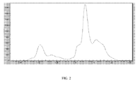
- the last group of ratios correspond with pixels originating from a combination of soft tissue and bone attenuation. This is also reflected in the histogram of the ratio values (see figure 2 ).
- the weight parameter W S is computed as the centre of the bin with the maximum counts.
- the histogram computation can be made more selective by computing a mask of pixels of interest in the image ignoring the background regions and the bright, mostly bone structures in the image. A good approximation of a mask of pixels of interest can be obtained by thresholding.
- Another embodiment is to spatially vary the weight parameter W s instead of using 1 global weight parameter in the log subtraction.
- the soft tissue attenuation ratio can vary slightly over the image due to heel effect, tissue thickness variation and scatter.
- the weight parameter W B has to be computed which represents the ratio of the attenuation coefficients of bone.
- the weight parameter W B for soft tissue image reconstruction can be computed using the information in the reconstructed bone image.
- the bone edges in the reconstructed bone image correspond with image regions where the average local gradient across the bone edge should be zero in the reconstructed soft tissue image.
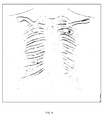
- the bone edges can e.g. be localized by applying an edge filter to the reconstructed bone image e.g. the Sobel filter (see figure 4 ).
- an edge filter to the reconstructed bone image e.g. the Sobel filter (see figure 4 ).
- Alternative methods to localise the edges are also applicable such as model fitting.
- Edge pixels with a low edge strength can be ignored as irrelevant noisy edges.
- Edge pixels with a high value of the edge strength can be ignored as they might be edges from implants or misalignment artefacts.
- the weight parameter W B is computed as the median of the ratios of the pixel differences over the bone edge pixels in the log-converted high energy image and the pixel differences in the log-converted low energy image.
- Alternative values can be envisaged such as the average value or a predefined percentile etc.
- the weight parameter W B can be spatially varied in a similar way as the computation of W S . This allows taking into account slight variations of the weight parameter W B due to the heel effect, bone thickness variation and scatter.
- the bone edge mask can be divided in regions or tiles and the weight parameter W B is then computed per region or tile. For every pixel in the image the locally most optimal weight parameter W B can be computed by interpolation.
- Post-processing operations can be applied to the reconstructed bone and soft tissue images to reduce noise, to apply scatter corrections and to convert the pixel values to values suitable for reproduction or displaying, e.g. using known multiscale image processing methods as there are multiscale gradation processing (as described in EP 1 341 125 ) and multiscale contrast enhancement processing (as described in EP 1 347 413 ).
Landscapes
- Health & Medical Sciences (AREA)
- Life Sciences & Earth Sciences (AREA)
- Engineering & Computer Science (AREA)
- Medical Informatics (AREA)
- Physics & Mathematics (AREA)
- Heart & Thoracic Surgery (AREA)
- Surgery (AREA)
- High Energy & Nuclear Physics (AREA)
- Veterinary Medicine (AREA)
- Nuclear Medicine, Radiotherapy & Molecular Imaging (AREA)
- Optics & Photonics (AREA)
- Pathology (AREA)
- Radiology & Medical Imaging (AREA)
- Biomedical Technology (AREA)
- Public Health (AREA)
- Molecular Biology (AREA)
- Biophysics (AREA)
- Animal Behavior & Ethology (AREA)
- General Health & Medical Sciences (AREA)
- Computer Vision & Pattern Recognition (AREA)
- Theoretical Computer Science (AREA)
- Pulmonology (AREA)
- Orthopedic Medicine & Surgery (AREA)
- Dentistry (AREA)
- Oral & Maxillofacial Surgery (AREA)
- General Physics & Mathematics (AREA)
- Apparatus For Radiation Diagnosis (AREA)
- Image Processing (AREA)
Abstract
Description
- The present invention relates to dual energy radiography and more particularly to an automatic, calibration free method for dual energy material decomposition in digital radiography.
- Dual energy radiography is a technique that involves taking two radiographic images of an object at different radiation energy. A high energy image and a low energy image are combined to form images which highlight different material compositions in the object. Typically in medical imaging applications, bone or soft-tissue images are reconstructed.
- There are different types of algorithms to obtain material decomposition images in dual energy radiography.
- A first category of algorithms decomposes the high and low energy images into equivalent thicknesses of basis materials. These methods represent the attenuation coefficient as a weighted sum of non-linear basis functions with the weights representing the basis material thicknesses. This technique requires an extensive calibration procedure whereby the high and low energy pixel values are measured for different kVp pairs using a calibration phantom containing a range of thicknesses for both basis materials, usually aluminum and Lucite.
- A second category of algorithms compute the decomposition images as a linear weighted log-subtraction of the low and high energy images.
In this straightforward approach it is assumed that images are generated using mono-energetic spectra and that the imaged object consists of 2 materials, bone and soft tissue.
The signal at pixel (x,y) in the low and high energy images is given as follows:

With µS,L and µB,L the attenuation coefficients of soft tissue and bone for the low-energy spectrum, µS,H and µB,H the attenuation coefficients of soft tissue and bone for the high-energy spectrum and zs and ZB the thicknesses of soft tissue and bone in the patient for pixel position (x, y). - By computing the subtraction of the log-converted images with a weight parameter w, one can generate material specific images.

If the weight parameter is defined as Ws = µS,H / µS,L a bone image is generated. In a similar way a soft tissue image is generated if the weight parameter is defined as WB = µB,H / µB,L. - In prior art the weight parameters are manually selected and adjusted by the observer during image review.
- In prior art patent
WO 02/19909 A1 - It is an object of the present invention to provide a method to compute the decomposition images (bone and soft tissue images) by means of a dual energy radiography technique based on weighted log subtraction of low and high energy images wherein the appropriate weight parameters are calculated automatically in a non-iterative, calibration free way and without any knowledge about the applied exposure settings.
- The above-mentioned aspects are realized by a method as set out in claim 1. Specific features for preferred embodiments of the invention are set out in the dependent claims.
- The decomposition into a soft-tissue image and a bone image is computed based on the weighted log subtraction technique whereby the weights are computed using image content analysis without any knowledge about the exposure settings (kVp, filter material), patient size and material attenuation coefficients.
- The decomposition is a non-iterative, two step method. In a first step the bone image is reconstructed whereby the weight parameter is obtained from the histogram of the ratios between the log-converted high and low energy image pixel values. In a second step the bone image is analyzed to localize the bone structures in the image. This information is used to compute the appropriate weight parameter to obtain the soft-tissue image out of the low and high energy images.
- The method of the present invention is generally implemented in the form of a computer program product adapted to carry out the method steps of the present invention when run on a signal processing means such as a computer. The computer program product is commonly stored in a computer readable carrier medium such as a DVD. Alternatively the computer program product takes the form of an electric signal and can be communicated to a user through electronic communication.
- Further advantages and embodiments of the present invention will become apparent from the following description and drawings.
-
-
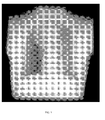
Fig. 1 shows a ratio image of a log converted low energy image and a log converted high energy image, -
Fig. 2 shows the histogram of the pixel values of the ratio image offigure 1 , -
Fig. 3 shows a reconstructed bone image, -
Fig. 4 shows the edges identified in the reconstructed bone image, -
Fig. 5 shows the reconstructed soft tissue image, - In dual energy radiography 2 images are generated by an x-ray imaging system, a low energy image and a high energy image.
- Such an x-ray system typically comprises an x-ray source and collimator for generating a beam of x-rays which is directed towards the object under examination (a patient, an animal, a test phantom etc.)
The imaging system commonly further comprises a radiation detector for capturing the radiation image of the irradiated object. Examples of suitable radiation detectors are a flat panel solid state radiation detector, a photostimulable phosphor screen, a conventional x-ray film etc. - Image read out means are part of the imaging system for reading the image stored in the radiation detector and for generating a digital image. This image read out means may be part of the radiation detector itself (e.g. part of the flat panel solid state radiation detector) or may be separate from the detector (e.g. an image digitizer used for detecting light emitted by an exposed photostimulable phosphor screen upon stimulation and for converting into digital pixel values or a film scanner).
- The image read out means are further coupled to signal processing means in its turn coupled to a memory. The memory stores the digital images read out of the radiation detector and stores a computer program for execution by said signal processing means in order to perform the calculations of the present invention which are described below on the digital images.
- The signal processing means can further be coupled to a display device or to a hard copy recorder for display or hard copy recording of the images that are generated by the method of the present invention.
- The log-subtraction technique allows decomposing the low and high energy images into material specific images, e.g. a bone and soft tissue image in chest radiography:

By choosing the appropriate weight parameters w, a soft tissue image can be reconstructed ( WB = µB,H / µB,L ) and a bone image can be reconstructed (Ws = µS,H / µS,L) . - The present invention provides a method to compute the appropriate weight parameters automatically in a non-iterative, calibration free way and without any knowledge about the exposure settings.
- In case of a dual-exposure system, the low and high energy images are preferably first spatially registered to reduce misalignment artifacts due to cardiac, respiratory, bowel and patient motion. Different rigid and non-rigid registration algorithms are known. (Spatial registration methods are e.g. described extensively in co-pending European patent applications
10169607.8 11151202.6 - Additional preprocessing operations can be applied to the registered low and high energy image to reduce the noise, remove the anti-scatter grid line-artifacts, etc.
- An appropriate weight parameter to reconstruct the bone image can be computed out of the ratio of the pixel values of pixels of the log converted high energy image and pixel values in corresponding pixel locations in the log converted low energy image:

An image which for each pixel comprises a value resulting from the above operation performed on corresponding pixels in the log converted low and high energy images is further on called 'ratio image'. - Mainly 3 regions can be identified in the ratio image (see
figure 1 ). A first group of ratios (pixel values of the ratio image) correspond with the direct exposed area. A second group of ratios correspond with pixels originating from only soft tissue attenuation. The last group of ratios correspond with pixels originating from a combination of soft tissue and bone attenuation. This is also reflected in the histogram of the ratio values (seefigure 2 ). - For the pixels originating from only soft tissue attenuation, the bone thickness ZB is 0, thus the ratio of log converted pixel values is:

This is exactly the unknown weight parameter Ws that is needed to reconstruct the bone image. - The weight parameter WS is computed as the centre of the bin with the maximum counts. As we are interested in the group of ratios corresponding with only soft tissue attenuation, the histogram computation can be made more selective by computing a mask of pixels of interest in the image ignoring the background regions and the bright, mostly bone structures in the image. A good approximation of a mask of pixels of interest can be obtained by thresholding.
- Another embodiment is to spatially vary the weight parameter Ws instead of using 1 global weight parameter in the log subtraction.
The soft tissue attenuation ratio can vary slightly over the image due to heel effect, tissue thickness variation and scatter.
By dividing the ratio image in regions or tiles and computing the weight parameter Ws per region or tile (in the way described higher), for every pixel in the image the locally most optimal weight parameter Ws can be computed by interpolation. - The result of the calculation of Pbone = Exp(Log(PH)-wsLog(PL)) is a reconstructed bone image as shown in
figure 3 . - To reconstruct the soft tissue image, the weight parameter WB has to be computed which represents the ratio of the attenuation coefficients of bone.
The weight parameter WB for soft tissue image reconstruction can be computed using the information in the reconstructed bone image.
The bone edges in the reconstructed bone image correspond with image regions where the average local gradient across the bone edge should be zero in the reconstructed soft tissue image. - The bone edges can e.g. be localized by applying an edge filter to the reconstructed bone image e.g. the Sobel filter (see
figure 4 ). Alternative methods to localise the edges are also applicable such as model fitting. - To preserve only a subset of relevant edge pixels, a selective range can be defined for the edge strengths. Edge pixels with a low edge strength can be ignored as irrelevant noisy edges. Edge pixels with a high value of the edge strength can be ignored as they might be edges from implants or misalignment artefacts.
- For every bone edge pixel it is required that there is no pixel difference across the bone edge position in the reconstructed soft tissue image.
- For every bone edge pixel 2 pixel positions are computed adjacent to the bone edge and situated on opposite sides of an edge, e.g. pixel values pairs are computed in opposite pixel positions perpendicular to the bone edge direction.
As the pixel values of these 2 pixels should be equal in the reconstructed bone image:
Or
Or
- The weight parameter WB is computed as the median of the ratios of the pixel differences over the bone edge pixels in the log-converted high energy image and the pixel differences in the log-converted low energy image.
Alternative values can be envisaged such as the average value or a predefined percentile etc. - In another embodiment the weight parameter WB can be spatially varied in a similar way as the computation of WS. This allows taking into account slight variations of the weight parameter WB due to the heel effect, bone thickness variation and scatter. The bone edge mask can be divided in regions or tiles and the weight parameter WB is then computed per region or tile. For every pixel in the image the locally most optimal weight parameter WB can be computed by interpolation.
- The result of the calculation of Psoft = Exp(Log(PH) - wBLog(PL)) is a reconstructed soft tissue image as shown in
figure 5 . - Post-processing operations can be applied to the reconstructed bone and soft tissue images to reduce noise, to apply scatter corrections and to convert the pixel values to values suitable for reproduction or displaying, e.g. using known multiscale image processing methods as there are multiscale gradation processing (as described in
EP 1 341 125 ) and multiscale contrast enhancement processing (as described inEP 1 347 413 ).
Claims (10)
- A method for generating a bone and a soft tissue image of an object by means of a dual energy radiography technique wherein said bone and soft tissue images are calculated as weighted log-subtractions of low energy and high energy images by- generating a high energy radiation image P H of said object with a first energy level of a source of radiation,- generating a low energy radiation image P L of said object with a second energy level of a source of radiation lower than said first energy level, characterized by the steps of- calculating a ratio image Log(PH)/ Log(PL),- computing at least one value of a weight parameter W S by means of pixel values of said ratio image,- generating a bone image as Pbone =Exp(Log(PH)-wSLog(PL)),- localizing bone edges in said bone image,- computing at least one value of a weight parameter WB from pixel values of pixel pairs of pixels adjacent to a bone edge and situated on opposite sides of said edge,- reconstructing a soft tissue image as Psoft =Exp(Log(PH)-wBLog(PL)) .
- A method according to claim 1 wherein said weight parameter W s is determined by- calculating the histogram of pixel values of said ratio image,- computing WS as the center of the bin with maximum counts in said histogram.
- A method according to claim 1 wherein said bone edges are localized by subjecting said bone image to an edge filtering operation.
- A method according to claim 1 wherein said weight parameter WB is calculated as an average value, a median value or a preset percentile of the ratios of differences of pixel values of said pairs of pixels in the log converted high energy image Log(PH) and in the log converted low energy image Log(PL).
- A method according to claim 1 wherein said low and high energy images are spatially registered prior to the step of calculating a ratio image.
- A method according to claim 1 wherein said low and high energy images are pre-processed prior to the step of calculating a ratio image.
- A method according to claim 2 wherein a mask of pixels of interest is computed, said mask comprising pixels of said ratio image with the exception of pixels of background regions and bright structures and wherein only masked pixels are taken in to account for the calculation of said histogram.
- A method according to claim 1 wherein said ratio image is divided into tiles and wherein a weight parameter W S and/or a weight parameter WB is calculated for each of said tiles and wherein for each pixel an optimal weight parameter is computed by interpolation between the calculated values of said weight parameters.
- A computer program product adapted to carry out the method of any of the preceding claims when run on a computer.
- A computer readable medium comprising computer executable program code adapted to carry out the steps of any of claims 1 - 9.
Priority Applications (6)
| Application Number | Priority Date | Filing Date | Title |
|---|---|---|---|
| EP11180932.3A EP2567659B1 (en) | 2011-09-12 | 2011-09-12 | Calibration free dual energy radiography method |
| ES11180932.3T ES2472454T3 (en) | 2011-09-12 | 2011-09-12 | Dual energy radiography method that does not need calibration |
| PCT/EP2012/067153 WO2013037659A1 (en) | 2011-09-12 | 2012-09-04 | Calibration free dual energy radiography method |
| US14/342,556 US9345443B2 (en) | 2011-09-12 | 2012-09-04 | Calibration free dual energy radiography method |
| BR112014005629A BR112014005629A2 (en) | 2011-09-12 | 2012-09-04 | calibration free dual energy radiography method |
| CN201280044283.9A CN103796587B (en) | 2011-09-12 | 2012-09-04 | Calibration-free dual-energy radiography method |
Applications Claiming Priority (1)
| Application Number | Priority Date | Filing Date | Title |
|---|---|---|---|
| EP11180932.3A EP2567659B1 (en) | 2011-09-12 | 2011-09-12 | Calibration free dual energy radiography method |
Publications (2)
| Publication Number | Publication Date |
|---|---|
| EP2567659A1 true EP2567659A1 (en) | 2013-03-13 |
| EP2567659B1 EP2567659B1 (en) | 2014-05-21 |
Family
ID=44582649
Family Applications (1)
| Application Number | Title | Priority Date | Filing Date |
|---|---|---|---|
| EP11180932.3A Not-in-force EP2567659B1 (en) | 2011-09-12 | 2011-09-12 | Calibration free dual energy radiography method |
Country Status (6)
| Country | Link |
|---|---|
| US (1) | US9345443B2 (en) |
| EP (1) | EP2567659B1 (en) |
| CN (1) | CN103796587B (en) |
| BR (1) | BR112014005629A2 (en) |
| ES (1) | ES2472454T3 (en) |
| WO (1) | WO2013037659A1 (en) |
Cited By (5)
| Publication number | Priority date | Publication date | Assignee | Title |
|---|---|---|---|---|
| CN104574288A (en) * | 2013-10-22 | 2015-04-29 | 辽宁开普医疗系统有限公司 | Self-adaptive grid artifact suppression method and device |
| CN105869163A (en) * | 2016-03-26 | 2016-08-17 | 南方医科大学 | Method for detecting motion artifact area based on gradient correlation between bone image and soft tissue image in dual energy subtraction |
| US10136868B2 (en) | 2015-09-03 | 2018-11-27 | General Electric Company | Fast dual energy for general radiography |
| WO2021111152A1 (en) * | 2019-12-06 | 2021-06-10 | Ibex Innovations Ltd | Composition-guided post processing for x-ray images |
| EP3806747A4 (en) * | 2018-06-15 | 2022-03-02 | Dalhousie University | METHODS AND APPARATUS FOR DUAL ENERGY X-RAY IMAGING |
Families Citing this family (13)
| Publication number | Priority date | Publication date | Assignee | Title |
|---|---|---|---|---|
| EP3034000A1 (en) | 2014-12-16 | 2016-06-22 | Agfa Healthcare | Motion correction method in dual energy radiography |
| US10631806B2 (en) * | 2015-02-19 | 2020-04-28 | Canon Medical Systems Corporation | X-ray diagnostic apparatus, image processing apparatus, and image processing method |
| JP6676337B2 (en) * | 2015-10-30 | 2020-04-08 | キヤノン株式会社 | Radiation imaging system, radiation image information processing apparatus, radiation image information processing method, and program therefor |
| EP3254623B1 (en) * | 2016-06-09 | 2020-09-16 | Agfa Nv | Method and system for correcting geometric misalignment during image reconstruction in chest tomosynthesis |
| US11000701B2 (en) * | 2017-08-01 | 2021-05-11 | Varex Imaging Corporation | Dual-layer detector for soft tissue motion tracking |
| US20220092785A1 (en) * | 2018-12-18 | 2022-03-24 | Agfa Nv | Method of decomposing a radiographic image into sub-images of different types |
| CN110448320A (en) * | 2019-08-01 | 2019-11-15 | 新里程医用加速器(无锡)有限公司 | Ball tube assembly used in CBCT system and reconstruction method of dual-energy three-dimensional volume image |
| CN114463259B (en) * | 2021-12-23 | 2025-04-29 | 深圳蓝影医学科技股份有限公司 | A method, device, equipment and storage medium for calculating dual-energy subtraction parameters |
| JP2024042609A (en) * | 2022-09-15 | 2024-03-28 | 富士フイルム株式会社 | Radiation image processing device, its operating method, and radiation image processing program |
| JP2024046543A (en) * | 2022-09-22 | 2024-04-03 | 富士フイルム株式会社 | Radiation image processing device, method and program |
| JP2024048121A (en) * | 2022-09-27 | 2024-04-08 | キヤノン株式会社 | Information processing device, radiation imaging system, information processing method and program |
| JP2025045866A (en) * | 2023-09-20 | 2025-04-02 | 富士フイルム株式会社 | Radiation image processing device, method and program |
| CN118750019A (en) * | 2024-07-12 | 2024-10-11 | 南京开德医疗科技有限公司 | A dual-energy correction method and system based on bone sclerosis model |
Citations (5)
| Publication number | Priority date | Publication date | Assignee | Title |
|---|---|---|---|---|
| WO2002019909A1 (en) | 2000-09-07 | 2002-03-14 | General Electric Company | Dual energy imaging using automatically determined subtraction parameters |
| EP1341125A2 (en) | 2002-02-22 | 2003-09-03 | Agfa-Gevaert | Multiscale gradation processing method |
| EP1347413A1 (en) | 2002-02-22 | 2003-09-24 | Agfa-Gevaert | Method for enhancing the contrast of an image. |
| US6683934B1 (en) * | 2000-06-05 | 2004-01-27 | General Electric Company | Dual energy x-ray imaging system and method for radiography and mammography |
| US20060109951A1 (en) * | 2004-10-25 | 2006-05-25 | Stefan Popescu | X-ray tomography apparatus and operating method for generating multiple energy images |
Family Cites Families (5)
| Publication number | Priority date | Publication date | Assignee | Title |
|---|---|---|---|---|
| US5148455A (en) * | 1986-07-14 | 1992-09-15 | Hologic, Inc. | Bone densitometer |
| TW393331B (en) | 1998-05-14 | 2000-06-11 | Sega Enterprises Kk | Information processing device,information processing method, information storage media and information processing system |
| EP1115120A3 (en) | 2000-01-07 | 2003-09-10 | Lucent Technologies Inc. | Method and apparatus for temperature compensation of read-only memory |
| US8019044B2 (en) * | 2008-02-15 | 2011-09-13 | Shkumat Nick A | Image acquisition for dual energy imaging |
| CN101455573B (en) * | 2009-01-08 | 2011-02-02 | 深圳市安健科技有限公司 | Method of realizing single-exposure double-energy subtraction, double-energy filter board and imaging device |
-
2011
- 2011-09-12 ES ES11180932.3T patent/ES2472454T3/en active Active
- 2011-09-12 EP EP11180932.3A patent/EP2567659B1/en not_active Not-in-force
-
2012
- 2012-09-04 US US14/342,556 patent/US9345443B2/en not_active Expired - Fee Related
- 2012-09-04 WO PCT/EP2012/067153 patent/WO2013037659A1/en not_active Ceased
- 2012-09-04 BR BR112014005629A patent/BR112014005629A2/en not_active IP Right Cessation
- 2012-09-04 CN CN201280044283.9A patent/CN103796587B/en not_active Expired - Fee Related
Patent Citations (5)
| Publication number | Priority date | Publication date | Assignee | Title |
|---|---|---|---|---|
| US6683934B1 (en) * | 2000-06-05 | 2004-01-27 | General Electric Company | Dual energy x-ray imaging system and method for radiography and mammography |
| WO2002019909A1 (en) | 2000-09-07 | 2002-03-14 | General Electric Company | Dual energy imaging using automatically determined subtraction parameters |
| EP1341125A2 (en) | 2002-02-22 | 2003-09-03 | Agfa-Gevaert | Multiscale gradation processing method |
| EP1347413A1 (en) | 2002-02-22 | 2003-09-24 | Agfa-Gevaert | Method for enhancing the contrast of an image. |
| US20060109951A1 (en) * | 2004-10-25 | 2006-05-25 | Stefan Popescu | X-ray tomography apparatus and operating method for generating multiple energy images |
Non-Patent Citations (2)
| Title |
|---|
| KASHANI H ET AL: "Development of a High-performance Dual-energy Chest Imaging System", ACADEMIC RADIOLOGY, RESTON, VA, US, vol. 16, no. 4, 1 April 2009 (2009-04-01), pages 464 - 476, XP026101419, ISSN: 1076-6332, [retrieved on 20090305], DOI: 10.1016/J.ACRA.2008.09.016 * |
| SHKUMAT N ET AL: "Optimization of image acquisition techniques for dual-energy imaging of the chest", MEDICAL PHYSICS, AIP, MELVILLE, NY, US, vol. 34, no. 10, 20 September 2007 (2007-09-20), pages 3904 - 3915, XP012103169, ISSN: 0094-2405, DOI: 10.1118/1.2777278 * |
Cited By (8)
| Publication number | Priority date | Publication date | Assignee | Title |
|---|---|---|---|---|
| CN104574288A (en) * | 2013-10-22 | 2015-04-29 | 辽宁开普医疗系统有限公司 | Self-adaptive grid artifact suppression method and device |
| US10136868B2 (en) | 2015-09-03 | 2018-11-27 | General Electric Company | Fast dual energy for general radiography |
| CN105869163A (en) * | 2016-03-26 | 2016-08-17 | 南方医科大学 | Method for detecting motion artifact area based on gradient correlation between bone image and soft tissue image in dual energy subtraction |
| CN105869163B (en) * | 2016-03-26 | 2018-12-11 | 南方医科大学 | Method based on the gradient correlation of bone picture and soft tissue picture detection motion artifacts region in dual energy subtraction |
| EP3806747A4 (en) * | 2018-06-15 | 2022-03-02 | Dalhousie University | METHODS AND APPARATUS FOR DUAL ENERGY X-RAY IMAGING |
| WO2021111152A1 (en) * | 2019-12-06 | 2021-06-10 | Ibex Innovations Ltd | Composition-guided post processing for x-ray images |
| GB2590519B (en) * | 2019-12-06 | 2024-02-21 | Ibex Innovations Ltd | Composition-guided post processing for X-ray images |
| US12456189B2 (en) | 2019-12-06 | 2025-10-28 | Ibex Innovations Ltd | Composition-guided post processing for x-ray images |
Also Published As
| Publication number | Publication date |
|---|---|
| CN103796587A (en) | 2014-05-14 |
| WO2013037659A1 (en) | 2013-03-21 |
| CN103796587B (en) | 2016-03-09 |
| BR112014005629A2 (en) | 2017-03-28 |
| EP2567659B1 (en) | 2014-05-21 |
| US9345443B2 (en) | 2016-05-24 |
| ES2472454T3 (en) | 2014-07-01 |
| US20140219423A1 (en) | 2014-08-07 |
Similar Documents
| Publication | Publication Date | Title |
|---|---|---|
| EP2567659B1 (en) | Calibration free dual energy radiography method | |
| US10235766B2 (en) | Radiographic image analysis device and method, and storage medium having stored therein program | |
| Richard et al. | Cascaded systems analysis of noise reduction algorithms in dual‐energy imaging | |
| US8054940B2 (en) | Image capturing system for medical use, image processing apparatus, and image processing method | |
| US10147168B2 (en) | Spectral CT | |
| US9036879B2 (en) | Multi-material decomposition using dual energy computed tomography | |
| EP2598032B1 (en) | Method and apparatus for processing image and medical image system employing the apparatus | |
| US7782996B2 (en) | Method for combined bone hardening and scattered radiation correction in X-ray computed tomography | |
| EP3612095B1 (en) | Beam hardening correction in x-ray dark-field imaging | |
| EP3072105B1 (en) | Processing dual energy spectral mammography images | |
| US8165379B2 (en) | Method of processing radiological images, and, in particular, mammographic images | |
| Feng et al. | An experimental method to correct low-frequency concentric artifacts in photon counting CT | |
| Kawamura et al. | Improvement in image quality and workflow of x-ray examinations using a new image processing method,“Virtual Grid Technology” | |
| Kappadath et al. | Dual‐energy digital mammography for calcification imaging: Scatter and nonuniformity corrections | |
| Shi et al. | X‐ray scatter correction for dedicated cone beam breast CT using a forward‐projection model | |
| WO2020241110A1 (en) | Image processing device, image processing method, and program | |
| JP2013119040A (en) | Medical video processing method and apparatus therefor | |
| Ferrari et al. | Digital radiographic image denoising via wavelet-based hidden Markov model estimation | |
| US20080192898A1 (en) | Renormalization of dual-energy images | |
| Lee et al. | Development of a non-linear dual-energy technique in chest radiography | |
| KR102590461B1 (en) | Method and apparatus for decomposing multi-material based on dual-energy technique | |
| Rajendran | MARS spectral CT technology for orthopaedic applications | |
| EP3773214B1 (en) | Cross directional bilateral filter for ct radiation dose reduction | |
| WO2021095447A1 (en) | Image processing device, radiography device, image processing method, and program | |
| Wong et al. | Effect of area x-ray beam equalization on image quality and dose in digital mammography |
Legal Events
| Date | Code | Title | Description |
|---|---|---|---|
| PUAI | Public reference made under article 153(3) epc to a published international application that has entered the european phase |
Free format text: ORIGINAL CODE: 0009012 |
|
| AK | Designated contracting states |
Kind code of ref document: A1 Designated state(s): AL AT BE BG CH CY CZ DE DK EE ES FI FR GB GR HR HU IE IS IT LI LT LU LV MC MK MT NL NO PL PT RO RS SE SI SK SM TR |
|
| AX | Request for extension of the european patent |
Extension state: BA ME |
|
| REG | Reference to a national code |
Ref country code: DE Ref legal event code: R079 Ref document number: 602011007159 Country of ref document: DE Free format text: PREVIOUS MAIN CLASS: A61B0006030000 Ipc: A61B0006000000 |
|
| 17P | Request for examination filed |
Effective date: 20130913 |
|
| RBV | Designated contracting states (corrected) |
Designated state(s): AL AT BE BG CH CY CZ DE DK EE ES FI FR GB GR HR HU IE IS IT LI LT LU LV MC MK MT NL NO PL PT RO RS SE SI SK SM TR |
|
| GRAP | Despatch of communication of intention to grant a patent |
Free format text: ORIGINAL CODE: EPIDOSNIGR1 |
|
| RIC1 | Information provided on ipc code assigned before grant |
Ipc: G06T 11/00 20060101ALI20131016BHEP Ipc: A61B 6/03 20060101ALI20131016BHEP Ipc: G06T 5/50 20060101ALI20131016BHEP Ipc: A61B 6/00 20060101AFI20131016BHEP Ipc: G06T 7/00 20060101ALI20131016BHEP |
|
| INTG | Intention to grant announced |
Effective date: 20131106 |
|
| GRAS | Grant fee paid |
Free format text: ORIGINAL CODE: EPIDOSNIGR3 |
|
| GRAA | (expected) grant |
Free format text: ORIGINAL CODE: 0009210 |
|
| AK | Designated contracting states |
Kind code of ref document: B1 Designated state(s): AL AT BE BG CH CY CZ DE DK EE ES FI FR GB GR HR HU IE IS IT LI LT LU LV MC MK MT NL NO PL PT RO RS SE SI SK SM TR |
|
| REG | Reference to a national code |
Ref country code: GB Ref legal event code: FG4D |
|
| REG | Reference to a national code |
Ref country code: CH Ref legal event code: EP |
|
| REG | Reference to a national code |
Ref country code: AT Ref legal event code: REF Ref document number: 669183 Country of ref document: AT Kind code of ref document: T Effective date: 20140615 |
|
| REG | Reference to a national code |
Ref country code: IE Ref legal event code: FG4D |
|
| REG | Reference to a national code |
Ref country code: ES Ref legal event code: FG2A Ref document number: 2472454 Country of ref document: ES Kind code of ref document: T3 Effective date: 20140701 |
|
| REG | Reference to a national code |
Ref country code: DE Ref legal event code: R096 Ref document number: 602011007159 Country of ref document: DE Effective date: 20140703 |
|
| REG | Reference to a national code |
Ref country code: NL Ref legal event code: VDEP Effective date: 20140521 Ref country code: AT Ref legal event code: MK05 Ref document number: 669183 Country of ref document: AT Kind code of ref document: T Effective date: 20140521 |
|
| REG | Reference to a national code |
Ref country code: LT Ref legal event code: MG4D |
|
| PG25 | Lapsed in a contracting state [announced via postgrant information from national office to epo] |
Ref country code: FI Free format text: LAPSE BECAUSE OF FAILURE TO SUBMIT A TRANSLATION OF THE DESCRIPTION OR TO PAY THE FEE WITHIN THE PRESCRIBED TIME-LIMIT Effective date: 20140521 Ref country code: IS Free format text: LAPSE BECAUSE OF FAILURE TO SUBMIT A TRANSLATION OF THE DESCRIPTION OR TO PAY THE FEE WITHIN THE PRESCRIBED TIME-LIMIT Effective date: 20140921 Ref country code: LT Free format text: LAPSE BECAUSE OF FAILURE TO SUBMIT A TRANSLATION OF THE DESCRIPTION OR TO PAY THE FEE WITHIN THE PRESCRIBED TIME-LIMIT Effective date: 20140521 Ref country code: GR Free format text: LAPSE BECAUSE OF FAILURE TO SUBMIT A TRANSLATION OF THE DESCRIPTION OR TO PAY THE FEE WITHIN THE PRESCRIBED TIME-LIMIT Effective date: 20140822 Ref country code: NO Free format text: LAPSE BECAUSE OF FAILURE TO SUBMIT A TRANSLATION OF THE DESCRIPTION OR TO PAY THE FEE WITHIN THE PRESCRIBED TIME-LIMIT Effective date: 20140821 |
|
| PG25 | Lapsed in a contracting state [announced via postgrant information from national office to epo] |
Ref country code: HR Free format text: LAPSE BECAUSE OF FAILURE TO SUBMIT A TRANSLATION OF THE DESCRIPTION OR TO PAY THE FEE WITHIN THE PRESCRIBED TIME-LIMIT Effective date: 20140521 Ref country code: SE Free format text: LAPSE BECAUSE OF FAILURE TO SUBMIT A TRANSLATION OF THE DESCRIPTION OR TO PAY THE FEE WITHIN THE PRESCRIBED TIME-LIMIT Effective date: 20140521 Ref country code: LV Free format text: LAPSE BECAUSE OF FAILURE TO SUBMIT A TRANSLATION OF THE DESCRIPTION OR TO PAY THE FEE WITHIN THE PRESCRIBED TIME-LIMIT Effective date: 20140521 Ref country code: RS Free format text: LAPSE BECAUSE OF FAILURE TO SUBMIT A TRANSLATION OF THE DESCRIPTION OR TO PAY THE FEE WITHIN THE PRESCRIBED TIME-LIMIT Effective date: 20140521 Ref country code: AT Free format text: LAPSE BECAUSE OF FAILURE TO SUBMIT A TRANSLATION OF THE DESCRIPTION OR TO PAY THE FEE WITHIN THE PRESCRIBED TIME-LIMIT Effective date: 20140521 Ref country code: PL Free format text: LAPSE BECAUSE OF FAILURE TO SUBMIT A TRANSLATION OF THE DESCRIPTION OR TO PAY THE FEE WITHIN THE PRESCRIBED TIME-LIMIT Effective date: 20140521 |
|
| PG25 | Lapsed in a contracting state [announced via postgrant information from national office to epo] |
Ref country code: PT Free format text: LAPSE BECAUSE OF FAILURE TO SUBMIT A TRANSLATION OF THE DESCRIPTION OR TO PAY THE FEE WITHIN THE PRESCRIBED TIME-LIMIT Effective date: 20140922 |
|
| PG25 | Lapsed in a contracting state [announced via postgrant information from national office to epo] |
Ref country code: CZ Free format text: LAPSE BECAUSE OF FAILURE TO SUBMIT A TRANSLATION OF THE DESCRIPTION OR TO PAY THE FEE WITHIN THE PRESCRIBED TIME-LIMIT Effective date: 20140521 Ref country code: DK Free format text: LAPSE BECAUSE OF FAILURE TO SUBMIT A TRANSLATION OF THE DESCRIPTION OR TO PAY THE FEE WITHIN THE PRESCRIBED TIME-LIMIT Effective date: 20140521 Ref country code: BE Free format text: LAPSE BECAUSE OF FAILURE TO SUBMIT A TRANSLATION OF THE DESCRIPTION OR TO PAY THE FEE WITHIN THE PRESCRIBED TIME-LIMIT Effective date: 20140521 Ref country code: EE Free format text: LAPSE BECAUSE OF FAILURE TO SUBMIT A TRANSLATION OF THE DESCRIPTION OR TO PAY THE FEE WITHIN THE PRESCRIBED TIME-LIMIT Effective date: 20140521 Ref country code: SK Free format text: LAPSE BECAUSE OF FAILURE TO SUBMIT A TRANSLATION OF THE DESCRIPTION OR TO PAY THE FEE WITHIN THE PRESCRIBED TIME-LIMIT Effective date: 20140521 Ref country code: RO Free format text: LAPSE BECAUSE OF FAILURE TO SUBMIT A TRANSLATION OF THE DESCRIPTION OR TO PAY THE FEE WITHIN THE PRESCRIBED TIME-LIMIT Effective date: 20140521 |
|
| REG | Reference to a national code |
Ref country code: DE Ref legal event code: R097 Ref document number: 602011007159 Country of ref document: DE |
|
| PG25 | Lapsed in a contracting state [announced via postgrant information from national office to epo] |
Ref country code: NL Free format text: LAPSE BECAUSE OF FAILURE TO SUBMIT A TRANSLATION OF THE DESCRIPTION OR TO PAY THE FEE WITHIN THE PRESCRIBED TIME-LIMIT Effective date: 20140521 |
|
| PLBE | No opposition filed within time limit |
Free format text: ORIGINAL CODE: 0009261 |
|
| STAA | Information on the status of an ep patent application or granted ep patent |
Free format text: STATUS: NO OPPOSITION FILED WITHIN TIME LIMIT |
|
| 26N | No opposition filed |
Effective date: 20150224 |
|
| PG25 | Lapsed in a contracting state [announced via postgrant information from national office to epo] |
Ref country code: LU Free format text: LAPSE BECAUSE OF FAILURE TO SUBMIT A TRANSLATION OF THE DESCRIPTION OR TO PAY THE FEE WITHIN THE PRESCRIBED TIME-LIMIT Effective date: 20140912 Ref country code: MC Free format text: LAPSE BECAUSE OF FAILURE TO SUBMIT A TRANSLATION OF THE DESCRIPTION OR TO PAY THE FEE WITHIN THE PRESCRIBED TIME-LIMIT Effective date: 20140521 |
|
| REG | Reference to a national code |
Ref country code: CH Ref legal event code: PL |
|
| REG | Reference to a national code |
Ref country code: DE Ref legal event code: R097 Ref document number: 602011007159 Country of ref document: DE Effective date: 20150224 |
|
| REG | Reference to a national code |
Ref country code: IE Ref legal event code: MM4A |
|
| PG25 | Lapsed in a contracting state [announced via postgrant information from national office to epo] |
Ref country code: SI Free format text: LAPSE BECAUSE OF FAILURE TO SUBMIT A TRANSLATION OF THE DESCRIPTION OR TO PAY THE FEE WITHIN THE PRESCRIBED TIME-LIMIT Effective date: 20140521 Ref country code: LI Free format text: LAPSE BECAUSE OF NON-PAYMENT OF DUE FEES Effective date: 20140930 Ref country code: CH Free format text: LAPSE BECAUSE OF NON-PAYMENT OF DUE FEES Effective date: 20140930 |
|
| PG25 | Lapsed in a contracting state [announced via postgrant information from national office to epo] |
Ref country code: IE Free format text: LAPSE BECAUSE OF NON-PAYMENT OF DUE FEES Effective date: 20140912 |
|
| PG25 | Lapsed in a contracting state [announced via postgrant information from national office to epo] |
Ref country code: SM Free format text: LAPSE BECAUSE OF FAILURE TO SUBMIT A TRANSLATION OF THE DESCRIPTION OR TO PAY THE FEE WITHIN THE PRESCRIBED TIME-LIMIT Effective date: 20140521 |
|
| PG25 | Lapsed in a contracting state [announced via postgrant information from national office to epo] |
Ref country code: MT Free format text: LAPSE BECAUSE OF FAILURE TO SUBMIT A TRANSLATION OF THE DESCRIPTION OR TO PAY THE FEE WITHIN THE PRESCRIBED TIME-LIMIT Effective date: 20140521 Ref country code: CY Free format text: LAPSE BECAUSE OF FAILURE TO SUBMIT A TRANSLATION OF THE DESCRIPTION OR TO PAY THE FEE WITHIN THE PRESCRIBED TIME-LIMIT Effective date: 20140521 Ref country code: BG Free format text: LAPSE BECAUSE OF FAILURE TO SUBMIT A TRANSLATION OF THE DESCRIPTION OR TO PAY THE FEE WITHIN THE PRESCRIBED TIME-LIMIT Effective date: 20140521 |
|
| PG25 | Lapsed in a contracting state [announced via postgrant information from national office to epo] |
Ref country code: HU Free format text: LAPSE BECAUSE OF FAILURE TO SUBMIT A TRANSLATION OF THE DESCRIPTION OR TO PAY THE FEE WITHIN THE PRESCRIBED TIME-LIMIT; INVALID AB INITIO Effective date: 20110912 Ref country code: TR Free format text: LAPSE BECAUSE OF FAILURE TO SUBMIT A TRANSLATION OF THE DESCRIPTION OR TO PAY THE FEE WITHIN THE PRESCRIBED TIME-LIMIT Effective date: 20140521 |
|
| REG | Reference to a national code |
Ref country code: FR Ref legal event code: PLFP Year of fee payment: 6 |
|
| REG | Reference to a national code |
Ref country code: FR Ref legal event code: PLFP Year of fee payment: 7 |
|
| PG25 | Lapsed in a contracting state [announced via postgrant information from national office to epo] |
Ref country code: MK Free format text: LAPSE BECAUSE OF FAILURE TO SUBMIT A TRANSLATION OF THE DESCRIPTION OR TO PAY THE FEE WITHIN THE PRESCRIBED TIME-LIMIT Effective date: 20140521 |
|
| REG | Reference to a national code |
Ref country code: FR Ref legal event code: PLFP Year of fee payment: 8 |
|
| REG | Reference to a national code |
Ref country code: GB Ref legal event code: 732E Free format text: REGISTERED BETWEEN 20180816 AND 20180822 |
|
| REG | Reference to a national code |
Ref country code: DE Ref legal event code: R081 Ref document number: 602011007159 Country of ref document: DE Owner name: AGFA NV, BE Free format text: FORMER OWNER: AGFA HEALTHCARE, MORTSEL, BE |
|
| REG | Reference to a national code |
Ref country code: ES Ref legal event code: PC2A Owner name: AGFA NV Effective date: 20181001 |
|
| PG25 | Lapsed in a contracting state [announced via postgrant information from national office to epo] |
Ref country code: AL Free format text: LAPSE BECAUSE OF FAILURE TO SUBMIT A TRANSLATION OF THE DESCRIPTION OR TO PAY THE FEE WITHIN THE PRESCRIBED TIME-LIMIT Effective date: 20140521 |
|
| PGFP | Annual fee paid to national office [announced via postgrant information from national office to epo] |
Ref country code: GB Payment date: 20230904 Year of fee payment: 13 |
|
| PGFP | Annual fee paid to national office [announced via postgrant information from national office to epo] |
Ref country code: FR Payment date: 20230904 Year of fee payment: 13 Ref country code: DE Payment date: 20230904 Year of fee payment: 13 |
|
| PGFP | Annual fee paid to national office [announced via postgrant information from national office to epo] |
Ref country code: ES Payment date: 20231013 Year of fee payment: 13 |
|
| PGFP | Annual fee paid to national office [announced via postgrant information from national office to epo] |
Ref country code: IT Payment date: 20230929 Year of fee payment: 13 |
|
| REG | Reference to a national code |
Ref country code: DE Ref legal event code: R119 Ref document number: 602011007159 Country of ref document: DE |
|
| GBPC | Gb: european patent ceased through non-payment of renewal fee |
Effective date: 20240912 |
|
| PG25 | Lapsed in a contracting state [announced via postgrant information from national office to epo] |
Ref country code: DE Free format text: LAPSE BECAUSE OF NON-PAYMENT OF DUE FEES Effective date: 20250401 |
|
| PG25 | Lapsed in a contracting state [announced via postgrant information from national office to epo] |
Ref country code: GB Free format text: LAPSE BECAUSE OF NON-PAYMENT OF DUE FEES Effective date: 20240912 |
|
| PG25 | Lapsed in a contracting state [announced via postgrant information from national office to epo] |
Ref country code: IT Free format text: LAPSE BECAUSE OF NON-PAYMENT OF DUE FEES Effective date: 20240912 |
|
| PG25 | Lapsed in a contracting state [announced via postgrant information from national office to epo] |
Ref country code: FR Free format text: LAPSE BECAUSE OF NON-PAYMENT OF DUE FEES Effective date: 20240930 |
|
| REG | Reference to a national code |
Ref country code: ES Ref legal event code: FD2A Effective date: 20251028 |
|
| PG25 | Lapsed in a contracting state [announced via postgrant information from national office to epo] |
Ref country code: ES Free format text: LAPSE BECAUSE OF NON-PAYMENT OF DUE FEES Effective date: 20240913 |