EP2564866A2 - Pharmaceutical composition using connective tissue growth factor - Google Patents
Pharmaceutical composition using connective tissue growth factor Download PDFInfo
- Publication number
- EP2564866A2 EP2564866A2 EP11775175A EP11775175A EP2564866A2 EP 2564866 A2 EP2564866 A2 EP 2564866A2 EP 11775175 A EP11775175 A EP 11775175A EP 11775175 A EP11775175 A EP 11775175A EP 2564866 A2 EP2564866 A2 EP 2564866A2
- Authority
- EP
- European Patent Office
- Prior art keywords
- seq
- amino acid
- acid sequence
- connective tissue
- tissue growth
- Prior art date
- Legal status (The legal status is an assumption and is not a legal conclusion. Google has not performed a legal analysis and makes no representation as to the accuracy of the status listed.)
- Granted
Links
Images


Classifications
-
- A—HUMAN NECESSITIES
- A61—MEDICAL OR VETERINARY SCIENCE; HYGIENE
- A61K—PREPARATIONS FOR MEDICAL, DENTAL OR TOILETRY PURPOSES
- A61K38/00—Medicinal preparations containing peptides
- A61K38/16—Peptides having more than 20 amino acids; Gastrins; Somatostatins; Melanotropins; Derivatives thereof
- A61K38/17—Peptides having more than 20 amino acids; Gastrins; Somatostatins; Melanotropins; Derivatives thereof from animals; from humans
- A61K38/18—Growth factors; Growth regulators
-
- A—HUMAN NECESSITIES
- A61—MEDICAL OR VETERINARY SCIENCE; HYGIENE
- A61P—SPECIFIC THERAPEUTIC ACTIVITY OF CHEMICAL COMPOUNDS OR MEDICINAL PREPARATIONS
- A61P1/00—Drugs for disorders of the alimentary tract or the digestive system
-
- A—HUMAN NECESSITIES
- A61—MEDICAL OR VETERINARY SCIENCE; HYGIENE
- A61P—SPECIFIC THERAPEUTIC ACTIVITY OF CHEMICAL COMPOUNDS OR MEDICINAL PREPARATIONS
- A61P1/00—Drugs for disorders of the alimentary tract or the digestive system
- A61P1/04—Drugs for disorders of the alimentary tract or the digestive system for ulcers, gastritis or reflux esophagitis, e.g. antacids, inhibitors of acid secretion, mucosal protectants
-
- A—HUMAN NECESSITIES
- A61—MEDICAL OR VETERINARY SCIENCE; HYGIENE
- A61P—SPECIFIC THERAPEUTIC ACTIVITY OF CHEMICAL COMPOUNDS OR MEDICINAL PREPARATIONS
- A61P1/00—Drugs for disorders of the alimentary tract or the digestive system
- A61P1/16—Drugs for disorders of the alimentary tract or the digestive system for liver or gallbladder disorders, e.g. hepatoprotective agents, cholagogues, litholytics
-
- A—HUMAN NECESSITIES
- A61—MEDICAL OR VETERINARY SCIENCE; HYGIENE
- A61P—SPECIFIC THERAPEUTIC ACTIVITY OF CHEMICAL COMPOUNDS OR MEDICINAL PREPARATIONS
- A61P13/00—Drugs for disorders of the urinary system
- A61P13/12—Drugs for disorders of the urinary system of the kidneys
-
- A—HUMAN NECESSITIES
- A61—MEDICAL OR VETERINARY SCIENCE; HYGIENE
- A61P—SPECIFIC THERAPEUTIC ACTIVITY OF CHEMICAL COMPOUNDS OR MEDICINAL PREPARATIONS
- A61P15/00—Drugs for genital or sexual disorders; Contraceptives
- A61P15/08—Drugs for genital or sexual disorders; Contraceptives for gonadal disorders or for enhancing fertility, e.g. inducers of ovulation or of spermatogenesis
-
- A—HUMAN NECESSITIES
- A61—MEDICAL OR VETERINARY SCIENCE; HYGIENE
- A61P—SPECIFIC THERAPEUTIC ACTIVITY OF CHEMICAL COMPOUNDS OR MEDICINAL PREPARATIONS
- A61P17/00—Drugs for dermatological disorders
- A61P17/02—Drugs for dermatological disorders for treating wounds, ulcers, burns, scars, keloids, or the like
-
- A—HUMAN NECESSITIES
- A61—MEDICAL OR VETERINARY SCIENCE; HYGIENE
- A61P—SPECIFIC THERAPEUTIC ACTIVITY OF CHEMICAL COMPOUNDS OR MEDICINAL PREPARATIONS
- A61P17/00—Drugs for dermatological disorders
- A61P17/06—Antipsoriatics
-
- A—HUMAN NECESSITIES
- A61—MEDICAL OR VETERINARY SCIENCE; HYGIENE
- A61P—SPECIFIC THERAPEUTIC ACTIVITY OF CHEMICAL COMPOUNDS OR MEDICINAL PREPARATIONS
- A61P19/00—Drugs for skeletal disorders
- A61P19/02—Drugs for skeletal disorders for joint disorders, e.g. arthritis, arthrosis
-
- A—HUMAN NECESSITIES
- A61—MEDICAL OR VETERINARY SCIENCE; HYGIENE
- A61P—SPECIFIC THERAPEUTIC ACTIVITY OF CHEMICAL COMPOUNDS OR MEDICINAL PREPARATIONS
- A61P25/00—Drugs for disorders of the nervous system
-
- A—HUMAN NECESSITIES
- A61—MEDICAL OR VETERINARY SCIENCE; HYGIENE
- A61P—SPECIFIC THERAPEUTIC ACTIVITY OF CHEMICAL COMPOUNDS OR MEDICINAL PREPARATIONS
- A61P25/00—Drugs for disorders of the nervous system
- A61P25/28—Drugs for disorders of the nervous system for treating neurodegenerative disorders of the central nervous system, e.g. nootropic agents, cognition enhancers, drugs for treating Alzheimer's disease or other forms of dementia
-
- A—HUMAN NECESSITIES
- A61—MEDICAL OR VETERINARY SCIENCE; HYGIENE
- A61P—SPECIFIC THERAPEUTIC ACTIVITY OF CHEMICAL COMPOUNDS OR MEDICINAL PREPARATIONS
- A61P27/00—Drugs for disorders of the senses
- A61P27/02—Ophthalmic agents
-
- A—HUMAN NECESSITIES
- A61—MEDICAL OR VETERINARY SCIENCE; HYGIENE
- A61P—SPECIFIC THERAPEUTIC ACTIVITY OF CHEMICAL COMPOUNDS OR MEDICINAL PREPARATIONS
- A61P27/00—Drugs for disorders of the senses
- A61P27/02—Ophthalmic agents
- A61P27/06—Antiglaucoma agents or miotics
-
- A—HUMAN NECESSITIES
- A61—MEDICAL OR VETERINARY SCIENCE; HYGIENE
- A61P—SPECIFIC THERAPEUTIC ACTIVITY OF CHEMICAL COMPOUNDS OR MEDICINAL PREPARATIONS
- A61P29/00—Non-central analgesic, antipyretic or antiinflammatory agents, e.g. antirheumatic agents; Non-steroidal antiinflammatory drugs [NSAID]
-
- A—HUMAN NECESSITIES
- A61—MEDICAL OR VETERINARY SCIENCE; HYGIENE
- A61P—SPECIFIC THERAPEUTIC ACTIVITY OF CHEMICAL COMPOUNDS OR MEDICINAL PREPARATIONS
- A61P3/00—Drugs for disorders of the metabolism
-
- A—HUMAN NECESSITIES
- A61—MEDICAL OR VETERINARY SCIENCE; HYGIENE
- A61P—SPECIFIC THERAPEUTIC ACTIVITY OF CHEMICAL COMPOUNDS OR MEDICINAL PREPARATIONS
- A61P3/00—Drugs for disorders of the metabolism
- A61P3/04—Anorexiants; Antiobesity agents
-
- A—HUMAN NECESSITIES
- A61—MEDICAL OR VETERINARY SCIENCE; HYGIENE
- A61P—SPECIFIC THERAPEUTIC ACTIVITY OF CHEMICAL COMPOUNDS OR MEDICINAL PREPARATIONS
- A61P3/00—Drugs for disorders of the metabolism
- A61P3/06—Antihyperlipidemics
-
- A—HUMAN NECESSITIES
- A61—MEDICAL OR VETERINARY SCIENCE; HYGIENE
- A61P—SPECIFIC THERAPEUTIC ACTIVITY OF CHEMICAL COMPOUNDS OR MEDICINAL PREPARATIONS
- A61P3/00—Drugs for disorders of the metabolism
- A61P3/08—Drugs for disorders of the metabolism for glucose homeostasis
- A61P3/10—Drugs for disorders of the metabolism for glucose homeostasis for hyperglycaemia, e.g. antidiabetics
-
- A—HUMAN NECESSITIES
- A61—MEDICAL OR VETERINARY SCIENCE; HYGIENE
- A61P—SPECIFIC THERAPEUTIC ACTIVITY OF CHEMICAL COMPOUNDS OR MEDICINAL PREPARATIONS
- A61P35/00—Antineoplastic agents
-
- A—HUMAN NECESSITIES
- A61—MEDICAL OR VETERINARY SCIENCE; HYGIENE
- A61P—SPECIFIC THERAPEUTIC ACTIVITY OF CHEMICAL COMPOUNDS OR MEDICINAL PREPARATIONS
- A61P35/00—Antineoplastic agents
- A61P35/04—Antineoplastic agents specific for metastasis
-
- A—HUMAN NECESSITIES
- A61—MEDICAL OR VETERINARY SCIENCE; HYGIENE
- A61P—SPECIFIC THERAPEUTIC ACTIVITY OF CHEMICAL COMPOUNDS OR MEDICINAL PREPARATIONS
- A61P37/00—Drugs for immunological or allergic disorders
- A61P37/02—Immunomodulators
- A61P37/06—Immunosuppressants, e.g. drugs for graft rejection
-
- A—HUMAN NECESSITIES
- A61—MEDICAL OR VETERINARY SCIENCE; HYGIENE
- A61P—SPECIFIC THERAPEUTIC ACTIVITY OF CHEMICAL COMPOUNDS OR MEDICINAL PREPARATIONS
- A61P43/00—Drugs for specific purposes, not provided for in groups A61P1/00-A61P41/00
-
- A—HUMAN NECESSITIES
- A61—MEDICAL OR VETERINARY SCIENCE; HYGIENE
- A61P—SPECIFIC THERAPEUTIC ACTIVITY OF CHEMICAL COMPOUNDS OR MEDICINAL PREPARATIONS
- A61P9/00—Drugs for disorders of the cardiovascular system
-
- A—HUMAN NECESSITIES
- A61—MEDICAL OR VETERINARY SCIENCE; HYGIENE
- A61P—SPECIFIC THERAPEUTIC ACTIVITY OF CHEMICAL COMPOUNDS OR MEDICINAL PREPARATIONS
- A61P9/00—Drugs for disorders of the cardiovascular system
- A61P9/10—Drugs for disorders of the cardiovascular system for treating ischaemic or atherosclerotic diseases, e.g. antianginal drugs, coronary vasodilators, drugs for myocardial infarction, retinopathy, cerebrovascula insufficiency, renal arteriosclerosis
-
- A—HUMAN NECESSITIES
- A61—MEDICAL OR VETERINARY SCIENCE; HYGIENE
- A61P—SPECIFIC THERAPEUTIC ACTIVITY OF CHEMICAL COMPOUNDS OR MEDICINAL PREPARATIONS
- A61P9/00—Drugs for disorders of the cardiovascular system
- A61P9/12—Antihypertensives
Definitions
- This disclosure relates to an angiogenesis-related pharmaceutical composition using connective tissue growth factor, more particularly to a pharmaceutical composition for promoting angiogenesis containing the connective tissue growth factor or a pharmaceutical composition for inhibiting angiogenesis containing at least one selected from the group consisting of polypeptide, antibody and a compound binding to connective tissue growth factor.
- Angiogenesis is a process wherein new capillary vessels are formed from the existing microvessels, and angiogenesis normally occurs during embryonic development, tissue regeneration and wound healing, corpus lutem development which is periodic change in female reproductive system, and even in this case, angiogenesis is stringently controlled and progressed ( Folkman J et al., Int. Rev. Exp. Pathol., 16, pp207-248, 1976 ).
- angiogenesis generally consists of decomposition of vascular basement membrane due to protease by stimulation of angiogenesis promoter, migration of vascular endothelial cells, proliferation, and tube formation by differentiation of vascular endothelial cells to reconstitute blood vessels to produce new capillary vessels.
- the diseases related to angiogenesis occurring at pathological states include hemangioma, vascular fibroma, vascular malformation, and cardiovascular disease such as atherosclerosis, vascular adhesion, scleroderma, and ophthalmic diseases caused by angiogenesis include angiogenesis due to corneal transplantation, neovascular glaucoma, diabetic retinopathy, corneal disease caused by angiogenesis, macular degeneration, pterygium, retinal degeneration, retrolental fibroplasias, granular conjunctivitis, and the like.
- Chronic inflammatory diseases such as arthritis, dermatological diseases such as psoriasis, capillarectasia, pyogenic granuloma, seborrheic dermatitis, acne, Alzheimer's diseases and obesity are also related to angiogenesis, and cancer growth and metastasis are necessarily dependent upon antiogenesis ( D'Amato RJ et al., Ophthalmology, 102(9), pp1261-1262, 1995 ; Arbiser JL, J. Am. Acad. Dermatol., 34(3), pp486-497, 1996 ; O'Brien KD et al. Circulation, 93(4), pp672-682, 1996 ; Hanahan D et al., Cell, 86, pp353-364, 1996 ).
- angiogenesis plays an important function for cancer cell growth and metastasis.
- Tumor is supplied with nutrient and oxygen required for growth through angiogenesis, and angiogenetic blood vessels penetrated into tumor provide a pathway for cancer cells to enter into blood circulation system thereby allowing metastasis of cancer cells ( Folkman and Tyler, Cancer Invasion and metastasis, Biologic mechanisms and Therapy(S.B. Day ed.) Raven press, New York, pp94-103, 1977 ; Polverini PJ, Crit. Rev. Oral. Biol. Med., 6(3), pp230-247, 1995 ).
- Angiogenesis is essential phenomenon for wound healing or tissue regeneration, for example, placenta with undeveloped blood vessel formation is an important cause of miscarriage, and necrosis, ulcer and ischemia due to non-formation of blood vessel may induce abnormal function of tissues or organs or may become a cause of death. And, diseases such as atherosclerosis, myocardial infarction and angina pectoris also become a cause of slow blood supply. Accordingly, there is a need for development of a treatment method that may decrease tissue damage due to low oxygen or low nutrition state caused by non-formation of blood vessels, and induce or promote new blood vessel formation for smooth tissue regeneration.
- VEGF-A vascular endothelial growth factor-A
- FPRL1 formyl peptide receptor-like 1
- CTGF connective tissue growth factor
- one embodiment of the present invention provides a pharmaceutical composition for promoting angiogenesis containing a fragment of protein corresponding to a FPRL1 binding region in CTGF and/or gene encoding the same; use of a fragment of protein corresponding to a FPRL1 binding region in CTGF and/or gene encoding the same for promoting angiogenesis and/or preparing angiogenesis promoter; and a method for promoting angiogenesis comprising administering a fragment of protein corresponding to a FPRL1 binding region in CTGF and/or gene encoding the same to a patient in need of promoting angiogenesis.
- Another embodiment of the present invention provides a pharmaceutical composition for inhibiting angiogenesis containing a fragment of protein corresponding to a FPRL1 binding region in CTGF and/or gene encoding the same; use of a fragment of protein corresponding to a FPRL1 binding region in CTGF and/or gene encoding the same for inhibiting angiogenesis and/or preparing angiogenesis inhibitor; and/or a method for inhibiting angiogenesis comprising administering a fragment of protein corresponding to a FPRL1 binding region in CTGF and/or gene encoding the same to a patient in need of inhibiting angiogenesis.
- Yet another embodiment of the present invention provides a method for screening angiogenesis inhibitor using a FPRL1 binding region in CTGF as a target.
- one embodiment of the invention provides a pharmaceutical composition for promoting angiogenesis containing a fragment of connective tissue growth factor protein comprising continuous 38 or more amino acids, for example continuous 38 to 140 amino acids (for example, selected from regions from 103 rd to 242 nd amino acid), continuous 38 to 100 amino acids (for example, selected from regions from 103 rd to 202 nd amino acid), or continuous 38 to 80 amino acids (for example, selected from regions from 163 rd to 242 nd amino acid), preferably continuous 38 to 39 amino acids, which comprises amino acid sequence of SEQ ID NO.
- continuous 38 to 140 amino acids for example, selected from regions from 103 rd to 242 nd amino acid
- continuous 38 to 100 amino acids for example, selected from regions from 103 rd to 202 nd amino acid
- continuous 38 to 80 amino acids for example, selected from regions from 163 rd to 242 nd amino acid
- continuous 38 to 39 amino acids which comprises amino acid sequence of SEQ ID NO.
- WVCDEPKDQTVVGPALAAYRLEDTFGPDPTMIRANCLV which is a region from 164 th to 201 st amino acid, in the amino acid sequence of SEQ ID NO. 9: use of the fragment of connective tissue growth factor protein for promoting angiogenesis and/or preparing angiogenesis promoter; and a method for promoting angiogenesis comprising administering the fragment of connective tissue growth factor protein to a patient in need of promoting angiogenesis.
- Another embodiment provides a pharmaceutical composition for promoting angiogenesis containing gene encoding a fragment of connective tissue growth factor protein comprising continuous 38 or more amino acids, for example, continuous 38 to 140 amino acids (for example, selected from regions from 103 rd to 242 nd amino acid), continuous 38 to 100 amino acids (for example, selected from regions from 103 rd to 202 nd amino acid), or continuous 38 to 80 amino acids (for example, selected from regions from 163 rd to 242 nd amino acid), preferably continuous 38 to 39 amino acids, which comprises amino acid sequence of SEQ ID NO. 5, in amino acid sequence of SEQ ID NO.
- the method for promoting angiogenesis may further include confirming a patient in need of promoting angiogenesis, before the administration, wherein the patient in need of promoting angiogenesis may be a patient requiring prevention or treatment of angina pectoris, atherosclerosis, stroke, vascular dementia, chronic ulcer, or wound.
- the patient may be mammals, preferably human.
- compositions for inhibiting angiogenesis containing at least one selected from the group consisting of polypeptide, antibody and a compound binding to a fragment of connective tissue growth factor protein comprising continuous 38 or more amino acids, for example, continuous 38 to 140 amino acids (for example, selected from regions from 103 rd to 202 nd amino acid), continuous 38 to 100 amino acids (For example, selected from regions from 103 rd to 202 nd amino acid), or continuous 38 to 80 amino acids (for example, selected from regions from 163 rd to 242 nd amino acid), preferably 38 to 39 amino acids, which comprises amino acid sequence of SEQ ID NO. 5, in amino acid sequence of SEQ ID NO.
- Yet another embodiment provides a pharmaceutical composition for inhibiting angiogenesis containing an expression inhibitor of a fragment of connective tissue growth factor comprising continuous 38 or more amino acids, for example, continuous 38 to 140 amino acids (for example, selected from regions from 103 rd to 202 nd amino acid), continuous 38 to 100 amino acids (For example, selected from regions from 103 rd to 202 nd amino acid), or continuous 38 to 80 amino acids (for example, selected from regions from 163 rd to 242 nd amino acid), preferably 38 to 39 amino acids, which comprises amino acid sequence of SEQ ID NO. 5, in amino acid sequence of SEQ ID NO.
- the expression inhibitor may be at least one selected from the group consisting of antisense nucleotide, short interfering RNA, and short hairpin RNA, which complementarily bind to the base sequence (SEQ ID NO. 7) of gene encoding the amino acid sequence of SEQ ID NO. 5 or mRNA thereof, for example, base sequence consisting of continuous 15 to 30, preferably continuous 20 to 25 bases in the mRNA.
- the method for inhibiting angiogenesis may further include confirming a patient in need of inhibiting angiogenesis, before the administration, wherein the patient in need of inhibiting angiogenesis may be a patient requiring prevention or treatment of cancer growth and metastasis, rheumatoid arthritis, psoriasis, diabetic retinopathy, diabetic nephropathy, hypertension, endometriosis, adiposis, retinopathy of prematurity, corneal inflammation rejection, neovascular glaucoma, proliferative retinopathy, hemophilic arthropathy, keloid, wound granulation, vascular adhesion, osteoarthritis, Crohn's disease, restenosis, atherosclerosis, intestinal adhesion, ulcer, liver cirrhosis, neophritis, malignant nephrosclerosis, organ transplant rejection, glomerulopathy, diabetes mellitus, or inflammation.
- the patient may be mammals, preferably human.
- Yet another embodiment provides a method for screening angiogenesis inhibitor comprising (a) treating cells comprising gene encoding a fragment of connective tissue growth factor protein comprising continuous 38 or more amino acids, for example, continuous 38 to 140 amino acids (for example, selected from regions from 103 rd to 202 nd amino acid), continuous 38 to 100 amino acids (For example, selected from regions from 103 rd to 202 nd amino acid), or continuous 38 to 80 amino acids (for example, selected from regions from 163 rd to 242 nd amino acid), preferably 38 to 39 amino acids, which comprises amino acid sequence of SEQ ID NO. 5, in amino acid sequence of SEQ ID NO.
- the composition for promoting angiogenesis contains a fragment of connective tissue growth factor protein comprising continuous 38 or more amino acids, for example, continuous 38 to 140 amino acids (for example, selected from regions from 103 rd to 242 nd amino acid), continuous 38 to 100 amino acids (for example, selected from regions from 103 rd to 202 nd amino acid), or continuous 38 to 80 amino acids (for example, selected from regions from 163 rd to 242 nd amino acid), preferably continuous 38 to 39 amino acids, which comprises amino acid sequence of SEQ ID NO. 5, in amino acid sequence of SEQ ID NO. 9.
- CTGF connective tissue growth factor
- human for example, accession no. CAG46559.1
- SEQ ID NO. 9 the connective tissue growth factor protein may bind to FPRL1 to induce angiogenesis.
- the connective tissue growth factor has activity for inducing FPRL1-specific ERK phosphorylation (see Experiment result 2), is a protein of which expression is induced by VEFG-A (see Experiment result 3), and it binds to FPRL1 to induce angiogenesis (see Experiment result 11).
- a region where the connective tissue growth factor protein binds to FPRL1 is a region from 164 th amino acid to 201 st amino acid in SEQ ID NO. 9 which is full length amino acid sequence of connective tissue growth factor protein, and it may be represented by amino acid sequence of SEQ ID NO. 5 (see Experiment result 4).
- the fragment of connective tissue growth factor protein which is an active ingredient of the pharmaceutical composition for promoting angiogenesis of the present invention, may comprise continuous 38 or more amino acids, for example, continuous 38 to 140 amino acids (for example, selected from regions from 103 rd to 242 nd amino acid), continuous 38 to 100 amino acids (for example, selected from regions from 103 rd to 202 nd amino acid), or continuous 38 to 80 amino acids (for example, selected from regions from 163 rd to 242 nd amino acid), preferably continuous 38 to 39 amino acids, which comprises amino acid sequence of SEQ ID NO. 5, a region from 164 th amino acid to 201 st amino acid in SEQ ID NO. 9, in the amino acid sequence of SEQ ID NO.
- the fragment of connective tissue growth factor protein may be preferably represented by amino acid sequence of SEQ ID NO. 6 (EWVCDEPKDQTVVGPALAAYRLEDTFGPDPTMIRANCLV), but not limited thereto.
- the fragment of connective tissue growth factor protein is a binding region to FPRL1 (see Experiment result 5), effectively induces FPRL1-specific ERK phosphorylation (see Experiment result 6), activates FPRL1 to increase intracellular Ca 2+ concentration (see Experiment result 7), and finally, effectively induces angiogenesis (see Experiment result 9).
- the pharmaceutical composition for promoting angiogenesis of the present invention may contain gene encoding connective tissue growth factor protein comprising continuous 38 or more amino acids, for example, continuous 38 to 140 amino acids (for example, selected from regions from 103 rd to 242 nd amino acid), continuous 38 to 100 amino acids (for example, selected from regions from 103 rd to 202 nd amino acid), or continuous 38 to 80 amino acids (for example, selected from regions from 163 rd to 242 nd amino acid), preferably continuous 38 to 39 amino acids, which comprises amino acid sequence of SEQ ID NO.. 5, in the amino acid sequence of SEQ ID NO. 9.
- the gene may preferably encode amino acid sequence of SEQ ID NO.
- the gene may be represented by base sequence of SEQ ID NO. 7 encoding the amino acid sequence of SEQ ID NO.
- the gene may be inserted into a vector, preferably recombinant expression vector, but is not limited thereto, wherein the recombinant expression vector may comprise the gene and expression regulator including operably linked promoter, and the like.
- the recombinant expression vector may be, for example, EcoR I-INSERT-Xba I in pAB-beeTM-FH vector (including signal peptide & flag tag), but is not limited thereto.
- the vector is used for a nucleic acid molecule transferring a DNA fragment from one cell to another cell, and may be derived from those selected from the group consisting of plasmid, bacteriophage, and plant and animal virus, but is not limited thereto.
- the expression vector refers to recombinant DNA comprising aimed coding sequence and appropriate nucleic acid sequence required for expression of the coding sequence, operably linked in a specific host, and in general, nucleic acid sequence required for expression in prokaryotic cells may comprise promoter, operator (optional), and ribosome binding region, in addition to the other sequence, and eukaryotic cells use promoter, enhancer, and stop and polyadenylation signal, which are known in the art.
- the recombinant expression vector may be a genetic construct comprising operably linked essential regulator elements so as to express the gene, which may prepare aimed protein, a fragment of connective tissue growth factor in the present invention, in a suitable host cells.
- operably linked means that nucleic acid expression regulator sequence and nucleic acid sequence encoding aimed protein are functionally linked so as to perform general functions.
- promoter and nucleic acid encoding protein or RNA may be operably linked to influence on the expression of coding sequence.
- the operable link with recombinant vector may be prepared using gene recombination technology well known in the art, and site-specific DNA cleavage and ligation may be performed using enzyme, and the like generally known in the art.
- Suitable expression vector comprises expression regulatory elements such as promoter, initiation codon, stop codon, polyadehylation signal and enhancer, and the like, and it may be variously prepared according to the purpose.
- the initiation codon and stop codon should act in the individual when a genetic construct is administered, and it should be in frame with coding sequence.
- the pharmaceutical composition for promoting angiogenesis may be used for treatment of diseases caused by inhibition of angiogenesis or diseases that may be cured by promoting angiogenesis, but is not limited thereto, and for example, it may be used for treatment of angina pectoris, atherosclerosis, ischemic stroke, vascular dementia, chronic ulcer, or wound.
- a composition for prevention and/or treatment of angina pectoris, atherosclerosis, ischemic stroke, vascular dementia, chronic ulcer, or wound comprising the pharmaceutical composition for promoting angiogenesis as an active ingredient.
- the pharmaceutical composition for inhibiting angiogenesis contains at least one selected from the group consisting of polypeptide, antibody and a compound, which bind to a fragment of connective tissue growth factor protein comprising continuous 38 or more amino acids, for example, continuous 38 to 140 amino acids (for example, selected from regions from 103 rd to 242 nd amino acid), continuous 38 to 100 amino acids (for example, selected from regions from 103 rd to 202 nd amino acid), or continuous 38 to 80 amino acids (for example, selected from regions from 163 rd to 242 nd amino acid), preferably continuous 38 to 39 amino acids, which comprises amino acid sequence of SEQ ID NO. 5, in the amino acid sequence of SEQ ID NO. 9
- a fragment of connective tissue growth factor protein comprising continuous 38 or more amino acids, for example, continuous 38 to 140 amino acids (for example, selected from regions from 103 rd to 242 nd amino acid), continuous 38 to 100 amino acids (for example, selected from regions from 103 rd to 202 nd amino acid), or continuous 38 to 80 amino acids (for example, selected from regions from 163 rd to 242 nd amino acid), preferably continuous 38 to 39 amino acids, which comprises amino acid sequence of SEQ ID NO. 5, in SEQ ID NO.
- the fragment of connective tissue growth factor may comprise continuous 38 or more amino acids, for example, continuous 38 to 140 amino acids (for example, selected from regions from 103 rd to 242 nd amino acid), continuous 38 to 100 amino acids (for example, selected from regions from 103 rd to 202 nd amino acid), or continuous 38 to 80 amino acids (for example, selected from regions from 163 rd to 242 nd amino acid), preferably continuous 38 to 39 amino acids, which comprises amino acid sequence of SEQ ID NO. 5, a 164 th to 201 st region, in the amino acid sequence of SEQ ID NO. 9 which is full length sequence of the connective tissue growth factor protein, and more preferably, it may be represented by amino acid sequence of SEQ ID NO. 6 comprising the amino acid sequence of SEQ ID NO. 5.
- the fragment of connective tissue growth factor protein comprises a FPRL1-binding region, polypeptide, antibody or a compound binding to the fragment of connective tissue growth factor protein may block binding of the connective tissue growth factor protein to FPRL1 to effectively inhibit angiogenesis.
- polypeptide, antibody or compound is not specifically limited as long as it may bind to the fragment of connective tissue growth factor protein, but preferably, it may be one binding to amino acid sequence of SEQ ID NO. 6, or SEQ ID NO. 5, and it may be easily prepared by one of ordinary knowledge in the art according to the amino acid sequence of the fragment of connective tissue growth factor protein to be bound.
- composition for inhibiting angiogenesis may contain at least one selected from the group consisting of antisense nucleotide, short interfering RNA, and short hairpin RNA, which complementarily bind to gene encoding a fragment of connective tissue growth factor protein comprising continuous 38 or more amino acids, for example, continuous 38 to 140 amino acids (for example, selected from regions from 103 rd to 242 nd amino acid), continuous 38 to 100 amino acids (for example, selected from regions from 103 rd to 202 nd amino acid), or continuous 38 to 80 amino acids (for example, selected from regions from 163 rd to 242 nd amino acid), preferably continuous 38 to 39 amino acids, comprising amino acid sequence of SEQ ID NO. 5, in the amino acid sequence of SEQ ID NO. 9, or mRNA thereof, for example, base sequence consisting of continuous 15 to 30, preferably continuous 20 to 25 bases in the mRNA.
- antisense nucleotide for example, continuous 38 to 140 amino acids (for example, selected from regions from 103 rd to
- the siRNA comprises a sense RNA strand and a complementary antisense RNA strand, the two strands anneal each other by standard Watson-Crick base pair interaction, and the sense strand comprises identical nucleic acid sequence to the target sequence in target mRNA. Technologies of selecting the target sequence of siRNA are described in, for example, Tuschl T et al, "The siRNA User Guide” revised Oct. 11, 2002 .
- the sense and antisense strands of the siRNA may comprise two complementary single-stranded RNA molecules, or it may comprise single molecule wherein two complementary parts form a base pair and covalently bonded by a "hairpin" region of single strand.
- the latter is referred to as short hairpin RNA (shRNA), wherein the shRNA is a single strand and forms a stem-loop structure in vivo.
- shRNA short hairpin RNA
- the shRNA is generally synthesized by transcription of complementary DNA sequence from Pol III promoter in vivo. Pol-III-induced transcription starts at a well-defined start site and stops at a linear (-TTTT-) second residue consisting of 4 or more thymidines to produce non-poly(A) transcript.
- Pol III promoter may be activated in all cells, and it may express shRNA. After transcription, the loop of shRNA is cleaved by dicer, and the shRNA acts with RISC (RNA-induced silencing complex) like siRNA (see, Tuschl, T. (2002), Cell 110(5): 563-74 ).
- RISC RNA-induced silencing complex
- siRNA may be obtained using many technologies known to one of ordinary knowledge in the art.
- siRNA may be chemically synthesized using a method known in the art or produced by recombination.
- the siRNA may be chemically synthesized using suitably protected ribonucleoside phosphoramidites and the existing DNA/RNA synthesizer.
- the siRNA may be synthesized as two complementary and separated RNA molecules or one RNA molecule having two complementary regions.
- the siRNA may be expressed from recombinant DNA plasmid using suitable promoter.
- the promoter suitable for expressing the siRNA from plasmid may include, for example, U6 or H1 RNA pol III promoter sequence and cytomegalovirus promoter.
- the recombinant plasmid may include inductive or controllable promoter in order to express siRNA in a specific tissue or specific intracellular environment.
- the siRNA may be expressed as two complementary and separated RNA molecule or one RNA molecule having two complementary regions from recombinant plasmid.
- suitable plasmid for expression of siRNA a method of inserting nucleic acid into plasmid to express siRNA, and a method of transferring recombinant plasmid to aimed cells are within the scope of technologies in the field to which the invention pertains. For example, see [ Tuschl, T. (2002), Nat. Biotechnol, 20: 446-448 ]: [ Brummelkamp TR et al. (2002), Science 296: 550- 553 ]: [ Miyagishi M et al. (2002), Nat. Biotechnol .
- double stranded RNA molecule with target of AAUUCACAUCGUGGUGGACAU (SEQ ID NO. 10) (for example, Sense: UUCACAUCGUGGUGGACAUdTdT (SEQ ID NO. 3) and Anti-sense: AUGUCCACCACGAUGUGAAdTdT (SEQ ID NO. 4)) may be used.
- the pharmaceutical composition for inhibiting angiogenesis may be used for prevention and/or treatment of angiogenesis-related diseases.
- the angiogenesis-related diseases may be diseases or conditions selected from the group consisting of cancer growth and metastasis, rheumatoid arthritis, psoriasis, diabetic retinopathy, diabetic nephropathy, hypertension, endometriosis, adiposis, retinopathy of prematurity, corneal inflammation rejection, neovascular glaucoma, proliferative retinopathy, hemophilic arthropathy, keloid, wound granulation, vascular adhesion, osteoarthritis, Crohn's disease, restenosis, atherosclerosis, intestinal adhesion, ulcer, liver cirrhosis, neophritis, malignant nephrosclerosis, organ transplant rejection, glomerulopathy, diabetes mellitus, inflammation, and the like.
- one embodiment of the present invention provides a composition for prevention or treatment of disease or condition selected from the group consisting of cancer growth and metastasis, rheumatoid arthritis, psoriasis, diabetic retinopathy, diabetic nephropathy, hypertension, endometriosis, adiposis, retinopathy of prematurity, corneal inflammation rejection, neovascular glaucoma, proliferative retinopathy, hemophilic arthropathy, keloid, wound granulation, vascular adhesion, osteoarthritis, Crohn's disease, restenosis, atherosclerosis, intestinal adhesion, ulcer, liver cirrhosis, neophritis, malignant nephrosclerosis, organ transplant rejection, glomerulopathy, diabetes mellitus, inflammation, and the like, containing the pharmaceutical composition for inhibiting angiogenesis as an active ingredient.
- disease or condition selected from the group consisting of cancer growth and metastasis, rheumato
- the pharmaceutically acceptable carrier may be at least one selected from a saline solution, sterile water, Ringer's solution, buffered saline, dextrose solution, maltodextrin solution, glycerol, ethanol, liposome, and a mixture thereof, and if necessary, other common additives such as antioxidant, buffer solution, bacteriostatic agent, and the like may be added.
- diluents, dispersant, surfactant, binder and lubricant may be additionally added to formulated into a dosage form for injection such as an aqueous solution, suspension, emulsion, and the like, a pill, a capsule, granules or a tablet, and a target organ-specific antibody or other ligands may be bound to the carrier so as to specifically act on a target organ.
- the pharmaceutical composition may be preferably formulated according to diseases or ingredients by suitable method in the art or using a method disclosed in Remington's Pharmaceutical Science (Recent Edition), Mack Publishing Company, Easton PA .
- the pharmaceutical composition for promoting angiogenesis or pharmaceutical composition for inhibiting angiogenesis of the present invention may be prepared for oral, topical, parenteral, intravenous, intramuscular, subcutaneous, intraocular, transdermal administration, and the like. Preferably, it may be used in an injectable form.
- the composition of the present invention may comprise a lyophilized composition that enables composition of an injectable solution upon addition of an isotonic sterile solution, sterile water or suitable physiological saline.
- Direct injection into tumor of a patient is favorable because treatment efficiency is focused on the infected tissue.
- the used dose may be controlled by various parameters, particularly, gene, vector, used administration route, disease in question or alternatively, required treatment period. And, the range may be varied according to weight, age, gender, health condition, diet of a patient, administration time, administration route, excretion rate and severance of disease, and the like.
- the daily dose may be about 0.0001 to 10 mg/kg, preferably 0.001 to 1 mg/kg, and it may be preferable to administer one time or divide into several times a day.
- a method for screening angiogenesis inhibitor comprises
- the fragment of connective tissue growth factor protein may be represented by amino acid sequence of SEQ ID NO. 5 or SEQ ID NO. 6.
- the candidate compound may be peptide, protein, a non-peptide compound, a synthetic compound, a fermentation product, cell extract, plant extract, or animal extract, but is not limited thereto, and it may include those widely known as well as novel material.
- the cells may be transformed cells with recombinant expression vector comprising the gene, and the transformation method is well known in the art.
- the degree of expression of gene may be preferably measured by immunofluorescence method, enzyme-linked immunosorbent assay (ELISA), Western Blotting or RT-PCR, but is not limited thereto.
- ELISA enzyme-linked immunosorbent assay
- a secondary antibody specific to target protein and linked to detectable label may be added, and after adding the secondary antibody, a tertiary antibody having binding affinity to the secondary antibody and linked to detectable label may be added.
- a detectable label in the secondary or tertiary antibody enzyme exhibiting coloration when cultured and appropriate coloring substrate may be used.
- the detectable part may include a composition detectable by spectroscopic, enzymatic, photochemical, bioelectronical, immunochemical, electrical, optical or chemical means, and for example, it may include fluorescent marker and dye, magnetic lable, linked enzyme, mass spectrometric tag, spin tag, electron donor and receptor, but is not limited thereto.
- the candidate compound may be selected as angiogenesis inhibitor.
- the angiogenesis inhibitor may be used for prevention or treatment of disease or condition selected from the group consisting of rheumatoid arthritis, psoriasis, diabetic retinopathy, diabetic nephropathy, hypertension, endometriosis, adiposis, cancer, retinopathy of prematurity, corneal inflammation rejection, neovascular glaucoma, proliferative retinopathy, hemophilic arthropathy, keloid, wound granulation, vascular adhesion, osteoarthritis, Crohn ' s disease, restenosis, atherosclerosis, intestinal adhesion, ulcer, liver cirrhosis, neophritis, malignant nephrosclerosis, organ transplant rejection, glomerulopathy, diabetes mellitus, inflammation, and the like, but is not limited there
- a therapeutic agent involved in the mechanism may be selected within a short time, and through experiments for the provement, useful prevention and/or treatment agent may be provided to a patient in need of prevention and/or treatment of disease caused by angiogenesis.
- the fragment of connective tissue growth factor protein which is discovered in the present invention, is a binding region to FPRL1, effectively induces FPRL1-specific ERK phosphorylation, activates FPRL1 to increase intracellular Ca 2+ concentration, and finally, effectively induces angiogenesis, and thus, the fragment of connective tissue growth factor protein may be useful for a pharmaceutical composition for promoting angiogenesis, while polypeptide, antibody or a compound binding to the fragment of connective tissue growth factor protein may be useful for a pharmaceutical composition for inhibiting angiogenesis.
- Phospho-ERK1/2 (Thr 202 /Tyr 204 ) and ERK1/2 antibody were purchased from Cell Signaling Technology Inc. (Beverly, MA, USA), and Human CTGF antibody and anti-Flag antibody were respectively purchased from Abcam (Cambridge, MA, USA) and Sigma Co. (St. Louis, MO, USA).
- Recombinant human CTGF was purchased from BioVendor Laboratory Medicine Inc. (Brno, Czech Republic).
- Recombinant human VEGF-A165, anti-VEGF-A mAb, anti-VEGFR-1 mAb, and anti-VEGFR-2 mAb were obtained from R&D Systems Inc. (Minneapolis, MN, USA).
- siRNA to FPRL1 (sense:UUCACAUCGUGGUGGACAUdTdT (SEQ ID NO. 3), anti-sense:AUGUCCACCACGAUGUGAAdTdT (SEQ ID NO. 4)) and siRNA to luciferase (sense: CGUACGCGGAAUACUUCGAdTdT (SEQ ID NO. 11), anti-sense: UCGAAGUAUUCCGCGUACGdTdT (SEQ ID NO. 12)) were synthesized in Dharmacon Research, Inc. (Chicago, IL). And, WKYMVm (SEQ ID NO. 1), WRWWWW (SEQ ID NO.
- the separated HUVECs were cultured in a dish coated with 0.2% (w/v) gelatin using Medium 199 (containing 20% (v/v) of thermally inactivated fetal bovine serum and 1%(w/v) of penicillin/streptomycin).
- FPRL1-expressing rat basophil leukemia (RBL)-2H3 FPRL1/RBL
- FPR-expressing RBL-2H3 FPR/RBL
- RBL-2H3 vector/RBL
- Conditioned medium was obtained from HUVECs (3 to 5 passages) cultured in Medium 199 that does not include FBS. The conditioned medium was centrifuged to remove residual cells, and it was stored at -80°C until used.
- Vector/RBL cells, FPR/RBL cells, FPRL1/RBL cells, and HUVECs were cultured to confluence, and serum-starved.
- the stimulated cells (1X10 5 cells) were washed with PBS twice, dissolved in 1 ml of sample buffer (50 mM Tris-HCl, 100 mM NaCl, 0.1% SDS, 1% Nonidet P-40, 50 mM NaF, 1 mM Na 3 VO 4 , 1 ⁇ g/ml aprotinin, 1 ⁇ g/ml pepstatin, and 1 ⁇ g/ml leupeptin), heated at 95°C for 30 seconds, separated with SDS-PAGE, and then, transferred to a nitrocellulose film.
- sample buffer 50 mM Tris-HCl, 100 mM NaCl, 0.1% SDS, 1% Nonidet P-40, 50 mM NaF, 1 mM Na 3 VO 4 , 1 ⁇ g/ml aprot
- Immunoblot was conducted using anti-phospho-ERK(extracellular signal regulated kinase)1/2 (Thr 202 /Tyr 204 )(Cell Signaling Technology, Beverly, MA, USA), anti-ERK1/2 (Cell Signaling Technology, Beverly, MA, USA), anti-CTGF (Abcam, Cambridge, MA, USA), anti-Actin (Sigma, St. Louis, MO, USA), or anti-Flag (Sigma, St. Louis, MO, USA) antibody, and then, the film was visualized with a chemiluminescence substrate (Amersham Pharmacia).
- CM conditioned medium
- HLB waters
- RP revere phase
- the RP C18 HPLC column (218 TP5215; 2.1 mm x 150 mm, Vydac) was equilibrated with water/0.1%(w/v) TFA.
- concentration gradient of ACN/0.1%(w/v) TFA from 0%(w/v) to 100%(w/v)
- 150 ul of fraction was obtained.
- the obtained activated fraction was treated with trypsin and cultured at 37°C overnight.
- nano-LC MS equipped with nano-ESI source and consisting of Ultimate HPLC system (LC Packings) and a QSTAR PULSAR I hybrid Q-TOF MS/MS system (Applied Biosytems/PE SCIEX) was used.
- the QSTAR was operated at resolution of 8000 to 10000 at constant mass for 24 hours.
- a spray tip was established to a voltage of 2300 V, and all the mass values detected by QSTAR were measured using Analyst QS software supplied by Applied Biosystems Inc. (AB).
- MASCOT search engine version 1.7, in-house
- the deleted variant was named as CTGF/I-II-L, CTGF/II-L, CTGF/II, CTGF/L, CTGF/L-III-IV, and CTGF/I-L-III-IV, of which specific deletions are shown in Fig. 8 (The deleted part is indicated by dotted line).
- the flag-tagged secretory protein was obtained by transient transfection of HEK 293T cells (ATCC, Manassas, VA, USA) using lipofectamine (Invitrogen, Carlsbad, CA) according to manufacturer's instruction. From HEK293T cells, full length and deleted variant Flag-CTGF were affinity-purified using anti-Flag M2 affinity gel column, and Flag peptide was eluted according to manufacturer's instruction. The purity of the protein was determined through silver staining using a Silver Stain Plus kit (Bio-Rad).
- linker polypeptide was labeled using a EZ-Link Micro Sulfo-NHS-LC-Biotinylation kit (Pierce Chemical Co., Rockford, IL, USA) according to the manufacturer's instruction.
- the linker polypeptide was cultured at room temperature for 1 hour in PBS with 9 mM sulfo-NHS-LC-biotin (Pierce Chemical Co., Rockford, IL, USA).
- vector/RBL cells, FPR/RBL cells, FPRL1/RBL cells, or HUVECs were treated with trypsin, and collected, and then, treated with rabbit serum at room temperature for 30 minutes.
- FPRL1/RBL, vector/RBL or FPR/RBL cells were cultured in a fluo-3-AM working solution containing 0.03%(w/v) plutonic F-127 (Molecular Probes) at 37°C for 1 hour so that the final concentration of fluo-3-AM became 20 uM/L.
- fluo-3-AM fluorescence occurred at 488 nm with high-power Ar + laser in the cells, and the emission band was measured at 530 nm with photomultiplier. Fluorescence signal was detected using confocal laser scanning system (LSM 510 Meta; Carl Zeiss, Jena, Germany) equipped with Nikon E-600 Eclipse microscope.
- Fluorescence intensities before adding CTGF (F 0 ) and after adding CTGF (F) were measured. Change in the intracellularar Ca 2+ concentration [Ca 2+ ] i was indicated by F/F 0 ratio. From each cell, 50 to 120 images were scanned.
- HUVECs were plate cultured in a gelatin-coated 24-well culture dish at 2 x 10 4 cells/well and allowed to adhere overnight. 4 hours after serum was depleted, the cells were treated with various mitogen (CTGF linker polypeptide, CTGF) for 48 hours.
- CTGF linker polypeptide CTGF
- [ 3 H]-thymidine (1 ⁇ Ci, Amersham International) was added to each well 6 hours before final culture. The inserted [ 3 H]-thymidine was extracted in 0.2 M NaOH and 0.1% SDS solution at 37°C for 1 hour. Radioactivity was measured using liquid scintillation counter (Beckmann Instruments), which was repeated three times, and the result was indicated by mean ⁇ standard deviation per minute.
- HUVECs inoculated on a 60-mm culture dish to confluence were wounded with the end of a pipette, and treated with linker polypeptide (10 -1 ⁇ 10 2 nM) or recombinant CTGF (recombinant human CTGF, rhCTGF; BioVendor R&D, NC, USA)(10 1 or 10 2 nM) in Medium 199 to which 1% serum and 1 mM thymidine were added. After culturing 16 hours, the number of cells that migrated over a reference line was counted to quantify the degree of migration, and the cells were photographed with 50x magnification.
- linker polypeptide 10 -1 ⁇ 10 2 nM
- CTGF recombinant human CTGF, rhCTGF; BioVendor R&D, NC, USA
- HUVECs were inoculated on the previously polymerized Matrigel(BD Biosciences) layer together with a certain amount of linker polypeptide, rhCTGF or VEGF-A 165 .
- linker polypeptide rhCTGF or VEGF-A 165 .
- the shape of the cells was observed by phase-contrast microscopy, and photographed with 40x magnification.
- image-Pro Plus v4.5 Media Cybernetics, Silver Spring, MD
- Fertilized eggs were cultured in an egg breeder set to fixed moisture at 37°C. 3 days after the culture, to take off expressed CAM from the shell, about 2 ml of egg albumin was taken out with 18-gague subcutaneous needle. After additionally culturing for 6 days, thermanox coverslips (Nunc) containing the sample was dried in the air, and applied to the CAM surface to test the activity of angiogenesis by linker polypeptide or rhCTGF. After 3 days, 1-2 ml of 10%(w/v) Intralipose was injected into chorioallantois, and observed with microscope.
- siRA to FPRL1 was used to inhibit transcription of human FPRL1 using siRNA.
- siRA to FPRL1 sense:UUCACAUCGUGGUGGACAUdTdT (SEQ ID NO. 3), anti-sense:AUGUCCACCACGAUGUGAAdTdT (SEQ ID NO. 4)
- siRNA sequence As result of BLAST search of the siRNA sequence, it was confirmed that there is no significant similarity to other sequences in the database. The oligonnucleoride showed similar results.
- HUVECs were used for siRNA transfection. The cells were transfected to the final concentration of 20 nM with FPRL1 siRNA (SEQ ID NO. 3 and SEQ ID NO. 4) or transfected with luciferase siRNA (SEQ ID NO.
- RNAs were separated from the transfected HUVECs according to the manufacturer's instruction using commercially available TRI reagent (Molecular Research Center).
- First-strand cDNA was synthesized according to the manufacturer's instruction using 3 ug of each DNA-free total RNA sample and oligo(dT) 15 and Moloney murine leukemia virus reverse transcriptase (Promega, WI, USA). And then, with 50 ul of a reaction solution containing 1x PCR buffer, 200 uM dNTPs, 10 uM of each specific primer (sFPRL1: 5'-GACCTTGGATTCTTGCTCTAGTC-3' (SEQ ID NO.
- VEGF-A Vascular endothelial growth factor-A
- G-protein-coupled receptor FPRL1 formyl peptide receptor-like 1
- CM conditioned medium
- FPRL1-overexpressing cells FPRL1/RBL were treated therewith in the concentration of 10ng/ml or 50ng/ml for 8 hours.
- FPRL1 agonistic peptide WKYMVm(Trp-Lys-Tyr-Met-Val-D-Met, SEQ ID NO. 1) was treated with 10 nM.
- Fig. 1 it can be seen that the conditioned medium obtained in HUVECs treated with VEGF-A increased ERK phophorylation in a concentration-dependent manner.
- the above described vector/RBL, FPR/RBL, or FPRL1/RBL cells were respectively treated with the above conditioned medium or VEGF-A at the concentration of 10 ng/ml for 8 hours, and positive control was treated with 10 nM of agonistic peptide WKYMVm (SEQ ID NO. 1). And then, immunoblot was conducted as described above, and the results are shown in Fig. 2 .
- conditioned medium, VEGF-A and positive control all increased ERK phosphorylation only in EPRL1-overexpressing FPRL1/RBL cells.
- ERK phosphorylation induced by the conditioned medium is specific to FPRL1.
- FPRL1/RBL cells were treated with 40ug of the HPLC fraction of the conditioned medium for 5 minutes, and the result confirming whether ERK phosphorylation occurred as described in the ⁇ Experiment result 1> was shown in Fig. 3 .
- fractions containing B12 to C4 induce ERK phosphorylation in FPRL1/RBL cells, and as result of examining the active peak of each fraction of B11 to C4, it can be seen that peaks are observed only in B12, C3 fractions.
- CTGF connective-tissue growth factor
- CTGF may bind to FPRL1 to induce ERK phosphorylation in FPRL1/RBL cells.
- RT-PCR and Western Blot analysis were conducted as described in the Experiment method. Specifically, the results of analyzing the degree of CTGF expression in cell lysate or conditioned medium of HUVECs treated with VEGF-A according to treatment time and treatment amount of VEGF-A were respectively shown in Fig. 5 and Fig. 6 .
- CTGF mRNA increases from the time when 0.5 hours have elapsed after treating HUVECs with VEGF-A
- CTGF value increases from the time when 2 hours have elapsed in the cell lysate
- CTGF value increases from the time when 4 hours have elapsed in the conditioned medium.
- CTGF value increases in proportion to the treatment amount of VEGF-A.
- VEGF-A receptor is involved in CTGF synthesis.
- HUVECs were treated with neutralizing antibodies to VEGF-A (R&D Systems, Minneapolis, MN, USA), VEGF receptor-1 (VEGFR-1) (R&D Systems, Minneapolis, MN, USA) and VEGF receptor-2 (VEGFR-2) (R&D Systems, Minneapolis, MN, USA), and as control, treated with IgG antibody at the concentration of 5 ug/ml for 30 minutes, and they were treated with VEGF-A at the concentration of 20 ng/ml. After 2 hours have elapsed, cell lysate was obtained and immunoblot was conducted with anti-human CTGF, and the result is shown in Fig. 7 .
- CTGF expression induction of VEGF-A is controlled by receptor coupling.
- plasmids in which gene encoding full length CTGF protein or partly deleted protein CTGF/I-II-L, CTGF/II-L, CTGF/II, CTGF/L, CTGF/L-III-IV, and CTGF/I-L-III-IV are inserted were prepared as shown in Fig. 8 according to the method described in the ⁇ Experiment method 6>, and transfected into HEK 293T cells (ATCC, Manassas, VA, USA), and each protein was purified.
- CTGF/II induce ERK phosphorylation.
- SEQ ID NO.5 full length CTGF protein
- CTGF linker polypeptide binds only to FPRL1/RBL cells.
- Fig. 1 it can be seen that if FPRL1/RBL, vector/RBL or FPR/RBL cells are treated with 10 uM of FPRL1 antagonist WRW4 (SEQ ID NO. 2) and cultured for 30 minutes, the linker polypeptide binding is inhibited in FPR/RBL cells.
- CTGF linker polypeptide specifically binds to FPRL1, and thereby, the linker polypeptide may increase ERK phosphorylation or Ca 2+ concentration by FPRL1.
- ERK phosphorylation induction of CTGF linker polypeptide was compared to recombinant human CTGF (rhCTGF). Specifically, FPRL1/RBL cells were treated with CTGF linker polypeptide and rhCTGF for 5 minutes at various concentrations, ERK phosphorylation was measured, and the results are shown in Fig. 11 .
- CTGF linker polypeptide induces ERK phosphorylation to a similar level to rhCTGF.
- CTGF linker polypeptide (indicated by LK) induces ERK phosphorylation to a similar level to rhCTGF (indicated by CTGF), and particularly, in case FPRL1 antagonist WRW4 is added, ERK phosphorylation is inhibited.
- CTGF directly binds to FPRL1 through the linker region, and thereby, activates FPRL1.
- HUVECs were treated with 10 nM of the CTGF linker polypeptide, and cultured for 30 minutes, and then, flow cytometry was conducted as described in the ⁇ Experiment method 7>. At this time, the cells were treated with 10 uM OF WRW4 together.
- HUVECs were treated with the CTGF linker polypeptide or rhCTGF at various concentrations for 5 minutes, cell lysate was obtained, and then, immuboblot was conducted with anti-phospho ERK1/2 antibody, and the results are shown in Fig. 14 .
- ERK phosphorylation is effectively induced, and that if treated with FPRL1 antagonist WRW4 (10 uM), ERK phosphorylation is inhibited.
- HUVECs were treated with the CTGF linker polypeptide or rhCTGF at various concentrations for 1 hour, it was confirmed whether intracellular Ca 2+ concentration increases by the above method, and the results are shown in Fig. 15 .
- CTGF linker polypeptide increases intracellular Ca 2+ concentration in a concentration-dependent manner, that's not the case when the cells are treated with FPRL1 antagonist WRW4 (10 uM),
- CTGF specifically binds to FPRL1 in HUVECs through the linker region to activate HUVECs.
- CTGF linker polypeptide induces proliferation of HUVECs in a concentration-dependent manner, the degree is not statistically significant.
- CTGF linker polypeptide induces migration of HUVECs in a concentration-dependent manner more effectively than recombinant CTGF, and furthermore, it can be seen that in case the cells are treated with FPRL1 antagonist WRW4 (10 uM), the cell migration is not induced.
- CTGF linker polypeptide induces tube formation of HUVECs more effectively than recombinant CTGF, and furthermore, it can be seen that in case the cells are treated with FPRL1 antagonist WRW4 (10 uM), the tube formation is not be induced.
- CTGF linker polypeptide induces cell migration and tube formation rather than cell proliferation more effectively than full length CTGF, it may promote angiogenesis.
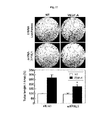
- the inventors confirmed in vivo angiogenesis activity by the ⁇ Experiment method 10>, and the results are shown in Fig. 19 .
- CTGF linker polypeptide very strongly induces angiogenesis also in vivo in a concentration-dependent manner, and thus, so called wheel-shaped blood vessel is formed.
- FPRL1 antagonist WRW4 10 uM
- CTGF binds to FPRL1 through the linker region, and thereby, induce angiogenesis in vitro and in vivo.
- CTGF/FPRL1 assembly may mediate the activity of VEGF-A in the process of angiogenesis.
- VEGF-A induces FPRL1 expression in HUVECs.
- HUVECs were treated with 20 ng/ml of VEGF-A, and cultured for various times, and then, RT-PCR was conducted according to the ⁇ Experiment method 12>, the degree of FPRL1 expression was measured, and the results are shown in Fig. 20 .
- flow cytometry analysis was conducted according to the ⁇ Experiment method 7>, and the results are shown in Fig. 20 .
- HUVECs were treated with various concentrations of VEGF-A and cultured, and then, RT-PCR was conducted according to the ⁇ Experiment method 12>, the degree of FPRL1 expression was measured, and the results are shown in Fig. 21 . And, flow cytometry analysis was conducted according to the ⁇ Experiment method 7>, and the results are shown in Fig. 21 .
- VEGF-A induces expression of FPRL1 in a concentration-dependent manner.
- VEGF-A 20 ng/ml of VEGF-A was administered to HUVECs, the degree of cell proliferation, cell migration, or tube formation was confirmed by the ⁇ Experiment method 9>, and the results are shown in Figs. 22 to 24 .
- FPRL1 antagonist WRW4 was additionally added.
- VEGF-A induces proliferation of HUVECs, but FPRL1 antagonist WRW4 did not exhibit statistically significant inhibition of HUVECs proliferation by VEGF-A.
- VEGF-A induces migration of HUVECs, and in case the cells are treated with FPRL1 antagonist WRW4 (10 uM), the cell migration is not induced.
- VEGF-A induces tube formation of HUVECs, and in case the cells are treated with FPRL1 antagonist WRW4 (10 uM), the tube formation is not induced.
- FPRL1 expression was inhibited using FPRL1 siRNA, and RT-PCR was conducted according to the ⁇ Experiment method 12> to measure the degree of FPRL1 expression, and the results are shown in Fig. 25 .
- FPRL1 expression is inhibited, and in case treated with Luciferase siRNA as control, FPRL1 expression is not inhibited.
- HUVECs are treated with FPRL1 siRNA
- cell migration or tube formation induced by VEGF-A is inhibited compared to the case where HUVECs are treated with Luciferase siRNA as control.
- FPRL1 antagonist strongly inhibits angiogenesis induced by VEGF-A.
- FPRL1 is an important factor for angiogenesis induced by VEGF-A
- CTGF is an important factor for inducing activation of FPRL1.
- FPRL1 and CTGF induced by VEGF-A promote migration of endothelial cells and tube formation by binding therebetween, thus involved in angiogenesis induced by VEGF-A.
Landscapes
- Health & Medical Sciences (AREA)
- Life Sciences & Earth Sciences (AREA)
- Public Health (AREA)
- Veterinary Medicine (AREA)
- Chemical & Material Sciences (AREA)
- Medicinal Chemistry (AREA)
- Pharmacology & Pharmacy (AREA)
- Animal Behavior & Ethology (AREA)
- Engineering & Computer Science (AREA)
- General Health & Medical Sciences (AREA)
- Bioinformatics & Cheminformatics (AREA)
- General Chemical & Material Sciences (AREA)
- Nuclear Medicine, Radiotherapy & Molecular Imaging (AREA)
- Organic Chemistry (AREA)
- Chemical Kinetics & Catalysis (AREA)
- Immunology (AREA)
- Diabetes (AREA)
- Hematology (AREA)
- Obesity (AREA)
- Gastroenterology & Hepatology (AREA)
- Heart & Thoracic Surgery (AREA)
- Biomedical Technology (AREA)
- Ophthalmology & Optometry (AREA)
- Neurosurgery (AREA)
- Neurology (AREA)
- Cardiology (AREA)
- Epidemiology (AREA)
- Zoology (AREA)
- Proteomics, Peptides & Aminoacids (AREA)
- Endocrinology (AREA)
- Dermatology (AREA)
- Urology & Nephrology (AREA)
- Reproductive Health (AREA)
- Rheumatology (AREA)
- Emergency Medicine (AREA)
- Child & Adolescent Psychology (AREA)
- Transplantation (AREA)
- Oncology (AREA)
- Orthopedic Medicine & Surgery (AREA)
- Physical Education & Sports Medicine (AREA)
Abstract
Description
- This disclosure relates to an angiogenesis-related pharmaceutical composition using connective tissue growth factor, more particularly to a pharmaceutical composition for promoting angiogenesis containing the connective tissue growth factor or a pharmaceutical composition for inhibiting angiogenesis containing at least one selected from the group consisting of polypeptide, antibody and a compound binding to connective tissue growth factor.
- Angiogenesis is a process wherein new capillary vessels are formed from the existing microvessels, and angiogenesis normally occurs during embryonic development, tissue regeneration and wound healing, corpus lutem development which is periodic change in female reproductive system, and even in this case, angiogenesis is stringently controlled and progressed (Folkman J et al., Int. Rev. Exp. Pathol., 16, pp207-248, 1976).
- In the adult, vascular endothelial cells grow very slowly, and relatively do not divide well compared to other kinds of cells. The process of angiogenesis generally consists of decomposition of vascular basement membrane due to protease by stimulation of angiogenesis promoter, migration of vascular endothelial cells, proliferation, and tube formation by differentiation of vascular endothelial cells to reconstitute blood vessels to produce new capillary vessels.
- There are diseases caused by failing to self-regulation of angiogenesis and abnormal growth. The diseases related to angiogenesis occurring at pathological states include hemangioma, vascular fibroma, vascular malformation, and cardiovascular disease such as atherosclerosis, vascular adhesion, scleroderma, and ophthalmic diseases caused by angiogenesis include angiogenesis due to corneal transplantation, neovascular glaucoma, diabetic retinopathy, corneal disease caused by angiogenesis, macular degeneration, pterygium, retinal degeneration, retrolental fibroplasias, granular conjunctivitis, and the like.
- Chronic inflammatory diseases such as arthritis, dermatological diseases such as psoriasis, capillarectasia, pyogenic granuloma, seborrheic dermatitis, acne, Alzheimer's diseases and obesity are also related to angiogenesis, and cancer growth and metastasis are necessarily dependent upon antiogenesis (D'Amato RJ et al., Ophthalmology, 102(9), pp1261-1262, 1995; Arbiser JL, J. Am. Acad. Dermatol., 34(3), pp486-497, 1996; O'Brien KD et al. Circulation, 93(4), pp672-682, 1996; Hanahan D et al., Cell, 86, pp353-364, 1996).
- Particularly, in cancer, angiogenesis plays an important function for cancer cell growth and metastasis. Tumor is supplied with nutrient and oxygen required for growth through angiogenesis, and angiogenetic blood vessels penetrated into tumor provide a pathway for cancer cells to enter into blood circulation system thereby allowing metastasis of cancer cells (Folkman and Tyler, Cancer Invasion and metastasis, Biologic mechanisms and Therapy(S.B. Day ed.) Raven press, New York, pp94-103, 1977; Polverini PJ, Crit. Rev. Oral. Biol. Med., 6(3), pp230-247, 1995).
- To the contrary, although excessive formation of blood vessels sometime becomes a leading cause of worsening of diseases, non-formation of blood vessels also becomes a cause of serious diseases. Angiogenesis is essential phenomenon for wound healing or tissue regeneration, for example, placenta with undeveloped blood vessel formation is an important cause of miscarriage, and necrosis, ulcer and ischemia due to non-formation of blood vessel may induce abnormal function of tissues or organs or may become a cause of death. And, diseases such as atherosclerosis, myocardial infarction and angina pectoris also become a cause of slow blood supply. Accordingly, there is a need for development of a treatment method that may decrease tissue damage due to low oxygen or low nutrition state caused by non-formation of blood vessels, and induce or promote new blood vessel formation for smooth tissue regeneration.
- The inventors found out that in angiogenesis in which a kind of inflammatory cytokine, vascular endothelial growth factor-A (VEGF-A), and FPRL1 (formyl peptide receptor-like 1) are involved, connective tissue growth factor (CTGF) binds to FPRL1 to induce angiogenesis, particularly found out a region where CTGF binds to FPRL1, confirmed that angiogenesis is induced through the region, and completed the invention.
- Therefore, one embodiment of the present invention provides a pharmaceutical composition for promoting angiogenesis containing a fragment of protein corresponding to a FPRL1 binding region in CTGF and/or gene encoding the same; use of a fragment of protein corresponding to a FPRL1 binding region in CTGF and/or gene encoding the same for promoting angiogenesis and/or preparing angiogenesis promoter; and a method for promoting angiogenesis comprising administering a fragment of protein corresponding to a FPRL1 binding region in CTGF and/or gene encoding the same to a patient in need of promoting angiogenesis.
- Another embodiment of the present invention provides a pharmaceutical composition for inhibiting angiogenesis containing a fragment of protein corresponding to a FPRL1 binding region in CTGF and/or gene encoding the same; use of a fragment of protein corresponding to a FPRL1 binding region in CTGF and/or gene encoding the same for inhibiting angiogenesis and/or preparing angiogenesis inhibitor; and/or a method for inhibiting angiogenesis comprising administering a fragment of protein corresponding to a FPRL1 binding region in CTGF and/or gene encoding the same to a patient in need of inhibiting angiogenesis.
- Yet another embodiment of the present invention provides a method for screening angiogenesis inhibitor using a FPRL1 binding region in CTGF as a target.
- To solve the problem, one embodiment of the invention provides a pharmaceutical composition for promoting angiogenesis containing a fragment of connective tissue growth factor protein comprising continuous 38 or more amino acids, for example continuous 38 to 140 amino acids (for example, selected from regions from 103rd to 242nd amino acid), continuous 38 to 100 amino acids (for example, selected from regions from 103rd to 202nd amino acid), or continuous 38 to 80 amino acids (for example, selected from regions from 163rd to 242nd amino acid), preferably continuous 38 to 39 amino acids, which comprises amino acid sequence of SEQ ID NO. 5 (WVCDEPKDQTVVGPALAAYRLEDTFGPDPTMIRANCLV), which is a region from 164th to 201st amino acid, in the amino acid sequence of SEQ ID NO. 9: use of the fragment of connective tissue growth factor protein for promoting angiogenesis and/or preparing angiogenesis promoter; and a method for promoting angiogenesis comprising administering the fragment of connective tissue growth factor protein to a patient in need of promoting angiogenesis.
- Another embodiment provides a pharmaceutical composition for promoting angiogenesis containing gene encoding a fragment of connective tissue growth factor protein comprising continuous 38 or more amino acids, for example, continuous 38 to 140 amino acids (for example, selected from regions from 103rd to 242nd amino acid), continuous 38 to 100 amino acids (for example, selected from regions from 103rd to 202nd amino acid), or continuous 38 to 80 amino acids (for example, selected from regions from 163rd to 242nd amino acid), preferably continuous 38 to 39 amino acids, which comprises amino acid sequence of SEQ ID NO. 5, in amino acid sequence of SEQ ID NO. 9: use of the gene encoding a fragment of connective tissue growth factor protein for promoting angiogenesis and/or preparing angiogenesis promoter; and a method for promoting angiogenesis comprising administering the gene encoding a fragment of connective tissue growth factor protein to a patient in need of promoting angiogenesis.
- The method for promoting angiogenesis may further include confirming a patient in need of promoting angiogenesis, before the administration, wherein the patient in need of promoting angiogenesis may be a patient requiring prevention or treatment of angina pectoris, atherosclerosis, stroke, vascular dementia, chronic ulcer, or wound. The patient may be mammals, preferably human.
- Yet another embodiment provides a pharmaceutical composition for inhibiting angiogenesis containing at least one selected from the group consisting of polypeptide, antibody and a compound binding to a fragment of connective tissue growth factor protein comprising continuous 38 or more amino acids, for example, continuous 38 to 140 amino acids (for example, selected from regions from 103rd to 202nd amino acid), continuous 38 to 100 amino acids (For example, selected from regions from 103rd to 202nd amino acid), or continuous 38 to 80 amino acids (for example, selected from regions from 163rd to 242nd amino acid), preferably 38 to 39 amino acids, which comprises amino acid sequence of SEQ ID NO. 5, in amino acid sequence of SEQ ID NO. 9: use of the polypeptide, antibody and compound binding to the fragment of connective tissue growth factor protein for inhibiting angiogenesis and/or preparing angiogenesis inhibitor; and a method for inhibiting angiogenesis comprising administering the polypeptide, antibody and compound binding to the fragment of connective tissue growth factor protein to a patient in need of inhibiting angiogenesis.
- Yet another embodiment provides a pharmaceutical composition for inhibiting angiogenesis containing an expression inhibitor of a fragment of connective tissue growth factor comprising continuous 38 or more amino acids, for example, continuous 38 to 140 amino acids (for example, selected from regions from 103rd to 202nd amino acid), continuous 38 to 100 amino acids (For example, selected from regions from 103rd to 202nd amino acid), or continuous 38 to 80 amino acids (for example, selected from regions from 163rd to 242nd amino acid), preferably 38 to 39 amino acids, which comprises amino acid sequence of SEQ ID NO. 5, in amino acid sequence of SEQ ID NO. 9, as an active ingredient; use of an expression inhibitor of the fragment of connective tissue growth factor protein for inhibiting angiogenesis and/or preparing angiogenesis inhibitor; and a method for inhibiting angiogenesis comprising administering an expression inhibitor of a fragment of connective tissue growth factor protein to a patient in need of inhibiting angiogenesis. For example, the expression inhibitor may be at least one selected from the group consisting of antisense nucleotide, short interfering RNA, and short hairpin RNA, which complementarily bind to the base sequence (SEQ ID NO. 7) of gene encoding the amino acid sequence of SEQ ID NO. 5 or mRNA thereof, for example, base sequence consisting of continuous 15 to 30, preferably continuous 20 to 25 bases in the mRNA.
- The method for inhibiting angiogenesis may further include confirming a patient in need of inhibiting angiogenesis, before the administration, wherein the patient in need of inhibiting angiogenesis may be a patient requiring prevention or treatment of cancer growth and metastasis, rheumatoid arthritis, psoriasis, diabetic retinopathy, diabetic nephropathy, hypertension, endometriosis, adiposis, retinopathy of prematurity, corneal inflammation rejection, neovascular glaucoma, proliferative retinopathy, hemophilic arthropathy, keloid, wound granulation, vascular adhesion, osteoarthritis, Crohn's disease, restenosis, atherosclerosis, intestinal adhesion, ulcer, liver cirrhosis, neophritis, malignant nephrosclerosis, organ transplant rejection, glomerulopathy, diabetes mellitus, or inflammation. The patient may be mammals, preferably human.
- Yet another embodiment provides a method for screening angiogenesis inhibitor comprising (a) treating cells comprising gene encoding a fragment of connective tissue growth factor protein comprising continuous 38 or more amino acids, for example, continuous 38 to 140 amino acids (for example, selected from regions from 103rd to 202nd amino acid), continuous 38 to 100 amino acids (For example, selected from regions from 103rd to 202nd amino acid), or continuous 38 to 80 amino acids (for example, selected from regions from 163rd to 242nd amino acid), preferably 38 to 39 amino acids, which comprises amino acid sequence of SEQ ID NO. 5, in amino acid sequence of SEQ ID NO. 9, with a candidate compound; and (b) measuring the degree of expression of the gene, wherein if the degree of expression of the gene is decreased in the cells treated with the candidate compound, compared to the cells that is not treated with the candidate compound, the candidate material is determined as angiogenesis inhibitor.
- Hereinafter, the present invention will be explained in detail.
- The composition for promoting angiogenesis contains a fragment of connective tissue growth factor protein comprising continuous 38 or more amino acids, for example, continuous 38 to 140 amino acids (for example, selected from regions from 103rd to 242nd amino acid), continuous 38 to 100 amino acids (for example, selected from regions from 103rd to 202nd amino acid), or continuous 38 to 80 amino acids (for example, selected from regions from 163rd to 242nd amino acid), preferably continuous 38 to 39 amino acids, which comprises amino acid sequence of SEQ ID NO. 5, in amino acid sequence of SEQ ID NO. 9.
- Full length amino acid sequence of the connective tissue growth factor (CTGF) may be derived from human (for example, accession no. CAG46559.1), and represented by SEQ ID NO. 9. In angiogenesis in which a kind of inflammatory cytokine, vascular endothelial growth factor-A (VEGF-A) and FPRL1 (formyl peptide receptor-like 1) are involved, the connective tissue growth factor protein may bind to FPRL1 to induce angiogenesis. Specifically, the connective tissue growth factor has activity for inducing FPRL1-specific ERK phosphorylation (see Experiment result 2), is a protein of which expression is induced by VEFG-A (see Experiment result 3), and it binds to FPRL1 to induce angiogenesis (see Experiment result 11).
- Particularly, a region where the connective tissue growth factor protein binds to FPRL1 is a region from 164th amino acid to 201st amino acid in SEQ ID NO. 9 which is full length amino acid sequence of connective tissue growth factor protein, and it may be represented by amino acid sequence of SEQ ID NO. 5 (see Experiment result 4).
- Therefore, the fragment of connective tissue growth factor protein, which is an active ingredient of the pharmaceutical composition for promoting angiogenesis of the present invention, may comprise continuous 38 or more amino acids, for example, continuous 38 to 140 amino acids (for example, selected from regions from 103rd to 242nd amino acid), continuous 38 to 100 amino acids (for example, selected from regions from 103rd to 202nd amino acid), or continuous 38 to 80 amino acids (for example, selected from regions from 163rd to 242nd amino acid), preferably continuous 38 to 39 amino acids, which comprises amino acid sequence of SEQ ID NO. 5, a region from 164th amino acid to 201st amino acid in SEQ ID NO. 9, in the amino acid sequence of SEQ ID NO. 9 which is full length sequence of the connective tissue growth factor protein. The fragment of connective tissue growth factor protein may be preferably represented by amino acid sequence of SEQ ID NO. 6 (EWVCDEPKDQTVVGPALAAYRLEDTFGPDPTMIRANCLV), but not limited thereto.
- The fragment of connective tissue growth factor protein is a binding region to FPRL1 (see Experiment result 5), effectively induces FPRL1-specific ERK phosphorylation (see Experiment result 6), activates FPRL1 to increase intracellular Ca2+ concentration (see Experiment result 7), and finally, effectively induces angiogenesis (see Experiment result 9).
- The pharmaceutical composition for promoting angiogenesis of the present invention may contain gene encoding connective tissue growth factor protein comprising continuous 38 or more amino acids, for example, continuous 38 to 140 amino acids (for example, selected from regions from 103rd to 242nd amino acid), continuous 38 to 100 amino acids (for example, selected from regions from 103rd to 202nd amino acid), or continuous 38 to 80 amino acids (for example, selected from regions from 163rd to 242nd amino acid), preferably continuous 38 to 39 amino acids, which comprises amino acid sequence of SEQ ID NO.. 5, in the amino acid sequence of SEQ ID NO. 9. The gene may preferably encode amino acid sequence of SEQ ID NO. 5, a region from 164th amino acid to 201st amino acid in the full length amino acid sequence of connective tissue growth factor protein, or encode amino acid sequence of SEQ ID NO. 6 comprising the amino acid sequence of SEQ ID NO. 5, but is not limited thereto. More preferably, the gene may be represented by base sequence of SEQ ID NO. 7 encoding the amino acid sequence of SEQ ID NO. 5 (164-TGG GTG TGT GAC GAG CCC AAG GAC CAA ACC GTG GTT GGG CCT GCC CTC GCG GCT TAC CGA CTG GAA GAC ACG TTT GGC CCA GAC CCA ACT ATG ATT AGA GCC AAC TGC CTG GTC-201), and it may be represented by base sequence of SEQ ID NO. 8 encoding the amino acid sequence of SEQ ID NO. 6 (163-GAG TGG GTG TGT GAC GAG CCC AAG GAC CAA ACC GTG GTT GGG CCT GCC CTC GCG GCT TAC CGA CTG GAA GAC ACG TTT GGC CCA GAC CCA ACT ATG ATT AGA GCC AAC TGC CTG GTC-201).
- The gene may be inserted into a vector, preferably recombinant expression vector, but is not limited thereto, wherein the recombinant expression vector may comprise the gene and expression regulator including operably linked promoter, and the like. For example, the recombinant expression vector may be, for example, EcoR I-INSERT-Xba I in pAB-bee™-FH vector (including signal peptide & flag tag), but is not limited thereto.
- Specifically, the vector is used for a nucleic acid molecule transferring a DNA fragment from one cell to another cell, and may be derived from those selected from the group consisting of plasmid, bacteriophage, and plant and animal virus, but is not limited thereto.
- The expression vector refers to recombinant DNA comprising aimed coding sequence and appropriate nucleic acid sequence required for expression of the coding sequence, operably linked in a specific host, and in general, nucleic acid sequence required for expression in prokaryotic cells may comprise promoter, operator (optional), and ribosome binding region, in addition to the other sequence, and eukaryotic cells use promoter, enhancer, and stop and polyadenylation signal, which are known in the art.
- The recombinant expression vector may be a genetic construct comprising operably linked essential regulator elements so as to express the gene, which may prepare aimed protein, a fragment of connective tissue growth factor in the present invention, in a suitable host cells.
- The term "operably linked" means that nucleic acid expression regulator sequence and nucleic acid sequence encoding aimed protein are functionally linked so as to perform general functions. For example, promoter and nucleic acid encoding protein or RNA may be operably linked to influence on the expression of coding sequence. The operable link with recombinant vector may be prepared using gene recombination technology well known in the art, and site-specific DNA cleavage and ligation may be performed using enzyme, and the like generally known in the art.
- Suitable expression vector comprises expression regulatory elements such as promoter, initiation codon, stop codon, polyadehylation signal and enhancer, and the like, and it may be variously prepared according to the purpose. The initiation codon and stop codon should act in the individual when a genetic construct is administered, and it should be in frame with coding sequence.
- The pharmaceutical composition for promoting angiogenesis may be used for treatment of diseases caused by inhibition of angiogenesis or diseases that may be cured by promoting angiogenesis, but is not limited thereto, and for example, it may be used for treatment of angina pectoris, atherosclerosis, ischemic stroke, vascular dementia, chronic ulcer, or wound. Thus, yet another embodiment of the present invention provides a composition for prevention and/or treatment of angina pectoris, atherosclerosis, ischemic stroke, vascular dementia, chronic ulcer, or wound, comprising the pharmaceutical composition for promoting angiogenesis as an active ingredient.
- Meanwhile, the pharmaceutical composition for inhibiting angiogenesis contains at least one selected from the group consisting of polypeptide, antibody and a compound, which bind to a fragment of connective tissue growth factor protein comprising continuous 38 or more amino acids, for example, continuous 38 to 140 amino acids (for example, selected from regions from 103rd to 242nd amino acid), continuous 38 to 100 amino acids (for example, selected from regions from 103rd to 202nd amino acid), or continuous 38 to 80 amino acids (for example, selected from regions from 163rd to 242nd amino acid), preferably continuous 38 to 39 amino acids, which comprises amino acid sequence of SEQ ID NO. 5, in the amino acid sequence of SEQ ID NO. 9
- As described, since a fragment of connective tissue growth factor protein comprising continuous 38 or more amino acids, for example, continuous 38 to 140 amino acids (for example, selected from regions from 103rd to 242nd amino acid), continuous 38 to 100 amino acids (for example, selected from regions from 103rd to 202nd amino acid), or continuous 38 to 80 amino acids (for example, selected from regions from 163rd to 242nd amino acid), preferably continuous 38 to 39 amino acids, which comprises amino acid sequence of SEQ ID NO. 5, in SEQ ID NO. 9 which is amino acid sequence of full length protein of connective tissue growth factor, binds to FPRL1 to induce angiogenesis, at least one selected from the group consisting of polypeptide, antibody and a compound binding to the fragment of connective tissue growth factor protein may effectively inhibit angiogenesis.
- The fragment of connective tissue growth factor may comprise continuous 38 or more amino acids, for example, continuous 38 to 140 amino acids (for example, selected from regions from 103rd to 242nd amino acid), continuous 38 to 100 amino acids (for example, selected from regions from 103rd to 202nd amino acid), or continuous 38 to 80 amino acids (for example, selected from regions from 163rd to 242nd amino acid), preferably continuous 38 to 39 amino acids, which comprises amino acid sequence of SEQ ID NO. 5, a 164th to 201st region, in the amino acid sequence of SEQ ID NO. 9 which is full length sequence of the connective tissue growth factor protein, and more preferably, it may be represented by amino acid sequence of SEQ ID NO. 6 comprising the amino acid sequence of SEQ ID NO. 5.
- Since the fragment of connective tissue growth factor protein comprises a FPRL1-binding region, polypeptide, antibody or a compound binding to the fragment of connective tissue growth factor protein may block binding of the connective tissue growth factor protein to FPRL1 to effectively inhibit angiogenesis.
- The polypeptide, antibody or compound is not specifically limited as long as it may bind to the fragment of connective tissue growth factor protein, but preferably, it may be one binding to amino acid sequence of SEQ ID NO. 6, or SEQ ID NO. 5, and it may be easily prepared by one of ordinary knowledge in the art according to the amino acid sequence of the fragment of connective tissue growth factor protein to be bound.
- Furthermore, pharmaceutical composition for inhibiting angiogenesis may contain at least one selected from the group consisting of antisense nucleotide, short interfering RNA, and short hairpin RNA, which complementarily bind to gene encoding a fragment of connective tissue growth factor protein comprising continuous 38 or more amino acids, for example, continuous 38 to 140 amino acids (for example, selected from regions from 103rd to 242nd amino acid), continuous 38 to 100 amino acids (for example, selected from regions from 103rd to 202nd amino acid), or continuous 38 to 80 amino acids (for example, selected from regions from 163rd to 242nd amino acid), preferably continuous 38 to 39 amino acids, comprising amino acid sequence of SEQ ID NO. 5, in the amino acid sequence of SEQ ID NO. 9, or mRNA thereof, for example, base sequence consisting of continuous 15 to 30, preferably continuous 20 to 25 bases in the mRNA.
- The siRNA comprises a sense RNA strand and a complementary antisense RNA strand, the two strands anneal each other by standard Watson-Crick base pair interaction, and the sense strand comprises identical nucleic acid sequence to the target sequence in target mRNA. Technologies of selecting the target sequence of siRNA are described in, for example, Tuschl T et al, "The siRNA User Guide" revised Oct. 11, 2002.
- The sense and antisense strands of the siRNA may comprise two complementary single-stranded RNA molecules, or it may comprise single molecule wherein two complementary parts form a base pair and covalently bonded by a "hairpin" region of single strand. The latter is referred to as short hairpin RNA (shRNA), wherein the shRNA is a single strand and forms a stem-loop structure in vivo. The shRNA is generally synthesized by transcription of complementary DNA sequence from Pol III promoter in vivo. Pol-III-induced transcription starts at a well-defined start site and stops at a linear (-TTTT-) second residue consisting of 4 or more thymidines to produce non-poly(A) transcript. Pol III promoter may be activated in all cells, and it may express shRNA. After transcription, the loop of shRNA is cleaved by dicer, and the shRNA acts with RISC (RNA-induced silencing complex) like siRNA (see, Tuschl, T. (2002), Cell 110(5): 563-74).
- The siRNA may be obtained using many technologies known to one of ordinary knowledge in the art. For example, siRNA may be chemically synthesized using a method known in the art or produced by recombination. Preferably, the siRNA may be chemically synthesized using suitably protected ribonucleoside phosphoramidites and the existing DNA/RNA synthesizer. The siRNA may be synthesized as two complementary and separated RNA molecules or one RNA molecule having two complementary regions. Alternatively, the siRNA may be expressed from recombinant DNA plasmid using suitable promoter. The promoter suitable for expressing the siRNA from plasmid may include, for example, U6 or H1 RNA pol III promoter sequence and cytomegalovirus promoter. And, the recombinant plasmid may include inductive or controllable promoter in order to express siRNA in a specific tissue or specific intracellular environment.
- The siRNA may be expressed as two complementary and separated RNA molecule or one RNA molecule having two complementary regions from recombinant plasmid. The selection of suitable plasmid for expression of siRNA, a method of inserting nucleic acid into plasmid to express siRNA, and a method of transferring recombinant plasmid to aimed cells are within the scope of technologies in the field to which the invention pertains. For example, see [Tuschl, T. (2002), Nat. Biotechnol, 20: 446-448]: [Brummelkamp TR et al. (2002), Science 296: 550- 553]: [Miyagishi M et al. (2002), Nat. Biotechnol . 20: 497-500]: [Paddison PJ et al. (2002), Genes Dev. 16: 948-958; Lee NS et al. (2002), Nat. Biotechnol. 20: 500-505]: and [Paul CP et al. (2002), Nat. Biotechnol. 20: 505-508], which references are incorporated herein by reference.
- For example, according to one embodiment of the invention, as the siRNA of FPRL1, double stranded RNA molecule with target of AAUUCACAUCGUGGUGGACAU (SEQ ID NO. 10) (for example, Sense: UUCACAUCGUGGUGGACAUdTdT (SEQ ID NO. 3) and Anti-sense: AUGUCCACCACGAUGUGAAdTdT (SEQ ID NO. 4)) may be used.
- The pharmaceutical composition for inhibiting angiogenesis may be used for prevention and/or treatment of angiogenesis-related diseases. The angiogenesis-related diseases may be diseases or conditions selected from the group consisting of cancer growth and metastasis, rheumatoid arthritis, psoriasis, diabetic retinopathy, diabetic nephropathy, hypertension, endometriosis, adiposis, retinopathy of prematurity, corneal inflammation rejection, neovascular glaucoma, proliferative retinopathy, hemophilic arthropathy, keloid, wound granulation, vascular adhesion, osteoarthritis, Crohn's disease, restenosis, atherosclerosis, intestinal adhesion, ulcer, liver cirrhosis, neophritis, malignant nephrosclerosis, organ transplant rejection, glomerulopathy, diabetes mellitus, inflammation, and the like. Thus, one embodiment of the present invention provides a composition for prevention or treatment of disease or condition selected from the group consisting of cancer growth and metastasis, rheumatoid arthritis, psoriasis, diabetic retinopathy, diabetic nephropathy, hypertension, endometriosis, adiposis, retinopathy of prematurity, corneal inflammation rejection, neovascular glaucoma, proliferative retinopathy, hemophilic arthropathy, keloid, wound granulation, vascular adhesion, osteoarthritis, Crohn's disease, restenosis, atherosclerosis, intestinal adhesion, ulcer, liver cirrhosis, neophritis, malignant nephrosclerosis, organ transplant rejection, glomerulopathy, diabetes mellitus, inflammation, and the like, containing the pharmaceutical composition for inhibiting angiogenesis as an active ingredient.
- To administer the pharmaceutical composition for promoting angiogenesis or pharmaceutical composition for inhibiting angiogenesis, at least one kind of pharmaceutically acceptable carrier may be further included in addition to the above described active ingredient. The pharmaceutically acceptable carrier may be at least one selected from a saline solution, sterile water, Ringer's solution, buffered saline, dextrose solution, maltodextrin solution, glycerol, ethanol, liposome, and a mixture thereof, and if necessary, other common additives such as antioxidant, buffer solution, bacteriostatic agent, and the like may be added. And, diluents, dispersant, surfactant, binder and lubricant may be additionally added to formulated into a dosage form for injection such as an aqueous solution, suspension, emulsion, and the like, a pill, a capsule, granules or a tablet, and a target organ-specific antibody or other ligands may be bound to the carrier so as to specifically act on a target organ. Furthermore, the pharmaceutical composition may be preferably formulated according to diseases or ingredients by suitable method in the art or using a method disclosed in Remington's Pharmaceutical Science (Recent Edition), Mack Publishing Company, Easton PA.
- The pharmaceutical composition for promoting angiogenesis or pharmaceutical composition for inhibiting angiogenesis of the present invention may be prepared for oral, topical, parenteral, intravenous, intramuscular, subcutaneous, intraocular, transdermal administration, and the like. Preferably, it may be used in an injectable form.
- Thereby, it may be mixed with pharmaceutically acceptable medium for injectable composition to directly inject into an area to be treated. The composition of the present invention may comprise a lyophilized composition that enables composition of an injectable solution upon addition of an isotonic sterile solution, sterile water or suitable physiological saline. Direct injection into tumor of a patient is favorable because treatment efficiency is focused on the infected tissue. The used dose may be controlled by various parameters, particularly, gene, vector, used administration route, disease in question or alternatively, required treatment period. And, the range may be varied according to weight, age, gender, health condition, diet of a patient, administration time, administration route, excretion rate and severance of disease, and the like. The daily dose may be about 0.0001 to 10 mg/kg, preferably 0.001 to 1 mg/kg, and it may be preferable to administer one time or divide into several times a day.
- Meanwhile, a method for screening angiogenesis inhibitor comprises
- (a) treating cells comprising gene encoding a fragment of connective tissue growth factor protein comprising continuous 38 or more amino acids, for example, continuous 38 to 140 amino acids (for example, selected from regions from 103rd to 202nd amino acid), continuous 38 to 100 amino acids (For example, selected from regions from 103rd to 202nd amino acid), or continuous 38 to 80 amino acids (for example, selected from regions from 163rd to 242nd amino acid), preferably 38 to 39 amino acids, which comprises amino acid sequence of SEQ ID NO. 5, in amino acid sequence of SEQ ID NO. 9, with a candidate compound; and
- (b) measuring the degree of expression of the gene,
- In the step (a), the fragment of connective tissue growth factor protein may be represented by amino acid sequence of SEQ ID NO. 5 or SEQ ID NO. 6. The candidate compound may be peptide, protein, a non-peptide compound, a synthetic compound, a fermentation product, cell extract, plant extract, or animal extract, but is not limited thereto, and it may include those widely known as well as novel material. In the step (a), the cells may be transformed cells with recombinant expression vector comprising the gene, and the transformation method is well known in the art.
- In the step (b), the degree of expression of gene may be preferably measured by immunofluorescence method, enzyme-linked immunosorbent assay (ELISA), Western Blotting or RT-PCR, but is not limited thereto. In case antibody is used for confirming the amount of protein according to the expression, a secondary antibody specific to target protein and linked to detectable label may be added, and after adding the secondary antibody, a tertiary antibody having binding affinity to the secondary antibody and linked to detectable label may be added. As the detectable label in the secondary or tertiary antibody, enzyme exhibiting coloration when cultured and appropriate coloring substrate may be used. The detectable part may include a composition detectable by spectroscopic, enzymatic, photochemical, bioelectronical, immunochemical, electrical, optical or chemical means, and for example, it may include fluorescent marker and dye, magnetic lable, linked enzyme, mass spectrometric tag, spin tag, electron donor and receptor, but is not limited thereto.
- If the degree of expression of the gene is decreased in the cells treated with the candidate compound, compared to control that is not treated with the candidate compound, the candidate compound may be selected as angiogenesis inhibitor. The angiogenesis inhibitor may be used for prevention or treatment of disease or condition selected from the group consisting of rheumatoid arthritis, psoriasis, diabetic retinopathy, diabetic nephropathy, hypertension, endometriosis, adiposis, cancer, retinopathy of prematurity, corneal inflammation rejection, neovascular glaucoma, proliferative retinopathy, hemophilic arthropathy, keloid, wound granulation, vascular adhesion, osteoarthritis, Crohn ' s disease, restenosis, atherosclerosis, intestinal adhesion, ulcer, liver cirrhosis, neophritis, malignant nephrosclerosis, organ transplant rejection, glomerulopathy, diabetes mellitus, inflammation, and the like, but is not limited thereto.
- Using the screening method of the present invention, a therapeutic agent involved in the mechanism may be selected within a short time, and through experiments for the provement, useful prevention and/or treatment agent may be provided to a patient in need of prevention and/or treatment of disease caused by angiogenesis.
- The fragment of connective tissue growth factor protein, which is discovered in the present invention, is a binding region to FPRL1, effectively induces FPRL1-specific ERK phosphorylation, activates FPRL1 to increase intracellular Ca2+ concentration, and finally, effectively induces angiogenesis, and thus, the fragment of connective tissue growth factor protein may be useful for a pharmaceutical composition for promoting angiogenesis, while polypeptide, antibody or a compound binding to the fragment of connective tissue growth factor protein may be useful for a pharmaceutical composition for inhibiting angiogenesis.
-
-
Fig. 1 shows the results of immunoblotting to confirm whether conditioned medium of HUVECs obtained by treating with VEGF-A induces ERK phosphorylation in FPRL1-overexpressed FPRL1/RBL cells. -
Fig. 2 shows the experiment results confirming that conditioned medium, VEGF-A and positive control (WKYMVm) induce ERK phosphorylation only in FPRL1-overexpressing FPRL1/RBL cells. -
Fig. 3 shows the results of confirming whether ERK phosphorylation occurs by treating FPRL1/RBL cells with HPLC fraction of conditioned medium. -
Fig. 4 shows the results of MS/MS analysis for B12 fraction of conditioned medium. -
Fig. 5 shows the results of analyzing the degree of CTGF expression in cell lysate or conditioned medium of HUVECs treated with VEGF-A according to treatment time of VEGF-A. -
Fig. 6 shows the results of analyzing the degree of CTGF expression in cell lysate or conditioned medium of HUVECs treated with VEGF-A according to treatment amount of VEGF-A. -
Fig. 7 shows the results confirming that CTGF expression is significantly decreased when antibody to VEGF-A receptor is administered. -
Fig. 8 is a schematic drawing showing a method for determining CTGF binding region to FPRL1. -
Fig. 9 shows the results confirming whether full length CTGF protein or partly deleted protein thereof induces ERK phosphorylation in FPRL1/RBL cells. -
Fig. 10 shows the results confirming whether CTGF linker polypeptide of SEQ ID NO. 6 binds only to FPRL1/RBL cells. -
Fig. 11 shows the results confirming that CTGF linker polypeptide of SEQ ID NO. 6 induces ERK phosphorylation to a similar level to rhCTGF, and the ERK phosphorylation is decreased by FPRL1 antagonist (WRW4). -
Fig. 12 shows the result confirming whether Ca2+ concentration is increased in FPRL1/RBL cells treated with CTGF linker polypeptide of SEQ ID NO. 6. -
Fig. 13 shows the result confirming whether CTGF linker polypeptide of SEQ ID NO. 6 binds to FPRL1 in HUVECs cells. -
Fig. 14 shows the result confirming that if HUVECs is treated with CTGF linker polypeptide of SEQ ID NO. 6, ERK phosphorylation is effectively induced. -
Fig. 15 shows the result confirming that if HUVECs is treated with CTGF linker polypeptide of SEQ ID NO. 6, intracellular Ca2+ concentration is increased in a concentration-dependent manner. -
Fig. 16 shows the result confirming that CTGF linker polypeptide of SEQ ID NO. 6 does not significantly influence on the proliferation of HUVECs. -
Fig. 17 shows the result confirming that CTGF linker polypeptide of SEQ ID NO. 6 induces migration of HUVECs in a concentration-dependent manner. -
Fig. 18 shows the result confirming that that CTGF linker polypeptide of SEQ ID NO. 6 induces tube formation of HUVECs in a concentration-dependent manner. -
Fig. 19 shows the result confirming that CTGF linker polypeptide of SEQ ID NO. 6 in vivo induces angiogenesis in a concentration-dependent manner. -
Fig. 20 shows the result confirming whether VEGF-A induces FPRL1 expression in HUVECs cells by conducting RT-PCR or flow cytometry according to incubation time. -
Fig. 21 shows the result confirming whether VEGF-A induces FPRL1 expression in HUVECs cells by conducting RT-PCR or flow cytometry according to treatment amount. -
Fig. 22 shows the experiment result confirming that the proliferation of HUVECs induced by VEGF-A is not significantly decreased by FPRL1 antagonist (WRW4). -
Fig. 23 shows the experiment result confirming that migration of HUVECs induced by VEGF-A is significantly decreased by FPRL1 antagonist (WRW4). -
Fig. 24 shows the experiment result confirming that the tube formation of HUVECs induced by VEGF-A is significantly decreased by FPRL1 antagonist (WRW4). -
Fig. 25 shows the result confirming that if HUVECs is treated with FPRL1 siRNA, FPRL1 expression is inhibited. -
Fig. 26 shows the result confirming that if HUVECs is treated with FPRL1 siRNA, cell migration induced by VEGF-A is inhibited. -
Fig. 27 shows the result confirming that if HUVECs is treated with FPRL1 siRNA, tube formation induced by VEGF-A is inhibited. -
Fig. 28 shows the result confirming that FPRL1 antagonist (WRW4) inhibits angiogenesis by VEGF-A. -
Fig. 29 is a schematic diagram showing the mechanism of angiogenesis that is induced by binding of CTGF expressed by VEGF-A and FPRL1. - Hereinafter, the present invention will be explained in detail with reference to the following Examples.
- However, these examples are only to illustrate the invention, and the scope of the invention is not limited thereto.
- In the following Examples, all data are represented by mean ± standard deviation, statistically compared by Student's t test, and it is considered as being statistically significant when p < 0.05.
- Phospho-ERK1/2 (Thr202/Tyr204) and ERK1/2 antibody were purchased from Cell Signaling Technology Inc. (Beverly, MA, USA), and Human CTGF antibody and anti-Flag antibody were respectively purchased from Abcam (Cambridge, MA, USA) and Sigma Co. (St. Louis, MO, USA). Recombinant human CTGF was purchased from BioVendor Laboratory Medicine Inc. (Brno, Czech Republic). Recombinant human VEGF-A165, anti-VEGF-A mAb, anti-VEGFR-1 mAb, and anti-VEGFR-2 mAb were obtained from R&D Systems Inc. (Minneapolis, MN, USA).
- The siRNA to FPRL1 (sense:UUCACAUCGUGGUGGACAUdTdT (SEQ ID NO. 3), anti-sense:AUGUCCACCACGAUGUGAAdTdT (SEQ ID NO. 4)) and siRNA to luciferase (sense: CGUACGCGGAAUACUUCGAdTdT (SEQ ID NO. 11), anti-sense: UCGAAGUAUUCCGCGUACGdTdT (SEQ ID NO. 12)) were synthesized in Dharmacon Research, Inc. (Chicago, IL). And, WKYMVm (SEQ ID NO. 1), WRWWWW (SEQ ID NO. 2, WRW4), and biotinylated WKYMVm were synthesized in A&PEP Inc (Seoul, Korea), of which purities were greater than 95%. Linker polypeptide comprising amino acid sequence of human CTGF link region (EWVCDEPKDQTVVGPALAAYRLEDTFGPDPTMIRANCLV (SEQ ID NO. 6)-NH2) was synthesized in Anygen Co. Ltd. (Gwangjoo, Korea), of which purity was about 99.1%.
- Human umbilical vein endothelial cells (HUVECs) were treated with collagenase (Sigma, St. Louis, MO, USA) to separate from umbilical cord (ST. MARY'S Hospital, Seoul, Korea) according to a method known in the art (Ferrero, E. et al. Transendothelial migration leads to protection from starvation-induced apoptosis in CD34+CD14+ circulating precursors: evidence for PECAM-1 involvement through Akt/PKB activation. ). The separated HUVECs were cultured in a dish coated with 0.2% (w/v) gelatin using Medium 199 (containing 20% (v/v) of thermally inactivated fetal bovine serum and 1%(w/v) of penicillin/streptomycin).
- For this experiment, each cell was cultured to subconfluence state, and those of 3 to 5 passages were used. FPRL1-expressing rat basophil leukemia (RBL)-2H3 (FPRL1/RBL) cells, FPR-expressing RBL-2H3 (FPR/RBL) cells, and RBL-2H3 (vector/RBL) cells (cells wherein FPRL1 or FPR receptor is not overexpressed) (ATCC, Manassas, VA, USA) were maintained in DMEM medium (Sigma, St. Louis, MO, USA) containing high concentration of glucose, to which 1% penicillin/streptomycin, 20% (v/v) thermally inactivated fetal bovine serum and G418 (Geneticin)(500 ug/ml) were added. The cells were cultured at 37°C in 5% CO2,.
- Conditioned medium was obtained from HUVECs (3 to 5 passages) cultured in Medium 199 that does not include FBS. The conditioned medium was centrifuged to remove residual cells, and it was stored at -80°C until used.
- Vector/RBL cells, FPR/RBL cells, FPRL1/RBL cells, and HUVECs were cultured to confluence, and serum-starved. The stimulated cells (1X10⁵ cells) were washed with PBS twice, dissolved in 1 ml of sample buffer (50 mM Tris-HCl, 100 mM NaCl, 0.1% SDS, 1% Nonidet P-40, 50 mM NaF, 1 mM Na3VO4, 1 µg/ml aprotinin, 1 µg/ml pepstatin, and 1 µg/ml leupeptin), heated at 95°C for 30 seconds, separated with SDS-PAGE, and then, transferred to a nitrocellulose film. Immunoblot was conducted using anti-phospho-ERK(extracellular signal regulated kinase)1/2 (Thr202/Tyr204)(Cell Signaling Technology, Beverly, MA, USA), anti-ERK1/2 (Cell Signaling Technology, Beverly, MA, USA), anti-CTGF (Abcam, Cambridge, MA, USA), anti-Actin (Sigma, St. Louis, MO, USA), or anti-Flag (Sigma, St. Louis, MO, USA) antibody, and then, the film was visualized with a chemiluminescence substrate (Amersham Pharmacia).
- The conditioned medium (CM) obtained in HUVEC was loaded in HLB cartridge (waters), and separated using revere phase (RP) C18 column. The RP C18 HPLC column (218 TP5215; 2.1 mm x 150 mm, Vydac) was equilibrated with water/0.1%(w/v) TFA. By concentration gradient of ACN/0.1%(w/v) TFA from 0%(w/v) to 100%(w/v), 150 ul of fraction was obtained. The obtained activated fraction was treated with trypsin and cultured at 37°C overnight.
- To obtain MS and MS/MS data of the activated fraction, nano-LC MS equipped with nano-ESI source and consisting of Ultimate HPLC system (LC Packings) and a QSTAR PULSAR I hybrid Q-TOF MS/MS system (Applied Biosytems/PE SCIEX) was used. The QSTAR was operated at resolution of 8000 to 10000 at constant mass for 24 hours. A spray tip was established to a voltage of 2300 V, and all the mass values detected by QSTAR were measured using Analyst QS software supplied by Applied Biosystems Inc. (AB). For MS/MS analysis, common mass values spectra were analyzed by non-redundant database using MASCOT search engine(version 1.7, in-house) to obtain sequence information. The obtained MASCOT results had higher MOWSE values than indicated by random match (p < 0.05).
- The full length and deleted variant of Xenopus CTGF inserted into pCS2 + expression vector containing Flag-tag sequence after chordin signal peptide up to Ala41 was supplied from E. M. De Robertis (University of California, Los Angeles, CA) (EcoRI-Chordin signal peptide-20 N.terminal AAs of chordin-FLAG epitope (DYKDDDDK)-Xho I-INSERT-Xba I in pCS2+) (Abreu, J.G., Ketpura, N.I., Reversade, B. & De Robertis, E.M. Connective-tissue growth factor (CTGF) modulates cell signalling by BMP and TGF- [beta]. ). The deleted variant was named as CTGF/I-II-L, CTGF/II-L, CTGF/II, CTGF/L, CTGF/L-III-IV, and CTGF/I-L-III-IV, of which specific deletions are shown in
Fig. 8 (The deleted part is indicated by dotted line). - The flag-tagged secretory protein was obtained by transient transfection of HEK 293T cells (ATCC, Manassas, VA, USA) using lipofectamine (Invitrogen, Carlsbad, CA) according to manufacturer's instruction. From HEK293T cells, full length and deleted variant Flag-CTGF were affinity-purified using anti-Flag M2 affinity gel column, and Flag peptide was eluted according to manufacturer's instruction. The purity of the protein was determined through silver staining using a Silver Stain Plus kit (Bio-Rad).
- To evaluate the degree of binding of linker polypeptide corresponding to the link region of human CTGF, linker polypeptide was labeled using a EZ-Link Micro Sulfo-NHS-LC-Biotinylation kit (Pierce Chemical Co., Rockford, IL, USA) according to the manufacturer's instruction. The linker polypeptide was cultured at room temperature for 1 hour in PBS with 9 mM sulfo-NHS-LC-biotin (Pierce Chemical Co., Rockford, IL, USA). During biotinylation, vector/RBL cells, FPR/RBL cells, FPRL1/RBL cells, or HUVECs were treated with trypsin, and collected, and then, treated with rabbit serum at room temperature for 30 minutes. After culturing the biotinylated linker polypeptide in the ice for 30 minutes, the cells were shortly washed with ice-cold PBS, cultured with antihuman fluoroscein-5-isothiocyanate (FITC)-conjugated streptavidin (Pierce Chemical Co.), and then, washed with ice-cold PBS, and fixed with 1% formaldehyde solution. And then, they were analyzed using FACS Caliber system (BD Biosciences, San Jose, CA) with CellQuest and WinMDI 2.9 software according to previously known method (He, R., Sang, H. & Ye, R.D. Serum amyloid A induces IL-8 secretion through a G protein-coupled receptor, FPRL1/LXA4R. ).
- FPRL1/RBL, vector/RBL or FPR/RBL cells were cultured in a fluo-3-AM working solution containing 0.03%(w/v) plutonic F-127 (Molecular Probes) at 37°C for 1 hour so that the final concentration of fluo-3-AM became 20 uM/L. After the culturing, fluo-3-AM fluorescence occurred at 488 nm with high-power Ar+ laser in the cells, and the emission band was measured at 530 nm with photomultiplier. Fluorescence signal was detected using confocal laser scanning system (LSM 510 Meta; Carl Zeiss, Jena, Germany) equipped with Nikon E-600 Eclipse microscope. Fluorescence intensities before adding CTGF (F0) and after adding CTGF (F) were measured. Change in the intracelullar Ca2+ concentration [Ca2+]i was indicated by F/F0 ratio. From each cell, 50 to 120 images were scanned.
- For proliferation assay, HUVECs were plate cultured in a gelatin-coated 24-well culture dish at 2 x 10⁴ cells/well and allowed to adhere overnight. 4 hours after serum was depleted, the cells were treated with various mitogen (CTGF linker polypeptide, CTGF) for 48 hours. [3H]-thymidine (1 µCi, Amersham International) was added to each well 6 hours before final culture. The inserted [3H]-thymidine was extracted in 0.2 M NaOH and 0.1% SDS solution at 37°C for 1 hour. Radioactivity was measured using liquid scintillation counter (Beckmann Instruments), which was repeated three times, and the result was indicated by mean ± standard deviation per minute.
- The activities of wound migration and tube formation of HUVECs were confirmed as follows.
- Specifically, HUVECs inoculated on a 60-mm culture dish to confluence were wounded with the end of a pipette, and treated with linker polypeptide (10-1 ~ 102 nM) or recombinant CTGF (recombinant human CTGF, rhCTGF; BioVendor R&D, NC, USA)(101 or 102 nM) in Medium 199 to which 1% serum and 1 mM thymidine were added. After culturing 16 hours, the number of cells that migrated over a reference line was counted to quantify the degree of migration, and the cells were photographed with 50x magnification.
- Meanwhile, to analyze tube formation, HUVECs were inoculated on the previously polymerized Matrigel(BD Biosciences) layer together with a certain amount of linker polypeptide, rhCTGF or VEGF-A165. After culturing for 18 hours, the shape of the cells was observed by phase-contrast microscopy, and photographed with 40x magnification. By measuring the length of
tubes 5 times in LPF(low-power fields) randomly selected from each well using image-Pro Plus v4.5 (Media Cybernetics, Silver Spring, MD), the degree of tube formation was quantified. - In vivo CAM analysis was conducted according to a previously known method (Lee, M.-S. et al. Angiogenic Activity of Pyruvic Acid in in Vivo and in Vitro Angiogenesis Models. Cancer Res 61, 3290-3293, 2001).
- Fertilized eggs were cultured in an egg breeder set to fixed moisture at 37°C. 3 days after the culture, to take off expressed CAM from the shell, about 2 ml of egg albumin was taken out with 18-gague subcutaneous needle. After additionally culturing for 6 days, thermanox coverslips (Nunc) containing the sample was dried in the air, and applied to the CAM surface to test the activity of angiogenesis by linker polypeptide or rhCTGF. After 3 days, 1-2 ml of 10%(w/v) Intralipose was injected into chorioallantois, and observed with microscope.
- To inhibit transcription of human FPRL1 using siRNA, siRA to FPRL1 (sense:UUCACAUCGUGGUGGACAUdTdT (SEQ ID NO. 3), anti-sense:AUGUCCACCACGAUGUGAAdTdT (SEQ ID NO. 4)) was used. As result of BLAST search of the siRNA sequence, it was confirmed that there is no significant similarity to other sequences in the database. The oligonnucleoride showed similar results. HUVECs were used for siRNA transfection. The cells were transfected to the final concentration of 20 nM with FPRL1 siRNA (SEQ ID NO. 3 and SEQ ID NO. 4) or transfected with luciferase siRNA (SEQ ID NO. 11 and SEQ ID NO. 12) as control. At this time, a lipofectamine reagent (Invitrogen Life Technologies) was used according to the manufacturer's instruction. The cells were washed with serum-free medium, and cultured with the transfection mixture to which medium containing 20%(v/v) FBS was added for 4.5 hours. 24 hours and 48 hours after culturing, the cells were collected, and the degree of FPRL1 expression was confirmed by RT-PCR analysis.
- All RNAs were separated from the transfected HUVECs according to the manufacturer's instruction using commercially available TRI reagent (Molecular Research Center). First-strand cDNA was synthesized according to the manufacturer's instruction using 3 ug of each DNA-free total RNA sample and oligo(dT)15 and Moloney murine leukemia virus reverse transcriptase (Promega, WI, USA). And then, with 50 ul of a reaction solution containing 1x PCR buffer, 200 uM dNTPs, 10 uM of each specific primer (sFPRL1: 5'-GACCTTGGATTCTTGCTCTAGTC-3' (SEQ ID NO. 13), asFPRL1: 5'-GGATCAGTCTCTCTCGGAAGTC-3' (SEQ ID NO. 14), sCTGF: 5'-TTCCAGAGCAGCTGCAAGTACCA-3' (SEQ ID NO. 15), asCTGF: 5'-TTGTCATTGGTAACCCGGGTGGA-3' (SEQ ID NO. 16)), and 1.25 units Taq DNA polymerase (Perkin-Elmer), the same amount of cDNA was amplified. The amplification product was electrophoresed on 1.5% agarose gel, stained with EtBr(ethidium bromide) and developed by infrared penetration.
- Vascular endothelial growth factor-A (VEGF-A), which is a kind of inflammatory cytokine, shares a lot of cellular functions with G-protein-coupled receptor FPRL1 (formyl peptide receptor-like 1) for the control of angiogenesis. However, it is unknown how VEGF-A and FPRL1 interacts.
- To confirm this, the inventors obtained conditioned medium (CM) in HUVEC treated with VEGF-A, and FPRL1-overexpressing cells FPRL1/RBL were treated therewith in the concentration of 10ng/ml or 50ng/ml for 8 hours. At this time, as positive control, FPRL1 agonistic peptide WKYMVm(Trp-Lys-Tyr-Met-Val-D-Met, SEQ ID NO. 1) was treated with 10 nM.
- After the treatment, immunoblot was conducted on cell lysate that was transferred to a nitrocellulose film with anti-phospho ERK(extracellular signal regulated kinase)1/2 and anti-ERK1/2 antibody (Cell Signaling Technology, Beverly, MA, USA), and then, the film was visualized with a chemiluminescence substrate (Amersham Pharmacia), and the results are shown in
Fig. 1 . - As shown in
Fig. 1 , it can be seen that the conditioned medium obtained in HUVECs treated with VEGF-A increased ERK phophorylation in a concentration-dependent manner. - Meanwhile, the above described vector/RBL, FPR/RBL, or FPRL1/RBL cells were respectively treated with the above conditioned medium or VEGF-A at the concentration of 10 ng/ml for 8 hours, and positive control was treated with 10 nM of agonistic peptide WKYMVm (SEQ ID NO. 1). And then, immunoblot was conducted as described above, and the results are shown in
Fig. 2 . - As shown in
Fig. 2 , it can be seen that conditioned medium, VEGF-A and positive control all increased ERK phosphorylation only in EPRL1-overexpressing FPRL1/RBL cells. Thus, it can be seen that ERK phosphorylation induced by the conditioned medium is specific to FPRL1. - As described in the <
Experiment method 5>, FPRL1/RBL cells were treated with 40ug of the HPLC fraction of the conditioned medium for 5 minutes, and the result confirming whether ERK phosphorylation occurred as described in the <Experiment result 1> was shown inFig. 3 . - As shown in
Fig. 3 , it can be seen that fractions containing B12 to C4 induce ERK phosphorylation in FPRL1/RBL cells, and as result of examining the active peak of each fraction of B11 to C4, it can be seen that peaks are observed only in B12, C3 fractions. - From the results, it was assumed that the protein having activity of inducing ERK phosphorylation in FPRL1/RBL cells is contained in B12, MS/MS analysis as described in the <
Experiment method 5> was conducted on the B12 fraction, and the results are shown inFig. 4 . - As shown in
Fig. 4, 5 polypeptide sequences (underlined parts inFig. 4 ) corresponding to the amino acid sequence of human connective-tissue growth factor (CTGF) were detected. - Therefore, it can be seen that protein having activity of inducing ERK phosphorylation in FPRL1/RBL cells is CTFG.
- From the <
Experiment 2>, it was considered that in the conditioned medium secreted in HUVECs treated with VEGF-A, CTGF may bind to FPRL1 to induce ERK phosphorylation in FPRL1/RBL cells. - To confirm this, RT-PCR and Western Blot analysis were conducted as described in the Experiment method. Specifically, the results of analyzing the degree of CTGF expression in cell lysate or conditioned medium of HUVECs treated with VEGF-A according to treatment time and treatment amount of VEGF-A were respectively shown in
Fig. 5 andFig. 6 . - As shown in
Fig. 5 , as result of RT-PCR analysis, it can be seen that CTGF mRNA increases from the time when 0.5 hours have elapsed after treating HUVECs with VEGF-A, and as result of Western blot analysis, it can be seen that CTGF value increases from the time when 2 hours have elapsed in the cell lysate, and that CTGF value increases from the time when 4 hours have elapsed in the conditioned medium. - And, as shown in
Fig. 6 , as results of RT-PCR and Western blot analysis, it can be seen that CTGF value increases in proportion to the treatment amount of VEGF-A. - Meanwhile, the inventors additionally confirmed whether VEGF-A receptor is involved in CTGF synthesis. Specifically, HUVECs were treated with neutralizing antibodies to VEGF-A (R&D Systems, Minneapolis, MN, USA), VEGF receptor-1 (VEGFR-1) (R&D Systems, Minneapolis, MN, USA) and VEGF receptor-2 (VEGFR-2) (R&D Systems, Minneapolis, MN, USA), and as control, treated with IgG antibody at the concentration of 5 ug/ml for 30 minutes, and they were treated with VEGF-A at the concentration of 20 ng/ml. After 2 hours have elapsed, cell lysate was obtained and immunoblot was conducted with anti-human CTGF, and the result is shown in
Fig. 7 . - As shown in
Fig. 7 , it can be seen that if antibody to VEGF-A receptor is administered, CTGF expression is remarkably decreased. Thus, it can be seen that CTGF expression induction of VEGF-A is controlled by receptor coupling. - To determine CTGF binding site to FPRL1, plasmids in which gene encoding full length CTGF protein or partly deleted protein CTGF/I-II-L, CTGF/II-L, CTGF/II, CTGF/L, CTGF/L-III-IV, and CTGF/I-L-III-IV are inserted were prepared as shown in
Fig. 8 according to the method described in the <Experiment method 6>, and transfected into HEK 293T cells (ATCC, Manassas, VA, USA), and each protein was purified. - To confirm whether the purified proteins induce ERK phosphorylation in FPRL1/RBL cells, immunoblot was conducted as described in the <
Experiment result 1>, and the results are shown inFig. 9 . - As shown in
Fig. 9 , it can be seen that all proteins except CTGF/II induce ERK phosphorylation. By drawing common region of deleted proteins except CTGF/II from the results, it can be seen that 164th to 201st (indicated by bold letter, SEQ ID NO.5) of full length CTGF protein (MTAASMGPVRVAFVVLLALCSRPAVGQNCSGPCRCPDEPAPRCPAGVSLVLDGC GCCRVCAKQLGELCTERDPCDPHKGLFCDFGSPANRKIGVCTAKDGAPCIFGGTV YRSGESFQSSCKYQCTCLDGAVGCMPLCSMDVRLPSPDCPFPRRVKLPGKCCEEW VCDEPKDQTVVGPALAAYRLEDTFGPDPTMIRANCLVQTTEWSACSKTCGMGIST RVTNDNASCRLEKQSRLCMVRPCEADLEENIKKGKKCIRTPKISKPIKFELSGCTSM KTYRAKFCGVCTDGRCCTPHRTTTLPVEFKCPDGEVMKKNMMFIKTCACHYNCP GDNDIFESLYYRKMYGDMA) is CTGF binding site to FPRL1. - 10 nM of linker polypeptide (SEQ ID NO. 6, EWVCDEPKDQTVVGPALAAYRLEDTFGPDPTMIRANCLV, further containing one amino acid (E) at N-terminal of SEQ ID NO. 5) including the CTGF binding region confirmed in the <
Experiment result 4> was biontinylated as described in the <Experiment method 7>, and it was cultured with FPRL1/RBL, vector/RBL or FPR/RBL cells, and then, flow cytometry was conducted and the results are shown inFig. 10 . - As shown in
Fig. 10 , it can be seen that the CTGF linker polypeptide binds only to FPRL1/RBL cells. And, as shown inFig. 1 , it can be seen that if FPRL1/RBL, vector/RBL or FPR/RBL cells are treated with 10 uM of FPRL1 antagonist WRW4 (SEQ ID NO. 2) and cultured for 30 minutes, the linker polypeptide binding is inhibited in FPR/RBL cells. - Therefore, from the results, it can be seen that CTGF linker polypeptide specifically binds to FPRL1, and thereby, the linker polypeptide may increase ERK phosphorylation or Ca2+ concentration by FPRL1.
- ERK phosphorylation induction of CTGF linker polypeptide was compared to recombinant human CTGF (rhCTGF). Specifically, FPRL1/RBL cells were treated with CTGF linker polypeptide and rhCTGF for 5 minutes at various concentrations, ERK phosphorylation was measured, and the results are shown in
Fig. 11 . - As shown in
Fig. 11 , it can be seen that CTGF linker polypeptide induces ERK phosphorylation to a similar level to rhCTGF. - And, when 10 nM of the CTGF linker polypeptide or rhCTGF was added to FPRL1/RBL cells, 10 uM of WRW4 was added or not added, and the ERK phosphorylation degrees were compared and the results are shown in
Fig. 11 . - As shown in
Fig. 11 , it can be seen that CTGF linker polypeptide (indicated by LK) induces ERK phosphorylation to a similar level to rhCTGF (indicated by CTGF), and particularly, in case FPRL1 antagonist WRW4 is added, ERK phosphorylation is inhibited. - Since activation of FPRL1 increases intracellular Ca2+ concentration, the inventors confirmed whether CTGF linker polypeptide activates FPRL1 to finally increase intracellular Ca2+ concentration by the <
Experiment method 8>, and the results are shown inFig. 12 . - As shown in
Fig. 12 , as a result of examining with confocal microscope, Ca2+ concentration is increased only in FPRL1/RBL cells treated with CTGF linker polypeptide. Furthermore, as a result of measuring Ca2+ concentration in FPRL1/RBL cells, it can be seen that the Ca2+ concentration increases dependently upon the concentration of CTGF linker polypeptide, and that the activity is more effective than rhCTGF and is similar to positive control of FPRL1 agonist WKYMVm (indicated by Wm). Also, it can be seen that if FPRL1 antagonist WRW4 10 uM is admistered, the degree of intracellular Ca2+ concentration increase by CTGF linker polypeptide decreases. - From the results, it can be seen that CTGF directly binds to FPRL1 through the linker region, and thereby, activates FPRL1.
- Since FPRL1 is expressed in large quantities in HUVECs, it was confirmed whether CTGF is secreted in HUVECs treated with VEGF-A, and thereby, angiogenesis was examined.
- It was examined whether the CTGF linker polypeptide binds to FPRL1 in HUVECs as described in the <Experiment method 7>, and the results are shown in
Fig. 13 . Specifically, HUVECs were treated with 10 nM of the CTGF linker polypeptide, and cultured for 30 minutes, and then, flow cytometry was conducted as described in the <Experiment method 7>. At this time, the cells were treated with 10 uM OF WRW4 together. - As shown in
Fig. 13 , it can be seen that in case HUVECs were treated with the CTGF linker polypeptide, average value shifts right compared to the case where they were not treated with the CTGF linker polypeptide (treated only with vehicle). However, in case they were treated with FPRL1 antagonist WRW4, the shift is not observed. - Meanwhile, HUVECs were treated with the CTGF linker polypeptide or rhCTGF at various concentrations for 5 minutes, cell lysate was obtained, and then, immuboblot was conducted with anti-phospho ERK1/2 antibody, and the results are shown in
Fig. 14 . - As shown in
Fig. 14 , it can be seen that in case HUVECs is treated with CTGF linker polypeptide, ERK phosphorylation is effectively induced, and that if treated with FPRL1 antagonist WRW4 (10 uM), ERK phosphorylation is inhibited. - And, HUVECs were treated with the CTGF linker polypeptide or rhCTGF at various concentrations for 1 hour, it was confirmed whether intracellular Ca2+ concentration increases by the above method, and the results are shown in
Fig. 15 . - As shown in
Fig. 15 , it can be seen that although CTGF linker polypeptide increases intracellular Ca2+ concentration in a concentration-dependent manner, that's not the case when the cells are treated with FPRL1 antagonist WRW4 (10 uM), - From the results, it can be seen that CTGF specifically binds to FPRL1 in HUVECs through the linker region to activate HUVECs.
- If angiogenesis is progressed, endothelial cells grow, the existing blood vessel migrates according to germination, and tube is formed (Carmeliet, P. & Jain, R.K. Angiogenesis in cancer and other diseases. Nature 407, 249-257, 2000), and thus, it was confirmed whether CTGF linker polypeptide induces the angiogenesis by the <Experiment method 9>, and the results are shown in
Figs. 16 to 18 . - As shown in
Fig. 16 , although CTGF linker polypeptide induces proliferation of HUVECs in a concentration-dependent manner, the degree is not statistically significant. - And, as shown in
Fig. 17 , it can be seen that the CTGF linker polypeptide induces migration of HUVECs in a concentration-dependent manner more effectively than recombinant CTGF, and furthermore, it can be seen that in case the cells are treated with FPRL1 antagonist WRW4 (10 uM), the cell migration is not induced. - And, as shown in
Fig. 18 , it can be seen that CTGF linker polypeptide induces tube formation of HUVECs more effectively than recombinant CTGF, and furthermore, it can be seen that in case the cells are treated with FPRL1 antagonist WRW4 (10 uM), the tube formation is not be induced. - From the results, it can be seen that since CTGF linker polypeptide induces cell migration and tube formation rather than cell proliferation more effectively than full length CTGF, it may promote angiogenesis.
- Thus, based on the results, the inventors confirmed in vivo angiogenesis activity by the <
Experiment method 10>, and the results are shown inFig. 19 . - As shown in
Fig. 19 , it can be seen that CTGF linker polypeptide very strongly induces angiogenesis also in vivo in a concentration-dependent manner, and thus, so called wheel-shaped blood vessel is formed. However, it can be seen that in case the cells are treated with FPRL1 antagonist WRW4 (10 uM), the blood vessel formation is not induced. - Therefore, it can be seen that CTGF binds to FPRL1 through the linker region, and thereby, induce angiogenesis in vitro and in vivo.
- Based on the results, it was considered that CTGF/FPRL1 assembly may mediate the activity of VEGF-A in the process of angiogenesis.
- Thus, it was confirmed whether VEGF-A induces FPRL1 expression in HUVECs. Specifically, HUVECs were treated with 20 ng/ml of VEGF-A, and cultured for various times, and then, RT-PCR was conducted according to the <Experiment method 12>, the degree of FPRL1 expression was measured, and the results are shown in
Fig. 20 . And, flow cytometry analysis was conducted according to the <Experiment method 7>, and the results are shown inFig. 20 . - And, HUVECs were treated with various concentrations of VEGF-A and cultured, and then, RT-PCR was conducted according to the <Experiment method 12>, the degree of FPRL1 expression was measured, and the results are shown in
Fig. 21 . And, flow cytometry analysis was conducted according to the <Experiment method 7>, and the results are shown inFig. 21 . - As shown in
Figs. 20 and 21 , it can be seen that VEGF-A induces expression of FPRL1 in a concentration-dependent manner. - It was additionally confirmed whether FPRL1 antagonist WRW4 inhibits angiogenesis induced by VEGF-A. Thereby, it can be seen that FPRL1 activation is involved in the angiogenesis induced by VEGF-A.
- First, 20 ng/ml of VEGF-A was administered to HUVECs, the degree of cell proliferation, cell migration, or tube formation was confirmed by the <Experiment method 9>, and the results are shown in
Figs. 22 to 24 . At this time, FPRL1 antagonist WRW4 was additionally added. - As shown in
Fig. 22 , it can be seen that VEGF-A induces proliferation of HUVECs, but FPRL1 antagonist WRW4 did not exhibit statistically significant inhibition of HUVECs proliferation by VEGF-A. - And, as shown in
Fig. 23 , it can be seen that VEGF-A induces migration of HUVECs, and in case the cells are treated with FPRL1 antagonist WRW4 (10 uM), the cell migration is not induced. - And, as shown in
Fig. 24 , it can be seen that VEGF-A induces tube formation of HUVECs, and in case the cells are treated with FPRL1 antagonist WRW4 (10 uM), the tube formation is not induced. - Meanwhile, as described in the <Experiment method 11>, FPRL1 expression was inhibited using FPRL1 siRNA, and RT-PCR was conducted according to the <Experiment method 12> to measure the degree of FPRL1 expression, and the results are shown in
Fig. 25 . - As shown in
Fig. 25 , it can be seen that when HUVECs are treated with FPRL1 siRNA, FPRL1 expression is inhibited, and in case treated with Luciferase siRNA as control, FPRL1 expression is not inhibited. - Thus, it was confirmed that in case HUVECs are treated with FPRL1 siRNA, cell migration or tube formation induced by VEGF-A is inhibited, and the results are shown in
Figs. 26 and27 . - As shown in
Figs. 26 and27 , in case HUVECs are treated with FPRL1 siRNA, cell migration or tube formation induced by VEGF-A is inhibited compared to the case where HUVECs are treated with Luciferase siRNA as control. - And, it was confirmed by the <
Experiment method 10> that in case HUVECs are treated with FPRL1 antagonist (10 ug/CAM) in vivo, angiogenesis activity of VEGF-A(25 ng/CAM) is inhibited, and the results are shown inFig. 28 . - As shown in
Fig. 28 , it can be seen that FPRL1 antagonist strongly inhibits angiogenesis induced by VEGF-A. - From the results, it can be seen that FPRL1 is an important factor for angiogenesis induced by VEGF-A, and that CTGF is an important factor for inducing activation of FPRL1. Namely, it can be seen that FPRL1 and CTGF induced by VEGF-A promote migration of endothelial cells and tube formation by binding therebetween, thus involved in angiogenesis induced by VEGF-A.


Claims (38)
- A composition for promoting angiogenesis containing a fragment of connective tissue growth factor protein consisting of continuous 38 or more amino acids comprising amino acid sequence of SEQ ID NO. 5 in amino acid sequence of SEQ ID NO. 9, or gene encoding the same as an active ingredient.
- The composition for promoting angiogenesis according to claim 1, wherein the fragment of connective tissue growth factor protein consists of continuous 38 to 39 amino acids comprising the amino acid sequence of SEQ ID NO. 5 in the amino acid sequence of SEQ ID NO. 9.
- The composition for promoting angiogenesis according to claim 2, wherein the fragment of connective tissue growth factor protein is represented by the amino acid sequence of SEQ ID NO. 5 or SEQ ID NO. 6.
- The composition for promoting angiogenesis according to claim 1, wherein the gene has base sequence represented by SEQ ID NO. 7 or SEQ ID NO. 8.
- The composition for promoting angiogenesis according to claim 1, wherein the gene is inserted into a vector.
- A short interfering RNA (siRNA) of FPRL1(formyl peptide receptor-like 1) comprising SEQ ID NO. 3 and SEQ ID NO. 4.
- A composition for promoting angiogenesis comprising the siRNA of claim 6 as an active ingredient.
- A composition for prevention or treatment of angina pectoris, atherosclerosis, stroke, vascular dementia, chronic ulcer, or wound, comprising the composition for promoting angiogenesis according to any one of claims 1 to 5, and 7.
- A composition for inhibiting angiogenesis containing
at least one selected from the group consisting of polypeptide, antibody and a compound binding to a fragment of connective tissue growth factor protein consisting of continuous 38 or more amino acids comprising amino acid sequence of SEQ ID NO. 5 in amino acid sequence of SEQ ID NO. 9; or
at least one selected from the group consisting of antisense nucleotide, short interfering RNA (siRNA) and short hairpin RNA complementarily binding to mRNA of gene encoding the connective tissue growth factor protein. - The composition for inhibiting angiogenesis according to claim 9, wherein the fragment of connective tissue growth factor protein consist of continuous 38 to 39 amino acids comprising amino acid sequence of SEQ ID NO. 5 in amino acid sequence of SEQ ID NO. 9.
- The composition for inhibiting angiogenesis according to claim 10, wherein the fragment of connective tissue growth factor protein is represented by the amino acid sequence of SEQ ID NO. 5 or SEQ ID NO. 6.
- The composition for inhibiting angiogenesis according to claim 9, wherein the gene encoding the fragment of connective tissue growth factor protein has base sequence of SEQ ID NO. 7 or SEQ ID NO. 8, and the antisense nucleotide, short interfering RNA (siRNA) and short hairpin RAN complementarily binds to the base sequence consisting of continuous 15 to 30 bases of mRNA to the SEQ ID NO. 7 or SEQ ID NO. 8.
- A composition for prevention or treatment of cancer growth and metastasis, rheumatoid arthritis, psoriasis, diabetic retinopathy, diabetic nephropathy, hypertension, endometriosis, adiposis, retinopathy of prematurity, corneal inflammation rejection, neovascular glaucoma, proliferative retinopathy, hemophilic arthropathy, keloid, wound granulation, vascular adhesion, osteoarthritis, Crohn's disease, restenosis, atherosclerosis, intestinal adhesion, ulcer, liver cirrhosis, neophritis, malignant nephrosclerosis, organ transplant rejection, glomerulopathy, diabetes mellitus, or inflammation, containing the composition for inhibiting angiogenesis according to anyone of claims 9 to 12 as an active ingredient.
- A method for screening angiogensis inhibitor comprising(a) treating cells comprising gene encoding a fragment of connective tissue growth factor protein consisting of continuous 38 or more amino acids comprising amino acid sequence of SEQ ID NO. 5 in amino acid sequence of SEQ ID NO. 9 with a candidate compound; and(b) measuring the degree of expression of the gene,
wherein if the degree of expression of the gene is decreased in the cells treated with the candidate compound compared to the cells that is not treated with the candidate compound, the candidate compound is determined as an angiogenesis inhibitor. - The screening method according to claim 14, wherein in the step (b), the degree of expression of the gene is measured by a method selected from the group consisting of immunofluorescence method, enzyme-linked immunosorbent assay (ELISA), Western Blotting and RT-PCR..
- The screening method according to claim 14, wherein the angiogenesis inhibitor is used for prevention or treatment of cancer growth and metastasis, rheumatoid arthritis, psoriasis, diabetic retinopathy, diabetic nephropathy, hypertension, endometriosis, adiposis, retinopathy of prematurity, corneal transplant rejection, neovascular glaucoma, proliferative retinopathy, hemophilic arthropathy, keloid, wound granulation, vascular adhesion, osteoarthritis, Crohn's disease, restenosis, atherosclerosis, intestinal adhesion, ulcer, liver cirrhosis, neophritis, malignant nephrosclerosis, organ transplant rejection, glomerulopathy, diabetes mellitus, or inflammation.
- Use of a fragment of connective tissue growth factor protein consisting of continuous 38 or more amino acids comprising amino acid sequence of SEQ ID NO. 5 in the amino acid sequence of SEQ ID NO. 9, or gene encoding the same, for promoting angiogenesis.
- The use according to claim 17, wherein the fragment of connective tissue growth factor protein consists of continuous 38 to 39 amino acids comprising amino acid sequence of SEQ ID NO. 5, in the amino acid sequence of SEQ ID NO. 9.
- The use according to claim 18, wherein the fragment of connective tissue growth factor protein is represented by amino acid sequence of SEQ ID NO. 5 or SEQ ID NO. 6.
- The use according to claim 17, wherein the gene has base sequence represented by SEQ ID NO. 7 or SEQ ID NO. 8.
- The use according to claim 17, wherein the gene is inserted into a vector.
- The use according to anyone of claims 17 to 21, wherein the angiogenesis promoter is used for prevention or treatment of angina pectoris, atherosclerosis, stroke, vascular dementia, chronic ulcer, or wound.
- Use of at least one selected from the group consisting of polypeptide, antibody and a compound binding to a fragment of connective tissue growth factor protein consisting of continuous 38 or more amino acids comprising amino acid sequence of SEQ ID NO. 5 in amino acid sequence of SEQ ID NO. 9; or
at least one selected from the group consisting of antisense nucleotide, short interfering RNA (siRNA) and short hairpin RNA complementarily binding to mRNA of gene encoding the connective tissue growth factor protein
for preparation of angiogenesis inhibitor. - The use according to claim 23, wherein the fragment of connective tissue growth protein consists of continuous 38 to 39 amino acids comprising amino acid sequence of SEQ ID NO. 5, in amino acid sequence of SEQ ID NO. 9.
- The use according to claim 23, wherein the fragment of connective tissue growth protein is represented by amino acid sequence of SEQ ID NO. 5 or SEQ ID NO. 6.
- The use according to claim 24, wherein the gene encoding the fragment of connective tissue growth protein has base sequence of SEQ ID NO. 7 or SEQ ID NO. 8, and the antisense nucleotide, short interfering RNA (siRNA) or short hairpin RNA complementarily binds to base sequence consisting of continuous 15 to 30 bases of mRNA to the SEQ ID NO. 7 or SEQ ID NO. 8.
- The use according to anyone of claims 23 to 26, wherein the angiogenesis inhibitor is used for prevention or treatment of cancer growth and metastasis, rheumatoid arthritis, psoriasis, diabetic retinopathy, diabetic nephropathy, hypertension, endometriosis, adiposis, retinopathy of prematurity, corneal inflammation rejection, neovascular glaucoma, proliferative retinopathy, hemophilic arthropathy, keloid, wound granulation, vascular adhesion, osteoarthritis, Crohn's disease, restenosis, atherosclerosis, intestinal adhesion, ulcer, liver cirrhosis, neophritis, malignant nephrosclerosis, organ transplant rejection, glomerulopathy, diabetes mellitus, or inflammation.
- A method for promoting angiogenesis comprising administering a fragment of connective tissue growth factor protein consisting of continuous 38 or more amino acids comprising amino acid sequence of SEQ ID NO. 5 in amino acid sequence of SEQ ID NO. 9, or gene encoding the same to a patient in need of promoting angiogenesis.
- The method according to claim 28, wherein the fragment of connective tissue growth factor protein consists of continuous 38 to 39 amino acids comprising amino acid sequence of SEQ ID NO. 5, in amino acid sequence of SEQ ID NO. 9.
- The use according to claim 29, wherein the fragment of connective tissue growth factor protein is represented by amino acid sequence of SEQ ID NO. 5 or SEQ ID NO. 6.
- The method according to claim 28, wherein the gene has base sequence represented by SEQ ID NO. 7 or SEQ ID NO. 8.
- The method according to claim 28, wherein the gene is inserted into a vector.
- The method according to claim 28, wherein the patient in need of promoting angiogenesis requires prevention or treatment of angina pectoris, atherosclerosis, stroke, vascular dementia, chronic ulcer, or wound.
- A method for inhibiting angiogenesis comprising administering
at least one selected from the group consisting of polypeptide, antibody and a compound binding to a fragment of connective tissue growth factor protein consisting of continuous 38 or more amino acids comprising amino acid sequence of SEQ ID NO. 5 in amino acid sequence of SEQ ID NO. 9; or
at least one selected from the group consisting of antisense nucleotide, short interfering RNA (siRNA) and short hairpin RNA complementarily binding to mRNA of gene encoding the connective tissue growth factor protein
to a patient in need of inhibiting angiogenesis. - The method according to claim 34, wherein the fragment of connective tissue growth factor protein consists of continuous 38 to 39 amino acids comprising amino acid sequence of SEQ ID NO. 5, in amino acid sequence of SEQ ID NO. 9.
- The method according to claim 35, wherein the fragment of connective tissue growth factor protein is represented by amino acid sequence of SEQ ID NO. 5 or SEQ ID NO. 6.
- The method according to claim 34, wherein the gene encoding the fragment of connective tissue growth factor protein has base sequence of SEQ ID NO. 7 or SEQ ID NO. 8, and the antisense nucleotide, short interfering RNA, (siRNA), and short hairpin RNA complementarily bind to base sequence consisting of continuous 15 to 30 bases of mRNA to the SEQ ID NO. 7 or SEQ ID NO. 8.
- The method according to claim 34, wherein the patient in need of inhibiting angiogenesis requires prevention or treatment of cancer growth and metastasis, rheumatoid arthritis, psoriasis, diabetic retinopathy, diabetic nephropathy, hypertension, endometriosis, adiposis, retinopathy of prematurity, corneal inflammation rejection, neovascular glaucoma, proliferative retinopathy, hemophilic arthropathy, keloid, wound granulation, vascular adhesion, osteoarthritis, Crohn's disease, restenosis, atherosclerosis, intestinal adhesion, ulcer, liver cirrhosis, neophritis, malignant nephrosclerosis, organ transplant rejection, glomerulopathy, diabetes mellitus, or inflammation.
Applications Claiming Priority (3)
| Application Number | Priority Date | Filing Date | Title |
|---|---|---|---|
| US32868610P | 2010-04-28 | 2010-04-28 | |
| KR1020100132775A KR101249041B1 (en) | 2010-04-28 | 2010-12-22 | Pharmaceutical composition using connective-tissue growth factor |
| PCT/KR2011/001125 WO2011136469A2 (en) | 2010-04-28 | 2011-02-21 | Pharmaceutical composition using connective tissue growth factor |
Publications (3)
| Publication Number | Publication Date |
|---|---|
| EP2564866A2 true EP2564866A2 (en) | 2013-03-06 |
| EP2564866A4 EP2564866A4 (en) | 2013-11-27 |
| EP2564866B1 EP2564866B1 (en) | 2017-05-31 |
Family
ID=45391450
Family Applications (1)
| Application Number | Title | Priority Date | Filing Date |
|---|---|---|---|
| EP11775175.0A Not-in-force EP2564866B1 (en) | 2010-04-28 | 2011-02-21 | Fragments of ctgf or inhibitors thereof for use in the treatment of diseases by promoting or inhibiting angiogenesis |
Country Status (6)
| Country | Link |
|---|---|
| US (1) | US8859496B2 (en) |
| EP (1) | EP2564866B1 (en) |
| JP (1) | JP5646732B2 (en) |
| KR (1) | KR101249041B1 (en) |
| CN (1) | CN102958532B (en) |
| WO (1) | WO2011136469A2 (en) |
Families Citing this family (3)
| Publication number | Priority date | Publication date | Assignee | Title |
|---|---|---|---|---|
| JP7102091B2 (en) | 2015-09-11 | 2022-07-19 | 大日本印刷株式会社 | Composite preforms, composite containers, plastic parts and their manufacturing methods |
| KR20200083972A (en) * | 2017-09-01 | 2020-07-09 | 엑셀 메드, 엘엘씨 | Pharmaceutical composition and its use for treating keloid |
| CN111424035A (en) * | 2020-04-13 | 2020-07-17 | 西南大学 | Method for expressing human connective tissue growth factor with biological activity based on silkworm silk gland, product and application thereof |
Family Cites Families (16)
| Publication number | Priority date | Publication date | Assignee | Title |
|---|---|---|---|---|
| US5770209A (en) | 1991-08-30 | 1998-06-23 | University Of South Florida | Acceleration of wound healing using connective tissue growth factor |
| US5408040A (en) * | 1991-08-30 | 1995-04-18 | University Of South Florida | Connective tissue growth factor(CTGF) |
| ES2304811T3 (en) | 1998-12-14 | 2008-10-16 | University Of Miami | FRAGMENTS OF THE GROWTH FACTOR OF CONJUNCTIVE FABRIC. |
| US6555322B1 (en) | 1999-08-27 | 2003-04-29 | Fibrogen, Inc. | α2-macroglobulin receptor as a receptor for CTGF |
| US20030215460A1 (en) | 2002-05-07 | 2003-11-20 | Schall Thomas J. | Methods and compositions for inducing an immune response |
| US6965025B2 (en) * | 2001-12-10 | 2005-11-15 | Isis Pharmaceuticals, Inc. | Antisense modulation of connective tissue growth factor expression |
| JP2004261218A (en) | 2003-02-07 | 2004-09-24 | Yasuhiko Tabata | Blood vessel blocking composition |
| TWI386225B (en) | 2004-12-23 | 2013-02-21 | Alcon Inc | Rnai inhibition of ctgf for treatment of ocular disorders |
| EP1870455A4 (en) | 2005-03-31 | 2010-01-20 | Two Cells Co Ltd | Method for distinguishing mesenchymal stem cell using molecular marker and use thereof |
| EP1889072A2 (en) | 2005-05-05 | 2008-02-20 | Fibrogen, Inc. | Diagnostic marker for diabetic vascular complications |
| EP1885756A2 (en) | 2005-05-05 | 2008-02-13 | Fibrogen, Inc. | Vascular disease therapies |
| JP2009067678A (en) * | 2005-12-07 | 2009-04-02 | Nihon Nosan Kogyo Kk | Antibody against connective tissue growth factor or composition containing the same |
| US8047215B1 (en) * | 2006-06-06 | 2011-11-01 | Larry Sasaki | Laparoscopic lens cleaner |
| WO2010027830A2 (en) | 2008-08-25 | 2010-03-11 | Excaliard Pharmaceuticals, Inc. | Methods for modulation of connective tissue growth factor expression by administering antisense oligonucleotides through a delivery system so as to reduce scarring from wound healing and threat fibrotic diseases |
| EP3081648A1 (en) | 2008-08-25 | 2016-10-19 | Excaliard Pharmaceuticals, Inc. | Antisense oligonucleotides directed against connective tissue growth factor and uses thereof |
| EP3375451A1 (en) * | 2008-08-25 | 2018-09-19 | Excaliard Pharmaceuticals, Inc. | Method for reducing scarring during wound healing using antisense compounds directed to ctgf |
-
2010
- 2010-12-22 KR KR1020100132775A patent/KR101249041B1/en not_active IP Right Cessation
-
2011
- 2011-02-21 JP JP2013507865A patent/JP5646732B2/en not_active Expired - Fee Related
- 2011-02-21 EP EP11775175.0A patent/EP2564866B1/en not_active Not-in-force
- 2011-02-21 US US13/642,143 patent/US8859496B2/en not_active Expired - Fee Related
- 2011-02-21 CN CN201180032686.7A patent/CN102958532B/en not_active Expired - Fee Related
- 2011-02-21 WO PCT/KR2011/001125 patent/WO2011136469A2/en active Application Filing
Also Published As
| Publication number | Publication date |
|---|---|
| US20130039918A1 (en) | 2013-02-14 |
| WO2011136469A2 (en) | 2011-11-03 |
| JP5646732B2 (en) | 2014-12-24 |
| JP2013530934A (en) | 2013-08-01 |
| WO2011136469A3 (en) | 2012-01-19 |
| KR20110120195A (en) | 2011-11-03 |
| EP2564866B1 (en) | 2017-05-31 |
| KR101249041B1 (en) | 2013-03-29 |
| US8859496B2 (en) | 2014-10-14 |
| EP2564866A4 (en) | 2013-11-27 |
| CN102958532B (en) | 2015-08-12 |
| CN102958532A (en) | 2013-03-06 |
Similar Documents
| Publication | Publication Date | Title |
|---|---|---|
| Liu et al. | Notch1 signaling in FIZZ1 induction of myofibroblast differentiation | |
| Ouadid-Ahidouch et al. | Kv10. 1 K+ channel: from physiology to cancer | |
| Folger et al. | Transforming Growth Factor-β–Stimulated Connective Tissue Growth Factor Expression during Corneal Myofibroblast Differentiation | |
| Larrivée et al. | Activation of the UNC5B receptor by Netrin-1 inhibits sprouting angiogenesis | |
| Imamura et al. | Erythropoietin protects the kidneys against ischemia reperfusion injury by activating hypoxia inducible factor-1α | |
| Dynes et al. | Arc mRNA docks precisely at the base of individual dendritic spines indicating the existence of a specialized microdomain for synapse‐specific mRNA translation | |
| Ohsawa et al. | The inhibitory core of the myostatin prodomain: its interaction with both type I and II membrane receptors, and potential to treat muscle atrophy | |
| Chen et al. | Administration of sonic hedgehog protein induces angiogenesis and has therapeutic effects after stroke in rats | |
| Yu et al. | Wnt5a inhibits hypoxia-induced pulmonary arterial smooth muscle cell proliferation by downregulation of β-catenin | |
| Xiong et al. | Irisin attenuates sepsis-induced cardiac dysfunction by attenuating inflammation-induced pyroptosis through a mitochondrial ubiquitin ligase-dependent mechanism | |
| Yun et al. | Akt1 isoform modulates phenotypic conversion of vascular smooth muscle cells | |
| US20230046305A1 (en) | Peptides and other agents for treating pain and increasing pain sensitivity | |
| Lee et al. | Functional interaction between CTGF and FPRL1 regulates VEGF-A-induced angiogenesis | |
| EP2564866B1 (en) | Fragments of ctgf or inhibitors thereof for use in the treatment of diseases by promoting or inhibiting angiogenesis | |
| Glaser-Gabay et al. | Endothelial cell surface vimentin binding peptide induces angiogenesis under hypoxic/ischemic conditions | |
| US9518098B2 (en) | Uses of NANOG inhibitors and related methods | |
| WO2006002854A2 (en) | Tie receptor and tie ligand materials and methods for modulating female fertility | |
| Cai et al. | A crucial role of fibroblast growth factor 2 in the differentiation of hair follicle stem cells toward endothelial cells in a STAT5-dependent manner | |
| US20150147292A1 (en) | Novel surface markers for adipose tissue | |
| WO2022210802A1 (en) | Therapeutic agent for muscular atrophy | |
| EP3978018A1 (en) | Novel therapeutic agent for digestive organ cancer, and screening method for same | |
| WO2006123644A1 (en) | Int6 PROTEIN INVOLVED IN HYPOXIA STRESS INDUCTION AND USE THEREOF | |
| WO2017126655A1 (en) | Pharmaceutical composition for preventing or treating pain, and method for screening for pain-preventing substance using robo4 | |
| Zhang et al. | Phosphorylation dependent mitochondrial translocation of Nr4a3 provokes cardiomyocyte death | |
| Shin et al. | Harnessing the regenerative potential of interleukin11 to enhance heart repair |
Legal Events
| Date | Code | Title | Description |
|---|---|---|---|
| PUAI | Public reference made under article 153(3) epc to a published international application that has entered the european phase |
Free format text: ORIGINAL CODE: 0009012 |
|
| 17P | Request for examination filed |
Effective date: 20121108 |
|
| AK | Designated contracting states |
Kind code of ref document: A2 Designated state(s): AL AT BE BG CH CY CZ DE DK EE ES FI FR GB GR HR HU IE IS IT LI LT LU LV MC MK MT NL NO PL PT RO RS SE SI SK SM TR |
|
| DAX | Request for extension of the european patent (deleted) | ||
| A4 | Supplementary search report drawn up and despatched |
Effective date: 20131025 |
|
| RIC1 | Information provided on ipc code assigned before grant |
Ipc: A61P 3/00 20060101ALI20131021BHEP Ipc: A61P 19/02 20060101ALI20131021BHEP Ipc: A61P 35/00 20060101ALI20131021BHEP Ipc: A61K 38/16 20060101ALI20131021BHEP Ipc: A61K 38/18 20060101AFI20131021BHEP |
|
| 17Q | First examination report despatched |
Effective date: 20150728 |
|
| GRAP | Despatch of communication of intention to grant a patent |
Free format text: ORIGINAL CODE: EPIDOSNIGR1 |
|
| INTG | Intention to grant announced |
Effective date: 20161213 |
|
| GRAS | Grant fee paid |
Free format text: ORIGINAL CODE: EPIDOSNIGR3 |
|
| GRAA | (expected) grant |
Free format text: ORIGINAL CODE: 0009210 |
|
| AK | Designated contracting states |
Kind code of ref document: B1 Designated state(s): AL AT BE BG CH CY CZ DE DK EE ES FI FR GB GR HR HU IE IS IT LI LT LU LV MC MK MT NL NO PL PT RO RS SE SI SK SM TR |
|
| REG | Reference to a national code |
Ref country code: CH Ref legal event code: EP Ref country code: GB Ref legal event code: FG4D |
|
| REG | Reference to a national code |
Ref country code: AT Ref legal event code: REF Ref document number: 896831 Country of ref document: AT Kind code of ref document: T Effective date: 20170615 |
|
| REG | Reference to a national code |
Ref country code: IE Ref legal event code: FG4D |
|
| REG | Reference to a national code |
Ref country code: DE Ref legal event code: R096 Ref document number: 602011038382 Country of ref document: DE |
|
| REG | Reference to a national code |
Ref country code: NL Ref legal event code: MP Effective date: 20170531 |
|
| REG | Reference to a national code |
Ref country code: LT Ref legal event code: MG4D |
|
| REG | Reference to a national code |
Ref country code: AT Ref legal event code: MK05 Ref document number: 896831 Country of ref document: AT Kind code of ref document: T Effective date: 20170531 |
|
| PG25 | Lapsed in a contracting state [announced via postgrant information from national office to epo] |
Ref country code: AT Free format text: LAPSE BECAUSE OF FAILURE TO SUBMIT A TRANSLATION OF THE DESCRIPTION OR TO PAY THE FEE WITHIN THE PRESCRIBED TIME-LIMIT Effective date: 20170531 Ref country code: ES Free format text: LAPSE BECAUSE OF FAILURE TO SUBMIT A TRANSLATION OF THE DESCRIPTION OR TO PAY THE FEE WITHIN THE PRESCRIBED TIME-LIMIT Effective date: 20170531 Ref country code: GR Free format text: LAPSE BECAUSE OF FAILURE TO SUBMIT A TRANSLATION OF THE DESCRIPTION OR TO PAY THE FEE WITHIN THE PRESCRIBED TIME-LIMIT Effective date: 20170901 Ref country code: LT Free format text: LAPSE BECAUSE OF FAILURE TO SUBMIT A TRANSLATION OF THE DESCRIPTION OR TO PAY THE FEE WITHIN THE PRESCRIBED TIME-LIMIT Effective date: 20170531 Ref country code: HR Free format text: LAPSE BECAUSE OF FAILURE TO SUBMIT A TRANSLATION OF THE DESCRIPTION OR TO PAY THE FEE WITHIN THE PRESCRIBED TIME-LIMIT Effective date: 20170531 Ref country code: FI Free format text: LAPSE BECAUSE OF FAILURE TO SUBMIT A TRANSLATION OF THE DESCRIPTION OR TO PAY THE FEE WITHIN THE PRESCRIBED TIME-LIMIT Effective date: 20170531 Ref country code: NO Free format text: LAPSE BECAUSE OF FAILURE TO SUBMIT A TRANSLATION OF THE DESCRIPTION OR TO PAY THE FEE WITHIN THE PRESCRIBED TIME-LIMIT Effective date: 20170831 |
|
| PG25 | Lapsed in a contracting state [announced via postgrant information from national office to epo] |
Ref country code: BG Free format text: LAPSE BECAUSE OF FAILURE TO SUBMIT A TRANSLATION OF THE DESCRIPTION OR TO PAY THE FEE WITHIN THE PRESCRIBED TIME-LIMIT Effective date: 20170831 Ref country code: IS Free format text: LAPSE BECAUSE OF FAILURE TO SUBMIT A TRANSLATION OF THE DESCRIPTION OR TO PAY THE FEE WITHIN THE PRESCRIBED TIME-LIMIT Effective date: 20170930 Ref country code: NL Free format text: LAPSE BECAUSE OF FAILURE TO SUBMIT A TRANSLATION OF THE DESCRIPTION OR TO PAY THE FEE WITHIN THE PRESCRIBED TIME-LIMIT Effective date: 20170531 Ref country code: LV Free format text: LAPSE BECAUSE OF FAILURE TO SUBMIT A TRANSLATION OF THE DESCRIPTION OR TO PAY THE FEE WITHIN THE PRESCRIBED TIME-LIMIT Effective date: 20170531 Ref country code: SE Free format text: LAPSE BECAUSE OF FAILURE TO SUBMIT A TRANSLATION OF THE DESCRIPTION OR TO PAY THE FEE WITHIN THE PRESCRIBED TIME-LIMIT Effective date: 20170531 Ref country code: RS Free format text: LAPSE BECAUSE OF FAILURE TO SUBMIT A TRANSLATION OF THE DESCRIPTION OR TO PAY THE FEE WITHIN THE PRESCRIBED TIME-LIMIT Effective date: 20170531 |
|
| PG25 | Lapsed in a contracting state [announced via postgrant information from national office to epo] |
Ref country code: SK Free format text: LAPSE BECAUSE OF FAILURE TO SUBMIT A TRANSLATION OF THE DESCRIPTION OR TO PAY THE FEE WITHIN THE PRESCRIBED TIME-LIMIT Effective date: 20170531 Ref country code: DK Free format text: LAPSE BECAUSE OF FAILURE TO SUBMIT A TRANSLATION OF THE DESCRIPTION OR TO PAY THE FEE WITHIN THE PRESCRIBED TIME-LIMIT Effective date: 20170531 Ref country code: RO Free format text: LAPSE BECAUSE OF FAILURE TO SUBMIT A TRANSLATION OF THE DESCRIPTION OR TO PAY THE FEE WITHIN THE PRESCRIBED TIME-LIMIT Effective date: 20170531 Ref country code: EE Free format text: LAPSE BECAUSE OF FAILURE TO SUBMIT A TRANSLATION OF THE DESCRIPTION OR TO PAY THE FEE WITHIN THE PRESCRIBED TIME-LIMIT Effective date: 20170531 Ref country code: CZ Free format text: LAPSE BECAUSE OF FAILURE TO SUBMIT A TRANSLATION OF THE DESCRIPTION OR TO PAY THE FEE WITHIN THE PRESCRIBED TIME-LIMIT Effective date: 20170531 |
|
| PG25 | Lapsed in a contracting state [announced via postgrant information from national office to epo] |
Ref country code: IT Free format text: LAPSE BECAUSE OF FAILURE TO SUBMIT A TRANSLATION OF THE DESCRIPTION OR TO PAY THE FEE WITHIN THE PRESCRIBED TIME-LIMIT Effective date: 20170531 Ref country code: PL Free format text: LAPSE BECAUSE OF FAILURE TO SUBMIT A TRANSLATION OF THE DESCRIPTION OR TO PAY THE FEE WITHIN THE PRESCRIBED TIME-LIMIT Effective date: 20170531 Ref country code: SM Free format text: LAPSE BECAUSE OF FAILURE TO SUBMIT A TRANSLATION OF THE DESCRIPTION OR TO PAY THE FEE WITHIN THE PRESCRIBED TIME-LIMIT Effective date: 20170531 |
|
| REG | Reference to a national code |
Ref country code: DE Ref legal event code: R097 Ref document number: 602011038382 Country of ref document: DE |
|
| PLBE | No opposition filed within time limit |
Free format text: ORIGINAL CODE: 0009261 |
|
| STAA | Information on the status of an ep patent application or granted ep patent |
Free format text: STATUS: NO OPPOSITION FILED WITHIN TIME LIMIT |
|
| 26N | No opposition filed |
Effective date: 20180301 |
|
| PG25 | Lapsed in a contracting state [announced via postgrant information from national office to epo] |
Ref country code: SI Free format text: LAPSE BECAUSE OF FAILURE TO SUBMIT A TRANSLATION OF THE DESCRIPTION OR TO PAY THE FEE WITHIN THE PRESCRIBED TIME-LIMIT Effective date: 20170531 |
|
| REG | Reference to a national code |
Ref country code: DE Ref legal event code: R119 Ref document number: 602011038382 Country of ref document: DE |
|
| REG | Reference to a national code |
Ref country code: CH Ref legal event code: PL |
|
| PG25 | Lapsed in a contracting state [announced via postgrant information from national office to epo] |
Ref country code: MC Free format text: LAPSE BECAUSE OF FAILURE TO SUBMIT A TRANSLATION OF THE DESCRIPTION OR TO PAY THE FEE WITHIN THE PRESCRIBED TIME-LIMIT Effective date: 20170531 |
|
| GBPC | Gb: european patent ceased through non-payment of renewal fee |
Effective date: 20180221 |
|
| REG | Reference to a national code |
Ref country code: IE Ref legal event code: MM4A |
|
| REG | Reference to a national code |
Ref country code: BE Ref legal event code: MM Effective date: 20180228 |
|
| PG25 | Lapsed in a contracting state [announced via postgrant information from national office to epo] |
Ref country code: LU Free format text: LAPSE BECAUSE OF NON-PAYMENT OF DUE FEES Effective date: 20180221 Ref country code: LI Free format text: LAPSE BECAUSE OF NON-PAYMENT OF DUE FEES Effective date: 20180228 Ref country code: CH Free format text: LAPSE BECAUSE OF NON-PAYMENT OF DUE FEES Effective date: 20180228 |
|
| REG | Reference to a national code |
Ref country code: FR Ref legal event code: ST Effective date: 20181031 |
|
| PG25 | Lapsed in a contracting state [announced via postgrant information from national office to epo] |
Ref country code: DE Free format text: LAPSE BECAUSE OF NON-PAYMENT OF DUE FEES Effective date: 20180901 Ref country code: IE Free format text: LAPSE BECAUSE OF NON-PAYMENT OF DUE FEES Effective date: 20180221 |
|
| PG25 | Lapsed in a contracting state [announced via postgrant information from national office to epo] |
Ref country code: GB Free format text: LAPSE BECAUSE OF NON-PAYMENT OF DUE FEES Effective date: 20180221 Ref country code: FR Free format text: LAPSE BECAUSE OF NON-PAYMENT OF DUE FEES Effective date: 20180228 Ref country code: BE Free format text: LAPSE BECAUSE OF NON-PAYMENT OF DUE FEES Effective date: 20180228 |
|
| PG25 | Lapsed in a contracting state [announced via postgrant information from national office to epo] |
Ref country code: MT Free format text: LAPSE BECAUSE OF NON-PAYMENT OF DUE FEES Effective date: 20180221 |
|
| PG25 | Lapsed in a contracting state [announced via postgrant information from national office to epo] |
Ref country code: TR Free format text: LAPSE BECAUSE OF FAILURE TO SUBMIT A TRANSLATION OF THE DESCRIPTION OR TO PAY THE FEE WITHIN THE PRESCRIBED TIME-LIMIT Effective date: 20170531 |
|
| PG25 | Lapsed in a contracting state [announced via postgrant information from national office to epo] |
Ref country code: PT Free format text: LAPSE BECAUSE OF FAILURE TO SUBMIT A TRANSLATION OF THE DESCRIPTION OR TO PAY THE FEE WITHIN THE PRESCRIBED TIME-LIMIT Effective date: 20170531 Ref country code: HU Free format text: LAPSE BECAUSE OF FAILURE TO SUBMIT A TRANSLATION OF THE DESCRIPTION OR TO PAY THE FEE WITHIN THE PRESCRIBED TIME-LIMIT; INVALID AB INITIO Effective date: 20110221 |
|
| PG25 | Lapsed in a contracting state [announced via postgrant information from national office to epo] |
Ref country code: CY Free format text: LAPSE BECAUSE OF FAILURE TO SUBMIT A TRANSLATION OF THE DESCRIPTION OR TO PAY THE FEE WITHIN THE PRESCRIBED TIME-LIMIT Effective date: 20170531 Ref country code: MK Free format text: LAPSE BECAUSE OF NON-PAYMENT OF DUE FEES Effective date: 20170531 |
|
| PG25 | Lapsed in a contracting state [announced via postgrant information from national office to epo] |
Ref country code: AL Free format text: LAPSE BECAUSE OF FAILURE TO SUBMIT A TRANSLATION OF THE DESCRIPTION OR TO PAY THE FEE WITHIN THE PRESCRIBED TIME-LIMIT Effective date: 20170531 |