EP2539439B1 - Modulation of angiogenesis - Google Patents
Modulation of angiogenesis Download PDFInfo
- Publication number
- EP2539439B1 EP2539439B1 EP11747958.4A EP11747958A EP2539439B1 EP 2539439 B1 EP2539439 B1 EP 2539439B1 EP 11747958 A EP11747958 A EP 11747958A EP 2539439 B1 EP2539439 B1 EP 2539439B1
- Authority
- EP
- European Patent Office
- Prior art keywords
- cells
- cell
- multistem
- angiogenesis
- pro
- Prior art date
- Legal status (The legal status is an assumption and is not a legal conclusion. Google has not performed a legal analysis and makes no representation as to the accuracy of the status listed.)
- Active
Links
Images


Classifications
-
- C—CHEMISTRY; METALLURGY
- C12—BIOCHEMISTRY; BEER; SPIRITS; WINE; VINEGAR; MICROBIOLOGY; ENZYMOLOGY; MUTATION OR GENETIC ENGINEERING
- C12N—MICROORGANISMS OR ENZYMES; COMPOSITIONS THEREOF; PROPAGATING, PRESERVING, OR MAINTAINING MICROORGANISMS; MUTATION OR GENETIC ENGINEERING; CULTURE MEDIA
- C12N5/00—Undifferentiated human, animal or plant cells, e.g. cell lines; Tissues; Cultivation or maintenance thereof; Culture media therefor
- C12N5/06—Animal cells or tissues; Human cells or tissues
- C12N5/0602—Vertebrate cells
- C12N5/0696—Artificially induced pluripotent stem cells, e.g. iPS
-
- A—HUMAN NECESSITIES
- A61—MEDICAL OR VETERINARY SCIENCE; HYGIENE
- A61K—PREPARATIONS FOR MEDICAL, DENTAL OR TOILETRY PURPOSES
- A61K35/00—Medicinal preparations containing materials or reaction products thereof with undetermined constitution
-
- A—HUMAN NECESSITIES
- A61—MEDICAL OR VETERINARY SCIENCE; HYGIENE
- A61K—PREPARATIONS FOR MEDICAL, DENTAL OR TOILETRY PURPOSES
- A61K35/00—Medicinal preparations containing materials or reaction products thereof with undetermined constitution
- A61K35/12—Materials from mammals; Compositions comprising non-specified tissues or cells; Compositions comprising non-embryonic stem cells; Genetically modified cells
- A61K35/48—Reproductive organs
- A61K35/54—Ovaries; Ova; Ovules; Embryos; Foetal cells; Germ cells
- A61K35/545—Embryonic stem cells; Pluripotent stem cells; Induced pluripotent stem cells; Uncharacterised stem cells
-
- A—HUMAN NECESSITIES
- A61—MEDICAL OR VETERINARY SCIENCE; HYGIENE
- A61P—SPECIFIC THERAPEUTIC ACTIVITY OF CHEMICAL COMPOUNDS OR MEDICINAL PREPARATIONS
- A61P13/00—Drugs for disorders of the urinary system
- A61P13/12—Drugs for disorders of the urinary system of the kidneys
-
- A—HUMAN NECESSITIES
- A61—MEDICAL OR VETERINARY SCIENCE; HYGIENE
- A61P—SPECIFIC THERAPEUTIC ACTIVITY OF CHEMICAL COMPOUNDS OR MEDICINAL PREPARATIONS
- A61P35/00—Antineoplastic agents
-
- A—HUMAN NECESSITIES
- A61—MEDICAL OR VETERINARY SCIENCE; HYGIENE
- A61P—SPECIFIC THERAPEUTIC ACTIVITY OF CHEMICAL COMPOUNDS OR MEDICINAL PREPARATIONS
- A61P9/00—Drugs for disorders of the cardiovascular system
-
- A—HUMAN NECESSITIES
- A61—MEDICAL OR VETERINARY SCIENCE; HYGIENE
- A61P—SPECIFIC THERAPEUTIC ACTIVITY OF CHEMICAL COMPOUNDS OR MEDICINAL PREPARATIONS
- A61P9/00—Drugs for disorders of the cardiovascular system
- A61P9/04—Inotropic agents, i.e. stimulants of cardiac contraction; Drugs for heart failure
-
- A—HUMAN NECESSITIES
- A61—MEDICAL OR VETERINARY SCIENCE; HYGIENE
- A61P—SPECIFIC THERAPEUTIC ACTIVITY OF CHEMICAL COMPOUNDS OR MEDICINAL PREPARATIONS
- A61P9/00—Drugs for disorders of the cardiovascular system
- A61P9/08—Vasodilators for multiple indications
-
- A—HUMAN NECESSITIES
- A61—MEDICAL OR VETERINARY SCIENCE; HYGIENE
- A61P—SPECIFIC THERAPEUTIC ACTIVITY OF CHEMICAL COMPOUNDS OR MEDICINAL PREPARATIONS
- A61P9/00—Drugs for disorders of the cardiovascular system
- A61P9/10—Drugs for disorders of the cardiovascular system for treating ischaemic or atherosclerotic diseases, e.g. antianginal drugs, coronary vasodilators, drugs for myocardial infarction, retinopathy, cerebrovascula insufficiency, renal arteriosclerosis
-
- A—HUMAN NECESSITIES
- A61—MEDICAL OR VETERINARY SCIENCE; HYGIENE
- A61P—SPECIFIC THERAPEUTIC ACTIVITY OF CHEMICAL COMPOUNDS OR MEDICINAL PREPARATIONS
- A61P9/00—Drugs for disorders of the cardiovascular system
- A61P9/14—Vasoprotectives; Antihaemorrhoidals; Drugs for varicose therapy; Capillary stabilisers
-
- C—CHEMISTRY; METALLURGY
- C12—BIOCHEMISTRY; BEER; SPIRITS; WINE; VINEGAR; MICROBIOLOGY; ENZYMOLOGY; MUTATION OR GENETIC ENGINEERING
- C12N—MICROORGANISMS OR ENZYMES; COMPOSITIONS THEREOF; PROPAGATING, PRESERVING, OR MAINTAINING MICROORGANISMS; MUTATION OR GENETIC ENGINEERING; CULTURE MEDIA
- C12N5/00—Undifferentiated human, animal or plant cells, e.g. cell lines; Tissues; Cultivation or maintenance thereof; Culture media therefor
- C12N5/06—Animal cells or tissues; Human cells or tissues
- C12N5/0602—Vertebrate cells
- C12N5/0634—Cells from the blood or the immune system
-
- G—PHYSICS
- G01—MEASURING; TESTING
- G01N—INVESTIGATING OR ANALYSING MATERIALS BY DETERMINING THEIR CHEMICAL OR PHYSICAL PROPERTIES
- G01N33/00—Investigating or analysing materials by specific methods not covered by groups G01N1/00 - G01N31/00
- G01N33/48—Biological material, e.g. blood, urine; Haemocytometers
- G01N33/50—Chemical analysis of biological material, e.g. blood, urine; Testing involving biospecific ligand binding methods; Immunological testing
- G01N33/5005—Chemical analysis of biological material, e.g. blood, urine; Testing involving biospecific ligand binding methods; Immunological testing involving human or animal cells
- G01N33/5008—Chemical analysis of biological material, e.g. blood, urine; Testing involving biospecific ligand binding methods; Immunological testing involving human or animal cells for testing or evaluating the effect of chemical or biological compounds, e.g. drugs, cosmetics
-
- C—CHEMISTRY; METALLURGY
- C12—BIOCHEMISTRY; BEER; SPIRITS; WINE; VINEGAR; MICROBIOLOGY; ENZYMOLOGY; MUTATION OR GENETIC ENGINEERING
- C12N—MICROORGANISMS OR ENZYMES; COMPOSITIONS THEREOF; PROPAGATING, PRESERVING, OR MAINTAINING MICROORGANISMS; MUTATION OR GENETIC ENGINEERING; CULTURE MEDIA
- C12N2501/00—Active agents used in cell culture processes, e.g. differentation
- C12N2501/02—Compounds of the arachidonic acid pathway, e.g. prostaglandins, leukotrienes
-
- C—CHEMISTRY; METALLURGY
- C12—BIOCHEMISTRY; BEER; SPIRITS; WINE; VINEGAR; MICROBIOLOGY; ENZYMOLOGY; MUTATION OR GENETIC ENGINEERING
- C12N—MICROORGANISMS OR ENZYMES; COMPOSITIONS THEREOF; PROPAGATING, PRESERVING, OR MAINTAINING MICROORGANISMS; MUTATION OR GENETIC ENGINEERING; CULTURE MEDIA
- C12N2501/00—Active agents used in cell culture processes, e.g. differentation
- C12N2501/20—Cytokines; Chemokines
- C12N2501/23—Interleukins [IL]
- C12N2501/2301—Interleukin-1 (IL-1)
-
- C—CHEMISTRY; METALLURGY
- C12—BIOCHEMISTRY; BEER; SPIRITS; WINE; VINEGAR; MICROBIOLOGY; ENZYMOLOGY; MUTATION OR GENETIC ENGINEERING
- C12N—MICROORGANISMS OR ENZYMES; COMPOSITIONS THEREOF; PROPAGATING, PRESERVING, OR MAINTAINING MICROORGANISMS; MUTATION OR GENETIC ENGINEERING; CULTURE MEDIA
- C12N2501/00—Active agents used in cell culture processes, e.g. differentation
- C12N2501/20—Cytokines; Chemokines
- C12N2501/24—Interferons [IFN]
-
- C—CHEMISTRY; METALLURGY
- C12—BIOCHEMISTRY; BEER; SPIRITS; WINE; VINEGAR; MICROBIOLOGY; ENZYMOLOGY; MUTATION OR GENETIC ENGINEERING
- C12N—MICROORGANISMS OR ENZYMES; COMPOSITIONS THEREOF; PROPAGATING, PRESERVING, OR MAINTAINING MICROORGANISMS; MUTATION OR GENETIC ENGINEERING; CULTURE MEDIA
- C12N2501/00—Active agents used in cell culture processes, e.g. differentation
- C12N2501/20—Cytokines; Chemokines
- C12N2501/25—Tumour necrosing factors [TNF]
Definitions
- the disclosure concerns methods for treating pathological conditions that can be improved by providing angiogenesis.
- the disclosure is generally directed to providing angiogenesis by administering cells that express and/or secrete one or more pro-angiogenic factors.
- the disclosure is also directed to drug discovery methods to screen for agents that modulate the ability of the cells to express and/or secrete one or more pro-angiogenic factors.
- the disclosure is also directed to cell banks that can be used to provide cells for administration to a subject, the banks comprising cells having desired levels of expression and/or secretion of one or more pro-angiogenic factors.
- compositions comprising cells having specific desired levels of expression and/or secretion of one or more pro-angiogenic factors, such as pharmaceutical compositions.
- the disclosure is also directed to diagnostic methods conducted prior to administering the cells to a subject to be treated, including assays to assess the desired potency of the cells to be administered.
- the disclosure is further directed to post-treatment diagnostic assays to assess the effect of the cells on a subject being treated.
- the cells described herein are non-embryonic stem, non-germ cells that can be characterized by one or more of the following: extended replication in culture and express markers of extended replication, such as telomerase, markers of pluripotentiality, and broad differentiation potential, without being transformed.
- the invention provides cells having expression and/or secretion of the pro-angiogenic factors VEGF, CXCL5 and IL-8 for use in preventing or treating an ischemic condition in a subject, the cells being non-embryonic stem, non-germ cells that express one or more of oct4, telomerase, rex-1, or rox-1 and can differentiate into cell types of at least two of endodermal, ectodermal, and mesodermal germ layers, wherein, prior to administration, said cells have been assayed for having expression and/or secretion of the pro-angiogenic factors VEGF, CXCL5 and IL-8.
- the cells may be allogeneic.
- the ischemic condition may be a condition selected from the group consisting of acute myocardial infarction, chronic heart failure, peripheral vascular disease, stroke, chronic total occlusion, renal ischemia, and acute kidney injury.
- the subject may be human.
- the cells may be multipotent progenitor cells.
- the invention provides a method for constructing a cell bank, said method comprising assaying cells for having expression and/or secretion of the pro-angiogenic factors VEGF, CXCL5 and IL-8, expanding and storing, for future administration to a subject, cells that have the expression and/or secretion of the pro-angiogenic factors VEGF, CXCL5 and IL-8, the cells being non-embryonic stem, non-germ cells that express one or more of oct4, telomerase, rex-1, or rox-1 and can differentiate into cell types of at least two of endodermal, ectodermal, and mesodermal germ layers.
- the cells may be multipotent progenitor cells.
- the invention provides an in vitro method for drug discovery, said method comprising exposing cells that have expression and/or secretion of the pro-angiogenic factors VEGF, CXCL5 and IL-8, to an agent to assess the effect of the agent on the ability of the cells to express and secrete the pro-angiogenic factors VEGF, CXCL5 and IL-8, the cells being non-embryonic stem, non-germ cells that express one or more of oct4, telomerase, rex-1, or rox-1 and/or can differentiate into cell types of at least two of endodermal, ectodermal, and mesodermal germ layers.
- the cells may have been assayed for having expression and/or secretion of the pro-angiogenic factors VEGF, CXCL5 and IL-8.
- the cells may be multipotent progenitor cells.
- the invention provides an in vitro method for increasing the expression of the pro-angiogenic factors VEGF, CXCL5 and IL-8 in a stem cell, the method comprising exposing the cell to a combination of TNF- ⁇ , IL-1 ⁇ , and IPN ⁇ , or to a prostaglandin F analog wherein said cells are non-embryonic stem, non-germ cells that express one or more of oct4, telomerase, rex-1, or rox-1 and can differentiate into cell types of at least two of endodermal, ectodermal, and mesodermal germ layers, wherein the prostaglandin F analog is latanoprost.
- the cells may be multipotent progenitor cells.
- the disclosure is broadly directed to methods for providing angiogenesis.
- the disclosure is also directed to methods for providing one or more pro-angiogenic factors to provide angiogenesis.
- Pro-angiogenic factors include, but are not limited to, FGF, VEGF, VEGFR, NRP-1, Ang1, Ang2, PDGF (BB-homodimer), PDGFR, TGF- ⁇ , endoglin, TGF- ⁇ receptors, MCP-1, Integrins ⁇ v ⁇ 3 , ⁇ v ⁇ 3 , ⁇ 5 ⁇ 1 , VE-Cadherin, CD31, ephrin, plasminogen activators, plasminogen activator inhibitor-1, eNOS, COX-2, AC133, Id1/Id3, Angiogenin, HGF, Vegf, Il-1 alpha, 11-8, 11-6, Cxcl5, Fgf ⁇ , Fgf ⁇ , Tgf ⁇ , Tgf ⁇ , MMPs (including mmp9), Plasminogen activator inhibitor-1, Thrombospondin, Angiopoietin 1, Angiopoietin 2, Amphiregulin, L
- Providing angiogenesis can be achieved by administering cells naturally (i.e., non-recombinantly) expressing and/or secreting one or more pro-angiogenic factors or medium conditioned by the cells.
- Cells include, but are not limited to, cells that are not embryonic stem cells and not germ cells, having some characteristics of embryonic stem cells, but being derived from non-embryonic tissue, and expressing and/or secreting one or more pro-angiogenic factors.
- the cells may naturally express/secrete one or more pro-angiogenic factors (i.e., not genetically or pharmaceutically modified to activate expression and/or secretion).
- natural expressors can be genetically or pharmaceutically modified to increase potency.
- the cells may express pluripotency markers, such as oct4. They may also express markers associated with extended replicative capacity, such as telomerase. Other characteristics of pluripotency can include the ability to differentiate into cell types of more than one germ layer, such as two or three of ectodermal, endodermal, and mesodermal embryonic germ layers. Such cells may or may not be immortalized or transformed in culture. The cells may be highly expanded without being transformed and also maintain a normal karyotype. For example, the non-embryonic stem, non-germ cells may have undergone at least 10-40 cell doublings in culture, such as 50, 60, or more, wherein the cells are not transformed and have a normal karyotype.
- the cells may differentiate into at least one cell type of each of two of the endodermal, ectodermal, and mesodermal embryonic lineages and may include differentiation into all three. Further, the cells may not be tumorigenic, such as not producing teratomas. If cells are transformed or tumorigenic, and it is desirable to use them for infusion, such cells may be disabled so they cannot form tumors in vivo, as by treatment that prevents cell proliferation into tumors. Such treatments are well known in the art.
- Cells include:
- the subject may be human.
- the cells that express and/or secrete one or more pro-angiogenic factors can be used in drug discovery methods to screen for an agent that modulates the ability of the cells to express and/or secrete one or more pro-angiogenic factors so as to be able to provide angiogenesis.
- agents include, but are not limited to, small organic molecules, antisense nucleic acids, siRNA, DNA aptamers, peptides, antibodies, non-antibody proteins, cytokines, chemokines, and chemo-attractants.
- Potency may be enhanced by exposing the cells to a combination of TNF- ⁇ , IL-1 ⁇ , and IFN- ⁇ . Any of these components could be used individually.
- Other pro-inflammatory molecules could be used, including, but not limited to, other interleukins or interferons such as Il1- ⁇ , IL-6, TGF-b, GM-CSF, IL11, IL12, IL17, IL18, IL8, toll-like receptor ligands including LPS, Poly(1:C), CPGN-ODN, and zymosan.
- Potency may be enhanced by exposing the cells to latanoprost, a prostaglandin F analog. The cells can be exposed to prostaglandin F, any other prostaglandin F2 alpha receptor analog, E-type prostaglandins or analogs.
- conditioned medium or extracts thereof
- Such medium would contain the secreted factors and, therefore, could be used instead of the cells or added to the cells. So, where cells can be used, it should be understood that conditioned medium (or extracts thereof) would also be effective and could be substituted or added.
- cell banks can be established containing cells that are selected for having a desired potency to express and secrete one or more pro-angiogenic factors so as to provide angiogenesis. Accordingly, also disclosed is assaying cells for the ability to express and/or secrete one or more pro-angiogenic factors and banking the cells having a desired potency.
- the bank can provide a source for making a pharmaceutical composition to administer to a subject.
- Cells can be used directly from the bank or expanded prior to use. Especially in the case that the cells are subjected to further expansion, after expansion it is desirable to validate that the cells still have the desired potency. Banks allow the "off the shelf" use of cells that are allogeneic to the subj ect.
- the procedures include assessing the potency of the cells to express and/or secrete one or more pro-angiogenic factors so as to be able to provide angiogenesis.
- the cells may be taken from a cell bank and used directly or expanded prior to administration. In either case, the cells could be assessed for the desired potency. Especially in the case that the cells are subjected to further expansion, after expansion it is desirable to validate that the cells still have the desired potency. Or the cells can be derived from the subject and expanded prior to administration. In this case, as well, the cells could be assessed for the desired potency prior to administration back to the subject (autologous).
- the cells that are selected for expression of the one or more pro-angiogenic factors are necessarily assayed during the selection procedure, it may be preferable and prudent to again assay the cells prior to administration to a subject for treatment to confirm that the cells still express desired levels of the factors. This is particularly preferable where the expressor cells have been expanded or have been stored for any length of time, such as in a cell bank, where cells are most likely frozen during storage.
- an assay may be performed after each expansion of the cells. If cells are stored in a cell bank, they may be assayed after being released from storage. If they are frozen, they may be assayed after thawing. If the cells from a cell bank are expanded, they may be assayed after expansion. Preferably, a portion of the final cell product (i.e., the cell preparation that is physically administered to the subject) may be assayed.
- the disclosure further includes post-treatment diagnostic assays, following administration of the cells, to assess efficacy.
- the diagnostic assays include, but are not limited to, analysis of angiogenesis by clinical symptoms, morphologically (e.g., presence of vessels) or by one or more biomarkers of angiogenesis.
- the disclosure is also directed to methods for establishing the dosage of the cells by assessing the potency of the cells to express and/or secrete one or more pro-angiogenic factors so as to provide angiogenesis. In this case, the potency would be determined and the dosage adjusted accordingly.
- Potency can be assessed by measuring the amounts of the factors themselves. It can also be assessed by assaying effects that the factors provide, such as in vivo or in vitro angiogenesis.
- compositions comprising a population of the cells having a desired potency, and, particularly the expression and/or secretion of desired amounts of one or more pro-angiogenic factors.
- populations may be found as pharmaceutical compositions suitable for administration to a subject and/or in cell banks from which cells can be used directly for administration to a subject or expanded prior to administration.
- the cells may have enhanced (increased) potency compared to the previous (parent) cell population.
- Parent cells are as defined herein. Enhancement can be by selection of natural expressors or by external factors acting on the cells.
- the methods and compositions of the disclosure are useful for treating any disease in which angiogenesis is beneficial.
- This includes, but is not limited to, any ischemic condition, for example, acute myocardial infarction, chronic heart failure, peripheral vascular disease, stroke, chronic total occlusion, renal ischemia, and acute kidney injury.
- the cells expressing the one or more pro-angiogenic factors could have been assessed for the amount of the factor(s) that they express and/or secrete and selected for desired amounts of expression and/or secretion of the factor(s).
- a or “an” means herein one or more than one; at least one. Where the plural form is used herein, it generally includes the singular.
- a "cell bank” is industry nomenclature for cells that have been grown and stored for future use.
- Cells may be stored in aliquots. They can be used directly out of storage or may be expanded after storage. This is a convenience so that there are “off the shelf” cells available for administration.
- the cells may already be stored in a pharmaceutically-acceptable excipient so they may be directly administered or they may be mixed with an appropriate excipient when they are released from storage.
- Cells may be frozen or otherwise stored in a form to preserve viability.
- Cell banks may be created in which the cells have been selected for enhanced expression of one or more pro-angiogenic factors.
- cells for potency i.e., level of expression of one or more pro-angiogenic factors. This can be done using any of the assays, direct or indirect, described in this application or otherwise known in the art. Then cells having the desired potency can then be administered to the subject for treatment.
- Banks can be made using cells derived from the individual to be treated (from their pre-natal tissues such as placenta, umbilical cord blood, or umbilical cord matrix or expanded from the individual at any time after birth). Or banks can contain cells for allogeneic uses.
- Co-administer means to administer in conjunction with one another, together, coordinately, including simultaneous or sequential administration of two or more agents.
- composition comprising x and y
- a method comprising the step of x encompasses any method in which x is carried out, whether x is the only step in the method or it is only one of the steps, no matter how many other steps there may be and no matter how simple or complex x is in comparison to them.
- “Comprised of and similar phrases using words of the root "comprise” are used herein as synonyms of "comprising” and have the same meaning.
- Conditioned cell culture medium is a term well-known in the art and refers to medium in which cells have been grown. Herein this means that the cells are grown for a sufficient time to secrete the factors that are effective to achieve any of the results described in this application, including providing angiogenesis or providing one or more pro-angiogenic factors.
- Conditioned cell culture medium refers to medium in which cells have been cultured so as to secrete factors into the medium.
- Cells can be grown through a sufficient number of cell divisions so as to produce effective amounts of such factors so that the medium has the effects, including providing angiogenesis or providing one or more pro-angiogenic factors.
- Cells are removed from the medium by any of the known methods in the art, including, but not limited to, centrifugation, filtration, immunodepletion (e.g., via tagged antibodies and magnetic columns), and FACS sorting.
- EC cells were discovered from analysis of a type of cancer called a teratocarcinoma. In 1964, researchers noted that a single cell in teratocarcinomas could be isolated and remain undifferentiated in culture. This type of stem cell became known as an embryonic carcinoma cell (EC cell).
- Effective amount generally means an amount which provides the desired local or systemic effect that results from providing angiogenesis.
- an effective amount is an amount sufficient to effectuate a beneficial or desired clinical result.
- the effective amount can be provided all at once in a single administration or in fractional amounts that provide the effective amount in several administrations. The precise determination of what would be considered an effective amount may be based on factors individual to each subject, including their size, age, injury, and/or disease or injury being treated, and amount of time since the injury occurred or the disease began. One skilled in the art will be able to determine the effective amount for a given subject based on these considerations which are routine in the art. As used herein, with respect to treatment, "effective dose” means the same as "effective amount.”
- Effective route generally means a route which provides for delivery of an agent to a desired compartment, system, or location.
- an effective route is one through which an agent can be administered to provide at the desired site of action an amount of the agent sufficient to effectuate a beneficial or desired clinical result.
- Embryonic Stem Cells are well known in the art and have been prepared from many different mammalian species. ESCs are described herein for reference only as they not covered by the claims. Embryonic stem cells are stem cells derived from the inner cell mass of an early stage embryo known as a blastocyst. They are able to differentiate into all derivatives of the three primary germ layers: ectoderm, endoderm, and mesoderm. These include each of the more than 220 cell types in the adult body. The ES cells can become any tissue in the body, excluding placenta. Only the morula's cells are totipotent, able to become all tissues and a placenta. Some cells similar to ESCs may be produced by nuclear transfer of a somatic cell nucleus into an enucleated fertilized egg.
- IPC Intrapuripotent stem cells
- IPS cells are somatic cells that have been reprogrammed, for example, by introducing exogenous genes that confer on the somatic cell a less differentiated phenotype. These cells can then be induced to differentiate into less differentiated progeny.
- IPS cells have been derived using modifications of an approach originally discovered in 2006 ( Yamanaka, S. et al., Cell Stem Cell, 1:39-49 (2007 )). For example, in one instance, to create IPS cells, scientists started with skin cells that were then modified by a standard laboratory technique using retroviruses to insert genes into the cellular DNA.
- the inserted genes were Oct4, Sox2, Lif4, and c-myc, known to act together as natural regulators to keep cells in an embryonic stem cell-like state.
- These cells have been described in the literature. See, for example, Wernig et al., PNAS, 105:5856-5861 (2008 ); Jaenisch et al., Cell, 132:567-582 (2008 ); Hanna et al., Cell, 133:250-264 (2008 ); and Brambrink et al., Cell Stem Cell, 2:151-159 (2008 ).
- IPSCs and methods for producing them It is also possible that such cells can be created by specific culture conditions (exposure to specific agents).
- isolated refers to a cell or cells which are not associated with one or more cells or one or more cellular components that are associated with the cell or cells in vivo.
- An "enriched population” means a relative increase in numbers of a desired cell relative to one or more other cell types in vivo or in primary culture.
- an "isolated” cell population may further include cell types in addition to stem cells and may include additional tissue components. This also can be expressed in terms of cell doublings, for example.
- a cell may have undergone 10, 20, 30, 40 or more doublings in vitro or ex vivo so that it is enriched compared to its original numbers in vivo or in its original tissue environment (e.g., bone marrow, peripheral blood, placenta, umbilical cord, umbilical cord blood, adipose tissue, etc.).
- MAPC multipotent adult progenitor cell
- It refers to a cell that is not an embryonic stem cell or germ cell but has some characteristics of these.
- MAPC can be characterized in a number of alternative descriptions, each of which conferred novelty to the cells when they were discovered. They can, therefore, be characterized by one or more of those descriptions.
- MAPCs may express one or more of Oct 3/4 (i.e., Oct 3A), rex-1, and rox-1. They may also express one or more of sox-2 and SSEA-4.
- MAPCs may express one or more of Oct 3/4 (i.e., Oct 3A), rex-1, and rox-1. They may also express one or more of sox-2 and SSEA-4.
- they may self-renew, that is, have an extended replication capacity without being transformed. This means that these cells express telomerase (i.e., have telomerase activity).
- the cell type that was designated "MAPC" may be characterized by alternative basic characteristics that describe the cell via some of its novel properties.
- MAPC adult in MAPC is non-restrictive. It refers to a non-embryonic somatic cell.
- MAPCs are karyotypically normal and do not form teratomas in vivo. This acronym was first used in U.S. Patent No. 7,015,037 to describe a pluripotent cell isolated from bone marrow. However, cells with pluripotential markers and/or differentiation potential have been discovered subsequently and, may be equivalent to those cells first designated "MAPC.”
- Essential descriptions of the MAPC type of cell are provided in the Summary of the Invention above.
- MAPC represents a more primitive progenitor cell population than MSC ( Verfaillie, C.M., Trends Cell Biol 12:502-8 (2002 ), Jahagirdar, B.N., et al., Exp Hematol, 29:543-56 (2001 ); Reyes, M. and C.M. Verfaillie, Ann N YAcad Sci, 938:231-233 (2001 ); Jiang, Y. et al., Exp Hematol, 30896-904 (2002 ); and ( Jiang, Y. et al., Nature, 418:41-9. (2002 )).
- MultiStem® is the trade name for a cell preparation based on the MAPCs of U.S. Patent No. 7,015,037 , i.e., a non-embryonic stem, non-germ cell as described above. MultiStem® is prepared according to cell culture methods disclosed in this patent application, particularly, lower oxygen and higher serum.
- “Pharmaceutically-acceptable carrier” is any pharmaceutically-acceptable medium for the cells used. Such a medium may retain isotonicity, cell metabolism, pH, and the like. It is compatible with administration to a subject in vivo, and can be used, therefore, for cell delivery and treatment.
- potency refers to the degree of effectiveness of the cells (or conditioned medium from the cells) to achieve the various effects described in this application. Accordingly, potency refers to the effect at various levels, including, but not limited to, (1) providing angiogenesis, (2) expressing and/or secreting one or more pro-angiogenic factors, or (3) treating a clinical symptom associated with inadequate angiogenesis so as to reduce (including prevent) the symptom.
- PG or EG cells can be cultured and stimulated to produce many less differentiated cell types. PG or EG cells are described herein for reference only as they are not covered by the claims.
- Progenitor cells are cells produced during differentiation of a stem cell that have some, but not all, of the characteristics of their terminally-differentiated progeny. Defined progenitor cells, such as “cardiac progenitor cells,” are committed to a lineage, but not to a specific or terminally differentiated cell type. The term “progenitor” as used in the acronym “MAPC” does not limit these cells to a particular lineage. A progenitor cell can form a progeny cell that is more highly differentiated than the progenitor cell.
- reduce means to prevent as well as decrease.
- to “reduce” is to either prevent or ameliorate one or more clinical symptoms.
- a clinical symptom is one (or more) that has or will have, if left untreated, a negative impact on the quality of life (health) of the subject. This also applies to the underlying biological effects, the end result of which would be to ameliorate the deleterious effects of inadequate angiogenesis.
- “Selecting" a cell with a desired level of potency can mean identifying (as by assay), isolating, and expanding a cell. This could create a population that has a higher potency than the parent call population from which the cell was isolated.
- the "parent” cell population refers to the parent cells from which the selected cells divided.
- "Parent” refers to an actual P1 ⁇ F1 relationship (i.e., a progeny cell). So if cell X is isolated from a mixed population of cells X and Y, in which X is an expressor and Y is not, one would not classify a mere isolate of X as having enhanced expression. But, if a progeny cell of X is a higher expressor, one would classify the progeny cell as having enhanced expression.
- the expressor cell may naturally express the one or more pro-angiogenic factors in that the cell does not express the factor(s) by recombinant means. But an expressor may be improved by being incubated with or exposed to an agent that increases factor expression. The cell population from which the expressor cell is selected may not be known to express the one or more pro-angiogenic factors prior to conducting the assay.
- Selection could be from cells in a tissue.
- cells would be isolated from a desired tissue, expanded in culture, selected for expression/secretion of one or more pro-angiogenic factors, and the selected cells further expanded.
- Selection could also be from cells ex vivo , such as cells in culture.
- cells in culture such as cells in culture.
- one or more of the cells in culture would be assayed for expression/secretion of one or more pro-angiogenic factors and the cells obtained that express/secrete one or more pro-angiogenic factors could be further expanded.
- Cells could also be selected for enhanced expression/secretion of one or more pro-angiogenic factors.
- the cell population from which the enhanced expresser is obtained may already express/secrete the one or more pro-angiogenic factors.
- Enhanced expression/secretion means a higher average amount (expression and/or secretion) of one or more pro-angiogenic factors per cell than in the parent expressor population.
- the parent population from which the higher expressor is selected may be substantially homogeneous (the same cell type).
- One way to obtain a higher expresser from this population is to create single cells or cell pools and assay those cells or cell pools for expression/secretion of one or more pro-angiogenic factors to obtain clones that naturally express/secrete enhanced levels of one or more pro-angiogenic factors (as opposed to treating the cells with an inducer of one or more pro-angiogenic factors) and then expanding those cells that are naturally higher expressors.
- cells may be treated with one or more agents that will enhance factor expression of the endogenous cellular gene for the factor.
- substantially homogeneous populations may be treated to enhance expression.
- the parental cell population to be treated contains at least 100 of the expressor cell type in which enhanced expression is sought, more preferably at least 1,000 of the cells, and still more preferably, at least 10,000 of the cells. Following treatment, this sub-population can be recovered from the heterogeneous population by known cell selection techniques and further expanded if desired.
- desired levels of factor expression may be those that are higher than the levels in a given preceding population.
- cells that are put into primary culture from a tissue and expanded and isolated by culture conditions that are not specifically designed to promote factor expression may provide a parent population.
- Such a parent population can be treated to enhance the average factor expression per cell or screened for a cell or cells within the population that express higher levels without deliberate treatment.
- Such cells can be expanded then to provide a population with a higher (desired) expression.
- Self-renewal refers to the ability to produce replicate daughter stem cells having differentiation potential that is identical to those from which they arose. A similar term used in this context is "proliferation.”
- Stem cell means a cell that can undergo self-renewal (i.e., progeny with the same differentiation potential) and also produce progeny cells that are more restricted in differentiation potential.
- a stem cell would also encompass a more differentiated cell that has de-differentiated, for example, by introduction of specific transcription factors, or by culture under specific conditions.
- de-differentiation may also occur by nuclear transfer or by fusion with a more primitive stem cell.
- Dedifferentiation may also be caused by the administration of certain compounds or exposure to a physical environment in vitro or in vivo that would cause the dedifferentiation.
- Stem cells also may be derived from abnormal tissue, such as a teratocarcinoma and some other sources such as embryoid bodies (although these can be considered embryonic stem cells in that they are derived from embryonic tissue, although not directly from the inner cell mass).
- Stem cells may also be produced by introducing genes associated with stem cell function into a non-stem cell, such as an induced pluripotent stem cell.
- Subject means a vertebrate, such as a mammal, such as a human. Mammals include, but are not limited to, humans, dogs, cats, horses, cows, and pigs.
- therapeutically effective amount refers to the amount of an agent determined to produce any therapeutic response in a mammal.
- effective therapeutic agents may prolong the survivability of the patient, and/or inhibit overt clinical symptoms.
- Treatments that are therapeutically effective within the meaning of the term as used herein include treatments that improve a subject's quality of life even if they do not improve the disease outcome per se. Such therapeutically effective amounts are readily ascertained by one of ordinary skill in the art.
- to "treat” means to deliver such an amount.
- treating can prevent or ameliorate pathological symptoms of inadequate angiogenesis.
- Treating are used broadly in relation to the invention and each such term encompasses, among others, preventing, ameliorating, inhibiting, or curing a deficiency, dysfunction, disease, or other deleterious process, including those that interfere with and/or result from a therapy.
- Validation means to confirm. In the context of the disclosure, one confirms that a cell is an expressor with a desired potency. This is so that one can then use that cell (in treatment, banking, drug screening, etc.) with a reasonable expectation of efficacy. Accordingly, to validate means to confirm that the cells, having been originally found to have/established as having pro-angiogenic activity, in fact, retain that activity. Thus, validation is a verification event in a two-event process involving the original determination and the follow-up determination. The second event is referred to herein as "validation.”
- stem cells only as defined in the claims, which may be of vertebrate species, such as humans, non-human primates, domestic animals, livestock, and other non-human mammals.
- Types of stem cells (those which are embryonic stem cells and germ cells fall outside the scope of the claims and are marked “for reference” accordingly) are described below.
- ESC embryonic stem cell
- ES and EG cells have been derived, first from mouse, and later, from many different animals, and more recently, also from non-human primates and humans. When introduced into mouse blastocysts or blastocysts of other animals, ESCs can contribute to all tissues of the animal.
- ES and EG cells can be identified by positive staining with antibodies against SSEA1 (mouse) and SSEA4 (human). See, for example, U.S.
- Oct4 belongs to the POU (Pit-Oct-Unc) family of transcription factors and is a DNA binding protein that is able to activate the transcription of genes, containing an octameric sequence called "the octamer motif" within the promoter or enhancer region. Oct4 is expressed at the moment of the cleavage stage of the fertilized zygote until the egg cylinder is formed.
- Oct3/4 The function of Oct3/4 is to repress differentiation inducing genes (i.e., FoxaD3, hCG) and to activate genes promoting pluripotency (FGF4, Utf1, Rex1). Sox2, a member of the high mobility group (HMG) box transcription factors, cooperates with Oct4 to activate transcription of genes expressed in the inner cell mass. It is essential that Oct3/4 expression in embryonic stem cells is maintained between certain levels. Overexpression or downregulation of >50% of Oct4 expression level will alter embryonic stem cell fate, with the formation of primitive endoderm/mesoderm or trophectoderm, respectively. In vivo , Oct4 deficient embryos develop to the blastocyst stage, but the inner cell mass cells are not pluripotent.
- HMG high mobility group
- Sall4 a mammalian Spalt transcription factor
- Oct4 is an upstream regulator of Oct4
- is an upstream regulator of Oct4 When Sall4 levels fall below a certain threshold, trophectodermal cells will expand ectopically into the inner cell mass.
- Another transcription factor required for pluripotency is Nanog, named after a celtic tribe "Tir Nan Og": the land of the ever young. In vivo , Nanog is expressed from the stage of the compacted morula, is subsequently defined to the inner cell mass and is downregulated by the implantation stage.
- Nanog null embryos isolated at day 5.5, consist of a disorganized blastocyst, mainly containing extraembryonic endoderm and no discernable epiblast.
- HSC hematopoietic stem cell
- HSCs are mesoderm-derived cells that can be purified using cell surface markers and functional characteristics. They have been isolated from bone marrow, peripheral blood, cord blood, fetal liver, and yolk sac. They initiate hematopoiesis and generate multiple hematopoietic lineages. When transplanted into lethally-irradiated animals, they can repopulate the erythroid neutrophil-macrophage, megakaryocyte, and lymphoid hematopoietic cell pool. They can also be induced to undergo some self-renewal cell division. See, for example, U.S. Patent Nos.
- U.S. Patent No. 5,192,553 reports methods for isolating human neonatal or fetal hematopoietic stem or progenitor cells.
- U.S. Patent No. 5,716,827 reports human hematopoietic cells that are Thy-1 + progenitors, and appropriate growth media to regenerate them in vitro.
- U.S. Patent No. 5,635,387 reports a method and device for culturing human hematopoietic cells and their precursors.
- U.S. Patent No. 6,015,554 describes a method of reconstituting human lymphoid and dendritic cells. Accordingly, HSCs and methods for isolating and expanding them are well-known in the art.
- neural stem cell Another stem cell that is well-known in the art is the neural stem cell (NSC). These cells can proliferate in vivo and continuously regenerate at least some neuronal cells. When cultured ex vivo, neural stem cells can be induced to proliferate as well as differentiate into different types of neurons and glial cells. When transplanted into the brain, neural stem cells can engraft and generate neural and glial cells. See, for example, Gage F.H., Science, 287:1433-1438 (2000 ), Svendsen S.N. et al, Brain Pathology, 9:499-513 (1999 ), and Okabe S. et al., Mech Development, 59:89-102 (1996 ).
- Gage F.H. Science, 287:1433-1438 (2000 )
- Svendsen S.N. et al Brain Pathology, 9:499-513 (1999 )
- Okabe S. et al. Mech Development, 59:89-
- U.S. Patent No. 5,766,948 reports producing neuroblasts from newborn cerebral hemispheres.
- U.S. Patent Nos. 5,564,183 and 5,849,553 report the use of mammalian neural crest stem cells.
- U.S. Patent No. 6,040,180 reports in vitro generation of differentiated neurons from cultures of mammalian multipotential CNS stem cells.
- WO 98/50526 and WO 99/01159 report generation and isolation of neuroepithelial stem cells, oligodendrocyte-astrocyte precursors, and lineage-restricted neuronal precursors.
- U.S. Patent No. 5,968,829 reports neural stem cells obtained from embryonic forebrain. Accordingly, neural stem cells and methods for making and expanding them are well-known in the art.
- MSC mesenchymal stem cell
- MSCs are derived from the embryonal mesoderm and can be isolated from many sources, including adult bone marrow, peripheral blood, fat, placenta, and umbilical blood, among others. MSCs can differentiate into many mesodermal tissues, including muscle, bone, cartilage, fat, and tendon.
- U.S. Patent Nos. 5,486,389 ; 5,827,735 ; 5,811,094 ; 5,736,396 ; 5,837,539 ; 5,837,670 ; and 5,827,740 See also Pittenger, M. et al, Science, 284:143-147 (1999 ).
- ADSCs adipose-derived adult stem cells
- MSCs adipose-derived adult stem cells
- a method of isolation has been described in U.S. 2005/0153442 .
- stem cells that are known in the art include gastrointestinal stem cells, epidermal stem cells, and hepatic stem cells, which have also been termed "oval cells” ( Potten, C., et al., Trans R Soc Lond B Biol Sci, 353:821-830 (1998 ), Watt, F., Trans R Soc Lond B Biol Sci, 353:831 (1997 ); Alison et al., Hepatology, 29:678-683 (1998 ).
- non-embryonic cells reported to be capable of differentiating into cell types of more than one embryonic germ layer include, but are not limited to, cells from umbilical cord blood (see U.S. Publication No. 2002/0164794 ), placenta (see U.S. Publication No. 2003/0181269 , umbilical cord matrix ( Mitchell, K.E. et al., Stem Cells, 21:50-60 (2003 )), small embryonic-like stem cells ( Kucia, M.
- Nuclear transfer involves the injection of a somatic nucleus into an enucleated oocyte, which, upon transfer into a surrogate mother, can give rise to a clone ("reproductive cloning"), or, upon explantation in culture, can give rise to genetically matched embryonic stem (ES) cells ("somatic cell nuclear transfer," SCNT).
- ES embryonic stem
- spermatogonial stem cells are the only source of pluripotent cells that can be derived from postnatal animals. Transduction of somatic cells with defined factors can initiate reprogramming to a pluripotent state.
- Nuclear transplantation also referred to as somatic cell nuclear transfer (SCNT) denotes the introduction of a nucleus from a donor somatic cell into an enucleated ogocyte to generate a cloned animal such as Dolly the sheep ( Wilmut et al., Nature, 385:810-813 (1997 ).
- the generation of live animals by NT demonstrated that the epigenetic state of somatic cells, including that of terminally differentiated cells, while stable, is not irreversible fixed but can be reprogrammed to an embryonic state that is capable of directing development of a new organism.
- nuclear cloning technology is of potential interest for patient-specific transplantation medicine.
- human ES cells have the potential to reprogram somatic nuclei after fusion ( Cowan et al., Science, 309:1369-1373(2005 )); Yu et al., Science, 318:1917-1920 (2006 )).
- Activation of silent pluripotency markers such as Oct4 or reactivation of the inactive somatic X chromosome provided molecular evidence for reprogramming of the somatic genome in the hybrid cells.
- Pluripotent cells have been derived from embryonic sources such as blastomeres and the inner cell mass (ICM) of the blastocyst (ES cells), the epiblast (EpiSC cells), primordial germ cells (EG cells), and postnatal spermatogonial stem cells (“maGSCsm” "ES-like” cells). Cells isolated from embryonic stem cells and germ cells are described for reference only.
- parthogenetic ES cells are derived from murine oocytes ( Narasimha et al., Curr Biol, 7:881-884 (1997 )); embryonic stem cells have been derived from blastomeres ( Wakayama et al., Stem Cells, 25:986-993 (2007 )); inner cell mass cells (source not applicable) ( Eggan et al., Nature, 428:44-49 (2004 )); embryonic germ and embryonal carcinoma cells have been derived from primordial germ cells ( Matsui et al., Cell, 70:841-847 (1992 )); GMCS, maSSC, and MASC have been derived from spermatogonial stem cells ( Guan et al., Nature, 440:1199-1203 (2006 ); Kanatsu-Shinohara et al., Cell, 119:1001-1012 (2004 ); and Seandel et al.,
- Donor cells from the germ cell lineage such as PGCs or spermatogonial stem cells are known to be unipotent in vivo, but it has been shown that pluripotent ES-like cells ( Kanatsu-Shinohara et al., Cell, 119:1001-1012 (2004 ) or maGSCs ( Guan et al., Nature, 440:1199-1203 (2006 ), can be isolated after prolonged in vitro culture.
- multipotent adult spermatogonial stem cells were derived from testicular spermatogonial stem cells of adult mice, and these cells had an expression profile different from that of ES cells ( Seandel et al., Nature, 449:346-350 (2007 )) but similar to EpiSC cells, which were derived from the epiblast of postimplantation mouse embryos ( Brons et al., Nature, 448:191-195 (2007 ); Tesar et al., Nature, 448:196-199 (2007 )).
- oncogenes may, in fact, be dispensable for reprogramming, as both mouse and human iPS cells have been obtained in the absence of c-myc transduction, although with low efficiency ( Nakagawa et al., Nat Biotechnol, 26:191-106 (2008 ); Werning et al., Nature, 448:318-324 (2008 ); Yu et al., Science, 318: 1917-1920 (2007 )).
- MAPCs Human MAPCs are described in U.S. Patent 7,015,037 . MAPCs have been identified in other mammals. Murine MAPCs, for example, are also described in U.S. Patent 7,015,037 . Rat MAPCs are also described in U.S. Patent No. 7,838,289 .
- MAPC isolation methods of MAPC isolation are known in the art. See, for example, U.S. Patent 7,015,037 , and these methods, along with the characterization (phenotype) of MAPCs.
- MAPCs can be isolated from multiple sources, including, but not limited to, bone marrow, placenta, umbilical cord and cord blood, muscle, brain, liver, spinal cord, blood or skin. It is, therefore, possible to obtain bone marrow aspirates, brain or liver biopsies, and other organs, and isolate the cells using positive or negative selection techniques available to those of skill in the art, relying upon the genes that are expressed (or not expressed) in these cells (e.g., by functional or morphological assays such as those disclosed in the above-referenced applications).
- MAPCs have also been obtained my modified methods described in Breyer et al., Experimental Hematology, 34:1596-1601 (2006 ) and Subramanian et al., Cellular Programming and Reprogramming: Methods and Protocols; S. Ding (ed.), Methods in Molecular Biology, 636:55-78 (2010 ).
- MAPCs do not express the common leukocyte antigen CD45 or erythroblast specific glycophorin-A (Gly-A).
- the mixed population of cells was subjected to a Ficoll Hypaque separation.
- the cells were then subjected to negative selection using anti-CD45 and anti-Gly-A antibodies, depleting the population of CD45 + and Gly-A + cells, and the remaining approximately 0.1% of marrow mononuclear cells were then recovered.
- Cells could also be plated in fibronectin-coated wells and cultured as described below for 2-4 weeks to deplete the cells of CD45 + and Gly-A + cells.
- adherent bone marrow cells many adherent stromal cells undergo replicative senescence around cell doubling 30 and a more homogenous population of cells continues to expand and maintains long telomeres.
- positive selection could be used to isolate cells via a combination of cell-specific markers.
- Both positive and negative selection techniques are available to those of skill in the art, and numerous monoclonal and polyclonal antibodies suitable for negative selection purposes are also available in the art (see, for example, Leukocyte Typing V, Schlossman, et al., Eds. (1995) Oxford University Press ) and are commercially available from a number of sources.
- Cells may be cultured in low-serum or serum-free culture medium.
- Serum-free medium used to culture MAPCs is described in U.S. Patent 7,015,037 .
- Commonly-used growth factors include but are not limited to platelet-derived growth factor and epidermal growth factor. See, for example, U.S. Patent Nos. 7,169,610 ; 7,109,032 ; 7,037,721 ; 6,617,161 ; 6,617,159 ; 6,372,210 ; 6,224,860 ; 6,037,174 ; 5,908,782 ; 5,766,951 ; 5,397,706 ; and 4,657,866 ; all teach growing cells in serum-free medium.
- the density at which MAPCs are cultured can vary from about 100 cells/cm 2 or about 150 cells/cm 2 to about 10,000 cells/cm 2 , including about 200 cells/cm 2 to about 1500 cells/cm 2 to about 2000 cells/cm 2 .
- the density can vary between species.
- optimal density can vary depending on culture conditions and source of cells. It is within the skill of the ordinary artisan to determine the optimal density for a given set of culture conditions and cells.
- effective atmospheric oxygen concentrations of less than about 10%, including about 1-5% and, especially, 3-5% can be used at any time during the isolation, growth and differentiation of MAPCs in culture.
- Cells may be cultured under various serum concentrations, e.g., about 2-20%. Fetal bovine serum may be used. Higher serum may be used in combination with lower oxygen tensions, for example, about 15-20%. Cells need not be selected prior to adherence to culture dishes. For example, after a Ficoll gradient, cells can be directly plated, e.g., 250,000-500,000/cm 2 . Adherent colonies can be picked, possibly pooled, and expanded.
- serum concentrations e.g., about 2-20%.
- Fetal bovine serum may be used. Higher serum may be used in combination with lower oxygen tensions, for example, about 15-20%.
- Cells need not be selected prior to adherence to culture dishes. For example, after a Ficoll gradient, cells can be directly plated, e.g., 250,000-500,000/cm 2 . Adherent colonies can be picked, possibly pooled, and expanded.
- high serum (around 15-20%) and low oxygen (around 3-5%) conditions were used for the cell culture.
- adherent cells from colonies were plated and passaged at densities of about 1700-2300 cells/cm 2 in 18% serum and 3% oxygen (with PDGF and EGF).
- supplements are cellular factors or components that allow MAPCs to retain the ability to differentiate into cell types of more than one embryonic lineage, such as all three lineages. This may be indicated by the expression of specific markers of the undifferentiated state, such as Oct 3/4 (Oct 3A) and/or markers of high expansion capacity, such as telomerase.
- cells useful for the invention can be maintained and expanded in culture medium that is available and well-known in the art. Also contemplated is supplementation of cell culture medium with mammalian sera. Additional supplements can also be used advantageously to supply the cells with the necessary trace elements for optimal growth and expansion. Hormones can also be advantageously used in cell culture. Lipids and lipid carriers can also be used to supplement cell culture media, depending on the type of cell and the fate of the differentiated cell. Also contemplated is the use of feeder cell layers.
- Cells in culture can be maintained either in suspension or attached to a solid support, such as extracellular matrix components.
- Stem cells often require additional factors that encourage their attachment to a solid support, such as type I and type II collagen, chondroitin sulfate, fibronectin, "superfibronectin” and fibronectin-like polymers, gelatin, poly-D and poly-L-lysine, thrombospondin and vitronectin.
- the present disclosure may utilize fibronectin.
- Cells may also be grown in "3D" (aggregated) cultures.
- 3D aggregated cultures.
- An example is PCT/US2009/31528, filed January 21, 2009 .
- cells can be used fresh or frozen and stored as frozen stocks, using, for example, DMEM with 40% FCS and 10% DMSO.
- DMEM fetal calf serum
- FCS fetal calf serum
- DMSO fetal calf serum
- U.S. 7,015,037 teaches pharmaceutical formulations.
- the cell populations may be present within a composition adapted for and suitable for delivery, i.e., physiologically compatible.
- the purity of the cells (or conditioned medium) for administration to a subject may be about 100% (substantially homogeneous). It may be 95% to 100%. It may be 85% to 95%. Particularly, in the case of admixtures with other cells, the percentage can be about 10%-15%, 15%-20%, 20%-25%, 25%-30%, 30%-35%, 35%-40%, 40%-45%, 45%-50%, 60%-70%, 70%-80%, 80%-90%, or 90%-95%. Or isolation/purity can be expressed in terms of cell doublings where the cells have undergone, for example, 10-20, 20-30, 30-40, 40-50 or more cell doublings.
- the choice of formulation for administering the cells for a given application will depend on a variety of factors. Prominent among these will be the species of subject, the nature of the condition being treated, its state and distribution in the subject, the nature of other therapies and agents that are being administered, the optimum route for administration, survivability via the route, the dosing regimen, and other factors that will be apparent to those skilled in the art. For instance, the choice of suitable carriers and other additives will depend on the exact route of administration and the nature of the particular dosage form.
- Final formulations of the aqueous suspension of cells/medium will typically involve adjusting the ionic strength of the suspension to isotonicity (i.e., about 0.1 to 0.2) and to physiological pH (i.e., about pH 6.8 to 7.5).
- the final formulation will also typically contain a fluid lubricant.
- the cells/medium may be formulated in a unit dosage injectable form, such as a solution, suspension, or emulsion.
- Pharmaceutical formulations suitable for injection of cells/medium typically are sterile aqueous solutions and dispersions.
- Carriers for injectable formulations can be a solvent or dispersing medium containing, for example, water, saline, phosphate buffered saline, polyol (for example, glycerol, propylene glycol, liquid polyethylene glycol, and the like), and suitable mixtures thereof.
- any additives are present in an amount of 0.001 to 50 wt % in solution, such as in phosphate buffered saline.
- the active ingredient is present in the order of micrograms to milligrams, such as about 0.0001 to about 5 wt %, preferably about 0.0001 to about 1 wt %, most preferably about 0.0001 to about 0.05 wt % or about 0.001 to about 20 wt %, preferably about 0.01 to about 10 wt %, and most preferably about 0.05 to about 5 wt %.
- the cells may be encapsulated for administration, particularly where encapsulation enhances the effectiveness of the therapy, or provides advantages in handling and/or shelf life.
- Cells may be encapsulated by membranes, as well as capsules, prior to implantation. It is contemplated that any of the many methods of cell encapsulation available may be employed.
- a wide variety of materials may be used for microencapsulation of cells.
- Such materials include, for example, polymer capsules, alginate-poly-L-lysine-alginate microcapsules, barium poly-L-lysine alginate capsules, barium alginate capsules, polyacrylonitrile/polyvinylchloride (PAN/PVC) hollow fibers, and polyethersulfone (PES) hollow fibers.
- PAN/PVC polyacrylonitrile/polyvinylchloride
- PES polyethersulfone
- the cells may be incorporated into a polymer, such as a biopolymer or synthetic polymer.
- a polymer such as a biopolymer or synthetic polymer.
- biopolymers include, but are not limited to, fibronectin, fibrin, fibrinogen, thrombin, collagen, and proteoglycans. Other factors, such as the cytokines discussed above, can also be incorporated into the polymer.
- Cells may be incorporated in the interstices of a three-dimensional gel. A large polymer or gel, typically, will be surgically implanted. A polymer or gel that can be formulated in small enough particles or fibers can be administered by other common, more convenient, non-surgical routes.
- the dosage of the cells will vary within wide limits and will be fitted to the individual requirements in each particular case. In general, in the case of parenteral administration, it is customary to administer from about 0.01 to about 20 million cells/kg of recipient body weight. The number of cells will vary depending on the weight and condition of the recipient, the number or frequency of administrations, and other variables known to those of skill in the art.
- the cells can be administered by a route that is suitable for the tissue or organ. For example, they can be administered systemically, i.e., parenterally, by intravenous administration, or can be targeted to a particular tissue or organ; they can be administrated via subcutaneous administration or by administration into specific desired tissues.
- the cells can be suspended in an appropriate excipient in a concentration from about 0.01 to about 5x10 6 cells/ml.
- Suitable excipients for injection solutions are those that are biologically and physiologically compatible with the cells and with the recipient, such as buffered saline solution or other suitable excipients.
- the composition for administration can be formulated, produced, and stored according to standard methods complying with proper sterility and stability.
- intra-bone marrow injections can involve injecting cells directly into the bone marrow cavity typically of the posterior iliac crest but may include other sites in the iliac crest, femur, tibia, humerus, or ulna; splenic injections could involve radiographic guided injections into the spleen or surgical exposure of the spleen via laparoscopic or laparotomy; Peyer's patches, GALT, or BALT injections could require laparotomy or laparoscopic injection procedures.
- Doses for humans or other mammals can be determined without undue experimentation by the skilled artisan, from this disclosure, the documents cited herein, and the knowledge in the art.
- the dose of cells/medium appropriate to be used will depend on numerous factors.
- the parameters that will determine optimal doses to be administered for primary and adjunctive therapy generally will include some or all of the following: the disease being treated and its stage; the species of the subject, their health, gender, age, weight, and metabolic rate; the subject's immunocompetence; other therapies being administered; and expected potential complications from the subject's history or genotype.
- the parameters may also include: whether the cells are syngeneic, autologous, allogeneic, or xenogeneic; their potency (specific activity); the site and/or distribution that must be targeted for the cells/medium to be effective; and such characteristics of the site such as accessibility to cells/medium and/or engraftment of cells. Additional parameters include co-administration with other factors (such as growth factors and cytokines). The optimal dose in a given situation also will take into consideration the way in which the cells/medium are formulated, the way they are administered, and the degree to which the cells/medium will be localized at the target sites following administration.
- the optimal dose of cells could be in the range of doses used for autologous, mononuclear bone marrow transplantation.
- optimal doses will range from 10 4 to 10 8 cells/kg of recipient mass per administration.
- the optimal dose per administration may be between 10 5 to 10 7 cells/kg.
- the optimal dose per administration may be 5 x 10 5 to 5 x 10 6 cells/kg.
- higher doses in the foregoing are analogous to the doses of nucleated cells used in autologous mononuclear bone marrow transplantation.
- Some of the lower doses are analogous to the number of CD34 + cells/kg used in autologous mononuclear bone marrow transplantation.
- the cells/medium may be administered in an initial dose, and thereafter maintained by further administration.
- Cells/medium may be administered by one method initially, and thereafter administered by the same method or one or more different methods.
- the levels can be maintained by the ongoing administration of the cells/medium.
- the cells/medium may be administered either initially or to maintain their level in the subject or both by intravenous injection. Other forms of administration, may be used, dependent upon the patient's condition and other factors, discussed elsewhere herein.
- Cells/medium may be administered in many frequencies over a wide range of times. Generally lengths of treatment will be proportional to the length of the disease process, the effectiveness of the therapies being applied, and the condition and response of the subject being treated.
- Administering the cells is useful to provide angiogenesis in any number of pathologies, including, but not limited to, any ischemic condition, for example, acute myocardial infarction, chronic heart failure, peripheral vascular disease, stroke, chronic total occlusion, renal ischemia, and acute kidney injury.
- any ischemic condition for example, acute myocardial infarction, chronic heart failure, peripheral vascular disease, stroke, chronic total occlusion, renal ischemia, and acute kidney injury.
- Inducers of one or more pro-angiogenic factors can be admixed with the cells to be administered prior to administration or could be co-administered (simultaneous or sequential) with the cells.
- Another uses are provided by knowledge of the biological mechanisms described in this application.
- One of these includes drug discovery. This involves screening one or more compounds for the ability to modulate the expression and/or secretion of one or more pro-angiogenic factors and/or the angiogenic effects of the one or more pro-angiogenic factors secreted by the cells. This would involve an assay for the cell's ability express and/or secrete one or more pro-angiogenic factors and/or the angiogenic effects of the one or more pro-angiogenic factors. Accordingly, the assay may be designed to be conducted in vivo or in vitro.
- Cells can be selected by directly assaying factor protein or RNA. This can be done through any of the well-known techniques available in the art, such as by FACS and other antibody-based detection methods and PCR and other hybridization-based detection methods. Indirect assays may also be used for factor expression, such as binding to any of the known receptors. Indirect effects also include assays for any of the specific biological signaling steps/events triggered by binding of a factor to any of its receptors. Therefore, a cell-based assay can also be used. Downstream targets can also be used to assay for expression/secretion of the one or more pro-angiogenic factors. Detection may be direct, e.g., via RNA or protein assays or indirect, e.g., biological assays for one or more biological effects of these factors.
- a surrogate marker could be used as long as it serves as an indicator that the cells express/secrete the one or more pro-angiogenic factors.
- Assays for expression/secretion include, but are not limited to, ELISA, Luminex. qRT-PCR, anti-factor western blots, and factor immunohistochemistry on tissue samples or cells.
- Quantitative determination of the factor(s) in cells and conditioned media can be performed using commercially available assay kits (e.g., R&D Systems that relies on a two-step subtractive antibody-based assay).
- assay kits e.g., R&D Systems that relies on a two-step subtractive antibody-based assay.
- In vitro angiogenesis assays can also be used to assess the expression/secretion of the factors.
- Such in vitro angiogenesis assays are well known in the art. See, for example, HUVEC tube formation assay as described in this application, endothelial cells proliferation or migration assays, aortic ring assays, and chick chorioallantoic membrane assay (CAM).
- Assays for angiogenesis in vivo may also be applied using any of the well-known assays for determining in vivo angiogenesis, such as matrigel plug assays, chick aortic arch assay, and matrigel sponge assays.
- a further use is the establishment of cell banks to provide cells for clinical administration.
- a fundamental part of this procedure is to provide cells that have a desired potency for administration in various therapeutic clinical settings.
- the cells (or medium) would be assayed for the ability to achieve any of the above effects. Then, cells would be selected that have a desired potency for any of the above effects, and these cells would form the basis for creating a cell bank.
- potency can be increased by treatment with an exogenous compound, such as a compound discovered through screening the cells with large combinatorial libraries.
- compound libraries may be libraries of agents that include, but are not limited to, small organic molecules, antisense nucleic acids, siRNA DNA aptamers, peptides, antibodies, non-antibody proteins, cytokines, chemokines, and chemo-attractants.
- agents include, but are not limited to, small organic molecules, antisense nucleic acids, siRNA DNA aptamers, peptides, antibodies, non-antibody proteins, cytokines, chemokines, and chemo-attractants.
- cells may be exposed such agents at any time during the growth and manufacturing procedure. The only requirement is that there be sufficient numbers for the desired assay to be conducted to assess whether or not the agent increases potency.
- Such an agent found during the general drug discovery process described above, could more advantageously be applied during the last passage prior to banking.
- Cells can be isolated from a qualified marrow donor that has undergone specific testing requirements to determine that a cell product that is obtained from this donor would be safe to be used in a clinical setting.
- the mononuclear cells are isolated using either a manual or automated procedure. These mononuclear cells are placed in culture allowing the cells to adhere to the treated surface of a cell culture vessel.
- the MAPC cells are allowed to expand on the treated surface with media changes occurring on day 2 and day 4.
- the cells are removed from the treated substrate by either mechanical or enzymatic means and replated onto another treated surface of a cell culture vessel.
- days 8 and 10 the cells are removed from the treated surface as before and replated.
- the cells are removed from the treated surface, washed and combined with a cryoprotectant material and frozen, ultimately, in liquid nitrogen. After the cells have been frozen for at least one week, an aliquot of the cells is removed and tested for potency, identity, sterility and other tests to determine the usefulness of the cell bank. These cells in this bank can then be used by thawing them, placing them in culture or use them out of the freeze to treat potential indications.
- Another use is a diagnostic assay for efficacy and beneficial clinical effect following administration of the cells. Depending on the indication, there may be biomarkers available to assess. The dosage of cells can be adjusted during treatment according to the effect.
- a further use is to assess the efficacy of the cell to achieve any of the above results as a pre-treatment diagnostic that precedes administering the cells to a subject.
- dosage can depend upon the potency of the cells that are being administered.
- a pre-treatment diagnostic assay for potency can be useful to determine the dose of the cells initially administered to the patient and, possibly, further administered during treatment based on the real-time assessment of clinical effect.
- the cells can be used to provide angiogenesis not only for purposes of treatment, but also research purposes, both in vivo and in vitro to understand the mechanism involved in angiogenesis, normally and in diseased models.
- Angiogenesis assays in vivo or in vitro, can be done in the presence of agents known to be involved in angiogenesis. The effect of those agents can then be assessed.
- These types of assays could also be used to screen for agents that have an effect on the angiogenesis that is promoted by the cells. Accordingly, one could screen for agents in the disease model that reverse the negative effects and/or promote positive effects. Conversely, one could screen for agents that have negative effects in a model of normal angiogenesis.
- cell populations with specific potencies for achieving any of the effects described herein. As described above, these populations are established by selecting for cells that have desired potency. These populations are used to make other compositions, for example, a cell bank comprising populations with specific desired potencies and pharmaceutical compositions containing a cell population with a specific desired potency.
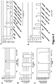
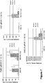
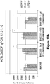
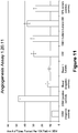
- the angiogenic potential of the cells can be increased by a combination of TNF- ⁇ , IL-1 ⁇ , and IFN- ⁇ . Exposure of the cells to this combination of factors increases pro-angiogenic gene expression, such as CXCL5, FGF2, and HGF. IL-8 may also be increased in these conditions.
- pro-angiogenic molecules can also be increased by treatment of cells with a prostaglandin-F analogue latanoprost.
- Gene expression of pro-angiogenic factors analyzed by RT-PCR shows an increase in HGF, VEGF, KITLG, and IL-8.
- the biologic prostaglandin-F also increased VEGF A levels.
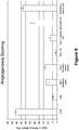
- conditioned media collected from MultiStem after four days induces angiogenesis in vitro.
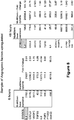
- the inventors identified multiple pro-angiogenic factors secreted by MultiStem including VEGF, CXCL5, and IL-8 and found all three factors are necessary for MultiStem induced angiogenesis.
- CXCL5 and IL-8 were not found to be expressed by cultured bone marrow derived mesenchymal stromal cells (MSC).
- MSC bone marrow derived mesenchymal stromal cells
- MultiStem can induce angiogenesis, in part, through the expression of IL-8, VEGF and CXCL5. This secretion profile is divergent from MSC and these differences are reflected in their functional activities.
- conditioned media collected from MultiStem induces angiogenesis in vitro.
- the inventors identified multiple pro-angiogenic factors secreted by MultiStem including VEGF, CXCL5 and IL-8 and found all three factors are necessary for MultiStem induced angiogenesis.
- CXCL5 and IL-8 were not expressed by cultured bone marrow derived mesenchymal stromal cells (MSC).
- MSC bone marrow derived mesenchymal stromal cells
- Ischemic injury characterized by the loss of blood flow to tissues or organs, can have devastating consequences as a result of tissue damage and cell death induced by loss of nutrients and oxygen to the ischemic area 1 .
- Acute myocardial infarction (AMI), peripheral vascular disease (PVD) and stroke are three common examples of ischemic injuries that result from loss of blood flow to the heart, limbs, and brain, respectively. These conditions can result in severe long term organ damage, limb amputation and even death from oxygen and nutrient deprivation. Treatment of these conditions often focuses on quick return of blood flow to the injured area to prevent further tissue damage, cell death and to reduce inflammation 2 .
- MultiStem® a large scale expanded adherent multipotent progenitor cell population derived from bone marrow, has been shown to be beneficial in animal models when delivered following ischemic injury such as AMI and PVD 3-6 .
- ischemic injury such as AMI and PVD 3-6 .
- Delivery of MultiStem into peri-infarct sites following induction of myocardial infarction by direct left anterior descending arterial ligation resulted in improved left ventricular contractile performance, reduced scar area, increased vascular density and improved myocardial energetic characteristics 7 .
- Mouse and human MultiStem have also been shown to improve limb movement, increase blood flow and capillary density and decreased necrosis in models of critical limb ischemia 5 . Due to low levels of engraftment of MultiStem and minimal differentiation of MultiStem into myocardium or endothelial cells, the benefits of MultiStem for AMI and PVD are believed to be derived from paracrine effects.
- MultiStem treated animals of AMI and PVD compared to vehicle treated controls suggests that MultiStem may be able to induce neo-vascularization by promoting angiogenesis. This activity may be an important mechanism of benefit in treatment of AMI and PVD. Increased vessel density ultimately results in increased blood flow and, hence, oxygen and nutrient delivery to the site of injury 8-9 .
- MultiStem Conditioned serum-free media collected from MultiStem induces angiogenesis in vitro. Multiple pro-angiogenic factors secreted by MultiStem were identified including VEGF, CXCL5 and IL-8; and immunodepletion studies demonstrated that all three factors are necessary for MultiStem induced angiogenesis. However, none of these factors alone are sufficient to induce angiogenesis.
- CXCL5 and IL-8 were not expressed by cultured bone marrow derived mesenchymal stromal cells (MSC).
- MSC bone marrow derived mesenchymal stromal cells
- conditioned media from MSC was unable to induce angiogenesis in this in vitro system.
- MSC can stabilize vessel formation in vitro and increase vessel density in ischemic animal models, although recent studies have suggested that MSC inhibit angiogenesis and cause endothelial cell death under certain conditions 11-13 .
- the results suggest that MSC do not secrete soluble factors sufficient to maintain angiogenesis in the absence of coculture with endothelial cells. Taken together, these results suggest that MultiStem and MSC have divergent secretion profiles and these differences are reflected in their paracrine activities under various conditions and settings.
- Human MultiStem was maintained in culture as described previously 7 .
- MSCs were purchased from Lonza (Walkersville, MD) and expanded in culture as described by the supplier's protocol.
- adherent cells were isolated and cultured from a fresh bone marrow using the conditions previously described for each cell line 14 .
- Human umbilical vein endothelial cells (HUVECs) (Lonza) were expanded in culture following manufacturers' instructions at a concentration of 2500 cells/cm 2 .
- HUVECs were used between passage three and five, seeding at 3000 cells/cm2 and cultured for three days, at which time, the confluence is approximately 70-80% prior to use in angiogenesis assay.
- MultiStem was plated into a tissue culture flask containing MultiStem culture media. Twenty-four hours later, serum containing media was removed, cells were washed with IX PBS and human MultiStem culture media containing growth factors but lacking serum was added. Cells were cultured for 4 days without any media change and on day 4, the serum-free conditioned media was collected, spun down at 1900rpm for 5 min at 4°C, aliquoted, and stored at -80°C.
- Panomics Human Angiogenesis Antibody Array was performed according to the manufacturer's instructions using 2 ml of a 2-fold diluted 4 day serum-free MultiStem conditioned media sample.
- Angiogenesis antibody array (R& D systems, Minneapolis, MN) was performed according to the manufacturer's instructions using 2 ml of a 2-fold diluted 3 day conditioned media samples from MultiStem and MSC derived from the same donor.
- Protein levels of IL-8, VEGF and CXCL5 were determined by ELISA (R&D Systems). All isoforms of VEGF are detected. Error bars are expressed +/- standard deviation.
- Protein A-agarose beads (Santa Cruz, Santa Cruz, CA) were used at a concentration of 75 1 of 50% slurry per 1ml of CM.
- Mouse anti-human VEGF monoclonal antibody (Santa Cruz) was used at a concentration of 4ug/ml of CM and rabbit anti-human IL-8 polyclonal antibody (Millipore, Billerica, MA) was used at 2ug/ml CM.
- Normal mouse IgG (Santa Cruz) and ChromPure rabbit IgG (Jackson ImmunoResearch, West Grove, PA) were used as isotype controls.
- Protein A-agarose beads were pre-incubated with anti-VEGF, anti-IL-8 antibody or isotype controls overnight at 4°C with rotation followed by 4 washes with ice-cold IX PBS, spinning at 2500rpm, 2 minutes, 4°C.
- CM was immunodepleted for 2 hours at 4°C with rotation in 6ml aliquots followed by filtration through 0.45 M filter to rid of any residual beads.
- CM treated with mouse IgG-AC was used as isotype control.
- Recombinant human VEGF121 eBioscience, San Diego, CA
- VEGF165 R&D Systems
- CXCL5 was neutralized using human CXCL5/ENA-78 antibody (R&D Systems) at a concentration of 10 ⁇ g/ml CM for 2 hours at 4°C with rotation.
- Normal total goat IgG (Jackson ImmunoResearch,) at a concentration of 10 ⁇ gg/ml CM was used as an isotype control.
- Additional controls used are endothelial growth medium (EGM, Lonza) spiked with either human ENA-78 neutralizing antibody or normal total goat IgG at concentration of 10 ⁇ g/ml.
- Growth factor-reduced Matrigel (BD Bioscience, San Jose, CA) was thawed on ice at 4°C overnight and used at a concentration of 6.0-6.5mg/ml, diluted on ice using ice-cold IX PBS. Four-hundred microliters of Matrigel was distributed into the inner wells of a 24-well tissue culture plate and allowed to solidify for 1 hour at 37°C. Addition of Matrigel was done on ice and outer wells of plate were filled with 1ml of IX PBS.
- HUVECs were harvested according to the following protocol: Cells were washed with IX PBS followed by a brief rinse with 0.25X trypsin-EDTA and then quenched using the IX PBS (with residual serum) wash. Cells were resuspended in endothelial cell basal media (EBM) and counted. HUVECs were added to the CM, other experimental conditions and controls at a concentration of 55,000 cells/ml/well. Each sample and control was assayed in triplicate. Plates were incubated for 6 hours or 18 hours at 5% CO2 and 37°C to allow for tube formation. Four fields per well were analyzed for a total of 12 fields. Pictures were taken at using 10 X objective. Angiogenesis was scored by counting the number of tubes formed between cells. Results are expressed as average tubes formed per field +/- SEM.
- MultiStem results in increased vessel density bordering the area of injury compared with vehicle treated controls, suggesting that MultiStem induces neo-vascularization and angiogenesis.
- an in vitro angiogenesis assay using conditioned media from MultiStem was utilized. MultiStem were plated under normal conditions for 24 hours. The cells were then transferred to serum-free conditions to generate conditioned media for four days. This media was then tested for angiogenic activity in an in vitro tube formation assay.
- HUVECs were plated on reduced growth factor Matrigel that was further diluted to a concentration that did not produce any spontaneous angiogenesis with serum-free basal MultiStem media or serum-free endothelial cell media.
- HUVECs were plated in conditioned media, basal media or endothelial growth media for 18 hours.
- Angiogenesis was measured as the average number of tubes formed per field of view for each condition. In the absence of any additional factors, the presence of serum alone in basal media can induce angiogenesis. Therefore, serum-free media was used for all the experiments. Robust, complex tube formation was observed in serum containing endothelial cell media while serum-free endothelial cell media or serum-free basal MultiStem media showed virtually no induction of tube formation.
- the inventors found that serum-free conditioned media from four day cultures of MultiStem induced angiogenesis compared to basal media ( Figure1 A,B ).
- MultiStem serum free conditioned media was analyzed on an angiogenesis antibody array ( Figure 1 C) .
- Many pro-angiogeneic factors, as well as a few angiostatic factors are secreted by MultiStem into the media.
- IL-8 interleukin 8
- CXCL5 another potent angiogenic cytokine
- VEGF, CXCL5 and IL-8 are expressed at physiologically active levels in four day MultiStem conditioned media ( Figure D-F).
- VEGF's role as a key factor involved in the induction of angiogenesis prompted us to examine if VEGF was necessary for MultiStem's pro-angiogenic activity 19-20 .
- VEGF antibody Using a VEGF antibody, VEGF was immunodepleted from MultiStem conditioned media. In order to ensure that VEGF was truly depleted from the media, the levels of VEGF from the immunodepeleted media were determined using a VEGF ELISA. The levels of VEGF were reduced by more than 95% in the immunodepleted media compared to the conditioned media and IgG alone depleted media ( Figure 2 A) . CXCL5 levels and IL-8 levels were not affected by VEGF immunodepletion ( Figure 2 B,C ). In the absence of VEGF, the induction of angiogenesis by MultiStem conditioned media was reduced ( Figure 2 , Supplemental Figure 1 ). These results demonstrate that VEGF is necessary in MultiStem conditioned media to induce angiogenesis.
- VEGF-A the most studied form of VEGF, commonly referred to as VEGF
- VEGF 165 the most studied form of VEGF, commonly referred to as VEGF
- Figure 2A,C the inventors found that although neither isoform alone was sufficient to completely restore the levels of angiogenesis previously observed with MultiStem conditioned media, 250 pg/ml of VEGF 121 was sufficient to restore some angiogenesis ( Figure 2C ).
- VEGF 165 50 pg/ml was sufficient to restore some levels of angiogenesis and the addition of more VEGF165 did not increase the levels of angiogenesis by any appreciable amount ( Figure 2D ).
- CXCL5 and IL-8 are both necessary for normal levels of MultiStem-induced angiogenesis but are not sufficient to induce angiogenesis
- MSC express VEGF but are unable to initiate angiogenesis in the HUVEC in vitro assay
- VEGF, CXCL5 and IL-8 were examined and VEGF was found to be expressed at higher levels than in MultiStem conditioned media, while CXCL5 and IL-8 were not expressed at detectable levels in MSC conditioned media.
- the levels of VEGF, IL-8 and CXCL5 expressed by MSC were examined under their normal culture conditions and compared to the protein levels of these factors found in MultiStem conditioned media under MultiStem's culture conditions.
- the inventors derived the MSC and MultiStem from the same donor to eliminate any genetic variation between the two lines.
- ischemic injuries such as acute myocardial infarction, stroke and peripheral vascular disease 21-30 . These injuries induce cellular and tissue damage from the initial loss of oxygen and nutrients in the affected tissue but also from subsequent inflammation in the region. The quick and sustained recovery of blood flow can reduce damage and inflammation within the ischemic region.
- the original intent of cell based therapies was the regeneration and repair of lost and damaged tissues following injury through the delivery and subsequent differentiation of exogenous stem cells.
- stem cell therapies have shown that many stem cells work primarily through paracrine effects rather than regeneration since many of the cell types are no longer detectable a few days post-delivery 31-33 .
- the therapeutic hypotheses are that these cell populations can provide trophic support to the injured tissue by regulating immune and inflammatory cells, limiting apoptosis, stimulating neo-angiogenesis, and recruiting host tissue for repair.
- Benefit is likely derived from a dynamic cascade of these pathways, and different cell populations may exert influences more strongly on certain pathways. Selection of the most appropriate adherent stem cell population for treatment may reflect both potency of a given population for key pathways, as well as time of delivery to effectively mediate the response. It is therefore important to establish standardized assays for these pathways in order to provide comparative data, and then to correlate these in vitro surrogates of activity to injury and recovery.
- Multipotent adult progenitor cells are an adherent adult stem cell population derived from bone marrow cultures. Previous in vivo studies have shown an increase in vessel density in animals treated with MultiStem following ischemic injury 4, 7 . In this study, we have shown that MultiStem secretes factors that can induce angiogenesis in an in vitro tube formation assay. Further analysis revealed that MultiStem secretes a variety of pro-angiogenic factors including VEGF, IL-8, and CXCL5. Two of these factors, CXCL5 and IL-8, are highly differentially secreted by MultiStem compared with MSC, which secrete very little, if any, CXCL5 and IL-8.
- VEGF, CXCL5, and IL-8 all are required for MultiStem induced angiogenesis. Removal or inhibition of any of these factors greatly reduces the ability of MultiStem conditioned media to promote angiogenesis.
- VEGF165 is the major isoform involved in angiogenesis. However, multiple VEGF isoforms may be responsible for MultiStem induced angiogenesis since tube formation could not be restored to 100% using either VEGF121 or VEGF165 isoforms independently in VEGF immunodepleted MultiStem conditioned media.
- stem cells to treat ischemic injuries could result in the delivery of multiple angiogenic factors directly to the site of injury, by cells that respond and home to the hypoxic and inflammatory microenvironment, achieving a dynamic balance to stimulate an appropriate angiogenic response.
- stem cells such as MultiStem could also simultaneously prevent tissue damage through immunomodulatory and anti-apoptotic mechanisms.
- MultiStem is, indeed, capable of inducing angiogenesis directly through the expression of at least three pro-angiogenic factors.
- MSC express and secrete high levels of VEGF
- conditioned media from MSC was insufficient to induce angiogenesis in this in vitro assay system.
- MSC have been shown to stabilize vessel formation in vitro in previous studies. However, many of these studies examine vessel formation at an earlier time point, such as at 4-6 hours or under different conditions 25-27, 38 . The inventors found that at these earlier time points, there is a higher level of background of angiogenesis in negative controls which is not stable at 24 hours. Similarly, we found at 6 hours, MSC can induce some level of angiogenesis which is subsequently lost by the 24 hour time point. In contrast, MultiStem induces tube formation that remains stable at 24 hours.
- MSC can support angiogenesis in the short term, in the absence of other factors, this vessel formation is not stable. These results reflect data from earlier studies that VEGF alone is not sufficient to initiate stable vessel formation at the levels expressed by these cells 24 . In the context of previous in vivo experiments which show increased vessel density in ischemic injuries treated with MSC, MSC may increase vessel density by inducing endogenous inflammatory cells or tissue progenitors to promote angiogenesis.
- Figures 7-12 show that expression of angiogenic factors can be increased in the MultiStem (MAPC) preparation.
Landscapes
- Health & Medical Sciences (AREA)
- Life Sciences & Earth Sciences (AREA)
- Engineering & Computer Science (AREA)
- Chemical & Material Sciences (AREA)
- Bioinformatics & Cheminformatics (AREA)
- Biomedical Technology (AREA)
- General Health & Medical Sciences (AREA)
- Organic Chemistry (AREA)
- Medicinal Chemistry (AREA)
- Biotechnology (AREA)
- Cell Biology (AREA)
- Veterinary Medicine (AREA)
- Public Health (AREA)
- Pharmacology & Pharmacy (AREA)
- Zoology (AREA)
- Animal Behavior & Ethology (AREA)
- Immunology (AREA)
- Genetics & Genomics (AREA)
- Wood Science & Technology (AREA)
- Nuclear Medicine, Radiotherapy & Molecular Imaging (AREA)
- General Chemical & Material Sciences (AREA)
- Chemical Kinetics & Catalysis (AREA)
- Developmental Biology & Embryology (AREA)
- Cardiology (AREA)
- Biochemistry (AREA)
- Microbiology (AREA)
- Heart & Thoracic Surgery (AREA)
- Urology & Nephrology (AREA)
- General Engineering & Computer Science (AREA)
- Hematology (AREA)
- Epidemiology (AREA)
- Reproductive Health (AREA)
- Molecular Biology (AREA)
- Virology (AREA)
- Vascular Medicine (AREA)
- Food Science & Technology (AREA)
- Physics & Mathematics (AREA)
- Analytical Chemistry (AREA)
- Tropical Medicine & Parasitology (AREA)
- General Physics & Mathematics (AREA)
Applications Claiming Priority (2)
| Application Number | Priority Date | Filing Date | Title |
|---|---|---|---|
| US30810310P | 2010-02-25 | 2010-02-25 | |
| PCT/US2011/025846 WO2011106365A1 (en) | 2010-02-25 | 2011-02-23 | Modulation of angiogenesis |
Publications (3)
| Publication Number | Publication Date |
|---|---|
| EP2539439A1 EP2539439A1 (en) | 2013-01-02 |
| EP2539439A4 EP2539439A4 (en) | 2013-09-04 |
| EP2539439B1 true EP2539439B1 (en) | 2018-10-17 |
Family
ID=44507175
Family Applications (1)
| Application Number | Title | Priority Date | Filing Date |
|---|---|---|---|
| EP11747958.4A Active EP2539439B1 (en) | 2010-02-25 | 2011-02-23 | Modulation of angiogenesis |
Country Status (12)
| Country | Link |
|---|---|
| EP (1) | EP2539439B1 (enExample) |
| JP (7) | JP2013520509A (enExample) |
| KR (1) | KR101900507B1 (enExample) |
| CN (1) | CN102858956B (enExample) |
| AU (1) | AU2011220940C1 (enExample) |
| BR (1) | BR112012021451B8 (enExample) |
| CA (1) | CA2791331A1 (enExample) |
| DK (1) | DK2539439T3 (enExample) |
| ES (1) | ES2703428T3 (enExample) |
| IL (3) | IL221575A (enExample) |
| SG (4) | SG10201501353YA (enExample) |
| WO (1) | WO2011106365A1 (enExample) |
Families Citing this family (5)
| Publication number | Priority date | Publication date | Assignee | Title |
|---|---|---|---|---|
| SG10201913920PA (en) | 2010-05-12 | 2020-03-30 | Abt Holding Co | Modulation of splenocytes in cell therapy |
| EP3533465A4 (en) * | 2016-09-26 | 2020-07-01 | MPO, Inc. | Egg, fertilized egg, or embryo quality improving agent |
| KR102656200B1 (ko) * | 2017-06-16 | 2024-04-12 | 아이엠비에이 - 인스티튜트 퓌어 몰레쿨라레 바이오테크놀로지 게엠베하 | 혈관 오가노이드, 상기 오가노이드의 제조 및 사용 방법 |
| DE102017116204B4 (de) * | 2017-07-18 | 2024-06-27 | Universität Rostock | Verfahren zur Vorhersage der Antwort auf die kardiovaskuläre Regeneration |
| KR102498202B1 (ko) * | 2019-01-24 | 2023-02-10 | 가톨릭대학교 산학협력단 | 줄기세포의 혈관 재생 유효성 평가 방법 |
Citations (1)
| Publication number | Priority date | Publication date | Assignee | Title |
|---|---|---|---|---|
| WO2008148105A1 (en) * | 2007-05-25 | 2008-12-04 | Medistem Laboratories, Inc. | Endometrial stem cells and methods of making and using same |
Family Cites Families (55)
| Publication number | Priority date | Publication date | Assignee | Title |
|---|---|---|---|---|
| US4304907A (en) * | 1972-05-10 | 1981-12-08 | The Upjohn Company | Bicyclo lactone intermediates for prostaglandin analogs |
| US4353888A (en) | 1980-12-23 | 1982-10-12 | Sefton Michael V | Encapsulation of live animal cells |
| US4657866A (en) | 1982-12-21 | 1987-04-14 | Sudhir Kumar | Serum-free, synthetic, completely chemically defined tissue culture media |
| US4744933A (en) | 1984-02-15 | 1988-05-17 | Massachusetts Institute Of Technology | Process for encapsulation and encapsulated active material system |
| US4749620A (en) | 1984-02-15 | 1988-06-07 | Massachusetts Institute Of Technology | Encapsulated active material system |
| JPH0628570B2 (ja) | 1986-02-13 | 1994-04-20 | 雪印乳業株式会社 | カプセル体の製造方法及び装置 |
| EP0301777A1 (en) | 1987-07-28 | 1989-02-01 | Queen's University At Kingston | Multiple membrane microencapsulation |
| EP0435928A1 (en) | 1988-09-21 | 1991-07-10 | Animal Biotechnology Cambridge Limited | Derivation of pluripotential embryonic cell lines from domestic animals |
| US5089272A (en) | 1989-03-29 | 1992-02-18 | Snow Brand Milk Products Co., Ltd. | Process for producing capsules having a permeability-controllable membrane |
| US7037721B1 (en) | 1990-01-29 | 2006-05-02 | Hy-Gene Biomedical, Inc. | Protein-free defined media for the growth of normal human keratinocytes |
| US5084350A (en) | 1990-02-16 | 1992-01-28 | The Royal Institution For The Advance Of Learning (Mcgill University) | Method for encapsulating biologically active material including cells |
| AU2882992A (en) | 1991-11-06 | 1993-06-07 | Arthur A. Axelrad | Cell culture medium |
| US5578442A (en) | 1992-03-23 | 1996-11-26 | Vivorx, Inc. | Graft copolymers of polycationic species and water-soluble polymers, and use therefor |
| US5340740A (en) | 1992-05-15 | 1994-08-23 | North Carolina State University | Method of producing an avian embryonic stem cell culture and the avian embryonic stem cell culture produced by the process |
| US5453357A (en) | 1992-10-08 | 1995-09-26 | Vanderbilt University | Pluripotential embryonic stem cells and methods of making same |
| US7153684B1 (en) | 1992-10-08 | 2006-12-26 | Vanderbilt University | Pluripotential embryonic stem cells and methods of making same |
| US5766951A (en) | 1992-11-12 | 1998-06-16 | Quality Biological, Inc. | Serum-free medium supporting growth and proliferation of normal bone marrow cells |
| CA2169292C (en) | 1993-08-12 | 2010-11-23 | E. Edward Baetge | Improved compositions and methods for the delivery of biologically active molecules using genetically altered cells contained in biocompatible immunoisolatory capsules |
| US6037174A (en) | 1993-08-23 | 2000-03-14 | Nexell Therapeutics, Inc. | Preparation of serum-free suspensions of human hematopoietic cells or precursor cells |
| US5409813A (en) | 1993-09-30 | 1995-04-25 | Systemix, Inc. | Method for mammalian cell separation from a mixture of cell populations |
| US6432711B1 (en) | 1993-11-03 | 2002-08-13 | Diacrin, Inc. | Embryonic stem cells capable of differentiating into desired cell lines |
| FR2726003B1 (fr) | 1994-10-21 | 2002-10-18 | Agronomique Inst Nat Rech | Milieu de culture de cellules embryonnaires totipotentes aviaires, procede de culture de ces cellules, et cellules embryonnaires totipotentes aviaires |
| US5914268A (en) | 1994-11-21 | 1999-06-22 | National Jewish Center For Immunology & Respiratory Medicine | Embryonic cell populations and methods to isolate such populations |
| US5874301A (en) | 1994-11-21 | 1999-02-23 | National Jewish Center For Immunology And Respiratory Medicine | Embryonic cell populations and methods to isolate such populations |
| US5843780A (en) | 1995-01-20 | 1998-12-01 | Wisconsin Alumni Research Foundation | Primate embryonic stem cells |
| US5908782A (en) | 1995-06-05 | 1999-06-01 | Osiris Therapeutics, Inc. | Chemically defined medium for human mesenchymal stem cells |
| US6190910B1 (en) | 1996-05-21 | 2001-02-20 | The Institute Of Physical And Chemical Research | Mouse embryonic stem cell lines |
| US5945337A (en) | 1996-10-18 | 1999-08-31 | Quality Biological, Inc. | Method for culturing CD34+ cells in a serum-free medium |
| AU7984498A (en) | 1997-06-25 | 1999-01-04 | Government Of The United States Of America, As Represented By The Secretary Of The Department Of Health And Human Services, The | Serum-free cell growth medium |
| EP1129176A4 (en) | 1998-11-09 | 2002-10-30 | Es Cell Int Pte Ltd | EMBRYONIC STEM CELLS |
| CA2348687A1 (en) | 1998-11-09 | 2000-05-18 | Consorzio Per La Gestione Del Centro Di Biotecnologie Avanzate | Serum free medium for chondrocyte-like cells |
| US8609412B2 (en) * | 1999-08-05 | 2013-12-17 | Regents Of The University Of Minnesota | Mapc generation of lung tissue |
| US8075881B2 (en) * | 1999-08-05 | 2011-12-13 | Regents Of The University Of Minnesota | Use of multipotent adult stem cells in treatment of myocardial infarction and congestive heart failure |
| DK1226233T3 (da) * | 1999-08-05 | 2011-10-03 | Abt Holding Co | Multipotente voksne stamceller og fremgangsmåder til isolering heraf |
| US7015037B1 (en) * | 1999-08-05 | 2006-03-21 | Regents Of The University Of Minnesota | Multiponent adult stem cells and methods for isolation |
| US20060003323A1 (en) * | 2000-03-01 | 2006-01-05 | John Alsobrook | Therapeutic polypeptides, nucleic acids encoding same, and methods of use |
| JP4795616B2 (ja) | 2000-06-20 | 2011-10-19 | イーエス・セル・インターナショナル・プライヴェート・リミテッド | Bmp−2経路アンタゴニストの存在下においてes細胞を培養することによって胚性幹細胞の分化をコントロールする方法 |
| US8071740B2 (en) * | 2000-11-17 | 2011-12-06 | Vascular Biogenics Ltd. | Promoters exhibiting endothelial cell specificity and methods of using same for regulation of angiogenesis |
| US8039261B2 (en) * | 2000-11-17 | 2011-10-18 | Vascular Biogenics Ltd. | Promoters exhibiting endothelial cell specificity and methods of using same for regulation of angiogenesis |
| CA2438501C (en) | 2001-02-14 | 2014-09-16 | Leo T. Furcht | Multipotent adult stem cells, sources thereof, methods of obtaining and maintaining same, methods of differentiation thereof, methods of use thereof and cells derived thereof |
| EP1421181A1 (en) | 2001-08-23 | 2004-05-26 | Reliance Life Sciences Pvt., Ltd. | ISOLATION OF INNER CELL MASS FOR THE ESTABLISHMENT OF HUMAN EMBRYONIC STEM CELL (hESC) LINES |
| US9969980B2 (en) | 2001-09-21 | 2018-05-15 | Garnet Biotherapeutics | Cell populations which co-express CD49c and CD90 |
| US6703279B2 (en) | 2002-01-04 | 2004-03-09 | Promos Technologies, Inc. | Semiconductor device having contact of Si-Ge combined with cobalt silicide |
| WO2003064598A2 (en) | 2002-01-25 | 2003-08-07 | Genzyme Corporation | Serum-free media for chondrocytes and methods of use thereof |
| US7145057B2 (en) | 2002-02-01 | 2006-12-05 | Origen Therapeutics, Inc. | Chimeric bird from embryonic stem cells |
| AU2003222252A1 (en) * | 2002-03-12 | 2003-09-29 | The General Hospital Corporation | Use of signal transduction inhibitors and combination therapies for the prevention or treatment of cancer and angiogenesis related diseases |
| US20030199464A1 (en) * | 2002-04-23 | 2003-10-23 | Silviu Itescu | Regeneration of endogenous myocardial tissue by induction of neovascularization |
| GB0221481D0 (en) | 2002-09-16 | 2002-10-23 | Richards John M | Grip for hand held instruments |
| WO2004039248A2 (en) * | 2002-10-31 | 2004-05-13 | The General Hospital Corporation | Repairing or replacing tissues or organs |
| JP2005065661A (ja) * | 2003-08-20 | 2005-03-17 | Akihiro Yamaguchi | ヒト血管内皮細胞のVEGF誘導性活性化及び血管新生におけるIdの役割 |
| FR2893252B1 (fr) * | 2005-11-17 | 2008-02-15 | Engelhard Lyon Sa | Extraits vegetaux stimulant has2 |
| US11992507B2 (en) * | 2006-01-23 | 2024-05-28 | Abt Holding Company | MAPC therapeutics without adjunctive immunosuppressive treatment |
| US20070253937A1 (en) * | 2006-04-26 | 2007-11-01 | Young-Sup Yoon | Novel multipotent stem cells and use thereof |
| US20100239535A1 (en) * | 2006-05-17 | 2010-09-23 | University Of Basel | Method for isolating homogeneous populations of transduced progenitors stably expressing specific levels of a transgene |
| AU2010276201B2 (en) * | 2009-07-21 | 2013-10-17 | Healios K.K. | Use of stem cells to reduce leukocyte extravasation |
-
2011
- 2011-02-23 BR BR112012021451A patent/BR112012021451B8/pt active IP Right Grant
- 2011-02-23 EP EP11747958.4A patent/EP2539439B1/en active Active
- 2011-02-23 SG SG10201501353YA patent/SG10201501353YA/en unknown
- 2011-02-23 KR KR1020127024595A patent/KR101900507B1/ko not_active Expired - Fee Related
- 2011-02-23 CN CN201180020289.8A patent/CN102858956B/zh not_active Expired - Fee Related
- 2011-02-23 WO PCT/US2011/025846 patent/WO2011106365A1/en not_active Ceased
- 2011-02-23 SG SG10201913904PA patent/SG10201913904PA/en unknown
- 2011-02-23 ES ES11747958T patent/ES2703428T3/es active Active
- 2011-02-23 JP JP2012555095A patent/JP2013520509A/ja active Pending
- 2011-02-23 DK DK11747958.4T patent/DK2539439T3/en active
- 2011-02-23 SG SG2012063194A patent/SG183498A1/en unknown
- 2011-02-23 CA CA2791331A patent/CA2791331A1/en not_active Abandoned
- 2011-02-23 AU AU2011220940A patent/AU2011220940C1/en active Active
- 2011-02-23 SG SG10202102952YA patent/SG10202102952YA/en unknown
-
2012
- 2012-08-22 IL IL221575A patent/IL221575A/en active IP Right Grant
-
2015
- 2015-03-06 JP JP2015044246A patent/JP6312326B2/ja active Active
- 2015-08-21 JP JP2015163449A patent/JP2015214585A/ja active Pending
-
2017
- 2017-05-17 JP JP2017097945A patent/JP2017137355A/ja not_active Withdrawn
- 2017-11-26 IL IL255914A patent/IL255914B/en active IP Right Grant
-
2019
- 2019-02-28 JP JP2019036165A patent/JP2019089846A/ja not_active Withdrawn
- 2019-11-26 IL IL270957A patent/IL270957B2/en unknown
-
2021
- 2021-01-08 JP JP2021002029A patent/JP2021058218A/ja not_active Withdrawn
-
2023
- 2023-01-06 JP JP2023001052A patent/JP2023026662A/ja active Pending
Patent Citations (1)
| Publication number | Priority date | Publication date | Assignee | Title |
|---|---|---|---|---|
| WO2008148105A1 (en) * | 2007-05-25 | 2008-12-04 | Medistem Laboratories, Inc. | Endometrial stem cells and methods of making and using same |
Non-Patent Citations (1)
| Title |
|---|
| LEI Y ET AL: "THERAPEUTIC ANGIOGENESIS. DEVISING NEW STRATEGIES BASED ON PAST EXPERIENCE", BASIC RESEARCH IN CARDIOLOGY, STEINKOPFF, DARMSTADT, DE, vol. 99, no. 2, 1 January 2004 (2004-01-01), pages 121 - 132, XP008065176, ISSN: 0300-8428, DOI: 10.1007/S00395-004-0447-X * |
Also Published As
Similar Documents
| Publication | Publication Date | Title |
|---|---|---|
| US20190048314A1 (en) | Modulation of Angiogenesis | |
| DK2568991T6 (en) | MODULATION OF SPLENOCYTES IN CELL THERAPY | |
| US20230131225A1 (en) | Modulation of Microglia Activation | |
| JP2023026662A (ja) | 血管形成の調節 | |
| AU2011220721B2 (en) | Modulation of macrophage activation | |
| AU2016256806B2 (en) | Modulation of angiogenesis | |
| AU2014250727B2 (en) | Modulation of angiogenesis | |
| HK1174053B (en) | Modulation of angiogenesis | |
| HK1174053A (en) | Modulation of angiogenesis | |
| AU2015202292B2 (en) | Modulation of macrophage activation | |
| HK40056997A (en) | Modulation of macrophage activation |
Legal Events
| Date | Code | Title | Description |
|---|---|---|---|
| PUAI | Public reference made under article 153(3) epc to a published international application that has entered the european phase |
Free format text: ORIGINAL CODE: 0009012 |
|
| 17P | Request for examination filed |
Effective date: 20120919 |
|
| AK | Designated contracting states |
Kind code of ref document: A1 Designated state(s): AL AT BE BG CH CY CZ DE DK EE ES FI FR GB GR HR HU IE IS IT LI LT LU LV MC MK MT NL NO PL PT RO RS SE SI SK SM TR |
|
| DAX | Request for extension of the european patent (deleted) | ||
| REG | Reference to a national code |
Ref country code: HK Ref legal event code: DE Ref document number: 1174053 Country of ref document: HK |
|
| RIC1 | Information provided on ipc code assigned before grant |
Ipc: C12N 5/074 20100101AFI20130724BHEP Ipc: A61K 38/19 20060101ALI20130724BHEP Ipc: A61P 9/00 20060101ALI20130724BHEP Ipc: A61K 38/20 20060101ALI20130724BHEP |
|
| A4 | Supplementary search report drawn up and despatched |
Effective date: 20130805 |
|
| 17Q | First examination report despatched |
Effective date: 20151015 |
|
| STAA | Information on the status of an ep patent application or granted ep patent |
Free format text: STATUS: EXAMINATION IS IN PROGRESS |
|
| REG | Reference to a national code |
Ref country code: DE Ref legal event code: R079 Ref document number: 602011052984 Country of ref document: DE Free format text: PREVIOUS MAIN CLASS: C12N0005074000 Ipc: A61K0035545000 |
|
| GRAP | Despatch of communication of intention to grant a patent |
Free format text: ORIGINAL CODE: EPIDOSNIGR1 |
|
| STAA | Information on the status of an ep patent application or granted ep patent |
Free format text: STATUS: GRANT OF PATENT IS INTENDED |
|
| RIC1 | Information provided on ipc code assigned before grant |
Ipc: A61P 9/00 20060101ALI20180222BHEP Ipc: A61K 38/20 20060101ALI20180222BHEP Ipc: A61K 35/545 20150101AFI20180222BHEP Ipc: G01N 33/50 20060101ALI20180222BHEP Ipc: C12N 5/074 20100101ALI20180222BHEP Ipc: A61K 38/19 20060101ALI20180222BHEP |
|
| INTG | Intention to grant announced |
Effective date: 20180312 |
|
| GRAJ | Information related to disapproval of communication of intention to grant by the applicant or resumption of examination proceedings by the epo deleted |
Free format text: ORIGINAL CODE: EPIDOSDIGR1 |
|
| STAA | Information on the status of an ep patent application or granted ep patent |
Free format text: STATUS: EXAMINATION IS IN PROGRESS |
|
| INTC | Intention to grant announced (deleted) | ||
| GRAR | Information related to intention to grant a patent recorded |
Free format text: ORIGINAL CODE: EPIDOSNIGR71 |
|
| GRAS | Grant fee paid |
Free format text: ORIGINAL CODE: EPIDOSNIGR3 |
|
| STAA | Information on the status of an ep patent application or granted ep patent |
Free format text: STATUS: GRANT OF PATENT IS INTENDED |
|
| GRAA | (expected) grant |
Free format text: ORIGINAL CODE: 0009210 |
|
| STAA | Information on the status of an ep patent application or granted ep patent |
Free format text: STATUS: THE PATENT HAS BEEN GRANTED |
|
| INTG | Intention to grant announced |
Effective date: 20180906 |
|
| AK | Designated contracting states |
Kind code of ref document: B1 Designated state(s): AL AT BE BG CH CY CZ DE DK EE ES FI FR GB GR HR HU IE IS IT LI LT LU LV MC MK MT NL NO PL PT RO RS SE SI SK SM TR |
|
| REG | Reference to a national code |
Ref country code: GB Ref legal event code: FG4D |
|
| REG | Reference to a national code |
Ref country code: CH Ref legal event code: EP |
|
| REG | Reference to a national code |
Ref country code: IE Ref legal event code: FG4D |
|
| REG | Reference to a national code |
Ref country code: DE Ref legal event code: R096 Ref document number: 602011052984 Country of ref document: DE Ref country code: AT Ref legal event code: REF Ref document number: 1053192 Country of ref document: AT Kind code of ref document: T Effective date: 20181115 |
|
| REG | Reference to a national code |
Ref country code: NL Ref legal event code: FP |
|
| REG | Reference to a national code |
Ref country code: DK Ref legal event code: T3 Effective date: 20190123 |
|
| REG | Reference to a national code |
Ref country code: SE Ref legal event code: TRGR |
|
| REG | Reference to a national code |
Ref country code: ES Ref legal event code: FG2A Ref document number: 2703428 Country of ref document: ES Kind code of ref document: T3 Effective date: 20190308 |
|
| REG | Reference to a national code |
Ref country code: LT Ref legal event code: MG4D |
|
| REG | Reference to a national code |
Ref country code: AT Ref legal event code: MK05 Ref document number: 1053192 Country of ref document: AT Kind code of ref document: T Effective date: 20181017 |
|
| PG25 | Lapsed in a contracting state [announced via postgrant information from national office to epo] |
Ref country code: FI Free format text: LAPSE BECAUSE OF FAILURE TO SUBMIT A TRANSLATION OF THE DESCRIPTION OR TO PAY THE FEE WITHIN THE PRESCRIBED TIME-LIMIT Effective date: 20181017 Ref country code: LT Free format text: LAPSE BECAUSE OF FAILURE TO SUBMIT A TRANSLATION OF THE DESCRIPTION OR TO PAY THE FEE WITHIN THE PRESCRIBED TIME-LIMIT Effective date: 20181017 Ref country code: NO Free format text: LAPSE BECAUSE OF FAILURE TO SUBMIT A TRANSLATION OF THE DESCRIPTION OR TO PAY THE FEE WITHIN THE PRESCRIBED TIME-LIMIT Effective date: 20190117 Ref country code: IS Free format text: LAPSE BECAUSE OF FAILURE TO SUBMIT A TRANSLATION OF THE DESCRIPTION OR TO PAY THE FEE WITHIN THE PRESCRIBED TIME-LIMIT Effective date: 20190217 Ref country code: PL Free format text: LAPSE BECAUSE OF FAILURE TO SUBMIT A TRANSLATION OF THE DESCRIPTION OR TO PAY THE FEE WITHIN THE PRESCRIBED TIME-LIMIT Effective date: 20181017 Ref country code: HR Free format text: LAPSE BECAUSE OF FAILURE TO SUBMIT A TRANSLATION OF THE DESCRIPTION OR TO PAY THE FEE WITHIN THE PRESCRIBED TIME-LIMIT Effective date: 20181017 Ref country code: BG Free format text: LAPSE BECAUSE OF FAILURE TO SUBMIT A TRANSLATION OF THE DESCRIPTION OR TO PAY THE FEE WITHIN THE PRESCRIBED TIME-LIMIT Effective date: 20190117 Ref country code: LV Free format text: LAPSE BECAUSE OF FAILURE TO SUBMIT A TRANSLATION OF THE DESCRIPTION OR TO PAY THE FEE WITHIN THE PRESCRIBED TIME-LIMIT Effective date: 20181017 Ref country code: AT Free format text: LAPSE BECAUSE OF FAILURE TO SUBMIT A TRANSLATION OF THE DESCRIPTION OR TO PAY THE FEE WITHIN THE PRESCRIBED TIME-LIMIT Effective date: 20181017 |
|
| PG25 | Lapsed in a contracting state [announced via postgrant information from national office to epo] |
Ref country code: PT Free format text: LAPSE BECAUSE OF FAILURE TO SUBMIT A TRANSLATION OF THE DESCRIPTION OR TO PAY THE FEE WITHIN THE PRESCRIBED TIME-LIMIT Effective date: 20190217 Ref country code: AL Free format text: LAPSE BECAUSE OF FAILURE TO SUBMIT A TRANSLATION OF THE DESCRIPTION OR TO PAY THE FEE WITHIN THE PRESCRIBED TIME-LIMIT Effective date: 20181017 Ref country code: GR Free format text: LAPSE BECAUSE OF FAILURE TO SUBMIT A TRANSLATION OF THE DESCRIPTION OR TO PAY THE FEE WITHIN THE PRESCRIBED TIME-LIMIT Effective date: 20190118 Ref country code: RS Free format text: LAPSE BECAUSE OF FAILURE TO SUBMIT A TRANSLATION OF THE DESCRIPTION OR TO PAY THE FEE WITHIN THE PRESCRIBED TIME-LIMIT Effective date: 20181017 |
|
| REG | Reference to a national code |
Ref country code: DE Ref legal event code: R097 Ref document number: 602011052984 Country of ref document: DE |
|
| PG25 | Lapsed in a contracting state [announced via postgrant information from national office to epo] |
Ref country code: CZ Free format text: LAPSE BECAUSE OF FAILURE TO SUBMIT A TRANSLATION OF THE DESCRIPTION OR TO PAY THE FEE WITHIN THE PRESCRIBED TIME-LIMIT Effective date: 20181017 |
|
| PLBE | No opposition filed within time limit |
Free format text: ORIGINAL CODE: 0009261 |
|
| STAA | Information on the status of an ep patent application or granted ep patent |
Free format text: STATUS: NO OPPOSITION FILED WITHIN TIME LIMIT |
|
| PG25 | Lapsed in a contracting state [announced via postgrant information from national office to epo] |
Ref country code: SK Free format text: LAPSE BECAUSE OF FAILURE TO SUBMIT A TRANSLATION OF THE DESCRIPTION OR TO PAY THE FEE WITHIN THE PRESCRIBED TIME-LIMIT Effective date: 20181017 Ref country code: RO Free format text: LAPSE BECAUSE OF FAILURE TO SUBMIT A TRANSLATION OF THE DESCRIPTION OR TO PAY THE FEE WITHIN THE PRESCRIBED TIME-LIMIT Effective date: 20181017 Ref country code: EE Free format text: LAPSE BECAUSE OF FAILURE TO SUBMIT A TRANSLATION OF THE DESCRIPTION OR TO PAY THE FEE WITHIN THE PRESCRIBED TIME-LIMIT Effective date: 20181017 Ref country code: SM Free format text: LAPSE BECAUSE OF FAILURE TO SUBMIT A TRANSLATION OF THE DESCRIPTION OR TO PAY THE FEE WITHIN THE PRESCRIBED TIME-LIMIT Effective date: 20181017 |
|
| 26N | No opposition filed |
Effective date: 20190718 |
|
| PG25 | Lapsed in a contracting state [announced via postgrant information from national office to epo] |
Ref country code: LU Free format text: LAPSE BECAUSE OF NON-PAYMENT OF DUE FEES Effective date: 20190223 Ref country code: SI Free format text: LAPSE BECAUSE OF FAILURE TO SUBMIT A TRANSLATION OF THE DESCRIPTION OR TO PAY THE FEE WITHIN THE PRESCRIBED TIME-LIMIT Effective date: 20181017 Ref country code: MC Free format text: LAPSE BECAUSE OF FAILURE TO SUBMIT A TRANSLATION OF THE DESCRIPTION OR TO PAY THE FEE WITHIN THE PRESCRIBED TIME-LIMIT Effective date: 20181017 |
|
| PG25 | Lapsed in a contracting state [announced via postgrant information from national office to epo] |
Ref country code: TR Free format text: LAPSE BECAUSE OF FAILURE TO SUBMIT A TRANSLATION OF THE DESCRIPTION OR TO PAY THE FEE WITHIN THE PRESCRIBED TIME-LIMIT Effective date: 20181017 |
|
| PG25 | Lapsed in a contracting state [announced via postgrant information from national office to epo] |
Ref country code: MT Free format text: LAPSE BECAUSE OF NON-PAYMENT OF DUE FEES Effective date: 20190223 |
|
| PG25 | Lapsed in a contracting state [announced via postgrant information from national office to epo] |
Ref country code: CY Free format text: LAPSE BECAUSE OF FAILURE TO SUBMIT A TRANSLATION OF THE DESCRIPTION OR TO PAY THE FEE WITHIN THE PRESCRIBED TIME-LIMIT Effective date: 20181017 |
|
| PG25 | Lapsed in a contracting state [announced via postgrant information from national office to epo] |
Ref country code: HU Free format text: LAPSE BECAUSE OF FAILURE TO SUBMIT A TRANSLATION OF THE DESCRIPTION OR TO PAY THE FEE WITHIN THE PRESCRIBED TIME-LIMIT; INVALID AB INITIO Effective date: 20110223 |
|
| PG25 | Lapsed in a contracting state [announced via postgrant information from national office to epo] |
Ref country code: MK Free format text: LAPSE BECAUSE OF FAILURE TO SUBMIT A TRANSLATION OF THE DESCRIPTION OR TO PAY THE FEE WITHIN THE PRESCRIBED TIME-LIMIT Effective date: 20181017 |
|
| PGFP | Annual fee paid to national office [announced via postgrant information from national office to epo] |
Ref country code: NL Payment date: 20230226 Year of fee payment: 13 |
|
| PGFP | Annual fee paid to national office [announced via postgrant information from national office to epo] |
Ref country code: IE Payment date: 20230227 Year of fee payment: 13 Ref country code: ES Payment date: 20230301 Year of fee payment: 13 Ref country code: DK Payment date: 20230227 Year of fee payment: 13 Ref country code: CH Payment date: 20230307 Year of fee payment: 13 |
|
| PGFP | Annual fee paid to national office [announced via postgrant information from national office to epo] |
Ref country code: SE Payment date: 20230227 Year of fee payment: 13 Ref country code: IT Payment date: 20230221 Year of fee payment: 13 Ref country code: DE Payment date: 20230223 Year of fee payment: 13 Ref country code: BE Payment date: 20230227 Year of fee payment: 13 |
|
| REG | Reference to a national code |
Ref country code: DE Ref legal event code: R119 Ref document number: 602011052984 Country of ref document: DE |
|
| REG | Reference to a national code |
Ref country code: DK Ref legal event code: EBP Effective date: 20240229 |
|
| REG | Reference to a national code |
Ref country code: CH Ref legal event code: PL |
|
| REG | Reference to a national code |
Ref country code: SE Ref legal event code: EUG |
|
| REG | Reference to a national code |
Ref country code: NL Ref legal event code: MM Effective date: 20240301 |
|
| REG | Reference to a national code |
Ref country code: GB Ref legal event code: 732E Free format text: REGISTERED BETWEEN 20240919 AND 20240925 |
|
| PG25 | Lapsed in a contracting state [announced via postgrant information from national office to epo] |
Ref country code: CH Free format text: LAPSE BECAUSE OF NON-PAYMENT OF DUE FEES Effective date: 20240229 |
|
| PG25 | Lapsed in a contracting state [announced via postgrant information from national office to epo] |
Ref country code: CH Free format text: LAPSE BECAUSE OF NON-PAYMENT OF DUE FEES Effective date: 20240229 |
|
| PG25 | Lapsed in a contracting state [announced via postgrant information from national office to epo] |
Ref country code: NL Free format text: LAPSE BECAUSE OF NON-PAYMENT OF DUE FEES Effective date: 20240301 |
|
| PG25 | Lapsed in a contracting state [announced via postgrant information from national office to epo] |
Ref country code: NL Free format text: LAPSE BECAUSE OF NON-PAYMENT OF DUE FEES Effective date: 20240301 |
|
| REG | Reference to a national code |
Ref country code: BE Ref legal event code: MM Effective date: 20240229 |
|
| PG25 | Lapsed in a contracting state [announced via postgrant information from national office to epo] |
Ref country code: DE Free format text: LAPSE BECAUSE OF NON-PAYMENT OF DUE FEES Effective date: 20240903 |
|
| PG25 | Lapsed in a contracting state [announced via postgrant information from national office to epo] |
Ref country code: DK Free format text: LAPSE BECAUSE OF NON-PAYMENT OF DUE FEES Effective date: 20240229 |
|
| PG25 | Lapsed in a contracting state [announced via postgrant information from national office to epo] |
Ref country code: BE Free format text: LAPSE BECAUSE OF NON-PAYMENT OF DUE FEES Effective date: 20240229 |
|
| PG25 | Lapsed in a contracting state [announced via postgrant information from national office to epo] |
Ref country code: IE Free format text: LAPSE BECAUSE OF NON-PAYMENT OF DUE FEES Effective date: 20240223 |
|
| PG25 | Lapsed in a contracting state [announced via postgrant information from national office to epo] |
Ref country code: IE Free format text: LAPSE BECAUSE OF NON-PAYMENT OF DUE FEES Effective date: 20240223 Ref country code: DK Free format text: LAPSE BECAUSE OF NON-PAYMENT OF DUE FEES Effective date: 20240229 Ref country code: DE Free format text: LAPSE BECAUSE OF NON-PAYMENT OF DUE FEES Effective date: 20240903 Ref country code: BE Free format text: LAPSE BECAUSE OF NON-PAYMENT OF DUE FEES Effective date: 20240229 |
|
| PG25 | Lapsed in a contracting state [announced via postgrant information from national office to epo] |
Ref country code: IT Free format text: LAPSE BECAUSE OF NON-PAYMENT OF DUE FEES Effective date: 20240223 |
|
| REG | Reference to a national code |
Ref country code: ES Ref legal event code: FD2A Effective date: 20250331 |
|
| PG25 | Lapsed in a contracting state [announced via postgrant information from national office to epo] |
Ref country code: ES Free format text: LAPSE BECAUSE OF NON-PAYMENT OF DUE FEES Effective date: 20240224 |
|
| PGFP | Annual fee paid to national office [announced via postgrant information from national office to epo] |
Ref country code: FR Payment date: 20250121 Year of fee payment: 15 |
|
| PGFP | Annual fee paid to national office [announced via postgrant information from national office to epo] |
Ref country code: GB Payment date: 20250123 Year of fee payment: 15 |
|
| PG25 | Lapsed in a contracting state [announced via postgrant information from national office to epo] |
Ref country code: SE Free format text: LAPSE BECAUSE OF NON-PAYMENT OF DUE FEES Effective date: 20240224 |