EP2538839B1 - Verfahren zur analyse eines medizinischen bildes - Google Patents
Verfahren zur analyse eines medizinischen bildes Download PDFInfo
- Publication number
- EP2538839B1 EP2538839B1 EP11748009.5A EP11748009A EP2538839B1 EP 2538839 B1 EP2538839 B1 EP 2538839B1 EP 11748009 A EP11748009 A EP 11748009A EP 2538839 B1 EP2538839 B1 EP 2538839B1
- Authority
- EP
- European Patent Office
- Prior art keywords
- image
- sub
- rescaled
- injury
- values
- Prior art date
- Legal status (The legal status is an assumption and is not a legal conclusion. Google has not performed a legal analysis and makes no representation as to the accuracy of the status listed.)
- Not-in-force
Links
Images





Classifications
-
- G—PHYSICS
- G06—COMPUTING OR CALCULATING; COUNTING
- G06V—IMAGE OR VIDEO RECOGNITION OR UNDERSTANDING
- G06V10/00—Arrangements for image or video recognition or understanding
- G06V10/20—Image preprocessing
- G06V10/25—Determination of region of interest [ROI] or a volume of interest [VOI]
-
- G—PHYSICS
- G06—COMPUTING OR CALCULATING; COUNTING
- G06T—IMAGE DATA PROCESSING OR GENERATION, IN GENERAL
- G06T7/00—Image analysis
- G06T7/0002—Inspection of images, e.g. flaw detection
- G06T7/0012—Biomedical image inspection
-
- G—PHYSICS
- G06—COMPUTING OR CALCULATING; COUNTING
- G06T—IMAGE DATA PROCESSING OR GENERATION, IN GENERAL
- G06T7/00—Image analysis
- G06T7/10—Segmentation; Edge detection
- G06T7/11—Region-based segmentation
-
- G—PHYSICS
- G06—COMPUTING OR CALCULATING; COUNTING
- G06T—IMAGE DATA PROCESSING OR GENERATION, IN GENERAL
- G06T7/00—Image analysis
- G06T7/10—Segmentation; Edge detection
- G06T7/136—Segmentation; Edge detection involving thresholding
-
- G—PHYSICS
- G06—COMPUTING OR CALCULATING; COUNTING
- G06T—IMAGE DATA PROCESSING OR GENERATION, IN GENERAL
- G06T7/00—Image analysis
- G06T7/10—Segmentation; Edge detection
- G06T7/143—Segmentation; Edge detection involving probabilistic approaches, e.g. Markov random field [MRF] modelling
-
- G—PHYSICS
- G06—COMPUTING OR CALCULATING; COUNTING
- G06T—IMAGE DATA PROCESSING OR GENERATION, IN GENERAL
- G06T7/00—Image analysis
- G06T7/10—Segmentation; Edge detection
- G06T7/155—Segmentation; Edge detection involving morphological operators
-
- G—PHYSICS
- G06—COMPUTING OR CALCULATING; COUNTING
- G06T—IMAGE DATA PROCESSING OR GENERATION, IN GENERAL
- G06T7/00—Image analysis
- G06T7/10—Segmentation; Edge detection
- G06T7/162—Segmentation; Edge detection involving graph-based methods
-
- G—PHYSICS
- G06—COMPUTING OR CALCULATING; COUNTING
- G06V—IMAGE OR VIDEO RECOGNITION OR UNDERSTANDING
- G06V10/00—Arrangements for image or video recognition or understanding
- G06V10/20—Image preprocessing
- G06V10/28—Quantising the image, e.g. histogram thresholding for discrimination between background and foreground patterns
-
- G—PHYSICS
- G06—COMPUTING OR CALCULATING; COUNTING
- G06T—IMAGE DATA PROCESSING OR GENERATION, IN GENERAL
- G06T2207/00—Indexing scheme for image analysis or image enhancement
- G06T2207/10—Image acquisition modality
- G06T2207/10072—Tomographic images
- G06T2207/10081—Computed x-ray tomography [CT]
-
- G—PHYSICS
- G06—COMPUTING OR CALCULATING; COUNTING
- G06T—IMAGE DATA PROCESSING OR GENERATION, IN GENERAL
- G06T2207/00—Indexing scheme for image analysis or image enhancement
- G06T2207/10—Image acquisition modality
- G06T2207/10072—Tomographic images
- G06T2207/10088—Magnetic resonance imaging [MRI]
-
- G—PHYSICS
- G06—COMPUTING OR CALCULATING; COUNTING
- G06T—IMAGE DATA PROCESSING OR GENERATION, IN GENERAL
- G06T2207/00—Indexing scheme for image analysis or image enhancement
- G06T2207/10—Image acquisition modality
- G06T2207/10072—Tomographic images
- G06T2207/10104—Positron emission tomography [PET]
-
- G—PHYSICS
- G06—COMPUTING OR CALCULATING; COUNTING
- G06T—IMAGE DATA PROCESSING OR GENERATION, IN GENERAL
- G06T2207/00—Indexing scheme for image analysis or image enhancement
- G06T2207/10—Image acquisition modality
- G06T2207/10116—X-ray image
-
- G—PHYSICS
- G06—COMPUTING OR CALCULATING; COUNTING
- G06T—IMAGE DATA PROCESSING OR GENERATION, IN GENERAL
- G06T2207/00—Indexing scheme for image analysis or image enhancement
- G06T2207/20—Special algorithmic details
- G06T2207/20036—Morphological image processing
-
- G—PHYSICS
- G06—COMPUTING OR CALCULATING; COUNTING
- G06T—IMAGE DATA PROCESSING OR GENERATION, IN GENERAL
- G06T2207/00—Indexing scheme for image analysis or image enhancement
- G06T2207/20—Special algorithmic details
- G06T2207/20072—Graph-based image processing
-
- G—PHYSICS
- G06—COMPUTING OR CALCULATING; COUNTING
- G06T—IMAGE DATA PROCESSING OR GENERATION, IN GENERAL
- G06T2207/00—Indexing scheme for image analysis or image enhancement
- G06T2207/20—Special algorithmic details
- G06T2207/20076—Probabilistic image processing
-
- G—PHYSICS
- G06—COMPUTING OR CALCULATING; COUNTING
- G06T—IMAGE DATA PROCESSING OR GENERATION, IN GENERAL
- G06T2207/00—Indexing scheme for image analysis or image enhancement
- G06T2207/30—Subject of image; Context of image processing
- G06T2207/30004—Biomedical image processing
- G06T2207/30016—Brain
-
- G—PHYSICS
- G06—COMPUTING OR CALCULATING; COUNTING
- G06T—IMAGE DATA PROCESSING OR GENERATION, IN GENERAL
- G06T2207/00—Indexing scheme for image analysis or image enhancement
- G06T2207/30—Subject of image; Context of image processing
- G06T2207/30004—Biomedical image processing
- G06T2207/30024—Cell structures in vitro; Tissue sections in vitro
-
- G—PHYSICS
- G06—COMPUTING OR CALCULATING; COUNTING
- G06T—IMAGE DATA PROCESSING OR GENERATION, IN GENERAL
- G06T2207/00—Indexing scheme for image analysis or image enhancement
- G06T2207/30—Subject of image; Context of image processing
- G06T2207/30004—Biomedical image processing
- G06T2207/30048—Heart; Cardiac
-
- G—PHYSICS
- G06—COMPUTING OR CALCULATING; COUNTING
- G06T—IMAGE DATA PROCESSING OR GENERATION, IN GENERAL
- G06T2207/00—Indexing scheme for image analysis or image enhancement
- G06T2207/30—Subject of image; Context of image processing
- G06T2207/30004—Biomedical image processing
- G06T2207/30052—Implant; Prosthesis
-
- G—PHYSICS
- G06—COMPUTING OR CALCULATING; COUNTING
- G06T—IMAGE DATA PROCESSING OR GENERATION, IN GENERAL
- G06T2207/00—Indexing scheme for image analysis or image enhancement
- G06T2207/30—Subject of image; Context of image processing
- G06T2207/30004—Biomedical image processing
- G06T2207/30056—Liver; Hepatic
-
- G—PHYSICS
- G06—COMPUTING OR CALCULATING; COUNTING
- G06T—IMAGE DATA PROCESSING OR GENERATION, IN GENERAL
- G06T2207/00—Indexing scheme for image analysis or image enhancement
- G06T2207/30—Subject of image; Context of image processing
- G06T2207/30004—Biomedical image processing
- G06T2207/30061—Lung
-
- G—PHYSICS
- G06—COMPUTING OR CALCULATING; COUNTING
- G06T—IMAGE DATA PROCESSING OR GENERATION, IN GENERAL
- G06T2207/00—Indexing scheme for image analysis or image enhancement
- G06T2207/30—Subject of image; Context of image processing
- G06T2207/30004—Biomedical image processing
- G06T2207/30084—Kidney; Renal
-
- G—PHYSICS
- G06—COMPUTING OR CALCULATING; COUNTING
- G06T—IMAGE DATA PROCESSING OR GENERATION, IN GENERAL
- G06T2207/00—Indexing scheme for image analysis or image enhancement
- G06T2207/30—Subject of image; Context of image processing
- G06T2207/30004—Biomedical image processing
- G06T2207/30088—Skin; Dermal
Definitions
- Medical imaging such as for example computed tomography scans (CT scan), magnetic resonance images (MRI), positron emission tomography scans (PET scan) and X-rays are essential for diagnosing and for monitoring the treatment of patients.
- CT scan computed tomography scans
- MRI magnetic resonance images
- PET scan positron emission tomography scans
- X-rays are essential for diagnosing and for monitoring the treatment of patients.
- the gold standard for analyzing a medical image is by human visual inspection and analysis of the image by a trained technician; however, human visual inspection and analysis of the image usually takes from minutes to hours to complete. Often, time is critical in reading a medical image and supplying the results to the personnel treating the patient.
- a method of analyzing a medical image comprising a region of interest, such as for example a medical image selected from the group consisting of a computed tomography scan (CT scan), a magnetic resonance image (MRI), a positron emission tomography scan (PET scan) and an X-ray.
- CT scan computed tomography scan
- MRI magnetic resonance image
- PET scan positron emission tomography scan
- the region of interest is a representation of an abnormality of living human tissue, such as for example an injury to living human tissue, and the method detects the representation of the abnormality.
- the method additionally qualifies the region of interest.
- a method of quantifying the spatiotemporal evolution of an abnormality of living human tissue such as for example an injury to living human tissue.
- a method of detecting the effects of endogenous or implanted neuronal stem cells (NSCs) on living human tissue is a human brain.
- Hierarchical Region Splitting is an automated computational method referred to as "Hierarchical Region Splitting" (HRS).
- HRS Human-Gal Region Splitting
- Using a magnetic resonance image as an example of a medical image Hierarchical Region Splitting advantageously analyzes a magnetic resonance image approximately 100 times faster than analyzing the magnetic resonance image by visual inspection and analysis of the image by a trained technician which are the gold standard of analyzing magnetic resonance images.
- Hierarchical Region Splitting can analyze medical images such as magnetic resonance images and computed tomography scan in both two dimensions and in three dimensions. Further advantageously, Hierarchical Region Splitting does not require an atlas of normal or diseased tissue for comparison, or depend on a probabilistic disease model which are required by some other methods of analyzing medical images such as magnetic resonance images.
- Hierarchical Region Splitting can be used for a variety of analyses, including detecting and quantifying an abnormality, such as for example an ischemic injury, to a human brain or other tissue, qualifying the abnormality, analyzing the internal characteristics of the abnormality, quantifying the spatiotemporal evolution of the abnormality, as well as determining the effects of endogenous or implanted neuronal stem cells (NSCs) on living human tissue.
- an abnormality such as for example an ischemic injury
- NSCs neuronal stem cells
- injury includes both traumatic injury (such as for example gun shot wound) and non-traumatic injury (such as for example ischemic stroke) as will be understood by those with skill in the art with reference to this disclosure.
- a "mask" of a region is a black-and-white image referred to as a "binary image” (usually of the same size as the original image), where a pixel in white means that pixel is inside the region and a pixel in black means that pixel is outside the region.
- This binary image when superimposed on the original medical image (Level 0 of the Hierarchical Region Splitting Tree disclosed below), reveals the sub-image containing region A only.
- machine readable medium includes, but is not limited to portable or fixed storage devices, optical storage devices, wireless channels and various other mediums capable of storing, containing or carrying either data, one or more than one instruction or both data and one or more than one instruction.
- computing device includes, but is not limited to computers, cellular telephones, hand-held computers and other devices that are capable of executing programmed instructions that are contained in a storage including machine readable medium.
- some embodiments are disclosed as a process that is depicted as a flowchart, a flow diagram, a structure diagram, or a block diagram.
- a flowchart discloses the operations as a sequential process, many of the operations can be performed in parallel or concurrently.
- the order of the operations can be rearranged.
- a process is terminated when its operations are completed.
- a process can correspond to a method, a function, a procedure, a subroutine, a subprogram.
- termination of the process corresponds to a return of the function to the calling function or the main function.
- a storage can represent one or more devices for storing data, including read-only memory (ROM), random access memory (RAM), magnetic disk storage mediums, optical storage mediums, flash memory devices and other machine readable mediums for storing information.
- ROM read-only memory
- RAM random access memory
- magnetic disk storage mediums including magnetic disks, optical storage mediums, flash memory devices and other machine readable mediums for storing information.
- flash memory devices and other machine readable mediums for storing information.
- embodiments can be implemented by hardware, software, firmware, middleware, microcode, or a combination of the proceeding.
- the program code or code segments to perform the necessary tasks can be stored in a machine-readable medium such as a storage medium or other storage(s).
- One or more than one processor can perform the necessary tasks in series or in parallel.
- a code segment can represent a procedure, a function, a subprogram, a program, a routine, a subroutine, a module, a software package, a class, or a combination of instructions, data structures, or program statements.
- a code segment can be coupled to another code segment or a hardware circuit by passing or by receiving information, data, arguments, parameters, or memory contents. Information, arguments, parameters and data can be passed, forwarded, or transmitted through a suitable means, such as for example memory sharing, message passing, token passing, network transmission.
- a method of analyzing a medical image comprising a region of interest, such as for example an image selected from the group consisting of a computed tomography scan (CT scan), a magnetic resonance image (MRI), a positron emission tomography scan (PET scan) and an X-ray.
- CT scan computed tomography scan
- MRI magnetic resonance image
- PET scan positron emission tomography scan
- the region of interest is a representation of an injury to living human tissue and the method detects the representation of the injury, however, the region of interest can be a representation of any abnormality as will be understood by those with skill in the art with reference to this disclosure.
- the region of interest comprises a representation of an abnormality to living human tissue and the method both detects the representation of the abnormality and qualifies the representation of the abnormality.
- the human tissue is selected from the group consisting of heart, intestines, joints, kidneys, liver, lungs and spleen, though any suitable tissue can be used as will be understood by those with skill in the art with reference to this disclosure.
- the living human tissue is brain. The method will now be disclosed in greater detail with reference to magnetic resonance imaging of the living human brain as an example of the method, though corresponding steps can be used with other types of medical images and other types of human tissue, as will be understood by those with skill in the art with reference to this disclosure.
- the method comprises first, providing a medical image.
- the medical image can be produced specifically for the present method or can be previously produced for another purpose and then used in the present method.
- the medical image provided can be in a hard copy form (such as for example a film or print form) or can be in a digital form. If the medical image provided is in a hard copy form, the method further comprises preparing a digital form of the medical image.
- the digital form of the medical image comprises a set of actual image values.
- the method comprises rescaling the actual image values to produce corresponding rescaled image values and a rescaled image from the rescaled image values.
- the medical image is a magnetic resonance image and the actual image values are rescaled to fit in [0,255] unsigned 8-bit integer range; however, other ranges can be used as will be understood by those with skill in the art with reference to this disclosure.
- the rescaling can be accomplished by any suitable method, as will be understood by those with skill in the art with reference to this disclosure.
- the actual image values of a magnetic resonance image were rescaled to generate a "rescaled image” ( rscImg ) by:
- the method comprises deriving a histogram of the rescaled image values. Deriving the histogram can be accomplished by:
- the method comprises computing an adaptive segmentation threshold (also referred to as a "histogram shape-based image threshold”) that can be used to split the gray level image of the rescaled image into two sub-images, a first sub-image with intensities at or below the adaptive segmentation threshold and a second sub-image with intensities above the adaptive segmentation threshold, or alternately a first sub-image with intensities below the adaptive segmentation threshold and a second sub-image with intensities at or above the adaptive segmentation threshold, or alternately a first sub-image with intensities below the adaptive segmentation threshold and a second sub-image with intensities above the adaptive segmentation threshold.
- the adaptive segmentation threshold can be computed using any standard technique, as will be understood by those with skill in the art with reference to this disclosure.
- the adaptive segmentation threshold is computed using Otsu's method, as will be understood by those with skill in the art with reference to this disclosure.
- the histogram H(i) is fit to a bimodal distribution (a function with two peaks), where each of the two peaks corresponds to a distinct cluster of image values from a relatively uniform region of the rescaled image, and where each of the two peaks is distinct and separated enough to be considered two different clusters.
- a valley (or trough) between the two peaks is found that can separate the two peaks.
- the rescaled image value of this valley is used as the adaptive segmentation threshold (" Th ").
- FIG. 2 a histogram of a rescaled apparent diffusion coefficient (ADC) image plotting rescaled apparent diffusion coefficient image values in the rescaled apparent diffusion coefficient image on the x axis (in this case apparent diffusion coefficient image values) versus frequency of each rescaled image value in the rescaled image on the y axis ( Figure 2 ); and the corresponding table of rescaled apparent diffusion coefficient image values for the rescaled image showing the adaptive segmentation threshold ( Th ) found for the histogram ( Figure 3 ).
- ADC rescaled apparent diffusion coefficient
- the adaptive segmentation threshold ( Figure 3 , right column) was determined as follows:
- the method comprises recursively splitting the rescaled image using recursive bimodal segmentation to generate a hierarchical tree (a "Hierarchical Region Splitting Tree") of sub(sub) images based on consistency of the rescaled image values.
- a hierarchical tree a "Hierarchical Region Splitting Tree”
- the rescaled image is recursively split for example by:
- the further generated images continue to be split and named according to Rule 4, as shown in Figure 4 , until predetermined split-termination criteria (disclosed below) are fulfilled.
- the method comprises terminating the recursive splitting of the sub(sub) images using one or more than one predetermined criterion, and then identifying, preferably, one sub(sub) image in the terminated Hierarchical Region Splitting Tree which comprises the region of interest.
- the medical image provided is a magnetic resonance image of a human brain. Different types of brain tissues are differently contrasted by magnetic resonance imaging. Therefore, brain regions with uniform rescaled image values are expected to be from a single type of brain tissue.
- the magnetic resonance image provided is from human brain, and the following predetermined criteria, Criteria 1 or Criteria 2 are used to terminate the recursive splitting of the sub(sub) images to keep the brain tissue regions of the images functionally meaningful and the size of the Hierarchical Region Splitting Tree small:
- FIG. 5 there is shown a diagram of the top levels of a Hierarchical Region Splitting Tree generated by the method of the present invention for a magnetic resonance image which was generated from the histogram of Figure 2 , where the level of image splitting is shown on the right side of the diagram beginning with the first image, Image 0, and the number of each sub-image and the range of rescaled image values (v) present in the sub-image are shown below each sub-image.
- the method further comprises performing a secondary rescaling (a scaling back) of some or all of the rescaled sub-images in the Hierarchical Region Splitting Tree generated from the medical image provided back to the actual image values present in the medical image provided to create secondary rescaled medical images.
- This secondary rescaling of sub-images generates scaled-back sub-images having the same range of image values as the range of image values present in the medical image provided and can be used to generate a Hierarchical Region Splitting Tree using the actual image values.
- a method of detecting an abnormality in living human tissue comprises analyzing a medical image according to the present invention, where the medical image comprises a representation of the abnormality in the living human tissue. In one embodiment, the method further comprises quantifying the abnormality in the living human tissue.
- injured brain tissues tend to have signal image values in particular magnetic resonance image modalities that are distinct from non-injured brain tissues, and therefore injured brain tissues tend to have good contrast with the adjacent non-injured brain tissues in magnetic resonance images, which makes detection and quantification of injured brain tissues from magnetic resonance images using the present method particularly effective.
- a magnetic resonance image of a living human brain was analyzed and the injured brain region that suffered an ischemic injury was detected as shown in Image 11 in level 2 as a hypo-intense ischemic injury region that is clearly visible in Image 11.
- the method of detecting an abnormality in living human tissue comprises, first, analyzing a medical image as disclosed in this disclosure, where the medical image comprises a representation of the abnormality in the living human tissue.
- the medical image can be any suitable medical image, such as for example a computed tomography scan (CT scan), a magnetic resonance image (MRI), a positron emission tomography scan (PET scan) and an X-ray
- CT scan computed tomography scan
- MRI magnetic resonance image
- PET scan positron emission tomography scan
- X-ray X-ray
- the abnormality can be any suitable abnormality, such as for example a genetic malformation and an injury.
- the method comprises determining an image value (MeanTh) or a set of image values (MeanThs) of actual image values in a medical image (after the secondary rescaling back to the Img image values), where the MeanTh or MeanThs determined identifies the abnormality represented in the medical image, such as a region of brain tissue as injured brain tissue, for the modality being used to generate the medical image provided, where the region of injured brain tissue has an actual image value that is less than the MeanTh, greater than the MeanTh, or within the set of MeartThs, and where the MeanTh(s) comprises a type(s) and an amount(s).
- the MeanTh(s) are sometimes called a soft threshold image value(s).
- determination of the MeanTh (s) is made before the step of providing the medical image. In another embodiment, determination of the MeanTh(s) is made after the step of providing the medical image. In one embodiment, determining the MeanTh (s) comprises consulting references containing these MeanTh (s) , as will be understood by those with skill in the art with reference to this disclosure. In another embodiment, determining the MeanTh (s) comprises performing research to ascertain the MeanTh(s), as will be understood by those with skill in the art with reference to this disclosure.
- the medical image is a magnetic resonance image and the modality being used to generate the medical image provided is selected from the group consisting of an apparent diffusion coefficient (ADC) map, and a magnetic susceptibility map and a T2 map, though any suitable modality can be used as will be understood by those with skill in the art with reference to this disclosure.
- ADC apparent diffusion coefficient
- T2 magnetic susceptibility
- the type of the MeanTh (s) determined is selected from the group consisting of diffusion coefficient, magnetic susceptibility and T2 relaxation time, though any suitable type of the MeanTh (s) can be used as will be understood by those with skill in the art with reference to this disclosure.
- the method comprises selecting a "relational operator” selected from the group consisting of "less than” (the MeanTh ) , “greater than” (the MeanTh), and “within” (the MeanThs ) , where the relation operator determined indicates the relationship of the MeanTh (s) determined to the mean of the actual image values in the medical image (the secondarily rescaled image values, that is, the rescaled image values after scaling back to actual image values) of the injured brain tissue for the modality being used to generate the medical image (in this example, a magnetic resonance image) provided. This relationship is used to determine whether the tissues imaged in a sub-image of the medical image constitutes injured brain tissue for the medical image modality being used.
- the method comprises comparing the MeanTh(s) to the average image value of each of the scaled-back sub-images of the Hierarchical Region Splitting Tree sequentially starting from the top level (level 0) of the Hierarchical Region Splitting Tree and then downwards through level 1, level 2, and subsequent levels until reaching the first scaled-back sub-image "A" in the Hierarchical Region Splitting Tree that has an average actual magnetic resonance image value that satisfies the relational operator with the MeanTh (s) , where the sub-image "A" comprises the abnormality (such as an ischemic injury) in the brain.
- the abnormality such as an ischemic injury
- ischemic injury to the brain has a MeanTh in rescaled apparent diffusion coefficient image value of 80 and a relational operator of "less than.”
- Image '11' has a mean image value (for apparent diffusion coefficient) of 76, an image value that is "less than” the determined MeanTh of 80 and, therefore, Image '11' is detected as comprising an abnormality in the injured living human tissue (here ischemic injured brain tissue).
- the actual adaptive threshold (95) used to create Image '11' from Image 1 and an actual mean of this image sub-region (76) are different from the MeanTh (80) used to detect the abnormality in the living human tissue.
- ADC maps magnetic resonance images of ischemically-injured brain
- T2 maps magnetic resonance images of ischemically-injured brain
- the mean ADC image value of a scaled-back ADC sub-image "A” is “less than” the MeanTh in ADC unit (mm/sec 2 )
- that sub-image "A” is delineated as ischemic injury based on the magnetic resonance image modality of diffusion coefficients.
- T2 maps of is chemically-injured brain when the mean T2 relaxation time (millisecond) of a scaled-back T2 sub-image "B” is “greater than” the MeanTh (in milliseconds), then sub-image "B” is delineated as ischemic injury based on the magnetic resonance image modality of T2 relaxation time.
- the method further comprises cleaning the mask of the detected injury region (injuryMask) using morphological opening, closing and cleaning to remove small outlier regions to generate a cleaned injuryMask, as will be understood by those with skill in the art with reference to this disclosure.
- the method further comprises using the cleaned injuryMask for further morphological quantifications (such as for example, area/volume, 2D/3D shape, boundary contour/surface, anatomical location, eigenvectors/image values, major/minor axes, orientation, compactness), as will be understood by those with skill in the art with respect to this disclosure.
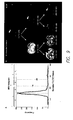
- each Figure shows a histogram (left) of a rescaled image (plotting apparent diffusion coefficient rescaled image values in the rescaled image on the x axis versus frequency of each rescaled image value in the rescaled image on the y axis) and the part of the corresponding diagram (right) (Level 0, Level 1 and Level 3) of the levels of a Hierarchical Region Splitting Tree generated by the method of the present invention for a magnetic resonance image which was generated from the histogram (left), where the level of image splitting is shown on the right side of the diagram beginning with the first image, Image 0, and the range of rescaled image values present in the sub-image are shown below each sub-image, where Figure 7 is generated from the brain of a human neonatal patient with a mild ischemic injury, and Figure 8 is generated from the brain of a human neonatal patient with a severe ischemic injury.
- the abnormality is detected in the left-most image of Level 3 in both diagrams because the left-most image of Level 3 is detected as the first sub-image that has a mean scaled-back image value (the scaled-back image value being the same as the original diffusion coefficient image value) that satisfies the relational operator ("less than” in these examples) and the MeanTh (0.16 mm2/sec diffusion for ischemic injury) previously determined.
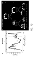
- each Figure shows a histogram (left) of a rescaled image (plotting rescaled image values for T2 relaxation time in the rescaled image on the x axis versus frequency of each rescaled image value in the rescaled image on the y axis) and the part of the corresponding diagram (right) (Level 0, Level 1 and Level 2) of the levels of a Hierarchical Region Splitting Tree generated by the method of the present invention for a magnetic resonance image which was generated from the histogram (left), where the level of image splitting is shown on the right side of the diagram beginning with the first image, Image 0, and the range of rescaled image values present in the sub-image are shown below each sub-image, where Figure 9 is generated from a rat brain with a mild ischemic injury, and Figure 10 is generated from a rat brain with a severe ischemic injury.
- the abnormality is detected in the right image of Level 1 in both diagrams because the right image of Level 1 is detected as the first sub-image that has a mean scaled-back image value (the scaled-back image value being the same as the original T2 relaxation time image value) that satisfies the relational operator ("greater than” in these examples) and the MeanTh (180 millisecond T2 relaxation time for ischemic injury) previously determined.
- FIG 11 there is shown a diagram comparing volumetric results of magnetic resonance images of injured brains using methods according to the present invention (HRS) and using standard manual methods at different injury-severities.
- HRS present invention
- T2WI from mild ( ⁇ 15% lesion), moderate (15-35%) and severe (> 35%) injuries are shown at the level of the horizontal line.
- the percentage of the lesion volume compared to the entire brain is shown below the 3D volumes.
- a method of detecting a core of an injury and detecting a penumbra of an injury in living human tissue, and distinguishing the core from the penumbra comprises analyzing the medical image according to the present invention.
- the human tissue is selected from the group consisting of heart, intestines, joints, kidneys, liver, lungs and spleen, though any suitable tissue can be used as will be understood by those with skill in the art with reference to this disclosure.
- the living human tissue is brain.
- the "core” of an injury is the area or volume that contains tissues that are dead and completely irrecoverable.
- the "penumbra” of an injury is the area or volume that contains tissue that is not dead but that is affected by the injury, where some of the tissue is recoverable.
- the penumbra is generally located adjacent to or around the core. Outside of the core and penumbra are normal healthy tissues.
- the water content (such as for example as determined by T2 maps) and water mobility (such as for example as determined by apparent diffusion coefficient maps) in magnetic resonance images are generally different for the core as compared to the penumbra, and the water content and water mobility in magnetic resonance images are generally different for both the core and the penumbra as compared to normal healthy tissues.
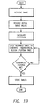
- the method according to the present invention of detecting a core of an injury and detecting a penumbra of an injury in living human tissue, and distinguishing the core from the penumbra is accomplished by:
- Image 11 is the injury sub-image and the sub-tree is Image 111 and Image 112 (as shown in Figure 5 ), and sub-images below Image 111 and Image 112 (that are not shown in Figure 5 ).
- Known T2 relaxation time image values for neonatal ischemic injury are greater than 200 ms for the core, and 160 ms ⁇ T2 relaxation time ⁇ 200 ms for the penumbra, and known apparent diffusion coefficient is less than 0.25 x 10 -3 mm 2 /sec for the core and 0.25 x 10 -3 mm2/sec ⁇ apparent diffusion coefficient ⁇ 0.50 x 10 -3 mm 2 /sec for the penumbra.
- the method further comprises determining different gradations (quantitative measures) of the core and the penumbra using steps corresponding to other embodiments of the method disclosed in this disclosure as will be understood by those with skill in the art with reference to this disclosure.
- the different quantitative measures of the core and the penumbra are useful for better pathological temporal monitoring and therapeutic intervention, even if there is no scientific term yet associated with the quantitative measures (such as core and penumbra).
- the corresponding steps for determining the different gradations of the core and penumbra are as follows:
- the method further comprises clustering multiple sub-images with the same range (from different branches and different levels of the HRS tree) to get unified sub-region structures of the injury.
- FIG 12 there is shown a diagram of core-penumbra injury detected according to the present invention in an animal brain.
- core was detected as red areas in row 2
- penumbra was detected as blue areas in row 2 using T2 maps (row 1).
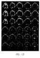
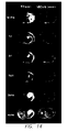
- FIG. 13 there is shown a diagram of core-penumbra injury detected according to the present invention in a human neonatal brain.
- core was detected as red areas and penumbra as blue areas in row 2 and row 5 using apparent diffusion coefficient maps (row 1 and row 4).
- apparent diffusion coefficient maps row 1 and row 4
- pseudo-colors used to visualize the finer gradation are as follows:
- injuries usually evolve spatially and temporally as does many other types of abnormalities such as genetic abnormality, that is, the anatomical location and extent of an injury changes over time after the injury and usually involve an initial degeneration process followed by a recovery process.
- genetic abnormality that is, the anatomical location and extent of an injury changes over time after the injury and usually involve an initial degeneration process followed by a recovery process.
- a method of quantifying the spatiotemporal evolution of an injury in living human tissue comprises:
- such quantifying of the spatiotemporal evolution of an injury comprises quantifying the mean T2 image value of the injured tissues over time, how the volume of an injury changes over time, and how an injured tissue recovers over time.
- the method involves the automated detection and quantitative monitoring of stem cells.
- stem cells that have been labeled with iron are visible on various medical imaging techniques, such as for example on magnetic resonance images in the modality of T2 maps and susceptibility weighted imaging (SWI) maps where the iron labeled stem cells appear as dark (hypo-intense) small clusters on the magnetic resonance image.
- SWI susceptibility weighted imaging
- Markers other than iron that allow stem cells to be distinguished from surrounding tissues are also useful for this method, as will be understood by those with skill in the art with reference to this disclosure.
- the methods disclosed in this disclosure are used to monitor labeled stem cells.
- the method comprises:
- Stem cells are attracted by signals from an injury region and the stem cells migrate, proliferate, differentiate and take part in recovery from the injury, including injury to the brain. Quantification of the stem cells' activities (in vivo) is currently performed by time-consuming and subjective visual/manual methods.
- a method of quantifying spatiotemporal activities of implanted labeled stem cells in living human tissue, including human brain comprises steps corresponding to steps from methods disclosed in this disclosure.
- shape, proximity and area-similarity based matching is done to track specific stem cells cluster over space and time. Migrations of stem cells are computed by location changes of the magnetic resonance image-signal-weighted centroid of the same stem cells cluster over time.
- Proliferations of stem cells are computed by the expansion and compression of the area or volume of a particular stem cells cluster, where directional preferences in proliferation are computed by changing shapes over time. Higher order statistics of the migration and proliferation (such as for example rate of change of migration and rate of change of proliferation) are also computed for detailed stem cells activities. Final locations of the stem cells are computed by determining the "leading edge," that is, the farthest pixel/voxel of the stem cells cluster from the implantation site. As different stem cells clusters take different paths towards the injury site, path-specific stem cells activities are quantified and compared for to allow monitoring of stem cell therapy.
- the method comprises steps corresponding to steps from methods disclosed in this disclosure.
Landscapes
- Engineering & Computer Science (AREA)
- Physics & Mathematics (AREA)
- General Physics & Mathematics (AREA)
- Theoretical Computer Science (AREA)
- Computer Vision & Pattern Recognition (AREA)
- Multimedia (AREA)
- Software Systems (AREA)
- Probability & Statistics with Applications (AREA)
- General Health & Medical Sciences (AREA)
- Radiology & Medical Imaging (AREA)
- Quality & Reliability (AREA)
- Nuclear Medicine, Radiotherapy & Molecular Imaging (AREA)
- Medical Informatics (AREA)
- Health & Medical Sciences (AREA)
- Magnetic Resonance Imaging Apparatus (AREA)
Claims (10)
- Methode zur Erfassung des Kerns einer Wunde und zur Erfassung des Halbschattens einer Wunde im menschlichen lebenden Gewebe sowie zur Unterscheidung des Kerns vom Halbschatten, die Methode schließt folgende Phasen ein:a) Untersuchung einer medizinischen Abbildung, die medizinische Abbildung schließt eine oder mehrere Interessenregionen ein, die Methode schließt folgende Phasen ein:1) Die medizinische Abbildung, die eine Gruppe effektiver Abbildungswerte einschließt, voraussehen;2) Die effektiven Abbildungswerte in Skala wieder ändern, um entsprechende wieder in Skala geänderte Abbildungswerte zu erzeugen, und eine wieder in Skala geänderte Abbildung aus den wieder in Skala geänderten Abbildungswerten erzeugen;3) Ein Histogramm der wieder in Skala geänderten Abbildungswerte ableiten;4) Das Histogramm verwenden, um eine adaptive Segmentierungsschwelle abzuleiten, die verwendet werden kann, um die wieder in Skala geänderte Abbildung in zwei Unterabbildungen, eine erste Unterabbildung mit einer Helligkeit entsprechend der oder mit einer niedrigeren Helligkeit als die adaptive Segmentierungsschwelle und eine zweite Unterabbildung mit einer höheren Helligkeit als die adaptive Segmentierungsschwelle, oder eine erste Unterabbildung mit einer niedrigeren Helligkeit als die adaptive Segmentierungsschwelle und eine zweite Unterabbildung mit einer Helligkeit entsprechend der oder mit einer höheren Helligkeit als die adaptive Segmentierungsschwelle, oder eine erste Unterabbildung mit einer niedrigeren Helligkeit als die adaptive Segmentierungsschwelle und eine zweite Unterabbildung mit einer höheren Helligkeit als die adaptive Segmentierungsschwelle zu unterteilen;5) Die adaptive Segmentierungsschwelle verwenden, um rekursiv die wieder in Skala geänderten Unterabbildungen zu unterteilen, um durch Basieren auf die Konsistenz der wieder in Skala geänderten Abbildungswerte der wieder in Skala geänderten Abbildung einen hierarchischen Baum für die Unterteilung der Regionen der Unter-Unter-Abbildungen zu erzeugen; und6) Die rekursive Unterteilung der Unter-Unter-Abbildungen beenden, indem ein oder mehr als ein vorbestimmtes Kriterium verwendet wird, um auf diese Weise den hierarchischen Baum der Unterteilung der Regionen zu vervollständigen; undb) Eine Unter-Unter-Abbildung, eine Unter-Abbildung der Wunde im beendeten hierarchischen Baum der Unterteilung der Regionen erfassen, der die Interessenregion einschließt, wo die Interessenregion eine Wunde darstellt;c) Die Maske der Wunde bestimmen;d) Einen Unterbaum unter der erfassten Unterabbildung der Wunde bestimmen, indem die Unterabbildung der Wunde als Wurzel des Unterbaums verwendet wird;e) Die Soft Abbildungsschwellenwerte bestimmen, um den Kern vom Halbschatten zu trennen, wo die Soft Abbildungsschwellenwerte die Anomalie identifizieren, die in der medizinischen Abbildung dargestellt wird;f) Die Soft Abbildungsschwellenwerte im Unterbaum vergleichen, um den Halbschatten und eine Maske des Halbschattens zu finden; undg) Die Maske des Kerns durch Abziehen der Maske des Halbschattens aus der Maske der Wunde bestimmen.
- Methode gemäß Patentanspruch 1, wo die Methode außerdem die Konfiguration von mindestens einem Prozessor einschließt, um die Funktionen der Phase a) auszuführen.
- Methode gemäß Patentanspruch 1 oder Patentanspruch 2, die außerdem die Ausführung einer erneuten Änderung in sekundärer Skala der wieder in Skala geänderten Abbildungswerte oder jede wieder in Skala geänderte Unter-Unter-Abbildung im hierarchischen Baum der erneuten Unterteilung der Regionen in die effektiven Abbildungswerte einschließt, die in der medizinischen Abbildung vorhanden sind, um eine wieder in sekundärer Skala geänderte medizinische Abbildung zu erzeugen, um auf diese Weise eine wieder in Skala sekundär geänderte Unter-Unter-Abbildung zu erzeugen, die die Interessenregion enthält.
- Methode gemäß Patentanspruch 3, in dem die Methode außerdem die Bestimmung eines Abbildungswertes oder einer Gruppe von Abbildungswerten der effektiven Abbildungswerte in der medizinischen Abbildung nach der erneuten Änderung in sekundärer Skala einschließt, wo der Abbildungswert oder eine Gruppe von Abbildungswerten der bestimmten effektiven Abbildungswerte die Wunde identifiziert, die in der medizinischen Abbildung durch die für die Erzeugung der gelieferten medizinischen Abbildung verwendete Modalität dargestellt wird.
- Methode gemäß Patentanspruch 3, die außerdem die Vorbereitung einer Maske der Unter-Unter-Abbildung einschießt, die die Darstellung der Wunde und die Reinigung der Maske enthält, um die kleinen äußeren Regionen zu entfernen, um eine saubere Maske der Unter-Unter-Abbildung zu erzeugen, die die Darstellung der Wunde enthält.
- Methode gemäß einem beliebigen der Patentansprüche 1-5, in denen die medizinische Abbildung von der Gruppe gewählt wird, die eine berechnete tomografische Abtastung, die Abbildung einer Magnetresonanz, eine tomografische Abtastung mit Positronenemission und ein Röntgenbild enthält.
- Methode gemäß einem beliebigen der Patentansprüche 1-6, in dem die Methode die Darstellung der Wunde qualifiziert.
- Methode gemäß einem beliebigen der Patentansprüche 1-7, wo die vorbestimmten Kriterien von der Gruppe, die eine Bereichsschwelle = 50 Pixel und eine Standard-Ablenkungsschwelle = 10 wieder in Skala geänderten Abbildungswerten und eine Kurtosis-Schwelle = 1,5 enthält, vorbestimmt werden.
- Methode gemäß einem beliebigen der Patentansprüche 1-8, die außerdem die Quantifizierung der Interessenregion im menschlichen lebenden Gewebe einschließt.
- System zur Untersuchung einer medizinischen Abbildung, die medizinische Abbildung schließt eine oder mehrere Interessenregionen ein, das System schließt folgendes ein:a) Einen oder mehrere Prozessoren;b) Einen durch die mit dem einen oder mehreren Prozessoren verbundenen Maschine lesbaren Speicher;c) Eine medizinische Abbildung, die eine Gruppe effektiver Abbildungswerte einschließt, die im Speicher gespeichert wurden;d) Eine Gruppe lesbarer Anleitungen der Maschine, die im Speicher gespeichert wurden, von der Maschine lesbar sind und auf der medizinischen Abbildung aktivierbar sind;e) Eine Benutzerschnittstelle, die für den Betrieb mit der Gruppe der Anleitungen des Computers verbunden wird, um einen oder mehrere Steuerbefehle an einen oder mehrere Prozessoren zu übertragen;f) Anleitungen, die für den Betrieb mit der Benutzerschnittstelle für folgendes verbunden sind:i) Die effektiven Abbildungswerte in Skala wieder ändern, um entsprechende wieder - geänderte Abbildungswerte in Skala zu erzeugen, und eine in Skala wieder geänderte Abbildung aus den wieder in Skala geänderten Abbildungswerten erzeugen;ii) Ein Histogramm der wieder in Skala geänderten Abbildungswerte ableiten;iii) Das Histogramm verwenden, um eine adaptive Segmentierungsschwelle abzuleiten, die verwendet werden kann, um die wieder in Skala geänderte Abbildung in zwei Unterabbildungen zu unterteilen, die eine Helligkeit entsprechend der oder eine niedrigere Helligkeit als die adaptive Segmentierungsschwelle haben, und eine zweite Unterabbildung mit einer höheren Helligkeit als die adaptive Segmentierungsschwelle, oder eine erste Unterabbildung mit einer niedrigeren Helligkeit als die adaptive Segmentierungsschwelle und eine zweite Unterabbildung mit einer Helligkeit entsprechend der oder mit einer höheren Helligkeit als die adaptive Segmentierungsschwelle;iv) Die adaptive Segmentierungsschwelle verwenden, um rekursiv die wieder in Skala geänderten Unterabbildungen zu unterteilen, um durch Basieren auf die Konsistenz der wieder in Skala geänderten Abbildungswerte der wieder in Skala geänderten Abbildung einen hierarchischen Baum für die Unterteilung der Regionen der Unter-Unter-Abbildungen zu erzeugen; undv) Die rekursive Unterteilung der Unter-Unter-Abbildungen beenden, indem ein oder mehr als ein vorbestimmtes Kriterium verwendet wird, um auf diese Weise den hierarchischen Baum der Unterteilung der Regionen zu vervollständigen;g) Einen Speicher, der für den Betrieb mit einem oder mehreren Prozessoren und mit einer Benutzerschnittstelle verbunden ist, um die sich aus dem hierarchischen Baum der Unterteilung der Regionen ergebenden Abbildungen zu speichern;h) Anleitungen, die für den Betrieb mit der Benutzerschnittstelle für folgendes verbunden sind:vii) Eine Unter-Unter-Abbildung, eine Unter-Abbildung der Wunde im beendeten hierarchischen Baum der Unterteilung der Regionen erfassen, der die Interessenregion einschließt, wo die Interessenregion eine Wunde darstellt;viii) Die Maske der Wunde bestimmen;ix) Einen Unterbaum unter der erfassten Unterabbildung der Wunde bestimmen, indem die Unterabbildung der Wunde als Wurzel des Unterbaums verwendet wird;x) Die Soft Abbildungsschwellenwerte bestimmen, um den Kern vom Halbschatten zu trennen, wo die Soft Abbildungsschwellenwerte die Anomalie identifizieren, die in der medizinischen Abbildung dargestellt wird;xi) Die Soft Abbildungsschwellenwerte im Unterbaum vergleichen, um den Halbschatten und eine Maske des Halbschattens zu finden; undxii) Die Maske des Kerns durch Abziehen der Maske des Halbschattens von der Maske der Wunde bestimmen.
Applications Claiming Priority (3)
| Application Number | Priority Date | Filing Date | Title |
|---|---|---|---|
| US30739610P | 2010-02-23 | 2010-02-23 | |
| US32763010P | 2010-04-23 | 2010-04-23 | |
| PCT/US2011/025943 WO2011106440A1 (en) | 2010-02-23 | 2011-02-23 | Method of analyzing a medical image |
Publications (3)
| Publication Number | Publication Date |
|---|---|
| EP2538839A1 EP2538839A1 (de) | 2013-01-02 |
| EP2538839A4 EP2538839A4 (de) | 2014-04-30 |
| EP2538839B1 true EP2538839B1 (de) | 2015-07-08 |
Family
ID=44507191
Family Applications (1)
| Application Number | Title | Priority Date | Filing Date |
|---|---|---|---|
| EP11748009.5A Not-in-force EP2538839B1 (de) | 2010-02-23 | 2011-02-23 | Verfahren zur analyse eines medizinischen bildes |
Country Status (3)
| Country | Link |
|---|---|
| US (3) | US8731261B2 (de) |
| EP (1) | EP2538839B1 (de) |
| WO (1) | WO2011106440A1 (de) |
Families Citing this family (24)
| Publication number | Priority date | Publication date | Assignee | Title |
|---|---|---|---|---|
| KR101886333B1 (ko) * | 2012-06-15 | 2018-08-09 | 삼성전자 주식회사 | 멀티 코어를 이용한 영역 성장 장치 및 방법 |
| JP5984559B2 (ja) * | 2012-07-31 | 2016-09-06 | オリンパス株式会社 | 壊死細胞領域検出装置及びその方法、壊死細胞領域検出プログラム |
| JP5954846B2 (ja) * | 2012-08-24 | 2016-07-20 | 富士通株式会社 | 形状データ生成プログラム、形状データ生成方法及び形状データ生成装置 |
| US9338143B2 (en) | 2013-03-15 | 2016-05-10 | Shape Security, Inc. | Stateless web content anti-automation |
| US20140283038A1 (en) | 2013-03-15 | 2014-09-18 | Shape Security Inc. | Safe Intelligent Content Modification |
| US8893294B1 (en) | 2014-01-21 | 2014-11-18 | Shape Security, Inc. | Flexible caching |
| US10089216B2 (en) * | 2014-06-30 | 2018-10-02 | Shape Security, Inc. | Automatically determining whether a page of a web site is broken despite elements on the page that may change |
| US9075990B1 (en) | 2014-07-01 | 2015-07-07 | Shape Security, Inc. | Reliable selection of security countermeasures |
| US9825984B1 (en) | 2014-08-27 | 2017-11-21 | Shape Security, Inc. | Background analysis of web content |
| US9825995B1 (en) | 2015-01-14 | 2017-11-21 | Shape Security, Inc. | Coordinated application of security policies |
| CN113763377B (zh) * | 2015-01-31 | 2025-10-31 | 文塔纳医疗系统公司 | 用于使用载片缩略图图像的感兴趣区域检测的系统和方法 |
| US10444311B2 (en) * | 2015-03-11 | 2019-10-15 | Ohio State Innovation Foundation | Methods and devices for optimizing magnetic resonance imaging protocols |
| US9813440B1 (en) | 2015-05-15 | 2017-11-07 | Shape Security, Inc. | Polymorphic treatment of annotated content |
| WO2016187014A1 (en) * | 2015-05-15 | 2016-11-24 | The Medical College Of Wisconsin, Inc. | Systems and methods for calibrated multi-spectral magnetic resonance imaging |
| US10375026B2 (en) | 2015-10-28 | 2019-08-06 | Shape Security, Inc. | Web transaction status tracking |
| WO2018098213A1 (en) * | 2016-11-23 | 2018-05-31 | Wake Forest University Health Sciences | Medical image analysis using mechanical deformation information |
| EP3379281A1 (de) * | 2017-03-20 | 2018-09-26 | Koninklijke Philips N.V. | Bildsegmentierung unter verwendung von grauskalenbezugswerten |
| US9738937B1 (en) | 2017-03-31 | 2017-08-22 | Cellmax, Ltd. | Identifying candidate cells using image analysis |
| CN109003270B (zh) * | 2018-07-23 | 2020-11-27 | 北京市商汤科技开发有限公司 | 一种图像处理方法、电子设备及存储介质 |
| CN109615600B (zh) * | 2018-12-12 | 2023-03-31 | 南昌工程学院 | 一种自适应层次直方图的彩色图像分割方法 |
| WO2023286925A1 (en) * | 2021-07-13 | 2023-01-19 | Samsung Electronics Co., Ltd. | A system and method for rendering differential video on graphical displays |
| EP4281926A4 (de) | 2021-07-13 | 2024-07-17 | Samsung Electronics Co., Ltd. | System und verfahren zur darstellung von differenzvideos auf grafischen anzeigen |
| EP4141791B1 (de) * | 2021-08-23 | 2026-04-01 | Dassault Systèmes | Automatische extraktion von gehirnmodellen |
| CN115201196A (zh) * | 2022-08-09 | 2022-10-18 | 广州健芯生物科技合伙企业(有限合伙) | 一种高精度尿液分析方法及系统 |
Family Cites Families (26)
| Publication number | Priority date | Publication date | Assignee | Title |
|---|---|---|---|---|
| IL77840A (en) * | 1986-02-10 | 1989-05-15 | Elscint Ltd | Data compression system for digital imaging |
| US4945478A (en) * | 1987-11-06 | 1990-07-31 | Center For Innovative Technology | Noninvasive medical imaging system and method for the identification and 3-D display of atherosclerosis and the like |
| CA2051939A1 (en) * | 1990-10-02 | 1992-04-03 | Gary A. Ransford | Digital data registration and differencing compression system |
| DE69214229T2 (de) * | 1991-08-14 | 1997-04-30 | Agfa Gevaert Nv | Verfahren und Vorrichtung zur Kontrastverbesserung von Bildern |
| US5982944A (en) * | 1991-09-27 | 1999-11-09 | E. I. Du Pont De Nemours And Company | Adaptive vision system using dual thresholding |
| US6058211A (en) * | 1995-07-07 | 2000-05-02 | Imec Vzw | Data compression method and apparatus |
| US6885762B2 (en) * | 2000-02-07 | 2005-04-26 | Trustees Of The University Of Pennsylvania | Scale-based image filtering of magnetic resonance data |
| US6674907B1 (en) * | 2000-02-17 | 2004-01-06 | Microsoft Corporation | Color image quantization using a hierarchical color perception model |
| US6470094B1 (en) * | 2000-03-14 | 2002-10-22 | Intel Corporation | Generalized text localization in images |
| JP4683253B2 (ja) * | 2000-07-14 | 2011-05-18 | ソニー株式会社 | Av信号処理装置および方法、プログラム、並びに記録媒体 |
| US7194134B2 (en) | 2001-01-02 | 2007-03-20 | Microsoft Corporation | Hierarchical, probabilistic, localized, semantic image classifier |
| US7058210B2 (en) * | 2001-11-20 | 2006-06-06 | General Electric Company | Method and system for lung disease detection |
| US7137062B2 (en) * | 2001-12-28 | 2006-11-14 | International Business Machines Corporation | System and method for hierarchical segmentation with latent semantic indexing in scale space |
| TW554629B (en) * | 2002-03-22 | 2003-09-21 | Ind Tech Res Inst | Layered object segmentation method based on motion picture compression standard |
| US8805619B2 (en) * | 2002-10-28 | 2014-08-12 | The General Hospital Corporation | Tissue disorder imaging analysis |
| DE10253191A1 (de) * | 2002-11-15 | 2004-05-27 | Philips Intellectual Property & Standards Gmbh | Verfahren zur selektiven Darstellung von Körperstrukturen |
| GB0329449D0 (en) | 2003-12-19 | 2004-01-28 | Omnicyte Ltd | Stem cells |
| US7907769B2 (en) * | 2004-05-13 | 2011-03-15 | The Charles Stark Draper Laboratory, Inc. | Image-based methods for measuring global nuclear patterns as epigenetic markers of cell differentiation |
| US7599535B2 (en) * | 2004-08-02 | 2009-10-06 | Siemens Medical Solutions Usa, Inc. | System and method for tree-model visualization for pulmonary embolism detection |
| US7536064B2 (en) | 2005-05-31 | 2009-05-19 | Microsoft Corporation | Image comparison by metric embeddings |
| US7720267B2 (en) * | 2005-07-15 | 2010-05-18 | Siemens Medical Solutions Usa, Inc. | Method and apparatus for classifying tissue using image data |
| US8014590B2 (en) | 2005-12-07 | 2011-09-06 | Drvision Technologies Llc | Method of directed pattern enhancement for flexible recognition |
| WO2009058915A1 (en) * | 2007-10-29 | 2009-05-07 | The Trustees Of The University Of Pennsylvania | Computer assisted diagnosis (cad) of cancer using multi-functional, multi-modal in-vivo magnetic resonance spectroscopy (mrs) and imaging (mri) |
| US20090136096A1 (en) | 2007-11-23 | 2009-05-28 | General Electric Company | Systems, methods and apparatus for segmentation of data involving a hierarchical mesh |
| US8107726B2 (en) * | 2008-06-18 | 2012-01-31 | Samsung Electronics Co., Ltd. | System and method for class-specific object segmentation of image data |
| US8385609B2 (en) * | 2008-10-21 | 2013-02-26 | Flashfoto, Inc. | Image segmentation |
-
2011
- 2011-02-23 EP EP11748009.5A patent/EP2538839B1/de not_active Not-in-force
- 2011-02-23 US US13/580,947 patent/US8731261B2/en not_active Expired - Fee Related
- 2011-02-23 WO PCT/US2011/025943 patent/WO2011106440A1/en not_active Ceased
-
2014
- 2014-05-07 US US14/272,299 patent/US8965089B2/en active Active
-
2015
- 2015-02-20 US US14/627,713 patent/US20150235362A1/en not_active Abandoned
Also Published As
| Publication number | Publication date |
|---|---|
| US20120314926A1 (en) | 2012-12-13 |
| US8731261B2 (en) | 2014-05-20 |
| US20150235362A1 (en) | 2015-08-20 |
| EP2538839A1 (de) | 2013-01-02 |
| US20140241608A1 (en) | 2014-08-28 |
| US8965089B2 (en) | 2015-02-24 |
| WO2011106440A1 (en) | 2011-09-01 |
| EP2538839A4 (de) | 2014-04-30 |
Similar Documents
| Publication | Publication Date | Title |
|---|---|---|
| US8965089B2 (en) | Method of analyzing a medical image | |
| US11443433B2 (en) | Quantification and staging of body-wide tissue composition and of abnormal states on medical images via automatic anatomy recognition | |
| JP6034299B2 (ja) | 半影帯サイズを推定するためのシステムおよびその作動方法 | |
| US10638995B2 (en) | Imaging-based biomarker for characterizing the structure or function of human or animal brain tissue and related uses and methods | |
| JP2023540910A (ja) | 病変検出のための共同訓練を伴う接続機械学習モデル | |
| CN113711271A (zh) | 用于通过正电子发射断层摄影进行肿瘤分割的深度卷积神经网络 | |
| Guo et al. | MMFGAN: A novel multimodal brain medical image fusion based on the improvement of generative adversarial network | |
| Hu et al. | Segmentation of brain from computed tomography head images | |
| Ribeiro et al. | Handling inter-annotator agreement for automated skin lesion segmentation | |
| US20150356733A1 (en) | Medical image processing | |
| Chechlacz et al. | Asymmetrical white matter networks for attending to global versus local features | |
| Wang et al. | Self-guided optimization semi-supervised method for joint segmentation of macular hole and cystoid macular edema in retinal OCT images | |
| Madesta et al. | Deep learning‐based conditional inpainting for restoration of artifact‐affected 4D CT images | |
| Li et al. | BrainK for structural image processing: creating electrical models of the human head | |
| US8873817B2 (en) | Processing an image dataset based on clinically categorized populations | |
| Wang et al. | Semi-automatic segmentation of the fetal brain from magnetic resonance imaging | |
| Jiménez-Balado et al. | The relationship between cortical thickness and white matter hyperintensities in mid to late life | |
| Østmo et al. | View it like a radiologist: shifted windows for deep learning augmentation of CT images | |
| Hu et al. | Automatic detection of melanins and sebums from skin images using a generative adversarial network | |
| Pardo et al. | Automated skin lesion segmentation with kernel density estimation | |
| Racimora et al. | Segmentation of polycystic kidneys from MR images | |
| Lee et al. | Increasing visual perception brain stroke detection system | |
| Katyal et al. | Gaussian intensity model with neighborhood cues for fluid-tissue categorization of multisequence MR brain images | |
| KR102832391B1 (ko) | Mri영상에서 뉴로멜라닌의 탈침착 정량화를 위한 이미지 처리방법 및 그 처리장치 | |
| Lin | Vessel Tree Segmentation and Modality Agnostic Aneurysm Detection |
Legal Events
| Date | Code | Title | Description |
|---|---|---|---|
| PUAI | Public reference made under article 153(3) epc to a published international application that has entered the european phase |
Free format text: ORIGINAL CODE: 0009012 |
|
| 17P | Request for examination filed |
Effective date: 20120919 |
|
| AK | Designated contracting states |
Kind code of ref document: A1 Designated state(s): AL AT BE BG CH CY CZ DE DK EE ES FI FR GB GR HR HU IE IS IT LI LT LU LV MC MK MT NL NO PL PT RO RS SE SI SK SM TR |
|
| DAX | Request for extension of the european patent (deleted) | ||
| A4 | Supplementary search report drawn up and despatched |
Effective date: 20140402 |
|
| RIC1 | Information provided on ipc code assigned before grant |
Ipc: G06K 9/32 20060101ALI20140327BHEP Ipc: G06K 9/38 20060101ALI20140327BHEP Ipc: A61B 5/05 20060101AFI20140327BHEP Ipc: G06T 7/00 20060101ALI20140327BHEP |
|
| 17Q | First examination report despatched |
Effective date: 20140423 |
|
| RAP1 | Party data changed (applicant data changed or rights of an application transferred) |
Owner name: LOMA LINDA UNIVERSITY Owner name: UNIVERSITY OF CALIFORNIA, RIVERSIDE |
|
| GRAP | Despatch of communication of intention to grant a patent |
Free format text: ORIGINAL CODE: EPIDOSNIGR1 |
|
| RIC1 | Information provided on ipc code assigned before grant |
Ipc: G06K 9/38 20060101ALI20141219BHEP Ipc: G06K 9/32 20060101ALI20141219BHEP Ipc: A61B 5/05 20060101AFI20141219BHEP Ipc: G06T 7/40 20060101ALI20141219BHEP Ipc: G06T 7/00 20060101ALI20141219BHEP |
|
| INTG | Intention to grant announced |
Effective date: 20150121 |
|
| GRAS | Grant fee paid |
Free format text: ORIGINAL CODE: EPIDOSNIGR3 |
|
| GRAA | (expected) grant |
Free format text: ORIGINAL CODE: 0009210 |
|
| AK | Designated contracting states |
Kind code of ref document: B1 Designated state(s): AL AT BE BG CH CY CZ DE DK EE ES FI FR GB GR HR HU IE IS IT LI LT LU LV MC MK MT NL NO PL PT RO RS SE SI SK SM TR |
|
| REG | Reference to a national code |
Ref country code: GB Ref legal event code: FG4D |
|
| REG | Reference to a national code |
Ref country code: AT Ref legal event code: REF Ref document number: 734732 Country of ref document: AT Kind code of ref document: T Effective date: 20150715 Ref country code: CH Ref legal event code: EP |
|
| REG | Reference to a national code |
Ref country code: IE Ref legal event code: FG4D |
|
| REG | Reference to a national code |
Ref country code: DE Ref legal event code: R096 Ref document number: 602011017711 Country of ref document: DE |
|
| REG | Reference to a national code |
Ref country code: AT Ref legal event code: MK05 Ref document number: 734732 Country of ref document: AT Kind code of ref document: T Effective date: 20150708 |
|
| REG | Reference to a national code |
Ref country code: NL Ref legal event code: MP Effective date: 20150708 |
|
| REG | Reference to a national code |
Ref country code: LT Ref legal event code: MG4D |
|
| PG25 | Lapsed in a contracting state [announced via postgrant information from national office to epo] |
Ref country code: GR Free format text: LAPSE BECAUSE OF FAILURE TO SUBMIT A TRANSLATION OF THE DESCRIPTION OR TO PAY THE FEE WITHIN THE PRESCRIBED TIME-LIMIT Effective date: 20151009 Ref country code: NO Free format text: LAPSE BECAUSE OF FAILURE TO SUBMIT A TRANSLATION OF THE DESCRIPTION OR TO PAY THE FEE WITHIN THE PRESCRIBED TIME-LIMIT Effective date: 20151008 Ref country code: FI Free format text: LAPSE BECAUSE OF FAILURE TO SUBMIT A TRANSLATION OF THE DESCRIPTION OR TO PAY THE FEE WITHIN THE PRESCRIBED TIME-LIMIT Effective date: 20150708 Ref country code: LT Free format text: LAPSE BECAUSE OF FAILURE TO SUBMIT A TRANSLATION OF THE DESCRIPTION OR TO PAY THE FEE WITHIN THE PRESCRIBED TIME-LIMIT Effective date: 20150708 Ref country code: LV Free format text: LAPSE BECAUSE OF FAILURE TO SUBMIT A TRANSLATION OF THE DESCRIPTION OR TO PAY THE FEE WITHIN THE PRESCRIBED TIME-LIMIT Effective date: 20150708 |
|
| PG25 | Lapsed in a contracting state [announced via postgrant information from national office to epo] |
Ref country code: PL Free format text: LAPSE BECAUSE OF FAILURE TO SUBMIT A TRANSLATION OF THE DESCRIPTION OR TO PAY THE FEE WITHIN THE PRESCRIBED TIME-LIMIT Effective date: 20150708 Ref country code: RS Free format text: LAPSE BECAUSE OF FAILURE TO SUBMIT A TRANSLATION OF THE DESCRIPTION OR TO PAY THE FEE WITHIN THE PRESCRIBED TIME-LIMIT Effective date: 20150708 Ref country code: SE Free format text: LAPSE BECAUSE OF FAILURE TO SUBMIT A TRANSLATION OF THE DESCRIPTION OR TO PAY THE FEE WITHIN THE PRESCRIBED TIME-LIMIT Effective date: 20150708 Ref country code: AT Free format text: LAPSE BECAUSE OF FAILURE TO SUBMIT A TRANSLATION OF THE DESCRIPTION OR TO PAY THE FEE WITHIN THE PRESCRIBED TIME-LIMIT Effective date: 20150708 Ref country code: IS Free format text: LAPSE BECAUSE OF FAILURE TO SUBMIT A TRANSLATION OF THE DESCRIPTION OR TO PAY THE FEE WITHIN THE PRESCRIBED TIME-LIMIT Effective date: 20151108 Ref country code: ES Free format text: LAPSE BECAUSE OF FAILURE TO SUBMIT A TRANSLATION OF THE DESCRIPTION OR TO PAY THE FEE WITHIN THE PRESCRIBED TIME-LIMIT Effective date: 20150708 Ref country code: PT Free format text: LAPSE BECAUSE OF FAILURE TO SUBMIT A TRANSLATION OF THE DESCRIPTION OR TO PAY THE FEE WITHIN THE PRESCRIBED TIME-LIMIT Effective date: 20151109 Ref country code: HR Free format text: LAPSE BECAUSE OF FAILURE TO SUBMIT A TRANSLATION OF THE DESCRIPTION OR TO PAY THE FEE WITHIN THE PRESCRIBED TIME-LIMIT Effective date: 20150708 |
|
| REG | Reference to a national code |
Ref country code: DE Ref legal event code: R097 Ref document number: 602011017711 Country of ref document: DE |
|
| PG25 | Lapsed in a contracting state [announced via postgrant information from national office to epo] |
Ref country code: DK Free format text: LAPSE BECAUSE OF FAILURE TO SUBMIT A TRANSLATION OF THE DESCRIPTION OR TO PAY THE FEE WITHIN THE PRESCRIBED TIME-LIMIT Effective date: 20150708 Ref country code: SK Free format text: LAPSE BECAUSE OF FAILURE TO SUBMIT A TRANSLATION OF THE DESCRIPTION OR TO PAY THE FEE WITHIN THE PRESCRIBED TIME-LIMIT Effective date: 20150708 Ref country code: CZ Free format text: LAPSE BECAUSE OF FAILURE TO SUBMIT A TRANSLATION OF THE DESCRIPTION OR TO PAY THE FEE WITHIN THE PRESCRIBED TIME-LIMIT Effective date: 20150708 Ref country code: EE Free format text: LAPSE BECAUSE OF FAILURE TO SUBMIT A TRANSLATION OF THE DESCRIPTION OR TO PAY THE FEE WITHIN THE PRESCRIBED TIME-LIMIT Effective date: 20150708 Ref country code: IT Free format text: LAPSE BECAUSE OF FAILURE TO SUBMIT A TRANSLATION OF THE DESCRIPTION OR TO PAY THE FEE WITHIN THE PRESCRIBED TIME-LIMIT Effective date: 20150708 |
|
| PLBE | No opposition filed within time limit |
Free format text: ORIGINAL CODE: 0009261 |
|
| STAA | Information on the status of an ep patent application or granted ep patent |
Free format text: STATUS: NO OPPOSITION FILED WITHIN TIME LIMIT |
|
| REG | Reference to a national code |
Ref country code: DE Ref legal event code: R082 Ref document number: 602011017711 Country of ref document: DE Representative=s name: HERNANDEZ, YORCK, DIPL.-ING., DE |
|
| PG25 | Lapsed in a contracting state [announced via postgrant information from national office to epo] |
Ref country code: BE Free format text: LAPSE BECAUSE OF NON-PAYMENT OF DUE FEES Effective date: 20160229 Ref country code: RO Free format text: LAPSE BECAUSE OF FAILURE TO SUBMIT A TRANSLATION OF THE DESCRIPTION OR TO PAY THE FEE WITHIN THE PRESCRIBED TIME-LIMIT Effective date: 20150708 |
|
| 26N | No opposition filed |
Effective date: 20160411 |
|
| PG25 | Lapsed in a contracting state [announced via postgrant information from national office to epo] |
Ref country code: SI Free format text: LAPSE BECAUSE OF FAILURE TO SUBMIT A TRANSLATION OF THE DESCRIPTION OR TO PAY THE FEE WITHIN THE PRESCRIBED TIME-LIMIT Effective date: 20150708 |
|
| PG25 | Lapsed in a contracting state [announced via postgrant information from national office to epo] |
Ref country code: LU Free format text: LAPSE BECAUSE OF FAILURE TO SUBMIT A TRANSLATION OF THE DESCRIPTION OR TO PAY THE FEE WITHIN THE PRESCRIBED TIME-LIMIT Effective date: 20160223 Ref country code: MC Free format text: LAPSE BECAUSE OF FAILURE TO SUBMIT A TRANSLATION OF THE DESCRIPTION OR TO PAY THE FEE WITHIN THE PRESCRIBED TIME-LIMIT Effective date: 20150708 |
|
| REG | Reference to a national code |
Ref country code: CH Ref legal event code: PL |
|
| PG25 | Lapsed in a contracting state [announced via postgrant information from national office to epo] |
Ref country code: LI Free format text: LAPSE BECAUSE OF NON-PAYMENT OF DUE FEES Effective date: 20160229 Ref country code: CH Free format text: LAPSE BECAUSE OF NON-PAYMENT OF DUE FEES Effective date: 20160229 |
|
| REG | Reference to a national code |
Ref country code: FR Ref legal event code: ST Effective date: 20161028 |
|
| REG | Reference to a national code |
Ref country code: IE Ref legal event code: MM4A |
|
| PG25 | Lapsed in a contracting state [announced via postgrant information from national office to epo] |
Ref country code: BE Free format text: LAPSE BECAUSE OF FAILURE TO SUBMIT A TRANSLATION OF THE DESCRIPTION OR TO PAY THE FEE WITHIN THE PRESCRIBED TIME-LIMIT Effective date: 20150708 |
|
| PG25 | Lapsed in a contracting state [announced via postgrant information from national office to epo] |
Ref country code: FR Free format text: LAPSE BECAUSE OF NON-PAYMENT OF DUE FEES Effective date: 20160229 Ref country code: IE Free format text: LAPSE BECAUSE OF NON-PAYMENT OF DUE FEES Effective date: 20160223 |
|
| PG25 | Lapsed in a contracting state [announced via postgrant information from national office to epo] |
Ref country code: NL Free format text: LAPSE BECAUSE OF FAILURE TO SUBMIT A TRANSLATION OF THE DESCRIPTION OR TO PAY THE FEE WITHIN THE PRESCRIBED TIME-LIMIT Effective date: 20150708 |
|
| PG25 | Lapsed in a contracting state [announced via postgrant information from national office to epo] |
Ref country code: MT Free format text: LAPSE BECAUSE OF FAILURE TO SUBMIT A TRANSLATION OF THE DESCRIPTION OR TO PAY THE FEE WITHIN THE PRESCRIBED TIME-LIMIT Effective date: 20150708 |
|
| PG25 | Lapsed in a contracting state [announced via postgrant information from national office to epo] |
Ref country code: CY Free format text: LAPSE BECAUSE OF FAILURE TO SUBMIT A TRANSLATION OF THE DESCRIPTION OR TO PAY THE FEE WITHIN THE PRESCRIBED TIME-LIMIT Effective date: 20150708 Ref country code: HU Free format text: LAPSE BECAUSE OF FAILURE TO SUBMIT A TRANSLATION OF THE DESCRIPTION OR TO PAY THE FEE WITHIN THE PRESCRIBED TIME-LIMIT; INVALID AB INITIO Effective date: 20110223 Ref country code: SM Free format text: LAPSE BECAUSE OF FAILURE TO SUBMIT A TRANSLATION OF THE DESCRIPTION OR TO PAY THE FEE WITHIN THE PRESCRIBED TIME-LIMIT Effective date: 20150708 |
|
| PG25 | Lapsed in a contracting state [announced via postgrant information from national office to epo] |
Ref country code: MT Free format text: LAPSE BECAUSE OF FAILURE TO SUBMIT A TRANSLATION OF THE DESCRIPTION OR TO PAY THE FEE WITHIN THE PRESCRIBED TIME-LIMIT Effective date: 20160229 Ref country code: TR Free format text: LAPSE BECAUSE OF FAILURE TO SUBMIT A TRANSLATION OF THE DESCRIPTION OR TO PAY THE FEE WITHIN THE PRESCRIBED TIME-LIMIT Effective date: 20150708 Ref country code: MK Free format text: LAPSE BECAUSE OF FAILURE TO SUBMIT A TRANSLATION OF THE DESCRIPTION OR TO PAY THE FEE WITHIN THE PRESCRIBED TIME-LIMIT Effective date: 20150708 |
|
| PG25 | Lapsed in a contracting state [announced via postgrant information from national office to epo] |
Ref country code: BG Free format text: LAPSE BECAUSE OF FAILURE TO SUBMIT A TRANSLATION OF THE DESCRIPTION OR TO PAY THE FEE WITHIN THE PRESCRIBED TIME-LIMIT Effective date: 20150708 |
|
| PG25 | Lapsed in a contracting state [announced via postgrant information from national office to epo] |
Ref country code: AL Free format text: LAPSE BECAUSE OF FAILURE TO SUBMIT A TRANSLATION OF THE DESCRIPTION OR TO PAY THE FEE WITHIN THE PRESCRIBED TIME-LIMIT Effective date: 20150708 |
|
| PGFP | Annual fee paid to national office [announced via postgrant information from national office to epo] |
Ref country code: GB Payment date: 20190220 Year of fee payment: 9 Ref country code: DE Payment date: 20190212 Year of fee payment: 9 |
|
| REG | Reference to a national code |
Ref country code: DE Ref legal event code: R119 Ref document number: 602011017711 Country of ref document: DE |
|
| GBPC | Gb: european patent ceased through non-payment of renewal fee |
Effective date: 20200223 |
|
| PG25 | Lapsed in a contracting state [announced via postgrant information from national office to epo] |
Ref country code: DE Free format text: LAPSE BECAUSE OF NON-PAYMENT OF DUE FEES Effective date: 20200901 Ref country code: GB Free format text: LAPSE BECAUSE OF NON-PAYMENT OF DUE FEES Effective date: 20200223 |